US12076100B2 - Robotic systems and methods for concomitant endoscopic and percutaneous medical procedures - Google Patents
Robotic systems and methods for concomitant endoscopic and percutaneous medical proceduresDownload PDFInfo
- Publication number
- US12076100B2 US12076100B2US16/586,198US201916586198AUS12076100B2US 12076100 B2US12076100 B2US 12076100B2US 201916586198 AUS201916586198 AUS 201916586198AUS 12076100 B2US12076100 B2US 12076100B2
- Authority
- US
- United States
- Prior art keywords
- target location
- target
- instrument
- medical instrument
- needle
- Prior art date
- Legal status (The legal status is an assumption and is not a legal conclusion. Google has not performed a legal analysis and makes no representation as to the accuracy of the status listed.)
- Active
Links
Images


Classifications
- A—HUMAN NECESSITIES
- A61—MEDICAL OR VETERINARY SCIENCE; HYGIENE
- A61B—DIAGNOSIS; SURGERY; IDENTIFICATION
- A61B1/00—Instruments for performing medical examinations of the interior of cavities or tubes of the body by visual or photographical inspection, e.g. endoscopes; Illuminating arrangements therefor
- A61B1/005—Flexible endoscopes
- A61B1/01—Guiding arrangements therefore
- A—HUMAN NECESSITIES
- A61—MEDICAL OR VETERINARY SCIENCE; HYGIENE
- A61B—DIAGNOSIS; SURGERY; IDENTIFICATION
- A61B1/00—Instruments for performing medical examinations of the interior of cavities or tubes of the body by visual or photographical inspection, e.g. endoscopes; Illuminating arrangements therefor
- A61B1/313—Instruments for performing medical examinations of the interior of cavities or tubes of the body by visual or photographical inspection, e.g. endoscopes; Illuminating arrangements therefor for introducing through surgical openings, e.g. laparoscopes
- A61B1/3132—Instruments for performing medical examinations of the interior of cavities or tubes of the body by visual or photographical inspection, e.g. endoscopes; Illuminating arrangements therefor for introducing through surgical openings, e.g. laparoscopes for laparoscopy
- A—HUMAN NECESSITIES
- A61—MEDICAL OR VETERINARY SCIENCE; HYGIENE
- A61B—DIAGNOSIS; SURGERY; IDENTIFICATION
- A61B34/00—Computer-aided surgery; Manipulators or robots specially adapted for use in surgery
- A61B34/20—Surgical navigation systems; Devices for tracking or guiding surgical instruments, e.g. for frameless stereotaxis
- A—HUMAN NECESSITIES
- A61—MEDICAL OR VETERINARY SCIENCE; HYGIENE
- A61B—DIAGNOSIS; SURGERY; IDENTIFICATION
- A61B34/00—Computer-aided surgery; Manipulators or robots specially adapted for use in surgery
- A61B34/30—Surgical robots
- A—HUMAN NECESSITIES
- A61—MEDICAL OR VETERINARY SCIENCE; HYGIENE
- A61B—DIAGNOSIS; SURGERY; IDENTIFICATION
- A61B34/00—Computer-aided surgery; Manipulators or robots specially adapted for use in surgery
- A61B34/30—Surgical robots
- A61B34/37—Leader-follower robots
- A—HUMAN NECESSITIES
- A61—MEDICAL OR VETERINARY SCIENCE; HYGIENE
- A61B—DIAGNOSIS; SURGERY; IDENTIFICATION
- A61B90/00—Instruments, implements or accessories specially adapted for surgery or diagnosis and not covered by any of the groups A61B1/00 - A61B50/00, e.g. for luxation treatment or for protecting wound edges
- A61B90/36—Image-producing devices or illumination devices not otherwise provided for
- A61B90/37—Surgical systems with images on a monitor during operation
- A—HUMAN NECESSITIES
- A61—MEDICAL OR VETERINARY SCIENCE; HYGIENE
- A61B—DIAGNOSIS; SURGERY; IDENTIFICATION
- A61B34/00—Computer-aided surgery; Manipulators or robots specially adapted for use in surgery
- A61B34/20—Surgical navigation systems; Devices for tracking or guiding surgical instruments, e.g. for frameless stereotaxis
- A61B2034/2046—Tracking techniques
- A61B2034/2051—Electromagnetic tracking systems
- A—HUMAN NECESSITIES
- A61—MEDICAL OR VETERINARY SCIENCE; HYGIENE
- A61B—DIAGNOSIS; SURGERY; IDENTIFICATION
- A61B34/00—Computer-aided surgery; Manipulators or robots specially adapted for use in surgery
- A61B34/20—Surgical navigation systems; Devices for tracking or guiding surgical instruments, e.g. for frameless stereotaxis
- A61B2034/2046—Tracking techniques
- A61B2034/2055—Optical tracking systems
- A—HUMAN NECESSITIES
- A61—MEDICAL OR VETERINARY SCIENCE; HYGIENE
- A61B—DIAGNOSIS; SURGERY; IDENTIFICATION
- A61B34/00—Computer-aided surgery; Manipulators or robots specially adapted for use in surgery
- A61B34/20—Surgical navigation systems; Devices for tracking or guiding surgical instruments, e.g. for frameless stereotaxis
- A61B2034/2046—Tracking techniques
- A61B2034/2065—Tracking using image or pattern recognition
- A—HUMAN NECESSITIES
- A61—MEDICAL OR VETERINARY SCIENCE; HYGIENE
- A61B—DIAGNOSIS; SURGERY; IDENTIFICATION
- A61B34/00—Computer-aided surgery; Manipulators or robots specially adapted for use in surgery
- A61B34/30—Surgical robots
- A61B2034/301—Surgical robots for introducing or steering flexible instruments inserted into the body, e.g. catheters or endoscopes
- A—HUMAN NECESSITIES
- A61—MEDICAL OR VETERINARY SCIENCE; HYGIENE
- A61B—DIAGNOSIS; SURGERY; IDENTIFICATION
- A61B90/00—Instruments, implements or accessories specially adapted for surgery or diagnosis and not covered by any of the groups A61B1/00 - A61B50/00, e.g. for luxation treatment or for protecting wound edges
- A61B90/06—Measuring instruments not otherwise provided for
- A61B2090/062—Measuring instruments not otherwise provided for penetration depth
- A—HUMAN NECESSITIES
- A61—MEDICAL OR VETERINARY SCIENCE; HYGIENE
- A61B—DIAGNOSIS; SURGERY; IDENTIFICATION
- A61B90/00—Instruments, implements or accessories specially adapted for surgery or diagnosis and not covered by any of the groups A61B1/00 - A61B50/00, e.g. for luxation treatment or for protecting wound edges
- A61B90/36—Image-producing devices or illumination devices not otherwise provided for
- A61B90/37—Surgical systems with images on a monitor during operation
- A61B2090/376—Surgical systems with images on a monitor during operation using X-rays, e.g. fluoroscopy
- A—HUMAN NECESSITIES
- A61—MEDICAL OR VETERINARY SCIENCE; HYGIENE
- A61B—DIAGNOSIS; SURGERY; IDENTIFICATION
- A61B90/00—Instruments, implements or accessories specially adapted for surgery or diagnosis and not covered by any of the groups A61B1/00 - A61B50/00, e.g. for luxation treatment or for protecting wound edges
- A61B90/50—Supports for surgical instruments, e.g. articulated arms
- A61B90/57—Accessory clamps
- A61B2090/571—Accessory clamps for clamping a support arm to a bed or other supports
- A—HUMAN NECESSITIES
- A61—MEDICAL OR VETERINARY SCIENCE; HYGIENE
- A61B—DIAGNOSIS; SURGERY; IDENTIFICATION
- A61B5/00—Measuring for diagnostic purposes; Identification of persons
- A61B5/06—Devices, other than using radiation, for detecting or locating foreign bodies ; Determining position of diagnostic devices within or on the body of the patient
- A61B5/065—Determining position of the probe employing exclusively positioning means located on or in the probe, e.g. using position sensors arranged on the probe
Definitions
- the systems and methods disclosed hereinare directed to medical systems and medical procedures, and more particularly, to robotic systems and methods for concomitant endoscopic and percutaneous medical procedures, such as endoscopically-assisted percutaneous medical procedures and laparoscopically-assisted endoscopic procedures.
- ureteroscopyis a medical procedure commonly used for the treatment of kidney stones.
- a thin, flexible tubular tool or instrumentknown as a ureteroscope, may be inserted into the urethra, through the bladder and ureter, and into the kidney.
- percutaneous access to the kidneymay also be desired.
- surgical robotic systemsmay be used to control the insertion and/or manipulation of the surgical tools.
- Surgical robotic systemsmay include at least one robotic arm or other instrument positioning device including a manipulator assembly used to control the positioning of the surgical tool during the procedures.
- a method for performing a medical procedurecomprises: inserting a first medical instrument comprising an elongated shaft and a first position sensor into a treatment region of a patient through a natural orifice of the patient; determining a first position of the first medical instrument within the treatment region with the first position sensor; defining a target location within the treatment region that is distanced from the determined first position; and percutaneously guiding a second medical instrument through the patient toward the target location.
- the methodmay also include one or more of the following features in any combination: (a) registering an output of the first position sensor with a coordinate frame of a preoperative model, and wherein determining the first position of the instrument within the treatment region with the first position sensor comprises determining the first position with reference to the preoperative model; (b) wherein the preoperative model comprises a three dimensional reconstruction of anatomy; (c) wherein defining the target location within the treatment region comprises determining the target location with reference to the preoperative model; (d) wherein determining the target location with reference to the preoperative model comprises: displaying the preoperative model to a user, and receiving a selection of the target location with reference to the preoperative model; (e) wherein defining the target location comprises determining, with reference to the preoperative model, a difference between the determined first position and the target location; (f) displaying a representation of the target location to the user; (g) wherein defining the target location within the treatment region comprises: capturing one or more intraoperative medical images of the treatment region, and defining the target
- a method for performing a medical procedurecomprises: inserting a first medical instrument comprising an elongated shaft and a first position sensor into a treatment region of a patient through a natural orifice of the patient; registering an output of the first position sensor with a coordinate frame of a preoperative model; displaying the preoperative model to a user; defining positions for one or more virtual fiducials to create a boundary with reference to the preoperative model, the positions of the one or more virtual fiducials determined based on the registered output of the first position sensor; positioning the first medical instrument such that the first position sensor is distanced from the boundary; and guiding a second instrument to or within the treatment region through a percutaneous opening based on the one or more virtual fiducials.
- the methodmay also include one or more of the following features in any combination: (a) wherein determining the positions for the one or more virtual fiducials comprises, for each of the positions: navigating the first medical instrument to a location within the treatment region at which a virtual fiducial will be placed, and defining the location as the position of a virtual fiducial based on the registered output of the first position sensor; (b) wherein determining the positions for the one or more virtual fiducials comprises, for each of the positions: receiving a user selection of a location at which to place one of the one more virtual fiducials, and determining a virtual fiducial position corresponding to the location with reference to at least a first position determined based on the first position sensor, wherein the virtual fiducial position is distanced from the first position; (c) wherein the boundary defines a resection volume; (d) wherein the first medical instrument comprises an endoscope; (e) wherein the first medical instrument is robotically controlled; (f) wherein the treatment region comprises a kidney,
- a computer readable mediumcomprises instructions configured to cause at least one processor to: insert a first medical instrument comprising an elongated shaft and a first position sensor into a treatment region of a patient through a natural orifice of the patient; determine a first position of the first medical instrument within the treatment region with the first position sensor; define a target location within the treatment region that is distanced from the determined first position; and percutaneously guide a second medical instrument through the patient toward the target location.
- the computer readable mediummay also include one or more of the following features in any combination: (a) wherein the instructions further cause the at least one processor to register an output of the first position sensor with a coordinate frame of a preoperative model, and wherein determining the first position of the instrument within the treatment region with the first position sensor comprises determining the first position with reference to the preoperative model; (b) wherein the preoperative model comprises a three dimensional reconstruction of anatomy; (c) wherein defining the target location within the treatment region comprises determining the target location with reference to the preoperative model; (d) wherein determining the target location with reference to the preoperative model comprises: displaying the preoperative model to a user, and receiving a selection of the target location with reference to the preoperative mode; (e) wherein defining the target location comprises determining, with reference to the preoperative model, a difference between the determined first position and the target location; (f) wherein the instructions further cause the at least one processor to display a representation of the target location to the user; (g) defining the target location within the
- a medical systemcomprises: a first medical instrument configured to be inserted into a treatment region of a patient through a natural orifice of the patient, the first medical instrument comprising an elongated shaft and a first position sensor; a second medical instrument configured to be inserted into the treatment region through a percutaneous opening in the patient; at least one computer-readable memory having stored thereon executable instructions; and one or more processors in communication with the at least one computer-readable memory and configured to execute the instructions to cause the system to at least: determine a first position of the first medical instrument within the treatment region based on an output of first position sensor; define a target location within the treatment region that is distanced from the determined first position; register the target location to the determined first position; and guide the second medical instrument through a percutaneous opening toward the target location.
- the systemmay also include one or more of the following features in any combination: (a) wherein the instructions further cause the system to register an output of the first position sensor with a coordinate frame of a preoperative model, and wherein determining the first position of the instrument within the treatment region with the first position sensor comprises determining the first position with reference to the preoperative model; (b) wherein defining the target location within the treatment region comprises determining the target location with reference to the preoperative model; (c) wherein determining the target location with reference to the preoperative model comprises: displaying the preoperative model to a user, and receiving a selection of the target location with reference to the preoperative model; (d) wherein defining the target location comprises determining, with reference to the preoperative model, a difference between the determined first position and the target location; (e) a display configured for displaying a representation of the target location to the use; (f) wherein defining the target location within the treatment region comprises: capturing one or more intraoperative medical images of the treatment region, and defining the target location with reference to the one
- a computer readable mediumcomprises instructions configured to cause at least one processor to: insert a first medical instrument comprising an elongated shaft and a first position sensor into a treatment region of a patient through a natural orifice of the patient; register an output of the first position sensor with a coordinate frame of a preoperative model; display the preoperative model to a user; define positions for one or more virtual fiducials to create a boundary with reference to the preoperative model, the positions of the one or more virtual fiducials determined based on the registered output of the first position sensor; position the first medical instrument such that the first position sensor is distanced from the boundary; and guide a second instrument to or within the treatment region through a percutaneous opening based on the one or more virtual fiducials.
- the computer readable mediummay also include one or more of the following features in any combination: (a) wherein determining the positions for the one or more virtual fiducials comprises, for each of the positions: navigating the first medical instrument to a location within the treatment region at which a virtual fiducial will be placed, and defining the location as the position of a virtual fiducial based on the registered output of the first position sensor; (b) wherein determining the positions for the one or more virtual fiducials comprises, for each of the positions: receiving a user selection of a location at which to place one of the one more virtual fiducials, and determining a virtual fiducial position corresponding to the location with reference to at least a first position determined based on the first position sensor, wherein the virtual fiducial position is distanced from the first position; (c) wherein the boundary defines a resection volume; (d) wherein the first medical instrument comprises an endoscope; (e) wherein the first medical instrument is robotically controlled; (f) wherein the treatment region comprises a
- a medical systemcomprises: a first medical instrument configured to be inserted into a treatment region of a patient through a natural orifice of the patient, the first medical instrument comprising an elongated shaft and a first position sensor; a second medical instrument configured to be inserted into the treatment region through a percutaneous opening in the patient; at least one computer-readable memory having stored thereon executable instructions; and one or more processors in communication with the at least one computer-readable memory and configured to execute the instructions to cause the system to at least: register an output of the first position sensor with a coordinate frame of a preoperative model; display the preoperative model to a user; define positions for one or more virtual fiducials to create a boundary with reference to the preoperative model, the positions of the one or more virtual fiducials determined based on the registered output of the first position sensor; and guide the second medical instrument to or within the treatment region through a percutaneous opening based on the one or more virtual fiducials.
- the systemmay also include one or more of the following features in any combination: (a) wherein determining the positions for the one or more virtual fiducials comprises, for each of the positions: navigating the first medical instrument to a location within the treatment region at which a virtual fiducial will be placed, and defining the location as the position of a virtual fiducial based on the registered output of the first position sensor; (b) wherein determining the positions for the one or more virtual fiducials comprises, for each of the positions: receiving a user selection of a location at which to place one of the one more virtual fiducials, and determining a virtual fiducial position corresponding to the location with reference to at least a first position determined based on the first position sensor, wherein the virtual fiducial position is distanced from the first position; (c) wherein the boundary defines a resection volume; (d) wherein the first medical instrument comprises an endoscope; (e) wherein the first medical instrument is robotically controlled; (f) wherein the treatment region comprises a kidney,
- FIG. 1illustrates an embodiment of a cart-based robotic system arranged for diagnostic and/or therapeutic bronchoscopy.
- FIG. 2depicts further aspects of the robotic system of FIG. 1 .
- FIG. 3illustrates an embodiment of the robotic system of FIG. 1 arranged for ureteroscopy.
- FIG. 4illustrates an embodiment of the robotic system of FIG. 1 arranged for a vascular procedure.
- FIG. 5illustrates an embodiment of a table-based robotic system arranged for a bronchoscopic procedure.
- FIG. 6provides an alternative view of the robotic system of FIG. 5 .
- FIG. 7illustrates an example system configured to stow robotic arm(s).
- FIG. 8illustrates an embodiment of a table-based robotic system configured for a ureteroscopic procedure.
- FIG. 9illustrates an embodiment of a table-based robotic system configured for a laparoscopic procedure.
- FIG. 10illustrates an embodiment of the table-based robotic system of FIGS. 5 - 9 with pitch or tilt adjustment.
- FIG. 11provides a detailed illustration of the interface between the table and the column of the table-based robotic system of FIGS. 5 - 10 .
- FIG. 12illustrates an alternative embodiment of a table-based robotic system.
- FIG. 13illustrates an end view of the table-based robotic system of FIG. 12 .
- FIG. 14illustrates an end view of a table-based robotic system with robotic arms attached thereto.
- FIG. 15illustrates an exemplary instrument driver.
- FIG. 16illustrates an exemplary medical instrument with a paired instrument driver.
- FIG. 17illustrates an alternative design for an instrument driver and instrument where the axes of the drive units are parallel to the axis of the elongated shaft of the instrument.
- FIG. 18illustrates an instrument having an instrument-based insertion architecture.
- FIG. 19illustrates an exemplary controller.
- FIG. 20depicts a block diagram illustrating a localization system that estimates a location of one or more elements of the robotic systems of FIGS. 1 - 10 , such as the location of the instrument of FIGS. 16 - 18 , in accordance to an example embodiment.
- FIGS. 21 A- 21 Dillustrate various steps in an embodiment of an endoscopically-assisted percutaneous medical procedure in a kidney.
- FIG. 21 Aillustrates an example of a first medical instrument being guided into the kidney.
- FIG. 21 Billustrates an example of determination of a target location that is distanced from the first medical instrument.
- FIG. 21 Cillustrates an example of alignment of a second medical instrument with the target location.
- FIG. 21 Dillustrates an example of percutaneous insertion of the second medical instrument to arrive at the target location.
- FIGS. 22 A and 22 Billustrate various steps in an embodiment of an alignment method for aligning a medical instrument with a target location for some concomitant endoscopic and percutaneous medical procedures.
- FIG. 22 Aillustrates an example of a gross alignment step during which a distal end of the medical instrument is brought into proximity with the target location.
- FIG. 22 Billustrates an example of a fine alignment step during which a longitudinal axis of the medical instrument is aligned with the target location.
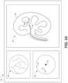
- FIGS. 23 A and 23 Billustrate an example alignment interface for assisting with the fine alignment step of FIG. 22 B , according to one embodiment.
- FIG. 23 Aillustrates an example of the alignment interface when the medical instrument is not aligned with the target location.
- FIG. 23 Billustrates an example of the alignment interface when the medical instrument is aligned with the target location.
- FIG. 24illustrates an example user interface that includes the alignment interface of FIGS. 23 A and 23 B .
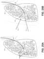
- FIGS. 25 A and 25 Billustrate various steps in another embodiment of an endoscopically-assisted percutaneous medical procedure in a lung.
- FIG. 25 Aillustrates an example of placing or creating virtual fiducials during the procedure.
- FIG. 25 Billustrates an example of creating a boundary for one or more percutaneous instruments based on the virtual fiducials.
- FIG. 26illustrates an example embodiment of a concomitant endoscopic and percutaneous medical procedure in a gastrointestinal tract.
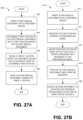
- FIG. 27 Ais a flow chart illustrating an embodiment of a method for performing a medical procedure that includes rendezvousing a medical instrument with a target location.
- FIG. 27 Bis a flow chart illustrating an embodiment of a method for performing a medical procedures that includes placing virtual fiducials to define a boundary.
- aspects of the present disclosuremay be integrated into a robotically-enabled medical system capable of performing a variety of medical procedures, including both minimally invasive, such as laparoscopy, and non-invasive, such as endoscopy, procedures.
- the systemmay be capable of performing bronchoscopy, ureteroscopy, gastroscopy, etc.
- the systemmay provide additional benefits, such as enhanced imaging and guidance to assist the physician. Additionally, the system may provide the physician with the ability to perform the procedure from an ergonomic position without the need for awkward arm motions and positions. Still further, the system may provide the physician with the ability to perform the procedure with improved ease of use such that one or more of the instruments of the system can be controlled by a single user.
- FIG. 1illustrates an embodiment of a cart-based robotically-enabled system 10 arranged for a diagnostic and/or therapeutic bronchoscopy.
- the system 10may comprise a cart 11 having one or more robotic arms 12 to deliver a medical instrument, such as a steerable endoscope 13 , which may be a procedure-specific bronchoscope for bronchoscopy, to a natural orifice access point (i.e., the mouth of the patient positioned on a table in the present example) to deliver diagnostic and/or therapeutic tools.
- a medical instrumentsuch as a steerable endoscope 13
- a natural orifice access pointi.e., the mouth of the patient positioned on a table in the present example
- the cart 11may be positioned proximate to the patient's upper torso in order to provide access to the access point.
- the robotic arms 12may be actuated to position the bronchoscope relative to the access point.
- the arrangement in FIG. 1may also be utilized when performing a gastro-intestinal (GI) procedure with a gastroscope, a specialized endoscope for GI procedures.
- FIG. 2depicts an example embodiment of the cart in greater detail.
- the robotic arms 12may insert the steerable endoscope 13 into the patient robotically, manually, or a combination thereof.
- the steerable endoscope 13may comprise at least two telescoping parts, such as an inner leader portion and an outer sheath portion, each portion coupled to a separate instrument driver from the set of instrument drivers 28 , each instrument driver coupled to the distal end of an individual robotic arm.
- This linear arrangement of the instrument drivers 28which facilitates coaxially aligning the leader portion with the sheath portion, creates a “virtual rail” 29 that may be repositioned in space by manipulating the one or more robotic arms 12 into different angles and/or positions.
- the virtual rails described hereinare depicted in the Figures using dashed lines, and accordingly the dashed lines do not depict any physical structure of the system.
- Translation of the instrument drivers 28 along the virtual rail 29telescopes the inner leader portion relative to the outer sheath portion or advances or retracts the endoscope 13 from the patient.
- the angle of the virtual rail 29may be adjusted, translated, and pivoted based on clinical application or physician preference. For example, in bronchoscopy, the angle and position of the virtual rail 29 as shown represents a compromise between providing physician access to the endoscope 13 while minimizing friction that results from bending the endoscope 13 into the patient's mouth.
- the endoscope 13may be directed down the patient's trachea and lungs after insertion using precise commands from the robotic system until reaching the target destination or operative site. In order to enhance navigation through the patient's lung network and/or reach the desired target, the endoscope 13 may be manipulated to telescopically extend the inner leader portion from the outer sheath portion to obtain enhanced articulation and greater bend radius.
- the use of separate instrument drivers 28also allows the leader portion and sheath portion to be driven independently of each other.
- the endoscope 13may be directed to deliver a biopsy needle to a target, such as, for example, a lesion or nodule within the lungs of a patient.
- the needlemay be deployed down a working channel that runs the length of the endoscope to obtain a tissue sample to be analyzed by a pathologist.
- additional toolsmay be deployed down the working channel of the endoscope for additional biopsies.
- the endoscope 13may endoscopically deliver tools to resect the potentially cancerous tissue.
- diagnostic and therapeutic treatmentscan be delivered in separate procedures.
- the endoscope 13may also be used to deliver a fiducial to “mark” the location of the target nodule as well. In other instances, diagnostic and therapeutic treatments may be delivered during the same procedure.
- the system 10may also include a movable tower 30 , which may be connected via support cables to the cart 11 to provide support for controls, electronics, fluidics, optics, sensors, and/or power to the cart 11 . Placing such functionality in the tower 30 allows for a smaller form factor cart 11 that may be more easily adjusted and/or re-positioned by an operating physician and his/her staff. Additionally, the division of functionality between the cart/table and the support tower 30 reduces operating room clutter and facilitates improving clinical workflow. While the cart 11 may be positioned close to the patient, the tower 30 may be stowed in a remote location to stay out of the way during a procedure.
- the tower 30may include component(s) of a computer-based control system that stores computer program instructions, for example, within a non-transitory computer-readable storage medium such as a persistent magnetic storage drive, solid state drive, etc.
- the execution of those instructionsmay control the entire system or sub-system(s) thereof.
- the instructionswhen executed by a processor of the computer system, the instructions may cause the components of the robotics system to actuate the relevant carriages and arm mounts, actuate the robotics arms, and control the medical instruments.
- the motors in the joints of the robotics armsmay position the arms into a certain posture.
- the tower 30may also include a pump, flow meter, valve control, and/or fluid access in order to provide controlled irrigation and aspiration capabilities to the system that may be deployed through the endoscope 13 . These components may also be controlled using the computer system of the tower 30 . In some embodiments, irrigation and aspiration capabilities may be delivered directly to the endoscope 13 through separate cable(s).
- the tower 30may include a voltage and surge protector designed to provide filtered and protected electrical power to the cart 11 , thereby avoiding placement of a power transformer and other auxiliary power components in the cart 11 , resulting in a smaller, more moveable cart 11 .
- the tower 30may also include support equipment for the sensors deployed throughout the robotic system 10 .
- the tower 30may include optoelectronics equipment for detecting, receiving, and processing data received from the optical sensors or cameras throughout the robotic system 10 . In combination with the control system, such optoelectronics equipment may be used to generate real-time images for display in any number of consoles deployed throughout the system, including in the tower 30 .
- the tower 30may also include an electronic subsystem for receiving and processing signals received from deployed electromagnetic (EM) sensors.
- EMelectromagnetic
- the tower 30may also be used to house and position an EM field generator for detection by EM sensors in or on the medical instrument.
- the tower 30may also include a console 31 in addition to other consoles available in the rest of the system, e.g., console mounted on top of the cart.
- the console 31may include a user interface and a display screen, such as a touchscreen, for the physician operator.
- Consoles in the system 10are generally designed to provide both robotic controls as well as preoperative and real-time information of the procedure, such as navigational and localization information of the endoscope 13 .
- the console 31is not the only console available to the physician, it may be used by a second operator, such as a nurse, to monitor the health or vitals of the patient and the operation of the system 10 , as well as to provide procedure-specific data, such as navigational and localization information.
- the console 30is housed in a body that is separate from the tower 30 .
- the tower 30may be coupled to the cart 11 and endoscope 13 through one or more cables or connections (not shown).
- the support functionality from the tower 30may be provided through a single cable to the cart 11 , simplifying and de-cluttering the operating room.
- specific functionalitymay be coupled in separate cabling and connections. For example, while power may be provided through a single power cable to the cart 11 , the support for controls, optics, fluidics, and/or navigation may be provided through a separate cable.
- FIG. 2provides a detailed illustration of an embodiment of the cart 11 from the cart-based robotically-enabled system shown in FIG. 1 .
- the cart 11generally includes an elongated support structure 14 (often referred to as a “column”), a cart base 15 , and a console 16 at the top of the column 14 .
- the column 14may include one or more carriages, such as a carriage 17 (alternatively “arm support”) for supporting the deployment of one or more robotic arms 12 (three shown in FIG. 2 ).
- the carriage 17may include individually configurable arm mounts that rotate along a perpendicular axis to adjust the base of the robotic arms 12 for better positioning relative to the patient.
- the carriage 17also includes a carriage interface 19 that allows the carriage 17 to vertically translate along the column 14 .
- the carriage interface 19is connected to the column 14 through slots, such as slot 20 , that are positioned on opposite sides of the column 14 to guide the vertical translation of the carriage 17 .
- the slot 20contains a vertical translation interface to position and hold the carriage 17 at various vertical heights relative to the cart base 15 .
- Vertical translation of the carriage 17allows the cart 11 to adjust the reach of the robotic arms 12 to meet a variety of table heights, patient sizes, and physician preferences.
- the individually configurable arm mounts on the carriage 17allow the robotic arm base 21 of the robotic arms 12 to be angled in a variety of configurations.
- the slot 20may be supplemented with slot covers that are flush and parallel to the slot surface to prevent dirt and fluid ingress into the internal chambers of the column 14 and the vertical translation interface as the carriage 17 vertically translates.
- the slot coversmay be deployed through pairs of spring spools positioned near the vertical top and bottom of the slot 20 .
- the coversare coiled within the spools until deployed to extend and retract from their coiled state as the carriage 17 vertically translates up and down.
- the spring-loading of the spoolsprovides force to retract the cover into a spool when the carriage 17 translates towards the spool, while also maintaining a tight seal when the carriage 17 translates away from the spool.
- the coversmay be connected to the carriage 17 using, for example, brackets in the carriage interface 19 to ensure proper extension and retraction of the cover as the carriage 17 translates.
- the column 14may internally comprise mechanisms, such as gears and motors, that are designed to use a vertically aligned lead screw to translate the carriage 17 in a mechanized fashion in response to control signals generated in response to user inputs, e.g., inputs from the console 16 .
- the robotic arms 12may generally comprise robotic arm bases 21 and end effectors 22 , separated by a series of linkages 23 that are connected by a series of joints 24 , each joint comprising an independent actuator, each actuator comprising an independently controllable motor.
- Each independently controllable jointrepresents an independent degree of freedom available to the robotic arm 12 .
- Each of the robotic arms 12may have seven joints, and thus provide seven degrees of freedom. A multitude of joints result in a multitude of degrees of freedom, allowing for “redundant” degrees of freedom. Having redundant degrees of freedom allows the robotic arms 12 to position their respective end effectors 22 at a specific position, orientation, and trajectory in space using different linkage positions and joint angles. This allows for the system to position and direct a medical instrument from a desired point in space while allowing the physician to move the arm joints into a clinically advantageous position away from the patient to create greater access, while avoiding arm collisions.
- the cart base 15balances the weight of the column 14 , carriage 17 , and robotic arms 12 over the floor. Accordingly, the cart base 15 houses heavier components, such as electronics, motors, power supply, as well as components that either enable movement and/or immobilize the cart 11 .
- the cart base 15includes rollable wheel-shaped casters 25 that allow for the cart 11 to easily move around the room prior to a procedure. After reaching the appropriate position, the casters 25 may be immobilized using wheel locks to hold the cart 11 in place during the procedure.
- the console 16allows for both a user interface for receiving user input and a display screen (or a dual-purpose device such as, for example, a touchscreen 26 ) to provide the physician user with both preoperative and intraoperative data.
- Potential preoperative data on the touchscreen 26may include preoperative plans, navigation and mapping data derived from preoperative computerized tomography (CT) scans, and/or notes from pre-operative patient interviews.
- Intraoperative data on displaymay include optical information provided from the tool, sensor and coordinate information from sensors, as well as vital patient statistics, such as respiration, heart rate, and/or pulse.
- the console 16may be positioned and tilted to allow a physician to access the console 16 from the side of the column 14 opposite the carriage 17 . From this position, the physician may view the console 16 , robotic arms 12 , and patient while operating the console 16 from behind the cart 11 .
- the console 16also includes a handle 27 to assist with maneuvering and stabilizing cart 11 .
- FIG. 3illustrates an embodiment of a robotically-enabled system 10 arranged for ureteroscopy.
- the cart 11may be positioned to deliver a ureteroscope 32 , a procedure-specific endoscope designed to traverse a patient's urethra and ureter, to the lower abdominal area of the patient.
- the ureteroscope 32may be directly aligned with the patient's urethra to reduce friction and forces on the sensitive anatomy in the area.
- the cart 11may be aligned at the foot of the table to allow the robotic arms 12 to position the ureteroscope 32 for direct linear access to the patient's urethra. From the foot of the table, the robotic arms 12 may insert the ureteroscope 32 along the virtual rail 33 directly into the patient's lower abdomen through the urethra.
- the ureteroscope 32may be navigated into the bladder, ureters, and/or kidneys for diagnostic and/or therapeutic applications.
- the ureteroscope 32may be directed into the ureter and kidneys to break up kidney stone build up using a laser or ultrasonic lithotripsy device deployed down the working channel of the ureteroscope 32 .
- the resulting stone fragmentsmay be removed using baskets deployed down the ureteroscope 32 .
- FIG. 4illustrates an embodiment of a robotically-enabled system 10 similarly arranged for a vascular procedure.
- the system 10may be configured such that the cart 11 may deliver a medical instrument 34 , such as a steerable catheter, to an access point in the femoral artery in the patient's leg.
- the femoral arterypresents both a larger diameter for navigation as well as a relatively less circuitous and tortuous path to the patient's heart, which simplifies navigation.
- the cart 11may be positioned towards the patient's legs and lower abdomen to allow the robotic arms 12 to provide a virtual rail 35 with direct linear access to the femoral artery access point in the patient's thigh/hip region.
- the medical instrument 34may be directed and inserted by translating the instrument drivers 28 .
- the cartmay be positioned around the patient's upper abdomen in order to reach alternative vascular access points, such as, for example, the carotid and brachial arteries near the shoulder and wrist.
- Embodiments of the robotically-enabled medical systemmay also incorporate the patient's table. Incorporation of the table reduces the amount of capital equipment within the operating room by removing the cart, which allows greater access to the patient.
- FIG. 5illustrates an embodiment of such a robotically-enabled system arranged for a bronchoscopic procedure.
- System 36includes a support structure or column 37 for supporting platform 38 (shown as a “table” or “bed”) over the floor.
- the end effectors of the robotic arms 39 of the system 36comprise instrument drivers 42 that are designed to manipulate an elongated medical instrument, such as a bronchoscope 40 in FIG. 5 , through or along a virtual rail 41 formed from the linear alignment of the instrument drivers 42 .
- a C-arm for providing fluoroscopic imagingmay be positioned over the patient's upper abdominal area by placing the emitter and detector around the table 38 .
- FIG. 6provides an alternative view of the system 36 without the patient and medical instrument for discussion purposes.
- the column 37may include one or more carriages 43 shown as ring-shaped in the system 36 , from which the one or more robotic arms 39 may be based.
- the carriages 43may translate along a vertical column interface 44 that runs the length of the column 37 to provide different vantage points from which the robotic arms 39 may be positioned to reach the patient.
- the carriage(s) 43may rotate around the column 37 using a mechanical motor positioned within the column 37 to allow the robotic arms 39 to have access to multiples sides of the table 38 , such as, for example, both sides of the patient.
- the carriagesmay be individually positioned on the column and may translate and/or rotate independently of the other carriages.
- the system 36can include a patient table or bed with adjustable arm supports in the form of bars or rails extending alongside it.
- One or more robotic arms 39e.g., via a shoulder with an elbow joint
- the robotic arms 39are advantageously capable of being stowed compactly beneath the patient table or bed, and subsequently raised during a procedure.
- the robotic arms 39may be mounted on the carriages 43 through a set of arm mounts 45 comprising a series of joints that may individually rotate and/or telescopically extend to provide additional configurability to the robotic arms 39 .
- the arm mounts 45may be positioned on the carriages 43 such that, when the carriages 43 are appropriately rotated, the arm mounts 45 may be positioned on either the same side of the table 38 (as shown in FIG. 6 ), on opposite sides of the table 38 (as shown in FIG. 9 ), or on adjacent sides of the table 38 (not shown).
- the column 37structurally provides support for the table 38 , and a path for vertical translation of the carriages 43 . Internally, the column 37 may be equipped with lead screws for guiding vertical translation of the carriages, and motors to mechanize the translation of the carriages 43 based the lead screws. The column 37 may also convey power and control signals to the carriages 43 and the robotic arms 39 mounted thereon.
- the table base 46serves a similar function as the cart base 15 in the cart 11 shown in FIG. 2 , housing heavier components to balance the table/bed 38 , the column 37 , the carriages 43 , and the robotic arms 39 .
- the table base 46may also incorporate rigid casters to provide stability during procedures. Deployed from the bottom of the table base 46 , the casters may extend in opposite directions on both sides of the base 46 and retract when the system 36 needs to be moved.
- the system 36may also include a tower (not shown) that divides the functionality of the system 36 between the table and the tower to reduce the form factor and bulk of the table.
- the towermay provide a variety of support functionalities to the table, such as processing, computing, and control capabilities, power, fluidics, and/or optical and sensor processing.
- the towermay also be movable to be positioned away from the patient to improve physician access and de-clutter the operating room. Additionally, placing components in the tower allows for more storage space in the table base 46 for potential stowage of the robotic arms 39 .
- the towermay also include a master controller or console that provides both a user interface for user input, such as keyboard and/or pendant, as well as a display screen (or touchscreen) for preoperative and intraoperative information, such as real-time imaging, navigation, and tracking information.
- the towermay also contain holders for gas tanks to be used for insufflation.
- a table basemay stow and store the robotic arms when not in use.
- FIG. 7illustrates a system 47 that stows robotic arms in an embodiment of the table-based system.
- carriages 48may be vertically translated into base 49 to stow robotic arms 50 , arm mounts 51 , and the carriages 48 within the base 49 .
- Base covers 52may be translated and retracted open to deploy the carriages 48 , arm mounts 51 , and robotic arms 50 around column 53 , and closed to stow to protect them when not in use.
- the base covers 52may be sealed with a membrane 54 along the edges of its opening to prevent dirt and fluid ingress when closed.
- FIG. 8illustrates an embodiment of a robotically-enabled table-based system configured for a ureteroscopic procedure.
- the table 38may include a swivel portion 55 for positioning a patient off-angle from the column 37 and table base 46 .
- the swivel portion 55may rotate or pivot around a pivot point (e.g., located below the patient's head) in order to position the bottom portion of the swivel portion 55 away from the column 37 .
- the pivoting of the swivel portion 55allows a C-arm (not shown) to be positioned over the patient's lower abdomen without competing for space with the column (not shown) below table 38 .
- the robotic arms 39may directly insert a ureteroscope 56 along a virtual rail 57 into the patient's groin area to reach the urethra.
- stirrups 58may also be fixed to the swivel portion 55 of the table 38 to support the position of the patient's legs during the procedure and allow clear access to the patient's groin area.
- minimally invasive instrumentsmay be inserted into the patient's anatomy.
- the minimally invasive instrumentscomprise an elongated rigid member, such as a shaft, which is used to access anatomy within the patient. After inflation of the patient's abdominal cavity, the instruments may be directed to perform surgical or medical tasks, such as grasping, cutting, ablating, suturing, etc.
- the instrumentscan comprise a scope, such as a laparoscope.
- FIG. 9illustrates an embodiment of a robotically-enabled table-based system configured for a laparoscopic procedure. As shown in FIG.
- the carriages 43 of the system 36may be rotated and vertically adjusted to position pairs of the robotic arms 39 on opposite sides of the table 38 , such that instrument 59 may be positioned using the arm mounts 45 to be passed through minimal incisions on both sides of the patient to reach his/her abdominal cavity.
- FIG. 10illustrates an embodiment of the robotically-enabled medical system with pitch or tilt adjustment.
- the system 36may accommodate tilt of the table 38 to position one portion of the table at a greater distance from the floor than the other.
- the arm mounts 45may rotate to match the tilt such that the robotic arms 39 maintain the same planar relationship with the table 38 .
- the column 37may also include telescoping portions 60 that allow vertical extension of the column 37 to keep the table 38 from touching the floor or colliding with the table base 46 .
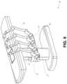
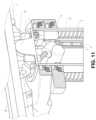
- FIG. 11provides a detailed illustration of the interface between the table 38 and the column 37 .
- Pitch rotation mechanism 61may be configured to alter the pitch angle of the table 38 relative to the column 37 in multiple degrees of freedom.
- the pitch rotation mechanism 61may be enabled by the positioning of orthogonal axes 1 , 2 at the column-table interface, each axis actuated by a separate motor 3 , 4 responsive to an electrical pitch angle command. Rotation along one screw 5 would enable tilt adjustments in one axis 1 , while rotation along the other screw 6 would enable tilt adjustments along the other axis 2 .
- a ball jointcan be used to alter the pitch angle of the table 38 relative to the column 37 in multiple degrees of freedom.
- pitch adjustmentsare particularly useful when trying to position the table in a Trendelenburg position, i.e., position the patient's lower abdomen at a higher position from the floor than the patient's upper abdomen, for lower abdominal surgery.
- the Trendelenburg positioncauses the patient's internal organs to slide towards his/her upper abdomen through the force of gravity, clearing out the abdominal cavity for minimally invasive tools to enter and perform lower abdominal surgical or medical procedures, such as laparoscopic prostatectomy.
- FIGS. 12 and 13illustrate isometric and end views of an alternative embodiment of a table-based surgical robotics system 100 .
- the surgical robotics system 100includes one or more adjustable arm supports 105 that can be configured to support one or more robotic arms (see, for example, FIG. 14 ) relative to a table 101 .
- a single adjustable arm support 105is shown, though an additional arm support can be provided on an opposite side of the table 101 .
- the adjustable arm support 105can be configured so that it can move relative to the table 101 to adjust and/or vary the position of the adjustable arm support 105 and/or any robotic arms mounted thereto relative to the table 101 .
- the adjustable arm support 105may be adjusted one or more degrees of freedom relative to the table 101 .
- the adjustable arm support 105provides high versatility to the system 100 , including the ability to easily stow the one or more adjustable arm supports 105 and any robotics arms attached thereto beneath the table 101 .
- the adjustable arm support 105can be elevated from the stowed position to a position below an upper surface of the table 101 . In other embodiments, the adjustable arm support 105 can be elevated from the stowed position to a position above an upper surface of the table 101 .
- the adjustable arm support 105can provide several degrees of freedom, including lift, lateral translation, tilt, etc.
- the arm support 105is configured with four degrees of freedom, which are illustrated with arrows in FIG. 12 .
- a first degree of freedomallows for adjustment of the adjustable arm support 105 in the z-direction (“Z-lift”).
- the adjustable arm support 105can include a carriage 109 configured to move up or down along or relative to a column 102 supporting the table 101 .
- a second degree of freedomcan allow the adjustable arm support 105 to tilt.
- the adjustable arm support 105can include a rotary joint, which can allow the adjustable arm support 105 to be aligned with the bed in a Trendelenburg position.
- a third degree of freedomcan allow the adjustable arm support 105 to “pivot up,” which can be used to adjust a distance between a side of the table 101 and the adjustable arm support 105 .
- a fourth degree of freedomcan permit translation of the adjustable arm support 105 along a longitudinal length of the table.
- the surgical robotics system 100 in FIGS. 12 and 13can comprise a table supported by a column 102 that is mounted to a base 103 .
- the base 103 and the column 102support the table 101 relative to a support surface.
- a floor axis 131 and a support axis 133are shown in FIG. 13 .
- the adjustable arm support 105can be mounted to the column 102 . In other embodiments, the arm support 105 can be mounted to the table 101 or base 103 .
- the adjustable arm support 105can include a carriage 109 , a bar or rail connector 111 and a bar or rail 107 . In some embodiments, one or more robotic arms mounted to the rail 107 can translate and move relative to one another.
- the carriage 109can be attached to the column 102 by a first joint 113 , which allows the carriage 109 to move relative to the column 102 (e.g., such as up and down a first or vertical axis 123 ).
- the first joint 113can provide the first degree of freedom (“Z-lift”) to the adjustable arm support 105 .
- the adjustable arm support 105can include a second joint 115 , which provides the second degree of freedom (tilt) for the adjustable arm support 105 .
- the adjustable arm support 105can include a third joint 117 , which can provide the third degree of freedom (“pivot up”) for the adjustable arm support 105 .
- An additional joint 119(shown in FIG.
- the adjustable arm support 105can include a fourth joint 121 , which can provide a fourth degree of freedom (translation) for the adjustable arm support 105 along a fourth axis 129 .
- FIG. 14illustrates an end view of the surgical robotics system 140 A with two adjustable arm supports 105 A, 105 B mounted on opposite sides of a table 101 .
- a first robotic arm 142 Ais attached to the bar or rail 107 A of the first adjustable arm support 105 B.
- the first robotic arm 142 Aincludes a base 144 A attached to the rail 107 A.
- the distal end of the first robotic arm 142 Aincludes an instrument drive mechanism 146 A that can attach to one or more robotic medical instruments or tools.
- the second robotic arm 142 Bincludes a base 144 B attached to the rail 107 B.
- the distal end of the second robotic arm 142 Bincludes an instrument drive mechanism 146 B.
- the instrument drive mechanism 146 Bcan be configured to attach to one or more robotic medical instruments or tools.
- one or more of the robotic arms 142 A, 142 Bcomprises an arm with seven or more degrees of freedom.
- one or more of the robotic arms 142 A, 142 Bcan include eight degrees of freedom, including an insertion axis (1-degree of freedom including insertion), a wrist (3-degrees of freedom including wrist pitch, yaw and roll), an elbow (1-degree of freedom including elbow pitch), a shoulder (2-degrees of freedom including shoulder pitch and yaw), and base 144 A, 144 B (1-degree of freedom including translation).
- the insertion degree of freedomcan be provided by the robotic arm 142 A, 142 B, while in other embodiments, the instrument itself provides insertion via an instrument-based insertion architecture.
- the end effectors of the system's robotic armsmay comprise (i) an instrument driver (alternatively referred to as “instrument drive mechanism” or “instrument device manipulator”) that incorporates electro-mechanical means for actuating the medical instrument and (ii) a removable or detachable medical instrument, which may be devoid of any electro-mechanical components, such as motors.
- instrument driveralternatively referred to as “instrument drive mechanism” or “instrument device manipulator”
- instrument device manipulatora removable or detachable medical instrument, which may be devoid of any electro-mechanical components, such as motors.
- This dichotomymay be driven by the need to sterilize medical instruments used in medical procedures, and the inability to adequately sterilize expensive capital equipment due to their intricate mechanical assemblies and sensitive electronics. Accordingly, the medical instruments may be designed to be detached, removed, and interchanged from the instrument driver (and thus the system) for individual sterilization or disposal by the physician or the physician's staff. In contrast, the instrument drivers need not be changed or sterilized, and may be draped for protection
- FIG. 15illustrates an example instrument driver.
- instrument driver 62Positioned at the distal end of a robotic arm, instrument driver 62 comprises one or more drive units 63 arranged with parallel axes to provide controlled torque to a medical instrument via drive shafts 64 .
- Each drive unit 63comprises an individual drive shaft 64 for interacting with the instrument, a gear head 65 for converting the motor shaft rotation to a desired torque, a motor 66 for generating the drive torque, an encoder 67 to measure the speed of the motor shaft and provide feedback to the control circuitry, and control circuity 68 for receiving control signals and actuating the drive unit.
- Each drive unit 63being independently controlled and motorized, the instrument driver 62 may provide multiple (four as shown in FIG. 15 ) independent drive outputs to the medical instrument.
- the control circuitry 68would receive a control signal, transmit a motor signal to the motor 66 , compare the resulting motor speed as measured by the encoder 67 with the desired speed, and modulate the motor signal to generate the desired torque.
- the robotic systemmay incorporate a drive interface, such as a sterile adapter connected to a sterile drape, that sits between the instrument driver and the medical instrument.
- a drive interfacesuch as a sterile adapter connected to a sterile drape
- the chief purpose of the sterile adapteris to transfer angular motion from the drive shafts of the instrument driver to the drive inputs of the instrument while maintaining physical separation, and thus sterility, between the drive shafts and drive inputs.
- an example sterile adaptermay comprise a series of rotational inputs and outputs intended to be mated with the drive shafts of the instrument driver and drive inputs on the instrument.
- the sterile drapecomprised of a thin, flexible material such as transparent or translucent plastic, is designed to cover the capital equipment, such as the instrument driver, robotic arm, and cart (in a cart-based system) or table (in a table-based system).
- the capital equipmentsuch as the instrument driver, robotic arm, and cart (in a cart-based system) or table (in a table-based system).
- Use of the drapewould allow the capital equipment to be positioned proximate to the patient while still being located in an area not requiring sterilization (i.e., non-sterile field).
- the medical instrumentmay interface with the patient in an area requiring sterilization (i.e., sterile field).
- FIG. 16illustrates an example medical instrument with a paired instrument driver.
- medical instrument 70comprises an elongated shaft 71 (or elongate body) and an instrument base 72 .
- the instrument base 72also referred to as an “instrument handle” due to its intended design for manual interaction by the physician, may generally comprise rotatable drive inputs 73 , e.g., receptacles, pulleys or spools, that are designed to be mated with drive outputs 74 that extend through a drive interface on instrument driver 75 at the distal end of robotic arm 76 .
- the mated drive inputs 73 of the instrument base 72may share axes of rotation with the drive outputs 74 in the instrument driver 75 to allow the transfer of torque from the drive outputs 74 to the drive inputs 73 .
- the drive outputs 74may comprise splines that are designed to mate with receptacles on the drive inputs 73 .
- the elongated shaft 71is designed to be delivered through either an anatomical opening or lumen, e.g., as in endoscopy, or a minimally invasive incision, e.g., as in laparoscopy.
- the elongated shaft 71may be either flexible (e.g., having properties similar to an endoscope) or rigid (e.g., having properties similar to a laparoscope) or contain a customized combination of both flexible and rigid portions.
- the distal end of a rigid elongated shaftmay be connected to an end effector extending from a jointed wrist formed from a clevis with at least one degree of freedom and a surgical tool or medical instrument, such as, for example, a grasper or scissors, that may be actuated based on force from the tendons as the drive inputs rotate in response to torque received from the drive outputs 74 of the instrument driver 75 .
- a surgical tool or medical instrumentsuch as, for example, a grasper or scissors
- the distal end of a flexible elongated shaftmay include a steerable or controllable bending section that may be articulated and bent based on torque received from the drive outputs 74 of the instrument driver 75 .
- Torque from the instrument driver 75is transmitted down the elongated shaft 71 using tendons along the elongated shaft 71 .
- These individual tendonssuch as pull wires, may be individually anchored to individual drive inputs 73 within the instrument handle 72 .
- the tendonsare directed down one or more pull lumens along the elongated shaft 71 and anchored at the distal portion of the elongated shaft 71 , or in the wrist at the distal portion of the elongated shaft.
- these tendonsmay be coupled to a distally mounted end effector, such as a wrist, grasper, or scissor.
- the tendonmay cause a joint to rotate about an axis, thereby causing the end effector to move in one direction or another.
- the tendonmay be connected to one or more jaws of a grasper at the distal end of the elongated shaft 71 , where tension from the tendon causes the grasper to close.
- the tendonsmay be coupled to a bending or articulating section positioned along the elongated shaft 71 (e.g., at the distal end) via adhesive, control ring, or other mechanical fixation.
- a bending or articulating sectionpositioned along the elongated shaft 71 (e.g., at the distal end) via adhesive, control ring, or other mechanical fixation.
- torque exerted on the drive inputs 73would be transmitted down the tendons, causing the softer, bending section (sometimes referred to as the articulable section or region) to bend or articulate.
- the angle of the spiraling and/or spacing therebetweenmay be altered or engineered for specific purposes, wherein tighter spiraling exhibits lesser shaft compression under load forces, while lower amounts of spiraling results in greater shaft compression under load forces, but limits bending.
- the pull lumensmay be directed parallel to the longitudinal axis of the elongated shaft 71 to allow for controlled articulation in the desired bending or articulable sections.
- the elongated shaft 71houses a number of components to assist with the robotic procedure.
- the shaft 71may comprise a working channel for deploying surgical tools (or medical instruments), irrigation, and/or aspiration to the operative region at the distal end of the shaft 71 .
- the shaft 71may also accommodate wires and/or optical fibers to transfer signals to/from an optical assembly at the distal tip, which may include an optical camera.
- the shaft 71may also accommodate optical fibers to carry light from proximally-located light sources, such as light emitting diodes, to the distal end of the shaft 71 .
- the distal tipmay also comprise the opening of a working channel for delivering tools for diagnostic and/or therapy, irrigation, and aspiration to an operative site.
- the distal tipmay also include a port for a camera, such as a fiberscope or a digital camera, to capture images of an internal anatomical space.
- the distal tipmay also include ports for light sources for illuminating the anatomical space when using the camera.
- the drive shaft axesand thus the drive input axes, are orthogonal to the axis of the elongated shaft 71 .
- This arrangementcomplicates roll capabilities for the elongated shaft 71 .
- Rolling the elongated shaft 71 along its axis while keeping the drive inputs 73 staticresults in undesirable tangling of the tendons as they extend off the drive inputs 73 and enter pull lumens within the elongated shaft 71 .
- the resulting entanglement of such tendonsmay disrupt any control algorithms intended to predict movement of the flexible elongated shaft 71 during an endoscopic procedure.
- FIG. 17illustrates an alternative design for an instrument driver and instrument where the axes of the drive units are parallel to the axis of the elongated shaft of the instrument.
- a circular instrument driver 80comprises four drive units with their drive outputs 81 aligned in parallel at the end of a robotic arm 82 .
- the drive units, and their respective drive outputs 81are housed in a rotational assembly 83 of the instrument driver 80 that is driven by one of the drive units within the assembly 83 .
- the rotational assembly 83rotates along a circular bearing that connects the rotational assembly 83 to the non-rotational portion 84 of the instrument driver 80 .
- Power and controls signalsmay be communicated from the non-rotational portion 84 of the instrument driver 80 to the rotational assembly 83 through electrical contacts that may be maintained through rotation by a brushed slip ring connection (not shown).
- the rotational assembly 83may be responsive to a separate drive unit that is integrated into the non-rotatable portion 84 , and thus not in parallel to the other drive units.
- the rotational mechanism 83allows the instrument driver 80 to rotate the drive units, and their respective drive outputs 81 , as a single unit around an instrument driver axis 85 .
- an instrument 86may comprise an elongated shaft portion 88 and an instrument base 87 (shown with a transparent external skin for discussion purposes) comprising a plurality of drive inputs 89 (such as receptacles, pulleys, and spools) that are configured to receive the drive outputs 81 in the instrument driver 80 .
- the instrument shaft 88extends from the center of the instrument base 87 with an axis substantially parallel to the axes of the drive inputs 89 , rather than orthogonal as in the design of FIG. 16 .
- the medical instrument 86When coupled to the rotational assembly 83 of the instrument driver 80 , the medical instrument 86 , comprising instrument base 87 and instrument shaft 88 , rotates in combination with the rotational assembly 83 about the instrument driver axis 85 . Since the instrument shaft 88 is positioned at the center of instrument base 87 , the instrument shaft 88 is coaxial with instrument driver axis 85 when attached. Thus, rotation of the rotational assembly 83 causes the instrument shaft 88 to rotate about its own longitudinal axis. Moreover, as the instrument base 87 rotates with the instrument shaft 88 , any tendons connected to the drive inputs 89 in the instrument base 87 are not tangled during rotation. Accordingly, the parallelism of the axes of the drive outputs 81 , drive inputs 89 , and instrument shaft 88 allows for the shaft rotation without tangling any control tendons.
- FIG. 18illustrates an instrument having an instrument based insertion architecture in accordance with some embodiments.
- the instrument 150can be coupled to any of the instrument drivers discussed above.
- the instrument 150comprises an elongated shaft 152 , an end effector 162 connected to the shaft 152 , and a handle 170 coupled to the shaft 152 .
- the elongated shaft 152comprises a tubular member having a proximal portion 154 and a distal portion 156 .
- the elongated shaft 152comprises one or more channels or grooves 158 along its outer surface.
- the grooves 158are configured to receive one or more wires or cables 180 therethrough.
- One or more cables 180thus run along an outer surface of the elongated shaft 152 .
- cables 180can also run through the elongated shaft 152 .
- Manipulation of the one or more cables 180results in actuation of the end effector 162 .
- the instrument handle 170which may also be referred to as an instrument base, may generally comprise an attachment interface 172 having one or more mechanical inputs 174 , e.g., receptacles, pulleys or spools, that are designed to be reciprocally mated with one or more torque couplers on an attachment surface of an instrument driver.
- mechanical inputs 174e.g., receptacles, pulleys or spools, that are designed to be reciprocally mated with one or more torque couplers on an attachment surface of an instrument driver.
- the instrument 150comprises a series of pulleys or cables that enable the elongated shaft 152 to translate relative to the handle 170 .
- the instrument 150itself comprises an instrument-based insertion architecture that accommodates insertion of the instrument, thereby minimizing the reliance on a robot arm to provide insertion of the instrument 150 .
- a robotic armcan be largely responsible for instrument insertion.
- any of the robotic systems described hereincan include an input device or controller for manipulating an instrument attached to a robotic arm.
- the controllercan be coupled (e.g., communicatively, electronically, electrically, wirelessly and/or mechanically) with an instrument such that manipulation of the controller causes a corresponding manipulation of the instrument e.g., via master slave control.
- FIG. 19is a perspective view of an embodiment of a controller 182 .
- the controller 182comprises a hybrid controller that can have both impedance and admittance control.
- the controller 182can utilize just impedance or passive control.
- the controller 182can utilize just admittance control.
- the controller 182advantageously can have a lower perceived inertia while in use.
- the controller 182is configured to allow manipulation of two medical instruments, and includes two handles 184 .
- Each of the handles 184is connected to a gimbal 186 .
- Each gimbal 186is connected to a positioning platform 188 .
- each positioning platform 188includes a SCARA arm (selective compliance assembly robot arm) 198 coupled to a column 194 by a prismatic joint 196 .
- the prismatic joints 196are configured to translate along the column 194 (e.g., along rails 197 ) to allow each of the handles 184 to be translated in the z-direction, providing a first degree of freedom.
- the SCARA arm 198is configured to allow motion of the handle 184 in an x-y plane, providing two additional degrees of freedom.
- one or more load cellsare positioned in the controller.
- a load cell(not shown) is positioned in the body of each of the gimbals 186 .
- portions of the controller 182are capable of operating under admittance control, thereby advantageously reducing the perceived inertia of the controller while in use.
- the positioning platform 188is configured for admittance control, while the gimbal 186 is configured for impedance control.
- the gimbal 186is configured for admittance control, while the positioning platform 188 is configured for impedance control. Accordingly, for some embodiments, the translational or positional degrees of freedom of the positioning platform 188 can rely on admittance control, while the rotational degrees of freedom of the gimbal 186 rely on impedance control.
- the robotic systems contemplated by this disclosurecan provide for non-radiation-based navigational and localization means to reduce physician exposure to radiation and reduce the amount of equipment within the operating room.
- the term “localization”may refer to determining and/or monitoring the position of objects in a reference coordinate system. Technologies such as preoperative mapping, computer vision, real-time EM tracking, and robot command data may be used individually or in combination to achieve a radiation-free operating environment. In other cases, where radiation-based imaging modalities are still used, the preoperative mapping, computer vision, real-time EM tracking, and robot command data may be used individually or in combination to improve upon the information obtained solely through radiation-based imaging modalities.
- FIG. 20is a block diagram illustrating a localization system 90 that estimates a location of one or more elements of the robotic system, such as the location of the instrument, in accordance to an example embodiment.
- the localization system 90may be a set of one or more computer devices configured to execute one or more instructions.
- the computer devicesmay be embodied by a processor (or processors) and computer-readable memory in one or more components discussed above.
- the computer devicesmay be in the tower 30 shown in FIG. 1 , the cart 11 shown in FIGS. 1 - 4 , the beds shown in FIGS. 5 - 14 , etc.
- the localization system 90may include a localization module 95 that processes input data 91 - 94 to generate location data 96 for the distal tip of a medical instrument.
- the location data 96may be data or logic that represents a location and/or orientation of the distal end of the instrument relative to a frame of reference.
- the frame of referencecan be a frame of reference relative to the anatomy of the patient or to a known object, such as an EM field generator (see discussion below for the EM field generator).
- Preoperative mappingmay be accomplished through the use of the collection of low dose CT scans.
- Preoperative CT scansare reconstructed into three-dimensional images, which are visualized, e.g. as “slices” of a cutaway view of the patient's internal anatomy.
- image-based models for anatomical cavities, spaces and structures of the patient's anatomy, such as a patient lung networkmay be generated.
- Techniquessuch as center-line geometry may be determined and approximated from the CT images to develop a three-dimensional volume of the patient's anatomy, referred to as model data 91 (also referred to as “preoperative model data” when generated using only preoperative CT scans).
- Network topological modelsmay also be derived from the CT-images, and are particularly appropriate for bronchoscopy.
- the instrumentmay be equipped with a camera to provide vision data (or image data) 92 .
- the localization module 95may process the vision data 92 to enable one or more vision-based (or image-based) location tracking modules or features.
- the preoperative model data 91may be used in conjunction with the vision data 92 to enable computer vision-based tracking of the medical instrument (e.g., an endoscope or an instrument advance through a working channel of the endoscope).
- the robotic systemmay generate a library of expected endoscopic images from the model based on the expected path of travel of the endoscope, each image linked to a location within the model. Intraoperatively, this library may be referenced by the robotic system in order to compare real-time images captured at the camera (e.g., a camera at a distal end of the endoscope) to those in the image library to assist localization.
- Some features of the localization module 95may identify circular geometries in the preoperative model data 91 that correspond to anatomical lumens and track the change of those geometries to determine which anatomical lumen was selected, as well as the relative rotational and/or translational motion of the camera. Use of a topological map may further enhance vision-based algorithms or techniques.
- Optical flowanother computer vision-based technique, may analyze the displacement and translation of image pixels in a video sequence in the vision data 92 to infer camera movement.
- optical flow techniquesmay include motion detection, object segmentation calculations, luminance, motion compensated encoding, stereo disparity measurement, etc. Through the comparison of multiple frames over multiple iterations, movement and location of the camera (and thus the endoscope) may be determined.
- the localization module 95may use real-time EM tracking to generate a real-time location of the endoscope in a global coordinate system that may be registered to the patient's anatomy, represented by the preoperative model.
- EM trackingan EM sensor (or tracker) comprising one or more sensor coils embedded in one or more locations and orientations in a medical instrument (e.g., an endoscopic tool) measures the variation in the EM field created by one or more static EM field generators positioned at a known location.
- the location information detected by the EM sensorsis stored as EM data 93 .
- the EM field generator(or transmitter), may be placed close to the patient to create a low intensity magnetic field that the embedded sensor may detect.
- the magnetic fieldinduces small currents in the sensor coils of the EM sensor, which may be analyzed to determine the distance and angle between the EM sensor and the EM field generator.
- These distances and orientationsmay be intraoperatively “registered” to the patient anatomy (e.g., the preoperative model) in order to determine the geometric transformation that aligns a single location in the coordinate system with a position in the preoperative model of the patient's anatomy.
- an embedded EM tracker in one or more positions of the medical instrumente.g., the distal tip of an endoscope
- Robotic command and kinematics data 94may also be used by the localization module 95 to provide localization data 96 for the robotic system.
- Device pitch and yaw resulting from articulation commandsmay be determined during preoperative calibration. Intraoperatively, these calibration measurements may be used in combination with known insertion depth information to estimate the position of the instrument. Alternatively, these calculations may be analyzed in combination with EM, vision, and/or topological modeling to estimate the position of the medical instrument within the network.
- a number of other input datacan be used by the localization module 95 .
- an instrument utilizing shape-sensing fibercan provide shape data that the localization module 95 can use to determine the location and shape of the instrument.
- the localization module 95may use the input data 91 - 94 in combination(s). In some cases, such a combination may use a probabilistic approach where the localization module 95 assigns a confidence weight to the location determined from each of the input data 91 - 94 . Thus, where the EM data may not be reliable (as may be the case where there is EM interference) the confidence of the location determined by the EM data 93 can be decrease and the localization module 95 may rely more heavily on the vision data 92 and/or the robotic command and kinematics data 94 .
- the robotic systems discussed hereinmay be designed to incorporate a combination of one or more of the technologies above.
- the robotic system's computer-based control systembased in the tower, bed and/or cart, may store computer program instructions, for example, within a non-transitory computer-readable storage medium such as a persistent magnetic storage drive, solid state drive, or the like, that, upon execution, cause the system to receive and analyze sensor data and user commands, generate control signals throughout the system, and display the navigational and localization data, such as the position of the instrument within the global coordinate system, anatomical map, etc.
- Embodiments of the disclosurerelate to robotic systems and methods for concomitant endoscopic and percutaneous medical procedures, such as endoscopically-assisted percutaneous (or laparposic) medical procedures and laparoscopically-assisted endoscopic procedures.
- the systems and methodscan be embodied in or employed using robotically-enabled medical systems, such as those described above with reference to FIGS. 1 - 20 and those described in further detail below.
- Many medical proceduresinvolve guiding a medical instrument to a target location within a treatment region of a patient.
- these medical procedurescan involve percutaneously guiding the medical instrument through an opening (such as a percutaneous access port) to the target region.
- an openingsuch as a percutaneous access port
- the target locationis often internal, it can be difficult to accurately guide the medical instrument to the target location.
- physicians, and in particular, highly trained radiologistsrely on live fluoroscopic images to guide the medical instrument to the target location. Still, such procedures are difficult.
- the radiologistmust derive a three-dimensional path to the target location from two-dimensional fluoroscopic images, often leading to imprecise guidance and placement of the medical instrument. Further, use of fluoroscopy undesirably exposes the patient and medical staff to radiation for a prolonged period of time.
- PCNLPercutaneous nephrolithotomy
- a radiologistpercutaneously guides an access sheath into the kidney to gain access to the treatment region.
- the radiologistrelies on two-dimensional fluoroscopic images to guide and place the access sheath.
- a urologistaccesses the treatment region through the access sheath to remove the kidney stone.
- the common need for a radiologist to perform the first part of the procedureadds cost, complication, and operation scheduling time delay to a procedure that would ideally need only the urologist and their staff to perform.
- the radiologistbecause the radiologist is not trained in urology, the radiologist often places the access sheath in a suboptimal position. However, the urologist must rely on the radiologist to place the access sheath because the urologist is not trained in radiology and thus cannot guide the access sheath himself. Because the radiologist relies on fluoroscopic imaging techniques, another disadvantage of PCNL is the radiation exposure mentioned above.
- the methods and systems for concomitant endoscopic and percutaneous medical procedures described in this applicationcan provide improved guidance and placement of percutaneous and/or endoscopic medical instruments.
- the methods and systemscan be advantageously employed to define rendezvous points for medical instruments within a treatment region and/or to define boundaries within the treatment region for use during the procedure.
- FIGS. 21 A- 21 Dillustrate various steps in an embodiment of an endoscopically-assisted percutaneous medical procedure in a kidney 202 .
- the medical procedureis an endoscopically-assisted percutaneous nephrolithotomy (PCNL) procedure for removing a kidney stone 212 , although the principles illustrated by the example are applicable to other types of medical procedures, all of which are intended to be within the scope of this disclosure.
- PCNLpercutaneous nephrolithotomy
- a first medical instrument 204is inserted into the kidney 202 through a natural orifice in the patient ( FIG. 21 A ).
- the first medical instrument 204includes a position sensor 206 .
- a target location 208 for guiding percutaneous insertion of a second medical instrument 214is determined with reference to the position sensor 206 ( FIG. 21 B ).
- the target location 208can be distanced from the position sensor 206 as shown.
- the target location 208can be a location (such as a point in space) or a trajectory (such as a line in space).
- the target location 208can be displayed as either a point or line on a graphical user interface.
- the second medical instrument 214is aligned with the target location 208 ( FIG. 21 C ).
- the second medical instrument 214is percutaneously inserted toward the target location 208 ( FIG. 21 D ). In this position, PCNL can be performed through the second medical instrument 214 . In some embodiments, PCNL can be assisted by the first medical instrument 204 .
- the endoscopically-assisted PCNL procedure illustrated in FIGS. 21 A- 21 Dcan provide several advantages.
- the procedurecan allow for increased accuracy in percutaneous placement of the second medical instrument 214 because insertion of the second medical instrument 214 can be guided toward the target location 208 .
- the procedureallows for guidance of the second medical instrument 214 without requiring fluoroscopic visualization. This can advantageously reduce or eliminate radiation exposure to the patient and medical personnel during the procedure. This can also simplify the procedure as it may allow the procedure to be performed wholly by a urologist, rather than by both a radiologist and urologist.
- the target location 208can be determined with reference to the position sensor 206 , but at positions that are distanced from the position sensor 206 , the target location 208 can be determined at positions that cannot be directly accessed by the first medical instrument 204 .
- the proceduresmay enable placement of an access port in a single attempt.
- other techniquessuch as a fluoroscopy guided techniques
- FIGS. 21 A- 21 Dillustrate various steps in an endoscopically-assisted percutaneous nephrolithotomy (PCNL) procedure for removing a kidney stone 212 .
- the kidney stone 212is positioned at the entrance to a calyx.
- a urologistmay desire to insert an access sheath into the kidney 202 within the calyx and behind the kidney stone 212 .
- PCNLcan then be performed through the access sheath to break up and remove the kidney stone 212 .
- Accurate placement of the access sheathcan be important for maximizing the efficacy of the procedure and minimizing stress and impact on the patient.
- the first medical instrument 204can be guided into the kidney 202 .
- the first medical instrument 204is inserted into the patient through a natural orifice.
- the first medical instrument 204can be inserted through the urethra, bladder, and ureter into the kidney 202 .
- other natural patient orificescan be used.
- the first medical instrument 204can be inserted percutaneously.
- the first medical instrument 204can be an endoscope.
- the first medical instrument 204can be robotically controlled.
- the first medical instrumentcan by any of the robotically-controllable medical instruments described above with reference to FIGS. 1 - 20 .
- the first medical instrument 204can be manually controlled.
- the first medical instrument 204can include an elongated shaft 205 .
- the elongated shaft 205can be articulable and controllable such that the first medical instrument 204 can be navigated through the patient's anatomy into the kidney 202 .
- FIGS. 16 - 18Several embodiments for such medical instruments are described above with reference to FIGS. 16 - 18 .
- the medical instrument 204can also include, in some embodiments, various other features such as optical systems (such as cameras), which can allow an operator to visualize the treatment region from the point of view of the first medical instrument 204 , and working channels, which can allow for delivery of additional medical tools or instruments through the elongated shaft 205 to the treatment region.
- the working channelsmay be used to deliver fluid or to aspirate fluid or debris.
- the first medical instrument 204can include a position sensor 206 .
- the position sensor 206is positioned at or near the distal end of the elongated shaft 205 of the first medical instrument 204 . In other embodiments, the position sensor 206 can be positioned in other locations on the elongated shaft 205 .
- the first medical instrument 204includes a plurality of position sensors 206 . The position sensor 206 is configured to provide an output from which the location or position of the position sensor 206 (and the first medical instrument 204 ) can be determined.
- the position sensor 206comprises an electromagnetic (EM) sensor configured to produce a detectable signal within an EM field from which the position of the EM sensor within the EM field can be determined.
- the position sensor 206comprises a shape sensing fiber from which the pose or shape of the elongated shaft 205 can be determined and used to determine the position of the medical instrument 204 . In other embodiments, other types of position sensors can be used.
- the first medical instrument 204is guided into the kidney 202 .
- guidance of the first medical instrument 204is facilitated by a navigation or localization system, such as localization system 90 described above.
- the operatormay visualize the kidney stone 212 using the optical system on the first medical instrument 204 .
- the operatorcan determine a target location 208 .
- the target location 208may represent a location at which it is desired that the second medical instrument 214 (see FIGS. 21 C and 21 D ) be placed.
- the target location 208can provide a beacon that is used for guiding insertion of the second medical instrument 214 .
- the second medical instrument 214can be guided so as to rendezvous with the target location 208 .
- the target location 208can be determined with reference to the position sensor 206 but at a position that is distanced from the position sensor 206 . That is, the target location 208 need not be coincident with the position sensor 206 (or any other point on the first medical instrument 204 ).
- the output of the position sensor 206can be registered to a preoperative model of the kidney 202 .
- Registrationcan involve, for example, using the position sensor 206 to map the path of the first medical instrument 204 through the anatomy, and matching the mapped path to the preoperative model.
- registrationcan involve navigating the medical instrument 204 to one or more anatomical landmarks, and using the output of the position sensor 206 at the one or more anatomical landmarks to register the preoperative model to the output of the position sensor 206 . Additional detail on registration is provided above with reference to localization system 90 and FIG. 20 .
- the operatormay, in some embodiments, determine the target location 208 by selecting it within the preoperative model.
- the preoperative modelcan be displayed to the operator, and the operator can select a location from within the preoperative model to be the target location 208 .
- the target location 208can be distanced from the position sensor 206 .
- a distance and direction between the location of the position sensor 206 and the chosen target location 208can be determined by the system.
- the location of the position sensor 206can be represented as an x, y, z coordinate
- the target location 208can be represented by an x′, y′, z′ coordinate
- a distance and direction between the x, y, z coordinate and x′, y′, z′ coordinatecan be determined.
- the target location 208can be determined with reference to intraoperative medical imaging, such as one or more fluoroscopic images. For example, an operator can select a location on a fluoroscopic image as the target location 208 . Again, the target location 208 can be distanced from the position sensor 206 . In some embodiments, the output of the position sensor can be registered to the fluoroscopic image such that a relationship between the target location 208 and the position sensor 206 can be determined.
- the position sensor 206can be an electromagnetic (EM) sensor.
- the EM base framecan be registered to a pre-operative CT image by moving the instrument including the EM sensor back and force in a known bronchial branch.
- the target location 208is selected along an axis 216 that extends outwardly from the distal tip of the elongated shaft 205 of the first medical instrument 204 (as shown, for example, in FIG. 21 B ).
- the target location 208can be a projection of the location of the position sensor 206 along the axis 216 .
- the target location 208need not lie on the axis 216 . That is, the target location 208 can be a projection of the position sensor 206 in any direction.
- Selecting a target location 208 that is distanced from the position sensor 206can advantageously allow the use of target locations 208 that are not directly accessible by the first medical instrument 204 .
- the kidney stone 212is positioned in an entrance of a calyx. In this position, the kidney stone 212 may prevent the medical instrument 204 from navigating into the calyx.
- the target location 208can be projected into the calyx, even if the first medical instrument 204 is not physically able to navigate into the calyx.
- the target location 208can be determined at positions that cannot even be visualized with the first medical instrument 204 .
- the kidney stone 212may even block visual access to the calyx. Regardless, the target location 208 can be advantageously projected into the calyx.
- the target location 208can be used as a beacon for aligning the second medical instrument 214 .
- the second medical instrument 214can be a percutaneous access sheath.
- the second medical instrumentcan be other types of medical instruments, such as endoscopic or laparoscopic tools, for example.
- the second medical instrument 214can be manipulated until an axis 218 of the second medical instrument 214 is aligned with the target location 208 .
- the second medical instrument 214can be positioned on an instrument positioning device, such as a robotic arm.
- the instrument positioning devicecan be robotically controlled to automatically align the second medical instrument 214 with the target location.
- the second medical instrument 214can be robotically aligned with the target location 208 based on the position of the instrument positioning device or an output of a position sensor on the second medical instrument 214 .
- the second medical instrument 214can be manually aligned with the target location 208 .
- the second medical instrument 214can include one or more position sensors for determining the position and orientation of the second medical instrument 214 .
- An example process for aligning the second medical instrument 214will be described in greater detail below with reference to FIGS. 22 A and 22 B .
- the second medical instrument 214can be guided or inserted so as to rendezvous with the target location 208 .
- the second medical instrument 214is percutaneously inserted into the kidney 202 , for example, through a percutaneous opening 220 .
- the percutaneous opening 220is created by the second medical instrument 214 .
- the percutaneous opening 220is created separately, for example, by a separate medical tool, prior to insertion of the second medical instrument 214 .
- Insertion of the second medical instrument 214can occur in a variety of ways.
- insertioncan be manual or robotic.
- a physicianmanually inserts the second medical instrument 214 .
- the physiciancan guide the second medical instrument 214 toward the target location 208 .
- an alignment interfacecan be provided that aids the physician in maintaining alignment of the medical instrument 214 with the target location 208 during insertion.
- An example alignment interfaceis shown in FIGS. 23 A and 23 B described below.
- the insertion of the second medical instrument 214can be robotic.
- the second medical instrument 214can be positioned on an instrument positioning device, such as a robotic arm.
- the instrument positioning devicemaintains alignment during insertion while the physician inserts the second medical instrument 214 by physically handling (e.g., pushing) the second medical instrument 214 (or the instrument positioning device).
- alignmentcan be maintained robotically, while the actual insertion of the second medical instrument 214 is performed manually.
- the physiciancan hold a handle on the second medical instrument 214 or the instrument positioning device to push the second medical instrument 214 into the patient.
- the instrument positioning devicecan limit or restrict motion of the second medical instrument 214 to motions along the axis of insertion.
- the instrument positioning devicecan limit or prevent motions that would cause the second medical instrument 214 to move out of alignment with the target location 208 .
- motionis limited to only along the insertion axis.
- motionis limited to a range around the insertion axis.
- motioncan be limited to within a cone-shaped or cylindrical-shaped boundary around the insertion axis.
- the motionis limited by haptic boundaries. Haptic boundaries can physically limit motion outside of the allowed range or provide tactile feedback to the physician that the motion is off track.
- the physiciancommands insertion using a controller, and both alignment and insertion are performed robotically.
- the robotic systemcan limit or prevent motion such that the second medical instrument 214 remains in alignment with the target location 208 .
- the first medical instrument 204remains in the treatment region (e.g., in the kidney 202 ).
- the output signal of the position sensor 206can be used to track patient motion during the procedure.
- Patient motionmay comprise respiration.
- the kidney 202may move slightly. This motion can be tracked using the position sensor 206 .
- This patient motioncan then be compensated for during alignment and insertion of the second medical instrument 214 such that accuracy of the rendezvous of the second medical instrument 214 with the target location 208 is increased.
- the first medical instrument 204should remain positioned during alignment and insertion such that the position sensor 206 remains in proximity to the target location 208 .
- insertion of the second medical instrument 214stops when the second medical instrument 214 reaches the target location 208 as shown in FIG. 21 D .
- the physiciancan perform PCNL to remove the kidney stone 212 .
- FIGS. 22 A and 22 Billustrate various steps in an embodiment of an alignment process that can be used for aligning the second medical instrument 214 with the target location 208 .
- FIG. 22 Aillustrates an example of a gross alignment step during which a distal end of the second medical instrument 214 is brought into proximity with the target location 208
- FIG. 22 Billustrates an example of a fine alignment step during which an axis 218 of the second medical instrument 214 is aligned with the target location 208 .
- the alignment process described with respect to FIGS. 22 A and 22 Bis used for manual alignment and insertion of the second medical instrument 214 .
- robotic alignment and insertioncan employ a similar process.
- the alignment processcan involve, first, grossly positioning the second medical instrument 214 with respect to the target location 208 . As illustrated, this can involve moving the second medical instrument 214 along or near the surface of the patient's skin 203 until the distal tip of the second medical instrument 214 is positioned within a zone 226 (represented visually by the area between two dashed lines) that is in proximity to the target location 208 .
- the position of the zone 226can be determined to be an anatomical region in proximity to the target location 208 or an anatomical region where it is desirable to make the percutaneous insertion.
- the position of the distal tip of the second medical instrument 214can be tracked using a position sensor 222 , which can be similar to any of the position sensors previously described.
- the physiciancan move the second medical instrument 214 back and forth, for example, in the directions of the arrows 224 , until the position sensor 222 is within the zone 226 .
- the systemmay provide an alert to the physician when the position sensor 222 is within the zone 226 .
- the alertmay be, for example, audible or visual.
- the axis 218 of the second medical instrument 214need not necessarily be aligned with the target location 208 . Rather, this step may focus on merely bringing the second medical instrument 214 into the zone 226 in proximity to the target location 208 .
- the physicianmay then focus on aligning the axis 218 of the second medical instrument 214 with the target location 208 .
- An example of this stepis illustrated in FIG. 22 B .
- the physiciancan maintain the distal tip of the second medical instrument 214 in position, and rotate or pivot the second medical instrument 214 about that point until the axis 218 is aligned with the target location 208 .
- the physicianmay rotate or pivot the second medical instrument 214 in the directions of the arrows 228 until the axis 218 is aligned with the target location 208 . This may provide a fine alignment for the second medical instrument 214 and the target location 208 .
- An alignment interfacecan be used to facilitate alignment of the axis 218 with the target location 208 .
- the alignment interfaceis a graphical user interface that provides a visual representation of alignment. An example of such an alignment interface is shown in FIGS. 23 A and 23 B , described below.
- the alignment interfacemay user other methods for indicating alignment, such as audible cues, for example.
- FIGS. 23 A and 23 Billustrate an example alignment interface 230 for assisting with the fine alignment step of FIG. 22 B , according to one embodiment.
- the upper portion of the figuresshow an example of the alignment interface 230
- the lower portions of the figuresshow corresponding examples of how the second medical instrument 214 and the target location 208 are aligned.
- the alignment interface 230can comprise a graphical or visual representation of alignment.
- the alignment interface 230comprises an outer circle 232 , a target indicator 234 , and an instrument indicator 236 .
- the target indicator 234can represent the position of the target location 208 . In some embodiments, the target indicator 234 remains in the center of the circle 232 .
- the instrument indicator 236can represent the current alignment of the second medical instrument 214 relative to the target location 208 . As the second medical instrument 214 moves, the instrument indicator 236 moves around the circle 232 as the relative alignment between the second medical instrument 214 and the target location 208 changes.
- the alignment interface 230is representative of a view of alignment looking down the shaft of the second medical instrument 214 .
- the instrument indicator 236is positioned above and to the left of the target indicator 234 . This can indicate that the second medical instrument 214 is misaligned in a direction up and to the left of the target location 208 .
- the physiciancan interpret the alignment interface 230 to know that the second medical instrument 214 should be pivoted down and to the right so as to align with the target location 208 .
- FIG. 23 Billustrates the alignment interface 230 when the second medical instrument 214 is aligned with the target location 208 .
- the instrument indicator 236can overlap the target indicator 234 .
- a physicianmay first use the alignment interface 230 to properly align the second medical instrument 214 with the target location 208 . The physician may then continue to use the alignment interface 230 during insertion to maintain alignment while inserting. As the physician inserts the second medical instrument 214 , the physician may focus on maintaining alignment by keeping the instrument indicator 236 overlapped with the target indicator 234 . The user can then continue to insert the second medical instrument 214 until the desired depth is reached.
- the alignment interface 230can be displayed to the user, for example, as part of a graphical user interface for the robotic system.
- FIG. 24illustrates an example of such a graphical user interface 235 that includes the alignment interface 230 .
- the graphical user interface 235can include various screen portions for displaying information to the user.
- the graphical user interface 235includes the alignment indictor 230 , an endoscope view 238 , and a model or fluoroscopic view 240 .
- the endoscopic view 238can display a live view from the optical system on the first medical instrument 204 .
- the model or fluoroscopic view 240can display the preoperative view of a live fluoroscopic view of the treatment region.
- the target location 208can be displayed on the model or fluoroscopic view 240 .
- the physiciancan use the model or fluoroscopic view 240 to select the target location 208 .
- the graphical user interface 235 illustrated in FIG. 24is provided by way of example only. Other graphical user interfaces 235 can be used showing more, less, or other types of information than are depicted in FIG. 24 .
- FIGS. 25 A and 25 Billustrate various steps in another embodiment of an endoscopically-assisted percutaneous medical procedure in a lung 302 .
- the medical procedureis an endoscopically-assisted resection procedure for removing a portion of the lung 302 , although the principles illustrated by the example are also applicable to other types of medical procedures, all of which are intended to be within the scope of this disclosure.
- a first medical instrument 304is inserted into the lung 302 through a natural orifice in the patient ( FIG. 25 A ).
- the first medical instrument 304includes a position sensor 306 .
- Positions for one or more virtual fiducials 308for defining a resection boundary, can be determined with reference to the position sensor 306 ( FIG. 25 A ).
- a boundary 309can be determined based on the virtual fiducials 308 ( FIG. 25 B ).
- One or more second medical instruments 314can perform percutaneous resection of a portion of the lung 302 based on the virtual fiducials 308 and boundary 309 ( FIG. 25 B ).
- the endoscopically-assisted resection procedure illustrated in FIGS. 25 A and 25 Bcan provide several advantages.
- the procedurecan allow for increased accuracy in defining the boundary 309 .
- motion of the second medical instruments 314can be restricted such that the second medical instruments 314 cannot be moved beyond the boundary. This can minimize the total portion of lung 302 that is resected and reduce the likelihood that healthy tissue will be harmed.
- the procedureallows for guidance of the second medical instrument 314 without requiring fluoroscopic visualization.
- such resection procedureshave involved placing physical radio-opaque markers (such as metallic markers) within the lung, and then viewing the markers fluoroscopically during the procedure to visualize the boundary.
- the lungcould be physically marked with dye that can be visualized using a bronchoscope to guide resection.
- the virtual fiducials 308 and boundary 309 described in this applicationcan, in some embodiments, be viewed with reference to a preoperative model that has been registered to the anatomy, and thus can be viewable without fluoroscopy.
- FIG. 25 Aillustrates an example of placing or creating virtual fiducials 308 during the procedure.
- a physicianmay desire to resect a portion of the lung 302 .
- the physicianmay place virtual fiducials 308 that can be used to define a boundary around the portion of the lung 302 that will be resected. Resection can then be performed percutaneously based on the boundary.
- the first medical instrument 304is guided into the lung 302 .
- the first medical instrument 304is inserted into the patient through a natural orifice.
- the first medical instrument 304can be inserted through the patient's mouth and trachea into the lung 302 . In some embodiments or in other procedures, other natural patient orifices can be used. In some embodiments, the first medical instrument 304 can be inserted percutaneously.
- the first medical instrument 304can be an endoscope, such as a bronchoscope.
- the first medical instrument 304can be robotically controlled.
- the first medical instrument 304can by any of the robotically-controllable medical instruments described above with reference to FIGS. 1 - 20 .
- the first medical instrument 304can be manually controlled.
- the first medical instrument 304can include an elongated shaft.
- the elongated shaftcan be articulable and controllable as described previously such that the first medical instrument 304 can be navigated through the patient's airways within the lung 302 .
- the first medical instrument 304can also include a position sensor 306 as described in the previous example.
- the position sensor 306is configured to provide an output signal from which the position of the position sensor 306 can be determined.
- Guidance of the first medical instrument 304can be facilitated by a navigation or localization system, such as localization system 90 described above.
- the physiciancan determine locations for one or more virtual fiducials 308 .
- the virtual fiducials 308can be used to mark the boundary of the resection volume similar to how physical fiducials or dye has been used previously.
- the virtual fiducials 308can be placed virtually, for example, with reference to a preoperative model that has been registered to the anatomy.
- the output of the position sensor 306can be registered to a preoperative model of the lung 302 .
- Registrationcan be accomplished as described above, for example, by using the position sensor 306 to map the path of the first medical instrument 304 through the anatomy, and matching the mapped path to the preoperative model, or navigating the medical instrument 304 to one or more anatomical landmarks, and using the output of the position sensor 306 at the one or more anatomical landmarks to register the preoperative model to the output of the position sensor 306 .
- virtual fiducials 308are placed by navigating the first medical instrument 304 to a position within the lung 302 at which it is desired to place a virtual fiducial 308 , determining that position using the position sensor 306 , and virtual placing the virtual fiducial within the preoperative model at that determined position. The physician can then navigate the first medical instrument 304 to the next position at which it is desired to place a virtual fiducial 308 and repeat the process until all desired virtual fiducials 308 are placed.
- positions for placement of the virtual fiducials 308can be selected with reference to the preoperative model.
- the positions for the virtual fiducials 308can be determined with reference to the position sensor 306 but at positions that are distanced from the position sensor 306 . That is, in some embodiments, the locations of the virtual fiducials 308 need not be coincident with the position sensor 306 (or any other portion of the medical instrument 314 ).
- the preoperative modelcan be displayed to the physician, and the physician can select the placement locations for the virtual fiducials 308 on the displayed preoperative model.
- the positions of the virtual fiducials 308can be determined preoperatively with reference to the preoperative model.
- the first medical instrument 304can then be navigated into the lung 302 to register the preoperative model to the anatomy.
- the first medical instrument 304can be used to verify the placement of the preoperatively selected positions of the virtual fiducials 308 , by for example, navigating to locations that correspond to the positions of the virtual fiducials 308 .
- the physicianmay then adjust the placement positions of the virtual fiducials 308 intraoperatively, if desired.
- the positions of the virtual fiducials 308can be determined with reference to intraoperative medical imaging, such as fluoroscopic images. For example, an operator can select the positions of the virtual fiducials 308 on a fluoroscopic image.
- the positions of the virtual fiducials 308may be displayed to the user, for example, on a graphical user interface as shown in FIG. 24 .
- the positions of the virtual fiducials 308can be used to define a boundary 309 .
- the boundary 309may define the resection volume.
- the systemis configured to fit a line or surface through the virtual fiducials 308 to define the boundary 309 .
- the boundary 309may be displayed to the user, for example, on a graphical user interface as shown in FIG. 24 .
- the boundary 309is omitted, and the virtual fiducials 308 are used as the boundary.
- the boundary 309(or the virtual fiducials 308 themselves) can be used to guide one or more second medical instruments 314 during resection. Resection can be performed percutaneously, although this need not be the case in all embodiments.
- the second medical instrumentscan be, for example, laparoscopic medical instruments, such as those described above with reference to FIGS. 1 - 20 .
- the second medical instruments 314can be positioned on instrument positioning devices, such as a robotic arms.
- the instrument positioning devicescan be robotically controlled.
- robotic controlcan limit or prevent the second medical instruments 314 from breaking or crossing the boundary 309 . This can limit or prevent resection of unintended tissue.
- the second medical instruments 314can be manually controlled.
- the systemcan provide an indication of when the second medical instruments 314 are approaching the boundary 309 so that the physician is alerted.
- the indicationcan be a visual, audible, or haptic signal.
- the first medical instrument 304remains in the treatment region (e.g., in the lung 302 ).
- the output signal of the position sensor 306can be used to track patient motion, such as respiration, during the procedure. This motion can be tracked using the position sensor 306 . This patient motion can then be compensated for during resection.
- FIG. 26illustrates an example embodiment of an endoscopically-assisted percutaneous medical procedure in a gastrointestinal tract.
- a physiciandesires to take a biopsy the pancreas percutaneously.
- a percutaneously inserted instrumentcan be aligned with a target location that is determined based in part on a position sensor of an endoscopically inserted instrument.
- a first medical instrument 504is guided through the patient's mouth, esophagus 532 , stomach 534 , and duodenum 536 to the papilla 538 .
- the papilla 538can be identified visually, for example, using an optical system on the first medical instrument 504 .
- the first medical instrument 504includes a position sensor 506 as described above.
- a target location 508 in the pancreascan be determined relative to the position determined by the position sensor 506 . This can be done because the physician knows that the pancreas is located opposite the papilla 538 . Thus, the target location 508 can be determined at a position that is distanced from the position sensor 506 .
- the target location 508can then used as a beacon for aligning and guiding percutaneous insertion of a second medical instrument 514 as described above.
- an axis 518 of the second medical instrumentcan be aligned with the target location 508 , and then the second medical instrument can be percutaneously inserted through an opening 520 so as to intersect with the target location 508 and take the biopsy.
- alignment of second medical instrument 514can be robotically maintained.
- FIG. 27 Ais a flow chart illustrating an embodiment of a method 600 for performing a medical procedure that includes rendezvousing a medical instrument with a target location.
- the method 600begins at block 602 , at which a first medical instrument is inserted into a treatment region.
- the treatment regioncan comprise, for example, a kidney, a bladder, a lung, a stomach, a gastrointestinal tract, etc.
- the first medical instrumentcan be an endoscope.
- the first medical instrumentis inserted into the treatment region through a natural patient orifice.
- the first medical instrumentcan be a laparoscope.
- the first medical instrumentis inserted into the treatment region percutaneously or through a percutaneous opening.
- the first medical instrumentcan be robotically controlled, for example, using the robotically-enabled medical systems described above with reference to FIGS. 1 - 20 .
- the first medical instrumentcan be manually controlled.
- the method 600moves to block 604 , at which a first position of the first medical instrument is determined using a first position sensor on the first medical instrument.
- the first medical instrumentcan include a first position sensor.
- the first position sensorcan be an EM sensor, shape sensing fiber, or any other type of sensor for determining position.
- the output of the first position sensorcan be registered to a preoperative model, such that the position of the first position sensor is determined with reference to the preoperative model.
- the preoperative modelcan be developed, for example, based on a CT scan or other methods as described above.
- the method 600then moves to block 606 , at which a target location that is distanced from the first position is determined within the treatment region.
- the target locationcan represent a rendezvous point for a second medical instrument.
- the target locationcan be displayed to the user.
- defining the target location within the treatment regioncomprises determining the target location with reference to the preoperative model. For example, determining the target location with reference to the preoperative model can be accomplished by displaying the preoperative model to a user, and receiving a selection of the target location with reference to the preoperative model.
- a distance and direction between the first position, as determined by the first position sensor, and the target locationcan be determined.
- the distance and directioncan be calculated, for example, from the registered preoperative model.
- defining the target location within the treatment regioncan include capturing one or more intraoperative medical images of the treatment region, and defining the target location with reference to the one or more intraoperative medical images.
- the intraoperative medical imagescan be one or more fluoroscopic images.
- the one or more medical imagescan be registered to the output of the position sensor.
- the methodmoves to block 608 , at which a second medical instrument is guided toward the target location.
- the second medical instrumentcan be a laparoscope.
- the second medical instrumentis inserted into the treatment region percutaneously or through a percutaneous opening.
- the second medical instrumentcan be an endoscope.
- the second medical instrumentis inserted into the treatment region through a natural patient orifice.
- the second medical instrumentcan be robotically controlled, for example, using the robotically-enabled medical systems described above with reference to FIGS. 1 - 20 .
- the second medical instrumentcan be manually controlled.
- initial accessis gained by percutaneously inserting the first medical instrument, which can include a position sensor built in.
- the first medical instrumentcan be a needle that includes a thin walled sleeve that is left behind in order to create a small access channel into the patient.
- a second medical instrumentsuch as a guidewire can then inserted through the sleeve and into the patient, at which point through the sleeve can be removed while ensuring the wire stays in place. Then, the wire can used as a rail for delivering a dilation tool and eventually a larger port.
- guiding the second medical instrument toward the target locationcan include aligning a second axis of the second medical instrument with the target location, and advancing the second medical instrument toward the target location.
- the second medical instrumentis attached to a robotic arm, or other instrument positioning device.
- the robotic armcan restrict motion of the second medical instrument to motion along or around the second axis to maintain alignment with the target location.
- the robotic armmay provide haptic boundaries that maintain alignment of the second medical instrument.
- the method 600further comprises determining patient movement with the first position sensor of the first medical instrument. For example, during a procedure, the first medical instrument can remain in the treatment region and the first position sensor can monitor patient movement. In some embodiment, guidance of the second instrument can compensate for the measured patient movement. Patient movement that can be compensated for can include, for example, movement due to respiration.
- FIG. 27 Bis a flow chart illustrating an embodiment of a method 610 for performing a medical procedures that includes placing virtual fiducials to define a boundary.
- the boundarycan be, for example, a resection boundary.
- the method 610begins at block 612 , at which at which a first medical instrument is inserted into a treatment region.
- the treatment regioncan comprise, for example, a kidney, a bladder, a lung, a stomach, a gastrointestinal tract, etc.
- the first medical instrumentcan be an endoscope.
- the first medical instrumentis inserted into the treatment region through a natural patient orifice.
- the first medical instrumentcan be a laparoscope.
- the first medical instrumentis inserted into the treatment region percutaneously or through a percutaneous opening.
- the first medical instrumentcan be robotically controlled, for example, using the robotically-enabled medical systems described above with reference to FIGS. 1 - 20 .
- the first medical instrumentcan be manually controlled.
- the method 610moves to block 614 , at which the first position sensor is registered to a preoperative model.
- the first medical instrumentcan include a first position sensor.
- the first position sensorcan be an EM sensor, shape sensing fiber, or any other type of sensor for determining position.
- the output of the first position sensorcan be registered to a preoperative model, such that the position of the first position sensor is determined with reference to the preoperative model.
- the preoperative modelcan be developed, for example, based on a CT scan or other methods as described above.
- determining the positions for the one or more virtual fiducialsincludes navigating the first medical instrument to a location within the treatment region at which a virtual fiducial will be placed, and defining the location as the position of a virtual fiducial based on the registered output of the first position sensor.
- determining the positions for the one or more virtual fiducialsincludes receiving a user selection of a location at which to place one of the one more virtual fiducials, and determining a virtual fiducial position corresponding to the location with reference to at least a first position determined based on the first position sensor, wherein the virtual fiducial position is distanced from the first position.
- a virtual curve or surfaceis fit to the virtual fiducials to define the boundary. The boundary and/or virtual fiducials can be displayed to the physician.
- the method 610moves to block 618 , at which the first medical instrument is positioned away from the boundary.
- patient movement during the procedurecan be monitored with the first position sensor of the first medical instrument, as described above.
- block 618can be omitted, and the first medical instrument can remain at the boundary during the procedure. Whether the first instrument remains at the boundary can depend, for example, on the usage of the boundary and timing. For instance, in FIG. 25 , the boundary is representative of a region to be removed. In this case, the first instrument can be positioned inside the boundary when the surgeon is resecting the distal part of the lung to provide a better visualization. However, when the surgeon needs to resect the region where the first instrument is located within the boundary, the first instrument should be pulled away to give space for the operation.
- the method 600moves to block 620 , at which a second medical instrument is guided within the treatment region based on the one or more virtual fiducials.
- the second medical instrumentcan be a laparoscope. In some embodiments, the second medical instrument is inserted into the treatment region percutaneously or through a percutaneous opening.
- the second medical instrumentcan be an endoscope. In some embodiments, the second medical instrument is inserted into the treatment region through a natural patient orifice.
- the second medical instrumentcan be robotically controlled, for example, using the robotically-enabled medical systems described above with reference to FIGS. 1 - 20 .
- the second medical instrumentcan be manually controlled. In some embodiments, movement of the second medical instrument is limited or restricted such that the second medical instrument cannot be moved beyond the boundary. In some embodiments, limits or restrictions on movements of the second medical instrument are created using haptic boundaries when the second medical instrument is attached to a robotic arm or other instrument positioning device.
- the methods 600 , 610can be performed, for example, using the robotically-enabled medical systems described above with reference to FIGS. 1 - 20 .
- endoscopic and percutaneous (e.g., laparoscopic) systems and methods described abovecan provide numerous advantages, including providing improved placement precision for endoscopic or laparoscopic tools and providing well defined or otherwise improved resection boundaries.
- CELSendoscopic and percutaneous (e.g., laparoscopic) systems and methods described above can also be used during combined endoscopic and laparoscopic surgery (CELS), which can be performed robotically using the systems described above.
- An example of a procedures that can be advantageously performed using CELSis colonic polyp resection, although other examples also exist.
- Polypscan be evaluated as to whether they can be removed endoscopically based on their size, type, and location. When polyps cannot be removed endoscopically, they can be removed via segmental colectomy, which is accompanied with a comparatively high complication rate and increased recovery time.
- CELScan enable extraluminal mobilization of the colon (with laparoscopic instruments) to make the polyp easier to resect intraluminally (with endoscopic instruments).
- CELSWhen performed manually, CELS typically requires at least two physicians (to control the laparoscopic and endoscopic instruments respectively) and two assistants (to hold the laparoscope and colonoscope respectively). While one physician is moving an instrument, the remaining providers may hold their instruments still, which may be physically demanding over extended periods of time. There may be additional staff members in the room to assist with instrument exchange, pass suture or gauze, handle specimens after removal, and control laparoscopic instruments, etc. Further, communication between the two operating physicians can be slow and difficult. For example, it may be difficult to for one physician to communicate the location of the instrument that he or she is controlling to the other physician or vice versa. The systems and methods described above may reduce or eliminate these difficulties that can occur when CELS is performed manually by allowing a single physician to control both instruments and/or providing improved rendezvous between the two instruments.
- endoscopic diagnosis and surgical resection of a cancerous tumorother example medical procedures may benefit from the systems and methods described herein, including bronchoscopic localization of lung cancer with simultaneous thoracoscopic resection, endoscopic localization of gastrointestinal cancer with laparoscopic resection, endoscopic localization and resection of gastrointestinal cancer with laparoscopic assistance, endoscopic imaging or visualization for gastrointestinal reconstructive procedures, such as gastrectomy, roux-en-y-gastric bypass, etc., ureteroscopic stone/tumor localization and percutaneous removal/resection.
- gastrointestinal reconstructive proceduressuch as gastrectomy, roux-en-y-gastric bypass, etc., ureteroscopic stone/tumor localization and percutaneous removal/resection.
- such procedurescan be performed in a single treatment episode.
- such procedurescan be performed with a minimal number of clinicians, and in some cases, a single physician.
- simultaneous procedurescan be performed using a single type of console to control the simultaneous procedures.
- one instrumente.g., an endoscopically inserted instrument
- another instrumente.g., a laparoscopically inserted instrument
- treate.g., biopsy or resect
- the laparoscopically inserted instrumentcan provide better visualization
- the endoscopically inserted instrumentcan provide better treatment.
- Implementations disclosed hereinprovide systems, methods and apparatus for endoscopically-assisted percutaneous medical procedures.
- Couplemay indicate either an indirect connection or a direct connection.
- first componentmay be either indirectly connected to the second component via another component or directly connected to the second component.
- phrases referencing specific computer-implemented processes and functions described hereinmay be stored as one or more instructions on a processor-readable or computer-readable medium.
- the term “computer-readable medium”refers to any available medium that can be accessed by a computer or processor.
- such a mediummay comprise random access memory (RAM), read-only memory (ROM), electrically erasable programmable read-only memory (EEPROM), flash memory, compact disc read-only memory (CD-ROM) or other optical disk storage, magnetic disk storage or other magnetic storage devices, or any other medium that can be used to store desired program code in the form of instructions or data structures and that can be accessed by a computer.
- a computer-readable mediummay be tangible and non-transitory.
- the term “code”may refer to software, instructions, code or data that is/are executable by a computing device or processor.
- the methods disclosed hereincomprise one or more steps or actions for achieving the described method.
- the method steps and/or actionsmay be interchanged with one another without departing from the scope of the claims.
- the order and/or use of specific steps and/or actionsmay be modified without departing from the scope of the claims.
- the term “plurality”denotes two or more. For example, a plurality of components indicates two or more components.
- the term “determining”encompasses a wide variety of actions and, therefore, “determining” can include calculating, computing, processing, deriving, investigating, looking up (e.g., looking up in a table, a database or another data structure), ascertaining and the like. Also, “determining” can include receiving (e.g., receiving information), accessing (e.g., accessing data in a memory) and the like. Also, “determining” can include resolving, selecting, choosing, establishing and the like.
Landscapes
- Health & Medical Sciences (AREA)
- Life Sciences & Earth Sciences (AREA)
- Surgery (AREA)
- Engineering & Computer Science (AREA)
- Nuclear Medicine, Radiotherapy & Molecular Imaging (AREA)
- General Health & Medical Sciences (AREA)
- Animal Behavior & Ethology (AREA)
- Veterinary Medicine (AREA)
- Public Health (AREA)
- Molecular Biology (AREA)
- Biomedical Technology (AREA)
- Heart & Thoracic Surgery (AREA)
- Medical Informatics (AREA)
- Robotics (AREA)
- Pathology (AREA)
- Radiology & Medical Imaging (AREA)
- Biophysics (AREA)
- Physics & Mathematics (AREA)
- Optics & Photonics (AREA)
- Gynecology & Obstetrics (AREA)
- Oral & Maxillofacial Surgery (AREA)
- Endoscopes (AREA)
Abstract
Description
Claims (25)
Priority Applications (2)
| Application Number | Priority Date | Filing Date | Title |
|---|---|---|---|
| US16/586,198US12076100B2 (en) | 2018-09-28 | 2019-09-27 | Robotic systems and methods for concomitant endoscopic and percutaneous medical procedures |
| US18/821,662US20240415592A1 (en) | 2018-09-28 | 2024-08-30 | Medical instrument guidance with robotic systems |
Applications Claiming Priority (2)
| Application Number | Priority Date | Filing Date | Title |
|---|---|---|---|
| US201862738706P | 2018-09-28 | 2018-09-28 | |
| US16/586,198US12076100B2 (en) | 2018-09-28 | 2019-09-27 | Robotic systems and methods for concomitant endoscopic and percutaneous medical procedures |
Related Child Applications (1)
| Application Number | Title | Priority Date | Filing Date |
|---|---|---|---|
| US18/821,662ContinuationUS20240415592A1 (en) | 2018-09-28 | 2024-08-30 | Medical instrument guidance with robotic systems |
Publications (2)
| Publication Number | Publication Date |
|---|---|
| US20200100855A1 US20200100855A1 (en) | 2020-04-02 |
| US12076100B2true US12076100B2 (en) | 2024-09-03 |
Family
ID=69947985
Family Applications (2)
| Application Number | Title | Priority Date | Filing Date |
|---|---|---|---|
| US16/586,198ActiveUS12076100B2 (en) | 2018-09-28 | 2019-09-27 | Robotic systems and methods for concomitant endoscopic and percutaneous medical procedures |
| US18/821,662PendingUS20240415592A1 (en) | 2018-09-28 | 2024-08-30 | Medical instrument guidance with robotic systems |
Family Applications After (1)
| Application Number | Title | Priority Date | Filing Date |
|---|---|---|---|
| US18/821,662PendingUS20240415592A1 (en) | 2018-09-28 | 2024-08-30 | Medical instrument guidance with robotic systems |
Country Status (6)
| Country | Link |
|---|---|
| US (2) | US12076100B2 (en) |
| EP (1) | EP3856065A4 (en) |
| JP (1) | JP7536752B2 (en) |
| CN (2) | CN112804959B (en) |
| AU (1) | AU2019347754B2 (en) |
| WO (1) | WO2020069404A1 (en) |
Cited By (3)
| Publication number | Priority date | Publication date | Assignee | Title |
|---|---|---|---|---|
| US20230181268A1 (en)* | 2020-09-30 | 2023-06-15 | Auris Health, Inc. | Guided coordinated bed motion for intra-operative patient positioning in robotic surgery |
| US20240415592A1 (en)* | 2018-09-28 | 2024-12-19 | Auris Health, Inc. | Medical instrument guidance with robotic systems |
| USD1066383S1 (en)* | 2023-01-13 | 2025-03-11 | Covidien Lp | Display screen with graphical user interface |
Families Citing this family (169)
| Publication number | Priority date | Publication date | Assignee | Title |
|---|---|---|---|---|
| WO2005087128A1 (en) | 2004-03-05 | 2005-09-22 | Hansen Medical, Inc. | Robotic catheter system |
| JP2009500086A (en) | 2005-07-01 | 2009-01-08 | ハンセン メディカル,インク. | Robotic guide catheter system |
| US12290277B2 (en) | 2007-01-02 | 2025-05-06 | Aquabeam, Llc | Tissue resection with pressure sensing |
| US9232959B2 (en) | 2007-01-02 | 2016-01-12 | Aquabeam, Llc | Multi fluid tissue resection methods and devices |
| ES2769535T3 (en) | 2008-03-06 | 2020-06-26 | Aquabeam Llc | Tissue ablation and cauterization with optical energy carried in a fluid stream |
| US9254123B2 (en) | 2009-04-29 | 2016-02-09 | Hansen Medical, Inc. | Flexible and steerable elongate instruments with shape control and support elements |
| US8672837B2 (en) | 2010-06-24 | 2014-03-18 | Hansen Medical, Inc. | Methods and devices for controlling a shapeable medical device |
| US20120071752A1 (en) | 2010-09-17 | 2012-03-22 | Sewell Christopher M | User interface and method for operating a robotic medical system |
| US20130030363A1 (en) | 2011-07-29 | 2013-01-31 | Hansen Medical, Inc. | Systems and methods utilizing shape sensing fibers |
| EP3351196A1 (en) | 2012-02-29 | 2018-07-25 | Procept Biorobotics Corporation | Automated image-guided tissue resection and treatment |
| US20130317519A1 (en) | 2012-05-25 | 2013-11-28 | Hansen Medical, Inc. | Low friction instrument driver interface for robotic systems |
| US10231867B2 (en) | 2013-01-18 | 2019-03-19 | Auris Health, Inc. | Method, apparatus and system for a water jet |
| US10149720B2 (en) | 2013-03-08 | 2018-12-11 | Auris Health, Inc. | Method, apparatus, and a system for facilitating bending of an instrument in a surgical or medical robotic environment |
| US9566414B2 (en) | 2013-03-13 | 2017-02-14 | Hansen Medical, Inc. | Integrated catheter and guide wire controller |
| US9057600B2 (en) | 2013-03-13 | 2015-06-16 | Hansen Medical, Inc. | Reducing incremental measurement sensor error |
| US9326822B2 (en) | 2013-03-14 | 2016-05-03 | Hansen Medical, Inc. | Active drives for robotic catheter manipulators |
| US9173713B2 (en) | 2013-03-14 | 2015-11-03 | Hansen Medical, Inc. | Torque-based catheter articulation |
| US11213363B2 (en) | 2013-03-14 | 2022-01-04 | Auris Health, Inc. | Catheter tension sensing |
| US20140277334A1 (en) | 2013-03-14 | 2014-09-18 | Hansen Medical, Inc. | Active drives for robotic catheter manipulators |
| US20140276647A1 (en) | 2013-03-15 | 2014-09-18 | Hansen Medical, Inc. | Vascular remote catheter manipulator |
| US10849702B2 (en) | 2013-03-15 | 2020-12-01 | Auris Health, Inc. | User input devices for controlling manipulation of guidewires and catheters |
| US9014851B2 (en) | 2013-03-15 | 2015-04-21 | Hansen Medical, Inc. | Systems and methods for tracking robotically controlled medical instruments |
| US9629595B2 (en) | 2013-03-15 | 2017-04-25 | Hansen Medical, Inc. | Systems and methods for localizing, tracking and/or controlling medical instruments |
| US9271663B2 (en) | 2013-03-15 | 2016-03-01 | Hansen Medical, Inc. | Flexible instrument localization from both remote and elongation sensors |
| US9408669B2 (en) | 2013-03-15 | 2016-08-09 | Hansen Medical, Inc. | Active drive mechanism with finite range of motion |
| US10376672B2 (en) | 2013-03-15 | 2019-08-13 | Auris Health, Inc. | Catheter insertion system and method of fabrication |
| US20140276936A1 (en) | 2013-03-15 | 2014-09-18 | Hansen Medical, Inc. | Active drive mechanism for simultaneous rotation and translation |
| US9283046B2 (en) | 2013-03-15 | 2016-03-15 | Hansen Medical, Inc. | User interface for active drive apparatus with finite range of motion |
| US11020016B2 (en) | 2013-05-30 | 2021-06-01 | Auris Health, Inc. | System and method for displaying anatomy and devices on a movable display |
| WO2014201165A1 (en) | 2013-06-11 | 2014-12-18 | Auris Surgical Robotics, Inc. | System for robotic assisted cataract surgery |
| US10426661B2 (en) | 2013-08-13 | 2019-10-01 | Auris Health, Inc. | Method and apparatus for laser assisted cataract surgery |
| EP3243476B1 (en) | 2014-03-24 | 2019-11-06 | Auris Health, Inc. | Systems and devices for catheter driving instinctiveness |
| US10046140B2 (en) | 2014-04-21 | 2018-08-14 | Hansen Medical, Inc. | Devices, systems, and methods for controlling active drive systems |
| US10569052B2 (en) | 2014-05-15 | 2020-02-25 | Auris Health, Inc. | Anti-buckling mechanisms for catheters |
| US9561083B2 (en) | 2014-07-01 | 2017-02-07 | Auris Surgical Robotics, Inc. | Articulating flexible endoscopic tool with roll capabilities |
| US9744335B2 (en) | 2014-07-01 | 2017-08-29 | Auris Surgical Robotics, Inc. | Apparatuses and methods for monitoring tendons of steerable catheters |
| US10792464B2 (en) | 2014-07-01 | 2020-10-06 | Auris Health, Inc. | Tool and method for using surgical endoscope with spiral lumens |
| EP3200718A4 (en) | 2014-09-30 | 2018-04-25 | Auris Surgical Robotics, Inc | Configurable robotic surgical system with virtual rail and flexible endoscope |
| US10499999B2 (en) | 2014-10-09 | 2019-12-10 | Auris Health, Inc. | Systems and methods for aligning an elongate member with an access site |
| US10314463B2 (en) | 2014-10-24 | 2019-06-11 | Auris Health, Inc. | Automated endoscope calibration |
| US10603135B2 (en)* | 2014-10-30 | 2020-03-31 | Intuitive Surgical Operations, Inc. | System and method for an articulated arm based tool guide |
| US11819636B2 (en) | 2015-03-30 | 2023-11-21 | Auris Health, Inc. | Endoscope pull wire electrical circuit |
| US20160287279A1 (en) | 2015-04-01 | 2016-10-06 | Auris Surgical Robotics, Inc. | Microsurgical tool for robotic applications |
| WO2016164824A1 (en) | 2015-04-09 | 2016-10-13 | Auris Surgical Robotics, Inc. | Surgical system with configurable rail-mounted mechanical arms |
| CN108348133B (en) | 2015-09-09 | 2020-11-13 | 奥瑞斯健康公司 | Instrument Manipulators for Surgical Robotic Systems |
| JP6824967B2 (en) | 2015-09-18 | 2021-02-03 | オーリス ヘルス インコーポレイテッド | Tubular net navigation |
| US9949749B2 (en) | 2015-10-30 | 2018-04-24 | Auris Surgical Robotics, Inc. | Object capture with a basket |
| US9955986B2 (en) | 2015-10-30 | 2018-05-01 | Auris Surgical Robotics, Inc. | Basket apparatus |
| US10231793B2 (en) | 2015-10-30 | 2019-03-19 | Auris Health, Inc. | Object removal through a percutaneous suction tube |
| US10143526B2 (en) | 2015-11-30 | 2018-12-04 | Auris Health, Inc. | Robot-assisted driving systems and methods |
| US10932861B2 (en) | 2016-01-14 | 2021-03-02 | Auris Health, Inc. | Electromagnetic tracking surgical system and method of controlling the same |
| US10932691B2 (en) | 2016-01-26 | 2021-03-02 | Auris Health, Inc. | Surgical tools having electromagnetic tracking components |
| US11324554B2 (en) | 2016-04-08 | 2022-05-10 | Auris Health, Inc. | Floating electromagnetic field generator system and method of controlling the same |
| US10454347B2 (en) | 2016-04-29 | 2019-10-22 | Auris Health, Inc. | Compact height torque sensing articulation axis assembly |
| US11037464B2 (en) | 2016-07-21 | 2021-06-15 | Auris Health, Inc. | System with emulator movement tracking for controlling medical devices |
| US10463439B2 (en) | 2016-08-26 | 2019-11-05 | Auris Health, Inc. | Steerable catheter with shaft load distributions |
| US11241559B2 (en) | 2016-08-29 | 2022-02-08 | Auris Health, Inc. | Active drive for guidewire manipulation |
| KR20230096148A (en) | 2016-08-31 | 2023-06-29 | 아우리스 헬스, 인코포레이티드 | Length conservative surgical instrument |
| US9931025B1 (en)* | 2016-09-30 | 2018-04-03 | Auris Surgical Robotics, Inc. | Automated calibration of endoscopes with pull wires |
| US10136959B2 (en) | 2016-12-28 | 2018-11-27 | Auris Health, Inc. | Endolumenal object sizing |
| US10244926B2 (en) | 2016-12-28 | 2019-04-02 | Auris Health, Inc. | Detecting endolumenal buckling of flexible instruments |
| WO2018183727A1 (en) | 2017-03-31 | 2018-10-04 | Auris Health, Inc. | Robotic systems for navigation of luminal networks that compensate for physiological noise |
| EP3606400B1 (en) | 2017-04-07 | 2022-03-09 | Auris Health, Inc. | Patient introducer alignment |
| KR102643758B1 (en) | 2017-05-12 | 2024-03-08 | 아우리스 헬스, 인코포레이티드 | Biopsy devices and systems |
| KR102576296B1 (en) | 2017-05-17 | 2023-09-08 | 아우리스 헬스, 인코포레이티드 | Interchangeable working channels |
| US10022192B1 (en) | 2017-06-23 | 2018-07-17 | Auris Health, Inc. | Automatically-initialized robotic systems for navigation of luminal networks |
| US10299870B2 (en) | 2017-06-28 | 2019-05-28 | Auris Health, Inc. | Instrument insertion compensation |
| US11026758B2 (en) | 2017-06-28 | 2021-06-08 | Auris Health, Inc. | Medical robotics systems implementing axis constraints during actuation of one or more motorized joints |
| US10426559B2 (en) | 2017-06-30 | 2019-10-01 | Auris Health, Inc. | Systems and methods for medical instrument compression compensation |
| US10464209B2 (en) | 2017-10-05 | 2019-11-05 | Auris Health, Inc. | Robotic system with indication of boundary for robotic arm |
| US10145747B1 (en) | 2017-10-10 | 2018-12-04 | Auris Health, Inc. | Detection of undesirable forces on a surgical robotic arm |
| US10016900B1 (en) | 2017-10-10 | 2018-07-10 | Auris Health, Inc. | Surgical robotic arm admittance control |
| US10555778B2 (en) | 2017-10-13 | 2020-02-11 | Auris Health, Inc. | Image-based branch detection and mapping for navigation |
| US11058493B2 (en) | 2017-10-13 | 2021-07-13 | Auris Health, Inc. | Robotic system configured for navigation path tracing |
| EP3684282B1 (en) | 2017-12-06 | 2024-02-21 | Auris Health, Inc. | Systems to correct for uncommanded instrument roll |
| WO2019113391A1 (en) | 2017-12-08 | 2019-06-13 | Auris Health, Inc. | System and method for medical instrument navigation and targeting |
| CN111770736A (en) | 2017-12-11 | 2020-10-13 | 奥瑞斯健康公司 | Systems and methods for instrument-based insertion architectures |
| US11510736B2 (en) | 2017-12-14 | 2022-11-29 | Auris Health, Inc. | System and method for estimating instrument location |
| WO2019125964A1 (en) | 2017-12-18 | 2019-06-27 | Auris Health, Inc. | Methods and systems for instrument tracking and navigation within luminal networks |
| USD901018S1 (en) | 2018-01-17 | 2020-11-03 | Auris Health, Inc. | Controller |
| USD901694S1 (en) | 2018-01-17 | 2020-11-10 | Auris Health, Inc. | Instrument handle |
| USD924410S1 (en) | 2018-01-17 | 2021-07-06 | Auris Health, Inc. | Instrument tower |
| KR102264368B1 (en) | 2018-01-17 | 2021-06-17 | 아우리스 헬스, 인코포레이티드 | Surgical platform with adjustable arm support |
| USD932628S1 (en) | 2018-01-17 | 2021-10-05 | Auris Health, Inc. | Instrument cart |
| USD873878S1 (en) | 2018-01-17 | 2020-01-28 | Auris Health, Inc. | Robotic arm |
| EP3740150A4 (en) | 2018-01-17 | 2021-11-03 | Auris Health, Inc. | SURGICAL ROBOTIC SYSTEMS WITH IMPROVED ROBOTIC ARMS |
| KR20240118200A (en) | 2018-02-13 | 2024-08-02 | 아우리스 헬스, 인코포레이티드 | System and method for driving medical instrument |
| JP7225259B2 (en) | 2018-03-28 | 2023-02-20 | オーリス ヘルス インコーポレイテッド | Systems and methods for indicating probable location of instruments |
| WO2019191144A1 (en) | 2018-03-28 | 2019-10-03 | Auris Health, Inc. | Systems and methods for registration of location sensors |
| CN117017505A (en) | 2018-03-28 | 2023-11-10 | 奥瑞斯健康公司 | Composite instrument and robotic system |
| US10872449B2 (en) | 2018-05-02 | 2020-12-22 | Covidien Lp | System and method for constructing virtual radial ultrasound images from CT data and performing a surgical navigation procedure using virtual ultrasound images |
| EP4595919A3 (en) | 2018-05-15 | 2025-08-13 | The Regents of the University of California | System for automated image-guided robotic intraocular surgery |
| US11179213B2 (en) | 2018-05-18 | 2021-11-23 | Auris Health, Inc. | Controllers for robotically-enabled teleoperated systems |
| US10905499B2 (en) | 2018-05-30 | 2021-02-02 | Auris Health, Inc. | Systems and methods for location sensor-based branch prediction |
| MX2020012904A (en) | 2018-05-31 | 2021-02-26 | Auris Health Inc | Image-based airway analysis and mapping. |
| KR102567087B1 (en) | 2018-05-31 | 2023-08-17 | 아우리스 헬스, 인코포레이티드 | Robotic systems and methods for navigation of luminal networks detecting physiological noise |
| JP7267309B2 (en) | 2018-06-07 | 2023-05-01 | オーリス ヘルス インコーポレイテッド | Robotic medical system with high-strength instruments |
| KR102712920B1 (en) | 2018-06-27 | 2024-10-07 | 아우리스 헬스, 인코포레이티드 | Alignment and attachment systems for medical devices |
| WO2020005370A1 (en) | 2018-06-27 | 2020-01-02 | Auris Health, Inc. | Systems and techniques for providing multiple perspectives during medical procedures |
| JP7391886B2 (en) | 2018-06-28 | 2023-12-05 | オーリス ヘルス インコーポレイテッド | Medical system incorporating pulley sharing |
| WO2020033318A1 (en) | 2018-08-07 | 2020-02-13 | Auris Health, Inc. | Combining strain-based shape sensing with catheter control |
| WO2020036685A1 (en) | 2018-08-15 | 2020-02-20 | Auris Health, Inc. | Medical instruments for tissue cauterization |
| WO2020036686A1 (en) | 2018-08-17 | 2020-02-20 | Auris Health, Inc. | Bipolar medical instrument |
| CN113164184B (en) | 2018-08-24 | 2024-06-14 | 奥瑞斯健康公司 | Medical instrument capable of being controlled manually and robotically |
| JP7427654B2 (en) | 2018-09-17 | 2024-02-05 | オーリス ヘルス インコーポレイテッド | Systems and methods for performing associated medical procedures |
| CN112804933B (en) | 2018-09-26 | 2024-10-18 | 奥瑞斯健康公司 | Articulating medical device |
| CN112770689B (en) | 2018-09-26 | 2024-07-19 | 奥瑞斯健康公司 | Systems and instruments for suction and irrigation |
| WO2020069080A1 (en) | 2018-09-28 | 2020-04-02 | Auris Health, Inc. | Devices, systems, and methods for manually and robotically driving medical instruments |
| KR102852843B1 (en) | 2018-09-28 | 2025-09-03 | 아우리스 헬스, 인코포레이티드 | System and method for docking medical devices |
| US11576738B2 (en) | 2018-10-08 | 2023-02-14 | Auris Health, Inc. | Systems and instruments for tissue sealing |
| WO2020131529A1 (en) | 2018-12-20 | 2020-06-25 | Auris Health, Inc. | Shielding for wristed instruments |
| US11254009B2 (en) | 2018-12-20 | 2022-02-22 | Auris Health, Inc. | Systems and methods for robotic arm alignment and docking |
| WO2020139973A1 (en) | 2018-12-28 | 2020-07-02 | Auris Health, Inc. | Medical instrument with articulable segment |
| CN113226202B (en) | 2018-12-28 | 2024-12-03 | 奥瑞斯健康公司 | Percutaneous sheath and method for robotic medical system |
| EP3883492B1 (en) | 2019-01-25 | 2025-05-21 | Auris Health, Inc. | Vessel sealer with heating capabilities |
| US11857277B2 (en) | 2019-02-08 | 2024-01-02 | Auris Health, Inc. | Robotically controlled clot manipulation and removal |
| CN113453642B (en) | 2019-02-22 | 2025-06-03 | 奥瑞斯健康公司 | Surgical platform with motorized arm for adjustable arm support |
| US10945904B2 (en) | 2019-03-08 | 2021-03-16 | Auris Health, Inc. | Tilt mechanisms for medical systems and applications |
| WO2020197671A1 (en) | 2019-03-22 | 2020-10-01 | Auris Health, Inc. | Systems and methods for aligning inputs on medical instruments |
| EP3908201B1 (en) | 2019-03-25 | 2024-04-24 | Auris Health, Inc. | Instruments for medical stapling |
| US11617627B2 (en) | 2019-03-29 | 2023-04-04 | Auris Health, Inc. | Systems and methods for optical strain sensing in medical instruments |
| EP3952779A4 (en) | 2019-04-08 | 2023-01-18 | Auris Health, Inc. | SYSTEMS, PROCESSES AND WORKFLOW FOR CONCURRENT PROCEEDINGS |
| CN114126529A (en) | 2019-06-25 | 2022-03-01 | 奥瑞斯健康公司 | Medical instrument including a wrist with hybrid redirecting surfaces |
| US12023119B2 (en) | 2019-06-26 | 2024-07-02 | Auris Health, Inc. | Systems and methods for robotic arm alignment and docking |
| WO2020263629A1 (en) | 2019-06-27 | 2020-12-30 | Auris Health, Inc. | Systems and methods for a medical clip applier |
| WO2020264418A1 (en) | 2019-06-28 | 2020-12-30 | Auris Health, Inc. | Console overlay and methods of using same |
| WO2020263949A1 (en) | 2019-06-28 | 2020-12-30 | Auris Health, Inc. | Medical instruments including wrists with hybrid redirect surfaces |
| CN112336295B (en)* | 2019-08-08 | 2024-07-05 | 上海安翰医疗技术有限公司 | Control method and device for magnetic capsule endoscope, storage medium and electronic device |
| US11896330B2 (en) | 2019-08-15 | 2024-02-13 | Auris Health, Inc. | Robotic medical system having multiple medical instruments |
| WO2021028883A1 (en) | 2019-08-15 | 2021-02-18 | Auris Health, Inc. | Medical device having multiple bending sections |
| US11207141B2 (en) | 2019-08-30 | 2021-12-28 | Auris Health, Inc. | Systems and methods for weight-based registration of location sensors |
| US11147633B2 (en) | 2019-08-30 | 2021-10-19 | Auris Health, Inc. | Instrument image reliability systems and methods |
| CN114375182A (en) | 2019-09-10 | 2022-04-19 | 奥瑞斯健康公司 | System and method for kinematic optimization using shared robot degrees of freedom |
| US10959792B1 (en) | 2019-09-26 | 2021-03-30 | Auris Health, Inc. | Systems and methods for collision detection and avoidance |
| WO2021059100A1 (en) | 2019-09-26 | 2021-04-01 | Auris Health, Inc. | Systems and methods for collision avoidance using object models |
| US11737845B2 (en) | 2019-09-30 | 2023-08-29 | Auris Inc. | Medical instrument with a capstan |
| US11737835B2 (en) | 2019-10-29 | 2023-08-29 | Auris Health, Inc. | Braid-reinforced insulation sheath |
| CN114727850A (en) | 2019-11-21 | 2022-07-08 | 奥瑞斯健康公司 | Systems and methods for draping surgical systems |
| WO2021137109A1 (en) | 2019-12-31 | 2021-07-08 | Auris Health, Inc. | Alignment techniques for percutaneous access |
| EP4084724A4 (en) | 2019-12-31 | 2023-12-27 | Auris Health, Inc. | ADVANCED BASKET DRIVE MODE |
| WO2021137108A1 (en) | 2019-12-31 | 2021-07-08 | Auris Health, Inc. | Alignment interfaces for percutaneous access |
| EP4084721B1 (en) | 2019-12-31 | 2025-10-01 | Auris Health, Inc. | Anatomical feature identification and targeting |
| CN114901188A (en) | 2019-12-31 | 2022-08-12 | 奥瑞斯健康公司 | Dynamic pulley system |
| US11737663B2 (en) | 2020-03-30 | 2023-08-29 | Auris Health, Inc. | Target anatomical feature localization |
| US12370002B2 (en) | 2020-03-30 | 2025-07-29 | Auris Health, Inc. | Workspace optimization for robotic surgery |
| US12193768B2 (en)* | 2020-04-21 | 2025-01-14 | Mazor Robotics Ltd. | System and method for aligning an imaging device |
| US11701492B2 (en) | 2020-06-04 | 2023-07-18 | Covidien Lp | Active distal tip drive |
| US12251175B2 (en) | 2020-06-22 | 2025-03-18 | Auris Health, Inc. | Medical instrument driving |
| US12251177B2 (en)* | 2020-06-22 | 2025-03-18 | Auris Health, Inc. | Control scheme calibration for medical instruments |
| CN115802975A (en) | 2020-06-29 | 2023-03-14 | 奥瑞斯健康公司 | System and method for detecting contact between a connecting rod and an external object |
| US11357586B2 (en) | 2020-06-30 | 2022-06-14 | Auris Health, Inc. | Systems and methods for saturated robotic movement |
| CN115734765A (en) | 2020-06-30 | 2023-03-03 | 奥瑞斯健康公司 | Robotic medical system with crash proximity indicator |
| US20230293253A1 (en)* | 2020-07-20 | 2023-09-21 | The University Of Hong Kong | Fluid-driven robotic needle positioner for image-guided percutaneous interventions |
| US12256923B2 (en) | 2020-08-13 | 2025-03-25 | Covidien Lp | Endoluminal robotic systems and methods for suturing |
| US12383352B2 (en) | 2020-08-13 | 2025-08-12 | Covidien Lp | Endoluminal robotic (ELR) systems and methods |
| US20230346538A1 (en) | 2020-10-07 | 2023-11-02 | Canary Medical Switzerland Ag | Providing medical devices with sensing functionality |
| EP4247288A4 (en)* | 2020-11-20 | 2024-10-30 | Auris Health, Inc. | AI-ASSISTED WORKFLOW SEGMENTATION |
| EP4247289A4 (en)* | 2020-11-20 | 2024-11-27 | Auris Health, Inc. | Ai-based triggering of automated actions |
| WO2022106991A1 (en)* | 2020-11-20 | 2022-05-27 | Auris Health, Inc. | Automated procedure evaluation |
| CN112353496B (en)* | 2020-11-30 | 2025-04-11 | 沈阳术驰医疗科技有限公司 | A flexible endoscope control robot |
| US20220202273A1 (en)* | 2020-12-30 | 2022-06-30 | Canon U.S.A., Inc. | Intraluminal navigation using virtual satellite targets |
| CN117615724A (en)* | 2021-05-11 | 2024-02-27 | 直观外科手术操作公司 | Medical instrument guidance system and associated methods |
| CN113256625A (en)* | 2021-06-29 | 2021-08-13 | 北京天星博迈迪医疗器械有限公司 | Electronic equipment and recognition device |
| US20230098670A1 (en) | 2021-09-29 | 2023-03-30 | Cilag Gmbh International | Surgical devices, systems, and methods using multi-source imaging |
| WO2023052962A1 (en)* | 2021-09-29 | 2023-04-06 | Cilag Gmbh International | Methods and systems for controlling cooperative surgical instruments |
| US12295667B2 (en) | 2021-09-29 | 2025-05-13 | Cilag Gmbh International | Surgical devices, systems, and methods using multi-source imaging |
| US12376910B2 (en) | 2021-09-29 | 2025-08-05 | Cilag Gmbh International | Methods for controlling cooperative surgical instruments |
| US20230143451A1 (en)* | 2021-11-05 | 2023-05-11 | CapsoVision, Inc. | Method and Apparatus of Image Adjustment for Gastrointestinal Tract Images |
| US12303220B2 (en) | 2022-01-26 | 2025-05-20 | Covidien Lp | Autonomous endobronchial access with an EM guided catheter |
Citations (669)
| Publication number | Priority date | Publication date | Assignee | Title |
|---|---|---|---|---|
| US4644237A (en) | 1985-10-17 | 1987-02-17 | International Business Machines Corp. | Collision avoidance system |
| US4745908A (en) | 1987-05-08 | 1988-05-24 | Circon Corporation | Inspection instrument fexible shaft having deflection compensation means |
| US4748969A (en) | 1987-05-07 | 1988-06-07 | Circon Corporation | Multi-lumen core deflecting endoscope |
| EP0347098A2 (en) | 1988-06-13 | 1989-12-20 | Samuel Shiber | Atherectomy system with a guide-wire |
| USD307263S (en) | 1987-09-14 | 1990-04-17 | Sharp Corporation | Control unit of programmable controller |
| US5190557A (en) | 1991-10-03 | 1993-03-02 | Urological Instrument Research, Inc. | Vibratory method and instrument for extracting stones from urinary tract |
| US5194791A (en) | 1990-07-19 | 1993-03-16 | Mcdonnell Douglas Corporation | Compliant stereo vision target |
| US5199417A (en) | 1990-12-21 | 1993-04-06 | Circon Corporation | Endoscope having a deflectable distal section and a semi-rigid proximal section |
| US5251611A (en) | 1991-05-07 | 1993-10-12 | Zehel Wendell E | Method and apparatus for conducting exploratory procedures |
| US5273025A (en) | 1990-04-13 | 1993-12-28 | Olympus Optical Co., Ltd. | Apparatus for detecting insertion condition of endoscope |
| US5280781A (en) | 1991-10-30 | 1994-01-25 | Kabushiki Kaisha Machida Seisakusho | Guide device for endoscope |
| US5408263A (en) | 1992-06-16 | 1995-04-18 | Olympus Optical Co., Ltd. | Electronic endoscope apparatus |
| US5526812A (en) | 1993-06-21 | 1996-06-18 | General Electric Company | Display system for enhancing visualization of body structures during medical procedures |
| US5550953A (en) | 1994-04-20 | 1996-08-27 | The United States Of America As Represented By The Administrator Of The National Aeronautics And Space Administration | On-line method and apparatus for coordinated mobility and manipulation of mobile robots |
| US5669876A (en) | 1993-02-16 | 1997-09-23 | Danek Medical, Inc. | Method for minimally invasive tissue removal |
| US5672877A (en) | 1996-03-27 | 1997-09-30 | Adac Laboratories | Coregistration of multi-modality data in a medical imaging system |
| US5769086A (en) | 1995-12-06 | 1998-06-23 | Biopsys Medical, Inc. | Control system and method for automated biopsy device |
| US5831614A (en) | 1996-07-01 | 1998-11-03 | Sun Microsystems, Inc. | X-Y viewport scroll using location of display with respect to a point |
| US5876325A (en) | 1993-11-02 | 1999-03-02 | Olympus Optical Co., Ltd. | Surgical manipulation system |
| US5899851A (en) | 1993-07-09 | 1999-05-04 | Saturnus A.G. | TV camera with rotational orientation correction |
| US5935075A (en) | 1995-09-20 | 1999-08-10 | Texas Heart Institute | Detecting thermal discrepancies in vessel walls |
| US6004016A (en) | 1996-08-06 | 1999-12-21 | Trw Inc. | Motion planning and control for systems with multiple mobile objects |
| US6038467A (en) | 1997-01-24 | 2000-03-14 | U.S. Philips Corporation | Image display system and image guided surgery system |
| US6047080A (en) | 1996-06-19 | 2000-04-04 | Arch Development Corporation | Method and apparatus for three-dimensional reconstruction of coronary vessels from angiographic images |
| US6059718A (en) | 1993-10-18 | 2000-05-09 | Olympus Optical Co., Ltd. | Endoscope form detecting apparatus in which coil is fixedly mounted by insulating member so that form is not deformed within endoscope |
| US6063095A (en) | 1996-02-20 | 2000-05-16 | Computer Motion, Inc. | Method and apparatus for performing minimally invasive surgical procedures |
| US6167292A (en) | 1998-06-09 | 2000-12-26 | Integrated Surgical Systems Sa | Registering method and apparatus for robotic surgery, and a registering device constituting an application thereof |
| US6198974B1 (en) | 1998-08-14 | 2001-03-06 | Cordis Webster, Inc. | Bi-directional steerable catheter |
| US20010000040A1 (en) | 1999-03-12 | 2001-03-15 | Ronald Adams | Controllable endoscopic sheath apparatus and related method of use |
| US6203493B1 (en) | 1996-02-15 | 2001-03-20 | Biosense, Inc. | Attachment with one or more sensors for precise position determination of endoscopes |
| US6246898B1 (en) | 1995-03-28 | 2001-06-12 | Sonometrics Corporation | Method for carrying out a medical procedure using a three-dimensional tracking and imaging system |
| US6246200B1 (en) | 1998-08-04 | 2001-06-12 | Intuitive Surgical, Inc. | Manipulator positioning linkage for robotic surgery |
| US6246784B1 (en) | 1997-08-19 | 2001-06-12 | The United States Of America As Represented By The Department Of Health And Human Services | Method for segmenting medical images and detecting surface anomalies in anatomical structures |
| WO2001056457A1 (en) | 2000-02-01 | 2001-08-09 | Pruitt David L | Multi-lumen medical device |
| WO2001059643A1 (en) | 2000-02-10 | 2001-08-16 | Sony Corporation | Automatic device, information providing device, robot device, and transaction method |
| US20010021843A1 (en) | 2000-03-13 | 2001-09-13 | Siemens Aktiengesellschaft | Medical instrument for insertion into an examination subject, and medical examination/treatment device employing same |
| US20010039421A1 (en) | 1992-04-21 | 2001-11-08 | Sofamor Danek Holdings, Inc. | Apparatus and method for photogrammetric surgical localization |
| US6332089B1 (en) | 1996-02-15 | 2001-12-18 | Biosense, Inc. | Medical procedures and apparatus using intrabody probes |
| US20020035330A1 (en) | 2000-07-14 | 2002-03-21 | Xillix Technologies Corporation | Compact fluorescent endoscopy video system |
| US20020065455A1 (en) | 1995-01-24 | 2002-05-30 | Shlomo Ben-Haim | Medical diagnosis, treatment and imaging systems |
| US20020077533A1 (en) | 2000-07-12 | 2002-06-20 | Johannes Bieger | Method and device for visualization of positions and orientation of intracorporeally guided instruments during a surgical intervention |
| US20020082612A1 (en) | 1998-11-20 | 2002-06-27 | Intuitive Surgical, Inc. | Arm cart for telerobotic surgical system |
| US6425865B1 (en) | 1998-06-12 | 2002-07-30 | The University Of British Columbia | Robotically assisted medical ultrasound |
| WO2002061371A1 (en) | 2001-01-30 | 2002-08-08 | Z-Kat, Inc. | Tool calibrator and tracker system |
| US20020120188A1 (en) | 2000-12-21 | 2002-08-29 | Brock David L. | Medical mapping system |
| US20020128535A1 (en) | 2001-03-12 | 2002-09-12 | Olympus Optical Co., Ltd. | Endoscope |
| US6459926B1 (en) | 1998-11-20 | 2002-10-01 | Intuitive Surgical, Inc. | Repositioning and reorientation of master/slave relationship in minimally invasive telesurgery |
| US6466198B1 (en) | 1999-11-05 | 2002-10-15 | Innoventions, Inc. | View navigation and magnification of a hand-held device with a display |
| EP1250986A2 (en) | 2001-04-16 | 2002-10-23 | Fanuc Ltd | Robot controller including bending compensation means |
| US20020161280A1 (en) | 1999-09-24 | 2002-10-31 | David Chatenever | Image orientation for endoscopic video displays |
| US6490467B1 (en) | 1990-10-19 | 2002-12-03 | Surgical Navigation Technologies, Inc. | Surgical navigation systems including reference and localization frames |
| US20030045778A1 (en) | 2000-04-03 | 2003-03-06 | Ohline Robert M. | Tendon-driven endoscope and methods of insertion |
| US6553251B1 (en) | 1999-11-05 | 2003-04-22 | Polar Electro Oy | Method and arrangement for heartbeat detection |
| US20030105603A1 (en) | 2001-11-30 | 2003-06-05 | Michael Hardesty | System for calibrating the axes on a computer numeric controlled machining system and method thereof |
| US20030125622A1 (en) | 1999-03-16 | 2003-07-03 | Achim Schweikard | Apparatus and method for compensating for respiratory and patient motion during treatment |
| US20030181809A1 (en) | 2002-03-11 | 2003-09-25 | Hall Andrew F. | 3D imaging for catheter interventions by use of 2D/3D image fusion |
| US20030182091A1 (en) | 2002-02-06 | 2003-09-25 | Markus Kukuk | Modeling a flexible tube |
| US20030195664A1 (en) | 1999-04-07 | 2003-10-16 | Intuitive Surgical, Inc. | Grip strength with tactile feedback for robotic surgery |
| US6665554B1 (en) | 1998-11-18 | 2003-12-16 | Steve T. Charles | Medical manipulator for use with an imaging device |
| US6671538B1 (en) | 1999-11-26 | 2003-12-30 | Koninklijke Philips Electronics, N.V. | Interface system for use with imaging devices to facilitate visualization of image-guided interventional procedure planning |
| US20040015079A1 (en) | 1999-06-22 | 2004-01-22 | Teratech Corporation | Ultrasound probe with integrated electronics |
| US20040047044A1 (en) | 2002-06-25 | 2004-03-11 | Dalton Michael Nicholas | Apparatus and method for combining three-dimensional spaces |
| WO2004029782A2 (en) | 2002-09-30 | 2004-04-08 | Stereotaxis, Inc. | A method and apparatus for improved surgical navigation employing electronic indentification with automatically actuated flexible medical devices |
| US20040072066A1 (en) | 2002-10-12 | 2004-04-15 | Samsung Sdi Co., Ltd. | Lithium metal anode for lithium battery |
| US6755797B1 (en) | 1999-11-29 | 2004-06-29 | Bowles Fluidics Corporation | Method and apparatus for producing oscillation of a bladder |
| US20040152970A1 (en) | 2003-01-30 | 2004-08-05 | Mark Hunter | Six degree of freedom alignment display for medical procedures |
| US20040176751A1 (en) | 2002-08-14 | 2004-09-09 | Endovia Medical, Inc. | Robotic medical instrument system |
| US20040186349A1 (en) | 2002-12-24 | 2004-09-23 | Usgi Medical Corp. | Apparatus and methods for achieving endoluminal access |
| US6812842B2 (en) | 2001-12-20 | 2004-11-02 | Calypso Medical Technologies, Inc. | System for excitation of a leadless miniature marker |
| US20040249267A1 (en) | 2002-04-17 | 2004-12-09 | Pinhas Gilboa | Endoscope structures and techniques for navigating to a target in branched structure |
| US20040257021A1 (en) | 2003-06-20 | 2004-12-23 | Chang Tien L. | Multiple robot arm tracking and mirror jog |
| US20040263535A1 (en) | 2003-04-25 | 2004-12-30 | Rainer Birkenbach | Visualization device and method for combined patient and object image data |
| US6837846B2 (en) | 2000-04-03 | 2005-01-04 | Neo Guide Systems, Inc. | Endoscope having a guide tube |
| US20050027397A1 (en) | 1999-04-07 | 2005-02-03 | Intuitive Surgical, Inc. | Aspects of a control system of a minimally invasive surgical apparatus |
| US20050043718A1 (en) | 1997-09-19 | 2005-02-24 | Intuitive Surgical, Inc. | Robotic apparatus |
| US20050060006A1 (en) | 2000-03-20 | 2005-03-17 | Pflueger D. Russell | Epidural nerve root access catheter and treatment methods |
| US20050065400A1 (en) | 2001-10-05 | 2005-03-24 | Banik Michael S. | Robotic endoscope with wireless interface |
| US20050085714A1 (en) | 2003-10-16 | 2005-04-21 | Foley Kevin T. | Method and apparatus for surgical navigation of a multiple piece construct for implantation |
| US20050107917A1 (en) | 2003-11-14 | 2005-05-19 | Smith Paul E. | Robotic system for sequencing multiple specimens between a holding tray and a microscope |
| US20050107679A1 (en) | 2003-07-11 | 2005-05-19 | Bernhard Geiger | System and method for endoscopic path planning |
| US6899672B2 (en) | 2002-11-08 | 2005-05-31 | Scimed Life Systems, Inc. | Endoscopic imaging system including removable deflection device |
| US20050143655A1 (en) | 2003-12-26 | 2005-06-30 | Fuji Photo Film Co., Ltd. | Ultrasonic image processing apparatus, ultrasonic image processing method and ultrasonic image processing program |
| US20050143649A1 (en) | 2003-12-26 | 2005-06-30 | Olympus Corporation | System for detecting travel state of capsule endoscope in subject |
| US6926709B2 (en) | 2000-05-22 | 2005-08-09 | Siemens Aktiengesellschaft | Fully automatic, robot-assisted camera guidance system employing position sensors for laparoscopic interventions |
| US20050182295A1 (en) | 2003-12-12 | 2005-08-18 | University Of Washington | Catheterscope 3D guidance and interface system |
| EP1566150A2 (en) | 2004-02-23 | 2005-08-24 | Biosense Webster, Inc. | Robotically guided catheter |
| US20050193451A1 (en) | 2003-12-30 | 2005-09-01 | Liposonix, Inc. | Articulating arm for medical procedures |
| WO2005087128A1 (en) | 2004-03-05 | 2005-09-22 | Hansen Medical, Inc. | Robotic catheter system |
| US20050234293A1 (en) | 2004-03-31 | 2005-10-20 | Hironori Yamamoto | Holding device, endoscopic device, and operating method of endoscopic device |
| US20050256398A1 (en) | 2004-05-12 | 2005-11-17 | Hastings Roger N | Systems and methods for interventional medicine |
| US20050261551A1 (en) | 2004-05-18 | 2005-11-24 | Scimed Life Systems, Inc. | Serialization of single use endoscopes |
| US20050272975A1 (en) | 2004-03-23 | 2005-12-08 | Mcweeney John O | In-vivo visualization system |
| US20060004286A1 (en) | 2004-04-21 | 2006-01-05 | Acclarent, Inc. | Methods and devices for performing procedures within the ear, nose, throat and paranasal sinuses |
| US20060015096A1 (en) | 2004-05-28 | 2006-01-19 | Hauck John A | Radio frequency ablation servo catheter and method |
| US20060025668A1 (en) | 2004-08-02 | 2006-02-02 | Peterson Thomas H | Operating table with embedded tracking technology |
| US20060041293A1 (en) | 2004-08-19 | 2006-02-23 | Mehdizadeh Bruce R | Novel lead body-to-connector transition zone |
| US20060058617A1 (en) | 2004-08-24 | 2006-03-16 | Daisuke Sano | Method and system for displaying medical images |
| US20060058643A1 (en) | 2002-11-13 | 2006-03-16 | Koninklijke Philips Electronics, N.V. | Medical viewing system and method for detecting boundary structures |
| US20060079745A1 (en) | 2004-10-07 | 2006-04-13 | Viswanathan Raju R | Surgical navigation with overlay on anatomical images |
| US20060084860A1 (en) | 2004-10-18 | 2006-04-20 | Bernhard Geiger | Method and system for virtual endoscopy with guidance for biopsy |
| US20060095066A1 (en) | 2004-04-21 | 2006-05-04 | Exploramed Nc1, Inc. | Devices, systems and methods for treating disorders of the ear, nose and throat |
| US20060098851A1 (en) | 2002-06-17 | 2006-05-11 | Moshe Shoham | Robot for use with orthopaedic inserts |
| WO2006079108A1 (en) | 2005-01-24 | 2006-07-27 | Intuitive Surgical, Inc | Modular manipulator support for robotic surgery |
| US20060173290A1 (en) | 2003-03-10 | 2006-08-03 | Stephane Lavallee | Localised medical instrument with tilt and swivel screen |
| US20060178556A1 (en) | 2001-06-29 | 2006-08-10 | Intuitive Surgical, Inc. | Articulate and swapable endoscope for a surgical robot |
| US20060184016A1 (en) | 2005-01-18 | 2006-08-17 | Glossop Neil D | Method and apparatus for guiding an instrument to a target in the lung |
| US20060200049A1 (en) | 2005-03-04 | 2006-09-07 | Giovanni Leo | Medical apparatus system having optical fiber load sensing capability |
| US20060200026A1 (en) | 2005-01-13 | 2006-09-07 | Hansen Medical, Inc. | Robotic catheter system |
| US20060209019A1 (en) | 2004-06-01 | 2006-09-21 | Energid Technologies Corporation | Magnetic haptic feedback systems and methods for virtual reality environments |
| CN1857877A (en) | 2005-05-06 | 2006-11-08 | 美国发那科机器人有限公司 | Roboter-mehrarm-steuerungssystem |
| US20060258935A1 (en) | 2005-05-12 | 2006-11-16 | John Pile-Spellman | System for autonomous robotic navigation |
| WO2006122061A1 (en) | 2005-05-06 | 2006-11-16 | Acumen Medical, Inc. | Complexly shaped steerable catheters and methods for making and using them |
| US20060258938A1 (en) | 2005-05-16 | 2006-11-16 | Intuitive Surgical Inc. | Methods and system for performing 3-D tool tracking by fusion of sensor and/or camera derived data during minimally invasive robotic surgery |
| US20070013336A1 (en) | 2005-05-19 | 2007-01-18 | Intuitive Surgical Inc. | Software center and highly configurable robotic systems for surgery and other uses |
| US20070032826A1 (en) | 2005-08-02 | 2007-02-08 | Yitzhack Schwartz | Standardization of catheter-based treatment for atrial fibrillation |
| US7180976B2 (en) | 2002-06-04 | 2007-02-20 | Koninklijke Philips Electronics N.V. | Rotational angiography based hybrid 3-D reconstruction of coronary arterial structure |
| US20070043455A1 (en) | 2005-07-26 | 2007-02-22 | Viswanathan Raju R | Apparatus and methods for automated sequential movement control for operation of a remote navigation system |
| US20070055128A1 (en) | 2005-08-24 | 2007-03-08 | Glossop Neil D | System, method and devices for navigated flexible endoscopy |
| US20070055144A1 (en) | 2004-08-12 | 2007-03-08 | Navotek Medical Ltd. | Medical Treatment System and Method |
| US7197354B2 (en) | 2004-06-21 | 2007-03-27 | Mediguide Ltd. | System for determining the position and orientation of a catheter |
| US20070073136A1 (en) | 2005-09-15 | 2007-03-29 | Robert Metzger | Bone milling with image guided surgery |
| WO2007041094A1 (en) | 2005-09-30 | 2007-04-12 | Intuitive Surgical, Inc. | Surgical accessory clamp and system |
| US20070083193A1 (en) | 2005-08-22 | 2007-04-12 | Werneth Randell L | User interface for tissue ablation system |
| US7206627B2 (en) | 2002-03-06 | 2007-04-17 | Z-Kat, Inc. | System and method for intra-operative haptic planning of a medical procedure |
| US20070123748A1 (en) | 2005-07-14 | 2007-05-31 | Dwight Meglan | Robot for minimally invasive interventions |
| US20070135886A1 (en) | 2005-12-12 | 2007-06-14 | Siemens Aktiengesellschaft | Catheter device |
| US20070142971A1 (en) | 2005-12-20 | 2007-06-21 | Schena Bruce M | Hook and pivot electro-mechanical interface for robotic medical arms |
| EP1800593A1 (en) | 2004-09-27 | 2007-06-27 | Olympus Corporation | Curve control device |
| US20070150155A1 (en) | 2004-08-19 | 2007-06-28 | Toshimasa Kawai | Electrically-operated curving control device |
| US20070156019A1 (en) | 2005-12-30 | 2007-07-05 | Larkin David Q | Robotic surgery system including position sensors using fiber bragg gratings |
| US20070161857A1 (en) | 2005-11-22 | 2007-07-12 | Neoguide Systems, Inc. | Method of determining the shape of a bendable instrument |
| US20070167801A1 (en) | 2005-12-02 | 2007-07-19 | Webler William E | Methods and apparatuses for image guided medical procedures |
| US20070167743A1 (en) | 2004-03-29 | 2007-07-19 | Olympus Corporation | Intra-subject position detection system |
| US20070185377A1 (en) | 2006-02-06 | 2007-08-09 | Olympus Medical Systems Corp. | Endoscopy system |
| US20070208252A1 (en) | 2004-04-21 | 2007-09-06 | Acclarent, Inc. | Systems and methods for performing image guided procedures within the ear, nose, throat and paranasal sinuses |
| US20070232856A1 (en) | 2004-12-03 | 2007-10-04 | Olympus Corporation | Bending endoscope with detachable insertion portion and power driven bending endoscope device with detachable insertion portion |
| US20070232882A1 (en) | 2006-03-31 | 2007-10-04 | Glossop Neil D | System, Methods, and Instrumentation for Image Guided Prostate Treatment |
| WO2007114975A2 (en) | 2006-01-25 | 2007-10-11 | Intuitive Surgical, Inc. | Center robotic arm with five-bar spherical linkage for endoscopic camera |
| US20070249911A1 (en) | 2006-04-21 | 2007-10-25 | Simon David A | Method and apparatus for optimizing a therapy |
| US20070253599A1 (en) | 2006-04-13 | 2007-11-01 | Nathan White | Motion Estimation Using Hidden Markov Model Processing in MRI and Other Applications |
| US20070269001A1 (en) | 2006-05-08 | 2007-11-22 | Siemens Aktiengesellschaft | X-ray diagnostic device |
| US20070287992A1 (en) | 2006-06-13 | 2007-12-13 | Intuitive Surgical, Inc. | Control system configured to compensate for non-ideal actuator-to-joint linkage characteristics in a medical robotic system |
| US20070293721A1 (en) | 2004-03-29 | 2007-12-20 | Pinhas Gilboa | Endoscope Structures And Techniques For Navigating To A Target In Branched Structure |
| US20070299353A1 (en) | 2006-06-13 | 2007-12-27 | Doron Harlev | Non-contact cardiac mapping, including preprocessing |
| US20080027313A1 (en) | 2003-10-20 | 2008-01-31 | Magnetecs, Inc. | System and method for radar-assisted catheter guidance and control |
| US20080046122A1 (en) | 2003-06-30 | 2008-02-21 | Intuitive Surgical, Inc. | Maximum torque driving of robotic surgical tools in robotic surgical systems |
| US20080071140A1 (en) | 2006-09-18 | 2008-03-20 | Abhishek Gattani | Method and apparatus for tracking a surgical instrument during surgery |
| CN101147676A (en) | 2006-09-19 | 2008-03-26 | 上海宏桐实业有限公司 | Endocardium three-dimension navigation system and navigation method |
| US20080079421A1 (en) | 2006-08-15 | 2008-04-03 | General Electric Company | Multi-sensor distortion mapping method and system |
| US20080103389A1 (en) | 2006-10-25 | 2008-05-01 | Rcadia Medical Imaging Ltd. | Method and system for automatic analysis of blood vessel structures to identify pathologies |
| US20080108870A1 (en) | 2006-11-06 | 2008-05-08 | Wiita Bruce E | Apparatus and method for stabilizing an image from an endoscopic camera |
| US20080118135A1 (en) | 2006-11-10 | 2008-05-22 | Superdimension, Ltd. | Adaptive Navigation Technique For Navigating A Catheter Through A Body Channel Or Cavity |
| US20080118118A1 (en) | 2006-11-22 | 2008-05-22 | Ralf Berger | Method and Apparatus for Segmenting Images |
| US20080123921A1 (en) | 2006-09-08 | 2008-05-29 | Medtronic, Inc. | System for identification of anatomical landmarks |
| US7386339B2 (en) | 1999-05-18 | 2008-06-10 | Mediguide Ltd. | Medical imaging and navigation system |
| US20080140087A1 (en) | 2006-05-17 | 2008-06-12 | Hansen Medical Inc. | Robotic instrument system |
| US20080147089A1 (en) | 2005-12-20 | 2008-06-19 | Intuitive Surgical, Inc. | Wireless communication in a robotic surgical system |
| US20080159653A1 (en) | 2006-12-28 | 2008-07-03 | Microvision | Rotation compensation and image stabilization system |
| US20080161681A1 (en) | 2006-12-29 | 2008-07-03 | Hauck John A | Navigational reference dislodgement detection method & system |
| US20080183073A1 (en) | 2007-01-31 | 2008-07-31 | The Penn State Research Foundation | Methods and apparatus for 3d route planning through hollow organs |
| US20080183068A1 (en) | 2007-01-25 | 2008-07-31 | Warsaw Orthopedic, Inc. | Integrated Visualization of Surgical Navigational and Neural Monitoring Information |
| US20080183064A1 (en) | 2007-01-30 | 2008-07-31 | General Electric Company | Multi-sensor distortion detection method and system |
| US20080183188A1 (en) | 2007-01-25 | 2008-07-31 | Warsaw Orthopedic, Inc. | Integrated Surgical Navigational and Neuromonitoring System |
| US20080201016A1 (en) | 2005-07-06 | 2008-08-21 | Prosurgics Limited | Robot and Method of Registering a Robot |
| US20080207997A1 (en) | 2007-01-31 | 2008-08-28 | The Penn State Research Foundation | Method and apparatus for continuous guidance of endoscopy |
| US20080212082A1 (en) | 2004-07-16 | 2008-09-04 | Luna Innovations Incorporated | Fiber optic position and/or shape sensing based on rayleigh scatter |
| US20080218770A1 (en) | 2007-02-02 | 2008-09-11 | Hansen Medical, Inc. | Robotic surgical instrument and methods using bragg fiber sensors |
| US20080231221A1 (en) | 2007-03-22 | 2008-09-25 | Kabushiki Kaisha Toshiba | Arm-equipped mobile robot and method for controlling the same |
| US20080243142A1 (en) | 2007-02-20 | 2008-10-02 | Gildenberg Philip L | Videotactic and audiotactic assisted surgical methods and procedures |
| US20080249640A1 (en) | 2003-12-01 | 2008-10-09 | Timothy Ramford Vittor | Method for Controlling a System Formed from Interdependent Units |
| US20080255505A1 (en) | 2007-03-26 | 2008-10-16 | Hansen Medical, Inc. | Robotic catheter systems and methods |
| JP2008538184A (en) | 2005-02-22 | 2008-10-16 | マコ サージカル コーポレーション | Tactile guidance system and method |
| US20080262297A1 (en) | 2004-04-26 | 2008-10-23 | Super Dimension Ltd. | System and Method for Image-Based Alignment of an Endoscope |
| US20080275349A1 (en) | 2007-05-02 | 2008-11-06 | Earlysense Ltd. | Monitoring, predicting and treating clinical episodes |
| US20080287963A1 (en) | 2005-12-30 | 2008-11-20 | Rogers Theodore W | Methods and apparatus to shape flexible entry guides for minimally invasive surgery |
| US20080306490A1 (en) | 2007-05-18 | 2008-12-11 | Ryan Cameron Lakin | Trackable diagnostic scope apparatus and methods of use |
| US20080312771A1 (en) | 2007-03-23 | 2008-12-18 | Honda Research Institute Europe Gmbh | Robots with Occlusion Avoidance Functionality |
| US20080312501A1 (en) | 2005-05-26 | 2008-12-18 | Jun Hasegawa | Capsule Medical System |
| US20090005768A1 (en) | 2006-05-02 | 2009-01-01 | Shiva Sharareh | Catheter with omni-directional optical lesion evaluation |
| US20090030307A1 (en) | 2007-06-04 | 2009-01-29 | Assaf Govari | Intracorporeal location system with movement compensation |
| US20090048611A1 (en) | 1992-05-27 | 2009-02-19 | International Business Machines Corporation | System and method for augmentation of endoscopic surgery |
| US20090054729A1 (en) | 2006-05-02 | 2009-02-26 | Olympus Medical Systems Corp. | Endoscope insertion support system and endoscope insertion support method |
| US20090062813A1 (en) | 1999-04-07 | 2009-03-05 | Intuitive Surgical Inc. | Medical robotic system with dynamically adjustable slave manipulator characteristics |
| US20090062611A1 (en) | 2007-08-29 | 2009-03-05 | Olympus Medical Systems Corp. | Endoscope apparatus |
| US20090076534A1 (en) | 2006-01-31 | 2009-03-19 | Ethicon Endo-Surgery, Inc. | Accessing data stored in a memory of a surgical instrument |
| US20090076476A1 (en) | 2007-08-15 | 2009-03-19 | Hansen Medical, Inc. | Systems and methods employing force sensing for mapping intra-body tissue |
| US20090082782A1 (en) | 2007-09-20 | 2009-03-26 | Medtronic, Inc. | Needle to port trajector indicator |
| US20090149867A1 (en) | 2006-06-05 | 2009-06-11 | Daniel Glozman | Controlled steering of a flexible needle |
| US20090184825A1 (en) | 2008-01-23 | 2009-07-23 | General Electric Company | RFID Transponder Used for Instrument Identification in an Electromagnetic Tracking System |
| WO2009097461A1 (en) | 2008-01-29 | 2009-08-06 | Neoguide Systems Inc. | Apparatus and methods for automatically controlling an endoscope |
| US20090198298A1 (en) | 2007-10-03 | 2009-08-06 | Kaiser Daniel R | Methods and systems for use in selecting cardiac pacing sites |
| US20090221908A1 (en) | 2008-03-01 | 2009-09-03 | Neil David Glossop | System and Method for Alignment of Instrumentation in Image-Guided Intervention |
| US20090228020A1 (en) | 2008-03-06 | 2009-09-10 | Hansen Medical, Inc. | In-situ graft fenestration |
| US20090227861A1 (en) | 2008-03-06 | 2009-09-10 | Vida Diagnostics, Inc. | Systems and methods for navigation within a branched structure of a body |
| US20090248036A1 (en) | 2008-03-28 | 2009-10-01 | Intuitive Surgical, Inc. | Controlling a robotic surgical tool with a display monitor |
| US20090245600A1 (en) | 2008-03-28 | 2009-10-01 | Intuitive Surgical, Inc. | Automated panning and digital zooming for robotic surgical systems |
| WO2009120940A2 (en) | 2008-03-27 | 2009-10-01 | St. Jude Medical, Atrial Fibrillation Division, Inc. | Robotic catheter system |
| US20090259099A1 (en) | 2008-04-10 | 2009-10-15 | Georgia Tech Research Corporation | Image-based control systems |
| US20090259230A1 (en) | 2008-04-15 | 2009-10-15 | Medtronic, Inc. | Method And Apparatus For Optimal Trajectory Planning |
| US20090256905A1 (en) | 2008-04-10 | 2009-10-15 | Hoya Corporation | Processor for Electronic Endoscope, Videoscope, and Electronic Endoscope Apparatus |
| US20090262109A1 (en) | 2008-04-18 | 2009-10-22 | Markowitz H Toby | Illustrating a three-dimensional nature of a data set on a two-dimensional display |
| US7607440B2 (en) | 2001-06-07 | 2009-10-27 | Intuitive Surgical, Inc. | Methods and apparatus for surgical planning |
| US20090287354A1 (en) | 2008-05-13 | 2009-11-19 | Samsung Electronics Co., Ltd | Robot and method of controlling cooperative work thereof |
| US20090292166A1 (en) | 2008-05-23 | 2009-11-26 | Olympus Medical Systems Corp. | Medical device |
| US20090295797A1 (en) | 2005-03-04 | 2009-12-03 | Takuya Sakaguchi | Image processing apparatus |
| US20090324161A1 (en) | 2008-06-30 | 2009-12-31 | Intuitive Surgical, Inc. | Fiber optic shape sensor |
| US20090326318A1 (en) | 2008-06-27 | 2009-12-31 | Intuitive Surgical, Inc. | Medical robotic system providing an auxilary view including range of motion limitations for articulatable instruments extending out of a distal end of an entry guide |
| US20100008555A1 (en) | 2008-05-15 | 2010-01-14 | Superdimension, Ltd. | Automatic Pathway And Waypoint Generation And Navigation Method |
| US20100030115A1 (en) | 2006-09-05 | 2010-02-04 | Hideo Fujimoto | Device for measuring compressive force of flexible linear body |
| US20100030061A1 (en) | 2008-07-31 | 2010-02-04 | Canfield Monte R | Navigation system for cardiac therapies using gating |
| US20100039506A1 (en) | 2008-08-15 | 2010-02-18 | Amir Sarvestani | System for and method of visualizing an interior of body |
| US20100041949A1 (en) | 2007-03-12 | 2010-02-18 | David Tolkowsky | Devices and methods for performing medical procedures in tree-like luminal structures |
| EP2158834A1 (en) | 2007-06-20 | 2010-03-03 | Olympus Medical Systems Corp. | Endoscope system, imaging system, and image processing device |
| US20100054536A1 (en) | 2006-12-01 | 2010-03-04 | Thomson Licensing | Estimating a location of an object in an image |
| US20100069920A1 (en) | 2008-09-12 | 2010-03-18 | Naylor Michael P | Seven or more degrees of freedom robotic manipulator having at least one redundant joint |
| US20100076305A1 (en) | 2008-06-25 | 2010-03-25 | Deutsches Krebsforschungszentrum Stiftung Des Offentlichen Rechts | Method, system and computer program product for targeting of a target with an elongate instrument |
| WO2010033306A2 (en) | 2008-09-16 | 2010-03-25 | Warsaw Orthopedic, Inc. | Electronic guidance of surgical instrumentation |
| US20100082041A1 (en) | 2008-09-30 | 2010-04-01 | Intuitive Surgical, Inc. | Passive preload and capstan drive for surgical instruments |
| US20100100045A1 (en) | 2006-11-22 | 2010-04-22 | Applied Medical Resources Corporation | Trocar cannula with atramatic tip |
| US20100113852A1 (en) | 2008-10-31 | 2010-05-06 | Chevron Phillips Chemical Company Lp | Oligomerization catalyst system and process for oligomerizing olefins |
| US20100121139A1 (en) | 2008-11-12 | 2010-05-13 | Ouyang Xiaolong | Minimally Invasive Imaging Systems |
| US20100121138A1 (en) | 2008-11-12 | 2010-05-13 | Hansen Medical, Inc. | Apparatus and method for sensing force |
| US20100161129A1 (en) | 2008-12-23 | 2010-06-24 | Intuitive Surgical, Inc. | System and method for adjusting an image capturing device attribute using an unused degree-of-freedom of a master control device |
| US20100161022A1 (en) | 2007-03-08 | 2010-06-24 | Sync-Rx, Ltd. | Pre-deployment positioning of an implantable device within a moving organ |
| US20100168918A1 (en) | 2008-12-31 | 2010-07-01 | Intuitive Surgical, Inc. | Obtaining force information in a minimally invasive surgical procedure |
| US7756563B2 (en) | 2005-05-23 | 2010-07-13 | The Penn State Research Foundation | Guidance method based on 3D-2D pose estimation and 3D-CT registration with application to live bronchoscopy |
| US20100198170A1 (en) | 2007-07-26 | 2010-08-05 | Kyoto University | Suction device, suction system, and suction method |
| US20100204713A1 (en) | 2006-02-03 | 2010-08-12 | The European Atomic Energy Community (Euratom) | Medical robotic system |
| US20100228266A1 (en) | 2009-03-09 | 2010-09-09 | Intuitive Surgical, Inc. | Master Controller Having Redundant Degrees of Freedom and Added Forces to Create Internal Motion |
| US20100225209A1 (en) | 2009-03-09 | 2010-09-09 | Intuitive Surgical, Inc. | Ergonomic surgeon control console in robotic surgical systems |
| US20100234857A1 (en) | 1998-11-20 | 2010-09-16 | Intuitve Surgical Operations, Inc. | Medical robotic system with operatively couplable simulator unit for surgeon training |
| US20100234856A1 (en) | 2008-11-05 | 2010-09-16 | The Johns Hopkins University | Rotating needle driver and apparatuses and methods related thereto |
| US20100240989A1 (en) | 2002-04-25 | 2010-09-23 | Dan Stoianovici | Robot for computed tomography interventions |
| US20100256812A1 (en) | 2008-08-08 | 2010-10-07 | Yuko Tsusaka | Control device and control method for cleaner, cleaner, control program for cleaner, and integrated electronic circuit |
| US20100274087A1 (en) | 2007-06-13 | 2010-10-28 | Intuitive Surgical Operations, Inc. | Medical robotic system with coupled control modes |
| WO2010127162A1 (en) | 2009-04-29 | 2010-11-04 | Hansen Medical, Inc. | Flexible and steerable elongate instruments with shape control and support elements |
| US20100292565A1 (en) | 2009-05-18 | 2010-11-18 | Andreas Meyer | Medical imaging medical device navigation from at least two 2d projections from different angles |
| US20100290530A1 (en) | 2009-05-14 | 2010-11-18 | Qualcomm Incorporated | Motion vector processing |
| US20100298641A1 (en) | 2009-01-15 | 2010-11-25 | Olympus Medical Systems Corp. | Endoscope system |
| WO2010133733A1 (en) | 2009-05-22 | 2010-11-25 | Universitat Politècnica De Catalunya | Robotic system for laparoscopic surgery |
| US20100298704A1 (en) | 2009-05-20 | 2010-11-25 | Laurent Pelissier | Freehand ultrasound imaging systems and methods providing position quality feedback |
| US7850642B2 (en) | 2004-03-05 | 2010-12-14 | Hansen Medical, Inc. | Methods using a robotic catheter system |
| US20100328455A1 (en) | 2008-02-27 | 2010-12-30 | Yun Young Nam | Method for Object Localization Using Visual Images with Reference Coordinates |
| WO2011002215A2 (en) | 2009-07-03 | 2011-01-06 | 주식회사 이턴 | Hybrid surgical robot system and method for controlling a surgical robot |
| US20110009880A1 (en) | 2005-12-20 | 2011-01-13 | Intuitive Surgical Operations, Inc. | Control System for Reducing Internally Generated Frictional and Inertial Resistance to Manual Positioning of a Surgical Manipulator |
| US20110015484A1 (en) | 2009-07-16 | 2011-01-20 | Alvarez Jeffrey B | Endoscopic robotic catheter system |
| US20110021926A1 (en) | 2009-07-01 | 2011-01-27 | Spencer Maegan K | Catheter-based off-axis optical coherence tomography imaging system |
| US20110054303A1 (en) | 2000-01-04 | 2011-03-03 | George Mason Intellectual Properties, Inc. | Apparatus for registering and tracking an instrument |
| US20110082366A1 (en) | 2005-10-04 | 2011-04-07 | Ascension Technology Corporation | DC magnetic-based position and orientation monitoring system for tracking medical instruments |
| US20110082462A1 (en) | 2009-10-01 | 2011-04-07 | Mako Surgical Corp. | Tool, kit-of-parts for multi-functional tool, and robotic system for same |
| US20110092808A1 (en) | 2009-10-20 | 2011-04-21 | Magnetecs, Inc. | Method for acquiring high density mapping data with a catheter guidance system |
| JP2011088260A (en) | 2009-10-26 | 2011-05-06 | Yaskawa Electric Corp | Robot control system |
| WO2011058893A1 (en) | 2009-11-10 | 2011-05-19 | オリンパスメディカルシステムズ株式会社 | Multi-joint manipulator device and endoscope system having the same |
| US20110137122A1 (en) | 2009-06-11 | 2011-06-09 | Toshimasa Kawai | Medical control system |
| US7963288B2 (en) | 2005-05-03 | 2011-06-21 | Hansen Medical, Inc. | Robotic catheter system |
| US20110153252A1 (en) | 2009-12-23 | 2011-06-23 | Assaf Govari | Actuator-based calibration system for a pressure-sensitive catheter |
| US20110160570A1 (en) | 2009-12-31 | 2011-06-30 | Itay Kariv | Prolapse detection and tool dislodgement detection |
| US20110184238A1 (en) | 2010-01-28 | 2011-07-28 | The Penn State Research Foundation | Image-based global registration system and method applicable to bronchoscopy guidance |
| US20110196199A1 (en) | 2010-02-11 | 2011-08-11 | Intuitive Surgical Operations, Inc. | Method and system for automatically maintaining an operator selected roll orientation at a distal tip of a robotic endoscope |
| US20110218676A1 (en) | 2009-07-02 | 2011-09-08 | Yasunao Okazaki | Robot, control device for robot arm and control program for robot arm |
| US8021326B2 (en) | 2004-03-05 | 2011-09-20 | Hansen Medical, Inc. | Instrument driver for robotic catheter system |
| US20110238082A1 (en) | 2008-12-12 | 2011-09-29 | Corindus Inc. | Remote catheter procedure system |
| US20110234780A1 (en) | 2010-02-22 | 2011-09-29 | Olympus Medical Systems Corp. | Medical apparatus |
| US20110245665A1 (en) | 2010-03-30 | 2011-10-06 | Angiodynamics, Inc. | Bronchial catheter and method of use |
| US20110249016A1 (en) | 2009-05-15 | 2011-10-13 | Sharp Kabushiki Kaisha | Image processing device and image processing method |
| US20110248987A1 (en) | 2010-04-08 | 2011-10-13 | Disney Enterprises, Inc. | Interactive three dimensional displays on handheld devices |
| WO2011132409A1 (en) | 2010-04-21 | 2011-10-27 | 日本ライフライン株式会社 | Catheter |
| US20110258842A1 (en) | 2003-10-17 | 2011-10-27 | Medtronic Navigation, Inc. | Method and Apparatus for Surgical Navigation |
| US20110270084A1 (en) | 2009-02-12 | 2011-11-03 | Seung Wook Choi | Surgical navigation apparatus and method for same |
| US20110276179A1 (en) | 2008-10-14 | 2011-11-10 | University Of Florida Research Foundation, Inc. | Imaging platform to provide integrated navigation capabilities for surgical guidance |
| US20110282188A1 (en) | 2007-11-26 | 2011-11-17 | C.R. Bard, Inc. | Insertion guidance system for needles and medical components |
| US20110277775A1 (en) | 2007-06-13 | 2011-11-17 | Intuitive Surgical Operations, Inc. | Surgical system instrument sterile adapter |
| EP2392435A2 (en) | 2010-06-07 | 2011-12-07 | KUKA Laboratories GmbH | Tool handling system and method for manipulating workpieces by means of cooperating manipulators |
| US20110319910A1 (en) | 2007-08-14 | 2011-12-29 | Hansen Medical, Inc. | Methods and devices for controlling a shapeable instrument |
| US20120000427A1 (en) | 2009-04-16 | 2012-01-05 | Delaval Holding Ab | milking parlour and method for operating the same |
| US20120046521A1 (en) | 2010-08-20 | 2012-02-23 | Mark Hunter | Systems, instruments, and methods for four dimensional soft tissue navigation |
| US20120046522A1 (en) | 2010-03-17 | 2012-02-23 | Olympus Medical Systems Corp. | Endoscopic system |
| US20120059249A1 (en) | 2002-11-19 | 2012-03-08 | Medtronic Navigation, Inc. | Navigation System for Cardiac Therapies |
| US20120056986A1 (en) | 2009-04-29 | 2012-03-08 | Koninklijke Philips Electronics N.V. | Real-time depth estimation from monocular endoscope images |
| US20120062714A1 (en) | 2009-05-08 | 2012-03-15 | Koninklijke Philips Electronics N.V. | Real-time scope tracking and branch labeling without electro-magnetic tracking and pre-operative scan roadmaps |
| US20120065481A1 (en) | 2002-11-19 | 2012-03-15 | Medtronic Navigation, Inc. | Navigation System for Cardiac Therapies |
| US20120071822A1 (en) | 2010-09-17 | 2012-03-22 | Enrique Romo | Steerable catheters |
| US20120069167A1 (en) | 2009-05-18 | 2012-03-22 | Koninklijke Philips Electronics N.V. | Marker-free tracking registration and calibration for em-tracked endoscopic system |
| US20120071782A1 (en) | 2010-06-13 | 2012-03-22 | Angiometrix Corporation | Methods and systems for determining vascular bodily lumen information and guiding medical devices |
| WO2012044334A2 (en) | 2009-11-13 | 2012-04-05 | Intuitive Surgical Operations, Inc. | Method and apparatus for hand gesture control in a minimally invasive surgical system |
| US20120082351A1 (en) | 2005-05-23 | 2012-04-05 | The Penn State Research Foundation | Fast 3d-2d image registration method with application to continuously guided endoscopy |
| US8155403B2 (en) | 2004-05-05 | 2012-04-10 | University Of Iowa Research Foundation | Methods and devices for airway tree labeling and/or matching |
| US20120120305A1 (en) | 2010-11-17 | 2012-05-17 | Olympus Corporation | Imaging apparatus, program, and focus control method |
| US20120123441A1 (en) | 2010-11-12 | 2012-05-17 | Intuitive Surgical Operations, Inc. | Tension control in actuation of multi-joint medical instruments |
| US20120130217A1 (en) | 2010-11-23 | 2012-05-24 | Kauphusman James V | Medical devices having electrodes mounted thereon and methods of manufacturing therefor |
| US8190238B2 (en) | 2005-12-09 | 2012-05-29 | Hansen Medical, Inc. | Robotic catheter system and methods |
| US20120143268A1 (en) | 2010-11-23 | 2012-06-07 | Burroughs Brian R | System and method for orienting orthopedic implants |
| WO2012082719A1 (en) | 2010-12-17 | 2012-06-21 | Ethicon Endo-Surgery, Inc. | Surgical system for mimicked motion |
| US20120165656A1 (en) | 2010-12-22 | 2012-06-28 | Avram Dan Montag | Compensation for magnetic disturbance due to fluoroscope |
| US20120191079A1 (en) | 2011-01-20 | 2012-07-26 | Hansen Medical, Inc. | System and method for endoluminal and translumenal therapy |
| US20120209069A1 (en) | 2009-11-04 | 2012-08-16 | Koninklijke Philips Electronics N.V. | Collision avoidance and detection using distance sensors |
| US20120215094A1 (en) | 2011-02-18 | 2012-08-23 | Voxel Rad, Ltd. | Systems and methods for 3d stereoscopic angiovision, angionavigation and angiotherapeutics |
| WO2012109760A1 (en) | 2011-02-18 | 2012-08-23 | Traumis Surgical Systems Inc. | Tool with integrated navigation and guidance system and related apparatus and methods |
| US20120219185A1 (en) | 2009-09-01 | 2012-08-30 | Ucl Business Plc | Apparatus and method for determining a location in a target image |
| US20120221007A1 (en) | 2010-12-20 | 2012-08-30 | David Batten | Articulating tissue removal systems and methods |
| US20120239060A1 (en) | 2005-12-20 | 2012-09-20 | Intuitive Surgical Operations, Inc. | Disposable Sterile Surgical Adaptor |
| US20120253200A1 (en)* | 2009-11-19 | 2012-10-04 | The Johns Hopkins University | Low-cost image-guided navigation and intervention systems using cooperative sets of local sensors |
| US20120283747A1 (en) | 2009-11-16 | 2012-11-08 | Koninklijke Philips Electronics N.V. | Human-robot shared control for endoscopic assistant robot |
| US20120289777A1 (en) | 2011-05-13 | 2012-11-15 | Intuitive Surgical Operations, Inc. | Medical system providing dynamic registration of a model of an anatomical structure for image-guided surgery |
| US20120289783A1 (en) | 2011-05-13 | 2012-11-15 | Intuitive Surgical Operations, Inc. | Medical system with multiple operating modes for steering a medical instrument through linked body passages |
| US8317746B2 (en) | 2008-11-20 | 2012-11-27 | Hansen Medical, Inc. | Automated alignment |
| US20120302869A1 (en)* | 2011-04-14 | 2012-11-29 | Koyrakh Lev A | Correction of shift and drift in impedance-based medical device navigation using magnetic field information |
| US8335557B2 (en) | 2006-12-22 | 2012-12-18 | Siemens Aktiengesellschaft | System for carrying out and monitoring minimally-invasive interventions |
| US20120328077A1 (en) | 2011-03-24 | 2012-12-27 | Bernard Bouvier | Multiplane medical imaging system |
| US20130018306A1 (en) | 2011-07-13 | 2013-01-17 | Doron Moshe Ludwin | System for indicating catheter deflection |
| US20130060146A1 (en) | 2010-04-28 | 2013-03-07 | Ryerson University | System and methods for intraoperative guidance feedback |
| US8396595B2 (en) | 2007-11-01 | 2013-03-12 | Honda Motor Co., Ltd. | Real-time self collision and obstacle avoidance using weighting matrix |
| CN102973317A (en) | 2011-09-05 | 2013-03-20 | 周宁新 | Arrangement structure for mechanical arm of minimally invasive surgery robot |
| WO2013039564A2 (en) | 2011-09-13 | 2013-03-21 | St. Jude Medical, Atrial Fibrillation Division, Inc. | Catheter navigation using impedance and magnetic field measurements |
| JP2013510662A (en) | 2009-11-13 | 2013-03-28 | インテュイティブ サージカル オペレーションズ, インコーポレイテッド | Curved cannula and robot manipulator |
| US20130085330A1 (en) | 2007-08-14 | 2013-04-04 | Koninklijke Philips Electronics N.V. | System and method for error analysis of fiber optic instrument data |
| WO2013055707A1 (en) | 2011-10-09 | 2013-04-18 | Clear Guide Medical, Llc | Interventional in-situ image-guidance by fusing ultrasound video |
| US20130102846A1 (en) | 2011-10-21 | 2013-04-25 | Viking Systems, Inc. | Steerable electronic stereoscopic endoscope |
| US8442618B2 (en) | 1999-05-18 | 2013-05-14 | Mediguide Ltd. | Method and system for delivering a medical device to a selected position within a lumen |
| US20130123580A1 (en) | 2011-09-09 | 2013-05-16 | The Children's National Medical Center | Enhanced control of flexible endoscopes through human-machine interface |
| WO2013071071A1 (en) | 2011-11-09 | 2013-05-16 | Intuitive Surgical Operations, Inc. | Application of force feedback on an input device to urge its operator to command an articulated instrument to a preferred pose |
| US20130131503A1 (en) | 2011-11-22 | 2013-05-23 | Ascension Technology Corporation | Tracking a guidewire |
| US20130144116A1 (en) | 2006-06-13 | 2013-06-06 | Intuitive Surgical Operations, Inc. | Surgical instrument control and actuation |
| US8460236B2 (en) | 2010-06-24 | 2013-06-11 | Hansen Medical, Inc. | Fiber optic instrument sensing system |
| US20130165854A1 (en) | 2011-12-22 | 2013-06-27 | Kulbir Sandhu | Multi-user touch-based control of a remote catheter guidance system (rcgs) |
| US20130172905A1 (en)* | 2011-12-29 | 2013-07-04 | Mako Surgical Corp. | Systems and methods for guiding an instrument using haptic object with collapsing geometry |
| US8498691B2 (en) | 2005-12-09 | 2013-07-30 | Hansen Medical, Inc. | Robotic catheter system and methods |
| US20130197357A1 (en)* | 2012-01-30 | 2013-08-01 | Inneroptic Technology, Inc | Multiple medical device guidance |
| US20130204124A1 (en) | 2012-02-03 | 2013-08-08 | Intuitive Surgical Operations, Inc. | Steerable flexible needle with embedded shape sensing |
| US8506555B2 (en) | 2006-02-03 | 2013-08-13 | The European Atomic Energy Community (Euratom) | Robotic surgical system for performing minimally invasive medical procedures |
| US20130209208A1 (en) | 2012-02-15 | 2013-08-15 | Intuitive Surgical Operations, Inc. | Compact needle manipulator for targeted interventions |
| US20130218005A1 (en) | 2012-02-08 | 2013-08-22 | University Of Maryland, Baltimore | Minimally invasive neurosurgical intracranial robot system and method |
| US20130225942A1 (en) | 2012-02-22 | 2013-08-29 | Veran Medical Technologies, Inc. | Surgical catheter having side exiting medical instrument and related systems and methods for four dimensional soft tissue navigation |
| US20130243153A1 (en) | 2011-09-08 | 2013-09-19 | Apn Health, Llc | Automatically Determining 3D Catheter Location and Orientation Using 2D Fluoroscopy Only |
| US20130246334A1 (en) | 2011-12-27 | 2013-09-19 | Mcafee, Inc. | System and method for providing data protection workflows in a network environment |
| US20130259315A1 (en) | 2010-12-08 | 2013-10-03 | Industrial Technology Research Institute | Methods for generating stereoscopic views from monoscopic endoscope images and systems using the same |
| US8554368B2 (en) | 2007-04-16 | 2013-10-08 | Tim Fielding | Frame mapping and force feedback methods, devices and systems |
| US20130274783A1 (en) | 2010-11-15 | 2013-10-17 | Jason B. Wynberg | Percutaneous renal access system |
| US20130303891A1 (en) | 2012-05-14 | 2013-11-14 | Intuitive Surgical Operations, Inc. | Systems and Methods for Registration of a Medical Device Using Rapid Pose Search |
| US20130303892A1 (en) | 2012-05-14 | 2013-11-14 | Intuitive Surgical Operations, Inc. | Systems and Methods for Navigation Based on Ordered Sensor Records |
| US20130325030A1 (en) | 1999-09-17 | 2013-12-05 | Intuitive Surgical Operations, Inc. | Systems and methods for avoiding collisions between manipulator arms using a null-space |
| US20130345718A1 (en) | 2007-02-16 | 2013-12-26 | Excelsius Surgical, L.L.C. | Surgical robot platform |
| US20140001235A1 (en) | 2012-06-28 | 2014-01-02 | Ethicon Endo-Surgery, Inc. | Robotically powered surgical device with manually-actuatable reversing system |
| WO2014005139A2 (en) | 2012-06-29 | 2014-01-03 | Children's National Medical Center | Automated surgical and interventional procedures |
| CN103565529A (en) | 2013-11-11 | 2014-02-12 | 哈尔滨工程大学 | Robot-assisted multifunctional instrument arm for minimally invasive surgery |
| US8652030B2 (en) | 2010-05-21 | 2014-02-18 | Olympus Medical Systems Corp. | Two-part bending endoscope |
| US20140051049A1 (en) | 2012-08-17 | 2014-02-20 | Intuitive Surgical Operations, Inc. | Anatomical model and method for surgical training |
| US20140058406A1 (en) | 2012-08-24 | 2014-02-27 | Nikolaos V. Tsekos | Robotic Device and Systems for Image-Guided and Robot-Assisted Surgery |
| WO2014052428A1 (en) | 2012-09-26 | 2014-04-03 | Stryker Corporation | Navigation system including optical and non-optical sensors |
| CN103705307A (en) | 2013-12-10 | 2014-04-09 | 中国科学院深圳先进技术研究院 | Surgical navigation system and medical robot |
| US20140107390A1 (en) | 2012-10-12 | 2014-04-17 | Elekta Ab (Publ) | Implementation and experimental results of real-time 4d tumor tracking using multi-leaf collimator (mlc), and/or mlc-carriage (mlc-bank), and/or treatment table (couch) |
| CN103735313A (en) | 2013-12-11 | 2014-04-23 | 中国科学院深圳先进技术研究院 | Surgical robot and condition monitoring method thereof |
| US20140114180A1 (en) | 2011-06-27 | 2014-04-24 | Koninklijke Philips N.V. | Live 3d angiogram using registration of a surgical tool curve to an x-ray image |
| CN103767659A (en) | 2014-01-02 | 2014-05-07 | 中国人民解放军总医院 | Digestive Endoscopy Robot |
| US8720448B2 (en) | 2008-11-07 | 2014-05-13 | Hansen Medical, Inc. | Sterile interface apparatus |
| US20140142591A1 (en) | 2012-04-24 | 2014-05-22 | Auris Surgical Robotics, Inc. | Method, apparatus and a system for robotic assisted surgery |
| US8738181B2 (en) | 2007-04-16 | 2014-05-27 | Alexander Greer | Methods, devices, and systems for automated movements involving medical robots |
| US20140163664A1 (en) | 2006-11-21 | 2014-06-12 | David S. Goldsmith | Integrated system for the ballistic and nonballistic infixion and retrieval of implants with or without drug targeting |
| US20140180063A1 (en) | 2012-10-12 | 2014-06-26 | Intuitive Surgical Operations, Inc. | Determining position of medical device in branched anatomical structure |
| DE102013100605A1 (en) | 2013-01-22 | 2014-07-24 | Rg Mechatronics Gmbh | Robotic system and method for controlling a robotic system for minimally invasive surgery |
| US20140222207A1 (en) | 2012-08-03 | 2014-08-07 | Stryker Corporation | Surgical Manipulator Capable of Controlling a Surgical Instrument in Multiple Modes |
| US20140235943A1 (en) | 2013-02-15 | 2014-08-21 | Intuitive Surgical Operations, Inc. | Vision Probe with Access Port |
| US20140243801A1 (en) | 2013-02-28 | 2014-08-28 | Ethicon Endo-Surgery, Inc. | Surgical instrument end effector articulation drive with pinion and opposing racks |
| US20140243849A1 (en) | 2013-02-26 | 2014-08-28 | Remzi Saglam | Remotely-operated robotic control system for use with a medical instrument and associated use thereof |
| US20140257746A1 (en) | 2013-03-05 | 2014-09-11 | Ezono Ag | Magnetic position detection system |
| US20140257333A1 (en) | 2013-03-07 | 2014-09-11 | Intuitive Surgical Operations, Inc. | Hybrid manual and robotic interventional instruments and methods of use |
| US20140276033A1 (en) | 2013-03-15 | 2014-09-18 | Covidien Lp | Microwave energy-device and system |
| US20140275988A1 (en) | 2013-03-15 | 2014-09-18 | Hansen Medical, Inc. | Flexible instrument localization from both remote and elongation sensors |
| US20140277333A1 (en) | 2013-03-14 | 2014-09-18 | Hansen Medical, Inc. | Active drives for robotic catheter manipulators |
| US20140264081A1 (en) | 2013-03-13 | 2014-09-18 | Hansen Medical, Inc. | Reducing incremental measurement sensor error |
| US20140276937A1 (en) | 2013-03-15 | 2014-09-18 | Hansen Medical, Inc. | Systems and methods for tracking robotically controlled medical instruments |
| US20140296657A1 (en) | 2013-04-02 | 2014-10-02 | Mediguide Ltd. | Enhanced compensation of motion in a moving organ using processed reference sensor data |
| US20140296655A1 (en) | 2013-03-11 | 2014-10-02 | ROPAMedics LLC | Real-time tracking of cerebral hemodynamic response (rtchr) of a subject based on hemodynamic parameters |
| US20140296870A1 (en) | 2005-09-29 | 2014-10-02 | Intuitive Surgical Operations, Inc. | Autofocus and/or autoscaling in telesurgery |
| US8858424B2 (en) | 2007-02-14 | 2014-10-14 | Olympus Medical Systems Corp. | Endoscopic system for surgical instrument position control and position control method thereof |
| US20140309527A1 (en) | 2013-04-12 | 2014-10-16 | Ninepoint Medical, Inc. | Multiple aperture, multiple modal optical systems and methods |
| US20140309625A1 (en) | 2012-01-16 | 2014-10-16 | Olympus Medical Systems Corp. | Insertion device |
| US20140316420A1 (en) | 2013-04-19 | 2014-10-23 | Warsaw Orthopedic, Inc. | Surgical rod measuring system and method |
| US20140343569A1 (en) | 2013-05-14 | 2014-11-20 | Intuitive Surgical Operations, Inc. | Grip force normalization for surgical instrument |
| US20140343416A1 (en) | 2013-05-16 | 2014-11-20 | Intuitive Surgical Operations, Inc. | Systems and methods for robotic medical system integration with external imaging |
| US8894610B2 (en) | 2012-11-28 | 2014-11-25 | Hansen Medical, Inc. | Catheter having unirail pullwire architecture |
| US20140350391A1 (en) | 2009-03-26 | 2014-11-27 | Intuitive Surgical Operations, Inc. | Method And System For Providing Visual Guidance To An Operator For Steering A Tip Of An Endoscopic Device Towards One Or More Landmarks In A Patient |
| US20140357984A1 (en) | 2013-05-30 | 2014-12-04 | Translucent Medical, Inc. | System and method for displaying anatomy and devices on a movable display |
| US20140364739A1 (en) | 2013-06-06 | 2014-12-11 | General Electric Company | Systems and methods for analyzing a vascular structure |
| US20140364870A1 (en) | 2013-06-11 | 2014-12-11 | Auris Surgical Robotics, Inc. | Method, apparatus, and a system for robotic assisted cataract surgery |
| US8929631B2 (en) | 2010-03-24 | 2015-01-06 | Siemens Aktiengesellschaft | Method and device for automatically adapting a reference image |
| US20150025549A1 (en) | 2013-03-15 | 2015-01-22 | Sri International | Hyperdexterous surgical system |
| US8945095B2 (en) | 2005-03-30 | 2015-02-03 | Intuitive Surgical Operations, Inc. | Force and torque sensing for surgical instruments |
| US20150045675A1 (en) | 2012-06-01 | 2015-02-12 | Ary Chernomorsky | Percutaneous methods and devices for carotid body ablation |
| US20150051482A1 (en) | 2012-02-09 | 2015-02-19 | Koninklijke Philips N.V | Shaft tracker for real-time navigation tracking |
| US20150054929A1 (en) | 2013-03-06 | 2015-02-26 | Olympus Medical Systems Corp. | Endoscope system |
| US20150057498A1 (en) | 2013-03-12 | 2015-02-26 | Olympus Medical Systems Corp. | Endoscope system |
| US20150073267A1 (en) | 2013-09-06 | 2015-03-12 | Covidien Lp | System and method for lung visualization using ultasound |
| US20150088161A1 (en) | 2013-09-20 | 2015-03-26 | Canon U.S.A., Inc. | Control apparatus and tendon-driven device |
| US20150104284A1 (en) | 2013-10-15 | 2015-04-16 | Kuka Laboratories Gmbh | Method For The Manipulation Of Objects By Means Of At Least Two Industrial Robots, And Associated Industrial Robot |
| US20150119628A1 (en) | 2012-05-15 | 2015-04-30 | Koninklijke Philips N.V. | Brachytherapy apparatus |
| US20150119645A1 (en) | 2013-10-30 | 2015-04-30 | Faculty Physicians And Surgeons Of Loma Linda University School Of Medicine | Controlled pressure endoscopic and percutaneous surgery |
| US20150141808A1 (en) | 2012-06-28 | 2015-05-21 | Koninklijke Philips N.V. | Fiber optic sensor guided navigation for vascular visualization and monitoring |
| US20150141858A1 (en) | 2013-11-19 | 2015-05-21 | Pacesetter, Inc. | Method and system to measure cardiac motion using a cardiovascular navigation system |
| US20150150635A1 (en) | 2013-03-14 | 2015-06-04 | Sri International | Compact robotic wrist |
| US20150150636A1 (en) | 2012-05-09 | 2015-06-04 | Deutsches Zentrum Für Luft - Und Raumfahrt E.V. | Minimally invasive instrument for robotic surgery |
| WO2015089013A1 (en) | 2013-12-09 | 2015-06-18 | Intuitive Surgical Operations, Inc. | Systems and methods for device-aware flexible tool registration |
| CN104758066A (en) | 2015-05-06 | 2015-07-08 | 中国科学院深圳先进技术研究院 | Equipment for surgical navigation and surgical robot |
| US9084623B2 (en) | 2009-08-15 | 2015-07-21 | Intuitive Surgical Operations, Inc. | Controller assisted reconfiguration of an articulated instrument during movement into and out of an entry guide |
| US20150202015A1 (en) | 2012-08-02 | 2015-07-23 | Koninklijke Philips N.V. | Controller definition of a robotic remote center of motion |
| US20150209056A1 (en) | 2013-12-08 | 2015-07-30 | Mazor Robotics Ltd. | Method and apparatus for alerting a user to sensed lateral forces upon a guide-sleeve in a robot surgical system |
| US20150223725A1 (en) | 2012-06-29 | 2015-08-13 | Fraunhofer-Gesellschaft Zur Foerderung Der Angewandten Forschung E.V. | Mobile maneuverable device for working on or observing a body |
| US20150223832A1 (en) | 2014-02-11 | 2015-08-13 | Vanderbilt University | System, method, and apparatus for configuration, design, and operation of an active cannula robot |
| US20150223902A1 (en) | 2014-02-07 | 2015-08-13 | Hansen Medical, Inc. | Navigation with 3d localization using 2d images |
| US20150223897A1 (en) | 2014-02-11 | 2015-08-13 | KB Medical SA | Sterile handle for controlling a robotic surgical system from a sterile field |
| US9129417B2 (en) | 2012-02-21 | 2015-09-08 | Siemens Aktiengesellschaft | Method and system for coronary artery centerline extraction |
| US9125639B2 (en) | 2004-11-23 | 2015-09-08 | Pneumrx, Inc. | Steerable device for accessing a target site and methods |
| US20150255782A1 (en) | 2014-03-04 | 2015-09-10 | Hyundai Motor Company | Separation membrane for lithium sulfur batteries |
| US9138129B2 (en) | 2007-06-13 | 2015-09-22 | Intuitive Surgical Operations, Inc. | Method and system for moving a plurality of articulated instruments in tandem back towards an entry guide |
| CN104931059A (en) | 2014-03-21 | 2015-09-23 | 比亚迪股份有限公司 | Vehicle-mounted rescue navigation system and method |
| US20150265368A1 (en) | 2014-03-24 | 2015-09-24 | Intuitive Surgical Operations, Inc. | Systems and Methods for Anatomic Motion Compensation |
| US20150265807A1 (en) | 2014-03-24 | 2015-09-24 | Hansen Medical, Inc. | Systems and devices for catheter driving instinctiveness |
| US20150265087A1 (en) | 2004-11-03 | 2015-09-24 | Cambridge International, Inc. | Hanger bar assembly for architectural mesh and the like |
| US20150265359A1 (en) | 2007-01-30 | 2015-09-24 | Hansen Medical, Inc. | Robotic instrument systems controlled using kinematics and mechanics models |
| WO2015142957A1 (en) | 2014-03-17 | 2015-09-24 | Intuitive Surgical Operations, Inc. | Systems and methods for control of imaging instrument orientation |
| US20150275986A1 (en) | 2012-11-07 | 2015-10-01 | Dana Automotive Systems Group, Llc | Clutch management system |
| US20150287192A1 (en) | 2012-12-20 | 2015-10-08 | Olympus Corporation | Image processing device, electronic device, endoscope apparatus, information storage device, and image processing method |
| US20150297133A1 (en) | 2012-11-28 | 2015-10-22 | Iee International Electronics & Engineering S.A. | Method and system for determining a ventilatory threshold |
| US20150297299A1 (en) | 2014-04-22 | 2015-10-22 | Bio-Medical Engineering (HK) Limited | Single access surgical robotic devices and systems, and methods of configuring single access surgical robotic devices and systems |
| US20150305650A1 (en)* | 2014-04-23 | 2015-10-29 | Mark Hunter | Apparatuses and methods for endobronchial navigation to and confirmation of the location of a target tissue and percutaneous interception of the target tissue |
| US20150311838A1 (en) | 2012-11-16 | 2015-10-29 | Trw Limited | Improvements Relating to Electrical Power Assisted Steering Systems |
| US9173713B2 (en) | 2013-03-14 | 2015-11-03 | Hansen Medical, Inc. | Torque-based catheter articulation |
| US20150313503A1 (en) | 2012-11-20 | 2015-11-05 | University Of Washington Through Its Center For Commercialization | Electromagnetic sensor integration with ultrathin scanning fiber endoscope |
| US9183354B2 (en) | 2012-08-15 | 2015-11-10 | Musc Foundation For Research Development | Systems and methods for image guided surgery |
| CN105030331A (en) | 2015-04-24 | 2015-11-11 | 长春理工大学 | Position sensor and three-dimensional laparoscope camera calibration device and method |
| RU2569699C2 (en) | 2010-04-07 | 2015-11-27 | Софар Спа | Advanced controlled robotic surgical system |
| US9199372B2 (en) | 2007-09-13 | 2015-12-01 | Procure Treatment Centers, Inc. | Patient positioner system |
| US20150342695A1 (en) | 2014-05-30 | 2015-12-03 | The Johns Hopkins University | Multi-force sensing surgical instrument and method of use for robotic surgical systems |
| US20160005576A1 (en) | 2013-03-06 | 2016-01-07 | Jx Nippon Mining & Metals Corporation | Titanium target for sputtering and manufacturing method thereof |
| US20160005220A1 (en) | 2014-07-02 | 2016-01-07 | Covidien Lp | Dynamic 3d lung map view for tool navigation inside the lung |
| US20160000495A1 (en) | 2014-05-16 | 2016-01-07 | Applied Medical Resources Corporation | Electrosurgical system |
| US20160005168A1 (en) | 2014-07-02 | 2016-01-07 | Covidien Lp | Fluoroscopic pose estimation |
| US20160001038A1 (en) | 2014-07-01 | 2016-01-07 | Auris Surgical Robotics, Inc. | Tool and method for using surgical endoscope with spiral lumens |
| US20160000414A1 (en) | 2014-07-02 | 2016-01-07 | Covidien Lp | Methods for marking biopsy location |
| US20160000302A1 (en) | 2014-07-02 | 2016-01-07 | Covidien Lp | System and method for navigating within the lung |
| US20160000520A1 (en) | 2014-07-02 | 2016-01-07 | Covidien Lp | System and method of providing distance and orientation feedback while navigating in 3d |
| US20160008033A1 (en) | 2011-08-31 | 2016-01-14 | DePuy Synthes Products, Inc. | Revisable orthopedic anchor and methods of use |
| US20160016319A1 (en) | 2010-07-08 | 2016-01-21 | Vanderbilt University | Continuum devices and control methods thereof |
| US9256940B2 (en) | 2011-09-13 | 2016-02-09 | Koninklijke Philips N.V. | Vascular outlining with ostia visualization |
| US20160045269A1 (en) | 2013-03-28 | 2016-02-18 | Koninklijke Philips N.V. | Localization of robotic remote center of motion point using custom trocar |
| US20160051221A1 (en) | 2014-08-25 | 2016-02-25 | Covidien Lp | System and Method for Planning, Monitoring, and Confirming Treatment |
| US9272416B2 (en) | 1999-09-17 | 2016-03-01 | Intuitive Surgical Operations, Inc. | Phantom degrees of freedom for manipulating the movement of mechanical bodies |
| US20160066794A1 (en) | 2013-04-12 | 2016-03-10 | Philips Gmbh | Shape sensed ultrasound probe for fractional flow reserve simulation |
| US20160075030A1 (en) | 2014-09-16 | 2016-03-17 | Canon Kabushiki Kaisha | Robot apparatus, robot control method, program, and recording medium |
| US20160081568A1 (en) | 2011-01-13 | 2016-03-24 | Biotronik Se & Co. Kg | Implantable Electrode Lead |
| US9302702B1 (en) | 2015-03-27 | 2016-04-05 | Proterra Inc. | Steering control mechanisms for an electric vehicle |
| US20160100772A1 (en) | 2013-07-02 | 2016-04-14 | Olympus Corporation | Medical instrument |
| US20160111192A1 (en) | 2014-10-15 | 2016-04-21 | Vincent Suzara | Magnetic field structures, field generators, navigation and imaging for untethered robotic device enabled medical procedure |
| CN105559850A (en) | 2015-12-17 | 2016-05-11 | 天津工业大学 | Robot-assisted surgery surgical drill instrument with force sensing function |
| CN105559886A (en) | 2014-10-31 | 2016-05-11 | 西门子公司 | Method for automatically monitoring the penetration behavior of a trocar and monitoring system |
| US20160128992A1 (en) | 2014-11-11 | 2016-05-12 | Vanderbilt University | Methods for limiting acute kidney injury |
| US9345456B2 (en) | 2004-03-24 | 2016-05-24 | Devicor Medical Products, Inc. | Biopsy device |
| EP3025630A1 (en) | 2013-07-26 | 2016-06-01 | Olympus Corporation | Manipulator system |
| US20160166320A1 (en) | 2014-12-11 | 2016-06-16 | Boston Scientific Scimed, Inc. | Medical device and methods of use |
| US20160183841A1 (en) | 2013-08-15 | 2016-06-30 | Intuitive Surgical Operations Inc. | Graphical User Interface For Catheter Positioning And Insertion |
| US20160199134A1 (en) | 2015-01-12 | 2016-07-14 | Biomet Manufacturing, Llc | Plan Implementation |
| US20160206389A1 (en) | 2005-12-27 | 2016-07-21 | Intuitive Surgical Operations, Inc. | Constraint based control in a minimally invasive surgical apparatus |
| US20160213432A1 (en) | 2013-10-02 | 2016-07-28 | Koninklijke Philips N.V. | Hub design and methods for optical shape sensing registration |
| US20160270865A1 (en) | 2014-07-01 | 2016-09-22 | Auris Surgical Robotics, Inc. | Reusable catheter with disposable balloon attachment and tapered tip |
| US9452276B2 (en) | 2011-10-14 | 2016-09-27 | Intuitive Surgical Operations, Inc. | Catheter with removable vision probe |
| US20160278865A1 (en) | 2015-03-19 | 2016-09-29 | Medtronic Navigation, Inc. | Flexible Skin Based Patient Tracker For Optical Navigation |
| US20160287053A1 (en) | 2015-03-30 | 2016-10-06 | Fujifilm Corporation | Apparatus, method, and non-transitory computer-readable medium for supporting viewing examination images |
| US20160287346A1 (en) | 2013-12-20 | 2016-10-06 | Olympus Corporation | Flexible-manipulator guide member and flexible manipulator |
| US20160287279A1 (en) | 2015-04-01 | 2016-10-06 | Auris Surgical Robotics, Inc. | Microsurgical tool for robotic applications |
| US20160287111A1 (en) | 2015-03-31 | 2016-10-06 | Medtronic Ps Medical, Inc. | Wireless Pressure Measurement And Monitoring For Shunts |
| US20160314710A1 (en) | 2013-12-20 | 2016-10-27 | Intuitive Surgical Operations, Inc. | Simulator system for medical procedure training |
| US20160317225A1 (en) | 2015-04-30 | 2016-11-03 | Covidien Lp | Microwave ablation planning and procedure systems using a three-dimensional model of a patient |
| US20160331469A1 (en) | 2014-01-24 | 2016-11-17 | Koninklijke Philips N.V. | Virtual image with optical shape sensing device perspective |
| US20160338787A1 (en) | 2014-01-24 | 2016-11-24 | Koninklijke Philips N.V. | Sensorless force control for transeopagel echocardiography probe |
| US9504604B2 (en) | 2011-12-16 | 2016-11-29 | Auris Surgical Robotics, Inc. | Lithotripsy eye treatment |
| US20160346924A1 (en) | 2014-02-21 | 2016-12-01 | Olympus Corporation | Manipulator initialization method, manipulator, and manipulator system |
| US20160346038A1 (en) | 2014-02-06 | 2016-12-01 | St. Jude Medical, Cardiology Division, Inc. | Elongate medical device including chamfered ring electrode and variable shaft |
| US20160354057A1 (en) | 2015-06-08 | 2016-12-08 | General Electric Company | Ultrasound imaging system and ultrasound-based method for guiding a catheter |
| US20160354152A1 (en)* | 2015-06-04 | 2016-12-08 | Paul Beck | Accurate Three-Dimensional Instrument Positioning |
| US20160360947A1 (en) | 2014-02-27 | 2016-12-15 | Olympus Corporation | Medical system and treatment tool calibrating method |
| US20160360949A1 (en) | 2014-02-26 | 2016-12-15 | Olympus Corporation | Slack correction mechanism, manipulator, and manipulator system |
| US9522034B2 (en) | 2014-07-22 | 2016-12-20 | Eximis Surgical, LLC | Large volume tissue reduction and removal system and method |
| US20160372743A1 (en) | 2015-06-19 | 2016-12-22 | SolidEnergy Systems | Multi-layer polymer coated li anode for high density li metal battery |
| US20170007337A1 (en) | 2014-07-01 | 2017-01-12 | Auris Surgical Robotics, Inc. | Driver-mounted torque sensing mechanism |
| US20170023423A1 (en) | 2015-07-22 | 2017-01-26 | Cambridge Medical Robotics Limited | Torque sensors |
| US9561083B2 (en) | 2014-07-01 | 2017-02-07 | Auris Surgical Robotics, Inc. | Articulating flexible endoscopic tool with roll capabilities |
| US20170056215A1 (en) | 2015-09-01 | 2017-03-02 | Medtronic, Inc. | Stent assemblies including passages to provide blood flow to coronary arteries and methods of delivering and deploying such stent assemblies |
| US20170055851A1 (en) | 2015-08-31 | 2017-03-02 | Masimo Corporation | Patient-worn wireless physiological sensor |
| WO2017036774A1 (en) | 2015-08-28 | 2017-03-09 | Koninklijke Philips N.V. | Apparatus for determining a motion relation |
| US20170065351A1 (en) | 2012-05-22 | 2017-03-09 | Covidien Lp | Systems and methods for planning and navigation |
| US20170068796A1 (en) | 2014-02-18 | 2017-03-09 | Siemens Healthcare Gmbh | System and method for real-time simulation of patient-specific cardiac electrophysiology including the effect of the electrical conduction system of the heart |
| US20170071456A1 (en) | 2015-06-10 | 2017-03-16 | Nitesh Ratnakar | Novel 360-degree panoramic view formed for endoscope adapted thereto with multiple cameras, and applications thereof to reduce polyp miss rate and facilitate targeted polyp removal |
| WO2017048194A1 (en) | 2015-09-17 | 2017-03-23 | Endomaster Pte Ltd | Improved flexible robotic endoscopy system |
| WO2017053698A1 (en) | 2015-09-25 | 2017-03-30 | Covidien Lp | Robotic surgical assemblies and electromechanical instruments thereof |
| US20170095299A1 (en) | 2015-10-02 | 2017-04-06 | Vanderbilt University | Concentric tube robot |
| US20170100197A1 (en) | 2014-06-05 | 2017-04-13 | Medrobotics Corporation | Articulating robotic probes, systesm and methods incorporating the same, and methods for performing surgical procedures |
| US9622827B2 (en) | 2015-05-15 | 2017-04-18 | Auris Surgical Robotics, Inc. | Surgical robotics system |
| WO2017066108A1 (en) | 2015-10-13 | 2017-04-20 | Lightlab Imaging, Inc. | Intravascular imaging system and methods to determine cut plane view angle of side branch |
| US20170106904A1 (en) | 2015-10-16 | 2017-04-20 | Ford Global Technologies, Llc | Control Method For Vehicle With Electronic Steering Column Lock |
| US9629595B2 (en) | 2013-03-15 | 2017-04-25 | Hansen Medical, Inc. | Systems and methods for localizing, tracking and/or controlling medical instruments |
| US20170119481A1 (en) | 2015-10-30 | 2017-05-04 | Auris Surgical Robotics, Inc. | Process for percutaneous operations |
| US20170119412A1 (en)* | 2015-10-30 | 2017-05-04 | Auris Surgical Robotics, Inc. | Object capture with a basket |
| WO2017075574A1 (en) | 2015-10-30 | 2017-05-04 | Auris Surgical Robotics, Inc. | Process for percutaneous operations |
| US20170135718A1 (en) | 2015-11-17 | 2017-05-18 | Covidien Lp | Articulating ultrasonic surgical instruments and systems |
| US20170135833A1 (en) | 2015-10-30 | 2017-05-18 | Mubin I. Syed | Apparatus and method for stabilization of procedural catheter in tortuous vessels |
| US20170135710A1 (en) | 2015-02-26 | 2017-05-18 | Olympus Corporation | Medical treatment instrument |
| US20170143442A1 (en) | 2015-11-25 | 2017-05-25 | Camplex, Inc. | Surgical visualization systems and displays |
| JP2017094084A (en) | 2015-11-17 | 2017-06-01 | コヴィディエン リミテッド パートナーシップ | Systems and methods for ultrasound image-guided ablation antenna placement |
| US9668768B2 (en) | 2013-03-15 | 2017-06-06 | Synaptive Medical (Barbados) Inc. | Intelligent positioning system and methods therefore |
| CN106821498A (en) | 2015-12-07 | 2017-06-13 | 柯惠有限合伙公司 | It is integrated with visualization, navigation and the planning of the airway wall and cone beam computer tomography of electromagnetic navigation |
| US20170165503A1 (en) | 2014-02-27 | 2017-06-15 | Koninklijke Philips N.V | System for performing a therapeutic procedure |
| US20170181809A1 (en)* | 2014-03-28 | 2017-06-29 | Intuitive Surgical Operations, Inc. | Alignment of q3d models with 3d images |
| US20170189131A1 (en) | 2016-01-06 | 2017-07-06 | Ethicon Endo-Surgery, Llc | Methods, Systems, And Devices For Controlling Movement Of A Robotic Surgical System |
| US20170189118A1 (en)* | 2014-02-04 | 2017-07-06 | Intuitive Surgical Operations Inc. | Systems And Methods For Non-Rigid Deformation Of Tissue For Virtual Navigation Of Interventional Tools |
| WO2017118750A1 (en) | 2016-01-07 | 2017-07-13 | Universität Bern | Method and system for pose controlled ablation |
| US20170202627A1 (en) | 2016-01-14 | 2017-07-20 | Auris Surgical Robotics, Inc. | Electromagnetic tracking surgical system and method of controlling the same |
| US9713509B2 (en) | 2013-10-24 | 2017-07-25 | Auris Surgical Robotics, Inc. | Instrument device manipulator with back-mounted tool attachment mechanism |
| US20170209162A1 (en) | 2016-01-22 | 2017-07-27 | Boston Scientific Scimed, Inc. | Retrieval |
| US20170209073A1 (en) | 2016-01-26 | 2017-07-27 | Auris Surgical Robotics, Inc. | Surgical tools having electromagnetic tracking components |
| US20170215969A1 (en) | 2014-06-11 | 2017-08-03 | Tsinghua University | Human organ movement monitoring method, surgical navigation system and computer readable medium |
| US20170215808A1 (en) | 2016-02-01 | 2017-08-03 | Verily Life Sciences Llc | Machine learnt model to detect rem sleep periods using a spectral analysis of heart rate and motion |
| US9727963B2 (en) | 2015-09-18 | 2017-08-08 | Auris Surgical Robotics, Inc. | Navigation of tubular networks |
| CN107028659A (en) | 2017-01-23 | 2017-08-11 | 新博医疗技术有限公司 | Operation guiding system and air navigation aid under a kind of CT images guiding |
| US20170231647A1 (en) | 2014-10-16 | 2017-08-17 | Creo Medical Limited | Surgical snare |
| US9737371B2 (en) | 2014-09-30 | 2017-08-22 | Auris Surgical Robotics, Inc. | Configurable robotic surgical system with virtual rail and flexible endoscope |
| US20170238807A9 (en) | 2013-03-15 | 2017-08-24 | LX Medical, Inc. | Tissue imaging and image guidance in luminal anatomic structures and body cavities |
| US9744335B2 (en) | 2014-07-01 | 2017-08-29 | Auris Surgical Robotics, Inc. | Apparatuses and methods for monitoring tendons of steerable catheters |
| US20170245854A1 (en) | 2007-10-05 | 2017-08-31 | Covidien Lp | Powered surgical stapling device |
| WO2017146890A1 (en) | 2016-02-26 | 2017-08-31 | Intuitive Surgical Operations, Inc. | System and method for collision avoidance using virtual boundaries |
| US20170245885A1 (en) | 2016-02-25 | 2017-08-31 | Indian Wells Medical, Inc. | Steerable endoluminal punch |
| US20170251988A1 (en) | 2014-09-18 | 2017-09-07 | Koninklijke Philips N.V. | Ultrasound imaging apparatus |
| US20170258366A1 (en) | 2009-03-30 | 2017-09-14 | Joe P. Tupin, JR. | Apparatus and method for continuous noninvasive measurement of respiratory function and events |
| US9763741B2 (en) | 2013-10-24 | 2017-09-19 | Auris Surgical Robotics, Inc. | System for robotic-assisted endolumenal surgery and related methods |
| US20170281049A1 (en) | 2014-12-19 | 2017-10-05 | Olympus Corporation | Insertion/removal supporting apparatus and insertion/removal supporting method |
| WO2017167754A1 (en) | 2016-03-31 | 2017-10-05 | Koninklijke Philips N.V. | Image guided robot for catheter placement |
| US20170280978A1 (en) | 2014-12-19 | 2017-10-05 | Olympus Corporation | Insertion/removal supporting apparatus and insertion/removal supporting method |
| US20170290631A1 (en) | 2016-04-08 | 2017-10-12 | Auris Surgical Robotics, Inc. | Floating electromagnetic field generator system and method of controlling the same |
| US9789608B2 (en) | 2006-06-29 | 2017-10-17 | Intuitive Surgical Operations, Inc. | Synthetic representation of a surgical robot |
| US9788910B2 (en) | 2014-07-01 | 2017-10-17 | Auris Surgical Robotics, Inc. | Instrument-mounted tension sensing mechanism for robotically-driven medical instruments |
| US20170296202A1 (en) | 2014-03-17 | 2017-10-19 | Roy Anthony Brown | Surgical targeting systems and methods |
| US20170296032A1 (en) | 2015-03-06 | 2017-10-19 | Fujifilm Corporation | Branching structure determination apparatus, method, and program |
| US20170303941A1 (en) | 2013-03-14 | 2017-10-26 | The General Hospital Corporation | System and method for guided removal from an in vivo subject |
| US20170304015A1 (en) | 2014-10-08 | 2017-10-26 | Mohammad Ali Tavallaei | System for catheter manipulation |
| US20170303889A1 (en) | 2016-04-21 | 2017-10-26 | Covidien Lp | Biopsy devices and methods of use thereof |
| US20170325715A1 (en) | 2014-12-01 | 2017-11-16 | Koninklijke Philips N.V. | Virtually-oriented electromagnetic tracking coil for catheter based navigation |
| US20170325896A1 (en) | 2014-11-13 | 2017-11-16 | Intuitive Surgical Operations, Inc. | Systems and methods for filtering localization data |
| US20170326337A1 (en) | 2013-03-15 | 2017-11-16 | St. Jude Medical International Holding S.À R.L. | Steering control mechanism for catheters |
| US20170340241A1 (en) | 2015-03-25 | 2017-11-30 | Fujifilm Corporation | Endoscopic examination support device, endoscopic examination support method, and endoscopic examination support program |
| US20170348067A1 (en) | 2016-06-02 | 2017-12-07 | Covidien Lp | Assessment of suture or staple line integrity and localization of potential tissue defects along the suture or staple line |
| US20170360508A1 (en) | 2016-06-20 | 2017-12-21 | General Electric Company | Virtual 4D stent implantation path assessment |
| US9867635B2 (en) | 2013-03-08 | 2018-01-16 | Auris Surgical Robotics, Inc. | Method, apparatus and system for a water jet |
| US20180025666A1 (en) | 2016-07-21 | 2018-01-25 | Auris Surgical Robotics, Inc. | System with emulator movement tracking for controlling medical devices |
| US20180055582A1 (en) | 2016-08-31 | 2018-03-01 | Covidien Lp | Pathway planning for use with a navigation planning and procedure system |
| US20180055576A1 (en) | 2016-09-01 | 2018-03-01 | Covidien Lp | Respiration motion stabilization for lung magnetic navigation system |
| US20180064498A1 (en) | 2015-03-10 | 2018-03-08 | Covidien Lp | Measuring health of a connector member of a robotic surgical system |
| US9918681B2 (en) | 2011-09-16 | 2018-03-20 | Auris Surgical Robotics, Inc. | System and method for virtually tracking a surgical tool on a movable display |
| US9931025B1 (en) | 2016-09-30 | 2018-04-03 | Auris Surgical Robotics, Inc. | Automated calibration of endoscopes with pull wires |
| US20180098817A1 (en) | 2015-06-18 | 2018-04-12 | Olympus Corporation | Medical system |
| US20180098690A1 (en) | 2015-06-11 | 2018-04-12 | Olympus Corporation | Endoscope apparatus and method for operating endoscope apparatus |
| US9955986B2 (en) | 2015-10-30 | 2018-05-01 | Auris Surgical Robotics, Inc. | Basket apparatus |
| US9962228B2 (en) | 2016-08-31 | 2018-05-08 | Auris Surgical Robotics, Inc. | Length conservative surgical instrument |
| WO2018085287A1 (en) | 2016-11-02 | 2018-05-11 | Intuitive Surgical Operations, Inc. | Systems and methods of continuous registration for image-guided surgery |
| WO2018098477A1 (en) | 2016-11-28 | 2018-05-31 | University Of Florida Research Foundation, Incorporated | Urologic sheath with photodiode and coaxial grabber for suctioning of larger kidney stones |
| US20180169671A1 (en) | 2014-08-28 | 2018-06-21 | Nebia Inc. | Immersive showerhead |
| US10016900B1 (en) | 2017-10-10 | 2018-07-10 | Auris Health, Inc. | Surgical robotic arm admittance control |
| US20180193102A1 (en)* | 2015-07-23 | 2018-07-12 | Olympus Corporation | Medical system and operation method therefor |
| US10022192B1 (en) | 2017-06-23 | 2018-07-17 | Auris Health, Inc. | Automatically-initialized robotic systems for navigation of luminal networks |
| US20180200015A1 (en) | 2017-01-17 | 2018-07-19 | NDR Medical Technology Pte. Ltd. | System And Method For Aligning An Elongated Tool To An Occluded Target |
| US20180217734A1 (en) | 2017-01-31 | 2018-08-02 | Medtronic Navigation, Inc. | Method and Apparatus for Image-Based Navigation |
| US10046140B2 (en) | 2014-04-21 | 2018-08-14 | Hansen Medical, Inc. | Devices, systems, and methods for controlling active drive systems |
| US20180235709A1 (en) | 2015-08-14 | 2018-08-23 | Intuitive Surgical Operations, Inc. | Systems and Methods of Registration for Image-Guided Surgery |
| US20180240237A1 (en) | 2015-08-14 | 2018-08-23 | Intuitive Surgical Operations, Inc. | Systems and Methods of Registration for Image-Guided Surgery |
| US20180250085A1 (en) | 2015-10-16 | 2018-09-06 | Medical Microinstruments S.p.A. | Surgical tool |
| JP6388686B2 (en) | 2017-05-22 | 2018-09-12 | 株式会社A−Traction | Surgery support apparatus, control method thereof, program, and surgery support system |
| US10080576B2 (en) | 2013-03-08 | 2018-09-25 | Auris Health, Inc. | Method, apparatus, and a system for facilitating bending of an instrument in a surgical or medical robotic environment |
| US20180271616A1 (en) | 2015-09-09 | 2018-09-27 | Auris Health, Inc. | Instrument device manipulator with roll mechanism |
| US20180271604A1 (en) | 2017-03-26 | 2018-09-27 | Verb Surgical Inc. | Coupler to attach robotic arm to surgical table |
| US20180279852A1 (en) | 2017-03-31 | 2018-10-04 | Auris Health, Inc. | Robotic systems for navigation of luminal networks that compensate for physiological noise |
| US20180280660A1 (en) | 2017-03-28 | 2018-10-04 | Auris Health, Inc. | Shaft actuating handle |
| US20180286108A1 (en) | 2017-03-29 | 2018-10-04 | Fujifilm Corporation | Three-dimensional image processing apparatus, three-dimensional image processing method, and three-dimensional image processing program |
| US20180289431A1 (en) | 2017-04-07 | 2018-10-11 | Auris Health, Inc. | Patient introducer alignment |
| US20180289394A1 (en) | 2014-09-30 | 2018-10-11 | Kaushikkumar Vallabhadas SHAH | Sheath assembly and multihole catheter for different fields of endoscopic surgery involving suction, irrigation and material removal |
| US20180308247A1 (en) | 2017-04-25 | 2018-10-25 | Best Medical International, Inc. | Tissue imaging system and method for tissue imaging |
| US20180325499A1 (en) | 2017-05-12 | 2018-11-15 | Auris Health, Inc. | Biopsy apparatus and system |
| US20180325608A1 (en)* | 2017-05-10 | 2018-11-15 | Mako Surgical Corp. | Robotic Spine Surgery System And Methods |
| US20180333044A1 (en) | 2017-05-17 | 2018-11-22 | Auris Health, Inc. | Exchangeable working channel |
| US10136950B2 (en) | 2007-06-19 | 2018-11-27 | Biomet Manufacturing, Llc | Patient-matched surgical component and methods of use |
| US10136959B2 (en) | 2016-12-28 | 2018-11-27 | Auris Health, Inc. | Endolumenal object sizing |
| US20180338799A1 (en) | 2017-05-29 | 2018-11-29 | Intellijoint Surgical Inc. | Systems and methods for surgical navigation with a tracker instrument |
| US10143526B2 (en) | 2015-11-30 | 2018-12-04 | Auris Health, Inc. | Robot-assisted driving systems and methods |
| US10145747B1 (en) | 2017-10-10 | 2018-12-04 | Auris Health, Inc. | Detection of undesirable forces on a surgical robotic arm |
| US10143360B2 (en) | 2010-06-24 | 2018-12-04 | Auris Health, Inc. | Methods and devices for controlling a shapeable medical device |
| US10149720B2 (en) | 2013-03-08 | 2018-12-11 | Auris Health, Inc. | Method, apparatus, and a system for facilitating bending of an instrument in a surgical or medical robotic environment |
| US10159533B2 (en) | 2014-07-01 | 2018-12-25 | Auris Health, Inc. | Surgical system with configurable rail-mounted mechanical arms |
| US20190000560A1 (en) | 2017-06-28 | 2019-01-03 | Auris Health, Inc. | Electromagnetic distortion detection |
| US20190000559A1 (en) | 2017-06-28 | 2019-01-03 | Auris Health, Inc. | Electromagnetic field generator alignment |
| US20190000576A1 (en) | 2017-06-28 | 2019-01-03 | Auris Health, Inc. | Medical robotics systems implementing axis constraints during actuation of one or more motorized joints |
| US20190046814A1 (en) | 2016-03-24 | 2019-02-14 | Koninklijke Philips N.V. | Treatment assessment device |
| US10213264B2 (en) | 2013-03-14 | 2019-02-26 | Auris Health, Inc. | Catheter tension sensing |
| US20190066314A1 (en) | 2017-08-23 | 2019-02-28 | Kamyar ABHARI | Methods and systems for updating an existing landmark registration |
| US10231867B2 (en) | 2013-01-18 | 2019-03-19 | Auris Health, Inc. | Method, apparatus and system for a water jet |
| US20190086349A1 (en) | 2015-02-20 | 2019-03-21 | Teleflex Medical Devices S.À R.L. | Medical device position location systems, devices and methods |
| US20190083178A1 (en) | 2017-09-21 | 2019-03-21 | DePuy Synthes Products, Inc. | Surgical instrument mounted display system |
| US20190083183A1 (en) | 2015-04-09 | 2019-03-21 | Auris Health, Inc. | Surgical system with configurable rail-mounted mechanical arms |
| US10244926B2 (en) | 2016-12-28 | 2019-04-02 | Auris Health, Inc. | Detecting endolumenal buckling of flexible instruments |
| US20190110839A1 (en) | 2017-10-13 | 2019-04-18 | Auris Health, Inc. | Robotic system configured for navigation path tracing |
| US10278778B2 (en) | 2016-10-27 | 2019-05-07 | Inneroptic Technology, Inc. | Medical device navigation using a virtual 3D space |
| US10285574B2 (en) | 2017-04-07 | 2019-05-14 | Auris Health, Inc. | Superelastic medical instrument |
| US10299870B2 (en) | 2017-06-28 | 2019-05-28 | Auris Health, Inc. | Instrument insertion compensation |
| US20190167366A1 (en) | 2017-12-06 | 2019-06-06 | Auris Health, Inc. | Systems and methods to correct for uncommanded instrument roll |
| US10314463B2 (en) | 2014-10-24 | 2019-06-11 | Auris Health, Inc. | Automated endoscope calibration |
| US20190175062A1 (en) | 2017-12-08 | 2019-06-13 | Auris Health, Inc. | System and method for medical instrument navigation and targeting |
| US20190175799A1 (en) | 2017-12-08 | 2019-06-13 | Auris Health, Inc. | Directed fluidics |
| US20190183587A1 (en) | 2017-12-18 | 2019-06-20 | Auris Health, Inc. | Methods and systems for instrument tracking and navigation within luminal networks |
| US20190183585A1 (en) | 2017-12-14 | 2019-06-20 | Auris Health, Inc. | System and method for estimating instrument location |
| US20190201105A1 (en)* | 2017-12-28 | 2019-07-04 | Ethicon Llc | Display of alignment of staple cartridge to prior linear staple line |
| US20190216576A1 (en) | 2018-01-17 | 2019-07-18 | Auris Health, Inc. | Surgical platform with adjustable arm supports |
| US20190223958A1 (en) | 2018-01-23 | 2019-07-25 | Inneroptic Technology, Inc. | Medical image guidance |
| US20190246882A1 (en) | 2018-02-13 | 2019-08-15 | Auris Health, Inc. | System and method for driving medical instrument |
| US10383765B2 (en) | 2012-04-24 | 2019-08-20 | Auris Health, Inc. | Apparatus and method for a global coordinate system for use in robotic surgery |
| US20190269468A1 (en) | 2018-03-01 | 2019-09-05 | Auris Health, Inc. | Methods and systems for mapping and navigation |
| US20190287673A1 (en) | 2018-03-13 | 2019-09-19 | Sony Olympus Medical Solutions Inc. | Medical image processing device, medical observation apparatus, and medical observation apparatus operation method |
| US10426559B2 (en) | 2017-06-30 | 2019-10-01 | Auris Health, Inc. | Systems and methods for medical instrument compression compensation |
| US10426661B2 (en) | 2013-08-13 | 2019-10-01 | Auris Health, Inc. | Method and apparatus for laser assisted cataract surgery |
| US20190298465A1 (en) | 2018-03-29 | 2019-10-03 | Auris Health, Inc. | Robotically-enabled medical systems with multifunction end effectors having rotational offsets |
| US20190298460A1 (en) | 2018-03-28 | 2019-10-03 | Auris Health, Inc. | Medical instruments with variable bending stiffness profiles |
| US20190298160A1 (en) | 2018-03-28 | 2019-10-03 | Auris Health, Inc. | Systems and methods for displaying estimated location of instrument |
| WO2019198061A1 (en) | 2018-04-13 | 2019-10-17 | Universidade Do Minho | Guidance system, method and devices thereof |
| US10454347B2 (en) | 2016-04-29 | 2019-10-22 | Auris Health, Inc. | Compact height torque sensing articulation axis assembly |
| US10464209B2 (en) | 2017-10-05 | 2019-11-05 | Auris Health, Inc. | Robotic system with indication of boundary for robotic arm |
| US10470830B2 (en) | 2017-12-11 | 2019-11-12 | Auris Health, Inc. | Systems and methods for instrument based insertion architectures |
| US20190365199A1 (en) | 2017-02-01 | 2019-12-05 | Intuitive Surgical Operations, Inc. | Systems and methods for data filtering of passageway sensor data |
| US20190365479A1 (en) | 2018-05-30 | 2019-12-05 | Auris Health, Inc. | Systems and methods for location sensor-based branch prediction |
| US20190365486A1 (en) | 2018-05-31 | 2019-12-05 | Auris Health, Inc. | Path-based navigation of tubular networks |
| US20190365209A1 (en) | 2018-05-31 | 2019-12-05 | Auris Health, Inc. | Robotic systems and methods for navigation of luminal network that detect physiological noise |
| US20190374297A1 (en) | 2018-06-07 | 2019-12-12 | Auris Health, Inc. | Robotic medical systems with high force instruments |
| US20190375383A1 (en) | 2018-06-06 | 2019-12-12 | Sensata Technologies, Inc. | Electromechanical braking connector |
| US20190380787A1 (en) | 2018-05-31 | 2019-12-19 | Auris Health, Inc. | Image-based airway analysis and mapping |
| US20200000533A1 (en) | 2018-06-28 | 2020-01-02 | Auris Health, Inc. | Medical systems incorporating pulley sharing |
| US20200000530A1 (en) | 2018-06-27 | 2020-01-02 | Auris Health, Inc. | Systems and techniques for providing multiple perspectives during medical procedures |
| US10524866B2 (en) | 2018-03-28 | 2020-01-07 | Auris Health, Inc. | Systems and methods for registration of location sensors |
| US20200008874A1 (en) | 2017-03-22 | 2020-01-09 | Intuitive Surgical Operations, Inc. | Systems and methods for intelligently seeding registration |
| US10543048B2 (en) | 2016-12-28 | 2020-01-28 | Auris Health, Inc. | Flexible instrument insertion using an adaptive insertion force threshold |
| US10555778B2 (en) | 2017-10-13 | 2020-02-11 | Auris Health, Inc. | Image-based branch detection and mapping for navigation |
| US20200046434A1 (en) | 2018-08-07 | 2020-02-13 | Auris Health, Inc. | Combining strain-based shape sensing with catheter control |
| US20200054408A1 (en) | 2018-08-15 | 2020-02-20 | Auris Health, Inc. | Medical instruments for tissue cauterization |
| US20200054405A1 (en) | 2018-08-17 | 2020-02-20 | Auris Health, Inc. | Bipolar medical instrument |
| US20200060516A1 (en) | 2018-08-24 | 2020-02-27 | Auris Health, Inc. | Manually and robotically controllable medical instruments |
| US10583271B2 (en) | 2012-11-28 | 2020-03-10 | Auris Health, Inc. | Method of anchoring pullwire directly articulatable region in catheter |
| US20200078103A1 (en)* | 2016-06-30 | 2020-03-12 | Intuitive Surgical Operations, Inc. | Graphical user interface for displaying guidance information in a plurality of modes during an image-guided procedure |
| US20200085516A1 (en) | 2018-09-17 | 2020-03-19 | Auris Health, Inc. | Systems and methods for concomitant medical procedures |
| US20200093554A1 (en) | 2018-09-26 | 2020-03-26 | Auris Health, Inc. | Articulating medical instruments |
| US20200093549A1 (en) | 2018-09-26 | 2020-03-26 | Auris Health, Inc. | Systems and instruments for suction and irrigation |
| US20200100845A1 (en) | 2018-09-28 | 2020-04-02 | Auris Health, Inc. | Devices, systems, and methods for manually and robotically driving medical instruments |
| US20200100855A1 (en) | 2018-09-28 | 2020-04-02 | Auris Health, Inc. | Robotic systems and methods for concomitant endoscopic and percutaneous medical procedures |
| US20200100853A1 (en) | 2018-09-28 | 2020-04-02 | Auris Health, Inc. | Systems and methods for docking medical instruments |
| US20200107894A1 (en) | 2018-10-08 | 2020-04-09 | Auris Health, Inc. | Systems and instruments for tissue sealing |
| US10646291B2 (en) | 2014-08-12 | 2020-05-12 | Intuitive Surgical Operations, Inc. | Detecting uncontrolled movement |
| US20200197112A1 (en) | 2018-12-20 | 2020-06-25 | Auris Health, Inc. | Shielding for wristed instruments |
| US20200206472A1 (en) | 2018-12-28 | 2020-07-02 | Auris Health, Inc. | Percutaneous sheath for robotic medical systems and methods |
| US20200268460A1 (en) | 2019-02-22 | 2020-08-27 | Auris Health, Inc. | Surgical platform with motorized arms for adjustable arm supports |
| US20200281787A1 (en) | 2019-03-08 | 2020-09-10 | Auris Health, Inc. | Tilt mechanisms for medical systems and applications |
| US20200297437A1 (en) | 2019-03-22 | 2020-09-24 | Auris Health, Inc. | Systems and methods for aligning inputs on medical instruments |
| US20200305989A1 (en) | 2019-03-25 | 2020-10-01 | Auris Health, Inc. | Systems and methods for medical stapling |
| US20200305983A1 (en) | 2019-03-29 | 2020-10-01 | Auris Health, Inc. | Systems and methods for optical strain sensing in medical instruments |
| US20200305922A1 (en) | 2017-10-19 | 2020-10-01 | Innolcon Medical Technology (Suzhou) Co., Ltd. | Ultrasonic surgical blade, waveguide and ultrasonic scalpel thereof |
| US20200315723A1 (en) | 2019-04-08 | 2020-10-08 | Auris Health, Inc. | Systems, methods, and workflows for concomitant procedures |
| US20210196312A1 (en) | 2019-12-31 | 2021-07-01 | Auris Health, Inc. | Alignment interfaces for percutaneous access |
| US20210196399A1 (en) | 2019-12-31 | 2021-07-01 | Auris Health, Inc. | Alignment techniques for percutaneous access |
Family Cites Families (5)
| Publication number | Priority date | Publication date | Assignee | Title |
|---|---|---|---|---|
| WO2008038282A2 (en)* | 2006-09-25 | 2008-04-03 | Mazor Surgical Technologies Ltd. | System for positioning of surgical inserts and tools |
| CN100594839C (en)* | 2007-05-17 | 2010-03-24 | 大连现代高技术发展有限公司 | Real-time tracking and positioning device |
| FR2985167A1 (en)* | 2011-12-30 | 2013-07-05 | Medtech | ROBOTISE MEDICAL METHOD FOR MONITORING PATIENT BREATHING AND CORRECTION OF ROBOTIC TRAJECTORY. |
| US11857149B2 (en)* | 2012-06-21 | 2024-01-02 | Globus Medical, Inc. | Surgical robotic systems with target trajectory deviation monitoring and related methods |
| CN109789315A (en)* | 2016-07-13 | 2019-05-21 | 胜赛斯医疗有限责任公司 | Radiotherapy in robot art |
- 2019
- 2019-09-27CNCN201980063756.1Apatent/CN112804959B/enactiveActive
- 2019-09-27JPJP2021517238Apatent/JP7536752B2/enactiveActive
- 2019-09-27WOPCT/US2019/053600patent/WO2020069404A1/ennot_activeCeased
- 2019-09-27EPEP19867337.8Apatent/EP3856065A4/enactivePending
- 2019-09-27CNCN202510008330.3Apatent/CN119924988A/enactivePending
- 2019-09-27AUAU2019347754Apatent/AU2019347754B2/enactiveActive
- 2019-09-27USUS16/586,198patent/US12076100B2/enactiveActive
- 2024
- 2024-08-30USUS18/821,662patent/US20240415592A1/enactivePending
Patent Citations (831)
| Publication number | Priority date | Publication date | Assignee | Title |
|---|---|---|---|---|
| US4644237A (en) | 1985-10-17 | 1987-02-17 | International Business Machines Corp. | Collision avoidance system |
| US4748969A (en) | 1987-05-07 | 1988-06-07 | Circon Corporation | Multi-lumen core deflecting endoscope |
| US4745908A (en) | 1987-05-08 | 1988-05-24 | Circon Corporation | Inspection instrument fexible shaft having deflection compensation means |
| USD307263S (en) | 1987-09-14 | 1990-04-17 | Sharp Corporation | Control unit of programmable controller |
| EP0347098A2 (en) | 1988-06-13 | 1989-12-20 | Samuel Shiber | Atherectomy system with a guide-wire |
| US5273025A (en) | 1990-04-13 | 1993-12-28 | Olympus Optical Co., Ltd. | Apparatus for detecting insertion condition of endoscope |
| US5194791A (en) | 1990-07-19 | 1993-03-16 | Mcdonnell Douglas Corporation | Compliant stereo vision target |
| US6490467B1 (en) | 1990-10-19 | 2002-12-03 | Surgical Navigation Technologies, Inc. | Surgical navigation systems including reference and localization frames |
| US5199417A (en) | 1990-12-21 | 1993-04-06 | Circon Corporation | Endoscope having a deflectable distal section and a semi-rigid proximal section |
| US5251611A (en) | 1991-05-07 | 1993-10-12 | Zehel Wendell E | Method and apparatus for conducting exploratory procedures |
| US5190557A (en) | 1991-10-03 | 1993-03-02 | Urological Instrument Research, Inc. | Vibratory method and instrument for extracting stones from urinary tract |
| US5280781A (en) | 1991-10-30 | 1994-01-25 | Kabushiki Kaisha Machida Seisakusho | Guide device for endoscope |
| US20010039421A1 (en) | 1992-04-21 | 2001-11-08 | Sofamor Danek Holdings, Inc. | Apparatus and method for photogrammetric surgical localization |
| US20090048611A1 (en) | 1992-05-27 | 2009-02-19 | International Business Machines Corporation | System and method for augmentation of endoscopic surgery |
| US5408263A (en) | 1992-06-16 | 1995-04-18 | Olympus Optical Co., Ltd. | Electronic endoscope apparatus |
| US5669876A (en) | 1993-02-16 | 1997-09-23 | Danek Medical, Inc. | Method for minimally invasive tissue removal |
| US5526812A (en) | 1993-06-21 | 1996-06-18 | General Electric Company | Display system for enhancing visualization of body structures during medical procedures |
| US5899851A (en) | 1993-07-09 | 1999-05-04 | Saturnus A.G. | TV camera with rotational orientation correction |
| US6059718A (en) | 1993-10-18 | 2000-05-09 | Olympus Optical Co., Ltd. | Endoscope form detecting apparatus in which coil is fixedly mounted by insulating member so that form is not deformed within endoscope |
| US5876325A (en) | 1993-11-02 | 1999-03-02 | Olympus Optical Co., Ltd. | Surgical manipulation system |
| US5550953A (en) | 1994-04-20 | 1996-08-27 | The United States Of America As Represented By The Administrator Of The National Aeronautics And Space Administration | On-line method and apparatus for coordinated mobility and manipulation of mobile robots |
| US6690963B2 (en) | 1995-01-24 | 2004-02-10 | Biosense, Inc. | System for determining the location and orientation of an invasive medical instrument |
| US20020065455A1 (en) | 1995-01-24 | 2002-05-30 | Shlomo Ben-Haim | Medical diagnosis, treatment and imaging systems |
| US6246898B1 (en) | 1995-03-28 | 2001-06-12 | Sonometrics Corporation | Method for carrying out a medical procedure using a three-dimensional tracking and imaging system |
| US5935075A (en) | 1995-09-20 | 1999-08-10 | Texas Heart Institute | Detecting thermal discrepancies in vessel walls |
| US5769086A (en) | 1995-12-06 | 1998-06-23 | Biopsys Medical, Inc. | Control system and method for automated biopsy device |
| US6332089B1 (en) | 1996-02-15 | 2001-12-18 | Biosense, Inc. | Medical procedures and apparatus using intrabody probes |
| US6203493B1 (en) | 1996-02-15 | 2001-03-20 | Biosense, Inc. | Attachment with one or more sensors for precise position determination of endoscopes |
| US6063095A (en) | 1996-02-20 | 2000-05-16 | Computer Motion, Inc. | Method and apparatus for performing minimally invasive surgical procedures |
| US5672877A (en) | 1996-03-27 | 1997-09-30 | Adac Laboratories | Coregistration of multi-modality data in a medical imaging system |
| US6047080A (en) | 1996-06-19 | 2000-04-04 | Arch Development Corporation | Method and apparatus for three-dimensional reconstruction of coronary vessels from angiographic images |
| US5831614A (en) | 1996-07-01 | 1998-11-03 | Sun Microsystems, Inc. | X-Y viewport scroll using location of display with respect to a point |
| US6004016A (en) | 1996-08-06 | 1999-12-21 | Trw Inc. | Motion planning and control for systems with multiple mobile objects |
| US6038467A (en) | 1997-01-24 | 2000-03-14 | U.S. Philips Corporation | Image display system and image guided surgery system |
| US6246784B1 (en) | 1997-08-19 | 2001-06-12 | The United States Of America As Represented By The Department Of Health And Human Services | Method for segmenting medical images and detecting surface anomalies in anatomical structures |
| US20050043718A1 (en) | 1997-09-19 | 2005-02-24 | Intuitive Surgical, Inc. | Robotic apparatus |
| US6167292A (en) | 1998-06-09 | 2000-12-26 | Integrated Surgical Systems Sa | Registering method and apparatus for robotic surgery, and a registering device constituting an application thereof |
| US6425865B1 (en) | 1998-06-12 | 2002-07-30 | The University Of British Columbia | Robotically assisted medical ultrasound |
| US6246200B1 (en) | 1998-08-04 | 2001-06-12 | Intuitive Surgical, Inc. | Manipulator positioning linkage for robotic surgery |
| US6198974B1 (en) | 1998-08-14 | 2001-03-06 | Cordis Webster, Inc. | Bi-directional steerable catheter |
| US6665554B1 (en) | 1998-11-18 | 2003-12-16 | Steve T. Charles | Medical manipulator for use with an imaging device |
| US20100234857A1 (en) | 1998-11-20 | 2010-09-16 | Intuitve Surgical Operations, Inc. | Medical robotic system with operatively couplable simulator unit for surgeon training |
| US20020082612A1 (en) | 1998-11-20 | 2002-06-27 | Intuitive Surgical, Inc. | Arm cart for telerobotic surgical system |
| US6459926B1 (en) | 1998-11-20 | 2002-10-01 | Intuitive Surgical, Inc. | Repositioning and reorientation of master/slave relationship in minimally invasive telesurgery |
| US20010000040A1 (en) | 1999-03-12 | 2001-03-15 | Ronald Adams | Controllable endoscopic sheath apparatus and related method of use |
| US20030125622A1 (en) | 1999-03-16 | 2003-07-03 | Achim Schweikard | Apparatus and method for compensating for respiratory and patient motion during treatment |
| US20030195664A1 (en) | 1999-04-07 | 2003-10-16 | Intuitive Surgical, Inc. | Grip strength with tactile feedback for robotic surgery |
| US20090062813A1 (en) | 1999-04-07 | 2009-03-05 | Intuitive Surgical Inc. | Medical robotic system with dynamically adjustable slave manipulator characteristics |
| US20050027397A1 (en) | 1999-04-07 | 2005-02-03 | Intuitive Surgical, Inc. | Aspects of a control system of a minimally invasive surgical apparatus |
| US8442618B2 (en) | 1999-05-18 | 2013-05-14 | Mediguide Ltd. | Method and system for delivering a medical device to a selected position within a lumen |
| US7386339B2 (en) | 1999-05-18 | 2008-06-10 | Mediguide Ltd. | Medical imaging and navigation system |
| US20040015079A1 (en) | 1999-06-22 | 2004-01-22 | Teratech Corporation | Ultrasound probe with integrated electronics |
| US9272416B2 (en) | 1999-09-17 | 2016-03-01 | Intuitive Surgical Operations, Inc. | Phantom degrees of freedom for manipulating the movement of mechanical bodies |
| US9675422B2 (en) | 1999-09-17 | 2017-06-13 | Intuitive Surgical Operations, Inc. | Systems and methods for avoiding collisions between manipulator arms using a null-space |
| US20130325030A1 (en) | 1999-09-17 | 2013-12-05 | Intuitive Surgical Operations, Inc. | Systems and methods for avoiding collisions between manipulator arms using a null-space |
| US20020161280A1 (en) | 1999-09-24 | 2002-10-31 | David Chatenever | Image orientation for endoscopic video displays |
| US6553251B1 (en) | 1999-11-05 | 2003-04-22 | Polar Electro Oy | Method and arrangement for heartbeat detection |
| US6466198B1 (en) | 1999-11-05 | 2002-10-15 | Innoventions, Inc. | View navigation and magnification of a hand-held device with a display |
| US6671538B1 (en) | 1999-11-26 | 2003-12-30 | Koninklijke Philips Electronics, N.V. | Interface system for use with imaging devices to facilitate visualization of image-guided interventional procedure planning |
| US6755797B1 (en) | 1999-11-29 | 2004-06-29 | Bowles Fluidics Corporation | Method and apparatus for producing oscillation of a bladder |
| US20110054303A1 (en) | 2000-01-04 | 2011-03-03 | George Mason Intellectual Properties, Inc. | Apparatus for registering and tracking an instrument |
| WO2001056457A1 (en) | 2000-02-01 | 2001-08-09 | Pruitt David L | Multi-lumen medical device |
| WO2001059643A1 (en) | 2000-02-10 | 2001-08-16 | Sony Corporation | Automatic device, information providing device, robot device, and transaction method |
| CN1364275A (en) | 2000-02-10 | 2002-08-14 | 索尼公司 | Automatic device, information providing device, robot device, and transaction method |
| US20010021843A1 (en) | 2000-03-13 | 2001-09-13 | Siemens Aktiengesellschaft | Medical instrument for insertion into an examination subject, and medical examination/treatment device employing same |
| US20050060006A1 (en) | 2000-03-20 | 2005-03-17 | Pflueger D. Russell | Epidural nerve root access catheter and treatment methods |
| US20030045778A1 (en) | 2000-04-03 | 2003-03-06 | Ohline Robert M. | Tendon-driven endoscope and methods of insertion |
| US6837846B2 (en) | 2000-04-03 | 2005-01-04 | Neo Guide Systems, Inc. | Endoscope having a guide tube |
| US6926709B2 (en) | 2000-05-22 | 2005-08-09 | Siemens Aktiengesellschaft | Fully automatic, robot-assisted camera guidance system employing position sensors for laparoscopic interventions |
| US6690964B2 (en) | 2000-07-12 | 2004-02-10 | Siemens Aktiengesellschaft | Method and device for visualization of positions and orientation of intracorporeally guided instruments during a surgical intervention |
| US20020077533A1 (en) | 2000-07-12 | 2002-06-20 | Johannes Bieger | Method and device for visualization of positions and orientation of intracorporeally guided instruments during a surgical intervention |
| US20020035330A1 (en) | 2000-07-14 | 2002-03-21 | Xillix Technologies Corporation | Compact fluorescent endoscopy video system |
| US20020120188A1 (en) | 2000-12-21 | 2002-08-29 | Brock David L. | Medical mapping system |
| CN1511249A (en) | 2001-01-30 | 2004-07-07 | Z-凯特公司 | Tool calibrator and tracker system |
| WO2002061371A1 (en) | 2001-01-30 | 2002-08-08 | Z-Kat, Inc. | Tool calibrator and tracker system |
| US20020128535A1 (en) | 2001-03-12 | 2002-09-12 | Olympus Optical Co., Ltd. | Endoscope |
| EP1250986A2 (en) | 2001-04-16 | 2002-10-23 | Fanuc Ltd | Robot controller including bending compensation means |
| US20020173878A1 (en) | 2001-04-16 | 2002-11-21 | Fanuc Ltd. | Robot controller |
| US20140135985A1 (en) | 2001-06-07 | 2014-05-15 | Inria Roquencourt | Methods and apparatus for surgical planning |
| US7607440B2 (en) | 2001-06-07 | 2009-10-27 | Intuitive Surgical, Inc. | Methods and apparatus for surgical planning |
| US20060178556A1 (en) | 2001-06-29 | 2006-08-10 | Intuitive Surgical, Inc. | Articulate and swapable endoscope for a surgical robot |
| US20050065400A1 (en) | 2001-10-05 | 2005-03-24 | Banik Michael S. | Robotic endoscope with wireless interface |
| US20030105603A1 (en) | 2001-11-30 | 2003-06-05 | Michael Hardesty | System for calibrating the axes on a computer numeric controlled machining system and method thereof |
| US6812842B2 (en) | 2001-12-20 | 2004-11-02 | Calypso Medical Technologies, Inc. | System for excitation of a leadless miniature marker |
| US20030182091A1 (en) | 2002-02-06 | 2003-09-25 | Markus Kukuk | Modeling a flexible tube |
| US7206627B2 (en) | 2002-03-06 | 2007-04-17 | Z-Kat, Inc. | System and method for intra-operative haptic planning of a medical procedure |
| US20100137882A1 (en)* | 2002-03-06 | 2010-06-03 | Z-Kat, Inc. | System and method for interactive haptic positioning of a medical device |
| US20030181809A1 (en) | 2002-03-11 | 2003-09-25 | Hall Andrew F. | 3D imaging for catheter interventions by use of 2D/3D image fusion |
| US20040249267A1 (en) | 2002-04-17 | 2004-12-09 | Pinhas Gilboa | Endoscope structures and techniques for navigating to a target in branched structure |
| US20100160733A1 (en) | 2002-04-17 | 2010-06-24 | Pinhas Gilboa | Endoscope Structures And Techniques For Navigating To A Target In Branched Structure |
| US7233820B2 (en) | 2002-04-17 | 2007-06-19 | Superdimension Ltd. | Endoscope structures and techniques for navigating to a target in branched structure |
| US20100240989A1 (en) | 2002-04-25 | 2010-09-23 | Dan Stoianovici | Robot for computed tomography interventions |
| US7180976B2 (en) | 2002-06-04 | 2007-02-20 | Koninklijke Philips Electronics N.V. | Rotational angiography based hybrid 3-D reconstruction of coronary arterial structure |
| US20060098851A1 (en) | 2002-06-17 | 2006-05-11 | Moshe Shoham | Robot for use with orthopaedic inserts |
| US20040047044A1 (en) | 2002-06-25 | 2004-03-11 | Dalton Michael Nicholas | Apparatus and method for combining three-dimensional spaces |
| US20040176751A1 (en) | 2002-08-14 | 2004-09-09 | Endovia Medical, Inc. | Robotic medical instrument system |
| WO2004029782A2 (en) | 2002-09-30 | 2004-04-08 | Stereotaxis, Inc. | A method and apparatus for improved surgical navigation employing electronic indentification with automatically actuated flexible medical devices |
| US20040072066A1 (en) | 2002-10-12 | 2004-04-15 | Samsung Sdi Co., Ltd. | Lithium metal anode for lithium battery |
| US6899672B2 (en) | 2002-11-08 | 2005-05-31 | Scimed Life Systems, Inc. | Endoscopic imaging system including removable deflection device |
| US20060058643A1 (en) | 2002-11-13 | 2006-03-16 | Koninklijke Philips Electronics, N.V. | Medical viewing system and method for detecting boundary structures |
| US20120065481A1 (en) | 2002-11-19 | 2012-03-15 | Medtronic Navigation, Inc. | Navigation System for Cardiac Therapies |
| US20120059249A1 (en) | 2002-11-19 | 2012-03-08 | Medtronic Navigation, Inc. | Navigation System for Cardiac Therapies |
| US20040186349A1 (en) | 2002-12-24 | 2004-09-23 | Usgi Medical Corp. | Apparatus and methods for achieving endoluminal access |
| US20040152970A1 (en) | 2003-01-30 | 2004-08-05 | Mark Hunter | Six degree of freedom alignment display for medical procedures |
| US20060173290A1 (en) | 2003-03-10 | 2006-08-03 | Stephane Lavallee | Localised medical instrument with tilt and swivel screen |
| US20040263535A1 (en) | 2003-04-25 | 2004-12-30 | Rainer Birkenbach | Visualization device and method for combined patient and object image data |
| WO2004114037A2 (en) | 2003-06-20 | 2004-12-29 | Fanuc Robotics America, Inc. | Multiple robot arm tracking and mirror jog |
| US20040257021A1 (en) | 2003-06-20 | 2004-12-23 | Chang Tien L. | Multiple robot arm tracking and mirror jog |
| CN1846181A (en) | 2003-06-20 | 2006-10-11 | 美国发那科机器人有限公司 | Multiple robot arm tracking and mirror jog |
| US20080046122A1 (en) | 2003-06-30 | 2008-02-21 | Intuitive Surgical, Inc. | Maximum torque driving of robotic surgical tools in robotic surgical systems |
| US20050107679A1 (en) | 2003-07-11 | 2005-05-19 | Bernhard Geiger | System and method for endoscopic path planning |
| US20050085714A1 (en) | 2003-10-16 | 2005-04-21 | Foley Kevin T. | Method and apparatus for surgical navigation of a multiple piece construct for implantation |
| US20110258842A1 (en) | 2003-10-17 | 2011-10-27 | Medtronic Navigation, Inc. | Method and Apparatus for Surgical Navigation |
| US20080027313A1 (en) | 2003-10-20 | 2008-01-31 | Magnetecs, Inc. | System and method for radar-assisted catheter guidance and control |
| US20050107917A1 (en) | 2003-11-14 | 2005-05-19 | Smith Paul E. | Robotic system for sequencing multiple specimens between a holding tray and a microscope |
| US20080249640A1 (en) | 2003-12-01 | 2008-10-09 | Timothy Ramford Vittor | Method for Controlling a System Formed from Interdependent Units |
| US7901348B2 (en) | 2003-12-12 | 2011-03-08 | University Of Washington | Catheterscope 3D guidance and interface system |
| US20060149134A1 (en) | 2003-12-12 | 2006-07-06 | University Of Washington | Catheterscope 3D guidance and interface system |
| US20160073928A1 (en) | 2003-12-12 | 2016-03-17 | University Of Washington | Catheterscope 3d guidance and interface system |
| US20050182295A1 (en) | 2003-12-12 | 2005-08-18 | University Of Washington | Catheterscope 3D guidance and interface system |
| US20050143655A1 (en) | 2003-12-26 | 2005-06-30 | Fuji Photo Film Co., Ltd. | Ultrasonic image processing apparatus, ultrasonic image processing method and ultrasonic image processing program |
| US20050143649A1 (en) | 2003-12-26 | 2005-06-30 | Olympus Corporation | System for detecting travel state of capsule endoscope in subject |
| US20050193451A1 (en) | 2003-12-30 | 2005-09-01 | Liposonix, Inc. | Articulating arm for medical procedures |
| EP1566150A2 (en) | 2004-02-23 | 2005-08-24 | Biosense Webster, Inc. | Robotically guided catheter |
| US20120253276A1 (en) | 2004-02-23 | 2012-10-04 | Assaf Govari | Robotically guided catheter |
| US8021326B2 (en) | 2004-03-05 | 2011-09-20 | Hansen Medical, Inc. | Instrument driver for robotic catheter system |
| WO2005087128A1 (en) | 2004-03-05 | 2005-09-22 | Hansen Medical, Inc. | Robotic catheter system |
| US9629682B2 (en) | 2004-03-05 | 2017-04-25 | Hansen Medical, Inc. | Robotic catheter system |
| US20050222554A1 (en) | 2004-03-05 | 2005-10-06 | Wallace Daniel T | Robotic catheter system |
| US8394054B2 (en) | 2004-03-05 | 2013-03-12 | Hansen Medical, Inc. | Robotic catheter system |
| US7850642B2 (en) | 2004-03-05 | 2010-12-14 | Hansen Medical, Inc. | Methods using a robotic catheter system |
| US20050272975A1 (en) | 2004-03-23 | 2005-12-08 | Mcweeney John O | In-vivo visualization system |
| US9345456B2 (en) | 2004-03-24 | 2016-05-24 | Devicor Medical Products, Inc. | Biopsy device |
| US20070293721A1 (en) | 2004-03-29 | 2007-12-20 | Pinhas Gilboa | Endoscope Structures And Techniques For Navigating To A Target In Branched Structure |
| US20070167743A1 (en) | 2004-03-29 | 2007-07-19 | Olympus Corporation | Intra-subject position detection system |
| US20050234293A1 (en) | 2004-03-31 | 2005-10-20 | Hironori Yamamoto | Holding device, endoscopic device, and operating method of endoscopic device |
| US20110060214A1 (en) | 2004-04-21 | 2011-03-10 | Acclarent, Inc. | Systems and Methods for Performing Image Guided Procedures Within the Ear, Nose, Throat and Paranasal Sinuses |
| US20060004286A1 (en) | 2004-04-21 | 2006-01-05 | Acclarent, Inc. | Methods and devices for performing procedures within the ear, nose, throat and paranasal sinuses |
| US20070208252A1 (en) | 2004-04-21 | 2007-09-06 | Acclarent, Inc. | Systems and methods for performing image guided procedures within the ear, nose, throat and paranasal sinuses |
| US20060095066A1 (en) | 2004-04-21 | 2006-05-04 | Exploramed Nc1, Inc. | Devices, systems and methods for treating disorders of the ear, nose and throat |
| US20080262297A1 (en) | 2004-04-26 | 2008-10-23 | Super Dimension Ltd. | System and Method for Image-Based Alignment of an Endoscope |
| US8155403B2 (en) | 2004-05-05 | 2012-04-10 | University Of Iowa Research Foundation | Methods and devices for airway tree labeling and/or matching |
| US20050256398A1 (en) | 2004-05-12 | 2005-11-17 | Hastings Roger N | Systems and methods for interventional medicine |
| US20050261551A1 (en) | 2004-05-18 | 2005-11-24 | Scimed Life Systems, Inc. | Serialization of single use endoscopes |
| US20060015096A1 (en) | 2004-05-28 | 2006-01-19 | Hauck John A | Radio frequency ablation servo catheter and method |
| US20060209019A1 (en) | 2004-06-01 | 2006-09-21 | Energid Technologies Corporation | Magnetic haptic feedback systems and methods for virtual reality environments |
| US7197354B2 (en) | 2004-06-21 | 2007-03-27 | Mediguide Ltd. | System for determining the position and orientation of a catheter |
| US7772541B2 (en) | 2004-07-16 | 2010-08-10 | Luna Innnovations Incorporated | Fiber optic position and/or shape sensing based on rayleigh scatter |
| US20080212082A1 (en) | 2004-07-16 | 2008-09-04 | Luna Innovations Incorporated | Fiber optic position and/or shape sensing based on rayleigh scatter |
| US20060025668A1 (en) | 2004-08-02 | 2006-02-02 | Peterson Thomas H | Operating table with embedded tracking technology |
| US20070055144A1 (en) | 2004-08-12 | 2007-03-08 | Navotek Medical Ltd. | Medical Treatment System and Method |
| US20070150155A1 (en) | 2004-08-19 | 2007-06-28 | Toshimasa Kawai | Electrically-operated curving control device |
| US20060041293A1 (en) | 2004-08-19 | 2006-02-23 | Mehdizadeh Bruce R | Novel lead body-to-connector transition zone |
| US20060058617A1 (en) | 2004-08-24 | 2006-03-16 | Daisuke Sano | Method and system for displaying medical images |
| EP1800593A1 (en) | 2004-09-27 | 2007-06-27 | Olympus Corporation | Curve control device |
| US20060079745A1 (en) | 2004-10-07 | 2006-04-13 | Viswanathan Raju R | Surgical navigation with overlay on anatomical images |
| US20060084860A1 (en) | 2004-10-18 | 2006-04-20 | Bernhard Geiger | Method and system for virtual endoscopy with guidance for biopsy |
| US20150265087A1 (en) | 2004-11-03 | 2015-09-24 | Cambridge International, Inc. | Hanger bar assembly for architectural mesh and the like |
| US9125639B2 (en) | 2004-11-23 | 2015-09-08 | Pneumrx, Inc. | Steerable device for accessing a target site and methods |
| US20070232856A1 (en) | 2004-12-03 | 2007-10-04 | Olympus Corporation | Bending endoscope with detachable insertion portion and power driven bending endoscope device with detachable insertion portion |
| US20060200026A1 (en) | 2005-01-13 | 2006-09-07 | Hansen Medical, Inc. | Robotic catheter system |
| US20060184016A1 (en) | 2005-01-18 | 2006-08-17 | Glossop Neil D | Method and apparatus for guiding an instrument to a target in the lung |
| WO2006079108A1 (en) | 2005-01-24 | 2006-07-27 | Intuitive Surgical, Inc | Modular manipulator support for robotic surgery |
| US7763015B2 (en) | 2005-01-24 | 2010-07-27 | Intuitive Surgical Operations, Inc. | Modular manipulator support for robotic surgery |
| US8348931B2 (en) | 2005-01-24 | 2013-01-08 | Intuitive Surgical Operations, Inc. | Modular mainpulator support for robotic surgery |
| US9023060B2 (en) | 2005-01-24 | 2015-05-05 | Intuitive Surgical Operations, Inc. | Modular manipulator support for robotic surgery |
| JP2008528130A (en) | 2005-01-24 | 2008-07-31 | インテュイティブ サージカル, インコーポレイテッド | Modular manipulator support for robotic surgery |
| JP2008538184A (en) | 2005-02-22 | 2008-10-16 | マコ サージカル コーポレーション | Tactile guidance system and method |
| US20060200049A1 (en) | 2005-03-04 | 2006-09-07 | Giovanni Leo | Medical apparatus system having optical fiber load sensing capability |
| US20090295797A1 (en) | 2005-03-04 | 2009-12-03 | Takuya Sakaguchi | Image processing apparatus |
| US8945095B2 (en) | 2005-03-30 | 2015-02-03 | Intuitive Surgical Operations, Inc. | Force and torque sensing for surgical instruments |
| US7963288B2 (en) | 2005-05-03 | 2011-06-21 | Hansen Medical, Inc. | Robotic catheter system |
| US20060287769A1 (en) | 2005-05-06 | 2006-12-21 | Fanuc Robotics America, Inc. | Robot multi-arm control system |
| CN1857877A (en) | 2005-05-06 | 2006-11-08 | 美国发那科机器人有限公司 | Roboter-mehrarm-steuerungssystem |
| WO2006122061A1 (en) | 2005-05-06 | 2006-11-16 | Acumen Medical, Inc. | Complexly shaped steerable catheters and methods for making and using them |
| US20060258935A1 (en) | 2005-05-12 | 2006-11-16 | John Pile-Spellman | System for autonomous robotic navigation |
| US20170079726A1 (en) | 2005-05-16 | 2017-03-23 | Intuitive Surgical Operations, Inc. | Methods and System for Performing 3-D Tool Tracking by Fusion of Sensor and/or Camera Derived Data During Minimally Invasive Robotic Surgery |
| US20170079725A1 (en) | 2005-05-16 | 2017-03-23 | Intuitive Surgical Operations, Inc. | Methods and System for Performing 3-D Tool Tracking by Fusion of Sensor and/or Camera Derived Data During Minimally Invasive Robotic Surgery |
| US20060258938A1 (en) | 2005-05-16 | 2006-11-16 | Intuitive Surgical Inc. | Methods and system for performing 3-D tool tracking by fusion of sensor and/or camera derived data during minimally invasive robotic surgery |
| WO2006124388A1 (en) | 2005-05-16 | 2006-11-23 | Intuitive Surgical, Inc | Methods and system for performing 3-d tool tracking by fusion of sensor and/or camera derived data during minimally invasive robotic surgery |
| CN101222882A (en) | 2005-05-16 | 2008-07-16 | 直观外科手术公司 | Method and system for 3-D tool tracking via sensor and/or camera-derived data in combination during minimally invasive mechanical surgery |
| US20070013336A1 (en) | 2005-05-19 | 2007-01-18 | Intuitive Surgical Inc. | Software center and highly configurable robotic systems for surgery and other uses |
| US20120082351A1 (en) | 2005-05-23 | 2012-04-05 | The Penn State Research Foundation | Fast 3d-2d image registration method with application to continuously guided endoscopy |
| US7756563B2 (en) | 2005-05-23 | 2010-07-13 | The Penn State Research Foundation | Guidance method based on 3D-2D pose estimation and 3D-CT registration with application to live bronchoscopy |
| US20080312501A1 (en) | 2005-05-26 | 2008-12-18 | Jun Hasegawa | Capsule Medical System |
| US20080201016A1 (en) | 2005-07-06 | 2008-08-21 | Prosurgics Limited | Robot and Method of Registering a Robot |
| US20070123748A1 (en) | 2005-07-14 | 2007-05-31 | Dwight Meglan | Robot for minimally invasive interventions |
| US20070043455A1 (en) | 2005-07-26 | 2007-02-22 | Viswanathan Raju R | Apparatus and methods for automated sequential movement control for operation of a remote navigation system |
| US20070032826A1 (en) | 2005-08-02 | 2007-02-08 | Yitzhack Schwartz | Standardization of catheter-based treatment for atrial fibrillation |
| US20070083193A1 (en) | 2005-08-22 | 2007-04-12 | Werneth Randell L | User interface for tissue ablation system |
| US20070055128A1 (en) | 2005-08-24 | 2007-03-08 | Glossop Neil D | System, method and devices for navigated flexible endoscopy |
| US20070073136A1 (en) | 2005-09-15 | 2007-03-29 | Robert Metzger | Bone milling with image guided surgery |
| US20140296870A1 (en) | 2005-09-29 | 2014-10-02 | Intuitive Surgical Operations, Inc. | Autofocus and/or autoscaling in telesurgery |
| JP2009509654A (en) | 2005-09-30 | 2009-03-12 | インテュイティブ サージカル インコーポレイテッド | Surgical accessory clamp and system |
| WO2007041094A1 (en) | 2005-09-30 | 2007-04-12 | Intuitive Surgical, Inc. | Surgical accessory clamp and system |
| US20110082366A1 (en) | 2005-10-04 | 2011-04-07 | Ascension Technology Corporation | DC magnetic-based position and orientation monitoring system for tracking medical instruments |
| US20070161857A1 (en) | 2005-11-22 | 2007-07-12 | Neoguide Systems, Inc. | Method of determining the shape of a bendable instrument |
| US20070167801A1 (en) | 2005-12-02 | 2007-07-19 | Webler William E | Methods and apparatuses for image guided medical procedures |
| US8498691B2 (en) | 2005-12-09 | 2013-07-30 | Hansen Medical, Inc. | Robotic catheter system and methods |
| US8190238B2 (en) | 2005-12-09 | 2012-05-29 | Hansen Medical, Inc. | Robotic catheter system and methods |
| US20070135886A1 (en) | 2005-12-12 | 2007-06-14 | Siemens Aktiengesellschaft | Catheter device |
| US20070142971A1 (en) | 2005-12-20 | 2007-06-21 | Schena Bruce M | Hook and pivot electro-mechanical interface for robotic medical arms |
| US20080147089A1 (en) | 2005-12-20 | 2008-06-19 | Intuitive Surgical, Inc. | Wireless communication in a robotic surgical system |
| US20110009880A1 (en) | 2005-12-20 | 2011-01-13 | Intuitive Surgical Operations, Inc. | Control System for Reducing Internally Generated Frictional and Inertial Resistance to Manual Positioning of a Surgical Manipulator |
| US20120239060A1 (en) | 2005-12-20 | 2012-09-20 | Intuitive Surgical Operations, Inc. | Disposable Sterile Surgical Adaptor |
| US20160206389A1 (en) | 2005-12-27 | 2016-07-21 | Intuitive Surgical Operations, Inc. | Constraint based control in a minimally invasive surgical apparatus |
| US20070156019A1 (en) | 2005-12-30 | 2007-07-05 | Larkin David Q | Robotic surgery system including position sensors using fiber bragg gratings |
| CN101325920A (en) | 2005-12-30 | 2008-12-17 | 直观外科手术公司 | Modular Force Sensor |
| US20080287963A1 (en) | 2005-12-30 | 2008-11-20 | Rogers Theodore W | Methods and apparatus to shape flexible entry guides for minimally invasive surgery |
| US11135023B2 (en) | 2005-12-30 | 2021-10-05 | Intuitive Surgical Operations, Inc. | Robotic surgery system including position sensors using fiber bragg gratings |
| US8469945B2 (en) | 2006-01-25 | 2013-06-25 | Intuitive Surgical Operations, Inc. | Center robotic arm with five-bar spherical linkage for endoscopic camera |
| JP2009524530A (en) | 2006-01-25 | 2009-07-02 | インテュイティブ サージカル インコーポレイテッド | Robot arm with 5 bar link spherical mechanism |
| WO2007114975A2 (en) | 2006-01-25 | 2007-10-11 | Intuitive Surgical, Inc. | Center robotic arm with five-bar spherical linkage for endoscopic camera |
| US20090076534A1 (en) | 2006-01-31 | 2009-03-19 | Ethicon Endo-Surgery, Inc. | Accessing data stored in a memory of a surgical instrument |
| US8506555B2 (en) | 2006-02-03 | 2013-08-13 | The European Atomic Energy Community (Euratom) | Robotic surgical system for performing minimally invasive medical procedures |
| US20100204713A1 (en) | 2006-02-03 | 2010-08-12 | The European Atomic Energy Community (Euratom) | Medical robotic system |
| US9358682B2 (en) | 2006-02-03 | 2016-06-07 | The European Atomic Energy Community (Euratom), Represented By The European Commission | Medical robotic system |
| US20070185377A1 (en) | 2006-02-06 | 2007-08-09 | Olympus Medical Systems Corp. | Endoscopy system |
| US20070232882A1 (en) | 2006-03-31 | 2007-10-04 | Glossop Neil D | System, Methods, and Instrumentation for Image Guided Prostate Treatment |
| US20070253599A1 (en) | 2006-04-13 | 2007-11-01 | Nathan White | Motion Estimation Using Hidden Markov Model Processing in MRI and Other Applications |
| US20070249911A1 (en) | 2006-04-21 | 2007-10-25 | Simon David A | Method and apparatus for optimizing a therapy |
| US20090005768A1 (en) | 2006-05-02 | 2009-01-01 | Shiva Sharareh | Catheter with omni-directional optical lesion evaluation |
| US20090054729A1 (en) | 2006-05-02 | 2009-02-26 | Olympus Medical Systems Corp. | Endoscope insertion support system and endoscope insertion support method |
| US20070269001A1 (en) | 2006-05-08 | 2007-11-22 | Siemens Aktiengesellschaft | X-ray diagnostic device |
| US20080140087A1 (en) | 2006-05-17 | 2008-06-12 | Hansen Medical Inc. | Robotic instrument system |
| US20090149867A1 (en) | 2006-06-05 | 2009-06-11 | Daniel Glozman | Controlled steering of a flexible needle |
| US20070299353A1 (en) | 2006-06-13 | 2007-12-27 | Doron Harlev | Non-contact cardiac mapping, including preprocessing |
| US20130144116A1 (en) | 2006-06-13 | 2013-06-06 | Intuitive Surgical Operations, Inc. | Surgical instrument control and actuation |
| US20070287992A1 (en) | 2006-06-13 | 2007-12-13 | Intuitive Surgical, Inc. | Control system configured to compensate for non-ideal actuator-to-joint linkage characteristics in a medical robotic system |
| US9789608B2 (en) | 2006-06-29 | 2017-10-17 | Intuitive Surgical Operations, Inc. | Synthetic representation of a surgical robot |
| US20080079421A1 (en) | 2006-08-15 | 2008-04-03 | General Electric Company | Multi-sensor distortion mapping method and system |
| US20100030115A1 (en) | 2006-09-05 | 2010-02-04 | Hideo Fujimoto | Device for measuring compressive force of flexible linear body |
| US20080123921A1 (en) | 2006-09-08 | 2008-05-29 | Medtronic, Inc. | System for identification of anatomical landmarks |
| US20080071140A1 (en) | 2006-09-18 | 2008-03-20 | Abhishek Gattani | Method and apparatus for tracking a surgical instrument during surgery |
| CN101147676A (en) | 2006-09-19 | 2008-03-26 | 上海宏桐实业有限公司 | Endocardium three-dimension navigation system and navigation method |
| US20080103389A1 (en) | 2006-10-25 | 2008-05-01 | Rcadia Medical Imaging Ltd. | Method and system for automatic analysis of blood vessel structures to identify pathologies |
| US20080108870A1 (en) | 2006-11-06 | 2008-05-08 | Wiita Bruce E | Apparatus and method for stabilizing an image from an endoscopic camera |
| US20080118135A1 (en) | 2006-11-10 | 2008-05-22 | Superdimension, Ltd. | Adaptive Navigation Technique For Navigating A Catheter Through A Body Channel Or Cavity |
| US20140163664A1 (en) | 2006-11-21 | 2014-06-12 | David S. Goldsmith | Integrated system for the ballistic and nonballistic infixion and retrieval of implants with or without drug targeting |
| US20100100045A1 (en) | 2006-11-22 | 2010-04-22 | Applied Medical Resources Corporation | Trocar cannula with atramatic tip |
| US20080118118A1 (en) | 2006-11-22 | 2008-05-22 | Ralf Berger | Method and Apparatus for Segmenting Images |
| US20100054536A1 (en) | 2006-12-01 | 2010-03-04 | Thomson Licensing | Estimating a location of an object in an image |
| US8335557B2 (en) | 2006-12-22 | 2012-12-18 | Siemens Aktiengesellschaft | System for carrying out and monitoring minimally-invasive interventions |
| US20080159653A1 (en) | 2006-12-28 | 2008-07-03 | Microvision | Rotation compensation and image stabilization system |
| US20080161681A1 (en) | 2006-12-29 | 2008-07-03 | Hauck John A | Navigational reference dislodgement detection method & system |
| US20080183188A1 (en) | 2007-01-25 | 2008-07-31 | Warsaw Orthopedic, Inc. | Integrated Surgical Navigational and Neuromonitoring System |
| US20080183068A1 (en) | 2007-01-25 | 2008-07-31 | Warsaw Orthopedic, Inc. | Integrated Visualization of Surgical Navigational and Neural Monitoring Information |
| US20150265359A1 (en) | 2007-01-30 | 2015-09-24 | Hansen Medical, Inc. | Robotic instrument systems controlled using kinematics and mechanics models |
| US20080183064A1 (en) | 2007-01-30 | 2008-07-31 | General Electric Company | Multi-sensor distortion detection method and system |
| US20080207997A1 (en) | 2007-01-31 | 2008-08-28 | The Penn State Research Foundation | Method and apparatus for continuous guidance of endoscopy |
| US20080183073A1 (en) | 2007-01-31 | 2008-07-31 | The Penn State Research Foundation | Methods and apparatus for 3d route planning through hollow organs |
| US20080218770A1 (en) | 2007-02-02 | 2008-09-11 | Hansen Medical, Inc. | Robotic surgical instrument and methods using bragg fiber sensors |
| US8858424B2 (en) | 2007-02-14 | 2014-10-14 | Olympus Medical Systems Corp. | Endoscopic system for surgical instrument position control and position control method thereof |
| US20130345718A1 (en) | 2007-02-16 | 2013-12-26 | Excelsius Surgical, L.L.C. | Surgical robot platform |
| US9782229B2 (en) | 2007-02-16 | 2017-10-10 | Globus Medical, Inc. | Surgical robot platform |
| US20080243142A1 (en) | 2007-02-20 | 2008-10-02 | Gildenberg Philip L | Videotactic and audiotactic assisted surgical methods and procedures |
| US20100161022A1 (en) | 2007-03-08 | 2010-06-24 | Sync-Rx, Ltd. | Pre-deployment positioning of an implantable device within a moving organ |
| US20100041949A1 (en) | 2007-03-12 | 2010-02-18 | David Tolkowsky | Devices and methods for performing medical procedures in tree-like luminal structures |
| US8821376B2 (en) | 2007-03-12 | 2014-09-02 | David Tolkowsky | Devices and methods for performing medical procedures in tree-like luminal structures |
| US20080231221A1 (en) | 2007-03-22 | 2008-09-25 | Kabushiki Kaisha Toshiba | Arm-equipped mobile robot and method for controlling the same |
| US20080312771A1 (en) | 2007-03-23 | 2008-12-18 | Honda Research Institute Europe Gmbh | Robots with Occlusion Avoidance Functionality |
| US20120209293A1 (en) | 2007-03-26 | 2012-08-16 | Hansen Medical, Inc. | Robotic catheter systems and methods |
| US20080255505A1 (en) | 2007-03-26 | 2008-10-16 | Hansen Medical, Inc. | Robotic catheter systems and methods |
| US8554368B2 (en) | 2007-04-16 | 2013-10-08 | Tim Fielding | Frame mapping and force feedback methods, devices and systems |
| US8738181B2 (en) | 2007-04-16 | 2014-05-27 | Alexander Greer | Methods, devices, and systems for automated movements involving medical robots |
| US20080275349A1 (en) | 2007-05-02 | 2008-11-06 | Earlysense Ltd. | Monitoring, predicting and treating clinical episodes |
| US20080306490A1 (en) | 2007-05-18 | 2008-12-11 | Ryan Cameron Lakin | Trackable diagnostic scope apparatus and methods of use |
| US20090030307A1 (en) | 2007-06-04 | 2009-01-29 | Assaf Govari | Intracorporeal location system with movement compensation |
| US20110277775A1 (en) | 2007-06-13 | 2011-11-17 | Intuitive Surgical Operations, Inc. | Surgical system instrument sterile adapter |
| US9138129B2 (en) | 2007-06-13 | 2015-09-22 | Intuitive Surgical Operations, Inc. | Method and system for moving a plurality of articulated instruments in tandem back towards an entry guide |
| US20100274087A1 (en) | 2007-06-13 | 2010-10-28 | Intuitive Surgical Operations, Inc. | Medical robotic system with coupled control modes |
| US10136950B2 (en) | 2007-06-19 | 2018-11-27 | Biomet Manufacturing, Llc | Patient-matched surgical component and methods of use |
| EP2158834A1 (en) | 2007-06-20 | 2010-03-03 | Olympus Medical Systems Corp. | Endoscope system, imaging system, and image processing device |
| US20100076263A1 (en) | 2007-06-20 | 2010-03-25 | Olympus Medical Systems Corp. | Endoscope system, image pickup system and image processing apparatus |
| US20100198170A1 (en) | 2007-07-26 | 2010-08-05 | Kyoto University | Suction device, suction system, and suction method |
| US20130165945A9 (en) | 2007-08-14 | 2013-06-27 | Hansen Medical, Inc. | Methods and devices for controlling a shapeable instrument |
| US9186046B2 (en) | 2007-08-14 | 2015-11-17 | Koninklijke Philips Electronics N.V. | Robotic instrument systems and methods utilizing optical fiber sensor |
| US20130085330A1 (en) | 2007-08-14 | 2013-04-04 | Koninklijke Philips Electronics N.V. | System and method for error analysis of fiber optic instrument data |
| US20110319910A1 (en) | 2007-08-14 | 2011-12-29 | Hansen Medical, Inc. | Methods and devices for controlling a shapeable instrument |
| US9726476B2 (en) | 2007-08-14 | 2017-08-08 | Koninklijke Philips Electronics N.V. | Fiber optic instrument orientation sensing system and method |
| US20130090530A1 (en) | 2007-08-14 | 2013-04-11 | Koninklijke Philips Electronics N.V. | Instrument systems and methods utilizing optical fiber sensor |
| US20090076476A1 (en) | 2007-08-15 | 2009-03-19 | Hansen Medical, Inc. | Systems and methods employing force sensing for mapping intra-body tissue |
| US20090062611A1 (en) | 2007-08-29 | 2009-03-05 | Olympus Medical Systems Corp. | Endoscope apparatus |
| US9199372B2 (en) | 2007-09-13 | 2015-12-01 | Procure Treatment Centers, Inc. | Patient positioner system |
| US8573228B2 (en) | 2007-09-20 | 2013-11-05 | Medtronic, Inc. | Needle to port trajectory indicator |
| US20090082782A1 (en) | 2007-09-20 | 2009-03-26 | Medtronic, Inc. | Needle to port trajector indicator |
| US20090198298A1 (en) | 2007-10-03 | 2009-08-06 | Kaiser Daniel R | Methods and systems for use in selecting cardiac pacing sites |
| US20170245854A1 (en) | 2007-10-05 | 2017-08-31 | Covidien Lp | Powered surgical stapling device |
| US8396595B2 (en) | 2007-11-01 | 2013-03-12 | Honda Motor Co., Ltd. | Real-time self collision and obstacle avoidance using weighting matrix |
| US20110282188A1 (en) | 2007-11-26 | 2011-11-17 | C.R. Bard, Inc. | Insertion guidance system for needles and medical components |
| US20090184825A1 (en) | 2008-01-23 | 2009-07-23 | General Electric Company | RFID Transponder Used for Instrument Identification in an Electromagnetic Tracking System |
| WO2009097461A1 (en) | 2008-01-29 | 2009-08-06 | Neoguide Systems Inc. | Apparatus and methods for automatically controlling an endoscope |
| US20100328455A1 (en) | 2008-02-27 | 2010-12-30 | Yun Young Nam | Method for Object Localization Using Visual Images with Reference Coordinates |
| US20090221908A1 (en) | 2008-03-01 | 2009-09-03 | Neil David Glossop | System and Method for Alignment of Instrumentation in Image-Guided Intervention |
| US20090227861A1 (en) | 2008-03-06 | 2009-09-10 | Vida Diagnostics, Inc. | Systems and methods for navigation within a branched structure of a body |
| US20090228020A1 (en) | 2008-03-06 | 2009-09-10 | Hansen Medical, Inc. | In-situ graft fenestration |
| WO2009120940A2 (en) | 2008-03-27 | 2009-10-01 | St. Jude Medical, Atrial Fibrillation Division, Inc. | Robotic catheter system |
| US20090245600A1 (en) | 2008-03-28 | 2009-10-01 | Intuitive Surgical, Inc. | Automated panning and digital zooming for robotic surgical systems |
| US20090248036A1 (en) | 2008-03-28 | 2009-10-01 | Intuitive Surgical, Inc. | Controlling a robotic surgical tool with a display monitor |
| US20090256905A1 (en) | 2008-04-10 | 2009-10-15 | Hoya Corporation | Processor for Electronic Endoscope, Videoscope, and Electronic Endoscope Apparatus |
| US20090259099A1 (en) | 2008-04-10 | 2009-10-15 | Georgia Tech Research Corporation | Image-based control systems |
| US20090259230A1 (en) | 2008-04-15 | 2009-10-15 | Medtronic, Inc. | Method And Apparatus For Optimal Trajectory Planning |
| US20090262109A1 (en) | 2008-04-18 | 2009-10-22 | Markowitz H Toby | Illustrating a three-dimensional nature of a data set on a two-dimensional display |
| US20090287354A1 (en) | 2008-05-13 | 2009-11-19 | Samsung Electronics Co., Ltd | Robot and method of controlling cooperative work thereof |
| US20100008555A1 (en) | 2008-05-15 | 2010-01-14 | Superdimension, Ltd. | Automatic Pathway And Waypoint Generation And Navigation Method |
| US8298135B2 (en) | 2008-05-23 | 2012-10-30 | Olympus Medical Systems Corp. | Medical device with endoscope and insertable instrument |
| US20090292166A1 (en) | 2008-05-23 | 2009-11-26 | Olympus Medical Systems Corp. | Medical device |
| US20100076305A1 (en) | 2008-06-25 | 2010-03-25 | Deutsches Krebsforschungszentrum Stiftung Des Offentlichen Rechts | Method, system and computer program product for targeting of a target with an elongate instrument |
| US20090326318A1 (en) | 2008-06-27 | 2009-12-31 | Intuitive Surgical, Inc. | Medical robotic system providing an auxilary view including range of motion limitations for articulatable instruments extending out of a distal end of an entry guide |
| US9717563B2 (en)* | 2008-06-27 | 2017-08-01 | Intuitive Surgical Operations, Inc. | Medical robotic system providing an auxilary view including range of motion limitations for articulatable instruments extending out of a distal end of an entry guide |
| US20090324161A1 (en) | 2008-06-30 | 2009-12-31 | Intuitive Surgical, Inc. | Fiber optic shape sensor |
| US20100030061A1 (en) | 2008-07-31 | 2010-02-04 | Canfield Monte R | Navigation system for cardiac therapies using gating |
| US20100256812A1 (en) | 2008-08-08 | 2010-10-07 | Yuko Tsusaka | Control device and control method for cleaner, cleaner, control program for cleaner, and integrated electronic circuit |
| US20100039506A1 (en) | 2008-08-15 | 2010-02-18 | Amir Sarvestani | System for and method of visualizing an interior of body |
| US20100069920A1 (en) | 2008-09-12 | 2010-03-18 | Naylor Michael P | Seven or more degrees of freedom robotic manipulator having at least one redundant joint |
| JP2012502686A (en) | 2008-09-16 | 2012-02-02 | ウォーソー・オーソペディック・インコーポレーテッド | Electronic guidance for surgical instruments |
| WO2010033306A2 (en) | 2008-09-16 | 2010-03-25 | Warsaw Orthopedic, Inc. | Electronic guidance of surgical instrumentation |
| US20100082041A1 (en) | 2008-09-30 | 2010-04-01 | Intuitive Surgical, Inc. | Passive preload and capstan drive for surgical instruments |
| US9259274B2 (en) | 2008-09-30 | 2016-02-16 | Intuitive Surgical Operations, Inc. | Passive preload and capstan drive for surgical instruments |
| US20110276179A1 (en) | 2008-10-14 | 2011-11-10 | University Of Florida Research Foundation, Inc. | Imaging platform to provide integrated navigation capabilities for surgical guidance |
| US20100113852A1 (en) | 2008-10-31 | 2010-05-06 | Chevron Phillips Chemical Company Lp | Oligomerization catalyst system and process for oligomerizing olefins |
| US20100234856A1 (en) | 2008-11-05 | 2010-09-16 | The Johns Hopkins University | Rotating needle driver and apparatuses and methods related thereto |
| US8720448B2 (en) | 2008-11-07 | 2014-05-13 | Hansen Medical, Inc. | Sterile interface apparatus |
| US20100121138A1 (en) | 2008-11-12 | 2010-05-13 | Hansen Medical, Inc. | Apparatus and method for sensing force |
| US20100121139A1 (en) | 2008-11-12 | 2010-05-13 | Ouyang Xiaolong | Minimally Invasive Imaging Systems |
| US8317746B2 (en) | 2008-11-20 | 2012-11-27 | Hansen Medical, Inc. | Automated alignment |
| US20110238082A1 (en) | 2008-12-12 | 2011-09-29 | Corindus Inc. | Remote catheter procedure system |
| US20100161129A1 (en) | 2008-12-23 | 2010-06-24 | Intuitive Surgical, Inc. | System and method for adjusting an image capturing device attribute using an unused degree-of-freedom of a master control device |
| US20100168918A1 (en) | 2008-12-31 | 2010-07-01 | Intuitive Surgical, Inc. | Obtaining force information in a minimally invasive surgical procedure |
| US20100298641A1 (en) | 2009-01-15 | 2010-11-25 | Olympus Medical Systems Corp. | Endoscope system |
| US20110270084A1 (en) | 2009-02-12 | 2011-11-03 | Seung Wook Choi | Surgical navigation apparatus and method for same |
| CN102316817A (en) | 2009-02-12 | 2012-01-11 | 伊顿株式会社 | Surgical navigation apparatus and method for same |
| US20100228266A1 (en) | 2009-03-09 | 2010-09-09 | Intuitive Surgical, Inc. | Master Controller Having Redundant Degrees of Freedom and Added Forces to Create Internal Motion |
| US20120283745A1 (en) | 2009-03-09 | 2012-11-08 | Intuitive Surgical Operations, Inc. | Selective additional control in a control console for surgical instruments |
| US20100225209A1 (en) | 2009-03-09 | 2010-09-09 | Intuitive Surgical, Inc. | Ergonomic surgeon control console in robotic surgical systems |
| US20140350391A1 (en) | 2009-03-26 | 2014-11-27 | Intuitive Surgical Operations, Inc. | Method And System For Providing Visual Guidance To An Operator For Steering A Tip Of An Endoscopic Device Towards One Or More Landmarks In A Patient |
| US20170258366A1 (en) | 2009-03-30 | 2017-09-14 | Joe P. Tupin, JR. | Apparatus and method for continuous noninvasive measurement of respiratory function and events |
| US20120000427A1 (en) | 2009-04-16 | 2012-01-05 | Delaval Holding Ab | milking parlour and method for operating the same |
| WO2010127162A1 (en) | 2009-04-29 | 2010-11-04 | Hansen Medical, Inc. | Flexible and steerable elongate instruments with shape control and support elements |
| US20120056986A1 (en) | 2009-04-29 | 2012-03-08 | Koninklijke Philips Electronics N.V. | Real-time depth estimation from monocular endoscope images |
| US20120062714A1 (en) | 2009-05-08 | 2012-03-15 | Koninklijke Philips Electronics N.V. | Real-time scope tracking and branch labeling without electro-magnetic tracking and pre-operative scan roadmaps |
| US20100290530A1 (en) | 2009-05-14 | 2010-11-18 | Qualcomm Incorporated | Motion vector processing |
| US20110249016A1 (en) | 2009-05-15 | 2011-10-13 | Sharp Kabushiki Kaisha | Image processing device and image processing method |
| US20100292565A1 (en) | 2009-05-18 | 2010-11-18 | Andreas Meyer | Medical imaging medical device navigation from at least two 2d projections from different angles |
| US20120069167A1 (en) | 2009-05-18 | 2012-03-22 | Koninklijke Philips Electronics N.V. | Marker-free tracking registration and calibration for em-tracked endoscopic system |
| US20100298704A1 (en) | 2009-05-20 | 2010-11-25 | Laurent Pelissier | Freehand ultrasound imaging systems and methods providing position quality feedback |
| WO2010133733A1 (en) | 2009-05-22 | 2010-11-25 | Universitat Politècnica De Catalunya | Robotic system for laparoscopic surgery |
| CN102458295A (en) | 2009-05-22 | 2012-05-16 | 加泰罗尼亚理工大学 | Robotic system for laparoscopic surgery |
| US20110137122A1 (en) | 2009-06-11 | 2011-06-09 | Toshimasa Kawai | Medical control system |
| US20110021926A1 (en) | 2009-07-01 | 2011-01-27 | Spencer Maegan K | Catheter-based off-axis optical coherence tomography imaging system |
| US20110218676A1 (en) | 2009-07-02 | 2011-09-08 | Yasunao Okazaki | Robot, control device for robot arm and control program for robot arm |
| WO2011002215A2 (en) | 2009-07-03 | 2011-01-06 | 주식회사 이턴 | Hybrid surgical robot system and method for controlling a surgical robot |
| US20110015484A1 (en) | 2009-07-16 | 2011-01-20 | Alvarez Jeffrey B | Endoscopic robotic catheter system |
| US9084623B2 (en) | 2009-08-15 | 2015-07-21 | Intuitive Surgical Operations, Inc. | Controller assisted reconfiguration of an articulated instrument during movement into and out of an entry guide |
| US20120219185A1 (en) | 2009-09-01 | 2012-08-30 | Ucl Business Plc | Apparatus and method for determining a location in a target image |
| US20110082462A1 (en) | 2009-10-01 | 2011-04-07 | Mako Surgical Corp. | Tool, kit-of-parts for multi-functional tool, and robotic system for same |
| US20110092808A1 (en) | 2009-10-20 | 2011-04-21 | Magnetecs, Inc. | Method for acquiring high density mapping data with a catheter guidance system |
| JP2011088260A (en) | 2009-10-26 | 2011-05-06 | Yaskawa Electric Corp | Robot control system |
| US20120209069A1 (en) | 2009-11-04 | 2012-08-16 | Koninklijke Philips Electronics N.V. | Collision avoidance and detection using distance sensors |
| CN102341057A (en) | 2009-11-10 | 2012-02-01 | 奥林巴斯医疗株式会社 | Multi-joint manipulator device and endoscope system with the device |
| US20110257480A1 (en) | 2009-11-10 | 2011-10-20 | Olympus Medical Systems Corp. | Multijoint manipulator and endoscope system having the same |
| US8376934B2 (en) | 2009-11-10 | 2013-02-19 | Olympus Medical Systems Corp. | Multijoint manipulator and endoscope system having the same |
| WO2011058893A1 (en) | 2009-11-10 | 2011-05-19 | オリンパスメディカルシステムズ株式会社 | Multi-joint manipulator device and endoscope system having the same |
| WO2012044334A2 (en) | 2009-11-13 | 2012-04-05 | Intuitive Surgical Operations, Inc. | Method and apparatus for hand gesture control in a minimally invasive surgical system |
| JP2013510662A (en) | 2009-11-13 | 2013-03-28 | インテュイティブ サージカル オペレーションズ, インコーポレイテッド | Curved cannula and robot manipulator |
| US20120283747A1 (en) | 2009-11-16 | 2012-11-08 | Koninklijke Philips Electronics N.V. | Human-robot shared control for endoscopic assistant robot |
| US20120253200A1 (en)* | 2009-11-19 | 2012-10-04 | The Johns Hopkins University | Low-cost image-guided navigation and intervention systems using cooperative sets of local sensors |
| US20110153252A1 (en) | 2009-12-23 | 2011-06-23 | Assaf Govari | Actuator-based calibration system for a pressure-sensitive catheter |
| US20110160570A1 (en) | 2009-12-31 | 2011-06-30 | Itay Kariv | Prolapse detection and tool dislodgement detection |
| US20110184238A1 (en) | 2010-01-28 | 2011-07-28 | The Penn State Research Foundation | Image-based global registration system and method applicable to bronchoscopy guidance |
| CN102711586A (en) | 2010-02-11 | 2012-10-03 | 直观外科手术操作公司 | Method and system for automatically maintaining an operator selected roll orientation at a distal tip of a robotic endoscope |
| US20110196199A1 (en) | 2010-02-11 | 2011-08-11 | Intuitive Surgical Operations, Inc. | Method and system for automatically maintaining an operator selected roll orientation at a distal tip of a robotic endoscope |
| WO2011100110A1 (en) | 2010-02-11 | 2011-08-18 | Intuitive Surgical Operations, Inc. | Method and system for automatically maintaining an operator selected roll orientation at a distal tip of a robotic endoscope |
| US20110234780A1 (en) | 2010-02-22 | 2011-09-29 | Olympus Medical Systems Corp. | Medical apparatus |
| US20120046522A1 (en) | 2010-03-17 | 2012-02-23 | Olympus Medical Systems Corp. | Endoscopic system |
| US8929631B2 (en) | 2010-03-24 | 2015-01-06 | Siemens Aktiengesellschaft | Method and device for automatically adapting a reference image |
| US20110245665A1 (en) | 2010-03-30 | 2011-10-06 | Angiodynamics, Inc. | Bronchial catheter and method of use |
| RU2569699C2 (en) | 2010-04-07 | 2015-11-27 | Софар Спа | Advanced controlled robotic surgical system |
| US20110248987A1 (en) | 2010-04-08 | 2011-10-13 | Disney Enterprises, Inc. | Interactive three dimensional displays on handheld devices |
| WO2011132409A1 (en) | 2010-04-21 | 2011-10-27 | 日本ライフライン株式会社 | Catheter |
| US20130060146A1 (en) | 2010-04-28 | 2013-03-07 | Ryerson University | System and methods for intraoperative guidance feedback |
| US8652030B2 (en) | 2010-05-21 | 2014-02-18 | Olympus Medical Systems Corp. | Two-part bending endoscope |
| EP2392435A2 (en) | 2010-06-07 | 2011-12-07 | KUKA Laboratories GmbH | Tool handling system and method for manipulating workpieces by means of cooperating manipulators |
| US20120071782A1 (en) | 2010-06-13 | 2012-03-22 | Angiometrix Corporation | Methods and systems for determining vascular bodily lumen information and guiding medical devices |
| US20190125164A1 (en) | 2010-06-24 | 2019-05-02 | Auris Health, Inc. | Methods and devices for controlling a shapeable medical device |
| US10143360B2 (en) | 2010-06-24 | 2018-12-04 | Auris Health, Inc. | Methods and devices for controlling a shapeable medical device |
| US8460236B2 (en) | 2010-06-24 | 2013-06-11 | Hansen Medical, Inc. | Fiber optic instrument sensing system |
| US20160016319A1 (en) | 2010-07-08 | 2016-01-21 | Vanderbilt University | Continuum devices and control methods thereof |
| US20120046521A1 (en) | 2010-08-20 | 2012-02-23 | Mark Hunter | Systems, instruments, and methods for four dimensional soft tissue navigation |
| EP2615992A2 (en) | 2010-09-17 | 2013-07-24 | Hansen Medical, Inc. | Robotically controlled steerable catheters |
| US20120071822A1 (en) | 2010-09-17 | 2012-03-22 | Enrique Romo | Steerable catheters |
| US9314306B2 (en) | 2010-09-17 | 2016-04-19 | Hansen Medical, Inc. | Systems and methods for manipulating an elongate member |
| US20120071752A1 (en) | 2010-09-17 | 2012-03-22 | Sewell Christopher M | User interface and method for operating a robotic medical system |
| US20120071894A1 (en) | 2010-09-17 | 2012-03-22 | Tanner Neal A | Robotic medical systems and methods |
| US20150142013A1 (en) | 2010-09-17 | 2015-05-21 | Hansen Medical, Inc. | Systems and methods for positioning an elongate member inside a body |
| US8827948B2 (en) | 2010-09-17 | 2014-09-09 | Hansen Medical, Inc. | Steerable catheters |
| US20120123441A1 (en) | 2010-11-12 | 2012-05-17 | Intuitive Surgical Operations, Inc. | Tension control in actuation of multi-joint medical instruments |
| US20130274783A1 (en) | 2010-11-15 | 2013-10-17 | Jason B. Wynberg | Percutaneous renal access system |
| US20120120305A1 (en) | 2010-11-17 | 2012-05-17 | Olympus Corporation | Imaging apparatus, program, and focus control method |
| US20120130217A1 (en) | 2010-11-23 | 2012-05-24 | Kauphusman James V | Medical devices having electrodes mounted thereon and methods of manufacturing therefor |
| US20120143268A1 (en) | 2010-11-23 | 2012-06-07 | Burroughs Brian R | System and method for orienting orthopedic implants |
| US20130259315A1 (en) | 2010-12-08 | 2013-10-03 | Industrial Technology Research Institute | Methods for generating stereoscopic views from monoscopic endoscope images and systems using the same |
| WO2012082719A1 (en) | 2010-12-17 | 2012-06-21 | Ethicon Endo-Surgery, Inc. | Surgical system for mimicked motion |
| US20120221007A1 (en) | 2010-12-20 | 2012-08-30 | David Batten | Articulating tissue removal systems and methods |
| US20120165656A1 (en) | 2010-12-22 | 2012-06-28 | Avram Dan Montag | Compensation for magnetic disturbance due to fluoroscope |
| US20160081568A1 (en) | 2011-01-13 | 2016-03-24 | Biotronik Se & Co. Kg | Implantable Electrode Lead |
| US20120191079A1 (en) | 2011-01-20 | 2012-07-26 | Hansen Medical, Inc. | System and method for endoluminal and translumenal therapy |
| US20140148808A1 (en) | 2011-02-18 | 2014-05-29 | DePuy Synthes Products, LLC | Tool with integrated navigation and guidance system and related apparatus and methods |
| JP2014512876A (en) | 2011-02-18 | 2014-05-29 | デピュイ・シンセス・プロダクツ・エルエルシー | Tool with integrated navigation and guidance system and associated apparatus and method |
| WO2012109760A1 (en) | 2011-02-18 | 2012-08-23 | Traumis Surgical Systems Inc. | Tool with integrated navigation and guidance system and related apparatus and methods |
| KR20140009359A (en) | 2011-02-18 | 2014-01-22 | 디퍼이 신테스 프로덕츠, 엘엘씨 | Tool with integrated navigation and guidance system and related apparatus and methods |
| US20120215094A1 (en) | 2011-02-18 | 2012-08-23 | Voxel Rad, Ltd. | Systems and methods for 3d stereoscopic angiovision, angionavigation and angiotherapeutics |
| US20120328077A1 (en) | 2011-03-24 | 2012-12-27 | Bernard Bouvier | Multiplane medical imaging system |
| US20120302869A1 (en)* | 2011-04-14 | 2012-11-29 | Koyrakh Lev A | Correction of shift and drift in impedance-based medical device navigation using magnetic field information |
| US20120289783A1 (en) | 2011-05-13 | 2012-11-15 | Intuitive Surgical Operations, Inc. | Medical system with multiple operating modes for steering a medical instrument through linked body passages |
| US20120289777A1 (en) | 2011-05-13 | 2012-11-15 | Intuitive Surgical Operations, Inc. | Medical system providing dynamic registration of a model of an anatomical structure for image-guided surgery |
| US20140114180A1 (en) | 2011-06-27 | 2014-04-24 | Koninklijke Philips N.V. | Live 3d angiogram using registration of a surgical tool curve to an x-ray image |
| US20130018306A1 (en) | 2011-07-13 | 2013-01-17 | Doron Moshe Ludwin | System for indicating catheter deflection |
| US20160008033A1 (en) | 2011-08-31 | 2016-01-14 | DePuy Synthes Products, Inc. | Revisable orthopedic anchor and methods of use |
| CN102973317A (en) | 2011-09-05 | 2013-03-20 | 周宁新 | Arrangement structure for mechanical arm of minimally invasive surgery robot |
| US20130243153A1 (en) | 2011-09-08 | 2013-09-19 | Apn Health, Llc | Automatically Determining 3D Catheter Location and Orientation Using 2D Fluoroscopy Only |
| US20130123580A1 (en) | 2011-09-09 | 2013-05-16 | The Children's National Medical Center | Enhanced control of flexible endoscopes through human-machine interface |
| WO2013039564A2 (en) | 2011-09-13 | 2013-03-21 | St. Jude Medical, Atrial Fibrillation Division, Inc. | Catheter navigation using impedance and magnetic field measurements |
| CN103813748A (en) | 2011-09-13 | 2014-05-21 | 圣犹达医疗用品电生理部门有限公司 | Catheter navigation using impedance and magnetic field measurements |
| US9256940B2 (en) | 2011-09-13 | 2016-02-09 | Koninklijke Philips N.V. | Vascular outlining with ostia visualization |
| US9918681B2 (en) | 2011-09-16 | 2018-03-20 | Auris Surgical Robotics, Inc. | System and method for virtually tracking a surgical tool on a movable display |
| WO2013055707A1 (en) | 2011-10-09 | 2013-04-18 | Clear Guide Medical, Llc | Interventional in-situ image-guidance by fusing ultrasound video |
| US9452276B2 (en) | 2011-10-14 | 2016-09-27 | Intuitive Surgical Operations, Inc. | Catheter with removable vision probe |
| US20130102846A1 (en) | 2011-10-21 | 2013-04-25 | Viking Systems, Inc. | Steerable electronic stereoscopic endoscope |
| CN103930063A (en) | 2011-11-09 | 2014-07-16 | 直观外科手术操作公司 | Application of force feedback on an input device prompts its operator to command an articulated instrument into a preferred pose |
| WO2013071071A1 (en) | 2011-11-09 | 2013-05-16 | Intuitive Surgical Operations, Inc. | Application of force feedback on an input device to urge its operator to command an articulated instrument to a preferred pose |
| US20130131503A1 (en) | 2011-11-22 | 2013-05-23 | Ascension Technology Corporation | Tracking a guidewire |
| US9504604B2 (en) | 2011-12-16 | 2016-11-29 | Auris Surgical Robotics, Inc. | Lithotripsy eye treatment |
| US20130165854A1 (en) | 2011-12-22 | 2013-06-27 | Kulbir Sandhu | Multi-user touch-based control of a remote catheter guidance system (rcgs) |
| US20130246334A1 (en) | 2011-12-27 | 2013-09-19 | Mcafee, Inc. | System and method for providing data protection workflows in a network environment |
| US20130172905A1 (en)* | 2011-12-29 | 2013-07-04 | Mako Surgical Corp. | Systems and methods for guiding an instrument using haptic object with collapsing geometry |
| US20140309625A1 (en) | 2012-01-16 | 2014-10-16 | Olympus Medical Systems Corp. | Insertion device |
| US20130197357A1 (en)* | 2012-01-30 | 2013-08-01 | Inneroptic Technology, Inc | Multiple medical device guidance |
| US20130204124A1 (en) | 2012-02-03 | 2013-08-08 | Intuitive Surgical Operations, Inc. | Steerable flexible needle with embedded shape sensing |
| US20130218005A1 (en) | 2012-02-08 | 2013-08-22 | University Of Maryland, Baltimore | Minimally invasive neurosurgical intracranial robot system and method |
| US20150051482A1 (en) | 2012-02-09 | 2015-02-19 | Koninklijke Philips N.V | Shaft tracker for real-time navigation tracking |
| US20130209208A1 (en) | 2012-02-15 | 2013-08-15 | Intuitive Surgical Operations, Inc. | Compact needle manipulator for targeted interventions |
| US9129417B2 (en) | 2012-02-21 | 2015-09-08 | Siemens Aktiengesellschaft | Method and system for coronary artery centerline extraction |
| US20130225942A1 (en) | 2012-02-22 | 2013-08-29 | Veran Medical Technologies, Inc. | Surgical catheter having side exiting medical instrument and related systems and methods for four dimensional soft tissue navigation |
| US10383765B2 (en) | 2012-04-24 | 2019-08-20 | Auris Health, Inc. | Apparatus and method for a global coordinate system for use in robotic surgery |
| US20140142591A1 (en) | 2012-04-24 | 2014-05-22 | Auris Surgical Robotics, Inc. | Method, apparatus and a system for robotic assisted surgery |
| US20150150636A1 (en) | 2012-05-09 | 2015-06-04 | Deutsches Zentrum Für Luft - Und Raumfahrt E.V. | Minimally invasive instrument for robotic surgery |
| US20130303891A1 (en) | 2012-05-14 | 2013-11-14 | Intuitive Surgical Operations, Inc. | Systems and Methods for Registration of a Medical Device Using Rapid Pose Search |
| US20130303892A1 (en) | 2012-05-14 | 2013-11-14 | Intuitive Surgical Operations, Inc. | Systems and Methods for Navigation Based on Ordered Sensor Records |
| US20150119628A1 (en) | 2012-05-15 | 2015-04-30 | Koninklijke Philips N.V. | Brachytherapy apparatus |
| US20170065351A1 (en) | 2012-05-22 | 2017-03-09 | Covidien Lp | Systems and methods for planning and navigation |
| US20150045675A1 (en) | 2012-06-01 | 2015-02-12 | Ary Chernomorsky | Percutaneous methods and devices for carotid body ablation |
| US20150141808A1 (en) | 2012-06-28 | 2015-05-21 | Koninklijke Philips N.V. | Fiber optic sensor guided navigation for vascular visualization and monitoring |
| US20140001235A1 (en) | 2012-06-28 | 2014-01-02 | Ethicon Endo-Surgery, Inc. | Robotically powered surgical device with manually-actuatable reversing system |
| WO2014005139A2 (en) | 2012-06-29 | 2014-01-03 | Children's National Medical Center | Automated surgical and interventional procedures |
| JP2015527906A (en) | 2012-06-29 | 2015-09-24 | チルドレンズ ナショナル メディカル センターChildren’S National Medical Center | Automated surgery and intervention |
| US20150223725A1 (en) | 2012-06-29 | 2015-08-13 | Fraunhofer-Gesellschaft Zur Foerderung Der Angewandten Forschung E.V. | Mobile maneuverable device for working on or observing a body |
| US20150202015A1 (en) | 2012-08-02 | 2015-07-23 | Koninklijke Philips N.V. | Controller definition of a robotic remote center of motion |
| US20140222207A1 (en) | 2012-08-03 | 2014-08-07 | Stryker Corporation | Surgical Manipulator Capable of Controlling a Surgical Instrument in Multiple Modes |
| US9226796B2 (en) | 2012-08-03 | 2016-01-05 | Stryker Corporation | Method for detecting a disturbance as an energy applicator of a surgical instrument traverses a cutting path |
| US9183354B2 (en) | 2012-08-15 | 2015-11-10 | Musc Foundation For Research Development | Systems and methods for image guided surgery |
| US20140051049A1 (en) | 2012-08-17 | 2014-02-20 | Intuitive Surgical Operations, Inc. | Anatomical model and method for surgical training |
| US20140058406A1 (en) | 2012-08-24 | 2014-02-27 | Nikolaos V. Tsekos | Robotic Device and Systems for Image-Guided and Robot-Assisted Surgery |
| CN104684502A (en) | 2012-09-26 | 2015-06-03 | 史赛克公司 | Navigation system including optical sensor and non-optical sensor |
| WO2014052428A1 (en) | 2012-09-26 | 2014-04-03 | Stryker Corporation | Navigation system including optical and non-optical sensors |
| US20140180063A1 (en) | 2012-10-12 | 2014-06-26 | Intuitive Surgical Operations, Inc. | Determining position of medical device in branched anatomical structure |
| US20140107390A1 (en) | 2012-10-12 | 2014-04-17 | Elekta Ab (Publ) | Implementation and experimental results of real-time 4d tumor tracking using multi-leaf collimator (mlc), and/or mlc-carriage (mlc-bank), and/or treatment table (couch) |
| US20150275986A1 (en) | 2012-11-07 | 2015-10-01 | Dana Automotive Systems Group, Llc | Clutch management system |
| US20150311838A1 (en) | 2012-11-16 | 2015-10-29 | Trw Limited | Improvements Relating to Electrical Power Assisted Steering Systems |
| US20150313503A1 (en) | 2012-11-20 | 2015-11-05 | University Of Washington Through Its Center For Commercialization | Electromagnetic sensor integration with ultrathin scanning fiber endoscope |
| US20150297133A1 (en) | 2012-11-28 | 2015-10-22 | Iee International Electronics & Engineering S.A. | Method and system for determining a ventilatory threshold |
| US10583271B2 (en) | 2012-11-28 | 2020-03-10 | Auris Health, Inc. | Method of anchoring pullwire directly articulatable region in catheter |
| US20200246591A1 (en) | 2012-11-28 | 2020-08-06 | Auris Health, Inc. | Method of anchoring pullwire directly articulatable region in catheter |
| US8894610B2 (en) | 2012-11-28 | 2014-11-25 | Hansen Medical, Inc. | Catheter having unirail pullwire architecture |
| US20150287192A1 (en) | 2012-12-20 | 2015-10-08 | Olympus Corporation | Image processing device, electronic device, endoscope apparatus, information storage device, and image processing method |
| US20190151148A1 (en) | 2013-01-18 | 2019-05-23 | Auris Health, Inc. | Method, apparatus and system for a water jet |
| US10231867B2 (en) | 2013-01-18 | 2019-03-19 | Auris Health, Inc. | Method, apparatus and system for a water jet |
| WO2014114551A1 (en) | 2013-01-22 | 2014-07-31 | Rg Mechatronics Gmbh | Robot system and method for controlling a robot system for minimally invasive surgery |
| US20150359597A1 (en) | 2013-01-22 | 2015-12-17 | gomtec GmbH | Robot system and method for controlling a robot system for minimally invasive surgery |
| DE102013100605A1 (en) | 2013-01-22 | 2014-07-24 | Rg Mechatronics Gmbh | Robotic system and method for controlling a robotic system for minimally invasive surgery |
| US20140235943A1 (en) | 2013-02-15 | 2014-08-21 | Intuitive Surgical Operations, Inc. | Vision Probe with Access Port |
| US20140243849A1 (en) | 2013-02-26 | 2014-08-28 | Remzi Saglam | Remotely-operated robotic control system for use with a medical instrument and associated use thereof |
| US20140243801A1 (en) | 2013-02-28 | 2014-08-28 | Ethicon Endo-Surgery, Inc. | Surgical instrument end effector articulation drive with pinion and opposing racks |
| US20140257746A1 (en) | 2013-03-05 | 2014-09-11 | Ezono Ag | Magnetic position detection system |
| US9459087B2 (en) | 2013-03-05 | 2016-10-04 | Ezono Ag | Magnetic position detection system |
| US20160005576A1 (en) | 2013-03-06 | 2016-01-07 | Jx Nippon Mining & Metals Corporation | Titanium target for sputtering and manufacturing method thereof |
| US20150054929A1 (en) | 2013-03-06 | 2015-02-26 | Olympus Medical Systems Corp. | Endoscope system |
| US20140257333A1 (en) | 2013-03-07 | 2014-09-11 | Intuitive Surgical Operations, Inc. | Hybrid manual and robotic interventional instruments and methods of use |
| US9839481B2 (en) | 2013-03-07 | 2017-12-12 | Intuitive Surgical Operations, Inc. | Hybrid manual and robotic interventional instruments and methods of use |
| US20180360435A1 (en) | 2013-03-08 | 2018-12-20 | Auris Health, Inc. | Method, apparatus, and system for facilitating bending of an instrument in a surgical or medical robotic environment |
| US10149720B2 (en) | 2013-03-08 | 2018-12-11 | Auris Health, Inc. | Method, apparatus, and a system for facilitating bending of an instrument in a surgical or medical robotic environment |
| US10080576B2 (en) | 2013-03-08 | 2018-09-25 | Auris Health, Inc. | Method, apparatus, and a system for facilitating bending of an instrument in a surgical or medical robotic environment |
| US9867635B2 (en) | 2013-03-08 | 2018-01-16 | Auris Surgical Robotics, Inc. | Method, apparatus and system for a water jet |
| US20140296655A1 (en) | 2013-03-11 | 2014-10-02 | ROPAMedics LLC | Real-time tracking of cerebral hemodynamic response (rtchr) of a subject based on hemodynamic parameters |
| US20150057498A1 (en) | 2013-03-12 | 2015-02-26 | Olympus Medical Systems Corp. | Endoscope system |
| US9844353B2 (en) | 2013-03-13 | 2017-12-19 | Hansen Medical, Inc. | Reducing incremental measurement sensor error |
| US10492741B2 (en) | 2013-03-13 | 2019-12-03 | Auris Health, Inc. | Reducing incremental measurement sensor error |
| US20140264081A1 (en) | 2013-03-13 | 2014-09-18 | Hansen Medical, Inc. | Reducing incremental measurement sensor error |
| US10123755B2 (en) | 2013-03-13 | 2018-11-13 | Auris Health, Inc. | Reducing incremental measurement sensor error |
| US9289578B2 (en) | 2013-03-13 | 2016-03-22 | Hansen Medical, Inc. | Reducing incremental measurement sensor error |
| US20200155084A1 (en) | 2013-03-13 | 2020-05-21 | Auris Health, Inc. | Reducing incremental measurement sensor error |
| US20140277333A1 (en) | 2013-03-14 | 2014-09-18 | Hansen Medical, Inc. | Active drives for robotic catheter manipulators |
| US9173713B2 (en) | 2013-03-14 | 2015-11-03 | Hansen Medical, Inc. | Torque-based catheter articulation |
| US10213264B2 (en) | 2013-03-14 | 2019-02-26 | Auris Health, Inc. | Catheter tension sensing |
| US20150150635A1 (en) | 2013-03-14 | 2015-06-04 | Sri International | Compact robotic wrist |
| US20170303941A1 (en) | 2013-03-14 | 2017-10-26 | The General Hospital Corporation | System and method for guided removal from an in vivo subject |
| US20140276937A1 (en) | 2013-03-15 | 2014-09-18 | Hansen Medical, Inc. | Systems and methods for tracking robotically controlled medical instruments |
| US9629595B2 (en) | 2013-03-15 | 2017-04-25 | Hansen Medical, Inc. | Systems and methods for localizing, tracking and/or controlling medical instruments |
| US20140276033A1 (en) | 2013-03-15 | 2014-09-18 | Covidien Lp | Microwave energy-device and system |
| US20140275988A1 (en) | 2013-03-15 | 2014-09-18 | Hansen Medical, Inc. | Flexible instrument localization from both remote and elongation sensors |
| US9014851B2 (en) | 2013-03-15 | 2015-04-21 | Hansen Medical, Inc. | Systems and methods for tracking robotically controlled medical instruments |
| US20160228032A1 (en) | 2013-03-15 | 2016-08-11 | Hansen Medical, Inc. | Flexible instrument localization from both remote and elongation sensors |
| US20170238807A9 (en) | 2013-03-15 | 2017-08-24 | LX Medical, Inc. | Tissue imaging and image guidance in luminal anatomic structures and body cavities |
| US20150025549A1 (en) | 2013-03-15 | 2015-01-22 | Sri International | Hyperdexterous surgical system |
| US20200170630A1 (en) | 2013-03-15 | 2020-06-04 | Auris Health, Inc. | Systems and methods for tracking robotically controlled medical instruments |
| US20170326337A1 (en) | 2013-03-15 | 2017-11-16 | St. Jude Medical International Holding S.À R.L. | Steering control mechanism for catheters |
| US9668768B2 (en) | 2013-03-15 | 2017-06-06 | Synaptive Medical (Barbados) Inc. | Intelligent positioning system and methods therefore |
| US10531864B2 (en) | 2013-03-15 | 2020-01-14 | Auris Health, Inc. | System and methods for tracking robotically controlled medical instruments |
| US10130345B2 (en) | 2013-03-15 | 2018-11-20 | Auris Health, Inc. | System and methods for tracking robotically controlled medical instruments |
| US9710921B2 (en) | 2013-03-15 | 2017-07-18 | Hansen Medical, Inc. | System and methods for tracking robotically controlled medical instruments |
| US20160045269A1 (en) | 2013-03-28 | 2016-02-18 | Koninklijke Philips N.V. | Localization of robotic remote center of motion point using custom trocar |
| US20140296657A1 (en) | 2013-04-02 | 2014-10-02 | Mediguide Ltd. | Enhanced compensation of motion in a moving organ using processed reference sensor data |
| US20160066794A1 (en) | 2013-04-12 | 2016-03-10 | Philips Gmbh | Shape sensed ultrasound probe for fractional flow reserve simulation |
| US20140309527A1 (en) | 2013-04-12 | 2014-10-16 | Ninepoint Medical, Inc. | Multiple aperture, multiple modal optical systems and methods |
| US20140316420A1 (en) | 2013-04-19 | 2014-10-23 | Warsaw Orthopedic, Inc. | Surgical rod measuring system and method |
| US20140343569A1 (en) | 2013-05-14 | 2014-11-20 | Intuitive Surgical Operations, Inc. | Grip force normalization for surgical instrument |
| US20140343416A1 (en) | 2013-05-16 | 2014-11-20 | Intuitive Surgical Operations, Inc. | Systems and methods for robotic medical system integration with external imaging |
| US20140357984A1 (en) | 2013-05-30 | 2014-12-04 | Translucent Medical, Inc. | System and method for displaying anatomy and devices on a movable display |
| US20140364739A1 (en) | 2013-06-06 | 2014-12-11 | General Electric Company | Systems and methods for analyzing a vascular structure |
| US20140364870A1 (en) | 2013-06-11 | 2014-12-11 | Auris Surgical Robotics, Inc. | Method, apparatus, and a system for robotic assisted cataract surgery |
| US20160100772A1 (en) | 2013-07-02 | 2016-04-14 | Olympus Corporation | Medical instrument |
| EP3025630A1 (en) | 2013-07-26 | 2016-06-01 | Olympus Corporation | Manipulator system |
| US10426661B2 (en) | 2013-08-13 | 2019-10-01 | Auris Health, Inc. | Method and apparatus for laser assisted cataract surgery |
| US20200121502A1 (en) | 2013-08-13 | 2020-04-23 | Auris Health, Inc. | Method and apparatus for laser assisted cataract surgery |
| US20160183841A1 (en) | 2013-08-15 | 2016-06-30 | Intuitive Surgical Operations Inc. | Graphical User Interface For Catheter Positioning And Insertion |
| US20150073267A1 (en) | 2013-09-06 | 2015-03-12 | Covidien Lp | System and method for lung visualization using ultasound |
| CN105611881A (en) | 2013-09-06 | 2016-05-25 | 柯惠有限合伙公司 | System and method for lung visualization using ultrasound |
| US20150073266A1 (en) | 2013-09-06 | 2015-03-12 | Covidien Lp | System and method for lung visualization using ultasound |
| US20150088161A1 (en) | 2013-09-20 | 2015-03-26 | Canon U.S.A., Inc. | Control apparatus and tendon-driven device |
| US20160213432A1 (en) | 2013-10-02 | 2016-07-28 | Koninklijke Philips N.V. | Hub design and methods for optical shape sensing registration |
| US20150104284A1 (en) | 2013-10-15 | 2015-04-16 | Kuka Laboratories Gmbh | Method For The Manipulation Of Objects By Means Of At Least Two Industrial Robots, And Associated Industrial Robot |
| US20190223974A1 (en) | 2013-10-24 | 2019-07-25 | Auris Health, Inc. | Endoscopic device with helical lumen design |
| US9713509B2 (en) | 2013-10-24 | 2017-07-25 | Auris Surgical Robotics, Inc. | Instrument device manipulator with back-mounted tool attachment mechanism |
| US9844412B2 (en) | 2013-10-24 | 2017-12-19 | Auris Surgical Robotics, Inc. | Methods and apparatus for constructing endoscopic device with helical lumen design |
| US9980785B2 (en) | 2013-10-24 | 2018-05-29 | Auris Health, Inc. | Instrument device manipulator with surgical tool de-articulation |
| US9737373B2 (en) | 2013-10-24 | 2017-08-22 | Auris Surgical Robotics, Inc. | Instrument device manipulator and surgical drape |
| US20190336238A1 (en) | 2013-10-24 | 2019-11-07 | Auris Health, Inc. | Instrument device manipulator with tension sensing apparatus |
| US10405940B2 (en) | 2013-10-24 | 2019-09-10 | Auris Health, Inc. | Endoscopic device with double-helical lumen design |
| US9993313B2 (en) | 2013-10-24 | 2018-06-12 | Auris Health, Inc. | Instrument device manipulator with roll mechanism |
| US9763741B2 (en) | 2013-10-24 | 2017-09-19 | Auris Surgical Robotics, Inc. | System for robotic-assisted endolumenal surgery and related methods |
| US10405939B2 (en) | 2013-10-24 | 2019-09-10 | Auris Health, Inc. | Endoscopic device with helical lumen design |
| US10219874B2 (en) | 2013-10-24 | 2019-03-05 | Auris Health, Inc. | Instrument device manipulator with tension sensing apparatus |
| US20150119645A1 (en) | 2013-10-30 | 2015-04-30 | Faculty Physicians And Surgeons Of Loma Linda University School Of Medicine | Controlled pressure endoscopic and percutaneous surgery |
| CN103565529A (en) | 2013-11-11 | 2014-02-12 | 哈尔滨工程大学 | Robot-assisted multifunctional instrument arm for minimally invasive surgery |
| US20150141858A1 (en) | 2013-11-19 | 2015-05-21 | Pacesetter, Inc. | Method and system to measure cardiac motion using a cardiovascular navigation system |
| US20150209056A1 (en) | 2013-12-08 | 2015-07-30 | Mazor Robotics Ltd. | Method and apparatus for alerting a user to sensed lateral forces upon a guide-sleeve in a robot surgical system |
| WO2015089013A1 (en) | 2013-12-09 | 2015-06-18 | Intuitive Surgical Operations, Inc. | Systems and methods for device-aware flexible tool registration |
| CN103705307A (en) | 2013-12-10 | 2014-04-09 | 中国科学院深圳先进技术研究院 | Surgical navigation system and medical robot |
| CN103735313A (en) | 2013-12-11 | 2014-04-23 | 中国科学院深圳先进技术研究院 | Surgical robot and condition monitoring method thereof |
| US20160314710A1 (en) | 2013-12-20 | 2016-10-27 | Intuitive Surgical Operations, Inc. | Simulator system for medical procedure training |
| US20160287346A1 (en) | 2013-12-20 | 2016-10-06 | Olympus Corporation | Flexible-manipulator guide member and flexible manipulator |
| CN103767659A (en) | 2014-01-02 | 2014-05-07 | 中国人民解放军总医院 | Digestive Endoscopy Robot |
| US20160338787A1 (en) | 2014-01-24 | 2016-11-24 | Koninklijke Philips N.V. | Sensorless force control for transeopagel echocardiography probe |
| US20160331469A1 (en) | 2014-01-24 | 2016-11-17 | Koninklijke Philips N.V. | Virtual image with optical shape sensing device perspective |
| US20170189118A1 (en)* | 2014-02-04 | 2017-07-06 | Intuitive Surgical Operations Inc. | Systems And Methods For Non-Rigid Deformation Of Tissue For Virtual Navigation Of Interventional Tools |
| US20160346038A1 (en) | 2014-02-06 | 2016-12-01 | St. Jude Medical, Cardiology Division, Inc. | Elongate medical device including chamfered ring electrode and variable shaft |
| US20150223902A1 (en) | 2014-02-07 | 2015-08-13 | Hansen Medical, Inc. | Navigation with 3d localization using 2d images |
| US20150223832A1 (en) | 2014-02-11 | 2015-08-13 | Vanderbilt University | System, method, and apparatus for configuration, design, and operation of an active cannula robot |
| US20150223897A1 (en) | 2014-02-11 | 2015-08-13 | KB Medical SA | Sterile handle for controlling a robotic surgical system from a sterile field |
| US20170068796A1 (en) | 2014-02-18 | 2017-03-09 | Siemens Healthcare Gmbh | System and method for real-time simulation of patient-specific cardiac electrophysiology including the effect of the electrical conduction system of the heart |
| US20160346924A1 (en) | 2014-02-21 | 2016-12-01 | Olympus Corporation | Manipulator initialization method, manipulator, and manipulator system |
| US20160360949A1 (en) | 2014-02-26 | 2016-12-15 | Olympus Corporation | Slack correction mechanism, manipulator, and manipulator system |
| US20160360947A1 (en) | 2014-02-27 | 2016-12-15 | Olympus Corporation | Medical system and treatment tool calibrating method |
| US20170165503A1 (en) | 2014-02-27 | 2017-06-15 | Koninklijke Philips N.V | System for performing a therapeutic procedure |
| US20150255782A1 (en) | 2014-03-04 | 2015-09-10 | Hyundai Motor Company | Separation membrane for lithium sulfur batteries |
| US20170296202A1 (en) | 2014-03-17 | 2017-10-19 | Roy Anthony Brown | Surgical targeting systems and methods |
| WO2015142957A1 (en) | 2014-03-17 | 2015-09-24 | Intuitive Surgical Operations, Inc. | Systems and methods for control of imaging instrument orientation |
| CN104931059A (en) | 2014-03-21 | 2015-09-23 | 比亚迪股份有限公司 | Vehicle-mounted rescue navigation system and method |
| US20150265807A1 (en) | 2014-03-24 | 2015-09-24 | Hansen Medical, Inc. | Systems and devices for catheter driving instinctiveness |
| US20150265368A1 (en) | 2014-03-24 | 2015-09-24 | Intuitive Surgical Operations, Inc. | Systems and Methods for Anatomic Motion Compensation |
| US20170181809A1 (en)* | 2014-03-28 | 2017-06-29 | Intuitive Surgical Operations, Inc. | Alignment of q3d models with 3d images |
| US10046140B2 (en) | 2014-04-21 | 2018-08-14 | Hansen Medical, Inc. | Devices, systems, and methods for controlling active drive systems |
| US20150297299A1 (en) | 2014-04-22 | 2015-10-22 | Bio-Medical Engineering (HK) Limited | Single access surgical robotic devices and systems, and methods of configuring single access surgical robotic devices and systems |
| US20150305650A1 (en)* | 2014-04-23 | 2015-10-29 | Mark Hunter | Apparatuses and methods for endobronchial navigation to and confirmation of the location of a target tissue and percutaneous interception of the target tissue |
| US20170251951A1 (en) | 2014-04-23 | 2017-09-07 | Veran Medical Technologies, Inc. | Apparatuses and methods for endobronchial navigation to and confirmation of the location of a target tissue and percutaneous interception of the target tissue |
| US20160000495A1 (en) | 2014-05-16 | 2016-01-07 | Applied Medical Resources Corporation | Electrosurgical system |
| US20150342695A1 (en) | 2014-05-30 | 2015-12-03 | The Johns Hopkins University | Multi-force sensing surgical instrument and method of use for robotic surgical systems |
| US20170100197A1 (en) | 2014-06-05 | 2017-04-13 | Medrobotics Corporation | Articulating robotic probes, systesm and methods incorporating the same, and methods for performing surgical procedures |
| US20170215969A1 (en) | 2014-06-11 | 2017-08-03 | Tsinghua University | Human organ movement monitoring method, surgical navigation system and computer readable medium |
| US9788910B2 (en) | 2014-07-01 | 2017-10-17 | Auris Surgical Robotics, Inc. | Instrument-mounted tension sensing mechanism for robotically-driven medical instruments |
| US10493241B2 (en) | 2014-07-01 | 2019-12-03 | Auris Health, Inc. | Apparatuses and methods for monitoring tendons of steerable catheters |
| US20190380797A1 (en) | 2014-07-01 | 2019-12-19 | Auris Health, Inc. | Medical instrument having translatable spool |
| US20160270865A1 (en) | 2014-07-01 | 2016-09-22 | Auris Surgical Robotics, Inc. | Reusable catheter with disposable balloon attachment and tapered tip |
| US9744335B2 (en) | 2014-07-01 | 2017-08-29 | Auris Surgical Robotics, Inc. | Apparatuses and methods for monitoring tendons of steerable catheters |
| US9561083B2 (en) | 2014-07-01 | 2017-02-07 | Auris Surgical Robotics, Inc. | Articulating flexible endoscopic tool with roll capabilities |
| US20170007337A1 (en) | 2014-07-01 | 2017-01-12 | Auris Surgical Robotics, Inc. | Driver-mounted torque sensing mechanism |
| US10398518B2 (en) | 2014-07-01 | 2019-09-03 | Auris Health, Inc. | Articulating flexible endoscopic tool with roll capabilities |
| US20200101264A1 (en) | 2014-07-01 | 2020-04-02 | Auris Health, Inc. | Apparatuses and methods for monitoring tendons of steerable catheters |
| US20160001038A1 (en) | 2014-07-01 | 2016-01-07 | Auris Surgical Robotics, Inc. | Tool and method for using surgical endoscope with spiral lumens |
| US10159533B2 (en) | 2014-07-01 | 2018-12-25 | Auris Health, Inc. | Surgical system with configurable rail-mounted mechanical arms |
| US20160000414A1 (en) | 2014-07-02 | 2016-01-07 | Covidien Lp | Methods for marking biopsy location |
| US20160005168A1 (en) | 2014-07-02 | 2016-01-07 | Covidien Lp | Fluoroscopic pose estimation |
| US20160000302A1 (en) | 2014-07-02 | 2016-01-07 | Covidien Lp | System and method for navigating within the lung |
| US20160000520A1 (en) | 2014-07-02 | 2016-01-07 | Covidien Lp | System and method of providing distance and orientation feedback while navigating in 3d |
| US20160005220A1 (en) | 2014-07-02 | 2016-01-07 | Covidien Lp | Dynamic 3d lung map view for tool navigation inside the lung |
| US9603668B2 (en) | 2014-07-02 | 2017-03-28 | Covidien Lp | Dynamic 3D lung map view for tool navigation inside the lung |
| US9522034B2 (en) | 2014-07-22 | 2016-12-20 | Eximis Surgical, LLC | Large volume tissue reduction and removal system and method |
| US10646291B2 (en) | 2014-08-12 | 2020-05-12 | Intuitive Surgical Operations, Inc. | Detecting uncontrolled movement |
| US20160051221A1 (en) | 2014-08-25 | 2016-02-25 | Covidien Lp | System and Method for Planning, Monitoring, and Confirming Treatment |
| US20180169671A1 (en) | 2014-08-28 | 2018-06-21 | Nebia Inc. | Immersive showerhead |
| US20160075030A1 (en) | 2014-09-16 | 2016-03-17 | Canon Kabushiki Kaisha | Robot apparatus, robot control method, program, and recording medium |
| US20170251988A1 (en) | 2014-09-18 | 2017-09-07 | Koninklijke Philips N.V. | Ultrasound imaging apparatus |
| US9737371B2 (en) | 2014-09-30 | 2017-08-22 | Auris Surgical Robotics, Inc. | Configurable robotic surgical system with virtual rail and flexible endoscope |
| US20200261172A1 (en) | 2014-09-30 | 2020-08-20 | Auris Health, Inc. | Configurable robotic surgical system with virtual rail and flexible endoscope |
| US20170340396A1 (en) | 2014-09-30 | 2017-11-30 | Auris Surgical Robotics, Inc. | Configurable robotic surgical system with virtual rail and flexible endoscope |
| US20180289394A1 (en) | 2014-09-30 | 2018-10-11 | Kaushikkumar Vallabhadas SHAH | Sheath assembly and multihole catheter for different fields of endoscopic surgery involving suction, irrigation and material removal |
| US20170304015A1 (en) | 2014-10-08 | 2017-10-26 | Mohammad Ali Tavallaei | System for catheter manipulation |
| US20160111192A1 (en) | 2014-10-15 | 2016-04-21 | Vincent Suzara | Magnetic field structures, field generators, navigation and imaging for untethered robotic device enabled medical procedure |
| US20170231647A1 (en) | 2014-10-16 | 2017-08-17 | Creo Medical Limited | Surgical snare |
| US10314463B2 (en) | 2014-10-24 | 2019-06-11 | Auris Health, Inc. | Automated endoscope calibration |
| US20160128781A1 (en) | 2014-10-31 | 2016-05-12 | Lutz Blohm | Method of automatically monitoring the penetration behavior of a trocar held by a robotic arm and monitoring system |
| CN105559886A (en) | 2014-10-31 | 2016-05-11 | 西门子公司 | Method for automatically monitoring the penetration behavior of a trocar and monitoring system |
| US20160128992A1 (en) | 2014-11-11 | 2016-05-12 | Vanderbilt University | Methods for limiting acute kidney injury |
| US20170325896A1 (en) | 2014-11-13 | 2017-11-16 | Intuitive Surgical Operations, Inc. | Systems and methods for filtering localization data |
| US20170325715A1 (en) | 2014-12-01 | 2017-11-16 | Koninklijke Philips N.V. | Virtually-oriented electromagnetic tracking coil for catheter based navigation |
| US20160166320A1 (en) | 2014-12-11 | 2016-06-16 | Boston Scientific Scimed, Inc. | Medical device and methods of use |
| US20170281049A1 (en) | 2014-12-19 | 2017-10-05 | Olympus Corporation | Insertion/removal supporting apparatus and insertion/removal supporting method |
| US20170280978A1 (en) | 2014-12-19 | 2017-10-05 | Olympus Corporation | Insertion/removal supporting apparatus and insertion/removal supporting method |
| US20160199134A1 (en) | 2015-01-12 | 2016-07-14 | Biomet Manufacturing, Llc | Plan Implementation |
| US20190086349A1 (en) | 2015-02-20 | 2019-03-21 | Teleflex Medical Devices S.À R.L. | Medical device position location systems, devices and methods |
| US20170135710A1 (en) | 2015-02-26 | 2017-05-18 | Olympus Corporation | Medical treatment instrument |
| US20170296032A1 (en) | 2015-03-06 | 2017-10-19 | Fujifilm Corporation | Branching structure determination apparatus, method, and program |
| US20180064498A1 (en) | 2015-03-10 | 2018-03-08 | Covidien Lp | Measuring health of a connector member of a robotic surgical system |
| US20160278865A1 (en) | 2015-03-19 | 2016-09-29 | Medtronic Navigation, Inc. | Flexible Skin Based Patient Tracker For Optical Navigation |
| US20170340241A1 (en) | 2015-03-25 | 2017-11-30 | Fujifilm Corporation | Endoscopic examination support device, endoscopic examination support method, and endoscopic examination support program |
| US9302702B1 (en) | 2015-03-27 | 2016-04-05 | Proterra Inc. | Steering control mechanisms for an electric vehicle |
| US20160287053A1 (en) | 2015-03-30 | 2016-10-06 | Fujifilm Corporation | Apparatus, method, and non-transitory computer-readable medium for supporting viewing examination images |
| US20160287111A1 (en) | 2015-03-31 | 2016-10-06 | Medtronic Ps Medical, Inc. | Wireless Pressure Measurement And Monitoring For Shunts |
| US10639109B2 (en) | 2015-04-01 | 2020-05-05 | Auris Health, Inc. | Microsurgical tool for robotic applications |
| US20200315717A1 (en) | 2015-04-01 | 2020-10-08 | Auris Health, Inc. | Microsurgical tool for robotic applications |
| US20160287279A1 (en) | 2015-04-01 | 2016-10-06 | Auris Surgical Robotics, Inc. | Microsurgical tool for robotic applications |
| US20190083183A1 (en) | 2015-04-09 | 2019-03-21 | Auris Health, Inc. | Surgical system with configurable rail-mounted mechanical arms |
| US20200323596A1 (en) | 2015-04-09 | 2020-10-15 | Auris Health, Inc. | Surgical system with configurable rail-mounted mechanical arms |
| CN105030331A (en) | 2015-04-24 | 2015-11-11 | 长春理工大学 | Position sensor and three-dimensional laparoscope camera calibration device and method |
| JP2018524031A (en) | 2015-04-30 | 2018-08-30 | コヴィディエン リミテッド パートナーシップ | Microwave ablation planning and treatment system |
| WO2016176549A1 (en) | 2015-04-30 | 2016-11-03 | Covidien Lp | Microwave ablation planning and procedure systems |
| US20160317225A1 (en) | 2015-04-30 | 2016-11-03 | Covidien Lp | Microwave ablation planning and procedure systems using a three-dimensional model of a patient |
| CN104758066A (en) | 2015-05-06 | 2015-07-08 | 中国科学院深圳先进技术研究院 | Equipment for surgical navigation and surgical robot |
| US9636184B2 (en) | 2015-05-15 | 2017-05-02 | Auris Surgical Robotics, Inc. | Swivel bed for a surgical robotics system |
| US10500001B2 (en) | 2015-05-15 | 2019-12-10 | Auris Health, Inc. | Surgical robotics system |
| US20200188043A1 (en) | 2015-05-15 | 2020-06-18 | Auris Health, Inc. | Surgical robotics system |
| US9622827B2 (en) | 2015-05-15 | 2017-04-18 | Auris Surgical Robotics, Inc. | Surgical robotics system |
| US20160354152A1 (en)* | 2015-06-04 | 2016-12-08 | Paul Beck | Accurate Three-Dimensional Instrument Positioning |
| US20160354057A1 (en) | 2015-06-08 | 2016-12-08 | General Electric Company | Ultrasound imaging system and ultrasound-based method for guiding a catheter |
| US20170071456A1 (en) | 2015-06-10 | 2017-03-16 | Nitesh Ratnakar | Novel 360-degree panoramic view formed for endoscope adapted thereto with multiple cameras, and applications thereof to reduce polyp miss rate and facilitate targeted polyp removal |
| US20180098690A1 (en) | 2015-06-11 | 2018-04-12 | Olympus Corporation | Endoscope apparatus and method for operating endoscope apparatus |
| US20180098817A1 (en) | 2015-06-18 | 2018-04-12 | Olympus Corporation | Medical system |
| US20160372743A1 (en) | 2015-06-19 | 2016-12-22 | SolidEnergy Systems | Multi-layer polymer coated li anode for high density li metal battery |
| US20190290109A1 (en) | 2015-06-26 | 2019-09-26 | Auris Health, Inc. | Automated endoscope calibration |
| US20170023423A1 (en) | 2015-07-22 | 2017-01-26 | Cambridge Medical Robotics Limited | Torque sensors |
| US20180193102A1 (en)* | 2015-07-23 | 2018-07-12 | Olympus Corporation | Medical system and operation method therefor |
| US20180235709A1 (en) | 2015-08-14 | 2018-08-23 | Intuitive Surgical Operations, Inc. | Systems and Methods of Registration for Image-Guided Surgery |
| US20180240237A1 (en) | 2015-08-14 | 2018-08-23 | Intuitive Surgical Operations, Inc. | Systems and Methods of Registration for Image-Guided Surgery |
| WO2017036774A1 (en) | 2015-08-28 | 2017-03-09 | Koninklijke Philips N.V. | Apparatus for determining a motion relation |
| US20170055851A1 (en) | 2015-08-31 | 2017-03-02 | Masimo Corporation | Patient-worn wireless physiological sensor |
| US20170056215A1 (en) | 2015-09-01 | 2017-03-02 | Medtronic, Inc. | Stent assemblies including passages to provide blood flow to coronary arteries and methods of delivering and deploying such stent assemblies |
| US10631949B2 (en) | 2015-09-09 | 2020-04-28 | Auris Health, Inc. | Instrument device manipulator with back-mounted tool attachment mechanism |
| US20180271616A1 (en) | 2015-09-09 | 2018-09-27 | Auris Health, Inc. | Instrument device manipulator with roll mechanism |
| CN108348139A (en) | 2015-09-17 | 2018-07-31 | 恩达马斯特有限公司 | Improved Flexible Robotic Endoscopy System |
| WO2017048194A1 (en) | 2015-09-17 | 2017-03-23 | Endomaster Pte Ltd | Improved flexible robotic endoscopy system |
| US10482599B2 (en) | 2015-09-18 | 2019-11-19 | Auris Health, Inc. | Navigation of tubular networks |
| US9727963B2 (en) | 2015-09-18 | 2017-08-08 | Auris Surgical Robotics, Inc. | Navigation of tubular networks |
| US20190228525A1 (en) | 2015-09-18 | 2019-07-25 | Auris Health, Inc. | Navigation of tubular networks |
| US10169875B2 (en) | 2015-09-18 | 2019-01-01 | Auris Health, Inc. | Navigation of tubular networks |
| WO2017053698A1 (en) | 2015-09-25 | 2017-03-30 | Covidien Lp | Robotic surgical assemblies and electromechanical instruments thereof |
| US20170095299A1 (en) | 2015-10-02 | 2017-04-06 | Vanderbilt University | Concentric tube robot |
| WO2017066108A1 (en) | 2015-10-13 | 2017-04-20 | Lightlab Imaging, Inc. | Intravascular imaging system and methods to determine cut plane view angle of side branch |
| US20170106904A1 (en) | 2015-10-16 | 2017-04-20 | Ford Global Technologies, Llc | Control Method For Vehicle With Electronic Steering Column Lock |
| US20180250085A1 (en) | 2015-10-16 | 2018-09-06 | Medical Microinstruments S.p.A. | Surgical tool |
| EP3367915A1 (en) | 2015-10-30 | 2018-09-05 | Auris Health, Inc. | Process for percutaneous operations |
| US20170119481A1 (en) | 2015-10-30 | 2017-05-04 | Auris Surgical Robotics, Inc. | Process for percutaneous operations |
| US20180221038A1 (en) | 2015-10-30 | 2018-08-09 | Auris Health, Inc. | Object capture with a basket |
| US20200330167A1 (en) | 2015-10-30 | 2020-10-22 | Auris Health, Inc. | Process for percutaneous operations |
| US10231793B2 (en) | 2015-10-30 | 2019-03-19 | Auris Health, Inc. | Object removal through a percutaneous suction tube |
| US20180221039A1 (en) | 2015-10-30 | 2018-08-09 | Auris Health, Inc. | Basket apparatus |
| US9949749B2 (en) | 2015-10-30 | 2018-04-24 | Auris Surgical Robotics, Inc. | Object capture with a basket |
| US9955986B2 (en) | 2015-10-30 | 2018-05-01 | Auris Surgical Robotics, Inc. | Basket apparatus |
| US20190274764A1 (en) | 2015-10-30 | 2019-09-12 | Auris Health, Inc. | Object removal through a percutaneous suction tube |
| US20170119412A1 (en)* | 2015-10-30 | 2017-05-04 | Auris Surgical Robotics, Inc. | Object capture with a basket |
| WO2017075574A1 (en) | 2015-10-30 | 2017-05-04 | Auris Surgical Robotics, Inc. | Process for percutaneous operations |
| CN109069136A (en) | 2015-10-30 | 2018-12-21 | 奥瑞斯健康公司 | process for percutaneous surgery |
| US20170135833A1 (en) | 2015-10-30 | 2017-05-18 | Mubin I. Syed | Apparatus and method for stabilization of procedural catheter in tortuous vessels |
| US10548666B2 (en) | 2015-11-17 | 2020-02-04 | Covidien Lp | Systems and methods for ultrasound image-guided ablation antenna placement |
| US20170135718A1 (en) | 2015-11-17 | 2017-05-18 | Covidien Lp | Articulating ultrasonic surgical instruments and systems |
| JP2017094084A (en) | 2015-11-17 | 2017-06-01 | コヴィディエン リミテッド パートナーシップ | Systems and methods for ultrasound image-guided ablation antenna placement |
| US20170143442A1 (en) | 2015-11-25 | 2017-05-25 | Camplex, Inc. | Surgical visualization systems and displays |
| US20190209252A1 (en) | 2015-11-30 | 2019-07-11 | Auris Health, Inc. | Robot-assisted driving systems and methods |
| US20190167367A1 (en) | 2015-11-30 | 2019-06-06 | Auris Health, Inc. | Robot-assisted driving systems and methods |
| US10143526B2 (en) | 2015-11-30 | 2018-12-04 | Auris Health, Inc. | Robot-assisted driving systems and methods |
| US11172895B2 (en) | 2015-12-07 | 2021-11-16 | Covidien Lp | Visualization, navigation, and planning with electromagnetic navigation bronchoscopy and cone beam computed tomography integrated |
| CN106821498A (en) | 2015-12-07 | 2017-06-13 | 柯惠有限合伙公司 | It is integrated with visualization, navigation and the planning of the airway wall and cone beam computer tomography of electromagnetic navigation |
| CN105559850A (en) | 2015-12-17 | 2016-05-11 | 天津工业大学 | Robot-assisted surgery surgical drill instrument with force sensing function |
| US20170189131A1 (en) | 2016-01-06 | 2017-07-06 | Ethicon Endo-Surgery, Llc | Methods, Systems, And Devices For Controlling Movement Of A Robotic Surgical System |
| WO2017118750A1 (en) | 2016-01-07 | 2017-07-13 | Universität Bern | Method and system for pose controlled ablation |
| US20170202627A1 (en) | 2016-01-14 | 2017-07-20 | Auris Surgical Robotics, Inc. | Electromagnetic tracking surgical system and method of controlling the same |
| US20170209162A1 (en) | 2016-01-22 | 2017-07-27 | Boston Scientific Scimed, Inc. | Retrieval |
| US20170209073A1 (en) | 2016-01-26 | 2017-07-27 | Auris Surgical Robotics, Inc. | Surgical tools having electromagnetic tracking components |
| US20170215808A1 (en) | 2016-02-01 | 2017-08-03 | Verily Life Sciences Llc | Machine learnt model to detect rem sleep periods using a spectral analysis of heart rate and motion |
| US20170245885A1 (en) | 2016-02-25 | 2017-08-31 | Indian Wells Medical, Inc. | Steerable endoluminal punch |
| WO2017146890A1 (en) | 2016-02-26 | 2017-08-31 | Intuitive Surgical Operations, Inc. | System and method for collision avoidance using virtual boundaries |
| US20190046814A1 (en) | 2016-03-24 | 2019-02-14 | Koninklijke Philips N.V. | Treatment assessment device |
| WO2017167754A1 (en) | 2016-03-31 | 2017-10-05 | Koninklijke Philips N.V. | Image guided robot for catheter placement |
| US20170290631A1 (en) | 2016-04-08 | 2017-10-12 | Auris Surgical Robotics, Inc. | Floating electromagnetic field generator system and method of controlling the same |
| US20170303889A1 (en) | 2016-04-21 | 2017-10-26 | Covidien Lp | Biopsy devices and methods of use thereof |
| US10454347B2 (en) | 2016-04-29 | 2019-10-22 | Auris Health, Inc. | Compact height torque sensing articulation axis assembly |
| US20170348067A1 (en) | 2016-06-02 | 2017-12-07 | Covidien Lp | Assessment of suture or staple line integrity and localization of potential tissue defects along the suture or staple line |
| US20170360508A1 (en) | 2016-06-20 | 2017-12-21 | General Electric Company | Virtual 4D stent implantation path assessment |
| US20200078103A1 (en)* | 2016-06-30 | 2020-03-12 | Intuitive Surgical Operations, Inc. | Graphical user interface for displaying guidance information in a plurality of modes during an image-guided procedure |
| US20180025666A1 (en) | 2016-07-21 | 2018-01-25 | Auris Surgical Robotics, Inc. | System with emulator movement tracking for controlling medical devices |
| US20180250083A1 (en) | 2016-08-31 | 2018-09-06 | Auris Health, Inc. | Length conservative surgical instrument |
| US20180055582A1 (en) | 2016-08-31 | 2018-03-01 | Covidien Lp | Pathway planning for use with a navigation planning and procedure system |
| US9962228B2 (en) | 2016-08-31 | 2018-05-08 | Auris Surgical Robotics, Inc. | Length conservative surgical instrument |
| US20180055576A1 (en) | 2016-09-01 | 2018-03-01 | Covidien Lp | Respiration motion stabilization for lung magnetic navigation system |
| US20180214011A1 (en) | 2016-09-30 | 2018-08-02 | Auris Health, Inc. | Automated calibration of surgical instruments with pull wires |
| US9931025B1 (en) | 2016-09-30 | 2018-04-03 | Auris Surgical Robotics, Inc. | Automated calibration of endoscopes with pull wires |
| US10278778B2 (en) | 2016-10-27 | 2019-05-07 | Inneroptic Technology, Inc. | Medical device navigation using a virtual 3D space |
| WO2018085287A1 (en) | 2016-11-02 | 2018-05-11 | Intuitive Surgical Operations, Inc. | Systems and methods of continuous registration for image-guided surgery |
| JP2019531809A (en) | 2016-11-02 | 2019-11-07 | インテュイティブ サージカル オペレーションズ, インコーポレイテッド | Continuous registration system and method for image guided surgery |
| WO2018098477A1 (en) | 2016-11-28 | 2018-05-31 | University Of Florida Research Foundation, Incorporated | Urologic sheath with photodiode and coaxial grabber for suctioning of larger kidney stones |
| US20190365201A1 (en) | 2016-12-28 | 2019-12-05 | Auris Health, Inc. | Detecting endolumenal buckling of flexible instruments |
| US20200268459A1 (en) | 2016-12-28 | 2020-08-27 | Auris Health, Inc. | Flexible instrument insertion using an adaptive insertion force threshold |
| US10136959B2 (en) | 2016-12-28 | 2018-11-27 | Auris Health, Inc. | Endolumenal object sizing |
| US10244926B2 (en) | 2016-12-28 | 2019-04-02 | Auris Health, Inc. | Detecting endolumenal buckling of flexible instruments |
| US10543048B2 (en) | 2016-12-28 | 2020-01-28 | Auris Health, Inc. | Flexible instrument insertion using an adaptive insertion force threshold |
| US20190175009A1 (en) | 2016-12-28 | 2019-06-13 | Auris Health, Inc. | Endolumenal object sizing |
| US20180200015A1 (en) | 2017-01-17 | 2018-07-19 | NDR Medical Technology Pte. Ltd. | System And Method For Aligning An Elongated Tool To An Occluded Target |
| CN107028659A (en) | 2017-01-23 | 2017-08-11 | 新博医疗技术有限公司 | Operation guiding system and air navigation aid under a kind of CT images guiding |
| US20180217734A1 (en) | 2017-01-31 | 2018-08-02 | Medtronic Navigation, Inc. | Method and Apparatus for Image-Based Navigation |
| US20190365199A1 (en) | 2017-02-01 | 2019-12-05 | Intuitive Surgical Operations, Inc. | Systems and methods for data filtering of passageway sensor data |
| US20200008874A1 (en) | 2017-03-22 | 2020-01-09 | Intuitive Surgical Operations, Inc. | Systems and methods for intelligently seeding registration |
| US20180271604A1 (en) | 2017-03-26 | 2018-09-27 | Verb Surgical Inc. | Coupler to attach robotic arm to surgical table |
| US20180280660A1 (en) | 2017-03-28 | 2018-10-04 | Auris Health, Inc. | Shaft actuating handle |
| US20180286108A1 (en) | 2017-03-29 | 2018-10-04 | Fujifilm Corporation | Three-dimensional image processing apparatus, three-dimensional image processing method, and three-dimensional image processing program |
| US20180279852A1 (en) | 2017-03-31 | 2018-10-04 | Auris Health, Inc. | Robotic systems for navigation of luminal networks that compensate for physiological noise |
| US20190328213A1 (en) | 2017-04-07 | 2019-10-31 | Auris Health, Inc. | Superelastic medical instrument |
| US10285574B2 (en) | 2017-04-07 | 2019-05-14 | Auris Health, Inc. | Superelastic medical instrument |
| US20180289431A1 (en) | 2017-04-07 | 2018-10-11 | Auris Health, Inc. | Patient introducer alignment |
| US20180308247A1 (en) | 2017-04-25 | 2018-10-25 | Best Medical International, Inc. | Tissue imaging system and method for tissue imaging |
| US20180325608A1 (en)* | 2017-05-10 | 2018-11-15 | Mako Surgical Corp. | Robotic Spine Surgery System And Methods |
| US20180325499A1 (en) | 2017-05-12 | 2018-11-15 | Auris Health, Inc. | Biopsy apparatus and system |
| US20180333044A1 (en) | 2017-05-17 | 2018-11-22 | Auris Health, Inc. | Exchangeable working channel |
| JP6388686B2 (en) | 2017-05-22 | 2018-09-12 | 株式会社A−Traction | Surgery support apparatus, control method thereof, program, and surgery support system |
| US20180338799A1 (en) | 2017-05-29 | 2018-11-29 | Intellijoint Surgical Inc. | Systems and methods for surgical navigation with a tracker instrument |
| US10022192B1 (en) | 2017-06-23 | 2018-07-17 | Auris Health, Inc. | Automatically-initialized robotic systems for navigation of luminal networks |
| US20190216548A1 (en) | 2017-06-23 | 2019-07-18 | Auris Health, Inc. | Robotic systems for determining a roll of a medical device in luminal networks |
| US10159532B1 (en) | 2017-06-23 | 2018-12-25 | Auris Health, Inc. | Robotic systems for determining a roll of a medical device in luminal networks |
| US20180368920A1 (en) | 2017-06-23 | 2018-12-27 | Auris Health, Inc. | Robotic systems for determining a roll of a medical device in luminal networks |
| US10299870B2 (en) | 2017-06-28 | 2019-05-28 | Auris Health, Inc. | Instrument insertion compensation |
| US20190000560A1 (en) | 2017-06-28 | 2019-01-03 | Auris Health, Inc. | Electromagnetic distortion detection |
| US20190000559A1 (en) | 2017-06-28 | 2019-01-03 | Auris Health, Inc. | Electromagnetic field generator alignment |
| US20190000576A1 (en) | 2017-06-28 | 2019-01-03 | Auris Health, Inc. | Medical robotics systems implementing axis constraints during actuation of one or more motorized joints |
| US20190262086A1 (en) | 2017-06-28 | 2019-08-29 | Auris Health, Inc. | Instrument insertion compensation |
| US20200038123A1 (en) | 2017-06-30 | 2020-02-06 | Auris Health, Inc. | Systems and methods for medical instrument compression compensation |
| US10426559B2 (en) | 2017-06-30 | 2019-10-01 | Auris Health, Inc. | Systems and methods for medical instrument compression compensation |
| US20190066314A1 (en) | 2017-08-23 | 2019-02-28 | Kamyar ABHARI | Methods and systems for updating an existing landmark registration |
| US20190083178A1 (en) | 2017-09-21 | 2019-03-21 | DePuy Synthes Products, Inc. | Surgical instrument mounted display system |
| US10464209B2 (en) | 2017-10-05 | 2019-11-05 | Auris Health, Inc. | Robotic system with indication of boundary for robotic arm |
| US20200171660A1 (en) | 2017-10-05 | 2020-06-04 | Auris Health, Inc. | Robotic system with indication of boundary for robotic arm |
| US10434660B2 (en) | 2017-10-10 | 2019-10-08 | Auris Health, Inc. | Surgical robotic arm admittance control |
| US10016900B1 (en) | 2017-10-10 | 2018-07-10 | Auris Health, Inc. | Surgical robotic arm admittance control |
| US20200217733A1 (en) | 2017-10-10 | 2020-07-09 | Auris Health, Inc. | Detection of undesirable forces on a robotic manipulator |
| US20200039086A1 (en) | 2017-10-10 | 2020-02-06 | Auris Health, Inc. | Surgical robotic arm admittance control |
| US10145747B1 (en) | 2017-10-10 | 2018-12-04 | Auris Health, Inc. | Detection of undesirable forces on a surgical robotic arm |
| US10539478B2 (en) | 2017-10-10 | 2020-01-21 | Auris Health, Inc. | Detection of misalignment of robotic arms |
| US20200170720A1 (en) | 2017-10-13 | 2020-06-04 | Auris Health, Inc. | Image-based branch detection and mapping for navigation |
| US20190110839A1 (en) | 2017-10-13 | 2019-04-18 | Auris Health, Inc. | Robotic system configured for navigation path tracing |
| US10555778B2 (en) | 2017-10-13 | 2020-02-11 | Auris Health, Inc. | Image-based branch detection and mapping for navigation |
| US20200305922A1 (en) | 2017-10-19 | 2020-10-01 | Innolcon Medical Technology (Suzhou) Co., Ltd. | Ultrasonic surgical blade, waveguide and ultrasonic scalpel thereof |
| US20190167366A1 (en) | 2017-12-06 | 2019-06-06 | Auris Health, Inc. | Systems and methods to correct for uncommanded instrument roll |
| US20190175062A1 (en) | 2017-12-08 | 2019-06-13 | Auris Health, Inc. | System and method for medical instrument navigation and targeting |
| US20190175799A1 (en) | 2017-12-08 | 2019-06-13 | Auris Health, Inc. | Directed fluidics |
| US20200022767A1 (en) | 2017-12-11 | 2020-01-23 | Auris Health, Inc. | Systems and methods for instrument based insertion architectures |
| US10470830B2 (en) | 2017-12-11 | 2019-11-12 | Auris Health, Inc. | Systems and methods for instrument based insertion architectures |
| US20190183585A1 (en) | 2017-12-14 | 2019-06-20 | Auris Health, Inc. | System and method for estimating instrument location |
| US20190183587A1 (en) | 2017-12-18 | 2019-06-20 | Auris Health, Inc. | Methods and systems for instrument tracking and navigation within luminal networks |
| US20190201105A1 (en)* | 2017-12-28 | 2019-07-04 | Ethicon Llc | Display of alignment of staple cartridge to prior linear staple line |
| US20190216576A1 (en) | 2018-01-17 | 2019-07-18 | Auris Health, Inc. | Surgical platform with adjustable arm supports |
| US10517692B2 (en) | 2018-01-17 | 2019-12-31 | Auris Health, Inc. | Surgical platform with adjustable arm supports |
| US20200146769A1 (en) | 2018-01-17 | 2020-05-14 | Auris Health, Inc. | Surgical platform with adjustable arm supports |
| US20190223958A1 (en) | 2018-01-23 | 2019-07-25 | Inneroptic Technology, Inc. | Medical image guidance |
| US20190246882A1 (en) | 2018-02-13 | 2019-08-15 | Auris Health, Inc. | System and method for driving medical instrument |
| US20190269468A1 (en) | 2018-03-01 | 2019-09-05 | Auris Health, Inc. | Methods and systems for mapping and navigation |
| US20190287673A1 (en) | 2018-03-13 | 2019-09-19 | Sony Olympus Medical Solutions Inc. | Medical image processing device, medical observation apparatus, and medical observation apparatus operation method |
| US20190298460A1 (en) | 2018-03-28 | 2019-10-03 | Auris Health, Inc. | Medical instruments with variable bending stiffness profiles |
| US20190298160A1 (en) | 2018-03-28 | 2019-10-03 | Auris Health, Inc. | Systems and methods for displaying estimated location of instrument |
| US10524866B2 (en) | 2018-03-28 | 2020-01-07 | Auris Health, Inc. | Systems and methods for registration of location sensors |
| US20190298465A1 (en) | 2018-03-29 | 2019-10-03 | Auris Health, Inc. | Robotically-enabled medical systems with multifunction end effectors having rotational offsets |
| WO2019198061A1 (en) | 2018-04-13 | 2019-10-17 | Universidade Do Minho | Guidance system, method and devices thereof |
| US20190365479A1 (en) | 2018-05-30 | 2019-12-05 | Auris Health, Inc. | Systems and methods for location sensor-based branch prediction |
| US20190365486A1 (en) | 2018-05-31 | 2019-12-05 | Auris Health, Inc. | Path-based navigation of tubular networks |
| US20190380787A1 (en) | 2018-05-31 | 2019-12-19 | Auris Health, Inc. | Image-based airway analysis and mapping |
| US20190365209A1 (en) | 2018-05-31 | 2019-12-05 | Auris Health, Inc. | Robotic systems and methods for navigation of luminal network that detect physiological noise |
| US20190375383A1 (en) | 2018-06-06 | 2019-12-12 | Sensata Technologies, Inc. | Electromechanical braking connector |
| US20190374297A1 (en) | 2018-06-07 | 2019-12-12 | Auris Health, Inc. | Robotic medical systems with high force instruments |
| US20200000530A1 (en) | 2018-06-27 | 2020-01-02 | Auris Health, Inc. | Systems and techniques for providing multiple perspectives during medical procedures |
| US20200237458A1 (en) | 2018-06-27 | 2020-07-30 | Auris Health, Inc. | Systems and techniques for providing multiple perspectives during medical procedures |
| US20200000533A1 (en) | 2018-06-28 | 2020-01-02 | Auris Health, Inc. | Medical systems incorporating pulley sharing |
| US20200046434A1 (en) | 2018-08-07 | 2020-02-13 | Auris Health, Inc. | Combining strain-based shape sensing with catheter control |
| US20200054408A1 (en) | 2018-08-15 | 2020-02-20 | Auris Health, Inc. | Medical instruments for tissue cauterization |
| US20200054405A1 (en) | 2018-08-17 | 2020-02-20 | Auris Health, Inc. | Bipolar medical instrument |
| US20200222134A1 (en) | 2018-08-17 | 2020-07-16 | Auris Health, Inc. | Medical instrument with mechanical interlock |
| US20200060516A1 (en) | 2018-08-24 | 2020-02-27 | Auris Health, Inc. | Manually and robotically controllable medical instruments |
| US20200085516A1 (en) | 2018-09-17 | 2020-03-19 | Auris Health, Inc. | Systems and methods for concomitant medical procedures |
| US20200093554A1 (en) | 2018-09-26 | 2020-03-26 | Auris Health, Inc. | Articulating medical instruments |
| US20200093549A1 (en) | 2018-09-26 | 2020-03-26 | Auris Health, Inc. | Systems and instruments for suction and irrigation |
| US20200100845A1 (en) | 2018-09-28 | 2020-04-02 | Auris Health, Inc. | Devices, systems, and methods for manually and robotically driving medical instruments |
| US20200100855A1 (en) | 2018-09-28 | 2020-04-02 | Auris Health, Inc. | Robotic systems and methods for concomitant endoscopic and percutaneous medical procedures |
| US20200100853A1 (en) | 2018-09-28 | 2020-04-02 | Auris Health, Inc. | Systems and methods for docking medical instruments |
| US20200107894A1 (en) | 2018-10-08 | 2020-04-09 | Auris Health, Inc. | Systems and instruments for tissue sealing |
| US20200197112A1 (en) | 2018-12-20 | 2020-06-25 | Auris Health, Inc. | Shielding for wristed instruments |
| US20200206472A1 (en) | 2018-12-28 | 2020-07-02 | Auris Health, Inc. | Percutaneous sheath for robotic medical systems and methods |
| US20200268460A1 (en) | 2019-02-22 | 2020-08-27 | Auris Health, Inc. | Surgical platform with motorized arms for adjustable arm supports |
| US20200281787A1 (en) | 2019-03-08 | 2020-09-10 | Auris Health, Inc. | Tilt mechanisms for medical systems and applications |
| US20200297437A1 (en) | 2019-03-22 | 2020-09-24 | Auris Health, Inc. | Systems and methods for aligning inputs on medical instruments |
| US20200305989A1 (en) | 2019-03-25 | 2020-10-01 | Auris Health, Inc. | Systems and methods for medical stapling |
| US20200305983A1 (en) | 2019-03-29 | 2020-10-01 | Auris Health, Inc. | Systems and methods for optical strain sensing in medical instruments |
| US20200315723A1 (en) | 2019-04-08 | 2020-10-08 | Auris Health, Inc. | Systems, methods, and workflows for concomitant procedures |
| US20210196312A1 (en) | 2019-12-31 | 2021-07-01 | Auris Health, Inc. | Alignment interfaces for percutaneous access |
| US20210196399A1 (en) | 2019-12-31 | 2021-07-01 | Auris Health, Inc. | Alignment techniques for percutaneous access |
| US11602372B2 (en) | 2019-12-31 | 2023-03-14 | Auris Health, Inc. | Alignment interfaces for percutaneous access |
| US11660147B2 (en) | 2019-12-31 | 2023-05-30 | Auris Health, Inc. | Alignment techniques for percutaneous access |
Non-Patent Citations (81)
| Title |
|---|
| Al-Ahmad et al., dated 2005, Early experience with a computerized robotically controlled catheter system, Journal of Interventional Cardiac Electrophysiology, 12:199-202. |
| Anonymous: "Software User Guide Rev. 1.0 Kolibri cranial/ENT Ver. 2.7", Jan. 1, 2010 (Jan. 1, 2010), XP055859399, Retrieved from the Internet: URL: https://manulaslib.com/manual/1863481/Brainlab-Kolibri-Cranial.html [retrieved on Nov. 9, 2021]. |
| AU Examination Report for Appl. No. 2019347754, dated May 9, 2024, 4 pages. |
| Blankenstein, Jun. 2008, Dynamic Registration and High Speed Visual Servoing in Robot-Assisted Surgery, Katholieke Universiteit Leuven, Leuven, Belgium. |
| Ciuti et al., 2012, Intra-operative monocular 30 reconstruction for image-guided navigation in active locomotion capsule endoscopy. Biomedical Robotics and Biomechatronics (Biorob), 4th IEEE Ras & Embs International Conference on IEEE. |
| CN 2nd Office Action for Appl. No. 201980063756.1, dated May 24, 2024, 8 pages. |
| CN Office Action for Appl. No. 201980063756.1, dated Dec. 13, 2023, 8 pages. |
| CN Office Action for Appl. No. 202080091047.7, dated Apr. 17, 2023, 8 pages. |
| CN Office Action for Appl. No. 202080091047.7, dated Nov. 20, 2023, 11 pages. |
| CN Search Report for Appl. No. 201980063756.1, dated Dec. 1, 2023, 2 pages. |
| Darwiche, 2015, Operative technique and early experience for robotic assisted laparoscopic nephroureterectomy (RALNU) using da Vinci XI, SpringerPlus, 4:298. |
| EP Examination Report for Appl. No. 19867337.8, dated Jan. 31, 2024, 6 pages. |
| EP Search Report for Appl. No. 20909386.3, dated Dec. 18, 2023, 8 pages. |
| EP Search Report for Appl. No. 20910792.9, dated Dec. 11, 2023, 12 pages. |
| European Search Report for Appl. No. 19867337.8, dated May 27, 2022, 10 pages. |
| Fallavollita et al., 2010, Acquiring multiview C-arm images to assist cardiac ablation procedures, EURASIP Journal on Image and Video Processing, vol. 2010, Article ID 871408, pp. 1-10. |
| Gutierrez et al., Mar. 2008, A practical global distortion correction method for an image intensifier based x-ray fluoroscopy system, Med. Phys, 35(3):997-1007. |
| Haigron et al., 2004, Depth-map-based scene analysis for active navigation in virtual angioscopy, IEEE Transactions on Medical Imaging, 23(11):1380-1390. |
| Hansen Medical, Inc. 2005, System Overview, product brochure, 2 pp., dated as available at http://hansenmedical.com/system.aspx on Jul. 14, 2006 (accessed Jun. 25, 2019 using the internet archive way back machine). |
| Hansen Medical, Inc. Bibliography, product brochure, 1 p., dated as available at http://hansenmedical.com/bibliography.aspx on Jul. 14, 2006 (accessed Jun. 25, 2019 using the internet archive wav back machine). |
| Hansen Medical, Inc. Bibliography, product brochure, 1 p., dated as available at http://hansenmedical.com/bibliography.aspx on Jul. 14, 2006 (accessed Jun. 25, 2019 using the internet archive way back machine). |
| Hansen Medical, Inc. dated 2007, Introducing the Sensei Robotic Catheter System, product brochure, 10 pp. |
| Hansen Medical, Inc. dated 2009, Sensei X Robotic Catheter System, product brochure, 5 pgs. |
| Hansen Medical, Inc. dated 2009, Sensei X Robotic Catheter System, product brochure, 5 pp. |
| Hansen Medical, Inc. Technology Advantages, product brochure, 1 p., dated as available at http://hansenmedical.com/advantages.aspx on Jul. 13, 2006 (accessed Jun. 25, 2019 using the internet archive way back machine). |
| Hansen Medical, Inc. Technology Advantages, product brochure, dated as available at http://hansenmedical.com/advantages.aspx on Jul. 13, 2006 (accessed Jun. 25, 2019 using the internet archive way back machine), 1 page. |
| International Preliminary Report on Patentability and Written Opinion for Appl. No. PCT/IB2020/062359, dated Jul. 5, 2022, 4 pages. |
| International Preliminary Report on Patentability and Written Opinion for Appl. No. PCT/IB2020/062360, dated Jul. 5, 2022, 4 pages. |
| International search report and written opinion dated Dec. 18, 2019 for PCT/US2019/53600. |
| International Search Report for PCT/IB2020/062360, dated Apr. 1, 2021, 3 pages. |
| JP Office Action for Appl. No. 2021-517238, dated Aug. 10, 2023, 15 pages. |
| JP Office Action for Appl. No. 2021-517238, dated Feb. 14, 2024, 17 pages. |
| JP Office Action for Appl. No. 2022-540452, dated Jan. 26, 2024, 4 pages. |
| Kiraly et al., 2002, Three-dimensional Human Airway Segmentation Methods for Clinical Virtual Bronchoscopy, Acad Radiol, 9:1153-1168. |
| Kiraly et al., Sep. 2004, Three-dimensional path planning for virtual bronchoscopy, IEEE Transactions on Medical Imaging, 23(9):1365-1379. |
| Konen et al., 1998, The VN-project: endoscopic image processing for neurosurgery, Computer Aided Surgery, 3:1-6. |
| Kukuk, Oct. 5, 2001, TBNA-protocols: Guiding TransBronchial Needle Aspirations Without a Computer in the Operating Room, MICCAI 2001, 2208:997-1006. |
| Kumar et al., 2014, Stereoscopic visualization of laparoscope image using depth information from 3D model, Computer methods and programs in biomedicine 113(3):862-868. |
| Lawton et al., 1999, Ribbons and groups: A thin rod theory for catheters and filaments, J. Phys. A., 1999, 32:1709-1735. |
| Livatino et al., 2015, Stereoscopic visualization and 3-D technologies in medical endoscopic teleoperation, IEEE. |
| Luo et al., 2010, Modified hybrid bronchoscope tracking based on sequential monte carlo sampler: Dynamic phantom validation, Asian Conference on Computer Vision. Springer, Berlin, Heidelberg. |
| Marrouche et al., dated May 6, 2005, AB32-1, Preliminary human experience using a novel robotic catheter remote control, Heart Rhythm, 2(5):S63. |
| Mayo Clinic, Robotic Surgery, https://www.mayoclinic.org/tests-procedures/robotic-surgery/about/pac-20394974?p=1, downloaded from the internet on Jul. 12, 2018, 2 pp. |
| Mourgues et al., 2002, Flexible calibration of actuated stereoscopic endoscope for overlay in robot assisted surgery, International Conference on Medical Image Computing and Computer-Assisted Intervention. Springer, Berlin, Heidelberg. |
| Nadeem et al., 2016, Depth Reconstruction and Computer-Aided Polyp Detection in Optical Colonoscopy Video Frames, arXiv preprint arXiv:1609.01329. |
| Non-Final Office Action for U.S. Appl. No. 17/130,700 dated Apr. 5, 2021, 11 pages. |
| Notice of Allowance for Appl. No. 17/111,17, dated Sep. 28, 2022, 8 pages. |
| Notice of Allowance for U.S. Appl. No. 17/130,700, dated Feb. 18, 2022, 5 pages. |
| Notice of Allowance for U.S. Appl. No. 17/130,700, dated Jul. 8, 2021, 5 pages. |
| Notice of Allowance for U.S. Appl. No. 17/130,700, dated Jun. 16, 2022, 2 pages. |
| Notice of Allowance for U.S. Appl. No. 17/130,700, dated Jun. 3, 2022, 5 pages. |
| Notice of Allowance for U.S. Appl. No. 17/130,700, dated Nov. 3, 2022, 5 pages. |
| Notice of allowance for U.S. Appl. No. 17/130,700, dated Oct. 29, 2021, 5 pages. |
| Notice of Allowance for U.S. Appl. No. 17/131,117, dated Feb. 1, 2022, 4 pages. |
| Notice of Allowance for U.S. Appl. No. 17/131,117, dated Feb. 8, 2023, 3 pages. |
| Notice of Allowance for U.S. Appl. No. 17/131,117, dated Jan. 20, 2022, 11 pages. |
| Notice of Allowance for U.S. Appl. No. 17/131,117, dated Jan. 31, 2023, 8 pages. |
| Notice of Allowance for U.S. Appl. No. 17/131,117, dated May 11, 2022, 8 pages. |
| Office action for U.S. Appl. No. 17/130,700, dated Apr. 5, 2021, 11 pages. |
| Office Action for U.S. Appl. No. 17/130,700, dated Sep. 15, 2021, 11 pages. |
| Office Action for U.S. Appl. No. 17/131,117, dated Sep. 15, 2021, 9 pages. |
| Oh et al., dated May 2005, P5-75, Novel robotic catheter remote control system: safety and accuracy in delivering RF Lesions in all 4 cardiac chambers, Heart Rhythm, 2(5):S277-S278. |
| Point Cloud, Sep. 10, 2010, Wikipedia, 2 pp. |
| Racadio et al., Dec. 2007, Live 3D guidance in the interventional radiology suite, AJR, 189:W357-W364. |
| Reddy et al., May 2005, P1-53. Porcine pulmonary vein ablation using a novel robotic catheter control system and real-time integration of CT imaging with electroanatomical mapping, Hearth Rhythm, 2(5):S121. |
| Sasaki, 2017, Laparoscopic hemicolectomy for a patient with situs inversus totalis: a case report, Int. J. Surg. Case Rep. 41:93-96. |
| Sato et al., 2016, Techniques of stapler-based navigational thoracoscopic segmentectomy using virtual assisted lung mapping (VAL-MAP), Journal of Thoracic Disease, 8(Suppl 9):S716. |
| Search Report for Appl. No. PCT/IB2020/062359, dated Jul. 8, 2021, 3 pages. |
| Shen et al., 2015, Robust camera localisation with depth reconstruction for bronchoscopic navigation. International Journal of Computer Assisted Radiology and Surgery, 10(6):801-813. |
| Shi et al., Sep. 14-18, 2014, Simultaneous catheter and environment modeling for trans-catheter aortic valve implantation, IEEE/RSJ International Conference on Intelligent Robots and Systems, pp. 2024-2029. |
| Slepian, dated 2010, Robotic Catheter Intervention: the Hansen Medical Sensei Robot Catheter System, PowerPoint presentation, 28 pp. |
| Solheim et al., May 14, 2009, Navigated resection of giant intracranial meningiomas based on intraoperative 3D ultrasound, Acta Neurochir, 151:1143-1151. |
| Solomon et al., Dec. 2000, Three-dimensional CT- Guided Bronchoscopy With a Real-Time Electromagnetic Position Sensor A Comparison of Two Image Registration Methods, Chest, 118(6):1783-1787. |
| Song et al., 2012, Autonomous and stable tracking of endoscope instrument tools with monocular camera, Advanced Intelligent Mechatronics (AIM), 2012 IEEE-ASME International Conference on. IEEE. |
| Vemuri et al., Dec. 2015, Inter-operative biopsy site relocations in endoluminal surgery, IEEE Transactions on Biomedical Engineering, Institute of Electrical and Electronics Engineers, <10.1109/TBME.2015.2503981>. < hal-01230752>. |
| Verdaasdonk et al., Jan. 23, 2012, Effect of microsecond pulse length and tip shape on explosive bubble formation of 2.78 μm Er, Cr; YSGG and 2.94 μm Er:YAG laser, Proceedings of SPIE, vol. 8221. 12. |
| Wilson et al., 2008, a buyer's guide to electromagnetic tracking systems for clinical applications, Proc. of SPCI, 6918:69182B-1 p. 6918B-11. |
| Written Opinion for Appl. No. PCT/IB2020/062359, dated Apr. 1, 2021, 3 pages. |
| Written Opinion for PCT/IB2020/062360, dated Apr. 1, 2021, 3 pages. |
| Yip et al., 2012, Tissue tracking and registration for image-guided surgery, IEEE transactions on medical imaging 31(11):2169-2182. |
| Zhou et al., 2010, Synthesis of stereoscopic views from monocular endoscopic videos, Computer Vision and Pattern Recognition Workshops (CVPRW), 2010 IEEE Computer Society Conference on IEEE. |
Cited By (3)
| Publication number | Priority date | Publication date | Assignee | Title |
|---|---|---|---|---|
| US20240415592A1 (en)* | 2018-09-28 | 2024-12-19 | Auris Health, Inc. | Medical instrument guidance with robotic systems |
| US20230181268A1 (en)* | 2020-09-30 | 2023-06-15 | Auris Health, Inc. | Guided coordinated bed motion for intra-operative patient positioning in robotic surgery |
| USD1066383S1 (en)* | 2023-01-13 | 2025-03-11 | Covidien Lp | Display screen with graphical user interface |
Also Published As
| Publication number | Publication date |
|---|---|
| CN112804959B (en) | 2025-01-28 |
| EP3856065A4 (en) | 2022-06-29 |
| JP7536752B2 (en) | 2024-08-20 |
| US20200100855A1 (en) | 2020-04-02 |
| AU2019347754A1 (en) | 2021-04-08 |
| JP2022502179A (en) | 2022-01-11 |
| AU2019347754B2 (en) | 2024-10-03 |
| US20240415592A1 (en) | 2024-12-19 |
| KR20210069670A (en) | 2021-06-11 |
| CN119924988A (en) | 2025-05-06 |
| CN112804959A (en) | 2021-05-14 |
| WO2020069404A1 (en) | 2020-04-02 |
| EP3856065A1 (en) | 2021-08-04 |
Similar Documents
| Publication | Publication Date | Title |
|---|---|---|
| US20240415592A1 (en) | Medical instrument guidance with robotic systems | |
| US11969217B2 (en) | Robotic system configured for navigation path tracing | |
| US12157238B2 (en) | Systems and methods for robotic arm alignment and docking | |
| US12285229B2 (en) | Medical systems incorporating pulley sharing | |
| US20220125527A1 (en) | Methods and systems for mapping and navigation | |
| US11576730B2 (en) | Systems and methods for registration of location sensors | |
| US11638618B2 (en) | Systems and methods for aligning inputs on medical instruments | |
| US20240138934A1 (en) | Systems and methods for concomitant medical procedures | |
| EP4034350A1 (en) | Systems and methods for collision avoidance using object models | |
| KR102870092B1 (en) | Robotic systems and methods for simultaneous endoscopic and percutaneous medical procedures | |
| WO2024184749A1 (en) | Tool driver axial displacement sensing |
Legal Events
| Date | Code | Title | Description |
|---|---|---|---|
| FEPP | Fee payment procedure | Free format text:ENTITY STATUS SET TO UNDISCOUNTED (ORIGINAL EVENT CODE: BIG.); ENTITY STATUS OF PATENT OWNER: LARGE ENTITY | |
| AS | Assignment | Owner name:AURIS HEALTH, INC., CALIFORNIA Free format text:ASSIGNMENT OF ASSIGNORS INTEREST;ASSIGNORS:LEPARMENTIER, RICHARD AUGUST;DAVIDSON, ERIC;ROMO, ENRIQUE;SIGNING DATES FROM 20191125 TO 20191204;REEL/FRAME:051263/0394 | |
| STPP | Information on status: patent application and granting procedure in general | Free format text:RESPONSE TO NON-FINAL OFFICE ACTION ENTERED AND FORWARDED TO EXAMINER | |
| STPP | Information on status: patent application and granting procedure in general | Free format text:FINAL REJECTION MAILED | |
| STPP | Information on status: patent application and granting procedure in general | Free format text:DOCKETED NEW CASE - READY FOR EXAMINATION | |
| STPP | Information on status: patent application and granting procedure in general | Free format text:NON FINAL ACTION MAILED | |
| STPP | Information on status: patent application and granting procedure in general | Free format text:RESPONSE TO NON-FINAL OFFICE ACTION ENTERED AND FORWARDED TO EXAMINER | |
| STPP | Information on status: patent application and granting procedure in general | Free format text:FINAL REJECTION MAILED | |
| STPP | Information on status: patent application and granting procedure in general | Free format text:NON FINAL ACTION MAILED | |
| STPP | Information on status: patent application and granting procedure in general | Free format text:NOTICE OF ALLOWANCE MAILED -- APPLICATION RECEIVED IN OFFICE OF PUBLICATIONS | |
| ZAAA | Notice of allowance and fees due | Free format text:ORIGINAL CODE: NOA | |
| ZAAB | Notice of allowance mailed | Free format text:ORIGINAL CODE: MN/=. | |
| STPP | Information on status: patent application and granting procedure in general | Free format text:AWAITING TC RESP., ISSUE FEE NOT PAID | |
| STPP | Information on status: patent application and granting procedure in general | Free format text:NOTICE OF ALLOWANCE MAILED -- APPLICATION RECEIVED IN OFFICE OF PUBLICATIONS | |
| ZAAA | Notice of allowance and fees due | Free format text:ORIGINAL CODE: NOA | |
| STPP | Information on status: patent application and granting procedure in general | Free format text:NOTICE OF ALLOWANCE MAILED -- APPLICATION RECEIVED IN OFFICE OF PUBLICATIONS | |
| ZAAA | Notice of allowance and fees due | Free format text:ORIGINAL CODE: NOA | |
| ZAAB | Notice of allowance mailed | Free format text:ORIGINAL CODE: MN/=. | |
| STPP | Information on status: patent application and granting procedure in general | Free format text:DOCKETED NEW CASE - READY FOR EXAMINATION | |
| ZAAA | Notice of allowance and fees due | Free format text:ORIGINAL CODE: NOA | |
| ZAAB | Notice of allowance mailed | Free format text:ORIGINAL CODE: MN/=. | |
| STPP | Information on status: patent application and granting procedure in general | Free format text:PUBLICATIONS -- ISSUE FEE PAYMENT VERIFIED | |
| STCF | Information on status: patent grant | Free format text:PATENTED CASE | |
| STPP | Information on status: patent application and granting procedure in general | Free format text:PUBLICATIONS -- ISSUE FEE PAYMENT VERIFIED | |
| STCF | Information on status: patent grant | Free format text:PATENTED CASE |