US12064280B2 - System and method for identifying and marking a target in a fluoroscopic three-dimensional reconstruction - Google Patents
System and method for identifying and marking a target in a fluoroscopic three-dimensional reconstructionDownload PDFInfo
- Publication number
- US12064280B2 US12064280B2US18/104,164US202318104164AUS12064280B2US 12064280 B2US12064280 B2US 12064280B2US 202318104164 AUS202318104164 AUS 202318104164AUS 12064280 B2US12064280 B2US 12064280B2
- Authority
- US
- United States
- Prior art keywords
- target
- f3dr
- fluoroscopic
- slices
- identification
- Prior art date
- Legal status (The legal status is an assumption and is not a legal conclusion. Google has not performed a legal analysis and makes no representation as to the accuracy of the status listed.)
- Active
Links
Images


Classifications
- A—HUMAN NECESSITIES
- A61—MEDICAL OR VETERINARY SCIENCE; HYGIENE
- A61B—DIAGNOSIS; SURGERY; IDENTIFICATION
- A61B6/00—Apparatus or devices for radiation diagnosis; Apparatus or devices for radiation diagnosis combined with radiation therapy equipment
- A61B6/52—Devices using data or image processing specially adapted for radiation diagnosis
- A61B6/5205—Devices using data or image processing specially adapted for radiation diagnosis involving processing of raw data to produce diagnostic data
- A—HUMAN NECESSITIES
- A61—MEDICAL OR VETERINARY SCIENCE; HYGIENE
- A61B—DIAGNOSIS; SURGERY; IDENTIFICATION
- A61B34/00—Computer-aided surgery; Manipulators or robots specially adapted for use in surgery
- A61B34/25—User interfaces for surgical systems
- A—HUMAN NECESSITIES
- A61—MEDICAL OR VETERINARY SCIENCE; HYGIENE
- A61B—DIAGNOSIS; SURGERY; IDENTIFICATION
- A61B6/00—Apparatus or devices for radiation diagnosis; Apparatus or devices for radiation diagnosis combined with radiation therapy equipment
- A61B6/02—Arrangements for diagnosis sequentially in different planes; Stereoscopic radiation diagnosis
- A61B6/03—Computed tomography [CT]
- A61B6/032—Transmission computed tomography [CT]
- A—HUMAN NECESSITIES
- A61—MEDICAL OR VETERINARY SCIENCE; HYGIENE
- A61B—DIAGNOSIS; SURGERY; IDENTIFICATION
- A61B6/00—Apparatus or devices for radiation diagnosis; Apparatus or devices for radiation diagnosis combined with radiation therapy equipment
- A61B6/12—Arrangements for detecting or locating foreign bodies
- A—HUMAN NECESSITIES
- A61—MEDICAL OR VETERINARY SCIENCE; HYGIENE
- A61B—DIAGNOSIS; SURGERY; IDENTIFICATION
- A61B6/00—Apparatus or devices for radiation diagnosis; Apparatus or devices for radiation diagnosis combined with radiation therapy equipment
- A61B6/46—Arrangements for interfacing with the operator or the patient
- A61B6/461—Displaying means of special interest
- A61B6/463—Displaying means of special interest characterised by displaying multiple images or images and diagnostic data on one display
- A—HUMAN NECESSITIES
- A61—MEDICAL OR VETERINARY SCIENCE; HYGIENE
- A61B—DIAGNOSIS; SURGERY; IDENTIFICATION
- A61B6/00—Apparatus or devices for radiation diagnosis; Apparatus or devices for radiation diagnosis combined with radiation therapy equipment
- A61B6/48—Diagnostic techniques
- A61B6/486—Diagnostic techniques involving generating temporal series of image data
- A61B6/487—Diagnostic techniques involving generating temporal series of image data involving fluoroscopy
- A—HUMAN NECESSITIES
- A61—MEDICAL OR VETERINARY SCIENCE; HYGIENE
- A61B—DIAGNOSIS; SURGERY; IDENTIFICATION
- A61B6/00—Apparatus or devices for radiation diagnosis; Apparatus or devices for radiation diagnosis combined with radiation therapy equipment
- A61B6/48—Diagnostic techniques
- A61B6/488—Diagnostic techniques involving pre-scan acquisition
- A—HUMAN NECESSITIES
- A61—MEDICAL OR VETERINARY SCIENCE; HYGIENE
- A61B—DIAGNOSIS; SURGERY; IDENTIFICATION
- A61B90/00—Instruments, implements or accessories specially adapted for surgery or diagnosis and not covered by any of the groups A61B1/00 - A61B50/00, e.g. for luxation treatment or for protecting wound edges
- A61B90/39—Markers, e.g. radio-opaque or breast lesions markers
- G—PHYSICS
- G06—COMPUTING OR CALCULATING; COUNTING
- G06T—IMAGE DATA PROCESSING OR GENERATION, IN GENERAL
- G06T11/00—2D [Two Dimensional] image generation
- G06T11/003—Reconstruction from projections, e.g. tomography
- G—PHYSICS
- G06—COMPUTING OR CALCULATING; COUNTING
- G06T—IMAGE DATA PROCESSING OR GENERATION, IN GENERAL
- G06T7/00—Image analysis
- G06T7/0002—Inspection of images, e.g. flaw detection
- G06T7/0012—Biomedical image inspection
- A—HUMAN NECESSITIES
- A61—MEDICAL OR VETERINARY SCIENCE; HYGIENE
- A61B—DIAGNOSIS; SURGERY; IDENTIFICATION
- A61B34/00—Computer-aided surgery; Manipulators or robots specially adapted for use in surgery
- A61B34/25—User interfaces for surgical systems
- A61B2034/252—User interfaces for surgical systems indicating steps of a surgical procedure
- A—HUMAN NECESSITIES
- A61—MEDICAL OR VETERINARY SCIENCE; HYGIENE
- A61B—DIAGNOSIS; SURGERY; IDENTIFICATION
- A61B90/00—Instruments, implements or accessories specially adapted for surgery or diagnosis and not covered by any of the groups A61B1/00 - A61B50/00, e.g. for luxation treatment or for protecting wound edges
- A61B90/36—Image-producing devices or illumination devices not otherwise provided for
- A61B2090/364—Correlation of different images or relation of image positions in respect to the body
- A61B2090/367—Correlation of different images or relation of image positions in respect to the body creating a 3D dataset from 2D images using position information
- A—HUMAN NECESSITIES
- A61—MEDICAL OR VETERINARY SCIENCE; HYGIENE
- A61B—DIAGNOSIS; SURGERY; IDENTIFICATION
- A61B90/00—Instruments, implements or accessories specially adapted for surgery or diagnosis and not covered by any of the groups A61B1/00 - A61B50/00, e.g. for luxation treatment or for protecting wound edges
- A61B90/36—Image-producing devices or illumination devices not otherwise provided for
- A61B90/37—Surgical systems with images on a monitor during operation
- A61B2090/376—Surgical systems with images on a monitor during operation using X-rays, e.g. fluoroscopy
- A—HUMAN NECESSITIES
- A61—MEDICAL OR VETERINARY SCIENCE; HYGIENE
- A61B—DIAGNOSIS; SURGERY; IDENTIFICATION
- A61B90/00—Instruments, implements or accessories specially adapted for surgery or diagnosis and not covered by any of the groups A61B1/00 - A61B50/00, e.g. for luxation treatment or for protecting wound edges
- A61B90/39—Markers, e.g. radio-opaque or breast lesions markers
- A61B2090/3904—Markers, e.g. radio-opaque or breast lesions markers specially adapted for marking specified tissue
- A61B2090/3908—Soft tissue, e.g. breast tissue
- G—PHYSICS
- G06—COMPUTING OR CALCULATING; COUNTING
- G06T—IMAGE DATA PROCESSING OR GENERATION, IN GENERAL
- G06T2200/00—Indexing scheme for image data processing or generation, in general
- G06T2200/24—Indexing scheme for image data processing or generation, in general involving graphical user interfaces [GUIs]
- G—PHYSICS
- G06—COMPUTING OR CALCULATING; COUNTING
- G06T—IMAGE DATA PROCESSING OR GENERATION, IN GENERAL
- G06T2207/00—Indexing scheme for image analysis or image enhancement
- G06T2207/10—Image acquisition modality
- G06T2207/10072—Tomographic images
- G06T2207/10081—Computed x-ray tomography [CT]
- G—PHYSICS
- G06—COMPUTING OR CALCULATING; COUNTING
- G06T—IMAGE DATA PROCESSING OR GENERATION, IN GENERAL
- G06T2207/00—Indexing scheme for image analysis or image enhancement
- G06T2207/10—Image acquisition modality
- G06T2207/10116—X-ray image
- G06T2207/10121—Fluoroscopy
- G—PHYSICS
- G06—COMPUTING OR CALCULATING; COUNTING
- G06T—IMAGE DATA PROCESSING OR GENERATION, IN GENERAL
- G06T2207/00—Indexing scheme for image analysis or image enhancement
- G06T2207/10—Image acquisition modality
- G06T2207/10116—X-ray image
- G06T2207/10124—Digitally reconstructed radiograph [DRR]
- G—PHYSICS
- G06—COMPUTING OR CALCULATING; COUNTING
- G06T—IMAGE DATA PROCESSING OR GENERATION, IN GENERAL
- G06T2207/00—Indexing scheme for image analysis or image enhancement
- G06T2207/20—Special algorithmic details
- G06T2207/20092—Interactive image processing based on input by user
- G—PHYSICS
- G06—COMPUTING OR CALCULATING; COUNTING
- G06T—IMAGE DATA PROCESSING OR GENERATION, IN GENERAL
- G06T2207/00—Indexing scheme for image analysis or image enhancement
- G06T2207/20—Special algorithmic details
- G06T2207/20092—Interactive image processing based on input by user
- G06T2207/20104—Interactive definition of region of interest [ROI]
Definitions
- the present disclosurerelates to the field of identifying and marking a target, such as a lesion or a small soft tissue object) in fluoroscopic images, 3D fluoroscopic images, fluoroscopic 3D reconstructions, or other data sets, in general, and to such target identification and marking in medical procedures involving intra-body navigation, in particular. Furthermore, the present disclosure relates to a system, apparatus, and methods of planning, navigating, identifying, marking, dying, biopsy, ablation, laparoscopy, or treatment in medical procedures.
- a targetsuch as a lesion or a small soft tissue object
- MRImagnetic resonance imaging
- CTcomputed tomography
- fluoroscopyfluoroscopy
- pre-operative scansmay be utilized for target identification and intraoperative guidance.
- real-time imagingmay be often required to obtain a more accurate and current image of the target area.
- real-time image datadisplaying the current location of a medical device with respect to the target and its surrounding may be required to navigate the medical device to the target in a more safe and accurate manner (e.g., with unnecessary or no damage caused to other organs or tissue).
- an endoscopic approachhas proven useful in navigating to areas of interest within a patient, and particularly so for areas within luminal networks of the body such as the lungs.
- endobronchial navigation systemshave been developed that use previously acquired MRI data or CT image data to generate a three-dimensional (3D) rendering, model or volume of the particular body part such as the lungs.
- the resulting volume generated from the MRI scan or CT scanis then utilized to create a navigation plan to facilitate the advancement of a navigation catheter (or other suitable medical device) through a bronchoscope and a branch of the bronchus of a patient to an area of interest.
- a locating or tracking systemsuch as an electromagnetic (EM) tracking system, may be utilized in conjunction with, for example, CT data, to facilitate guidance of the navigation catheter through the branch of the bronchus to the area of interest.
- the navigation cathetermay be positioned within one of the airways of the branched luminal networks adjacent to, or within, the area of interest to provide access for one or more medical instruments.
- a 3D volume of a patient's lungsmay not provide a basis sufficient for accurate guiding of medical instruments to a target during a navigation procedure.
- the inaccuracyis caused by deformation of the patient's lungs during the procedure relative to the lungs at the time of the acquisition of the previously acquired CT data.
- This deformationmay be caused by many different factors including, for example, sedation vs. no sedation, bronchoscope changing patient pose and also pushing the tissue, different lung volume because CT was during inhale while navigation is during breathing, different bed, different day, etc.
- both the medical device and the targetshould be visible in some sort of a 3D guidance system.
- a fluoroscopic imaging deviceis commonly located in the operating room during navigation procedures.
- the standard fluoroscopic imaging devicemay be used by a clinician, for example, to visualize and confirm the placement of a medical device after it has been navigated to a desired location.
- standard fluoroscopic imagesdisplay highly dense objects such as metal tools and bones as well as large soft-tissue objects such as the heart, the fluoroscopic images may have difficulty resolving small soft-tissue objects of interest such as lesions.
- the fluoroscope imageis only a two-dimensional projection. Therefore, an X-ray volumetric reconstruction may enable identification of such soft tissue objects and navigation to the target.
- cone-beam CT machinewhich is available in some operation rooms and is somewhat easier to operate but is expensive and like the CT only provides blind navigation between scans, requires multiple iterations for navigation and requires the staff to leave the room.
- a CT-based imaging systemis extremely costly, and in many cases not available in the same location as the location where a procedure is carried out.
- a standard fluoroscope c-armcan be rotated, e.g., about 30 degrees, around a patient during a medical procedure, and a fluoroscopic 3D reconstruction (F3DR) of the region of interest is generated by a specialized software algorithm.
- F3DRfluoroscopic 3D reconstruction
- Such quick generation of a 3D reconstruction of a region of interestcan provide real-time 3D imaging of the target area.
- Real-time imaging of the target and medical devices positioned in its areamay benefit numerous interventional procedures, such as biopsy and ablation procedures in various organs, vascular interventions and orthopedic surgeries.
- the aimmay be to receive accurate information about the position of a biopsy catheter relative to a target lesion.
- minimally invasive proceduressuch as laparoscopy procedures, including robotic-assisted surgery, may employ intraoperative fluoroscopy to increase visualization, e.g., for guidance and lesion locating, and to prevent unnecessary injury and complications.
- intraoperative fluoroscopyto increase visualization, e.g., for guidance and lesion locating, and to prevent unnecessary injury and complications.
- Employing the above-mentioned systems and methods for real-time reconstruction of fluoroscopic 3D imaging of a target area and for navigation based on the reconstructionmay benefit such procedures as well.
- the present disclosureis directed to systems, methods and computer program products for displaying an F3DR and for facilitating the identification and marking of a target by a user in the F3DR.
- Marking a target in an F3DR and especially in real-timemay not be straight forward or a simple task, especially for the untrained user.
- marking of the target performed at two stagesspecifically when the first stage includes marking in AP position, facilitates the user's identification of the target and provides a better accuracy in the localization of the target, better registration between imaging modalities, and therefrom better results in treatment.
- the systemincludes a display and one or more storage devices having stored thereon instructions for receiving an initial selection of the target in the F3DR, fining the F3DR based on the initial selection of the target, displaying the fined F3DR on the display, and receiving a final selection of the target in the fined F3DR via a user.
- the systemfurther includes at least one hardware processor configured to execute said instructions.
- the one or more storage deviceshave stored thereon further instructions for receiving a selection of a medical device in two two-dimensional fluoroscopic images, wherein the medical device is located in an area of the target, and initially fining the F3DR based on the selection of the medical device prior to fining the F3DR based on the initial selection of the target. Such a selection of the medical device may be performed prior to the selection or marking of the target.
- the one or more storage deviceshave stored thereon further instructions for displaying the initially fined F3DR on the display and wherein the initial selection of the target is received via a user selection.
- the two-dimensional fluoroscopic imagesmay be displayed on the display and the selection of the medical device in the two-dimensional fluoroscopic images may be received via a user selection.
- the one or more storage deviceshave stored thereon further instructions for receiving a CT scan of the body region of the patient, wherein the CT scan includes a marking of the target, generating at least one virtual fluoroscopy image based on the CT scan, wherein the virtual fluoroscopy image includes the target and the marking of the target, and displaying the virtual fluoroscopy image.
- Displaying the fined F3DRmay include displaying different slices of the fined F3DR and may be according to commands provided by the user such as a user selection. Additionally, or alternatively, displaying the fined F3DR includes displaying the fined F3DR at different capture angles and may be according to commands provided by the user such as a user selection.
- Receiving of the final selection of the target in the fined F3DRmay include directing the user to identify and mark the target in two fluoroscopic slice images of the fined F3DR captured at two different angles. Additionally, or alternatively, receiving of the final selection of the target in the fined F3DR may include indicating proper ranges of capture angles in which the target should be marked.
- the targetis a soft-tissue target.
- the systemfurther includes a fluoroscopic imaging device and the one or more storage devices have stored thereon further instructions for acquiring a sequence of fluoroscopic images of the body region about a plurality of angles relative to the body region while a medical device is positioned in a target area, generating the F3DR of the body region based on the sequence of fluoroscopic images, and determining an offset of the medical device with respect to the target based on the selection of the medical device and at least one of the initial selection of the target or the final selection of the target.
- the target areamay include at least a portion of lungs and the medical device may be configured to be navigated to the target area through a luminal network of lungs.
- the one or more storage deviceshave stored thereon further instructions for receiving a three-dimensional imaging of the body region of the patient, wherein the three-dimensional imaging includes a marking of the target, and displaying the three-dimensional imaging.
- the three-dimensional imagingmay be a CT or an MRI scan.
- the systemis used during a medical procedure, and the three-dimensional imaging is a pre-operative imaging which was used in a planning phase of the medical procedure.
- a method for facilitating identification and marking of a target in a displayed F3DR of a body region of a patientincludes using at least one hardware processor for receiving an initial selection of the target in the F3DR, fining the F3DR based on the initial selection of the target, displaying the fined F3DR on a display, and receiving a final selection of the target in the fined F3DR via a user.
- the methodincludes using said at least one hardware processor for receiving a selection of a medical device in two two-dimensional fluoroscopic images, where the medical device is located in an area of the target, and initially fining the F3DR based on the selection of the medical device. Additionally, or alternatively, the method further includes at least one hardware processor for displaying the initially fined F3DR on the display, wherein the initial selection of the target is received via a user selection. In an aspect, the method further includes using said at least one hardware processor for displaying the two-dimensional fluoroscopic images on the display, wherein the selection of the medical device in the two-dimensional fluoroscopic images is received via a user selection.
- the methodincludes using the at least one hardware processor for receiving a CT scan of the body region of the patient, wherein the CT scan includes a marking of the target, generating at least one virtual fluoroscopy image based on the CT scan, wherein the virtual fluoroscopy image includes the target and the marking of the target, and displaying the virtual fluoroscopy image on the display.
- Displaying of the virtual fluoroscopy imagemay be performed upon a user's request.
- Displaying of the fined F3DRmay include displaying different slices of the fined F3DR and may be according to commands provided by the user such as a user selection. Additionally, or alternatively, displaying of the fined F3DR includes displaying the fined F3DR at different capture angles and may be according to commands provided by the user such as a user selection.
- receiving of the final selection of the target in the fined F3DRincludes directing the user to identify and mark the target in two fluoroscopic slice images of the fined F3DR captured at two different angles. Additionally, or alternatively, receiving of the final selection of the target in the fined F3DR includes indicating proper ranges of capture angles in which the target should be marked.
- the methodfurther includes using the at least one hardware processor for acquiring a sequence of fluoroscopic images of the body region via a fluoroscopic imaging device and about a plurality of angles relative to the body region, generating the F3DR of the body region based on the sequence of fluoroscopic images, and determining an offset of a medical device with respect to the target based on the selections of the target and the medical device, thereby facilitating navigation to an area of the target within the patient's body region during a medical procedure using real-time two-dimensional fluoroscopic images.
- the methodincludes using the at least one hardware processor for receiving a three-dimensional imaging of the body region of the patient, wherein the three-dimensional imaging includes a marking of the target, and displaying the three-dimensional imaging.
- the methodmay be used during a medical procedure, and the three-dimensional imaging is a pre-operative imaging which was used in a planning phase of the medical procedure.
- a non-transitory computer-readable storage medium encoded with a program that, when executed by a processor, performs a method for facilitating identification and marking of a target in a F3DR of a body region of a patientis provided.
- the methodincludes receiving a selection of a medical device in two two-dimensional fluoroscopic images, where the medical device is located in an area of the target, initially fining the F3DR based on the selection of the medical device, displaying the initially fined F3DR on a display, receiving an initial selection of the target in the initially fined F3DR via a user selection, further fining the F3DR based on the initial selection of the target, displaying the further fined F3DR on the display, and receiving a final selection of the target in the further fined F3DR via a user selection, thereby facilitating an identification and marking of the target in the F3DR.
- FIG. 1is a flow chart of a method for displaying a F3DR and for identifying and marking a target in the F3DR in accordance with the present disclosure
- FIG. 2is a schematic diagram of a system configured for use with the method of FIG. 1 ;
- FIG. 3 Ais an exemplary screen shot showing a display of a F3DR in accordance with the present disclosure
- FIG. 3 Bis an exemplary screen shot showing an initial selection of a target in the F3DR of FIG. 3 A ;
- FIG. 3 Cis an exemplary screen shot showing a display of a virtual fluoroscopy image showing a previous selection of a target (for example, a previous selection of the target performed in a planning phase) in accordance with the present disclosure
- FIG. 3 Dis an exemplary screen shot showing a display of a fined F3DR in accordance with the present disclosure
- FIG. 3 Eis an exemplary screen shot of a final selection of the target in a slice image of the fined F3DR while captured at a first capture angle in accordance with the present disclosure
- FIG. 3 Fis an exemplary screen shot of a final selection of the target in a slice image of the fined F3DR while captured at a first capture angle and a second capture angle in accordance with the present disclosure
- FIG. 4 Ais an exemplary screen shot showing a selection of a medical device in two two-dimensional fluoroscopic images in accordance with the present disclosure
- FIG. 4 Bis an exemplary screen shot showing a first selection of a target in an initially fined F3DR in accordance with the present disclosure
- FIG. 4 Cis an exemplary screen shot showing a display of the initially fined F3DR and of slice images of a CT scan in accordance with the present disclosure
- FIG. 4 Dis an exemplary screen shot showing a display of a virtual fluoroscopy image and of the slice images of a CT scan in accordance with the present disclosure
- FIG. 4 Eis an exemplary screen shot showing a display of the fined F3DR in accordance with the present disclosure.
- FIG. 4 Fis an exemplary screen shot showing a clip of the F3DR for confirming the selection of the target in accordance with the present disclosure.
- targetmay relate to any element, biological or artificial, or to a region of interest in a patient's body, such as a tissue (including soft tissue and bone tissue), an organ, an implant or a fiducial marker.
- target areamay relate to the target and at least a portion of its surrounding area.
- body regionmay be used interchangeably when the term “body region” refers to the body region in which the target is located.
- target areamay also refer to a portion of the body region in which the target is located, all according to the context.
- medical devicemay include, without limitation, optical systems, ultrasound probes, marker placement tools, biopsy tools, ablation tools (i.e., microwave ablation devices), laser probes, cryogenic probes, sensor probes, and aspirating needles.
- ablation toolsi.e., microwave ablation devices
- fluoroscopic imagemay refer to a 2D fluoroscopic image/s and/or to a slice-image of a fluoroscopic 3D reconstruction, all in accordance with the term's context.
- virtual fluoroscopic imagemay refer to a virtual 2D fluoroscopic image/s and/or to a virtual fluoroscopy slice-image/s of a virtual fluoroscopic 3D reconstruction, or other 3D image data all in accordance with the term's context.
- the present disclosureis directed to systems, methods and computer program products for displaying an F3DR and for facilitating the identification and marking of a target by a user in the F3DR.
- Marking a target in an F3DR and especially in real-timemay not be straight forward or a simple task, especially for the untrained user.
- marking of the target performed at two stagesspecifically when the first stage includes marking in AP position, facilitates the user's identification of the target and provides a better accuracy in the localization of the target, better registration between imaging modalities, and therefrom better results in treatment.
- the processmay be further enhanced by performing a two-stage fining of the F3DR by using a selection or marking of a medical device located in the target area to initially fine the F3DR.
- FIG. 1is a flow chart of a method for displaying a F3DR of a body region of a patient and for identifying and marking a target in the F3DR in accordance with the present disclosure.
- that targetmay be a soft tissue target, such as a lesion and the body region may include at least a portion of the lungs.
- the methodmay begin either at step 100 or at step 120 .
- steps 100 and 110are optional steps.
- step 100a selection of a medical device in one or more fluoroscopy images or in a F3DR is received. Such a selection may be made from a single capture angle or from multiple capture angles.
- the selection of the medical device in step 100may be automatically made by the system via a dedicated algorithm or may be made by a user's selection.
- the selection of the medical deviceis performed in 2D fluoroscopic images, in particular, in at least two images.
- the medical deviceis marked or selected before the generation of the F3DR and the marking may be used for the generation/reconstruction of the F3DR.
- the F3DRis initially fined based on the received selection of the medical device in step 100 .
- the initial fining of the F3DR in step 100may include determining a range for the slice scrolling.
- the rangeis determined such that only a portion of the F3DR, which includes the selection of the medical device, is displayed and may be scrolled through.
- less slicesare displayed to the user.
- One benefit of fining in this manneris that less slices are processed (e.g., fined), thereby reducing the use of computing resources and speeding the fining process.
- the F3DRmay be initially fined by decreasing the thickness of the slices thus achieving a better resolution for display.
- One benefit of fining in this manneris that a user may be presented with sharpened slices, thereby offering a better visualization of the target area and objects (e.g., medical devices, targets, etc.) located therein.
- the thickness of the slicesis predetermined. Although thinning the slices provides a better resolution, there may be a toll on thinning. That is, when slices are thinned, the volume appears more smeared and thus it becomes more difficult for the user to identify the target. Therefore, at the initial fining (step 110 ), a predetermined thickness is used which provides optimal results, taking into consideration the above. At the second stage, as described below (in step 130 ), the scroll range is decreased, the thickness of the slices is decreased, again to a predetermined thickness, or both the scroll range is decreased and the thickness of the slices is decreased.
- step 120an initial selection of the target in the F3DR is received. As described above, in aspects, this method begins at step 120 and the initial selection of the target is made in the F3DR. Alternatively, where the method begins at step 100 , the initial selection of the target in step 120 is made in the initially fined F3DR (as initially fined in step 110 ). The initial selection of the target may be automatic via a dedicated algorithm or may be performed by a user's selection, e.g., by identifying and marking the target in the displayed F3DR.
- FIGS. 3 A- 3 Bare screen shots of a user interface, which facilitates the selection of a target in an F3DR.
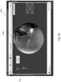
- FIG. 3 Ais an exemplary screen shot 300 a showing a display of the F3DR and a scrolling bar 302 in accordance with the present disclosure.
- FIG. 3 Bis an exemplary screen shot 300 b , showing an initial selection of the target “T” in the F3DR of FIG. 3 A .
- the F3DR in this exampleis shown in AP position (anteroposterior) and includes slice images 305 through which the user may scroll by using a scroll bar 302 .
- the thickness of the slice images 305may be predetermined (e.g., number of pixels).
- the slice images 305are slices of the F3DR.
- the usermay then identify the target T in one of the slice images 305 and mark it, for example, by using a circular marking.
- an additional image 307 of the marked target Tmay be displayed, e.g., in a window adjacent to the F3DR.
- the additional image 307may be any image including but not limited to a zoom-in image, a cropped image, a stretched image, or any combinations thereof.
- the aimis to mark the target T in the slice image 305 that displays it best or in a good enough visibility.
- the methodincludes the step of fining the image as described in further detail below.
- the F3DR(or the initially fined F3DR as initially fined in step 110 ) is fined based on the initial selection of the target.
- the F3DRmay be fined by determining a range for the slice scrolling. The range is determined such that only a portion of the F3DR, which includes the marked target, is displayed and may be scrolled through. Thus, in this aspect, less slices are displayed to the user.
- One benefit of fining in this manneris that less slices are processed (e.g., fined), thereby reducing the use of computing resources and speeding the fining process.
- the F3DRmay be fined by decreasing the thickness of the slices thus achieving a better resolution for display.
- One benefit of fining in this manneris that a user may be presented with sharpened slices, thereby offering a better visualization of the target area and objects (e.g., medical devices, targets, etc.) located therein.
- the scroll rangeis decreased, the thickness of the slices is decreased, again to a predetermined thickness, or both the scroll range is decreased and the thickness of the slices is decreased.
- the fined F3DRis displayed on a display.
- the display of the fined F3DRmay include displaying different slices of the fined F3DR and may be displayed according to commands provided by the user, e.g., through a slices scroll bar as shown in FIGS. 3 A and 3 B .
- the display of the fined F3DRmay include displaying the fined F3DR at different capture angles and according to commands provided by the user.
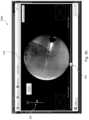
- FIG. 3 Dis an exemplary screen shot 300 d showing a display of a fined F3DR 370 in accordance with the present disclosure.
- the display of the screen shot 300 dincludes the fined F3DR 370 , the slices scroll bar 302 and a capture angle scroll bar 303 , which allows the user to control the angle at which the fined F3DR 370 is displayed.
- capture angleit is meant the angle at which the F3DR or fined F3DR 370 is captured or viewed.
- a final selection of the target T in the fined F3DRis received via a user.
- the receiving of the final selection of the targetmay include directing the user to identify and mark the target or select the target in a single fluoroscopic (or F3DR) slice image.
- the receiving of the final selection of the targetmay include directing the user to identify and mark the target or select the target in two fluoroscopic slice images of the fined F3DR captured at two different angles. Identifying and marking the target or selecting the target in two or more slice images captured at different angles, as opposed to one, may enhance the localization of the target to achieve a better accuracy, e.g., by using triangulation.
- the targetmay be located within the slice image (which has a certain thickness).
- the receiving of the final selection of the target in the fined F3DR in step 150may further include indicating the proper ranges of capture angles in which the target should be marked.
- the selection of the slicesmay be limited to such proper ranges of capture angles. The proper ranges of the two capture angles may be determined to provide enough distance between the two angles to achieve a good enough accuracy in the target localization within the F3DR.
- Displaying the fined F3DR, e.g., a portion of the F3DR surrounding the initially identified target, in step 140may facilitate the selection of the target (for example in step 150 ). Specifically, it may facilitate the selection of the target in two slices captured at different angles. Selection of the target in two such slices may be more time consuming and may be more difficult than, for example, selecting the target when the F3DR is positioned in AP.
- FIG. 3 Eis an exemplary screen shot 300 e of a final selection of the target T in a slice image 321 of the fined F3DR while captured at a first capture angle in accordance with the present disclosure.
- FIG. 3 Fis an exemplary screen shot 300 f of a final selection of the target T in a slice image 321 of the fined F3DR 370 while captured at a first capture angle and a second capture angle in accordance with the present disclosure.
- the usermay mark a target T, which is shown only in slices 321 in the delimited areas of the capture angles scroll bar 303 .
- a close-up image of the marked targetis displayed in a window for each of the first capture angle and the second capture angle.
- a first window 323is displayed with the target T at the first capture angle and a second window 325 is displayed with the target T at the second capture angle.
- a selection of a medical device in two two-dimensional (2D) fluoroscopic imagesmay be received, where the medical device is located in the area of the target.
- the F3DRmay be then initially fined based on the selection of the medical device.
- the 2D fluoroscopic imagesmay be related to the F3DR via their capture angles.
- the initial selection of the targetmay be then performed in the initially fined F3DR, thus facilitating the initial selection of the target.
- the two-dimensional fluoroscopic imagesmay be displayed on the display and the selection of the medical device may be received via automatic detection algorithms or via the user's selection.
- the initially fined F3DRmay be displayed on the display and the initial selection of the target may be received via the user's selection.
- FIGS. 4 A- 4 Fare screen shots of another user interface that facilitates the selection of a target in an F3DR.
- FIG. 4 Ais an exemplary screen shot 400 a showing a selection of a catheter “C” in two two-dimensional fluoroscopic images, namely first two-dimensional fluoroscopic image 401 and second two-dimensional fluoroscopic image 402 , in accordance with the present disclosure.
- FIG. 4 Bis an exemplary screen shot 400 b showing a first selection of a target T in an initially fined F3DR 470 , in accordance with the present disclosure.
- a CT scan of the body region of the patient, which includes a marking of the targetmay be received.
- At least one virtual fluoroscopy imagefor example, virtual fluoroscopy image 350 ( FIG. 3 C ) or virtual fluoroscopy image 450 ( FIG. 4 D ), which includes the target and the marking of the target may be generated based on the CT scan.
- the virtual fluoroscopy imagemay be then displayed on the display and may be used by the user as a reference when selecting the target in the F3DR (e.g., when performing an initial and/or final selection of the target).
- the virtual fluoroscopy imagemay be displayed upon the user's request.
- the virtual fluoroscopy image and the F3DRmay be displayed simultaneously.
- the virtual fluoroscopy image and the F3DRmay be displayed alternatively. Further details with respect to the display, generation and use of the virtual fluoroscopy image are described in U.S. patent application Ser. No. 16/022,222 to Weinberg et al., entitled SYSTEM AND METHOD FOR IDENTIFYING, MARKING AND NAVIGATING TO A TARGET USING REAL TIME TWO DIMENSIONAL FLUOROSCOPIC DATA, the entire contents of which are incorporated herein by reference.
- FIG. 3 Cis an exemplary screen shot 300 c showing a display of a virtual fluoroscopy image 350 in accordance with the present disclosure.
- the virtual fluoroscopy image 350is displayed upon the user's request or command, e.g., by pressing a “Planned Target” button, and may be displayed at any stage of the target selection.
- the virtual fluoroscopy image 350is displayed instead of the F3DR, but it is appreciated that the virtual fluoroscopy image 350 may be displayed along with (e.g., adjacent) the displayed F3DR.
- the methodmay further include acquiring the sequence of fluoroscopic images of the body region via a fluoroscopic imaging device and about a plurality of angles relative to the body region.
- the F3DRmay be then generated based on the sequence of fluoroscopic images.
- An offset of the medical device with respect to the targetmay be determined based on the selections of the target and the medical device.
- Such offset determination based on two-dimensional fluoroscopic images captured in real-timemay be used to facilitate navigation to an area of the target during a medical procedure.
- the real-time target-medical-device offsetmay be used, for example and without limitation, to correct navigation (e.g., displayed or calculated positions of a catheter) and generate local F3DR-CT scan registration.
- the target areamay include at least a portion of the lungs and the medical device is configured to be navigated to the target area through the airway's luminal network.
- Generation of such an F3DR and the uses of the above offsetare described in U.S. Patent Publication No. 2017/035379 to Weinberg et al., entitled SYSTEMS AND METHODS FOR LOCAL THREE DIMENSIONAL VOLUME RECONSTRUCTION USING A STANDARD FLUOROSCOPE, U.S. Patent Publication No.
- the methodmay further include receiving three-dimensional (3D) imaging of the body region of the patient, which includes a marking of the target.
- the 3D imagingmay be, for example, a CT scan or an MRI scan data set.
- the 3D imagingmay be then displayed.
- the 3D imagingmay be used by the user as a reference when performing an initial and/or final selection of the target. Utilizing a 3D view of the target for identifying the target in another 3D view, e.g., the F3DR, may be advantageous.
- the 3D imagingmay be displayed upon the user's request or command.
- the 3D imaging and the F3DR and/or a virtual fluoroscopy imagemay be displayed simultaneously and/or alternatively.
- a pre-operative 3D imaging data setwhich was used in a planning phase of the procedure may be used.
- imagese.g., which were used for identifying the target and planning the navigation to the target, may be very advantageous in performing identification and marking (e.g., selection) of the target in a real-time three-dimensional volume, reconstruction or rendering.
- FIG. 4 Cis an exemplary screen shot 400 c showing a display of the initially fined F3DR 470 and of slice images 460 of a CT scan in accordance with the present disclosure.
- the slice images 460 of the CT scanmay be displayed upon the user's command.
- the CT scanis displayed by displaying three views of the CT (e.g., axial, coronal and sagittal) which may provide the user with the option to create a 3D mental image of the target area that can help in identifying the target in the real-time F3DR.
- the default slices displayed in FIG. 4 Care the slices at which the user marked the target at the planning phase of a medical procedure.
- the usermay scroll or page through the slices. Additionally, the user may select an enlarge button to enlarge any of the slice images 460 for a more detailed view of the displayed slice.
- other views of the CT used in the planning phasemay be displayed.
- FIG. 4 Dis an exemplary screen shot 400 d showing a display of a virtual fluoroscopy image 450 and of the slice images 460 of a CT scan in accordance with the present disclosure.
- the virtual fluoroscopy image 450is displayed instead of the F3DR and is displayed adjacent to the slice images 460 of the CT scan.
- FIG. 4 Eis an exemplary screen shot 400 e showing a display of the fined F3DR 471 in accordance with the present disclosure.
- FIG. 4 Fis an exemplary screen shot 400 f showing a clip 470 of the F3DR for confirming the selection of the target T in accordance with the present disclosure.
- the F3DR including the marking of the target T (e.g., a circular marking) and optionally a marking of the catheter (e.g., a plus or cross-hair marking)may be displayed.
- a clip 470may be displayed which shows the F3DR with the markings at the different capture angles (e.g., the angles at which the 2D fluoroscopic images used to generate the F3DR were captured).
- a computer program product for displaying a F3DR and for identifying, marking and navigating to a targetis herein disclosed.
- the computer program productmay include a non-transitory computer-readable storage medium having program code embodied therewith.
- the program codemay be executable by at least one hardware processor to perform the steps of the method of FIG. 1 and as disclosed herein above.
- System 200may include a workstation 80 , and optionally a fluoroscopic imaging device or fluoroscope 215 .
- workstation 80may be coupled with fluoroscope 215 , directly or indirectly, e.g., by wireless communication.
- Workstation 80may include memory 202 (e.g., storage device), a processor 204 , a display 206 and an input device 210 .
- Processor or hardware processor 204may include one or more hardware processors.
- Workstation 80may optionally include an output module 212 and a network interface 208 .
- Memory 202may store an application 81 and image data 214 .
- Application 81may include instructions executable by processor 204 , inter alia, for executing the method steps of FIG. 1 and as described herein above.
- Application 81may further include a user interface 216 .
- Image data 214may include the 3D imaging such as a pre-operative CT scan, the F3DRs of the target area and/or any other fluoroscopic image data and/or one or more virtual fluoroscopy images.
- Processor 204may be coupled with memory 202 , display 206 , input device 210 , output module 212 , network interface 208 and imaging device (e.g., fluoroscope 215 ).
- Workstation 80may be a stationary computing device, such as a personal computer, or a portable computing device such as a tablet computer. Workstation 80 may embed a plurality of computer devices.
- Memory 202may include any non-transitory computer-readable storage media for storing data and/or software including instructions that are executable by processor 204 and which control the operation of workstation 80 and in some embodiments, may also control the operation of fluoroscope 215 .
- Fluoroscope 215may be used to capture a sequence of fluoroscopic images based on which the F3DR is generated. The two-dimensional fluoroscopic images in which the medical device is selected may be selected from the captured sequence of fluoroscopic images.
- storage device or memory 202may include one or more storage devices such as solid-state storage devices such as flash memory chips.
- memory 202may include one or more mass storage devices connected to the processor 204 through a mass storage controller (not shown) and a communications bus (not shown).
- mass storage controllernot shown
- communications busnot shown
- computer-readable storage mediacan be any available media that can be accessed by the processor 204 . That is, computer readable storage media may include non-transitory, volatile and non-volatile, removable and non-removable media implemented in any method or technology for storage of information such as computer-readable instructions, data structures, program modules or other data.
- computer-readable storage mediamay include RAM, ROM, EPROM, EEPROM, flash memory or other solid-state memory technology, CD-ROM, DVD, Blu-Ray or other optical storage, magnetic cassettes, magnetic tape, magnetic disk storage or other magnetic storage devices, or any other medium which may be used to store the desired information, and which may be accessed by workstation 80 .
- Application 81may, when executed by processor 204 , cause display 206 to present user interface 216 .
- User interface 216may be configured to present to the user the F3DR, two-dimensional fluoroscopic images, images of the 3D imaging and virtual fluoroscopy image, as shown, for example, in the exemplary screen shots of FIGS. 3 A- 3 F and 4 A- 4 F .
- User interface 216may be further configured to direct the user to select the target by, inter alia, identifying and marking the target in the displayed F3DR or any other fluoroscopic image data in accordance with the present disclosure.
- Network interface 208may be configured to connect to a network such as a local area network (LAN) consisting of a wired network and/or a wireless network, a wide area network (WAN), a wireless mobile network, a Bluetooth network, and/or the internet.
- Network interface 208may be used to connect between workstation 80 and fluoroscope 215 .
- Network interface 208may be also used to receive image data 214 .
- Input device 210may be any device by means of which a user may interact with workstation 80 , such as, for example, a mouse, keyboard, foot pedal, touch screen, and/or voice interface.
- Output module 212may include any connectivity port or bus, such as, for example, parallel ports, serial ports, universal serial busses (USB), or any other similar connectivity port known to those skilled in the art.
Landscapes
- Health & Medical Sciences (AREA)
- Life Sciences & Earth Sciences (AREA)
- Engineering & Computer Science (AREA)
- Medical Informatics (AREA)
- Surgery (AREA)
- General Health & Medical Sciences (AREA)
- Nuclear Medicine, Radiotherapy & Molecular Imaging (AREA)
- Molecular Biology (AREA)
- Public Health (AREA)
- Veterinary Medicine (AREA)
- Animal Behavior & Ethology (AREA)
- Heart & Thoracic Surgery (AREA)
- Biomedical Technology (AREA)
- Physics & Mathematics (AREA)
- Pathology (AREA)
- Radiology & Medical Imaging (AREA)
- Biophysics (AREA)
- High Energy & Nuclear Physics (AREA)
- Optics & Photonics (AREA)
- Theoretical Computer Science (AREA)
- Human Computer Interaction (AREA)
- Computer Vision & Pattern Recognition (AREA)
- General Physics & Mathematics (AREA)
- Robotics (AREA)
- Pulmonology (AREA)
- Oral & Maxillofacial Surgery (AREA)
- Quality & Reliability (AREA)
- Apparatus For Radiation Diagnosis (AREA)
- Measuring And Recording Apparatus For Diagnosis (AREA)
Abstract
Description
This application is a continuation of U.S. patent application Ser. No. 17/115,599 filed Dec. 12, 2020, which is a continuation of U.S. Pat. No. 10,893,843 filed Oct. 10, 2018, which claims the benefit of the filing dates of provisional U.S. Patent Application No. 62/570,431, filed Oct. 10, 2017, provisional U.S. Patent Application No. 62/641,777, filed Mar. 12, 2018, and provisional U.S. Patent Application No. 62/628,017, filed Feb. 8, 2018, the entire contents of each of which are incorporated herein by reference.
The present disclosure relates to the field of identifying and marking a target, such as a lesion or a small soft tissue object) in fluoroscopic images, 3D fluoroscopic images, fluoroscopic 3D reconstructions, or other data sets, in general, and to such target identification and marking in medical procedures involving intra-body navigation, in particular. Furthermore, the present disclosure relates to a system, apparatus, and methods of planning, navigating, identifying, marking, dying, biopsy, ablation, laparoscopy, or treatment in medical procedures.
There are several commonly applied medical methods, such as endoscopic procedures or minimally invasive procedures, for treating various maladies affecting organs including the liver, brain, heart, lung, gallbladder, kidney and bones. Often, one or more imaging modalities, such as magnetic resonance imaging (MRI), ultrasound imaging, computed tomography (CT), fluoroscopy as well as others are employed by clinicians to identify and navigate to areas of interest within a patient and ultimately a target for treatment. In some procedures, pre-operative scans may be utilized for target identification and intraoperative guidance. However, real-time imaging may be often required to obtain a more accurate and current image of the target area. Furthermore, real-time image data displaying the current location of a medical device with respect to the target and its surrounding may be required to navigate the medical device to the target in a more safe and accurate manner (e.g., with unnecessary or no damage caused to other organs or tissue).
For example, an endoscopic approach has proven useful in navigating to areas of interest within a patient, and particularly so for areas within luminal networks of the body such as the lungs. To enable the endoscopic approach, and more particularly the bronchoscopic approach in the lungs, endobronchial navigation systems have been developed that use previously acquired MRI data or CT image data to generate a three-dimensional (3D) rendering, model or volume of the particular body part such as the lungs.
The resulting volume generated from the MRI scan or CT scan is then utilized to create a navigation plan to facilitate the advancement of a navigation catheter (or other suitable medical device) through a bronchoscope and a branch of the bronchus of a patient to an area of interest. A locating or tracking system, such as an electromagnetic (EM) tracking system, may be utilized in conjunction with, for example, CT data, to facilitate guidance of the navigation catheter through the branch of the bronchus to the area of interest. In certain instances, the navigation catheter may be positioned within one of the airways of the branched luminal networks adjacent to, or within, the area of interest to provide access for one or more medical instruments.
However, a 3D volume of a patient's lungs, generated from previously acquired scans, such as CT scans, may not provide a basis sufficient for accurate guiding of medical instruments to a target during a navigation procedure. In certain instances, the inaccuracy is caused by deformation of the patient's lungs during the procedure relative to the lungs at the time of the acquisition of the previously acquired CT data. This deformation (CT-to-Body divergence) may be caused by many different factors including, for example, sedation vs. no sedation, bronchoscope changing patient pose and also pushing the tissue, different lung volume because CT was during inhale while navigation is during breathing, different bed, different day, etc.
Thus, another imaging modality is necessary to visualize such targets in real-time and enhance the in-vivo navigation procedure by correcting navigation during the procedure. Furthermore, in order to accurately and safely navigate medical devices to a remote target, for example, for biopsy or treatment, both the medical device and the target should be visible in some sort of a 3D guidance system.
A fluoroscopic imaging device is commonly located in the operating room during navigation procedures. The standard fluoroscopic imaging device may be used by a clinician, for example, to visualize and confirm the placement of a medical device after it has been navigated to a desired location. However, although standard fluoroscopic images display highly dense objects such as metal tools and bones as well as large soft-tissue objects such as the heart, the fluoroscopic images may have difficulty resolving small soft-tissue objects of interest such as lesions. Furthermore, the fluoroscope image is only a two-dimensional projection. Therefore, an X-ray volumetric reconstruction may enable identification of such soft tissue objects and navigation to the target.
Several solutions exist that provide 3D volume reconstruction such as CT and cone-beam CT which are extensively used in the medical world. These machines algorithmically combine multiple X-ray projections from known, calibrated X-ray source positions into 3D volume in which, inter alia, soft-tissues are more visible. For example, a CT machine can be used with iterative scans during procedure to provide guidance through the body until the tools reach the target. This is a tedious procedure, as it requires several full CT scans, a dedicated CT room and blind navigation between scans. In addition, each scan requires the staff to leave the room due to high-levels of ionizing radiation and exposes the patient to such radiation. Another option is a cone-beam CT machine, which is available in some operation rooms and is somewhat easier to operate but is expensive and like the CT only provides blind navigation between scans, requires multiple iterations for navigation and requires the staff to leave the room. In addition, a CT-based imaging system is extremely costly, and in many cases not available in the same location as the location where a procedure is carried out.
An imaging technology that uses standard fluoroscope devices to reconstruct local 3D volume in order to visualize and facilitate navigation to in-vivo targets, and to small soft-tissue objects in particular, is described in U.S. Patent Publication No. 2017/035379 to Weingarten et al., entitled SYSTEMS AND METHODS FOR LOCAL THREE DIMENSIONAL VOLUME RECONSTRUCTION USING A STANDARD FLUOROSCOPE, U.S. Patent Publication No. 2017/035380 to Barak et al., entitled SYSTEM AND METHOD FOR NAVIGATING TO TARGET AND PERFORMING PROCEDURE ON TARGET UTILIZING FLUOROSCOPIC-BASED LOCAL THREE DIMENSIONAL VOLUME RECONSTRUCTION, and U.S. Patent Publication No. 2018/0160991 to Weingarten et al., entitled SYSTEMS AND METHODS FOR LOCAL THREE DIMENSIONAL VOLUME RECONSTRUCTION USING A STANDARD FLUOROSCOPE, the entire contents of each of which are incorporated herein by reference.
In general, according to the systems and methods disclosed in the above-mentioned patent publications, a standard fluoroscope c-arm can be rotated, e.g., about 30 degrees, around a patient during a medical procedure, and a fluoroscopic 3D reconstruction (F3DR) of the region of interest is generated by a specialized software algorithm.
Such quick generation of a 3D reconstruction of a region of interest can provide real-time 3D imaging of the target area. Real-time imaging of the target and medical devices positioned in its area may benefit numerous interventional procedures, such as biopsy and ablation procedures in various organs, vascular interventions and orthopedic surgeries. For example, when navigational bronchoscopy is concerned, the aim may be to receive accurate information about the position of a biopsy catheter relative to a target lesion.
As another example, minimally invasive procedures, such as laparoscopy procedures, including robotic-assisted surgery, may employ intraoperative fluoroscopy to increase visualization, e.g., for guidance and lesion locating, and to prevent unnecessary injury and complications. Employing the above-mentioned systems and methods for real-time reconstruction of fluoroscopic 3D imaging of a target area and for navigation based on the reconstruction may benefit such procedures as well.
Still, it may not be an easy task to accurately identify and mark a target in the F3DR, in particular when the target is a small soft-tissue. Thus, there is a need for systems and methods for facilitating the identification and marking of a target in fluoroscopic image data, and in a F3DR in particular, to consequently facilitate the navigation to the target and the yield of pertinent medical procedures.
The present disclosure is directed to systems, methods and computer program products for displaying an F3DR and for facilitating the identification and marking of a target by a user in the F3DR. Marking a target in an F3DR and especially in real-time may not be straight forward or a simple task, especially for the untrained user. Furthermore, it is desired to receive a selection of the target at two capture or view angles, while at least one of them would be different than AP (anteroposterior), as selection in AP position is usually easier. Marking of the target at two such angles enhances the accuracy of the target localization. Thus, marking of the target performed at two stages, specifically when the first stage includes marking in AP position, facilitates the user's identification of the target and provides a better accuracy in the localization of the target, better registration between imaging modalities, and therefrom better results in treatment.
There is provided in accordance with the present disclosure a system for facilitating identification and marking of a target in a displayed Fluoroscopic Three-Dimensional Reconstruction (F3DR) of a body region of a patient. The system includes a display and one or more storage devices having stored thereon instructions for receiving an initial selection of the target in the F3DR, fining the F3DR based on the initial selection of the target, displaying the fined F3DR on the display, and receiving a final selection of the target in the fined F3DR via a user. The system further includes at least one hardware processor configured to execute said instructions.
In an aspect, the one or more storage devices have stored thereon further instructions for receiving a selection of a medical device in two two-dimensional fluoroscopic images, wherein the medical device is located in an area of the target, and initially fining the F3DR based on the selection of the medical device prior to fining the F3DR based on the initial selection of the target. Such a selection of the medical device may be performed prior to the selection or marking of the target. Additionally, the one or more storage devices have stored thereon further instructions for displaying the initially fined F3DR on the display and wherein the initial selection of the target is received via a user selection. The two-dimensional fluoroscopic images may be displayed on the display and the selection of the medical device in the two-dimensional fluoroscopic images may be received via a user selection.
In an aspect, the one or more storage devices have stored thereon further instructions for receiving a CT scan of the body region of the patient, wherein the CT scan includes a marking of the target, generating at least one virtual fluoroscopy image based on the CT scan, wherein the virtual fluoroscopy image includes the target and the marking of the target, and displaying the virtual fluoroscopy image. Displaying the fined F3DR may include displaying different slices of the fined F3DR and may be according to commands provided by the user such as a user selection. Additionally, or alternatively, displaying the fined F3DR includes displaying the fined F3DR at different capture angles and may be according to commands provided by the user such as a user selection.
Receiving of the final selection of the target in the fined F3DR may include directing the user to identify and mark the target in two fluoroscopic slice images of the fined F3DR captured at two different angles. Additionally, or alternatively, receiving of the final selection of the target in the fined F3DR may include indicating proper ranges of capture angles in which the target should be marked. In an aspect, the target is a soft-tissue target.
In an aspect, the system further includes a fluoroscopic imaging device and the one or more storage devices have stored thereon further instructions for acquiring a sequence of fluoroscopic images of the body region about a plurality of angles relative to the body region while a medical device is positioned in a target area, generating the F3DR of the body region based on the sequence of fluoroscopic images, and determining an offset of the medical device with respect to the target based on the selection of the medical device and at least one of the initial selection of the target or the final selection of the target. The target area may include at least a portion of lungs and the medical device may be configured to be navigated to the target area through a luminal network of lungs.
In an aspect, the one or more storage devices have stored thereon further instructions for receiving a three-dimensional imaging of the body region of the patient, wherein the three-dimensional imaging includes a marking of the target, and displaying the three-dimensional imaging. The three-dimensional imaging may be a CT or an MRI scan.
In an aspect, the system is used during a medical procedure, and the three-dimensional imaging is a pre-operative imaging which was used in a planning phase of the medical procedure.
In another aspect of the present disclosure a method for facilitating identification and marking of a target in a displayed F3DR of a body region of a patient is provided. The method includes using at least one hardware processor for receiving an initial selection of the target in the F3DR, fining the F3DR based on the initial selection of the target, displaying the fined F3DR on a display, and receiving a final selection of the target in the fined F3DR via a user.
In an aspect, the method includes using said at least one hardware processor for receiving a selection of a medical device in two two-dimensional fluoroscopic images, where the medical device is located in an area of the target, and initially fining the F3DR based on the selection of the medical device. Additionally, or alternatively, the method further includes at least one hardware processor for displaying the initially fined F3DR on the display, wherein the initial selection of the target is received via a user selection. In an aspect, the method further includes using said at least one hardware processor for displaying the two-dimensional fluoroscopic images on the display, wherein the selection of the medical device in the two-dimensional fluoroscopic images is received via a user selection.
In an aspect, the method includes using the at least one hardware processor for receiving a CT scan of the body region of the patient, wherein the CT scan includes a marking of the target, generating at least one virtual fluoroscopy image based on the CT scan, wherein the virtual fluoroscopy image includes the target and the marking of the target, and displaying the virtual fluoroscopy image on the display. Displaying of the virtual fluoroscopy image may be performed upon a user's request. Displaying of the fined F3DR may include displaying different slices of the fined F3DR and may be according to commands provided by the user such as a user selection. Additionally, or alternatively, displaying of the fined F3DR includes displaying the fined F3DR at different capture angles and may be according to commands provided by the user such as a user selection.
In an aspect, receiving of the final selection of the target in the fined F3DR includes directing the user to identify and mark the target in two fluoroscopic slice images of the fined F3DR captured at two different angles. Additionally, or alternatively, receiving of the final selection of the target in the fined F3DR includes indicating proper ranges of capture angles in which the target should be marked.
In an aspect, the method further includes using the at least one hardware processor for acquiring a sequence of fluoroscopic images of the body region via a fluoroscopic imaging device and about a plurality of angles relative to the body region, generating the F3DR of the body region based on the sequence of fluoroscopic images, and determining an offset of a medical device with respect to the target based on the selections of the target and the medical device, thereby facilitating navigation to an area of the target within the patient's body region during a medical procedure using real-time two-dimensional fluoroscopic images. Additionally, or alternatively, the method includes using the at least one hardware processor for receiving a three-dimensional imaging of the body region of the patient, wherein the three-dimensional imaging includes a marking of the target, and displaying the three-dimensional imaging.
The method may be used during a medical procedure, and the three-dimensional imaging is a pre-operative imaging which was used in a planning phase of the medical procedure.
In yet another aspect of the present disclosure, a non-transitory computer-readable storage medium encoded with a program that, when executed by a processor, performs a method for facilitating identification and marking of a target in a F3DR of a body region of a patient is provided. The method includes receiving a selection of a medical device in two two-dimensional fluoroscopic images, where the medical device is located in an area of the target, initially fining the F3DR based on the selection of the medical device, displaying the initially fined F3DR on a display, receiving an initial selection of the target in the initially fined F3DR via a user selection, further fining the F3DR based on the initial selection of the target, displaying the further fined F3DR on the display, and receiving a final selection of the target in the further fined F3DR via a user selection, thereby facilitating an identification and marking of the target in the F3DR.
Various aspects and embodiments of the present disclosure are described hereinbelow with references to the drawings, wherein:
The term “target”, as referred to herein, may relate to any element, biological or artificial, or to a region of interest in a patient's body, such as a tissue (including soft tissue and bone tissue), an organ, an implant or a fiducial marker.
The term “target area”, as referred to herein, may relate to the target and at least a portion of its surrounding area. The term “target area” and the term “body region” may be used interchangeably when the term “body region” refers to the body region in which the target is located. Alternatively or in addition, the term “target area” may also refer to a portion of the body region in which the target is located, all according to the context.
The terms “and”, “or” and “and/or” may be used interchangeably, while each term may incorporate the others, all according to the term's context.
The term “medical device”, as referred to herein, may include, without limitation, optical systems, ultrasound probes, marker placement tools, biopsy tools, ablation tools (i.e., microwave ablation devices), laser probes, cryogenic probes, sensor probes, and aspirating needles.
The terms “fluoroscopic image”, “fluoroscopic images”, “fluoroscopy image”, and “fluoroscopy images” may refer to a 2D fluoroscopic image/s and/or to a slice-image of a fluoroscopic 3D reconstruction, all in accordance with the term's context.
The terms “virtual fluoroscopic image”, “virtual fluoroscopic images”, “virtual fluoroscopy image”, and “virtual fluoroscopy images” may refer to a virtual 2D fluoroscopic image/s and/or to a virtual fluoroscopy slice-image/s of a virtual fluoroscopic 3D reconstruction, or other 3D image data all in accordance with the term's context.
The present disclosure is directed to systems, methods and computer program products for displaying an F3DR and for facilitating the identification and marking of a target by a user in the F3DR. Marking a target in an F3DR and especially in real-time may not be straight forward or a simple task, especially for the untrained user. Furthermore, it is desired to receive a selection of the target at multiple capture or view angles (for example, two or three capture angles), while at least one of them would be different than AP (anteroposterior), as selection in AP position is usually easier. Marking of the target at two such angles enhances the accuracy of the target localization. Thus, marking of the target performed at two stages, specifically when the first stage includes marking in AP position, facilitates the user's identification of the target and provides a better accuracy in the localization of the target, better registration between imaging modalities, and therefrom better results in treatment.
In addition, using the initial target selection (e.g., at the first stage) to fine the F3DR further facilitates the final selection (e.g., at two different angles) and enhances its accuracy. Optionally, the process may be further enhanced by performing a two-stage fining of the F3DR by using a selection or marking of a medical device located in the target area to initially fine the F3DR.
Reference is now made toFIG.1 , which is a flow chart of a method for displaying a F3DR of a body region of a patient and for identifying and marking a target in the F3DR in accordance with the present disclosure. In some embodiments, that target may be a soft tissue target, such as a lesion and the body region may include at least a portion of the lungs. The method may begin either atstep 100 or atstep 120. To this end, steps100 and110 are optional steps. Instep 100, a selection of a medical device in one or more fluoroscopy images or in a F3DR is received. Such a selection may be made from a single capture angle or from multiple capture angles. Additionally, the selection of the medical device instep 100 may be automatically made by the system via a dedicated algorithm or may be made by a user's selection. In an aspect, the selection of the medical device is performed in 2D fluoroscopic images, in particular, in at least two images. In an aspect, the medical device is marked or selected before the generation of the F3DR and the marking may be used for the generation/reconstruction of the F3DR.
Instep 110, the F3DR is initially fined based on the received selection of the medical device instep 100. The initial fining of the F3DR instep 100 may include determining a range for the slice scrolling. In this aspect, the range is determined such that only a portion of the F3DR, which includes the selection of the medical device, is displayed and may be scrolled through. Thus, in this aspect, less slices are displayed to the user. One benefit of fining in this manner is that less slices are processed (e.g., fined), thereby reducing the use of computing resources and speeding the fining process. Additionally, or alternatively, the F3DR may be initially fined by decreasing the thickness of the slices thus achieving a better resolution for display. One benefit of fining in this manner is that a user may be presented with sharpened slices, thereby offering a better visualization of the target area and objects (e.g., medical devices, targets, etc.) located therein.
In an aspect, the thickness of the slices is predetermined. Although thinning the slices provides a better resolution, there may be a toll on thinning. That is, when slices are thinned, the volume appears more smeared and thus it becomes more difficult for the user to identify the target. Therefore, at the initial fining (step110), a predetermined thickness is used which provides optimal results, taking into consideration the above. At the second stage, as described below (in step130), the scroll range is decreased, the thickness of the slices is decreased, again to a predetermined thickness, or both the scroll range is decreased and the thickness of the slices is decreased.
Instep 120, an initial selection of the target in the F3DR is received. As described above, in aspects, this method begins atstep 120 and the initial selection of the target is made in the F3DR. Alternatively, where the method begins atstep 100, the initial selection of the target instep 120 is made in the initially fined F3DR (as initially fined in step110). The initial selection of the target may be automatic via a dedicated algorithm or may be performed by a user's selection, e.g., by identifying and marking the target in the displayed F3DR.
Reference is now briefly made toFIGS.3A-3B , which are screen shots of a user interface, which facilitates the selection of a target in an F3DR.FIG.3A is an exemplary screen shot300ashowing a display of the F3DR and a scrollingbar 302 in accordance with the present disclosure.FIG.3B is an exemplary screen shot300b, showing an initial selection of the target “T” in the F3DR ofFIG.3A . The F3DR in this example is shown in AP position (anteroposterior) and includes slice images305 through which the user may scroll by using ascroll bar 302. The thickness of the slice images305 may be predetermined (e.g., number of pixels). In an aspect, the slice images305 are slices of the F3DR. The user may then identify the target T in one of the slice images305 and mark it, for example, by using a circular marking. For further convenience, anadditional image 307 of the marked target T may be displayed, e.g., in a window adjacent to the F3DR. Theadditional image 307 may be any image including but not limited to a zoom-in image, a cropped image, a stretched image, or any combinations thereof. The aim is to mark the target T in the slice image305 that displays it best or in a good enough visibility. To assist in this respect, the method includes the step of fining the image as described in further detail below.
Instep 130, the F3DR (or the initially fined F3DR as initially fined in step110) is fined based on the initial selection of the target. The F3DR may be fined by determining a range for the slice scrolling. The range is determined such that only a portion of the F3DR, which includes the marked target, is displayed and may be scrolled through. Thus, in this aspect, less slices are displayed to the user. One benefit of fining in this manner is that less slices are processed (e.g., fined), thereby reducing the use of computing resources and speeding the fining process. Additionally, or alternatively, the F3DR may be fined by decreasing the thickness of the slices thus achieving a better resolution for display. One benefit of fining in this manner is that a user may be presented with sharpened slices, thereby offering a better visualization of the target area and objects (e.g., medical devices, targets, etc.) located therein. As described above, at the second stage (in step130), the scroll range is decreased, the thickness of the slices is decreased, again to a predetermined thickness, or both the scroll range is decreased and the thickness of the slices is decreased.
Instep 140, the fined F3DR is displayed on a display. In some embodiments, the display of the fined F3DR may include displaying different slices of the fined F3DR and may be displayed according to commands provided by the user, e.g., through a slices scroll bar as shown inFIGS.3A and3B . In some embodiments, the display of the fined F3DR may include displaying the fined F3DR at different capture angles and according to commands provided by the user.
Reference is now made toFIG.3D , which is an exemplary screen shot300dshowing a display of a finedF3DR 370 in accordance with the present disclosure. The display of the screen shot300dincludes the finedF3DR 370, theslices scroll bar 302 and a captureangle scroll bar 303, which allows the user to control the angle at which the finedF3DR 370 is displayed. By “capture angle” it is meant the angle at which the F3DR or finedF3DR 370 is captured or viewed.
Instep 150, a final selection of the target T in the fined F3DR is received via a user. In some embodiments, the receiving of the final selection of the target may include directing the user to identify and mark the target or select the target in a single fluoroscopic (or F3DR) slice image. In some embodiments, the receiving of the final selection of the target may include directing the user to identify and mark the target or select the target in two fluoroscopic slice images of the fined F3DR captured at two different angles. Identifying and marking the target or selecting the target in two or more slice images captured at different angles, as opposed to one, may enhance the localization of the target to achieve a better accuracy, e.g., by using triangulation. Thus, the target may be located within the slice image (which has a certain thickness). In some embodiments, the receiving of the final selection of the target in the fined F3DR instep 150 may further include indicating the proper ranges of capture angles in which the target should be marked. In some embodiments, the selection of the slices may be limited to such proper ranges of capture angles. The proper ranges of the two capture angles may be determined to provide enough distance between the two angles to achieve a good enough accuracy in the target localization within the F3DR.
Displaying the fined F3DR, e.g., a portion of the F3DR surrounding the initially identified target, instep 140 may facilitate the selection of the target (for example in step150). Specifically, it may facilitate the selection of the target in two slices captured at different angles. Selection of the target in two such slices may be more time consuming and may be more difficult than, for example, selecting the target when the F3DR is positioned in AP.
Reference is now made toFIGS.3E and3F .FIG.3E is an exemplary screen shot300eof a final selection of the target T in aslice image 321 of the fined F3DR while captured at a first capture angle in accordance with the present disclosure.FIG.3F is an exemplary screen shot300fof a final selection of the target T in aslice image 321 of the finedF3DR 370 while captured at a first capture angle and a second capture angle in accordance with the present disclosure. According to the exemplary user interface shown inFIGS.3E and3F , the user may mark a target T, which is shown only inslices 321 in the delimited areas of the capture anglesscroll bar 303. When the user marks a target T, a close-up image of the marked target is displayed in a window for each of the first capture angle and the second capture angle. As shown in the exemplary screen shot300fofFIG.3F , afirst window 323 is displayed with the target T at the first capture angle and asecond window 325 is displayed with the target T at the second capture angle.
As described above with reference tosteps
Reference is now made toFIGS.4A-4F , which are screen shots of another user interface that facilitates the selection of a target in an F3DR.FIG.4A is an exemplary screen shot400ashowing a selection of a catheter “C” in two two-dimensional fluoroscopic images, namely first two-dimensionalfluoroscopic image 401 and second two-dimensionalfluoroscopic image 402, in accordance with the present disclosure.FIG.4B is an exemplary screen shot400bshowing a first selection of a target T in an initially finedF3DR 470, in accordance with the present disclosure.
In some embodiments, a CT scan of the body region of the patient, which includes a marking of the target, may be received. At least one virtual fluoroscopy image, for example, virtual fluoroscopy image350 (FIG.3C ) or virtual fluoroscopy image450 (FIG.4D ), which includes the target and the marking of the target may be generated based on the CT scan. The virtual fluoroscopy image may be then displayed on the display and may be used by the user as a reference when selecting the target in the F3DR (e.g., when performing an initial and/or final selection of the target). In some embodiments, the virtual fluoroscopy image may be displayed upon the user's request. In some embodiments, the virtual fluoroscopy image and the F3DR may be displayed simultaneously. In other embodiments, the virtual fluoroscopy image and the F3DR may be displayed alternatively. Further details with respect to the display, generation and use of the virtual fluoroscopy image are described in U.S. patent application Ser. No. 16/022,222 to Weingarten et al., entitled SYSTEM AND METHOD FOR IDENTIFYING, MARKING AND NAVIGATING TO A TARGET USING REAL TIME TWO DIMENSIONAL FLUOROSCOPIC DATA, the entire contents of which are incorporated herein by reference.
Reference is now made toFIG.3C , which is an exemplary screen shot300cshowing a display of a virtualfluoroscopy image 350 in accordance with the present disclosure. According to the example shown inFIG.3A-3F , the virtualfluoroscopy image 350 is displayed upon the user's request or command, e.g., by pressing a “Planned Target” button, and may be displayed at any stage of the target selection. In this example, the virtualfluoroscopy image 350 is displayed instead of the F3DR, but it is appreciated that the virtualfluoroscopy image 350 may be displayed along with (e.g., adjacent) the displayed F3DR.
In some embodiments, the method may further include acquiring the sequence of fluoroscopic images of the body region via a fluoroscopic imaging device and about a plurality of angles relative to the body region. The F3DR may be then generated based on the sequence of fluoroscopic images. An offset of the medical device with respect to the target may be determined based on the selections of the target and the medical device. Such offset determination based on two-dimensional fluoroscopic images captured in real-time may be used to facilitate navigation to an area of the target during a medical procedure. The real-time target-medical-device offset may be used, for example and without limitation, to correct navigation (e.g., displayed or calculated positions of a catheter) and generate local F3DR-CT scan registration. In some embodiments, the target area may include at least a portion of the lungs and the medical device is configured to be navigated to the target area through the airway's luminal network. Generation of such an F3DR and the uses of the above offset are described in U.S. Patent Publication No. 2017/035379 to Weingarten et al., entitled SYSTEMS AND METHODS FOR LOCAL THREE DIMENSIONAL VOLUME RECONSTRUCTION USING A STANDARD FLUOROSCOPE, U.S. Patent Publication No. 2017/035380 to Barak et al., entitled SYSTEM AND METHOD FOR NAVIGATING TO TARGET AND PERFORMING PROCEDURE ON TARGET UTILIZING FLUOROSCOPIC-BASED LOCAL THREE DIMENSIONAL VOLUME RECONSTRUCTION, and provisional U.S. Patent Application No. 62/628,017 to Barak et al., entitled SYSTEM AND METHOD FOR POSE ESTIMATION OF AN IMAGING DEVICE AND FOR DETERMINING THE LOCATION OF A MEDICAL DEVICE WITH RESPECT TO A TARGET, the entire contents of each of which are incorporated herein by reference.
In some embodiments, the method may further include receiving three-dimensional (3D) imaging of the body region of the patient, which includes a marking of the target. The 3D imaging may be, for example, a CT scan or an MRI scan data set. The 3D imaging may be then displayed. The 3D imaging may be used by the user as a reference when performing an initial and/or final selection of the target. Utilizing a 3D view of the target for identifying the target in another 3D view, e.g., the F3DR, may be advantageous. The 3D imaging may be displayed upon the user's request or command. The 3D imaging and the F3DR and/or a virtual fluoroscopy image may be displayed simultaneously and/or alternatively. In some embodiments, when the disclosed methods are used during a medical procedure (e.g., identifying a target via real-time fluoroscopic 3D images), a pre-operative 3D imaging data set, which was used in a planning phase of the procedure may be used. Using images, e.g., which were used for identifying the target and planning the navigation to the target, may be very advantageous in performing identification and marking (e.g., selection) of the target in a real-time three-dimensional volume, reconstruction or rendering.
Reference is now made toFIGS.4C-4E .FIG.4C is an exemplary screen shot400cshowing a display of the initially finedF3DR 470 and ofslice images 460 of a CT scan in accordance with the present disclosure. In this specific example, theslice images 460 of the CT scan may be displayed upon the user's command. The CT scan is displayed by displaying three views of the CT (e.g., axial, coronal and sagittal) which may provide the user with the option to create a 3D mental image of the target area that can help in identifying the target in the real-time F3DR. The default slices displayed inFIG.4C are the slices at which the user marked the target at the planning phase of a medical procedure. The user may scroll or page through the slices. Additionally, the user may select an enlarge button to enlarge any of theslice images 460 for a more detailed view of the displayed slice. In some embodiments, other views of the CT used in the planning phase may be displayed.
In some embodiments, after the final selection of the target is performed, the target as selected may be displayed to the user and the user may be requested to confirm the selection. Reference is now made toFIG.4F , which is an exemplary screen shot400fshowing aclip 470 of the F3DR for confirming the selection of the target T in accordance with the present disclosure. The F3DR including the marking of the target T (e.g., a circular marking) and optionally a marking of the catheter (e.g., a plus or cross-hair marking) may be displayed. In order to further confirm the selection, aclip 470 may be displayed which shows the F3DR with the markings at the different capture angles (e.g., the angles at which the 2D fluoroscopic images used to generate the F3DR were captured).
A computer program product for displaying a F3DR and for identifying, marking and navigating to a target is herein disclosed. The computer program product may include a non-transitory computer-readable storage medium having program code embodied therewith. The program code may be executable by at least one hardware processor to perform the steps of the method ofFIG.1 and as disclosed herein above.
Reference is now made toFIG.2 , which is a schematic diagram of asystem 200 configured for use with the method ofFIG.1 and as described herein above.System 200 may include aworkstation 80, and optionally a fluoroscopic imaging device orfluoroscope 215. In some embodiments,workstation 80 may be coupled withfluoroscope 215, directly or indirectly, e.g., by wireless communication.Workstation 80 may include memory202 (e.g., storage device), aprocessor 204, adisplay 206 and aninput device 210. Processor orhardware processor 204 may include one or more hardware processors.Workstation 80 may optionally include anoutput module 212 and anetwork interface 208. Memory202 may store anapplication 81 andimage data 214.Application 81 may include instructions executable byprocessor 204, inter alia, for executing the method steps ofFIG.1 and as described herein above.Application 81 may further include a user interface216.Image data 214 may include the 3D imaging such as a pre-operative CT scan, the F3DRs of the target area and/or any other fluoroscopic image data and/or one or more virtual fluoroscopy images.Processor 204 may be coupled with memory202,display 206,input device 210,output module 212,network interface 208 and imaging device (e.g., fluoroscope215).Workstation 80 may be a stationary computing device, such as a personal computer, or a portable computing device such as a tablet computer.Workstation 80 may embed a plurality of computer devices.
Memory202 may include any non-transitory computer-readable storage media for storing data and/or software including instructions that are executable byprocessor 204 and which control the operation ofworkstation 80 and in some embodiments, may also control the operation offluoroscope 215.Fluoroscope 215 may be used to capture a sequence of fluoroscopic images based on which the F3DR is generated. The two-dimensional fluoroscopic images in which the medical device is selected may be selected from the captured sequence of fluoroscopic images. In an embodiment, storage device or memory202 may include one or more storage devices such as solid-state storage devices such as flash memory chips. Alternatively, or in addition to the one or more solid-state storage devices, memory202 may include one or more mass storage devices connected to theprocessor 204 through a mass storage controller (not shown) and a communications bus (not shown). Although the description of computer-readable media contained herein refers to a solid-state storage, it should be appreciated by those skilled in the art that computer-readable storage media can be any available media that can be accessed by theprocessor 204. That is, computer readable storage media may include non-transitory, volatile and non-volatile, removable and non-removable media implemented in any method or technology for storage of information such as computer-readable instructions, data structures, program modules or other data. For example, computer-readable storage media may include RAM, ROM, EPROM, EEPROM, flash memory or other solid-state memory technology, CD-ROM, DVD, Blu-Ray or other optical storage, magnetic cassettes, magnetic tape, magnetic disk storage or other magnetic storage devices, or any other medium which may be used to store the desired information, and which may be accessed byworkstation 80.
From the foregoing and with reference to the various figure drawings, those skilled in the art will appreciate that certain modifications can also be made to the present disclosure without departing from the scope of the same.
Detailed embodiments of the present disclosure are disclosed herein. However, the disclosed embodiments are merely examples of the disclosure, which may be embodied in various forms and aspects. Therefore, specific structural and functional details disclosed herein are not to be interpreted as limiting, but merely as a basis for the claims and as a representative basis for teaching one skilled in the art to variously employ the present disclosure in virtually any appropriately detailed structure.
While several embodiments of the disclosure have been shown in the drawings, it is not intended that the disclosure be limited thereto, as it is intended that the disclosure be as broad in scope as the art will allow and that the specification be read likewise. Therefore, the above description should not be construed as limiting, but merely as exemplifications of embodiments. Those skilled in the art will envision other modifications within the scope and spirit of the claims appended hereto.
Claims (20)
1. A system for identification and marking of a target in a body region of a patient, the system comprising:
(i) a display;
(ii) a computing device including a processor and a storage device, the storage device storing thereon instructions that when executed by the processor cause the computing device to:
receive a sequence of fluoroscopic images of the body region about a plurality of angles relative to the body region while a medical device is positioned in a target area of the target in the body region of the patient;
generate a Fluoroscopic Three-Dimensional Reconstruction (F3DR) of the body region based on the sequence of fluoroscopic images;
receive an identification of the medical device in a first two-dimensional fluoroscopic image from the sequence of fluoroscopic images;
receive a first identification of the target in the F3DR;
determine a range of fluoroscopic slices of the F3DR for scrolling based on the identification of the target or the medical device;
display a second fluoroscopic slice image from the determined range of fluoroscopic slices of the F3DR on the display;
receive a second identification of the target in the second fluoroscopic slice image;
determine an offset of the medical device with respect to the target based on the identification of the medical device and at least one of the first identification of the target or the second identification of the target; and
register the F3DR to a computed tomography (CT) scan stored in the storage device.
2. The system ofclaim 1 , wherein the storage device stores thereon further instructions that when executed by the processor cause the computing device to:
determine a second range of fluoroscopic slices of the F3DR based on selection of the medical device prior to initial selection of the target.
3. The system ofclaim 1 , wherein the storage device stores thereon further instructions that when executed by the processor cause the computing device to:
receive the CT scan, wherein the CT scan is of the body region of the patient and includes a marking of the target;
generate at least one virtual fluoroscopy image based on the CT scan, wherein the virtual fluoroscopy image includes the target and the marking of the target; and
display the virtual fluoroscopy image.
4. The system ofclaim 1 , wherein display of the second fluoroscopic slice image from the determined range of fluoroscopic slices of the F3DR on the display comprises displaying a series of different slices of the F3DR.
5. The system ofclaim 1 , wherein display of the second fluoroscopic slice image from the determined range of fluoroscopic slices of the F3DR on the display comprises displaying a series of different slices at different capture angles.
6. The system ofclaim 1 , wherein receiving the second identification of the target in the second fluoroscopic slice image comprises directing a user to identify and mark the target in two slices of the F3DR captured at two different angles.
7. The system ofclaim 1 , wherein a thickness of a fluoroscopic slice from the determined range of fluoroscopic slices of the F3DR is reduced to achieve better resolution.
8. The system ofclaim 1 , wherein the target comprises at least a portion of lungs and the medical device is configured to be navigated to the target through a luminal network of lungs.
9. The system ofclaim 1 , wherein the storage device stores thereon further instructions that when executed by the processor cause the computing device to:
receive a three-dimensional image data set of the body region of the patient, wherein the three-dimensional image data set includes a marking of the target; and
displaying the three-dimensional image data set.
10. The system ofclaim 9 , wherein the three-dimensional image data set is a CT or a magnetic resonance imaging (MRI) scan.
11. A method for marking of a target in a body region of a patient, the method comprising:
receiving a sequence of fluoroscopic images of the body region while a medical device is positioned proximate a target in the body region of the patient;
generating a Fluoroscopic Three-Dimensional Reconstruction (F3DR) of the body region based on the sequence of fluoroscopic images;
receiving an identification of the medical device in a first two-dimensional fluoroscopic image from the sequence of fluoroscopic images;
receiving a first identification of the target in the F3DR;
determining a range of fluoroscopic slices of the F3DR for scrolling based on the identification of the target or the medical device;
displaying a second fluoroscopic slice image from the determined range of fluoroscopic slices of the F3DR on a display;
receiving a second identification of the target in the displayed second fluoroscopic slice image; and
determining an offset of the medical device with respect to the target based on the identification of the medical device and at least one of the identification of the target or the second identification of the target.
12. The method ofclaim 11 , further comprising:
determining the range of fluoroscopic slices of the F3DR based on the identification of the medical device prior to identification of the target.
13. The method ofclaim 11 , further comprising:
receiving a computed tomography (CT) scan of the body region of the patient, wherein the CT scan includes a marking of the target;
generating at least one virtual fluoroscopy image based on the CT scan, wherein the virtual fluoroscopy image includes the target and the marking of the target; and
displaying the virtual fluoroscopy image on the display.
14. The method ofclaim 11 , wherein displaying the second fluoroscopic slice image from the determined range of fluoroscopic slices of the F3DR on the display comprises displaying a series of slices of the F3DR.
15. The method ofclaim 11 , wherein the displaying the second fluoroscopic slice image from of the determined range of slices of the F3DR on the display comprises displaying a series of slices of the F3DR at different capture angles.
16. The method ofclaim 11 , wherein receiving the second identification of the target in the second fluoroscopic slice image comprises directing a user to identify and mark the target in two fluoroscopic slices of the F3DR captured at two different angles.
17. The method ofclaim 11 wherein a thickness of the fluoroscopic slices from the determined range of fluoroscopic slices of the F3DR is reduced to achieve better resolution.
18. The method ofclaim 11 , wherein the target comprises at least a portion of lungs and the medical device is configured to be navigated to the target through a luminal network of lungs.
19. The method ofclaim 11 , further comprising:
receiving a three-dimensional image data set of the body region of the patient, wherein the three-dimensional image data set includes a marking of the target; and
displaying the three-dimensional image data set.
20. The method ofclaim 19 , wherein the method is used during a medical procedure, and wherein the three-dimensional image data set is a pre-operative imaging which was used in a planning phase of the medical procedure.
Priority Applications (1)
| Application Number | Priority Date | Filing Date | Title |
|---|---|---|---|
| US18/104,164US12064280B2 (en) | 2017-10-10 | 2023-01-31 | System and method for identifying and marking a target in a fluoroscopic three-dimensional reconstruction |
Applications Claiming Priority (6)
| Application Number | Priority Date | Filing Date | Title |
|---|---|---|---|
| US201762570431P | 2017-10-10 | 2017-10-10 | |
| US201862628017P | 2018-02-08 | 2018-02-08 | |
| US201862641777P | 2018-03-12 | 2018-03-12 | |
| US16/156,650US10893843B2 (en) | 2017-10-10 | 2018-10-10 | System and method for identifying and marking a target in a fluoroscopic three-dimensional reconstruction |
| US17/115,599US11564649B2 (en) | 2017-10-10 | 2020-12-08 | System and method for identifying and marking a target in a fluoroscopic three-dimensional reconstruction |
| US18/104,164US12064280B2 (en) | 2017-10-10 | 2023-01-31 | System and method for identifying and marking a target in a fluoroscopic three-dimensional reconstruction |
Related Parent Applications (1)
| Application Number | Title | Priority Date | Filing Date |
|---|---|---|---|
| US17/115,599ContinuationUS11564649B2 (en) | 2017-10-10 | 2020-12-08 | System and method for identifying and marking a target in a fluoroscopic three-dimensional reconstruction |
Publications (2)
| Publication Number | Publication Date |
|---|---|
| US20230172574A1 US20230172574A1 (en) | 2023-06-08 |
| US12064280B2true US12064280B2 (en) | 2024-08-20 |
Family
ID=65992784
Family Applications (3)
| Application Number | Title | Priority Date | Filing Date |
|---|---|---|---|
| US16/156,650Active2039-04-25US10893843B2 (en) | 2017-10-10 | 2018-10-10 | System and method for identifying and marking a target in a fluoroscopic three-dimensional reconstruction |
| US17/115,599Active2038-12-28US11564649B2 (en) | 2017-10-10 | 2020-12-08 | System and method for identifying and marking a target in a fluoroscopic three-dimensional reconstruction |
| US18/104,164ActiveUS12064280B2 (en) | 2017-10-10 | 2023-01-31 | System and method for identifying and marking a target in a fluoroscopic three-dimensional reconstruction |
Family Applications Before (2)
| Application Number | Title | Priority Date | Filing Date |
|---|---|---|---|
| US16/156,650Active2039-04-25US10893843B2 (en) | 2017-10-10 | 2018-10-10 | System and method for identifying and marking a target in a fluoroscopic three-dimensional reconstruction |
| US17/115,599Active2038-12-28US11564649B2 (en) | 2017-10-10 | 2020-12-08 | System and method for identifying and marking a target in a fluoroscopic three-dimensional reconstruction |
Country Status (4)
| Country | Link |
|---|---|
| US (3) | US10893843B2 (en) |
| EP (1) | EP3694412A4 (en) |
| CN (1) | CN111163697B (en) |
| WO (1) | WO2019075074A1 (en) |
Families Citing this family (9)
| Publication number | Priority date | Publication date | Assignee | Title |
|---|---|---|---|---|
| US11625825B2 (en)* | 2019-01-30 | 2023-04-11 | Covidien Lp | Method for displaying tumor location within endoscopic images |
| JP7335432B2 (en)* | 2019-09-24 | 2023-08-29 | ブレインラボ アーゲー | Method and system for determining configuration of medical imaging system |
| US11847730B2 (en)* | 2020-01-24 | 2023-12-19 | Covidien Lp | Orientation detection in fluoroscopic images |
| US20230052862A1 (en) | 2021-08-12 | 2023-02-16 | Imperative Care, Inc. | Sterile packaging assembly for robotic interventional device |
| US12419703B2 (en) | 2022-08-01 | 2025-09-23 | Imperative Care, Inc. | Robotic drive system for achieving supra-aortic access |
| US20240041480A1 (en) | 2022-08-02 | 2024-02-08 | Imperative Care, Inc. | Multi catheter system with integrated fluidics management |
| US12285283B2 (en) | 2022-09-20 | 2025-04-29 | Shanghai United Imaging Intelligence Co., Ltd. | Systems and methods for medical image fusion |
| US20240181213A1 (en) | 2022-12-01 | 2024-06-06 | Imperative Care, Inc. | Drive table with shuttle |
| US12377206B2 (en) | 2023-05-17 | 2025-08-05 | Imperative Care, Inc. | Fluidics control system for multi catheter stack |
Citations (361)
| Publication number | Priority date | Publication date | Assignee | Title |
|---|---|---|---|---|
| US5057494A (en) | 1988-08-03 | 1991-10-15 | Ethicon, Inc. | Method for preventing tissue damage after an ischemic episode |
| US5321113A (en) | 1993-05-14 | 1994-06-14 | Ethicon, Inc. | Copolymers of an aromatic anhydride and aliphatic ester |
| US5706324A (en) | 1995-04-05 | 1998-01-06 | Siemens Aktiengesellschaft | X-ray computed tomography apparatus having marks for geometrical image correlation |
| US5852646A (en) | 1996-05-21 | 1998-12-22 | U.S. Philips Corporation | X-ray imaging method |
| EP0917855A1 (en) | 1997-11-24 | 1999-05-26 | Medison Co., Ltd. | X-Ray photographing apparatus and method capable of performing computerized tomography using C-Arm |
| US5930329A (en) | 1997-09-22 | 1999-07-27 | Siemens Corporate Research, Inc. | Apparatus and method for detection and localization of a biopsy needle or similar surgical tool in a radiographic image |
| WO1999044503A1 (en) | 1998-03-05 | 1999-09-10 | Wake Forest University | Method and system for creating three-dimensional images using tomosynthetic computed tomography |
| US5951475A (en) | 1997-09-25 | 1999-09-14 | International Business Machines Corporation | Methods and apparatus for registering CT-scan data to multiple fluoroscopic images |
| US5963613A (en) | 1997-12-31 | 1999-10-05 | Siemens Corporate Research, Inc. | C-arm calibration method for 3D reconstruction in an imaging system |
| US6003517A (en) | 1998-04-30 | 1999-12-21 | Ethicon Endo-Surgery, Inc. | Method for using an electrosurgical device on lung tissue |
| US6038282A (en) | 1997-04-30 | 2000-03-14 | Siemens Aktiengesellschaft | X-ray imaging system |
| US6050724A (en) | 1997-01-31 | 2000-04-18 | U. S. Philips Corporation | Method of and device for position detection in X-ray imaging |
| US6055449A (en) | 1997-09-22 | 2000-04-25 | Siemens Corporate Research, Inc. | Method for localization of a biopsy needle or similar surgical tool in a radiographic image |
| US6081577A (en) | 1998-07-24 | 2000-06-27 | Wake Forest University | Method and system for creating task-dependent three-dimensional images |
| US6118845A (en) | 1998-06-29 | 2000-09-12 | Surgical Navigation Technologies, Inc. | System and methods for the reduction and elimination of image artifacts in the calibration of X-ray imagers |
| US6120180A (en) | 1997-10-17 | 2000-09-19 | Siemens Aktiengesellschaft | X-ray exposure system for 3D imaging |
| DE19919907A1 (en) | 1999-04-30 | 2000-11-09 | Siemens Ag | Three-dimensional inter-cranial navigation, optionally with three-dimensional fly-through visualization, superimposes data from miniaturized neuro-catheter radiolocation set, onto computer reconstruction of branched vessel network |
| US6236704B1 (en) | 1999-06-30 | 2001-05-22 | Siemens Corporate Research, Inc. | Method and apparatus using a virtual detector for three-dimensional reconstruction from x-ray images |
| US6243439B1 (en) | 1998-03-11 | 2001-06-05 | Kabushiki Kaisha Morita Seisakusho | CT scanning apparatus |
| US6285739B1 (en) | 1999-02-19 | 2001-09-04 | The Research Foundation Of State University Of New York | Radiographic imaging apparatus and method for vascular interventions |
| WO2001087136A2 (en) | 2000-04-28 | 2001-11-22 | Visualization Technology | Fluoroscopic tracking and visualization system |
| US6351513B1 (en) | 2000-06-30 | 2002-02-26 | Siemens Corporate Research, Inc. | Fluoroscopy based 3-D neural navigation based on co-registration of other modalities with 3-D angiography reconstruction data |
| US6359960B1 (en) | 1999-08-03 | 2002-03-19 | Siemens Aktiengesellschaft | Method for identifying and locating markers in a 3D volume data set |
| US6373916B1 (en) | 1999-05-10 | 2002-04-16 | Shimadzu Corporation | X-ray CT apparatus |
| US6382835B2 (en) | 2000-01-27 | 2002-05-07 | Siemens Aktiengesellschaft | Mobile X-ray apparatus and method for determining projection geometries therein |
| US6389104B1 (en) | 2000-06-30 | 2002-05-14 | Siemens Corporate Research, Inc. | Fluoroscopy based 3-D neural navigation based on 3-D angiography reconstruction data |
| US6404843B1 (en) | 1999-09-27 | 2002-06-11 | Ge Medical Systems, S.A. | Method of reconstruction of a three-dimensional image of elements of sharp contrast |
| US6424731B1 (en) | 1998-07-17 | 2002-07-23 | Ge Medical Systems S.A. | Method of positioning a radiographic device |
| US20020122536A1 (en) | 2001-02-12 | 2002-09-05 | Erwan Kerrien | Method and apparatus for viewing instrument markers used in medical imaging |
| US20020147462A1 (en) | 2000-09-11 | 2002-10-10 | Closure Medical Corporation | Bronchial occlusion method and apparatus |
| US6473634B1 (en) | 2000-11-22 | 2002-10-29 | Koninklijke Philips Electronics N.V. | Medical imaging at two temporal resolutions for tumor treatment planning |
| US20020163996A1 (en) | 2001-04-27 | 2002-11-07 | Erwan Kerrien | System and method of calibrating an image system and storage medium |
| US6485422B1 (en) | 1999-05-26 | 2002-11-26 | Endocare, Inc. | Computer guided cryosurgery |
| US6491430B1 (en) | 1999-08-03 | 2002-12-10 | Siemens Aktiengesellschaft | Mobile X-ray apparatus and method for determining projection geometries therein |
| US6493575B1 (en) | 1998-06-04 | 2002-12-10 | Randy J. Kesten | Fluoroscopic tracking enhanced intraventricular catheter system |
| US20020188194A1 (en) | 1991-01-28 | 2002-12-12 | Sherwood Services Ag | Surgical positioning system |
| US20030013972A1 (en) | 2001-05-29 | 2003-01-16 | Makin Inder Raj. S. | Treatment of lung lesions using ultrasound |
| US6546279B1 (en) | 2001-10-12 | 2003-04-08 | University Of Florida | Computer controlled guidance of a biopsy needle |
| US6546068B1 (en) | 1999-01-19 | 2003-04-08 | Fuji Photo Film Co., Ltd. | Image data acquisition method and image data acquisition device |
| US20030088179A1 (en) | 2000-04-28 | 2003-05-08 | Teresa Seeley | Fluoroscopic tracking and visualization system |
| US20030095147A1 (en)* | 2001-11-21 | 2003-05-22 | Confirma, Incorporated | User interface having analysis status indicators |
| US6585412B2 (en) | 2000-09-25 | 2003-07-01 | Siemens Aktiengesellschaft | X-ray calibration dummy, a method for non-marker-based registration for use in navigation-guided operations employing said x-ray calibration dummy, and a medical system having such an x-ray calibration dummy |
| BR0013237A (en) | 1999-08-12 | 2003-07-15 | Johnson & Johnson | Antibacterial heterobicyclic substituted phenyl oxazolidinones |
| US6666579B2 (en) | 2000-12-28 | 2003-12-23 | Ge Medical Systems Global Technology Company, Llc | Method and apparatus for obtaining and displaying computed tomography images using a fluoroscopy imaging system |
| MXPA03005028A (en) | 2000-12-06 | 2004-01-29 | Johnson & Johnson | 6-0-carbamoyl ketolide derivatives of erythromycin useful as antibacterials. |
| US6697664B2 (en) | 1999-02-10 | 2004-02-24 | Ge Medical Systems Global Technology Company, Llc | Computer assisted targeting device for use in orthopaedic surgery |
| US6707878B2 (en) | 2002-04-15 | 2004-03-16 | General Electric Company | Generalized filtered back-projection reconstruction in digital tomosynthesis |
| US6714810B2 (en) | 2000-09-07 | 2004-03-30 | Cbyon, Inc. | Fluoroscopic registration system and method |
| US6731970B2 (en) | 2000-07-07 | 2004-05-04 | Brainlab Ag | Method for breath compensation in radiation therapy |
| US20040120981A1 (en) | 2002-12-20 | 2004-06-24 | Aruna Nathan | Crosslinked alkyd polyesters for medical applications |
| US6768784B1 (en) | 2001-11-07 | 2004-07-27 | Koninklijke Philips Electronics N.V. | X-ray image enhancement |
| US6782287B2 (en) | 2000-06-27 | 2004-08-24 | The Board Of Trustees Of The Leland Stanford Junior University | Method and apparatus for tracking a medical instrument based on image registration |
| US6785356B2 (en) | 2001-11-13 | 2004-08-31 | Koninklijke Philips Electronics N.V. | Fluoroscopic computed tomography method |
| US6785571B2 (en) | 2001-03-30 | 2004-08-31 | Neil David Glossop | Device and method for registering a position sensor in an anatomical body |
| MXPA03006874A (en) | 2002-07-31 | 2004-09-03 | Johnson & Johnson | Long term oxygen therapy system. |
| MXPA03000137A (en) | 2000-06-30 | 2004-09-13 | Johnson & Johnson | Dna encoding human serine protease d-g. |
| DE69726415T2 (en) | 1996-02-15 | 2004-09-16 | Biosense, Inc., Miami | INDEPENDENTLY ADJUSTABLE CONVERTERS FOR LOCATION SYSTEMS |
| WO2004081877A1 (en) | 2003-03-14 | 2004-09-23 | Koninklijke Philips Electronics N.V. | Motion-corrected three-dimensional volume imaging method |
| US6823207B1 (en) | 2000-08-26 | 2004-11-23 | Ge Medical Systems Global Technology Company, Llc | Integrated fluoroscopic surgical navigation and imaging workstation with command protocol |
| BR0307259A (en) | 2002-01-28 | 2004-12-07 | Ortho Mcneil Pharm Inc | Heterocyclically substituted phenyl oxazolidinone antibacterials and related compositions and methods |
| US6851855B2 (en) | 2002-04-10 | 2005-02-08 | Siemens Aktiengesellschaft | Registration method for navigation-guided medical interventions |
| US6856826B2 (en) | 2000-04-28 | 2005-02-15 | Ge Medical Systems Global Technology Company, Llc | Fluoroscopic tracking and visualization system |
| WO2005015125A1 (en) | 2003-08-08 | 2005-02-17 | University Health Network | Method and system for calibrating a source and detector instrument |
| US6865253B2 (en) | 2000-10-17 | 2005-03-08 | Brainlab Ag | Method and device for accurately positioning a patient in radiotherapy and/or radiosurgery |
| US6898263B2 (en) | 2002-11-27 | 2005-05-24 | Ge Medical Systems Global Technology Company, Llc | Method and apparatus for soft-tissue volume visualization |
| US6912265B2 (en) | 2002-09-30 | 2005-06-28 | Siemens Aktiengesellschaft | Method for intraoperative generation of an updated volume data set |
| MXPA03010507A (en) | 2001-05-15 | 2005-07-25 | Johnson & Johnson | EX-VIVO ACTIVATION TO GENERATE SPECIFIC C CYTO-TOXIC LYMPHOCYTES FOR NON-TUMOR ANTIGENS TO TREAT AUTOIMMUNE AND ALLERGIC DISEASES. |
| US6928142B2 (en) | 2002-10-18 | 2005-08-09 | Koninklijke Philips Electronics N.V. | Non-invasive plaque detection using combined nuclear medicine and x-ray system |
| DE102004004620A1 (en) | 2004-01-29 | 2005-08-25 | Siemens Ag | Medical x-ray imaging method for recording an examination area for use in medical navigational procedures, whereby a spatial position of an examination area is recorded just prior to each shot and images then spatially compensated |
| WO2005082246A1 (en) | 2004-02-18 | 2005-09-09 | Philips Intellectual Property & Standards Gmbh | Device and method for the determination of the position of a catheter in a vascular system |
| US6944260B2 (en) | 2003-11-11 | 2005-09-13 | Ge Medical Systems Global Technology Company, Llc | Methods and apparatus for artifact reduction in computed tomography imaging systems |
| US20050220264A1 (en) | 2004-03-31 | 2005-10-06 | Siemens Aktiengesellschaft | Method and device for medical image reconstruction |
| US6956927B2 (en) | 2003-05-16 | 2005-10-18 | Shimadzu Corporation | Radiation therapy planning device |
| EP1593343A2 (en) | 2004-05-03 | 2005-11-09 | Surgical Navigation Technologies, Inc. | Method and apparatus for orthopedic surgery |
| JP2005332397A (en)* | 2004-05-18 | 2005-12-02 | Agfa Gevaert Nv | Method for automatically mapping of geometric object in digital medical image |
| US20050281385A1 (en) | 2004-06-02 | 2005-12-22 | Johnson Douglas K | Method and system for improved correction of registration error in a fluoroscopic image |
| US7010080B2 (en) | 2003-05-20 | 2006-03-07 | Siemens Aktiengesellschaft | Method for marker-free automatic fusion of 2-D fluoroscopic C-arm images with preoperative 3D images using an intraoperatively obtained 3D data record |
| US7010152B2 (en) | 2002-01-22 | 2006-03-07 | Canon Kabushiki Kaisha | Radiographic image composition and use |
| US7035371B2 (en) | 2004-03-22 | 2006-04-25 | Siemens Aktiengesellschaft | Method and device for medical imaging |
| MXPA05011725A (en) | 2003-04-30 | 2006-05-17 | Johnson & Johnson | Cngh0010 specific polynucleotides, polypeptides, antibodies, compositions, methods and uses. |
| US7048440B2 (en) | 2003-03-12 | 2006-05-23 | Siemens Aktiengesellschaft | C-arm x-ray device |
| US7066646B2 (en) | 2002-12-18 | 2006-06-27 | Ge Medical Systems Global Technology Company, Llc | Process and apparatus for calibration of a radiological imaging device |
| US20060182216A1 (en) | 2005-02-15 | 2006-08-17 | Gunter Lauritsch | Method for reconstructing a CT image using an algorithm for a short-scan circle combined with various lines |
| US7106825B2 (en) | 2002-08-21 | 2006-09-12 | Breakaway Imaging, Llc | Apparatus and method for reconstruction of volumetric images in a divergent scanning computed tomography system |
| US7117027B2 (en) | 2001-02-07 | 2006-10-03 | Synthes (Usa) | Method for establishing a three-dimensional representation of a bone from image data |
| US7129946B2 (en) | 2002-09-17 | 2006-10-31 | Siemens Aktiengesellschaft | Computer-aided presentation method for a 3D subject |
| US7130676B2 (en) | 1998-08-20 | 2006-10-31 | Sofamor Danek Holdings, Inc. | Fluoroscopic image guided orthopaedic surgery system with intraoperative registration |
| US20060251213A1 (en) | 2005-05-06 | 2006-11-09 | Siemens Aktiengesellschaft | Method for presetting the imaging parameters during the generation of two-dimensional fluoroscopic x-ray images |
| US7142633B2 (en) | 2004-03-31 | 2006-11-28 | General Electric Company | Enhanced X-ray imaging system and method |
| US7165362B2 (en) | 2002-07-15 | 2007-01-23 | Apple Computer, Inc. | Glass support member |
| US7186023B2 (en) | 2003-06-10 | 2007-03-06 | Shimadzu Corporation | Slice image and/or dimensional image creating method |
| US20070100234A1 (en)* | 2005-10-27 | 2007-05-03 | Arenson Jerome S | Methods and systems for tracking instruments in fluoroscopy |
| US7251522B2 (en) | 2002-09-12 | 2007-07-31 | Brainlab Ag | X-ray image-assisted navigation using original, two-dimensional x-ray images |
| MX2007006441A (en) | 2004-11-30 | 2007-08-14 | Johnson & Johnson | Lung cancer prognostics. |
| US20070276216A1 (en) | 2004-08-16 | 2007-11-29 | Refael Beyar | Image-Guided Navigation for Catheter-Based Interventions |
| US7327872B2 (en) | 2004-10-13 | 2008-02-05 | General Electric Company | Method and system for registering 3D models of anatomical regions with projection images of the same |
| US20080045938A1 (en) | 2006-07-14 | 2008-02-21 | Micrablate | Energy delivery systems and uses thereof |
| US20080049014A1 (en)* | 2006-08-22 | 2008-02-28 | Martin Haimerl | Registration of imaging data |
| US7343195B2 (en) | 1999-05-18 | 2008-03-11 | Mediguide Ltd. | Method and apparatus for real time quantitative three-dimensional image reconstruction of a moving organ and intra-body navigation |
| US7369641B2 (en) | 2005-02-01 | 2008-05-06 | Canon Kabushiki Kaisha | Photographing apparatus and three-dimensional image generating apparatus |
| CN101190149A (en) | 2006-10-05 | 2008-06-04 | 西门子公司 | Methods and systems for incorporating 3-dimensional images into interventional procedures |
| US7440538B2 (en) | 2003-06-09 | 2008-10-21 | Canon Kabushiki Kaisha | Radiation imaging apparatus |
| US7467007B2 (en) | 2006-05-16 | 2008-12-16 | Siemens Medical Solutions Usa, Inc. | Respiratory gated image fusion of computed tomography 3D images and live fluoroscopy images |
| EP1644519B1 (en) | 2003-07-04 | 2008-12-31 | Johnson & Johnson Research Pty Limited | Method for detection of alkylated cytosine in dna |
| US7474913B2 (en) | 2004-06-25 | 2009-01-06 | Siemens Aktiengesellschaft | Method for medical imaging |
| US7505549B2 (en) | 1999-01-21 | 2009-03-17 | Kabushiki Kaisha Toshiba | X-ray diagnosis apparatus |
| US7508388B2 (en) | 2005-05-19 | 2009-03-24 | Siemens Aktiengesellschaft | Method for extending the display of a 2D image of an object region |
| US20090080765A1 (en) | 2007-09-20 | 2009-03-26 | General Electric Company | System and method to generate a selected visualization of a radiological image of an imaged subject |
| US7551759B2 (en) | 2004-12-07 | 2009-06-23 | Siemens Medical Solutions Usa, Inc. | Target identification using time-based data sets |
| US20090163800A1 (en) | 2007-12-20 | 2009-06-25 | Siemens Corporate Research, Inc. | Tools and methods for visualization and motion compensation during electrophysiology procedures |
| WO2009081297A2 (en) | 2007-12-18 | 2009-07-02 | Koninklijke Philips Electronics, N.V. | Features-based 2d/3d image registration |
| US7603155B2 (en) | 2005-05-24 | 2009-10-13 | General Electric Company | Method and system of acquiring images with a medical imaging device |
| US7620223B2 (en) | 2004-04-22 | 2009-11-17 | Siemens Medical Solutions Usa, Inc. | Method and system for registering pre-procedural images with intra-procedural images using a pre-computed knowledge base |
| US7639866B2 (en) | 2003-06-10 | 2009-12-29 | Biospace Med | Method of radiographic imaging for three-dimensional reconstruction, and a computer program and apparatus for implementing the method |
| EP2141497A1 (en) | 2008-07-03 | 2010-01-06 | Amic AB | Method for the analysis of circulating antibodies |
| US7664542B2 (en) | 2005-07-29 | 2010-02-16 | Siemens Aktiengesellschaft | Registering intra-operative image data sets with pre-operative 3D image data sets on the basis of optical surface extraction |
| US7671887B2 (en) | 2006-11-20 | 2010-03-02 | General Electric Company | System and method of navigating a medical instrument |
| US7689042B2 (en) | 2005-06-30 | 2010-03-30 | Siemens Aktiengesellschaft | Method for contour visualization of regions of interest in 2D fluoroscopy images |
| US7689019B2 (en) | 2005-05-19 | 2010-03-30 | Siemens Aktiengesellschaft | Method and device for registering 2D projection images relative to a 3D image data record |
| US7693263B2 (en) | 2006-08-03 | 2010-04-06 | General Electric Company | Method for the reconstruction of a body map |
| US7711082B2 (en) | 2007-11-30 | 2010-05-04 | Ge Medical Systems Global Technology Company, Llc | X-ray CT apparatus and X-ray CT imaging method |
| US7711083B2 (en) | 2006-09-01 | 2010-05-04 | Siemens Aktiengesellschaft | Method for reconstructing a three-dimensional image volume and x-ray devices |
| US7711409B2 (en) | 2006-10-04 | 2010-05-04 | Hampton University | Opposed view and dual head detector apparatus for diagnosis and biopsy with image processing methods |
| US7712961B2 (en) | 2005-11-05 | 2010-05-11 | Ziehm Imaging, Gmbh | Methods and systems for improving 3D volume reconstruction from 2D X-ray images |
| US7720520B2 (en) | 2004-12-01 | 2010-05-18 | Boston Scientific Scimed, Inc. | Method and system for registering an image with a navigation reference catheter |
| US7725165B2 (en) | 2004-12-07 | 2010-05-25 | M2S, Inc. | Method and apparatus for visualizing anatomical structures |
| US7734329B2 (en) | 2005-07-12 | 2010-06-08 | Siemens Aktiengesellschaft | Method for pre-interventional planning of a 2D fluoroscopy projection |
| US7742557B2 (en) | 2007-06-05 | 2010-06-22 | Siemens Aktiengesellschaft | Method for generating a 3D reconstruction of a body |
| US7761135B2 (en) | 2005-12-14 | 2010-07-20 | Siemens Aktiengesellschaft | Method and device for correction motion in imaging during a medical intervention |
| US7778690B2 (en) | 2006-05-24 | 2010-08-17 | Siemens Aktiengesellschaft | Method for locating a medical instrument during an intervention performed on the human body |
| US7778685B2 (en) | 2000-10-18 | 2010-08-17 | Paieon Inc. | Method and system for positioning a device in a tubular organ |
| US7787932B2 (en) | 2002-04-26 | 2010-08-31 | Brainlab Ag | Planning and navigation assistance using two-dimensionally adapted generic and detected patient data |
| US7804991B2 (en) | 2000-01-18 | 2010-09-28 | Mako Surgical Corp. | Apparatus and method for measuring anatomical objects using coordinated fluoroscopy |
| US7831096B2 (en) | 2006-11-17 | 2010-11-09 | General Electric Company | Medical navigation system with tool and/or implant integration into fluoroscopic image projections and method of use |
| US7835779B2 (en) | 2002-03-27 | 2010-11-16 | Ge Medical Systems Global Technology Company Llc | Magnetic tracking system |
| US7844094B2 (en) | 2005-04-29 | 2010-11-30 | Varian Medical Systems, Inc. | Systems and methods for determining geometric parameters of imaging devices |
| US7853061B2 (en) | 2007-04-26 | 2010-12-14 | General Electric Company | System and method to improve visibility of an object in an imaged subject |
| US7877132B2 (en) | 2005-04-26 | 2011-01-25 | Koninklijke Philips Electronics N.V. | Medical viewing system and method for detecting and enhancing static structures in noisy images using motion of the image acquisition means |
| US7899226B2 (en) | 2007-04-03 | 2011-03-01 | General Electric Company | System and method of navigating an object in an imaged subject |
| US7907989B2 (en) | 2003-06-16 | 2011-03-15 | Koninklijke Philips Electronics N.V. | Imaging system for interventional radiology |
| US7912262B2 (en) | 2005-03-10 | 2011-03-22 | Koninklijke Philips Electronics N.V. | Image processing system and method for registration of two-dimensional with three-dimensional volume data during interventional procedures |
| US7912180B2 (en) | 2009-02-19 | 2011-03-22 | Kabushiki Kaisha Toshiba | Scattered radiation correction method and scattered radiation correction apparatus |
| US7916918B2 (en) | 2004-07-09 | 2011-03-29 | Hologic, Inc. | Diagnostic system for multimodality mammography |
| US7949088B2 (en) | 2007-10-15 | 2011-05-24 | Ge Medical Systems Global Technology Company, Llc | X-ray CT apparatus and method for processing X-ray projection data |
| US7950849B2 (en) | 2005-11-29 | 2011-05-31 | General Electric Company | Method and device for geometry analysis and calibration of volumetric imaging systems |
| US7991450B2 (en) | 2007-07-02 | 2011-08-02 | General Electric Company | Methods and systems for volume fusion in diagnostic imaging |
| US8000436B2 (en) | 2002-07-24 | 2011-08-16 | Varian Medical Systems, Inc. | Radiation scanning units including a movable platform |
| US8045780B2 (en) | 2006-09-29 | 2011-10-25 | Siemens Aktiengesellschaft | Device for merging a 2D radioscopy image with an image from a 3D image data record |
| US8043003B2 (en) | 2007-09-06 | 2011-10-25 | Siemens Aktiengesellschaft | Method for determining an imaging rule and method for generating a 3D reconstruction |
| US8050739B2 (en) | 2005-12-15 | 2011-11-01 | Koninklijke Philips Electronics N.V. | System and method for visualizing heart morphology during electrophysiology mapping and treatment |
| US8090168B2 (en) | 2007-10-15 | 2012-01-03 | General Electric Company | Method and system for visualizing registered images |
| US8104958B2 (en) | 2008-08-22 | 2012-01-31 | Brainlab Ag | Assigning X-ray markers to image markers imaged in the X-ray image |
| US8111894B2 (en) | 2006-11-16 | 2012-02-07 | Koninklijke Philips Electronics N.V. | Computer Tomography (CT) C-arm system and method for examination of an object |
| US8111895B2 (en) | 2006-12-06 | 2012-02-07 | Siemens Medical Solutions Usa, Inc. | Locally adaptive image enhancement for digital subtraction X-ray imaging |
| US8126241B2 (en) | 2001-10-15 | 2012-02-28 | Michael Zarkh | Method and apparatus for positioning a device in a tubular organ |
| US8126111B2 (en) | 2008-01-22 | 2012-02-28 | Brainlab Ag | Displaying recordings in a superimposed or oriented way |
| US8150131B2 (en) | 2008-01-04 | 2012-04-03 | Siemens Aktiengesellschaft | Computer tomography method for determining an object region with the aid of projection images |
| US8180132B2 (en) | 2007-07-19 | 2012-05-15 | General Electric Company | Method to correct the registration of radiography images |
| US8195271B2 (en) | 2007-11-06 | 2012-06-05 | Siemens Aktiengesellschaft | Method and system for performing ablation to treat ventricular tachycardia |
| US8208708B2 (en) | 2006-03-30 | 2012-06-26 | Koninklijke Philips Electronics N.V. | Targeting method, targeting device, computer readable medium and program element |
| US8229061B2 (en) | 2008-04-25 | 2012-07-24 | Fraunhofer-Gesellschaft zur Förderung der angewandten Forschung e.V. | X-ray computer tomograph and method for investigating a component by means of X-ray computer tomography |
| US8248413B2 (en) | 2006-09-18 | 2012-08-21 | Stryker Corporation | Visual navigation system for endoscopic surgery |
| US8271068B2 (en) | 2007-10-02 | 2012-09-18 | Siemens Aktiengesellschaft | Method for dynamic road mapping |
| US8270691B2 (en) | 2007-10-09 | 2012-09-18 | Siemens Aktiengesellschaft | Method for fusing images acquired from a plurality of different image acquiring modalities |
| US8275448B2 (en) | 2006-03-10 | 2012-09-25 | Siemens Aktiengesellschaft | Method for reconstructing a 3D presentation |
| US8306303B2 (en) | 2007-12-21 | 2012-11-06 | Siemens Aktiengesellschaft | Method for improving the quality of computed tomography image series by image processing and CT system comprising a computational unit |
| US8320992B2 (en) | 2006-10-05 | 2012-11-27 | Visionsense Ltd. | Method and system for superimposing three dimensional medical information on a three dimensional image |
| US8326403B2 (en) | 2005-02-21 | 2012-12-04 | General Electric Company | Method and apparatus for determining movement of an object in an imager |
| US8335359B2 (en) | 2007-07-20 | 2012-12-18 | General Electric Company | Systems, apparatus and processes for automated medical image segmentation |
| US8340379B2 (en) | 2008-03-07 | 2012-12-25 | Inneroptic Technology, Inc. | Systems and methods for displaying guidance data based on updated deformable imaging data |
| US8345817B2 (en) | 2008-04-10 | 2013-01-01 | Fraunhofer-Gesellschaft Zur Foerderung Der Angewandten Forschung E.V. | Apparatus and method for non-rotational computer tomography |
| US8374416B2 (en) | 2008-10-08 | 2013-02-12 | Siemens Aktiengesellschaft | Device and method for localization of a tissue variation for a biopsy |
| US8374678B2 (en) | 2006-02-21 | 2013-02-12 | Siemens Aktiengesellschaft | Medical apparatus with image acquisition device and position determination device combined in the medical apparatus |
| US8423117B2 (en) | 2009-06-22 | 2013-04-16 | General Electric Company | System and method to process an acquired image of a subject anatomy to differentiate a portion of subject anatomy to protect relative to a portion to receive treatment |
| US8442618B2 (en) | 1999-05-18 | 2013-05-14 | Mediguide Ltd. | Method and system for delivering a medical device to a selected position within a lumen |
| US8482606B2 (en) | 2006-08-02 | 2013-07-09 | Inneroptic Technology, Inc. | System and method of providing real-time dynamic imagery of a medical procedure site using multiple modalities |
| US8515527B2 (en) | 2004-10-13 | 2013-08-20 | General Electric Company | Method and apparatus for registering 3D models of anatomical regions of a heart and a tracking system with projection images of an interventional fluoroscopic system |
| US8526688B2 (en) | 2006-03-09 | 2013-09-03 | General Electric Company | Methods and systems for registration of surgical navigation data and image data |
| US8526700B2 (en) | 2010-10-06 | 2013-09-03 | Robert E. Isaacs | Imaging system and method for surgical and interventional medical procedures |
| US20130231556A1 (en)* | 2012-02-22 | 2013-09-05 | Veran Medical Technologies, Inc. | Systems, methods and devices for placing a localization element in an organ of a patient for four dimensional soft tissue navigation |
| US8532258B2 (en) | 2009-10-19 | 2013-09-10 | Siemens Aktiengesellschaft | Method for determining the projection geometry of an x-ray apparatus |
| US8532259B2 (en) | 2008-04-17 | 2013-09-10 | University Of Florida Research Foundation, Inc. | Method and apparatus for computed imaging backscatter radiography |
| US8548567B2 (en) | 2005-07-13 | 2013-10-01 | Siemens Aktiengesellschaft | System for performing and monitoring minimally invasive interventions |
| US20130303945A1 (en) | 2012-05-14 | 2013-11-14 | Intuitive Surgical Operations, Inc. | Electromagnetic tip sensor |
| US8625869B2 (en) | 2010-05-21 | 2014-01-07 | Siemens Medical Solutions Usa, Inc. | Visualization of medical image data with localized enhancement |
| US20140035798A1 (en) | 2012-08-06 | 2014-02-06 | Sony Corporation | Display panel, display device and electronic apparatus |
| US20140046175A1 (en) | 2012-08-07 | 2014-02-13 | Covidien Lp | Microwave ablation catheter and method of utilizing the same |
| US8666137B2 (en) | 2009-09-07 | 2014-03-04 | Koninklijke Philips N.V. | Apparatus and method for processing projection data |
| US8670603B2 (en) | 2007-03-08 | 2014-03-11 | Sync-Rx, Ltd. | Apparatus and methods for masking a portion of a moving image stream |
| US8675996B2 (en) | 2009-07-29 | 2014-03-18 | Siemens Aktiengesellschaft | Catheter RF ablation using segmentation-based 2D-3D registration |
| US8693622B2 (en) | 2011-04-07 | 2014-04-08 | Siemens Aktiengesellschaft | X-ray method and X-ray system for merging X-ray images and determining three-dimensional volume data |
| US8694075B2 (en) | 2009-12-21 | 2014-04-08 | General Electric Company | Intra-operative registration for navigated surgical procedures |
| US8706184B2 (en) | 2009-10-07 | 2014-04-22 | Intuitive Surgical Operations, Inc. | Methods and apparatus for displaying enhanced imaging data on a clinical image |
| US8706186B2 (en) | 2002-04-22 | 2014-04-22 | The Johns Hopkins University | Apparatus for insertion of a medical device during a medical imaging process |
| US8712129B2 (en) | 2007-01-17 | 2014-04-29 | Mediguide Ltd. | Method and a system for registering a 3D pre-acquired image coordinates system with a medical positioning system coordinate system and with a 2D image coordinate system |
| US8718346B2 (en) | 2011-10-05 | 2014-05-06 | Saferay Spine Llc | Imaging system and method for use in surgical and interventional medical procedures |
| US8750582B2 (en) | 2010-05-06 | 2014-06-10 | Siemens Aktiengesellschaft | Method for recording and reconstructing a three-dimensional image dataset and X-ray apparatus |
| US8755587B2 (en) | 2011-03-04 | 2014-06-17 | Siemens Aktiengesellschaft | Method for providing a 3D image data record of a physiological object with a metal object therein |
| US8798339B2 (en) | 2007-05-10 | 2014-08-05 | Koninklijke Philips N.V. | Targeting method, targeting device, computer readable medium and program element |
| US20140232840A1 (en)* | 2010-08-20 | 2014-08-21 | Veran Medical Technologies, Inc. | Apparatus and method for four dimensional soft tissue navigation in endoscopic applications |
| US8827934B2 (en) | 2011-05-13 | 2014-09-09 | Intuitive Surgical Operations, Inc. | Method and system for determining information of extrema during expansion and contraction cycles of an object |
| US20140281961A1 (en)* | 2013-03-15 | 2014-09-18 | Covidien Lp | Pathway planning system and method |
| US20140343408A1 (en) | 2007-03-12 | 2014-11-20 | David Tolkowsky | Devices and methods for performing medical procedures in tree-like luminal structures |
| US9001121B2 (en) | 2007-11-15 | 2015-04-07 | The Boeing Company | Method and apparatus for generating data for three-dimensional models from x-rays |
| US9001962B2 (en) | 2012-12-20 | 2015-04-07 | Triple Ring Technologies, Inc. | Method and apparatus for multiple X-ray imaging applications |
| US9031188B2 (en) | 2012-02-08 | 2015-05-12 | Georgetown Rail Equipment Company | Internal imaging system |
| US9036777B2 (en) | 2011-06-06 | 2015-05-19 | Kabushiki Kaisha Toshiba | Medical image processing apparatus |
| US9042624B2 (en) | 2012-03-14 | 2015-05-26 | Siemens Aktiengesellschaft | Method for reducing motion artifacts |
| US20150148690A1 (en) | 2011-05-13 | 2015-05-28 | Intuitive Surgical Operations, Inc. | Method and system for determining a shape of a lumen in an anatomical structure |
| US9044190B2 (en) | 2006-09-25 | 2015-06-02 | Mazor Robotics Ltd. | C-arm computerized tomography system |
| WO2015101948A2 (en) | 2014-01-06 | 2015-07-09 | Body Vision Medical Ltd. | Surgical devices and methods of use thereof |
| US9087404B2 (en) | 2010-05-27 | 2015-07-21 | Koninklijke Philips N.V. | Reconstruction for cone-beam computed tomography imaging with off-center flat panel detector |
| US9095252B2 (en) | 2010-01-13 | 2015-08-04 | Koninklijke Philips N.V. | Image integration based registration and navigation for endoscopic surgery |
| US9104902B2 (en) | 2010-04-15 | 2015-08-11 | Koninklijke Philips N.V. | Instrument-based image registration for fusing images with tubular structures |
| US20150227679A1 (en) | 2012-07-12 | 2015-08-13 | Ao Technology Ag | Method for generating a graphical 3d computer model of at least one anatomical structure in a selectable pre-, intra-, or postoperative status |
| US20150223902A1 (en) | 2014-02-07 | 2015-08-13 | Hansen Medical, Inc. | Navigation with 3d localization using 2d images |
| US20150265368A1 (en) | 2014-03-24 | 2015-09-24 | Intuitive Surgical Operations, Inc. | Systems and Methods for Anatomic Motion Compensation |
| US9171365B2 (en) | 2013-11-29 | 2015-10-27 | Kabushiki Kaisha Toshiba | Distance driven computation balancing |
| US9179878B2 (en) | 2011-12-09 | 2015-11-10 | Industry Foundation Of Chonnam National University | Non-rotational computerized tomography system |
| US20150320513A1 (en) | 2012-12-26 | 2015-11-12 | Catholic Kwandong University Industry Academic Operation Foundation | Method for producing complex real three-dimensional images, and system for same |
| US20150342546A1 (en)* | 2013-02-14 | 2015-12-03 | Kabushiki Kaisha Toshiba | X-ray diagnostic apparatus |
| US20160000302A1 (en)* | 2014-07-02 | 2016-01-07 | Covidien Lp | System and method for navigating within the lung |
| US20160005194A1 (en) | 2009-01-21 | 2016-01-07 | Koninklijke Philips N.V. | Method and apparatus for large field of view imaging and detection and compensation of motion artifacts |
| US9232924B2 (en) | 2011-04-01 | 2016-01-12 | Koninklijke Philips N.V. | X-ray pose recovery |
| US9262830B2 (en) | 2011-03-04 | 2016-02-16 | Koninklijke Philips N.V. | 2D/3D image registration |
| US9265468B2 (en) | 2011-05-11 | 2016-02-23 | Broncus Medical, Inc. | Fluoroscopy-based surgical device tracking method |
| US9277893B2 (en) | 2009-12-24 | 2016-03-08 | Kabushiki Kaisha Toshiba | X-ray computed tomography apparatus |
| US9280837B2 (en) | 2008-10-10 | 2016-03-08 | Koninklijke Philips N.V. | Angiographic image acquisition system and method with automatic shutter adaptation for yielding a reduced field of view covering a segmented target structure or lesion for decreasing X-radiation dose in minimally invasive X-ray-guided interventions |
| US9282944B2 (en) | 2010-06-22 | 2016-03-15 | Queen's University At Kingston | C-arm pose estimation using intensity-based registration of imaging modalities |
| US20160120521A1 (en) | 2014-10-31 | 2016-05-05 | Covidien Lp | Computed tomography enhanced fluoroscopic system, device, and method of utilizing the same |
| US20160157939A1 (en) | 2013-07-29 | 2016-06-09 | Intuitive Surgical Operations, Inc. | Shape sensor systems with redundant sensing |
| US9375268B2 (en) | 2007-02-15 | 2016-06-28 | Ethicon Endo-Surgery, Inc. | Electroporation ablation apparatus, system, and method |
| US20160183841A1 (en) | 2013-08-15 | 2016-06-30 | Intuitive Surgical Operations Inc. | Graphical User Interface For Catheter Positioning And Insertion |
| US20160192860A1 (en) | 2013-08-15 | 2016-07-07 | Intuitive Surgical Operations Inc. | Systems and methods for medical procedure confirmation |
| US20160206380A1 (en) | 2008-05-15 | 2016-07-21 | Intelect Medical, Inc. | Clinician programmer system and method for generating interface models and displays of volumes of activation |
| US9401047B2 (en) | 2010-04-15 | 2016-07-26 | Siemens Medical Solutions, Usa, Inc. | Enhanced visualization of medical image data |
| US9406134B2 (en) | 2007-03-22 | 2016-08-02 | Siemens Healthcare Gmbh | Image system for supporting the navigation of interventional tools |
| US9433390B2 (en) | 2007-06-21 | 2016-09-06 | Surgix Ltd. | System for measuring the true dimensions and orientation of objects in a two dimensional image |
| US9445776B2 (en) | 2012-12-27 | 2016-09-20 | Samsung Electronics Co., Ltd. | X-ray imaging apparatus and method of controlling the same |
| US9445772B2 (en) | 2007-12-31 | 2016-09-20 | St. Jude Medical, Atrial Fibrillatin Division, Inc. | Reduced radiation fluoroscopic system |
| US20160287343A1 (en) | 2013-03-15 | 2016-10-06 | Mediguide Ltd. | Medical device navigation system |
| US20160287344A1 (en) | 2012-06-25 | 2016-10-06 | Intuitive Surgical Operations, Inc. | Systems and methods for reducing measurement error in optical fiber shape sensors |
| US9466135B2 (en) | 2009-08-20 | 2016-10-11 | Koninklijke Philips N.V. | Reconstruction of a region-of-interest image |
| US20170035380A1 (en) | 2015-08-06 | 2017-02-09 | Covidien Lp | System and method for navigating to target and performing procedure on target utilizing fluoroscopic-based local three dimensional volume reconstruction |
| US20170035379A1 (en) | 2015-08-06 | 2017-02-09 | Covidien Lp | System and method for local three dimensional volume reconstruction using a standard fluoroscope |
| US20170112576A1 (en) | 2001-06-07 | 2017-04-27 | Intuitive Surgical Operations, Inc. | Methods and Apparatus for Surgical Planning |
| US20170112571A1 (en) | 2015-10-26 | 2017-04-27 | Neuwave Medical, Inc. | Energy delivery systems and uses thereof |
| US20170151027A1 (en) | 2015-11-30 | 2017-06-01 | Hansen Medical, Inc. | Robot-assisted driving systems and methods |
| US20170209071A1 (en) | 2014-07-28 | 2017-07-27 | Intuitive Surgical Operations, Inc. | Systems and Methods for Intraoperative Segmentation |
| US20170258526A1 (en)* | 2016-03-12 | 2017-09-14 | Philipp K. Lang | Devices and methods for surgery |
| US20170265952A1 (en) | 2014-07-28 | 2017-09-21 | Intuitive Surgical Operations, Inc. | Systems and Methods for Planning Multiple Interventional Procedures |
| US20170311844A1 (en) | 2012-10-12 | 2017-11-02 | Intuitive Surgical Operations, Inc. | Determining Position of Medical Device in Branched Anatomical Structure |
| US9833167B2 (en) | 1999-05-18 | 2017-12-05 | Mediguide Ltd. | Method and system for superimposing virtual anatomical landmarks on an image |
| US9888898B2 (en) | 2014-03-27 | 2018-02-13 | Toshiba Medical Systems Corporation | X-ray diagnostic apparatus |
| US9918659B2 (en) | 2013-03-15 | 2018-03-20 | Intuitive Surgical Operations, Inc. | Shape sensor systems for tracking interventional instruments and mehods of use |
| US20180078318A1 (en) | 2015-04-06 | 2018-03-22 | Intuitive Surgical Operations, Inc. | Systems and Methods of Registration Compensation in Image Guided Surgery |
| US20180144092A1 (en) | 2016-11-21 | 2018-05-24 | Johnson & Johnson Vision Care, Inc. | Biomedical sensing methods and apparatus for the detection and prevention of lung cancer states |
| US20180153621A1 (en) | 2015-05-22 | 2018-06-07 | Intuitive Surgical Operations, Inc. | Systems and Methods of Registration for Image Guided Surgery |
| US20180160991A1 (en) | 2014-12-02 | 2018-06-14 | Rayence Co., Ltd. | Intraoral sensor |
| US10004558B2 (en) | 2009-01-12 | 2018-06-26 | Ethicon Endo-Surgery, Inc. | Electrical ablation devices |
| US20180235709A1 (en) | 2015-08-14 | 2018-08-23 | Intuitive Surgical Operations, Inc. | Systems and Methods of Registration for Image-Guided Surgery |
| US20180240237A1 (en) | 2015-08-14 | 2018-08-23 | Intuitive Surgical Operations, Inc. | Systems and Methods of Registration for Image-Guided Surgery |
| US20180256262A1 (en) | 2015-09-10 | 2018-09-13 | Intuitive Surgical Operations, Inc, | Systems and Methods for Using Tracking in Image-Guided Medical Procedure |
| US20180263706A1 (en) | 2014-10-20 | 2018-09-20 | Body Vision Medical Ltd. | Surgical devices and methods of use thereof |
| BR112018003862A2 (en) | 2015-08-31 | 2018-10-02 | Ethicon Llc | staple cartridge assembly for use with a surgical stapler, method of using a staple cartridge assembly, end actuator for a surgical instrument, and method of using an end actuator |
| US20180279852A1 (en) | 2017-03-31 | 2018-10-04 | Auris Health, Inc. | Robotic systems for navigation of luminal networks that compensate for physiological noise |
| US20180310907A1 (en)* | 2017-05-01 | 2018-11-01 | EchoPixel, Inc. | Simulated Fluoroscopy Images with 3D Context |
| US20180325419A1 (en) | 2012-05-14 | 2018-11-15 | Intuitive Surgical Operations, Inc. | Systems and methods for navigation based on ordered sensor records |
| US10130316B2 (en) | 2014-01-07 | 2018-11-20 | Toshiba Medical Systems Corporation | X-ray CT apparatus and display method for CT image |
| EP3413830A1 (en) | 2016-02-12 | 2018-12-19 | Intuitive Surgical Operations Inc. | Systems and methods for using registered fluoroscopic images in image-guided surgery |
| US20190000560A1 (en) | 2017-06-28 | 2019-01-03 | Auris Health, Inc. | Electromagnetic distortion detection |
| US20190000559A1 (en) | 2017-06-28 | 2019-01-03 | Auris Health, Inc. | Electromagnetic field generator alignment |
| US20190005687A1 (en) | 2017-06-29 | 2019-01-03 | Covidien Lp | System and method for identifying, marking and navigating to a target using real time two dimensional fluoroscopic data |
| US20190008413A1 (en) | 2012-05-14 | 2019-01-10 | Intuitive Surgical Operations,Inc. | Systems and methods for deformation compensation using shape sensing |
| US10194897B2 (en) | 2014-07-18 | 2019-02-05 | Ethicon, Inc. | Mechanical retraction via tethering for lung volume reduction |
| US20190038365A1 (en) | 2016-02-12 | 2019-02-07 | Intuitive Surgical Operations, Inc | Systems and methods of pose estimation and calibration of perspective imaging system in image guided surgery |
| US20190065209A1 (en) | 2015-12-23 | 2019-02-28 | Intel Corporation | Byte and nibble sort instructions that produce sorted destination register and destination index mapping |
| US20190110839A1 (en) | 2017-10-13 | 2019-04-18 | Auris Health, Inc. | Robotic system configured for navigation path tracing |
| EP3478161A1 (en) | 2016-06-30 | 2019-05-08 | Intuitive Surgical Operations Inc. | Graphical user interface for displaying guidance information in a plurality of modes during an image-guided procedure |
| US20190142528A1 (en)* | 2016-05-23 | 2019-05-16 | Lx Medical Corporation | Method and system for image-guided procedures |
| US20190175799A1 (en) | 2017-12-08 | 2019-06-13 | Auris Health, Inc. | Directed fluidics |
| US20190175062A1 (en) | 2017-12-08 | 2019-06-13 | Auris Health, Inc. | System and method for medical instrument navigation and targeting |
| US20190183587A1 (en) | 2017-12-18 | 2019-06-20 | Auris Health, Inc. | Methods and systems for instrument tracking and navigation within luminal networks |
| US20190183585A1 (en) | 2017-12-14 | 2019-06-20 | Auris Health, Inc. | System and method for estimating instrument location |
| US20190183318A1 (en) | 2016-08-16 | 2019-06-20 | Intuitive Surgical Operations, Inc. | Augmented accuracy using large diameter shape fiber |
| US20190192234A1 (en) | 2016-08-23 | 2019-06-27 | Intuitive Surgical Operations, Inc. | Systems and methods for monitoring patient motion during a medical procedure |
| US20190209043A1 (en) | 2012-08-14 | 2019-07-11 | Intuitive Surgical Operations, Inc. | Systems and methods for registration of multiple vision systems |
| US20190209016A1 (en) | 2012-07-20 | 2019-07-11 | Intuitive Surgical Operations, Inc. | Processing images from annular receptor arrays |
| US20190216548A1 (en) | 2017-06-23 | 2019-07-18 | Auris Health, Inc. | Robotic systems for determining a roll of a medical device in luminal networks |
| US10373719B2 (en) | 2014-09-10 | 2019-08-06 | Intuitive Surgical Operations, Inc. | Systems and methods for pre-operative modeling |
| US20190239723A1 (en) | 2016-09-21 | 2019-08-08 | Intuitive Surgical Operations, Inc. | Systems and methods for instrument buckling detection |
| US20190239831A1 (en) | 2014-02-07 | 2019-08-08 | Intuitive Surgical Operations, Inc. | Systems and methods for using x-ray field emission to determine instrument position and orientation |
| US10376178B2 (en) | 2012-05-14 | 2019-08-13 | Intuitive Surgical Operations, Inc. | Systems and methods for registration of a medical device using rapid pose search |
| US20190250050A1 (en) | 2016-10-21 | 2019-08-15 | Intuitive Surgical Operations, Inc. | Shape sensing with multi-core fiber sensor |
| US20190254649A1 (en) | 2016-06-15 | 2019-08-22 | Intuitive Surgical Operations, Inc. | Systems and methods of integrated real-time visualization |
| US20190269819A1 (en) | 2018-03-05 | 2019-09-05 | Ethicon Llc | Sealant foam compositions for lung applications |
| US20190269470A1 (en) | 2016-09-30 | 2019-09-05 | Intuitive Surgical Operations, Inc. | Systems and methods for entry point localization |
| US20190269818A1 (en) | 2018-03-05 | 2019-09-05 | Ethicon Llc | Sealant foam compositions for lung applications |
| US20190272634A1 (en) | 2018-03-01 | 2019-09-05 | Intuitive Surgical Operations, Inc. | Systems and methods for segmentation of anatomical structures for image-guided surgery |
| US10405753B2 (en) | 2015-11-10 | 2019-09-10 | Intuitive Surgical Operations, Inc. | Pharmaceutical compositions of near IR closed chain, sulfo-cyanine dyes |
| US20190298451A1 (en) | 2018-03-27 | 2019-10-03 | Intuitive Surgical Operations, Inc. | Systems and methods for delivering targeted therapy |
| US20190298160A1 (en) | 2018-03-28 | 2019-10-03 | Auris Health, Inc. | Systems and methods for displaying estimated location of instrument |
| US20190320937A1 (en) | 2012-05-14 | 2019-10-24 | Intuitive Surgical Operations, Inc. | Systems and methods for registration of a medical device using a reduced search space |
| US20190320878A1 (en) | 2017-01-09 | 2019-10-24 | Intuitive Surgical Operations, Inc. | Systems and methods for registering elongate devices to three dimensional images in image-guided procedures |
| US20190336238A1 (en) | 2013-10-24 | 2019-11-07 | Auris Health, Inc. | Instrument device manipulator with tension sensing apparatus |
| US20190343424A1 (en) | 2014-10-10 | 2019-11-14 | Intuitive Surgical Operations, Inc. | Systems and methods for reducing measurement error using optical fiber shape sensors |
| US10480926B2 (en) | 2015-12-14 | 2019-11-19 | Intuitive Surgical Operations, Inc. | Apparatus and method for generating 3-D data for an anatomical target using optical fiber shape sensing |
| US10478162B2 (en) | 2014-08-23 | 2019-11-19 | Intuitive Surgical Operations, Inc. | Systems and methods for display of pathological data in an image guided procedure |
| US20190350659A1 (en) | 2016-12-08 | 2019-11-21 | Intuitive Surgical Operations, Inc, | Systems and methods for navigation in image-guided medical procedures |
| US20190365486A1 (en) | 2018-05-31 | 2019-12-05 | Auris Health, Inc. | Path-based navigation of tubular networks |
| US20190365199A1 (en) | 2017-02-01 | 2019-12-05 | Intuitive Surgical Operations, Inc. | Systems and methods for data filtering of passageway sensor data |
| US20190365479A1 (en) | 2018-05-30 | 2019-12-05 | Auris Health, Inc. | Systems and methods for location sensor-based branch prediction |
| US20190380787A1 (en) | 2018-05-31 | 2019-12-19 | Auris Health, Inc. | Image-based airway analysis and mapping |
| US20200000526A1 (en) | 2017-02-01 | 2020-01-02 | Intuitive Surgical Operations, Inc. | Systems and methods of registration for image-guided procedures |
| US20200000319A1 (en) | 2006-06-14 | 2020-01-02 | Intuitive Surgical Operations, Inc. | In-vivo visualization systems |
| US10524866B2 (en) | 2018-03-28 | 2020-01-07 | Auris Health, Inc. | Systems and methods for registration of location sensors |
| US20200008655A1 (en) | 2014-10-17 | 2020-01-09 | Intuitive Surgical Operations, Inc. | Systems and methods for reducing measurement error using optical fiber shape sensors |
| US20200030044A1 (en) | 2017-04-18 | 2020-01-30 | Intuitive Surgical Operations, Inc. | Graphical user interface for planning a procedure |
| US20200030461A1 (en) | 2016-04-18 | 2020-01-30 | Intuitive Surgical Operations, Inc. | Compositions of Near IR Closed Chain, Sulfo-Cyanine Dyes and Prostate Specific Membrane Antigen Ligands |
| US20200038750A1 (en) | 2018-08-01 | 2020-02-06 | Sony Interactive Entertainment LLC | Cross-Pollination Of In-Game Events, Assets and Persistent Communications Using Signs and Likes Across Virtual Environments In Gaming Sessions of a Video Game |
| US20200043207A1 (en) | 2018-08-03 | 2020-02-06 | Intuitive Surgical Operations, Inc. | Systems and methods for generating anatomical tree structures |
| US10555788B2 (en) | 2014-03-28 | 2020-02-11 | Intuitive Surgical Operations, Inc. | Surgical system with haptic feedback based upon quantitative three-dimensional imaging |
| US20200046431A1 (en) | 2016-11-02 | 2020-02-13 | Intuitive Surgical Operations, Inc. | Systems and methods of continuous registration for image-guided surgery |
| US20200046436A1 (en) | 2018-08-13 | 2020-02-13 | Body Vision Medical Ltd. | Methods and systems for multi view pose estimation using digital computational tomography |
| US20200054408A1 (en) | 2018-08-15 | 2020-02-20 | Auris Health, Inc. | Medical instruments for tissue cauterization |
| US20200054399A1 (en) | 2017-04-18 | 2020-02-20 | Intuitive Surgical. Operations, Inc. | Graphical user interface for monitoring an image-guided procedure |
| US10569071B2 (en) | 2015-08-31 | 2020-02-25 | Ethicon Llc | Medicant eluting adjuncts and methods of using medicant eluting adjuncts |
| US20200060771A1 (en) | 2018-08-23 | 2020-02-27 | Intuitive Surgical Operations, Inc. | Systems and methods for generating anatomic tree structures using backward pathway growth |
| US20200069192A1 (en) | 2016-12-09 | 2020-03-05 | Intuitive Surgical Operations, Inc. | System and method for distributed heat flux sensing of body tissue |
| US20200078023A1 (en) | 2014-07-18 | 2020-03-12 | Ethicon, Inc. | Methods and devices for controlling the size of emphysematous bullae |
| US20200077870A1 (en) | 2014-03-17 | 2020-03-12 | Intuitive Surgical Operations, Inc. | Surgical system including a non-white light general illuminator |
| US20200078095A1 (en) | 2014-02-04 | 2020-03-12 | Intuitive Surgical Operations, Inc. | Systems and methods for non-rigid deformation of tissue for virtual navigation of interventional tools |
| US20200085514A1 (en) | 2014-12-22 | 2020-03-19 | Intuitive Surgical Operations, Inc. | Flexible electromagnetic sensor |
| US10603106B2 (en) | 2010-05-03 | 2020-03-31 | Neuwave Medical, Inc. | Energy delivery systems and uses thereof |
| US10610306B2 (en) | 2013-12-09 | 2020-04-07 | Intuitive Surgical Operations, Inc. | Systems and methods for device-aware flexible tool registration |
| US20200109124A1 (en) | 2009-03-19 | 2020-04-09 | The Johns Hopkins Universt | Psma targeted fluorescent agents for image guided surgery |
| EP3641686A2 (en) | 2017-06-23 | 2020-04-29 | Intuitive Surgical Operations Inc. | Systems and methods for navigating to a target location during a medical procedure |
| US20200129239A1 (en) | 2016-06-30 | 2020-04-30 | Intuitive Surgical Operations, Inc. | Graphical user interface for displaying guidance information during an image-guided procedure |
| US20200129045A1 (en) | 2009-03-26 | 2020-04-30 | Intuitive Surgical Operations, Inc. | Method and system for assisting an operator in endoscopic navigation |
| US10639114B2 (en) | 2018-08-17 | 2020-05-05 | Auris Health, Inc. | Bipolar medical instrument |
| US10638953B2 (en) | 2012-02-03 | 2020-05-05 | Intuitive Surgical Operations, Inc. | Steerable flexible needle with embedded shape sensing |
| US20200142013A1 (en) | 2018-11-02 | 2020-05-07 | Intuitive Surgical Operations, Inc. | Cooled antenna with fluid cooling |
| US20200138514A1 (en) | 2018-11-02 | 2020-05-07 | Intuitive Surgical Operations, Inc. | Tissue penetrating device tips |
| US20200138515A1 (en) | 2018-11-02 | 2020-05-07 | Intuitive Surgical Operations, Inc | Coiled dipole antenna |
| US20200155232A1 (en) | 2018-10-31 | 2020-05-21 | Intuitive Surgical Operations, Inc. | Antenna systems and methods of use |
| US20200155116A1 (en) | 2012-12-31 | 2020-05-21 | Intuitive Surgical Operations, Inc. | Systems and methods for interventional procedure planning |
| US20200170623A1 (en) | 2017-05-24 | 2020-06-04 | Body Vision Medical Ltd. | Methods for using radial endobronchial ultrasound probes for three-dimensional reconstruction of images and improved target localization |
| US20200170720A1 (en) | 2017-10-13 | 2020-06-04 | Auris Health, Inc. | Image-based branch detection and mapping for navigation |
| US10674970B2 (en) | 2016-03-10 | 2020-06-09 | Body Vision Medical Ltd. | Methods and systems for using multi view pose estimation |
| US10682070B2 (en) | 2011-10-14 | 2020-06-16 | Intuitive Surgical Operations, Inc. | Electromagnetic sensor with probe and guide sensing elements |
| US20200188021A1 (en) | 2018-11-13 | 2020-06-18 | Intuitive Surgical Operations, Inc. | Cooled chokes for ablation systems and methods of use |
| US20200188038A1 (en) | 2014-11-13 | 2020-06-18 | Intuitive Surgical Operations, Inc. | Systems and methods for filtering localization data |
| US10702137B2 (en) | 2016-03-14 | 2020-07-07 | Intuitive Surgical Operations, Inc.. | Endoscopic instrument with compliant thermal interface |
| US20200229679A1 (en) | 2017-02-01 | 2020-07-23 | Intuitive Surgical Operations, Inc. | Systems and methods of registration for image-guided procedures |
| US20200297442A1 (en) | 2017-08-16 | 2020-09-24 | Intuitive Surgical Operations, Inc. | Systems and methods for monitoring patient motion during a medical procedure |
| US10796432B2 (en) | 2015-09-18 | 2020-10-06 | Auris Health, Inc. | Navigation of tubular networks |
| US20200330795A1 (en) | 2019-04-19 | 2020-10-22 | University Of Maryland, Baltimore | System and method for radiation therapy using spatial-functional mapping and dose sensitivity of branching structures and functional sub-volumes |
| US20200383750A1 (en) | 2017-11-14 | 2020-12-10 | Intuitive Surgical Operations, Inc. | Systems and methods for cleaning endoscopic instruments |
| EP3749239A1 (en) | 2018-02-05 | 2020-12-16 | Broncus Medical Inc. | Image-guided lung tumor planning and ablation system |
| US20210000524A1 (en) | 2004-11-16 | 2021-01-07 | Uptake Medical Technology Inc. | Device and method for lung treatment |
| US20210192759A1 (en)* | 2018-01-29 | 2021-06-24 | Philipp K. Lang | Augmented Reality Guidance for Orthopedic and Other Surgical Procedures |
Family Cites Families (6)
| Publication number | Priority date | Publication date | Assignee | Title |
|---|---|---|---|---|
| US8538106B2 (en)* | 2009-10-20 | 2013-09-17 | Siemens Aktiengesellschaft | Three-dimensional esophageal reconstruction |
| US8189735B2 (en)* | 2010-07-22 | 2012-05-29 | General Electric Company | System and method for reconstruction of X-ray images |
| JP5981248B2 (en)* | 2011-07-06 | 2016-08-31 | 東芝メディカルシステムズ株式会社 | Medical diagnostic imaging equipment |
| US11227427B2 (en)* | 2014-08-11 | 2022-01-18 | Covidien Lp | Treatment procedure planning system and method |
| WO2016120086A1 (en)* | 2015-01-30 | 2016-08-04 | Koninklijke Philips N.V. | Automated scan planning for follow-up magnetic resonance imaging |
| CN106097437B (en)* | 2016-06-14 | 2019-03-15 | 中国科学院自动化研究所 | Bioluminescence 3D Imaging Method Based on Pure Optical System |
- 2018
- 2018-10-10EPEP18865691.2Apatent/EP3694412A4/enactivePending
- 2018-10-10USUS16/156,650patent/US10893843B2/enactiveActive
- 2018-10-10WOPCT/US2018/055228patent/WO2019075074A1/ennot_activeCeased
- 2018-10-10CNCN201880063882.2Apatent/CN111163697B/enactiveActive
- 2020
- 2020-12-08USUS17/115,599patent/US11564649B2/enactiveActive
- 2023
- 2023-01-31USUS18/104,164patent/US12064280B2/enactiveActive
Patent Citations (415)
| Publication number | Priority date | Publication date | Assignee | Title |
|---|---|---|---|---|
| US5057494A (en) | 1988-08-03 | 1991-10-15 | Ethicon, Inc. | Method for preventing tissue damage after an ischemic episode |
| US6662036B2 (en) | 1991-01-28 | 2003-12-09 | Sherwood Services Ag | Surgical positioning system |
| US20020188194A1 (en) | 1991-01-28 | 2002-12-12 | Sherwood Services Ag | Surgical positioning system |
| US5321113A (en) | 1993-05-14 | 1994-06-14 | Ethicon, Inc. | Copolymers of an aromatic anhydride and aliphatic ester |
| US5706324A (en) | 1995-04-05 | 1998-01-06 | Siemens Aktiengesellschaft | X-ray computed tomography apparatus having marks for geometrical image correlation |
| DE69726415T2 (en) | 1996-02-15 | 2004-09-16 | Biosense, Inc., Miami | INDEPENDENTLY ADJUSTABLE CONVERTERS FOR LOCATION SYSTEMS |
| US5852646A (en) | 1996-05-21 | 1998-12-22 | U.S. Philips Corporation | X-ray imaging method |
| US6050724A (en) | 1997-01-31 | 2000-04-18 | U. S. Philips Corporation | Method of and device for position detection in X-ray imaging |
| US6038282A (en) | 1997-04-30 | 2000-03-14 | Siemens Aktiengesellschaft | X-ray imaging system |
| US5930329A (en) | 1997-09-22 | 1999-07-27 | Siemens Corporate Research, Inc. | Apparatus and method for detection and localization of a biopsy needle or similar surgical tool in a radiographic image |
| US6055449A (en) | 1997-09-22 | 2000-04-25 | Siemens Corporate Research, Inc. | Method for localization of a biopsy needle or similar surgical tool in a radiographic image |
| US5951475A (en) | 1997-09-25 | 1999-09-14 | International Business Machines Corporation | Methods and apparatus for registering CT-scan data to multiple fluoroscopic images |
| US6120180A (en) | 1997-10-17 | 2000-09-19 | Siemens Aktiengesellschaft | X-ray exposure system for 3D imaging |
| EP0917855A1 (en) | 1997-11-24 | 1999-05-26 | Medison Co., Ltd. | X-Ray photographing apparatus and method capable of performing computerized tomography using C-Arm |
| US5963613A (en) | 1997-12-31 | 1999-10-05 | Siemens Corporate Research, Inc. | C-arm calibration method for 3D reconstruction in an imaging system |
| US6049582A (en) | 1997-12-31 | 2000-04-11 | Siemens Corporate Research, Inc. | C-arm calibration method for 3D reconstruction |
| US6731283B1 (en) | 1997-12-31 | 2004-05-04 | Siemens Corporate Research, Inc. | C-arm calibration method utilizing aplanar transformation for 3D reconstruction in an imaging system |
| US5963612A (en) | 1997-12-31 | 1999-10-05 | Siemens Corporation Research, Inc. | Apparatus for C-arm calibration for 3D reconstruction in an imaging system utilizing planar transformation |
| US6810278B2 (en) | 1998-03-05 | 2004-10-26 | Wake Forest University | Method and system for creating three-dimensional images using tomosynthetic computed tomography |
| WO1999044503A1 (en) | 1998-03-05 | 1999-09-10 | Wake Forest University | Method and system for creating three-dimensional images using tomosynthetic computed tomography |
| US6289235B1 (en) | 1998-03-05 | 2001-09-11 | Wake Forest University | Method and system for creating three-dimensional images using tomosynthetic computed tomography |
| US6243439B1 (en) | 1998-03-11 | 2001-06-05 | Kabushiki Kaisha Morita Seisakusho | CT scanning apparatus |
| US6003517A (en) | 1998-04-30 | 1999-12-21 | Ethicon Endo-Surgery, Inc. | Method for using an electrosurgical device on lung tissue |
| US6493575B1 (en) | 1998-06-04 | 2002-12-10 | Randy J. Kesten | Fluoroscopic tracking enhanced intraventricular catheter system |
| US6118845A (en) | 1998-06-29 | 2000-09-12 | Surgical Navigation Technologies, Inc. | System and methods for the reduction and elimination of image artifacts in the calibration of X-ray imagers |
| US6424731B1 (en) | 1998-07-17 | 2002-07-23 | Ge Medical Systems S.A. | Method of positioning a radiographic device |
| US6801597B2 (en) | 1998-07-24 | 2004-10-05 | Wake Forest University Health Sciences | Method and system for creating task-dependent three-dimensional images |
| US6081577A (en) | 1998-07-24 | 2000-06-27 | Wake Forest University | Method and system for creating task-dependent three-dimensional images |
| US6549607B1 (en) | 1998-07-24 | 2003-04-15 | Wake Forest University | Method and system for creating task-dependent three-dimensional images |
| US7130676B2 (en) | 1998-08-20 | 2006-10-31 | Sofamor Danek Holdings, Inc. | Fluoroscopic image guided orthopaedic surgery system with intraoperative registration |
| US6546068B1 (en) | 1999-01-19 | 2003-04-08 | Fuji Photo Film Co., Ltd. | Image data acquisition method and image data acquisition device |
| US7505549B2 (en) | 1999-01-21 | 2009-03-17 | Kabushiki Kaisha Toshiba | X-ray diagnosis apparatus |
| US6697664B2 (en) | 1999-02-10 | 2004-02-24 | Ge Medical Systems Global Technology Company, Llc | Computer assisted targeting device for use in orthopaedic surgery |
| US6285739B1 (en) | 1999-02-19 | 2001-09-04 | The Research Foundation Of State University Of New York | Radiographic imaging apparatus and method for vascular interventions |
| DE19919907A1 (en) | 1999-04-30 | 2000-11-09 | Siemens Ag | Three-dimensional inter-cranial navigation, optionally with three-dimensional fly-through visualization, superimposes data from miniaturized neuro-catheter radiolocation set, onto computer reconstruction of branched vessel network |
| US6317621B1 (en) | 1999-04-30 | 2001-11-13 | Siemens Aktiengesellschaft | Method and device for catheter navigation in three-dimensional vascular tree exposures |
| US6373916B1 (en) | 1999-05-10 | 2002-04-16 | Shimadzu Corporation | X-ray CT apparatus |
| US7343195B2 (en) | 1999-05-18 | 2008-03-11 | Mediguide Ltd. | Method and apparatus for real time quantitative three-dimensional image reconstruction of a moving organ and intra-body navigation |
| US9833167B2 (en) | 1999-05-18 | 2017-12-05 | Mediguide Ltd. | Method and system for superimposing virtual anatomical landmarks on an image |
| US8442618B2 (en) | 1999-05-18 | 2013-05-14 | Mediguide Ltd. | Method and system for delivering a medical device to a selected position within a lumen |
| US6485422B1 (en) | 1999-05-26 | 2002-11-26 | Endocare, Inc. | Computer guided cryosurgery |
| US6236704B1 (en) | 1999-06-30 | 2001-05-22 | Siemens Corporate Research, Inc. | Method and apparatus using a virtual detector for three-dimensional reconstruction from x-ray images |
| US6491430B1 (en) | 1999-08-03 | 2002-12-10 | Siemens Aktiengesellschaft | Mobile X-ray apparatus and method for determining projection geometries therein |
| US6359960B1 (en) | 1999-08-03 | 2002-03-19 | Siemens Aktiengesellschaft | Method for identifying and locating markers in a 3D volume data set |
| BR0013237A (en) | 1999-08-12 | 2003-07-15 | Johnson & Johnson | Antibacterial heterobicyclic substituted phenyl oxazolidinones |
| US6404843B1 (en) | 1999-09-27 | 2002-06-11 | Ge Medical Systems, S.A. | Method of reconstruction of a three-dimensional image of elements of sharp contrast |
| US7804991B2 (en) | 2000-01-18 | 2010-09-28 | Mako Surgical Corp. | Apparatus and method for measuring anatomical objects using coordinated fluoroscopy |
| US6382835B2 (en) | 2000-01-27 | 2002-05-07 | Siemens Aktiengesellschaft | Mobile X-ray apparatus and method for determining projection geometries therein |
| US6856827B2 (en) | 2000-04-28 | 2005-02-15 | Ge Medical Systems Global Technology Company, Llc | Fluoroscopic tracking and visualization system |
| WO2001087136A2 (en) | 2000-04-28 | 2001-11-22 | Visualization Technology | Fluoroscopic tracking and visualization system |
| US6490475B1 (en) | 2000-04-28 | 2002-12-03 | Ge Medical Systems Global Technology Company, Llc | Fluoroscopic tracking and visualization system |
| US6484049B1 (en) | 2000-04-28 | 2002-11-19 | Ge Medical Systems Global Technology Company, Llc | Fluoroscopic tracking and visualization system |
| US6856826B2 (en) | 2000-04-28 | 2005-02-15 | Ge Medical Systems Global Technology Company, Llc | Fluoroscopic tracking and visualization system |
| US20030088179A1 (en) | 2000-04-28 | 2003-05-08 | Teresa Seeley | Fluoroscopic tracking and visualization system |
| US6782287B2 (en) | 2000-06-27 | 2004-08-24 | The Board Of Trustees Of The Leland Stanford Junior University | Method and apparatus for tracking a medical instrument based on image registration |
| US6389104B1 (en) | 2000-06-30 | 2002-05-14 | Siemens Corporate Research, Inc. | Fluoroscopy based 3-D neural navigation based on 3-D angiography reconstruction data |
| MXPA03000137A (en) | 2000-06-30 | 2004-09-13 | Johnson & Johnson | Dna encoding human serine protease d-g. |
| US6351513B1 (en) | 2000-06-30 | 2002-02-26 | Siemens Corporate Research, Inc. | Fluoroscopy based 3-D neural navigation based on co-registration of other modalities with 3-D angiography reconstruction data |
| US6731970B2 (en) | 2000-07-07 | 2004-05-04 | Brainlab Ag | Method for breath compensation in radiation therapy |
| US6823207B1 (en) | 2000-08-26 | 2004-11-23 | Ge Medical Systems Global Technology Company, Llc | Integrated fluoroscopic surgical navigation and imaging workstation with command protocol |
| US6714810B2 (en) | 2000-09-07 | 2004-03-30 | Cbyon, Inc. | Fluoroscopic registration system and method |
| US20020147462A1 (en) | 2000-09-11 | 2002-10-10 | Closure Medical Corporation | Bronchial occlusion method and apparatus |
| US6585412B2 (en) | 2000-09-25 | 2003-07-01 | Siemens Aktiengesellschaft | X-ray calibration dummy, a method for non-marker-based registration for use in navigation-guided operations employing said x-ray calibration dummy, and a medical system having such an x-ray calibration dummy |
| US6865253B2 (en) | 2000-10-17 | 2005-03-08 | Brainlab Ag | Method and device for accurately positioning a patient in radiotherapy and/or radiosurgery |
| US7778685B2 (en) | 2000-10-18 | 2010-08-17 | Paieon Inc. | Method and system for positioning a device in a tubular organ |
| US6473634B1 (en) | 2000-11-22 | 2002-10-29 | Koninklijke Philips Electronics N.V. | Medical imaging at two temporal resolutions for tumor treatment planning |
| MXPA03005028A (en) | 2000-12-06 | 2004-01-29 | Johnson & Johnson | 6-0-carbamoyl ketolide derivatives of erythromycin useful as antibacterials. |
| BR0116004A (en) | 2000-12-06 | 2004-06-22 | Ortho Mcneil Pharm Inc | Erythromycin 6-o-carbamoyl ketolide derivatives useful as antibacterials |
| US6666579B2 (en) | 2000-12-28 | 2003-12-23 | Ge Medical Systems Global Technology Company, Llc | Method and apparatus for obtaining and displaying computed tomography images using a fluoroscopy imaging system |
| US7117027B2 (en) | 2001-02-07 | 2006-10-03 | Synthes (Usa) | Method for establishing a three-dimensional representation of a bone from image data |
| US20020122536A1 (en) | 2001-02-12 | 2002-09-05 | Erwan Kerrien | Method and apparatus for viewing instrument markers used in medical imaging |
| US6785571B2 (en) | 2001-03-30 | 2004-08-31 | Neil David Glossop | Device and method for registering a position sensor in an anatomical body |
| US20020163996A1 (en) | 2001-04-27 | 2002-11-07 | Erwan Kerrien | System and method of calibrating an image system and storage medium |
| MXPA03010507A (en) | 2001-05-15 | 2005-07-25 | Johnson & Johnson | EX-VIVO ACTIVATION TO GENERATE SPECIFIC C CYTO-TOXIC LYMPHOCYTES FOR NON-TUMOR ANTIGENS TO TREAT AUTOIMMUNE AND ALLERGIC DISEASES. |
| US20030013972A1 (en) | 2001-05-29 | 2003-01-16 | Makin Inder Raj. S. | Treatment of lung lesions using ultrasound |
| US10709506B2 (en) | 2001-06-07 | 2020-07-14 | Intuitive Surgical Operations, Inc. | Methods and apparatus for surgical planning |
| US20170112576A1 (en) | 2001-06-07 | 2017-04-27 | Intuitive Surgical Operations, Inc. | Methods and Apparatus for Surgical Planning |
| US6546279B1 (en) | 2001-10-12 | 2003-04-08 | University Of Florida | Computer controlled guidance of a biopsy needle |
| US8126241B2 (en) | 2001-10-15 | 2012-02-28 | Michael Zarkh | Method and apparatus for positioning a device in a tubular organ |
| US6768784B1 (en) | 2001-11-07 | 2004-07-27 | Koninklijke Philips Electronics N.V. | X-ray image enhancement |
| US6785356B2 (en) | 2001-11-13 | 2004-08-31 | Koninklijke Philips Electronics N.V. | Fluoroscopic computed tomography method |
| US20030095147A1 (en)* | 2001-11-21 | 2003-05-22 | Confirma, Incorporated | User interface having analysis status indicators |
| US7502503B2 (en) | 2002-01-22 | 2009-03-10 | Canon Kabushiki Kaisha | Radiographic image composition and use |
| US7010152B2 (en) | 2002-01-22 | 2006-03-07 | Canon Kabushiki Kaisha | Radiographic image composition and use |
| BR0307259A (en) | 2002-01-28 | 2004-12-07 | Ortho Mcneil Pharm Inc | Heterocyclically substituted phenyl oxazolidinone antibacterials and related compositions and methods |
| US7835779B2 (en) | 2002-03-27 | 2010-11-16 | Ge Medical Systems Global Technology Company Llc | Magnetic tracking system |
| US6851855B2 (en) | 2002-04-10 | 2005-02-08 | Siemens Aktiengesellschaft | Registration method for navigation-guided medical interventions |
| US6707878B2 (en) | 2002-04-15 | 2004-03-16 | General Electric Company | Generalized filtered back-projection reconstruction in digital tomosynthesis |
| US8706186B2 (en) | 2002-04-22 | 2014-04-22 | The Johns Hopkins University | Apparatus for insertion of a medical device during a medical imaging process |
| US7787932B2 (en) | 2002-04-26 | 2010-08-31 | Brainlab Ag | Planning and navigation assistance using two-dimensionally adapted generic and detected patient data |
| US7165362B2 (en) | 2002-07-15 | 2007-01-23 | Apple Computer, Inc. | Glass support member |
| US8000436B2 (en) | 2002-07-24 | 2011-08-16 | Varian Medical Systems, Inc. | Radiation scanning units including a movable platform |
| MXPA03006874A (en) | 2002-07-31 | 2004-09-03 | Johnson & Johnson | Long term oxygen therapy system. |
| US7106825B2 (en) | 2002-08-21 | 2006-09-12 | Breakaway Imaging, Llc | Apparatus and method for reconstruction of volumetric images in a divergent scanning computed tomography system |
| US7251522B2 (en) | 2002-09-12 | 2007-07-31 | Brainlab Ag | X-ray image-assisted navigation using original, two-dimensional x-ray images |
| US7129946B2 (en) | 2002-09-17 | 2006-10-31 | Siemens Aktiengesellschaft | Computer-aided presentation method for a 3D subject |
| US6912265B2 (en) | 2002-09-30 | 2005-06-28 | Siemens Aktiengesellschaft | Method for intraoperative generation of an updated volume data set |
| US6928142B2 (en) | 2002-10-18 | 2005-08-09 | Koninklijke Philips Electronics N.V. | Non-invasive plaque detection using combined nuclear medicine and x-ray system |
| US6898263B2 (en) | 2002-11-27 | 2005-05-24 | Ge Medical Systems Global Technology Company, Llc | Method and apparatus for soft-tissue volume visualization |
| US7066646B2 (en) | 2002-12-18 | 2006-06-27 | Ge Medical Systems Global Technology Company, Llc | Process and apparatus for calibration of a radiological imaging device |
| US20040120981A1 (en) | 2002-12-20 | 2004-06-24 | Aruna Nathan | Crosslinked alkyd polyesters for medical applications |
| US7048440B2 (en) | 2003-03-12 | 2006-05-23 | Siemens Aktiengesellschaft | C-arm x-ray device |
| WO2004081877A1 (en) | 2003-03-14 | 2004-09-23 | Koninklijke Philips Electronics N.V. | Motion-corrected three-dimensional volume imaging method |
| US7426256B2 (en) | 2003-03-14 | 2008-09-16 | Koninklijke Philips Electronics N.V. | Motion-corrected three-dimensional volume imaging method |
| MXPA05011725A (en) | 2003-04-30 | 2006-05-17 | Johnson & Johnson | Cngh0010 specific polynucleotides, polypeptides, antibodies, compositions, methods and uses. |
| US6956927B2 (en) | 2003-05-16 | 2005-10-18 | Shimadzu Corporation | Radiation therapy planning device |
| US7010080B2 (en) | 2003-05-20 | 2006-03-07 | Siemens Aktiengesellschaft | Method for marker-free automatic fusion of 2-D fluoroscopic C-arm images with preoperative 3D images using an intraoperatively obtained 3D data record |
| US7440538B2 (en) | 2003-06-09 | 2008-10-21 | Canon Kabushiki Kaisha | Radiation imaging apparatus |
| US7186023B2 (en) | 2003-06-10 | 2007-03-06 | Shimadzu Corporation | Slice image and/or dimensional image creating method |
| US7639866B2 (en) | 2003-06-10 | 2009-12-29 | Biospace Med | Method of radiographic imaging for three-dimensional reconstruction, and a computer program and apparatus for implementing the method |
| US7907989B2 (en) | 2003-06-16 | 2011-03-15 | Koninklijke Philips Electronics N.V. | Imaging system for interventional radiology |
| EP1644519B1 (en) | 2003-07-04 | 2008-12-31 | Johnson & Johnson Research Pty Limited | Method for detection of alkylated cytosine in dna |
| US7147373B2 (en) | 2003-08-08 | 2006-12-12 | University Health Network | Method and system for calibrating a source and detector instrument |
| WO2005015125A1 (en) | 2003-08-08 | 2005-02-17 | University Health Network | Method and system for calibrating a source and detector instrument |
| US6944260B2 (en) | 2003-11-11 | 2005-09-13 | Ge Medical Systems Global Technology Company, Llc | Methods and apparatus for artifact reduction in computed tomography imaging systems |
| DE102004004620A1 (en) | 2004-01-29 | 2005-08-25 | Siemens Ag | Medical x-ray imaging method for recording an examination area for use in medical navigational procedures, whereby a spatial position of an examination area is recorded just prior to each shot and images then spatially compensated |
| US20050245807A1 (en) | 2004-01-29 | 2005-11-03 | Jan Boese | Method for registering and merging medical image data |
| WO2005082246A1 (en) | 2004-02-18 | 2005-09-09 | Philips Intellectual Property & Standards Gmbh | Device and method for the determination of the position of a catheter in a vascular system |
| US7035371B2 (en) | 2004-03-22 | 2006-04-25 | Siemens Aktiengesellschaft | Method and device for medical imaging |
| US20050220264A1 (en) | 2004-03-31 | 2005-10-06 | Siemens Aktiengesellschaft | Method and device for medical image reconstruction |
| US7142633B2 (en) | 2004-03-31 | 2006-11-28 | General Electric Company | Enhanced X-ray imaging system and method |
| US7620223B2 (en) | 2004-04-22 | 2009-11-17 | Siemens Medical Solutions Usa, Inc. | Method and system for registering pre-procedural images with intra-procedural images using a pre-computed knowledge base |
| EP1593343A2 (en) | 2004-05-03 | 2005-11-09 | Surgical Navigation Technologies, Inc. | Method and apparatus for orthopedic surgery |
| JP2005332397A (en)* | 2004-05-18 | 2005-12-02 | Agfa Gevaert Nv | Method for automatically mapping of geometric object in digital medical image |
| US20050281385A1 (en) | 2004-06-02 | 2005-12-22 | Johnson Douglas K | Method and system for improved correction of registration error in a fluoroscopic image |
| US7474913B2 (en) | 2004-06-25 | 2009-01-06 | Siemens Aktiengesellschaft | Method for medical imaging |
| US7916918B2 (en) | 2004-07-09 | 2011-03-29 | Hologic, Inc. | Diagnostic system for multimodality mammography |
| US20070276216A1 (en) | 2004-08-16 | 2007-11-29 | Refael Beyar | Image-Guided Navigation for Catheter-Based Interventions |
| US8515527B2 (en) | 2004-10-13 | 2013-08-20 | General Electric Company | Method and apparatus for registering 3D models of anatomical regions of a heart and a tracking system with projection images of an interventional fluoroscopic system |
| US7327872B2 (en) | 2004-10-13 | 2008-02-05 | General Electric Company | Method and system for registering 3D models of anatomical regions with projection images of the same |
| US20210000524A1 (en) | 2004-11-16 | 2021-01-07 | Uptake Medical Technology Inc. | Device and method for lung treatment |
| MX2007006441A (en) | 2004-11-30 | 2007-08-14 | Johnson & Johnson | Lung cancer prognostics. |
| US7720520B2 (en) | 2004-12-01 | 2010-05-18 | Boston Scientific Scimed, Inc. | Method and system for registering an image with a navigation reference catheter |
| US7551759B2 (en) | 2004-12-07 | 2009-06-23 | Siemens Medical Solutions Usa, Inc. | Target identification using time-based data sets |
| US7725165B2 (en) | 2004-12-07 | 2010-05-25 | M2S, Inc. | Method and apparatus for visualizing anatomical structures |
| US7369641B2 (en) | 2005-02-01 | 2008-05-06 | Canon Kabushiki Kaisha | Photographing apparatus and three-dimensional image generating apparatus |
| US20060182216A1 (en) | 2005-02-15 | 2006-08-17 | Gunter Lauritsch | Method for reconstructing a CT image using an algorithm for a short-scan circle combined with various lines |
| US8326403B2 (en) | 2005-02-21 | 2012-12-04 | General Electric Company | Method and apparatus for determining movement of an object in an imager |
| US7912262B2 (en) | 2005-03-10 | 2011-03-22 | Koninklijke Philips Electronics N.V. | Image processing system and method for registration of two-dimensional with three-dimensional volume data during interventional procedures |
| US7877132B2 (en) | 2005-04-26 | 2011-01-25 | Koninklijke Philips Electronics N.V. | Medical viewing system and method for detecting and enhancing static structures in noisy images using motion of the image acquisition means |
| US7844094B2 (en) | 2005-04-29 | 2010-11-30 | Varian Medical Systems, Inc. | Systems and methods for determining geometric parameters of imaging devices |
| US20060251213A1 (en) | 2005-05-06 | 2006-11-09 | Siemens Aktiengesellschaft | Method for presetting the imaging parameters during the generation of two-dimensional fluoroscopic x-ray images |
| US7508388B2 (en) | 2005-05-19 | 2009-03-24 | Siemens Aktiengesellschaft | Method for extending the display of a 2D image of an object region |
| US7689019B2 (en) | 2005-05-19 | 2010-03-30 | Siemens Aktiengesellschaft | Method and device for registering 2D projection images relative to a 3D image data record |
| US7603155B2 (en) | 2005-05-24 | 2009-10-13 | General Electric Company | Method and system of acquiring images with a medical imaging device |
| US7689042B2 (en) | 2005-06-30 | 2010-03-30 | Siemens Aktiengesellschaft | Method for contour visualization of regions of interest in 2D fluoroscopy images |
| US7734329B2 (en) | 2005-07-12 | 2010-06-08 | Siemens Aktiengesellschaft | Method for pre-interventional planning of a 2D fluoroscopy projection |
| US8548567B2 (en) | 2005-07-13 | 2013-10-01 | Siemens Aktiengesellschaft | System for performing and monitoring minimally invasive interventions |
| US7664542B2 (en) | 2005-07-29 | 2010-02-16 | Siemens Aktiengesellschaft | Registering intra-operative image data sets with pre-operative 3D image data sets on the basis of optical surface extraction |
| US20070100234A1 (en)* | 2005-10-27 | 2007-05-03 | Arenson Jerome S | Methods and systems for tracking instruments in fluoroscopy |
| US7712961B2 (en) | 2005-11-05 | 2010-05-11 | Ziehm Imaging, Gmbh | Methods and systems for improving 3D volume reconstruction from 2D X-ray images |
| US7950849B2 (en) | 2005-11-29 | 2011-05-31 | General Electric Company | Method and device for geometry analysis and calibration of volumetric imaging systems |
| US7761135B2 (en) | 2005-12-14 | 2010-07-20 | Siemens Aktiengesellschaft | Method and device for correction motion in imaging during a medical intervention |
| US8050739B2 (en) | 2005-12-15 | 2011-11-01 | Koninklijke Philips Electronics N.V. | System and method for visualizing heart morphology during electrophysiology mapping and treatment |
| US8374678B2 (en) | 2006-02-21 | 2013-02-12 | Siemens Aktiengesellschaft | Medical apparatus with image acquisition device and position determination device combined in the medical apparatus |
| US8526688B2 (en) | 2006-03-09 | 2013-09-03 | General Electric Company | Methods and systems for registration of surgical navigation data and image data |
| US8275448B2 (en) | 2006-03-10 | 2012-09-25 | Siemens Aktiengesellschaft | Method for reconstructing a 3D presentation |
| US8208708B2 (en) | 2006-03-30 | 2012-06-26 | Koninklijke Philips Electronics N.V. | Targeting method, targeting device, computer readable medium and program element |
| US7467007B2 (en) | 2006-05-16 | 2008-12-16 | Siemens Medical Solutions Usa, Inc. | Respiratory gated image fusion of computed tomography 3D images and live fluoroscopy images |
| US7778690B2 (en) | 2006-05-24 | 2010-08-17 | Siemens Aktiengesellschaft | Method for locating a medical instrument during an intervention performed on the human body |
| US20200000319A1 (en) | 2006-06-14 | 2020-01-02 | Intuitive Surgical Operations, Inc. | In-vivo visualization systems |
| US20080045938A1 (en) | 2006-07-14 | 2008-02-21 | Micrablate | Energy delivery systems and uses thereof |
| US8482606B2 (en) | 2006-08-02 | 2013-07-09 | Inneroptic Technology, Inc. | System and method of providing real-time dynamic imagery of a medical procedure site using multiple modalities |
| US10127629B2 (en) | 2006-08-02 | 2018-11-13 | Inneroptic Technology, Inc. | System and method of providing real-time dynamic imagery of a medical procedure site using multiple modalities |
| US7693263B2 (en) | 2006-08-03 | 2010-04-06 | General Electric Company | Method for the reconstruction of a body map |
| US20080049014A1 (en)* | 2006-08-22 | 2008-02-28 | Martin Haimerl | Registration of imaging data |
| US7711083B2 (en) | 2006-09-01 | 2010-05-04 | Siemens Aktiengesellschaft | Method for reconstructing a three-dimensional image volume and x-ray devices |
| US8248413B2 (en) | 2006-09-18 | 2012-08-21 | Stryker Corporation | Visual navigation system for endoscopic surgery |
| US9044190B2 (en) | 2006-09-25 | 2015-06-02 | Mazor Robotics Ltd. | C-arm computerized tomography system |
| US8045780B2 (en) | 2006-09-29 | 2011-10-25 | Siemens Aktiengesellschaft | Device for merging a 2D radioscopy image with an image from a 3D image data record |
| US7711409B2 (en) | 2006-10-04 | 2010-05-04 | Hampton University | Opposed view and dual head detector apparatus for diagnosis and biopsy with image processing methods |
| US8311617B2 (en) | 2006-10-04 | 2012-11-13 | Hampton University | Opposed view and dual head detector apparatus for diagnosis and biopsy with image processing method |
| US8200316B2 (en) | 2006-10-04 | 2012-06-12 | Hampton University | Opposed view and dual head detector apparatus for diagnosis and biopsy with image processing methods |
| US8855748B2 (en) | 2006-10-04 | 2014-10-07 | Hampton University | Opposed view and dual head detector apparatus for diagnosis and biopsy with image processing methods |
| US8320992B2 (en) | 2006-10-05 | 2012-11-27 | Visionsense Ltd. | Method and system for superimposing three dimensional medical information on a three dimensional image |
| CN101190149A (en) | 2006-10-05 | 2008-06-04 | 西门子公司 | Methods and systems for incorporating 3-dimensional images into interventional procedures |
| US8111894B2 (en) | 2006-11-16 | 2012-02-07 | Koninklijke Philips Electronics N.V. | Computer Tomography (CT) C-arm system and method for examination of an object |
| US7831096B2 (en) | 2006-11-17 | 2010-11-09 | General Electric Company | Medical navigation system with tool and/or implant integration into fluoroscopic image projections and method of use |
| US7671887B2 (en) | 2006-11-20 | 2010-03-02 | General Electric Company | System and method of navigating a medical instrument |
| US8111895B2 (en) | 2006-12-06 | 2012-02-07 | Siemens Medical Solutions Usa, Inc. | Locally adaptive image enhancement for digital subtraction X-ray imaging |
| US9111175B2 (en) | 2007-01-17 | 2015-08-18 | Mediguide Ltd. | Method and a system for registering a 3D pre-acquired image coordinate system with a medical positioning system coordinate system and with a 2D image coordinate system |
| US8712129B2 (en) | 2007-01-17 | 2014-04-29 | Mediguide Ltd. | Method and a system for registering a 3D pre-acquired image coordinates system with a medical positioning system coordinate system and with a 2D image coordinate system |
| US9375268B2 (en) | 2007-02-15 | 2016-06-28 | Ethicon Endo-Surgery, Inc. | Electroporation ablation apparatus, system, and method |
| US9216065B2 (en) | 2007-03-08 | 2015-12-22 | Sync-Rx, Ltd. | Forming and displaying a composite image |
| US8670603B2 (en) | 2007-03-08 | 2014-03-11 | Sync-Rx, Ltd. | Apparatus and methods for masking a portion of a moving image stream |
| US9008367B2 (en) | 2007-03-08 | 2015-04-14 | Sync-Rx, Ltd. | Apparatus and methods for reducing visibility of a periphery of an image stream |
| US8693756B2 (en) | 2007-03-08 | 2014-04-08 | Sync-Rx, Ltd. | Automatic reduction of interfering elements from an image stream of a moving organ |
| US20140343408A1 (en) | 2007-03-12 | 2014-11-20 | David Tolkowsky | Devices and methods for performing medical procedures in tree-like luminal structures |
| US9406134B2 (en) | 2007-03-22 | 2016-08-02 | Siemens Healthcare Gmbh | Image system for supporting the navigation of interventional tools |
| US7899226B2 (en) | 2007-04-03 | 2011-03-01 | General Electric Company | System and method of navigating an object in an imaged subject |
| US7853061B2 (en) | 2007-04-26 | 2010-12-14 | General Electric Company | System and method to improve visibility of an object in an imaged subject |
| US8798339B2 (en) | 2007-05-10 | 2014-08-05 | Koninklijke Philips N.V. | Targeting method, targeting device, computer readable medium and program element |
| US7742557B2 (en) | 2007-06-05 | 2010-06-22 | Siemens Aktiengesellschaft | Method for generating a 3D reconstruction of a body |
| US9433390B2 (en) | 2007-06-21 | 2016-09-06 | Surgix Ltd. | System for measuring the true dimensions and orientation of objects in a two dimensional image |
| US7991450B2 (en) | 2007-07-02 | 2011-08-02 | General Electric Company | Methods and systems for volume fusion in diagnostic imaging |
| US8180132B2 (en) | 2007-07-19 | 2012-05-15 | General Electric Company | Method to correct the registration of radiography images |
| US8335359B2 (en) | 2007-07-20 | 2012-12-18 | General Electric Company | Systems, apparatus and processes for automated medical image segmentation |
| US8043003B2 (en) | 2007-09-06 | 2011-10-25 | Siemens Aktiengesellschaft | Method for determining an imaging rule and method for generating a 3D reconstruction |
| US20090080765A1 (en) | 2007-09-20 | 2009-03-26 | General Electric Company | System and method to generate a selected visualization of a radiological image of an imaged subject |
| US8271068B2 (en) | 2007-10-02 | 2012-09-18 | Siemens Aktiengesellschaft | Method for dynamic road mapping |
| US8270691B2 (en) | 2007-10-09 | 2012-09-18 | Siemens Aktiengesellschaft | Method for fusing images acquired from a plurality of different image acquiring modalities |
| US8090168B2 (en) | 2007-10-15 | 2012-01-03 | General Electric Company | Method and system for visualizing registered images |
| US7949088B2 (en) | 2007-10-15 | 2011-05-24 | Ge Medical Systems Global Technology Company, Llc | X-ray CT apparatus and method for processing X-ray projection data |
| US8195271B2 (en) | 2007-11-06 | 2012-06-05 | Siemens Aktiengesellschaft | Method and system for performing ablation to treat ventricular tachycardia |
| US9001121B2 (en) | 2007-11-15 | 2015-04-07 | The Boeing Company | Method and apparatus for generating data for three-dimensional models from x-rays |
| US7711082B2 (en) | 2007-11-30 | 2010-05-04 | Ge Medical Systems Global Technology Company, Llc | X-ray CT apparatus and X-ray CT imaging method |
| US9135706B2 (en) | 2007-12-18 | 2015-09-15 | Koninklijke Philips N.V. | Features-based 2D-3D image registration |
| WO2009081297A2 (en) | 2007-12-18 | 2009-07-02 | Koninklijke Philips Electronics, N.V. | Features-based 2d/3d image registration |
| US20090163800A1 (en) | 2007-12-20 | 2009-06-25 | Siemens Corporate Research, Inc. | Tools and methods for visualization and motion compensation during electrophysiology procedures |
| US8306303B2 (en) | 2007-12-21 | 2012-11-06 | Siemens Aktiengesellschaft | Method for improving the quality of computed tomography image series by image processing and CT system comprising a computational unit |
| US9445772B2 (en) | 2007-12-31 | 2016-09-20 | St. Jude Medical, Atrial Fibrillatin Division, Inc. | Reduced radiation fluoroscopic system |
| US8150131B2 (en) | 2008-01-04 | 2012-04-03 | Siemens Aktiengesellschaft | Computer tomography method for determining an object region with the aid of projection images |
| US8126111B2 (en) | 2008-01-22 | 2012-02-28 | Brainlab Ag | Displaying recordings in a superimposed or oriented way |
| US8831310B2 (en) | 2008-03-07 | 2014-09-09 | Inneroptic Technology, Inc. | Systems and methods for displaying guidance data based on updated deformable imaging data |
| US8340379B2 (en) | 2008-03-07 | 2012-12-25 | Inneroptic Technology, Inc. | Systems and methods for displaying guidance data based on updated deformable imaging data |
| US8781064B2 (en) | 2008-04-10 | 2014-07-15 | Fraunhofer-Gesellschaft Zur Foerderung Der Angewandten Forschung E.V. | Apparatus and method for non-rotational computer tomography |
| US8345817B2 (en) | 2008-04-10 | 2013-01-01 | Fraunhofer-Gesellschaft Zur Foerderung Der Angewandten Forschung E.V. | Apparatus and method for non-rotational computer tomography |
| US8532259B2 (en) | 2008-04-17 | 2013-09-10 | University Of Florida Research Foundation, Inc. | Method and apparatus for computed imaging backscatter radiography |
| US8229061B2 (en) | 2008-04-25 | 2012-07-24 | Fraunhofer-Gesellschaft zur Förderung der angewandten Forschung e.V. | X-ray computer tomograph and method for investigating a component by means of X-ray computer tomography |
| US20160206380A1 (en) | 2008-05-15 | 2016-07-21 | Intelect Medical, Inc. | Clinician programmer system and method for generating interface models and displays of volumes of activation |
| EP2141497A1 (en) | 2008-07-03 | 2010-01-06 | Amic AB | Method for the analysis of circulating antibodies |
| US8104958B2 (en) | 2008-08-22 | 2012-01-31 | Brainlab Ag | Assigning X-ray markers to image markers imaged in the X-ray image |
| US8374416B2 (en) | 2008-10-08 | 2013-02-12 | Siemens Aktiengesellschaft | Device and method for localization of a tissue variation for a biopsy |
| US9280837B2 (en) | 2008-10-10 | 2016-03-08 | Koninklijke Philips N.V. | Angiographic image acquisition system and method with automatic shutter adaptation for yielding a reduced field of view covering a segmented target structure or lesion for decreasing X-radiation dose in minimally invasive X-ray-guided interventions |
| US10004558B2 (en) | 2009-01-12 | 2018-06-26 | Ethicon Endo-Surgery, Inc. | Electrical ablation devices |
| US20160005194A1 (en) | 2009-01-21 | 2016-01-07 | Koninklijke Philips N.V. | Method and apparatus for large field of view imaging and detection and compensation of motion artifacts |
| US7912180B2 (en) | 2009-02-19 | 2011-03-22 | Kabushiki Kaisha Toshiba | Scattered radiation correction method and scattered radiation correction apparatus |
| US20200109124A1 (en) | 2009-03-19 | 2020-04-09 | The Johns Hopkins Universt | Psma targeted fluorescent agents for image guided surgery |
| US20200129045A1 (en) | 2009-03-26 | 2020-04-30 | Intuitive Surgical Operations, Inc. | Method and system for assisting an operator in endoscopic navigation |
| US8423117B2 (en) | 2009-06-22 | 2013-04-16 | General Electric Company | System and method to process an acquired image of a subject anatomy to differentiate a portion of subject anatomy to protect relative to a portion to receive treatment |
| US8675996B2 (en) | 2009-07-29 | 2014-03-18 | Siemens Aktiengesellschaft | Catheter RF ablation using segmentation-based 2D-3D registration |
| US9466135B2 (en) | 2009-08-20 | 2016-10-11 | Koninklijke Philips N.V. | Reconstruction of a region-of-interest image |
| US8666137B2 (en) | 2009-09-07 | 2014-03-04 | Koninklijke Philips N.V. | Apparatus and method for processing projection data |
| US8706184B2 (en) | 2009-10-07 | 2014-04-22 | Intuitive Surgical Operations, Inc. | Methods and apparatus for displaying enhanced imaging data on a clinical image |
| US8532258B2 (en) | 2009-10-19 | 2013-09-10 | Siemens Aktiengesellschaft | Method for determining the projection geometry of an x-ray apparatus |
| US8694075B2 (en) | 2009-12-21 | 2014-04-08 | General Electric Company | Intra-operative registration for navigated surgical procedures |
| US9277893B2 (en) | 2009-12-24 | 2016-03-08 | Kabushiki Kaisha Toshiba | X-ray computed tomography apparatus |
| US9095252B2 (en) | 2010-01-13 | 2015-08-04 | Koninklijke Philips N.V. | Image integration based registration and navigation for endoscopic surgery |
| US9401047B2 (en) | 2010-04-15 | 2016-07-26 | Siemens Medical Solutions, Usa, Inc. | Enhanced visualization of medical image data |
| US9104902B2 (en) | 2010-04-15 | 2015-08-11 | Koninklijke Philips N.V. | Instrument-based image registration for fusing images with tubular structures |
| US10603106B2 (en) | 2010-05-03 | 2020-03-31 | Neuwave Medical, Inc. | Energy delivery systems and uses thereof |
| US8750582B2 (en) | 2010-05-06 | 2014-06-10 | Siemens Aktiengesellschaft | Method for recording and reconstructing a three-dimensional image dataset and X-ray apparatus |
| US8625869B2 (en) | 2010-05-21 | 2014-01-07 | Siemens Medical Solutions Usa, Inc. | Visualization of medical image data with localized enhancement |
| US9087404B2 (en) | 2010-05-27 | 2015-07-21 | Koninklijke Philips N.V. | Reconstruction for cone-beam computed tomography imaging with off-center flat panel detector |
| US9282944B2 (en) | 2010-06-22 | 2016-03-15 | Queen's University At Kingston | C-arm pose estimation using intensity-based registration of imaging modalities |
| US20140232840A1 (en)* | 2010-08-20 | 2014-08-21 | Veran Medical Technologies, Inc. | Apparatus and method for four dimensional soft tissue navigation in endoscopic applications |
| US8792704B2 (en) | 2010-10-06 | 2014-07-29 | Saferay Spine Llc | Imaging system and method for use in surgical and interventional medical procedures |
| US8526700B2 (en) | 2010-10-06 | 2013-09-03 | Robert E. Isaacs | Imaging system and method for surgical and interventional medical procedures |
| US9262830B2 (en) | 2011-03-04 | 2016-02-16 | Koninklijke Philips N.V. | 2D/3D image registration |
| US8755587B2 (en) | 2011-03-04 | 2014-06-17 | Siemens Aktiengesellschaft | Method for providing a 3D image data record of a physiological object with a metal object therein |
| US9232924B2 (en) | 2011-04-01 | 2016-01-12 | Koninklijke Philips N.V. | X-ray pose recovery |
| US8693622B2 (en) | 2011-04-07 | 2014-04-08 | Siemens Aktiengesellschaft | X-ray method and X-ray system for merging X-ray images and determining three-dimensional volume data |
| US9265468B2 (en) | 2011-05-11 | 2016-02-23 | Broncus Medical, Inc. | Fluoroscopy-based surgical device tracking method |
| US20150148690A1 (en) | 2011-05-13 | 2015-05-28 | Intuitive Surgical Operations, Inc. | Method and system for determining a shape of a lumen in an anatomical structure |
| US8827934B2 (en) | 2011-05-13 | 2014-09-09 | Intuitive Surgical Operations, Inc. | Method and system for determining information of extrema during expansion and contraction cycles of an object |
| US9036777B2 (en) | 2011-06-06 | 2015-05-19 | Kabushiki Kaisha Toshiba | Medical image processing apparatus |
| US8718346B2 (en) | 2011-10-05 | 2014-05-06 | Saferay Spine Llc | Imaging system and method for use in surgical and interventional medical procedures |
| US20200275860A1 (en) | 2011-10-14 | 2020-09-03 | Intuitive Surgical Operations, Inc. | Electromagnetic sensor with probe and guide sensing elements |
| US10682070B2 (en) | 2011-10-14 | 2020-06-16 | Intuitive Surgical Operations, Inc. | Electromagnetic sensor with probe and guide sensing elements |
| US9179878B2 (en) | 2011-12-09 | 2015-11-10 | Industry Foundation Of Chonnam National University | Non-rotational computerized tomography system |
| US10638953B2 (en) | 2012-02-03 | 2020-05-05 | Intuitive Surgical Operations, Inc. | Steerable flexible needle with embedded shape sensing |
| US9031188B2 (en) | 2012-02-08 | 2015-05-12 | Georgetown Rail Equipment Company | Internal imaging system |
| US20130231556A1 (en)* | 2012-02-22 | 2013-09-05 | Veran Medical Technologies, Inc. | Systems, methods and devices for placing a localization element in an organ of a patient for four dimensional soft tissue navigation |
| US9042624B2 (en) | 2012-03-14 | 2015-05-26 | Siemens Aktiengesellschaft | Method for reducing motion artifacts |
| US10376178B2 (en) | 2012-05-14 | 2019-08-13 | Intuitive Surgical Operations, Inc. | Systems and methods for registration of a medical device using rapid pose search |
| US20190008413A1 (en) | 2012-05-14 | 2019-01-10 | Intuitive Surgical Operations,Inc. | Systems and methods for deformation compensation using shape sensing |
| US20190320937A1 (en) | 2012-05-14 | 2019-10-24 | Intuitive Surgical Operations, Inc. | Systems and methods for registration of a medical device using a reduced search space |
| US20130303945A1 (en) | 2012-05-14 | 2013-11-14 | Intuitive Surgical Operations, Inc. | Electromagnetic tip sensor |
| US20180325419A1 (en) | 2012-05-14 | 2018-11-15 | Intuitive Surgical Operations, Inc. | Systems and methods for navigation based on ordered sensor records |
| US20160287344A1 (en) | 2012-06-25 | 2016-10-06 | Intuitive Surgical Operations, Inc. | Systems and methods for reducing measurement error in optical fiber shape sensors |
| US20150227679A1 (en) | 2012-07-12 | 2015-08-13 | Ao Technology Ag | Method for generating a graphical 3d computer model of at least one anatomical structure in a selectable pre-, intra-, or postoperative status |
| US20190209016A1 (en) | 2012-07-20 | 2019-07-11 | Intuitive Surgical Operations, Inc. | Processing images from annular receptor arrays |
| US20140035798A1 (en) | 2012-08-06 | 2014-02-06 | Sony Corporation | Display panel, display device and electronic apparatus |
| US20140046175A1 (en) | 2012-08-07 | 2014-02-13 | Covidien Lp | Microwave ablation catheter and method of utilizing the same |
| US20190209043A1 (en) | 2012-08-14 | 2019-07-11 | Intuitive Surgical Operations, Inc. | Systems and methods for registration of multiple vision systems |
| US20170311844A1 (en) | 2012-10-12 | 2017-11-02 | Intuitive Surgical Operations, Inc. | Determining Position of Medical Device in Branched Anatomical Structure |
| US9001962B2 (en) | 2012-12-20 | 2015-04-07 | Triple Ring Technologies, Inc. | Method and apparatus for multiple X-ray imaging applications |
| US20150320513A1 (en) | 2012-12-26 | 2015-11-12 | Catholic Kwandong University Industry Academic Operation Foundation | Method for producing complex real three-dimensional images, and system for same |
| US9445776B2 (en) | 2012-12-27 | 2016-09-20 | Samsung Electronics Co., Ltd. | X-ray imaging apparatus and method of controlling the same |
| US20200155116A1 (en) | 2012-12-31 | 2020-05-21 | Intuitive Surgical Operations, Inc. | Systems and methods for interventional procedure planning |
| US20200214664A1 (en) | 2012-12-31 | 2020-07-09 | Intuitive Surgical Operations, Inc. | Systems and methods for interventional procedure planning |
| US20150342546A1 (en)* | 2013-02-14 | 2015-12-03 | Kabushiki Kaisha Toshiba | X-ray diagnostic apparatus |
| US20140281961A1 (en)* | 2013-03-15 | 2014-09-18 | Covidien Lp | Pathway planning system and method |
| US20160287343A1 (en) | 2013-03-15 | 2016-10-06 | Mediguide Ltd. | Medical device navigation system |
| US9918659B2 (en) | 2013-03-15 | 2018-03-20 | Intuitive Surgical Operations, Inc. | Shape sensor systems for tracking interventional instruments and mehods of use |
| US20160157939A1 (en) | 2013-07-29 | 2016-06-09 | Intuitive Surgical Operations, Inc. | Shape sensor systems with redundant sensing |
| US20160192860A1 (en) | 2013-08-15 | 2016-07-07 | Intuitive Surgical Operations Inc. | Systems and methods for medical procedure confirmation |
| US20160183841A1 (en) | 2013-08-15 | 2016-06-30 | Intuitive Surgical Operations Inc. | Graphical User Interface For Catheter Positioning And Insertion |
| US20190336238A1 (en) | 2013-10-24 | 2019-11-07 | Auris Health, Inc. | Instrument device manipulator with tension sensing apparatus |
| US9171365B2 (en) | 2013-11-29 | 2015-10-27 | Kabushiki Kaisha Toshiba | Distance driven computation balancing |
| US10610306B2 (en) | 2013-12-09 | 2020-04-07 | Intuitive Surgical Operations, Inc. | Systems and methods for device-aware flexible tool registration |
| US20200205904A1 (en) | 2013-12-09 | 2020-07-02 | Intuitive Surgical Operations, Inc. | Systems and methods for device-aware flexible tool registration |
| US20170319165A1 (en) | 2014-01-06 | 2017-11-09 | Body Vision Medical Ltd. | Surgical devices and methods of use thereof |
| US20160302747A1 (en) | 2014-01-06 | 2016-10-20 | Body Vision Medical Ltd. | Surgical devices and methods of use thereof |
| US9743896B2 (en) | 2014-01-06 | 2017-08-29 | Body Vision Medical | Surgical devices and methods of use thereof |
| WO2015101948A2 (en) | 2014-01-06 | 2015-07-09 | Body Vision Medical Ltd. | Surgical devices and methods of use thereof |
| US10130316B2 (en) | 2014-01-07 | 2018-11-20 | Toshiba Medical Systems Corporation | X-ray CT apparatus and display method for CT image |
| US20200078095A1 (en) | 2014-02-04 | 2020-03-12 | Intuitive Surgical Operations, Inc. | Systems and methods for non-rigid deformation of tissue for virtual navigation of interventional tools |
| US20190239831A1 (en) | 2014-02-07 | 2019-08-08 | Intuitive Surgical Operations, Inc. | Systems and methods for using x-ray field emission to determine instrument position and orientation |
| US20150223902A1 (en) | 2014-02-07 | 2015-08-13 | Hansen Medical, Inc. | Navigation with 3d localization using 2d images |
| US20200077870A1 (en) | 2014-03-17 | 2020-03-12 | Intuitive Surgical Operations, Inc. | Surgical system including a non-white light general illuminator |
| US20150265368A1 (en) | 2014-03-24 | 2015-09-24 | Intuitive Surgical Operations, Inc. | Systems and Methods for Anatomic Motion Compensation |
| US9888898B2 (en) | 2014-03-27 | 2018-02-13 | Toshiba Medical Systems Corporation | X-ray diagnostic apparatus |
| US10555788B2 (en) | 2014-03-28 | 2020-02-11 | Intuitive Surgical Operations, Inc. | Surgical system with haptic feedback based upon quantitative three-dimensional imaging |
| US20160000302A1 (en)* | 2014-07-02 | 2016-01-07 | Covidien Lp | System and method for navigating within the lung |
| US20200078023A1 (en) | 2014-07-18 | 2020-03-12 | Ethicon, Inc. | Methods and devices for controlling the size of emphysematous bullae |
| US10194897B2 (en) | 2014-07-18 | 2019-02-05 | Ethicon, Inc. | Mechanical retraction via tethering for lung volume reduction |
| US20170265952A1 (en) | 2014-07-28 | 2017-09-21 | Intuitive Surgical Operations, Inc. | Systems and Methods for Planning Multiple Interventional Procedures |
| US20170209071A1 (en) | 2014-07-28 | 2017-07-27 | Intuitive Surgical Operations, Inc. | Systems and Methods for Intraoperative Segmentation |
| US10478162B2 (en) | 2014-08-23 | 2019-11-19 | Intuitive Surgical Operations, Inc. | Systems and methods for display of pathological data in an image guided procedure |
| US10373719B2 (en) | 2014-09-10 | 2019-08-06 | Intuitive Surgical Operations, Inc. | Systems and methods for pre-operative modeling |
| US20190343424A1 (en) | 2014-10-10 | 2019-11-14 | Intuitive Surgical Operations, Inc. | Systems and methods for reducing measurement error using optical fiber shape sensors |
| US10772485B2 (en) | 2014-10-17 | 2020-09-15 | Intuitive Surgical Operations, Inc. | Systems and methods for reducing measurement error using optical fiber shape sensors |
| US20200008655A1 (en) | 2014-10-17 | 2020-01-09 | Intuitive Surgical Operations, Inc. | Systems and methods for reducing measurement error using optical fiber shape sensors |
| US20180263706A1 (en) | 2014-10-20 | 2018-09-20 | Body Vision Medical Ltd. | Surgical devices and methods of use thereof |
| US20160120521A1 (en) | 2014-10-31 | 2016-05-05 | Covidien Lp | Computed tomography enhanced fluoroscopic system, device, and method of utilizing the same |
| US20200188038A1 (en) | 2014-11-13 | 2020-06-18 | Intuitive Surgical Operations, Inc. | Systems and methods for filtering localization data |
| US20180160991A1 (en) | 2014-12-02 | 2018-06-14 | Rayence Co., Ltd. | Intraoral sensor |
| US20200085514A1 (en) | 2014-12-22 | 2020-03-19 | Intuitive Surgical Operations, Inc. | Flexible electromagnetic sensor |
| US20180078318A1 (en) | 2015-04-06 | 2018-03-22 | Intuitive Surgical Operations, Inc. | Systems and Methods of Registration Compensation in Image Guided Surgery |
| US20180153621A1 (en) | 2015-05-22 | 2018-06-07 | Intuitive Surgical Operations, Inc. | Systems and Methods of Registration for Image Guided Surgery |
| US20170035379A1 (en) | 2015-08-06 | 2017-02-09 | Covidien Lp | System and method for local three dimensional volume reconstruction using a standard fluoroscope |
| US20170035380A1 (en) | 2015-08-06 | 2017-02-09 | Covidien Lp | System and method for navigating to target and performing procedure on target utilizing fluoroscopic-based local three dimensional volume reconstruction |
| US20180240237A1 (en) | 2015-08-14 | 2018-08-23 | Intuitive Surgical Operations, Inc. | Systems and Methods of Registration for Image-Guided Surgery |
| US20200364865A1 (en) | 2015-08-14 | 2020-11-19 | Intuitive Surgical Operations, Inc. | Systems and methods of registration for image-guided surgery |
| US10706543B2 (en) | 2015-08-14 | 2020-07-07 | Intuitive Surgical Operations, Inc. | Systems and methods of registration for image-guided surgery |
| US20180235709A1 (en) | 2015-08-14 | 2018-08-23 | Intuitive Surgical Operations, Inc. | Systems and Methods of Registration for Image-Guided Surgery |
| US10569071B2 (en) | 2015-08-31 | 2020-02-25 | Ethicon Llc | Medicant eluting adjuncts and methods of using medicant eluting adjuncts |
| BR112018003862A2 (en) | 2015-08-31 | 2018-10-02 | Ethicon Llc | staple cartridge assembly for use with a surgical stapler, method of using a staple cartridge assembly, end actuator for a surgical instrument, and method of using an end actuator |
| US20180256262A1 (en) | 2015-09-10 | 2018-09-13 | Intuitive Surgical Operations, Inc, | Systems and Methods for Using Tracking in Image-Guided Medical Procedure |
| US10796432B2 (en) | 2015-09-18 | 2020-10-06 | Auris Health, Inc. | Navigation of tubular networks |
| US20170112571A1 (en) | 2015-10-26 | 2017-04-27 | Neuwave Medical, Inc. | Energy delivery systems and uses thereof |
| US10405753B2 (en) | 2015-11-10 | 2019-09-10 | Intuitive Surgical Operations, Inc. | Pharmaceutical compositions of near IR closed chain, sulfo-cyanine dyes |
| US20170151027A1 (en) | 2015-11-30 | 2017-06-01 | Hansen Medical, Inc. | Robot-assisted driving systems and methods |
| US10480926B2 (en) | 2015-12-14 | 2019-11-19 | Intuitive Surgical Operations, Inc. | Apparatus and method for generating 3-D data for an anatomical target using optical fiber shape sensing |
| US20190065209A1 (en) | 2015-12-23 | 2019-02-28 | Intel Corporation | Byte and nibble sort instructions that produce sorted destination register and destination index mapping |
| US10896506B2 (en) | 2016-02-12 | 2021-01-19 | Intuitive Surgical Operations, Inc | Systems and methods for using registered fluoroscopic images in image-guided surgery |
| EP3413830A1 (en) | 2016-02-12 | 2018-12-19 | Intuitive Surgical Operations Inc. | Systems and methods for using registered fluoroscopic images in image-guided surgery |
| EP3413830A4 (en) | 2016-02-12 | 2019-09-04 | Intuitive Surgical Operations Inc. | SYSTEMS AND METHODS OF USING ALIGNED RADIOSCOPIC IMAGES IN IMAGE GUIDED SURGERY |
| US20190038365A1 (en) | 2016-02-12 | 2019-02-07 | Intuitive Surgical Operations, Inc | Systems and methods of pose estimation and calibration of perspective imaging system in image guided surgery |
| US20200242767A1 (en) | 2016-02-12 | 2020-07-30 | Intuitive Surgical Operations, Inc. | Systems and methods for using registered fluoroscopic images in image-guided surgery |
| US20200315554A1 (en) | 2016-03-10 | 2020-10-08 | Body Vision Medical Ltd. | Methods and systems for using multi view pose estimation |
| US10674970B2 (en) | 2016-03-10 | 2020-06-09 | Body Vision Medical Ltd. | Methods and systems for using multi view pose estimation |
| US20170258526A1 (en)* | 2016-03-12 | 2017-09-14 | Philipp K. Lang | Devices and methods for surgery |
| US20200352427A1 (en) | 2016-03-14 | 2020-11-12 | Intuitive Surgical Operations, Inc. | Endoscopic instrument with compliant thermal interface |
| US10702137B2 (en) | 2016-03-14 | 2020-07-07 | Intuitive Surgical Operations, Inc.. | Endoscopic instrument with compliant thermal interface |
| US20200030461A1 (en) | 2016-04-18 | 2020-01-30 | Intuitive Surgical Operations, Inc. | Compositions of Near IR Closed Chain, Sulfo-Cyanine Dyes and Prostate Specific Membrane Antigen Ligands |
| US20190142528A1 (en)* | 2016-05-23 | 2019-05-16 | Lx Medical Corporation | Method and system for image-guided procedures |
| US20190254649A1 (en) | 2016-06-15 | 2019-08-22 | Intuitive Surgical Operations, Inc. | Systems and methods of integrated real-time visualization |
| US20200129239A1 (en) | 2016-06-30 | 2020-04-30 | Intuitive Surgical Operations, Inc. | Graphical user interface for displaying guidance information during an image-guided procedure |
| EP3478161A1 (en) | 2016-06-30 | 2019-05-08 | Intuitive Surgical Operations Inc. | Graphical user interface for displaying guidance information in a plurality of modes during an image-guided procedure |
| US20200078103A1 (en) | 2016-06-30 | 2020-03-12 | Intuitive Surgical Operations, Inc. | Graphical user interface for displaying guidance information in a plurality of modes during an image-guided procedure |
| EP3478161A4 (en) | 2016-06-30 | 2020-02-26 | Intuitive Surgical Operations Inc. | GRAPHIC USER INTERFACE FOR DISPLAYING GUIDE INFORMATION IN MULTIPLE OPERATING MODES DURING AN IMAGE-GUIDED PROCESS |
| US20190183318A1 (en) | 2016-08-16 | 2019-06-20 | Intuitive Surgical Operations, Inc. | Augmented accuracy using large diameter shape fiber |
| US20190192234A1 (en) | 2016-08-23 | 2019-06-27 | Intuitive Surgical Operations, Inc. | Systems and methods for monitoring patient motion during a medical procedure |
| US20190239723A1 (en) | 2016-09-21 | 2019-08-08 | Intuitive Surgical Operations, Inc. | Systems and methods for instrument buckling detection |
| US20190269470A1 (en) | 2016-09-30 | 2019-09-05 | Intuitive Surgical Operations, Inc. | Systems and methods for entry point localization |
| US20190250050A1 (en) | 2016-10-21 | 2019-08-15 | Intuitive Surgical Operations, Inc. | Shape sensing with multi-core fiber sensor |
| US10823627B2 (en) | 2016-10-21 | 2020-11-03 | Intuitive Surgical Operations, Inc. | Shape sensing with multi-core fiber sensor |
| US20200046431A1 (en) | 2016-11-02 | 2020-02-13 | Intuitive Surgical Operations, Inc. | Systems and methods of continuous registration for image-guided surgery |
| US20180144092A1 (en) | 2016-11-21 | 2018-05-24 | Johnson & Johnson Vision Care, Inc. | Biomedical sensing methods and apparatus for the detection and prevention of lung cancer states |
| US20190350659A1 (en) | 2016-12-08 | 2019-11-21 | Intuitive Surgical Operations, Inc, | Systems and methods for navigation in image-guided medical procedures |
| US20200069192A1 (en) | 2016-12-09 | 2020-03-05 | Intuitive Surgical Operations, Inc. | System and method for distributed heat flux sensing of body tissue |
| US20190320878A1 (en) | 2017-01-09 | 2019-10-24 | Intuitive Surgical Operations, Inc. | Systems and methods for registering elongate devices to three dimensional images in image-guided procedures |
| US20200229679A1 (en) | 2017-02-01 | 2020-07-23 | Intuitive Surgical Operations, Inc. | Systems and methods of registration for image-guided procedures |
| US20190365199A1 (en) | 2017-02-01 | 2019-12-05 | Intuitive Surgical Operations, Inc. | Systems and methods for data filtering of passageway sensor data |
| US20200000526A1 (en) | 2017-02-01 | 2020-01-02 | Intuitive Surgical Operations, Inc. | Systems and methods of registration for image-guided procedures |
| US20180279852A1 (en) | 2017-03-31 | 2018-10-04 | Auris Health, Inc. | Robotic systems for navigation of luminal networks that compensate for physiological noise |
| US20200030044A1 (en) | 2017-04-18 | 2020-01-30 | Intuitive Surgical Operations, Inc. | Graphical user interface for planning a procedure |
| US20200054399A1 (en) | 2017-04-18 | 2020-02-20 | Intuitive Surgical. Operations, Inc. | Graphical user interface for monitoring an image-guided procedure |
| US20180310907A1 (en)* | 2017-05-01 | 2018-11-01 | EchoPixel, Inc. | Simulated Fluoroscopy Images with 3D Context |
| US20200170623A1 (en) | 2017-05-24 | 2020-06-04 | Body Vision Medical Ltd. | Methods for using radial endobronchial ultrasound probes for three-dimensional reconstruction of images and improved target localization |
| EP3641686A2 (en) | 2017-06-23 | 2020-04-29 | Intuitive Surgical Operations Inc. | Systems and methods for navigating to a target location during a medical procedure |
| US20200179058A1 (en) | 2017-06-23 | 2020-06-11 | Intuitive Surgical Operations, Inc. | Systems and methods for navigating to a target location during a medical procedure |
| US20190216548A1 (en) | 2017-06-23 | 2019-07-18 | Auris Health, Inc. | Robotic systems for determining a roll of a medical device in luminal networks |
| US20190000559A1 (en) | 2017-06-28 | 2019-01-03 | Auris Health, Inc. | Electromagnetic field generator alignment |
| EP3644885A1 (en) | 2017-06-28 | 2020-05-06 | Auris Health, Inc. | Electromagnetic field generator alignment |
| EP3644886A1 (en) | 2017-06-28 | 2020-05-06 | Auris Health, Inc. | Electromagnetic distortion detection |
| US20190000560A1 (en) | 2017-06-28 | 2019-01-03 | Auris Health, Inc. | Electromagnetic distortion detection |
| US20190005687A1 (en) | 2017-06-29 | 2019-01-03 | Covidien Lp | System and method for identifying, marking and navigating to a target using real time two dimensional fluoroscopic data |
| US20200297442A1 (en) | 2017-08-16 | 2020-09-24 | Intuitive Surgical Operations, Inc. | Systems and methods for monitoring patient motion during a medical procedure |
| US20200170720A1 (en) | 2017-10-13 | 2020-06-04 | Auris Health, Inc. | Image-based branch detection and mapping for navigation |
| US20190110839A1 (en) | 2017-10-13 | 2019-04-18 | Auris Health, Inc. | Robotic system configured for navigation path tracing |
| US20200383750A1 (en) | 2017-11-14 | 2020-12-10 | Intuitive Surgical Operations, Inc. | Systems and methods for cleaning endoscopic instruments |
| US20190175062A1 (en) | 2017-12-08 | 2019-06-13 | Auris Health, Inc. | System and method for medical instrument navigation and targeting |
| US10835153B2 (en) | 2017-12-08 | 2020-11-17 | Auris Health, Inc. | System and method for medical instrument navigation and targeting |
| US20190175799A1 (en) | 2017-12-08 | 2019-06-13 | Auris Health, Inc. | Directed fluidics |
| US20190183585A1 (en) | 2017-12-14 | 2019-06-20 | Auris Health, Inc. | System and method for estimating instrument location |
| US20190183587A1 (en) | 2017-12-18 | 2019-06-20 | Auris Health, Inc. | Methods and systems for instrument tracking and navigation within luminal networks |
| US20210192759A1 (en)* | 2018-01-29 | 2021-06-24 | Philipp K. Lang | Augmented Reality Guidance for Orthopedic and Other Surgical Procedures |
| EP3749239A1 (en) | 2018-02-05 | 2020-12-16 | Broncus Medical Inc. | Image-guided lung tumor planning and ablation system |
| US20190272634A1 (en) | 2018-03-01 | 2019-09-05 | Intuitive Surgical Operations, Inc. | Systems and methods for segmentation of anatomical structures for image-guided surgery |
| US10885630B2 (en) | 2018-03-01 | 2021-01-05 | Intuitive Surgical Operations, Inc | Systems and methods for segmentation of anatomical structures for image-guided surgery |
| US20190269818A1 (en) | 2018-03-05 | 2019-09-05 | Ethicon Llc | Sealant foam compositions for lung applications |
| US20190269819A1 (en) | 2018-03-05 | 2019-09-05 | Ethicon Llc | Sealant foam compositions for lung applications |
| US20190298451A1 (en) | 2018-03-27 | 2019-10-03 | Intuitive Surgical Operations, Inc. | Systems and methods for delivering targeted therapy |
| US20200205903A1 (en) | 2018-03-28 | 2020-07-02 | Auris Health, Inc. | Systems and methods for registration of location sensors |
| US10524866B2 (en) | 2018-03-28 | 2020-01-07 | Auris Health, Inc. | Systems and methods for registration of location sensors |
| US20190298160A1 (en) | 2018-03-28 | 2019-10-03 | Auris Health, Inc. | Systems and methods for displaying estimated location of instrument |
| US10827913B2 (en) | 2018-03-28 | 2020-11-10 | Auris Health, Inc. | Systems and methods for displaying estimated location of instrument |
| US20190365479A1 (en) | 2018-05-30 | 2019-12-05 | Auris Health, Inc. | Systems and methods for location sensor-based branch prediction |
| US20190365486A1 (en) | 2018-05-31 | 2019-12-05 | Auris Health, Inc. | Path-based navigation of tubular networks |
| US20190380787A1 (en) | 2018-05-31 | 2019-12-19 | Auris Health, Inc. | Image-based airway analysis and mapping |
| US20200038750A1 (en) | 2018-08-01 | 2020-02-06 | Sony Interactive Entertainment LLC | Cross-Pollination Of In-Game Events, Assets and Persistent Communications Using Signs and Likes Across Virtual Environments In Gaming Sessions of a Video Game |
| US20200043207A1 (en) | 2018-08-03 | 2020-02-06 | Intuitive Surgical Operations, Inc. | Systems and methods for generating anatomical tree structures |
| US20200046436A1 (en) | 2018-08-13 | 2020-02-13 | Body Vision Medical Ltd. | Methods and systems for multi view pose estimation using digital computational tomography |
| US20200054408A1 (en) | 2018-08-15 | 2020-02-20 | Auris Health, Inc. | Medical instruments for tissue cauterization |
| US10639114B2 (en) | 2018-08-17 | 2020-05-05 | Auris Health, Inc. | Bipolar medical instrument |
| US20200060771A1 (en) | 2018-08-23 | 2020-02-27 | Intuitive Surgical Operations, Inc. | Systems and methods for generating anatomic tree structures using backward pathway growth |
| US20200155232A1 (en) | 2018-10-31 | 2020-05-21 | Intuitive Surgical Operations, Inc. | Antenna systems and methods of use |
| US20200142013A1 (en) | 2018-11-02 | 2020-05-07 | Intuitive Surgical Operations, Inc. | Cooled antenna with fluid cooling |
| US20200138514A1 (en) | 2018-11-02 | 2020-05-07 | Intuitive Surgical Operations, Inc. | Tissue penetrating device tips |
| US20200138515A1 (en) | 2018-11-02 | 2020-05-07 | Intuitive Surgical Operations, Inc | Coiled dipole antenna |
| US20200188021A1 (en) | 2018-11-13 | 2020-06-18 | Intuitive Surgical Operations, Inc. | Cooled chokes for ablation systems and methods of use |
| US20200330795A1 (en) | 2019-04-19 | 2020-10-22 | University Of Maryland, Baltimore | System and method for radiation therapy using spatial-functional mapping and dose sensitivity of branching structures and functional sub-volumes |
Non-Patent Citations (6)
| Title |
|---|
| Australian Examination Report No. 2 issued in Appl. No. AU 2016210747 dated Oct. 18, 2017 (4 pages). |
| Canadian Office Action issued in Appl. No. 2,937,825 dated Mar. 26, 2018 (4 pages). |
| Chinese Office Action issued in Chinese Patent Application No. 201880063882.2 dated Feb. 8, 2023. English translation not available. |
| CT scan—Wikipedia, the free encyclopedia [retrieved from internet on Mar. 30, 2017]. published on Jun. 30, 2015 as per Wayback Machine. |
| Office Action issued in Chinese Appl. No. 201610635896.X dated Jul. 23, 2018, together with English language translation (16 pages). |
| Steenbeke Femke et al: "Analysis of the targeting uncertainty of a stereotactic frameless radiosurgery technique for arteriovenous malformation; incl. Supplementary data" Radiotherapy and Oncology, Elsevier, Ireland, vol. 113, No. 3, Dec. 1, 2014 (Dec. 1, 2014), pp. 371-373. |
Also Published As
| Publication number | Publication date |
|---|---|
| US20190105007A1 (en) | 2019-04-11 |
| CN111163697A (en) | 2020-05-15 |
| WO2019075074A1 (en) | 2019-04-18 |
| US20230172574A1 (en) | 2023-06-08 |
| EP3694412A1 (en) | 2020-08-19 |
| US20210085274A1 (en) | 2021-03-25 |
| US10893843B2 (en) | 2021-01-19 |
| US11564649B2 (en) | 2023-01-31 |
| CN111163697B (en) | 2023-10-03 |
| EP3694412A4 (en) | 2021-08-18 |
Similar Documents
| Publication | Publication Date | Title |
|---|---|---|
| US11341692B2 (en) | System and method for identifying, marking and navigating to a target using real time two dimensional fluoroscopic data | |
| US12064280B2 (en) | System and method for identifying and marking a target in a fluoroscopic three-dimensional reconstruction | |
| US11547377B2 (en) | System and method for navigating to target and performing procedure on target utilizing fluoroscopic-based local three dimensional volume reconstruction | |
| US11896414B2 (en) | System and method for pose estimation of an imaging device and for determining the location of a medical device with respect to a target | |
| US12059281B2 (en) | Systems and methods of fluoro-CT imaging for initial registration | |
| US12390175B2 (en) | Systems and methods for pose estimation of a fluoroscopic imaging device and for three-dimensional imaging of body structures | |
| EP3527159A2 (en) | 3d reconstruction and guidance based on combined endobronchial ultrasound and magnetic tracking | |
| EP4601579A1 (en) | Systems and methods for confirming position or orientation of medical device relative to target |
Legal Events
| Date | Code | Title | Description |
|---|---|---|---|
| FEPP | Fee payment procedure | Free format text:ENTITY STATUS SET TO UNDISCOUNTED (ORIGINAL EVENT CODE: BIG.); ENTITY STATUS OF PATENT OWNER: LARGE ENTITY | |
| AS | Assignment | Owner name:COVIDIEN LP, MASSACHUSETTS Free format text:ASSIGNMENT OF ASSIGNORS INTEREST;ASSIGNORS:KEDMI-SHAHAR, EFRAT;MARDIX, DAFNA;WEINGARTEN, OREN P.;AND OTHERS;SIGNING DATES FROM 20181205 TO 20190228;REEL/FRAME:062825/0286 | |
| STPP | Information on status: patent application and granting procedure in general | Free format text:NON FINAL ACTION MAILED | |
| STPP | Information on status: patent application and granting procedure in general | Free format text:RESPONSE TO NON-FINAL OFFICE ACTION ENTERED AND FORWARDED TO EXAMINER | |
| STPP | Information on status: patent application and granting procedure in general | Free format text:NOTICE OF ALLOWANCE MAILED -- APPLICATION RECEIVED IN OFFICE OF PUBLICATIONS | |
| STPP | Information on status: patent application and granting procedure in general | Free format text:PUBLICATIONS -- ISSUE FEE PAYMENT VERIFIED | |
| STCF | Information on status: patent grant | Free format text:PATENTED CASE |