US11887504B2 - Simulated tissue models and methods - Google Patents
Simulated tissue models and methodsDownload PDFInfo
- Publication number
- US11887504B2 US11887504B2US17/081,820US202017081820AUS11887504B2US 11887504 B2US11887504 B2US 11887504B2US 202017081820 AUS202017081820 AUS 202017081820AUS 11887504 B2US11887504 B2US 11887504B2
- Authority
- US
- United States
- Prior art keywords
- layer
- simulated
- silicone
- tumor
- simulated tissue
- Prior art date
- Legal status (The legal status is an assumption and is not a legal conclusion. Google has not performed a legal analysis and makes no representation as to the accuracy of the status listed.)
- Active, expires
Links
Images


Classifications
- G—PHYSICS
- G09—EDUCATION; CRYPTOGRAPHY; DISPLAY; ADVERTISING; SEALS
- G09B—EDUCATIONAL OR DEMONSTRATION APPLIANCES; APPLIANCES FOR TEACHING, OR COMMUNICATING WITH, THE BLIND, DEAF OR MUTE; MODELS; PLANETARIA; GLOBES; MAPS; DIAGRAMS
- G09B23/00—Models for scientific, medical, or mathematical purposes, e.g. full-sized devices for demonstration purposes
- G09B23/28—Models for scientific, medical, or mathematical purposes, e.g. full-sized devices for demonstration purposes for medicine
- G09B23/30—Anatomical models
- G09B23/34—Anatomical models with removable parts
- G—PHYSICS
- G09—EDUCATION; CRYPTOGRAPHY; DISPLAY; ADVERTISING; SEALS
- G09B—EDUCATIONAL OR DEMONSTRATION APPLIANCES; APPLIANCES FOR TEACHING, OR COMMUNICATING WITH, THE BLIND, DEAF OR MUTE; MODELS; PLANETARIA; GLOBES; MAPS; DIAGRAMS
- G09B23/00—Models for scientific, medical, or mathematical purposes, e.g. full-sized devices for demonstration purposes
- G09B23/28—Models for scientific, medical, or mathematical purposes, e.g. full-sized devices for demonstration purposes for medicine
- G09B23/285—Models for scientific, medical, or mathematical purposes, e.g. full-sized devices for demonstration purposes for medicine for injections, endoscopy, bronchoscopy, sigmoidscopy, insertion of contraceptive devices or enemas
- B—PERFORMING OPERATIONS; TRANSPORTING
- B29—WORKING OF PLASTICS; WORKING OF SUBSTANCES IN A PLASTIC STATE IN GENERAL
- B29C—SHAPING OR JOINING OF PLASTICS; SHAPING OF MATERIAL IN A PLASTIC STATE, NOT OTHERWISE PROVIDED FOR; AFTER-TREATMENT OF THE SHAPED PRODUCTS, e.g. REPAIRING
- B29C41/00—Shaping by coating a mould, core or other substrate, i.e. by depositing material and stripping-off the shaped article; Apparatus therefor
- B29C41/02—Shaping by coating a mould, core or other substrate, i.e. by depositing material and stripping-off the shaped article; Apparatus therefor for making articles of definite length, i.e. discrete articles
- B29C41/08—Coating a former, core or other substrate by spraying or fluidisation, e.g. spraying powder
- B29C41/085—Coating a former, core or other substrate by spraying or fluidisation, e.g. spraying powder by rotating the former around its axis of symmetry
- B—PERFORMING OPERATIONS; TRANSPORTING
- B29—WORKING OF PLASTICS; WORKING OF SUBSTANCES IN A PLASTIC STATE IN GENERAL
- B29C—SHAPING OR JOINING OF PLASTICS; SHAPING OF MATERIAL IN A PLASTIC STATE, NOT OTHERWISE PROVIDED FOR; AFTER-TREATMENT OF THE SHAPED PRODUCTS, e.g. REPAIRING
- B29C41/00—Shaping by coating a mould, core or other substrate, i.e. by depositing material and stripping-off the shaped article; Apparatus therefor
- B29C41/02—Shaping by coating a mould, core or other substrate, i.e. by depositing material and stripping-off the shaped article; Apparatus therefor for making articles of definite length, i.e. discrete articles
- B29C41/20—Shaping by coating a mould, core or other substrate, i.e. by depositing material and stripping-off the shaped article; Apparatus therefor for making articles of definite length, i.e. discrete articles incorporating preformed parts or layers, e.g. moulding inserts or for coating articles
- B—PERFORMING OPERATIONS; TRANSPORTING
- B29—WORKING OF PLASTICS; WORKING OF SUBSTANCES IN A PLASTIC STATE IN GENERAL
- B29C—SHAPING OR JOINING OF PLASTICS; SHAPING OF MATERIAL IN A PLASTIC STATE, NOT OTHERWISE PROVIDED FOR; AFTER-TREATMENT OF THE SHAPED PRODUCTS, e.g. REPAIRING
- B29C41/00—Shaping by coating a mould, core or other substrate, i.e. by depositing material and stripping-off the shaped article; Apparatus therefor
- B29C41/02—Shaping by coating a mould, core or other substrate, i.e. by depositing material and stripping-off the shaped article; Apparatus therefor for making articles of definite length, i.e. discrete articles
- B29C41/22—Making multilayered or multicoloured articles
- G—PHYSICS
- G09—EDUCATION; CRYPTOGRAPHY; DISPLAY; ADVERTISING; SEALS
- G09B—EDUCATIONAL OR DEMONSTRATION APPLIANCES; APPLIANCES FOR TEACHING, OR COMMUNICATING WITH, THE BLIND, DEAF OR MUTE; MODELS; PLANETARIA; GLOBES; MAPS; DIAGRAMS
- G09B23/00—Models for scientific, medical, or mathematical purposes, e.g. full-sized devices for demonstration purposes
- G09B23/28—Models for scientific, medical, or mathematical purposes, e.g. full-sized devices for demonstration purposes for medicine
- G09B23/30—Anatomical models
- B—PERFORMING OPERATIONS; TRANSPORTING
- B29—WORKING OF PLASTICS; WORKING OF SUBSTANCES IN A PLASTIC STATE IN GENERAL
- B29K—INDEXING SCHEME ASSOCIATED WITH SUBCLASSES B29B, B29C OR B29D, RELATING TO MOULDING MATERIALS OR TO MATERIALS FOR MOULDS, REINFORCEMENTS, FILLERS OR PREFORMED PARTS, e.g. INSERTS
- B29K2083/00—Use of polymers having silicon, with or without sulfur, nitrogen, oxygen, or carbon only, in the main chain, as moulding material
- B—PERFORMING OPERATIONS; TRANSPORTING
- B29—WORKING OF PLASTICS; WORKING OF SUBSTANCES IN A PLASTIC STATE IN GENERAL
- B29L—INDEXING SCHEME ASSOCIATED WITH SUBCLASS B29C, RELATING TO PARTICULAR ARTICLES
- B29L2031/00—Other particular articles
- B29L2031/753—Medical equipment; Accessories therefor
Definitions
- This applicationis generally related to surgical training tools, and in particular, to anatomical models simulating organs or tissue for teaching and practicing various surgical techniques and procedures.
- model organs or simulated tissue elementsthat are likely to be encountered in endoscopic, laparoscopic, transanal, minimally invasive or other surgical procedures that include the removal of tumors or other tissue structures.
- realistic model organsfor the repeatable practice of removing a tumor or other undesired tissue followed by the closure of the target area by suturing or stapling as part of the same surgical procedure.
- a simulated tissue structure for surgical trainingincludes a first layer made of silicone having a substantially planar first surface opposite a substantially planar second surface defining a first thickness therebetween.
- the first layerhas an outer perimeter and a protrusion extending outwardly from the first surface at a protrusion location inside the outer perimeter.
- the first thicknessis substantially uniform and the protrusion being defined by an increased first thickness of the first layer.
- the structureincludes a second layer made of silicone having a substantially planar first surface opposite a substantially planar second surface defining a second thickness therebetween. The second thickness is substantially uniform.
- the second layerhas an outer perimeter and is connected to the first layer such that the outer perimeter of the first layer and the outer perimeter of the second layer are aligned and the first surface of the second layer faces and contacts the second surface of the first layer.
- the first layer and the second layerare adhered together with adhesive located around the protrusion location such that the first layer and second layer are separable at the protrusion location to facilitate excision of the protrusion.
- a simulated tissue structurefor surgical training.
- the simulated tissue structureincludes a substantially cylindrical tube having a sidewall with an inner surface and an outer surface extending between a proximal end and a distal end and defining a central lumen having a longitudinal axis. At least one of the proximal end and distal end is open.
- the cylindrical tubeincludes at least one aperture extending across the sidewall from the inner surface to the outer surface.
- the structureincludes at least one pod that is sized and configured for insertion into the at least one aperture.
- the podis also configured for removable connection with the cylindrical tube.
- the podincludes a cap and a simulated tissue connected to the cap.
- the capincludes a frame having a flange and defining an opening.
- the simulated tissueincludes at least one planar layer of silicone having an inner surface and an outer surface.
- the simulated tissueis connected to the flange such that the outer surface of the simulated tissue is connected to the flange and the simulated tissue spans the opening defined by the frame.
- the podis removably connected to the cylindrical tube such that the simulated tissue is aligned with the inner surface of the sidewall when connected to the cylindrical tube.
- a method for manufacturing a simulated tissue modelincludes the steps of providing an elongated mandrel having an outer surface with at least one depression, rotating the mandrel, applying a first layer of uncured silicone on the mandrel, and allowing the first layer to cure to form a substantially tubular structure having an inner surface and an outer surface and a well having a depth formed in the outer surface in the location of the depression.
- the methodfurther includes the steps of providing a second layer of cured silicone having a shape substantially corresponding to the shape of the well and a thickness substantially corresponding to the depth of the well, placing the second layer inside the well of the first layer, applying a third layer of uncured silicone on the outer surface of the first layer and second layer, and allowing the third layer to cure and adhere to the first layer and second layer to form a smooth outer surface.
- the methodfurther includes the steps of providing a simulated tumor having a size smaller than the second layer, and attaching a simulated tumor to the inner surface of the first layer in the location of the depression adjacent to the second layer.
- a method for manufacturing a simulated tissue modelincludes the steps of providing an elongated mandrel having an outer surface, rotating the mandrel, applying a first layer of uncured silicone on the mandrel, allowing the first layer to cure to form a substantially tubular structure having an inner surface and an outer surface.
- the methodfurther includes the steps of providing a simulated tumor having a size smaller than the first layer, and attaching the simulated tumor to a location on the inner surface of the first layer.
- the methodfurther includes the steps of providing a second layer of cured silicone having a size larger than the size of the tumor, and placing the second layer on the outer surface of the first layer in a location opposite from the location of the tumor.
- a method for manufacturing a simulated tissue modelincludes the steps of providing an elongated mandrel having an outer surface with at least one outward detent, rotating the mandrel, applying a first layer of uncured silicone on the mandrel, and allowing the first layer to cure to form a substantially tubular structure having an inner surface forming a lumen and an outer surface and a well having a depth formed in the inner surface in the location of the outward detent.
- the methodfurther includes the steps of providing a polyp simulation, and placing the polyp simulation inside the well of the first layer.
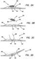
- FIG. 1illustrates a side view of a surgical training device with a model organ according to the present invention.
- FIG. 2 Aillustrates a side cross-sectional view of a simulated tissue structure according to the present invention.
- FIG. 2 Billustrates a side cross-sectional view of a simulated tissue structure with tumor excised according to the present invention.
- FIG. 2 Cillustrates a side cross-sectional view of a simulated tissue structure with an open suture according to the present invention.
- FIG. 2 Dillustrates a side cross-sectional view of a simulated tissue structure with a closed suture according to the present invention.
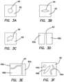
- FIG. 3 Aillustrates a top view of a defect layer having a circular shaped defect according to the present invention.
- FIG. 3 Billustrates a top view of a defect layer having an elongated defect according to the present invention.
- FIG. 3 Cillustrates a top view of a defect layer having an amorphous defect according to the present invention.
- FIG. 3 Dillustrates a top view of a defect layer having a two-piece defect according to the present invention.
- FIG. 3 Eillustrates a top view of a multi-part defect layer according to the present invention.
- FIG. 3 Fillustrates a top view of a defect layer having multiple defects according to the present invention.
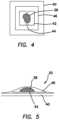
- FIG. 4illustrates a top view of a simulated tissue structure according to the present invention.
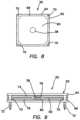
- FIG. 5illustrates a side cross-sectional view of a simulated tissue structure according to the present invention.
- FIG. 6 Aillustrates a perspective view of a modular tissue structure and support according to the present invention.
- FIG. 6 Billustrates a perspective view of a modular tissue structure and support according to the present invention.
- FIG. 7illustrates a cross-sectional view of a simulated tissue structure configured to mimic a human uterus according to the present invention.
- FIG. 8illustrates a top view of a modular tissue structure according to the present invention.
- FIG. 9illustrates a side view of a modular tissue structure according to the present invention.
- FIG. 10 Aillustrates a perspective view of a simulated tissue structure according to the present invention.
- FIG. 10 Billustrates a perspective view of a simulated tissue structure according to the present invention.
- FIG. 11 Aillustrates a perspective view of a simulated tissue structure according to the present invention.
- FIG. 11 Billustrates a perspective view of a simulated tissue structure according to the present invention.
- FIG. 12illustrates a perspective view of a suture needle and a simulated tissue structure according to the present invention.
- FIG. 13 Aillustrates a transparent side view of a polyp simulation according to the present invention.
- FIG. 13 Billustrates a side elevational view of a defect layer of a polyp simulation according to the present invention.
- FIG. 13 Cillustrates a side elevational view of a mesh layer of a polyp simulation according to the present invention.
- FIG. 13 Dillustrates a side elevational view of a muscle layer of a polyp simulation according to the present invention.
- FIG. 14 Aillustrates a side elevational view of a mold for a muscle layer according to the present invention.
- FIG. 14 Billustrates a top view of a mold for a muscle layer according to the present invention.
- FIG. 15 Aillustrates a side elevational view of a mold for a defect layer according to the present invention.
- FIG. 15 Billustrates a top view of a mold for a defect layer according to the present invention.
- FIG. 16illustrates an exploded view of a defect mold, defect layer, mesh layer, mold release layer and muscle layer according to the present invention.
- FIG. 17 Aillustrates a tissue simulation model having pods with attached simulated tissue portions according to the present invention.
- FIG. 17 Billustrates a pod assembly according to the present invention.
- FIG. 17 Cillustrates an exploded view of a pod assembly according to the present invention.
- FIG. 18is a bottom perspective view of a pod frame without a tissue portion according to the present invention.
- FIG. 19is a top perspective cross-sectional view of a tissue simulation module according to the present invention.
- FIG. 20is a top perspective view sectional of a mandrel used to manufacture a tissue simulation model according to the present invention.
- FIG. 21is a sectional view of a tissue simulation model according to the present invention.
- FIG. 22is a top perspective view of a tissue simulation model according to the present invention.
- FIG. 23is a sectional view of a tissue simulation model according to the present invention.
- FIG. 24is a sectional view of a mandrel for manufacturing a tissue simulation model according to the present invention.
- FIG. 25is a sectional view of tissue simulation model according to the present invention.
- FIG. 26 Ais a top planar view of a mesh layer of a tissue simulation model according to the present invention.
- FIG. 26 Bis a top planar view of a mesh layer of a tissue simulation model formed into a cylindrical sleeve according to the present invention.
- FIG. 27illustrates a mesh sleeve being placed onto a mandrel according to the present invention.
- FIG. 28illustrates a mesh sleeve located on a mandrel according to the present invention.
- FIG. 29illustrates a sectional end view of a fully suturable rectum model with an exemplary suture pathway according to the present invention.
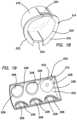
- a surgical training device 10that is configured to mimic the torso of a patient such as the abdominal region is shown in FIG. 1 .
- the surgical training device 10provides a simulated body cavity 18 substantially obscured from the user for receiving model organs or simulated or live tissue 20 .
- the body cavity 18is accessed via a tissue simulation region 19 that is penetrated by the user employing devices to practice surgical techniques on the tissue or organ 20 found located in the body cavity 18 .
- the body cavity 18is shown to be accessible through a tissue simulation region 19 , a hand-assisted access device or single-site port device may be alternatively employed to access the body cavity 18 as described in U.S. patent application Ser. No. 13/248,449 entitled “Portable Laparoscopic Trainer” filed on Sep. 29, 2011 and incorporated herein by reference in its entirety.
- the surgical training device 10is particularly well suited for practicing laparoscopic or other minimally invasive surgical procedures.
- the surgical training device 10includes a base 12 and a top cover 14 connected to and spaced apart from the base 12 to define an internal body cavity 18 between the top cover 14 and the base 12 . At least one leg 16 interconnects and spaces apart the top cover 14 and base 12 .
- a model organ or simulated tissue 20is disposed within the body cavity 18 .
- the model organ 20 shown in FIG. 1is a partial colon or intestine that is shown suspended from the top cover 14 by tethers 22 and connected to at least one leg 24 .
- the at least one leg 24has an aperture (not shown) facing the internal cavity 20 .
- the model colon 20includes a tube 26 having a proximal end and a distal end.
- the proximal end of the tube 26is interconnected with the aperture of the leg 16 such that the aperture provides an access port to the lumen of the tube 26 .
- the access port and apertureis shown to be closed off in FIG. 1 with an access device 28 which in combination with a sealed distal end of the tube 26 provides a model organ 20 that is adapted for insufflation with fluid deliverable via an insufflation port 30 .
- An optional insert 32 made of soft material such as siliconecreates a realistic interface for the access port.
- the distal end of the tube 26extends into the body cavity 18 and is suspended within the body cavity 18 .
- the interior of the tube 26 of the simulated organ 20is accessible via the access port of leg 24 or via the tissue simulation region 19 or instrument insertion ports 34 .
- An endoscopic camera inserted into the body cavity 18 or into the organ 20 via the access portgenerates a live image for display on a fold out video screen 36 shown in the closed position in FIG. 1 .
- the simulated organ 20 of FIG. 1is ideal for practicing procedures related to transanal minimally invasive surgery, any simulated organ or tissue portion may be employed.
- One particular aspect of the organ 20is at least one tumor or defect 38 is provided and connected to the organ. As shown in FIG. 1 , the tumor 38 is connected to the wall of the organ tube 26 .
- FIG. 2 Athere is shown a partial side cross-sectional view of a portion of a simulated organ 20 that includes the tumor 38 .
- the simulated organ or tissue 20includes a base layer or organ wall 40 .
- the organ wall 40is made from a material configured to mimic real live tissue such as silicone or other polymer and is dyed appropriately.
- One or more base layers 40 of varying thicknesses and colorationsmay be employed to comprise the entirety of the wall 40 .
- the organ wall 40is rigid and made of polymeric material.
- the defect layer 42is the same size or smaller than the base layer 40 forming a raised platform for the tumor 38 .
- the defect layer 42is connected to the base layer 40 by adhesive or other means known to one having ordinary skill in the art including being integrally formed with the base layer 40 as a single unit.
- the defect layer 42is made of silicone and in one variation of the same color as the base layer 40 such that the defect layer 42 blends into the background of the base layer 40 .
- the defect layer 42includes at least one defect or gap 44 .
- the defect 44is a pre-fabricated breach in the defect layer 42 that mimics an incision, gap or other void in real tissue resulting from a tear, cut, removal or other surgical procedure that requires surgical attention by way of suturing, stapling or the like to close the defect.
- the defect 44comprises two opposed sides or surfaces defining a gap therebetween. Although the adjacent sides or surfaces are shown to be vertical with respect to the base layer 40 , the invention is not so limited and the juxtaposed surfaces or sides can have any shape and, for example, be curved.
- the defect 44can be any shape as will be discussed with respect to FIGS. 3 A- 3 F .
- FIG. 3 Athere is shown a top view of a defect layer 42 having a circular defect 44 .
- a defect layer 42 with an elongated, oblong or elliptically shaped defect 44is shown in the FIG. 3 B .
- the defect 44can be amorphic or any shape as shown in FIG. 3 C .
- the defect layer 42may be multi-part as shown in FIG. 3 D wherein the defect layer 42 includes two or more adjacent defect layer pieces 42 a , 42 b juxtaposed to create at least one defect 44 therebetween.
- Another multi-part defect layer 42is shown in FIG. 3 E where a plurality of adjacent defect layer pieces 42 a , 42 b and 42 c form one or more defects 44 therebetween.
- a defect layer 42may include multiple defects 44 a , 44 b and 44 c as shown in FIG. 3 F .
- the defects 44may all be the same or have different shapes as shown in FIG. 3 F .
- the shape, thickness and size of the defectallow the surgeon trainee to practice suturing across defects of varying difficulty.
- the defect layer 42is not of equal thickness. Instead, the thickness of the defect layer 42 varies at the defect location 48 to increase the difficulty of suturing or closing the defect.
- a tumor 38is located above the defect layer 42 .
- the tumor 38is preferably a different color from the base layer 40 or defect layer 42 or both such that it is readily identifiable by the trainee.
- the tumor 38is made of silicone or other polymer material and is red, black, blue or dark brown in color.
- the tumor 38is of a darker color than the base or defect layers 40 , 42 or otherwise in contrast therewith when viewed through a scope.
- the tumor 38is connected to the defect layer 42 by adhesive or other means known to one of ordinary skill in the art.
- the tumor 38is not connected or attached to the defect layer 42 but is removably located thereon.
- the simulated tissue structure 20includes a cover layer 46 located above the tumor 38 .
- the cover layer 46overlays the tumor 38 , defect layer 42 and the base layer 40 .
- the cover layer 46is preferably transparent or translucent in color and made of a polymer material such as silicone.
- the cover layer 46is the same color as the base layer 40 or defect layer 42 .
- the cover layer 46is at least as thick as the base layer 40 or defect layer 42 and in one variation is thinner than the defect layer 42 and in another variation is thinner than the base layer 40 .
- the cover layer 46is sized to cover the entire tumor 38 and defect layer 42 and is big enough to contact the base layer 40 in one variation.
- the cover layer 46is sized to cover the entire tumor 38 and contact the defect layer 40 .
- the cover layer 46is connected to the base layer 40 , defect layer 42 , tumor 38 or any more than one of the three layers by way of adhesive or other means known to one of ordinary skill in the art.
- the cover layer 46is smaller and connected to the defect layer 42 alone.
- the cover layer 46is connected to both the defect layer 42 and base layer 42 by adhesive or other means known to one of ordinary skill in the art.
- the cover layer 46can be any shape or sized and be configured to provide a smooth surface to the surgeon instead of a layered surface to the artificial tumor location.
- the cover layer 46 , tumor 38 , defect layer 42 or base layer 40includes surface texturing in one variation.
- the cover layer 46assists in keeping the tumor 38 and defect layer 42 sandwiched between the cover layer 46 and base layer 40 which is advantageous in a variation wherein the tumor 38 is not adhered to the defect layer 42 .
- a top planar view of the base layer 40 , defect layer 42 , cover layer 46 and tumor 38is shown in FIG. 4 .
- any one or more of the base layer 40 , defect layer 42 and cover layer 46is formed of silicone molded over a woven, fabric, or mesh material such as nylon or cheesecloth so that the silicone layer has an integrated mesh structural support or other type of reinforcement.
- Any one or more of the layers 38 , 40 , 42 , 46can include a fabric or mesh reinforcement combined with an elastic polymer such silicone.
- the mesh supportaids in preventing the suture, staple, or suture needle from tearing through at least one of layers and especially the defect layer 42 when the suture is pulled to close the gap 44 .
- FIG. 2 Bthe tumor 38 and a portion of the cover layer 46 are shown excised from the base layer 40 .
- the excisionis performed by the trainee using a surgical instrument such as a scalpel or other medical instrument to remove the tumor 38 .
- the traineewill cut through the cover layer 46 around the tumor 38 , isolate the tumor 38 , lift and remove the tumor 38 away from the site to expose the underlying defect 44 as shown in FIG. 2 B .
- the traineesutures the defect 44 using a surgical suture 48 bringing the lips or edges of the defect layer 42 together as shown in FIG. 2 D , thereby, practicing the closing of a gap or wound created by the surgical removal of a tumor 38 .
- Cutting the at least one layer to create an opening and removing the artificial tumor and suturing the gapis performed while the simulated tissue structure is disposed inside a simulated body cavity 18 of a surgical training device such that the simulated tissue structure is at least partially obscured from view by the user.
- FIG. 5there is shown another variation in which there is no pre-formed gap or defect in the second or defect layer 42 .
- the defectis created by the user in one or more of the cover layer 46 , defect layer 42 , base layer 40 and any remaining tumor portion not removed by the user. The user would then practice suturing the created defect in any of these layers 38 , 40 , 42 , 46 .
- one of the defect layer 42 or base layer 40is omitted from the construct.
- the tumor 38is located on a base layer 40 and the defect layer 42 is placed over the tumor 38 such that the defect layer 42 is above the tumor 38 .
- a cover layer 46may or may not be included.
- a cover layer 46it may be integrally formed together with the defect layer as a separate unitary layer.
- the constructsmay be flipped upside down or otherwise the layers placed in reverse or otherwise the construct being approachable by the user from either the top or bottom direction with the thicknesses and colors of the layers being adjusted accordingly if necessary to provide the simulated effects of real tissue.
- the simulated tissue constructcan be modular such that it is not integrally formed with the entire simulated organ 20 but instead configured as a module 50 that is removable and interchangeable.
- One or more modules 50are supported or contained in a module support 52 .
- a module support 52includes a first surface 51 , a second surface 53 and one or more tumor module receiving portions 54 , 56 , 58 formed in the support 52 .
- the tumor support 52can be rigid or pliable and made of polymeric material.
- the tumor support 52may also comprise a sheet of elastomeric material.
- the module receiving portions 54 , 56 , 58are each sized and configured to receive a correspondingly sized and configured module 50 .
- the modules 50 and module receiving portions 54 , 56 , 48 in FIG. 6are shown to be circular; however, the tumor module 50 can be any shape with a complementary shaped receiving portion formed in the module support 52 .
- the thickness of the support 52can vary providing the construct with varying depths of tumor module 50 positioning.
- the module receiving portions 54 , 56 , 58may include bottom walls onto which the tumor modules 50 may rest.
- the tumor receiving portions 54 , 56 , 58extend between openings in the first surface 51 and the second surface 53 with the modules 50 with tumor 38 being connected between or at one of the openings at either surface 51 , 53 or suspended within the tumor receiving portion.
- a single tumor module 50includes one or more tumors 38 .
- the module support 52is loaded with one or more tumor modules 50 and the simulated tissue construct 20 is inserted into the body cavity 18 of the surgical training device 10 , framework or other torso model. It can be placed on the base 12 of the training device 10 or suspended within the body cavity 18 of the training device 10 .
- the simulated tissue construct 20 and/or training deviceis fashioned with attachment mechanisms such as clips, fasteners, wires, hook-and-loop type fasteners and the like for placement, suspension or connection of the simulated tissue construct 20 to a training device 10 .
- the module support 52 of FIG. 6 Bincludes a first layer 57 connected to a second layer 55 .
- the first layer 57is made of a sheet of elastomeric material and the second layer 55 is made of any suitable polymeric material such as low-density elastomeric foam.
- the second layer 55serves as a support for the first layer 57 .
- the second layer 55also advantageously provides depth to the module support 52 permitting the tumors 38 within the modules 50 to be placed deeply into the module support 52 relative to the first surface 51 .
- Module receiving portions 54 , 56 , 58are formed in one or more than one of the first layer 57 and the second layer 55 .
- Module receiving portions 54 , 56 , 58 formed in the second layer 55may have a different shape than the shape the same module receiving portion 54 , 56 , 58 has in the first layer 57 .
- the tumor module 50comprises at least only the simulated tumor 38 which is embedded or buried inside the second layer 55 with at least one of the first layer 57 or second layer 55 constituting a defect layer which the user can practice closing.
- the first layer 57does not include a module receiving portion but instead the first layer 57 serves as a cover layer which the user practices cutting through to access the tumor 38 located in a tumor receiving portion formed in the second layer 55 .
- the first layer 57can be a sheet of elastomeric material such as silicone and the second layer 55 is a layer of low-density elastomeric foam.
- the module support 52is planar as shown in FIGS. 6 A and 6 B or, alternatively, shaped to mimic a portion of the human anatomy, tissue or organ.
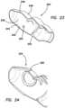
- FIG. 7illustrates a support 52 that is shaped to mimic a human uterus.
- the support 52includes a first layer 57 connected to a second layer 55 .
- the first layer 57is made of any suitable polymeric material such as a sheet of elastomeric material and the second layer 55 is made of any suitable polymeric material such as low-density elastomeric foam.
- the second layer 55serves as a support for the first layer 57 and advantageously permits the tumors 38 within the modules 50 or the tumors 38 by themselves to be connected to the support 52 and realistically extend deeply into the support 52 and be dispersed throughout the support 52 in various locations and orientations including being embedded into the first layer 57 as shown in FIG. 7 .
- Tumor or module receiving portions 61are formed in at least one of the first layer 57 and second layer 55 .
- the tumor receiving portions 61may be pockets that are pre-formed in the second layer 55 or can be formed by the user by cutting slits into the second layer 55 .
- the tumors 38are configured to mimic fibroid tumors commonly found in the human uterus. Examples of fibroid tumors that are simulated by the tumors 38 disposed in the support include but are not limited to one or more of the following types of fibroids: pedunculated submucosal fibroids, subserosal fibroids, submucosal fibroids, pedunculated subserosal fibroids and intramural fibroids.
- the usercan approach the support 52 to excise the simulated tumors 38 from the first surface 51 or the second surface 53 via the access channel or opening 63 .
- the opening 63serves as the only opening to the hollow portion 59 or alternatively the support 52 can have a substantially C-shaped planar configuration with access available to the user from above or below the planar C-shaped structure.
- the module support 52 in any of the variationsis not planar but is provided with a landscape that includes curves and other structures, mountains and valleys and various textures.
- the varying landscapeprovides the user with various levels of difficulty in approaching each tumor location requiring the user to navigate around artifacts and features that may obscure the tumor location.
- These structural artifacts in the tumor support 52may be integrally formed with the tumor support 52 or also be modular in structure similar to the tumor modules 50 making the anatomy landscape modules removable and interchangeable.
- Tumor modules 50are interchangeable with non-tumor modules that include, for example, features and artifacts or textures made of silicone or other material extending outwardly or inwardly from the one or more of the upper and lower surfaces 51 , 53 of the module support 52 .
- non-tumor modulescan have various shapes to mimic anatomy that includes adjacent organ structures or tissues.
- a non-tumor modulecan include a tubular form of silicone to mimic an intestine.
- the non-tumor and tumor modules 50are removably connected to the module support 52 by any means known to one skilled in the art enabling the user to discard a module after use and then to continue practicing by replacing the discarded module or moving to an adjacent module 50 in the module support 52 or changing out a tumor module 50 for another tumor module 50 having a different feature or level of difficulty.
- the tumor module 50includes a simulated tissue portion 60 connected to a support 62 .
- the support 62includes a top frame 64 connected to a bottom frame 66 . At least one of the top frame 64 and bottom frame 66 includes a window.
- the top frame 64 having a window 68is shown in FIG. 8 .
- the bottom frame 66may or may not include a window. If windows are provided in both the top frame 64 and the bottom frame 66 , the windows are aligned at least in part.
- the support 62is sized and configured to receive a simulated tissue portion 60 between the top frame 64 and the bottom frame 66 .
- the top frame 64is connectable to the bottom frame 66 to capture the unitary simulated tissue portion 60 or a simulated tissue portion 60 formed from multiple layers and, in one variation, separable.
- the frames 64 , 66are spaced apart from each other using spacers 70 .
- at least one of the top and bottom frames 64 , 66includes one or more connecting features 72 configured to secure the tumor module 50 to a tumor support 52 (not shown).
- the connecting features 72are shown as extending pegs for insertion into corresponding holes formed in the tumor support 52 to provide a snap-fit engagement.
- a friction fit or other fasteners or connecting meanssuch as hook-and-loop type materials can be employed on the module 50 and module support 52 to connect the module 50 to the support 52 in a removable fashion.
- the simulated tissue portion 60can be any of the constructs described above with reference to FIGS. 2 - 5 . With windows formed in both the first and second frames 64 , 66 , the simulated tissue portion 60 can be approached from either side of the module 50 . Any layer described above as a cover layer may act as a top layer or as a bottom layer depending on from which side or direction the simulated tissue portion 60 is approached. For example, a base layer may also serve as a top layer or as a bottom layer depending on which side or direction the simulated tissue portion 60 is approached. In such, bi-directional constructs, the thicknesses and colors of the layers may be adjusted accordingly to provide the desired simulated effect.
- the simulated tissue portion 60 in FIG. 9includes a first layer 74 and a second layer 76 .
- the first and second layers 74 , 76are made from a polymeric material configured to mimic real live tissue such as silicone or other polymer and can include dye of any one or more appropriate colors or mesh, fabric, or other reinforcement.
- Each of the layers 74 , 76includes a tumor receiving portion 78 , 80 , respectively.
- Each tumor receiving portion 78 , 80is a concavity, indent, half-pocket or a location of reduced layer thickness that is formed in the layers 74 , 76 .
- the tumor receiving portions 78 , 80are substantially aligned to form a pocket for the tumor 38 .
- a tumor receiving portion 78 , 80a single tumor receiving portion is formed in at least one of the first and second layers 74 , 76 in one variation.
- a tumor 38is disposed within the pocket formed by one or more tumor receiving portions 78 , 80 formed in the one or more layers 74 , 76 .
- the tumor 38may be adhered to either layer 74 , 76 or free floating inside the pocket.
- the tumor receiving portion formed in a layercan be considered to be one type of defect and the variation of FIG. 9 describes a simulated tissue construct comprising two defect layers with a tumor therebetween. As a user approaches the simulated tissue portion 60 , the user will see the target tumor location.
- the tumor receiving portionbeing thinner in thickness relative to the rest of the layer with the thinning of the layer being provided by the concavity or pocket.
- the userwill then cut in the general location of the tumor cutting into at least one of the layers 74 , 76 to remove the tumor 38 . Cutting through one or more layers completes the creation of a gap or full defect which the user can then practice suturing or otherwise closing together.
- at least one tumoris disposed between the two layers 74 , 76 wherein the layers 74 , 76 have a substantially uniform thickness with the tumor 38 creating a minor bulge in the layers.
- the tissue portion 86can be integral or modular as described above.
- the tissue portion 86includes a base layer 88 formed of any suitable polymeric material such as silicone or other elastomeric polymer that may or may not include a reinforcement material such as fabric, mesh, nylon or other reinforcement material or filler that will resist tearing while carrying sutures or while being sutured.
- the base layer 88is connected to a defect layer 90 that is overlaid onto the base layer 88 .
- the defect layer 90includes a plurality of protrusions extending upwardly from the base layer 88 .
- the defect layer 90may be integrally formed with the base layer 88 or be a separate layer that is adhered to the base layer 88 . As can be seen in FIGS. 10 A, 11 A and 12 , the defect layer 90 is configured into a lattice shaped pattern such that the lattice is raised above the base layer 88 or projects upwardly from the base layer 88 .
- a lattice patternis exemplary and any shape may be formed by the defect layer 90 such that it contains a plurality of adjacent projections. These projections of the base layer 90 provide the user with locations to hook a suture needle into and as a platform to raise the tumor 38 a , 38 b above the base layer 88 for easy excision.
- FIGS. 10 A and 11 Ashow the base layer 88 , defect layer 90 , tumors 38 a , 38 b and a cover layer 92 in a semi-exploded view of the simulated tissue portion 86 wherein the cover layer 92 is raised above the other layers.
- the tumor 38 a of FIG. 10 ais substantially planar and is shown covered in FIG. 10 B by the cover layer 92 .
- Tumor 38 b of FIG. 11 Ahas greater height and is substantially spherical in shape and FIG. 11 B shows the spherical tumor 38 b covered with the cover layer 92 leaving a raised portion or protuberance in the construct.
- FIG. 12shows the tumor 38 being removed leaving a remnant defect 94 in the base layer 88 and a suture needle crossing the gap in the defect 94 with the defect having been accessed under or through the cover layer 92 .
- the polyp simulation 100includes a defect layer 102 , a mesh layer 104 , a muscle layer 106 and a mold release 108 .
- the defect layer 102includes a first surface 110 opposite from a second surface 112 .
- the defect layer 102is a substantially planar and thin layer of silicone material in the x-y plane.
- the defect layer 102includes a defect 114 extending outwardly from the first surface 110 along a z-axis in a direction perpendicular to the x-y plane.
- the defect 114may be any shape.
- the defect 114mimics an abnormal tissue growth such as a polyp.
- the defect 114includes a narrow elongated stalk and a bulbous distal end. In another variation, the distal end of the defect is curved.
- the defect 114mimics a colorectal polyp or colon polyp. In one variation, the defect 114 is approximately 2-5 millimeters in length and 1-5 millimeters in width. In one variation, the silicone of the defect layer 102 is dyed red or pink. In one variation, the defect layer includes an inclusion of contrast colored silicone.
- the mesh layer 104includes a first surface 116 opposite from a second surface 118 .
- the mesh layer 104is a substantially planar and thin layer comprising strands 120 of fibers made of nylon or other polymer in the x-y plane.
- the mesh layer 104is made of LYCRA.
- the mesh layer 104is capable of being stretched in any direction.
- the mesh layerhas bi-directional stretch properties.
- the strands of polymer fiberform a web or net.
- the mesh layer 104may be woven and have a uniform pattern.
- the mesh layer 104is pink, clear or white in color.
- the muscle layer 106includes a first surface 122 opposite from a second surface 124 .
- the muscle layer 106is a substantially planar and thin layer of silicone material in the x-y plane. In one variation, the muscle layer is yellow in color.
- the mold release 108is a mold release agent that is typically in liquid form and sprayed on to form a mold release area or layer.
- the mold release agentis one that is suitable for use on silicone.
- the mold release layer 108is a mold release agent alternative or substitute.
- the mold release layer 108prevents at least a portion of a silicone layer surface from bonding to an adjacent silicone surface.
- the mold release 108prevents a portion of the defect layer 102 from bonding to the adjacent muscle layer 106 .
- the mold release 108prevents at least a portion of the defect layer 102 and mesh layer 104 combination from bonding to the adjacent muscle layer 106 .
- the muscle mold 126for molding the muscle layer 106 .
- the muscle mold 126includes a first well 128 .
- the well 128is circular in shape to produce a circular muscle layer 106 .
- the first well 128may be any shape.
- Uncured siliconeis poured into the mold and allowed to cure to form the muscle layer. Mold release may be employed to help remove the cured layer. The removed layer may be washed with alcohol to remove any mold release.
- the defect mold 130for molding the defect layer 102 .
- the defect mold 130includes a first well 132 having a first depth and a second well 134 having a second depth. The second depth is greater than the first depth.
- the second well 134is located within the first well 132 .
- the second well 134is for forming the shape of a polyp or other defect 114 .
- the shape of the second well 134corresponds to the shape of the defect 114 .
- the shape of the first well 132is circular to form a circular defect layer although it may have any shape.
- the size and shape of the first well 132is the same size and shape as the first well 128 of the muscle mold 126 to create muscle layers 106 and defect layers 102 that have the same size and shape and can be easily aligned and connected to form a nice patch-like simulation 100 .
- the second well 134is formed such that it is within the perimeter of the first well 132 .
- the second well 134produces a defect 114 that is surrounded by the remaining of the defect layer 102 .
- Uncured siliconeis poured into the defect mold 130 and allowed to cure before being removed. Mold release may be employed to facilitate removal of the defect layer 102 from the defect mold 130 .
- contrast-colored, cured silicone piecesare placed into the second well 134 that forms the shape of the polyp or other defect.
- one or more red colored, cylindrically shaped cured silicone pieces sized to fit inside the second well 134are disposed into the second well 134 prior to pouring the uncured silicone into the defect mold or after the uncured silicone is poured into the defect mold 130 to form the defect layer 102 .
- the resultis that the contrast colored silicone pieces will be embedded inside the defect layer 102 in the location of the defect to provide a custom and more realistic construction of a particular defect that is being simulated.
- the defect layer 102is connected to the mesh layer 104 such that the second surface 112 of the defect layer 102 faces the first surface 116 of the mesh layer 104 .
- adhesivemay be employed between the defect layer 102 and the mesh layer 104 or, in another variation, the mesh layer 104 is placed into the defect layer 102 while the silicone of the defect layer 102 has not been cured. As a result, the mesh layer 104 is embedded within the defect layer 102 . If the mesh layer 104 is embedded within the defect layer 102 , the resulting combination has a proximal surface which is the first surface 110 of the defect layer 102 and a distal surface which is the surface close to the mesh layer 104 .
- Mold release 108is applied to the distal surface of the defect/mesh layer combination in a selective area.
- the mold release 108is applied in the center of the perimeter such that an annular area without mold release 108 surrounds the area where mold release is applied.
- mold release 108is applied under the defect 114 such that an area of the distal surface that does not have mold release surrounds the area with mold release 108 on it. The area that surrounds the mold release area does not have mold release 108 on it so as to create a bond between the muscle layer 106 and the defect layer 102 that is annular in shape. The area with mold release 108 on it will not bond the muscle layer 106 to the defect layer 102 making them separable in the location of the defect 114 .
- the muscle layer 106is connected to the distal surface of the defect/mesh layer combination.
- the muscle layer 106is connected with adhesive in one variation.
- the muscle layer 106is applied to the distal surface of the defect/mesh layer combination while the silicone of the defect layer 102 is still uncured so as to embed the muscle layer 106 into the defect/mesh combination.
- the forming processinvolves two molds, the muscle mold 126 and the defect mold 130 .
- Siliconeis cast in the muscle mold 126 to form the muscle layer 106 .
- the silicone of the muscle layer 106is allowed to cure.
- the muscle layer 106is then removed from the muscle mold 126 .
- the muscle layer 106is cleaned using isopropyl alcohol.
- Mold release 108is applied only to the center of the muscle layer 106 or underneath the defect 114 .
- the mold release 108is applied to the first surface 122 using a stencil.
- the muscle layer 106 with the mold release 108is set aside.
- siliconeis cast in the defect mold 130 to form the defect layer 102 .
- the mesh layer 104While the silicon in the defect mold 130 is un-cured, the mesh layer 104 , having a shape that conforms to the shape of the first well 132 , is placed onto the un-cured silicone in the defect mold 130 such that it becomes connected thereto.
- the muscle layer 106 with the mold release 108is placed above the mesh layer 104 such that the first surface 122 having the mold release 108 applied to it faces the mesh layer 104 and the defect layer 102 .
- the muscle layer 106 with the mold release 108is placed face down towards and onto the mesh layer 104 and the uncured silicone of the defect layer 102 .
- the muscle layer 106is pressed into the defect layer 102 while it is un-cured with gloved fingers, for example, to remove any air bubbles.
- the silicone of the defect layer 102is allowed to cure and the resulting polyp simulation 100 is removed from the defect mold 130 . All of the layers have the same shape and are overlaid each other in an aligned fashion to form a single piece polyp simulation 100 .
- part of the muscle layer 106is not adhered to the defect and mesh layers 102 , 104 and a part of the muscle layer 106 without mold release 108 on it is adhered to the defect and mesh layers 102 , 104 .
- the selective adherenceadvantageously creates a polyp simulation 100 suitable for practicing polyp removal and the mesh layer creates a polyp simulation suitable for practicing suturing after the polyp has been removed.
- the defect layer 102is allowed to cure with or without a mesh layer 104 .
- the second layer 106is allowed to cure.
- a stencilis laid on top of one of the defect layer 102 and second layer 106 .
- the stencilhas one or more apertures.
- the one or more aperturesare arranged on the stencil in a pattern configured for adhesion.
- One patterncomprises a plurality of randomly spaced dots or circles.
- Uncured silicone or adhesiveis applied onto the stencil in the location of the apertures such that the uncured silicone or adhesive passes through the one or more aperture and comes into contact with the layer on which the stencil is laid.
- the stencilis removed along with excess adhesive or uncured silicone leaving behind a pattern of uncured silicone or adhesive.
- the other one of the defect layer and second layer 106is then laid on top of the other layer and adhered thereto.
- the pattern for adhesive on the stencilcan be a circumferential pattern or circular pattern in the location of the defect or any other pattern.
- the stencil aperturemay be a single continuous or multi-aperture pattern of a plurality of circles, for example, that forms a larger circle along the perimeter of the layers and/or around the defect such that the two layers are not adhered outside the applied adhesive or applied uncured silicone. Mold release may or may not be employed between the two adhered layers.
- One or more resulting polyp simulation 100is then adhered to another portion of a simulated tissue structure.
- the patch-like polyp simulation 100is adhered with adhesive to the inside surface of a tubular simulated colon, that in one variation, is made of silicone.
- the poly simulation 100is connected to the simulated colon model such that the defect 114 extends into the lumen of the colon.
- the muscle layer 106 and the defect layer 102are bonded together without an additional mesh layer 104 for ease of manufacturing. In another variation, the muscle layer 106 and the defect layer 102 are separately fully cured and adhered together without any mold release 108 .
- the defect 114is not formed as an integral protrusion extending from the first surface 110 of the defect layer 102 . Instead, the defect 114 is a separate piece that is located between the muscle layer 106 and the defect layer 102 . In another variation, the defect 114 is not formed as an integral protrusion extending from the first surface 110 of the defect layer 102 . Instead, the defect 114 is a separate piece that is located between the muscle layer 106 and the defect layer 102 such that the defect 114 floats between the two layers 102 , 106 .
- the polyp simulation 100is used with a simulated rectum.
- the simulation 100advantageously includes a mesh layer 104 embedded in it which allows the user to practice suturing techniques following the practiced removal of the defect 114 .
- the present simulation 100increases the difficulty of removing the defect because the layers 106 , 102 are not as easily separated due to the annular area that does not have mold release 108 .
- the mesh layer 104allows the polyp simulation 100 to be sutured.
- the suturing techniquesare practiced by the user without damaging the surrounding silicone. Connecting the two muscle layer 106 while the defect layer 102 is still uncured results in a construct that increases the difficulty of separating the two layers and increases the accuracy of the simulation.
- the embedded mesh layer 104stops the suture from tearing, ripping or cutting through the silicone.
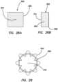
- FIGS. 17 A- 17 Cthere is shown a simulated tissue model 200 for practicing surgical procedures.
- the model 200 shown in FIG. 17is configured to mimic a portion of a colon or bowel section; however, the invention is not limited to a colon or bowel.
- the entire model 200may be configured to mimic at least a portion of any organ or tissue section upon which the practice of certain surgical procedures is desired.
- the simulated tissue model 200comprises an inner surface 202 and an outer surface 204 that together define a sidewall having a thickness.
- 17 Ahas a shape of a cylinder having a central lumen 206 extending along a longitudinal axis between an opening at the proximal end and an opening at the distal end to approximate a colon, rectum or bowel section.
- One of the openings at the endsmay be omitted.
- Either one or more of the inner surface 202 and the outer surface 204may include surface features or textures that mimic real tissue.
- transverse folds and/or a mesorectum layermay be included. Simulated transverse folds may be an obstacle to the movement of the surgeon's instrument and/or obscure direct visualization of a lesion. Therefore, the presence of transverse folds replicates the challenges that a surgeon may face while performing a transanal procedure.
- a simplified model 200will omit the transverse folds.
- a simulated mesorectum layer that is included with the model 200advantageously provides a reference plane for approaching target tissue lesions that are located closer to the anal verge which are more difficult to remove due to limited instrument movement and the sharp approach angle.
- the sidewallseparates an interior space defined by the central lumen 206 of the simulated tissue model 200 from an exterior space of the model 200 .
- the model 200includes one or more apertures 208 extending through the sidewall from the inner surface 202 to the outer surface 204 . Each aperture 208 is shaped and configured to receive a module or pod 210 .
- the sidewallhas a substantially uniform thickness in the area surrounding the pod-receiving apertures 208 .
- a plurality of apertures 208are formed along the length of the model 200 from the proximal end to the distal end and around the entire sidewall in various locations.
- the sidewall of the model 200is made of a rigid or semi-rigid material such as plastic. In another variation, the sidewall of the model 200 is soft and/or combined with soft and semi-rigid or rigid portions.
- the sidewall of the model 200is constructed to allow the pods 210 to be attached to the model 200 .
- Each pod 210includes a simulated tissue portion 212 that is connected to a tissue carrier 214 also called a cap.
- a tissue carrier 214is shown in FIG. 18 .
- the carrier 214includes a flange 216 that is connected to a frame 218 .
- the frame 218is substantially cylindrical in shape and has an opening 220 at the proximal end and a closed distal end.
- the flange 216is located at the proximal end and surrounds at least in part the opening 220 .
- the frame 218defines a substantially circular proximal opening 220 and the flange 216 extends at least along part of the perimeter of the opening 220 .
- the flange 216is substantially perpendicular to the cylindrical sidewall of the frame 218 and extends radially outwardly at the proximal end.
- the flange 216includes a surface 222 configured to connect to and suspend the tissue portion 212 .
- the pod 210is configured to serve as a tissue insert that fills the apertures 208 and the flange surface 222 is contoured to match the portion of the inner surface 202 in which it is received. For example, if the inner surface 202 of the model 200 is concave, the surface 222 of the flange 216 will also be correspondingly concave in shape.
- the frame 218includes oppositely disposed detents 224 configured to flex inwardly and then spring back outwardly for connecting and disconnecting the pod 210 to and from the model 200 .
- Fingers of a userare employed to press the detents 224 inwardly while inserting the pod 210 into an aperture 208 of the model 200 .
- the pods 210may be inserted from the interior space of the model such that the sidewall of the model 200 ramps over the detents 224 flexing them inwardly and allowing them to snap back outwardly after the detents 224 have cleared the sidewall to capture the sidewall of the model 200 between the detent 224 and the flange 214 , and thereby, connect the pod 214 to the model 200 .
- the inner surface 202 of the model 200includes a recess 226 that encompasses each aperture 208 as shown in FIG. 19 .
- Each recess 226extends around the apertures 208 and are sized and configured to receive the flange 216 of the pod 210 such that a connected tissue portion 212 or flange 216 is substantially even or flush with the inner surface 202 of the model 200 .
- a pod 210 having a tissue portion 212 connected to itis shown inserted into an aperture 208 of the model 200 in FIGS. 17 A and 19 .
- the tissue portion 212is connected to the flange 216 of the pod 210 .
- the tissue portion 212is connected to the surface 222 of the flange 216 by adhesive or bonding such that at least a portion of the tissue portion 212 is suspended or spans across the proximal opening 220 .
- the tissue portion 212 that is suspendedis free to flex and stretch within the pod 210 and be dissected in a simulation procedure. Following the procedure, the pod 210 may be removed from the model 200 , discarded and replaced with a new pod 210 into the cylindrical sidewall of the model 200 for subsequent training and practice of surgical procedures.
- Each pod 210 with the tissue portion 212is configured for attachment to a simulated model 200 having a tubular shape.
- the tubular shapecan be configured to open as a clam to insert and remove the pods 210 .
- the tissue portion 212 of each pod 210is flexible and includes at least a planar first layer 228 .
- the first layer 228includes a first side and a second side.
- the first layer 228is connected to the flange surface 222 such that the first side faces the interior of the model 200 .
- the first layer 228is sized and configured to overlay the opening 220 and attach to the flange 216 . As such, the connected first layer 228 covers the opening 220 .
- the central portion of the first layer 228is free to flex in response to impingement by surgical instruments.
- the first layer 228is also configured to be severed with a blade such as a scalpel or other instrument and to be grasped by a surgical instrument or otherwise manipulated as needed by the surgeon practicing a surgical procedure.
- the central portion of the first layer 228is suspended in trampoline-like fashion.
- the first layer 228is made of silicone and may or may not include a mesh layer, fiber, fabric or other reinforcement material that would impart the first layer 228 with suturable qualities permitting the first layer 228 to hold sutures without being torn.
- the first layer 228is made of KRATON®.
- the tissue portion 212includes a substantially planar first layer 228 and a simulated target or tumor 232 connected to the first layer 228 .
- the first layer 228is connected to the frame 218 such that the first side faces the interior of the model 200 .
- the simulated tumor 232is connected to the first side of the first layer 228 such that the simulated tumor 232 faces the interior of the model 200 and in one variation protrudes toward the longitudinal axis.
- both the first layer 228 and the simulated tumor 232are made of silicone.
- the first layer 228is generally dyed to have the same color as the surrounding color of the inner surface 202 of the model 200 so that it is indistinguishable from the surrounding inner surface 202 .
- the first layer 228is generally pink or red in color.
- the simulated tumor 232can be dyed a color, such as dark red, brown or black, that is darker or in contrast to the first layer 228 .
- the simulated tumor 232extends outwardly from the first surface of the first layer 228 .
- the polyp simulation 100 of FIGS. 13 - 16is attached to the pod 210 .
- the tissue portion 212includes a substantially planar first layer 228 having a first side and a second side, a planar second layer 230 having a first side and a second side and a simulated target or tumor 232 .
- the first layer 228is substantially planar and connected to the frame 218 such that the first side faces the interior of the model 200 .
- the first layer 228is made of silicone and dyed a pink or red color.
- the second layer 230is substantially planar and includes a first side and a second side.
- the second layer 230is connected to the first layer 228 such that the first side of the second layer 230 faces the second side of the first layer 228 .
- adhesiveis used on least a portion of the first or second layer to connect the two layers together.
- one of the layers 228 , 230is applied to the other layer while in an uncured state and allowed to cure and adhere to the other layer resulting in the layers being more easily separated relative to using adhesive.
- the second layer 230is made of silicone and is dyed a yellow color.
- the simulated tumor 232is attached or integrally formed with the first layer 228 such that the simulated tumor 232 is connected to the first side of the first layer 228 or extends outwardly from the first side of the first layer 228 .
- the second layer 230is yellow in color and simulates the submucosa layer.
- the first layer 228is pink and simulates the rectum wall.
- the simulated tumor 232simulates a tumor, lesion or other surgically desirable target.
- the second layer 230has a planar configuration with an outer surface and an inner surface and sized to be the same size and shape as the first layer 228 ; and the tumor 232 is sized smaller than the first and second layers 228 , 230 .
- the surgeon in practicing a transanal approachwill insert surgical instruments into an opening at one or more of the proximal end or distal end of the model 200 .
- the surgeonwill practice using a scalpel to make an incision into the first layer 228 , extend the incision through the first layer 228 and around the simulated tumor 232 .
- the second layer 230provides an indication or warning to the surgeon to stop cutting and to not cut into the second layer 230 .
- yellow second layer 230serves as a reference plane for the surgeon.
- the area of the first layer 228 that is adjacent to or beneath the simulated tumor 232is not adhered to the second layer 230 with adhesive.
- the first layer 228is adhered to the second layer 230 only circumferentially around the simulated tumor 232 making the first layer 228 easily separable from the second layer 230 if the cut is made within the perimeter of adhesion. This type of placement of adhesive advantageously help guide the surgeon to making a more precise excision.
- the area of the first layer 228 under the simulated tumor 232is adhered to the second layer 230 without adhesive by way of surface adhesion properties of like materials or by curing one layer onto the other layer in the fabrication of the tissue portion 212 .
- the surgeonmay practice suturing the resulting defect or gap closed with sutures.
- the first and/or second layers 228 , 230may be made of suturable material.
- a suturable materialmay include a thermoset polymer over molded onto fibers, mesh or fabric, a thermoplastic elastomer, or a thermoplastic elastomer over molded onto fibers, mesh or fabric.
- the fabric mesh materialmay also have bi-directional stretchable characteristics.
- At least a portion of the tissue portion 212is suspended by the frame 218 such that there is a space behind the simulated tissue portion 212 that allows manipulation of the simulated tumor 232 and/or tissue portion 212 .
- the suspended portionis the middle portion of the tissue portion 212 , the perimeter of which is attached to the frame 218 .
- the attached tissue portion 212has elasticity or springiness that simulates the elasticity of a rectum wall and that, in one variation, is different from the elasticity of the surrounding material.
- FIG. 20illustrates a mandrel 234 for forming a simulated tissue model according to the present invention.
- the mandrel 234is provided with at least one depression 236 .
- the mandrel 234may also include one or more crevices 238 that are substantially perpendicular to the longitudinal axis for forming transverse folds in the resulting tissue model.
- a section of a resulting tissue modelis shown in FIG. 21 .
- a first layer 240 of uncured siliconeis evenly applied about the mandrel 234 of FIG. 20 and allowed to cure on the mandrel 234 to form a model 200 having a substantially cylindrical shape that mimics a bowel section or colon.
- the first layer 240may include multiple applications of uncured silicone applied with swipes of a brush or other instrument carrying or pouring silicone.
- the siliconeis allowed to cure resulting in the first layer 240 having an inner surface 244 and an outer surface 246 and at least one recess 242 formed in the outer surface 246 in the location of the at least one mandrel depression 236 .
- a second layer 246that is sized and shaped to fit inside the recess 242 , is provided and placed inside the recess 242 .
- the second layer 248is substantially planar having an inner surface and an outer surface. The second layer 248 is placed in the recess 242 such that the inner surface faces the outer surface 246 of the first layer 240 .
- the second layer 248is made of yellow-dyed silicone which flexes to conform to the first layer 240 .
- Adhesivemay be used to bond the second layer 248 to the first layer 240 .
- a third layer 250is then applied onto the first layer 240 and the second layer 248 to capture the second layer 248 between the first layer 240 and the third layer 250 .
- the third layer 250is made of silicone that is clear or pink in color.
- the third layer 250is typically evenly applied while the silicone is uncured to form a layer having a substantially uniform thickness. The third layer 250 naturally adheres to the first layer 240 as the silicone cures.
- a simulated tumor 252 , lesion or tissue targetis adhered to the inner surface 244 of the first layer 240 in the location adjacent inwardly from the second layer 248 .
- the second layer 248is patch-like having a planar configuration with an outer surface and an inner surface and sized smaller than the first layer 240 ; and the tumor 252 is sized smaller than the second layer 248 .
- the simulated tumor 252protrudes into the interior from the inner surface 244 of the first layer 240 .
- a plurality of simulated tumors 252are placed adjoining the plurality of recesses 242 and second layers 248 throughout the tissue model 200 .
- the simulated tumor 252may also be formed as a layer having an inner surface and an outer surface with the outer surface being connected to the inner surface of the first layer 240 .
- the first layer 240provides a substantially smooth inner surface that faces the interior with the one or more simulated tumors 252 projecting inwardly.
- the outer surface of the third layer 250is substantially smooth because the second layer 240 and its interface with the first layer 240 and third layer 250 are filled in with the wet silicone of the third layer 250 .
- a simulated tumor-containing bowel sectionis provided having an indicating layer provided by the second layer 248 being located behind the simulated tumor 252 .
- adhesiveis applied between the first layer 240 and the second layer 248 in a location around the location of the simulated tumor 252 such that the central portion of the second layer 248 without adhesive is easily separable from the first layer 240 .
- Additional adhesivemay be applied between the second layer 248 and the third layer 250 to keep the second layer 248 in position when the first layer 240 and attached tumor 252 are removed.
- no adhesiveis applied between the first layer 240 and the second layer 248 such that the two adjacent layers can be easily separated. It is desirable to allow the first layer 240 to be separated from the second layer 248 in preselected areas that provide feedback to the surgeon as to the proper location of an incision.
- the traineewill have a difficult time separating the glued areas between the first and second layers 240 , 248 ; whereas, an incision made close to the tumor 252 will result in the surgeon encountering the un-glued area adjacent to the tumor 252 and will more easily separate the two layers 240 , 248 .
- no adhesiveis employed between the first layer 240 and the second layer 248 but because both of the layers are made of silicone and one cured on top of the other a natural adhesion without bonding with glue making the layers stick together yet easily separable.
- the second layer 248provides as an indication or visual reference layer to stop cutting too deeply into or through the second layer 248 .
- the first layer 240may include fibers, mesh or fabric configured to hold sutures so that the surgeon can practice placing sutures to close the gap created in the first layer 240 .
- the reinforced first layer 240helps retain the sutures so they do not tear through the silicone and may include stretchable mesh material.
- uncured siliconeis poured or brushed onto a mandrel and removed after it has been cured to form a first layer 240 .
- the resulting first layer 240 molded about a cylindrical mandrelforms a substantially cylindrical shape having an open proximal end and/or open distal end and an inner surface 244 and an outer surface 246 .
- the first layer 240may include folds extending at least partially perpendicular and circumferentially to the longitudinal axis.
- the first layer 240is dyed a pink color.
- One or more simulated tumor 252(not shown in FIG. 22 ) is attached to the inner surface 244 .
- the simulated tumor 252is made of silicone having a dark color.
- a plurality of second layers 248are attached to the outer surface of the first layer 240 in locations opposite to the simulated tumors 252 .
- the second layer 248is silicone dyed yellow in color and attached with adhesive.
- the second layer 248is patch-like having a planar configuration with an outer surface and an inner surface sized smaller than the first layer 240 .
- the tumor 252is sized smaller than the second layer 248 .
- the inner surface of the second layer 248faces the outer surface 246 of the first layer 240 .
- the inner surface 244 of the first layer 240is substantially smooth.
- the inner surface 244 of the first layer 240in smooth in the area surrounding the simulated tumor 252 .
- the simulated tumor 252projects inwardly from the inner surface 244 .
- the yellow second layer 248is slightly visible through the first layer 240 .
- the outer surface 246 of the first layer 240is interrupted with outwardly protruding patches of the second layer 248 .
- the first layer 240may include a mesh, fabric or fiber to facility suturing as described above.
- the yellow second layer 248serves an indication layer to the trainee not to breach the simulated fat layer. After passing through the first layer 240 , the trainee will visualize a greater color contrast or brighter color to the underlying second layer 248 .
- the trainercan examine the model 200 subsequent to the simulation to examine if the second layer 248 has been breached in order to provide feedback to the trainee.
- the second layer 246may be attached along its perimeter to the outer surface 246 of the first layer 240 such that the generally central area of the second layer 248 is easily separable form the outer surface 246 of the first layer 240 to assist the trainee in separating the first layer 240 and attached tumor 252 from the rest of the model 200 and, in particular, from the second layer 246 .
- the second layer 248may be selectively attached such that not the entirety of the inner surface of the second layer 248 adheres to the outer surface 246 of the first layer 240 .
- a first layer 240 of uncured siliconeis applied to a cylindrical mandrel.
- the first layer 240is colored pink and allowed to cure.
- a second layer 248 of siliconeis then applied onto the outer surface 246 of the first layer 240 and allowed to cure such that the second layer 248 becomes attached to the first layer 240 .
- the model 200is then removed from the mandrel a tubular-like sleeve that is sized and shaped to resemble a colon or bowel section having optional transverse folds 254 extending inwardly into the central lumen 206 .
- a simulated tumor 252 made of siliconeis then attached to anywhere alone the inner surface 244 of the first layer 240 .
- the first layer 240is pink in color and simulates the rectal wall.
- the first layer 240includes a mesh, fabric or fiber to create a suturable wall configured to stretch under the forces applied by the user as well as to hold sutures without tearing through the silicone layer.
- the second layer 248is yellow and simulates the mesorectum layer.
- the outer surface of the second layer 248will be generally smooth along the length of the model 200 and the folds 254 project inwardly toward the longitudinal axis.
- the inner surface 244is also generally smooth along the length of the model 200 and the folds 254 project into the central lumen 206 .
- the smooth inner surface 244is interrupted by the inwardly projecting simulated tumors 252 that are attached to the inner surface 244 .
- the simulated tumors 252have a color, such as black or dark red, that contrasts with the color of the first layer 240 .
- the smooth inner and outer surfaces of the model 200provide a realistic approach for the practitioner. Also, a user that approaches a lesion to be excised from the central lumen 206 will be able to make an incision into the first layer 240 ; hence, the first layer 240 is incisable. After the first layer 240 is penetrated, the user will visualize the yellow second layer 248 directly which will serve as a reference layer to indicate to the user that first layer 248 has been penetrated and that the incision should not proceed further into the second layer 248 .
- the second layer 248is not glued to the first layer 240 and allowed to cure onto the first layer 240 , the first layer 240 along with the simulated tumor 252 is easily separated from the second layer 248 . After the tumor 252 is removed, the user can practice suturing the resulting gap in the first layer 240 where the simulated tumor 252 was attached by passing sutures into the first layer 240 and closing the gap.
- the advantage of this variation of the model 200permits the tumor 252 to be placed anywhere along the length of the model so that the user can practice removal of the tumor 252 located in hard to reach areas.
- FIG. 24illustrates a mandrel 234 having outward detents 256 that project outwardly from the outer surface of the mandrel 234 .
- the tubular-shaped model 200is removed from the mandrel 234 leaving a model 200 having a plurality of recesses 242 formed in the first layer 240 .
- the recesses 242extend outwardly from the inner surface 244 to form wells for receiving simulated tumors 252 inside the recesses 242 .
- a simulated tumor 252may be formed as part of a pod.
- the tumor pod 258includes a first layer 260 having an inner surface 262 and an outer surface 264 .
- the first layer 260is substantially planar and corresponds is shape to the shape of the recess 242 in which it is inserted.
- the first layer 260is made of silicone and is pink in color and matches the color of the first layer 240 .
- a simulated tumor 252is attached to the inner surface 262 of the first layer 260 .
- the tumor 252is also made of silicone and is darker in color than the first layer 260 to provide a color contrast indicative of a tumor.
- the tumor 252extends outwardly from the inner surface 262 of the first layer 260 forming a protrusion.
- the tumor 252has a smaller area than the first layer 260 .
- the tumor pod 258includes a second layer 266 having an inner surface 268 and an outer surface 270 .
- the second layer 266is made of silicone that is yellow in color to simulate the mesorectum.
- the second layer 266is attached to the first layer 260 by being connected to each other while one of the layers 260 , 266 is still uncured and allowing the uncured layer to cure onto the other layer.
- the first layer 260is more easily separated from the second layer 266 relative to being adhered with glue.
- the first layer 260is glued to the second layer 266 by calendaring the surface of one of the layers 260 , 266 with adhesive and attaching the two layers together.
- the inner surface 268 of the second layer 266faces the outer surface 264 of the first layer 260 when attached.
- the adhesiveis selectively applied around the location of the tumor 252 on the outer surface 264 of the first layer 260 or the inner surface 268 of the second layer 266 to create an area between the first layer 260 and the second layer 266 that is not adhered together making the area more easily separable from each other.
- the thickness of the first layer 260 and the second layer 266 combinedis approximately the same thickness as the depth of the recess 242 in which it is placed making the inner surface 262 of the first layer 260 substantially flush or even with the inner surface of the first layer 240 .
- the thickness of the first layer 260 and second layer 266 combinedis slightly less than the overall thickness of the first layer 240 such that the tumor 262 is slightly recessed. The user will visualize the tumor 252 and approach with instruments via the central lumen 246 from one or more opening at the ends of the model 200 . The user will incise the first layer 260 of the tumor pod 258 at a location adjacent to the tumor 252 and guide a blade around the tumor 252 to excise it.
- the userwill practice depth incision by visualizing when the yellow second layer 266 is reached being careful not to incise into the second layer 266 .
- the first layer 260 and the attached tumor 252is separated from the second layer 266 and removed from the model 200 .
- the usercan repeatedly practice such removal at other locations along the length of the model 200 .
- the recesses 242allow the modular pods 258 to be inserted while maintaining a substantially smooth outer surface and smooth inner surface of the model 200 while providing a reference layer for practicing depth perception with incision making.
- the second layer 266serving as a reference layer, helps define a dissection pathway through the first layer 260 from the inner surface 262 of the first layer 260 to the outer surface 264 of the first layer 260 and then separation along the interface between the first layer 260 and the second layer 266 .
- the first layer 260 and attached tumor 252are pulled away from the second layer 266 .
- the first layer 260may also include a mesh material making the layer capable of holding sutures after the tumor 252 has been removed. The user can also practice closing the remnant gap with sutures.
- the suturable layersmay include silicon with mesh material described above or, alternatively, be made of KRATON® such as VERSAFLEX® without a mesh, fabric or fiber reinforcement.
- a fully suturable rectum model 300is disclosed.
- the rectum model 300is made of silicone with embedded mesh material.
- the rectum model 300is fully suturable meaning the entire length of the tubular rectum model 300 contains mesh and can be suturable allowing surgeons and users to practice suturing techniques on a simulated colon. Silicone by itself does not lend itself to be easily and realistically suturable because the sutures do not hold and easily cut through the silicone material.
- the addition of meshsuch as SPANDEX, stops the suture from cutting through the silicone.
- silicone rectum modelsare too thick and, therefore, provide too much resistance for a suture passing through the full thickness. Practitioners are in need of practicing certain suturing techniques such as purse strings or simply suturing a defect closed.
- the present inventionprovides such a suturable rectum model.
- the mesh layer 302includes a first surface 304 opposite from a second surface 306 .
- the mesh layer 302is a substantially planar and thin layer comprising strands of fibers made of nylon or other polymer in the x-y plane.
- the mesh layer 302is made of LYCRA.
- the mesh layer 302is SPANDEX.
- the mesh layer 302is capable of being stretched in any direction.
- the mesh layerhas bi-directional stretch properties.
- the strands of polymer fiberform a web or net.
- the mesh layer 302may be woven and have a uniform pattern.
- the mesh layer 302is red, pink, clear or white in color.
- the appropriate length and width of the mesh layer 302is provided.
- the mesh layer 302is formed into a cylinder and a longitudinal seam 308 is formed using a bar-sealer.
- the bar-sealheat-seals the mesh layer 302 together to form a seam 308 .
- the cylindrical mesh layer 302forms a sleeve 312 that has a central lumen 310 as shown in FIG. 27 .
- a mandrel 314is provided.
- a mold release agentis applied to the mandrel 314 such as by spraying the mandrel 314 or brushing it with the mold release.
- a mold release substitute or alternativemay also be employed.
- the mandrel 314is inserted into the central lumen 310 of the sleeve 312 .
- the sleeve 312is sized and configured to fit over a mandrel 314 having a size and shape that mimics a real rectum.
- the inner diameter of the sleeve 312has the same diameter or slightly larger diameter than the outer diameter of the mandrel 314 .
- the mandrel 314includes a rotating pin 316 which is connected to a motor that is configured to rotate the mandrel 314 about its longitudinal axis. While the mandrel 314 is rotating, uncured silicone is applied to sleeve 312 of mesh. Uncured silicone may be applied to the rotating mandrel 314 such as at the proximal and distal ends of the sleeve 312 or prior to inserting the mandrel 314 into the sleeve 312 . The uncured silicone is evenly applied with a brush or other dispensing mechanism. Uncured silicone is repeatedly applied and over previous applications of silicone on the sleeve 312 . In one variation, uncured silicone is applied before the mandrel 314 is inserted into the sleeve 312 .
- the fully suturable rectum model 300is viewed along its longitudinal axis 318 .
- a suture 320is passed in and out of the rectum model 300 in a daisy-like pattern without the rectum model wall being torn as a result of the embedded mesh construction.
- the ends of the suture 320may then be cinched like a coin purse to constrict the central lumen of the rectum model 300 .
- one or more strips of mesh materialare placed directly onto the mandrel and held in place or placed onto uncured silicone brushed onto the mandrel 314 followed by consecutive applications of uncured silicone in an even fashion around the mandrel 314 to embed the one or more strips of mesh material.
Landscapes
- Engineering & Computer Science (AREA)
- Physics & Mathematics (AREA)
- General Physics & Mathematics (AREA)
- Health & Medical Sciences (AREA)
- Mathematical Physics (AREA)
- Educational Administration (AREA)
- Medical Informatics (AREA)
- Algebra (AREA)
- Computational Mathematics (AREA)
- General Health & Medical Sciences (AREA)
- Mathematical Analysis (AREA)
- Mathematical Optimization (AREA)
- Chemical & Material Sciences (AREA)
- Pure & Applied Mathematics (AREA)
- Business, Economics & Management (AREA)
- Medicinal Chemistry (AREA)
- Educational Technology (AREA)
- Theoretical Computer Science (AREA)
- Mechanical Engineering (AREA)
- Pulmonology (AREA)
- Radiology & Medical Imaging (AREA)
- Instructional Devices (AREA)
- Toys (AREA)
- Investigating Or Analysing Biological Materials (AREA)
- Prostheses (AREA)
Abstract
Description
Claims (20)
Priority Applications (3)
| Application Number | Priority Date | Filing Date | Title |
|---|---|---|---|
| US17/081,820US11887504B2 (en) | 2014-11-13 | 2020-10-27 | Simulated tissue models and methods |
| US18/427,587US12211394B2 (en) | 2014-11-13 | 2024-01-30 | Simulated tissue models and methods |
| US19/024,264US20250157360A1 (en) | 2014-11-13 | 2025-01-16 | Simulated tissue models and methods |
Applications Claiming Priority (7)
| Application Number | Priority Date | Filing Date | Title |
|---|---|---|---|
| US201462079523P | 2014-11-13 | 2014-11-13 | |
| US201462079479P | 2014-11-13 | 2014-11-13 | |
| US201462089919P | 2014-12-10 | 2014-12-10 | |
| US201562118179P | 2015-02-19 | 2015-02-19 | |
| PCT/US2015/059668WO2016077195A1 (en) | 2014-11-13 | 2015-11-09 | Simulated tissue models and methods |
| US15/133,822US10818201B2 (en) | 2014-11-13 | 2016-04-20 | Simulated tissue models and methods |
| US17/081,820US11887504B2 (en) | 2014-11-13 | 2020-10-27 | Simulated tissue models and methods |
Related Parent Applications (1)
| Application Number | Title | Priority Date | Filing Date |
|---|---|---|---|
| US15/133,822ContinuationUS10818201B2 (en) | 2014-11-13 | 2016-04-20 | Simulated tissue models and methods |
Related Child Applications (1)
| Application Number | Title | Priority Date | Filing Date |
|---|---|---|---|
| US18/427,587ContinuationUS12211394B2 (en) | 2014-11-13 | 2024-01-30 | Simulated tissue models and methods |
Publications (2)
| Publication Number | Publication Date |
|---|---|
| US20210043115A1 US20210043115A1 (en) | 2021-02-11 |
| US11887504B2true US11887504B2 (en) | 2024-01-30 |
Family
ID=55954889
Family Applications (4)
| Application Number | Title | Priority Date | Filing Date |
|---|---|---|---|
| US15/133,822Active2037-04-23US10818201B2 (en) | 2014-11-13 | 2016-04-20 | Simulated tissue models and methods |
| US17/081,820Active2037-02-06US11887504B2 (en) | 2014-11-13 | 2020-10-27 | Simulated tissue models and methods |
| US18/427,587ActiveUS12211394B2 (en) | 2014-11-13 | 2024-01-30 | Simulated tissue models and methods |
| US19/024,264PendingUS20250157360A1 (en) | 2014-11-13 | 2025-01-16 | Simulated tissue models and methods |
Family Applications Before (1)
| Application Number | Title | Priority Date | Filing Date |
|---|---|---|---|
| US15/133,822Active2037-04-23US10818201B2 (en) | 2014-11-13 | 2016-04-20 | Simulated tissue models and methods |
Family Applications After (2)
| Application Number | Title | Priority Date | Filing Date |
|---|---|---|---|
| US18/427,587ActiveUS12211394B2 (en) | 2014-11-13 | 2024-01-30 | Simulated tissue models and methods |
| US19/024,264PendingUS20250157360A1 (en) | 2014-11-13 | 2025-01-16 | Simulated tissue models and methods |
Country Status (8)
| Country | Link |
|---|---|
| US (4) | US10818201B2 (en) |
| EP (1) | EP3218892B1 (en) |
| JP (5) | JP6754359B2 (en) |
| KR (6) | KR102718882B1 (en) |
| AU (2) | AU2015347077B2 (en) |
| CA (1) | CA2967586A1 (en) |
| ES (1) | ES2765731T3 (en) |
| WO (1) | WO2016077195A1 (en) |
Cited By (1)
| Publication number | Priority date | Publication date | Assignee | Title |
|---|---|---|---|---|
| US20230317258A1 (en)* | 2020-12-03 | 2023-10-05 | Intuitive Surgical Operations, Inc. | Systems and methods for assessing surgical ability |
Families Citing this family (42)
| Publication number | Priority date | Publication date | Assignee | Title |
|---|---|---|---|---|
| EP2622594B1 (en) | 2010-10-01 | 2018-08-22 | Applied Medical Resources Corporation | Portable laparoscopic trainer |
| US9218753B2 (en) | 2011-10-21 | 2015-12-22 | Applied Medical Resources Corporation | Simulated tissue structure for surgical training |
| KR101953187B1 (en) | 2011-12-20 | 2019-02-28 | 어플라이드 메디컬 리소시스 코포레이션 | Advanced surgical simulation |
| CA2880277A1 (en) | 2012-08-03 | 2014-02-06 | Applied Medical Resources Corporation | Simulated stapling and energy based ligation for surgical training |
| JP2015532450A (en) | 2012-09-26 | 2015-11-09 | アプライド メディカル リソーシーズ コーポレイション | Surgical training model for laparoscopic procedures |
| US10679520B2 (en) | 2012-09-27 | 2020-06-09 | Applied Medical Resources Corporation | Surgical training model for laparoscopic procedures |
| WO2014052612A1 (en) | 2012-09-27 | 2014-04-03 | Applied Medical Resources Corporation | Surgical training model for laparoscopic procedures |
| US9959786B2 (en) | 2012-09-27 | 2018-05-01 | Applied Medical Resources Corporation | Surgical training model for laparoscopic procedures |
| CA2885314C (en) | 2012-09-28 | 2021-01-19 | Applied Medical Resources Corporation | Surgical training model for transluminal laparoscopic procedures |
| EP2901439A1 (en) | 2012-09-28 | 2015-08-05 | Applied Medical Resources Corporation | Surgical training model for laparoscopic procedures |
| AU2014224004B2 (en) | 2013-03-01 | 2018-04-05 | Applied Medical Resources Corporation | Advanced surgical simulation constructions and methods |
| CA3139494A1 (en) | 2013-05-15 | 2014-11-20 | Applied Medical Resources Corporation | Hernia model |
| KR102607634B1 (en) | 2013-06-18 | 2023-11-29 | 어플라이드 메디컬 리소시스 코포레이션 | Gallbladder model for teaching and practicing surgical procedures |
| US10198966B2 (en) | 2013-07-24 | 2019-02-05 | Applied Medical Resources Corporation | Advanced first entry model for surgical simulation |
| AU2014293036B2 (en) | 2013-07-24 | 2017-12-21 | Applied Medical Resources Corporation | First entry model |
| ES2891756T3 (en) | 2014-03-26 | 2022-01-31 | Applied Med Resources | Simulated dissectable tissue |
| AU2015347077B2 (en) | 2014-11-13 | 2021-08-12 | Applied Medical Resources Corporation | Simulated tissue models and methods |
| KR102586607B1 (en) | 2015-02-19 | 2023-10-10 | 어플라이드 메디컬 리소시스 코포레이션 | Simulated tissue structures and methods |
| ES2716924T3 (en) | 2015-05-14 | 2019-06-18 | Applied Med Resources | Synthetic tissue structures for training and electrosurgical stimulation |
| AU2016276771B2 (en) | 2015-06-09 | 2022-02-03 | Applied Medical Resources Corporation | Hysterectomy model |
| KR102697097B1 (en) | 2015-07-16 | 2024-08-21 | 어플라이드 메디컬 리소시스 코포레이션 | Simulated exciseable tissue |
| WO2017015438A1 (en) | 2015-07-22 | 2017-01-26 | Applied Medical Resources Corporation | Appendectomy model |
| KR20250099424A (en) | 2015-10-02 | 2025-07-01 | 어플라이드 메디컬 리소시스 코포레이션 | Hysterectomy Model |
| JP6886975B2 (en) | 2015-11-20 | 2021-06-16 | アプライド メディカル リソーシーズ コーポレイション | Simulated incisable tissue |
| ES2946810T3 (en) | 2016-06-27 | 2023-07-26 | Applied Med Resources | simulated abdominal wall |
| EP3288009A1 (en)* | 2016-08-23 | 2018-02-28 | Virtamed AG | Low-resistance anatomical tissue model |
| JP6868239B2 (en)* | 2016-10-24 | 2021-05-12 | 学校法人 名城大学 | Dural model |
| KR101979023B1 (en)* | 2017-01-20 | 2019-05-17 | 재단법인 아산사회복지재단 | Module for endoscopic operation simulator and endoscopic operation simulator using the same |
| ES3004046T3 (en) | 2017-02-14 | 2025-03-11 | Applied Med Resources | Laparoscopic training system |
| US10847057B2 (en) | 2017-02-23 | 2020-11-24 | Applied Medical Resources Corporation | Synthetic tissue structures for electrosurgical training and simulation |
| CN110869996B (en)* | 2017-06-28 | 2021-07-30 | 电化株式会社 | Ulcer model for practicing procedures including hemostasis |
| US11568762B2 (en) | 2017-10-20 | 2023-01-31 | American Association of Gynecological Laparoscopists, Inc. | Laparoscopic training system |
| US11189195B2 (en)* | 2017-10-20 | 2021-11-30 | American Association of Gynecological Laparoscopists, Inc. | Hysteroscopy training and evaluation |
| US11501662B2 (en)* | 2017-11-15 | 2022-11-15 | Applied Medical Resources Corporation | Suturing skills surgical training model |
| EP3727172A1 (en) | 2017-12-19 | 2020-10-28 | Applied Medical Resources Corporation | Total mesorectal excision surgical simulator |
| EP3803837A1 (en)* | 2018-06-01 | 2021-04-14 | Applied Medical Resources Corporation | Renal hilum surgical simulation system |
| EP3789988B1 (en)* | 2019-09-06 | 2023-06-07 | Virtamed AG | Soft body haptics material arrangements |
| US20230377485A1 (en)* | 2020-09-16 | 2023-11-23 | University Of Florida Research Foundation | Simulated Cervical Tissue and Methods for Making and Using the Same |
| JP7576442B2 (en) | 2020-12-04 | 2024-10-31 | 株式会社日本製鋼所 | Gas supply device, injection molding machine, and foam molding method |
| AU2022353968A1 (en)* | 2021-09-29 | 2024-03-21 | Applied Medical Resources Corporation | Simulated tissue structure composition and use for surgical training |
| JP7488610B1 (en) | 2023-04-27 | 2024-05-22 | 株式会社タナクロ | Multi-purpose stackable gear storage container |
| CN117106722A (en)* | 2023-09-08 | 2023-11-24 | 陕西师范大学 | Multilayer structure for in-situ tumor modeling and preparation method and application thereof |
Citations (444)
| Publication number | Priority date | Publication date | Assignee | Title |
|---|---|---|---|---|
| US184573A (en) | 1876-11-21 | Improvement in gas-cocks | ||
| US2127774A (en) | 1936-04-27 | 1938-08-23 | Jacobs Julian Bay | Apparatus for teaching obstetrics |
| US2284888A (en) | 1941-04-14 | 1942-06-02 | Arc Diaphragm & Drug Co | Demonstrating device for vaginal diaphragms |
| US2324702A (en) | 1938-11-30 | 1943-07-20 | Karl F Hoffmann | Surgical simulacra and process of preparing same |
| US2345489A (en) | 1943-04-10 | 1944-03-28 | Frederic P Lord | Anatomical model |
| US2495568A (en) | 1948-12-30 | 1950-01-24 | Holland Rantos Company Inc | Clinical model |
| US3766666A (en) | 1971-10-13 | 1973-10-23 | Robins Co Inc A H | Uterine simulator trainer |
| US3775865A (en) | 1972-07-24 | 1973-12-04 | R Rowan | Simulator for teaching suturing techniques |
| US3789518A (en) | 1972-04-12 | 1974-02-05 | Weatherby Nasco Inc | Simulated human limb |
| US3921311A (en) | 1975-01-06 | 1975-11-25 | Pathfinder Fund | Clinical demonstration model |
| US3991490A (en) | 1973-04-09 | 1976-11-16 | Markman H David | Teaching aid for sigmoidoscope and the like |
| US4001951A (en) | 1975-03-25 | 1977-01-11 | Fasse Wolfgang G | Breast cancer detection training device |
| US4001952A (en) | 1975-10-24 | 1977-01-11 | Kleppinger Trygve M | Hysteroscopy teaching aid |
| US4321047A (en) | 1980-06-05 | 1982-03-23 | Bradley Landis | Simulator and process for teaching surgical knot tying techniques |
| US4323350A (en) | 1980-09-22 | 1982-04-06 | Bowden Jr Robert L | Anatomical model |
| US4332569A (en) | 1981-03-16 | 1982-06-01 | University Of Kentucky Research Foundation | Instructional device for use of a bronchoscope |
| US4371345A (en) | 1980-10-17 | 1983-02-01 | National Research Development Corporation | Multi-dimensional display equipment |
| US4386917A (en) | 1981-09-16 | 1983-06-07 | Forrest Leonard E | Suturing training device and method |
| US4459113A (en) | 1981-08-31 | 1984-07-10 | Boscaro Gatti Antonino E | Device for specializing in the use of endoscopes |
| US4481001A (en) | 1983-05-26 | 1984-11-06 | Collagen Corporation | Human skin model for intradermal injection demonstration or training |
| US4596528A (en) | 1984-07-02 | 1986-06-24 | Lewis Leonard A | Simulated skin and method |
| US4726772A (en) | 1986-12-01 | 1988-02-23 | Kurt Amplatz | Medical simulator |
| US4737109A (en) | 1987-03-03 | 1988-04-12 | Abramson Daniel J | Breast cancer detection training device |
| US4789340A (en) | 1987-08-18 | 1988-12-06 | Zikria Bashir A | Surgical student teaching aid |
| US4832978A (en) | 1987-04-24 | 1989-05-23 | Lesser Jary M | Simulated connective tissue for construction of models and prostheses |
| US4867686A (en) | 1989-02-09 | 1989-09-19 | Goldstein Mark K | Breast cancer detection model and method for using same |
| US4907973A (en) | 1988-11-14 | 1990-03-13 | Hon David C | Expert system simulator for modeling realistic internal environments and performance |
| US4938696A (en) | 1989-07-25 | 1990-07-03 | Foster-Pickard International, Inc. | Model demonstrating human organ systems |
| US4940412A (en) | 1987-12-08 | 1990-07-10 | Elscint Ltd. | Method of manufacturing anatomical models |
| DE9102218U1 (en) | 1991-02-14 | 1991-05-16 | Pier, Arnold, Dipl.-Ing. Dr.med., 5138 Heinsberg | Training device for laparoscopic surgical technique |
| US5061187A (en) | 1990-04-12 | 1991-10-29 | Ravinder Jerath | Ultrasound training apparatus |
| US5083962A (en) | 1987-08-24 | 1992-01-28 | Pracas Victor M | Doll capable of bodily functions |
| US5104328A (en) | 1990-04-18 | 1992-04-14 | Lounsbury Katherine L | Anatomical model |
| DE4105892A1 (en) | 1991-02-14 | 1992-08-27 | Arnold Dipl Ing Dr Med Pier | Simulation training appts. for surgery - comprises sealed housing and cover releasably connected to housing, excess pressure being formable in housing, and having flat insert in cover top side opening |
| US5149270A (en) | 1990-10-29 | 1992-09-22 | Mckeown M J | Apparatus for practicing surgical procedures |
| US5180308A (en) | 1992-01-06 | 1993-01-19 | Garito Jon C | Medical demonstration model |
| US5230630A (en) | 1992-07-20 | 1993-07-27 | Richard Burgett | Suture training device |
| FR2691826A1 (en) | 1992-06-01 | 1993-12-03 | Allal Hossein | Coelio-surgery simulator for teaching and training of surgeons - uses semi-rigid envelope in form of human body, with coeloscope connected to video camera and to video screen |
| US5273435A (en) | 1992-07-16 | 1993-12-28 | The Mcw Research Foundation, Inc. | Tumor localization phantom |
| WO1994006109A1 (en) | 1992-09-07 | 1994-03-17 | Diethelm Wallwiener | Medical training apparatus |
| US5295694A (en) | 1992-10-27 | 1994-03-22 | Levin John M | Laparoscopic surgery simulating game |
| US5310348A (en) | 1992-10-09 | 1994-05-10 | United States Surgical Corporation | Suture demonstration portfolio |
| US5318448A (en) | 1992-01-06 | 1994-06-07 | Garito Jon C | Demonstration model for gynecological procedure |
| US5320537A (en) | 1993-03-16 | 1994-06-14 | Triangle Research And Development Corporation | Microsurgical training apparatus |
| DE9320422U1 (en) | 1992-09-07 | 1994-06-30 | Bastert, G., Prof. Dr., 69115 Heidelberg | Medical training device |
| US5358408A (en) | 1993-03-09 | 1994-10-25 | Marelyn Medina | Tissue specimen suspension device |
| US5368487A (en) | 1992-07-31 | 1994-11-29 | Medina; Marelyn | Laparoscopic training device and method of use |
| US5380207A (en) | 1993-12-27 | 1995-01-10 | Siepser; Steven B. | Slip suture practice kit |
| US5403191A (en) | 1991-10-21 | 1995-04-04 | Tuason; Leo B. | Laparoscopic surgery simulator and method of use |
| US5425644A (en) | 1993-05-13 | 1995-06-20 | Gerhard Szinicz | Surgical training apparatus and method |
| US5425731A (en) | 1991-04-05 | 1995-06-20 | Metcal, Inc. | Instrument for cutting, coagulating and ablating tissue |
| DE4414832A1 (en) | 1994-04-28 | 1995-11-02 | Laerdal Asmund S As | Teaching model for practising blood taking or injection of blood vessels |
| US5472345A (en) | 1993-04-14 | 1995-12-05 | Gaumard Scientific Company, Inc. | Gynecological simulator |
| US5518407A (en) | 1993-11-02 | 1996-05-21 | Greenfield; Cathy L. | Anatomically correct artificial organ replicas for use as teaching aids |
| US5518406A (en) | 1993-11-24 | 1996-05-21 | Waters; Tammie C. | Percutaneous endoscopic gastrostomy teaching device |
| US5520633A (en) | 1991-01-03 | 1996-05-28 | Costin; John A. | Computer controlled smart phacoemulsification method and apparatus |
| US5541304A (en) | 1994-05-02 | 1996-07-30 | Hercules Incorporated | Crosslinked hydrogel compositions with improved mechanical performance |
| WO1996042076A1 (en) | 1995-06-09 | 1996-12-27 | Simulab Corporation | Anatomical simulator for videoendoscopic surgical training |
| US5720742A (en) | 1994-10-11 | 1998-02-24 | Zacharias; Jaime | Controller and actuating system for surgical instrument |
| US5722836A (en) | 1996-05-21 | 1998-03-03 | Simulab Corporation | Reflected-image videoendoscopic surgical trainer and method of training |
| US5727948A (en) | 1996-09-05 | 1998-03-17 | Jordan; Lynette S. | Syringe injection practice device |
| US5743730A (en) | 1996-05-07 | 1998-04-28 | Clester; Kenneth E. | Dental porcelain shading guide and method of use therefore |
| US5762458A (en) | 1996-02-20 | 1998-06-09 | Computer Motion, Inc. | Method and apparatus for performing minimally invasive cardiac procedures |
| US5769640A (en) | 1992-12-02 | 1998-06-23 | Cybernet Systems Corporation | Method and system for simulating medical procedures including virtual reality and control method and system for use therein |
| US5775916A (en) | 1992-01-15 | 1998-07-07 | Limbs & Things Limited | Method of making a surgical and/or clinical apparatus |
| US5785531A (en) | 1996-06-06 | 1998-07-28 | Wilson-Cook Medical Incorporated | Cuttable papilla and sphincterotomy training apparatus |
| JPH10211160A (en) | 1997-01-29 | 1998-08-11 | Olympus Optical Co Ltd | Colon endoscope insertion training device |
| US5800178A (en) | 1995-03-29 | 1998-09-01 | Gillio; Robert G. | Virtual surgery input device |
| US5803746A (en) | 1996-01-23 | 1998-09-08 | Medisim Corporation | Body part model and method of making same |
| US5807378A (en) | 1995-06-07 | 1998-09-15 | Sri International | Surgical manipulator for a telerobotic system |
| US5810880A (en) | 1995-06-07 | 1998-09-22 | Sri International | System and method for releasably holding a surgical instrument |
| DE19716341A1 (en) | 1997-03-19 | 1998-10-15 | Erbe Elektromedizin | Model of human torso with simulated organs for surgical training |
| US5850033A (en) | 1994-09-30 | 1998-12-15 | Mirzeabasov; Timur Akhmedbekovich | Target for simulating biological subjects |
| WO1998058358A1 (en) | 1997-06-19 | 1998-12-23 | Limbs & Things Limited | Clinical and/or surgical training apparatus |
| US5855583A (en) | 1996-02-20 | 1999-01-05 | Computer Motion, Inc. | Method and apparatus for performing minimally invasive cardiac procedures |
| WO1999001074A1 (en) | 1997-07-03 | 1999-01-14 | Neothermia Corporation | Methods and apparatus for therapeutic cauterization of predetermined volumes of biological tissue |
| US5873732A (en) | 1997-09-26 | 1999-02-23 | Hasson; Harrith M. | Apparatus for training for the performance of a medical procedure |
| US5873863A (en) | 1997-08-29 | 1999-02-23 | United States Surgical Corporation | Vascular surgery demonstration/training kit |
| US5908302A (en) | 1998-06-12 | 1999-06-01 | Goldfarb; Michael A. | Inguinal hernia model |
| US5947743A (en) | 1997-09-26 | 1999-09-07 | Hasson; Harrith M. | Apparatus for training for the performance of a medical procedure |
| WO2000036577A1 (en) | 1998-12-14 | 2000-06-22 | Pharmabotics Limited | Simulated body tissue |
| US6083008A (en) | 1997-09-01 | 2000-07-04 | Agency Of Industrial Science & Technology, Ministry Of International Trade & Industry | Optical phantom of living body and method for producing it |
| EP1024173A1 (en) | 1999-01-14 | 2000-08-02 | Dow Corning Corporation | Silicone gel composition and silicone gel produced therefrom |
| US6113395A (en) | 1998-08-18 | 2000-09-05 | Hon; David C. | Selectable instruments with homing devices for haptic virtual reality medical simulation |
| JP2001005378A (en) | 1999-06-24 | 2001-01-12 | Yasuhisa Koki:Kk | Simulator for training of operation technique |
| CN2421706Y (en) | 2000-04-26 | 2001-02-28 | 佟玉章 | Multifunctional exerciser for operating tying and suturing |
| US6234804B1 (en) | 1999-03-02 | 2001-05-22 | Peter Yong | Thoracic training model for endoscopic cardiac surgery |
| US6271278B1 (en) | 1997-05-13 | 2001-08-07 | Purdue Research Foundation | Hydrogel composites and superporous hydrogel composites having fast swelling, high mechanical strength, and superabsorbent properties |
| WO2002038039A2 (en) | 2000-10-23 | 2002-05-16 | Toly Christopher C | Human surgical trainer and methods for training |
| US6398557B1 (en) | 1999-09-17 | 2002-06-04 | The University Of Iowa Research Foundation | Devices, methods and kits for training in surgical techniques |
| US6474993B1 (en) | 1996-12-04 | 2002-11-05 | Erbe Elektromedizin Gmbh | Artificial tissue |
| US20020168619A1 (en) | 1999-12-29 | 2002-11-14 | Provenza Thadeu Rezende | Simulator device for human feminine mammary gland |
| US6485308B1 (en) | 2001-07-09 | 2002-11-26 | Mark K. Goldstein | Training aid for needle biopsy |
| US6488507B1 (en) | 1999-11-29 | 2002-12-03 | Ethicon, Inc. | Portable surgical trainer |
| US6497902B1 (en) | 1999-12-01 | 2002-12-24 | The Regents Of The University Of Michigan | Ionically crosslinked hydrogels with adjustable gelation time |
| US6511325B1 (en) | 1998-05-04 | 2003-01-28 | Advanced Research & Technology Institute | Aortic stent-graft calibration and training model |
| US6517354B1 (en) | 2000-11-17 | 2003-02-11 | David Levy | Medical simulation apparatus and related method |
| US20030031993A1 (en) | 1999-08-30 | 2003-02-13 | Carla Pugh | Medical examination teaching and measurement system |
| US20030091967A1 (en) | 2000-04-12 | 2003-05-15 | Edna Chosack | Endoscopic tutorial system for urology |
| US6589057B1 (en) | 2000-09-27 | 2003-07-08 | Becton, Dickinson & Company | Incision trainer for ophthalmological surgery |
| US20030176770A1 (en) | 2000-03-16 | 2003-09-18 | Merril Gregory L. | System and method for controlling force applied to and manipulation of medical instruments |
| MXPA02004422A (en) | 2002-05-03 | 2003-11-07 | Univ Mexico Nacional Autonoma | Training device for surgery of minimal invasion. |
| US6654000B2 (en) | 1994-07-14 | 2003-11-25 | Immersion Corporation | Physically realistic computer simulation of medical procedures |
| US6659776B1 (en) | 2000-12-28 | 2003-12-09 | 3-D Technical Services, Inc. | Portable laparoscopic trainer |
| US20040005423A1 (en) | 2000-05-12 | 2004-01-08 | Dalton Paul D. | Method of producing structures using centrifugal forces |
| WO2004032095A1 (en) | 2002-10-07 | 2004-04-15 | Xitact S.A. | Interactive medical training system and method |
| US20040126746A1 (en) | 2000-10-23 | 2004-07-01 | Toly Christopher C. | Medical physiological simulator including a conductive elastomer layer |
| US6773263B2 (en) | 2001-10-09 | 2004-08-10 | Robert J. Nicholls | Medical simulator |
| WO2004082486A1 (en) | 2003-03-18 | 2004-09-30 | Anke Gasche | Apparatus and method for colonoscopic appendectomy |
| US6820025B2 (en) | 2000-10-30 | 2004-11-16 | The United States Of America As Represented By The Secretary Of The Navy | Method and apparatus for motion tracking of an articulated rigid body |
| US20040248072A1 (en) | 2001-10-02 | 2004-12-09 | Gray Roger Leslie | Method and apparatus for simulation of an endoscopy operation |
| US20050008997A1 (en) | 2003-07-08 | 2005-01-13 | Mayo Foundation For Medical Education And Research | Portable endoscopic training and research device and methods of use |
| US20050026125A1 (en) | 2000-10-23 | 2005-02-03 | Toly Christopher C. | Simulated anatomical structures incorporating an embedded image layer |
| US6854976B1 (en) | 2002-11-02 | 2005-02-15 | John S. Suhr | Breast model teaching aid and method |
| US6857878B1 (en) | 1998-01-26 | 2005-02-22 | Simbionix Ltd. | Endoscopic tutorial system |
| US6866514B2 (en) | 2003-01-31 | 2005-03-15 | Von Enterprises, Inc. | Gel electrophoresis training aid and training kit |
| US20050064378A1 (en) | 2003-09-24 | 2005-03-24 | Toly Christopher C. | Laparoscopic and endoscopic trainer including a digital camera |
| US20050084833A1 (en) | 2002-05-10 | 2005-04-21 | Gerard Lacey | Surgical training simulator |
| US6887082B2 (en) | 2000-08-23 | 2005-05-03 | The Royal Alexandra Hospital For Children | Laparoscopic trainer |
| US20050131390A1 (en) | 2002-04-25 | 2005-06-16 | Russell Heinrich | Surgical instruments including mems devices |
| US20050142525A1 (en) | 2003-03-10 | 2005-06-30 | Stephane Cotin | Surgical training system for laparoscopic procedures |
| WO2005071639A1 (en) | 2004-01-09 | 2005-08-04 | Board Of Regents, The University Of Texas System | Models imitating internal organs and the real anatomy |
| US6929481B1 (en) | 1996-09-04 | 2005-08-16 | Immersion Medical, Inc. | Interface device and method for interfacing instruments to medical procedure simulation systems |
| US20050196739A1 (en) | 2004-03-08 | 2005-09-08 | Olympus Corporation | Endoscopic simulator system and training method for endoscopic manipulation using endoscopic simulator |
| US20050196740A1 (en) | 2004-03-08 | 2005-09-08 | Olympus Corporation | Simulator system and training method for endoscopic manipulation using simulator |
| WO2005083653A1 (en) | 2004-02-24 | 2005-09-09 | Cedars-Sinai Medical Center | Laparoscopic surgery training device with adjustable instrument placement |
| US6950025B1 (en) | 2002-05-17 | 2005-09-27 | Li Nguyen | Medical surgery safety device |
| US20050214727A1 (en) | 2004-03-08 | 2005-09-29 | The Johns Hopkins University | Device and method for medical training and evaluation |
| US6960617B2 (en) | 2002-04-22 | 2005-11-01 | Purdue Research Foundation | Hydrogels having enhanced elasticity and mechanical strength properties |
| EP1609431A1 (en) | 2004-06-22 | 2005-12-28 | Simsurgery AS | Kit, operating element and haptic device for use in surgical smulation systems |
| CN2751372Y (en) | 2004-12-15 | 2006-01-11 | 武彪 | Laparoscope simulated training table |
| US6997719B2 (en) | 2002-06-26 | 2006-02-14 | Ethicon, Inc. | Training model for endoscopic vessel harvesting |
| US7008232B2 (en) | 2001-09-29 | 2006-03-07 | Friedhelm Brassel | Method for producing a modeling system for vessel deformations |
| US7018327B1 (en) | 2002-08-27 | 2006-03-28 | Conti James C | Test apparatus providing pulsatile flow service for test samples |
| US7056123B2 (en) | 2001-07-16 | 2006-06-06 | Immersion Corporation | Interface apparatus with cable-driven force feedback and grounded actuators |
| JP2006187566A (en) | 2005-01-05 | 2006-07-20 | Tamotsu Sato | Table for injection and blood collection |
| US7080984B1 (en) | 2002-04-29 | 2006-07-25 | Bonnie Cohen | Simulated disposable foreskin for training surgical procedure of infant circumcision |
| WO2006083963A2 (en) | 2005-02-03 | 2006-08-10 | Christopher Sakezles | Models and methods of using same for testing medical devices |
| US20060232664A1 (en) | 2003-09-24 | 2006-10-19 | Christopher Toly | Laparoscopic and endoscopic trainer including a digital camera with multiple camera angles |
| US20060252019A1 (en) | 2005-05-06 | 2006-11-09 | David Burkitt | Knot tying training apparatus |
| US20060275741A1 (en) | 2005-06-02 | 2006-12-07 | Depuy Spine, Inc. | Spine simulator system |
| US20070078484A1 (en) | 2005-10-03 | 2007-04-05 | Joseph Talarico | Gentle touch surgical instrument and method of using same |
| US20070074584A1 (en) | 2005-10-03 | 2007-04-05 | Joseph Talarico | Gentle touch surgical instrument and method of using same |
| US20070077544A1 (en) | 2005-06-16 | 2007-04-05 | Gottfried Lemperle | Life-like anatomic feature for testing injection of soft tissue fillers |
| CN2909427Y (en) | 2006-03-21 | 2007-06-06 | 南方医科大学珠江医院 | Surgical basic skill training box |
| WO2007068360A1 (en) | 2005-12-13 | 2007-06-21 | Erbe Elektromedizin Gmbh | Training model for the endoscopic investigation and treatment of hollow organs |
| US20070148626A1 (en) | 2003-10-16 | 2007-06-28 | Seiichi Ikeda | Three-dimensional model |
| US20070166682A1 (en) | 2003-01-22 | 2007-07-19 | Realsim Systems, Llc. | Medical training apparatus |
| US7255565B2 (en) | 2004-03-15 | 2007-08-14 | Brian Keegan | Anthropomorphic phantoms and method |
| US20070197895A1 (en) | 2006-02-17 | 2007-08-23 | Sdgi Holdings, Inc. | Surgical instrument to assess tissue characteristics |
| US7269532B2 (en) | 2002-04-05 | 2007-09-11 | Commissariat A L'energie Atomique | Device and method for measuring orientation of a solid with measurement correction means |
| US7272766B2 (en) | 2005-04-04 | 2007-09-18 | Christopher Sakezles | Method of making tissue simulating analog materials and models made from same |
| US20070225734A1 (en) | 2006-03-22 | 2007-09-27 | Minos Medical | Systems and methods for less invasive resolution of maladies of tissue including the appendix, gall bladder, and hemorrhoids |
| US20070238081A1 (en) | 2006-04-11 | 2007-10-11 | Koh Charles H | Surgical training device and method |
| US7300450B2 (en) | 2001-09-03 | 2007-11-27 | Vleugels Holding B.V. | Surgical instrument |
| US20080032273A1 (en) | 2006-06-21 | 2008-02-07 | Boston Scientific Scimed, Inc. | Anatomical model |
| US20080032272A1 (en) | 2006-08-01 | 2008-02-07 | Palakodeti Ratna K | Surgery Practice Kit |
| WO2008021720A2 (en) | 2006-08-14 | 2008-02-21 | Artann Laboratories, Inc. | Human tissue phantoms and methods for manufacturing thereof |
| US20080064017A1 (en) | 2006-08-29 | 2008-03-13 | Grundmeyer Ramond Iii | Suture training device |
| US20080076101A1 (en) | 2006-05-12 | 2008-03-27 | Abbott Laboratories | Forming vascular diseases within anatomical models |
| US20080097501A1 (en) | 2006-06-22 | 2008-04-24 | Tyco Healthcare Group Lp | Ultrasonic probe deflection sensor |
| US7364582B2 (en) | 2003-10-30 | 2008-04-29 | Cambridge Endoscopic Devices, Inc. | Surgical instrument |
| US20080108869A1 (en) | 2006-10-20 | 2008-05-08 | Femsuite Llc | Optical surgical device and methods of use |
| US20080188948A1 (en) | 2007-02-05 | 2008-08-07 | Flatt Terry J | Liner system and liner for prosthetics and method for using and making |
| WO2008103383A1 (en) | 2007-02-20 | 2008-08-28 | Gildenberg Philip L | Videotactic and audiotactic assisted surgical methods and procedures |
| US7427199B2 (en) | 2005-02-03 | 2008-09-23 | Christopher Sakezles | Models and methods of using same for testing medical devices |
| US7431189B2 (en) | 2006-08-02 | 2008-10-07 | Ethicon Endo-Surgery, Inc. | Pneumatically powered surgical cutting and fastening instrument with mechanical linkage coupling end effector and trigger motion |
| CN101313842A (en) | 2007-05-29 | 2008-12-03 | 高永东 | Peritoneoscope vermiform appendix ablation lancing retractor |
| US7467075B2 (en) | 2004-12-23 | 2008-12-16 | Covidien Ag | Three-dimensional finite-element code for electrosurgery and thermal ablation simulations |
| US7465168B2 (en) | 2004-09-03 | 2008-12-16 | Birth Injury Prevention, Llc | Birthing simulator |
| US20080317818A1 (en) | 2005-09-09 | 2008-12-25 | May Griffith | Interpenetrating Networks, and Related Methods and Compositions |
| FR2917876A1 (en) | 2007-06-22 | 2008-12-26 | Michel Bams | Anatomical pedagogical device for use in e.g. healthcare establishment, has abdominal muscles with strap connecting lower part of sternum to pubis to shorten/extend muscles length for inducing hyperpressure and amplitude limitation |
| WO2009000939A1 (en) | 2007-06-22 | 2008-12-31 | Gmv, S.A. | Laparoscopic surgical simulator |
| JP2009063787A (en) | 2007-09-06 | 2009-03-26 | Hakko Co Ltd | Endoscopic surgery training device |
| US20090143642A1 (en) | 2007-11-29 | 2009-06-04 | Kazuhiko Takahashi | Therapeutic device system and manipulator system |
| US20090142741A1 (en) | 2007-11-29 | 2009-06-04 | Cedars-Sinai Medical Center | Medical training methods and devices |
| US20090142739A1 (en) | 2006-10-18 | 2009-06-04 | Shyh-Jen Wang | Laparoscopic trainer and method of training |
| US7544062B1 (en) | 2005-08-02 | 2009-06-09 | Ams Research Corporation | Abdominopelvic region male anatomic model |
| EP2068295A2 (en) | 2007-12-03 | 2009-06-10 | Endosim Limited | Laparoscopic training apparatus |
| US7549866B2 (en) | 2005-12-15 | 2009-06-23 | Kimberly-Clark Worldwide, Inc. | Mannequin with more skin-like properties |
| US7553159B1 (en) | 2006-05-12 | 2009-06-30 | Ams Research Corporation | Abdominopelvic region surgical training model |
| WO2009089614A1 (en) | 2008-01-14 | 2009-07-23 | The University Of Western Ontario | Sensorized medical instrument |
| US20090187079A1 (en) | 2008-01-22 | 2009-07-23 | Applied Medical Resources Corporation | Surgical instrument access device |
| CN101528780A (en) | 2006-09-11 | 2009-09-09 | 费迪亚医药股份公司 | Hyaluronic acid derivatives obtained via 'click chemistry' crosslinking |
| US20090246747A1 (en) | 2008-03-25 | 2009-10-01 | Operative Experience, Inc. | Simulator for major surgical operations |
| JP2009236963A (en) | 2008-03-25 | 2009-10-15 | Panasonic Electric Works Co Ltd | Training device for endoscopic surgery, and skill evaluation method for endoscopic surgery |
| US7621749B2 (en) | 2006-05-05 | 2009-11-24 | Wallcur, Inc. | Kit, injectable object, aids and a method of using them for practicing hypodermic needle insertion techniques |
| US20090298034A1 (en) | 2008-06-03 | 2009-12-03 | Techline Technologies, Inc. Dba Mps Techline Of Pennsylvania, Inc. | Wearable Wound Simulant |
| CN201364679Y (en) | 2008-12-05 | 2009-12-16 | 天津市天堰医教科技开发有限公司 | Genital cutting demonstration model |
| US20090314550A1 (en) | 2008-06-18 | 2009-12-24 | Layton Michael D | Touchpad designed in a planar configuration that can be molded to conform to a non-planar object |
| US7646901B2 (en) | 2001-04-30 | 2010-01-12 | Chase Medical, L.P. | System and method for facilitating cardiac intervention |
| US7648367B1 (en) | 2005-09-23 | 2010-01-19 | Acclarent, Inc. | Anatomical models and methods for training and demonstration of medical procedures |
| US7651332B2 (en) | 2003-08-01 | 2010-01-26 | Centre National De La Recherche Scientifique | Functional and anatomical delivery simulator |
| US20100047752A1 (en) | 2006-12-21 | 2010-02-25 | Koninklijke Philips Electronics N.V. | Anatomically and functionally accurate soft tissue phantoms and method for generating same |
| US20100094730A1 (en) | 2006-09-29 | 2010-04-15 | Apple Inc. | Enhancing online shopping atmosphere |
| US20100094312A1 (en) | 2006-10-25 | 2010-04-15 | The European Atomic Energy Community (Euratom), Represented By The European Commission | Force estimation for a minimally invasive robotic surgery system |
| US20100099067A1 (en) | 2008-10-21 | 2010-04-22 | Felice Eugenio Agro' | Mannequin for Medical Training |
| US20100167249A1 (en) | 2008-12-31 | 2010-07-01 | Haptica Ltd. | Surgical training simulator having augmented reality |
| US20100167250A1 (en) | 2008-12-31 | 2010-07-01 | Haptica Ltd. | Surgical training simulator having multiple tracking systems |
| US20100167253A1 (en) | 2008-12-31 | 2010-07-01 | Haptica Ltd. | Surgical training simulator |
| US20100167254A1 (en) | 2008-12-23 | 2010-07-01 | Dermsurg Scientific, Llc | Cutaneous surgical training model of the head, neck and shoulders |
| US20100167248A1 (en) | 2008-12-31 | 2010-07-01 | Haptica Ltd. | Tracking and training system for medical procedures |
| US20100196867A1 (en) | 2007-07-13 | 2010-08-05 | Koninklijke Philips Electronics N.V. | Phantom for ultrasound guided needle insertion and method for making the phantom |
| US20100204713A1 (en) | 2006-02-03 | 2010-08-12 | The European Atomic Energy Community (Euratom) | Medical robotic system |
| US7775916B1 (en) | 2005-08-05 | 2010-08-17 | Thomas Henry Mahoney | Soccer goal structure |
| EP2218570A1 (en) | 2009-01-26 | 2010-08-18 | VKR Holding A/S | Roofing components having vacuum-formed thermoset materials and related manufacturing methods |
| US20100209899A1 (en) | 2009-02-18 | 2010-08-19 | Park Adrian E | Simulated Abdominal Wall |
| US7780451B2 (en) | 2006-11-07 | 2010-08-24 | Arthrex, Inc. | Shoulder model for shoulder arthroscopy |
| JP3162161U (en) | 2009-11-26 | 2010-08-26 | 株式会社ワインレッド | Endoscopic surgery / inspection training organ placement device and pulsation device |
| WO2010094730A1 (en) | 2009-02-20 | 2010-08-26 | Commissariat A L'energie Atomique Et Aux Energies Alternatives | Device for connecting two hollow biological tissues by sutures |
| US7802990B2 (en) | 2004-01-23 | 2010-09-28 | Korndorffer James R Jr | Laparoscopic camera navigation trainer |
| US7803151B2 (en) | 2001-12-04 | 2010-09-28 | Power Medical Interventions, Llc | System and method for calibrating a surgical instrument |
| US20100248200A1 (en) | 2008-09-26 | 2010-09-30 | Ladak Hanif M | System, Method and Computer Program for Virtual Reality Simulation for Medical Procedure Skills Training |
| US7806696B2 (en) | 1998-01-28 | 2010-10-05 | Immersion Corporation | Interface device and method for interfacing instruments to medical procedure simulation systems |
| US20100258611A1 (en) | 2006-05-19 | 2010-10-14 | Smith Kevin W | Electrical Surgical Stapling Instrument with Tissue Compressive Force Control |
| US7819799B2 (en) | 2000-03-16 | 2010-10-26 | Immersion Medical, Inc. | System and method for controlling force applied to and manipulation of medical instruments |
| US20100273136A1 (en) | 2009-04-24 | 2010-10-28 | Sangampalyam Vedanayagam Kandasami | Svk's real time turp simulator |
| US20100279263A1 (en) | 2009-04-29 | 2010-11-04 | Scott Duryea | Polysomnography Training Apparatus |
| US20100285094A1 (en) | 2006-04-20 | 2010-11-11 | University Of Utah Research Foundation | Polymeric compositions and methods of making and using thereof |
| US7850456B2 (en) | 2003-07-15 | 2010-12-14 | Simbionix Ltd. | Surgical simulation device, system and method |
| US7854612B2 (en) | 2006-05-19 | 2010-12-21 | Spirus Medical, Inc. | Anatomical model |
| US7866983B2 (en) | 2006-01-13 | 2011-01-11 | East Tennessee State University Research Foundation | Surgical simulator system |
| US20110020779A1 (en) | 2005-04-25 | 2011-01-27 | University Of Washington | Skill evaluation using spherical motion mechanism |
| US20110046659A1 (en) | 2007-07-09 | 2011-02-24 | Immersion Corporation | Minimally Invasive Surgical Tools With Haptic Feedback |
| WO2011035410A1 (en) | 2009-09-22 | 2011-03-31 | The University Of Western Ontario | Surgical training aids and methods of fabrication thereof |
| US20110091855A1 (en) | 2009-10-15 | 2011-04-21 | Evans, Harrison & Hackett, PLLC | Pelvic surgery training model |
| US7931471B2 (en) | 2007-05-24 | 2011-04-26 | Anthony Senagore | Surgical training aid apparatus |
| JP2011113056A (en) | 2009-11-30 | 2011-06-09 | Kagoshima Tlo Co Ltd | Device for simulating operation using mirror |
| US20110137337A1 (en) | 2008-05-30 | 2011-06-09 | Vieugels Holding B.V. | Instrument for Minimally Invasive Surgery |
| US7988992B2 (en) | 2006-07-06 | 2011-08-02 | KOS Life Sciences Abbott Laboratories | Superporous hydrogels for heavy-duty applications |
| US7997903B2 (en) | 2003-01-22 | 2011-08-16 | Realsim Systems, Llc | Medical training apparatus |
| US20110200976A1 (en) | 2010-02-12 | 2011-08-18 | Mari Hou | Method and apparatus for in vitro testing for medical devices |
| US20110207104A1 (en) | 2010-02-19 | 2011-08-25 | Gaumard Scientific Company, Inc. | Breast tissue models, materials, and methods |
| CN201955979U (en) | 2010-11-17 | 2011-08-31 | 天津市医学堂科技有限公司 | Abdominal operation opening-suturing model |
| US20110218550A1 (en) | 2010-03-08 | 2011-09-08 | Tyco Healthcare Group Lp | System and method for determining and adjusting positioning and orientation of a surgical device |
| US8017107B2 (en) | 2005-12-22 | 2011-09-13 | Zimmer, Inc. | Perfluorocyclobutane crosslinked hydrogels |
| US8016818B2 (en) | 2005-09-23 | 2011-09-13 | Mcgill University | Tactile amplification instrument and method of use |
| US8021162B2 (en) | 2004-08-06 | 2011-09-20 | The Chinese University Of Hong Kong | Navigation surgical training model, apparatus having the same and method thereof |
| US20110244436A1 (en) | 2010-04-01 | 2011-10-06 | Campo Theresa M | Incision and drainage simulator |
| WO2011127379A2 (en) | 2010-04-09 | 2011-10-13 | University Of Florida Research Foundation Inc. | Interactive mixed reality system and uses thereof |
| US20110281251A1 (en) | 2009-01-27 | 2011-11-17 | Thierry Mousques | Pedagogical device for incisions and sutures |
| WO2011151304A1 (en) | 2010-05-31 | 2011-12-08 | Laerdal Medical As | Iv training system |
| US8083691B2 (en) | 2008-11-12 | 2011-12-27 | Hansen Medical, Inc. | Apparatus and method for sensing force |
| US20120015339A1 (en) | 2010-07-15 | 2012-01-19 | Hendrickson Dean A | Surgical simulator, simulated organs and method of making same |
| US20120028231A1 (en) | 2009-02-17 | 2012-02-02 | Terumo Kabushiki Kaisha | Biological model for training and production method of biological model for training |
| US8116847B2 (en) | 2006-10-19 | 2012-02-14 | Stryker Corporation | System and method for determining an optimal surgical trajectory |
| US20120045743A1 (en) | 2009-04-28 | 2012-02-23 | Yuugengaisha Seiwadental | Organ model |
| US20120065632A1 (en) | 1998-03-27 | 2012-03-15 | Tsunami Medtech, Llc | Medical instrument and method of use |
| US8137110B2 (en) | 2005-02-03 | 2012-03-20 | Christopher Sakezles | Dielectric properties models and methods of using same |
| US20120082970A1 (en) | 2010-10-01 | 2012-04-05 | Applied Medical Resources Corporation | Portable laparoscopic trainer |
| US8157145B2 (en) | 2007-05-31 | 2012-04-17 | Ethicon Endo-Surgery, Inc. | Pneumatically powered surgical cutting and fastening instrument with electrical feedback |
| US20120100217A1 (en) | 2010-10-22 | 2012-04-26 | Newsouth Innovations Pty Limited | Polymeric material |
| US20120115118A1 (en) | 2010-11-08 | 2012-05-10 | Marshall M Blair | Suture training device |
| US20120116391A1 (en) | 2010-11-05 | 2012-05-10 | Houser Kevin L | Surgical instrument with sensor and powered control |
| US20120115117A1 (en) | 2010-11-08 | 2012-05-10 | Marshall M Blair | Suture training device |
| CN102458496A (en) | 2009-05-15 | 2012-05-16 | 新加坡南洋理工大学 | Composition for manufacturing scaffold for tissue engineering and production method thereof |
| US8197464B2 (en) | 2007-10-19 | 2012-06-12 | Cordis Corporation | Deflecting guide catheter for use in a minimally invasive medical procedure for the treatment of mitral valve regurgitation |
| US20120148994A1 (en) | 2010-12-14 | 2012-06-14 | Sini Inc. | Human body partial manikin |
| US8205779B2 (en) | 2009-07-23 | 2012-06-26 | Tyco Healthcare Group Lp | Surgical stapler with tactile feedback system |
| US20120164616A1 (en) | 2009-09-07 | 2012-06-28 | Koken Co., Ltd. | Exercise Mode For Small Intestine Endoscope |
| US20120165866A1 (en) | 2006-02-03 | 2012-06-28 | Biomet Sports Medicine, Llc | Method and Apparatus for Coupling Soft Tissue to Bone |
| US20120172873A1 (en) | 2010-10-04 | 2012-07-05 | Tyco Healthcare Group Lp | Vessel Sealing Instrument |
| US20120179072A1 (en) | 2009-09-22 | 2012-07-12 | Marc Kegreiss | Surgical device |
| US20120202180A1 (en) | 2008-10-14 | 2012-08-09 | Pyng Medical Corp. | Training Device For Medical Procedures |
| CN202443680U (en) | 2012-01-19 | 2012-09-19 | 德州学院 | Teaching model for abdominal operation |
| GB2488994A (en) | 2011-03-14 | 2012-09-19 | Marek Stefan Cynk | Surgical Training Model |
| US20120264096A1 (en) | 2011-04-15 | 2012-10-18 | Taylor Christopher J | Bph laser ablation simulation |
| US20120264097A1 (en) | 2010-12-15 | 2012-10-18 | Allergan, Inc. | Anatomical model |
| US20120282583A1 (en) | 2011-05-02 | 2012-11-08 | Ofer Thaler | System and method for performing a hybrid simulation of a medical procedure |
| WO2012149606A1 (en) | 2011-05-05 | 2012-11-08 | University Of New England | Artificial bowel model |
| US20120283707A1 (en) | 2011-03-01 | 2012-11-08 | Giordano James R | Surgical instrument with wireless communication between control unit and remote sensor |
| US8308817B2 (en) | 2007-08-02 | 2012-11-13 | Ossur Hf | Liner for prosthetic and orthopedic systems |
| US20120288839A1 (en) | 2010-05-12 | 2012-11-15 | Traves Dean Crabtree | Surgical simulation model and methods of practicing surgical procedures using the same |
| CN202563792U (en) | 2012-05-17 | 2012-11-28 | 北京日正华瑞科技发展有限公司 | Base laparoscope simulator |
| US8323028B2 (en) | 2006-10-05 | 2012-12-04 | Mudit Matanhelia | Endotrainer |
| US20120308977A1 (en) | 2010-08-24 | 2012-12-06 | Angelo Tortola | Apparatus and method for laparoscopic skills training |
| CN202601055U (en) | 2012-05-17 | 2012-12-12 | 谢梅芳 | Perineum cutting and suturing simulation teaching model |
| WO2012168287A1 (en) | 2011-06-06 | 2012-12-13 | Lapskill Medical As | Artificial organs for surgical simulation training and method of producing artificial organs |
| WO2012175993A1 (en) | 2011-06-22 | 2012-12-27 | Royal Brompton & Harefield Nhs Foundation | Simulation apparatus |
| US8342851B1 (en) | 2008-09-19 | 2013-01-01 | Devicor Medical Products, Inc. | Tissue model for testing biopsy needles |
| CN202694651U (en) | 2012-03-15 | 2013-01-23 | 中国人民解放军第二军医大学 | Laparoscopic surgery puncture operation training device |
| KR101231565B1 (en) | 2009-09-04 | 2013-02-08 | 한양대학교 산학협력단 | Preparation method of dna-carbon nanotube hydrogel fiber and dna-carbon nanotube hydrogel fiber thereof |
| US8403676B2 (en) | 2006-05-19 | 2013-03-26 | Olympus Endo Technology America Inc. | Anatomical model |
| US8403674B2 (en) | 2004-03-23 | 2013-03-26 | Laerdal Medical As | Vascular-access simulation system with ergonomic features |
| US8408920B2 (en) | 2006-11-10 | 2013-04-02 | Bayer Healthcare Llc | Training aid |
| WO2013048978A1 (en) | 2011-09-26 | 2013-04-04 | Allergan, Inc. | Silicone implant with imprinted texture |
| US20130087597A1 (en) | 2006-01-31 | 2013-04-11 | Ethicon Endo-Surgery, Inc. | Surgical instrument |
| CN103050040A (en) | 2011-10-11 | 2013-04-17 | 天津艾劢奇科技有限公司 | Surgical planar model for use in simulation teaching of laparoscope gynecological tumor surgery |
| US8425234B2 (en) | 2005-02-03 | 2013-04-23 | Christopher Sakezles | Joint replica models and methods of using same for testing medical devices |
| US20130101973A1 (en) | 2011-10-21 | 2013-04-25 | Applied Medical Resources Corporation | Simulated tissue structure for surgical training |
| US20130105552A1 (en) | 2011-10-26 | 2013-05-02 | Intuitive Surgical Operations, Inc. | Cartridge Status and Presence Detection |
| US8442621B2 (en) | 2006-05-17 | 2013-05-14 | Nuvasive, Inc. | Surgical trajectory monitoring system and related methods |
| US8439687B1 (en) | 2006-12-29 | 2013-05-14 | Acclarent, Inc. | Apparatus and method for simulated insertion and positioning of guidewares and other interventional devices |
| US8459094B2 (en) | 2009-01-30 | 2013-06-11 | Research In Motion Limited | Method for calibrating an accelerometer of an electronic device, an accelerometer, and an electronic device having an accelerometer with improved calibration features |
| US8459520B2 (en) | 2007-01-10 | 2013-06-11 | Ethicon Endo-Surgery, Inc. | Surgical instrument with wireless communication between control unit and remote sensor |
| US8465771B2 (en) | 2005-03-30 | 2013-06-18 | The University Of Western Ontario | Anisotropic hydrogels |
| CN203013103U (en) | 2013-01-16 | 2013-06-19 | 黄磊 | A uterus operation teaching and training model |
| US20130157240A1 (en)* | 2011-12-20 | 2013-06-20 | Applied Medical Resources Corporation | Advanced surgical simulation |
| US8469716B2 (en) | 2010-04-19 | 2013-06-25 | Covidien Lp | Laparoscopic surgery simulator |
| US8469715B2 (en) | 2007-09-26 | 2013-06-25 | Rose Marie Ambrozio | Dynamic human model |
| JP2013127496A (en) | 2011-12-16 | 2013-06-27 | Tanac Co Ltd | Simulated internal organ installing base and surgical operation training device |
| CN203038549U (en) | 2012-12-12 | 2013-07-03 | 内蒙古自治区人民医院医学工程处 | Endoscopic surgery operation training device |
| US20130171288A1 (en) | 2011-12-29 | 2013-07-04 | Allergan, Inc. | Device for facilitating molding of breast implant shells |
| US8480408B2 (en) | 2005-02-09 | 2013-07-09 | Koken Co., Ltd. | Medical training model device |
| US8480407B2 (en) | 2008-08-13 | 2013-07-09 | National Research Council Of Canada | Tissue-mimicking phantom for prostate cancer brachytherapy |
| WO2013103956A1 (en) | 2012-01-05 | 2013-07-11 | President And Fellows Of Harvard College | Interpenetrating networks with covalent and ionic crosslinks |
| US20130177890A1 (en) | 2011-11-23 | 2013-07-11 | Christopher Sakezles | Artificial anatomic model |
| US8491309B2 (en) | 2008-06-03 | 2013-07-23 | Techline Technologies, Inc. | Wearable wound simulant |
| US20130192741A1 (en) | 2012-01-27 | 2013-08-01 | Gaumard Scientific Company, Inc. | Human Tissue Models, Materials, and Methods |
| US20130218166A1 (en) | 2012-02-21 | 2013-08-22 | Ranell Elmore | Surgical Angulation Measurement Instrument for Orthopedic Instumentation System |
| US8521252B2 (en) | 2006-01-13 | 2013-08-27 | Siemens Aktiengesellschaft | Method for displaying a hollow space in an object under investigation |
| US20130224709A1 (en) | 2012-02-24 | 2013-08-29 | Arizona Board Of Regents, On Behalf Of The University Of Arizona | Portable Low Cost Computer Assisted Surgical Trainer and Assessment System |
| US20130245681A1 (en) | 2012-03-16 | 2013-09-19 | Ethicon, Inc. | Devices for dispensing surgical fasteners into tissue while simultaneously generating external marks that mirror the number and location of the dispensed surgical fasteners |
| US20130253480A1 (en) | 2012-03-22 | 2013-09-26 | Cory G. Kimball | Surgical instrument usage data management |
| PT106230A (en) | 2012-03-27 | 2013-09-27 | David Serrano Faustino Angelo | SURGICAL TRAINING PLATFORM |
| US20130267876A1 (en) | 2010-12-14 | 2013-10-10 | Aslam Khan | Stylus and treatment head for use with a medical device |
| US8556635B2 (en) | 2000-10-23 | 2013-10-15 | Christopher C. Toly | Physiological simulator for use as a brachial plexus nerve block trainer |
| US20130282038A1 (en) | 2012-04-18 | 2013-10-24 | William D. Dannaher | Surgical instrument with tissue density sensing |
| US20130288216A1 (en) | 2008-06-03 | 2013-10-31 | Techline Technologies, Inc. | Simulant with Vascular Element Mechanically Responsive to a Tourniquet |
| US20130302771A1 (en) | 2012-04-17 | 2013-11-14 | Suzanne Renee Alderete | Three-dimensional muscle and fascial pieces |
| US20130324999A1 (en) | 2012-05-31 | 2013-12-05 | Daniel W. Price | Surgical instrument with orientation sensing |
| US20130324991A1 (en) | 2012-05-31 | 2013-12-05 | William E. Clem | Surgical instrument with stress sensor |
| CN203338651U (en) | 2013-07-09 | 2013-12-11 | 金黑鹰 | Laparoscope exercising machine |
| US20140011172A1 (en) | 2012-01-28 | 2014-01-09 | Gaumard Scientific Company, Inc. | Surgical Simulation Models, Materials, and Methods |
| CN203397593U (en) | 2013-08-22 | 2014-01-15 | 马常兰 | Obstetric perineum cutting and stitching skill training model |
| US20140017651A1 (en) | 2011-03-31 | 2014-01-16 | Fasotec Co., Ltd. | Method for Manufacturing Three-Dimensional Molded Model and Support Tool for Medical Treatment, Medical Training, Research, and Education |
| US8636520B2 (en) | 2008-07-16 | 2014-01-28 | Waseda University | Mold for producing simulated blood vessel, method of producing simulated blood vessel and simulated blood vessel |
| US20140030682A1 (en) | 2012-07-26 | 2014-01-30 | William Jackson THILENIUS | Training device and method for spaying and/or suturing animals |
| US8641423B2 (en) | 2009-08-14 | 2014-02-04 | Covidien Lp | Circumcision testing and training model |
| WO2014022815A1 (en) | 2012-08-03 | 2014-02-06 | Applied Medical Resources Corporation | Simulated stapling and energy based ligation for surgical training |
| US8647125B2 (en) | 2007-03-30 | 2014-02-11 | Emory University | Apparatuses and methods for simulating microlaryngeal surgery |
| USD699297S1 (en) | 2012-03-30 | 2014-02-11 | Ali Nehme Bahsoun | Laparoscopic trainer |
| US20140051049A1 (en) | 2012-08-17 | 2014-02-20 | Intuitive Surgical Operations, Inc. | Anatomical model and method for surgical training |
| US8679279B2 (en) | 2010-11-16 | 2014-03-25 | Allergan, Inc. | Methods for creating foam-like texture |
| US20140087347A1 (en) | 2012-09-27 | 2014-03-27 | Applied Medical Resources Corporation | Surgical training model for laparoscopic procedures |
| US20140087345A1 (en) | 2012-09-26 | 2014-03-27 | Applied Medical Resources Corporation | Surgical training model for laparoscopic procedures |
| US20140087348A1 (en) | 2012-09-27 | 2014-03-27 | Applied Medical Resources Corporation | Surgical training model for laparoscopic procedures |
| US20140088413A1 (en) | 2011-06-10 | 2014-03-27 | Koninklijke Philips N.V. | Optical fiber sensing for determining real time changes in applicator geometry for interventional therapy |
| US20140093852A1 (en) | 2012-09-28 | 2014-04-03 | Applied Medical Resources Corporation | Surgical training model for laparoscopic procedures |
| US20140093854A1 (en) | 2012-09-28 | 2014-04-03 | Applied Medical Resources Corporation | Surgical training model for transluminal laparoscopic procedures |
| US20140099858A1 (en) | 2012-10-05 | 2014-04-10 | Wsm Investment Llc | Model dolls and methods for making the same |
| US20140107471A1 (en) | 2011-06-27 | 2014-04-17 | Hani Haider | On-board tool tracking system and methods of computer assisted surgery |
| US20140106328A1 (en) | 2012-10-17 | 2014-04-17 | The Cleveland Clinic Foundation | Surgical training apparatus |
| CN203562128U (en) | 2013-11-29 | 2014-04-23 | 刘兰峰 | Porous laparoscope simulation teaching aid |
| US8708213B2 (en) | 2006-01-31 | 2014-04-29 | Ethicon Endo-Surgery, Inc. | Surgical instrument having a feedback system |
| CN102596275B (en) | 2009-09-04 | 2014-06-04 | 亚洲大学校产学协力团 | In situ forming hydrogels for tissue adhesives and their biomedical applications |
| CN103845757A (en) | 2013-12-13 | 2014-06-11 | 天津大学 | Artificial articular cartilage material and preparation method thereof |
| US20140162016A1 (en) | 2012-12-06 | 2014-06-12 | Sony Corporation | Molded article producing method and molded article |
| WO2014093669A1 (en) | 2012-12-13 | 2014-06-19 | Allergan, Inc. | Device and method for making a variable surface breast implant |
| US20140170623A1 (en) | 2012-12-19 | 2014-06-19 | John S. Jarstad | Cataract surgical trainer |
| CN103886797A (en) | 2014-03-13 | 2014-06-25 | 西安交通大学 | High-simulation laparoscopic surgery simulated training device |
| US8764449B2 (en) | 2012-10-30 | 2014-07-01 | Trulnject Medical Corp. | System for cosmetic and therapeutic training |
| US20140187855A1 (en) | 2012-12-28 | 2014-07-03 | Boston Scientific Scimed, Inc. | Methods, compositions and kits for surgical repair |
| US20140200561A1 (en) | 2013-01-16 | 2014-07-17 | Covidien Lp | Hand held electromechanical surgical system including battery compartment diagnostic display |
| US20140212861A1 (en) | 2013-01-29 | 2014-07-31 | Peter Joseph Romano | Educational suturing apparatus |
| US20140220530A1 (en) | 2013-02-07 | 2014-08-07 | The Johns Hopkins University | Human Surrogate Neck Model |
| US20140220527A1 (en) | 2013-02-07 | 2014-08-07 | AZ Board of Regents, a body corporate of the State of AZ, acting for & on behalf of AZ State | Video-Based System for Improving Surgical Training by Providing Corrective Feedback on a Trainee's Movement |
| US8800839B2 (en) | 2005-06-03 | 2014-08-12 | Covidien Lp | Surgical instruments employing sensors |
| US8807414B2 (en) | 2006-10-06 | 2014-08-19 | Covidien Lp | System and method for non-contact electronic articulation sensing |
| US20140248596A1 (en) | 2013-03-01 | 2014-09-04 | Applied Medical Resources Corporation | Advanced surgical simulation constructions and methods |
| US20140246479A1 (en) | 2013-03-01 | 2014-09-04 | Ethicon Endo-Surgery, Inc. | Sensor straightened end effector during removal through trocar |
| US8827988B2 (en) | 2009-02-27 | 2014-09-09 | Modular Surgical, Inc. | Apparatus and methods for hybrid endoscopic and laparoscopic surgery |
| US20140275795A1 (en) | 2013-03-14 | 2014-09-18 | 7-Sigma, Inc. | Access device with variable lumen |
| US20140263538A1 (en) | 2013-03-14 | 2014-09-18 | Ethicon Endo-Surgery, Inc. | Sensor arrangements for absolute positioning system for surgical instruments |
| US20140272879A1 (en) | 2013-03-15 | 2014-09-18 | Smsrtummy Llc | Dynamically-changeable abdominal simulator system |
| US20140275981A1 (en) | 2013-03-13 | 2014-09-18 | Sean P. Selover | Methods, systems, and devices for guiding surgical instruments using radio frequency technology |
| US20140272878A1 (en) | 2013-03-15 | 2014-09-18 | Smartummy Llc | Dynamically-changeable abdominal simulator system |
| US20140303660A1 (en) | 2013-04-04 | 2014-10-09 | Elwha Llc | Active tremor control in surgical instruments |
| US20140303646A1 (en) | 2006-01-31 | 2014-10-09 | Ethicon Endo-Surgery, Inc. | Robotically-controlled end effector |
| US20140303643A1 (en) | 2013-04-08 | 2014-10-09 | Samsung Electronics Co., Ltd. | Surgical robot system |
| US8888498B2 (en) | 2009-06-02 | 2014-11-18 | National Research Council Of Canada | Multilayered tissue phantoms, fabrication methods, and use |
| US20140342334A1 (en) | 2013-05-15 | 2014-11-20 | Applied Medical Resources Corporation | Hernia model |
| US8893946B2 (en) | 2007-03-28 | 2014-11-25 | Ethicon Endo-Surgery, Inc. | Laparoscopic tissue thickness and clamp load measuring devices |
| US20140349266A1 (en) | 2011-12-06 | 2014-11-27 | Ohio University | Active colonoscopy training model and method of using the same |
| US20140357977A1 (en) | 2011-05-12 | 2014-12-04 | William Beaumont Hospital | Catheter Placement Detection System and Method for Surgical Procedures |
| WO2014197793A1 (en) | 2013-06-06 | 2014-12-11 | The Board Of Regents Of The University Of Nebraska | Camera aided simulator for minimally invasive surgical training |
| US8911238B2 (en) | 2011-11-28 | 2014-12-16 | BrachyTech LLC | Prostate brachytherapy simulator |
| US20140370477A1 (en) | 2013-06-18 | 2014-12-18 | Applied Medical Resources Corporation | Gallbladder model |
| US20140371761A1 (en) | 2010-12-17 | 2014-12-18 | Jesus Hernandez Juanpera | Portable stand-alone device, particularly suitable for use in surgery, micro-component handling and the like |
| US20140378995A1 (en) | 2011-05-05 | 2014-12-25 | Intuitive Surgical Operations, Inc. | Method and system for analyzing a task trajectory |
| US20150031008A1 (en) | 2013-07-24 | 2015-01-29 | Applied Medical Resources | First entry model |
| US8945095B2 (en) | 2005-03-30 | 2015-02-03 | Intuitive Surgical Operations, Inc. | Force and torque sensing for surgical instruments |
| US20150037773A1 (en) | 2011-10-06 | 2015-02-05 | Cesar Quirarte Catano | Tissue-Simulation Device for Learning and Training in Basic Techniques of Laparoscopic, Endoscopic or Minimally-Invasive Surgery |
| US8966954B2 (en) | 2011-10-31 | 2015-03-03 | Institute Of Nuclear Energy Research Atomic Energy Council, Executive Yuan | Anthropomorphic phantom for medical imaging systems |
| US20150086955A1 (en) | 2012-05-03 | 2015-03-26 | Lauren H. Poniatowski | Systems and methods for analyzing surgical techniques |
| US9008989B2 (en) | 2012-05-02 | 2015-04-14 | Microsoft Technology Licensing, Llc | Wireless controller |
| US9017080B1 (en) | 2008-08-29 | 2015-04-28 | Otto J. Placik | System and method for teaching injection techniques of the human head and face |
| US9026247B2 (en) | 2011-03-30 | 2015-05-05 | University of Washington through its Center for Communication | Motion and video capture for tracking and evaluating robotic surgery and associated systems and methods |
| US20150132733A1 (en) | 2012-04-30 | 2015-05-14 | Laerdal Global Health As | Postpartum uterus model |
| US20150135832A1 (en) | 2013-11-13 | 2015-05-21 | Intuitive Surgical Operations, Inc. | Integrated fiber bragg grating accelerometer in a surgical instrument |
| US20150148660A1 (en) | 2012-06-28 | 2015-05-28 | Koninklijke Philips N.V. | Dedicated user interface for mr-guided interstitial interventions |
| US9056126B2 (en) | 2004-07-05 | 2015-06-16 | Ascendis Pharma A/S | Hydrogel formulations |
| US9070306B2 (en) | 2012-11-02 | 2015-06-30 | Digital Surgicals Pte. Ltd. | Apparatus, method and system for microsurgical suture training |
| US20150187229A1 (en) | 2013-07-24 | 2015-07-02 | Applied Medical Resources Corporation | Advanced first entry model for surgical simulation |
| CN103396562B (en) | 2013-07-09 | 2015-07-08 | 西安交通大学 | A kind of preparation method based on sodium alginate-polyacrylamide hydrogel |
| US20150202299A1 (en) | 2012-08-14 | 2015-07-23 | The Trustees Of The University Of Pennsylvania | Stabilizing shear-thinning hydrogels |
| US20150209573A1 (en) | 2014-01-28 | 2015-07-30 | Ethicon Endo-Surgery, Inc. | Surgical devices having controlled tissue cutting and sealing |
| US20150209035A1 (en) | 2007-10-05 | 2015-07-30 | Covidien Lp | Methods to shorten calibration times for powered devices |
| US20150209059A1 (en) | 2014-01-28 | 2015-07-30 | Ethicon Endo-Surgery, Inc. | Methods and devices for controlling motorized surgical devices |
| US9119572B2 (en) | 2007-10-24 | 2015-09-01 | Josef Gorek | Monitoring trajectory of surgical instrument during the placement of a pedicle screw |
| US9129054B2 (en) | 2012-09-17 | 2015-09-08 | DePuy Synthes Products, Inc. | Systems and methods for surgical and interventional planning, support, post-operative follow-up, and, functional recovery tracking |
| US20150262511A1 (en) | 2014-03-17 | 2015-09-17 | Henry Lin | Systems and methods for medical device simulator scoring |
| US20150272580A1 (en) | 2014-03-26 | 2015-10-01 | Ethicon Endo-Surgery, Inc. | Verification of number of battery exchanges/procedure count |
| US20150272574A1 (en) | 2014-03-26 | 2015-10-01 | Ethicon Endo-Surgery, Inc. | Power management through sleep options of segmented circuit and wake up control |
| WO2015148817A1 (en) | 2014-03-26 | 2015-10-01 | Applied Medical Resources Corporation | Simulated dissectible tissue |
| US20150272604A1 (en) | 2014-03-31 | 2015-10-01 | Covidien Lp | Apparatus and method for tissue thickness sensing |
| US20150332609A1 (en) | 2014-05-16 | 2015-11-19 | Kimberly Jean Alexander | Kit for simulated animal spaying |
| US9196176B2 (en) | 2009-03-20 | 2015-11-24 | The Johns Hopkins University | Systems and methods for training one or more training users |
| CN105194740A (en) | 2015-09-20 | 2015-12-30 | 哈尔滨工业大学 | Postoperation anti-adhesion hydrogel and preparing method thereof |
| US20150374449A1 (en) | 2014-06-26 | 2015-12-31 | Covidien Lp | Adapter assemblies for interconnecting electromechanical handle assemblies and surgical loading units |
| US20150374378A1 (en) | 2007-01-10 | 2015-12-31 | Ethicon Endo-Surgery, Inc. | Surgical instrument with wireless communication between control unit and remote sensor |
| US9226799B2 (en) | 2010-06-23 | 2016-01-05 | Mako Surgical Corp. | Inertially tracked objects |
| US20160022374A1 (en) | 2013-03-15 | 2016-01-28 | Board Of Regents Of The University Of Nebraska | On-board tool tracking system and methods of computer assisted surgery |
| US20160030240A1 (en) | 2014-07-29 | 2016-02-04 | The Johns Hopkins University | Micromanipulation systems and methods |
| US20160031091A1 (en) | 2013-03-29 | 2016-02-04 | Koninklijke Philips N.V. | Force feedback gripping device with magnetorheological based actuator |
| US9265587B2 (en) | 2010-05-03 | 2016-02-23 | General Electric Company | Method for determining an insertion trajectory of a tool in a deformable tissular matrix and robotic system executing the method |
| US20160058534A1 (en) | 2014-08-27 | 2016-03-03 | The Cleveland Clinic Foundation | Biocompatible tissue graft |
| US20160066909A1 (en) | 2014-09-05 | 2016-03-10 | Ethicon Endo-Surgery, Inc. | Multiple sensors with one sensor affecting a second sensor's output or interpretation |
| US20160070436A1 (en) | 2013-03-15 | 2016-03-10 | Monroe M. Thomas | Planning, navigation and simulation systems and methods for minimally invasive therapy |
| US20160074103A1 (en) | 2014-09-15 | 2016-03-17 | Covidien Lp | Vessel-sealing device including force-balance interface and electrosurgical system including same |
| US20160073928A1 (en) | 2003-12-12 | 2016-03-17 | University Of Washington | Catheterscope 3d guidance and interface system |
| US20160104394A1 (en) | 2014-10-09 | 2016-04-14 | Douglas Miyazaki | Pelvic Model |
| CN105504166A (en) | 2016-01-20 | 2016-04-20 | 武汉理工大学 | Sodium alginate-acrylamide composite aquagel, and preparation method and application thereof |
| US20160117956A1 (en) | 2013-06-07 | 2016-04-28 | Surgical Science Sweden Ab | A user interface for a surgical simulation system |
| US20160125762A1 (en) | 2014-11-05 | 2016-05-05 | Illinois Tool Works Inc. | System and method for welding system clamp assembly |
| US20160140876A1 (en) | 2014-11-18 | 2016-05-19 | Ibrahim Ihsan Jabbour | Collapsible Surgical Training Apparatus and Method for Laparoscopic Procedures |
| US9364224B2 (en) | 2010-11-19 | 2016-06-14 | Covidien Lp | Surgical device |
| US9364279B2 (en) | 2010-11-05 | 2016-06-14 | Ethicon Endo-Surgery, Llc | User feedback through handpiece of surgical instrument |
| US9370361B2 (en) | 2005-06-03 | 2016-06-21 | Covidien Lp | Surgical stapler with timer and feedback display |
| US20160194378A1 (en) | 2013-03-18 | 2016-07-07 | Heart Biotech Limited | Peptides and uses thereof |
| US20160220314A1 (en) | 2014-03-12 | 2016-08-04 | Justin Huelman | Surgical guidance systems, devices, and methods |
| US20160225288A1 (en) | 2013-07-18 | 2016-08-04 | Biotras Holdings, Llc | Spinal injection trainer and methods therefor |
| US20160220150A1 (en) | 2013-09-25 | 2016-08-04 | Covidien Lp | Surgical instrument with magnetic sensor |
| US20160232819A1 (en) | 2014-11-13 | 2016-08-11 | Applied Medical Resources Corporation | Simulated tissue models and methods |
| US9427496B2 (en) | 2005-02-18 | 2016-08-30 | Drexel University | Method for creating an internal transport system within tissue scaffolds using computer-aided tissue engineering |
| WO2016138528A1 (en) | 2015-02-27 | 2016-09-01 | Wayne State University | Methods and compositions relating to biocompatible implants |
| US20160256187A1 (en) | 2015-03-06 | 2016-09-08 | Ethicon Endo-Surgery, Llc | Time dependent evaluation of sensor data to determine stability, creep, and viscoelastic elements of measures |
| US20160293055A1 (en) | 2015-02-19 | 2016-10-06 | Applied Medical Resources Corporation | Simulated tissue structures and methods |
| US20160296144A1 (en) | 2014-04-29 | 2016-10-13 | Nxp B.V. | Time and frequency domain based activity tracking system |
| WO2016183412A1 (en) | 2015-05-14 | 2016-11-17 | Applied Medical Resources Corporation | Synthetic tissue structures for electrosurgical training and simulation |
| WO2016201085A1 (en) | 2015-06-09 | 2016-12-15 | Applied Medical Resources Corporation | Hysterectomy model |
| WO2016198238A1 (en) | 2015-06-11 | 2016-12-15 | Commissariat à l'énergie atomique et aux énergies alternatives | Material comprising a polymer capable of forming a hydrogel and nanoparticles |
| WO2017031214A1 (en) | 2015-08-19 | 2017-02-23 | University Of Iowa Research Foundation | Preventative therapy for post-traumatic osteoarthritis |
| WO2017042301A1 (en) | 2015-09-09 | 2017-03-16 | ETH Zürich | Injectable macroporous hydrogels |
Family Cites Families (8)
| Publication number | Priority date | Publication date | Assignee | Title |
|---|---|---|---|---|
| JP3926629B2 (en)* | 2002-01-09 | 2007-06-06 | オリンパス株式会社 | Colonoscopy practice device |
| JP2004049479A (en) | 2002-07-18 | 2004-02-19 | Olympus Corp | Training device for endoscope |
| JP2006116206A (en) | 2004-10-25 | 2006-05-11 | Koken Co Ltd | Submucosal deissectio model for endoscope |
| JP5289714B2 (en) | 2007-02-14 | 2013-09-11 | 株式会社高研 | ESD training model |
| JP5290103B2 (en) | 2009-09-16 | 2013-09-18 | テルモ株式会社 | Biological model for training |
| JP5136993B2 (en)* | 2009-03-20 | 2013-02-06 | 学校法人早稲田大学 | Simulated stenotic blood vessel and method for producing the same |
| KR101401338B1 (en)* | 2012-09-13 | 2014-05-29 | 주식회사 인피니트헬스케어 | Surgery simulation method using virtual knife and apparatus thereof |
| JP6162945B2 (en)* | 2012-11-12 | 2017-07-12 | 株式会社 タナック | Rectal palpation training model |
- 2015
- 2015-11-09AUAU2015347077Apatent/AU2015347077B2/enactiveActive
- 2015-11-09JPJP2017525818Apatent/JP6754359B2/enactiveActive
- 2015-11-09KRKR1020247015167Apatent/KR102718882B1/enactiveActive
- 2015-11-09WOPCT/US2015/059668patent/WO2016077195A1/ennot_activeApplication Discontinuation
- 2015-11-09KRKR1020237043128Apatent/KR102665331B1/enactiveActive
- 2015-11-09EPEP15797231.6Apatent/EP3218892B1/enactiveActive
- 2015-11-09KRKR1020247034026Apatent/KR20240154096A/ennot_activeCeased
- 2015-11-09CACA2967586Apatent/CA2967586A1/enactivePending
- 2015-11-09KRKR1020227024865Apatent/KR102518089B1/enactiveActive
- 2015-11-09KRKR1020237011015Apatent/KR102615540B1/enactiveActive
- 2015-11-09ESES15797231Tpatent/ES2765731T3/enactiveActive
- 2015-11-09KRKR1020177016178Apatent/KR102425397B1/enactiveActive
- 2016
- 2016-04-20USUS15/133,822patent/US10818201B2/enactiveActive
- 2020
- 2020-08-21JPJP2020139841Apatent/JP6986124B2/enactiveActive
- 2020-10-27USUS17/081,820patent/US11887504B2/enactiveActive
- 2021
- 2021-11-11AUAU2021266318Apatent/AU2021266318B2/enactiveActive
- 2021-11-26JPJP2021191846Apatent/JP7309823B2/enactiveActive
- 2023
- 2023-07-05JPJP2023110491Apatent/JP7531029B2/enactiveActive
- 2024
- 2024-01-30USUS18/427,587patent/US12211394B2/enactiveActive
- 2024-07-29JPJP2024122034Apatent/JP2024152741A/enactivePending
- 2025
- 2025-01-16USUS19/024,264patent/US20250157360A1/enactivePending
Patent Citations (572)
| Publication number | Priority date | Publication date | Assignee | Title |
|---|---|---|---|---|
| US184573A (en) | 1876-11-21 | Improvement in gas-cocks | ||
| US2127774A (en) | 1936-04-27 | 1938-08-23 | Jacobs Julian Bay | Apparatus for teaching obstetrics |
| US2324702A (en) | 1938-11-30 | 1943-07-20 | Karl F Hoffmann | Surgical simulacra and process of preparing same |
| US2284888A (en) | 1941-04-14 | 1942-06-02 | Arc Diaphragm & Drug Co | Demonstrating device for vaginal diaphragms |
| US2345489A (en) | 1943-04-10 | 1944-03-28 | Frederic P Lord | Anatomical model |
| US2495568A (en) | 1948-12-30 | 1950-01-24 | Holland Rantos Company Inc | Clinical model |
| US3766666A (en) | 1971-10-13 | 1973-10-23 | Robins Co Inc A H | Uterine simulator trainer |
| US3789518A (en) | 1972-04-12 | 1974-02-05 | Weatherby Nasco Inc | Simulated human limb |
| US3775865A (en) | 1972-07-24 | 1973-12-04 | R Rowan | Simulator for teaching suturing techniques |
| US3991490A (en) | 1973-04-09 | 1976-11-16 | Markman H David | Teaching aid for sigmoidoscope and the like |
| US3921311A (en) | 1975-01-06 | 1975-11-25 | Pathfinder Fund | Clinical demonstration model |
| US4001951A (en) | 1975-03-25 | 1977-01-11 | Fasse Wolfgang G | Breast cancer detection training device |
| US4001952A (en) | 1975-10-24 | 1977-01-11 | Kleppinger Trygve M | Hysteroscopy teaching aid |
| US4321047A (en) | 1980-06-05 | 1982-03-23 | Bradley Landis | Simulator and process for teaching surgical knot tying techniques |
| US4323350A (en) | 1980-09-22 | 1982-04-06 | Bowden Jr Robert L | Anatomical model |
| US4371345A (en) | 1980-10-17 | 1983-02-01 | National Research Development Corporation | Multi-dimensional display equipment |
| US4332569A (en) | 1981-03-16 | 1982-06-01 | University Of Kentucky Research Foundation | Instructional device for use of a bronchoscope |
| US4459113A (en) | 1981-08-31 | 1984-07-10 | Boscaro Gatti Antonino E | Device for specializing in the use of endoscopes |
| US4386917A (en) | 1981-09-16 | 1983-06-07 | Forrest Leonard E | Suturing training device and method |
| US4481001A (en) | 1983-05-26 | 1984-11-06 | Collagen Corporation | Human skin model for intradermal injection demonstration or training |
| US4596528A (en) | 1984-07-02 | 1986-06-24 | Lewis Leonard A | Simulated skin and method |
| US4726772A (en) | 1986-12-01 | 1988-02-23 | Kurt Amplatz | Medical simulator |
| US4737109A (en) | 1987-03-03 | 1988-04-12 | Abramson Daniel J | Breast cancer detection training device |
| US4832978A (en) | 1987-04-24 | 1989-05-23 | Lesser Jary M | Simulated connective tissue for construction of models and prostheses |
| US4789340A (en) | 1987-08-18 | 1988-12-06 | Zikria Bashir A | Surgical student teaching aid |
| US5083962A (en) | 1987-08-24 | 1992-01-28 | Pracas Victor M | Doll capable of bodily functions |
| US4940412A (en) | 1987-12-08 | 1990-07-10 | Elscint Ltd. | Method of manufacturing anatomical models |
| US4907973A (en) | 1988-11-14 | 1990-03-13 | Hon David C | Expert system simulator for modeling realistic internal environments and performance |
| US4867686A (en) | 1989-02-09 | 1989-09-19 | Goldstein Mark K | Breast cancer detection model and method for using same |
| US4938696A (en) | 1989-07-25 | 1990-07-03 | Foster-Pickard International, Inc. | Model demonstrating human organ systems |
| US5061187A (en) | 1990-04-12 | 1991-10-29 | Ravinder Jerath | Ultrasound training apparatus |
| US5104328A (en) | 1990-04-18 | 1992-04-14 | Lounsbury Katherine L | Anatomical model |
| US5149270A (en) | 1990-10-29 | 1992-09-22 | Mckeown M J | Apparatus for practicing surgical procedures |
| US5520633A (en) | 1991-01-03 | 1996-05-28 | Costin; John A. | Computer controlled smart phacoemulsification method and apparatus |
| DE9102218U1 (en) | 1991-02-14 | 1991-05-16 | Pier, Arnold, Dipl.-Ing. Dr.med., 5138 Heinsberg | Training device for laparoscopic surgical technique |
| DE4105892A1 (en) | 1991-02-14 | 1992-08-27 | Arnold Dipl Ing Dr Med Pier | Simulation training appts. for surgery - comprises sealed housing and cover releasably connected to housing, excess pressure being formable in housing, and having flat insert in cover top side opening |
| US5425731A (en) | 1991-04-05 | 1995-06-20 | Metcal, Inc. | Instrument for cutting, coagulating and ablating tissue |
| US5403191A (en) | 1991-10-21 | 1995-04-04 | Tuason; Leo B. | Laparoscopic surgery simulator and method of use |
| US5180308A (en) | 1992-01-06 | 1993-01-19 | Garito Jon C | Medical demonstration model |
| US5318448A (en) | 1992-01-06 | 1994-06-07 | Garito Jon C | Demonstration model for gynecological procedure |
| US5775916A (en) | 1992-01-15 | 1998-07-07 | Limbs & Things Limited | Method of making a surgical and/or clinical apparatus |
| FR2691826A1 (en) | 1992-06-01 | 1993-12-03 | Allal Hossein | Coelio-surgery simulator for teaching and training of surgeons - uses semi-rigid envelope in form of human body, with coeloscope connected to video camera and to video screen |
| US5273435A (en) | 1992-07-16 | 1993-12-28 | The Mcw Research Foundation, Inc. | Tumor localization phantom |
| US5273435B1 (en) | 1992-07-16 | 1995-12-05 | Wisconsin Med College Inc | Tumor localization phantom |
| US5230630A (en) | 1992-07-20 | 1993-07-27 | Richard Burgett | Suture training device |
| US5368487A (en) | 1992-07-31 | 1994-11-29 | Medina; Marelyn | Laparoscopic training device and method of use |
| DE9320422U1 (en) | 1992-09-07 | 1994-06-30 | Bastert, G., Prof. Dr., 69115 Heidelberg | Medical training device |
| WO1994006109A1 (en) | 1992-09-07 | 1994-03-17 | Diethelm Wallwiener | Medical training apparatus |
| US5310348A (en) | 1992-10-09 | 1994-05-10 | United States Surgical Corporation | Suture demonstration portfolio |
| US5295694A (en) | 1992-10-27 | 1994-03-22 | Levin John M | Laparoscopic surgery simulating game |
| US5769640A (en) | 1992-12-02 | 1998-06-23 | Cybernet Systems Corporation | Method and system for simulating medical procedures including virtual reality and control method and system for use therein |
| US5358408A (en) | 1993-03-09 | 1994-10-25 | Marelyn Medina | Tissue specimen suspension device |
| US5320537A (en) | 1993-03-16 | 1994-06-14 | Triangle Research And Development Corporation | Microsurgical training apparatus |
| US5472345A (en) | 1993-04-14 | 1995-12-05 | Gaumard Scientific Company, Inc. | Gynecological simulator |
| US5425644A (en) | 1993-05-13 | 1995-06-20 | Gerhard Szinicz | Surgical training apparatus and method |
| US5518407A (en) | 1993-11-02 | 1996-05-21 | Greenfield; Cathy L. | Anatomically correct artificial organ replicas for use as teaching aids |
| US5518406A (en) | 1993-11-24 | 1996-05-21 | Waters; Tammie C. | Percutaneous endoscopic gastrostomy teaching device |
| US5380207A (en) | 1993-12-27 | 1995-01-10 | Siepser; Steven B. | Slip suture practice kit |
| DE4414832A1 (en) | 1994-04-28 | 1995-11-02 | Laerdal Asmund S As | Teaching model for practising blood taking or injection of blood vessels |
| US5541304A (en) | 1994-05-02 | 1996-07-30 | Hercules Incorporated | Crosslinked hydrogel compositions with improved mechanical performance |
| US6654000B2 (en) | 1994-07-14 | 2003-11-25 | Immersion Corporation | Physically realistic computer simulation of medical procedures |
| US5850033A (en) | 1994-09-30 | 1998-12-15 | Mirzeabasov; Timur Akhmedbekovich | Target for simulating biological subjects |
| US5720742A (en) | 1994-10-11 | 1998-02-24 | Zacharias; Jaime | Controller and actuating system for surgical instrument |
| US5800178A (en) | 1995-03-29 | 1998-09-01 | Gillio; Robert G. | Virtual surgery input device |
| US6620174B2 (en) | 1995-06-07 | 2003-09-16 | Sri International | Surgical manipulator for a telerobotic system |
| US20050192595A1 (en) | 1995-06-07 | 2005-09-01 | Sri International | Surgical manipulator for a telerobotic system |
| US6413264B1 (en) | 1995-06-07 | 2002-07-02 | Sri International | Surgical manipulator for a telerobotic system |
| US8048088B2 (en) | 1995-06-07 | 2011-11-01 | Sri International | Surgical manipulator for a telerobotic system |
| US7648513B2 (en) | 1995-06-07 | 2010-01-19 | Sri International | Surgical manipulator for a telerobotic system |
| US6080181A (en) | 1995-06-07 | 2000-06-27 | Sri International | System and method for releasably holding a surgical instrument |
| US8840628B2 (en) | 1995-06-07 | 2014-09-23 | Intuitive Surgical Operations, Inc. | Surgical manipulator for a telerobotic system |
| US5807378A (en) | 1995-06-07 | 1998-09-15 | Sri International | Surgical manipulator for a telerobotic system |
| US5810880A (en) | 1995-06-07 | 1998-09-22 | Sri International | System and method for releasably holding a surgical instrument |
| US5814038A (en) | 1995-06-07 | 1998-09-29 | Sri International | Surgical manipulator for a telerobotic system |
| US8500753B2 (en) | 1995-06-07 | 2013-08-06 | Sri International | Surgical manipulator for a telerobotic system |
| WO1996042076A1 (en) | 1995-06-09 | 1996-12-27 | Simulab Corporation | Anatomical simulator for videoendoscopic surgical training |
| EP0870292B1 (en) | 1995-06-09 | 2008-07-30 | Simulab Corporation | Anatomical simulator for videoendoscopic surgical training |
| US5620326A (en) | 1995-06-09 | 1997-04-15 | Simulab Corporation | Anatomical simulator for videoendoscopic surgical training |
| US5951301A (en) | 1995-06-09 | 1999-09-14 | Simulab Corporation | Anatomical simulator for videoendoscopic surgical training |
| US5803746A (en) | 1996-01-23 | 1998-09-08 | Medisim Corporation | Body part model and method of making same |
| US7118582B1 (en) | 1996-02-20 | 2006-10-10 | Computer Motion, Inc. | Method and apparatus for performing minimally invasive cardiac procedures |
| US20110087238A1 (en) | 1996-02-20 | 2011-04-14 | Intuitive Surgical Operations, Inc. | Method and apparatus for performing minimally invasive cardiac procedures |
| US7025064B2 (en) | 1996-02-20 | 2006-04-11 | Intuitive Surgical Inc | Method and apparatus for performing minimally invasive cardiac procedures |
| US5762458A (en) | 1996-02-20 | 1998-06-09 | Computer Motion, Inc. | Method and apparatus for performing minimally invasive cardiac procedures |
| US5855583A (en) | 1996-02-20 | 1999-01-05 | Computer Motion, Inc. | Method and apparatus for performing minimally invasive cardiac procedures |
| US5743730A (en) | 1996-05-07 | 1998-04-28 | Clester; Kenneth E. | Dental porcelain shading guide and method of use therefore |
| US5722836A (en) | 1996-05-21 | 1998-03-03 | Simulab Corporation | Reflected-image videoendoscopic surgical trainer and method of training |
| US5785531A (en) | 1996-06-06 | 1998-07-28 | Wilson-Cook Medical Incorporated | Cuttable papilla and sphincterotomy training apparatus |
| US20060046235A1 (en) | 1996-09-04 | 2006-03-02 | David Alexander | Interface device and method for interfacing instruments to medical procedure simulation systems |
| US7931470B2 (en) | 1996-09-04 | 2011-04-26 | Immersion Medical, Inc. | Interface device and method for interfacing instruments to medical procedure simulation systems |
| US6929481B1 (en) | 1996-09-04 | 2005-08-16 | Immersion Medical, Inc. | Interface device and method for interfacing instruments to medical procedure simulation systems |
| US7833018B2 (en) | 1996-09-04 | 2010-11-16 | Immersion Corporation | Interface device and method for interfacing instruments to medical procedure simulation systems |
| US5727948A (en) | 1996-09-05 | 1998-03-17 | Jordan; Lynette S. | Syringe injection practice device |
| US6474993B1 (en) | 1996-12-04 | 2002-11-05 | Erbe Elektromedizin Gmbh | Artificial tissue |
| JPH10211160A (en) | 1997-01-29 | 1998-08-11 | Olympus Optical Co Ltd | Colon endoscope insertion training device |
| DE19716341A1 (en) | 1997-03-19 | 1998-10-15 | Erbe Elektromedizin | Model of human torso with simulated organs for surgical training |
| US6271278B1 (en) | 1997-05-13 | 2001-08-07 | Purdue Research Foundation | Hydrogel composites and superporous hydrogel composites having fast swelling, high mechanical strength, and superabsorbent properties |
| EP0990227B1 (en) | 1997-06-19 | 2002-04-10 | LIMBS & THINGS LIMITED | Clinical and/or surgical training apparatus |
| WO1998058358A1 (en) | 1997-06-19 | 1998-12-23 | Limbs & Things Limited | Clinical and/or surgical training apparatus |
| US6336812B1 (en) | 1997-06-19 | 2002-01-08 | Limbs & Things Limited | Clinical and/or surgical training apparatus |
| CA2293585A1 (en) | 1997-06-19 | 1998-12-23 | Limbs & Things Limited | Clinical and/or surgical training apparatus |
| WO1999001074A1 (en) | 1997-07-03 | 1999-01-14 | Neothermia Corporation | Methods and apparatus for therapeutic cauterization of predetermined volumes of biological tissue |
| US5873863A (en) | 1997-08-29 | 1999-02-23 | United States Surgical Corporation | Vascular surgery demonstration/training kit |
| US6083008A (en) | 1997-09-01 | 2000-07-04 | Agency Of Industrial Science & Technology, Ministry Of International Trade & Industry | Optical phantom of living body and method for producing it |
| US5947743A (en) | 1997-09-26 | 1999-09-07 | Hasson; Harrith M. | Apparatus for training for the performance of a medical procedure |
| US5873732A (en) | 1997-09-26 | 1999-02-23 | Hasson; Harrith M. | Apparatus for training for the performance of a medical procedure |
| US6863536B1 (en) | 1998-01-26 | 2005-03-08 | Simbionix Ltd. | Endoscopic tutorial system with a bleeding complication |
| US6857878B1 (en) | 1998-01-26 | 2005-02-22 | Simbionix Ltd. | Endoscopic tutorial system |
| US7806696B2 (en) | 1998-01-28 | 2010-10-05 | Immersion Corporation | Interface device and method for interfacing instruments to medical procedure simulation systems |
| US20120065632A1 (en) | 1998-03-27 | 2012-03-15 | Tsunami Medtech, Llc | Medical instrument and method of use |
| US6511325B1 (en) | 1998-05-04 | 2003-01-28 | Advanced Research & Technology Institute | Aortic stent-graft calibration and training model |
| US5908302A (en) | 1998-06-12 | 1999-06-01 | Goldfarb; Michael A. | Inguinal hernia model |
| US6113395A (en) | 1998-08-18 | 2000-09-05 | Hon; David C. | Selectable instruments with homing devices for haptic virtual reality medical simulation |
| WO2000036577A1 (en) | 1998-12-14 | 2000-06-22 | Pharmabotics Limited | Simulated body tissue |
| EP1024173A1 (en) | 1999-01-14 | 2000-08-02 | Dow Corning Corporation | Silicone gel composition and silicone gel produced therefrom |
| US20010019818A1 (en) | 1999-03-02 | 2001-09-06 | Peter Yong | Method of endoscopic cardiac surgery training |
| US6234804B1 (en) | 1999-03-02 | 2001-05-22 | Peter Yong | Thoracic training model for endoscopic cardiac surgery |
| JP2001005378A (en) | 1999-06-24 | 2001-01-12 | Yasuhisa Koki:Kk | Simulator for training of operation technique |
| US20030031993A1 (en) | 1999-08-30 | 2003-02-13 | Carla Pugh | Medical examination teaching and measurement system |
| US6398557B1 (en) | 1999-09-17 | 2002-06-04 | The University Of Iowa Research Foundation | Devices, methods and kits for training in surgical techniques |
| US6488507B1 (en) | 1999-11-29 | 2002-12-03 | Ethicon, Inc. | Portable surgical trainer |
| US6497902B1 (en) | 1999-12-01 | 2002-12-24 | The Regents Of The University Of Michigan | Ionically crosslinked hydrogels with adjustable gelation time |
| US20020168619A1 (en) | 1999-12-29 | 2002-11-14 | Provenza Thadeu Rezende | Simulator device for human feminine mammary gland |
| US20030176770A1 (en) | 2000-03-16 | 2003-09-18 | Merril Gregory L. | System and method for controlling force applied to and manipulation of medical instruments |
| US7819799B2 (en) | 2000-03-16 | 2010-10-26 | Immersion Medical, Inc. | System and method for controlling force applied to and manipulation of medical instruments |
| US6817973B2 (en) | 2000-03-16 | 2004-11-16 | Immersion Medical, Inc. | Apparatus for controlling force for manipulation of medical instruments |
| US6939138B2 (en) | 2000-04-12 | 2005-09-06 | Simbionix Ltd. | Endoscopic tutorial system for urology |
| US20030091967A1 (en) | 2000-04-12 | 2003-05-15 | Edna Chosack | Endoscopic tutorial system for urology |
| CN2421706Y (en) | 2000-04-26 | 2001-02-28 | 佟玉章 | Multifunctional exerciser for operating tying and suturing |
| US20040005423A1 (en) | 2000-05-12 | 2004-01-08 | Dalton Paul D. | Method of producing structures using centrifugal forces |
| US6887082B2 (en) | 2000-08-23 | 2005-05-03 | The Royal Alexandra Hospital For Children | Laparoscopic trainer |
| US6589057B1 (en) | 2000-09-27 | 2003-07-08 | Becton, Dickinson & Company | Incision trainer for ophthalmological surgery |
| US20090068627A1 (en) | 2000-10-23 | 2009-03-12 | Toly Christopher C | Medical physiological simulator including a conductive elastomer layer |
| US20040126746A1 (en) | 2000-10-23 | 2004-07-01 | Toly Christopher C. | Medical physiological simulator including a conductive elastomer layer |
| US8323029B2 (en) | 2000-10-23 | 2012-12-04 | Toly Christopher C | Medical physiological simulator including a conductive elastomer layer |
| WO2002038039A2 (en) | 2000-10-23 | 2002-05-16 | Toly Christopher C | Human surgical trainer and methods for training |
| US7850454B2 (en) | 2000-10-23 | 2010-12-14 | Toly Christopher C | Simulated anatomical structures incorporating an embedded image layer |
| US7857626B2 (en) | 2000-10-23 | 2010-12-28 | Toly Christopher C | Medical physiological simulator including a conductive elastomer layer |
| WO2002038039A3 (en) | 2000-10-23 | 2002-10-10 | Christopher C Toly | Human surgical trainer and methods for training |
| US6780016B1 (en) | 2000-10-23 | 2004-08-24 | Christopher C. Toly | Human surgical trainer and methods for training |
| US20050026125A1 (en) | 2000-10-23 | 2005-02-03 | Toly Christopher C. | Simulated anatomical structures incorporating an embedded image layer |
| US8556635B2 (en) | 2000-10-23 | 2013-10-15 | Christopher C. Toly | Physiological simulator for use as a brachial plexus nerve block trainer |
| US6820025B2 (en) | 2000-10-30 | 2004-11-16 | The United States Of America As Represented By The Secretary Of The Navy | Method and apparatus for motion tracking of an articulated rigid body |
| US6517354B1 (en) | 2000-11-17 | 2003-02-11 | David Levy | Medical simulation apparatus and related method |
| US6659776B1 (en) | 2000-12-28 | 2003-12-09 | 3-D Technical Services, Inc. | Portable laparoscopic trainer |
| US7646901B2 (en) | 2001-04-30 | 2010-01-12 | Chase Medical, L.P. | System and method for facilitating cardiac intervention |
| US6485308B1 (en) | 2001-07-09 | 2002-11-26 | Mark K. Goldstein | Training aid for needle biopsy |
| US6568941B1 (en) | 2001-07-09 | 2003-05-27 | Mark K. Goldstein | Training aids and methods for needle biopsy |
| US8007282B2 (en) | 2001-07-16 | 2011-08-30 | Immersion Corporation | Medical simulation interface apparatus and method |
| US7056123B2 (en) | 2001-07-16 | 2006-06-06 | Immersion Corporation | Interface apparatus with cable-driven force feedback and grounded actuators |
| US7404716B2 (en) | 2001-07-16 | 2008-07-29 | Immersion Corporation | Interface apparatus with cable-driven force feedback and four grounded actuators |
| US7300450B2 (en) | 2001-09-03 | 2007-11-27 | Vleugels Holding B.V. | Surgical instrument |
| US7008232B2 (en) | 2001-09-29 | 2006-03-07 | Friedhelm Brassel | Method for producing a modeling system for vessel deformations |
| US8696363B2 (en) | 2001-10-02 | 2014-04-15 | Keymed (Medical & Industrial Equipment) Limited | Method and apparatus for simulation of an endoscopy operation |
| US20040248072A1 (en) | 2001-10-02 | 2004-12-09 | Gray Roger Leslie | Method and apparatus for simulation of an endoscopy operation |
| US6773263B2 (en) | 2001-10-09 | 2004-08-10 | Robert J. Nicholls | Medical simulator |
| US20100324541A1 (en) | 2001-12-04 | 2010-12-23 | Power Medical Interventions, Llc | System and method for calibrating a surgical instrument |
| US7803151B2 (en) | 2001-12-04 | 2010-09-28 | Power Medical Interventions, Llc | System and method for calibrating a surgical instrument |
| US20080052034A1 (en) | 2002-04-05 | 2008-02-28 | Commissariat A L'energie Atomique | System and method for rotational motion capture of a solid |
| US7269532B2 (en) | 2002-04-05 | 2007-09-11 | Commissariat A L'energie Atomique | Device and method for measuring orientation of a solid with measurement correction means |
| US6960617B2 (en) | 2002-04-22 | 2005-11-01 | Purdue Research Foundation | Hydrogels having enhanced elasticity and mechanical strength properties |
| US20120016362A1 (en) | 2002-04-25 | 2012-01-19 | Tyco Healthcare Group Lp | Surgical Instrument Including MEMS Devices |
| US8808311B2 (en) | 2002-04-25 | 2014-08-19 | Covidien Lp | Surgical instruments including MEMS devices |
| US9295468B2 (en) | 2002-04-25 | 2016-03-29 | Covidien Lp | Surgical instruments including MEMS devices |
| US20050131390A1 (en) | 2002-04-25 | 2005-06-16 | Russell Heinrich | Surgical instruments including mems devices |
| US7080984B1 (en) | 2002-04-29 | 2006-07-25 | Bonnie Cohen | Simulated disposable foreskin for training surgical procedure of infant circumcision |
| MXPA02004422A (en) | 2002-05-03 | 2003-11-07 | Univ Mexico Nacional Autonoma | Training device for surgery of minimal invasion. |
| US20050084833A1 (en) | 2002-05-10 | 2005-04-21 | Gerard Lacey | Surgical training simulator |
| US6950025B1 (en) | 2002-05-17 | 2005-09-27 | Li Nguyen | Medical surgery safety device |
| US6997719B2 (en) | 2002-06-26 | 2006-02-14 | Ethicon, Inc. | Training model for endoscopic vessel harvesting |
| US7018327B1 (en) | 2002-08-27 | 2006-03-28 | Conti James C | Test apparatus providing pulsatile flow service for test samples |
| WO2004032095A1 (en) | 2002-10-07 | 2004-04-15 | Xitact S.A. | Interactive medical training system and method |
| US6854976B1 (en) | 2002-11-02 | 2005-02-15 | John S. Suhr | Breast model teaching aid and method |
| US20070166682A1 (en) | 2003-01-22 | 2007-07-19 | Realsim Systems, Llc. | Medical training apparatus |
| US7997903B2 (en) | 2003-01-22 | 2011-08-16 | Realsim Systems, Llc | Medical training apparatus |
| US6866514B2 (en) | 2003-01-31 | 2005-03-15 | Von Enterprises, Inc. | Gel electrophoresis training aid and training kit |
| US20050142525A1 (en) | 2003-03-10 | 2005-06-30 | Stephane Cotin | Surgical training system for laparoscopic procedures |
| WO2004082486A1 (en) | 2003-03-18 | 2004-09-30 | Anke Gasche | Apparatus and method for colonoscopic appendectomy |
| US20050008997A1 (en) | 2003-07-08 | 2005-01-13 | Mayo Foundation For Medical Education And Research | Portable endoscopic training and research device and methods of use |
| US7850456B2 (en) | 2003-07-15 | 2010-12-14 | Simbionix Ltd. | Surgical simulation device, system and method |
| US7651332B2 (en) | 2003-08-01 | 2010-01-26 | Centre National De La Recherche Scientifique | Functional and anatomical delivery simulator |
| US20060232664A1 (en) | 2003-09-24 | 2006-10-19 | Christopher Toly | Laparoscopic and endoscopic trainer including a digital camera with multiple camera angles |
| US7594815B2 (en) | 2003-09-24 | 2009-09-29 | Toly Christopher C | Laparoscopic and endoscopic trainer including a digital camera |
| US20050064378A1 (en) | 2003-09-24 | 2005-03-24 | Toly Christopher C. | Laparoscopic and endoscopic trainer including a digital camera |
| US8007281B2 (en) | 2003-09-24 | 2011-08-30 | Toly Christopher C | Laparoscopic and endoscopic trainer including a digital camera with multiple camera angles |
| US20070148626A1 (en) | 2003-10-16 | 2007-06-28 | Seiichi Ikeda | Three-dimensional model |
| US7364582B2 (en) | 2003-10-30 | 2008-04-29 | Cambridge Endoscopic Devices, Inc. | Surgical instrument |
| US20160073928A1 (en) | 2003-12-12 | 2016-03-17 | University Of Washington | Catheterscope 3d guidance and interface system |
| WO2005071639A1 (en) | 2004-01-09 | 2005-08-04 | Board Of Regents, The University Of Texas System | Models imitating internal organs and the real anatomy |
| US7802990B2 (en) | 2004-01-23 | 2010-09-28 | Korndorffer James R Jr | Laparoscopic camera navigation trainer |
| WO2005083653A1 (en) | 2004-02-24 | 2005-09-09 | Cedars-Sinai Medical Center | Laparoscopic surgery training device with adjustable instrument placement |
| US20050196740A1 (en) | 2004-03-08 | 2005-09-08 | Olympus Corporation | Simulator system and training method for endoscopic manipulation using simulator |
| US8403675B2 (en) | 2004-03-08 | 2013-03-26 | The Johns Hopkins University | Device and method for medical training and evaluation |
| US20050196739A1 (en) | 2004-03-08 | 2005-09-08 | Olympus Corporation | Endoscopic simulator system and training method for endoscopic manipulation using endoscopic simulator |
| US20050214727A1 (en) | 2004-03-08 | 2005-09-29 | The Johns Hopkins University | Device and method for medical training and evaluation |
| US7255565B2 (en) | 2004-03-15 | 2007-08-14 | Brian Keegan | Anthropomorphic phantoms and method |
| US8403674B2 (en) | 2004-03-23 | 2013-03-26 | Laerdal Medical As | Vascular-access simulation system with ergonomic features |
| US20070275359A1 (en) | 2004-06-22 | 2007-11-29 | Rotnes Jan S | Kit, operating element and haptic device for use in surgical simulation systems |
| EP1609431A1 (en) | 2004-06-22 | 2005-12-28 | Simsurgery AS | Kit, operating element and haptic device for use in surgical smulation systems |
| US9056126B2 (en) | 2004-07-05 | 2015-06-16 | Ascendis Pharma A/S | Hydrogel formulations |
| US8021162B2 (en) | 2004-08-06 | 2011-09-20 | The Chinese University Of Hong Kong | Navigation surgical training model, apparatus having the same and method thereof |
| US7465168B2 (en) | 2004-09-03 | 2008-12-16 | Birth Injury Prevention, Llc | Birthing simulator |
| CN2751372Y (en) | 2004-12-15 | 2006-01-11 | 武彪 | Laparoscope simulated training table |
| US7467075B2 (en) | 2004-12-23 | 2008-12-16 | Covidien Ag | Three-dimensional finite-element code for electrosurgery and thermal ablation simulations |
| JP2006187566A (en) | 2005-01-05 | 2006-07-20 | Tamotsu Sato | Table for injection and blood collection |
| US8512044B2 (en) | 2005-02-03 | 2013-08-20 | Christopher Sakezles | Dielectric properties models and methods of using same |
| US20080187895A1 (en) | 2005-02-03 | 2008-08-07 | Christopher Sakezles | Models And Methods Of Using Same For Testing Medical Devices |
| US7993140B2 (en) | 2005-02-03 | 2011-08-09 | Christopher Sakezles | Models and methods of using same for testing medical devices |
| US8137110B2 (en) | 2005-02-03 | 2012-03-20 | Christopher Sakezles | Dielectric properties models and methods of using same |
| US7677897B2 (en) | 2005-02-03 | 2010-03-16 | Christopher Sakezles | Models and methods of using same for testing medical devices |
| US7427199B2 (en) | 2005-02-03 | 2008-09-23 | Christopher Sakezles | Models and methods of using same for testing medical devices |
| US8425234B2 (en) | 2005-02-03 | 2013-04-23 | Christopher Sakezles | Joint replica models and methods of using same for testing medical devices |
| WO2006083963A2 (en) | 2005-02-03 | 2006-08-10 | Christopher Sakezles | Models and methods of using same for testing medical devices |
| US8480408B2 (en) | 2005-02-09 | 2013-07-09 | Koken Co., Ltd. | Medical training model device |
| US9427496B2 (en) | 2005-02-18 | 2016-08-30 | Drexel University | Method for creating an internal transport system within tissue scaffolds using computer-aided tissue engineering |
| US20150164598A1 (en) | 2005-03-30 | 2015-06-18 | Intuitive Surgical Operations, Inc. | Force and torque sensing for surgical instruments |
| US8945095B2 (en) | 2005-03-30 | 2015-02-03 | Intuitive Surgical Operations, Inc. | Force and torque sensing for surgical instruments |
| US8465771B2 (en) | 2005-03-30 | 2013-06-18 | The University Of Western Ontario | Anisotropic hydrogels |
| US9096744B2 (en) | 2005-03-30 | 2015-08-04 | The University Of Western Ontario | Anisotropic hydrogels |
| US7272766B2 (en) | 2005-04-04 | 2007-09-18 | Christopher Sakezles | Method of making tissue simulating analog materials and models made from same |
| US20110020779A1 (en) | 2005-04-25 | 2011-01-27 | University Of Washington | Skill evaluation using spherical motion mechanism |
| US20060252019A1 (en) | 2005-05-06 | 2006-11-09 | David Burkitt | Knot tying training apparatus |
| US20060275741A1 (en) | 2005-06-02 | 2006-12-07 | Depuy Spine, Inc. | Spine simulator system |
| US9370361B2 (en) | 2005-06-03 | 2016-06-21 | Covidien Lp | Surgical stapler with timer and feedback display |
| US8800839B2 (en) | 2005-06-03 | 2014-08-12 | Covidien Lp | Surgical instruments employing sensors |
| US20070077544A1 (en) | 2005-06-16 | 2007-04-05 | Gottfried Lemperle | Life-like anatomic feature for testing injection of soft tissue fillers |
| US7544062B1 (en) | 2005-08-02 | 2009-06-09 | Ams Research Corporation | Abdominopelvic region male anatomic model |
| US7775916B1 (en) | 2005-08-05 | 2010-08-17 | Thomas Henry Mahoney | Soccer goal structure |
| US20080317818A1 (en) | 2005-09-09 | 2008-12-25 | May Griffith | Interpenetrating Networks, and Related Methods and Compositions |
| US8016818B2 (en) | 2005-09-23 | 2011-09-13 | Mcgill University | Tactile amplification instrument and method of use |
| US7648367B1 (en) | 2005-09-23 | 2010-01-19 | Acclarent, Inc. | Anatomical models and methods for training and demonstration of medical procedures |
| US20070078484A1 (en) | 2005-10-03 | 2007-04-05 | Joseph Talarico | Gentle touch surgical instrument and method of using same |
| US20070074584A1 (en) | 2005-10-03 | 2007-04-05 | Joseph Talarico | Gentle touch surgical instrument and method of using same |
| WO2007068360A1 (en) | 2005-12-13 | 2007-06-21 | Erbe Elektromedizin Gmbh | Training model for the endoscopic investigation and treatment of hollow organs |
| US20080299529A1 (en) | 2005-12-13 | 2008-12-04 | Daniel Schaller | Training Model for the Endoscopic Investigation and Treatment of Hollow Organs |
| US7549866B2 (en) | 2005-12-15 | 2009-06-23 | Kimberly-Clark Worldwide, Inc. | Mannequin with more skin-like properties |
| US8017107B2 (en) | 2005-12-22 | 2011-09-13 | Zimmer, Inc. | Perfluorocyclobutane crosslinked hydrogels |
| US8521252B2 (en) | 2006-01-13 | 2013-08-27 | Siemens Aktiengesellschaft | Method for displaying a hollow space in an object under investigation |
| US7866983B2 (en) | 2006-01-13 | 2011-01-11 | East Tennessee State University Research Foundation | Surgical simulator system |
| US20130116668A1 (en) | 2006-01-31 | 2013-05-09 | Ethicon Endo-Surgery, Inc. | Surgical instrument having force feedback capabilities. |
| US8708213B2 (en) | 2006-01-31 | 2014-04-29 | Ethicon Endo-Surgery, Inc. | Surgical instrument having a feedback system |
| US20140303646A1 (en) | 2006-01-31 | 2014-10-09 | Ethicon Endo-Surgery, Inc. | Robotically-controlled end effector |
| US20160262745A1 (en) | 2006-01-31 | 2016-09-15 | Ethicon Endo-Surgery, Llc | Robotically-controlled end effector |
| US20130087597A1 (en) | 2006-01-31 | 2013-04-11 | Ethicon Endo-Surgery, Inc. | Surgical instrument |
| US20160256229A1 (en) | 2006-01-31 | 2016-09-08 | Ethicon Endo-Surgery, Llc | Robotically-controlled end effector |
| US9439649B2 (en) | 2006-01-31 | 2016-09-13 | Ethicon Endo-Surgery, Llc | Surgical instrument having force feedback capabilities |
| US20160199059A1 (en) | 2006-01-31 | 2016-07-14 | Ethicon Endo-Surgery, Llc | Surgical instrument having a feedback system |
| US20120165866A1 (en) | 2006-02-03 | 2012-06-28 | Biomet Sports Medicine, Llc | Method and Apparatus for Coupling Soft Tissue to Bone |
| US20100204713A1 (en) | 2006-02-03 | 2010-08-12 | The European Atomic Energy Community (Euratom) | Medical robotic system |
| US9358682B2 (en) | 2006-02-03 | 2016-06-07 | The European Atomic Energy Community (Euratom), Represented By The European Commission | Medical robotic system |
| US20070197895A1 (en) | 2006-02-17 | 2007-08-23 | Sdgi Holdings, Inc. | Surgical instrument to assess tissue characteristics |
| CN2909427Y (en) | 2006-03-21 | 2007-06-06 | 南方医科大学珠江医院 | Surgical basic skill training box |
| US20070225734A1 (en) | 2006-03-22 | 2007-09-27 | Minos Medical | Systems and methods for less invasive resolution of maladies of tissue including the appendix, gall bladder, and hemorrhoids |
| US20070238081A1 (en) | 2006-04-11 | 2007-10-11 | Koh Charles H | Surgical training device and method |
| US7837473B2 (en) | 2006-04-11 | 2010-11-23 | Koh Charles H | Surgical training device and method |
| US20100285094A1 (en) | 2006-04-20 | 2010-11-11 | University Of Utah Research Foundation | Polymeric compositions and methods of making and using thereof |
| US7621749B2 (en) | 2006-05-05 | 2009-11-24 | Wallcur, Inc. | Kit, injectable object, aids and a method of using them for practicing hypodermic needle insertion techniques |
| US7553159B1 (en) | 2006-05-12 | 2009-06-30 | Ams Research Corporation | Abdominopelvic region surgical training model |
| US20080076101A1 (en) | 2006-05-12 | 2008-03-27 | Abbott Laboratories | Forming vascular diseases within anatomical models |
| US8442621B2 (en) | 2006-05-17 | 2013-05-14 | Nuvasive, Inc. | Surgical trajectory monitoring system and related methods |
| US7854612B2 (en) | 2006-05-19 | 2010-12-21 | Spirus Medical, Inc. | Anatomical model |
| US20100258611A1 (en) | 2006-05-19 | 2010-10-14 | Smith Kevin W | Electrical Surgical Stapling Instrument with Tissue Compressive Force Control |
| US8403676B2 (en) | 2006-05-19 | 2013-03-26 | Olympus Endo Technology America Inc. | Anatomical model |
| US20080032273A1 (en) | 2006-06-21 | 2008-02-07 | Boston Scientific Scimed, Inc. | Anatomical model |
| US20080097501A1 (en) | 2006-06-22 | 2008-04-24 | Tyco Healthcare Group Lp | Ultrasonic probe deflection sensor |
| US7988992B2 (en) | 2006-07-06 | 2011-08-02 | KOS Life Sciences Abbott Laboratories | Superporous hydrogels for heavy-duty applications |
| US20080032272A1 (en) | 2006-08-01 | 2008-02-07 | Palakodeti Ratna K | Surgery Practice Kit |
| US7575434B2 (en) | 2006-08-01 | 2009-08-18 | Palakodeti Ratna K | Surgery practice kit |
| US7441684B2 (en) | 2006-08-02 | 2008-10-28 | Ethicon Endo-Surgery, Inc. | Pneumatically powered surgical cutting and fastening instrument with audible and visual feedback features |
| US7431189B2 (en) | 2006-08-02 | 2008-10-07 | Ethicon Endo-Surgery, Inc. | Pneumatically powered surgical cutting and fastening instrument with mechanical linkage coupling end effector and trigger motion |
| WO2008021720A2 (en) | 2006-08-14 | 2008-02-21 | Artann Laboratories, Inc. | Human tissue phantoms and methods for manufacturing thereof |
| US7419376B2 (en) | 2006-08-14 | 2008-09-02 | Artahn Laboratories, Inc. | Human tissue phantoms and methods for manufacturing thereof |
| US20080064017A1 (en) | 2006-08-29 | 2008-03-13 | Grundmeyer Ramond Iii | Suture training device |
| CN101528780A (en) | 2006-09-11 | 2009-09-09 | 费迪亚医药股份公司 | Hyaluronic acid derivatives obtained via 'click chemistry' crosslinking |
| US20100094730A1 (en) | 2006-09-29 | 2010-04-15 | Apple Inc. | Enhancing online shopping atmosphere |
| US8323028B2 (en) | 2006-10-05 | 2012-12-04 | Mudit Matanhelia | Endotrainer |
| US20160262736A1 (en) | 2006-10-06 | 2016-09-15 | Covidien Lp | System and method for non-contact electronic articulation sensing |
| US20140350530A1 (en) | 2006-10-06 | 2014-11-27 | Covidien Lp | System and method for non-contact electronic articulation sensing |
| US8807414B2 (en) | 2006-10-06 | 2014-08-19 | Covidien Lp | System and method for non-contact electronic articulation sensing |
| US9351714B2 (en) | 2006-10-06 | 2016-05-31 | Covidien Lp | System and method for non-contact electronic articulation sensing |
| US20090142739A1 (en) | 2006-10-18 | 2009-06-04 | Shyh-Jen Wang | Laparoscopic trainer and method of training |
| US8460002B2 (en) | 2006-10-18 | 2013-06-11 | Shyh-Jen Wang | Laparoscopic trainer and method of training |
| US8116847B2 (en) | 2006-10-19 | 2012-02-14 | Stryker Corporation | System and method for determining an optimal surgical trajectory |
| US20080108869A1 (en) | 2006-10-20 | 2008-05-08 | Femsuite Llc | Optical surgical device and methods of use |
| US20100094312A1 (en) | 2006-10-25 | 2010-04-15 | The European Atomic Energy Community (Euratom), Represented By The European Commission | Force estimation for a minimally invasive robotic surgery system |
| US7780451B2 (en) | 2006-11-07 | 2010-08-24 | Arthrex, Inc. | Shoulder model for shoulder arthroscopy |
| US8408920B2 (en) | 2006-11-10 | 2013-04-02 | Bayer Healthcare Llc | Training aid |
| US20100047752A1 (en) | 2006-12-21 | 2010-02-25 | Koninklijke Philips Electronics N.V. | Anatomically and functionally accurate soft tissue phantoms and method for generating same |
| US8439687B1 (en) | 2006-12-29 | 2013-05-14 | Acclarent, Inc. | Apparatus and method for simulated insertion and positioning of guidewares and other interventional devices |
| US20160235494A1 (en) | 2007-01-10 | 2016-08-18 | Ethicon Endo-Surgery, Llc | Surgical instrument with wireless communication between a control unit of a robotic system and remote sensor |
| US8459520B2 (en) | 2007-01-10 | 2013-06-11 | Ethicon Endo-Surgery, Inc. | Surgical instrument with wireless communication between control unit and remote sensor |
| US20150374378A1 (en) | 2007-01-10 | 2015-12-31 | Ethicon Endo-Surgery, Inc. | Surgical instrument with wireless communication between control unit and remote sensor |
| US20160000437A1 (en) | 2007-01-10 | 2016-01-07 | Ethicon Endo-Surgery, Inc. | Surgical instrument with wireless communication between control unit and remote sensor |
| US8517243B2 (en) | 2007-01-10 | 2013-08-27 | Ethicon Endo-Surgery, Inc. | Surgical instrument with wireless communication between control unit and remote sensor |
| US20080188948A1 (en) | 2007-02-05 | 2008-08-07 | Flatt Terry J | Liner system and liner for prosthetics and method for using and making |
| WO2008103383A1 (en) | 2007-02-20 | 2008-08-28 | Gildenberg Philip L | Videotactic and audiotactic assisted surgical methods and procedures |
| US8893946B2 (en) | 2007-03-28 | 2014-11-25 | Ethicon Endo-Surgery, Inc. | Laparoscopic tissue thickness and clamp load measuring devices |
| US20150076207A1 (en) | 2007-03-28 | 2015-03-19 | Ethicon Endo-Surgery, Inc. | Laparoscopic tissue thickness and clamp load measuring devices |
| US8647125B2 (en) | 2007-03-30 | 2014-02-11 | Emory University | Apparatuses and methods for simulating microlaryngeal surgery |
| US7931471B2 (en) | 2007-05-24 | 2011-04-26 | Anthony Senagore | Surgical training aid apparatus |
| CN101313842A (en) | 2007-05-29 | 2008-12-03 | 高永东 | Peritoneoscope vermiform appendix ablation lancing retractor |
| US8157145B2 (en) | 2007-05-31 | 2012-04-17 | Ethicon Endo-Surgery, Inc. | Pneumatically powered surgical cutting and fastening instrument with electrical feedback |
| WO2009000939A1 (en) | 2007-06-22 | 2008-12-31 | Gmv, S.A. | Laparoscopic surgical simulator |
| FR2917876A1 (en) | 2007-06-22 | 2008-12-26 | Michel Bams | Anatomical pedagogical device for use in e.g. healthcare establishment, has abdominal muscles with strap connecting lower part of sternum to pubis to shorten/extend muscles length for inducing hyperpressure and amplitude limitation |
| US20110046659A1 (en) | 2007-07-09 | 2011-02-24 | Immersion Corporation | Minimally Invasive Surgical Tools With Haptic Feedback |
| US20100196867A1 (en) | 2007-07-13 | 2010-08-05 | Koninklijke Philips Electronics N.V. | Phantom for ultrasound guided needle insertion and method for making the phantom |
| US8308817B2 (en) | 2007-08-02 | 2012-11-13 | Ossur Hf | Liner for prosthetic and orthopedic systems |
| US20150265431A1 (en) | 2007-08-02 | 2015-09-24 | Ossur Hf | Liner for prosthetic and orthopedic systems |
| US9050201B2 (en) | 2007-08-02 | 2015-06-09 | Ossur Hf | Liner for prosthetic and orthopedic systems |
| JP2009063787A (en) | 2007-09-06 | 2009-03-26 | Hakko Co Ltd | Endoscopic surgery training device |
| US8469715B2 (en) | 2007-09-26 | 2013-06-25 | Rose Marie Ambrozio | Dynamic human model |
| US20150209035A1 (en) | 2007-10-05 | 2015-07-30 | Covidien Lp | Methods to shorten calibration times for powered devices |
| US8197464B2 (en) | 2007-10-19 | 2012-06-12 | Cordis Corporation | Deflecting guide catheter for use in a minimally invasive medical procedure for the treatment of mitral valve regurgitation |
| US9119572B2 (en) | 2007-10-24 | 2015-09-01 | Josef Gorek | Monitoring trajectory of surgical instrument during the placement of a pedicle screw |
| US8454368B2 (en) | 2007-11-29 | 2013-06-04 | Cedars-Sinai Medical Center | Medical training methods and devices |
| US20090143642A1 (en) | 2007-11-29 | 2009-06-04 | Kazuhiko Takahashi | Therapeutic device system and manipulator system |
| US20090142741A1 (en) | 2007-11-29 | 2009-06-04 | Cedars-Sinai Medical Center | Medical training methods and devices |
| US8328560B2 (en) | 2007-12-03 | 2012-12-11 | Endosim Limited | Laparoscopic apparatus |
| EP2068295A2 (en) | 2007-12-03 | 2009-06-10 | Endosim Limited | Laparoscopic training apparatus |
| US20090176196A1 (en) | 2007-12-03 | 2009-07-09 | Endosim Limited | Laparoscopic apparatus |
| US20110046637A1 (en) | 2008-01-14 | 2011-02-24 | The University Of Western Ontario | Sensorized medical instrument |
| WO2009089614A1 (en) | 2008-01-14 | 2009-07-23 | The University Of Western Ontario | Sensorized medical instrument |
| US20090187079A1 (en) | 2008-01-22 | 2009-07-23 | Applied Medical Resources Corporation | Surgical instrument access device |
| JP2009236963A (en) | 2008-03-25 | 2009-10-15 | Panasonic Electric Works Co Ltd | Training device for endoscopic surgery, and skill evaluation method for endoscopic surgery |
| US20090246747A1 (en) | 2008-03-25 | 2009-10-01 | Operative Experience, Inc. | Simulator for major surgical operations |
| US20110137337A1 (en) | 2008-05-30 | 2011-06-09 | Vieugels Holding B.V. | Instrument for Minimally Invasive Surgery |
| US8221129B2 (en) | 2008-06-03 | 2012-07-17 | Techline Technologies, Inc. | Wearable wound simulant |
| US20130288216A1 (en) | 2008-06-03 | 2013-10-31 | Techline Technologies, Inc. | Simulant with Vascular Element Mechanically Responsive to a Tourniquet |
| US20090298034A1 (en) | 2008-06-03 | 2009-12-03 | Techline Technologies, Inc. Dba Mps Techline Of Pennsylvania, Inc. | Wearable Wound Simulant |
| US8491309B2 (en) | 2008-06-03 | 2013-07-23 | Techline Technologies, Inc. | Wearable wound simulant |
| US20090314550A1 (en) | 2008-06-18 | 2009-12-24 | Layton Michael D | Touchpad designed in a planar configuration that can be molded to conform to a non-planar object |
| US8636520B2 (en) | 2008-07-16 | 2014-01-28 | Waseda University | Mold for producing simulated blood vessel, method of producing simulated blood vessel and simulated blood vessel |
| US8480407B2 (en) | 2008-08-13 | 2013-07-09 | National Research Council Of Canada | Tissue-mimicking phantom for prostate cancer brachytherapy |
| US9017080B1 (en) | 2008-08-29 | 2015-04-28 | Otto J. Placik | System and method for teaching injection techniques of the human head and face |
| US8342851B1 (en) | 2008-09-19 | 2013-01-01 | Devicor Medical Products, Inc. | Tissue model for testing biopsy needles |
| US20100248200A1 (en) | 2008-09-26 | 2010-09-30 | Ladak Hanif M | System, Method and Computer Program for Virtual Reality Simulation for Medical Procedure Skills Training |
| US20120202180A1 (en) | 2008-10-14 | 2012-08-09 | Pyng Medical Corp. | Training Device For Medical Procedures |
| US20100099067A1 (en) | 2008-10-21 | 2010-04-22 | Felice Eugenio Agro' | Mannequin for Medical Training |
| US8083691B2 (en) | 2008-11-12 | 2011-12-27 | Hansen Medical, Inc. | Apparatus and method for sensing force |
| CN201364679Y (en) | 2008-12-05 | 2009-12-16 | 天津市天堰医教科技开发有限公司 | Genital cutting demonstration model |
| US8535062B2 (en) | 2008-12-23 | 2013-09-17 | Simskin, Llc | Cutaneous surgical training model of the head, neck and shoulders |
| US8814573B2 (en) | 2008-12-23 | 2014-08-26 | Simskin, Llc | Cutaneous surgical training model of the head, neck and shoulders |
| US20100167254A1 (en) | 2008-12-23 | 2010-07-01 | Dermsurg Scientific, Llc | Cutaneous surgical training model of the head, neck and shoulders |
| US20100167248A1 (en) | 2008-12-31 | 2010-07-01 | Haptica Ltd. | Tracking and training system for medical procedures |
| US20100167250A1 (en) | 2008-12-31 | 2010-07-01 | Haptica Ltd. | Surgical training simulator having multiple tracking systems |
| US20100167249A1 (en) | 2008-12-31 | 2010-07-01 | Haptica Ltd. | Surgical training simulator having augmented reality |
| US20100167253A1 (en) | 2008-12-31 | 2010-07-01 | Haptica Ltd. | Surgical training simulator |
| EP2218570A1 (en) | 2009-01-26 | 2010-08-18 | VKR Holding A/S | Roofing components having vacuum-formed thermoset materials and related manufacturing methods |
| US20110281251A1 (en) | 2009-01-27 | 2011-11-17 | Thierry Mousques | Pedagogical device for incisions and sutures |
| US8801437B2 (en) | 2009-01-27 | 2014-08-12 | Thierry Mousques | Pedagogical device for incisions and sutures |
| US8459094B2 (en) | 2009-01-30 | 2013-06-11 | Research In Motion Limited | Method for calibrating an accelerometer of an electronic device, an accelerometer, and an electronic device having an accelerometer with improved calibration features |
| US8808004B2 (en) | 2009-02-17 | 2014-08-19 | Terumo Kabushiki Kaisha | Biological model for training and production method of biological model for training |
| US20120028231A1 (en) | 2009-02-17 | 2012-02-02 | Terumo Kabushiki Kaisha | Biological model for training and production method of biological model for training |
| US8297982B2 (en) | 2009-02-18 | 2012-10-30 | University Of Maryland, Baltimore | Simulated abdominal wall |
| US20100209899A1 (en) | 2009-02-18 | 2010-08-19 | Park Adrian E | Simulated Abdominal Wall |
| WO2010094730A1 (en) | 2009-02-20 | 2010-08-26 | Commissariat A L'energie Atomique Et Aux Energies Alternatives | Device for connecting two hollow biological tissues by sutures |
| US20110301620A1 (en) | 2009-02-20 | 2011-12-08 | Commissariat A L'energie Atomique Et Aux Energies Alternatives | Device for suturing two hollow biological tissues |
| US8827988B2 (en) | 2009-02-27 | 2014-09-09 | Modular Surgical, Inc. | Apparatus and methods for hybrid endoscopic and laparoscopic surgery |
| US20160098933A1 (en) | 2009-03-20 | 2016-04-07 | The Johns Hopkins University | Method and system for quantifying technical skill |
| US9196176B2 (en) | 2009-03-20 | 2015-11-24 | The Johns Hopkins University | Systems and methods for training one or more training users |
| US20100273136A1 (en) | 2009-04-24 | 2010-10-28 | Sangampalyam Vedanayagam Kandasami | Svk's real time turp simulator |
| US20120045743A1 (en) | 2009-04-28 | 2012-02-23 | Yuugengaisha Seiwadental | Organ model |
| US20100279263A1 (en) | 2009-04-29 | 2010-11-04 | Scott Duryea | Polysomnography Training Apparatus |
| CN102458496A (en) | 2009-05-15 | 2012-05-16 | 新加坡南洋理工大学 | Composition for manufacturing scaffold for tissue engineering and production method thereof |
| US8888498B2 (en) | 2009-06-02 | 2014-11-18 | National Research Council Of Canada | Multilayered tissue phantoms, fabrication methods, and use |
| US8544711B2 (en) | 2009-07-23 | 2013-10-01 | Covidien Lp | Surgical stapler with tactile feedback system |
| US8205779B2 (en) | 2009-07-23 | 2012-06-26 | Tyco Healthcare Group Lp | Surgical stapler with tactile feedback system |
| US8641423B2 (en) | 2009-08-14 | 2014-02-04 | Covidien Lp | Circumcision testing and training model |
| KR101231565B1 (en) | 2009-09-04 | 2013-02-08 | 한양대학교 산학협력단 | Preparation method of dna-carbon nanotube hydrogel fiber and dna-carbon nanotube hydrogel fiber thereof |
| CN102596275B (en) | 2009-09-04 | 2014-06-04 | 亚洲大学校产学协力团 | In situ forming hydrogels for tissue adhesives and their biomedical applications |
| US20120164616A1 (en) | 2009-09-07 | 2012-06-28 | Koken Co., Ltd. | Exercise Mode For Small Intestine Endoscope |
| US9257055B2 (en) | 2009-09-07 | 2016-02-09 | Showa University | Small intestine endoscope training simulator |
| WO2011035410A1 (en) | 2009-09-22 | 2011-03-31 | The University Of Western Ontario | Surgical training aids and methods of fabrication thereof |
| US8870576B2 (en) | 2009-09-22 | 2014-10-28 | The University Of Western Ontario | Surgical training aids and methods of fabrication thereof |
| US20120282584A1 (en) | 2009-09-22 | 2012-11-08 | The University Of Western Ontario | Surgical training aids and methods of fabrication thereof |
| US20120179072A1 (en) | 2009-09-22 | 2012-07-12 | Marc Kegreiss | Surgical device |
| WO2011046606A1 (en) | 2009-10-15 | 2011-04-21 | Miyazaki Douglas W | Pelvic surgery training model |
| US20110269109A2 (en) | 2009-10-15 | 2011-11-03 | Douglas Miyazaki | Pelvic surgery training model |
| US20110091855A1 (en) | 2009-10-15 | 2011-04-21 | Evans, Harrison & Hackett, PLLC | Pelvic surgery training model |
| US9373270B2 (en) | 2009-10-15 | 2016-06-21 | Douglas Wayne Miyazaki | Pelvic surgery training model |
| JP3162161U (en) | 2009-11-26 | 2010-08-26 | 株式会社ワインレッド | Endoscopic surgery / inspection training organ placement device and pulsation device |
| JP2011113056A (en) | 2009-11-30 | 2011-06-09 | Kagoshima Tlo Co Ltd | Device for simulating operation using mirror |
| US20110200976A1 (en) | 2010-02-12 | 2011-08-18 | Mari Hou | Method and apparatus for in vitro testing for medical devices |
| US8678831B2 (en) | 2010-02-19 | 2014-03-25 | Gaumard Scientific Company, Inc. | Ultrasound phantom models, materials, and methods |
| US20110207104A1 (en) | 2010-02-19 | 2011-08-25 | Gaumard Scientific Company, Inc. | Breast tissue models, materials, and methods |
| US20140308643A1 (en) | 2010-02-19 | 2014-10-16 | Gaumard Scientific Company, Inc. | Ultrasound Phantom Models, Materials, and Methods |
| US8608483B2 (en) | 2010-02-19 | 2013-12-17 | Gaumard Scientific Company, Inc. | Breast tissue models, materials, and methods |
| US20110218550A1 (en) | 2010-03-08 | 2011-09-08 | Tyco Healthcare Group Lp | System and method for determining and adjusting positioning and orientation of a surgical device |
| US20110244436A1 (en) | 2010-04-01 | 2011-10-06 | Campo Theresa M | Incision and drainage simulator |
| WO2011127379A2 (en) | 2010-04-09 | 2011-10-13 | University Of Florida Research Foundation Inc. | Interactive mixed reality system and uses thereof |
| US8469716B2 (en) | 2010-04-19 | 2013-06-25 | Covidien Lp | Laparoscopic surgery simulator |
| US9265587B2 (en) | 2010-05-03 | 2016-02-23 | General Electric Company | Method for determining an insertion trajectory of a tool in a deformable tissular matrix and robotic system executing the method |
| US20120288839A1 (en) | 2010-05-12 | 2012-11-15 | Traves Dean Crabtree | Surgical simulation model and methods of practicing surgical procedures using the same |
| WO2011151304A1 (en) | 2010-05-31 | 2011-12-08 | Laerdal Medical As | Iv training system |
| US9226799B2 (en) | 2010-06-23 | 2016-01-05 | Mako Surgical Corp. | Inertially tracked objects |
| US8915742B2 (en) | 2010-07-15 | 2014-12-23 | Colorado State University Research Foundation | Simulated tissue, body lumens and body wall and methods of making same |
| US8708707B2 (en) | 2010-07-15 | 2014-04-29 | Colorado State University Research Foundation | Surgical simulator, simulated organs and method of making same |
| US20120015339A1 (en) | 2010-07-15 | 2012-01-19 | Hendrickson Dean A | Surgical simulator, simulated organs and method of making same |
| US20140186809A1 (en) | 2010-07-15 | 2014-07-03 | Colorado State University Research Foundation | Surgical simulator, simulated organs and methods of making same |
| US8968003B2 (en) | 2010-07-15 | 2015-03-03 | Colorado State University Research Foundation | Surgical simulator, simulated organs and methods of making same |
| US8613621B2 (en) | 2010-07-15 | 2013-12-24 | Colorado State University Research Foundation | Simulated tissue, body lumens and body wall and methods of making same |
| US20120015337A1 (en) | 2010-07-15 | 2012-01-19 | Hendrickson Dean A | Simulated tissue, body lumens and body wall and methods of making same |
| US20140072941A1 (en) | 2010-07-15 | 2014-03-13 | Colorado State University Research Foundation | Simulated tissue, body lumens and body wall and methods of making same |
| US20120308977A1 (en) | 2010-08-24 | 2012-12-06 | Angelo Tortola | Apparatus and method for laparoscopic skills training |
| US20120082970A1 (en) | 2010-10-01 | 2012-04-05 | Applied Medical Resources Corporation | Portable laparoscopic trainer |
| US8764452B2 (en) | 2010-10-01 | 2014-07-01 | Applied Medical Resources Corporation | Portable laparoscopic trainer |
| US20140242564A1 (en) | 2010-10-01 | 2014-08-28 | Applied Medical Resources Corporation | Portable laparoscopic trainer |
| US20120172873A1 (en) | 2010-10-04 | 2012-07-05 | Tyco Healthcare Group Lp | Vessel Sealing Instrument |
| US20120100217A1 (en) | 2010-10-22 | 2012-04-26 | Newsouth Innovations Pty Limited | Polymeric material |
| US9364279B2 (en) | 2010-11-05 | 2016-06-14 | Ethicon Endo-Surgery, Llc | User feedback through handpiece of surgical instrument |
| US20120116391A1 (en) | 2010-11-05 | 2012-05-10 | Houser Kevin L | Surgical instrument with sensor and powered control |
| US20120115118A1 (en) | 2010-11-08 | 2012-05-10 | Marshall M Blair | Suture training device |
| US20120115117A1 (en) | 2010-11-08 | 2012-05-10 | Marshall M Blair | Suture training device |
| US8679279B2 (en) | 2010-11-16 | 2014-03-25 | Allergan, Inc. | Methods for creating foam-like texture |
| US20140156002A1 (en) | 2010-11-16 | 2014-06-05 | Allergan, Inc. | Methods for creating foam-like texture |
| CN201955979U (en) | 2010-11-17 | 2011-08-31 | 天津市医学堂科技有限公司 | Abdominal operation opening-suturing model |
| US9364224B2 (en) | 2010-11-19 | 2016-06-14 | Covidien Lp | Surgical device |
| US20120148994A1 (en) | 2010-12-14 | 2012-06-14 | Sini Inc. | Human body partial manikin |
| US20130267876A1 (en) | 2010-12-14 | 2013-10-10 | Aslam Khan | Stylus and treatment head for use with a medical device |
| US20120264097A1 (en) | 2010-12-15 | 2012-10-18 | Allergan, Inc. | Anatomical model |
| US20140371761A1 (en) | 2010-12-17 | 2014-12-18 | Jesus Hernandez Juanpera | Portable stand-alone device, particularly suitable for use in surgery, micro-component handling and the like |
| US20120283707A1 (en) | 2011-03-01 | 2012-11-08 | Giordano James R | Surgical instrument with wireless communication between control unit and remote sensor |
| GB2488994A (en) | 2011-03-14 | 2012-09-19 | Marek Stefan Cynk | Surgical Training Model |
| US9026247B2 (en) | 2011-03-30 | 2015-05-05 | University of Washington through its Center for Communication | Motion and video capture for tracking and evaluating robotic surgery and associated systems and methods |
| US20140017651A1 (en) | 2011-03-31 | 2014-01-16 | Fasotec Co., Ltd. | Method for Manufacturing Three-Dimensional Molded Model and Support Tool for Medical Treatment, Medical Training, Research, and Education |
| US20120264096A1 (en) | 2011-04-15 | 2012-10-18 | Taylor Christopher J | Bph laser ablation simulation |
| US20120282583A1 (en) | 2011-05-02 | 2012-11-08 | Ofer Thaler | System and method for performing a hybrid simulation of a medical procedure |
| US20140378995A1 (en) | 2011-05-05 | 2014-12-25 | Intuitive Surgical Operations, Inc. | Method and system for analyzing a task trajectory |
| WO2012149606A1 (en) | 2011-05-05 | 2012-11-08 | University Of New England | Artificial bowel model |
| US20140357977A1 (en) | 2011-05-12 | 2014-12-04 | William Beaumont Hospital | Catheter Placement Detection System and Method for Surgical Procedures |
| WO2012168287A1 (en) | 2011-06-06 | 2012-12-13 | Lapskill Medical As | Artificial organs for surgical simulation training and method of producing artificial organs |
| US20140088413A1 (en) | 2011-06-10 | 2014-03-27 | Koninklijke Philips N.V. | Optical fiber sensing for determining real time changes in applicator geometry for interventional therapy |
| WO2012175993A1 (en) | 2011-06-22 | 2012-12-27 | Royal Brompton & Harefield Nhs Foundation | Simulation apparatus |
| US20140220532A1 (en) | 2011-06-22 | 2014-08-07 | Royal Brompton & Harefield Nhs Foundation | Simulation apparatus |
| US20140107471A1 (en) | 2011-06-27 | 2014-04-17 | Hani Haider | On-board tool tracking system and methods of computer assisted surgery |
| WO2013048978A1 (en) | 2011-09-26 | 2013-04-04 | Allergan, Inc. | Silicone implant with imprinted texture |
| US20150037773A1 (en) | 2011-10-06 | 2015-02-05 | Cesar Quirarte Catano | Tissue-Simulation Device for Learning and Training in Basic Techniques of Laparoscopic, Endoscopic or Minimally-Invasive Surgery |
| CN103050040A (en) | 2011-10-11 | 2013-04-17 | 天津艾劢奇科技有限公司 | Surgical planar model for use in simulation teaching of laparoscope gynecological tumor surgery |
| US20130101973A1 (en) | 2011-10-21 | 2013-04-25 | Applied Medical Resources Corporation | Simulated tissue structure for surgical training |
| US20130105552A1 (en) | 2011-10-26 | 2013-05-02 | Intuitive Surgical Operations, Inc. | Cartridge Status and Presence Detection |
| US8966954B2 (en) | 2011-10-31 | 2015-03-03 | Institute Of Nuclear Energy Research Atomic Energy Council, Executive Yuan | Anthropomorphic phantom for medical imaging systems |
| US8801438B2 (en) | 2011-11-23 | 2014-08-12 | Christopher Sakezles | Artificial anatomic model |
| US20130177890A1 (en) | 2011-11-23 | 2013-07-11 | Christopher Sakezles | Artificial anatomic model |
| US8911238B2 (en) | 2011-11-28 | 2014-12-16 | BrachyTech LLC | Prostate brachytherapy simulator |
| US20140349266A1 (en) | 2011-12-06 | 2014-11-27 | Ohio University | Active colonoscopy training model and method of using the same |
| JP2013127496A (en) | 2011-12-16 | 2013-06-27 | Tanac Co Ltd | Simulated internal organ installing base and surgical operation training device |
| US8961190B2 (en) | 2011-12-20 | 2015-02-24 | Applied Medical Resources Corporation | Advanced surgical simulation |
| US20150132732A1 (en) | 2011-12-20 | 2015-05-14 | Applied Medical Resources Corporation | Advanced surgical simulation |
| US20130157240A1 (en)* | 2011-12-20 | 2013-06-20 | Applied Medical Resources Corporation | Advanced surgical simulation |
| US20130171288A1 (en) | 2011-12-29 | 2013-07-04 | Allergan, Inc. | Device for facilitating molding of breast implant shells |
| US9387276B2 (en) | 2012-01-05 | 2016-07-12 | President And Fellows Of Harvard College | Interpenetrating networks with covalent and Ionic Crosslinks |
| WO2013103956A1 (en) | 2012-01-05 | 2013-07-11 | President And Fellows Of Harvard College | Interpenetrating networks with covalent and ionic crosslinks |
| US20150038613A1 (en) | 2012-01-05 | 2015-02-05 | President And Fellows Of Harvard College | Interpenetrating Networks With Covalent and Ionic Crosslinks |
| CN202443680U (en) | 2012-01-19 | 2012-09-19 | 德州学院 | Teaching model for abdominal operation |
| US20130192741A1 (en) | 2012-01-27 | 2013-08-01 | Gaumard Scientific Company, Inc. | Human Tissue Models, Materials, and Methods |
| US9123261B2 (en) | 2012-01-28 | 2015-09-01 | Gaumard Scientific Company, Inc. | Surgical simulation models, materials, and methods |
| US20140011172A1 (en) | 2012-01-28 | 2014-01-09 | Gaumard Scientific Company, Inc. | Surgical Simulation Models, Materials, and Methods |
| US20150371560A1 (en) | 2012-01-28 | 2015-12-24 | Gaumard Scientific Company, Inc. | Surgical simulation models, materials, and methods |
| US20130218166A1 (en) | 2012-02-21 | 2013-08-22 | Ranell Elmore | Surgical Angulation Measurement Instrument for Orthopedic Instumentation System |
| US20130224709A1 (en) | 2012-02-24 | 2013-08-29 | Arizona Board Of Regents, On Behalf Of The University Of Arizona | Portable Low Cost Computer Assisted Surgical Trainer and Assessment System |
| CN202694651U (en) | 2012-03-15 | 2013-01-23 | 中国人民解放军第二军医大学 | Laparoscopic surgery puncture operation training device |
| US20130245681A1 (en) | 2012-03-16 | 2013-09-19 | Ethicon, Inc. | Devices for dispensing surgical fasteners into tissue while simultaneously generating external marks that mirror the number and location of the dispensed surgical fasteners |
| US20150358426A1 (en) | 2012-03-22 | 2015-12-10 | Ethicon Endo-Surgery, Inc. | Surgical instrument usage data management |
| US20130253480A1 (en) | 2012-03-22 | 2013-09-26 | Cory G. Kimball | Surgical instrument usage data management |
| PT106230A (en) | 2012-03-27 | 2013-09-27 | David Serrano Faustino Angelo | SURGICAL TRAINING PLATFORM |
| USD699297S1 (en) | 2012-03-30 | 2014-02-11 | Ali Nehme Bahsoun | Laparoscopic trainer |
| US20130302771A1 (en) | 2012-04-17 | 2013-11-14 | Suzanne Renee Alderete | Three-dimensional muscle and fascial pieces |
| US20130282038A1 (en) | 2012-04-18 | 2013-10-24 | William D. Dannaher | Surgical instrument with tissue density sensing |
| US20150132733A1 (en) | 2012-04-30 | 2015-05-14 | Laerdal Global Health As | Postpartum uterus model |
| US9008989B2 (en) | 2012-05-02 | 2015-04-14 | Microsoft Technology Licensing, Llc | Wireless controller |
| US20150086955A1 (en) | 2012-05-03 | 2015-03-26 | Lauren H. Poniatowski | Systems and methods for analyzing surgical techniques |
| CN202563792U (en) | 2012-05-17 | 2012-11-28 | 北京日正华瑞科技发展有限公司 | Base laparoscope simulator |
| CN202601055U (en) | 2012-05-17 | 2012-12-12 | 谢梅芳 | Perineum cutting and suturing simulation teaching model |
| US20130324999A1 (en) | 2012-05-31 | 2013-12-05 | Daniel W. Price | Surgical instrument with orientation sensing |
| US20130324991A1 (en) | 2012-05-31 | 2013-12-05 | William E. Clem | Surgical instrument with stress sensor |
| US20150148660A1 (en) | 2012-06-28 | 2015-05-28 | Koninklijke Philips N.V. | Dedicated user interface for mr-guided interstitial interventions |
| US20140030682A1 (en) | 2012-07-26 | 2014-01-30 | William Jackson THILENIUS | Training device and method for spaying and/or suturing animals |
| US20140038151A1 (en) | 2012-08-03 | 2014-02-06 | Applied Medical Resources Corporation | Simulated stapling and energy based ligation for surgical training |
| WO2014022815A1 (en) | 2012-08-03 | 2014-02-06 | Applied Medical Resources Corporation | Simulated stapling and energy based ligation for surgical training |
| US20150202299A1 (en) | 2012-08-14 | 2015-07-23 | The Trustees Of The University Of Pennsylvania | Stabilizing shear-thinning hydrogels |
| US20140051049A1 (en) | 2012-08-17 | 2014-02-20 | Intuitive Surgical Operations, Inc. | Anatomical model and method for surgical training |
| US9129054B2 (en) | 2012-09-17 | 2015-09-08 | DePuy Synthes Products, Inc. | Systems and methods for surgical and interventional planning, support, post-operative follow-up, and, functional recovery tracking |
| US20140087345A1 (en) | 2012-09-26 | 2014-03-27 | Applied Medical Resources Corporation | Surgical training model for laparoscopic procedures |
| US20140087346A1 (en) | 2012-09-26 | 2014-03-27 | Applied Medical Resources Corporation | Surgical training model for laparoscopic procedures |
| US20140087348A1 (en) | 2012-09-27 | 2014-03-27 | Applied Medical Resources Corporation | Surgical training model for laparoscopic procedures |
| US20140087347A1 (en) | 2012-09-27 | 2014-03-27 | Applied Medical Resources Corporation | Surgical training model for laparoscopic procedures |
| US20140093854A1 (en) | 2012-09-28 | 2014-04-03 | Applied Medical Resources Corporation | Surgical training model for transluminal laparoscopic procedures |
| US20140093852A1 (en) | 2012-09-28 | 2014-04-03 | Applied Medical Resources Corporation | Surgical training model for laparoscopic procedures |
| US20140099858A1 (en) | 2012-10-05 | 2014-04-10 | Wsm Investment Llc | Model dolls and methods for making the same |
| US20140106328A1 (en) | 2012-10-17 | 2014-04-17 | The Cleveland Clinic Foundation | Surgical training apparatus |
| US8764449B2 (en) | 2012-10-30 | 2014-07-01 | Trulnject Medical Corp. | System for cosmetic and therapeutic training |
| US9070306B2 (en) | 2012-11-02 | 2015-06-30 | Digital Surgicals Pte. Ltd. | Apparatus, method and system for microsurgical suture training |
| US20150194075A1 (en) | 2012-11-02 | 2015-07-09 | Digital Surgicals Pte. Ltd. | Apparatus, Method and System for Microsurgical Suture Training |
| US20140162016A1 (en) | 2012-12-06 | 2014-06-12 | Sony Corporation | Molded article producing method and molded article |
| CN203038549U (en) | 2012-12-12 | 2013-07-03 | 内蒙古自治区人民医院医学工程处 | Endoscopic surgery operation training device |
| WO2014093669A1 (en) | 2012-12-13 | 2014-06-19 | Allergan, Inc. | Device and method for making a variable surface breast implant |
| US20140170623A1 (en) | 2012-12-19 | 2014-06-19 | John S. Jarstad | Cataract surgical trainer |
| US20140187855A1 (en) | 2012-12-28 | 2014-07-03 | Boston Scientific Scimed, Inc. | Methods, compositions and kits for surgical repair |
| US20140200561A1 (en) | 2013-01-16 | 2014-07-17 | Covidien Lp | Hand held electromechanical surgical system including battery compartment diagnostic display |
| CN203013103U (en) | 2013-01-16 | 2013-06-19 | 黄磊 | A uterus operation teaching and training model |
| US20140212861A1 (en) | 2013-01-29 | 2014-07-31 | Peter Joseph Romano | Educational suturing apparatus |
| US20140220530A1 (en) | 2013-02-07 | 2014-08-07 | The Johns Hopkins University | Human Surrogate Neck Model |
| US20140220527A1 (en) | 2013-02-07 | 2014-08-07 | AZ Board of Regents, a body corporate of the State of AZ, acting for & on behalf of AZ State | Video-Based System for Improving Surgical Training by Providing Corrective Feedback on a Trainee's Movement |
| US9468438B2 (en) | 2013-03-01 | 2016-10-18 | Eticon Endo-Surgery, LLC | Sensor straightened end effector during removal through trocar |
| US20140246479A1 (en) | 2013-03-01 | 2014-09-04 | Ethicon Endo-Surgery, Inc. | Sensor straightened end effector during removal through trocar |
| US20140248596A1 (en) | 2013-03-01 | 2014-09-04 | Applied Medical Resources Corporation | Advanced surgical simulation constructions and methods |
| US20140275981A1 (en) | 2013-03-13 | 2014-09-18 | Sean P. Selover | Methods, systems, and devices for guiding surgical instruments using radio frequency technology |
| US20140275795A1 (en) | 2013-03-14 | 2014-09-18 | 7-Sigma, Inc. | Access device with variable lumen |
| US20140263538A1 (en) | 2013-03-14 | 2014-09-18 | Ethicon Endo-Surgery, Inc. | Sensor arrangements for absolute positioning system for surgical instruments |
| US20140277017A1 (en) | 2013-03-14 | 2014-09-18 | Ethicon Endo-Surgery, Inc. | Method and system for operating a surgical instrument |
| US20140272878A1 (en) | 2013-03-15 | 2014-09-18 | Smartummy Llc | Dynamically-changeable abdominal simulator system |
| US9117377B2 (en) | 2013-03-15 | 2015-08-25 | SmarTummy, LLC | Dynamically-changeable abdominal simulator system |
| US9336694B2 (en) | 2013-03-15 | 2016-05-10 | Smartummy Llc | Dynamically programmable abdominal simulator system |
| US20160022374A1 (en) | 2013-03-15 | 2016-01-28 | Board Of Regents Of The University Of Nebraska | On-board tool tracking system and methods of computer assisted surgery |
| US20150228206A1 (en) | 2013-03-15 | 2015-08-13 | Smartummy Llc | Dynamically programmable abdominal simulator system |
| US9087458B2 (en) | 2013-03-15 | 2015-07-21 | Smartummy Llc | Dynamically-changeable abdominal simulator system |
| US20160070436A1 (en) | 2013-03-15 | 2016-03-10 | Monroe M. Thomas | Planning, navigation and simulation systems and methods for minimally invasive therapy |
| US20140272879A1 (en) | 2013-03-15 | 2014-09-18 | Smsrtummy Llc | Dynamically-changeable abdominal simulator system |
| US20160194378A1 (en) | 2013-03-18 | 2016-07-07 | Heart Biotech Limited | Peptides and uses thereof |
| US20160031091A1 (en) | 2013-03-29 | 2016-02-04 | Koninklijke Philips N.V. | Force feedback gripping device with magnetorheological based actuator |
| US20140303660A1 (en) | 2013-04-04 | 2014-10-09 | Elwha Llc | Active tremor control in surgical instruments |
| US20140303643A1 (en) | 2013-04-08 | 2014-10-09 | Samsung Electronics Co., Ltd. | Surgical robot system |
| US9439733B2 (en) | 2013-04-08 | 2016-09-13 | Samsung Electronics Co., Ltd. | Surgical robot system |
| US20140342334A1 (en) | 2013-05-15 | 2014-11-20 | Applied Medical Resources Corporation | Hernia model |
| US9449532B2 (en) | 2013-05-15 | 2016-09-20 | Applied Medical Resources Corporation | Hernia model |
| WO2014197793A1 (en) | 2013-06-06 | 2014-12-11 | The Board Of Regents Of The University Of Nebraska | Camera aided simulator for minimally invasive surgical training |
| US20160133158A1 (en) | 2013-06-06 | 2016-05-12 | The Board Of Regents Of The University Of Nebraska | PORTABLE CAMERA AIDED SIMULATOR (PortCAS) FOR MINIMALLY INVASIVE SURGICAL TRAINING |
| US20160117956A1 (en) | 2013-06-07 | 2016-04-28 | Surgical Science Sweden Ab | A user interface for a surgical simulation system |
| US20140370477A1 (en) | 2013-06-18 | 2014-12-18 | Applied Medical Resources Corporation | Gallbladder model |
| CN103396562B (en) | 2013-07-09 | 2015-07-08 | 西安交通大学 | A kind of preparation method based on sodium alginate-polyacrylamide hydrogel |
| CN203338651U (en) | 2013-07-09 | 2013-12-11 | 金黑鹰 | Laparoscope exercising machine |
| US20160225288A1 (en) | 2013-07-18 | 2016-08-04 | Biotras Holdings, Llc | Spinal injection trainer and methods therefor |
| US20150031008A1 (en) | 2013-07-24 | 2015-01-29 | Applied Medical Resources | First entry model |
| US20150187229A1 (en) | 2013-07-24 | 2015-07-02 | Applied Medical Resources Corporation | Advanced first entry model for surgical simulation |
| CN203397593U (en) | 2013-08-22 | 2014-01-15 | 马常兰 | Obstetric perineum cutting and stitching skill training model |
| US20160220150A1 (en) | 2013-09-25 | 2016-08-04 | Covidien Lp | Surgical instrument with magnetic sensor |
| US20150135832A1 (en) | 2013-11-13 | 2015-05-21 | Intuitive Surgical Operations, Inc. | Integrated fiber bragg grating accelerometer in a surgical instrument |
| CN203562128U (en) | 2013-11-29 | 2014-04-23 | 刘兰峰 | Porous laparoscope simulation teaching aid |
| CN103845757A (en) | 2013-12-13 | 2014-06-11 | 天津大学 | Artificial articular cartilage material and preparation method thereof |
| US20150209059A1 (en) | 2014-01-28 | 2015-07-30 | Ethicon Endo-Surgery, Inc. | Methods and devices for controlling motorized surgical devices |
| US20150209573A1 (en) | 2014-01-28 | 2015-07-30 | Ethicon Endo-Surgery, Inc. | Surgical devices having controlled tissue cutting and sealing |
| US20160220314A1 (en) | 2014-03-12 | 2016-08-04 | Justin Huelman | Surgical guidance systems, devices, and methods |
| CN103886797A (en) | 2014-03-13 | 2014-06-25 | 西安交通大学 | High-simulation laparoscopic surgery simulated training device |
| US20150262511A1 (en) | 2014-03-17 | 2015-09-17 | Henry Lin | Systems and methods for medical device simulator scoring |
| US20150272580A1 (en) | 2014-03-26 | 2015-10-01 | Ethicon Endo-Surgery, Inc. | Verification of number of battery exchanges/procedure count |
| US20150272574A1 (en) | 2014-03-26 | 2015-10-01 | Ethicon Endo-Surgery, Inc. | Power management through sleep options of segmented circuit and wake up control |
| US20150272583A1 (en) | 2014-03-26 | 2015-10-01 | Ethicon Endo-Surgery, Inc. | Surgical stapling instrument system |
| WO2015148817A1 (en) | 2014-03-26 | 2015-10-01 | Applied Medical Resources Corporation | Simulated dissectible tissue |
| US20150272571A1 (en) | 2014-03-26 | 2015-10-01 | Ethicon Endo-Surgery, Inc. | Surgical instrument utilizing sensor adaptation |
| US20150272581A1 (en) | 2014-03-26 | 2015-10-01 | Ethicon Endo-Surgery, Inc. | Sterilization verification circuit |
| US20150272604A1 (en) | 2014-03-31 | 2015-10-01 | Covidien Lp | Apparatus and method for tissue thickness sensing |
| US20160296144A1 (en) | 2014-04-29 | 2016-10-13 | Nxp B.V. | Time and frequency domain based activity tracking system |
| US20150332609A1 (en) | 2014-05-16 | 2015-11-19 | Kimberly Jean Alexander | Kit for simulated animal spaying |
| US20150374449A1 (en) | 2014-06-26 | 2015-12-31 | Covidien Lp | Adapter assemblies for interconnecting electromechanical handle assemblies and surgical loading units |
| US20160030240A1 (en) | 2014-07-29 | 2016-02-04 | The Johns Hopkins University | Micromanipulation systems and methods |
| US20160058534A1 (en) | 2014-08-27 | 2016-03-03 | The Cleveland Clinic Foundation | Biocompatible tissue graft |
| US20160066909A1 (en) | 2014-09-05 | 2016-03-10 | Ethicon Endo-Surgery, Inc. | Multiple sensors with one sensor affecting a second sensor's output or interpretation |
| US20160074103A1 (en) | 2014-09-15 | 2016-03-17 | Covidien Lp | Vessel-sealing device including force-balance interface and electrosurgical system including same |
| US20160104394A1 (en) | 2014-10-09 | 2016-04-14 | Douglas Miyazaki | Pelvic Model |
| US20160125762A1 (en) | 2014-11-05 | 2016-05-05 | Illinois Tool Works Inc. | System and method for welding system clamp assembly |
| US20160232819A1 (en) | 2014-11-13 | 2016-08-11 | Applied Medical Resources Corporation | Simulated tissue models and methods |
| US20160140876A1 (en) | 2014-11-18 | 2016-05-19 | Ibrahim Ihsan Jabbour | Collapsible Surgical Training Apparatus and Method for Laparoscopic Procedures |
| US20160293055A1 (en) | 2015-02-19 | 2016-10-06 | Applied Medical Resources Corporation | Simulated tissue structures and methods |
| WO2016138528A1 (en) | 2015-02-27 | 2016-09-01 | Wayne State University | Methods and compositions relating to biocompatible implants |
| US20160256187A1 (en) | 2015-03-06 | 2016-09-08 | Ethicon Endo-Surgery, Llc | Time dependent evaluation of sensor data to determine stability, creep, and viscoelastic elements of measures |
| WO2016183412A1 (en) | 2015-05-14 | 2016-11-17 | Applied Medical Resources Corporation | Synthetic tissue structures for electrosurgical training and simulation |
| WO2016201085A1 (en) | 2015-06-09 | 2016-12-15 | Applied Medical Resources Corporation | Hysterectomy model |
| WO2016198238A1 (en) | 2015-06-11 | 2016-12-15 | Commissariat à l'énergie atomique et aux énergies alternatives | Material comprising a polymer capable of forming a hydrogel and nanoparticles |
| WO2017031214A1 (en) | 2015-08-19 | 2017-02-23 | University Of Iowa Research Foundation | Preventative therapy for post-traumatic osteoarthritis |
| WO2017042301A1 (en) | 2015-09-09 | 2017-03-16 | ETH Zürich | Injectable macroporous hydrogels |
| CN105194740A (en) | 2015-09-20 | 2015-12-30 | 哈尔滨工业大学 | Postoperation anti-adhesion hydrogel and preparing method thereof |
| CN105504166A (en) | 2016-01-20 | 2016-04-20 | 武汉理工大学 | Sodium alginate-acrylamide composite aquagel, and preparation method and application thereof |
Non-Patent Citations (96)
| Title |
|---|
| "Surgical Female Pelvic Trainer (SFPT) with Advanced Surgical Uterus," Limbs & Things Limited, Issue 1, Jul. 31, 2003, URL:https://www.accuratesolutions.it/wp-content/uploads/2012/08/Surgical_Female_Pelvic_ Trainer_SFPT _with_Advanced_Uterus_Us er_Guide.pdf, retrieved Feb. 21, 2020, 2 pgs. |
| 3D-MED Corporation, "Loops and Wire #1" https://www.3-dmed.com/product/loops-and-wire-1 , printed Aug. 23, 2016, 4 pgs. |
| 3D-MED Corporation, "Validated Training Course for Laparoscopic Skills", https://www.3-dmed.com/sites/default/files/product-additional/product-spec/Validated%20Training%20Course%20for%20Laparoscopic%20Skills.docx_3.pdf , printed Aug. 23, 2016, pp. 1-6. |
| Anonymous: Realsim Systems—LTS2000, Sep. 4, 2005, pp. 1-2, XP055096193, Retrieved from the Internet: URL:https://web.archive.org/web/2005090403; 3030/http://www.realsimsystems.com/exersizes.htm (retrieved on Jan. 14, 2014). |
| Anonymous: Silicone rubber—from Wikipedia, the free encyclopedia, pp. 1-6, XP055192375, Retrieved from the Internet: URL:http://en.wikipedia.org/w.index.php?title=Silicone_rubber&oldid=596456058 (retrieved on May 29, 2015). |
| Australian Patent Office, Patent Examination Report No. 1 for Australian Application No. 2012358851 titled "Advanced Surgical Simulation" dated May 26, 2016, 3 pgs. |
| Barrier, et al., "A Novel and Inexpensive Vaginal Hysterectomy Simulatory," Simulation in Healthcare: The Journal of the Society for Simulation in Healthcare, vol. 7, No. 6, Dec. 1, 2012, pp. 374-379. |
| Condino et al.; "How to build patient-specific synthetic abdominal anatomies. An innovative approach from physical toward hybrid surgical simulators," The International Journal of Medical Robotics and Computer Assisted Surgery, Apr. 27, 2011, vol. 7, No. 2, pp. 202-213. |
| European Patent Office, Examination Report for European Application No. 14733949.3 titled "Gallbladder Model," dated Dec. 21, 2016, 6 pgs. |
| European Patent Office, Extended European Search Report for European Patent Application No. 21159294.4, titled "Surgical Training Model for Laparoscopic Procedures," dated Apr. 5, 2021, 7 pgs. |
| European Patent Office, Extended European Search Report for European Patent Application No. 22212824.1, titled "Surgical Training Model for Laparoscopic Procedures," dated Feb. 28, 2023, 20 pgs. |
| European Patent Office, Extended European Search Report for European Patent Application No. 22214865.2, titled "Gallbladder Model," dated Feb. 28, 2023, 18 pgs. |
| European Patent Office, Extended European Search Report for European Patent Application No. EP 17202365.7, titled "Gallbladder Model", dated Jan. 31, 2018, 8 pgs. |
| European Patent Office, Extended European Search Report for European Patent Application No. EP 18177751.7, titled "Portable Laparoscopic Trainer," dated Jul. 13, 2018, 8 pgs. |
| European Patent Office, Extended European Search Report for European Patent Application No. EP 18184147.9, titled "First Entry Model," dated Nov. 7, 2018, 7 pgs. |
| European Patent Office, Extended European Search Report for European Patent Application No. EP 18207214.0, titled "Synthetic Tissue Structures for Electrosurgical Training and Simulation," dated Mar. 28, 2019, 6 pgs. |
| European Patent Office, Extended European Search Report for European Patent Application No. EP 18210006.5, titled "Surgical Training Model for Laparoscopic Procedures," dated Jan. 21, 2019, 7 pgs. |
| European Patent Office, Extended European Search Report for European Patent Application No. EP 18216002.8, titled "Surgical Training Model for Laparoscopic Procedures," dated Feb. 4, 2019, 6 pgs. |
| European Patent Office, Extended European Search Report for European Patent Application No. EP 18216005.1, titled "Surgical Training Model for Laparoscopic Procedures," dated Feb. 4, 2019, 7 pgs. |
| European Patent Office, Extended European Search Report for European Patent Application No. EP 19159065.2, titled "Simulated Tissue Structures and Methods," dated May 29, 2019, 8 pgs. |
| European Patent Office, Extended European Search Report for European Patent Application No. EP 20153338.7, titled "Advanced Surgical Simulation Constructions and Methods," dated Mar. 5, 2020, 7 pgs. |
| European Patent Office, Extended European Search Report for European Patent Application No. EP 20158500.7, titled "Surgical Training Device," dated May 14, 2020, 9 pgs. |
| European Patent Office, Extended European Search Report for European Patent Application No. EP 20186713.2, titled "Simulated Dissectible Tissue," dated Nov. 10, 2020, 12 pgs. |
| European Patent Office, Extended European Search Report for European Patent Application No. EP 21182654.0, titled "Simulated Dissectible Tissue," dated Oct. 22, 2021, 13 pgs. |
| European Patent Office, Extended European Search Report for European Patent Application No. EP 21191452.8, titled "Advanced Surgical Simulation Constructions and Methods," dated Dec. 13, 2021, 8 pgs. |
| European Patent Office, Extended European Search Report for European Patent Application No. EP 22151452.4, titled "Portable Laparoscopic Trainer," dated Apr. 13, 2022, 8 pgs. |
| European Patent Office, Extended European Search Report for European Patent Application No. EP 22172093.1, titled "Hysterectomy Model," dated Jul. 20, 2022, 9 pgs. |
| European Patent Office, Extended European Search Report for European Patent ApplicationNo. EP 19215545.5, titled "Advanced First Entry Model for Surgical Simulation," dated Mar. 26, 2020, 8 pgs. |
| European Patent Office, International Search Report and Written Opinion for International Application No. PCT/US2013/053497 titled "Simulated Stapling and Energy Based Ligation for Surgical Training" dated Nov. 5, 2013. |
| European Patent Office, International Search Report and Written Opinion for International Application No. PCT/US2015/059668 titled "Simulated Tissue Models and Methods" dated Apr. 26, 2016. |
| European Patent Office, International Search Report and Written Opinion for International Application No. PCT/US2016/018697 titled "Simulated Tissue Structures and Methods," dated Jul. 14, 2016, 21 pgs. |
| European Patent Office, International Search Report and Written Opinion for International Application No. PCT/US2016/032292 titled "Synthetic Tissue Structures for Electrosurgical Training and Simulation," dated Jul. 14, 2016, 11 pgs. |
| European Patent Office, International Search Report and Written Opinion for International Application No. PCT/US2016/034591 titled "Surgical Training Model for Laparoscopic Procedures," dated Aug. 8, 2016, 18 pgs. |
| European Patent Office, International Search Report for International Application No. PCT/US2011/053859 A3, dated Apr. 5, 2012, entitled "Portable Laparoscopic Trainer." |
| European Patent Office, Invitation to Pay Additional Fees for International Application No. PCT/US2016/062669, titled "Simulated Dissectible Tissue", dated Feb. 10, 2017, 8 pgs. |
| European Patent Office, The International Search Report and Written Opinion for International Application No. PCT/US2012/070971, dated Mar. 18, 2013, entitled "Advanced Surgical Simulation." |
| European Patent Office, The International Search Report and Written Opinion for International Application No. PCT/US2012/60997, dated Mar. 7, 2013, entitled "Simulated Tissue Structure for Surgical Training." |
| European Patent Office, The International Search Report and Written Opinion for International Application No. PCT/US2013/061557, dated Feb. 10, 2014, entitled "Surgical Training Model for Laparoscopic Procedures." |
| European Patent Office, The International Search Report and Written Opinion for International Application No. PCT/US2013/061728 dated Oct. 18, 2013, entitled "Surgical Training Model for Laparoscopic Procedures." |
| European Patent Office, The International Search Report and Written Opinion for International Application No. PCT/US2013/061949, dated Feb. 17, 2014, entitled "Surgical Training Model for Laparoscopic Procedures." |
| European Patent Office, The International Search Report and Written Opinion for International Application No. PCT/US2013/062269, dated Feb. 17, 2014, entitled "Surgical Training Model for Transluminal Procedures." |
| European Patent Office, The International Search Report and Written Opinion for International Application No. PCT/US2013/062363, dated Jan. 22, 2014, entitled "Surgical Training Model for Laparoscopic Procedures." |
| European Patent Office, The International Search Report and Written Opinion for International Application No. PCT/US2014/019840 dated Jul. 4, 2014 entitled "Advanced Surgical Simulation Constructions and Methods." |
| European Patent Office, The International Search Report and Written Opinion for International Application No. PCT/US2014/042998, title; Gallbladder Model, dated Jan. 7, 2015. |
| European Patent Office, The International Search Report and Written Opinion for International Application No. PCT/US2015/020574, titled "Advanced First Entry Model for Surgical Simulation," dated Jun. 1, 2015. |
| European Patent Office, The International Search Report and Written Opinion for International Application No. PCT/US2015/022774, dated Jun. 11, 2015 entitled "Simulated Dissectible Tissue." |
| European Patent Office, The International Search Report and Written Opinion for International Application No. PCT/US2017/039113, entitled "Simulated Abdominal Wall," dated Aug. 7, 2017, 13 pgs. |
| European Patent Office, The International Search Report and Written Opinion for International Application No. PCT/US2018/018036, entitled "Laparoscopic Training System," dated Jun. 8, 2018, 13 pgs. |
| European Patent Office, The International Search Report and Written Opinion for International Application No. PCT/US2018/018895, entitled "Synthetic Tissue Structures for Electrosurgical Training and Simulation," dated May 17, 2018, 12 pgs. |
| European Patent Office, The International Search Report and Written Opinion for International Application No. PCT/US2018/034705, entitled "Laparoscopic Training System," dated Aug. 20, 2018, 14 pgs. |
| European Patent Office, The International Search Report and Written Opinion of the International Searching Authority for International Application No. PCT/US2014/038195 titled "Hernia Model", dated Oct. 15, 2014. |
| European Patent Office, The International Search Report and Written Opinion of the International Searching Authority for International Application No. PCT/US2014/048027 titled "First Entry Model", dated Oct. 17, 2014. |
| European Patent Office, The International Search Report and Written Opinion of the International Searching Authority for International Application No. PCT/US2016/0043277 titled "Appendectomy Model", dated Oct. 4, 2016, 12 pgs. |
| European Patent Office, The International Search Report and Written Opinion of the International Searching Authority for International Application No. PCT/US2016/036664 titled "Hysterectomy Model", dated Aug. 19, 2016, 15 pgs. |
| European Patent Office, The International Search Report and Written Opinion of the International Searching Authority for International Application No. PCT/US2016/041852 titled "Simulated Dissectible Tissue", dated Oct. 13, 2016, 12 pgs. |
| European Patent Office, The International Search Report and Written Opinion of the International Searching Authority for International Application No. PCT/US2016/055148 titled "Hysterectomy Model", dated Feb. 28, 2017, 12 pgs. |
| European Patent Office, The International Search Report and Written Opinion of the International Searching Authority for International Application No. PCT/US2016/062669 titled "Simulated Dissectible Tissue," dated Apr. 5, 2017, 19 pgs. |
| European Patent Office, The International Search Report and Written Opinion of the International Searching Authority for International Application No. PCT/US2017/020389 titled "Simulated Tissue Cartridge", dated May 24, 2017, 13 pgs. |
| Human Patient Simulator, Medical Education Technologies, Inc., http://www.meti.com (1999) all. |
| Kurashima Y et al, "A tool for training and evaluation of Laparoscopic inguinal hernia repair; the Global Operative Assessment of Laparoscopic Skills—Groin Hernia" American Journal of Surgery, Paul Hoeber, New York, NY, US vol. 201, No. 1, Jan. 1, 2011, pp. 54-61 XP027558745. |
| Lamouche, et al., "Review of tissue simulating phantoms with controllable optical, mechanical and structural properties for use in optical coherence tomography," Biomedical Optics Express, Jun. 1, 2012, 18 pgs., vol. 3, No. 6. |
| Limps and Things, EP Guildford MATTU Hernia Trainer, http://limbsandthings.com/us/products/tep-guildford-mattu-hernia-trainer/. |
| McGill Laparoscopic Inguinal Hernia Simulator, Novel Low-Cost Simulator for Laparoscopic Inguinal Hernia Repair. |
| Miyazaki Enterprises, "Miya Model Pelvic Surgery Training Model and Video," www.miyazakienterprises, printed Jul. 1, 2016, 1 pg. |
| Simulab, Hernia Model, http://www.simulab.com/product/surgery/open/hernia model. |
| Society of Laparoendoscopic Surgeons, "Future Technology Session: The Edge of Innovation in Surgery, Space, and Business" http://www.laparoscopytoday.com/endourology/page/2/ , Figure 1B: http://laparoscopy.blogs.com/laparoscopy_today/images/6-1/6-1VlaovicPicB.jpg , Sep. 5-8, 2007, 10 pgs. |
| The International Bureau of WIPO, International Preliminary Report on Patentability and Written Opinion for International Application No. PCT/US2011/053859, titled "Portable Laparoscopic Trainer" dated Apr. 2, 2013. |
| The International Bureau of WIPO, International Preliminary Report on Patentability and Written Opinion for International Application No. PCT/US2015/059668, entitled "Simulated Tissue Models and Methods," dated May 26, 2017, 16 pgs. |
| The International Bureau of WIPO, International Preliminary Report on Patentability for International Application No. PCT/US2012/060997, titled "Simulated Tissue Structure For Surgical Training" dated Apr. 22, 2014. |
| The International Bureau of WIPO, International Preliminary Report on Patentability for International Application No. PCT/US2012/070971, titled "Advanced Surgical Simulation" dated Jun. 24, 2014. |
| The International Bureau of WIPO, International Preliminary Report on Patentability for International Application No. PCT/US2013/061557, titled Surgical Training Model for Laparoscopic Procedures, dated Apr. 9, 2015. |
| The International Bureau of WIPO, International Preliminary Report on Patentability for International Application No. PCT/US2013/061728, titled Surgical Training Model for Laparoscopic Procedures, dated Apr. 9, 2015. |
| The International Bureau of WIPO, International Preliminary Report on Patentability for International Application No. PCT/US2013/061949, titled Surgical Training Model for Laparoscopic Procedures, dated Apr. 9, 2015. |
| The International Bureau of WIPO, International Preliminary Report on Patentability for International Application No. PCT/US2013/062269, titled Surgical Training Model for Laparoscopic Procedures, dated Apr. 9, 2015. |
| The International Bureau of WIPO, International Preliminary Report on Patentability for International Application No. PCT/US2013/062363, titled Surgical Training Model for Laparoscopic Procedures, dated Apr. 9, 2015. |
| The International Bureau of WIPO, International Preliminary Report on Patentability for International Application No. PCT/US2014/019840, titled "Simulated Tissue Structure For Surgical Training" dated Sep. 11, 2015. |
| The International Bureau of WIPO, International Preliminary Report on Patentability for International Application No. PCT/US2014/038195, titled Hernia Model, dated Nov. 26, 2015. |
| The International Bureau of WIPO, International Preliminary Report on Patentability for International Application No. PCT/US2014/042998, titled "Gallbladder Model" dated Dec. 30, 2015. |
| The International Bureau of WIPO, International Preliminary Report on Patentability for International Application No. PCT/US2014/048027, titled "First Entry Model" dated Feb. 4, 2016. |
| The International Bureau of WIPO, International Preliminary Report on Patentability for International Application No. PCT/US2015/020574, entitled "Advanced First Entry Model for Surgical Simulation," dated Sep. 22, 2016, 9 pgs. |
| The International Bureau of WIPO, International Preliminary Report on Patentability for International Application No. PCT/US2015/022774, titled "Simulated Dissectible Tissue," dated Oct. 6, 2016, 9 pgs. |
| The International Bureau of WIPO, International Preliminary Report on Patentability for International Application No. PCT/US2016/0032292, entitled "Synthetic Tissue Structures for Electrosurgical Training and Simulation," dated Nov. 23, 2017, 2017, 8 pgs. |
| The International Bureau of WIPO, International Preliminary Report on Patentability for International Application No. PCT/US2016/018697, entitled "Simulated Tissue Structures and Methods," dated Aug. 31, 2017, 14 pgs. |
| The International Bureau of WIPO, International Preliminary Report on Patentability for International Application No. PCT/US2016/034591, entitled "Surgical Training Model for Laparoscopic Procedures," dated Dec. 7, 2017, 2017, 14 pgs. |
| The International Bureau of WIPO, International Preliminary Report on Patentability for International Application No. PCT/US2016/036664, entitled "Hysterectomy Model," dated Dec. 21, 2017, 10 pgs. |
| The International Bureau of WIPO, International Preliminary Report on Patentability for International Application No. PCT/US2016/041852, entitled "Simulated Dissectible Tissue," dated Jan. 25, 2018, 12 pgs. |
| The International Bureau of WIPO, International Preliminary Report on Patentability for International Application No. PCT/US2016/043277, entitled "Appendectomy Model," dated Feb. 1, 2018, 9 pgs. |
| The International Bureau of WIPO, International Preliminary Report on Patentability for International Application No. PCT/US2016/055148, entitled "Hysterectomy Model," dated Apr. 12, 2018, 12 pgs. |
| The International Bureau of WIPO, International Preliminary Report on Patentability for International Application No. PCT/US2016/062669, entitled "Simulated Dissectible Tissue," dated May 31, 2018, 11 pgs. |
| The International Bureau of WIPO, International Preliminary Report on Patentability for International Application No. PCT/US2017/020389, entitled "Simulated Tissue Cartridge," dated Sep. 13, 2018, 8 pgs. |
| The International Bureau of WIPO, International Preliminary Report on Patentability for International Application No. PCT/US2017/039113, entitled "Simulated Abdominal Wall," dated Jan. 10, 2019, 8 pgs. |
| The International Bureau of WIPO, International Preliminary Report on Patentability for International Application No. PCT/US2018/018036, entitled "Laparoscopic Training System," dated Aug. 29, 2019, 8 pgs. |
| The International Bureau of WIPO, International Preliminary Report on Patentability for International Application No. PCT/US2018/018895, entitled "Synthetic Tissue Structures for Electrosurgical Training and Simulation," dated Sep. 6, 2019, 7 pgs. |
| The International Bureau of WIPO, International Preliminary Report on Patentability, for PCT application No. PCT/US2013/053497, titled, Simulated Stapling and Energy Based Ligation for Surgical Training, dated Feb. 12, 2015. |
| University of Wisconsin—Madison Biomedical Engineering, Inguinal Hernia Model, http://bmedesign.engr.wisc.edu/projects/s10/hernia_model/. |
| Wilkes et al.; "Closed Incision Management with Negative Pressure Wound Therapy (CIM): Biomechanics," Surgical Innovation 19(1), URL:https://journals.sagepub.com/doi/pdf/10.1177/1553350611414920, Jan. 1, 2012, pp. 67-75. |
Cited By (1)
| Publication number | Priority date | Publication date | Assignee | Title |
|---|---|---|---|---|
| US20230317258A1 (en)* | 2020-12-03 | 2023-10-05 | Intuitive Surgical Operations, Inc. | Systems and methods for assessing surgical ability |
Also Published As
Similar Documents
| Publication | Publication Date | Title |
|---|---|---|
| US12211394B2 (en) | Simulated tissue models and methods | |
| US12014652B2 (en) | Simulated tissue structure for surgical training |
Legal Events
| Date | Code | Title | Description |
|---|---|---|---|
| FEPP | Fee payment procedure | Free format text:ENTITY STATUS SET TO UNDISCOUNTED (ORIGINAL EVENT CODE: BIG.); ENTITY STATUS OF PATENT OWNER: LARGE ENTITY | |
| STPP | Information on status: patent application and granting procedure in general | Free format text:APPLICATION DISPATCHED FROM PREEXAM, NOT YET DOCKETED | |
| AS | Assignment | Owner name:CITIBANK, N.A., TEXAS Free format text:SECURITY INTEREST;ASSIGNOR:APPLIED MEDICAL RESOURCES CORPORATION;REEL/FRAME:056683/0001 Effective date:20210625 | |
| STPP | Information on status: patent application and granting procedure in general | Free format text:DOCKETED NEW CASE - READY FOR EXAMINATION | |
| STPP | Information on status: patent application and granting procedure in general | Free format text:RESPONSE TO NON-FINAL OFFICE ACTION ENTERED AND FORWARDED TO EXAMINER | |
| STPP | Information on status: patent application and granting procedure in general | Free format text:NOTICE OF ALLOWANCE MAILED -- APPLICATION RECEIVED IN OFFICE OF PUBLICATIONS | |
| STPP | Information on status: patent application and granting procedure in general | Free format text:PUBLICATIONS -- ISSUE FEE PAYMENT RECEIVED | |
| STPP | Information on status: patent application and granting procedure in general | Free format text:PUBLICATIONS -- ISSUE FEE PAYMENT VERIFIED | |
| STCF | Information on status: patent grant | Free format text:PATENTED CASE | |
| AS | Assignment | Owner name:BMO BANK N.A., AS ADMINISTRATIVE AGENT, CALIFORNIA Free format text:SECURITY INTEREST;ASSIGNOR:APPLIED MEDICAL RESOURCES CORPORATION;REEL/FRAME:066702/0123 Effective date:20240227 | |
| AS | Assignment | Owner name:APPLIED MEDICAL RESOURCES CORPORATION, CALIFORNIA Free format text:RELEASE BY SECURED PARTY;ASSIGNOR:CITIBANK N.A., AS ADMINISTRATIVE AGENT;REEL/FRAME:066796/0262 Effective date:20240129 |