US11871995B2 - Patient-specific modeling of hemodynamic parameters in coronary arteries - Google Patents
Patient-specific modeling of hemodynamic parameters in coronary arteriesDownload PDFInfo
- Publication number
- US11871995B2 US11871995B2US16/217,328US201816217328AUS11871995B2US 11871995 B2US11871995 B2US 11871995B2US 201816217328 AUS201816217328 AUS 201816217328AUS 11871995 B2US11871995 B2US 11871995B2
- Authority
- US
- United States
- Prior art keywords
- patient
- model
- coronary
- data
- pressure
- Prior art date
- Legal status (The legal status is an assumption and is not a legal conclusion. Google has not performed a legal analysis and makes no representation as to the accuracy of the status listed.)
- Active, expires
Links
- 210000004351coronary vesselAnatomy0.000titleclaimsabstractdescription108
- 230000000004hemodynamic effectEffects0.000titleclaimsabstractdescription46
- 238000000034methodMethods0.000claimsabstractdescription77
- 238000004088simulationMethods0.000claimsabstractdescription55
- 230000036772blood pressureEffects0.000claimsabstractdescription14
- 239000012530fluidSubstances0.000claimsabstractdescription11
- 230000017531blood circulationEffects0.000claimsdescription66
- 210000003484anatomyAnatomy0.000claimsdescription25
- 238000000547structure dataMethods0.000claimsdescription15
- 210000005242cardiac chamberAnatomy0.000claimsdescription12
- 230000001052transient effectEffects0.000claimsdescription10
- 210000000709aortaAnatomy0.000claimsdescription9
- 238000002591computed tomographyMethods0.000claimsdescription6
- 230000004088pulmonary circulationEffects0.000claimsdescription6
- 230000001839systemic circulationEffects0.000claimsdescription6
- 238000005259measurementMethods0.000claimsdescription4
- 230000003534oscillatory effectEffects0.000claimsdescription2
- 238000004335scaling lawMethods0.000claimsdescription2
- 238000002059diagnostic imagingMethods0.000abstractdescription5
- 230000035479physiological effects, processes and functionsEffects0.000description62
- 210000002216heartAnatomy0.000description50
- 230000004087circulationEffects0.000description38
- 230000004872arterial blood pressureEffects0.000description20
- 239000008280bloodSubstances0.000description17
- 210000004369bloodAnatomy0.000description17
- 238000004458analytical methodMethods0.000description15
- 238000012545processingMethods0.000description11
- 238000010586diagramMethods0.000description10
- 230000002861ventricularEffects0.000description10
- 206010002906aortic stenosisDiseases0.000description9
- 230000037198cardiovascular physiologyEffects0.000description9
- 230000006735deficitEffects0.000description9
- 230000006870functionEffects0.000description9
- 230000011218segmentationEffects0.000description9
- 230000002107myocardial effectEffects0.000description8
- 208000031481Pathologic ConstrictionDiseases0.000description7
- 210000001367arteryAnatomy0.000description7
- 238000010205computational analysisMethods0.000description6
- 230000003205diastolic effectEffects0.000description6
- 230000000694effectsEffects0.000description6
- 238000002565electrocardiographyMethods0.000description6
- 230000003993interactionEffects0.000description6
- 230000002685pulmonary effectEffects0.000description6
- 239000007787solidSubstances0.000description6
- 230000009471actionEffects0.000description5
- 238000002583angiographyMethods0.000description5
- 230000008901benefitEffects0.000description5
- 238000010968computed tomography angiographyMethods0.000description5
- 238000001727in vivoMethods0.000description5
- 238000012552reviewMethods0.000description5
- 238000010200validation analysisMethods0.000description5
- 230000002792vascularEffects0.000description5
- 210000005166vasculatureAnatomy0.000description5
- 241000282465CanisSpecies0.000description4
- 208000024172Cardiovascular diseaseDiseases0.000description4
- 238000004422calculation algorithmMethods0.000description4
- 230000000747cardiac effectEffects0.000description4
- 238000002474experimental methodMethods0.000description4
- 238000009499grossingMethods0.000description4
- 230000007246mechanismEffects0.000description4
- 210000004165myocardiumAnatomy0.000description4
- 238000011002quantificationMethods0.000description4
- 208000037804stenosisDiseases0.000description4
- 230000036262stenosisEffects0.000description4
- 230000009885systemic effectEffects0.000description4
- 239000000556agonistSubstances0.000description3
- 238000004891communicationMethods0.000description3
- 238000005094computer simulationMethods0.000description3
- 230000002526effect on cardiovascular systemEffects0.000description3
- 230000004907fluxEffects0.000description3
- 210000001308heart ventricleAnatomy0.000description3
- 230000002093peripheral effectEffects0.000description3
- 238000007781pre-processingMethods0.000description3
- 230000008569processEffects0.000description3
- 230000002123temporal effectEffects0.000description3
- 230000036962time dependentEffects0.000description3
- 102000009346Adenosine receptorsHuman genes0.000description2
- 108050000203Adenosine receptorsProteins0.000description2
- 201000000057Coronary StenosisDiseases0.000description2
- 206010011071Coronary artery aneurysmDiseases0.000description2
- 206010011089Coronary artery stenosisDiseases0.000description2
- 208000011200Kawasaki diseaseDiseases0.000description2
- OIRDTQYFTABQOQ-KQYNXXCUSA-NadenosineChemical compoundC1=NC=2C(N)=NC=NC=2N1[C@@H]1O[C@H](CO)[C@@H](O)[C@H]1OOIRDTQYFTABQOQ-KQYNXXCUSA-N0.000description2
- 230000008321arterial blood flowEffects0.000description2
- 230000001746atrial effectEffects0.000description2
- 238000009530blood pressure measurementMethods0.000description2
- 210000004204blood vesselAnatomy0.000description2
- 208000029078coronary artery diseaseDiseases0.000description2
- 230000034994deathEffects0.000description2
- 231100000517deathToxicity0.000description2
- 230000007423decreaseEffects0.000description2
- 230000001419dependent effectEffects0.000description2
- 238000011161developmentMethods0.000description2
- 230000018109developmental processEffects0.000description2
- 230000003861general physiologyEffects0.000description2
- 210000002837heart atriumAnatomy0.000description2
- 210000005003heart tissueAnatomy0.000description2
- 238000003384imaging methodMethods0.000description2
- 238000007914intraventricular administrationMethods0.000description2
- 208000001725mucocutaneous lymph node syndromeDiseases0.000description2
- 238000013517stratificationMethods0.000description2
- 238000012360testing methodMethods0.000description2
- 230000001732thrombotic effectEffects0.000description2
- 210000001519tissueAnatomy0.000description2
- 230000010415tropismEffects0.000description2
- 230000001457vasomotorEffects0.000description2
- 210000003462veinAnatomy0.000description2
- 238000012800visualizationMethods0.000description2
- XJFMHMFFBSOEPR-DNZQAUTHSA-N(2r,3r,4s,5r)-2-[6-amino-2-[(2e)-2-(cyclohexylmethylidene)hydrazinyl]purin-9-yl]-5-(hydroxymethyl)oxolane-3,4-diolChemical compoundN=1C=2N([C@H]3[C@@H]([C@H](O)[C@@H](CO)O3)O)C=NC=2C(N)=NC=1N\N=C\C1CCCCC1XJFMHMFFBSOEPR-DNZQAUTHSA-N0.000description1
- 101150051188Adora2a geneProteins0.000description1
- 208000037260Atherosclerotic PlaqueDiseases0.000description1
- 208000020446Cardiac diseaseDiseases0.000description1
- 238000012893Hill functionMethods0.000description1
- 206010020565HyperaemiaDiseases0.000description1
- 102100037601P2X purinoceptor 4Human genes0.000description1
- 108010080192Purinergic ReceptorsProteins0.000description1
- 238000009825accumulationMethods0.000description1
- 229940099424adenocardDrugs0.000description1
- 229940060202adenoscanDrugs0.000description1
- 230000008484agonismEffects0.000description1
- QVGXLLKOCUKJST-UHFFFAOYSA-Natomic oxygenChemical compound[O]QVGXLLKOCUKJST-UHFFFAOYSA-N0.000description1
- 229950005661binodenosonDrugs0.000description1
- 238000004364calculation methodMethods0.000description1
- 230000001364causal effectEffects0.000description1
- 230000036474chronotropismEffects0.000description1
- 238000012937correctionMethods0.000description1
- 238000003745diagnosisMethods0.000description1
- 230000035487diastolic blood pressureEffects0.000description1
- 230000010339dilationEffects0.000description1
- 229940079593drugDrugs0.000description1
- 239000003814drugSubstances0.000description1
- 230000008030eliminationEffects0.000description1
- 238000003379elimination reactionMethods0.000description1
- 210000001174endocardiumAnatomy0.000description1
- 230000010247heart contractionEffects0.000description1
- 208000019622heart diseaseDiseases0.000description1
- 238000005534hematocritMethods0.000description1
- 238000010191image analysisMethods0.000description1
- 230000006872improvementEffects0.000description1
- 230000036450inotropismEffects0.000description1
- 230000002452interceptive effectEffects0.000description1
- 230000003834intracellular effectEffects0.000description1
- 210000005240left ventricleAnatomy0.000description1
- 238000002595magnetic resonance imagingMethods0.000description1
- 239000000463materialSubstances0.000description1
- 238000000386microscopyMethods0.000description1
- 230000003387muscularEffects0.000description1
- 230000003287optical effectEffects0.000description1
- 229910052760oxygenInorganic materials0.000description1
- 239000001301oxygenSubstances0.000description1
- 239000002245particleSubstances0.000description1
- 230000000737periodic effectEffects0.000description1
- 230000000144pharmacologic effectEffects0.000description1
- 238000012805post-processingMethods0.000description1
- 230000002028prematureEffects0.000description1
- 230000000541pulsatile effectEffects0.000description1
- 230000001696purinergic effectEffects0.000description1
- 238000012887quadratic functionMethods0.000description1
- 238000004451qualitative analysisMethods0.000description1
- 102000005962receptorsHuman genes0.000description1
- 108020003175receptorsProteins0.000description1
- 230000009467reductionEffects0.000description1
- 229960003614regadenosonDrugs0.000description1
- LZPZPHGJDAGEJZ-AKAIJSEGSA-NregadenosonChemical compoundC1=C(C(=O)NC)C=NN1C1=NC(N)=C(N=CN2[C@H]3[C@@H]([C@H](O)[C@@H](CO)O3)O)C2=N1LZPZPHGJDAGEJZ-AKAIJSEGSA-N0.000description1
- 230000003252repetitive effectEffects0.000description1
- 238000011160researchMethods0.000description1
- 208000037803restenosisDiseases0.000description1
- 230000000284resting effectEffects0.000description1
- 230000000250revascularizationEffects0.000description1
- 230000002441reversible effectEffects0.000description1
- 238000005070samplingMethods0.000description1
- 230000035945sensitivityEffects0.000description1
- 238000004904shorteningMethods0.000description1
- 230000000391smoking effectEffects0.000description1
- 230000003595spectral effectEffects0.000description1
- 239000013589supplementSubstances0.000description1
- 230000035488systolic blood pressureEffects0.000description1
- 238000002287time-lapse microscopyMethods0.000description1
- 230000000304vasodilatating effectEffects0.000description1
- 230000024883vasodilationEffects0.000description1
Images


Classifications
- A—HUMAN NECESSITIES
- A61—MEDICAL OR VETERINARY SCIENCE; HYGIENE
- A61B—DIAGNOSIS; SURGERY; IDENTIFICATION
- A61B34/00—Computer-aided surgery; Manipulators or robots specially adapted for use in surgery
- A61B34/10—Computer-aided planning, simulation or modelling of surgical operations
- A—HUMAN NECESSITIES
- A61—MEDICAL OR VETERINARY SCIENCE; HYGIENE
- A61B—DIAGNOSIS; SURGERY; IDENTIFICATION
- A61B5/00—Measuring for diagnostic purposes; Identification of persons
- A61B5/0033—Features or image-related aspects of imaging apparatus, e.g. for MRI, optical tomography or impedance tomography apparatus; Arrangements of imaging apparatus in a room
- A61B5/004—Features or image-related aspects of imaging apparatus, e.g. for MRI, optical tomography or impedance tomography apparatus; Arrangements of imaging apparatus in a room adapted for image acquisition of a particular organ or body part
- A61B5/0044—Features or image-related aspects of imaging apparatus, e.g. for MRI, optical tomography or impedance tomography apparatus; Arrangements of imaging apparatus in a room adapted for image acquisition of a particular organ or body part for the heart
- A—HUMAN NECESSITIES
- A61—MEDICAL OR VETERINARY SCIENCE; HYGIENE
- A61B—DIAGNOSIS; SURGERY; IDENTIFICATION
- A61B5/00—Measuring for diagnostic purposes; Identification of persons
- A61B5/02—Detecting, measuring or recording for evaluating the cardiovascular system, e.g. pulse, heart rate, blood pressure or blood flow
- A61B5/02007—Evaluating blood vessel condition, e.g. elasticity, compliance
- A—HUMAN NECESSITIES
- A61—MEDICAL OR VETERINARY SCIENCE; HYGIENE
- A61B—DIAGNOSIS; SURGERY; IDENTIFICATION
- A61B5/00—Measuring for diagnostic purposes; Identification of persons
- A61B5/02—Detecting, measuring or recording for evaluating the cardiovascular system, e.g. pulse, heart rate, blood pressure or blood flow
- A61B5/02028—Determining haemodynamic parameters not otherwise provided for, e.g. cardiac contractility or left ventricular ejection fraction
- A—HUMAN NECESSITIES
- A61—MEDICAL OR VETERINARY SCIENCE; HYGIENE
- A61B—DIAGNOSIS; SURGERY; IDENTIFICATION
- A61B5/00—Measuring for diagnostic purposes; Identification of persons
- A61B5/02—Detecting, measuring or recording for evaluating the cardiovascular system, e.g. pulse, heart rate, blood pressure or blood flow
- A61B5/021—Measuring pressure in heart or blood vessels
- A—HUMAN NECESSITIES
- A61—MEDICAL OR VETERINARY SCIENCE; HYGIENE
- A61B—DIAGNOSIS; SURGERY; IDENTIFICATION
- A61B5/00—Measuring for diagnostic purposes; Identification of persons
- A61B5/02—Detecting, measuring or recording for evaluating the cardiovascular system, e.g. pulse, heart rate, blood pressure or blood flow
- A61B5/026—Measuring blood flow
- A61B5/0263—Measuring blood flow using NMR
- A—HUMAN NECESSITIES
- A61—MEDICAL OR VETERINARY SCIENCE; HYGIENE
- A61B—DIAGNOSIS; SURGERY; IDENTIFICATION
- A61B6/00—Apparatus or devices for radiation diagnosis; Apparatus or devices for radiation diagnosis combined with radiation therapy equipment
- A61B6/02—Arrangements for diagnosis sequentially in different planes; Stereoscopic radiation diagnosis
- A61B6/03—Computed tomography [CT]
- A61B6/032—Transmission computed tomography [CT]
- A—HUMAN NECESSITIES
- A61—MEDICAL OR VETERINARY SCIENCE; HYGIENE
- A61B—DIAGNOSIS; SURGERY; IDENTIFICATION
- A61B6/00—Apparatus or devices for radiation diagnosis; Apparatus or devices for radiation diagnosis combined with radiation therapy equipment
- A61B6/50—Apparatus or devices for radiation diagnosis; Apparatus or devices for radiation diagnosis combined with radiation therapy equipment specially adapted for specific body parts; specially adapted for specific clinical applications
- A61B6/504—Apparatus or devices for radiation diagnosis; Apparatus or devices for radiation diagnosis combined with radiation therapy equipment specially adapted for specific body parts; specially adapted for specific clinical applications for diagnosis of blood vessels, e.g. by angiography
- A—HUMAN NECESSITIES
- A61—MEDICAL OR VETERINARY SCIENCE; HYGIENE
- A61B—DIAGNOSIS; SURGERY; IDENTIFICATION
- A61B6/00—Apparatus or devices for radiation diagnosis; Apparatus or devices for radiation diagnosis combined with radiation therapy equipment
- A61B6/50—Apparatus or devices for radiation diagnosis; Apparatus or devices for radiation diagnosis combined with radiation therapy equipment specially adapted for specific body parts; specially adapted for specific clinical applications
- A61B6/507—Apparatus or devices for radiation diagnosis; Apparatus or devices for radiation diagnosis combined with radiation therapy equipment specially adapted for specific body parts; specially adapted for specific clinical applications for determination of haemodynamic parameters, e.g. perfusion CT
- A—HUMAN NECESSITIES
- A61—MEDICAL OR VETERINARY SCIENCE; HYGIENE
- A61B—DIAGNOSIS; SURGERY; IDENTIFICATION
- A61B6/00—Apparatus or devices for radiation diagnosis; Apparatus or devices for radiation diagnosis combined with radiation therapy equipment
- A61B6/52—Devices using data or image processing specially adapted for radiation diagnosis
- A61B6/5211—Devices using data or image processing specially adapted for radiation diagnosis involving processing of medical diagnostic data
- A61B6/5217—Devices using data or image processing specially adapted for radiation diagnosis involving processing of medical diagnostic data extracting a diagnostic or physiological parameter from medical diagnostic data
- A—HUMAN NECESSITIES
- A61—MEDICAL OR VETERINARY SCIENCE; HYGIENE
- A61B—DIAGNOSIS; SURGERY; IDENTIFICATION
- A61B6/00—Apparatus or devices for radiation diagnosis; Apparatus or devices for radiation diagnosis combined with radiation therapy equipment
- A61B6/52—Devices using data or image processing specially adapted for radiation diagnosis
- A61B6/5211—Devices using data or image processing specially adapted for radiation diagnosis involving processing of medical diagnostic data
- A61B6/5229—Devices using data or image processing specially adapted for radiation diagnosis involving processing of medical diagnostic data combining image data of a patient, e.g. combining a functional image with an anatomical image
- A61B6/5247—Devices using data or image processing specially adapted for radiation diagnosis involving processing of medical diagnostic data combining image data of a patient, e.g. combining a functional image with an anatomical image combining images from an ionising-radiation diagnostic technique and a non-ionising radiation diagnostic technique, e.g. X-ray and ultrasound
- G—PHYSICS
- G16—INFORMATION AND COMMUNICATION TECHNOLOGY [ICT] SPECIALLY ADAPTED FOR SPECIFIC APPLICATION FIELDS
- G16H—HEALTHCARE INFORMATICS, i.e. INFORMATION AND COMMUNICATION TECHNOLOGY [ICT] SPECIALLY ADAPTED FOR THE HANDLING OR PROCESSING OF MEDICAL OR HEALTHCARE DATA
- G16H10/00—ICT specially adapted for the handling or processing of patient-related medical or healthcare data
- G16H10/60—ICT specially adapted for the handling or processing of patient-related medical or healthcare data for patient-specific data, e.g. for electronic patient records
- G—PHYSICS
- G16—INFORMATION AND COMMUNICATION TECHNOLOGY [ICT] SPECIALLY ADAPTED FOR SPECIFIC APPLICATION FIELDS
- G16H—HEALTHCARE INFORMATICS, i.e. INFORMATION AND COMMUNICATION TECHNOLOGY [ICT] SPECIALLY ADAPTED FOR THE HANDLING OR PROCESSING OF MEDICAL OR HEALTHCARE DATA
- G16H30/00—ICT specially adapted for the handling or processing of medical images
- G16H30/40—ICT specially adapted for the handling or processing of medical images for processing medical images, e.g. editing
- G—PHYSICS
- G16—INFORMATION AND COMMUNICATION TECHNOLOGY [ICT] SPECIALLY ADAPTED FOR SPECIFIC APPLICATION FIELDS
- G16H—HEALTHCARE INFORMATICS, i.e. INFORMATION AND COMMUNICATION TECHNOLOGY [ICT] SPECIALLY ADAPTED FOR THE HANDLING OR PROCESSING OF MEDICAL OR HEALTHCARE DATA
- G16H50/00—ICT specially adapted for medical diagnosis, medical simulation or medical data mining; ICT specially adapted for detecting, monitoring or modelling epidemics or pandemics
- G16H50/50—ICT specially adapted for medical diagnosis, medical simulation or medical data mining; ICT specially adapted for detecting, monitoring or modelling epidemics or pandemics for simulation or modelling of medical disorders
- A—HUMAN NECESSITIES
- A61—MEDICAL OR VETERINARY SCIENCE; HYGIENE
- A61B—DIAGNOSIS; SURGERY; IDENTIFICATION
- A61B34/00—Computer-aided surgery; Manipulators or robots specially adapted for use in surgery
- A61B34/10—Computer-aided planning, simulation or modelling of surgical operations
- A61B2034/101—Computer-aided simulation of surgical operations
- A61B2034/105—Modelling of the patient, e.g. for ligaments or bones
- A—HUMAN NECESSITIES
- A61—MEDICAL OR VETERINARY SCIENCE; HYGIENE
- A61B—DIAGNOSIS; SURGERY; IDENTIFICATION
- A61B34/00—Computer-aided surgery; Manipulators or robots specially adapted for use in surgery
- A61B34/10—Computer-aided planning, simulation or modelling of surgical operations
- A61B2034/108—Computer aided selection or customisation of medical implants or cutting guides
- A—HUMAN NECESSITIES
- A61—MEDICAL OR VETERINARY SCIENCE; HYGIENE
- A61B—DIAGNOSIS; SURGERY; IDENTIFICATION
- A61B2505/00—Evaluating, monitoring or diagnosing in the context of a particular type of medical care
- A61B2505/05—Surgical care
- A—HUMAN NECESSITIES
- A61—MEDICAL OR VETERINARY SCIENCE; HYGIENE
- A61B—DIAGNOSIS; SURGERY; IDENTIFICATION
- A61B2576/00—Medical imaging apparatus involving image processing or analysis
- A61B2576/02—Medical imaging apparatus involving image processing or analysis specially adapted for a particular organ or body part
- A61B2576/023—Medical imaging apparatus involving image processing or analysis specially adapted for a particular organ or body part for the heart
Definitions
- Cardiovascular diseaseis the leading cause of death for men and women in the United States and accounts for no less than 30% of deaths worldwide. Although medical advances in recent years have provided important improvements in the diagnosis and treatment of cardiac disease, the incidence of premature morbidity and mortality is still large. One reason for this is a lack of accurate estimates of patient-specific parameters that accurately characterize the anatomy, physiology, and hemodynamics of coronary arteries, all of which play an important role in the progression of cardiovascular disease.
- Medical imaging based techniquesare typically used in clinical practice for characterizing the severity of stenosis in the coronary arteries.
- Such techniquesonly provide an anatomical assessment, which is often inadequate for clinical decision making.
- anatomical assessment of the severity of coronary artery stenosisoften leads to overestimation or underestimation, both of which are undesirable.
- Overestimation of stenosis severitycan lead to unnecessary intervention and subsequent risk of restenosis, while underestimation will likely lead to non-treatment.
- An accurate functional assessmentmay require measurements of pressure and/or flow, which are determined invasively.
- CFDcomputational fluid dynamics
- FIG. 1is a schematic diagram of a method for patient-specific modeling of hemodynamic parameters in coronary arteries in accordance with one or more example embodiments of the disclosure.
- FIG. 2is a schematic block diagram of a method for patient-specific modeling of hemodynamic parameters in coronary arteries in accordance with one or more example embodiments of the disclosure.
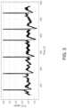
- FIG. 3is an exemplary electrocardiogram recording of a patient.
- FIG. 4is an exemplary Lomb-Scargle periodogram of a patient's heart cycle.
- FIG. 5is a schematic of a three-component model for use in determining coronary circulation boundary conditions.
- FIG. 6illustrates four different Windkessel models, specifically two-, three-, four- and five-element Windkessel models (2WM, 3WM, 4WM, 5WM), suitable for use in a blood circulatory system (BCS) component model.
- 2WMtwo-, three-, four- and five-element Windkessel models
- 3WMthree-, four- and five-element Windkessel models
- 4WM4WM
- 5WMfive-element Windkessel models
- FIG. 7illustrates several functional blocks (a-c) and an exemplary multi-block system (d) composed of functional block (b) for use in a blood circulatory system (BCS) component model.
- FIG. 8illustrates a blood circulatory system (BCS) model comprising systemic and pulmonary circulation elements, and its relation to an HPV component.
- BCSblood circulatory system
- FIG. 9illustrates a lumped parameter functional block comprising resistance, inertance, and capacitance (RLC) parameters that is suitable for use in a blood circulatory system (BCS) component model.
- RLCresistance, inertance, and capacitance
- FIG. 10illustrates schematic diagrams of (a) a heart-ventricle pressure-volume loop, (b) aortic pressure plotted as a function of time, and (c) ventricular volume plotted as a function of time.
- FIG. 11illustrates a functional block (a) and a whole heart pressure-volume (HPV) component model (b).
- FIG. 12is a graph showing reconstructed patient-specific heart ventricle volume and pressure during five heart cycles.
- FIG. 13illustrates a general coronary blood flow (CBF) model concept.
- FIG. 14illustrates six exemplary models suitable for use in a coronary blood flow (CBF) component model.
- CBFcoronary blood flow
- FIG. 15illustrates five different functional blocks (a)-(e) suitable for use in a multi-compartment coronary blood flow (CBF) model.
- CBFcoronary blood flow
- FIG. 16illustrates a set of parameters of a functional block suitable for use in a coronary blood flow (CBF) component model.
- CBFcoronary blood flow
- FIG. 17illustrates a lumped parameter multilayer/multicompartment model with describing parameters, suitable for use in a coronary blood flow (CBF) component model.
- CBFcoronary blood flow
- FIG. 18illustrates in detail a three-component model for use in determining coronary circulation boundary conditions including: a blood circulatory system (BCS) (pulmonary and systemic circulation) model component, a heart pressure-volume (HPV) model component, and a coronary blood flow (CBF) model component.
- BCSblood circulatory system
- HPVheart pressure-volume
- CBFcoronary blood flow
- FIG. 19is an example 3D mesh of a portion of a patient's blood vessel.
- FIG. 20illustrates a schematic for determining coronary circulation inflow and outflow boundary conditions.
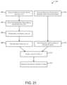
- FIG. 21is a schematic block diagram of a method for patient-specific modeling of hemodynamic parameters in coronary arteries using a steady-state simulation in accordance with one or more example embodiments of the disclosure.
- FIG. 22is a schematic block diagram of a method for patient-specific modeling of hemodynamic parameters in coronary arteries using a steady-state simulation in accordance with one or more example embodiments of the disclosure.
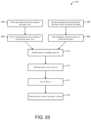
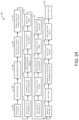
- FIG. 23is a schematic block diagram of a method for patient-specific modeling of hemodynamic parameters in coronary arteries using a transient simulation in accordance with one or more example embodiments of the disclosure.
- FIG. 24is a schematic block diagram of a method for patient-specific modeling of hemodynamic parameters in coronary arteries using a transient simulation in accordance with one or more example embodiments of the disclosure.
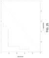
- FIG. 25is a receiver operating characteristic (ROC) curve comparing fractional flow reserve (FFR) results obtained using a three-component model variant to real-life results.
- ROCreceiver operating characteristic
- volumetric data of a patient's coronary arteriesmay be captured using non-invasive medical imaging techniques such as computed tomography angiography (CTA) or magnetic resonance angiography (MRA).
- CTAcomputed tomography angiography
- MRAmagnetic resonance angiography
- the volumetric datamay be used to create an anatomical model of the patient's coronary arteries suitable for a computational fluid dynamics (CFD) simulation.
- CFDcomputational fluid dynamics
- Continuous arterial pressure datamay be derived using non-invasive techniques.
- the continuous arterial pressure datamay be used to determine boundary conditions for the CFD simulation.
- Patient-specific CFD simulationsmay be performed using the coronary artery anatomical model, with the inlet and outlet boundary conditions determined from continuous arterial pressure data.
- Patient-specific hemodynamic parameters in the coronary arteriesmay be derived from the CFD simulations and may be used to characterize/assess cardiovascular disease, such as the functional assessment of stenosis in the patient.
- a CFD simulationmay be performed using a patient-specific coronary artery anatomical model derived from medical imaging data and patient-specific boundary conditions derived from continuous arterial pressure data to determine patient-specific hemodynamic parameters in a patient's coronary arteries.
- a three-component modelmay be used to determine coronary artery inflow boundary conditions for the CFD simulation.
- the three-component modelmay include a blood circulatory system (BCS) component that describes systemic and pulmonary blood circulation, a heart chambers pressure-volume (HPV) component that describes the relationship between ventricular or atrial pressure and volume, and a coronary blood flow (CBF) component that describes coronary tree blood circulation.
- BCSblood circulatory system
- HPVheart chambers pressure-volume
- CBFcoronary blood flow
- the three-component modelmay allow for determining the volumetric flow rate waveform at the inlet of the patient's coronary arteries.
- the determined volumetric flow rate waveform at the inlet of a patient's coronary arteriesmay be used to determine coronary artery outflow boundary conditions for the CFD simulation.
- the volumetric flow rate waveform at the inlet of a patient's coronary arteriesmay be used to determine the volumetric flow rate waveform at the outlet of the patient's coronary arteries using Murray's law or a similar allometric scaling law (see Sherman T (1981) On connecting large vessels to small—the meaning of murray's law. Journal of General Physiology, 78(4):431-453.).
- the patient-specific modeling of coronary artery blood flow in accordance with this disclosuremay utilize techniques that provide advantages over existing methods.
- the constructed patient-specific anatomical modelmay only model the patient's coronary arteries. That is, the constructed patient-specific anatomical model may not include, for example, reconstruction of the patient's aorta or an estimation of heart chamber volume. This may reduce numerical complexity and simulation time.
- the boundary conditionsmay be derived from noninvasively measured continuous arterial pressure data.
- Advantages of using pressure data to derive boundary conditionsinclude the ease with which pressure may be measured relative to other parameters typically used to determined boundary conditions (e.g., velocity, mass flux) and the robustness of pressure measurements, which are not vitiated by excessive error even when measured noninvasively and in a location far from the heart.
- coronary arteriesmay include not only the two main coronary arteries but also arterial branches depending therefrom and any plaques contained therein unless the context clearly dictates otherwise.
- FIGS. 1 and 2illustrate a method 100 for patient-specific modeling of hemodynamic parameters in coronary arteries in accordance with one or more example embodiments of the disclosure.
- the method 100may be performed within a computer or a computer system.
- a computermay include one or more non-transitory computer-readable storage medium that store instructions that, when executed by a processor, may perform any of the actions described herein for patient-specific modeling of hemodynamic parameters in coronary arteries.
- the computermay be, or the computer system may include, a desktop or portable computer, a mobile device (e.g., smartphone), a cloud-based computing system, a server, or any other computer.
- a computermay include a processor, a read-only memory (ROM), a random access memory (RAM), an input/output (I/O) adapter for connecting peripheral devices (e.g., an input device, output device, storage device, etc.), a user interface adapter for connecting input devices such as a keyboard, a mouse, a touch screen, and/or other devices, a communications adapter for connecting the computer to a network, and a display adapter for connecting the computer to a display.
- a displaymay be used to display any calculated hemodynamic parameters to a user (e.g., display images or three-dimensional models of a patient's coronary arteries overlaid with determined hemodynamic parameters).
- a computer systemmay receive patient-specific anatomical structure data.
- a computer systemmay receive the patient-specific anatomical structure data (e.g., image data acquired by a CT scanner or an X-ray device) over a communication network and/or from a computer readable storage medium.
- patient-specific anatomical structure datae.g., image data acquired by a CT scanner or an X-ray device
- the patient-specific anatomical structure datamay be 2D or 3D images (volumes) of a patient's circulatory system.
- the imagesmay include at least a portion of, or the entirety of, the patient's coronary arteries.
- the imagesmay or may not include other anatomical structures such as the patient's heart, aorta, and the like.
- the patient-specific anatomical structure datamay be obtained noninvasively using various noninvasive medical imaging modalities.
- the datamay be obtained using computed tomography (CT), computed tomography angiography (CTA), magnetic resonance imaging (MRI), or magnetic resonance angiography (MRA).
- CTcomputed tomography
- CTAcomputed tomography angiography
- MRImagnetic resonance imaging
- MRAmagnetic resonance angiography
- the patient-specific anatomical structure datamay be obtained using various invasive imaging methods such as rotational angiography, dynamic angiography, or digital subtraction angiography.
- the received patient-specific anatomical structure datamay be preprocessed by a user and/or by the computer system before further use. Preprocessing may include, for example, checking for misregistration, inconsistencies, or blurring in the captured image data, checking for stents shown in the captured image data, checking for other artifacts that may prevent the visibility of lumens of the coronary arteries, checking for sufficient contrast between anatomical structures (e.g., the aorta, the main coronary arteries, other blood vessels, and other portions of the patient).
- anatomical structurese.g., the aorta, the main coronary arteries, other blood vessels, and other portions of the patient.
- the user and/or computer systemmay be able to correct certain errors or problems with the data.
- Preprocessingmay also include using image processing techniques on the received patient-specific anatomical structure data to prepare the data for use in generating an anatomical model (e.g., preparing the data for segmentation).
- the image processingmay include, for example, adjusting contrast levels between different anatomical structures (e.g., the heart, the aorta, the coronary arteries, other vasculature, atherosclerotic plaques, etc.) in the images, smoothing of anatomical structures (e.g., applying a smoothing filter), and the like.
- a computer systemmay receive patient-specific physiological data.
- a computer systemmay receive the patient-specific physiological data over a communication network and/or from a computer readable storage medium.
- the patient-specific physiological datamay include continuous arterial pressure data (e.g., a continuously recorded blood pressure waveform).
- Continuous arterial blood pressureis time-varying and measured in real-time without any interruptions (e.g., continuously).
- a continuously recorded blood pressure waveformmay be obtained for a time period of approximately one (1) minute or a time period within a range of one (1) minute to two (2) minutes, although other continuous time periods may be used.
- the continuous arterial pressure datamay be obtained without a percutaneous procedure (e.g., noninvasively).
- the datamay be obtained using a NexfinTM monitor, a ClearSightTM monitor, a CNAPTM monitor, a Finapres® NOVA monitor or successor systems (e.g., Finometer® and Portapres® monitors), or other similar noninvasive continuous arterial pressure measuring devices.
- the continuous arterial pressure datamay be obtained using various invasive methods such as arterial catheterization.
- the continuous arterial pressure datamay undergo data processing (e.g., signal processing) to prepare the data for use in determining boundary conditions for a CFD simulation and/or simulating blood flood in an anatomical model using CFD.
- pressure signalsmay be extracted from the continuous arterial pressure data.
- the patient-specific physiological datamay include physiological data other than continuous arterial pressure data, such as the patient's heart electrical activity, baseline heart rate, height, weight, hematocrit, stroke volume, and the like.
- any physiological datamay undergo data processing (e.g., signal processing) to prepare the data for use in determining boundary conditions for a CFD simulation and/or simulating blood flood in an anatomical model using CFD.
- the physiological datamay include, for example, a continuous recording of an electrocardiography (ECG) signal from the patient, an example of which is shown in FIG. 3 .
- ECGelectrocardiography
- the ECG signalmay be used to directly reconstruct temporal heart cycle parameters such as a heart rate variability (e.g., an RR-interval).
- a heart rate variabilitye.g., an RR-interval
- the calculated average RR-interval for the patient's recordingis 0.897 s.
- the RR-intervalmay be used, for example, in determining boundary conditions for a CFD simulation.
- the physiological datamay include, for example, aortic pressure course.
- Aortic pressure coursemay be used to indirectly determine temporal heart cycle parameters when a patient's ECG signal is unavailable, although this is slightly less accurate when compared to ECG.
- a Lomb-Scargle algorithmmay be used to construct a Lomb-Scargle periodogram of a patient's heart cycle from aortic pressure course, an example of which is shown in FIG. 4 .
- the Lomb-Scargle algorithmmay be used to find and test the significance of weak periodic signals with uneven temporal sampling (see Townsend R H D (2010) Fast calculation of the Lomb-Scargle periodogram using graphics processing units.
- the Astrophysical Journal, Supplement Series, Vol. 191, 247-253.The Astrophysical Journal, Supplement Series, Vol. 191, 247-253.
- the calculated RR-interval for the patient's pressure recording using the Lomb-Scargle algorithmis 0.901 s.
- the RR-interval calculated using the Lomb-Scargle algorithmis slightly different than the RR-interval determined from ECG data, but the difference is less than 0.5%.
- a computer systemmay generate a patient-specific anatomical model of the patient's coronary arteries from the received patient-specific anatomical structure data.
- the patient-specific anatomical modelmay be a 3D geometric model of the patient's coronary arteries.
- the constructed patient-specific anatomical modelmay only model the patient's coronary arteries. That is, the constructed patient-specific anatomical model may not include, for example, reconstruction of the patient's heart, aorta, non-coronary artery related vasculature, or other tissues.
- Received patient-specific anatomical structure datamay include regions of varying optical density that correspond to different anatomical structures such as the aorta, the main coronary arteries, coronary artery branches, myocardium, and the like.
- the locations of anatomical structure surfacesmay be determined based on the contrast (e.g., relative darkness and lightness) between different anatomical structures.
- the contrast between anatomical structuresmay also enable the selective modeling of certain anatomical features (e.g., coronary arteries) while excluding other anatomical features from the generated model (e.g., the heart).
- the process of forming the patient-specific anatomical modelis generally referred to as segmentation. Segmentation may be performed automatically by the computer system with or without user input.
- the coronary arteriesmay be segmented in the patient-specific anatomical structure data using any suitable coronary artery segmentation method.
- Methods for generating an anatomical model of a patient's coronary arteriesare described, for example, in U.S. Patent Application Nos. 2010/006776 and 2012/0072190 and U.S. Pat. Nos. 7,860,290, 7,953,266, and 8,315,812, each of which are incorporated herein by reference in their entirety for all purposes.
- the segmented coronary arteriesmay be reviewed and/or corrected by the computer system and/or a user, if necessary (e.g., to correct the segmentation if there are any errors such as missing or inaccurate coronary arteries or branches extending therefrom).
- the patient-specific anatomical modelmay be represented as a surface mesh.
- the surface meshmay represent the external boundary of segmented structures such that their shape is represented as a set of connected vertices (e.g., a mesh).
- shape constraintsmay be imposed using mesh-based shape metrics or statistics.
- a deformable modelsuch as an Active Mesh Model (AMM) (see Dufour, A. et al., Segmenting and tracking fluorescent cells in dynamic 3-D microscopy with coupled active surfaces. IEEE Transactions on Image Processing, 14(9), 1396-1410, 2005; Dufour, A. et al., J.-C.
- 3-D active meshesfast discrete deformable models for cell tracking in 3-D time-lapse microscopy. IEEE Transactions on Image Processing, 20(7), 1925-1937, 2011.), may be a starting point for creating the patient-specific anatomical model.
- AMMis 3D extension of the active contour model (ACM) used in image analysis techniques (see Kass, M. et al., Active contour models. Int. J. of Computer Vision 1(4), 321-331, 1988.).
- AMM-based methodssegmented structures may be represented as closed surfaces (fronts, meshes) that evolve with a speed computed from both image-dependent data and image-independent geometric properties.
- the process for forming the patient-specific anatomical modelmay include, for example, segmenting visible plaques in coronary arteries using an AMM-based method, selecting by a computer and/or user root points (e.g., starting points) for the left and right coronary arteries, segmenting the coronary arteries using the AMM-based method and selected root points, and verifying and/or correcting the geometry of the segmented plaques and arteries.
- root pointse.g., starting points
- a user and/or computer systemmay post-process the patient-specific anatomical model to prepare the model for CFD simulations. This may include, for example, determining centerlines for the coronary arteries and their branches, determining cross-sectional areas of the coronary arteries and their branches, creating models of inflow boundaries (e.g., the boundaries through which flow is directed into the coronary arteries) and outflow boundaries (e.g., the boundaries through which flow is directed out of the coronary arteries and/or coronary artery branches) such that the inflow boundaries and the outflow boundaries are perpendicular to the determined centerlines, thereby permitting boundary condition application, and smoothing the model (e.g. smoothing any ridges, points, etc).
- the post-processing of the patient-specific anatomical modelmay be reviewed and/or corrected by the computer system and/or the user, if necessary.
- a computer systemmay determine boundary conditions for a computational fluid dynamics (CFD) simulation of blood flow in the anatomical model. At least some of the boundary conditions may be determined using received patient-specific physiological data, such as received continuous arterial pressure data.
- the boundary conditionsmay include coronary circulation inflow and outflow boundary conditions.
- a three-component modelmay be used in determining coronary circulation boundary conditions.
- the three-component modelmay include a blood circulatory system (BCS) component that describes systemic and pulmonary blood circulation, a heart pressure-volume (HPV) component that describes a cardiac pressure-volume loop, and a coronary blood flow (CBF) component that describes coronary artery blood circulation (see FIG. 5 ).
- BCSblood circulatory system
- HPVheart pressure-volume
- CBFcoronary blood flow
- Each of the BCS, HPV, and CBF componentsmay be selected from various models of each component, which are discussed in more detail below.
- the three-component modelmay take as an input the pressure waveform p sa (t), which may be derived from the patient-specific continuous recording of arterial pressure (e.g., patient-specific continuous arterial pressure data).
- An exemplary embodiment of a three-component modelis shown in FIG. 18 .
- the three-component modelmay be used to directly determine inflow boundary conditions, such as the volumetric flow rate waveform at the inlet of the patient's coronary arteries (see FIG. 20 ).
- the three-component modelmay be used to indirectly determine outflow boundary conditions, such as the volumetric flow rate waveform at the outlet of the patient's coronary arteries (see FIG. 20 ).
- the volumetric flow rate waveform at the inlet of the patient's coronary arteriesmay be used to determine the volumetric flow rate waveform at the outlet of the patient's coronary arteries using an allometric law of scaling (ALS) such as Murray's law, which describes a relationship between blood flow and vessel radius (see FIG.
- ALSallometric law of scaling
- the blood circulatory system (BCS) componentdescribes systemic and pulmonary blood circulation.
- Blood circulationmay be modeled, for example, using a two-, three-, four-, or five-element Windkessel (2WM, 3WM, 4WM, 5WM) lumped functional block, which are shown in FIG. 6 (see Garcia D et al. (2009) Impairment of coronary flow reserve in aortic stenosis. Journal of Applied Physiology, Vol. 106, No. 1, 113-121; Li J K-J (2000) The Arterial Circulation. Physical Principles and Clinical Applications, Springer, New York; Ostadfar A (2016) Biofluid mechanics. Principles and applications. Elsevier; Pappano A et al.
- Pulmonary and systemic circulationmay be modeled, in a preferred embodiment, using one of the lumped parameter models shown in FIG. 7 , while overall blood circulation may be modeled using a multi-compartment model shown in FIG. 8 .
- the blood circulatory system model componente.g. the systemic and pulmonary circulation model
- Rresistance
- Linductance
- Ccapacitor
- the block inputs (in) and output (out)are related in time (t):
- the resulting system of sixteen equationsmay be solved numerically.
- the heart ventricle or atrium pressure-volume (HPV) componentdescribes a cardiac pressure-volume loop.
- the heart cycleconsists of four phases, as shown in FIG. 10 (see Barrett K E et al. (2016) Ganong's review of medical physiology, McGraw-Hill; Mohrman D et al. (2013) Cardiovascular physiology. McGraw-Hill, Lange, New York; Pappano A et al. (2013) Cardiovascular physiology. Elsevier.).
- Many different modelsmay be used for the isovolumetric systolic and diastolic phases such as, for example, a time varying-elastance model (TVE), a time-varying compliance (TVC) model, or other models (see Garcia D et al.
- TVEtime varying-elastance model
- TVCtime-varying compliance
- FIG. 11illustrates a functional block for building a heart chambers pressure-volume (HPV) component model (a); and a whole, multi-compartment heart chambers pressure-volume (HPV) component model (b).
- HPVpressure-volume
- the pressure-volume (HPV) componentuses a model based on the idea of varying elastance E(t) as a reciprocal of compliance, which may be written in the form:
- V(t)is the heart chamber volume
- V ois a volume intercept.
- ⁇ t nt ⁇ % ⁇ T t max
- Tis the heart cycle duration according to an RR-interval, which may be determined by ECG or estimated from aortic pressure course.
- Typical values of time-varying elastance model empirical parametersare provided in the table below (see Stergiopulos N et al.
- a time-varying elastance modelmay only be used during a heart cycle's isovolumetric phases.
- blood volumeis partially accumulated in the atrium while the rest—followed by the transvalvular pressure gradient—flows out. Therefore, the atrial flow rate balance can be described as:
- ventricular flow rateC RA ⁇ dp sv dt + q t
- q pvC LA ⁇ dp pv dt + q m
- transvalvular flowcan be described as:
- a patient-specific calibration (PSC) proceduremay be used for the optimal parameter estimation of the HPV and BCS models.
- the proceduremay include: (i) determining initial approximations of model parameters from patient systolic and diastolic pressure levels, gender, age, and heart rate (HR) (see Barrett K E et al. (2016) Ganong's review of medical physiology, McGraw-Hill; Li J K-J (2000) The Arterial Circulation. Physical Principles and Clinical Applications, Springer, New York; Pappano A et al. (2013) Cardiovascular physiology. Elsevier; Zamir M (2005) The physics of coronary blood flow. Springer-Verlag; Maceira A M et al.
- a time-varying elastance model(e.g., applied in the HPV model) in conjunction with a circulation model (BCS) may be used to reconstruct left and right heart instantaneous ventricle volumes (V) and internal pressures (p V ) course using a patient's recorded aortic pressure (p sa ), as shown in FIG. 12 .
- the coronary blood flow (CBF) componentdescribes coronary artery blood circulation, and is shown generally in FIG. 13 .
- the CBF componentderives from several conclusions drawn from physiology findings (see Epstein S et al. (2015) Reducing the number of parameters in 1D arterial blood flow modeling: less is more for patient-specific simulations.
- the CBF component shown generally in FIG. 13is suitable for determining boundary conditions for CFD simulations of flow in coronary arteries.
- the CBF componentspecifies that flow in the coronary artery inlet q 0 (t) results from forcing aortic pressure p sa (t) throttled by heart contraction and reverse accumulation, the latter determined mainly by ventricular pressure.
- the CBF componentdescribes a causal relationship, with pressure acting as an independent variable. Because pressure serves as the independent variable in the CBF component, the CBF component and its use in patient-specific computational modeling is advantageous over other techniques for determining boundary conditions. Some advantages of using pressure as the independent variable include: (i) pressure is relatively easy to measure when compared to velocity or mass flux, which are much more challenging to measure; and (ii) pressure measurements, even noninvasive and in a location far from heart, will not be vitiated by excessive error.
- Coronary blood flowmay be modeled in many different ways (see Beyar R et al. (1987) Time-dependent coronary blood flow distribution in left ventricular wall. American Journal of Physiology, Heart and Circulatory Physiology, Vol. 252, No. 2, Pt. 2, H417-H433; Boileau E et al. (2015) One-Dimensional Modelling of the Coronary Circulation. Application to Noninvasive Quantification of Fractional Flow Reserve (FFR). Computational and Experimental Biomedical Sciences: Methods and Applications, Vol. 21, 137-155; Bruinsma T et al. (1988) Model of the coronary circulation based on pressure dependence of coronary resistance and compliance.
- the capacitive branchmay include its own resistive element (d) (see Garcia D et al. (2009) Impairment of coronary flow reserve in aortic stenosis. Journal of Applied Physiology, Vol. 106, No. 1, 113-121; Judd R M et al. (1991) Coronary input impedance is constant during systole and diastole.
- the resistive branchusually includes its own source related to intraventricular pressure (a,b,c,d,e) (see Beyar R et al. (1987) Time-dependent coronary blood flow distribution in left ventricular wall. American Journal of Physiology, Heart and Circulatory Physiology, Vol. 252, No. 2, Pt. 2, H417-H433; Boileau E et al. (2015) One-Dimensional Modelling of the Coronary Circulation. Application to Noninvasive Quantification of Fractional Flow Reserve (FFR). Computational and Experimental Biomedical Sciences: Methods and Applications, Vol. 21, 137-155; Bruinsma T et al.
- coronary blood flowis modeled using the lumped functional block shown in FIG. 16 .
- Use of the coronary blood flow model shown in FIG. 16may require solving the following mass flux conservation equation:
- Coronary arteriesare spatially distributed in the heart wall and affected by extracellular pressure in a non-uniform manner, and they may be additionally moderated by physical or pharmacological stress conditions—especially hyperemia by administration of adenosine receptors (purinergic P1 receptors) agonists such as Adenocard or Adenoscan or more selective agonist of A2A receptor (Regadenoson, Binodenoson).
- adenosine receptorspurinergic P1 receptors
- agonistssuch as Adenocard or Adenoscan or more selective agonist of A2A receptor (Regadenoson, Binodenoson).
- the effect of heart wall heterogeneitymay be described by utilizing a multilayer and multi-compartment model with a variable tissue pressure coefficient (see Garcia D et al. (2009) Impairment of coronary flow reserve in aortic stenosis.
- FIG. 17According to FIG. 17 :
- vasodilating effect related to elimination of active coronary vasomotor tonemay not be limited to heart tissue and function. More generally, vasodilation is just one of the cardiac tropism form (chronotropism, inotropism, lusitropism, and many others). Furthermore, endogenous and/or exogenous mediators may cause a decrease in vascular resistance and allow an increase in coronary blood flow—as well as—systemic and pulmonary blood flow.
- net cardiac tropism effects (E/E max ) of purinergic receptor (R) binding endo- or exogenous agonists (A)may be modeled by the cooperative kinetics relation
- a computer systemmay simulate blood flow in the patient-specific anatomical model (e.g., the coronary arteries) using CFD and the patient-specific boundary conditions.
- the CFD simulationmay use the coronary volumetric flow rate waveform at the inlets and/or outlets of the coronary arteries, which may be determined at least in part by patient-specific continuous arterial pressure data, as boundary conditions for the CFD modeling.
- a 3D meshmay be created for the patient specific anatomical model, together with separate inflow and outflow boundary models, to enable the CFD simulation (e.g., create a 3D computational grid for numerical simulations).
- the 3D meshmay include a plurality of nodes (e.g., meshpoints or gridpoints) along the surfaces of the patient-specific anatomical model and throughout the interior of the patient-specific anatomical model (see FIG. 19 ).
- the generated meshmay be reviewed and/or corrected by the computer system and/or the user, if necessary (e.g., to correct mesh distortions, insufficient spatial resolution in the mesh, etc.).
- bloodmay be modeled as a Newtonian fluid or a non-Newtonian fluid, and the flow fields may be obtained by numerically solving the discretized mass and momentum (Navier-Stokes) balance equations under the rigid wall assumption.
- Numerical methods to solve the three-dimensional equations of blood flowmay include finite difference, finite volume, spectral, lattice Boltzmann, particle-based, level set, isogeometric, or finite element methods, or other computational fluid dynamics (CFD) numerical techniques.
- the discretized Navier-Stokes equationsmay be used to incrementally simulate velocity of blood flow and pressure within the coronary arteries over time. That is, the CFD simulation may determine blood flow and pressure at each of the nodes of the meshed anatomical model.
- the result of the CFD simulationsmay be patient-specific blood flow and pressure distribution in the patient's coronary arteries based on patient-specific anatomy and patient-specific boundary conditions.
- a computer systemmay determine one or more hemodynamic parameters associated with the patient's coronary arteries.
- the one or more hemodynamic parametersmay be determined based at least in part on the CFD simulation results.
- Examples of hemodynamic parametersmay include coronary artery characteristics such as blood pressure, blood flow rate, wall shear stress (WSS), oscillatory shear index (OSI), relative residence time (RRT), fractional flow reserve (FFR), coronary flow reserve (CFR), instantaneous wave-free ratio (iFR), and the like.
- the hemodynamic parametersmay be interpolated across the patient-specific anatomical model to provide a user with information about the hemodynamic parameters across the anatomical model.
- a computer systemmay output the one or more determined hemodynamic parameters.
- the computer systemmay, for example, display the one or more hemodynamic parameters or visualizations (e.g., 2D or 3D images) of the one or more hemodynamic parameters.
- the computer systemmay, for example, present the hemodynamic parameters as a three-dimensional interactive visualization.
- the computer systemmay send the one or more determined hemodynamic parameters to a remote computer for display on the remote computer.
- the one or more determined hemodynamic parametersare used to determine and/or as part of a patient-specific treatment plan.
- the one or more determined hemodynamic parametersare used to plan a coronary revascularization procedure in cardiovascular disease.
- the one or more determined hemodynamic parametersmay be used to determine an optimal, patient-specific location for stent placement in a patient that improves hemodynamic conditions for blood flow in the patient's coronary arteries, and then the stent is positioned at the determined optimal location.
- the one or more determined hemodynamic parametersmay be used to determine an optimal coronary artery bypass procedure in a patient that provides better hemodynamic conditions for coronary artery flow in the patient when compared to alternative coronary artery bypass procedures, and then a physician performs the optimal coronary artery bypass procedure in the patient.
- the one or more determined hemodynamic parametersare used in support of a virtual cardiopulmonary exercise test.
- the one or more determined hemodynamic parametersmay include a fractional flow reserve (FFR) estimation, which can be used to provide a non-invasive estimation of fractional flow reserve and/or oxygen blood saturation during virtual cardiopulmonary exercise test conditions.
- FFRfractional flow reserve
- Blood flow through the coronary arteriesis pulsatile. Its pressure and velocity are changing in time during a single heart beat and this process is repetitive. The most straightforward way of simulating such a flow is to use a transient solver, but this may be very time consuming. Use of a steady-state (e.g., stationary) simulation may be advantageous as its time-to-solution is relatively shorter but it is not applicable to every non-stationary phenomena.
- coronary arteriesmay be treated as a pipeline system.
- the pressure drop ⁇ pis dependent on fluid velocity v.
- three steady-state simulationscan be run for various pressure and velocity (calculated from flow rate) value boundary conditions and the pressure drop values respective to those velocities can be found. As those simulations are independent, they may be run in parallel. This allows for a great reduction of time-to-solution.
- results of a transient simulation which take tens of hours to completemay be obtained from a stationary simulation in less than an hour.
- an additional termwas added to the equation for the pressure drop (see Bird R B et al. (1960) Transport Phenomena. John Wiley & Sons, New York; Young D et al. (1973) Flow characteristics in models of arterial stenoses. II. Unsteady flow, Journal of Biomechanics, Vol. 6, No. 5, 547-559; Young D et al. (1977) Hemodynamics of arterial stenoses at elevated flow rates. Circulation Research, Vol. 41, No. 1, 99-107.):
- ⁇ ⁇ pav 2 + bv + c + kl ⁇ dv dt
- FIGS. 21 - 24show low-detail or high detail schematic block diagrams of a method for patient-specific modeling of hemodynamic parameters in coronary arteries using a steady-state simulation or a transient simulation. As shown in FIGS. 21 - 24 , there are a few differences between a steady-state simulation based method and a transient simulation based method. However, many of the implementation details for a steady-state simulation based method can be applied to a transient simulation based method, and vice versa.
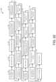
- FIGS. 21 - 22shown are a low-detail or high detail schematic block diagram of a method 200 for patient-specific modeling of hemodynamic parameters in coronary arteries using a steady-state simulation.
- step 202patient-specific anatomical data is obtained and pre-processed.
- step 204a three-dimensional model is created based on the obtained anatomical data.
- step 206the three-dimensional model is prepared for numerical analysis.
- step 208a computational analysis is performed using the three-dimensional model.
- step 210patient-specific peripheral artery pressure recording data is obtained and preprocessed.
- step 212boundary conditions are created based on the pressure recording data.
- step 214the results of the computational analysis and boundary conditions are assembled and output.
- step 216a patient-specific treatment plan is prepared based on the results.
- step 302acquired patient-specific anatomical data (e.g., CT data) is initially reviewed.
- step 304the acquired anatomical data undergoes image processing.
- step 306which marks the beginning of creating a three-dimensional model from the obtained anatomical data
- plaqueis segmented.
- step 308coronary artery root points are selected.
- step 310the coronary arteries are segmented.
- step 312the quality of the segmentation is checked.
- step 314the artery centerlines are automatically found.
- step 316inflow and outflow boundary models are created.
- step 318the solid model is output and smoothed.
- step 320the output solid model is verified.
- step 322which marks the beginning of preparing the solid model for numerical analysis, a final mesh of the model is generated.
- step 324the mesh is verified.
- step 326which marks the beginning of performing the computational analysis, a set of CFD cases is prepared for numerical analysis.
- step 328the set of CFD cases is solved by flow simulations.
- step 330the simulation results are verified.
- acquired patient-specific anatomical datae.g., recorded pressure data
- step 334which begins the creation of boundary conditions based on the recorded pressure data, pressure data is input to a blood circulation system model.
- step 336results from the blood circulation system model are input into a heart chambers model.
- step 338results from the heart chambers model are input into a coronary blood flow model, the outputs of which are used to determine boundary conditions.
- step 340the results of the boundary condition determination are verified.
- step 342the results of the boundary condition determination and computational fluid dynamics analysis are assembled.
- step 344the assembled results are output.
- FIGS. 23 - 24shown are a low-detail or high detail schematic block diagrams of a method 400 for patient-specific modeling of hemodynamic parameters in coronary arteries using a transient simulation.
- step 402patient-specific anatomical data is obtained and pre-processed.
- step 404a three-dimensional model is created based on the obtained anatomical data.
- step 406patient-specific peripheral artery pressure recording data is obtained and preprocessed.
- step 408boundary conditions are created based on the pressure recording data.
- step 410the three-dimensional model is prepared for numerical analysis.
- step 412a computational analysis is performed using the three-dimensional model and boundary conditions.
- step 414the results of the computational analysis are output.
- step 416a patient-specific treatment plan is prepared based on the results.
- step 502acquired patient-specific anatomical data (e.g., CT data) is initially reviewed.
- step 504the acquired anatomical data undergoes image processing.
- step 506which marks the beginning of creating a three-dimensional model from the obtained anatomical data
- plaqueis segmented.
- step 508coronary artery root points are selected.
- step 510the coronary arteries are segmented.
- step 512the quality of the segmentation is checked.
- step 514the artery centerlines are automatically found.
- step 516inflow and outflow boundary models are created.
- step 518the solid model is output and smoothed.
- step 520the output solid model is verified.
- step 522acquired patient-specific anatomical data (e.g., recorded pressure data) is initially reviewed.
- step 524which begins the creation of boundary conditions based on the recorded pressure data, pressure data is input to a blood circulation system model.
- results from the blood circulation system modelare input into a heart chambers model.
- results from the heart chambers modelare input into a coronary blood flow model, the outputs of which are used to determine boundary conditions.
- step 530the results of the boundary condition determination are verified.
- step 532which marks the beginning of preparing the solid model for numerical analysis, a final mesh of the model is generated.
- step 534the mesh is verified.
- step 536which marks the beginning of performing the computational analysis, a CFD case is prepared for numerical analysis.
- step 538the CFD case is solved by flow simulation.
- step 540the simulation results are verified.
- step 542the results are output.
- Results from a method for patient-specific modeling of hemodynamic parameters in coronary arteries in accordance with one or more example embodiments of the disclosurewere compared to real life results.
- invasively collected FFR data from 30 patients in 3 hospitalswas compared to numerically calculated FFR values using one or more example embodiments of the disclosure.
- the statistical results for a total of 35 stenosesare summarized in the table below and in FIG. 25 .
Landscapes
- Health & Medical Sciences (AREA)
- Life Sciences & Earth Sciences (AREA)
- Engineering & Computer Science (AREA)
- Medical Informatics (AREA)
- Public Health (AREA)
- General Health & Medical Sciences (AREA)
- Biomedical Technology (AREA)
- Surgery (AREA)
- Pathology (AREA)
- Heart & Thoracic Surgery (AREA)
- Veterinary Medicine (AREA)
- Molecular Biology (AREA)
- Animal Behavior & Ethology (AREA)
- Physics & Mathematics (AREA)
- Biophysics (AREA)
- Nuclear Medicine, Radiotherapy & Molecular Imaging (AREA)
- Radiology & Medical Imaging (AREA)
- Cardiology (AREA)
- Physiology (AREA)
- High Energy & Nuclear Physics (AREA)
- Optics & Photonics (AREA)
- Epidemiology (AREA)
- Primary Health Care (AREA)
- Vascular Medicine (AREA)
- Computer Vision & Pattern Recognition (AREA)
- Dentistry (AREA)
- Oral & Maxillofacial Surgery (AREA)
- Data Mining & Analysis (AREA)
- Databases & Information Systems (AREA)
- Hematology (AREA)
- Pulmonology (AREA)
- Robotics (AREA)
- Theoretical Computer Science (AREA)
- Measuring Pulse, Heart Rate, Blood Pressure Or Blood Flow (AREA)
- Apparatus For Radiation Diagnosis (AREA)
Abstract
Description
where: q is flow rate and p is the pressure of flowing blood in a selected compartment. As shown in
p(t)=E(t)·(V(t)−V0).
where V(t) is the heart chamber volume, and Vois a volume intercept.
and T is the heart cycle duration according to an RR-interval, which may be determined by ECG or estimated from aortic pressure course. Typical values of time-varying elastance model empirical parameters are provided in the table below (see Stergiopulos N et al. (1996) Determinants of stroke volume and systolic and diastolic aortic pressure. American Journal of Physiology, Vol. 270, No. 6, Pt. 2, H2050-H2059; Faragallah G et al. (2012) A new control system for left ventricular assist devices based on patient-specific physiological demand. Inverse Problems in Science and Engineering, Vol. 20, No. 5, 721-734.).
| Emin | Emax | a1 | a2 | n1 | n2 | ||
| 0.06 | 2.31 | 0.303 | 0.508 | 1.32 | 21.9 | ||
| 0.06 | 2.00 | 0.700 | 1.170 | 1.90 | 21.9 | ||
and similarly the ventricular flow rate can be described as:
Simultaneously, transvalvular flow can be described as:
where H is Heaviside step function.
where H is the Heaviside step function. The throttling pressure pRas well as pCdescribes myocardium-coronary vessel interaction (MVI), wherein pC=kC·(CEP+SIP) and pR=kR(CEP+SIP) (see Boileau E et al. (2015) One-Dimensional Modelling of the Coronary Circulation. Application to Noninvasive Quantification of Fractional Flow Reserve (FFR). Computational and Experimental Biomedical Sciences: Methods and Applications, Vol. 21, 137-155; Bruinsma T et al. (1988) Model of the coronary circulation based on pressure dependence of coronary resistance and compliance. Basic Res Cardiol, 83:510-524; Epstein S et al. (2015) Reducing the number of parameters in 1D arterial blood flow modeling: less is more for patient-specific simulations. American Journal of Physiology, Heart and Circulatory Physiology, Vol. 309, No. 1, H222-H234; Mohrman D et al. (2013) Cardiovascular physiology. McGraw-Hill, Lange, New York; Pappano A et al. (2013) Cardiovascular physiology. Elsevier; Zamir M (2005) The physics of coronary blood flow. Springer-Verlag.). There are three main hypotheses of the passive interaction mechanism, and the extravascular pressure description can include (see Algranati D et al. (2010) Mechanisms of myocardium-coronary vessel interaction. American Journal of Physiology. Heart and Circulatory Physiology, Vol. 298, No. 3, H861-H873; Mynard J P et al. (2014) Scalability and in vivo validation of a multiscale numerical model of the left coronary circulation. American Journal of Physiology. Heart and Circulatory Physiology, Vol. 306, No. 4, H517-H528; Westerhof N et al. (2006) Cross-talk between cardiac muscle and coronary vasculature. Physiological Reviews, Vol. 86, No. 4, 1263-1308): (i) interstitial, cavity-induced extracellular pressure (CEP=μ1·pV), and (ii) shortening-induced intracellular pressure (SIP=μ2·EV). The instantaneous heart left (or right, respectively) ventricle pressure pVand elastance EVmay be taken from the HPV component and the zero flow pressure pzfmay be assumed to equal 20 mmHg or less.
where the heart tissue pressure coefficient is:
During resting condition, extravascular pressure decreases nonlinearly, concave downward from endocardium to epicardium with exponent k≈2.0 or greater. Contrary to this, at the cassation of any active coronary vasomotor tone (hypothetical maximum coronary dilation) the linear relationship can be assumed (k≈1.0).
where concentration of occupied receptors
Combining these equations and introduction transducer ratio τ=[R0]/KE, we get explicit relation
cooperative purinergic receptor-stimulus model of agonism (using affinities KA, and efficacies KE).
where: a, b, c—coefficients calculated based on stationary simulations, k=1.2—inertia coefficient, l—distance from inlet.
| Sensitivity | 82.4% | ||
| Specificity | 88.9% | ||
| Positive Predictive Value | 87.5% | ||
| Negative Predictive Value | 84.2% | ||
| Accuracy | 85.7% | ||
| Area under ROC curve | 0.863 | ||
Claims (20)
Priority Applications (2)
| Application Number | Priority Date | Filing Date | Title |
|---|---|---|---|
| US16/217,328US11871995B2 (en) | 2017-12-18 | 2018-12-12 | Patient-specific modeling of hemodynamic parameters in coronary arteries |
| US18/509,201US20240081910A1 (en) | 2017-12-18 | 2023-11-14 | Patient-specific modeling of hemodynamic parameters in coronary arteries |
Applications Claiming Priority (2)
| Application Number | Priority Date | Filing Date | Title |
|---|---|---|---|
| US201762599930P | 2017-12-18 | 2017-12-18 | |
| US16/217,328US11871995B2 (en) | 2017-12-18 | 2018-12-12 | Patient-specific modeling of hemodynamic parameters in coronary arteries |
Related Child Applications (1)
| Application Number | Title | Priority Date | Filing Date |
|---|---|---|---|
| US18/509,201ContinuationUS20240081910A1 (en) | 2017-12-18 | 2023-11-14 | Patient-specific modeling of hemodynamic parameters in coronary arteries |
Publications (2)
| Publication Number | Publication Date |
|---|---|
| US20190183579A1 US20190183579A1 (en) | 2019-06-20 |
| US11871995B2true US11871995B2 (en) | 2024-01-16 |
Family
ID=66815371
Family Applications (2)
| Application Number | Title | Priority Date | Filing Date |
|---|---|---|---|
| US16/217,328Active2042-07-20US11871995B2 (en) | 2017-12-18 | 2018-12-12 | Patient-specific modeling of hemodynamic parameters in coronary arteries |
| US18/509,201PendingUS20240081910A1 (en) | 2017-12-18 | 2023-11-14 | Patient-specific modeling of hemodynamic parameters in coronary arteries |
Family Applications After (1)
| Application Number | Title | Priority Date | Filing Date |
|---|---|---|---|
| US18/509,201PendingUS20240081910A1 (en) | 2017-12-18 | 2023-11-14 | Patient-specific modeling of hemodynamic parameters in coronary arteries |
Country Status (1)
| Country | Link |
|---|---|
| US (2) | US11871995B2 (en) |
Families Citing this family (11)
| Publication number | Priority date | Publication date | Assignee | Title |
|---|---|---|---|---|
| US10679350B2 (en)* | 2018-08-10 | 2020-06-09 | Koninklijke Philips N.V | Method and apparatus for adjusting a model of an anatomical structure |
| US11967435B2 (en)* | 2019-02-28 | 2024-04-23 | Medstar Health, Inc. | Modeling of flow through a left ventricular assist device (LVAD) |
| KR102190431B1 (en)* | 2020-03-18 | 2020-12-11 | 연세대학교 산학협력단 | Method and Apparatus for Diagnosing Vascular Disease |
| JP7445569B2 (en)* | 2020-09-15 | 2024-03-07 | キヤノンメディカルシステムズ株式会社 | Image processing device |
| CN112711881B (en)* | 2020-12-29 | 2023-03-31 | 深圳技术大学 | Physiological dynamic blood flow simulation method, device, computer equipment and storage medium |
| JP2025508520A (en)* | 2022-03-03 | 2025-03-26 | ヘモレンズ ダイアグノスティックス スプーカ ス オグラニチョノ オツポヴィエジャルノシチョン | Assessment of myocardial blood flow using non-invasive measurements and porous structure engineering |
| WO2024010616A1 (en)* | 2022-07-05 | 2024-01-11 | Cooper Health System | Non-invasive system and method for detecting and accurately quantifying subclinical and clinical systolic and diastolic heart failure |
| EP4310859A1 (en) | 2022-07-10 | 2024-01-24 | Tata Consultancy Services Limited | Method and system for determining progression of atrial fibrillation based on hemodynamic metrics |
| CN120379587A (en)* | 2022-12-09 | 2025-07-25 | 赫莫伦斯诊断有限责任公司 | Method for calculating the pressure drop between the cross sections of arteries |
| CN116746893B (en)* | 2023-06-16 | 2024-05-31 | 上海博动医疗科技股份有限公司 | Vascular pressure difference calculation method and device based on single-bit radiography data |
| CN117679059B (en)* | 2024-02-01 | 2024-04-26 | 北京大学第三医院(北京大学第三临床医学院) | System and method for quantifying functional hemodynamic parameters |
Citations (66)
| Publication number | Priority date | Publication date | Assignee | Title |
|---|---|---|---|---|
| US6236878B1 (en) | 1998-05-22 | 2001-05-22 | Charles A. Taylor | Method for predictive modeling for planning medical interventions and simulating physiological conditions |
| US20100006776A1 (en) | 2000-05-17 | 2010-01-14 | Nec Corporation | Semiconductor thin film forming system |
| US7860290B2 (en) | 2006-04-21 | 2010-12-28 | Siemens Medical Solutions Usa, Inc. | Three-dimensional (3D) modeling of coronary arteries |
| US7953266B2 (en) | 2007-02-06 | 2011-05-31 | Siemens Medical Solutions Usa, Inc. | Robust vessel tree modeling |
| US20120041739A1 (en)* | 2010-08-12 | 2012-02-16 | Heartflow, Inc. | Method and System for Patient-Specific Modeling of Blood Flow |
| US20120072190A1 (en) | 2010-09-16 | 2012-03-22 | Siemens Corporation | Method and System for Non-Invasive Assessment of Coronary Artery Disease |
| US8548778B1 (en) | 2012-05-14 | 2013-10-01 | Heartflow, Inc. | Method and system for providing information from a patient-specific model of blood flow |
| US20140073977A1 (en) | 2012-09-12 | 2014-03-13 | Heartflow, Inc. | Systems and methods for estimating blood flow characteristics from vessel geometry and physiology |
| US20140073976A1 (en) | 2012-09-12 | 2014-03-13 | Heartflow, Inc. | Systems and methods for estimating ischemia and blood flow characteristics from vessel geometry and physiology |
| US8824752B1 (en) | 2013-03-15 | 2014-09-02 | Heartflow, Inc. | Methods and systems for assessing image quality in modeling of patient anatomic or blood flow characteristics |
| US8837860B1 (en) | 2014-03-28 | 2014-09-16 | Heartflow, Inc. | Systems and methods for data and model-driven image reconstruction and enhancement |
| US8958623B1 (en) | 2014-04-29 | 2015-02-17 | Heartflow, Inc. | Systems and methods for correction of artificial deformation in anatomic modeling |
| US20150065847A1 (en) | 2013-08-27 | 2015-03-05 | Heartflow, Inc. | Systems and methods for predicting location, onset, and/or change of coronary lesions |
| US9008392B1 (en) | 2013-09-25 | 2015-04-14 | Heartflow, Inc. | Systems and methods for visualizing elongated structures and detecting branches therein |
| US9042613B2 (en) | 2013-03-01 | 2015-05-26 | Heartflow, Inc. | Method and system for determining treatments by modifying patient-specific geometrical models |
| US9043190B2 (en) | 2013-08-16 | 2015-05-26 | Heartflow, Inc. | Systems and methods for identifying personalized vascular implants from patient-specific anatomic data |
| US9058692B1 (en) | 2014-04-16 | 2015-06-16 | Heartflow, Inc. | Systems and methods for image-based object modeling using multiple image acquisitions or reconstructions |
| US20150164451A1 (en) | 2013-12-18 | 2015-06-18 | Heartflow, Inc. | Systems and methods for predicting coronary plaque vulnerability from patient-specific anatomic image data |
| US9087147B1 (en) | 2014-03-31 | 2015-07-21 | Heartflow, Inc. | Systems and methods for determining blood flow characteristics using flow ratio |
| US20150269351A1 (en) | 2014-03-24 | 2015-09-24 | Heartflow, Inc. | Systems and methods for modeling changes in patient-specific blood vessel geometry and boundary conditions |
| US20150278976A1 (en) | 2014-04-01 | 2015-10-01 | Heartflow, Inc. | Systems and methods for using geometry sensitivity information for guiding workflow |
| US9152761B2 (en) | 2014-01-10 | 2015-10-06 | Heartflow, Inc. | Systems and methods for identifying medical image acquisition parameters |
| US9195801B1 (en) | 2014-08-05 | 2015-11-24 | Heartflow, Inc. | Systems and methods for treatment planning based on plaque progression and regression curves |
| US9262581B2 (en) | 2012-09-24 | 2016-02-16 | Heartflow, Inc. | Method and system for facilitating physiological computations |
| US20160066862A1 (en) | 2014-09-09 | 2016-03-10 | Heartflow, Inc. | Method and system for quantifying limitations in coronary artery blood flow during physical activity in patients with coronary artery disease |
| US9292659B1 (en) | 2014-10-29 | 2016-03-22 | Heartflow, Inc. | Systems and methods for vessel reactivity to guide diagnosis or treatment of cardiovascular disease |
| US9304982B2 (en) | 2013-09-25 | 2016-04-05 | Heartflow, Inc. | Systems and methods for validating and correcting automated medical image annotations |
| US9336354B1 (en) | 2014-11-04 | 2016-05-10 | Heartflow, Inc. | Systems and methods for simulation of hemodialysis access and optimization |
| US9390224B2 (en) | 2014-08-29 | 2016-07-12 | Heartflow, Inc. | Systems and methods for automatically determining myocardial bridging and patient impact |
| US9386933B2 (en) | 2014-08-29 | 2016-07-12 | Heartflow, Inc. | Systems and methods for determination of blood flow characteristics and pathologies through modeling of myocardial blood supply |
| US20160203289A1 (en) | 2015-01-14 | 2016-07-14 | Heartflow, Inc. | Systems and methods for embolism prediction using embolus source and destination probabilities |
| US20160220123A1 (en) | 2015-02-02 | 2016-08-04 | Heartflow, Inc. | Systems and methods for vascular diagnosis using blood flow magnitude and/or direction |
| US20160220193A1 (en) | 2015-02-02 | 2016-08-04 | Heartflow, Inc. | Systems and methods for diagnosis, risk assessment, and/or virtual treatment assessment of visceral ischemia |
| US9424395B2 (en) | 2013-03-04 | 2016-08-23 | Heartflow, Inc. | Method and system for sensitivity analysis in modeling blood flow characteristics |
| US9449145B2 (en) | 2014-04-22 | 2016-09-20 | Heartflow, Inc. | Systems and methods for virtual contrast agent simulation and computational fluid dynamics (CFD) to compute functional significance of stenoses |
| US20160287093A1 (en) | 2015-04-02 | 2016-10-06 | Heartflow, Inc. | Systems and methods for predicting tissue viability deficits from physiological, anatomical, and patient characteristics |
| US20160292382A1 (en) | 2015-04-02 | 2016-10-06 | Heartflow, Inc. | Systems and methods for predicting perfusion deficits from physiological, anatomical, and patient characteristics |
| US20160287338A1 (en) | 2015-04-02 | 2016-10-06 | Heartflow, Inc. | Systems and methods for providing personalized estimates of bioheat transfer |
| US20160296286A1 (en) | 2015-04-10 | 2016-10-13 | Heartflow, Inc. | System and method for vascular tree generation using patient-specific structural and functional data, and joint prior information |
| US20160306943A1 (en) | 2015-04-17 | 2016-10-20 | Heartflow, Inc. | Systems and methods for cardiovascular blood flow and musculoskeletal modeling for predicting device failure or clinical events |
| US9501622B2 (en) | 2014-03-05 | 2016-11-22 | Heartflow, Inc. | Methods and systems for predicting sensitivity of blood flow calculations to changes in anatomical geometry |
| US20160346043A1 (en) | 2015-04-02 | 2016-12-01 | Heartflow, Inc. | Systems and methods for determining and visualizing a functional relationship between a vascular network and perfused tissue |
| US9514530B2 (en) | 2014-04-16 | 2016-12-06 | Heartflow, Inc. | Systems and methods for image-based object modeling using multiple image acquisitions or reconstructions |
| US20160367153A1 (en) | 2014-05-30 | 2016-12-22 | Heartflow, Inc. | Systems and methods for reporting blood flow characteristics |
| US20170018081A1 (en) | 2015-07-17 | 2017-01-19 | Heartflow, Inc. | Systems and methods for assessing the severity of plaque and/or stenotic lesions using contrast distribution predictions and measurements |
| US20170014034A1 (en) | 2015-07-14 | 2017-01-19 | Heartflow, Inc. | Systems and methods for estimating hemodynamic forces acting on plaque and monitoring patient risk |
| US9589349B2 (en) | 2013-09-25 | 2017-03-07 | Heartflow, Inc. | Systems and methods for controlling user repeatability and reproducibility of automated image annotation correction |
| US9585623B2 (en) | 2012-10-19 | 2017-03-07 | Heartflow, Inc. | Systems and methods for numerically evaluating vasculature |
| US9594876B2 (en) | 2014-11-04 | 2017-03-14 | Heartflow, Inc. | Systems and methods for simulation of occluded arteries and optimization of occlusion-based treatments |
| US20170076062A1 (en) | 2015-09-16 | 2017-03-16 | Heartflow, Inc. | Systems and methods for patient-specific imaging and modeling of drug delivery |
| US20170095292A1 (en) | 2015-10-02 | 2017-04-06 | Heartflow, Inc. | System and method for diagnosis and assessment of cardiovascular disease by comparing arterial supply capacity to end-organ demand |
| US20170132388A1 (en) | 2015-11-10 | 2017-05-11 | Heartflow, Inc. | Systems and methods for anatomical modeling using information obtained from a medical procedure |
| US20170143427A1 (en) | 2015-11-23 | 2017-05-25 | Heartflow, Inc. | Systems and methods for assessing organ and/or tissue transplantation by simulating one or more transplant characteristics |
| US20170161455A1 (en) | 2015-12-03 | 2017-06-08 | Heartflow, Inc. | Systems and methods for associating medical images with a patient |
| US20170265831A1 (en) | 2016-03-16 | 2017-09-21 | Heartflow, Inc. | Systems and methods for estimating healthy lumen diameter and stenosis quantification in coronary arteries |
| US9773219B2 (en) | 2014-04-01 | 2017-09-26 | Heartflow, Inc. | Systems and methods for using geometry sensitivity information for guiding workflow |
| US20170286628A1 (en) | 2014-08-29 | 2017-10-05 | Knu-Industry Cooperation Foundation | Method for determining patient-specific blood vessel information |
| US9839483B2 (en) | 2015-04-21 | 2017-12-12 | Heartflow, Inc. | Systems and methods for risk assessment and treatment planning of arterio-venous malformation |
| US20170372096A1 (en) | 2016-06-28 | 2017-12-28 | Heartflow, Inc. | Systems and methods for modifying and redacting health data across geographic regions |
| US20180047304A1 (en) | 2016-08-10 | 2018-02-15 | Heartflow, Inc. | Systems and methods for modeling nutrient transport and/or predicting weight change |
| US9913616B2 (en) | 2013-07-30 | 2018-03-13 | Heartflow, Inc. | Method and system for modeling blood flow with boundary conditions for optimized diagnostic performance |
| US20180078139A1 (en) | 2016-09-20 | 2018-03-22 | Heartflow, Inc. | System And Methods For Estimation Of Blood Flow Characteristics Using Reduced Order Model And Machine Learning |
| US10007762B2 (en) | 2015-04-17 | 2018-06-26 | Heartflow, Inc. | Systems and methods for assessment of tissue function based on vascular disease |
| US20180182096A1 (en) | 2016-12-23 | 2018-06-28 | Heartflow, Inc. | Systems and methods for medical acquisition processing and machine learning for anatomical assessment |
| US20180243033A1 (en) | 2017-02-24 | 2018-08-30 | Heartflow, Inc. | Systems and Methods for Identifying Anatomically Relevant Blood Flow Characteristics in a Patient |
| US20220093266A1 (en) | 2019-01-11 | 2022-03-24 | LifeFlow Sp. z.o.o. | Patient-specific modeling of hemodynamic parameters in coronary arteries |
- 2018
- 2018-12-12USUS16/217,328patent/US11871995B2/enactiveActive
- 2023
- 2023-11-14USUS18/509,201patent/US20240081910A1/enactivePending
Patent Citations (214)
| Publication number | Priority date | Publication date | Assignee | Title |
|---|---|---|---|---|
| US6236878B1 (en) | 1998-05-22 | 2001-05-22 | Charles A. Taylor | Method for predictive modeling for planning medical interventions and simulating physiological conditions |
| US20100006776A1 (en) | 2000-05-17 | 2010-01-14 | Nec Corporation | Semiconductor thin film forming system |
| US7860290B2 (en) | 2006-04-21 | 2010-12-28 | Siemens Medical Solutions Usa, Inc. | Three-dimensional (3D) modeling of coronary arteries |
| US7953266B2 (en) | 2007-02-06 | 2011-05-31 | Siemens Medical Solutions Usa, Inc. | Robust vessel tree modeling |
| US20170053092A1 (en) | 2010-08-12 | 2017-02-23 | Heartflow, Inc. | Method and system for image processing and patient-specific modeling of blood flow |
| US20160364859A1 (en) | 2010-08-12 | 2016-12-15 | Heartflow, Inc. | Method and system for image processing and patient-specific modeling of blood flow |
| US8311747B2 (en) | 2010-08-12 | 2012-11-13 | Heartflow, Inc. | Method and system for patient-specific modeling of blood flow |
| US8315813B2 (en) | 2010-08-12 | 2012-11-20 | Heartflow, Inc. | Method and system for patient-specific modeling of blood flow |
| US8315814B2 (en) | 2010-08-12 | 2012-11-20 | Heartflow, Inc. | Method and system for patient-specific modeling of blood flow |
| US8315812B2 (en) | 2010-08-12 | 2012-11-20 | Heartflow, Inc. | Method and system for patient-specific modeling of blood flow |
| US8386188B2 (en) | 2010-08-12 | 2013-02-26 | Heartflow, Inc. | Method and system for patient-specific modeling of blood flow |
| US20130064438A1 (en) | 2010-08-12 | 2013-03-14 | Heartflow, Inc. | Method and system for patient-specific modeling of blood flow |
| US8496594B2 (en) | 2010-08-12 | 2013-07-30 | Heartflow, Inc. | Method and system for patient-specific modeling of blood flow |
| US9235679B2 (en) | 2010-08-12 | 2016-01-12 | Heartflow, Inc. | Method and system for patient-specific modeling of blood flow |
| US8594950B2 (en) | 2010-08-12 | 2013-11-26 | Heartflow, Inc. | Method and system for patient-specific modeling of blood flow |
| US20150363941A1 (en) | 2010-08-12 | 2015-12-17 | Heartflow, Inc. | Method and system for patient-specific modeling of blood flow |
| US20150339459A1 (en) | 2010-08-12 | 2015-11-26 | Heartflow, Inc. | Method and system for patient-specific modeling of blood flow |
| US9268902B2 (en) | 2010-08-12 | 2016-02-23 | Heartflow, Inc. | Method and system for patient-specific modeling of blood flow |
| US8734356B2 (en) | 2010-08-12 | 2014-05-27 | Heartflow, Inc. | Method and system for patient-specific modeling of blood flow |
| US20140148693A1 (en) | 2010-08-12 | 2014-05-29 | Heartflow, Inc. | Method and system for patient-specific modeling of blood flow |
| US9271657B2 (en) | 2010-08-12 | 2016-03-01 | Heartflow, Inc. | Method and system for patient-specific modeling of blood flow |
| US8812246B2 (en) | 2010-08-12 | 2014-08-19 | Heartflow, Inc. | Method and system for patient-specific modeling of blood flow |
| US9078564B2 (en) | 2010-08-12 | 2015-07-14 | Heartflow, Inc. | Method and system for patient-specific modeling of blood flow |
| US20180161104A1 (en) | 2010-08-12 | 2018-06-14 | Heartflow Inc. | Method and system for image processing to determine blood flow |
| US9081882B2 (en) | 2010-08-12 | 2015-07-14 | HeartFlow, Inc | Method and system for patient-specific modeling of blood flow |
| US20160364860A1 (en) | 2010-08-12 | 2016-12-15 | Heartflow, Inc. | Method and system for image processing and patient-specific modeling of blood flow |
| US20170202621A1 (en) | 2010-08-12 | 2017-07-20 | Heartflow, Inc. | Method and system for image processing to determine blood flow |
| US20160073991A1 (en) | 2010-08-12 | 2016-03-17 | Heartflow, Inc. | Method and system for image processing and patient-specific modeling of blood flow |
| US9152757B2 (en) | 2010-08-12 | 2015-10-06 | Heartflow, Inc. | Method and system for patient-specific modeling of blood flow |
| US10092360B2 (en) | 2010-08-12 | 2018-10-09 | Heartflow, Inc. | Method and system for image processing and patient-specific modeling of blood flow |
| US20120041739A1 (en)* | 2010-08-12 | 2012-02-16 | Heartflow, Inc. | Method and System for Patient-Specific Modeling of Blood Flow |
| US20160113528A1 (en) | 2010-08-12 | 2016-04-28 | Heartflow, Inc. | Method and system for image processing to determine patient-specific blood flow characteristics |
| US20150073722A1 (en) | 2010-08-12 | 2015-03-12 | Heartflow, Inc. | Method and system for patient-specific modeling of blood flow |
| US9226672B2 (en) | 2010-08-12 | 2016-01-05 | Heartflow, Inc. | Method and system for patient-specific modeling of blood flow |
| US9149197B2 (en) | 2010-08-12 | 2015-10-06 | Heartflow, Inc. | Method and system for patient-specific modeling of blood flow |
| US20180071027A1 (en) | 2010-08-12 | 2018-03-15 | Heartflow, Inc. | Method and system for image processing to determine blood flow |
| US20150201849A1 (en) | 2010-08-12 | 2015-07-23 | Heartflow, Inc. | Method and system for patient-specific modeling of blood flow |
| US9855105B2 (en) | 2010-08-12 | 2018-01-02 | Heartflow, Inc. | Method and system for image processing to determine patient-specific blood flow characteristics |
| US20160296287A1 (en) | 2010-08-12 | 2016-10-13 | Heartflow, Inc. | Image processing and patient-specific modeling of blood flow |
| US20150150530A1 (en) | 2010-08-12 | 2015-06-04 | Heartflow, Inc. | Method and system for patient-specific modeling of blood flow |
| US9801689B2 (en) | 2010-08-12 | 2017-10-31 | Heartflow, Inc. | Method and system for patient-specific modeling of blood flow |
| US9449147B2 (en) | 2010-08-12 | 2016-09-20 | Heartflow, Inc. | Method and system for patient-specific modeling of blood flow |
| US10080613B2 (en) | 2010-08-12 | 2018-09-25 | Heartflow, Inc. | Systems and methods for determining and visualizing perfusion of myocardial muscle |
| US20160246939A1 (en) | 2010-08-12 | 2016-08-25 | Heartflow, Inc. | Method and system for image processing and patient-specific modeling of blood flow |
| US20120072190A1 (en) | 2010-09-16 | 2012-03-22 | Siemens Corporation | Method and System for Non-Invasive Assessment of Coronary Artery Disease |
| US9002690B2 (en) | 2012-05-14 | 2015-04-07 | Heartflow, Inc. | Method and system for providing information from a patient-specific model of blood flow |
| US8706457B2 (en) | 2012-05-14 | 2014-04-22 | Heartflow, Inc. | Method and system for providing information from a patient-specific model of blood flow |
| US8548778B1 (en) | 2012-05-14 | 2013-10-01 | Heartflow, Inc. | Method and system for providing information from a patient-specific model of blood flow |
| US9063635B2 (en) | 2012-05-14 | 2015-06-23 | Heartflow, Inc. | Method and system for providing information from a patient-specific model of blood flow |
| US8768669B1 (en) | 2012-05-14 | 2014-07-01 | Heartflow, Inc. | Method and system for providing information from a patient-specific model of blood flow |
| US20170027648A1 (en) | 2012-05-14 | 2017-02-02 | Heartflow, Inc. | Method and system for providing information from a patient-specific model of blood flow |
| US8914264B1 (en) | 2012-05-14 | 2014-12-16 | Heartflow, Inc. | Method and system for providing information from a patient-specific model of blood flow |
| US9517040B2 (en) | 2012-05-14 | 2016-12-13 | Heartflow, Inc. | Method and system for providing information from a patient-specific model of blood flow |
| US10010255B2 (en) | 2012-09-12 | 2018-07-03 | Heartflow, Inc. | Systems and methods for estimating ischemia and blood flow characteristics from vessel geometry and physiology |
| US20160317046A1 (en) | 2012-09-12 | 2016-11-03 | Heartflow, Inc. | Systems and methods for estimating ischemia and blood flow characteristics from vessel geometry and physiology |
| US20160321417A1 (en) | 2012-09-12 | 2016-11-03 | Heartflow, Inc. | Systems and methods for estimating ischemia and blood flow characteristics from vessel geometry and physiology |
| US20180235482A1 (en) | 2012-09-12 | 2018-08-23 | Heartflow, Inc. | Systems and methods for estimating ischemia and blood flow characteristics from vessel geometry and physiology |
| US20160317114A1 (en) | 2012-09-12 | 2016-11-03 | Heartflow, Inc. | Systems and methods for estimating ischemia and blood flow characteristics from vessel geometry and physiology |
| US9974453B2 (en) | 2012-09-12 | 2018-05-22 | HeartFlow, Inc | Systems and methods for estimating ischemia and blood flow characteristics from vessel geometry and physiology |
| US20150245775A1 (en) | 2012-09-12 | 2015-09-03 | Heartflow, Inc. | Systems and methods for estimating ischemia and blood flow characteristics from vessel geometry and physiology |
| US20160310018A1 (en) | 2012-09-12 | 2016-10-27 | Heartflow, Inc. | Systems and methods for estimating ischemia and blood flow characteristics from vessel geometry and physiology |
| US20140073976A1 (en) | 2012-09-12 | 2014-03-13 | Heartflow, Inc. | Systems and methods for estimating ischemia and blood flow characteristics from vessel geometry and physiology |
| US20140073977A1 (en) | 2012-09-12 | 2014-03-13 | Heartflow, Inc. | Systems and methods for estimating blood flow characteristics from vessel geometry and physiology |
| US20170281011A1 (en) | 2012-09-12 | 2017-10-05 | Heartflow, Inc. | Systems and methods of image processing to determine flow characteristics |
| US20160132657A1 (en) | 2012-09-24 | 2016-05-12 | Heartflow, Inc. | Method and system for facilitating physiological computations |
| US9262581B2 (en) | 2012-09-24 | 2016-02-16 | Heartflow, Inc. | Method and system for facilitating physiological computations |
| US9839399B2 (en) | 2012-10-19 | 2017-12-12 | Heartflow, Inc. | Systems and methods for numerically evaluating vasculature |
| US9675301B2 (en) | 2012-10-19 | 2017-06-13 | Heartflow, Inc. | Systems and methods for numerically evaluating vasculature |
| US9585623B2 (en) | 2012-10-19 | 2017-03-07 | Heartflow, Inc. | Systems and methods for numerically evaluating vasculature |
| US20180049705A1 (en) | 2012-10-19 | 2018-02-22 | Heartflow, Inc. | Systems and methods for numerically evaluating vasculature |
| US9042613B2 (en) | 2013-03-01 | 2015-05-26 | Heartflow, Inc. | Method and system for determining treatments by modifying patient-specific geometrical models |
| US20160210428A1 (en) | 2013-03-01 | 2016-07-21 | Heartflow, Inc. | Method and system for determinig treatments by modifying patient-specific geometrical models |
| US20180055572A1 (en) | 2013-03-01 | 2018-03-01 | Heartflow, Inc. | Method and system for image processing to determine blood flow |
| US9449146B2 (en) | 2013-03-01 | 2016-09-20 | Heartflow, Inc. | Method and system for determining treatments by modifying patient-specific geometrical models |
| US9189600B2 (en) | 2013-03-01 | 2015-11-17 | Heartflow, Inc. | Method and system for determining treatments by modifying patient-specific geometrical models |
| US20170007332A1 (en) | 2013-03-01 | 2017-01-12 | Heartflow, Inc. | Method and system for determinig treatments by modifying patient-specific geometrical models |
| US20170357773A1 (en) | 2013-03-04 | 2017-12-14 | Heartflow, Inc. | Method and system for processing images to determine blood flow characteristics |
| US9424395B2 (en) | 2013-03-04 | 2016-08-23 | Heartflow, Inc. | Method and system for sensitivity analysis in modeling blood flow characteristics |
| US9805168B2 (en) | 2013-03-04 | 2017-10-31 | Heartflow, Inc. | Method and system for sensitivity analysis in modeling blood flow characteristics |
| US20180068445A1 (en) | 2013-03-15 | 2018-03-08 | Heartflow, Inc. | Methods and systems for assessing image quality in modeling of patient anatomic or blood flow characteristics |
| US9008405B2 (en) | 2013-03-15 | 2015-04-14 | Heartflow, Inc. | Methods and systems for assessing image quality in modeling of patient anatomic or blood flow characteristics |
| US9672615B2 (en) | 2013-03-15 | 2017-06-06 | Heartflow, Inc. | Methods and systems for assessing image quality in modeling of patient anatomic or blood flow characteristics |
| US9836840B2 (en) | 2013-03-15 | 2017-12-05 | Heartflow, Inc. | Methods and systems for assessing image quality in modeling of patient anatomic or blood flow characteristics |
| US8831314B1 (en) | 2013-03-15 | 2014-09-09 | Heartflow, Inc. | Methods and systems for assessing image quality in modeling of patient anatomic or blood flow characteristics |
| US8861820B2 (en) | 2013-03-15 | 2014-10-14 | Heartflow, Inc. | Methods and systems for assessing image quality in modeling of patient anatomic or blood flow characteristics |
| US8831315B1 (en) | 2013-03-15 | 2014-09-09 | Heartflow, Inc. | Methods and systems for assessing image quality in modeling of patient anatomic or blood flow characteristics |
| US8824752B1 (en) | 2013-03-15 | 2014-09-02 | Heartflow, Inc. | Methods and systems for assessing image quality in modeling of patient anatomic or blood flow characteristics |
| US20180140258A1 (en) | 2013-07-30 | 2018-05-24 | Heartflow, Inc. | Method and system for modeling blood flow with boundary conditions for optimized diagnostic performance |
| US9913616B2 (en) | 2013-07-30 | 2018-03-13 | Heartflow, Inc. | Method and system for modeling blood flow with boundary conditions for optimized diagnostic performance |
| US9043190B2 (en) | 2013-08-16 | 2015-05-26 | Heartflow, Inc. | Systems and methods for identifying personalized vascular implants from patient-specific anatomic data |
| US20160038251A1 (en) | 2013-08-16 | 2016-02-11 | Heartflow, Inc. | Systems and methods for identifying personalized vascular implants from patient-specific anatomic image data |
| US9195800B2 (en) | 2013-08-16 | 2015-11-24 | Heartflow, Inc. | Systems and methods for identifying personalized vascular implants from patient-specific anatomic data |
| US9043191B2 (en) | 2013-08-16 | 2015-05-26 | Heartflow, Inc. | Systems and methods for identifying personalized vascular implants from patient-specific anatomic data |
| US20150065847A1 (en) | 2013-08-27 | 2015-03-05 | Heartflow, Inc. | Systems and methods for predicting location, onset, and/or change of coronary lesions |
| US9679374B2 (en) | 2013-08-27 | 2017-06-13 | Heartflow, Inc. | Systems and methods for predicting location, onset, and/or change of coronary lesions |
| US20150065848A1 (en) | 2013-08-27 | 2015-03-05 | Heartflow, Inc. | Systems and methods for predicting location, onset, and/or change of coronary lesions |
| US9805463B2 (en) | 2013-08-27 | 2017-10-31 | Heartflow, Inc. | Systems and methods for predicting location, onset, and/or change of coronary lesions |
| US10096104B2 (en) | 2013-08-27 | 2018-10-09 | Heartflow, Inc. | Systems and methods for predicting location, onset, and/or change of coronary lesions |
| US9424682B2 (en) | 2013-09-25 | 2016-08-23 | Heartflow, Inc. | Systems and methods for visualizing elongated structures and detecting branches therein |
| US9159159B2 (en) | 2013-09-25 | 2015-10-13 | Heartflow, Inc. | Systems and methods for visualizing elongated structures and detecting branches therein |
| US9870634B2 (en) | 2013-09-25 | 2018-01-16 | Heartflow, Inc. | Systems and methods for controlling user repeatability and reproducibility of automated image annotation correction |
| US9589349B2 (en) | 2013-09-25 | 2017-03-07 | Heartflow, Inc. | Systems and methods for controlling user repeatability and reproducibility of automated image annotation correction |
| US20180108162A1 (en) | 2013-09-25 | 2018-04-19 | Heartflow, Inc. | System and method for controlling user repeatability and reproducibility of automated image annotation correction |
| US10049093B2 (en) | 2013-09-25 | 2018-08-14 | Heartflow, Inc. | Systems and methods for validating and correcting automated medical image annotations |
| US9304982B2 (en) | 2013-09-25 | 2016-04-05 | Heartflow, Inc. | Systems and methods for validating and correcting automated medical image annotations |
| US20160103816A1 (en) | 2013-09-25 | 2016-04-14 | Heartflow, Inc. | Systems and methods for validating and correcting automated medical image annotations |
| US9008392B1 (en) | 2013-09-25 | 2015-04-14 | Heartflow, Inc. | Systems and methods for visualizing elongated structures and detecting branches therein |
| US20150164451A1 (en) | 2013-12-18 | 2015-06-18 | Heartflow, Inc. | Systems and methods for predicting coronary plaque vulnerability from patient-specific anatomic image data |
| US20170340393A1 (en) | 2013-12-18 | 2017-11-30 | Heartflow, Inc. | Systems and methods for predicting coronary plaque vulnerability from patient-specific anatomic image data |
| US9220419B2 (en) | 2013-12-18 | 2015-12-29 | Heartflow, Inc. | Systems and methods for predicting coronary plaque vulnerability from patient-specific anatomic image data |
| US9220418B2 (en) | 2013-12-18 | 2015-12-29 | Heartflow, Inc. | Systems and methods for predicting coronary plaque vulnerability from patient-specific anatomic image data |
| US9155512B2 (en) | 2013-12-18 | 2015-10-13 | Heartflow, Inc. | Systems and methods for predicting coronary plaque vulnerability from patient-specific anatomic image data |
| US9770303B2 (en) | 2013-12-18 | 2017-09-26 | Heartflow, Inc. | Systems and methods for predicting coronary plaque vulnerability from patient-specific anatomic image data |
| US9805470B2 (en) | 2014-01-10 | 2017-10-31 | Heartflow, Inc. | Systems and methods for identifying medical image acquisition parameters |
| US20170372476A1 (en) | 2014-01-10 | 2017-12-28 | Heartflow, Inc. | Systems and methods for identifying medical image acquisition parameters |
| US9152761B2 (en) | 2014-01-10 | 2015-10-06 | Heartflow, Inc. | Systems and methods for identifying medical image acquisition parameters |
| US9330233B2 (en) | 2014-01-10 | 2016-05-03 | Heartflow, Inc. | Systems and methods for identifying medical image acquisition parameters |
| US9501622B2 (en) | 2014-03-05 | 2016-11-22 | Heartflow, Inc. | Methods and systems for predicting sensitivity of blood flow calculations to changes in anatomical geometry |
| US20170039340A1 (en) | 2014-03-05 | 2017-02-09 | Heartflow, Inc. | Methods and systems for predicting sensitivity of blood flow calculations to changes in anatomical geometry |
| US20170004280A1 (en) | 2014-03-24 | 2017-01-05 | Heartflow, Inc. | Systems and methods for image processing for modeling changes in patient-specific blood vessel geometry and boundary conditions |
| US20150269351A1 (en) | 2014-03-24 | 2015-09-24 | Heartflow, Inc. | Systems and methods for modeling changes in patient-specific blood vessel geometry and boundary conditions |
| US20160283688A1 (en) | 2014-03-24 | 2016-09-29 | Heartflow, Inc. | Systems and methods for modeling changes in patient-specific blood vessel geometry and boundary conditions |
| US20150269350A1 (en) | 2014-03-24 | 2015-09-24 | Heartflow, Inc. | Systems and methods for modeling changes in patient-specific blood vessel geometry and boundary conditions |
| US9390232B2 (en) | 2014-03-24 | 2016-07-12 | Heartflow, Inc. | Systems and methods for modeling changes in patient-specific blood vessel geometry and boundary conditions |
| US9202010B2 (en) | 2014-03-24 | 2015-12-01 | Heartflow, Inc. | Systems and methods for modeling changes in patient-specific blood vessel geometry and boundary conditions |
| US9070214B1 (en) | 2014-03-28 | 2015-06-30 | Heartflow, Inc. | Systems and methods for data and model-driven image reconstruction and enhancement |
| US8917925B1 (en) | 2014-03-28 | 2014-12-23 | Heartflow, Inc. | Systems and methods for data and model-driven image reconstruction and enhancement |
| US9965873B2 (en) | 2014-03-28 | 2018-05-08 | Heartflow, Inc. | Systems and methods for data and model-driven image reconstruction and enhancement |
| US8837860B1 (en) | 2014-03-28 | 2014-09-16 | Heartflow, Inc. | Systems and methods for data and model-driven image reconstruction and enhancement |
| US9153047B1 (en) | 2014-03-28 | 2015-10-06 | Heartflow, Inc. | Systems and methods for data and model-driven image reconstruction and enhancement |
| US20180225847A1 (en) | 2014-03-28 | 2018-08-09 | Heartflow, Inc. | Systems and methods for data and model-driven image reconstruction and enhancement |
| US9339200B2 (en) | 2014-03-31 | 2016-05-17 | Heartflow, Inc. | Systems and methods for determining blood flow characteristics using flow ratio |
| US9613186B2 (en) | 2014-03-31 | 2017-04-04 | Heartflow, Inc. | Systems and methods for determining blood flow characteristics using flow ratio |
| US9087147B1 (en) | 2014-03-31 | 2015-07-21 | Heartflow, Inc. | Systems and methods for determining blood flow characteristics using flow ratio |
| US20170329930A1 (en) | 2014-03-31 | 2017-11-16 | Heartflow, Inc. | Systems and methods for determining blood flow characteristics using flow ratio |
| US9785746B2 (en) | 2014-03-31 | 2017-10-10 | Heartflow, Inc. | Systems and methods for determining blood flow characteristics using flow ratio |
| US20170220760A1 (en) | 2014-03-31 | 2017-08-03 | Heartflow, Inc. | Systems and methods for determining blood flow characteristics using flow ratio |
| US9773219B2 (en) | 2014-04-01 | 2017-09-26 | Heartflow, Inc. | Systems and methods for using geometry sensitivity information for guiding workflow |
| US20150278976A1 (en) | 2014-04-01 | 2015-10-01 | Heartflow, Inc. | Systems and methods for using geometry sensitivity information for guiding workflow |
| US20170330116A1 (en) | 2014-04-01 | 2017-11-16 | Heartflow, Inc. | Systems and methods for using geometry sensitivity information for guiding workflow |
| US9514530B2 (en) | 2014-04-16 | 2016-12-06 | Heartflow, Inc. | Systems and methods for image-based object modeling using multiple image acquisitions or reconstructions |
| US9058692B1 (en) | 2014-04-16 | 2015-06-16 | Heartflow, Inc. | Systems and methods for image-based object modeling using multiple image acquisitions or reconstructions |
| US9378580B2 (en) | 2014-04-16 | 2016-06-28 | Heartflow, Inc. | Systems and methods for image-based object modeling using multiple image acquisitions or reconstructions |
| US9965891B2 (en) | 2014-04-16 | 2018-05-08 | Heartflow, Inc. | Systems and methods for image-based object modeling using multiple image acquisitions or reconstructions |
| US20180225863A1 (en) | 2014-04-16 | 2018-08-09 | Heartflow, Inc. | Systems and methods for image-based object modeling using multiple image acquisitions or reconstructions |
| US9449145B2 (en) | 2014-04-22 | 2016-09-20 | Heartflow, Inc. | Systems and methods for virtual contrast agent simulation and computational fluid dynamics (CFD) to compute functional significance of stenoses |
| US20180085078A1 (en) | 2014-04-22 | 2018-03-29 | Heartflow, Inc. | Systems and Methods for Image Processing to Determine Blood Flow |
| US20160342765A1 (en) | 2014-04-22 | 2016-11-24 | Heartflow, Inc. | Systems and methods for virtual contrast agent simulation and computational fluid dynamics (cfd) to compute functional significance of stenoses |
| US9607386B2 (en) | 2014-04-29 | 2017-03-28 | Heartflow, Inc. | Systems and methods for correction of artificial deformation in anatomic modeling |
| US9081721B1 (en) | 2014-04-29 | 2015-07-14 | Heartflow, Inc. | Systems and methods for correction of artificial deformation in anatomic modeling |
| US20180235707A1 (en) | 2014-04-29 | 2018-08-23 | Heartflow, Inc. | Systems and methods for correction of artificial deformation in anatomic modeling |
| US8958623B1 (en) | 2014-04-29 | 2015-02-17 | Heartflow, Inc. | Systems and methods for correction of artificial deformation in anatomic modeling |
| US9974616B2 (en) | 2014-04-29 | 2018-05-22 | Heartflow, Inc. | Systems and methods for correction of artificial deformation in anatomic modeling |
| US9754082B2 (en) | 2014-05-30 | 2017-09-05 | Heartflow, Inc. | Systems and methods for reporting blood flow characteristics |
| US20160371440A1 (en) | 2014-05-30 | 2016-12-22 | Heartflow, Inc. | Systems and methods for reporting blood flow characteristics |
| US20160367153A1 (en) | 2014-05-30 | 2016-12-22 | Heartflow, Inc. | Systems and methods for reporting blood flow characteristics |
| US20160371456A1 (en) | 2014-05-30 | 2016-12-22 | Heartflow, Inc. | Systems and methods for reporting and displaying blood flow characteristics |
| US9649171B2 (en) | 2014-08-05 | 2017-05-16 | Heartflow, Inc. | Systems and methods for treatment planning based on plaque progression and regression curves |
| US20160296288A1 (en) | 2014-08-05 | 2016-10-13 | Heartflow, Inc. | Systems and methods for treatment planning based on plaque progression and regression curves |
| US9195801B1 (en) | 2014-08-05 | 2015-11-24 | Heartflow, Inc. | Systems and methods for treatment planning based on plaque progression and regression curves |
| US9239905B1 (en) | 2014-08-05 | 2016-01-19 | Heartflow, Inc. | Systems and methods for treatment planning based on plaque progression and regression curves |
| US9280639B2 (en) | 2014-08-05 | 2016-03-08 | Heartflow, Inc. | Systems and methods for treatment planning based on plaque progression and regression curves |
| US9390224B2 (en) | 2014-08-29 | 2016-07-12 | Heartflow, Inc. | Systems and methods for automatically determining myocardial bridging and patient impact |
| US20170286628A1 (en) | 2014-08-29 | 2017-10-05 | Knu-Industry Cooperation Foundation | Method for determining patient-specific blood vessel information |
| US20160292384A1 (en) | 2014-08-29 | 2016-10-06 | Heartflow, Inc. | Systems and methods for automatically determining myocardial bridging and patient impact |
| US9386933B2 (en) | 2014-08-29 | 2016-07-12 | Heartflow, Inc. | Systems and methods for determination of blood flow characteristics and pathologies through modeling of myocardial blood supply |
| US20160292383A1 (en) | 2014-08-29 | 2016-10-06 | Heartflow, Inc. | Systems and methods for automatically determining myocardial bridging and patient impact |
| US20160283689A1 (en) | 2014-08-29 | 2016-09-29 | Heartflow, Inc. | Systems and methods for determination of blood flow characteristics and pathologies through modeling of myocardial blood supply |
| US20160066862A1 (en) | 2014-09-09 | 2016-03-10 | Heartflow, Inc. | Method and system for quantifying limitations in coronary artery blood flow during physical activity in patients with coronary artery disease |
| US10092247B2 (en) | 2014-09-09 | 2018-10-09 | Heartflow, Inc. | Method and system for quantifying limitations in coronary artery blood flow during physical activity in patients with coronary artery disease |
| US9668700B2 (en) | 2014-09-09 | 2017-06-06 | Heartflow, Inc. | Method and system for quantifying limitations in coronary artery blood flow during physical activity in patients with coronary artery disease |
| US9292659B1 (en) | 2014-10-29 | 2016-03-22 | Heartflow, Inc. | Systems and methods for vessel reactivity to guide diagnosis or treatment of cardiovascular disease |
| US20170156679A1 (en) | 2014-10-29 | 2017-06-08 | Heartflow, Inc. | Systems and methods for vessel reactivity to guide diagnosis or treatment of cardiovascular disease |
| US9607130B2 (en) | 2014-10-29 | 2017-03-28 | Heartflow, Inc. | Systems and methods for vessel reactivity to guide diagnosis or treatment of cardiovascular disease |
| US9336354B1 (en) | 2014-11-04 | 2016-05-10 | Heartflow, Inc. | Systems and methods for simulation of hemodialysis access and optimization |
| US9594876B2 (en) | 2014-11-04 | 2017-03-14 | Heartflow, Inc. | Systems and methods for simulation of occluded arteries and optimization of occlusion-based treatments |
| US20170147780A1 (en) | 2014-11-04 | 2017-05-25 | Heartflow, Inc. | Systems and methods for simulation of occluded arteries and optimization of occlusion-based treatments |
| US20160175053A1 (en) | 2014-11-04 | 2016-06-23 | Heartflow, Inc. | Systems and methods for simulation of hemodialysis access and optimization |
| US20180089384A1 (en) | 2015-01-14 | 2018-03-29 | Heartflow, Inc. | Systems and methods for embolism prediction using embolus source and destination probabilities |
| US20160224744A1 (en) | 2015-01-14 | 2016-08-04 | Heartflow, Inc. | Systems and methods for embolism prediction using embolus source and destination probabilities |
| US9864840B2 (en) | 2015-01-14 | 2018-01-09 | Heartflow, Inc. | Systems and methods for embolism prediction using embolus source and destination probabilities |
| US20160224758A1 (en) | 2015-01-14 | 2016-08-04 | Heartflow, Inc. | Systems and methods for embolism prediction using embolus source and destination probabilities |
| US20160203289A1 (en) | 2015-01-14 | 2016-07-14 | Heartflow, Inc. | Systems and methods for embolism prediction using embolus source and destination probabilities |
| US20160224753A1 (en) | 2015-02-02 | 2016-08-04 | Heartflow, Inc. | Systems and methods for diagnosis, risk assessment, and/or virtual treatment assessment of visceral ischemia |
| US20160220193A1 (en) | 2015-02-02 | 2016-08-04 | Heartflow, Inc. | Systems and methods for diagnosis, risk assessment, and/or virtual treatment assessment of visceral ischemia |
| US20160220123A1 (en) | 2015-02-02 | 2016-08-04 | Heartflow, Inc. | Systems and methods for vascular diagnosis using blood flow magnitude and/or direction |
| US20160220124A1 (en) | 2015-02-02 | 2016-08-04 | Heartflow, Inc. | Systems and methods for vascular diagnosis using blood flow magnitude and/or direction |
| US20160287338A1 (en) | 2015-04-02 | 2016-10-06 | Heartflow, Inc. | Systems and methods for providing personalized estimates of bioheat transfer |
| US20160292382A1 (en) | 2015-04-02 | 2016-10-06 | Heartflow, Inc. | Systems and methods for predicting perfusion deficits from physiological, anatomical, and patient characteristics |
| US20160287093A1 (en) | 2015-04-02 | 2016-10-06 | Heartflow, Inc. | Systems and methods for predicting tissue viability deficits from physiological, anatomical, and patient characteristics |
| US20160346043A1 (en) | 2015-04-02 | 2016-12-01 | Heartflow, Inc. | Systems and methods for determining and visualizing a functional relationship between a vascular network and perfused tissue |
| US20160296286A1 (en) | 2015-04-10 | 2016-10-13 | Heartflow, Inc. | System and method for vascular tree generation using patient-specific structural and functional data, and joint prior information |
| US10007762B2 (en) | 2015-04-17 | 2018-06-26 | Heartflow, Inc. | Systems and methods for assessment of tissue function based on vascular disease |
| US20180277254A1 (en) | 2015-04-17 | 2018-09-27 | Heartflow, Inc. | Systems and methods for assessment of tissue function based on vascular disease |
| US20160306943A1 (en) | 2015-04-17 | 2016-10-20 | Heartflow, Inc. | Systems and methods for cardiovascular blood flow and musculoskeletal modeling for predicting device failure or clinical events |
| US20180256260A1 (en) | 2015-04-21 | 2018-09-13 | Heartflow, Inc. | Systems and methods for risk assessment and treatment planning of arterio-venous malformation |
| US9839483B2 (en) | 2015-04-21 | 2017-12-12 | Heartflow, Inc. | Systems and methods for risk assessment and treatment planning of arterio-venous malformation |
| US9993303B2 (en) | 2015-04-21 | 2018-06-12 | Heartflow, Inc. | Systems and methods for risk assessment and treatment planning of arterio-venous malformation |
| US9785748B2 (en) | 2015-07-14 | 2017-10-10 | Heartflow, Inc. | Systems and methods for estimating hemodynamic forces acting on plaque and monitoring patient risk |
| US20170014033A1 (en) | 2015-07-14 | 2017-01-19 | Heartflow, Inc. | Systems and methods for estimating hemodynamic forces acting on plaque and monitoring patient risk |
| US20170014034A1 (en) | 2015-07-14 | 2017-01-19 | Heartflow, Inc. | Systems and methods for estimating hemodynamic forces acting on plaque and monitoring patient risk |
| US20170018081A1 (en) | 2015-07-17 | 2017-01-19 | Heartflow, Inc. | Systems and methods for assessing the severity of plaque and/or stenotic lesions using contrast distribution predictions and measurements |
| US20170076062A1 (en) | 2015-09-16 | 2017-03-16 | Heartflow, Inc. | Systems and methods for patient-specific imaging and modeling of drug delivery |
| US20170095292A1 (en) | 2015-10-02 | 2017-04-06 | Heartflow, Inc. | System and method for diagnosis and assessment of cardiovascular disease by comparing arterial supply capacity to end-organ demand |
| US20170132388A1 (en) | 2015-11-10 | 2017-05-11 | Heartflow, Inc. | Systems and methods for anatomical modeling using information obtained from a medical procedure |
| US20170143427A1 (en) | 2015-11-23 | 2017-05-25 | Heartflow, Inc. | Systems and methods for assessing organ and/or tissue transplantation by simulating one or more transplant characteristics |
| US20170161455A1 (en) | 2015-12-03 | 2017-06-08 | Heartflow, Inc. | Systems and methods for associating medical images with a patient |
| US20170265831A1 (en) | 2016-03-16 | 2017-09-21 | Heartflow, Inc. | Systems and methods for estimating healthy lumen diameter and stenosis quantification in coronary arteries |
| US20170372096A1 (en) | 2016-06-28 | 2017-12-28 | Heartflow, Inc. | Systems and methods for modifying and redacting health data across geographic regions |
| US20180047304A1 (en) | 2016-08-10 | 2018-02-15 | Heartflow, Inc. | Systems and methods for modeling nutrient transport and/or predicting weight change |
| US20180078139A1 (en) | 2016-09-20 | 2018-03-22 | Heartflow, Inc. | System And Methods For Estimation Of Blood Flow Characteristics Using Reduced Order Model And Machine Learning |
| US20180182096A1 (en) | 2016-12-23 | 2018-06-28 | Heartflow, Inc. | Systems and methods for medical acquisition processing and machine learning for anatomical assessment |
| US20180243033A1 (en) | 2017-02-24 | 2018-08-30 | Heartflow, Inc. | Systems and Methods for Identifying Anatomically Relevant Blood Flow Characteristics in a Patient |
| US20220093266A1 (en) | 2019-01-11 | 2022-03-24 | LifeFlow Sp. z.o.o. | Patient-specific modeling of hemodynamic parameters in coronary arteries |
Non-Patent Citations (42)
| Title |
|---|
| "A novel patient-specific model to compute coronary fractional flow reserve", Kwon SS, Chung EC, Park JS, Kim GT, Kim JW, Kim KH, Shin ES, Shim EB. Prog Biophys Mol Biol 2014; 116: 48-55. |
| "Diagnostic Performance of a Novel Method for Fractional Flow Reserve Computed from Noninvasive Computed Tomography Angiography (Novel-Flow Study)", Chung Ju-Hyun et al, American Journal of Cardiology, Cahners Publishing Co., Newton, MA, US, vol. 120, No. 3, May 15, 2017 (May 15, 2017), pp. 362-368, XP085115531, ISSN: 0002-9149, DOI: 10.1016/J.AMJCARD.2017.04.057. |
| "Expert consensus document on arterial stiffness: methodological issues and clinical applications", Stephane Laurent et al., Eur Heart J. Nov. 2006;27(21):2588-605. doi: 10.1093/eurheartj/ehl254. Epub Sep. 25, 2006. |
| "Patient-Specific Coronary Stenoses Can Be Modeled Using a Combination of OCT and Flow Velocities to Accurately Predict Hyperemic Pressure Gradients", Kousera C A et al., IEEE Transactions on Biomedical Engineering, IEEE Service Center, Piscataway, NJ, USA, vol. 61, No. 6, Jun. 1, 2014 (Jun. 1, 2014), pp. 1902-1913, XP011548214, ISSN: 0018-9294, DOI: 10.1109/TBME.2014.2310954. |
| Algranati D et al. (2010) Mechanisms of myocardium-coronary vessel interaction. American Journal of Physiology. Heart and Circulatory Physiology, vol. 298, No. 3,H861-H873. |
| Beyar R et al. (1987) Time-dependent coronary blood flow distribution in left ventricular wall. American Journal of Physiology, Heart and Circulatory Physiology, vol. 252, No. 2, Pt.2, H417-H433. |
| Boileau E et al. (2015) One-Dimensional Modelling of the Coronary Circulation. Application to Noninvasive Quantification of Fractional Flow Reserve (FFR). Computational and Experimental Biomedical Sciences: Methods and Applications, vol. 21, 137-155. |
| Bruinsma T et al. (1988) Model of the coronary circulation based on pressure dependence of coronary resistance and compliance. Basic Res Cardiol, 83:510-524. |
| Burattini R et al. (1985) Identification of canine intramyocardial compliance on the basis of the waterfall model. Annals of Biomedical Engineering, vol. 13, No. 5, 385-404. |
| Chadwick R S et al. (1990) Phasic regional myocardial inflow and outflow: comparison of theory and experiments. American Journal of Physiology, Heart and Circulatory Physiology, vol. 258, No. 6, H1687-H1698. |
| Diaz et al., "The effects of age on pulse wave velocity in untreated hypertension", J. Clin. Hypertens., 2018, 20:258-265. |
| Dufour, A. et al., J.-C. 3-D active meshes: fast discrete deformable models for cell tracking in 3-D time-lapse microscopy. IEEE Transactions on Image Processing, 20(7), 1925-1937, 2011. |
| Dufour, A. et al., Segmenting and tracking fluorescent cells in dynamic 3-D microscopy with coupled active surfaces. IEEE Transactions on Image Processing, 14(9), 1396-1410, 2005. |
| Epstein S et al. (2015) Reducing the number of parameters in 1D arterial blood flow modeling: less is more for patient-specific simulations. American Journal of Physiology, Heart and Circulatory Physiology, vol. 309, No. 1, H222-H234. |
| Faragallah G et al. (2012) A new control system for left ventricular assist devices based on patient-specific physiological demand. Inverse Problems in Science and Engineering, vol. 20, No. 5, 721-734. |
| Freund J et al., (2012) Fluid flows and forces in development: functions, features and biophysical principles. Development, 139(7):1229-1245. |
| Garcia D et al. (2009) Impairment of coronary flow reserve in aortic stenosis. Journal of Applied Physiology, vol. 106, No. 1, 113-121. |
| Holenstein R et al. (1990) Parametric analysis of flow in the intramyocardial circulation. Annals of Biomedical Engineering, vol. 18, No. 4, 347-365. |
| Judd R M et al. (1991) Coronary input impedance is constant during systole and diastole. American Journal of Physiology—Heart and Circulatory Physiology, vol. 260, No. 6, H1841-H1851. |
| Kass, M. et al., Active contour models. Int. J. of Computer Vision 1(4), 321-331, 1988. |
| Kheyfets VO et al. (2016) A zero-dimensional model and protocol for simulating patient-specific pulmonary hemodynamics from limited clinical data. Journal of Biomechanical Engineering, vol. 138, Issue 12,1-8. |
| Kim et al. (Annals of Biomedical Engineering (2010) vol. 38:3195-3209).* |
| Kresh J Y et al. (1990) Model-based analysis of transmural vessel impedance and myocardial circulation dynamics. American Journal of Physiology, Heart and Circulatory Physiology, vol. 258, No. 1, H262-H276. |
| Kwon et al., "A novel patient-specific model to compute coronary fractional flow reserve", Progress in Biophysics and Molecular Biology, Sep. 2014, 116:48-55. |
| Lankhaar JW et al. (2009) Modeling the instantaneous pressure-volume relation of the left ventricle: a comparison of six models. Annals of biomedical engineering, vol. 37, No. 9, 1710-1726. |
| Lee J et al. (1984) The role of vascular capacitance in the coronary arteries. Circ Res 55:751-762. |
| Lee J et al. (2012) The multi-scale modelling of coronary blood flow. Annals of Biomedical Engineering, vol. 40, Issue 11, 2399-2413. |
| Maceira AM et al. (2006) Normalized left ventricular systolic and diastolic function by steady state free precession cardiovascular magnetic resonance. Journal of Cardiovascular Magnetic Resonance, vol. 8, Issue 3, 417-426. |
| Maceira AM et al. (2006) Reference right ventricular systolic and diastolic function normalized to age, gender and body surface area from steady-state free precession cardiovascular magnetic resonance. European Heart Journal, vol. 27, Issue 23, pp. 2879-2888. |
| Mynard J P et al. (2014) Scalability and in vivo validation of a multiscale numerical model of the left coronary circulation. American Journal of Physiology. Heart and Circulatory Physiology, vol. 306, No. 4, H517-H528. |
| Newberry et al., "Testing Foundations of Biological Scaling Theory Using Automated Measurements of Vascular Networks", PLOS Computational Biology, Aug. 2015, 18 pages. |
| Sengupta D et al. (2014) Thrombotic risk stratification using computational modeling in patients with coronary artery aneurysms following Kawasaki disease. Biomechanics and Modeling in Mechanobiology, vol. 13, No. 6, 1261-1276. |
| Sherman T (1981) On connecting large vessels to small—the meaning of murray's law. Journal of General Physiology, 78(4):431-453. |
| Spaan Jae et al. (1981) Diastolic-systolic coronary flow differences are caused by intramyocardial pump action in the anesthetized dog. Circ Res, vol. 49, Issue 3, 584-593. |
| Stergiopulos N et al. (1996) Determinants of stroke volume and systolic and diastolic aortic pressure. American Journal of Physiology, vol. 270, No. 6, Pt.2, H2050-H2059. |
| Townsend RHD (2010) Fast calculation of the Lomb-Scargle periodogram using graphics processing units. The Astrophysical Journal, Supplement Series, vol. 191, 247-253. |
| Tsanas A et al. (2009) The Windkessel model revisited: a qualitative analysis of the circulatory system. Medical Engineering & Physics, vol. 31, Issue 5, 581-588. |
| Vignon-Clementel et al. Comput. Methods Appl. Mech. Engrg. (2006) vol. 195:3776-3796).* |
| Westerhof N et al. (2006) Cross-talk between cardiac muscle and coronary vasculature. Physiological Reviews, vol. 86. No. 4, 1263-1308. |
| Westerhof N et al. (2009) The arterial windkessel. Medical & Biological Engineering & Computing, vol. 47, No. 2, 131-141. |
| Young D et al. (1973) Flow characteristics in models of arterial stenoses. II. Unsteady flow, Journal of Biomechanics, vol. 6, No. 5, 547-559. |
| Young D et al. (1977) Hemodynamics of arterial stenoses at elevated flow rates. Circulation Research, vol. 41, No. 1, 99-107. |
Also Published As
| Publication number | Publication date |
|---|---|
| US20190183579A1 (en) | 2019-06-20 |
| US20240081910A1 (en) | 2024-03-14 |
Similar Documents
| Publication | Publication Date | Title |
|---|---|---|
| US20240081910A1 (en) | Patient-specific modeling of hemodynamic parameters in coronary arteries | |
| EP4122381B1 (en) | Patient-specific modeling of hemodynamic parameters in coronary arteries | |
| US10162932B2 (en) | Method and system for multi-scale anatomical and functional modeling of coronary circulation | |
| US9595089B2 (en) | Method and system for non-invasive computation of hemodynamic indices for coronary artery stenosis | |
| US10373700B2 (en) | Non-invasive functional assessment of coronary artery stenosis including simulation of hyperemia by changing resting microvascular resistance | |
| CN106650267B (en) | System for calculating fractional flow reserve and method for setting boundary conditions | |
| CN113015497B (en) | Method and device for simulating blood flow of blood vessel inherent to object | |
| JP2017529146A (en) | System and method for automatically determining myocardial bridge and patient impact | |
| HK40050874A (en) | Patient-specific modeling of hemodynamic parameters in coronary arteries | |
| EA042942B1 (en) | PATIENT-SPECIFIC MODELING OF HEMODYNAMIC PARAMETERS IN THE CORONARY ARTERIES | |
| IT201700020637A1 (en) | Method and apparatus for estimating cardiocirculatory quantities |
Legal Events
| Date | Code | Title | Description |
|---|---|---|---|
| FEPP | Fee payment procedure | Free format text:ENTITY STATUS SET TO UNDISCOUNTED (ORIGINAL EVENT CODE: BIG.); ENTITY STATUS OF PATENT OWNER: SMALL ENTITY | |
| FEPP | Fee payment procedure | Free format text:ENTITY STATUS SET TO SMALL (ORIGINAL EVENT CODE: SMAL); ENTITY STATUS OF PATENT OWNER: SMALL ENTITY | |
| STPP | Information on status: patent application and granting procedure in general | Free format text:APPLICATION DISPATCHED FROM PREEXAM, NOT YET DOCKETED | |
| STPP | Information on status: patent application and granting procedure in general | Free format text:DOCKETED NEW CASE - READY FOR EXAMINATION | |
| AS | Assignment | Owner name:HEMOLENS DIAGNOSTICS SP. Z.O.O., POLAND Free format text:CHANGE OF NAME;ASSIGNOR:LIFEFLOW SP. Z.O.O.;REEL/FRAME:060958/0697 Effective date:20220614 | |
| STPP | Information on status: patent application and granting procedure in general | Free format text:FINAL REJECTION MAILED | |
| STPP | Information on status: patent application and granting procedure in general | Free format text:DOCKETED NEW CASE - READY FOR EXAMINATION | |
| STPP | Information on status: patent application and granting procedure in general | Free format text:NOTICE OF ALLOWANCE MAILED -- APPLICATION RECEIVED IN OFFICE OF PUBLICATIONS | |
| STPP | Information on status: patent application and granting procedure in general | Free format text:PUBLICATIONS -- ISSUE FEE PAYMENT RECEIVED | |
| STPP | Information on status: patent application and granting procedure in general | Free format text:PUBLICATIONS -- ISSUE FEE PAYMENT VERIFIED | |
| STPP | Information on status: patent application and granting procedure in general | Free format text:AWAITING TC RESP, ISSUE FEE PAYMENT VERIFIED | |
| STPP | Information on status: patent application and granting procedure in general | Free format text:PUBLICATIONS -- ISSUE FEE PAYMENT VERIFIED | |
| STCF | Information on status: patent grant | Free format text:PATENTED CASE | |
| AS | Assignment | Owner name:HEMOLENS DIAGNOSTICS SP. Z.O.O., POLAND Free format text:ASSIGNMENT OF ASSIGNORS INTEREST;ASSIGNORS:KOSIOR, ANDRZEJ;MIROTA, KRYSPIN;TARNAWSKI, WOJCIECH;SIGNING DATES FROM 20240613 TO 20240710;REEL/FRAME:068294/0814 |