US11864742B2 - Biopsy device - Google Patents
Biopsy deviceDownload PDFInfo
- Publication number
- US11864742B2 US11864742B2US15/902,781US201815902781AUS11864742B2US 11864742 B2US11864742 B2US 11864742B2US 201815902781 AUS201815902781 AUS 201815902781AUS 11864742 B2US11864742 B2US 11864742B2
- Authority
- US
- United States
- Prior art keywords
- jaw
- catheter
- biopsy device
- wire
- lever arm
- Prior art date
- Legal status (The legal status is an assumption and is not a legal conclusion. Google has not performed a legal analysis and makes no representation as to the accuracy of the status listed.)
- Active, expires
Links
Images


Classifications
- A—HUMAN NECESSITIES
- A61—MEDICAL OR VETERINARY SCIENCE; HYGIENE
- A61B—DIAGNOSIS; SURGERY; IDENTIFICATION
- A61B10/00—Instruments for taking body samples for diagnostic purposes; Other methods or instruments for diagnosis, e.g. for vaccination diagnosis, sex determination or ovulation-period determination; Throat striking implements
- A61B10/02—Instruments for taking cell samples or for biopsy
- A61B10/04—Endoscopic instruments, e.g. catheter-type instruments
- A—HUMAN NECESSITIES
- A61—MEDICAL OR VETERINARY SCIENCE; HYGIENE
- A61B—DIAGNOSIS; SURGERY; IDENTIFICATION
- A61B10/00—Instruments for taking body samples for diagnostic purposes; Other methods or instruments for diagnosis, e.g. for vaccination diagnosis, sex determination or ovulation-period determination; Throat striking implements
- A61B10/02—Instruments for taking cell samples or for biopsy
- A61B10/06—Biopsy forceps, e.g. with cup-shaped jaws
- A—HUMAN NECESSITIES
- A61—MEDICAL OR VETERINARY SCIENCE; HYGIENE
- A61B—DIAGNOSIS; SURGERY; IDENTIFICATION
- A61B10/00—Instruments for taking body samples for diagnostic purposes; Other methods or instruments for diagnosis, e.g. for vaccination diagnosis, sex determination or ovulation-period determination; Throat striking implements
- A61B10/02—Instruments for taking cell samples or for biopsy
- A61B2010/0225—Instruments for taking cell samples or for biopsy for taking multiple samples
Definitions
- the present applicationrelates to a biopsy device. More particularly, the present application relates to an endoscopic biopsy device configured to take multiple tissue samples.
- Endoscopic biopsy proceduresmay be performed with an endoscope and an endoscopic biopsy forceps device.
- the endoscopeis a long flexible tube with various optical features allowing for visualization and having a narrow lumen through which the biopsy forceps device is inserted.
- Known biopsy forceps devices for endoscope useinclude a long flexible cannula having a pair of opposed jaws at the distal end and manual actuation means at the proximal end. Manipulation of the actuation means opens and closes the jaws.
- an operatorguides the endoscope to the biopsy site while viewing a video image of the site.
- the operatorcan position the jaws around a tissue to be sampled and manipulate the actuation means so that the jaws close around the tissue.
- the normal closing action of the jawsmay sever a tissue sample and in some cases, the operator may need to apply an additional pulling or closing force to sever a tissue sample.
- the operatormust first deliver the jaws to the tissue site via the endoscope lumen, sever the tissue sample with the jaws, withdraw the biopsy forceps device from the endoscope, and open the jaws to collect the single biopsy tissue sample from within.
- the deviceWith the single biopsy embodiment, the device must be repeatedly inserted, actuated, and withdrawn to acquire multiple tissue samples in a one-at-a-time manner.
- suctionis used to retrieve the tissue sample while the distal end of the biopsy forceps device remains in the patient.
- a suction passageis added to the biopsy device so that each biopsy sample can be withdrawn out of from the patient and retrieved from outside of the patient without withdrawing the instrument.
- a biopsy deviceincludes a first jaws and a second jaw pivotally connected to the first jaw through a pivot.
- the second jawhas a lever arm extending rearward from the pivot when the second jaw is closed.
- a wire having an endis connected to the lever arm of the second jaw.
- a suction tubeis disposed between the first and second jaw.
- FIG. 1is a perspective view of one embodiment of an endoscopic biopsy assembly
- FIG. 2 Ais a first perspective view of a distal end of one embodiment of a biopsy forceps device of the endoscopic biopsy assembly, showing jaws in an open position;
- FIG. 2 Bis a second perspective view of the distal end of the biopsy forceps device, showing jaws in a closed position;
- FIG. 2 Cis a top plan view of one embodiment of a movable jaw of the biopsy forceps device
- FIG. 2 Dis a top plan view of one embodiment of a stationary jaw of the biopsy forceps device
- FIG. 3is a cross-section of the distal end of the biopsy forceps device
- FIG. 4is a partial perspective view of one embodiment of a catheter and wire assembly of the endoscopic biopsy assembly
- FIG. 5is a perspective view of an alternative embodiment of a catheter assembly of the endoscopic biopsy assembly
- FIG. 6is a front view of another alternative embodiment of a catheter of the endoscopic biopsy assembly.
- FIG. 7is a side view of one embodiment of a connector and actuation handle of the endoscopic biopsy assembly
- FIG. 8is a perspective view of assembled connecting components configured to be housed in the connector
- FIG. 9is a cross-section of the connector of the endoscopic biopsy assembly
- FIG. 10is a cross-section of an alternative embodiment of a connector of an endoscopic biopsy assembly
- FIG. 11is a perspective view of one embodiment of a housing in the alternative embodiment of the connector.
- FIG. 12is a perspective view of an alternative embodiment of an endoscopic biopsy assembly
- FIGS. 13 A-Care cross-sections of a biopsy forceps device in the alternative embodiment of the endoscopic biopsy assembly, at various stages of taking a biopsy sample;
- FIG. 14is a cross-section of another alternative embodiment of a biopsy forceps device, taking a biopsy sample.
- FIG. 15is a cross-section of another alternative embodiment of a biopsy forceps device, taking a biopsy sample.
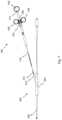
- FIG. 1is a perspective view of one embodiment of an endoscopic biopsy assembly 100 .
- the endoscopic biopsy assembly 100is for use with an endoscope (not shown) and includes a biopsy forceps device 200 disposed therein.
- the endoscopemay be any known endoscope.
- the endoscopeis an Olympus 160-series endoscope.
- a catheter 205is operatively connected to the biopsy forceps device 200 , and passes through the endoscope and a connector 300 , to a sample collection chamber 105 .
- the collection chamberincludes a tube 110 , having an aperture for receiving a tray 115 with a handle.
- the tray 115may be slidably removed from the aperture.
- the tray 115may be held in place by a locking mechanism (not shown).
- a locking mechanismmay include a pivoting or flexible member and a projection that is received in an aperture of the handle of the tray 115 .
- the tray 115has a single sample receiving surface.
- the trayincludes one or more dividers that define a plurality of sample receiving surfaces.
- the dividersmay be walls that extend upwards from a bottom surface of the tray, or the dividers may be indentations formed in the bottom surface of the tray.
- Such a traymay be moved within the sample collection chamber 105 , such as by sliding, pivoting, or rotating, to receive samples on the different sample receiving surfaces.
- the sample collection chamber 105may be any existing collection chamber.
- the sample collection chamber 105is a polypectomy trap adapter commercially available under the name ETRAP polyp trap and sold by U.S. Endoscopy.
- the sample collection chamber 105may include multiple components constructed of polymeric materials, such as thermoplastic elastomers or clear rigid plastic.
- An actuation handle 400is also operatively connected to the biopsy forceps device 200 through connector 300 .
- the actuation handle 400includes a sliding member and a shaft.
- the sliding membermay be manually translated along the shall, which causes the biopsy forceps device 200 to open and close.
- the actuation handle 400may be constructed of metal or a polymeric material, such as acrylonitrile butadiene styrene (“ABS”) plastic.
- the sample collection chamber 105 , connector 300 , and actuation handle 400are spaced from each other.
- one or more of the sample collection chamber 105 , connector 300 and actuation handle 400may be directly attached to each other.
- Such attachmentsmay be permanent attachments or releasable attachments.
- the sample collection chamber 105is fixedly or releasably attached to the actuation handle 400 .
- the sample collection chamber 105is releasably attached to the connector 300 .
- the connector 300is releasably attached to the actuation handle 400 .
- Releasable attachmentsmay be formed by clips, VELCRO, snaps, threaded connectors, or other known releasable connectors.
- Fixed attachmentsmay be formed by adhesive, bolts, rivets, welds, and other known fixed connectors.
- a fixed attachmentmay also be formed by molding two or more components as a single component.
- a suction device(not shown) is connected to a suction end S of a tube T, and the tube T is operatively connected to the sample collection chamber 105 .
- a valve Vis disposed along tube T.
- suction deviceis turned on and the valve V is open, suction is applied through the tube T, and through the sample collection chamber 105 and catheter 205 to the distal end of the biopsy forceps device 200 .
- the tray 115 in the sample collection chamber 105includes a plurality of apertures, so as not to interrupt suction along the tube T to the biopsy forceps device 200 .
- suction deviceis turned on and the valve V is closed, suction is only applied through a portion of the tube T.
- the valve Vis biased in a closed position and is opened by a manually operated push button.
- the valveis a trumpet valve.
- the valveis opened by a foot pump or a clamp.
- the valveis disposed on the sample collection chamber 105 or the catheter 205 .
- the operatorguides the endoscope to the biopsy site while viewing the biopsy site through various optical features allowing for visualization.
- the operatorpositions the biopsy forceps device 200 at a desired location and actuates the actuation handle 400 to open the biopsy forceps device 200 .
- the operatoroptionally applies suction through the biopsy forceps device 200 by opening the valve V. Applying suction at this time may cause “tenting” of the tissue, thus facilitating the taking of a sample.
- the operatorthen actuates the actuation handle 400 to close the biopsy forceps device 200 around a tissue sample.
- the act of closing the biopsy forceps device 200may sever the tissue sample in some instances. In other instances, the operator may need to apply force to withdraw the biopsy forceps device 200 from the biopsy site.
- This additional forcemay help to sever the tissue sample from the site.
- the operatoropens the valve V to apply suction.
- the suctionevacuates the sample from the biopsy forceps device 200 and draws it through the catheter 205 to the sample collection chamber 105 .
- the valve Vmay be opened before or after the actuation handle 400 is actuated.
- the endoscopic biopsy assembly 100may be operated by one or more operators. For example, a first operator may guide the endoscope, a second operator may actuate the actuation handle 400 , a third operator may open and close the valve V, and a fourth operator may retrieve the tissue sample from the sample collection chamber 105 . Alternatively, a single operator may perform each of these tasks. As another alternative, two or three operators may operate the endoscopic biopsy assembly 100 , with each operator performing one or more tasks.
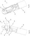
- FIGS. 2 A and 2 Billustrate first and second perspective views of the biopsy forceps device 200 connected to a catheter 205 .
- the biopsy forceps device 200includes a stationary jaw 210 having fenestration 215 and movable jaw 220 also having fenestration 225 .
- the stationary jaw 210 , movable jaw 220 , or bothare solid and do not include fenestrations.
- jawsare movable.
- FIGS. 2 C and 2 Dillustrate top plan views of the movable jaw 220 and the stationary jaw 210 .
- each of the stationary jaw 210 and movable jaw 220have sharpened edges configured to cut tissue.
- the sharpened edgesmay be continuous edges or serrated edges.
- each of the stationary jaw 210 and movable jaw 220have a radial curve and are cup shaped.
- the jawsmay have non-radial curves, or straight edges that form a geometric shape.
- Each of the stationary jaw 210 and movable jaw 220may be formed by a metal injection molding (“MIM”) or other processes including, but not limited to, stamping, laser welding, sintering, and molding.
- MIMmetal injection molding
- the jawsmay be constructed of stainless steel, aluminum, titanium, ceramics, plastics, or other known materials.
- the catheter 205may be constructed of a polymeric material, such as TEFLON, polyethylene, polypropylene, nylon, polyetherether keytone (PEEK), and other polymeric materials.
- the catheter 205may be formed by an extrusion process.
- the movable jaw 220is connected to the stationary jaw 210 by a pivot 230 .
- the pivot 230includes two posts that extend from the movable jaw 220 and are seated in corresponding apertures of the stationary jaw 210 .
- the pivot 230includes two posts that extend from the stationary jaw 210 and are seated in corresponding apertures of the movable jaw 220 .
- both the stationary jaw 210 and the movable jaw 220include a pair of corresponding apertures, and a pin is inserted therein to form a pivot.
- the pivotmay be positioned so as not to interfere with a passageway for tissue samples.
- the pivotmay be described as an external pivot.
- the pivotmay cross such a passageway.
- the biopsy forceps device 200further includes suction tubing 235 having a first end disposed within the chamber formed by stationary jaw 210 and movable jaw 220 .
- the end of the suction tubing 235is positioned forward of the pivot 230 , such that when the movable jaw 220 is opened, the first end of the suction tubing 235 may directly contact tissue.
- An operatormay choose to apply suction prior to taking a tissue sample, such that when the valve V is open and suction is applied through the suction tubing 235 , the tissue that is in direct contact with the suction tubing 235 is raised.
- the tenting processpulls tissue between the jaws, so that when the movable jaw 220 is closed, the sharpened edges of the jaws may sever the tented tissue and capture a sample between the jaws. An additional backwards force may also be required to sever the tissue sample.
- the tissue sampleis subsequently drawn down the catheter by the applied suction.
- the suction tubing 235is an insert that extends partially into the catheter. In an alternative embodiment, the suction tubing 235 extends the length of the catheter.
- the movable jaw 220further includes a lever arm 240 having an aperture 245 .
- a wire 250engages the lever arm 240 through the aperture 245 , such that manipulation of the wire 250 moves the lever arm 240 about the pivot 230 , causing the movable jaw 220 to open and close.
- the wire 250may be manipulated through the actuation handle 400 .
- FIG. 3illustrates a cross-section of the biopsy forceps device 200 .
- the stationary jaw 210further includes a ledge 255 disposed opposite the cutting surface of the stationary jaw 210 .
- the ledge 255may be created during the manufacturing process to form apertures in the stationary jaw 210 to accept the posts of the movable jaw 220 .
- a staking operationis performed to bend the ledge 255 .
- the ledge 255forms a stop, thereby defining the arc through which the movable jaw 220 may pivot.
- the ledge 255may be dimensioned so as not to interfere with the pivoting of the movable jaw 220 .
- the aperture 245 in the lever arm 240forms a socket that engages a ball 260 at the end of the wire 250 .
- the outer edges of the aperture 245may be crimped or otherwise closed around the ball 260 after the wire is received, such that the ball will remain housed in the socket formed by aperture 245 during operation of the endoscopic biopsy assembly 100 .
- a staking operationis performed to secure the ball 260 in the socket.
- a stopis formed on the wire on the opposite side of the lever aim, such that the lever arm is sandwiched between the ball 260 and the stop.
- the distal end of the suction tubing 235is slightly spaced from the distal end of the jaws 210 , 220 .
- the distal end of the suction tubing 235may be positioned adjacent the distal end of the jaws 210 , 220 .
- the distal end of the suction tubing 235may be further spaced from the distal end of the jaws 210 , 220 , such that the suction tubing 235 is bellow the ball 260 of the wire 250 .
- the position of the suction tubing 235may be varied before or during an operation.
- FIG. 4illustrates a partial perspective view of one embodiment of a catheter assembly 270 .
- the catheter assemblyincludes the catheter 205 and suction tubing 235 .
- the suction tubing 235may be a separate component or it may be integral with the catheter 205 .
- the catheter 205may be constructed of a polymeric material such as polytetrafluoroethylene (“PTFE”).
- PTFEpolytetrafluoroethylene
- the suction 235 tubingmay also be constructed of a polymeric material such as PTFE.
- the center of the suction tubing 235is a hollow passageway 265 through which tissue samples may be drawn.
- the hollow passageway 265is operatively connected to the suction device, such that when the suction device is turned on and the valve V is open, suction will be applied to the passageway 265 and draws a severed tissue sample to the capture container 105 .
- the catheter 205further includes a pair of lumens 275 a,b configured to receive the wire 250 . Although only one of the lumens ( 275 a ) is used, two lumens are formed for manufacturing purposes. Additionally, a pair of corresponding grooves 280 a,b are formed on the suction tubing 235 . The grooves 280 a,b may extend the entire length of the suction tubing 235 , or may only extend along a portion of the suction tubing 235 . The corresponding grooves 280 a,b are aligned with the lumens 275 a,b and may restrict lateral movement of the wire 250 .
- a lumen 275 for the wire 250that is separate from the passageway 235 ensures that the tissue sample has a clear travel path to the collection chamber 105 .
- the lumen 275is optional and that the wire ay be disposed along the passageway 235 .
- the catheter 205includes a pair of notches 285 a,b or other apertures configured to receive tangs 290 of the stationary jaw 210 (as shown in FIG. 2 D ).
- the notches 285 a,bmay extend for a portion of the catheter 205 , or they may extend the entire length of the catheter 205 .
- adhesivemay be placed in the notches 285 a,b such that the jaws are thereby affixed to the catheter 205 . Where adhesive is to be used, the surface may be scored to aid in bonding.
- the notches 285 a,bserve as an anchor point for the base of the stationary jaw 210 and prevents the jaws from rotating relative to the catheter.
- the notches 285 a,bare replaced with an annular groove configured to receive a flange of the jaws.
- the catheterdoes not include notches or a groove, and the jaws are glued, welded, or otherwise affixed to the catheter.
- FIG. 5illustrates a perspective view of an alternative embodiment of a catheter assembly 500 .
- the catheter assemblyincludes the catheter 505 and suction tubing 510 , wherein the suction tubing 510 is a separate insert that is received in a hollow passageway 515 of the catheter 505 .
- the center of the suction tubing 510is also a hollow passageway 520 for tissue samples.
- the catheter 505further includes a lumen 525 configured to receive a wire.
- the inclusion of a lumen 525 for the wireensures that the tissue sample has a clear travel path to the collection chamber 105 .
- FIG. 6illustrates a front view of an alternative embodiment of a catheter Catheter 605 may be employed in catheter assembly 270 or 500 , or other variations.
- the catheter 605includes a hollow passageway 610 .
- the catheter 605does not include any lumens for a wire. Instead, the wire may be disposed along the passageway 610 .
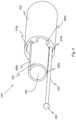
- FIG. 7illustrates a side view of one embodiment of a connector 300 and actuation handle 400 of the endoscopic biopsy assembly 100 .
- the connector 300is a substantially “y-shaped” component, having a major chamber 305 and a minor chamber 310 .
- the catheter 205passes through the major chamber 305 to the sample collection chamber 105 .
- a cable 315leads from the minor chamber 310 to the actuation handle 400 .
- the actuation handle 400includes a shaft 405 having a distal end 410 and a proximal end 415 .
- the proximal end 415has a first ring 420 attached thereto.
- the first ring 420is configured to receive an operator's thumb or finger.
- the ringmay be omitted or replaced with a transversely oriented member.
- a sliding member 425is slidably mounted to the shaft 405 .
- the sliding member 425includes a second ring 430 and a third ring 435 , each configured to receive an operator's finger or thumb.
- the sliding member 425may be a spool, or include a transversely oriented member in lieu of rings.
- a proximal end of the wire 250is fixedly attached to the sliding member 425 .
- the wire 250is retracted. This retraction causes the ball 260 at the opposite end of the wire 250 to pull the lever arm. 240 of the movable jaw 220 , which causes the movable jaw 220 to pivot towards the closed position.
- the wire 250is pushed forward. This movement causes the ball 260 at the opposite end of the wire 250 to push the lever arm 240 of the movable jaw 220 , which causes the movable jaw 220 to pivot towards the open position.
- FIG. 8illustrates a perspective view of assembled connecting components disposed within the connector 300 .
- the catheter 205 connected to the biopsy forceps device 200includes a first catheter 205 a and a second catheter 205 b . End portions of each of the first and second catheters 205 a,b are disposed in an aligning member 320 .
- the aligning member 320is a hollow rod having an elongated cavity to receive the first and second catheters 205 a,b .
- the aligning member 320further includes a side opening 325 .
- the side opening 325is formed by a chamfer extending downwards to the cavity and outwards to an end of the aligning member 320 .
- the side openingmay extend the entire length of the aligning member 320 .
- the side openingmay be a hole or slot that does not extend to an end of the aligning member 320 .
- the connecting componentsmay be assembled in the following manner.
- a first end of the first catheter 205 ais inserted into a first end of the aligning member 320 .
- the first catheter 205 amay be affixed in its position with adhesive, or by a press fit.
- the first end of the first catheter 205 aincludes a notch 330 that provides an exit for the cable 315 .
- the first catheter 205 ais positioned such that the notch 330 and the cable 315 are accessible through the side opening 325 of the aligning member 320 .
- a flap of the catheterremains in place over the notch 330 . This flap may shield the wire from adhesive that is applied during assembly.
- a first end of the second catheter 205 bis then inserted into a second end of the aligning member 320 , such that it abuts the first end of the first catheter 205 a .
- the second catheter 205 bmay be affixed in its position with an adhesive or by a press fit.
- FIG. 9illustrates a cross-section of the connector 300 .
- the first and second catheters 205 a,bpass through the major chamber 305 of the connector 300 .
- a first outer tube 335surrounds the catheter 205 at a front end of the major chamber 305 , forward from the aligning member 320 .
- a second outer tube 340surrounds the catheter 205 at a rear end of the major chamber 305 .
- the first and second outer tubes 335 , 340may prevent crimping or strains on the catheter 205 near the connector 300 .
- a single outer tubemay extend through the entire connector 300 .
- no outer tubesare included.
- a cable 315extends from the minor chamber 310 .
- the cable 315may be a sheath spring having tubing 345 disposed therein.
- the tubing 345is PEEK tubing.
- the wire 250is disposed within the cable 315 .
- the wire 250extends from the sliding member 425 of the actuation handle 400 , through the cable 315 and the minor chamber 310 , and joins the catheter 205 inside the major chamber 305 of the connector 300 .
- the wire 250either extends through a lumen or the hollow passageway in the catheter 205 .
- the cable 315may act as a dampener that prevents the sliding member 425 of the actuation handle 400 from being translated too quickly or violently.
- the minor chamber 310extends from the major chamber 305 at an angle of approximately 15°. In alternative embodiment, the minor chamber 310 may extend from the major chamber at an angle between 5° and 60°. A smaller angle may be preferable to prevent crimping of the wire 250 .
- the major chamber 305also includes a sterilization hole 350 .
- the sterilization hole 350is configured to receive sterilizing material, such as ethylene oxide, as may be needed or desired.
- FIG. 10illustrates a partial cross-section of an alternative embodiment of a connector 700 of an endoscopic biopsy assembly.
- the connector 700is substantially the same as the connector 300 , except for the differences described herein.
- Like reference numbersindicate like components.
- the connector 700includes a major chamber 705 having an enlarged cavity portion 710 sized to accept a housing 715 .
- the housing 715surrounds portions of the aligning member 320 , the first catheter 205 a , and the second catheter 205 b.
- FIG. 11illustrates a perspective view of the housing 715 .
- the housing 715has a through hole 720 and tapered ends 725 , 730 , and may form a seal around the enclosed components.
- a distal end of the housing 715surrounds a cylindrical distal end of aligning member 320 to create a seal therewith. If desired, small gaps or clearances can be filled with adhesive, grease, sealing compounds and the like to create an air-tight seal.
- a proximal end of aligning member 320includes the side opening 325 (see FIG. 8 ). The side opening 325 creates a gap between housing 715 and second catheter 205 b for the passage of an “S” portion of cable 315 .
- the gapmay be filled around the cable 315 with a seal 316 such as elastomeric seal or adhesive.
- the cable 315may also be lubricated to prevent sticking with the seal 316 .
- the ends of the housingare straight.
- the housing 715may be constructed of a polymeric material such as but not limited to ABS or any one of a number of metals. In one embodiment, the housing 715 is constructed at least partially of rubber to aid in sealing the enclosed components. In an alternative embodiment (not shown), o-rings or other seals may be disposed within the housing.
- FIG. 12illustrates a perspective view of an alternative embodiment of an endoscopic biopsy assembly 800 .
- the endoscopic biopsy assembly 800is substantially the same as the endoscopic biopsy assembly 100 shown in FIG. 1 , except for the differences described herein.
- Like reference numbersindicate like components.
- the endoscopic biopsy assembly 800includes a radio frequency (RF) generator 805 connected to the actuation handle 400 .
- RFradio frequency
- at least one of the jaws 210 , 220is formed from an electrically conductive material, such as stainless steel, and the RF generator 805 is in electrical communication with the electrically conductive jaw.
- both jaws 210 , 220are formed from an electrically conductive material.
- handle 400includes art RF connector socket positioned distal to a dual ring portion of the handle. The RE connector socket is electrically connected to the jaws 210 , 220 via at least wire 250 for the delivery of RE energy to the jaws.
- the RF generator 805is in electrical communication with at least one of the jaws 210 , 220 through the wire 250 .
- the RE generator 805is also in electrical communication with an actuator 810 that is used by the surgeon to deliver RF energy when required.
- RE energyis provided to at least one of the jaws 210 , 220 that can cauterize or cut the tissue.
- the RF generator 805may include a wave form selection switch (not shown) that allows an operator to select between a cauterizing waveform and a cutting waveform.
- the actuator 810is a foot pedal. However, it should be understood that any actuator may be employed, such as buttons, dials, and switches.
- the RE generator 805has a first pole 815 (i.e., a positive pole) and a second pole 820 (i.e., a negative pole or a ground pole).
- first pole 815i.e., a positive pole
- second pole 820i.e., a negative pole or a ground pole.
- the second pole 820is connected to a ground pad 825 .
- the pad 825is placed under the patient to form an electrical ground between the patient and the RE generator 805 .
- the second pole 820may be in electrical communication with any conductive object that can contact or be placed proximal to a patient.
- both positive and negative polesare in electrical communication with the jaws 210 , 220 , making the biopsy forceps device 200 a bipolar device.
- one of the first and second poles 815 , 820is in electrical communication with jaw 210 through a first wire, and the opposite pole is in electrical communication with jaw 220 through a second wire.
- Such an embodimentwill electrically isolate each jaw 210 , 220 and the first and second wires from electrical contact with the other to prevent shorting.
- each electrically isolated jaw 210 , 220has an electrically conductive area exposed in the tissue clamping area, such as the horseshoe shaped sharpened edges that are shown contacted together when the jaws are fully closed.
- the electrical contact areaswill short together, and prevent the generator from activating.
- tissueis between the clamped jaws, the tissue provides electrical resistance in the flow path between the exposed sharpened edges, and the generator will actuate and coagulate the clamped tissue.
- each jaw 210 , 220may include additional insulation or electrically non-conductive materials or coatings.
- portions of the jawsmay be constructed of ceramic or a polymeric material.
- each jawcan be completely ceramic coated with an insulating layer, and portions of the insulating layer can be removed (by grinding, masking or the like) at the wire contact area and at the horseshoe shaped tissue biting area.
- electrical energy of one poleis conducted along the insulated wire, into the jaw 210 or jaw 220 at the wire contact area, and to tissue through the exposed horseshoe shaped tissue biting area.
- Such an embodimentmay also deliver more focused energy that would not interfere with other electrical devices, such as a pacemaker in a patient.
- FIGS. 13 A-Cillustrate cross-sections of the biopsy forceps device 200 of the alternative embodiment of the endoscopic biopsy assembly 800 , at various stages of taking a biopsy sample.
- the biopsy forceps device 200has been positioned at a desired location, the jaws 210 , 220 have been opened, and a potential tissue sample, such as a polyp, is shown positioned partially inside the jaws 210 , 220 .
- a potential tissue samplesuch as a polyp
- suctionis applied through suction tubing 235 , which causes “tenting” or drawing of the tissue at least partially into the suction tubing 235 as shown. If desired, a larger bite of tissue can be taken by pushing the jaws 210 , 220 farther into the tissue wall.
- the operatorhas actuated the actuation handle 400 to close the jaws 210 , 220 of biopsy forceps device 200 around a tissue sample as it is being tented and drawn into the suction tubing 235 .
- the act of closing the biopsy forceps device 200pinches the “tented” tissue at the base of the polyp and in this view has fully severed the tissue sample just before it is drawn farther into the suction tubing 235 by the applied suction.
- the tissue samplemay not be severed entirely between the jaws 210 , 220 and the operator may apply force (i.e., by pulling or shaking) to fully sever the tissue sample from the site.
- the operatormay activate the RF generator to apply RF energy at the jaws to cut the tissue sample from the site. The operator may select a cutting wave form prior to activating the RIF generator.
- the closing of the jawshas failed to sever the tissue sample and, the operator is activating the RF generator to apply monopolar RF energy at the jaws to cauterize the tissue around the site.
- the operatormay select a cauterizing wave form, such as a square wave, prior to activating the RF generator.
- Cauterizing the site in this mannermay staunch bleeding caused by the severing, and may also kill cancer cells at the site.
- the un-severed sampleis inside of the jaws, it is prevented from contacting the negatively charged patient and is not burned when the RF energy is applied.
- the tissuecannot be coagulated because it is exposed to only positive RF energy which travels along the external skin of the jaws and to the negatively charged patient.
- the operatorelects to pull and shake the tissue sample free so that it can be sucked into the suction tubing 235
- FIG. 14illustrates a cross-section of the biopsy forceps device 200 as it extends from a distal end of an endoscope 900 , while taking a biopsy sample.
- the biopsy forceps device 200is the same as biopsy forceps device 200 shown in FIGS. 1 - 3 , except for the placement within the endoscope 900 .
- Like reference numbersindicate like components.
- the endoscope 900has a tube 905 that includes a first lumen 915 that is configured to receive the biopsy forceps device 200 .
- Catheter 205is shown extending along the first lumen 915 of endoscope 900 with the jaws 210 , 220 extending from the endoscope 910 .
- Endoscope 900further comprises a second lumen 910 in communication with a fluid source for providing irrigation to tissue and to the jaws 210 , 220 .
- a plurality of tissue sampleshave accumulated within the suction tubing 235 and the operator may irrigate the surgical site by providing fluid through the second lumen 915 of the endoscope 900 .
- the fluidmay be used to clean the biopsy site or used to wash the accumulated tissue samples through the suction tubing 235 .
- the fluidmay enter the biopsy forceps device 200 through the fenestrations 215 , 225 in the jaws 210 , 220 .
- the fluid entering the biopsy forceps deviceis drawn down the suction tubing 235 when suction is applied, and may aid in washing or drawing the tissue sample down the suction tubing 235 .
- Exemplary fluidsmay include water, saline, drugs or any combination thereof.
- FIG. 15illustrates a cross-section of the biopsy forceps device 200 as it extends from the endoscope 900 .
- suctionis being applied to draw a stuck tissue sample down the suction tubing 235 .
- the operatorcan open and close the jaws 210 , 210 to dislodge the tissue sample by activating the actuation handle 400 . Repeating this opening and closing action causes the wire 250 to move longitudinally within the suction tubing 235 which may then rub wire 250 against the tissue sample and aid in drawing it through the suction tubing 235 .
- This methodmay be effective in dislodging a tissue sample that has become stuck, and may be performed with or without irrigation.
- many elements of the endoscopic biopsy assembly 100can be operably configured to be flexible such as, for example, but not limited to: catheter 205 , suction tubing 210 , suction tubing 235 , wire 250 , cable 315 , tubing 345 , and catheter 505 .
- one or more of the flexible elementscan be substantially rigid such as exemplary catheter 205 or any other element of assembly 100 .
Landscapes
- Health & Medical Sciences (AREA)
- Life Sciences & Earth Sciences (AREA)
- Surgery (AREA)
- Heart & Thoracic Surgery (AREA)
- Molecular Biology (AREA)
- Veterinary Medicine (AREA)
- Engineering & Computer Science (AREA)
- Biomedical Technology (AREA)
- Nuclear Medicine, Radiotherapy & Molecular Imaging (AREA)
- Medical Informatics (AREA)
- Pathology (AREA)
- Animal Behavior & Ethology (AREA)
- General Health & Medical Sciences (AREA)
- Public Health (AREA)
- Radiology & Medical Imaging (AREA)
- Biodiversity & Conservation Biology (AREA)
- Endoscopes (AREA)
- Surgical Instruments (AREA)
- Manipulator (AREA)
Abstract
Description
Claims (10)
Priority Applications (1)
| Application Number | Priority Date | Filing Date | Title |
|---|---|---|---|
| US15/902,781US11864742B2 (en) | 2010-03-24 | 2018-02-22 | Biopsy device |
Applications Claiming Priority (3)
| Application Number | Priority Date | Filing Date | Title |
|---|---|---|---|
| US31703610P | 2010-03-24 | 2010-03-24 | |
| US13/070,741US20110237975A1 (en) | 2010-03-24 | 2011-03-24 | Multiple biopsy device |
| US15/902,781US11864742B2 (en) | 2010-03-24 | 2018-02-22 | Biopsy device |
Related Parent Applications (1)
| Application Number | Title | Priority Date | Filing Date |
|---|---|---|---|
| US13/070,741ContinuationUS20110237975A1 (en) | 2010-03-24 | 2011-03-24 | Multiple biopsy device |
Publications (2)
| Publication Number | Publication Date |
|---|---|
| US20180177496A1 US20180177496A1 (en) | 2018-06-28 |
| US11864742B2true US11864742B2 (en) | 2024-01-09 |
Family
ID=44657234
Family Applications (2)
| Application Number | Title | Priority Date | Filing Date |
|---|---|---|---|
| US13/070,741AbandonedUS20110237975A1 (en) | 2010-03-24 | 2011-03-24 | Multiple biopsy device |
| US15/902,781Active2033-03-23US11864742B2 (en) | 2010-03-24 | 2018-02-22 | Biopsy device |
Family Applications Before (1)
| Application Number | Title | Priority Date | Filing Date |
|---|---|---|---|
| US13/070,741AbandonedUS20110237975A1 (en) | 2010-03-24 | 2011-03-24 | Multiple biopsy device |
Country Status (4)
| Country | Link |
|---|---|
| US (2) | US20110237975A1 (en) |
| EP (1) | EP2549931B1 (en) |
| JP (1) | JP2013521994A (en) |
| WO (1) | WO2011119817A2 (en) |
Families Citing this family (11)
| Publication number | Priority date | Publication date | Assignee | Title |
|---|---|---|---|---|
| KR102014061B1 (en) | 2011-10-03 | 2019-08-28 | 모더나 세라퓨틱스, 인코포레이티드 | Modified nucleosides, nucleotides, and nucleic acids, and uses thereof |
| US9999407B2 (en) | 2012-01-21 | 2018-06-19 | Choon Kee Lee | Tissue sampling device |
| DE102012223788A1 (en)* | 2012-12-19 | 2014-06-26 | Karl Storz Gmbh & Co. Kg | Endoscopic instrument for retrograde biopsy, in particular synovial biopsy |
| US20160095584A1 (en)* | 2014-10-01 | 2016-04-07 | Boston Scientific Scimed, Inc. | Endoscopic needle with rotary jaw for lateral acquisition |
| CZ2014691A3 (en)* | 2014-10-10 | 2016-01-06 | Oprox, A.S. | Catheter for optical biopsy |
| US20160256140A1 (en)* | 2015-03-03 | 2016-09-08 | United States Endoscopy Group, Inc. | Microforceps |
| GB2543039A (en)* | 2015-10-02 | 2017-04-12 | Creo Medical Ltd | Electrosurgical device |
| SE541805C2 (en)* | 2017-12-22 | 2019-12-17 | Multi4 Ab | An endosurgical device |
| CN112770689B (en)* | 2018-09-26 | 2024-07-19 | 奥瑞斯健康公司 | Systems and instruments for suction and irrigation |
| PL3870073T3 (en)* | 2018-12-14 | 2024-04-08 | Devicor Medical Products, Inc. | Biopsy device with translating shuttle valve assembly |
| US12004717B2 (en)* | 2019-12-20 | 2024-06-11 | Gyrus Acmi, Inc. | Endoscope with detachable camera module |
Citations (123)
| Publication number | Priority date | Publication date | Assignee | Title |
|---|---|---|---|---|
| US2751908A (en) | 1953-03-19 | 1956-06-26 | American Cystoscope Makers Inc | Surgical instrument |
| US3807406A (en) | 1971-06-25 | 1974-04-30 | Bio Medicus Inc | Instrument surgical with suction device |
| US3964468A (en) | 1975-05-30 | 1976-06-22 | The Board Of Trustees Of Leland Stanford Junior University | Bioptome |
| US4122856A (en) | 1976-10-08 | 1978-10-31 | American Hospital Supply Corporation | Surgical instrument and method of assembly thereof |
| US4445509A (en) | 1982-02-04 | 1984-05-01 | Auth David C | Method and apparatus for removal of enclosed abnormal deposits |
| US4522206A (en) | 1983-01-26 | 1985-06-11 | Dyonics, Inc. | Surgical instrument |
| US4632110A (en) | 1984-09-28 | 1986-12-30 | Olympus Optical Co., Ltd. | Medical operation instrument for endoscope |
| US4662371A (en) | 1983-01-26 | 1987-05-05 | Whipple Terry L | Surgical instrument |
| US4674502A (en) | 1985-09-27 | 1987-06-23 | Coopervision, Inc. | Intraocular surgical instrument |
| US4693257A (en) | 1986-05-12 | 1987-09-15 | Markham Charles W | Needle aspiration biopsy device with enclosed fluid supply |
| US4712545A (en)* | 1984-04-05 | 1987-12-15 | Acufex Microsurgical, Inc. | Surgical instrument |
| US5108381A (en) | 1991-03-11 | 1992-04-28 | Kolozsi William Z | Tissue sample collection trap |
| US5217460A (en) | 1991-03-22 | 1993-06-08 | Knoepfler Dennis J | Multiple purpose forceps |
| US5286255A (en) | 1991-07-29 | 1994-02-15 | Linvatec Corporation | Surgical forceps |
| US5300087A (en) | 1991-03-22 | 1994-04-05 | Knoepfler Dennis J | Multiple purpose forceps |
| US5310406A (en) | 1992-05-08 | 1994-05-10 | Sharpe Endosurgical Corporation | Endoscopic aspirator surgical instrument |
| US5347991A (en) | 1992-10-20 | 1994-09-20 | Nakao Naomi L | Endoscope suction trap and associated method |
| US5373854A (en) | 1993-07-15 | 1994-12-20 | Kolozsi; William Z. | Biopsy apparatus for use in endoscopy |
| US5383471A (en) | 1992-04-10 | 1995-01-24 | Funnell; David M. | Surgical biopsy instrument |
| US5417709A (en) | 1994-04-12 | 1995-05-23 | Symbiosis Corporation | Endoscopic instrument with end effectors forming suction and/or irrigation lumens |
| WO1995019145A1 (en) | 1994-01-12 | 1995-07-20 | Symbiosis Corporation | Arthroscopic surgical instruments having suction capability |
| US5441503A (en) | 1988-09-24 | 1995-08-15 | Considine; John | Apparatus for removing tumors from hollow organs of the body |
| US5458112A (en) | 1994-08-15 | 1995-10-17 | Arrow Precision Products, Inc. | Biliary biopsy device |
| US5478347A (en) | 1990-10-05 | 1995-12-26 | United States Surgical Corporation | Endoscopic surgical instrument having curved blades |
| US5571136A (en) | 1994-08-15 | 1996-11-05 | Medical Innovations Corporation | Forceps with guide wire |
| US5575293A (en) | 1995-02-06 | 1996-11-19 | Promex, Inc. | Apparatus for collecting and staging tissue |
| US5603724A (en) | 1995-02-13 | 1997-02-18 | Tnco, Inc. | Suction punch |
| US5620415A (en)* | 1993-01-29 | 1997-04-15 | Smith & Dyonics, Inc. | Surgical instrument |
| US5665100A (en) | 1989-12-05 | 1997-09-09 | Yoon; Inbae | Multifunctional instrument with interchangeable operating units for performing endoscopic procedures |
| US5669394A (en) | 1989-02-06 | 1997-09-23 | The Board Of Regents Of The Univ. Of Oklahoma | Biosample aspirator |
| US5683388A (en) | 1996-01-11 | 1997-11-04 | Symbiosis Corporation | Endoscopic bipolar multiple sample bioptome |
| US5683359A (en) | 1992-11-18 | 1997-11-04 | Symbiosis Corporation | Arthroscopic surgical instruments having suction capability |
| US5697949A (en)* | 1995-05-18 | 1997-12-16 | Symbiosis Corporation | Small diameter endoscopic instruments |
| US5715832A (en) | 1995-02-28 | 1998-02-10 | Boston Scientific Corporation | Deflectable biopsy catheter |
| US5775333A (en) | 1994-03-24 | 1998-07-07 | Ethicon Endo-Surgery, Inc. | Apparatus for automated biopsy and collection of soft tissue |
| WO1998040015A2 (en) | 1997-03-13 | 1998-09-17 | Biomax Technologies, Inc. | Catheters and endoscopes comprising optical probes and bioptomes and methods of using the same |
| US5810876A (en) | 1995-10-03 | 1998-09-22 | Akos Biomedical, Inc. | Flexible forceps device |
| US5817033A (en) | 1994-04-11 | 1998-10-06 | Desantis; Stephen A. | Needle core biopsy device |
| US5857997A (en)* | 1994-11-14 | 1999-01-12 | Heart Rhythm Technologies, Inc. | Catheter for electrophysiological procedures |
| US5871453A (en) | 1994-02-08 | 1999-02-16 | Boston Scientific Corporation | Moveable sample tube multiple biopsy sampling device |
| US5871454A (en) | 1997-04-22 | 1999-02-16 | Majlessi; Heshmat | Percutaneous excisional biopsy device |
| US5897507A (en) | 1996-11-25 | 1999-04-27 | Symbiosis Corporation | Biopsy forceps instrument having irrigation and aspiration capabilities |
| US5906629A (en) | 1997-05-27 | 1999-05-25 | T.A.G. Medical Products Ltd. | Arthroscopic surgical apparatus |
| US5944673A (en) | 1998-05-14 | 1999-08-31 | Ethicon Endo-Surgery, Inc. | Biopsy instrument with multi-port needle |
| WO1999045847A1 (en) | 1998-03-09 | 1999-09-16 | Spectrascience, Inc. | Optical biopsy forceps system and method of sampling tissue |
| US5964716A (en) | 1998-05-14 | 1999-10-12 | Ethicon Endo-Surgery, Inc. | Method of use for a multi-port biopsy instrument |
| US5980468A (en) | 1997-09-22 | 1999-11-09 | Zimmon Scientific Corporation | Apparatus and method for serial collection storage and processing of biopsy specimens |
| WO1999059475A1 (en) | 1998-05-15 | 1999-11-25 | Boston Scientific Limited | Biopsy instrument having irrigation and aspiration capabilities |
| US6017316A (en) | 1997-06-18 | 2000-01-25 | Biopsys Medical | Vacuum control system and method for automated biopsy device |
| US6019733A (en) | 1997-09-19 | 2000-02-01 | United States Surgical Corporation | Biopsy apparatus and method |
| US6053933A (en) | 1996-08-10 | 2000-04-25 | Deutsches Zentrum Fur Luft- Und Raumfahrt E.V. | Gripping unit for application in minimally invasive surgery |
| US6074408A (en) | 1998-10-13 | 2000-06-13 | Freeman; Kenneth V. | Modular medical instrument and method of using same |
| US6106543A (en) | 1998-05-15 | 2000-08-22 | Esser; Theodor | Medical instrument driving member and end effector connection |
| US6110127A (en) | 1998-02-17 | 2000-08-29 | Olympus Optical, Co., Ltd. | Medical instrument for use in combination with an endoscope |
| WO2000054658A1 (en) | 1999-03-12 | 2000-09-21 | C.R. Bard, Inc. | Endoscopic multiple sample biopsy forceps |
| JP2000271128A (en) | 1999-03-29 | 2000-10-03 | Asahi Optical Co Ltd | High-frequency biopsy forceps for endoscope |
| US6139508A (en)* | 1998-08-04 | 2000-10-31 | Endonetics, Inc. | Articulated medical device |
| US6142957A (en)* | 1993-09-20 | 2000-11-07 | Boston Scientific Corporation | Multiple biopsy sampling device |
| US6142956A (en) | 1996-11-25 | 2000-11-07 | Symbiosis Corporation | Proximal actuation handle for a biopsy forceps instrument having irrigation and aspiration capabilities |
| US6162187A (en) | 1999-08-02 | 2000-12-19 | Ethicon Endo-Surgery, Inc. | Fluid collection apparatus for a surgical device |
| US6282442B1 (en) | 1998-09-11 | 2001-08-28 | Surgical Laser Technologies, Inc. | Multi-fit suction irrigation hand piece |
| US6309404B1 (en)* | 1999-10-19 | 2001-10-30 | Jacek Krzyzanowski | Flexible biopsy jaw assembly |
| US6322522B1 (en) | 1997-09-22 | 2001-11-27 | Zimmon Science Corp. | Apparatus for separable external serial collection, storage and processing of biopsy specimens |
| US20020029007A1 (en) | 1997-09-19 | 2002-03-07 | Bryan Graham W. | Biopsy apparatus and method |
| US6358224B1 (en) | 1999-09-24 | 2002-03-19 | Tyco Healthcare Group Lp | Irrigation system for endoscopic surgery |
| US6387057B1 (en) | 1999-11-09 | 2002-05-14 | Norbert Heske | Device for gently removing tissue from animal or human tissue |
| US6398741B2 (en) | 1998-11-20 | 2002-06-04 | J. Morita Manufacturing Corporation | Tissue excision and cutting apparatus and its forceps |
| US20020173699A1 (en) | 2001-05-16 | 2002-11-21 | Stephen Becker | Endoscope sleeve and irrigation device |
| US6485436B1 (en) | 2000-08-10 | 2002-11-26 | Csaba Truckai | Pressure-assisted biopsy needle apparatus and technique |
| US6530891B2 (en) | 2001-04-02 | 2003-03-11 | Temple University Of The Commonwealth System Of Higher Education | Multiple biopsy device |
| US20030060816A1 (en) | 2001-08-30 | 2003-03-27 | Olympus Optical Co., Ltd. | Treatment device for tissue from living tissues |
| US6569105B1 (en)* | 2000-09-14 | 2003-05-27 | Syntheon, Llc | Rotatable and deflectable biopsy forceps |
| US20030125639A1 (en) | 2002-01-02 | 2003-07-03 | Fisher John S. | Biopsy needle having rotating core for shearing tissue |
| US6599309B1 (en) | 1999-09-09 | 2003-07-29 | Tnco, Inc. | Pin-less surgical instrument |
| US6613068B2 (en)* | 2000-03-07 | 2003-09-02 | Pentax Corporation | Endoscopic treatment instrument |
| US6620111B2 (en) | 2001-04-20 | 2003-09-16 | Ethicon Endo-Surgery, Inc. | Surgical biopsy device having automatic rotation of the probe for taking multiple samples |
| US6632182B1 (en) | 1998-10-23 | 2003-10-14 | The Trustees Of Columbia University In The City Of New York | Multiple bit, multiple specimen endoscopic biopsy forceps |
| US6638235B2 (en) | 2000-11-06 | 2003-10-28 | Suros Surgical Systems, Inc. | Biopsy apparatus |
| US20040068291A1 (en)* | 2001-09-25 | 2004-04-08 | Olympus Optical Co., Ltd. | Medical instrument |
| US20040097829A1 (en) | 2002-11-15 | 2004-05-20 | Mcrury Ian D. | Tissue biopsy and processing device |
| US6746462B1 (en) | 1997-02-28 | 2004-06-08 | Lumend, Inc. | Methods and apparatus for treating vascular occlusions |
| US20040153003A1 (en) | 2002-12-11 | 2004-08-05 | Chris Cicenas | Biopsy device with sample tube |
| US20050027210A1 (en) | 2000-11-06 | 2005-02-03 | Miller Michael E. | Biopsy apparatus |
| US6858014B2 (en) | 2002-04-05 | 2005-02-22 | Scimed Life Systems, Inc. | Multiple biopsy device |
| US20050043758A1 (en) | 2003-08-18 | 2005-02-24 | Scimed Life Systems, Inc. | Endoscopic medical instrument and related methods of use |
| US20050065453A1 (en) | 2003-02-24 | 2005-03-24 | Senorx, Inc. | Biopsy device with selectable tissue receiving aperture orientation and site illumination |
| US6875182B2 (en) | 1998-03-03 | 2005-04-05 | Senorx, Inc. | Electrosurgical specimen-collection system |
| US6878149B2 (en) | 2002-03-25 | 2005-04-12 | Acueity, Inc. | Apparatus and method for intraductal abalation |
| US20050113715A1 (en) | 2000-11-06 | 2005-05-26 | Jeffrey Schwindt | Biopsy apparatus |
| US20050165329A1 (en) | 2004-01-22 | 2005-07-28 | Reflux Corporation | Multiple biopsy collection device |
| US20060178560A1 (en) | 2003-01-15 | 2006-08-10 | Usgi Medical, Inc. | Endoluminal tool deployment system |
| US7118586B1 (en) | 1999-10-25 | 2006-10-10 | Boston Scientific Scimed, Inc. | Forceps for medical use |
| US20060258955A1 (en) | 2005-05-13 | 2006-11-16 | Hoffman David W | Endoscopic apparatus with integrated multiple biopsy device |
| USRE39415E1 (en) | 1990-05-10 | 2006-11-28 | Boston Scientific Miami Corporation | Radial jaw biopsy forceps |
| US7169115B2 (en) | 2003-09-29 | 2007-01-30 | Ethicon Endo-Surgery, Inc. | Endoscopic mucosal resection device with overtube and method of use |
| US20070032723A1 (en) | 2005-06-21 | 2007-02-08 | Glossop Neil D | System, method and apparatus for navigated therapy and diagnosis |
| US20070038146A1 (en) | 2005-08-05 | 2007-02-15 | Quick Richard L | Biopsy device with fluid delivery to tissue specimens |
| US7189206B2 (en) | 2003-02-24 | 2007-03-13 | Senorx, Inc. | Biopsy device with inner cutter |
| US7220226B2 (en) | 2003-07-04 | 2007-05-22 | Tokendo | Removable operating device for a flexible endoscopic probe for medical purposes |
| US7226424B2 (en) | 1994-03-24 | 2007-06-05 | Ethicon Endo-Surgery, Inc. | Methods and devices for automated biopsy and collection of soft tissue |
| US20070167868A1 (en) | 2006-01-18 | 2007-07-19 | Lsi Solutions, Inc. | Ergonomic needle tissue harvesting instrument not requiring a stylet |
| US20070179401A1 (en) | 2006-02-01 | 2007-08-02 | Ethicon Endo-Surgery, Inc. | Biopsy device with replaceable probe incorporating static vacuum source dual valve sample stacking retrieval and saline flush |
| US20070213634A1 (en) | 2006-02-24 | 2007-09-13 | Boston Scientific Scimed, Inc. | Obtaining a tissue sample |
| US7276032B2 (en) | 2004-09-29 | 2007-10-02 | Ethicon Endo-Surgery, Inc. | Biopsy apparatus and method |
| US20070270894A1 (en) | 2006-05-16 | 2007-11-22 | Zimmon David S | Combined endoscope and biopsy instrument with a removable biopsy cassette for in situ fixation and specimen processing |
| US20080009857A1 (en)* | 2006-07-05 | 2008-01-10 | Olympus Medical Systems Corp. | Treatment instrument for endoscope |
| US7322935B2 (en) | 2003-01-22 | 2008-01-29 | Medcanica, Llc | Endoscopic retractor |
| US7331930B2 (en) | 2001-10-30 | 2008-02-19 | Movdicé Holding, Inc. | Biopsy/access tool |
| US7347828B2 (en) | 1996-11-25 | 2008-03-25 | Boston Scientific Miami Corporation | Suction adapter for medical instrument |
| US7361174B2 (en) | 2002-12-11 | 2008-04-22 | Boston Scientific Scimed, Inc. | Angle indexer for medical devices |
| US20080108871A1 (en) | 2006-11-06 | 2008-05-08 | Mohr Catherine J | Vacuum stabilized overtube for endoscopic surgery |
| US20080221480A1 (en) | 2006-12-13 | 2008-09-11 | Hibner John A | Biopsy Sample Storage |
| US20080255424A1 (en) | 2007-04-10 | 2008-10-16 | Boston Scientific Scimed, Inc. | Endoscopes including distal chamber and related methods of use |
| US7481817B2 (en) | 2003-02-13 | 2009-01-27 | Lsi Soultions, Inc. | Instrument for surgically cutting tissue and method of use |
| US7517321B2 (en) | 2005-01-31 | 2009-04-14 | C. R. Bard, Inc. | Quick cycle biopsy system |
| US20090149746A1 (en) | 2007-11-19 | 2009-06-11 | Rubicor Medical, Inc. | Post-biopsy cavity treatment implants and methods |
| US20090171147A1 (en) | 2007-12-31 | 2009-07-02 | Woojin Lee | Surgical instrument |
| US7559887B2 (en) | 2004-12-08 | 2009-07-14 | Patrick Dannan | Tool insertion device for use in minimally invasive surgery |
| US20090182198A1 (en) | 2007-12-27 | 2009-07-16 | Skerven Gregory J | Multiple band dispenser endoscope sheath |
| US20090187146A1 (en) | 2007-12-19 | 2009-07-23 | Vance Products Inc. D/B/A Cook Urological Incorporated | Vacuum aspiration handle |
| US20090192352A1 (en) | 2008-01-24 | 2009-07-30 | P Regadas F Sergio | Operating Anoscope For Transanal Endoscopic Microsurgery |
| US7569626B2 (en) | 2003-06-05 | 2009-08-04 | Dfine, Inc. | Polymer composites for biomedical applications and methods of making |
| US7762960B2 (en) | 2005-05-13 | 2010-07-27 | Boston Scientific Scimed, Inc. | Biopsy forceps assemblies |
Family Cites Families (3)
| Publication number | Priority date | Publication date | Assignee | Title |
|---|---|---|---|---|
| JPS60246742A (en)* | 1984-05-22 | 1985-12-06 | オリンパス光学工業株式会社 | Forcept apparatus for endoscope |
| JP4343316B2 (en)* | 1999-03-30 | 2009-10-14 | オリンパス株式会社 | Endoscopic treatment tool |
| JP2006000524A (en)* | 2004-06-21 | 2006-01-05 | Pentax Corp | Ultrasound endoscope suction valve device |
- 2011
- 2011-03-24JPJP2013501449Apatent/JP2013521994A/ennot_activeCeased
- 2011-03-24EPEP11760205.2Apatent/EP2549931B1/enactiveActive
- 2011-03-24USUS13/070,741patent/US20110237975A1/ennot_activeAbandoned
- 2011-03-24WOPCT/US2011/029772patent/WO2011119817A2/enactiveApplication Filing
- 2018
- 2018-02-22USUS15/902,781patent/US11864742B2/enactiveActive
Patent Citations (144)
| Publication number | Priority date | Publication date | Assignee | Title |
|---|---|---|---|---|
| US2751908A (en) | 1953-03-19 | 1956-06-26 | American Cystoscope Makers Inc | Surgical instrument |
| US3807406A (en) | 1971-06-25 | 1974-04-30 | Bio Medicus Inc | Instrument surgical with suction device |
| US3964468A (en) | 1975-05-30 | 1976-06-22 | The Board Of Trustees Of Leland Stanford Junior University | Bioptome |
| US4122856A (en) | 1976-10-08 | 1978-10-31 | American Hospital Supply Corporation | Surgical instrument and method of assembly thereof |
| US4445509A (en) | 1982-02-04 | 1984-05-01 | Auth David C | Method and apparatus for removal of enclosed abnormal deposits |
| US4662371A (en) | 1983-01-26 | 1987-05-05 | Whipple Terry L | Surgical instrument |
| US4522206A (en) | 1983-01-26 | 1985-06-11 | Dyonics, Inc. | Surgical instrument |
| US4712545A (en)* | 1984-04-05 | 1987-12-15 | Acufex Microsurgical, Inc. | Surgical instrument |
| US4632110A (en) | 1984-09-28 | 1986-12-30 | Olympus Optical Co., Ltd. | Medical operation instrument for endoscope |
| US4674502A (en) | 1985-09-27 | 1987-06-23 | Coopervision, Inc. | Intraocular surgical instrument |
| US4693257A (en) | 1986-05-12 | 1987-09-15 | Markham Charles W | Needle aspiration biopsy device with enclosed fluid supply |
| US5441503A (en) | 1988-09-24 | 1995-08-15 | Considine; John | Apparatus for removing tumors from hollow organs of the body |
| US5669394A (en) | 1989-02-06 | 1997-09-23 | The Board Of Regents Of The Univ. Of Oklahoma | Biosample aspirator |
| US5665100A (en) | 1989-12-05 | 1997-09-09 | Yoon; Inbae | Multifunctional instrument with interchangeable operating units for performing endoscopic procedures |
| USRE39415E1 (en) | 1990-05-10 | 2006-11-28 | Boston Scientific Miami Corporation | Radial jaw biopsy forceps |
| US5478347A (en) | 1990-10-05 | 1995-12-26 | United States Surgical Corporation | Endoscopic surgical instrument having curved blades |
| US5108381A (en) | 1991-03-11 | 1992-04-28 | Kolozsi William Z | Tissue sample collection trap |
| US5217460A (en) | 1991-03-22 | 1993-06-08 | Knoepfler Dennis J | Multiple purpose forceps |
| US5300087A (en) | 1991-03-22 | 1994-04-05 | Knoepfler Dennis J | Multiple purpose forceps |
| US5286255A (en) | 1991-07-29 | 1994-02-15 | Linvatec Corporation | Surgical forceps |
| US5383471A (en) | 1992-04-10 | 1995-01-24 | Funnell; David M. | Surgical biopsy instrument |
| US5310406A (en) | 1992-05-08 | 1994-05-10 | Sharpe Endosurgical Corporation | Endoscopic aspirator surgical instrument |
| US5347991A (en) | 1992-10-20 | 1994-09-20 | Nakao Naomi L | Endoscope suction trap and associated method |
| US5683359A (en) | 1992-11-18 | 1997-11-04 | Symbiosis Corporation | Arthroscopic surgical instruments having suction capability |
| US5620415A (en)* | 1993-01-29 | 1997-04-15 | Smith & Dyonics, Inc. | Surgical instrument |
| US5373854A (en) | 1993-07-15 | 1994-12-20 | Kolozsi; William Z. | Biopsy apparatus for use in endoscopy |
| US6142957A (en)* | 1993-09-20 | 2000-11-07 | Boston Scientific Corporation | Multiple biopsy sampling device |
| WO1995019145A1 (en) | 1994-01-12 | 1995-07-20 | Symbiosis Corporation | Arthroscopic surgical instruments having suction capability |
| US5871453A (en) | 1994-02-08 | 1999-02-16 | Boston Scientific Corporation | Moveable sample tube multiple biopsy sampling device |
| US5928164A (en) | 1994-03-24 | 1999-07-27 | Ethicon Endo-Surgery, Inc. | Apparatus for automated biopsy and collection of soft tissue |
| US20070156064A1 (en) | 1994-03-24 | 2007-07-05 | Ritchart Mark A | Methods and Devices for Automated Biopsy and Collection of Soft Tissue |
| US7226424B2 (en) | 1994-03-24 | 2007-06-05 | Ethicon Endo-Surgery, Inc. | Methods and devices for automated biopsy and collection of soft tissue |
| US5775333A (en) | 1994-03-24 | 1998-07-07 | Ethicon Endo-Surgery, Inc. | Apparatus for automated biopsy and collection of soft tissue |
| US5817033A (en) | 1994-04-11 | 1998-10-06 | Desantis; Stephen A. | Needle core biopsy device |
| US5971939A (en) | 1994-04-11 | 1999-10-26 | Laurus Medical Corporation | Needle core biopsy device |
| US5417709A (en) | 1994-04-12 | 1995-05-23 | Symbiosis Corporation | Endoscopic instrument with end effectors forming suction and/or irrigation lumens |
| US5458112A (en) | 1994-08-15 | 1995-10-17 | Arrow Precision Products, Inc. | Biliary biopsy device |
| US5571136A (en) | 1994-08-15 | 1996-11-05 | Medical Innovations Corporation | Forceps with guide wire |
| US5857997A (en)* | 1994-11-14 | 1999-01-12 | Heart Rhythm Technologies, Inc. | Catheter for electrophysiological procedures |
| US5575293A (en) | 1995-02-06 | 1996-11-19 | Promex, Inc. | Apparatus for collecting and staging tissue |
| US5603724A (en) | 1995-02-13 | 1997-02-18 | Tnco, Inc. | Suction punch |
| US5715832A (en) | 1995-02-28 | 1998-02-10 | Boston Scientific Corporation | Deflectable biopsy catheter |
| US5697949A (en)* | 1995-05-18 | 1997-12-16 | Symbiosis Corporation | Small diameter endoscopic instruments |
| US5810876A (en) | 1995-10-03 | 1998-09-22 | Akos Biomedical, Inc. | Flexible forceps device |
| US5683388A (en) | 1996-01-11 | 1997-11-04 | Symbiosis Corporation | Endoscopic bipolar multiple sample bioptome |
| US6053933A (en) | 1996-08-10 | 2000-04-25 | Deutsches Zentrum Fur Luft- Und Raumfahrt E.V. | Gripping unit for application in minimally invasive surgery |
| US7833167B2 (en) | 1996-11-25 | 2010-11-16 | Boston Scientific Miami Corporation | Proximal actuation handle for a biopsy forceps instrument having irrigation and aspiration capabilities |
| US7204811B2 (en) | 1996-11-25 | 2007-04-17 | Boston Scientific Miami Corporation | Proximal actuation handle for a biopsy forceps instrument having irrigation and aspiration capabilities |
| US6832990B2 (en) | 1996-11-25 | 2004-12-21 | Symbiosis Corporation | Biopsy instrument having aspiration capabilities |
| US6926676B2 (en) | 1996-11-25 | 2005-08-09 | Boston Scientific Scimed, Inc. | Biopsy instrument having irrigation and aspiration capabilities |
| US20050245841A1 (en)* | 1996-11-25 | 2005-11-03 | Vincent Turturro | Biopsy instrument having irrigation and aspiration capabilities |
| US6174292B1 (en) | 1996-11-25 | 2001-01-16 | Symbiosis Corporation | Biopsy forceps instrument having irrigation and aspiration capabilities |
| US6544194B1 (en) | 1996-11-25 | 2003-04-08 | Symbiosis Corporation | Proximal actuation handle for a biopsy forceps instrument having irrigation and aspiration capabilities |
| US6142956A (en) | 1996-11-25 | 2000-11-07 | Symbiosis Corporation | Proximal actuation handle for a biopsy forceps instrument having irrigation and aspiration capabilities |
| US5897507A (en) | 1996-11-25 | 1999-04-27 | Symbiosis Corporation | Biopsy forceps instrument having irrigation and aspiration capabilities |
| US7297121B2 (en) | 1996-11-25 | 2007-11-20 | Boston Scientific Scimed, Inc. | Biopsy instrument having irrigation and aspiration capabilities |
| US7347828B2 (en) | 1996-11-25 | 2008-03-25 | Boston Scientific Miami Corporation | Suction adapter for medical instrument |
| US20020029006A1 (en) | 1996-11-25 | 2002-03-07 | Scimed Life Systems, Inc. | Biopsy instrument having irrigation and aspiration capabilities |
| US6331165B1 (en) | 1996-11-25 | 2001-12-18 | Scimed Life Systems, Inc. | Biopsy instrument having irrigation and aspiration capabilities |
| US6746462B1 (en) | 1997-02-28 | 2004-06-08 | Lumend, Inc. | Methods and apparatus for treating vascular occlusions |
| WO1998040015A2 (en) | 1997-03-13 | 1998-09-17 | Biomax Technologies, Inc. | Catheters and endoscopes comprising optical probes and bioptomes and methods of using the same |
| US5871454A (en) | 1997-04-22 | 1999-02-16 | Majlessi; Heshmat | Percutaneous excisional biopsy device |
| US5906629A (en) | 1997-05-27 | 1999-05-25 | T.A.G. Medical Products Ltd. | Arthroscopic surgical apparatus |
| US6017316A (en) | 1997-06-18 | 2000-01-25 | Biopsys Medical | Vacuum control system and method for automated biopsy device |
| US20020029007A1 (en) | 1997-09-19 | 2002-03-07 | Bryan Graham W. | Biopsy apparatus and method |
| US6019733A (en) | 1997-09-19 | 2000-02-01 | United States Surgical Corporation | Biopsy apparatus and method |
| US6322522B1 (en) | 1997-09-22 | 2001-11-27 | Zimmon Science Corp. | Apparatus for separable external serial collection, storage and processing of biopsy specimens |
| US5980468A (en) | 1997-09-22 | 1999-11-09 | Zimmon Scientific Corporation | Apparatus and method for serial collection storage and processing of biopsy specimens |
| US6071248A (en) | 1997-09-22 | 2000-06-06 | Zimmon Science Corp. | Apparatus for serial collection, storage and processing of biopsy specimens |
| US6110127A (en) | 1998-02-17 | 2000-08-29 | Olympus Optical, Co., Ltd. | Medical instrument for use in combination with an endoscope |
| US6875182B2 (en) | 1998-03-03 | 2005-04-05 | Senorx, Inc. | Electrosurgical specimen-collection system |
| US20050187489A1 (en) | 1998-03-03 | 2005-08-25 | Wardle John L. | Electrosurgical specimen-collection system |
| WO1999045847A1 (en) | 1998-03-09 | 1999-09-16 | Spectrascience, Inc. | Optical biopsy forceps system and method of sampling tissue |
| EP1063921A1 (en) | 1998-03-09 | 2001-01-03 | Spectrascience, Inc. | Optical biopsy forceps system and method of sampling tissue |
| US6066102A (en) | 1998-03-09 | 2000-05-23 | Spectrascience, Inc. | Optical biopsy forceps system and method of diagnosing tissue |
| US5944673A (en) | 1998-05-14 | 1999-08-31 | Ethicon Endo-Surgery, Inc. | Biopsy instrument with multi-port needle |
| US5964716A (en) | 1998-05-14 | 1999-10-12 | Ethicon Endo-Surgery, Inc. | Method of use for a multi-port biopsy instrument |
| WO1999059475A1 (en) | 1998-05-15 | 1999-11-25 | Boston Scientific Limited | Biopsy instrument having irrigation and aspiration capabilities |
| US6106543A (en) | 1998-05-15 | 2000-08-22 | Esser; Theodor | Medical instrument driving member and end effector connection |
| US6139508A (en)* | 1998-08-04 | 2000-10-31 | Endonetics, Inc. | Articulated medical device |
| US6282442B1 (en) | 1998-09-11 | 2001-08-28 | Surgical Laser Technologies, Inc. | Multi-fit suction irrigation hand piece |
| US6074408A (en) | 1998-10-13 | 2000-06-13 | Freeman; Kenneth V. | Modular medical instrument and method of using same |
| US6632182B1 (en) | 1998-10-23 | 2003-10-14 | The Trustees Of Columbia University In The City Of New York | Multiple bit, multiple specimen endoscopic biopsy forceps |
| US6398741B2 (en) | 1998-11-20 | 2002-06-04 | J. Morita Manufacturing Corporation | Tissue excision and cutting apparatus and its forceps |
| WO2000054658A1 (en) | 1999-03-12 | 2000-09-21 | C.R. Bard, Inc. | Endoscopic multiple sample biopsy forceps |
| JP2000271128A (en) | 1999-03-29 | 2000-10-03 | Asahi Optical Co Ltd | High-frequency biopsy forceps for endoscope |
| US6162187A (en) | 1999-08-02 | 2000-12-19 | Ethicon Endo-Surgery, Inc. | Fluid collection apparatus for a surgical device |
| US6599309B1 (en) | 1999-09-09 | 2003-07-29 | Tnco, Inc. | Pin-less surgical instrument |
| US6358224B1 (en) | 1999-09-24 | 2002-03-19 | Tyco Healthcare Group Lp | Irrigation system for endoscopic surgery |
| US6309404B1 (en)* | 1999-10-19 | 2001-10-30 | Jacek Krzyzanowski | Flexible biopsy jaw assembly |
| US7118586B1 (en) | 1999-10-25 | 2006-10-10 | Boston Scientific Scimed, Inc. | Forceps for medical use |
| US6387057B1 (en) | 1999-11-09 | 2002-05-14 | Norbert Heske | Device for gently removing tissue from animal or human tissue |
| US6613068B2 (en)* | 2000-03-07 | 2003-09-02 | Pentax Corporation | Endoscopic treatment instrument |
| US6485436B1 (en) | 2000-08-10 | 2002-11-26 | Csaba Truckai | Pressure-assisted biopsy needle apparatus and technique |
| US6569105B1 (en)* | 2000-09-14 | 2003-05-27 | Syntheon, Llc | Rotatable and deflectable biopsy forceps |
| US20050113715A1 (en) | 2000-11-06 | 2005-05-26 | Jeffrey Schwindt | Biopsy apparatus |
| US6638235B2 (en) | 2000-11-06 | 2003-10-28 | Suros Surgical Systems, Inc. | Biopsy apparatus |
| US20070027407A1 (en) | 2000-11-06 | 2007-02-01 | Suros Surgical Systems, Inc. | Biopsy apparatus with vacuum relief |
| US20050027210A1 (en) | 2000-11-06 | 2005-02-03 | Miller Michael E. | Biopsy apparatus |
| US6530891B2 (en) | 2001-04-02 | 2003-03-11 | Temple University Of The Commonwealth System Of Higher Education | Multiple biopsy device |
| US7108660B2 (en) | 2001-04-20 | 2006-09-19 | Ethicon Endo-Surgery, Inc. | Surgical biopsy device having automatic rotation of the probe for taking multiple samples |
| US6620111B2 (en) | 2001-04-20 | 2003-09-16 | Ethicon Endo-Surgery, Inc. | Surgical biopsy device having automatic rotation of the probe for taking multiple samples |
| US20020173699A1 (en) | 2001-05-16 | 2002-11-21 | Stephen Becker | Endoscope sleeve and irrigation device |
| US20030060816A1 (en) | 2001-08-30 | 2003-03-27 | Olympus Optical Co., Ltd. | Treatment device for tissue from living tissues |
| US20040068291A1 (en)* | 2001-09-25 | 2004-04-08 | Olympus Optical Co., Ltd. | Medical instrument |
| US7331930B2 (en) | 2001-10-30 | 2008-02-19 | Movdicé Holding, Inc. | Biopsy/access tool |
| US20030125639A1 (en) | 2002-01-02 | 2003-07-03 | Fisher John S. | Biopsy needle having rotating core for shearing tissue |
| US6878149B2 (en) | 2002-03-25 | 2005-04-12 | Acueity, Inc. | Apparatus and method for intraductal abalation |
| US6858014B2 (en) | 2002-04-05 | 2005-02-22 | Scimed Life Systems, Inc. | Multiple biopsy device |
| US20050124913A1 (en) | 2002-04-05 | 2005-06-09 | Scimed Life Systems, Inc. | Multiple biopsy device |
| US20040097829A1 (en) | 2002-11-15 | 2004-05-20 | Mcrury Ian D. | Tissue biopsy and processing device |
| US7361174B2 (en) | 2002-12-11 | 2008-04-22 | Boston Scientific Scimed, Inc. | Angle indexer for medical devices |
| US20040153003A1 (en) | 2002-12-11 | 2004-08-05 | Chris Cicenas | Biopsy device with sample tube |
| US20060178560A1 (en) | 2003-01-15 | 2006-08-10 | Usgi Medical, Inc. | Endoluminal tool deployment system |
| US7322935B2 (en) | 2003-01-22 | 2008-01-29 | Medcanica, Llc | Endoscopic retractor |
| US7481817B2 (en) | 2003-02-13 | 2009-01-27 | Lsi Soultions, Inc. | Instrument for surgically cutting tissue and method of use |
| US7189206B2 (en) | 2003-02-24 | 2007-03-13 | Senorx, Inc. | Biopsy device with inner cutter |
| US20050065453A1 (en) | 2003-02-24 | 2005-03-24 | Senorx, Inc. | Biopsy device with selectable tissue receiving aperture orientation and site illumination |
| US7569626B2 (en) | 2003-06-05 | 2009-08-04 | Dfine, Inc. | Polymer composites for biomedical applications and methods of making |
| US7220226B2 (en) | 2003-07-04 | 2007-05-22 | Tokendo | Removable operating device for a flexible endoscopic probe for medical purposes |
| US20050043758A1 (en) | 2003-08-18 | 2005-02-24 | Scimed Life Systems, Inc. | Endoscopic medical instrument and related methods of use |
| US7169115B2 (en) | 2003-09-29 | 2007-01-30 | Ethicon Endo-Surgery, Inc. | Endoscopic mucosal resection device with overtube and method of use |
| US20050165329A1 (en) | 2004-01-22 | 2005-07-28 | Reflux Corporation | Multiple biopsy collection device |
| US7276032B2 (en) | 2004-09-29 | 2007-10-02 | Ethicon Endo-Surgery, Inc. | Biopsy apparatus and method |
| US7559887B2 (en) | 2004-12-08 | 2009-07-14 | Patrick Dannan | Tool insertion device for use in minimally invasive surgery |
| US7517321B2 (en) | 2005-01-31 | 2009-04-14 | C. R. Bard, Inc. | Quick cycle biopsy system |
| US7762960B2 (en) | 2005-05-13 | 2010-07-27 | Boston Scientific Scimed, Inc. | Biopsy forceps assemblies |
| US20060258955A1 (en) | 2005-05-13 | 2006-11-16 | Hoffman David W | Endoscopic apparatus with integrated multiple biopsy device |
| US20070032723A1 (en) | 2005-06-21 | 2007-02-08 | Glossop Neil D | System, method and apparatus for navigated therapy and diagnosis |
| US7572236B2 (en) | 2005-08-05 | 2009-08-11 | Senorx, Inc. | Biopsy device with fluid delivery to tissue specimens |
| US20070038146A1 (en) | 2005-08-05 | 2007-02-15 | Quick Richard L | Biopsy device with fluid delivery to tissue specimens |
| US20070167868A1 (en) | 2006-01-18 | 2007-07-19 | Lsi Solutions, Inc. | Ergonomic needle tissue harvesting instrument not requiring a stylet |
| US20070179401A1 (en) | 2006-02-01 | 2007-08-02 | Ethicon Endo-Surgery, Inc. | Biopsy device with replaceable probe incorporating static vacuum source dual valve sample stacking retrieval and saline flush |
| US20070213634A1 (en) | 2006-02-24 | 2007-09-13 | Boston Scientific Scimed, Inc. | Obtaining a tissue sample |
| US20070270894A1 (en) | 2006-05-16 | 2007-11-22 | Zimmon David S | Combined endoscope and biopsy instrument with a removable biopsy cassette for in situ fixation and specimen processing |
| US20080009857A1 (en)* | 2006-07-05 | 2008-01-10 | Olympus Medical Systems Corp. | Treatment instrument for endoscope |
| US20080108871A1 (en) | 2006-11-06 | 2008-05-08 | Mohr Catherine J | Vacuum stabilized overtube for endoscopic surgery |
| US20080221480A1 (en) | 2006-12-13 | 2008-09-11 | Hibner John A | Biopsy Sample Storage |
| US20080255424A1 (en) | 2007-04-10 | 2008-10-16 | Boston Scientific Scimed, Inc. | Endoscopes including distal chamber and related methods of use |
| US20090149746A1 (en) | 2007-11-19 | 2009-06-11 | Rubicor Medical, Inc. | Post-biopsy cavity treatment implants and methods |
| US20090187146A1 (en) | 2007-12-19 | 2009-07-23 | Vance Products Inc. D/B/A Cook Urological Incorporated | Vacuum aspiration handle |
| US20090182198A1 (en) | 2007-12-27 | 2009-07-16 | Skerven Gregory J | Multiple band dispenser endoscope sheath |
| US20090171147A1 (en) | 2007-12-31 | 2009-07-02 | Woojin Lee | Surgical instrument |
| US20090192352A1 (en) | 2008-01-24 | 2009-07-30 | P Regadas F Sergio | Operating Anoscope For Transanal Endoscopic Microsurgery |
Non-Patent Citations (24)
| Title |
|---|
| Communication Pursuant to Rule 94(3) from European Patent Application No. 11760205.2 dated Oct. 11, 2018. |
| European Search Report from European Patent Application No. 11760205.2 dated Nov. 15, 2017. |
| International Search Report and Written Opinion of the Korean Intellectual Property Office; Corresponding PCT Application PCT/US2011/029772; Authorized Officer: Won, Jong Hyuk; dated Jan. 2, 2012 (12 pages). |
| Office Action from U.S. Appl. No. 13/070,741 dated Feb. 23, 2015. |
| Office Action from U.S. Appl. No. 13/070,741 dated Jul. 7, 2016. |
| Office Action from U.S. Appl. No. 13/070,741 dated Mar. 1, 2013. |
| Office Action from U.S. Appl. No. 13/070,741 dated Mar. 10, 2017. |
| Office Action from U.S. Appl. No. 13/070,741 dated Mar. 2, 2016. |
| Office Action from U.S. Appl. No. 13/070,741 dated May 18, 2017. |
| Office Action from U.S. Appl. No. 13/070,741 dated May 29, 2014. |
| Office Action from U.S. Appl. No. 13/070,741 dated Nov. 22, 2017. |
| Office Action from U.S. Appl. No. 13/070,741 dated Oct. 7, 2016. |
| Office Action from U.S. Appl. No. 13/070,741 dated Sep. 11, 2015. |
| Office Action from U.S. Appl. No. 13/070,741 dated Sep. 12, 2013. |
| Response to Office Action from U.S. Appl. No. 13/070,741 dated Aug. 21, 2015. |
| Response to Office Action from U.S. Appl. No. 13/070,741 dated Feb. 12, 2014. |
| Response to Office Action from U.S. Appl. No. 13/070,741 dated Feb. 7, 2017. |
| Response to Office Action from U.S. Appl. No. 13/070,741 dated Jan. 11, 2016. |
| Response to Office Action from U.S. Appl. No. 13/070,741 dated Jul. 1, 2013. |
| Response to Office Action from U.S. Appl. No. 13/070,741 dated May 10, 2017. |
| Response to Office Action from U.S. Appl. No. 13/070,741 dated May 24, 2016. |
| Response to Office Action from U.S. Appl. No. 13/070,741 dated Nov. 25, 2014. |
| Response to Office Action from U.S. Appl. No. 13/070,741 dated Sep. 18, 2017. |
| Response to Office Action from U.S. Appl. No. 13/070,741 dated Sep. 8, 2016. |
Also Published As
| Publication number | Publication date |
|---|---|
| JP2013521994A (en) | 2013-06-13 |
| US20180177496A1 (en) | 2018-06-28 |
| EP2549931A4 (en) | 2017-12-13 |
| EP2549931B1 (en) | 2019-12-04 |
| US20110237975A1 (en) | 2011-09-29 |
| WO2011119817A2 (en) | 2011-09-29 |
| WO2011119817A3 (en) | 2012-03-29 |
| EP2549931A2 (en) | 2013-01-30 |
Similar Documents
| Publication | Publication Date | Title |
|---|---|---|
| US11864742B2 (en) | Biopsy device | |
| JP6584568B2 (en) | Medical devices for fluid transfer and electronic surgery | |
| US9770228B2 (en) | Flexible biopsy collection device and related methods of use | |
| US6689071B2 (en) | Electrosurgical biopsy device and method | |
| US5171255A (en) | Biopsy device | |
| US5085659A (en) | Biopsy device with bipolar coagulation capability | |
| JP4157183B2 (en) | Endoscopic treatment tool | |
| US5976130A (en) | Bipolar push rod assembly for a bipolar endoscopic surgical instrument and instruments incorporating the same | |
| US20170202430A1 (en) | Tissue Resection Bander and Related Methods of Use | |
| US20250186031A1 (en) | Endosurgical device | |
| JPH11225951A (en) | Treatment tool for endoscope | |
| US20250255589A1 (en) | Tissue specimen collector for use in biopsy | |
| US20230277237A1 (en) | Knife head assembly of a novel medical electrotome and a novel medical electrotome | |
| JP4827879B2 (en) | Endoscope | |
| JP4388204B2 (en) | Endoscope |
Legal Events
| Date | Code | Title | Description |
|---|---|---|---|
| FEPP | Fee payment procedure | Free format text:ENTITY STATUS SET TO UNDISCOUNTED (ORIGINAL EVENT CODE: BIG.); ENTITY STATUS OF PATENT OWNER: LARGE ENTITY | |
| STPP | Information on status: patent application and granting procedure in general | Free format text:DOCKETED NEW CASE - READY FOR EXAMINATION | |
| AS | Assignment | Owner name:UNITED STATES ENDOSCOPY GROUP, INC., OHIO Free format text:ASSIGNMENT OF ASSIGNORS INTEREST;ASSIGNORS:SECREST, DEAN;JOHN, KEITH RANDALL;REEL/FRAME:046816/0476 Effective date:20110323 | |
| STPP | Information on status: patent application and granting procedure in general | Free format text:RESPONSE TO NON-FINAL OFFICE ACTION ENTERED AND FORWARDED TO EXAMINER | |
| STPP | Information on status: patent application and granting procedure in general | Free format text:FINAL REJECTION MAILED | |
| STPP | Information on status: patent application and granting procedure in general | Free format text:DOCKETED NEW CASE - READY FOR EXAMINATION | |
| STPP | Information on status: patent application and granting procedure in general | Free format text:NON FINAL ACTION MAILED | |
| STCV | Information on status: appeal procedure | Free format text:NOTICE OF APPEAL FILED | |
| STPP | Information on status: patent application and granting procedure in general | Free format text:FINAL REJECTION MAILED | |
| STPP | Information on status: patent application and granting procedure in general | Free format text:DOCKETED NEW CASE - READY FOR EXAMINATION | |
| STPP | Information on status: patent application and granting procedure in general | Free format text:NOTICE OF ALLOWANCE MAILED -- APPLICATION RECEIVED IN OFFICE OF PUBLICATIONS | |
| STPP | Information on status: patent application and granting procedure in general | Free format text:PUBLICATIONS -- ISSUE FEE PAYMENT RECEIVED | |
| STPP | Information on status: patent application and granting procedure in general | Free format text:PUBLICATIONS -- ISSUE FEE PAYMENT VERIFIED | |
| STCF | Information on status: patent grant | Free format text:PATENTED CASE |