US11854425B2 - First entry model - Google Patents
First entry modelDownload PDFInfo
- Publication number
- US11854425B2 US11854425B2US16/878,291US202016878291AUS11854425B2US 11854425 B2US11854425 B2US 11854425B2US 202016878291 AUS202016878291 AUS 202016878291AUS 11854425 B2US11854425 B2US 11854425B2
- Authority
- US
- United States
- Prior art keywords
- simulated
- layer
- entry
- fat
- combined
- Prior art date
- Legal status (The legal status is an assumption and is not a legal conclusion. Google has not performed a legal analysis and makes no representation as to the accuracy of the status listed.)
- Active, expires
Links
- 210000001519tissueAnatomy0.000claimsabstractdescription38
- 210000003815abdominal wallAnatomy0.000claimsabstractdescription37
- 210000001113umbilicusAnatomy0.000claimsabstractdescription34
- 238000001356surgical procedureMethods0.000claimsabstractdescription11
- 230000035515penetrationEffects0.000claimsabstractdescription9
- 210000002747omentumAnatomy0.000claimsdescription15
- 238000004088simulationMethods0.000claimsdescription14
- VYQNWZOUAUKGHI-UHFFFAOYSA-NmonobenzoneChemical compoundC1=CC(O)=CC=C1OCC1=CC=CC=C1VYQNWZOUAUKGHI-UHFFFAOYSA-N0.000claimsdescription13
- 210000000056organAnatomy0.000claimsdescription11
- 238000012549trainingMethods0.000claimsdescription11
- 230000000694effectsEffects0.000claimsdescription6
- 238000004891communicationMethods0.000claimsdescription3
- 230000037361pathwayEffects0.000claimsdescription3
- 210000003205muscleAnatomy0.000abstractdescription51
- 210000003195fasciaAnatomy0.000abstractdescription31
- 239000004744fabricSubstances0.000abstractdescription11
- 239000011496polyurethane foamSubstances0.000abstractdescription7
- 230000003187abdominal effectEffects0.000abstractdescription4
- 229920000657LRPuPolymers0.000abstractdescription2
- 210000000562peritoneum layerAnatomy0.000description40
- 239000006260foamSubstances0.000description39
- 229920001296polysiloxanePolymers0.000description31
- 229920002725thermoplastic elastomerPolymers0.000description31
- 238000000034methodMethods0.000description27
- 229920002379silicone rubberPolymers0.000description23
- 230000001413cellular effectEffects0.000description21
- 210000001015abdomenAnatomy0.000description16
- 239000000463materialSubstances0.000description15
- CURLTUGMZLYLDI-UHFFFAOYSA-NCarbon dioxideChemical compoundO=C=OCURLTUGMZLYLDI-UHFFFAOYSA-N0.000description14
- 239000007787solidSubstances0.000description14
- 239000004698PolyethyleneSubstances0.000description13
- -1polyethylenePolymers0.000description13
- 229920000573polyethylenePolymers0.000description13
- 210000003462veinAnatomy0.000description13
- 210000000683abdominal cavityAnatomy0.000description12
- 210000001367arteryAnatomy0.000description12
- 230000003287optical effectEffects0.000description12
- 239000005038ethylene vinyl acetateSubstances0.000description11
- 210000004303peritoneumAnatomy0.000description10
- 239000000853adhesiveSubstances0.000description9
- 230000001070adhesive effectEffects0.000description9
- DQXBYHZEEUGOBF-UHFFFAOYSA-Nbut-3-enoic acid;etheneChemical compoundC=C.OC(=O)CC=CDQXBYHZEEUGOBF-UHFFFAOYSA-N0.000description9
- 229920001200poly(ethylene-vinyl acetate)Polymers0.000description9
- 229910002092carbon dioxideInorganic materials0.000description7
- 239000001569carbon dioxideSubstances0.000description7
- 229920000079Memory foamPolymers0.000description6
- 208000005646PneumoperitoneumDiseases0.000description6
- UIIMBOGNXHQVGW-UHFFFAOYSA-MSodium bicarbonateChemical compound[Na+].OC([O-])=OUIIMBOGNXHQVGW-UHFFFAOYSA-M0.000description6
- 239000008210memory foamSubstances0.000description6
- 239000000203mixtureSubstances0.000description6
- 229920005830Polyurethane FoamPolymers0.000description5
- 239000000654additiveSubstances0.000description5
- 230000000996additive effectEffects0.000description5
- 231100000241scarToxicity0.000description5
- 230000005477standard modelEffects0.000description5
- 230000000149penetrating effectEffects0.000description4
- 229920003023plasticPolymers0.000description4
- 239000004033plasticSubstances0.000description4
- 239000008259solid foamSubstances0.000description4
- 229920001651CyanoacrylatePolymers0.000description3
- 229920002633Kraton (polymer)Polymers0.000description3
- MWCLLHOVUTZFKS-UHFFFAOYSA-NMethyl cyanoacrylateChemical compoundCOC(=O)C(=C)C#NMWCLLHOVUTZFKS-UHFFFAOYSA-N0.000description3
- 239000003292glueSubstances0.000description3
- 238000003780insertionMethods0.000description3
- 230000037431insertionEffects0.000description3
- 238000002357laparoscopic surgeryMethods0.000description3
- 239000002480mineral oilSubstances0.000description3
- 235000010446mineral oilNutrition0.000description3
- 229920000642polymerPolymers0.000description3
- 229920002635polyurethanePolymers0.000description3
- 239000004814polyurethaneSubstances0.000description3
- 229910000030sodium bicarbonateInorganic materials0.000description3
- 235000017557sodium bicarbonateNutrition0.000description3
- 210000001835visceraAnatomy0.000description3
- 210000000577adipose tissueAnatomy0.000description2
- 230000000295complement effectEffects0.000description2
- 238000005520cutting processMethods0.000description2
- 229920001971elastomerPolymers0.000description2
- 239000000806elastomerSubstances0.000description2
- 239000006261foam materialSubstances0.000description2
- 239000007788liquidSubstances0.000description2
- 230000003278mimic effectEffects0.000description2
- 238000012986modificationMethods0.000description2
- 230000004048modificationEffects0.000description2
- 230000008569processEffects0.000description2
- 238000011084recoveryMethods0.000description2
- 239000004834spray adhesiveSubstances0.000description2
- 210000003954umbilical cordAnatomy0.000description2
- 230000000007visual effectEffects0.000description2
- 238000012800visualizationMethods0.000description2
- 229920004943Delrin®Polymers0.000description1
- 206010019909HerniaDiseases0.000description1
- 238000004026adhesive bondingMethods0.000description1
- 210000003484anatomyAnatomy0.000description1
- 238000013459approachMethods0.000description1
- 239000012237artificial materialSubstances0.000description1
- 230000008901benefitEffects0.000description1
- 239000011230binding agentSubstances0.000description1
- 239000003086colorantSubstances0.000description1
- 230000006835compressionEffects0.000description1
- 238000007906compressionMethods0.000description1
- 239000000835fiberSubstances0.000description1
- 239000002657fibrous materialSubstances0.000description1
- 239000012467final productSubstances0.000description1
- 230000003760hair shineEffects0.000description1
- 238000012830laparoscopic surgical procedureMethods0.000description1
- 238000004519manufacturing processMethods0.000description1
- 238000000465mouldingMethods0.000description1
- 230000037368penetrate the skinEffects0.000description1
- 230000002093peripheral effectEffects0.000description1
- 229920000728polyesterPolymers0.000description1
- 238000005086pumpingMethods0.000description1
- 230000004044responseEffects0.000description1
- 230000035807sensationEffects0.000description1
- 239000013464silicone adhesiveSubstances0.000description1
- 210000004003subcutaneous fatAnatomy0.000description1
- 239000000126substanceSubstances0.000description1
- 239000012815thermoplastic materialSubstances0.000description1
- 230000007704transitionEffects0.000description1
- 230000003313weakening effectEffects0.000description1
Images


Classifications
- G—PHYSICS
- G09—EDUCATION; CRYPTOGRAPHY; DISPLAY; ADVERTISING; SEALS
- G09B—EDUCATIONAL OR DEMONSTRATION APPLIANCES; APPLIANCES FOR TEACHING, OR COMMUNICATING WITH, THE BLIND, DEAF OR MUTE; MODELS; PLANETARIA; GLOBES; MAPS; DIAGRAMS
- G09B23/00—Models for scientific, medical, or mathematical purposes, e.g. full-sized devices for demonstration purposes
- G09B23/28—Models for scientific, medical, or mathematical purposes, e.g. full-sized devices for demonstration purposes for medicine
- G09B23/285—Models for scientific, medical, or mathematical purposes, e.g. full-sized devices for demonstration purposes for medicine for injections, endoscopy, bronchoscopy, sigmoidscopy, insertion of contraceptive devices or enemas
- B—PERFORMING OPERATIONS; TRANSPORTING
- B29—WORKING OF PLASTICS; WORKING OF SUBSTANCES IN A PLASTIC STATE IN GENERAL
- B29C—SHAPING OR JOINING OF PLASTICS; SHAPING OF MATERIAL IN A PLASTIC STATE, NOT OTHERWISE PROVIDED FOR; AFTER-TREATMENT OF THE SHAPED PRODUCTS, e.g. REPAIRING
- B29C39/00—Shaping by casting, i.e. introducing the moulding material into a mould or between confining surfaces without significant moulding pressure; Apparatus therefor
- B29C39/02—Shaping by casting, i.e. introducing the moulding material into a mould or between confining surfaces without significant moulding pressure; Apparatus therefor for making articles of definite length, i.e. discrete articles
- B29C39/026—Shaping by casting, i.e. introducing the moulding material into a mould or between confining surfaces without significant moulding pressure; Apparatus therefor for making articles of definite length, i.e. discrete articles characterised by the shape of the surface
- G—PHYSICS
- G09—EDUCATION; CRYPTOGRAPHY; DISPLAY; ADVERTISING; SEALS
- G09B—EDUCATIONAL OR DEMONSTRATION APPLIANCES; APPLIANCES FOR TEACHING, OR COMMUNICATING WITH, THE BLIND, DEAF OR MUTE; MODELS; PLANETARIA; GLOBES; MAPS; DIAGRAMS
- G09B5/00—Electrically-operated educational appliances
- G09B5/02—Electrically-operated educational appliances with visual presentation of the material to be studied, e.g. using film strip
- B—PERFORMING OPERATIONS; TRANSPORTING
- B29—WORKING OF PLASTICS; WORKING OF SUBSTANCES IN A PLASTIC STATE IN GENERAL
- B29K—INDEXING SCHEME ASSOCIATED WITH SUBCLASSES B29B, B29C OR B29D, RELATING TO MOULDING MATERIALS OR TO MATERIALS FOR MOULDS, REINFORCEMENTS, FILLERS OR PREFORMED PARTS, e.g. INSERTS
- B29K2083/00—Use of polymers having silicon, with or without sulfur, nitrogen, oxygen, or carbon only, in the main chain, as moulding material
- B29K2083/005—LSR, i.e. liquid silicone rubbers, or derivatives thereof
- B—PERFORMING OPERATIONS; TRANSPORTING
- B29—WORKING OF PLASTICS; WORKING OF SUBSTANCES IN A PLASTIC STATE IN GENERAL
- B29L—INDEXING SCHEME ASSOCIATED WITH SUBCLASS B29C, RELATING TO PARTICULAR ARTICLES
- B29L2031/00—Other particular articles
- B29L2031/753—Medical equipment; Accessories therefor
- B29L2031/7532—Artificial members, protheses
Definitions
- This applicationrelates to surgical training tools, and in particular, to simulated tissue structures and models for teaching and practicing surgical procedures.
- Laparoscopic surgeryrequires several small incisions in the abdomen for the insertion of trocars or small cylindrical tubes approximately 5 to 10 millimeters in diameter through which surgical instruments and a laparoscope are placed into the abdominal cavity.
- the laparoscopeilluminates the surgical field and sends a magnified image from inside the body to a video monitor giving the surgeon a close-up view of the organs and tissues.
- the surgeonwatches the live video feed and performs the operation by manipulating the surgical instruments placed through the trocars.
- the first step in laparoscopic surgeryis to make a small incision to access and create pneumoperitoneum.
- Pneumoperitoneumis the insufflation of the abdominal cavity with carbon dioxide gas. Insufflation with gas creates a working space in the abdomen necessary for laparoscopy. Once a proper working space has been created, surgical instruments can be inserted for performing a laparoscopic procedure. This process of penetrating the abdomen and creating pneumoperitoneum prior to insertion of other instruments is called first entry.
- One optionis using a Veress needle.
- a Veress needleis approximately 12-15 centimeters long with a diameter of approximately 2 millimeters.
- the surgeoninserts the spring-loaded needle into the abdomen of the patient after making a small incision.
- the spring-loaded inner stylet springsforward to cover the sharp needle in order protect internal organs.
- the surgeonrelies on the tactile feedback of the needle and spring for proper placement. Once proper entry is confirmed, carbon dioxide is introduced through the Veress needle and into the abdominal cavity of the patient expanding the abdomen to creating a working space.
- Hasson techniqueor cut down technique in which the surgeon makes an initial incision at the umbilicus and the tissue is bluntly dissected. A suture is placed on either side of the incision into the fascia layer to help hold the device in place. The supraperitoneal tissue is dissected away and the peritoneum is incised to enter the abdominal cavity. At this point, a Hasson trocar is inserted into the incision. The Hasson trocar has a blunt tip with suture ties and/or a balloon to hold it in place. After the trocar is placed into the incision, the device is secured with sutures and/or the balloon and carbon dioxide gas is pumped into the patient through the trocar to achieve pneumoperitoneum.
- Another optionis direct trocar entry.
- the surgeonuses a bladed or non-bladed trocar either optically or non-optically.
- the trocaris placed through the layers of the abdominal wall after the initial skin incision is made.
- a camerais inserted into the trocar before entry.
- the trocaris placed through the layers of the abdomen. Since the camera is present, all of the layers of the abdominal wall can be observed during penetration.
- penetrationcan halt, the obturator tip of the trocar pulled back slightly or removed entirely and insufflation can commence by pumping carbon dioxide gas in through the cannula to create pneumoperitoneum.
- a specialized first entry trocarsuch as the FIOS® first entry trocar made by Applied Medical Resources Corporation in California.
- a camerais inserted into the FIOS® trocar and the abdominal wall layers are observed during insertion into the abdominal cavity.
- the specialized FIOS® trocarhas a small vent hole in the tip such that instead of requiring that the obturator of the trocar be pulled back or removed completely to introduce carbon dioxide through the cannula, carbon dioxide gas is introduced through the small vent hole in the tip of the obturator with the camera in place.
- the FIOS® trocardoes not have to penetrate as deeply into the abdominal cavity as a traditional trocar, thereby, affording internal organs greater protection before insufflation can commence. Also, because the obturator does not have to be pulled back or removed, observation via the inserted camera can take place at the point of insufflation.
- the umbilicusis a natural weakening in the abdomen where the umbilical cord was attached in the womb. In this part of the abdomen, there are no rectus muscles, arteries or veins so it is generally easier to reach the abdominal cavity. Additionally, the umbilicus is typically an easy place to hide a scar. When surgeons use the umbilicus as an entry site, particularly for the Hasson technique, clamps are often used to grab the base of the umbilicus and the umbilicus is inverted.
- the surgeoncuts down as desired and inserts the trocar or Veress needle.
- the surgeonis able to see all the layers of the abdominal wall. In this location of penetration, they are able to see the fatty tissue, linea alba, transversalis fascia and, finally, the peritoneum.
- the umbilical stalkshould also be visible. The stalk is what remains of the umbilical cord and it stretches from the skin making up the umbilicus to the peritoneal layer.
- a patienthas had a previous surgery and adhesions are suspected or a hernia is present at the site of the umbilicus, first entry may need to occur at another location.
- the surgeonwill often enter from the left upper quadrant since there is less chance of damaging a vital organ in this location.
- the left upper quadrantis different from the umbilicus region in that there are muscle layers.
- the rectus abdominus musclesrun parallel with the patient's abdomen and are found on either side of the patient's midline. Underneath the rectus abdominus muscles run the inferior epigastric veins and arteries which the surgeon must be careful to avoid.
- the surgeonWhen a surgeon is entering the upper quadrant of the abdominal cavity optically, he or she is able to see the skin, fatty tissue, anterior rectus sheath, rectus abdominus, the epigastric vein, which runs through the posterior rectus sheath, and finally, the peritoneum. If the left upper quadrant is not an ideal position for a port, the surgeon may choose to enter at another location such as sub-xiphoid where subcutaneous fat, rectus sheath and peritoneum are present.
- an anatomical model of the umbilical region and surrounding abdomenthat is anatomically correct and includes all the layers of the abdominal wall as well as the veins and arteries that run through the wall. Not only does the model have to be anatomically correct, but also, the model must provide a realistic aural and tactile sensation. For example, when using a Veress needle, two pops are generally felt as the surgeon pushes the needle through the abdominal wall. For optical entry, the surgeon needs to view all of the appropriate tissue layers in the abdominal wall. For entry through the umbilicus, the surgeon must be able to grasp and invert the umbilicus. Also, the model must be able to be used with all four first entry techniques and at multiple (umbilical and upper left quandrant at minimum) entry sites.
- a simulated tissue structureincludes a support and an artificial anatomical portion.
- the artificial anatomical portionis configured to simulate a region of an abdominal wall.
- the anatomical portionis connected to the support such that the anatomical portion is penetrable from a first side to a second side of the anatomical portion.
- the anatomical portionincludes a plurality of simulated tissue layers arranged in juxtaposition with each other.
- the simulated tissue layersinclude a simulated skin layer located above the remaining layers. Each of the remaining layers has an opening extending through the layer.
- the simulated skin layerhas a top surface and a bottom surface. The top surface of the simulated skin layer defines a first side of the anatomical portion.
- the anatomical portionincludes a tubular structure having a proximal end and a distal opening at a distal end. The distal end of the tubular extends through one or more of the openings in the remaining layers.
- the proximal end of the tubular structureis connected to the simulated skin layer.
- the anatomical portionfurther includes a simulated peritoneum layer having a top surface and a bottom surface. The bottom surface of the simulated peritoneum layer forms the second side of the anatomical portion.
- the anatomical portionfurther includes a first layer having a top surface and a bottom surface. The bottom surface of the first layer overlays the top surface of the simulated peritoneum layer.
- the anatomical portionincludes a second layer having a top surface and a bottom surface and the bottom surface of the second layer overlays the top surface of the first layer.
- the anatomical portionfurther includes a third layer having a top surface and a bottom surface. The bottom surface of the skin layer overlays the top surface of the third layer.
- the first layeris made of closed cell polyethylene foam.
- the second layeris made of fibrous material.
- the third layeris made of memory polyurethane foam.
- a surgical simulation systemincludes an abdominal wall model.
- the modelincludes a support and an artificial anatomical portion.
- the artificial anatomical portionis configured to simulate a region of an abdominal wall.
- the anatomical portionis connected to the support such that the anatomical portion is penetrable from a first side to a second side of the anatomical portion.
- the anatomical portionincludes a plurality of simulated tissue layers arranged in juxtaposition with each other.
- the simulated tissue layersincluding a simulated skin layer located above the remaining layers.
- the simulated skin layerhas a top surface and a bottom surface. The top surface of the simulated skin layer defines a first side of the anatomical portion.
- the surgical simulation systemincludes a trainer.
- the trainerincludes a base and a top cover having a top surface and a bottom surface.
- the top coveris connected to and spaced apart from the base to define an internal cavity between the top cover and the base.
- the top coverhas a first opening and the abdominal wall model is removably located inside the first opening.
- the modelis connected to the top cover such that penetration of the anatomical portion provides access to the internal cavity of the trainer.
- a simulated tissue structureconfigured to simulate an abdominal wall.
- the simulated abdominal wall structureincludes a simulated skin layer having a top surface and a bottom surface.
- the simulated abdominal wall structureincludes a simulated fat layer having a top surface and a bottom surface. The bottom surface of the simulated skin layer overlays the top surface of the simulated fat layer.
- a first simulated muscle layer having a top surface and a bottom surfaceis included.
- a second simulated muscle layer having a top surface and a bottom surfaceis included.
- the simulated abdominal wall structurefurther includes a third layer having a top surface and a bottom surface. The third layer is located between the first and second simulated muscle layers.
- a fourth layer having a top surface and a bottom surfaceis provided.
- a fifth layer having a top surface and a bottom surfaceis also included.
- the bottom surface of the fourth layeroverlays the top surface of the fifth layer.
- the simulated abdominal wall structureincludes a simulated peritoneum layer having a top surface and a bottom surface.
- the bottom surface of the fifth layeroverlays the top surface of the simulated peritoneum layer.
- the fourth layeris made of fabric.
- the simulated fat layeris made of polyurethane memory foam.
- the simulated skin layeris made of silicone.
- the third and fifth layersare made of closed cell polyethylene foam.
- a simulated tissue structureincludes a support and an artificial anatomical portion.
- the supportincludes a top frame defining a top opening and a bottom frame defining a bottom opening.
- the artificial anatomical portionis configured to simulate a region of an abdominal wall.
- the artificial anatomical portionis connected to the support between the top frame and the bottom frame such that the anatomical portion is penetrable through the top opening and bottom opening.
- the anatomical portionincludes a first layer having a top surface and a bottom surface and a second layer having a top surface and a bottom surface.
- the second layerhas a second opening and the bottom surface of the first layer overlays the top surface of the second layer.
- the anatomical portionincludes third layer having a top surface and a bottom surface.
- the third layerhas a third opening or gap and the bottom surface of the second layer overlays the top surface of the third layer.
- a fourth layer having a top surface and a bottom surfaceis provided.
- the fourth layerhas a fourth opening or gap and the bottom surface of the third layer overlays the top surface of the fourth layer.
- a fifth layer having a top surface and a bottom surfaceis provided.
- the fifth layerhas a fifth opening or gap and the bottom surface of the fourth layer overlays the top surface of the fifth layer.
- a sixth layer having a top surface and a bottom surfaceis provided.
- the sixth layerhas a sixth opening or gap and the bottom surface of the fifth layer overlays the top surface of the sixth layer.
- a seventh layer having a top surface and a bottom surfaceis provided.
- the seventh layerhas a seventh opening and the bottom surface of the sixth layer overlays the top surface of the seventh layer.
- An eighth layer having a top surface and a bottom surfaceis provided.
- the eighth layerhas an eighth opening and the eighth layer is located under the seventh layer.
- a ninth layer having a top surface and a bottom surfaceis provided.
- the ninth layerhas a ninth opening and the bottom surface of the eighth layer overlays the top surface of the ninth layer.
- the third opening/gap, fourth opening/gap, fifth opening/gap and sixth opening/gapare elongate substantially in alignment with each other when the layers are overlayed and have a width and length that extends along a longitudinal axis.
- the second opening, seventh opening, eighth opening and ninth openingare substantially in alignment with each other and smaller than the elongate openings/gaps of the third opening/gap, fourth opening/gap, fifth opening/gap and sixth opening/gap. All of the openings/gaps overlap at least in part to provide passage of a simulated umbilicus.
- a method for manufacturing a simulated skin layer for a simulated abdominal wallis provided.
- a moldis provided.
- the moldincludes a cavity having a first depth and a first well inside the cavity having a second depth greater than the first depth.
- a coreis located inside the first well.
- a silicone mixtureis poured into the mold cavity and first well.
- the siliconeis cured inside the mold to form an artificial skin layer having a top surface and a bottom surface and a tubular structure extending from the top surface.
- the tubular structureis formed with a lumen that defines an opening in the layer at the proximal end and an opening at a distal end.
- the tubular structureis inverted by passing the distal end of the tubular structure through the opening.
- a thicker portionis formed around the first well.
- the opening at the proximal end of the tubular structureis sealed closed with adhesive to simulate an umbilicus.
- the first entry modelincludes an anatomical portion connected to a support.
- the anatomical portionincludes a plurality of anatomical layers that is captured between two frame elements which can attach to a laparoscopic trainer.
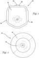
- FIG. 1is a top perspective view of a first entry model according to the present invention.
- FIG. 2is top perspective view of a first entry model according to the present invention.
- FIG. 3is a top perspective view of a laparoscopic trainer for use with a first entry model according to the present invention.
- FIG. 4is a side, exploded view of an anatomical portion of a first entry model according to the present invention.
- FIG. 5is a side view of an anatomical portion of a first entry model according to the present invention.
- FIG. 6is a top planar view that is representative of more than one layer in an anatomical portion of a first entry model according to the present invention.
- FIG. 7is a top planar view that is representative of more than one layer in an anatomical portion of a first entry model according to the present invention.
- FIG. 8is top perspective, exploded view of a mold for a skin layer of a first entry model according to the present invention.
- FIG. 9is a side, cross-sectional view of a mold for a skin layer for a first entry model according to the present invention.
- FIG. 10is a top perspective view of a mold for a skin layer for a first entry model according to the present invention.
- FIG. 11is a top perspective view of a mold for a skin layer for a first entry model according to the present invention.
- FIG. 12is a side, cross-sectional view of a mold for a skin layer for a first entry model according to the present invention.
- FIG. 13is an exploded view of a first entry model according to the present invention.
- FIG. 14is a side view of an anatomical portion of a first entry model according to the present invention.
- FIG. 15is a bottom planar view of a transversalis fascia layer and umbilical stalk according to the present invention.
- FIG. 16 Ais an end view of a standard first entry model connected to a top cover of a trainer according to the present invention.
- FIG. 16 Bis an end view of an obese first entry model connected to a top cover of a trainer according to the present invention.
- FIG. 17is a top planar view that is representative of more than one layer in an anatomical portion of a first entry model according to the present invention.
- FIG. 18is a top planar view that is representative of more than one layer in an anatomical portion of a first entry model according to the present invention.
- FIG. 19is a top planar view that is representative of more than one layer in an anatomical portion of a first entry model according to the present invention.
- the model 10includes an anatomical portion 12 connected to a support 14 to form a substantially planar configuration.
- the support 14is a frame that encompasses and connects to the perimeter of the anatomical portion 12 and holds the anatomical portion 12 together.
- the support 14includes a top frame and a bottom frame made of plastic material sufficiently rigid to provide structural support and maintain the planar shape of the model 10 and permit the center-located anatomical portion to be penetrated from one side to the other.
- the model 10is slightly curved to mimic an outwardly curved abdomen.
- the top frame and the bottom frameconnect together capturing the perimeter of the anatomical portion 12 between the top and bottom frames.
- the model 10 in FIG. 1is polygonal having five sides forming a slightly elongated shape wherein one side is curved outwardly in a generally U-shaped configuration.
- a model 10 having a circular support 14 that frames a circular anatomical portion 12is shown in FIG. 2 .
- the model 10can be any shape.
- the frame 14includes connecting elements 16 configured for connecting the model 10 to a larger laparoscopic trainer 20 as shown in FIG. 3 .
- a laparoscopic trainer 20includes a top cover 22 connected to a base 24 by a pair of legs 26 spacing the top cover 22 from the base 24 .
- the laparoscopic trainer 20is configured to mimic the torso of a patient such as the abdominal region.
- the top cover 22is representative of the anterior surface of the patient and a space 28 defined between the top cover 22 and the base 24 is representative of an interior of the patient or body cavity where organs reside.
- the laparoscopic trainer 20is a useful tool for teaching, practicing and demonstrating various surgical procedures and their related instruments in simulation of a patient.
- the top cover 22When assembled, the top cover 22 is positioned directly above the base 24 with the legs 26 located substantially at the periphery and interconnected between the top cover 22 and base 24
- the top cover 22 and base 24are substantially the same shape and size and have substantially the same peripheral outline.
- the laparoscopic trainer 20includes a top cover 22 that angulates with respect to the base 24 .
- the legs 26are configured to permit the angle of the top cover 22 with respect to the base 24 to be adjusted.
- FIG. 3illustrates the trainer 20 adjusted to an angulation of approximately 30-45 degrees with respect to the base 24 .
- a laparoscopic trainer 20is described in co-pending U.S. patent application Ser. No. 13/248,449 entitled “Portable laparoscopic trainer” and filed on Sep. 29, 2011 by Pravong et al. to Applied Medical Resources Corporation and published as U.S. Patent Application Publication No. 2012/0082970, hereby incorporated by reference in its entirety herein.
- surgical instrumentsare inserted into the cavity 28 of the laparoscopic trainer 20 through pre-established apertures 30 in the top cover 22 .
- These pre-established apertures 30may include seals that simulate trocars or may include simulated tissue that simulates the patient's skin and abdominal wall portions.
- the circular first entry model 10 depicted in FIG. 2is connected to the top cover 22 in the location of the central circular aperture 30 that has a conforming circular shape.
- the top cover 22 of the laparoscopic trainer 20is configured with a removable insert 32 that is replaceable with the first entry model 10 depicted in FIG. 1 .
- the insert 32which is provided with apertures 30 , has a shape that conforms to an opening in the top cover 22 .
- the first entry model 10such as the one depicted in FIG. 1 , having a conforming shape is inserted into the opening in the top cover 20 and the connecting elements 16 on the first entry model 10 aid in securing the model 10 to the trainer 20 .
- Various tools and techniquesmay be used to penetrate the top cover 20 as described in the background of this description to perform mock procedures not only on the model 10 but also on additional model organs placed between the top cover 22 and the base 24 .
- an organ modelWhen placed inside the cavity 28 of the trainer 20 , an organ model is generally obscured from the perspective of the user who can then practice performing surgical techniques laparoscopically by viewing the surgical site indirectly via a video feed displayed on a video monitor 34 .
- the video display monitor 34is hinged to the top cover 22 and is shown in an open orientation in FIG. 3 .
- the video monitor 34is connectable to a variety of visual systems for delivering an image to the monitor 34 .
- a laparoscope inserted through one of the pre-established apertures 30 or a webcam located in the cavity 28 and used to observe the simulated procedurecan be connected to the video monitor 34 and/or a mobile computing device to provide an image to the user.
- the first entry model 10is removed and may be replaced with a new insert or reconstructed and reconnected to the trainer 20 to allow training to continue or be repeated.
- the first entry model 10may be employed independently of the trainer 20 for practicing first entry techniques.
- the anatomical portion 12includes a skin layer 40 , an umbilical stalk 42 , a fat layer 44 , an anterior rectus sheath layer 46 , a first rectus muscle layer 48 , a second rectus muscle layer 50 , a third rectus muscle layer 52 , a posterior rectus sheath layer 54 , a transversalis fascia layer 56 , and a peritoneum layer 58 .
- the layers 40 , 44 , 46 , 48 , 50 , 52 , 54 , 56 , 58are placed one on top of the other as shown in FIGS.
- the layers 40 , 44 , 46 , 48 , 50 , 52 , 54 , 56 , 58are connected together with adhesive or other fastener.
- the layers 40 , 44 , 46 , 48 , 50 , 52 , 54 , 56are connected with at least one price-tag holder punched through the layers and sandwiched between the skin layer 40 and the peritoneum layer 58 before being attached to the frame 14 .
- the layersare held together without adhesive or other fastener and are clamped between the top frame and bottom frame.
- An optional inferior epigastric vein and artery layer 60is included between the posterior rectus sheath layer 54 and the transversalis fascia layer 56 as shown in FIGS. 4 - 5 .
- the skin layer 40is molded of silicone or thermoplastic elastomer dyed with a flesh color.
- the skin layer 40includes a top surface 62 and bottom surface 64 defining a thickness of approximately 0.1 inches.
- the skin layer 40includes an integrally formed umbilical stalk portion 42 a . The skin layer 40 will be described in greater detail below.
- the fat layer 44is made of cellular polyethylene foam having a yellow color.
- the cellular foam layeris not solid but textured with air bubbles.
- the fat layer 44is approximately 0.625 inches thick.
- the anterior rectus sheath layer 46is made of solid ethylene vinyl acetate (EVA) foam having a white color and is approximately 1 millimeter thick.
- the first rectus muscle layer 48is made of solid EVA foam and is red in color and approximately 1 millimeter thick.
- the second rectus muscle layer 50is made of cellular polyethylene foam having a pink color.
- the second rectus muscle layer 50is cellular foam that includes air bubbles that provide a cellular texture and is approximately 0.125 inches thick.
- the third rectus muscle layer 52is made of solid EVA foam having a red color and is approximately 1 millimeter thick.
- the posterior rectus sheath layer 54is made of solid EVA foam that is white in color and is approximately 1 millimeter thick.
- the transversalis fascia layer 56is made of cellular polyethylene foam that is white in color and approximately 0.25 inches thick.
- the fascia layer 56has a cellular texture arising from the cellular polyethylene foam as opposed to the solid EVA foam layers.
- the peritoneum layer 58is made of solid EVA foam that is white in color and approximately 1 millimeter thick.
- the inferior epigastric vein and artery layer 60 layerinclude solid or hollow elongate cylindrical structures made of silicone or Kraton® polymer or other elastomer having a cross-sectional diameter of approximately 0.15 inches.
- the arteriesare red in color and the veins are blue in color.
- the layers as described aboveprovide an optical entry with a very realistic appearance to the end user.
- Cellular polyethylene foamis also called closed cell polyethylene foam.
- FIG. 6there is shown a top planar view that is representative of the fat layer 44 , the posterior rectus sheath layer 54 , the transversalis fascia layer 56 and the peritoneum layer 58 . These layers are approximately six inches wide and six and a half inches long.
- the fat layer 44 , the posterior rectus sheath layer 54 , the transversalis fascia layer 56 and the peritoneum layer 58all have a circular aperture 66 that is approximately one inch in diameter.
- the aperture 66is located approximately two inches from one side and is in the same place in all of these layers 44 , 54 , 56 , 58 such that when overlaid the apertures 66 line up to provide a pathway for the umbilical stalk 42 across these layers.
- FIG. 7there is shown a top planar view that is representative of the anterior rectus sheath layer 46 , first rectus muscle layer 48 , the second rectus muscle layer 50 and the third rectus muscle layer 52 . These layers are approximately six inches wide and six and a half inches long.
- the anterior rectus sheath layer 46 , first rectus muscle layer 48 , the second rectus muscle layer 50 and the third rectus muscle layer 52all have an elongate opening 68 .
- the elongate opening 68extends along the center line of the layers and is shown in FIG. 7 to be a rectangular cut out that is approximately one inch wide and 5.75 inches long.
- the elongate opening 68represents the linea alba of the abdomen.
- the skin layer 40is formed by pouring the uncured and dyed silicone or thermoplastic elastomer into a special mold 70 .
- An exploded, top perspective view of the mold 70is shown in FIG. 8 .
- the mold 70includes a base 72 , a top 74 , and a core 76 .
- the base 72 of the mold 70includes a cavity 78 for receiving the plastic material.
- the cavity 78is polygonal and substantially rectangular in shape.
- the cavity 78includes a first floor 79 that surrounds a well 80 having a second floor 82 .
- the second floor 82 of the well 80is approximately 1 inch below the first floor 79 and includes a hole for inserting the core 76 inside the well 80 .
- the cross-section of the well 80is elliptical in shape having a long axis of approximately 1 inch and a short axis of approximately half an inch.
- the cross-section of the core 76is also elliptical in shape, complementary to the well 80 .
- the core 76has a long axis of approximately 0.75 inches and a short axis of approximately 0.25 inches.
- a space of approximately 1 ⁇ 8 inchis formed all around the core 76 between the outer surface of the core 76 and the inner surface of the well 80 into which silicone or thermoplastic elastomer is poured to form a tubular structure of the umbilical stalk 42 a having an opening 92 .
- the core 76is approximately one inch and a half in length and extends above the pour line when inside the well 80 .
- the mold cavity 78further includes a circumferential well 84 that is formed circumferentially around the first well 80 .
- the circumferential well 84has a concave or curved floor 86 that is approximately 1 ⁇ 8 inch deeper from the first floor 79 .
- silicone or thermoplastic elastomeris poured, an elliptical toroidal shape with a flat top is formed in the plastic material resulting in an increased thickness of material of approximately 0.25 inch in the area of the circumferential well 84 in the final product.
- the circumferential well 84has an inner perimeter 88 that coincides with the wall of the first well 80 .
- the annular distance from the inner perimeter 88 of the circumferential well 84 to the outer perimeter or end of circumferential well 84is approximately 0.75 inches.
- the base 72 of the mold 70further includes a plurality of pegs 90 upstanding from the first floor 79 to form holes in the resulting molded material.
- the first well 80is described to have an elliptical shape, in another variation it is circular in shape with a corresponding circular core and circular circumferential well.
- the core 76is first inserted into the well 80 and silicone or thermoplastic elastomer is poured into the base 72 of the mold 70 .
- the silicone or thermoplastic elastomerwill run into the well 80 forming a tubular structure defined by the space between the core 76 and wall of the well 80 .
- the silicone or thermoplastic elastomerwill also run into the circumferential well 84 and cover the concave floor 86 forming a substantially toroidal shape of increased thickness of approximately 0.25 inch.
- the circumferential portion of increased thickness 94is visible in FIGS. 4 and 5 .
- the silicone or thermoplastic elastomer in its liquid statewill cover the first floor 79 forming a planar area having a thickness of approximately 1 ⁇ 8 inch.
- the top 74 of the mold 70will be placed over the base 72 of the mold 70 .
- the top 74is configured to cover only the perimeter of the poured silicone or thermoplastic elastomer to reduce the thickness of the silicone around the perimeter.
- the top 74 of the moldis removed and the molded silicone or thermoplastic elastomer is removed from the mold 70 .
- the core 76is also removed from the material leaving an elliptical opening 92 through the skin layer 40 .
- the tubular structure or umbilical stalk 42 athat is integrally formed by the well 80 with the rest of the skin layer 40 defines an opening 92 and is elliptical in shape having long axis of approximately 0.75 inches and a short axis of approximately 0.25 inches with a wall thickness of approximately 1 ⁇ 8 inch.
- the tubular structure 42 ais inverted, that is, it is pushed through the opening 92 such that the surface in contact with the floor 79 of the mold 70 becomes the skin layer top surface 62 .
- the portion of increased thickness 94 of the skin layer 40will advantageously create a raised surface at the skin layer top surface 62 which is clearly visible in FIGS. 4 and 5 .
- This raised portion 94advantageously provides extra thickness of material for drawing sutures through and maintaining them in position without pulling through the silicone or thermoplastic material.
- a circumferential raised portion 94 that surrounds the opening 92creates a realistic belly-button effect that can be seen in FIG. 1 .
- a variation of the skin layer 40 without the raised circumferential portion 94is shown in FIG. 2 .
- the skin layer 40is planar sheet of molded material having a top surface 62 and a bottom surface 64 defining a skin layer thickness of approximately 0.1 inches.
- the skin layer 40further includes an opening 92 with a tubular extension 42 integrally formed at opening 92 and interconnected with the rest of the layer 40 .
- Surrounding the opening 92is a circumferential raised portion 94 of increased thickness of approximately 0.2 inches.
- the raised portion 94provides a convex outer surface that transitions into the remainder of the top surface 62 of the skin layer 40 .
- the mold 70is 3D printed from Vero White Plus Fullcure 835 material.
- the distance from the pour line to the floor 79is approximately 0.1 inches to create a skin layer thickness of approximately 0.1 inches.
- the thickness beneath the top 74 of the mold 70is reduced to approximately 0.05 inches for a resulting skin layer thickness at the perimeter having a reduced thickness of approximately 0.05 inches which facilitates connection to the frame support 14 .
- the thickness of the resulting skin layer 40is approximately 0.2 inches.
- the mold 70is sprayed with mold release solution and allowed to dry. In one variation, approximately 5 grams of Dragon Skin Silicone comprising 2.5 grams of part A and 2.5 grams of part B is mixed.
- thermoplastic elastomersuch as Kraton CL2003X is used for its cost savings and its ability to be sutured.
- Approximately 20 microliters of fleshtone coloris mixed into the silicone.
- the core 76is inserted into the well 80 and the silicone mixture is poured into the mold base 72 .
- the mixtureis spread evenly up to a pour line making sure all the wells are filled.
- the top 74is placed over the base 72 of the mold 70 . Excess silicone mixture is cleaned away and the silicone inside the mold 70 is allowed to dry for approximately one hour under a heat lamp or for two hours without a heat lamp.
- the top 74is removed and the formed skin layer 40 is peeled and removed from the base 72 .
- the core 76is also removed.
- the integrally formed umbilical stalk 42is inverted by passing it through a formed opening 92 .
- Silicone adhesiveis provided and delivered using a syringe to the inside of the tube of the umbilical stalk 42 .
- One or more clamps and in one variation, three clamps, such as binder clips,are used to clamp the inverted umbilical stalk 42 closed and sealed to create a bellybutton shape having a star or Y-shaped closure as shown in FIG. 1 or 2 .
- an umbilical shaft 42 bis provided.
- the umbilical shaft 42 bis tubular having a central lumen and made of a thin layer of white silicone that is approximately 1 mm thick.
- the umbilical shaft 42 bis glued to the umbilical stalk 42 a to extend the umbilicus deeper into the layers and create a more realistic look and feel.
- the umbilical shaft 42 bis glued to the umbilical stalk 42 a such that the lumens interconnect.
- the proximal end of the umbilical shaft 42 bis place over the stalk 42 a and glued thereto and the distal end of the umbilical shaft 42 b is free.
- the distal end of the umbilical shaftis glued or integrally formed with the peritoneum layer 58 .
- All of the layersare properly oriented in the same direction and aligned such that the apertures 66 and openings 68 are superimposed. Then, with the skin layer 40 inverted and the umbilical stalk 42 a either alone or with an extended umbilical shaft 42 b is passed through the circular aperture 66 of the fat layer 44 and through the elongate openings 68 of the anterior rectus sheath layer 46 , the first rectus muscle layer 48 , the second rectus muscle layer 50 , and the third rectus muscle layer 52 and then through the circular apertures 66 of the posterior rectus sheath layer 54 , the transversalis fascia layer 56 and the peritoneum layer 58 as shown in FIG. 5 .
- the umbilicus 42is left meeting the peritoneum layer 58 or in another variation, the umbilicus 42 is attached with adhesive to the peritoneum layer 58 and yet in another variation, integrally molded with the peritoneum layer 58 .
- the inferior epigastric vein and artery layer 60is optionally included. This layer 60 can be formed as a layer having a circular aperture 66 with embedded arteries and veins or simply comprise a pair of cylindrical silicone structures, one red and one blue, placed on one side of the midline and another pair of cylindrical silicone structures, one red and one blue in color, placed on the other side of the midline as shown in FIG. 4 .
- the cylindrical silicone structures representing the epigastric veins and arteriesare glued to at least one of the adjacent posterior rectus sheath layer 54 and the transversalis fascia layer 56 .
- a price tag holder or other fastenercan then be used to connect the layers together as shown in FIG. 5 with the umbilicus 42 shown protruding from the aperture 66 in the bottom-most peritoneum layer 58 .
- the skin layer 50 and the peritoneum layer 58is slightly larger than the other internal layers 44 , 46 , 48 , 50 , 52 , 54 , 56 .
- the skin layer 50 and peritoneum layer 58are larger by approximately 1.25 inches in length and width.
- the internal layersare approximately 6.5 inches long and 6 inches wide
- the peritoneum layer 58 and skin layer 40is approximately 8 inches long and 7.5 inches wide.
- These extra length and width portionsare captured between the top and bottom frames of the support 14 .
- Pegs in one of the top or bottom framesare passed through apertures in the skin layer 40 formed by mold pegs 90 .
- the peritoneum layer 58may also include apertures for passing of frame pegs.
- the top frame and bottom frameare then heat staked together capturing the anatomical portion 12 .
- the resulting model 10is approximately 1.5 inches thick.
- the first entry model 10is then placed inside an opening in the top cover 22 of a laparoscopic trainer 20 and securely attached.
- Laparoscopic first entry proceduressuch as the ones discussed in the background of this specification are then practiced on the model 10 employing one or more of the trocar instruments described above creating first entry in any of the locations described above including first entry directly through the umbilicus. Another location for first entry could be within a half inch on either side of the midline.
- the practitionerwill advantageously and quickly recognize a mistaken first approach when only the skin layer 40 , the fat layer 44 and posterior rectus sheath 54 and peritoneum 58 layers are observed at the linea alba.
- first entry penetrationcan take place at the left upper quadrant or right upper quadrant.
- the left upper quadrantis different from the umbilicus region in that there are muscle layers. While penetrating at the upper right or left quadrants, the practitioner will observe the following layers: the skin layer 40 , the fat layer 44 , the anterior rectus sheath layer 46 , the first rectus muscle layer 48 , the second rectus muscle layer 50 , the third rectus muscle layer 52 , the posterior rectus sheath layer 54 , the transversalis fascia layer 56 and the peritoneum layer 58 .
- the layersare configured such that first entry through the umbilicus 42 will not penetrate any of the layers or will only penetrate the skin layer 40 .
- the mold 70is made of a polymer known as Delrin® and includes a base 72 , a top 74 , and a core 76 .
- the base 72 of the mold 70includes a cavity 78 for receiving the plastic material.
- the cavity 78which is approximately 0.1 inches deep, is in the shape of a large abdominal wall frame configured to hold all the layers of the model.
- the cavity 78includes a first floor 79 that surrounds a well 80 having a second floor 82 .
- the second floor 82 of the well 80includes a hole for inserting the core 76 inside the well 80 .
- the cross-section of the well 80is elliptical in shape having a long axis of approximately 1 inch and a short axis of approximately half an inch.
- the well 80is approximately three inches from one side of the cavity 78 and approximately three inches from the curved side of the cavity 78 and approximately 0.75 inches deep.
- the well 80includes a secondary well at the second floor 82 which is also an ovular cutout that has a long axis of approximately 0.5 inches and a short axis of approximately 0.2 inches and approximately 0.1 inches deep.
- the secondary wellis used to align the core 76 within the well 80 .
- the first well 80is described to have an elliptical shape, in another variation, the first well 80 is circular in shape with a corresponding circular core.
- the cross-section of the core 76is also elliptical in shape, complementary to the well 80 .
- the core 76In a cross-section taken perpendicular to the longitudinal axis of the core 76 , the core 76 has a long axis of approximately 0.75 inches and a short axis of approximately 0.25 inches.
- a space of approximately 1 ⁇ 8 inchis formed all around the core 76 between the outer surface of the core 76 and the inner surface of the well 80 into which silicone or thermoplastic elastomer is poured to form a tubular structure of the umbilical stalk 42 a having an opening 92 .
- the core 76is approximately one inch and a half in length and extends above the pour line when inside the well 80 .
- the base 72 of the mold 70further includes a plurality of pegs 90 for forming apertures through which pegs will pass for securing the skin layer 40 to the frame 14 .
- the core 76is first inserted into the well 80 and silicone or thermoplastic elastomer is poured into the base 72 of the mold 70 .
- the silicone or thermoplastic elastomerwill run into the well 80 forming a tubular structure defined by the space between the core 76 and wall of the well 80 .
- the silicone or thermoplastic elastomer in its liquid statewill cover the first floor 79 forming a planar area having a thickness of approximately 1 ⁇ 8 inch.
- the top 74 of the mold 70will be placed over the base 72 of the mold 70 .
- the top 74includes a through-hole having the same shape as the cavity 78 but sized slightly larger so as to cover only the perimeter of the poured silicone or thermoplastic elastomer.
- the top 74includes a lip of approximately 0.39 inches in length that extends vertically approximately 0.05 inches.
- the lipis configured to create a flat edge around the skin layer that is only 0.05 inches allowing the skin layer to be easily heat staked in the location of the edge after assembly.
- the model 10includes an anatomical portion 12 connected between two parts of a frame-like support 14 .
- the frame-like support 14includes a top frame having protrusions that snap through the skin layer 40 and into apertures formed in a bottom frame.
- the anatomical portion 12includes a skin layer 40 , an umbilical stalk 42 , a fat layer 44 , an anterior rectus sheath layer 46 , a first rectus muscle layer 48 , a second rectus muscle layer 50 , a third rectus muscle layer 52 , a posterior rectus sheath layer 54 , a transversalis fascia layer 56 , and a peritoneum layer 58 .
- the layers 40 , 44 , 46 , 48 , 50 , 52 , 54 , 56 , 58are placed one on top of the other as shown in FIGS. 13 - 14 with the umbilical stalk 42 penetrating through all of the layers beneath the skin layer 40 except for the peritoneum layer 58 .
- the layers 40 , 44 , 46 , 48 , 50 , 52 , 54 , 56 , 58are connected together with adhesive or other fastener.
- the layers 40 , 44 , 46 , 48 , 50 , 52 , 54 , 56 , 58are connected with at least one price-tag holder 100 punched through the layers and sandwiched between the skin layer 40 and the peritoneum layer 58 before being attached to the frame 14 .
- the layersare held together without adhesive or other fastener and clamped between the top frame and bottom frame.
- An optional inferior epigastric vein and artery layer 60is included between the posterior rectus sheath layer 54 and the transversalis fascia layer 56 as shown in FIGS. 13 - 14 .
- the skin layer 40is molded of silicone or thermoplastic elastomer (TPE) dyed with a flesh color.
- the skin layer 40includes a top surface and bottom surface defining a thickness of approximately 0.1 inches.
- the skin layer 40includes an integrally formed tubular umbilical stalk portion 42 a having a central lumen formed by the core 76 during the molding process.
- An umbilical shaft 42 bmay be formed together with the umbilical stalk 42 a or connected to the umbilical stalk 42 a or placed as a separate tubular portion within the anatomical portion 12 .
- the umbilical stalk 42is made of a thin layer of white silicone that is approximately 1 millimeter thick.
- the umbilical stalk 42 aby itself or together with the umbilical shaft 42 b is configured to be long enough to travel through all the layers 44 , 46 , 48 , 50 , 52 , 54 and 56 until it reaches between the transversalis fascia layer 56 and the peritoneum layer 58 .
- the distal end of the umbilical stalk 42(or umbilical shaft 42 b if one is employed) is cut one or more times such that the cut extends from the distal end of the umbilical stalk towards the proximal end of the umbilical stalk. Several cuts are provided at a length to sufficiently flare the distal end of the umbilical stalk.
- four or more cutsare formed to form four or more pieces or flaps at the distal end of the simulated umbilicus 42 .
- These flaps 102are fanned out over the distal-facing surface of the transversalis fascia layer 56 as shown in FIG. 15 .
- the umbilical stalk 42is adhered to the transversalis fascia layer 56 using two types of adhesive. Because the transversalis fascia layer 56 is made of cellular polyethylene foam which is porous, the surface insensitive cyanoacrylate glue cannot be used alone to adhere the silicone because it will burn through the foam and not adhere. Therefore, a heavy duty spray adhesive is sprayed on the foam transversalis fascia layer 56 and allowed to dry for a few minutes.
- the surface insensitive cyanoacrylate glueis then placed on the silicone umbilical stalk 42 and the distal flaps 102 of the stalk 42 are adhered to the distal-facing surface of the transversalis fascia layer 56 .
- the spray adhesivewhich alone is not strong enough to bond the foam and the silicone, protects the foam from the cyanoacrylate.
- the fat layer 44needs to react similarly to real fat when grasped or touched externally and it needs to look like fat under optical entry and to respond physically like to fat when pierced internally.
- the fat layer 44is made of cellular foam that is porous, sponge-like and yellow in color. The yellow foam looks like fat under optical entry.
- the fat layer 44is made of polyurethane foam that is yellow in color. Memory foam is polyurethane with additional chemicals increasing its viscosity and density. It is also called viscoelastic polyurethane foam or low-resilience polyurethane foam or polyurethane foam having a slow recovery.
- the memory foamfeels realistic when the user touches the model 10 at the skin layer 40 and also when the user enters the fat layer 44 optically with a trocar.
- the polyurethane fat layer 44shines advantageously creating the illusion that the fat is wet internally.
- the polyurethane foamrecovers its shape.
- the ability of the fat layer 44 to recover its shapeis important for the Hasson cut-down technique because the surgeon must practice retracting the fat layer 44 before cutting the fascia. The practice is more realistic if the fat layer 44 tends to return to its original location requiring the practitioner to retract the fat layer 44 .
- the fat layer 44is made of a thermoplastic elastomer (TPE) with an additive such as baking soda or mineral oil to create a material that acts more like real fat.
- TPEthermoplastic elastomer
- An additive such as baking sodawill create a porous fat layer allowing the trocar to easily pierce and enter the fat layer 44 and advantageously provide a more realistic appearance under optical entry.
- An additive such as mineral oilwill create a gel that has the shape-recovery characteristics similar to the memory foam but provides a more realistic feel when touched externally.
- TPE with either the mineral oil or baking soda as an additiveprovides a tactile response similar to fat when grasped.
- the fat layer 44is approximately 1.5-4.0 cm thick in a standard model 10 . An obese model 10 will be described hereinbelow.
- the skin layer 40is attached to the fat layer 44 .
- the skin layer 40is cast over the fat layer 44 .
- the silicone or TPE of the skin layer 40will adhere to the fat layer 44 located directly below the skin layer 40 as it cures/cools.
- the mold 70is made deeper to receive the fat layer 44 .
- the umbilical stalkcannot be inverted because the silicone or the TPE is poured over the fat layer and attaches thereto as it cures. Therefore, the core 76 is a different shape than described above with respect to FIGS.
- the core 76is shaped such that the cured silicone results in a shape that simulates an inverted umbilicus.
- the top of the core 76may be provided with a recess with texturing that simulates the belly button as viewed from outside the patient.
- the fat layer 44is placed into the mold base 72 that is modified with a larger receptacle for receiving a fat layer 44 and the silicone or TPE is be poured over it and then the umbilicus-shaped core 76 may be previously placed into a well or is placed on top to mold the umbilicus shape into the silicone skin layer 40 without inverting or gluing a lumen of the umbilical stalk 42 .
- the step of inverting the skin layer 40 and pinched together to create the umbilicus shapewould not be needed.
- an obese modelis provided in the present invention.
- the obese modelincludes all of the same layers as shown in FIGS. 13 - 14 but includes a fat layer 44 that is significantly thicker.
- the fat layer 44 of the obese modelcan be made of the same materials already described herein. Whereas the thickness of the standard fat layer 44 is approximately 1.5 to 4.0 cm, the fat layer 44 in the obese model is approximately 4.0 to 7.0 cm.
- the obese modelalso includes a special skin layer 40 .
- the skin layer 40can be made as previously stated herein and be of the same size in the x-y plane as the skin layer in the standard model or the same size in the x-y plane as the fat layer in the obese model or, alternatively, the skin layer 40 can be larger in size with respect to the size of the fat layer of the obese model in the x-y plane or larger in size with respect to the size of the fat layer of the standard model. If the skin layer is the same size and shape, the obese model 10 b will have a domed effect as can be seen in FIG. 16 B when compared to a standard model 10 a illustrated in FIG. 16 A .
- the same-sized skin layer 40 in combination with the thicker fat layer 44 or otherwise a skin layer 40 that is the same size or is slightly smaller than the dimensions of the fat layer 44will result in the thicker fat layer(s) 44 of the obese model being compressed into the same space previously made for the standard model.
- This compressionprovides the obese model 10 A with the appearance of an obese patient when using any of the four laparoscopic entry techniques.
- the obese model 10 Awill not be easily and realistically grasped with the smaller and tighter skin layer 40 encompassing the larger fat layer 44 ; however, a larger skin layer 40 can be employed. If TPE or memory foam is used for the fat layer 44 , the larger skin layer 40 will allow the fat layer 44 to expand into the extra space of a larger skin layer 40 when gasped and moved.
- FIGS. 16 A and 16 Billustrate a laparoscopic trainer 20 with legs 26 removed such that the top cover 22 is seated directly onto the base 24 of the trainer 20 reducing the size of the cavity 28 such that first entry procedures may be more easily and conveniently practiced.
- the top cover 22forms a shell over the base 24 and fits securely around an upstanding lip so that the top cover 22 does not dislocate with respect to the base 24 .
- the first entry model 10is inserted into an aperture 30 in the top cover 22 of the trainer 20 and a simulated organ is placed into the cavity 28 of the trainer 20 such that when a practitioner enters through the first entry model 10 by piercing the various layers, the practitioner will see the simulated organ located within the cavity 28 .
- One or more organsmay be placed inside the cavity 28 .
- at least a simulated omentumis provided inside the cavity 28 .
- the simulated omentumis made of a sheet of fabric or thin layer of silicone.
- the sheetis placed inside the cavity 28 of the trainer 20 and the sheet is configured such that when the first entry model 10 is pierced by an instrument such as an optical trocar having a laparoscope inserted into the trocar, the practitioner will see the sheet on the video display monitor.
- the sheetis suspended within the cavity 28 using clips attached to the trainer 20 .
- the sheetmay be placed on a frame or just laid over the base.
- the thin sheet of material, representing the omentumis yellow in color and loosely connected to the trainer and is configured such that it would flutter when insufflation gasses are delivered into the cavity such as with an insufflation trocar after piercing the first entry model 10 .
- the representative omentum layeris attached to the trainer selectively leaving portions of the simulated omentum unattached to enable the flutter effect.
- the presence of the simulated omentum layer comprising a thin sheetis advantageous because when a surgeon first enters into the abdominal cavity and insufflation is delivered to expand the abdomen in order to create a working space, the surgeon knows that the abdominal wall was successfully entered when visually the representative omentum or viscera is observed and further seen fluttering with the force of insufflation gasses.
- This training featureis advantageously provided in the present invention in the combination of the first entry model 10 , a trainer 20 and simulated omentum such as that depicted in FIG. 3 or FIGS. 16 A and 16 B of the present invention.
- simulated omentum sheetwith the trainer 20 configured as shown in FIGS. 16 A and 16 B advantageously provides a smaller space for the cavity 28 , creating a more air-tight and dark location to simulate insufflation and observe the fluttering of the simulated omentum.
- the anterior rectus sheath layer 46is made of solid ethylene vinyl acetate (EVA) foam having a white color and is approximately 1 millimeter thick.
- the first rectus muscle layer 48is made of solid EVA foam and is red in color and approximately 1 millimeter thick.
- the second rectus muscle layer 50is made of cellular polyethylene foam having a pink color.
- the second rectus muscle layer 50comprises two layers 50 a , 50 b of cellular polyethylene foam having a total thickness of approximately 0.25 inches.
- the second rectus muscle layer 50is cellular foam that includes air bubbles that provide a cellular texture.
- Each second rectus muscle layer 50 a , 50 bis approximately 0.125 inches thick.
- the third rectus muscle layer 52is made of solid EVA foam having a red color and is approximately 1 millimeter thick.
- the posterior rectus sheath layer 54is not made of foam material, but instead, is made of an interfacing fabric.
- the interfacing fabricis made of strong polyester fibers that can stretch considerably before ripping.
- the interfacing fabricis thin being approximately 0.2 mm thick and white in color.
- the interfacing fabric layer 54is thin enough to allow a trocar or Veress needle to puncture through the fabric when using an entry tactic other than a Hasson cut down technique and capable of being cut when employing the Hasson cut down technique.
- the posterior rectus sheath layer 54 in the modelrepresents the fascia of both the anterior and posterior rectus sheath that come together at the linea alba.
- the fabric of the posterior rectus sheath layer 54represents the linea alba configured by exposing the posterior rectus sheath layer through and by way of an elongate opening 68 formed in anterior rectus sheath layer 46 , first rectus muscle layer 48 , second rectus muscle layer 50 and third rectus muscle layer 52 .
- the elongate opening 68 in each of these layersare shown in FIG. 17 .
- the fascia of the linea alba as represented by the posterior rectus sheath layer 54is grasped and pulled through the incision in order to safely incise the layer 54 .
- the stretchable fabric layer 54advantageously provides ability to pull the fascia layer up so that safe cutting techniques may be practiced using this model.
- the transversalis fascia layer 56is made of cellular polyethylene foam that is white in color and approximately 0.25 inches thick.
- the fascia layer 56has a cellular texture arising from the cellular polyethylene foam as opposed to the solid EVA foam layers.
- the peritoneum layer 58is made of solid EVA foam that is white in color and approximately 1 millimeter thick.
- the peritoneum layer 58may also be made of silicone or TPE.
- the optional inferior epigastric vein and artery layer 60 layerincludes solid or hollow elongate cylindrical structures made of silicone or Kraton® polymer or other elastomer having a cross-sectional diameter of approximately 0.15 inches.
- the arteriesare red in color and the veins are blue in color.
- the layersas described above, provide an optical entry with a very realistic appearance to the end user.
- the layers of foamare capable of being punctured with a trocar and look realistic under optical entry via a laparoscope inserted into an optical trocar. Also, the foam layers provide a realistic tactile feedback to the practitioner when using Veress needle entry as well as with optical entry.
- the thicknesses, colors and compositions of the various layers of the abdominal wall of the first entry model 10are shown in Table 1 below.
- FIG. 17there is shown a top planar view that is representative of the anterior rectus sheath layer 46 , first rectus muscle layer 48 , the second rectus muscle layer 50 and the third rectus muscle layer 52 . These layers are approximately six inches wide and six and a half inches long.
- the anterior rectus sheath layer 46 , first rectus muscle layer 48 , the second rectus muscle layer 50 and the third rectus muscle layer 52all have an elongate opening 68 .
- the elongate opening 68extends along the center line of the layers and is shown in FIG. 17 to be a substantially rectangular cut out that is approximately one inch wide and approximately 5.75 inches long.
- the elongate opening 68represents the lack of muscle at the linea alba.
- the linea albavaries between patients and in other variations of the model, the width of the elongate opening 68 can range from 8 mm to 30 mm.
- the shape of the openingmay also vary.
- FIG. 18there is shown a top planar view that is representative of the fat layer 44 and the peritoneum layer 58 . These layers are approximately six inches wide and six and a half inches long.
- the fat layer 44 and the peritoneum layer 58all have an ovular hole 66 that has a length of approximately one inch and a width of approximately 0.5 inches.
- the ovular hole 66is located approximately two inches from one side and is in the same location in the fat layer 44 and the peritoneum layer 58 such that when overlaid the ovular holes 66 line up to provide a pathway for the umbilical stalk 42 across these layers.
- the ovular hole 66closely hugs the umbilical stalk 42 compared with a circular hole advantageously providing a more realistic visualization.
- FIG. 19there is shown a top planar view that is representative of the posterior rectus sheath layer 54 and the transversalis fascia layer 56 .
- These layers 54 , 56include a slit 104 .
- the slit 104is approximately 1 inch in length and is a narrow cut substantially perpendicular to the representative linea alba so that the ends of the slit 104 are not aligned with the longitudinal axis of the linea alba.
- the slit 104allows the umbilical stalk 42 to pass through to its termination between the transversalis fascia layer 56 and the peritoneum layer 58 while still allowing these layers to touch or closely approximate the curvature of the umbilical stalk 42 .
- the posterior rectus sheath layer 54 and the transversalis fascia layer 56closely hug the umbilical stalk 42 which advantageously makes the visualization more realistic such that these layers are seen or felt during entry especially when employing a Hasson or Veress needle first entry.
- the one or more distal fat layer(s) 44are also configured with a slit 104 as shown in FIG. 19 ; whereas the proximal fat layer(s) 44 are configured with an ovular hole 66 as shown in FIG. 18 .
- the first entry model 10includes simulations for adhesions present in real anatomy. Frequently, organs and tissues located underneath the peritoneum will adhere to the peritoneum and create an adhesion. While practicing first entry techniques, it is necessary for the surgeon to learn how to be wary of adhesions and how to navigate with respect to them in the event they occur in the patient.
- the present inventionprovides a first entry model that allows the surgeon to practice encountering and navigating adhesions in a first entry laparoscopic environment. It is necessary for the surgeon to be careful, because aggressive entry in the location of an adhesion may result in accidental piercing of the adhered tissue or organ. In this variation of the first entry model 10 , adhesions are included in the model.
- a simulated adhesionis a piece of simulated bowel that is attached to the undersurface of the peritoneum layer 58 .
- the piece of simulated bowelis made of silicone.
- the adhesionmay be made of any suitable material and adhesive may be used to connect the adhesion to the peritoneum layer 58 .
- a piece of siliconeis used to attach the simulated bowel to the peritoneum layer 58 .
- the peritoneum layer 58may be made of silicone or TPE instead of foam in order to more easily attach a silicone adhesion to the peritoneum layer 58 .
- the peritoneum layer 58that is made of silicone or TPE will stretch as the adhesion is being removed making the simulation more realistic.
- a scar indicating a previous surgerymay be molded or printed onto the surface of the skin layer 40 in a location above the adhesion to the peritoneum layer; thereby, the surgeon would anticipate an adhesion being present in the general area beneath the layers in the abdominal cavity.
- the scarwould require the practitioner to make a decision about the best place to enter or pierce the first entry model 10 and thus adds an important practice dimension to the model 10 .
- a scarmay or may not be provided.
- an adhesionmay still be provided to surprise the practitioner adding yet another practice dimension to the first entry model 10 .
- the surgeoncan insert a grasper to pull at the adhesion such as a piece of bowel, stretch the adhesion away from the peritoneum and/or bowel, and use a scalpel or scissors to cut through the silicone that is located between the bowel and peritoneum layer 58 and used to attach the simulated adhesion to the peritoneum layer 58 in order to free the adhesion.
- the first entry model 10 of the present inventionis particularly suited for laparoscopic procedures and may be employed with a laparoscopic trainer 20 ; however, the invention is not so limited and the first entry model 10 of the present invention can be used alone to practice first entry surgical procedures equally effectively.
- the present inventionadvantageously provides numerous practice possibilities for the surgeon who is learning or practicing first entry techniques while at the same time being manufactured of simple silicone and foam materials providing maximum costs savings while also providing a most realistic tactile and visual experience.
- the first entry model 10may be used repeatedly allowing the surgeon to practice numerous entry techniques on the same model before discarding the model which can then be easily replaced with a new model when used with the laparoscopic trainer.
Landscapes
- Engineering & Computer Science (AREA)
- Physics & Mathematics (AREA)
- General Physics & Mathematics (AREA)
- Health & Medical Sciences (AREA)
- Theoretical Computer Science (AREA)
- Educational Technology (AREA)
- Educational Administration (AREA)
- Business, Economics & Management (AREA)
- Computational Mathematics (AREA)
- Pure & Applied Mathematics (AREA)
- Medicinal Chemistry (AREA)
- Medical Informatics (AREA)
- Mathematical Analysis (AREA)
- Mathematical Optimization (AREA)
- Mathematical Physics (AREA)
- Algebra (AREA)
- General Health & Medical Sciences (AREA)
- Chemical & Material Sciences (AREA)
- Radiology & Medical Imaging (AREA)
- Pulmonology (AREA)
- Instructional Devices (AREA)
- Endoscopes (AREA)
Abstract
Description
| TABLE 1 |
| Abdominal Wall Layers |
| Thickness | Thickness | |||
| Standard | Obese | |||
| Layer | Material | model | model | Color |
| Skin | Silicone or | 0.1″ | 0.1″ | Flesh |
| TPE | Tone | |||
| Fat | Cellular | 1.5 to | 4.0 to | Yellow |
| Foam | 4.0 cm | 7.0 cm | ||
| Memory | ||||
| Foam | ||||
| TPE with | ||||
| Additive | ||||
| Gel | ||||
| Anterior | Solid Foam | 1 mm | 1 mm | White |
| Rectus | ||||
| Sheath | ||||
| Rectus | Solid Foam | 1 mm | 1 mm | Red |
| Muscle | ||||
| Rectus | Cellular | ¼″ | ¼″ | Pink or |
| Muscle | Foam | White | ||
| Rectus | Solid Foam | 1 mm | 1 mm | Red |
| Muscle | ||||
| Posterior | Interfacing | 0.2 mm | 0.2 mm | White |
| Rectus | Fabric | |||
| Sheath | ||||
| Transversalis | Cellular | ¼″ | ¼″ | White |
| Fascia | Foam | |||
| Peritoneum | Solid Foam, | 1 mm | 1 mm | White |
| Silicone or | ||||
| TPE | ||||
Claims (20)
Priority Applications (2)
| Application Number | Priority Date | Filing Date | Title |
|---|---|---|---|
| US16/878,291US11854425B2 (en) | 2013-07-24 | 2020-05-19 | First entry model |
| US18/392,509US20240127714A1 (en) | 2013-07-24 | 2023-12-21 | First entry model |
Applications Claiming Priority (6)
| Application Number | Priority Date | Filing Date | Title |
|---|---|---|---|
| US201361857982P | 2013-07-24 | 2013-07-24 | |
| US201461971714P | 2014-03-28 | 2014-03-28 | |
| US14/340,234US9548002B2 (en) | 2013-07-24 | 2014-07-24 | First entry model |
| US15/370,231US10026337B2 (en) | 2013-07-24 | 2016-12-06 | First entry model |
| US16/030,696US10657845B2 (en) | 2013-07-24 | 2018-07-09 | First entry model |
| US16/878,291US11854425B2 (en) | 2013-07-24 | 2020-05-19 | First entry model |
Related Parent Applications (1)
| Application Number | Title | Priority Date | Filing Date |
|---|---|---|---|
| US16/030,696ContinuationUS10657845B2 (en) | 2013-07-24 | 2018-07-09 | First entry model |
Related Child Applications (1)
| Application Number | Title | Priority Date | Filing Date |
|---|---|---|---|
| US18/392,509ContinuationUS20240127714A1 (en) | 2013-07-24 | 2023-12-21 | First entry model |
Publications (2)
| Publication Number | Publication Date |
|---|---|
| US20200279508A1 US20200279508A1 (en) | 2020-09-03 |
| US11854425B2true US11854425B2 (en) | 2023-12-26 |
Family
ID=51359419
Family Applications (5)
| Application Number | Title | Priority Date | Filing Date |
|---|---|---|---|
| US14/340,234Active2035-01-20US9548002B2 (en) | 2013-07-24 | 2014-07-24 | First entry model |
| US15/370,231ActiveUS10026337B2 (en) | 2013-07-24 | 2016-12-06 | First entry model |
| US16/030,696ActiveUS10657845B2 (en) | 2013-07-24 | 2018-07-09 | First entry model |
| US16/878,291Active2035-10-14US11854425B2 (en) | 2013-07-24 | 2020-05-19 | First entry model |
| US18/392,509PendingUS20240127714A1 (en) | 2013-07-24 | 2023-12-21 | First entry model |
Family Applications Before (3)
| Application Number | Title | Priority Date | Filing Date |
|---|---|---|---|
| US14/340,234Active2035-01-20US9548002B2 (en) | 2013-07-24 | 2014-07-24 | First entry model |
| US15/370,231ActiveUS10026337B2 (en) | 2013-07-24 | 2016-12-06 | First entry model |
| US16/030,696ActiveUS10657845B2 (en) | 2013-07-24 | 2018-07-09 | First entry model |
Family Applications After (1)
| Application Number | Title | Priority Date | Filing Date |
|---|---|---|---|
| US18/392,509PendingUS20240127714A1 (en) | 2013-07-24 | 2023-12-21 | First entry model |
Country Status (7)
| Country | Link |
|---|---|
| US (5) | US9548002B2 (en) |
| EP (2) | EP3025322B1 (en) |
| JP (1) | JP6517201B2 (en) |
| KR (3) | KR102573569B1 (en) |
| AU (2) | AU2014293036B2 (en) |
| CA (1) | CA2916952C (en) |
| WO (1) | WO2015013516A1 (en) |
Cited By (1)
| Publication number | Priority date | Publication date | Assignee | Title |
|---|---|---|---|---|
| US20230317258A1 (en)* | 2020-12-03 | 2023-10-05 | Intuitive Surgical Operations, Inc. | Systems and methods for assessing surgical ability |
Families Citing this family (56)
| Publication number | Priority date | Publication date | Assignee | Title |
|---|---|---|---|---|
| EP2622594B1 (en) | 2010-10-01 | 2018-08-22 | Applied Medical Resources Corporation | Portable laparoscopic trainer |
| KR101953187B1 (en) | 2011-12-20 | 2019-02-28 | 어플라이드 메디컬 리소시스 코포레이션 | Advanced surgical simulation |
| CA2880277A1 (en) | 2012-08-03 | 2014-02-06 | Applied Medical Resources Corporation | Simulated stapling and energy based ligation for surgical training |
| JP2015532450A (en) | 2012-09-26 | 2015-11-09 | アプライド メディカル リソーシーズ コーポレイション | Surgical training model for laparoscopic procedures |
| US10679520B2 (en) | 2012-09-27 | 2020-06-09 | Applied Medical Resources Corporation | Surgical training model for laparoscopic procedures |
| WO2014052612A1 (en) | 2012-09-27 | 2014-04-03 | Applied Medical Resources Corporation | Surgical training model for laparoscopic procedures |
| US9959786B2 (en) | 2012-09-27 | 2018-05-01 | Applied Medical Resources Corporation | Surgical training model for laparoscopic procedures |
| CA2885314C (en) | 2012-09-28 | 2021-01-19 | Applied Medical Resources Corporation | Surgical training model for transluminal laparoscopic procedures |
| EP2901439A1 (en) | 2012-09-28 | 2015-08-05 | Applied Medical Resources Corporation | Surgical training model for laparoscopic procedures |
| AU2014224004B2 (en) | 2013-03-01 | 2018-04-05 | Applied Medical Resources Corporation | Advanced surgical simulation constructions and methods |
| US9472122B2 (en)* | 2013-03-07 | 2016-10-18 | Syndaver Labs, Inc. | Central line simulation and training device |
| CA3139494A1 (en) | 2013-05-15 | 2014-11-20 | Applied Medical Resources Corporation | Hernia model |
| KR102607634B1 (en) | 2013-06-18 | 2023-11-29 | 어플라이드 메디컬 리소시스 코포레이션 | Gallbladder model for teaching and practicing surgical procedures |
| AU2014293036B2 (en) | 2013-07-24 | 2017-12-21 | Applied Medical Resources Corporation | First entry model |
| US10198966B2 (en) | 2013-07-24 | 2019-02-05 | Applied Medical Resources Corporation | Advanced first entry model for surgical simulation |
| ES2891756T3 (en) | 2014-03-26 | 2022-01-31 | Applied Med Resources | Simulated dissectable tissue |
| JP5759055B1 (en)* | 2014-05-26 | 2015-08-05 | サンアロー株式会社 | Organ model |
| EP3165148A4 (en)* | 2014-07-03 | 2017-12-06 | Olympus Corporation | Model for endoscope |
| AU2015347077B2 (en) | 2014-11-13 | 2021-08-12 | Applied Medical Resources Corporation | Simulated tissue models and methods |
| GB2534893A (en)* | 2015-02-03 | 2016-08-10 | Micheal Riain Pol | A teaching device for the replacement of gastronomy tubes in an already established tract |
| KR102586607B1 (en)* | 2015-02-19 | 2023-10-10 | 어플라이드 메디컬 리소시스 코포레이션 | Simulated tissue structures and methods |
| JP2016177235A (en)* | 2015-03-23 | 2016-10-06 | セイコーエプソン株式会社 | Simulated organ |
| ES2716924T3 (en) | 2015-05-14 | 2019-06-18 | Applied Med Resources | Synthetic tissue structures for training and electrosurgical stimulation |
| AU2016276771B2 (en) | 2015-06-09 | 2022-02-03 | Applied Medical Resources Corporation | Hysterectomy model |
| KR101838168B1 (en)* | 2015-07-08 | 2018-03-13 | 주식회사 삼육오엠씨네트웍스 | Liposuction operative practice equipment |
| KR102697097B1 (en) | 2015-07-16 | 2024-08-21 | 어플라이드 메디컬 리소시스 코포레이션 | Simulated exciseable tissue |
| WO2017015438A1 (en) | 2015-07-22 | 2017-01-26 | Applied Medical Resources Corporation | Appendectomy model |
| KR20250099424A (en) | 2015-10-02 | 2025-07-01 | 어플라이드 메디컬 리소시스 코포레이션 | Hysterectomy Model |
| JP6886975B2 (en) | 2015-11-20 | 2021-06-16 | アプライド メディカル リソーシーズ コーポレイション | Simulated incisable tissue |
| WO2017151881A1 (en) | 2016-03-03 | 2017-09-08 | Applied Medical Resources Corporation | Simulated tissue cartridge |
| US9972218B2 (en)* | 2016-05-26 | 2018-05-15 | Cari Normand | Wound debridement model |
| EP3252737A1 (en)* | 2016-06-03 | 2017-12-06 | Sofradim Production | Abdominal model for laparoscopic abdominal wall repair/reconstruction simulation |
| ES2946810T3 (en) | 2016-06-27 | 2023-07-26 | Applied Med Resources | simulated abdominal wall |
| US10755600B2 (en)* | 2016-08-30 | 2020-08-25 | St. Jude Medical, Cardiology Division, Inc. | Synthetic tissue phantom for medical evaluation |
| US10372548B2 (en)* | 2016-11-17 | 2019-08-06 | International Business Machines Corporation | Copying data from multiple point-in-time copies to a log storage to use to roll-back a source storage |
| US10423594B2 (en)* | 2016-11-28 | 2019-09-24 | Atlassian Pty Ltd | Systems and methods for indexing source code in a search engine |
| US10580327B1 (en)* | 2016-11-30 | 2020-03-03 | Shifali Shrivastava | Hernia model and surgical training system |
| ES3004046T3 (en) | 2017-02-14 | 2025-03-11 | Applied Med Resources | Laparoscopic training system |
| US10847057B2 (en) | 2017-02-23 | 2020-11-24 | Applied Medical Resources Corporation | Synthetic tissue structures for electrosurgical training and simulation |
| US11328625B2 (en)* | 2017-08-30 | 2022-05-10 | SurgiReal Products, Inc. | Vessel cutdown simulant |
| US11568762B2 (en) | 2017-10-20 | 2023-01-31 | American Association of Gynecological Laparoscopists, Inc. | Laparoscopic training system |
| USD852884S1 (en) | 2017-10-20 | 2019-07-02 | American Association of Gynecological Laparoscopists, Inc. | Training device for minimally invasive medical procedures |
| USD866661S1 (en) | 2017-10-20 | 2019-11-12 | American Association of Gynecological Laparoscopists, Inc. | Training device assembly for minimally invasive medical procedures |
| US11189195B2 (en) | 2017-10-20 | 2021-11-30 | American Association of Gynecological Laparoscopists, Inc. | Hysteroscopy training and evaluation |
| US11501662B2 (en)* | 2017-11-15 | 2022-11-15 | Applied Medical Resources Corporation | Suturing skills surgical training model |
| KR102183427B1 (en)* | 2018-04-02 | 2020-11-27 | 애니메디솔루션 주식회사 | A coating solution for a 3d model and a method for preparing a 3d model using the same |
| EP3803837A1 (en)* | 2018-06-01 | 2021-04-14 | Applied Medical Resources Corporation | Renal hilum surgical simulation system |
| US11417241B2 (en) | 2018-12-01 | 2022-08-16 | Syndaver Labs, Inc. | Artificial canine model |
| JP7370030B2 (en)* | 2019-10-09 | 2023-10-27 | 学校法人自治医科大学 | Simulated skin device, how to use the simulated skin device, suture evaluation method, and control device |
| ES2998383T3 (en)* | 2019-11-13 | 2025-02-20 | Corporacio Sanitaria Parc Tauli | Device for medical training and method for medical training associated therewith |
| US12046151B2 (en) | 2019-11-20 | 2024-07-23 | EDWARD Via COLLEGE OF OSTEOPATHIC MEDICINE | Wearable training and simulation device and uses thereof |
| BR102019024459A2 (en)* | 2019-11-21 | 2021-06-01 | Pedro Henrique Alves De Morais | MULTIMODAL MODEL FOR LAPAROSCOPIC TRAINING |
| FR3106232B1 (en) | 2020-01-13 | 2022-08-26 | Virtualisurg | DRIVE DEVICE |
| DE102021200362A1 (en)* | 2021-01-15 | 2022-07-21 | SKZ - KFE gGmbH | Method for manufacturing a medical simulator, medical simulator and use of a medical simulator |
| IT202100031058A1 (en)* | 2021-12-10 | 2023-06-10 | Univ Degli Studi Padova | SIMULATION DEVICE |
| US20240153405A1 (en)* | 2022-11-04 | 2024-05-09 | SurgiReal Products, Inc. | Simulated Body Tissue Model |
Citations (443)
| Publication number | Priority date | Publication date | Assignee | Title |
|---|---|---|---|---|
| US184573A (en) | 1876-11-21 | Improvement in gas-cocks | ||
| US2127774A (en) | 1936-04-27 | 1938-08-23 | Jacobs Julian Bay | Apparatus for teaching obstetrics |
| US2284888A (en) | 1941-04-14 | 1942-06-02 | Arc Diaphragm & Drug Co | Demonstrating device for vaginal diaphragms |
| US2324702A (en) | 1938-11-30 | 1943-07-20 | Karl F Hoffmann | Surgical simulacra and process of preparing same |
| US2345489A (en) | 1943-04-10 | 1944-03-28 | Frederic P Lord | Anatomical model |
| US2495568A (en) | 1948-12-30 | 1950-01-24 | Holland Rantos Company Inc | Clinical model |
| US3766666A (en) | 1971-10-13 | 1973-10-23 | Robins Co Inc A H | Uterine simulator trainer |
| US3775865A (en) | 1972-07-24 | 1973-12-04 | R Rowan | Simulator for teaching suturing techniques |
| US3789518A (en) | 1972-04-12 | 1974-02-05 | Weatherby Nasco Inc | Simulated human limb |
| US3921311A (en) | 1975-01-06 | 1975-11-25 | Pathfinder Fund | Clinical demonstration model |
| US3991490A (en) | 1973-04-09 | 1976-11-16 | Markman H David | Teaching aid for sigmoidoscope and the like |
| US4001952A (en) | 1975-10-24 | 1977-01-11 | Kleppinger Trygve M | Hysteroscopy teaching aid |
| US4001951A (en) | 1975-03-25 | 1977-01-11 | Fasse Wolfgang G | Breast cancer detection training device |
| US4321047A (en) | 1980-06-05 | 1982-03-23 | Bradley Landis | Simulator and process for teaching surgical knot tying techniques |
| US4323350A (en) | 1980-09-22 | 1982-04-06 | Bowden Jr Robert L | Anatomical model |
| US4332569A (en) | 1981-03-16 | 1982-06-01 | University Of Kentucky Research Foundation | Instructional device for use of a bronchoscope |
| US4371345A (en) | 1980-10-17 | 1983-02-01 | National Research Development Corporation | Multi-dimensional display equipment |
| US4386917A (en) | 1981-09-16 | 1983-06-07 | Forrest Leonard E | Suturing training device and method |
| US4459113A (en) | 1981-08-31 | 1984-07-10 | Boscaro Gatti Antonino E | Device for specializing in the use of endoscopes |
| US4481001A (en) | 1983-05-26 | 1984-11-06 | Collagen Corporation | Human skin model for intradermal injection demonstration or training |
| US4596528A (en) | 1984-07-02 | 1986-06-24 | Lewis Leonard A | Simulated skin and method |
| US4726772A (en) | 1986-12-01 | 1988-02-23 | Kurt Amplatz | Medical simulator |
| US4737109A (en) | 1987-03-03 | 1988-04-12 | Abramson Daniel J | Breast cancer detection training device |
| US4789340A (en) | 1987-08-18 | 1988-12-06 | Zikria Bashir A | Surgical student teaching aid |
| US4832978A (en) | 1987-04-24 | 1989-05-23 | Lesser Jary M | Simulated connective tissue for construction of models and prostheses |
| US4867686A (en) | 1989-02-09 | 1989-09-19 | Goldstein Mark K | Breast cancer detection model and method for using same |
| US4907973A (en) | 1988-11-14 | 1990-03-13 | Hon David C | Expert system simulator for modeling realistic internal environments and performance |
| US4938696A (en) | 1989-07-25 | 1990-07-03 | Foster-Pickard International, Inc. | Model demonstrating human organ systems |
| US4940412A (en) | 1987-12-08 | 1990-07-10 | Elscint Ltd. | Method of manufacturing anatomical models |
| DE9102218U1 (en) | 1991-02-14 | 1991-05-16 | Pier, Arnold, Dipl.-Ing. Dr.med., 5138 Heinsberg | Training device for laparoscopic surgical technique |
| US5061187A (en) | 1990-04-12 | 1991-10-29 | Ravinder Jerath | Ultrasound training apparatus |
| US5083962A (en) | 1987-08-24 | 1992-01-28 | Pracas Victor M | Doll capable of bodily functions |
| US5104328A (en) | 1990-04-18 | 1992-04-14 | Lounsbury Katherine L | Anatomical model |
| US5149270A (en) | 1990-10-29 | 1992-09-22 | Mckeown M J | Apparatus for practicing surgical procedures |
| US5180308A (en) | 1992-01-06 | 1993-01-19 | Garito Jon C | Medical demonstration model |
| DE4105892C2 (en) | 1991-02-14 | 1993-03-11 | Arnold Dipl.-Ing. Dr.Med. 5138 Heinsberg De Pier | |
| US5230630A (en) | 1992-07-20 | 1993-07-27 | Richard Burgett | Suture training device |
| FR2691826A1 (en) | 1992-06-01 | 1993-12-03 | Allal Hossein | Coelio-surgery simulator for teaching and training of surgeons - uses semi-rigid envelope in form of human body, with coeloscope connected to video camera and to video screen |
| US5273435A (en) | 1992-07-16 | 1993-12-28 | The Mcw Research Foundation, Inc. | Tumor localization phantom |
| WO1994006109A1 (en) | 1992-09-07 | 1994-03-17 | Diethelm Wallwiener | Medical training apparatus |
| US5295694A (en) | 1992-10-27 | 1994-03-22 | Levin John M | Laparoscopic surgery simulating game |
| US5310348A (en) | 1992-10-09 | 1994-05-10 | United States Surgical Corporation | Suture demonstration portfolio |
| US5318448A (en) | 1992-01-06 | 1994-06-07 | Garito Jon C | Demonstration model for gynecological procedure |
| US5320537A (en) | 1993-03-16 | 1994-06-14 | Triangle Research And Development Corporation | Microsurgical training apparatus |
| DE9320422U1 (en) | 1992-09-07 | 1994-06-30 | Bastert, G., Prof. Dr., 69115 Heidelberg | Medical training device |
| US5358408A (en) | 1993-03-09 | 1994-10-25 | Marelyn Medina | Tissue specimen suspension device |
| US5368487A (en) | 1992-07-31 | 1994-11-29 | Medina; Marelyn | Laparoscopic training device and method of use |
| US5380207A (en) | 1993-12-27 | 1995-01-10 | Siepser; Steven B. | Slip suture practice kit |
| US5403191A (en) | 1991-10-21 | 1995-04-04 | Tuason; Leo B. | Laparoscopic surgery simulator and method of use |
| US5425731A (en) | 1991-04-05 | 1995-06-20 | Metcal, Inc. | Instrument for cutting, coagulating and ablating tissue |
| US5425644A (en) | 1993-05-13 | 1995-06-20 | Gerhard Szinicz | Surgical training apparatus and method |
| DE4414832A1 (en) | 1994-04-28 | 1995-11-02 | Laerdal Asmund S As | Teaching model for practising blood taking or injection of blood vessels |
| US5472345A (en) | 1993-04-14 | 1995-12-05 | Gaumard Scientific Company, Inc. | Gynecological simulator |
| US5518406A (en) | 1993-11-24 | 1996-05-21 | Waters; Tammie C. | Percutaneous endoscopic gastrostomy teaching device |
| US5518407A (en) | 1993-11-02 | 1996-05-21 | Greenfield; Cathy L. | Anatomically correct artificial organ replicas for use as teaching aids |
| US5520633A (en) | 1991-01-03 | 1996-05-28 | Costin; John A. | Computer controlled smart phacoemulsification method and apparatus |
| US5541304A (en) | 1994-05-02 | 1996-07-30 | Hercules Incorporated | Crosslinked hydrogel compositions with improved mechanical performance |
| WO1996042076A1 (en) | 1995-06-09 | 1996-12-27 | Simulab Corporation | Anatomical simulator for videoendoscopic surgical training |
| US5720742A (en) | 1994-10-11 | 1998-02-24 | Zacharias; Jaime | Controller and actuating system for surgical instrument |
| US5722836A (en) | 1996-05-21 | 1998-03-03 | Simulab Corporation | Reflected-image videoendoscopic surgical trainer and method of training |
| US5727948A (en) | 1996-09-05 | 1998-03-17 | Jordan; Lynette S. | Syringe injection practice device |
| US5743730A (en) | 1996-05-07 | 1998-04-28 | Clester; Kenneth E. | Dental porcelain shading guide and method of use therefore |
| US5762458A (en) | 1996-02-20 | 1998-06-09 | Computer Motion, Inc. | Method and apparatus for performing minimally invasive cardiac procedures |
| US5769640A (en) | 1992-12-02 | 1998-06-23 | Cybernet Systems Corporation | Method and system for simulating medical procedures including virtual reality and control method and system for use therein |
| US5775916A (en) | 1992-01-15 | 1998-07-07 | Limbs & Things Limited | Method of making a surgical and/or clinical apparatus |
| US5785531A (en) | 1996-06-06 | 1998-07-28 | Wilson-Cook Medical Incorporated | Cuttable papilla and sphincterotomy training apparatus |
| JPH10211160A (en) | 1997-01-29 | 1998-08-11 | Olympus Optical Co Ltd | Colon endoscope insertion training device |
| US5800178A (en) | 1995-03-29 | 1998-09-01 | Gillio; Robert G. | Virtual surgery input device |
| US5803746A (en) | 1996-01-23 | 1998-09-08 | Medisim Corporation | Body part model and method of making same |
| US5807378A (en) | 1995-06-07 | 1998-09-15 | Sri International | Surgical manipulator for a telerobotic system |
| US5810880A (en) | 1995-06-07 | 1998-09-22 | Sri International | System and method for releasably holding a surgical instrument |
| US5850033A (en) | 1994-09-30 | 1998-12-15 | Mirzeabasov; Timur Akhmedbekovich | Target for simulating biological subjects |
| WO1998058358A1 (en) | 1997-06-19 | 1998-12-23 | Limbs & Things Limited | Clinical and/or surgical training apparatus |
| US5855583A (en) | 1996-02-20 | 1999-01-05 | Computer Motion, Inc. | Method and apparatus for performing minimally invasive cardiac procedures |
| WO1999001074A1 (en) | 1997-07-03 | 1999-01-14 | Neothermia Corporation | Methods and apparatus for therapeutic cauterization of predetermined volumes of biological tissue |
| US5873863A (en) | 1997-08-29 | 1999-02-23 | United States Surgical Corporation | Vascular surgery demonstration/training kit |
| US5873732A (en) | 1997-09-26 | 1999-02-23 | Hasson; Harrith M. | Apparatus for training for the performance of a medical procedure |
| US5908302A (en) | 1998-06-12 | 1999-06-01 | Goldfarb; Michael A. | Inguinal hernia model |
| US5947743A (en) | 1997-09-26 | 1999-09-07 | Hasson; Harrith M. | Apparatus for training for the performance of a medical procedure |
| WO2000036577A1 (en) | 1998-12-14 | 2000-06-22 | Pharmabotics Limited | Simulated body tissue |
| US6083008A (en) | 1997-09-01 | 2000-07-04 | Agency Of Industrial Science & Technology, Ministry Of International Trade & Industry | Optical phantom of living body and method for producing it |
| EP1024173A1 (en) | 1999-01-14 | 2000-08-02 | Dow Corning Corporation | Silicone gel composition and silicone gel produced therefrom |
| US6113395A (en) | 1998-08-18 | 2000-09-05 | Hon; David C. | Selectable instruments with homing devices for haptic virtual reality medical simulation |
| DE19716341C2 (en) | 1997-03-19 | 2000-09-21 | Erbe Elektromedizin | Training model, especially torso |
| JP2001005378A (en) | 1999-06-24 | 2001-01-12 | Yasuhisa Koki:Kk | Simulator for training of operation technique |
| CN2421706Y (en) | 2000-04-26 | 2001-02-28 | 佟玉章 | Multifunctional exerciser for operating tying and suturing |
| US6234804B1 (en) | 1999-03-02 | 2001-05-22 | Peter Yong | Thoracic training model for endoscopic cardiac surgery |
| US6271278B1 (en) | 1997-05-13 | 2001-08-07 | Purdue Research Foundation | Hydrogel composites and superporous hydrogel composites having fast swelling, high mechanical strength, and superabsorbent properties |
| WO2002038039A2 (en) | 2000-10-23 | 2002-05-16 | Toly Christopher C | Human surgical trainer and methods for training |
| US6398557B1 (en) | 1999-09-17 | 2002-06-04 | The University Of Iowa Research Foundation | Devices, methods and kits for training in surgical techniques |
| US6474993B1 (en) | 1996-12-04 | 2002-11-05 | Erbe Elektromedizin Gmbh | Artificial tissue |
| US20020168619A1 (en) | 1999-12-29 | 2002-11-14 | Provenza Thadeu Rezende | Simulator device for human feminine mammary gland |
| US6485308B1 (en) | 2001-07-09 | 2002-11-26 | Mark K. Goldstein | Training aid for needle biopsy |
| US6488507B1 (en) | 1999-11-29 | 2002-12-03 | Ethicon, Inc. | Portable surgical trainer |
| US6497902B1 (en) | 1999-12-01 | 2002-12-24 | The Regents Of The University Of Michigan | Ionically crosslinked hydrogels with adjustable gelation time |
| US6511325B1 (en) | 1998-05-04 | 2003-01-28 | Advanced Research & Technology Institute | Aortic stent-graft calibration and training model |
| US6517354B1 (en) | 2000-11-17 | 2003-02-11 | David Levy | Medical simulation apparatus and related method |
| US20030031993A1 (en) | 1999-08-30 | 2003-02-13 | Carla Pugh | Medical examination teaching and measurement system |
| US20030091967A1 (en) | 2000-04-12 | 2003-05-15 | Edna Chosack | Endoscopic tutorial system for urology |
| US6589057B1 (en) | 2000-09-27 | 2003-07-08 | Becton, Dickinson & Company | Incision trainer for ophthalmological surgery |
| US20030176770A1 (en) | 2000-03-16 | 2003-09-18 | Merril Gregory L. | System and method for controlling force applied to and manipulation of medical instruments |
| MXPA02004422A (en) | 2002-05-03 | 2003-11-07 | Univ Mexico Nacional Autonoma | Training device for surgery of minimal invasion. |
| US6654000B2 (en) | 1994-07-14 | 2003-11-25 | Immersion Corporation | Physically realistic computer simulation of medical procedures |
| US6659776B1 (en) | 2000-12-28 | 2003-12-09 | 3-D Technical Services, Inc. | Portable laparoscopic trainer |
| US20040005423A1 (en) | 2000-05-12 | 2004-01-08 | Dalton Paul D. | Method of producing structures using centrifugal forces |
| WO2004032095A1 (en) | 2002-10-07 | 2004-04-15 | Xitact S.A. | Interactive medical training system and method |
| US20040126746A1 (en) | 2000-10-23 | 2004-07-01 | Toly Christopher C. | Medical physiological simulator including a conductive elastomer layer |
| US6773263B2 (en) | 2001-10-09 | 2004-08-10 | Robert J. Nicholls | Medical simulator |
| WO2004082486A1 (en) | 2003-03-18 | 2004-09-30 | Anke Gasche | Apparatus and method for colonoscopic appendectomy |
| US6820025B2 (en) | 2000-10-30 | 2004-11-16 | The United States Of America As Represented By The Secretary Of The Navy | Method and apparatus for motion tracking of an articulated rigid body |
| US20040248072A1 (en) | 2001-10-02 | 2004-12-09 | Gray Roger Leslie | Method and apparatus for simulation of an endoscopy operation |
| US20050008997A1 (en) | 2003-07-08 | 2005-01-13 | Mayo Foundation For Medical Education And Research | Portable endoscopic training and research device and methods of use |
| US20050026125A1 (en) | 2000-10-23 | 2005-02-03 | Toly Christopher C. | Simulated anatomical structures incorporating an embedded image layer |
| US6854976B1 (en) | 2002-11-02 | 2005-02-15 | John S. Suhr | Breast model teaching aid and method |
| US6857878B1 (en) | 1998-01-26 | 2005-02-22 | Simbionix Ltd. | Endoscopic tutorial system |
| US6866514B2 (en) | 2003-01-31 | 2005-03-15 | Von Enterprises, Inc. | Gel electrophoresis training aid and training kit |
| US20050064378A1 (en) | 2003-09-24 | 2005-03-24 | Toly Christopher C. | Laparoscopic and endoscopic trainer including a digital camera |
| US20050084833A1 (en) | 2002-05-10 | 2005-04-21 | Gerard Lacey | Surgical training simulator |
| US6887082B2 (en) | 2000-08-23 | 2005-05-03 | The Royal Alexandra Hospital For Children | Laparoscopic trainer |
| US20050131390A1 (en) | 2002-04-25 | 2005-06-16 | Russell Heinrich | Surgical instruments including mems devices |
| US20050142525A1 (en) | 2003-03-10 | 2005-06-30 | Stephane Cotin | Surgical training system for laparoscopic procedures |
| WO2005071639A1 (en) | 2004-01-09 | 2005-08-04 | Board Of Regents, The University Of Texas System | Models imitating internal organs and the real anatomy |
| US6929481B1 (en) | 1996-09-04 | 2005-08-16 | Immersion Medical, Inc. | Interface device and method for interfacing instruments to medical procedure simulation systems |
| US20050196739A1 (en) | 2004-03-08 | 2005-09-08 | Olympus Corporation | Endoscopic simulator system and training method for endoscopic manipulation using endoscopic simulator |
| US20050196740A1 (en) | 2004-03-08 | 2005-09-08 | Olympus Corporation | Simulator system and training method for endoscopic manipulation using simulator |
| WO2005083653A1 (en) | 2004-02-24 | 2005-09-09 | Cedars-Sinai Medical Center | Laparoscopic surgery training device with adjustable instrument placement |
| US6950025B1 (en) | 2002-05-17 | 2005-09-27 | Li Nguyen | Medical surgery safety device |
| US20050214727A1 (en) | 2004-03-08 | 2005-09-29 | The Johns Hopkins University | Device and method for medical training and evaluation |
| US6960617B2 (en) | 2002-04-22 | 2005-11-01 | Purdue Research Foundation | Hydrogels having enhanced elasticity and mechanical strength properties |
| EP1609431A1 (en) | 2004-06-22 | 2005-12-28 | Simsurgery AS | Kit, operating element and haptic device for use in surgical smulation systems |
| CN2751372Y (en) | 2004-12-15 | 2006-01-11 | 武彪 | Laparoscope simulated training table |
| US6997719B2 (en) | 2002-06-26 | 2006-02-14 | Ethicon, Inc. | Training model for endoscopic vessel harvesting |
| US7008232B2 (en) | 2001-09-29 | 2006-03-07 | Friedhelm Brassel | Method for producing a modeling system for vessel deformations |
| US7018327B1 (en) | 2002-08-27 | 2006-03-28 | Conti James C | Test apparatus providing pulsatile flow service for test samples |
| US7056123B2 (en) | 2001-07-16 | 2006-06-06 | Immersion Corporation | Interface apparatus with cable-driven force feedback and grounded actuators |
| JP2006187566A (en) | 2005-01-05 | 2006-07-20 | Tamotsu Sato | Table for injection and blood collection |
| US7080984B1 (en) | 2002-04-29 | 2006-07-25 | Bonnie Cohen | Simulated disposable foreskin for training surgical procedure of infant circumcision |
| WO2006083963A2 (en) | 2005-02-03 | 2006-08-10 | Christopher Sakezles | Models and methods of using same for testing medical devices |
| US20060232664A1 (en) | 2003-09-24 | 2006-10-19 | Christopher Toly | Laparoscopic and endoscopic trainer including a digital camera with multiple camera angles |
| US20060252019A1 (en) | 2005-05-06 | 2006-11-09 | David Burkitt | Knot tying training apparatus |
| US20060275741A1 (en) | 2005-06-02 | 2006-12-07 | Depuy Spine, Inc. | Spine simulator system |
| US20070077544A1 (en) | 2005-06-16 | 2007-04-05 | Gottfried Lemperle | Life-like anatomic feature for testing injection of soft tissue fillers |
| US20070078484A1 (en) | 2005-10-03 | 2007-04-05 | Joseph Talarico | Gentle touch surgical instrument and method of using same |
| US20070074584A1 (en) | 2005-10-03 | 2007-04-05 | Joseph Talarico | Gentle touch surgical instrument and method of using same |
| CN2909427Y (en) | 2006-03-21 | 2007-06-06 | 南方医科大学珠江医院 | Surgical basic skill training box |
| WO2007068360A1 (en) | 2005-12-13 | 2007-06-21 | Erbe Elektromedizin Gmbh | Training model for the endoscopic investigation and treatment of hollow organs |
| US20070148626A1 (en) | 2003-10-16 | 2007-06-28 | Seiichi Ikeda | Three-dimensional model |
| US20070166682A1 (en) | 2003-01-22 | 2007-07-19 | Realsim Systems, Llc. | Medical training apparatus |
| US7255565B2 (en) | 2004-03-15 | 2007-08-14 | Brian Keegan | Anthropomorphic phantoms and method |
| US20070197895A1 (en) | 2006-02-17 | 2007-08-23 | Sdgi Holdings, Inc. | Surgical instrument to assess tissue characteristics |
| US7269532B2 (en) | 2002-04-05 | 2007-09-11 | Commissariat A L'energie Atomique | Device and method for measuring orientation of a solid with measurement correction means |
| US7272766B2 (en) | 2005-04-04 | 2007-09-18 | Christopher Sakezles | Method of making tissue simulating analog materials and models made from same |
| US20070225734A1 (en) | 2006-03-22 | 2007-09-27 | Minos Medical | Systems and methods for less invasive resolution of maladies of tissue including the appendix, gall bladder, and hemorrhoids |
| US20070238081A1 (en) | 2006-04-11 | 2007-10-11 | Koh Charles H | Surgical training device and method |
| US7300450B2 (en) | 2001-09-03 | 2007-11-27 | Vleugels Holding B.V. | Surgical instrument |
| US20080032273A1 (en) | 2006-06-21 | 2008-02-07 | Boston Scientific Scimed, Inc. | Anatomical model |
| US20080032272A1 (en) | 2006-08-01 | 2008-02-07 | Palakodeti Ratna K | Surgery Practice Kit |
| WO2008021720A2 (en) | 2006-08-14 | 2008-02-21 | Artann Laboratories, Inc. | Human tissue phantoms and methods for manufacturing thereof |
| US20080064017A1 (en) | 2006-08-29 | 2008-03-13 | Grundmeyer Ramond Iii | Suture training device |
| US20080076101A1 (en) | 2006-05-12 | 2008-03-27 | Abbott Laboratories | Forming vascular diseases within anatomical models |
| US20080097501A1 (en) | 2006-06-22 | 2008-04-24 | Tyco Healthcare Group Lp | Ultrasonic probe deflection sensor |
| US7364582B2 (en) | 2003-10-30 | 2008-04-29 | Cambridge Endoscopic Devices, Inc. | Surgical instrument |
| US20080108869A1 (en) | 2006-10-20 | 2008-05-08 | Femsuite Llc | Optical surgical device and methods of use |
| US20080188948A1 (en) | 2007-02-05 | 2008-08-07 | Flatt Terry J | Liner system and liner for prosthetics and method for using and making |
| WO2008103383A1 (en) | 2007-02-20 | 2008-08-28 | Gildenberg Philip L | Videotactic and audiotactic assisted surgical methods and procedures |
| US7427199B2 (en) | 2005-02-03 | 2008-09-23 | Christopher Sakezles | Models and methods of using same for testing medical devices |
| US7431189B2 (en) | 2006-08-02 | 2008-10-07 | Ethicon Endo-Surgery, Inc. | Pneumatically powered surgical cutting and fastening instrument with mechanical linkage coupling end effector and trigger motion |
| CN101313842A (en) | 2007-05-29 | 2008-12-03 | 高永东 | Peritoneoscope vermiform appendix ablation lancing retractor |
| US7467075B2 (en) | 2004-12-23 | 2008-12-16 | Covidien Ag | Three-dimensional finite-element code for electrosurgery and thermal ablation simulations |
| US7465168B2 (en) | 2004-09-03 | 2008-12-16 | Birth Injury Prevention, Llc | Birthing simulator |
| US20080317818A1 (en) | 2005-09-09 | 2008-12-25 | May Griffith | Interpenetrating Networks, and Related Methods and Compositions |
| WO2009000939A1 (en) | 2007-06-22 | 2008-12-31 | Gmv, S.A. | Laparoscopic surgical simulator |
| JP2009063787A (en) | 2007-09-06 | 2009-03-26 | Hakko Co Ltd | Endoscopic surgery training device |
| US20090142741A1 (en) | 2007-11-29 | 2009-06-04 | Cedars-Sinai Medical Center | Medical training methods and devices |
| US20090143642A1 (en) | 2007-11-29 | 2009-06-04 | Kazuhiko Takahashi | Therapeutic device system and manipulator system |
| US20090142739A1 (en) | 2006-10-18 | 2009-06-04 | Shyh-Jen Wang | Laparoscopic trainer and method of training |
| US7544062B1 (en) | 2005-08-02 | 2009-06-09 | Ams Research Corporation | Abdominopelvic region male anatomic model |
| EP2068295A2 (en) | 2007-12-03 | 2009-06-10 | Endosim Limited | Laparoscopic training apparatus |
| US7549866B2 (en) | 2005-12-15 | 2009-06-23 | Kimberly-Clark Worldwide, Inc. | Mannequin with more skin-like properties |
| US7553159B1 (en) | 2006-05-12 | 2009-06-30 | Ams Research Corporation | Abdominopelvic region surgical training model |
| US20090187079A1 (en) | 2008-01-22 | 2009-07-23 | Applied Medical Resources Corporation | Surgical instrument access device |
| WO2009089614A1 (en) | 2008-01-14 | 2009-07-23 | The University Of Western Ontario | Sensorized medical instrument |
| CN101528780A (en) | 2006-09-11 | 2009-09-09 | 费迪亚医药股份公司 | Hyaluronic acid derivatives obtained via 'click chemistry' crosslinking |
| US20090246747A1 (en) | 2008-03-25 | 2009-10-01 | Operative Experience, Inc. | Simulator for major surgical operations |
| JP2009236963A (en) | 2008-03-25 | 2009-10-15 | Panasonic Electric Works Co Ltd | Training device for endoscopic surgery, and skill evaluation method for endoscopic surgery |
| US7621749B2 (en) | 2006-05-05 | 2009-11-24 | Wallcur, Inc. | Kit, injectable object, aids and a method of using them for practicing hypodermic needle insertion techniques |
| US20090298034A1 (en) | 2008-06-03 | 2009-12-03 | Techline Technologies, Inc. Dba Mps Techline Of Pennsylvania, Inc. | Wearable Wound Simulant |
| CN201364679Y (en) | 2008-12-05 | 2009-12-16 | 天津市天堰医教科技开发有限公司 | Genital cutting demonstration model |
| US20090314550A1 (en) | 2008-06-18 | 2009-12-24 | Layton Michael D | Touchpad designed in a planar configuration that can be molded to conform to a non-planar object |
| US7646901B2 (en) | 2001-04-30 | 2010-01-12 | Chase Medical, L.P. | System and method for facilitating cardiac intervention |
| FR2917876B1 (en) | 2007-06-22 | 2010-01-15 | Michel Bams | ANATOMICAL PEDAGOGICAL DEVICE OF THE VERTEBRAL COLUMN TYPE WHICH ENABLES TO DEMONSTRATE AND TEACH THE IMPORTANCE OF THE ROLE OF FLEXIBILITY |
| US7648367B1 (en) | 2005-09-23 | 2010-01-19 | Acclarent, Inc. | Anatomical models and methods for training and demonstration of medical procedures |
| US7651332B2 (en) | 2003-08-01 | 2010-01-26 | Centre National De La Recherche Scientifique | Functional and anatomical delivery simulator |
| US20100047752A1 (en) | 2006-12-21 | 2010-02-25 | Koninklijke Philips Electronics N.V. | Anatomically and functionally accurate soft tissue phantoms and method for generating same |
| US20100094730A1 (en) | 2006-09-29 | 2010-04-15 | Apple Inc. | Enhancing online shopping atmosphere |
| US20100094312A1 (en) | 2006-10-25 | 2010-04-15 | The European Atomic Energy Community (Euratom), Represented By The European Commission | Force estimation for a minimally invasive robotic surgery system |
| US20100099067A1 (en) | 2008-10-21 | 2010-04-22 | Felice Eugenio Agro' | Mannequin for Medical Training |
| US20100167253A1 (en) | 2008-12-31 | 2010-07-01 | Haptica Ltd. | Surgical training simulator |
| US20100167248A1 (en) | 2008-12-31 | 2010-07-01 | Haptica Ltd. | Tracking and training system for medical procedures |
| US20100167250A1 (en) | 2008-12-31 | 2010-07-01 | Haptica Ltd. | Surgical training simulator having multiple tracking systems |
| US20100167254A1 (en) | 2008-12-23 | 2010-07-01 | Dermsurg Scientific, Llc | Cutaneous surgical training model of the head, neck and shoulders |
| US20100167249A1 (en) | 2008-12-31 | 2010-07-01 | Haptica Ltd. | Surgical training simulator having augmented reality |
| US20100196867A1 (en) | 2007-07-13 | 2010-08-05 | Koninklijke Philips Electronics N.V. | Phantom for ultrasound guided needle insertion and method for making the phantom |
| US20100204713A1 (en) | 2006-02-03 | 2010-08-12 | The European Atomic Energy Community (Euratom) | Medical robotic system |
| US7775916B1 (en) | 2005-08-05 | 2010-08-17 | Thomas Henry Mahoney | Soccer goal structure |
| EP2218570A1 (en) | 2009-01-26 | 2010-08-18 | VKR Holding A/S | Roofing components having vacuum-formed thermoset materials and related manufacturing methods |
| US20100209899A1 (en) | 2009-02-18 | 2010-08-19 | Park Adrian E | Simulated Abdominal Wall |
| US7780451B2 (en) | 2006-11-07 | 2010-08-24 | Arthrex, Inc. | Shoulder model for shoulder arthroscopy |
| JP3162161U (en) | 2009-11-26 | 2010-08-26 | 株式会社ワインレッド | Endoscopic surgery / inspection training organ placement device and pulsation device |
| WO2010094730A1 (en) | 2009-02-20 | 2010-08-26 | Commissariat A L'energie Atomique Et Aux Energies Alternatives | Device for connecting two hollow biological tissues by sutures |
| US7803151B2 (en) | 2001-12-04 | 2010-09-28 | Power Medical Interventions, Llc | System and method for calibrating a surgical instrument |
| US7802990B2 (en) | 2004-01-23 | 2010-09-28 | Korndorffer James R Jr | Laparoscopic camera navigation trainer |
| US20100248200A1 (en) | 2008-09-26 | 2010-09-30 | Ladak Hanif M | System, Method and Computer Program for Virtual Reality Simulation for Medical Procedure Skills Training |
| US7806696B2 (en) | 1998-01-28 | 2010-10-05 | Immersion Corporation | Interface device and method for interfacing instruments to medical procedure simulation systems |
| US20100258611A1 (en) | 2006-05-19 | 2010-10-14 | Smith Kevin W | Electrical Surgical Stapling Instrument with Tissue Compressive Force Control |
| US7819799B2 (en) | 2000-03-16 | 2010-10-26 | Immersion Medical, Inc. | System and method for controlling force applied to and manipulation of medical instruments |
| US20100273136A1 (en) | 2009-04-24 | 2010-10-28 | Sangampalyam Vedanayagam Kandasami | Svk's real time turp simulator |
| US20100279263A1 (en) | 2009-04-29 | 2010-11-04 | Scott Duryea | Polysomnography Training Apparatus |
| US20100285094A1 (en) | 2006-04-20 | 2010-11-11 | University Of Utah Research Foundation | Polymeric compositions and methods of making and using thereof |
| US7850456B2 (en) | 2003-07-15 | 2010-12-14 | Simbionix Ltd. | Surgical simulation device, system and method |
| US7854612B2 (en) | 2006-05-19 | 2010-12-21 | Spirus Medical, Inc. | Anatomical model |
| US7866983B2 (en) | 2006-01-13 | 2011-01-11 | East Tennessee State University Research Foundation | Surgical simulator system |
| US20110020779A1 (en) | 2005-04-25 | 2011-01-27 | University Of Washington | Skill evaluation using spherical motion mechanism |
| US20110046659A1 (en) | 2007-07-09 | 2011-02-24 | Immersion Corporation | Minimally Invasive Surgical Tools With Haptic Feedback |
| WO2011035410A1 (en) | 2009-09-22 | 2011-03-31 | The University Of Western Ontario | Surgical training aids and methods of fabrication thereof |
| US20110091855A1 (en) | 2009-10-15 | 2011-04-21 | Evans, Harrison & Hackett, PLLC | Pelvic surgery training model |
| US7931471B2 (en) | 2007-05-24 | 2011-04-26 | Anthony Senagore | Surgical training aid apparatus |
| US20110137337A1 (en) | 2008-05-30 | 2011-06-09 | Vieugels Holding B.V. | Instrument for Minimally Invasive Surgery |
| JP2011113056A (en) | 2009-11-30 | 2011-06-09 | Kagoshima Tlo Co Ltd | Device for simulating operation using mirror |
| US7988992B2 (en) | 2006-07-06 | 2011-08-02 | KOS Life Sciences Abbott Laboratories | Superporous hydrogels for heavy-duty applications |
| US7997903B2 (en) | 2003-01-22 | 2011-08-16 | Realsim Systems, Llc | Medical training apparatus |
| US20110200976A1 (en) | 2010-02-12 | 2011-08-18 | Mari Hou | Method and apparatus for in vitro testing for medical devices |
| US20110207104A1 (en) | 2010-02-19 | 2011-08-25 | Gaumard Scientific Company, Inc. | Breast tissue models, materials, and methods |
| CN201955979U (en) | 2010-11-17 | 2011-08-31 | 天津市医学堂科技有限公司 | Abdominal operation opening-suturing model |
| US20110218550A1 (en) | 2010-03-08 | 2011-09-08 | Tyco Healthcare Group Lp | System and method for determining and adjusting positioning and orientation of a surgical device |
| US8016818B2 (en) | 2005-09-23 | 2011-09-13 | Mcgill University | Tactile amplification instrument and method of use |
| US8017107B2 (en) | 2005-12-22 | 2011-09-13 | Zimmer, Inc. | Perfluorocyclobutane crosslinked hydrogels |
| US8021162B2 (en) | 2004-08-06 | 2011-09-20 | The Chinese University Of Hong Kong | Navigation surgical training model, apparatus having the same and method thereof |
| US20110244436A1 (en) | 2010-04-01 | 2011-10-06 | Campo Theresa M | Incision and drainage simulator |
| WO2011127379A2 (en) | 2010-04-09 | 2011-10-13 | University Of Florida Research Foundation Inc. | Interactive mixed reality system and uses thereof |
| US20110281251A1 (en) | 2009-01-27 | 2011-11-17 | Thierry Mousques | Pedagogical device for incisions and sutures |
| WO2011151304A1 (en) | 2010-05-31 | 2011-12-08 | Laerdal Medical As | Iv training system |
| US8083691B2 (en) | 2008-11-12 | 2011-12-27 | Hansen Medical, Inc. | Apparatus and method for sensing force |
| US20120015339A1 (en) | 2010-07-15 | 2012-01-19 | Hendrickson Dean A | Surgical simulator, simulated organs and method of making same |
| US20120028231A1 (en) | 2009-02-17 | 2012-02-02 | Terumo Kabushiki Kaisha | Biological model for training and production method of biological model for training |
| US8116847B2 (en) | 2006-10-19 | 2012-02-14 | Stryker Corporation | System and method for determining an optimal surgical trajectory |
| US20120045743A1 (en) | 2009-04-28 | 2012-02-23 | Yuugengaisha Seiwadental | Organ model |
| US20120065632A1 (en) | 1998-03-27 | 2012-03-15 | Tsunami Medtech, Llc | Medical instrument and method of use |
| US8137110B2 (en) | 2005-02-03 | 2012-03-20 | Christopher Sakezles | Dielectric properties models and methods of using same |
| US20120082970A1 (en)* | 2010-10-01 | 2012-04-05 | Applied Medical Resources Corporation | Portable laparoscopic trainer |
| US8157145B2 (en) | 2007-05-31 | 2012-04-17 | Ethicon Endo-Surgery, Inc. | Pneumatically powered surgical cutting and fastening instrument with electrical feedback |
| US20120100217A1 (en) | 2010-10-22 | 2012-04-26 | Newsouth Innovations Pty Limited | Polymeric material |
| US20120115118A1 (en) | 2010-11-08 | 2012-05-10 | Marshall M Blair | Suture training device |
| US20120115117A1 (en) | 2010-11-08 | 2012-05-10 | Marshall M Blair | Suture training device |
| US20120116391A1 (en) | 2010-11-05 | 2012-05-10 | Houser Kevin L | Surgical instrument with sensor and powered control |
| CN102458496A (en) | 2009-05-15 | 2012-05-16 | 新加坡南洋理工大学 | Composition for manufacturing scaffold for tissue engineering and production method thereof |
| US8197464B2 (en) | 2007-10-19 | 2012-06-12 | Cordis Corporation | Deflecting guide catheter for use in a minimally invasive medical procedure for the treatment of mitral valve regurgitation |
| US20120148994A1 (en) | 2010-12-14 | 2012-06-14 | Sini Inc. | Human body partial manikin |
| US8205779B2 (en) | 2009-07-23 | 2012-06-26 | Tyco Healthcare Group Lp | Surgical stapler with tactile feedback system |
| US20120165866A1 (en) | 2006-02-03 | 2012-06-28 | Biomet Sports Medicine, Llc | Method and Apparatus for Coupling Soft Tissue to Bone |
| US20120164616A1 (en) | 2009-09-07 | 2012-06-28 | Koken Co., Ltd. | Exercise Mode For Small Intestine Endoscope |
| US20120172873A1 (en) | 2010-10-04 | 2012-07-05 | Tyco Healthcare Group Lp | Vessel Sealing Instrument |
| US20120179072A1 (en) | 2009-09-22 | 2012-07-12 | Marc Kegreiss | Surgical device |
| US20120202180A1 (en) | 2008-10-14 | 2012-08-09 | Pyng Medical Corp. | Training Device For Medical Procedures |
| GB2488994A (en) | 2011-03-14 | 2012-09-19 | Marek Stefan Cynk | Surgical Training Model |
| CN202443680U (en) | 2012-01-19 | 2012-09-19 | 德州学院 | Teaching model for abdominal operation |
| US20120264096A1 (en) | 2011-04-15 | 2012-10-18 | Taylor Christopher J | Bph laser ablation simulation |
| US20120264097A1 (en) | 2010-12-15 | 2012-10-18 | Allergan, Inc. | Anatomical model |
| WO2012149606A1 (en) | 2011-05-05 | 2012-11-08 | University Of New England | Artificial bowel model |
| US20120282583A1 (en) | 2011-05-02 | 2012-11-08 | Ofer Thaler | System and method for performing a hybrid simulation of a medical procedure |
| US20120283707A1 (en) | 2011-03-01 | 2012-11-08 | Giordano James R | Surgical instrument with wireless communication between control unit and remote sensor |
| US8308817B2 (en) | 2007-08-02 | 2012-11-13 | Ossur Hf | Liner for prosthetic and orthopedic systems |
| US20120288839A1 (en) | 2010-05-12 | 2012-11-15 | Traves Dean Crabtree | Surgical simulation model and methods of practicing surgical procedures using the same |
| CN202563792U (en) | 2012-05-17 | 2012-11-28 | 北京日正华瑞科技发展有限公司 | Base laparoscope simulator |
| US8323028B2 (en) | 2006-10-05 | 2012-12-04 | Mudit Matanhelia | Endotrainer |
| US20120308977A1 (en) | 2010-08-24 | 2012-12-06 | Angelo Tortola | Apparatus and method for laparoscopic skills training |
| CN202601055U (en) | 2012-05-17 | 2012-12-12 | 谢梅芳 | Perineum cutting and suturing simulation teaching model |
| WO2012168287A1 (en) | 2011-06-06 | 2012-12-13 | Lapskill Medical As | Artificial organs for surgical simulation training and method of producing artificial organs |
| WO2012175993A1 (en) | 2011-06-22 | 2012-12-27 | Royal Brompton & Harefield Nhs Foundation | Simulation apparatus |
| US8342851B1 (en) | 2008-09-19 | 2013-01-01 | Devicor Medical Products, Inc. | Tissue model for testing biopsy needles |
| CN202694651U (en) | 2012-03-15 | 2013-01-23 | 中国人民解放军第二军医大学 | Laparoscopic surgery puncture operation training device |
| KR101231565B1 (en) | 2009-09-04 | 2013-02-08 | 한양대학교 산학협력단 | Preparation method of dna-carbon nanotube hydrogel fiber and dna-carbon nanotube hydrogel fiber thereof |
| US8403676B2 (en) | 2006-05-19 | 2013-03-26 | Olympus Endo Technology America Inc. | Anatomical model |
| US8403674B2 (en) | 2004-03-23 | 2013-03-26 | Laerdal Medical As | Vascular-access simulation system with ergonomic features |
| US8408920B2 (en) | 2006-11-10 | 2013-04-02 | Bayer Healthcare Llc | Training aid |
| WO2013048978A1 (en) | 2011-09-26 | 2013-04-04 | Allergan, Inc. | Silicone implant with imprinted texture |
| US20130087597A1 (en) | 2006-01-31 | 2013-04-11 | Ethicon Endo-Surgery, Inc. | Surgical instrument |
| CN103050040A (en) | 2011-10-11 | 2013-04-17 | 天津艾劢奇科技有限公司 | Surgical planar model for use in simulation teaching of laparoscope gynecological tumor surgery |
| US8425234B2 (en) | 2005-02-03 | 2013-04-23 | Christopher Sakezles | Joint replica models and methods of using same for testing medical devices |
| US20130101973A1 (en) | 2011-10-21 | 2013-04-25 | Applied Medical Resources Corporation | Simulated tissue structure for surgical training |
| US20130105552A1 (en) | 2011-10-26 | 2013-05-02 | Intuitive Surgical Operations, Inc. | Cartridge Status and Presence Detection |
| US8442621B2 (en) | 2006-05-17 | 2013-05-14 | Nuvasive, Inc. | Surgical trajectory monitoring system and related methods |
| US8439687B1 (en) | 2006-12-29 | 2013-05-14 | Acclarent, Inc. | Apparatus and method for simulated insertion and positioning of guidewares and other interventional devices |
| US8459094B2 (en) | 2009-01-30 | 2013-06-11 | Research In Motion Limited | Method for calibrating an accelerometer of an electronic device, an accelerometer, and an electronic device having an accelerometer with improved calibration features |
| US8459520B2 (en) | 2007-01-10 | 2013-06-11 | Ethicon Endo-Surgery, Inc. | Surgical instrument with wireless communication between control unit and remote sensor |
| US8465771B2 (en) | 2005-03-30 | 2013-06-18 | The University Of Western Ontario | Anisotropic hydrogels |
| CN203013103U (en) | 2013-01-16 | 2013-06-19 | 黄磊 | A uterus operation teaching and training model |
| US20130157240A1 (en) | 2011-12-20 | 2013-06-20 | Applied Medical Resources Corporation | Advanced surgical simulation |
| US8469716B2 (en) | 2010-04-19 | 2013-06-25 | Covidien Lp | Laparoscopic surgery simulator |
| US8469715B2 (en) | 2007-09-26 | 2013-06-25 | Rose Marie Ambrozio | Dynamic human model |
| JP2013127496A (en) | 2011-12-16 | 2013-06-27 | Tanac Co Ltd | Simulated internal organ installing base and surgical operation training device |
| CN203038549U (en) | 2012-12-12 | 2013-07-03 | 内蒙古自治区人民医院医学工程处 | Endoscopic surgery operation training device |
| US20130171288A1 (en) | 2011-12-29 | 2013-07-04 | Allergan, Inc. | Device for facilitating molding of breast implant shells |
| US8480407B2 (en) | 2008-08-13 | 2013-07-09 | National Research Council Of Canada | Tissue-mimicking phantom for prostate cancer brachytherapy |
| US8480408B2 (en) | 2005-02-09 | 2013-07-09 | Koken Co., Ltd. | Medical training model device |
| US20130177890A1 (en) | 2011-11-23 | 2013-07-11 | Christopher Sakezles | Artificial anatomic model |
| WO2013103956A1 (en) | 2012-01-05 | 2013-07-11 | President And Fellows Of Harvard College | Interpenetrating networks with covalent and ionic crosslinks |
| US8491309B2 (en) | 2008-06-03 | 2013-07-23 | Techline Technologies, Inc. | Wearable wound simulant |
| US20130192741A1 (en) | 2012-01-27 | 2013-08-01 | Gaumard Scientific Company, Inc. | Human Tissue Models, Materials, and Methods |
| US20130218166A1 (en) | 2012-02-21 | 2013-08-22 | Ranell Elmore | Surgical Angulation Measurement Instrument for Orthopedic Instumentation System |
| US8521252B2 (en) | 2006-01-13 | 2013-08-27 | Siemens Aktiengesellschaft | Method for displaying a hollow space in an object under investigation |
| US20130224709A1 (en) | 2012-02-24 | 2013-08-29 | Arizona Board Of Regents, On Behalf Of The University Of Arizona | Portable Low Cost Computer Assisted Surgical Trainer and Assessment System |
| US20130245681A1 (en) | 2012-03-16 | 2013-09-19 | Ethicon, Inc. | Devices for dispensing surgical fasteners into tissue while simultaneously generating external marks that mirror the number and location of the dispensed surgical fasteners |
| US20130253480A1 (en) | 2012-03-22 | 2013-09-26 | Cory G. Kimball | Surgical instrument usage data management |
| PT106230A (en) | 2012-03-27 | 2013-09-27 | David Serrano Faustino Angelo | SURGICAL TRAINING PLATFORM |
| US20130267876A1 (en) | 2010-12-14 | 2013-10-10 | Aslam Khan | Stylus and treatment head for use with a medical device |
| US8556635B2 (en) | 2000-10-23 | 2013-10-15 | Christopher C. Toly | Physiological simulator for use as a brachial plexus nerve block trainer |
| US20130282038A1 (en) | 2012-04-18 | 2013-10-24 | William D. Dannaher | Surgical instrument with tissue density sensing |
| US20130288216A1 (en) | 2008-06-03 | 2013-10-31 | Techline Technologies, Inc. | Simulant with Vascular Element Mechanically Responsive to a Tourniquet |
| US20130302771A1 (en) | 2012-04-17 | 2013-11-14 | Suzanne Renee Alderete | Three-dimensional muscle and fascial pieces |
| US20130324991A1 (en) | 2012-05-31 | 2013-12-05 | William E. Clem | Surgical instrument with stress sensor |
| US20130324999A1 (en) | 2012-05-31 | 2013-12-05 | Daniel W. Price | Surgical instrument with orientation sensing |
| CN203338651U (en) | 2013-07-09 | 2013-12-11 | 金黑鹰 | Laparoscope exercising machine |
| US20140011172A1 (en) | 2012-01-28 | 2014-01-09 | Gaumard Scientific Company, Inc. | Surgical Simulation Models, Materials, and Methods |
| CN203397593U (en) | 2013-08-22 | 2014-01-15 | 马常兰 | Obstetric perineum cutting and stitching skill training model |
| US20140017651A1 (en) | 2011-03-31 | 2014-01-16 | Fasotec Co., Ltd. | Method for Manufacturing Three-Dimensional Molded Model and Support Tool for Medical Treatment, Medical Training, Research, and Education |
| US8636520B2 (en) | 2008-07-16 | 2014-01-28 | Waseda University | Mold for producing simulated blood vessel, method of producing simulated blood vessel and simulated blood vessel |
| US20140030682A1 (en) | 2012-07-26 | 2014-01-30 | William Jackson THILENIUS | Training device and method for spaying and/or suturing animals |
| US8641423B2 (en) | 2009-08-14 | 2014-02-04 | Covidien Lp | Circumcision testing and training model |
| WO2014022815A1 (en) | 2012-08-03 | 2014-02-06 | Applied Medical Resources Corporation | Simulated stapling and energy based ligation for surgical training |
| USD699297S1 (en) | 2012-03-30 | 2014-02-11 | Ali Nehme Bahsoun | Laparoscopic trainer |
| US8647125B2 (en) | 2007-03-30 | 2014-02-11 | Emory University | Apparatuses and methods for simulating microlaryngeal surgery |
| US20140051049A1 (en) | 2012-08-17 | 2014-02-20 | Intuitive Surgical Operations, Inc. | Anatomical model and method for surgical training |
| US8679279B2 (en) | 2010-11-16 | 2014-03-25 | Allergan, Inc. | Methods for creating foam-like texture |
| US20140087347A1 (en) | 2012-09-27 | 2014-03-27 | Applied Medical Resources Corporation | Surgical training model for laparoscopic procedures |
| US20140087345A1 (en) | 2012-09-26 | 2014-03-27 | Applied Medical Resources Corporation | Surgical training model for laparoscopic procedures |
| US20140088413A1 (en) | 2011-06-10 | 2014-03-27 | Koninklijke Philips N.V. | Optical fiber sensing for determining real time changes in applicator geometry for interventional therapy |
| US20140087348A1 (en) | 2012-09-27 | 2014-03-27 | Applied Medical Resources Corporation | Surgical training model for laparoscopic procedures |
| US20140093852A1 (en) | 2012-09-28 | 2014-04-03 | Applied Medical Resources Corporation | Surgical training model for laparoscopic procedures |
| US20140093854A1 (en) | 2012-09-28 | 2014-04-03 | Applied Medical Resources Corporation | Surgical training model for transluminal laparoscopic procedures |
| US20140099858A1 (en) | 2012-10-05 | 2014-04-10 | Wsm Investment Llc | Model dolls and methods for making the same |
| US20140107471A1 (en) | 2011-06-27 | 2014-04-17 | Hani Haider | On-board tool tracking system and methods of computer assisted surgery |
| US20140106328A1 (en) | 2012-10-17 | 2014-04-17 | The Cleveland Clinic Foundation | Surgical training apparatus |
| CN203562128U (en) | 2013-11-29 | 2014-04-23 | 刘兰峰 | Porous laparoscope simulation teaching aid |
| US8708213B2 (en) | 2006-01-31 | 2014-04-29 | Ethicon Endo-Surgery, Inc. | Surgical instrument having a feedback system |
| CN102596275B (en) | 2009-09-04 | 2014-06-04 | 亚洲大学校产学协力团 | In situ forming hydrogels for tissue adhesives and their biomedical applications |
| CN103845757A (en) | 2013-12-13 | 2014-06-11 | 天津大学 | Artificial articular cartilage material and preparation method thereof |
| US20140162016A1 (en) | 2012-12-06 | 2014-06-12 | Sony Corporation | Molded article producing method and molded article |
| WO2014093669A1 (en) | 2012-12-13 | 2014-06-19 | Allergan, Inc. | Device and method for making a variable surface breast implant |
| US20140170623A1 (en) | 2012-12-19 | 2014-06-19 | John S. Jarstad | Cataract surgical trainer |
| US8764449B2 (en) | 2012-10-30 | 2014-07-01 | Trulnject Medical Corp. | System for cosmetic and therapeutic training |
| US20140187855A1 (en) | 2012-12-28 | 2014-07-03 | Boston Scientific Scimed, Inc. | Methods, compositions and kits for surgical repair |
| US20140200561A1 (en) | 2013-01-16 | 2014-07-17 | Covidien Lp | Hand held electromechanical surgical system including battery compartment diagnostic display |
| US20140212861A1 (en) | 2013-01-29 | 2014-07-31 | Peter Joseph Romano | Educational suturing apparatus |
| US20140220530A1 (en) | 2013-02-07 | 2014-08-07 | The Johns Hopkins University | Human Surrogate Neck Model |
| US20140220527A1 (en) | 2013-02-07 | 2014-08-07 | AZ Board of Regents, a body corporate of the State of AZ, acting for & on behalf of AZ State | Video-Based System for Improving Surgical Training by Providing Corrective Feedback on a Trainee's Movement |
| US8800839B2 (en) | 2005-06-03 | 2014-08-12 | Covidien Lp | Surgical instruments employing sensors |
| US8807414B2 (en) | 2006-10-06 | 2014-08-19 | Covidien Lp | System and method for non-contact electronic articulation sensing |
| US20140246479A1 (en) | 2013-03-01 | 2014-09-04 | Ethicon Endo-Surgery, Inc. | Sensor straightened end effector during removal through trocar |
| US20140248596A1 (en) | 2013-03-01 | 2014-09-04 | Applied Medical Resources Corporation | Advanced surgical simulation constructions and methods |
| US8827988B2 (en) | 2009-02-27 | 2014-09-09 | Modular Surgical, Inc. | Apparatus and methods for hybrid endoscopic and laparoscopic surgery |
| US20140272878A1 (en) | 2013-03-15 | 2014-09-18 | Smartummy Llc | Dynamically-changeable abdominal simulator system |
| US20140275795A1 (en) | 2013-03-14 | 2014-09-18 | 7-Sigma, Inc. | Access device with variable lumen |
| US20140272879A1 (en) | 2013-03-15 | 2014-09-18 | Smsrtummy Llc | Dynamically-changeable abdominal simulator system |
| US20140263538A1 (en) | 2013-03-14 | 2014-09-18 | Ethicon Endo-Surgery, Inc. | Sensor arrangements for absolute positioning system for surgical instruments |
| US20140275981A1 (en) | 2013-03-13 | 2014-09-18 | Sean P. Selover | Methods, systems, and devices for guiding surgical instruments using radio frequency technology |
| US20140303660A1 (en) | 2013-04-04 | 2014-10-09 | Elwha Llc | Active tremor control in surgical instruments |
| US20140303643A1 (en) | 2013-04-08 | 2014-10-09 | Samsung Electronics Co., Ltd. | Surgical robot system |
| US20140303646A1 (en) | 2006-01-31 | 2014-10-09 | Ethicon Endo-Surgery, Inc. | Robotically-controlled end effector |
| US8888498B2 (en) | 2009-06-02 | 2014-11-18 | National Research Council Of Canada | Multilayered tissue phantoms, fabrication methods, and use |
| US20140342334A1 (en) | 2013-05-15 | 2014-11-20 | Applied Medical Resources Corporation | Hernia model |
| US8893946B2 (en) | 2007-03-28 | 2014-11-25 | Ethicon Endo-Surgery, Inc. | Laparoscopic tissue thickness and clamp load measuring devices |
| US20140349266A1 (en) | 2011-12-06 | 2014-11-27 | Ohio University | Active colonoscopy training model and method of using the same |
| US20140357977A1 (en) | 2011-05-12 | 2014-12-04 | William Beaumont Hospital | Catheter Placement Detection System and Method for Surgical Procedures |
| WO2014197793A1 (en) | 2013-06-06 | 2014-12-11 | The Board Of Regents Of The University Of Nebraska | Camera aided simulator for minimally invasive surgical training |
| US8911238B2 (en) | 2011-11-28 | 2014-12-16 | BrachyTech LLC | Prostate brachytherapy simulator |
| US20140371761A1 (en) | 2010-12-17 | 2014-12-18 | Jesus Hernandez Juanpera | Portable stand-alone device, particularly suitable for use in surgery, micro-component handling and the like |
| US20140370477A1 (en) | 2013-06-18 | 2014-12-18 | Applied Medical Resources Corporation | Gallbladder model |
| US20140378995A1 (en) | 2011-05-05 | 2014-12-25 | Intuitive Surgical Operations, Inc. | Method and system for analyzing a task trajectory |
| US20150031008A1 (en) | 2013-07-24 | 2015-01-29 | Applied Medical Resources | First entry model |
| US8945095B2 (en) | 2005-03-30 | 2015-02-03 | Intuitive Surgical Operations, Inc. | Force and torque sensing for surgical instruments |
| US20150037773A1 (en) | 2011-10-06 | 2015-02-05 | Cesar Quirarte Catano | Tissue-Simulation Device for Learning and Training in Basic Techniques of Laparoscopic, Endoscopic or Minimally-Invasive Surgery |
| US8966954B2 (en) | 2011-10-31 | 2015-03-03 | Institute Of Nuclear Energy Research Atomic Energy Council, Executive Yuan | Anthropomorphic phantom for medical imaging systems |
| US20150086955A1 (en) | 2012-05-03 | 2015-03-26 | Lauren H. Poniatowski | Systems and methods for analyzing surgical techniques |
| US9008989B2 (en) | 2012-05-02 | 2015-04-14 | Microsoft Technology Licensing, Llc | Wireless controller |
| US9017080B1 (en) | 2008-08-29 | 2015-04-28 | Otto J. Placik | System and method for teaching injection techniques of the human head and face |
| US9026247B2 (en) | 2011-03-30 | 2015-05-05 | University of Washington through its Center for Communication | Motion and video capture for tracking and evaluating robotic surgery and associated systems and methods |
| US20150132733A1 (en) | 2012-04-30 | 2015-05-14 | Laerdal Global Health As | Postpartum uterus model |
| US20150135832A1 (en) | 2013-11-13 | 2015-05-21 | Intuitive Surgical Operations, Inc. | Integrated fiber bragg grating accelerometer in a surgical instrument |
| US20150148660A1 (en) | 2012-06-28 | 2015-05-28 | Koninklijke Philips N.V. | Dedicated user interface for mr-guided interstitial interventions |
| US9056126B2 (en) | 2004-07-05 | 2015-06-16 | Ascendis Pharma A/S | Hydrogel formulations |
| US9070306B2 (en) | 2012-11-02 | 2015-06-30 | Digital Surgicals Pte. Ltd. | Apparatus, method and system for microsurgical suture training |
| US20150187229A1 (en) | 2013-07-24 | 2015-07-02 | Applied Medical Resources Corporation | Advanced first entry model for surgical simulation |
| CN103396562B (en) | 2013-07-09 | 2015-07-08 | 西安交通大学 | A kind of preparation method based on sodium alginate-polyacrylamide hydrogel |
| US20150202299A1 (en) | 2012-08-14 | 2015-07-23 | The Trustees Of The University Of Pennsylvania | Stabilizing shear-thinning hydrogels |
| US20150209059A1 (en) | 2014-01-28 | 2015-07-30 | Ethicon Endo-Surgery, Inc. | Methods and devices for controlling motorized surgical devices |
| US20150209035A1 (en) | 2007-10-05 | 2015-07-30 | Covidien Lp | Methods to shorten calibration times for powered devices |
| US20150209573A1 (en) | 2014-01-28 | 2015-07-30 | Ethicon Endo-Surgery, Inc. | Surgical devices having controlled tissue cutting and sealing |
| US9119572B2 (en) | 2007-10-24 | 2015-09-01 | Josef Gorek | Monitoring trajectory of surgical instrument during the placement of a pedicle screw |
| US9129054B2 (en) | 2012-09-17 | 2015-09-08 | DePuy Synthes Products, Inc. | Systems and methods for surgical and interventional planning, support, post-operative follow-up, and, functional recovery tracking |
| US20150262511A1 (en) | 2014-03-17 | 2015-09-17 | Henry Lin | Systems and methods for medical device simulator scoring |
| US20150272581A1 (en) | 2014-03-26 | 2015-10-01 | Ethicon Endo-Surgery, Inc. | Sterilization verification circuit |
| US20150272580A1 (en) | 2014-03-26 | 2015-10-01 | Ethicon Endo-Surgery, Inc. | Verification of number of battery exchanges/procedure count |
| US20150272604A1 (en) | 2014-03-31 | 2015-10-01 | Covidien Lp | Apparatus and method for tissue thickness sensing |
| WO2015148817A1 (en) | 2014-03-26 | 2015-10-01 | Applied Medical Resources Corporation | Simulated dissectible tissue |
| US20150332609A1 (en) | 2014-05-16 | 2015-11-19 | Kimberly Jean Alexander | Kit for simulated animal spaying |
| US9196176B2 (en) | 2009-03-20 | 2015-11-24 | The Johns Hopkins University | Systems and methods for training one or more training users |
| CN105194740A (en) | 2015-09-20 | 2015-12-30 | 哈尔滨工业大学 | Postoperation anti-adhesion hydrogel and preparing method thereof |
| US20150374449A1 (en) | 2014-06-26 | 2015-12-31 | Covidien Lp | Adapter assemblies for interconnecting electromechanical handle assemblies and surgical loading units |
| US20150374378A1 (en) | 2007-01-10 | 2015-12-31 | Ethicon Endo-Surgery, Inc. | Surgical instrument with wireless communication between control unit and remote sensor |
| US9226799B2 (en) | 2010-06-23 | 2016-01-05 | Mako Surgical Corp. | Inertially tracked objects |
| US20160022374A1 (en) | 2013-03-15 | 2016-01-28 | Board Of Regents Of The University Of Nebraska | On-board tool tracking system and methods of computer assisted surgery |
| US20160031091A1 (en) | 2013-03-29 | 2016-02-04 | Koninklijke Philips N.V. | Force feedback gripping device with magnetorheological based actuator |
| US20160030240A1 (en) | 2014-07-29 | 2016-02-04 | The Johns Hopkins University | Micromanipulation systems and methods |
| US9265587B2 (en) | 2010-05-03 | 2016-02-23 | General Electric Company | Method for determining an insertion trajectory of a tool in a deformable tissular matrix and robotic system executing the method |
| US20160058534A1 (en) | 2014-08-27 | 2016-03-03 | The Cleveland Clinic Foundation | Biocompatible tissue graft |
| US20160070436A1 (en) | 2013-03-15 | 2016-03-10 | Monroe M. Thomas | Planning, navigation and simulation systems and methods for minimally invasive therapy |
| US20160066909A1 (en) | 2014-09-05 | 2016-03-10 | Ethicon Endo-Surgery, Inc. | Multiple sensors with one sensor affecting a second sensor's output or interpretation |
| US20160074103A1 (en) | 2014-09-15 | 2016-03-17 | Covidien Lp | Vessel-sealing device including force-balance interface and electrosurgical system including same |
| US20160073928A1 (en) | 2003-12-12 | 2016-03-17 | University Of Washington | Catheterscope 3d guidance and interface system |
| US20160104394A1 (en) | 2014-10-09 | 2016-04-14 | Douglas Miyazaki | Pelvic Model |
| CN105504166A (en) | 2016-01-20 | 2016-04-20 | 武汉理工大学 | Sodium alginate-acrylamide composite aquagel, and preparation method and application thereof |
| US20160117956A1 (en) | 2013-06-07 | 2016-04-28 | Surgical Science Sweden Ab | A user interface for a surgical simulation system |
| US20160125762A1 (en) | 2014-11-05 | 2016-05-05 | Illinois Tool Works Inc. | System and method for welding system clamp assembly |
| US20160140876A1 (en) | 2014-11-18 | 2016-05-19 | Ibrahim Ihsan Jabbour | Collapsible Surgical Training Apparatus and Method for Laparoscopic Procedures |
| US9364224B2 (en) | 2010-11-19 | 2016-06-14 | Covidien Lp | Surgical device |
| US9364279B2 (en) | 2010-11-05 | 2016-06-14 | Ethicon Endo-Surgery, Llc | User feedback through handpiece of surgical instrument |
| US9370361B2 (en) | 2005-06-03 | 2016-06-21 | Covidien Lp | Surgical stapler with timer and feedback display |
| US20160194378A1 (en) | 2013-03-18 | 2016-07-07 | Heart Biotech Limited | Peptides and uses thereof |
| US20160225288A1 (en) | 2013-07-18 | 2016-08-04 | Biotras Holdings, Llc | Spinal injection trainer and methods therefor |
| US20160220314A1 (en) | 2014-03-12 | 2016-08-04 | Justin Huelman | Surgical guidance systems, devices, and methods |
| US20160220150A1 (en) | 2013-09-25 | 2016-08-04 | Covidien Lp | Surgical instrument with magnetic sensor |
| US20160232819A1 (en) | 2014-11-13 | 2016-08-11 | Applied Medical Resources Corporation | Simulated tissue models and methods |
| US9427496B2 (en) | 2005-02-18 | 2016-08-30 | Drexel University | Method for creating an internal transport system within tissue scaffolds using computer-aided tissue engineering |
| WO2016138528A1 (en) | 2015-02-27 | 2016-09-01 | Wayne State University | Methods and compositions relating to biocompatible implants |
| US20160256187A1 (en) | 2015-03-06 | 2016-09-08 | Ethicon Endo-Surgery, Llc | Time dependent evaluation of sensor data to determine stability, creep, and viscoelastic elements of measures |
| US20160293055A1 (en) | 2015-02-19 | 2016-10-06 | Applied Medical Resources Corporation | Simulated tissue structures and methods |
| US20160296144A1 (en) | 2014-04-29 | 2016-10-13 | Nxp B.V. | Time and frequency domain based activity tracking system |
| WO2016183412A1 (en) | 2015-05-14 | 2016-11-17 | Applied Medical Resources Corporation | Synthetic tissue structures for electrosurgical training and simulation |
| WO2016198238A1 (en) | 2015-06-11 | 2016-12-15 | Commissariat à l'énergie atomique et aux énergies alternatives | Material comprising a polymer capable of forming a hydrogel and nanoparticles |
| WO2016201085A1 (en) | 2015-06-09 | 2016-12-15 | Applied Medical Resources Corporation | Hysterectomy model |
| WO2017031214A1 (en) | 2015-08-19 | 2017-02-23 | University Of Iowa Research Foundation | Preventative therapy for post-traumatic osteoarthritis |
| WO2017042301A1 (en) | 2015-09-09 | 2017-03-16 | ETH Zürich | Injectable macroporous hydrogels |
Family Cites Families (4)
| Publication number | Priority date | Publication date | Assignee | Title |
|---|---|---|---|---|
| US5947744A (en)* | 1998-01-21 | 1999-09-07 | The Chinese University Of Hong Kong | Training apparatus and method for coronary artery anastomoses featuring simulated pulsating heart |
| JP2007333816A (en)* | 2006-06-12 | 2007-12-27 | Keisuke Aiso | Artificial alimentary canal membrane model, and method of manufacturing the same |
| CN101388679A (en) | 2008-10-20 | 2009-03-18 | 李青洋 | Belt head having interphone function |
| CN103878679B (en) | 2014-03-06 | 2016-05-18 | 浙江工业大学 | A kind of double freedom plane polishing device |
- 2014
- 2014-07-24AUAU2014293036Apatent/AU2014293036B2/enactiveActive
- 2014-07-24CACA2916952Apatent/CA2916952C/enactiveActive
- 2014-07-24KRKR1020227007996Apatent/KR102573569B1/enactiveActive
- 2014-07-24JPJP2016530051Apatent/JP6517201B2/enactiveActive
- 2014-07-24WOPCT/US2014/048027patent/WO2015013516A1/enactiveApplication Filing
- 2014-07-24EPEP14752959.8Apatent/EP3025322B1/enactiveActive
- 2014-07-24EPEP18184147.9Apatent/EP3410421A1/ennot_activeWithdrawn
- 2014-07-24KRKR1020217010121Apatent/KR102374993B1/enactiveActive
- 2014-07-24USUS14/340,234patent/US9548002B2/enactiveActive
- 2014-07-24KRKR1020167002139Apatent/KR102239454B1/enactiveActive
- 2016
- 2016-12-06USUS15/370,231patent/US10026337B2/enactiveActive
- 2018
- 2018-03-20AUAU2018201988Apatent/AU2018201988A1/ennot_activeAbandoned
- 2018-07-09USUS16/030,696patent/US10657845B2/enactiveActive
- 2020
- 2020-05-19USUS16/878,291patent/US11854425B2/enactiveActive
- 2023
- 2023-12-21USUS18/392,509patent/US20240127714A1/enactivePending
Patent Citations (571)
| Publication number | Priority date | Publication date | Assignee | Title |
|---|---|---|---|---|
| US184573A (en) | 1876-11-21 | Improvement in gas-cocks | ||
| US2127774A (en) | 1936-04-27 | 1938-08-23 | Jacobs Julian Bay | Apparatus for teaching obstetrics |
| US2324702A (en) | 1938-11-30 | 1943-07-20 | Karl F Hoffmann | Surgical simulacra and process of preparing same |
| US2284888A (en) | 1941-04-14 | 1942-06-02 | Arc Diaphragm & Drug Co | Demonstrating device for vaginal diaphragms |
| US2345489A (en) | 1943-04-10 | 1944-03-28 | Frederic P Lord | Anatomical model |
| US2495568A (en) | 1948-12-30 | 1950-01-24 | Holland Rantos Company Inc | Clinical model |
| US3766666A (en) | 1971-10-13 | 1973-10-23 | Robins Co Inc A H | Uterine simulator trainer |
| US3789518A (en) | 1972-04-12 | 1974-02-05 | Weatherby Nasco Inc | Simulated human limb |
| US3775865A (en) | 1972-07-24 | 1973-12-04 | R Rowan | Simulator for teaching suturing techniques |
| US3991490A (en) | 1973-04-09 | 1976-11-16 | Markman H David | Teaching aid for sigmoidoscope and the like |
| US3921311A (en) | 1975-01-06 | 1975-11-25 | Pathfinder Fund | Clinical demonstration model |
| US4001951A (en) | 1975-03-25 | 1977-01-11 | Fasse Wolfgang G | Breast cancer detection training device |
| US4001952A (en) | 1975-10-24 | 1977-01-11 | Kleppinger Trygve M | Hysteroscopy teaching aid |
| US4321047A (en) | 1980-06-05 | 1982-03-23 | Bradley Landis | Simulator and process for teaching surgical knot tying techniques |
| US4323350A (en) | 1980-09-22 | 1982-04-06 | Bowden Jr Robert L | Anatomical model |
| US4371345A (en) | 1980-10-17 | 1983-02-01 | National Research Development Corporation | Multi-dimensional display equipment |
| US4332569A (en) | 1981-03-16 | 1982-06-01 | University Of Kentucky Research Foundation | Instructional device for use of a bronchoscope |
| US4459113A (en) | 1981-08-31 | 1984-07-10 | Boscaro Gatti Antonino E | Device for specializing in the use of endoscopes |
| US4386917A (en) | 1981-09-16 | 1983-06-07 | Forrest Leonard E | Suturing training device and method |
| US4481001A (en) | 1983-05-26 | 1984-11-06 | Collagen Corporation | Human skin model for intradermal injection demonstration or training |
| US4596528A (en) | 1984-07-02 | 1986-06-24 | Lewis Leonard A | Simulated skin and method |
| US4726772A (en) | 1986-12-01 | 1988-02-23 | Kurt Amplatz | Medical simulator |
| US4737109A (en) | 1987-03-03 | 1988-04-12 | Abramson Daniel J | Breast cancer detection training device |
| US4832978A (en) | 1987-04-24 | 1989-05-23 | Lesser Jary M | Simulated connective tissue for construction of models and prostheses |
| US4789340A (en) | 1987-08-18 | 1988-12-06 | Zikria Bashir A | Surgical student teaching aid |
| US5083962A (en) | 1987-08-24 | 1992-01-28 | Pracas Victor M | Doll capable of bodily functions |
| US4940412A (en) | 1987-12-08 | 1990-07-10 | Elscint Ltd. | Method of manufacturing anatomical models |
| US4907973A (en) | 1988-11-14 | 1990-03-13 | Hon David C | Expert system simulator for modeling realistic internal environments and performance |
| US4867686A (en) | 1989-02-09 | 1989-09-19 | Goldstein Mark K | Breast cancer detection model and method for using same |
| US4938696A (en) | 1989-07-25 | 1990-07-03 | Foster-Pickard International, Inc. | Model demonstrating human organ systems |
| US5061187A (en) | 1990-04-12 | 1991-10-29 | Ravinder Jerath | Ultrasound training apparatus |
| US5104328A (en) | 1990-04-18 | 1992-04-14 | Lounsbury Katherine L | Anatomical model |
| US5149270A (en) | 1990-10-29 | 1992-09-22 | Mckeown M J | Apparatus for practicing surgical procedures |
| US5520633A (en) | 1991-01-03 | 1996-05-28 | Costin; John A. | Computer controlled smart phacoemulsification method and apparatus |
| DE4105892C2 (en) | 1991-02-14 | 1993-03-11 | Arnold Dipl.-Ing. Dr.Med. 5138 Heinsberg De Pier | |
| DE9102218U1 (en) | 1991-02-14 | 1991-05-16 | Pier, Arnold, Dipl.-Ing. Dr.med., 5138 Heinsberg | Training device for laparoscopic surgical technique |
| US5425731A (en) | 1991-04-05 | 1995-06-20 | Metcal, Inc. | Instrument for cutting, coagulating and ablating tissue |
| US5403191A (en) | 1991-10-21 | 1995-04-04 | Tuason; Leo B. | Laparoscopic surgery simulator and method of use |
| US5318448A (en) | 1992-01-06 | 1994-06-07 | Garito Jon C | Demonstration model for gynecological procedure |
| US5180308A (en) | 1992-01-06 | 1993-01-19 | Garito Jon C | Medical demonstration model |
| US5775916A (en) | 1992-01-15 | 1998-07-07 | Limbs & Things Limited | Method of making a surgical and/or clinical apparatus |
| FR2691826A1 (en) | 1992-06-01 | 1993-12-03 | Allal Hossein | Coelio-surgery simulator for teaching and training of surgeons - uses semi-rigid envelope in form of human body, with coeloscope connected to video camera and to video screen |
| US5273435B1 (en) | 1992-07-16 | 1995-12-05 | Wisconsin Med College Inc | Tumor localization phantom |
| US5273435A (en) | 1992-07-16 | 1993-12-28 | The Mcw Research Foundation, Inc. | Tumor localization phantom |
| US5230630A (en) | 1992-07-20 | 1993-07-27 | Richard Burgett | Suture training device |
| US5368487A (en) | 1992-07-31 | 1994-11-29 | Medina; Marelyn | Laparoscopic training device and method of use |
| DE9320422U1 (en) | 1992-09-07 | 1994-06-30 | Bastert, G., Prof. Dr., 69115 Heidelberg | Medical training device |
| WO1994006109A1 (en) | 1992-09-07 | 1994-03-17 | Diethelm Wallwiener | Medical training apparatus |
| US5310348A (en) | 1992-10-09 | 1994-05-10 | United States Surgical Corporation | Suture demonstration portfolio |
| US5295694A (en) | 1992-10-27 | 1994-03-22 | Levin John M | Laparoscopic surgery simulating game |
| US5769640A (en) | 1992-12-02 | 1998-06-23 | Cybernet Systems Corporation | Method and system for simulating medical procedures including virtual reality and control method and system for use therein |
| US5358408A (en) | 1993-03-09 | 1994-10-25 | Marelyn Medina | Tissue specimen suspension device |
| US5320537A (en) | 1993-03-16 | 1994-06-14 | Triangle Research And Development Corporation | Microsurgical training apparatus |
| US5472345A (en) | 1993-04-14 | 1995-12-05 | Gaumard Scientific Company, Inc. | Gynecological simulator |
| US5425644A (en) | 1993-05-13 | 1995-06-20 | Gerhard Szinicz | Surgical training apparatus and method |
| US5518407A (en) | 1993-11-02 | 1996-05-21 | Greenfield; Cathy L. | Anatomically correct artificial organ replicas for use as teaching aids |
| US5518406A (en) | 1993-11-24 | 1996-05-21 | Waters; Tammie C. | Percutaneous endoscopic gastrostomy teaching device |
| US5380207A (en) | 1993-12-27 | 1995-01-10 | Siepser; Steven B. | Slip suture practice kit |
| DE4414832A1 (en) | 1994-04-28 | 1995-11-02 | Laerdal Asmund S As | Teaching model for practising blood taking or injection of blood vessels |
| US5541304A (en) | 1994-05-02 | 1996-07-30 | Hercules Incorporated | Crosslinked hydrogel compositions with improved mechanical performance |
| US6654000B2 (en) | 1994-07-14 | 2003-11-25 | Immersion Corporation | Physically realistic computer simulation of medical procedures |
| US5850033A (en) | 1994-09-30 | 1998-12-15 | Mirzeabasov; Timur Akhmedbekovich | Target for simulating biological subjects |
| US5720742A (en) | 1994-10-11 | 1998-02-24 | Zacharias; Jaime | Controller and actuating system for surgical instrument |
| US5800178A (en) | 1995-03-29 | 1998-09-01 | Gillio; Robert G. | Virtual surgery input device |
| US5810880A (en) | 1995-06-07 | 1998-09-22 | Sri International | System and method for releasably holding a surgical instrument |
| US5807378A (en) | 1995-06-07 | 1998-09-15 | Sri International | Surgical manipulator for a telerobotic system |
| US8500753B2 (en) | 1995-06-07 | 2013-08-06 | Sri International | Surgical manipulator for a telerobotic system |
| US6080181A (en) | 1995-06-07 | 2000-06-27 | Sri International | System and method for releasably holding a surgical instrument |
| US8840628B2 (en) | 1995-06-07 | 2014-09-23 | Intuitive Surgical Operations, Inc. | Surgical manipulator for a telerobotic system |
| US20050192595A1 (en) | 1995-06-07 | 2005-09-01 | Sri International | Surgical manipulator for a telerobotic system |
| US6413264B1 (en) | 1995-06-07 | 2002-07-02 | Sri International | Surgical manipulator for a telerobotic system |
| US8048088B2 (en) | 1995-06-07 | 2011-11-01 | Sri International | Surgical manipulator for a telerobotic system |
| US7648513B2 (en) | 1995-06-07 | 2010-01-19 | Sri International | Surgical manipulator for a telerobotic system |
| US5814038A (en) | 1995-06-07 | 1998-09-29 | Sri International | Surgical manipulator for a telerobotic system |
| US6620174B2 (en) | 1995-06-07 | 2003-09-16 | Sri International | Surgical manipulator for a telerobotic system |
| US5620326A (en) | 1995-06-09 | 1997-04-15 | Simulab Corporation | Anatomical simulator for videoendoscopic surgical training |
| WO1996042076A1 (en) | 1995-06-09 | 1996-12-27 | Simulab Corporation | Anatomical simulator for videoendoscopic surgical training |
| US5951301A (en) | 1995-06-09 | 1999-09-14 | Simulab Corporation | Anatomical simulator for videoendoscopic surgical training |
| EP0870292B1 (en) | 1995-06-09 | 2008-07-30 | Simulab Corporation | Anatomical simulator for videoendoscopic surgical training |
| US5803746A (en) | 1996-01-23 | 1998-09-08 | Medisim Corporation | Body part model and method of making same |
| US20110087238A1 (en) | 1996-02-20 | 2011-04-14 | Intuitive Surgical Operations, Inc. | Method and apparatus for performing minimally invasive cardiac procedures |
| US5762458A (en) | 1996-02-20 | 1998-06-09 | Computer Motion, Inc. | Method and apparatus for performing minimally invasive cardiac procedures |
| US7118582B1 (en) | 1996-02-20 | 2006-10-10 | Computer Motion, Inc. | Method and apparatus for performing minimally invasive cardiac procedures |
| US7025064B2 (en) | 1996-02-20 | 2006-04-11 | Intuitive Surgical Inc | Method and apparatus for performing minimally invasive cardiac procedures |
| US5855583A (en) | 1996-02-20 | 1999-01-05 | Computer Motion, Inc. | Method and apparatus for performing minimally invasive cardiac procedures |
| US5743730A (en) | 1996-05-07 | 1998-04-28 | Clester; Kenneth E. | Dental porcelain shading guide and method of use therefore |
| US5722836A (en) | 1996-05-21 | 1998-03-03 | Simulab Corporation | Reflected-image videoendoscopic surgical trainer and method of training |
| US5785531A (en) | 1996-06-06 | 1998-07-28 | Wilson-Cook Medical Incorporated | Cuttable papilla and sphincterotomy training apparatus |
| US20060046235A1 (en) | 1996-09-04 | 2006-03-02 | David Alexander | Interface device and method for interfacing instruments to medical procedure simulation systems |
| US7833018B2 (en) | 1996-09-04 | 2010-11-16 | Immersion Corporation | Interface device and method for interfacing instruments to medical procedure simulation systems |
| US7931470B2 (en) | 1996-09-04 | 2011-04-26 | Immersion Medical, Inc. | Interface device and method for interfacing instruments to medical procedure simulation systems |
| US6929481B1 (en) | 1996-09-04 | 2005-08-16 | Immersion Medical, Inc. | Interface device and method for interfacing instruments to medical procedure simulation systems |
| US5727948A (en) | 1996-09-05 | 1998-03-17 | Jordan; Lynette S. | Syringe injection practice device |
| US6474993B1 (en) | 1996-12-04 | 2002-11-05 | Erbe Elektromedizin Gmbh | Artificial tissue |
| JPH10211160A (en) | 1997-01-29 | 1998-08-11 | Olympus Optical Co Ltd | Colon endoscope insertion training device |
| DE19716341C2 (en) | 1997-03-19 | 2000-09-21 | Erbe Elektromedizin | Training model, especially torso |
| US6271278B1 (en) | 1997-05-13 | 2001-08-07 | Purdue Research Foundation | Hydrogel composites and superporous hydrogel composites having fast swelling, high mechanical strength, and superabsorbent properties |
| CA2293585A1 (en) | 1997-06-19 | 1998-12-23 | Limbs & Things Limited | Clinical and/or surgical training apparatus |
| US6336812B1 (en) | 1997-06-19 | 2002-01-08 | Limbs & Things Limited | Clinical and/or surgical training apparatus |
| EP0990227B1 (en) | 1997-06-19 | 2002-04-10 | LIMBS & THINGS LIMITED | Clinical and/or surgical training apparatus |
| WO1998058358A1 (en) | 1997-06-19 | 1998-12-23 | Limbs & Things Limited | Clinical and/or surgical training apparatus |
| WO1999001074A1 (en) | 1997-07-03 | 1999-01-14 | Neothermia Corporation | Methods and apparatus for therapeutic cauterization of predetermined volumes of biological tissue |
| US5873863A (en) | 1997-08-29 | 1999-02-23 | United States Surgical Corporation | Vascular surgery demonstration/training kit |
| US6083008A (en) | 1997-09-01 | 2000-07-04 | Agency Of Industrial Science & Technology, Ministry Of International Trade & Industry | Optical phantom of living body and method for producing it |
| US5873732A (en) | 1997-09-26 | 1999-02-23 | Hasson; Harrith M. | Apparatus for training for the performance of a medical procedure |
| US5947743A (en) | 1997-09-26 | 1999-09-07 | Hasson; Harrith M. | Apparatus for training for the performance of a medical procedure |
| US6863536B1 (en) | 1998-01-26 | 2005-03-08 | Simbionix Ltd. | Endoscopic tutorial system with a bleeding complication |
| US6857878B1 (en) | 1998-01-26 | 2005-02-22 | Simbionix Ltd. | Endoscopic tutorial system |
| US7806696B2 (en) | 1998-01-28 | 2010-10-05 | Immersion Corporation | Interface device and method for interfacing instruments to medical procedure simulation systems |
| US20120065632A1 (en) | 1998-03-27 | 2012-03-15 | Tsunami Medtech, Llc | Medical instrument and method of use |
| US6511325B1 (en) | 1998-05-04 | 2003-01-28 | Advanced Research & Technology Institute | Aortic stent-graft calibration and training model |
| US5908302A (en) | 1998-06-12 | 1999-06-01 | Goldfarb; Michael A. | Inguinal hernia model |
| US6113395A (en) | 1998-08-18 | 2000-09-05 | Hon; David C. | Selectable instruments with homing devices for haptic virtual reality medical simulation |
| WO2000036577A1 (en) | 1998-12-14 | 2000-06-22 | Pharmabotics Limited | Simulated body tissue |
| EP1024173A1 (en) | 1999-01-14 | 2000-08-02 | Dow Corning Corporation | Silicone gel composition and silicone gel produced therefrom |
| US20010019818A1 (en) | 1999-03-02 | 2001-09-06 | Peter Yong | Method of endoscopic cardiac surgery training |
| US6234804B1 (en) | 1999-03-02 | 2001-05-22 | Peter Yong | Thoracic training model for endoscopic cardiac surgery |
| JP2001005378A (en) | 1999-06-24 | 2001-01-12 | Yasuhisa Koki:Kk | Simulator for training of operation technique |
| US20030031993A1 (en) | 1999-08-30 | 2003-02-13 | Carla Pugh | Medical examination teaching and measurement system |
| US6398557B1 (en) | 1999-09-17 | 2002-06-04 | The University Of Iowa Research Foundation | Devices, methods and kits for training in surgical techniques |
| US6488507B1 (en) | 1999-11-29 | 2002-12-03 | Ethicon, Inc. | Portable surgical trainer |
| US6497902B1 (en) | 1999-12-01 | 2002-12-24 | The Regents Of The University Of Michigan | Ionically crosslinked hydrogels with adjustable gelation time |
| US20020168619A1 (en) | 1999-12-29 | 2002-11-14 | Provenza Thadeu Rezende | Simulator device for human feminine mammary gland |
| US20030176770A1 (en) | 2000-03-16 | 2003-09-18 | Merril Gregory L. | System and method for controlling force applied to and manipulation of medical instruments |
| US6817973B2 (en) | 2000-03-16 | 2004-11-16 | Immersion Medical, Inc. | Apparatus for controlling force for manipulation of medical instruments |
| US7819799B2 (en) | 2000-03-16 | 2010-10-26 | Immersion Medical, Inc. | System and method for controlling force applied to and manipulation of medical instruments |
| US20030091967A1 (en) | 2000-04-12 | 2003-05-15 | Edna Chosack | Endoscopic tutorial system for urology |
| US6939138B2 (en) | 2000-04-12 | 2005-09-06 | Simbionix Ltd. | Endoscopic tutorial system for urology |
| CN2421706Y (en) | 2000-04-26 | 2001-02-28 | 佟玉章 | Multifunctional exerciser for operating tying and suturing |
| US20040005423A1 (en) | 2000-05-12 | 2004-01-08 | Dalton Paul D. | Method of producing structures using centrifugal forces |
| US6887082B2 (en) | 2000-08-23 | 2005-05-03 | The Royal Alexandra Hospital For Children | Laparoscopic trainer |
| US6589057B1 (en) | 2000-09-27 | 2003-07-08 | Becton, Dickinson & Company | Incision trainer for ophthalmological surgery |
| US20040126746A1 (en) | 2000-10-23 | 2004-07-01 | Toly Christopher C. | Medical physiological simulator including a conductive elastomer layer |
| US8323029B2 (en) | 2000-10-23 | 2012-12-04 | Toly Christopher C | Medical physiological simulator including a conductive elastomer layer |
| US6780016B1 (en) | 2000-10-23 | 2004-08-24 | Christopher C. Toly | Human surgical trainer and methods for training |
| US8556635B2 (en) | 2000-10-23 | 2013-10-15 | Christopher C. Toly | Physiological simulator for use as a brachial plexus nerve block trainer |
| US20090068627A1 (en) | 2000-10-23 | 2009-03-12 | Toly Christopher C | Medical physiological simulator including a conductive elastomer layer |
| WO2002038039A2 (en) | 2000-10-23 | 2002-05-16 | Toly Christopher C | Human surgical trainer and methods for training |
| US7857626B2 (en) | 2000-10-23 | 2010-12-28 | Toly Christopher C | Medical physiological simulator including a conductive elastomer layer |
| US7850454B2 (en) | 2000-10-23 | 2010-12-14 | Toly Christopher C | Simulated anatomical structures incorporating an embedded image layer |
| US20050026125A1 (en) | 2000-10-23 | 2005-02-03 | Toly Christopher C. | Simulated anatomical structures incorporating an embedded image layer |
| WO2002038039A3 (en) | 2000-10-23 | 2002-10-10 | Christopher C Toly | Human surgical trainer and methods for training |
| US6820025B2 (en) | 2000-10-30 | 2004-11-16 | The United States Of America As Represented By The Secretary Of The Navy | Method and apparatus for motion tracking of an articulated rigid body |
| US6517354B1 (en) | 2000-11-17 | 2003-02-11 | David Levy | Medical simulation apparatus and related method |
| US6659776B1 (en) | 2000-12-28 | 2003-12-09 | 3-D Technical Services, Inc. | Portable laparoscopic trainer |
| US7646901B2 (en) | 2001-04-30 | 2010-01-12 | Chase Medical, L.P. | System and method for facilitating cardiac intervention |
| US6568941B1 (en) | 2001-07-09 | 2003-05-27 | Mark K. Goldstein | Training aids and methods for needle biopsy |
| US6485308B1 (en) | 2001-07-09 | 2002-11-26 | Mark K. Goldstein | Training aid for needle biopsy |
| US7056123B2 (en) | 2001-07-16 | 2006-06-06 | Immersion Corporation | Interface apparatus with cable-driven force feedback and grounded actuators |
| US7404716B2 (en) | 2001-07-16 | 2008-07-29 | Immersion Corporation | Interface apparatus with cable-driven force feedback and four grounded actuators |
| US8007282B2 (en) | 2001-07-16 | 2011-08-30 | Immersion Corporation | Medical simulation interface apparatus and method |
| US7300450B2 (en) | 2001-09-03 | 2007-11-27 | Vleugels Holding B.V. | Surgical instrument |
| US7008232B2 (en) | 2001-09-29 | 2006-03-07 | Friedhelm Brassel | Method for producing a modeling system for vessel deformations |
| US8696363B2 (en) | 2001-10-02 | 2014-04-15 | Keymed (Medical & Industrial Equipment) Limited | Method and apparatus for simulation of an endoscopy operation |
| US20040248072A1 (en) | 2001-10-02 | 2004-12-09 | Gray Roger Leslie | Method and apparatus for simulation of an endoscopy operation |
| US6773263B2 (en) | 2001-10-09 | 2004-08-10 | Robert J. Nicholls | Medical simulator |
| US20100324541A1 (en) | 2001-12-04 | 2010-12-23 | Power Medical Interventions, Llc | System and method for calibrating a surgical instrument |
| US7803151B2 (en) | 2001-12-04 | 2010-09-28 | Power Medical Interventions, Llc | System and method for calibrating a surgical instrument |
| US20080052034A1 (en) | 2002-04-05 | 2008-02-28 | Commissariat A L'energie Atomique | System and method for rotational motion capture of a solid |
| US7269532B2 (en) | 2002-04-05 | 2007-09-11 | Commissariat A L'energie Atomique | Device and method for measuring orientation of a solid with measurement correction means |
| US6960617B2 (en) | 2002-04-22 | 2005-11-01 | Purdue Research Foundation | Hydrogels having enhanced elasticity and mechanical strength properties |
| US20050131390A1 (en) | 2002-04-25 | 2005-06-16 | Russell Heinrich | Surgical instruments including mems devices |
| US20120016362A1 (en) | 2002-04-25 | 2012-01-19 | Tyco Healthcare Group Lp | Surgical Instrument Including MEMS Devices |
| US9295468B2 (en) | 2002-04-25 | 2016-03-29 | Covidien Lp | Surgical instruments including MEMS devices |
| US8808311B2 (en) | 2002-04-25 | 2014-08-19 | Covidien Lp | Surgical instruments including MEMS devices |
| US7080984B1 (en) | 2002-04-29 | 2006-07-25 | Bonnie Cohen | Simulated disposable foreskin for training surgical procedure of infant circumcision |
| MXPA02004422A (en) | 2002-05-03 | 2003-11-07 | Univ Mexico Nacional Autonoma | Training device for surgery of minimal invasion. |
| US20050084833A1 (en) | 2002-05-10 | 2005-04-21 | Gerard Lacey | Surgical training simulator |
| US6950025B1 (en) | 2002-05-17 | 2005-09-27 | Li Nguyen | Medical surgery safety device |
| US6997719B2 (en) | 2002-06-26 | 2006-02-14 | Ethicon, Inc. | Training model for endoscopic vessel harvesting |
| US7018327B1 (en) | 2002-08-27 | 2006-03-28 | Conti James C | Test apparatus providing pulsatile flow service for test samples |
| WO2004032095A1 (en) | 2002-10-07 | 2004-04-15 | Xitact S.A. | Interactive medical training system and method |
| US6854976B1 (en) | 2002-11-02 | 2005-02-15 | John S. Suhr | Breast model teaching aid and method |
| US20070166682A1 (en) | 2003-01-22 | 2007-07-19 | Realsim Systems, Llc. | Medical training apparatus |
| US7997903B2 (en) | 2003-01-22 | 2011-08-16 | Realsim Systems, Llc | Medical training apparatus |
| US6866514B2 (en) | 2003-01-31 | 2005-03-15 | Von Enterprises, Inc. | Gel electrophoresis training aid and training kit |
| US20050142525A1 (en) | 2003-03-10 | 2005-06-30 | Stephane Cotin | Surgical training system for laparoscopic procedures |
| WO2004082486A1 (en) | 2003-03-18 | 2004-09-30 | Anke Gasche | Apparatus and method for colonoscopic appendectomy |
| US20050008997A1 (en) | 2003-07-08 | 2005-01-13 | Mayo Foundation For Medical Education And Research | Portable endoscopic training and research device and methods of use |
| US7850456B2 (en) | 2003-07-15 | 2010-12-14 | Simbionix Ltd. | Surgical simulation device, system and method |
| US7651332B2 (en) | 2003-08-01 | 2010-01-26 | Centre National De La Recherche Scientifique | Functional and anatomical delivery simulator |
| US20060232664A1 (en) | 2003-09-24 | 2006-10-19 | Christopher Toly | Laparoscopic and endoscopic trainer including a digital camera with multiple camera angles |
| US7594815B2 (en) | 2003-09-24 | 2009-09-29 | Toly Christopher C | Laparoscopic and endoscopic trainer including a digital camera |
| US8007281B2 (en) | 2003-09-24 | 2011-08-30 | Toly Christopher C | Laparoscopic and endoscopic trainer including a digital camera with multiple camera angles |
| US20050064378A1 (en) | 2003-09-24 | 2005-03-24 | Toly Christopher C. | Laparoscopic and endoscopic trainer including a digital camera |
| US20070148626A1 (en) | 2003-10-16 | 2007-06-28 | Seiichi Ikeda | Three-dimensional model |
| US7364582B2 (en) | 2003-10-30 | 2008-04-29 | Cambridge Endoscopic Devices, Inc. | Surgical instrument |
| US20160073928A1 (en) | 2003-12-12 | 2016-03-17 | University Of Washington | Catheterscope 3d guidance and interface system |
| WO2005071639A1 (en) | 2004-01-09 | 2005-08-04 | Board Of Regents, The University Of Texas System | Models imitating internal organs and the real anatomy |
| US7802990B2 (en) | 2004-01-23 | 2010-09-28 | Korndorffer James R Jr | Laparoscopic camera navigation trainer |
| WO2005083653A1 (en) | 2004-02-24 | 2005-09-09 | Cedars-Sinai Medical Center | Laparoscopic surgery training device with adjustable instrument placement |
| US8403675B2 (en) | 2004-03-08 | 2013-03-26 | The Johns Hopkins University | Device and method for medical training and evaluation |
| US20050196739A1 (en) | 2004-03-08 | 2005-09-08 | Olympus Corporation | Endoscopic simulator system and training method for endoscopic manipulation using endoscopic simulator |
| US20050196740A1 (en) | 2004-03-08 | 2005-09-08 | Olympus Corporation | Simulator system and training method for endoscopic manipulation using simulator |
| US20050214727A1 (en) | 2004-03-08 | 2005-09-29 | The Johns Hopkins University | Device and method for medical training and evaluation |
| US7255565B2 (en) | 2004-03-15 | 2007-08-14 | Brian Keegan | Anthropomorphic phantoms and method |
| US8403674B2 (en) | 2004-03-23 | 2013-03-26 | Laerdal Medical As | Vascular-access simulation system with ergonomic features |
| EP1609431A1 (en) | 2004-06-22 | 2005-12-28 | Simsurgery AS | Kit, operating element and haptic device for use in surgical smulation systems |
| US20070275359A1 (en) | 2004-06-22 | 2007-11-29 | Rotnes Jan S | Kit, operating element and haptic device for use in surgical simulation systems |
| US9056126B2 (en) | 2004-07-05 | 2015-06-16 | Ascendis Pharma A/S | Hydrogel formulations |
| US8021162B2 (en) | 2004-08-06 | 2011-09-20 | The Chinese University Of Hong Kong | Navigation surgical training model, apparatus having the same and method thereof |
| US7465168B2 (en) | 2004-09-03 | 2008-12-16 | Birth Injury Prevention, Llc | Birthing simulator |
| CN2751372Y (en) | 2004-12-15 | 2006-01-11 | 武彪 | Laparoscope simulated training table |
| US7467075B2 (en) | 2004-12-23 | 2008-12-16 | Covidien Ag | Three-dimensional finite-element code for electrosurgery and thermal ablation simulations |
| JP2006187566A (en) | 2005-01-05 | 2006-07-20 | Tamotsu Sato | Table for injection and blood collection |
| US8512044B2 (en) | 2005-02-03 | 2013-08-20 | Christopher Sakezles | Dielectric properties models and methods of using same |
| US7427199B2 (en) | 2005-02-03 | 2008-09-23 | Christopher Sakezles | Models and methods of using same for testing medical devices |
| US8425234B2 (en) | 2005-02-03 | 2013-04-23 | Christopher Sakezles | Joint replica models and methods of using same for testing medical devices |
| WO2006083963A2 (en) | 2005-02-03 | 2006-08-10 | Christopher Sakezles | Models and methods of using same for testing medical devices |
| US8137110B2 (en) | 2005-02-03 | 2012-03-20 | Christopher Sakezles | Dielectric properties models and methods of using same |
| US7677897B2 (en) | 2005-02-03 | 2010-03-16 | Christopher Sakezles | Models and methods of using same for testing medical devices |
| US7993140B2 (en) | 2005-02-03 | 2011-08-09 | Christopher Sakezles | Models and methods of using same for testing medical devices |
| US20080187895A1 (en) | 2005-02-03 | 2008-08-07 | Christopher Sakezles | Models And Methods Of Using Same For Testing Medical Devices |
| US8480408B2 (en) | 2005-02-09 | 2013-07-09 | Koken Co., Ltd. | Medical training model device |
| US9427496B2 (en) | 2005-02-18 | 2016-08-30 | Drexel University | Method for creating an internal transport system within tissue scaffolds using computer-aided tissue engineering |
| US9096744B2 (en) | 2005-03-30 | 2015-08-04 | The University Of Western Ontario | Anisotropic hydrogels |
| US20150164598A1 (en) | 2005-03-30 | 2015-06-18 | Intuitive Surgical Operations, Inc. | Force and torque sensing for surgical instruments |
| US8465771B2 (en) | 2005-03-30 | 2013-06-18 | The University Of Western Ontario | Anisotropic hydrogels |
| US8945095B2 (en) | 2005-03-30 | 2015-02-03 | Intuitive Surgical Operations, Inc. | Force and torque sensing for surgical instruments |
| US7272766B2 (en) | 2005-04-04 | 2007-09-18 | Christopher Sakezles | Method of making tissue simulating analog materials and models made from same |
| US20110020779A1 (en) | 2005-04-25 | 2011-01-27 | University Of Washington | Skill evaluation using spherical motion mechanism |
| US20060252019A1 (en) | 2005-05-06 | 2006-11-09 | David Burkitt | Knot tying training apparatus |
| US20060275741A1 (en) | 2005-06-02 | 2006-12-07 | Depuy Spine, Inc. | Spine simulator system |
| US8800839B2 (en) | 2005-06-03 | 2014-08-12 | Covidien Lp | Surgical instruments employing sensors |
| US9370361B2 (en) | 2005-06-03 | 2016-06-21 | Covidien Lp | Surgical stapler with timer and feedback display |
| US20070077544A1 (en) | 2005-06-16 | 2007-04-05 | Gottfried Lemperle | Life-like anatomic feature for testing injection of soft tissue fillers |
| US7544062B1 (en) | 2005-08-02 | 2009-06-09 | Ams Research Corporation | Abdominopelvic region male anatomic model |
| US7775916B1 (en) | 2005-08-05 | 2010-08-17 | Thomas Henry Mahoney | Soccer goal structure |
| US20080317818A1 (en) | 2005-09-09 | 2008-12-25 | May Griffith | Interpenetrating Networks, and Related Methods and Compositions |
| US7648367B1 (en) | 2005-09-23 | 2010-01-19 | Acclarent, Inc. | Anatomical models and methods for training and demonstration of medical procedures |
| US8016818B2 (en) | 2005-09-23 | 2011-09-13 | Mcgill University | Tactile amplification instrument and method of use |
| US20070074584A1 (en) | 2005-10-03 | 2007-04-05 | Joseph Talarico | Gentle touch surgical instrument and method of using same |
| US20070078484A1 (en) | 2005-10-03 | 2007-04-05 | Joseph Talarico | Gentle touch surgical instrument and method of using same |
| WO2007068360A1 (en) | 2005-12-13 | 2007-06-21 | Erbe Elektromedizin Gmbh | Training model for the endoscopic investigation and treatment of hollow organs |
| US20080299529A1 (en) | 2005-12-13 | 2008-12-04 | Daniel Schaller | Training Model for the Endoscopic Investigation and Treatment of Hollow Organs |
| US7549866B2 (en) | 2005-12-15 | 2009-06-23 | Kimberly-Clark Worldwide, Inc. | Mannequin with more skin-like properties |
| US8017107B2 (en) | 2005-12-22 | 2011-09-13 | Zimmer, Inc. | Perfluorocyclobutane crosslinked hydrogels |
| US7866983B2 (en) | 2006-01-13 | 2011-01-11 | East Tennessee State University Research Foundation | Surgical simulator system |
| US8521252B2 (en) | 2006-01-13 | 2013-08-27 | Siemens Aktiengesellschaft | Method for displaying a hollow space in an object under investigation |
| US20160256229A1 (en) | 2006-01-31 | 2016-09-08 | Ethicon Endo-Surgery, Llc | Robotically-controlled end effector |
| US20160199059A1 (en) | 2006-01-31 | 2016-07-14 | Ethicon Endo-Surgery, Llc | Surgical instrument having a feedback system |
| US20160262745A1 (en) | 2006-01-31 | 2016-09-15 | Ethicon Endo-Surgery, Llc | Robotically-controlled end effector |
| US9439649B2 (en) | 2006-01-31 | 2016-09-13 | Ethicon Endo-Surgery, Llc | Surgical instrument having force feedback capabilities |
| US20130087597A1 (en) | 2006-01-31 | 2013-04-11 | Ethicon Endo-Surgery, Inc. | Surgical instrument |
| US20130116668A1 (en) | 2006-01-31 | 2013-05-09 | Ethicon Endo-Surgery, Inc. | Surgical instrument having force feedback capabilities. |
| US8708213B2 (en) | 2006-01-31 | 2014-04-29 | Ethicon Endo-Surgery, Inc. | Surgical instrument having a feedback system |
| US20140303646A1 (en) | 2006-01-31 | 2014-10-09 | Ethicon Endo-Surgery, Inc. | Robotically-controlled end effector |
| US20100204713A1 (en) | 2006-02-03 | 2010-08-12 | The European Atomic Energy Community (Euratom) | Medical robotic system |
| US9358682B2 (en) | 2006-02-03 | 2016-06-07 | The European Atomic Energy Community (Euratom), Represented By The European Commission | Medical robotic system |
| US20120165866A1 (en) | 2006-02-03 | 2012-06-28 | Biomet Sports Medicine, Llc | Method and Apparatus for Coupling Soft Tissue to Bone |
| US20070197895A1 (en) | 2006-02-17 | 2007-08-23 | Sdgi Holdings, Inc. | Surgical instrument to assess tissue characteristics |
| CN2909427Y (en) | 2006-03-21 | 2007-06-06 | 南方医科大学珠江医院 | Surgical basic skill training box |
| US20070225734A1 (en) | 2006-03-22 | 2007-09-27 | Minos Medical | Systems and methods for less invasive resolution of maladies of tissue including the appendix, gall bladder, and hemorrhoids |
| US20070238081A1 (en) | 2006-04-11 | 2007-10-11 | Koh Charles H | Surgical training device and method |
| US7837473B2 (en) | 2006-04-11 | 2010-11-23 | Koh Charles H | Surgical training device and method |
| US20100285094A1 (en) | 2006-04-20 | 2010-11-11 | University Of Utah Research Foundation | Polymeric compositions and methods of making and using thereof |
| US7621749B2 (en) | 2006-05-05 | 2009-11-24 | Wallcur, Inc. | Kit, injectable object, aids and a method of using them for practicing hypodermic needle insertion techniques |
| US7553159B1 (en) | 2006-05-12 | 2009-06-30 | Ams Research Corporation | Abdominopelvic region surgical training model |
| US20080076101A1 (en) | 2006-05-12 | 2008-03-27 | Abbott Laboratories | Forming vascular diseases within anatomical models |
| US8442621B2 (en) | 2006-05-17 | 2013-05-14 | Nuvasive, Inc. | Surgical trajectory monitoring system and related methods |
| US7854612B2 (en) | 2006-05-19 | 2010-12-21 | Spirus Medical, Inc. | Anatomical model |
| US20100258611A1 (en) | 2006-05-19 | 2010-10-14 | Smith Kevin W | Electrical Surgical Stapling Instrument with Tissue Compressive Force Control |
| US8403676B2 (en) | 2006-05-19 | 2013-03-26 | Olympus Endo Technology America Inc. | Anatomical model |
| US20080032273A1 (en) | 2006-06-21 | 2008-02-07 | Boston Scientific Scimed, Inc. | Anatomical model |
| US20080097501A1 (en) | 2006-06-22 | 2008-04-24 | Tyco Healthcare Group Lp | Ultrasonic probe deflection sensor |
| US7988992B2 (en) | 2006-07-06 | 2011-08-02 | KOS Life Sciences Abbott Laboratories | Superporous hydrogels for heavy-duty applications |
| US20080032272A1 (en) | 2006-08-01 | 2008-02-07 | Palakodeti Ratna K | Surgery Practice Kit |
| US7575434B2 (en) | 2006-08-01 | 2009-08-18 | Palakodeti Ratna K | Surgery practice kit |
| US7431189B2 (en) | 2006-08-02 | 2008-10-07 | Ethicon Endo-Surgery, Inc. | Pneumatically powered surgical cutting and fastening instrument with mechanical linkage coupling end effector and trigger motion |
| US7441684B2 (en) | 2006-08-02 | 2008-10-28 | Ethicon Endo-Surgery, Inc. | Pneumatically powered surgical cutting and fastening instrument with audible and visual feedback features |
| US7419376B2 (en) | 2006-08-14 | 2008-09-02 | Artahn Laboratories, Inc. | Human tissue phantoms and methods for manufacturing thereof |
| WO2008021720A2 (en) | 2006-08-14 | 2008-02-21 | Artann Laboratories, Inc. | Human tissue phantoms and methods for manufacturing thereof |
| US20080064017A1 (en) | 2006-08-29 | 2008-03-13 | Grundmeyer Ramond Iii | Suture training device |
| CN101528780A (en) | 2006-09-11 | 2009-09-09 | 费迪亚医药股份公司 | Hyaluronic acid derivatives obtained via 'click chemistry' crosslinking |
| US20100094730A1 (en) | 2006-09-29 | 2010-04-15 | Apple Inc. | Enhancing online shopping atmosphere |
| US8323028B2 (en) | 2006-10-05 | 2012-12-04 | Mudit Matanhelia | Endotrainer |
| US20140350530A1 (en) | 2006-10-06 | 2014-11-27 | Covidien Lp | System and method for non-contact electronic articulation sensing |
| US20160262736A1 (en) | 2006-10-06 | 2016-09-15 | Covidien Lp | System and method for non-contact electronic articulation sensing |
| US9351714B2 (en) | 2006-10-06 | 2016-05-31 | Covidien Lp | System and method for non-contact electronic articulation sensing |
| US8807414B2 (en) | 2006-10-06 | 2014-08-19 | Covidien Lp | System and method for non-contact electronic articulation sensing |
| US20090142739A1 (en) | 2006-10-18 | 2009-06-04 | Shyh-Jen Wang | Laparoscopic trainer and method of training |
| US8460002B2 (en) | 2006-10-18 | 2013-06-11 | Shyh-Jen Wang | Laparoscopic trainer and method of training |
| US8116847B2 (en) | 2006-10-19 | 2012-02-14 | Stryker Corporation | System and method for determining an optimal surgical trajectory |
| US20080108869A1 (en) | 2006-10-20 | 2008-05-08 | Femsuite Llc | Optical surgical device and methods of use |
| US20100094312A1 (en) | 2006-10-25 | 2010-04-15 | The European Atomic Energy Community (Euratom), Represented By The European Commission | Force estimation for a minimally invasive robotic surgery system |
| US7780451B2 (en) | 2006-11-07 | 2010-08-24 | Arthrex, Inc. | Shoulder model for shoulder arthroscopy |
| US8408920B2 (en) | 2006-11-10 | 2013-04-02 | Bayer Healthcare Llc | Training aid |
| US20100047752A1 (en) | 2006-12-21 | 2010-02-25 | Koninklijke Philips Electronics N.V. | Anatomically and functionally accurate soft tissue phantoms and method for generating same |
| US8439687B1 (en) | 2006-12-29 | 2013-05-14 | Acclarent, Inc. | Apparatus and method for simulated insertion and positioning of guidewares and other interventional devices |
| US8517243B2 (en) | 2007-01-10 | 2013-08-27 | Ethicon Endo-Surgery, Inc. | Surgical instrument with wireless communication between control unit and remote sensor |
| US20160235494A1 (en) | 2007-01-10 | 2016-08-18 | Ethicon Endo-Surgery, Llc | Surgical instrument with wireless communication between a control unit of a robotic system and remote sensor |
| US20160000437A1 (en) | 2007-01-10 | 2016-01-07 | Ethicon Endo-Surgery, Inc. | Surgical instrument with wireless communication between control unit and remote sensor |
| US8459520B2 (en) | 2007-01-10 | 2013-06-11 | Ethicon Endo-Surgery, Inc. | Surgical instrument with wireless communication between control unit and remote sensor |
| US20150374378A1 (en) | 2007-01-10 | 2015-12-31 | Ethicon Endo-Surgery, Inc. | Surgical instrument with wireless communication between control unit and remote sensor |
| US20080188948A1 (en) | 2007-02-05 | 2008-08-07 | Flatt Terry J | Liner system and liner for prosthetics and method for using and making |
| WO2008103383A1 (en) | 2007-02-20 | 2008-08-28 | Gildenberg Philip L | Videotactic and audiotactic assisted surgical methods and procedures |
| US8893946B2 (en) | 2007-03-28 | 2014-11-25 | Ethicon Endo-Surgery, Inc. | Laparoscopic tissue thickness and clamp load measuring devices |
| US20150076207A1 (en) | 2007-03-28 | 2015-03-19 | Ethicon Endo-Surgery, Inc. | Laparoscopic tissue thickness and clamp load measuring devices |
| US8647125B2 (en) | 2007-03-30 | 2014-02-11 | Emory University | Apparatuses and methods for simulating microlaryngeal surgery |
| US7931471B2 (en) | 2007-05-24 | 2011-04-26 | Anthony Senagore | Surgical training aid apparatus |
| CN101313842A (en) | 2007-05-29 | 2008-12-03 | 高永东 | Peritoneoscope vermiform appendix ablation lancing retractor |
| US8157145B2 (en) | 2007-05-31 | 2012-04-17 | Ethicon Endo-Surgery, Inc. | Pneumatically powered surgical cutting and fastening instrument with electrical feedback |
| FR2917876B1 (en) | 2007-06-22 | 2010-01-15 | Michel Bams | ANATOMICAL PEDAGOGICAL DEVICE OF THE VERTEBRAL COLUMN TYPE WHICH ENABLES TO DEMONSTRATE AND TEACH THE IMPORTANCE OF THE ROLE OF FLEXIBILITY |
| WO2009000939A1 (en) | 2007-06-22 | 2008-12-31 | Gmv, S.A. | Laparoscopic surgical simulator |
| US20110046659A1 (en) | 2007-07-09 | 2011-02-24 | Immersion Corporation | Minimally Invasive Surgical Tools With Haptic Feedback |
| US20100196867A1 (en) | 2007-07-13 | 2010-08-05 | Koninklijke Philips Electronics N.V. | Phantom for ultrasound guided needle insertion and method for making the phantom |
| US8308817B2 (en) | 2007-08-02 | 2012-11-13 | Ossur Hf | Liner for prosthetic and orthopedic systems |
| US9050201B2 (en) | 2007-08-02 | 2015-06-09 | Ossur Hf | Liner for prosthetic and orthopedic systems |
| US20150265431A1 (en) | 2007-08-02 | 2015-09-24 | Ossur Hf | Liner for prosthetic and orthopedic systems |
| JP2009063787A (en) | 2007-09-06 | 2009-03-26 | Hakko Co Ltd | Endoscopic surgery training device |
| US8469715B2 (en) | 2007-09-26 | 2013-06-25 | Rose Marie Ambrozio | Dynamic human model |
| US20150209035A1 (en) | 2007-10-05 | 2015-07-30 | Covidien Lp | Methods to shorten calibration times for powered devices |
| US8197464B2 (en) | 2007-10-19 | 2012-06-12 | Cordis Corporation | Deflecting guide catheter for use in a minimally invasive medical procedure for the treatment of mitral valve regurgitation |
| US9119572B2 (en) | 2007-10-24 | 2015-09-01 | Josef Gorek | Monitoring trajectory of surgical instrument during the placement of a pedicle screw |
| US20090142741A1 (en) | 2007-11-29 | 2009-06-04 | Cedars-Sinai Medical Center | Medical training methods and devices |
| US8454368B2 (en) | 2007-11-29 | 2013-06-04 | Cedars-Sinai Medical Center | Medical training methods and devices |
| US20090143642A1 (en) | 2007-11-29 | 2009-06-04 | Kazuhiko Takahashi | Therapeutic device system and manipulator system |
| US8328560B2 (en) | 2007-12-03 | 2012-12-11 | Endosim Limited | Laparoscopic apparatus |
| US20090176196A1 (en) | 2007-12-03 | 2009-07-09 | Endosim Limited | Laparoscopic apparatus |
| EP2068295A2 (en) | 2007-12-03 | 2009-06-10 | Endosim Limited | Laparoscopic training apparatus |
| WO2009089614A1 (en) | 2008-01-14 | 2009-07-23 | The University Of Western Ontario | Sensorized medical instrument |
| US20110046637A1 (en) | 2008-01-14 | 2011-02-24 | The University Of Western Ontario | Sensorized medical instrument |
| US20090187079A1 (en) | 2008-01-22 | 2009-07-23 | Applied Medical Resources Corporation | Surgical instrument access device |
| JP2009236963A (en) | 2008-03-25 | 2009-10-15 | Panasonic Electric Works Co Ltd | Training device for endoscopic surgery, and skill evaluation method for endoscopic surgery |
| US20090246747A1 (en) | 2008-03-25 | 2009-10-01 | Operative Experience, Inc. | Simulator for major surgical operations |
| US20110137337A1 (en) | 2008-05-30 | 2011-06-09 | Vieugels Holding B.V. | Instrument for Minimally Invasive Surgery |
| US8221129B2 (en) | 2008-06-03 | 2012-07-17 | Techline Technologies, Inc. | Wearable wound simulant |
| US20130288216A1 (en) | 2008-06-03 | 2013-10-31 | Techline Technologies, Inc. | Simulant with Vascular Element Mechanically Responsive to a Tourniquet |
| US8491309B2 (en) | 2008-06-03 | 2013-07-23 | Techline Technologies, Inc. | Wearable wound simulant |
| US20090298034A1 (en) | 2008-06-03 | 2009-12-03 | Techline Technologies, Inc. Dba Mps Techline Of Pennsylvania, Inc. | Wearable Wound Simulant |
| US20090314550A1 (en) | 2008-06-18 | 2009-12-24 | Layton Michael D | Touchpad designed in a planar configuration that can be molded to conform to a non-planar object |
| US8636520B2 (en) | 2008-07-16 | 2014-01-28 | Waseda University | Mold for producing simulated blood vessel, method of producing simulated blood vessel and simulated blood vessel |
| US8480407B2 (en) | 2008-08-13 | 2013-07-09 | National Research Council Of Canada | Tissue-mimicking phantom for prostate cancer brachytherapy |
| US9017080B1 (en) | 2008-08-29 | 2015-04-28 | Otto J. Placik | System and method for teaching injection techniques of the human head and face |
| US8342851B1 (en) | 2008-09-19 | 2013-01-01 | Devicor Medical Products, Inc. | Tissue model for testing biopsy needles |
| US20100248200A1 (en) | 2008-09-26 | 2010-09-30 | Ladak Hanif M | System, Method and Computer Program for Virtual Reality Simulation for Medical Procedure Skills Training |
| US20120202180A1 (en) | 2008-10-14 | 2012-08-09 | Pyng Medical Corp. | Training Device For Medical Procedures |
| US20100099067A1 (en) | 2008-10-21 | 2010-04-22 | Felice Eugenio Agro' | Mannequin for Medical Training |
| US8083691B2 (en) | 2008-11-12 | 2011-12-27 | Hansen Medical, Inc. | Apparatus and method for sensing force |
| CN201364679Y (en) | 2008-12-05 | 2009-12-16 | 天津市天堰医教科技开发有限公司 | Genital cutting demonstration model |
| US20100167254A1 (en) | 2008-12-23 | 2010-07-01 | Dermsurg Scientific, Llc | Cutaneous surgical training model of the head, neck and shoulders |
| US8814573B2 (en) | 2008-12-23 | 2014-08-26 | Simskin, Llc | Cutaneous surgical training model of the head, neck and shoulders |
| US8535062B2 (en) | 2008-12-23 | 2013-09-17 | Simskin, Llc | Cutaneous surgical training model of the head, neck and shoulders |
| US20100167250A1 (en) | 2008-12-31 | 2010-07-01 | Haptica Ltd. | Surgical training simulator having multiple tracking systems |
| US20100167249A1 (en) | 2008-12-31 | 2010-07-01 | Haptica Ltd. | Surgical training simulator having augmented reality |
| US20100167248A1 (en) | 2008-12-31 | 2010-07-01 | Haptica Ltd. | Tracking and training system for medical procedures |
| US20100167253A1 (en) | 2008-12-31 | 2010-07-01 | Haptica Ltd. | Surgical training simulator |
| EP2218570A1 (en) | 2009-01-26 | 2010-08-18 | VKR Holding A/S | Roofing components having vacuum-formed thermoset materials and related manufacturing methods |
| US8801437B2 (en) | 2009-01-27 | 2014-08-12 | Thierry Mousques | Pedagogical device for incisions and sutures |
| US20110281251A1 (en) | 2009-01-27 | 2011-11-17 | Thierry Mousques | Pedagogical device for incisions and sutures |
| US8459094B2 (en) | 2009-01-30 | 2013-06-11 | Research In Motion Limited | Method for calibrating an accelerometer of an electronic device, an accelerometer, and an electronic device having an accelerometer with improved calibration features |
| US20120028231A1 (en) | 2009-02-17 | 2012-02-02 | Terumo Kabushiki Kaisha | Biological model for training and production method of biological model for training |
| US8808004B2 (en) | 2009-02-17 | 2014-08-19 | Terumo Kabushiki Kaisha | Biological model for training and production method of biological model for training |
| US20100209899A1 (en) | 2009-02-18 | 2010-08-19 | Park Adrian E | Simulated Abdominal Wall |
| US8297982B2 (en) | 2009-02-18 | 2012-10-30 | University Of Maryland, Baltimore | Simulated abdominal wall |
| US20110301620A1 (en) | 2009-02-20 | 2011-12-08 | Commissariat A L'energie Atomique Et Aux Energies Alternatives | Device for suturing two hollow biological tissues |
| WO2010094730A1 (en) | 2009-02-20 | 2010-08-26 | Commissariat A L'energie Atomique Et Aux Energies Alternatives | Device for connecting two hollow biological tissues by sutures |
| US8827988B2 (en) | 2009-02-27 | 2014-09-09 | Modular Surgical, Inc. | Apparatus and methods for hybrid endoscopic and laparoscopic surgery |
| US9196176B2 (en) | 2009-03-20 | 2015-11-24 | The Johns Hopkins University | Systems and methods for training one or more training users |
| US20160098933A1 (en) | 2009-03-20 | 2016-04-07 | The Johns Hopkins University | Method and system for quantifying technical skill |
| US20100273136A1 (en) | 2009-04-24 | 2010-10-28 | Sangampalyam Vedanayagam Kandasami | Svk's real time turp simulator |
| US20120045743A1 (en) | 2009-04-28 | 2012-02-23 | Yuugengaisha Seiwadental | Organ model |
| US20100279263A1 (en) | 2009-04-29 | 2010-11-04 | Scott Duryea | Polysomnography Training Apparatus |
| CN102458496A (en) | 2009-05-15 | 2012-05-16 | 新加坡南洋理工大学 | Composition for manufacturing scaffold for tissue engineering and production method thereof |
| US8888498B2 (en) | 2009-06-02 | 2014-11-18 | National Research Council Of Canada | Multilayered tissue phantoms, fabrication methods, and use |
| US8205779B2 (en) | 2009-07-23 | 2012-06-26 | Tyco Healthcare Group Lp | Surgical stapler with tactile feedback system |
| US8544711B2 (en) | 2009-07-23 | 2013-10-01 | Covidien Lp | Surgical stapler with tactile feedback system |
| US8641423B2 (en) | 2009-08-14 | 2014-02-04 | Covidien Lp | Circumcision testing and training model |
| KR101231565B1 (en) | 2009-09-04 | 2013-02-08 | 한양대학교 산학협력단 | Preparation method of dna-carbon nanotube hydrogel fiber and dna-carbon nanotube hydrogel fiber thereof |
| CN102596275B (en) | 2009-09-04 | 2014-06-04 | 亚洲大学校产学协力团 | In situ forming hydrogels for tissue adhesives and their biomedical applications |
| US9257055B2 (en) | 2009-09-07 | 2016-02-09 | Showa University | Small intestine endoscope training simulator |
| US20120164616A1 (en) | 2009-09-07 | 2012-06-28 | Koken Co., Ltd. | Exercise Mode For Small Intestine Endoscope |
| US20120282584A1 (en) | 2009-09-22 | 2012-11-08 | The University Of Western Ontario | Surgical training aids and methods of fabrication thereof |
| US8870576B2 (en) | 2009-09-22 | 2014-10-28 | The University Of Western Ontario | Surgical training aids and methods of fabrication thereof |
| US20120179072A1 (en) | 2009-09-22 | 2012-07-12 | Marc Kegreiss | Surgical device |
| WO2011035410A1 (en) | 2009-09-22 | 2011-03-31 | The University Of Western Ontario | Surgical training aids and methods of fabrication thereof |
| WO2011046606A1 (en) | 2009-10-15 | 2011-04-21 | Miyazaki Douglas W | Pelvic surgery training model |
| US20110091855A1 (en) | 2009-10-15 | 2011-04-21 | Evans, Harrison & Hackett, PLLC | Pelvic surgery training model |
| US20110269109A2 (en) | 2009-10-15 | 2011-11-03 | Douglas Miyazaki | Pelvic surgery training model |
| US9373270B2 (en) | 2009-10-15 | 2016-06-21 | Douglas Wayne Miyazaki | Pelvic surgery training model |
| JP3162161U (en) | 2009-11-26 | 2010-08-26 | 株式会社ワインレッド | Endoscopic surgery / inspection training organ placement device and pulsation device |
| JP2011113056A (en) | 2009-11-30 | 2011-06-09 | Kagoshima Tlo Co Ltd | Device for simulating operation using mirror |
| US20110200976A1 (en) | 2010-02-12 | 2011-08-18 | Mari Hou | Method and apparatus for in vitro testing for medical devices |
| US20140308643A1 (en) | 2010-02-19 | 2014-10-16 | Gaumard Scientific Company, Inc. | Ultrasound Phantom Models, Materials, and Methods |
| US8678831B2 (en) | 2010-02-19 | 2014-03-25 | Gaumard Scientific Company, Inc. | Ultrasound phantom models, materials, and methods |
| US20110207104A1 (en) | 2010-02-19 | 2011-08-25 | Gaumard Scientific Company, Inc. | Breast tissue models, materials, and methods |
| US8608483B2 (en) | 2010-02-19 | 2013-12-17 | Gaumard Scientific Company, Inc. | Breast tissue models, materials, and methods |
| US20110218550A1 (en) | 2010-03-08 | 2011-09-08 | Tyco Healthcare Group Lp | System and method for determining and adjusting positioning and orientation of a surgical device |
| US20110244436A1 (en) | 2010-04-01 | 2011-10-06 | Campo Theresa M | Incision and drainage simulator |
| WO2011127379A2 (en) | 2010-04-09 | 2011-10-13 | University Of Florida Research Foundation Inc. | Interactive mixed reality system and uses thereof |
| US8469716B2 (en) | 2010-04-19 | 2013-06-25 | Covidien Lp | Laparoscopic surgery simulator |
| US9265587B2 (en) | 2010-05-03 | 2016-02-23 | General Electric Company | Method for determining an insertion trajectory of a tool in a deformable tissular matrix and robotic system executing the method |
| US20120288839A1 (en) | 2010-05-12 | 2012-11-15 | Traves Dean Crabtree | Surgical simulation model and methods of practicing surgical procedures using the same |
| WO2011151304A1 (en) | 2010-05-31 | 2011-12-08 | Laerdal Medical As | Iv training system |
| US9226799B2 (en) | 2010-06-23 | 2016-01-05 | Mako Surgical Corp. | Inertially tracked objects |
| US20140072941A1 (en) | 2010-07-15 | 2014-03-13 | Colorado State University Research Foundation | Simulated tissue, body lumens and body wall and methods of making same |
| US8613621B2 (en) | 2010-07-15 | 2013-12-24 | Colorado State University Research Foundation | Simulated tissue, body lumens and body wall and methods of making same |
| US8915742B2 (en) | 2010-07-15 | 2014-12-23 | Colorado State University Research Foundation | Simulated tissue, body lumens and body wall and methods of making same |
| US20120015339A1 (en) | 2010-07-15 | 2012-01-19 | Hendrickson Dean A | Surgical simulator, simulated organs and method of making same |
| US20120015337A1 (en) | 2010-07-15 | 2012-01-19 | Hendrickson Dean A | Simulated tissue, body lumens and body wall and methods of making same |
| US8708707B2 (en) | 2010-07-15 | 2014-04-29 | Colorado State University Research Foundation | Surgical simulator, simulated organs and method of making same |
| US20140186809A1 (en) | 2010-07-15 | 2014-07-03 | Colorado State University Research Foundation | Surgical simulator, simulated organs and methods of making same |
| US8968003B2 (en) | 2010-07-15 | 2015-03-03 | Colorado State University Research Foundation | Surgical simulator, simulated organs and methods of making same |
| US20120308977A1 (en) | 2010-08-24 | 2012-12-06 | Angelo Tortola | Apparatus and method for laparoscopic skills training |
| US20120082970A1 (en)* | 2010-10-01 | 2012-04-05 | Applied Medical Resources Corporation | Portable laparoscopic trainer |
| US20140242564A1 (en) | 2010-10-01 | 2014-08-28 | Applied Medical Resources Corporation | Portable laparoscopic trainer |
| US8764452B2 (en) | 2010-10-01 | 2014-07-01 | Applied Medical Resources Corporation | Portable laparoscopic trainer |
| US20120172873A1 (en) | 2010-10-04 | 2012-07-05 | Tyco Healthcare Group Lp | Vessel Sealing Instrument |
| US20120100217A1 (en) | 2010-10-22 | 2012-04-26 | Newsouth Innovations Pty Limited | Polymeric material |
| US9364279B2 (en) | 2010-11-05 | 2016-06-14 | Ethicon Endo-Surgery, Llc | User feedback through handpiece of surgical instrument |
| US20120116391A1 (en) | 2010-11-05 | 2012-05-10 | Houser Kevin L | Surgical instrument with sensor and powered control |
| US20120115117A1 (en) | 2010-11-08 | 2012-05-10 | Marshall M Blair | Suture training device |
| US20120115118A1 (en) | 2010-11-08 | 2012-05-10 | Marshall M Blair | Suture training device |
| US20140156002A1 (en) | 2010-11-16 | 2014-06-05 | Allergan, Inc. | Methods for creating foam-like texture |
| US8679279B2 (en) | 2010-11-16 | 2014-03-25 | Allergan, Inc. | Methods for creating foam-like texture |
| CN201955979U (en) | 2010-11-17 | 2011-08-31 | 天津市医学堂科技有限公司 | Abdominal operation opening-suturing model |
| US9364224B2 (en) | 2010-11-19 | 2016-06-14 | Covidien Lp | Surgical device |
| US20130267876A1 (en) | 2010-12-14 | 2013-10-10 | Aslam Khan | Stylus and treatment head for use with a medical device |
| US20120148994A1 (en) | 2010-12-14 | 2012-06-14 | Sini Inc. | Human body partial manikin |
| US20120264097A1 (en) | 2010-12-15 | 2012-10-18 | Allergan, Inc. | Anatomical model |
| US20140371761A1 (en) | 2010-12-17 | 2014-12-18 | Jesus Hernandez Juanpera | Portable stand-alone device, particularly suitable for use in surgery, micro-component handling and the like |
| US20120283707A1 (en) | 2011-03-01 | 2012-11-08 | Giordano James R | Surgical instrument with wireless communication between control unit and remote sensor |
| GB2488994A (en) | 2011-03-14 | 2012-09-19 | Marek Stefan Cynk | Surgical Training Model |
| US9026247B2 (en) | 2011-03-30 | 2015-05-05 | University of Washington through its Center for Communication | Motion and video capture for tracking and evaluating robotic surgery and associated systems and methods |
| US20140017651A1 (en) | 2011-03-31 | 2014-01-16 | Fasotec Co., Ltd. | Method for Manufacturing Three-Dimensional Molded Model and Support Tool for Medical Treatment, Medical Training, Research, and Education |
| US20120264096A1 (en) | 2011-04-15 | 2012-10-18 | Taylor Christopher J | Bph laser ablation simulation |
| US20120282583A1 (en) | 2011-05-02 | 2012-11-08 | Ofer Thaler | System and method for performing a hybrid simulation of a medical procedure |
| US20140378995A1 (en) | 2011-05-05 | 2014-12-25 | Intuitive Surgical Operations, Inc. | Method and system for analyzing a task trajectory |
| WO2012149606A1 (en) | 2011-05-05 | 2012-11-08 | University Of New England | Artificial bowel model |
| US20140357977A1 (en) | 2011-05-12 | 2014-12-04 | William Beaumont Hospital | Catheter Placement Detection System and Method for Surgical Procedures |
| WO2012168287A1 (en) | 2011-06-06 | 2012-12-13 | Lapskill Medical As | Artificial organs for surgical simulation training and method of producing artificial organs |
| US20140088413A1 (en) | 2011-06-10 | 2014-03-27 | Koninklijke Philips N.V. | Optical fiber sensing for determining real time changes in applicator geometry for interventional therapy |
| WO2012175993A1 (en) | 2011-06-22 | 2012-12-27 | Royal Brompton & Harefield Nhs Foundation | Simulation apparatus |
| US20140220532A1 (en) | 2011-06-22 | 2014-08-07 | Royal Brompton & Harefield Nhs Foundation | Simulation apparatus |
| US20140107471A1 (en) | 2011-06-27 | 2014-04-17 | Hani Haider | On-board tool tracking system and methods of computer assisted surgery |
| WO2013048978A1 (en) | 2011-09-26 | 2013-04-04 | Allergan, Inc. | Silicone implant with imprinted texture |
| US20150037773A1 (en) | 2011-10-06 | 2015-02-05 | Cesar Quirarte Catano | Tissue-Simulation Device for Learning and Training in Basic Techniques of Laparoscopic, Endoscopic or Minimally-Invasive Surgery |
| CN103050040A (en) | 2011-10-11 | 2013-04-17 | 天津艾劢奇科技有限公司 | Surgical planar model for use in simulation teaching of laparoscope gynecological tumor surgery |
| US20130101973A1 (en) | 2011-10-21 | 2013-04-25 | Applied Medical Resources Corporation | Simulated tissue structure for surgical training |
| US20130105552A1 (en) | 2011-10-26 | 2013-05-02 | Intuitive Surgical Operations, Inc. | Cartridge Status and Presence Detection |
| US8966954B2 (en) | 2011-10-31 | 2015-03-03 | Institute Of Nuclear Energy Research Atomic Energy Council, Executive Yuan | Anthropomorphic phantom for medical imaging systems |
| US20130177890A1 (en) | 2011-11-23 | 2013-07-11 | Christopher Sakezles | Artificial anatomic model |
| US8801438B2 (en) | 2011-11-23 | 2014-08-12 | Christopher Sakezles | Artificial anatomic model |
| US8911238B2 (en) | 2011-11-28 | 2014-12-16 | BrachyTech LLC | Prostate brachytherapy simulator |
| US20140349266A1 (en) | 2011-12-06 | 2014-11-27 | Ohio University | Active colonoscopy training model and method of using the same |
| JP2013127496A (en) | 2011-12-16 | 2013-06-27 | Tanac Co Ltd | Simulated internal organ installing base and surgical operation training device |
| US20150132732A1 (en) | 2011-12-20 | 2015-05-14 | Applied Medical Resources Corporation | Advanced surgical simulation |
| US20130157240A1 (en) | 2011-12-20 | 2013-06-20 | Applied Medical Resources Corporation | Advanced surgical simulation |
| US8961190B2 (en) | 2011-12-20 | 2015-02-24 | Applied Medical Resources Corporation | Advanced surgical simulation |
| US20130171288A1 (en) | 2011-12-29 | 2013-07-04 | Allergan, Inc. | Device for facilitating molding of breast implant shells |
| US9387276B2 (en) | 2012-01-05 | 2016-07-12 | President And Fellows Of Harvard College | Interpenetrating networks with covalent and Ionic Crosslinks |
| US20150038613A1 (en) | 2012-01-05 | 2015-02-05 | President And Fellows Of Harvard College | Interpenetrating Networks With Covalent and Ionic Crosslinks |
| WO2013103956A1 (en) | 2012-01-05 | 2013-07-11 | President And Fellows Of Harvard College | Interpenetrating networks with covalent and ionic crosslinks |
| CN202443680U (en) | 2012-01-19 | 2012-09-19 | 德州学院 | Teaching model for abdominal operation |
| US20130192741A1 (en) | 2012-01-27 | 2013-08-01 | Gaumard Scientific Company, Inc. | Human Tissue Models, Materials, and Methods |
| US20140011172A1 (en) | 2012-01-28 | 2014-01-09 | Gaumard Scientific Company, Inc. | Surgical Simulation Models, Materials, and Methods |
| US20150371560A1 (en) | 2012-01-28 | 2015-12-24 | Gaumard Scientific Company, Inc. | Surgical simulation models, materials, and methods |
| US9123261B2 (en) | 2012-01-28 | 2015-09-01 | Gaumard Scientific Company, Inc. | Surgical simulation models, materials, and methods |
| US20130218166A1 (en) | 2012-02-21 | 2013-08-22 | Ranell Elmore | Surgical Angulation Measurement Instrument for Orthopedic Instumentation System |
| US20130224709A1 (en) | 2012-02-24 | 2013-08-29 | Arizona Board Of Regents, On Behalf Of The University Of Arizona | Portable Low Cost Computer Assisted Surgical Trainer and Assessment System |
| CN202694651U (en) | 2012-03-15 | 2013-01-23 | 中国人民解放军第二军医大学 | Laparoscopic surgery puncture operation training device |
| US20130245681A1 (en) | 2012-03-16 | 2013-09-19 | Ethicon, Inc. | Devices for dispensing surgical fasteners into tissue while simultaneously generating external marks that mirror the number and location of the dispensed surgical fasteners |
| US20130253480A1 (en) | 2012-03-22 | 2013-09-26 | Cory G. Kimball | Surgical instrument usage data management |
| US20150358426A1 (en) | 2012-03-22 | 2015-12-10 | Ethicon Endo-Surgery, Inc. | Surgical instrument usage data management |
| PT106230A (en) | 2012-03-27 | 2013-09-27 | David Serrano Faustino Angelo | SURGICAL TRAINING PLATFORM |
| USD699297S1 (en) | 2012-03-30 | 2014-02-11 | Ali Nehme Bahsoun | Laparoscopic trainer |
| US20130302771A1 (en) | 2012-04-17 | 2013-11-14 | Suzanne Renee Alderete | Three-dimensional muscle and fascial pieces |
| US20130282038A1 (en) | 2012-04-18 | 2013-10-24 | William D. Dannaher | Surgical instrument with tissue density sensing |
| US20150132733A1 (en) | 2012-04-30 | 2015-05-14 | Laerdal Global Health As | Postpartum uterus model |
| US9008989B2 (en) | 2012-05-02 | 2015-04-14 | Microsoft Technology Licensing, Llc | Wireless controller |
| US20150086955A1 (en) | 2012-05-03 | 2015-03-26 | Lauren H. Poniatowski | Systems and methods for analyzing surgical techniques |
| CN202563792U (en) | 2012-05-17 | 2012-11-28 | 北京日正华瑞科技发展有限公司 | Base laparoscope simulator |
| CN202601055U (en) | 2012-05-17 | 2012-12-12 | 谢梅芳 | Perineum cutting and suturing simulation teaching model |
| US20130324999A1 (en) | 2012-05-31 | 2013-12-05 | Daniel W. Price | Surgical instrument with orientation sensing |
| US20130324991A1 (en) | 2012-05-31 | 2013-12-05 | William E. Clem | Surgical instrument with stress sensor |
| US20150148660A1 (en) | 2012-06-28 | 2015-05-28 | Koninklijke Philips N.V. | Dedicated user interface for mr-guided interstitial interventions |
| US20140030682A1 (en) | 2012-07-26 | 2014-01-30 | William Jackson THILENIUS | Training device and method for spaying and/or suturing animals |
| US20140038151A1 (en) | 2012-08-03 | 2014-02-06 | Applied Medical Resources Corporation | Simulated stapling and energy based ligation for surgical training |
| WO2014022815A1 (en) | 2012-08-03 | 2014-02-06 | Applied Medical Resources Corporation | Simulated stapling and energy based ligation for surgical training |
| US20150202299A1 (en) | 2012-08-14 | 2015-07-23 | The Trustees Of The University Of Pennsylvania | Stabilizing shear-thinning hydrogels |
| US20140051049A1 (en) | 2012-08-17 | 2014-02-20 | Intuitive Surgical Operations, Inc. | Anatomical model and method for surgical training |
| US9129054B2 (en) | 2012-09-17 | 2015-09-08 | DePuy Synthes Products, Inc. | Systems and methods for surgical and interventional planning, support, post-operative follow-up, and, functional recovery tracking |
| US20140087345A1 (en) | 2012-09-26 | 2014-03-27 | Applied Medical Resources Corporation | Surgical training model for laparoscopic procedures |
| US20140087346A1 (en) | 2012-09-26 | 2014-03-27 | Applied Medical Resources Corporation | Surgical training model for laparoscopic procedures |
| US20140087348A1 (en) | 2012-09-27 | 2014-03-27 | Applied Medical Resources Corporation | Surgical training model for laparoscopic procedures |
| US20140087347A1 (en) | 2012-09-27 | 2014-03-27 | Applied Medical Resources Corporation | Surgical training model for laparoscopic procedures |
| US20140093852A1 (en) | 2012-09-28 | 2014-04-03 | Applied Medical Resources Corporation | Surgical training model for laparoscopic procedures |
| US20140093854A1 (en) | 2012-09-28 | 2014-04-03 | Applied Medical Resources Corporation | Surgical training model for transluminal laparoscopic procedures |
| US20140099858A1 (en) | 2012-10-05 | 2014-04-10 | Wsm Investment Llc | Model dolls and methods for making the same |
| US20140106328A1 (en) | 2012-10-17 | 2014-04-17 | The Cleveland Clinic Foundation | Surgical training apparatus |
| US8764449B2 (en) | 2012-10-30 | 2014-07-01 | Trulnject Medical Corp. | System for cosmetic and therapeutic training |
| US9070306B2 (en) | 2012-11-02 | 2015-06-30 | Digital Surgicals Pte. Ltd. | Apparatus, method and system for microsurgical suture training |
| US20150194075A1 (en) | 2012-11-02 | 2015-07-09 | Digital Surgicals Pte. Ltd. | Apparatus, Method and System for Microsurgical Suture Training |
| US20140162016A1 (en) | 2012-12-06 | 2014-06-12 | Sony Corporation | Molded article producing method and molded article |
| CN203038549U (en) | 2012-12-12 | 2013-07-03 | 内蒙古自治区人民医院医学工程处 | Endoscopic surgery operation training device |
| WO2014093669A1 (en) | 2012-12-13 | 2014-06-19 | Allergan, Inc. | Device and method for making a variable surface breast implant |
| US20140170623A1 (en) | 2012-12-19 | 2014-06-19 | John S. Jarstad | Cataract surgical trainer |
| US20140187855A1 (en) | 2012-12-28 | 2014-07-03 | Boston Scientific Scimed, Inc. | Methods, compositions and kits for surgical repair |
| CN203013103U (en) | 2013-01-16 | 2013-06-19 | 黄磊 | A uterus operation teaching and training model |
| US20140200561A1 (en) | 2013-01-16 | 2014-07-17 | Covidien Lp | Hand held electromechanical surgical system including battery compartment diagnostic display |
| US20140212861A1 (en) | 2013-01-29 | 2014-07-31 | Peter Joseph Romano | Educational suturing apparatus |
| US20140220527A1 (en) | 2013-02-07 | 2014-08-07 | AZ Board of Regents, a body corporate of the State of AZ, acting for & on behalf of AZ State | Video-Based System for Improving Surgical Training by Providing Corrective Feedback on a Trainee's Movement |
| US20140220530A1 (en) | 2013-02-07 | 2014-08-07 | The Johns Hopkins University | Human Surrogate Neck Model |
| US20140248596A1 (en) | 2013-03-01 | 2014-09-04 | Applied Medical Resources Corporation | Advanced surgical simulation constructions and methods |
| US20140246479A1 (en) | 2013-03-01 | 2014-09-04 | Ethicon Endo-Surgery, Inc. | Sensor straightened end effector during removal through trocar |
| US9468438B2 (en) | 2013-03-01 | 2016-10-18 | Eticon Endo-Surgery, LLC | Sensor straightened end effector during removal through trocar |
| US20140275981A1 (en) | 2013-03-13 | 2014-09-18 | Sean P. Selover | Methods, systems, and devices for guiding surgical instruments using radio frequency technology |
| US20140263538A1 (en) | 2013-03-14 | 2014-09-18 | Ethicon Endo-Surgery, Inc. | Sensor arrangements for absolute positioning system for surgical instruments |
| US20140277017A1 (en) | 2013-03-14 | 2014-09-18 | Ethicon Endo-Surgery, Inc. | Method and system for operating a surgical instrument |
| US20140275795A1 (en) | 2013-03-14 | 2014-09-18 | 7-Sigma, Inc. | Access device with variable lumen |
| US9087458B2 (en) | 2013-03-15 | 2015-07-21 | Smartummy Llc | Dynamically-changeable abdominal simulator system |
| US9117377B2 (en) | 2013-03-15 | 2015-08-25 | SmarTummy, LLC | Dynamically-changeable abdominal simulator system |
| US20160070436A1 (en) | 2013-03-15 | 2016-03-10 | Monroe M. Thomas | Planning, navigation and simulation systems and methods for minimally invasive therapy |
| US9336694B2 (en) | 2013-03-15 | 2016-05-10 | Smartummy Llc | Dynamically programmable abdominal simulator system |
| US20150228206A1 (en) | 2013-03-15 | 2015-08-13 | Smartummy Llc | Dynamically programmable abdominal simulator system |
| US20140272879A1 (en) | 2013-03-15 | 2014-09-18 | Smsrtummy Llc | Dynamically-changeable abdominal simulator system |
| US20140272878A1 (en) | 2013-03-15 | 2014-09-18 | Smartummy Llc | Dynamically-changeable abdominal simulator system |
| US20160022374A1 (en) | 2013-03-15 | 2016-01-28 | Board Of Regents Of The University Of Nebraska | On-board tool tracking system and methods of computer assisted surgery |
| US20160194378A1 (en) | 2013-03-18 | 2016-07-07 | Heart Biotech Limited | Peptides and uses thereof |
| US20160031091A1 (en) | 2013-03-29 | 2016-02-04 | Koninklijke Philips N.V. | Force feedback gripping device with magnetorheological based actuator |
| US20140303660A1 (en) | 2013-04-04 | 2014-10-09 | Elwha Llc | Active tremor control in surgical instruments |
| US20140303643A1 (en) | 2013-04-08 | 2014-10-09 | Samsung Electronics Co., Ltd. | Surgical robot system |
| US9439733B2 (en) | 2013-04-08 | 2016-09-13 | Samsung Electronics Co., Ltd. | Surgical robot system |
| US9449532B2 (en) | 2013-05-15 | 2016-09-20 | Applied Medical Resources Corporation | Hernia model |
| US20140342334A1 (en) | 2013-05-15 | 2014-11-20 | Applied Medical Resources Corporation | Hernia model |
| US20160133158A1 (en) | 2013-06-06 | 2016-05-12 | The Board Of Regents Of The University Of Nebraska | PORTABLE CAMERA AIDED SIMULATOR (PortCAS) FOR MINIMALLY INVASIVE SURGICAL TRAINING |
| WO2014197793A1 (en) | 2013-06-06 | 2014-12-11 | The Board Of Regents Of The University Of Nebraska | Camera aided simulator for minimally invasive surgical training |
| US20160117956A1 (en) | 2013-06-07 | 2016-04-28 | Surgical Science Sweden Ab | A user interface for a surgical simulation system |
| US20140370477A1 (en) | 2013-06-18 | 2014-12-18 | Applied Medical Resources Corporation | Gallbladder model |
| CN103396562B (en) | 2013-07-09 | 2015-07-08 | 西安交通大学 | A kind of preparation method based on sodium alginate-polyacrylamide hydrogel |
| CN203338651U (en) | 2013-07-09 | 2013-12-11 | 金黑鹰 | Laparoscope exercising machine |
| US20160225288A1 (en) | 2013-07-18 | 2016-08-04 | Biotras Holdings, Llc | Spinal injection trainer and methods therefor |
| US20150031008A1 (en) | 2013-07-24 | 2015-01-29 | Applied Medical Resources | First entry model |
| US20150187229A1 (en) | 2013-07-24 | 2015-07-02 | Applied Medical Resources Corporation | Advanced first entry model for surgical simulation |
| CN203397593U (en) | 2013-08-22 | 2014-01-15 | 马常兰 | Obstetric perineum cutting and stitching skill training model |
| US20160220150A1 (en) | 2013-09-25 | 2016-08-04 | Covidien Lp | Surgical instrument with magnetic sensor |
| US20150135832A1 (en) | 2013-11-13 | 2015-05-21 | Intuitive Surgical Operations, Inc. | Integrated fiber bragg grating accelerometer in a surgical instrument |
| CN203562128U (en) | 2013-11-29 | 2014-04-23 | 刘兰峰 | Porous laparoscope simulation teaching aid |
| CN103845757A (en) | 2013-12-13 | 2014-06-11 | 天津大学 | Artificial articular cartilage material and preparation method thereof |
| US20150209059A1 (en) | 2014-01-28 | 2015-07-30 | Ethicon Endo-Surgery, Inc. | Methods and devices for controlling motorized surgical devices |
| US20150209573A1 (en) | 2014-01-28 | 2015-07-30 | Ethicon Endo-Surgery, Inc. | Surgical devices having controlled tissue cutting and sealing |
| US20160220314A1 (en) | 2014-03-12 | 2016-08-04 | Justin Huelman | Surgical guidance systems, devices, and methods |
| US20150262511A1 (en) | 2014-03-17 | 2015-09-17 | Henry Lin | Systems and methods for medical device simulator scoring |
| US20150272581A1 (en) | 2014-03-26 | 2015-10-01 | Ethicon Endo-Surgery, Inc. | Sterilization verification circuit |
| US20150272580A1 (en) | 2014-03-26 | 2015-10-01 | Ethicon Endo-Surgery, Inc. | Verification of number of battery exchanges/procedure count |
| US20150272574A1 (en) | 2014-03-26 | 2015-10-01 | Ethicon Endo-Surgery, Inc. | Power management through sleep options of segmented circuit and wake up control |
| WO2015148817A1 (en) | 2014-03-26 | 2015-10-01 | Applied Medical Resources Corporation | Simulated dissectible tissue |
| US20150272571A1 (en) | 2014-03-26 | 2015-10-01 | Ethicon Endo-Surgery, Inc. | Surgical instrument utilizing sensor adaptation |
| US20150272583A1 (en) | 2014-03-26 | 2015-10-01 | Ethicon Endo-Surgery, Inc. | Surgical stapling instrument system |
| US20150272604A1 (en) | 2014-03-31 | 2015-10-01 | Covidien Lp | Apparatus and method for tissue thickness sensing |
| US20160296144A1 (en) | 2014-04-29 | 2016-10-13 | Nxp B.V. | Time and frequency domain based activity tracking system |
| US20150332609A1 (en) | 2014-05-16 | 2015-11-19 | Kimberly Jean Alexander | Kit for simulated animal spaying |
| US20150374449A1 (en) | 2014-06-26 | 2015-12-31 | Covidien Lp | Adapter assemblies for interconnecting electromechanical handle assemblies and surgical loading units |
| US20160030240A1 (en) | 2014-07-29 | 2016-02-04 | The Johns Hopkins University | Micromanipulation systems and methods |
| US20160058534A1 (en) | 2014-08-27 | 2016-03-03 | The Cleveland Clinic Foundation | Biocompatible tissue graft |
| US20160066909A1 (en) | 2014-09-05 | 2016-03-10 | Ethicon Endo-Surgery, Inc. | Multiple sensors with one sensor affecting a second sensor's output or interpretation |
| US20160074103A1 (en) | 2014-09-15 | 2016-03-17 | Covidien Lp | Vessel-sealing device including force-balance interface and electrosurgical system including same |
| US20160104394A1 (en) | 2014-10-09 | 2016-04-14 | Douglas Miyazaki | Pelvic Model |
| US20160125762A1 (en) | 2014-11-05 | 2016-05-05 | Illinois Tool Works Inc. | System and method for welding system clamp assembly |
| US20160232819A1 (en) | 2014-11-13 | 2016-08-11 | Applied Medical Resources Corporation | Simulated tissue models and methods |
| US20160140876A1 (en) | 2014-11-18 | 2016-05-19 | Ibrahim Ihsan Jabbour | Collapsible Surgical Training Apparatus and Method for Laparoscopic Procedures |
| US20160293055A1 (en) | 2015-02-19 | 2016-10-06 | Applied Medical Resources Corporation | Simulated tissue structures and methods |
| WO2016138528A1 (en) | 2015-02-27 | 2016-09-01 | Wayne State University | Methods and compositions relating to biocompatible implants |
| US20160256187A1 (en) | 2015-03-06 | 2016-09-08 | Ethicon Endo-Surgery, Llc | Time dependent evaluation of sensor data to determine stability, creep, and viscoelastic elements of measures |
| WO2016183412A1 (en) | 2015-05-14 | 2016-11-17 | Applied Medical Resources Corporation | Synthetic tissue structures for electrosurgical training and simulation |
| WO2016201085A1 (en) | 2015-06-09 | 2016-12-15 | Applied Medical Resources Corporation | Hysterectomy model |
| WO2016198238A1 (en) | 2015-06-11 | 2016-12-15 | Commissariat à l'énergie atomique et aux énergies alternatives | Material comprising a polymer capable of forming a hydrogel and nanoparticles |
| WO2017031214A1 (en) | 2015-08-19 | 2017-02-23 | University Of Iowa Research Foundation | Preventative therapy for post-traumatic osteoarthritis |
| WO2017042301A1 (en) | 2015-09-09 | 2017-03-16 | ETH Zürich | Injectable macroporous hydrogels |
| CN105194740A (en) | 2015-09-20 | 2015-12-30 | 哈尔滨工业大学 | Postoperation anti-adhesion hydrogel and preparing method thereof |
| CN105504166A (en) | 2016-01-20 | 2016-04-20 | 武汉理工大学 | Sodium alginate-acrylamide composite aquagel, and preparation method and application thereof |
Non-Patent Citations (96)
| Title |
|---|
| "Surgical Female Pelvic Trainer (SFPT) with Advanced Surgical Uterus," Limbs & Things Limited, Issue 1, Jul. 31, 2003, URL:https://www.accuratesolutions.it/wp-content/uploads/2012/08/ Surgical_Female_Pelvic_ Trainer_SFPT_with_Advanced_Uterus_Us er_Guide.pdf, retrieved Feb. 21, 2020, 2 pgs. |
| 3D-MED Corporation, "Loops and Wire #1," https://www.3-dmed.com/product/loops-and-wire-1 , printed Aug. 23, 2016, 4 pgs. |
| 3D-MED Corporation, "Validated Training Course for Laparoscopic Skills", https://www.3-dmed.com/sites/default/files/product-additional/product-spec/Validated%20Training%20Course%20for%20Laparoscopic%20Skills.docx__3.pdf , printed Aug. 23, 2016, pp. 1-6. |
| Anonymous: Realsim Systems—LTS2000, Sep. 4, 2005, pp. 1-2, XP055096193, Retrieved from the Internet: URL:https://web.archive.org/web/2005090403;3030/http://www.realsimsystems.com/exersizes.htm (retrieved on Jan. 14, 2014). |
| Anonymous: Silicone Rubber—from Wikipedia, the free encyclopedia, Feb. 21, 2014, pp. 1-6. |
| Australian Patent Office, Patent Examination Report No. 1 for Australian Application No. 2012358851, titled "Advanced Surgical Simulation," dated May 26, 2016, 3 pgs. |
| Barrier, et al., "A Novel and Inexpensive Vaginal Hysterectomy Simulatory,"Simulation in Healthcare: The Journal of the Society for Simulation in Healthcare, vol. 7, No. 6, Dec. 1, 2012, pp. 374-379. |
| Condino et al.; "How to build patient-specific synthetic abdominal anatomies. An innovative approach from physical toward hybrid surgical simulators," The International Journal of Medical Robotics and Computer Assisted Surgery, Apr. 27, 2011, vol. 7, No. 2, pp. 202-213. |
| European Patent Office, Examination Report for European Application No. 14733949.3 titled "Gallbladder Model," dated Dec. 21, 2016, 6 pgs. |
| European Patent Office, Extended European Search Report for European Patent Application No. 21159294.4, titled "Surgical Training Model for Laparoscopic Procedures," dated Apr. 5, 2021, 7 pgs. |
| European Patent Office, Extended European Search Report for European Patent Application No. 22212824.1, titled "Surgical Training Model for Laparoscopic Procedures," dated Feb. 28, 2023, 20 pgs. |
| European Patent Office, Extended European Search Report for European Patent Application No. 22214865.2, titled "Gallbladder Model," dated Feb. 28, 2023, 18 pgs. |
| European Patent Office, Extended European Search Report for European Patent Application No. EP 17202365.7, titled "Gallbladder Model", dated Jan. 31, 2018, 8 pgs. |
| European Patent Office, Extended European Search Report for European Patent Application No. EP 18177751.7, titled "Portable Laparoscopic Trainer", dated Jul. 13, 2018, 8 pgs. |
| European Patent Office, Extended European Search Report for European Patent Application No. EP 18184147.9, titled "First Entry Model," dated Nov. 7, 2018, 7 pgs. |
| European Patent Office, Extended European Search Report for European Patent Application No. EP 18207214.0, titled "Synthetic Tissue Structures for Electrosurgical Training and Simulation," dated Mar. 28, 2019, 6 pgs. |
| European Patent Office, Extended European Search Report for European Patent Application No. EP 18210006.5, titled "Surgical Training Model for Laparoscopic Procedures," dated Jan. 21, 2019, 7 pgs. |
| European Patent Office, Extended European Search Report for European Patent Application No. EP 18216002.8, titled "Surgical Training Model for Laparoscopic Procedures," dated Apr. 2, 2019, 6 pgs. |
| European Patent Office, Extended European Search Report for European Patent Application No. EP 18216005.1, titled "Surgical Training Model for Laparoscopic Procedures," dated Apr. 2, 2019, 7 pgs. |
| European Patent Office, Extended European Search Report for European Patent Application No. EP 19159065.2, titled "Simulated Tissue Structures and Methods," dated May 29, 2019, 8 pgs. |
| European Patent Office, Extended European Search Report for European Patent Application No. EP 19215545.5, titled "Advanced First Entry Model for Surgical Simulation," dated Mar. 26, 2020, 8 pgs. |
| European Patent Office, Extended European Search Report for European Patent Application No. EP 20153338.7, titled "Advanced Surgical Simulation Constructions and Methods," dated Mar. 5, 2020, 7 pgs. |
| European Patent Office, Extended European Search Report for European Patent Application No. EP 20158500.7, titled "Surgical Training Device," dated May 14, 2020, 9 pgs. |
| European Patent Office, Extended European Search Report for European Patent Application No. EP 20186713.2, titled "Simulated Dissectible Tissue," dated Nov. 10, 2020, 12 pgs. |
| European Patent Office, Extended European Search Report for European Patent Application No. EP 21182654.0, titled "Simulated Dissectible Tissue," dated Oct. 22, 2021, 13 pgs. |
| European Patent Office, Extended European Search Report for European Patent Application No. EP 22151452.4, titled "Portable Laparoscopic Trainer," dated Apr. 13, 2022, 8 pgs. |
| European Patent Office, Extended European Search Report for European Patent Application No. EP 22172093.1, titled "Hysterectomy Model," dated Jul. 20, 2022, 9 pgs. |
| European Patent Office, Extended European Search Report for European PatentApplication No. EP 21191452.8, titled "Advanced Surgical Simulation Constructions and Methods," dated Dec. 13, 2021, 8 pgs. |
| European Patent Office, International Search Report and Written Opinion for International Application No. PCT/US2013/053497 titled "Simulated Stapling and Energy Based Ligation for Surgical Training" dated Nov. 5, 2013. |
| European Patent Office, International Search Report and Written Opinion for International Application No. PCT/US2015/059668, entitled "Simulated Tissue Models and Methods," dated Apr. 26, 2016, 20 pgs. |
| European Patent Office, International Search Report and Written Opinion for International Application No. PCT/US2016/018697, entitled "Simulated Tissue Structures and Methods," dated Jul. 14, 2016, 21 pgs. |
| European Patent Office, International Search Report and Written Opinion for International Application No. PCT/US2016/032292, entitled "Synthetic Tissue Structures for Electrosurgical Training and Simulation," dated Jul. 14, 2016, 11 pgs. |
| European Patent Office, International Search Report and Written Opinion for International Application No. PCT/US2016/034591, entitled "Surgical Training Model for Laparoscopic Procedures," dated Aug. 8, 2016, 18 pgs. |
| European Patent Office, International Search Report for International Application No. PCT/US2011/053859 A3, dated May 4, 2012, entitled "Portable Laparoscopic Trainer". |
| European Patent Office, Invitation to Pay Additional Fees for International Application No. PCT/US2016/062669, titled "Simulated Dissectible Tissue", dated Feb. 10, 2017, 8 pgs. |
| European Patent Office, The International Search Report and Written Opinion for International Application No. PCT/US2012/070971, dated Mar. 18, 2013, entitled "Advanced Surgical Simulation". |
| European Patent Office, The International Search Report and Written Opinion for International Application No. PCT/US2012/60997, dated Mar. 7, 2013, entitled "Simulated Tissue Structure for Surgical Training". |
| European Patent Office, The International Search Report and Written Opinion for International Application No. PCT/US2013/061557, dated Feb. 10, 2014, entitled "Surgical Training Model for Laparoscopic Procedures". |
| European Patent Office, The International Search Report and Written Opinion for International Application No. PCT/US2013/061728 dated Oct. 18, 2013, entitled "Surgical Training Model for Laparoscopic Procedures". |
| European Patent Office, The International Search Report and Written Opinion for International Application No. PCT/US2013/061949, dated Feb. 17, 2014, entitled "Surgical Training Model for Laparoscopic Procedures". |
| European Patent Office, The International Search Report and Written Opinion for International Application No. PCT/US2013/062269, dated Feb. 17, 2014, entitled "Surgical Training Model for Transluminal Procedures". |
| European Patent Office, The International Search Report and Written Opinion for International Application No. PCT/US2013/062363, dated Jan. 22, 2014, entitled "Surgical Training Model for Laparoscopic Procedures". |
| European Patent Office, The International Search Report and Written Opinion for International Application No. PCT/US2014/019840 dated Jul. 4, 2014 entitled "Advanced Surgical Simulation Constructions and Methods". |
| European Patent Office, The International Search Report and Written Opinion for International Application No. PCT/US2014/042998, title; Gallbladder Model, dated Jan. 7, 2015. |
| European Patent Office, The International Search Report and Written Opinion for International Application No. PCT/US2014/048027, titled "First Entry Model" dated Oct. 17, 2014. |
| European Patent Office, The International Search Report and Written Opinion for International Application No. PCT/US2015/020574, titled "Advanced First Entry Model for Surgical Simulation," dated Jun. 1, 2015. |
| European Patent Office, The International Search Report and Written Opinion for International Application No. PCT/US2015/022774, dated Jun. 11, 2015 entitled "Simulated Dissectible Tissue." |
| European Patent Office, The International Search Report and Written Opinion for International Application No. PCT/US2017/039113, entitled "Simulated Abdominal Wall," dated Aug. 7, 2017, 13 pgs. |
| European Patent Office, The International Search Report and Written Opinion for International Application No. PCT/US2018/018036, entitled "Laparoscopic Training System," dated Jun. 8, 2018, 13 pgs. |
| European Patent Office, The International Search Report and Written Opinion for International Application No. PCT/US2018/018895, entitled "Synthetic Tissue Structures for Electrosurgical Training and Simulation," dated May 17, 2018, 12 pgs. |
| European Patent Office, The International Search Report and Written Opinion for International Application No. PCT/US2018/034705, entitled "Laparoscopic Training System," dated Aug. 20, 2018, 14 pgs. |
| European Patent Office, The International Search Report and Written Opinion of the International Searching Authority for International Application No. PCT/US2014/038195 titled "Hernia Model", dated Oct. 15, 2014. |
| European Patent Office, The International Search Report and Written Opinion of the International Searching Authority for International Application No. PCT/US2016/0043277 titled "Appendectomy Model", dated Oct. 4, 2016, 12 pgs. |
| European Patent Office, The International Search Report and Written Opinion of the International Searching Authority for International Application No. PCT/US2016/036664, entitled "Hysterectomy Model", dated Aug. 19, 2016, 15 pgs. |
| European Patent Office, The International Search Report and Written Opinion of the International Searching Authority for International Application No. PCT/US2016/041852 titled "Simulated Dissectible Tissue", dated Oct. 13, 2016, 12 pgs. |
| European Patent Office, The International Search Report and Written Opinion of the International Searching Authority for International Application No. PCT/US2016/055148 titled "Hysterectomy Model", dated Feb. 28, 2017, 12 pgs. |
| European Patent Office, The International Search Report and Written Opinion of the International Searching Authority for International Application No. PCT/US2016/062669 titled "Simulated Dissectible Tissue", dated Apr. 5, 2017, 19 pgs. |
| European Patent Office, The International Search Report and Written Opinion of the International Searching Authority for International Application No. PCT/US2017/020389 titled "Simulated Tissue Cartridge", dated May 24, 2017, 13 pgs. |
| Human Patient Simulator, Medical Education Technologies, Inc., http://www.meti.com (1999) all. |
| Kurashima Y et al, "A tool for training and evaluation of Laparoscopic inguinal hernia repair; the Global Operative Assessment of Laparoscopic Skills-Groin Hernia" American Journal of Surgery, Paul Hoeber, New York, NY, US vol. 201, No. 1, Jan. 1, 2011, pp. 54-61 XP027558745. |
| Lamouche, Guy, et al., "Review of tissue simulating phantoms with controllable optical, mechanical and structural properties for use in optical coherence tomography," Biomedical Optics Express, vol. 3, No. 6, Jun. 1, 2012 (18 pgs.). |
| Limps and Things, EP Guildford MATTU Hernia Trainer, http://limbsandthings.com/US/products/tep-guildford-mattu-hernia-trainer/. |
| McGill Laparoscopic Inguinal Hernia Simulator, Novel Low-Cost Simulator for Laparoscopic Inguinal Hernia Repair. |
| Miyazaki Enterprises, "Miya Model Pelvic Surgery Training Model and Video," www.miyazakienterprises, printed Jul. 1, 2016, 1 pg. |
| Simulab, Hernia Model, http://www.simulab.com/product/surgery/open/hernia-model. |
| Society of Laparoendoscopic Surgeons, "Future Technology Session: The Edge of Innovation in Surgery, Space, and Business," http://www.laparoscopytoday.com/endourology/page/2/, Figure 1B: http://laparoscopy.blogs.com/laparoscopy_today/images/6-1/6-1VlaovicPicB.jpg , Sep. 5-8, 2007, 10 pgs. |
| The International Bureau of WIPO, International Preliminary Report on Patentability for International Application No. PCT/US2011/053859, titled "Portable Laparoscopic Trainer" dated Apr. 2, 2013. |
| The International Bureau of WIPO, International Preliminary Report on Patentability for International Application No. PCT/US2012/060997, titled "Simulated Tissue Structure For Surgical Training" dated Apr. 22, 2014. |
| The International Bureau of Wipo, International Preliminary Report on Patentability for International Application No. PCT/US2012/070971, titled "Advanced Surgical Simulation" dated Jun. 24, 2014. |
| The International Bureau of WIPO, International Preliminary Report on Patentability for International Application No. PCT/US2013/061557, titled Surgical Training Model for Laparoscopic Procedures, dated Apr. 9, 2015. |
| The International Bureau of WIPO, International Preliminary Report on Patentability for international application No. PCT/US2013/061728, titled Surgical Training Model for Laparoscopic Procedures, dated Apr. 9, 2015. |
| The International Bureau of WIPO, International Preliminary Report on Patentability for International Application No. PCT/US2013/061949, titled Surgical Training Model for Laparoscopic Procedures, dated Apr. 9, 2015. |
| The International Bureau of WIPO, International Preliminary Report on Patentability for International Application No. PCT/US2013/062269, titled Surgical Training Model for Laparoscopic Procedures, dated Apr. 9, 2015. |
| The International Bureau of WIPO, International Preliminary Report on Patentability for International Application No. PCT/US2013/062363, titled Surgical Training Model for Laparoscopic Procedures, dated Apr. 9, 2015. |
| The International Bureau of WIPO, International Preliminary Report on Patentability for International Application No. PCT/US2014/019840, titled Simulated Tissue Structure For Surgical Training, dated Sep. 11, 2015. |
| The International Bureau of WIPO, International Preliminary Report on Patentability for International Application No. PCT/US2014/038195, titled Hernia Model, dated Nov. 26, 2015. |
| The International Bureau of WIPO, International Preliminary Report on Patentability for International Application No. PCT/US2014/042998, titled "Gallbladder Model" dated Dec. 30, 2015. |
| The International Bureau of WIPO, International Preliminary Report on Patentability for International Application No. PCT/US2014/048027, titled "First Entry Model" dated Feb. 4, 2016. |
| The International Bureau of WIPO, International Preliminary Report on Patentability for International Application No. PCT/US2015/020574, entitled "Advanced First Entry Model for Surgical Simulation," dated Sep. 22, 2016, 9 pgs. |
| The International Bureau of WIPO, International Preliminary Report on Patentability for International Application No. PCT/US2015/022774, titled "Simulated Dissectible Tissue," dated Oct. 6, 2016, 9 pgs. |
| The International Bureau of WIPO, International Preliminary Report on Patentability for International Application No. PCT/US2015/059668, entitled "Simulated Tissue Models and Methods," dated May 26, 2017, 16 pgs. |
| The International Bureau of WIPO, International Preliminary Report on Patentability for International Application No. PCT/US2016/0032292, entitled "Synthetic Tissue Structures for Electrosurgical Training and Simulation," dated Nov. 23, 2017, 8 pgs. |
| The International Bureau of WIPO, International Preliminary Report on Patentability for International Application No. PCT/US2016/018697, entitled "Simulated Tissue Structures and Methods," dated Aug. 31, 2017, 14 pgs. |
| The International Bureau of WIPO, International Preliminary Report on Patentability for International Application No. PCT/US2016/034591, entitled "Surgical Training Model for Laparoscopic Procedures," dated Dec. 7, 2017, 14 pgs. |
| The International Bureau of WIPO, International Preliminary Report on Patentability for International Application No. PCT/US2016/036664, entitled "Hysterectomy Model," dated Dec. 21, 2017, 10 pgs. |
| The International Bureau of WIPO, International Preliminary Report on Patentability for International Application No. PCT/US2016/041852, entitled "Simulated Dissectible Tissue," dated Jan. 25, 2018, 12 pgs. |
| The International Bureau of WIPO, International Preliminary Report on Patentability for International Application No. PCT/US2016/043277, entitled "Appendectomy Model," dated Feb. 1, 2018, 9 pgs. |
| The International Bureau of WIPO, International Preliminary Report on Patentability for International Application No. PCT/US2016/055148, entitled "Hysterectomy Model," dated Apr. 12, 2018, 12 pgs. |
| The International Bureau of WIPO, International Preliminary Report on Patentability for International Application No. PCT/US2016/062669, entitled "Simulated Dissectible Tissue," dated May 31, 2018, 11 pgs. |
| The International Bureau of WIPO, International Preliminary Report on Patentability for International Application No. PCT/US2017/020389, entitled "Simulated Tissue Cartridge," dated Sep. 13, 2018, 8 pgs. |
| The International Bureau of WIPO, International Preliminary Report on Patentability for International Application No. PCT/US2017/039113, entitled "Simulated Abdominal Wall," dated Jan. 10, 2019, 8 pgs. |
| The International Bureau of WIPO, International Preliminary Report on Patentability for International Application No. PCT/US2018/018036, entitled "Laparoscopic Training System," dated Aug. 29, 2019, 8 pgs. |
| The International Bureau of WIPO, International Preliminary Report on Patentability for International Application No. PCT/US2018/018895, entitled "Synthetic Tissue Structures for Electrosurgical Training and Simulation," dated Sep. 6, 2019, 7 pgs. |
| The International Bureau of WIPO, International Preliminary Report on Patentability, for PCT application No. PCT/US2013/053497, titled, Simulated Stapling and Energy Based Ligation for Surgical Training, dated Feb. 12, 2015. |
| University of Wisconsin-Madison Biomedical Engineering, Inguinal Hernia Model, http://bmedesign.engr.wisc.edu/projects/s10/hernia_model/. |
| Wilkes et al.; "Closed Incision Management with Negative Pressure Wound Therapy (CIM): Biomechanics," Surgical Innovation 19(1), URL:https://journals.sagepub.com/doi/pdf/10.1177/1553350611414920, Jan. 1, 2012, pp. 67-75. |
Cited By (1)
| Publication number | Priority date | Publication date | Assignee | Title |
|---|---|---|---|---|
| US20230317258A1 (en)* | 2020-12-03 | 2023-10-05 | Intuitive Surgical Operations, Inc. | Systems and methods for assessing surgical ability |
Also Published As
| Publication number | Publication date |
|---|---|
| EP3025322B1 (en) | 2018-09-05 |
| AU2014293036A1 (en) | 2015-12-24 |
| US10657845B2 (en) | 2020-05-19 |
| EP3025322A1 (en) | 2016-06-01 |
| AU2018201988A1 (en) | 2018-04-12 |
| EP3410421A1 (en) | 2018-12-05 |
| KR102374993B1 (en) | 2022-03-16 |
| KR102573569B1 (en) | 2023-09-01 |
| US9548002B2 (en) | 2017-01-17 |
| WO2015013516A1 (en) | 2015-01-29 |
| US20200279508A1 (en) | 2020-09-03 |
| JP2016532151A (en) | 2016-10-13 |
| US20240127714A1 (en) | 2024-04-18 |
| KR102239454B1 (en) | 2021-04-13 |
| US20150031008A1 (en) | 2015-01-29 |
| KR20160034918A (en) | 2016-03-30 |
| US20170084204A1 (en) | 2017-03-23 |
| CA2916952A1 (en) | 2015-01-29 |
| KR20210041118A (en) | 2021-04-14 |
| US20180322807A1 (en) | 2018-11-08 |
| AU2014293036B2 (en) | 2017-12-21 |
| KR20220035286A (en) | 2022-03-21 |
| US10026337B2 (en) | 2018-07-17 |
| JP6517201B2 (en) | 2019-05-22 |
| CA2916952C (en) | 2023-10-17 |
Similar Documents
| Publication | Publication Date | Title |
|---|---|---|
| US11854425B2 (en) | First entry model | |
| US12288476B2 (en) | Advanced first entry model for surgical simulation | |
| JP7129532B2 (en) | A new first entry model for surgical imitation |
Legal Events
| Date | Code | Title | Description |
|---|---|---|---|
| FEPP | Fee payment procedure | Free format text:ENTITY STATUS SET TO UNDISCOUNTED (ORIGINAL EVENT CODE: BIG.); ENTITY STATUS OF PATENT OWNER: LARGE ENTITY | |
| STPP | Information on status: patent application and granting procedure in general | Free format text:DOCKETED NEW CASE - READY FOR EXAMINATION | |
| AS | Assignment | Owner name:CITIBANK, N.A., TEXAS Free format text:SECURITY INTEREST;ASSIGNOR:APPLIED MEDICAL RESOURCES CORPORATION;REEL/FRAME:056683/0001 Effective date:20210625 | |
| STPP | Information on status: patent application and granting procedure in general | Free format text:NON FINAL ACTION MAILED | |
| STPP | Information on status: patent application and granting procedure in general | Free format text:RESPONSE TO NON-FINAL OFFICE ACTION ENTERED AND FORWARDED TO EXAMINER | |
| STPP | Information on status: patent application and granting procedure in general | Free format text:NON FINAL ACTION MAILED | |
| STPP | Information on status: patent application and granting procedure in general | Free format text:AWAITING TC RESP., ISSUE FEE NOT PAID | |
| STPP | Information on status: patent application and granting procedure in general | Free format text:NOTICE OF ALLOWANCE MAILED -- APPLICATION RECEIVED IN OFFICE OF PUBLICATIONS | |
| STPP | Information on status: patent application and granting procedure in general | Free format text:PUBLICATIONS -- ISSUE FEE PAYMENT VERIFIED | |
| STCF | Information on status: patent grant | Free format text:PATENTED CASE | |
| AS | Assignment | Owner name:BMO BANK N.A., AS ADMINISTRATIVE AGENT, CALIFORNIA Free format text:SECURITY INTEREST;ASSIGNOR:APPLIED MEDICAL RESOURCES CORPORATION;REEL/FRAME:066702/0123 Effective date:20240227 | |
| AS | Assignment | Owner name:APPLIED MEDICAL RESOURCES CORPORATION, CALIFORNIA Free format text:RELEASE BY SECURED PARTY;ASSIGNOR:CITIBANK N.A., AS ADMINISTRATIVE AGENT;REEL/FRAME:066796/0262 Effective date:20240129 |