US11756691B2 - Brain health comparison system - Google Patents
Brain health comparison systemDownload PDFInfo
- Publication number
- US11756691B2 US11756691B2US16/241,967US201916241967AUS11756691B2US 11756691 B2US11756691 B2US 11756691B2US 201916241967 AUS201916241967 AUS 201916241967AUS 11756691 B2US11756691 B2US 11756691B2
- Authority
- US
- United States
- Prior art keywords
- user
- information
- functional
- body organ
- anatomical
- Prior art date
- Legal status (The legal status is an assumption and is not a legal conclusion. Google has not performed a legal analysis and makes no representation as to the accuracy of the status listed.)
- Active, expires
Links
- 230000036995brain healthEffects0.000titleabstractdescription50
- 238000000034methodMethods0.000claimsabstractdescription56
- 230000036541healthEffects0.000claimsabstractdescription49
- 230000003542behavioural effectEffects0.000claimsabstractdescription43
- 238000013528artificial neural networkMethods0.000claimsabstractdescription25
- 210000004556brainAnatomy0.000claimsdescription113
- 210000000056organAnatomy0.000claimsdescription41
- 238000012800visualizationMethods0.000claimsdescription23
- 238000004590computer programMethods0.000claimsdescription5
- 238000013527convolutional neural networkMethods0.000claimsdescription3
- 238000013135deep learningMethods0.000claimsdescription3
- 230000000306recurrent effectEffects0.000claimsdescription3
- 230000004044responseEffects0.000claimsdescription3
- 230000006403short-term memoryEffects0.000claimsdescription3
- 230000005856abnormalityEffects0.000claimsdescription2
- 230000006870functionEffects0.000claimsdescription2
- 230000007257malfunctionEffects0.000claimsdescription2
- 238000009877renderingMethods0.000claims4
- 238000002610neuroimagingMethods0.000abstractdescription40
- 230000008569processEffects0.000description32
- 230000006399behaviorEffects0.000description18
- 230000004913activationEffects0.000description17
- 238000000605extractionMethods0.000description10
- 238000012552reviewMethods0.000description9
- 238000004458analytical methodMethods0.000description8
- 238000002599functional magnetic resonance imagingMethods0.000description8
- 238000007781pre-processingMethods0.000description8
- 239000008280bloodSubstances0.000description7
- 210000004369bloodAnatomy0.000description7
- 238000006213oxygenation reactionMethods0.000description7
- 230000036642wellbeingEffects0.000description6
- 208000010877cognitive diseaseDiseases0.000description5
- 230000000875corresponding effectEffects0.000description5
- 210000002442prefrontal cortexAnatomy0.000description5
- 238000012545processingMethods0.000description5
- 230000006999cognitive declineEffects0.000description4
- 230000002068genetic effectEffects0.000description4
- 230000003993interactionEffects0.000description4
- 230000015654memoryEffects0.000description4
- 208000001613GamblingDiseases0.000description3
- 230000003925brain functionEffects0.000description3
- 230000001149cognitive effectEffects0.000description3
- 239000003086colorantSubstances0.000description3
- 238000012937correctionMethods0.000description3
- 239000000284extractSubstances0.000description3
- 239000000463materialSubstances0.000description3
- 239000011159matrix materialSubstances0.000description3
- 238000012544monitoring processMethods0.000description3
- 238000011160researchMethods0.000description3
- 208000024891symptomDiseases0.000description3
- 230000003936working memoryEffects0.000description3
- 206010012289DementiaDiseases0.000description2
- 230000003930cognitive abilityEffects0.000description2
- 230000003920cognitive functionEffects0.000description2
- 230000002596correlated effectEffects0.000description2
- 230000001054cortical effectEffects0.000description2
- 238000013461designMethods0.000description2
- 238000003745diagnosisMethods0.000description2
- 235000005911dietNutrition0.000description2
- 230000000378dietary effectEffects0.000description2
- 239000003814drugSubstances0.000description2
- 230000000694effectsEffects0.000description2
- 238000012986modificationMethods0.000description2
- 230000004048modificationEffects0.000description2
- 230000008447perceptionEffects0.000description2
- 230000037081physical activityEffects0.000description2
- 230000002360prefrontal effectEffects0.000description2
- 230000011218segmentationEffects0.000description2
- 238000005728strengtheningMethods0.000description2
- 230000000007visual effectEffects0.000description2
- 206010034158Pathological gamblingDiseases0.000description1
- 238000003556assayMethods0.000description1
- 230000008901benefitEffects0.000description1
- 230000007177brain activityEffects0.000description1
- 210000005013brain tissueAnatomy0.000description1
- 238000003759clinical diagnosisMethods0.000description1
- 238000004891communicationMethods0.000description1
- 230000007423decreaseEffects0.000description1
- 230000006866deteriorationEffects0.000description1
- 229940079593drugDrugs0.000description1
- 238000013213extrapolationMethods0.000description1
- 238000001914filtrationMethods0.000description1
- 210000004884grey matterAnatomy0.000description1
- 238000003384imaging methodMethods0.000description1
- 230000005032impulse controlEffects0.000description1
- 230000002427irreversible effectEffects0.000description1
- 230000007787long-term memoryEffects0.000description1
- 238000002595magnetic resonance imagingMethods0.000description1
- 230000007246mechanismEffects0.000description1
- 238000002483medicationMethods0.000description1
- 230000005039memory spanEffects0.000description1
- 239000002184metalSubstances0.000description1
- 208000027061mild cognitive impairmentDiseases0.000description1
- 230000001537neural effectEffects0.000description1
- 238000010606normalizationMethods0.000description1
- 235000016709nutritionNutrition0.000description1
- 230000005180public healthEffects0.000description1
- 210000003625skullAnatomy0.000description1
- 238000007619statistical methodMethods0.000description1
- 239000004557technical materialSubstances0.000description1
- 230000002123temporal effectEffects0.000description1
- 210000001519tissueAnatomy0.000description1
- 230000009466transformationEffects0.000description1
- 238000011282treatmentMethods0.000description1
- 230000001755vocal effectEffects0.000description1
Images


Classifications
- G—PHYSICS
- G16—INFORMATION AND COMMUNICATION TECHNOLOGY [ICT] SPECIALLY ADAPTED FOR SPECIFIC APPLICATION FIELDS
- G16H—HEALTHCARE INFORMATICS, i.e. INFORMATION AND COMMUNICATION TECHNOLOGY [ICT] SPECIALLY ADAPTED FOR THE HANDLING OR PROCESSING OF MEDICAL OR HEALTHCARE DATA
- G16H50/00—ICT specially adapted for medical diagnosis, medical simulation or medical data mining; ICT specially adapted for detecting, monitoring or modelling epidemics or pandemics
- G16H50/70—ICT specially adapted for medical diagnosis, medical simulation or medical data mining; ICT specially adapted for detecting, monitoring or modelling epidemics or pandemics for mining of medical data, e.g. analysing previous cases of other patients
- A—HUMAN NECESSITIES
- A61—MEDICAL OR VETERINARY SCIENCE; HYGIENE
- A61B—DIAGNOSIS; SURGERY; IDENTIFICATION
- A61B5/00—Measuring for diagnostic purposes; Identification of persons
- A61B5/40—Detecting, measuring or recording for evaluating the nervous system
- A61B5/4058—Detecting, measuring or recording for evaluating the nervous system for evaluating the central nervous system
- A61B5/4064—Evaluating the brain
- G—PHYSICS
- G16—INFORMATION AND COMMUNICATION TECHNOLOGY [ICT] SPECIALLY ADAPTED FOR SPECIFIC APPLICATION FIELDS
- G16H—HEALTHCARE INFORMATICS, i.e. INFORMATION AND COMMUNICATION TECHNOLOGY [ICT] SPECIALLY ADAPTED FOR THE HANDLING OR PROCESSING OF MEDICAL OR HEALTHCARE DATA
- G16H20/00—ICT specially adapted for therapies or health-improving plans, e.g. for handling prescriptions, for steering therapy or for monitoring patient compliance
- G—PHYSICS
- G16—INFORMATION AND COMMUNICATION TECHNOLOGY [ICT] SPECIALLY ADAPTED FOR SPECIFIC APPLICATION FIELDS
- G16H—HEALTHCARE INFORMATICS, i.e. INFORMATION AND COMMUNICATION TECHNOLOGY [ICT] SPECIALLY ADAPTED FOR THE HANDLING OR PROCESSING OF MEDICAL OR HEALTHCARE DATA
- G16H30/00—ICT specially adapted for the handling or processing of medical images
- G16H30/40—ICT specially adapted for the handling or processing of medical images for processing medical images, e.g. editing
- G—PHYSICS
- G16—INFORMATION AND COMMUNICATION TECHNOLOGY [ICT] SPECIALLY ADAPTED FOR SPECIFIC APPLICATION FIELDS
- G16H—HEALTHCARE INFORMATICS, i.e. INFORMATION AND COMMUNICATION TECHNOLOGY [ICT] SPECIALLY ADAPTED FOR THE HANDLING OR PROCESSING OF MEDICAL OR HEALTHCARE DATA
- G16H50/00—ICT specially adapted for medical diagnosis, medical simulation or medical data mining; ICT specially adapted for detecting, monitoring or modelling epidemics or pandemics
- G16H50/20—ICT specially adapted for medical diagnosis, medical simulation or medical data mining; ICT specially adapted for detecting, monitoring or modelling epidemics or pandemics for computer-aided diagnosis, e.g. based on medical expert systems
- G—PHYSICS
- G16—INFORMATION AND COMMUNICATION TECHNOLOGY [ICT] SPECIALLY ADAPTED FOR SPECIFIC APPLICATION FIELDS
- G16H—HEALTHCARE INFORMATICS, i.e. INFORMATION AND COMMUNICATION TECHNOLOGY [ICT] SPECIALLY ADAPTED FOR THE HANDLING OR PROCESSING OF MEDICAL OR HEALTHCARE DATA
- G16H50/00—ICT specially adapted for medical diagnosis, medical simulation or medical data mining; ICT specially adapted for detecting, monitoring or modelling epidemics or pandemics
- G16H50/30—ICT specially adapted for medical diagnosis, medical simulation or medical data mining; ICT specially adapted for detecting, monitoring or modelling epidemics or pandemics for calculating health indices; for individual health risk assessment
- A—HUMAN NECESSITIES
- A61—MEDICAL OR VETERINARY SCIENCE; HYGIENE
- A61B—DIAGNOSIS; SURGERY; IDENTIFICATION
- A61B5/00—Measuring for diagnostic purposes; Identification of persons
- A61B5/05—Detecting, measuring or recording for diagnosis by means of electric currents or magnetic fields; Measuring using microwaves or radio waves
- A61B5/055—Detecting, measuring or recording for diagnosis by means of electric currents or magnetic fields; Measuring using microwaves or radio waves involving electronic [EMR] or nuclear [NMR] magnetic resonance, e.g. magnetic resonance imaging
- A—HUMAN NECESSITIES
- A61—MEDICAL OR VETERINARY SCIENCE; HYGIENE
- A61B—DIAGNOSIS; SURGERY; IDENTIFICATION
- A61B5/00—Measuring for diagnostic purposes; Identification of persons
- A61B5/40—Detecting, measuring or recording for evaluating the nervous system
- A61B5/4076—Diagnosing or monitoring particular conditions of the nervous system
- A61B5/4088—Diagnosing of monitoring cognitive diseases, e.g. Alzheimer, prion diseases or dementia
- G—PHYSICS
- G16—INFORMATION AND COMMUNICATION TECHNOLOGY [ICT] SPECIALLY ADAPTED FOR SPECIFIC APPLICATION FIELDS
- G16H—HEALTHCARE INFORMATICS, i.e. INFORMATION AND COMMUNICATION TECHNOLOGY [ICT] SPECIALLY ADAPTED FOR THE HANDLING OR PROCESSING OF MEDICAL OR HEALTHCARE DATA
- G16H10/00—ICT specially adapted for the handling or processing of patient-related medical or healthcare data
- G16H10/60—ICT specially adapted for the handling or processing of patient-related medical or healthcare data for patient-specific data, e.g. for electronic patient records
Definitions
- the present inventionrelates to the field of human health. More specifically, the present invention relates to providing individualized health reports and recommendations.
- one's cognitive ability and brain healthbegin to progressively decline, with millions of people diagnosed with brain-based productivity problems and cognitive declines each year. Often, declines in cognitive function are detected after clinical symptoms have manifested and substantial deterioration has occurred and may be irreversible. Opportunities to obtain information about brain health are limited to clinical or research settings, but the former requires a diagnosis and/or symptoms of cognitive decline that have already taken effect, while the latter strives for generalization and extrapolation from small sample sizes to the general population and is not aimed at providing individualized brain health information for particular individuals.
- the present inventionseeks to deliver a solution to these problems by providing a comprehensive brain health comparison system that allows for analyzing brain information, including through the various types of data discussed above; visualizing brain structure and activation; predicting health outcomes using computational deep-learning techniques; comparing behavioral and neurophysiological performance to other users; and developing customized recommendations for possible treatments.
- userscan access the brain health comparison system through a graphical user interface accessed online or through remote devices such as a mobile phone or tablet.
- the interfaceis preferably extremely intuitive for the user to navigate a visual representation of brain structure and function and obtain customized information on brain health and associated recommendations in non-clinical and non-traditional research settings.
- the brain health comparison system of the present inventionsubstantially departs from the conventional concepts and designs of the prior art. Its computerized, automated design affords more accurate and less time-consuming diagnosis of brain health at an individual level, preferably along with an easy-to-navigate multi-dimensional visualization that facilitates a user's exploration of the brain. Further, the brain health comparison system preferably leverages large samples in order to identify particular brain regions associated with relevant health outcomes, and, among other methods, makes use of functional magnetic resonance imaging (fMRI) that has high spatial resolution and offers both structural and functional information.
- fMRIfunctional magnetic resonance imaging
- the brain health comparison systemis also able to integrate a variety of other types of data that help to jointly assess brain health as well as relevant antecedents, mechanisms, and outcomes, including behavioral data, genetic data, and other self-report data.
- the brain health comparison systemis preferably designed such that it allows not only physicians, clinicians, and scientists but also patients, relatives of patients, caregivers, and other end users to obtain access to their data through a user interface.
- FIG. 1is a high-level flowchart illustrating an embodiment of a health prediction system.
- FIG. 2is a detailed flowchart illustrating an embodiment of a health prediction system.
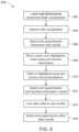
- FIG. 3is a flowchart illustrating an embodiment of a user experiential process of a health prediction system.
- FIG. 4is a flowchart illustrating an embodiment of a brain information preprocessing process.
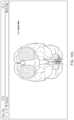
- FIG. 5is a multi-dimensional view illustrating an embodiment of a voxel positioning system.
- FIG. 6is a flowchart illustrating an embodiment of a voxel intensity extraction process.
- FIG. 7is a flowchart illustrating an embodiment of a behavior and self-report prediction process.
- FIG. 8is a flowchart illustrating an embodiment of a brain area pinpointing process and a brain health benchmarking process.
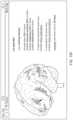
- FIG. 9is a flowchart illustrating an embodiment of a multi-dimensional visualization and brain health recommendation process.
- FIGS. 10 A- 10 Hillustrate user interface examples.
- the present inventioncan be implemented in many ways, including as a process; an apparatus; a system; a non-transitory computer readable program product embodied on a computer readable storage medium; and/or a processor, such as a processor configured to execute instructions on and/or provided by a memory coupled to the processor.
- these implementations, or any other form that the invention may take,may be referred to as techniques.
- the order of the steps of disclosed processesmay be altered within the scope of the invention.
- a componentsuch as a processor or a memory described as being configured to perform a task may be implemented as a general component that is temporarily configured to perform the task at a given time or a specific component that is manufactured to perform the task.
- the term processorrefers to one or more devices, circuits, and/or processing cores configured to process information, such as computer program instructions.
- FIG. 1is a high-level flowchart illustrating an embodiment of a health prediction system 100 configured in accordance with the present invention and is not intended to limit scope as one of ordinary skill in the art would understand on review of this application that other configurations could be utilized without departing from the scope of the claimed invention.
- system 100is implemented in connection with assessment of the human brain.
- system 100includes brain health comparison system 102 , which may be implemented using one or more server computers having one or more processors, one or more special purpose computing appliances, or any other appropriate hardware, software, or combinations thereof capable of implementing and/or executing computer readable instructions. The operations of the brain health comparison system 102 are described in greater detail below.
- various users of the systemaccess the brain health comparison system via a network 104 using client devices 106 , 108 , 110 .
- network 104may be a cloud-based storage and processing system.
- network 104may be a network of server computers, connected to store and process information.
- Client devices 106 , 108 , 110may be a computer, a mobile phone or device, a tablet, or any other such device that allows a user to access network 104 .
- Database 112stores user information, including functional neuroimaging information; anatomical neuroimaging information; behavioral information, which may be acquired during neuroimaging scan sessions; self-reported information; other behavioral information such as but not limited to information from users' stationary and mobile devices such as smartphones and smartwatches, other wearable devices such as fitness armbands etc.; and health-monitoring information etc. pertaining to the users.
- Database 112can be implemented on an integral storage component of the brain health comparison system, an attached storage device, a separate storage device accessible by the brain health comparison system, or combinations thereof.
- the entirety of the collected anatomical and functional neuroimaging informationis stored in a database 112 to facilitate the brain health comparison system 102 .
- voxelsare extracted from the brain images of participants undergoing anatomical and/or functional neuroimaging scans, such as fMRI, and assayed and stored in database 112 . Because the human brain varies in size among individuals, the number of voxels extracted, assayed, and stored may range in number, depending on the size of the brain undergoing a neuroimaging scan.
- cortical voxelsmay be extracted in an anatomical neuroimaging scan and approximately 26,000 3-millimeter isotropic functional voxels may be extracted in a functional neuroimaging scan, and then assayed and stored in database 112 .
- the numbers of voxels extracted, assayed, and stored by the present inventionare not limited to these quantities, and one of skill in the art would understand the present invention may extract, assay, and store a greater or smaller quantity of voxels depending on the size of the brain being undergoing assessment.
- FIG. 2is a detailed flowchart illustrating an embodiment of a health prediction system 200 configured in accordance with the present invention and is not intended to limit scope as one of ordinary skill in the art would understand on review of this application that other configurations could be utilized without departing from the scope of the claimed invention.
- system 200is implemented in connection with assessment of the human brain.
- system 200includes brain health comparison system 202 , which may be implemented using one or more server computers having one or more processors, one or more special purpose computing appliances, or any other appropriate hardware, software, or combinations thereof capable of executing computer readable instructions.
- datamay be exchanged between pieces of software and/or different servers, storage memory, systems, and/or components.
- user 1accesses the brain health comparison system 202 of system 200 via a network 204 using client devices such as 206 , as described with respect to FIG. 1 .
- Both individual user information 224e.g., user 1 “Janet”
- comparison user information 226are stored in a database 208 .
- Database 208stores individual user information 224 , which may include functional neuroimaging information F and anatomical neuroimaging information A taken from brain scans of individuals, through, for example, fMRI.
- Individual user information 224 stored in database 208also contains behavioral information B taken during scan sessions (e.g., numerical behavioral responses) and self-reported information S taken from individuals' self-reports.
- Database 208may also store other behavioral information such as information on users' stationary and mobile devices as well as health monitoring information etc., denoted “ . . . ” in database 208 .
- Data F, A, B, S, and “ . . . ”are stored in individual information system 224 within user information database 208 .
- the frontend of the brain health comparison system 202is a multi-dimensional visualization and brain health recommendation system 210 , described in greater detail at FIG. 9 below, in which users can view multi-dimensional visualizations of their brain information, access statistics and reports, and share information with other users.
- Database 208receives information from a brain area pinpointing system 212 (described in greater detail at FIG. 8 below) and a brain health benchmarking system 214 (described in greater detail at FIG. 8 below), using comparison user information 226 in a database 208 .
- the brain area pinpointing system 212 and brain health benchmarking system 214compare the performance of specific brain areas in user e.g., user 1 “Janet” in FIG. 2 to the performance of the specific brain areas of other users.
- Brain area pinpointing system 212 and brain health benchmarking system 214receive information from a behavior and self-report prediction system 216 .
- Behavior and self-report prediction system 216integrates behavioral and self-report data, and uses neuroimaging information from voxel extraction system 218 .
- Voxel extraction system 218extracts a numerical brain activation value from each voxel.
- behavior and self-report prediction system 216predicts user information as described in greater detail at FIG. 7 , including behavioral information acquired during neuroimaging scan sessions (denoted “B” in 208 ), self-reported information (denoted “S” in 208 ), and other information denoted “ . . . ” in 208 .
- Voxel positioning system 220(described in greater detail at FIG. 5 below) enables voxel intensity extraction system 218 (described in greater detail at FIG.
- Voxel positioning system 220receives information from a brain information preprocessing system 222 (described in greater detail at FIG. 4 below).
- Brain information preprocessing system 222receives information from individual user information 224 in database 208 , which contains functional neuroimaging information (denoted “F” in system 208 ) and anatomical neuroimaging information (denoted “A” in system 208 ). While the preferred embodiment is described above for illustrative purposes, the invention is not limited to such an embodiment and other configurations of the invention are contemplated as would be understood by one skilled in the art.
- FIG. 3is a flowchart illustrating an embodiment of a user experiential process 300 in accordance with the present invention and is not intended to limit scope as one of ordinary skill in the art would understand on review of this application that other arrangements could be utilized without departing from the scope of the claimed invention.
- Experiential process 300may be implemented on a health prediction system such as system 100 or system 200 . Many different arrangements of the steps of a user experiential process 300 are possible in various embodiments.
- a userbegins the user experiential process by making a reservation for a neuroimaging brain scan.
- Usermay make a reservation by using an electronic device (e.g., a personal computer, a mobile device or phone, a tablet) to access an online scheduling system, by making a reservation by phone, in person, or through other communication channels.
- an electronic devicee.g., a personal computer, a mobile device or phone, a tablet
- userresponds to prescreening questions in order to determine eligibility for brain scanning.
- prescreening questionsmay include medical history, current medications, and/or metal in or on one's body (e.g., pacemakers, tattoos).
- Brain scansmay be conducted by use of, though not limited to, fMRI machines, such as those manufactured by Siemens (e.g., Magnetom, Biograph, Mobile MRI Scanner), Philips (e.g., Achieva, Ingenia), General Electrics (e.g., Signa), and other manufacturers.
- Such brain scansmay involve a user engaging in certain tasks that allow for collection of functional, anatomical, behavioral, and/or self-reported information.
- a usermay be engaged in one or multiple behavioral tasks while the user's anatomical and/or functional neuroimaging information is collected.
- Each behavioral taskrepresents standardized, structured, and precisely timed presentations of stimulus material and user prompts to respond to the stimulus material, for example, perception of abstract geometrical shapes presented in different colors, or perception of and interaction with concrete symbols such as playing cards and scenarios involving monetary gains and losses as indications of risk-prone or risk-averse behaviors.
- Stimulus materialsmay be presented through over-head projections or viewing goggles while the user is positioned within the brain scanner as would be understood in the art.
- Behavioral responsesmay be collected through button-box or other collection devices, which allow users to provide behavioral responses on a multi-button response box held in the user's hand.
- Behavioral tasksmay assess cognitive function, including impulse control, self-control, attentional control, working memory, memory span, long-term memory, autobiographical memory, intelligence, verbal, auditory, and visual processing, selection of appropriate options, risk-taking, gambling, card sorting, and/or other cognitive abilities, though this list is neither exhaustive nor exclusive and the present invention is not limited to such behavioral tasks or cognitive assessments.
- a usermay respond to several survey questions, for example, pertaining to a user's overall health and lifestyle choices.
- a userlogs in to a brain health comparison system's user interface, such as depicted at 206 in system 200 , via e.g., a personal computing and viewing device such as a desktop computer, laptop computer, tablet computer, and/or mobile phone.
- a personal computing and viewing devicesuch as a desktop computer, laptop computer, tablet computer, and/or mobile phone.
- Other devices that provide access to a network via a user interfaceare also contemplated by the invention.
- brain health comparison system's interface 306user will be able to browse through visualizations of user's brain; obtain prediction and comparison reports; obtain suggestions on scholarly publications relevant to certain brain conditions; obtain referrals to specialists that are directed toward certain aspects of a user's brain and/or brain function; receive opportunities for information sharing with other people; and receive personalized brain health recommendations (as seen at FIGS.
- Exemplary recommendationsmay include interventions such as physical activities, cognitive exercises, changes in lifestyle or habits, and/or dietary suggestions, etc. Other recommendations for maximizing brain health are contemplated within the scope of the invention.
- a usermay consider implementing some of the recommendations obtained by accessing user interface during 306 .
- a usermay schedule another brain scan 302 , for example, to target another brain function and/or monitor changes over time.
- FIG. 4is a flowchart illustrating an embodiment of a brain information preprocessing process 400 in accordance with the present invention and is not intended to limit scope as one of ordinary skill in the art would understand on review of this application that other arrangements could be utilized without departing from the scope of the claimed invention.
- Brain information preprocessing process 400may be implemented by brain information preprocessing system 222 in system 200 . Many different arrangements of the steps of a brain information preprocessing process, and the components of a brain information preprocessing process system, are possible in various embodiments.
- information of a first useris received from a neuroimaging scanner (such as fMRI), the behavioral response collection devices, and self-report surveys, and stored in a database, such as 208 , as described above with respect to FIG. 2 .
- User informationmay include raw functional neuroimaging information, raw anatomical neuroimaging information, behavioral information acquired during neuroimaging scan sessions, and self-reported information.
- preliminary processing of raw functional neuroimaging information and raw anatomical neuroimaging informationis undertaken, neuroimaging information quality is checked, and behavioral and self-reported information is organized.
- Preliminary processing of the neuroimaging informationmay include slice-scan-timing correction, temporal high pass filtering, three-dimensional motion correction, inhomogeneity correction, ISO matrix transformation, spatial normalization and alignment, brain tissue segmentation, and surface reconstruction as is known in the art.
- the anatomical neuroimaging informationis conformed to a standardized brain template (e.g., preferably 1-millimeter cortical voxels but which could range from 1 micrometer to 3 centimeters in size) in order to make information comparable to other users.
- a voxel positioning systemas described with respect to FIG. 5 , facilitates this step.
- an image of the skull and other matteris also removed computationally to allow a direct view of the brain.
- the functional neuroimaging informationis superimposed onto the conformed anatomical neuroimaging information in order to match each functional information point (e.g., regions of detected brain activity) with its corresponding anatomical information point.
- functional neuroimaging informationis conformed to a standardized format. Said standardized format may preferably be 3-millimeter isotropic functional voxels, though the format can range from micrometers to centimeters in size.
- the behavioral informationis superimposed onto the conformed functional and anatomical neuroimaging information in order to match each time point of a behavioral task to the corresponding neuroimaging information points.
- a final datasetis stored in a database such as 208 .
- FIG. 5is a multi-dimensional view illustrating an embodiment of a voxel positioning system 500 such as 220 in system 200 configured in accordance with the present invention and is not intended to limit scope as one of ordinary skill in the art would understand on review of this application that other configurations could be utilized without departing from the scope of the claimed invention. Many different arrangements of the steps of a voxel positioning process, and the components of a voxel positioning system, are possible in various embodiments.
- neuroimaging informationis collected over time and spaced in a matrix, which may preferably be a matrix comprising 256 points ⁇ 256 points ⁇ 256 points, but the present invention is not limited to such dimensions and other three-dimensional configurations (e.g., 128 ⁇ 128 ⁇ 128) are also contemplated within the scope of the invention.
- Each data pointmay reflect the raw blood oxygenation value at each coordinate corresponding to a region of the brain for each time point (e.g., T1, T2, T3, etc.) for each brain.
- the raw blood oxygenationmay then be normalized to a value between 0 and 1 to account for any differences in baseline blood oxygenation levels between users.
- the normalized blood oxygenation valuemay preferably be computed as follows: (open raw blood oxygenation values ⁇ mean blood oxygenation values)/standard deviation.
- the present inventionis not limited to any particular method of normalizing such values.
- the coordinate at the very center of the boxis x: 0, y: 0, z: 0.
- Voxel positioningis repeated at time points T2, T3, T4, and so on until the end of the behavioral task.
- the preferable resolution of the described analysesis by example only and may be either higher or lower in other embodiments.
- FIG. 6is a flowchart illustrating an embodiment of a voxel intensity extraction process 600 in accordance with the present invention and is not intended to limit scope as one of ordinary skill in the art would understand on review of this application that other arrangements could be utilized without departing from the scope of the claimed invention.
- Process 600may be implemented in connection with voxel intensity extraction system 218 in system 200 .
- Many different arrangements of the steps of a voxel intensity extraction process, and the components of a voxel intensity extraction system,are possible in various embodiments.
- stored information from voxel positioning system 500is retrieved from a database, such as 208 .
- brain areas of interestare defined (e.g., in cubic form), containing voxels with specific x-y-z coordinates as described in voxel extraction system 500 at FIG. 5 .
- time points of interestare defined, for example, in a millisecond time span, ranging from 10,000 ms to 14,000 ms in some embodiments.
- time points of interestmay present phases before, during, and/or after a user has responded to a stimulus, has made a decision, has provided a button press or performed any other activity while undergoing a brain scan.
- voxel intensitiesi.e., the brain activation values in a specific brain area at a specific time point
- voxel intensitiesWhen used in conjunction with fMRI, voxel intensities may reflect levels of blood oxygenation at specific brain areas as indicative of the relative levels of activation at each brain area at a given point in time.
- voxel intensitiesare stored in a database. Voxel intensities may be stored in a tabular format.
- FIG. 7is a flowchart illustrating an embodiment of a behavior and self-report prediction process in accordance with the present invention and is not intended to limit scope as one of ordinary skill in the art would understand on review of this application that other arrangements could be utilized without departing from the scope of the claimed invention.
- Behavior and self-report prediction process 700may be implemented by behavior and self-report prediction system 216 in system 200 . Many different arrangements of the steps of a behavior and self-report prediction process, and the components of a behavior and self-report prediction system, are possible in various embodiments.
- an artificial neural networkis trained on previously existing user information.
- the artificial neural networkmay be a convolutional neural network and/or a recurrent neural network (e.g., long short-term memory neural network), as is understood in the art, though the present invention is not limited to such networks.
- Previously existing user informationmay comprise functional, anatomical, behavioral, self-report, genetic, and other data obtained from users as described above.
- An artificial neural networkextracts information such as voxel intensities (as described at 600 in FIG. 6 ) or behavioral and self-reported information (as described under system 200 in FIG. 2 ) from users stored in a database such as 208 to recognize patterns in the voxel intensities and predict behavioral and self-reported information based on recognized patterns in the functional neuroimaging information.
- an artificial neural networkclassifies voxel intensities to provide a likelihood of a user making a particular decision on a behavioral task or responding in a certain way to a stimulus (e.g., “85% risky choice” or “90% working memory”).
- a first user's informatione.g., user 1 “Janet”
- the artificial neural networkpredicts brain health based on neuroimaging information.
- user 1's behavioral informationis added to the previously existing user information.
- An artificial neural network trained on previous user informationis then fed user 1's voxel intensities as described at 600 in FIG. 6 .
- a behavior and self-report prediction systemprovides a prediction accuracy.
- user 1's voxel intensities of a region of user 1's brainpredict with high likelihood a particular behavioral decision, based on a plurality of previous user information stored in database such as 208 .
- user 1's informationis added to the database of previously existing user information and an artificial neural network is trained again, now with the added information obtained from user 1.
- FIG. 8is a flowchart illustrating an embodiment of a brain area pinpointing process 800 in accordance with the present invention and is not intended to limit scope as one of ordinary skill in the art would understand on review of this application that other configurations could be utilized without departing from the scope of the claimed invention.
- Brain area pinpointing process 800may be implemented by brain area pinpointing system 212 and brain health benchmarking system 214 in system 200 .
- Many different arrangements of the steps of a behavior and self-report prediction process, and the components of a behavior and self-report prediction system,are possible in various embodiments.
- a user's prediction accuracyas described at 706 in FIG.
- a specific brain area and a specific behavioral taskis benchmarked against previously existing user information in a database.
- user 3's prediction accuracyis assayed and compared against prediction accuracies (e.g., expressed in a measure-of-central-tendency such as mean, median, mode etc.) of all user information in a database or a specific subset of users in a database (e.g., selected by age, sex, gender, ethnicity, education, occupation etc.).
- a user's non-neuroimaging information(e.g., behavioral information, self-reported information etc.) is assayed and compared against non-neuroimaging information (e.g., expressed in a measure-of-central-tendency such as mean, median, mode etc.) of all user information in a database or a specific subset of users in a database.
- benchmarking results of 802 and 804are assayed against thresholds to determine abnormalities, malfunctions, deviations, and other irregularities in the prediction accuracy of a user, their behavioral performances, their self-reported responses, and combinations thereof.
- the comparison thresholdspreferably include quartiles (e.g.
- results of this comparison and threshold examinationare stored in a database such as 208 for example.
- FIG. 9is a flowchart illustrating an embodiment of a multi-dimensional visualization and brain health recommendation process 900 in accordance with the present invention and is not intended to limit scope as one of ordinary skill in the art would understand on review of this application that other arrangements could be utilized without departing from the scope of the claimed invention.
- Multi-dimensional visualization and brain health recommendation process 900may be implemented by multi-dimensional visualization and brain health recommendation system 210 in system 200 .
- Many different arrangements of the steps of a multi-dimensional visualization and brain health recommendation process, and the components of a multi-dimensional visualization and brain health recommendation system,are possible in various embodiments.
- a multi-dimensional anatomical brain visualization(e.g., three-dimensional view or four-dimensional view including three space dimensions and one time dimension) is loaded in an Internet browser and/or a mobile application, or other personal viewing device.
- a userinteracts with the anatomical brain visualization.
- a usermay interact with the anatomical brain visualization, for example, by using pan/zoom controls, slider/toggle controls, and light/shade controls to vary the angle/tilt of the brain and the degree of brain slice segmentation in order to expose the brain's deeper layers in horizontal, sagittal, and coronal slices.
- a userselects a behavioral task that the user engaged with during a neuroimaging scan session. Several available tasks may be shown. Some tasks may be active if the user participated in them, while other tasks may be inactive if the user has not yet participated in them.
- the functional neuroimaging information corresponding to the selected behavioral taskis then loaded and superimposed on the multi-dimensional anatomical brain visualization.
- the anatomical visualizationnow shows functional brain activation in certain brain areas, corresponding to specific phases of the selected behavioral task.
- warm colorssuch as yellow and red may be used to indicate varying degrees of brain activation at specific phases of each selected task.
- the present inventionis not limited to such colors and any indications of brain activation may be used.
- a usermay select between “choice of a safe gamble” phase and “choice of a risky gamble” phase to see which of their brain areas predicted safe versus risky gambling behavior.
- a usermoves a cursor over the highlighted brain areas and, after doing so, receives statistical and textual information on the selected brain area.
- the informationmay include, but is not limited to, performance statistics on the behavioral task (e.g., “80% choice of risky gambles and 20% choice of safe gambles”), prediction statistics of the brain area (e.g., “prefrontal cortex predicts safe choice 65% of the time”), and comparison statistics (e.g., “the prefrontal cortex of other users in the same age group predicts safe choice 95% of the time”).
- performance statistics on the behavioral taske.g., “80% choice of risky gambles and 20% choice of safe gambles”
- prediction statistics of the brain areae.g., “prefrontal cortex predicts safe choice 65% of the time”
- comparison statisticse.g., “the prefrontal cortex of other users in the same age group predicts safe choice 95% of the time”.
- the present inventionis not limited to such examples.
- a userclicks on or highlights the brain area of interest to receive personal brain health recommendations.
- These recommendationsmay include, for example, personalized health risk analyses, such as reports on potential risks related to the health of the prefrontal cortex; potential risks related to compulsive gambling; potential risks related to incurring excessive debt; potential risks related to cognitive decline across the life span; potential risks related to the interplay between mild cognitive impairment and/or dementia and risky decision making. Additional recommendations are contemplated by brain recommendation system and the invention is not limited to the exemplary recommendations.
- brain recommendation systemmay provide links to scientific reports published in peer-reviewed scholarly journals in academic fields such as neuroscience, medicine, psychology, psychiatry, genetics, health management, well-being, public health, and/or economics relevant to the brain area clicked on or highlighted by the user, or relevant to other non-anatomical (e.g., behavioral) information provided by user.
- Brain health recommendationsmay also include well-being analyses, such as customized recommendations for strengthening the selected brain area. For example, recommendations may include interventions pertaining to physical activities, cognitive exercises, changes in habits, dietary suggestions, directed toward certain aspects of a user's brain and/or brain function.
- links to scientific reportsmay be provided, as previously discussed, and users may receive referrals to specialists in the user's geographic area.
- Specialistsmay include neurologists, psychologists, and physicians.
- Usersmay also receive connections to and be provided with sharing options with other users that show similar sociodemographic characteristics and/or similar and/or related results. For example, users may choose to connect to other users in the same age group, the same geographical location, and/or the same sex/gender etc. Further, users may connect to other users that have similar anatomical, functional, and/or behavioral results.
- a userselects a segment of self-reported information provided by the user, for example, sociodemographic information, phenotypes such as personality traits and other individual differences, well-being, happiness, satisfaction with life, risk-taking, financial decision making. For each of these segments, health risk and well-being analyses are provided, as discussed under 905 .
- a usermay link other data forms to his or her profile.
- a usermay optionally link his or her profile to data such as health monitoring information tracked by the user's smart watch or fitness armband (e.g., how much the user moves, how many steps the user takes, nutritional information), credit card information (e.g., outstanding balances, balance overdue, number of accounts open), genotype information (e.g., profiles from DNA decoding services), and/or social media information (e.g., number of social connections).
- health monitoring information tracked by the user's smart watch or fitness armbande.g., how much the user moves, how many steps the user takes, nutritional information
- credit card informatione.g., outstanding balances, balance overdue, number of accounts open
- genotype informatione.g., profiles from DNA decoding services
- social media informatione.g., number of social connections
- a user's self-reported health risk datais correlated with his or her brain activation and a correlation coefficient is displayed to the user such as “Your health risk is 80% correlated with activation in your prefrontal cortex.”
- FIGS. 10 A- 10 Hillustrate user interface examples in connection with 210 in system 200 as well as user experiential process 300 .
- the interfaceis implemented in an Internet browser and/or a mobile application, or other personal viewing device, though other media that may allow a user to access the interface are contemplated by the present invention.
- the interfaceprovides two views to the user: a three-dimensional view and a two-dimensional view.
- a devicesuch as a computer mouse, touch pad, or touch screen
- the usercan turn or rotate the brain, zoom in or out, or click on certain brain areas in order to obtain more detailed information.
- FIG. 10 Ashows an interface example for a three-dimensional view of user Jane Doe's brain after an fMRI scan.
- the brain imagemay be laid-over with functional activation as indicated by, for example, differently shaded or colored brain regions.
- the viewmay be of two-dimensional slices of the brain, such as coronal, sagittal, or other slices and perspectives as are known in the art.
- FIG. 10 Bshows an interface example for another three-dimensional view of user Jane Doe's brain. Compared to FIG. 10 A , the brain in FIG. 10 B has been turned around its vertical axis. Such movement may be accomplished by a device such as a computer mouse, touch pad, touch screen or other device that allows for manipulation of visualizations by a user.
- FIG. 10 Cshows an interface example of the inferior view of user Jane Doe's brain, though the present invention is of course not limited to such an example and other three-dimensional views as are known in the art are possible.
- shadingrepresents functional activation detected in brain regions.
- FIG. 10 Dshows an interface example of another inferior view user Jane Doe's brain, rotated about the vertical axis.
- FIG. 10 Eshows the view as FIG. 10 D , but with an exemplary cursor added showing how the user can select certain brain areas, such as where activation has been detected. By selecting a brain area, the user can obtain more detailed information about that area and the user's individualized results, as shown in FIG. 10 F .
- the userhas selected a prefrontal area of the brain, which is associated with working memory and that has demonstrated functional activation during a task of interest, as seen by the shading.
- the interfacesummarizes relevant details of user Jane Doe's activation in the selected prefrontal area of the brain.
- user Jane Doe's activation in this areais 75% stronger than that of her peers.
- the activationis shown to predict financial success to 85%, happiness to 10%, and dementia to 5%.
- a linkis provided through which the user can obtain a list of relevant recent publications on this brain area, or any brain area the user has selected.
- FIG. 10 Gdepicts the interface a user interacts with after selecting the “these recommendations” hyperlink seen in FIG. 10 F .
- the useris asked to complete an assessment of his or her personal health-risk propensity. In this example, an eight-item survey scale is used, though other surveys or assessments of personal health-risk may be used. After completing the assessment, the user learns about his/her personal health-risk propensity score in FIG. 10 H .
Landscapes
- Health & Medical Sciences (AREA)
- Engineering & Computer Science (AREA)
- Public Health (AREA)
- Medical Informatics (AREA)
- Biomedical Technology (AREA)
- General Health & Medical Sciences (AREA)
- Life Sciences & Earth Sciences (AREA)
- Pathology (AREA)
- Data Mining & Analysis (AREA)
- Epidemiology (AREA)
- Primary Health Care (AREA)
- Neurology (AREA)
- Databases & Information Systems (AREA)
- Physics & Mathematics (AREA)
- Veterinary Medicine (AREA)
- Nuclear Medicine, Radiotherapy & Molecular Imaging (AREA)
- Biophysics (AREA)
- Heart & Thoracic Surgery (AREA)
- Molecular Biology (AREA)
- Surgery (AREA)
- Animal Behavior & Ethology (AREA)
- Radiology & Medical Imaging (AREA)
- Psychology (AREA)
- Neurosurgery (AREA)
- Physiology (AREA)
- High Energy & Nuclear Physics (AREA)
- Measuring And Recording Apparatus For Diagnosis (AREA)
- Medical Treatment And Welfare Office Work (AREA)
Abstract
Description
Claims (11)
Priority Applications (2)
| Application Number | Priority Date | Filing Date | Title |
|---|---|---|---|
| US16/241,967US11756691B2 (en) | 2018-08-01 | 2019-01-07 | Brain health comparison system |
| US29/706,242USD969820S1 (en) | 2018-08-01 | 2019-09-19 | Display screen or portion thereof with a graphical user interface |
Applications Claiming Priority (2)
| Application Number | Priority Date | Filing Date | Title |
|---|---|---|---|
| US201862713260P | 2018-08-01 | 2018-08-01 | |
| US16/241,967US11756691B2 (en) | 2018-08-01 | 2019-01-07 | Brain health comparison system |
Related Child Applications (1)
| Application Number | Title | Priority Date | Filing Date |
|---|---|---|---|
| US29/706,242ContinuationUSD969820S1 (en) | 2018-08-01 | 2019-09-19 | Display screen or portion thereof with a graphical user interface |
Publications (2)
| Publication Number | Publication Date |
|---|---|
| US20200043615A1 US20200043615A1 (en) | 2020-02-06 |
| US11756691B2true US11756691B2 (en) | 2023-09-12 |
Family
ID=69228928
Family Applications (2)
| Application Number | Title | Priority Date | Filing Date |
|---|---|---|---|
| US16/241,967Active2040-10-31US11756691B2 (en) | 2018-08-01 | 2019-01-07 | Brain health comparison system |
| US29/706,242ActiveUSD969820S1 (en) | 2018-08-01 | 2019-09-19 | Display screen or portion thereof with a graphical user interface |
Family Applications After (1)
| Application Number | Title | Priority Date | Filing Date |
|---|---|---|---|
| US29/706,242ActiveUSD969820S1 (en) | 2018-08-01 | 2019-09-19 | Display screen or portion thereof with a graphical user interface |
Country Status (1)
| Country | Link |
|---|---|
| US (2) | US11756691B2 (en) |
Families Citing this family (11)
| Publication number | Priority date | Publication date | Assignee | Title |
|---|---|---|---|---|
| USD919645S1 (en)* | 2019-01-02 | 2021-05-18 | Palantir Technologies, Inc. | Display screen or portion thereof with transitional graphical user interface |
| US11526808B2 (en)* | 2019-05-29 | 2022-12-13 | The Board Of Trustees Of The Leland Stanford Junior University | Machine learning based generation of ontology for structural and functional mapping |
| USD991279S1 (en)* | 2019-12-09 | 2023-07-04 | Ankon Technologies Co., Ltd | Display screen or portion thereof with transitional graphical user interface |
| US11264137B1 (en)* | 2020-10-08 | 2022-03-01 | Omniscient Neurotechnology Pty Limited | Centrality rankings of network graphs generated using connectomic brain data |
| JP7679631B2 (en)* | 2021-01-29 | 2025-05-20 | 株式会社リコー | Brain function evaluation system, method, and program |
| USD1082800S1 (en)* | 2021-05-08 | 2025-07-08 | Huawei Technologies Co., Ltd. | Display screen or portion thereof with graphical user interface |
| USD1048047S1 (en)* | 2021-11-29 | 2024-10-22 | Stratasys Ltd. | Display screen or portion thereof with graphical user interface |
| USD1068792S1 (en)* | 2022-08-31 | 2025-04-01 | Idexx Laboratories, Inc. | Display screen or portion thereof with graphical user interface for viewing x-ray images and calculating vertebral heart score |
| USD1075797S1 (en)* | 2023-04-20 | 2025-05-20 | Histosonics, Inc. | Display screen or portion thereof with graphical user interface |
| USD1079716S1 (en)* | 2023-04-20 | 2025-06-17 | Histosonics, Inc. | Display screen or portion thereof with animated graphical user interface |
| USD1075796S1 (en)* | 2023-04-20 | 2025-05-20 | Histosonics, Inc. | Display screen or portion thereof with an animated graphical user interface |
Citations (51)
| Publication number | Priority date | Publication date | Assignee | Title |
|---|---|---|---|---|
| US5927988A (en) | 1997-12-17 | 1999-07-27 | Jenkins; William M. | Method and apparatus for training of sensory and perceptual systems in LLI subjects |
| WO2000044283A1 (en) | 1999-01-29 | 2000-08-03 | Scientific Learning Corporation | Remote computer-implemented methods for cognitive and perceptual testing |
| US6159014A (en) | 1997-12-17 | 2000-12-12 | Scientific Learning Corp. | Method and apparatus for training of cognitive and memory systems in humans |
| US6261101B1 (en) | 1997-12-17 | 2001-07-17 | Scientific Learning Corp. | Method and apparatus for cognitive training of humans using adaptive timing of exercises |
| US6280198B1 (en) | 1999-01-29 | 2001-08-28 | Scientific Learning Corporation | Remote computer implemented methods for cognitive testing |
| US6463315B1 (en) | 2000-01-26 | 2002-10-08 | The Board Of Trustees Of The Leland Stanford Junior University | Analysis of cerebral white matter for prognosis and diagnosis of neurological disorders |
| US20040092809A1 (en) | 2002-07-26 | 2004-05-13 | Neurion Inc. | Methods for measurement and analysis of brain activity |
| US20060112050A1 (en)* | 2000-10-17 | 2006-05-25 | Catalis, Inc. | Systems and methods for adaptive medical decision support |
| US20070166675A1 (en) | 2005-12-15 | 2007-07-19 | Posit Science Corporation | Cognitive training using visual stimuli |
| US7317821B2 (en) | 2004-11-22 | 2008-01-08 | Carestream Health, Inc. | Automatic abnormal tissue detection in MRI images |
| US20090025023A1 (en) | 2007-06-06 | 2009-01-22 | Neurofocus Inc. | Multi-market program and commercial response monitoring system using neuro-response measurements |
| US20090267945A1 (en) | 2008-04-25 | 2009-10-29 | Marcel Warntjes | Visualization of Quantitative MRI Data by Quantitative Tissue Plot |
| US7647098B2 (en) | 2005-10-31 | 2010-01-12 | New York University | System and method for prediction of cognitive decline |
| US20100145215A1 (en) | 2008-12-09 | 2010-06-10 | Neurofocus, Inc. | Brain pattern analyzer using neuro-response data |
| US20100250325A1 (en) | 2009-03-24 | 2010-09-30 | Neurofocus, Inc. | Neurological profiles for market matching and stimulus presentation |
| US20100249538A1 (en) | 2009-03-24 | 2010-09-30 | Neurofocus, Inc. | Presentation measure using neurographics |
| WO2010147913A1 (en) | 2009-06-15 | 2010-12-23 | Brain Computer Interface Llc | A brain-computer interface test battery for the physiological assessment of nervous system health |
| US20110046504A1 (en) | 2009-08-20 | 2011-02-24 | Neurofocus, Inc. | Distributed neuro-response data collection and analysis |
| US20110106750A1 (en) | 2009-10-29 | 2011-05-05 | Neurofocus, Inc. | Generating ratings predictions using neuro-response data |
| US7961922B2 (en) | 2007-05-31 | 2011-06-14 | The Board Of Regents Of The University Of Texas System | Systems and methods for processing medical image data to facilitate comparisons among groups of subjects |
| US20110237971A1 (en) | 2010-03-25 | 2011-09-29 | Neurofocus, Inc. | Discrete choice modeling using neuro-response data |
| US8209224B2 (en) | 2009-10-29 | 2012-06-26 | The Nielsen Company (Us), Llc | Intracluster content management using neuro-response priming data |
| US8270814B2 (en) | 2009-01-21 | 2012-09-18 | The Nielsen Company (Us), Llc | Methods and apparatus for providing video with embedded media |
| US20120330109A1 (en)* | 2006-05-24 | 2012-12-27 | Bao Tran | Health monitoring appliance |
| US8386312B2 (en) | 2007-05-01 | 2013-02-26 | The Nielsen Company (Us), Llc | Neuro-informatics repository system |
| US8392250B2 (en) | 2010-08-09 | 2013-03-05 | The Nielsen Company (Us), Llc | Neuro-response evaluated stimulus in virtual reality environments |
| US8392255B2 (en) | 2007-08-29 | 2013-03-05 | The Nielsen Company (Us), Llc | Content based selection and meta tagging of advertisement breaks |
| US8464288B2 (en) | 2009-01-21 | 2013-06-11 | The Nielsen Company (Us), Llc | Methods and apparatus for providing personalized media in video |
| US8484081B2 (en) | 2007-03-29 | 2013-07-09 | The Nielsen Company (Us), Llc | Analysis of marketing and entertainment effectiveness using central nervous system, autonomic nervous system, and effector data |
| US8494905B2 (en) | 2007-06-06 | 2013-07-23 | The Nielsen Company (Us), Llc | Audience response analysis using simultaneous electroencephalography (EEG) and functional magnetic resonance imaging (fMRI) |
| US8655437B2 (en) | 2009-08-21 | 2014-02-18 | The Nielsen Company (Us), Llc | Analysis of the mirror neuron system for evaluation of stimulus |
| EP2709061A1 (en) | 2012-09-12 | 2014-03-19 | Virtual Proteins B.V. | System and method for 3D visualization of brain iron deposition |
| US20150018630A1 (en) | 2013-07-15 | 2015-01-15 | Neurexpand Brain Center Llc | Systems and methods for creating comprehensive and personalized brain health programs |
| CN104851101A (en) | 2015-05-25 | 2015-08-19 | 电子科技大学 | Brain tumor automatic segmentation method based on deep learning |
| CN104866727A (en) | 2015-06-02 | 2015-08-26 | 陈宽 | Method for analyzing medical data based on deep learning and its intelligent analyzer |
| US20160015289A1 (en) | 2013-03-06 | 2016-01-21 | Adam J. Simon | Form factors for the multi-modal physiological assessment of brain health |
| US20160081575A1 (en) | 2013-11-15 | 2016-03-24 | Yibing Wu | A life maintenance mode, a brain inhibition therapy and a personal health information platform |
| US9332954B2 (en) | 2013-03-15 | 2016-05-10 | Jacqueline K. Vestevich | Systems and methods for evaluating a brain scan |
| CN105796097A (en) | 2014-12-29 | 2016-07-27 | 普天信息技术有限公司 | EEG signal acquisition device, brain rehabilitation training device and brain rehabilitation training system |
| US9420970B2 (en) | 2013-10-22 | 2016-08-23 | Mindstrong, LLC | Method and system for assessment of cognitive function based on mobile device usage |
| US9474481B2 (en) | 2013-10-22 | 2016-10-25 | Mindstrong, LLC | Method and system for assessment of cognitive function based on electronic device usage |
| US9519981B2 (en) | 2011-11-04 | 2016-12-13 | Siemens Healthcare Gmbh | Visualizing brain network connectivity |
| CN106250707A (en) | 2016-08-12 | 2016-12-21 | 王双坤 | A kind of based on degree of depth learning algorithm process head construction as the method for data |
| US20170053540A1 (en) | 2015-08-18 | 2017-02-23 | Cogniciti Inc. | Systems methods and devices for brain health assessment |
| US20170109881A1 (en) | 2015-10-14 | 2017-04-20 | The Regents Of The University Of California | Automated segmentation of organ chambers using deep learning methods from medical imaging |
| US9633430B2 (en) | 2011-12-28 | 2017-04-25 | Institute Of Automation, Chinese Academy Of Sciences | Method for analyzing functional MRI brain images |
| US20170323485A1 (en) | 2016-05-09 | 2017-11-09 | Magic Leap, Inc. | Augmented reality systems and methods for user health analysis |
| WO2018005814A1 (en) | 2016-06-29 | 2018-01-04 | The University Of North Carolina At Chapel Hill | Methods, systems, and computer readable media for utilizing functional connectivity brain imaging for diagnosis of a neurobehavioral disorder |
| US9886981B2 (en) | 2007-05-01 | 2018-02-06 | The Nielsen Company (Us), Llc | Neuro-feedback based stimulus compression device |
| US9993190B2 (en) | 2011-08-16 | 2018-06-12 | Intendu Ltd. | System and method for neurocognitive training and/or neuropsychological assessment |
| US20180315188A1 (en) | 2017-04-21 | 2018-11-01 | General Electric Company | Automated organ risk segmentation machine learning methods and systems |
Family Cites Families (88)
| Publication number | Priority date | Publication date | Assignee | Title |
|---|---|---|---|---|
| USD395291S (en)* | 1995-04-06 | 1998-06-16 | Avid Technology, Inc. | Icon for a display screen |
| US8594410B2 (en)* | 2006-08-28 | 2013-11-26 | Definiens Ag | Context driven image mining to generate image-based biomarkers |
| US20070016016A1 (en)* | 2005-05-31 | 2007-01-18 | Gabriel Haras | Interactive user assistant for imaging processes |
| USD561193S1 (en)* | 2006-05-26 | 2008-02-05 | Google Inc. | Display device showing user interface |
| US7860331B2 (en)* | 2006-11-23 | 2010-12-28 | General Electric Company | Purpose-driven enhancement filtering of anatomical data |
| US8989468B2 (en)* | 2007-05-25 | 2015-03-24 | Definiens Ag | Generating an anatomical model using a rule-based segmentation and classification process |
| WO2009073185A1 (en)* | 2007-12-03 | 2009-06-11 | Dataphysics Research, Inc. | Systems and methods for efficient imaging |
| USD606092S1 (en)* | 2008-12-17 | 2009-12-15 | Microsoft Corporation | Icon for a display screen |
| USD709901S1 (en)* | 2011-05-31 | 2014-07-29 | Lifescan, Inc. | Display screen with computer icon for blood glucose monitoring |
| US8867807B1 (en)* | 2011-09-23 | 2014-10-21 | Dr Systems, Inc. | Intelligent dynamic preloading and processing |
| USD696285S1 (en)* | 2012-01-06 | 2013-12-24 | Microsoft Corporation | Display screen with a transitional graphical user interface |
| US9082231B2 (en)* | 2012-01-12 | 2015-07-14 | Siemens Medical Solutions Usa, Inc. | Symmetry-based visualization for enhancing anomaly detection |
| USD694770S1 (en)* | 2012-06-18 | 2013-12-03 | Microsoft Corporation | Display screen with animated graphical user interface |
| EP2863986A4 (en)* | 2012-06-22 | 2016-01-20 | Thync Inc | Device and methods for noninvasive neuromodulation using targeted transcrannial electrical stimulation |
| USD720366S1 (en)* | 2012-11-12 | 2014-12-30 | Kone Corporation | Portion of a display panel with a computer generated display of a slider |
| USD732051S1 (en)* | 2013-01-04 | 2015-06-16 | Samsung Electronics Co., Ltd. | Display screen or portion thereof with graphical user interface |
| JP1527512S (en)* | 2013-02-22 | 2015-06-29 | ||
| US11289188B2 (en)* | 2013-03-29 | 2022-03-29 | Koninklijke Philips N.V. | Context driven summary view of radiology findings |
| USD805535S1 (en)* | 2013-06-04 | 2017-12-19 | Abbyy Production Llc | Display screen or portion thereof with a transitional graphical user interface |
| USD744529S1 (en)* | 2013-06-09 | 2015-12-01 | Apple Inc. | Display screen or portion thereof with icon |
| USD754708S1 (en)* | 2013-06-19 | 2016-04-26 | Advanced Digital Broadcast S.A. | Display screen with graphical user interface |
| USD749127S1 (en)* | 2013-08-01 | 2016-02-09 | Palantir Technologies Inc. | Display screen or portion thereof with icon set |
| US10376715B2 (en)* | 2013-08-08 | 2019-08-13 | Washington University | System and method for the validation and quality assurance of computerized contours of human anatomy |
| USD708193S1 (en)* | 2013-08-30 | 2014-07-01 | Nike, Inc. | Display screen with graphical user interface |
| USD733744S1 (en)* | 2013-10-21 | 2015-07-07 | Apple Inc. | Display screen or portion thereof with graphical user interface |
| USD755215S1 (en)* | 2013-11-01 | 2016-05-03 | Samsung Electronics Co., Ltd. | Display screen or portion thereof with graphical user interface |
| USD743977S1 (en)* | 2014-02-19 | 2015-11-24 | Moj.Io Inc. | Display screen or a portion thereof with icon |
| USD760271S1 (en)* | 2014-03-19 | 2016-06-28 | Wargaming.Net Limited | Display screen with graphical user interface |
| USD759076S1 (en)* | 2014-04-18 | 2016-06-14 | Nutonian, Inc. | Display screen with graphical user interface |
| USD781318S1 (en)* | 2014-04-22 | 2017-03-14 | Google Inc. | Display screen with graphical user interface or portion thereof |
| USD781317S1 (en)* | 2014-04-22 | 2017-03-14 | Google Inc. | Display screen with graphical user interface or portion thereof |
| USD798900S1 (en)* | 2014-06-01 | 2017-10-03 | Apple Inc. | Display screen or portion thereof with icon |
| USD752607S1 (en)* | 2014-06-17 | 2016-03-29 | Tencent Technology (Shenzhen) Company Limited | Display screen or portion thereof with animated graphical user interface |
| USD765684S1 (en)* | 2014-08-26 | 2016-09-06 | Tencent Technology (Shenzhen) Company Limited | Display screen with graphical user interface |
| USD782531S1 (en)* | 2014-10-22 | 2017-03-28 | Samsung Electronics Co., Ltd. | Display screen or portion thereof with graphical user interface |
| US9984476B2 (en)* | 2015-03-30 | 2018-05-29 | General Electric Company | Methods and systems for automatic segmentation |
| USD868797S1 (en)* | 2015-04-24 | 2019-12-03 | Amazon Technologies, Inc. | Display screen having a graphical user interface for product substitution |
| USD766956S1 (en)* | 2015-04-28 | 2016-09-20 | IncludeFitness, Inc. | Display screen with an animated graphical user interface |
| US9846535B2 (en)* | 2015-06-05 | 2017-12-19 | Apple Inc. | Devices and methods for processing touch inputs over multiple regions of a touch-sensitive surface |
| US10169863B2 (en)* | 2015-06-12 | 2019-01-01 | International Business Machines Corporation | Methods and systems for automatically determining a clinical image or portion thereof for display to a diagnosing physician |
| USD784395S1 (en)* | 2015-09-11 | 2017-04-18 | Under Armour, Inc. | Display screen with graphical user interface |
| USD811428S1 (en)* | 2015-09-24 | 2018-02-27 | 4Thought Sa | Display screen or portion thereof with transitional graphical user interface |
| USD840412S1 (en)* | 2015-09-30 | 2019-02-12 | Deere & Company | Display screen with a graphical user interface |
| CA165019S (en)* | 2015-10-29 | 2016-07-04 | Global Medical Vr Inc | Display screen with graphical user interface |
| USD857721S1 (en)* | 2016-01-12 | 2019-08-27 | Google Llc | Display screen with graphical user interface for presenting user activity timeline in a colloquial style |
| USD789404S1 (en)* | 2016-01-20 | 2017-06-13 | BOT Home Automation, Inc. | Display screen or portion thereof with animated graphical user interface |
| USD789416S1 (en)* | 2016-02-04 | 2017-06-13 | Lutron Electronics Co., Inc. | Display screen or portion thereof with animated graphical user interface |
| KR101798083B1 (en)* | 2016-02-04 | 2017-12-12 | 삼성전자주식회사 | Apparatus and method for processing medical image, and computer readable recording medium related to the method |
| USD802014S1 (en)* | 2016-04-28 | 2017-11-07 | Rubicon Global Holdings, Llc | Display screen with animated icon |
| USD837239S1 (en)* | 2016-07-06 | 2019-01-01 | Fujifilm Corporation | Digital camera display panel with transitional graphical user interface |
| USD847144S1 (en)* | 2016-07-13 | 2019-04-30 | Palantir Technologies Inc. | Display screen or portion thereof with graphical user interface |
| USD841687S1 (en)* | 2016-09-22 | 2019-02-26 | Brainlab Ag | Display screen with an animated graphical user interface for medical software |
| USD828365S1 (en)* | 2016-10-31 | 2018-09-11 | Medstreaming, Llc | Display screen with transitional graphical user interface for a medical image data processing application |
| USD819684S1 (en)* | 2016-11-04 | 2018-06-05 | Microsoft Corporation | Display screen with graphical user interface |
| USD816698S1 (en)* | 2016-11-10 | 2018-05-01 | Koninklijke Philips N.V. | Display screen with animated graphical user interface |
| USD831034S1 (en)* | 2016-12-07 | 2018-10-16 | Intuit Inc. | Display device with an invoice tracker for a graphical user interface |
| USD809009S1 (en)* | 2016-12-13 | 2018-01-30 | Caterpillar Inc. | Display screen or portion thereof with icon set |
| USD835663S1 (en)* | 2017-01-23 | 2018-12-11 | Facebook, Inc. | Display screen or portion thereof with graphical user interface |
| USD833468S1 (en)* | 2017-01-27 | 2018-11-13 | Intuit Inc. | Display device with graphical user interface |
| US12064183B2 (en)* | 2017-03-21 | 2024-08-20 | Canon U.S.A., Inc. | Methods, apparatuses and storage mediums for ablation planning and performance |
| USD839298S1 (en)* | 2017-04-19 | 2019-01-29 | Palantir Technologies Inc. | Display screen or portion thereof with graphical user interface |
| USD824406S1 (en)* | 2017-05-01 | 2018-07-31 | Promontech Llc | Computer display panel with a graphical user interface for a mortgage application |
| USD829732S1 (en)* | 2017-05-01 | 2018-10-02 | Promontech Llc | Computer display panel with transitional icon image for mortgage application progress |
| USD852815S1 (en)* | 2017-08-23 | 2019-07-02 | Amazon Technologies, Inc. | Display screen or portion thereof having a graphical user interface |
| USD877747S1 (en)* | 2017-09-15 | 2020-03-10 | Express Scripts Strategic Development, Inc. | Display screen with graphical user interface |
| USD855072S1 (en)* | 2017-10-12 | 2019-07-30 | Thermoteknix Systems Limited | Display screen or portion thereof with graphical user interface |
| USD887437S1 (en)* | 2017-11-09 | 2020-06-16 | Siemens Schweiz Ag | Display screen or portion thereof with graphical user interface |
| CN116741353A (en)* | 2017-11-24 | 2023-09-12 | 佳能医疗系统株式会社 | Medical data processing device, magnetic resonance imaging device, and learning model generation method |
| USD842889S1 (en)* | 2018-01-05 | 2019-03-12 | Byton Limited | Display screen or portion thereof with a graphical user interface |
| USD857742S1 (en)* | 2018-01-07 | 2019-08-27 | Illumina, Inc. | Display screen or portion thereof with graphical user interface icon |
| USD854565S1 (en)* | 2018-01-30 | 2019-07-23 | Citrix Systems, Inc. | Display screen or portion thereof with graphical user interface |
| USD842901S1 (en)* | 2018-02-05 | 2019-03-12 | Abbott Diabetes Care Inc. | Display screen with cloud icon |
| EP3537447A1 (en)* | 2018-03-07 | 2019-09-11 | Koninklijke Philips N.V. | Display of medical image data |
| USD872749S1 (en)* | 2018-03-14 | 2020-01-14 | Google Llc | Display screen with graphical user interface |
| USD914042S1 (en)* | 2018-10-15 | 2021-03-23 | Koninklijke Philips N.V. | Display screen with graphical user interface |
| USD908733S1 (en)* | 2018-10-15 | 2021-01-26 | Episurf Ip Management Ab | Display screen with epioscopy icon |
| USD916777S1 (en)* | 2018-10-15 | 2021-04-20 | Koninklijke Philips N.V. | Display screen with graphical user interface |
| USD890776S1 (en)* | 2018-11-21 | 2020-07-21 | General Electric Company | Display screen or portion thereof with graphical user interface |
| USD911375S1 (en)* | 2019-01-17 | 2021-02-23 | Beijing Baidu Netcom Science And Technology Co., Ltd. | Mobile phone or portion thereof with graphical user interface |
| USD911374S1 (en)* | 2019-01-17 | 2021-02-23 | Beijing Baidu Netcom Science And Technology Co.. Ltd. | Mobile phone or portion thereof with graphical user interface |
| USD884027S1 (en)* | 2019-01-31 | 2020-05-12 | Salesforce.Com, Inc. | Display screen or portion thereof with graphical user interface |
| USD913319S1 (en)* | 2019-01-31 | 2021-03-16 | Salesforce.Com, Inc. | Display screen or portion thereof with graphical user interface |
| US11062459B2 (en)* | 2019-02-07 | 2021-07-13 | Vysioneer INC. | Method and apparatus for automated target and tissue segmentation using multi-modal imaging and ensemble machine learning models |
| USD915420S1 (en)* | 2019-03-07 | 2021-04-06 | Intuit Inc. | Display screen with graphical user interface |
| US11100611B2 (en)* | 2019-03-29 | 2021-08-24 | GE Precision Healthcare LLC | Systems and methods for background noise reduction in magnetic resonance images |
| EP3723037B1 (en)* | 2019-04-10 | 2024-05-15 | Canon Medical Systems Corporation | Medical information processing apparatus and medical information processing method |
| USD914709S1 (en)* | 2019-05-31 | 2021-03-30 | Samsung Electronics Co., Ltd. | Display screen or portion thereof with graphical user interface |
| USD915425S1 (en)* | 2019-09-19 | 2021-04-06 | Keurig Green Mountain, Inc. | Display screen with graphical user interface |
- 2019
- 2019-01-07USUS16/241,967patent/US11756691B2/enactiveActive
- 2019-09-19USUS29/706,242patent/USD969820S1/enactiveActive
Patent Citations (72)
| Publication number | Priority date | Publication date | Assignee | Title |
|---|---|---|---|---|
| US6629844B1 (en) | 1997-12-17 | 2003-10-07 | Scientific Learning Corporation | Method and apparatus for training of cognitive and memory systems in humans |
| US5927988A (en) | 1997-12-17 | 1999-07-27 | Jenkins; William M. | Method and apparatus for training of sensory and perceptual systems in LLI subjects |
| US6159014A (en) | 1997-12-17 | 2000-12-12 | Scientific Learning Corp. | Method and apparatus for training of cognitive and memory systems in humans |
| US6261101B1 (en) | 1997-12-17 | 2001-07-17 | Scientific Learning Corp. | Method and apparatus for cognitive training of humans using adaptive timing of exercises |
| US6565359B2 (en) | 1999-01-29 | 2003-05-20 | Scientific Learning Corporation | Remote computer-implemented methods for cognitive and perceptual testing |
| US6280198B1 (en) | 1999-01-29 | 2001-08-28 | Scientific Learning Corporation | Remote computer implemented methods for cognitive testing |
| WO2000044283A1 (en) | 1999-01-29 | 2000-08-03 | Scientific Learning Corporation | Remote computer-implemented methods for cognitive and perceptual testing |
| US6463315B1 (en) | 2000-01-26 | 2002-10-08 | The Board Of Trustees Of The Leland Stanford Junior University | Analysis of cerebral white matter for prognosis and diagnosis of neurological disorders |
| US20060112050A1 (en)* | 2000-10-17 | 2006-05-25 | Catalis, Inc. | Systems and methods for adaptive medical decision support |
| US20090318794A1 (en) | 2002-07-26 | 2009-12-24 | Decharms Richard Christopher | Methods for measurement and analysis of brain activity |
| US20040092809A1 (en) | 2002-07-26 | 2004-05-13 | Neurion Inc. | Methods for measurement and analysis of brain activity |
| US20130245424A1 (en) | 2002-07-26 | 2013-09-19 | R. Christopher deCharms | Methods for measurement and analysis of brain activity |
| US20070191704A1 (en) | 2002-07-26 | 2007-08-16 | Decharms Richard C | Methods for Measurement and Analysis of Brain Activity |
| US20140316248A1 (en) | 2002-07-26 | 2014-10-23 | R. Christopher deCharms | Methods for measurement and analysis of brain activity |
| US7317821B2 (en) | 2004-11-22 | 2008-01-08 | Carestream Health, Inc. | Automatic abnormal tissue detection in MRI images |
| US7647098B2 (en) | 2005-10-31 | 2010-01-12 | New York University | System and method for prediction of cognitive decline |
| US7986991B2 (en) | 2005-10-31 | 2011-07-26 | New York University | System and method for prediction of cognitive decline |
| US20070166675A1 (en) | 2005-12-15 | 2007-07-19 | Posit Science Corporation | Cognitive training using visual stimuli |
| US20120330109A1 (en)* | 2006-05-24 | 2012-12-27 | Bao Tran | Health monitoring appliance |
| US8484081B2 (en) | 2007-03-29 | 2013-07-09 | The Nielsen Company (Us), Llc | Analysis of marketing and entertainment effectiveness using central nervous system, autonomic nervous system, and effector data |
| US8386312B2 (en) | 2007-05-01 | 2013-02-26 | The Nielsen Company (Us), Llc | Neuro-informatics repository system |
| US9886981B2 (en) | 2007-05-01 | 2018-02-06 | The Nielsen Company (Us), Llc | Neuro-feedback based stimulus compression device |
| US7961922B2 (en) | 2007-05-31 | 2011-06-14 | The Board Of Regents Of The University Of Texas System | Systems and methods for processing medical image data to facilitate comparisons among groups of subjects |
| US20090025023A1 (en) | 2007-06-06 | 2009-01-22 | Neurofocus Inc. | Multi-market program and commercial response monitoring system using neuro-response measurements |
| US8494905B2 (en) | 2007-06-06 | 2013-07-23 | The Nielsen Company (Us), Llc | Audience response analysis using simultaneous electroencephalography (EEG) and functional magnetic resonance imaging (fMRI) |
| US8392255B2 (en) | 2007-08-29 | 2013-03-05 | The Nielsen Company (Us), Llc | Content based selection and meta tagging of advertisement breaks |
| US10140628B2 (en) | 2007-08-29 | 2018-11-27 | The Nielsen Company, (US), LLC | Content based selection and meta tagging of advertisement breaks |
| US20090267945A1 (en) | 2008-04-25 | 2009-10-29 | Marcel Warntjes | Visualization of Quantitative MRI Data by Quantitative Tissue Plot |
| US20100145215A1 (en) | 2008-12-09 | 2010-06-10 | Neurofocus, Inc. | Brain pattern analyzer using neuro-response data |
| US8464288B2 (en) | 2009-01-21 | 2013-06-11 | The Nielsen Company (Us), Llc | Methods and apparatus for providing personalized media in video |
| US8270814B2 (en) | 2009-01-21 | 2012-09-18 | The Nielsen Company (Us), Llc | Methods and apparatus for providing video with embedded media |
| US8977110B2 (en) | 2009-01-21 | 2015-03-10 | The Nielsen Company (Us), Llc | Methods and apparatus for providing video with embedded media |
| US20100250325A1 (en) | 2009-03-24 | 2010-09-30 | Neurofocus, Inc. | Neurological profiles for market matching and stimulus presentation |
| US20100249538A1 (en) | 2009-03-24 | 2010-09-30 | Neurofocus, Inc. | Presentation measure using neurographics |
| US9357941B2 (en) | 2009-06-15 | 2016-06-07 | Cerora, Inc. | Brain-computer interface test battery for the physiological assessment of nervous system health |
| WO2010147913A1 (en) | 2009-06-15 | 2010-12-23 | Brain Computer Interface Llc | A brain-computer interface test battery for the physiological assessment of nervous system health |
| US20110046504A1 (en) | 2009-08-20 | 2011-02-24 | Neurofocus, Inc. | Distributed neuro-response data collection and analysis |
| US8655437B2 (en) | 2009-08-21 | 2014-02-18 | The Nielsen Company (Us), Llc | Analysis of the mirror neuron system for evaluation of stimulus |
| US20110106750A1 (en) | 2009-10-29 | 2011-05-05 | Neurofocus, Inc. | Generating ratings predictions using neuro-response data |
| US8209224B2 (en) | 2009-10-29 | 2012-06-26 | The Nielsen Company (Us), Llc | Intracluster content management using neuro-response priming data |
| US20110237971A1 (en) | 2010-03-25 | 2011-09-29 | Neurofocus, Inc. | Discrete choice modeling using neuro-response data |
| US8392250B2 (en) | 2010-08-09 | 2013-03-05 | The Nielsen Company (Us), Llc | Neuro-response evaluated stimulus in virtual reality environments |
| US20130185144A1 (en) | 2010-08-09 | 2013-07-18 | Anantha Pradeep | Systems and methods for analyzing neuro-reponse data and virtual reality environments |
| US9993190B2 (en) | 2011-08-16 | 2018-06-12 | Intendu Ltd. | System and method for neurocognitive training and/or neuropsychological assessment |
| US9519981B2 (en) | 2011-11-04 | 2016-12-13 | Siemens Healthcare Gmbh | Visualizing brain network connectivity |
| US9633430B2 (en) | 2011-12-28 | 2017-04-25 | Institute Of Automation, Chinese Academy Of Sciences | Method for analyzing functional MRI brain images |
| EP2709061A1 (en) | 2012-09-12 | 2014-03-19 | Virtual Proteins B.V. | System and method for 3D visualization of brain iron deposition |
| US20160015289A1 (en) | 2013-03-06 | 2016-01-21 | Adam J. Simon | Form factors for the multi-modal physiological assessment of brain health |
| US9332954B2 (en) | 2013-03-15 | 2016-05-10 | Jacqueline K. Vestevich | Systems and methods for evaluating a brain scan |
| US20150018630A1 (en) | 2013-07-15 | 2015-01-15 | Neurexpand Brain Center Llc | Systems and methods for creating comprehensive and personalized brain health programs |
| US9474481B2 (en) | 2013-10-22 | 2016-10-25 | Mindstrong, LLC | Method and system for assessment of cognitive function based on electronic device usage |
| US9420970B2 (en) | 2013-10-22 | 2016-08-23 | Mindstrong, LLC | Method and system for assessment of cognitive function based on mobile device usage |
| US20170258383A1 (en) | 2013-10-22 | 2017-09-14 | Mindstrong, LLC | Method and System For Assessment of Cognitive Function Based on Electronic Device Usage |
| US9538948B2 (en) | 2013-10-22 | 2017-01-10 | Mindstrong, LLC | Method and system for assessment of cognitive function based on mobile device usage |
| US9693724B2 (en) | 2013-10-22 | 2017-07-04 | Mindstrong, LLC | Method and system for assessment of cognitive function based on electronic device usage |
| US9687187B2 (en) | 2013-10-22 | 2017-06-27 | Mindstrong, LLC | Method and system for assessment of cognitive function based on mobile device usage |
| US20170258382A1 (en) | 2013-10-22 | 2017-09-14 | Mindstrong, LLC | Method and System for Assessment of Cognitive Function Based on Mobile Device Usage |
| US20170112392A1 (en) | 2013-11-15 | 2017-04-27 | Yibing Wu | Life maintenance mode, a brain inhibition therapy and a personal health information platform |
| US20160081575A1 (en) | 2013-11-15 | 2016-03-24 | Yibing Wu | A life maintenance mode, a brain inhibition therapy and a personal health information platform |
| US20170112407A1 (en) | 2013-11-15 | 2017-04-27 | Yibing Wu | A life maintenance mode, a brain inhibition therapy and a personal health information platform |
| US20170087301A1 (en) | 2013-11-15 | 2017-03-30 | Yibing Wu | Life maintenance mode, a brain inhibition therapy and a personal health information platform |
| US20170086698A1 (en) | 2013-11-15 | 2017-03-30 | Yibing Wu | Life maintenance mode, a brain inhibition therapy and a personal health information platform |
| CN105796097A (en) | 2014-12-29 | 2016-07-27 | 普天信息技术有限公司 | EEG signal acquisition device, brain rehabilitation training device and brain rehabilitation training system |
| CN104851101A (en) | 2015-05-25 | 2015-08-19 | 电子科技大学 | Brain tumor automatic segmentation method based on deep learning |
| US20180137941A1 (en) | 2015-06-02 | 2018-05-17 | Infervision Co., Ltd. | Method For Analysing Medical Treatment Data Based On Deep Learning and Intelligence Analyser Thereof |
| CN104866727A (en) | 2015-06-02 | 2015-08-26 | 陈宽 | Method for analyzing medical data based on deep learning and its intelligent analyzer |
| US20170053540A1 (en) | 2015-08-18 | 2017-02-23 | Cogniciti Inc. | Systems methods and devices for brain health assessment |
| US20170109881A1 (en) | 2015-10-14 | 2017-04-20 | The Regents Of The University Of California | Automated segmentation of organ chambers using deep learning methods from medical imaging |
| US20170323485A1 (en) | 2016-05-09 | 2017-11-09 | Magic Leap, Inc. | Augmented reality systems and methods for user health analysis |
| WO2018005814A1 (en) | 2016-06-29 | 2018-01-04 | The University Of North Carolina At Chapel Hill | Methods, systems, and computer readable media for utilizing functional connectivity brain imaging for diagnosis of a neurobehavioral disorder |
| CN106250707A (en) | 2016-08-12 | 2016-12-21 | 王双坤 | A kind of based on degree of depth learning algorithm process head construction as the method for data |
| US20180315188A1 (en) | 2017-04-21 | 2018-11-01 | General Electric Company | Automated organ risk segmentation machine learning methods and systems |
Non-Patent Citations (28)
| Title |
|---|
| Ashburner, J., Barnes, G., Chen, C., Daunizeau, J., Flandin, G., Friston, K., . . . & Penny, W. (2015). SPM12 manual. Welcome Trust Centre for Neuroimaging, London, UK, 2464. (Year: 2015).* |
| Bengio, Learning Deep Architectures for AI, Foundations and Trends in Machine Learning, 2009, 1-127, 2(1), Now Publishers Inc., Hanover, MA, USA. |
| Chen, From "Where" to "What": Distributed Representations of Brand Associations in the Human Brain, Journal of Marketing Research, 2015, 453-466, 51(4), Berkeley, CA, USA. |
| Glorot, Deep Sparse Rectifier Neural Networks, Proceedings of the 14th International Conference on Artificial Intelligence and Statistics (AISTATS), 2011, 15, Ft. Lauderdale, FL, USA. |
| Greff, LSTM: A Search Space Odyssey, Transactions on Neural Networks and Learning Systems, Oct. 2017, 2222-2232, 28(10). |
| Hochreiter, Long Short-Term Memory, Neural Computation, 1997, 1735-1780, 9(8). |
| Josef, Stability and Change in Risk-Taking Propensity Across the Adult Lifespan, Journal of Personality and Social Psychology, 2015, 430-450, 111(3). |
| Just, A Neurosemantic Theory of Concrete Noun Representation Based on the Underlying Brain Codes, PLOS One, 2010, 5(1), Pittsburgh, PA, USA. |
| Just, Machine Learning of Neural Representations of Suicide and Emotion Concepts Identifies Suicidal Youth, Nature Human Behaviour, 2017, 911-919, 1. |
| Kim, Deep Neural Network with Weight Sparsity Control and Pre-Training Extracts Hierarchical Features and Enhances Classification Performance: Evidence from Whole-Brain Resting-State Functional Connectivity Patterns of Schizophrenia, NeuroImage, 2016, 127-146, 124. |
| Krizhevsky, ImageNet Classification with Deep Convolutional Neural Networks, Communications of the ACM, Jun. 2017, 84-90, 60(6), New York, NY, USA. |
| Liu, J., Pan, Y., Li, M., Chen, Z., Tang, L., Lu, C., & Wang, J. (2018). Applications of deep learning to MRI images: A survey. Big Data Mining and Analytics, 1(1), 1-18. (Year: 2018).* |
| Liu, Visual Listening In: Extracting Brand Image Portrayed on Social Media, 2018, 1-50. |
| Mandal, Structural Brain Atlases: Design, Rationale, and Applications in Normal and Pathological Cohorts, J. Alzheimers Dis. 2012, S169-S188, 31(03). |
| Mitchell, Predicting Human Brain Activity Associated with the Meanings of Nouns, Science, 2008, 1191-1195, 320 (5880). |
| Nathawani, Neuroscience Meets Deep Learning, 2016. |
| Neelakantan, Neural Programmer: Inducing Latent Programs With Gradient Descent, ICLR 2016, 1-18. |
| Norman, Beyond Mind-Reading: Multi-Voxel Pattern Analysis of fMRI Data, Trends in Cognitive Sciences, 2006, 424-430, 10(9). |
| Pascanu, On the Difficulty of Training Recurrent Neural Networks, Proceedings of the 30th International Conference on Machine Learning, JMLR:W&CP, 2013, 1-9, 28. |
| Reimann, M., Schilke, O., Weber, B., Neuhaus, C., & Zaichkowsky, J. (2011). Functional magnetic resonance imaging in consumer research: A review and application. Psychology & Marketing, 28(6), 608-637. (Year: 2011).* |
| Scholz, Training Induces Changes in White Matter Architecture, Nat. Neurosci., 2009, 1370-1371, 12(11). |
| Sherif, BrainBrowser: Distributed, Web-Based Neurological Data Visualization, 2015, Frontiers in Neuroinformatics, 1-10, 8(89). |
| Sherif, CBRAIN: A Web-Based, Distributed Computing Platform for Collaborative Neuroimaging Research, Frontiers in Integrative Neuroscience, 2014, 1-13, 8(54). |
| Ventola, C. L. (2014). Social media and health care professionals: benefits, risks, and best practices. Pharmacy and therapeutics, 39(7), 491. (Year: 2014).* |
| Vieira, Using Deep Learning to Investigate the Neuroimaging Correlates of Psychiatric and Neurological Disorders: Methods and Applications, Neuroscience and Biobehavioral Reviews, 2017, 58-75, 74. |
| Weber, A Domain-specific Risk-attitude Scale: iVleasuring Risk Perceptions and Risk Behaviors, Journal of Behavioral Decision Making, 2002, 263-290, 15. |
| Wu, Introduction to Convolutional Neural Networks, National Key Lab for Novel Software Technology, 2017, Nanjing University, CN. |
| Yamins, Using Goal-Driven Deep Learning Models to Understand Sensory Cortex, Nature Neuroscience, 2016, 356-365, 19(3). |
Also Published As
| Publication number | Publication date |
|---|---|
| USD969820S1 (en) | 2022-11-15 |
| US20200043615A1 (en) | 2020-02-06 |
Similar Documents
| Publication | Publication Date | Title |
|---|---|---|
| US11756691B2 (en) | Brain health comparison system | |
| Uyulan et al. | Major depressive disorder classification based on different convolutional neural network models: Deep learning approach | |
| Alugubelli | Exploratory study of artificial intelligence in healthcare | |
| Mussel et al. | State-and trait-greed, its impact on risky decision-making and underlying neural mechanisms | |
| US20190355271A1 (en) | Differentially weighted modifiable prescribed history reporting apparatus, systems, and methods for decision support and health | |
| US11776668B2 (en) | Capturing person-specific self-reported subjective experiences as behavioral predictors | |
| US8249892B2 (en) | Method of data mining in medical applications | |
| US12059268B1 (en) | Managing meal excursions in blood glucose data | |
| Javaid et al. | An extensive study on Internet of Behavior (IoB) enabled Healthcare-Systems: Features, facilitators, and challenges | |
| US20210335491A1 (en) | Predictive adaptive intelligent diagnostics and treatment | |
| IL278719B2 (en) | Systems and methods for adapting a user interface based platform based on a patient's medical information | |
| US20220130513A1 (en) | Method and system for dynamically generating profile-specific therapeutic protocols using machine learning models | |
| US20190172586A1 (en) | System and method for determining medical risk factors for patients | |
| Vyas et al. | Smart health systems: Emerging trends | |
| US11908577B2 (en) | Telemedicine platform including virtual assistance | |
| Słowiński et al. | Assessment of cognitive biases in augmented reality: Beyond eye tracking | |
| Vega-Huerta et al. | Classification of Alzheimer's Disease Based on Deep Learning Using Medical Images. | |
| Aboulmira et al. | SkinHealthMate app: an AI-powered digital platform for skin disease diagnosis | |
| CN119833130A (en) | Multidisciplinary health consultation auxiliary management method based on large language model and related equipment | |
| Kumar et al. | AI-Enhanced Neurophysiological Assessment | |
| Kaur et al. | Construction of benchmark retinal image database for diabetic retinopathy analysis | |
| Mitra et al. | Diagnosing Alzheimer’s disease using deep learning techniques | |
| Karacomak | Artificial intelligence tool for attention detection to be used in Neuromarketing | |
| Bhatt et al. | Stress Level Classification Using Multimodal Deep Learning and Physiological Signal Analysis | |
| Halminen et al. | Artificial Intelligence Applications & Venture Funding in Healthcare |
Legal Events
| Date | Code | Title | Description |
|---|---|---|---|
| FEPP | Fee payment procedure | Free format text:ENTITY STATUS SET TO UNDISCOUNTED (ORIGINAL EVENT CODE: BIG.); ENTITY STATUS OF PATENT OWNER: SMALL ENTITY | |
| AS | Assignment | Owner name:NEURALIKE, INC., CALIFORNIA Free format text:ASSIGNMENT OF ASSIGNORS INTEREST;ASSIGNORS:REIMANN, MARTIN;SCHILKE, OLIVER;REEL/FRAME:047936/0468 Effective date:20190108 | |
| FEPP | Fee payment procedure | Free format text:ENTITY STATUS SET TO SMALL (ORIGINAL EVENT CODE: SMAL); ENTITY STATUS OF PATENT OWNER: SMALL ENTITY | |
| STPP | Information on status: patent application and granting procedure in general | Free format text:NON FINAL ACTION MAILED | |
| FEPP | Fee payment procedure | Free format text:ENTITY STATUS SET TO MICRO (ORIGINAL EVENT CODE: MICR); ENTITY STATUS OF PATENT OWNER: SMALL ENTITY | |
| STCB | Information on status: application discontinuation | Free format text:ABANDONED -- FAILURE TO RESPOND TO AN OFFICE ACTION | |
| FEPP | Fee payment procedure | Free format text:ENTITY STATUS SET TO MICRO (ORIGINAL EVENT CODE: MICR); ENTITY STATUS OF PATENT OWNER: SMALL ENTITY | |
| STCC | Information on status: application revival | Free format text:WITHDRAWN ABANDONMENT, AWAITING EXAMINER ACTION | |
| STPP | Information on status: patent application and granting procedure in general | Free format text:NON FINAL ACTION MAILED | |
| STPP | Information on status: patent application and granting procedure in general | Free format text:RESPONSE TO NON-FINAL OFFICE ACTION ENTERED AND FORWARDED TO EXAMINER Free format text:NON FINAL ACTION MAILED | |
| AS | Assignment | Owner name:REIMANN, MARTIN, ARIZONA Free format text:ASSIGNMENT OF ASSIGNORS INTEREST;ASSIGNOR:NEURALIKE, INC.;REEL/FRAME:056041/0138 Effective date:20190108 Owner name:SCHILKE, OLIVER, ARIZONA Free format text:ASSIGNMENT OF ASSIGNORS INTEREST;ASSIGNOR:NEURALIKE, INC.;REEL/FRAME:056041/0138 Effective date:20190108 | |
| STPP | Information on status: patent application and granting procedure in general | Free format text:DOCKETED NEW CASE - READY FOR EXAMINATION | |
| STPP | Information on status: patent application and granting procedure in general | Free format text:PUBLICATIONS -- ISSUE FEE PAYMENT VERIFIED | |
| STCF | Information on status: patent grant | Free format text:PATENTED CASE |