US11701229B2 - Kit with coaptation aid and fixation system and methods for valve repair - Google Patents
Kit with coaptation aid and fixation system and methods for valve repairDownload PDFInfo
- Publication number
- US11701229B2 US11701229B2US17/096,684US202017096684AUS11701229B2US 11701229 B2US11701229 B2US 11701229B2US 202017096684 AUS202017096684 AUS 202017096684AUS 11701229 B2US11701229 B2US 11701229B2
- Authority
- US
- United States
- Prior art keywords
- leaflet
- coaptation
- balloon
- scaffold
- expandable
- Prior art date
- Legal status (The legal status is an assumption and is not a legal conclusion. Google has not performed a legal analysis and makes no representation as to the accuracy of the status listed.)
- Active, expires
Links
Images


Classifications
- A—HUMAN NECESSITIES
- A61—MEDICAL OR VETERINARY SCIENCE; HYGIENE
- A61F—FILTERS IMPLANTABLE INTO BLOOD VESSELS; PROSTHESES; DEVICES PROVIDING PATENCY TO, OR PREVENTING COLLAPSING OF, TUBULAR STRUCTURES OF THE BODY, e.g. STENTS; ORTHOPAEDIC, NURSING OR CONTRACEPTIVE DEVICES; FOMENTATION; TREATMENT OR PROTECTION OF EYES OR EARS; BANDAGES, DRESSINGS OR ABSORBENT PADS; FIRST-AID KITS
- A61F2/00—Filters implantable into blood vessels; Prostheses, i.e. artificial substitutes or replacements for parts of the body; Appliances for connecting them with the body; Devices providing patency to, or preventing collapsing of, tubular structures of the body, e.g. stents
- A61F2/02—Prostheses implantable into the body
- A61F2/24—Heart valves ; Vascular valves, e.g. venous valves; Heart implants, e.g. passive devices for improving the function of the native valve or the heart muscle; Transmyocardial revascularisation [TMR] devices; Valves implantable in the body
- A61F2/2442—Annuloplasty rings or inserts for correcting the valve shape; Implants for improving the function of a native heart valve
- A61F2/246—Devices for obstructing a leak through a native valve in a closed condition
- A—HUMAN NECESSITIES
- A61—MEDICAL OR VETERINARY SCIENCE; HYGIENE
- A61F—FILTERS IMPLANTABLE INTO BLOOD VESSELS; PROSTHESES; DEVICES PROVIDING PATENCY TO, OR PREVENTING COLLAPSING OF, TUBULAR STRUCTURES OF THE BODY, e.g. STENTS; ORTHOPAEDIC, NURSING OR CONTRACEPTIVE DEVICES; FOMENTATION; TREATMENT OR PROTECTION OF EYES OR EARS; BANDAGES, DRESSINGS OR ABSORBENT PADS; FIRST-AID KITS
- A61F2/00—Filters implantable into blood vessels; Prostheses, i.e. artificial substitutes or replacements for parts of the body; Appliances for connecting them with the body; Devices providing patency to, or preventing collapsing of, tubular structures of the body, e.g. stents
- A61F2/02—Prostheses implantable into the body
- A61F2/24—Heart valves ; Vascular valves, e.g. venous valves; Heart implants, e.g. passive devices for improving the function of the native valve or the heart muscle; Transmyocardial revascularisation [TMR] devices; Valves implantable in the body
- A61F2/2442—Annuloplasty rings or inserts for correcting the valve shape; Implants for improving the function of a native heart valve
- A61F2/2466—Delivery devices therefor
- A—HUMAN NECESSITIES
- A61—MEDICAL OR VETERINARY SCIENCE; HYGIENE
- A61F—FILTERS IMPLANTABLE INTO BLOOD VESSELS; PROSTHESES; DEVICES PROVIDING PATENCY TO, OR PREVENTING COLLAPSING OF, TUBULAR STRUCTURES OF THE BODY, e.g. STENTS; ORTHOPAEDIC, NURSING OR CONTRACEPTIVE DEVICES; FOMENTATION; TREATMENT OR PROTECTION OF EYES OR EARS; BANDAGES, DRESSINGS OR ABSORBENT PADS; FIRST-AID KITS
- A61F2/00—Filters implantable into blood vessels; Prostheses, i.e. artificial substitutes or replacements for parts of the body; Appliances for connecting them with the body; Devices providing patency to, or preventing collapsing of, tubular structures of the body, e.g. stents
- A61F2/02—Prostheses implantable into the body
- A61F2/24—Heart valves ; Vascular valves, e.g. venous valves; Heart implants, e.g. passive devices for improving the function of the native valve or the heart muscle; Transmyocardial revascularisation [TMR] devices; Valves implantable in the body
- A61F2/2427—Devices for manipulating or deploying heart valves during implantation
- A61F2/243—Deployment by mechanical expansion
- A61F2/2433—Deployment by mechanical expansion using balloon catheter
- A—HUMAN NECESSITIES
- A61—MEDICAL OR VETERINARY SCIENCE; HYGIENE
- A61F—FILTERS IMPLANTABLE INTO BLOOD VESSELS; PROSTHESES; DEVICES PROVIDING PATENCY TO, OR PREVENTING COLLAPSING OF, TUBULAR STRUCTURES OF THE BODY, e.g. STENTS; ORTHOPAEDIC, NURSING OR CONTRACEPTIVE DEVICES; FOMENTATION; TREATMENT OR PROTECTION OF EYES OR EARS; BANDAGES, DRESSINGS OR ABSORBENT PADS; FIRST-AID KITS
- A61F2/00—Filters implantable into blood vessels; Prostheses, i.e. artificial substitutes or replacements for parts of the body; Appliances for connecting them with the body; Devices providing patency to, or preventing collapsing of, tubular structures of the body, e.g. stents
- A61F2/02—Prostheses implantable into the body
- A61F2/24—Heart valves ; Vascular valves, e.g. venous valves; Heart implants, e.g. passive devices for improving the function of the native valve or the heart muscle; Transmyocardial revascularisation [TMR] devices; Valves implantable in the body
- A61F2/2427—Devices for manipulating or deploying heart valves during implantation
- A61F2/2436—Deployment by retracting a sheath
- A—HUMAN NECESSITIES
- A61—MEDICAL OR VETERINARY SCIENCE; HYGIENE
- A61F—FILTERS IMPLANTABLE INTO BLOOD VESSELS; PROSTHESES; DEVICES PROVIDING PATENCY TO, OR PREVENTING COLLAPSING OF, TUBULAR STRUCTURES OF THE BODY, e.g. STENTS; ORTHOPAEDIC, NURSING OR CONTRACEPTIVE DEVICES; FOMENTATION; TREATMENT OR PROTECTION OF EYES OR EARS; BANDAGES, DRESSINGS OR ABSORBENT PADS; FIRST-AID KITS
- A61F2210/00—Particular material properties of prostheses classified in groups A61F2/00 - A61F2/26 or A61F2/82 or A61F9/00 or A61F11/00 or subgroups thereof
- A61F2210/0004—Particular material properties of prostheses classified in groups A61F2/00 - A61F2/26 or A61F2/82 or A61F9/00 or A61F11/00 or subgroups thereof bioabsorbable
- A—HUMAN NECESSITIES
- A61—MEDICAL OR VETERINARY SCIENCE; HYGIENE
- A61F—FILTERS IMPLANTABLE INTO BLOOD VESSELS; PROSTHESES; DEVICES PROVIDING PATENCY TO, OR PREVENTING COLLAPSING OF, TUBULAR STRUCTURES OF THE BODY, e.g. STENTS; ORTHOPAEDIC, NURSING OR CONTRACEPTIVE DEVICES; FOMENTATION; TREATMENT OR PROTECTION OF EYES OR EARS; BANDAGES, DRESSINGS OR ABSORBENT PADS; FIRST-AID KITS
- A61F2210/00—Particular material properties of prostheses classified in groups A61F2/00 - A61F2/26 or A61F2/82 or A61F9/00 or A61F11/00 or subgroups thereof
- A61F2210/0014—Particular material properties of prostheses classified in groups A61F2/00 - A61F2/26 or A61F2/82 or A61F9/00 or A61F11/00 or subgroups thereof using shape memory or superelastic materials, e.g. nitinol
- A—HUMAN NECESSITIES
- A61—MEDICAL OR VETERINARY SCIENCE; HYGIENE
- A61F—FILTERS IMPLANTABLE INTO BLOOD VESSELS; PROSTHESES; DEVICES PROVIDING PATENCY TO, OR PREVENTING COLLAPSING OF, TUBULAR STRUCTURES OF THE BODY, e.g. STENTS; ORTHOPAEDIC, NURSING OR CONTRACEPTIVE DEVICES; FOMENTATION; TREATMENT OR PROTECTION OF EYES OR EARS; BANDAGES, DRESSINGS OR ABSORBENT PADS; FIRST-AID KITS
- A61F2220/00—Fixations or connections for prostheses classified in groups A61F2/00 - A61F2/26 or A61F2/82 or A61F9/00 or A61F11/00 or subgroups thereof
- A61F2220/0008—Fixation appliances for connecting prostheses to the body
- A—HUMAN NECESSITIES
- A61—MEDICAL OR VETERINARY SCIENCE; HYGIENE
- A61F—FILTERS IMPLANTABLE INTO BLOOD VESSELS; PROSTHESES; DEVICES PROVIDING PATENCY TO, OR PREVENTING COLLAPSING OF, TUBULAR STRUCTURES OF THE BODY, e.g. STENTS; ORTHOPAEDIC, NURSING OR CONTRACEPTIVE DEVICES; FOMENTATION; TREATMENT OR PROTECTION OF EYES OR EARS; BANDAGES, DRESSINGS OR ABSORBENT PADS; FIRST-AID KITS
- A61F2250/00—Special features of prostheses classified in groups A61F2/00 - A61F2/26 or A61F2/82 or A61F9/00 or A61F11/00 or subgroups thereof
- A61F2250/0058—Additional features; Implant or prostheses properties not otherwise provided for
- A61F2250/0067—Means for introducing or releasing pharmaceutical products into the body
- A—HUMAN NECESSITIES
- A61—MEDICAL OR VETERINARY SCIENCE; HYGIENE
- A61F—FILTERS IMPLANTABLE INTO BLOOD VESSELS; PROSTHESES; DEVICES PROVIDING PATENCY TO, OR PREVENTING COLLAPSING OF, TUBULAR STRUCTURES OF THE BODY, e.g. STENTS; ORTHOPAEDIC, NURSING OR CONTRACEPTIVE DEVICES; FOMENTATION; TREATMENT OR PROTECTION OF EYES OR EARS; BANDAGES, DRESSINGS OR ABSORBENT PADS; FIRST-AID KITS
- A61F2250/00—Special features of prostheses classified in groups A61F2/00 - A61F2/26 or A61F2/82 or A61F9/00 or A61F11/00 or subgroups thereof
- A61F2250/0058—Additional features; Implant or prostheses properties not otherwise provided for
- A61F2250/0096—Markers and sensors for detecting a position or changes of a position of an implant, e.g. RF sensors, ultrasound markers
Definitions
- a method for fixation of native leaflets of a heart valve using a coaptation aidincludes introducing a coaptation aid with a coaptation catheter having an expandable member at a distal end thereof to a left ventricular outflow tract of a heart via a retrograde approach.
- the expandable memberhas a delivery configuration with a reduced cross-dimension and a deployed configuration with an expanded cross-dimension.
- the expandable memberis in the delivery configuration while being introduced to the left ventricular outflow tract.
- the methodalso includes transitioning the expandable member from the delivery configuration toward the deployed configuration to contact a ventricular side of a first leaflet of a heart valve and position the first leaflet generally proximate a coapting configuration with a second leaflet of the heart valve.
- the methodalso includes delivering a fixation device to couple the first leaflet to the second leaflet of the heart valve.
- a leaflet fixation device 150when performing transcatheter edge-to-edge repair, is delivered on the distal end of a catheter shaft 111 such that the opposing leaflets 103 , 105 are inserted within a leaflet fixation device 150 and brought together into coaptation.
- These fixation devicescan use arms to contact the distal side 104 of leaflets. Once the arms are positioned to stabilize the leaflets in a beating heart, gripping elements or the like contact the atrial side of the leaflets to capture the leaflets. Once both opposing leaflets are captured, the fixation device 150 can be closed such that the leaflets are pulled and brought into coaptation, which results in a reduction in valvular regurgitation during ventricular systole.
- edge-to-edge leaflet repair devicesare known, for purpose of understanding and not limitation, reference will be made herein to a fixation device having a first gripper assembly 351 and a second gripper assembly 352 to capture the native leaflets, wherein each gripper assembly includes a movable arm and gripper element to capture a leaflet therebetween.
- Additional features and alternative embodiments of suitable catheters for delivering the leaflet fixation device and the expandable memberare set forth, for example, in U.S. Pat. No. 7,226,467 to Lucatero et al., U.S. Pat. No. 7,563,267 to Goldfarb et al., U.S. Pat. No.
- the delivery catheter 300is introduced proximate the native leaflets of a heart valve 100 .
- the delivery catheter 300can be delivered via an antegrade approach from a patient's left atrium 101 to the left ventricle 102 .
- the delivery catheter 300can be introduced in a femoral vein and advanced through the inferior vena cava into the heart, across a penetration in the interatrial septum (i.e., a transseptal approach), and to the mitral valve from the atrium toward the ventricle. Details of various suitable approaches for leaflet repair are described in, for example, the Representative Patent Publications.
- a guide wire 330can be introduced to the left ventricular outflow tract 106 via retrograde approach, which can include an access route selected from the group consisting of a femoral artery, a brachial artery, and a radial artery. Any suitable guidewire known in the art can be used for such delivery.
- the coaptation catheter 321can be delivered to the left ventricular outflow tract over guide wire 330 .
- the expandable member 340is in the delivery configuration 341 , for example located within the coaptation catheter 321 .
- the expandable member 340can include a material selected from the group consisting of a metal, a super elastic material, a polymer, a bioresorable material and combinations thereof.
- the expandable membercan be a self-expanding scaffold, such as a scaffold contained within a sheath, or a balloon expandable scaffold mounted on a balloon of the coaptation catheter.
- the expandable membercan be a balloon.
- the expandable scaffold 340can adapted for release from the coaptation catheter 321 in the deployed configuration at the left ventricular outflow tract 106 .
- the expandable scaffoldcan be mounted on a balloon 740 of the coaptation catheter 321 .
- the ballooncan have any suitable shape for delivering the expandable scaffold such as a cylinder.
- the ballooncan include a tapered cylinder in the deployed configuration with a distal portion 761 with a larger cross dimension than a proximal portion 762 .
- the fixation device 150can be more easily used to simultaneously grasp both leaflets.
- the arm of the second gripper assembly 352 of the leaflet fixation device 150is deployed to contact the ventricular surface 104 of and grasp the anterior mitral leaflet 103 .
- the corresponding gripping elementcan remain on the atrial side of the leaflet so that the leaflet lies between the gripping element and the arm.
- the fixation device 150can be repeatedly manipulated to reposition the device so that the leaflets are properly grasped at a desired location. Repositioning can be achieved with the fixation device 150 in an open position.
- the coaptation aid 320can be manipulated to reposition the leaflets, such as by further expanding or retracting the expandable member 340 to better position the anterior mitral leaflet 103 .
- the delivery catheter 300can be longitudinally rotated to apply different tension on the leaflet via the expandable member 340 to better position the leaflet for capture. Regurgitation of the valve can also be checked while the fixation device 150 is in the open position. If regurgitation is not satisfactorily reduced, the fixation device 150 can be repositioned, and regurgitation checked again until desired results are achieved.
Landscapes
- Health & Medical Sciences (AREA)
- Cardiology (AREA)
- Engineering & Computer Science (AREA)
- Heart & Thoracic Surgery (AREA)
- Transplantation (AREA)
- Biomedical Technology (AREA)
- Oral & Maxillofacial Surgery (AREA)
- Vascular Medicine (AREA)
- Life Sciences & Earth Sciences (AREA)
- Animal Behavior & Ethology (AREA)
- General Health & Medical Sciences (AREA)
- Public Health (AREA)
- Veterinary Medicine (AREA)
- Prostheses (AREA)
- Mechanical Engineering (AREA)
Abstract
Description
This application claims priority to U.S. Provisional Application No. 62/935,241, filed Nov. 14, 2019, the full disclosure of which is incorporated herein by reference in its entirety.
The disclosed subject matter is directed to medical devices and methods for the endovascular, percutaneous or minimally invasive surgical treatment of bodily tissues, such as tissue approximation or valve repair. More particularly, the present disclosure relates to repair of valves of the heart and venous valves.
Surgical repair of bodily tissues can involve tissue approximation and fastening of such tissues in the approximated arrangement. When repairing valves, tissue approximation includes coapting the leaflets of the valves in a therapeutic arrangement, which can then be maintained by fastening or fixing the leaflets. Such coaptation can be used to treat regurgitation, which commonly occurs in the mitral valve and in the tricuspid valve.
Mitral valve regurgitation is characterized by retrograde flow from the left ventricle of a heart through an incompetent mitral valve into the left atrium. During a normal cycle of heart contraction (systole), the mitral valve acts as a check valve to prevent flow of oxygenated blood back into the left atrium. In this way, the oxygenated blood is pumped into the aorta through the aortic valve. Regurgitation of the mitral valve can significantly decrease the pumping efficiency of the heart, placing the patient at risk of severe, progressive heart failure.
Mitral valve regurgitation can result from a number of different mechanical defects in the mitral valve or the left ventricular wall. The valve leaflets, the valve chordae, which connect the leaflets to the papillary muscles, the papillary muscles or the left ventricular wall can be damaged or otherwise dysfunctional. Commonly, the valve annulus can be damaged, dilated, or weakened, limiting the ability of the mitral valve to close adequately against the high pressures of the left ventricle. In some anatomies, the anterior mitral leaflet can be positioned at a significant distance away from the opposing posterior mitral leaflet or the anterior leaflet can dynamically move away from the region of coaptation with the posterior leaflet, and there is a need for repair assemblies enabling leaflet grasping in such or similar anatomies.
The purpose and advantages of the disclosed subject matter will be set forth in and apparent from the description that follows, as well as will be learned by practice of the disclosed subject matter. Additional advantages of the disclosed subject matter will be realized and attained by the methods and systems particularly pointed out in the written description and claims hereof, as well as from the appended drawings.
To achieve these and other advantages and in accordance with the purpose of the disclosed subject matter, as embodied and broadly described, the disclosed subject matter is directed to a kit including a coaptation aid and a fixation system for repair of leaflets of a heart valve. The coaptation aid includes a coaptation catheter having an expandable member at a distal end thereof adapted to be introduced to a left ventricular outflow tract of a heart via a retrograde approach. The expandable member has a delivery configuration with a reduced cross-dimension and a deployed configuration with an expanded cross-dimension adapted to contact a ventricular side of a first leaflet of a heart valve and position the first leaflet generally proximate a coapting configuration with a second leaflet of the heart valve. The fixation system includes a delivery catheter having a distal end, and a fixation device removably coupled to the distal end of the delivery catheter and adapted to couple the first leaflet to the second leaflet of the heart valve.
The heart valve can be a mitral valve, the first leaflet can be an anterior leaflet and the second leaflet can be a posterior leaflet.
The coaptation catheter can further include a retractable sheath having an extended position and a retracted position. The expandable member can be an expandable scaffold. The retractable sheath can be adapted to retract from the extended position toward the retracted position to expose the expandable scaffold and allow the expandable member to transition from the delivery configuration toward the deployed configuration. The expandable scaffold can be a self-expanding scaffold. The self-expanding scaffold can include a tapered body having a distal portion with a larger cross dimension than a proximal portion. The self-expanding scaffold can have a proximal end portion and a distal end portion. The distal end portion can have at least one notch when in the deployed configuration to allow access of the fixation device to the first leaflet while positioned proximate the coapting configuration with the second leaflet. The self-expanding scaffold can have a marker proximate the at least one notch.
The expandable scaffold can be a balloon-expandable scaffold mounted on an expandable balloon of the coaptation catheter. The expandable scaffold can be adapted for release in the deployed configuration from the expandable balloon at the left ventricular outflow tract. The expandable scaffold can be made of a bioresorbable material. The expandable scaffold can further include one or more beneficial agents selected from the group consisting of an anti-inflammatory agent, an anti-coagulant agent, a thrombotic agent, an oxidative stress reducing agent, a growth factor, and a pro-healing agent.
The expandable member can be a balloon. The balloon can have a proximal end portion and a distal end portion. The distal end portion can have at least one recess formed therein when in the deployed configuration to allow access of the fixation device to the first leaflet while positioned proximate the coapting configuration with the second leaflet. The balloon can have a marker proximate the at least one recess. The coaptation catheter can include an inflation lumen in fluid communication with the balloon. The balloon can be an elongate balloon. The balloon can have a tapered body in the deployed configuration, which can have a distal portion with a larger cross dimension than a proximal portion.
The expandable member can further include a wireless sensor, which can provide one or more representative signals selected from the group consisting of left ventricular pressure signal, cardiac output measurement signal, vessel wall motion measurement signal, and ejection fraction measurement signal.
The coaptation catheter can be adapted for introduction via the retrograde approach through an access route selected from the group consisting of a femoral artery, a brachial artery, and a radial artery.
The expandable member can include a material selected from the group consisting of a metal, a super elastic material, a polymer, a bioresorable material and combinations thereof.
In accordance with the disclosed subject matter, a method for fixation of native leaflets of a heart valve using a coaptation aid includes introducing a coaptation aid with a coaptation catheter having an expandable member at a distal end thereof to a left ventricular outflow tract of a heart via a retrograde approach. The expandable member has a delivery configuration with a reduced cross-dimension and a deployed configuration with an expanded cross-dimension. The expandable member is in the delivery configuration while being introduced to the left ventricular outflow tract. The method also includes transitioning the expandable member from the delivery configuration toward the deployed configuration to contact a ventricular side of a first leaflet of a heart valve and position the first leaflet generally proximate a coapting configuration with a second leaflet of the heart valve. The method also includes delivering a fixation device to couple the first leaflet to the second leaflet of the heart valve.
The heart valve can be a mitral valve, the first leaflet can be an anterior leaflet and the second leaflet can be a posterior leaflet.
The method can further include, after delivering the fixation device, transitioning the expandable member toward the delivery configuration and withdrawing the coaptation aid from the left ventricular outflow tract of the heart.
The method for fixation of native leaflets of a heart valve using a coaptation aid can include any of the features described above for the kit. For example, the expandable member can be an expandable scaffold. The method can further include releasing the expandable scaffold from the coaptation catheter in the deployed configuration at the left ventricular outflow tract. The expandable scaffold can be made of a bioresorbable material. The coaptation catheter can further include a retractable sheath. The method, prior to transitioning the expandable member, can further include aligning a distal tip of the retractable sheath with a hinge point of the first leaflet, and retracting the retractable sheath to expose the expandable member. Introducing the coaptation aid can include using an access route from the group consisting of a femoral artery, a brachial artery, and a radial artery.
Reference will now be made in detail to various exemplary embodiments of the disclosed subject matter, which are illustrated in the accompanying drawings. A technique for mitral valve repair, which relies on suturing adjacent segments of the opposed valve leaflets together is referred to as the “bow-tie” or “edge-to-edge” technique. The kit, devices and methods of the disclosed subject matter provide for repair of leaflets of a heart valve, such as edge-to-edge valve repair for patients having various conditions, such as a regurgitant mitral valve. Such kits, devices and systems likewise can be useful for repair of tissues in the body other than heart valves. The kits, devices and systems disclosed herein do not require open chest access and are capable of being performed endovascularly, i.e., using devices, such as a catheter, which are advanced to the heart from a point in the patient's vasculature remote from the heart.
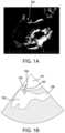
For the purpose of illustration and not limitation,FIG.1A is trans-esophageal echocardiography image of aheart valve 100, specifically a left ventricular outflow tract view of a mitral valve. Such a trans-esophageal echocardiography can be used for visualization during an edge-to-edge mitral valve repair procedure.
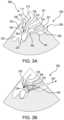
As illustrated inFIG.1C , when performing transcatheter edge-to-edge repair, aleaflet fixation device 150, is delivered on the distal end of acatheter shaft 111 such that the opposingleaflets leaflet fixation device 150 and brought together into coaptation. These fixation devices can use arms to contact thedistal side 104 of leaflets. Once the arms are positioned to stabilize the leaflets in a beating heart, gripping elements or the like contact the atrial side of the leaflets to capture the leaflets. Once both opposing leaflets are captured, thefixation device 150 can be closed such that the leaflets are pulled and brought into coaptation, which results in a reduction in valvular regurgitation during ventricular systole.
However, leaflet locations can vary from patient to patient, and in certain patients it can be difficult to reach and capture both leaflets using fixation devices alone. For example,FIG.2A depicts a mitral valve anatomy demonstrating a large coaptation gap, with the anteriormitral leaflet 103 depressed or spaced at a distance from the posteriormitral leaflet 105. As illustrated inFIG.2B , because the anteriormitral leaflet 103 is significantly spaced from the posteriormitral leaflet 105, theleaflet fixation device 150 may not be able to reach the anteriormitral leaflet 103, and successful edge-to-edge repair may be difficult, requiring a great deal of procedural skill and time. The leaflet grasping procedure can be further complicated by erratic leaflet motion, such as systolic anterior motion of the anterior mitral valve leaflet. Further, if only a minimal amount of leaflet can be inserted within the arms and gripping elements during the procedure based upon the large gap, mitral regurgitation may not be significantly improved by a single implantation and additional devices or further intervention may be needed to reduce the mitral regurgitation.
To address these problems, generally, and as set forth in greater detail below, the disclosed subject matter includes a kit with a coaptation aid and a fixation system for repair of leaflets of a heart valve. The coaptation aid includes a coaptation catheter having an expandable member at a distal end thereof adapted to be introduced to a left ventricular outflow tract of a heart via a retrograde approach. The expandable member has a delivery configuration with a reduced cross-dimension and a deployed configuration with an expanded cross-dimension adapted to contact a ventricular side of an anterior leaflet of a mitral valve and position the anterior leaflet generally proximate a coapting configuration with a posterior leaflet of the mitral valve. The fixation system includes a delivery catheter having a distal end, and a fixation device removably coupled to the distal end of the delivery catheter and adapted to couple the anterior leaflet to the posterior leaflet of the mitral valve
Likewise, as further described in conjunction with the kit of the disclosed subject matter, a method for fixation of native leaflets of a mitral valve using a coaptation aid is provided. The method includes introducing a coaptation aid with a coaptation catheter having an expandable member at a distal end thereof to a left ventricular outflow tract of a heart via a retrograde approach. The expandable member has a delivery configuration with a reduced cross-dimension and a deployed configuration with an expanded cross-dimension. The expandable member is in the delivery configuration while being introduced to the left ventricular outflow tract. The method also includes transitioning the expandable member from the delivery configuration toward the deployed configuration to contact a ventricular side of an anterior leaflet of a mitral valve and position the anterior leaflet generally proximate a coapting configuration with a posterior leaflet of the mitral valve. The method also includes delivering a fixation device to couple the anterior leaflet to the posterior leaflet of the mitral valve.
The coaptation aid thus can improve the ease of leaflet grasping and capture by providing a mechanism to temporarily facilitate positioning and stabilization of a leaflet, such as the anterior mitral leaflet, during an edge-to-edge repair procedure.
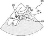
Referring toFIGS.3A and3B , for purpose of illustration and not limitation, an exemplary kit includescoaptation aid 320 with acoaptation catheter 321 for delivering anexpandable member 340 to a leftventricular outflow tract 106 of a heart. Theexpandable member 340 has adelivery configuration 341 with a reduced cross-dimension (inFIG.3A expandable member is within coaptation catheter321) and a deployedconfiguration 342 as shown inFIG.3B with an expanded cross-dimension adapted to contact aventricular side 104 of ananterior leaflet 103 of a mitral valve and position the anterior leaflet generally proximate a coapting configuration with aposterior leaflet 105 of the mitral valve. The kit also includes a fixation system including adelivery catheter 300 having adistal end 313, and afixation device 150 removably coupled to the distal end of the delivery catheter and adapted to couple theanterior leaflet 103 to theposterior leaflet 105 of the mitral valve.
With continued reference toFIG.3A , theleaflet fixation device 150, e.g., a clip, can be delivered via aseparate delivery catheter 300 including anelongate catheter shaft 111 having aproximal portion 312 and adistal end 313. Theleaflet fixation device 150 is releasably coupled to thedistal end 313 of the catheter shaft and is configured to grasp and couple two (or more) leaflets together. Although a number of edge-to-edge leaflet repair devices are known, for purpose of understanding and not limitation, reference will be made herein to a fixation device having afirst gripper assembly 351 and asecond gripper assembly 352 to capture the native leaflets, wherein each gripper assembly includes a movable arm and gripper element to capture a leaflet therebetween. Additional features and alternative embodiments of suitable catheters for delivering the leaflet fixation device and the expandable member (including details of the proximal portions) are set forth, for example, in U.S. Pat. No. 7,226,467 to Lucatero et al., U.S. Pat. No. 7,563,267 to Goldfarb et al., U.S. Pat. No. 7,655,015 to Goldfarb et al., U.S. Pat. No. 7,736,388 to Goldfarb et al., U.S. Pat. No. 7,811,296 to Goldfarb et al., U.S. Pat. No. 8,057,493 to Goldfarb et al., U.S. Pat. No. 8,303,608 to Goldfarb et al., U.S. Pat. No. 8,500,761 to Goldfarb et al., U.S. Pat. No. 8,734,505 to Goldfarb et al., U.S. Pat. No. 8,740,920 to Goldfarb et al., U.S. Pat. No. 9,510,829 to Goldfarb et al., U.S. Pat. No. 7,635,329 to Goldfarb et al., U.S. Patent Application Publication No. 2017/0042546 to Goldfarb et al., U.S. Patent Application Publication No. 2017/0239048 to Goldfarb et al., U.S. Patent Application Publication No. 2018/0325671 to Abunassar et al., the contents of each of which is incorporated by reference in its entirety herein (collectively “the Representative Patent Publications”).
In use, thedelivery catheter 300 is introduced proximate the native leaflets of aheart valve 100. For illustration and not limitation and as embodied herein, thedelivery catheter 300 can be delivered via an antegrade approach from a patient'sleft atrium 101 to theleft ventricle 102. For example, in a transcatheter approach, thedelivery catheter 300 can be introduced in a femoral vein and advanced through the inferior vena cava into the heart, across a penetration in the interatrial septum (i.e., a transseptal approach), and to the mitral valve from the atrium toward the ventricle. Details of various suitable approaches for leaflet repair are described in, for example, the Representative Patent Publications.
Once located proximate native leaflets of aheart valve 100, thefirst gripper assembly 351 of theleaflet fixation device 150 is deployed to capture a first native leaflet, such as the posteriormitral leaflet 105, as depicted inFIG.3A . For example, arms of thefixation device 150 can be aligned generally perpendicular to a line of coaptation of the mitral valve, with the device positioned so that a first arm contacts the ventricular surface of the posteriormitral leaflet 105. A corresponding gripping element can remain on the atrial side of the valve leaflet so that the leaflet lies between the gripping element and the arm. As embodied herein, the first gripping element can be lowered toward the arm so that the leaflet is captured and held therebetween. Alternatively, the leaflet can be held with the arm underneath without lowering the gripping element until later in the procedure. Additional features and alternative embodiments of leaflet fixation devices and the grasping and capture process are described in, for example, the Representative Patent Publications.
In certain anatomies where the anterior mitral leaflet is significantly spaced from the posterior mitral leaflet or has dynamically moved away from the region of coaptation where edge-to-edge repair using the leaflet fixation device would not be possible as shown inFIG.3A , the coaptation aid of the disclosed subject matter can be used to position the anterior mitral leaflet close to the posterior mitral leaflet to allow for edge-to-edge repair.
As shown inFIG.3A , to introduce the coaptation aid of the disclosed subject matter, a guide wire330 can be introduced to the leftventricular outflow tract 106 via retrograde approach, which can include an access route selected from the group consisting of a femoral artery, a brachial artery, and a radial artery. Any suitable guidewire known in the art can be used for such delivery. Thecoaptation catheter 321 can be delivered to the left ventricular outflow tract over guide wire330. During introduction to the left ventricular outflow tract, theexpandable member 340 is in thedelivery configuration 341, for example located within thecoaptation catheter 321. Once positioned in the left ventricular outflow tract proximate the anteriormitral leaflet 103, theexpandable member 340 is allowed to transition from the delivery configuration341 (FIG.3A ) toward the deployed configuration342 (FIG.3B ) to contact aventricular side 104 of ananterior leaflet 103 of a mitral valve and position the anterior leaflet generally proximate a coapting configuration with aposterior leaflet 105 of the mitral valve as shown inFIG.3B .
For example, and as depicted schematically inFIGS.3A,3B, and3C , the coaptation catheter can include aretractable sheath 355 having anextended position 356 and a retractedposition 357. The retractable sheath can be adapted to retract from theextended position 356 shown inFIG.3A toward the retractedposition 357 shown inFIG.3B to expose theexpandable member 340 and allow the expandable member to transition from thedelivery configuration 341 toward the deployedconfiguration 342. The method, prior to transitioning the expandable member, can further include aligning adistal tip 358 of the retractable sheath with a hinge point of theanterior leaflet 103. Once located in the desired position, thesheath 355 can be partially retracted as shown inFIG.3B to expose theexpandable member 340. Once expanded,expandable member 340 contacts the underside ordistal side 104 of theanterior leaflet 103 to position, e.g., prop, the leaflet into a favorable location for deployment of theleaflet fixation device 150, described below.
Depending upon the desired construction as disclosed herein, theexpandable member 340 can include a material selected from the group consisting of a metal, a super elastic material, a polymer, a bioresorable material and combinations thereof. For example, the expandable member can be a self-expanding scaffold, such as a scaffold contained within a sheath, or a balloon expandable scaffold mounted on a balloon of the coaptation catheter. Alternatively, and as contemplated by the disclosed subject matter, and as described further below, the expandable member can be a balloon.
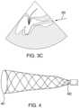
For example and in accordance with the disclosed subject matter, the expandable member can be an expandable scaffold as depicted schematically for purpose of illustration and not limitation inFIG.4 . The expandable scaffold can have a variety of suitable shapes, such as an elongate cylinder or a tapered cylinder as shown, as long as it includes a sufficient diameter to engage with the underside of the anterior mitral leaflet. For example, the scaffold can be formed with a larger cross dimension at adistal portion 461 and taper continuously to a smaller cross dimension at aproximal portion 462. The tapered configuration can increase the chance of achieving favorable contact with the anteriormitral leaflet 103 at a point of the leaflet hinge point, or even farther out toward the leaflet tip near the region of coaptation.
The expandable scaffold can be self-expanding and can be formed from a shape memory material, such as nitinol, which is well known in the field of stents and frames for heart valve replacement devices. The scaffold can be formed by laser cutting from a tube or can be shaped of nitinol wire on a mandrel as known in the art. The scaffold could also be made other materials, such as polymers. The scaffold can include any structure known in the art, including ring and connector, spiral, open or closed basket or cell pattern, or the like. The scaffold can have a sparse construction to reduce interaction with chordae tendineae below the native valve.
In accordance with another aspect of the disclosed subject matter, and with reference toFIG.5 for illustration and not limitation, thedistal portion 461 of the scaffold can be provided with at least one recess or notch570 at its distal end. The notch, when in the deployed configuration, can allow thefixation device 150 to access the anterior leaflet while positioned proximate the coapting configuration with the posterior leaflet. As embodied herein, the notch is placed over the middle of theanterior leaflet 103 such that the scaffold remains clear of the arm of thefixation device 150 being positioned and closed at the leaflet. Additional notches could be positioned at many locations along the scaffold to avoid undesirable interactions with the papillary muscles or chordae tendineae.
In accordance with another aspect of the disclosed subject matter, and with reference toFIG.6 for illustration and not limitation, the expandable scaffold can include amarker 680 proximate the at least onenotch 570. For example, a single marker, pairs of markers, or an array of markers proximate the notch at the distal end of the scaffold can be aligned with the target region of the leaflet for leaflet fixation such as the middle of the anterior leaflet. The markers can be radiopaque or echogenic and can be used to improve the position of the notch during a procedure, e.g., by locating the notch at the twelve o'clock position as the catheter is torqued.
In accordance with another aspect of the disclosed subject matter, and with reference toFIG.7 for illustration and not limitation, theexpandable scaffold 340 can adapted for release from thecoaptation catheter 321 in the deployed configuration at the leftventricular outflow tract 106. For example, the expandable scaffold can be mounted on aballoon 740 of thecoaptation catheter 321. The balloon can have any suitable shape for delivering the expandable scaffold such as a cylinder. Alternatively, the balloon can include a tapered cylinder in the deployed configuration with adistal portion 761 with a larger cross dimension than aproximal portion 762. In such embodiments, the scaffold can be positioned on a proximal portion of the balloon so that it is delivered within the left ventricular outflow tract yet does not interfere with the function of the mitral valve after implant. The tapered balloon distal portion extending beyond the scaffold can be used to position the anterior leaflet proximate the coapting configuration with the posterior leaflet prior to deploying the fixation device. Furthermore, the scaffold can be short enough to avoid blocking the aortic valve and can be used to prevent left ventricular outflow obstruction (LVOT) after deployment.
For example, the scaffold can have a length of at least 2 to 3 centimeters in length and preferably 4 to 6 centimeters in length in order to encompass and support a full length of an anterior mitral valve leaflet with a significant margin included. Further, the scaffold can preferably have a diameter that approximately corresponds to the left ventricular outflow tract (LVOT) diameter for a given patient, which is estimated in millimeters in the literature to be 5.7×body surface area+12.1. (Hahn et al., Accurate Measurement of Left Ventricular Outflow Tract Diameter: Comment on the Updated Recommendations for the Echocardiographic Assessment of Aortic Valve Stenosis, Journal of the American Society of Echocardiography Volume 30 Number 10). The scaffold can have an open cell pattern (for more conformability) or a closed cell pattern (for more radial support) with any combination and pattern transitions throughout its length. The scaffold can be made radially stiff in sections that secure the scaffold in the LVOT, while the leading edge that contacts leaflets can be made with narrower or thinner struts to reduce the potential for damaging leaflets or subvalvular anatomic structures.
In accordance with the disclosed subject matter, the expandable scaffold adapted for release can be made of a bioresorbable material and intended to remain in the body for a period of time (e.g., days, weeks, months, or years) after implant. For example, the scaffold can be made of biodegradable polymers like poly-L-lactide.
Thus, in addition to providing the positioning benefits of the coaptation aid embodiments described herein, the release of the scaffold can also be used to provide additional benefits. For example, the expandable scaffold can further include and elute one or more beneficial agent, such as an anti-inflammatory agent, an anti-coagulant agent, a thrombotic agent, an oxidative stress reducing agent, a growth factor, a pro-healing agent, or the like. The presence of the scaffold can also aid in the prevention of left ventricular outflow tract obstruction if a used in combination with a replacement mitral valve of a size that could otherwise compromise the left ventricular outflow tract.
In accordance with another aspect of the disclosed subject matter, and as shown inFIG.8 for the purpose of illustration and not limitation, theexpandable member 340 can be aballoon 840, such as an elongate balloon. The balloon can be cylindrical or can have a tapered body in the deployed configuration with adistal portion 861 with a larger cross dimension than aproximal portion 862.
Furthermore, thedistal end portion 861 ofballoon 840 can have at least onerecess 870 formed therein. Like thenotch 570 described above, in the deployed configuration, therecess 870 in the balloon allows access of thefixation device 150 to the anterior leaflet while positioned proximate the coapting configuration with the posterior leaflet. In addition, theballoon 840 can have amarker 880 proximate the at least onerecess 870. Additionally or alternatively, theballoon 840 can be filled with contrast media to aid in visualization and positioning of the balloon.
Thecoaptation catheter 321 can include aninflation lumen 890 in fluid communication with theballoon 840. Thus the interior of the balloon is in a fluid flow relation with an inflation lumen extending along a length of the coaptation catheter, and fluid under pressure can thereby be supplied to the interior of the balloon through the inflation lumen to expand the balloon. The balloon can include marker bands (e.g., made of platinum/iridium), for example positioned on the proximal and distal sections of the balloon or catheter.
In accordance with the disclosed subject matter, theballoon 840 can be composed of a wide variety of suitable materials, for example, nylons, co-polyamides such as PEBAX®, polyester, co-polyester, polyurethane, polyethylene, or the like. Theballoon 840 can comprise a single layer or multiple layers of polymer material. Details of suitable multilayer balloons are described in U.S. Pat. Nos. 7,828,766, 8,394,055, and 9,132,259, the contents of each of which are herein incorporated by reference in their entirety. The balloon can have wings and be folded as known in the art.
As embodied herein, theballoon 840 can be a relatively high rupture pressure, non-compliant balloon, which can have a rupture pressure of at least about 20-30 atm. Alternatively, the balloon can be made to function in a more conforming manner and can be made with a thin wall in order to provide a more compliant and atraumatic impingement on the chordae and leaflet structures. The balloon can be made porous to permit blood flow to pass through the coaptation aid during a procedure. Additionally or alternatively, the balloon can be fashioned with a toroidal geometry such that blood flow can pass through its center. Since the left ventricular outflow tract is generally elliptical in shape, an aligning elliptical balloon geometry can also be used. Further, the balloon can be tapered, can have a curved profile geometry, or can be manufactured with intentional kinks in order to conform to the apex of the heart and turn upward toward the anterior mitral valve leaflet. Any combination of the above-mentioned features is contemplated in this application.
The balloon can additionally include any of any of the features described above for the expandable scaffold. Additional suitable materials, configurations, and methods of manufacture of theballoon 140 and related disclosure are provided in U.S. Pat. Nos. 7,074,206, 8,052,638, 10,086,175, U.S. Patent Publication No. 2016/0339204 and U.S. Patent Publication No. 2016/0067459, the contents of each of which is hereby incorporated by reference in its entirety.
Further in accordance with the disclosed subject matter, the expandable member can further include a wireless sensor, which can provide one or more representative signals, such as left ventricular pressure signal, cardiac output measurement signal, vessel wall motion measurement signal, ejection fraction measurement signal, or the like. For example, cardiac output can be estimated intraprocedurally to determine the impact of intra-procedural blood flow obstruction occurring during leaflet stabilization and for evaluation of the degree of therapeutic improvement achieved during a valve repair procedure. This cardiac output (flow rate) feedback, along with pressure gradient measurements can aid a physician in balancing the effectiveness and safety risks of performing a coaptation aided mitral valve repair.
The expandable member (scaffold or balloon) can have any suitable dimensions, for example including a cross sectional area and length of 4-6 cm2and 4-6 cm, respectively. The expanding member can be tapered in profile, curved to conform around the apex of the heart, or kinked to easily turn upward into contact with an anterior mitral valve leaflet to provide support during mitral valve repair procedures.
Returning now to the method of leaflet repair, in the deployedconfiguration 342 ofFIG.3B , with thecoaptation aid 320 including expandable member340 (scaffold or balloon840) acting as a stabilizing support for the anteriormitral leaflet 103, thefixation device 150 can be more easily used to simultaneously grasp both leaflets. The arm of thesecond gripper assembly 352 of theleaflet fixation device 150 is deployed to contact theventricular surface 104 of and grasp the anteriormitral leaflet 103. The corresponding gripping element can remain on the atrial side of the leaflet so that the leaflet lies between the gripping element and the arm.
If needed, thefixation device 150 can be repeatedly manipulated to reposition the device so that the leaflets are properly grasped at a desired location. Repositioning can be achieved with thefixation device 150 in an open position. For example, thecoaptation aid 320 can be manipulated to reposition the leaflets, such as by further expanding or retracting theexpandable member 340 to better position the anteriormitral leaflet 103. As embodied herein, thedelivery catheter 300 can be longitudinally rotated to apply different tension on the leaflet via theexpandable member 340 to better position the leaflet for capture. Regurgitation of the valve can also be checked while thefixation device 150 is in the open position. If regurgitation is not satisfactorily reduced, thefixation device 150 can be repositioned, and regurgitation checked again until desired results are achieved.
Once thefixation device 150 has been positioned in a desired location relative to the valve leaflets, the leaflets can then be captured between the gripping elements and the arms. As noted above, the first gripping element may already have been lowered to capture the posterior leaflet. If so, the second gripping element is lowered to capture the anterior leaflet. Alternatively, if the posterior leaflet has not yet been captured, both gripping elements can be lowered toward the arms so that the leaflets are held and captured therebetween, either simultaneously or sequentially. Once both leaflets are captured, the arms can be closed to a tight arm angle to bring each leaflet into central coaptation to reduce regurgitation. The arms can further be locked to the prevent the device from moving toward an open position.
Once completed, thecoaptation aid 320 can be removed by transitioning the expandable member340 (scaffold or balloon) toward the delivery configuration241. For example, the balloon can be deflated or the scaffold resheathed within the retractable sheath as shown inFIG.3 , to return the delivery system to a low-profile configuration. With the coaptation aid removed, the repair of the leaflets or tissue can be observed by non-invasive visualization techniques, such as echocardiography, to ensure the desired outcome. If the repair is not desired, thefixation device 150 can be repositioned or retrieved. Once the desired outcome is achieved, thefixation device 150 can then be detached from thedistal end 313 of the catheter shaft, and thecoaptation aid 320 withcoaptation catheter 321 can be safely withdrawn from the left ventricular outflow tract of the heart and from the body, and thedelivery catheter 300 can be withdrawn from proximate the leaflets and from the body. If a balloon expandable scaffold is provided, it can remain in the body after the coaptation catheter is withdrawn.
As embodied herein,coaptation aid 320 can be an optional accessory for use with adelivery catheter 300 for delivering a leaflet fixation device. For example, thecoaptation aid 320 can be part of a kit including aleaflet fixation device 150. Prior to a procedure, a user can observe the native anatomy and spacing between the leaflets to determine if acoaptation aid 320 will assist in the leaflet repair procedure.
The embodiments illustrated herein are adapted for repair of a heart valve and have been described in connection with repair of a mitral valve, using a leaflet fixation device delivered via an antegrade approach from a patient's left atrium and a coaptation aid delivered via a retrograde approach. However, the disclosed subject matter can be used on any other leaflet that requires intraprocedural propping or positioning to make leaflet grasping and clipping easier. For example, the disclosed subject matter could be used to position the posterior mitral leaflet, with the scaffold or balloon features adjusted to avoid interactions with the chordae tendineae or papillary muscles within the ventricle. Other exemplary leaflets include any tricuspid, aortic, or pulmonary valve leaflet, however, accessing these leaflets may require antegrade delivery of a coaptation aid. Furthermore, while shown herein as a clip, the leaflet fixation device could be any suitable fixation means, including sutures, clips, staples, and the like. In addition, while a single device was shown and described in each embodiment herein, a plurality of devices or device features can be used to simultaneously support or stabilize two or more opposing leaflets in order to further simplify the edge-to-edge repair procedure. Further, while described in connection with an edge-to-edge repair procedure, the disclosure subject matter can be adapted to aid in other procedures, such as leaflet resection, minimally invasive leaflet suturing, and chordae replacement.
While the disclosed subject matter is described herein in terms of certain embodiments for purpose of illustration and not limitation, those skilled in the art will recognize that various modifications and improvements can be made to the disclosed subject matter without departing from the scope thereof. Moreover, although individual features of the disclosed subject matter may have been discussed herein or shown in the drawings of one embodiment and not in other embodiments, it is readily apparent that individual features of one embodiment can be combined with, or substituted for, one or more features of another embodiment or features from a plurality of embodiments.
In addition to the specific embodiments claimed below, the disclosed subject matter is also directed to other embodiments having any other possible combination of the dependent features claimed below and those disclosed above. As such, the particular features presented in the dependent claims and disclosed above can be combined with each other in other possible combinations. Thus, the foregoing description of specific embodiments of the disclosed subject matter has been presented for purposes of illustration and description. It is not intended to be exhaustive or to limit the disclosed subject matter to those embodiments disclosed.
It will be apparent to those skilled in the art that various modifications and variations can be made in the method and system of the disclosed subject matter without departing from the spirit or scope of the disclosed subject matter. Thus, it is intended that the disclosed subject matter include modifications and variations that are within the scope of the appended claims and their equivalents.
Claims (28)
1. A kit for repair of leaflets of a heart valve, comprising:
a coaptation aid comprising
a coaptation catheter comprising an expandable scaffold at a distal end thereof adapted to be introduced to a left ventricular outflow tract of a heart via a retrograde approach, the expandable scaffold having a delivery configuration with a reduced cross-dimension and a deployed configuration with an expanded cross-dimension adapted to contact a ventricular side of a first leaflet of a heart valve and position the first leaflet generally proximate a coapting configuration with a second leaflet of the heart valve, and
a retractable sheath having an extended position and a retracted position, the retractable sheath being adapted to retract from the extended position toward the retracted position to expose the expandable scaffold and cause the expandable scaffold to be transitioned from the delivery configuration toward the deployed configuration, the retractable sheath further adapted to extend from the retracted position to the extended position to retrieve the expandable scaffold in order to transition the expandable scaffold from the deployed configuration back to the delivery configuration; and
a fixation system comprising a delivery catheter having a distal end, and a fixation device removably coupled to the distal end of the delivery catheter, the fixation device adapted to couple the first leaflet to the second leaflet of the heart valve.
2. The kit ofclaim 1 , wherein the heart valve is a mitral valve, the first leaflet is an anterior leaflet and the second leaflet is a posterior leaflet.
3. The kit ofclaim 1 , wherein the expandable scaffold is a self-expanding scaffold.
4. The kit ofclaim 3 , wherein the self-expanding scaffold comprises a tapered body having a distal portion with a larger cross dimension than a proximal portion.
5. The kit ofclaim 3 , wherein the self-expanding scaffold has a proximal end portion and a distal end portion, the distal end portion having at least one notch when in the deployed configuration to allow access of the fixation device to the first leaflet while positioned proximate the coapting configuration with the second leaflet.
6. The kit ofclaim 5 , wherein the self-expanding scaffold has a marker proximate the at least one notch.
7. The kit ofclaim 1 , wherein the expandable scaffold further comprises a wireless sensor.
8. The kit ofclaim 7 , wherein the wireless sensor provides one or more representative signals selected from the group consisting of left ventricular pressure signal, cardiac output measurement signal, vessel wall motion measurement signal, and ejection fraction measurement signal.
9. The kit ofclaim 1 , wherein the coaptation catheter is adapted for introduction via the retrograde approach through an access route selected from the group consisting of a femoral artery, a brachial artery, and a radial artery.
10. The kit ofclaim 1 , wherein the expandable scaffold comprises a material selected from the group consisting of a metal, a super elastic material, a polymer, a bioabsorbable material and combinations thereof.
11. A method for fixation of native leaflets of a heart valve comprising:
introducing a coaptation aid comprising a coaptation catheter having an expandable scaffold at a distal end thereof to a left ventricular outflow tract of a heart via a retrograde approach, the expandable scaffold having a delivery configuration with a reduced cross-dimension and a deployed configuration with an expanded cross-dimension, the coaptation aid further comprising a retractable sheath having an extended position and a retracted position, the expandable scaffold being in the delivery configuration and the retractable sheath being in the extended position while being introduced to the left ventricular outflow tract;
retracting the retractable sheath from the extended position toward the retracted position to expose the expandable scaffold and cause the expandable scaffold to transition from the delivery configuration toward the deployed configuration to contact a ventricular side of a first leaflet of a heart valve and position the first leaflet generally proximate a coapting configuration with a second leaflet of the heart valve;
delivering a fixation device to couple the first leaflet to the second leaflet of the heart valve.
12. The method ofclaim 11 , wherein the heart valve is a mitral valve, the first leaflet is an anterior leaflet and the second leaflet is a posterior leaflet.
13. The method ofclaim 11 , further comprising, after delivering the fixation device,
transitioning the expandable member back to the delivery configuration;
extending the retractable sheath from the retracted position to the extended position; and
withdrawing the coaptation aid from the left ventricular outflow tract of the heart.
14. The method ofclaim 11 , wherein the expandable scaffold is a self-expanding scaffold.
15. The method ofclaim 11 , wherein prior to transitioning the expandable scaffold, the method further comprises aligning a distal tip of the retractable sheath proximate a hinge point of the first leaflet.
16. The method ofclaim 11 , wherein introducing the coaptation aid includes using an access route selected from the group consisting of a femoral artery, a brachial artery, and a radial artery.
17. A method for fixation of native leaflets of a mitral valve comprising:
introducing a coaptation aid comprising a coaptation catheter having an expandable member at a distal end thereof to a left ventricular outflow tract of a heart via a retrograde approach, the expandable member having a delivery configuration with a reduced cross-dimension and a deployed configuration with an expanded cross-dimension, the expandable member in the delivery configuration while being introduced to the left ventricular outflow tract;
transitioning the expandable member from the delivery configuration toward the deployed configuration to contact a ventricular side of an anterior leaflet of the mitral valve and position the anterior leaflet generally proximate a coapting configuration with a posterior leaflet of the mitral valve; and
delivering a fixation device to couple the anterior leaflet to the posterior leaflet of the mitral valve.
18. The method ofclaim 17 , wherein the expandable member is a balloon-expandable scaffold mounted on an expandable balloon of the coaptation catheter.
19. The method ofclaim 18 , wherein the balloon-expandable scaffold is released in the deployed configuration from the expandable balloon at the left ventricular outflow tract.
20. The method ofclaim 19 , wherein the balloon-expandable scaffold is made of a bioresorbable material.
21. The method ofclaim 19 , wherein the balloon-expandable scaffold further comprises one or more beneficial agents selected from the group consisting of an anti-inflammatory agent, an anti-coagulant agent, a thrombotic agent, an oxidative stress reducing agent, a growth factor, and a pro-healing agent.
22. The method ofclaim 17 , wherein the expandable member is a balloon.
23. The method ofclaim 22 , wherein the balloon has a proximal end portion and a distal end portion, the distal end portion having at least one recess formed therein when in the deployed configuration to allow access of the fixation device to the anterior leaflet while positioned proximate the coapting configuration with the posterior leaflet.
24. The method ofclaim 23 , wherein the balloon has a marker proximate the at least one recess.
25. The method ofclaim 22 , wherein the coaptation catheter further comprises an inflation lumen in fluid communication with the balloon.
26. The method ofclaim 22 , wherein the balloon comprises an elongate balloon.
27. The method ofclaim 22 , wherein balloon comprises a tapered body in the deployed configuration.
28. The method ofclaim 27 , wherein the tapered body has a distal portion with a larger cross dimension than a proximal portion.
Priority Applications (1)
| Application Number | Priority Date | Filing Date | Title |
|---|---|---|---|
| US17/096,684US11701229B2 (en) | 2019-11-14 | 2020-11-12 | Kit with coaptation aid and fixation system and methods for valve repair |
Applications Claiming Priority (2)
| Application Number | Priority Date | Filing Date | Title |
|---|---|---|---|
| US201962935241P | 2019-11-14 | 2019-11-14 | |
| US17/096,684US11701229B2 (en) | 2019-11-14 | 2020-11-12 | Kit with coaptation aid and fixation system and methods for valve repair |
Publications (2)
| Publication Number | Publication Date |
|---|---|
| US20210145577A1 US20210145577A1 (en) | 2021-05-20 |
| US11701229B2true US11701229B2 (en) | 2023-07-18 |
Family
ID=73726882
Family Applications (1)
| Application Number | Title | Priority Date | Filing Date |
|---|---|---|---|
| US17/096,684Active2040-12-06US11701229B2 (en) | 2019-11-14 | 2020-11-12 | Kit with coaptation aid and fixation system and methods for valve repair |
Country Status (2)
| Country | Link |
|---|---|
| US (1) | US11701229B2 (en) |
| WO (1) | WO2021097089A1 (en) |
Citations (189)
| Publication number | Priority date | Publication date | Assignee | Title |
|---|---|---|---|---|
| US3378010A (en) | 1965-07-28 | 1968-04-16 | Coldling | Surgical clip with means for releasing the clamping pressure |
| US3874388A (en) | 1973-02-12 | 1975-04-01 | Ochsner Med Found Alton | Shunt defect closure system |
| US4007743A (en) | 1975-10-20 | 1977-02-15 | American Hospital Supply Corporation | Opening mechanism for umbrella-like intravascular shunt defect closure device |
| US4055861A (en) | 1975-04-11 | 1977-11-01 | Rhone-Poulenc Industries | Support for a natural human heart valve |
| US4327736A (en) | 1979-11-20 | 1982-05-04 | Kanji Inoue | Balloon catheter |
| US4340091A (en) | 1975-05-07 | 1982-07-20 | Albany International Corp. | Elastomeric sheet materials for heart valve and other prosthetic implants |
| US4657024A (en) | 1980-02-04 | 1987-04-14 | Teleflex Incorporated | Medical-surgical catheter |
| US4693248A (en) | 1983-06-20 | 1987-09-15 | Ethicon, Inc. | Two-piece tissue fastener with deformable retaining receiver |
| US4716886A (en) | 1986-05-01 | 1988-01-05 | Norman Schulman | Umbilical cord clamp and cutters |
| US4795458A (en) | 1987-07-02 | 1989-01-03 | Regan Barrie F | Stent for use following balloon angioplasty |
| US4809695A (en) | 1981-10-21 | 1989-03-07 | Owen M. Gwathmey | Suturing assembly and method |
| US4930674A (en) | 1989-02-24 | 1990-06-05 | Abiomed, Inc. | Surgical stapler |
| WO1991001689A1 (en) | 1989-07-28 | 1991-02-21 | Jacob Segalowitz | Valvulotome catheter |
| US5002562A (en) | 1988-06-03 | 1991-03-26 | Oberlander Michael A | Surgical clip |
| US5069679A (en) | 1989-02-16 | 1991-12-03 | Taheri Syde A | Method and apparatus for removing venous valves |
| US5098440A (en) | 1990-08-14 | 1992-03-24 | Cordis Corporation | Object retrieval method and apparatus |
| US5125895A (en) | 1986-07-22 | 1992-06-30 | Medtronic Versaflex, Inc. | Steerable catheter |
| WO1992012690A1 (en) | 1991-01-24 | 1992-08-06 | Autogenics | Heart valve with tissue alignment and clamping |
| US5147370A (en) | 1991-06-12 | 1992-09-15 | Mcnamara Thomas O | Nitinol stent for hollow body conduits |
| US5171259A (en) | 1990-04-02 | 1992-12-15 | Kanji Inoue | Device for nonoperatively occluding a defect |
| US5222963A (en) | 1991-01-17 | 1993-06-29 | Ethicon, Inc. | Pull-through circular anastomosic intraluminal stapler with absorbable fastener means |
| US5271544A (en) | 1991-08-23 | 1993-12-21 | Ethicon, Inc. | Surgical anastomosis stapling instrument |
| US5327905A (en) | 1992-02-14 | 1994-07-12 | Boaz Avitall | Biplanar deflectable catheter for arrhythmogenic tissue ablation |
| US5330501A (en) | 1991-05-30 | 1994-07-19 | United States Surgical Corporation | Tissue gripping device for use with a cannula and a cannula incorporating the device |
| US5334217A (en) | 1992-01-21 | 1994-08-02 | Regents Of The University Of Minnesota | Septal defect closure device |
| WO1994018893A1 (en) | 1993-02-22 | 1994-09-01 | Valleylab, Inc. | A laparoscopic dissection tension retractor device and method |
| US5363861A (en) | 1991-11-08 | 1994-11-15 | Ep Technologies, Inc. | Electrode tip assembly with variable resistance to bending |
| US5389077A (en) | 1993-03-03 | 1995-02-14 | Uresil Corporation | Minimally invasive body cavity penetrating instruments |
| US5403326A (en) | 1993-02-01 | 1995-04-04 | The Regents Of The University Of California | Method for performing a gastric wrap of the esophagus for use in the treatment of esophageal reflux |
| US5425744A (en) | 1991-11-05 | 1995-06-20 | C. R. Bard, Inc. | Occluder for repair of cardiac and vascular defects |
| US5450860A (en) | 1993-08-31 | 1995-09-19 | W. L. Gore & Associates, Inc. | Device for tissue repair and method for employing same |
| US5452837A (en) | 1994-01-21 | 1995-09-26 | Ethicon Endo-Surgery, Inc. | Surgical stapler with tissue gripping ridge |
| US5456400A (en) | 1993-04-22 | 1995-10-10 | United States Surgical Corporation | Apparatus and clip for fastening body tissue |
| US5456674A (en) | 1993-03-31 | 1995-10-10 | Cordis Corporation | Catheters with variable properties |
| US5478353A (en) | 1987-05-14 | 1995-12-26 | Yoon; Inbae | Suture tie device system and method for suturing anatomical tissue proximate an opening |
| US5542949A (en) | 1987-05-14 | 1996-08-06 | Yoon; Inbae | Multifunctional clip applier instrument |
| US5562678A (en) | 1995-06-02 | 1996-10-08 | Cook Pacemaker Corporation | Needle's eye snare |
| WO1996032882A1 (en) | 1995-04-20 | 1996-10-24 | Heartport, Inc. | Method and apparatus for thoracoscopic intracardiac procedures |
| US5601224A (en) | 1992-10-09 | 1997-02-11 | Ethicon, Inc. | Surgical instrument |
| US5601574A (en) | 1992-09-14 | 1997-02-11 | Ethicon, Inc. | Sterile clips and instrument for their placement |
| US5607471A (en) | 1993-08-03 | 1997-03-04 | Jacques Seguin | Prosthetic ring for heart surgery |
| US5607462A (en) | 1993-09-24 | 1997-03-04 | Cardiac Pathways Corporation | Catheter assembly, catheter and multi-catheter introducer for use therewith |
| US5609598A (en) | 1994-12-30 | 1997-03-11 | Vnus Medical Technologies, Inc. | Method and apparatus for minimally invasive treatment of chronic venous insufficiency |
| US5611794A (en) | 1990-10-11 | 1997-03-18 | Lasersurge, Inc. | Clamp for approximating tissue sections |
| US5636634A (en) | 1993-03-16 | 1997-06-10 | Ep Technologies, Inc. | Systems using guide sheaths for introducing, deploying, and stabilizing cardiac mapping and ablation probes |
| WO1997027807A1 (en) | 1996-01-31 | 1997-08-07 | Heartport, Inc. | Endoscopic suturing devices and methods |
| US5695504A (en) | 1995-02-24 | 1997-12-09 | Heartport, Inc. | Devices and methods for performing a vascular anastomosis |
| US5713911A (en) | 1996-10-03 | 1998-02-03 | United States Surgical Corporation | Surgical clip |
| US5716417A (en) | 1995-06-07 | 1998-02-10 | St. Jude Medical, Inc. | Integral supporting structure for bioprosthetic heart valve |
| WO1998007375A1 (en) | 1996-08-22 | 1998-02-26 | The Trustees Of Columbia University | Endovascular flexible stapling device |
| US5741297A (en) | 1996-08-28 | 1998-04-21 | Simon; Morris | Daisy occluder and method for septal defect repair |
| US5755778A (en) | 1996-10-16 | 1998-05-26 | Nitinol Medical Technologies, Inc. | Anastomosis device |
| US5782239A (en) | 1992-06-30 | 1998-07-21 | Cordis Webster, Inc. | Unique electrode configurations for cardiovascular electrode catheter with built-in deflection method and central puller wire |
| US5814097A (en) | 1992-12-03 | 1998-09-29 | Heartport, Inc. | Devices and methods for intracardiac procedures |
| US5843178A (en) | 1996-06-20 | 1998-12-01 | St. Jude Medical, Inc. | Suture guard for annuloplasty ring |
| US5849019A (en) | 1995-03-09 | 1998-12-15 | Yoon; Inbae | Multifunctional spring clips and cartridges and applications therefor |
| US5855601A (en) | 1996-06-21 | 1999-01-05 | The Trustees Of Columbia University In The City Of New York | Artificial heart valve and method and device for implanting the same |
| WO1999007354A2 (en) | 1997-08-08 | 1999-02-18 | Duke University | Compositions, apparatus and methods for facilitating surgical procedures |
| FR2768324A1 (en) | 1997-09-12 | 1999-03-19 | Jacques Seguin | Surgical instrument for joining soft tissues through percutaneous access |
| WO1999015223A1 (en) | 1997-09-26 | 1999-04-01 | Cardeon Corporation | Multi-function aortic catheterization and bumper instrument |
| EP0558031B1 (en) | 1992-02-27 | 1999-04-14 | United States Surgical Corporation | Apparatus for subcuticular stapling of body tissue |
| US5976159A (en) | 1995-02-24 | 1999-11-02 | Heartport, Inc. | Surgical clips and methods for tissue approximation |
| FR2768325B1 (en) | 1997-09-16 | 1999-11-19 | Instr Medecine Veterinaire | CATHETER FOR ARTIFICIAL INSEMINATION OF BIRDS, ESPECIALLY TURKEYS, AND METHOD FOR PRODUCING SUCH A CATHETER |
| US6015417A (en) | 1996-01-25 | 2000-01-18 | Reynolds, Jr.; Walker | Surgical fastener |
| WO2000003759A2 (en) | 1998-07-15 | 2000-01-27 | St. Jude Medical, Inc. | Mitral and tricuspid valve repair |
| US6048351A (en) | 1992-09-04 | 2000-04-11 | Scimed Life Systems, Inc. | Transvaginal suturing system |
| US6117144A (en) | 1995-08-24 | 2000-09-12 | Sutura, Inc. | Suturing device and method for sealing an opening in a blood vessel or other biological structure |
| US6120496A (en) | 1998-05-05 | 2000-09-19 | Scimed Life Systems, Inc. | Surgical method and apparatus for positioning a diagnostic or therapeutic element within the body and coupling device for use with same |
| WO2000060995A2 (en) | 1999-04-09 | 2000-10-19 | Evalve, Inc. | Methods and apparatus for cardiac valve repair |
| US6149658A (en) | 1997-01-09 | 2000-11-21 | Coalescent Surgical, Inc. | Sutured staple surgical fasteners, instruments and methods for minimally invasive vascular and endoscopic surgery |
| US6182664B1 (en) | 1996-02-19 | 2001-02-06 | Edwards Lifesciences Corporation | Minimally invasive cardiac valve surgery procedure |
| US6193734B1 (en) | 1998-01-23 | 2001-02-27 | Heartport, Inc. | System for performing vascular anastomoses |
| US6200315B1 (en) | 1997-12-18 | 2001-03-13 | Medtronic, Inc. | Left atrium ablation catheter |
| US6217528B1 (en) | 1999-02-11 | 2001-04-17 | Scimed Life Systems, Inc. | Loop structure having improved tissue contact capability |
| WO2001028432A1 (en) | 1999-10-21 | 2001-04-26 | Edwards Lifesciences Corporation | Minimally invasive mitral valve repair method and apparatus |
| US6269819B1 (en) | 1997-06-27 | 2001-08-07 | The Trustees Of Columbia University In The City Of New York | Method and apparatus for circulatory valve repair |
| US6290674B1 (en) | 1999-09-20 | 2001-09-18 | Appriva Medical, Inc. | Method and apparatus for closing intracardiac septal defects |
| US6312447B1 (en) | 1999-10-13 | 2001-11-06 | The General Hospital Corporation | Devices and methods for percutaneous mitral valve repair |
| US6332880B1 (en) | 1996-12-19 | 2001-12-25 | Ep Technologies, Inc. | Loop structures for supporting multiple electrode elements |
| US20020013571A1 (en) | 1999-04-09 | 2002-01-31 | Evalve, Inc. | Methods and devices for capturing and fixing leaflets in valve repair |
| US6346074B1 (en) | 1993-02-22 | 2002-02-12 | Heartport, Inc. | Devices for less invasive intracardiac interventions |
| US6419696B1 (en) | 2000-07-06 | 2002-07-16 | Paul A. Spence | Annuloplasty devices and related heart valve repair methods |
| US20020183787A1 (en) | 2001-06-01 | 2002-12-05 | Velocimed, L.L.C. | Closure devices, related delivery methods and tools, and related methods of use |
| US6496420B2 (en) | 1999-03-01 | 2002-12-17 | Micron Technology, Inc. | Methods and apparatus for reading memory device register data |
| WO2003020179A1 (en) | 2001-08-31 | 2003-03-13 | Mitral Interventions | Apparatus for valve repair |
| US6544215B1 (en) | 1998-10-02 | 2003-04-08 | Scimed Life Systems, Inc. | Steerable device for introducing diagnostic and therapeutic apparatus into the body |
| US6551303B1 (en) | 1999-10-27 | 2003-04-22 | Atritech, Inc. | Barrier device for ostium of left atrial appendage |
| US6575971B2 (en) | 2001-11-15 | 2003-06-10 | Quantum Cor, Inc. | Cardiac valve leaflet stapler device and methods thereof |
| WO2003049619A2 (en) | 2001-12-08 | 2003-06-19 | Lattouf Omar M | Treatments for a patient with congestive heart failure |
| US6599311B1 (en) | 1998-06-05 | 2003-07-29 | Broncus Technologies, Inc. | Method and assembly for lung volume reduction |
| US20030167071A1 (en) | 2002-03-01 | 2003-09-04 | Evalve, Inc. | Suture fasteners and methods of use |
| US6669687B1 (en) | 1999-06-25 | 2003-12-30 | Vahid Saadat | Apparatus and methods for treating tissue |
| US20040034365A1 (en) | 2002-08-16 | 2004-02-19 | Lentz David J. | Catheter having articulation system |
| US20040044350A1 (en) | 1999-04-09 | 2004-03-04 | Evalve, Inc. | Steerable access sheath and methods of use |
| US6719767B1 (en) | 2000-08-11 | 2004-04-13 | Edwards Lifesciences Corporation | Device and a method for treatment of atrioventricular regurgitation |
| US6797002B2 (en) | 2000-02-02 | 2004-09-28 | Paul A. Spence | Heart valve repair apparatus and methods |
| US6837867B2 (en) | 2001-04-30 | 2005-01-04 | Biosense Webster, Inc. | Steerable catheter with reinforced tip |
| US6855137B2 (en) | 2002-03-07 | 2005-02-15 | Visionary Biomedical, Inc. | Catheter shaft with coextruded stiffener |
| US6908481B2 (en) | 1996-12-31 | 2005-06-21 | Edwards Lifesciences Pvt, Inc. | Value prosthesis for implantation in body channels |
| US6926730B1 (en) | 2000-10-10 | 2005-08-09 | Medtronic, Inc. | Minimally invasive valve repair procedure and apparatus |
| US20050267493A1 (en) | 2001-02-06 | 2005-12-01 | Schreck Stefan G | Method and system for tissue repair using dual catheters |
| US7074206B2 (en) | 1999-12-01 | 2006-07-11 | Advanced Cardiovascular Systems, Inc. | Medical device balloon |
| US7101395B2 (en) | 2002-06-12 | 2006-09-05 | Mitral Interventions, Inc. | Method and apparatus for tissue connection |
| US20070038293A1 (en) | 1999-04-09 | 2007-02-15 | St Goar Frederick G | Device and methods for endoscopic annuloplasty |
| US7226467B2 (en) | 1999-04-09 | 2007-06-05 | Evalve, Inc. | Fixation device delivery catheter, systems and methods of use |
| US20070197858A1 (en)* | 2004-09-27 | 2007-08-23 | Evalve, Inc. | Methods and devices for tissue grasping and assessment |
| EP1383448B1 (en) | 2001-03-29 | 2008-06-04 | Viacor, Inc. | Apparatus for improving mitral valve function |
| CA2296317C (en) | 1997-07-15 | 2009-01-20 | Litana Ltd. | Implantable medical devices of shape memory alloy |
| US7556632B2 (en) | 2004-07-09 | 2009-07-07 | Reza Zadno | Device and method for repairing tissue |
| US7569062B1 (en) | 1998-07-15 | 2009-08-04 | St. Jude Medical, Inc. | Mitral and tricuspid valve repair |
| US7604646B2 (en) | 1999-04-09 | 2009-10-20 | Evalve, Inc. | Locking mechanisms for fixation devices and methods of engaging tissue |
| US7811296B2 (en) | 1999-04-09 | 2010-10-12 | Evalve, Inc. | Fixation devices for variation in engagement of tissue |
| US7828766B2 (en) | 2005-12-20 | 2010-11-09 | Advanced Cardiovascular Systems, Inc. | Non-compliant multilayered balloon for a catheter |
| US7972323B1 (en) | 1998-10-02 | 2011-07-05 | Boston Scientific Scimed, Inc. | Steerable device for introducing diagnostic and therapeutic apparatus into the body |
| US8052638B2 (en) | 2008-11-26 | 2011-11-08 | Abbott Cardiovascular Systems, Inc. | Robust multi-layer balloon |
| US8118822B2 (en) | 1999-03-01 | 2012-02-21 | Medtronic, Inc. | Bridge clip tissue connector apparatus and methods |
| US8216256B2 (en) | 1999-04-09 | 2012-07-10 | Evalve, Inc. | Detachment mechanism for implantable fixation devices |
| WO2015057289A1 (en) | 2013-10-17 | 2015-04-23 | Abbott Cardiovascular Systems Inc. | Lumen based pressure measurement guide wire system for measuring pressure in a body lumen |
| US20150182223A1 (en) | 2011-09-13 | 2015-07-02 | Abbott Cardiovascular Systems Inc. | Independent gripper |
| US9132259B2 (en) | 2012-11-19 | 2015-09-15 | Abbott Cardiovascular Systems Inc. | Multilayer balloon for a catheter |
| US20160067459A1 (en) | 2014-09-04 | 2016-03-10 | Abbott Cardiovascular Systems Inc. | Balloon catheter |
| WO2016178722A1 (en) | 2015-05-04 | 2016-11-10 | Abbott Cardiovascular Systems Inc. | System for a catheter |
| US20160339204A1 (en) | 2015-05-19 | 2016-11-24 | Abbott Cardiovascular Systems Inc. | Balloon catheter |
| US20170049571A1 (en)* | 2015-08-21 | 2017-02-23 | Twelve, Inc. | Implantable heart valve devices, mitral valve repair devices and associated systems and methods |
| WO2017151566A1 (en) | 2016-03-01 | 2017-09-08 | Spence Paul A | System, devices and methods for anchoring and/or sealing a heart valve prosthesis |
| US20170265994A1 (en) | 2016-03-15 | 2017-09-21 | Abbott Cardiovascular Systems Inc. | System and method for transcatheter heart valve platform |
| US20180021133A1 (en) | 2016-07-20 | 2018-01-25 | Abbott Cardiovascular Systems Inc. | System for tricuspid valve repair |
| US20180036119A1 (en) | 2016-08-04 | 2018-02-08 | Evalve Inc. | Annular augmentation device for cardiac valve repair |
| US20180092661A1 (en) | 2016-10-05 | 2018-04-05 | Evalve, Inc. | Cardiac valve cutting device |
| WO2018093663A1 (en) | 2016-11-15 | 2018-05-24 | Abbott Cardiovascular Systems, Inc. | Fixation devices, systems and methods for heart valve leaf repair |
| US20180146964A1 (en) | 2016-11-29 | 2018-05-31 | Abbott Cardiovascular Systems Inc. | Cinch and post for tricuspid valve repair |
| WO2018143560A1 (en) | 2017-02-01 | 2018-08-09 | 주식회사 타우피엔유메디칼 | Tool for tricuspid regurgitation operation |
| US20180235657A1 (en) | 2017-02-21 | 2018-08-23 | Abbott Cardiovascular Systems Inc. | High torsion delivery catheter element |
| US20180243086A1 (en) | 2017-02-24 | 2018-08-30 | Abbott Cardiovascular Systems Inc. | Double basket assembly for valve repair |
| US20180242976A1 (en) | 2017-02-28 | 2018-08-30 | Abbott Cardiovascular Systems Inc. | System and method for mitral valve function |
| US20180243087A1 (en)* | 2011-07-27 | 2018-08-30 | The Cleveland Clinic Foundation | Systems and methods for treating a regurgitant heart valve |
| US10076415B1 (en) | 2018-01-09 | 2018-09-18 | Edwards Lifesciences Corporation | Native valve repair devices and procedures |
| US10086175B2 (en) | 2014-09-04 | 2018-10-02 | Abbott Cardiovascular Systems Inc. | Balloon catheter |
| US10105222B1 (en) | 2018-01-09 | 2018-10-23 | Edwards Lifesciences Corporation | Native valve repair devices and procedures |
| US10123873B1 (en) | 2018-01-09 | 2018-11-13 | Edwards Lifesciences Corporation | Native valve repair devices and procedures |
| US20180325671A1 (en) | 2017-05-12 | 2018-11-15 | Evalve, Inc. | Long arm valve repair clip |
| US10130475B1 (en) | 2018-01-09 | 2018-11-20 | Edwards Lifesciences Corporation | Native valve repair devices and procedures |
| US10136993B1 (en) | 2018-01-09 | 2018-11-27 | Edwards Lifesciences Corporation | Native valve repair devices and procedures |
| US20180344460A1 (en) | 2015-04-02 | 2018-12-06 | Abbott Cardiovascular Systems, Inc. | Tissue fixation devices and methods |
| US20180353181A1 (en) | 2017-06-07 | 2018-12-13 | Evalve, Inc. | Tissue compression device for cardiac valve repair |
| US20180360457A1 (en) | 2010-09-14 | 2018-12-20 | Evalve, Inc. | Flexible actuator mandrel for tissue apposition systems |
| US10159570B1 (en) | 2018-01-09 | 2018-12-25 | Edwards Lifesciences Corporation | Native valve repair devices and procedures |
| US10231837B1 (en) | 2018-01-09 | 2019-03-19 | Edwards Lifesciences Corporation | Native valve repair devices and procedures |
| US10238493B1 (en) | 2018-01-09 | 2019-03-26 | Edwards Lifesciences Corporation | Native valve repair devices and procedures |
| US10245144B1 (en) | 2018-01-09 | 2019-04-02 | Edwards Lifesciences Corporation | Native valve repair devices and procedures |
| US20190125536A1 (en) | 2017-10-27 | 2019-05-02 | Abbott Cardiovascular Systems Inc. | System and method for valve activation |
| USD847983S1 (en) | 2015-10-09 | 2019-05-07 | Evalve, Inc. | Stabilizer |
| US20190151041A1 (en) | 2015-10-09 | 2019-05-23 | Evalve, Inc. | Devices, systems, and methods to support, stabilize, and position a medical device |
| US20190151089A1 (en) | 2017-11-21 | 2019-05-23 | Abbott Cardiovascular Systems Inc. | System and method for annuloplasty |
| US20190159899A1 (en) | 2015-10-09 | 2019-05-30 | Evalve, Inc. | Delivery catheter handle and methods of use |
| US20190167197A1 (en) | 2016-07-28 | 2019-06-06 | Evalve, Inc. | Systems and methods for intra-procedural cardiac pressure monitoring |
| US10314586B2 (en) | 2016-12-13 | 2019-06-11 | Evalve, Inc. | Rotatable device and method for fixing tricuspid valve tissue |
| US20190183571A1 (en) | 2016-06-20 | 2019-06-20 | University Of Miami | Transapical removal device |
| US20190209293A1 (en) | 2018-01-09 | 2019-07-11 | Edwards Lifesciences Corporation | Native valve repair devices and procedures |
| US20190247187A1 (en) | 2016-02-10 | 2019-08-15 | Abbott Cardiovascular Systems Inc. | System and method for implant delivery |
| US20190274831A1 (en) | 2016-11-09 | 2019-09-12 | Evalve, Inc. | Devices for adjusting the curvature of cardiac valve structures |
| US10413408B2 (en) | 2015-08-06 | 2019-09-17 | Evalve, Inc. | Delivery catheter systems, methods, and devices |
| US20190321597A1 (en) | 2015-06-19 | 2019-10-24 | Evalve, Inc. | Catheter guiding system and methods |
| US20190343630A1 (en) | 2016-11-11 | 2019-11-14 | Evalve, Inc. | Opposing disk device for grasping cardiac valve tissue |
| US20190350702A1 (en) | 2014-03-17 | 2019-11-21 | Evalve, Inc. | Double orifice device for transcatheter mitral valve replacement |
| US20190350710A1 (en) | 2005-02-07 | 2019-11-21 | Evalve, Inc. | Methods, systems and devices for cardiac valve repair |
| US20190365536A1 (en) | 2016-11-17 | 2019-12-05 | Evalve, Inc. | Cardiac implant delivery system |
| US10507109B2 (en) | 2018-01-09 | 2019-12-17 | Edwards Lifesciences Corporation | Native valve repair devices and procedures |
| US10517726B2 (en) | 2015-05-14 | 2019-12-31 | Edwards Lifesciences Corporation | Heart valve sealing devices and delivery devices therefor |
| US20200000473A1 (en) | 2011-09-13 | 2020-01-02 | Abbott Cardiovascular Systems Inc. | Gripper pusher mechanism for tissue apposition systems |
| US10524792B2 (en) | 2014-12-04 | 2020-01-07 | Edwards Lifesciences Corporation | Percutaneous clip for repairing a heart valve |
| US20200060687A1 (en) | 2016-11-29 | 2020-02-27 | Evalve, Inc. | Tricuspid valve repair system |
| US20200078173A1 (en) | 2016-07-20 | 2020-03-12 | Abbott Cardiovascular Systems Inc. | Independent system for tricuspid valve repair |
| US10595997B2 (en) | 2018-01-09 | 2020-03-24 | Edwards Lifesciences Corporation | Native valve repair devices and procedures |
| US20200113678A1 (en) | 2018-10-10 | 2020-04-16 | Edwards Lifesciences Corporation | Heart valve sealing devices and delivery devices therefor |
| US20200121460A1 (en) | 2018-10-22 | 2020-04-23 | Evalve, Inc. | Tissue cutting systems, devices and methods |
| US20200121894A1 (en) | 2017-07-27 | 2020-04-23 | Evalve, Inc. | Intravascular delivery system with centralized steering |
| US10646342B1 (en) | 2017-05-10 | 2020-05-12 | Edwards Lifesciences Corporation | Mitral valve spacer device |
| US20200187942A1 (en) | 2017-06-07 | 2020-06-18 | Evalve, Inc. | Tissue tensioning device for cardiac valve repair |
| US20200205829A1 (en) | 2014-12-19 | 2020-07-02 | Abbott Cardiovascular Systems Inc. | Grasping for tissue repair |
| US20200245998A1 (en) | 2014-03-17 | 2020-08-06 | Evalve, Inc. | Mitral valve fixation device removal devices and methods |
| US20200261107A1 (en) | 2011-09-28 | 2020-08-20 | Evalve, Inc. | Apparatuses and methods for cutting a tissue bridge and/or removing a heart valve clip or suture |
| US20200281591A1 (en) | 2015-07-21 | 2020-09-10 | Evalve, Inc. | Tissue grasping devices and related methods |
| US10779837B2 (en) | 2016-12-08 | 2020-09-22 | Evalve, Inc. | Adjustable arm device for grasping tissues |
| US20200323528A1 (en) | 2016-07-06 | 2020-10-15 | Evalve, Inc. | Methods and devices for valve clip excision |
| US20200323634A1 (en) | 2017-10-30 | 2020-10-15 | Cephea Valve Technologies, Inc. | Insert For Distal End Cap |
| USD902403S1 (en) | 2015-10-09 | 2020-11-17 | Evalve, Inc. | Handle for a medical device |
| US20200367871A1 (en) | 2019-05-22 | 2020-11-26 | Evalve, Inc. | Devices and systems for accessing and repairing a heart valve |
| US10856988B2 (en) | 2015-06-29 | 2020-12-08 | Evalve, Inc. | Self-aligning radiopaque ring |
- 2020
- 2020-11-12USUS17/096,684patent/US11701229B2/enactiveActive
- 2020-11-12WOPCT/US2020/060233patent/WO2021097089A1/ennot_activeCeased
Patent Citations (228)
| Publication number | Priority date | Publication date | Assignee | Title |
|---|---|---|---|---|
| US3378010A (en) | 1965-07-28 | 1968-04-16 | Coldling | Surgical clip with means for releasing the clamping pressure |
| US3874388A (en) | 1973-02-12 | 1975-04-01 | Ochsner Med Found Alton | Shunt defect closure system |
| US4055861A (en) | 1975-04-11 | 1977-11-01 | Rhone-Poulenc Industries | Support for a natural human heart valve |
| US4340091A (en) | 1975-05-07 | 1982-07-20 | Albany International Corp. | Elastomeric sheet materials for heart valve and other prosthetic implants |
| US4007743A (en) | 1975-10-20 | 1977-02-15 | American Hospital Supply Corporation | Opening mechanism for umbrella-like intravascular shunt defect closure device |
| US4327736A (en) | 1979-11-20 | 1982-05-04 | Kanji Inoue | Balloon catheter |
| US4657024A (en) | 1980-02-04 | 1987-04-14 | Teleflex Incorporated | Medical-surgical catheter |
| US4809695A (en) | 1981-10-21 | 1989-03-07 | Owen M. Gwathmey | Suturing assembly and method |
| US4693248A (en) | 1983-06-20 | 1987-09-15 | Ethicon, Inc. | Two-piece tissue fastener with deformable retaining receiver |
| US4716886A (en) | 1986-05-01 | 1988-01-05 | Norman Schulman | Umbilical cord clamp and cutters |
| US5125895A (en) | 1986-07-22 | 1992-06-30 | Medtronic Versaflex, Inc. | Steerable catheter |
| US5542949A (en) | 1987-05-14 | 1996-08-06 | Yoon; Inbae | Multifunctional clip applier instrument |
| US5478353A (en) | 1987-05-14 | 1995-12-26 | Yoon; Inbae | Suture tie device system and method for suturing anatomical tissue proximate an opening |
| US4795458A (en) | 1987-07-02 | 1989-01-03 | Regan Barrie F | Stent for use following balloon angioplasty |
| US5002562A (en) | 1988-06-03 | 1991-03-26 | Oberlander Michael A | Surgical clip |
| US5069679A (en) | 1989-02-16 | 1991-12-03 | Taheri Syde A | Method and apparatus for removing venous valves |
| US4930674A (en) | 1989-02-24 | 1990-06-05 | Abiomed, Inc. | Surgical stapler |
| WO1991001689A1 (en) | 1989-07-28 | 1991-02-21 | Jacob Segalowitz | Valvulotome catheter |
| US5171259A (en) | 1990-04-02 | 1992-12-15 | Kanji Inoue | Device for nonoperatively occluding a defect |
| US5098440A (en) | 1990-08-14 | 1992-03-24 | Cordis Corporation | Object retrieval method and apparatus |
| US5611794A (en) | 1990-10-11 | 1997-03-18 | Lasersurge, Inc. | Clamp for approximating tissue sections |
| US5222963A (en) | 1991-01-17 | 1993-06-29 | Ethicon, Inc. | Pull-through circular anastomosic intraluminal stapler with absorbable fastener means |
| WO1992012690A1 (en) | 1991-01-24 | 1992-08-06 | Autogenics | Heart valve with tissue alignment and clamping |
| US5330501A (en) | 1991-05-30 | 1994-07-19 | United States Surgical Corporation | Tissue gripping device for use with a cannula and a cannula incorporating the device |
| US5147370A (en) | 1991-06-12 | 1992-09-15 | Mcnamara Thomas O | Nitinol stent for hollow body conduits |
| US5271544A (en) | 1991-08-23 | 1993-12-21 | Ethicon, Inc. | Surgical anastomosis stapling instrument |
| US5425744A (en) | 1991-11-05 | 1995-06-20 | C. R. Bard, Inc. | Occluder for repair of cardiac and vascular defects |
| US5363861A (en) | 1991-11-08 | 1994-11-15 | Ep Technologies, Inc. | Electrode tip assembly with variable resistance to bending |
| US5334217A (en) | 1992-01-21 | 1994-08-02 | Regents Of The University Of Minnesota | Septal defect closure device |
| US5327905A (en) | 1992-02-14 | 1994-07-12 | Boaz Avitall | Biplanar deflectable catheter for arrhythmogenic tissue ablation |
| EP0558031B1 (en) | 1992-02-27 | 1999-04-14 | United States Surgical Corporation | Apparatus for subcuticular stapling of body tissue |
| US5782239A (en) | 1992-06-30 | 1998-07-21 | Cordis Webster, Inc. | Unique electrode configurations for cardiovascular electrode catheter with built-in deflection method and central puller wire |
| US6048351A (en) | 1992-09-04 | 2000-04-11 | Scimed Life Systems, Inc. | Transvaginal suturing system |
| US5601574A (en) | 1992-09-14 | 1997-02-11 | Ethicon, Inc. | Sterile clips and instrument for their placement |
| US5601224A (en) | 1992-10-09 | 1997-02-11 | Ethicon, Inc. | Surgical instrument |
| US5814097A (en) | 1992-12-03 | 1998-09-29 | Heartport, Inc. | Devices and methods for intracardiac procedures |
| US5403326A (en) | 1993-02-01 | 1995-04-04 | The Regents Of The University Of California | Method for performing a gastric wrap of the esophagus for use in the treatment of esophageal reflux |
| US5797960A (en) | 1993-02-22 | 1998-08-25 | Stevens; John H. | Method and apparatus for thoracoscopic intracardiac procedures |
| WO1994018893A1 (en) | 1993-02-22 | 1994-09-01 | Valleylab, Inc. | A laparoscopic dissection tension retractor device and method |
| US6346074B1 (en) | 1993-02-22 | 2002-02-12 | Heartport, Inc. | Devices for less invasive intracardiac interventions |
| US6079414A (en) | 1993-02-22 | 2000-06-27 | Heartport, Inc. | Method for thoracoscopic intracardiac procedures including septal defect |
| US5389077A (en) | 1993-03-03 | 1995-02-14 | Uresil Corporation | Minimally invasive body cavity penetrating instruments |
| US5636634A (en) | 1993-03-16 | 1997-06-10 | Ep Technologies, Inc. | Systems using guide sheaths for introducing, deploying, and stabilizing cardiac mapping and ablation probes |
| US5456674A (en) | 1993-03-31 | 1995-10-10 | Cordis Corporation | Catheters with variable properties |
| US5456400A (en) | 1993-04-22 | 1995-10-10 | United States Surgical Corporation | Apparatus and clip for fastening body tissue |
| US5607471A (en) | 1993-08-03 | 1997-03-04 | Jacques Seguin | Prosthetic ring for heart surgery |
| US5450860A (en) | 1993-08-31 | 1995-09-19 | W. L. Gore & Associates, Inc. | Device for tissue repair and method for employing same |
| US5607462A (en) | 1993-09-24 | 1997-03-04 | Cardiac Pathways Corporation | Catheter assembly, catheter and multi-catheter introducer for use therewith |
| US5452837A (en) | 1994-01-21 | 1995-09-26 | Ethicon Endo-Surgery, Inc. | Surgical stapler with tissue gripping ridge |
| US5609598A (en) | 1994-12-30 | 1997-03-11 | Vnus Medical Technologies, Inc. | Method and apparatus for minimally invasive treatment of chronic venous insufficiency |
| US5810847A (en) | 1994-12-30 | 1998-09-22 | Vnus Medical Technologies, Inc. | Method and apparatus for minimally invasive treatment of chronic venous insufficiency |
| US5976159A (en) | 1995-02-24 | 1999-11-02 | Heartport, Inc. | Surgical clips and methods for tissue approximation |
| US5695504A (en) | 1995-02-24 | 1997-12-09 | Heartport, Inc. | Devices and methods for performing a vascular anastomosis |
| US5849019A (en) | 1995-03-09 | 1998-12-15 | Yoon; Inbae | Multifunctional spring clips and cartridges and applications therefor |
| WO1996032882A1 (en) | 1995-04-20 | 1996-10-24 | Heartport, Inc. | Method and apparatus for thoracoscopic intracardiac procedures |
| US5562678A (en) | 1995-06-02 | 1996-10-08 | Cook Pacemaker Corporation | Needle's eye snare |
| US5716417A (en) | 1995-06-07 | 1998-02-10 | St. Jude Medical, Inc. | Integral supporting structure for bioprosthetic heart valve |
| US6117144A (en) | 1995-08-24 | 2000-09-12 | Sutura, Inc. | Suturing device and method for sealing an opening in a blood vessel or other biological structure |
| US6015417A (en) | 1996-01-25 | 2000-01-18 | Reynolds, Jr.; Walker | Surgical fastener |
| WO1997027807A1 (en) | 1996-01-31 | 1997-08-07 | Heartport, Inc. | Endoscopic suturing devices and methods |
| US6182664B1 (en) | 1996-02-19 | 2001-02-06 | Edwards Lifesciences Corporation | Minimally invasive cardiac valve surgery procedure |
| US5843178A (en) | 1996-06-20 | 1998-12-01 | St. Jude Medical, Inc. | Suture guard for annuloplasty ring |
| US5855601A (en) | 1996-06-21 | 1999-01-05 | The Trustees Of Columbia University In The City Of New York | Artificial heart valve and method and device for implanting the same |
| US6482224B1 (en) | 1996-08-22 | 2002-11-19 | The Trustees Of Columbia University In The City Of New York | Endovascular flexible stapling device |
| WO1998007375A1 (en) | 1996-08-22 | 1998-02-26 | The Trustees Of Columbia University | Endovascular flexible stapling device |
| US5741297A (en) | 1996-08-28 | 1998-04-21 | Simon; Morris | Daisy occluder and method for septal defect repair |
| US5713911A (en) | 1996-10-03 | 1998-02-03 | United States Surgical Corporation | Surgical clip |
| US5755778A (en) | 1996-10-16 | 1998-05-26 | Nitinol Medical Technologies, Inc. | Anastomosis device |
| US6332880B1 (en) | 1996-12-19 | 2001-12-25 | Ep Technologies, Inc. | Loop structures for supporting multiple electrode elements |
| US6908481B2 (en) | 1996-12-31 | 2005-06-21 | Edwards Lifesciences Pvt, Inc. | Value prosthesis for implantation in body channels |
| US6149658A (en) | 1997-01-09 | 2000-11-21 | Coalescent Surgical, Inc. | Sutured staple surgical fasteners, instruments and methods for minimally invasive vascular and endoscopic surgery |
| US6269819B1 (en) | 1997-06-27 | 2001-08-07 | The Trustees Of Columbia University In The City Of New York | Method and apparatus for circulatory valve repair |
| CA2296317C (en) | 1997-07-15 | 2009-01-20 | Litana Ltd. | Implantable medical devices of shape memory alloy |
| WO1999007354A2 (en) | 1997-08-08 | 1999-02-18 | Duke University | Compositions, apparatus and methods for facilitating surgical procedures |
| US6770083B2 (en) | 1997-09-12 | 2004-08-03 | Evalve, Inc. | Surgical device for connecting soft tissue |
| US20170049455A1 (en) | 1997-09-12 | 2017-02-23 | Evalve, Inc. | Surgical device for connecting soft tissue |
| FR2768324A1 (en) | 1997-09-12 | 1999-03-19 | Jacques Seguin | Surgical instrument for joining soft tissues through percutaneous access |
| US6461366B1 (en) | 1997-09-12 | 2002-10-08 | Evalve, Inc. | Surgical device for connecting soft tissue |
| WO1999013777A1 (en) | 1997-09-12 | 1999-03-25 | Jacques Seguin | Surgical instrument for fixing soft tissue |
| FR2768325B1 (en) | 1997-09-16 | 1999-11-19 | Instr Medecine Veterinaire | CATHETER FOR ARTIFICIAL INSEMINATION OF BIRDS, ESPECIALLY TURKEYS, AND METHOD FOR PRODUCING SUCH A CATHETER |
| WO1999015223A1 (en) | 1997-09-26 | 1999-04-01 | Cardeon Corporation | Multi-function aortic catheterization and bumper instrument |
| US6200315B1 (en) | 1997-12-18 | 2001-03-13 | Medtronic, Inc. | Left atrium ablation catheter |
| US6193734B1 (en) | 1998-01-23 | 2001-02-27 | Heartport, Inc. | System for performing vascular anastomoses |
| US6120496A (en) | 1998-05-05 | 2000-09-19 | Scimed Life Systems, Inc. | Surgical method and apparatus for positioning a diagnostic or therapeutic element within the body and coupling device for use with same |
| US6599311B1 (en) | 1998-06-05 | 2003-07-29 | Broncus Technologies, Inc. | Method and assembly for lung volume reduction |
| WO2000003759A2 (en) | 1998-07-15 | 2000-01-27 | St. Jude Medical, Inc. | Mitral and tricuspid valve repair |
| US7569062B1 (en) | 1998-07-15 | 2009-08-04 | St. Jude Medical, Inc. | Mitral and tricuspid valve repair |
| US6695866B1 (en) | 1998-07-15 | 2004-02-24 | St. Jude Medical, Inc. | Mitral and tricuspid valve repair |
| US6165183A (en) | 1998-07-15 | 2000-12-26 | St. Jude Medical, Inc. | Mitral and tricuspid valve repair |
| US6544215B1 (en) | 1998-10-02 | 2003-04-08 | Scimed Life Systems, Inc. | Steerable device for introducing diagnostic and therapeutic apparatus into the body |
| US7972323B1 (en) | 1998-10-02 | 2011-07-05 | Boston Scientific Scimed, Inc. | Steerable device for introducing diagnostic and therapeutic apparatus into the body |
| US6217528B1 (en) | 1999-02-11 | 2001-04-17 | Scimed Life Systems, Inc. | Loop structure having improved tissue contact capability |
| US6496420B2 (en) | 1999-03-01 | 2002-12-17 | Micron Technology, Inc. | Methods and apparatus for reading memory device register data |
| US8118822B2 (en) | 1999-03-01 | 2012-02-21 | Medtronic, Inc. | Bridge clip tissue connector apparatus and methods |
| US20070038293A1 (en) | 1999-04-09 | 2007-02-15 | St Goar Frederick G | Device and methods for endoscopic annuloplasty |
| US7604646B2 (en) | 1999-04-09 | 2009-10-20 | Evalve, Inc. | Locking mechanisms for fixation devices and methods of engaging tissue |
| US8740920B2 (en) | 1999-04-09 | 2014-06-03 | Evalve, Inc. | Fixation devices, systems and methods for engaging tissue |
| US20170239048A1 (en) | 1999-04-09 | 2017-08-24 | Evalve, Inc. | Fixation devices, systems and methods for engaging tissue |
| US8734505B2 (en) | 1999-04-09 | 2014-05-27 | Evalve, Inc. | Methods and apparatus for cardiac valve repair |
| US20100016958A1 (en)* | 1999-04-09 | 2010-01-21 | Evalve, Inc. | Methods and apparatus for cardiac valve repair |
| US6629534B1 (en) | 1999-04-09 | 2003-10-07 | Evalve, Inc. | Methods and apparatus for cardiac valve repair |
| US20170042546A1 (en) | 1999-04-09 | 2017-02-16 | Evalve, Inc. | Fixation devices, systems and methods for engaging tissue |
| US8500761B2 (en) | 1999-04-09 | 2013-08-06 | Abbott Vascular | Fixation devices, systems and methods for engaging tissue |
| US7666204B2 (en) | 1999-04-09 | 2010-02-23 | Evalve, Inc. | Multi-catheter steerable guiding system and methods of use |
| US20040044350A1 (en) | 1999-04-09 | 2004-03-04 | Evalve, Inc. | Steerable access sheath and methods of use |
| US8057493B2 (en) | 1999-04-09 | 2011-11-15 | Evalve, Inc. | Fixation devices, systems and methods for engaging tissue |
| US6752813B2 (en) | 1999-04-09 | 2004-06-22 | Evalve, Inc. | Methods and devices for capturing and fixing leaflets in valve repair |
| US20020013571A1 (en) | 1999-04-09 | 2002-01-31 | Evalve, Inc. | Methods and devices for capturing and fixing leaflets in valve repair |
| US7655015B2 (en) | 1999-04-09 | 2010-02-02 | Evalve, Inc. | Fixation devices, systems and methods for engaging tissue |
| US8303608B2 (en) | 1999-04-09 | 2012-11-06 | Evalve, Inc. | Fixation devices for variation in engagement of tissue |
| US8216256B2 (en) | 1999-04-09 | 2012-07-10 | Evalve, Inc. | Detachment mechanism for implantable fixation devices |
| US7563267B2 (en) | 1999-04-09 | 2009-07-21 | Evalve, Inc. | Fixation device and methods for engaging tissue |
| US9510829B2 (en) | 1999-04-09 | 2016-12-06 | Evalve, Inc. | Fixation devices, systems and methods for engaging tissue |
| US7736388B2 (en) | 1999-04-09 | 2010-06-15 | Evalve, Inc. | Fixation devices, systems and methods for engaging tissue |
| WO2000060995A2 (en) | 1999-04-09 | 2000-10-19 | Evalve, Inc. | Methods and apparatus for cardiac valve repair |
| US7811296B2 (en) | 1999-04-09 | 2010-10-12 | Evalve, Inc. | Fixation devices for variation in engagement of tissue |
| US7226467B2 (en) | 1999-04-09 | 2007-06-05 | Evalve, Inc. | Fixation device delivery catheter, systems and methods of use |
| US6669687B1 (en) | 1999-06-25 | 2003-12-30 | Vahid Saadat | Apparatus and methods for treating tissue |
| US6290674B1 (en) | 1999-09-20 | 2001-09-18 | Appriva Medical, Inc. | Method and apparatus for closing intracardiac septal defects |
| US6312447B1 (en) | 1999-10-13 | 2001-11-06 | The General Hospital Corporation | Devices and methods for percutaneous mitral valve repair |
| US6875224B2 (en) | 1999-10-13 | 2005-04-05 | Massachusetts General Hospital | Devices and methods for percutaneous mitral valve repair |
| US7112207B2 (en) | 1999-10-21 | 2006-09-26 | Edwards Lifesciences Corporation | Minimally invasive mitral valve repair method and apparatus |
| WO2001028432A1 (en) | 1999-10-21 | 2001-04-26 | Edwards Lifesciences Corporation | Minimally invasive mitral valve repair method and apparatus |
| US6626930B1 (en) | 1999-10-21 | 2003-09-30 | Edwards Lifesciences Corporation | Minimally invasive mitral valve repair method and apparatus |
| US6551303B1 (en) | 1999-10-27 | 2003-04-22 | Atritech, Inc. | Barrier device for ostium of left atrial appendage |
| US7074206B2 (en) | 1999-12-01 | 2006-07-11 | Advanced Cardiovascular Systems, Inc. | Medical device balloon |
| US6797002B2 (en) | 2000-02-02 | 2004-09-28 | Paul A. Spence | Heart valve repair apparatus and methods |
| US6419696B1 (en) | 2000-07-06 | 2002-07-16 | Paul A. Spence | Annuloplasty devices and related heart valve repair methods |
| US8062313B2 (en) | 2000-08-11 | 2011-11-22 | Edwards Lifesciences Corporation | Device and a method for treatment of atrioventricular regurgitation |
| US7011669B2 (en) | 2000-08-11 | 2006-03-14 | Edwards Lifesciences Corporation | Device and method for treatment of atrioventricular regurgitation |
| US6719767B1 (en) | 2000-08-11 | 2004-04-13 | Edwards Lifesciences Corporation | Device and a method for treatment of atrioventricular regurgitation |
| US6926730B1 (en) | 2000-10-10 | 2005-08-09 | Medtronic, Inc. | Minimally invasive valve repair procedure and apparatus |
| US20050267493A1 (en) | 2001-02-06 | 2005-12-01 | Schreck Stefan G | Method and system for tissue repair using dual catheters |
| EP1383448B1 (en) | 2001-03-29 | 2008-06-04 | Viacor, Inc. | Apparatus for improving mitral valve function |
| US6837867B2 (en) | 2001-04-30 | 2005-01-04 | Biosense Webster, Inc. | Steerable catheter with reinforced tip |
| US20020183787A1 (en) | 2001-06-01 | 2002-12-05 | Velocimed, L.L.C. | Closure devices, related delivery methods and tools, and related methods of use |
| US20030069593A1 (en) | 2001-08-31 | 2003-04-10 | Tremulis William S. | Method and apparatus for valve repair |
| US7125421B2 (en) | 2001-08-31 | 2006-10-24 | Mitral Interventions, Inc. | Method and apparatus for valve repair |
| WO2003020179A1 (en) | 2001-08-31 | 2003-03-13 | Mitral Interventions | Apparatus for valve repair |
| US6575971B2 (en) | 2001-11-15 | 2003-06-10 | Quantum Cor, Inc. | Cardiac valve leaflet stapler device and methods thereof |
| US8216230B2 (en) | 2001-11-15 | 2012-07-10 | Evalve, Inc. | Cardiac valve leaflet attachment device and methods thereof |
| WO2003049619A2 (en) | 2001-12-08 | 2003-06-19 | Lattouf Omar M | Treatments for a patient with congestive heart failure |
| US7981139B2 (en) | 2002-03-01 | 2011-07-19 | Evalve, Inc | Suture anchors and methods of use |
| US20030167071A1 (en) | 2002-03-01 | 2003-09-04 | Evalve, Inc. | Suture fasteners and methods of use |
| US6855137B2 (en) | 2002-03-07 | 2005-02-15 | Visionary Biomedical, Inc. | Catheter shaft with coextruded stiffener |
| US7101395B2 (en) | 2002-06-12 | 2006-09-05 | Mitral Interventions, Inc. | Method and apparatus for tissue connection |
| US20040034365A1 (en) | 2002-08-16 | 2004-02-19 | Lentz David J. | Catheter having articulation system |
| US7556632B2 (en) | 2004-07-09 | 2009-07-07 | Reza Zadno | Device and method for repairing tissue |
| US20070197858A1 (en)* | 2004-09-27 | 2007-08-23 | Evalve, Inc. | Methods and devices for tissue grasping and assessment |
| US7635329B2 (en) | 2004-09-27 | 2009-12-22 | Evalve, Inc. | Methods and devices for tissue grasping and assessment |
| US20200323549A1 (en) | 2004-09-27 | 2020-10-15 | Evalve, Inc. | Methods and devices for tissue grasping and assessment |
| US20190350710A1 (en) | 2005-02-07 | 2019-11-21 | Evalve, Inc. | Methods, systems and devices for cardiac valve repair |
| US8394055B2 (en) | 2005-12-20 | 2013-03-12 | Abbott Cardiovascular Systems Inc. | Non-compliant multilayered balloon for a catheter |
| US7828766B2 (en) | 2005-12-20 | 2010-11-09 | Advanced Cardiovascular Systems, Inc. | Non-compliant multilayered balloon for a catheter |
| US8052638B2 (en) | 2008-11-26 | 2011-11-08 | Abbott Cardiovascular Systems, Inc. | Robust multi-layer balloon |
| US20180360457A1 (en) | 2010-09-14 | 2018-12-20 | Evalve, Inc. | Flexible actuator mandrel for tissue apposition systems |
| US20180243087A1 (en)* | 2011-07-27 | 2018-08-30 | The Cleveland Clinic Foundation | Systems and methods for treating a regurgitant heart valve |
| US20150182223A1 (en) | 2011-09-13 | 2015-07-02 | Abbott Cardiovascular Systems Inc. | Independent gripper |
| US20200360018A1 (en) | 2011-09-13 | 2020-11-19 | Abbott Cardiovascular Systems Inc. | System for fixation of leaflets of a heart valve |
| US20190053803A1 (en) | 2011-09-13 | 2019-02-21 | Abbott Cardiovascular Systems Inc. | Independent gripper |
| US20200000473A1 (en) | 2011-09-13 | 2020-01-02 | Abbott Cardiovascular Systems Inc. | Gripper pusher mechanism for tissue apposition systems |
| US20200261107A1 (en) | 2011-09-28 | 2020-08-20 | Evalve, Inc. | Apparatuses and methods for cutting a tissue bridge and/or removing a heart valve clip or suture |
| US9132259B2 (en) | 2012-11-19 | 2015-09-15 | Abbott Cardiovascular Systems Inc. | Multilayer balloon for a catheter |
| WO2015057289A1 (en) | 2013-10-17 | 2015-04-23 | Abbott Cardiovascular Systems Inc. | Lumen based pressure measurement guide wire system for measuring pressure in a body lumen |
| US20200245998A1 (en) | 2014-03-17 | 2020-08-06 | Evalve, Inc. | Mitral valve fixation device removal devices and methods |
| US20190350702A1 (en) | 2014-03-17 | 2019-11-21 | Evalve, Inc. | Double orifice device for transcatheter mitral valve replacement |
| US20160067459A1 (en) | 2014-09-04 | 2016-03-10 | Abbott Cardiovascular Systems Inc. | Balloon catheter |
| US10086175B2 (en) | 2014-09-04 | 2018-10-02 | Abbott Cardiovascular Systems Inc. | Balloon catheter |
| US10524792B2 (en) | 2014-12-04 | 2020-01-07 | Edwards Lifesciences Corporation | Percutaneous clip for repairing a heart valve |
| US20200205829A1 (en) | 2014-12-19 | 2020-07-02 | Abbott Cardiovascular Systems Inc. | Grasping for tissue repair |
| US20180344460A1 (en) | 2015-04-02 | 2018-12-06 | Abbott Cardiovascular Systems, Inc. | Tissue fixation devices and methods |
| WO2016178722A1 (en) | 2015-05-04 | 2016-11-10 | Abbott Cardiovascular Systems Inc. | System for a catheter |
| US10517726B2 (en) | 2015-05-14 | 2019-12-31 | Edwards Lifesciences Corporation | Heart valve sealing devices and delivery devices therefor |
| US20160339204A1 (en) | 2015-05-19 | 2016-11-24 | Abbott Cardiovascular Systems Inc. | Balloon catheter |
| US20190321597A1 (en) | 2015-06-19 | 2019-10-24 | Evalve, Inc. | Catheter guiding system and methods |
| US10856988B2 (en) | 2015-06-29 | 2020-12-08 | Evalve, Inc. | Self-aligning radiopaque ring |
| US20200281591A1 (en) | 2015-07-21 | 2020-09-10 | Evalve, Inc. | Tissue grasping devices and related methods |
| US10413408B2 (en) | 2015-08-06 | 2019-09-17 | Evalve, Inc. | Delivery catheter systems, methods, and devices |
| US20170049571A1 (en)* | 2015-08-21 | 2017-02-23 | Twelve, Inc. | Implantable heart valve devices, mitral valve repair devices and associated systems and methods |
| US20190151041A1 (en) | 2015-10-09 | 2019-05-23 | Evalve, Inc. | Devices, systems, and methods to support, stabilize, and position a medical device |
| USD902403S1 (en) | 2015-10-09 | 2020-11-17 | Evalve, Inc. | Handle for a medical device |
| US20190159899A1 (en) | 2015-10-09 | 2019-05-30 | Evalve, Inc. | Delivery catheter handle and methods of use |
| USD847983S1 (en) | 2015-10-09 | 2019-05-07 | Evalve, Inc. | Stabilizer |
| US20190247187A1 (en) | 2016-02-10 | 2019-08-15 | Abbott Cardiovascular Systems Inc. | System and method for implant delivery |
| WO2017151566A1 (en) | 2016-03-01 | 2017-09-08 | Spence Paul A | System, devices and methods for anchoring and/or sealing a heart valve prosthesis |
| US20170265994A1 (en) | 2016-03-15 | 2017-09-21 | Abbott Cardiovascular Systems Inc. | System and method for transcatheter heart valve platform |
| US20190183571A1 (en) | 2016-06-20 | 2019-06-20 | University Of Miami | Transapical removal device |
| US20200323528A1 (en) | 2016-07-06 | 2020-10-15 | Evalve, Inc. | Methods and devices for valve clip excision |
| US20180021133A1 (en) | 2016-07-20 | 2018-01-25 | Abbott Cardiovascular Systems Inc. | System for tricuspid valve repair |
| US20200078173A1 (en) | 2016-07-20 | 2020-03-12 | Abbott Cardiovascular Systems Inc. | Independent system for tricuspid valve repair |
| US20190167197A1 (en) | 2016-07-28 | 2019-06-06 | Evalve, Inc. | Systems and methods for intra-procedural cardiac pressure monitoring |
| US20180036119A1 (en) | 2016-08-04 | 2018-02-08 | Evalve Inc. | Annular augmentation device for cardiac valve repair |
| US20180092661A1 (en) | 2016-10-05 | 2018-04-05 | Evalve, Inc. | Cardiac valve cutting device |
| US20190274831A1 (en) | 2016-11-09 | 2019-09-12 | Evalve, Inc. | Devices for adjusting the curvature of cardiac valve structures |
| US20190343630A1 (en) | 2016-11-11 | 2019-11-14 | Evalve, Inc. | Opposing disk device for grasping cardiac valve tissue |
| WO2018093663A1 (en) | 2016-11-15 | 2018-05-24 | Abbott Cardiovascular Systems, Inc. | Fixation devices, systems and methods for heart valve leaf repair |
| US20190365536A1 (en) | 2016-11-17 | 2019-12-05 | Evalve, Inc. | Cardiac implant delivery system |
| US20200060687A1 (en) | 2016-11-29 | 2020-02-27 | Evalve, Inc. | Tricuspid valve repair system |
| US20180146964A1 (en) | 2016-11-29 | 2018-05-31 | Abbott Cardiovascular Systems Inc. | Cinch and post for tricuspid valve repair |
| US10779837B2 (en) | 2016-12-08 | 2020-09-22 | Evalve, Inc. | Adjustable arm device for grasping tissues |
| US10314586B2 (en) | 2016-12-13 | 2019-06-11 | Evalve, Inc. | Rotatable device and method for fixing tricuspid valve tissue |
| WO2018143560A1 (en) | 2017-02-01 | 2018-08-09 | 주식회사 타우피엔유메디칼 | Tool for tricuspid regurgitation operation |
| US20180235657A1 (en) | 2017-02-21 | 2018-08-23 | Abbott Cardiovascular Systems Inc. | High torsion delivery catheter element |
| US20180243086A1 (en) | 2017-02-24 | 2018-08-30 | Abbott Cardiovascular Systems Inc. | Double basket assembly for valve repair |
| US20180242976A1 (en) | 2017-02-28 | 2018-08-30 | Abbott Cardiovascular Systems Inc. | System and method for mitral valve function |
| US10646342B1 (en) | 2017-05-10 | 2020-05-12 | Edwards Lifesciences Corporation | Mitral valve spacer device |
| US20180325671A1 (en) | 2017-05-12 | 2018-11-15 | Evalve, Inc. | Long arm valve repair clip |
| US20180353181A1 (en) | 2017-06-07 | 2018-12-13 | Evalve, Inc. | Tissue compression device for cardiac valve repair |
| US20200187942A1 (en) | 2017-06-07 | 2020-06-18 | Evalve, Inc. | Tissue tensioning device for cardiac valve repair |
| US20200121894A1 (en) | 2017-07-27 | 2020-04-23 | Evalve, Inc. | Intravascular delivery system with centralized steering |
| US20190125536A1 (en) | 2017-10-27 | 2019-05-02 | Abbott Cardiovascular Systems Inc. | System and method for valve activation |
| US20200323634A1 (en) | 2017-10-30 | 2020-10-15 | Cephea Valve Technologies, Inc. | Insert For Distal End Cap |
| US20190151089A1 (en) | 2017-11-21 | 2019-05-23 | Abbott Cardiovascular Systems Inc. | System and method for annuloplasty |
| US20190209293A1 (en) | 2018-01-09 | 2019-07-11 | Edwards Lifesciences Corporation | Native valve repair devices and procedures |
| US10159570B1 (en) | 2018-01-09 | 2018-12-25 | Edwards Lifesciences Corporation | Native valve repair devices and procedures |
| US10507109B2 (en) | 2018-01-09 | 2019-12-17 | Edwards Lifesciences Corporation | Native valve repair devices and procedures |
| US10076415B1 (en) | 2018-01-09 | 2018-09-18 | Edwards Lifesciences Corporation | Native valve repair devices and procedures |
| US10245144B1 (en) | 2018-01-09 | 2019-04-02 | Edwards Lifesciences Corporation | Native valve repair devices and procedures |
| US10238493B1 (en) | 2018-01-09 | 2019-03-26 | Edwards Lifesciences Corporation | Native valve repair devices and procedures |
| US10231837B1 (en) | 2018-01-09 | 2019-03-19 | Edwards Lifesciences Corporation | Native valve repair devices and procedures |
| US10595997B2 (en) | 2018-01-09 | 2020-03-24 | Edwards Lifesciences Corporation | Native valve repair devices and procedures |
| US10136993B1 (en) | 2018-01-09 | 2018-11-27 | Edwards Lifesciences Corporation | Native valve repair devices and procedures |
| US10105222B1 (en) | 2018-01-09 | 2018-10-23 | Edwards Lifesciences Corporation | Native valve repair devices and procedures |
| US10130475B1 (en) | 2018-01-09 | 2018-11-20 | Edwards Lifesciences Corporation | Native valve repair devices and procedures |
| US10123873B1 (en) | 2018-01-09 | 2018-11-13 | Edwards Lifesciences Corporation | Native valve repair devices and procedures |
| US20200113678A1 (en) | 2018-10-10 | 2020-04-16 | Edwards Lifesciences Corporation | Heart valve sealing devices and delivery devices therefor |
| US20200121460A1 (en) | 2018-10-22 | 2020-04-23 | Evalve, Inc. | Tissue cutting systems, devices and methods |
| US20200367871A1 (en) | 2019-05-22 | 2020-11-26 | Evalve, Inc. | Devices and systems for accessing and repairing a heart valve |
Non-Patent Citations (5)
| Title |
|---|
| "MitraClip Transcatheter Edge-to-Edge Repair", Abbott, accessed Jul. 29, 2022. https://mitraclip.com/physician/ (Year: 2022).* |
| Hahn et al., Accurate Measurement of Left Ventricular Outflow Tract Diameter: Comment on the Updated Recommendations for the Echocardiographic Assessment of Aortic Valve Stenosis, Journal of the American Society of Echocardiography 30(10):1038-1041 (2017). |
| International Search Report and Written Opinion dated Feb. 25, 2021 in International Application No. PCT/US2020/060233. |
| U.S. Appl. 17/096,638, Jan. 18, 2023 Non-Final Office Action. |
| U.S. Appl. No. 17/096,638, filed Nov. 12, 2020. |
Also Published As
| Publication number | Publication date |
|---|---|
| US20210145577A1 (en) | 2021-05-20 |
| WO2021097089A1 (en) | 2021-05-20 |
Similar Documents
| Publication | Publication Date | Title |
|---|---|---|
| US11832829B2 (en) | Stabilization of a transseptal delivery device | |
| US11304803B2 (en) | Method for replacement of heart valve | |
| US10898323B2 (en) | Catheter based apical approach heart prostheses delivery system | |
| US11925558B2 (en) | Coronary sinus mitral valve annuloplasty procedure and coronary artery and myocardial protection device | |
| US10779943B2 (en) | Method for stabilizing a cardiac valve annulus | |
| US10758353B2 (en) | Devices, systems and methods for accurate positioning of a prosthetic valve | |
| US20200078176A1 (en) | Steerable lesion excluding heart implants for congestive heart failure | |
| EP2222248B1 (en) | Cardiac valve downsizing device | |
| US20050240202A1 (en) | Devices and methods of repairing cardiac valves | |
| US20160206427A1 (en) | Location, time, and/or pressure determining devices, systems, and methods for deployment of lesion-excluding heart implants for treatment of cardiac heart failure and other disease states | |
| JP2014523282A (en) | Minimally invasive repair of heart valve leaflets | |
| CN111601638A (en) | Inwardly curved charge concentration device for tissue tearing | |
| US11701229B2 (en) | Kit with coaptation aid and fixation system and methods for valve repair | |
| Hadidi et al. | Step-by-step guide: Transfemoral Sapien S3 TAVR | |
| HK1197359A (en) | Minimally invasive repair of heart valve leaflets |
Legal Events
| Date | Code | Title | Description |
|---|---|---|---|
| FEPP | Fee payment procedure | Free format text:ENTITY STATUS SET TO UNDISCOUNTED (ORIGINAL EVENT CODE: BIG.); ENTITY STATUS OF PATENT OWNER: LARGE ENTITY | |
| STPP | Information on status: patent application and granting procedure in general | Free format text:APPLICATION DISPATCHED FROM PREEXAM, NOT YET DOCKETED | |
| STPP | Information on status: patent application and granting procedure in general | Free format text:DOCKETED NEW CASE - READY FOR EXAMINATION | |
| STPP | Information on status: patent application and granting procedure in general | Free format text:NON FINAL ACTION MAILED | |
| STPP | Information on status: patent application and granting procedure in general | Free format text:RESPONSE TO NON-FINAL OFFICE ACTION ENTERED AND FORWARDED TO EXAMINER | |
| AS | Assignment | Owner name:EVALVE, INC., CALIFORNIA Free format text:ASSIGNMENT OF ASSIGNORS INTEREST;ASSIGNOR:ABUNASSAR, CHAD;REEL/FRAME:059748/0544 Effective date:20220418 | |
| STPP | Information on status: patent application and granting procedure in general | Free format text:NON FINAL ACTION MAILED | |
| STPP | Information on status: patent application and granting procedure in general | Free format text:RESPONSE TO NON-FINAL OFFICE ACTION ENTERED AND FORWARDED TO EXAMINER | |
| STCF | Information on status: patent grant | Free format text:PATENTED CASE | |
| STCF | Information on status: patent grant | Free format text:PATENTED CASE |