US11701199B2 - Needle breast biopsy system and method of use - Google Patents
Needle breast biopsy system and method of useDownload PDFInfo
- Publication number
- US11701199B2 US11701199B2US16/809,355US202016809355AUS11701199B2US 11701199 B2US11701199 B2US 11701199B2US 202016809355 AUS202016809355 AUS 202016809355AUS 11701199 B2US11701199 B2US 11701199B2
- Authority
- US
- United States
- Prior art keywords
- needle
- support arm
- needle biopsy
- assembly
- biopsy
- Prior art date
- Legal status (The legal status is an assumption and is not a legal conclusion. Google has not performed a legal analysis and makes no representation as to the accuracy of the status listed.)
- Active, expires
Links
- 238000001574biopsyMethods0.000titleclaimsabstractdescription99
- 238000000034methodMethods0.000titleabstractdescription17
- 210000000481breastAnatomy0.000titledescription38
- 238000013188needle biopsyMethods0.000claimsabstractdescription73
- 238000003384imaging methodMethods0.000claimsabstractdescription47
- 230000007246mechanismEffects0.000claimsdescription16
- 230000008878couplingEffects0.000claimsdescription7
- 238000010168coupling processMethods0.000claimsdescription7
- 238000005859coupling reactionMethods0.000claimsdescription7
- 238000006073displacement reactionMethods0.000claimsdescription4
- 238000009607mammographyMethods0.000abstractdescription12
- 230000004807localizationEffects0.000abstractdescription8
- 210000000779thoracic wallAnatomy0.000abstractdescription7
- 238000012800visualizationMethods0.000abstractdescription7
- 210000001099axillaAnatomy0.000abstractdescription5
- 230000006835compressionEffects0.000description24
- 238000007906compressionMethods0.000description24
- 230000003902lesionEffects0.000description19
- 230000008901benefitEffects0.000description8
- 230000008685targetingEffects0.000description7
- 230000003190augmentative effectEffects0.000description5
- 238000003745diagnosisMethods0.000description5
- 238000012216screeningMethods0.000description5
- 238000001356surgical procedureMethods0.000description5
- 206010006187Breast cancerDiseases0.000description3
- 208000026310Breast neoplasmDiseases0.000description3
- 230000036541healthEffects0.000description3
- 238000013519translationMethods0.000description3
- 241001201483Selenia <moth>Species0.000description2
- 230000004913activationEffects0.000description2
- 230000000712assemblyEffects0.000description2
- 238000000429assemblyMethods0.000description2
- 238000010586diagramMethods0.000description2
- 238000002595magnetic resonance imagingMethods0.000description2
- 230000007170pathologyEffects0.000description2
- 238000012545processingMethods0.000description2
- 210000003813thumbAnatomy0.000description2
- 238000002604ultrasonographyMethods0.000description2
- 230000000007visual effectEffects0.000description2
- 208000004434CalcinosisDiseases0.000description1
- 230000002308calcificationEffects0.000description1
- 238000006243chemical reactionMethods0.000description1
- 238000004891communicationMethods0.000description1
- 230000000295complement effectEffects0.000description1
- 238000004590computer programMethods0.000description1
- 238000013461designMethods0.000description1
- 238000001514detection methodMethods0.000description1
- 230000002068genetic effectEffects0.000description1
- 238000003780insertionMethods0.000description1
- 230000037431insertionEffects0.000description1
- 230000008569processEffects0.000description1
- 230000004044responseEffects0.000description1
- 238000012552reviewMethods0.000description1
- 230000006641stabilisationEffects0.000description1
- 238000011105stabilizationMethods0.000description1
- 230000000087stabilizing effectEffects0.000description1
- 230000001629suppressionEffects0.000description1
- 238000012285ultrasound imagingMethods0.000description1
Images


Classifications
- A—HUMAN NECESSITIES
- A61—MEDICAL OR VETERINARY SCIENCE; HYGIENE
- A61B—DIAGNOSIS; SURGERY; IDENTIFICATION
- A61B6/00—Apparatus or devices for radiation diagnosis; Apparatus or devices for radiation diagnosis combined with radiation therapy equipment
- A61B6/04—Positioning of patients; Tiltable beds or the like
- A—HUMAN NECESSITIES
- A61—MEDICAL OR VETERINARY SCIENCE; HYGIENE
- A61B—DIAGNOSIS; SURGERY; IDENTIFICATION
- A61B90/00—Instruments, implements or accessories specially adapted for surgery or diagnosis and not covered by any of the groups A61B1/00 - A61B50/00, e.g. for luxation treatment or for protecting wound edges
- A61B90/10—Instruments, implements or accessories specially adapted for surgery or diagnosis and not covered by any of the groups A61B1/00 - A61B50/00, e.g. for luxation treatment or for protecting wound edges for stereotaxic surgery, e.g. frame-based stereotaxis
- A61B90/14—Fixators for body parts, e.g. skull clamps; Constructional details of fixators, e.g. pins
- A61B90/17—Fixators for body parts, e.g. skull clamps; Constructional details of fixators, e.g. pins for soft tissue, e.g. breast-holding devices
- A—HUMAN NECESSITIES
- A61—MEDICAL OR VETERINARY SCIENCE; HYGIENE
- A61B—DIAGNOSIS; SURGERY; IDENTIFICATION
- A61B10/00—Instruments for taking body samples for diagnostic purposes; Other methods or instruments for diagnosis, e.g. for vaccination diagnosis, sex determination or ovulation-period determination; Throat striking implements
- A61B10/02—Instruments for taking cell samples or for biopsy
- A61B10/0233—Pointed or sharp biopsy instruments
- A—HUMAN NECESSITIES
- A61—MEDICAL OR VETERINARY SCIENCE; HYGIENE
- A61B—DIAGNOSIS; SURGERY; IDENTIFICATION
- A61B6/00—Apparatus or devices for radiation diagnosis; Apparatus or devices for radiation diagnosis combined with radiation therapy equipment
- A61B6/12—Arrangements for detecting or locating foreign bodies
- A—HUMAN NECESSITIES
- A61—MEDICAL OR VETERINARY SCIENCE; HYGIENE
- A61B—DIAGNOSIS; SURGERY; IDENTIFICATION
- A61B6/00—Apparatus or devices for radiation diagnosis; Apparatus or devices for radiation diagnosis combined with radiation therapy equipment
- A61B6/44—Constructional features of apparatus for radiation diagnosis
- A—HUMAN NECESSITIES
- A61—MEDICAL OR VETERINARY SCIENCE; HYGIENE
- A61B—DIAGNOSIS; SURGERY; IDENTIFICATION
- A61B90/00—Instruments, implements or accessories specially adapted for surgery or diagnosis and not covered by any of the groups A61B1/00 - A61B50/00, e.g. for luxation treatment or for protecting wound edges
- A61B90/10—Instruments, implements or accessories specially adapted for surgery or diagnosis and not covered by any of the groups A61B1/00 - A61B50/00, e.g. for luxation treatment or for protecting wound edges for stereotaxic surgery, e.g. frame-based stereotaxis
- A61B90/11—Instruments, implements or accessories specially adapted for surgery or diagnosis and not covered by any of the groups A61B1/00 - A61B50/00, e.g. for luxation treatment or for protecting wound edges for stereotaxic surgery, e.g. frame-based stereotaxis with guides for needles or instruments, e.g. arcuate slides or ball joints
- A—HUMAN NECESSITIES
- A61—MEDICAL OR VETERINARY SCIENCE; HYGIENE
- A61B—DIAGNOSIS; SURGERY; IDENTIFICATION
- A61B17/00—Surgical instruments, devices or methods
- A61B17/34—Trocars; Puncturing needles
- A61B17/3403—Needle locating or guiding means
- A61B2017/3405—Needle locating or guiding means using mechanical guide means
- A61B2017/3409—Needle locating or guiding means using mechanical guide means including needle or instrument drives
- A—HUMAN NECESSITIES
- A61—MEDICAL OR VETERINARY SCIENCE; HYGIENE
- A61B—DIAGNOSIS; SURGERY; IDENTIFICATION
- A61B90/00—Instruments, implements or accessories specially adapted for surgery or diagnosis and not covered by any of the groups A61B1/00 - A61B50/00, e.g. for luxation treatment or for protecting wound edges
- A61B90/36—Image-producing devices or illumination devices not otherwise provided for
- A61B90/37—Surgical systems with images on a monitor during operation
- A61B2090/376—Surgical systems with images on a monitor during operation using X-rays, e.g. fluoroscopy
- A—HUMAN NECESSITIES
- A61—MEDICAL OR VETERINARY SCIENCE; HYGIENE
- A61B—DIAGNOSIS; SURGERY; IDENTIFICATION
- A61B34/00—Computer-aided surgery; Manipulators or robots specially adapted for use in surgery
- A61B34/25—User interfaces for surgical systems
- A—HUMAN NECESSITIES
- A61—MEDICAL OR VETERINARY SCIENCE; HYGIENE
- A61B—DIAGNOSIS; SURGERY; IDENTIFICATION
- A61B6/00—Apparatus or devices for radiation diagnosis; Apparatus or devices for radiation diagnosis combined with radiation therapy equipment
- A61B6/50—Apparatus or devices for radiation diagnosis; Apparatus or devices for radiation diagnosis combined with radiation therapy equipment specially adapted for specific body parts; specially adapted for specific clinical applications
- A61B6/502—Apparatus or devices for radiation diagnosis; Apparatus or devices for radiation diagnosis combined with radiation therapy equipment specially adapted for specific body parts; specially adapted for specific clinical applications for diagnosis of breast, i.e. mammography
Definitions
- Mammographyis a well-established method of breast imaging which may be used for breast cancer screening and diagnosis. Screening mammograms are preferably obtained annually for female members of the population over the age of forty, or those having a genetic risk of breast cancer. Should masses or calcifications (‘regions of interest’) be identified during a screening mammogram, the patient may require further diagnosis. Such diagnosis may involve biopsying the region of interest and analyzing excised tissue.
- the imaging modalitiesinclude ultrasound imaging, x-ray imaging and magnetic resonance imaging.
- Performing a breast biopsytypically involves positioning the patient, visualizing the region of interest using the imaging equipment, targeting coordinates of the region and retrieving cells or tissue from the targeted region.
- Cells or tissuemay be retrieved in a variety of ways, including through open surgery, fine needle aspiration, core needle biopsy or vacuum assisted biopsy.
- Open surgerythe most invasive procedure, is generally performed by a radiologist placing a wire into the breast during visualization of the region of interest, where the wire extends into the region that is to be excised. The patient is then transferred to surgery and tissue is retrieved using the wire to locate the region of interest.
- Fine needle aspiration, core needle biopsies and vacuum assisted biopsiesare less invasive than open surgery, allowing cells and tissue to be obtained without the need for open surgery. All are needle biopsies, with the size of the needle, and thus the corresponding size (and number) of the biopsied samples, being differentiators.
- the patientis positioned, the region of interest is visualized, the needle of the biopsy device is advanced to the target region of interest and the tissue is retrieved.
- Fine needle aspiration and core needle biopsy devicestypically retrieve one tissue sample and their advancement to the target may be monitored using an imaging modality such as ultrasound.
- Vacuum assisted biopsy devicesgenerally have larger needles and can extract multiple cores.
- X-ray imaging in stereotactic modeis generally used for breast biopsies because it is desirable to visualize and target regions in a three dimensional volume.
- Stereotactic biopsiesobtain volume information using x-ray images taken in at least two planes. The x-ray images are then processed to localize a target region of interest in three-dimensional space using the principal of parallax to determine the depth, or Z dimension, of the target region.
- Tomosynthesisis a method of performing three dimensional (3D) breast x-ray imaging. It generates images of cross sectional slices through a compressed breast, and also is used to identify breast pathologies.
- 3Dthree dimensional
- One of the advantages of tomosynthesisis that the images are three-dimensional so that once an area of interest is identified in an image its exact 3D coordinate in the breast can be calculated or estimated, e.g. from the x, y coordinate in the image of a slice and from the z, or depth, coordinate given by the image slice depth location.
- tomosynthesisAnother advantage of tomosynthesis is its ability to provide high contrast visibility of objects by the suppression of images from objects at different heights in the breast. Because of its superior contrast visibility, it is expected that there will be pathologies seen on the tomo images that will not be visible using standard x-ray mammography, stereotactic devices, ultrasound or even MRI. For this reason, it is desired to develop localization methods using tomosynthesis systems that utilize tomosynthesis' natural 3D localization abilities.
- a stereotactic needle biopsy assemblyfor mounting between an x-ray source and a detector of an x-ray imaging system.
- the stereotactic assemblyincludes a mounting arm for supporting a biopsy device at an angle offset from normal to a plane of defined by the detector.
- the assemblymay also include a lateral side arm permitting lateral access to the breast.
- the stereotactic needle biopsy assemblyincludes a guidance module for motorized guidance of the biopsy device to a target location during a biopsy for excising tissue. Because the biopsy needle is angled relative to the detector, x-ray imaging may be performed during the biopsy procedure without interference by the biopsy device. In addition the angled biopsy needle allows improved access to the axilla and chest wall of the patient.
- the needle biopsy assemblyincludes a motor or equivalent mechanism enabling automatic advancement of the biopsy device towards an identified biopsy target location.
- the systemadvantageously additionally includes mechanisms enabling manual advancement of the device. The system permits the user to define stop locations along the biopsy path to the target, for transitioning between automated and manual control.
- the needle biopsy assemblymay include a control module, mounted on the needle biopsy assembly, the control module enabling the medical personnel to control the automated movement of the device towards the target without leaving the patient.
- the control modulein some embodiments may display information related to the biopsy procedure, such as the relative locations of the needle and the target.
- the control modulemay also provide visible or audible warnings to the user, for example to warn of proximity of the needle to the chest wall, the breast platform, or other undesirable position.
- the control buttons of the control moduleare arranged to preclude unintended advancement of the biopsy device.
- the stereotactic biopsy device of the present inventionmay be coupled to any x-ray system, whether upright or prone, including but not limited to mammography systems, tomosynthesis systems, and combination mammography/tomosynthesis systems.
- the systemflexibly supports the use of any mode of image capture (i.e., scout, two dimensional mammogram, three-dimensional reconstructed volume) and any combination of two dimensional or three dimensional image data for either or both target visualization and target localization.
- any mode of image capturei.e., scout, two dimensional mammogram, three-dimensional reconstructed volume
- any combination of two dimensional or three dimensional image datafor either or both target visualization and target localization.
- Such an assemblymay easily be integrated into a tomosynthesis imaging system.
- a tomosynthesis systemcould be readily adapted to provide automated stereotactic image capture for use with the needle biopsy assembly by capturing tomo projection images at desired stereotactic angles.
- Such a systemhas the added advantage of reducing patient exposure because tomosynthesis projection images are generally obtained at lower dose than conventional mammograms and therefore stereotactic volume information (for use in visualization or targeting) can be obtained at reduced dosage.
- FIG. 1illustrates an x-ray imaging system which may advantageously incorporate the needle biopsy assembly of the present invention
- FIG. 2illustrates one embodiment of the needle biopsy assembly of the present invention with a mounted biopsy device
- FIGS. 3 A and 3 Billustrates a portion of an x-ray system on which the needle biopsy assembly of the present invention is mounted and is used to illustrate how the angular tilt of the biopsy device reduces interference during x-ray imaging;
- FIG. 4is a side view of the needle biopsy assembly of the present invention, illustrating in more detail the angular tilt of the biopsy device support arm;
- FIG. 5is a top down view of the needle biopsy assembly of the present invention.
- FIGS. 6 A- 6 Cillustrate compression paddles which may advantageously be used with the needle biopsy assembly of FIG. 2 ;
- FIG. 7is a side of a compression paddle which may be used with the needle biopsy assembly of the present invention.
- FIG. 8illustrates an image capture at the acquisition workstation interface, wherein the entire detector is visible through the radiolucent compression paddle
- FIG. 9is a flow diagram illustrating exemplary steps that may be performed during a biopsy using the needle biopsy assembly of the present invention.
- FIG. 10illustrates an exemplary view of a user interface of the control unit of the needle biopsy assembly of FIG. 2 ;
- FIG. 11illustrates an exemplary view of a user interface of an acquisition work station including functional modules and displays supporting and related to the needle biopsy assembly of FIG. 2 ;
- FIGS. 12 A- 12 Cillustrate views of an embodiment of the needle biopsy assembly with a lateral biopsy arm attachment.
- Breast tomosynthesis systemsgenerally include an x-ray source mounted on a rotatable arm of a gantry and an x-ray detector positioned generally normal to the x-ray source when the x-ray source is at zero position.
- the x-ray sourceis rotated over a limited angular range.
- the sourceis activated and an image is captured by the detector.
- Each image captured at each pointis referred to as a projection image.
- Computer programsare used to reconstruct a three dimensional volume from the projection images and the three dimensional volume is used for lesion detection.
- FIG. 1One example of an x-ray imaging system capable of mammographic and tomosynthesis imaging and which may be adapted to incorporate the present invention is shown in FIG. 1 .
- the mammography/tomosynthesis systemis shown to include an acquisition work station (AWS) 4 and gantry 1 supporting an x-ray imaging assembly 2 .
- AWSacquisition work station
- gantry 1supporting an x-ray imaging assembly 2 .
- AVSacquisition work station
- gantry 1supports a C-arm that can move up or down along the gantry to a selected height, driven by motor(s) controlled by a health professional operating the system.
- C-armcarries an x-ray tube 2 a at an upper end and a breast tray 2 b at a lower end.
- Tray 2 bcovers a flat panel x-ray image receptor 2 c , spaced from the tray by a focused anti-scatter grid 2 d (which may be retractable so that it can be removed from the space between tray 2 b and receptor 2 c ).
- the C-armalso carries a compression paddle 2 e that is between source 2 a and breast tray 2 b and is motorized to move away from tray 2 b so a patient's breast can fit between tray 2 b and paddle 2 e , and closer to tray 2 b so the patient's breast can be compressed and immobilized.
- the movement of paddle 2 eis motorized and controlled by the health professional.
- Paddles 2 e of different size and different configurationscan be fitted on the gantry to suit different breast sizes or to suit imaging needs (i.e., for screening or diagnosis).
- the health professionalcan move paddle 2 e along the width of tray 2 b to a position in which paddle 2 e matches the position of a breast that is not centered on tray 2 b , as in the Selenia system currently offered by the common assignee.
- the systemfurther includes other components, such as a control station 4 comprising interface devices such a keyboard 4 a and trackball 4 b , a display screen 4 c , and control and image processing facilities.
- a needle biopsy assembly 10may easily be mounted in between the x-ray source and the x-ray detector of the imaging system 2 .
- the needle biopsy assembly of the present inventionutilizes all of the existing components of the x-ray system, including the compression device and the x-ray detector. As a result, it can be appreciated that the needle biopsy assembly is a low cost solution which makes upright needle biopsy capability available to a variety of current x-ray imaging platforms.
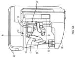
- FIG. 2illustrates the needle biopsy assembly 10 in more detail.
- Support bracket 21extends between handles 23 a and 23 b , which facilitate transport of the assembly.
- Guidance module 19mounted on support bracket 21 , includes components for controlling the movement of the biopsy device 15 .
- the biopsy devicemay be, for example, an EvivaTM vacuum assisted biopsy device manufactured and sold by Hologic, Inc.
- Fixed support arm 17extends from the guidance module to connector 31 .
- connector 31connects angular support arm 12 to the fixed support arm 17 at a fixed angle.
- Alternative embodiments which include adjustment mechanisms for varying the angle of displacement between the angular support arm and the fixed support armmay be substituted herein as equivalents.
- Holster mount 35is moveably coupled to the support arm.
- the linear movementmay be mechanically controlled (i.e., via the guidance module and associated motors) and/or may be manually controlled using either or both of the thumbwheel knobs 33 a and 33 b .
- the holster mount 35includes an attachment mechanism 36 that is adapted to receive biopsy holster 13 .
- the biopsy device 15sits within the biopsy holster 13 .
- a needle support 11may advantageously be coupled to the holster mount for needle stabilization.
- a control module 25may be mounted to either of the handles, 23 b or 23 a via clamp 36 .
- each handlemay include one or more electrical connectors which enable communication between the clamped control module 25 and the guidance module 19 , and the medical professional may move the control module to either handle as a matter of preference.
- the control module 25includes a user interface that enables a medical professional to control the biopsy procedure without the need to leave the patient.
- the control moduleincludes a display 28 for displaying a status or other information about the biopsy, and one or more buttons 27 for controlling the movement of the biopsy device during the procedure.
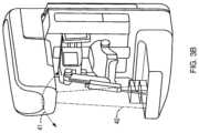
- FIGS. 3 A and 3 Billustrate the needle biopsy assembly 10 mounted on a gantry of an x-ray imaging system.
- Various embodiments of the needle biopsy assemblymay be used with either an upright or prone imaging system, for the purposes of this application an embodiment for use with an upright breast tomosynthesis imaging system (such as the DimensionsTM Breast Tomosynthesis imaging system provided by Hologic, Inc.) is described.
- An exemplary tomosynthesis imaging systemmay include a tube head 32 supporting a cone beam or other x-ray source, and a compression platform 30 encasing an x-ray detector.
- the tube head 32is rotatably mounted on a gantry (not shown) to enable the tube head to rotate in along an angular trajectory generally designated by the dashed line 41 in FIG. 3 B .
- the needle biopsy assembly 10includes clamps, hooks or other attachment means for mounting the needle biopsy assembly to the gantry of the tomosynthesis imaging system.
- the clampsare mated to features of the gantry that support other attached devices (such as face shields and the like) although such reuse is not a requirement.
- the holster 13is coupled to the holster mount on the a fixed angle arm 12 , and the fixed angle arm 12 is fixedly mounted on the support arm 17 at an angle offset from normal by 10 degrees, although it is readily appreciated that the offset angle may vary and is largely a matter of design choice.
- Angling the arm 12(and by consequence the biopsy device 15 ) allows the biopsy device to be advanced to a desired location within a biopsy target area (indicated generally by the target area 50 ) without the biopsy device and holster introducing artifacts into the x-ray image.
- the cone beam x-ray sourcewill extend into the target area 50 , but the device 15 does not fall within the cone beam.
- the present inventionis not limited to any particular fixed angle and it is appreciated that the selected fixed angle may differ in response to particular geometries of the imaging systems and tissue removal tools.
- the present inventionencompasses the idea of angling a biopsy needle to limit the introduction of visual artifacts caused by the needle during x-ray imaging.
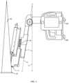
- FIG. 4is a side view of an exemplary embodiment of a needle biopsy assembly of the present invention.
- line Ais within a plane that is ‘normal’ to the plane of the x-ray detector 30 .
- Line Billustrates the angular displacement of the biopsy device, and therefore the device is offset from the normal by an angular measure of 0. As a result the biopsy device will interfere with biopsy imaging.
- FIG. 4Also shown in more detail in FIG. 4 are exemplary coupling mechanisms 50 a and 50 b of the needle biopsy assembly 10 .
- the coupling mechanisms 50 a and 50 bare adapted to mate with complementary features of the gantry.
- Other types of coupling mechanisms, including latches, hooks, slots and the likemay be readily substituted herein as equivalents.
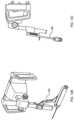
- FIG. 5is a top down view of the exemplary embodiment of the needle biopsy assembly, shown with only a subset of components labeled for ease of reference.
- the displacement N of the needle tip which results from the angular tilt of the biopsy deviceis readily apparent.
- This viewbest illustrates the ability of this device to biopsy areas of the breast (such as the axilla tissue and tissue near the chest wall) which were previously difficult to access using prior art stereotactic devices.
- FIGS. 6 A through 6 Cillustrate various compression paddles which may be used with the present invention.
- the compression paddles 62 A- 62 Care preferably radiolucent. Slots 64 a and 64 b are positioned to enable the compression paddle to be removably coupled to the compression device of the x-ray imaging system, allowing the paddles to easily be exchanged with the mammography/tomosynthesis paddles used for breast cancer screening.
- the biopsy compression paddlesmay be manufactured in different sizes to be used for different sized breasts, as shown in FIGS. 6 B and 6 C .
- An opening 66is provided in each biopsy compression paddle.
- the openingenables the portion of the breast associated with the target to be exposed.
- the portion 68 of the compression paddle 62 a that surrounding the opening 66is shaped to increase the compression on the portion of the breast being biopsied, thereby stabilizing the biopsied area.
- the radiolucent breast compression paddleallows full view of the detector; in prior art needle biopsy arrangement, only a portion of the detector associated with target localization was visible. This was sufficient because prior art biopsies were only performed on that portion of the tissue which was immediately below the opening in the compression paddle.
- the tilted needle of the present inventionincreases the amount of tissue that is available for biopsy beyond the border of the compression paddle opening—the needle in fact has the reach N shown in FIG. 5 .
- axilla tissue and tissue close to the chest wallcan more readily be excised during biopsy.
- FIG. 8illustrates a 2D image of a breast taken with a radiolucent biopsy paddle such as those shown in FIGS. 6 and 7 .
- a radiolucent compression paddlesuch as those shown in FIGS. 6 and 7 .
- one advantage of the radiolucent compression paddleis that the entire detector image may be viewed at the acquisition workstation interface; the radiologist is no longer limited in their access to information. As a result, the radiologist may see that portions of a lesion 80 extend beyond the compression paddle opening 66 . This portion of the breast can be accessed using the tilted biopsy needle assembly of the present invention to ensure that all tissue is accessed for proper diagnosis.
- Exemplary needle biopsy assembliessuch as that described above may be generally used as follows.
- a patient who has been identified as a candidate for biopsyis positioned at the x-ray imaging system.
- the biopsy compression paddlemoves down towards the compression platform, compressing the patient's breast, and the process of visualizing the lesion is initiated at step 92 .
- visualization of the lesionmay be performed using a scout image, a mammogram, acquired stereotactic images, acquired tomosynthesis projection images, tomosynthesis reconstructed images, or any combination thereof.
- an x-ray imaging system having tomosynthesis capabilitiesmay be adapted to include a ‘stereotactic mode’, which, when selected, causes the x-ray imaging system to automatically retrieve the typical+/ ⁇ 15 degree stereotactic images and performs appropriate imaging processing on the stereotactic images to derive a stereotactic volume.
- a ‘stereotactic mode’which, when selected, causes the x-ray imaging system to automatically retrieve the typical+/ ⁇ 15 degree stereotactic images and performs appropriate imaging processing on the stereotactic images to derive a stereotactic volume.
- the lesionis targeted.
- Targeting the lesioninvolves identifying the coordinates of the lesion using image data, and converting the coordinates from the Cartesian coordinate system of the images to the angular coordinate system of the tilted biopsy assembly using conversion techniques known to those of skill in the art.
- different images, or combinations of imagesmay be used for visualizing the lesion than are used for targeting the lesion. For example, assume that a scout image is initially used to ensure that the patient is in position, and a pair of stereotactic images are used to visualize the lesion.
- the scoutmay be used in combination with the stereotactic image in which the lesion is located to derive target location information. Therefore, as above, depending upon the capabilities of the x-ray imaging system, the lesion target coordinates may be derived using a scout image, a mammogram, acquired stereotactic images, acquired tomosynthesis projection images, tomosynthesis reconstructed images, or any combination thereof.
- FIG. 10illustrates an exemplary display and buttons of a control unit 25 .
- the control unitincludes a display 28 for displaying information related to the biopsy, such as information regarding needle size, distance to platform and paddle, needle coordinates, target coordinates, proximity to target and other related information.
- the control panelmay also provide other helpful information to the user, such as warning indicators when the needle is too close to the breast platform, chest wall or skin line.
- the warning indicatorsmay be color coded or may provide other visual or audible indicators of undesirable conditions.
- control unitalso includes buttons (including button 27 ) positioned and arranged to allow single handed activation of the biopsy assembly while precluding accidental activation.
- buttonsincluding button 27
- a pair of control buttonsis provided, one on the front face of the control panel, and another on the back face of the control panel. Biopsy assembly movement may only be activated via simultaneous depression of both buttons. Other mechanisms for affirming operator intent may be substituted herein without affecting the scope of the invention.
- mechanical stopsmay be introduced into the biopsy path to stop automated needle movement at particular points along the path. For example, it may be desirable to switch to manual control of the needle movement, i.e, via knob 33 a or 33 b when the needle is within a desired range of the target. Or it may be desirable to provide a release brake for slow insertion of the needle into the breast.
- the number and location of mechanical stopsis a programmable function which may be tailored to the individual preferences of a user of the system. Suffice it to say that numerous methods for mechanical advancement of the needle, including geared advancements, piston advancements, etc. are envisioned.
- an imagemay be acquired to verify that, in fact, the needle is positioned at the lesion. As the biopsy needle is out of the view of the x-ray imaging system such image may be obtained without interference.
- the tissuemay be excised, and the biopsy is complete.
- a biopsy needle assemblyhas been shown and described.
- a user interface of the acquisition workstationis advantageously augmented with needle biopsy control capabilities.
- An example of user interface features that may be added to an acquisition workstation for use with the needle biopsy assemblyis shown in FIG. 11 .
- the user interfaceincludes menus and/or control buttons or icons which enable the radiologist to control the display and output of information gathered during the biopsy.
- Targeting tools 110allow the user to review, modify and delete target information.
- Image source selectors 120(including stereotactic, tomosynthesis, scout, mammography, etc.) allow the radiologist to select which images to use for visualization or targeting.
- Image view selectors 130allow the radiologist to quickly pull up and view any of the images that may have been acquired during the biopsy. Any of the images may be pulled up in the image window 150 .
- Other information related to the biopsysuch as the type of biopsy device, the relative distances between the target and the compression plate/platform, etc., may also be included on the acquisition workstation. It should be noted that although a particular arrangement of buttons and icons has been shown in the representative view of FIG. 11 , the present invention is not limited to any particular representation of such information, and other forms of pull down menus, hyperlinks, icons, buttons, and windows are considered equivalents hereto.
- FIGS. 12 A- 12 Cillustrate various views of an embodiment of the needle biopsy assembly of the present invention that has been augmented with a lateral biopsy arm 200 .
- FIG. 12 Ais a straight on view of the augmented assembly, FIG.
- FIG. 12 Bis a perspective view of the augmented assembly while FIG. 12 C is a side view of the augmented assembly.
- the lateral biopsy armsupports the biopsy needle along a line substantially parallel to the detector, thereby permitting lateral excision of breast tissue.
- the lateral biopsy armincludes a holster mount plate 210 and a thumb wheel 220 . Rotation of the thumb wheel 220 results in manual translation of the needle in the direction indicated generally as X in FIG. 12 A . Although manual translation in the X plane is shown, it is appreciated that motorized translation may be achieved by extending control from the guidance module 19 to the holster plate 210 .
- an electrical connection on the holster mountmounts with a connector on the lateral biopsy arm when the lateral biopsy arm is coupled to the holster mount 13 to enable the guidance module 19 to adjust the guidance coordinate system as appropriate for proper lesion localization.
- a tilted needle biopsy assemblyhaving numerous advantages has been shown and described. Because the biopsy needle is angled relative to at least one of the detector and the x-ray source, x-ray imaging may be performed during the biopsy procedure without interference by the biopsy device. In addition the angled biopsy needle allows improved access to the axilla and chest wall of the patient.
- the stereotactic biopsy device of the present inventionmay be coupled to any x-ray system, whether upright or prone, including but not limited to mammography systems, tomosynthesis systems, and combination mammography/tomosynthesis systems.
- the systemflexibly supports the use of any mode of image capture (i.e., scout, two dimensional mammogram, three-dimensional reconstructed volume) for either or both target visualization and target localization. With such an arrangement, a needle biopsy assembly having improved patient coverage is provided for use with a variety of different x-ray imaging platforms.
Landscapes
- Health & Medical Sciences (AREA)
- Life Sciences & Earth Sciences (AREA)
- Surgery (AREA)
- Medical Informatics (AREA)
- Engineering & Computer Science (AREA)
- Veterinary Medicine (AREA)
- Animal Behavior & Ethology (AREA)
- Public Health (AREA)
- Pathology (AREA)
- General Health & Medical Sciences (AREA)
- Biomedical Technology (AREA)
- Heart & Thoracic Surgery (AREA)
- Molecular Biology (AREA)
- Nuclear Medicine, Radiotherapy & Molecular Imaging (AREA)
- Oral & Maxillofacial Surgery (AREA)
- High Energy & Nuclear Physics (AREA)
- Radiology & Medical Imaging (AREA)
- Optics & Photonics (AREA)
- Physics & Mathematics (AREA)
- Biophysics (AREA)
- Neurosurgery (AREA)
- Dentistry (AREA)
- Apparatus For Radiation Diagnosis (AREA)
Abstract
Description
Claims (18)
Priority Applications (3)
| Application Number | Priority Date | Filing Date | Title |
|---|---|---|---|
| US16/809,355US11701199B2 (en) | 2009-10-08 | 2020-03-04 | Needle breast biopsy system and method of use |
| US18/107,113US12193886B2 (en) | 2009-10-08 | 2023-02-08 | Needle breast biopsy system and method of use |
| US18/966,720US20250177082A1 (en) | 2009-10-08 | 2024-12-03 | Needle breast biopsy system and method of use |
Applications Claiming Priority (3)
| Application Number | Priority Date | Filing Date | Title |
|---|---|---|---|
| US24977209P | 2009-10-08 | 2009-10-08 | |
| US12/715,591US10595954B2 (en) | 2009-10-08 | 2010-03-02 | Needle breast biopsy system and method for use |
| US16/809,355US11701199B2 (en) | 2009-10-08 | 2020-03-04 | Needle breast biopsy system and method of use |
Related Parent Applications (1)
| Application Number | Title | Priority Date | Filing Date |
|---|---|---|---|
| US12/715,591ContinuationUS10595954B2 (en) | 2009-10-08 | 2010-03-02 | Needle breast biopsy system and method for use |
Related Child Applications (1)
| Application Number | Title | Priority Date | Filing Date |
|---|---|---|---|
| US18/107,113ContinuationUS12193886B2 (en) | 2009-10-08 | 2023-02-08 | Needle breast biopsy system and method of use |
Publications (2)
| Publication Number | Publication Date |
|---|---|
| US20200205928A1 US20200205928A1 (en) | 2020-07-02 |
| US11701199B2true US11701199B2 (en) | 2023-07-18 |
Family
ID=42139195
Family Applications (4)
| Application Number | Title | Priority Date | Filing Date |
|---|---|---|---|
| US12/715,591Active2031-10-03US10595954B2 (en) | 2009-10-08 | 2010-03-02 | Needle breast biopsy system and method for use |
| US16/809,355Active2031-05-13US11701199B2 (en) | 2009-10-08 | 2020-03-04 | Needle breast biopsy system and method of use |
| US18/107,113ActiveUS12193886B2 (en) | 2009-10-08 | 2023-02-08 | Needle breast biopsy system and method of use |
| US18/966,720PendingUS20250177082A1 (en) | 2009-10-08 | 2024-12-03 | Needle breast biopsy system and method of use |
Family Applications Before (1)
| Application Number | Title | Priority Date | Filing Date |
|---|---|---|---|
| US12/715,591Active2031-10-03US10595954B2 (en) | 2009-10-08 | 2010-03-02 | Needle breast biopsy system and method for use |
Family Applications After (2)
| Application Number | Title | Priority Date | Filing Date |
|---|---|---|---|
| US18/107,113ActiveUS12193886B2 (en) | 2009-10-08 | 2023-02-08 | Needle breast biopsy system and method of use |
| US18/966,720PendingUS20250177082A1 (en) | 2009-10-08 | 2024-12-03 | Needle breast biopsy system and method of use |
Country Status (6)
| Country | Link |
|---|---|
| US (4) | US10595954B2 (en) |
| EP (1) | EP2485651B1 (en) |
| JP (2) | JP5762423B2 (en) |
| CN (2) | CN106420066B (en) |
| ES (1) | ES2862525T3 (en) |
| WO (1) | WO2011043838A1 (en) |
Cited By (11)
| Publication number | Priority date | Publication date | Assignee | Title |
|---|---|---|---|---|
| US20230225821A1 (en)* | 2009-10-08 | 2023-07-20 | Hologic, Inc. | Needle breast biopsy system and method of use |
| US12183309B2 (en) | 2011-11-27 | 2024-12-31 | Hologic, Inc. | System and method for generating a 2D image using mammography and/or tomosynthesis image data |
| US12193853B2 (en) | 2006-02-15 | 2025-01-14 | Hologic, Inc. | Breast biopsy and needle localization using tomosynthesis systems |
| US12211608B2 (en) | 2013-03-15 | 2025-01-28 | Hologic, Inc. | System and method for navigating a tomosynthesis stack including automatic focusing |
| US12211124B2 (en) | 2017-03-30 | 2025-01-28 | Hologic, Inc. | System and method for synthesizing low-dimensional image data from high-dimensional image data using an object grid enhancement |
| US12236597B2 (en) | 2021-11-29 | 2025-02-25 | Hologic, Inc. | Systems and methods for correlating objects of interest |
| US12236582B2 (en) | 2018-09-24 | 2025-02-25 | Hologic, Inc. | Breast mapping and abnormality localization |
| US12239471B2 (en) | 2011-03-08 | 2025-03-04 | Hologic, Inc. | System and method for dual energy and/or contrast enhanced breast imaging for screening, diagnosis and biopsy |
| US12254586B2 (en) | 2021-10-25 | 2025-03-18 | Hologic, Inc. | Auto-focus tool for multimodality image review |
| US12307604B2 (en) | 2012-02-13 | 2025-05-20 | Hologic, Inc. | System and method for navigating a tomosynthesis stack using synthesized image data |
| US12380972B2 (en) | 2014-03-21 | 2025-08-05 | Dhrpro, Llc | Data command center visual display system |
Families Citing this family (61)
| Publication number | Priority date | Publication date | Assignee | Title |
|---|---|---|---|---|
| EP1816965B1 (en) | 2004-11-26 | 2016-06-29 | Hologic, Inc. | Integrated multi-mode mammography/tomosynthesis x-ray system |
| EP2408375B1 (en) | 2009-03-20 | 2017-12-06 | Orthoscan Incorporated | Moveable imaging apparatus |
| US20120133600A1 (en) | 2010-11-26 | 2012-05-31 | Hologic, Inc. | User interface for medical image review workstation |
| WO2012082799A1 (en) | 2010-12-13 | 2012-06-21 | Orthoscan, Inc. | Mobile fluoroscopic imaging system |
| USD659832S1 (en)* | 2011-01-27 | 2012-05-15 | Fujifilm Corporation | Mammography equipment |
| USD659833S1 (en)* | 2011-01-27 | 2012-05-15 | Fujifilm Corporation | Mammography equipment |
| JP5501290B2 (en)* | 2011-05-23 | 2014-05-21 | 富士フイルム株式会社 | Image processing apparatus, radiographic image capturing system, and image processing program |
| JP2012245329A (en)* | 2011-05-31 | 2012-12-13 | Fujifilm Corp | Image processing apparatus, radiographic image radiographing system, image processing program, and image processing method |
| USD671647S1 (en)* | 2011-08-29 | 2012-11-27 | General Electric Company | Mobile mammograph machine |
| USD669989S1 (en)* | 2011-08-30 | 2012-10-30 | General Electric Company | Mammography system |
| USD679398S1 (en)* | 2011-09-02 | 2013-04-02 | General Electric Company | Mammography system |
| ES2795416T3 (en)* | 2011-09-16 | 2020-11-23 | Hologic Inc | Lateral Arm System for Breast Biopsy |
| US11284869B2 (en) | 2011-09-16 | 2022-03-29 | Hologic, Inc. | Breast biopsy lateral arm system |
| US12042134B2 (en) | 2011-09-16 | 2024-07-23 | Hologic, Inc. | Breast biopsy lateral arm system |
| HU229210B1 (en)* | 2011-11-16 | 2013-09-30 | Diagon Kft | Method and automatic device for in vitro diagnostic tests of blood clotting |
| EP2967474B1 (en) | 2013-03-15 | 2020-05-06 | Hologic, Inc. | X-ray scatter reducing device for use with 2d and 3d mammography |
| US10092358B2 (en) | 2013-03-15 | 2018-10-09 | Hologic, Inc. | Tomosynthesis-guided biopsy apparatus and method |
| RU2657188C2 (en)* | 2013-07-19 | 2018-06-08 | Конинклейке Филипс Н.В. | User interface for biopsy unit |
| EP3060132B1 (en) | 2013-10-24 | 2019-12-04 | Hologic, Inc. | System and method for navigating x-ray guided breast biopsy |
| USD728106S1 (en)* | 2013-11-27 | 2015-04-28 | Planmed Oy | Digital breast tomosynthesis machine |
| FI126329B (en)* | 2013-11-29 | 2016-10-14 | Planmed Oy | Mammography Equipment Arrangements |
| FI126217B (en)* | 2013-11-29 | 2016-08-31 | Planmed Oy | Mammography equipment |
| USD728794S1 (en) | 2014-01-29 | 2015-05-05 | Samsung Electronics Co., Ltd. | X-ray device |
| USD728793S1 (en)* | 2014-01-29 | 2015-05-05 | Samsung Electronics Co., Ltd. | X-ray device |
| JP6506769B2 (en) | 2014-02-28 | 2019-04-24 | ホロジック, インコーポレイテッドHologic, Inc. | System and method for generating and displaying tomosynthesis image slabs |
| US10390806B2 (en)* | 2014-03-28 | 2019-08-27 | Covidien Lp | Devices, systems, and methods for obtaining a tissue sample using a biopsy tool |
| EP3148471B1 (en)* | 2014-05-28 | 2021-11-03 | General Electric Company | Method and associated biopsy device |
| US9750469B2 (en) | 2014-10-24 | 2017-09-05 | I.M.S. Internazionale Medico Scientifica S.R.L. | Apparatus for performing a biopsy on a patient's breast and computer-implemented method for defining a route for a biopsy needle through a patient's breast |
| KR20160071938A (en)* | 2014-12-12 | 2016-06-22 | 삼성전자주식회사 | X-ray imaging apparatus |
| US10542951B2 (en)* | 2015-07-23 | 2020-01-28 | General Electric Company | Systems, methods, and devices for simplified high quality imaging of biopsy samples on a mammography machine |
| CN105286960B (en)* | 2015-12-03 | 2019-01-18 | 上海联影医疗科技有限公司 | The localization method and system of breast lesion puncture position |
| US10786224B2 (en) | 2016-04-21 | 2020-09-29 | Covidien Lp | Biopsy devices and methods of use thereof |
| JP6656199B2 (en)* | 2017-03-30 | 2020-03-04 | 富士フイルム株式会社 | Mammography equipment |
| EP3600052A1 (en) | 2017-03-30 | 2020-02-05 | Hologic, Inc. | System and method for targeted object enhancement to generate synthetic breast tissue images |
| EP3600047A1 (en) | 2017-03-30 | 2020-02-05 | Hologic, Inc. | System and method for hierarchical multi-level feature image synthesis and representation |
| WO2018236565A1 (en) | 2017-06-20 | 2018-12-27 | Hologic, Inc. | METHOD AND SYSTEM FOR MEDICAL IMAGING WITH DYNAMIC SELF-LEARNING |
| IT201700122588A1 (en)* | 2017-10-27 | 2019-04-27 | Ims Giotto S P A | EQUIPMENT FOR ANALYSIS OF SAMPLES BY MEANS OF BIOPSY. |
| IT201800003416A1 (en)* | 2018-03-09 | 2019-09-09 | Ims Giotto S P A | EQUIPMENT FOR ANALYSIS OF SAMPLES TAKEN BY BIOPSY WITH MOBILE STABILIZATION ELEMENT. |
| US11331161B2 (en) | 2018-03-23 | 2022-05-17 | Covidien Lp | Surgical assemblies facilitating tissue marking and methods of use thereof |
| JP6945491B2 (en)* | 2018-04-27 | 2021-10-06 | 富士フイルム株式会社 | Mammography equipment |
| EP3787520B1 (en) | 2018-05-04 | 2024-09-25 | Hologic, Inc. | Biopsy needle visualization |
| US12121304B2 (en) | 2018-05-04 | 2024-10-22 | Hologic, Inc. | Introducer and localization wire visualization |
| KR102761184B1 (en)* | 2018-09-25 | 2025-02-03 | 홀로직, 인크. | Method for illuminating assembly and mammography and tomosynthesis imaging system |
| JP7584148B2 (en) | 2018-10-22 | 2024-11-15 | チョート バーケット,ジョセフ | Vacuum Assisted Insertion Device |
| US11517294B2 (en) | 2019-05-07 | 2022-12-06 | Covidien Lp | Biopsy devices and methods of use thereof |
| US11883206B2 (en) | 2019-07-29 | 2024-01-30 | Hologic, Inc. | Personalized breast imaging system |
| EP4439580A3 (en) | 2019-09-27 | 2024-12-25 | Hologic, Inc. | Ai system for predicting reading time and reading complexity for reviewing 2d/3d breast images |
| EP3832689A3 (en) | 2019-12-05 | 2021-08-11 | Hologic, Inc. | Systems and methods for improved x-ray tube life |
| US11481038B2 (en) | 2020-03-27 | 2022-10-25 | Hologic, Inc. | Gesture recognition in controlling medical hardware or software |
| US11471118B2 (en) | 2020-03-27 | 2022-10-18 | Hologic, Inc. | System and method for tracking x-ray tube focal spot position |
| US11308594B2 (en)* | 2020-05-15 | 2022-04-19 | GE Precision Healthcare LLC | Tomosynthesis dataset generation using pre-exposure acquisition |
| CN111759427B (en)* | 2020-07-28 | 2023-05-09 | 南阳市中心医院 | Pericardium puncture liquid pumping device for department of cardiology |
| CN111839567A (en)* | 2020-08-21 | 2020-10-30 | 上海联影医疗科技有限公司 | mammography equipment |
| EP4183345B1 (en)* | 2020-08-21 | 2025-10-01 | Shanghai United Imaging Healthcare Co., Ltd. | Breast x-ray imaging apparatus, and method and system for correcting and verifying breast biopsy positioning device |
| US20230351600A1 (en) | 2020-09-18 | 2023-11-02 | Hologic, Inc. | Correlating regions of interest |
| US11801018B2 (en) | 2020-09-24 | 2023-10-31 | Hologic, Inc. | Light source for an imaging system and methods of the same |
| US11918392B2 (en)* | 2020-12-18 | 2024-03-05 | GE Precision Healthcare LLC | Vision-guided biopsy system and method for mammography |
| WO2023009435A1 (en) | 2021-07-27 | 2023-02-02 | Hologic, Inc. | Projection for interventional medical procedures |
| US12414217B2 (en) | 2022-02-07 | 2025-09-09 | Hologic, Inc. | Systems and methods for adaptively controlling filament current in an X-ray tube |
| IT202200002660A1 (en)* | 2022-02-14 | 2023-08-14 | Ims Giotto S P A | MEDICAL ANALYSIS EQUIPMENT AND RELATED METHOD |
| JP2025530679A (en)* | 2022-08-29 | 2025-09-17 | ホロジック, インコーポレイテッド | Systems and methods for lateral treatment of the breast |
Citations (402)
| Publication number | Priority date | Publication date | Assignee | Title |
|---|---|---|---|---|
| US3502878A (en) | 1967-09-22 | 1970-03-24 | Us Health Education & Welfare | Automatic x-ray apparatus for limiting the field size of a projected x-ray beam in response to film size and to source-to-film distance |
| US3863073A (en) | 1973-04-26 | 1975-01-28 | Machlett Lab Inc | Automatic system for precise collimation of radiation |
| US3971950A (en) | 1975-04-14 | 1976-07-27 | Xerox Corporation | Independent compression and positioning device for use in mammography |
| US4160906A (en) | 1977-06-23 | 1979-07-10 | General Electric Company | Anatomically coordinated user dominated programmer for diagnostic x-ray apparatus |
| US4310766A (en) | 1978-09-06 | 1982-01-12 | Siemens Aktiengesellschaft | Motor driven x-ray grid and film-holder assembly |
| US4496557A (en) | 1981-08-27 | 1985-01-29 | Adir | Tricyclic ethers, their preparation and the pharmaceutical compositions containing them |
| US4559641A (en) | 1983-06-24 | 1985-12-17 | Thomson-Cgr | Retractable cassette holder for a radiological and radiographic examination apparatus |
| US4706269A (en) | 1985-03-11 | 1987-11-10 | Reina Leo J | Anti-scatter grid structure |
| US4744099A (en) | 1983-11-03 | 1988-05-10 | Siemens Aktiengesellschaft | X-ray diagnostic apparatus comprising radiation filters |
| US4773087A (en) | 1986-04-14 | 1988-09-20 | University Of Rochester | Quality of shadowgraphic x-ray images |
| US4773086A (en) | 1983-12-16 | 1988-09-20 | Yokogawa Medical Systems, Limited | Operator console for X-ray tomographs |
| US4819258A (en) | 1986-11-28 | 1989-04-04 | Bennett X-Ray Corp. | Auto-setting of KV in an x-ray machine after selection of technic factors |
| US4821727A (en) | 1986-10-30 | 1989-04-18 | Elscint Ltd. | Mammographic biopsy needle holder system |
| US4907156A (en) | 1987-06-30 | 1990-03-06 | University Of Chicago | Method and system for enhancement and detection of abnormal anatomic regions in a digital image |
| WO1990005485A1 (en) | 1988-11-23 | 1990-05-31 | Nrt-Nordisk Roentgen Teknik A/S | X-ray apparatus |
| US4969174A (en) | 1989-09-06 | 1990-11-06 | General Electric Company | Scanning mammography system with reduced scatter radiation |
| US4989227A (en) | 1989-04-28 | 1991-01-29 | General Electric Cgr S.A. | Cassette carrier adaptable in size and position for mammography |
| US5018176A (en) | 1989-03-29 | 1991-05-21 | General Electric Cgr S.A. | Mammograph equipped with an integrated device for taking stereotaxic photographs and a method of utilization of said mammograph |
| US5029193A (en) | 1989-07-03 | 1991-07-02 | Siemens Aktiengesellschaft | X-ray diagnostic installation for mammography exposures |
| USRE33634E (en) | 1986-09-23 | 1991-07-09 | Method and structure for optimizing radiographic quality by controlling X-ray tube voltage, current focal spot size and exposure time | |
| US5051904A (en) | 1988-03-24 | 1991-09-24 | Olganix Corporation | Computerized dynamic tomography system |
| US5078142A (en) | 1989-11-21 | 1992-01-07 | Fischer Imaging Corporation | Precision mammographic needle biopsy system |
| US5099846A (en) | 1988-12-23 | 1992-03-31 | Hardy Tyrone L | Method and apparatus for video presentation from a variety of scanner imaging sources |
| US5129911A (en) | 1991-03-11 | 1992-07-14 | Siczek Bernard W | Orbital aiming device |
| US5133020A (en) | 1989-07-21 | 1992-07-21 | Arch Development Corporation | Automated method and system for the detection and classification of abnormal lesions and parenchymal distortions in digital medical images |
| US5163075A (en) | 1991-08-08 | 1992-11-10 | Eastman Kodak Company | Contrast enhancement of electrographic imaging |
| US5164976A (en) | 1989-09-06 | 1992-11-17 | General Electric Company | Scanning mammography system with improved skin line viewing |
| US5199056A (en) | 1989-11-28 | 1993-03-30 | Darrah Carol J | Mammography compression paddle |
| US5219351A (en) | 1990-10-24 | 1993-06-15 | General Electric Cgr S.A. | Mammograph provided with an improved needle carrier |
| US5240011A (en) | 1991-11-27 | 1993-08-31 | Fischer Imaging Corporation | Motorized biopsy needle positioner |
| WO1993017620A1 (en) | 1992-03-12 | 1993-09-16 | Fischer Imaging Corporation | Isocentric puncture instrument aiming device |
| US5279309A (en) | 1991-06-13 | 1994-01-18 | International Business Machines Corporation | Signaling device and method for monitoring positions in a surgical operation |
| US5280427A (en) | 1989-11-27 | 1994-01-18 | Bard International, Inc. | Puncture guide for computer tomography |
| US5289520A (en) | 1991-11-27 | 1994-02-22 | Lorad Corporation | Stereotactic mammography imaging system with prone position examination table and CCD camera |
| WO1994006352A1 (en) | 1992-09-23 | 1994-03-31 | Fischer Imaging Corporation | Mammographic screening and biopsy apparatus |
| US5343390A (en) | 1992-02-28 | 1994-08-30 | Arch Development Corporation | Method and system for automated selection of regions of interest and detection of septal lines in digital chest radiographs |
| US5359637A (en) | 1992-04-28 | 1994-10-25 | Wake Forest University | Self-calibrated tomosynthetic, radiographic-imaging system, method, and device |
| US5365562A (en) | 1993-09-20 | 1994-11-15 | Fischer Imaging Corporation | Digital imaging apparatus |
| US5415169A (en) | 1989-11-21 | 1995-05-16 | Fischer Imaging Corporation | Motorized mammographic biopsy apparatus |
| US5452367A (en) | 1993-11-29 | 1995-09-19 | Arch Development Corporation | Automated method and system for the segmentation of medical images |
| US5491627A (en) | 1993-05-13 | 1996-02-13 | Arch Development Corporation | Method and system for the detection of microcalcifications in digital mammograms |
| US5499097A (en) | 1994-09-19 | 1996-03-12 | Neopath, Inc. | Method and apparatus for checking automated optical system performance repeatability |
| US5506877A (en) | 1994-11-23 | 1996-04-09 | The General Hospital Corporation | Mammography breast compression device and method |
| US5526394A (en) | 1993-11-26 | 1996-06-11 | Fischer Imaging Corporation | Digital scan mammography apparatus |
| US5539797A (en) | 1993-03-29 | 1996-07-23 | Ge Medical Systems Sa | Method and apparatus for digital stereotaxic mammography |
| US5553111A (en) | 1994-10-26 | 1996-09-03 | The General Hospital Corporation | Apparatus and method for improved tissue imaging |
| US5592562A (en) | 1994-01-19 | 1997-01-07 | International Business Machines Corporation | Inspection system for cross-sectional imaging |
| WO1997000649A1 (en) | 1995-06-20 | 1997-01-09 | Wan Sing Ng | Articulated arm for medical procedures |
| US5594769A (en) | 1991-11-27 | 1997-01-14 | Thermotrex Corporation | Method and apparatus for obtaining stereotactic mammographic guided needle breast biopsies |
| US5596200A (en) | 1992-10-14 | 1997-01-21 | Primex | Low dose mammography system |
| US5598454A (en) | 1994-04-26 | 1997-01-28 | Siemens Aktiengesellschaft | X-ray diagnostics installation |
| US5627869A (en) | 1995-11-22 | 1997-05-06 | Thermotrex Corporation | Mammography apparatus with proportional collimation |
| EP0775467A1 (en) | 1995-11-23 | 1997-05-28 | Planmed Oy | Method and system for controlling the functions of a mammography apparatus |
| US5642433A (en) | 1995-07-31 | 1997-06-24 | Neopath, Inc. | Method and apparatus for image contrast quality evaluation |
| US5642441A (en) | 1995-10-24 | 1997-06-24 | Neopath, Inc. | Separation apparatus and method for measuring focal plane |
| US5647025A (en) | 1994-09-20 | 1997-07-08 | Neopath, Inc. | Automatic focusing of biomedical specimens apparatus |
| JPH09198490A (en) | 1996-01-22 | 1997-07-31 | Hitachi Medical Corp | Three-dimensional discrete data projector |
| US5657362A (en) | 1995-02-24 | 1997-08-12 | Arch Development Corporation | Automated method and system for computerized detection of masses and parenchymal distortions in medical images |
| JPH09238934A (en) | 1996-03-11 | 1997-09-16 | Toshiba Medical Eng Co Ltd | Image display system |
| US5668889A (en) | 1990-04-19 | 1997-09-16 | Fuji Photo Film Co., Ltd. | Apparatus for determining an image position, and method for adjusting read-out conditions and/or image processing conditions for a radiation image |
| US5671288A (en) | 1995-05-31 | 1997-09-23 | Neopath, Inc. | Method and apparatus for assessing slide and specimen preparation quality |
| US5712890A (en) | 1994-11-23 | 1998-01-27 | Thermotrex Corp. | Full breast digital mammography device |
| JPH1033523A (en) | 1996-07-24 | 1998-02-10 | Hitachi Medical Corp | X-ray ct device |
| WO1998016903A1 (en) | 1996-10-16 | 1998-04-23 | Vital Images, Inc. | Advanced diagnostic viewer |
| US5763871A (en) | 1994-09-20 | 1998-06-09 | Neopath, Inc. | Cytological system autofocus integrity checking apparatus |
| US5769086A (en) | 1995-12-06 | 1998-06-23 | Biopsys Medical, Inc. | Control system and method for automated biopsy device |
| US5773832A (en) | 1995-11-21 | 1998-06-30 | Loral Fairchild Corporation | Advanced CCD-based x-ray image sensor system |
| US5818898A (en) | 1995-11-07 | 1998-10-06 | Kabushiki Kaisha Toshiba | X-ray imaging apparatus using X-ray planar detector |
| US5828722A (en) | 1996-05-17 | 1998-10-27 | Sirona Dental Systems Gmbh & Co., Kg | X-ray diagnostic apparatus for tomosynthesis having a detector that detects positional relationships |
| US5835079A (en) | 1996-06-13 | 1998-11-10 | International Business Machines Corporation | Virtual pointing device for touchscreens |
| US5841124A (en) | 1996-06-19 | 1998-11-24 | Neopath, Inc. | Cytological system autofocus integrity checking apparatus |
| US5872828A (en) | 1996-07-23 | 1999-02-16 | The General Hospital Corporation | Tomosynthesis system for breast imaging |
| US5875258A (en) | 1994-09-20 | 1999-02-23 | Neopath, Inc. | Biological specimen analysis system processing integrity checking apparatus |
| US5878104A (en) | 1996-05-17 | 1999-03-02 | Sirona Dental Systems Gmbh & Co. Kg | Method for producing tomosynthesis exposures employing a reference object formed by a region of the examination subject |
| US5878746A (en) | 1993-08-25 | 1999-03-09 | Lemelson; Jerome H. | Computerized medical diagnostic system |
| US5896437A (en) | 1996-05-17 | 1999-04-20 | Sirona Dental Systems Gmbh & Co. Kg | X-ray diagnostics apparatus for tomosynthesis having a reference object in fixed relationship to a radiation emitter |
| US5941832A (en) | 1991-09-27 | 1999-08-24 | Tumey; David M. | Method and apparatus for detection of cancerous and precancerous conditions in a breast |
| US5954650A (en) | 1996-11-13 | 1999-09-21 | Kabushiki Kaisha Toshiba | Medical image processing apparatus |
| US6005907A (en) | 1996-05-17 | 1999-12-21 | Sirona Dental Systems Gmbh & Co. Kg | Method and apparatus for producing tomosynthesis exposures employing a reference object composed of a number of sub-objects |
| EP0982001A1 (en) | 1998-08-25 | 2000-03-01 | General Electric Company | Protocol driven image reconstruction, display, and processing in a multislice imaging system |
| US6067079A (en) | 1996-06-13 | 2000-05-23 | International Business Machines Corporation | Virtual pointing device for touchscreens |
| US6075879A (en) | 1993-09-29 | 2000-06-13 | R2 Technology, Inc. | Method and system for computer-aided lesion detection using information from multiple images |
| JP2000200340A (en) | 1999-01-06 | 2000-07-18 | Ge Yokogawa Medical Systems Ltd | Method and device for displaying image and ct system |
| US6091841A (en) | 1997-09-04 | 2000-07-18 | Qualia Computing, Inc. | Method and system for segmenting desired regions in digital mammograms |
| US6101236A (en) | 1998-10-02 | 2000-08-08 | University Of Iowa Research Foundation | Iterative method and apparatus for x-ray computed tomographic fluoroscopy |
| US6102866A (en) | 1996-10-15 | 2000-08-15 | Fischer Imaging Corporation | Enhanced breast imaging/biopsy system employing targeted ultrasound |
| WO2000051484A2 (en) | 1998-11-25 | 2000-09-08 | Fischer Imaging Corporation | User interface system for mammographic imager |
| US6137527A (en) | 1996-12-23 | 2000-10-24 | General Electric Company | System and method for prompt-radiology image screening service via satellite |
| US6149301A (en) | 1998-12-30 | 2000-11-21 | General Electric Company | X-ray target centering apparatus for radiographic imaging system |
| US6175117B1 (en) | 1998-01-23 | 2001-01-16 | Quanta Vision, Inc. | Tissue analysis apparatus |
| US6196715B1 (en) | 1959-04-28 | 2001-03-06 | Kabushiki Kaisha Toshiba | X-ray diagnostic system preferable to two dimensional x-ray detection |
| US6215892B1 (en) | 1995-11-30 | 2001-04-10 | Chromavision Medical Systems, Inc. | Method and apparatus for automated image analysis of biological specimens |
| US6216540B1 (en) | 1995-06-06 | 2001-04-17 | Robert S. Nelson | High resolution device and method for imaging concealed objects within an obscuring medium |
| US6233473B1 (en) | 1999-02-16 | 2001-05-15 | Hologic, Inc. | Determining body composition using fan beam dual-energy x-ray absorptiometry |
| US6243441B1 (en) | 1999-07-13 | 2001-06-05 | Edge Medical Devices | Active matrix detector for X-ray imaging |
| US6245028B1 (en) | 1999-11-24 | 2001-06-12 | Marconi Medical Systems, Inc. | Needle biopsy system |
| US6256370B1 (en) | 2000-01-24 | 2001-07-03 | General Electric Company | Method and apparatus for performing tomosynthesis |
| US6272207B1 (en) | 1999-02-18 | 2001-08-07 | Creatv Microtech, Inc. | Method and apparatus for obtaining high-resolution digital X-ray and gamma ray images |
| US6289235B1 (en) | 1998-03-05 | 2001-09-11 | Wake Forest University | Method and system for creating three-dimensional images using tomosynthetic computed tomography |
| US6292530B1 (en) | 1999-04-29 | 2001-09-18 | General Electric Company | Method and apparatus for reconstructing image data acquired by a tomosynthesis x-ray imaging system |
| US6293282B1 (en) | 1996-11-05 | 2001-09-25 | Jerome Lemelson | System and method for treating select tissue in living being |
| US20010038861A1 (en) | 1999-12-16 | 2001-11-08 | Tsung-Min Hsu | Transdermal administration of nonsteroidal anti-inflammatory drugs using hydroxide-releasing agents as permeation enhancers |
| US20010038681A1 (en) | 2000-02-11 | 2001-11-08 | Brandeis University | Method and system for low-dose three-dimensional imaging of a scene |
| US6327336B1 (en) | 2000-06-05 | 2001-12-04 | Direct Radiography Corp. | Radiogram showing location of automatic exposure control sensor |
| US6327377B1 (en) | 1988-04-08 | 2001-12-04 | Autocyte North Carolina, L.L.C. | Automated cytological specimen classification system and method |
| US6341156B1 (en) | 1999-05-14 | 2002-01-22 | Siemens Aktiengesellschaft | X-ray diagnostic apparatus with relatively moved x-ray source and detector |
| US20020012450A1 (en) | 1998-01-09 | 2002-01-31 | Osamu Tsujii | Image processing apparatus and method |
| US6375352B1 (en) | 1999-10-01 | 2002-04-23 | General Electric Company | Apparatus and method for obtaining x-ray tomosynthesis data for mammography |
| US20020050986A1 (en) | 2000-08-11 | 2002-05-02 | Hitoshi Inoue | Image display apparatus and method, and storage medium |
| US6389104B1 (en) | 2000-06-30 | 2002-05-14 | Siemens Corporate Research, Inc. | Fluoroscopy based 3-D neural navigation based on 3-D angiography reconstruction data |
| US20020075997A1 (en) | 2000-12-18 | 2002-06-20 | Unger Christopher David | Medical diagnostic method and apparatus to control dual energy exposure techniques based on image information |
| US6411836B1 (en) | 1999-12-30 | 2002-06-25 | General Electric Company | Method and apparatus for user preferences configuring in an image handling system |
| US6415015B2 (en) | 1999-12-28 | 2002-07-02 | Ge Medical Systems Sa | Method and system of compensation of thickness of an organ |
| US6424332B1 (en) | 1999-01-29 | 2002-07-23 | Hunter Innovations, Inc. | Image comparison apparatus and method |
| US20020113681A1 (en) | 2001-02-16 | 2002-08-22 | Byram Robert James | Rotary position sensor |
| US6442288B1 (en) | 1997-12-17 | 2002-08-27 | Siemens Aktiengesellschaft | Method for reconstructing a three-dimensional image of an object scanned in the context of a tomosynthesis, and apparatus for tomosynthesis |
| US20020122533A1 (en) | 2000-12-19 | 2002-09-05 | Alain Marie | Mammography apparatus and method |
| JP2002282248A (en) | 2000-11-27 | 2002-10-02 | Ge Medical Systems Global Technology Co Llc | Color parametric synthesizing map for ct perfusion method |
| US6463181B2 (en) | 2000-12-22 | 2002-10-08 | The United States Of America As Represented By The Secretary Of The Navy | Method for optimizing visual display of enhanced digital images |
| US6468226B1 (en) | 2000-11-22 | 2002-10-22 | Mcintyre, Iv John J. | Remote tissue biopsy apparatus and associated methods |
| US6480565B1 (en) | 1999-11-18 | 2002-11-12 | University Of Rochester | Apparatus and method for cone beam volume computed tomography breast imaging |
| US20020188466A1 (en) | 2001-04-18 | 2002-12-12 | Barrette Pierre Philip | Secure digital medical intellectual property (IP) distribution, market applications, and mobile devices |
| US20020193676A1 (en) | 2001-05-29 | 2002-12-19 | Anke Bodicker | Method and computer system for screening of medical cases |
| US20030007598A1 (en) | 2000-11-24 | 2003-01-09 | U-Systems, Inc. | Breast cancer screening with adjunctive ultrasound mammography |
| US20030018272A1 (en) | 2001-06-28 | 2003-01-23 | Treado Patrick J. | Method for Raman chemical imaging and characterization of calcification in tissue |
| US20030026386A1 (en) | 2001-02-01 | 2003-02-06 | Cha-Mei Tang | Anti-scatter grids and collimator designs, and their motion, fabrication and assembly |
| WO2003020114A2 (en) | 2001-08-31 | 2003-03-13 | Analogic Corporation | Image positioning method and system for tomosynthesis in a digital x-ray radiography system |
| US20030048260A1 (en) | 2001-08-17 | 2003-03-13 | Alec Matusis | System and method for selecting actions based on the identification of user's fingers |
| US6556655B1 (en) | 1998-11-27 | 2003-04-29 | Ge Medical Systems Sa | Method for automatic detection of glandular tissue |
| US20030097055A1 (en) | 2001-11-21 | 2003-05-22 | Philips Medical Systems(Cleveland), Inc. | Method of reviewing tomographic scans with a large number of images |
| US20030095624A1 (en) | 2001-11-21 | 2003-05-22 | Eberhard Jeffrey Wayne | Dose management system for mammographic tomosynthesis |
| US6574304B1 (en) | 2002-09-13 | 2003-06-03 | Ge Medical Systems Global Technology Company, Llc | Computer aided acquisition of medical images |
| JP2003189179A (en) | 2001-12-14 | 2003-07-04 | Konica Corp | Abnormal shade detector and image output device |
| US20030128893A1 (en) | 2001-11-19 | 2003-07-10 | Alfio Castorina | Method for merging digital images to obtain a high dynamic range digital image |
| JP2003199737A (en) | 2001-10-12 | 2003-07-15 | General Electric Co <Ge> | Reconstruction method for tomosynthesis |
| US20030135115A1 (en) | 1997-11-24 | 2003-07-17 | Burdette Everette C. | Method and apparatus for spatial registration and mapping of a biopsy needle during a tissue biopsy |
| US6597762B1 (en) | 2002-11-27 | 2003-07-22 | Ge Medical Systems Global Technology Co., Llc | Method and apparatus of lesion detection and validation based on multiple reviews of a CT image |
| US6611575B1 (en) | 2001-07-27 | 2003-08-26 | General Electric Company | Method and system for high resolution 3D visualization of mammography images |
| US20030169847A1 (en) | 2001-11-21 | 2003-09-11 | University Of Massachusetts Medical Center | System and method for x-ray fluoroscopic imaging |
| US6620111B2 (en) | 2001-04-20 | 2003-09-16 | Ethicon Endo-Surgery, Inc. | Surgical biopsy device having automatic rotation of the probe for taking multiple samples |
| US6626849B2 (en) | 2001-11-01 | 2003-09-30 | Ethicon Endo-Surgery, Inc. | MRI compatible surgical biopsy device |
| US6633674B1 (en) | 1999-11-24 | 2003-10-14 | General Electric Company | Picture archiving and communication system employing improved data compression |
| US20030194050A1 (en) | 2002-04-15 | 2003-10-16 | General Electric Company | Multi modality X-ray and nuclear medicine mammography imaging system and method |
| US20030195433A1 (en) | 2002-04-16 | 2003-10-16 | Roman Turovskiy | Localization element with energized tip |
| US20030194121A1 (en) | 2002-04-15 | 2003-10-16 | General Electric Company | Computer aided detection (CAD) for 3D digital mammography |
| JP2003531516A (en) | 2000-04-18 | 2003-10-21 | リットン システムズ、 インコーポレーテッド | Enhanced visualization of live breast biopsy locations for medical documentation |
| US6638235B2 (en) | 2000-11-06 | 2003-10-28 | Suros Surgical Systems, Inc. | Biopsy apparatus |
| US6647092B2 (en) | 2002-01-18 | 2003-11-11 | General Electric Company | Radiation imaging system and method of collimation |
| US20030210254A1 (en) | 2002-05-13 | 2003-11-13 | Doan William D. | Method, system and computer product for displaying axial images |
| US20030212327A1 (en) | 2000-11-24 | 2003-11-13 | U-Systems Inc. | Adjunctive ultrasound processing and display for breast cancer screening |
| US20030215120A1 (en) | 2002-05-15 | 2003-11-20 | Renuka Uppaluri | Computer aided diagnosis of an image set |
| US20040008900A1 (en) | 2002-07-12 | 2004-01-15 | Jabri Kadri N. | System and method for efficiently customizing an imaging system |
| US20040008809A1 (en) | 1998-07-24 | 2004-01-15 | Webber Richard L. | Method and system for creating task-dependent three-dimensional images |
| US20040008901A1 (en) | 2002-07-11 | 2004-01-15 | Avinash Gopal B. | Interpolated image filtering method and apparatus |
| US6683934B1 (en) | 2000-06-05 | 2004-01-27 | General Electric Company | Dual energy x-ray imaging system and method for radiography and mammography |
| US20040036680A1 (en) | 2002-08-26 | 2004-02-26 | Mark Davis | User-interface features for computers with contact-sensitive displays |
| US20040047518A1 (en) | 2002-08-28 | 2004-03-11 | Carlo Tiana | Image fusion system and method |
| US20040052328A1 (en) | 2002-09-13 | 2004-03-18 | Sabol John M. | Computer assisted analysis of tomographic mammography data |
| US20040064037A1 (en) | 2002-09-27 | 2004-04-01 | Confirma, Inc. | Rules-based approach for processing medical images |
| US20040066884A1 (en) | 2002-10-07 | 2004-04-08 | Hermann Claus Bernhard Erich | Continuous scan tomosynthesis system and method |
| US20040070582A1 (en) | 2002-10-11 | 2004-04-15 | Matthew Warren Smith To Sonocine, Inc. | 3D modeling system |
| US20040077938A1 (en) | 2002-10-07 | 2004-04-22 | Mark Joseph L. | Introduction system for minimally invasive surgical instruments |
| US20040081273A1 (en) | 1999-11-18 | 2004-04-29 | Ruola Ning | Apparatus and method for cone beam volume computed tomography breast imaging |
| US20040094167A1 (en) | 2000-03-17 | 2004-05-20 | Brady John Michael | Three-dimensional reconstructions of a breast from two x-ray mammographics |
| US20040101095A1 (en) | 2002-11-27 | 2004-05-27 | Hologic Inc. | Full field mammography with tissue exposure control, tomosynthesis, and dynamic field of view processing |
| US20040109028A1 (en) | 2002-12-10 | 2004-06-10 | Siemens Medical Solutions Usa, Inc. | Medical imaging programmable custom user interface system and method |
| US20040109529A1 (en) | 2002-12-10 | 2004-06-10 | General Electric Company | Full field digital tomosynthesis method and apparatus |
| US20040127789A1 (en) | 2002-12-17 | 2004-07-01 | Kabushiki Kaisha Toshiba | Method and system for X-ray diagnosis of object in which X-ray contrast agent is injected |
| US20040138569A1 (en) | 1999-08-20 | 2004-07-15 | Sorin Grunwald | User interface for handheld imaging devices |
| US20040171933A1 (en) | 2002-11-25 | 2004-09-02 | Milton Stoller | Mammography needle biopsy system and method |
| US20040171986A1 (en) | 1999-04-26 | 2004-09-02 | Scimed Life System, Inc. | Apparatus and methods for guiding a needle |
| JP2004254742A (en) | 2003-02-24 | 2004-09-16 | Konica Minolta Holdings Inc | Medical image processor and method of judging malignancy |
| US6813334B2 (en) | 2000-10-20 | 2004-11-02 | Koninklijke Philips Electronics N.V. | Tomosynthesis in a limited angular range |
| US20050047636A1 (en) | 2003-08-29 | 2005-03-03 | David Gines | System and method for performing auto-focused tomosynthesis |
| US20050063509A1 (en) | 2001-10-19 | 2005-03-24 | Defreitas Kenneth F | Mammography system and method employing offset compression paddles automatic collimation and retractable anti-scatter grid |
| US20050078797A1 (en) | 2002-03-01 | 2005-04-14 | Mats Danielsson | X-ray protection device |
| US6882700B2 (en) | 2002-04-15 | 2005-04-19 | General Electric Company | Tomosynthesis X-ray mammogram system and method with automatic drive system |
| US20050084060A1 (en) | 2003-10-15 | 2005-04-21 | Seppi Edward J. | Systems and methods for functional imaging using contrast-enhanced multiple-energy computed tomography |
| US6885724B2 (en) | 2003-08-22 | 2005-04-26 | Ge Medical Systems Global Technology Company, Llc | Radiographic tomosynthesis image acquisition utilizing asymmetric geometry |
| US20050089205A1 (en) | 2003-10-23 | 2005-04-28 | Ajay Kapur | Systems and methods for viewing an abnormality in different kinds of images |
| US20050105679A1 (en) | 2003-02-12 | 2005-05-19 | Tao Wu | Tomosynthesis imaging system and method |
| US20050107689A1 (en) | 2003-11-14 | 2005-05-19 | Konica Minolta Meical & Graphic, Inc. | Medical image management system |
| US20050111718A1 (en) | 2003-11-26 | 2005-05-26 | University Of Chicago | Automated method and system for the evaluation of disease and registration accuracy in the subtraction of temporally sequential medical images |
| US20050113715A1 (en) | 2000-11-06 | 2005-05-26 | Jeffrey Schwindt | Biopsy apparatus |
| US20050113681A1 (en) | 2002-11-27 | 2005-05-26 | Defreitas Kenneth F. | X-ray mammography with tomosynthesis |
| US6901156B2 (en) | 2000-02-04 | 2005-05-31 | Arch Development Corporation | Method, system and computer readable medium for an intelligent search workstation for computer assisted interpretation of medical images |
| US20050124845A1 (en) | 2003-09-18 | 2005-06-09 | Thomadsen Bruce R. | Device for placement of needles and radioactive seeds in radiotherapy |
| WO2005051197A2 (en) | 2003-11-26 | 2005-06-09 | Koninklijke Philips Electronics, N.V. | Workflow optimization for high throughput imaging environment |
| US20050135555A1 (en) | 2003-12-23 | 2005-06-23 | Claus Bernhard Erich H. | Method and system for simultaneously viewing rendered volumes |
| US20050135664A1 (en) | 2003-12-23 | 2005-06-23 | Kaufhold John P. | Methods and apparatus for reconstruction of volume data from projection data |
| US20050226375A1 (en) | 2004-03-31 | 2005-10-13 | Eberhard Jeffrey W | Enhanced X-ray imaging system and method |
| WO2005110230A1 (en) | 2004-05-14 | 2005-11-24 | Philips Intellectual Property & Standards Gmbh | System and method for diagnosing breast cancer |
| WO2005112767A1 (en) | 2004-05-21 | 2005-12-01 | Tissuomics Limited | Apparatus and method for penetrating radiation measurements |
| US6978040B2 (en) | 2001-12-19 | 2005-12-20 | Canon Kabushiki Kaisha | Optical recovery of radiographic geometry |
| US20060009693A1 (en) | 2004-04-08 | 2006-01-12 | Techniscan, Inc. | Apparatus for imaging and treating a breast |
| US6987331B2 (en) | 1999-01-29 | 2006-01-17 | American Superconductor Corporation | Voltage recovery device for use with a utility power network |
| US20060018526A1 (en) | 2004-07-23 | 2006-01-26 | Avinash Gopal B | Methods and apparatus for noise reduction filtering of images |
| US20060025680A1 (en) | 2004-07-29 | 2006-02-02 | Fanny Jeune-Iomme | Method and apparatus for contrast enhanced medical imaging |
| US6999554B2 (en) | 2003-11-17 | 2006-02-14 | Siemens Aktiengesellschaft | X-ray diagnostic apparatus for mammography examinations |
| US20060074288A1 (en) | 2004-10-04 | 2006-04-06 | Thomas Kelly | Estimating visceral fat by dual-energy x-ray absorptiometry |
| US7025725B2 (en) | 2002-03-28 | 2006-04-11 | Ultrasound Detection Systems, Llc | Three-dimensional ultrasound computed tomography imaging system |
| US7030861B1 (en) | 2001-02-10 | 2006-04-18 | Wayne Carl Westerman | System and method for packing multi-touch gestures onto a hand |
| US20060098855A1 (en) | 2002-11-27 | 2006-05-11 | Gkanatsios Nikolaos A | Image handling and display in X-ray mammography and tomosynthesis |
| WO2006055830A2 (en) | 2004-11-15 | 2006-05-26 | Hologic, Inc. | Matching geometry generation and display of mammograms and tomosynthesis images |
| WO2006058160A2 (en) | 2004-11-26 | 2006-06-01 | Hologic, Inc. | Integrated multi-mode mammography/tomosynthesis x-ray system and method |
| US20060132508A1 (en) | 2004-12-16 | 2006-06-22 | Navid Sadikali | Multi-planar image viewing system and method |
| US20060147099A1 (en) | 2004-12-30 | 2006-07-06 | R2 Technology, Inc. | Medical image review workstation with integrated content-based resource retrieval |
| JP2006519634A (en) | 2003-03-11 | 2006-08-31 | シーメンス メディカル ソリューションズ ユーエスエー インコーポレイテッド | System and method for performing automatic three-dimensional lesion segmentation and measurement |
| US20060197753A1 (en) | 2005-03-04 | 2006-09-07 | Hotelling Steven P | Multi-functional hand-held device |
| US7110502B2 (en) | 2003-05-12 | 2006-09-19 | Canon Kabushiki Kaisha | Radiographic apparatus and method for switching a grid |
| US20060210131A1 (en) | 2005-03-15 | 2006-09-21 | Wheeler Frederick W Jr | Tomographic computer aided diagnosis (CAD) with multiple reconstructions |
| US7117098B1 (en) | 1997-02-27 | 2006-10-03 | Cellomics, Inc. | Machine-readable storage medium for analyzing distribution of macromolecules between the cell membrane and the cell cytoplasm |
| US20060228012A1 (en) | 2005-04-06 | 2006-10-12 | Kabushiki Kaisha Toshiba | Image display apparatus and image display method |
| US7127091B2 (en) | 2000-12-22 | 2006-10-24 | Koninklijke Philips Electronics, N.V. | Method and apparatus for visualizing a limited part of a 3D medical image-point-related data set, through basing a rendered image on an intermediate region between first and second clipping planes, and including spectroscopic viewing of such region |
| US20060238546A1 (en) | 2005-03-08 | 2006-10-26 | Carrie Handley | Comparative image review system and method |
| JP2006312026A (en) | 2005-04-06 | 2006-11-16 | Toshiba Corp | Image display device |
| US20060257009A1 (en) | 2000-11-24 | 2006-11-16 | Shih-Ping Wang | Controlling thick-slice viewing of breast ultrasound data |
| US20060269040A1 (en) | 2005-05-17 | 2006-11-30 | Thomas Mertelmeier | Mammography method and apparatus for forming a tomosynthetic 3-D X-ray image |
| US20070019846A1 (en) | 2003-08-25 | 2007-01-25 | Elizabeth Bullitt | Systems, methods, and computer program products for analysis of vessel attributes for diagnosis, disease staging, and surfical planning |
| US20070036265A1 (en) | 2005-08-15 | 2007-02-15 | Zhenxue Jing | X-ray mammography/tomosynthesis of patient's breast |
| US20070046649A1 (en) | 2005-08-30 | 2007-03-01 | Bruce Reiner | Multi-functional navigational device and method |
| US20070052700A1 (en) | 2005-09-07 | 2007-03-08 | Wheeler Frederick W | System and method for 3D CAD using projection images |
| US20070118400A1 (en) | 2005-11-22 | 2007-05-24 | General Electric Company | Method and system for gesture recognition to drive healthcare applications |
| US20070114424A1 (en) | 2005-11-18 | 2007-05-24 | Sectra Mamea Ab | Method and arrangement relating to x-ray imaging |
| US20070156451A1 (en) | 2006-01-05 | 2007-07-05 | Gering David T | System and method for portable display of relevant healthcare information |
| WO2007095330A2 (en) | 2006-02-15 | 2007-08-23 | Hologic Inc | Breast biopsy and needle localization using tomosynthesis systems |
| US20070223651A1 (en) | 2006-03-21 | 2007-09-27 | Wagenaar Douglas J | Dual modality mammography device |
| US20070236490A1 (en) | 2005-11-25 | 2007-10-11 | Agfa-Gevaert | Medical image display and review system |
| US7289825B2 (en) | 2004-03-15 | 2007-10-30 | General Electric Company | Method and system for utilizing wireless voice technology within a radiology workflow |
| US20070263765A1 (en) | 2003-12-03 | 2007-11-15 | The General Hospital Corporation D/B/A Massachusetts General Hospital | Multi-Segment Cone-Beam Reconstruction System and Method for Tomosynthesis Imaging |
| US7298881B2 (en) | 2004-02-13 | 2007-11-20 | University Of Chicago | Method, system, and computer software product for feature-based correlation of lesions from multiple images |
| US20070274585A1 (en) | 2006-05-25 | 2007-11-29 | Zhang Daoxian H | Digital mammography system with improved workflow |
| JP2007330334A (en) | 2006-06-12 | 2007-12-27 | Toshiba Corp | X-ray imaging apparatus and method thereof |
| US7315607B2 (en) | 2005-09-02 | 2008-01-01 | Siemens Aktiengesellschaft | Mammograph system with a face shield |
| US20080019581A1 (en) | 2002-11-27 | 2008-01-24 | Gkanatsios Nikolaos A | Image Handling and display in X-ray mammography and tomosynthesis |
| US7323692B2 (en) | 2004-08-10 | 2008-01-29 | Research Foundation Of State University Of New York | Flat-panel detector with avalanche gain |
| WO2008014670A1 (en) | 2006-07-25 | 2008-02-07 | Xiangshen Ni | Micro-invasive surgery x-ray puncturing and locating device and method |
| US20080043905A1 (en) | 2006-08-21 | 2008-02-21 | Bamdad Hassanpourgol | Portable Prone Stereotactic Mammography System for Biopsies, Image Guided Lumpectomies, and Radiation Treatment |
| US7346381B2 (en) | 2002-11-01 | 2008-03-18 | Ge Medical Systems Global Technology Company Llc | Method and apparatus for medical intervention procedure planning |
| JP2008068032A (en) | 2006-09-15 | 2008-03-27 | Toshiba Corp | Image display device |
| WO2008047270A1 (en) | 2006-10-17 | 2008-04-24 | Koninklijke Philips Electronics N.V. | Visualization of 3d images in combination with 2d projection images |
| US20080101537A1 (en) | 2006-10-26 | 2008-05-01 | Fujifilm Corporation | Tomographic image obtainment apparatus and method |
| WO2008054436A2 (en) | 2006-01-05 | 2008-05-08 | United Technologies Corporation | Damped coil pin for attachment hanger hinge |
| US20080114614A1 (en) | 2006-11-15 | 2008-05-15 | General Electric Company | Methods and systems for healthcare application interaction using gesture-based interaction enhanced with pressure sensitivity |
| US20080125643A1 (en) | 2006-11-24 | 2008-05-29 | Qview, Inc. | Processing and displaying dynamic contrast-enhanced magnetic resonance imaging information |
| US20080139896A1 (en) | 2006-10-13 | 2008-06-12 | Siemens Medical Solutions Usa, Inc. | System and Method for Graphical Annotation of Anatomical Images Using a Touch Screen Display |
| US20080152086A1 (en) | 2006-12-21 | 2008-06-26 | Sectra Ab | Synchronized viewing of tomosynthesis and/or mammograms |
| US20080165136A1 (en) | 2007-01-07 | 2008-07-10 | Greg Christie | System and Method for Managing Lists |
| US7406150B2 (en) | 2002-11-29 | 2008-07-29 | Hologic, Inc. | Distributed architecture for mammographic image acquisition and processing |
| US20080187095A1 (en) | 2005-05-03 | 2008-08-07 | The Regents Of The University Of California | Biopsy Systems For Breast Computed Tomography |
| US20080198966A1 (en) | 2007-01-31 | 2008-08-21 | Sectra Mamea Ab | Method and Arrangement Relating to X-Ray Imaging |
| US20080221479A1 (en) | 2007-03-07 | 2008-09-11 | Ritchie Paul G | Integrated Imaging and Biopsy System with Integrated Utilities |
| US20080229256A1 (en) | 2007-03-12 | 2008-09-18 | Fuji Xerox Co., Ltd. | Image processing apparatus, image processing method and computer readable medium |
| US20080240533A1 (en) | 2007-03-27 | 2008-10-02 | Cameron Anthony Piron | Post-acquisition adaptive reconstruction of mri data |
| US20080297482A1 (en) | 2007-05-30 | 2008-12-04 | Microsoft Corporation | Recognizing selection regions from multiple simultaneous inputs |
| US20090005668A1 (en) | 2007-06-30 | 2009-01-01 | West Jay B | Non-invasive method for using 2D angiographic images for radiosurgical target definition |
| US20090037821A1 (en) | 2004-07-23 | 2009-02-05 | O'neal David Sheldon | System And Method For Electronic Presentations |
| US20090034684A1 (en) | 2007-08-02 | 2009-02-05 | Sylvain Bernard | Method and system for displaying tomosynthesis images |
| WO2009026587A1 (en) | 2007-08-23 | 2009-02-26 | Fischer Medical Technologies, Inc. | Improved computed tomography breast imaging and biopsy system |
| US20090080602A1 (en) | 2006-08-03 | 2009-03-26 | Kenneth Brooks | Dedicated breast radiation imaging/therapy system |
| US20090080765A1 (en) | 2007-09-20 | 2009-03-26 | General Electric Company | System and method to generate a selected visualization of a radiological image of an imaged subject |
| US20090079705A1 (en) | 2007-09-14 | 2009-03-26 | Steven Sizelove | Portable User Control Device and Method for Vehicle Information Systems |
| US20090080752A1 (en) | 2007-09-20 | 2009-03-26 | Chris Ruth | Breast tomosynthesis with display of highlighted suspected calcifications |
| US20090087067A1 (en) | 2007-10-02 | 2009-04-02 | George Allen Khorasani | Displaying breast tomosynthesis computer-aided detection results |
| US20090123052A1 (en) | 2002-11-27 | 2009-05-14 | Chris Ruth | System and Method for Generating a 2D Image from a Tomosynthesis Data Set |
| US20090129644A1 (en) | 2007-11-21 | 2009-05-21 | Confirma Inc | System and method for efficient workflow in reading medical image data |
| US20090135997A1 (en) | 2006-03-27 | 2009-05-28 | Hologic, Inc. | Breast Compression For Digital Mammography, Tomosynthesis And Other Modalities |
| US20090138280A1 (en) | 2007-11-26 | 2009-05-28 | The General Electric Company | Multi-stepped default display protocols |
| JP2009522005A (en) | 2005-12-29 | 2009-06-11 | ケアストリーム ヘルス インク | Cross-time and cross-modality medical diagnosis |
| US20090171244A1 (en) | 2007-12-21 | 2009-07-02 | Koning Corporation | Methods and apparatus of cone beam ct imaging and image-guided procedures |
| US20090167702A1 (en) | 2008-01-02 | 2009-07-02 | Nokia Corporation | Pointing device detection |
| JP2009207545A (en) | 2008-02-29 | 2009-09-17 | Fujifilm Corp | Image display method and apparatus |
| US20090238424A1 (en) | 2008-03-19 | 2009-09-24 | Kazumasa Arakita | Image processing apparatus and image processing method |
| US20090259958A1 (en) | 2008-04-10 | 2009-10-15 | Pantech Co., Ltd. | Mobile terminal and method for controlling widget arrangement on standby screen |
| US7606801B2 (en) | 2005-06-07 | 2009-10-20 | Varonis Inc. | Automatic management of storage access control |
| US20090268865A1 (en) | 2003-11-26 | 2009-10-29 | Baorui Ren | X-ray imaging with X-ray markers that provide adjunct information but preserve image quality |
| US20090278812A1 (en) | 2008-05-09 | 2009-11-12 | Synaptics Incorporated | Method and apparatus for control of multiple degrees of freedom of a display |
| US7634050B2 (en) | 2007-11-23 | 2009-12-15 | General Electric Company | Mammography devices |
| US7640051B2 (en) | 2003-06-25 | 2009-12-29 | Siemens Medical Solutions Usa, Inc. | Systems and methods for automated diagnosis and decision support for breast imaging |
| US20100034348A1 (en) | 2008-08-07 | 2010-02-11 | Xcision Medical Systems Llc | Method and system for translational digital tomosynthesis mammography |
| US20100049046A1 (en) | 2008-08-21 | 2010-02-25 | General Electric Company | System and method for touch screen control of an ultrasound system |
| US20100054400A1 (en) | 2008-08-29 | 2010-03-04 | Hologic, Inc. | Multi-mode tomosynthesis/mammography gain calibration and image correction using gain map information from selected projection angles |
| WO2010028208A1 (en) | 2008-09-04 | 2010-03-11 | Hologic, Inc. | Integrated multi-mode mammography/tomosynthesis x-ray system and method |
| US20100079405A1 (en) | 2008-09-30 | 2010-04-01 | Jeffrey Traer Bernstein | Touch Screen Device, Method, and Graphical User Interface for Moving On-Screen Objects Without Using a Cursor |
| US20100088346A1 (en) | 2008-10-08 | 2010-04-08 | General Electric Company | Method and system for attaching objects to a data repository |
| US20100098214A1 (en) | 2008-10-22 | 2010-04-22 | Varian Medical Systems, Inc. | Methods and Systems for Treating Breast Cancer Using External Beam Radiation |
| US20100105879A1 (en) | 2007-03-28 | 2010-04-29 | Jsr Corporation | Support having protein immobilized thereon and method of producing the same |
| US20100131294A1 (en) | 2008-11-26 | 2010-05-27 | Medhi Venon | Mobile medical device image and series navigation |
| US20100131482A1 (en) | 2008-11-26 | 2010-05-27 | General Electric Company | Adaptive user interface systems and methods for healthcare applications |
| US20100135558A1 (en) | 2002-11-27 | 2010-06-03 | Chris Ruth | System and Method for Generating a 2D Image from a Tomosynthesis Data Set |
| US20100152570A1 (en) | 2006-04-12 | 2010-06-17 | Nassir Navab | Virtual Penetrating Mirror Device for Visualizing Virtual Objects in Angiographic Applications |
| JP2010137004A (en) | 2008-12-15 | 2010-06-24 | Fujifilm Corp | Radiation image processing system and processing method |
| US20100166267A1 (en) | 2008-12-26 | 2010-07-01 | Three Palm Software | Computer-aided diagnosis and visualization of tomosynthesis mammography data |
| US7769219B2 (en) | 2006-12-11 | 2010-08-03 | Cytyc Corporation | Method for assessing image focus quality |
| US20100208037A1 (en) | 2009-02-19 | 2010-08-19 | Fujifilm Corporation | Image displaying system and image capturing and displaying system |
| US7787936B2 (en) | 2004-01-23 | 2010-08-31 | Traxyz Medical, Inc. | Methods and apparatus for performing procedures on target locations in the body |
| US20100231522A1 (en) | 2005-02-23 | 2010-09-16 | Zienon, Llc | Method and apparatus for data entry input |
| US20100246909A1 (en) | 2009-03-26 | 2010-09-30 | Thomas Blum | Medical imaging device and method to evaluate a test bolus image series |
| US7809175B2 (en) | 2005-07-01 | 2010-10-05 | Hologic, Inc. | Displaying and navigating computer-aided detection results on a review workstation |
| EP2236085A1 (en) | 2009-03-31 | 2010-10-06 | Fujifilm Corporation | Biopsy apparatus and biopsy method |
| US20100259645A1 (en) | 2009-04-13 | 2010-10-14 | Pure Digital Technologies | Method and system for still image capture from video footage |
| US20100260316A1 (en) | 2009-04-13 | 2010-10-14 | Jay Stein | Integrated Breast X-Ray and Molecular Imaging System |
| US20100259561A1 (en) | 2009-04-10 | 2010-10-14 | Qualcomm Incorporated | Virtual keypad generator with learning capabilities |
| US20100280375A1 (en) | 2003-11-28 | 2010-11-04 | U-Systems, Inc. | Breast Ultrasound Examination Including Scanning Through Chestwardly Compressing Membrane And Processing And Displaying Acquired Ultrasound Image Information |
| US7828733B2 (en) | 2000-11-24 | 2010-11-09 | U-Systems Inc. | Coronal and axial thick-slice ultrasound images derived from ultrasonic scans of a chestwardly-compressed breast |
| US20100293500A1 (en) | 2009-05-13 | 2010-11-18 | International Business Machines Corporation | Multi-finger touch adaptations for medical imaging systems |
| WO2011008239A1 (en) | 2009-06-29 | 2011-01-20 | Thomson Licensing | Contrast enhancement |
| US20110019891A1 (en) | 2009-07-27 | 2011-01-27 | Sylvie Puong | Medical imaging process for triple-energy modeling, and device for implementing such a process |
| US20110018817A1 (en) | 2007-06-28 | 2011-01-27 | Panasonic Corporation | Touchpad-enabled remote controller and user interaction methods |
| US20110054944A1 (en) | 1999-12-30 | 2011-03-03 | Sandberg Dale E | Systems and methods for providing and maintaining electronic medical records |
| US20110069906A1 (en) | 2009-09-23 | 2011-03-24 | Samsung Electronics Co., Ltd. | Method and apparatus for blending images |
| EP2301432A1 (en) | 2008-06-03 | 2011-03-30 | FUJIFILM Corporation | Projection image creation device, method, and program |
| US20110087132A1 (en) | 2009-10-08 | 2011-04-14 | Hologic, Inc. | Needle Breast Biopsy System and Method for Use |
| US20110105879A1 (en) | 2009-09-15 | 2011-05-05 | Fujifilm Corporation | Diagnosis assisting system, computer readable recording medium having diagnosis assisting program recorded thereon, and diagnosis assisting method |
| US20110110576A1 (en) | 2009-10-07 | 2011-05-12 | Hologic, Inc. | Selective Display Of Computer-Aided Detection Findings With Associated Breast X-Ray Mammogram and/or Tomosynthesis Image Information |
| WO2011065950A1 (en) | 2009-11-27 | 2011-06-03 | Qview Medical, Inc. | Interactive display of computer aided detection radiological screening results combined with quantitative prompts |
| JP2011110175A (en) | 2009-11-26 | 2011-06-09 | Konica Minolta Medical & Graphic Inc | Medical image display system and program |
| WO2011073864A2 (en) | 2009-12-17 | 2011-06-23 | Koninklijke Philips Electronics N.V. | Reconstructing an object of interest |
| US20110150447A1 (en) | 2009-12-21 | 2011-06-23 | Sony Corporation | Autofocus with confidence measure |
| US7974924B2 (en) | 2006-07-19 | 2011-07-05 | Mvisum, Inc. | Medical data encryption for communication over a vulnerable system |
| US20110163939A1 (en) | 2010-01-05 | 2011-07-07 | Rovi Technologies Corporation | Systems and methods for transferring content between user equipment and a wireless communications device |
| US20110178389A1 (en) | 2008-05-02 | 2011-07-21 | Eigen, Inc. | Fused image moldalities guidance |
| US20110182402A1 (en) | 2010-01-28 | 2011-07-28 | Varian Medical Systems, Inc. | Imaging breast cancerous lesions with microcalcifications |
| WO2011091300A2 (en) | 2010-01-24 | 2011-07-28 | Mistretta Medical, Llc | System and method for implementation of 4d time-energy subtraction computed tomography |
| DE102010009295A1 (en) | 2010-02-25 | 2011-08-25 | Siemens Aktiengesellschaft, 80333 | Method for displaying a region to be examined and / or treated |
| US20110234630A1 (en) | 2008-11-28 | 2011-09-29 | Fujifilm Medical Systems USA, Inc | Active Overlay System and Method for Accessing and Manipulating Imaging Displays |
| US20110242092A1 (en) | 2010-03-30 | 2011-10-06 | Fujifilm Corporation | Image display system |
| US8051386B2 (en) | 2006-12-21 | 2011-11-01 | Sectra Ab | CAD-based navigation of views of medical image data stacks or volumes |
| US20110310126A1 (en) | 2010-06-22 | 2011-12-22 | Emil Markov Georgiev | Method and system for interacting with datasets for display |
| WO2012001572A1 (en) | 2010-06-28 | 2012-01-05 | Koninklijke Philips Electronics N.V. | Medical tomosynthesis system |
| JP2012011255A (en) | 2011-10-20 | 2012-01-19 | Toshiba Corp | Image display device |
| US20120014504A1 (en) | 2010-07-19 | 2012-01-19 | Samsung Electronics Co., Ltd. | Apparatus and method for imaging breast |
| US20120014578A1 (en) | 2010-07-19 | 2012-01-19 | Qview Medical, Inc. | Computer Aided Detection Of Abnormalities In Volumetric Breast Ultrasound Scans And User Interface |
| CN202161328U (en) | 2011-03-16 | 2012-03-14 | 西门子公司 | Compression plate for tomosynthesis shooting |
| US20120069951A1 (en) | 2010-09-17 | 2012-03-22 | Fujifilm Corporation | Tomographic image displaying method and apparatus |
| CN102429678A (en) | 2010-08-27 | 2012-05-02 | 富士胶片株式会社 | Radiographic image capturing system and bioptic method using radiographic image capturing system |
| WO2012063653A1 (en) | 2010-11-12 | 2012-05-18 | 株式会社 日立メディコ | Medical image display device and medical image display method |
| WO2012068373A1 (en) | 2010-11-18 | 2012-05-24 | Hologic Inc. | Table for performing medical procedures |
| US20120131488A1 (en) | 2010-11-23 | 2012-05-24 | David Karlsson | Gui controls with movable touch-control objects for alternate interactions |
| US20120133601A1 (en) | 2010-11-26 | 2012-05-31 | Hologic, Inc. | User interface for medical image review workstation |
| US20120134464A1 (en) | 2010-10-04 | 2012-05-31 | Mathias Hoernig | Method to show a concentration of a contrast agent in a predetermined volume segment by means of tomosynthesis, and corresponding tomosynthesis apparatus |
| US20120148151A1 (en) | 2010-12-10 | 2012-06-14 | Casio Computer Co., Ltd. | Image processing apparatus, image processing method, and storage medium |
| US20120189092A1 (en) | 2011-01-25 | 2012-07-26 | Siemens Aktiengesellschaft | Imaging Method with Improved Display of a Tissue Region, Imaging Device, and Computer Program Product |
| US20120194425A1 (en) | 2009-10-05 | 2012-08-02 | Koninklijke Philips Electronics N.V. | Interactive selection of a region of interest in an image |
| WO2012112627A2 (en) | 2011-02-14 | 2012-08-23 | University Of Rochester | Method and apparatus of cone beam breast ct image-based computer-aided detection and diagnosis |
| EP2491863A1 (en) | 2011-02-25 | 2012-08-29 | I.M.S. Internazionale Medico Scientifica S.r.l. | Apparatus for tomosynthesis and mammography |
| WO2012122399A1 (en) | 2011-03-08 | 2012-09-13 | Hologic, Inc. | System and method for dual energy and/or contrast enhanced breast imaging for screening, diagnosis and biopsy |
| WO2013001439A2 (en) | 2011-06-27 | 2013-01-03 | Koninklijke Philips Electronics N.V. | Method of anatomical tagging of findings in image data |
| US20130022165A1 (en) | 2011-07-22 | 2013-01-24 | Samsung Electronics Co., Ltd. | X-ray imaging apparatus and method |
| US20130044861A1 (en) | 2011-02-15 | 2013-02-21 | Serge Muller | Method for acquiring morphology of a breast |
| US20130059758A1 (en) | 2010-05-23 | 2013-03-07 | Technion Research And Development Foundation Ltd. | Detection, Staging and Grading of Benign and Malignant Tumors |
| WO2013035026A1 (en) | 2011-09-07 | 2013-03-14 | Koninklijke Philips Electronics N.V. | Interactive live segmentation with automatic selection of optimal tomography slice |
| US20130108138A1 (en) | 2010-06-30 | 2013-05-02 | Fujifilm Corporation | Radiological image radiographing display method and system thereof |
| US20130121618A1 (en) | 2011-05-27 | 2013-05-16 | Vikas Yadav | Seamless Image Composition |
| US20130121569A1 (en) | 2009-09-14 | 2013-05-16 | Vikas Yadav | Methods and Apparatus for Blending Images |
| DE102011087127A1 (en) | 2011-11-25 | 2013-05-29 | Siemens Aktiengesellschaft | Determination of acquisition parameters in a dual-energy tomosynthesis |
| WO2013078476A1 (en) | 2011-11-27 | 2013-05-30 | Hologic, Inc. | System and method for generating a 2d image using mammography and/or tomosynthesis image data |
| US20130202168A1 (en) | 2012-02-03 | 2013-08-08 | Siemens Aktiengesellschaft | Plane-by-plane iterative reconstruction for digital breast tomosynthesis |
| WO2013123091A1 (en) | 2012-02-13 | 2013-08-22 | Hologic, Inc. | System and method for navigating a tomosynthesis stack using synthesized image data |
| US20130259193A1 (en) | 2012-04-03 | 2013-10-03 | Nathan J. Packard | Apparatus and method for breast imaging |
| EP2656789A1 (en) | 2012-04-24 | 2013-10-30 | I.M.S. Internazionale Medico Scientifica S.r.l. | Apparatus for carrying out an examination on the breast of a patient |
| JP2013244211A (en) | 2012-05-25 | 2013-12-09 | Toshiba Corp | Medical image processor, medical image processing method and control program |
| US20140033126A1 (en) | 2008-12-08 | 2014-01-30 | Hologic, Inc. | Displaying Computer-Aided Detection Information With Associated Breast Tomosynthesis Image Information |
| US20140035811A1 (en) | 2012-08-06 | 2014-02-06 | Jens Guehring | Control module and control method to determine perspective in the rendering of medical image data sets |
| US20140064444A1 (en) | 2012-09-05 | 2014-03-06 | Samsung Life Public Welfare Foundation | X-ray imaging device and x-ray image forming method |
| WO2014080215A1 (en) | 2012-11-23 | 2014-05-30 | University Of Reading | Control method |
| US20140219548A1 (en) | 2013-02-07 | 2014-08-07 | Siemens Aktiengesellschaft | Method and System for On-Site Learning of Landmark Detection Models for End User-Specific Diagnostic Medical Image Reading |
| WO2014149554A1 (en) | 2013-03-15 | 2014-09-25 | Hologic, Inc. | System and method for navigating a tomosynthesis stack including automatic focusing |
| US20140328517A1 (en) | 2011-11-30 | 2014-11-06 | Rush University Medical Center | System and methods for identification of implanted medical devices and/or detection of retained surgical foreign objects from medical images |
| US20140327702A1 (en) | 2005-11-10 | 2014-11-06 | Hologic, Inc | System and method for generating a 2d image using mammography and/or tomosynthesis image data |
| WO2014207080A1 (en) | 2013-06-28 | 2014-12-31 | Koninklijke Philips N.V. | Methods for generation of edge-preserving synthetic mammograms from tomosynthesis data |
| EP2823765A1 (en) | 2012-03-05 | 2015-01-14 | FUJIFILM Corporation | Medical image display device and medical image display method as well as medical image display program |
| EP2823464A2 (en) | 2012-03-08 | 2015-01-14 | Koninklijke Philips N.V. | Intelligent landmark selection to improve registration accuracy in multimodal image fusion |
| KR20150010515A (en) | 2013-07-19 | 2015-01-28 | 삼성전자주식회사 | Apparatus and method for photographing a medical image |
| US20150061582A1 (en) | 2012-03-28 | 2015-03-05 | Panasonic Corporation | Power supply apparatus |
| US8983156B2 (en) | 2012-11-23 | 2015-03-17 | Icad, Inc. | System and method for improving workflow efficiences in reading tomosynthesis medical image data |
| WO2015061582A2 (en) | 2013-10-24 | 2015-04-30 | Smith Andrew P | System and method for navigating x-ray guided breast biopsy |
| WO2015066650A1 (en) | 2013-11-04 | 2015-05-07 | Terarecon, Inc. | Evolving contextual clinical data engine for medical information |
| JP2015144632A (en) | 2014-01-31 | 2015-08-13 | 国立大学法人 熊本大学 | Image processing method, image processing apparatus, and program |
| US20150238148A1 (en) | 2013-10-17 | 2015-08-27 | Siemens Aktiengesellschaft | Method and system for anatomical object detection using marginal space deep neural networks |
| WO2015130916A1 (en) | 2014-02-28 | 2015-09-03 | Hologic, Inc. | System and method for generating and displaying tomosynthesis image slabs |
| US9129362B2 (en) | 2013-05-22 | 2015-09-08 | Siemens Aktiengesellschaft | Semantic navigation and lesion mapping from digital breast tomosynthesis |
| US20150331995A1 (en) | 2014-05-14 | 2015-11-19 | Tiecheng Zhao | Evolving contextual clinical data engine for medical data processing |
| US20160000399A1 (en) | 2014-07-02 | 2016-01-07 | General Electric Company | Method and apparatus for ultrasound needle guidance |
| US20160022364A1 (en) | 2013-03-15 | 2016-01-28 | Kenneth F. Defreitas | Tomosynthesis-guided biopsy in prone |
| US20160078645A1 (en) | 2014-09-12 | 2016-03-17 | Siemens Aktiengesellschaft | Method and apparatus for projection image generation from tomographic images |
| WO2016103094A1 (en) | 2014-12-24 | 2016-06-30 | Koninklijke Philips N.V. | Needle trajectory prediction for target biopsy |
| US9451924B2 (en) | 2009-12-30 | 2016-09-27 | General Electric Company | Single screen multi-modality imaging displays |
| WO2016184746A1 (en) | 2015-05-18 | 2016-11-24 | Koninklijke Philips N.V. | Intra-procedural accuracy feedback for image-guided biopsy |
| JP2016198197A (en) | 2015-04-08 | 2016-12-01 | 株式会社エクスメディオ | Diagnosis support system and application program |
| US9589374B1 (en) | 2016-08-01 | 2017-03-07 | 12 Sigma Technologies | Computer-aided diagnosis system for medical images using deep convolutional neural networks |
| US9592019B2 (en) | 2014-02-04 | 2017-03-14 | Toshiba Medical Systems Corporation | Medical image processing apparatus and medical image diagnostic apparatus for associating a positional relation of a breast between pieces of image data |
| US20170071562A1 (en) | 2014-01-15 | 2017-03-16 | Alara Systems, Inc | Converting low-dose to higher dose 3d tomosynthesis images through machine-learning processes |
| KR20170062839A (en) | 2015-11-30 | 2017-06-08 | 임욱빈 | diagnostics system for cell using Deep Neural Network learning, diagnostics management method of the same, and computer-readable recording medium for the same |
| US20170262737A1 (en) | 2016-03-11 | 2017-09-14 | Magic Leap, Inc. | Structure learning in convolutional neural networks |
| CN107440730A (en) | 2016-05-12 | 2017-12-08 | 太豪生医股份有限公司 | Breast image alignment method and image processing device |
| US20180144244A1 (en) | 2016-11-23 | 2018-05-24 | Vital Images, Inc. | Distributed clinical workflow training of deep learning neural networks |
| WO2018183548A1 (en) | 2017-03-30 | 2018-10-04 | Hologic, Inc. | System and method for hierarchical multi-level feature image synthesis and representation |
| WO2018183549A1 (en) | 2017-03-30 | 2018-10-04 | Hologic, Inc. | System and method for synthesizing low-dimensional image data from high-dimensional image data using an object grid enhancement |
| WO2018183550A1 (en) | 2017-03-30 | 2018-10-04 | Hologic, Inc. | System and method for targeted object enhancement to generate synthetic breast tissue images |
| WO2018236565A1 (en) | 2017-06-20 | 2018-12-27 | Hologic, Inc. | METHOD AND SYSTEM FOR MEDICAL IMAGING WITH DYNAMIC SELF-LEARNING |
| US10242490B2 (en) | 2016-11-25 | 2019-03-26 | Screenpoint Medical | Displaying system for displaying digital breast tomosynthesis data |
Family Cites Families (98)
| Publication number | Priority date | Publication date | Assignee | Title |
|---|---|---|---|---|
| SE8306243L (en) | 1983-11-14 | 1985-05-15 | Cytex Medicinteknik Ab | LOCATION METHODOLOGY |
| US4559557A (en) | 1984-06-01 | 1985-12-17 | General Electric Company | Region-of-interest digital subtraction angiography |
| US5660185A (en) | 1995-04-13 | 1997-08-26 | Neovision Corporation | Image-guided biopsy apparatus with enhanced imaging and methods |
| JP3617698B2 (en) | 1995-07-17 | 2005-02-09 | 東芝医用システムエンジニアリング株式会社 | Diagnosis support device |
| US5709206A (en) | 1995-11-27 | 1998-01-20 | Teboul; Michel | Imaging system for breast sonography |
| US5757880A (en) | 1997-01-08 | 1998-05-26 | Colomb; Denis | Apparatus, article of manufacture, and method for creation of an uncompressed image of compressed matter |
| US6091981A (en) | 1997-09-16 | 2000-07-18 | Assurance Medical Inc. | Clinical tissue examination |
| DE19914455B4 (en)* | 1999-03-30 | 2005-07-14 | Siemens Ag | Method for determining the movement of an organ or therapeutic area of a patient and a system suitable for this purpose |
| JP2002109510A (en) | 2000-09-27 | 2002-04-12 | Fuji Photo Film Co Ltd | Possible abnormal shadow detecting and processing system |
| FR2829918A1 (en) | 2001-09-25 | 2003-03-28 | Ge Med Sys Global Tech Co Llc | Imaging method in mammography apparatus, involves moving compression paddle in compression position, in projection to different directions perpendicular to X-ray propagation direction |
| WO2003077202A1 (en) | 2002-03-06 | 2003-09-18 | Siemens Corporate Research, Inc. | Visualization of volume-volume fusion |
| US6819790B2 (en) | 2002-04-12 | 2004-11-16 | The University Of Chicago | Massive training artificial neural network (MTANN) for detecting abnormalities in medical images |
| US6707878B2 (en) | 2002-04-15 | 2004-03-16 | General Electric Company | Generalized filtered back-projection reconstruction in digital tomosynthesis |
| US7286634B2 (en) | 2002-12-23 | 2007-10-23 | Select Technologies, Llc | Method and apparatus for improving baggage screening examination |
| US6999553B2 (en) | 2003-03-04 | 2006-02-14 | Livingston Products, Inc. | Method and apparatus for x-ray mammography imaging |
| EP1620003B1 (en) | 2003-05-06 | 2015-04-15 | Victor John Yannacone, Jr. | System and method for identifying and classifying dynamic thermodynamic processes in mammals and discriminating between and among such processes |
| WO2005028681A1 (en) | 2003-09-19 | 2005-03-31 | Arcturus Bioscience, Inc. | Predicting breast cancer treatment outcome |
| JP2005110843A (en) | 2003-10-06 | 2005-04-28 | Canon Inc | Radiation image processing apparatus and processing method |
| US20050113680A1 (en) | 2003-10-29 | 2005-05-26 | Yoshihiro Ikeda | Cerebral ischemia diagnosis assisting apparatus, X-ray computer tomography apparatus, and apparatus for aiding diagnosis and treatment of acute cerebral infarct |
| JP2005227350A (en) | 2004-02-10 | 2005-08-25 | Olympus Corp | Electric revolver controller and program for controlling electric revolver |
| JP4966007B2 (en)* | 2004-04-28 | 2012-07-04 | Jx日鉱日石金属株式会社 | InP single crystal wafer and method of manufacturing InP single crystal |
| US20060074287A1 (en)* | 2004-09-30 | 2006-04-06 | General Electric Company | Systems, methods and apparatus for dual mammography image detection |
| WO2006042077A2 (en) | 2004-10-09 | 2006-04-20 | Viatronix Incorporated | Sampling medical images for virtual histology |
| WO2006055251A2 (en) | 2004-11-02 | 2006-05-26 | Metrohealth System | Method and apparatus for determining correlation between spatial coordinates in breast |
| US20090005693A1 (en) | 2004-12-22 | 2009-01-01 | Biotree Systems, Inc. | Medical Imaging Methods and Apparatus for Diagnosis and Monitoring of Diseases and Uses Therefor |
| JP3891442B2 (en) | 2005-05-25 | 2007-03-14 | 株式会社日立メディコ | 3D image processing method |
| EP1893077A4 (en) | 2005-06-02 | 2011-02-09 | Medipattern Corp | SYSTEM AND METHOD FOR COMPUTER-ASSISTED DETECTION |
| US20070014468A1 (en) | 2005-07-12 | 2007-01-18 | Gines David L | System and method for confidence measures for mult-resolution auto-focused tomosynthesis |
| US7764820B2 (en) | 2005-08-24 | 2010-07-27 | The General Hospital Corporation | Multi-threshold peripheral equalization method and apparatus for digital mammography and breast tomosynthesis |
| US20150258271A1 (en) | 2005-12-01 | 2015-09-17 | Dr. Susan Love Research Foundation | Methods of assessing intraductal disease |
| FR2897461A1 (en) | 2006-02-16 | 2007-08-17 | Gen Electric | X-RAY DEVICE AND IMAGE PROCESSING METHOD |
| US8471866B2 (en) | 2006-05-05 | 2013-06-25 | General Electric Company | User interface and method for identifying related information displayed in an ultrasound system |
| FR2902218A1 (en) | 2006-06-07 | 2007-12-14 | Gen Electric | METHOD FOR PROCESSING TOMOSYNTHESIS IMAGES FOR DETECTION OF RADIOLOGICAL SIGNS |
| WO2008050823A1 (en) | 2006-10-26 | 2008-05-02 | Hitachi Medical Corporation | Medical image display device |
| JP2008253401A (en) | 2007-04-02 | 2008-10-23 | Toshiba Corp | Data management system |
| JP2010068929A (en) | 2008-09-17 | 2010-04-02 | Fujifilm Corp | Mamma x-ray transmission plane image and tomographic image radiographing apparatus |
| US8942342B2 (en) | 2008-12-29 | 2015-01-27 | Analogic Corporation | Multi-modality image acquisition |
| CN101783129B (en) | 2009-01-04 | 2012-12-05 | 虹软(杭州)科技有限公司 | Image scaling device and image scaling method |
| JP5355110B2 (en) | 2009-01-27 | 2013-11-27 | キヤノン株式会社 | Diagnosis support apparatus and diagnosis support method |
| US8290227B2 (en) | 2009-03-27 | 2012-10-16 | Carestream Health, Inc. | Method and system for diagnostics support |
| WO2011006257A1 (en) | 2009-07-16 | 2011-01-20 | Karim Karim S | Multi-layer flat panel x-ray detector |
| JP5632913B2 (en) | 2009-07-17 | 2014-11-26 | コーニンクレッカ フィリップス エヌ ヴェ | Multi-modality chest imaging system, method and program |
| US20110110570A1 (en) | 2009-11-10 | 2011-05-12 | Avi Bar-Shalev | Apparatus and methods for generating a planar image |
| US8687860B2 (en) | 2009-11-24 | 2014-04-01 | Penrad Technologies, Inc. | Mammography statistical diagnostic profiler and prediction system |
| US20140082542A1 (en) | 2010-07-19 | 2014-03-20 | Qview, Inc. | Viewing and correlating between breast ultrasound and mammogram or breast tomosynthesis images |
| US9019262B2 (en) | 2009-11-27 | 2015-04-28 | Hologic, Inc. | Systems and methods for tracking positions between imaging modalities and transforming a displayed three-dimensional image corresponding to a position and orientation of a probe |
| US8433120B2 (en) | 2010-02-25 | 2013-04-30 | Carestream Health, Inc. | Method for image processing of mammographic images |
| WO2012004742A1 (en) | 2010-07-09 | 2012-01-12 | Koninklijke Philips Electronics N.V. | Automatic point-wise validation of respiratory motion estimation |
| US9125570B2 (en) | 2010-07-16 | 2015-09-08 | The Board Of Trustees Of The Leland Stanford Junior University | Real-time tomosynthesis guidance for radiation therapy |
| US10282840B2 (en) | 2010-07-21 | 2019-05-07 | Armin Moehrle | Image reporting method |
| CA2813591C (en) | 2010-10-05 | 2020-09-22 | Hologic, Inc. | Upright x-ray breast imaging with a ct mode, multiple tomosynthesis modes, and a mammography mode |
| US20120127297A1 (en) | 2010-11-24 | 2012-05-24 | Baxi Vipul A | Digital microscopy with focus grading in zones distinguished for comparable image structures |
| US9901320B2 (en) | 2010-12-14 | 2018-02-27 | Hologic, Inc. | System and method for fusing three dimensional image data from a plurality of different imaging systems for use in diagnostic imaging |
| JP5355619B2 (en) | 2011-04-27 | 2013-11-27 | 富士フイルム株式会社 | Radiation imaging equipment |
| US9715751B2 (en) | 2011-07-13 | 2017-07-25 | Apple Inc. | Zooming to faces depicted in images |
| CA2851590A1 (en) | 2011-10-10 | 2013-04-18 | Tractus Corporation | Method, apparatus and system for complete examination of tissue with hand-held imaging devices |
| US9743893B2 (en) | 2011-12-21 | 2017-08-29 | Carestream Health, Inc. | Dental imaging with photon-counting detector |
| US20150042658A1 (en) | 2012-03-12 | 2015-02-12 | Koninklijke Philips N.V. | Providing image information of an object |
| KR102154733B1 (en) | 2013-01-16 | 2020-09-11 | 삼성전자주식회사 | Apparatus and method for estimating whether malignant tumor is in object by using medical image |
| US20140276061A1 (en) | 2013-03-15 | 2014-09-18 | Cubuus, Inc. | System and Method for Identification of Breast Ductal Network |
| WO2014210430A1 (en) | 2013-06-27 | 2014-12-31 | Tractus Corporation | Systems and methods for tissue mapping |
| KR20150074304A (en) | 2013-12-23 | 2015-07-02 | 삼성전자주식회사 | Method for Providing Medical Image and Apparatus Thereof |
| US9460499B2 (en) | 2014-05-30 | 2016-10-04 | Shenzhen Mindray Bio-Medical Electronics Co., Ltd. | Systems and methods for selective enhancement of a region of interest in an image |
| JP6331922B2 (en) | 2014-09-22 | 2018-05-30 | コニカミノルタ株式会社 | Medical image system and program |
| US20160210774A1 (en) | 2015-01-15 | 2016-07-21 | QT Ultrasound LLC | Breast density estimation |
| US9922433B2 (en) | 2015-05-29 | 2018-03-20 | Moira F. Schieke | Method and system for identifying biomarkers using a probability map |
| JP2017000364A (en) | 2015-06-09 | 2017-01-05 | コニカミノルタ株式会社 | Ultrasonograph and ultrasonic image processing method |
| US10169863B2 (en) | 2015-06-12 | 2019-01-01 | International Business Machines Corporation | Methods and systems for automatically determining a clinical image or portion thereof for display to a diagnosing physician |
| CN107787203B (en) | 2015-06-25 | 2021-04-27 | 皇家飞利浦有限公司 | Image registration |
| US20180008236A1 (en) | 2015-10-08 | 2018-01-11 | Zmk Medical Technologies Inc. | 3d multi-parametric ultrasound imaging |
| CN105193447A (en) | 2015-10-09 | 2015-12-30 | 汕头市超声仪器研究所有限公司 | X-ray and ultrasound combined breast examination device and fusion imaging method thereof |
| DE102015221998B4 (en) | 2015-11-09 | 2019-01-17 | Siemens Healthcare Gmbh | A method of assisting a finder in locating a target structure in a breast, apparatus and computer program |
| EP3416560A4 (en) | 2015-12-28 | 2019-12-25 | Metritrack, Inc. | SYSTEM AND METHOD FOR CO-RECORDING MEDICAL IMAGE DATA |
| JP6748434B2 (en) | 2016-01-18 | 2020-09-02 | キヤノン株式会社 | Image processing apparatus, estimation method, system and program |
| KR20170091913A (en) | 2016-02-02 | 2017-08-10 | 삼성전자주식회사 | Method and apparatus for providing video service |
| KR20180115725A (en) | 2016-02-08 | 2018-10-23 | 이마고 시스템즈, 인크. | System and method for visualization and characterization of objects in an image |
| US10984528B2 (en) | 2016-07-19 | 2021-04-20 | Volpara Health Technologies Limited | System and apparatus for clinical decision optimisation |
| US10276265B2 (en) | 2016-08-31 | 2019-04-30 | International Business Machines Corporation | Automated anatomically-based reporting of medical images via image annotation |
| EP3665620A4 (en) | 2017-08-07 | 2021-04-21 | Imago Systems, Inc. | SYSTEM AND PROCESS FOR VISUALIZATION AND CHARACTERIZATION OF OBJECTS IN PICTURES |
| US11361868B2 (en) | 2017-08-16 | 2022-06-14 | The Johns Hopkins University | Abnormal tissue detection via modal upstream data fusion |
| JP7431729B2 (en) | 2017-11-08 | 2024-02-15 | コーニンクレッカ フィリップス エヌ ヴェ | Ultrasound system and method for correlating ultrasound chest images with chest images from other imaging modalities |
| US10796430B2 (en) | 2018-04-24 | 2020-10-06 | General Electric Company | Multimodality 2D to 3D imaging navigation |
| EP3787520B1 (en) | 2018-05-04 | 2024-09-25 | Hologic, Inc. | Biopsy needle visualization |
| US12121304B2 (en) | 2018-05-04 | 2024-10-22 | Hologic, Inc. | Introducer and localization wire visualization |
| WO2020068851A1 (en) | 2018-09-24 | 2020-04-02 | Hologic, Inc. | Breast mapping and abnormality localization |
| WO2021021329A1 (en) | 2019-07-31 | 2021-02-04 | Google Llc | System and method for interpretation of multiple medical images using deep learning |
| WO2021084060A1 (en) | 2019-11-01 | 2021-05-06 | Koninklijke Philips N.V. | Systems and methods for vascular imaging |
| US11817204B2 (en) | 2019-12-09 | 2023-11-14 | Case Western Reserve University | Specialized computer-aided diagnosis and disease characterization with a multi-focal ensemble of convolutional neural networks |
| AU2021206666A1 (en) | 2020-01-07 | 2022-07-21 | The Regents Of The University Of Michigan | Systems and methods for robotically-assisted histotripsy targeting based on MRI/CT scans taken prior to treatment |
| US11890142B2 (en) | 2020-01-13 | 2024-02-06 | GE Precision Healthcare LLC | System and methods for automatic lesion characterization |
| EP4107752A1 (en) | 2020-02-21 | 2022-12-28 | Hologic, Inc. | Real-time ai for physical biopsy marker detection |
| JP7671776B2 (en) | 2020-03-27 | 2025-05-02 | ホロジック, インコーポレイテッド | System and method for identifying regions of interest in multiple imaging modalities - Patents.com |
| AU2021244562A1 (en) | 2020-03-27 | 2022-09-01 | Hologic, Inc. | Systems and methods to produce tissue imaging biomarkers |
| JP2023519878A (en) | 2020-03-27 | 2023-05-15 | ホロジック, インコーポレイテッド | Systems and methods for correlating regions of interest in multiple imaging modalities |
| CN112561908B (en) | 2020-12-24 | 2021-11-23 | 北京医准智能科技有限公司 | Mammary gland image focus matching method, device and storage medium |
| US12254586B2 (en) | 2021-10-25 | 2025-03-18 | Hologic, Inc. | Auto-focus tool for multimodality image review |
| WO2023097279A1 (en) | 2021-11-29 | 2023-06-01 | Hologic, Inc. | Systems and methods for correlating objects of interest |
| US12191895B2 (en) | 2022-04-25 | 2025-01-07 | Airoha Technology Corp. | Bluetooth transmitter, bluetooth device, and transmitter capable of improving success rate of broadcast operation |
- 2010
- 2010-03-02ESES10707751Tpatent/ES2862525T3/enactiveActive
- 2010-03-02CNCN201610571794.6Apatent/CN106420066B/enactiveActive
- 2010-03-02WOPCT/US2010/025873patent/WO2011043838A1/enactiveApplication Filing
- 2010-03-02EPEP10707751.3Apatent/EP2485651B1/enactiveActive
- 2010-03-02JPJP2012533145Apatent/JP5762423B2/enactiveActive
- 2010-03-02CNCN201080038740.4Apatent/CN102481146B/enactiveActive
- 2010-03-02USUS12/715,591patent/US10595954B2/enactiveActive
- 2015
- 2015-06-09JPJP2015116237Apatent/JP6407800B2/enactiveActive
- 2020
- 2020-03-04USUS16/809,355patent/US11701199B2/enactiveActive
- 2023
- 2023-02-08USUS18/107,113patent/US12193886B2/enactiveActive
- 2024
- 2024-12-03USUS18/966,720patent/US20250177082A1/enactivePending
Patent Citations (556)
| Publication number | Priority date | Publication date | Assignee | Title |
|---|---|---|---|---|
| US6196715B1 (en) | 1959-04-28 | 2001-03-06 | Kabushiki Kaisha Toshiba | X-ray diagnostic system preferable to two dimensional x-ray detection |
| US3502878A (en) | 1967-09-22 | 1970-03-24 | Us Health Education & Welfare | Automatic x-ray apparatus for limiting the field size of a projected x-ray beam in response to film size and to source-to-film distance |
| US3863073A (en) | 1973-04-26 | 1975-01-28 | Machlett Lab Inc | Automatic system for precise collimation of radiation |
| US3971950A (en) | 1975-04-14 | 1976-07-27 | Xerox Corporation | Independent compression and positioning device for use in mammography |
| US4160906A (en) | 1977-06-23 | 1979-07-10 | General Electric Company | Anatomically coordinated user dominated programmer for diagnostic x-ray apparatus |
| US4310766A (en) | 1978-09-06 | 1982-01-12 | Siemens Aktiengesellschaft | Motor driven x-ray grid and film-holder assembly |
| US4496557A (en) | 1981-08-27 | 1985-01-29 | Adir | Tricyclic ethers, their preparation and the pharmaceutical compositions containing them |
| US4559641A (en) | 1983-06-24 | 1985-12-17 | Thomson-Cgr | Retractable cassette holder for a radiological and radiographic examination apparatus |
| US4744099A (en) | 1983-11-03 | 1988-05-10 | Siemens Aktiengesellschaft | X-ray diagnostic apparatus comprising radiation filters |
| US4773086A (en) | 1983-12-16 | 1988-09-20 | Yokogawa Medical Systems, Limited | Operator console for X-ray tomographs |
| US4706269A (en) | 1985-03-11 | 1987-11-10 | Reina Leo J | Anti-scatter grid structure |
| US4773087A (en) | 1986-04-14 | 1988-09-20 | University Of Rochester | Quality of shadowgraphic x-ray images |
| USRE33634E (en) | 1986-09-23 | 1991-07-09 | Method and structure for optimizing radiographic quality by controlling X-ray tube voltage, current focal spot size and exposure time | |
| US4821727A (en) | 1986-10-30 | 1989-04-18 | Elscint Ltd. | Mammographic biopsy needle holder system |
| US4819258A (en) | 1986-11-28 | 1989-04-04 | Bennett X-Ray Corp. | Auto-setting of KV in an x-ray machine after selection of technic factors |
| US4907156A (en) | 1987-06-30 | 1990-03-06 | University Of Chicago | Method and system for enhancement and detection of abnormal anatomic regions in a digital image |
| US5051904A (en) | 1988-03-24 | 1991-09-24 | Olganix Corporation | Computerized dynamic tomography system |
| US6327377B1 (en) | 1988-04-08 | 2001-12-04 | Autocyte North Carolina, L.L.C. | Automated cytological specimen classification system and method |
| WO1990005485A1 (en) | 1988-11-23 | 1990-05-31 | Nrt-Nordisk Roentgen Teknik A/S | X-ray apparatus |
| US5099846A (en) | 1988-12-23 | 1992-03-31 | Hardy Tyrone L | Method and apparatus for video presentation from a variety of scanner imaging sources |
| US5018176A (en) | 1989-03-29 | 1991-05-21 | General Electric Cgr S.A. | Mammograph equipped with an integrated device for taking stereotaxic photographs and a method of utilization of said mammograph |
| US4989227A (en) | 1989-04-28 | 1991-01-29 | General Electric Cgr S.A. | Cassette carrier adaptable in size and position for mammography |
| US5029193A (en) | 1989-07-03 | 1991-07-02 | Siemens Aktiengesellschaft | X-ray diagnostic installation for mammography exposures |
| US5133020A (en) | 1989-07-21 | 1992-07-21 | Arch Development Corporation | Automated method and system for the detection and classification of abnormal lesions and parenchymal distortions in digital medical images |
| US5164976A (en) | 1989-09-06 | 1992-11-17 | General Electric Company | Scanning mammography system with improved skin line viewing |
| US4969174A (en) | 1989-09-06 | 1990-11-06 | General Electric Company | Scanning mammography system with reduced scatter radiation |
| US5078142A (en) | 1989-11-21 | 1992-01-07 | Fischer Imaging Corporation | Precision mammographic needle biopsy system |
| US5735264A (en) | 1989-11-21 | 1998-04-07 | Fischer Imaging Corporation | Motorized mammographic biopsy apparatus |
| US5803912A (en) | 1989-11-21 | 1998-09-08 | Fischer Imaging Corporation | Positioning function mammographic biopsy function system with offset |
| US6022325A (en) | 1989-11-21 | 2000-02-08 | Fischer Imaging Corporation | Mammographic biopsy apparatus |
| US5415169A (en) | 1989-11-21 | 1995-05-16 | Fischer Imaging Corporation | Motorized mammographic biopsy apparatus |
| US5280427A (en) | 1989-11-27 | 1994-01-18 | Bard International, Inc. | Puncture guide for computer tomography |
| US5199056A (en) | 1989-11-28 | 1993-03-30 | Darrah Carol J | Mammography compression paddle |
| US5668889A (en) | 1990-04-19 | 1997-09-16 | Fuji Photo Film Co., Ltd. | Apparatus for determining an image position, and method for adjusting read-out conditions and/or image processing conditions for a radiation image |
| US5219351A (en) | 1990-10-24 | 1993-06-15 | General Electric Cgr S.A. | Mammograph provided with an improved needle carrier |
| US5129911A (en) | 1991-03-11 | 1992-07-14 | Siczek Bernard W | Orbital aiming device |
| US5279309A (en) | 1991-06-13 | 1994-01-18 | International Business Machines Corporation | Signaling device and method for monitoring positions in a surgical operation |
| US5163075A (en) | 1991-08-08 | 1992-11-10 | Eastman Kodak Company | Contrast enhancement of electrographic imaging |
| US5941832A (en) | 1991-09-27 | 1999-08-24 | Tumey; David M. | Method and apparatus for detection of cancerous and precancerous conditions in a breast |
| US5240011A (en) | 1991-11-27 | 1993-08-31 | Fischer Imaging Corporation | Motorized biopsy needle positioner |
| US5426685A (en) | 1991-11-27 | 1995-06-20 | Thermotrex Corporation | Stereotactic mammography system imaging |
| US5609152A (en) | 1991-11-27 | 1997-03-11 | Thermotrex Corporation | Prone position stereotactic mammography needle biopsy apparatus and method for using the same |
| US5289520A (en) | 1991-11-27 | 1994-02-22 | Lorad Corporation | Stereotactic mammography imaging system with prone position examination table and CCD camera |
| US5594769A (en) | 1991-11-27 | 1997-01-14 | Thermotrex Corporation | Method and apparatus for obtaining stereotactic mammographic guided needle breast biopsies |
| US5343390A (en) | 1992-02-28 | 1994-08-30 | Arch Development Corporation | Method and system for automated selection of regions of interest and detection of septal lines in digital chest radiographs |
| WO1993017620A1 (en) | 1992-03-12 | 1993-09-16 | Fischer Imaging Corporation | Isocentric puncture instrument aiming device |
| US5359637A (en) | 1992-04-28 | 1994-10-25 | Wake Forest University | Self-calibrated tomosynthetic, radiographic-imaging system, method, and device |
| US5386447A (en) | 1992-09-23 | 1995-01-31 | Fischer Imaging Corporation | Mammographic screening and biopsy apparatus |
| WO1994006352A1 (en) | 1992-09-23 | 1994-03-31 | Fischer Imaging Corporation | Mammographic screening and biopsy apparatus |
| US5596200A (en) | 1992-10-14 | 1997-01-21 | Primex | Low dose mammography system |
| US5539797A (en) | 1993-03-29 | 1996-07-23 | Ge Medical Systems Sa | Method and apparatus for digital stereotaxic mammography |
| US5491627A (en) | 1993-05-13 | 1996-02-13 | Arch Development Corporation | Method and system for the detection of microcalcifications in digital mammograms |
| US5878746A (en) | 1993-08-25 | 1999-03-09 | Lemelson; Jerome H. | Computerized medical diagnostic system |
| US5365562A (en) | 1993-09-20 | 1994-11-15 | Fischer Imaging Corporation | Digital imaging apparatus |
| US6075879A (en) | 1993-09-29 | 2000-06-13 | R2 Technology, Inc. | Method and system for computer-aided lesion detection using information from multiple images |
| US5526394A (en) | 1993-11-26 | 1996-06-11 | Fischer Imaging Corporation | Digital scan mammography apparatus |
| US5452367A (en) | 1993-11-29 | 1995-09-19 | Arch Development Corporation | Automated method and system for the segmentation of medical images |
| US5719952A (en) | 1994-01-19 | 1998-02-17 | International Business Machines Corporation | Inspection system for cross-sectional imaging |
| US5592562A (en) | 1994-01-19 | 1997-01-07 | International Business Machines Corporation | Inspection system for cross-sectional imaging |
| US5598454A (en) | 1994-04-26 | 1997-01-28 | Siemens Aktiengesellschaft | X-ray diagnostics installation |
| US5499097A (en) | 1994-09-19 | 1996-03-12 | Neopath, Inc. | Method and apparatus for checking automated optical system performance repeatability |
| US5647025A (en) | 1994-09-20 | 1997-07-08 | Neopath, Inc. | Automatic focusing of biomedical specimens apparatus |
| US5875258A (en) | 1994-09-20 | 1999-02-23 | Neopath, Inc. | Biological specimen analysis system processing integrity checking apparatus |
| US5763871A (en) | 1994-09-20 | 1998-06-09 | Neopath, Inc. | Cytological system autofocus integrity checking apparatus |
| US5553111A (en) | 1994-10-26 | 1996-09-03 | The General Hospital Corporation | Apparatus and method for improved tissue imaging |
| US5712890A (en) | 1994-11-23 | 1998-01-27 | Thermotrex Corp. | Full breast digital mammography device |
| US5506877A (en) | 1994-11-23 | 1996-04-09 | The General Hospital Corporation | Mammography breast compression device and method |
| US5657362A (en) | 1995-02-24 | 1997-08-12 | Arch Development Corporation | Automated method and system for computerized detection of masses and parenchymal distortions in medical images |
| US5671288A (en) | 1995-05-31 | 1997-09-23 | Neopath, Inc. | Method and apparatus for assessing slide and specimen preparation quality |
| US6216540B1 (en) | 1995-06-06 | 2001-04-17 | Robert S. Nelson | High resolution device and method for imaging concealed objects within an obscuring medium |
| WO1997000649A1 (en) | 1995-06-20 | 1997-01-09 | Wan Sing Ng | Articulated arm for medical procedures |
| US5642433A (en) | 1995-07-31 | 1997-06-24 | Neopath, Inc. | Method and apparatus for image contrast quality evaluation |
| US5642441A (en) | 1995-10-24 | 1997-06-24 | Neopath, Inc. | Separation apparatus and method for measuring focal plane |
| US5818898A (en) | 1995-11-07 | 1998-10-06 | Kabushiki Kaisha Toshiba | X-ray imaging apparatus using X-ray planar detector |
| US5773832A (en) | 1995-11-21 | 1998-06-30 | Loral Fairchild Corporation | Advanced CCD-based x-ray image sensor system |
| US5627869A (en) | 1995-11-22 | 1997-05-06 | Thermotrex Corporation | Mammography apparatus with proportional collimation |
| EP0775467A1 (en) | 1995-11-23 | 1997-05-28 | Planmed Oy | Method and system for controlling the functions of a mammography apparatus |
| US6215892B1 (en) | 1995-11-30 | 2001-04-10 | Chromavision Medical Systems, Inc. | Method and apparatus for automated image analysis of biological specimens |
| US5769086A (en) | 1995-12-06 | 1998-06-23 | Biopsys Medical, Inc. | Control system and method for automated biopsy device |
| JPH09198490A (en) | 1996-01-22 | 1997-07-31 | Hitachi Medical Corp | Three-dimensional discrete data projector |
| JPH09238934A (en) | 1996-03-11 | 1997-09-16 | Toshiba Medical Eng Co Ltd | Image display system |
| US5896437A (en) | 1996-05-17 | 1999-04-20 | Sirona Dental Systems Gmbh & Co. Kg | X-ray diagnostics apparatus for tomosynthesis having a reference object in fixed relationship to a radiation emitter |
| US5878104A (en) | 1996-05-17 | 1999-03-02 | Sirona Dental Systems Gmbh & Co. Kg | Method for producing tomosynthesis exposures employing a reference object formed by a region of the examination subject |
| US6005907A (en) | 1996-05-17 | 1999-12-21 | Sirona Dental Systems Gmbh & Co. Kg | Method and apparatus for producing tomosynthesis exposures employing a reference object composed of a number of sub-objects |
| US5828722A (en) | 1996-05-17 | 1998-10-27 | Sirona Dental Systems Gmbh & Co., Kg | X-ray diagnostic apparatus for tomosynthesis having a detector that detects positional relationships |
| US5835079A (en) | 1996-06-13 | 1998-11-10 | International Business Machines Corporation | Virtual pointing device for touchscreens |
| US6067079A (en) | 1996-06-13 | 2000-05-23 | International Business Machines Corporation | Virtual pointing device for touchscreens |
| US5841124A (en) | 1996-06-19 | 1998-11-24 | Neopath, Inc. | Cytological system autofocus integrity checking apparatus |
| US5872828A (en) | 1996-07-23 | 1999-02-16 | The General Hospital Corporation | Tomosynthesis system for breast imaging |
| JPH1033523A (en) | 1996-07-24 | 1998-02-10 | Hitachi Medical Corp | X-ray ct device |
| US6102866A (en) | 1996-10-15 | 2000-08-15 | Fischer Imaging Corporation | Enhanced breast imaging/biopsy system employing targeted ultrasound |
| US20030073895A1 (en) | 1996-10-15 | 2003-04-17 | Nields Morgan W. | User interface system for mammographic imager |
| US20090143674A1 (en) | 1996-10-15 | 2009-06-04 | Nields Morgan W | User interface system for mammographic imager |
| US6219059B1 (en) | 1996-10-16 | 2001-04-17 | Vital Images, Inc. | Interactive control of voxel attributes using selectable characteristics |
| WO1998016903A1 (en) | 1996-10-16 | 1998-04-23 | Vital Images, Inc. | Advanced diagnostic viewer |
| US5986662A (en) | 1996-10-16 | 1999-11-16 | Vital Images, Inc. | Advanced diagnostic viewer employing automated protocol selection for volume-rendered imaging |
| US6293282B1 (en) | 1996-11-05 | 2001-09-25 | Jerome Lemelson | System and method for treating select tissue in living being |
| US5954650A (en) | 1996-11-13 | 1999-09-21 | Kabushiki Kaisha Toshiba | Medical image processing apparatus |
| US6137527A (en) | 1996-12-23 | 2000-10-24 | General Electric Company | System and method for prompt-radiology image screening service via satellite |
| US7117098B1 (en) | 1997-02-27 | 2006-10-03 | Cellomics, Inc. | Machine-readable storage medium for analyzing distribution of macromolecules between the cell membrane and the cell cytoplasm |
| US6091841A (en) | 1997-09-04 | 2000-07-18 | Qualia Computing, Inc. | Method and system for segmenting desired regions in digital mammograms |
| US20030135115A1 (en) | 1997-11-24 | 2003-07-17 | Burdette Everette C. | Method and apparatus for spatial registration and mapping of a biopsy needle during a tissue biopsy |
| US6442288B1 (en) | 1997-12-17 | 2002-08-27 | Siemens Aktiengesellschaft | Method for reconstructing a three-dimensional image of an object scanned in the context of a tomosynthesis, and apparatus for tomosynthesis |
| US20020012450A1 (en) | 1998-01-09 | 2002-01-31 | Osamu Tsujii | Image processing apparatus and method |
| US6175117B1 (en) | 1998-01-23 | 2001-01-16 | Quanta Vision, Inc. | Tissue analysis apparatus |
| US6289235B1 (en) | 1998-03-05 | 2001-09-11 | Wake Forest University | Method and system for creating three-dimensional images using tomosynthetic computed tomography |
| US20040008809A1 (en) | 1998-07-24 | 2004-01-15 | Webber Richard L. | Method and system for creating task-dependent three-dimensional images |
| EP0982001A1 (en) | 1998-08-25 | 2000-03-01 | General Electric Company | Protocol driven image reconstruction, display, and processing in a multislice imaging system |
| US6141398A (en) | 1998-08-25 | 2000-10-31 | General Electric Company | Protocol driven image reconstruction, display, and processing in a multislice imaging system |
| US6101236A (en) | 1998-10-02 | 2000-08-08 | University Of Iowa Research Foundation | Iterative method and apparatus for x-ray computed tomographic fluoroscopy |
| US6459925B1 (en) | 1998-11-25 | 2002-10-01 | Fischer Imaging Corporation | User interface system for mammographic imager |
| WO2000051484A2 (en) | 1998-11-25 | 2000-09-08 | Fischer Imaging Corporation | User interface system for mammographic imager |
| US6556655B1 (en) | 1998-11-27 | 2003-04-29 | Ge Medical Systems Sa | Method for automatic detection of glandular tissue |
| US6149301A (en) | 1998-12-30 | 2000-11-21 | General Electric Company | X-ray target centering apparatus for radiographic imaging system |
| JP2000200340A (en) | 1999-01-06 | 2000-07-18 | Ge Yokogawa Medical Systems Ltd | Method and device for displaying image and ct system |
| US6987331B2 (en) | 1999-01-29 | 2006-01-17 | American Superconductor Corporation | Voltage recovery device for use with a utility power network |
| US6424332B1 (en) | 1999-01-29 | 2002-07-23 | Hunter Innovations, Inc. | Image comparison apparatus and method |
| US6233473B1 (en) | 1999-02-16 | 2001-05-15 | Hologic, Inc. | Determining body composition using fan beam dual-energy x-ray absorptiometry |
| US6272207B1 (en) | 1999-02-18 | 2001-08-07 | Creatv Microtech, Inc. | Method and apparatus for obtaining high-resolution digital X-ray and gamma ray images |
| US20040171986A1 (en) | 1999-04-26 | 2004-09-02 | Scimed Life System, Inc. | Apparatus and methods for guiding a needle |
| US6292530B1 (en) | 1999-04-29 | 2001-09-18 | General Electric Company | Method and apparatus for reconstructing image data acquired by a tomosynthesis x-ray imaging system |
| US6341156B1 (en) | 1999-05-14 | 2002-01-22 | Siemens Aktiengesellschaft | X-ray diagnostic apparatus with relatively moved x-ray source and detector |
| US6243441B1 (en) | 1999-07-13 | 2001-06-05 | Edge Medical Devices | Active matrix detector for X-ray imaging |
| US20040138569A1 (en) | 1999-08-20 | 2004-07-15 | Sorin Grunwald | User interface for handheld imaging devices |
| US7022075B2 (en) | 1999-08-20 | 2006-04-04 | Zonare Medical Systems, Inc. | User interface for handheld imaging devices |
| US6375352B1 (en) | 1999-10-01 | 2002-04-23 | General Electric Company | Apparatus and method for obtaining x-ray tomosynthesis data for mammography |
| US7697660B2 (en) | 1999-11-18 | 2010-04-13 | University Of Rochester | Apparatus and method for cone beam computed tomography breast imaging |
| US20040081273A1 (en) | 1999-11-18 | 2004-04-29 | Ruola Ning | Apparatus and method for cone beam volume computed tomography breast imaging |
| US6480565B1 (en) | 1999-11-18 | 2002-11-12 | University Of Rochester | Apparatus and method for cone beam volume computed tomography breast imaging |
| US6912319B1 (en) | 1999-11-24 | 2005-06-28 | Ge Medical Systems Information Technologies, Inc. | Method and system for lossless wavelet decomposition, compression and decompression of data |
| US6633674B1 (en) | 1999-11-24 | 2003-10-14 | General Electric Company | Picture archiving and communication system employing improved data compression |
| US6245028B1 (en) | 1999-11-24 | 2001-06-12 | Marconi Medical Systems, Inc. | Needle biopsy system |
| US20010038861A1 (en) | 1999-12-16 | 2001-11-08 | Tsung-Min Hsu | Transdermal administration of nonsteroidal anti-inflammatory drugs using hydroxide-releasing agents as permeation enhancers |
| US6415015B2 (en) | 1999-12-28 | 2002-07-02 | Ge Medical Systems Sa | Method and system of compensation of thickness of an organ |
| US6411836B1 (en) | 1999-12-30 | 2002-06-25 | General Electric Company | Method and apparatus for user preferences configuring in an image handling system |
| US20110054944A1 (en) | 1999-12-30 | 2011-03-03 | Sandberg Dale E | Systems and methods for providing and maintaining electronic medical records |
| US6256370B1 (en) | 2000-01-24 | 2001-07-03 | General Electric Company | Method and apparatus for performing tomosynthesis |
| US6901156B2 (en) | 2000-02-04 | 2005-05-31 | Arch Development Corporation | Method, system and computer readable medium for an intelligent search workstation for computer assisted interpretation of medical images |
| US20010038681A1 (en) | 2000-02-11 | 2001-11-08 | Brandeis University | Method and system for low-dose three-dimensional imaging of a scene |
| US6744848B2 (en) | 2000-02-11 | 2004-06-01 | Brandeis University | Method and system for low-dose three-dimensional imaging of a scene |
| US20040094167A1 (en) | 2000-03-17 | 2004-05-20 | Brady John Michael | Three-dimensional reconstructions of a breast from two x-ray mammographics |
| JP2003531516A (en) | 2000-04-18 | 2003-10-21 | リットン システムズ、 インコーポレーテッド | Enhanced visualization of live breast biopsy locations for medical documentation |
| US6683934B1 (en) | 2000-06-05 | 2004-01-27 | General Electric Company | Dual energy x-ray imaging system and method for radiography and mammography |
| US6327336B1 (en) | 2000-06-05 | 2001-12-04 | Direct Radiography Corp. | Radiogram showing location of automatic exposure control sensor |
| US6389104B1 (en) | 2000-06-30 | 2002-05-14 | Siemens Corporate Research, Inc. | Fluoroscopy based 3-D neural navigation based on 3-D angiography reconstruction data |
| US20020050986A1 (en) | 2000-08-11 | 2002-05-02 | Hitoshi Inoue | Image display apparatus and method, and storage medium |
| US6813334B2 (en) | 2000-10-20 | 2004-11-02 | Koninklijke Philips Electronics N.V. | Tomosynthesis in a limited angular range |
| US20050049521A1 (en) | 2000-11-06 | 2005-03-03 | Suros Surgical Systems, Inc. | Collection filter for biopsy apparatus |
| US20060030784A1 (en) | 2000-11-06 | 2006-02-09 | Miller Michael E | Collection filter |
| US20060155209A1 (en) | 2000-11-06 | 2006-07-13 | Miller Michael E | Selectively detachable outer cannula hub |
| US20060129062A1 (en) | 2000-11-06 | 2006-06-15 | Nicoson Zachary R | Fluid control element for biopsy apparatus |
| US20050113715A1 (en) | 2000-11-06 | 2005-05-26 | Jeffrey Schwindt | Biopsy apparatus |
| US20040267157A1 (en) | 2000-11-06 | 2004-12-30 | Miller Michael E | Biopsy apparatus |
| US6758824B1 (en) | 2000-11-06 | 2004-07-06 | Suros Surgical Systems, Inc. | Biopsy apparatus |
| US6638235B2 (en) | 2000-11-06 | 2003-10-28 | Suros Surgical Systems, Inc. | Biopsy apparatus |
| US6468226B1 (en) | 2000-11-22 | 2002-10-22 | Mcintyre, Iv John J. | Remote tissue biopsy apparatus and associated methods |
| US20030007598A1 (en) | 2000-11-24 | 2003-01-09 | U-Systems, Inc. | Breast cancer screening with adjunctive ultrasound mammography |
| US7828733B2 (en) | 2000-11-24 | 2010-11-09 | U-Systems Inc. | Coronal and axial thick-slice ultrasound images derived from ultrasonic scans of a chestwardly-compressed breast |
| US20060257009A1 (en) | 2000-11-24 | 2006-11-16 | Shih-Ping Wang | Controlling thick-slice viewing of breast ultrasound data |
| US20030212327A1 (en) | 2000-11-24 | 2003-11-13 | U-Systems Inc. | Adjunctive ultrasound processing and display for breast cancer screening |
| JP2002282248A (en) | 2000-11-27 | 2002-10-02 | Ge Medical Systems Global Technology Co Llc | Color parametric synthesizing map for ct perfusion method |
| US6650928B1 (en) | 2000-11-27 | 2003-11-18 | Ge Medical Systems Global Technology Company, Llc | Color parametric and composite maps for CT perfusion |
| US6501819B2 (en) | 2000-12-18 | 2002-12-31 | Ge Medical Systems Global Technology Company, Llc | Medical diagnostic method and apparatus to control dual energy exposure techniques based on image information |
| US20020075997A1 (en) | 2000-12-18 | 2002-06-20 | Unger Christopher David | Medical diagnostic method and apparatus to control dual energy exposure techniques based on image information |
| US20020122533A1 (en) | 2000-12-19 | 2002-09-05 | Alain Marie | Mammography apparatus and method |
| US7127091B2 (en) | 2000-12-22 | 2006-10-24 | Koninklijke Philips Electronics, N.V. | Method and apparatus for visualizing a limited part of a 3D medical image-point-related data set, through basing a rendered image on an intermediate region between first and second clipping planes, and including spectroscopic viewing of such region |
| US6463181B2 (en) | 2000-12-22 | 2002-10-08 | The United States Of America As Represented By The Secretary Of The Navy | Method for optimizing visual display of enhanced digital images |
| US20030026386A1 (en) | 2001-02-01 | 2003-02-06 | Cha-Mei Tang | Anti-scatter grids and collimator designs, and their motion, fabrication and assembly |
| US7030861B1 (en) | 2001-02-10 | 2006-04-18 | Wayne Carl Westerman | System and method for packing multi-touch gestures onto a hand |
| US7705830B2 (en) | 2001-02-10 | 2010-04-27 | Apple Inc. | System and method for packing multitouch gestures onto a hand |
| US20020113681A1 (en) | 2001-02-16 | 2002-08-22 | Byram Robert James | Rotary position sensor |
| US20020188466A1 (en) | 2001-04-18 | 2002-12-12 | Barrette Pierre Philip | Secure digital medical intellectual property (IP) distribution, market applications, and mobile devices |
| US6620111B2 (en) | 2001-04-20 | 2003-09-16 | Ethicon Endo-Surgery, Inc. | Surgical biopsy device having automatic rotation of the probe for taking multiple samples |
| US20020193676A1 (en) | 2001-05-29 | 2002-12-19 | Anke Bodicker | Method and computer system for screening of medical cases |
| US20030018272A1 (en) | 2001-06-28 | 2003-01-23 | Treado Patrick J. | Method for Raman chemical imaging and characterization of calcification in tissue |
| US6611575B1 (en) | 2001-07-27 | 2003-08-26 | General Electric Company | Method and system for high resolution 3D visualization of mammography images |
| US20030048260A1 (en) | 2001-08-17 | 2003-03-13 | Alec Matusis | System and method for selecting actions based on the identification of user's fingers |
| WO2003020114A2 (en) | 2001-08-31 | 2003-03-13 | Analogic Corporation | Image positioning method and system for tomosynthesis in a digital x-ray radiography system |
| JP2003199737A (en) | 2001-10-12 | 2003-07-15 | General Electric Co <Ge> | Reconstruction method for tomosynthesis |
| US7443949B2 (en) | 2001-10-19 | 2008-10-28 | Hologic, Inc. | Mammography system and method employing offset compression paddles, automatic collimation, and retractable anti-scatter grid |
| US20050063509A1 (en) | 2001-10-19 | 2005-03-24 | Defreitas Kenneth F | Mammography system and method employing offset compression paddles automatic collimation and retractable anti-scatter grid |
| US7319735B2 (en) | 2001-10-19 | 2008-01-15 | Hologic, Inc. | Mammography system and method employing offset compression paddles, automatic collimation, and retractable anti-scatter grid |
| US20070076844A1 (en) | 2001-10-19 | 2007-04-05 | Defreitas Kenneth F | Mammography system and method employing offset compression paddles, automatic collimation, and retractable anti-scatter grid |
| US6626849B2 (en) | 2001-11-01 | 2003-09-30 | Ethicon Endo-Surgery, Inc. | MRI compatible surgical biopsy device |
| US20030128893A1 (en) | 2001-11-19 | 2003-07-10 | Alfio Castorina | Method for merging digital images to obtain a high dynamic range digital image |
| US20030169847A1 (en) | 2001-11-21 | 2003-09-11 | University Of Massachusetts Medical Center | System and method for x-ray fluoroscopic imaging |
| US20030095624A1 (en) | 2001-11-21 | 2003-05-22 | Eberhard Jeffrey Wayne | Dose management system for mammographic tomosynthesis |
| US6751285B2 (en) | 2001-11-21 | 2004-06-15 | General Electric Company | Dose management system for mammographic tomosynthesis |
| US20030097055A1 (en) | 2001-11-21 | 2003-05-22 | Philips Medical Systems(Cleveland), Inc. | Method of reviewing tomographic scans with a large number of images |
| JP2003189179A (en) | 2001-12-14 | 2003-07-04 | Konica Corp | Abnormal shade detector and image output device |
| US6978040B2 (en) | 2001-12-19 | 2005-12-20 | Canon Kabushiki Kaisha | Optical recovery of radiographic geometry |
| US20040066904A1 (en) | 2002-01-18 | 2004-04-08 | Eberhard Jeffrey Wayne | Radiation imaging system and method of collimation |
| US6647092B2 (en) | 2002-01-18 | 2003-11-11 | General Electric Company | Radiation imaging system and method of collimation |
| US20050078797A1 (en) | 2002-03-01 | 2005-04-14 | Mats Danielsson | X-ray protection device |
| US7025725B2 (en) | 2002-03-28 | 2006-04-11 | Ultrasound Detection Systems, Llc | Three-dimensional ultrasound computed tomography imaging system |
| US20030194050A1 (en) | 2002-04-15 | 2003-10-16 | General Electric Company | Multi modality X-ray and nuclear medicine mammography imaging system and method |
| US20030194121A1 (en) | 2002-04-15 | 2003-10-16 | General Electric Company | Computer aided detection (CAD) for 3D digital mammography |
| US7218766B2 (en) | 2002-04-15 | 2007-05-15 | General Electric Company | Computer aided detection (CAD) for 3D digital mammography |
| US6882700B2 (en) | 2002-04-15 | 2005-04-19 | General Electric Company | Tomosynthesis X-ray mammogram system and method with automatic drive system |
| US20030195433A1 (en) | 2002-04-16 | 2003-10-16 | Roman Turovskiy | Localization element with energized tip |
| US20030210254A1 (en) | 2002-05-13 | 2003-11-13 | Doan William D. | Method, system and computer product for displaying axial images |
| US20030215120A1 (en) | 2002-05-15 | 2003-11-20 | Renuka Uppaluri | Computer aided diagnosis of an image set |
| US20040008901A1 (en) | 2002-07-11 | 2004-01-15 | Avinash Gopal B. | Interpolated image filtering method and apparatus |
| US20040008900A1 (en) | 2002-07-12 | 2004-01-15 | Jabri Kadri N. | System and method for efficiently customizing an imaging system |
| US20040036680A1 (en) | 2002-08-26 | 2004-02-26 | Mark Davis | User-interface features for computers with contact-sensitive displays |
| US20040047518A1 (en) | 2002-08-28 | 2004-03-11 | Carlo Tiana | Image fusion system and method |
| US6574304B1 (en) | 2002-09-13 | 2003-06-03 | Ge Medical Systems Global Technology Company, Llc | Computer aided acquisition of medical images |
| US6748044B2 (en) | 2002-09-13 | 2004-06-08 | Ge Medical Systems Global Technology Company, Llc | Computer assisted analysis of tomographic mammography data |
| US20040052328A1 (en) | 2002-09-13 | 2004-03-18 | Sabol John M. | Computer assisted analysis of tomographic mammography data |
| US20040064037A1 (en) | 2002-09-27 | 2004-04-01 | Confirma, Inc. | Rules-based approach for processing medical images |
| US20040077938A1 (en) | 2002-10-07 | 2004-04-22 | Mark Joseph L. | Introduction system for minimally invasive surgical instruments |
| US20040066884A1 (en) | 2002-10-07 | 2004-04-08 | Hermann Claus Bernhard Erich | Continuous scan tomosynthesis system and method |
| US6940943B2 (en) | 2002-10-07 | 2005-09-06 | General Electric Company | Continuous scan tomosynthesis system and method |
| US20040070582A1 (en) | 2002-10-11 | 2004-04-15 | Matthew Warren Smith To Sonocine, Inc. | 3D modeling system |
| US7346381B2 (en) | 2002-11-01 | 2008-03-18 | Ge Medical Systems Global Technology Company Llc | Method and apparatus for medical intervention procedure planning |
| US20040171933A1 (en) | 2002-11-25 | 2004-09-02 | Milton Stoller | Mammography needle biopsy system and method |
| US20090010384A1 (en) | 2002-11-27 | 2009-01-08 | Hologic, Inc. | Full field mammography with tissue exposure control, tomosynthesis, and dynamic field of view processing |
| US10010302B2 (en) | 2002-11-27 | 2018-07-03 | Hologic, Inc. | System and method for generating a 2D image from a tomosynthesis data set |
| US20080019581A1 (en) | 2002-11-27 | 2008-01-24 | Gkanatsios Nikolaos A | Image Handling and display in X-ray mammography and tomosynthesis |
| US8897535B2 (en) | 2002-11-27 | 2014-11-25 | Hologic, Inc. | System and method for generating a 2D image from a tomosynthesis data set |
| US20050113681A1 (en) | 2002-11-27 | 2005-05-26 | Defreitas Kenneth F. | X-ray mammography with tomosynthesis |
| US8571289B2 (en) | 2002-11-27 | 2013-10-29 | Hologic, Inc. | System and method for generating a 2D image from a tomosynthesis data set |
| US20040101095A1 (en) | 2002-11-27 | 2004-05-27 | Hologic Inc. | Full field mammography with tissue exposure control, tomosynthesis, and dynamic field of view processing |
| US7430272B2 (en) | 2002-11-27 | 2008-09-30 | Hologic, Inc. | Full field mammography with tissue exposure control, tomosynthesis, and dynamic field of view processing |
| US20110069808A1 (en) | 2002-11-27 | 2011-03-24 | Hologic, Inc. | X-ray mammography with tomosynthesis |
| US7831296B2 (en) | 2002-11-27 | 2010-11-09 | Hologic, Inc. | X-ray mammography with tomosynthesis |
| US20070030949A1 (en) | 2002-11-27 | 2007-02-08 | Zhenxue Jing | Full field mammography with tissue exposure control, tomosynthesis, and dynamic field of view processing |
| US20090123052A1 (en) | 2002-11-27 | 2009-05-14 | Chris Ruth | System and Method for Generating a 2D Image from a Tomosynthesis Data Set |
| US20060098855A1 (en) | 2002-11-27 | 2006-05-11 | Gkanatsios Nikolaos A | Image handling and display in X-ray mammography and tomosynthesis |
| US20100135558A1 (en) | 2002-11-27 | 2010-06-03 | Chris Ruth | System and Method for Generating a 2D Image from a Tomosynthesis Data Set |
| US20090296882A1 (en) | 2002-11-27 | 2009-12-03 | Hologic, Inc. | Image Handling And Display In X-Ray Mammography And Tomosynthess |
| US10413263B2 (en) | 2002-11-27 | 2019-09-17 | Hologic, Inc. | System and method for generating a 2D image from a tomosynthesis data set |
| US7616801B2 (en) | 2002-11-27 | 2009-11-10 | Hologic, Inc. | Image handling and display in x-ray mammography and tomosynthesis |
| US9456797B2 (en) | 2002-11-27 | 2016-10-04 | Hologic, Inc. | System and method for generating a 2D image from a tomosynthesis data set |
| US9808215B2 (en) | 2002-11-27 | 2017-11-07 | Hologic, Inc. | System and method for generating a 2D image from a tomosynthesis data set |
| US6597762B1 (en) | 2002-11-27 | 2003-07-22 | Ge Medical Systems Global Technology Co., Llc | Method and apparatus of lesion detection and validation based on multiple reviews of a CT image |
| US7760924B2 (en) | 2002-11-27 | 2010-07-20 | Hologic, Inc. | System and method for generating a 2D image from a tomosynthesis data set |
| US7123684B2 (en) | 2002-11-27 | 2006-10-17 | Hologic, Inc. | Full field mammography with tissue exposure control, tomosynthesis, and dynamic field of view processing |
| US7577282B2 (en) | 2002-11-27 | 2009-08-18 | Hologic, Inc. | Image handling and display in X-ray mammography and tomosynthesis |
| US7406150B2 (en) | 2002-11-29 | 2008-07-29 | Hologic, Inc. | Distributed architecture for mammographic image acquisition and processing |
| EP1428473A2 (en) | 2002-12-10 | 2004-06-16 | General Electric Company | Full field digital tomosynthesis method and apparatus |
| US20060291618A1 (en) | 2002-12-10 | 2006-12-28 | General Electric Company | Tomographic mammography method |
| US7110490B2 (en) | 2002-12-10 | 2006-09-19 | General Electric Company | Full field digital tomosynthesis method and apparatus |
| US7466795B2 (en) | 2002-12-10 | 2008-12-16 | General Electric Company | Tomographic mammography method |
| US20040109529A1 (en) | 2002-12-10 | 2004-06-10 | General Electric Company | Full field digital tomosynthesis method and apparatus |
| US20040109028A1 (en) | 2002-12-10 | 2004-06-10 | Siemens Medical Solutions Usa, Inc. | Medical imaging programmable custom user interface system and method |
| US20040127789A1 (en) | 2002-12-17 | 2004-07-01 | Kabushiki Kaisha Toshiba | Method and system for X-ray diagnosis of object in which X-ray contrast agent is injected |
| US20050105679A1 (en) | 2003-02-12 | 2005-05-19 | Tao Wu | Tomosynthesis imaging system and method |
| JP2004254742A (en) | 2003-02-24 | 2004-09-16 | Konica Minolta Holdings Inc | Medical image processor and method of judging malignancy |
| JP2006519634A (en) | 2003-03-11 | 2006-08-31 | シーメンス メディカル ソリューションズ ユーエスエー インコーポレイテッド | System and method for performing automatic three-dimensional lesion segmentation and measurement |
| US7110502B2 (en) | 2003-05-12 | 2006-09-19 | Canon Kabushiki Kaisha | Radiographic apparatus and method for switching a grid |
| US20100121178A1 (en) | 2003-06-25 | 2010-05-13 | Sriram Krishnan | Systems and Methods for Automated Diagnosis and Decision Support for Breast Imaging |
| US7640051B2 (en) | 2003-06-25 | 2009-12-29 | Siemens Medical Solutions Usa, Inc. | Systems and methods for automated diagnosis and decision support for breast imaging |
| US6885724B2 (en) | 2003-08-22 | 2005-04-26 | Ge Medical Systems Global Technology Company, Llc | Radiographic tomosynthesis image acquisition utilizing asymmetric geometry |
| US20070019846A1 (en) | 2003-08-25 | 2007-01-25 | Elizabeth Bullitt | Systems, methods, and computer program products for analysis of vessel attributes for diagnosis, disease staging, and surfical planning |
| US20050047636A1 (en) | 2003-08-29 | 2005-03-03 | David Gines | System and method for performing auto-focused tomosynthesis |
| US20050124845A1 (en) | 2003-09-18 | 2005-06-09 | Thomadsen Bruce R. | Device for placement of needles and radioactive seeds in radiotherapy |
| US20050084060A1 (en) | 2003-10-15 | 2005-04-21 | Seppi Edward J. | Systems and methods for functional imaging using contrast-enhanced multiple-energy computed tomography |
| US20050089205A1 (en) | 2003-10-23 | 2005-04-28 | Ajay Kapur | Systems and methods for viewing an abnormality in different kinds of images |
| US20050107689A1 (en) | 2003-11-14 | 2005-05-19 | Konica Minolta Meical & Graphic, Inc. | Medical image management system |
| US6999554B2 (en) | 2003-11-17 | 2006-02-14 | Siemens Aktiengesellschaft | X-ray diagnostic apparatus for mammography examinations |
| US20090268865A1 (en) | 2003-11-26 | 2009-10-29 | Baorui Ren | X-ray imaging with X-ray markers that provide adjunct information but preserve image quality |
| WO2005051197A2 (en) | 2003-11-26 | 2005-06-09 | Koninklijke Philips Electronics, N.V. | Workflow optimization for high throughput imaging environment |
| US20050111718A1 (en) | 2003-11-26 | 2005-05-26 | University Of Chicago | Automated method and system for the evaluation of disease and registration accuracy in the subtraction of temporally sequential medical images |
| US20100280375A1 (en) | 2003-11-28 | 2010-11-04 | U-Systems, Inc. | Breast Ultrasound Examination Including Scanning Through Chestwardly Compressing Membrane And Processing And Displaying Acquired Ultrasound Image Information |
| US20070263765A1 (en) | 2003-12-03 | 2007-11-15 | The General Hospital Corporation D/B/A Massachusetts General Hospital | Multi-Segment Cone-Beam Reconstruction System and Method for Tomosynthesis Imaging |
| US20050135664A1 (en) | 2003-12-23 | 2005-06-23 | Kaufhold John P. | Methods and apparatus for reconstruction of volume data from projection data |
| US20050135555A1 (en) | 2003-12-23 | 2005-06-23 | Claus Bernhard Erich H. | Method and system for simultaneously viewing rendered volumes |
| US7787936B2 (en) | 2004-01-23 | 2010-08-31 | Traxyz Medical, Inc. | Methods and apparatus for performing procedures on target locations in the body |
| US7298881B2 (en) | 2004-02-13 | 2007-11-20 | University Of Chicago | Method, system, and computer software product for feature-based correlation of lesions from multiple images |
| US7289825B2 (en) | 2004-03-15 | 2007-10-30 | General Electric Company | Method and system for utilizing wireless voice technology within a radiology workflow |
| US7142633B2 (en) | 2004-03-31 | 2006-11-28 | General Electric Company | Enhanced X-ray imaging system and method |
| US20050226375A1 (en) | 2004-03-31 | 2005-10-13 | Eberhard Jeffrey W | Enhanced X-ray imaging system and method |
| US20060009693A1 (en) | 2004-04-08 | 2006-01-12 | Techniscan, Inc. | Apparatus for imaging and treating a breast |
| JP2007536968A (en) | 2004-05-14 | 2007-12-20 | コーニンクレッカ フィリップス エレクトロニクス エヌ ヴィ | Breast cancer diagnosis system and method |
| WO2005110230A1 (en) | 2004-05-14 | 2005-11-24 | Philips Intellectual Property & Standards Gmbh | System and method for diagnosing breast cancer |
| US20070225600A1 (en) | 2004-05-14 | 2007-09-27 | Koninklijke Philips Electronics, N.V. | System And Method For Diagnosing Breast Cancer |
| WO2005112767A1 (en) | 2004-05-21 | 2005-12-01 | Tissuomics Limited | Apparatus and method for penetrating radiation measurements |
| US20090037821A1 (en) | 2004-07-23 | 2009-02-05 | O'neal David Sheldon | System And Method For Electronic Presentations |
| US20060018526A1 (en) | 2004-07-23 | 2006-01-26 | Avinash Gopal B | Methods and apparatus for noise reduction filtering of images |
| US20060025680A1 (en) | 2004-07-29 | 2006-02-02 | Fanny Jeune-Iomme | Method and apparatus for contrast enhanced medical imaging |
| US7323692B2 (en) | 2004-08-10 | 2008-01-29 | Research Foundation Of State University Of New York | Flat-panel detector with avalanche gain |
| US20060074288A1 (en) | 2004-10-04 | 2006-04-06 | Thomas Kelly | Estimating visceral fat by dual-energy x-ray absorptiometry |
| US8712127B2 (en) | 2004-11-15 | 2014-04-29 | Hologic, Inc. | Matching geometry generation and display of mammograms and tomosynthesis images |
| US20150317538A1 (en) | 2004-11-15 | 2015-11-05 | Hologic, Inc. | Matching geometry generation and display of mammograpms and tomosynthesis |
| WO2006055830A2 (en) | 2004-11-15 | 2006-05-26 | Hologic, Inc. | Matching geometry generation and display of mammograms and tomosynthesis images |
| US7702142B2 (en) | 2004-11-15 | 2010-04-20 | Hologic, Inc. | Matching geometry generation and display of mammograms and tomosynthesis images |
| US20080130979A1 (en) | 2004-11-15 | 2008-06-05 | Baorui Ren | Matching Geometry Generation and Display of Mammograms and Tomosynthesis Images |
| US20180137385A1 (en) | 2004-11-15 | 2018-05-17 | Hologic, Inc. | Matching geometry generation and display of mammograms and tomosynthesis images |
| US8155421B2 (en) | 2004-11-15 | 2012-04-10 | Hologic, Inc. | Matching geometry generation and display of mammograms and tomosynthesis images |
| US9084579B2 (en) | 2004-11-15 | 2015-07-21 | Hologic, Inc. | Matching geometry generation and display of mammograms and tomosynthesis |
| US9811758B2 (en) | 2004-11-15 | 2017-11-07 | Hologic, Inc. | Matching geometry generation and display of mammograms and tomosynthesis |
| US20100195882A1 (en) | 2004-11-15 | 2010-08-05 | Hologic, Inc. | Matching Geometry Generation And Display Of Mammograms And Tomosynthesis Images |
| WO2006058160A2 (en) | 2004-11-26 | 2006-06-01 | Hologic, Inc. | Integrated multi-mode mammography/tomosynthesis x-ray system and method |
| US7869563B2 (en) | 2004-11-26 | 2011-01-11 | Hologic, Inc. | Integrated multi-mode mammography/tomosynthesis x-ray system and method |
| US20090003519A1 (en) | 2004-11-26 | 2009-01-01 | Kenneth Defreitas | Integrated Multi-Mode Mammography/Tomosynthesis X-Ray System And Method |
| US20060132508A1 (en) | 2004-12-16 | 2006-06-22 | Navid Sadikali | Multi-planar image viewing system and method |
| US20060147099A1 (en) | 2004-12-30 | 2006-07-06 | R2 Technology, Inc. | Medical image review workstation with integrated content-based resource retrieval |
| US20100231522A1 (en) | 2005-02-23 | 2010-09-16 | Zienon, Llc | Method and apparatus for data entry input |
| US20060197753A1 (en) | 2005-03-04 | 2006-09-07 | Hotelling Steven P | Multi-functional hand-held device |
| US20060238546A1 (en) | 2005-03-08 | 2006-10-26 | Carrie Handley | Comparative image review system and method |
| US20060210131A1 (en) | 2005-03-15 | 2006-09-21 | Wheeler Frederick W Jr | Tomographic computer aided diagnosis (CAD) with multiple reconstructions |
| US20060228012A1 (en) | 2005-04-06 | 2006-10-12 | Kabushiki Kaisha Toshiba | Image display apparatus and image display method |
| CN1846622A (en) | 2005-04-06 | 2006-10-18 | 株式会社东芝 | Image display device and image display method |
| JP2006312026A (en) | 2005-04-06 | 2006-11-16 | Toshiba Corp | Image display device |
| US20080187095A1 (en) | 2005-05-03 | 2008-08-07 | The Regents Of The University Of California | Biopsy Systems For Breast Computed Tomography |
| US20060269040A1 (en) | 2005-05-17 | 2006-11-30 | Thomas Mertelmeier | Mammography method and apparatus for forming a tomosynthetic 3-D X-ray image |
| US7606801B2 (en) | 2005-06-07 | 2009-10-20 | Varonis Inc. | Automatic management of storage access control |
| US7809175B2 (en) | 2005-07-01 | 2010-10-05 | Hologic, Inc. | Displaying and navigating computer-aided detection results on a review workstation |
| US20070036265A1 (en) | 2005-08-15 | 2007-02-15 | Zhenxue Jing | X-ray mammography/tomosynthesis of patient's breast |
| US20090304147A1 (en) | 2005-08-15 | 2009-12-10 | Hologic, Inc. | X-ray mammography/tomosynthesis of patient's breast |
| US20070242800A1 (en) | 2005-08-15 | 2007-10-18 | Zhenxue Jing | X-ray mammography/tomosynthesis of patient's breast |
| US7245694B2 (en) | 2005-08-15 | 2007-07-17 | Hologic, Inc. | X-ray mammography/tomosynthesis of patient's breast |
| US20070046649A1 (en) | 2005-08-30 | 2007-03-01 | Bruce Reiner | Multi-functional navigational device and method |
| US7315607B2 (en) | 2005-09-02 | 2008-01-01 | Siemens Aktiengesellschaft | Mammograph system with a face shield |
| US20070052700A1 (en) | 2005-09-07 | 2007-03-08 | Wheeler Frederick W | System and method for 3D CAD using projection images |
| JP2007130487A (en) | 2005-11-10 | 2007-05-31 | Hologic Inc | Image processing and display in X-ray mammography and tomosynthesis |
| US20140327702A1 (en) | 2005-11-10 | 2014-11-06 | Hologic, Inc | System and method for generating a 2d image using mammography and/or tomosynthesis image data |
| US10008184B2 (en) | 2005-11-10 | 2018-06-26 | Hologic, Inc. | System and method for generating a 2D image using mammography and/or tomosynthesis image data |
| US20070114424A1 (en) | 2005-11-18 | 2007-05-24 | Sectra Mamea Ab | Method and arrangement relating to x-ray imaging |
| US20070118400A1 (en) | 2005-11-22 | 2007-05-24 | General Electric Company | Method and system for gesture recognition to drive healthcare applications |
| US20070236490A1 (en) | 2005-11-25 | 2007-10-11 | Agfa-Gevaert | Medical image display and review system |
| JP2009522005A (en) | 2005-12-29 | 2009-06-11 | ケアストリーム ヘルス インク | Cross-time and cross-modality medical diagnosis |
| US20070156451A1 (en) | 2006-01-05 | 2007-07-05 | Gering David T | System and method for portable display of relevant healthcare information |
| WO2008054436A2 (en) | 2006-01-05 | 2008-05-08 | United Technologies Corporation | Damped coil pin for attachment hanger hinge |
| JP2009526618A (en) | 2006-02-15 | 2009-07-23 | ホロジック, インコーポレイテッド | Breast biopsy and needle localization using tomosynthesis system |
| US20140073913A1 (en) | 2006-02-15 | 2014-03-13 | Hologic, Inc. | Breast biopsy and needle localization using tomosynthesis systems |
| EP1986548A2 (en) | 2006-02-15 | 2008-11-05 | Hologic, Inc. | Breast biopsy and needle localization using tomosynthesis systems |
| US20200046303A1 (en) | 2006-02-15 | 2020-02-13 | Hologic, Inc. | Breast biopsy and needle localization using tomosynthesis systems |
| US20080045833A1 (en) | 2006-02-15 | 2008-02-21 | Defreitas Kenneth F | Breast biopsy and needle localization using tomosynthesis systems |
| US9901309B2 (en) | 2006-02-15 | 2018-02-27 | Hologic Inc. | Breast biopsy and needle localization using tomosynthesis systems |
| WO2007095330A2 (en) | 2006-02-15 | 2007-08-23 | Hologic Inc | Breast biopsy and needle localization using tomosynthesis systems |
| US8532745B2 (en) | 2006-02-15 | 2013-09-10 | Hologic, Inc. | Breast biopsy and needle localization using tomosynthesis systems |
| US10335094B2 (en) | 2006-02-15 | 2019-07-02 | Hologic, Inc. | Breast biopsy and needle localization using tomosynthesis systems |
| US20180256118A1 (en) | 2006-02-15 | 2018-09-13 | Hologic, Inc. | Breast biopsy and needle localization using tomosynthesis systems |
| US20070223651A1 (en) | 2006-03-21 | 2007-09-27 | Wagenaar Douglas J | Dual modality mammography device |
| US20090135997A1 (en) | 2006-03-27 | 2009-05-28 | Hologic, Inc. | Breast Compression For Digital Mammography, Tomosynthesis And Other Modalities |
| US20100152570A1 (en) | 2006-04-12 | 2010-06-17 | Nassir Navab | Virtual Penetrating Mirror Device for Visualizing Virtual Objects in Angiographic Applications |
| US20070274585A1 (en) | 2006-05-25 | 2007-11-29 | Zhang Daoxian H | Digital mammography system with improved workflow |
| JP2007330334A (en) | 2006-06-12 | 2007-12-27 | Toshiba Corp | X-ray imaging apparatus and method thereof |
| US7974924B2 (en) | 2006-07-19 | 2011-07-05 | Mvisum, Inc. | Medical data encryption for communication over a vulnerable system |
| WO2008014670A1 (en) | 2006-07-25 | 2008-02-07 | Xiangshen Ni | Micro-invasive surgery x-ray puncturing and locating device and method |
| US20090080594A1 (en) | 2006-08-03 | 2009-03-26 | Kenneth Brooks | Dedicated breast radiation imaging/therapy system |
| US20090080602A1 (en) | 2006-08-03 | 2009-03-26 | Kenneth Brooks | Dedicated breast radiation imaging/therapy system |
| US20080043905A1 (en) | 2006-08-21 | 2008-02-21 | Bamdad Hassanpourgol | Portable Prone Stereotactic Mammography System for Biopsies, Image Guided Lumpectomies, and Radiation Treatment |
| JP2008068032A (en) | 2006-09-15 | 2008-03-27 | Toshiba Corp | Image display device |
| US20080139896A1 (en) | 2006-10-13 | 2008-06-12 | Siemens Medical Solutions Usa, Inc. | System and Method for Graphical Annotation of Anatomical Images Using a Touch Screen Display |
| WO2008047270A1 (en) | 2006-10-17 | 2008-04-24 | Koninklijke Philips Electronics N.V. | Visualization of 3d images in combination with 2d projection images |
| US20080101537A1 (en) | 2006-10-26 | 2008-05-01 | Fujifilm Corporation | Tomographic image obtainment apparatus and method |
| US20080114614A1 (en) | 2006-11-15 | 2008-05-15 | General Electric Company | Methods and systems for healthcare application interaction using gesture-based interaction enhanced with pressure sensitivity |
| US20080125643A1 (en) | 2006-11-24 | 2008-05-29 | Qview, Inc. | Processing and displaying dynamic contrast-enhanced magnetic resonance imaging information |
| US7769219B2 (en) | 2006-12-11 | 2010-08-03 | Cytyc Corporation | Method for assessing image focus quality |
| US8044972B2 (en) | 2006-12-21 | 2011-10-25 | Sectra Mamea Ab | Synchronized viewing of tomosynthesis and/or mammograms |
| US8051386B2 (en) | 2006-12-21 | 2011-11-01 | Sectra Ab | CAD-based navigation of views of medical image data stacks or volumes |
| US20080152086A1 (en) | 2006-12-21 | 2008-06-26 | Sectra Ab | Synchronized viewing of tomosynthesis and/or mammograms |
| US20080165136A1 (en) | 2007-01-07 | 2008-07-10 | Greg Christie | System and Method for Managing Lists |
| US20080198966A1 (en) | 2007-01-31 | 2008-08-21 | Sectra Mamea Ab | Method and Arrangement Relating to X-Ray Imaging |
| US20080221479A1 (en) | 2007-03-07 | 2008-09-11 | Ritchie Paul G | Integrated Imaging and Biopsy System with Integrated Utilities |
| US20080229256A1 (en) | 2007-03-12 | 2008-09-18 | Fuji Xerox Co., Ltd. | Image processing apparatus, image processing method and computer readable medium |
| US20080240533A1 (en) | 2007-03-27 | 2008-10-02 | Cameron Anthony Piron | Post-acquisition adaptive reconstruction of mri data |
| US20100105879A1 (en) | 2007-03-28 | 2010-04-29 | Jsr Corporation | Support having protein immobilized thereon and method of producing the same |
| US20080297482A1 (en) | 2007-05-30 | 2008-12-04 | Microsoft Corporation | Recognizing selection regions from multiple simultaneous inputs |
| US20110018817A1 (en) | 2007-06-28 | 2011-01-27 | Panasonic Corporation | Touchpad-enabled remote controller and user interaction methods |
| US20090005668A1 (en) | 2007-06-30 | 2009-01-01 | West Jay B | Non-invasive method for using 2D angiographic images for radiosurgical target definition |
| US20090034684A1 (en) | 2007-08-02 | 2009-02-05 | Sylvain Bernard | Method and system for displaying tomosynthesis images |
| US8165365B2 (en) | 2007-08-02 | 2012-04-24 | General Electric Company | Method and system for displaying tomosynthesis images |
| JP2009034503A (en) | 2007-08-02 | 2009-02-19 | General Electric Co <Ge> | Method and system for displaying tomosynthesis images |
| US20110237927A1 (en) | 2007-08-03 | 2011-09-29 | Hologic, Inc. | Dedicated breast radiation imaging/therapy system |
| WO2009026587A1 (en) | 2007-08-23 | 2009-02-26 | Fischer Medical Technologies, Inc. | Improved computed tomography breast imaging and biopsy system |
| US20090080604A1 (en) | 2007-08-23 | 2009-03-26 | Fischer Medical Technologies, Inc. | Computed tomography breast imaging and biopsy system |
| US20090079705A1 (en) | 2007-09-14 | 2009-03-26 | Steven Sizelove | Portable User Control Device and Method for Vehicle Information Systems |
| US20100086188A1 (en) | 2007-09-20 | 2010-04-08 | Hologic, Inc. | Breast Tomosynthesis With Display Of Highlighted Suspected Calcifications |
| US20090080765A1 (en) | 2007-09-20 | 2009-03-26 | General Electric Company | System and method to generate a selected visualization of a radiological image of an imaged subject |
| US20090080752A1 (en) | 2007-09-20 | 2009-03-26 | Chris Ruth | Breast tomosynthesis with display of highlighted suspected calcifications |
| US8126226B2 (en) | 2007-09-20 | 2012-02-28 | General Electric Company | System and method to generate a selected visualization of a radiological image of an imaged subject |
| US7630533B2 (en) | 2007-09-20 | 2009-12-08 | Hologic, Inc. | Breast tomosynthesis with display of highlighted suspected calcifications |
| US20090087067A1 (en) | 2007-10-02 | 2009-04-02 | George Allen Khorasani | Displaying breast tomosynthesis computer-aided detection results |
| US20090129644A1 (en) | 2007-11-21 | 2009-05-21 | Confirma Inc | System and method for efficient workflow in reading medical image data |
| US7634050B2 (en) | 2007-11-23 | 2009-12-15 | General Electric Company | Mammography devices |
| US20090138280A1 (en) | 2007-11-26 | 2009-05-28 | The General Electric Company | Multi-stepped default display protocols |
| US20090171244A1 (en) | 2007-12-21 | 2009-07-02 | Koning Corporation | Methods and apparatus of cone beam ct imaging and image-guided procedures |
| US20090167702A1 (en) | 2008-01-02 | 2009-07-02 | Nokia Corporation | Pointing device detection |
| JP2009207545A (en) | 2008-02-29 | 2009-09-17 | Fujifilm Corp | Image display method and apparatus |
| US20090238424A1 (en) | 2008-03-19 | 2009-09-24 | Kazumasa Arakita | Image processing apparatus and image processing method |
| US20090259958A1 (en) | 2008-04-10 | 2009-10-15 | Pantech Co., Ltd. | Mobile terminal and method for controlling widget arrangement on standby screen |
| US20110178389A1 (en) | 2008-05-02 | 2011-07-21 | Eigen, Inc. | Fused image moldalities guidance |
| US20090278812A1 (en) | 2008-05-09 | 2009-11-12 | Synaptics Incorporated | Method and apparatus for control of multiple degrees of freedom of a display |
| EP2301432A1 (en) | 2008-06-03 | 2011-03-30 | FUJIFILM Corporation | Projection image creation device, method, and program |
| US20100034348A1 (en) | 2008-08-07 | 2010-02-11 | Xcision Medical Systems Llc | Method and system for translational digital tomosynthesis mammography |
| US20100049046A1 (en) | 2008-08-21 | 2010-02-25 | General Electric Company | System and method for touch screen control of an ultrasound system |
| US7991106B2 (en) | 2008-08-29 | 2011-08-02 | Hologic, Inc. | Multi-mode tomosynthesis/mammography gain calibration and image correction using gain map information from selected projection angles |
| US20100054400A1 (en) | 2008-08-29 | 2010-03-04 | Hologic, Inc. | Multi-mode tomosynthesis/mammography gain calibration and image correction using gain map information from selected projection angles |
| WO2010028208A1 (en) | 2008-09-04 | 2010-03-11 | Hologic, Inc. | Integrated multi-mode mammography/tomosynthesis x-ray system and method |
| JP2012501750A (en) | 2008-09-04 | 2012-01-26 | ホロジック,インコーポレイテッド | Integrated multi-mode mammography / tomosynthesis x-ray system and method |
| US20100079405A1 (en) | 2008-09-30 | 2010-04-01 | Jeffrey Traer Bernstein | Touch Screen Device, Method, and Graphical User Interface for Moving On-Screen Objects Without Using a Cursor |
| US20100088346A1 (en) | 2008-10-08 | 2010-04-08 | General Electric Company | Method and system for attaching objects to a data repository |
| US20100098214A1 (en) | 2008-10-22 | 2010-04-22 | Varian Medical Systems, Inc. | Methods and Systems for Treating Breast Cancer Using External Beam Radiation |
| EP2215600A2 (en) | 2008-11-21 | 2010-08-11 | Hologic Inc. | System and method for generating a 2d image from a tomosynthesis data set |
| WO2010059920A2 (en) | 2008-11-21 | 2010-05-27 | Hologic, Inc. | System and method for generating a 2d image from a tomosynthesis data set |
| US20100131294A1 (en) | 2008-11-26 | 2010-05-27 | Medhi Venon | Mobile medical device image and series navigation |
| US20100131482A1 (en) | 2008-11-26 | 2010-05-27 | General Electric Company | Adaptive user interface systems and methods for healthcare applications |
| US20110234630A1 (en) | 2008-11-28 | 2011-09-29 | Fujifilm Medical Systems USA, Inc | Active Overlay System and Method for Accessing and Manipulating Imaging Displays |
| US20140033126A1 (en) | 2008-12-08 | 2014-01-30 | Hologic, Inc. | Displaying Computer-Aided Detection Information With Associated Breast Tomosynthesis Image Information |
| JP2010137004A (en) | 2008-12-15 | 2010-06-24 | Fujifilm Corp | Radiation image processing system and processing method |
| US20100166267A1 (en) | 2008-12-26 | 2010-07-01 | Three Palm Software | Computer-aided diagnosis and visualization of tomosynthesis mammography data |
| US20100208037A1 (en) | 2009-02-19 | 2010-08-19 | Fujifilm Corporation | Image displaying system and image capturing and displaying system |
| US20100246909A1 (en) | 2009-03-26 | 2010-09-30 | Thomas Blum | Medical imaging device and method to evaluate a test bolus image series |
| EP2236085A1 (en) | 2009-03-31 | 2010-10-06 | Fujifilm Corporation | Biopsy apparatus and biopsy method |
| US20100259561A1 (en) | 2009-04-10 | 2010-10-14 | Qualcomm Incorporated | Virtual keypad generator with learning capabilities |
| US20100259645A1 (en) | 2009-04-13 | 2010-10-14 | Pure Digital Technologies | Method and system for still image capture from video footage |
| US20100260316A1 (en) | 2009-04-13 | 2010-10-14 | Jay Stein | Integrated Breast X-Ray and Molecular Imaging System |
| US20100293500A1 (en) | 2009-05-13 | 2010-11-18 | International Business Machines Corporation | Multi-finger touch adaptations for medical imaging systems |
| US8677282B2 (en) | 2009-05-13 | 2014-03-18 | International Business Machines Corporation | Multi-finger touch adaptations for medical imaging systems |
| WO2011008239A1 (en) | 2009-06-29 | 2011-01-20 | Thomson Licensing | Contrast enhancement |
| US20110019891A1 (en) | 2009-07-27 | 2011-01-27 | Sylvie Puong | Medical imaging process for triple-energy modeling, and device for implementing such a process |
| US20130121569A1 (en) | 2009-09-14 | 2013-05-16 | Vikas Yadav | Methods and Apparatus for Blending Images |
| US20110105879A1 (en) | 2009-09-15 | 2011-05-05 | Fujifilm Corporation | Diagnosis assisting system, computer readable recording medium having diagnosis assisting program recorded thereon, and diagnosis assisting method |
| US20110069906A1 (en) | 2009-09-23 | 2011-03-24 | Samsung Electronics Co., Ltd. | Method and apparatus for blending images |
| US20120194425A1 (en) | 2009-10-05 | 2012-08-02 | Koninklijke Philips Electronics N.V. | Interactive selection of a region of interest in an image |
| US20110110576A1 (en) | 2009-10-07 | 2011-05-12 | Hologic, Inc. | Selective Display Of Computer-Aided Detection Findings With Associated Breast X-Ray Mammogram and/or Tomosynthesis Image Information |
| US20110109650A1 (en) | 2009-10-07 | 2011-05-12 | Hologic, Inc. | Displaying Computer-Aided Detection Information With Associated Breast Tomosynthesis Image Information |
| US20110087132A1 (en) | 2009-10-08 | 2011-04-14 | Hologic, Inc. | Needle Breast Biopsy System and Method for Use |
| US10595954B2 (en)* | 2009-10-08 | 2020-03-24 | Hologic, Inc. | Needle breast biopsy system and method for use |
| WO2011043838A1 (en) | 2009-10-08 | 2011-04-14 | Hologic, Inc . | Needle breast biopsy system and method of use |
| JP2011110175A (en) | 2009-11-26 | 2011-06-09 | Konica Minolta Medical & Graphic Inc | Medical image display system and program |
| WO2011065950A1 (en) | 2009-11-27 | 2011-06-03 | Qview Medical, Inc. | Interactive display of computer aided detection radiological screening results combined with quantitative prompts |
| US9289183B2 (en) | 2009-11-27 | 2016-03-22 | Qview Medical, Inc. | Interactive display of computer aided detection results in combination with quantitative prompts |
| WO2011073864A2 (en) | 2009-12-17 | 2011-06-23 | Koninklijke Philips Electronics N.V. | Reconstructing an object of interest |
| US20110150447A1 (en) | 2009-12-21 | 2011-06-23 | Sony Corporation | Autofocus with confidence measure |
| US9451924B2 (en) | 2009-12-30 | 2016-09-27 | General Electric Company | Single screen multi-modality imaging displays |
| US20110163939A1 (en) | 2010-01-05 | 2011-07-07 | Rovi Technologies Corporation | Systems and methods for transferring content between user equipment and a wireless communications device |
| WO2011091300A2 (en) | 2010-01-24 | 2011-07-28 | Mistretta Medical, Llc | System and method for implementation of 4d time-energy subtraction computed tomography |
| US20110182402A1 (en) | 2010-01-28 | 2011-07-28 | Varian Medical Systems, Inc. | Imaging breast cancerous lesions with microcalcifications |
| US20120293511A1 (en) | 2010-02-25 | 2012-11-22 | Siemens Aktiengesellschaft | Method for displaying an area to be medically examined and/or treated |
| DE102010009295A1 (en) | 2010-02-25 | 2011-08-25 | Siemens Aktiengesellschaft, 80333 | Method for displaying a region to be examined and / or treated |
| US20110242092A1 (en) | 2010-03-30 | 2011-10-06 | Fujifilm Corporation | Image display system |
| US20130059758A1 (en) | 2010-05-23 | 2013-03-07 | Technion Research And Development Foundation Ltd. | Detection, Staging and Grading of Benign and Malignant Tumors |
| US20110310126A1 (en) | 2010-06-22 | 2011-12-22 | Emil Markov Georgiev | Method and system for interacting with datasets for display |
| WO2012001572A1 (en) | 2010-06-28 | 2012-01-05 | Koninklijke Philips Electronics N.V. | Medical tomosynthesis system |
| US20130108138A1 (en) | 2010-06-30 | 2013-05-02 | Fujifilm Corporation | Radiological image radiographing display method and system thereof |
| US20120014578A1 (en) | 2010-07-19 | 2012-01-19 | Qview Medical, Inc. | Computer Aided Detection Of Abnormalities In Volumetric Breast Ultrasound Scans And User Interface |
| US20120014504A1 (en) | 2010-07-19 | 2012-01-19 | Samsung Electronics Co., Ltd. | Apparatus and method for imaging breast |
| CN102429678A (en) | 2010-08-27 | 2012-05-02 | 富士胶片株式会社 | Radiographic image capturing system and bioptic method using radiographic image capturing system |
| JP2012061196A (en) | 2010-09-17 | 2012-03-29 | Fujifilm Corp | Tomographic image displaying method and apparatus |
| US20120069951A1 (en) | 2010-09-17 | 2012-03-22 | Fujifilm Corporation | Tomographic image displaying method and apparatus |
| US20120134464A1 (en) | 2010-10-04 | 2012-05-31 | Mathias Hoernig | Method to show a concentration of a contrast agent in a predetermined volume segment by means of tomosynthesis, and corresponding tomosynthesis apparatus |
| US8594274B2 (en) | 2010-10-04 | 2013-11-26 | Siemens Aktiengesellschaft | Method to show a concentration of a contrast agent in a predetermined volume segment by means of tomosynthesis, and corresponding tomosynthesis apparatus |
| WO2012063653A1 (en) | 2010-11-12 | 2012-05-18 | 株式会社 日立メディコ | Medical image display device and medical image display method |
| WO2012068373A1 (en) | 2010-11-18 | 2012-05-24 | Hologic Inc. | Table for performing medical procedures |
| US20120131488A1 (en) | 2010-11-23 | 2012-05-24 | David Karlsson | Gui controls with movable touch-control objects for alternate interactions |
| US10444960B2 (en) | 2010-11-26 | 2019-10-15 | Hologic, Inc. | User interface for medical image review workstation |
| US20120133601A1 (en) | 2010-11-26 | 2012-05-31 | Hologic, Inc. | User interface for medical image review workstation |
| US20120133600A1 (en) | 2010-11-26 | 2012-05-31 | Hologic, Inc. | User interface for medical image review workstation |
| US20150309712A1 (en) | 2010-11-26 | 2015-10-29 | Hologic, Inc. | User interface for medical image review workstation |
| US20150302146A1 (en) | 2010-11-26 | 2015-10-22 | Hologic, Inc. | User interface for medical image review workstation |
| US9075903B2 (en) | 2010-11-26 | 2015-07-07 | Hologic, Inc. | User interface for medical image review workstation |
| US20120148151A1 (en) | 2010-12-10 | 2012-06-14 | Casio Computer Co., Ltd. | Image processing apparatus, image processing method, and storage medium |
| US20120189092A1 (en) | 2011-01-25 | 2012-07-26 | Siemens Aktiengesellschaft | Imaging Method with Improved Display of a Tissue Region, Imaging Device, and Computer Program Product |
| WO2012112627A2 (en) | 2011-02-14 | 2012-08-23 | University Of Rochester | Method and apparatus of cone beam breast ct image-based computer-aided detection and diagnosis |
| US20130044861A1 (en) | 2011-02-15 | 2013-02-21 | Serge Muller | Method for acquiring morphology of a breast |
| EP2491863A1 (en) | 2011-02-25 | 2012-08-29 | I.M.S. Internazionale Medico Scientifica S.r.l. | Apparatus for tomosynthesis and mammography |
| WO2012122399A1 (en) | 2011-03-08 | 2012-09-13 | Hologic, Inc. | System and method for dual energy and/or contrast enhanced breast imaging for screening, diagnosis and biopsy |
| US9020579B2 (en) | 2011-03-08 | 2015-04-28 | Hologic, Inc. | System and method for dual energy and/or contrast enhanced breast imaging for screening, diagnosis and biopsy |
| US10357211B2 (en) | 2011-03-08 | 2019-07-23 | Hologic, Inc. | Method for dual energy and/or contrast enhanced breast imaging for screening, diagnosis and biopsy |
| JP2014507250A (en) | 2011-03-08 | 2014-03-27 | ホロジック,インコーポレイテッド | System and method for dual energy and / or contrast enhanced breast imaging for screening, diagnosis and biopsy |
| US20120238870A1 (en) | 2011-03-08 | 2012-09-20 | Hologic, Inc. | System and method for dual energy and/or contrast enhanced breast imaging for screening, diagnosis and biopsy |
| US20190290221A1 (en) | 2011-03-08 | 2019-09-26 | Hologic, Inc. | System and Method for Dual Energy and/or Contrast Enhanced Breast Imaging for Screening, Diagnosis and Biopsy |
| CN202161328U (en) | 2011-03-16 | 2012-03-14 | 西门子公司 | Compression plate for tomosynthesis shooting |
| US20130121618A1 (en) | 2011-05-27 | 2013-05-16 | Vikas Yadav | Seamless Image Composition |
| WO2013001439A2 (en) | 2011-06-27 | 2013-01-03 | Koninklijke Philips Electronics N.V. | Method of anatomical tagging of findings in image data |
| US20130022165A1 (en) | 2011-07-22 | 2013-01-24 | Samsung Electronics Co., Ltd. | X-ray imaging apparatus and method |
| WO2013035026A1 (en) | 2011-09-07 | 2013-03-14 | Koninklijke Philips Electronics N.V. | Interactive live segmentation with automatic selection of optimal tomography slice |
| US20140219534A1 (en) | 2011-09-07 | 2014-08-07 | Koninklijke Philips N.V. | Interactive live segmentation with automatic selection of optimal tomography slice |
| JP2012011255A (en) | 2011-10-20 | 2012-01-19 | Toshiba Corp | Image display device |
| DE102011087127A1 (en) | 2011-11-25 | 2013-05-29 | Siemens Aktiengesellschaft | Determination of acquisition parameters in a dual-energy tomosynthesis |
| US10978026B2 (en) | 2011-11-27 | 2021-04-13 | Hologic, Inc. | System and method for generating a 2D image using mammography and/or tomosynthesis image data |
| US20220013089A1 (en) | 2011-11-27 | 2022-01-13 | Hologic, Inc. | System and method for generating a 2d image using mammography and/or tomosynthesis image data |
| WO2013078476A1 (en) | 2011-11-27 | 2013-05-30 | Hologic, Inc. | System and method for generating a 2d image using mammography and/or tomosynthesis image data |
| US20190043456A1 (en) | 2011-11-27 | 2019-02-07 | Hologic, Inc. | System and method for generating a 2d image using mammography and/or tomosynthesis image data |
| US10573276B2 (en) | 2011-11-27 | 2020-02-25 | Hologic, Inc. | System and method for generating a 2D image using mammography and/or tomosynthesis image data |
| US20230053489A1 (en) | 2011-11-27 | 2023-02-23 | Hologic, Inc. | System and method for generating a 2d image using mammography and/or tomosynthesis image data |
| JP2014534042A (en) | 2011-11-27 | 2014-12-18 | ホロジック, インコーポレイテッドHologic, Inc. | System and method for generating 2D images using mammography and / or tomosynthesis image data |
| US20160228034A1 (en) | 2011-11-30 | 2016-08-11 | Rush University Medical Center | System and methods for identification of implanted medical devices and/or detection of retained surgical foreign objects from medical images |
| US20140328517A1 (en) | 2011-11-30 | 2014-11-06 | Rush University Medical Center | System and methods for identification of implanted medical devices and/or detection of retained surgical foreign objects from medical images |
| US20130202168A1 (en) | 2012-02-03 | 2013-08-08 | Siemens Aktiengesellschaft | Plane-by-plane iterative reconstruction for digital breast tomosynthesis |
| WO2013123091A1 (en) | 2012-02-13 | 2013-08-22 | Hologic, Inc. | System and method for navigating a tomosynthesis stack using synthesized image data |
| US20180047211A1 (en) | 2012-02-13 | 2018-02-15 | Hologic, Inc. | System and method for navigating a tomosynthesis stack using synthesized image data |
| US9805507B2 (en) | 2012-02-13 | 2017-10-31 | Hologic, Inc | System and method for navigating a tomosynthesis stack using synthesized image data |
| US10410417B2 (en) | 2012-02-13 | 2019-09-10 | Hologic, Inc. | System and method for navigating a tomosynthesis stack using synthesized image data |
| JP2015506794A (en) | 2012-02-13 | 2015-03-05 | ホロジック インコーポレイティッド | System and method for navigating a tomosynthesis stack using composite image data |
| US20150052471A1 (en) | 2012-02-13 | 2015-02-19 | Hologic, Inc. | System and method for navigating a tomosynthesis stack using synthesized image data |
| US20220005277A1 (en) | 2012-02-13 | 2022-01-06 | Hologic, Inc. | System and method for navigating a tomosynthesis stack using synthesized image data |
| US10977863B2 (en) | 2012-02-13 | 2021-04-13 | Hologic, Inc. | System and method for navigating a tomosynthesis stack using synthesized image data |
| US9119599B2 (en) | 2012-03-05 | 2015-09-01 | Fujifilm Corporation | Medical image display apparatus, medical image display method and non-transitory computer-readable recording medium having stored therein medical image display program |
| EP2823765A1 (en) | 2012-03-05 | 2015-01-14 | FUJIFILM Corporation | Medical image display device and medical image display method as well as medical image display program |
| EP2823464A2 (en) | 2012-03-08 | 2015-01-14 | Koninklijke Philips N.V. | Intelligent landmark selection to improve registration accuracy in multimodal image fusion |
| US9478028B2 (en) | 2012-03-08 | 2016-10-25 | Koninklijke Philips N.V. | Intelligent landmark selection to improve registration accuracy in multimodal image fusion |
| US20150061582A1 (en) | 2012-03-28 | 2015-03-05 | Panasonic Corporation | Power supply apparatus |
| US20130259193A1 (en) | 2012-04-03 | 2013-10-03 | Nathan J. Packard | Apparatus and method for breast imaging |
| EP2656789A1 (en) | 2012-04-24 | 2013-10-30 | I.M.S. Internazionale Medico Scientifica S.r.l. | Apparatus for carrying out an examination on the breast of a patient |
| JP2013244211A (en) | 2012-05-25 | 2013-12-09 | Toshiba Corp | Medical image processor, medical image processing method and control program |
| US20140035811A1 (en) | 2012-08-06 | 2014-02-06 | Jens Guehring | Control module and control method to determine perspective in the rendering of medical image data sets |
| US20140064444A1 (en) | 2012-09-05 | 2014-03-06 | Samsung Life Public Welfare Foundation | X-ray imaging device and x-ray image forming method |
| WO2014080215A1 (en) | 2012-11-23 | 2014-05-30 | University Of Reading | Control method |
| US8983156B2 (en) | 2012-11-23 | 2015-03-17 | Icad, Inc. | System and method for improving workflow efficiences in reading tomosynthesis medical image data |
| US20140219548A1 (en) | 2013-02-07 | 2014-08-07 | Siemens Aktiengesellschaft | Method and System for On-Site Learning of Landmark Detection Models for End User-Specific Diagnostic Medical Image Reading |
| WO2014149554A1 (en) | 2013-03-15 | 2014-09-25 | Hologic, Inc. | System and method for navigating a tomosynthesis stack including automatic focusing |
| US20160022364A1 (en) | 2013-03-15 | 2016-01-28 | Kenneth F. Defreitas | Tomosynthesis-guided biopsy in prone |
| US20200345320A1 (en) | 2013-03-15 | 2020-11-05 | Hologic, Inc. | System and method for navigating a tomosynthesis stack including automatic focusing |
| US20190015173A1 (en) | 2013-03-15 | 2019-01-17 | Hologic, Inc. | Tomosynthesis-guided biopsy apparatus and method |
| US10624598B2 (en) | 2013-03-15 | 2020-04-21 | Hologic, Inc. | System and method for navigating a tomosynthesis stack including automatic focusing |
| US10092358B2 (en) | 2013-03-15 | 2018-10-09 | Hologic, Inc. | Tomosynthesis-guided biopsy apparatus and method |
| US20200093562A1 (en) | 2013-03-15 | 2020-03-26 | Hologic, Inc. | Tomosynthesis-guided biopsy apparatus and method |
| US10456213B2 (en) | 2013-03-15 | 2019-10-29 | Hologic, Inc. | Tomosynthesis-guided biopsy apparatus and method |
| US20160051215A1 (en) | 2013-03-15 | 2016-02-25 | Hologic, Inc. | System and method for navigating a tomosynthesis stack including automatic focusing |
| US9129362B2 (en) | 2013-05-22 | 2015-09-08 | Siemens Aktiengesellschaft | Semantic navigation and lesion mapping from digital breast tomosynthesis |
| US20160140749A1 (en) | 2013-06-28 | 2016-05-19 | Koninklijke Philips N.V. | Methods for generation of edge=preserving synthetic mammograms from tomosynthesis data |
| WO2014207080A1 (en) | 2013-06-28 | 2014-12-31 | Koninklijke Philips N.V. | Methods for generation of edge-preserving synthetic mammograms from tomosynthesis data |
| KR20150010515A (en) | 2013-07-19 | 2015-01-28 | 삼성전자주식회사 | Apparatus and method for photographing a medical image |
| US20150238148A1 (en) | 2013-10-17 | 2015-08-27 | Siemens Aktiengesellschaft | Method and system for anatomical object detection using marginal space deep neural networks |
| WO2015061582A2 (en) | 2013-10-24 | 2015-04-30 | Smith Andrew P | System and method for navigating x-ray guided breast biopsy |
| EP3060132A2 (en) | 2013-10-24 | 2016-08-31 | Inc. Hologic | System and method for navigating x-ray guided breast biopsy |
| AU2014339982A1 (en) | 2013-10-24 | 2016-05-12 | Hologic, Inc. | System and method for navigating x-ray guided breast biopsy |
| US20160235380A1 (en) | 2013-10-24 | 2016-08-18 | Andrew P. Smith | System and method for navigating x-ray guided breast biopsy |
| WO2015066650A1 (en) | 2013-11-04 | 2015-05-07 | Terarecon, Inc. | Evolving contextual clinical data engine for medical information |
| US20170071562A1 (en) | 2014-01-15 | 2017-03-16 | Alara Systems, Inc | Converting low-dose to higher dose 3d tomosynthesis images through machine-learning processes |
| JP2015144632A (en) | 2014-01-31 | 2015-08-13 | 国立大学法人 熊本大学 | Image processing method, image processing apparatus, and program |
| US9592019B2 (en) | 2014-02-04 | 2017-03-14 | Toshiba Medical Systems Corporation | Medical image processing apparatus and medical image diagnostic apparatus for associating a positional relation of a breast between pieces of image data |
| WO2015130916A1 (en) | 2014-02-28 | 2015-09-03 | Hologic, Inc. | System and method for generating and displaying tomosynthesis image slabs |
| US10575807B2 (en) | 2014-02-28 | 2020-03-03 | Hologic, Inc. | System and method for generating and displaying tomosynthesis image slabs |
| US11419565B2 (en) | 2014-02-28 | 2022-08-23 | IIologic, Inc. | System and method for generating and displaying tomosynthesis image slabs |
| US10111631B2 (en) | 2014-02-28 | 2018-10-30 | Hologic, Inc. | System and method for generating and displaying tomosynthesis image slabs |
| US20160367210A1 (en) | 2014-02-28 | 2016-12-22 | Hologic, Inc. | System and method for generating and displaying tomosynthesis image slabs |
| US20200253573A1 (en) | 2014-02-28 | 2020-08-13 | Hologic, Inc. | System and method for generating and displaying tomosynthesis image slabs |
| US20150331995A1 (en) | 2014-05-14 | 2015-11-19 | Tiecheng Zhao | Evolving contextual clinical data engine for medical data processing |
| US20160000399A1 (en) | 2014-07-02 | 2016-01-07 | General Electric Company | Method and apparatus for ultrasound needle guidance |
| US20160078645A1 (en) | 2014-09-12 | 2016-03-17 | Siemens Aktiengesellschaft | Method and apparatus for projection image generation from tomographic images |
| WO2016103094A1 (en) | 2014-12-24 | 2016-06-30 | Koninklijke Philips N.V. | Needle trajectory prediction for target biopsy |
| JP2016198197A (en) | 2015-04-08 | 2016-12-01 | 株式会社エクスメディオ | Diagnosis support system and application program |
| WO2016184746A1 (en) | 2015-05-18 | 2016-11-24 | Koninklijke Philips N.V. | Intra-procedural accuracy feedback for image-guided biopsy |
| KR20170062839A (en) | 2015-11-30 | 2017-06-08 | 임욱빈 | diagnostics system for cell using Deep Neural Network learning, diagnostics management method of the same, and computer-readable recording medium for the same |
| US20170262737A1 (en) | 2016-03-11 | 2017-09-14 | Magic Leap, Inc. | Structure learning in convolutional neural networks |
| CN107440730A (en) | 2016-05-12 | 2017-12-08 | 太豪生医股份有限公司 | Breast image alignment method and image processing device |
| US9589374B1 (en) | 2016-08-01 | 2017-03-07 | 12 Sigma Technologies | Computer-aided diagnosis system for medical images using deep convolutional neural networks |
| US20180144244A1 (en) | 2016-11-23 | 2018-05-24 | Vital Images, Inc. | Distributed clinical workflow training of deep learning neural networks |
| US10242490B2 (en) | 2016-11-25 | 2019-03-26 | Screenpoint Medical | Displaying system for displaying digital breast tomosynthesis data |
| WO2018183550A1 (en) | 2017-03-30 | 2018-10-04 | Hologic, Inc. | System and method for targeted object enhancement to generate synthetic breast tissue images |
| US20210100518A1 (en) | 2017-03-30 | 2021-04-08 | Hologic, Inc. | System and method for targeted object enhancement to generate synthetic breast tissue images |
| US20210113167A1 (en) | 2017-03-30 | 2021-04-22 | Hologic, Inc. | System and method for hierarchical multi-level feature image synthesis and representation |
| US20210118199A1 (en) | 2017-03-30 | 2021-04-22 | Hologic, Inc. | System and method for synthesizing low-dimensional image data from high-dimensional image data using an object grid enhancement |
| WO2018183549A1 (en) | 2017-03-30 | 2018-10-04 | Hologic, Inc. | System and method for synthesizing low-dimensional image data from high-dimensional image data using an object grid enhancement |
| US20220192615A1 (en) | 2017-03-30 | 2022-06-23 | Hologic, Inc. | System and method for hierarchical multi-level feature image synthesis and representation |
| US20230054121A1 (en) | 2017-03-30 | 2023-02-23 | Hologic, Inc. | System and method for synthesizing low-dimensional image data from high-dimensional image data using an object grid enhancement |
| WO2018183548A1 (en) | 2017-03-30 | 2018-10-04 | Hologic, Inc. | System and method for hierarchical multi-level feature image synthesis and representation |
| US20200184262A1 (en) | 2017-06-20 | 2020-06-11 | Hologic, Inc. | Dynamic self-learning medical image method and system |
| WO2018236565A1 (en) | 2017-06-20 | 2018-12-27 | Hologic, Inc. | METHOD AND SYSTEM FOR MEDICAL IMAGING WITH DYNAMIC SELF-LEARNING |
Non-Patent Citations (96)
| Title |
|---|
| "Filtered Back Projection", (NYGREN), published May 8, 2007, URL: http://web.archive.org/web/19991010131715/http://www.owlnet.rice.edu/˜elec539/Projects97/cult/node2.html, 2 pgs. |
| "Supersonic to feature Aixplorer Ultimate at ECR", AuntiMinnie.com, 3 pages (Feb. 2018). |
| Al Sallab et al., "Self Learning Machines Using Deep Networks", Soft Computing and Pattern Recognition (SoCPaR), 2011 Int'l. Conference of IEEE, Oct. 14, 2011, pp. 21-26. |
| Berg WA et al., "Combined screening with ultrasound and mammography vs mammography alone in women at elevated risk of breast cancer", JAMA 299:2151-2163, 2008. |
| Burbank, Fred, "Stereotactic Breast Biopsy: Its History, Its Present, and Its Future", published in 1996 at the Southeastern Surgical Congress, 24 pages. (Reference labeled D9 in 0020 Opposition). |
| Bushberg, Jerrold et al., "The Essential Physics of Medical Imaging", 3rd ed., In: "The Essential Physics of Medical Imaging, Third Edition", Dec. 28, 2011, Lippincott & Wilkins, Philadelphia, PA, USA, XP05579051, pp. 270-272. |
| Canadian Office Action in Application 2829349, dated Oct. 15, 2018, 4 pages. |
| Caroline, B.E. et al., "Computer aided detection of masses in digital breast tomosynthesis: A review", 2012 International Conference on Emerging Trends in Science, Engineering and Technology (INCOSET), Tiruchirappalli, 2012, pp. 186-191. |
| Carton AK, et al., "Dual-energy contrast-enhanced digital breast tomosynthesis—a feasibility study", BR J Radiol. Apr. 2010;83 (988):344-50. |
| Chan, Heang-Ping et al., "Computer-aided detection system for breast masses on digital tomosynthesis mammograms: Preliminary Experience", Radiology, Dec. 2005, 1075-1080. |
| Chan, Heang-Ping et al., "ROC Study of the effect of stereoscopic imaging on assessment of breast lesions," Medical Physics, vol. 32, No. 4, Apr. 2005, 1001-1009. |
| Chen SC, et al., "Initial clinical experience with contrast-enhanced digital breast tomosynthesis", Acad Radio. Feb. 2007 14(2):229-38. |
| Chinese 2nd Office Action in Application 201480058064.5, dated Jul. 16, 2019, 5 pgs. |
| Conner, Peter, "Breast Response to Menopausal Hormone Therapy—Aspects on Proliferation, apoptosis and Mammographic Density", 2007 Annals of Medicine, 39;1, 28-41. |
| Diekmann F., et al., "Digital mammography using iodine-based contrast media: initial clinical experience with dynamic contrast medium enhancement", Invest Radiol 2005; 40:397-404. |
| Diekmann, Felix et al., "Thick Slices from Tomosynthesis Data Sets: Phantom Study for the Evaluation of Different Algorithms", Journal of Digital Imaging, Springer, vol. 22, No. 5, Oct. 23, 2007, pp. 519-526. |
| Dromain C., et al., "Contrast enhanced spectral mammography: a multi-reader study", RSNA 2010, 96th Scientific Assembly and Scientific Meeting. |
| Dromain C., et al., "Contrast-enhanced digital mammography", Eur J Radiol. 2009; 69:34-42. |
| Dromain, Clarisse et al., "Dual-energy contrast-enhanced digital mammography: initial clinical results", European Radiology, Sep. 14, 2010, vol. 21, pp. 565-574. |
| Dromain, Clarisse, et al., "Evaluation of tumor angiogenesis of breast carcinoma using contrast-enhanced digital mammography", AJR: 187, Nov. 2006, 16 pages. |
| E. Shaw de Paredes et al., "Interventional Breast Procedure", published Sep./Oct. 1998 in Curr Probl Diagn Radiol, pp. 138-184. |
| EFilm Mobile HD by Merge Healthcare, web site: http://itunes.apple.com/bw/app/efilm-mobile-hd/id405261243?mt=8, accessed on Nov. 3, 2011 (2 pages). |
| EFilm Solutions, eFilm Workstation (tm) 3.4, website: http://estore.merge.com/na/estore/content.aspx?productID=405, accessed on Nov. 3, 2011 (2 pages). |
| Elbakri, Idris A. et al., "Automatic exposure control for a slot scannong full field digital mammagraphy system", Med. Phys. Sep. 2005; 32(9):2763-2770, Abstract only. |
| EP Extended Search Report dated Jul. 18, 2014 in EP App 12754521.8, 7 pages. |
| Ertas, M. et al., "2D versus 3D total variation minimization in digital breast tomosynthesis", 2015 IEEE International Conference on Imaging Systems and Techniques (IST), Macau, 2015, pp. 1-4. |
| European Communication in Application 10707751.3, dated Aug. 7, 2019, 6 pages. |
| European Communication in Application 10707751.3, dated Oct. 4, 2018, 5 pages. (corresponding to 0.0020USU1 matter). |
| European Communication of a Notice of Opposition in Application 10707751.3, dated Sep. 13, 2021, 23 pages. |
| European Extended Search Report for European Patent Application No. 14770362.3 dated Sep. 28, 2016, 8 pgs. |
| European Extended Search Report in Application 14855181.5, dated May 15, 2017, 7 pages. |
| European extended Search Report in Application 18153706.9, dated Jun. 1, 2018, 8 pages. |
| European Mar. 23, 2009 European Search Report in connection with counterpart European patent Application No. 07750818. |
| European Office Action in Application 10707751.3, dated Feb. 19, 2018, 5 pgs. |
| European Transmittal of Decision and Summons to attend oral proceedings in EP Application 10707751.3, dated Mar. 8, 2022, 15 pages. |
| Feng, Steve Si Jia, et al., "Clinical digital breast tomosynthesis system: Dosimetric Characterization", Radiology, Apr. 2012, 263(1); pp. 35-42. |
| Fischer Imaging Corp, Mammotest Plus manual on minimally invasive breast biopsy system, 2002, 8 pages. |
| Fischer Imaging Corporation, Installation Manual, MammoTest Family of Breast Biopsy Systems, 86683G, 86684G, P-55957-IM, Issue 1, Revision 3, Jul. 2005, 98 pages. |
| Fischer Imaging Corporation, Operator Manual, MammoTest Family of Breast Biopsy Systems, 86683G, 86684G, P-55956-OM, Issue 1, Revision 6, Sep. 2005, 258 pages. |
| Freiherr G., "Breast tomosynthesis trials show promise", Diagnostic Imaging—San Francisco 2005, V27; N4:42-48. |
| Georgian-Smith, Dianne, et al., "Stereotactic Biopsy of the Breast Using an Upright Unit, a Vacuum-Suction Needle, and a Lateral Arm-Support System", 2001, at the American Roentgen Ray Society meeting, 8 pages. (Reference labeled D10 in 0020 Opposition). |
| Ghiassi, M. et al., "A Dynamic Architecture for Artificial Networks", Neurocomputing, vol. 63, Aug. 20, 2004, pp. 397-413. |
| Giger, M. et al., "An "Intelligent" Workstation for Computer-aided Diagnosis", RadioGraphics, (1993), 13(3): 647-656. |
| Giger, M. et al., "Development of a "smart" workstation for use in mammography", Proceedings of SPIE, (1991), 45: 101-103. |
| Glick, Stephen J., "Breast CT", Annual Rev. Biomed. Eng., 2007, 9;501-26. |
| Hologic, "Lorad StereoLoc II" Operator's Manual 9-500-0261, Rev. 005, 2004, 78 pgs. |
| Hologic, Inc., 510(k) Summary, prepared Aug. 14, 2012, for Affirm Breast Biopsy Guidance System Special 510(k) Premarket Notification, 5 pages. |
| Hologic, Inc., 510(k) Summary, prepared Nov. 28, 2010, for Affirm Breast Biopsy Guidance System Special 510(k) Premarket Notification, 5 pages. |
| ICRP Publication 60: 1990 Recommendations of the International Commission on Radiological Protection. |
| Ijaz, Umer Zeeshan, et al., "Mammography phantom studies using 3D electrical impedance tomography with numerical forward solver", Frontiers in the Convergence of Bioscience and Information Technologies 2007, 379-383. |
| Japanese Notice of Final Rejection in Application 2016-526115, dated Jun. 24, 2019, 5 pages. |
| Jochelson M., et al, Bilateral, RSNA 2010, 96th Scientific Assembly and Scientific Meeting. |
| Jong, RA, et al., Contrast-enhanced digital mammography: initial clinical experience. Radiology 2003; 228:842-850. |
| Kao, Tzu-Jen et al., "Regional admittivity spectra with tomosynthesis images for breast cancer detection", Proc. Of the 29th Annual Int'l. Conf. of the IEEE EMBS, Aug. 23-26, 2007, 4142-4145. |
| Koechli, Ossi R., "Available Sterotactic Systems for Breast Biopsy", Renzo Brun del Re (Ed.), Minimally Invasive Breast Biopsies, Recent Results in Cancer Research 173:105-113; Springer-Verlag, 2009. |
| Kopans, Daniel B., "Breast Imaging", 3rd Edition, Lippincott Williams and Wilkins, published Nov. 2, 2006, pp. 960-967. |
| Kopans, et.al. Will tomosynthesis replace conventional mammography? Plenary Session SFN08: RSNA 2005. |
| Lehman CD, et al. MRI evaluation of the contralateral breast in women with recently diagnosed breast cancer. N Engl J Med 2007; 356:1295-1303. |
| Lewin JM, et al., Dual-energy contrast-enhanced digital subtraction mammography: feasibility. Radiology 2003; 229:261-268. |
| Lilja, Mikko, "Fast and accurate voxel projection technique in free-form cone-beam geometry with application to algebraic reconstruction," Applies Sciences on Biomedical and Communication Technologies, 2008, Isabel '08, first international symposium on, IEEE, Piscataway, NJ, Oct. 25, 2008. |
| Lindfors KK, et al., Dedicated breast CT: initial clinical experience. Radiology 2008; 246(3): 725-733. |
| Mahesh, Mahadevappa, "AAPM/RSNA Physics Tutorial for Residents—Digital Mammography: An Overview", Nov.-Dec. 2004, vol. 24, No. 6, 1747-1760. |
| Metheany, Kathrine G. et al., "Characterizing anatomical variability in breast CT images", Oct. 2008, Med. Phys. 35 (10); 4685-4694. |
| Niklason, L., et al., Digital tomosynthesis in breast imaging. Radiology. Nov. 1997; 205(2):399-406. |
| Observations by Third Party, Remarks concerning European patent application No. 10707751.3 according to Article 115 EPC, dated Apr. 24, 2014, 8 pgs. |
| Pathmanathan et al., "Predicting tumour location by simulating large deformations of the breast using a 3D finite element model and nonlinear elasticity", Medical Image Computing and Computer-Assisted Intervention, pp. 217-224, vol. 3217 (2004). |
| PCT Feb. 20, 2008 International Search Report and Written Opinion in connection with corresponding International patent application No. PCT/US2007/04006, 7 pgs. |
| PCT International Preliminary Report on Patentability in International Application PCT/US2014/061994, dated Apr. 26, 2016, 5 pages. |
| PCT International Search Report and Written Opinion in Application PCT/US2010/025873, dated Aug. 2, 2010, 19 pgs. |
| PCT International Search Report in Application PCT/US2014/026164, dated Jul. 28, 2014, 1 page. |
| PCT International Written Report for International Application PCT/US2014/026164, dated Jul. 28, 2014, 12 pgs. |
| PCT Written Opinion in International Application PCT/US2014/061994, dated Jan. 22, 2015, 4 pages. |
| PCT/US12/28334 International Search Report and Written Opinion, dated Jul. 5, 2012. |
| Pediconi, "Color-coded automated signal intensity-curve for detection and characterization of breast lesions: Preliminary evaluation of new software for MR-based breast imaging," International Congress Series 1281 (2005) 1081-1086. |
| Poplack SP, et al, Digital breast tomosynthesis: initial experience in 98 women with abnormal digital screening mammography. AJR Am J Roentgenology Sep. 2007 189(3):616-23. |
| Prionas ND, et al., Contrast-enhanced dedicated breast CT: initial clinical experience. Radiology. Sep. 2010 256(3):714-723. |
| Rafferty E. et al. Assessing Radiologist Performance Using Combined Full-Field Digital Mammography and Breast Tomosynthesis Versus Full-Field Digital Mammography Alone: Results . . . presented at 2007 Radiological Society of North America meeting, Chicago IL. |
| Reynolds, April, "Stereotactic Breast Biopsy: A Review", Radiologic Technology, vol. 80, No. 5, Jun. 1, 2009, pp. 447M-464M, XP055790574. |
| Sakic et al., "Mammogram synthesis using a 3D simulation. I. breast tissue model and image acquisition simulation" Medical Physics. 29, pp. 2131-2139 (2002). |
| Samani, A. et al., "Biomechanical 3-D Finite Element Modeling of the Human Breast Using MRI Data", 2001, IEEE Transactions on Medical Imaging, vol. 20, No. 4, pp. 271-279. |
| Sechopoulos, et al., "Glandular radiation dose in tomosynthesis of the breast using tungsten targets", Journal of Applied Clinical Medical Physics, vol. 8, No. 4, Fall 2008, 161-171. |
| Shrading, Simone et al., "Digital Breast Tomosynthesis-guided Vacuum-assisted Breast Biopsy: Initial Experiences and Comparison with Prone Stereotactic Vacuum-assisted Biopsy", the Department of Diagnostic and Interventional Radiology, Univ. of Aachen, Germany, published Nov. 12, 2014, 10 pgs. |
| Smith, A., Full field breast tomosynthesis. Radiol Manage. Sep.-Oct. 2005; 27(5):25-31. |
| SMITH, M.A. PREVILL, L.A. CROSBY, M.E. ZHU, X. BOWSER, R. PERRY, G.: "P3-229", ALZHEIMER'S & DEMENTIA, ELSEVIER, NEW YORK, NY, US, vol. 2, no. 3, 1 July 2006 (2006-07-01), US , pages S443, XP005579051, ISSN: 1552-5260, DOI: 10.1016/j.jalz.2006.05.1498 |
| Taghibakhsh, f. et al., "High dynamic range 2-TFT amplified pixel sensor architecture for digital mammography tomosynthesis", IET Circuits Devices Syst., 2007, 1(10, pp. 87-92. |
| Van Schie, Guido, et al., "Generating Synthetic Mammograms from Reconstructed Tomosynthesis Volumes", IEEE Transactions on Medical Imaging, vol. 32, No. 12, Dec. 2013, 2322-2331. |
| Van Schie, Guido, et al., "Mass detection in reconstructed digital breast tomosynthesis volumes with a computer-aided detection system trained on 2D mammograms", Med. Phys. 40(4), Apr. 2013, 41902-1-41902-11. |
| Varjonen, Mari, "Three-Dimensional Digital Breast Tomosynthesis in the Early Diagnosis and Detection of Breast Cancer", IWDM 2006, LNCS 4046, 152-159. |
| Weidner N, Semple JP, Welch WR, Folkman J. Tumor angiogenesis and metastasis: correlation in invasive breast carcinoma. New England Journal of Medicine 1991; 324:1-8. |
| Weidner N, The importance of tumor angiogenesis: the evidence continues to grow. Am J Clin Pathol. Nov. 2004 122(5):696-703. |
| Wen, Junhai et al., "A study on truncated cone-beam sampling strategies for 3D mammography", 2004, IEEE, 3200-3204. |
| Williams, Mark B. et al., "Optimization of exposure parameters in full field digital mammography", Medical Physics 35, 2414 (May 20, 2008); doi: 10.1118/1.2912177, pp. 2414-2423. |
| Wodajo, Felasfa, MD, "Now Playing: Radiology Images from Your Hospital PACS on your iPad," Mar. 17, 2010; web site: http://www.imedicalapps.com/2010/03/now-playing-radiology-images-from-your-hospital-pacs-on-your-ipad/, accessed on Nov. 3, 2011 (3 pages). |
| Yin, H.M., et al., "Image Parser: a tool for finite element generation from three-dimensional medical images", BioMedical Engineering Online. 3:31, pp. 1-9, Oct. 1, 2004. |
| Zhang, Yiheng et al., "A comparative study of limited-angle cone-beam reconstruction methods for breast tomosythesis", Med Phys., Oct. 2006, 33(10): 3781-3795. |
| Zhao, Bo, et al., "Imaging performance of an amorphous selenium digital mammography detector in a breast tomosynthesis system", May 2008, Med. Phys 35(5); 1978-1987. |
Cited By (12)
| Publication number | Priority date | Publication date | Assignee | Title |
|---|---|---|---|---|
| US12193853B2 (en) | 2006-02-15 | 2025-01-14 | Hologic, Inc. | Breast biopsy and needle localization using tomosynthesis systems |
| US20230225821A1 (en)* | 2009-10-08 | 2023-07-20 | Hologic, Inc. | Needle breast biopsy system and method of use |
| US12193886B2 (en)* | 2009-10-08 | 2025-01-14 | Hologic, Inc. | Needle breast biopsy system and method of use |
| US12239471B2 (en) | 2011-03-08 | 2025-03-04 | Hologic, Inc. | System and method for dual energy and/or contrast enhanced breast imaging for screening, diagnosis and biopsy |
| US12183309B2 (en) | 2011-11-27 | 2024-12-31 | Hologic, Inc. | System and method for generating a 2D image using mammography and/or tomosynthesis image data |
| US12307604B2 (en) | 2012-02-13 | 2025-05-20 | Hologic, Inc. | System and method for navigating a tomosynthesis stack using synthesized image data |
| US12211608B2 (en) | 2013-03-15 | 2025-01-28 | Hologic, Inc. | System and method for navigating a tomosynthesis stack including automatic focusing |
| US12380972B2 (en) | 2014-03-21 | 2025-08-05 | Dhrpro, Llc | Data command center visual display system |
| US12211124B2 (en) | 2017-03-30 | 2025-01-28 | Hologic, Inc. | System and method for synthesizing low-dimensional image data from high-dimensional image data using an object grid enhancement |
| US12236582B2 (en) | 2018-09-24 | 2025-02-25 | Hologic, Inc. | Breast mapping and abnormality localization |
| US12254586B2 (en) | 2021-10-25 | 2025-03-18 | Hologic, Inc. | Auto-focus tool for multimodality image review |
| US12236597B2 (en) | 2021-11-29 | 2025-02-25 | Hologic, Inc. | Systems and methods for correlating objects of interest |
Also Published As
| Publication number | Publication date |
|---|---|
| EP2485651B1 (en) | 2020-12-23 |
| CN102481146B (en) | 2016-08-17 |
| CN102481146A (en) | 2012-05-30 |
| ES2862525T3 (en) | 2021-10-07 |
| US12193886B2 (en) | 2025-01-14 |
| JP2015178010A (en) | 2015-10-08 |
| US20200205928A1 (en) | 2020-07-02 |
| US20230225821A1 (en) | 2023-07-20 |
| US10595954B2 (en) | 2020-03-24 |
| CN106420066B (en) | 2020-08-25 |
| JP5762423B2 (en) | 2015-08-12 |
| US20110087132A1 (en) | 2011-04-14 |
| JP2013507175A (en) | 2013-03-04 |
| EP2485651A1 (en) | 2012-08-15 |
| CN106420066A (en) | 2017-02-22 |
| WO2011043838A1 (en) | 2011-04-14 |
| JP6407800B2 (en) | 2018-10-17 |
| US20250177082A1 (en) | 2025-06-05 |
Similar Documents
| Publication | Publication Date | Title |
|---|---|---|
| US12193886B2 (en) | Needle breast biopsy system and method of use | |
| US12064291B2 (en) | Tomosynthesis-guided biopsy in prone | |
| US11617548B2 (en) | Integrated multi-mode mammography/tomosynthesis x-ray system and method | |
| JP6360923B2 (en) | Integrated multi-mode mammography / tomosynthesis x-ray system and method | |
| JP5554927B2 (en) | Breast biopsy and needle localization using tomosynthesis system | |
| US20250255566A1 (en) | Systems and methods for lateral treatments of the breast |
Legal Events
| Date | Code | Title | Description |
|---|---|---|---|
| FEPP | Fee payment procedure | Free format text:ENTITY STATUS SET TO UNDISCOUNTED (ORIGINAL EVENT CODE: BIG.); ENTITY STATUS OF PATENT OWNER: LARGE ENTITY | |
| STPP | Information on status: patent application and granting procedure in general | Free format text:DOCKETED NEW CASE - READY FOR EXAMINATION | |
| AS | Assignment | Owner name:BANK OF AMERICA, N.A., AS COLLATERAL AGENT, NORTH CAROLINA Free format text:SECURITY INTEREST;ASSIGNORS:HOLOGIC, INC.;FAXITRON BIOPTICS, LLC;FOCAL THERAPEUTICS, INC.;AND OTHERS;REEL/FRAME:054089/0804 Effective date:20201013 | |
| STPP | Information on status: patent application and granting procedure in general | Free format text:NON FINAL ACTION MAILED | |
| AS | Assignment | Owner name:HOLOGIC, INC., MASSACHUSETTS Free format text:ASSIGNMENT OF ASSIGNORS INTEREST;ASSIGNORS:DEFREITAS, KENNETH;SHAW, IAN;LAVIOLA, JOHN;AND OTHERS;SIGNING DATES FROM 20100323 TO 20100330;REEL/FRAME:061526/0497 | |
| STPP | Information on status: patent application and granting procedure in general | Free format text:RESPONSE TO NON-FINAL OFFICE ACTION ENTERED AND FORWARDED TO EXAMINER | |
| STPP | Information on status: patent application and granting procedure in general | Free format text:NOTICE OF ALLOWANCE MAILED -- APPLICATION RECEIVED IN OFFICE OF PUBLICATIONS | |
| STPP | Information on status: patent application and granting procedure in general | Free format text:NOTICE OF ALLOWANCE MAILED -- APPLICATION RECEIVED IN OFFICE OF PUBLICATIONS | |
| STCF | Information on status: patent grant | Free format text:PATENTED CASE | |
| STCF | Information on status: patent grant | Free format text:PATENTED CASE |