US11690498B2 - Viewing trocar with integrated prism for use with angled endoscope - Google Patents
Viewing trocar with integrated prism for use with angled endoscopeDownload PDFInfo
- Publication number
- US11690498B2 US11690498B2US16/792,831US202016792831AUS11690498B2US 11690498 B2US11690498 B2US 11690498B2US 202016792831 AUS202016792831 AUS 202016792831AUS 11690498 B2US11690498 B2US 11690498B2
- Authority
- US
- United States
- Prior art keywords
- endoscope
- prism
- obturator
- light
- angled
- Prior art date
- Legal status (The legal status is an assumption and is not a legal conclusion. Google has not performed a legal analysis and makes no representation as to the accuracy of the status listed.)
- Active, expires
Links
- 239000011521glassSubstances0.000claimsabstractdescription5
- 230000003287optical effectEffects0.000claimsdescription13
- 238000003384imaging methodMethods0.000abstractdescription27
- 238000012976endoscopic surgical procedureMethods0.000abstractdescription10
- 238000000034methodMethods0.000description28
- 239000000758substrateSubstances0.000description22
- 210000001519tissueAnatomy0.000description14
- 238000003491arrayMethods0.000description7
- 238000003780insertionMethods0.000description6
- 230000008569processEffects0.000description6
- 230000037431insertionEffects0.000description5
- 238000004891communicationMethods0.000description4
- 230000008901benefitEffects0.000description2
- 230000005540biological transmissionEffects0.000description2
- 210000004204blood vesselAnatomy0.000description2
- 230000008859changeEffects0.000description2
- 230000000295complement effectEffects0.000description2
- 230000003247decreasing effectEffects0.000description2
- 230000005670electromagnetic radiationEffects0.000description2
- 238000005516engineering processMethods0.000description2
- 230000014759maintenance of locationEffects0.000description2
- 239000000463materialSubstances0.000description2
- 210000000056organAnatomy0.000description2
- 239000004065semiconductorSubstances0.000description2
- 238000001356surgical procedureMethods0.000description2
- 230000003187abdominal effectEffects0.000description1
- 239000000853adhesiveSubstances0.000description1
- 230000001070adhesive effectEffects0.000description1
- 238000005452bendingMethods0.000description1
- 230000009286beneficial effectEffects0.000description1
- 238000004140cleaningMethods0.000description1
- 238000011161developmentMethods0.000description1
- 239000000835fiberSubstances0.000description1
- 238000010348incorporationMethods0.000description1
- 210000000629knee jointAnatomy0.000description1
- 238000004519manufacturing processMethods0.000description1
- 238000012986modificationMethods0.000description1
- 230000004048modificationEffects0.000description1
- 230000037361pathwayEffects0.000description1
- 230000002093peripheral effectEffects0.000description1
- 230000008439repair processEffects0.000description1
- 238000011160researchMethods0.000description1
- 229910052710siliconInorganic materials0.000description1
- 239000010703siliconSubstances0.000description1
- 230000006641stabilisationEffects0.000description1
- 230000001954sterilising effectEffects0.000description1
- 238000004659sterilization and disinfectionMethods0.000description1
- 239000000126substanceSubstances0.000description1
- 238000001429visible spectrumMethods0.000description1
Images


Classifications
- A—HUMAN NECESSITIES
- A61—MEDICAL OR VETERINARY SCIENCE; HYGIENE
- A61B—DIAGNOSIS; SURGERY; IDENTIFICATION
- A61B1/00—Instruments for performing medical examinations of the interior of cavities or tubes of the body by visual or photographical inspection, e.g. endoscopes; Illuminating arrangements therefor
- A61B1/00064—Constructional details of the endoscope body
- A61B1/00071—Insertion part of the endoscope body
- A61B1/0008—Insertion part of the endoscope body characterised by distal tip features
- A61B1/00096—Optical elements
- A—HUMAN NECESSITIES
- A61—MEDICAL OR VETERINARY SCIENCE; HYGIENE
- A61B—DIAGNOSIS; SURGERY; IDENTIFICATION
- A61B1/00—Instruments for performing medical examinations of the interior of cavities or tubes of the body by visual or photographical inspection, e.g. endoscopes; Illuminating arrangements therefor
- A61B1/00163—Optical arrangements
- A61B1/00194—Optical arrangements adapted for three-dimensional imaging
- A—HUMAN NECESSITIES
- A61—MEDICAL OR VETERINARY SCIENCE; HYGIENE
- A61B—DIAGNOSIS; SURGERY; IDENTIFICATION
- A61B1/00—Instruments for performing medical examinations of the interior of cavities or tubes of the body by visual or photographical inspection, e.g. endoscopes; Illuminating arrangements therefor
- A61B1/00131—Accessories for endoscopes
- A61B1/00135—Oversleeves mounted on the endoscope prior to insertion
- A—HUMAN NECESSITIES
- A61—MEDICAL OR VETERINARY SCIENCE; HYGIENE
- A61B—DIAGNOSIS; SURGERY; IDENTIFICATION
- A61B1/00—Instruments for performing medical examinations of the interior of cavities or tubes of the body by visual or photographical inspection, e.g. endoscopes; Illuminating arrangements therefor
- A61B1/00147—Holding or positioning arrangements
- A61B1/00154—Holding or positioning arrangements using guiding arrangements for insertion
- A—HUMAN NECESSITIES
- A61—MEDICAL OR VETERINARY SCIENCE; HYGIENE
- A61B—DIAGNOSIS; SURGERY; IDENTIFICATION
- A61B1/00—Instruments for performing medical examinations of the interior of cavities or tubes of the body by visual or photographical inspection, e.g. endoscopes; Illuminating arrangements therefor
- A61B1/00163—Optical arrangements
- A61B1/00174—Optical arrangements characterised by the viewing angles
- A61B1/00179—Optical arrangements characterised by the viewing angles for off-axis viewing
- A—HUMAN NECESSITIES
- A61—MEDICAL OR VETERINARY SCIENCE; HYGIENE
- A61B—DIAGNOSIS; SURGERY; IDENTIFICATION
- A61B1/00—Instruments for performing medical examinations of the interior of cavities or tubes of the body by visual or photographical inspection, e.g. endoscopes; Illuminating arrangements therefor
- A61B1/00163—Optical arrangements
- A61B1/00193—Optical arrangements adapted for stereoscopic vision
- A—HUMAN NECESSITIES
- A61—MEDICAL OR VETERINARY SCIENCE; HYGIENE
- A61B—DIAGNOSIS; SURGERY; IDENTIFICATION
- A61B1/00—Instruments for performing medical examinations of the interior of cavities or tubes of the body by visual or photographical inspection, e.g. endoscopes; Illuminating arrangements therefor
- A61B1/04—Instruments for performing medical examinations of the interior of cavities or tubes of the body by visual or photographical inspection, e.g. endoscopes; Illuminating arrangements therefor combined with photographic or television appliances
- A61B1/05—Instruments for performing medical examinations of the interior of cavities or tubes of the body by visual or photographical inspection, e.g. endoscopes; Illuminating arrangements therefor combined with photographic or television appliances characterised by the image sensor, e.g. camera, being in the distal end portion
- A61B1/051—Details of CCD assembly
- A—HUMAN NECESSITIES
- A61—MEDICAL OR VETERINARY SCIENCE; HYGIENE
- A61B—DIAGNOSIS; SURGERY; IDENTIFICATION
- A61B1/00—Instruments for performing medical examinations of the interior of cavities or tubes of the body by visual or photographical inspection, e.g. endoscopes; Illuminating arrangements therefor
- A61B1/313—Instruments for performing medical examinations of the interior of cavities or tubes of the body by visual or photographical inspection, e.g. endoscopes; Illuminating arrangements therefor for introducing through surgical openings, e.g. laparoscopes
- A61B1/3132—Instruments for performing medical examinations of the interior of cavities or tubes of the body by visual or photographical inspection, e.g. endoscopes; Illuminating arrangements therefor for introducing through surgical openings, e.g. laparoscopes for laparoscopy
- A—HUMAN NECESSITIES
- A61—MEDICAL OR VETERINARY SCIENCE; HYGIENE
- A61B—DIAGNOSIS; SURGERY; IDENTIFICATION
- A61B17/00—Surgical instruments, devices or methods
- A61B17/34—Trocars; Puncturing needles
- A—HUMAN NECESSITIES
- A61—MEDICAL OR VETERINARY SCIENCE; HYGIENE
- A61B—DIAGNOSIS; SURGERY; IDENTIFICATION
- A61B17/00—Surgical instruments, devices or methods
- A61B17/34—Trocars; Puncturing needles
- A61B17/3417—Details of tips or shafts, e.g. grooves, expandable, bendable; Multiple coaxial sliding cannulas, e.g. for dilating
- A—HUMAN NECESSITIES
- A61—MEDICAL OR VETERINARY SCIENCE; HYGIENE
- A61B—DIAGNOSIS; SURGERY; IDENTIFICATION
- A61B17/00—Surgical instruments, devices or methods
- A61B17/34—Trocars; Puncturing needles
- A61B17/3478—Endoscopic needles, e.g. for infusion
- A—HUMAN NECESSITIES
- A61—MEDICAL OR VETERINARY SCIENCE; HYGIENE
- A61B—DIAGNOSIS; SURGERY; IDENTIFICATION
- A61B17/00—Surgical instruments, devices or methods
- A61B2017/00831—Material properties
- A61B2017/00902—Material properties transparent or translucent
- A61B2017/00907—Material properties transparent or translucent for light
- A—HUMAN NECESSITIES
- A61—MEDICAL OR VETERINARY SCIENCE; HYGIENE
- A61B—DIAGNOSIS; SURGERY; IDENTIFICATION
- A61B17/00—Surgical instruments, devices or methods
- A61B17/34—Trocars; Puncturing needles
- A61B17/3417—Details of tips or shafts, e.g. grooves, expandable, bendable; Multiple coaxial sliding cannulas, e.g. for dilating
- A61B2017/3454—Details of tips
- A—HUMAN NECESSITIES
- A61—MEDICAL OR VETERINARY SCIENCE; HYGIENE
- A61B—DIAGNOSIS; SURGERY; IDENTIFICATION
- A61B90/00—Instruments, implements or accessories specially adapted for surgery or diagnosis and not covered by any of the groups A61B1/00 - A61B50/00, e.g. for luxation treatment or for protecting wound edges
- A61B90/36—Image-producing devices or illumination devices not otherwise provided for
- A61B90/37—Surgical systems with images on a monitor during operation
Definitions
- Endoscopic surgical procedurescan be less invasive than traditional surgical procedures because they allow a patient's internal body portions, including the surgical site, to be examined (and sometimes treated) by inserting an imaging device called an endoscope into a small port in the patient.
- a trocaris first utilized to create a small port, or pathway, to the surgical site of interest inside the patient. More particularly, the trocar is first inserted into a narrow endoscopic tube, or cannula. The trocar is then used to puncture the patient's tissue, distal portion first, to reach the surgical site. The trocar's distal portion typically terminates in a relatively sharp tip (i.e., insertion tip) to facilitate puncturing the tissue and reaching the surgical site. Once the surgical site is reached, the trocar can then be removed, leaving the cannula as the port.
- a relatively sharp tipi.e., insertion tip
- viewing trocarshave been developed to allow the trocar's tip to be observed as it is inserted (i.e., punctures the patient) and passes through the patient's tissue to the surgical site.
- viewing trocarsare typically configured with a window at or near their distal portion and a hollow portion to allow an endoscope to be inserted. The endoscope can then be used to view the tip's insertion and passage through the patient's tissue through the window.
- non-angled (zero-degree) endoscopesrather than angled endoscopes are typically used to create an initial port.
- Angled endoscopesare commonly used and preferred for most other parts of many endoscopic procedures. This makes using a non-angled endoscope for such procedures inconvenient, costly, and inefficient, especially when the endoscope is a limited use, re-posable, or single-use/disposable endoscope.
- FIG. 1 Aillustrates an example endoscopic system according to at least one implementation and made in accordance with the teachings and principles of the disclosure
- FIG. 1 Billustrates an example endoscopic system according to at least one implementation and made in accordance with the teachings and principles of the disclosure
- FIG. 1 Cillustrates an example endoscopic system according to at least one implementation and made in accordance with the teachings and principles of the disclosure
- FIG. 2illustrates an example of a connected angled endoscope device and viewing trocar according to at least one implementation and made in accordance with the teachings and principles of the disclosure
- FIG. 3is an enlarged, detailed view of a distal portion of an example connected angled endo scope device and viewing trocar according to at least one implementation and made in accordance with the teachings and principles of the disclosure;
- FIG. 4illustrates an example method according to at least one implementation in accordance with the teachings and principles of the disclosure
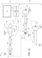
- FIGS. 5 A and 5 Billustrate a perspective view and a side view, respectively, of an implementation of a monolithic sensor having a plurality of pixel arrays for producing a three dimensional image in accordance with the teachings and principles of the disclosure;
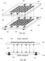
- FIGS. 6 A and 6 Billustrate a perspective view and a side view, respectively, of an implementation of an imaging sensor built on a plurality of substrates, wherein a plurality of pixel columns forming the pixel array are located on the first substrate and a plurality of circuit columns are located on a second substrate and showing an electrical connection and communication between one column of pixels to its associated or corresponding column of circuitry; and
- FIGS. 7 A and 7 Billustrate a perspective view and a side view, respectively, of an implementation of an imaging sensor having a plurality of pixel arrays for producing a three dimensional image, wherein the plurality of pixel arrays and the image sensor are built on a plurality of substrates.
- the disclosureextends to methods, devices, and systems for endoscopic light refraction imaging that allow angled endoscopes to be used with viewing trocars in a convenient, efficient, and less costly manner to create ports in a patient, including initial ports of endoscopic surgical procedures.
- Endoscopic light refraction imaging techniquesare described that allow angled endoscopes to be used with viewing trocars in a convenient, efficient, and less costly manner to create ports in a patient, including initial ports of endoscopic surgical procedures.
- a viewing trocar and/or angled endoscope of an endoscopic systemcan be configured with a light refracting element, such as glass and/or plastic prism for instance.
- the light refracting elementcan be utilized in and/or with the viewing trocar to refract (i.e., bend) light passing into the trocar through the trocar's window. More particularly, the light refracting element can change the incoming light's direction of travel to be along a plane substantially perpendicular to the endoscope's offset angle. As a result, the endoscope's field of view can be substantially aligned with the field of view of the viewing trocar's window.
- the viewing trocarcan be configured with a prism that is integrated with a lumen defined by the viewing trocar or that is removeably attached to the lumen.
- the angled endoscopecan be configured with a prism that is integrated onto the endoscope or that is removeably attached to the endoscope.
- the angled endoscopecan be configured with an image sensor that is disposed at and/or near the endoscope's distal end or tip.
- the image sensormay be a digital charge-coupled device (CCD) and/or complementary metal-oxide-semiconductor (CMOS) array of active pixel sensors for example.
- CCDdigital charge-coupled device
- CMOScomplementary metal-oxide-semiconductor
- endoscopic surgical procedurescan be less invasive than traditional surgical procedures because they allow a patient's internal body portions (i.e., tissue) to be examined (e.g., observed, inspected, and/or diagnosed) and/or treated by inserting an endoscope into a small port in the patient.
- tissuei.e., tissue
- a surgeoncan examine and/or treat a patient by inserting a type of endoscope known as a laparoscope through a port to reach the interior of the patient's abdominal or pelvic cavity.
- a surgeonmight examine and/or treat a patient by inserting another type of endoscope known as an arthroscope through a port to reach the interior of the patient's joint, such as a knee joint.
- Endoscopesare typically long slender objects with a light-gathering element (e.g., objective lens(s)) positioned at and/or near the endoscope's distal end, and an imaging system to receive optical images and convert them into electronic images that can be rendered on a display.
- a light-gathering elemente.g., objective lens(s)
- the field of view of an endoscope's light gathering elementmay be referred to herein as the endoscope's field of view.
- the imaginary line passing through an endoscope from its distal end to the proximal endcan generally define the endoscope's axis.
- the light gathering element of a non-angled endoscopeis disposed substantially perpendicular to the axis such that the endoscope's field of view is not substantially offset relative to a plane extending transversely to the endoscope's axis (i.e., the transverse plane).
- the distal endi.e., the tip
- the tipdoes not define an angle relative to the transverse plane, and thus appears blunt.
- the light gathering element of an angled endoscopein contrast, is not disposed substantially perpendicular to its axis. Instead, the light gathering element and distal end define an angle relative to the transverse plane.
- the degree of this anglewhich can be referred to as the endoscope's offset angle, can vary and may be between about 12 and about 90 degrees.
- endoscope offset anglesof about 30 degrees (i.e., 30-degree endoscopes) and 45 degrees (i.e., 45-degree endoscopes) are commonly used for many endoscopic procedures.
- the angled endoscope's field of viewis substantially offset relative to the transverse plane, and the distal end thus appears angled or pointed.
- endoscopesare designed such that the image sensor of the endoscope's imaging device or sensor is positioned at or near the endoscope's proximal end.
- the image sensoris typically positioned in the endoscope's hand-piece unit at and/or near the proximal end of the endoscope.
- lightcan enter through the light gathering element at the endoscope's distal end and propagate along the endoscope's axis toward the image sensor.
- This image sensorcan be configured to convert the optical image(s) represented by the light into an electronic signal that can then be used to render the image on a display.
- the endoscopeneeds to be configured with a complex set of precisely coupled optical propagation components for the light to propagate to the image sensor.
- optical propagation componentscan significantly increase the endoscope's cost (e.g., production cost). Additionally, optical propagation components can increase the endoscope's fragility since relatively minor impacts can easily damage these components or upset their relative alignments. This fragility necessitates frequent, expensive repair cycles in order to maintain image quality. Therefore, removing or decreasing the optical propagation components in an endoscope would be advantageous for at least the reason that it would reduce the endoscope's cost and fragility.
- endoscopic imaging techniquesare described herein that allow an endoscope to be configured with few or no optical propagation components, thus significantly decreasing the endoscope's cost and fragility as compared to traditional endoscopes.
- the endoscopecan be configured with an image sensor positioned at and/or near the endoscope's distal end or tip rather than at and/or near the endoscope's proximal end or in the hand piece.
- the image sensorcan be located comparatively closer to the endoscope's light gathering element, thus reducing or eliminating the need for optical propagation components in the endoscope.
- a trocaris first utilized to create a port to the site of interest (e.g., surgical site) inside the patient.
- the trocarcan include an obturator configured with a relatively sharp tip at or near the trocar's distal portion for puncturing the patient's tissue and reaching the site of interest.
- the obturatorcan first be inserted into a narrow endoscopic tube outside of the patient's body. The trocar (with the obturator inside the cannula) can then be inserted (distal portion first) into the patient. Once the site of interest is reached, the trocar can then be removed, leaving the cannula as the port.
- viewing trocarse.g., optical trocars
- Viewing trocarsare typically configured with a transparent or translucent window to allow the trocar's tip (e.g., obturator's tip) to be observed as it is inserted (i.e., punctures the patient) and passes through the patient's tissue.
- a viewing trocar's windowis usually positioned at and/or near the trocar's distal end.
- Viewing trocarsare also typically configured with a hollow portion, or lumen, so that an endoscope can be inserted into the trocar to observe the tip's insertion and passage.
- some viewing trocarsare configured with a transparent or translucent obturator tip and lumen along the trocar's length (from proximal end to distal end).
- An endoscopecan thus be inserted (e.g., slid) into the trocar such that the endoscope's light-gathering element is at and/or near (e.g., adjacent) the window.
- Light rays from the patient's tissue and/or other objects within the window's field of view and endoscope's field of viewcan enter through the window, be gathered and focused by the light gathering element, and viewed via the endoscope's imaging system and display.
- Non-angled endoscopesare typically preferred to angled endoscopes for creating an initial port. This is because, when placed into a viewing trocar, a non-angled endoscope's field of view is typically substantially aligned with the field of view of the viewing trocar's window. As a result, little if any of the trocar's window is obscured from the endoscope's light-gathering element, and most or all of the light entering the trocar's window can reach the endoscope's light-gathering element.
- the endoscope's field of viewis not typically substantially aligned with the field of view of the viewing trocar's window. This is due to the angled endoscope's offset angle. Generally, the greater the offset angle, the greater the extent that the field of view and window are out of alignment. For this reason, it is not surprising that most if not all viewing trocars are configured for non-angled endoscopes rather than angled endoscopes.
- Angled endoscopesare generally used and preferred for most types of endoscopic procedures other than creating the initial port.
- obtaining and utilizing both types of endoscopesnamely an angled and non-angled endoscope, can be costly, inconvenient, and wasteful—especially given that the non-angled endoscope may only be needed for creating the initial port. Therefore, it would be more convenient, efficient, and less costly to be able to use one angled endoscope for an entire endoscopic surgical procedure.
- viewing trocars and/or non-angled endoscopescan be configured to be used together when creating ports in a patient, including an initial port of a procedure.
- a light refracting elemente.g., prism
- the light refracting elementcan change the incoming light's direction of travel to be along a plane substantially perpendicular to the endoscope's offset angle.
- the endoscope's field of viewcan be substantially aligned with the field of view of the trocar's window.
- the light refracting elementcan be utilized to increase the amount of light that reaches the light gathering element by bending incoming light at an angle such that the fields of view of the endoscope and window are similar or the same.
- a prismcan be used that bends incoming light at about a 30 degree angle towards the endoscope's light gathering element.
- FIGS. 1 A- 1 Cillustrate exemplary endoscopic systems 100 that may be implemented in accordance with the techniques described herein.
- the endoscopic systems 100 illustratedare example implementations, and are thus not to be interpreted as limiting. More particularly, while the endoscopic systems 100 of FIGS. 1 A- 1 C are described in the context of including various systems and components, this is not be construed as limiting the implementation of any one or more of these systems or components to the endoscopic system 100 . Instead, it is to be appreciated and understood that any of the described systems and components can be implemented alone or in any combination irrespective of the endoscopic system 100 .
- the endoscopic system 100may include an angled endoscope system 102 .
- the angled endoscope system 102may include an endoscope device (i.e., endoscope) 104 , endoscope housing 106 (e.g., hand piece and/or camera head), control unit 108 , light source 110 , display 112 , and imaging device 114 (e.g., camera, sensors, etc.).
- endoscope device 104 , endoscope housing 106 , control unit 108 , light source 110 , display 112 , and imaging device 114are each shown individually with respect to one another. However, it is to be appreciated and understood that this is not to be interpreted as limiting, and any one or more of these components can be integrated and/or connected in any suitable way.
- FIGS. 1 A and 1 Bthe endoscopic device 104 and endoscope housing 106 are shown in a detached state. However, these components may be operably connected (e.g., coupled), as illustrated for example in FIG. 1 C , to one another to form an angled endoscope unit for performing endoscopic surgical procedures.
- the control unit 108 and light source 110are shown as being separate. Whereas, in the implementation of FIG. 1 B , the control unit 108 and the light source 110 are shown as being part of the same unit 111 .
- the light source 110can be configured to provide light, when needed, via one or more fiber optics or other light transmission functionality to the endoscope device 104 for use in illuminating or otherwise facilitating observation of a patient's tissue.
- these componentsmay be integrated (e.g., in the same housing, etc.) or otherwise operably connected in unit 111 .
- the imaging device 114 of FIG. 1 Cis shown as being configured with components located in both the endoscope housing 106 and endoscope device 104 .
- the imaging device 114may be configured otherwise.
- all of the features of the imaging device 114can be included or located in the endoscope housing 106 (illustrated best in FIG. 1 A ), or alternatively some or all of the imaging device 114 can be located remotely or externally with respect to the endoscope housing 106 in one or more other components that may or may not include the control unit 108 or endoscope device 104 .
- the imaging device 114includes an image sensor 116 that is advantageously disposed (i.e., located) at and/or near the distal end (i.e., tip) of the endoscope device 104 .
- the image sensor 116can be any suitable type of device and/or related circuitry, such as a digital charge-coupled device (CCD) and/or complementary metal-oxide-semiconductor (CMOS) array of active pixel sensors for instance.
- CCDdigital charge-coupled device
- CMOScomplementary metal-oxide-semiconductor
- certain mechanical and software stabilization techniquescan be employed.
- the image sensor 116can be configured to receive light gathered and focused by a light gathering element 118 (e.g., lens) positioned at and/or near the distal end of the endoscope device 104 .
- the image sensor 116can also be configured to convert optical images represented by the received light into electronic images that can be rendered on the display 112 .
- the light gathering element 118is not disposed substantially perpendicular to the axis 119 of the endoscope device 104 . Instead, the light gathering element 118 is disposed at an angle of about 30 degrees relative to a plane extending transversely to the axis 119 (i.e., the transverse plane). Therefore the offset angle of the endoscope device 104 is about 30 degrees, thus making in the endoscope device 104 an angled endoscope. As a result of this offset angle, the field of view of endoscope device 104 is substantially offset relative to the transverse plane, thus resulting in the angled, or pointed, appearance of the distal end of the endo scope device 104 .
- the control unit 108can be electronically and/or communicatively linked to the imaging device 114 and/or one or other components in the endoscope housing 106 and/or endoscope device 104 .
- the control unit 108can be linked in this manner via a physical (e.g., wired) and/or wireless (e.g., BLUETOOTH, infrared, etc.) connection, as represented by connection 120 .
- the control unit 108can be electronically and/or communicatively linked to the display 112 , as represented by connection 122 .
- the display 112may be any type of display device suitably configured to display rendered electronic images received from the imaging device 114 .
- the endoscope housing 106may also include other components, such as a transceiver 124 (e.g., wireless transceiver) that can be configured to facilitate communication between the endoscope housing 106 and the control unit 108 via the connection 120 .
- a transceiver 124e.g., wireless transceiver
- the ability to separate and communicatively link the endoscope housing 106 from the control unit 108may provide for the easy replacement of used endoscopes and/or endoscope housings for sterilized and renewed endoscopes and/or housings.
- the ability to separate and communicatively link these componentsalso allows for greater mobility of the endoscope housing 106 during the endoscope device's use.
- endoscopic system 100may also include a viewing trocar system 128 that be configured to mitigate puncture risk during an endoscopic surgical procedure by allowing the distal end (i.e., tip) of the trocar system 128 to be observed as it is inserted into a patient and passes through the patient's tissue.
- the viewing trocar system 128can include an obturator 130 and a cannula 134 .
- the obturator 130can include obturator housing 132 , which in this example is shaped to facilitate handling of the obturator 130 .
- the obturator 130can also define an interior obturator lumen 136 that extends along the axis 131 of the obturator 130 from the obturator's proximal end (at the obturator housing's proximal end) to a point at or near the relatively sharp tip at the obturator's distal end (i.e., trocar system's 128 distal end) that is formed by a transparent or translucent window 138 .
- the obturator housing 132includes a hollow portion (e.g., trocar housing lumen) that effectively allows the obturator lumen 136 to extend from at or near the window 138 through the obturator housing 132 to an opening 133 at the housing's obturator's proximal end.
- a hollow portione.g., trocar housing lumen
- the angled endoscope system 102 and viewing trocar system 128can be configured to be operably connected (e.g., coupled) to one another to initiate an endoscopic surgical procedure.
- the endoscope device 104may be coupled with (e.g., slid into, and removed from) the obturator 130 by first inserting the endoscope device 104 (distal end first) through the opening 133 in the obturator housing 132 and along the obturator lumen 136 until the distal end of the endoscope device 104 reaches a point at or near the proximal end of the window 138 .
- the light gathering element 118 and imaging device 114can be used to examine the tip formed by the window 138 as it is inserted into the patient's tissue. More particularly, in accordance with the described techniques, and as shown in FIGS. 2 and 3 , when the angled endoscope system 102 and viewing trocar system 128 are operably connected a light refracting element 140 can be utilized in the obturator to bend incoming light 302 that has entered through the window 138 .
- the light refracting element 140can be any suitable type of device or material capable of refracting light in a particular direction.
- the light refracting element 140is a prism made of glass and/or plastic that is able to cause the incoming light 302 to be bent toward the light gathering element 118 . It will be appreciated that the light refracting element 140 may be made from any suitable material that has the ability to refract light as disclosed herein.
- the light refracting element 140can be located in a light refracting region 142 in the obturator lumen 136 and disposed at an angle such that incoming light 302 is bent by the light refracting element 140 at a 30 degree angle in a direction toward the light gathering element 118 , which is disposed at an angle of about 30 degrees relative to the transverse plane.
- the incoming light 302can be bent by the light refracting element 140 such that the light's direction of travel is changed to be toward the light gathering element 118 along a plane that is substantially perpendicular to the light gathering element 118 .
- the field of view of the endoscope device 104can be substantially aligned with the field of view of the window 138 , thus allowing the endoscope device 104 to be used in the viewing trocar system 128 in a convenient, effective, and cost efficient manner.
- the light refracting element 140can be integrated with the inside wall of the obturator lumen 136 and/or on the endoscope device 104 , such that the element's location and/or position is fixed.
- the light refracting elementcan be placed into, and/or attached (i.e., temporarily or permanently) to, the obturator lumen 136 and/or to the endoscope device 104 .
- retaining functionality and structurecan be utilized to place and/or attach the light refracting element 140 in a particular location and/or position in or on the obturator 130 .
- the retaining functionality and structuremay be configured to allow the light refracting element 140 to be removeably attached (e.g., temporarily) or permanently attached.
- the retaining functionality and structuremight be a mechanical structure, structurally integrated shape in the lumen or elsewhere in/on the obturator 130 , adhesive chemical substance, and/or a region (e.g., the light refracting region 142 ) that allows the light refracting element 140 to be placed into, and/or attached to, the obturator 130 .
- the retaining functionality and/or retention structuresmight be configured such that the light refracting element 140 remains fixed with the respect to the obturator lumen 136 .
- the endoscope device 104when coupled with the viewing trocar system, would likely need to be rotated around the axis 131 (and thus axis 119 ) relative to the obturator 130 to reach a suitable orientation for the light refracting element 140 and light gathering element 118 to be substantially rotationally aligned, and thus adjacent to one another.
- the retaining functionalitymay be configured such that the light refracting element 140 is allowed to move within the obturator lumen 136 .
- the endoscope device 104may not need to be rotated around the axis 131 for the light refracting element 142 and light gathering element 118 to be substantially rotationally aligned, and thus adjacent to one another. Instead, the light refracting element 140 may be rotated around the axis 131 until alignment is achieved.
- the retaining functionality and/or retention structurescan also be configured to allow the light refracting element 140 to be manually and/or automatically disposed at one or more desired angles relative to a plane extending transversely to the axis 131 and/or to the endoscope device 104 .
- the retaining functionality and structuremay be configured to allow the light refracting element's disposition to be changed from one desired angle to another desired angle.
- the retaining functionality and/or light refracting element 140can be provided in any suitable manner.
- the retaining functionality and/or light refracting element 140may be provided (e.g., commercially packaged) alone and/or with one or other components, such as with the viewing trocar system 128 , endoscope device, and/or endoscopic system 100 for instance.
- the obturator 130 and trocar housing 132may be operably connected to the cannula 134 before being inserted into the patient. As explained above, once the site of interest inside the patient is reached, the obturator 130 and/or trocar housing 132 can be removed, leaving the cannula as a port into the patient.
- the obturator 130can be configured to be slid into, and removed from, the cannula 134 by first inserting the obturator 130 through an opening 144 in the cannula 134 and then along a cannula lumen 147 inside the cannula 134 until the distal end of the obturator housing 132 comes into contact with the proximal end of the cannula's housing 146 .
- a viewing trocar and angled endoscopecan be configured to be operably connected to place an endoscopic port.
- the viewing trocar system 128 and angled endoscope system 102 of endoscopic system 100can be utilized.
- the viewing trocar or angled endoscopecan be configured with a light refracting element (e.g., light refracting element 140 ), such a glass and/or plastic prism, to refract received light. As explained above, this light can be received through a window (e.g., window 138 ) disposed at the distal end of the viewing trocar.
- the viewing trocarcan be configured with a prism at or near the distal tip of the trocar that is integrated with a lumen defined by the viewing trocar, or that is removeably attached to the lumen.
- the angled endoscopecan be configured with a prism that is integrated onto the endoscope or that is removeably attached to the endoscope.
- the viewing trocarcan be configured with retaining functionality.
- this retaining functionality and structurecan be any functionality that allows the light refracting element to be placed into, and/or attached to, the viewing trocar.
- the disclosuremay be used with any image sensor, whether a CMOS image sensor or CCD image sensor, without departing from the scope of the disclosure.
- the image sensormay be located in any location within the overall system, including, but not limited to, the tip of the endoscope, the hand piece of the imaging device or camera, the control unit, or any other location within the system without departing from the scope of the disclosure.
- Implementations of an image sensor that may be utilized by the disclosureinclude, but are not limited to, the following, which are merely examples of various types of sensors that may be utilized by the disclosure.
- FIGS. 5 A and 5 Bthe figures illustrate a perspective view and a side view, respectively, of an implementation of a monolithic sensor 500 having a plurality of pixel arrays for producing a three dimensional image in accordance with the teachings and principles of the disclosure.
- Such an implementationmay be desirable for three dimensional image capture, wherein the two pixel arrays 502 and 504 may be offset during use.
- a first pixel array 502 and a second pixel array 504may be dedicated to receiving a predetermined range of wave lengths of electromagnetic radiation, wherein the first pixel array 502 is dedicated to a different range of wave length electromagnetic radiation than the second pixel array 504 .
- FIGS. 6 A and 6 Billustrate a perspective view and a side view, respectively, of an implementation of an imaging sensor 600 built on a plurality of substrates.
- a plurality of pixel columns 604 forming the pixel arrayare located on the first substrate 602 and a plurality of circuit columns 608 are located on a second substrate 606 .
- the electrical connection and communication between one column of pixels to its associated or corresponding column of circuitrymay be implemented in one implementation, an image sensor, which might otherwise be manufactured with its pixel array and supporting circuitry on a single, monolithic substrate/chip, may have the pixel array separated from all or a majority of the supporting circuitry.
- the disclosuremay use at least two substrates/chips, which will be stacked together using three-dimensional stacking technology.
- the first 602 of the two substrates/chipsmay be processed using an image CMOS process.
- the first substrate/chip 602may be comprised either of a pixel array exclusively or a pixel array surrounded by limited circuitry.
- the second or subsequent substrate/chip 606may be processed using any process, and does not have to be from an image CMOS process.
- the second substrate/chip 606may be, but is not limited to, a highly dense digital process in order to integrate a variety and number of functions in a very limited space or area on the substrate/chip, or a mixed-mode or analog process in order to integrate for example precise analog functions, or a RF process in order to implement wireless capability, or MEMS (Micro-Electro-Mechanical Systems) in order to integrate MEMS devices.
- the image CMOS substrate/chip 602may be stacked with the second or subsequent substrate/chip 606 using any three-dimensional technique.
- the second substrate/chip 606may support most, or a majority, of the circuitry that would have otherwise been implemented in the first image CMOS chip 602 (if implemented on a monolithic substrate/chip) as peripheral circuits and therefore have increased the overall system area while keeping the pixel array size constant and optimized to the fullest extent possible.
- the electrical connection between the two substrates/chipsmay be done through interconnects 603 and 605 , which may be wirebonds, bump and/or TSV (Through Silicon Via).
- FIGS. 7 A and 7 Billustrate a perspective view and a side view, respectively, of an implementation of an imaging sensor 700 having a plurality of pixel arrays for producing a three dimensional image.
- the three dimensional image sensormay be built on a plurality of substrates and may comprise the plurality of pixel arrays and other associated circuitry, wherein a plurality of pixel columns 704 a forming the first pixel array and a plurality of pixel columns 704 b forming a second pixel array are located on respective substrates 702 a and 702 b , respectively, and a plurality of circuit columns 708 a and 708 b are located on a separate substrate 706 . Also illustrated are the electrical connections and communications between columns of pixels to associated or corresponding column of circuitry.
- teachings and principles of the disclosuremay be used in a reusable device platform, a limited use device platform, a re-posable use device platform, or a single-use/disposable device platform without departing from the scope of the disclosure. It will be appreciated that in a re-usable device platform an end-user is responsible for cleaning and sterilization of the device. In a limited use device platform the device can be used for some specified amount of times before becoming inoperable. Typical new device is delivered sterile with additional uses requiring the end-user to clean and sterilize before additional uses.
- a third-partymay reprocess the device (e.g., cleans, packages and sterilizes) a single-use device for additional uses at a lower cost than a new unit.
- a deviceis provided sterile to the operating room and used only once before being disposed of.
- teachings and principles of the disclosuremay include any and all wavelengths of electromagnetic energy, including the visible and non-visible spectrums, such as infrared (IR), ultraviolet (UV), and X-ray.
- IRinfrared
- UVultraviolet
- X-rayX-ray
Landscapes
- Health & Medical Sciences (AREA)
- Life Sciences & Earth Sciences (AREA)
- Surgery (AREA)
- Animal Behavior & Ethology (AREA)
- Public Health (AREA)
- Engineering & Computer Science (AREA)
- Biomedical Technology (AREA)
- Heart & Thoracic Surgery (AREA)
- Medical Informatics (AREA)
- Molecular Biology (AREA)
- Veterinary Medicine (AREA)
- General Health & Medical Sciences (AREA)
- Nuclear Medicine, Radiotherapy & Molecular Imaging (AREA)
- Pathology (AREA)
- Physics & Mathematics (AREA)
- Optics & Photonics (AREA)
- Biophysics (AREA)
- Radiology & Medical Imaging (AREA)
- Endoscopes (AREA)
- Surgical Instruments (AREA)
Abstract
Description
Claims (24)
Priority Applications (1)
| Application Number | Priority Date | Filing Date | Title |
|---|---|---|---|
| US16/792,831US11690498B2 (en) | 2013-03-15 | 2020-02-17 | Viewing trocar with integrated prism for use with angled endoscope |
Applications Claiming Priority (3)
| Application Number | Priority Date | Filing Date | Title |
|---|---|---|---|
| US201361791935P | 2013-03-15 | 2013-03-15 | |
| US14/214,400US10561302B2 (en) | 2013-03-15 | 2014-03-14 | Viewing trocar with integrated prism for use with angled endoscope |
| US16/792,831US11690498B2 (en) | 2013-03-15 | 2020-02-17 | Viewing trocar with integrated prism for use with angled endoscope |
Related Parent Applications (1)
| Application Number | Title | Priority Date | Filing Date |
|---|---|---|---|
| US14/214,400ContinuationUS10561302B2 (en) | 2013-03-15 | 2014-03-14 | Viewing trocar with integrated prism for use with angled endoscope |
Publications (2)
| Publication Number | Publication Date |
|---|---|
| US20200178769A1 US20200178769A1 (en) | 2020-06-11 |
| US11690498B2true US11690498B2 (en) | 2023-07-04 |
Family
ID=51530279
Family Applications (2)
| Application Number | Title | Priority Date | Filing Date |
|---|---|---|---|
| US14/214,400Active2035-05-13US10561302B2 (en) | 2013-03-15 | 2014-03-14 | Viewing trocar with integrated prism for use with angled endoscope |
| US16/792,831Active2034-04-20US11690498B2 (en) | 2013-03-15 | 2020-02-17 | Viewing trocar with integrated prism for use with angled endoscope |
Family Applications Before (1)
| Application Number | Title | Priority Date | Filing Date |
|---|---|---|---|
| US14/214,400Active2035-05-13US10561302B2 (en) | 2013-03-15 | 2014-03-14 | Viewing trocar with integrated prism for use with angled endoscope |
Country Status (9)
| Country | Link |
|---|---|
| US (2) | US10561302B2 (en) |
| EP (1) | EP2967309B1 (en) |
| JP (1) | JP6466400B2 (en) |
| CN (1) | CN105263393B (en) |
| AU (1) | AU2014233486B2 (en) |
| BR (1) | BR112015022987A2 (en) |
| CA (1) | CA2906832A1 (en) |
| ES (1) | ES2914064T3 (en) |
| WO (1) | WO2014145008A2 (en) |
Families Citing this family (30)
| Publication number | Priority date | Publication date | Assignee | Title |
|---|---|---|---|---|
| BR112015022987A2 (en) | 2013-03-15 | 2017-07-18 | Olive Medical Corp | integrated prism trocar visualization for use with angled endoscope |
| US11033182B2 (en) | 2014-02-21 | 2021-06-15 | 3Dintegrated Aps | Set comprising a surgical instrument |
| CN108024806B (en) | 2015-07-21 | 2022-07-01 | 3D集成公司 | Cannula assembly kit, trocar assembly kit, sleeve assembly, minimally invasive surgical system and method thereof |
| US11020144B2 (en) | 2015-07-21 | 2021-06-01 | 3Dintegrated Aps | Minimally invasive surgery system |
| KR101737926B1 (en)* | 2015-08-04 | 2017-05-19 | 고려대학교 산학협력단 | Medical arthroscopy and cannula therefor |
| US10172525B2 (en) | 2015-08-17 | 2019-01-08 | Rebound Therapeutics Corporation | Cannula with proximally mounted camera |
| DK178899B1 (en) | 2015-10-09 | 2017-05-08 | 3Dintegrated Aps | A depiction system |
| US10105042B2 (en) | 2016-08-17 | 2018-10-23 | Rebound Therapeutics Corporation | Cannula with proximally mounted camera |
| KR102486092B1 (en) | 2016-08-17 | 2023-01-06 | 리바운드 세라퓨틱스 코포레이션 | Cannula with Proximally Mounted Camera |
| GB2553045B (en)* | 2016-08-17 | 2019-02-13 | Rebound Therapeutics Corp | Cannula with proximally mounted camera |
| JP7155149B2 (en)* | 2016-12-27 | 2022-10-18 | デピュイ・シンセス・プロダクツ・インコーポレイテッド | Systems, methods, and devices for providing illumination in an endoscopic imaging environment |
| WO2019227018A1 (en)* | 2018-05-25 | 2019-11-28 | Flash Surgical, Inc. | Optical entry trocar camera |
| JP2022508311A (en)* | 2018-08-09 | 2022-01-19 | オプティカル スパイン | Translucent illuminated endoscope probe |
| CA3130085A1 (en)* | 2019-02-22 | 2020-08-27 | Rebound Therapeutics Corporation | Cannula and obturator with a transparent tip with an opaque component |
| CN113795187A (en)* | 2019-03-06 | 2021-12-14 | 诺亚医疗集团公司 | Single use endoscope, cannula and obturator with integrated vision and illumination |
| US11903557B2 (en) | 2019-04-30 | 2024-02-20 | Psip2 Llc | Endoscope for imaging in nonvisible light |
| US11931009B2 (en) | 2019-06-20 | 2024-03-19 | Cilag Gmbh International | Offset illumination of a scene using multiple emitters in a hyperspectral imaging system |
| US11589819B2 (en)* | 2019-06-20 | 2023-02-28 | Cilag Gmbh International | Offset illumination of a scene using multiple emitters in a laser mapping imaging system |
| US11903563B2 (en) | 2019-06-20 | 2024-02-20 | Cilag Gmbh International | Offset illumination of a scene using multiple emitters in a fluorescence imaging system |
| CN110448359B (en)* | 2019-08-02 | 2021-05-14 | 中国人民解放军总医院 | Operation navigation equipment for improving success rate of transjugular intrahepatic portosystemic shunt and application thereof |
| CN110575113A (en)* | 2019-09-25 | 2019-12-17 | 广州为实光电医疗科技有限公司 | An electronic endoscope and its insertion device |
| JP2023510167A (en)* | 2020-01-18 | 2023-03-13 | ピーエスアイピー2 エルエルシー | Injection needle with endoscope for regenerative medicine |
| AU2022200563B2 (en)* | 2021-02-03 | 2023-03-23 | Chin Piao Chang | Endoscope assembly and endoscope system having the same |
| EP4039166A1 (en)* | 2021-02-03 | 2022-08-10 | Hong So Kao | Endoscope assembly having a surgical instrument and endoscope system having the same |
| US20220240766A1 (en)* | 2021-02-03 | 2022-08-04 | Chin-Piao Chang | Endoscope Kit having Functions of Injection, Clamping and Placing Medical Materials or Medicines |
| WO2022249116A2 (en)* | 2021-05-26 | 2022-12-01 | Psip2 Llc | Endoscope |
| US20220378279A1 (en)* | 2021-05-26 | 2022-12-01 | Psip2 Llc | Endoscope |
| US12014997B2 (en)* | 2021-07-01 | 2024-06-18 | Taiwan Semiconductor Manufacturing Co., Ltd. | Dummy stacked structures surrounding TSVs and method forming the same |
| US12238265B2 (en) | 2022-12-12 | 2025-02-25 | Cilag Gmbh International | Optical filter for improved multispectral imaging performance in stereo camera |
| US12316965B2 (en) | 2023-02-27 | 2025-05-27 | Cilag Gmbh International | Adaptive overlay stabilization of false color overlay heatmaps |
Citations (206)
| Publication number | Priority date | Publication date | Assignee | Title |
|---|---|---|---|---|
| US4011403A (en) | 1976-03-30 | 1977-03-08 | Northwestern University | Fiber optic laser illuminators |
| US4140364A (en)* | 1973-06-23 | 1979-02-20 | Olympus Optical Co., Ltd. | Variable field optical system for endoscopes |
| US4254762A (en)* | 1979-10-23 | 1981-03-10 | Inbae Yoon | Safety endoscope system |
| US4363963A (en) | 1979-03-08 | 1982-12-14 | Nippon Hoso Kyokai | Solid state photo-electric converting device and solid state imaging apparatus employing it |
| US4433675A (en) | 1981-04-01 | 1984-02-28 | Olympus Optical Co. Ltd. | Light supply apparatus for endoscope |
| US4740837A (en) | 1985-09-19 | 1988-04-26 | Kabushiki Kaisha Toshiba | Endoscope apparatus with solid state image pickup device |
| US4741327A (en) | 1986-04-30 | 1988-05-03 | Olympus Optical Co., Ltd. | Endoscope having bent circuit board |
| US4745471A (en) | 1986-05-13 | 1988-05-17 | Olympus Optical Co., Ltd. | Solid-state imaging apparatus and endoscope |
| US4786965A (en) | 1986-09-04 | 1988-11-22 | Olympus Optical Co., Ltd. | Eletronic endoscope with an imaging device having side bonding pads |
| US4832003A (en) | 1986-09-12 | 1989-05-23 | Olympus Optical Co., Ltd. | Electronic endoscope tip |
| US4853772A (en) | 1987-02-26 | 1989-08-01 | Olympus Optical Co., Ltd. | Electronic endoscope apparatus having isolated patient and secondary circuitry |
| US4866526A (en) | 1987-07-25 | 1989-09-12 | Richard Wolf Gmbh | Video endoscope with light intensity regulation |
| US4916534A (en)* | 1987-04-28 | 1990-04-10 | Olympus Optical Co., Ltd. | Endoscope |
| US4918521A (en) | 1987-01-20 | 1990-04-17 | Olympus Optical Co., Ltd. | Solid state imaging apparatus |
| US4942473A (en) | 1987-07-16 | 1990-07-17 | Techninon Research & Development Foundation | Intelligent scan image sensor |
| US5016975A (en) | 1984-07-31 | 1991-05-21 | Olympus Optical Co., Ltd. | Electronic endoscope provided with a sample-hold circuit |
| US5021888A (en) | 1987-12-18 | 1991-06-04 | Kabushiki Kaisha Toshiba | Miniaturized solid state imaging device |
| USRE33854E (en) | 1989-02-03 | 1992-03-24 | sterilizable sheathpe with .[.heat.]. | |
| US5133035A (en) | 1989-11-14 | 1992-07-21 | Hicks John W | Multifiber endoscope with multiple scanning modes to produce an image free of fixed pattern noise |
| US5187572A (en) | 1990-10-31 | 1993-02-16 | Olympus Optical Co., Ltd. | Endoscope system with a plurality of synchronized light source apparatuses |
| US5200838A (en) | 1988-05-27 | 1993-04-06 | The University Of Connecticut | Lateral effect imaging system |
| US5220198A (en) | 1990-08-27 | 1993-06-15 | Olympus Optical Co., Ltd. | Solid state imaging apparatus in which a solid state imaging device chip and substrate are face-bonded with each other |
| US5228430A (en) | 1989-08-04 | 1993-07-20 | Kabushiki Kaisha Toshiba | Electronic endoscope apparatus including easy focusing distal end |
| US5241170A (en) | 1992-02-19 | 1993-08-31 | Itt Corporation | Fiber optic imaging device and methods |
| US5271380A (en)* | 1990-11-06 | 1993-12-21 | Siegfried Riek | Penetration instrument |
| US5313306A (en) | 1991-05-13 | 1994-05-17 | Telerobotics International, Inc. | Omniview motionless camera endoscopy system |
| US5325847A (en) | 1991-10-25 | 1994-07-05 | Asahi Kogaku Kogyo Kabushiki Kaisha | Distal end part of endoscope |
| US5334150A (en)* | 1992-11-17 | 1994-08-02 | Kaali Steven G | Visually directed trocar for laparoscopic surgical procedures and method of using same |
| US5354302A (en)* | 1992-11-06 | 1994-10-11 | Ko Sung Tao | Medical device and method for facilitating intra-tissue visual observation and manipulation of distensible tissues |
| US5385572A (en) | 1992-11-12 | 1995-01-31 | Beowulf Holdings | Trocar for endoscopic surgery |
| US5411020A (en) | 1990-11-27 | 1995-05-02 | Asahi Kogaku Kogyo Kabushiki Kaisha | Structure of the distal end portion of an endoscope |
| US5427087A (en) | 1990-11-26 | 1995-06-27 | Asahi Kogaku Kogyo Kabushiki Kaisha | Structure of the distal end portion of an endoscope |
| US5441041A (en)* | 1993-09-13 | 1995-08-15 | United States Surgical Corporation | Optical trocar |
| US5445142A (en)* | 1994-03-15 | 1995-08-29 | Ethicon Endo-Surgery, Inc. | Surgical trocars having optical tips defining one or more viewing ports |
| US5454366A (en) | 1990-11-27 | 1995-10-03 | Asashi Kogaku Kogyo Kabushiki Kaisha | Endoscope distal end with folded circuit board |
| WO1996005693A1 (en) | 1994-08-09 | 1996-02-22 | Applitec Ltd. | Random access multispectral illumination device for an imaging system |
| US5554097A (en) | 1994-10-05 | 1996-09-10 | United States Surgical Corporation | Surgical instrumentation kit |
| US5569160A (en)* | 1993-09-13 | 1996-10-29 | United States Surgical Corporation | Optical trocar |
| US5569291A (en)* | 1995-02-01 | 1996-10-29 | Ethicon Endo-Surgery, Inc. | Surgical penetration and dissection instrument |
| US5569292A (en)* | 1995-02-01 | 1996-10-29 | Ethicon Endo-Surgery, Inc. | Surgical penetration instrument with transparent blades and tip cover |
| US5573493A (en)* | 1993-10-08 | 1996-11-12 | United States Surgical Corporation | Endoscope attachment for changing angle of view |
| US5588949A (en) | 1993-10-08 | 1996-12-31 | Heartport, Inc. | Stereoscopic percutaneous visualization system |
| US5591192A (en)* | 1995-02-01 | 1997-01-07 | Ethicon Endo-Surgery, Inc. | Surgical penetration instrument including an imaging element |
| US5594497A (en) | 1993-04-07 | 1997-01-14 | Ahern; John M. | Endoscope provided with a distally located color CCD |
| US5607441A (en)* | 1995-03-24 | 1997-03-04 | Ethicon Endo-Surgery, Inc. | Surgical dissector |
| US5609562A (en)* | 1993-11-16 | 1997-03-11 | Worldwide Optical Trocar Licensing Corporation | Visually directed trocar and method |
| US5665959A (en) | 1995-01-13 | 1997-09-09 | The United States Of America As Represented By The Administrator Of The National Aeronautics And Space Adminstration | Solid-state image sensor with focal-plane digital photon-counting pixel array |
| US5685820A (en)* | 1990-11-06 | 1997-11-11 | Partomed Medizintechnik Gmbh | Instrument for the penetration of body tissue |
| US5689365A (en)* | 1994-09-13 | 1997-11-18 | Olympus Optical Co., Ltd | Stereoscopic-vision endoscope |
| US5690664A (en)* | 1993-09-13 | 1997-11-25 | United States Surgical Corporation | Trocar having movable blade |
| US5720761A (en)* | 1993-11-16 | 1998-02-24 | Worldwide Optical Trocar Licensing Corp. | Visually directed trocar and method |
| US5738628A (en)* | 1995-03-24 | 1998-04-14 | Ethicon Endo-Surgery, Inc. | Surgical dissector and method for its use |
| US5743881A (en)* | 1995-11-03 | 1998-04-28 | Aptec Medical Corporation | Laparoscopic surgical instrument and method of using same |
| US5762604A (en) | 1994-06-01 | 1998-06-09 | Archimedes Surgical, Inc. | Surgical instrument permitting endoscopic viewing and dissecting |
| US5797944A (en)* | 1992-11-12 | 1998-08-25 | Ethicon Endo-Surgery, Inc. | Visualization trocar |
| US5797836A (en)* | 1995-06-07 | 1998-08-25 | Smith & Nephew, Inc. | Endoscope with relative rotation and axial motion between an optical element and an imaging device |
| US5817061A (en)* | 1997-05-16 | 1998-10-06 | Ethicon Endo-Surgery, Inc. | Trocar assembly |
| US5860996A (en)* | 1994-05-26 | 1999-01-19 | United States Surgical Corporation | Optical trocar |
| US5873889A (en)* | 1997-08-08 | 1999-02-23 | Origin Medsystems, Inc. | Tissue separation cannula with dissection probe and method |
| US5916233A (en)* | 1998-03-05 | 1999-06-29 | Origin Medsystems, Inc. | Vessel harvesting method and instrument including access port |
| US5968065A (en)* | 1995-07-13 | 1999-10-19 | Origin Medsystems, Inc. | Tissue separation cannula |
| US5980549A (en)* | 1995-07-13 | 1999-11-09 | Origin Medsystems, Inc. | Tissue separation cannula with dissection probe and method |
| US6001084A (en)* | 1995-12-18 | 1999-12-14 | Riek; Siegfried | Medical needle for endoscopic surgery |
| US6019720A (en)* | 1995-07-07 | 2000-02-01 | Olympus Optical Co., Ltd. | System for evulsing subcutaneous tissue |
| US6183444B1 (en)* | 1998-05-16 | 2001-02-06 | Microheart, Inc. | Drug delivery module |
| US6206823B1 (en)* | 1999-08-02 | 2001-03-27 | Ethicon Endo-Surgery, Inc. | Surgical instrument and method for endoscopic tissue dissection |
| US6272269B1 (en) | 1999-11-16 | 2001-08-07 | Dn Labs Inc. | Optical fiber/waveguide illumination system |
| US20010030744A1 (en) | 1999-12-27 | 2001-10-18 | Og Technologies, Inc. | Method of simultaneously applying multiple illumination schemes for simultaneous image acquisition in an imaging system |
| US6310642B1 (en) | 1997-11-24 | 2001-10-30 | Micro-Medical Devices, Inc. | Reduced area imaging devices incorporated within surgical instruments |
| US6327493B1 (en) | 1997-08-28 | 2001-12-04 | Olympus Optical Co., Ltd. | Light scanning devices of a water-tight structure to be inserted into a body cavity to obtain optical information on inside of a biological tissue |
| US6331156B1 (en) | 1999-06-21 | 2001-12-18 | Richard Wolf Gmbh | Electronic endoscope |
| US6387043B1 (en)* | 1998-05-13 | 2002-05-14 | Inbae Yoon | Penetrating endoscope and endoscopic surgical instrument with CMOS image sensor and display |
| US6432044B1 (en)* | 1999-01-08 | 2002-08-13 | Origin Medsystems, Inc. | Combined vessel dissection and transection device and method |
| US6471638B1 (en)* | 2000-04-28 | 2002-10-29 | Origin Medsystems, Inc. | Surgical apparatus |
| US6485414B1 (en) | 1998-07-13 | 2002-11-26 | Ceramoptec Industries, Inc. | Color video diagnostic system for mini-endoscopes |
| US20030007087A1 (en) | 2001-07-06 | 2003-01-09 | Fuji Photo Film Co., Ltd. | Light source device and image capturing device |
| US6508759B1 (en) | 1993-10-08 | 2003-01-21 | Heartport, Inc. | Stereoscopic percutaneous visualization system |
| US20030189663A1 (en) | 2002-04-04 | 2003-10-09 | Richard Wolf Gmbh | Solid-state video camera and method for brightness control |
| US6690466B2 (en) | 1999-08-06 | 2004-02-10 | Cambridge Research & Instrumentation, Inc. | Spectral imaging system |
| US6692431B2 (en) | 2001-09-07 | 2004-02-17 | Smith & Nephew, Inc. | Endoscopic system with a solid-state light source |
| US20040082833A1 (en) | 2000-04-10 | 2004-04-29 | Doron Adler | Image sensor and an endoscope using the same |
| US6773392B2 (en) | 2001-03-12 | 2004-08-10 | Olympus Corporation | Endoscope |
| US20040162516A1 (en)* | 2001-06-20 | 2004-08-19 | Evgenia Mandrusov | Agents that stimulate therapeutic angiogenesis and techniques and devices that enable their delivery |
| US6809358B2 (en) | 2002-02-05 | 2004-10-26 | E-Phocus, Inc. | Photoconductor on active pixel image sensor |
| US20050027164A1 (en) | 2003-07-29 | 2005-02-03 | Scimed Life Systems, Inc. | Vision catheter |
| US20050065543A1 (en)* | 2001-09-24 | 2005-03-24 | Henry Kahle | Bladeless optical obturator |
| US20050070943A1 (en)* | 2003-09-30 | 2005-03-31 | Hueil Geoffrey C. | Instrument lock assembly for trocar |
| US20050070850A1 (en)* | 2003-09-30 | 2005-03-31 | Albrecht Thomas E. | Low-profile, recessed stop-cock valve for trocar assembly |
| US20050070851A1 (en)* | 2003-09-30 | 2005-03-31 | Thompson Brian J. | Trocar housing/stop-cock assembly |
| US20050070947A1 (en)* | 2003-09-30 | 2005-03-31 | Franer Paul T. | Rotational latching system for a trocar |
| US20050077689A1 (en)* | 2003-09-30 | 2005-04-14 | Hueil Geoffrey C. | Woven protector for trocar seal assembly |
| US6895270B2 (en) | 1998-08-19 | 2005-05-17 | Scimed Life Systems, Inc. | Optical scanning and imaging method |
| US6899675B2 (en) | 2002-01-15 | 2005-05-31 | Xillix Technologies Corp. | Fluorescence endoscopy video systems with no moving parts in the camera |
| US20050149094A1 (en)* | 2003-10-31 | 2005-07-07 | Olympus Corporation | Trocar |
| US6921920B2 (en) | 2001-08-31 | 2005-07-26 | Smith & Nephew, Inc. | Solid-state light source |
| US20050234299A1 (en)* | 2002-07-09 | 2005-10-20 | Jurgen Eitenmuller | Proctoscope |
| US20050234302A1 (en) | 2003-09-26 | 2005-10-20 | Mackinnon Nicholas B | Apparatus and methods relating to color imaging endoscope systems |
| US6961461B2 (en) | 2000-05-17 | 2005-11-01 | Tidal Photonics, Inc. | Apparatus and method for measurement, encoding and displaying of object color for digital imaging |
| US20050267328A1 (en) | 2002-05-16 | 2005-12-01 | C2Cure Inc. | Miniature camera head |
| US20050288622A1 (en)* | 2004-06-29 | 2005-12-29 | Applied Medical Resources Corporation | Insufflating optical surgical instrument |
| US20050288546A1 (en) | 2004-05-14 | 2005-12-29 | Elazar Sonnenschein | Device employing camera connector |
| US20060069314A1 (en) | 2004-09-24 | 2006-03-30 | Mina Farr | Solid state illumination for endoscopy |
| US20060173479A1 (en)* | 2005-01-28 | 2006-08-03 | Smith Robert C | Optical penetrating adapter for surgical portal |
| US20060224174A1 (en)* | 2005-03-31 | 2006-10-05 | Smith Robert C | Optical obturator |
| US20060270900A1 (en)* | 2005-05-26 | 2006-11-30 | Chin Albert K | Apparatus and methods for performing ablation |
| US7160247B2 (en)* | 2004-05-12 | 2007-01-09 | Linvatec Corporation | Endoscope with large diameter distal end |
| US7189226B2 (en) | 2003-07-28 | 2007-03-13 | Synergetics, Inc. | Coaxial illuminated laser endoscopic probe and active numerical aperture control |
| US20070129719A1 (en)* | 2005-05-26 | 2007-06-07 | Amar Kendale | Apparatus and methods for performing minimally-invasive surgical procedures |
| US20070129601A1 (en) | 2005-02-23 | 2007-06-07 | University Of Washington | Scanning beam device with detector assembly |
| US20070149993A1 (en)* | 2001-12-27 | 2007-06-28 | Olympus Corporation | Treatment sheath for endoscopic blood vessel harvesting |
| US20070185481A1 (en)* | 2004-09-22 | 2007-08-09 | Olympus Corporation | Living body tissue harvesting apparatus |
| US7258663B2 (en) | 1999-05-18 | 2007-08-21 | Olympus Corporation | Endoscope system with irradiated light switching feature |
| US7261687B2 (en) | 2004-03-23 | 2007-08-28 | California Institute Of Technology | Forward scanning imaging optical fiber probe |
| US20070244365A1 (en) | 2006-04-17 | 2007-10-18 | Microvision, Inc. | Scanned beam imagers and endoscopes with positionable light collector |
| US20070244364A1 (en) | 2006-02-27 | 2007-10-18 | Selso Luanava | Endoscope tips, scanned beam endoscopes using same, and methods of use |
| US20070276187A1 (en) | 2006-02-27 | 2007-11-29 | Wiklof Christopher A | Scanned beam imager and endoscope configured for scanning beams of selected beam shapes and/or providing multiple fields-of-view |
| US20080086160A1 (en)* | 2006-10-06 | 2008-04-10 | Surgiquest, Incorporated | Visualization trocar |
| US20080086074A1 (en)* | 2006-10-06 | 2008-04-10 | Applied Medical Presources Corporation | Visual insufflation port |
| US7369176B2 (en) | 2003-06-30 | 2008-05-06 | Medigus Ltd. | Autoclavable imager assembly |
| US20080108870A1 (en) | 2006-11-06 | 2008-05-08 | Wiita Bruce E | Apparatus and method for stabilizing an image from an endoscopic camera |
| US7384423B1 (en)* | 1995-07-13 | 2008-06-10 | Origin Medsystems, Inc. | Tissue dissection method |
| WO2008079373A1 (en) | 2006-12-20 | 2008-07-03 | Tyco Healthcare Group Lp | Surgical visual obturator |
| US20080165360A1 (en) | 2007-01-10 | 2008-07-10 | University Of Washington | Scanning beam device calibration |
| US20080243162A1 (en)* | 2007-04-02 | 2008-10-02 | Norikiyo Shibata | Trocar |
| US20080249369A1 (en) | 2007-04-05 | 2008-10-09 | University Of Washington | Compact scanning fiber device |
| US20080306335A1 (en)* | 2006-06-01 | 2008-12-11 | Origin Medsystems, Inc. | Endoscopic vessel harvesting system components |
| US20090074265A1 (en) | 2007-09-17 | 2009-03-19 | Capsovision Inc. | Imaging review and navigation workstation system |
| US20090112061A1 (en)* | 2007-10-25 | 2009-04-30 | Dhs Company Ltd. | Endoscope capable of varying field of vision |
| US7544163B2 (en) | 2003-09-26 | 2009-06-09 | Tidal Photonics, Inc. | Apparatus and methods relating to expanded dynamic range imaging endoscope systems |
| US20090154886A1 (en) | 2007-12-13 | 2009-06-18 | Microvision, Inc. | Multi-zone scanned-beam imager |
| US20090160976A1 (en) | 2007-12-21 | 2009-06-25 | Altek Corporation | Digital photographic camera with brightness compensation and compensation method thereof |
| US20090208143A1 (en) | 2008-02-19 | 2009-08-20 | University Of Washington | Efficient automated urothelial imaging using an endoscope with tip bending |
| US20090270819A1 (en)* | 2008-04-29 | 2009-10-29 | Dario Vitali | Optical safety trocar and method of use thereof |
| US20090292168A1 (en) | 2004-09-24 | 2009-11-26 | Vivid Medical | Wavelength multiplexing endoscope |
| US20090312783A1 (en)* | 2008-06-12 | 2009-12-17 | Ncontact Surgical, Inc. | Dissecting cannula and methods of use thereof |
| US20090316116A1 (en) | 2008-05-19 | 2009-12-24 | University Of Washington Uw Techtransfer - Invention Licensing | Scanning laser projection display for small handheld devices |
| US20090326327A1 (en)* | 2008-06-24 | 2009-12-31 | Olympus Corporation | Adapter for objective lens |
| US20100063356A1 (en)* | 2007-01-03 | 2010-03-11 | Smith Robert C | Access sheath with removable optical penetrating member |
| US20100076269A1 (en)* | 2008-09-18 | 2010-03-25 | Acclarent, Inc. | Systems and Methods for Treating Sinuses |
| US20100081988A1 (en)* | 2008-09-29 | 2010-04-01 | Applies Medical Resources Corporation | First-entry trocar system |
| US7708689B2 (en)* | 2004-05-12 | 2010-05-04 | Linvatec Corporation | Endoscope and related system |
| US20100121142A1 (en) | 2008-11-12 | 2010-05-13 | Ouyang Xiaolong | Minimally Invasive Imaging Device |
| US20100125166A1 (en)* | 2008-11-17 | 2010-05-20 | Marc Henzler | Video endoscope |
| US7758603B2 (en)* | 2002-05-16 | 2010-07-20 | Applied Medical Resources Corporation | Blunt tip obturator |
| US20100191057A1 (en)* | 2008-07-28 | 2010-07-29 | Jansen Lex P | Penetrating member with direct visualization |
| US7794394B2 (en) | 2002-05-22 | 2010-09-14 | Beth Israel Deaconess Medical Center | Device for wavelength-selective imaging |
| US7801584B2 (en) | 2003-05-01 | 2010-09-21 | Given Imaging Ltd. | Panoramic field of view imaging device |
| US7824327B2 (en)* | 2005-04-12 | 2010-11-02 | Tyco Healthcare Group Llp | Optical trocar with scope holding assembly |
| US20100290100A1 (en) | 2009-05-13 | 2010-11-18 | Hoya Corporation | Confocal optical system |
| US20100305406A1 (en) | 2009-05-26 | 2010-12-02 | Ori Braun | System, device and method for gynecological use |
| US20100324369A1 (en)* | 2007-02-28 | 2010-12-23 | Smith Robert C | Surgical optical access apparatus |
| US20110034795A9 (en) | 2003-12-24 | 2011-02-10 | Zvika Gilad | Device, system and method for in-vivo imaging of a body lumen |
| US20110040149A1 (en)* | 2007-01-12 | 2011-02-17 | Smith Robert C | Obturator assembly |
| US20110071349A1 (en) | 2009-09-23 | 2011-03-24 | Entellus Medical, Inc. | Endoscope system for treatment of sinusitis |
| US20110092769A1 (en)* | 2009-07-23 | 2011-04-21 | Olympus Medical Systems Corp. | Endoscope apparatus |
| US20110152910A1 (en)* | 2009-12-17 | 2011-06-23 | Tyco Healthcare Group Lp | Visual obturator with tip openings |
| US20110181840A1 (en) | 2010-01-25 | 2011-07-28 | Joshua Monroe Cobb | Multi-projector system using multiplexed illumination |
| US20110199471A1 (en)* | 2009-07-30 | 2011-08-18 | Makoto Tomioka | Endoscope optical system and endoscope |
| US20110237884A1 (en) | 2010-03-23 | 2011-09-29 | Takaaki Saito | Electronic endoscope system |
| US20110237882A1 (en) | 2010-03-24 | 2011-09-29 | Takaaki Saito | Electronic endoscope system |
| US20110257485A1 (en)* | 2010-03-10 | 2011-10-20 | Harald Baumann | Swing prism endoscope |
| US8048100B2 (en)* | 2008-06-10 | 2011-11-01 | Terumo Cardiovascular Systems, Corp. | Blunt dissector for separating blood vessels from surrounding tissue |
| US20110288374A1 (en) | 2010-05-10 | 2011-11-24 | Nanamed, Llc | Method and endoscopic device for examining or imaging an interior surface of a corporeal cavity |
| US20110319826A1 (en)* | 2008-10-01 | 2011-12-29 | Zisow David L | Device For Anchoring a Trocar |
| US20120004508A1 (en) | 2010-07-02 | 2012-01-05 | Mcdowall Ian | Surgical illuminator with dual spectrum fluorescence |
| US20120041267A1 (en) | 2010-08-10 | 2012-02-16 | Christopher Benning | Endoscopic system for enhanced visualization |
| US20120041534A1 (en) | 2010-08-10 | 2012-02-16 | Boston Scientific Scimed, Inc. | Stent delivery system with integrated camera |
| US20120050592A1 (en) | 2010-09-01 | 2012-03-01 | Kazuhiko Oguma | High-speed video camera |
| US20120059324A1 (en)* | 2004-03-22 | 2012-03-08 | Applied Medical Resources Corporation | Surgical access port and method of using same |
| US20120078052A1 (en) | 2006-02-07 | 2012-03-29 | Boston Scientific Scimed, Inc. | Medical device light source |
| US8159584B2 (en) | 2005-10-21 | 2012-04-17 | Sony Corporation | Solid-state imaging apparatus and camera |
| US8193542B2 (en) | 2008-06-27 | 2012-06-05 | Fujifilm Corporation | Photoelectric apparatus and imaging apparatus including the same |
| US20120140302A1 (en) | 2009-09-03 | 2012-06-07 | University Of Florida Research Foundation, Incorpo | Mems-based optical image scanning apparatus, methods, and systems |
| WO2012155142A1 (en) | 2011-05-12 | 2012-11-15 | Olive Medical Corporation | Pixel array area optimization using stacking scheme for hybrid image sensor with minimal vertical interconnects |
| US20130044361A1 (en)* | 2010-12-15 | 2013-02-21 | Masahiro Katakura | Endoscope Optical System |
| US8382707B2 (en)* | 2008-03-03 | 2013-02-26 | Applied Medical Resources Corporation | Balloon trocar advanced fixation |
| US8393737B2 (en) | 2011-03-03 | 2013-03-12 | Ford Global Technologies, Llc | Light engine using common building blocks |
| US20130144122A1 (en) | 1997-10-06 | 2013-06-06 | Micro-Imaging Solutions Llc | Reduced area imaging device incorporated within endoscopic devices |
| US20130158346A1 (en) | 2003-12-12 | 2013-06-20 | University Of Washington | Catheterscope 3D Guidance and Interface System |
| US20130197443A1 (en)* | 2007-02-21 | 2013-08-01 | Covidien Lp | Obturator tips |
| US8551058B2 (en)* | 2006-05-08 | 2013-10-08 | Ethicon Endo-Surgery, Inc. | Endoscopic translumenal surgical systems |
| US20130289600A1 (en)* | 2012-04-27 | 2013-10-31 | Alan Winfree | Optical obturator system |
| US20130303853A1 (en)* | 2011-12-07 | 2013-11-14 | Olympus Medical Systems Corp. | Electronic endoscope |
| US20130310644A1 (en)* | 2011-12-07 | 2013-11-21 | Olympus Medical Systems Corp. | Electronic endoscope |
| US20130331773A1 (en)* | 2012-06-06 | 2013-12-12 | Covidien Lp | Obturator tip with insufflation pathway |
| US20140005532A1 (en) | 2012-06-29 | 2014-01-02 | Korea Advanced Institute Of Science And Technology | Fiber scanning optical probe and medical imaging apparatus including the same |
| US20140052004A1 (en) | 2012-08-15 | 2014-02-20 | Arthrex, Inc. | Endoscopic camera illumination system and method |
| US20140073852A1 (en) | 2003-04-01 | 2014-03-13 | Boston Scientific Scimed, Inc. | Force feedback control system for video endoscope |
| US8698887B2 (en) | 2010-04-07 | 2014-04-15 | Olympus Corporation | Image pickup apparatus, endoscope and manufacturing method for image pickup apparatus |
| US20140128671A1 (en)* | 2011-06-30 | 2014-05-08 | Siegfried Riek | Trocar System |
| US20140148655A1 (en)* | 2011-06-30 | 2014-05-29 | Siegfried Riek | Trocar System |
| US20140194685A1 (en)* | 2011-06-30 | 2014-07-10 | Siegfried Riek | Trocar System |
| US20140203084A1 (en) | 2005-06-03 | 2014-07-24 | Hand Held Products, Inc. | Apparatus having hybrid monochrome and color image sensor array |
| US20140235944A1 (en)* | 2011-10-24 | 2014-08-21 | Trocare Llc | Jawed tip assembly |
| US8836834B2 (en) | 1998-04-30 | 2014-09-16 | Canon Kabushiki Kaisha | Arangement of circuits in pixels, each circuit shared by a plurality of pixels, in image sensing appratus |
| US8834358B2 (en)* | 2009-03-27 | 2014-09-16 | EndoSphere Surgical, Inc. | Cannula with integrated camera and illumination |
| US20140303439A1 (en)* | 2011-12-29 | 2014-10-09 | Olympus Winter & Ibe Gmbh | Video endoscope and video endoscope system |
| US20140316199A1 (en) | 2010-07-29 | 2014-10-23 | Cannuflow, Inc. | Arthroscopic system |
| US20150014805A1 (en)* | 2012-04-05 | 2015-01-15 | Olympus Corporation | Image pickup module |
| US20150038787A1 (en)* | 2012-04-05 | 2015-02-05 | Olympus Corporation | Endoscope |
| US9028521B2 (en)* | 2007-06-01 | 2015-05-12 | Covidien Lp | Obturator tips |
| US20150216560A1 (en)* | 2012-09-28 | 2015-08-06 | Covidien Lp | Optical trocar visualization system and apparatus |
| US20150313632A1 (en)* | 2013-01-18 | 2015-11-05 | Covidien Lp | Obturator with instrument retention |
| US20160038018A1 (en)* | 2013-03-13 | 2016-02-11 | Vantage Surgical Systems, Inc. | Merged trocar-obturator device for optical-entry in minimally invasive surgery |
| US20160331406A1 (en)* | 2012-07-05 | 2016-11-17 | Pavilion Medical Innovations, Llc | Endoscopic Cannulas and Methods Of Using the Same |
| US10561302B2 (en) | 2013-03-15 | 2020-02-18 | DePuy Synthes Products, Inc. | Viewing trocar with integrated prism for use with angled endoscope |
Family Cites Families (3)
| Publication number | Priority date | Publication date | Assignee | Title |
|---|---|---|---|---|
| USRE33584E (en) | 1979-12-28 | 1991-05-07 | Fujitsu Limited | High electron mobility single heterojunction semiconductor devices |
| US5554100A (en)* | 1994-03-24 | 1996-09-10 | United States Surgical Corporation | Arthroscope with shim for angularly orienting illumination fibers |
| JP3061563B2 (en)* | 1995-07-07 | 2000-07-10 | オリンパス光学工業株式会社 | Peeler and treatment device for endoscope |
- 2014
- 2014-03-14BRBR112015022987Apatent/BR112015022987A2/ennot_activeApplication Discontinuation
- 2014-03-14CNCN201480016076.1Apatent/CN105263393B/enactiveActive
- 2014-03-14CACA2906832Apatent/CA2906832A1/ennot_activeAbandoned
- 2014-03-14USUS14/214,400patent/US10561302B2/enactiveActive
- 2014-03-14ESES14762648Tpatent/ES2914064T3/enactiveActive
- 2014-03-14AUAU2014233486Apatent/AU2014233486B2/ennot_activeCeased
- 2014-03-14EPEP14762648.5Apatent/EP2967309B1/enactiveActive
- 2014-03-14JPJP2016503177Apatent/JP6466400B2/enactiveActive
- 2014-03-14WOPCT/US2014/029642patent/WO2014145008A2/enactiveApplication Filing
- 2020
- 2020-02-17USUS16/792,831patent/US11690498B2/enactiveActive
Patent Citations (238)
| Publication number | Priority date | Publication date | Assignee | Title |
|---|---|---|---|---|
| US4140364A (en)* | 1973-06-23 | 1979-02-20 | Olympus Optical Co., Ltd. | Variable field optical system for endoscopes |
| US4011403A (en) | 1976-03-30 | 1977-03-08 | Northwestern University | Fiber optic laser illuminators |
| US4363963A (en) | 1979-03-08 | 1982-12-14 | Nippon Hoso Kyokai | Solid state photo-electric converting device and solid state imaging apparatus employing it |
| US4254762A (en)* | 1979-10-23 | 1981-03-10 | Inbae Yoon | Safety endoscope system |
| US4433675A (en) | 1981-04-01 | 1984-02-28 | Olympus Optical Co. Ltd. | Light supply apparatus for endoscope |
| US5016975A (en) | 1984-07-31 | 1991-05-21 | Olympus Optical Co., Ltd. | Electronic endoscope provided with a sample-hold circuit |
| US4740837A (en) | 1985-09-19 | 1988-04-26 | Kabushiki Kaisha Toshiba | Endoscope apparatus with solid state image pickup device |
| US4741327A (en) | 1986-04-30 | 1988-05-03 | Olympus Optical Co., Ltd. | Endoscope having bent circuit board |
| US4745471A (en) | 1986-05-13 | 1988-05-17 | Olympus Optical Co., Ltd. | Solid-state imaging apparatus and endoscope |
| US4786965A (en) | 1986-09-04 | 1988-11-22 | Olympus Optical Co., Ltd. | Eletronic endoscope with an imaging device having side bonding pads |
| US4832003A (en) | 1986-09-12 | 1989-05-23 | Olympus Optical Co., Ltd. | Electronic endoscope tip |
| US4918521A (en) | 1987-01-20 | 1990-04-17 | Olympus Optical Co., Ltd. | Solid state imaging apparatus |
| US4853772A (en) | 1987-02-26 | 1989-08-01 | Olympus Optical Co., Ltd. | Electronic endoscope apparatus having isolated patient and secondary circuitry |
| US4916534A (en)* | 1987-04-28 | 1990-04-10 | Olympus Optical Co., Ltd. | Endoscope |
| US4942473A (en) | 1987-07-16 | 1990-07-17 | Techninon Research & Development Foundation | Intelligent scan image sensor |
| US4866526A (en) | 1987-07-25 | 1989-09-12 | Richard Wolf Gmbh | Video endoscope with light intensity regulation |
| US5021888A (en) | 1987-12-18 | 1991-06-04 | Kabushiki Kaisha Toshiba | Miniaturized solid state imaging device |
| US5200838A (en) | 1988-05-27 | 1993-04-06 | The University Of Connecticut | Lateral effect imaging system |
| USRE33854E (en) | 1989-02-03 | 1992-03-24 | sterilizable sheathpe with .[.heat.]. | |
| US5228430A (en) | 1989-08-04 | 1993-07-20 | Kabushiki Kaisha Toshiba | Electronic endoscope apparatus including easy focusing distal end |
| US5133035A (en) | 1989-11-14 | 1992-07-21 | Hicks John W | Multifiber endoscope with multiple scanning modes to produce an image free of fixed pattern noise |
| US5220198A (en) | 1990-08-27 | 1993-06-15 | Olympus Optical Co., Ltd. | Solid state imaging apparatus in which a solid state imaging device chip and substrate are face-bonded with each other |
| US5187572A (en) | 1990-10-31 | 1993-02-16 | Olympus Optical Co., Ltd. | Endoscope system with a plurality of synchronized light source apparatuses |
| US5685820A (en)* | 1990-11-06 | 1997-11-11 | Partomed Medizintechnik Gmbh | Instrument for the penetration of body tissue |
| US5271380A (en)* | 1990-11-06 | 1993-12-21 | Siegfried Riek | Penetration instrument |
| US6007481A (en)* | 1990-11-06 | 1999-12-28 | Riek; Siegfried | Instrument for the penetration of body tissue with cutting and viewing of body structure |
| US5431151A (en)* | 1990-11-06 | 1995-07-11 | Partomed Medizintechnik Gmbh | Instrument for the penetration of body tissue |
| US5427087A (en) | 1990-11-26 | 1995-06-27 | Asahi Kogaku Kogyo Kabushiki Kaisha | Structure of the distal end portion of an endoscope |
| US5411020A (en) | 1990-11-27 | 1995-05-02 | Asahi Kogaku Kogyo Kabushiki Kaisha | Structure of the distal end portion of an endoscope |
| US5454366A (en) | 1990-11-27 | 1995-10-03 | Asashi Kogaku Kogyo Kabushiki Kaisha | Endoscope distal end with folded circuit board |
| US5313306A (en) | 1991-05-13 | 1994-05-17 | Telerobotics International, Inc. | Omniview motionless camera endoscopy system |
| US5325847A (en) | 1991-10-25 | 1994-07-05 | Asahi Kogaku Kogyo Kabushiki Kaisha | Distal end part of endoscope |
| US5241170A (en) | 1992-02-19 | 1993-08-31 | Itt Corporation | Fiber optic imaging device and methods |
| US5354302A (en)* | 1992-11-06 | 1994-10-11 | Ko Sung Tao | Medical device and method for facilitating intra-tissue visual observation and manipulation of distensible tissues |
| US5385572A (en) | 1992-11-12 | 1995-01-31 | Beowulf Holdings | Trocar for endoscopic surgery |
| US5797944A (en)* | 1992-11-12 | 1998-08-25 | Ethicon Endo-Surgery, Inc. | Visualization trocar |
| US5551947A (en) | 1992-11-17 | 1996-09-03 | Worldwide Optical Trocar Licensing Corporation | Visually directed trocar for laparoscopic surgical procedures and method of using same |
| US5334150A (en)* | 1992-11-17 | 1994-08-02 | Kaali Steven G | Visually directed trocar for laparoscopic surgical procedures and method of using same |
| US5594497A (en) | 1993-04-07 | 1997-01-14 | Ahern; John M. | Endoscope provided with a distally located color CCD |
| US5690664A (en)* | 1993-09-13 | 1997-11-25 | United States Surgical Corporation | Trocar having movable blade |
| US20020143236A1 (en)* | 1993-09-13 | 2002-10-03 | Sauer Jude S. | Optical trocar |
| US20110040253A1 (en)* | 1993-09-13 | 2011-02-17 | Sauer Jude S | Optical trocar |
| US5569160A (en)* | 1993-09-13 | 1996-10-29 | United States Surgical Corporation | Optical trocar |
| US5441041A (en)* | 1993-09-13 | 1995-08-15 | United States Surgical Corporation | Optical trocar |
| US6508759B1 (en) | 1993-10-08 | 2003-01-21 | Heartport, Inc. | Stereoscopic percutaneous visualization system |
| US5573493A (en)* | 1993-10-08 | 1996-11-12 | United States Surgical Corporation | Endoscope attachment for changing angle of view |
| US5584793A (en)* | 1993-10-08 | 1996-12-17 | United States Surgical Corporation | Endoscope attachment for changing angle of view |
| US5588949A (en) | 1993-10-08 | 1996-12-31 | Heartport, Inc. | Stereoscopic percutaneous visualization system |
| US5700236A (en)* | 1993-10-08 | 1997-12-23 | United States Surgical Corporation | Endoscope attachment for changing angle of view |
| US5609562A (en)* | 1993-11-16 | 1997-03-11 | Worldwide Optical Trocar Licensing Corporation | Visually directed trocar and method |
| US5720761A (en)* | 1993-11-16 | 1998-02-24 | Worldwide Optical Trocar Licensing Corp. | Visually directed trocar and method |
| US5674184A (en)* | 1994-03-15 | 1997-10-07 | Ethicon Endo-Surgery, Inc. | Surgical trocars with cutting electrode and viewing rod |
| US5445142A (en)* | 1994-03-15 | 1995-08-29 | Ethicon Endo-Surgery, Inc. | Surgical trocars having optical tips defining one or more viewing ports |
| US5860996A (en)* | 1994-05-26 | 1999-01-19 | United States Surgical Corporation | Optical trocar |
| US5762604A (en) | 1994-06-01 | 1998-06-09 | Archimedes Surgical, Inc. | Surgical instrument permitting endoscopic viewing and dissecting |
| WO1996005693A1 (en) | 1994-08-09 | 1996-02-22 | Applitec Ltd. | Random access multispectral illumination device for an imaging system |
| US5689365A (en)* | 1994-09-13 | 1997-11-18 | Olympus Optical Co., Ltd | Stereoscopic-vision endoscope |
| US5554097A (en) | 1994-10-05 | 1996-09-10 | United States Surgical Corporation | Surgical instrumentation kit |
| US5665959A (en) | 1995-01-13 | 1997-09-09 | The United States Of America As Represented By The Administrator Of The National Aeronautics And Space Adminstration | Solid-state image sensor with focal-plane digital photon-counting pixel array |
| US5591192A (en)* | 1995-02-01 | 1997-01-07 | Ethicon Endo-Surgery, Inc. | Surgical penetration instrument including an imaging element |
| US5569292A (en)* | 1995-02-01 | 1996-10-29 | Ethicon Endo-Surgery, Inc. | Surgical penetration instrument with transparent blades and tip cover |
| US5569291A (en)* | 1995-02-01 | 1996-10-29 | Ethicon Endo-Surgery, Inc. | Surgical penetration and dissection instrument |
| US5738628A (en)* | 1995-03-24 | 1998-04-14 | Ethicon Endo-Surgery, Inc. | Surgical dissector and method for its use |
| US5607441A (en)* | 1995-03-24 | 1997-03-04 | Ethicon Endo-Surgery, Inc. | Surgical dissector |
| US5797836A (en)* | 1995-06-07 | 1998-08-25 | Smith & Nephew, Inc. | Endoscope with relative rotation and axial motion between an optical element and an imaging device |
| US6019720A (en)* | 1995-07-07 | 2000-02-01 | Olympus Optical Co., Ltd. | System for evulsing subcutaneous tissue |
| US7384423B1 (en)* | 1995-07-13 | 2008-06-10 | Origin Medsystems, Inc. | Tissue dissection method |
| US5968065A (en)* | 1995-07-13 | 1999-10-19 | Origin Medsystems, Inc. | Tissue separation cannula |
| US5980549A (en)* | 1995-07-13 | 1999-11-09 | Origin Medsystems, Inc. | Tissue separation cannula with dissection probe and method |
| US5743881A (en)* | 1995-11-03 | 1998-04-28 | Aptec Medical Corporation | Laparoscopic surgical instrument and method of using same |
| US6001084A (en)* | 1995-12-18 | 1999-12-14 | Riek; Siegfried | Medical needle for endoscopic surgery |
| US5817061A (en)* | 1997-05-16 | 1998-10-06 | Ethicon Endo-Surgery, Inc. | Trocar assembly |
| US5873889A (en)* | 1997-08-08 | 1999-02-23 | Origin Medsystems, Inc. | Tissue separation cannula with dissection probe and method |
| US6327493B1 (en) | 1997-08-28 | 2001-12-04 | Olympus Optical Co., Ltd. | Light scanning devices of a water-tight structure to be inserted into a body cavity to obtain optical information on inside of a biological tissue |
| US20130144122A1 (en) | 1997-10-06 | 2013-06-06 | Micro-Imaging Solutions Llc | Reduced area imaging device incorporated within endoscopic devices |
| US8885034B2 (en) | 1997-10-06 | 2014-11-11 | Micro-Imaging Solutions Llc | Reduced area imaging device incorporated within endoscopic devices |
| US20140364689A1 (en) | 1997-10-06 | 2014-12-11 | Micro-Imaging Solutions Llc | Reduced area imaging device incorporated within endoscopic devices |
| US6310642B1 (en) | 1997-11-24 | 2001-10-30 | Micro-Medical Devices, Inc. | Reduced area imaging devices incorporated within surgical instruments |
| US5916233A (en)* | 1998-03-05 | 1999-06-29 | Origin Medsystems, Inc. | Vessel harvesting method and instrument including access port |
| US8836834B2 (en) | 1998-04-30 | 2014-09-16 | Canon Kabushiki Kaisha | Arangement of circuits in pixels, each circuit shared by a plurality of pixels, in image sensing appratus |
| US6387043B1 (en)* | 1998-05-13 | 2002-05-14 | Inbae Yoon | Penetrating endoscope and endoscopic surgical instrument with CMOS image sensor and display |
| US6183444B1 (en)* | 1998-05-16 | 2001-02-06 | Microheart, Inc. | Drug delivery module |
| US6485414B1 (en) | 1998-07-13 | 2002-11-26 | Ceramoptec Industries, Inc. | Color video diagnostic system for mini-endoscopes |
| US6895270B2 (en) | 1998-08-19 | 2005-05-17 | Scimed Life Systems, Inc. | Optical scanning and imaging method |
| US6432044B1 (en)* | 1999-01-08 | 2002-08-13 | Origin Medsystems, Inc. | Combined vessel dissection and transection device and method |
| US7258663B2 (en) | 1999-05-18 | 2007-08-21 | Olympus Corporation | Endoscope system with irradiated light switching feature |
| US6331156B1 (en) | 1999-06-21 | 2001-12-18 | Richard Wolf Gmbh | Electronic endoscope |
| US6206823B1 (en)* | 1999-08-02 | 2001-03-27 | Ethicon Endo-Surgery, Inc. | Surgical instrument and method for endoscopic tissue dissection |
| US6690466B2 (en) | 1999-08-06 | 2004-02-10 | Cambridge Research & Instrumentation, Inc. | Spectral imaging system |
| US6272269B1 (en) | 1999-11-16 | 2001-08-07 | Dn Labs Inc. | Optical fiber/waveguide illumination system |
| US20010030744A1 (en) | 1999-12-27 | 2001-10-18 | Og Technologies, Inc. | Method of simultaneously applying multiple illumination schemes for simultaneous image acquisition in an imaging system |
| US20040082833A1 (en) | 2000-04-10 | 2004-04-29 | Doron Adler | Image sensor and an endoscope using the same |
| US6471638B1 (en)* | 2000-04-28 | 2002-10-29 | Origin Medsystems, Inc. | Surgical apparatus |
| US6961461B2 (en) | 2000-05-17 | 2005-11-01 | Tidal Photonics, Inc. | Apparatus and method for measurement, encoding and displaying of object color for digital imaging |
| US6773392B2 (en) | 2001-03-12 | 2004-08-10 | Olympus Corporation | Endoscope |
| US20040162516A1 (en)* | 2001-06-20 | 2004-08-19 | Evgenia Mandrusov | Agents that stimulate therapeutic angiogenesis and techniques and devices that enable their delivery |
| US20030007087A1 (en) | 2001-07-06 | 2003-01-09 | Fuji Photo Film Co., Ltd. | Light source device and image capturing device |
| US7037259B2 (en) | 2001-07-06 | 2006-05-02 | Fuji Photo Film Co., Ltd. | Light source device and image capturing device |
| US7540645B2 (en) | 2001-08-31 | 2009-06-02 | Smith & Nephew, Inc. | Solid-state light source |
| US6921920B2 (en) | 2001-08-31 | 2005-07-26 | Smith & Nephew, Inc. | Solid-state light source |
| US6692431B2 (en) | 2001-09-07 | 2004-02-17 | Smith & Nephew, Inc. | Endoscopic system with a solid-state light source |
| US20050065543A1 (en)* | 2001-09-24 | 2005-03-24 | Henry Kahle | Bladeless optical obturator |
| US20120316595A1 (en)* | 2001-09-24 | 2012-12-13 | Applied Medical Resources Corporation | Bladeless optical obturator |
| US20110190592A1 (en)* | 2001-09-24 | 2011-08-04 | Applied Medical Resources Corporation | Bladeless optical obturator |
| US20070149993A1 (en)* | 2001-12-27 | 2007-06-28 | Olympus Corporation | Treatment sheath for endoscopic blood vessel harvesting |
| US6899675B2 (en) | 2002-01-15 | 2005-05-31 | Xillix Technologies Corp. | Fluorescence endoscopy video systems with no moving parts in the camera |
| US6809358B2 (en) | 2002-02-05 | 2004-10-26 | E-Phocus, Inc. | Photoconductor on active pixel image sensor |
| US20030189663A1 (en) | 2002-04-04 | 2003-10-09 | Richard Wolf Gmbh | Solid-state video camera and method for brightness control |
| US7758603B2 (en)* | 2002-05-16 | 2010-07-20 | Applied Medical Resources Corporation | Blunt tip obturator |
| US20050267328A1 (en) | 2002-05-16 | 2005-12-01 | C2Cure Inc. | Miniature camera head |
| US7794394B2 (en) | 2002-05-22 | 2010-09-14 | Beth Israel Deaconess Medical Center | Device for wavelength-selective imaging |
| US20050234299A1 (en)* | 2002-07-09 | 2005-10-20 | Jurgen Eitenmuller | Proctoscope |
| US20140073852A1 (en) | 2003-04-01 | 2014-03-13 | Boston Scientific Scimed, Inc. | Force feedback control system for video endoscope |
| US7801584B2 (en) | 2003-05-01 | 2010-09-21 | Given Imaging Ltd. | Panoramic field of view imaging device |
| US7369176B2 (en) | 2003-06-30 | 2008-05-06 | Medigus Ltd. | Autoclavable imager assembly |
| US7189226B2 (en) | 2003-07-28 | 2007-03-13 | Synergetics, Inc. | Coaxial illuminated laser endoscopic probe and active numerical aperture control |
| US20050027164A1 (en) | 2003-07-29 | 2005-02-03 | Scimed Life Systems, Inc. | Vision catheter |
| US20050234302A1 (en) | 2003-09-26 | 2005-10-20 | Mackinnon Nicholas B | Apparatus and methods relating to color imaging endoscope systems |
| US20090012361A1 (en) | 2003-09-26 | 2009-01-08 | Tidal Photonics, Inc. | Apparatus and methods relating to color imaging endoscope systems |
| US7544163B2 (en) | 2003-09-26 | 2009-06-09 | Tidal Photonics, Inc. | Apparatus and methods relating to expanded dynamic range imaging endoscope systems |
| US20050070947A1 (en)* | 2003-09-30 | 2005-03-31 | Franer Paul T. | Rotational latching system for a trocar |
| US20050070943A1 (en)* | 2003-09-30 | 2005-03-31 | Hueil Geoffrey C. | Instrument lock assembly for trocar |
| US20050077689A1 (en)* | 2003-09-30 | 2005-04-14 | Hueil Geoffrey C. | Woven protector for trocar seal assembly |
| US7597701B2 (en)* | 2003-09-30 | 2009-10-06 | Ethican Endo-Surgery, Inc. | Instrument lock assembly for trocar |
| US20050070851A1 (en)* | 2003-09-30 | 2005-03-31 | Thompson Brian J. | Trocar housing/stop-cock assembly |
| US20050070850A1 (en)* | 2003-09-30 | 2005-03-31 | Albrecht Thomas E. | Low-profile, recessed stop-cock valve for trocar assembly |
| US20050149094A1 (en)* | 2003-10-31 | 2005-07-07 | Olympus Corporation | Trocar |
| US20130158346A1 (en) | 2003-12-12 | 2013-06-20 | University Of Washington | Catheterscope 3D Guidance and Interface System |
| US20110034795A9 (en) | 2003-12-24 | 2011-02-10 | Zvika Gilad | Device, system and method for in-vivo imaging of a body lumen |
| US20120059324A1 (en)* | 2004-03-22 | 2012-03-08 | Applied Medical Resources Corporation | Surgical access port and method of using same |
| US7261687B2 (en) | 2004-03-23 | 2007-08-28 | California Institute Of Technology | Forward scanning imaging optical fiber probe |
| US7708689B2 (en)* | 2004-05-12 | 2010-05-04 | Linvatec Corporation | Endoscope and related system |
| US7160247B2 (en)* | 2004-05-12 | 2007-01-09 | Linvatec Corporation | Endoscope with large diameter distal end |
| US20050288546A1 (en) | 2004-05-14 | 2005-12-29 | Elazar Sonnenschein | Device employing camera connector |
| US20100210998A1 (en)* | 2004-06-29 | 2010-08-19 | Applied Medical Resources Corporation | Insufflating optical surgical instruments |
| US20050288622A1 (en)* | 2004-06-29 | 2005-12-29 | Applied Medical Resources Corporation | Insufflating optical surgical instrument |
| US20070185481A1 (en)* | 2004-09-22 | 2007-08-09 | Olympus Corporation | Living body tissue harvesting apparatus |
| US20060069314A1 (en) | 2004-09-24 | 2006-03-30 | Mina Farr | Solid state illumination for endoscopy |
| US20090292168A1 (en) | 2004-09-24 | 2009-11-26 | Vivid Medical | Wavelength multiplexing endoscope |
| US20080045800A2 (en) | 2004-09-24 | 2008-02-21 | Mina Farr | Solid state illumination for endoscopy |
| US20160106458A1 (en)* | 2005-01-28 | 2016-04-21 | Covidien Lp | Optical penetrating adapter for surgical portal |
| US20060173479A1 (en)* | 2005-01-28 | 2006-08-03 | Smith Robert C | Optical penetrating adapter for surgical portal |
| US20070129601A1 (en) | 2005-02-23 | 2007-06-07 | University Of Washington | Scanning beam device with detector assembly |
| US20170360472A1 (en)* | 2005-03-31 | 2017-12-21 | Covidien Lp | Optical obturator |
| US20060224174A1 (en)* | 2005-03-31 | 2006-10-05 | Smith Robert C | Optical obturator |
| US7824327B2 (en)* | 2005-04-12 | 2010-11-02 | Tyco Healthcare Group Llp | Optical trocar with scope holding assembly |
| US8517918B2 (en)* | 2005-04-12 | 2013-08-27 | Covidien Lp | Optical trocar with scope holding assembly |
| US20130317298A1 (en)* | 2005-04-12 | 2013-11-28 | Covidien Lp | Optical trocar with scope holding assembly |
| US20060270900A1 (en)* | 2005-05-26 | 2006-11-30 | Chin Albert K | Apparatus and methods for performing ablation |
| US20070129719A1 (en)* | 2005-05-26 | 2007-06-07 | Amar Kendale | Apparatus and methods for performing minimally-invasive surgical procedures |
| US20140203084A1 (en) | 2005-06-03 | 2014-07-24 | Hand Held Products, Inc. | Apparatus having hybrid monochrome and color image sensor array |
| US8159584B2 (en) | 2005-10-21 | 2012-04-17 | Sony Corporation | Solid-state imaging apparatus and camera |
| US20120078052A1 (en) | 2006-02-07 | 2012-03-29 | Boston Scientific Scimed, Inc. | Medical device light source |
| US20070276187A1 (en) | 2006-02-27 | 2007-11-29 | Wiklof Christopher A | Scanned beam imager and endoscope configured for scanning beams of selected beam shapes and/or providing multiple fields-of-view |
| US20070244364A1 (en) | 2006-02-27 | 2007-10-18 | Selso Luanava | Endoscope tips, scanned beam endoscopes using same, and methods of use |
| US20070244365A1 (en) | 2006-04-17 | 2007-10-18 | Microvision, Inc. | Scanned beam imagers and endoscopes with positionable light collector |
| US7435217B2 (en) | 2006-04-17 | 2008-10-14 | Microvision, Inc. | Scanned beam imagers and endoscopes with positionable light collector |
| US8551058B2 (en)* | 2006-05-08 | 2013-10-08 | Ethicon Endo-Surgery, Inc. | Endoscopic translumenal surgical systems |
| US20080306335A1 (en)* | 2006-06-01 | 2008-12-11 | Origin Medsystems, Inc. | Endoscopic vessel harvesting system components |
| US20080086074A1 (en)* | 2006-10-06 | 2008-04-10 | Applied Medical Presources Corporation | Visual insufflation port |
| US20130331731A1 (en)* | 2006-10-06 | 2013-12-12 | Applied Medical Resources Corporation | Visual insufflation port |
| US20080086160A1 (en)* | 2006-10-06 | 2008-04-10 | Surgiquest, Incorporated | Visualization trocar |
| US20080108870A1 (en) | 2006-11-06 | 2008-05-08 | Wiita Bruce E | Apparatus and method for stabilizing an image from an endoscopic camera |
| WO2008079373A1 (en) | 2006-12-20 | 2008-07-03 | Tyco Healthcare Group Lp | Surgical visual obturator |
| US20100016664A1 (en)* | 2006-12-20 | 2010-01-21 | Tyco Healthcare Group Lp | Surgical visual obturator |
| US20100063356A1 (en)* | 2007-01-03 | 2010-03-11 | Smith Robert C | Access sheath with removable optical penetrating member |
| US20160074065A1 (en)* | 2007-01-03 | 2016-03-17 | Covidien Lp | Access sheath with removable optical penetrating member |
| US20080165360A1 (en) | 2007-01-10 | 2008-07-10 | University Of Washington | Scanning beam device calibration |
| US20110040149A1 (en)* | 2007-01-12 | 2011-02-17 | Smith Robert C | Obturator assembly |
| US20130197443A1 (en)* | 2007-02-21 | 2013-08-01 | Covidien Lp | Obturator tips |
| US20100324369A1 (en)* | 2007-02-28 | 2010-12-23 | Smith Robert C | Surgical optical access apparatus |
| US20080243162A1 (en)* | 2007-04-02 | 2008-10-02 | Norikiyo Shibata | Trocar |
| US20080249369A1 (en) | 2007-04-05 | 2008-10-09 | University Of Washington | Compact scanning fiber device |
| US9028521B2 (en)* | 2007-06-01 | 2015-05-12 | Covidien Lp | Obturator tips |
| US20090074265A1 (en) | 2007-09-17 | 2009-03-19 | Capsovision Inc. | Imaging review and navigation workstation system |
| US20090112061A1 (en)* | 2007-10-25 | 2009-04-30 | Dhs Company Ltd. | Endoscope capable of varying field of vision |
| US20090154886A1 (en) | 2007-12-13 | 2009-06-18 | Microvision, Inc. | Multi-zone scanned-beam imager |
| US20090160976A1 (en) | 2007-12-21 | 2009-06-25 | Altek Corporation | Digital photographic camera with brightness compensation and compensation method thereof |
| US20090208143A1 (en) | 2008-02-19 | 2009-08-20 | University Of Washington | Efficient automated urothelial imaging using an endoscope with tip bending |
| US8382707B2 (en)* | 2008-03-03 | 2013-02-26 | Applied Medical Resources Corporation | Balloon trocar advanced fixation |
| US20090270819A1 (en)* | 2008-04-29 | 2009-10-29 | Dario Vitali | Optical safety trocar and method of use thereof |
| US20090316116A1 (en) | 2008-05-19 | 2009-12-24 | University Of Washington Uw Techtransfer - Invention Licensing | Scanning laser projection display for small handheld devices |
| US8048100B2 (en)* | 2008-06-10 | 2011-11-01 | Terumo Cardiovascular Systems, Corp. | Blunt dissector for separating blood vessels from surrounding tissue |
| US20090312783A1 (en)* | 2008-06-12 | 2009-12-17 | Ncontact Surgical, Inc. | Dissecting cannula and methods of use thereof |
| US20090326327A1 (en)* | 2008-06-24 | 2009-12-31 | Olympus Corporation | Adapter for objective lens |
| US8169724B2 (en)* | 2008-06-24 | 2012-05-01 | Olympus Corporation | Objective lens adapter |
| US8193542B2 (en) | 2008-06-27 | 2012-06-05 | Fujifilm Corporation | Photoelectric apparatus and imaging apparatus including the same |
| US20100191057A1 (en)* | 2008-07-28 | 2010-07-29 | Jansen Lex P | Penetrating member with direct visualization |
| US20100076269A1 (en)* | 2008-09-18 | 2010-03-25 | Acclarent, Inc. | Systems and Methods for Treating Sinuses |
| US9314266B2 (en)* | 2008-09-29 | 2016-04-19 | Applied Medical Resources Corporation | First-entry trocar system |
| US20100081988A1 (en)* | 2008-09-29 | 2010-04-01 | Applies Medical Resources Corporation | First-entry trocar system |
| US20110319826A1 (en)* | 2008-10-01 | 2011-12-29 | Zisow David L | Device For Anchoring a Trocar |
| US20100121142A1 (en) | 2008-11-12 | 2010-05-13 | Ouyang Xiaolong | Minimally Invasive Imaging Device |
| US20100125166A1 (en)* | 2008-11-17 | 2010-05-20 | Marc Henzler | Video endoscope |
| US8834358B2 (en)* | 2009-03-27 | 2014-09-16 | EndoSphere Surgical, Inc. | Cannula with integrated camera and illumination |
| US20100290100A1 (en) | 2009-05-13 | 2010-11-18 | Hoya Corporation | Confocal optical system |
| US20100305406A1 (en) | 2009-05-26 | 2010-12-02 | Ori Braun | System, device and method for gynecological use |
| US20110092769A1 (en)* | 2009-07-23 | 2011-04-21 | Olympus Medical Systems Corp. | Endoscope apparatus |
| US20110199471A1 (en)* | 2009-07-30 | 2011-08-18 | Makoto Tomioka | Endoscope optical system and endoscope |
| US20120140302A1 (en) | 2009-09-03 | 2012-06-07 | University Of Florida Research Foundation, Incorpo | Mems-based optical image scanning apparatus, methods, and systems |
| US20110071349A1 (en) | 2009-09-23 | 2011-03-24 | Entellus Medical, Inc. | Endoscope system for treatment of sinusitis |
| US20110152910A1 (en)* | 2009-12-17 | 2011-06-23 | Tyco Healthcare Group Lp | Visual obturator with tip openings |
| US20160128727A1 (en)* | 2009-12-17 | 2016-05-12 | Covidien Lp | Visual obturator with tip openings |
| US9226774B2 (en)* | 2009-12-17 | 2016-01-05 | Covidien Lp | Visual obturator with tip openings |
| US20110181840A1 (en) | 2010-01-25 | 2011-07-28 | Joshua Monroe Cobb | Multi-projector system using multiplexed illumination |
| US20110257485A1 (en)* | 2010-03-10 | 2011-10-20 | Harald Baumann | Swing prism endoscope |
| US20110237884A1 (en) | 2010-03-23 | 2011-09-29 | Takaaki Saito | Electronic endoscope system |
| US20110237882A1 (en) | 2010-03-24 | 2011-09-29 | Takaaki Saito | Electronic endoscope system |
| US8698887B2 (en) | 2010-04-07 | 2014-04-15 | Olympus Corporation | Image pickup apparatus, endoscope and manufacturing method for image pickup apparatus |
| US20110288374A1 (en) | 2010-05-10 | 2011-11-24 | Nanamed, Llc | Method and endoscopic device for examining or imaging an interior surface of a corporeal cavity |
| US20120004508A1 (en) | 2010-07-02 | 2012-01-05 | Mcdowall Ian | Surgical illuminator with dual spectrum fluorescence |
| US20140316199A1 (en) | 2010-07-29 | 2014-10-23 | Cannuflow, Inc. | Arthroscopic system |
| US20120041267A1 (en) | 2010-08-10 | 2012-02-16 | Christopher Benning | Endoscopic system for enhanced visualization |
| US20120041534A1 (en) | 2010-08-10 | 2012-02-16 | Boston Scientific Scimed, Inc. | Stent delivery system with integrated camera |
| US20120050592A1 (en) | 2010-09-01 | 2012-03-01 | Kazuhiko Oguma | High-speed video camera |
| US20130044361A1 (en)* | 2010-12-15 | 2013-02-21 | Masahiro Katakura | Endoscope Optical System |
| US8393737B2 (en) | 2011-03-03 | 2013-03-12 | Ford Global Technologies, Llc | Light engine using common building blocks |
| WO2012155142A1 (en) | 2011-05-12 | 2012-11-15 | Olive Medical Corporation | Pixel array area optimization using stacking scheme for hybrid image sensor with minimal vertical interconnects |
| US20120307030A1 (en) | 2011-05-12 | 2012-12-06 | Olive Medical Corporation | Image sensor for endoscopic use |
| US20140128671A1 (en)* | 2011-06-30 | 2014-05-08 | Siegfried Riek | Trocar System |
| US20140194685A1 (en)* | 2011-06-30 | 2014-07-10 | Siegfried Riek | Trocar System |
| US20140148655A1 (en)* | 2011-06-30 | 2014-05-29 | Siegfried Riek | Trocar System |
| US20140235944A1 (en)* | 2011-10-24 | 2014-08-21 | Trocare Llc | Jawed tip assembly |
| US20130303853A1 (en)* | 2011-12-07 | 2013-11-14 | Olympus Medical Systems Corp. | Electronic endoscope |
| US20130310644A1 (en)* | 2011-12-07 | 2013-11-21 | Olympus Medical Systems Corp. | Electronic endoscope |
| US20140303439A1 (en)* | 2011-12-29 | 2014-10-09 | Olympus Winter & Ibe Gmbh | Video endoscope and video endoscope system |
| US20150038787A1 (en)* | 2012-04-05 | 2015-02-05 | Olympus Corporation | Endoscope |
| US20150014805A1 (en)* | 2012-04-05 | 2015-01-15 | Olympus Corporation | Image pickup module |
| US20130289600A1 (en)* | 2012-04-27 | 2013-10-31 | Alan Winfree | Optical obturator system |
| US20130331773A1 (en)* | 2012-06-06 | 2013-12-12 | Covidien Lp | Obturator tip with insufflation pathway |
| US20140005532A1 (en) | 2012-06-29 | 2014-01-02 | Korea Advanced Institute Of Science And Technology | Fiber scanning optical probe and medical imaging apparatus including the same |
| US20160331406A1 (en)* | 2012-07-05 | 2016-11-17 | Pavilion Medical Innovations, Llc | Endoscopic Cannulas and Methods Of Using the Same |
| US20140052004A1 (en) | 2012-08-15 | 2014-02-20 | Arthrex, Inc. | Endoscopic camera illumination system and method |
| US20150216560A1 (en)* | 2012-09-28 | 2015-08-06 | Covidien Lp | Optical trocar visualization system and apparatus |
| US20150313632A1 (en)* | 2013-01-18 | 2015-11-05 | Covidien Lp | Obturator with instrument retention |
| US20160038018A1 (en)* | 2013-03-13 | 2016-02-11 | Vantage Surgical Systems, Inc. | Merged trocar-obturator device for optical-entry in minimally invasive surgery |
| US10561302B2 (en) | 2013-03-15 | 2020-02-18 | DePuy Synthes Products, Inc. | Viewing trocar with integrated prism for use with angled endoscope |
| US20200178769A1 (en) | 2013-03-15 | 2020-06-11 | DePuy Synthes Products, Inc. | Viewing trocar with integrated prism for use with angled endoscope |
Also Published As
| Publication number | Publication date |
|---|---|
| US20140275771A1 (en) | 2014-09-18 |
| US10561302B2 (en) | 2020-02-18 |
| JP2016521142A (en) | 2016-07-21 |
| US20200178769A1 (en) | 2020-06-11 |
| EP2967309A4 (en) | 2017-01-11 |
| AU2014233486B2 (en) | 2018-11-15 |
| CA2906832A1 (en) | 2014-09-18 |
| CN105263393B (en) | 2018-10-26 |
| BR112015022987A2 (en) | 2017-07-18 |
| ES2914064T3 (en) | 2022-06-07 |
| WO2014145008A3 (en) | 2015-11-05 |
| WO2014145008A2 (en) | 2014-09-18 |
| JP6466400B2 (en) | 2019-02-06 |
| EP2967309B1 (en) | 2022-05-04 |
| CN105263393A (en) | 2016-01-20 |
| AU2014233486A1 (en) | 2015-09-03 |
| EP2967309A2 (en) | 2016-01-20 |
| HK1219643A1 (en) | 2017-04-13 |
Similar Documents
| Publication | Publication Date | Title |
|---|---|---|
| US11690498B2 (en) | Viewing trocar with integrated prism for use with angled endoscope | |
| CN110151098B (en) | Endoscope system with off-center view | |
| CN113243977B (en) | Fully integrated single-use tissue visualization device | |
| EP2621158B1 (en) | Small diameter visualization probes | |
| US10188275B2 (en) | Small diameter video camera heads and visualization probes and medical devices containing them | |
| EP3094231B1 (en) | Fully integrated, disposable tissue visualization device | |
| KR101541988B1 (en) | Laparoscope system | |
| US10342579B2 (en) | Fully integrated, disposable tissue visualization device | |
| CN103348470A (en) | Flexible electronic circuit board for multi-camera endoscopy | |
| US20230255459A1 (en) | Fully integrated, disposable tissue visualization device | |
| AU2022200563B2 (en) | Endoscope assembly and endoscope system having the same | |
| HK1219643B (en) | Viewing trocar with intergrated prism for use with angled endoscope |
Legal Events
| Date | Code | Title | Description |
|---|---|---|---|
| FEPP | Fee payment procedure | Free format text:ENTITY STATUS SET TO UNDISCOUNTED (ORIGINAL EVENT CODE: BIG.); ENTITY STATUS OF PATENT OWNER: LARGE ENTITY | |
| AS | Assignment | Owner name:DEPUY SYNTHES PRODUCTS, INC., MASSACHUSETTS Free format text:MERGER AND CHANGE OF NAME;ASSIGNORS:OLIVE MEDICAL CORPORATION;DEPUY SYNTHES PRODUCTS, INC.;REEL/FRAME:051889/0991 Effective date:20160103 Owner name:OLIVE MEDICAL CORPORATION, UTAH Free format text:ASSIGNMENT OF ASSIGNORS INTEREST;ASSIGNORS:HENLEY, JEREMIAH D.;TALBERT, JOSHUA D.;DEAN, BRIAN;AND OTHERS;REEL/FRAME:051889/0920 Effective date:20130612 | |
| STPP | Information on status: patent application and granting procedure in general | Free format text:NON FINAL ACTION MAILED | |
| STPP | Information on status: patent application and granting procedure in general | Free format text:RESPONSE TO NON-FINAL OFFICE ACTION ENTERED AND FORWARDED TO EXAMINER | |
| STPP | Information on status: patent application and granting procedure in general | Free format text:FINAL REJECTION MAILED | |
| STPP | Information on status: patent application and granting procedure in general | Free format text:DOCKETED NEW CASE - READY FOR EXAMINATION | |
| STPP | Information on status: patent application and granting procedure in general | Free format text:NON FINAL ACTION MAILED | |
| STPP | Information on status: patent application and granting procedure in general | Free format text:RESPONSE TO NON-FINAL OFFICE ACTION ENTERED AND FORWARDED TO EXAMINER | |
| STPP | Information on status: patent application and granting procedure in general | Free format text:FINAL REJECTION MAILED | |
| STCF | Information on status: patent grant | Free format text:PATENTED CASE |