US11684429B2 - Affected region display associated with a medical device - Google Patents
Affected region display associated with a medical deviceDownload PDFInfo
- Publication number
- US11684429B2 US11684429B2US17/071,459US202017071459AUS11684429B2US 11684429 B2US11684429 B2US 11684429B2US 202017071459 AUS202017071459 AUS 202017071459AUS 11684429 B2US11684429 B2US 11684429B2
- Authority
- US
- United States
- Prior art keywords
- medical device
- ablation volume
- real
- display
- ablation
- Prior art date
- Legal status (The legal status is an assumption and is not a legal conclusion. Google has not performed a legal analysis and makes no representation as to the accuracy of the status listed.)
- Active
Links
Images


Classifications
- A—HUMAN NECESSITIES
- A61—MEDICAL OR VETERINARY SCIENCE; HYGIENE
- A61B—DIAGNOSIS; SURGERY; IDENTIFICATION
- A61B34/00—Computer-aided surgery; Manipulators or robots specially adapted for use in surgery
- A61B34/20—Surgical navigation systems; Devices for tracking or guiding surgical instruments, e.g. for frameless stereotaxis
- G—PHYSICS
- G06—COMPUTING OR CALCULATING; COUNTING
- G06T—IMAGE DATA PROCESSING OR GENERATION, IN GENERAL
- G06T19/00—Manipulating 3D models or images for computer graphics
- A—HUMAN NECESSITIES
- A61—MEDICAL OR VETERINARY SCIENCE; HYGIENE
- A61B—DIAGNOSIS; SURGERY; IDENTIFICATION
- A61B34/00—Computer-aided surgery; Manipulators or robots specially adapted for use in surgery
- A61B34/20—Surgical navigation systems; Devices for tracking or guiding surgical instruments, e.g. for frameless stereotaxis
- A61B2034/2046—Tracking techniques
- A61B2034/2048—Tracking techniques using an accelerometer or inertia sensor
- A—HUMAN NECESSITIES
- A61—MEDICAL OR VETERINARY SCIENCE; HYGIENE
- A61B—DIAGNOSIS; SURGERY; IDENTIFICATION
- A61B34/00—Computer-aided surgery; Manipulators or robots specially adapted for use in surgery
- A61B34/20—Surgical navigation systems; Devices for tracking or guiding surgical instruments, e.g. for frameless stereotaxis
- A61B2034/2046—Tracking techniques
- A61B2034/2051—Electromagnetic tracking systems
- A—HUMAN NECESSITIES
- A61—MEDICAL OR VETERINARY SCIENCE; HYGIENE
- A61B—DIAGNOSIS; SURGERY; IDENTIFICATION
- A61B34/00—Computer-aided surgery; Manipulators or robots specially adapted for use in surgery
- A61B34/20—Surgical navigation systems; Devices for tracking or guiding surgical instruments, e.g. for frameless stereotaxis
- A61B2034/2046—Tracking techniques
- A61B2034/2055—Optical tracking systems
- A—HUMAN NECESSITIES
- A61—MEDICAL OR VETERINARY SCIENCE; HYGIENE
- A61B—DIAGNOSIS; SURGERY; IDENTIFICATION
- A61B90/00—Instruments, implements or accessories specially adapted for surgery or diagnosis and not covered by any of the groups A61B1/00 - A61B50/00, e.g. for luxation treatment or for protecting wound edges
- A61B90/36—Image-producing devices or illumination devices not otherwise provided for
- A61B2090/364—Correlation of different images or relation of image positions in respect to the body
- A61B2090/365—Correlation of different images or relation of image positions in respect to the body augmented reality, i.e. correlating a live optical image with another image
- A—HUMAN NECESSITIES
- A61—MEDICAL OR VETERINARY SCIENCE; HYGIENE
- A61B—DIAGNOSIS; SURGERY; IDENTIFICATION
- A61B90/00—Instruments, implements or accessories specially adapted for surgery or diagnosis and not covered by any of the groups A61B1/00 - A61B50/00, e.g. for luxation treatment or for protecting wound edges
- A61B90/36—Image-producing devices or illumination devices not otherwise provided for
- A61B90/37—Surgical systems with images on a monitor during operation
- A61B2090/378—Surgical systems with images on a monitor during operation using ultrasound
- A—HUMAN NECESSITIES
- A61—MEDICAL OR VETERINARY SCIENCE; HYGIENE
- A61B—DIAGNOSIS; SURGERY; IDENTIFICATION
- A61B34/00—Computer-aided surgery; Manipulators or robots specially adapted for use in surgery
- A61B34/25—User interfaces for surgical systems
- G—PHYSICS
- G06—COMPUTING OR CALCULATING; COUNTING
- G06T—IMAGE DATA PROCESSING OR GENERATION, IN GENERAL
- G06T2210/00—Indexing scheme for image generation or computer graphics
- G06T2210/41—Medical
Definitions
- the systems and methods disclosed hereinrelate generally to computer systems facilitating medical device guidance through tissue by a medical practitioner.
- the medical device systemsare available to aid a healthcare provider to guide a medical device in a patient.
- the medical device systemscan provide various image guidance cues to aid the healthcare provider, and can also provide views of images of an imaged area and of virtual medical devices corresponding to physical medical devices.
- FIG. 1is a diagram of an embodiment of a system for image-guided medical procedures.
- FIG. 2is a diagram of an embodiment of a rendering of image guidance cues and medical display objects on a display.
- FIGS. 3 A, 3 B, 3 C, 3 D, 3 E, 3 F, 3 G, 3 H, 3 I, and 3 Jare diagrams illustrating embodiments of displayed affected regions.
- FIGS. 4 A, 4 B, and 4 Care diagrams illustrating embodiments of surface display regions.
- FIGS. 5 A, 5 B, and 5 Care diagrams illustrating embodiments of displayed affected regions, including surface display regions.
- FIG. 6is a flow diagram illustrative of an embodiment of a routine implemented by the system to display a displayed affected region.
- FIG. 7is a flow diagram illustrative of an embodiment of a routine implemented by the system to display displayed affected regions.
- Implementations disclosed hereinprovide systems, methods, and apparatus for generating images facilitating medical device insertion into tissue by an operator.
- Certain embodimentspertain to a free-hand medical device guidance system.
- the systemcan provide the healthcare provider manual control over the medical device, while making the spatial relationships between the target, medical device and U/S image more intuitive via a visual display. Using this visual feedback, the operator can adjust the medical device's position, orientation, or trajectory.
- Certain of the contemplated embodimentscan be used in conjunction with systems described in greater detail in U.S. patent application Ser. No. 13/014,587, filed Jan.
- the systemcan aid the healthcare provider in guiding one or more medical devices through the tissue of the patient and/or placing the medical devices, and can be used for treatment of tumors, fibroids or cysts, with bipolar radiofrequency medical device ablation, multiple microwave medical devices, electroporation, and/or electrochemotherapy systems. It can also be used for nerve or muscle stimulation or sensing (electrodes in the spine, brain).
- the systemcan be used during open surgery, laparoscopic surgery, endoscopic procedures, biopsies, and/or interventional radiology procedures.
- the systemcan be used in conjunction with live intraoperative ultrasound (U/S), pre-operative CT, or any cross-sectional medical imaging modality (e.g. MRI, OCT, etc.).
- the systemcan use a variety of techniques to determine the position and/or orientation of one or more medical devices.
- the systemcan use the NDI Aurora magnetic system, NDI Polaris optical system, etc.
- a position sensorcan be embedded inside, or affixed to each medical device, at the tip, along the shaft, and/or on the handle. Sensors can be built into the medical devices or attached after manufacturing, as described in greater detail in U.S. application Ser. No. 14/212,184, filed Mar. 14, 2014, entitled SENSOR MOUNT, incorporated herein in its entirety.
- Each medical devicecan be associated with one or more sensors, which can continually, or repeatedly, report position and/or orientation, or a single sensor can be used for all the medical devices.
- the healthcare providercan attach the sensor to the particular medical device that she is intentionally repositioning, and then, once she has placed that medical device, she can remove the sensor and attach it to the next medical device she is repositioning.
- the medical devicescan be manipulated by the healthcare provider.
- the systemcan be used with a robotic manipulator, where the robot controls the medical devices.
- the handles of medical devicescan have push-button switches, to allow the user to select a medical device, indicate a tissue target, etc.
- the handlecan also have an indicator light to indicate to the users which medical device is selected.
- the handlecan have an encoder to detect how much length of electrode has been exposed by the user, and report this information to the guidance system and therapeutic generator
- FIG. 1is a diagram illustrating an embodiment of a system for image management in image-guided medical procedures.
- the position sensing unit 140can track surgical instruments, also referred to herein as medical devices, within a tracking area and provide data to the image guidance unit 130 .
- the medical devicescan include invasive medical devices, such as, but not limited to, biopsy needles, ablation needles, surgical needles, nerve-block needles, or other needles, electrocautery device, catheters, stents, laparoscopes or laparoscopic cameras, ultrasound transducers, or other instruments that enter a part of the body, and non-invasive medical devices that do not enter the body, such as, but not limited to, ultrasound transducers, probes, or other external imaging devices, etc.
- invasive medical devicessuch as, but not limited to, biopsy needles, ablation needles, surgical needles, nerve-block needles, or other needles, electrocautery device, catheters, stents, laparoscopes or laparoscopic cameras, ultrasound transduc
- the medical devicescan also include medical imaging devices that provide or aid in the selection of medical images for display.
- the medical imaging devicecan be any device that is used to select a particular medical image for display.
- the medical imaging devicescan include invasive medical devices, such as laparoscopic cameras, and non-invasive medical devices, such as external ultrasound transducers.
- the image guidance unit 130can process or combine the data and show image guidance data on display 120 .
- This image guidance datacan be used by a healthcare provider to guide a procedure and improve care.
- system 100There are numerous other possible embodiments of system 100 . For example, many of the depicted components can be joined together to form a single component and can be implemented in a single computer or machine. Further, additional position sensing units can be used in conjunction with position sensing unit 140 to track all relevant surgical instruments 145 and 155 , as discussed in more detail below.
- Additional imaging units 150can be included, and combined imaging data from the multiple imaging units 150 can be processed by image guidance unit 130 and shown on display unit 120 . Additionally, two or more surgical systems 149 can also be included.
- Image guidance unit 130Information about and from multiple surgical systems 149 and attached surgical instruments 145 (and additional surgical instruments not shown) can be processed by image guidance unit 130 and shown on display 120 . These and other possible embodiments are discussed in more detail below. It will be understood that any combination of the display objects, image guidance cues, etc., described herein can be displayed concurrently, or simultaneously. Further, reference to displaying objects “concurrently” and/or “simultaneously” is to be interpreted broadly and may refer to displaying objects in such a way that to a human observer the objects are visible at the same time.
- Imaging unit 150can be coupled to image guidance unit 130 .
- imaging unit 150can be coupled to a second display unit (not shown).
- the second display unitcan display imaging data from imaging unit 150 .
- the imaging data displayed on display unit 120 and displayed on second display unitcan be the same or different.
- the imaging unit 150is an ultrasound machine 150
- the movable imaging device 155is an ultrasound transducer 155 or ultrasound probe 155
- the second display unitis a display associated with the ultrasound machine 150 that displays the ultrasound images from the ultrasound machine 150 .
- a movable imaging unit 155can be connected to image guidance unit 130 .
- the movable imaging unit 155can be useful for allowing a user to indicate what portions of a first set of imaging data are to be displayed.
- the movable imaging unit 155can be an ultrasound transducer 155 , a needle or other medical device, for example, and can be used by a user to indicate what portions of imaging data, such as a pre-operative CT scan, to show on a display unit 120 as image 125 .
- system 100comprises a display unit 120 and a position sensing unit 140 communicatively coupled to image guidance unit 130 .
- position sensing unit 140 , display unit 120 , and image guidance unit 130are coupled to the stand 170 .
- Image guidance unit 130can be used to produce images 125 that are displayed on display unit 120 .
- the images 125 produced on display unit 120 by the image guidance unit 130can be determined based on ultrasound or other visual images from the first surgical instrument 145 and second surgical instrument 155 .
- the images 125includes a 2D viewing area and a 3D viewing area.
- the 2D viewing areaincludes a 2D view of each of an ultrasound slice 121 , a virtual medical device 122 corresponding to the first surgical instrument 145 , a virtual imaging device 123 corresponding to the second surgical instrument 155 , surface display regions 124 a , 124 b , intersection indicator 126 , and trajectory and other image guidance cues 127 .
- the 3D viewing areaincludes perspective views of each of the image slice 121 , the virtual medical device 122 , a displayed affected region 129 including the surface display regions 124 a , 124 b , the virtual imaging device 123 , intersection indicator 126 , trajectory and other image guidance cues 127 , and a patient orientation indicator 128 . It will be understood that any combination of the aforementioned display objects can be displayed in the 2D view and/or 3D view as desired.
- images 125 produced on display 120can include the images, or video, from the ultrasound probe 155 (e.g., image slice 121 ) combined with other medical display objects and image guidance cues, such as projected medical device drive (e.g., trajectory indicators 127 ) or projected ablation volume (e.g., displayed affected region 129 ), determined based on the emplacement of ablation needle 145 .
- projected medical device drivee.g., trajectory indicators 127
- projected ablation volumee.g., displayed affected region 129
- images 125 produced on display 120can include the video from the laparoscopic camera 155 combined with ultrasound data superimposed on the laparoscopic image.
- More surgical instrumentscan be added to the system.
- the systemcan include an ultrasound probe, ablation needle, laparoscopic camera, stapler, cauterizer, scalpel and/or any other surgical instrument or medical device.
- the systemcan also process and/or display collected data, such as preoperative CT scans, X-Rays, MRIs, laser scanned 3D surfaces etc.
- the term “emplacement” as used hereinis a broad term and may refer to, without limitation, position and/or orientation or any other appropriate location information.
- the term “pose” as used hereinis a broad term encompassing its plain and ordinary meaning and may refer to, without limitation, position and orientation or any other appropriate location information.
- the imaging data obtained from one or both of surgical instruments 145 and 155can include other modalities such as a CT scan, MRI, open-magnet MRI, optical coherence tomography (“OCT”), positron emission tomography (“PET”) scans, fluoroscopy, ultrasound, or other preoperative, or intraoperative 2D or 3D anatomical imaging data.
- surgical instruments 145 and 155can also be scalpels, implantable hardware, or any other device used in surgery. Any appropriate surgical system 149 or imaging unit 150 can be attached to the corresponding medical instruments 145 and 155 .
- images 125 producedcan also be generated based on live, intraoperative, or real-time data obtained using the second surgical instrument 155 , which is coupled to second imaging unit 150 .
- the term “real time” as used hereinis a broad term and has its ordinary and customary meaning, including without limitation instantaneously or nearly instantaneously. The use of the term real time can also mean that actions are performed or data is obtained with the intention to be used immediately, upon the next cycle of a system or control loop, or any other appropriate meaning.
- real-time datacan be data that is obtained at a frequency that would allow a healthcare provider to meaningfully interact with the data during surgery.
- real-time datacan be a medical image of a patient that is updated one time per second.
- real-time datacan be ultrasound data that is updated multiple times per second.
- the surgical instruments 145 , 155can be communicatively coupled to the position sensing unit 140 (e.g., sensors embedded or coupled to the surgical instruments 145 , 155 can be communicatively coupled with the position sensing unit 140 ).
- the position sensing unit 140can be part of imaging unit 150 or it can be separate.
- the position sensing unit 140can be used to determine the emplacement of first surgical instrument 145 and/or the second surgical instrument 155 .
- the position sensing unit 140can include a magnetic tracker and/or one or more magnetic coils can be coupled to surgical instruments 145 and/or 155 .
- the position sensing unit 140can include an optical tracker and/or one or more visually-detectable fiducials can be coupled to surgical instruments 145 and/or 155 .
- the position sensing unit 140can be located below the patient.
- the position sensing unit 140can be located on or below the table 180 .
- the position sensing unit 140is a magnetic tracker, it can be mounted below the surgical table 180 .
- magnetic tracking coilscan be mounted in or on the medical devices 145 and 155 .
- the position sensing unit 140can be an electromagnetic measurement system (e.g., NDI Aurora system) using sensor coils for tracking units attached to the first and/or second surgical devices 145 and 155 .
- the second position sensing unit 140can be an optical 3D tracking system using fiducials.
- optical 3D tracking systemscan include the NDI Polaris Spectra, Vicra, Certus, PhaseSpace IMPULSE, Vicon MX, InterSense IS-900, NaturalPoint OptiTrack, Polhemus FastTrak, IsoTrak, or Claron MicronTracker2.
- the position sensing unit 140can each be an inertial 3D tracking system comprising a compass, accelerometer, tilt sensor, and/or gyro, such as the InterSense InertiaCube or the Nintendo Wii controller. In some embodiments, the position sensing unit 140 can be attached to or affixed on the corresponding surgical device 145 and 155 .
- the position sensing units 140can include sensing devices such as the HiBall tracking system, a GPS device, or signal emitting device that would allow for tracking of the position and/or orientation (e.g., emplacement) of the tracking unit (also referred to as an emplacement sensor).
- a position sensing unit 140can be affixed to either or both of the surgical devices 145 and 155 .
- the surgical devices 145 or 155can be tracked by the position sensing unit 140 .
- a room coordinate system reference, such as the display 120can also be tracked by the position sensing unit 140 in order to determine the emplacements of the surgical devices 145 and 155 with respect to the room coordinate system.
- Devices 145 and 155can also include or have coupled thereto one or more accelerometers, which can be used to estimate movement, position, and location of the devices.
- the position sensing unit 140can be an Ascension Flock of Birds, Nest of Birds, driveBAY, medSAFE, trakSTAR, miniBIRD, MotionSTAR, pciBIRD, or Calypso 2D Localization System and tracking units attached to the first and/or second medical devices 145 and 155 can be magnetic tracking coils.
- tracking unit(also referred to as an emplacement sensor), as used herein, is a broad term encompassing its plain and ordinary meaning and includes without limitation all types of magnetic coils or other magnetic field sensing devices for use with magnetic trackers, fiducials or other optically detectable markers for use with optical trackers, such as those discussed above and below.
- the tracking unitscan be implemented using optical position sensing devices, such as the HiBall tracking system and the position sensing unit 140 can form part of the HiBall tracking system.
- Tracking unitscan also include a GPS device or signal emitting device that allows for tracking of the position and/or orientation of the tracking unit.
- a signal emitting devicemight include a radio-frequency identifier (RFID).
- RFIDradio-frequency identifier
- the position sensing unit 140can use the GPS coordinates of the tracking units or can, for example, triangulate the radio frequency signal being emitted by the RFID associated with tracking units.
- the tracking systemscan also include one or more 3D mice.
- Images 125can be produced based on intraoperative or real-time data obtained using first surgical instrument 145 , which is coupled to first surgical system 149 .
- the first surgical system 149is shown as coupled to image guidance unit 130 .
- the coupling between the first surgical system 149 and image guidance unit 130may not be present in all embodiments.
- the coupling between first surgical system 149 and image guidance unit 130can be included where information about first surgical instrument 145 available to first surgical system 149 is useful for the processing performed by image guidance unit 130 .
- the first surgical instrument 145is an ablation needle 145 and first surgical system 149 is an ablation system 149 .
- the first surgical system 149is not coupled to image guidance unit 130 .
- Example embodiments including images and graphics that can be displayedare included below.
- the display unit 120displays 3D images to a user, such as a healthcare provider.
- Stereoscopic 3D displaysseparate the imagery shown to each of the user's eyes. This can be accomplished by a stereoscopic display, a lenticular auto-stereoscopic display, or any other appropriate type of display.
- the display 120can be an alternating row or alternating column display.
- Example alternating row displaysinclude the Miracube G240S, as well as Zalman Trimon Monitors.
- Alternating column displaysinclude devices manufactured by Sharp, as well as many “auto-stereoscopic” displays (e.g., Philips).
- Sony Panasonic 3D passive displays and LG, Samsung, and/or Vizio 3D TVscan be used as well.
- Display 120can also be a cathode ray tube.
- Cathode Ray Tube (CRT) based devicescan use temporal sequencing, showing imagery for the left and right eye in temporal sequential alternation. This method can also be used projection-based devices, as well as by liquid crystal display (LCD) devices, light emitting diode (LED) devices, and/or organic LED (OLED) devices.
- LCDliquid crystal display
- LEDlight emitting diode
- OLEDorganic LED
- the display unit 120can be a head mounted display worn by the user in order to receive 3D images from the image guidance unit 130 .
- a separate displaysuch as the pictured display unit 120 , can be omitted.
- the 3D graphicscan be produced using underlying data models, stored in the image guidance unit 130 and projected onto one or more 2D planes in order to create left and right eye images for a head mount, lenticular, or other 3D display.
- the underlying 3D modelcan be updated based on the relative emplacements of the various devices 145 and 155 , as determined by the position sensing unit(s) 140 , and/or based on new data associated with the devices 145 and 155 .
- the underlying data modelcan be updated to reflect the most recent ultrasound image.
- the first medical device 145is an ablation needle, then the underlying model can be updated to reflect any changes related to the needle, such as power or duration information.
- Any appropriate 3D graphics processingcan be used for rendering including processing based on OpenGL, Direct3D, Java 3D, etc. Whole, partial, or modified 3D graphics packages can also be used, such packages including 3DS Max, SolidWorks, Maya, Form Z, Cybermotion 3D, VTK, Slicer, or any others.
- various parts of the needed renderingcan occur on traditional or specialized graphics hardware.
- the renderingcan also occur on the general CPU, on programmable hardware, on a separate processor, be distributed over multiple processors, over multiple dedicated graphics cards, or using any other appropriate combination of hardware or technique.
- kitscan be packaged and/or distributed as part of a kit.
- an ablation needle, one or more tracking units, 3D viewing glasses, and/or a portion of an ultrasound wandcan form a kit.
- Other embodimentscan have different elements or combinations of elements grouped and/or packaged together. Kits can be sold or distributed separately from or with the other portions of the system.
- various embodiments hereinprovide image guidance that can help the healthcare provider better understand the scene, relative emplacements or poses of object in the scene and thereby provide improved image guidance.
- FIG. 2illustrates a perspective view of a virtual rendering 202 of a surgical instrument 242 being displayed on a screen 220 with a perspective view of a medical image 204 .
- the screen 220can correspond to the screen of a display unit 120 , which can be implemented using a TV, computer screen, head-mounted display, projector, etc.
- the rendered surgical instrument 202 displayed on the screen 220corresponds to the ablation needle 242 .
- a wire 246 connecting the ablation needle 242 to an ablation systemis also depicted in FIG. 2 .
- the virtual surgical instrument 202can be displayed in a virtual 3D space with the screen 220 acting as a window into the virtual 3D space.
- the virtual surgical instrument 202also moves to the right.
- the surgical instrument 242is rotated 90 degrees so that the tip of the surgical instrument is pointing away from the point-of-view location (e.g., at the screen 220 )
- the virtual surgical instrument 201will likewise show the change in orientation, and show the tip of the virtual surgical instrument 202 in the background and the other end of the virtual surgical instrument 202 in the foreground.
- the point-of-view locationcan be a fixed location, such as a predetermined distance/angle from the screen 220 or stand 170 and or a location configured by the user; or the point-of-view location can by dynamic.
- the systemcan track a user in real-time and determine the point-of-view location based at least in part on the tracked location of the user.
- Some models of medical deviceshave markings such as bands around the shaft (to indicate distance along the shaft), and a colored region 203 near the tip to indicate from where the radio frequency or microwave energy is emitted in the case of an ablation probe. Healthcare providers performing medical device procedures are often familiar with these markings and can use them to help understand the spatial relationship between the medical device and anatomy.
- the make and model of the medical device 242is known to the image guidance system and the virtual medical device 202 displayed in display 220 can resemble medical device 242 .
- the features of medical devices that can be rendered in the sceneinclude the overall shape (diameter, cross sectional shape, curvature, etc.), color, distance markers, visuals or echogenic fiduciary markers, the state of deployable elements such as tines, paddles, anchors, resection loops, stiffening or steerable sleeves, temperature, radiation, light or magnetic field sensors, lens, waveguides, fluid transfer channels, and the like.
- the type of medical device being usedcan be input into the image guidance system 100 , can be a system default, can be detected by a camera or other device, can be received as data from an attached medical device, such as surgical system 149 in FIG. 1 , or the information can be received in any other appropriate manner.
- Displaying on display 220a virtual surgical instrument that resembled the surgical instrument 242 can help healthcare providers associate the image guidance data with the real world and can provide more familiar guidance information to a healthcare provider, thereby further aiding the healthcare provider in the guidance task.
- the healthcare providercan see the familiar markings on the medical device being displayed on the display 220 and therefore be familiar with the distance and relative placement of the displayed medical device with respect to other data, such as a tumor 212 seen in a rendered ultrasound image 204 , 205 .
- This knowledge of relative placement of items being displayedcan help the healthcare provider move the medical device into place.
- the virtual surgical instrument 202 in the display 220is an ablation needle depicting the portion of the needle that will perform the ablation, for example, the portion that emits the radio or microwave energy.
- the display 220also includes ultrasound data
- the doctorcan be able to find the tumor 212 she wishes to ablate by moving the ultrasound probe around until she spots the tumor 212 .
- shewill be able to see the displayed ultrasound data and its location relative to the displayed medical device with the markings. She can then drive the medical device until she sees, on display 220 , that the emitter-portion of the medical device encompasses the tumor in the ultrasound, also seen on display 220 .
- she activates the ablationshe can then be much more certain that she has ablated the correct portion of the tissue. Various embodiments of this are discussed below.
- the image guidance unitcan represent these markings in the images displayed in the display.
- certain ultrasound transducersare built with an orientation mark (e.g., a small bump) on one side of the transducing array. That mark can also be shown in the ultrasound image on the scanner's display, to help the healthcare provider understand where the scanned anatomical structures shown on screen are located under the transducer, inside the patient.
- the image guidance systemcan display a symbolic 3D representation of the orientation mark both next to the motion-tracked ultrasound slice (e.g., moving with the displayed ultrasound slice) and next to the 2D view of the ultrasound slice also displayed by the system.
- a symbolic 3D representation of the orientation markis displayed in FIG. 2 , where a small rectilinear volume 214 corresponding to a feature on an ultrasound probe is shown both in proximity to the ultrasound slice displayed in the 3D view and the ultrasound slice displayed in a 2D view.
- an image slicecan correspond to image data received from an imaging device, such as an ultrasound transponder.
- the image datacan correspond to a cross-section of tissue having a certain thickness.
- the imaging devicecan compact the image data, and/or treat the image data as 2D data, such that there is no perceived thickness.
- the systemwhen the image slice is displayed in a 3D view, the system can treat the image slice as a 2D or quasi 2D object. In such embodiments, the system can cause the image slice to have little to no perceptible thickness.
- the systemwhen the image slice is oriented orthogonally or perpendicularly with respect to the point-of-view location, the system can cause the display to display nothing or a line having a relatively small thickness, such as a few pixels, etc.
- the number of pixels used to display the relatively small thickness of the image slicecan correspond to the size of the display. For example, more pixels can be used for a larger display and fewer pixels can be used for a smaller display, etc.
- a healthcare providermay want to track one or more of a scalpel, a biopsy, a cauterizer (including an electrocauterizer and Bovies), forceps, cutting loops on hysteroscopes, harmonic sheers, lasers (including CO 2 lasers), etc.
- a scalpelincluding an electrocauterizer and Bovies
- a cauterizerincluding an electrocauterizer and Bovies
- forcepsincluding an electrocauterizer and Bovies
- cutting loops on hysteroscopesincluding harmonic sheers
- lasersincluding CO 2 lasers
- the following devicescan be tracked and various aspects of their design displayed on display 220 : OlympusTM OES Pro Hystero-Resectoscope, SonoSurg Ultrasonic Surgical System OlympusTM GF-UC 160 Endoscope WallusTM Embryo Transfer Catheter AngioDynamics® NanoKnifeTM, VenaCureTM laser, StarBurst, Uniblade, Habib® Resector BovieTM Electrodes, Covidien EvidentTM, Cool-tipTM Ablation Antennas, Opti4TM Electrodes Microsulis MEA (microwave endometrial ablation), Acculis HaltTM Medical System Optimed BigLumen Aspiration Catheter Optimed Optipure Stent Central venous catheterization introducer medical device (such as those made by Bard and Arrow).
- OlympusTM OES Pro Hystero-ResectoscopeSonoSurg Ultrasonic Surgical System
- a healthcare provideris able to see image guidance data on display 220 that will allow her to know the relative pose, location, or emplacement of the tracked instrument(s) with respect to one another or with respect to imaging data and will be able to see, on display 220 , the features of the instrument rendered in the scene.
- the systemcan provide image prediction information related to the surgical instruments as image guidance cues.
- image guidance cuesIn the context of scalpel movement, this can be the location that the scalpel will hit if a healthcare provider continues to move the scalpel in a particular direction.
- thisIn the context of ablation or biopsies, this can be the projected medical device placement if it is driven along its central axis, which is also referred to herein as a longitudinal axis.
- FIG. 2further illustrates an embodiment of a projected needle drive 208 (also referred to as a trajectory indicator) as an image guidance cue.
- a healthcare provideris driving an ablation needle 242 into tissue (not pictured), then she can know where the medical device will be driven.
- the projected drive 208 of a medical devicecan be depicted on the display 220 and can show the healthcare provider the projected path 208 that the medical device 242 will take if it is driven along its central axis.
- the trajectory of only one medical deviceis displayed, it will be understood that the trajectory of multiple medical devices can be determined and displayed simultaneously on screen 220 , as described in greater detail in the '274 application.
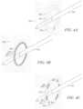
- the image guidance systemcan draw a number of rings about the axis of the medical device shaft, extrapolated beyond its tip, as depicted in FIG. 4 .
- a healthcare providercan view and manipulate the emplacement of the medical device 242 and its expected drive projection (via its displayed projected trajectory) before it enters the patients tissue. In some embodiments, this is accomplished by the doctor positioning the virtual rings in the drive projection such that they are co-incident (or pass through) the ultrasound representation of a target, such as a tumor that the doctor has spotted in the ultrasound. This can allow the healthcare provider to verify that the medical device 242 is properly aimed at the target and can drive the medical device 242 forward into the tissue such that it reaches its desired target or destination. For example, if the doctor identifies a tumor 212 in the ultrasound image, she can align the ablation needle 242 such that the drive projection rings on display 220 intersect or otherwise indicate that the medical device, if driven straight, will reach the tumor 212 .
- the ringscan, in some embodiments, be spaced at regular (e.g., 0.5, 1, or 2 cm) intervals to provide the healthcare provider with visual or guidance cues regarding the distance from the medical device tip to the targeted anatomy.
- the spacing of the ringscan indicate other aspects of the data, such as the drive speed of the medical device, the density of the tissue, the distance to a landmark, such as the ultrasound data, or any other appropriate guidance data or property.
- the rings or other trajectory indicatorscan extend beyond the medical device tip, by a distance equal to the length of the medical device-shaft. This way, the user knows if the medical device is long enough to reach the target—even before the tip enters the patient. That is, in some embodiments, if the rings do not reach the target with the tip still outside the body, then the tip will not reach the target even when the entire length shaft is inserted into the body.
- Other display markerscan be used to show trajectory, such as a dashed, dotted, or solid line, transparent medical device shaft, point cloud, wire frame, etc.
- three-dimensional ringscan be used and provide depth cues and obscure little of the ultrasound image.
- Virtual rings or other virtual markerscan be displayed semi-transparently, so that they obscure less of the ultrasound image than an opaque marker would.
- Other prediction informationcan also be displayed as image guidance cues. For example, if a scalpel is being tracked by the image guidance system, then a cutting plane corresponding to the scalpel can be displayed (not pictured). Such a cutting plan can be coplanar with the blade of the scalpel and can project from the blade of the scalpel. For example, the projected cutting plane can show where the scalpel would cut if the doctor were to advance the scalpel. Similar prediction information can be estimable or determinable for cauterizers, lasers, and numerous other surgical instruments.
- the data from two or more devicescan be combined and displayed based on their relative emplacements or poses.
- the system 100can determine the emplacement of an image plane based on the emplacement information of the ultrasound probe 222 .
- the rendered ultrasound image 204can be displayed on the image plane with respect to the virtual medical device 202 on the display 220 in a manner that estimates the relative emplacements or poses of an ultrasound probe 222 and the medical device 242 .
- the image guidance cues associated with the virtual medical device 202including the affected region indicator 206 and trajectory indicators 208 , are shown spatially located with the rendered ultrasound image 204 on display 220 .
- the display 220can include another image guidance cue in the form of an intersection indicator 210 that indicates where the virtual ablation medical device 202 (and/or its trajectory) intersects the ultrasound image 204 .
- the intersection indicator 210can be displayed before the medical device is inserted, thereby allowing the healthcare provider to see where the medical device will intersect the image, or imaged area.
- a tumor 212appears in the ultrasound image, or rendered ultrasound image 204 , and the virtual ablation needle 202 is shown driven through the tumor 212 .
- the displayed affected region (or affected region indicator) 206can indicate what region or volume would be affected when the medical device 242 is operated.
- the displayed affected region 206can estimate where ablation would occur if the tissue were ablated at that time.
- the displayed affected region 206appears to cover the tumor displayed in the ultrasound image.
- Various embodimentscan include any combinations of the graphics described above and/or other graphics or image guidance cues.
- data related to a single surgical instrumentsuch as an ablation needle, ultrasound probe, etc.
- a single surgical instrumentcan be presented in more than one manner on a single display.
- device 242is an ablation needle and device 222 is an ultrasound transducer.
- the image guidance systemcan track the healthcare provider's head using an emplacement sensor and/or a position sensing unit. In some embodiments, such as, when the head of a user is tracked, the healthcare provider can then move her head to the side, so that she sees the ultrasound image from a different point of view location.
- the image guidance systemcan constantly display an additional 2D view 205 of the ultrasound image, simultaneous to the 3D depiction 204 , so that the ultrasound image is always visible, regardless of the emplacement in which the healthcare provider holds the transducer 222 .
- the 2D view 205 of the ultrasound datacan be similar to what a healthcare provider is accustomed to seeing with traditional ultrasound displays. This can be useful to provide the healthcare provider with imaging to which she is accustomed and allows a healthcare provider to see the ultrasound data regardless of the then-current emplacement of the ultrasound probe with respect to the user.
- the 2D view 205 of an ultrasound imageis depicted in the upper right corner of the monitor (though it can be placed in any location).
- the guidance systemcan automatically (and continually) choose a corner in which to render the 2D view 205 of the ultrasound image, based on the 3D position of the surgical instruments in the rendered scene.
- ablation needle 242can be held in the healthcare provider's left hand and the medical device shaft is to the left of the 3D view of the ultrasound image slice, so that the 2D view 202 of the ultrasound image in the upper right corner of display 220 does not cover any of the 3D features of the medical device (or vice-versa).
- the systemcan automatically move it to a corner that would not otherwise be occupied by graphics or data.
- the systemattempts to avoid having the 2D view 205 of the ultrasound image quickly moving among corners of the display in order to avoid overlapping with graphics and data in the display.
- a function fcan be used to determine which corner is most suitable for the 2D ultrasound image to be drawn in.
- the inputs to fcan include the locations, in the screen coordinate system, of the displayed medical device tip, the corners of the 3D view of the ultrasound image, etc.
- f's output for any given point in timeis independent of f's output in the previous frames, which can cause the ultrasound image to move among corners of the display rapidly.
- the image guidance systemwill filter f's output over time.
- the output of a filter gcould be the corner, which has been output by f the most number of times over the last n frames, possibly weighting the most recent values for f most heavily.
- the output of the filter gcan be used to determine in which corner of display 220 to display the 2D ultrasound image and the temporal filtering provided by g can allow the 2D view 205 of the ultrasound image display to move more smoothly among the corners of the display 220 .
- orientation indicator 214an indication of the distance between the medical device's tip and the point in the plane of the ultrasound image that is closest to the medical device tip; the cross section or outline of the ablation volume that intersects with the ultrasound slice; and/or the intersection point, box, outline, etc. between the medical device's axis and the ultrasound image plane.
- the system 100can generate and/or display graphical indicators that help indicate the spatial relationship between a medical device and an ultrasound image plane (e.g., graphical image plane indicators) or other plane (e.g., graphical plane indicators), indicators to indicate the relative positions of the medical device(s) and ultrasound image, features of interest, annotations, foundational plane indicators, foundational plane intersection indicators, other graphical indicators, approximate medical device location indicators, etc.
- an ultrasound image planee.g., graphical image plane indicators
- other planee.g., graphical plane indicators
- the various image guidance cuescan be generated based at least in part on the emplacement information of the medical devices used with the system 100 .
- Embodiments of the systemcan include image guidance cues as part of the image guidance data to depict information related to the region or regions that will be affected by the use of surgical instruments.
- an image guidance cue displayed by the image guidance systemcan include affected region information.
- the illustrated embodiment of FIG. 2shows a virtual ablation needle 202 , which has a darkened portion 203 that indicates where the radio frequency or microwave energy for ablation will be emitted, and a displayed affected region 206 showing the volume that will be, or is being, ablated.
- the systemcan use the operating parameters of the medical device 242 and/or measured parameters to determine the affected region (and display the displayed affected region 206 ).
- the affected region's approximate sizee.g., girth and length
- the medical device make and modele.g., the medical device make and model
- power and duration settings of the medical devicee.g., microwave or radio frequency generator for ablation needles, etc.
- the systemcan use measured parameters to determine the affected region, such as, but not limited to, measured or estimated temperature, impedance of surrounding tissue.
- the measured parameterscan be received in real-time as real-time data.
- the systemcan use one or more a formulas, a look-up-tables, fixed or default values, or any other appropriate available information, etc. to determine the affected region.
- the systemcan determine affected regions prior to operating the medical device and/or during operation of the medical device. For example, prior to operating the medical device, the system can determine one or more predicted affected regions and/or during operation of the medical device, the system can determine one or more dynamic affected regions. In some embodiments, the predicted affected regions can be static during operation of the medical device and the dynamic affected regions can change over time. In certain embodiments, the system may rely more on operating parameters of the medical device to determine the predicted affected regions and measured parameters to determine the dynamic affected regions. However, it will be understood that operating parameters and/or measured parameters can be used to determine the predicted affected regions and/or the dynamic affected regions
- the operating parameters, measured parameters, formulas, a look-up-tables, fixed or default values, or other information used to determine the affected regionsmay include some amount of error or variance.
- the variancemay be due to uncertainty regarding the tissue that will be presented for ablation, a manufacturers indication that impedance, temperature, power, etc., can vary between tissue and/or medical devices, etc.
- the operating parameters or other datacan include one or more variance parameters indicating the amount of variance that a healthcare provider can expect when using a particular medical device.
- the variance parametermay account for all possible outcomes or a significant portion of possible outcomes (non-limiting examples: 95% or 99%).
- the variance parametercan indicate that a medical device operates within a certain range, or that a healthcare provider can expect a certain volume to be affected with a particular standard deviation and/or +/ ⁇ some percent.
- the variance parametermay indicate that, when operating for a particular amount of time, an ablation needle will ablate a certain range of tissue, or that a certain amount of tissue will be ablated with a particular standard deviation and/or +/ ⁇ some percent.
- the systemcan determine multiple affected regions based at least in part on the variance parameter. For example, the system can determine two affected regions using the extrema of the variance parameter. In some cases, such as when the variance parameter includes a lower threshold and a higher threshold, the system can determine two affected regions using the lower threshold and the higher threshold, respectively.
- the affected regionscan be predicted affected regions and/or dynamic affected regions depending on when and how the system determines them.
- a third affected regionscan be determined. The third affected region can be determined using a third point in the range, such as the midpoint, average, or other point.
- the systemcan also determine the emplacement of the affected region.
- the emplacement of the affected regioncan be based at least in part on the emplacement of some or all of the corresponding medical device (or virtual medical device), such as medical device 242 in FIG. 2 .
- the systemcan receive emplacement data from one or more emplacement sensors associated with the medical device 242 (non-limiting examples: coupled to or integrated with the medical device 242 , within an optical path of the medical device, etc.).
- the systemcan use the emplacement data to determine the emplacement of the tracked medical device and/or the emplacement of the virtual medical device 202 corresponding to the medical device 242 .
- the systemcan determine the emplacement of the medical device 242 and/or virtual medical device 202 with respect to a point-of-view location.
- the emplacement of the ablation volumecan be based at least in part on the emplacement of the ablation needle (or its rendered version) or at least a portion of it, such as the location on the ablation needle where the ablation energy will be emitted.
- the affected regioncan be centered at a location on the medical device, such as the location on the medical device that affects the surrounding tissue (non-limiting examples: microwave emitter, laser source output, etc.).
- the posedcan be based at least in part on the emplacement of the medical devices.
- the systemcan display the affected region in a variety of ways. Furthermore, although the illustrated embodiment of FIG. 2 includes only one displayed affected region 206 , it will be understood that one or more affected regions can be displayed corresponding to each medical device 242 that is displayed on the screen 220 and/or multiple affected regions can be displayed corresponding to a single medical device 242 . In some embodiments, the system can display a perspective view of the affected region and/or non-perspective view, such as by displaying the affected region on or with the ultrasound image displayed in the 2D view. Some or all of the affected regions can be displayed as desired. In some embodiments, the portion of the affected region that is displayed can be referred to as the displayed affected region and/or the surface display region.
- the expected volume of ablated tissueis neither spherical nor centered at the tip of the medical device. Accordingly, in such embodiments, the affected regions can match expected volumes.
- a Covidien surgical microwave medical devicehas an ellipsoidal ablation volume
- a Covidien Evident transcutaneous microwave medical devicehas a teardrop-like ablation volume
- RFA Medical's bipolar ablation systemuses two medical devices simultaneously, where each medical device has paddles that deploy after the medical device is inserted inside the tissue (which one can equate to a canoe's oar).
- the affected region for such a medical devicecorresponds to a volume that lies directly between the paddles of the two medical devices.
- the affected regioncan correspond to a variety of medical procedures. For example, if a cauterizer is tracked as part of an image guidance system, then the affected region can correspond to a cauterization volume. If a laser is tracked as part of the image guidance system, then the affected region can correspond to the projected laser path. Similarly, the affected region can correspond to a biopsy volume, an electroporated volume, cryoablation volume, laser ablation volume, high-frequency focused ultrasound ablation (HIFU) volume, external beam radiation therapy volume, and drilling volume (where the display volume corresponds to the region of bone and other tissue that the manually operated, or computer-controlled drill would remove), depending on the type of medical instrument being used.
- HIFUhigh-frequency focused ultrasound ablation
- the systemcan display the affected regions as a transparent volume, a wireframe volume (as depicted in FIG. 2 ), a volume with varying opacity, a point cloud of various densities, a surface, an outline, or any portion thereof.
- the affected regionscan correspond to predicted affected regions and/or dynamic affected regions, as desired.
- the systemcan display the portions of the volume that are located in front of the image slice with respect to the point-of-view location (or display them differently than portions that are behind the image slice), alternating bands or tiles of the affected region, portions of the affected region that are co-located with or intersect the medical device and/or the image slice.
- the systemcan display them in any combination as described above.
- the displayed affected regioncan include the portions of the second affected region that are unique second affected region with respect to the first affected region only, or in combination with other portions.
- the systemcan display the overlapping portions only, or in combination with other portions.
- the systemcan display the affected regions and other displayed features differently.
- the systemcan vary the characteristics of the affected regions (non-limiting examples: portions closer to the outline or edge of the affected regions can be more/less opaque, bright or focused, distal portions of the affected regions can be more/less opaque, bright or focused).
- the systemcan vary the characteristics of the other displayed features.
- the systemcan use different display settings for different portions of an image slice. For example, the system can display portions of the image slice within a first affected region using a first setting, portions of the image slice within a second affected region using a second setting, and portions of the image slice outside the first and second affected regions using a third setting.
- the different settingscan correspond to different opacity levels, brightness levels, contrast levels, and/or focus levels, etc.
- portions of the image slice outside the first and second affected regionscan be darkened, blurred, or otherwise adjusted to provide the healthcare provider with additional insight regarding the portions of the image slice that are within the affected region(s).
- FIGS. 3 A- 3 Jare diagrams illustrating various non-limiting embodiments for displaying a perspective view of at least a portion of the affected regions as a volume.
- FIG. 3 Aillustrates an embodiment in which two affected regions are displayed as two transparent volumes 302 , 304 and/or outlines along with a virtual medical device 301 .
- FIG. 3 Billustrates an embodiment in which two affected regions are displayed as two volumes 306 , 308 with varying opacity (or as semi-transparent).
- the volumes 306 , 308are more opaque near the edges, however, it will be understood that the opacity can be varied throughout the volumes 306 , 308 as desired.
- FIG. 3 Cillustrates an embodiment in which two affected regions are displayed as two volumes 310 , 312 with a surface texture.
- FIG. 3 Dillustrates an embodiment in which a portion of the affected region that is located between the image slice 314 and the point-of-view location (or in front of the image slice 314 ) is displayed as a volume 316 .
- the portion of the affected region that is located between the image slice 314 and the point-of-view locationcan be displayed differently from portions of the affected region that is located distally from the point-of-view location with respect to the image slice 314 .
- the different portionscan be displayed with different colors, brightness, sharpness, etc.
- the portions of the affected region located distally from the point-of-view location with respect to the image slice 314can be displayed with lighter or more faded colors, etc.
- FIG. 3 Eillustrates an embodiment in which portions of a second affected region are displayed.
- the portions displayedcorrespond to the portions of the second affected region that are unique to the second affected region with respect to the first affected region.
- the portionsare displayed as spikes 318 .
- the spikes 318can be a predefined length and/or have marking as predefined lengths, such as 1 cm. As such, user can use the spikes to determine distances of object displayed on the screen.
- the opacity of the spikescan vary 318 as desired.
- the systemcan cause the spikes 318 to become transparent, or otherwise adjust a display setting, based at least in part on the location of the dynamic affected region with respect to the spikes 318 .
- FIG. 3 Fillustrates an embodiment in which the embodiments from FIGS. 3 B and 3 E are combined. As mentioned previously, any combination of the described embodiments can be used as desired.
- FIG. 3 Gillustrates embodiments in which portions of three affected regions are displayed as outlines 320 a , 320 b , 322 a , 322 b , 324 a , 324 b .
- outlines 320 a , 320 bcorrespond to vertical and horizontal edges, respectively, of a first affected region
- outlines 322 a , 322 bcorrespond to vertical and horizontal edges, respectively, of a second affected region
- outlines 324 a , 324 bcorrespond to vertical and horizontal edges, respectively, of a third affected region.
- 3 Galso illustrates embodiments in which the one or more outlines 320 a , 320 b , 322 a , 322 b , 324 a , 324 c are associated with a particular affected region, such as portions of a second affected region that are unique to it with respect to a first affected region.
- FIG. 3 Hillustrates an embodiment in which the two affected regions are displayed as two volumes 326 , 328 , in which the portions of the second affected region that are unique to the second affected region with respect to the first affected region are displayed differently (non-limiting examples: varying opacity, color, brightness, focus, etc.).
- FIG. 3 Iillustrates embodiments in which portions of an affected region are displayed using horizontal bands 330 with alternating display settings (e.g., transparency, brightness, etc.). It will be understood that bands with any orientation can be used. Bands with higher transparency levels can enable a healthcare provider to see into the affected region.
- the width and/or the arc length of each bandcan be equal, and a top portion 332 and a bottom portion 334 can have a variable corresponding width/arc length. As such, user can use the bands to determine distances of object displayed on the screen.
- the variable width/arc lengthcan be determined such that the bands 330 have an equal width/arc length.
- FIG. 3 Jillustrates an embodiment in which portions of an affected region are displayed using tiles 336 having alternating display settings (e.g., transparency, brightness, etc.).
- each tile 336can be displayed with a different display level.
- every other tilecan be displayed with the same display level.
- Tiles with higher transparency levelscan enable a healthcare provider to see into the affected region.
- horizontal and/or vertical lengths and/or the arc length of each tilecan be equal, and a top portion and a bottom portion can have a variable corresponding length. The variable length can be determined such that the tiles 336 have an equal length.
- FIGS. 4 A- 4 C, and 5 A- 5 Care diagrams showing various embodiments for displaying at least a portion of the affected regions that intersect with or are co-located or level with at least a portion of a medical display object (non-limiting examples: the image slice 302 ( FIGS. 4 A- 4 C ) or the virtual medical device ( FIGS. 5 A and 5 B )), which may also be referred to herein as surface display regions.
- a medical display objectnon-limiting examples: the image slice 302 ( FIGS. 4 A- 4 C ) or the virtual medical device ( FIGS. 5 A and 5 B )
- the surface display regionscan correspond to predicted affected regions and/or dynamic affected regions, as desired. Accordingly, if the first and/or second affected regions are dynamic affected regions, the associated surface display regions can move, or grow, during operation of the medical device associated with the virtual medical device 401 , 501 .
- the surface display regionscan correspond to affected regions that are co-located with the medical display object or only portions thereof, or the medical display object's trajectory. Accordingly, in some embodiments, the surface display region can be displayed as a volume, area, or line depending on which portions of the affected region and medical display object are used to determine the surface display region.
- the system 100can compare the coordinates of the portion of the affected region with the portion of the medical display object. If the coordinates (e.g., the x, y, z coordinates) match (e.g., are equal) or satisfy a distance threshold, the system can determine that the portion of the medical display object and the portion of the affected region are co-located. In certain embodiments, the system 100 can determine that the portion of the affected region and the portion of the medical display object are co-located if the portion of the affected region and the portion of the medical display object can be mapped to the same pixel in a video or image output data buffer.
- the coordinatese.g., the x, y, z coordinates
- the distance thresholdcan be a predefined distance, such as one or more bits, one or more pixels, etc. In some embodiments, the distance threshold can be based at least in part on whether the distance between the coordinates is perceptible to a user, which may be based at least in part on the size of the display, the size of the display relative to the image and/or imaged area, and/or the distance between the point-of-view location and the display, etc. For example, in some cases, the distance threshold can be smaller for larger displays (or larger display:image ratios) and larger for smaller displays (or smaller display:image ratios), or vice versa. In certain cases, the distance threshold can be larger for larger distances between the point-of-view location and the display and smaller for smaller distances between the point-of-view location and the display, or vice versa. In certain embodiments, the distance threshold can be different for each coordinate.
- the system 100can perform the comparison for each location of the medical display objects and/or each location of the affected regions. In some cases, the system can determine that the portion of the medical display object and the portion of the affected region are co-located if the portion of the medical display object and the portion of the affected region are level and have the same depth.
- Any coordinate systemcan be used to compare the coordinates of the portion of the affected region with the medical display object and/or to determine whether the portion of the affected region is co-located with the medical display object.
- the coordinate system of the display and/or the coordinate system of device in the system 100 that is used to determine the emplacement of the medical devicescan be used, as desired.
- the coordinate system of the displayis used.
- the coordinate system of the displaycan be any emplacement as desired.
- the coordinates of the displayare that the x-axis is the width of the display, the y-axis is the height of the display, and the z-axis is the depth (e.g., into and out of) the display.
- the system 100can determine that the portion of the affected region satisfies the location threshold and/or is level with the medical display object, based at least in part on the x and y coordinates of the affected region and the x and y coordinates of the medical display object. For example, if the x and y coordinates of the affected region and the x and y coordinates of the medical display object match (or satisfy a distance threshold); the system 100 can determine that the portion of the affected region satisfies the location threshold.
- the coordinates used to determine whether the portion of the affected region satisfies the location threshold and/or is co-located with the medical display objectcan be based at least in part on the coordinate system used.
- the coordinate system usedcan include the x-axis as the depth (e.g., forward/backward), the y-axis as lateral movement (e.g., side-to-side), and the z-axis as elevation (e.g., up/down).
- the system 100can determine that portion of the affected region satisfies the location threshold if the y and z coordinates of the affected region match (or satisfy a distance threshold) the y and z coordinates of the medical display object.
- the systemcan query whether a portion of the medical display object and/or a portion of the affected region have been (or will be) mapped to that location. If the system 100 determines that a portion of the medical display object and a portion of the affected region have been (or will be) mapped to that location, the system 100 can determine that the portion of the medical display object and the portion of the affected region are co-located.
- the system 100can determine that the portion of the affected region satisfies the location threshold, intersects, and/or is co-located with the medical display object if the portion of the affected region and the medical display object (or portion of the image corresponding to the medical display object) are co-located when mapped to a 2D plane.
- the 2D planecan be based at least in part on the point-of-view location.
- the 2D planecan be orthogonal to the point-of-view location.
- the system 100can determine that the portion of the affected region satisfies the location threshold (or corresponding virtual affected region) if the portion of the affected region overlaps with the medical display object (or portion of the image corresponding to the medical display object) in a virtual image (e.g., one is directly in front of or behind the other in the virtual image). In certain embodiments, the system 100 can determine that the portion of the affected region satisfies the location threshold if the portion of the affected region and the medical display object (or portion of the image corresponding to the medical display object) map to the same location on a display, such as the same pixel or same array of pixels.
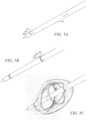
- FIGS. 4 A- 4 Cvarious embodiments of surface display regions are shown. It will be understood that the embodiments illustrated in FIGS. 4 A- 4 C are non-limiting in nature. Furthermore, any portion of any of the embodiments described above with reference to FIGS. 3 A- 3 J can be displayed in conjunction with any portion of the embodiments described below with reference to FIGS. 4 A- 4 C .
- FIG. 4 Aillustrates various embodiments including embodiments showing two surface display regions as lines 402 , 404 .
- the line 402can correspond to an outline of a first affected region that is co-located with at least a portion of a virtual medical device (image slice 406 ) and the line 404 can correspond to an outline of a second affected region that is co-located at least a portion of the image slice 406 .
- the area between the lines 402 , 404can be displayed differently (non-limiting examples: varying opacity, color, brightness, focus, etc.).
- FIG. 4 Acan also illustrate embodiments showing a surface display region displayed as area 405 .
- the area 405can correspond to portions of a second affected region that are unique to it with respect to a first affected region and that are co-located with at least a portion of the image slice 406 .
- the portions of the area 405can be displayed differently (non-limiting examples: varying opacity, color, brightness, focus, etc.).
- FIG. 5 Ais similar to FIG. 4 A except that the medical display object is the virtual medical device 501 .
- lines 502 , 504correspond to lines 402 , 404 , respectively
- area 505corresponds to area 405 .
- lines 502 , 505 , and/or area 505can correspond to portions of a second affected region that are co-located with at least a portion of the virtual medical device 501 and/or its trajectory.
- FIG. 4 Billustrates various embodiments showing multiple solid lines 408 , 410 and dashed lines 412 , 414 , which can form part of one or more surface display regions.
- lines 408 , 410can correspond different portions of a first affected region (and form part of a first surface display region) and lines 412 , 414 can correspond to different portions of a second affected region (and form part of a surface display region).
- line 408forms part of a first surface display region and lines 410 , 412 , 414 form part of a second surface display region, such as portions of the second affected region that are unique to it with respect to the first affected region.
- lines 408 , 410 , 412 , 414form part of four surface display regions.
- FIG. 5 Ais similar to FIG. 4 A except that the medical display object is the virtual medical device 501 .
- lines 508 , 510 , 512 , 514correspond to lines 408 , 410 , 412 , 414 , respectively, and area 505 corresponds to area 405 .
- FIG. 4 Cillustrates an embodiment in which an additional surface display region is displayed with the surface display regions illustrated in FIG. 4 B .
- line 408forms part of a first surface display region that corresponds to a first predicted affected region and lines 410 , 412 , 414 form part of a second surface display region that corresponds to a second predicted affected region.
- Indicators 416form part of a third surface display region, which corresponds to a dynamic affected region.
- lines 408 , 410 , 412 , and 414can remain static, while indicators 416 move outward. In this manner, a healthcare provider can determine at what point to terminate operation of the medical device.
- a corresponding FIG. 5is not provided, it will be understood that the medical display object can correspond to any object displayed by a display.
- FIG. 5 Cis a diagram illustrating an embodiment in which the embodiments described above with reference to FIGS. 3 D, 3 G, 4 C, and 5 B are combined.
- FIG. 6is a flow diagram illustrative of an embodiment of a routine 600 implemented by the system 100 to display at least a portion of an affected region, or displayed affected region.
- routine 600can be implemented by one or more computing devices/components that are associated with the system 100 , such as the position sensing unit 140 , the image guidance unit 130 , surgical system 149 , and/or imaging unit 150 . Accordingly, routine 600 has been logically associated as being generally performed by the system 100 . However, the following illustrative embodiment should not be construed as limiting.
- the various blocks described herein with reference to FIG. 6can be implemented in a variety of orders. For example, the system may implement some blocks concurrently or change the order, as desired.
- the system 100receives operating parameters of a medical device.
- the operating parameterscan include information regarding make and model, power and duration settings of the medical device, and/or variance parameters, etc.
- the operating parameterscan be stored in a non-transitory, computer-readable medium associated with the system 100 and/or can be stored in the medical device.
- the system 100determines a first affected region.
- affected regionscan correspond to predicted affected regions and/or a dynamic affected regions.
- the systemdetermines the first affected region based at least in part on the operating parameters and/or measured parameters.
- the system 100determines the first affected region based at least in part on a variance parameter of the medical device.
- the system 100causes one or more displays to display at least a portion of the first affected region, or first displayed affected region.
- the first displayed affected regioncan be displayed in a 2D view or 3D view and/or as a perspective view.
- the displayed affected regioncan be displayed as a volume, area, and/or line.
- the displayed affected regioncan be wire-framed, transparent, semi-transparent, have varied opacity, brightness, and/or focus, include alternating bands/tiles, be textured, include solid or dashed lines, include spikes, etc.
- the at least a portion of the first affected regioncorresponds to portions of the first affected region that are unique to it, with respect to other affected regions.
- the displayed affected regioncorresponds to at least a portion of the first affected region that is co-located with at least a portion of a medical display object (non-limiting examples: a virtual medical device, image slice, etc.) or its trajectory, also referred to as the surface display region.
- a medical display objectnon-limiting examples: a virtual medical device, image slice, etc.
- its trajectoryalso referred to as the surface display region.
- routine 600it will be understood that fewer, more, or different blocks can be used as part of the routine 600 .
- any combination of blocks 608 , 610 , 612 , and 614can be included as part of routine 600 .
- the system 100determines a second affected region.
- the second affected regioncan be determined in a manner similar to the first affected region.
- the variance parametercan be used to determine the first and second affected regions.
- a first variance thresholdcan be used to determine the first affected region and a second variance threshold can be used to determine the second affected region.
- the first variance thresholdcan be less than the second variance threshold.
- the second affected regioncan be larger than, and in some cases include, the first affected region.
- the system 100causes one or more displays to display at least a portion of the second affected region, or second displayed affected region.
- the system 100can cause the one or more displays to display the second displayed affected region similar to the first displayed affected region.
- the second displayed affected regioncan be displayed differently, such as by using a different color, transparency level, focus setting, shape, texture, etc.
- the systemcan omit causing the display of the first displayed affected region in favor of the second displayed affected region.
- the second displayed affected regioncan, in some instances, correspond to the portions of the second affected region that are unique to the second affected region, with respect to the first affected region.
- the first affected regioncan be a predicted affected region and the second affected region can be a dynamic affected region.
- the second displayed affected regioncan change and/or grow with respect to the first displayed affected region.
- the system 100determines a third affected region.
- the first and second affected regionscan be first and second predicted affected regions and the third affected region can be a dynamic affected region.
- the three affected regionscan be dynamic affected regions, predicted affected regions, or any combination thereof.
- the system 100causes one or more displays to display at least a portion of the third affected region, or third displayed affected region.
- the system 100can cause the one or more displays to display the third displayed affected region similar to the first and second displayed affected regions.
- the third displayed affected regioncan be displayed differently, such as by using a different color, transparency level, focus setting, shape, texture, etc.
- the third displayed affected regioncan move with respect to the first and second displayed affected regions.
- the system 100can receive emplacement data from one or more sensors corresponding to one or more medical devices, determine a emplacement of the medical devices and/or corresponding virtual medical devices (as a non-limiting example, the emplacement can be determined based at least in part on a point-of-view location), cause one or more displays to display the virtual medical devices and/or perspective views thereof, determine emplacement of the affected regions with respect to the virtual medical devices, determine emplacement of and display an image slice, alter the display of the image slice, display the medical display objects in a 2D view, a 3D view, and/or a perspective view, etc.
- FIG. 7is a flow diagram illustrative of an embodiment of a routine 700 implemented by the system 100 to display at least a portion of multiple affected regions, or multiple displayed affected regions.
- routine 700can be implemented by one or more computing devices/components that are associated with the system 100 , such as the position sensing unit 140 , the image guidance unit 130 , surgical system 149 , and/or imaging unit 150 . Accordingly, routine 700 has been logically associated as being generally performed by the system 100 . However, the following illustrative embodiment should not be construed as limiting.
- the various blocks described herein with reference to FIG. 7can be implemented in a variety of orders. For example, the system may implement some blocks concurrently or change the order, as desired.
- the system 100determines the emplacement of a virtual medical device.
- the system 100can determine the emplacement of the virtual medical device based at least in part on emplacement data corresponding to a medical device, such as medical device 242 .
- the emplacement datacan be received from one or more emplacement sensors associated with the medical device.
- the system 100obtains operating parameters of the medical device, as described in greater detail above with reference to block 602 of FIG. 6 .
- the system 100determines a first affected region. In some embodiments, the system 100 can determine the first affected region similar to the determination of the first affected region referenced above with respect to block 604 of FIG. 6 .
- the system 100determines a second affected region. In some embodiments, the system 100 can determine the second affected region similar to the determination of the second affected region referenced above with respect to block 608 of FIG. 6 .
- the first and second affected regionscan be predicted affected regions and/or dynamic affected regions, as desired.
- the system 100can determine the emplacement of the first and second affected regions.
- the system 100can cause one or more displays to display at least a portion of the virtual medical device, at least a portion of the first affected region (second displayed affected region), and at least a portion of the second affected region (second displayed affected region).
- the displayed affected regionscan, in some embodiments, correspond to surface display regions.
- the system 100can cause the one or more displays to display at least a portion of the first and second affected regions similar to blocks 606 and 610 of FIG. 6 , described previously.
- the first and second displayed affected regionscan be displayed in a variety of ways.
- routine 700can be used as part of routine 700 .
- routine 700can be included as part of routine 700 .
- the system 100can determine a third affected region, and at block 716 , the system 100 can cause the one or more displays to display at least a portion of the third affected region.
- the system 100can determine the third affected region and display a third displayed affected region similar to the first and second displayed affected regions as described in greater detail above with reference to blocks 612 and 616 of FIG. 6 .
- the third displayed affected regioncan be a surface display region.
- the system 100can determine an emplacement of an image slice, and at block 720 , the system 100 can cause the one or more displays to display at least a portion of the image slice and/or perspective view thereof, as described in greater detail above.
- the system 100can receive emplacement data from one or more sensors associated with one or more medical devices, display additional displayed affected regions, alter the display of the image slice, display image guidance cues, display the medical display objects in a 2D view, a 3D view, and/or a perspective view, etc.
- DSPdigital signal processor
- ASICapplication specific integrated circuit
- FPGAfield programmable gate array
- a processorcan be a microprocessor, but in the alternative, the processor can be any conventional processor, controller, or microcontroller.
- a processorcan also be implemented as a combination of computing devices, e.g., a combination of a DSP and a microprocessor, a plurality of microprocessors, one or more microprocessors in conjunction with a DSP core, or any other such configuration.
- a software modulecan be stored in RAM memory, flash memory, ROM memory, EPROM memory, EEPROM memory, registers, hard disk, a removable disk, a CD-ROM, or any other form of non-transitory computer-readable medium known in the art, as computer-executable instructions.
- An exemplary computer-readable storage mediumis coupled to the processor such the processor can read information and/or computer-executable instructions from, and write information to, the computer-readable storage medium.
- the storage mediumcan be integral to the processor.
- the processor and the storage mediumcan reside in an ASIC.
- the ASICcan reside in a user terminal, camera, or other device.
- the processor and the storage mediumcan reside as discrete components in a user terminal, camera, or other device.
- a device configured toare intended to include one or more recited devices. Such one or more recited devices can also be collectively configured to carry out the stated recitations.
- a processor configured to carry out recitations A, B and Ccan include a first processor configured to carry out recitation A working in conjunction with a second processor configured to carry out recitations B and C.
Landscapes
- Engineering & Computer Science (AREA)
- Health & Medical Sciences (AREA)
- Life Sciences & Earth Sciences (AREA)
- Surgery (AREA)
- Theoretical Computer Science (AREA)
- Physics & Mathematics (AREA)
- Computer Graphics (AREA)
- General Physics & Mathematics (AREA)
- Computer Hardware Design (AREA)
- General Engineering & Computer Science (AREA)
- Software Systems (AREA)
- Robotics (AREA)
- Molecular Biology (AREA)
- Veterinary Medicine (AREA)
- Public Health (AREA)
- Biomedical Technology (AREA)
- Heart & Thoracic Surgery (AREA)
- Medical Informatics (AREA)
- Nuclear Medicine, Radiotherapy & Molecular Imaging (AREA)
- Animal Behavior & Ethology (AREA)
- General Health & Medical Sciences (AREA)
- Magnetic Resonance Imaging Apparatus (AREA)
- Apparatus For Radiation Diagnosis (AREA)
- Computing Systems (AREA)
- Geometry (AREA)
- Architecture (AREA)
Abstract
Description
The present application is a continuation of U.S. patent application Ser. No. 15/882,709, filed Jan. 29, 2018, entitled AFFECTED REGION DISPLAY BASED ON A VARIANCE PARAMETER ASSOCIATED WITH A MEDICAL DEVICE, which is a continuation of U.S. patent application Ser. No. 14/872,930, filed Oct. 1, 2015, entitled AFFECTED REGION DISPLAY ASSOCIATED WITH A MEDICAL DEVICE, which claims priority benefit to U.S. Provisional Application No. 62/059,077, filed Oct. 2, 2014, entitled ABLATION AREA VISUALIZATIONS, each of which is hereby incorporated herein by reference in its entirety. Any and all applications for which a foreign or domestic priority claim is identified in the Application Data Sheet as filed with the present application are incorporated by reference under 37 CFR 1.57 and made a part of this specification.
The systems and methods disclosed herein relate generally to computer systems facilitating medical device guidance through tissue by a medical practitioner.
Various medical device systems are available to aid a healthcare provider to guide a medical device in a patient. The medical device systems can provide various image guidance cues to aid the healthcare provider, and can also provide views of images of an imaged area and of virtual medical devices corresponding to physical medical devices.
Implementations disclosed herein provide systems, methods, and apparatus for generating images facilitating medical device insertion into tissue by an operator. Certain embodiments pertain to a free-hand medical device guidance system. The system can provide the healthcare provider manual control over the medical device, while making the spatial relationships between the target, medical device and U/S image more intuitive via a visual display. Using this visual feedback, the operator can adjust the medical device's position, orientation, or trajectory. Certain of the contemplated embodiments can be used in conjunction with systems described in greater detail in U.S. patent application Ser. No. 13/014,587, filed Jan. 26, 2011, entitled SYSTEMS, METHODS, APPARATUSES, AND COMPUTER-READABLE MEDIA FOR IMAGE MANAGEMENT IN IMAGE-GUIDED MEDICAL PROCEDURES and U.S. patent application Ser. No. 13/753,274, filed Jan. 29, 2013, entitled MULTIPLE MEDICAL DEVICE GUIDANCE (the '274 application), and U.S. patent application Ser. No. 14/212,933, filed Mar. 14, 2014, entitled MEDICAL DEVICE GUIDANCE, each of which is hereby incorporated by reference in its entirety.
The system can aid the healthcare provider in guiding one or more medical devices through the tissue of the patient and/or placing the medical devices, and can be used for treatment of tumors, fibroids or cysts, with bipolar radiofrequency medical device ablation, multiple microwave medical devices, electroporation, and/or electrochemotherapy systems. It can also be used for nerve or muscle stimulation or sensing (electrodes in the spine, brain). The system can be used during open surgery, laparoscopic surgery, endoscopic procedures, biopsies, and/or interventional radiology procedures.
The system can be used in conjunction with live intraoperative ultrasound (U/S), pre-operative CT, or any cross-sectional medical imaging modality (e.g. MRI, OCT, etc.). In addition, the system can use a variety of techniques to determine the position and/or orientation of one or more medical devices. For example, the system can use the NDI Aurora magnetic system, NDI Polaris optical system, etc. In some embodiments, a position sensor can be embedded inside, or affixed to each medical device, at the tip, along the shaft, and/or on the handle. Sensors can be built into the medical devices or attached after manufacturing, as described in greater detail in U.S. application Ser. No. 14/212,184, filed Mar. 14, 2014, entitled SENSOR MOUNT, incorporated herein in its entirety.
Each medical device can be associated with one or more sensors, which can continually, or repeatedly, report position and/or orientation, or a single sensor can be used for all the medical devices. In embodiments where one sensor is used, the healthcare provider can attach the sensor to the particular medical device that she is intentionally repositioning, and then, once she has placed that medical device, she can remove the sensor and attach it to the next medical device she is repositioning. In some embodiments, the medical devices can be manipulated by the healthcare provider. In certain embodiments, the system can be used with a robotic manipulator, where the robot controls the medical devices.
In some embodiments, the handles of medical devices can have push-button switches, to allow the user to select a medical device, indicate a tissue target, etc. The handle can also have an indicator light to indicate to the users which medical device is selected. Finally, the handle can have an encoder to detect how much length of electrode has been exposed by the user, and report this information to the guidance system and therapeutic generator
Although only twosurgical instruments FIG.1 , it will be understood that additional surgical instruments can be tracked and associated data can be provided to theimage guidance unit 130. Theimage guidance unit 130 can process or combine the data and show image guidance data ondisplay 120. This image guidance data can be used by a healthcare provider to guide a procedure and improve care. There are numerous other possible embodiments ofsystem 100. For example, many of the depicted components can be joined together to form a single component and can be implemented in a single computer or machine. Further, additional position sensing units can be used in conjunction withposition sensing unit 140 to track all relevantsurgical instruments Additional imaging units 150 can be included, and combined imaging data from themultiple imaging units 150 can be processed byimage guidance unit 130 and shown ondisplay unit 120. Additionally, two or moresurgical systems 149 can also be included.
Information about and from multiplesurgical systems 149 and attached surgical instruments145 (and additional surgical instruments not shown) can be processed byimage guidance unit 130 and shown ondisplay 120. These and other possible embodiments are discussed in more detail below. It will be understood that any combination of the display objects, image guidance cues, etc., described herein can be displayed concurrently, or simultaneously. Further, reference to displaying objects “concurrently” and/or “simultaneously” is to be interpreted broadly and may refer to displaying objects in such a way that to a human observer the objects are visible at the same time.
In some embodiments,system 100 comprises adisplay unit 120 and aposition sensing unit 140 communicatively coupled to imageguidance unit 130. In some embodiments,position sensing unit 140,display unit 120, andimage guidance unit 130 are coupled to thestand 170.Image guidance unit 130 can be used to produceimages 125 that are displayed ondisplay unit 120. Theimages 125 produced ondisplay unit 120 by theimage guidance unit 130 can be determined based on ultrasound or other visual images from the firstsurgical instrument 145 and secondsurgical instrument 155. In the illustrated embodiment, theimages 125 includes a 2D viewing area and a 3D viewing area. The 2D viewing area includes a 2D view of each of anultrasound slice 121, a virtualmedical device 122 corresponding to the firstsurgical instrument 145, avirtual imaging device 123 corresponding to the secondsurgical instrument 155,surface display regions 124a,124b,intersection indicator 126, and trajectory and otherimage guidance cues 127. In the illustrated embodiment, the 3D viewing area includes perspective views of each of theimage slice 121, the virtualmedical device 122, a displayedaffected region 129 including thesurface display regions 124a,124b, thevirtual imaging device 123,intersection indicator 126, trajectory and otherimage guidance cues 127, and apatient orientation indicator 128. It will be understood that any combination of the aforementioned display objects can be displayed in the 2D view and/or 3D view as desired.
As a non-limiting example, if the firstsurgical instrument 145 is anablation needle 145 and the secondsurgical instrument 155 is anultrasound probe 155, thenimages 125 produced ondisplay 120 can include the images, or video, from the ultrasound probe155 (e.g., image slice121) combined with other medical display objects and image guidance cues, such as projected medical device drive (e.g., trajectory indicators127) or projected ablation volume (e.g., displayed affected region129), determined based on the emplacement ofablation needle 145. If the firstsurgical instrument 145 is anultrasound probe 145 and the secondsurgical instrument 155 is alaparoscopic camera 155, thenimages 125 produced ondisplay 120 can include the video from thelaparoscopic camera 155 combined with ultrasound data superimposed on the laparoscopic image. More surgical instruments can be added to the system. For example, the system can include an ultrasound probe, ablation needle, laparoscopic camera, stapler, cauterizer, scalpel and/or any other surgical instrument or medical device. The system can also process and/or display collected data, such as preoperative CT scans, X-Rays, MRIs, laser scanned 3D surfaces etc.
The term “emplacement” as used herein is a broad term and may refer to, without limitation, position and/or orientation or any other appropriate location information. The term “pose” as used herein is a broad term encompassing its plain and ordinary meaning and may refer to, without limitation, position and orientation or any other appropriate location information. In some embodiments, the imaging data obtained from one or both ofsurgical instruments surgical instruments surgical system 149 orimaging unit 150 can be attached to the correspondingmedical instruments
As noted above,images 125 produced can also be generated based on live, intraoperative, or real-time data obtained using the secondsurgical instrument 155, which is coupled tosecond imaging unit 150. The term “real time” as used herein is a broad term and has its ordinary and customary meaning, including without limitation instantaneously or nearly instantaneously. The use of the term real time can also mean that actions are performed or data is obtained with the intention to be used immediately, upon the next cycle of a system or control loop, or any other appropriate meaning. Additionally, as used herein, real-time data can be data that is obtained at a frequency that would allow a healthcare provider to meaningfully interact with the data during surgery. For example, in some embodiments, real-time data can be a medical image of a patient that is updated one time per second. In some embodiments, real-time data can be ultrasound data that is updated multiple times per second.
Thesurgical instruments surgical instruments position sensing unit 140 can be part ofimaging unit 150 or it can be separate. Theposition sensing unit 140 can be used to determine the emplacement of firstsurgical instrument 145 and/or the secondsurgical instrument 155. In some embodiments, theposition sensing unit 140 can include a magnetic tracker and/or one or more magnetic coils can be coupled tosurgical instruments 145 and/or155. In some embodiments, theposition sensing unit 140 can include an optical tracker and/or one or more visually-detectable fiducials can be coupled tosurgical instruments 145 and/or155. In some embodiments, theposition sensing unit 140 can be located below the patient. In such embodiments, theposition sensing unit 140 can be located on or below the table180. For example, in embodiments where theposition sensing unit 140 is a magnetic tracker, it can be mounted below the surgical table180. Such an arrangement can be useful when the tracking volume of theposition sensing unit 140 is dependent on the location of theposition sensing unit 140, as with many magnetic trackers. In some embodiments, magnetic tracking coils can be mounted in or on themedical devices
In some embodiments, theposition sensing unit 140 can be an electromagnetic measurement system (e.g., NDI Aurora system) using sensor coils for tracking units attached to the first and/or secondsurgical devices position sensing unit 140 can be an optical 3D tracking system using fiducials. Such optical 3D tracking systems can include the NDI Polaris Spectra, Vicra, Certus, PhaseSpace IMPULSE, Vicon MX, InterSense IS-900, NaturalPoint OptiTrack, Polhemus FastTrak, IsoTrak, or Claron MicronTracker2. In some embodiments, theposition sensing unit 140 can each be an inertial 3D tracking system comprising a compass, accelerometer, tilt sensor, and/or gyro, such as the InterSense InertiaCube or the Nintendo Wii controller. In some embodiments, theposition sensing unit 140 can be attached to or affixed on the correspondingsurgical device
In some embodiments, theposition sensing units 140, can include sensing devices such as the HiBall tracking system, a GPS device, or signal emitting device that would allow for tracking of the position and/or orientation (e.g., emplacement) of the tracking unit (also referred to as an emplacement sensor). In some embodiments, aposition sensing unit 140 can be affixed to either or both of thesurgical devices surgical devices position sensing unit 140. A room coordinate system reference, such as thedisplay 120 can also be tracked by theposition sensing unit 140 in order to determine the emplacements of thesurgical devices Devices
In some embodiments, theposition sensing unit 140 can be an Ascension Flock of Birds, Nest of Birds, driveBAY, medSAFE, trakSTAR, miniBIRD, MotionSTAR, pciBIRD, or Calypso 2D Localization System and tracking units attached to the first and/or secondmedical devices
The term “tracking unit” (also referred to as an emplacement sensor), as used herein, is a broad term encompassing its plain and ordinary meaning and includes without limitation all types of magnetic coils or other magnetic field sensing devices for use with magnetic trackers, fiducials or other optically detectable markers for use with optical trackers, such as those discussed above and below. In some embodiments, the tracking units can be implemented using optical position sensing devices, such as the HiBall tracking system and theposition sensing unit 140 can form part of the HiBall tracking system. Tracking units can also include a GPS device or signal emitting device that allows for tracking of the position and/or orientation of the tracking unit. In some embodiments, a signal emitting device might include a radio-frequency identifier (RFID). In such embodiments, theposition sensing unit 140 can use the GPS coordinates of the tracking units or can, for example, triangulate the radio frequency signal being emitted by the RFID associated with tracking units. The tracking systems can also include one or more 3D mice.
In some embodiments, thedisplay unit 120 displays 3D images to a user, such as a healthcare provider. Stereoscopic 3D displays separate the imagery shown to each of the user's eyes. This can be accomplished by a stereoscopic display, a lenticular auto-stereoscopic display, or any other appropriate type of display. Thedisplay 120 can be an alternating row or alternating column display. Example alternating row displays include the Miracube G240S, as well as Zalman Trimon Monitors. Alternating column displays include devices manufactured by Sharp, as well as many “auto-stereoscopic” displays (e.g., Philips). In some embodiments, Sony Panasonic 3D passive displays and LG, Samsung, and/or Vizio 3D TVs can be used as well.Display 120 can also be a cathode ray tube. Cathode Ray Tube (CRT) based devices, can use temporal sequencing, showing imagery for the left and right eye in temporal sequential alternation. This method can also be used projection-based devices, as well as by liquid crystal display (LCD) devices, light emitting diode (LED) devices, and/or organic LED (OLED) devices.
In certain embodiments, thedisplay unit 120 can be a head mounted display worn by the user in order to receive 3D images from theimage guidance unit 130. In such embodiments, a separate display, such as the pictureddisplay unit 120, can be omitted. The 3D graphics can be produced using underlying data models, stored in theimage guidance unit 130 and projected onto one or more 2D planes in order to create left and right eye images for a head mount, lenticular, or other 3D display. The underlying 3D model can be updated based on the relative emplacements of thevarious devices devices medical device 155 is an ultrasound probe, then the underlying data model can be updated to reflect the most recent ultrasound image. If the firstmedical device 145 is an ablation needle, then the underlying model can be updated to reflect any changes related to the needle, such as power or duration information. Any appropriate 3D graphics processing can be used for rendering including processing based on OpenGL, Direct3D, Java 3D, etc. Whole, partial, or modified 3D graphics packages can also be used, such packages including 3DS Max, SolidWorks, Maya, Form Z, Cybermotion 3D, VTK, Slicer, or any others. In some embodiments, various parts of the needed rendering can occur on traditional or specialized graphics hardware. The rendering can also occur on the general CPU, on programmable hardware, on a separate processor, be distributed over multiple processors, over multiple dedicated graphics cards, or using any other appropriate combination of hardware or technique.
One or more components, units, devices, or elements of various embodiments can be packaged and/or distributed as part of a kit. For example, in one embodiment, an ablation needle, one or more tracking units, 3D viewing glasses, and/or a portion of an ultrasound wand can form a kit. Other embodiments can have different elements or combinations of elements grouped and/or packaged together. Kits can be sold or distributed separately from or with the other portions of the system.
One will readily recognize that there are numerous other examples of image guidance systems which can use, incorporate, support, or provide for the techniques, methods, processes, and systems described herein.
It can often be difficult to discern the content of a 3D scene from a 2D depiction of it, or even from a 3D depiction of it. Therefore, various embodiments herein provide image guidance that can help the healthcare provider better understand the scene, relative emplacements or poses of object in the scene and thereby provide improved image guidance.
Although only one virtualsurgical instrument 202 is displayed, it will be understood that multiple medical devices can be tracked and displayed concurrently, or simultaneously, onscreen 220, as described in greater detail in the '274 application, previously incorporated by reference. For example, a virtual rendering of themedical imaging device 222 can be displayed.
The virtualsurgical instrument 202 can be displayed in a virtual 3D space with thescreen 220 acting as a window into the virtual 3D space. Thus, as thesurgical instrument 242 is moved to the right with respect to a point-of-view location (e.g., the location of the point-of-view for viewing the 3D space), the virtualsurgical instrument 202 also moves to the right. Similarly, if thesurgical instrument 242 is rotated 90 degrees so that the tip of the surgical instrument is pointing away from the point-of-view location (e.g., at the screen220), the virtual surgical instrument201 will likewise show the change in orientation, and show the tip of the virtualsurgical instrument 202 in the background and the other end of the virtualsurgical instrument 202 in the foreground. In some embodiments, as described in greater detail in U.S. application Ser. No. 14/212,933, incorporated herein by reference in its entirety, the point-of-view location can be a fixed location, such as a predetermined distance/angle from thescreen 220 or stand170 and or a location configured by the user; or the point-of-view location can by dynamic. For example, the system can track a user in real-time and determine the point-of-view location based at least in part on the tracked location of the user.
Some models of medical devices have markings such as bands around the shaft (to indicate distance along the shaft), and acolored region 203 near the tip to indicate from where the radio frequency or microwave energy is emitted in the case of an ablation probe. Healthcare providers performing medical device procedures are often familiar with these markings and can use them to help understand the spatial relationship between the medical device and anatomy. In some embodiments, the make and model of themedical device 242 is known to the image guidance system and the virtualmedical device 202 displayed indisplay 220 can resemblemedical device 242. The features of medical devices that can be rendered in the scene include the overall shape (diameter, cross sectional shape, curvature, etc.), color, distance markers, visuals or echogenic fiduciary markers, the state of deployable elements such as tines, paddles, anchors, resection loops, stiffening or steerable sleeves, temperature, radiation, light or magnetic field sensors, lens, waveguides, fluid transfer channels, and the like.
The type of medical device being used can be input into theimage guidance system 100, can be a system default, can be detected by a camera or other device, can be received as data from an attached medical device, such assurgical system 149 inFIG.1 , or the information can be received in any other appropriate manner. Displaying ondisplay 220, a virtual surgical instrument that resembled thesurgical instrument 242 can help healthcare providers associate the image guidance data with the real world and can provide more familiar guidance information to a healthcare provider, thereby further aiding the healthcare provider in the guidance task. For example, the healthcare provider can see the familiar markings on the medical device being displayed on thedisplay 220 and therefore be familiar with the distance and relative placement of the displayed medical device with respect to other data, such as atumor 212 seen in a renderedultrasound image
Consider an embodiment in which the virtualsurgical instrument 202 in thedisplay 220 is an ablation needle depicting the portion of the needle that will perform the ablation, for example, the portion that emits the radio or microwave energy. If thedisplay 220 also includes ultrasound data, then the doctor can be able to find thetumor 212 she wishes to ablate by moving the ultrasound probe around until she spots thetumor 212. In various embodiments, she will be able to see the displayed ultrasound data and its location relative to the displayed medical device with the markings. She can then drive the medical device until she sees, ondisplay 220, that the emitter-portion of the medical device encompasses the tumor in the ultrasound, also seen ondisplay 220. When she activates the ablation, she can then be much more certain that she has ablated the correct portion of the tissue. Various embodiments of this are discussed below.
As another example, consider the physical markings that can be on the instruments themselves. These markings can help orient a healthcare provider during use of the instrument. In some embodiments, the image guidance unit can represent these markings in the images displayed in the display. For example, certain ultrasound transducers are built with an orientation mark (e.g., a small bump) on one side of the transducing array. That mark can also be shown in the ultrasound image on the scanner's display, to help the healthcare provider understand where the scanned anatomical structures shown on screen are located under the transducer, inside the patient. In some embodiments, the image guidance system can display a symbolic 3D representation of the orientation mark both next to the motion-tracked ultrasound slice (e.g., moving with the displayed ultrasound slice) and next to the 2D view of the ultrasound slice also displayed by the system. An example of this is displayed inFIG.2 , where a smallrectilinear volume 214 corresponding to a feature on an ultrasound probe is shown both in proximity to the ultrasound slice displayed in the 3D view and the ultrasound slice displayed in a 2D view.
It will be understood that an image slice can correspond to image data received from an imaging device, such as an ultrasound transponder. In some embodiments, the image data can correspond to a cross-section of tissue having a certain thickness. In some instances, the imaging device can compact the image data, and/or treat the image data as 2D data, such that there is no perceived thickness. In certain embodiments, when the image slice is displayed in a 3D view, the system can treat the image slice as a 2D or quasi 2D object. In such embodiments, the system can cause the image slice to have little to no perceptible thickness. Accordingly, in certain embodiments, when the image slice is oriented orthogonally or perpendicularly with respect to the point-of-view location, the system can cause the display to display nothing or a line having a relatively small thickness, such as a few pixels, etc. In some cases, the number of pixels used to display the relatively small thickness of the image slice can correspond to the size of the display. For example, more pixels can be used for a larger display and fewer pixels can be used for a smaller display, etc.
Other embodiments can track and display other types of instruments and their features. For example, a healthcare provider may want to track one or more of a scalpel, a biopsy, a cauterizer (including an electrocauterizer and Bovies), forceps, cutting loops on hysteroscopes, harmonic sheers, lasers (including CO2lasers), etc. For example, in various embodiments, the following devices can be tracked and various aspects of their design displayed on display220: Olympus™ OES Pro Hystero-Resectoscope, SonoSurg Ultrasonic Surgical System Olympus™ GF-UC 160 Endoscope Wallus™ Embryo Transfer Catheter AngioDynamics® NanoKnife™, VenaCure™ laser, StarBurst, Uniblade, Habib® Resector Bovie™ Electrodes, Covidien Evident™, Cool-tip™ Ablation Antennas, Opti4™ Electrodes Microsulis MEA (microwave endometrial ablation), Acculis Halt™ Medical System Optimed BigLumen Aspiration Catheter Optimed Optipure Stent Central venous catheterization introducer medical device (such as those made by Bard and Arrow).
Once tracked, a healthcare provider is able to see image guidance data ondisplay 220 that will allow her to know the relative pose, location, or emplacement of the tracked instrument(s) with respect to one another or with respect to imaging data and will be able to see, ondisplay 220, the features of the instrument rendered in the scene.
In certain procedures, the system can provide image prediction information related to the surgical instruments as image guidance cues. In the context of scalpel movement, this can be the location that the scalpel will hit if a healthcare provider continues to move the scalpel in a particular direction. In the context of ablation or biopsies, this can be the projected medical device placement if it is driven along its central axis, which is also referred to herein as a longitudinal axis.
In some embodiments, to implement thetrajectory indicators 208, the image guidance system can draw a number of rings about the axis of the medical device shaft, extrapolated beyond its tip, as depicted inFIG.4 . A healthcare provider can view and manipulate the emplacement of themedical device 242 and its expected drive projection (via its displayed projected trajectory) before it enters the patients tissue. In some embodiments, this is accomplished by the doctor positioning the virtual rings in the drive projection such that they are co-incident (or pass through) the ultrasound representation of a target, such as a tumor that the doctor has spotted in the ultrasound. This can allow the healthcare provider to verify that themedical device 242 is properly aimed at the target and can drive themedical device 242 forward into the tissue such that it reaches its desired target or destination. For example, if the doctor identifies atumor 212 in the ultrasound image, she can align theablation needle 242 such that the drive projection rings ondisplay 220 intersect or otherwise indicate that the medical device, if driven straight, will reach thetumor 212.
The rings can, in some embodiments, be spaced at regular (e.g., 0.5, 1, or 2 cm) intervals to provide the healthcare provider with visual or guidance cues regarding the distance from the medical device tip to the targeted anatomy. In some embodiments, the spacing of the rings can indicate other aspects of the data, such as the drive speed of the medical device, the density of the tissue, the distance to a landmark, such as the ultrasound data, or any other appropriate guidance data or property. In some embodiments, the rings or other trajectory indicators can extend beyond the medical device tip, by a distance equal to the length of the medical device-shaft. This way, the user knows if the medical device is long enough to reach the target—even before the tip enters the patient. That is, in some embodiments, if the rings do not reach the target with the tip still outside the body, then the tip will not reach the target even when the entire length shaft is inserted into the body.
Other display markers can be used to show trajectory, such as a dashed, dotted, or solid line, transparent medical device shaft, point cloud, wire frame, etc. In some embodiments, three-dimensional rings can be used and provide depth cues and obscure little of the ultrasound image. Virtual rings or other virtual markers can be displayed semi-transparently, so that they obscure less of the ultrasound image than an opaque marker would.
Other prediction information can also be displayed as image guidance cues. For example, if a scalpel is being tracked by the image guidance system, then a cutting plane corresponding to the scalpel can be displayed (not pictured). Such a cutting plan can be coplanar with the blade of the scalpel and can project from the blade of the scalpel. For example, the projected cutting plane can show where the scalpel would cut if the doctor were to advance the scalpel. Similar prediction information can be estimable or determinable for cauterizers, lasers, and numerous other surgical instruments.
Furthermore, the data from two or more devices can be combined and displayed based on their relative emplacements or poses. For example, thesystem 100 can determine the emplacement of an image plane based on the emplacement information of theultrasound probe 222. Further, the renderedultrasound image 204 can be displayed on the image plane with respect to the virtualmedical device 202 on thedisplay 220 in a manner that estimates the relative emplacements or poses of anultrasound probe 222 and themedical device 242. As illustrated inFIG.2 , the image guidance cues associated with the virtualmedical device 202, including the affectedregion indicator 206 andtrajectory indicators 208, are shown spatially located with the renderedultrasound image 204 ondisplay 220.
In addition, thedisplay 220 can include another image guidance cue in the form of anintersection indicator 210 that indicates where the virtual ablation medical device202 (and/or its trajectory) intersects theultrasound image 204. In some embodiments, theintersection indicator 210 can be displayed before the medical device is inserted, thereby allowing the healthcare provider to see where the medical device will intersect the image, or imaged area.
In the illustrated embodiment, atumor 212 appears in the ultrasound image, or renderedultrasound image 204, and thevirtual ablation needle 202 is shown driven through thetumor 212. As will be described in greater detail below, the displayed affected region (or affected region indicator)206 can indicate what region or volume would be affected when themedical device 242 is operated. In the illustrated embodiment, the displayedaffected region 206 can estimate where ablation would occur if the tissue were ablated at that time. As can be seen, in the illustrated embodiment, the displayedaffected region 206 appears to cover the tumor displayed in the ultrasound image.
Various embodiments can include any combinations of the graphics described above and/or other graphics or image guidance cues. For example, in some embodiments, data related to a single surgical instrument (such as an ablation needle, ultrasound probe, etc.) can be presented in more than one manner on a single display. Consider an embodiment in whichdevice 242 is an ablation needle anddevice 222 is an ultrasound transducer. As mentioned previously, as the medical devices are displayed in a virtual 3D space, with thescreen 220 acting as a window into the virtual 3D space, if a healthcare provider orientsultrasound transducer 222 such that it is perpendicular to the point-of-view or point-of-view location (e.g., perpendicular to the screen), the perspective view of theultrasound image 204 would show only the edge and the contents of theultrasound image 204 would not be visible. In some embodiments, the image guidance system can track the healthcare provider's head using an emplacement sensor and/or a position sensing unit. In some embodiments, such as, when the head of a user is tracked, the healthcare provider can then move her head to the side, so that she sees the ultrasound image from a different point of view location.
In some embodiments, the image guidance system can constantly display anadditional 2D view 205 of the ultrasound image, simultaneous to the3D depiction 204, so that the ultrasound image is always visible, regardless of the emplacement in which the healthcare provider holds thetransducer 222. The2D view 205 of the ultrasound data can be similar to what a healthcare provider is accustomed to seeing with traditional ultrasound displays. This can be useful to provide the healthcare provider with imaging to which she is accustomed and allows a healthcare provider to see the ultrasound data regardless of the then-current emplacement of the ultrasound probe with respect to the user.
In some embodiments, the2D view 205 of an ultrasound image is depicted in the upper right corner of the monitor (though it can be placed in any location). In some embodiments, the guidance system can automatically (and continually) choose a corner in which to render the2D view 205 of the ultrasound image, based on the 3D position of the surgical instruments in the rendered scene. For example, inFIG.2 ,ablation needle 242 can be held in the healthcare provider's left hand and the medical device shaft is to the left of the 3D view of the ultrasound image slice, so that the2D view 202 of the ultrasound image in the upper right corner ofdisplay 220 does not cover any of the 3D features of the medical device (or vice-versa). If the medical device were held in the healthcare provider's right hand, the virtual medical device shaft would appear on the right side. To prevent the2D view 205 in the corner ofdisplay 220 from covering the medical device shaft, the system can automatically move it to a corner that would not otherwise be occupied by graphics or data.
In some embodiments, the system attempts to avoid having the2D view 205 of the ultrasound image quickly moving among corners of the display in order to avoid overlapping with graphics and data in the display. For example, a function f can be used to determine which corner is most suitable for the 2D ultrasound image to be drawn in. The inputs to f can include the locations, in the screen coordinate system, of the displayed medical device tip, the corners of the 3D view of the ultrasound image, etc. In some embodiments, f's output for any given point in time is independent of f's output in the previous frames, which can cause the ultrasound image to move among corners of the display rapidly. In some embodiments, the image guidance system will filter f's output over time. For example, the output of a filter g, for any given frame, could be the corner, which has been output by f the most number of times over the last n frames, possibly weighting the most recent values for f most heavily. The output of the filter g can be used to determine in which corner ofdisplay 220 to display the 2D ultrasound image and the temporal filtering provided by g can allow the2D view 205 of the ultrasound image display to move more smoothly among the corners of thedisplay 220.
In some embodiments, other appropriate virtual information and/or image guidance cues can be overlaid on the2D view 205 of the ultrasound image as well as the3D view 204. Examples include:orientation indicator 214, an indication of the distance between the medical device's tip and the point in the plane of the ultrasound image that is closest to the medical device tip; the cross section or outline of the ablation volume that intersects with the ultrasound slice; and/or the intersection point, box, outline, etc. between the medical device's axis and the ultrasound image plane.
Furthermore, it will be understood that other image guidance cues can be generated and displayed on the display as described in greater detail in the '274 application, previously incorporated herein by reference. For example, thesystem 100 can generate and/or display graphical indicators that help indicate the spatial relationship between a medical device and an ultrasound image plane (e.g., graphical image plane indicators) or other plane (e.g., graphical plane indicators), indicators to indicate the relative positions of the medical device(s) and ultrasound image, features of interest, annotations, foundational plane indicators, foundational plane intersection indicators, other graphical indicators, approximate medical device location indicators, etc. As described in greater detail above and in the '274 application, the various image guidance cues can be generated based at least in part on the emplacement information of the medical devices used with thesystem 100.
Embodiments of the system can include image guidance cues as part of the image guidance data to depict information related to the region or regions that will be affected by the use of surgical instruments. For example, in some embodiments, an image guidance cue displayed by the image guidance system can include affected region information. The illustrated embodiment ofFIG.2 shows avirtual ablation needle 202, which has a darkenedportion 203 that indicates where the radio frequency or microwave energy for ablation will be emitted, and a displayedaffected region 206 showing the volume that will be, or is being, ablated.
In some embodiments, the system can use the operating parameters of themedical device 242 and/or measured parameters to determine the affected region (and display the displayed affected region206). For example, in some cases, the affected region's approximate size (e.g., girth and length) can be either specified by the healthcare provider, or automatically computed by the guidance system based on or more operating parameters, such as, but not limited to, the medical device make and model, power and duration settings of the medical device (e.g., microwave or radio frequency generator for ablation needles, etc.), and the like. Similarly, the system can use measured parameters to determine the affected region, such as, but not limited to, measured or estimated temperature, impedance of surrounding tissue. In some embodiments, the measured parameters can be received in real-time as real-time data. In either case, the system can use one or more a formulas, a look-up-tables, fixed or default values, or any other appropriate available information, etc. to determine the affected region.
In addition, the system can determine affected regions prior to operating the medical device and/or during operation of the medical device. For example, prior to operating the medical device, the system can determine one or more predicted affected regions and/or during operation of the medical device, the system can determine one or more dynamic affected regions. In some embodiments, the predicted affected regions can be static during operation of the medical device and the dynamic affected regions can change over time. In certain embodiments, the system may rely more on operating parameters of the medical device to determine the predicted affected regions and measured parameters to determine the dynamic affected regions. However, it will be understood that operating parameters and/or measured parameters can be used to determine the predicted affected regions and/or the dynamic affected regions
In some circumstances, the operating parameters, measured parameters, formulas, a look-up-tables, fixed or default values, or other information used to determine the affected regions may include some amount of error or variance. The variance may be due to uncertainty regarding the tissue that will be presented for ablation, a manufacturers indication that impedance, temperature, power, etc., can vary between tissue and/or medical devices, etc.
As such, it can be difficult to determine the affected region with certainty. Thus, the operating parameters or other data can include one or more variance parameters indicating the amount of variance that a healthcare provider can expect when using a particular medical device. The variance parameter may account for all possible outcomes or a significant portion of possible outcomes (non-limiting examples: 95% or 99%). For example, the variance parameter can indicate that a medical device operates within a certain range, or that a healthcare provider can expect a certain volume to be affected with a particular standard deviation and/or +/− some percent. For example, the variance parameter may indicate that, when operating for a particular amount of time, an ablation needle will ablate a certain range of tissue, or that a certain amount of tissue will be ablated with a particular standard deviation and/or +/− some percent.
Accordingly, in such scenarios, the system can determine multiple affected regions based at least in part on the variance parameter. For example, the system can determine two affected regions using the extrema of the variance parameter. In some cases, such as when the variance parameter includes a lower threshold and a higher threshold, the system can determine two affected regions using the lower threshold and the higher threshold, respectively. The affected regions can be predicted affected regions and/or dynamic affected regions depending on when and how the system determines them. In some cases, a third affected regions can be determined. The third affected region can be determined using a third point in the range, such as the midpoint, average, or other point.
The system can also determine the emplacement of the affected region. In some cases, the emplacement of the affected region can be based at least in part on the emplacement of some or all of the corresponding medical device (or virtual medical device), such asmedical device 242 inFIG.2 . For example, the system can receive emplacement data from one or more emplacement sensors associated with the medical device242 (non-limiting examples: coupled to or integrated with themedical device 242, within an optical path of the medical device, etc.). The system can use the emplacement data to determine the emplacement of the tracked medical device and/or the emplacement of the virtualmedical device 202 corresponding to themedical device 242. In some instances, the system can determine the emplacement of themedical device 242 and/or virtualmedical device 202 with respect to a point-of-view location.
As yet another example, if the medical device is an ablation needle and the affected region is an ablation volume, the emplacement of the ablation volume can be based at least in part on the emplacement of the ablation needle (or its rendered version) or at least a portion of it, such as the location on the ablation needle where the ablation energy will be emitted. Specifically, in some embodiments, the affected region can be centered at a location on the medical device, such as the location on the medical device that affects the surrounding tissue (non-limiting examples: microwave emitter, laser source output, etc.). Similarly, if multiple medical devices are used, the posed can be based at least in part on the emplacement of the medical devices.
The system can display the affected region in a variety of ways. Furthermore, although the illustrated embodiment ofFIG.2 includes only one displayedaffected region 206, it will be understood that one or more affected regions can be displayed corresponding to eachmedical device 242 that is displayed on thescreen 220 and/or multiple affected regions can be displayed corresponding to a singlemedical device 242. In some embodiments, the system can display a perspective view of the affected region and/or non-perspective view, such as by displaying the affected region on or with the ultrasound image displayed in the 2D view. Some or all of the affected regions can be displayed as desired. In some embodiments, the portion of the affected region that is displayed can be referred to as the displayed affected region and/or the surface display region.
For some medical devices, the expected volume of ablated tissue is neither spherical nor centered at the tip of the medical device. Accordingly, in such embodiments, the affected regions can match expected volumes. For example, a Covidien surgical microwave medical device has an ellipsoidal ablation volume; a Covidien Evident transcutaneous microwave medical device has a teardrop-like ablation volume; RFA Medical's bipolar ablation system uses two medical devices simultaneously, where each medical device has paddles that deploy after the medical device is inserted inside the tissue (which one can equate to a canoe's oar). In some embodiments, the affected region for such a medical device corresponds to a volume that lies directly between the paddles of the two medical devices.
Although the illustrated embodiment ofFIG.2 refers to the affected region as an ablation volume, it will be understood that the affected region can correspond to a variety of medical procedures. For example, if a cauterizer is tracked as part of an image guidance system, then the affected region can correspond to a cauterization volume. If a laser is tracked as part of the image guidance system, then the affected region can correspond to the projected laser path. Similarly, the affected region can correspond to a biopsy volume, an electroporated volume, cryoablation volume, laser ablation volume, high-frequency focused ultrasound ablation (HIFU) volume, external beam radiation therapy volume, and drilling volume (where the display volume corresponds to the region of bone and other tissue that the manually operated, or computer-controlled drill would remove), depending on the type of medical instrument being used.
As non-limiting examples and with reference toFIGS.3A-3J,4A-4C, and5A-5C , the system can display the affected regions as a transparent volume, a wireframe volume (as depicted inFIG.2 ), a volume with varying opacity, a point cloud of various densities, a surface, an outline, or any portion thereof. As mentioned above, the affected regions can correspond to predicted affected regions and/or dynamic affected regions, as desired.
When displaying a portion of a volume, the system can display the portions of the volume that are located in front of the image slice with respect to the point-of-view location (or display them differently than portions that are behind the image slice), alternating bands or tiles of the affected region, portions of the affected region that are co-located with or intersect the medical device and/or the image slice. In addition, when multiple affected regions are determined, the system can display them in any combination as described above. Further, in some embodiments, such as when the system determines a second affected region that includes a first affected region, the displayed affected region can include the portions of the second affected region that are unique second affected region with respect to the first affected region only, or in combination with other portions. In certain embodiments, such as when the system determines that portions of a second affected region and a first affected region overlap, the system can display the overlapping portions only, or in combination with other portions.
Furthermore, the system can display the affected regions and other displayed features differently. For example, in some embodiments, the system can vary the characteristics of the affected regions (non-limiting examples: portions closer to the outline or edge of the affected regions can be more/less opaque, bright or focused, distal portions of the affected regions can be more/less opaque, bright or focused). Similarly, the system can vary the characteristics of the other displayed features. In some embodiments, the system can use different display settings for different portions of an image slice. For example, the system can display portions of the image slice within a first affected region using a first setting, portions of the image slice within a second affected region using a second setting, and portions of the image slice outside the first and second affected regions using a third setting. The different settings can correspond to different opacity levels, brightness levels, contrast levels, and/or focus levels, etc. In some embodiments, portions of the image slice outside the first and second affected regions can be darkened, blurred, or otherwise adjusted to provide the healthcare provider with additional insight regarding the portions of the image slice that are within the affected region(s).
The surface display regions can correspond to predicted affected regions and/or dynamic affected regions, as desired. Accordingly, if the first and/or second affected regions are dynamic affected regions, the associated surface display regions can move, or grow, during operation of the medical device associated with the virtualmedical device 401,501.
In addition, the surface display regions can correspond to affected regions that are co-located with the medical display object or only portions thereof, or the medical display object's trajectory. Accordingly, in some embodiments, the surface display region can be displayed as a volume, area, or line depending on which portions of the affected region and medical display object are used to determine the surface display region.
In some embodiments, to determine whether a portion of the affected region and a portion of the medical display object are co-located or level, thesystem 100 can compare the coordinates of the portion of the affected region with the portion of the medical display object. If the coordinates (e.g., the x, y, z coordinates) match (e.g., are equal) or satisfy a distance threshold, the system can determine that the portion of the medical display object and the portion of the affected region are co-located. In certain embodiments, thesystem 100 can determine that the portion of the affected region and the portion of the medical display object are co-located if the portion of the affected region and the portion of the medical display object can be mapped to the same pixel in a video or image output data buffer.
The distance threshold can be a predefined distance, such as one or more bits, one or more pixels, etc. In some embodiments, the distance threshold can be based at least in part on whether the distance between the coordinates is perceptible to a user, which may be based at least in part on the size of the display, the size of the display relative to the image and/or imaged area, and/or the distance between the point-of-view location and the display, etc. For example, in some cases, the distance threshold can be smaller for larger displays (or larger display:image ratios) and larger for smaller displays (or smaller display:image ratios), or vice versa. In certain cases, the distance threshold can be larger for larger distances between the point-of-view location and the display and smaller for smaller distances between the point-of-view location and the display, or vice versa. In certain embodiments, the distance threshold can be different for each coordinate.
In certain embodiments, thesystem 100 can perform the comparison for each location of the medical display objects and/or each location of the affected regions. In some cases, the system can determine that the portion of the medical display object and the portion of the affected region are co-located if the portion of the medical display object and the portion of the affected region are level and have the same depth.
Any coordinate system can be used to compare the coordinates of the portion of the affected region with the medical display object and/or to determine whether the portion of the affected region is co-located with the medical display object. For example, the coordinate system of the display and/or the coordinate system of device in thesystem 100 that is used to determine the emplacement of the medical devices can be used, as desired.
In some embodiments, the coordinate system of the display is used. The coordinate system of the display can be any emplacement as desired. In certain embodiments, the coordinates of the display are that the x-axis is the width of the display, the y-axis is the height of the display, and the z-axis is the depth (e.g., into and out of) the display. In such embodiments, thesystem 100 can determine that the portion of the affected region satisfies the location threshold and/or is level with the medical display object, based at least in part on the x and y coordinates of the affected region and the x and y coordinates of the medical display object. For example, if the x and y coordinates of the affected region and the x and y coordinates of the medical display object match (or satisfy a distance threshold); thesystem 100 can determine that the portion of the affected region satisfies the location threshold.
Although reference is made to the x and y coordinates, it will be understood that the coordinates used to determine whether the portion of the affected region satisfies the location threshold and/or is co-located with the medical display object can be based at least in part on the coordinate system used. For example, in some embodiments, the coordinate system used can include the x-axis as the depth (e.g., forward/backward), the y-axis as lateral movement (e.g., side-to-side), and the z-axis as elevation (e.g., up/down). In such embodiments, thesystem 100 can determine that portion of the affected region satisfies the location threshold if the y and z coordinates of the affected region match (or satisfy a distance threshold) the y and z coordinates of the medical display object.
In some embodiments, for each location on the display, the system can query whether a portion of the medical display object and/or a portion of the affected region have been (or will be) mapped to that location. If thesystem 100 determines that a portion of the medical display object and a portion of the affected region have been (or will be) mapped to that location, thesystem 100 can determine that the portion of the medical display object and the portion of the affected region are co-located.
In certain embodiments, thesystem 100 can determine that the portion of the affected region satisfies the location threshold, intersects, and/or is co-located with the medical display object if the portion of the affected region and the medical display object (or portion of the image corresponding to the medical display object) are co-located when mapped to a 2D plane. In some embodiments, the 2D plane can be based at least in part on the point-of-view location. For example, the 2D plane can be orthogonal to the point-of-view location. In certain embodiments, thesystem 100 can determine that the portion of the affected region satisfies the location threshold (or corresponding virtual affected region) if the portion of the affected region overlaps with the medical display object (or portion of the image corresponding to the medical display object) in a virtual image (e.g., one is directly in front of or behind the other in the virtual image). In certain embodiments, thesystem 100 can determine that the portion of the affected region satisfies the location threshold if the portion of the affected region and the medical display object (or portion of the image corresponding to the medical display object) map to the same location on a display, such as the same pixel or same array of pixels.
With continued reference toFIGS.4A-4C , various embodiments of surface display regions are shown. It will be understood that the embodiments illustrated inFIGS.4A-4C are non-limiting in nature. Furthermore, any portion of any of the embodiments described above with reference toFIGS.3A-3J can be displayed in conjunction with any portion of the embodiments described below with reference toFIGS.4A-4C .
It will be understood that any of the aforementioned embodiments fromFIGS.3A-3J,4A-4C,5A, and5B can be combined as desired. For example,FIG.5C is a diagram illustrating an embodiment in which the embodiments described above with reference toFIGS.3D,3G,4C, and5B are combined.
Atblock 602, thesystem 100 receives operating parameters of a medical device. As described in greater detail above, the operating parameters can include information regarding make and model, power and duration settings of the medical device, and/or variance parameters, etc. The operating parameters can be stored in a non-transitory, computer-readable medium associated with thesystem 100 and/or can be stored in the medical device.
Atblock 604, thesystem 100 determines a first affected region. As described previously, affected regions can correspond to predicted affected regions and/or a dynamic affected regions. In some embodiments, the system determines the first affected region based at least in part on the operating parameters and/or measured parameters. In certain embodiments, thesystem 100 determines the first affected region based at least in part on a variance parameter of the medical device.
Atblock 606, thesystem 100 causes one or more displays to display at least a portion of the first affected region, or first displayed affected region. As described previously, the first displayed affected region can be displayed in a 2D view or 3D view and/or as a perspective view.
In addition, as described in greater detail above, the displayed affected region can be displayed as a volume, area, and/or line. The displayed affected region can be wire-framed, transparent, semi-transparent, have varied opacity, brightness, and/or focus, include alternating bands/tiles, be textured, include solid or dashed lines, include spikes, etc. In certain embodiments, the at least a portion of the first affected region corresponds to portions of the first affected region that are unique to it, with respect to other affected regions. In some embodiments, the displayed affected region corresponds to at least a portion of the first affected region that is co-located with at least a portion of a medical display object (non-limiting examples: a virtual medical device, image slice, etc.) or its trajectory, also referred to as the surface display region.
It will be understood that fewer, more, or different blocks can be used as part of the routine600. For example, any combination ofblocks routine 600.
Atblock 608, thesystem 100 determines a second affected region. The second affected region can be determined in a manner similar to the first affected region. As described in greater detail above, in some embodiments, the variance parameter can be used to determine the first and second affected regions. For example, a first variance threshold can be used to determine the first affected region and a second variance threshold can be used to determine the second affected region. In certain cases, the first variance threshold can be less than the second variance threshold. In such embodiments, the second affected region can be larger than, and in some cases include, the first affected region.
Atblock 610, thesystem 100 causes one or more displays to display at least a portion of the second affected region, or second displayed affected region. Thesystem 100 can cause the one or more displays to display the second displayed affected region similar to the first displayed affected region. In some embodiments, the second displayed affected region can be displayed differently, such as by using a different color, transparency level, focus setting, shape, texture, etc. Furthermore, in some embodiments, the system can omit causing the display of the first displayed affected region in favor of the second displayed affected region. In such embodiments, the second displayed affected region can, in some instances, correspond to the portions of the second affected region that are unique to the second affected region, with respect to the first affected region.
In some instances the first affected region can be a predicted affected region and the second affected region can be a dynamic affected region. As such, in certain embodiments, during operation of the medical device, the second displayed affected region can change and/or grow with respect to the first displayed affected region.
Atblock 612, thesystem 100 determines a third affected region. For example, in some embodiments, the first and second affected regions can be first and second predicted affected regions and the third affected region can be a dynamic affected region. However, in certain embodiments the three affected regions can be dynamic affected regions, predicted affected regions, or any combination thereof.
At block614, thesystem 100 causes one or more displays to display at least a portion of the third affected region, or third displayed affected region. Thesystem 100 can cause the one or more displays to display the third displayed affected region similar to the first and second displayed affected regions. In some embodiments, the third displayed affected region can be displayed differently, such as by using a different color, transparency level, focus setting, shape, texture, etc. In embodiments, where the third affected region is a dynamic affected region and the first and second affected regions are predicted affected regions, the third displayed affected region can move with respect to the first and second displayed affected regions.
With continued reference toFIG.6 , it will be understood that fewer or more blocks can be included. For example, as described in greater detail above, thesystem 100 can receive emplacement data from one or more sensors corresponding to one or more medical devices, determine a emplacement of the medical devices and/or corresponding virtual medical devices (as a non-limiting example, the emplacement can be determined based at least in part on a point-of-view location), cause one or more displays to display the virtual medical devices and/or perspective views thereof, determine emplacement of the affected regions with respect to the virtual medical devices, determine emplacement of and display an image slice, alter the display of the image slice, display the medical display objects in a 2D view, a 3D view, and/or a perspective view, etc.
Atblock 702, thesystem 100 determines the emplacement of a virtual medical device. In some embodiments, as described above, thesystem 100 can determine the emplacement of the virtual medical device based at least in part on emplacement data corresponding to a medical device, such asmedical device 242. As described previously, the emplacement data can be received from one or more emplacement sensors associated with the medical device.
Atblock 704, thesystem 100 obtains operating parameters of the medical device, as described in greater detail above with reference to block602 ofFIG.6 . Atblock 706, thesystem 100 determines a first affected region. In some embodiments, thesystem 100 can determine the first affected region similar to the determination of the first affected region referenced above with respect to block604 ofFIG.6 . Atblock 708, thesystem 100 determines a second affected region. In some embodiments, thesystem 100 can determine the second affected region similar to the determination of the second affected region referenced above with respect to block608 ofFIG.6 . As described in greater detail above the first and second affected regions can be predicted affected regions and/or dynamic affected regions, as desired.
Atblock 710, thesystem 100 can determine the emplacement of the first and second affected regions. Atblock 712, thesystem 100 can cause one or more displays to display at least a portion of the virtual medical device, at least a portion of the first affected region (second displayed affected region), and at least a portion of the second affected region (second displayed affected region). As described previously, the displayed affected regions can, in some embodiments, correspond to surface display regions. In some embodiments, thesystem 100 can cause the one or more displays to display at least a portion of the first and second affected regions similar toblocks FIG.6 , described previously. As described above, the first and second displayed affected regions can be displayed in a variety of ways.
It will be understood that fewer, more, or different blocks can be used as part of the routine700. For example, any combination ofblocks routine 700.
Atblock 714, thesystem 100 can determine a third affected region, and atblock 716, thesystem 100 can cause the one or more displays to display at least a portion of the third affected region. In some embodiments, thesystem 100 can determine the third affected region and display a third displayed affected region similar to the first and second displayed affected regions as described in greater detail above with reference toblocks FIG.6 . In certain embodiments, the third displayed affected region can be a surface display region.
Atblock 718, thesystem 100 can determine an emplacement of an image slice, and atblock 720, thesystem 100 can cause the one or more displays to display at least a portion of the image slice and/or perspective view thereof, as described in greater detail above.
With continued reference toFIG.7 , it will be understood that fewer or more blocks can be included. For example, as described in greater detail above, thesystem 100 can receive emplacement data from one or more sensors associated with one or more medical devices, display additional displayed affected regions, alter the display of the image slice, display image guidance cues, display the medical display objects in a 2D view, a 3D view, and/or a perspective view, etc.
Those having skill in the art will further appreciate that the various illustrative logical blocks, modules, circuits, and process steps described in connection with the implementations disclosed herein can be implemented as electronic hardware, computer software, or combinations of both. To clearly illustrate this interchangeability of hardware and software, various illustrative components, blocks, modules, circuits, and steps have been described above generally in terms of their functionality. Whether such functionality is implemented as hardware or software depends upon the particular application and design constraints imposed on the overall system. Skilled artisans can implement the described functionality in varying ways for each particular application, but such implementation decisions should not be interpreted as causing a departure from the scope of the present invention. One skilled in the art will recognize that a portion, or a part, can comprise something less than, or equal to, a whole. For example, a portion of a collection of pixels can refer to a sub-collection of those pixels.
The various illustrative logical blocks, modules, and circuits described in connection with the implementations disclosed herein can be implemented or performed with a processor, a digital signal processor (DSP), an application specific integrated circuit (ASIC), a field programmable gate array (FPGA) or other programmable logic device, discrete gate or transistor logic, discrete hardware components, or any combination thereof designed to perform the functions described herein. A processor can be a microprocessor, but in the alternative, the processor can be any conventional processor, controller, or microcontroller. A processor can also be implemented as a combination of computing devices, e.g., a combination of a DSP and a microprocessor, a plurality of microprocessors, one or more microprocessors in conjunction with a DSP core, or any other such configuration.
The steps of a method or process described in connection with the implementations disclosed herein can be embodied directly in hardware, in a software module executed by a processor, or in a combination of the two. A software module can be stored in RAM memory, flash memory, ROM memory, EPROM memory, EEPROM memory, registers, hard disk, a removable disk, a CD-ROM, or any other form of non-transitory computer-readable medium known in the art, as computer-executable instructions. An exemplary computer-readable storage medium is coupled to the processor such the processor can read information and/or computer-executable instructions from, and write information to, the computer-readable storage medium. In the alternative, the storage medium can be integral to the processor. The processor and the storage medium can reside in an ASIC. The ASIC can reside in a user terminal, camera, or other device. In the alternative, the processor and the storage medium can reside as discrete components in a user terminal, camera, or other device.
Headings are included herein for reference and to aid in locating various sections. These headings are not intended to limit the scope of the concepts described with respect thereto. Such concepts can have applicability throughout the entire specification.
Conditional language such as, among others, “can,” “could,” “might” or “may,” unless specifically stated otherwise, are otherwise understood within the context as used in general to convey that certain embodiments include, while other embodiments do not include, certain features, elements and/or steps. Thus, such conditional language is not generally intended to imply that features, elements and/or steps are in any way required for one or more embodiments or that one or more embodiments necessarily include logic for deciding, with or without user input or prompting, whether these features, elements and/or steps are included or are to be performed in any particular embodiment.
Language such as the phrase “at least one of X, Y and Z,” and “at least one of X, Y or Z,” unless specifically stated otherwise, is understood with the context as used in general to convey that an item, term, etc. may be either X, Y or Z, or any combination thereof. Thus, such language is not generally intended to imply that certain embodiments require at least one of X, at least one of Y and at least one of Z to each be present or exclusively X or exclusively Y or exclusively Z.
Unless otherwise explicitly stated, articles such as ‘a’ or ‘an’ should generally be interpreted to include one or more described items. Accordingly, phrases such as “a device configured to” are intended to include one or more recited devices. Such one or more recited devices can also be collectively configured to carry out the stated recitations. For example, “a processor configured to carry out recitations A, B and C” can include a first processor configured to carry out recitation A working in conjunction with a second processor configured to carry out recitations B and C.
The previous description of the disclosed implementations is provided to enable any person skilled in the art to make or use the present invention. Various modifications to these implementations will be readily apparent to those skilled in the art, and the generic principles defined herein can be applied to other implementations without departing from the spirit or scope of the invention. Furthermore, although described above with reference to medical devices and procedures, it will be understood that the embodiments described herein can be applied to other systems in which objects are tracked and virtual representations are displayed on a display and/or systems in which multiple objects are displayed on a display within a virtual space, such as within a virtual 3D space. Thus, the present invention is not intended to be limited to the implementations shown herein but is to be accorded the widest scope consistent with the principles and novel features disclosed herein.
Claims (20)
1. A method, comprising:
receiving first emplacement data in real time corresponding to a first medical device;
receiving second emplacement data in real time corresponding to a second medical device;
determining a real-time emplacement of the first medical device with respect to a real-time point-of-view location based at least in part on the first emplacement data;
determining a real-time emplacement of the second medical device with respect to the real-time point-of-view location based at least in part on the second emplacement data;
determining a first ablation volume based at least in part on first operating parameters associated with the first medical device and a first variance parameter;
determining a second ablation volume based at least in part on second operating parameters associated with the second medical device and a second variance parameter;
determining a third ablation volume based at least in part on a combination of the first ablation volume and the second ablation volume; and
causing one or more displays to display:
a real-time perspective view of at least a portion of a first virtual medical device that corresponds to the first medical device,
a real-time perspective view of at least a portion of a second virtual medical device that corresponds to the second medical device, and
a real-time perspective view of the third ablation volume.
2. The method ofclaim 1 , wherein said determining the first ablation volume is further based at least in part on real-time ablation data associated with the first medical device.
3. The method ofclaim 1 , wherein the point-of-view location comprises at least one of a location of a user, an expected location of user, or a fixed location relative to the one or more displays.
4. The method ofclaim 1 , wherein the first ablation volume corresponds to a volume that is at least one standard deviation smaller than an expected affected region.
5. The method ofclaim 1 , wherein the second ablation volume corresponds to a volume that is at least one standard deviation larger than an expected affected region.
6. The method ofclaim 1 , wherein the first ablation volume corresponds to an expected ablation volume of the first medical device.
7. The method ofclaim 1 , wherein the second ablation volume encompasses a medical display object.
8. The method ofclaim 7 , wherein the medical display object is a tumor.
9. The method ofclaim 1 , wherein portions of the first ablation volume that are closer to a surface of the first ablation volume are displayed at a different opacity than portions of the first ablation volume that are farther away from the surface of the first ablation volume.
10. A system, comprising:
one or more processors communicatively coupled with one or more displays, and a non-transitory computer-readable storage medium storing computer-executable instructions that when executed by the one or more processors cause the one or more processors to:
receive first emplacement data in real-time corresponding to a first medical device;
receive second emplacement data in real time corresponding to a second medical device;
determine a real-time emplacement of the first medical device with respect to a real-time point-of-view location based at least in part on the first emplacement data;
determine a real-time emplacement of the second medical device with respect to the real-time point-of-view location based at least in part on the second emplacement data;
determine a first ablation volume based at least in part on first operating parameters associated with the first medical device and a first variance parameter;
determine a second ablation volume based at least in part on second operating parameters associated with the second medical device and a second variance parameter; and
determine a third ablation volume based at least in part on a combination of the first ablation volume and the second ablation volume;
cause one or more displays to display:
a real-time perspective view of at least a portion of a first virtual medical device that corresponds to the first medical device,
a real-time perspective view of at least a portion of a second virtual medical device that corresponds to the second medical device, and
a real-time perspective view of the third ablation volume.
11. The system ofclaim 10 , wherein to determine the first ablation volume, the one or more processors determine the first ablation volume based at least in part on real-time ablation data associated with the first medical device.
12. The system ofclaim 10 , wherein the point-of-view location comprises at least one of a location of a user, an expected location of user, or a fixed location relative to the one or more displays.
13. The system ofclaim 10 , wherein the first ablation volume corresponds to an estimated ablation volume that is based on the first operating parameters associated with the first medical device.
14. The system ofclaim 10 , wherein the first ablation volume encompasses a medical display object, and wherein the medical display object is a tumor.
15. The system ofclaim 10 , wherein portions of the first ablation volume that are closer to a surface of the first ablation volume are displayed at a different opacity than portions of the first ablation volume that are farther away from the surface of the first ablation volume.
16. A computer-readable, non-transitory storage medium storing computer-executable instructions that when executed by one or more processors cause the one or more processors to:
receive first emplacement data in real-time corresponding to a first medical device;
receive second emplacement data in real time corresponding to a second medical device;
determine a real time emplacement of the first medical device with respect to a real-time point-of-view location based at least in part on the first emplacement data;
determine a real-time emplacement of the second medical device with respect to the real-time point-of-view location based at least in part on the second emplacement data;
determine a first ablation volume based at least in part on first operating parameters associated with the first medical device and a first variance parameter;
determine a second ablation volume based at least in part on second operating parameters associated with the second medical device and a second variance parameter; and
determine a third ablation volume based at least in part on a combination of the first ablation volume and the second ablation volume;
cause one or more displays to display:
a real-time perspective view of at least a portion of a first virtual medical device that corresponds to the first medical device,
a real-time perspective view of at least a portion of a second virtual medical device that corresponds to the second medical device, and
a perspective view of the third ablation volume.
17. The computer-readable, non-transitory storage medium ofclaim 16 , wherein to determine the first ablation volume, the one or more processors determine the first ablation volume based at least in part on real-time ablation data associated with the first medical device.
18. The computer-readable, non-transitory storage medium ofclaim 16 , wherein the point-of-view location comprises at least one of a location of a user, an expected location of user, or a fixed location relative to the one or more displays.
19. The computer-readable, non-transitory storage medium ofclaim 16 , wherein the second ablation volume corresponds to an estimated ablation volume that is based on the second operating parameters associated with the second medical device.
20. The computer-readable, non-transitory storage medium ofclaim 16 , wherein the first ablation volume encompasses a medical display object, and wherein the medical display object is a tumor.
Priority Applications (2)
| Application Number | Priority Date | Filing Date | Title |
|---|---|---|---|
| US17/071,459US11684429B2 (en) | 2014-10-02 | 2020-10-15 | Affected region display associated with a medical device |
| US18/340,717US12262960B2 (en) | 2014-10-02 | 2023-06-23 | Affected region display associated with a medical device |
Applications Claiming Priority (4)
| Application Number | Priority Date | Filing Date | Title |
|---|---|---|---|
| US201462059077P | 2014-10-02 | 2014-10-02 | |
| US14/872,930US9901406B2 (en) | 2014-10-02 | 2015-10-01 | Affected region display associated with a medical device |
| US15/882,709US10820944B2 (en) | 2014-10-02 | 2018-01-29 | Affected region display based on a variance parameter associated with a medical device |
| US17/071,459US11684429B2 (en) | 2014-10-02 | 2020-10-15 | Affected region display associated with a medical device |
Related Parent Applications (1)
| Application Number | Title | Priority Date | Filing Date |
|---|---|---|---|
| US15/882,709ContinuationUS10820944B2 (en) | 2014-10-02 | 2018-01-29 | Affected region display based on a variance parameter associated with a medical device |
Related Child Applications (1)
| Application Number | Title | Priority Date | Filing Date |
|---|---|---|---|
| US18/340,717ContinuationUS12262960B2 (en) | 2014-10-02 | 2023-06-23 | Affected region display associated with a medical device |
Publications (2)
| Publication Number | Publication Date |
|---|---|
| US20210161601A1 US20210161601A1 (en) | 2021-06-03 |
| US11684429B2true US11684429B2 (en) | 2023-06-27 |
Family
ID=55792389
Family Applications (4)
| Application Number | Title | Priority Date | Filing Date |
|---|---|---|---|
| US14/872,930Active2035-11-08US9901406B2 (en) | 2014-10-02 | 2015-10-01 | Affected region display associated with a medical device |
| US15/882,709ActiveUS10820944B2 (en) | 2014-10-02 | 2018-01-29 | Affected region display based on a variance parameter associated with a medical device |
| US17/071,459ActiveUS11684429B2 (en) | 2014-10-02 | 2020-10-15 | Affected region display associated with a medical device |
| US18/340,717ActiveUS12262960B2 (en) | 2014-10-02 | 2023-06-23 | Affected region display associated with a medical device |
Family Applications Before (2)
| Application Number | Title | Priority Date | Filing Date |
|---|---|---|---|
| US14/872,930Active2035-11-08US9901406B2 (en) | 2014-10-02 | 2015-10-01 | Affected region display associated with a medical device |
| US15/882,709ActiveUS10820944B2 (en) | 2014-10-02 | 2018-01-29 | Affected region display based on a variance parameter associated with a medical device |
Family Applications After (1)
| Application Number | Title | Priority Date | Filing Date |
|---|---|---|---|
| US18/340,717ActiveUS12262960B2 (en) | 2014-10-02 | 2023-06-23 | Affected region display associated with a medical device |
Country Status (1)
| Country | Link |
|---|---|
| US (4) | US9901406B2 (en) |
Cited By (3)
| Publication number | Priority date | Publication date | Assignee | Title |
|---|---|---|---|---|
| US11931117B2 (en) | 2014-12-12 | 2024-03-19 | Inneroptic Technology, Inc. | Surgical guidance intersection display |
| US12262960B2 (en) | 2014-10-02 | 2025-04-01 | Inneroptic Technology, Inc. | Affected region display associated with a medical device |
| US12419695B2 (en) | 2009-02-17 | 2025-09-23 | Inneroptic Technology, Inc. | Systems, methods, apparatuses, and computer-readable media for image management in image-guided medical procedures |
Families Citing this family (532)
| Publication number | Priority date | Publication date | Assignee | Title |
|---|---|---|---|---|
| US9060770B2 (en) | 2003-05-20 | 2015-06-23 | Ethicon Endo-Surgery, Inc. | Robotically-driven surgical instrument with E-beam driver |
| US20070084897A1 (en) | 2003-05-20 | 2007-04-19 | Shelton Frederick E Iv | Articulating surgical stapling instrument incorporating a two-piece e-beam firing mechanism |
| US11890012B2 (en) | 2004-07-28 | 2024-02-06 | Cilag Gmbh International | Staple cartridge comprising cartridge body and attached support |
| US9072535B2 (en) | 2011-05-27 | 2015-07-07 | Ethicon Endo-Surgery, Inc. | Surgical stapling instruments with rotatable staple deployment arrangements |
| US11998198B2 (en) | 2004-07-28 | 2024-06-04 | Cilag Gmbh International | Surgical stapling instrument incorporating a two-piece E-beam firing mechanism |
| US8215531B2 (en) | 2004-07-28 | 2012-07-10 | Ethicon Endo-Surgery, Inc. | Surgical stapling instrument having a medical substance dispenser |
| US7934630B2 (en) | 2005-08-31 | 2011-05-03 | Ethicon Endo-Surgery, Inc. | Staple cartridges for forming staples having differing formed staple heights |
| US11246590B2 (en) | 2005-08-31 | 2022-02-15 | Cilag Gmbh International | Staple cartridge including staple drivers having different unfired heights |
| US7669746B2 (en) | 2005-08-31 | 2010-03-02 | Ethicon Endo-Surgery, Inc. | Staple cartridges for forming staples having differing formed staple heights |
| US10159482B2 (en) | 2005-08-31 | 2018-12-25 | Ethicon Llc | Fastener cartridge assembly comprising a fixed anvil and different staple heights |
| US11484312B2 (en) | 2005-08-31 | 2022-11-01 | Cilag Gmbh International | Staple cartridge comprising a staple driver arrangement |
| US9237891B2 (en) | 2005-08-31 | 2016-01-19 | Ethicon Endo-Surgery, Inc. | Robotically-controlled surgical stapling devices that produce formed staples having different lengths |
| US20070106317A1 (en) | 2005-11-09 | 2007-05-10 | Shelton Frederick E Iv | Hydraulically and electrically actuated articulation joints for surgical instruments |
| US11793518B2 (en) | 2006-01-31 | 2023-10-24 | Cilag Gmbh International | Powered surgical instruments with firing system lockout arrangements |
| US7753904B2 (en) | 2006-01-31 | 2010-07-13 | Ethicon Endo-Surgery, Inc. | Endoscopic surgical instrument with a handle that can articulate with respect to the shaft |
| US11278279B2 (en) | 2006-01-31 | 2022-03-22 | Cilag Gmbh International | Surgical instrument assembly |
| US8186555B2 (en) | 2006-01-31 | 2012-05-29 | Ethicon Endo-Surgery, Inc. | Motor-driven surgical cutting and fastening instrument with mechanical closure system |
| US11224427B2 (en) | 2006-01-31 | 2022-01-18 | Cilag Gmbh International | Surgical stapling system including a console and retraction assembly |
| US20110295295A1 (en) | 2006-01-31 | 2011-12-01 | Ethicon Endo-Surgery, Inc. | Robotically-controlled surgical instrument having recording capabilities |
| US20110024477A1 (en) | 2009-02-06 | 2011-02-03 | Hall Steven G | Driven Surgical Stapler Improvements |
| US7845537B2 (en) | 2006-01-31 | 2010-12-07 | Ethicon Endo-Surgery, Inc. | Surgical instrument having recording capabilities |
| US8708213B2 (en) | 2006-01-31 | 2014-04-29 | Ethicon Endo-Surgery, Inc. | Surgical instrument having a feedback system |
| US20120292367A1 (en) | 2006-01-31 | 2012-11-22 | Ethicon Endo-Surgery, Inc. | Robotically-controlled end effector |
| US8820603B2 (en) | 2006-01-31 | 2014-09-02 | Ethicon Endo-Surgery, Inc. | Accessing data stored in a memory of a surgical instrument |
| US8992422B2 (en) | 2006-03-23 | 2015-03-31 | Ethicon Endo-Surgery, Inc. | Robotically-controlled endoscopic accessory channel |
| US8322455B2 (en) | 2006-06-27 | 2012-12-04 | Ethicon Endo-Surgery, Inc. | Manually driven surgical cutting and fastening instrument |
| US7728868B2 (en) | 2006-08-02 | 2010-06-01 | Inneroptic Technology, Inc. | System and method of providing real-time dynamic imagery of a medical procedure site using multiple modalities |
| US10568652B2 (en) | 2006-09-29 | 2020-02-25 | Ethicon Llc | Surgical staples having attached drivers of different heights and stapling instruments for deploying the same |
| US11980366B2 (en) | 2006-10-03 | 2024-05-14 | Cilag Gmbh International | Surgical instrument |
| US8632535B2 (en) | 2007-01-10 | 2014-01-21 | Ethicon Endo-Surgery, Inc. | Interlock and surgical instrument including same |
| US11291441B2 (en) | 2007-01-10 | 2022-04-05 | Cilag Gmbh International | Surgical instrument with wireless communication between control unit and remote sensor |
| US8684253B2 (en) | 2007-01-10 | 2014-04-01 | Ethicon Endo-Surgery, Inc. | Surgical instrument with wireless communication between a control unit of a robotic system and remote sensor |
| US20080169333A1 (en) | 2007-01-11 | 2008-07-17 | Shelton Frederick E | Surgical stapler end effector with tapered distal end |
| US11039836B2 (en) | 2007-01-11 | 2021-06-22 | Cilag Gmbh International | Staple cartridge for use with a surgical stapling instrument |
| US7673782B2 (en) | 2007-03-15 | 2010-03-09 | Ethicon Endo-Surgery, Inc. | Surgical stapling instrument having a releasable buttress material |
| US8931682B2 (en) | 2007-06-04 | 2015-01-13 | Ethicon Endo-Surgery, Inc. | Robotically-controlled shaft based rotary drive systems for surgical instruments |
| US11564682B2 (en) | 2007-06-04 | 2023-01-31 | Cilag Gmbh International | Surgical stapler device |
| US7753245B2 (en) | 2007-06-22 | 2010-07-13 | Ethicon Endo-Surgery, Inc. | Surgical stapling instruments |
| US11849941B2 (en) | 2007-06-29 | 2023-12-26 | Cilag Gmbh International | Staple cartridge having staple cavities extending at a transverse angle relative to a longitudinal cartridge axis |
| US9179912B2 (en) | 2008-02-14 | 2015-11-10 | Ethicon Endo-Surgery, Inc. | Robotically-controlled motorized surgical cutting and fastening instrument |
| US8636736B2 (en) | 2008-02-14 | 2014-01-28 | Ethicon Endo-Surgery, Inc. | Motorized surgical cutting and fastening instrument |
| JP5410110B2 (en) | 2008-02-14 | 2014-02-05 | エシコン・エンド−サージェリィ・インコーポレイテッド | Surgical cutting / fixing instrument with RF electrode |
| US7819298B2 (en) | 2008-02-14 | 2010-10-26 | Ethicon Endo-Surgery, Inc. | Surgical stapling apparatus with control features operable with one hand |
| US11986183B2 (en) | 2008-02-14 | 2024-05-21 | Cilag Gmbh International | Surgical cutting and fastening instrument comprising a plurality of sensors to measure an electrical parameter |
| US8758391B2 (en) | 2008-02-14 | 2014-06-24 | Ethicon Endo-Surgery, Inc. | Interchangeable tools for surgical instruments |
| US7866527B2 (en) | 2008-02-14 | 2011-01-11 | Ethicon Endo-Surgery, Inc. | Surgical stapling apparatus with interlockable firing system |
| US8573465B2 (en) | 2008-02-14 | 2013-11-05 | Ethicon Endo-Surgery, Inc. | Robotically-controlled surgical end effector system with rotary actuated closure systems |
| US9585657B2 (en) | 2008-02-15 | 2017-03-07 | Ethicon Endo-Surgery, Llc | Actuator for releasing a layer of material from a surgical end effector |
| US8210411B2 (en) | 2008-09-23 | 2012-07-03 | Ethicon Endo-Surgery, Inc. | Motor-driven surgical cutting instrument |
| US11648005B2 (en) | 2008-09-23 | 2023-05-16 | Cilag Gmbh International | Robotically-controlled motorized surgical instrument with an end effector |
| US9005230B2 (en) | 2008-09-23 | 2015-04-14 | Ethicon Endo-Surgery, Inc. | Motorized surgical instrument |
| US9386983B2 (en) | 2008-09-23 | 2016-07-12 | Ethicon Endo-Surgery, Llc | Robotically-controlled motorized surgical instrument |
| US8608045B2 (en) | 2008-10-10 | 2013-12-17 | Ethicon Endo-Sugery, Inc. | Powered surgical cutting and stapling apparatus with manually retractable firing system |
| US8517239B2 (en) | 2009-02-05 | 2013-08-27 | Ethicon Endo-Surgery, Inc. | Surgical stapling instrument comprising a magnetic element driver |
| RU2525225C2 (en) | 2009-02-06 | 2014-08-10 | Этикон Эндо-Серджери, Инк. | Improvement of drive surgical suturing instrument |
| US8641621B2 (en) | 2009-02-17 | 2014-02-04 | Inneroptic Technology, Inc. | Systems, methods, apparatuses, and computer-readable media for image management in image-guided medical procedures |
| US8690776B2 (en) | 2009-02-17 | 2014-04-08 | Inneroptic Technology, Inc. | Systems, methods, apparatuses, and computer-readable media for image guided surgery |
| US8220688B2 (en) | 2009-12-24 | 2012-07-17 | Ethicon Endo-Surgery, Inc. | Motor-driven surgical cutting instrument with electric actuator directional control assembly |
| US8851354B2 (en) | 2009-12-24 | 2014-10-07 | Ethicon Endo-Surgery, Inc. | Surgical cutting instrument that analyzes tissue thickness |
| US8783543B2 (en) | 2010-07-30 | 2014-07-22 | Ethicon Endo-Surgery, Inc. | Tissue acquisition arrangements and methods for surgical stapling devices |
| US11925354B2 (en) | 2010-09-30 | 2024-03-12 | Cilag Gmbh International | Staple cartridge comprising staples positioned within a compressible portion thereof |
| US9386988B2 (en) | 2010-09-30 | 2016-07-12 | Ethicon End-Surgery, LLC | Retainer assembly including a tissue thickness compensator |
| US9629814B2 (en) | 2010-09-30 | 2017-04-25 | Ethicon Endo-Surgery, Llc | Tissue thickness compensator configured to redistribute compressive forces |
| US12213666B2 (en) | 2010-09-30 | 2025-02-04 | Cilag Gmbh International | Tissue thickness compensator comprising layers |
| US9788834B2 (en) | 2010-09-30 | 2017-10-17 | Ethicon Llc | Layer comprising deployable attachment members |
| US11298125B2 (en) | 2010-09-30 | 2022-04-12 | Cilag Gmbh International | Tissue stapler having a thickness compensator |
| US10945731B2 (en) | 2010-09-30 | 2021-03-16 | Ethicon Llc | Tissue thickness compensator comprising controlled release and expansion |
| US9351730B2 (en) | 2011-04-29 | 2016-05-31 | Ethicon Endo-Surgery, Llc | Tissue thickness compensator comprising channels |
| US11812965B2 (en) | 2010-09-30 | 2023-11-14 | Cilag Gmbh International | Layer of material for a surgical end effector |
| US9016542B2 (en) | 2010-09-30 | 2015-04-28 | Ethicon Endo-Surgery, Inc. | Staple cartridge comprising compressible distortion resistant components |
| US8695866B2 (en) | 2010-10-01 | 2014-04-15 | Ethicon Endo-Surgery, Inc. | Surgical instrument having a power control circuit |
| AU2012250197B2 (en) | 2011-04-29 | 2017-08-10 | Ethicon Endo-Surgery, Inc. | Staple cartridge comprising staples positioned within a compressible portion thereof |
| US11207064B2 (en) | 2011-05-27 | 2021-12-28 | Cilag Gmbh International | Automated end effector component reloading system for use with a robotic system |
| BR112014024098B1 (en) | 2012-03-28 | 2021-05-25 | Ethicon Endo-Surgery, Inc. | staple cartridge |
| JP6224070B2 (en) | 2012-03-28 | 2017-11-01 | エシコン・エンド−サージェリィ・インコーポレイテッドEthicon Endo−Surgery,Inc. | Retainer assembly including tissue thickness compensator |
| MX358135B (en) | 2012-03-28 | 2018-08-06 | Ethicon Endo Surgery Inc | Tissue thickness compensator comprising a plurality of layers. |
| US11871901B2 (en) | 2012-05-20 | 2024-01-16 | Cilag Gmbh International | Method for situational awareness for surgical network or surgical network connected device capable of adjusting function based on a sensed situation or usage |
| US9101358B2 (en) | 2012-06-15 | 2015-08-11 | Ethicon Endo-Surgery, Inc. | Articulatable surgical instrument comprising a firing drive |
| US20140001231A1 (en) | 2012-06-28 | 2014-01-02 | Ethicon Endo-Surgery, Inc. | Firing system lockout arrangements for surgical instruments |
| US12383267B2 (en) | 2012-06-28 | 2025-08-12 | Cilag Gmbh International | Robotically powered surgical device with manually-actuatable reversing system |
| US9289256B2 (en) | 2012-06-28 | 2016-03-22 | Ethicon Endo-Surgery, Llc | Surgical end effectors having angled tissue-contacting surfaces |
| US11278284B2 (en) | 2012-06-28 | 2022-03-22 | Cilag Gmbh International | Rotary drive arrangements for surgical instruments |
| US9408606B2 (en) | 2012-06-28 | 2016-08-09 | Ethicon Endo-Surgery, Llc | Robotically powered surgical device with manually-actuatable reversing system |
| BR112014032776B1 (en) | 2012-06-28 | 2021-09-08 | Ethicon Endo-Surgery, Inc | SURGICAL INSTRUMENT SYSTEM AND SURGICAL KIT FOR USE WITH A SURGICAL INSTRUMENT SYSTEM |
| US9282974B2 (en) | 2012-06-28 | 2016-03-15 | Ethicon Endo-Surgery, Llc | Empty clip cartridge lockout |
| JP6290201B2 (en) | 2012-06-28 | 2018-03-07 | エシコン・エンド−サージェリィ・インコーポレイテッドEthicon Endo−Surgery,Inc. | Lockout for empty clip cartridge |
| RU2672520C2 (en) | 2013-03-01 | 2018-11-15 | Этикон Эндо-Серджери, Инк. | Hingedly turnable surgical instruments with conducting ways for signal transfer |
| BR112015021082B1 (en) | 2013-03-01 | 2022-05-10 | Ethicon Endo-Surgery, Inc | surgical instrument |
| US9808244B2 (en) | 2013-03-14 | 2017-11-07 | Ethicon Llc | Sensor arrangements for absolute positioning system for surgical instruments |
| US10314559B2 (en) | 2013-03-14 | 2019-06-11 | Inneroptic Technology, Inc. | Medical device guidance |
| US9629629B2 (en) | 2013-03-14 | 2017-04-25 | Ethicon Endo-Surgey, LLC | Control systems for surgical instruments |
| US9826976B2 (en) | 2013-04-16 | 2017-11-28 | Ethicon Llc | Motor driven surgical instruments with lockable dual drive shafts |
| BR112015026109B1 (en) | 2013-04-16 | 2022-02-22 | Ethicon Endo-Surgery, Inc | surgical instrument |
| MX369362B (en) | 2013-08-23 | 2019-11-06 | Ethicon Endo Surgery Llc | Firing member retraction devices for powered surgical instruments. |
| US9775609B2 (en) | 2013-08-23 | 2017-10-03 | Ethicon Llc | Tamper proof circuit for surgical instrument battery pack |
| US9962161B2 (en) | 2014-02-12 | 2018-05-08 | Ethicon Llc | Deliverable surgical instrument |
| US10004497B2 (en) | 2014-03-26 | 2018-06-26 | Ethicon Llc | Interface systems for use with surgical instruments |
| US20150272580A1 (en) | 2014-03-26 | 2015-10-01 | Ethicon Endo-Surgery, Inc. | Verification of number of battery exchanges/procedure count |
| US12232723B2 (en) | 2014-03-26 | 2025-02-25 | Cilag Gmbh International | Systems and methods for controlling a segmented circuit |
| US10013049B2 (en) | 2014-03-26 | 2018-07-03 | Ethicon Llc | Power management through sleep options of segmented circuit and wake up control |
| BR112016021943B1 (en) | 2014-03-26 | 2022-06-14 | Ethicon Endo-Surgery, Llc | SURGICAL INSTRUMENT FOR USE BY AN OPERATOR IN A SURGICAL PROCEDURE |
| US10470768B2 (en) | 2014-04-16 | 2019-11-12 | Ethicon Llc | Fastener cartridge including a layer attached thereto |
| CN106456176B (en) | 2014-04-16 | 2019-06-28 | 伊西康内外科有限责任公司 | Fastener Cartridge Including Extensions With Different Configurations |
| US20150297225A1 (en) | 2014-04-16 | 2015-10-22 | Ethicon Endo-Surgery, Inc. | Fastener cartridges including extensions having different configurations |
| US10327764B2 (en) | 2014-09-26 | 2019-06-25 | Ethicon Llc | Method for creating a flexible staple line |
| CN106456159B (en) | 2014-04-16 | 2019-03-08 | 伊西康内外科有限责任公司 | Fastener Cartridge Assembly and Nail Retainer Cover Arrangement |
| BR112016023825B1 (en) | 2014-04-16 | 2022-08-02 | Ethicon Endo-Surgery, Llc | STAPLE CARTRIDGE FOR USE WITH A SURGICAL STAPLER AND STAPLE CARTRIDGE FOR USE WITH A SURGICAL INSTRUMENT |
| US10135242B2 (en) | 2014-09-05 | 2018-11-20 | Ethicon Llc | Smart cartridge wake up operation and data retention |
| US11311294B2 (en) | 2014-09-05 | 2022-04-26 | Cilag Gmbh International | Powered medical device including measurement of closure state of jaws |
| BR112017004361B1 (en) | 2014-09-05 | 2023-04-11 | Ethicon Llc | ELECTRONIC SYSTEM FOR A SURGICAL INSTRUMENT |
| US10105142B2 (en) | 2014-09-18 | 2018-10-23 | Ethicon Llc | Surgical stapler with plurality of cutting elements |
| US11523821B2 (en) | 2014-09-26 | 2022-12-13 | Cilag Gmbh International | Method for creating a flexible staple line |
| CN107427300B (en) | 2014-09-26 | 2020-12-04 | 伊西康有限责任公司 | Surgical suture buttresses and auxiliary materials |
| US10076325B2 (en) | 2014-10-13 | 2018-09-18 | Ethicon Llc | Surgical stapling apparatus comprising a tissue stop |
| US9924944B2 (en) | 2014-10-16 | 2018-03-27 | Ethicon Llc | Staple cartridge comprising an adjunct material |
| US11141153B2 (en) | 2014-10-29 | 2021-10-12 | Cilag Gmbh International | Staple cartridges comprising driver arrangements |
| US10517594B2 (en) | 2014-10-29 | 2019-12-31 | Ethicon Llc | Cartridge assemblies for surgical staplers |
| US11504192B2 (en) | 2014-10-30 | 2022-11-22 | Cilag Gmbh International | Method of hub communication with surgical instrument systems |
| GB201501157D0 (en)* | 2015-01-23 | 2015-03-11 | Scopis Gmbh | Instrument guidance system for sinus surgery |
| US9844376B2 (en) | 2014-11-06 | 2017-12-19 | Ethicon Llc | Staple cartridge comprising a releasable adjunct material |
| US10736636B2 (en) | 2014-12-10 | 2020-08-11 | Ethicon Llc | Articulatable surgical instrument system |
| US9943309B2 (en) | 2014-12-18 | 2018-04-17 | Ethicon Llc | Surgical instruments with articulatable end effectors and movable firing beam support arrangements |
| US9987000B2 (en) | 2014-12-18 | 2018-06-05 | Ethicon Llc | Surgical instrument assembly comprising a flexible articulation system |
| US9844374B2 (en) | 2014-12-18 | 2017-12-19 | Ethicon Llc | Surgical instrument systems comprising an articulatable end effector and means for adjusting the firing stroke of a firing member |
| US9844375B2 (en) | 2014-12-18 | 2017-12-19 | Ethicon Llc | Drive arrangements for articulatable surgical instruments |
| US10085748B2 (en) | 2014-12-18 | 2018-10-02 | Ethicon Llc | Locking arrangements for detachable shaft assemblies with articulatable surgical end effectors |
| MX389118B (en) | 2014-12-18 | 2025-03-20 | Ethicon Llc | SURGICAL INSTRUMENT WITH AN ANVIL THAT CAN BE SELECTIVELY MOVED ON A DISCRETE, NON-MOBILE AXIS RELATIVE TO A STAPLE CARTRIDGE. |
| US11154301B2 (en) | 2015-02-27 | 2021-10-26 | Cilag Gmbh International | Modular stapling assembly |
| US10245033B2 (en) | 2015-03-06 | 2019-04-02 | Ethicon Llc | Surgical instrument comprising a lockable battery housing |
| US9993248B2 (en) | 2015-03-06 | 2018-06-12 | Ethicon Endo-Surgery, Llc | Smart sensors with local signal processing |
| US10441279B2 (en) | 2015-03-06 | 2019-10-15 | Ethicon Llc | Multiple level thresholds to modify operation of powered surgical instruments |
| US10687806B2 (en) | 2015-03-06 | 2020-06-23 | Ethicon Llc | Adaptive tissue compression techniques to adjust closure rates for multiple tissue types |
| JP2020121162A (en) | 2015-03-06 | 2020-08-13 | エシコン エルエルシーEthicon LLC | Time dependent evaluation of sensor data to determine stability element, creep element and viscoelastic element of measurement |
| US10617412B2 (en) | 2015-03-06 | 2020-04-14 | Ethicon Llc | System for detecting the mis-insertion of a staple cartridge into a surgical stapler |
| US9901342B2 (en) | 2015-03-06 | 2018-02-27 | Ethicon Endo-Surgery, Llc | Signal and power communication system positioned on a rotatable shaft |
| US10548504B2 (en) | 2015-03-06 | 2020-02-04 | Ethicon Llc | Overlaid multi sensor radio frequency (RF) electrode system to measure tissue compression |
| US10433844B2 (en) | 2015-03-31 | 2019-10-08 | Ethicon Llc | Surgical instrument with selectively disengageable threaded drive systems |
| US9949700B2 (en) | 2015-07-22 | 2018-04-24 | Inneroptic Technology, Inc. | Medical device approaches |
| US10835249B2 (en) | 2015-08-17 | 2020-11-17 | Ethicon Llc | Implantable layers for a surgical instrument |
| US10105139B2 (en) | 2015-09-23 | 2018-10-23 | Ethicon Llc | Surgical stapler having downstream current-based motor control |
| US10238386B2 (en) | 2015-09-23 | 2019-03-26 | Ethicon Llc | Surgical stapler having motor control based on an electrical parameter related to a motor current |
| US10299878B2 (en) | 2015-09-25 | 2019-05-28 | Ethicon Llc | Implantable adjunct systems for determining adjunct skew |
| US10433846B2 (en) | 2015-09-30 | 2019-10-08 | Ethicon Llc | Compressible adjunct with crossing spacer fibers |
| US11890015B2 (en) | 2015-09-30 | 2024-02-06 | Cilag Gmbh International | Compressible adjunct with crossing spacer fibers |
| US10980539B2 (en) | 2015-09-30 | 2021-04-20 | Ethicon Llc | Implantable adjunct comprising bonded layers |
| US10478188B2 (en) | 2015-09-30 | 2019-11-19 | Ethicon Llc | Implantable layer comprising a constricted configuration |
| US10292704B2 (en) | 2015-12-30 | 2019-05-21 | Ethicon Llc | Mechanisms for compensating for battery pack failure in powered surgical instruments |
| US10265068B2 (en) | 2015-12-30 | 2019-04-23 | Ethicon Llc | Surgical instruments with separable motors and motor control circuits |
| US10368865B2 (en) | 2015-12-30 | 2019-08-06 | Ethicon Llc | Mechanisms for compensating for drivetrain failure in powered surgical instruments |
| US11213293B2 (en) | 2016-02-09 | 2022-01-04 | Cilag Gmbh International | Articulatable surgical instruments with single articulation link arrangements |
| US11419660B2 (en)* | 2016-02-09 | 2022-08-23 | Andrea Borsic | System and methods for ablation treatment of tissue |
| BR112018016098B1 (en) | 2016-02-09 | 2023-02-23 | Ethicon Llc | SURGICAL INSTRUMENT |
| US10448948B2 (en) | 2016-02-12 | 2019-10-22 | Ethicon Llc | Mechanisms for compensating for drivetrain failure in powered surgical instruments |
| US11224426B2 (en) | 2016-02-12 | 2022-01-18 | Cilag Gmbh International | Mechanisms for compensating for drivetrain failure in powered surgical instruments |
| US9675319B1 (en) | 2016-02-17 | 2017-06-13 | Inneroptic Technology, Inc. | Loupe display |
| JP7010838B2 (en) | 2016-04-01 | 2022-01-26 | エシコン エルエルシー | Surgical staple fasteners |
| US10420552B2 (en)* | 2016-04-01 | 2019-09-24 | Ethicon Llc | Surgical stapling system configured to provide selective cutting of tissue |
| US10828028B2 (en) | 2016-04-15 | 2020-11-10 | Ethicon Llc | Surgical instrument with multiple program responses during a firing motion |
| US10492783B2 (en) | 2016-04-15 | 2019-12-03 | Ethicon, Llc | Surgical instrument with improved stop/start control during a firing motion |
| US10426467B2 (en) | 2016-04-15 | 2019-10-01 | Ethicon Llc | Surgical instrument with detection sensors |
| US11179150B2 (en) | 2016-04-15 | 2021-11-23 | Cilag Gmbh International | Systems and methods for controlling a surgical stapling and cutting instrument |
| US10357247B2 (en) | 2016-04-15 | 2019-07-23 | Ethicon Llc | Surgical instrument with multiple program responses during a firing motion |
| US10335145B2 (en) | 2016-04-15 | 2019-07-02 | Ethicon Llc | Modular surgical instrument with configurable operating mode |
| US10456137B2 (en) | 2016-04-15 | 2019-10-29 | Ethicon Llc | Staple formation detection mechanisms |
| US11607239B2 (en) | 2016-04-15 | 2023-03-21 | Cilag Gmbh International | Systems and methods for controlling a surgical stapling and cutting instrument |
| US20170296173A1 (en) | 2016-04-18 | 2017-10-19 | Ethicon Endo-Surgery, Llc | Method for operating a surgical instrument |
| US11317917B2 (en) | 2016-04-18 | 2022-05-03 | Cilag Gmbh International | Surgical stapling system comprising a lockable firing assembly |
| US10363037B2 (en) | 2016-04-18 | 2019-07-30 | Ethicon Llc | Surgical instrument system comprising a magnetic lockout |
| US10500000B2 (en) | 2016-08-16 | 2019-12-10 | Ethicon Llc | Surgical tool with manual control of end effector jaws |
| US11439428B2 (en)* | 2016-08-18 | 2022-09-13 | Samuel S. Shields | Hybrid guide |
| CA3041352C (en)* | 2016-10-21 | 2023-12-12 | Synaptive Medical (Barbados) Inc. | Methods and systems for providing depth information |
| US10278778B2 (en) | 2016-10-27 | 2019-05-07 | Inneroptic Technology, Inc. | Medical device navigation using a virtual 3D space |
| JP7010956B2 (en) | 2016-12-21 | 2022-01-26 | エシコン エルエルシー | How to staple tissue |
| US10973516B2 (en) | 2016-12-21 | 2021-04-13 | Ethicon Llc | Surgical end effectors and adaptable firing members therefor |
| US11419606B2 (en) | 2016-12-21 | 2022-08-23 | Cilag Gmbh International | Shaft assembly comprising a clutch configured to adapt the output of a rotary firing member to two different systems |
| US10695055B2 (en) | 2016-12-21 | 2020-06-30 | Ethicon Llc | Firing assembly comprising a lockout |
| JP2020501815A (en) | 2016-12-21 | 2020-01-23 | エシコン エルエルシーEthicon LLC | Surgical stapling system |
| US11134942B2 (en) | 2016-12-21 | 2021-10-05 | Cilag Gmbh International | Surgical stapling instruments and staple-forming anvils |
| JP6983893B2 (en) | 2016-12-21 | 2021-12-17 | エシコン エルエルシーEthicon LLC | Lockout configuration for surgical end effectors and replaceable tool assemblies |
| US20180168615A1 (en) | 2016-12-21 | 2018-06-21 | Ethicon Endo-Surgery, Llc | Method of deforming staples from two different types of staple cartridges with the same surgical stapling instrument |
| JP7010957B2 (en) | 2016-12-21 | 2022-01-26 | エシコン エルエルシー | Shaft assembly with lockout |
| CN110087565A (en) | 2016-12-21 | 2019-08-02 | 爱惜康有限责任公司 | Surgical stapling system |
| US10542982B2 (en) | 2016-12-21 | 2020-01-28 | Ethicon Llc | Shaft assembly comprising first and second articulation lockouts |
| US10813638B2 (en) | 2016-12-21 | 2020-10-27 | Ethicon Llc | Surgical end effectors with expandable tissue stop arrangements |
| US11090048B2 (en) | 2016-12-21 | 2021-08-17 | Cilag Gmbh International | Method for resetting a fuse of a surgical instrument shaft |
| US10582928B2 (en) | 2016-12-21 | 2020-03-10 | Ethicon Llc | Articulation lock arrangements for locking an end effector in an articulated position in response to actuation of a jaw closure system |
| US10758229B2 (en) | 2016-12-21 | 2020-09-01 | Ethicon Llc | Surgical instrument comprising improved jaw control |
| US10485543B2 (en) | 2016-12-21 | 2019-11-26 | Ethicon Llc | Anvil having a knife slot width |
| US20180168625A1 (en) | 2016-12-21 | 2018-06-21 | Ethicon Endo-Surgery, Llc | Surgical stapling instruments with smart staple cartridges |
| MX2019007295A (en) | 2016-12-21 | 2019-10-15 | Ethicon Llc | Surgical instrument system comprising an end effector lockout and a firing assembly lockout. |
| US10568625B2 (en) | 2016-12-21 | 2020-02-25 | Ethicon Llc | Staple cartridges and arrangements of staples and staple cavities therein |
| US10898186B2 (en) | 2016-12-21 | 2021-01-26 | Ethicon Llc | Staple forming pocket arrangements comprising primary sidewalls and pocket sidewalls |
| US10980536B2 (en) | 2016-12-21 | 2021-04-20 | Ethicon Llc | No-cartridge and spent cartridge lockout arrangements for surgical staplers |
| US11779192B2 (en)* | 2017-05-03 | 2023-10-10 | Covidien Lp | Medical image viewer control from surgeon's camera |
| US11510630B2 (en)* | 2017-05-11 | 2022-11-29 | Canon Kabushiki Kaisha | Display control apparatus, image display method, and non-transitory computer-readable medium |
| US11653914B2 (en) | 2017-06-20 | 2023-05-23 | Cilag Gmbh International | Systems and methods for controlling motor velocity of a surgical stapling and cutting instrument according to articulation angle of end effector |
| USD890784S1 (en) | 2017-06-20 | 2020-07-21 | Ethicon Llc | Display panel with changeable graphical user interface |
| US11517325B2 (en) | 2017-06-20 | 2022-12-06 | Cilag Gmbh International | Closed loop feedback control of motor velocity of a surgical stapling and cutting instrument based on measured displacement distance traveled over a specified time interval |
| US11071554B2 (en) | 2017-06-20 | 2021-07-27 | Cilag Gmbh International | Closed loop feedback control of motor velocity of a surgical stapling and cutting instrument based on magnitude of velocity error measurements |
| US10307170B2 (en) | 2017-06-20 | 2019-06-04 | Ethicon Llc | Method for closed loop control of motor velocity of a surgical stapling and cutting instrument |
| US10779820B2 (en) | 2017-06-20 | 2020-09-22 | Ethicon Llc | Systems and methods for controlling motor speed according to user input for a surgical instrument |
| US10888321B2 (en) | 2017-06-20 | 2021-01-12 | Ethicon Llc | Systems and methods for controlling velocity of a displacement member of a surgical stapling and cutting instrument |
| US10881399B2 (en) | 2017-06-20 | 2021-01-05 | Ethicon Llc | Techniques for adaptive control of motor velocity of a surgical stapling and cutting instrument |
| US10646220B2 (en) | 2017-06-20 | 2020-05-12 | Ethicon Llc | Systems and methods for controlling displacement member velocity for a surgical instrument |
| USD879809S1 (en) | 2017-06-20 | 2020-03-31 | Ethicon Llc | Display panel with changeable graphical user interface |
| US11382638B2 (en) | 2017-06-20 | 2022-07-12 | Cilag Gmbh International | Closed loop feedback control of motor velocity of a surgical stapling and cutting instrument based on measured time over a specified displacement distance |
| US10980537B2 (en) | 2017-06-20 | 2021-04-20 | Ethicon Llc | Closed loop feedback control of motor velocity of a surgical stapling and cutting instrument based on measured time over a specified number of shaft rotations |
| US11090046B2 (en) | 2017-06-20 | 2021-08-17 | Cilag Gmbh International | Systems and methods for controlling displacement member motion of a surgical stapling and cutting instrument |
| US10856869B2 (en) | 2017-06-27 | 2020-12-08 | Ethicon Llc | Surgical anvil arrangements |
| US11266405B2 (en) | 2017-06-27 | 2022-03-08 | Cilag Gmbh International | Surgical anvil manufacturing methods |
| US10993716B2 (en) | 2017-06-27 | 2021-05-04 | Ethicon Llc | Surgical anvil arrangements |
| US11090049B2 (en) | 2017-06-27 | 2021-08-17 | Cilag Gmbh International | Staple forming pocket arrangements |
| US11324503B2 (en) | 2017-06-27 | 2022-05-10 | Cilag Gmbh International | Surgical firing member arrangements |
| US11564686B2 (en) | 2017-06-28 | 2023-01-31 | Cilag Gmbh International | Surgical shaft assemblies with flexible interfaces |
| US11259805B2 (en) | 2017-06-28 | 2022-03-01 | Cilag Gmbh International | Surgical instrument comprising firing member supports |
| US10758232B2 (en) | 2017-06-28 | 2020-09-01 | Ethicon Llc | Surgical instrument with positive jaw opening features |
| US10716614B2 (en) | 2017-06-28 | 2020-07-21 | Ethicon Llc | Surgical shaft assemblies with slip ring assemblies with increased contact pressure |
| US10765427B2 (en) | 2017-06-28 | 2020-09-08 | Ethicon Llc | Method for articulating a surgical instrument |
| USD906355S1 (en) | 2017-06-28 | 2020-12-29 | Ethicon Llc | Display screen or portion thereof with a graphical user interface for a surgical instrument |
| US10903685B2 (en) | 2017-06-28 | 2021-01-26 | Ethicon Llc | Surgical shaft assemblies with slip ring assemblies forming capacitive channels |
| US11484310B2 (en) | 2017-06-28 | 2022-11-01 | Cilag Gmbh International | Surgical instrument comprising a shaft including a closure tube profile |
| US11246592B2 (en) | 2017-06-28 | 2022-02-15 | Cilag Gmbh International | Surgical instrument comprising an articulation system lockable to a frame |
| EP3420947B1 (en) | 2017-06-28 | 2022-05-25 | Cilag GmbH International | Surgical instrument comprising selectively actuatable rotatable couplers |
| US11007022B2 (en) | 2017-06-29 | 2021-05-18 | Ethicon Llc | Closed loop velocity control techniques based on sensed tissue parameters for robotic surgical instrument |
| US10932772B2 (en) | 2017-06-29 | 2021-03-02 | Ethicon Llc | Methods for closed loop velocity control for robotic surgical instrument |
| US10898183B2 (en) | 2017-06-29 | 2021-01-26 | Ethicon Llc | Robotic surgical instrument with closed loop feedback techniques for advancement of closure member during firing |
| US11259879B2 (en) | 2017-08-01 | 2022-03-01 | Inneroptic Technology, Inc. | Selective transparency to assist medical device navigation |
| US11944300B2 (en) | 2017-08-03 | 2024-04-02 | Cilag Gmbh International | Method for operating a surgical system bailout |
| US11974742B2 (en) | 2017-08-03 | 2024-05-07 | Cilag Gmbh International | Surgical system comprising an articulation bailout |
| US11304695B2 (en) | 2017-08-03 | 2022-04-19 | Cilag Gmbh International | Surgical system shaft interconnection |
| US11471155B2 (en) | 2017-08-03 | 2022-10-18 | Cilag Gmbh International | Surgical system bailout |
| US11399829B2 (en) | 2017-09-29 | 2022-08-02 | Cilag Gmbh International | Systems and methods of initiating a power shutdown mode for a surgical instrument |
| USD907647S1 (en) | 2017-09-29 | 2021-01-12 | Ethicon Llc | Display screen or portion thereof with animated graphical user interface |
| US10765429B2 (en) | 2017-09-29 | 2020-09-08 | Ethicon Llc | Systems and methods for providing alerts according to the operational state of a surgical instrument |
| US10856771B2 (en)* | 2017-09-29 | 2020-12-08 | Biosense Webster (Israel) Ltd. | Ablation size estimation and visual representation |
| USD907648S1 (en) | 2017-09-29 | 2021-01-12 | Ethicon Llc | Display screen or portion thereof with animated graphical user interface |
| USD917500S1 (en) | 2017-09-29 | 2021-04-27 | Ethicon Llc | Display screen or portion thereof with graphical user interface |
| US10743872B2 (en) | 2017-09-29 | 2020-08-18 | Ethicon Llc | System and methods for controlling a display of a surgical instrument |
| US11229436B2 (en) | 2017-10-30 | 2022-01-25 | Cilag Gmbh International | Surgical system comprising a surgical tool and a surgical hub |
| US11134944B2 (en) | 2017-10-30 | 2021-10-05 | Cilag Gmbh International | Surgical stapler knife motion controls |
| US11911045B2 (en) | 2017-10-30 | 2024-02-27 | Cllag GmbH International | Method for operating a powered articulating multi-clip applier |
| US11801098B2 (en) | 2017-10-30 | 2023-10-31 | Cilag Gmbh International | Method of hub communication with surgical instrument systems |
| US11090075B2 (en) | 2017-10-30 | 2021-08-17 | Cilag Gmbh International | Articulation features for surgical end effector |
| US11925373B2 (en) | 2017-10-30 | 2024-03-12 | Cilag Gmbh International | Surgical suturing instrument comprising a non-circular needle |
| US11291510B2 (en) | 2017-10-30 | 2022-04-05 | Cilag Gmbh International | Method of hub communication with surgical instrument systems |
| US11564756B2 (en) | 2017-10-30 | 2023-01-31 | Cilag Gmbh International | Method of hub communication with surgical instrument systems |
| US11317919B2 (en) | 2017-10-30 | 2022-05-03 | Cilag Gmbh International | Clip applier comprising a clip crimping system |
| US11311342B2 (en) | 2017-10-30 | 2022-04-26 | Cilag Gmbh International | Method for communicating with surgical instrument systems |
| US11510741B2 (en) | 2017-10-30 | 2022-11-29 | Cilag Gmbh International | Method for producing a surgical instrument comprising a smart electrical system |
| US11026687B2 (en) | 2017-10-30 | 2021-06-08 | Cilag Gmbh International | Clip applier comprising clip advancing systems |
| US10842490B2 (en) | 2017-10-31 | 2020-11-24 | Ethicon Llc | Cartridge body design with force reduction based on firing completion |
| US10779903B2 (en) | 2017-10-31 | 2020-09-22 | Ethicon Llc | Positive shaft rotation lock activated by jaw closure |
| US10779826B2 (en) | 2017-12-15 | 2020-09-22 | Ethicon Llc | Methods of operating surgical end effectors |
| US11071543B2 (en) | 2017-12-15 | 2021-07-27 | Cilag Gmbh International | Surgical end effectors with clamping assemblies configured to increase jaw aperture ranges |
| US10779825B2 (en) | 2017-12-15 | 2020-09-22 | Ethicon Llc | Adapters with end effector position sensing and control arrangements for use in connection with electromechanical surgical instruments |
| US10743874B2 (en) | 2017-12-15 | 2020-08-18 | Ethicon Llc | Sealed adapters for use with electromechanical surgical instruments |
| US10869666B2 (en) | 2017-12-15 | 2020-12-22 | Ethicon Llc | Adapters with control systems for controlling multiple motors of an electromechanical surgical instrument |
| US10743875B2 (en) | 2017-12-15 | 2020-08-18 | Ethicon Llc | Surgical end effectors with jaw stiffener arrangements configured to permit monitoring of firing member |
| US11197670B2 (en) | 2017-12-15 | 2021-12-14 | Cilag Gmbh International | Surgical end effectors with pivotal jaws configured to touch at their respective distal ends when fully closed |
| US11033267B2 (en) | 2017-12-15 | 2021-06-15 | Ethicon Llc | Systems and methods of controlling a clamping member firing rate of a surgical instrument |
| US11006955B2 (en) | 2017-12-15 | 2021-05-18 | Ethicon Llc | End effectors with positive jaw opening features for use with adapters for electromechanical surgical instruments |
| US10966718B2 (en) | 2017-12-15 | 2021-04-06 | Ethicon Llc | Dynamic clamping assemblies with improved wear characteristics for use in connection with electromechanical surgical instruments |
| US10687813B2 (en) | 2017-12-15 | 2020-06-23 | Ethicon Llc | Adapters with firing stroke sensing arrangements for use in connection with electromechanical surgical instruments |
| US10828033B2 (en) | 2017-12-15 | 2020-11-10 | Ethicon Llc | Handheld electromechanical surgical instruments with improved motor control arrangements for positioning components of an adapter coupled thereto |
| US11020112B2 (en) | 2017-12-19 | 2021-06-01 | Ethicon Llc | Surgical tools configured for interchangeable use with different controller interfaces |
| US10729509B2 (en) | 2017-12-19 | 2020-08-04 | Ethicon Llc | Surgical instrument comprising closure and firing locking mechanism |
| US10835330B2 (en) | 2017-12-19 | 2020-11-17 | Ethicon Llc | Method for determining the position of a rotatable jaw of a surgical instrument attachment assembly |
| US10716565B2 (en) | 2017-12-19 | 2020-07-21 | Ethicon Llc | Surgical instruments with dual articulation drivers |
| US11045270B2 (en) | 2017-12-19 | 2021-06-29 | Cilag Gmbh International | Robotic attachment comprising exterior drive actuator |
| USD910847S1 (en) | 2017-12-19 | 2021-02-16 | Ethicon Llc | Surgical instrument assembly |
| US11179151B2 (en) | 2017-12-21 | 2021-11-23 | Cilag Gmbh International | Surgical instrument comprising a display |
| US11129680B2 (en) | 2017-12-21 | 2021-09-28 | Cilag Gmbh International | Surgical instrument comprising a projector |
| US12336705B2 (en) | 2017-12-21 | 2025-06-24 | Cilag Gmbh International | Continuous use self-propelled stapling instrument |
| US11076853B2 (en) | 2017-12-21 | 2021-08-03 | Cilag Gmbh International | Systems and methods of displaying a knife position during transection for a surgical instrument |
| US11311290B2 (en) | 2017-12-21 | 2022-04-26 | Cilag Gmbh International | Surgical instrument comprising an end effector dampener |
| US11666331B2 (en) | 2017-12-28 | 2023-06-06 | Cilag Gmbh International | Systems for detecting proximity of surgical end effector to cancerous tissue |
| US11419667B2 (en) | 2017-12-28 | 2022-08-23 | Cilag Gmbh International | Ultrasonic energy device which varies pressure applied by clamp arm to provide threshold control pressure at a cut progression location |
| US11132462B2 (en) | 2017-12-28 | 2021-09-28 | Cilag Gmbh International | Data stripping method to interrogate patient records and create anonymized record |
| US11051876B2 (en) | 2017-12-28 | 2021-07-06 | Cilag Gmbh International | Surgical evacuation flow paths |
| US11896322B2 (en) | 2017-12-28 | 2024-02-13 | Cilag Gmbh International | Sensing the patient position and contact utilizing the mono-polar return pad electrode to provide situational awareness to the hub |
| US12062442B2 (en) | 2017-12-28 | 2024-08-13 | Cilag Gmbh International | Method for operating surgical instrument systems |
| US11464559B2 (en) | 2017-12-28 | 2022-10-11 | Cilag Gmbh International | Estimating state of ultrasonic end effector and control system therefor |
| US11304699B2 (en) | 2017-12-28 | 2022-04-19 | Cilag Gmbh International | Method for adaptive control schemes for surgical network control and interaction |
| US11696760B2 (en) | 2017-12-28 | 2023-07-11 | Cilag Gmbh International | Safety systems for smart powered surgical stapling |
| US11389164B2 (en) | 2017-12-28 | 2022-07-19 | Cilag Gmbh International | Method of using reinforced flexible circuits with multiple sensors to optimize performance of radio frequency devices |
| US10758310B2 (en) | 2017-12-28 | 2020-09-01 | Ethicon Llc | Wireless pairing of a surgical device with another device within a sterile surgical field based on the usage and situational awareness of devices |
| US12376855B2 (en) | 2017-12-28 | 2025-08-05 | Cilag Gmbh International | Safety systems for smart powered surgical stapling |
| US11234756B2 (en) | 2017-12-28 | 2022-02-01 | Cilag Gmbh International | Powered surgical tool with predefined adjustable control algorithm for controlling end effector parameter |
| US11419630B2 (en) | 2017-12-28 | 2022-08-23 | Cilag Gmbh International | Surgical system distributed processing |
| US12127729B2 (en) | 2017-12-28 | 2024-10-29 | Cilag Gmbh International | Method for smoke evacuation for surgical hub |
| US11744604B2 (en) | 2017-12-28 | 2023-09-05 | Cilag Gmbh International | Surgical instrument with a hardware-only control circuit |
| US11266468B2 (en) | 2017-12-28 | 2022-03-08 | Cilag Gmbh International | Cooperative utilization of data derived from secondary sources by intelligent surgical hubs |
| US20190206569A1 (en) | 2017-12-28 | 2019-07-04 | Ethicon Llc | Method of cloud based data analytics for use with the hub |
| US11069012B2 (en) | 2017-12-28 | 2021-07-20 | Cilag Gmbh International | Interactive surgical systems with condition handling of devices and data capabilities |
| US11160605B2 (en) | 2017-12-28 | 2021-11-02 | Cilag Gmbh International | Surgical evacuation sensing and motor control |
| US11324557B2 (en) | 2017-12-28 | 2022-05-10 | Cilag Gmbh International | Surgical instrument with a sensing array |
| US11026751B2 (en) | 2017-12-28 | 2021-06-08 | Cilag Gmbh International | Display of alignment of staple cartridge to prior linear staple line |
| US11311306B2 (en) | 2017-12-28 | 2022-04-26 | Cilag Gmbh International | Surgical systems for detecting end effector tissue distribution irregularities |
| US11559308B2 (en) | 2017-12-28 | 2023-01-24 | Cilag Gmbh International | Method for smart energy device infrastructure |
| US11432885B2 (en) | 2017-12-28 | 2022-09-06 | Cilag Gmbh International | Sensing arrangements for robot-assisted surgical platforms |
| US10943454B2 (en) | 2017-12-28 | 2021-03-09 | Ethicon Llc | Detection and escalation of security responses of surgical instruments to increasing severity threats |
| US11179175B2 (en) | 2017-12-28 | 2021-11-23 | Cilag Gmbh International | Controlling an ultrasonic surgical instrument according to tissue location |
| US12096916B2 (en) | 2017-12-28 | 2024-09-24 | Cilag Gmbh International | Method of sensing particulate from smoke evacuated from a patient, adjusting the pump speed based on the sensed information, and communicating the functional parameters of the system to the hub |
| US11424027B2 (en) | 2017-12-28 | 2022-08-23 | Cilag Gmbh International | Method for operating surgical instrument systems |
| US10987178B2 (en) | 2017-12-28 | 2021-04-27 | Ethicon Llc | Surgical hub control arrangements |
| US11937769B2 (en) | 2017-12-28 | 2024-03-26 | Cilag Gmbh International | Method of hub communication, processing, storage and display |
| US11291495B2 (en) | 2017-12-28 | 2022-04-05 | Cilag Gmbh International | Interruption of energy due to inadvertent capacitive coupling |
| US11969142B2 (en) | 2017-12-28 | 2024-04-30 | Cilag Gmbh International | Method of compressing tissue within a stapling device and simultaneously displaying the location of the tissue within the jaws |
| US11832840B2 (en) | 2017-12-28 | 2023-12-05 | Cilag Gmbh International | Surgical instrument having a flexible circuit |
| US11571234B2 (en) | 2017-12-28 | 2023-02-07 | Cilag Gmbh International | Temperature control of ultrasonic end effector and control system therefor |
| US11633237B2 (en) | 2017-12-28 | 2023-04-25 | Cilag Gmbh International | Usage and technique analysis of surgeon / staff performance against a baseline to optimize device utilization and performance for both current and future procedures |
| US11109866B2 (en) | 2017-12-28 | 2021-09-07 | Cilag Gmbh International | Method for circular stapler control algorithm adjustment based on situational awareness |
| US11903601B2 (en) | 2017-12-28 | 2024-02-20 | Cilag Gmbh International | Surgical instrument comprising a plurality of drive systems |
| US11304720B2 (en) | 2017-12-28 | 2022-04-19 | Cilag Gmbh International | Activation of energy devices |
| US10918310B2 (en) | 2018-01-03 | 2021-02-16 | Biosense Webster (Israel) Ltd. | Fast anatomical mapping (FAM) using volume filling |
| US11364075B2 (en) | 2017-12-28 | 2022-06-21 | Cilag Gmbh International | Radio frequency energy device for delivering combined electrical signals |
| US11998193B2 (en) | 2017-12-28 | 2024-06-04 | Cilag Gmbh International | Method for usage of the shroud as an aspect of sensing or controlling a powered surgical device, and a control algorithm to adjust its default operation |
| US11284936B2 (en) | 2017-12-28 | 2022-03-29 | Cilag Gmbh International | Surgical instrument having a flexible electrode |
| US11818052B2 (en) | 2017-12-28 | 2023-11-14 | Cilag Gmbh International | Surgical network determination of prioritization of communication, interaction, or processing based on system or device needs |
| US12396806B2 (en) | 2017-12-28 | 2025-08-26 | Cilag Gmbh International | Adjustment of a surgical device function based on situational awareness |
| WO2019133144A1 (en) | 2017-12-28 | 2019-07-04 | Ethicon Llc | Detection and escalation of security responses of surgical instruments to increasing severity threats |
| US11257589B2 (en) | 2017-12-28 | 2022-02-22 | Cilag Gmbh International | Real-time analysis of comprehensive cost of all instrumentation used in surgery utilizing data fluidity to track instruments through stocking and in-house processes |
| US11304745B2 (en) | 2017-12-28 | 2022-04-19 | Cilag Gmbh International | Surgical evacuation sensing and display |
| US11446052B2 (en) | 2017-12-28 | 2022-09-20 | Cilag Gmbh International | Variation of radio frequency and ultrasonic power level in cooperation with varying clamp arm pressure to achieve predefined heat flux or power applied to tissue |
| US11559307B2 (en) | 2017-12-28 | 2023-01-24 | Cilag Gmbh International | Method of robotic hub communication, detection, and control |
| US11147607B2 (en) | 2017-12-28 | 2021-10-19 | Cilag Gmbh International | Bipolar combination device that automatically adjusts pressure based on energy modality |
| US11786251B2 (en) | 2017-12-28 | 2023-10-17 | Cilag Gmbh International | Method for adaptive control schemes for surgical network control and interaction |
| US11273001B2 (en) | 2017-12-28 | 2022-03-15 | Cilag Gmbh International | Surgical hub and modular device response adjustment based on situational awareness |
| US11013563B2 (en) | 2017-12-28 | 2021-05-25 | Ethicon Llc | Drive arrangements for robot-assisted surgical platforms |
| US10892995B2 (en) | 2017-12-28 | 2021-01-12 | Ethicon Llc | Surgical network determination of prioritization of communication, interaction, or processing based on system or device needs |
| US10944728B2 (en) | 2017-12-28 | 2021-03-09 | Ethicon Llc | Interactive surgical systems with encrypted communication capabilities |
| US11376002B2 (en) | 2017-12-28 | 2022-07-05 | Cilag Gmbh International | Surgical instrument cartridge sensor assemblies |
| US11659023B2 (en) | 2017-12-28 | 2023-05-23 | Cilag Gmbh International | Method of hub communication |
| US11166772B2 (en) | 2017-12-28 | 2021-11-09 | Cilag Gmbh International | Surgical hub coordination of control and communication of operating room devices |
| US11202570B2 (en) | 2017-12-28 | 2021-12-21 | Cilag Gmbh International | Communication hub and storage device for storing parameters and status of a surgical device to be shared with cloud based analytics systems |
| US20190201039A1 (en) | 2017-12-28 | 2019-07-04 | Ethicon Llc | Situational awareness of electrosurgical systems |
| US11317937B2 (en) | 2018-03-08 | 2022-05-03 | Cilag Gmbh International | Determining the state of an ultrasonic end effector |
| US11857152B2 (en) | 2017-12-28 | 2024-01-02 | Cilag Gmbh International | Surgical hub spatial awareness to determine devices in operating theater |
| US11864728B2 (en) | 2017-12-28 | 2024-01-09 | Cilag Gmbh International | Characterization of tissue irregularities through the use of mono-chromatic light refractivity |
| US11576677B2 (en) | 2017-12-28 | 2023-02-14 | Cilag Gmbh International | Method of hub communication, processing, display, and cloud analytics |
| US11096693B2 (en) | 2017-12-28 | 2021-08-24 | Cilag Gmbh International | Adjustment of staple height of at least one row of staples based on the sensed tissue thickness or force in closing |
| US10898622B2 (en) | 2017-12-28 | 2021-01-26 | Ethicon Llc | Surgical evacuation system with a communication circuit for communication between a filter and a smoke evacuation device |
| US10966791B2 (en) | 2017-12-28 | 2021-04-06 | Ethicon Llc | Cloud-based medical analytics for medical facility segmented individualization of instrument function |
| US11529187B2 (en) | 2017-12-28 | 2022-12-20 | Cilag Gmbh International | Surgical evacuation sensor arrangements |
| US11308075B2 (en) | 2017-12-28 | 2022-04-19 | Cilag Gmbh International | Surgical network, instrument, and cloud responses based on validation of received dataset and authentication of its source and integrity |
| US20190201090A1 (en) | 2017-12-28 | 2019-07-04 | Ethicon Llc | Capacitive coupled return path pad with separable array elements |
| US11253315B2 (en) | 2017-12-28 | 2022-02-22 | Cilag Gmbh International | Increasing radio frequency to create pad-less monopolar loop |
| US11100631B2 (en) | 2017-12-28 | 2021-08-24 | Cilag Gmbh International | Use of laser light and red-green-blue coloration to determine properties of back scattered light |
| US11896443B2 (en) | 2017-12-28 | 2024-02-13 | Cilag Gmbh International | Control of a surgical system through a surgical barrier |
| US11076921B2 (en) | 2017-12-28 | 2021-08-03 | Cilag Gmbh International | Adaptive control program updates for surgical hubs |
| US11423007B2 (en) | 2017-12-28 | 2022-08-23 | Cilag Gmbh International | Adjustment of device control programs based on stratified contextual data in addition to the data |
| US11612444B2 (en) | 2017-12-28 | 2023-03-28 | Cilag Gmbh International | Adjustment of a surgical device function based on situational awareness |
| US11540855B2 (en) | 2017-12-28 | 2023-01-03 | Cilag Gmbh International | Controlling activation of an ultrasonic surgical instrument according to the presence of tissue |
| US11589888B2 (en) | 2017-12-28 | 2023-02-28 | Cilag Gmbh International | Method for controlling smart energy devices |
| US11278281B2 (en) | 2017-12-28 | 2022-03-22 | Cilag Gmbh International | Interactive surgical system |
| US11304763B2 (en) | 2017-12-28 | 2022-04-19 | Cilag Gmbh International | Image capturing of the areas outside the abdomen to improve placement and control of a surgical device in use |
| US11786245B2 (en) | 2017-12-28 | 2023-10-17 | Cilag Gmbh International | Surgical systems with prioritized data transmission capabilities |
| US11678881B2 (en) | 2017-12-28 | 2023-06-20 | Cilag Gmbh International | Spatial awareness of surgical hubs in operating rooms |
| US20190201112A1 (en) | 2017-12-28 | 2019-07-04 | Ethicon Llc | Computer implemented interactive surgical systems |
| US11602393B2 (en) | 2017-12-28 | 2023-03-14 | Cilag Gmbh International | Surgical evacuation sensing and generator control |
| US11056244B2 (en) | 2017-12-28 | 2021-07-06 | Cilag Gmbh International | Automated data scaling, alignment, and organizing based on predefined parameters within surgical networks |
| US11179208B2 (en) | 2017-12-28 | 2021-11-23 | Cilag Gmbh International | Cloud-based medical analytics for security and authentication trends and reactive measures |
| US11832899B2 (en) | 2017-12-28 | 2023-12-05 | Cilag Gmbh International | Surgical systems with autonomously adjustable control programs |
| US11410259B2 (en) | 2017-12-28 | 2022-08-09 | Cilag Gmbh International | Adaptive control program updates for surgical devices |
| US20190201142A1 (en) | 2017-12-28 | 2019-07-04 | Ethicon Llc | Automatic tool adjustments for robot-assisted surgical platforms |
| US11464535B2 (en) | 2017-12-28 | 2022-10-11 | Cilag Gmbh International | Detection of end effector emersion in liquid |
| US11969216B2 (en) | 2017-12-28 | 2024-04-30 | Cilag Gmbh International | Surgical network recommendations from real time analysis of procedure variables against a baseline highlighting differences from the optimal solution |
| US12178514B2 (en)* | 2018-02-23 | 2024-12-31 | Boston Scientific Scimed, Inc. | Volumetric imaging |
| US12303159B2 (en) | 2018-03-08 | 2025-05-20 | Cilag Gmbh International | Methods for estimating and controlling state of ultrasonic end effector |
| US11534196B2 (en) | 2018-03-08 | 2022-12-27 | Cilag Gmbh International | Using spectroscopy to determine device use state in combo instrument |
| US11986233B2 (en) | 2018-03-08 | 2024-05-21 | Cilag Gmbh International | Adjustment of complex impedance to compensate for lost power in an articulating ultrasonic device |
| US11259830B2 (en) | 2018-03-08 | 2022-03-01 | Cilag Gmbh International | Methods for controlling temperature in ultrasonic device |
| US11090047B2 (en) | 2018-03-28 | 2021-08-17 | Cilag Gmbh International | Surgical instrument comprising an adaptive control system |
| US11096688B2 (en) | 2018-03-28 | 2021-08-24 | Cilag Gmbh International | Rotary driven firing members with different anvil and channel engagement features |
| US11471156B2 (en) | 2018-03-28 | 2022-10-18 | Cilag Gmbh International | Surgical stapling devices with improved rotary driven closure systems |
| US11219453B2 (en) | 2018-03-28 | 2022-01-11 | Cilag Gmbh International | Surgical stapling devices with cartridge compatible closure and firing lockout arrangements |
| US11207067B2 (en) | 2018-03-28 | 2021-12-28 | Cilag Gmbh International | Surgical stapling device with separate rotary driven closure and firing systems and firing member that engages both jaws while firing |
| US11589865B2 (en) | 2018-03-28 | 2023-02-28 | Cilag Gmbh International | Methods for controlling a powered surgical stapler that has separate rotary closure and firing systems |
| US11278280B2 (en) | 2018-03-28 | 2022-03-22 | Cilag Gmbh International | Surgical instrument comprising a jaw closure lockout |
| US11213294B2 (en) | 2018-03-28 | 2022-01-04 | Cilag Gmbh International | Surgical instrument comprising co-operating lockout features |
| US10973520B2 (en) | 2018-03-28 | 2021-04-13 | Ethicon Llc | Surgical staple cartridge with firing member driven camming assembly that has an onboard tissue cutting feature |
| US10842492B2 (en) | 2018-08-20 | 2020-11-24 | Ethicon Llc | Powered articulatable surgical instruments with clutching and locking arrangements for linking an articulation drive system to a firing drive system |
| USD914878S1 (en) | 2018-08-20 | 2021-03-30 | Ethicon Llc | Surgical instrument anvil |
| US11045192B2 (en) | 2018-08-20 | 2021-06-29 | Cilag Gmbh International | Fabricating techniques for surgical stapler anvils |
| US20200054321A1 (en) | 2018-08-20 | 2020-02-20 | Ethicon Llc | Surgical instruments with progressive jaw closure arrangements |
| US11324501B2 (en) | 2018-08-20 | 2022-05-10 | Cilag Gmbh International | Surgical stapling devices with improved closure members |
| US11083458B2 (en) | 2018-08-20 | 2021-08-10 | Cilag Gmbh International | Powered surgical instruments with clutching arrangements to convert linear drive motions to rotary drive motions |
| US11253256B2 (en) | 2018-08-20 | 2022-02-22 | Cilag Gmbh International | Articulatable motor powered surgical instruments with dedicated articulation motor arrangements |
| US11207065B2 (en) | 2018-08-20 | 2021-12-28 | Cilag Gmbh International | Method for fabricating surgical stapler anvils |
| US11291440B2 (en) | 2018-08-20 | 2022-04-05 | Cilag Gmbh International | Method for operating a powered articulatable surgical instrument |
| US11039834B2 (en) | 2018-08-20 | 2021-06-22 | Cilag Gmbh International | Surgical stapler anvils with staple directing protrusions and tissue stability features |
| US10779821B2 (en) | 2018-08-20 | 2020-09-22 | Ethicon Llc | Surgical stapler anvils with tissue stop features configured to avoid tissue pinch |
| US10912559B2 (en) | 2018-08-20 | 2021-02-09 | Ethicon Llc | Reinforced deformable anvil tip for surgical stapler anvil |
| US10856870B2 (en) | 2018-08-20 | 2020-12-08 | Ethicon Llc | Switching arrangements for motor powered articulatable surgical instruments |
| US11464511B2 (en) | 2019-02-19 | 2022-10-11 | Cilag Gmbh International | Surgical staple cartridges with movable authentication key arrangements |
| US11317915B2 (en) | 2019-02-19 | 2022-05-03 | Cilag Gmbh International | Universal cartridge based key feature that unlocks multiple lockout arrangements in different surgical staplers |
| US11369377B2 (en) | 2019-02-19 | 2022-06-28 | Cilag Gmbh International | Surgical stapling assembly with cartridge based retainer configured to unlock a firing lockout |
| US11357503B2 (en) | 2019-02-19 | 2022-06-14 | Cilag Gmbh International | Staple cartridge retainers with frangible retention features and methods of using same |
| US11331100B2 (en) | 2019-02-19 | 2022-05-17 | Cilag Gmbh International | Staple cartridge retainer system with authentication keys |
| US11147551B2 (en) | 2019-03-25 | 2021-10-19 | Cilag Gmbh International | Firing drive arrangements for surgical systems |
| US11696761B2 (en) | 2019-03-25 | 2023-07-11 | Cilag Gmbh International | Firing drive arrangements for surgical systems |
| US11172929B2 (en) | 2019-03-25 | 2021-11-16 | Cilag Gmbh International | Articulation drive arrangements for surgical systems |
| US11147553B2 (en) | 2019-03-25 | 2021-10-19 | Cilag Gmbh International | Firing drive arrangements for surgical systems |
| US11253254B2 (en) | 2019-04-30 | 2022-02-22 | Cilag Gmbh International | Shaft rotation actuator on a surgical instrument |
| US11471157B2 (en) | 2019-04-30 | 2022-10-18 | Cilag Gmbh International | Articulation control mapping for a surgical instrument |
| US11903581B2 (en) | 2019-04-30 | 2024-02-20 | Cilag Gmbh International | Methods for stapling tissue using a surgical instrument |
| US11426251B2 (en) | 2019-04-30 | 2022-08-30 | Cilag Gmbh International | Articulation directional lights on a surgical instrument |
| US11648009B2 (en) | 2019-04-30 | 2023-05-16 | Cilag Gmbh International | Rotatable jaw tip for a surgical instrument |
| US11432816B2 (en) | 2019-04-30 | 2022-09-06 | Cilag Gmbh International | Articulation pin for a surgical instrument |
| US11452528B2 (en) | 2019-04-30 | 2022-09-27 | Cilag Gmbh International | Articulation actuators for a surgical instrument |
| ES3015162T3 (en)* | 2019-06-17 | 2025-04-29 | Galgo Medical Sl | A computer implemented method, a system and computer programs for computing simultaneous rectilinear paths using medical images |
| USD952144S1 (en) | 2019-06-25 | 2022-05-17 | Cilag Gmbh International | Surgical staple cartridge retainer with firing system authentication key |
| USD950728S1 (en) | 2019-06-25 | 2022-05-03 | Cilag Gmbh International | Surgical staple cartridge |
| USD964564S1 (en) | 2019-06-25 | 2022-09-20 | Cilag Gmbh International | Surgical staple cartridge retainer with a closure system authentication key |
| US11627959B2 (en) | 2019-06-28 | 2023-04-18 | Cilag Gmbh International | Surgical instruments including manual and powered system lockouts |
| US11426167B2 (en) | 2019-06-28 | 2022-08-30 | Cilag Gmbh International | Mechanisms for proper anvil attachment surgical stapling head assembly |
| US11478241B2 (en) | 2019-06-28 | 2022-10-25 | Cilag Gmbh International | Staple cartridge including projections |
| US11376098B2 (en) | 2019-06-28 | 2022-07-05 | Cilag Gmbh International | Surgical instrument system comprising an RFID system |
| US11464601B2 (en) | 2019-06-28 | 2022-10-11 | Cilag Gmbh International | Surgical instrument comprising an RFID system for tracking a movable component |
| US11291451B2 (en) | 2019-06-28 | 2022-04-05 | Cilag Gmbh International | Surgical instrument with battery compatibility verification functionality |
| US11246678B2 (en) | 2019-06-28 | 2022-02-15 | Cilag Gmbh International | Surgical stapling system having a frangible RFID tag |
| US11298127B2 (en) | 2019-06-28 | 2022-04-12 | Cilag GmbH Interational | Surgical stapling system having a lockout mechanism for an incompatible cartridge |
| US11523822B2 (en) | 2019-06-28 | 2022-12-13 | Cilag Gmbh International | Battery pack including a circuit interrupter |
| US11771419B2 (en) | 2019-06-28 | 2023-10-03 | Cilag Gmbh International | Packaging for a replaceable component of a surgical stapling system |
| US11259803B2 (en) | 2019-06-28 | 2022-03-01 | Cilag Gmbh International | Surgical stapling system having an information encryption protocol |
| US11684434B2 (en) | 2019-06-28 | 2023-06-27 | Cilag Gmbh International | Surgical RFID assemblies for instrument operational setting control |
| US12004740B2 (en) | 2019-06-28 | 2024-06-11 | Cilag Gmbh International | Surgical stapling system having an information decryption protocol |
| US11638587B2 (en) | 2019-06-28 | 2023-05-02 | Cilag Gmbh International | RFID identification systems for surgical instruments |
| US11399837B2 (en) | 2019-06-28 | 2022-08-02 | Cilag Gmbh International | Mechanisms for motor control adjustments of a motorized surgical instrument |
| US11497492B2 (en) | 2019-06-28 | 2022-11-15 | Cilag Gmbh International | Surgical instrument including an articulation lock |
| US11219455B2 (en) | 2019-06-28 | 2022-01-11 | Cilag Gmbh International | Surgical instrument including a lockout key |
| US11660163B2 (en) | 2019-06-28 | 2023-05-30 | Cilag Gmbh International | Surgical system with RFID tags for updating motor assembly parameters |
| US11224497B2 (en) | 2019-06-28 | 2022-01-18 | Cilag Gmbh International | Surgical systems with multiple RFID tags |
| US11051807B2 (en) | 2019-06-28 | 2021-07-06 | Cilag Gmbh International | Packaging assembly including a particulate trap |
| US11241235B2 (en) | 2019-06-28 | 2022-02-08 | Cilag Gmbh International | Method of using multiple RFID chips with a surgical assembly |
| US11553971B2 (en) | 2019-06-28 | 2023-01-17 | Cilag Gmbh International | Surgical RFID assemblies for display and communication |
| US11298132B2 (en) | 2019-06-28 | 2022-04-12 | Cilag GmbH Inlernational | Staple cartridge including a honeycomb extension |
| EP3804630A1 (en)* | 2019-10-10 | 2021-04-14 | Koninklijke Philips N.V. | Ultrasound object zoom tracking |
| CN110882057B (en)* | 2019-12-11 | 2023-01-20 | 南京亿高微波系统工程有限公司 | Needle withdrawing and distributing system and positioning method thereof |
| US11911032B2 (en) | 2019-12-19 | 2024-02-27 | Cilag Gmbh International | Staple cartridge comprising a seating cam |
| US11576672B2 (en) | 2019-12-19 | 2023-02-14 | Cilag Gmbh International | Surgical instrument comprising a closure system including a closure member and an opening member driven by a drive screw |
| US11559304B2 (en) | 2019-12-19 | 2023-01-24 | Cilag Gmbh International | Surgical instrument comprising a rapid closure mechanism |
| US11701111B2 (en) | 2019-12-19 | 2023-07-18 | Cilag Gmbh International | Method for operating a surgical stapling instrument |
| US11607219B2 (en) | 2019-12-19 | 2023-03-21 | Cilag Gmbh International | Staple cartridge comprising a detachable tissue cutting knife |
| US11931033B2 (en) | 2019-12-19 | 2024-03-19 | Cilag Gmbh International | Staple cartridge comprising a latch lockout |
| US11464512B2 (en) | 2019-12-19 | 2022-10-11 | Cilag Gmbh International | Staple cartridge comprising a curved deck surface |
| US11304696B2 (en) | 2019-12-19 | 2022-04-19 | Cilag Gmbh International | Surgical instrument comprising a powered articulation system |
| US11529139B2 (en) | 2019-12-19 | 2022-12-20 | Cilag Gmbh International | Motor driven surgical instrument |
| US11844520B2 (en) | 2019-12-19 | 2023-12-19 | Cilag Gmbh International | Staple cartridge comprising driver retention members |
| US11504122B2 (en) | 2019-12-19 | 2022-11-22 | Cilag Gmbh International | Surgical instrument comprising a nested firing member |
| US11446029B2 (en) | 2019-12-19 | 2022-09-20 | Cilag Gmbh International | Staple cartridge comprising projections extending from a curved deck surface |
| US11291447B2 (en) | 2019-12-19 | 2022-04-05 | Cilag Gmbh International | Stapling instrument comprising independent jaw closing and staple firing systems |
| US11529137B2 (en) | 2019-12-19 | 2022-12-20 | Cilag Gmbh International | Staple cartridge comprising driver retention members |
| US11234698B2 (en) | 2019-12-19 | 2022-02-01 | Cilag Gmbh International | Stapling system comprising a clamp lockout and a firing lockout |
| US12035913B2 (en) | 2019-12-19 | 2024-07-16 | Cilag Gmbh International | Staple cartridge comprising a deployable knife |
| CN111513849B (en)* | 2020-04-30 | 2022-04-19 | 京东方科技集团股份有限公司 | Surgical system for puncture, control method and control device |
| USD967421S1 (en) | 2020-06-02 | 2022-10-18 | Cilag Gmbh International | Staple cartridge |
| USD974560S1 (en) | 2020-06-02 | 2023-01-03 | Cilag Gmbh International | Staple cartridge |
| USD975851S1 (en) | 2020-06-02 | 2023-01-17 | Cilag Gmbh International | Staple cartridge |
| USD975850S1 (en) | 2020-06-02 | 2023-01-17 | Cilag Gmbh International | Staple cartridge |
| USD975278S1 (en) | 2020-06-02 | 2023-01-10 | Cilag Gmbh International | Staple cartridge |
| USD976401S1 (en) | 2020-06-02 | 2023-01-24 | Cilag Gmbh International | Staple cartridge |
| USD966512S1 (en) | 2020-06-02 | 2022-10-11 | Cilag Gmbh International | Staple cartridge |
| US12402955B2 (en) | 2020-06-29 | 2025-09-02 | Regents Of The University Of Minnesota | Extended-reality visualization of endovascular navigation |
| US11619737B2 (en)* | 2020-07-01 | 2023-04-04 | Ge Precision Healthcare | Ultrasound imaging system and method for generating a volume-rendered image |
| US11871925B2 (en) | 2020-07-28 | 2024-01-16 | Cilag Gmbh International | Surgical instruments with dual spherical articulation joint arrangements |
| US12053175B2 (en) | 2020-10-29 | 2024-08-06 | Cilag Gmbh International | Surgical instrument comprising a stowed closure actuator stop |
| US11931025B2 (en) | 2020-10-29 | 2024-03-19 | Cilag Gmbh International | Surgical instrument comprising a releasable closure drive lock |
| USD980425S1 (en) | 2020-10-29 | 2023-03-07 | Cilag Gmbh International | Surgical instrument assembly |
| US11534259B2 (en) | 2020-10-29 | 2022-12-27 | Cilag Gmbh International | Surgical instrument comprising an articulation indicator |
| US11517390B2 (en) | 2020-10-29 | 2022-12-06 | Cilag Gmbh International | Surgical instrument comprising a limited travel switch |
| US11717289B2 (en) | 2020-10-29 | 2023-08-08 | Cilag Gmbh International | Surgical instrument comprising an indicator which indicates that an articulation drive is actuatable |
| US11617577B2 (en) | 2020-10-29 | 2023-04-04 | Cilag Gmbh International | Surgical instrument comprising a sensor configured to sense whether an articulation drive of the surgical instrument is actuatable |
| US11452526B2 (en) | 2020-10-29 | 2022-09-27 | Cilag Gmbh International | Surgical instrument comprising a staged voltage regulation start-up system |
| US11779330B2 (en) | 2020-10-29 | 2023-10-10 | Cilag Gmbh International | Surgical instrument comprising a jaw alignment system |
| USD1013170S1 (en) | 2020-10-29 | 2024-01-30 | Cilag Gmbh International | Surgical instrument assembly |
| US11896217B2 (en) | 2020-10-29 | 2024-02-13 | Cilag Gmbh International | Surgical instrument comprising an articulation lock |
| US11844518B2 (en) | 2020-10-29 | 2023-12-19 | Cilag Gmbh International | Method for operating a surgical instrument |
| US11744581B2 (en) | 2020-12-02 | 2023-09-05 | Cilag Gmbh International | Powered surgical instruments with multi-phase tissue treatment |
| US11737751B2 (en) | 2020-12-02 | 2023-08-29 | Cilag Gmbh International | Devices and methods of managing energy dissipated within sterile barriers of surgical instrument housings |
| US11653915B2 (en) | 2020-12-02 | 2023-05-23 | Cilag Gmbh International | Surgical instruments with sled location detection and adjustment features |
| US11849943B2 (en) | 2020-12-02 | 2023-12-26 | Cilag Gmbh International | Surgical instrument with cartridge release mechanisms |
| US11653920B2 (en) | 2020-12-02 | 2023-05-23 | Cilag Gmbh International | Powered surgical instruments with communication interfaces through sterile barrier |
| US11627960B2 (en) | 2020-12-02 | 2023-04-18 | Cilag Gmbh International | Powered surgical instruments with smart reload with separately attachable exteriorly mounted wiring connections |
| US11678882B2 (en) | 2020-12-02 | 2023-06-20 | Cilag Gmbh International | Surgical instruments with interactive features to remedy incidental sled movements |
| US11890010B2 (en) | 2020-12-02 | 2024-02-06 | Cllag GmbH International | Dual-sided reinforced reload for surgical instruments |
| US11944296B2 (en) | 2020-12-02 | 2024-04-02 | Cilag Gmbh International | Powered surgical instruments with external connectors |
| US11744583B2 (en) | 2021-02-26 | 2023-09-05 | Cilag Gmbh International | Distal communication array to tune frequency of RF systems |
| US11793514B2 (en) | 2021-02-26 | 2023-10-24 | Cilag Gmbh International | Staple cartridge comprising sensor array which may be embedded in cartridge body |
| US11730473B2 (en) | 2021-02-26 | 2023-08-22 | Cilag Gmbh International | Monitoring of manufacturing life-cycle |
| US11751869B2 (en) | 2021-02-26 | 2023-09-12 | Cilag Gmbh International | Monitoring of multiple sensors over time to detect moving characteristics of tissue |
| US11925349B2 (en) | 2021-02-26 | 2024-03-12 | Cilag Gmbh International | Adjustment to transfer parameters to improve available power |
| US12108951B2 (en) | 2021-02-26 | 2024-10-08 | Cilag Gmbh International | Staple cartridge comprising a sensing array and a temperature control system |
| US12324580B2 (en) | 2021-02-26 | 2025-06-10 | Cilag Gmbh International | Method of powering and communicating with a staple cartridge |
| US11950779B2 (en) | 2021-02-26 | 2024-04-09 | Cilag Gmbh International | Method of powering and communicating with a staple cartridge |
| US11749877B2 (en) | 2021-02-26 | 2023-09-05 | Cilag Gmbh International | Stapling instrument comprising a signal antenna |
| US11696757B2 (en) | 2021-02-26 | 2023-07-11 | Cilag Gmbh International | Monitoring of internal systems to detect and track cartridge motion status |
| US11723657B2 (en) | 2021-02-26 | 2023-08-15 | Cilag Gmbh International | Adjustable communication based on available bandwidth and power capacity |
| US11701113B2 (en) | 2021-02-26 | 2023-07-18 | Cilag Gmbh International | Stapling instrument comprising a separate power antenna and a data transfer antenna |
| US11812964B2 (en) | 2021-02-26 | 2023-11-14 | Cilag Gmbh International | Staple cartridge comprising a power management circuit |
| US11980362B2 (en) | 2021-02-26 | 2024-05-14 | Cilag Gmbh International | Surgical instrument system comprising a power transfer coil |
| US11950777B2 (en) | 2021-02-26 | 2024-04-09 | Cilag Gmbh International | Staple cartridge comprising an information access control system |
| US11737749B2 (en) | 2021-03-22 | 2023-08-29 | Cilag Gmbh International | Surgical stapling instrument comprising a retraction system |
| US11806011B2 (en) | 2021-03-22 | 2023-11-07 | Cilag Gmbh International | Stapling instrument comprising tissue compression systems |
| US11717291B2 (en) | 2021-03-22 | 2023-08-08 | Cilag Gmbh International | Staple cartridge comprising staples configured to apply different tissue compression |
| US11759202B2 (en) | 2021-03-22 | 2023-09-19 | Cilag Gmbh International | Staple cartridge comprising an implantable layer |
| US11826042B2 (en) | 2021-03-22 | 2023-11-28 | Cilag Gmbh International | Surgical instrument comprising a firing drive including a selectable leverage mechanism |
| US11826012B2 (en) | 2021-03-22 | 2023-11-28 | Cilag Gmbh International | Stapling instrument comprising a pulsed motor-driven firing rack |
| US11723658B2 (en) | 2021-03-22 | 2023-08-15 | Cilag Gmbh International | Staple cartridge comprising a firing lockout |
| US11744603B2 (en) | 2021-03-24 | 2023-09-05 | Cilag Gmbh International | Multi-axis pivot joints for surgical instruments and methods for manufacturing same |
| US11849944B2 (en) | 2021-03-24 | 2023-12-26 | Cilag Gmbh International | Drivers for fastener cartridge assemblies having rotary drive screws |
| US11849945B2 (en) | 2021-03-24 | 2023-12-26 | Cilag Gmbh International | Rotary-driven surgical stapling assembly comprising eccentrically driven firing member |
| US11786239B2 (en) | 2021-03-24 | 2023-10-17 | Cilag Gmbh International | Surgical instrument articulation joint arrangements comprising multiple moving linkage features |
| US11944336B2 (en) | 2021-03-24 | 2024-04-02 | Cilag Gmbh International | Joint arrangements for multi-planar alignment and support of operational drive shafts in articulatable surgical instruments |
| US11896219B2 (en) | 2021-03-24 | 2024-02-13 | Cilag Gmbh International | Mating features between drivers and underside of a cartridge deck |
| US11857183B2 (en) | 2021-03-24 | 2024-01-02 | Cilag Gmbh International | Stapling assembly components having metal substrates and plastic bodies |
| US12102323B2 (en) | 2021-03-24 | 2024-10-01 | Cilag Gmbh International | Rotary-driven surgical stapling assembly comprising a floatable component |
| US11832816B2 (en) | 2021-03-24 | 2023-12-05 | Cilag Gmbh International | Surgical stapling assembly comprising nonplanar staples and planar staples |
| US11896218B2 (en) | 2021-03-24 | 2024-02-13 | Cilag Gmbh International | Method of using a powered stapling device |
| US11793516B2 (en) | 2021-03-24 | 2023-10-24 | Cilag Gmbh International | Surgical staple cartridge comprising longitudinal support beam |
| US11786243B2 (en) | 2021-03-24 | 2023-10-17 | Cilag Gmbh International | Firing members having flexible portions for adapting to a load during a surgical firing stroke |
| US11903582B2 (en) | 2021-03-24 | 2024-02-20 | Cilag Gmbh International | Leveraging surfaces for cartridge installation |
| US11826047B2 (en) | 2021-05-28 | 2023-11-28 | Cilag Gmbh International | Stapling instrument comprising jaw mounts |
| US11957337B2 (en) | 2021-10-18 | 2024-04-16 | Cilag Gmbh International | Surgical stapling assembly with offset ramped drive surfaces |
| US11877745B2 (en) | 2021-10-18 | 2024-01-23 | Cilag Gmbh International | Surgical stapling assembly having longitudinally-repeating staple leg clusters |
| US11980363B2 (en) | 2021-10-18 | 2024-05-14 | Cilag Gmbh International | Row-to-row staple array variations |
| US12239317B2 (en) | 2021-10-18 | 2025-03-04 | Cilag Gmbh International | Anvil comprising an arrangement of forming pockets proximal to tissue stop |
| US11937816B2 (en) | 2021-10-28 | 2024-03-26 | Cilag Gmbh International | Electrical lead arrangements for surgical instruments |
| US12089841B2 (en) | 2021-10-28 | 2024-09-17 | Cilag CmbH International | Staple cartridge identification systems |
| US12432790B2 (en) | 2021-10-28 | 2025-09-30 | Cilag Gmbh International | Method and device for transmitting UART communications over a security short range wireless communication |
| WO2024026349A2 (en) | 2022-07-26 | 2024-02-01 | Elucent Medical, Inc. | Systems and methods for wireless localization |
| WO2024031560A1 (en)* | 2022-08-11 | 2024-02-15 | 威朋(苏州)医疗器械有限公司 | Operation guiding and monitoring method and apparatus, and computer device |
Citations (431)
| Publication number | Priority date | Publication date | Assignee | Title |
|---|---|---|---|---|
| US3556079A (en) | 1967-05-16 | 1971-01-19 | Haruo Omizo | Method of puncturing a medical instrument under guidance of ultrasound |
| US4058114A (en) | 1974-09-11 | 1977-11-15 | Siemens Aktiengesellschaft | Ultrasonic arrangement for puncturing internal body organs, vessels and the like |
| USRE30397E (en) | 1976-04-27 | 1980-09-09 | Three-dimensional ultrasonic imaging of animal soft tissue | |
| US4249539A (en) | 1979-02-09 | 1981-02-10 | Technicare Corporation | Ultrasound needle tip localization system |
| US4294544A (en) | 1979-08-03 | 1981-10-13 | Altschuler Bruce R | Topographic comparator |
| US4390025A (en) | 1980-04-24 | 1983-06-28 | Tokyo Shibaura Denki Kabushiki Kaisha | Ultrasonic display apparatus having means for detecting the position of an ultrasonic probe |
| US4407294A (en) | 1982-01-07 | 1983-10-04 | Technicare Corporation | Ultrasound tissue probe localization system |
| US4431006A (en) | 1982-01-07 | 1984-02-14 | Technicare Corporation | Passive ultrasound needle probe locator |
| US4567896A (en) | 1984-01-20 | 1986-02-04 | Elscint, Inc. | Method and apparatus for calibrating a biopsy attachment for ultrasonic imaging apparatus |
| US4583538A (en) | 1984-05-04 | 1986-04-22 | Onik Gary M | Method and apparatus for stereotaxic placement of probes in the body utilizing CT scanner localization |
| US4620546A (en) | 1984-06-30 | 1986-11-04 | Kabushiki Kaisha Toshiba | Ultrasound hyperthermia apparatus |
| US4671292A (en) | 1985-04-30 | 1987-06-09 | Dymax Corporation | Concentric biopsy probe |
| JPS63290550A (en) | 1987-05-22 | 1988-11-28 | Hitachi Medical Corp | Ultrasonic tomographic apparatus for guiding puncture needle |
| US4839836A (en) | 1986-03-11 | 1989-06-13 | U.S. Philips Corporation | Signal transient improvement arrangement |
| US4862873A (en) | 1987-05-27 | 1989-09-05 | Olympus Optical Co., Ltd. | Stereo endoscope |
| US4884219A (en) | 1987-01-21 | 1989-11-28 | W. Industries Limited | Method and apparatus for the perception of computer-generated imagery |
| US4899756A (en) | 1988-07-18 | 1990-02-13 | Sonek Jiri D | Articulated needle guide for ultrasound imaging and method of using same |
| US4911173A (en) | 1987-11-13 | 1990-03-27 | Diasonics, Inc. | Biopsy attachment for ultrasound probe |
| US4945305A (en) | 1986-10-09 | 1990-07-31 | Ascension Technology Corporation | Device for quantitatively measuring the relative position and orientation of two bodies in the presence of metals utilizing direct current magnetic fields |
| EP0427358A1 (en) | 1989-11-08 | 1991-05-15 | George S. Allen | Mechanical arm for and interactive image-guided surgical system |
| US5076279A (en) | 1990-07-17 | 1991-12-31 | Acuson Corporation | Needle guide for assembly upon an ultrasound imaging transducer |
| US5078142A (en) | 1989-11-21 | 1992-01-07 | Fischer Imaging Corporation | Precision mammographic needle biopsy system |
| US5078140A (en) | 1986-05-08 | 1992-01-07 | Kwoh Yik S | Imaging device - aided robotic stereotaxis system |
| US5095910A (en) | 1990-04-18 | 1992-03-17 | Advanced Technology Laboratories, Inc. | Ultrasonic imaging of biopsy needle |
| US5109276A (en) | 1988-05-27 | 1992-04-28 | The University Of Connecticut | Multi-dimensional multi-spectral imaging system |
| US5156144A (en) | 1989-10-20 | 1992-10-20 | Olympus Optical Co., Ltd. | Ultrasonic wave therapeutic device |
| US5158088A (en) | 1990-11-14 | 1992-10-27 | Advanced Technology Laboratories, Inc. | Ultrasonic diagnostic systems for imaging medical instruments within the body |
| US5161536A (en) | 1991-03-22 | 1992-11-10 | Catheter Technology | Ultrasonic position indicating apparatus and methods |
| US5193120A (en) | 1991-02-27 | 1993-03-09 | Mechanical Technology Incorporated | Machine vision three dimensional profiling system |
| US5209235A (en) | 1991-09-13 | 1993-05-11 | Cardiovascular Imaging Systems, Inc. | Ultrasonic imaging catheter assembly and method for identification of the same |
| US5249581A (en) | 1991-07-15 | 1993-10-05 | Horbal Mark T | Precision bone alignment |
| US5251127A (en) | 1988-02-01 | 1993-10-05 | Faro Medical Technologies Inc. | Computer-aided surgery apparatus |
| US5261404A (en) | 1991-07-08 | 1993-11-16 | Mick Peter R | Three-dimensional mammal anatomy imaging system and method |
| US5265610A (en) | 1991-09-03 | 1993-11-30 | General Electric Company | Multi-planar X-ray fluoroscopy system using radiofrequency fields |
| US5271400A (en) | 1992-04-01 | 1993-12-21 | General Electric Company | Tracking system to monitor the position and orientation of a device using magnetic resonance detection of a sample contained within the device |
| US5307153A (en) | 1990-06-19 | 1994-04-26 | Fujitsu Limited | Three-dimensional measuring apparatus |
| US5309913A (en) | 1992-11-30 | 1994-05-10 | The Cleveland Clinic Foundation | Frameless stereotaxy system |
| US5323002A (en) | 1992-03-25 | 1994-06-21 | Texas Instruments Incorporated | Spatial light modulator based optical calibration system |
| US5371543A (en) | 1993-03-03 | 1994-12-06 | Texas Instruments Incorporated | Monolithic color wheel |
| US5383454A (en) | 1990-10-19 | 1995-01-24 | St. Louis University | System for indicating the position of a surgical probe within a head on an image of the head |
| US5394875A (en) | 1993-10-21 | 1995-03-07 | Lewis; Judith T. | Automatic ultrasonic localization of targets implanted in a portion of the anatomy |
| US5411026A (en) | 1993-10-08 | 1995-05-02 | Nomos Corporation | Method and apparatus for lesion position verification |
| JPH07116164A (en) | 1993-10-25 | 1995-05-09 | Hitachi Medical Corp | Ultrasonic tomographic apparatus |
| US5433198A (en) | 1993-03-11 | 1995-07-18 | Desai; Jawahar M. | Apparatus and method for cardiac ablation |
| US5433739A (en) | 1993-11-02 | 1995-07-18 | Sluijter; Menno E. | Method and apparatus for heating an intervertebral disc for relief of back pain |
| US5443489A (en) | 1993-07-20 | 1995-08-22 | Biosense, Inc. | Apparatus and method for ablation |
| US5446798A (en) | 1989-06-20 | 1995-08-29 | Fujitsu Limited | Method and apparatus for measuring position and orientation of an object based on a sequence of projected points |
| US5447154A (en) | 1992-07-31 | 1995-09-05 | Universite Joseph Fourier | Method for determining the position of an organ |
| US5452024A (en) | 1993-11-01 | 1995-09-19 | Texas Instruments Incorporated | DMD display system |
| US5457493A (en) | 1993-09-15 | 1995-10-10 | Texas Instruments Incorporated | Digital micro-mirror based image simulation system |
| US5474073A (en) | 1994-11-22 | 1995-12-12 | Advanced Technology Laboratories, Inc. | Ultrasonic diagnostic scanning for three dimensional display |
| US5476096A (en) | 1994-02-09 | 1995-12-19 | Vingmed Sound A/S | Analysis and measurement of temporal tissue variations |
| US5483961A (en) | 1993-03-19 | 1996-01-16 | Kelly; Patrick J. | Magnetic field digitizer for stereotactic surgery |
| US5488431A (en) | 1993-11-04 | 1996-01-30 | Texas Instruments Incorporated | Video data formatter for a multi-channel digital television system without overlap |
| US5489952A (en) | 1993-07-14 | 1996-02-06 | Texas Instruments Incorporated | Method and device for multi-format television |
| US5491510A (en) | 1993-12-03 | 1996-02-13 | Texas Instruments Incorporated | System and method for simultaneously viewing a scene and an obscured object |
| US5494039A (en) | 1993-07-16 | 1996-02-27 | Cryomedical Sciences, Inc. | Biopsy needle insertion guide and method of use in prostate cryosurgery |
| WO1996005768A1 (en) | 1994-08-19 | 1996-02-29 | Biosense, Inc. | Medical diagnosis, treatment and imaging systems |
| US5503152A (en) | 1994-09-28 | 1996-04-02 | Tetrad Corporation | Ultrasonic transducer assembly and method for three-dimensional imaging |
| US5505204A (en) | 1993-05-13 | 1996-04-09 | University Hospital (London) Development Corporation | Ultrasonic blood volume flow rate meter |
| US5515856A (en) | 1994-08-30 | 1996-05-14 | Vingmed Sound A/S | Method for generating anatomical M-mode displays |
| US5517990A (en) | 1992-11-30 | 1996-05-21 | The Cleveland Clinic Foundation | Stereotaxy wand and tool guide |
| US5526051A (en) | 1993-10-27 | 1996-06-11 | Texas Instruments Incorporated | Digital television system |
| US5526812A (en) | 1993-06-21 | 1996-06-18 | General Electric Company | Display system for enhancing visualization of body structures during medical procedures |
| US5529070A (en) | 1990-11-22 | 1996-06-25 | Advanced Technology Laboratories, Inc. | Acquisition and display of ultrasonic images from sequentially oriented image planes |
| US5531227A (en) | 1994-01-28 | 1996-07-02 | Schneider Medical Technologies, Inc. | Imaging device and method |
| US5532997A (en) | 1990-06-06 | 1996-07-02 | Texas Instruments Incorporated | Optical tracking system |
| US5541723A (en) | 1993-06-21 | 1996-07-30 | Minolta Camera Kabushiki Kaisha | Distance measuring device |
| US5558091A (en) | 1993-10-06 | 1996-09-24 | Biosense, Inc. | Magnetic determination of position and orientation |
| US5568811A (en) | 1994-10-04 | 1996-10-29 | Vingmed Sound A/S | Method for motion encoding of tissue structures in ultrasonic imaging |
| US5579026A (en) | 1993-05-14 | 1996-11-26 | Olympus Optical Co., Ltd. | Image display apparatus of head mounted type |
| US5581271A (en) | 1994-12-05 | 1996-12-03 | Hughes Aircraft Company | Head mounted visual display |
| US5588948A (en) | 1993-02-17 | 1996-12-31 | Olympus Optical Co. Ltd. | Stereoscopic endoscope |
| US5608849A (en) | 1991-08-27 | 1997-03-04 | King, Jr.; Donald | Method of visual guidance for positioning images or data in three-dimensional space |
| US5611345A (en) | 1995-04-24 | 1997-03-18 | Hibbeln; John F. | Medical instrument with improved ultrasonic visibility |
| US5611353A (en) | 1993-06-21 | 1997-03-18 | Osteonics Corp. | Method and apparatus for locating functional structures of the lower leg during knee surgery |
| US5612753A (en) | 1995-01-27 | 1997-03-18 | Texas Instruments Incorporated | Full-color projection display system using two light modulators |
| US5625408A (en) | 1993-06-24 | 1997-04-29 | Canon Kabushiki Kaisha | Three-dimensional image recording/reconstructing method and apparatus therefor |
| WO1997015249A1 (en) | 1995-10-23 | 1997-05-01 | Cypromed A.S | Method for control of prostheses and other aid means |
| US5629794A (en) | 1995-05-31 | 1997-05-13 | Texas Instruments Incorporated | Spatial light modulator having an analog beam for steering light |
| US5630027A (en) | 1994-12-28 | 1997-05-13 | Texas Instruments Incorporated | Method and apparatus for compensating horizontal and vertical alignment errors in display systems |
| US5628327A (en) | 1994-12-15 | 1997-05-13 | Imarx Pharmaceutical Corp. | Apparatus for performing biopsies and the like |
| WO1997017014A1 (en) | 1995-11-10 | 1997-05-15 | Nyfotek As | Surgical instrument |
| US5647361A (en) | 1992-09-28 | 1997-07-15 | Fonar Corporation | Magnetic resonance imaging method and apparatus for guiding invasive therapy |
| US5647373A (en) | 1993-11-07 | 1997-07-15 | Ultra-Guide Ltd. | Articulated needle guide for ultrasound imaging and method of using same |
| WO1997029682A1 (en) | 1996-02-15 | 1997-08-21 | Biosense Inc. | Locatable biopsy needle |
| US5660185A (en) | 1995-04-13 | 1997-08-26 | Neovision Corporation | Image-guided biopsy apparatus with enhanced imaging and methods |
| US5662111A (en) | 1991-01-28 | 1997-09-02 | Cosman; Eric R. | Process of stereotactic optical navigation |
| US5699444A (en) | 1995-03-31 | 1997-12-16 | Synthonics Incorporated | Methods and apparatus for using image data to determine camera location and orientation |
| US5701898A (en) | 1994-09-02 | 1997-12-30 | The United States Of America As Represented By The Department Of Health And Human Services | Method and system for Doppler ultrasound measurement of blood flow |
| US5701900A (en) | 1995-05-01 | 1997-12-30 | Cedars-Sinai Medical Center | Ultrasonic transducer orientation sensing and display apparatus and method |
| US5726670A (en) | 1992-07-20 | 1998-03-10 | Olympus Optical Co., Ltd. | Display apparatus to be mounted on the head or face of an individual |
| US5728044A (en) | 1995-03-10 | 1998-03-17 | Shan; Yansong | Sensor device for spacial imaging of endoscopes |
| US5758650A (en) | 1996-09-30 | 1998-06-02 | Siemens Medical Systems, Inc. | Universal needle guide for ultrasonic transducers |
| US5766135A (en) | 1995-03-08 | 1998-06-16 | Terwilliger; Richard A. | Echogenic needle tip |
| US5784098A (en) | 1995-08-28 | 1998-07-21 | Olympus Optical Co., Ltd. | Apparatus for measuring three-dimensional configurations |
| US5793701A (en) | 1995-04-07 | 1998-08-11 | Acuson Corporation | Method and apparatus for coherent image formation |
| US5792147A (en) | 1994-03-17 | 1998-08-11 | Roke Manor Research Ltd. | Video-based systems for computer assisted surgery and localisation |
| US5797849A (en) | 1995-03-28 | 1998-08-25 | Sonometrics Corporation | Method for carrying out a medical procedure using a three-dimensional tracking and imaging system |
| US5807395A (en) | 1993-08-27 | 1998-09-15 | Medtronic, Inc. | Method and apparatus for RF ablation and hyperthermia |
| US5806521A (en) | 1996-03-26 | 1998-09-15 | Sandia Corporation | Composite ultrasound imaging apparatus and method |
| US5810008A (en) | 1996-12-03 | 1998-09-22 | Isg Technologies Inc. | Apparatus and method for visualizing ultrasonic images |
| US5817022A (en) | 1995-03-28 | 1998-10-06 | Sonometrics Corporation | System for displaying a 2-D ultrasound image within a 3-D viewing environment |
| US5820554A (en) | 1993-08-31 | 1998-10-13 | Medtronic, Inc. | Ultrasound biopsy needle |
| US5820561A (en) | 1996-07-30 | 1998-10-13 | Vingmed Sound A/S | Analysis and measurement of temporal tissue velocity information |
| US5829444A (en) | 1994-09-15 | 1998-11-03 | Visualization Technology, Inc. | Position tracking and imaging system for use in medical applications |
| US5829439A (en) | 1995-06-28 | 1998-11-03 | Hitachi Medical Corporation | Needle-like ultrasonic probe for ultrasonic diagnosis apparatus, method of producing same, and ultrasonic diagnosis apparatus using same |
| US5870136A (en) | 1997-12-05 | 1999-02-09 | The University Of North Carolina At Chapel Hill | Dynamic generation of imperceptible structured light for tracking and acquisition of three dimensional scene geometry and surface characteristics in interactive three dimensional computer graphics applications |
| US5891034A (en) | 1990-10-19 | 1999-04-06 | St. Louis University | System for indicating the position of a surgical probe within a head on an image of the head |
| AU9453898A (en) | 1997-10-07 | 1999-04-27 | Ge Vingmed Ultrasound As | A method and apparatus for providing real-time calculation and display of strainin ultrasound imaging |
| WO1999026534A1 (en) | 1995-07-26 | 1999-06-03 | Burdette Medical Systems, Inc. | Virtual reality 3d visualization for surgical procedures |
| US5920395A (en) | 1993-04-22 | 1999-07-06 | Image Guided Technologies, Inc. | System for locating relative positions of objects in three dimensional space |
| US5961527A (en) | 1997-01-22 | 1999-10-05 | Barzell Whitmore Maroon Bells, Inc. | Omni-directional precision instrument platform |
| US5967991A (en) | 1996-12-03 | 1999-10-19 | Echocath, Inc. | Drive apparatus for an interventional medical device used in an ultrasonic imaging system |
| US5991085A (en) | 1995-04-21 | 1999-11-23 | I-O Display Systems Llc | Head-mounted personal visual display apparatus with image generator and holder |
| US6016439A (en) | 1996-10-15 | 2000-01-18 | Biosense, Inc. | Method and apparatus for synthetic viewpoint imaging |
| US6019724A (en) | 1995-02-22 | 2000-02-01 | Gronningsaeter; Aage | Method for ultrasound guidance during clinical procedures |
| US6048312A (en) | 1998-04-23 | 2000-04-11 | Ishrak; Syed Omar | Method and apparatus for three-dimensional ultrasound imaging of biopsy needle |
| US6064749A (en) | 1996-08-02 | 2000-05-16 | Hirota; Gentaro | Hybrid tracking for augmented reality using both camera motion detection and landmark tracking |
| US6091546A (en) | 1997-10-30 | 2000-07-18 | The Microoptical Corporation | Eyeglass interface system |
| US6095982A (en) | 1995-03-14 | 2000-08-01 | Board Of Regents, The University Of Texas System | Spectroscopic method and apparatus for optically detecting abnormal mammalian epithelial tissue |
| US6099471A (en) | 1997-10-07 | 2000-08-08 | General Electric Company | Method and apparatus for real-time calculation and display of strain in ultrasound imaging |
| US6108130A (en) | 1999-09-10 | 2000-08-22 | Intel Corporation | Stereoscopic image sensor |
| US6122538A (en) | 1997-01-16 | 2000-09-19 | Acuson Corporation | Motion--Monitoring method and system for medical devices |
| US6122541A (en) | 1995-05-04 | 2000-09-19 | Radionics, Inc. | Head band for frameless stereotactic registration |
| US6160666A (en) | 1994-02-07 | 2000-12-12 | I-O Display Systems Llc | Personal visual display system |
| US6167296A (en) | 1996-06-28 | 2000-12-26 | The Board Of Trustees Of The Leland Stanford Junior University | Method for volumetric image navigation |
| US6181371B1 (en) | 1995-05-30 | 2001-01-30 | Francis J Maguire, Jr. | Apparatus for inducing attitudinal head movements for passive virtual reality |
| US6216029B1 (en) | 1995-07-16 | 2001-04-10 | Ultraguide Ltd. | Free-hand aiming of a needle guide |
| US6241725B1 (en) | 1993-12-15 | 2001-06-05 | Sherwood Services Ag | High frequency thermal ablation of cancerous tumors and functional targets with image data assistance |
| WO2001039683A1 (en) | 1999-12-03 | 2001-06-07 | Sinvent As | Tool navigator |
| US6246898B1 (en) | 1995-03-28 | 2001-06-12 | Sonometrics Corporation | Method for carrying out a medical procedure using a three-dimensional tracking and imaging system |
| US6246784B1 (en) | 1997-08-19 | 2001-06-12 | The United States Of America As Represented By The Department Of Health And Human Services | Method for segmenting medical images and detecting surface anomalies in anatomical structures |
| US6245017B1 (en) | 1998-10-30 | 2001-06-12 | Kabushiki Kaisha Toshiba | 3D ultrasonic diagnostic apparatus |
| US6248101B1 (en) | 1997-01-22 | 2001-06-19 | Barzell Whitmore Maroon Bells, Inc. | Omni-directional precision instrument platform |
| US6261234B1 (en) | 1998-05-07 | 2001-07-17 | Diasonics Ultrasound, Inc. | Method and apparatus for ultrasound imaging with biplane instrument guidance |
| US20010016804A1 (en) | 1996-09-04 | 2001-08-23 | Cunningham Richard L. | Surgical simulation interface device and method |
| US20010031920A1 (en) | 1999-06-29 | 2001-10-18 | The Research Foundation Of State University Of New York | System and method for performing a three-dimensional virtual examination of objects, such as internal organs |
| US20010045979A1 (en) | 1995-03-29 | 2001-11-29 | Sanyo Electric Co., Ltd. | Methods for creating an image for a three-dimensional display, for calculating depth information, and for image processing using the depth information |
| US6341016B1 (en) | 1999-08-06 | 2002-01-22 | Michael Malione | Method and apparatus for measuring three-dimensional shape of object |
| US20020010384A1 (en) | 2000-03-30 | 2002-01-24 | Ramin Shahidi | Apparatus and method for calibrating an endoscope |
| US6348058B1 (en) | 1997-12-12 | 2002-02-19 | Surgical Navigation Technologies, Inc. | Image guided spinal surgery guide, system, and method for use thereof |
| US6350238B1 (en) | 1999-11-02 | 2002-02-26 | Ge Medical Systems Global Technology Company, Llc | Real-time display of ultrasound in slow motion |
| US6352507B1 (en) | 1999-08-23 | 2002-03-05 | G.E. Vingmed Ultrasound As | Method and apparatus for providing real-time calculation and display of tissue deformation in ultrasound imaging |
| US20020032772A1 (en) | 2000-09-14 | 2002-03-14 | Bjorn Olstad | Method for searching and analysing information in data networks |
| US20020049375A1 (en) | 1999-05-18 | 2002-04-25 | Mediguide Ltd. | Method and apparatus for real time quantitative three-dimensional image reconstruction of a moving organ and intra-body navigation |
| US6379302B1 (en) | 1999-10-28 | 2002-04-30 | Surgical Navigation Technologies Inc. | Navigation information overlay onto ultrasound imagery |
| US6385475B1 (en) | 1997-03-11 | 2002-05-07 | Philippe Cinquin | Process and device for the preoperative determination of the positioning data of endoprosthetic parts |
| US20020077543A1 (en) | 2000-06-27 | 2002-06-20 | Robert Grzeszczuk | Method and apparatus for tracking a medical instrument based on image registration |
| US20020077540A1 (en) | 2000-11-17 | 2002-06-20 | Kienzle Thomas C. | Enhanced graphic features for computer assisted surgery system |
| US20020103431A1 (en) | 2001-01-30 | 2002-08-01 | Emre Toker | Medical instrument guidance using stereo radiolocation |
| US20020105484A1 (en) | 2000-09-25 | 2002-08-08 | Nassir Navab | System and method for calibrating a monocular optical see-through head-mounted display system for augmented reality |
| US6442417B1 (en) | 1999-11-29 | 2002-08-27 | The Board Of Trustees Of The Leland Stanford Junior University | Method and apparatus for transforming view orientations in image-guided surgery |
| US6447450B1 (en) | 1999-11-02 | 2002-09-10 | Ge Medical Systems Global Technology Company, Llc | ECG gated ultrasonic image compounding |
| US6456868B2 (en) | 1999-03-30 | 2002-09-24 | Olympus Optical Co., Ltd. | Navigation apparatus and surgical operation image acquisition/display apparatus using the same |
| US20020138008A1 (en) | 2000-01-13 | 2002-09-26 | Kazuhiro Tsujita | Method and apparatus for displaying fluorescence images and method and apparatus for acquiring endoscope images |
| US20020135673A1 (en) | 2000-11-03 | 2002-09-26 | Favalora Gregg E. | Three-dimensional display systems |
| US20020140814A1 (en) | 2001-03-28 | 2002-10-03 | Koninkiijke Philips Electronics N.V. | Method for assisting an automated video tracking system in reaquiring a target |
| US6470207B1 (en) | 1999-03-23 | 2002-10-22 | Surgical Navigation Technologies, Inc. | Navigational guidance via computer-assisted fluoroscopic imaging |
| US6471366B1 (en) | 2001-07-30 | 2002-10-29 | The United States Of America As Represented By The Secretary Of The Navy | Depth-compensated underwater light |
| US6477400B1 (en) | 1998-08-20 | 2002-11-05 | Sofamor Danek Holdings, Inc. | Fluoroscopic image guided orthopaedic surgery system with intraoperative registration |
| US6478793B1 (en) | 1999-06-11 | 2002-11-12 | Sherwood Services Ag | Ablation treatment of bone metastases |
| US20020198451A1 (en) | 2001-02-27 | 2002-12-26 | Carson Christopher P. | Surgical navigation systems and processes for high tibial osteotomy |
| US6503195B1 (en) | 1999-05-24 | 2003-01-07 | University Of North Carolina At Chapel Hill | Methods and systems for real-time structured light depth extraction and endoscope using real-time structured light depth extraction |
| US6518939B1 (en) | 1996-11-08 | 2003-02-11 | Olympus Optical Co., Ltd. | Image observation apparatus |
| US6527443B1 (en) | 1999-04-20 | 2003-03-04 | Brainlab Ag | Process and apparatus for image guided treatment with an integration of X-ray detection and navigation system |
| US6537217B1 (en) | 2001-08-24 | 2003-03-25 | Ge Medical Systems Global Technology Company, Llc | Method and apparatus for improved spatial and temporal resolution in ultrasound imaging |
| US20030056799A1 (en) | 2001-09-06 | 2003-03-27 | Stewart Young | Method and apparatus for segmentation of an object |
| US6546279B1 (en) | 2001-10-12 | 2003-04-08 | University Of Florida | Computer controlled guidance of a biopsy needle |
| US6545706B1 (en) | 1999-07-30 | 2003-04-08 | Electric Planet, Inc. | System, method and article of manufacture for tracking a head of a camera-generated image of a person |
| US6547777B2 (en) | 2000-02-17 | 2003-04-15 | Sloan-Kettering Institute For Cancer Research | Apparatus and method for reducing interstitial fluid pressure and enhancing delivery of a therapeutic agent |
| US6551325B2 (en) | 2000-09-26 | 2003-04-22 | Brainlab Ag | Device, system and method for determining the position of an incision block |
| WO2003034705A2 (en) | 2001-10-19 | 2003-04-24 | University Of North Carolina At Chapel Hill | Methods and systems for dynamic virtual convergence and head mountable display |
| US6570566B1 (en) | 1999-06-10 | 2003-05-27 | Sony Corporation | Image processing apparatus, image processing method, and program providing medium |
| US6575969B1 (en) | 1995-05-04 | 2003-06-10 | Sherwood Services Ag | Cool-tip radiofrequency thermosurgery electrode system for tumor ablation |
| US6579240B2 (en) | 2001-06-12 | 2003-06-17 | Ge Medical Systems Global Technology Company, Llc | Ultrasound display of selected movement parameter values |
| US6587711B1 (en) | 1999-07-22 | 2003-07-01 | The Research Foundation Of Cuny | Spectral polarizing tomographic dermatoscope |
| US6592522B2 (en) | 2001-06-12 | 2003-07-15 | Ge Medical Systems Global Technology Company, Llc | Ultrasound display of displacement |
| US6594517B1 (en) | 1998-05-15 | 2003-07-15 | Robin Medical, Inc. | Method and apparatus for generating controlled torques on objects particularly objects inside a living body |
| US20030135119A1 (en) | 2001-12-31 | 2003-07-17 | Medison Co., Ltd. | Method and apparatus for enabling a biopsy needle to be observed |
| US6597818B2 (en) | 1997-05-09 | 2003-07-22 | Sarnoff Corporation | Method and apparatus for performing geo-spatial registration of imagery |
| US6604404B2 (en) | 1997-12-31 | 2003-08-12 | Ultraguide Ltd. | Calibration method and apparatus for calibrating position sensors on scanning transducers |
| US20030163142A1 (en) | 1997-11-27 | 2003-08-28 | Yoav Paltieli | System and method for guiding the movements of a device to a target particularly for medical applications |
| US20030164172A1 (en) | 2000-06-09 | 2003-09-04 | Chumas Nicole Jane | Method and apparatus for guiding a surgical instrument |
| US6616610B2 (en) | 2000-11-16 | 2003-09-09 | Ge Medical Systems Kretztechnik Gmbh & Co. Ohg | Method for determination of the direction of introduction and for controlling the introduction path of biopsy needles |
| US6626832B1 (en) | 1999-04-15 | 2003-09-30 | Ultraguide Ltd. | Apparatus and method for detecting the bending of medical invasive tools in medical interventions |
| US6652462B2 (en) | 2001-06-12 | 2003-11-25 | Ge Medical Systems Global Technology Company, Llc. | Ultrasound display of movement parameter gradients |
| WO2003105289A2 (en) | 2002-06-07 | 2003-12-18 | University Of North Carolina At Chapel Hill | Methods and systems for laser based real-time structured light depth extraction |
| US20030231789A1 (en) | 2002-06-18 | 2003-12-18 | Scimed Life Systems, Inc. | Computer generated representation of the imaging pattern of an imaging device |
| US20030233123A1 (en) | 2002-06-17 | 2003-12-18 | Johann Kindlein | Real time radiation treament planning system |
| US6689067B2 (en) | 2001-11-28 | 2004-02-10 | Siemens Corporate Research, Inc. | Method and apparatus for ultrasound guidance of needle biopsies |
| US20040034313A1 (en) | 2000-12-15 | 2004-02-19 | Aesculap Ag & Co. Kg | Method and device for determining the mechanical axis of a femur |
| US6695786B2 (en) | 2001-03-16 | 2004-02-24 | U-Systems, Inc. | Guide and position monitor for invasive medical instrument |
| US6711429B1 (en) | 1998-09-24 | 2004-03-23 | Super Dimension Ltd. | System and method for determining the location of a catheter during an intra-body medical procedure |
| US6725082B2 (en) | 1999-03-17 | 2004-04-20 | Synthes U.S.A. | System and method for ligament graft placement |
| US20040078036A1 (en) | 2002-10-21 | 2004-04-22 | Yaron Keidar | Real-time monitoring and mapping of ablation lesion formation in the heart |
| US6733458B1 (en) | 2001-09-25 | 2004-05-11 | Acuson Corporation | Diagnostic medical ultrasound systems and methods using image based freehand needle guidance |
| US20040095507A1 (en) | 2002-11-18 | 2004-05-20 | Medicapture, Inc. | Apparatus and method for capturing, processing and storing still images captured inline from an analog video stream and storing in a digital format on removable non-volatile memory |
| US20040116810A1 (en) | 2002-12-17 | 2004-06-17 | Bjorn Olstad | Ultrasound location of anatomical landmarks |
| US6766184B2 (en) | 2000-03-28 | 2004-07-20 | Board Of Regents, The University Of Texas System | Methods and apparatus for diagnostic multispectral digital imaging |
| US6768496B2 (en) | 2000-03-30 | 2004-07-27 | Siemens Aktiengesellschaft | System and method for generating an image from an image dataset and a video image |
| US20040147920A1 (en) | 2002-10-21 | 2004-07-29 | Yaron Keidar | Prediction and assessment of ablation of cardiac tissue |
| US20040152970A1 (en) | 2003-01-30 | 2004-08-05 | Mark Hunter | Six degree of freedom alignment display for medical procedures |
| US6775404B1 (en) | 1999-03-18 | 2004-08-10 | University Of Washington | Apparatus and method for interactive 3D registration of ultrasound and magnetic resonance images based on a magnetic position sensor |
| US6783524B2 (en) | 2001-04-19 | 2004-08-31 | Intuitive Surgical, Inc. | Robotic surgical tool with ultrasound cauterizing and cutting instrument |
| US20040215071A1 (en) | 2003-04-25 | 2004-10-28 | Frank Kevin J. | Method and apparatus for performing 2D to 3D registration |
| US20040243148A1 (en) | 2003-04-08 | 2004-12-02 | Wasielewski Ray C. | Use of micro- and miniature position sensing devices for use in TKA and THA |
| US20040243146A1 (en) | 2002-11-18 | 2004-12-02 | Chesbrough Richard M | Method and apparatus for supporting a medical device |
| US20040249281A1 (en) | 2003-06-09 | 2004-12-09 | Bjorn Olstad | Method and apparatus for extracting wall function information relative to ultrasound-located landmarks |
| US20040249282A1 (en) | 2003-06-09 | 2004-12-09 | Bjorn Olstad | System and method for extracting information based on ultrasound-located landmarks |
| US20040254454A1 (en) | 2001-06-13 | 2004-12-16 | Kockro Ralf Alfons | Guide system and a probe therefor |
| US20050010098A1 (en) | 2003-04-11 | 2005-01-13 | Sigmund Frigstad | Method and apparatus for knowledge based diagnostic imaging |
| WO2005010711A2 (en) | 2003-07-21 | 2005-02-03 | Johns Hopkins University | Robotic 5-dimensional ultrasound |
| US20050033160A1 (en) | 2003-06-27 | 2005-02-10 | Kabushiki Kaisha Toshiba | Image processing/displaying apparatus and method of controlling the same |
| US6863655B2 (en) | 2001-06-12 | 2005-03-08 | Ge Medical Systems Global Technology Company, Llc | Ultrasound display of tissue, tracking and tagging |
| JP2005058584A (en) | 2003-08-19 | 2005-03-10 | Toshiba Corp | Ultrasonic diagnostic equipment |
| US6873867B2 (en) | 2000-04-05 | 2005-03-29 | Brainlab Ag | Referencing or registering a patient or a patient body part in a medical navigation system by means of irradiation of light points |
| US6875179B2 (en) | 2002-06-17 | 2005-04-05 | Board Of Trustees Of The University Of Arkansas | Ultrasonic guided catheter deployment system |
| US20050085718A1 (en) | 2003-10-21 | 2005-04-21 | Ramin Shahidi | Systems and methods for intraoperative targetting |
| US20050085717A1 (en) | 2003-10-21 | 2005-04-21 | Ramin Shahidi | Systems and methods for intraoperative targetting |
| US20050090742A1 (en) | 2003-08-19 | 2005-04-28 | Yoshitaka Mine | Ultrasonic diagnostic apparatus |
| US20050090733A1 (en) | 2003-10-14 | 2005-04-28 | Nucletron B.V. | Method and apparatus for determining the position of a surgical tool relative to a target volume inside an animal body |
| US6895268B1 (en) | 1999-06-28 | 2005-05-17 | Siemens Aktiengesellschaft | Medical workstation, imaging system, and method for mixing two images |
| US20050107679A1 (en) | 2003-07-11 | 2005-05-19 | Bernhard Geiger | System and method for endoscopic path planning |
| US20050111733A1 (en) | 2003-11-26 | 2005-05-26 | Fors Steven L. | Automated digitized film slicing and registration tool |
| US20050159641A1 (en) | 2004-01-15 | 2005-07-21 | Pentax Corporation | Optical system for stereoscopic rigid endoscope |
| US20050182316A1 (en) | 2002-08-29 | 2005-08-18 | Burdette Everette C. | Method and system for localizing a medical tool |
| US6936048B2 (en) | 2003-01-16 | 2005-08-30 | Charlotte-Mecklenburg Hospital Authority | Echogenic needle for transvaginal ultrasound directed reduction of uterine fibroids and an associated method |
| US6947783B2 (en) | 2000-09-26 | 2005-09-20 | Brainlab Ag | System for the navigation-assisted positioning of elements |
| US20050222574A1 (en) | 2002-09-27 | 2005-10-06 | Aesculap Ag & Co. Kg | Method and apparatus for determining the position of the tibial exit point of the anterior cruciate ligament |
| US20050231532A1 (en) | 2004-03-31 | 2005-10-20 | Canon Kabushiki Kaisha | Image processing method and image processing apparatus |
| US20050240094A1 (en) | 2004-04-16 | 2005-10-27 | Eric Pichon | System and method for visualization of pulmonary emboli from high-resolution computed tomography images |
| US20050251148A1 (en) | 2002-11-05 | 2005-11-10 | Aesculap Ag & Co. Kg | Method and device for determining the position of a knee-joint endoprosthesis |
| JP2005323669A (en) | 2004-05-12 | 2005-11-24 | Toshiba Corp | Ultrasonic diagnostic equipment for puncture therapy |
| US6978167B2 (en) | 2002-07-01 | 2005-12-20 | Claron Technology Inc. | Video pose tracking system and method |
| US20060004275A1 (en) | 2004-06-30 | 2006-01-05 | Vija A H | Systems and methods for localized image registration and fusion |
| US20060020204A1 (en) | 2004-07-01 | 2006-01-26 | Bracco Imaging, S.P.A. | System and method for three-dimensional space management and visualization of ultrasound data ("SonoDEX") |
| US20060036162A1 (en) | 2004-02-02 | 2006-02-16 | Ramin Shahidi | Method and apparatus for guiding a medical instrument to a subsurface target site in a patient |
| US7008373B2 (en) | 2001-11-08 | 2006-03-07 | The Johns Hopkins University | System and method for robot targeting under fluoroscopy based on image servoing |
| US20060052792A1 (en) | 2003-02-26 | 2006-03-09 | Aesculap Ag & Co. Kg | Patella reference device |
| US20060058675A1 (en) | 2004-08-31 | 2006-03-16 | General Electric Company | Three dimensional atrium-ventricle plane detection |
| US20060058674A1 (en) | 2004-08-31 | 2006-03-16 | General Electric Company | Optimizing ultrasound acquisition based on ultrasound-located landmarks |
| US20060058610A1 (en) | 2004-08-31 | 2006-03-16 | General Electric Company | Increasing the efficiency of quantitation in stress echo |
| US20060058609A1 (en) | 2004-08-31 | 2006-03-16 | General Electric Company | Extracting ultrasound summary information useful for inexperienced users of ultrasound |
| US20060100505A1 (en) | 2004-10-26 | 2006-05-11 | Viswanathan Raju R | Surgical navigation using a three-dimensional user interface |
| US20060122495A1 (en) | 2002-11-14 | 2006-06-08 | Kienzle Thomas C Iii | Interchangeable localizing devices for use with tracking systems |
| US20060135866A1 (en) | 2004-12-02 | 2006-06-22 | Yasushi Namii | Three-dimensional medical imaging apparatus |
| US7072707B2 (en) | 2001-06-27 | 2006-07-04 | Vanderbilt University | Method and apparatus for collecting and processing physical space data for use while performing image-guided surgery |
| US20060184040A1 (en) | 2004-12-09 | 2006-08-17 | Keller Kurtis P | Apparatus, system and method for optically analyzing a substrate |
| US20060193504A1 (en) | 2003-03-27 | 2006-08-31 | Koninklijke Philips Electronics N.V. | Guidance of invasive medical devices by three dimensional ultrasonic imaging |
| US7110013B2 (en) | 2000-03-15 | 2006-09-19 | Information Decision Technology | Augmented reality display integrated with self-contained breathing apparatus |
| US20060229594A1 (en) | 2000-01-19 | 2006-10-12 | Medtronic, Inc. | Method for guiding a medical device |
| US20060235538A1 (en) | 2005-04-13 | 2006-10-19 | Tornier | Surgical apparatus for implantation of a partial of total knee prosthesis |
| US20060235290A1 (en) | 2005-04-04 | 2006-10-19 | Aesculap Ag & Co. Kg | Method and apparatus for positioning a cutting tool for orthopedic surgery using a localization system |
| US20060241450A1 (en) | 2003-03-17 | 2006-10-26 | Biotelligent Inc. | Ultrasound guided tissue measurement system |
| US20060253032A1 (en) | 2005-04-26 | 2006-11-09 | Altmann Andres C | Display of catheter tip with beam direction for ultrasound system |
| US20060271056A1 (en) | 2005-05-10 | 2006-11-30 | Smith & Nephew, Inc. | System and method for modular navigated osteotome |
| US20060282023A1 (en) | 2002-12-03 | 2006-12-14 | Aesculap Ag & Co. Kg | Method of determining the position of the articular point of a joint |
| US20060293643A1 (en) | 2004-03-05 | 2006-12-28 | Wallace Daniel T | Robotic catheter system |
| US20070002582A1 (en) | 2003-03-14 | 2007-01-04 | Light Sciences Corporation | Medical apparatus employing flexible light structures and methods for manufacturing same |
| US20070016035A1 (en) | 2005-05-17 | 2007-01-18 | Ge Medical Systems Global Technology Company, Llc | Ultrasonic diagnostic apparatus and ultrasonic image generating method |
| US20070024617A1 (en) | 2005-08-01 | 2007-02-01 | Ian Poole | Method for determining a path along a biological object with a lumen |
| US20070032906A1 (en) | 2002-08-13 | 2007-02-08 | Sutherland Garnette R | Microsurgical robot system |
| WO2007019216A1 (en) | 2005-08-04 | 2007-02-15 | Teratech Corporation | Integrated ultrasound and magnetic resonance imaging system |
| US20070073155A1 (en) | 2005-09-02 | 2007-03-29 | Ultrasound Ventures, Llc | Ultrasound guidance system |
| US20070073455A1 (en) | 2005-09-29 | 2007-03-29 | Toyota Jidosha Kabushiki Kaisha | Vehicle preferential treatment system, electrically powered vehicle, server used for vehicle preferential treatment system, and vehicle preferential treatment method |
| WO2007067323A2 (en) | 2005-12-02 | 2007-06-14 | Abbott Cardiovascular Systems Inc. | Image-guidance in medical image processing |
| US7245746B2 (en) | 2001-06-12 | 2007-07-17 | Ge Medical Systems Global Technology Company, Llc | Ultrasound color characteristic mapping |
| US20070167699A1 (en) | 2005-12-20 | 2007-07-19 | Fabienne Lathuiliere | Methods and systems for segmentation and surface matching |
| US20070167701A1 (en) | 2005-12-26 | 2007-07-19 | Depuy Products, Inc. | Computer assisted orthopaedic surgery system with light source and associated method |
| US7248232B1 (en) | 1998-02-25 | 2007-07-24 | Semiconductor Energy Laboratory Co., Ltd. | Information processing device |
| US20070225553A1 (en) | 2003-10-21 | 2007-09-27 | The Board Of Trustees Of The Leland Stanford Junio | Systems and Methods for Intraoperative Targeting |
| US20070239281A1 (en) | 2006-01-10 | 2007-10-11 | Brainlab Ab | Femur head center localization |
| US20070244488A1 (en) | 2006-03-03 | 2007-10-18 | Robert Metzger | Tensor for use in surgical navigation |
| US20070255136A1 (en) | 2006-04-27 | 2007-11-01 | General Electric Company | Method and system for measuring flow through a heart valve |
| US20070270718A1 (en) | 2005-04-13 | 2007-11-22 | Tornier | Surgical apparatus for implantation of a partial or total knee prosthesis |
| US20070291000A1 (en) | 2006-05-16 | 2007-12-20 | Cheng-Chung Liang | Joy-stick like graphical user interface to adjust 3D cross sectional plane in 3D volume |
| US7313430B2 (en) | 2003-08-28 | 2007-12-25 | Medtronic Navigation, Inc. | Method and apparatus for performing stereotactic surgery |
| US20080004516A1 (en) | 2006-06-30 | 2008-01-03 | Disilvestro Mark R | Registration pointer and method for registering a bone of a patient to a computer assisted orthopaedic surgery system |
| US20080004481A1 (en) | 2006-06-28 | 2008-01-03 | Jeffrey Bax | Apparatus and method for guiding insertion of a medical tool |
| US20080007720A1 (en) | 2005-12-16 | 2008-01-10 | Anurag Mittal | Generalized multi-sensor planning and systems |
| WO2008017051A2 (en) | 2006-08-02 | 2008-02-07 | Inneroptic Technology Inc. | System and method of providing real-time dynamic imagery of a medical procedure site using multiple modalities |
| US20080039723A1 (en) | 2006-05-18 | 2008-02-14 | Suri Jasjit S | System and method for 3-d biopsy |
| US20080051910A1 (en) | 2006-08-08 | 2008-02-28 | Aesculap Ag & Co. Kg | Method and apparatus for positioning a bone prosthesis using a localization system |
| US7351205B2 (en) | 2003-01-03 | 2008-04-01 | Civco Medical Instruments Co., Inc. | Shallow angle needle guide apparatus and method |
| US20080091106A1 (en) | 2006-10-17 | 2008-04-17 | Medison Co., Ltd. | Ultrasound system for fusing an ultrasound image and an external medical image |
| US20080114235A1 (en) | 2006-11-15 | 2008-05-15 | Wisconsin Alumni Research Foundation | System and method for simultaneous 3DPR device tracking and imaging under MR-guidance for therapeutic endovascular interventions |
| US7379769B2 (en) | 2003-09-30 | 2008-05-27 | Sunnybrook Health Sciences Center | Hybrid imaging method to monitor medical device delivery and patient support for use in the method |
| US20080146939A1 (en) | 2002-06-07 | 2008-06-19 | Mcmorrow Gerald | Apparatus and method for image guided insertion and removal of a cannula or needle |
| US7392076B2 (en) | 2003-11-04 | 2008-06-24 | Stryker Leibinger Gmbh & Co. Kg | System and method of registering image data to intra-operatively digitized landmarks |
| US20080161824A1 (en) | 2006-12-27 | 2008-07-03 | Howmedica Osteonics Corp. | System and method for performing femoral sizing through navigation |
| US7398116B2 (en) | 2003-08-11 | 2008-07-08 | Veran Medical Technologies, Inc. | Methods, apparatuses, and systems useful in conducting image guided interventions |
| US20080183080A1 (en) | 2006-10-12 | 2008-07-31 | Innoscion, Llc | Image guided catheter having deployable balloons and pericardial access procedure |
| US20080200794A1 (en) | 2007-02-19 | 2008-08-21 | Robert Teichman | Multi-configuration tracknig array and related method |
| US20080208081A1 (en) | 2005-05-02 | 2008-08-28 | Smith & Nephew, Inc. | System and Method For Determining Tibial Rotation |
| US20080208031A1 (en) | 2007-02-28 | 2008-08-28 | Wisconsin Alumni Research Foundation | Voltage standing wave suppression for MR-guided therapeutic interventions |
| US20080214932A1 (en) | 2005-06-15 | 2008-09-04 | Aesculap Ag & Co. Kg | Method and surgical navigation system for creating a recess to receive an acetabulum |
| US20080232679A1 (en) | 2005-08-17 | 2008-09-25 | Hahn Daniel V | Apparatus and Method for 3-Dimensional Scanning of an Object |
| US20080287837A1 (en) | 2004-05-21 | 2008-11-20 | Ethicon Endo-Surgery, Inc. | Ultrasound medical system and method |
| US20080287794A1 (en) | 2007-05-16 | 2008-11-20 | General Electric Company | Method for implementing an imaging nd navigation system |
| US20080287805A1 (en) | 2007-05-16 | 2008-11-20 | General Electric Company | System and method to guide an instrument through an imaged subject |
| US7466303B2 (en) | 2004-02-10 | 2008-12-16 | Sunnybrook Health Sciences Center | Device and process for manipulating real and virtual objects in three-dimensional space |
| US20090024030A1 (en) | 2007-07-20 | 2009-01-22 | Martin Lachaine | Methods and systems for guiding the acquisition of ultrasound images |
| US20090036902A1 (en) | 2006-06-06 | 2009-02-05 | Intuitive Surgical, Inc. | Interactive user interfaces for robotic minimally invasive surgical systems |
| US7505809B2 (en) | 2003-01-13 | 2009-03-17 | Mediguide Ltd. | Method and system for registering a first image with a second image relative to the body of a patient |
| US20090105597A1 (en) | 2006-10-12 | 2009-04-23 | Innoscion, Llc | Image guided catheter having remotely controlled surfaces-mounted and internal ultrasound transducers |
| US20090118727A1 (en) | 2007-11-05 | 2009-05-07 | Robert Pearson | Ablation devices and methods of using the same |
| US20090118724A1 (en) | 2005-10-05 | 2009-05-07 | Roni Zvuloni | Method and Apparatus for Positioning a Medical Instrument |
| US20090118613A1 (en) | 2007-11-01 | 2009-05-07 | Tyco Healthcare Group Lp | Method for Volume Determination and Geometric Reconstruction |
| US20090131783A1 (en) | 2007-11-21 | 2009-05-21 | Surgi-Vision, Inc. | Methods, systems and computer program products for positioning a guidance apparatus relative to a patient |
| WO2009063423A1 (en) | 2007-11-16 | 2009-05-22 | Koninklijke Philips Electronics, N.V. | Interventional navigation using 3d contrast-enhanced ultrasound |
| US20090137907A1 (en) | 2007-11-22 | 2009-05-28 | Kabushiki Kaisha Toshiba | Imaging diagnosis apparatus having needling navigation control system and a needling navigation controlling method |
| US20090148008A1 (en) | 2004-11-23 | 2009-06-11 | Koninklijke Philips Electronics, N.V. | Planar angular visualization of the bronchial tree |
| WO2009094646A2 (en) | 2008-01-24 | 2009-07-30 | The University Of North Carolina At Chapel Hill | Methods, systems, and computer readable media for image guided ablation |
| US20090196480A1 (en) | 2008-02-04 | 2009-08-06 | Alba-Tx, Inc. | Methods And Apparatuses For Planning, Performing, Monitoring And Assessing Thermal Ablation |
| US7588541B2 (en) | 2003-12-10 | 2009-09-15 | Sonosite, Inc. | Method and system for positioning a medical device at one or more angles relative to an imaging probe |
| US20090234369A1 (en) | 2006-06-19 | 2009-09-17 | Robarts Research Institute | Apparatus for guiding a medical tool |
| US7596267B2 (en) | 2003-02-28 | 2009-09-29 | Cedara Software Corp. | Image region segmentation system and method |
| US20090312629A1 (en) | 2008-06-13 | 2009-12-17 | Inneroptic Technology Inc. | Correction of relative tracking errors based on a fiducial |
| US7652259B2 (en) | 2000-08-21 | 2010-01-26 | Spectrum Dynamics Llc | Apparatus and methods for imaging and attenuation correction |
| US7662128B2 (en) | 2002-12-23 | 2010-02-16 | Salcudean Septimiu E | Steerable needle |
| US20100041949A1 (en) | 2007-03-12 | 2010-02-18 | David Tolkowsky | Devices and methods for performing medical procedures in tree-like luminal structures |
| US7678052B2 (en) | 2004-04-13 | 2010-03-16 | General Electric Company | Method and apparatus for detecting anatomic structures |
| WO2010057315A1 (en) | 2008-11-24 | 2010-05-27 | The University Of British Columbia | Apparatus and method for imaging a medical instrument |
| US20100152570A1 (en) | 2006-04-12 | 2010-06-17 | Nassir Navab | Virtual Penetrating Mirror Device for Visualizing Virtual Objects in Angiographic Applications |
| US7747305B2 (en) | 2003-06-11 | 2010-06-29 | Case Western Reserve University | Computer-aided-design of skeletal implants |
| US20100185087A1 (en) | 2006-08-04 | 2010-07-22 | Nields Morgan W | Methods and apparatuses for performing and monitoring thermal ablation |
| US20100198402A1 (en) | 2007-04-16 | 2010-08-05 | Alexander Greer | Methods, devices, and systems for non-mechanically restricting and/or programming movement of a tool of a manipulator along a single axis |
| US20100208963A1 (en) | 2006-11-27 | 2010-08-19 | Koninklijke Philips Electronics N. V. | System and method for fusing real-time ultrasound images with pre-acquired medical images |
| WO2010096419A2 (en) | 2009-02-17 | 2010-08-26 | Inneroptic Technology, Inc. | Systems, methods, apparatuses, and computer-readable media for image guided surgery |
| US7797032B2 (en) | 1999-10-28 | 2010-09-14 | Medtronic Navigation, Inc. | Method and system for navigating a catheter probe in the presence of field-influencing objects |
| US20100268072A1 (en) | 2007-11-15 | 2010-10-21 | Koninklijke Philips Electronics N.V. | Method and apparatus for positional tracking of therapeutic ultrasound transducer |
| US7833168B2 (en) | 2003-08-13 | 2010-11-16 | Envisioneering Medical Technologies, Llc | Targeted biopsy delivery system |
| US7833221B2 (en) | 2004-10-22 | 2010-11-16 | Ethicon Endo-Surgery, Inc. | System and method for treatment of tissue using the tissue as a fiducial |
| US20100298705A1 (en) | 2009-05-20 | 2010-11-25 | Laurent Pelissier | Freehand ultrasound imaging systems and methods for guiding fine elongate instruments |
| US20100296718A1 (en) | 2009-05-20 | 2010-11-25 | Algotec Systems Ltd. | Soft tissue segmentation using a bone atlas |
| US20100305448A1 (en) | 2009-05-26 | 2010-12-02 | Anne Cecile Dagonneau | Apparatus and method for indicating ultrasound probe orientation and activation status |
| US7846103B2 (en) | 2004-09-17 | 2010-12-07 | Medical Equipment Diversified Services, Inc. | Probe guide for use with medical imaging systems |
| US20100312121A1 (en) | 2009-06-09 | 2010-12-09 | Zhonghui Guan | Apparatus for a needle director for an ultrasound transducer probe |
| US20100331252A1 (en) | 2009-06-25 | 2010-12-30 | Hamrick Mark W | Myostatin inhibitor enhancement of musculoskeletal repair |
| US7876942B2 (en) | 2006-03-30 | 2011-01-25 | Activiews Ltd. | System and method for optical position measurement and guidance of a rigid or semi-flexible tool to a target |
| WO2011014687A2 (en) | 2009-07-31 | 2011-02-03 | Inneroptic Technology, Inc. | Dual-tube stereoscope |
| US7889905B2 (en) | 2005-05-23 | 2011-02-15 | The Penn State Research Foundation | Fast 3D-2D image registration method with application to continuously guided endoscopy |
| US20110046486A1 (en) | 2009-08-18 | 2011-02-24 | Medison Co., Ltd. | Ultrasound image enhancement in an ultrasound system |
| US20110051083A1 (en)* | 2009-08-27 | 2011-03-03 | Virginia Mason Medical Center | No-history method for intraocular lens power adjustment after excimer laser refractive surgery |
| US20110057930A1 (en) | 2006-07-26 | 2011-03-10 | Inneroptic Technology Inc. | System and method of using high-speed, high-resolution depth extraction to provide three-dimensional imagery for endoscopy |
| US7912849B2 (en) | 2005-05-06 | 2011-03-22 | Microsoft Corporation | Method for determining contextual summary information across documents |
| US7920909B2 (en) | 2005-09-13 | 2011-04-05 | Veran Medical Technologies, Inc. | Apparatus and method for automatic image guided accuracy verification |
| US20110082351A1 (en) | 2009-10-07 | 2011-04-07 | Inneroptic Technology, Inc. | Representing measurement information during a medical procedure |
| US20110130641A1 (en) | 2009-12-01 | 2011-06-02 | Inneroptic Technology, Inc. | Imager focusing based on intraoperative data |
| US20110137156A1 (en) | 2009-02-17 | 2011-06-09 | Inneroptic Technology, Inc. | Systems, methods, apparatuses, and computer-readable media for image management in image-guided medical procedures |
| US7976469B2 (en) | 2007-06-04 | 2011-07-12 | Medtronic, Inc. | Percutaneous needle guide |
| US20110201976A1 (en) | 2005-06-01 | 2011-08-18 | Focus Surgery, Inc. | Laparoscopic hifu probe |
| US20110201915A1 (en) | 2008-10-23 | 2011-08-18 | Koninklijke Philips Electronics N.V. | Cardiac and or respiratory gated image acquisition system and method for virtual anatomy enriched real time 2d imaging in interventional radiofrequency ablation or pace maker replacement procecure |
| US20110208055A1 (en) | 2008-11-04 | 2011-08-25 | Koninklijke Philips Electronics N.V. | Method and system for ultrasound therapy |
| US8023712B2 (en) | 2007-12-27 | 2011-09-20 | Olympus Medical Systems Corp. | Medical system and method for generating medical guide image |
| US20110230351A1 (en) | 2010-02-10 | 2011-09-22 | Bayer Cropscience Ag | Spiroheterocyclically Substituted Tetramic Acid Derivatives |
| US20110238043A1 (en) | 2010-03-25 | 2011-09-29 | Sonowand As | Attachment device for attaching a rigid body to an arbitrary medical instrument |
| US20110251483A1 (en) | 2010-04-12 | 2011-10-13 | Inneroptic Technology, Inc. | Image annotation in image-guided medical procedures |
| US8041413B2 (en) | 2006-10-02 | 2011-10-18 | Hansen Medical, Inc. | Systems and methods for three-dimensional ultrasound mapping |
| US8052636B2 (en) | 2004-03-05 | 2011-11-08 | Hansen Medical, Inc. | Robotic catheter system and methods |
| US20110274324A1 (en) | 2010-05-04 | 2011-11-10 | Logan Clements | System and method for abdominal surface matching using pseudo-features |
| US20110282188A1 (en) | 2007-11-26 | 2011-11-17 | C.R. Bard, Inc. | Insertion guidance system for needles and medical components |
| US20110288412A1 (en) | 2006-04-20 | 2011-11-24 | Gynesonics, Inc. | Devices and methods for treatment of tissue |
| US8066644B2 (en) | 2007-05-17 | 2011-11-29 | Vanderbilt University | System, method and device for positioning a target located within soft tissue in a path of an instrument |
| US20110295108A1 (en) | 2007-11-26 | 2011-12-01 | C.R. Bard, Inc. | Apparatus for use with needle insertion guidance system |
| US8073528B2 (en) | 2007-09-30 | 2011-12-06 | Intuitive Surgical Operations, Inc. | Tool tracking systems, methods and computer products for image guided surgery |
| US8086298B2 (en) | 2008-09-29 | 2011-12-27 | Civco Medical Instruments Co., Inc. | EM tracking systems for use with ultrasound and other imaging modalities |
| US20120035473A1 (en) | 2003-03-10 | 2012-02-09 | Focus Surgery, Inc. | Laparoscopic hifu probe |
| US20120059260A1 (en) | 2009-04-28 | 2012-03-08 | Koninklijke Philips Electronics N.V. | Biopsy guide system with an ultrasound transducer and method of using same |
| US8135669B2 (en) | 2005-10-13 | 2012-03-13 | Microsoft Corporation | Information access with usage-driven metadata feedback |
| US8137281B2 (en) | 2005-12-20 | 2012-03-20 | Shenzhen Mindray Bio-Medical Electronics Co., Ltd. | Structure for attaching needle guide to ultrasound probe |
| US20120071759A1 (en) | 2010-09-20 | 2012-03-22 | Soma Access Systems, Llc | Virtual image formation method for an ultrasound device |
| US20120078094A1 (en) | 2008-09-08 | 2012-03-29 | Olympus Medical Systems Corp. | Ultrasound-Guided Ablation Method and Ultrasound-Guided Ablation System |
| US8147408B2 (en) | 2005-08-31 | 2012-04-03 | Sonosite, Inc. | Medical device guide locator |
| US8152724B2 (en) | 2003-11-11 | 2012-04-10 | Soma Access Systems, Llc | Ultrasound guided probe device and method of using same |
| US8167805B2 (en) | 2005-10-20 | 2012-05-01 | Kona Medical, Inc. | Systems and methods for ultrasound applicator station keeping |
| US20120143055A1 (en) | 2010-12-01 | 2012-06-07 | General Electric Company | Method and system for ultrasound imaging |
| US20120138658A1 (en) | 2010-12-07 | 2012-06-07 | Immersion Corporation | Surgical stapler having haptic feedback |
| US20120143029A1 (en) | 2007-11-26 | 2012-06-07 | Bard Access Systems, Inc. | Systems and methods for guiding a medical instrument |
| US20120165679A1 (en) | 2010-12-22 | 2012-06-28 | C. R. Bard. Inc. | Selectable Angle Needle Guide |
| US8216149B2 (en) | 2005-08-11 | 2012-07-10 | Kabushiki Kaisha Toshiba | Puncture adaptor, ultrasonic probe for puncture, ultrasonic diagnostic apparatus for puncture, method for detecting angle of puncture needle |
| US8221322B2 (en) | 2002-06-07 | 2012-07-17 | Verathon Inc. | Systems and methods to improve clarity in ultrasound images |
| US8228028B2 (en) | 2007-06-05 | 2012-07-24 | Ascension Technology Corporation | Systems and methods for compensating for large moving objects in magnetic-tracking environments |
| US20120215096A1 (en) | 2009-11-11 | 2012-08-23 | Activiews Ltd. | Systems & methods for planning and performing percutaneous needle procedures |
| US20120230559A1 (en) | 2011-03-09 | 2012-09-13 | Fujifilm Corporation | Image processing apparatus, image processing method, and image processing program |
| US20120237105A1 (en) | 2009-12-08 | 2012-09-20 | Koninklijke Philips Electronics N.V. | Ablation treatment planning and device |
| US20120238857A1 (en) | 2010-09-16 | 2012-09-20 | Orthomems, Inc. | Expandable implantable pressure sensor for intraocular surgery |
| US20120259210A1 (en) | 2011-04-11 | 2012-10-11 | Harhen Edward P | Ultrasound guided positioning of cardiac replacement valves with 3d visualization |
| US8296797B2 (en) | 2005-10-19 | 2012-10-23 | Microsoft International Holdings B.V. | Intelligent video summaries in information access |
| US8340379B2 (en) | 2008-03-07 | 2012-12-25 | Inneroptic Technology, Inc. | Systems and methods for displaying guidance data based on updated deformable imaging data |
| US20130030286A1 (en) | 2011-07-28 | 2013-01-31 | Alouani Ali T | Image guided surgery trackers using multiple asynchronous sensors |
| US20130044930A1 (en) | 2011-04-18 | 2013-02-21 | Senhu Li | Image segmentation of organs and anatomical structures |
| US20130079770A1 (en) | 2011-09-22 | 2013-03-28 | Pathfinder Therapeutics, Inc. | Ire guidance collar |
| US20130090646A1 (en) | 2011-09-28 | 2013-04-11 | Angiodynamics, Inc. | Multiple Treatment Zone Ablation Probe |
| US20130096497A1 (en) | 2011-10-14 | 2013-04-18 | Intuitive Surgical Operations, Inc. | Catheters with control modes for interchangeable probes |
| US20130144165A1 (en) | 2010-06-09 | 2013-06-06 | Emad S. Ebbini | Dual mode ultrasound transducer (dmut) system and method for controlling delivery of ultrasound therapy |
| US20130151533A1 (en) | 2011-12-07 | 2013-06-13 | Microsoft Corporation | Provision of query suggestions independent of query logs |
| US20130178745A1 (en) | 2011-09-22 | 2013-07-11 | Pathfinder Therapeutics, Inc. | Ultrasound tracking adapter |
| US20130197357A1 (en) | 2012-01-30 | 2013-08-01 | Inneroptic Technology, Inc | Multiple medical device guidance |
| US20130218024A1 (en) | 2011-10-09 | 2013-08-22 | Clear Guide Medical, Llc | Interventional In-Situ Image-Guidance by Fusing Ultrasound and Video |
| US20130249787A1 (en) | 2012-03-23 | 2013-09-26 | Sony Corporation | Head-mounted display |
| US20140051987A1 (en) | 2012-08-14 | 2014-02-20 | Intuitive Surgical Operations, Inc. | Systems and Methods for Configuring Components in a Minimally Invasive Instrument |
| US20140058387A1 (en) | 2010-11-18 | 2014-02-27 | Koninklijke Philips Electronics N.V. | System and method for probabilistic ablation planning |
| US20140078138A1 (en) | 2012-03-06 | 2014-03-20 | Pathfinder Therapeutics, Inc. | Apparatus and methods for localization and relative positioning of a surgical instrument |
| US20140201669A1 (en) | 2011-07-28 | 2014-07-17 | Koninklijke Philips N.V. | Ablation planning system |
| US20140275810A1 (en) | 2013-03-15 | 2014-09-18 | Inneroptic Technology, Inc. | Sensor mount |
| US20140275760A1 (en) | 2013-03-13 | 2014-09-18 | Samsung Electronics Co., Ltd. | Augmented reality image display system and surgical robot system comprising the same |
| US20140275997A1 (en) | 2013-03-15 | 2014-09-18 | Intuitive Surgical Operations, Inc. | Shape sensor systems for tracking interventional instruments and mehods of use |
| US20140343404A1 (en) | 2013-03-14 | 2014-11-20 | Inneroptic Technology, Inc. | Medical device guidance |
| US20140350390A1 (en) | 2012-01-18 | 2014-11-27 | Koninklijke Philips N.V. | Ultrasonic guidance of a needle path during biopsy |
| US8977339B1 (en) | 2013-12-05 | 2015-03-10 | Intrinsic Medical Imaging Llc | Method for assessing stenosis severity through stenosis mapping |
| US20150235373A1 (en) | 2013-08-26 | 2015-08-20 | Panasonic Intellectual Property Management Co., Ltd. | Three-dimensional display device and three-dimensional display method |
| US20150238259A1 (en) | 2014-02-27 | 2015-08-27 | Dan David Albeck | Nerve sparing treatment systems and methods |
| US20150257847A1 (en) | 2007-01-31 | 2015-09-17 | The Penn State Research Foundation | Methods and apparatus for 3d route planning through hollow organs |
| US20160117857A1 (en) | 2014-10-02 | 2016-04-28 | Inneroptic Technology, Inc. | Affected region display |
| US20160166336A1 (en) | 2014-12-12 | 2016-06-16 | Inneroptic Technology, Inc. | Surgical guidance intersection display |
| US20160196694A1 (en) | 2015-01-05 | 2016-07-07 | Worcester Polytechnic Institute | System and method for controlling immersiveness of head-worn displays |
| US20160228068A1 (en) | 2015-02-10 | 2016-08-11 | General Electric Company | Quality assurance for mri-guided breast biopsy |
| US20160354152A1 (en) | 2015-06-04 | 2016-12-08 | Paul Beck | Accurate Three-Dimensional Instrument Positioning |
| US20170024903A1 (en) | 2015-07-22 | 2017-01-26 | Inneroptic Technology, Inc. | Medical device approaches |
| US20170099479A1 (en) | 2014-05-20 | 2017-04-06 | University Of Washington Through Its Center For Commercialization | Systems and methods for mediated-reality surgical visualization |
| US9675319B1 (en) | 2016-02-17 | 2017-06-13 | Inneroptic Technology, Inc. | Loupe display |
| US20170325896A1 (en) | 2014-11-13 | 2017-11-16 | Intuitive Surgical Operations, Inc. | Systems and methods for filtering localization data |
| US20170340266A1 (en) | 2015-02-16 | 2017-11-30 | Cardiovascular Biotherapeutics, Inc. | Therapeutic angiogenesis for treating erectile conditions |
| US20170348067A1 (en) | 2016-06-02 | 2017-12-07 | Covidien Lp | Assessment of suture or staple line integrity and localization of potential tissue defects along the suture or staple line |
| US20180116731A1 (en) | 2016-10-27 | 2018-05-03 | Inneroptic Technology, Inc. | Medical instrument navigation |
| US10026191B2 (en) | 2013-11-27 | 2018-07-17 | Analogic Corporation | Multi-imaging modality navigation system |
| US20190060001A1 (en) | 2017-08-01 | 2019-02-28 | Inneroptic Technology, Inc. | Selective transparency to assist medical device navigation |
| US20190223958A1 (en) | 2018-01-23 | 2019-07-25 | Inneroptic Technology, Inc. | Medical image guidance |
| US20190247130A1 (en) | 2009-02-17 | 2019-08-15 | Inneroptic Technology, Inc. | Systems, methods, apparatuses, and computer-readable media for image management in image-guided medical procedures |
- 2015
- 2015-10-01USUS14/872,930patent/US9901406B2/enactiveActive
- 2018
- 2018-01-29USUS15/882,709patent/US10820944B2/enactiveActive
- 2020
- 2020-10-15USUS17/071,459patent/US11684429B2/enactiveActive
- 2023
- 2023-06-23USUS18/340,717patent/US12262960B2/enactiveActive
Patent Citations (564)
| Publication number | Priority date | Publication date | Assignee | Title |
|---|---|---|---|---|
| US3556079A (en) | 1967-05-16 | 1971-01-19 | Haruo Omizo | Method of puncturing a medical instrument under guidance of ultrasound |
| US4058114A (en) | 1974-09-11 | 1977-11-15 | Siemens Aktiengesellschaft | Ultrasonic arrangement for puncturing internal body organs, vessels and the like |
| USRE30397E (en) | 1976-04-27 | 1980-09-09 | Three-dimensional ultrasonic imaging of animal soft tissue | |
| US4249539A (en) | 1979-02-09 | 1981-02-10 | Technicare Corporation | Ultrasound needle tip localization system |
| US4294544A (en) | 1979-08-03 | 1981-10-13 | Altschuler Bruce R | Topographic comparator |
| US4390025A (en) | 1980-04-24 | 1983-06-28 | Tokyo Shibaura Denki Kabushiki Kaisha | Ultrasonic display apparatus having means for detecting the position of an ultrasonic probe |
| US4407294A (en) | 1982-01-07 | 1983-10-04 | Technicare Corporation | Ultrasound tissue probe localization system |
| US4431006A (en) | 1982-01-07 | 1984-02-14 | Technicare Corporation | Passive ultrasound needle probe locator |
| US4567896A (en) | 1984-01-20 | 1986-02-04 | Elscint, Inc. | Method and apparatus for calibrating a biopsy attachment for ultrasonic imaging apparatus |
| US4583538A (en) | 1984-05-04 | 1986-04-22 | Onik Gary M | Method and apparatus for stereotaxic placement of probes in the body utilizing CT scanner localization |
| US4620546A (en) | 1984-06-30 | 1986-11-04 | Kabushiki Kaisha Toshiba | Ultrasound hyperthermia apparatus |
| US4671292A (en) | 1985-04-30 | 1987-06-09 | Dymax Corporation | Concentric biopsy probe |
| US4839836A (en) | 1986-03-11 | 1989-06-13 | U.S. Philips Corporation | Signal transient improvement arrangement |
| US5078140A (en) | 1986-05-08 | 1992-01-07 | Kwoh Yik S | Imaging device - aided robotic stereotaxis system |
| US4945305A (en) | 1986-10-09 | 1990-07-31 | Ascension Technology Corporation | Device for quantitatively measuring the relative position and orientation of two bodies in the presence of metals utilizing direct current magnetic fields |
| US4884219A (en) | 1987-01-21 | 1989-11-28 | W. Industries Limited | Method and apparatus for the perception of computer-generated imagery |
| JPS63290550A (en) | 1987-05-22 | 1988-11-28 | Hitachi Medical Corp | Ultrasonic tomographic apparatus for guiding puncture needle |
| US4862873A (en) | 1987-05-27 | 1989-09-05 | Olympus Optical Co., Ltd. | Stereo endoscope |
| US4911173A (en) | 1987-11-13 | 1990-03-27 | Diasonics, Inc. | Biopsy attachment for ultrasound probe |
| US5251127A (en) | 1988-02-01 | 1993-10-05 | Faro Medical Technologies Inc. | Computer-aided surgery apparatus |
| US5109276A (en) | 1988-05-27 | 1992-04-28 | The University Of Connecticut | Multi-dimensional multi-spectral imaging system |
| US4899756A (en) | 1988-07-18 | 1990-02-13 | Sonek Jiri D | Articulated needle guide for ultrasound imaging and method of using same |
| US5446798A (en) | 1989-06-20 | 1995-08-29 | Fujitsu Limited | Method and apparatus for measuring position and orientation of an object based on a sequence of projected points |
| US5156144A (en) | 1989-10-20 | 1992-10-20 | Olympus Optical Co., Ltd. | Ultrasonic wave therapeutic device |
| EP0427358A1 (en) | 1989-11-08 | 1991-05-15 | George S. Allen | Mechanical arm for and interactive image-guided surgical system |
| US5078142A (en) | 1989-11-21 | 1992-01-07 | Fischer Imaging Corporation | Precision mammographic needle biopsy system |
| US5095910A (en) | 1990-04-18 | 1992-03-17 | Advanced Technology Laboratories, Inc. | Ultrasonic imaging of biopsy needle |
| US5532997A (en) | 1990-06-06 | 1996-07-02 | Texas Instruments Incorporated | Optical tracking system |
| US5307153A (en) | 1990-06-19 | 1994-04-26 | Fujitsu Limited | Three-dimensional measuring apparatus |
| US5076279A (en) | 1990-07-17 | 1991-12-31 | Acuson Corporation | Needle guide for assembly upon an ultrasound imaging transducer |
| US5383454B1 (en) | 1990-10-19 | 1996-12-31 | Univ St Louis | System for indicating the position of a surgical probe within a head on an image of the head |
| US5891034A (en) | 1990-10-19 | 1999-04-06 | St. Louis University | System for indicating the position of a surgical probe within a head on an image of the head |
| US5851183A (en) | 1990-10-19 | 1998-12-22 | St. Louis University | System for indicating the position of a surgical probe within a head on an image of the head |
| US5383454A (en) | 1990-10-19 | 1995-01-24 | St. Louis University | System for indicating the position of a surgical probe within a head on an image of the head |
| US5158088A (en) | 1990-11-14 | 1992-10-27 | Advanced Technology Laboratories, Inc. | Ultrasonic diagnostic systems for imaging medical instruments within the body |
| US5529070A (en) | 1990-11-22 | 1996-06-25 | Advanced Technology Laboratories, Inc. | Acquisition and display of ultrasonic images from sequentially oriented image planes |
| US5662111A (en) | 1991-01-28 | 1997-09-02 | Cosman; Eric R. | Process of stereotactic optical navigation |
| US5193120A (en) | 1991-02-27 | 1993-03-09 | Mechanical Technology Incorporated | Machine vision three dimensional profiling system |
| US5161536A (en) | 1991-03-22 | 1992-11-10 | Catheter Technology | Ultrasonic position indicating apparatus and methods |
| US5261404A (en) | 1991-07-08 | 1993-11-16 | Mick Peter R | Three-dimensional mammal anatomy imaging system and method |
| US5249581A (en) | 1991-07-15 | 1993-10-05 | Horbal Mark T | Precision bone alignment |
| US5608849A (en) | 1991-08-27 | 1997-03-04 | King, Jr.; Donald | Method of visual guidance for positioning images or data in three-dimensional space |
| US5265610A (en) | 1991-09-03 | 1993-11-30 | General Electric Company | Multi-planar X-ray fluoroscopy system using radiofrequency fields |
| US5209235A (en) | 1991-09-13 | 1993-05-11 | Cardiovascular Imaging Systems, Inc. | Ultrasonic imaging catheter assembly and method for identification of the same |
| US5323002A (en) | 1992-03-25 | 1994-06-21 | Texas Instruments Incorporated | Spatial light modulator based optical calibration system |
| US5271400A (en) | 1992-04-01 | 1993-12-21 | General Electric Company | Tracking system to monitor the position and orientation of a device using magnetic resonance detection of a sample contained within the device |
| US5726670A (en) | 1992-07-20 | 1998-03-10 | Olympus Optical Co., Ltd. | Display apparatus to be mounted on the head or face of an individual |
| US5447154A (en) | 1992-07-31 | 1995-09-05 | Universite Joseph Fourier | Method for determining the position of an organ |
| US5647361A (en) | 1992-09-28 | 1997-07-15 | Fonar Corporation | Magnetic resonance imaging method and apparatus for guiding invasive therapy |
| US5309913A (en) | 1992-11-30 | 1994-05-10 | The Cleveland Clinic Foundation | Frameless stereotaxy system |
| US5517990A (en) | 1992-11-30 | 1996-05-21 | The Cleveland Clinic Foundation | Stereotaxy wand and tool guide |
| US5588948A (en) | 1993-02-17 | 1996-12-31 | Olympus Optical Co. Ltd. | Stereoscopic endoscope |
| US5371543A (en) | 1993-03-03 | 1994-12-06 | Texas Instruments Incorporated | Monolithic color wheel |
| US5433198A (en) | 1993-03-11 | 1995-07-18 | Desai; Jawahar M. | Apparatus and method for cardiac ablation |
| US5483961A (en) | 1993-03-19 | 1996-01-16 | Kelly; Patrick J. | Magnetic field digitizer for stereotactic surgery |
| US5920395A (en) | 1993-04-22 | 1999-07-06 | Image Guided Technologies, Inc. | System for locating relative positions of objects in three dimensional space |
| US5505204A (en) | 1993-05-13 | 1996-04-09 | University Hospital (London) Development Corporation | Ultrasonic blood volume flow rate meter |
| US5579026A (en) | 1993-05-14 | 1996-11-26 | Olympus Optical Co., Ltd. | Image display apparatus of head mounted type |
| US5541723A (en) | 1993-06-21 | 1996-07-30 | Minolta Camera Kabushiki Kaisha | Distance measuring device |
| US5526812A (en) | 1993-06-21 | 1996-06-18 | General Electric Company | Display system for enhancing visualization of body structures during medical procedures |
| US5611353A (en) | 1993-06-21 | 1997-03-18 | Osteonics Corp. | Method and apparatus for locating functional structures of the lower leg during knee surgery |
| US5625408A (en) | 1993-06-24 | 1997-04-29 | Canon Kabushiki Kaisha | Three-dimensional image recording/reconstructing method and apparatus therefor |
| US5570135A (en) | 1993-07-14 | 1996-10-29 | Texas Instruments Incorporated | Method and device for multi-format television |
| US5608468A (en) | 1993-07-14 | 1997-03-04 | Texas Instruments Incorporated | Method and device for multi-format television |
| US5489952A (en) | 1993-07-14 | 1996-02-06 | Texas Instruments Incorporated | Method and device for multi-format television |
| US5494039A (en) | 1993-07-16 | 1996-02-27 | Cryomedical Sciences, Inc. | Biopsy needle insertion guide and method of use in prostate cryosurgery |
| US5443489A (en) | 1993-07-20 | 1995-08-22 | Biosense, Inc. | Apparatus and method for ablation |
| US5807395A (en) | 1993-08-27 | 1998-09-15 | Medtronic, Inc. | Method and apparatus for RF ablation and hyperthermia |
| US5820554A (en) | 1993-08-31 | 1998-10-13 | Medtronic, Inc. | Ultrasound biopsy needle |
| US5457493A (en) | 1993-09-15 | 1995-10-10 | Texas Instruments Incorporated | Digital micro-mirror based image simulation system |
| US5558091A (en) | 1993-10-06 | 1996-09-24 | Biosense, Inc. | Magnetic determination of position and orientation |
| US5411026A (en) | 1993-10-08 | 1995-05-02 | Nomos Corporation | Method and apparatus for lesion position verification |
| US5394875A (en) | 1993-10-21 | 1995-03-07 | Lewis; Judith T. | Automatic ultrasonic localization of targets implanted in a portion of the anatomy |
| JPH07116164A (en) | 1993-10-25 | 1995-05-09 | Hitachi Medical Corp | Ultrasonic tomographic apparatus |
| US5526051A (en) | 1993-10-27 | 1996-06-11 | Texas Instruments Incorporated | Digital television system |
| US5452024A (en) | 1993-11-01 | 1995-09-19 | Texas Instruments Incorporated | DMD display system |
| US5433739A (en) | 1993-11-02 | 1995-07-18 | Sluijter; Menno E. | Method and apparatus for heating an intervertebral disc for relief of back pain |
| US5488431A (en) | 1993-11-04 | 1996-01-30 | Texas Instruments Incorporated | Video data formatter for a multi-channel digital television system without overlap |
| US5647373A (en) | 1993-11-07 | 1997-07-15 | Ultra-Guide Ltd. | Articulated needle guide for ultrasound imaging and method of using same |
| US5491510A (en) | 1993-12-03 | 1996-02-13 | Texas Instruments Incorporated | System and method for simultaneously viewing a scene and an obscured object |
| US6241725B1 (en) | 1993-12-15 | 2001-06-05 | Sherwood Services Ag | High frequency thermal ablation of cancerous tumors and functional targets with image data assistance |
| US5531227A (en) | 1994-01-28 | 1996-07-02 | Schneider Medical Technologies, Inc. | Imaging device and method |
| US6160666A (en) | 1994-02-07 | 2000-12-12 | I-O Display Systems Llc | Personal visual display system |
| US5476096A (en) | 1994-02-09 | 1995-12-19 | Vingmed Sound A/S | Analysis and measurement of temporal tissue variations |
| US5792147A (en) | 1994-03-17 | 1998-08-11 | Roke Manor Research Ltd. | Video-based systems for computer assisted surgery and localisation |
| WO1996005768A1 (en) | 1994-08-19 | 1996-02-29 | Biosense, Inc. | Medical diagnosis, treatment and imaging systems |
| US5515856A (en) | 1994-08-30 | 1996-05-14 | Vingmed Sound A/S | Method for generating anatomical M-mode displays |
| USRE37088E1 (en) | 1994-08-30 | 2001-03-06 | Vingmed Sound A/S | Method for generating anatomical M-mode displays |
| US5701898A (en) | 1994-09-02 | 1997-12-30 | The United States Of America As Represented By The Department Of Health And Human Services | Method and system for Doppler ultrasound measurement of blood flow |
| US5829444A (en) | 1994-09-15 | 1998-11-03 | Visualization Technology, Inc. | Position tracking and imaging system for use in medical applications |
| US5967980A (en) | 1994-09-15 | 1999-10-19 | Visualization Technology, Inc. | Position tracking and imaging system for use in medical applications |
| US5503152A (en) | 1994-09-28 | 1996-04-02 | Tetrad Corporation | Ultrasonic transducer assembly and method for three-dimensional imaging |
| US5568811A (en) | 1994-10-04 | 1996-10-29 | Vingmed Sound A/S | Method for motion encoding of tissue structures in ultrasonic imaging |
| US5474073A (en) | 1994-11-22 | 1995-12-12 | Advanced Technology Laboratories, Inc. | Ultrasonic diagnostic scanning for three dimensional display |
| US5581271A (en) | 1994-12-05 | 1996-12-03 | Hughes Aircraft Company | Head mounted visual display |
| US5628327A (en) | 1994-12-15 | 1997-05-13 | Imarx Pharmaceutical Corp. | Apparatus for performing biopsies and the like |
| US5630027A (en) | 1994-12-28 | 1997-05-13 | Texas Instruments Incorporated | Method and apparatus for compensating horizontal and vertical alignment errors in display systems |
| US5612753A (en) | 1995-01-27 | 1997-03-18 | Texas Instruments Incorporated | Full-color projection display system using two light modulators |
| US6019724A (en) | 1995-02-22 | 2000-02-01 | Gronningsaeter; Aage | Method for ultrasound guidance during clinical procedures |
| US5766135A (en) | 1995-03-08 | 1998-06-16 | Terwilliger; Richard A. | Echogenic needle tip |
| US5728044A (en) | 1995-03-10 | 1998-03-17 | Shan; Yansong | Sensor device for spacial imaging of endoscopes |
| US6095982A (en) | 1995-03-14 | 2000-08-01 | Board Of Regents, The University Of Texas System | Spectroscopic method and apparatus for optically detecting abnormal mammalian epithelial tissue |
| US6246898B1 (en) | 1995-03-28 | 2001-06-12 | Sonometrics Corporation | Method for carrying out a medical procedure using a three-dimensional tracking and imaging system |
| US5797849A (en) | 1995-03-28 | 1998-08-25 | Sonometrics Corporation | Method for carrying out a medical procedure using a three-dimensional tracking and imaging system |
| US5817022A (en) | 1995-03-28 | 1998-10-06 | Sonometrics Corporation | System for displaying a 2-D ultrasound image within a 3-D viewing environment |
| US20010045979A1 (en) | 1995-03-29 | 2001-11-29 | Sanyo Electric Co., Ltd. | Methods for creating an image for a three-dimensional display, for calculating depth information, and for image processing using the depth information |
| US5699444A (en) | 1995-03-31 | 1997-12-16 | Synthonics Incorporated | Methods and apparatus for using image data to determine camera location and orientation |
| US5793701A (en) | 1995-04-07 | 1998-08-11 | Acuson Corporation | Method and apparatus for coherent image formation |
| US5660185A (en) | 1995-04-13 | 1997-08-26 | Neovision Corporation | Image-guided biopsy apparatus with enhanced imaging and methods |
| US5991085A (en) | 1995-04-21 | 1999-11-23 | I-O Display Systems Llc | Head-mounted personal visual display apparatus with image generator and holder |
| US5611345A (en) | 1995-04-24 | 1997-03-18 | Hibbeln; John F. | Medical instrument with improved ultrasonic visibility |
| US5701900A (en) | 1995-05-01 | 1997-12-30 | Cedars-Sinai Medical Center | Ultrasonic transducer orientation sensing and display apparatus and method |
| US6122541A (en) | 1995-05-04 | 2000-09-19 | Radionics, Inc. | Head band for frameless stereotactic registration |
| US6575969B1 (en) | 1995-05-04 | 2003-06-10 | Sherwood Services Ag | Cool-tip radiofrequency thermosurgery electrode system for tumor ablation |
| US6181371B1 (en) | 1995-05-30 | 2001-01-30 | Francis J Maguire, Jr. | Apparatus for inducing attitudinal head movements for passive virtual reality |
| US5629794A (en) | 1995-05-31 | 1997-05-13 | Texas Instruments Incorporated | Spatial light modulator having an analog beam for steering light |
| US5829439A (en) | 1995-06-28 | 1998-11-03 | Hitachi Medical Corporation | Needle-like ultrasonic probe for ultrasonic diagnosis apparatus, method of producing same, and ultrasonic diagnosis apparatus using same |
| US6216029B1 (en) | 1995-07-16 | 2001-04-10 | Ultraguide Ltd. | Free-hand aiming of a needle guide |
| US7171255B2 (en) | 1995-07-26 | 2007-01-30 | Computerized Medical Systems, Inc. | Virtual reality 3D visualization for surgical procedures |
| WO1999026534A1 (en) | 1995-07-26 | 1999-06-03 | Burdette Medical Systems, Inc. | Virtual reality 3d visualization for surgical procedures |
| US20010041838A1 (en) | 1995-07-26 | 2001-11-15 | Holupka Edward J. | Virtual reality 3D visualization for surgical procedures |
| US5784098A (en) | 1995-08-28 | 1998-07-21 | Olympus Optical Co., Ltd. | Apparatus for measuring three-dimensional configurations |
| WO1997015249A1 (en) | 1995-10-23 | 1997-05-01 | Cypromed A.S | Method for control of prostheses and other aid means |
| WO1997017014A1 (en) | 1995-11-10 | 1997-05-15 | Nyfotek As | Surgical instrument |
| AU7656896A (en) | 1995-11-10 | 1997-05-29 | Nyfotek As | Surgical instrument |
| WO1997029682A1 (en) | 1996-02-15 | 1997-08-21 | Biosense Inc. | Locatable biopsy needle |
| US5806521A (en) | 1996-03-26 | 1998-09-15 | Sandia Corporation | Composite ultrasound imaging apparatus and method |
| US20030032878A1 (en) | 1996-06-28 | 2003-02-13 | The Board Of Trustees Of The Leland Stanford Junior University | Method and apparatus for volumetric image navigation |
| US6591130B2 (en) | 1996-06-28 | 2003-07-08 | The Board Of Trustees Of The Leland Stanford Junior University | Method of image-enhanced endoscopy at a patient site |
| US6529758B2 (en) | 1996-06-28 | 2003-03-04 | The Board Of Trustees Of The Leland Stanford Junior University | Method and apparatus for volumetric image navigation |
| US6167296A (en) | 1996-06-28 | 2000-12-26 | The Board Of Trustees Of The Leland Stanford Junior University | Method for volumetric image navigation |
| US20010007919A1 (en) | 1996-06-28 | 2001-07-12 | Ramin Shahidi | Method and apparatus for volumetric image navigation |
| US5820561A (en) | 1996-07-30 | 1998-10-13 | Vingmed Sound A/S | Analysis and measurement of temporal tissue velocity information |
| US6064749A (en) | 1996-08-02 | 2000-05-16 | Hirota; Gentaro | Hybrid tracking for augmented reality using both camera motion detection and landmark tracking |
| US20010016804A1 (en) | 1996-09-04 | 2001-08-23 | Cunningham Richard L. | Surgical simulation interface device and method |
| US5758650A (en) | 1996-09-30 | 1998-06-02 | Siemens Medical Systems, Inc. | Universal needle guide for ultrasonic transducers |
| US6016439A (en) | 1996-10-15 | 2000-01-18 | Biosense, Inc. | Method and apparatus for synthetic viewpoint imaging |
| US6518939B1 (en) | 1996-11-08 | 2003-02-11 | Olympus Optical Co., Ltd. | Image observation apparatus |
| US5810008A (en) | 1996-12-03 | 1998-09-22 | Isg Technologies Inc. | Apparatus and method for visualizing ultrasonic images |
| US5967991A (en) | 1996-12-03 | 1999-10-19 | Echocath, Inc. | Drive apparatus for an interventional medical device used in an ultrasonic imaging system |
| US6122538A (en) | 1997-01-16 | 2000-09-19 | Acuson Corporation | Motion--Monitoring method and system for medical devices |
| US6248101B1 (en) | 1997-01-22 | 2001-06-19 | Barzell Whitmore Maroon Bells, Inc. | Omni-directional precision instrument platform |
| US5961527A (en) | 1997-01-22 | 1999-10-05 | Barzell Whitmore Maroon Bells, Inc. | Omni-directional precision instrument platform |
| US20040181144A1 (en) | 1997-03-11 | 2004-09-16 | Aesculap Ag & Co. Kg | Process and device for the preoperative determination of the positioning data of endoprosthetic parts |
| US6915150B2 (en) | 1997-03-11 | 2005-07-05 | Aesculap Ag & Co. Kg | Process and device for the preoperative determination of the positioning data of endoprosthetic parts |
| US6385475B1 (en) | 1997-03-11 | 2002-05-07 | Philippe Cinquin | Process and device for the preoperative determination of the positioning data of endoprosthetic parts |
| US7033360B2 (en) | 1997-03-11 | 2006-04-25 | Aesculap Ag & Co. Kg | Process and device for the preoperative determination of the positioning data endoprosthetic parts |
| US6597818B2 (en) | 1997-05-09 | 2003-07-22 | Sarnoff Corporation | Method and apparatus for performing geo-spatial registration of imagery |
| US6246784B1 (en) | 1997-08-19 | 2001-06-12 | The United States Of America As Represented By The Department Of Health And Human Services | Method for segmenting medical images and detecting surface anomalies in anatomical structures |
| US6099471A (en) | 1997-10-07 | 2000-08-08 | General Electric Company | Method and apparatus for real-time calculation and display of strain in ultrasound imaging |
| AU9453898A (en) | 1997-10-07 | 1999-04-27 | Ge Vingmed Ultrasound As | A method and apparatus for providing real-time calculation and display of strainin ultrasound imaging |
| US6091546A (en) | 1997-10-30 | 2000-07-18 | The Microoptical Corporation | Eyeglass interface system |
| US20030163142A1 (en) | 1997-11-27 | 2003-08-28 | Yoav Paltieli | System and method for guiding the movements of a device to a target particularly for medical applications |
| US5870136A (en) | 1997-12-05 | 1999-02-09 | The University Of North Carolina At Chapel Hill | Dynamic generation of imperceptible structured light for tracking and acquisition of three dimensional scene geometry and surface characteristics in interactive three dimensional computer graphics applications |
| US6348058B1 (en) | 1997-12-12 | 2002-02-19 | Surgical Navigation Technologies, Inc. | Image guided spinal surgery guide, system, and method for use thereof |
| US6604404B2 (en) | 1997-12-31 | 2003-08-12 | Ultraguide Ltd. | Calibration method and apparatus for calibrating position sensors on scanning transducers |
| US7248232B1 (en) | 1998-02-25 | 2007-07-24 | Semiconductor Energy Laboratory Co., Ltd. | Information processing device |
| US6048312A (en) | 1998-04-23 | 2000-04-11 | Ishrak; Syed Omar | Method and apparatus for three-dimensional ultrasound imaging of biopsy needle |
| US6261234B1 (en) | 1998-05-07 | 2001-07-17 | Diasonics Ultrasound, Inc. | Method and apparatus for ultrasound imaging with biplane instrument guidance |
| US6594517B1 (en) | 1998-05-15 | 2003-07-15 | Robin Medical, Inc. | Method and apparatus for generating controlled torques on objects particularly objects inside a living body |
| US6477400B1 (en) | 1998-08-20 | 2002-11-05 | Sofamor Danek Holdings, Inc. | Fluoroscopic image guided orthopaedic surgery system with intraoperative registration |
| US6711429B1 (en) | 1998-09-24 | 2004-03-23 | Super Dimension Ltd. | System and method for determining the location of a catheter during an intra-body medical procedure |
| US6245017B1 (en) | 1998-10-30 | 2001-06-12 | Kabushiki Kaisha Toshiba | 3D ultrasonic diagnostic apparatus |
| US6725082B2 (en) | 1999-03-17 | 2004-04-20 | Synthes U.S.A. | System and method for ligament graft placement |
| US6775404B1 (en) | 1999-03-18 | 2004-08-10 | University Of Washington | Apparatus and method for interactive 3D registration of ultrasound and magnetic resonance images based on a magnetic position sensor |
| US20030073901A1 (en) | 1999-03-23 | 2003-04-17 | Simon David A. | Navigational guidance via computer-assisted fluoroscopic imaging |
| US6470207B1 (en) | 1999-03-23 | 2002-10-22 | Surgical Navigation Technologies, Inc. | Navigational guidance via computer-assisted fluoroscopic imaging |
| US6456868B2 (en) | 1999-03-30 | 2002-09-24 | Olympus Optical Co., Ltd. | Navigation apparatus and surgical operation image acquisition/display apparatus using the same |
| US6626832B1 (en) | 1999-04-15 | 2003-09-30 | Ultraguide Ltd. | Apparatus and method for detecting the bending of medical invasive tools in medical interventions |
| US6527443B1 (en) | 1999-04-20 | 2003-03-04 | Brainlab Ag | Process and apparatus for image guided treatment with an integration of X-ray detection and navigation system |
| US20020049375A1 (en) | 1999-05-18 | 2002-04-25 | Mediguide Ltd. | Method and apparatus for real time quantitative three-dimensional image reconstruction of a moving organ and intra-body navigation |
| US6503195B1 (en) | 1999-05-24 | 2003-01-07 | University Of North Carolina At Chapel Hill | Methods and systems for real-time structured light depth extraction and endoscope using real-time structured light depth extraction |
| US6570566B1 (en) | 1999-06-10 | 2003-05-27 | Sony Corporation | Image processing apparatus, image processing method, and program providing medium |
| US20050192564A1 (en) | 1999-06-11 | 2005-09-01 | Cosman Eric R. | Ablation treatment of bone metastases |
| US6478793B1 (en) | 1999-06-11 | 2002-11-12 | Sherwood Services Ag | Ablation treatment of bone metastases |
| US6881214B2 (en) | 1999-06-11 | 2005-04-19 | Sherwood Services Ag | Ablation treatment of bone metastases |
| US7480533B2 (en) | 1999-06-11 | 2009-01-20 | Covidien Ag | Ablation treatment of bone metastases |
| US20030040743A1 (en) | 1999-06-11 | 2003-02-27 | Cosman Eric R. | Ablation treatment of bone metastases |
| US6895268B1 (en) | 1999-06-28 | 2005-05-17 | Siemens Aktiengesellschaft | Medical workstation, imaging system, and method for mixing two images |
| US20010031920A1 (en) | 1999-06-29 | 2001-10-18 | The Research Foundation Of State University Of New York | System and method for performing a three-dimensional virtual examination of objects, such as internal organs |
| US6587711B1 (en) | 1999-07-22 | 2003-07-01 | The Research Foundation Of Cuny | Spectral polarizing tomographic dermatoscope |
| US6545706B1 (en) | 1999-07-30 | 2003-04-08 | Electric Planet, Inc. | System, method and article of manufacture for tracking a head of a camera-generated image of a person |
| US6341016B1 (en) | 1999-08-06 | 2002-01-22 | Michael Malione | Method and apparatus for measuring three-dimensional shape of object |
| US6517485B2 (en) | 1999-08-23 | 2003-02-11 | G.E. Vingmed Ultrasound As | Method and apparatus for providing real-time calculation and display of tissue deformation in ultrasound imaging |
| US7077807B2 (en) | 1999-08-23 | 2006-07-18 | G.E. Vingmed Ultrasound As | Method and apparatus for providing real-time calculation and display of tissue deformation in ultrasound imaging |
| US6352507B1 (en) | 1999-08-23 | 2002-03-05 | G.E. Vingmed Ultrasound As | Method and apparatus for providing real-time calculation and display of tissue deformation in ultrasound imaging |
| US6676599B2 (en) | 1999-08-23 | 2004-01-13 | G.E. Vingmed Ultrasound As | Method and apparatus for providing real-time calculation and display of tissue deformation in ultrasound imaging |
| US7798965B2 (en) | 1999-08-23 | 2010-09-21 | G.E. Vingmed Ultrasound As | Method and apparatus for providing real-time calculation and display of tissue deformation in ultrasound imaging |
| US7261694B2 (en) | 1999-08-23 | 2007-08-28 | G.E. Vingmed Ultrasound As | Method and apparatus for providing real-time calculation and display of tissue deformation in ultrasound imaging |
| US6108130A (en) | 1999-09-10 | 2000-08-22 | Intel Corporation | Stereoscopic image sensor |
| US6669635B2 (en) | 1999-10-28 | 2003-12-30 | Surgical Navigation Technologies, Inc. | Navigation information overlay onto ultrasound imagery |
| US7797032B2 (en) | 1999-10-28 | 2010-09-14 | Medtronic Navigation, Inc. | Method and system for navigating a catheter probe in the presence of field-influencing objects |
| US6968224B2 (en) | 1999-10-28 | 2005-11-22 | Surgical Navigation Technologies, Inc. | Method of detecting organ matter shift in a patient |
| US20020156375A1 (en) | 1999-10-28 | 2002-10-24 | Paul Kessman | Navigation information overlay onto ultrasound imagery |
| US6379302B1 (en) | 1999-10-28 | 2002-04-30 | Surgical Navigation Technologies Inc. | Navigation information overlay onto ultrasound imagery |
| US6447450B1 (en) | 1999-11-02 | 2002-09-10 | Ge Medical Systems Global Technology Company, Llc | ECG gated ultrasonic image compounding |
| US6350238B1 (en) | 1999-11-02 | 2002-02-26 | Ge Medical Systems Global Technology Company, Llc | Real-time display of ultrasound in slow motion |
| US6442417B1 (en) | 1999-11-29 | 2002-08-27 | The Board Of Trustees Of The Leland Stanford Junior University | Method and apparatus for transforming view orientations in image-guided surgery |
| WO2001039683A1 (en) | 1999-12-03 | 2001-06-07 | Sinvent As | Tool navigator |
| AU1719601A (en) | 1999-12-03 | 2001-06-12 | Sinvent As | Tool navigator |
| US20020138008A1 (en) | 2000-01-13 | 2002-09-26 | Kazuhiro Tsujita | Method and apparatus for displaying fluorescence images and method and apparatus for acquiring endoscope images |
| US20060229594A1 (en) | 2000-01-19 | 2006-10-12 | Medtronic, Inc. | Method for guiding a medical device |
| US6547777B2 (en) | 2000-02-17 | 2003-04-15 | Sloan-Kettering Institute For Cancer Research | Apparatus and method for reducing interstitial fluid pressure and enhancing delivery of a therapeutic agent |
| US7110013B2 (en) | 2000-03-15 | 2006-09-19 | Information Decision Technology | Augmented reality display integrated with self-contained breathing apparatus |
| US6766184B2 (en) | 2000-03-28 | 2004-07-20 | Board Of Regents, The University Of Texas System | Methods and apparatus for diagnostic multispectral digital imaging |
| US20020010384A1 (en) | 2000-03-30 | 2002-01-24 | Ramin Shahidi | Apparatus and method for calibrating an endoscope |
| US6768496B2 (en) | 2000-03-30 | 2004-07-27 | Siemens Aktiengesellschaft | System and method for generating an image from an image dataset and a video image |
| US6511418B2 (en) | 2000-03-30 | 2003-01-28 | The Board Of Trustees Of The Leland Stanford Junior University | Apparatus and method for calibrating and endoscope |
| US6873867B2 (en) | 2000-04-05 | 2005-03-29 | Brainlab Ag | Referencing or registering a patient or a patient body part in a medical navigation system by means of irradiation of light points |
| US20030164172A1 (en) | 2000-06-09 | 2003-09-04 | Chumas Nicole Jane | Method and apparatus for guiding a surgical instrument |
| US20020077543A1 (en) | 2000-06-27 | 2002-06-20 | Robert Grzeszczuk | Method and apparatus for tracking a medical instrument based on image registration |
| US6782287B2 (en) | 2000-06-27 | 2004-08-24 | The Board Of Trustees Of The Leland Stanford Junior University | Method and apparatus for tracking a medical instrument based on image registration |
| US7652259B2 (en) | 2000-08-21 | 2010-01-26 | Spectrum Dynamics Llc | Apparatus and methods for imaging and attenuation correction |
| US20020032772A1 (en) | 2000-09-14 | 2002-03-14 | Bjorn Olstad | Method for searching and analysing information in data networks |
| BR0113882A (en) | 2000-09-14 | 2003-07-15 | Fast Search & Transfer Asa | Method for searching and analyzing traffic content on access points in data networks |
| AU2001290363B2 (en) | 2000-09-14 | 2006-02-02 | Excalibur Ip, Llc | A method for searching and analysing information in data networks |
| AU9036301A (en) | 2000-09-14 | 2002-03-26 | Fast Serach And Transfer Asa | A method for searching and analysing information in data networks |
| US7093012B2 (en) | 2000-09-14 | 2006-08-15 | Overture Services, Inc. | System and method for enhancing crawling by extracting requests for webpages in an information flow |
| CA2420382C (en) | 2000-09-14 | 2011-04-19 | Fast Search & Transfer Asa | A method for searching and analysing information in data networks |
| DE60126798T2 (en) | 2000-09-14 | 2007-10-31 | Fast Search & Transfer Asa | METHOD FOR BROWSING AND ANALYZING INFORMATION IN DATA NETWORKS |
| US20020105484A1 (en) | 2000-09-25 | 2002-08-08 | Nassir Navab | System and method for calibrating a monocular optical see-through head-mounted display system for augmented reality |
| US6947783B2 (en) | 2000-09-26 | 2005-09-20 | Brainlab Ag | System for the navigation-assisted positioning of elements |
| US6551325B2 (en) | 2000-09-26 | 2003-04-22 | Brainlab Ag | Device, system and method for determining the position of an incision block |
| US20020135673A1 (en) | 2000-11-03 | 2002-09-26 | Favalora Gregg E. | Three-dimensional display systems |
| US6616610B2 (en) | 2000-11-16 | 2003-09-09 | Ge Medical Systems Kretztechnik Gmbh & Co. Ohg | Method for determination of the direction of introduction and for controlling the introduction path of biopsy needles |
| US20020077540A1 (en) | 2000-11-17 | 2002-06-20 | Kienzle Thomas C. | Enhanced graphic features for computer assisted surgery system |
| US6917827B2 (en) | 2000-11-17 | 2005-07-12 | Ge Medical Systems Global Technology Company, Llc | Enhanced graphic features for computer assisted surgery system |
| US7331932B2 (en) | 2000-12-15 | 2008-02-19 | Aesculap Ag & Co. Kg | Method and device for determining the mechanical axis of a femur |
| US20040034313A1 (en) | 2000-12-15 | 2004-02-19 | Aesculap Ag & Co. Kg | Method and device for determining the mechanical axis of a femur |
| US20020103431A1 (en) | 2001-01-30 | 2002-08-01 | Emre Toker | Medical instrument guidance using stereo radiolocation |
| US6923817B2 (en) | 2001-02-27 | 2005-08-02 | Smith & Nephew, Inc. | Total knee arthroplasty systems and processes |
| US6827723B2 (en) | 2001-02-27 | 2004-12-07 | Smith & Nephew, Inc. | Surgical navigation systems and processes for unicompartmental knee arthroplasty |
| US20020198451A1 (en) | 2001-02-27 | 2002-12-26 | Carson Christopher P. | Surgical navigation systems and processes for high tibial osteotomy |
| US6695786B2 (en) | 2001-03-16 | 2004-02-24 | U-Systems, Inc. | Guide and position monitor for invasive medical instrument |
| US20020140814A1 (en) | 2001-03-28 | 2002-10-03 | Koninkiijke Philips Electronics N.V. | Method for assisting an automated video tracking system in reaquiring a target |
| US6783524B2 (en) | 2001-04-19 | 2004-08-31 | Intuitive Surgical, Inc. | Robotic surgical tool with ultrasound cauterizing and cutting instrument |
| US6592522B2 (en) | 2001-06-12 | 2003-07-15 | Ge Medical Systems Global Technology Company, Llc | Ultrasound display of displacement |
| US6579240B2 (en) | 2001-06-12 | 2003-06-17 | Ge Medical Systems Global Technology Company, Llc | Ultrasound display of selected movement parameter values |
| US6652462B2 (en) | 2001-06-12 | 2003-11-25 | Ge Medical Systems Global Technology Company, Llc. | Ultrasound display of movement parameter gradients |
| US6863655B2 (en) | 2001-06-12 | 2005-03-08 | Ge Medical Systems Global Technology Company, Llc | Ultrasound display of tissue, tracking and tagging |
| US7245746B2 (en) | 2001-06-12 | 2007-07-17 | Ge Medical Systems Global Technology Company, Llc | Ultrasound color characteristic mapping |
| US20040254454A1 (en) | 2001-06-13 | 2004-12-16 | Kockro Ralf Alfons | Guide system and a probe therefor |
| US7072707B2 (en) | 2001-06-27 | 2006-07-04 | Vanderbilt University | Method and apparatus for collecting and processing physical space data for use while performing image-guided surgery |
| US6471366B1 (en) | 2001-07-30 | 2002-10-29 | The United States Of America As Represented By The Secretary Of The Navy | Depth-compensated underwater light |
| US6537217B1 (en) | 2001-08-24 | 2003-03-25 | Ge Medical Systems Global Technology Company, Llc | Method and apparatus for improved spatial and temporal resolution in ultrasound imaging |
| US20030056799A1 (en) | 2001-09-06 | 2003-03-27 | Stewart Young | Method and apparatus for segmentation of an object |
| US6733458B1 (en) | 2001-09-25 | 2004-05-11 | Acuson Corporation | Diagnostic medical ultrasound systems and methods using image based freehand needle guidance |
| US6546279B1 (en) | 2001-10-12 | 2003-04-08 | University Of Florida | Computer controlled guidance of a biopsy needle |
| WO2003032837A1 (en) | 2001-10-12 | 2003-04-24 | University Of Florida | Computer controlled guidance of a biopsy needle |
| WO2003034705A2 (en) | 2001-10-19 | 2003-04-24 | University Of North Carolina At Chapel Hill | Methods and systems for dynamic virtual convergence and head mountable display |
| US20040238732A1 (en) | 2001-10-19 | 2004-12-02 | Andrei State | Methods and systems for dynamic virtual convergence and head mountable display |
| US20100045783A1 (en) | 2001-10-19 | 2010-02-25 | Andrei State | Methods and systems for dynamic virtual convergence and head mountable display using same |
| US7008373B2 (en) | 2001-11-08 | 2006-03-07 | The Johns Hopkins University | System and method for robot targeting under fluoroscopy based on image servoing |
| US6689067B2 (en) | 2001-11-28 | 2004-02-10 | Siemens Corporate Research, Inc. | Method and apparatus for ultrasound guidance of needle biopsies |
| US6764449B2 (en) | 2001-12-31 | 2004-07-20 | Medison Co., Ltd. | Method and apparatus for enabling a biopsy needle to be observed |
| US20030135119A1 (en) | 2001-12-31 | 2003-07-17 | Medison Co., Ltd. | Method and apparatus for enabling a biopsy needle to be observed |
| US7385708B2 (en) | 2002-06-07 | 2008-06-10 | The University Of North Carolina At Chapel Hill | Methods and systems for laser based real-time structured light depth extraction |
| WO2003105289A2 (en) | 2002-06-07 | 2003-12-18 | University Of North Carolina At Chapel Hill | Methods and systems for laser based real-time structured light depth extraction |
| US8221322B2 (en) | 2002-06-07 | 2012-07-17 | Verathon Inc. | Systems and methods to improve clarity in ultrasound images |
| US20050219552A1 (en) | 2002-06-07 | 2005-10-06 | Ackerman Jermy D | Methods and systems for laser based real-time structured light depth extraction |
| US20080146939A1 (en) | 2002-06-07 | 2008-06-19 | Mcmorrow Gerald | Apparatus and method for image guided insertion and removal of a cannula or needle |
| US6875179B2 (en) | 2002-06-17 | 2005-04-05 | Board Of Trustees Of The University Of Arkansas | Ultrasonic guided catheter deployment system |
| US20030233123A1 (en) | 2002-06-17 | 2003-12-18 | Johann Kindlein | Real time radiation treament planning system |
| US20030231789A1 (en) | 2002-06-18 | 2003-12-18 | Scimed Life Systems, Inc. | Computer generated representation of the imaging pattern of an imaging device |
| US6978167B2 (en) | 2002-07-01 | 2005-12-20 | Claron Technology Inc. | Video pose tracking system and method |
| US20070032906A1 (en) | 2002-08-13 | 2007-02-08 | Sutherland Garnette R | Microsurgical robot system |
| US20050182316A1 (en) | 2002-08-29 | 2005-08-18 | Burdette Everette C. | Method and system for localizing a medical tool |
| US20050222574A1 (en) | 2002-09-27 | 2005-10-06 | Aesculap Ag & Co. Kg | Method and apparatus for determining the position of the tibial exit point of the anterior cruciate ligament |
| US20040147920A1 (en) | 2002-10-21 | 2004-07-29 | Yaron Keidar | Prediction and assessment of ablation of cardiac tissue |
| US20040078036A1 (en) | 2002-10-21 | 2004-04-22 | Yaron Keidar | Real-time monitoring and mapping of ablation lesion formation in the heart |
| US20050251148A1 (en) | 2002-11-05 | 2005-11-10 | Aesculap Ag & Co. Kg | Method and device for determining the position of a knee-joint endoprosthesis |
| US20060122495A1 (en) | 2002-11-14 | 2006-06-08 | Kienzle Thomas C Iii | Interchangeable localizing devices for use with tracking systems |
| US20040095507A1 (en) | 2002-11-18 | 2004-05-20 | Medicapture, Inc. | Apparatus and method for capturing, processing and storing still images captured inline from an analog video stream and storing in a digital format on removable non-volatile memory |
| US20040243146A1 (en) | 2002-11-18 | 2004-12-02 | Chesbrough Richard M | Method and apparatus for supporting a medical device |
| US7209776B2 (en) | 2002-12-03 | 2007-04-24 | Aesculap Ag & Co. Kg | Method of determining the position of the articular point of a joint |
| US20060282023A1 (en) | 2002-12-03 | 2006-12-14 | Aesculap Ag & Co. Kg | Method of determining the position of the articular point of a joint |
| US20040116810A1 (en) | 2002-12-17 | 2004-06-17 | Bjorn Olstad | Ultrasound location of anatomical landmarks |
| AU2003297225A1 (en) | 2002-12-17 | 2004-07-22 | Ge Medical Systems Global Technology Company, Llc A Delaware Limited Liability Company | Ultrasound location of anatomical landmarks |
| US20070167771A1 (en) | 2002-12-17 | 2007-07-19 | G.E. Medical Systems Global Technology Co., Llc | Ultrasound location of anatomical landmarks |
| US7662128B2 (en) | 2002-12-23 | 2010-02-16 | Salcudean Septimiu E | Steerable needle |
| US7351205B2 (en) | 2003-01-03 | 2008-04-01 | Civco Medical Instruments Co., Inc. | Shallow angle needle guide apparatus and method |
| US7505809B2 (en) | 2003-01-13 | 2009-03-17 | Mediguide Ltd. | Method and system for registering a first image with a second image relative to the body of a patient |
| US6936048B2 (en) | 2003-01-16 | 2005-08-30 | Charlotte-Mecklenburg Hospital Authority | Echogenic needle for transvaginal ultrasound directed reduction of uterine fibroids and an associated method |
| US20040152970A1 (en) | 2003-01-30 | 2004-08-05 | Mark Hunter | Six degree of freedom alignment display for medical procedures |
| US20060052792A1 (en) | 2003-02-26 | 2006-03-09 | Aesculap Ag & Co. Kg | Patella reference device |
| US7596267B2 (en) | 2003-02-28 | 2009-09-29 | Cedara Software Corp. | Image region segmentation system and method |
| US20120035473A1 (en) | 2003-03-10 | 2012-02-09 | Focus Surgery, Inc. | Laparoscopic hifu probe |
| US20070002582A1 (en) | 2003-03-14 | 2007-01-04 | Light Sciences Corporation | Medical apparatus employing flexible light structures and methods for manufacturing same |
| US20060241450A1 (en) | 2003-03-17 | 2006-10-26 | Biotelligent Inc. | Ultrasound guided tissue measurement system |
| US20060193504A1 (en) | 2003-03-27 | 2006-08-31 | Koninklijke Philips Electronics N.V. | Guidance of invasive medical devices by three dimensional ultrasonic imaging |
| US20040243148A1 (en) | 2003-04-08 | 2004-12-02 | Wasielewski Ray C. | Use of micro- and miniature position sensing devices for use in TKA and THA |
| US20050010098A1 (en) | 2003-04-11 | 2005-01-13 | Sigmund Frigstad | Method and apparatus for knowledge based diagnostic imaging |
| US20040215071A1 (en) | 2003-04-25 | 2004-10-28 | Frank Kevin J. | Method and apparatus for performing 2D to 3D registration |
| US20040249281A1 (en) | 2003-06-09 | 2004-12-09 | Bjorn Olstad | Method and apparatus for extracting wall function information relative to ultrasound-located landmarks |
| US20040249282A1 (en) | 2003-06-09 | 2004-12-09 | Bjorn Olstad | System and method for extracting information based on ultrasound-located landmarks |
| US7747305B2 (en) | 2003-06-11 | 2010-06-29 | Case Western Reserve University | Computer-aided-design of skeletal implants |
| US20050033160A1 (en) | 2003-06-27 | 2005-02-10 | Kabushiki Kaisha Toshiba | Image processing/displaying apparatus and method of controlling the same |
| US20050107679A1 (en) | 2003-07-11 | 2005-05-19 | Bernhard Geiger | System and method for endoscopic path planning |
| US20110237947A1 (en) | 2003-07-21 | 2011-09-29 | The Johns Hopkins University | Robotic 5-dimensional ultrasound |
| WO2005010711A2 (en) | 2003-07-21 | 2005-02-03 | Johns Hopkins University | Robotic 5-dimensional ultrasound |
| US7398116B2 (en) | 2003-08-11 | 2008-07-08 | Veran Medical Technologies, Inc. | Methods, apparatuses, and systems useful in conducting image guided interventions |
| US7833168B2 (en) | 2003-08-13 | 2010-11-16 | Envisioneering Medical Technologies, Llc | Targeted biopsy delivery system |
| US20050090742A1 (en) | 2003-08-19 | 2005-04-28 | Yoshitaka Mine | Ultrasonic diagnostic apparatus |
| JP2005058584A (en) | 2003-08-19 | 2005-03-10 | Toshiba Corp | Ultrasonic diagnostic equipment |
| US7313430B2 (en) | 2003-08-28 | 2007-12-25 | Medtronic Navigation, Inc. | Method and apparatus for performing stereotactic surgery |
| US8050736B2 (en) | 2003-09-30 | 2011-11-01 | Hologic, Inc. | Hybrid imaging method to monitor medical device delivery and patient support for use in the method |
| US7379769B2 (en) | 2003-09-30 | 2008-05-27 | Sunnybrook Health Sciences Center | Hybrid imaging method to monitor medical device delivery and patient support for use in the method |
| US20050090733A1 (en) | 2003-10-14 | 2005-04-28 | Nucletron B.V. | Method and apparatus for determining the position of a surgical tool relative to a target volume inside an animal body |
| US20070276234A1 (en) | 2003-10-21 | 2007-11-29 | The Board Of Trustees Of The Leland Stanford Junior University | Systems and Methods for Intraoperative Targeting |
| US20070225553A1 (en) | 2003-10-21 | 2007-09-27 | The Board Of Trustees Of The Leland Stanford Junio | Systems and Methods for Intraoperative Targeting |
| US20050085717A1 (en) | 2003-10-21 | 2005-04-21 | Ramin Shahidi | Systems and methods for intraoperative targetting |
| US20050085718A1 (en) | 2003-10-21 | 2005-04-21 | Ramin Shahidi | Systems and methods for intraoperative targetting |
| US7392076B2 (en) | 2003-11-04 | 2008-06-24 | Stryker Leibinger Gmbh & Co. Kg | System and method of registering image data to intra-operatively digitized landmarks |
| US8152724B2 (en) | 2003-11-11 | 2012-04-10 | Soma Access Systems, Llc | Ultrasound guided probe device and method of using same |
| US20050111733A1 (en) | 2003-11-26 | 2005-05-26 | Fors Steven L. | Automated digitized film slicing and registration tool |
| US7588541B2 (en) | 2003-12-10 | 2009-09-15 | Sonosite, Inc. | Method and system for positioning a medical device at one or more angles relative to an imaging probe |
| US20050159641A1 (en) | 2004-01-15 | 2005-07-21 | Pentax Corporation | Optical system for stereoscopic rigid endoscope |
| US20060036162A1 (en) | 2004-02-02 | 2006-02-16 | Ramin Shahidi | Method and apparatus for guiding a medical instrument to a subsurface target site in a patient |
| US7466303B2 (en) | 2004-02-10 | 2008-12-16 | Sunnybrook Health Sciences Center | Device and process for manipulating real and virtual objects in three-dimensional space |
| US20060293643A1 (en) | 2004-03-05 | 2006-12-28 | Wallace Daniel T | Robotic catheter system |
| US8052636B2 (en) | 2004-03-05 | 2011-11-08 | Hansen Medical, Inc. | Robotic catheter system and methods |
| US20050231532A1 (en) | 2004-03-31 | 2005-10-20 | Canon Kabushiki Kaisha | Image processing method and image processing apparatus |
| US7678052B2 (en) | 2004-04-13 | 2010-03-16 | General Electric Company | Method and apparatus for detecting anatomic structures |
| US20050240094A1 (en) | 2004-04-16 | 2005-10-27 | Eric Pichon | System and method for visualization of pulmonary emboli from high-resolution computed tomography images |
| JP2005323669A (en) | 2004-05-12 | 2005-11-24 | Toshiba Corp | Ultrasonic diagnostic equipment for puncture therapy |
| US20080287837A1 (en) | 2004-05-21 | 2008-11-20 | Ethicon Endo-Surgery, Inc. | Ultrasound medical system and method |
| US20060004275A1 (en) | 2004-06-30 | 2006-01-05 | Vija A H | Systems and methods for localized image registration and fusion |
| US20060020204A1 (en) | 2004-07-01 | 2006-01-26 | Bracco Imaging, S.P.A. | System and method for three-dimensional space management and visualization of ultrasound data ("SonoDEX") |
| US20060058610A1 (en) | 2004-08-31 | 2006-03-16 | General Electric Company | Increasing the efficiency of quantitation in stress echo |
| US20060058609A1 (en) | 2004-08-31 | 2006-03-16 | General Electric Company | Extracting ultrasound summary information useful for inexperienced users of ultrasound |
| US20060058674A1 (en) | 2004-08-31 | 2006-03-16 | General Electric Company | Optimizing ultrasound acquisition based on ultrasound-located landmarks |
| US20060058675A1 (en) | 2004-08-31 | 2006-03-16 | General Electric Company | Three dimensional atrium-ventricle plane detection |
| US7846103B2 (en) | 2004-09-17 | 2010-12-07 | Medical Equipment Diversified Services, Inc. | Probe guide for use with medical imaging systems |
| US7833221B2 (en) | 2004-10-22 | 2010-11-16 | Ethicon Endo-Surgery, Inc. | System and method for treatment of tissue using the tissue as a fiducial |
| US20060100505A1 (en) | 2004-10-26 | 2006-05-11 | Viswanathan Raju R | Surgical navigation using a three-dimensional user interface |
| US20090148008A1 (en) | 2004-11-23 | 2009-06-11 | Koninklijke Philips Electronics, N.V. | Planar angular visualization of the bronchial tree |
| US20060135866A1 (en) | 2004-12-02 | 2006-06-22 | Yasushi Namii | Three-dimensional medical imaging apparatus |
| US20060184040A1 (en) | 2004-12-09 | 2006-08-17 | Keller Kurtis P | Apparatus, system and method for optically analyzing a substrate |
| US20060235290A1 (en) | 2005-04-04 | 2006-10-19 | Aesculap Ag & Co. Kg | Method and apparatus for positioning a cutting tool for orthopedic surgery using a localization system |
| US20060235538A1 (en) | 2005-04-13 | 2006-10-19 | Tornier | Surgical apparatus for implantation of a partial of total knee prosthesis |
| US20070270718A1 (en) | 2005-04-13 | 2007-11-22 | Tornier | Surgical apparatus for implantation of a partial or total knee prosthesis |
| US20060253030A1 (en) | 2005-04-26 | 2006-11-09 | Altmann Andres C | Registration of electro-anatomical map with pre-acquired image using ultrasound |
| US20060253032A1 (en) | 2005-04-26 | 2006-11-09 | Altmann Andres C | Display of catheter tip with beam direction for ultrasound system |
| US20080208081A1 (en) | 2005-05-02 | 2008-08-28 | Smith & Nephew, Inc. | System and Method For Determining Tibial Rotation |
| US7912849B2 (en) | 2005-05-06 | 2011-03-22 | Microsoft Corporation | Method for determining contextual summary information across documents |
| US20060271056A1 (en) | 2005-05-10 | 2006-11-30 | Smith & Nephew, Inc. | System and method for modular navigated osteotome |
| US20070016035A1 (en) | 2005-05-17 | 2007-01-18 | Ge Medical Systems Global Technology Company, Llc | Ultrasonic diagnostic apparatus and ultrasonic image generating method |
| US7889905B2 (en) | 2005-05-23 | 2011-02-15 | The Penn State Research Foundation | Fast 3D-2D image registration method with application to continuously guided endoscopy |
| US8038631B1 (en) | 2005-06-01 | 2011-10-18 | Sanghvi Narendra T | Laparoscopic HIFU probe |
| US20110201976A1 (en) | 2005-06-01 | 2011-08-18 | Focus Surgery, Inc. | Laparoscopic hifu probe |
| US20080214932A1 (en) | 2005-06-15 | 2008-09-04 | Aesculap Ag & Co. Kg | Method and surgical navigation system for creating a recess to receive an acetabulum |
| US20070024617A1 (en) | 2005-08-01 | 2007-02-01 | Ian Poole | Method for determining a path along a biological object with a lumen |
| US20070167705A1 (en) | 2005-08-04 | 2007-07-19 | Chiang Alice M | Integrated ultrasound imaging system |
| WO2007019216A1 (en) | 2005-08-04 | 2007-02-15 | Teratech Corporation | Integrated ultrasound and magnetic resonance imaging system |
| US8216149B2 (en) | 2005-08-11 | 2012-07-10 | Kabushiki Kaisha Toshiba | Puncture adaptor, ultrasonic probe for puncture, ultrasonic diagnostic apparatus for puncture, method for detecting angle of puncture needle |
| US20080232679A1 (en) | 2005-08-17 | 2008-09-25 | Hahn Daniel V | Apparatus and Method for 3-Dimensional Scanning of an Object |
| US8147408B2 (en) | 2005-08-31 | 2012-04-03 | Sonosite, Inc. | Medical device guide locator |
| US8257264B2 (en) | 2005-09-02 | 2012-09-04 | Ultrasound Ventures, Llc | Ultrasonic probe with a needle clip and method of using same |
| US20070073155A1 (en) | 2005-09-02 | 2007-03-29 | Ultrasound Ventures, Llc | Ultrasound guidance system |
| US20070078346A1 (en) | 2005-09-02 | 2007-04-05 | Ultrasound Ventures, Llc | Sterile shell for an ultrasonic probe and method of using same |
| US7962193B2 (en) | 2005-09-13 | 2011-06-14 | Veran Medical Technologies, Inc. | Apparatus and method for image guided accuracy verification |
| US7920909B2 (en) | 2005-09-13 | 2011-04-05 | Veran Medical Technologies, Inc. | Apparatus and method for automatic image guided accuracy verification |
| US20070073455A1 (en) | 2005-09-29 | 2007-03-29 | Toyota Jidosha Kabushiki Kaisha | Vehicle preferential treatment system, electrically powered vehicle, server used for vehicle preferential treatment system, and vehicle preferential treatment method |
| US20090118724A1 (en) | 2005-10-05 | 2009-05-07 | Roni Zvuloni | Method and Apparatus for Positioning a Medical Instrument |
| US8135669B2 (en) | 2005-10-13 | 2012-03-13 | Microsoft Corporation | Information access with usage-driven metadata feedback |
| US8296797B2 (en) | 2005-10-19 | 2012-10-23 | Microsoft International Holdings B.V. | Intelligent video summaries in information access |
| US20130132374A1 (en) | 2005-10-19 | 2013-05-23 | Microsoft Holdings International B.V. | Intelligent video summaries in information access |
| US8167805B2 (en) | 2005-10-20 | 2012-05-01 | Kona Medical, Inc. | Systems and methods for ultrasound applicator station keeping |
| EP1955284A2 (en) | 2005-12-02 | 2008-08-13 | Advanced Cardiovascular Systems, Inc. | Image-guidance in medical image processing |
| US20070167801A1 (en) | 2005-12-02 | 2007-07-19 | Webler William E | Methods and apparatuses for image guided medical procedures |
| WO2007067323A2 (en) | 2005-12-02 | 2007-06-14 | Abbott Cardiovascular Systems Inc. | Image-guidance in medical image processing |
| JP2009517177A (en) | 2005-12-02 | 2009-04-30 | アボット・カーディオヴァスキュラー・システムズ・インコーポレーテッド | Method and apparatus for image guided medical procedures |
| US20080007720A1 (en) | 2005-12-16 | 2008-01-10 | Anurag Mittal | Generalized multi-sensor planning and systems |
| US20070167699A1 (en) | 2005-12-20 | 2007-07-19 | Fabienne Lathuiliere | Methods and systems for segmentation and surface matching |
| US8137281B2 (en) | 2005-12-20 | 2012-03-20 | Shenzhen Mindray Bio-Medical Electronics Co., Ltd. | Structure for attaching needle guide to ultrasound probe |
| US20070167701A1 (en) | 2005-12-26 | 2007-07-19 | Depuy Products, Inc. | Computer assisted orthopaedic surgery system with light source and associated method |
| US20070239281A1 (en) | 2006-01-10 | 2007-10-11 | Brainlab Ab | Femur head center localization |
| US20070244488A1 (en) | 2006-03-03 | 2007-10-18 | Robert Metzger | Tensor for use in surgical navigation |
| US7876942B2 (en) | 2006-03-30 | 2011-01-25 | Activiews Ltd. | System and method for optical position measurement and guidance of a rigid or semi-flexible tool to a target |
| US20100152570A1 (en) | 2006-04-12 | 2010-06-17 | Nassir Navab | Virtual Penetrating Mirror Device for Visualizing Virtual Objects in Angiographic Applications |
| US20110288412A1 (en) | 2006-04-20 | 2011-11-24 | Gynesonics, Inc. | Devices and methods for treatment of tissue |
| US20070255136A1 (en) | 2006-04-27 | 2007-11-01 | General Electric Company | Method and system for measuring flow through a heart valve |
| US20070291000A1 (en) | 2006-05-16 | 2007-12-20 | Cheng-Chung Liang | Joy-stick like graphical user interface to adjust 3D cross sectional plane in 3D volume |
| US20080039723A1 (en) | 2006-05-18 | 2008-02-14 | Suri Jasjit S | System and method for 3-d biopsy |
| US20090036902A1 (en) | 2006-06-06 | 2009-02-05 | Intuitive Surgical, Inc. | Interactive user interfaces for robotic minimally invasive surgical systems |
| US20090234369A1 (en) | 2006-06-19 | 2009-09-17 | Robarts Research Institute | Apparatus for guiding a medical tool |
| US20080004481A1 (en) | 2006-06-28 | 2008-01-03 | Jeffrey Bax | Apparatus and method for guiding insertion of a medical tool |
| US20080004516A1 (en) | 2006-06-30 | 2008-01-03 | Disilvestro Mark R | Registration pointer and method for registering a bone of a patient to a computer assisted orthopaedic surgery system |
| US20110057930A1 (en) | 2006-07-26 | 2011-03-10 | Inneroptic Technology Inc. | System and method of using high-speed, high-resolution depth extraction to provide three-dimensional imagery for endoscopy |
| US20100198045A1 (en) | 2006-08-02 | 2010-08-05 | Inneroptic Technology Inc. | System and method of providing real-time dynamic imagery of a medical procedure site using multiple modalities |
| US20210027418A1 (en) | 2006-08-02 | 2021-01-28 | Inneroptic Technology, Inc. | System and method of providing real-time dynamic imagery of a medical procedure site using multiple modalities |
| US20120108955A1 (en) | 2006-08-02 | 2012-05-03 | Inneroptic Technology Inc. | System and method of providing real-time dynamic imagery of a medical procedure site using multiple modalities |
| US8482606B2 (en) | 2006-08-02 | 2013-07-09 | Inneroptic Technology, Inc. | System and method of providing real-time dynamic imagery of a medical procedure site using multiple modalities |
| US11481868B2 (en) | 2006-08-02 | 2022-10-25 | Inneroptic Technology, Inc. | System and method of providing real-time dynamic imagery of a medical procedure she using multiple modalities |
| US8350902B2 (en) | 2006-08-02 | 2013-01-08 | Inneroptic Technology, Inc. | System and method of providing real-time dynamic imagery of a medical procedure site using multiple modalities |
| US10733700B2 (en) | 2006-08-02 | 2020-08-04 | Inneroptic Technology, Inc. | System and method of providing real-time dynamic imagery of a medical procedure site using multiple modalities |
| US20190180411A1 (en) | 2006-08-02 | 2019-06-13 | Inneroptic Technology, Inc. | System and method of providing real-time dynamic imagery of a medical procedure site using multiple modalities |
| US10127629B2 (en) | 2006-08-02 | 2018-11-13 | Inneroptic Technology, Inc. | System and method of providing real-time dynamic imagery of a medical procedure site using multiple modalities |
| US20140016848A1 (en) | 2006-08-02 | 2014-01-16 | Inneroptic Technology, Inc. | System and method of providing real-time dynamic imagery of a medical procedure site using multiple modalities |
| US20170323424A1 (en) | 2006-08-02 | 2017-11-09 | Inneroptic Technology, Inc. | System and method of providing real-time dynamic imagery of a medical procedure site using multiple modalities |
| US9659345B2 (en) | 2006-08-02 | 2017-05-23 | Inneroptic Technology, Inc. | System and method of providing real-time dynamic imagery of a medical procedure site using multiple modalities |
| US7728868B2 (en) | 2006-08-02 | 2010-06-01 | Inneroptic Technology, Inc. | System and method of providing real-time dynamic imagery of a medical procedure site using multiple modalities |
| WO2008017051A2 (en) | 2006-08-02 | 2008-02-07 | Inneroptic Technology Inc. | System and method of providing real-time dynamic imagery of a medical procedure site using multiple modalities |
| US20080030578A1 (en) | 2006-08-02 | 2008-02-07 | Inneroptic Technology Inc. | System and method of providing real-time dynamic imagery of a medical procedure site using multiple modalities |
| US20100185087A1 (en) | 2006-08-04 | 2010-07-22 | Nields Morgan W | Methods and apparatuses for performing and monitoring thermal ablation |
| US20080051910A1 (en) | 2006-08-08 | 2008-02-28 | Aesculap Ag & Co. Kg | Method and apparatus for positioning a bone prosthesis using a localization system |
| US8041413B2 (en) | 2006-10-02 | 2011-10-18 | Hansen Medical, Inc. | Systems and methods for three-dimensional ultrasound mapping |
| US20080183080A1 (en) | 2006-10-12 | 2008-07-31 | Innoscion, Llc | Image guided catheter having deployable balloons and pericardial access procedure |
| US20090105597A1 (en) | 2006-10-12 | 2009-04-23 | Innoscion, Llc | Image guided catheter having remotely controlled surfaces-mounted and internal ultrasound transducers |
| US20080091106A1 (en) | 2006-10-17 | 2008-04-17 | Medison Co., Ltd. | Ultrasound system for fusing an ultrasound image and an external medical image |
| US20080114235A1 (en) | 2006-11-15 | 2008-05-15 | Wisconsin Alumni Research Foundation | System and method for simultaneous 3DPR device tracking and imaging under MR-guidance for therapeutic endovascular interventions |
| US20100208963A1 (en) | 2006-11-27 | 2010-08-19 | Koninklijke Philips Electronics N. V. | System and method for fusing real-time ultrasound images with pre-acquired medical images |
| US20080161824A1 (en) | 2006-12-27 | 2008-07-03 | Howmedica Osteonics Corp. | System and method for performing femoral sizing through navigation |
| US20150257847A1 (en) | 2007-01-31 | 2015-09-17 | The Penn State Research Foundation | Methods and apparatus for 3d route planning through hollow organs |
| US20080200794A1 (en) | 2007-02-19 | 2008-08-21 | Robert Teichman | Multi-configuration tracknig array and related method |
| US20080208031A1 (en) | 2007-02-28 | 2008-08-28 | Wisconsin Alumni Research Foundation | Voltage standing wave suppression for MR-guided therapeutic interventions |
| US20100041949A1 (en) | 2007-03-12 | 2010-02-18 | David Tolkowsky | Devices and methods for performing medical procedures in tree-like luminal structures |
| US20100198402A1 (en) | 2007-04-16 | 2010-08-05 | Alexander Greer | Methods, devices, and systems for non-mechanically restricting and/or programming movement of a tool of a manipulator along a single axis |
| US20080287805A1 (en) | 2007-05-16 | 2008-11-20 | General Electric Company | System and method to guide an instrument through an imaged subject |
| US20080287794A1 (en) | 2007-05-16 | 2008-11-20 | General Electric Company | Method for implementing an imaging nd navigation system |
| US8066644B2 (en) | 2007-05-17 | 2011-11-29 | Vanderbilt University | System, method and device for positioning a target located within soft tissue in a path of an instrument |
| US7976469B2 (en) | 2007-06-04 | 2011-07-12 | Medtronic, Inc. | Percutaneous needle guide |
| US8228028B2 (en) | 2007-06-05 | 2012-07-24 | Ascension Technology Corporation | Systems and methods for compensating for large moving objects in magnetic-tracking environments |
| US20090024030A1 (en) | 2007-07-20 | 2009-01-22 | Martin Lachaine | Methods and systems for guiding the acquisition of ultrasound images |
| US8073528B2 (en) | 2007-09-30 | 2011-12-06 | Intuitive Surgical Operations, Inc. | Tool tracking systems, methods and computer products for image guided surgery |
| US20090118613A1 (en) | 2007-11-01 | 2009-05-07 | Tyco Healthcare Group Lp | Method for Volume Determination and Geometric Reconstruction |
| US20090118727A1 (en) | 2007-11-05 | 2009-05-07 | Robert Pearson | Ablation devices and methods of using the same |
| US20100268072A1 (en) | 2007-11-15 | 2010-10-21 | Koninklijke Philips Electronics N.V. | Method and apparatus for positional tracking of therapeutic ultrasound transducer |
| US20100268085A1 (en) | 2007-11-16 | 2010-10-21 | Koninklijke Philips Electronics N.V. | Interventional navigation using 3d contrast-enhanced ultrasound |
| WO2009063423A1 (en) | 2007-11-16 | 2009-05-22 | Koninklijke Philips Electronics, N.V. | Interventional navigation using 3d contrast-enhanced ultrasound |
| US20090131783A1 (en) | 2007-11-21 | 2009-05-21 | Surgi-Vision, Inc. | Methods, systems and computer program products for positioning a guidance apparatus relative to a patient |
| US20090137907A1 (en) | 2007-11-22 | 2009-05-28 | Kabushiki Kaisha Toshiba | Imaging diagnosis apparatus having needling navigation control system and a needling navigation controlling method |
| US20110295108A1 (en) | 2007-11-26 | 2011-12-01 | C.R. Bard, Inc. | Apparatus for use with needle insertion guidance system |
| US20110282188A1 (en) | 2007-11-26 | 2011-11-17 | C.R. Bard, Inc. | Insertion guidance system for needles and medical components |
| US20120143029A1 (en) | 2007-11-26 | 2012-06-07 | Bard Access Systems, Inc. | Systems and methods for guiding a medical instrument |
| US8023712B2 (en) | 2007-12-27 | 2011-09-20 | Olympus Medical Systems Corp. | Medical system and method for generating medical guide image |
| US9265572B2 (en) | 2008-01-24 | 2016-02-23 | The University Of North Carolina At Chapel Hill | Methods, systems, and computer readable media for image guided ablation |
| US20110046483A1 (en) | 2008-01-24 | 2011-02-24 | Henry Fuchs | Methods, systems, and computer readable media for image guided ablation |
| US20160270862A1 (en) | 2008-01-24 | 2016-09-22 | The University Of North Carolina At Chapel Hill | Methods, systems, and computer readable media for image guided ablation |
| WO2009094646A2 (en) | 2008-01-24 | 2009-07-30 | The University Of North Carolina At Chapel Hill | Methods, systems, and computer readable media for image guided ablation |
| US20090196480A1 (en) | 2008-02-04 | 2009-08-06 | Alba-Tx, Inc. | Methods And Apparatuses For Planning, Performing, Monitoring And Assessing Thermal Ablation |
| US8831310B2 (en) | 2008-03-07 | 2014-09-09 | Inneroptic Technology, Inc. | Systems and methods for displaying guidance data based on updated deformable imaging data |
| US8340379B2 (en) | 2008-03-07 | 2012-12-25 | Inneroptic Technology, Inc. | Systems and methods for displaying guidance data based on updated deformable imaging data |
| US20090312629A1 (en) | 2008-06-13 | 2009-12-17 | Inneroptic Technology Inc. | Correction of relative tracking errors based on a fiducial |
| US20120078094A1 (en) | 2008-09-08 | 2012-03-29 | Olympus Medical Systems Corp. | Ultrasound-Guided Ablation Method and Ultrasound-Guided Ablation System |
| US8086298B2 (en) | 2008-09-29 | 2011-12-27 | Civco Medical Instruments Co., Inc. | EM tracking systems for use with ultrasound and other imaging modalities |
| US20110201915A1 (en) | 2008-10-23 | 2011-08-18 | Koninklijke Philips Electronics N.V. | Cardiac and or respiratory gated image acquisition system and method for virtual anatomy enriched real time 2d imaging in interventional radiofrequency ablation or pace maker replacement procecure |
| US20110208055A1 (en) | 2008-11-04 | 2011-08-25 | Koninklijke Philips Electronics N.V. | Method and system for ultrasound therapy |
| US20110301451A1 (en) | 2008-11-24 | 2011-12-08 | The University Of British Columbia | Apparatus And Method For Imaging A Medical Instrument |
| WO2010057315A1 (en) | 2008-11-24 | 2010-05-27 | The University Of British Columbia | Apparatus and method for imaging a medical instrument |
| US20190167354A1 (en) | 2009-02-17 | 2019-06-06 | Inneroptic Technology, Inc. | Systems, methods, apparatuses, and computer-readable media for image guided surgery |
| US9364294B2 (en) | 2009-02-17 | 2016-06-14 | Inneroptic Technology, Inc. | Systems, methods, apparatuses, and computer-readable media for image management in image-guided medical procedures |
| US20120101370A1 (en) | 2009-02-17 | 2012-04-26 | Inneroptic Technology Inc. | Systems, methods, apparatuses, and computer-readable media for image guided surgery |
| US10136951B2 (en) | 2009-02-17 | 2018-11-27 | Inneroptic Technology, Inc. | Systems, methods, apparatuses, and computer-readable media for image guided surgery |
| US9398936B2 (en) | 2009-02-17 | 2016-07-26 | Inneroptic Technology, Inc. | Systems, methods, apparatuses, and computer-readable media for image guided surgery |
| US20110137156A1 (en) | 2009-02-17 | 2011-06-09 | Inneroptic Technology, Inc. | Systems, methods, apparatuses, and computer-readable media for image management in image-guided medical procedures |
| US8690776B2 (en) | 2009-02-17 | 2014-04-08 | Inneroptic Technology, Inc. | Systems, methods, apparatuses, and computer-readable media for image guided surgery |
| US11464578B2 (en) | 2009-02-17 | 2022-10-11 | Inneroptic Technology, Inc. | Systems, methods, apparatuses, and computer-readable media for image management in image-guided medical procedures |
| WO2010096419A2 (en) | 2009-02-17 | 2010-08-26 | Inneroptic Technology, Inc. | Systems, methods, apparatuses, and computer-readable media for image guided surgery |
| US8585598B2 (en) | 2009-02-17 | 2013-11-19 | Inneroptic Technology, Inc. | Systems, methods, apparatuses, and computer-readable media for image guided surgery |
| US11464575B2 (en) | 2009-02-17 | 2022-10-11 | Inneroptic Technology, Inc. | Systems, methods, apparatuses, and computer-readable media for image guided surgery |
| US8641621B2 (en) | 2009-02-17 | 2014-02-04 | Inneroptic Technology, Inc. | Systems, methods, apparatuses, and computer-readable media for image management in image-guided medical procedures |
| US20170128139A1 (en) | 2009-02-17 | 2017-05-11 | Inneroptic Technology, Inc. | Systems, methods, apparatuses, and computer-readable media for image guided surgery |
| US10398513B2 (en) | 2009-02-17 | 2019-09-03 | Inneroptic Technology, Inc. | Systems, methods, apparatuses, and computer-readable media for image management in image-guided medical procedures |
| US20190247130A1 (en) | 2009-02-17 | 2019-08-15 | Inneroptic Technology, Inc. | Systems, methods, apparatuses, and computer-readable media for image management in image-guided medical procedures |
| US20170065352A1 (en) | 2009-02-17 | 2017-03-09 | Inneroptic Technology, Inc. | Systems, methods, apparatuses, and computer-readable media for image management in image-guided medical procedures |
| US20120059260A1 (en) | 2009-04-28 | 2012-03-08 | Koninklijke Philips Electronics N.V. | Biopsy guide system with an ultrasound transducer and method of using same |
| US20100296718A1 (en) | 2009-05-20 | 2010-11-25 | Algotec Systems Ltd. | Soft tissue segmentation using a bone atlas |
| US20100298705A1 (en) | 2009-05-20 | 2010-11-25 | Laurent Pelissier | Freehand ultrasound imaging systems and methods for guiding fine elongate instruments |
| US20100305448A1 (en) | 2009-05-26 | 2010-12-02 | Anne Cecile Dagonneau | Apparatus and method for indicating ultrasound probe orientation and activation status |
| US20100312121A1 (en) | 2009-06-09 | 2010-12-09 | Zhonghui Guan | Apparatus for a needle director for an ultrasound transducer probe |
| US20100331252A1 (en) | 2009-06-25 | 2010-12-30 | Hamrick Mark W | Myostatin inhibitor enhancement of musculoskeletal repair |
| WO2011014687A2 (en) | 2009-07-31 | 2011-02-03 | Inneroptic Technology, Inc. | Dual-tube stereoscope |
| US20110043612A1 (en) | 2009-07-31 | 2011-02-24 | Inneroptic Technology Inc. | Dual-tube stereoscope |
| US20110046486A1 (en) | 2009-08-18 | 2011-02-24 | Medison Co., Ltd. | Ultrasound image enhancement in an ultrasound system |
| US20110051083A1 (en)* | 2009-08-27 | 2011-03-03 | Virginia Mason Medical Center | No-history method for intraocular lens power adjustment after excimer laser refractive surgery |
| US20110082351A1 (en) | 2009-10-07 | 2011-04-07 | Inneroptic Technology, Inc. | Representing measurement information during a medical procedure |
| US20120215096A1 (en) | 2009-11-11 | 2012-08-23 | Activiews Ltd. | Systems & methods for planning and performing percutaneous needle procedures |
| US9282947B2 (en) | 2009-12-01 | 2016-03-15 | Inneroptic Technology, Inc. | Imager focusing based on intraoperative data |
| US20110130641A1 (en) | 2009-12-01 | 2011-06-02 | Inneroptic Technology, Inc. | Imager focusing based on intraoperative data |
| US20120237105A1 (en) | 2009-12-08 | 2012-09-20 | Koninklijke Philips Electronics N.V. | Ablation treatment planning and device |
| US20110230351A1 (en) | 2010-02-10 | 2011-09-22 | Bayer Cropscience Ag | Spiroheterocyclically Substituted Tetramic Acid Derivatives |
| US20110238043A1 (en) | 2010-03-25 | 2011-09-29 | Sonowand As | Attachment device for attaching a rigid body to an arbitrary medical instrument |
| US9107698B2 (en) | 2010-04-12 | 2015-08-18 | Inneroptic Technology, Inc. | Image annotation in image-guided medical procedures |
| US20160166334A1 (en) | 2010-04-12 | 2016-06-16 | Inneroptic Technology, Inc. | Image annotation in image-guided medical procedures |
| US8554307B2 (en) | 2010-04-12 | 2013-10-08 | Inneroptic Technology, Inc. | Image annotation in image-guided medical procedures |
| US20110251483A1 (en) | 2010-04-12 | 2011-10-13 | Inneroptic Technology, Inc. | Image annotation in image-guided medical procedures |
| WO2012169990A2 (en) | 2010-05-04 | 2012-12-13 | Pathfinder Therapeutics, Inc. | System and method for abdominal surface matching using pseudo-features |
| US20110274324A1 (en) | 2010-05-04 | 2011-11-10 | Logan Clements | System and method for abdominal surface matching using pseudo-features |
| US20130144165A1 (en) | 2010-06-09 | 2013-06-06 | Emad S. Ebbini | Dual mode ultrasound transducer (dmut) system and method for controlling delivery of ultrasound therapy |
| US20120238857A1 (en) | 2010-09-16 | 2012-09-20 | Orthomems, Inc. | Expandable implantable pressure sensor for intraocular surgery |
| US20120071759A1 (en) | 2010-09-20 | 2012-03-22 | Soma Access Systems, Llc | Virtual image formation method for an ultrasound device |
| US20140058387A1 (en) | 2010-11-18 | 2014-02-27 | Koninklijke Philips Electronics N.V. | System and method for probabilistic ablation planning |
| US20120143055A1 (en) | 2010-12-01 | 2012-06-07 | General Electric Company | Method and system for ultrasound imaging |
| US20120138658A1 (en) | 2010-12-07 | 2012-06-07 | Immersion Corporation | Surgical stapler having haptic feedback |
| US20120165679A1 (en) | 2010-12-22 | 2012-06-28 | C. R. Bard. Inc. | Selectable Angle Needle Guide |
| US20120230559A1 (en) | 2011-03-09 | 2012-09-13 | Fujifilm Corporation | Image processing apparatus, image processing method, and image processing program |
| US20120259210A1 (en) | 2011-04-11 | 2012-10-11 | Harhen Edward P | Ultrasound guided positioning of cardiac replacement valves with 3d visualization |
| US20130044930A1 (en) | 2011-04-18 | 2013-02-21 | Senhu Li | Image segmentation of organs and anatomical structures |
| US20140201669A1 (en) | 2011-07-28 | 2014-07-17 | Koninklijke Philips N.V. | Ablation planning system |
| US20130030286A1 (en) | 2011-07-28 | 2013-01-31 | Alouani Ali T | Image guided surgery trackers using multiple asynchronous sensors |
| US20130178745A1 (en) | 2011-09-22 | 2013-07-11 | Pathfinder Therapeutics, Inc. | Ultrasound tracking adapter |
| US20130079770A1 (en) | 2011-09-22 | 2013-03-28 | Pathfinder Therapeutics, Inc. | Ire guidance collar |
| US20130090646A1 (en) | 2011-09-28 | 2013-04-11 | Angiodynamics, Inc. | Multiple Treatment Zone Ablation Probe |
| US20130218024A1 (en) | 2011-10-09 | 2013-08-22 | Clear Guide Medical, Llc | Interventional In-Situ Image-Guidance by Fusing Ultrasound and Video |
| US20130096497A1 (en) | 2011-10-14 | 2013-04-18 | Intuitive Surgical Operations, Inc. | Catheters with control modes for interchangeable probes |
| US20130151533A1 (en) | 2011-12-07 | 2013-06-13 | Microsoft Corporation | Provision of query suggestions independent of query logs |
| US20140350390A1 (en) | 2012-01-18 | 2014-11-27 | Koninklijke Philips N.V. | Ultrasonic guidance of a needle path during biopsy |
| US20130197357A1 (en) | 2012-01-30 | 2013-08-01 | Inneroptic Technology, Inc | Multiple medical device guidance |
| WO2013116240A1 (en) | 2012-01-30 | 2013-08-08 | Inneroptic Technology, Inc. | Multiple medical device guidance |
| US8670816B2 (en) | 2012-01-30 | 2014-03-11 | Inneroptic Technology, Inc. | Multiple medical device guidance |
| US20180289344A1 (en) | 2012-01-30 | 2018-10-11 | Inneroptic Technology, Inc. | Multiple medical device guidance |
| US20140180074A1 (en) | 2012-01-30 | 2014-06-26 | Inneroptic Technology, Inc. | Multiple medical device guidance |
| US20140078138A1 (en) | 2012-03-06 | 2014-03-20 | Pathfinder Therapeutics, Inc. | Apparatus and methods for localization and relative positioning of a surgical instrument |
| US20130249787A1 (en) | 2012-03-23 | 2013-09-26 | Sony Corporation | Head-mounted display |
| US20140051987A1 (en) | 2012-08-14 | 2014-02-20 | Intuitive Surgical Operations, Inc. | Systems and Methods for Configuring Components in a Minimally Invasive Instrument |
| US20140275760A1 (en) | 2013-03-13 | 2014-09-18 | Samsung Electronics Co., Ltd. | Augmented reality image display system and surgical robot system comprising the same |
| US20140343404A1 (en) | 2013-03-14 | 2014-11-20 | Inneroptic Technology, Inc. | Medical device guidance |
| US20200046315A1 (en) | 2013-03-14 | 2020-02-13 | Inneroptic Technology, Inc. | Medical device guidance |
| US10314559B2 (en) | 2013-03-14 | 2019-06-11 | Inneroptic Technology, Inc. | Medical device guidance |
| US20140275810A1 (en) | 2013-03-15 | 2014-09-18 | Inneroptic Technology, Inc. | Sensor mount |
| US20140275997A1 (en) | 2013-03-15 | 2014-09-18 | Intuitive Surgical Operations, Inc. | Shape sensor systems for tracking interventional instruments and mehods of use |
| US20150235373A1 (en) | 2013-08-26 | 2015-08-20 | Panasonic Intellectual Property Management Co., Ltd. | Three-dimensional display device and three-dimensional display method |
| US10026191B2 (en) | 2013-11-27 | 2018-07-17 | Analogic Corporation | Multi-imaging modality navigation system |
| US8977339B1 (en) | 2013-12-05 | 2015-03-10 | Intrinsic Medical Imaging Llc | Method for assessing stenosis severity through stenosis mapping |
| US20150238259A1 (en) | 2014-02-27 | 2015-08-27 | Dan David Albeck | Nerve sparing treatment systems and methods |
| US20170099479A1 (en) | 2014-05-20 | 2017-04-06 | University Of Washington Through Its Center For Commercialization | Systems and methods for mediated-reality surgical visualization |
| US20180263713A1 (en) | 2014-10-02 | 2018-09-20 | Inneroptic Technology, Inc. | Affected region display associated with a medical device |
| US9901406B2 (en) | 2014-10-02 | 2018-02-27 | Inneroptic Technology, Inc. | Affected region display associated with a medical device |
| US10820944B2 (en) | 2014-10-02 | 2020-11-03 | Inneroptic Technology, Inc. | Affected region display based on a variance parameter associated with a medical device |
| US20160117857A1 (en) | 2014-10-02 | 2016-04-28 | Inneroptic Technology, Inc. | Affected region display |
| US20170325896A1 (en) | 2014-11-13 | 2017-11-16 | Intuitive Surgical Operations, Inc. | Systems and methods for filtering localization data |
| US10820946B2 (en) | 2014-12-12 | 2020-11-03 | Inneroptic Technology, Inc. | Surgical guidance intersection display |
| US20210161600A1 (en) | 2014-12-12 | 2021-06-03 | Inneroptic Technology, Inc. | Surgical guidance intersection display |
| US11534245B2 (en) | 2014-12-12 | 2022-12-27 | Inneroptic Technology, Inc. | Surgical guidance intersection display |
| US10188467B2 (en) | 2014-12-12 | 2019-01-29 | Inneroptic Technology, Inc. | Surgical guidance intersection display |
| US20190216547A1 (en) | 2014-12-12 | 2019-07-18 | Inneroptic Technology, Inc. | Surgical guidance intersection display |
| US20160166336A1 (en) | 2014-12-12 | 2016-06-16 | Inneroptic Technology, Inc. | Surgical guidance intersection display |
| US20160196694A1 (en) | 2015-01-05 | 2016-07-07 | Worcester Polytechnic Institute | System and method for controlling immersiveness of head-worn displays |
| US20160228068A1 (en) | 2015-02-10 | 2016-08-11 | General Electric Company | Quality assurance for mri-guided breast biopsy |
| US20170340266A1 (en) | 2015-02-16 | 2017-11-30 | Cardiovascular Biotherapeutics, Inc. | Therapeutic angiogenesis for treating erectile conditions |
| US20160354152A1 (en) | 2015-06-04 | 2016-12-08 | Paul Beck | Accurate Three-Dimensional Instrument Positioning |
| US20190021681A1 (en) | 2015-07-22 | 2019-01-24 | Inneroptic Technology, Inc. | Medical device approaches |
| US11103200B2 (en) | 2015-07-22 | 2021-08-31 | Inneroptic Technology, Inc. | Medical device approaches |
| US20220192611A1 (en) | 2015-07-22 | 2022-06-23 | Inneroptic Technology, Inc. | Medical device approaches |
| US20170024903A1 (en) | 2015-07-22 | 2017-01-26 | Inneroptic Technology, Inc. | Medical device approaches |
| US9949700B2 (en) | 2015-07-22 | 2018-04-24 | Inneroptic Technology, Inc. | Medical device approaches |
| US11179136B2 (en) | 2016-02-17 | 2021-11-23 | Inneroptic Technology, Inc. | Loupe display |
| US10433814B2 (en) | 2016-02-17 | 2019-10-08 | Inneroptic Technology, Inc. | Loupe display |
| US20170360395A1 (en) | 2016-02-17 | 2017-12-21 | Inneroptic Technology, Inc. | Loupe display |
| US20200138402A1 (en) | 2016-02-17 | 2020-05-07 | Inneroptic Technology, Inc. | Loupe display |
| US20220296208A1 (en) | 2016-02-17 | 2022-09-22 | Inneroptic Technology, Inc. | Loupe display |
| US9675319B1 (en) | 2016-02-17 | 2017-06-13 | Inneroptic Technology, Inc. | Loupe display |
| US20170348067A1 (en) | 2016-06-02 | 2017-12-07 | Covidien Lp | Assessment of suture or staple line integrity and localization of potential tissue defects along the suture or staple line |
| US20190321107A1 (en) | 2016-10-27 | 2019-10-24 | Inneroptic Technology, Inc. | Medical device navigation using a virtual 3d space |
| US11369439B2 (en) | 2016-10-27 | 2022-06-28 | Inneroptic Technology, Inc. | Medical device navigation using a virtual 3D space |
| US20210113273A1 (en) | 2016-10-27 | 2021-04-22 | Inneroptic Technology, Inc. | Medical device navigation using a virtual 3d space |
| US10772686B2 (en) | 2016-10-27 | 2020-09-15 | Inneroptic Technology, Inc. | Medical device navigation using a virtual 3D space |
| US20180116731A1 (en) | 2016-10-27 | 2018-05-03 | Inneroptic Technology, Inc. | Medical instrument navigation |
| US10278778B2 (en) | 2016-10-27 | 2019-05-07 | Inneroptic Technology, Inc. | Medical device navigation using a virtual 3D space |
| US20190060001A1 (en) | 2017-08-01 | 2019-02-28 | Inneroptic Technology, Inc. | Selective transparency to assist medical device navigation |
| US11259879B2 (en) | 2017-08-01 | 2022-03-01 | Inneroptic Technology, Inc. | Selective transparency to assist medical device navigation |
| US20190223958A1 (en) | 2018-01-23 | 2019-07-25 | Inneroptic Technology, Inc. | Medical image guidance |
| US11484365B2 (en) | 2018-01-23 | 2022-11-01 | Inneroptic Technology, Inc. | Medical image guidance |
Non-Patent Citations (138)
| Title |
|---|
| "3D Laparoscope Technology," http://www.inneroptic.com/tech_3DL.htm, copyright 2007 InnerOptic Technology, Inc. printed Sep. 19, 2007, 2 pages. |
| "AIM 3D Needle Placement Software from InnerOptic", Medgadget, Sep. 21, 2012. |
| "Cancer Facts & Figures 2004," www.cancer.org/downloads/STT/CAFF_finalPWSecured.pdf, copyright 2004 American Cancer Society, Inc., printed Sep. 19, 2007, 60 pages. |
| "David Laserscanner <-Latest News <- Institute for Robotics and Process Control <- Te . . . ," http://www/rob.cs.tu-bs.de/en/news/david, printed Sep. 19, 2007, 1 page. |
| "InnerOptic's AIM System Receives DA 510(K) Clearance", InnerOptic Technology, Inc., Sep. 18, 2012. |
| "Laser scanned 3d model Final" video, still image of video attached, http://www.youtube.com/watch?v+DaLglgmoUf8, copyright 2007 YouTube, LLC, printed Sep. 19, 2007, 2 pages. |
| "Olympus Endoscopic Ultrasound System," www.olympusamerica.com/msg_section/download_brochures/135_b_gfum130.pdf, printed Sep. 20, 2007, 20 pages. |
| "Point Grey Research Inc.—Imaging Products—Triclops SDK Samples," http://www.ptgrey.com/products/triclopsSDK/samples.asp, copyright 2007 Point Grey Research Inc., printed Sep. 19, 2007, 1 page. |
| "Robbins, Mike—Computer Vision Research—Stereo Depth Perception," http://www.compumike.com/vision/stereodepth. php, copyright 2007 Michael F. Robbins, printed Sep. 19, 2007, 3 pages. |
| "RUE, Registered Ultrasound-Endoscope," copyright 2007 InnerOptic Technology, Inc., 2 pages. |
| "Sony Introduces ‘head-mount image processing unit’ for endoscopic image display" (Jul. 23, 2013), retrieved Sep. 27, 2016, 14 pages, available at http://www.sony.net/SonyInfo/News/Press/201307/13-085E/index.html. |
| "Sony Introduces Head-Mounted Display for Endoscopic Surgery" (Jul. 23, 2013), retrieved Sep. 27, 2016, 5 pages, available at http://www.medgaget.com/2013/07/sony-introduces-head-mounted-display-for-endoscopic-surgery.html. |
| Advertisement, "Inspeck 3DC 3D Capturor," Inspeck 3DC 3D Capturor (www.inspeck.com), 1998. |
| Advertisement, "Virtual 3D High Speed Non-Contact Surface Perception," Virtual 3-D Technologies Corporation (www.virtual3dtech.com)., Dec. 21, 1998. |
| Advertisements, "Virtuoso," Visual Interface, Inc. (www.visint.com), Dec. 21, 1998. |
| AIM Section 5: 510k Summary, submitted by InnerOptic Technology, Inc., in 5 pages, submission date May 17, 2012. |
| AKKA, "Automatic Software Control of Display Parameters for Stereoscopic Graphics Images," SPIE vol. 1669: Stereoscopic Displays and Applications III, pp. 31-38 (1992). |
| Ali et al., "Near Infrared Spectroscopy and Imaging to Probe Differences in Water Content in Normal and Cancer Human Prostate Tissues," Technology in Cancer Research & Treatment; Oct. 2004; 3(5):491-497; Adenine Press. |
| Aylward et al., Analysis of the Parameter Space of a Metric for Registering 3D Vascular Images, in W. Niessen and M. Viergever (Eds.): MICCAI 2001, LNCS 2208, pp. 932-939, 2001. |
| Aylward et al., Registration and Analysis of Vascular Images, International Journal of Computer Vision 55(2/3), 123-138, 2003. |
| Aylward, et al., Intra-Operative 3D Ultrasound Augmentation, Proceedings of the IEEE International Symposium on Biomedical Imaging, Washington, Jul. 2002. |
| Azuma et al., "Improving Static and Dynamic Registration in an Optical See-Through HMD," Paper Presented at SIGGRAPH '94 Annual Conference in Orlando, FL, 17 pages (1994). |
| Azuma, "A Survey of Augmented Reality," Presence: Teleoperators and Virtual Environments 6, 4:1-48 (Aug. 1997). |
| Badler et al., "Simulating Humans: Computer Graphics, Animation, and Control," Oxford University Press (1993). |
| Bajura, Michael et al., "Merging Virtual Objects with the Real World: Seeing Ultrasound Imagery within the Patient," Computer Graphics, Proceedings of SIGGRAPH 1992, vol. 26(2), pp. 203-210, available from www.cs.unc.edu/˜fuchs/publications/MergVirtObjs92.pdf, printed Sep. 20, 2007, 8 pages. |
| Benavides et al., "Multispectral digital colposcopy for in vivo detection of cervical cancer," Optics Express; May 19, 2003; 11 (1 0) Optical Society of America; USA. |
| Beraldin, J.A. et al., "Optimized Position Sensors for Flying-Spot Active Triangulation Systems," Proceedings of the Fourth International Conference on a 3-D Digital Imaging and Modeling (3DIM), Banff, Alberta, Canada, Oct. 6-10, 2003, pp. 334-341, NRC 47083, copyright 2003 National Research Council of Canada, http:/iit-iti.nrc-cnrc.gc.ca/iit-publications-iti/docs/NRC-47083.pdf, printed Sep. 19, 2007, 9 pages. |
| Billinghurst, M. et al., Research Directions in Handheld AR; Int. J. of Virtual Reality 5(2),51-58 (2006). |
| Blais, F., "Review of 20 Years of Range Sensor Development," Journal of Electronic Imaging, 13(1):231-240, Jan. 2004, NRC 46531, copyright 2004 National Research Council of Canada, http://iit-iti.nrc-cnrc.gc.ca/iit-publications-iti/docs/NRC-46531.pdf, printed Sep. 19, 2007, 14 pages. |
| Bouguet, Jean-Yves, "Camera Calibration Toolbox for Matlab," www.vision.caltech.edu/bouguetj/calib_doc, printed Sep. 20, 2007, 5 pages. |
| Buxton et al.; "Colposcopically directed punch biopsy: a potentially misleading investigation," British Journal of Obstetrics and Gynecology; Dec. 1991; 98:1273-1276. |
| Caines, Judy S. et al. Stereotaxic Needle Core Biopsy of Breast Lesions Using a Regular Mammographic Table with an Adaptable Stereotaxic Device, American Journal of Roentgenology, vol. 163, No. 2, Aug. 1994, pp. 317-321. Downloaded from www.ajrorline.org on Jul. 10, 2013. |
| Cancer Prevention & Early Detection Facts & Figures 2004; National Center for Tobacco-Free Kids; 2004; American Cancer Society; USA. |
| Cantor et al., "Cost-Effectiveness Analysis of Diagnosis and Management of Cervical Squamous Intraepithelial Lesions," Diagnostic Strategies for SILs; Feb. 1998; 91(2):270-277. |
| Catalano et al. "Multiphase helical CT findings after percutaneous ablation procedures for hepatocellular carcinoma." Abdom. Imaging, 25(6),2000, pp. 607-614. |
| Chiriboga et al., "Infrared Spectroscopy of Human Tissue. IV. Detection of Dysplastic and Neoplastic Changes of Human Cervical Tissue Via Infrared Microscopy," Cellular and Molecular Biology; 1998; 44(1): 219-229. |
| Crawford, David E. et al., "Computer Modeling of Prostate Biopsy: Tumor Size and Location—Not Clinical Significance—Determine Cancer Detection," Journal of Urology, Apr. 1998, vol. 159(4), pp. 1260-1264, 5 pages. |
| Deering, Michael "High Resolution Virtual Reality." Proceedings of SIGGRAPH '92, Computer Graphics, 26(2), 1992, pp. 195-202. |
| Depiero et al., "3-D Computer Vision Using Structured Light: Design, Calibration and Implementation Issues," The University of Tennessee, pp. 1-46, (1996). |
| Dodd, G.D. et al. "Minimally invasive treatment of malignant hepatic tumors: at the threshold of a major breakthrough." Radiographies 20(1),2000, pp. 9-27. |
| Drascic et al., "Perceptual Issues in Augmented Reality," SPIE vol. 2653: Stereoscopic Displays and Virtual Reality Systems III, pp. 123-134 (Feb. 1996). |
| Dumoulin, C.L. et al, Real-Time Position Monitoring of Invasive Devices Using Magnetic Resonance, Magnetic Resonance in Medicine, vol. 29, Issue 3, Mar. 1993, pp. 411-415. |
| Edwards et al., Video See-Through Design for Merging of Real and Virtual Environments, VRAIS '93, pp. 1-11 (1993). |
| Fahey et al., "Meta-analysis of Pap Test Accuracy; American Journal of Epidemiology," 1995 141(7):680-689; The John Hopkins University School of Hvqiene and Public Health; USA. |
| Foxlin et al., "An Inertial Head-Orientation Tracker with Automatic Drift Compensation for Use with HMD's," Proceedings of the 1994 Virtual Reality Software and Technology Conference, Aug. 23-26, 1994, Singapore, pp. 159-173 (1994). |
| Fronheiser et al., Real-Time 3D Color Doppler for Guidance of Vibrating Interventional Devices, IEEE Ultrasonics Symposium, pp. 149-152 (2004). |
| Fuchs, et al.: "Optimizing a Head-Tracked Stereo Display System to Guide Hepatic Tumor Ablation," Departments of Computer Sciences and Radiology, and School of Medicine, University of North Carolina at Chapel Hill; InnerOptic Technology, Inc. 2008. |
| Fuchs, et al.: "Virtual Environments Technology To Aid Needle Biopsies of the Breast," Health Care in the Information Age, Ch. 6, pp. 60-61, Presented in San Diego, Jan. 17-20, 1996, published by IOS Press and Ohmsha Feb. 1996. |
| Fuchs, Henry et al. "Augmented Reality Visualization for Laparoscopic Surgery," Proceedings of Medical Image Computing and Computer-Assisted Intervention (MICCAI) 1998, pp. 934-943, available from www.cs.unc.edu/˜fuchs/publications /AugRealVis_LaparoSurg98.pdf, printed Sep. 20, 2007, 10 pages. |
| Fuhrmann A. et al., Comprehensive calibration and registration procedures for augmented reality; Proc. Eurographics Workshop on Virtual Environments 2001,219-228 (2001). |
| Garrett, William F. et al., "Real-Time Incremental Visualization of Dynamic Ultrasound Volumes Using Parallel BSP Trees," Proceedings of IEEE Visualization 1996, pp. 235-240, available from www.cs.unc.edu/˜andrei/pubs/1996_VIS_dualBSP_Mac.pdf, printed Sep. 20, 2007, 7 pages. |
| Georgakoudi et al., "Trimodal spectroscopy for the detection and characterization of cervical precancers in vivo," American Journal of Obstetrics and Gynecology; Mar. 2002; 186(3):374-382; USA. |
| Herline et al., Surface Registration for Use in Interactive, Image-Guided Liver Surgery, Computer Aided Surgery 5:11-17 (2000). |
| Holloway, R.; Registration Error Analysis for Augmented Reality; Presence: Teleoperators and Virtual Environments 6(4), 413-432 (1997). |
| Hornung et al., "Quantitative near-infrared spectroscopy of cervical dysplasia in vivo," Human Reproduction; 1999; 14(11):2908-2916; European Society of Human Reproduction and Embryology. |
| Howard, M.D., et al.: "An Electronic Device for Needle Placement during Sonographically Guided Percutaneous Intervention", Radiology 2001; 218:905-911. |
| InnerAim Brochure; 3D Visualization Software for Simpler, Safer, more Precise Aiming, Published no earlier than Apr. 1, 2010. |
| InVision System Brochure; A "GPS" for Real-Time 3D Needle Visualization & Guidance, Published no earlier than Mar. 1, 2008. |
| InVision User Manual; Professional Instructions for Use, Published no earlier than Dec. 1, 2008. |
| Jacobs, Marco C. et al., "Managing Latency in Complex Augmented Reality Systems," ACM SIGGRAPH Proceedings of the Symposium of Interactive 3D Graphics 1997, pp. 49-54, available from www.cs.unc.edu/˜us/Latency//ManagingRelativeLatency.html, printed Sep. 20, 2007, 12 pages. |
| Jolesz, Ferenc A, M.D., et al. MRI-Guided Laser-Induced Interstitial Thermotherapy: Basic Principles, SPIE Institute on Laser-Induced Interstitial Thermotherapy (L1TT), Jun. 22-23, 1995, Berlin, Germany. |
| Kadi, A Majeed, et al., Design and Simulation of an Articulated Surgical Arm for Guiding Sterotactic Neurosurgery, SPIE vol. 1708 Applications of Artificial Intelligence X: Machine Vision and Robotics (1992). Downloaded from: http://proceedings.spiedigitallibrary.org/ on Jul. 11, 2013. |
| Kanbara et al., "A Stereoscopic Video See-through Augmented Reality System Based on Real-time Vision-Based Registration," Nara Institute of Science and Technology, pp. 1-8 (2000). |
| Kato, Amami, et al., A frameless, armless navigational system for computer-assisted neurosurgery, Journal of Neurosurgery, vol. 74, No. 5, May 1991, pp. 845-849. |
| Keller et al., "What is it in Head Mounted Displays (MDs) that really make them all so terrible?," pp. 1-8 (1998). |
| Lass, Amir, "Assessment of Ovarian Reserve," Human Reproduction, 2004, vol. 19(3), pp. 467-469, available from http://humrep.oxfordjournals.orgcgi/reprint/19/3/467, printed Sep. 20, 2007, 3 pages. |
| Lee et al., "Modeling Real Objects Using Video See-Through Augmented Reality," Presence, 11(2):144-157 (Apr. 2002). |
| Lee, et al., "Modeling Real Objects Using Video See-Through Augmented Reality," Proceedings of the Second International Symposium on Mixed Reality, ISMR 2001, pp. 19-26 (Mar. 14-15, 2001). |
| Leven et al., DaVinci Canvas: A Telerobotic Surgical System with Integrated, Robot-Assisted, Laparoscopic Ultrasound Capability, in J. Duncan and G. Gerig (Eds.): MICCAI 2005, LNCS 3749, pp. 811-818, 2005. |
| Levy, et al., An Internet-Connected, Patient Specific, Deformable Brain Atlas Integrated into a Surgical Navigation System, Journal of Digital Imaging, vol. 10, No. 3. Suppl. 1 Aug. 1997: pp. 231-237. |
| Lindeman, A Low-Cost, Low-latency Approach to Dynamic Immersion in Occlusive Head-Mounted Displays, University of Canterbury, WPI,—Poster from IEEE VR 2016, Mar. 19-23, 2016. |
| Lipton, "Foundations of the Steroscopic Cinema A Study in Depth," Van Nostrad Reinhold Company, pp. 1-319 (1982). |
| Livingston, Mark A. et al., "Magnetic Tracker Calibration for Improved Augmented Reality Registration," Presence: Teleoperators and Virtual Environments, 1997, vol. 6(5), pp. 532-546, available from www.cs.unc.edu/˜andrei/pubs/1997_Presence_calibr.pdf, printed Sep. 20, 2007, 14 pages. |
| Matsunaga et al., "The Effect of the Ratio Difference of Overlapped Areas of Stereoscopic Images on each Eye in a Teleoperalion," Stereoscopic Displays and Virtual Reality Systems VII, Proceedings of SPIE, 3957:236-243 (2000). |
| Meehan, Michael et al., "Effect of Latency on Presence in Stressful Virtual Environment," Proceedings of IEEE Virtual Reality 2003, pp. 141-148, available from http://www.cs.unc.edu/˜eve/pubs.html, printed Sep. 20, 2007, 8 pages. |
| Milgram et al., "Adaptation Effects in Stereo due to Online Changes in Camera Configuration," SPIE vol. 1669-13, Stereoscopic Displays and Applications III, 17 pages (1992). |
| Mitchell et al., "Colposcopy for the Diagnosis of Squamous Intraepithelial lesions: A metaanalysis," Obstetrics and Gynecology; Apr. 1998; 91(4):626-631. |
| Nakamoto et al., 3D Ultrasound System Using a Magneto-optic Hybrid Tracker for Augmented Reality Visualization in Laparoscopic Liver Surgery, in T. Dohi and R. Kikinis (Eds.): MICCAI 2002, LNCS 2489, pp. 148-155, 2002. |
| Nordstrom et al., "Identification of Cervical Intraepithelial Neoplasia (CIN) Using UV-Excited Fluorescence and Diffuse-Reflectance Tissue Spectroscopy," Lasers in Surgery and Medicine; 2001; 29; pp. 118-127; Wiley-Liss, Inc. |
| NPL Video titled "Real Time Approximation of the Ablation Zone", by Fraunhofer MEVIS, Published Apr. 8, 2014; available for viewing at: https://www.youtube.com/watch?v=hPw7IOVUi04; select screenshots included. (Year: 2014).* |
| Ohbuchi et al. "An Incremental Volume Rendering Algorithm for Interactive 3D Ultrasound Imaging", UNC-CH Computer Science Technical Report TR91-003, (1991). |
| Ohbuchi et al., "Incremental Volume Reconstruction and Rendering for 3D Ultrasound Imaging," Visualization in Biomedical Computing, SPIE Proceedings, pp. 312-323, (Oct. 13, 1992). |
| Ohbuchi, "Incremental Acquisition and Visualization of 3D Ultrasound Images," Ph.D. Dissertation, UNC-CH Computer Science Technical Report TR95-023, (1993). |
| Ohnesorge, Lauren K., "InnerOptic technology wins FDA approval", Triangle Business Journal, Sep. 19, 2012. |
| Pogue, Brian W. et al., "Analysis of acetic acid-induced whitening of high-grade squamous intraepitheliallesions," Journal of Biomedical Optics; Oct. 2001; 6(4):397-403. |
| Press Release: Pathfinder and InnerOptic Announce Technology Integration to Enhance Visualization and Outcomes in Liver Surgery, Published Mar. 6, 2013. |
| Raij, A.B., et al., Comparing Interpersonal Interactions with a Virtual Human to Those with a Real Human; IEEE Transactions on Visualization and Computer Graphics 13(3), 443-457 (2007). |
| Raz et al., Real-Time Magnetic Resonance Imaging—Guided Focal Laser Therapy in Patients with Low-Risk Prostate Cancer, European Urology 58, pp. 173-177. Mar. 12, 2010. |
| Rieder, Christian, et al. "GPU-based real-time approximation of the ablation zone for radiofrequency ablation." IEEE transactions on visualization and computer graphics 17.12 (2011): 1812-1821. (Year: 2011).* |
| Robinett et al., "A Computational Model for the Stereoscopic Optics of a Head-Mounted Display," SPIE vol. 1457, Stereoscopic Displays and Applications II, pp. 140-160 (1991). |
| Rolland et al., Towards Quantifying Depth and Size Perception in Virtual Environments, Presence: Teleoperators and Virtual Environments, Winter 1995, vol. 4, Issue 1, pp. 1-21 and 24-49. |
| Rosenthal, Michael et al., "Augmented Reality Guidance for Needle Biopsies: A Randomized, Controlled Trial in Phantoms," Proceedings of MICCAI 2001, eds. W. Niessen and M. Viergever, Lecture Notes in Computer Science, 2001, vol. 2208, pp. 240-248, available from www.cs.unc.edu/˜us/AugmentedRealityAssistance.pdf, printed Sep. 20, 2007, 9 pages. |
| Rosenthal, Michael et al., "Augmented Reality Guidance for Needle Biopsies: An Initial Randomized, Controlled Trial in Phantoms," Proceedings of Medical Image Analysis, Sep. 2002, vol. 6(3), pp. 313-320, available from www.cs.unc.edu/˜fuchs/publications/AugRealGuida_NeedleBiop02.pdf, printed Sep. 20, 2007, 8 pages. |
| Screenshots from video produced by the University of North Carolina, produced circa 1992. |
| Splechtna et al, Comprehensive calibration and registration procedures for augmented reality; Proc. Eurographics Workshop on Virtual Environments 2001, 219-228 (2001). |
| State et al., "Case Study: Observing a Volume Rendered Fetus within a Pregnant Patient," Proceedings of IEEE Visualization 1994, pp. 364-368, available from www.cs.unc.edu/˜fuchs/publications/cs-ObservVolRendFetus94.pdf, printed Sep. 20, 2007, 5 pages. |
| State et al., "Interactive Volume Visualization on a Heterogeneous Message-Passing Multicomputer," Proceedings of 1995 Symposium on Interactive 3D Graphics, 1995, pp. 69-74, 208, available from www.cs.unc.edu/˜andrei/pubs/1995_I3D_vol2_Mac.pdf, printed Sep. 20, 2007. |
| State et al., "Simulation-Based Design and Rapid Prototyping of a Parallax-Free, Orthoscopic Video See-Through Head-Mounted Display," Proceedings of International Symposium on Mixed and Augmented Reality (ISMAR) 2005, available from www.cs.unc.edu/˜andrei/pubs/2005_ISMAR_VSTHMD_design.pdf, printed Sep. 20, 2007, 4 pages. |
| State et al., "Stereo Imagery from the UNC Augmented Reality System for Breast Biopsy Guidance" Proc. Medicine Meets Virtual Reality (MMVR) 2003 (Newport Beach, CA, Jan. 22-25, 2003). |
| State et al., "Superior Augmented Reality Registration by Integrating Landmark Tracking and Magnetic Tracking," ACM SIGGRAPH Computer Graphics, Proceedings of SIGGRAPH 1996, 10 pages (Aug. 1996). |
| State et al., "Technologies for Augmented Reality Systems: Realizing Ultrasound-Guided Needle Biopsies," Proc. SIGGRAPH 96 (New Orleans, LA, Aug. 4-9, 1996). In Computer Graphics Proceedings, Annual Conference Series, 1996, ACM SIGGRAPH, pp. 439-446. |
| State, Andrei "Exact Eye Contact with Virtual Humans." Proc. IEEE International Workshop on Human Computer Interaction 2007 (Rio de Janeiro, Brazil, Oct. 20, 2007), pp. 138-145. |
| State, et al.: Contextually Enhanced 3D Visualization for Multi-Born Tumor Ablation Guidance, Departments of Computer Science and Radiology, and School of Medicine, University of North Carolina at Chapel Hill; InnerOptic Technology, Inc. 2008, Chapel Hill, NC, pp. 70-77. |
| StereoMirror Technology Webpage, http://www.planar.com/products/flatpanel_monitors/stereoscopic/ (Printed Dec. 29, 2011). |
| Symons et al., "What are You Looking at? Acuity for Triadic Eye Gaze," J. Gen. Psychology 131(4), pp. 451-469 (2004). |
| Takacs et al., "The Virtual Human Interface: A Photorealistic Digital Human," IEEE Computer Graphics and Applications 23(5), pp. 38-45 (2003). |
| Takagi et al., "Development of a Stereo Video See-through HMD for AR Systems," IEEE, pp. 68-77 (2000). |
| Takayama et al., "Virtual Human with Regard to Physical Contact and Eye Contact," Entertaining Computing 2005, LNCS, vol. 3711, pp. 268-278 (2005). |
| U.S. Appl. No. 11/828,826, filed Jul. 26, 2007, Keller et al. |
| U.S. Appl. No. 15/041,868, filed Feb. 11, 2016, Fuchs et al. |
| U.S. Appl. No. 15/068,323, filed Mar. 11, 2016, Razzaque et al. |
| U.S. Appl. No. 15/415,398, filed Jan. 25, 2017, State et al. |
| U.S. Appl. No. 15/598,616, filed May 18, 2017, Razzaque et al. |
| U.S. Appl. No. 15/799,639, filed Oct. 31, 2017, Green et al. |
| U.S. Appl. No. 15/995,059, filed Apr. 17, 2018, Kohli et al. |
| U.S. Appl. No. 16/052,289, filed Aug. 1, 2018, Kohli et al. |
| U.S. Appl. No. 16/177,894, filed Nov. 1, 2018, Keller et al. |
| U.S. Appl. No. 16/178,002, filed Nov. 1, 2018, Heaney et al. |
| U.S. Appl. No. 16/209,021, filed Dec. 4, 2018, Razzaque et al. |
| U.S. Appl. No. 16/255,629, filed Jan. 23, 2019, Kohli. |
| U.S. Appl. No. 16/985,580, filed Aug. 5, 2020, State et al. |
| U.S. Appl. No. 17/446,417, filed Aug. 30, 2021, Kohli et al. |
| U.S. Appl. No. 17/453,288, filed Nov. 2, 2021, Kohli et al. |
| U.S. Appl. No. 17/652,785, filed Feb. 28, 2022, Kohli et al. |
| U.S. Appl. No. 17/664,797, filed May 24, 2022, State et al. |
| U.S. Appl. No. 18/045,115, filed Oct. 7, 2022, State et al. |
| U.S. Appl. No. 18/048,711, filed Oct. 21, 2022, Keller et al. |
| U.S. Appl. No. 18/051,399, filed Oct. 31, 2022, Kohli et al. |
| U.S. Appl. No. 18/145,648, filed Dec. 22, 2022, Heaney et al. |
| Ultraguide 1000 System, Ultraguide, www.ultraguideinc.com, 1998. |
| Van Staveren et al., "Light Scattering in Intralipid-10% in the wavelength range of 400-1100 nm," Applied Optics; Nov. 1991; 30(31):4507-4514. |
| Viola et al., "Alignment by Maximization of Mutual Information," International Journal of Computer Vision, vol. 24, No. 2, pp. 137-154 (1997). |
| Viola, Paul A., Alignment by Maximization of Mutual Information, Ph.D. Dissertation, MIT-Artificial Intelligence Laboratory Technical Report No. 1548 (Jun. 1995), 156 pages. |
| Ware et al., "Dynamic Adjustment of Stereo Display Parameters," IEEE Transactions on Systems, Many and Cybernetics, 28(1):1-19 (1998). |
| Watson et al., "Using Texture Maps to Correct for Optical Distortion in Head-Mounted Displays," Proceedings of the Virtual Reality Annual Symposium '95, IEEE, pp. 1-7 (1995). |
| Welch, Hybrid Self-Tracker: An Inertial/Optical Hybrid Three-Dimensional Tracking System, University of North Carolina Chapel Hill Department of Computer Science, TR 95-048 (1995). |
| Yinghui et al., Real-Time Deformation Using Modal Analysis on Graphics Hardware, GRAPHITE 2006, Kuala Lumpur, Malaysia, Nov. 29-Dec. 2, 2006. |
| Zitnick et al., "Multi-Base Stereo Using Surface Extraction," Visual Interface Inc., (Nov. 24, 1996). |
Cited By (3)
| Publication number | Priority date | Publication date | Assignee | Title |
|---|---|---|---|---|
| US12419695B2 (en) | 2009-02-17 | 2025-09-23 | Inneroptic Technology, Inc. | Systems, methods, apparatuses, and computer-readable media for image management in image-guided medical procedures |
| US12262960B2 (en) | 2014-10-02 | 2025-04-01 | Inneroptic Technology, Inc. | Affected region display associated with a medical device |
| US11931117B2 (en) | 2014-12-12 | 2024-03-19 | Inneroptic Technology, Inc. | Surgical guidance intersection display |
Also Published As
| Publication number | Publication date |
|---|---|
| US20210161601A1 (en) | 2021-06-03 |
| US20160117857A1 (en) | 2016-04-28 |
| US10820944B2 (en) | 2020-11-03 |
| US9901406B2 (en) | 2018-02-27 |
| US20240041536A1 (en) | 2024-02-08 |
| US20180263713A1 (en) | 2018-09-20 |
| US12262960B2 (en) | 2025-04-01 |
Similar Documents
| Publication | Publication Date | Title |
|---|---|---|
| US12262960B2 (en) | Affected region display associated with a medical device | |
| US11931117B2 (en) | Surgical guidance intersection display | |
| US20200046315A1 (en) | Medical device guidance | |
| US20220296208A1 (en) | Loupe display | |
| US11369439B2 (en) | Medical device navigation using a virtual 3D space | |
| US8670816B2 (en) | Multiple medical device guidance | |
| US11259879B2 (en) | Selective transparency to assist medical device navigation | |
| US12419695B2 (en) | Systems, methods, apparatuses, and computer-readable media for image management in image-guided medical procedures | |
| US11484365B2 (en) | Medical image guidance | |
| US8585598B2 (en) | Systems, methods, apparatuses, and computer-readable media for image guided surgery |
Legal Events
| Date | Code | Title | Description |
|---|---|---|---|
| FEPP | Fee payment procedure | Free format text:ENTITY STATUS SET TO UNDISCOUNTED (ORIGINAL EVENT CODE: BIG.); ENTITY STATUS OF PATENT OWNER: LARGE ENTITY | |
| AS | Assignment | Owner name:INNEROPTIC TECHNOLOGY, INC., NORTH CAROLINA Free format text:ASSIGNMENT OF ASSIGNORS INTEREST;ASSIGNORS:STATE, ANDREI;KOHLI, LUV;RAZZAQUE, SHARIF;AND OTHERS;REEL/FRAME:054096/0742 Effective date:20160119 | |
| STPP | Information on status: patent application and granting procedure in general | Free format text:NON FINAL ACTION MAILED | |
| STPP | Information on status: patent application and granting procedure in general | Free format text:RESPONSE TO NON-FINAL OFFICE ACTION ENTERED AND FORWARDED TO EXAMINER | |
| STPP | Information on status: patent application and granting procedure in general | Free format text:FINAL REJECTION MAILED | |
| STPP | Information on status: patent application and granting procedure in general | Free format text:NON FINAL ACTION MAILED | |
| STCF | Information on status: patent grant | Free format text:PATENTED CASE |