US11684260B2 - System and methods with user interfaces for monitoring physical therapy and rehabilitation - Google Patents
System and methods with user interfaces for monitoring physical therapy and rehabilitationDownload PDFInfo
- Publication number
- US11684260B2 US11684260B2US15/422,320US201715422320AUS11684260B2US 11684260 B2US11684260 B2US 11684260B2US 201715422320 AUS201715422320 AUS 201715422320AUS 11684260 B2US11684260 B2US 11684260B2
- Authority
- US
- United States
- Prior art keywords
- patient
- physical therapy
- exercise
- user
- display
- Prior art date
- Legal status (The legal status is an assumption and is not a legal conclusion. Google has not performed a legal analysis and makes no representation as to the accuracy of the status listed.)
- Active, expires
Links
Images


Classifications
- A—HUMAN NECESSITIES
- A61—MEDICAL OR VETERINARY SCIENCE; HYGIENE
- A61B—DIAGNOSIS; SURGERY; IDENTIFICATION
- A61B5/00—Measuring for diagnostic purposes; Identification of persons
- A61B5/0002—Remote monitoring of patients using telemetry, e.g. transmission of vital signals via a communication network
- A61B5/0031—Implanted circuitry
- A—HUMAN NECESSITIES
- A61—MEDICAL OR VETERINARY SCIENCE; HYGIENE
- A61B—DIAGNOSIS; SURGERY; IDENTIFICATION
- A61B1/00—Instruments for performing medical examinations of the interior of cavities or tubes of the body by visual or photographical inspection, e.g. endoscopes; Illuminating arrangements therefor
- A61B1/04—Instruments for performing medical examinations of the interior of cavities or tubes of the body by visual or photographical inspection, e.g. endoscopes; Illuminating arrangements therefor combined with photographic or television appliances
- A61B1/041—Capsule endoscopes for imaging
- A—HUMAN NECESSITIES
- A61—MEDICAL OR VETERINARY SCIENCE; HYGIENE
- A61B—DIAGNOSIS; SURGERY; IDENTIFICATION
- A61B5/00—Measuring for diagnostic purposes; Identification of persons
- A61B5/02—Detecting, measuring or recording for evaluating the cardiovascular system, e.g. pulse, heart rate, blood pressure or blood flow
- A61B5/0205—Simultaneously evaluating both cardiovascular conditions and different types of body conditions, e.g. heart and respiratory condition
- A61B5/02055—Simultaneously evaluating both cardiovascular condition and temperature
- A—HUMAN NECESSITIES
- A61—MEDICAL OR VETERINARY SCIENCE; HYGIENE
- A61B—DIAGNOSIS; SURGERY; IDENTIFICATION
- A61B5/00—Measuring for diagnostic purposes; Identification of persons
- A61B5/07—Endoradiosondes
- A61B5/076—Permanent implantation
- A—HUMAN NECESSITIES
- A61—MEDICAL OR VETERINARY SCIENCE; HYGIENE
- A61B—DIAGNOSIS; SURGERY; IDENTIFICATION
- A61B5/00—Measuring for diagnostic purposes; Identification of persons
- A61B5/45—For evaluating or diagnosing the musculoskeletal system or teeth
- A61B5/4528—Joints
- A—HUMAN NECESSITIES
- A61—MEDICAL OR VETERINARY SCIENCE; HYGIENE
- A61B—DIAGNOSIS; SURGERY; IDENTIFICATION
- A61B5/00—Measuring for diagnostic purposes; Identification of persons
- A61B5/48—Other medical applications
- A61B5/4851—Prosthesis assessment or monitoring
- G—PHYSICS
- G16—INFORMATION AND COMMUNICATION TECHNOLOGY [ICT] SPECIALLY ADAPTED FOR SPECIFIC APPLICATION FIELDS
- G16H—HEALTHCARE INFORMATICS, i.e. INFORMATION AND COMMUNICATION TECHNOLOGY [ICT] SPECIALLY ADAPTED FOR THE HANDLING OR PROCESSING OF MEDICAL OR HEALTHCARE DATA
- G16H20/00—ICT specially adapted for therapies or health-improving plans, e.g. for handling prescriptions, for steering therapy or for monitoring patient compliance
- G16H20/30—ICT specially adapted for therapies or health-improving plans, e.g. for handling prescriptions, for steering therapy or for monitoring patient compliance relating to physical therapies or activities, e.g. physiotherapy, acupressure or exercising
- G—PHYSICS
- G16—INFORMATION AND COMMUNICATION TECHNOLOGY [ICT] SPECIALLY ADAPTED FOR SPECIFIC APPLICATION FIELDS
- G16H—HEALTHCARE INFORMATICS, i.e. INFORMATION AND COMMUNICATION TECHNOLOGY [ICT] SPECIALLY ADAPTED FOR THE HANDLING OR PROCESSING OF MEDICAL OR HEALTHCARE DATA
- G16H40/00—ICT specially adapted for the management or administration of healthcare resources or facilities; ICT specially adapted for the management or operation of medical equipment or devices
- G16H40/60—ICT specially adapted for the management or administration of healthcare resources or facilities; ICT specially adapted for the management or operation of medical equipment or devices for the operation of medical equipment or devices
- G16H40/67—ICT specially adapted for the management or administration of healthcare resources or facilities; ICT specially adapted for the management or operation of medical equipment or devices for the operation of medical equipment or devices for remote operation
- G—PHYSICS
- G16—INFORMATION AND COMMUNICATION TECHNOLOGY [ICT] SPECIALLY ADAPTED FOR SPECIFIC APPLICATION FIELDS
- G16H—HEALTHCARE INFORMATICS, i.e. INFORMATION AND COMMUNICATION TECHNOLOGY [ICT] SPECIALLY ADAPTED FOR THE HANDLING OR PROCESSING OF MEDICAL OR HEALTHCARE DATA
- G16H50/00—ICT specially adapted for medical diagnosis, medical simulation or medical data mining; ICT specially adapted for detecting, monitoring or modelling epidemics or pandemics
- G16H50/30—ICT specially adapted for medical diagnosis, medical simulation or medical data mining; ICT specially adapted for detecting, monitoring or modelling epidemics or pandemics for calculating health indices; for individual health risk assessment
- A—HUMAN NECESSITIES
- A61—MEDICAL OR VETERINARY SCIENCE; HYGIENE
- A61B—DIAGNOSIS; SURGERY; IDENTIFICATION
- A61B90/00—Instruments, implements or accessories specially adapted for surgery or diagnosis and not covered by any of the groups A61B1/00 - A61B50/00, e.g. for luxation treatment or for protecting wound edges
- A61B90/30—Devices for illuminating a surgical field, the devices having an interrelation with other surgical devices or with a surgical procedure
- A61B2090/309—Devices for illuminating a surgical field, the devices having an interrelation with other surgical devices or with a surgical procedure using white LEDs
- A—HUMAN NECESSITIES
- A61—MEDICAL OR VETERINARY SCIENCE; HYGIENE
- A61B—DIAGNOSIS; SURGERY; IDENTIFICATION
- A61B2505/00—Evaluating, monitoring or diagnosing in the context of a particular type of medical care
- A61B2505/09—Rehabilitation or training
- A—HUMAN NECESSITIES
- A61—MEDICAL OR VETERINARY SCIENCE; HYGIENE
- A61B—DIAGNOSIS; SURGERY; IDENTIFICATION
- A61B2560/00—Constructional details of operational features of apparatus; Accessories for medical measuring apparatus
- A61B2560/02—Operational features
- A61B2560/0204—Operational features of power management
- A61B2560/0214—Operational features of power management of power generation or supply
- A—HUMAN NECESSITIES
- A61—MEDICAL OR VETERINARY SCIENCE; HYGIENE
- A61B—DIAGNOSIS; SURGERY; IDENTIFICATION
- A61B2562/00—Details of sensors; Constructional details of sensor housings or probes; Accessories for sensors
- A61B2562/02—Details of sensors specially adapted for in-vivo measurements
- A61B2562/0219—Inertial sensors, e.g. accelerometers, gyroscopes, tilt switches
- A—HUMAN NECESSITIES
- A61—MEDICAL OR VETERINARY SCIENCE; HYGIENE
- A61B—DIAGNOSIS; SURGERY; IDENTIFICATION
- A61B2562/00—Details of sensors; Constructional details of sensor housings or probes; Accessories for sensors
- A61B2562/02—Details of sensors specially adapted for in-vivo measurements
- A61B2562/0223—Magnetic field sensors
- A—HUMAN NECESSITIES
- A61—MEDICAL OR VETERINARY SCIENCE; HYGIENE
- A61B—DIAGNOSIS; SURGERY; IDENTIFICATION
- A61B2562/00—Details of sensors; Constructional details of sensor housings or probes; Accessories for sensors
- A61B2562/02—Details of sensors specially adapted for in-vivo measurements
- A61B2562/0247—Pressure sensors
- A—HUMAN NECESSITIES
- A61—MEDICAL OR VETERINARY SCIENCE; HYGIENE
- A61B—DIAGNOSIS; SURGERY; IDENTIFICATION
- A61B5/00—Measuring for diagnostic purposes; Identification of persons
- A61B5/0002—Remote monitoring of patients using telemetry, e.g. transmission of vital signals via a communication network
- A61B5/0004—Remote monitoring of patients using telemetry, e.g. transmission of vital signals via a communication network characterised by the type of physiological signal transmitted
- A61B5/0013—Medical image data
- A—HUMAN NECESSITIES
- A61—MEDICAL OR VETERINARY SCIENCE; HYGIENE
- A61B—DIAGNOSIS; SURGERY; IDENTIFICATION
- A61B5/00—Measuring for diagnostic purposes; Identification of persons
- A61B5/02—Detecting, measuring or recording for evaluating the cardiovascular system, e.g. pulse, heart rate, blood pressure or blood flow
- A61B5/024—Measuring pulse rate or heart rate
- A—HUMAN NECESSITIES
- A61—MEDICAL OR VETERINARY SCIENCE; HYGIENE
- A61B—DIAGNOSIS; SURGERY; IDENTIFICATION
- A61B5/00—Measuring for diagnostic purposes; Identification of persons
- A61B5/103—Measuring devices for testing the shape, pattern, colour, size or movement of the body or parts thereof, for diagnostic purposes
- A61B5/107—Measuring physical dimensions, e.g. size of the entire body or parts thereof
- A61B5/1071—Measuring physical dimensions, e.g. size of the entire body or parts thereof measuring angles, e.g. using goniometers
- A—HUMAN NECESSITIES
- A61—MEDICAL OR VETERINARY SCIENCE; HYGIENE
- A61B—DIAGNOSIS; SURGERY; IDENTIFICATION
- A61B5/00—Measuring for diagnostic purposes; Identification of persons
- A61B5/103—Measuring devices for testing the shape, pattern, colour, size or movement of the body or parts thereof, for diagnostic purposes
- A61B5/11—Measuring movement of the entire body or parts thereof, e.g. head or hand tremor or mobility of a limb
- A61B5/1118—Determining activity level
- A—HUMAN NECESSITIES
- A61—MEDICAL OR VETERINARY SCIENCE; HYGIENE
- A61B—DIAGNOSIS; SURGERY; IDENTIFICATION
- A61B5/00—Measuring for diagnostic purposes; Identification of persons
- A61B5/103—Measuring devices for testing the shape, pattern, colour, size or movement of the body or parts thereof, for diagnostic purposes
- A61B5/11—Measuring movement of the entire body or parts thereof, e.g. head or hand tremor or mobility of a limb
- A61B5/112—Gait analysis
- A—HUMAN NECESSITIES
- A61—MEDICAL OR VETERINARY SCIENCE; HYGIENE
- A61B—DIAGNOSIS; SURGERY; IDENTIFICATION
- A61B5/00—Measuring for diagnostic purposes; Identification of persons
- A61B5/103—Measuring devices for testing the shape, pattern, colour, size or movement of the body or parts thereof, for diagnostic purposes
- A61B5/11—Measuring movement of the entire body or parts thereof, e.g. head or hand tremor or mobility of a limb
- A61B5/1121—Determining geometric values, e.g. centre of rotation or angular range of movement
- A—HUMAN NECESSITIES
- A61—MEDICAL OR VETERINARY SCIENCE; HYGIENE
- A61B—DIAGNOSIS; SURGERY; IDENTIFICATION
- A61B5/00—Measuring for diagnostic purposes; Identification of persons
- A61B5/45—For evaluating or diagnosing the musculoskeletal system or teeth
- A61B5/4538—Evaluating a particular part of the muscoloskeletal system or a particular medical condition
- A61B5/4585—Evaluating the knee
- A—HUMAN NECESSITIES
- A61—MEDICAL OR VETERINARY SCIENCE; HYGIENE
- A61B—DIAGNOSIS; SURGERY; IDENTIFICATION
- A61B5/00—Measuring for diagnostic purposes; Identification of persons
- A61B5/68—Arrangements of detecting, measuring or recording means, e.g. sensors, in relation to patient
- A61B5/6801—Arrangements of detecting, measuring or recording means, e.g. sensors, in relation to patient specially adapted to be attached to or worn on the body surface
- A61B5/683—Means for maintaining contact with the body
- A61B5/6832—Means for maintaining contact with the body using adhesives
- A61B5/6833—Adhesive patches
- A—HUMAN NECESSITIES
- A61—MEDICAL OR VETERINARY SCIENCE; HYGIENE
- A61B—DIAGNOSIS; SURGERY; IDENTIFICATION
- A61B5/00—Measuring for diagnostic purposes; Identification of persons
- A61B5/74—Details of notification to user or communication with user or patient; User input means
- A61B5/742—Details of notification to user or communication with user or patient; User input means using visual displays
- A61B5/743—Displaying an image simultaneously with additional graphical information, e.g. symbols, charts, function plots
Definitions
- the present inventionis directed to the area of methods, systems, and devices, including user interfaces, for monitoring physical therapy or rehabilitation.
- the present inventionis also directed to systems and methods with user interfaces for monitoring physical therapy or rehabilitation after surgery or implantation of an orthopedic device.
- Joint replacement surgeryis a common orthopedic procedure for joints such as the shoulder, hip, knee, ankle, and wrist. In situations where the patient has worn-out or damaged a joint, it is possible to replace the joint with an implant that can merge with the skeletal structure and restore pain free movement and function.
- a surgeonPrior to implanting prosthetic components in a joint of a patient, a surgeon generally resects at least a portion of the patient's native bone in order to create a platform, recess, or cavity for receiving at least a portion of the prosthetic components being implanted. During the process of implanting the prosthetic components muscles and tendons must be repositioned and reattached.
- the patientmust go through physical therapy in order to recover from this major surgery.
- the patientmust exercise regularly as well as push for flexibility and balance in muscles that have been displaced. While the goal is to have the patient extend their range of motion, there can be an increased risk of falls or over-extension that can damage the implant and injure the patient. If the patient does not push their rehabilitation and achieve the needed range of motion, they will find themselves with a stiff joint which may require an additional surgical operation (MUA—Manipulation Under Anesthesia) to achieve an adequate range of motion to maintain their active lifestyle. Measuring or monitoring the progress of the physical therapy can be problematic but is very useful for maintaining the patient's dedication and participation.
- One embodimentis a system for monitoring a patient.
- the systemincludes a patient device configured and arranged for communication with a sensor unit disposed on or within the patient, the patient device including a display, a camera, a memory, and a processor coupled to the display, camera, and memory, wherein the processor is configured and arranged for performing actions including: directing, on the display, a user to take a photograph or video of a site on the patient; upon receiving a user-input to take the photograph or video, taking the photograph or video; and storing the photograph or video in memory.
- the actionsfurther include performing a pigment analysis using the photograph or video to assess health of the site on the patient. In at least some embodiments, the actions further include, when the pigment analysis indicates possible infection of the site on the patient, producing a visual or audible warning to the patient. In at least some embodiments, the actions further include, when the pigment analysis indicates possible infection of the site on the patient, sending a message to a clinician device with an indication of the possible infection.
- the actionsfurther include sending the photograph or video to a clinician device for assessment of health of the site on the patient.
- the photograph or videois a video and the actions further include assessing performance of an exercise using the video.
- the actionsfurther include overlaying one or more lines or angles on the photograph or video to represent position or movement of a limb of the patient within the photograph or video.
- the systemincludes a patient device configured and arranged for communication with a sensor unit disposed on or within the patient, the patient device including a display, a memory, and a processor coupled to the display, and memory, wherein the processor is configured and arranged for performing actions including: directing, on the display, a user to input a pain score; and sending the pain score to a clinician device.
- the actionsfurther include receiving a selection by the user of an exercise; displaying, on the display, a graphical representation of the exercise and a user control which, when actuated by the user, indicates that the user is performing the exercise; and, upon actuation of the user control, monitoring sensor data from the sensor unit to monitor performance of the exercise.
- the actionsfurther include, during performance of the exercise, display a count of repetitions of the exercise being performed.
- the actionsfurther include, during performance of the exercise, display either a current value or a maximum achieved value of a range of motion measurement associated with the exercise, as determined from the sensor data.
- the actionsfurther include receiving from the user a request for an exercise summary; and displaying, on the display, a summary of a plurality of exercises indicating a number of repetitions performed for each exercise over a period of time.
- the actionsfurther include receiving from the user a request for a progress report for a range of motion measurement; and displaying, on the display, a graph of a plurality of values for the range of motion measurement obtained over a period of time.
- the actionsfurther include displaying, on the display, a representation of a pathway to a goal for the range of motion measurement with an indication of current progress of the patient toward that goal.
- a further embodimentis a system for monitoring physical therapy of a patient.
- the systemincludes a patient device configured and arranged for communication with a sensor unit disposed on or within the patient, the patient device including a display, a memory, and a processor coupled to the display, and memory, wherein the processor is configured and arranged for performing actions including: directing, on the display, a user to input one or more friends; sending a connection message to each of the one or more friends; and receiving from at least one of the one or more friends a message encouraging the patient to continue the physical therapy.
- the actionsfurther include, in response to user input, sending a message to at least one of the one or more friends. In at least some embodiments, the actions further include, in response to user input, sending a challenge to at least one of the one or more friends. In at least some embodiments, the actions further include sending a progress report to at least one of the one or more friends, wherein the progress report includes an indication of progress by the patient toward at least one physical therapy goal. In at least some embodiments, the actions further include sending a progress report to at least one of the one or more friends, wherein the progress report includes an indication of patient performance of at least one physical therapy exercise. In at least some embodiments, the progress report further includes an indication of performance of the at least one physical therapy exercise by at least one of the one or more friends.
- Yet another embodimentis a method for performing the actions recited for any of the systems described above.
- a further embodimentis a non-transitory computer readable medium comprising instructions for performing the actions recited for any of the systems described above.
- FIG. 1is a schematic diagram of one embodiment of a system for monitoring rehabilitation of a patient after implant surgery, according to the invention
- FIG. 2is a schematic diagram of one embodiment of a computing device for use in the system of FIG. 1 , according to the invention
- FIG. 3 Ais a perspective side view of one embodiment of a sensor unit and a base disengaged from each other, according to the invention
- FIG. 3 Bis a side view of the sensor unit and base of FIG. 3 A engaged with each other, according to the invention
- FIG. 3 Cis a top view of the sensor unit and base of FIG. 3 A engaged with each other, according to the invention.
- FIG. 4 Ais a top view of one embodiment of the sensor unit of FIG. 3 A , according to the invention.
- FIG. 4 Bis a side view of the sensor unit of FIG. 4 A , according to the invention.
- FIG. 4 Cis an exploded view of the sensor unit of FIG. 4 A , according to the invention.
- FIG. 4 Dis an exploded view of one embodiment of a housing of the sensor unit of FIG. 4 A , according to the invention.
- FIG. 5is a diagram of one embodiment of a user interface for a mobile device to display a profile for a patient or clinician, according to the invention
- FIG. 6is a diagram of one embodiment of a user interface for a mobile device to display information obtained from a sensor unit, according to the invention.
- FIG. 7is a diagram of another embodiment of a user interface for a mobile device to display information obtained from a sensor unit, according to the invention.
- FIG. 8is a diagram of one embodiment of a user interface for a mobile device to display a range of motion measurement, according to the invention.
- FIG. 9is a diagram of one embodiment of a user interface for a mobile device to display a summary of repetitions of exercises, according to the invention.
- FIG. 10is a diagram of one embodiment of a user interface for a mobile device to display information obtained from a sensor unit, according to the invention.
- FIG. 11is a diagram of one embodiment of a user interface for a mobile device to display selectable videos to demonstrate exercise, according to the invention.
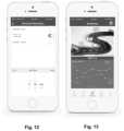
- FIG. 12is a diagram of one embodiment of a user interface for a mobile device to set an exercise reminder, according to the invention.
- FIG. 13is a diagram of one embodiment of a user interface for a mobile device to display information obtained from a sensor unit and a pathway toward a physical therapy goal, according to the invention
- FIG. 14is a diagram of yet another embodiment of a user interface for a mobile device to display information obtained from a sensor unit, according to the invention.
- FIG. 15is a diagram of one embodiment of a user interface for a mobile device to set patient specific settings for a sensor unit, according to the invention.
- FIG. 16is a diagram of one embodiment of a user interface for taking a photograph or video, according to the invention.
- FIG. 17is a diagram of another embodiment of a user interface to display information obtained from a sensor unit, according to the invention.
- FIG. 18is a diagram of a further embodiment of a user interface to display information obtained from a sensor unit, according to the invention.
- FIG. 19is a diagram of yet another embodiment of a user interface to display information obtained from a sensor unit, according to the invention.
- FIG. 20is a flowchart of one embodiment of a method of taking a photograph or video of a site on the patient; according to the invention.
- FIG. 21is a flowchart of one embodiment of a method of inputting a pain score; according to the invention.
- FIG. 22is a flowchart of one embodiment of a method for displaying information requested by the patient; according to the invention.
- FIG. 23is a flowchart of one embodiment of a method for including friends in physical therapy; according to the invention.
- the present inventionis directed to the area of methods, systems, and devices, including user interfaces, for monitoring physical therapy or rehabilitation.
- the present inventionis also directed to systems and methods with user interfaces for monitoring physical therapy or rehabilitation after surgery or implantation of an orthopedic device.
- a systemcan be used to monitor physical therapy or the healing process or rehabilitation of the patient after surgery, as well as monitor or verify the extent of the patient's activity.
- the systemincludes one or more sensors that can communicate with a processor that can produce information, based on the sensor readings and data, that can facilitate the patient or another user, such as a clinician, doctor, physical therapist, nurse, care coordinator, or other appropriate person, monitoring the patient's activity, the status of an orthopedic implant or surrounding tissues, or the effects of rehabilitation or other therapy.
- a clinician, doctor, physical therapist, nurse, care coordinator, or other appropriate personmonitoring the patient's activity, the status of an orthopedic implant or surrounding tissues, or the effects of rehabilitation or other therapy.
- the sensorsdescribed below, are placed near a physical therapy or rehabilitation site, such as a surgical site or the body portion to be rehabilitated.
- the systemmay also provide alerts if patient tissue becomes inflamed or if the effectiveness of, or compliance to, physical or rehabilitation therapy is insufficient.
- the systemincludes a wearable device with one or more sensors.
- one or more sensorsmay be provided on a wearable device that is applied to the skin of the patient.
- the one or more sensorscommunicate with a sensor processor on the device containing the sensors.
- the sensor processoror, alternatively or additionally, the sensors, communicate with a processor of a patient device, such as a mobile phone, tablet, computer or the like, or with a processor of a clinician device, such as a mobile phone, tablet, computer or the like.
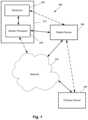
- FIG. 1illustrates one embodiment of a system 100 for monitoring an orthopedic implant and rehabilitation after orthopedic replacement surgery.
- the system 100includes one or more sensors 102 , an optional sensor processor 104 , a patient device 106 (such as a mobile phone, tablet, computer or the like), a clinician device 108 , and a network 60 .
- the one or more sensors 102 and, preferably, the sensor processor 104are provided in a wearable device 112 that is external to the patient such as, for example, a device that is applied to the skin of the patient or is carried in a brace or other article or textile that is worn by the patient.
- one or more of the sensors 102 and, optionally, the sensor processorcan be implanted in the patient.
- one or more of the sensors 102are implanted and a sensor processor and, optionally, one or more additional sensors are provided in a wearable device.
- the systemmay include fewer or more components than those illustrated in FIG. 1 , but the system typically includes the sensor(s) 102 and a processor (such as sensor processor 104 , patient device 106 , or clinician device 108 ) to communicate with the sensor(s) and provide information based on the sensor data.
- the wearable device 112includes the sensors 102 and sensor processor 104 , but it will be understood that other sensors may be included that are not part of the wearable device 112 .
- one or more additional sensorsmay be combined into another wearable device that may also include a sensor processor.
- the wearable device 102may not include a sensor processor 104 or the sensor processor 104 may have limited capabilities (such as, for example, obtaining and transmitting sensor readings without (or with limited) analysis of the sensor readings.
- the solid linesindicate communication between components in at least some embodiments of the system. Dotted lines indicate alternative or additional modes of communication between components.
- the sensor processor 104 or sensors 102may also communicate directly with the clinician device. Communications can include, but is not limited to, wireless communication, wired communication, optical communication, ultrasonic communication, or the combination thereof. Satellite communication, cellular communication, BluetoothTM, near field communications (NFC), Infrared Data Association standard (IrDA), wireless fidelity (WiFi), and worldwide interoperability for microwave access (WiMAX) are non-limiting examples of wireless communication that can be used for communications. Ethernet, digital subscriber line (DSL), fiber to the home (FTTH), and plain old telephone service (POTS) are non-limiting examples of wired communication that can be used for communications.
- DSLdigital subscriber line
- FTTHfiber to the home
- POTSplain old telephone service
- the network 60can be any suitable type of network including, but not limited to, a personal area network (PAN), local area network (LAN), metropolitan area network (MAN), wide area network (WAN), the Internet, or any combination thereof. In at least some embodiments, the network 60 can be bypassed to provide direct connection between components. It will be understood that other devices, such as a server or server farm, memory storage device, or the like can be connected to the patient device 106 or clinician device 108 through the network 60 or directly. For example, a server may be coupled to the patient device 106 or clinician device 108 that stores patient or other medical information, applications, user interfaces, a web interface, or the like for access by the patient device 106 or clinician device 108 .
- PANpersonal area network
- LANlocal area network
- MANmetropolitan area network
- WANwide area network
- the Internetor any combination thereof.
- the network 60can be bypassed to provide direct connection between components.
- other devicessuch as a server or server farm, memory storage device, or the like can be connected to
- the patient device 106 and the clinician device 108can be any of a variety of devices, such as computers (for example, a notebook computer, a mobile medical station or computer, a server, a mainframe computer, or a desktop computer), mobile devices (for example, a cellular phone or smartphone, personal digital assistant, or a tablet), or any other suitable device.
- the clinician device 108can be incorporated into a medical station or system.
- FIG. 2illustrates one embodiment of a computing device 201 for use as the patient device 106 or clinician device 108 .
- the computing device 201includes a processor 214 , a memory 216 , a display 218 , and an input device 220 .
- the computing device 201can be local to the user or can include components that are non-local to the computer including one or both of the processor 214 or memory 216 (or portions thereof). For example, in some embodiments, the user may operate a terminal that is connected to a non-local processor or memory.
- the computing device 201can utilize any suitable processor 214 including one or more hardware processors that may be local to the user or non-local to the user or other components of the computing device.
- the processor 214is configured to execute instructions provided to the processor. Such instructions can include any of the steps of methods or processes described herein.
- the memory 216illustrates a type of computer-readable media, namely computer-readable storage media.
- Computer-readable storage mediamay include, but is not limited to, nonvolatile, non-transitory, removable, and non-removable computer-readable media implemented in any method or technology for storage of information, such as computer readable instructions, data structures, program modules, or other data. Examples of computer-readable storage media include RAM, ROM, EEPROM, flash memory, or other memory technology, CD-ROM, digital versatile disks (“DVD”) or other optical storage, magnetic cassettes, magnetic tape, magnetic disk storage or other magnetic storage devices, or any other medium which can be used to store the desired information and which can be accessed by a computing device.
- Communication methodsprovide another type of computer readable media; namely communication media.
- Communication mediatypically embodies computer-readable instructions, data structures, program modules, or other data in a modulated data signal such as a carrier wave, data signal, or other transport mechanism and include any information delivery media.
- modulated data signaland “carrier-wave signal” includes a signal that has one or more of its characteristics set or changed in such a manner as to encode information, instructions, data, and the like, in the signal.
- communication mediaincludes wired media such as twisted pair, coaxial cable, fiber optics, wave guides, and other wired media and wireless media such as acoustic, RF, infrared, BluetoothTM, near field communication, and other wireless media.
- the display 218can be any suitable display device, such as a monitor, screen, display, or the like, and can include a printer.
- the input device 220can be, for example, a keyboard, mouse, touch screen, track ball, joystick, voice recognition system, camera, microphone, or any combination thereof, or the like.
- the sensor processor 104can be any suitable processor including one or more hardware processors.
- the sensor processor 104is configured to execute instructions provided to the processor.
- the sensor processor 104is configured to receive sensor data from the sensor(s) and communicate with the patient device 106 , network 60 , clinician device 108 , or any combination thereof.
- the sensor processor 104may also process or analyze the sensor data and may have instructions stored thereon to perform such processing or analysis including, for example, instructions to perform the steps of any of the processing or analysis described herein.
- one or more of the sensor(s) 102can each include a processor that perhaps some or all of the functions of the sensor processor 104 .
- the one or more sensors 102are provided to monitor an orthopedic implant and surrounding tissue or monitor rehabilitation after orthopedic surgery whether an implant was required or not, or to provide preparatory therapy in advance of a surgery, or any combination thereof.
- This disclosurewill use an orthopedic knee implant as an example, but it will be understood that other joint implants, such as, for example, implants for the shoulder, hip, ankle, wrist, or any other joint, or any other orthopedic device, such as an orthopedic spinal implant, whether joint replacement, joint resurfacing, soft tissue reconstruction, debridement, limb correction surgery, ligament replacement, or the like.
- any suitable type of sensor 102can be used including, but not limited to, accelerometers, magnetometers, gyroscopes, proximity sensors, infrared sensors, ultrasound sensors, thermistors or other temperature sensors, cameras, piezoelectric or other pressure sensors, sonar sensors, external fluid sensor, skin discoloration sensor, pH sensor, microphone, or the like or any combination thereof.
- the system 100includes at least one, two, three, four, five, six, or more different types of sensors 102 .
- the systemmay include at least one, two, three, four, five, six, eight, ten, or more sensors 102 .
- Further examples of suitable sensors and their arrangement and usecan be found at U.S. patent application Ser. Nos. 15/077,809 and 15/077,793 and U.S. Provisional Patent Application Ser. Nos. 62/136,892 and 62/136,925, all of which are incorporated herein by reference.
- the one or more sensors 102can be used to measure, monitor, or otherwise observe one or more aspects of the orthopedic device, surrounding tissue, or patient activity, or the like.
- a system 100can observe or measure one or more of these items or any combination of the items.
- One or more sensorsmay count steps or repetitions of an exercise or number of joint movements or other actions experienced by the sensor, and may be utilized to determine what type of exercise or movement is occurring. This can be used, for example, to monitor patient activity, monitor compliance with exercise therapy, or monitor possible signs of pain or other conditions that may hinder or aid rehabilitation.
- the sensor datamay also be used to monitor changes in activity or trends in activity.
- One or more sensorsmay sense or detect or compute the range of motion of the sensor, joint, or other portion of the patient body or the flexion of the joint. This can be used, for example, to monitor patient rehabilitation, patient activity, monitor compliance with exercise therapy, or monitor possible signs of pain or other conditions that may hinder or aid rehabilitation. These sensors or other sensors may be used to monitor shock to, or impact on, the orthopedic device or tissue around the orthopedic device. The sensor data may also be used to monitor changes in range of motion or flexion or trends in range of motion or flexion.
- one or more accelerometerscan measure the acceleration from joint movement.
- a ratio of measured acceleration between accelerometers of known distance apartcan be used to assess the joint movement and region of motion or flexion by calculating the center of rotation about which the device is being rotated. This information can be used for the same purposes as described in the preceding example.
- an accelerometer and 2) a gyroscope or magnetometercan be used to measure range of motion, rate of motion, number of repetitions, or the like. This information can be used for the same purposes as described in the preceding two examples.
- a single sensorsuch as an accelerometer, gyroscope, or magnetometer can be used to measure or otherwise observe range of motion, rate of motion, number of repetitions, or the like.
- these measurements or other observationsare determined using the sensor data and one or more assumptions about the sensor or sensor data based on, for example, the recognition of patterns in the sensor data, the upper and lower limits of the range in the data collected, or the like. Such information can be used in a manner similar to that in the preceding three examples.
- One or more sensorsmay sense or detect or compute a temperature or a change in temperature or a temperature trend.
- the temperaturemay be a skin temperature or ambient temperature.
- the temperature measurementsmay be used, for example, to indicate the possibility of inflammation or pain or another condition that may hinder rehabilitation or patient health.
- the temperature measurementmay also be used, for example, to monitor if icing is being performed effectively, which can help reduce inflammation and aid healing.
- These sensorsmay also or alternatively be used to sense, detect, or measure a pulse, a change in pulse, trends in the patient's pulse, a pulse profile, or heart rate recovery after patient activity (such as exercise or other exertion).
- One or more sensorscan sense or detect or compute particles or density of particles or a particle density trend. These sensors may also be used to sense the tissue surrounding the orthopedic device, detect wear or dimensional changes on the orthopedic device or surrounding tissue, or the like. Ultrasound and sonar sensors may also be used to determine how close other parts of the knee (or other joint) are to the implant.
- One or more sensorscan sense or detect or compute pressure or load with or around the sensor or orthopedic device.
- the sensor datamay also be used to monitor changes in range of pressure or load bearing or trends in pressure or load bearing.
- These sensors or other sensorsmay be used to monitor shock to, or impact on, the orthopedic device or tissue around the orthopedic device.
- a pressure or load bearing sensormay also be used to detect swelling of the tissue around the orthopedic implant.
- Multiple pressure or load bearing sensorsmay also be used to detect flexion (which may be indicated by a uniaxial stretching of the tissue) and swelling (which may be indicated by biaxial stretching of the tissue.)
- Powercan be provided to the sensors 102 and optional sensor processor 104 using any suitable power source including, but not limited to, primary cells, rechargeable batteries, storage capacitors, other power storage devices, or the like or any combination thereof.
- the powercan be provided by a kinetic energy power source that utilizes the movements of the patient's body to generate power for the components or to or to charge a battery or storage capacitor or other power storage device coupled to the components.
- wireless power sourcescan be used in place of (or in addition to) the battery, storage capacitor, or other power storage device.
- a charging portcan be provided for charging the battery or storage capacitor or other power storage device from a source such as a wall socket.
- a sourcesuch as a wall socket.
- wireless charging systems and methodscan also be used. It will be understood that in some embodiments there may be multiple methods for providing power to the component or to a power storage device associated with the component. All of the sensors and optional sensor processor may be coupled to the same power source or some of the sensors (or even all of the sensors) and sensor processor may have individual power sources.
- the sensors and optional sensor processorcan be active at all times to measure, monitor, or otherwise observe.
- one or more of the sensors and optional sensor processorcan be active periodically (with a period of, for example, 15 or 30 seconds or 1, 5, 10, 15, or 30 minutes or 1, 2, 3, 4, 6, 7, or 24 hours) or randomly to measure, monitor, or otherwise observe.
- the periodmay be programmable.
- the periodmay be optionally altered based on data from one or more of the sensors.
- one or more of the sensors and optional sensor processormay be activated manually or automatically by the sensor module, patient device, clinician device, or other device.
- the sensors and optional sensor processormay have different activation schedules (continuous, periodic, random, or manual). For example, a sensor to measure temperature may do so periodically, a sensor to measure number of steps or movement of the joint may be continuous, and a sensor to measure range of motion may be activated manually by the wearable device, patient device, or clinician device when the patient performs rehabilitation exercises.
- the systems and methodswill be described herein with reference to an orthopedic knee implant or other knee surgery. Similar systems and methods can be used with other joints including, but not limited to, the finger joint, wrist joint, elbow joint, shoulder joint, hip joint, ankle joint, or toe joint.
- the systems and methodscan be used to monitor physical therapy for any reason including, but not limited to, rehabilitation associated with other treatments including treatments for ligament or fracture surgery.
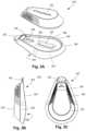
- FIGS. 3 A- 3 Cillustrate one embodiment of a wearable device 312 that includes a sensor unit 322 and a base 324 .
- the sensor unit 322is removable from the base 324 , as illustrated in FIG. 3 A .
- the wearable device 312as illustrated in FIGS. 3 B and 3 C , is disposed on the patient's skin with the base 324 adhered to the skin.
- the base 324includes a flexible receiving shell 326 , a magnet 328 , an optional opening 330 for a temperature sensor, an optional tab 332 , adhesive disposed on a bottom surface 334 of the shell, and an optional magnet holder 336 disposed on the shell.
- the magnet 328 of the base 324magnetically attaches to a similar magnet 354 ( FIG. 4 C ) in the sensor unit 322 when the sensor unit 322 is attached to the base 324 .
- the magnets 328 , 354are intended to maintain attachment of the sensor unit 322 to the base 324 during normal activity, exercise, and other physical therapy unless a patient or other person disengages the sensor unit from the base.
- a magnet holder 336fits over (entirely or only a perimeter of) the magnet 328 to hold the magnet to the shell 326 .
- the shell 326 of the base 324is sufficiently flexible for adhesion to the skin of a patient as the patient moves during normal activity or physical therapy exercises.
- the shellmay be made of any suitable material including, but not limited to, flexible plastics such as silicone or polyurethane.
- the shell 326may also removably grip the sensor unit 322 to provide further maintenance of the attachment of the sensor unit to the base 324 .
- the shell 326defines a receiving cavity 338 with sidewalls 340 around the cavity and a rim 342 around the sidewalls. In operation, the shell 326 receives a portion of the sensor unit 322 , as illustrated in FIGS. 3 B and 3 C .
- the sidewalls 340 or rim 342may be resiliently flexible to expand when the portion of the sensor unit 322 is received in the cavity 338 and then compress against a perimeter of the received portion of the sensor unit 322 .
- the rim 342 or sidewalls 340 (or both) of the base 324are made of a material that grips the sensor unit 322 by adhesion, compression, or the like or any combination thereof.
- the sensor unit 322may have a groove 390 that can receive the rim 342 to further facilitate maintaining the attachment of the sensor unit to the base 324 .
- the sidewalls 340slope outwardly and downwardly from the rim 342 to form an undercut region below the rim.
- the sensor unit 322can be similarly formed with a sloping housing to fit in the undercut below the rim 342 of the base 324 to further facilitate maintaining engagement between the sensor unit and the base. It will be recognized that in addition or as an alternative to the magnets (or magnet and magnetically attracted material) any other suitable type of mechanical fastener can be used to fasten the sensor unit 322 to the base 324 .
- the adhesivecan be applied to the base 324 or can be an adhesive disposed on two sides of a substrate with one side of the substrate adhered to the base 324 .
- the adhesiveis selected to be water resistant and resist losing adherence due to contact with sweat.
- the base 324 or the adhesive on the baseis intended for use for at least one, two, three, five, seven, or ten days or two, three, or four weeks or more under normal usage conditions before replacement or reapplication of adhesive.
- the adhesiveis selected to maintain adhesion to the skin when the user takes a shower.
- the adhesiveis selected to maintain adhesion to the skin when the user takes a bath, swims in a pool, or sits in jacuzzi, hot tub, or rehabilitation pool.
- the base 324optionally includes a tab 332 disposed at any suitable position relative to the shell 326 .
- the tab 332can facilitate removal of the sensor unit 322 from the base 324 by pushing or pulling on the tab 332 to deform the shell 326 to free the sensor unit.
- operation of the tab 332 to disengage the sensor unit 322can be performed while maintaining attachment of the base 324 to the skin of the patient.
- operation of the tab 332can also facilitate engagement of the sensor unit 322 with the base 324 .
- FIGS. 4 A- 4 Cillustrate one embodiment of a sensor unit 322 .
- the illustrated sensor unit 322includes an upper housing 350 , a lower housing 352 , a magnet 354 , an electronics assembly 356 , a power source 358 , a light emission arrangement 360 , and adhesive 362 , 364 .
- the upper housing 350can include a main housing 366 and a gripping element 368 .
- the sensor unit 322can include more or fewer components than those illustrated in FIGS. 4 A- 4 D .
- the upper housing 350 and lower housing 352form a cavity within which at least the electronics assembly 356 and power 358 source reside.
- the upper housing 350 and lower housing 352can be made of any suitable material, such as metal or plastic materials (preferably, rigid plastic materials) or any combination thereof.
- the upper housing 350 and lower housing 352as well as the joining of the upper housing to the lower housing, are water resistant to resist ingress of water, sweat, rain, and other fluids into the interior of the housing.
- the sensor unit 322is sufficiently water resistant to allow the patient to shower without any covering over the sensor unit. In some embodiments, the sensor unit 322 is sufficiently water resistant to allow the patient to bathe or swim without any covering over the sensor unit.
- the optional gripping element 368can have a roughened or otherwise non-smooth surface on at least a portion of the gripping element. This non-smooth surface facilitates gripping of the sensor unit 322 , particularly for engaging or disengaging the sensor unit from the base 324 .
- the gripping element 368is a separate element that is overmolded, adhered, or otherwise attached to the main housing 366 .
- the gripping element 368may be made of a different, more flexible material than the main housing 366 , such as silicone or polyurethane.
- the gripping element 368is formed as part of the main housing 366 by roughening or otherwise making at least a portion of the surface of the main housing non-smooth.
- the magnet 354is arranged for magnetically coupling to the magnet 328 of the base 324 .
- one of the magnets 354 , 328can be replaced with a magnetically attracted material that will then couple with the other magnet 354 , 328 to magnetically coupled the base 324 to the sensor unit 322 .
- the magnet 354is attached to the lower housing 352 by adhesive 364 which can be a layer of adhesive or adhesive disposed on both sides of a substrate.
- the magnet 354may be attached to the lower housing 352 by any other suitable method or may be disposed within the cavity formed by the upper housing 350 and lower housing 352 .
- the power source 358can be any suitable power source.
- the power source 358can be a primary cell (e.g., a battery) and may have an expected lifetime, under normal usage, of at least 7, 10, 20, 30, 60, 90, 100, 70, or 180 days or more.
- the primary cellmay be replaceable.
- the power source 358is rechargeable using, for example, a recharge port or an inductive recharge device (such as an inductive mat or sleeve), or using WiFi or ultrasonic charging or any other suitable recharging method.
- the primary celle.g., battery
- the primary cellcan be the magnetically attractive material that the magnet 328 of the base 324 can be magnetically coupled to.
- the electronics assembly 356can contain any suitable components for operation of the sensor unit 322 .
- the electronics assembly 356comprises a circuit board 368 , a sensor processor 304 , a temperature sensor 370 , an accelerometer 372 , at least one LED 374 , a communications arrangement 376 , and a magnetic switch 378 .
- Adhesive 362can couple the circuit board 368 to the lower housing 352 .
- Other adhesivemay couple the circuit board or other components to the upper housing 350 .
- the sensor processor 304can be similar to the sensor processor 104 described above and may have more or fewer capabilities than that sensor processor 104 .
- the sensor processor 304may include analysis algorithms for analyzing or partially analyzing the sensor data.
- the sensor processor 304may be primarily designed to receive, store, and transmit sensor data.
- the illustrated sensor unit 322includes a temperature sensor 370 and an accelerometer 372 , but other embodiments can contain more or different sensors, in any suitable combination, as described above.
- the temperature sensor 370is a thermistor which extends away from the circuit board 368 and through an opening 366 in the lower housing 352 .
- a portion of the temperature sensor 370extends through the opening 330 in the base 324 so that the temperature sensor 370 is exposed to the skin of the patient and may be in contact with the skin of the patient.
- the communications arrangement 376operates with the sensor processor 304 to communicate with patient or clinician devices or other devices, as described above.
- Any suitable communications method or protocolcan be used including, but not limited to WiFi, BluetoothTM, near field communications, infrared, radio frequency, acoustic, optical, or the like.
- the electronic assembly 356also includes a magnetic switch 378 , such as a reed switch, that is coupled to the sensor processor 304 so that when positioned near the magnet 328 of the base 324 is actuated to place the sensor unit 322 in an active mode.
- a magnetic switch 378such as a reed switch
- the magnetic switchwhen the sensor unit 322 is removed from the base 324 the magnetic switch is actuated to place the sensor in an inactive or standby mode.
- the sensor unit 322may include a button, mechanical switch, or other mechanism to place the sensor into the active mode or into an inactive or standby mode or to toggle between modes or to turn the sensor unit on or off.
- the sensor unit 322may be placed into the one of these modes (or toggled between modes) using signals from a patient or clinician device or other device communicating with the sensor unit 322 .
- the sensor unit 322in the inactive or standby mode, continues to be receptive to signals from an external source (such as the patient or clinician device).
- the sensor unit 322in the inactive or standby mode, also maintains an internal clock.
- the at least one LED 374is coupled to the light emission arrangement 360 to provide light to the light emission arrangement.
- the light emission arrangement 360includes a light emitter 380 and a light pipe 382 to direct light from the LED(s) 374 to the light emitter.
- the light emission arrangement 360provides an indication of operation of the device to a user or patient. For example, the light emission arrangement 360 may be lit when the sensor unit 322 is operating or is in the active mode. In some embodiments, the color of light emitted by the light emission arrangement may indicate which mode (active or inactive/standby) the sensor unit is currently in or may indicate operations being performed by the sensor unit (for example, transmitting, sensing, not sensing, synching with a patient or clinician device, or the like).
- flashing of the light or brightness of the lightmay be used to indicate mode or operations.
- a flashing blue lightmay indicate synching with a patient or clinician device
- a green lightmay indicate the active mode
- the absence of lightmay indicate the inactive/standby mode.
- a second sensor unitcan be used.
- the second sensor unitcan be placed on or within the same leg on the other side of the joint.
- a second sensor unitmay be placed on the other leg for use in detecting or observing limp or other gait deficiencies or placed on the torso to detect or observe body orientation.
- a second sensor unit(or more additional sensor units) may also be used when two or more replacements are implanted in the body, for example, with multiple joint or vertebra replacements, to detect or observe, for example, subluxations, changes or defects in posture, scoliosis, or the like.
- the two sensor unitsoptionally can communicate or synch with each other.
- the two sensor unitscan sync to each other and know where each one is in space and their location from each other in terms of distance and orientation.
- the two sensor unitsmay triangulate their positions using a patient or clinician device.
- the patient device or other sensor unitis advised.
- the systemcan determine the location or distance of the new sensor unit relative to the other sensor unit.
- the sensors in the two sensor unitscan be used to measure flexion angles; range of motion; calculate vectors, angles, rays, planes or distances; and the like. Temperature sensors on the two sensor units can be used to determine temperature differences between two portions of the body. The sensors from the two sensor units can be used to calculate angles or other information that can be used to send signals to the patient if the patient is exceeding limitations on movement of range of movement during physical therapy or rehabilitation.
- the sensor processor or sensorcommunicates with a patient device or clinician device to provide sensor data or information derived from the sensor data.
- the patient devicecan be a dedicated device or can be an application on a smartphone, tablet, laptop or desktop computer, a web or cloud application, or any other suitable arrangement. Communication between an implanted or wearable sensor unit and the patient or clinician device can occur at predetermined times (for example, every 30 minutes or every hour or once a day). In this manner, the sensor unit can sync with the patient or clinician device. Similarly, the patient device may sync with the clinician device on a regular schedule.
- the sensor unit or patient devicemay communicate (or attempt to communicate) with the patient device or clinician device, respectively, immediately to provide a warning.
- the communication between the sensor unit and the patient or clinician devicecan be constant or nearly constant (for example, once every 1, 5, 10, 30, or 60 seconds.)
- the constant or near constant communicationmay be established when the sensor unit determines that the patient is performing exercises or when the patient manually actuates a control on the patient device or sensor unit indicating that the patient will commence with exercises or otherwise desires the sensor unit to communicate or sync with the patient device.
- FIGS. 5 - 16illustrate screenshots of one embodiment of an application or user interface for the patient device or clinician device.
- the illustrated application or user interfaceis particularly useful for a mobile device such as a smartphone or tablet, but can also be used with other devices such as desktop or laptop computers.
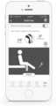
- FIG. 5illustrates one embodiment of a patient or clinician profile for the application or user interface. Elements of this page can include, but are not limited to, patient or clinician information, controls to input an identification number or other identification information for a wearable device so that the wearable device can be synched or otherwise coupled to the patient or clinician device, controls to input or change a password or to input or access application settings, a control to access a calibration program to calibrate the wearable device, controls for accessing one or more other features or pages, or the like.
- Other information that might be presented on this or another pagecan include, but is not limited to, controls for account creation or account login, indication of the status of the wearable device, or controls to access help information; FAQs; photos or videos or text for directions on how to apply the wearable device to the skin of the patient, how to care for a surgical wound, how to perform particular exercises, or how to program or operate the wearable device.
- FIG. 6illustrates another page of the user interface or application that provides information such as steps per day (or number of repetitions of an exercise or the like) and a temperature measurement as shown in section 592 .
- the user interface 590may also include a section 594 that shows graphs of the data such as the hourly number of steps, as illustrated in FIG. 6 .
- the illustrated user interfacepermits the user to select from other charts such as exercise history (labeled “ROM”), temperature or temperature trends, and number of impacts or shocks to the sensor module. It will be understood that other measurement or observations from the sensor described above can be graphed.
- the usermay also be able to select the time period of the graph to display data in periods of time such as, for example, minutes, hours, days, or weeks.
- This user interfacecan be useful in monitoring patient activity and progress.
- the graphs in section 594may be useful for showing patient exercise history and progress.
- the user interfacemay also allow the user to set goals such as, for example, a number of steps or a number of exercise repetitions over a particular period (for example, 1, 2, 4, 6, or 7 hours or 1 day or 1 week).
- the user interfacemay also display the current status towards attaining those goals.
- the user interfacemay also highlight notable events, such as, for example, the largest number of steps or exercise repetitions, elevated temperature readings, large numbers of impacts or shocks, or the like.
- the user interfacemay also highlight the attainment of goals.
- FIG. 7illustrates another page of the user interface or application that displays information related to particular patient measurements that can be tracked to monitor rehabilitation or physical therapy.
- the patient measurementsare flexion and extension related to a patient's knee. These patient measurements can include, but are not limited to, range of motion measurements such as flexion and extension.
- the pagealso illustrates a chart 596 tracking the progress of these measurements. The progress may be tracked hourly, daily, weekly, or over any other period of time.
- the user interface or applicationallows the user to select or change the time period illustrated in the chart.
- the page in FIG. 7also provides information about other measurements such as percentage of exercise completion, skin temperature, number of steps or the like.
- FIG. 8illustrates another page in which the user interface or application can be directed to calculate or otherwise determine a particular measurement.
- the measurementis femur angle.
- FIG. 9illustrates another page in which the user interface or application tracks the daily exercise program.
- the exercisesare sitting lift, heel slide (hip and knee flexion), straight leg raise, and knee to chest.
- Other exercisescan include, but are not limited to, standing lift, ankle pump, ankle circle, thigh squeeze (quadriceps set), lying kick (short arc quadriceps), knee bend (sitting knee flexion), prolonged knee stretch, sitting kick (long arc quadriceps), keen straightening stretch, knee dangling/swinging, hamstring set (heel dig), buttocks squeeze (gluteal set), walking, or the like.
- These exercisesare directed to knee rehabilitation.
- rehabilitation or physical therapy for other joints or body regionscan include a different set of exercises.
- the pageillustrates the percentage of completion for each set of repetitions (in this case, three sets) that are to be performed by the patient.
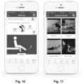
- FIG. 10illustrates yet another page with a single exercise.
- This pageillustrates the current measurements associated with the exercise (in this case, flexion and extension).
- the pagealso illustrates how the exercise is performed and may include a control for the patient to indicate that the exercise is to be begun.
- the pagemay also provide an indication of the number of repetitions (or the number of repetitions that are still needed to achieve a repetition goal) as the patient exercises.
- the pagemay also indicate patient measurements based on the exercise (e.g., a current measurement for the latest repetition or an average measurement for the current set of repetitions or a maximum measurement for a set of repetitions) and may also indicate a goal for the measurement.
- This pagemay include a meter with bars or the like to indicate what portion of an exercise goal has been met.
- An indication(such as a bar or the like) may also indicate what portion of a range of motion or other therapy goal has been met.
- the pagemay display an average patient time to the range of motion goal or the like to motivate the patient.
- FIG. 11illustrates a page with controls for accessing videos that can show the patient how to perform exercises.
- FIG. 12illustrates a page where the patient can set reminders to perform an exercise. The patient can set a time for the reminder and may also set a visual or audible alarm to remind the patient to exercise at the appointed time.
- FIG. 13illustrates a page with an indication of how far the patient has progressed in the physical therapy or rehabilitation.
- the distance and milestones included on this indicationcan be based on time (e.g., days or weeks) of the rehabilitation; physical measurements (e.g., flexion or extension) towards a final goal for that physical measurement; number of completed repetitions, or completion of, one or more exercises towards an exercise goal, or the like.
- the pagemay also include a graph of measurements (similar to FIG. 7 ) or number of repetitions or a graph of any other pertinent information.
- FIG. 14illustrates a page for a clinician device that indicates information about a group of patients, such as number or percentage of patients completing exercise or other goals, number or percentage of patients achieving particular range of motion or other measurement goals, or the like.
- FIG. 15illustrates a page for a patient or clinician device where settings can be entered or changed for a patient.
- Such settingscan include, for example, which exercises are to be performed, number of repetitions for each exercise, number of sets of repetitions per day for each exercise, number of steps for each day.
- This pagemay include controls to permit changing these settings.
- the settingsmay be related to a particular stage of the rehabilitation or physical therapy. The page may allow for toggling between different stages sot that settings can be viewed, entered, or changed for that stage.
- Another page for a patient or clinician devicemay display the actual results achieved by the patient and may compare those to the settings or goals entered for that patient.
- FIG. 22illustrates one embodiment of a method for displaying information requested by the patient.
- the patient devicereceives input from the patient for display of exercise or other information.
- the patient devicedisplays the requested information.
- the requested informationmay be a graphical representation of the exercise and a user control which, when actuated by the user, indicates that the user is performing the exercise.
- FIG. 10One example of such requested information is illustrated in FIG. 10 .
- the requested informationmay be a count of repetitions of the exercise being performed or a summary of a plurality of exercises indicating a number of repetitions performed for each exercise over a period of time. Examples of such requested information are presented in FIGS. 7 , 9 , 13 , and 14 .
- the requested informationmay be a progress report for a range of motion measurement or a graph of values for the range of motion measurement obtained over a period of time. Examples of such requested information are presented in FIGS. 7 , 13 , and 14 .
- the requested informationmay be a representation of a pathway to a goal for the range of motion measurement with an indication of current progress of the patient toward that goal as illustrated, for example, in FIG. 13 .
- FIG. 16illustrates another page in which the patient is directed to take a photograph or video of their knee or other wound site or physical therapy site using a camera on the patient device or other device.
- the pagemay direct the user how to frame the photo or video.
- the photo or videomay be sent to the clinician device or other device through the network (see, FIG. 1 ) or by other methods.
- a clinicianmay use the photo or video to assess the wound or physical therapy site.
- the patient devicemay request a video be taken of the patient performing an exercise.
- the videomay be provided to the patient device or clinician device to assess or view performance of the exercise. For example, a clinician may assess whether the patient is performing the exercise correctly or may assess progress in physical therapy or rehabilitation by observation of the exercise.
- the patient deviceis configured and arranged to perform pigment analysis or other wound analysis on the knee using the photo or video.
- the patient devicemay compare skin pigment at the wound site with skin pigment near the wound site to identify infection (for example, superficial wound infection or deep wound infection), rash, discoloration, or other issues.
- the pigment or other wound analysismay be combined with skin temperature information to assess infection (for example, superficial wound infection or deep wound infection), rash, discoloration, or other issues. If the analysis indicates a potential or actual issue, the patient device may provide a visual or audible warning to the patient and may also send an alert to the clinician device.
- the pigment or other wound analysismay be performed by the clinician device or other device instead of (or in addition to) the patient device.
- the patient device or clinician devicemay include white balancing or light compensating algorithms to assess the photos or videos.
- the patient devicemay also include a calibration tool to facilitate calibrating the light and other aspects of the photo or video.
- the patient devicemay display the region at which the camera is pointed for viewing by the patient. This displayed area, or the photo or video, may be altered to overlay lines or graphics that correspond to patient anatomy. These lines or graphics may move as the patient's leg or other body part move.
- patient measurementssuch as flexion or extension or other range of motion measurements, may be calculated during the movement and displayed on the patient device; changing as the patient limb moves.
- FIG. 20describes one embodiment of a method of taking a photograph or video of a site on the patient.
- the patient deviceor clinician or other device or person directs the patient to take photograph or video of the site (such as the site of physical therapy or a surgical or wound site.)
- the patienttakes the photograph or video using the camera of the patient device (or a camera of another device) and the photograph or video is stored.
- the photograph or videois sent to the clinician device or other device, in optional step 2006 .
- analysiscan be performed on the photograph or video.
- the analysiscan be performed by the patient device, clinician device or any other suitable device.
- a pigment analysiscan be performed or analysis related to exercises or range of motion measurements can be performed.
- graphical indiciasuch as lines or angles, can be superimposed on the photograph or video based on the analysis.
- a patient device, user interface, or applicationmay include other features.
- the patient device, user interface, or applicationmay include controls for a patient to enter information or ratings about their experience in the hospital, their experience during rehabilitation, how connected the patient fees during the rehabilitation process, whether the patient would recommend the wearable device or other aspects of the treatment to family or friends, or the like.
- the patient device, user interface, or applicationmay include controls for entering a rating related to pain or other clinical aspects.
- the patientmay enter a pain score based on a scale provided on the device.
- scoresthat may be entered by the patient, clinician, or others may be based on scores such as a Knee Society score, a New Knee Society score, other society score, KOSS or PROM (patient reported outcome measurements), oxford knee score, or Womack, or any other suitable score or rating.
- KOSS or PROMpatient reported outcome measurements
- oxford knee scoreor Womack, or any other suitable score or rating.
- FIG. 21illustrates one embodiment of a method of inputting a pain score.
- the patient devicedirects the user to input a pain score and the device receives the pain score.
- the pain scoreis sent to the clinician device or other device.
- the patient device, user interface, or applicationmay include controls to add friends, create a friend network, send messages to friends, send progress updates or other exercise information to friends or others, or the like. Some of the friends may be other patients, and the patient device, user interface, or application may display comparisons of progress with friends, allow issuance of a challenge to a friend, provide a control to send encouragement to a friend, or other social controls or interaction capabilities.
- another applicationmay be available to family, friends, and associates of the patient. This application may allow the user to send encouragement or messages to the patient and may also display progress updates or other exercise information that the patient has permitted.
- FIG. 23illustrates one embodiment of a method for including friends in the physical therapy.
- the patientis directed to enter one or more friends into the patient device.
- a connection messageis sent to the one or more friends to connect them to the patient's friend network.
- the patientreceives a message of encouragement from at least one of the one or more friends.
- the patientsends a message, challenge, or progress report to at least one of the one or more friends.
- the progress reportcan include, for example, an indication of progress by the patient toward at least one physical therapy goal, an indication of performance of the at least one physical therapy exercise by the patient, or an indication of performance of the at least one physical therapy exercise by at least one of the one or more friends, or the like or any combination thereof.
- FIG. 17illustrates a user interface 690 that may be suitable for a computer or web interface.
- the illustrated user interfaceincludes a region 692 displaying the results of temperature measurements 692 a , step measurements 692 b , range of motion tests 692 c , specific exercises and tests 692 d , and adverse events 692 e .
- These resultsmay include numerical information and graphical information. These results may also illustrate graphically or numerically the degree of success in performing exercises (see, for example, region 692 d ) and may also illustrate the degree of compliance with rehabilitation activities (such as the number of exercise repetitions performed).
- Such an arrangement of informationcan facilitate monitoring or patient progress, identification of progress or lack of progress, identification of concerns (such as elevated temperature or elevated number of shocks or impacts), and the like.
- Other information that can be displayed in one or more pages on the user interfacecan be include any suitable patient rehabilitation progress data include baselines and progress over time.
- the informationcan include baseline range of motion information for exercises such as a sitting leg lift, heel slide, standing lift, prone lift, or the like.
- the informationmay also include current range of motion information for these exercises.
- the informationmay also include step analysis information including, but not limited to, pre-operation and post-operation average cadence, maximum cadence, stride angle, as well as time spent walking, biking, running, or in sedentary activities.
- Additional informationcan include skin temperature, ambient temperature, and trends in temperature.
- the user interfacemay also provide information about how many times or how often the patient falls or other notable events.
- the user interfacemay provide information from GPS readings from the wearable device or patient device to assess baseline activity, current activity, general activity after surgery or physical therapy or the like.
- the user interface of a clinician devicemay also be used to conduct in-office range of motion tests.
- the clinician device or patient devicemay be used to create video of range of motion exercises.
- FIG. 18illustrates a user interface 790 for a clinician to monitor multiple patients.
- the region 792includes information such as patient name, surgery date, sensor date and results of tests 792 a , number of adverse events, location of the orthopedic implant, and the like.
- the clinicianmay also track number of surgeries 794 , rate of successful rehabilitation 796 , and other suitable information such as, for example, total number of surgeries (for example, total number of knee replacements), average time to reach a particular rehabilitation outcome (for example, average time to reach specified range of motion), and the like.
- FIG. 19illustrates another user interface for a clinician to monitor patients.
- the region 1592includes information such as patient name, gender, surgery date, days post operation, measured or trending temperature, range of motion measure, activity, number of steps, notable events, implant site, wearable device status, and the like.
- the clinicianmay also track number of successful rehabilitations 894 , range of motion achieved over time for a group of patients 896 , and other suitable information. Controls may also be provided to access individual patient records 898 or access patient alerts 899 .
- the applications or user interfaces described hereincan be web or application interfaces that are accessible when the patient device or clinician device accesses a server for a content provider.
- the server or other servers or memory storage devicescan store information for the web interface and may also store patient-specific information including patient identification data, sensor data or information derived from sensor data, patient or clinician comments or the like, or any other suitable data.
- the patient-specific informationcan be accessed from the patient device, clinician device or other device which, in some embodiments, may require providing credentials (e.g., username or password or both) to access the information.
- Systems referenced hereintypically include memory and typically include methods for communication with other devices including mobile devices.
- Methods of communicationcan include both wired and wireless (e.g., RF, optical, or infrared) communications methods and such methods provide another type of computer readable media; namely communication media.
- Wired communicationcan include communication over a twisted pair, coaxial cable, fiber optics, wave guides, or the like, or any combination thereof.
- Wireless communicationcan include RF, infrared, acoustic, near field communication, BluetoothTM, or the like, or any combination thereof.
- the computer program instructionscan be stored on any suitable computer-readable medium including, but not limited to, RAM, ROM, EEPROM, flash memory or other memory technology, CD-ROM, digital versatile disks (“DVD”) or other optical storage, magnetic cassettes, magnetic tape, magnetic disk storage or other magnetic storage devices, or any other medium which can be used to store the desired information and which can be accessed by a computer.
Landscapes
- Health & Medical Sciences (AREA)
- Life Sciences & Earth Sciences (AREA)
- Engineering & Computer Science (AREA)
- Medical Informatics (AREA)
- Public Health (AREA)
- Biomedical Technology (AREA)
- General Health & Medical Sciences (AREA)
- Surgery (AREA)
- Biophysics (AREA)
- Pathology (AREA)
- Heart & Thoracic Surgery (AREA)
- Physics & Mathematics (AREA)
- Veterinary Medicine (AREA)
- Animal Behavior & Ethology (AREA)
- Molecular Biology (AREA)
- Epidemiology (AREA)
- Cardiology (AREA)
- Primary Health Care (AREA)
- Physiology (AREA)
- Optics & Photonics (AREA)
- Oral & Maxillofacial Surgery (AREA)
- Radiology & Medical Imaging (AREA)
- Nuclear Medicine, Radiotherapy & Molecular Imaging (AREA)
- Physical Education & Sports Medicine (AREA)
- General Business, Economics & Management (AREA)
- Pulmonology (AREA)
- Transplantation (AREA)
- Dentistry (AREA)
- Business, Economics & Management (AREA)
- Orthopedic Medicine & Surgery (AREA)
- Rheumatology (AREA)
- Data Mining & Analysis (AREA)
- Databases & Information Systems (AREA)
- Computer Networks & Wireless Communication (AREA)
- Measurement Of The Respiration, Hearing Ability, Form, And Blood Characteristics Of Living Organisms (AREA)
- Rehabilitation Tools (AREA)
Abstract
Description
Claims (20)
Priority Applications (7)
| Application Number | Priority Date | Filing Date | Title |
|---|---|---|---|
| US15/422,320US11684260B2 (en) | 2015-03-23 | 2017-02-01 | System and methods with user interfaces for monitoring physical therapy and rehabilitation |
| AU2018216895AAU2018216895B2 (en) | 2017-02-01 | 2018-02-01 | System and methods with user interfaces for monitoring physical therapy and rehabilitation |
| CN201880023731.4ACN110621214A (en) | 2017-02-01 | 2018-02-01 | System and method for monitoring physical therapy and rehabilitation using a user interface |
| JP2019541732AJP7187466B2 (en) | 2017-02-01 | 2018-02-01 | System and method with user interface for monitoring physical therapy and rehabilitation |
| EP18748777.2AEP3576614A4 (en) | 2017-02-01 | 2018-02-01 | System and methods with user interfaces for monitoring physical therapy and rehabilitation |
| PCT/US2018/016424WO2018144716A1 (en) | 2017-02-01 | 2018-02-01 | System and methods with user interfaces for monitoring physical therapy and rehabilitation |
| US18/213,768US12433487B2 (en) | 2015-03-23 | 2023-06-23 | System and methods with user interfaces for monitoring physical therapy and rehabilitation |
Applications Claiming Priority (4)
| Application Number | Priority Date | Filing Date | Title |
|---|---|---|---|
| US201562136925P | 2015-03-23 | 2015-03-23 | |
| US201562136892P | 2015-03-23 | 2015-03-23 | |
| US15/077,809US10709377B2 (en) | 2015-03-23 | 2016-03-22 | System and methods for monitoring an orthopedic implant and rehabilitation |
| US15/422,320US11684260B2 (en) | 2015-03-23 | 2017-02-01 | System and methods with user interfaces for monitoring physical therapy and rehabilitation |
Related Parent Applications (1)
| Application Number | Title | Priority Date | Filing Date |
|---|---|---|---|
| US15/077,809Continuation-In-PartUS10709377B2 (en) | 2015-03-23 | 2016-03-22 | System and methods for monitoring an orthopedic implant and rehabilitation |
Related Child Applications (1)
| Application Number | Title | Priority Date | Filing Date |
|---|---|---|---|
| US18/213,768ContinuationUS12433487B2 (en) | 2015-03-23 | 2023-06-23 | System and methods with user interfaces for monitoring physical therapy and rehabilitation |
Publications (2)
| Publication Number | Publication Date |
|---|---|
| US20170147789A1 US20170147789A1 (en) | 2017-05-25 |
| US11684260B2true US11684260B2 (en) | 2023-06-27 |
Family
ID=58720756
Family Applications (2)
| Application Number | Title | Priority Date | Filing Date |
|---|---|---|---|
| US15/422,320Active2037-07-19US11684260B2 (en) | 2015-03-23 | 2017-02-01 | System and methods with user interfaces for monitoring physical therapy and rehabilitation |
| US18/213,768Active2036-05-24US12433487B2 (en) | 2015-03-23 | 2023-06-23 | System and methods with user interfaces for monitoring physical therapy and rehabilitation |
Family Applications After (1)
| Application Number | Title | Priority Date | Filing Date |
|---|---|---|---|
| US18/213,768Active2036-05-24US12433487B2 (en) | 2015-03-23 | 2023-06-23 | System and methods with user interfaces for monitoring physical therapy and rehabilitation |
Country Status (1)
| Country | Link |
|---|---|
| US (2) | US11684260B2 (en) |
Cited By (9)
| Publication number | Priority date | Publication date | Assignee | Title |
|---|---|---|---|---|
| USD1039159S1 (en)* | 2015-07-09 | 2024-08-13 | Dexcom, Inc. | Medical device electronic module |
| USD1039158S1 (en)* | 2015-07-09 | 2024-08-13 | Dexcom, Inc. | Base for medical device electronic module |
| USD1057956S1 (en) | 2015-07-09 | 2025-01-14 | Dexcom, Inc. | Medical device inserter |
| US12285267B2 (en) | 2016-03-23 | 2025-04-29 | Canary Medical Inc. | Implantable reporting processor for an alert implant |
| US12285234B2 (en) | 2014-09-17 | 2025-04-29 | Canary Medical Inc. | Devices, systems and methods for using and monitoring medical devices |
| US12293828B2 (en) | 2019-06-06 | 2025-05-06 | Canary Medical Inc. | Intelligent joint prosthesis |
| US12408963B2 (en) | 2020-02-20 | 2025-09-09 | Canary Medical Switzerland Ag | Medical device for implanting in boney tissue and characterization of bone fractures |
| US12419567B2 (en) | 2014-06-25 | 2025-09-23 | Canary Medical Switzerland Ag | Devices, systems and methods for using and monitoring orthopedic hardware |
| US12440153B2 (en) | 2021-12-06 | 2025-10-14 | Canary Medical Inc. | Implantable reporting processor for an alert implant |
Families Citing this family (87)
| Publication number | Priority date | Publication date | Assignee | Title |
|---|---|---|---|---|
| US11272879B2 (en) | 2015-03-23 | 2022-03-15 | Consensus Orthopedics, Inc. | Systems and methods using a wearable device for monitoring an orthopedic implant and rehabilitation |
| US11684260B2 (en) | 2015-03-23 | 2023-06-27 | Tracpatch Health, Inc. | System and methods with user interfaces for monitoring physical therapy and rehabilitation |
| US10646746B1 (en) | 2016-09-12 | 2020-05-12 | Rom Technologies, Inc. | Adjustable rehabilitation and exercise device |
| WO2018081795A1 (en) | 2016-10-31 | 2018-05-03 | Zipline Medical, Inc. | Systems and methods for monitoring physical therapy of the knee and other joints |
| US20190066832A1 (en)* | 2017-02-20 | 2019-02-28 | KangarooHealth, Inc. | Method for detecting patient risk and selectively notifying a care provider of at-risk patients |
| CN108606797B (en)* | 2018-04-28 | 2022-03-15 | 京东方科技集团股份有限公司 | A kind of smart patch and its making method |
| GB2574074B (en) | 2018-07-27 | 2020-05-20 | Mclaren Applied Tech Ltd | Time synchronisation |
| US11557215B2 (en)* | 2018-08-07 | 2023-01-17 | Physera, Inc. | Classification of musculoskeletal form using machine learning model |
| RO133954A2 (en)* | 2018-09-21 | 2020-03-30 | Kineto Tech Rehab S.R.L. | System and method for optimized joint monitoring in kinesiotherapy |
| US11471729B2 (en) | 2019-03-11 | 2022-10-18 | Rom Technologies, Inc. | System, method and apparatus for a rehabilitation machine with a simulated flywheel |
| US11904202B2 (en) | 2019-03-11 | 2024-02-20 | Rom Technolgies, Inc. | Monitoring joint extension and flexion using a sensor device securable to an upper and lower limb |
| US11185735B2 (en) | 2019-03-11 | 2021-11-30 | Rom Technologies, Inc. | System, method and apparatus for adjustable pedal crank |
| US11475571B2 (en) | 2019-03-13 | 2022-10-18 | Canon Kabushiki Kaisha | Apparatus, image processing apparatus, and control method |
| US11433276B2 (en) | 2019-05-10 | 2022-09-06 | Rehab2Fit Technologies, Inc. | Method and system for using artificial intelligence to independently adjust resistance of pedals based on leg strength |
| US11904207B2 (en) | 2019-05-10 | 2024-02-20 | Rehab2Fit Technologies, Inc. | Method and system for using artificial intelligence to present a user interface representing a user's progress in various domains |
| US11937896B1 (en)* | 2019-05-10 | 2024-03-26 | Smith & Nephew, Inc. | Orthopedic implants with improved sensors |
| US11957960B2 (en) | 2019-05-10 | 2024-04-16 | Rehab2Fit Technologies Inc. | Method and system for using artificial intelligence to adjust pedal resistance |
| US12102878B2 (en) | 2019-05-10 | 2024-10-01 | Rehab2Fit Technologies, Inc. | Method and system for using artificial intelligence to determine a user's progress during interval training |
| US11801423B2 (en) | 2019-05-10 | 2023-10-31 | Rehab2Fit Technologies, Inc. | Method and system for using artificial intelligence to interact with a user of an exercise device during an exercise session |
| US12402804B2 (en) | 2019-09-17 | 2025-09-02 | Rom Technologies, Inc. | Wearable device for coupling to a user, and measuring and monitoring user activity |
| US11071597B2 (en) | 2019-10-03 | 2021-07-27 | Rom Technologies, Inc. | Telemedicine for orthopedic treatment |
| US11701548B2 (en) | 2019-10-07 | 2023-07-18 | Rom Technologies, Inc. | Computer-implemented questionnaire for orthopedic treatment |
| USD928635S1 (en) | 2019-09-18 | 2021-08-24 | Rom Technologies, Inc. | Goniometer |
| US11955222B2 (en) | 2019-10-03 | 2024-04-09 | Rom Technologies, Inc. | System and method for determining, based on advanced metrics of actual performance of an electromechanical machine, medical procedure eligibility in order to ascertain survivability rates and measures of quality-of-life criteria |
| US12420143B1 (en) | 2019-10-03 | 2025-09-23 | Rom Technologies, Inc. | System and method for enabling residentially-based cardiac rehabilitation by using an electromechanical machine and educational content to mitigate risk factors and optimize user behavior |
| US12420145B2 (en) | 2019-10-03 | 2025-09-23 | Rom Technologies, Inc. | Systems and methods of using artificial intelligence and machine learning for generating alignment plans to align a user with an imaging sensor during a treatment session |
| US12062425B2 (en) | 2019-10-03 | 2024-08-13 | Rom Technologies, Inc. | System and method for implementing a cardiac rehabilitation protocol by using artificial intelligence and standardized measurements |
| US12380984B2 (en) | 2019-10-03 | 2025-08-05 | Rom Technologies, Inc. | Systems and methods for using artificial intelligence and machine learning to generate treatment plans having dynamically tailored cardiac protocols for users to manage a state of an electromechanical machine |
| US12224052B2 (en) | 2019-10-03 | 2025-02-11 | Rom Technologies, Inc. | System and method for using AI, machine learning and telemedicine for long-term care via an electromechanical machine |
| US11830601B2 (en) | 2019-10-03 | 2023-11-28 | Rom Technologies, Inc. | System and method for facilitating cardiac rehabilitation among eligible users |
| US11915816B2 (en) | 2019-10-03 | 2024-02-27 | Rom Technologies, Inc. | Systems and methods of using artificial intelligence and machine learning in a telemedical environment to predict user disease states |
| US12020799B2 (en) | 2019-10-03 | 2024-06-25 | Rom Technologies, Inc. | Rowing machines, systems including rowing machines, and methods for using rowing machines to perform treatment plans for rehabilitation |
| US11961603B2 (en) | 2019-10-03 | 2024-04-16 | Rom Technologies, Inc. | System and method for using AI ML and telemedicine to perform bariatric rehabilitation via an electromechanical machine |
| US11978559B2 (en) | 2019-10-03 | 2024-05-07 | Rom Technologies, Inc. | Systems and methods for remotely-enabled identification of a user infection |
| US12230381B2 (en) | 2019-10-03 | 2025-02-18 | Rom Technologies, Inc. | System and method for an enhanced healthcare professional user interface displaying measurement information for a plurality of users |
| US12087426B2 (en) | 2019-10-03 | 2024-09-10 | Rom Technologies, Inc. | Systems and methods for using AI ML to predict, based on data analytics or big data, an optimal number or range of rehabilitation sessions for a user |
| US20210134412A1 (en) | 2019-10-03 | 2021-05-06 | Rom Technologies, Inc. | System and method for processing medical claims using biometric signatures |
| US11265234B2 (en) | 2019-10-03 | 2022-03-01 | Rom Technologies, Inc. | System and method for transmitting data and ordering asynchronous data |
| US11087865B2 (en) | 2019-10-03 | 2021-08-10 | Rom Technologies, Inc. | System and method for use of treatment device to reduce pain medication dependency |
| US11282608B2 (en) | 2019-10-03 | 2022-03-22 | Rom Technologies, Inc. | Method and system for using artificial intelligence and machine learning to provide recommendations to a healthcare provider in or near real-time during a telemedicine session |
| US11887717B2 (en) | 2019-10-03 | 2024-01-30 | Rom Technologies, Inc. | System and method for using AI, machine learning and telemedicine to perform pulmonary rehabilitation via an electromechanical machine |
| US11515021B2 (en) | 2019-10-03 | 2022-11-29 | Rom Technologies, Inc. | Method and system to analytically optimize telehealth practice-based billing processes and revenue while enabling regulatory compliance |
| US11955221B2 (en) | 2019-10-03 | 2024-04-09 | Rom Technologies, Inc. | System and method for using AI/ML to generate treatment plans to stimulate preferred angiogenesis |
| US11101028B2 (en) | 2019-10-03 | 2021-08-24 | Rom Technologies, Inc. | Method and system using artificial intelligence to monitor user characteristics during a telemedicine session |
| US11955223B2 (en) | 2019-10-03 | 2024-04-09 | Rom Technologies, Inc. | System and method for using artificial intelligence and machine learning to provide an enhanced user interface presenting data pertaining to cardiac health, bariatric health, pulmonary health, and/or cardio-oncologic health for the purpose of performing preventative actions |
| US11955220B2 (en) | 2019-10-03 | 2024-04-09 | Rom Technologies, Inc. | System and method for using AI/ML and telemedicine for invasive surgical treatment to determine a cardiac treatment plan that uses an electromechanical machine |
| US11756666B2 (en) | 2019-10-03 | 2023-09-12 | Rom Technologies, Inc. | Systems and methods to enable communication detection between devices and performance of a preventative action |
| US12327623B2 (en) | 2019-10-03 | 2025-06-10 | Rom Technologies, Inc. | System and method for processing medical claims |
| US12154672B2 (en) | 2019-10-03 | 2024-11-26 | Rom Technologies, Inc. | Method and system for implementing dynamic treatment environments based on patient information |
| US12230382B2 (en) | 2019-10-03 | 2025-02-18 | Rom Technologies, Inc. | Systems and methods for using artificial intelligence and machine learning to predict a probability of an undesired medical event occurring during a treatment plan |
| US11515028B2 (en) | 2019-10-03 | 2022-11-29 | Rom Technologies, Inc. | Method and system for using artificial intelligence and machine learning to create optimal treatment plans based on monetary value amount generated and/or patient outcome |
| US20210134458A1 (en) | 2019-10-03 | 2021-05-06 | Rom Technologies, Inc. | System and method to enable remote adjustment of a device during a telemedicine session |
| US11075000B2 (en) | 2019-10-03 | 2021-07-27 | Rom Technologies, Inc. | Method and system for using virtual avatars associated with medical professionals during exercise sessions |
| US12020800B2 (en) | 2019-10-03 | 2024-06-25 | Rom Technologies, Inc. | System and method for using AI/ML and telemedicine to integrate rehabilitation for a plurality of comorbid conditions |
| US11325005B2 (en) | 2019-10-03 | 2022-05-10 | Rom Technologies, Inc. | Systems and methods for using machine learning to control an electromechanical device used for prehabilitation, rehabilitation, and/or exercise |
| US11139060B2 (en) | 2019-10-03 | 2021-10-05 | Rom Technologies, Inc. | Method and system for creating an immersive enhanced reality-driven exercise experience for a user |
| US12246222B2 (en) | 2019-10-03 | 2025-03-11 | Rom Technologies, Inc. | Method and system for using artificial intelligence to assign patients to cohorts and dynamically controlling a treatment apparatus based on the assignment during an adaptive telemedical session |
| US11337648B2 (en) | 2020-05-18 | 2022-05-24 | Rom Technologies, Inc. | Method and system for using artificial intelligence to assign patients to cohorts and dynamically controlling a treatment apparatus based on the assignment during an adaptive telemedical session |
| US20230245750A1 (en) | 2019-10-03 | 2023-08-03 | Rom Technologies, Inc. | Systems and methods for using elliptical machine to perform cardiovascular rehabilitation |
| US11282599B2 (en) | 2019-10-03 | 2022-03-22 | Rom Technologies, Inc. | System and method for use of telemedicine-enabled rehabilitative hardware and for encouragement of rehabilitative compliance through patient-based virtual shared sessions |
| US12220201B2 (en) | 2019-10-03 | 2025-02-11 | Rom Technologies, Inc. | Remote examination through augmented reality |
| US11069436B2 (en) | 2019-10-03 | 2021-07-20 | Rom Technologies, Inc. | System and method for use of telemedicine-enabled rehabilitative hardware and for encouraging rehabilitative compliance through patient-based virtual shared sessions with patient-enabled mutual encouragement across simulated social networks |
| US12347543B2 (en) | 2019-10-03 | 2025-07-01 | Rom Technologies, Inc. | Systems and methods for using artificial intelligence to implement a cardio protocol via a relay-based system |
| US12150792B2 (en) | 2019-10-03 | 2024-11-26 | Rom Technologies, Inc. | Augmented reality placement of goniometer or other sensors |
| US12100499B2 (en) | 2020-08-06 | 2024-09-24 | Rom Technologies, Inc. | Method and system for using artificial intelligence and machine learning to create optimal treatment plans based on monetary value amount generated and/or patient outcome |
| US11282604B2 (en) | 2019-10-03 | 2022-03-22 | Rom Technologies, Inc. | Method and system for use of telemedicine-enabled rehabilitative equipment for prediction of secondary disease |
| US11317975B2 (en) | 2019-10-03 | 2022-05-03 | Rom Technologies, Inc. | Method and system for treating patients via telemedicine using sensor data from rehabilitation or exercise equipment |
| US12427376B2 (en) | 2019-10-03 | 2025-09-30 | Rom Technologies, Inc. | Systems and methods for an artificial intelligence engine to optimize a peak performance |
| US11915815B2 (en) | 2019-10-03 | 2024-02-27 | Rom Technologies, Inc. | System and method for using artificial intelligence and machine learning and generic risk factors to improve cardiovascular health such that the need for additional cardiac interventions is mitigated |
| US11270795B2 (en) | 2019-10-03 | 2022-03-08 | Rom Technologies, Inc. | Method and system for enabling physician-smart virtual conference rooms for use in a telehealth context |
| US12176089B2 (en) | 2019-10-03 | 2024-12-24 | Rom Technologies, Inc. | System and method for using AI ML and telemedicine for cardio-oncologic rehabilitation via an electromechanical machine |
| US12191018B2 (en) | 2019-10-03 | 2025-01-07 | Rom Technologies, Inc. | System and method for using artificial intelligence in telemedicine-enabled hardware to optimize rehabilitative routines capable of enabling remote rehabilitative compliance |
| US11923065B2 (en) | 2019-10-03 | 2024-03-05 | Rom Technologies, Inc. | Systems and methods for using artificial intelligence and machine learning to detect abnormal heart rhythms of a user performing a treatment plan with an electromechanical machine |
| GB2588236B (en) | 2019-10-18 | 2024-03-20 | Mclaren Applied Ltd | Gyroscope bias estimation |
| US11826613B2 (en) | 2019-10-21 | 2023-11-28 | Rom Technologies, Inc. | Persuasive motivation for orthopedic treatment |
| US12424319B2 (en) | 2019-11-06 | 2025-09-23 | Rom Technologies, Inc. | System for remote treatment utilizing privacy controls |
| USD907143S1 (en) | 2019-12-17 | 2021-01-05 | Rom Technologies, Inc. | Rehabilitation device |
| US10863928B1 (en) | 2020-01-28 | 2020-12-15 | Consensus Orthopedics, Inc. | System and methods for monitoring the spine, balance, gait, or posture of a patient |
| US11107591B1 (en) | 2020-04-23 | 2021-08-31 | Rom Technologies, Inc. | Method and system for describing and recommending optimal treatment plans in adaptive telemedical or other contexts |
| EP4164471A4 (en)* | 2020-06-10 | 2024-03-06 | PMotion, Inc. | IMPROVED GONIOMETER |
| WO2021262809A1 (en) | 2020-06-26 | 2021-12-30 | Rom Technologies, Inc. | System, method and apparatus for anchoring an electronic device and measuring a joint angle |
| US12367960B2 (en) | 2020-09-15 | 2025-07-22 | Rom Technologies, Inc. | System and method for using AI ML and telemedicine to perform bariatric rehabilitation via an electromechanical machine |
| CN113689933B (en)* | 2021-08-30 | 2024-06-21 | 杨宏强 | Informatization system for accelerating rehabilitation surgery |
| USD1053901S1 (en)* | 2021-11-05 | 2024-12-10 | Howmedica Osteonics Corp. | Display screen or portion thereof with graphical user interface |
| USD1067239S1 (en) | 2021-11-05 | 2025-03-18 | Howmedica Osteonics Corp. | Display screen or portion thereof with animated graphical user interface |
| USD1084008S1 (en)* | 2021-11-05 | 2025-07-15 | Howmedica Osteonics Corp. | Display screen or portion thereof with graphical user interface |
| US12109017B2 (en)* | 2022-08-17 | 2024-10-08 | August River, Ltd Co | Remotely tracking range of motion measurement |
Citations (87)
| Publication number | Priority date | Publication date | Assignee | Title |
|---|---|---|---|---|
| US1086392A (en) | 1913-07-05 | 1914-02-10 | Wilmarth Show Case Company | Glass-locking device for show-cases. |
| US3724651A (en) | 1971-03-03 | 1973-04-03 | Johnson & Johnson | Peelable surgical package |
| US4353135A (en) | 1980-05-09 | 1982-10-12 | Minnesota Mining And Manufacturing Company | Patellar flange and femoral knee-joint prosthesis |
| US4756312A (en) | 1984-03-22 | 1988-07-12 | Advanced Hearing Technology, Inc. | Magnetic attachment device for insertion and removal of hearing aid |
| WO1994026359A1 (en) | 1993-05-07 | 1994-11-24 | Multibusiness Enterprise Finntrade Oy | Device for registering movement of a joint |
| US5833603A (en) | 1996-03-13 | 1998-11-10 | Lipomatrix, Inc. | Implantable biosensing transponder |
| US20030069714A1 (en) | 2001-10-04 | 2003-04-10 | Wigley Fredrick M. | Ambulatory surface skin temperature monitor |
| US6588931B2 (en) | 2000-12-07 | 2003-07-08 | Delphi Technologies, Inc. | Temperature sensor with flexible circuit substrate |
| US20030163287A1 (en) | 2000-12-15 | 2003-08-28 | Vock Curtis A. | Movement and event systems and associated methods related applications |
| US20040039254A1 (en) | 2002-08-22 | 2004-02-26 | Stivoric John M. | Apparatus for detecting human physiological and contextual information |
| US20040122334A1 (en) | 2002-09-19 | 2004-06-24 | Alfred E. Mann Institute For Biomedical Engineering At The University Of Southern | Vantilation and volume change measurements using permanent magnet and magnet sensor affixed to body |
| US20040167390A1 (en) | 1998-09-14 | 2004-08-26 | Alexander Eugene J. | Assessing the condition of a joint and devising treatment |
| US20050010299A1 (en) | 2003-07-11 | 2005-01-13 | Disilvestro Mark R. | In vivo joint implant cycle counter |
| US20050010301A1 (en) | 2003-07-11 | 2005-01-13 | Disilvestro Mark R. | In vivo joint space measurement device and method |
| US20050197540A1 (en) | 2003-11-24 | 2005-09-08 | Bionics Pharma Gmbh | Dermal diagnostic system including an active transponder |
| US20070167688A1 (en)* | 2005-12-19 | 2007-07-19 | Ross S M | Healthcare management systems and associated methods |
| US20070250286A1 (en) | 2003-07-01 | 2007-10-25 | Queensland University Of Technology | Motion Monitoring and Analysis System |
| US20080027296A1 (en) | 2003-12-22 | 2008-01-31 | Paul Hadvary | Dermally Affixed Sensor Device |
| EP1938749A2 (en) | 2006-12-27 | 2008-07-02 | DePuy Products, Inc. | Apparatus for monitoring the range of motion of a joint |
| WO2008120215A2 (en) | 2007-04-02 | 2008-10-09 | Novocart Medical Solutions Ltd | Intra-articular implant for treating irregularities in cartilage surfaces |
| US20080299960A1 (en)* | 2007-06-01 | 2008-12-04 | Kendall Gregory Lockhart | System and method for managing message transmissions on a mobile device |
| US20080311765A1 (en) | 2007-06-15 | 2008-12-18 | Microsoft Corporation | Electrical connection between devices |
| US20090309683A1 (en) | 2008-06-16 | 2009-12-17 | Cochran William T | Sensor inductors, sensors for monitoring movements and positioning, apparatus, systems and methods therefore |
| US20100114596A1 (en)* | 2003-10-24 | 2010-05-06 | Caringfamily, Llc | Use of a closed communication service for social support networks to improve the well being of a primary care provider |
| US20100174189A1 (en) | 2007-10-12 | 2010-07-08 | Innoscion, Llc | Remotely controlled implantable transducer and associated displays and controls |
| CN201537084U (en) | 2009-09-30 | 2010-08-04 | 深圳市纽泰克电子有限公司 | Wireless body temperature sensor and adhesive tape |
| WO2010088696A1 (en) | 2009-02-02 | 2010-08-05 | Mohamed Rashwan Mahfouz | Noninvasive diagnostic system |
| US20100228089A1 (en) | 2008-06-18 | 2010-09-09 | Versatile Power, Inc. | Endoscopic Light Source |
| US20100262047A1 (en)* | 2009-04-08 | 2010-10-14 | Drexel University | Physical therapy systems and methods |
| US20100324403A1 (en) | 2007-09-13 | 2010-12-23 | Dexcom, Inc. | Transcutaneous analyte sensor |
| US20110046558A1 (en) | 2009-08-18 | 2011-02-24 | Peter Gravesen | Medicine delivery device having detachable pressure sensing unit |
| US20110208444A1 (en) | 2006-07-21 | 2011-08-25 | Solinsky James C | System and method for measuring balance and track motion in mammals |
| US20110213275A1 (en) | 2008-06-27 | 2011-09-01 | Bort Gmbh | Device for determining the stability of a knee joint |
| US20110231208A1 (en)* | 2008-12-17 | 2011-09-22 | The Johns Hopkins University | System and method for patient self-assessment or treatment compliance |
| US20110245711A1 (en) | 2010-04-05 | 2011-10-06 | Corventis, Inc. | Method and apparatus for personalized physiologic parameters |
| US20110288379A1 (en) | 2007-08-02 | 2011-11-24 | Wuxi Microsens Co., Ltd. | Body sign dynamically monitoring system |
| JP2012090946A (en) | 2010-09-29 | 2012-05-17 | Terumo Corp | Apparatus for determining infection, and information processing device and method |
| CN102472719A (en) | 2009-06-30 | 2012-05-23 | 雅培糖尿病护理公司 | Analyte monitoring device and methods of use |
| US20120143135A1 (en) | 2010-11-30 | 2012-06-07 | Cole Russell S | Insulin Pump Dermal Infusion Set Having Partially Integrated Mechanized Cannula Insertion With Disposable Activation Portion |
| JP2012110573A (en) | 2010-11-26 | 2012-06-14 | Chiba Univ | Dynamic knee joint composite diagnostic apparatus |
| JP2012223452A (en) | 2011-04-21 | 2012-11-15 | Ritsumeikan | Leg strength evaluation method, and leg strength evaluation device using the same |
| JP2013013579A (en) | 2011-07-04 | 2013-01-24 | Iwate Univ | Unaffected side information feedback type walking assisting device |
| WO2013072234A1 (en) | 2011-11-16 | 2013-05-23 | Telefonica, S.A. | Physical exercise correctness calculation method and system |
| US20130211259A1 (en) | 2009-02-02 | 2013-08-15 | Jointvue, Llc | Determination of joint condition based on vibration analysis |
| US20140015687A1 (en) | 2012-07-12 | 2014-01-16 | Vital Connect, Inc. | Posture calibration for activity monitoring |
| US20140049911A1 (en) | 2011-01-31 | 2014-02-20 | Apple Inc. | Magnetic attachment unit |
| US20140100464A1 (en)* | 2012-10-09 | 2014-04-10 | Bodies Done Right | Virtual avatar using biometric feedback |
| US20140114453A1 (en) | 2005-01-26 | 2014-04-24 | K-Motion Interactive, Inc. | Method and system for athletic motion analysis and instruction |
| US20140128778A1 (en) | 2012-11-02 | 2014-05-08 | Vital Connect, Inc. | Determining body postures and activities |
| US20140142864A1 (en) | 2011-06-16 | 2014-05-22 | Teesside University | Method and apparatus for measuring expended energy |
| US20140157911A1 (en)* | 2012-12-10 | 2014-06-12 | The Regents Of The University Of California | On-bed monitoring system for range of motion exercises with a pressure sensitive bed sheet |
| US8784274B1 (en) | 2011-03-18 | 2014-07-22 | Thomas C. Chuang | Athletic performance monitoring with body synchronization analysis |
| JP2014138661A (en) | 2013-01-21 | 2014-07-31 | Toshiba Corp | Medical image display apparatus and program |
| US20140275815A1 (en) | 2012-02-27 | 2014-09-18 | Orthosensor Inc. | Muscular-Skeletal Joint Stability Detection and Method Therefor |
| US20140274413A1 (en)* | 2013-03-15 | 2014-09-18 | Mingo Development, Inc. | Systems and methods in support of providing customized gamification for accomplishing goals |
| CN104068831A (en) | 2014-01-17 | 2014-10-01 | 胡晓存 | Intelligent thermometer |
| US20140316526A1 (en) | 2011-09-01 | 2014-10-23 | R. Thomas Grotz | Resilient interpositional arthroplasty device |
| US20140358193A1 (en) | 2011-11-11 | 2014-12-04 | Declan Lyons | Apparatus and methods for prevention of syncope |
| US20150003699A1 (en) | 2013-04-18 | 2015-01-01 | Digimarc Corporation | Methods and arrangements for identifying dermatological diagnoses with clinically negligible probabilties |
| US20150019135A1 (en) | 2013-06-03 | 2015-01-15 | Mc10, Inc. | Motion sensor and analysis |
| US20150045700A1 (en) | 2013-08-09 | 2015-02-12 | University Of Washington Through Its Center For Commercialization | Patient activity monitoring systems and associated methods |
| US8990041B2 (en) | 2009-04-27 | 2015-03-24 | Mayo Foundation For Medical Education And Research | Fall detection |
| JP2015062654A (en) | 2013-08-28 | 2015-04-09 | 日本電信電話株式会社 | Gait estimation device, program thereof, stumble risk calculation device and program thereof |
| US20150230183A1 (en) | 2014-02-13 | 2015-08-13 | Google Inc. | Detecting transitions between physical activity |
| WO2015127218A1 (en) | 2014-02-24 | 2015-08-27 | Medtronic Monitoring, Inc. | Separable monitoring device and method |
| US9138174B2 (en) | 2008-05-12 | 2015-09-22 | Koninklijke Philips N.V. | Displacement measurement in a fall detection system |
| US20150282796A1 (en) | 2012-09-17 | 2015-10-08 | DePuy Synthes Products, Inc. | Systems And Methods For Surgical And Interventional Planning, Support, Post-Operative Follow-Up, And, Functional Recovery Tracking |
| US20150302162A1 (en)* | 2014-04-16 | 2015-10-22 | Carkmh, Llc | Cloud-assisted rehabilitation methods and systems for musculoskeletal conditions |
| US9176932B2 (en) | 2009-11-25 | 2015-11-03 | Koninklijke Philips N.V. | Method for detecting falls and a fall detector |
| US20160007861A1 (en) | 2014-07-08 | 2016-01-14 | iWEECARE ENTERPRISE INC. | Attachable monitoring device |
| WO2016029138A1 (en) | 2014-08-22 | 2016-02-25 | The Trustees Of The University Of Pennsylvania | Systems and methods for joint activity monitoring |
| US20160066843A1 (en) | 2012-07-09 | 2016-03-10 | Dexcom, Inc. | Systems and methods for leveraging smartphone features in continuous glucose monitoring |
| US20160077596A1 (en) | 2014-09-23 | 2016-03-17 | Fitbit, Inc. | Methods, systems, and apparatuses to display visibility changes responsive to user gestures |
| KR20160033828A (en) | 2014-09-18 | 2016-03-29 | 한국산업기술대학교산학협력단 | System For Measuring Range Of Joint Movement Using Inertial Measurement Unit |
| US20160220176A1 (en) | 2013-09-18 | 2016-08-04 | Simon Philippe Paul Maria Desnerck | Apparatus and method for user exercise monitoring |
| US20160302721A1 (en) | 2015-03-23 | 2016-10-20 | Consensus Orthopedics, Inc. | System and methods for monitoring an orthopedic implant and rehabilitation |
| US20160302674A1 (en) | 2012-10-07 | 2016-10-20 | Rhythm Diagnostic Systems, Inc. | Health monitoring systems and methods |
| WO2016191753A1 (en) | 2015-05-27 | 2016-12-01 | Georgia Tech Research Corporation | Wearable technologies for joint health assessment |
| US20170143261A1 (en) | 2015-03-23 | 2017-05-25 | Consensus Orthopedics, Inc. | System and methods for monitoring physical therapy and rehabilitation of joints |
| US20170181698A1 (en) | 2015-03-23 | 2017-06-29 | Consensus Orthopedics, Inc. | Systems and methods using a wearable device for monitoring an orthopedic implant and rehabilitation |
| US20170244827A1 (en) | 2016-02-19 | 2017-08-24 | Samsung Electronics Co., Ltd. | Method for controlling display using sensor data and electronic device thereof |
| US20170241797A1 (en) | 2016-02-01 | 2017-08-24 | One Two Free Inc. | Pedometer with Accelerometer and Foot Motion Distinguishing Method |
| US20170273601A1 (en) | 2016-03-28 | 2017-09-28 | Lumo BodyTech, Inc | System and method for applying biomechanical characterizations to patient care |
| US20180116572A1 (en) | 2016-10-31 | 2018-05-03 | Dexcom, Inc. | Transcutaneous analyte sensor systems and methods |
| US20180177436A1 (en) | 2016-12-22 | 2018-06-28 | Lumo BodyTech, Inc | System and method for remote monitoring for elderly fall prediction, detection, and prevention |
| US20180199674A1 (en) | 2016-03-15 | 2018-07-19 | Nike, Inc. | Foot presence signal processing using velocity |
| US20190336825A1 (en) | 2016-12-29 | 2019-11-07 | BioMech Sensor LLC | Systems and methods for real-time data quantification, acquisition, analysis, and feedback |
Family Cites Families (4)
| Publication number | Priority date | Publication date | Assignee | Title |
|---|---|---|---|---|
| US10143405B2 (en)* | 2012-11-14 | 2018-12-04 | MAD Apparel, Inc. | Wearable performance monitoring, analysis, and feedback systems and methods |
| US10409961B2 (en)* | 2015-02-04 | 2019-09-10 | Nike, Inc. | Predictable and adaptive personal fitness planning |
| US11684260B2 (en) | 2015-03-23 | 2023-06-27 | Tracpatch Health, Inc. | System and methods with user interfaces for monitoring physical therapy and rehabilitation |
| US10863928B1 (en) | 2020-01-28 | 2020-12-15 | Consensus Orthopedics, Inc. | System and methods for monitoring the spine, balance, gait, or posture of a patient |
- 2017
- 2017-02-01USUS15/422,320patent/US11684260B2/enactiveActive
- 2023
- 2023-06-23USUS18/213,768patent/US12433487B2/enactiveActive
Patent Citations (90)
| Publication number | Priority date | Publication date | Assignee | Title |
|---|---|---|---|---|
| US1086392A (en) | 1913-07-05 | 1914-02-10 | Wilmarth Show Case Company | Glass-locking device for show-cases. |
| US3724651A (en) | 1971-03-03 | 1973-04-03 | Johnson & Johnson | Peelable surgical package |
| US4353135A (en) | 1980-05-09 | 1982-10-12 | Minnesota Mining And Manufacturing Company | Patellar flange and femoral knee-joint prosthesis |
| US4756312A (en) | 1984-03-22 | 1988-07-12 | Advanced Hearing Technology, Inc. | Magnetic attachment device for insertion and removal of hearing aid |
| WO1994026359A1 (en) | 1993-05-07 | 1994-11-24 | Multibusiness Enterprise Finntrade Oy | Device for registering movement of a joint |
| US5833603A (en) | 1996-03-13 | 1998-11-10 | Lipomatrix, Inc. | Implantable biosensing transponder |
| US20040167390A1 (en) | 1998-09-14 | 2004-08-26 | Alexander Eugene J. | Assessing the condition of a joint and devising treatment |
| US6588931B2 (en) | 2000-12-07 | 2003-07-08 | Delphi Technologies, Inc. | Temperature sensor with flexible circuit substrate |
| US20030163287A1 (en) | 2000-12-15 | 2003-08-28 | Vock Curtis A. | Movement and event systems and associated methods related applications |
| US20030069714A1 (en) | 2001-10-04 | 2003-04-10 | Wigley Fredrick M. | Ambulatory surface skin temperature monitor |
| US20040039254A1 (en) | 2002-08-22 | 2004-02-26 | Stivoric John M. | Apparatus for detecting human physiological and contextual information |
| US20040122334A1 (en) | 2002-09-19 | 2004-06-24 | Alfred E. Mann Institute For Biomedical Engineering At The University Of Southern | Vantilation and volume change measurements using permanent magnet and magnet sensor affixed to body |
| US20070250286A1 (en) | 2003-07-01 | 2007-10-25 | Queensland University Of Technology | Motion Monitoring and Analysis System |
| US20050010301A1 (en) | 2003-07-11 | 2005-01-13 | Disilvestro Mark R. | In vivo joint space measurement device and method |
| US20050010299A1 (en) | 2003-07-11 | 2005-01-13 | Disilvestro Mark R. | In vivo joint implant cycle counter |
| US20100114596A1 (en)* | 2003-10-24 | 2010-05-06 | Caringfamily, Llc | Use of a closed communication service for social support networks to improve the well being of a primary care provider |
| US20050197540A1 (en) | 2003-11-24 | 2005-09-08 | Bionics Pharma Gmbh | Dermal diagnostic system including an active transponder |
| US20080027296A1 (en) | 2003-12-22 | 2008-01-31 | Paul Hadvary | Dermally Affixed Sensor Device |
| US20140114453A1 (en) | 2005-01-26 | 2014-04-24 | K-Motion Interactive, Inc. | Method and system for athletic motion analysis and instruction |
| US20070167688A1 (en)* | 2005-12-19 | 2007-07-19 | Ross S M | Healthcare management systems and associated methods |
| US20110208444A1 (en) | 2006-07-21 | 2011-08-25 | Solinsky James C | System and method for measuring balance and track motion in mammals |
| EP1938749A2 (en) | 2006-12-27 | 2008-07-02 | DePuy Products, Inc. | Apparatus for monitoring the range of motion of a joint |
| WO2008120215A2 (en) | 2007-04-02 | 2008-10-09 | Novocart Medical Solutions Ltd | Intra-articular implant for treating irregularities in cartilage surfaces |
| US20080299960A1 (en)* | 2007-06-01 | 2008-12-04 | Kendall Gregory Lockhart | System and method for managing message transmissions on a mobile device |
| US20080311765A1 (en) | 2007-06-15 | 2008-12-18 | Microsoft Corporation | Electrical connection between devices |
| US20110288379A1 (en) | 2007-08-02 | 2011-11-24 | Wuxi Microsens Co., Ltd. | Body sign dynamically monitoring system |
| US20100324403A1 (en) | 2007-09-13 | 2010-12-23 | Dexcom, Inc. | Transcutaneous analyte sensor |
| US20100174189A1 (en) | 2007-10-12 | 2010-07-08 | Innoscion, Llc | Remotely controlled implantable transducer and associated displays and controls |
| US9138174B2 (en) | 2008-05-12 | 2015-09-22 | Koninklijke Philips N.V. | Displacement measurement in a fall detection system |
| US20090309683A1 (en) | 2008-06-16 | 2009-12-17 | Cochran William T | Sensor inductors, sensors for monitoring movements and positioning, apparatus, systems and methods therefore |
| US20100228089A1 (en) | 2008-06-18 | 2010-09-09 | Versatile Power, Inc. | Endoscopic Light Source |
| US20110213275A1 (en) | 2008-06-27 | 2011-09-01 | Bort Gmbh | Device for determining the stability of a knee joint |
| US20110231208A1 (en)* | 2008-12-17 | 2011-09-22 | The Johns Hopkins University | System and method for patient self-assessment or treatment compliance |
| US20130217998A1 (en) | 2009-02-02 | 2013-08-22 | Jointvue, Llc | Motion Tracking System with Inertial-Based Sensing Units |
| WO2010088696A1 (en) | 2009-02-02 | 2010-08-05 | Mohamed Rashwan Mahfouz | Noninvasive diagnostic system |
| US20130211259A1 (en) | 2009-02-02 | 2013-08-15 | Jointvue, Llc | Determination of joint condition based on vibration analysis |
| US20100262047A1 (en)* | 2009-04-08 | 2010-10-14 | Drexel University | Physical therapy systems and methods |
| US8990041B2 (en) | 2009-04-27 | 2015-03-24 | Mayo Foundation For Medical Education And Research | Fall detection |
| CN102472719A (en) | 2009-06-30 | 2012-05-23 | 雅培糖尿病护理公司 | Analyte monitoring device and methods of use |
| US20110046558A1 (en) | 2009-08-18 | 2011-02-24 | Peter Gravesen | Medicine delivery device having detachable pressure sensing unit |
| CN201537084U (en) | 2009-09-30 | 2010-08-04 | 深圳市纽泰克电子有限公司 | Wireless body temperature sensor and adhesive tape |
| US9176932B2 (en) | 2009-11-25 | 2015-11-03 | Koninklijke Philips N.V. | Method for detecting falls and a fall detector |
| US20110245711A1 (en) | 2010-04-05 | 2011-10-06 | Corventis, Inc. | Method and apparatus for personalized physiologic parameters |
| JP2012090946A (en) | 2010-09-29 | 2012-05-17 | Terumo Corp | Apparatus for determining infection, and information processing device and method |
| JP2012110573A (en) | 2010-11-26 | 2012-06-14 | Chiba Univ | Dynamic knee joint composite diagnostic apparatus |
| US20120143135A1 (en) | 2010-11-30 | 2012-06-07 | Cole Russell S | Insulin Pump Dermal Infusion Set Having Partially Integrated Mechanized Cannula Insertion With Disposable Activation Portion |
| US20140049911A1 (en) | 2011-01-31 | 2014-02-20 | Apple Inc. | Magnetic attachment unit |
| US8784274B1 (en) | 2011-03-18 | 2014-07-22 | Thomas C. Chuang | Athletic performance monitoring with body synchronization analysis |
| JP2012223452A (en) | 2011-04-21 | 2012-11-15 | Ritsumeikan | Leg strength evaluation method, and leg strength evaluation device using the same |
| US20140142864A1 (en) | 2011-06-16 | 2014-05-22 | Teesside University | Method and apparatus for measuring expended energy |
| JP2013013579A (en) | 2011-07-04 | 2013-01-24 | Iwate Univ | Unaffected side information feedback type walking assisting device |
| US20140316526A1 (en) | 2011-09-01 | 2014-10-23 | R. Thomas Grotz | Resilient interpositional arthroplasty device |
| US20140358193A1 (en) | 2011-11-11 | 2014-12-04 | Declan Lyons | Apparatus and methods for prevention of syncope |
| WO2013072234A1 (en) | 2011-11-16 | 2013-05-23 | Telefonica, S.A. | Physical exercise correctness calculation method and system |
| US20140275815A1 (en) | 2012-02-27 | 2014-09-18 | Orthosensor Inc. | Muscular-Skeletal Joint Stability Detection and Method Therefor |
| US20160066843A1 (en) | 2012-07-09 | 2016-03-10 | Dexcom, Inc. | Systems and methods for leveraging smartphone features in continuous glucose monitoring |
| US20140015687A1 (en) | 2012-07-12 | 2014-01-16 | Vital Connect, Inc. | Posture calibration for activity monitoring |
| US20150282796A1 (en) | 2012-09-17 | 2015-10-08 | DePuy Synthes Products, Inc. | Systems And Methods For Surgical And Interventional Planning, Support, Post-Operative Follow-Up, And, Functional Recovery Tracking |
| US20160302674A1 (en) | 2012-10-07 | 2016-10-20 | Rhythm Diagnostic Systems, Inc. | Health monitoring systems and methods |
| US20140100464A1 (en)* | 2012-10-09 | 2014-04-10 | Bodies Done Right | Virtual avatar using biometric feedback |
| US20140128778A1 (en) | 2012-11-02 | 2014-05-08 | Vital Connect, Inc. | Determining body postures and activities |
| US20140157911A1 (en)* | 2012-12-10 | 2014-06-12 | The Regents Of The University Of California | On-bed monitoring system for range of motion exercises with a pressure sensitive bed sheet |
| JP2014138661A (en) | 2013-01-21 | 2014-07-31 | Toshiba Corp | Medical image display apparatus and program |
| US20140274413A1 (en)* | 2013-03-15 | 2014-09-18 | Mingo Development, Inc. | Systems and methods in support of providing customized gamification for accomplishing goals |
| US20150003699A1 (en) | 2013-04-18 | 2015-01-01 | Digimarc Corporation | Methods and arrangements for identifying dermatological diagnoses with clinically negligible probabilties |
| US20150019135A1 (en) | 2013-06-03 | 2015-01-15 | Mc10, Inc. | Motion sensor and analysis |
| US20150045700A1 (en) | 2013-08-09 | 2015-02-12 | University Of Washington Through Its Center For Commercialization | Patient activity monitoring systems and associated methods |
| JP2015062654A (en) | 2013-08-28 | 2015-04-09 | 日本電信電話株式会社 | Gait estimation device, program thereof, stumble risk calculation device and program thereof |
| US20160220176A1 (en) | 2013-09-18 | 2016-08-04 | Simon Philippe Paul Maria Desnerck | Apparatus and method for user exercise monitoring |
| CN104068831A (en) | 2014-01-17 | 2014-10-01 | 胡晓存 | Intelligent thermometer |
| US20150230183A1 (en) | 2014-02-13 | 2015-08-13 | Google Inc. | Detecting transitions between physical activity |
| WO2015127218A1 (en) | 2014-02-24 | 2015-08-27 | Medtronic Monitoring, Inc. | Separable monitoring device and method |
| US20150238094A1 (en) | 2014-02-24 | 2015-08-27 | Medtronic Monitoring, Inc. | Separable monitoring device and method |
| US20150302162A1 (en)* | 2014-04-16 | 2015-10-22 | Carkmh, Llc | Cloud-assisted rehabilitation methods and systems for musculoskeletal conditions |
| US20160007861A1 (en) | 2014-07-08 | 2016-01-14 | iWEECARE ENTERPRISE INC. | Attachable monitoring device |
| WO2016029138A1 (en) | 2014-08-22 | 2016-02-25 | The Trustees Of The University Of Pennsylvania | Systems and methods for joint activity monitoring |
| KR20160033828A (en) | 2014-09-18 | 2016-03-29 | 한국산업기술대학교산학협력단 | System For Measuring Range Of Joint Movement Using Inertial Measurement Unit |
| US20160077596A1 (en) | 2014-09-23 | 2016-03-17 | Fitbit, Inc. | Methods, systems, and apparatuses to display visibility changes responsive to user gestures |
| US20160302721A1 (en) | 2015-03-23 | 2016-10-20 | Consensus Orthopedics, Inc. | System and methods for monitoring an orthopedic implant and rehabilitation |
| US20160310066A1 (en) | 2015-03-23 | 2016-10-27 | Consensus Orthopedics, Inc. | Joint sensor system and method of operation thereof |
| US20170143261A1 (en) | 2015-03-23 | 2017-05-25 | Consensus Orthopedics, Inc. | System and methods for monitoring physical therapy and rehabilitation of joints |
| US20170181698A1 (en) | 2015-03-23 | 2017-06-29 | Consensus Orthopedics, Inc. | Systems and methods using a wearable device for monitoring an orthopedic implant and rehabilitation |
| WO2016191753A1 (en) | 2015-05-27 | 2016-12-01 | Georgia Tech Research Corporation | Wearable technologies for joint health assessment |
| US20170241797A1 (en) | 2016-02-01 | 2017-08-24 | One Two Free Inc. | Pedometer with Accelerometer and Foot Motion Distinguishing Method |
| US20170244827A1 (en) | 2016-02-19 | 2017-08-24 | Samsung Electronics Co., Ltd. | Method for controlling display using sensor data and electronic device thereof |
| US20180199674A1 (en) | 2016-03-15 | 2018-07-19 | Nike, Inc. | Foot presence signal processing using velocity |
| US20170273601A1 (en) | 2016-03-28 | 2017-09-28 | Lumo BodyTech, Inc | System and method for applying biomechanical characterizations to patient care |
| US20180116572A1 (en) | 2016-10-31 | 2018-05-03 | Dexcom, Inc. | Transcutaneous analyte sensor systems and methods |
| US20180177436A1 (en) | 2016-12-22 | 2018-06-28 | Lumo BodyTech, Inc | System and method for remote monitoring for elderly fall prediction, detection, and prevention |
| US20190336825A1 (en) | 2016-12-29 | 2019-11-07 | BioMech Sensor LLC | Systems and methods for real-time data quantification, acquisition, analysis, and feedback |
Non-Patent Citations (35)
| Title |
|---|
| "Emerging technologies—An imaging pill for gastrointestinal endoscopy", IEEE Engineering in Medicine and Biology Magazine, IEEE Service Center, Pisacataway, NJ, US, vol. 24, No. 4, Jul. 1, 2005 (Jul. 1, 2005), pp. 12-14. |
| Extended European Search Report for European Patent Application No. 18748777.2 dated Jul. 31, 2020. |
| Giorgio Ferriero et al: "Reliability of a smartphone-based goniometer for knee joint goniometry :", International Journal of Rehabilitation Research, vol. 36, No. 2, Jun. 1, 2013 (Jun. 1, 2013), pp. 146-151. |
| International Search Report and Wiillen Opinion for PCT/US2018/016417 dated Mar. 29, 2018. |
| International Search Report and Written Opinion for PCT Application No. PCT/US2021/015520 dated Mar. 26, 2021. |
| International Search Report and Written Opinion for PCT/US2016/023632 dated May 31, 2016. |
| International Search Report and Written Opinion for PCT/US2016/023637 dated Aug. 9, 2016. |
| International Search Report and Written Opinion for PCT/US2018/016422 dated Apr. 16, 2018. |
| International Search Report and Written Opinion for PCT/US2018/016424 dated Mar. 28, 2018. |
| Jorleeds Medical: "Orthophysical App—Gyroscope Test", Nov. 29, 2016 (Nov. 29, 2016), pp. 1-3, XP054980919, Retrieved from the Internet: URL:https://www.youtube.com/watch?v=buUqo9E35Mk [retrieved on Sep. 29, 2020]. |
| Lenaerts B et al: "An Omnidirectional Transcutaneous Power Link for Capsule Endoscopy", Wearable and Implantable Body Sensor Networks, 2006. BSN 2006. International Workshop On Cambridge, MA, USA Apr. 3-5, 2006, Piscataway, NJ, USA, IEEE, Apr. 3, 2006 (Apr. 3, 2006), pp. 46-49. |
| Martinson et al., "Implementation of motion capture support in smartphones," Department of Computer Science and Engineering, Chalmers University of Technology, Jan. 1, 2010, Retrieved from the Internet at http://studentarbeten.chalmers.se/publication/129442-implementation-of-motion-capture-support-in-smartphones. |
| Nejati Hossein et al: "Smartphone and Mobile Image Processing for Assisted Living: Health-monitoring apps powered by advanced mobile imaging algorithms". IEEE Signal Processing Magazine. IEEE Service Center. Piscataway. NJ. US. vol. 33. No. 4. Jul. 1, 2016 (Jul. 1, 2016). pp. 30-48. |
| Official Communication for U.S. Appl. No. 15/077,793 dated Aug. 24, 2022. |
| Official Communication for U.S. Appl. No. 15/077,793 dated Jul. 15, 2019. |
| Official Communication for U.S. Appl. No. 15/077,793 dated Mar. 21, 2019. |
| Official Communication for U.S. Appl. No. 15/077,793 dated Nov. 29, 2021. |
| Official Communication for U.S. Appl. No. 15/077,793 dated Nov. 6, 2019. |
| Official Communication for U.S. Appl. No. 15/077,793 dated Oct. 25, 2018. |
| Official Communication for U.S. Appl. No. 15/077,809 dated Dec. 11, 2019. |
| Official Communication for U.S. Appl. No. 15/077,809 dated Dec. 28, 2017. |
| Official Communication for U.S. Appl. No. 15/077,809 dated Jul. 2, 2019. |
| Official Communication for U.S. Appl. No. 15/077,809 dated Nov. 2, 2018. |
| Official Communication for U.S. Appl. No. 15/422,299 dated Apr. 4, 2018. |
| Official Communication for U.S. Appl. No. 15/422,299 dated May 2, 2019. |
| Official Communication for U.S. Appl. No. 15/422,312 dated Dec. 9, 2020. |
| Official Communication for U.S. Appl. No. 15/422,312 dated May 1, 2020. |
| Official Communication for U.S. Appl. No. 15/422,312 dated Nov. 15, 2019. |
| Official Communication for U.S. Appl. No. 16/775,026 dated Mar. 6, 2020. |
| Official Communication for U.S. Appl. No. 16/813,548 dated Mar. 30, 2022. |
| Official Communication for U.S. Appl. No. 16/813,548 dated Oct. 20, 2022. |
| Official Communication for U.S. Appl. No. 16/927,561 dated Aug. 18, 2022. |
| Rehamm Abo Elrahim et al: II Inter-rater and intra-rater reliability of Kinovea software for measurement of shoulder range of motion. Bulletin of Faculty of Physical Therapy, vol. 21. No. 2. Jan. 1, 2016 (Jan. 1, 2016). p. 80. |
| U.S. Appl. No. 15/422,299, Entitled: System and Methods for Monitoring Physical Therapy and Rehabilitation of Joints, Inventor: Wiedenhoefer et al., filed Feb. 1, 2017. |
| U.S. Appl. No. 15/422,312, Entitled: Systems and Methods Using a Wearable Device for Monitoring an Orthopedic Implant and Rehabilitation , Inventor: Wiedenhoefer et al., filed Feb. 1, 2017. |
Cited By (11)
| Publication number | Priority date | Publication date | Assignee | Title |
|---|---|---|---|---|
| US12419567B2 (en) | 2014-06-25 | 2025-09-23 | Canary Medical Switzerland Ag | Devices, systems and methods for using and monitoring orthopedic hardware |
| US12285234B2 (en) | 2014-09-17 | 2025-04-29 | Canary Medical Inc. | Devices, systems and methods for using and monitoring medical devices |
| US12426784B2 (en) | 2014-09-17 | 2025-09-30 | Canary Medical Inc. | Devices, systems and methods for using and monitoring medical devices |
| USD1039159S1 (en)* | 2015-07-09 | 2024-08-13 | Dexcom, Inc. | Medical device electronic module |
| USD1039158S1 (en)* | 2015-07-09 | 2024-08-13 | Dexcom, Inc. | Base for medical device electronic module |
| USD1057956S1 (en) | 2015-07-09 | 2025-01-14 | Dexcom, Inc. | Medical device inserter |
| US12285267B2 (en) | 2016-03-23 | 2025-04-29 | Canary Medical Inc. | Implantable reporting processor for an alert implant |
| US12419574B2 (en) | 2016-03-23 | 2025-09-23 | Canary Medical Inc. | Implantable reporting processor for an alert implant |
| US12293828B2 (en) | 2019-06-06 | 2025-05-06 | Canary Medical Inc. | Intelligent joint prosthesis |
| US12408963B2 (en) | 2020-02-20 | 2025-09-09 | Canary Medical Switzerland Ag | Medical device for implanting in boney tissue and characterization of bone fractures |
| US12440153B2 (en) | 2021-12-06 | 2025-10-14 | Canary Medical Inc. | Implantable reporting processor for an alert implant |
Also Published As
| Publication number | Publication date |
|---|---|
| US12433487B2 (en) | 2025-10-07 |
| US20170147789A1 (en) | 2017-05-25 |
| US20230329557A1 (en) | 2023-10-19 |
Similar Documents
| Publication | Publication Date | Title |
|---|---|---|
| US12433487B2 (en) | System and methods with user interfaces for monitoring physical therapy and rehabilitation | |
| US11272879B2 (en) | Systems and methods using a wearable device for monitoring an orthopedic implant and rehabilitation | |
| US10582891B2 (en) | System and methods for monitoring physical therapy and rehabilitation of joints | |
| AU2018216895B2 (en) | System and methods with user interfaces for monitoring physical therapy and rehabilitation | |
| AU2018216893B2 (en) | System and methods for monitoring physical therapy and rehabilitation of joints | |
| AU2018217090B2 (en) | Systems and methods using a wearable device for monitoring an orthopedic implant and rehabilitation | |
| AU2021201226B2 (en) | Systems and methods for monitoring an orthopedic implant and rehabilitation | |
| US10638970B2 (en) | Method for identifying human joint characteristics | |
| US20160242646A1 (en) | Noninvasive medical monitoring device, system and method | |
| US20160220175A1 (en) | Apparatus and method for range of motion tracking with integrated reporting |
Legal Events
| Date | Code | Title | Description |
|---|---|---|---|
| STPP | Information on status: patent application and granting procedure in general | Free format text:DOCKETED NEW CASE - READY FOR EXAMINATION | |
| AS | Assignment | Owner name:CONSENSUS ORTHOPEDICS, INC., CALIFORNIA Free format text:ASSIGNMENT OF ASSIGNORS INTEREST;ASSIGNORS:WIEDENHOEFER, CURT;CREEL, JUSTIN ANTHONY;KATERBERG, BRIAN JAMES;AND OTHERS;SIGNING DATES FROM 20170515 TO 20171023;REEL/FRAME:044353/0266 | |
| AS | Assignment | Owner name:MIDCAP FINANCIAL TRUST, AS AGENT, MARYLAND Free format text:SECURITY INTEREST;ASSIGNOR:CONSENSUS ORTHOPEDICS, INC.;REEL/FRAME:045474/0451 Effective date:20180406 | |
| STPP | Information on status: patent application and granting procedure in general | Free format text:NON FINAL ACTION MAILED | |
| STPP | Information on status: patent application and granting procedure in general | Free format text:RESPONSE TO NON-FINAL OFFICE ACTION ENTERED AND FORWARDED TO EXAMINER | |
| STPP | Information on status: patent application and granting procedure in general | Free format text:FINAL REJECTION MAILED | |
| STPP | Information on status: patent application and granting procedure in general | Free format text:NON FINAL ACTION MAILED | |
| STPP | Information on status: patent application and granting procedure in general | Free format text:RESPONSE TO NON-FINAL OFFICE ACTION ENTERED AND FORWARDED TO EXAMINER | |
| AS | Assignment | Owner name:MIDCAP FINANCIAL TRUST, MARYLAND Free format text:SECURITY INTEREST;ASSIGNOR:TRACPATCH HEALTH, INC. F/K/A CONSENSUS ORTHOPEDICS, INC.;REEL/FRAME:057534/0837 Effective date:20210913 | |
| STPP | Information on status: patent application and granting procedure in general | Free format text:NON FINAL ACTION MAILED | |
| AS | Assignment | Owner name:TRACPATCH HEALTH, INC., CALIFORNIA Free format text:CHANGE OF NAME;ASSIGNOR:CONSENSUS ORTHOPEDICS, INC.;REEL/FRAME:059115/0332 Effective date:20210803 | |
| STPP | Information on status: patent application and granting procedure in general | Free format text:RESPONSE TO NON-FINAL OFFICE ACTION ENTERED AND FORWARDED TO EXAMINER | |
| STPP | Information on status: patent application and granting procedure in general | Free format text:NON FINAL ACTION MAILED | |
| STPP | Information on status: patent application and granting procedure in general | Free format text:RESPONSE TO NON-FINAL OFFICE ACTION ENTERED AND FORWARDED TO EXAMINER | |
| STCF | Information on status: patent grant | Free format text:PATENTED CASE | |
| AS | Assignment | Owner name:GRANGER CAPITAL, LLC, MICHIGAN Free format text:ASSIGNMENT OF SECURITY INTEREST;ASSIGNOR:MIDCAP FINANCIAL TRUST;REEL/FRAME:064745/0013 Effective date:20230630 | |
| AS | Assignment | Owner name:TRACPATCH HEALTH, LLC, MICHIGAN Free format text:ASSIGNMENT OF ASSIGNORS INTEREST;ASSIGNOR:TRACPATCH HEALTH, INC.;REEL/FRAME:067395/0184 Effective date:20231017 |