US11653815B2 - Recording device, image observation device, observation system, control method of observation system, and computer-readable recording medium - Google Patents
Recording device, image observation device, observation system, control method of observation system, and computer-readable recording mediumDownload PDFInfo
- Publication number
- US11653815B2 US11653815B2US17/173,602US202117173602AUS11653815B2US 11653815 B2US11653815 B2US 11653815B2US 202117173602 AUS202117173602 AUS 202117173602AUS 11653815 B2US11653815 B2US 11653815B2
- Authority
- US
- United States
- Prior art keywords
- image data
- dimensional image
- plural sets
- observation
- image
- Prior art date
- Legal status (The legal status is an assumption and is not a legal conclusion. Google has not performed a legal analysis and makes no representation as to the accuracy of the status listed.)
- Active
Links
Images


Classifications
- A—HUMAN NECESSITIES
- A61—MEDICAL OR VETERINARY SCIENCE; HYGIENE
- A61B—DIAGNOSIS; SURGERY; IDENTIFICATION
- A61B1/00—Instruments for performing medical examinations of the interior of cavities or tubes of the body by visual or photographical inspection, e.g. endoscopes; Illuminating arrangements therefor
- A61B1/00002—Operational features of endoscopes
- A61B1/00004—Operational features of endoscopes characterised by electronic signal processing
- A61B1/00009—Operational features of endoscopes characterised by electronic signal processing of image signals during a use of endoscope
- A61B1/000095—Operational features of endoscopes characterised by electronic signal processing of image signals during a use of endoscope for image enhancement
- A—HUMAN NECESSITIES
- A61—MEDICAL OR VETERINARY SCIENCE; HYGIENE
- A61B—DIAGNOSIS; SURGERY; IDENTIFICATION
- A61B1/00—Instruments for performing medical examinations of the interior of cavities or tubes of the body by visual or photographical inspection, e.g. endoscopes; Illuminating arrangements therefor
- A61B1/00002—Operational features of endoscopes
- A61B1/0002—Operational features of endoscopes provided with data storages
- A—HUMAN NECESSITIES
- A61—MEDICAL OR VETERINARY SCIENCE; HYGIENE
- A61B—DIAGNOSIS; SURGERY; IDENTIFICATION
- A61B1/00—Instruments for performing medical examinations of the interior of cavities or tubes of the body by visual or photographical inspection, e.g. endoscopes; Illuminating arrangements therefor
- A61B1/04—Instruments for performing medical examinations of the interior of cavities or tubes of the body by visual or photographical inspection, e.g. endoscopes; Illuminating arrangements therefor combined with photographic or television appliances
- A61B1/045—Control thereof
- A—HUMAN NECESSITIES
- A61—MEDICAL OR VETERINARY SCIENCE; HYGIENE
- A61B—DIAGNOSIS; SURGERY; IDENTIFICATION
- A61B5/00—Measuring for diagnostic purposes; Identification of persons
- A61B5/0059—Measuring for diagnostic purposes; Identification of persons using light, e.g. diagnosis by transillumination, diascopy, fluorescence
- A61B5/0082—Measuring for diagnostic purposes; Identification of persons using light, e.g. diagnosis by transillumination, diascopy, fluorescence adapted for particular medical purposes
- A61B5/0084—Measuring for diagnostic purposes; Identification of persons using light, e.g. diagnosis by transillumination, diascopy, fluorescence adapted for particular medical purposes for introduction into the body, e.g. by catheters
- A—HUMAN NECESSITIES
- A61—MEDICAL OR VETERINARY SCIENCE; HYGIENE
- A61B—DIAGNOSIS; SURGERY; IDENTIFICATION
- A61B5/00—Measuring for diagnostic purposes; Identification of persons
- A61B5/02—Detecting, measuring or recording for evaluating the cardiovascular system, e.g. pulse, heart rate, blood pressure or blood flow
- A61B5/021—Measuring pressure in heart or blood vessels
- A61B5/02108—Measuring pressure in heart or blood vessels from analysis of pulse wave characteristics
- A—HUMAN NECESSITIES
- A61—MEDICAL OR VETERINARY SCIENCE; HYGIENE
- A61B—DIAGNOSIS; SURGERY; IDENTIFICATION
- A61B5/00—Measuring for diagnostic purposes; Identification of persons
- A61B5/02—Detecting, measuring or recording for evaluating the cardiovascular system, e.g. pulse, heart rate, blood pressure or blood flow
- A61B5/026—Measuring blood flow
- A61B5/0261—Measuring blood flow using optical means, e.g. infrared light
- G—PHYSICS
- G06—COMPUTING OR CALCULATING; COUNTING
- G06T—IMAGE DATA PROCESSING OR GENERATION, IN GENERAL
- G06T15/00—3D [Three Dimensional] image rendering
- G06T15/10—Geometric effects
- G06T15/20—Perspective computation
- H—ELECTRICITY
- H04—ELECTRIC COMMUNICATION TECHNIQUE
- H04N—PICTORIAL COMMUNICATION, e.g. TELEVISION
- H04N13/00—Stereoscopic video systems; Multi-view video systems; Details thereof
- H04N13/10—Processing, recording or transmission of stereoscopic or multi-view image signals
- H04N13/106—Processing image signals
- H04N13/172—Processing image signals image signals comprising non-image signal components, e.g. headers or format information
- H—ELECTRICITY
- H04—ELECTRIC COMMUNICATION TECHNIQUE
- H04N—PICTORIAL COMMUNICATION, e.g. TELEVISION
- H04N13/00—Stereoscopic video systems; Multi-view video systems; Details thereof
- H04N13/10—Processing, recording or transmission of stereoscopic or multi-view image signals
- H04N13/189—Recording image signals; Reproducing recorded image signals
Definitions
- the present disclosurerelates to a recording device, an image observation device, an observation system, a control method of the observation system, and a computer-readable recording medium.
- the disclosureaddresses the above-described issue, and a general purpose thereof is to provide a recording device, an image observation device, an observation system, a control method of the observation system, and an operating program for the observation system that facilitate the observation using the recorded images captured by an endoscope.
- a recording deviceincludes: a memory; and a processor including hardware.
- the processoris configured to generate, based on temporal change in plural sets of image data that have been generated by an endoscope and arranged chronologically, biological information on a subject, associate the plural sets of image data with the biological information to record the plural sets of image data with the biological information into the memory, and select, based on the biological information, image data from the plural sets of image data that have been recorded in the memory to generate three-dimensional image data.
- an image observation deviceincludes: a processor comprising hardware.
- the processoris configured to acquire data on a three-dimensional image, the data being generated by selection of a set of image data from plural sets of image data that have been generated by an endoscope and arranged chronologically, the selection being based on biological information on a subject, the biological information being generated based on temporal change in the plural sets of image data, generate angle-of-view information, based on information of a position and an angle at which the subject is observed, and generate two-dimensional image data acquired by observation of the three-dimensional image, the observation being based on the biological information and the angle-of-view information.
- an observation systemincludes: a memory; and a processor including hardware.
- the processoris configured to generate, based on temporal change in plural sets of image data that have been generated by an endoscope and arranged chronologically, biological information on a subject, associate the plural sets of image data with the biological information to record the plural sets of image data with the biological information into the memory, select, based on the biological information, image data from the plural sets of image data that have been recorded in the memory to generate data on a three-dimensional image, generate angle-of-view information, based on information of a position and an angle at which the subject is observed, and generate two-dimensional image data acquired by observation of the three-dimensional image, the observation being based on the biological information and the angle-of-view information.
- a control method of an observation systemincludes: generating, based on temporal change in plural sets of image data that have been generated by an endoscope and arranged chronologically, biological information on a subject; selecting, based on the biological information, image data from the plural sets of image data to generate data on a three-dimensional image; generating angle-of-view information, based on information of a position and an angle at which the subject is observed; and generating two-dimensional image data acquired by observation of the three-dimensional image, the observation being based on the biological information and the angle-of-view information.
- a non-transitory computer-readable recording mediumwith an executable program stored thereon.
- the programcauses an observation system to execute: generating, based on temporal change in plural sets of image data that have been generated by an endoscope and arranged chronologically, biological information on a subject; selecting, based on the biological information, image data from the plural sets of image data to generate data on a three-dimensional image; generating angle-of-view information, based on information of a position and an angle at which the subject is observed; and generating two-dimensional image data acquired by observation of the three-dimensional image, the observation being based on the biological information and the angle-of-view information.
- FIG. 1is a block diagram illustrating a configuration of an observation system according to an embodiment of the disclosure
- FIG. 2is a flow chart for observation using the observation system
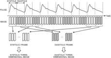
- FIG. 3is a diagram illustrating how a recording device generates three-dimensional image data
- FIG. 4is a diagram illustrating how an image observation device generates two-dimensional image data
- FIG. 5is a block diagram illustrating a configuration of an observation system according to a first modified example
- FIG. 6is a block diagram illustrating a configuration of an observation system according to a second modified example
- FIG. 7is a block diagram illustrating a configuration of an observation system according to a third modified example.
- FIG. 8is a diagram illustrating how biological information is generated from signal values of feature points.
- FIG. 9is a diagram illustrating how biological information is generated from coordinate values of feature points.
- Embodiments of a recording device, an image observation device, an observation system, a control method of the observation system, and an operating program for the observation systemwill be described below by reference to the drawings.
- the disclosureis not limited by these embodiments.
- the disclosureis generally applicable to recording devices using medical endoscopes, image observation devices using medical endoscopes, observation systems using medical endoscopes, control methods for the observation systems, and operating programs for the observation systems.
- FIG. 1is a block diagram illustrating a configuration of an observation system according to an embodiment of the disclosure.
- an observation system 1includes an endoscope observation system 10 , a recording device 20 , and an image observation device 30 , which are connected to one another via a network 2 , such as an Internet connection.
- a network 2such as an Internet connection.
- the endoscope observation system 10includes: an endoscope 11 that captures an image of the inside of the body of a subject that is a living body; a sensor unit 12 that detects biological information on the subject; an image processing device 13 that performs image processing on image data captured by the endoscope 11 ; and a display device 14 that displays an image according to image data generated by the image processing device 13 .
- the endoscope 11has an insertion unit to be inserted into the subject, and an operating unit that is connected consecutively to a proximal end of the insertion unit. According to input to the operating unit, an imaging device arranged at a distal end of the insertion unit captures an image of the inside of the body of the subject.
- the imaging deviceis an image sensor, such as a complementary metal oxide semiconductor (CMOS) or a charge coupled device (CCD).
- CMOScomplementary metal oxide semiconductor
- CCDcharge coupled device
- the sensor unit 12acquires biological information that is information on, for example, the pulse or respiration.
- the sensor unit 12is a bloodstream sensor that is fixed to hold a fingertip of the subject.
- the sensor unit 12may be a respiration sensor attached to, for example, the mouth, nose, or chest of the subject.
- the image processing device 13is configured using a work station or personal computer including a general-purpose processor or a dedicated processor.
- the general purpose processormay be, for example, a central processing unit (CPU), and the dedicated processor may be any of various arithmetic circuits that execute specific functions, such as application specific integrated circuits (ASICs) and field programmable gate arrays (FPGAs).
- ASICsapplication specific integrated circuits
- FPGAsfield programmable gate arrays
- the display device 14is configured using liquid crystal or organic electroluminescence, for example.
- the recording device 20is a server connected to plural devices, such as an endoscope observation system and an image observation device, including the endoscope observation system 10 and the image observation device 30 , via the network 2 .
- the recording device 20is implemented by, for example: a general-purpose processor, such as a CPU, or a dedicated processor, such as an arithmetic circuit that executes a specific function; a recording medium (a memory device), such as a semiconductor memory, an HDD, an MO, a CD-R, or a DVD-R; and a drive device that drives the recording medium.
- the arithmetic circuitmay be an ASIC or FPGA, and the semiconductor memory may be a flash memory, a random access memory (RAM), or a read only memory (ROM).
- the recording device 20includes: a biological information generating unit 21 that generates biological information on the subject; a recording unit 22 that records therein plural sets of image data in association with the biological information, the plural sets of image data having been generated by the endoscope 11 and chronologically arranged; and a three-dimensional image generating unit 23 that generates three-dimensional image data by selecting, based on the biological information, image data from the plural sets of image data that have been recorded in the recording unit 22 .
- the image observation device 30includes: an input device 31 that receives input of a position and an angle, at which the subject is observed; an image processing device 32 that reads three-dimensional image data recorded in the recording device 20 and generates image data; and a display device 33 that displays an image according to the image data generated by the image processing device 32 .
- the input device 31has a configuration similar to that of the operating unit of the endoscope 11 and receives input of a position and an angle, at which the subject is observed.
- the input device 31may be formed of, for example, at least one selected from the group of a mouse, a keyboard, a touch pad, and a touch panel.
- the image processing device 32is configured using a work station or personal computer including, for example, a general-purpose processor, such as a CPU, or a dedicated processor, such as an arithmetic circuit that executes a specific function.
- arithmetic circuitmay be an ASIC or FPGA.
- the image processing device 32has: an acquiring unit 321 that acquires the three-dimensional image data generated by the three-dimensional image generating unit 23 ; an angle-of-view information generating unit 322 that generates angle-of-view information, based on information received by the input device 31 ; and a two-dimensional image generating unit 323 that generates two-dimensional image data acquired by observation of a three-dimensional image, the observation being based on the biological information and the angle-of-view information.
- the display device 33is configured using liquid crystal or organic electroluminescence, for example.
- FIG. 2is a flow chart for observation using an observation system. As illustrated in FIG. 2 , by using the endoscope 11 , the first medical doctor captures images of the inside of the body of a subject (Step S 1 ).
- the sensor unit 12detects bloodstream information for a time period in which the endoscope 11 is capturing the images (Step S 2 ).
- the image processing device 13performs predetermined image processing on plural sets of image data generated by the endoscope 11 (Step S 3 ).
- the endoscope observation system 10transmits the plural sets of image data and the bloodstream information via the network 2 to the recording device 20 , and records the plural sets of image data and the bloodstream information into the recording device 20 (Step S 4 ).
- FIG. 3is a diagram illustrating how a recording device generates three-dimensional image data.
- the biological information generating unit 21determines whether each image captured by the endoscope 11 is an image captured in the systole or an image captured in the diastole. A result of this determination is the pulse information on each image.
- Systoleis a phase in which a heart contracts and blood is being sent out from the heart.
- diastoleis a phase in which the heart dilates and the arterial blood pressure is low according to the systemic vascular resistance.
- the blood flow volumes in organs inside a bodydiffer between the systole and the diastole, and color tones of the organs thus also differ between the systole and the diastole.
- the recording unit 22then records therein the plural sets of image data and the biological information in association with each other (Step S 6 ). Specifically, the recording unit 22 records therein each image included in the plural sets of image data in association with pulse information on that image.
- the three-dimensional image generating unit 23generates three-dimensional image data by selecting image data, based on the biological information, from the plural sets of image data (Step S 7 : three-dimensional image generating step). Specifically, as illustrated in FIG. 3 , the three-dimensional image generating unit 23 selects, from the plural sets of image data, an image of a systolic frame captured in the systole, and generates a systolic three-dimensional image. The three-dimensional image generating unit 23 selects, from the plural sets of image data, an image of a diastolic frame captured in the diastole, and generates a diastolic three-dimensional image.
- the three-dimensional image generating unit 23generates a three-dimensional image by extracting feature points from each image and joining the feature points in consecutive images in the chronological order.
- the systolic three-dimensional image and the diastolic three-dimensional image generated by the three-dimensional image generating unit 23are recorded into the recording unit 22 .
- the acquiring unit 321 of the image observation device 30acquires the three-dimensional image data and the biological information (Step S 8 ). Specifically, the acquiring unit 321 acquires the systolic three-dimensional image and the diastolic three-dimensional image from the recording unit 22 of the recording device 20 .
- the angle-of-view information generating unit 322Based on information received by the input device 31 , the angle-of-view information generating unit 322 generates angle-of-view information (Step S 9 : angle-of-view information generating step). Specifically, based on operation on the input device 31 by the second medical doctor, the angle-of-view information generating unit 322 generates angle-of-view information that temporally changes.
- the two-dimensional image generating unit 323generates two-dimensional image data acquired by observation of a three-dimensional image, the observation being based on biological information and angle-of-view information (Step S 10 : two-dimensional image generating step).
- FIG. 4is a diagram illustrating how an image observation device generates two-dimensional image data. As illustrated in FIG. 4 , based on pulse information, the two-dimensional image generating unit 323 generates a virtual pulse phase. This phase may be acquired by using the pulse information, or by calculating, based on the pulse information, the average value of periods or amplitudes, for example.
- the two-dimensional image generating unit 323Based on the phase and angle-of-view information, the two-dimensional image generating unit 323 generates, from a three-dimensional image corresponding to the phase, two-dimensional image data for a position and an angle indicated by the second medical doctor, the three-dimensional image being the systolic three-dimensional image or the diastolic three-dimensional image.
- the embodimentenables the second medical doctor to observe the inside of the body of a subject that is virtually pulsating and has changing bloodstream. That is, observation is facilitated because the second medical doctor is able to perform observation as if the second medical doctor is directly observing the inside of the body of the subject using the endoscope 11 .
- the second medical doctoris able to observe the inside of the body of a subject that virtually moves due to aspiration. That is, observation is facilitated because the second medical doctor is able to perform observation as if the second medical doctor is directly observing the inside of the body of the subject using the endoscope 11 .
- a virtual pulseis generated for observation of the inside of the body of a subject, but the systolic three-dimensional image or the diastolic three-dimensional image may be observed as is.
- FIG. 5is a block diagram illustrating a configuration of an observation system according to a first modified example.
- a recording device 20 A in an observation system 1 Afurther includes an image correcting unit 24 A that performs correction including: reducing change in shape that differs according to biological information; and enhancing change in color tones that differ according to the biological information.
- the image correcting unit 24 Acorrects shape information and color tone information on the systolic three-dimensional image and the diastolic three-dimensional image illustrated in FIG. 3 .
- Shape informationis, for example, three-dimensional coordinate values of feature points extracted when a three-dimensional image is generated, and color tone information is, for example, RGB signal values of the feature points.
- the shape information on the systolic three-dimensional imageis Ss
- the shape information on the diastolic three-dimensional imageis Se
- the corrected shape information on the systolic three-dimensional imageis Ss′
- the corrected shape information on the diastolic three-dimensional imageis Se′
- the correction coefficientis ks; the correction for reducing the change in shape is able to be performed on a three-dimensional image by using the following Equations (1) and (2).
- Ss′Ss+ks ( Se ⁇ Ss ), where 0 ⁇ ks ⁇ 0.5
- Se′Se ⁇ ks ( Se ⁇ Ss ) where 0 ⁇ ks ⁇ 0.5 (2)
- the corrected color tone information on the systolic three-dimensional imageis Ts
- the color tone information on the diastolic three-dimensional imageis Te
- the corrected color tone information on the systolic three-dimensional imageis Ts′
- the corrected color tone information on the diastolic three-dimensional imageis Te′
- the correction coefficientis kt
- the correction for enhancing the change in color tones in a three-dimensional imageis able to be performed by using the following Equations (3) and (4).
- Ts′Ts+ks ( Te ⁇ Ts ) where kt ⁇ 0 (3)
- Te′Te ⁇ ks ( Te ⁇ Ts ) where kt ⁇ 0 (4)
- the image correcting unit 24 Aperforming the correction, the change in shape between the systolic three-dimensional image and the diastolic three-dimensional image generated by the three-dimensional image generating unit 23 is reduced, and unnecessary movement due to the pulse, for example, in the two-dimensional image data is thus eliminated when the second medical doctor performs observation and the observation is thus facilitated. Furthermore, by the image correcting unit 24 A implementing the correction, the change in color tones between the systolic three-dimensional image and the diastolic three-dimensional image is increased, and the second medical doctor is able to easily observe a portion having a different color tone in the two-dimensional image data, the portion being, for example reddening or a tumor.
- the image correcting unit 24 Acorrects each of the shapes and the color tones of the systolic three-dimensional image and the diastolic three-dimensional image, but the first modified example is not limited to this example.
- the three-dimensional image generating unit 23may generate a shape of a three-dimensional image using all of images, generate a systolic three-dimensional image using systolic color tone information for the generated shape of the three-dimensional image, and generate a diastolic three-dimensional image using diastolic color tone information for the generated shape of the three-dimensional image.
- the image correcting unit 24 Amay then correct only the color tones of the systolic three-dimensional image and the diastolic three-dimensional image by using Equations (3) and (4) above. Similarly, the image correcting unit 24 A may correct only the shapes of three-dimensional images generated by the three-dimensional image generating unit 23 .
- the image correcting unit 24 Acorrects the systolic three-dimensional image and the diastolic three-dimensional image, but the first modified example is not limited to this example.
- the image correcting unit 24 Amay, for example, correct the image data generated by the endoscope 11 , and the three-dimensional image generating unit 23 may generate three-dimensional image data using the corrected image data.
- FIG. 6is a block diagram illustrating a configuration of an observation system according to a second modified example.
- an image processing device 13 B of an endoscope observation system 10 B in an observation system 1 Bfurther includes an imaging control unit 131 B that controls the timing for imaging by the endoscope 11 , according to biological information.
- imagingis enabled at times when the bloodstream becomes the maximum and the minimum, for example.
- the second medical doctoris then able to perform observation in a state where the differences between the systole and the diastole are large and thus the observation is facilitated.
- FIG. 7is a block diagram illustrating a configuration of an observation system according to a third modified example. As illustrated in FIG. 7 , an endoscope observation system 100 in an observation system 10 does not have a sensor unit.
- the biological information generating unit 21Based on temporal change in image data that have been recorded in the recording unit 22 , the biological information generating unit 21 generates biological information.
- FIG. 8is a diagram illustrating how biological information is generated from signal values of feature points.
- the biological information generating unit 21generates biological information, based on temporal change in RGB signal values of feature points extracted upon generation of a three-dimensional image. Specifically, the biological information generating unit 21 generates, based on temporal change in signal values of the R component and B component of feature points, biological information that is pulse information like the one illustrated in FIG. 3 .
- the biological information generating unit 21may generate pulse information, based on, for example, temporal change in color tones of the whole image or temporal change in color tones of an area extracted by predetermined image processing.
- the biological information generating unit 21may generate pulse information, based on information on any one of color components, such as the R component or the B component, or may generate pulse information by performing predetermined calculation with signal values of each color component.
- FIG. 9is a diagram illustrating how biological information is generated from coordinate values of feature points.
- the biological information generating unit 21generates biological information, based on temporal change in coordinate values of feature points extracted upon generation of a three-dimensional image. Specifically, the biological information generating unit 21 generates, based on temporal change in coordinates of feature points, biological information that is pulse information like the one illustrated in FIG. 3 .
- the biological information generating unit 21may generate pulse information, based on temporal change in coordinates of, for example, a contour detected from an image, without being limited to feature points.
- a recording devicean image observation device, an observation system, a control method of the observation system, and an operating program for the observation system that further facilitate observation are able to be provided, the observation involving acquisition of images from the recording device, the images having been captured using an endoscope.
Landscapes
- Health & Medical Sciences (AREA)
- Life Sciences & Earth Sciences (AREA)
- Engineering & Computer Science (AREA)
- Surgery (AREA)
- Physics & Mathematics (AREA)
- Pathology (AREA)
- Veterinary Medicine (AREA)
- Biophysics (AREA)
- General Health & Medical Sciences (AREA)
- Public Health (AREA)
- Biomedical Technology (AREA)
- Heart & Thoracic Surgery (AREA)
- Medical Informatics (AREA)
- Molecular Biology (AREA)
- Animal Behavior & Ethology (AREA)
- Signal Processing (AREA)
- Optics & Photonics (AREA)
- Radiology & Medical Imaging (AREA)
- Nuclear Medicine, Radiotherapy & Molecular Imaging (AREA)
- Cardiology (AREA)
- Theoretical Computer Science (AREA)
- Multimedia (AREA)
- Physiology (AREA)
- Vascular Medicine (AREA)
- Computing Systems (AREA)
- Geometry (AREA)
- Computer Graphics (AREA)
- General Physics & Mathematics (AREA)
- Hematology (AREA)
- Endoscopes (AREA)
Abstract
Description
This application is a continuation of International Application No. PCT/JP2018/032248, filed on Aug. 30, 2018, the entire contents of which are incorporated herein by reference.
The present disclosure relates to a recording device, an image observation device, an observation system, a control method of the observation system, and a computer-readable recording medium.
Observation of the inside of the bodies of subjects, which are living bodies, using endoscopes has been performed in the medical field (for example, see Japanese Patent Application Laid-open No. 2016-062488). Images captured by an endoscope are used in diagnosis performed by a first medical doctor operating the endoscope.
Studies have been made on: storage of the images captured by the endoscope into a recording device; and use of the stored images for diagnosis (a second opinion) by a second medical doctor who is at a place spatially or temporally away from where the first medical doctor is.
However, biological information of the subject such as pulse and respiration is not available when the second medical doctor observes the stored images, which makes diagnosis more difficult than when the subject is directly observed by inserting the endoscope into the body.
The disclosure addresses the above-described issue, and a general purpose thereof is to provide a recording device, an image observation device, an observation system, a control method of the observation system, and an operating program for the observation system that facilitate the observation using the recorded images captured by an endoscope.
To address the above issue, in some embodiments, a recording device includes: a memory; and a processor including hardware. The processor is configured to generate, based on temporal change in plural sets of image data that have been generated by an endoscope and arranged chronologically, biological information on a subject, associate the plural sets of image data with the biological information to record the plural sets of image data with the biological information into the memory, and select, based on the biological information, image data from the plural sets of image data that have been recorded in the memory to generate three-dimensional image data.
In some embodiments, an image observation device includes: a processor comprising hardware. The processor is configured to acquire data on a three-dimensional image, the data being generated by selection of a set of image data from plural sets of image data that have been generated by an endoscope and arranged chronologically, the selection being based on biological information on a subject, the biological information being generated based on temporal change in the plural sets of image data, generate angle-of-view information, based on information of a position and an angle at which the subject is observed, and generate two-dimensional image data acquired by observation of the three-dimensional image, the observation being based on the biological information and the angle-of-view information.
In some embodiments, an observation system includes: a memory; and a processor including hardware. The processor is configured to generate, based on temporal change in plural sets of image data that have been generated by an endoscope and arranged chronologically, biological information on a subject, associate the plural sets of image data with the biological information to record the plural sets of image data with the biological information into the memory, select, based on the biological information, image data from the plural sets of image data that have been recorded in the memory to generate data on a three-dimensional image, generate angle-of-view information, based on information of a position and an angle at which the subject is observed, and generate two-dimensional image data acquired by observation of the three-dimensional image, the observation being based on the biological information and the angle-of-view information.
In some embodiments, a control method of an observation system includes: generating, based on temporal change in plural sets of image data that have been generated by an endoscope and arranged chronologically, biological information on a subject; selecting, based on the biological information, image data from the plural sets of image data to generate data on a three-dimensional image; generating angle-of-view information, based on information of a position and an angle at which the subject is observed; and generating two-dimensional image data acquired by observation of the three-dimensional image, the observation being based on the biological information and the angle-of-view information.
In some embodiments, provided is a non-transitory computer-readable recording medium with an executable program stored thereon. The program causes an observation system to execute: generating, based on temporal change in plural sets of image data that have been generated by an endoscope and arranged chronologically, biological information on a subject; selecting, based on the biological information, image data from the plural sets of image data to generate data on a three-dimensional image; generating angle-of-view information, based on information of a position and an angle at which the subject is observed; and generating two-dimensional image data acquired by observation of the three-dimensional image, the observation being based on the biological information and the angle-of-view information.
The above and other features, advantages and technical and industrial significance of this disclosure will be better understood by reading the following detailed description of presently preferred embodiments of the disclosure, when considered in connection with the accompanying drawings.
Embodiments of a recording device, an image observation device, an observation system, a control method of the observation system, and an operating program for the observation system will be described below by reference to the drawings. The disclosure is not limited by these embodiments. The disclosure is generally applicable to recording devices using medical endoscopes, image observation devices using medical endoscopes, observation systems using medical endoscopes, control methods for the observation systems, and operating programs for the observation systems.
Any elements that are the same or corresponding to each other are assigned with the same reference sign throughout the drawings, as appropriate. In addition, it needs to be noted that the drawings are schematic and relations between dimensions of each element therein and proportions between the elements therein may be different from the actual ones. The drawings may also include a portion that differs in its dimensional relations or proportions between the drawings.
Theendoscope observation system 10 includes: anendoscope 11 that captures an image of the inside of the body of a subject that is a living body; a sensor unit12 that detects biological information on the subject; animage processing device 13 that performs image processing on image data captured by theendoscope 11; and adisplay device 14 that displays an image according to image data generated by theimage processing device 13.
Theendoscope 11 has an insertion unit to be inserted into the subject, and an operating unit that is connected consecutively to a proximal end of the insertion unit. According to input to the operating unit, an imaging device arranged at a distal end of the insertion unit captures an image of the inside of the body of the subject. The imaging device is an image sensor, such as a complementary metal oxide semiconductor (CMOS) or a charge coupled device (CCD).
The sensor unit12 acquires biological information that is information on, for example, the pulse or respiration. Specifically, the sensor unit12 is a bloodstream sensor that is fixed to hold a fingertip of the subject. Furthermore, the sensor unit12 may be a respiration sensor attached to, for example, the mouth, nose, or chest of the subject.
Theimage processing device 13 is configured using a work station or personal computer including a general-purpose processor or a dedicated processor. The general purpose processor may be, for example, a central processing unit (CPU), and the dedicated processor may be any of various arithmetic circuits that execute specific functions, such as application specific integrated circuits (ASICs) and field programmable gate arrays (FPGAs).
Thedisplay device 14 is configured using liquid crystal or organic electroluminescence, for example.
Therecording device 20 is a server connected to plural devices, such as an endoscope observation system and an image observation device, including theendoscope observation system 10 and theimage observation device 30, via thenetwork 2. Therecording device 20 is implemented by, for example: a general-purpose processor, such as a CPU, or a dedicated processor, such as an arithmetic circuit that executes a specific function; a recording medium (a memory device), such as a semiconductor memory, an HDD, an MO, a CD-R, or a DVD-R; and a drive device that drives the recording medium. The arithmetic circuit may be an ASIC or FPGA, and the semiconductor memory may be a flash memory, a random access memory (RAM), or a read only memory (ROM).
Therecording device 20 includes: a biologicalinformation generating unit 21 that generates biological information on the subject; arecording unit 22 that records therein plural sets of image data in association with the biological information, the plural sets of image data having been generated by theendoscope 11 and chronologically arranged; and a three-dimensionalimage generating unit 23 that generates three-dimensional image data by selecting, based on the biological information, image data from the plural sets of image data that have been recorded in therecording unit 22.
Theimage observation device 30 includes: an input device31 that receives input of a position and an angle, at which the subject is observed; an image processing device32 that reads three-dimensional image data recorded in therecording device 20 and generates image data; and adisplay device 33 that displays an image according to the image data generated by the image processing device32.
The input device31 has a configuration similar to that of the operating unit of theendoscope 11 and receives input of a position and an angle, at which the subject is observed. However, the input device31 may be formed of, for example, at least one selected from the group of a mouse, a keyboard, a touch pad, and a touch panel.
The image processing device32 is configured using a work station or personal computer including, for example, a general-purpose processor, such as a CPU, or a dedicated processor, such as an arithmetic circuit that executes a specific function. The arithmetic circuit may be an ASIC or FPGA.
The image processing device32 has: an acquiring unit321 that acquires the three-dimensional image data generated by the three-dimensionalimage generating unit 23; an angle-of-viewinformation generating unit 322 that generates angle-of-view information, based on information received by the input device31; and a two-dimensionalimage generating unit 323 that generates two-dimensional image data acquired by observation of a three-dimensional image, the observation being based on the biological information and the angle-of-view information.
Thedisplay device 33 is configured using liquid crystal or organic electroluminescence, for example.
Next, operation in which a second medical doctor observes an image using theimage observation device 30 will be described, the image having been captured by a first medical doctor using theendoscope observation system 10.FIG.2 is a flow chart for observation using an observation system. As illustrated inFIG.2 , by using theendoscope 11, the first medical doctor captures images of the inside of the body of a subject (Step S1).
Simultaneously, the sensor unit12 detects bloodstream information for a time period in which theendoscope 11 is capturing the images (Step S2).
Subsequently, theimage processing device 13 performs predetermined image processing on plural sets of image data generated by the endoscope11 (Step S3).
Thereafter, theendoscope observation system 10 transmits the plural sets of image data and the bloodstream information via thenetwork 2 to therecording device 20, and records the plural sets of image data and the bloodstream information into the recording device20 (Step S4).
Subsequently, the biologicalinformation generating unit 21 generates biological information that is pulse information, using the bloodstream information (Step S5: biological information generating step).FIG.3 is a diagram illustrating how a recording device generates three-dimensional image data. As illustrated inFIG.3 , based on the bloodstream information detected by the sensor unit12, the biologicalinformation generating unit 21 determines whether each image captured by theendoscope 11 is an image captured in the systole or an image captured in the diastole. A result of this determination is the pulse information on each image. Systole is a phase in which a heart contracts and blood is being sent out from the heart. On the contrary, diastole is a phase in which the heart dilates and the arterial blood pressure is low according to the systemic vascular resistance. The blood flow volumes in organs inside a body differ between the systole and the diastole, and color tones of the organs thus also differ between the systole and the diastole.
Therecording unit 22 then records therein the plural sets of image data and the biological information in association with each other (Step S6). Specifically, therecording unit 22 records therein each image included in the plural sets of image data in association with pulse information on that image.
Furthermore, the three-dimensionalimage generating unit 23 generates three-dimensional image data by selecting image data, based on the biological information, from the plural sets of image data (Step S7: three-dimensional image generating step). Specifically, as illustrated inFIG.3 , the three-dimensionalimage generating unit 23 selects, from the plural sets of image data, an image of a systolic frame captured in the systole, and generates a systolic three-dimensional image. The three-dimensionalimage generating unit 23 selects, from the plural sets of image data, an image of a diastolic frame captured in the diastole, and generates a diastolic three-dimensional image. The three-dimensionalimage generating unit 23 generates a three-dimensional image by extracting feature points from each image and joining the feature points in consecutive images in the chronological order. The systolic three-dimensional image and the diastolic three-dimensional image generated by the three-dimensionalimage generating unit 23 are recorded into therecording unit 22.
Subsequently, the acquiring unit321 of theimage observation device 30 acquires the three-dimensional image data and the biological information (Step S8). Specifically, the acquiring unit321 acquires the systolic three-dimensional image and the diastolic three-dimensional image from therecording unit 22 of therecording device 20.
Based on information received by the input device31, the angle-of-viewinformation generating unit 322 generates angle-of-view information (Step S9: angle-of-view information generating step). Specifically, based on operation on the input device31 by the second medical doctor, the angle-of-viewinformation generating unit 322 generates angle-of-view information that temporally changes.
Furthermore, the two-dimensionalimage generating unit 323 generates two-dimensional image data acquired by observation of a three-dimensional image, the observation being based on biological information and angle-of-view information (Step S10: two-dimensional image generating step).FIG.4 is a diagram illustrating how an image observation device generates two-dimensional image data. As illustrated inFIG.4 , based on pulse information, the two-dimensionalimage generating unit 323 generates a virtual pulse phase. This phase may be acquired by using the pulse information, or by calculating, based on the pulse information, the average value of periods or amplitudes, for example. Based on the phase and angle-of-view information, the two-dimensionalimage generating unit 323 generates, from a three-dimensional image corresponding to the phase, two-dimensional image data for a position and an angle indicated by the second medical doctor, the three-dimensional image being the systolic three-dimensional image or the diastolic three-dimensional image.
As described above, the embodiment enables the second medical doctor to observe the inside of the body of a subject that is virtually pulsating and has changing bloodstream. That is, observation is facilitated because the second medical doctor is able to perform observation as if the second medical doctor is directly observing the inside of the body of the subject using theendoscope 11.
Furthermore, when respiration information is used as the biological information, the second medical doctor is able to observe the inside of the body of a subject that virtually moves due to aspiration. That is, observation is facilitated because the second medical doctor is able to perform observation as if the second medical doctor is directly observing the inside of the body of the subject using theendoscope 11.
According to the above description of the embodiment, a virtual pulse is generated for observation of the inside of the body of a subject, but the systolic three-dimensional image or the diastolic three-dimensional image may be observed as is.
When the shape information on the systolic three-dimensional image is Ss, the shape information on the diastolic three-dimensional image is Se, the corrected shape information on the systolic three-dimensional image is Ss′, the corrected shape information on the diastolic three-dimensional image is Se′, and the correction coefficient is ks; the correction for reducing the change in shape is able to be performed on a three-dimensional image by using the following Equations (1) and (2).
Ss′=Ss+ks(Se−Ss), where 0<ks<0.5 (1)
Se′=Se−ks(Se−Ss) where 0<ks<0.5 (2)
Ss′=Ss+ks(Se−Ss), where 0<ks<0.5 (1)
Se′=Se−ks(Se−Ss) where 0<ks<0.5 (2)
Furthermore, when the color tone information on the systolic three-dimensional image is Ts, the color tone information on the diastolic three-dimensional image is Te, the corrected color tone information on the systolic three-dimensional image is Ts′, the corrected color tone information on the diastolic three-dimensional image is Te′, and the correction coefficient is kt; the correction for enhancing the change in color tones in a three-dimensional image is able to be performed by using the following Equations (3) and (4).
Ts′=Ts+ks(Te−Ts) wherekt<0 (3)
Te′=Te−ks(Te−Ts) wherekt<0 (4)
Ts′=Ts+ks(Te−Ts) wherekt<0 (3)
Te′=Te−ks(Te−Ts) wherekt<0 (4)
As described above, by theimage correcting unit 24A performing the correction, the change in shape between the systolic three-dimensional image and the diastolic three-dimensional image generated by the three-dimensionalimage generating unit 23 is reduced, and unnecessary movement due to the pulse, for example, in the two-dimensional image data is thus eliminated when the second medical doctor performs observation and the observation is thus facilitated. Furthermore, by theimage correcting unit 24A implementing the correction, the change in color tones between the systolic three-dimensional image and the diastolic three-dimensional image is increased, and the second medical doctor is able to easily observe a portion having a different color tone in the two-dimensional image data, the portion being, for example reddening or a tumor.
According to the above description of the first modified example, theimage correcting unit 24A corrects each of the shapes and the color tones of the systolic three-dimensional image and the diastolic three-dimensional image, but the first modified example is not limited to this example. The three-dimensionalimage generating unit 23 may generate a shape of a three-dimensional image using all of images, generate a systolic three-dimensional image using systolic color tone information for the generated shape of the three-dimensional image, and generate a diastolic three-dimensional image using diastolic color tone information for the generated shape of the three-dimensional image. Theimage correcting unit 24A may then correct only the color tones of the systolic three-dimensional image and the diastolic three-dimensional image by using Equations (3) and (4) above. Similarly, theimage correcting unit 24A may correct only the shapes of three-dimensional images generated by the three-dimensionalimage generating unit 23.
According to the above description of the first modified example, theimage correcting unit 24A corrects the systolic three-dimensional image and the diastolic three-dimensional image, but the first modified example is not limited to this example. Theimage correcting unit 24A may, for example, correct the image data generated by theendoscope 11, and the three-dimensionalimage generating unit 23 may generate three-dimensional image data using the corrected image data.
Based on temporal change in image data that have been recorded in therecording unit 22, the biologicalinformation generating unit 21 generates biological information.
According to the disclosure, a recording device, an image observation device, an observation system, a control method of the observation system, and an operating program for the observation system that further facilitate observation are able to be provided, the observation involving acquisition of images from the recording device, the images having been captured using an endoscope.
Additional advantages and modifications will readily occur to those skilled in the art. Therefore, the disclosure in its broader aspects is not limited to the specific details and representative embodiments shown and described herein. Accordingly, various modifications may be made without departing from the spirit or scope of the general inventive concept as defined by the appended claims and their equivalents.
Claims (4)
1. A recording device comprising:
a processor comprising hardware, the processor being configured to:
generate, based on temporal change in plural sets of image data that have been generated by an endoscope and arranged chronologically, pulse information on a subject;
associate the plural sets of image data with the pulse information to record the plural sets of image data with the pulse information into a memory; and
select, based on the pulse information, image data from the plural sets of image data that have been recorded in the memory to generate three-dimensional image data,
wherein the processor is further configured to perform correction to reduce change in shape that differs according to the pulse information and to enhance change in a color tone that differs according to the pulse information, and
wherein the processor is further configured to:
select image data in systole from the plural sets of image data that have been recorded in the memory to generate systolic three-dimensional image data, the systole being a phase in which a heart contracts and blood is being sent out from the heart;
select image data in diastole from the plural sets of image data that have been recorded in the memory to generate diastolic three-dimensional image data, the diastole being a phase in which the heart dilates and an arterial blood pressure is low according to a systemic vascular resistance; and
perform the correction to reduce change in shape between a systolic three-dimensional image and a diastolic three-dimensional image and to enhance change in color tone between the systolic three-dimensional image and the diastolic three-dimensional image.
2. The recording device according toclaim 1 , wherein the processor is further configured to:
extract feature points in images from the plural sets of image data that have been arranged chronologically; and
generate, based on temporal change in the extracted feature points, the pulse information on the subject.
3. The recording device according toclaim 1 , wherein the processor is further configured to:
extract feature points in images from the plural sets of image data that have been arranged chronologically; and
generate, based on temporal change in colors of the extracted feature points, the pulse information on the subject.
4. The recording device according toclaim 1 , wherein the processor is further configured to:
extract feature points in images from the plural sets of image data that have been arranged chronologically; and
generate, based on temporal change in coordinates of the extracted feature points, the pulse information on the subject.
Applications Claiming Priority (1)
| Application Number | Priority Date | Filing Date | Title |
|---|---|---|---|
| PCT/JP2018/032248WO2020044523A1 (en) | 2018-08-30 | 2018-08-30 | Recording device, image observation device, observation system, observation system control method, and observation system operating program |
Related Parent Applications (1)
| Application Number | Title | Priority Date | Filing Date |
|---|---|---|---|
| PCT/JP2018/032248ContinuationWO2020044523A1 (en) | 2018-08-30 | 2018-08-30 | Recording device, image observation device, observation system, observation system control method, and observation system operating program |
Publications (2)
| Publication Number | Publication Date |
|---|---|
| US20210192836A1 US20210192836A1 (en) | 2021-06-24 |
| US11653815B2true US11653815B2 (en) | 2023-05-23 |
Family
ID=69644009
Family Applications (1)
| Application Number | Title | Priority Date | Filing Date |
|---|---|---|---|
| US17/173,602ActiveUS11653815B2 (en) | 2018-08-30 | 2021-02-11 | Recording device, image observation device, observation system, control method of observation system, and computer-readable recording medium |
Country Status (4)
| Country | Link |
|---|---|
| US (1) | US11653815B2 (en) |
| JP (1) | JP6988001B2 (en) |
| CN (1) | CN112584738B (en) |
| WO (1) | WO2020044523A1 (en) |
Citations (208)
| Publication number | Priority date | Publication date | Assignee | Title |
|---|---|---|---|---|
| US6295465B1 (en)* | 1999-02-17 | 2001-09-25 | Siemens Medical Systems, Inc. | Myocardial perfusion studies using magnetic resonance imaging |
| US6398731B1 (en)* | 1997-07-25 | 2002-06-04 | Tomtec Imaging Systems Gmbh | Method for recording ultrasound images of moving objects |
| US20020072670A1 (en)* | 2000-12-07 | 2002-06-13 | Cedric Chenal | Acquisition, analysis and display of ultrasonic diagnostic cardiac images |
| US20020087068A1 (en)* | 2000-12-30 | 2002-07-04 | Foo Thomas K.F. | Method and apparatus for fast breath-held 3D MR data acquisition using variable sampling |
| JP2002282213A (en) | 2001-03-22 | 2002-10-02 | Olympus Optical Co Ltd | Endoscopic image filing system |
| US20030038802A1 (en)* | 2001-08-23 | 2003-02-27 | Johnson Richard K. | Automatic delineation of heart borders and surfaces from images |
| US20030160786A1 (en)* | 2002-02-28 | 2003-08-28 | Johnson Richard K. | Automatic determination of borders of body structures |
| US6628743B1 (en)* | 2002-11-26 | 2003-09-30 | Ge Medical Systems Global Technology Company, Llc | Method and apparatus for acquiring and analyzing cardiac data from a patient |
| US6674879B1 (en)* | 1998-03-30 | 2004-01-06 | Echovision, Inc. | Echocardiography workstation |
| US20040044283A1 (en)* | 2002-06-18 | 2004-03-04 | Kabushiki Kaisha Toshiba, Tokyo, Japan | Ultrasound diagnosis apparatus that adjusts a time phase between a plurality of image series |
| US20040077952A1 (en)* | 2002-10-21 | 2004-04-22 | Rafter Patrick G. | System and method for improved diagnostic image displays |
| US20040153128A1 (en)* | 2003-01-30 | 2004-08-05 | Mitta Suresh | Method and system for image processing and contour assessment |
| US20040167414A1 (en)* | 2002-12-05 | 2004-08-26 | Omron Healthcare Co., Ltd. | Pulse wave monitoring device |
| US20040249297A1 (en)* | 2002-12-23 | 2004-12-09 | Pfeiffer Ulrich J. | Apparatus for determining cardiovascular parameters |
| US20050018890A1 (en)* | 2003-07-24 | 2005-01-27 | Mcdonald John Alan | Segmentation of left ventriculograms using boosted decision trees |
| US20050043609A1 (en)* | 2003-01-30 | 2005-02-24 | Gregory Murphy | System and method for facilitating cardiac intervention |
| US6889071B2 (en)* | 2000-12-19 | 2005-05-03 | General Electric Company | Acquisition of high-temporal free-breathing MR images |
| US20050113665A1 (en)* | 2003-11-26 | 2005-05-26 | Mohr Kelly A. | Cardiac display methods and apparatus |
| US20050137661A1 (en)* | 2003-12-19 | 2005-06-23 | Sra Jasbir S. | Method and system of treatment of cardiac arrhythmias using 4D imaging |
| US20060025689A1 (en)* | 2002-06-07 | 2006-02-02 | Vikram Chalana | System and method to measure cardiac ejection fraction |
| US20060058618A1 (en)* | 2004-08-30 | 2006-03-16 | Masahide Nishiura | Medical kinematic analysis apparatus and a medical kinematic analysis method |
| US20060082677A1 (en)* | 2004-10-20 | 2006-04-20 | Fuji Photo Film Co., Ltd. | Brightness adjustment method and image processing apparatus |
| US20060122512A1 (en)* | 2004-05-31 | 2006-06-08 | Yasuhiko Abe | Ultrasonic diagnostic stystem and system and method for ultrasonic imaging |
| US20060241449A1 (en)* | 2005-04-12 | 2006-10-26 | Kabushiki Kaisha Toshiba | Ultrasound image diagnosis apparatus and an apparatus and method for processing an image display |
| US20060253031A1 (en)* | 2005-04-26 | 2006-11-09 | Altmann Andres C | Registration of ultrasound data with pre-acquired image |
| US20070014452A1 (en)* | 2003-12-01 | 2007-01-18 | Mitta Suresh | Method and system for image processing and assessment of a state of a heart |
| US20070083105A1 (en)* | 2005-09-22 | 2007-04-12 | Kabushiki Kaisha Toshiba | Magnetic resonance imaging apparatus and magnetic resonance imaging method |
| US20070106146A1 (en)* | 2005-10-28 | 2007-05-10 | Altmann Andres C | Synchronization of ultrasound imaging data with electrical mapping |
| US20070167733A1 (en)* | 2005-12-22 | 2007-07-19 | Mitsuharu Miyoshi | Magnetic resonance imaging apparatus |
| US20070167777A1 (en)* | 2005-12-27 | 2007-07-19 | Yasuhiko Abe | Ultrasonic image processing apparatus and control program for ultrasonic image processing apparatus |
| US20070248319A1 (en)* | 2006-03-01 | 2007-10-25 | Takuya Sakaguchi | Image processing apparatus |
| US20080085042A1 (en)* | 2006-10-09 | 2008-04-10 | Valery Trofimov | Registration of images of an organ using anatomical features outside the organ |
| US20080118126A1 (en)* | 2006-11-17 | 2008-05-22 | Takuya Sakaguchi | Image display method and image display apparatus |
| US20080170770A1 (en)* | 2007-01-15 | 2008-07-17 | Suri Jasjit S | method for tissue culture extraction |
| US20080181479A1 (en)* | 2002-06-07 | 2008-07-31 | Fuxing Yang | System and method for cardiac imaging |
| US20080281218A1 (en)* | 2007-05-07 | 2008-11-13 | Siemens Medical Solutions Usa, Inc. | System and Method for Selecting End of Diastole and End of Systole Frames |
| US20080285819A1 (en)* | 2006-08-30 | 2008-11-20 | The Trustees Of Columbia University In The City Of New York | Systems and method for composite elastography and wave imaging |
| US20080312527A1 (en)* | 2007-05-28 | 2008-12-18 | Jun Masumoto | Cardiac function analysis apparatus, method and program |
| US20090054776A1 (en)* | 2007-08-21 | 2009-02-26 | Kabushiki Kaisha Toshiba | Ultrasound diagnosis apparatus and method for acquiring 3-d images |
| US20090052613A1 (en)* | 2007-08-23 | 2009-02-26 | Takuya Sakaguchi | Three dimensional image processing apparatus and x-ray diagnosis apparatus |
| US20090082675A1 (en)* | 2007-09-26 | 2009-03-26 | Takayuki Gunji | Ultrasonic diagnostic apparatus and medical image display apparatus |
| US7522696B2 (en)* | 2006-04-06 | 2009-04-21 | General Electric Company | X-ray CT apparatus |
| US20090136109A1 (en)* | 2006-03-20 | 2009-05-28 | Koninklijke Philips Electronics, N.V. | Ultrasonic diagnosis by quantification of myocardial performance |
| US20090149734A1 (en)* | 2007-12-07 | 2009-06-11 | Kabushiki Kaisha Toshiba | Diagnostic imaging apparatus, magnetic resonance imaging apparatus, and x-ray ct apparatus |
| US20090148020A1 (en)* | 2007-12-07 | 2009-06-11 | Satoshi Sugiura | Image display apparatus and magnetic resonance imaging apparatus |
| US20090163815A1 (en)* | 2007-12-20 | 2009-06-25 | Tetsuya Kawagishi | Ultrasonic diagnosis device, ultrasonic image analysis device, and ultrasonic image analysis method |
| US20090207241A1 (en)* | 2006-05-31 | 2009-08-20 | National University Corporation Chiba University | Three-dimensional-image forming device, three dimensional-image forming method and program |
| US20090234220A1 (en) | 2008-03-12 | 2009-09-17 | Simens Aktiengesellschaft | Catheter and associated medical examination and treatment device |
| US20090287303A1 (en)* | 2008-05-13 | 2009-11-19 | Edwards Lifesciences Corporation | Physiologically harmonized tricuspid annuloplasty ring |
| US20090292175A1 (en)* | 2008-05-23 | 2009-11-26 | Olympus Medical Systems Corp. | Medical device |
| US20090311655A1 (en)* | 2008-06-16 | 2009-12-17 | Microsoft Corporation | Surgical procedure capture, modelling, and editing interactive playback |
| US20090312648A1 (en)* | 2008-06-16 | 2009-12-17 | Siemens Medical Solutions Usa, Inc. | Adaptive Medical Image Acquisition System |
| US20100027861A1 (en)* | 2005-08-30 | 2010-02-04 | University Of Maryland | Segmentation of regions in measurements of a body based on a deformable model |
| US20100081917A1 (en)* | 2008-09-30 | 2010-04-01 | Hongxuan Zhang | System for multi-dimensional anatomical functional imaging |
| US20100128943A1 (en)* | 2008-11-27 | 2010-05-27 | Kabushiki Kaisha Toshiba | Medical image generation apparatus, medical image storage apparatus, medical image display apparatus, and medical image display system |
| US20100174191A1 (en)* | 2008-12-16 | 2010-07-08 | Industrial Technology Research Institute | Apparatus and method for providing a dynamic 3d ultrasound image |
| US20100195887A1 (en)* | 2009-02-05 | 2010-08-05 | Kabushiki Kaisha Toshiba | Medical imaging apparatus, medical image processing apparatus, ultrasonic imaging apparatus, ultrasonic image processing apparatus and method of processing medical images |
| US20100208957A1 (en)* | 2008-06-17 | 2010-08-19 | Siemens Medical Solutions Usa, Inc. | Respiratory Motion Compensated Cardiac Wall Motion Determination System |
| JP2010279500A (en) | 2009-06-03 | 2010-12-16 | Toshiba Corp | Ultrasonic diagnostic apparatus and control program thereof |
| US20100317976A1 (en)* | 2008-02-15 | 2010-12-16 | Denis Chelma | Device and process for calculating new indices of arterial stiffness, and/or for stroke volume monitoring |
| US20110160849A1 (en)* | 2009-12-22 | 2011-06-30 | Edwards Lifesciences Corporation | Bimodal tricuspid annuloplasty ring |
| US20110218427A1 (en)* | 2010-03-08 | 2011-09-08 | Fujifilm Corporation | Diagnosis assisting apparatus, coronary artery analyzing method and recording medium having a coronary artery analyzing program stored therein |
| US20110282151A1 (en)* | 2008-10-20 | 2011-11-17 | Koninklijke Philips Electronics N.V. | Image-based localization method and system |
| US8083679B1 (en)* | 2004-09-03 | 2011-12-27 | Hitachi Medical Corporation | Ultrasonic imaging apparatus |
| US20120053408A1 (en)* | 2010-08-31 | 2012-03-01 | Fujifilm Corporation | Endoscopic image processing device, method and program |
| US20120089016A1 (en)* | 2010-10-12 | 2012-04-12 | Fujifilm Corporation | Diagnosis assisting apparatus, diagnosis assisting program, and diagnosis assisting method |
| US20120087561A1 (en)* | 2010-10-12 | 2012-04-12 | Siemens Corporation | Interaction method for regions of-interest in time series images |
| US20120093278A1 (en)* | 2010-10-15 | 2012-04-19 | Shinsuke Tsukagoshi | Medical image processing apparatus and x-ray computed tomography apparatus |
| US20120101368A1 (en)* | 2010-10-25 | 2012-04-26 | Fujifilm Corporation | Medical image diagnosis assisting apparatus, method, and program |
| US20120232853A1 (en)* | 2011-03-09 | 2012-09-13 | Siemens Corporation | Physically-constrained modeling of a heart in medical imaging |
| US20120281901A1 (en)* | 2010-01-22 | 2012-11-08 | Nobuyuki Yoshizawa | Magnetic resonance imaging apparatus and blood vessel image capturing method |
| US20120302870A1 (en)* | 2009-12-10 | 2012-11-29 | Bjaellmark Anna | System to quantify and visualize ventricular rotation pattern of the heart |
| US20130012835A1 (en)* | 2010-03-31 | 2013-01-10 | Hitachi Medical Corporation | Ultrasonic diagnostic apparatus and method for re-inputting measurement value of medical image |
| US20130013278A1 (en)* | 2011-07-08 | 2013-01-10 | Hu Wei-Chih | Non-invasive cardiovascular image matching method |
| US20130034287A1 (en)* | 2010-04-20 | 2013-02-07 | Hitachi Medical Corporation | Magnetic resonance imaging apparatus and blood vessel image capturing method |
| US20130053664A1 (en)* | 2010-01-29 | 2013-02-28 | Edwards Lifesciences Corporation | Elimination of the effects of irregular cardiac cycles in the determination of cardiovascular parameters |
| US20130137926A1 (en)* | 2010-07-28 | 2013-05-30 | Fujifilm Corporation | Image processing apparatus, method, and program |
| US8509511B2 (en)* | 2007-09-28 | 2013-08-13 | Kabushiki Kaisha Toshiba | Image processing apparatus and X-ray diagnostic apparatus |
| US20130267835A1 (en)* | 2012-04-10 | 2013-10-10 | Jerome Ranjeev Edwards | System and method for localizing medical instruments during cardiovascular medical procedures |
| US20130285655A1 (en)* | 2012-04-27 | 2013-10-31 | Toshiba Medical Systems Corporation | Apparatus and method for magnetic resonance imaging |
| US8577443B2 (en)* | 2006-09-06 | 2013-11-05 | Kabushiki Kaisha Toshiba | Magnetic resonance imaging apparatus and image processing apparatus |
| US20130336450A1 (en)* | 2012-04-12 | 2013-12-19 | Yiannis Kyriakou | Method for recording a four-dimensional angiography data record |
| US8620044B2 (en)* | 2003-04-25 | 2013-12-31 | Olympus Corporation | Image display apparatus, image display method, and computer program |
| US20140081079A1 (en)* | 2011-06-01 | 2014-03-20 | Toshiba Medical Systems Corporation | Medical image displaying apparatus and a medical image diagnostic apparatus |
| US20140111201A1 (en)* | 2012-10-23 | 2014-04-24 | Samsung Electronics Co., Ltd. | Magnetic resonance imaging system and magnetic resonance imaging method |
| US20140121496A1 (en)* | 2012-10-26 | 2014-05-01 | Siemens Medical Solutions Usa, Inc. | Automatic System for Timing In Imaging |
| US20150020547A1 (en)* | 2012-03-09 | 2015-01-22 | Kanazawa Medical University | Manufacturing method for heart correction net |
| US20150038860A1 (en)* | 2013-07-30 | 2015-02-05 | Heartflow, Inc. | Method and system for modeling blood flow with boundary conditions for optimized diagnostic performance |
| US9031387B2 (en)* | 2008-02-19 | 2015-05-12 | Olympus Corporation | Image processing apparatus |
| US20150145953A1 (en)* | 2012-03-17 | 2015-05-28 | Waseda University | Image completion system for in-image cutoff region, image processing device, and program therefor |
| US20150178938A1 (en)* | 2013-10-08 | 2015-06-25 | The Trustees Of The University Of Pennsylvania | Fully Automatic Image Segmentation of Heart Valves Using Multi-Atlas Label Fusion and Deformable Medial Modeling |
| US20150190038A1 (en)* | 2012-09-26 | 2015-07-09 | Fujifilm Corporation | Virtual endoscopic image generation device, method, and medium containing program |
| US20150193966A1 (en)* | 2012-09-26 | 2015-07-09 | Fujifilm Corporation | Virtual endoscopic image generation device, method, and medium containing program |
| US20150237325A1 (en)* | 2012-08-15 | 2015-08-20 | Industrial Technology Research Institute | Method and apparatus for converting 2d images to 3d images |
| US20150245772A1 (en)* | 2014-02-28 | 2015-09-03 | Nihon Kohden Corporation | Blood pressure measuring apparatus and blood pressure measuring method |
| US20150257655A1 (en)* | 2012-11-30 | 2015-09-17 | Kabushiki Kaisha Toshiba | Medical image processing apparatus |
| US9186051B2 (en)* | 2009-07-23 | 2015-11-17 | Olympus Corporation | Image processing device, computer-readable recording device, and image processing method |
| US20160038048A1 (en)* | 2013-04-04 | 2016-02-11 | Healthstats International Pte Ltd | Method and system for detecting heartbeat irregularities |
| US9275473B2 (en)* | 2011-06-16 | 2016-03-01 | Olympus Corporation | Image processing apparatus, image processing method, and program |
| US20160061920A1 (en)* | 2013-04-03 | 2016-03-03 | Choukri Mekkaoui | Mapping cardiac tissue architecture systems and methods |
| US20160063707A1 (en)* | 2014-08-27 | 2016-03-03 | Fujifilm Corporation | Image registration device, image registration method, and image registration program |
| US20160070877A1 (en)* | 2014-09-09 | 2016-03-10 | Heartflow, Inc. | Method and system for quantifying limitations in coronary artery blood flow during physical activity in patients with coronary artery disease |
| US20160100766A1 (en)* | 2014-10-09 | 2016-04-14 | Panasonic Intellectual Property Management Co., Ltd. | Non-contact blood-pressure measuring device and non-contact blood-pressure measuring method |
| JP2016062488A (en) | 2014-09-19 | 2016-04-25 | オリンパス株式会社 | Endoscope business support system |
| US20160125596A1 (en)* | 2014-11-03 | 2016-05-05 | Samsung Electronics Co., Ltd. | Medical imaging apparatus and method of processing medical image |
| US20160220190A1 (en)* | 2015-01-29 | 2016-08-04 | Siemens Medical Solutions Usa, Inc. | Patient signal analysis based on affine template matching |
| US20160269713A1 (en)* | 2014-01-24 | 2016-09-15 | Olympus Corporation | Stereoscopic endoscopic image processing apparatus |
| JP2016179121A (en) | 2015-03-25 | 2016-10-13 | 富士フイルム株式会社 | Endoscopy support apparatus, method and program |
| US20160310077A1 (en)* | 2014-09-17 | 2016-10-27 | William L. Hunter | Devices, systems and methods for using and monitoring medical devices |
| US9516993B2 (en)* | 2013-03-27 | 2016-12-13 | Olympus Corporation | Endoscope system |
| US9521330B2 (en)* | 2010-12-02 | 2016-12-13 | Olympus Corporation | Endoscopic image processing device, information storage device and image processing method |
| US9547940B1 (en)* | 2014-09-12 | 2017-01-17 | University Of South Florida | Systems and methods for providing augmented reality in minimally invasive surgery |
| US20170065230A1 (en)* | 2015-06-15 | 2017-03-09 | Vital Labs, Inc. | Method and system for acquiring data for assessment of cardiovascular disease |
| JP6095879B1 (en) | 2015-10-29 | 2017-03-15 | オリンパス株式会社 | Endoscope device |
| US20170084036A1 (en)* | 2015-09-21 | 2017-03-23 | Siemens Aktiengesellschaft | Registration of video camera with medical imaging |
| WO2017073181A1 (en) | 2015-10-29 | 2017-05-04 | オリンパス株式会社 | Endoscope apparatus |
| US9684849B2 (en)* | 2012-09-27 | 2017-06-20 | Olympus Corporation | Image processing device, information storage device, and image processing method |
| US20170188986A1 (en)* | 2014-09-19 | 2017-07-06 | Olympus Corporation | Medical observation system |
| US20170245835A1 (en)* | 2016-02-26 | 2017-08-31 | Toshiba Medical Systems Corporation | Ultrasound diagnosis apparatus and image processing method |
| JP2017153953A (en) | 2016-02-26 | 2017-09-07 | 東芝メディカルシステムズ株式会社 | Ultrasonic diagnostic apparatus and image processing program |
| US20170252000A1 (en)* | 2016-03-04 | 2017-09-07 | Toshiba Medical Systems Corporation | Analyzer |
| US20170296055A1 (en)* | 2015-01-06 | 2017-10-19 | Cardiovascular Biotherapeutics, Inc. | Angiogenic treatment of ischemic heart disease |
| US20170311839A1 (en)* | 2016-04-27 | 2017-11-02 | Myocardial Solutions, Inc. | Rapid quantitative evaluations of heart function with strain measurements from mri |
| US20170347975A1 (en)* | 2016-06-06 | 2017-12-07 | Toshiba Medical Systems Corporation | X-ray ct apparatus, medical information processing apparatus, and medical information processing method |
| US20170367659A1 (en)* | 2016-06-24 | 2017-12-28 | Qualcomm Incorporated | Dynamic Calibration of A Blood Pressure Measurement Device |
| US20180020932A1 (en)* | 2015-03-20 | 2018-01-25 | East Carolina University | Multi-spectral physiologic visualization (mspv) using laser imaging methods and systems for blood flow and perfusion imaging and quantification in an endoscopic design |
| US20180055364A1 (en)* | 2016-08-25 | 2018-03-01 | Vivonics, Inc. | Contactless System and Method For Measuring and Continuously Monitoring Arterial Blood Pressure |
| US20180108138A1 (en)* | 2015-04-29 | 2018-04-19 | Siemens Aktiengesellschaft | Method and system for semantic segmentation in laparoscopic and endoscopic 2d/2.5d image data |
| US9959618B2 (en)* | 2014-03-17 | 2018-05-01 | Olympus Corporation | Image processing apparatus, image processing method, and computer-readable recording medium |
| US20180150929A1 (en)* | 2015-05-11 | 2018-05-31 | Siemens Aktiengesellschaft | Method and system for registration of 2d/2.5d laparoscopic and endoscopic image data to 3d volumetric image data |
| US20180153621A1 (en)* | 2015-05-22 | 2018-06-07 | Intuitive Surgical Operations, Inc. | Systems and Methods of Registration for Image Guided Surgery |
| US10010379B1 (en)* | 2017-02-21 | 2018-07-03 | Novarad Corporation | Augmented reality viewing and tagging for medical procedures |
| US20180184920A1 (en)* | 2017-01-05 | 2018-07-05 | Livemetric (Medical) S.A. | System and method for providing user feeedback of blood pressure sensor placement and contact quality |
| US20180246178A1 (en)* | 2015-05-14 | 2018-08-30 | Ohio State Innovation Foundation | Systems and methods for estimating complex b1+ fields of transmit coils of a magnetic resonance imaging (mri) system |
| US20180263503A1 (en)* | 2017-03-16 | 2018-09-20 | Nihon Kohden Corporation | Circulatory dynamic measuring apparatus and circulatory dynamic measuring method |
| US20180289270A1 (en)* | 2017-04-06 | 2018-10-11 | ContinUse Biometrics Ltd. | System and method for blood pressure measurement |
| US10198811B2 (en)* | 2014-03-20 | 2019-02-05 | Olympus Corporation | Image processing apparatus, image processing method, and computer-readable recording medium |
| US10206555B2 (en)* | 2015-11-26 | 2019-02-19 | Olympus Corporation | Image processing apparatus, image processing method, and computer readable recording medium |
| US10210610B2 (en)* | 2014-07-30 | 2019-02-19 | Olympus Corporation | Image processing apparatus for generating combined image signal of region-of-interest image signal and second image signal, the region-of-interest image signal being generated based on blank portion and initial region-of-interest of first image signal |
| US20190089895A1 (en)* | 2016-05-27 | 2019-03-21 | Olympus Corporation | Image processing device, operation method of image processing device, and computer-readable recording medium |
| US20190090818A1 (en)* | 2016-04-15 | 2019-03-28 | Omron Corporation | Biological information analyzing device, system, and program |
| US20190167233A1 (en)* | 2016-06-01 | 2019-06-06 | The Trustees Of Columbia University In The City Of New York | Methods and systems for ultrasound elastography |
| US10325369B2 (en)* | 2017-01-23 | 2019-06-18 | Shanghai United Imaging Healthcare Co., Ltd. | Method and system for analyzing blood flow condition |
| US20190239861A1 (en)* | 2018-02-07 | 2019-08-08 | Canon Medical Systems Corporation | Ultrasonic diagnostic apparatus |
| US20190247127A1 (en)* | 2018-02-15 | 2019-08-15 | Covidien Lp | 3d reconstruction and guidance based on combined endobronchial ultrasound and magnetic tracking |
| US20190247142A1 (en)* | 2018-02-15 | 2019-08-15 | Leica Instruments (Singapore) Pte. Ltd. | Image processing method and apparatus using elastic mapping of vascular plexus structures |
| US20190290247A1 (en)* | 2016-05-31 | 2019-09-26 | Koninklijke Philips N.V. | Image-based fusion of endoscopic image and ultrasound images |
| US20190304107A1 (en)* | 2017-02-24 | 2019-10-03 | Fujifilm Corporation | Additional information display device, additional information display method, and additional information display program |
| US20190298290A1 (en)* | 2018-03-29 | 2019-10-03 | Konica Minolta, Inc. | Imaging support apparatus and radiographic imaging system |
| US20190304129A1 (en)* | 2018-03-27 | 2019-10-03 | Siemens Medical Solutions Usa, Inc. | Image-based guidance for navigating tubular networks |
| US20190347814A1 (en)* | 2016-06-02 | 2019-11-14 | Verily Life Sciences Llc | System and method for 3d scene reconstruction with dual complementary pattern illumination |
| US20190357854A1 (en)* | 2009-04-22 | 2019-11-28 | Streamline Automation, Llc | Cardiovascular state determination apparatus and method of use thereof |
| US20190380781A1 (en)* | 2018-06-13 | 2019-12-19 | Johnfk Medical Inc. | Airway model generation system and intubation assistance system |
| US10568497B2 (en)* | 2015-09-18 | 2020-02-25 | Olympus Corporation | Signal processing apparatus and endoscope system with composite image generation |
| US20200060528A1 (en)* | 2017-06-15 | 2020-02-27 | Olympus Corporation | Endoscope system, endoscope image processing method, and computer readable recording medium |
| US20200077869A1 (en)* | 2017-05-19 | 2020-03-12 | Olympus Corporation | 3d endoscope apparatus and 3d video processing apparatus |
| US20200085579A1 (en)* | 2017-06-13 | 2020-03-19 | Wake Forest University Health Sciences | Customized ventricular support device |
| US20200085281A1 (en)* | 2018-09-19 | 2020-03-19 | Siemens Healthcare Gmbh | Method for supporting a user, computer program product, data medium and imaging system |
| US20200085315A1 (en)* | 2018-09-18 | 2020-03-19 | Samsung Electronics Co., Ltd. | Apparatus and method for estimating bio-information |
| US20200146568A1 (en)* | 2018-11-12 | 2020-05-14 | Samsung Electronics Co., Ltd. | Blood pressure measuring apparatus and blood pressure measuring method |
| US10679358B2 (en)* | 2015-12-22 | 2020-06-09 | Olympus Corporation | Learning image automatic sorting device, learning image automatic sorting method, and learning image automatic sorting program |
| US20200184723A1 (en)* | 2017-08-25 | 2020-06-11 | Beijing Keeyoo Technologies Co., Ltd. | Image processing method and device |
| US20200183013A1 (en)* | 2017-05-31 | 2020-06-11 | Sharp Kabushiki Kaisha | Three-dimensional image element and optical radar device |
| US20200187897A1 (en)* | 2018-12-14 | 2020-06-18 | Canon Medical Systems Corporation | Ultrasonic wave diagnostic apparatus, medical information processing apparatus, and computer program product |
| US20200219272A1 (en)* | 2019-01-07 | 2020-07-09 | The University Of North Carolina At Chapel Hill | Methods, systems, and computer readable media for deriving a three-dimensional (3d) textured surface from endoscopic video |
| US10732246B2 (en)* | 2016-08-30 | 2020-08-04 | Canon Medical Systems Corporation | Magnetic resonance imaging apparatus |
| US10748279B2 (en)* | 2015-12-24 | 2020-08-18 | Olympus Corporation | Image processing apparatus, image processing method, and computer readable recording medium |
| US20200273148A1 (en)* | 2017-11-01 | 2020-08-27 | Beijing Keeyoo Technologies Co., Ltd. | Image processing method and device |
| US10765297B2 (en)* | 2015-12-25 | 2020-09-08 | Olympus Corporation | Image processing apparatus, image processing method, and computer readable recording medium |
| US10776921B2 (en)* | 2016-05-19 | 2020-09-15 | Olympus Corporation | Image processing apparatus, operation method for image processing apparatus, and recording medium |
| US10776998B1 (en)* | 2014-02-28 | 2020-09-15 | University Of Louisville Research Foundation, Inc. | Method and system for analysis of 3D deformations and regional function of a heart with 3D SinMod |
| US20200345291A1 (en)* | 2019-05-01 | 2020-11-05 | Stuart M. Bradley | Systems and methods for measuring volumes and dimensions of objects and features during swallowing observation |
| US20200345215A1 (en)* | 2017-09-08 | 2020-11-05 | Sony Corporation | An imaging device, method and program for producing images of a scene |
| US20200387706A1 (en)* | 2019-06-04 | 2020-12-10 | Magentiq Eye Ltd | Systems and methods for processing colon images and videos |
| US20200383588A1 (en)* | 2018-01-13 | 2020-12-10 | Delta Tooling Co. Ltd. | Blood pressure estimation device, blood pressure estimation method, computer program, and storage medium |
| US20210015340A1 (en)* | 2018-04-09 | 2021-01-21 | Olympus Corporation | Endoscopic task supporting system and endoscopic task supporting method |
| US10918358B2 (en)* | 2015-07-10 | 2021-02-16 | Shenzhen Mindray Bio-Medical Electronics Co., Ltd. | Monitoring system method and device |
| US10918260B2 (en)* | 2017-02-02 | 2021-02-16 | Olympus Corporation | Endoscopic image observation support system |
| US20210052146A1 (en)* | 2019-08-19 | 2021-02-25 | Covidien Lp | Systems and methods for selectively varying resolutions |
| US20210052240A1 (en)* | 2019-08-19 | 2021-02-25 | Covidien Lp | Systems and methods of fluoro-ct imaging for initial registration |
| US20210068683A1 (en)* | 2009-04-22 | 2021-03-11 | Alton Reich | Cardiovascular state monitoring - drug delivery apparatus and method of use thereof |
| US20210089079A1 (en)* | 2019-09-19 | 2021-03-25 | Samsung Electronics Co., Ltd. | Foldable electronic device and method of estimating bioinformation using the same |
| US20210121079A1 (en)* | 2019-10-24 | 2021-04-29 | Nihon Kohden Corporation | Apparatus for measuring hemodynamic parameters |
| US20210146117A1 (en)* | 2009-04-22 | 2021-05-20 | Alton Reich | Cardiac output control apparatus and method of use thereof |
| US20210158526A1 (en)* | 2018-04-13 | 2021-05-27 | Koninklijke Philips N.V. | Automatic slice selection in medical imaging |
| US20210153745A1 (en)* | 2019-11-21 | 2021-05-27 | Gb Soft Inc. | Method of measuring physiological parameter of subject in contactless manner |
| US20210153756A1 (en)* | 2017-05-18 | 2021-05-27 | Preventicus Gmbh | Reliable acquisition of photoplethysmographic data |
| US20210196386A1 (en)* | 2019-12-30 | 2021-07-01 | Ethicon Llc | Analyzing surgical trends by a surgical system |
| US11055865B2 (en)* | 2018-08-30 | 2021-07-06 | Olympus Corporation | Image acquisition device and method of operating image acquisition device |
| US20210216822A1 (en)* | 2019-10-01 | 2021-07-15 | Sirona Medical, Inc. | Complex image data analysis using artificial intelligence and machine learning algorithms |
| US20210280312A1 (en)* | 2020-03-06 | 2021-09-09 | Verily Life Sciences Llc | Detecting deficient coverage in gastroenterological procedures |
| US11145053B2 (en)* | 2016-12-07 | 2021-10-12 | Olympus Corporation | Image processing apparatus and computer-readable storage medium storing instructions for specifying lesion portion and performing differentiation classification in response to judging that differentiation classification operation is engaged based on signal from endoscope |
| US20210386299A1 (en)* | 2020-06-16 | 2021-12-16 | Grant Hocking | Method to Quantify the Hemodynamic and Vascular Properties in Vivo from Arterial Waveform Measurements |
| US20210398304A1 (en)* | 2018-11-07 | 2021-12-23 | Sony Group Corporation | Medical observation system configured to generate three-dimensional information and to calculate an estimated region and a corresponding method |
| US20220020118A1 (en)* | 2020-07-15 | 2022-01-20 | Alcon Inc. | Digital Image Optimization For Ophthalmic Surgery |
| US20220020166A1 (en)* | 2019-09-27 | 2022-01-20 | Asensus Surgical Us, Inc. | Method and System for Providing Real Time Surgical Site Measurements |
| US11232568B2 (en)* | 2019-02-06 | 2022-01-25 | Olympus Corporation | Three-dimensional image display method, three-dimensional image display device, and recording medium |
| US11232570B2 (en)* | 2020-02-13 | 2022-01-25 | Olympus Corporation | System and method for diagnosing severity of gastritis |
| US20220039666A1 (en)* | 2020-08-05 | 2022-02-10 | Samsung Electronics Co., Ltd. | Apparatus and method for estimating bio-information |
| US20220104790A1 (en)* | 2020-10-02 | 2022-04-07 | Caption Health, Inc. | Continuous and dynamic ejection fraction determination |
| US20220222825A1 (en)* | 2021-01-10 | 2022-07-14 | DiA Imaging Analysis | Automated right ventricle medical imaging and computation of clinical parameters |
| US20220248962A1 (en)* | 2019-05-20 | 2022-08-11 | Shenzhen Darma Technology Co., Ltd. | Cardiac diastolic function assessment method, device, and system |
| US20220265149A1 (en)* | 2021-02-25 | 2022-08-25 | Samsung Electronics Co., Ltd. | Apparatus and method for measuring multiple bio-signals using image sensor, ad electronic device |
| US20220296111A1 (en)* | 2020-12-18 | 2022-09-22 | Movano Inc. | Method for operating a wearable device that includes an optical sensor system and an rf sensor system |
| US20220313214A1 (en)* | 2021-04-02 | 2022-10-06 | Canon Medical Systems Corporation | Ultrasonic diagnostic apparatus, image processing apparatus, and image processing method |
| US11471065B2 (en)* | 2017-05-12 | 2022-10-18 | Canon Medical Systems Corporation | Medical image diagnosis apparatus |
| US20220409071A1 (en)* | 2019-11-27 | 2022-12-29 | Directed Systems | Cardiovascular analytic system and method |
| US11565136B2 (en)* | 2014-09-02 | 2023-01-31 | Universite de Bordeaux | System for the ablation or the monitoring of a zone of the heart by ultrasounds and associated methods |
Family Cites Families (7)
| Publication number | Priority date | Publication date | Assignee | Title |
|---|---|---|---|---|
| CN1725978A (en)* | 2002-12-13 | 2006-01-25 | 皇家飞利浦电子股份有限公司 | System and method for processing a series of image frames representing a cardiac cycle |
| CN100496407C (en)* | 2004-12-13 | 2009-06-10 | 株式会社日立医药 | Ultrasonic diagnostic apparatus |
| CN103298407B (en)* | 2011-06-23 | 2016-05-18 | 株式会社东芝 | Medical image processing device and medical image diagnosis device |
| JP5981248B2 (en)* | 2011-07-06 | 2016-08-31 | 東芝メディカルシステムズ株式会社 | Medical diagnostic imaging equipment |
| JP6125281B2 (en)* | 2013-03-06 | 2017-05-10 | 東芝メディカルシステムズ株式会社 | Medical image diagnostic apparatus, medical image processing apparatus, and control program |
| EP3127470A1 (en)* | 2014-06-18 | 2017-02-08 | Olympus Corporation | Image-processing device |
| JP6763719B2 (en)* | 2015-12-07 | 2020-09-30 | パナソニック株式会社 | Biometric information measuring device, biometric information measuring method and program |
- 2018
- 2018-08-30CNCN201880096645.6Apatent/CN112584738B/enactiveActive
- 2018-08-30JPJP2020539972Apatent/JP6988001B2/enactiveActive
- 2018-08-30WOPCT/JP2018/032248patent/WO2020044523A1/ennot_activeCeased
- 2021
- 2021-02-11USUS17/173,602patent/US11653815B2/enactiveActive
Patent Citations (209)
| Publication number | Priority date | Publication date | Assignee | Title |
|---|---|---|---|---|
| US6398731B1 (en)* | 1997-07-25 | 2002-06-04 | Tomtec Imaging Systems Gmbh | Method for recording ultrasound images of moving objects |
| US6674879B1 (en)* | 1998-03-30 | 2004-01-06 | Echovision, Inc. | Echocardiography workstation |
| US6295465B1 (en)* | 1999-02-17 | 2001-09-25 | Siemens Medical Systems, Inc. | Myocardial perfusion studies using magnetic resonance imaging |
| US20020072670A1 (en)* | 2000-12-07 | 2002-06-13 | Cedric Chenal | Acquisition, analysis and display of ultrasonic diagnostic cardiac images |
| US6889071B2 (en)* | 2000-12-19 | 2005-05-03 | General Electric Company | Acquisition of high-temporal free-breathing MR images |
| US20020087068A1 (en)* | 2000-12-30 | 2002-07-04 | Foo Thomas K.F. | Method and apparatus for fast breath-held 3D MR data acquisition using variable sampling |
| JP2002282213A (en) | 2001-03-22 | 2002-10-02 | Olympus Optical Co Ltd | Endoscopic image filing system |
| US20030038802A1 (en)* | 2001-08-23 | 2003-02-27 | Johnson Richard K. | Automatic delineation of heart borders and surfaces from images |
| US20030160786A1 (en)* | 2002-02-28 | 2003-08-28 | Johnson Richard K. | Automatic determination of borders of body structures |
| US20080181479A1 (en)* | 2002-06-07 | 2008-07-31 | Fuxing Yang | System and method for cardiac imaging |
| US20060025689A1 (en)* | 2002-06-07 | 2006-02-02 | Vikram Chalana | System and method to measure cardiac ejection fraction |
| US20040044283A1 (en)* | 2002-06-18 | 2004-03-04 | Kabushiki Kaisha Toshiba, Tokyo, Japan | Ultrasound diagnosis apparatus that adjusts a time phase between a plurality of image series |
| US20040077952A1 (en)* | 2002-10-21 | 2004-04-22 | Rafter Patrick G. | System and method for improved diagnostic image displays |
| US6628743B1 (en)* | 2002-11-26 | 2003-09-30 | Ge Medical Systems Global Technology Company, Llc | Method and apparatus for acquiring and analyzing cardiac data from a patient |
| US20040167414A1 (en)* | 2002-12-05 | 2004-08-26 | Omron Healthcare Co., Ltd. | Pulse wave monitoring device |
| US20040249297A1 (en)* | 2002-12-23 | 2004-12-09 | Pfeiffer Ulrich J. | Apparatus for determining cardiovascular parameters |
| US20040153128A1 (en)* | 2003-01-30 | 2004-08-05 | Mitta Suresh | Method and system for image processing and contour assessment |
| US20050043609A1 (en)* | 2003-01-30 | 2005-02-24 | Gregory Murphy | System and method for facilitating cardiac intervention |
| US8620044B2 (en)* | 2003-04-25 | 2013-12-31 | Olympus Corporation | Image display apparatus, image display method, and computer program |
| US20050018890A1 (en)* | 2003-07-24 | 2005-01-27 | Mcdonald John Alan | Segmentation of left ventriculograms using boosted decision trees |
| US20050113665A1 (en)* | 2003-11-26 | 2005-05-26 | Mohr Kelly A. | Cardiac display methods and apparatus |
| US20070014452A1 (en)* | 2003-12-01 | 2007-01-18 | Mitta Suresh | Method and system for image processing and assessment of a state of a heart |
| US20050137661A1 (en)* | 2003-12-19 | 2005-06-23 | Sra Jasbir S. | Method and system of treatment of cardiac arrhythmias using 4D imaging |
| US20060122512A1 (en)* | 2004-05-31 | 2006-06-08 | Yasuhiko Abe | Ultrasonic diagnostic stystem and system and method for ultrasonic imaging |
| US20060058618A1 (en)* | 2004-08-30 | 2006-03-16 | Masahide Nishiura | Medical kinematic analysis apparatus and a medical kinematic analysis method |
| US8083679B1 (en)* | 2004-09-03 | 2011-12-27 | Hitachi Medical Corporation | Ultrasonic imaging apparatus |
| US20060082677A1 (en)* | 2004-10-20 | 2006-04-20 | Fuji Photo Film Co., Ltd. | Brightness adjustment method and image processing apparatus |
| US20060241449A1 (en)* | 2005-04-12 | 2006-10-26 | Kabushiki Kaisha Toshiba | Ultrasound image diagnosis apparatus and an apparatus and method for processing an image display |
| US20060253031A1 (en)* | 2005-04-26 | 2006-11-09 | Altmann Andres C | Registration of ultrasound data with pre-acquired image |
| US20100027861A1 (en)* | 2005-08-30 | 2010-02-04 | University Of Maryland | Segmentation of regions in measurements of a body based on a deformable model |
| US20070083105A1 (en)* | 2005-09-22 | 2007-04-12 | Kabushiki Kaisha Toshiba | Magnetic resonance imaging apparatus and magnetic resonance imaging method |
| US20070106146A1 (en)* | 2005-10-28 | 2007-05-10 | Altmann Andres C | Synchronization of ultrasound imaging data with electrical mapping |
| US20070167733A1 (en)* | 2005-12-22 | 2007-07-19 | Mitsuharu Miyoshi | Magnetic resonance imaging apparatus |
| US20070167777A1 (en)* | 2005-12-27 | 2007-07-19 | Yasuhiko Abe | Ultrasonic image processing apparatus and control program for ultrasonic image processing apparatus |
| US20070248319A1 (en)* | 2006-03-01 | 2007-10-25 | Takuya Sakaguchi | Image processing apparatus |
| US20090136109A1 (en)* | 2006-03-20 | 2009-05-28 | Koninklijke Philips Electronics, N.V. | Ultrasonic diagnosis by quantification of myocardial performance |
| US7522696B2 (en)* | 2006-04-06 | 2009-04-21 | General Electric Company | X-ray CT apparatus |
| US20090207241A1 (en)* | 2006-05-31 | 2009-08-20 | National University Corporation Chiba University | Three-dimensional-image forming device, three dimensional-image forming method and program |
| US20080285819A1 (en)* | 2006-08-30 | 2008-11-20 | The Trustees Of Columbia University In The City Of New York | Systems and method for composite elastography and wave imaging |
| US8577443B2 (en)* | 2006-09-06 | 2013-11-05 | Kabushiki Kaisha Toshiba | Magnetic resonance imaging apparatus and image processing apparatus |
| US20080085042A1 (en)* | 2006-10-09 | 2008-04-10 | Valery Trofimov | Registration of images of an organ using anatomical features outside the organ |
| US20080118126A1 (en)* | 2006-11-17 | 2008-05-22 | Takuya Sakaguchi | Image display method and image display apparatus |
| US20080170770A1 (en)* | 2007-01-15 | 2008-07-17 | Suri Jasjit S | method for tissue culture extraction |
| US20080281218A1 (en)* | 2007-05-07 | 2008-11-13 | Siemens Medical Solutions Usa, Inc. | System and Method for Selecting End of Diastole and End of Systole Frames |
| US20080312527A1 (en)* | 2007-05-28 | 2008-12-18 | Jun Masumoto | Cardiac function analysis apparatus, method and program |
| US20090054776A1 (en)* | 2007-08-21 | 2009-02-26 | Kabushiki Kaisha Toshiba | Ultrasound diagnosis apparatus and method for acquiring 3-d images |
| US20090052613A1 (en)* | 2007-08-23 | 2009-02-26 | Takuya Sakaguchi | Three dimensional image processing apparatus and x-ray diagnosis apparatus |
| US20090082675A1 (en)* | 2007-09-26 | 2009-03-26 | Takayuki Gunji | Ultrasonic diagnostic apparatus and medical image display apparatus |
| US8509511B2 (en)* | 2007-09-28 | 2013-08-13 | Kabushiki Kaisha Toshiba | Image processing apparatus and X-ray diagnostic apparatus |
| US20090149734A1 (en)* | 2007-12-07 | 2009-06-11 | Kabushiki Kaisha Toshiba | Diagnostic imaging apparatus, magnetic resonance imaging apparatus, and x-ray ct apparatus |
| US20090148020A1 (en)* | 2007-12-07 | 2009-06-11 | Satoshi Sugiura | Image display apparatus and magnetic resonance imaging apparatus |
| US20090163815A1 (en)* | 2007-12-20 | 2009-06-25 | Tetsuya Kawagishi | Ultrasonic diagnosis device, ultrasonic image analysis device, and ultrasonic image analysis method |
| US20100317976A1 (en)* | 2008-02-15 | 2010-12-16 | Denis Chelma | Device and process for calculating new indices of arterial stiffness, and/or for stroke volume monitoring |
| US9031387B2 (en)* | 2008-02-19 | 2015-05-12 | Olympus Corporation | Image processing apparatus |
| US20090234220A1 (en) | 2008-03-12 | 2009-09-17 | Simens Aktiengesellschaft | Catheter and associated medical examination and treatment device |
| US20090287303A1 (en)* | 2008-05-13 | 2009-11-19 | Edwards Lifesciences Corporation | Physiologically harmonized tricuspid annuloplasty ring |
| US20090292175A1 (en)* | 2008-05-23 | 2009-11-26 | Olympus Medical Systems Corp. | Medical device |
| US20090312648A1 (en)* | 2008-06-16 | 2009-12-17 | Siemens Medical Solutions Usa, Inc. | Adaptive Medical Image Acquisition System |
| US20090311655A1 (en)* | 2008-06-16 | 2009-12-17 | Microsoft Corporation | Surgical procedure capture, modelling, and editing interactive playback |
| US20100208957A1 (en)* | 2008-06-17 | 2010-08-19 | Siemens Medical Solutions Usa, Inc. | Respiratory Motion Compensated Cardiac Wall Motion Determination System |
| US20100081917A1 (en)* | 2008-09-30 | 2010-04-01 | Hongxuan Zhang | System for multi-dimensional anatomical functional imaging |
| US20110282151A1 (en)* | 2008-10-20 | 2011-11-17 | Koninklijke Philips Electronics N.V. | Image-based localization method and system |
| US20100128943A1 (en)* | 2008-11-27 | 2010-05-27 | Kabushiki Kaisha Toshiba | Medical image generation apparatus, medical image storage apparatus, medical image display apparatus, and medical image display system |
| US20100174191A1 (en)* | 2008-12-16 | 2010-07-08 | Industrial Technology Research Institute | Apparatus and method for providing a dynamic 3d ultrasound image |
| US20100195887A1 (en)* | 2009-02-05 | 2010-08-05 | Kabushiki Kaisha Toshiba | Medical imaging apparatus, medical image processing apparatus, ultrasonic imaging apparatus, ultrasonic image processing apparatus and method of processing medical images |
| US20210146117A1 (en)* | 2009-04-22 | 2021-05-20 | Alton Reich | Cardiac output control apparatus and method of use thereof |
| US20190357854A1 (en)* | 2009-04-22 | 2019-11-28 | Streamline Automation, Llc | Cardiovascular state determination apparatus and method of use thereof |
| US20210068683A1 (en)* | 2009-04-22 | 2021-03-11 | Alton Reich | Cardiovascular state monitoring - drug delivery apparatus and method of use thereof |
| JP2010279500A (en) | 2009-06-03 | 2010-12-16 | Toshiba Corp | Ultrasonic diagnostic apparatus and control program thereof |
| US9186051B2 (en)* | 2009-07-23 | 2015-11-17 | Olympus Corporation | Image processing device, computer-readable recording device, and image processing method |
| US20120302870A1 (en)* | 2009-12-10 | 2012-11-29 | Bjaellmark Anna | System to quantify and visualize ventricular rotation pattern of the heart |
| US20110160849A1 (en)* | 2009-12-22 | 2011-06-30 | Edwards Lifesciences Corporation | Bimodal tricuspid annuloplasty ring |
| US20120281901A1 (en)* | 2010-01-22 | 2012-11-08 | Nobuyuki Yoshizawa | Magnetic resonance imaging apparatus and blood vessel image capturing method |
| US20130053664A1 (en)* | 2010-01-29 | 2013-02-28 | Edwards Lifesciences Corporation | Elimination of the effects of irregular cardiac cycles in the determination of cardiovascular parameters |
| US20110218427A1 (en)* | 2010-03-08 | 2011-09-08 | Fujifilm Corporation | Diagnosis assisting apparatus, coronary artery analyzing method and recording medium having a coronary artery analyzing program stored therein |
| US20130012835A1 (en)* | 2010-03-31 | 2013-01-10 | Hitachi Medical Corporation | Ultrasonic diagnostic apparatus and method for re-inputting measurement value of medical image |
| US20130034287A1 (en)* | 2010-04-20 | 2013-02-07 | Hitachi Medical Corporation | Magnetic resonance imaging apparatus and blood vessel image capturing method |
| US20130137926A1 (en)* | 2010-07-28 | 2013-05-30 | Fujifilm Corporation | Image processing apparatus, method, and program |
| US20120053408A1 (en)* | 2010-08-31 | 2012-03-01 | Fujifilm Corporation | Endoscopic image processing device, method and program |
| US20120087561A1 (en)* | 2010-10-12 | 2012-04-12 | Siemens Corporation | Interaction method for regions of-interest in time series images |
| US20120089016A1 (en)* | 2010-10-12 | 2012-04-12 | Fujifilm Corporation | Diagnosis assisting apparatus, diagnosis assisting program, and diagnosis assisting method |
| US20120093278A1 (en)* | 2010-10-15 | 2012-04-19 | Shinsuke Tsukagoshi | Medical image processing apparatus and x-ray computed tomography apparatus |
| US20120101368A1 (en)* | 2010-10-25 | 2012-04-26 | Fujifilm Corporation | Medical image diagnosis assisting apparatus, method, and program |
| US9521330B2 (en)* | 2010-12-02 | 2016-12-13 | Olympus Corporation | Endoscopic image processing device, information storage device and image processing method |
| US20120232853A1 (en)* | 2011-03-09 | 2012-09-13 | Siemens Corporation | Physically-constrained modeling of a heart in medical imaging |
| US20140081079A1 (en)* | 2011-06-01 | 2014-03-20 | Toshiba Medical Systems Corporation | Medical image displaying apparatus and a medical image diagnostic apparatus |
| US9275473B2 (en)* | 2011-06-16 | 2016-03-01 | Olympus Corporation | Image processing apparatus, image processing method, and program |
| US20130013278A1 (en)* | 2011-07-08 | 2013-01-10 | Hu Wei-Chih | Non-invasive cardiovascular image matching method |
| US20150020547A1 (en)* | 2012-03-09 | 2015-01-22 | Kanazawa Medical University | Manufacturing method for heart correction net |
| US20150145953A1 (en)* | 2012-03-17 | 2015-05-28 | Waseda University | Image completion system for in-image cutoff region, image processing device, and program therefor |
| US20130267835A1 (en)* | 2012-04-10 | 2013-10-10 | Jerome Ranjeev Edwards | System and method for localizing medical instruments during cardiovascular medical procedures |
| US20130336450A1 (en)* | 2012-04-12 | 2013-12-19 | Yiannis Kyriakou | Method for recording a four-dimensional angiography data record |
| US20130285655A1 (en)* | 2012-04-27 | 2013-10-31 | Toshiba Medical Systems Corporation | Apparatus and method for magnetic resonance imaging |
| US20150237325A1 (en)* | 2012-08-15 | 2015-08-20 | Industrial Technology Research Institute | Method and apparatus for converting 2d images to 3d images |
| US20150190038A1 (en)* | 2012-09-26 | 2015-07-09 | Fujifilm Corporation | Virtual endoscopic image generation device, method, and medium containing program |
| US20150193966A1 (en)* | 2012-09-26 | 2015-07-09 | Fujifilm Corporation | Virtual endoscopic image generation device, method, and medium containing program |
| US9684849B2 (en)* | 2012-09-27 | 2017-06-20 | Olympus Corporation | Image processing device, information storage device, and image processing method |
| US20140111201A1 (en)* | 2012-10-23 | 2014-04-24 | Samsung Electronics Co., Ltd. | Magnetic resonance imaging system and magnetic resonance imaging method |
| US20140121496A1 (en)* | 2012-10-26 | 2014-05-01 | Siemens Medical Solutions Usa, Inc. | Automatic System for Timing In Imaging |
| US20150257655A1 (en)* | 2012-11-30 | 2015-09-17 | Kabushiki Kaisha Toshiba | Medical image processing apparatus |
| US9516993B2 (en)* | 2013-03-27 | 2016-12-13 | Olympus Corporation | Endoscope system |
| US20160061920A1 (en)* | 2013-04-03 | 2016-03-03 | Choukri Mekkaoui | Mapping cardiac tissue architecture systems and methods |
| US20160038048A1 (en)* | 2013-04-04 | 2016-02-11 | Healthstats International Pte Ltd | Method and system for detecting heartbeat irregularities |
| US20150038860A1 (en)* | 2013-07-30 | 2015-02-05 | Heartflow, Inc. | Method and system for modeling blood flow with boundary conditions for optimized diagnostic performance |
| US20150178938A1 (en)* | 2013-10-08 | 2015-06-25 | The Trustees Of The University Of Pennsylvania | Fully Automatic Image Segmentation of Heart Valves Using Multi-Atlas Label Fusion and Deformable Medial Modeling |
| US20160269713A1 (en)* | 2014-01-24 | 2016-09-15 | Olympus Corporation | Stereoscopic endoscopic image processing apparatus |
| US10776998B1 (en)* | 2014-02-28 | 2020-09-15 | University Of Louisville Research Foundation, Inc. | Method and system for analysis of 3D deformations and regional function of a heart with 3D SinMod |
| US20150245772A1 (en)* | 2014-02-28 | 2015-09-03 | Nihon Kohden Corporation | Blood pressure measuring apparatus and blood pressure measuring method |
| US9959618B2 (en)* | 2014-03-17 | 2018-05-01 | Olympus Corporation | Image processing apparatus, image processing method, and computer-readable recording medium |
| US10198811B2 (en)* | 2014-03-20 | 2019-02-05 | Olympus Corporation | Image processing apparatus, image processing method, and computer-readable recording medium |
| US10210610B2 (en)* | 2014-07-30 | 2019-02-19 | Olympus Corporation | Image processing apparatus for generating combined image signal of region-of-interest image signal and second image signal, the region-of-interest image signal being generated based on blank portion and initial region-of-interest of first image signal |
| US20160063707A1 (en)* | 2014-08-27 | 2016-03-03 | Fujifilm Corporation | Image registration device, image registration method, and image registration program |
| US11565136B2 (en)* | 2014-09-02 | 2023-01-31 | Universite de Bordeaux | System for the ablation or the monitoring of a zone of the heart by ultrasounds and associated methods |
| US20160070877A1 (en)* | 2014-09-09 | 2016-03-10 | Heartflow, Inc. | Method and system for quantifying limitations in coronary artery blood flow during physical activity in patients with coronary artery disease |
| US9547940B1 (en)* | 2014-09-12 | 2017-01-17 | University Of South Florida | Systems and methods for providing augmented reality in minimally invasive surgery |
| US20160310077A1 (en)* | 2014-09-17 | 2016-10-27 | William L. Hunter | Devices, systems and methods for using and monitoring medical devices |
| JP2016062488A (en) | 2014-09-19 | 2016-04-25 | オリンパス株式会社 | Endoscope business support system |
| US20170188986A1 (en)* | 2014-09-19 | 2017-07-06 | Olympus Corporation | Medical observation system |
| US20160100766A1 (en)* | 2014-10-09 | 2016-04-14 | Panasonic Intellectual Property Management Co., Ltd. | Non-contact blood-pressure measuring device and non-contact blood-pressure measuring method |
| US20160125596A1 (en)* | 2014-11-03 | 2016-05-05 | Samsung Electronics Co., Ltd. | Medical imaging apparatus and method of processing medical image |
| US20170296055A1 (en)* | 2015-01-06 | 2017-10-19 | Cardiovascular Biotherapeutics, Inc. | Angiogenic treatment of ischemic heart disease |
| US20160220190A1 (en)* | 2015-01-29 | 2016-08-04 | Siemens Medical Solutions Usa, Inc. | Patient signal analysis based on affine template matching |
| US20180020932A1 (en)* | 2015-03-20 | 2018-01-25 | East Carolina University | Multi-spectral physiologic visualization (mspv) using laser imaging methods and systems for blood flow and perfusion imaging and quantification in an endoscopic design |
| JP2016179121A (en) | 2015-03-25 | 2016-10-13 | 富士フイルム株式会社 | Endoscopy support apparatus, method and program |
| US20180108138A1 (en)* | 2015-04-29 | 2018-04-19 | Siemens Aktiengesellschaft | Method and system for semantic segmentation in laparoscopic and endoscopic 2d/2.5d image data |
| US20180150929A1 (en)* | 2015-05-11 | 2018-05-31 | Siemens Aktiengesellschaft | Method and system for registration of 2d/2.5d laparoscopic and endoscopic image data to 3d volumetric image data |
| US20180246178A1 (en)* | 2015-05-14 | 2018-08-30 | Ohio State Innovation Foundation | Systems and methods for estimating complex b1+ fields of transmit coils of a magnetic resonance imaging (mri) system |
| US20180153621A1 (en)* | 2015-05-22 | 2018-06-07 | Intuitive Surgical Operations, Inc. | Systems and Methods of Registration for Image Guided Surgery |
| US20170065230A1 (en)* | 2015-06-15 | 2017-03-09 | Vital Labs, Inc. | Method and system for acquiring data for assessment of cardiovascular disease |
| US10918358B2 (en)* | 2015-07-10 | 2021-02-16 | Shenzhen Mindray Bio-Medical Electronics Co., Ltd. | Monitoring system method and device |
| US10568497B2 (en)* | 2015-09-18 | 2020-02-25 | Olympus Corporation | Signal processing apparatus and endoscope system with composite image generation |
| US20170084036A1 (en)* | 2015-09-21 | 2017-03-23 | Siemens Aktiengesellschaft | Registration of video camera with medical imaging |
| WO2017073181A1 (en) | 2015-10-29 | 2017-05-04 | オリンパス株式会社 | Endoscope apparatus |
| JP6095879B1 (en) | 2015-10-29 | 2017-03-15 | オリンパス株式会社 | Endoscope device |
| US10206555B2 (en)* | 2015-11-26 | 2019-02-19 | Olympus Corporation | Image processing apparatus, image processing method, and computer readable recording medium |
| US10679358B2 (en)* | 2015-12-22 | 2020-06-09 | Olympus Corporation | Learning image automatic sorting device, learning image automatic sorting method, and learning image automatic sorting program |
| US10748279B2 (en)* | 2015-12-24 | 2020-08-18 | Olympus Corporation | Image processing apparatus, image processing method, and computer readable recording medium |
| US10765297B2 (en)* | 2015-12-25 | 2020-09-08 | Olympus Corporation | Image processing apparatus, image processing method, and computer readable recording medium |
| US20170245835A1 (en)* | 2016-02-26 | 2017-08-31 | Toshiba Medical Systems Corporation | Ultrasound diagnosis apparatus and image processing method |
| JP2017153953A (en) | 2016-02-26 | 2017-09-07 | 東芝メディカルシステムズ株式会社 | Ultrasonic diagnostic apparatus and image processing program |
| US20170252000A1 (en)* | 2016-03-04 | 2017-09-07 | Toshiba Medical Systems Corporation | Analyzer |
| US20190090818A1 (en)* | 2016-04-15 | 2019-03-28 | Omron Corporation | Biological information analyzing device, system, and program |
| US20170311839A1 (en)* | 2016-04-27 | 2017-11-02 | Myocardial Solutions, Inc. | Rapid quantitative evaluations of heart function with strain measurements from mri |
| US10776921B2 (en)* | 2016-05-19 | 2020-09-15 | Olympus Corporation | Image processing apparatus, operation method for image processing apparatus, and recording medium |
| US20190089895A1 (en)* | 2016-05-27 | 2019-03-21 | Olympus Corporation | Image processing device, operation method of image processing device, and computer-readable recording medium |
| US20190290247A1 (en)* | 2016-05-31 | 2019-09-26 | Koninklijke Philips N.V. | Image-based fusion of endoscopic image and ultrasound images |
| US20190167233A1 (en)* | 2016-06-01 | 2019-06-06 | The Trustees Of Columbia University In The City Of New York | Methods and systems for ultrasound elastography |
| US20190347814A1 (en)* | 2016-06-02 | 2019-11-14 | Verily Life Sciences Llc | System and method for 3d scene reconstruction with dual complementary pattern illumination |
| US20170347975A1 (en)* | 2016-06-06 | 2017-12-07 | Toshiba Medical Systems Corporation | X-ray ct apparatus, medical information processing apparatus, and medical information processing method |
| US20170367659A1 (en)* | 2016-06-24 | 2017-12-28 | Qualcomm Incorporated | Dynamic Calibration of A Blood Pressure Measurement Device |
| US20180055364A1 (en)* | 2016-08-25 | 2018-03-01 | Vivonics, Inc. | Contactless System and Method For Measuring and Continuously Monitoring Arterial Blood Pressure |
| US10732246B2 (en)* | 2016-08-30 | 2020-08-04 | Canon Medical Systems Corporation | Magnetic resonance imaging apparatus |
| US11145053B2 (en)* | 2016-12-07 | 2021-10-12 | Olympus Corporation | Image processing apparatus and computer-readable storage medium storing instructions for specifying lesion portion and performing differentiation classification in response to judging that differentiation classification operation is engaged based on signal from endoscope |
| US20180184920A1 (en)* | 2017-01-05 | 2018-07-05 | Livemetric (Medical) S.A. | System and method for providing user feeedback of blood pressure sensor placement and contact quality |
| US10325369B2 (en)* | 2017-01-23 | 2019-06-18 | Shanghai United Imaging Healthcare Co., Ltd. | Method and system for analyzing blood flow condition |
| US10918260B2 (en)* | 2017-02-02 | 2021-02-16 | Olympus Corporation | Endoscopic image observation support system |
| US10010379B1 (en)* | 2017-02-21 | 2018-07-03 | Novarad Corporation | Augmented reality viewing and tagging for medical procedures |
| US20190304107A1 (en)* | 2017-02-24 | 2019-10-03 | Fujifilm Corporation | Additional information display device, additional information display method, and additional information display program |
| US20180263503A1 (en)* | 2017-03-16 | 2018-09-20 | Nihon Kohden Corporation | Circulatory dynamic measuring apparatus and circulatory dynamic measuring method |
| US20180289270A1 (en)* | 2017-04-06 | 2018-10-11 | ContinUse Biometrics Ltd. | System and method for blood pressure measurement |
| US11471065B2 (en)* | 2017-05-12 | 2022-10-18 | Canon Medical Systems Corporation | Medical image diagnosis apparatus |
| US20210153756A1 (en)* | 2017-05-18 | 2021-05-27 | Preventicus Gmbh | Reliable acquisition of photoplethysmographic data |
| US20200077869A1 (en)* | 2017-05-19 | 2020-03-12 | Olympus Corporation | 3d endoscope apparatus and 3d video processing apparatus |
| US20200183013A1 (en)* | 2017-05-31 | 2020-06-11 | Sharp Kabushiki Kaisha | Three-dimensional image element and optical radar device |
| US20200085579A1 (en)* | 2017-06-13 | 2020-03-19 | Wake Forest University Health Sciences | Customized ventricular support device |
| US20200060528A1 (en)* | 2017-06-15 | 2020-02-27 | Olympus Corporation | Endoscope system, endoscope image processing method, and computer readable recording medium |
| US20200184723A1 (en)* | 2017-08-25 | 2020-06-11 | Beijing Keeyoo Technologies Co., Ltd. | Image processing method and device |
| US20200345215A1 (en)* | 2017-09-08 | 2020-11-05 | Sony Corporation | An imaging device, method and program for producing images of a scene |
| US20200273148A1 (en)* | 2017-11-01 | 2020-08-27 | Beijing Keeyoo Technologies Co., Ltd. | Image processing method and device |
| US20200383588A1 (en)* | 2018-01-13 | 2020-12-10 | Delta Tooling Co. Ltd. | Blood pressure estimation device, blood pressure estimation method, computer program, and storage medium |
| US20190239861A1 (en)* | 2018-02-07 | 2019-08-08 | Canon Medical Systems Corporation | Ultrasonic diagnostic apparatus |
| US20190247127A1 (en)* | 2018-02-15 | 2019-08-15 | Covidien Lp | 3d reconstruction and guidance based on combined endobronchial ultrasound and magnetic tracking |
| US20190247142A1 (en)* | 2018-02-15 | 2019-08-15 | Leica Instruments (Singapore) Pte. Ltd. | Image processing method and apparatus using elastic mapping of vascular plexus structures |
| US20190304129A1 (en)* | 2018-03-27 | 2019-10-03 | Siemens Medical Solutions Usa, Inc. | Image-based guidance for navigating tubular networks |
| US20190298290A1 (en)* | 2018-03-29 | 2019-10-03 | Konica Minolta, Inc. | Imaging support apparatus and radiographic imaging system |
| US20210015340A1 (en)* | 2018-04-09 | 2021-01-21 | Olympus Corporation | Endoscopic task supporting system and endoscopic task supporting method |
| US20210158526A1 (en)* | 2018-04-13 | 2021-05-27 | Koninklijke Philips N.V. | Automatic slice selection in medical imaging |
| US20190380781A1 (en)* | 2018-06-13 | 2019-12-19 | Johnfk Medical Inc. | Airway model generation system and intubation assistance system |
| US11055865B2 (en)* | 2018-08-30 | 2021-07-06 | Olympus Corporation | Image acquisition device and method of operating image acquisition device |
| US20200085315A1 (en)* | 2018-09-18 | 2020-03-19 | Samsung Electronics Co., Ltd. | Apparatus and method for estimating bio-information |
| US20200085281A1 (en)* | 2018-09-19 | 2020-03-19 | Siemens Healthcare Gmbh | Method for supporting a user, computer program product, data medium and imaging system |
| US20210398304A1 (en)* | 2018-11-07 | 2021-12-23 | Sony Group Corporation | Medical observation system configured to generate three-dimensional information and to calculate an estimated region and a corresponding method |
| US20200146568A1 (en)* | 2018-11-12 | 2020-05-14 | Samsung Electronics Co., Ltd. | Blood pressure measuring apparatus and blood pressure measuring method |
| US20200187897A1 (en)* | 2018-12-14 | 2020-06-18 | Canon Medical Systems Corporation | Ultrasonic wave diagnostic apparatus, medical information processing apparatus, and computer program product |
| US20200219272A1 (en)* | 2019-01-07 | 2020-07-09 | The University Of North Carolina At Chapel Hill | Methods, systems, and computer readable media for deriving a three-dimensional (3d) textured surface from endoscopic video |
| US11232568B2 (en)* | 2019-02-06 | 2022-01-25 | Olympus Corporation | Three-dimensional image display method, three-dimensional image display device, and recording medium |
| US20200345291A1 (en)* | 2019-05-01 | 2020-11-05 | Stuart M. Bradley | Systems and methods for measuring volumes and dimensions of objects and features during swallowing observation |
| US20220248962A1 (en)* | 2019-05-20 | 2022-08-11 | Shenzhen Darma Technology Co., Ltd. | Cardiac diastolic function assessment method, device, and system |
| US20200387706A1 (en)* | 2019-06-04 | 2020-12-10 | Magentiq Eye Ltd | Systems and methods for processing colon images and videos |
| US20210052146A1 (en)* | 2019-08-19 | 2021-02-25 | Covidien Lp | Systems and methods for selectively varying resolutions |
| US20210052240A1 (en)* | 2019-08-19 | 2021-02-25 | Covidien Lp | Systems and methods of fluoro-ct imaging for initial registration |
| US20210089079A1 (en)* | 2019-09-19 | 2021-03-25 | Samsung Electronics Co., Ltd. | Foldable electronic device and method of estimating bioinformation using the same |
| US20220020166A1 (en)* | 2019-09-27 | 2022-01-20 | Asensus Surgical Us, Inc. | Method and System for Providing Real Time Surgical Site Measurements |
| US20210216822A1 (en)* | 2019-10-01 | 2021-07-15 | Sirona Medical, Inc. | Complex image data analysis using artificial intelligence and machine learning algorithms |
| US20210121079A1 (en)* | 2019-10-24 | 2021-04-29 | Nihon Kohden Corporation | Apparatus for measuring hemodynamic parameters |
| US20210153752A1 (en)* | 2019-11-21 | 2021-05-27 | Gb Soft Inc. | Method of measuring physiological parameter of subject in contactless manner |
| US20210153745A1 (en)* | 2019-11-21 | 2021-05-27 | Gb Soft Inc. | Method of measuring physiological parameter of subject in contactless manner |
| US20220409071A1 (en)* | 2019-11-27 | 2022-12-29 | Directed Systems | Cardiovascular analytic system and method |
| US20210196386A1 (en)* | 2019-12-30 | 2021-07-01 | Ethicon Llc | Analyzing surgical trends by a surgical system |
| US11232570B2 (en)* | 2020-02-13 | 2022-01-25 | Olympus Corporation | System and method for diagnosing severity of gastritis |
| US20210280312A1 (en)* | 2020-03-06 | 2021-09-09 | Verily Life Sciences Llc | Detecting deficient coverage in gastroenterological procedures |
| US20210386299A1 (en)* | 2020-06-16 | 2021-12-16 | Grant Hocking | Method to Quantify the Hemodynamic and Vascular Properties in Vivo from Arterial Waveform Measurements |
| US20220020118A1 (en)* | 2020-07-15 | 2022-01-20 | Alcon Inc. | Digital Image Optimization For Ophthalmic Surgery |
| US20220039666A1 (en)* | 2020-08-05 | 2022-02-10 | Samsung Electronics Co., Ltd. | Apparatus and method for estimating bio-information |
| US20220104790A1 (en)* | 2020-10-02 | 2022-04-07 | Caption Health, Inc. | Continuous and dynamic ejection fraction determination |
| US20220296111A1 (en)* | 2020-12-18 | 2022-09-22 | Movano Inc. | Method for operating a wearable device that includes an optical sensor system and an rf sensor system |
| US20220222825A1 (en)* | 2021-01-10 | 2022-07-14 | DiA Imaging Analysis | Automated right ventricle medical imaging and computation of clinical parameters |
| US20220265149A1 (en)* | 2021-02-25 | 2022-08-25 | Samsung Electronics Co., Ltd. | Apparatus and method for measuring multiple bio-signals using image sensor, ad electronic device |
| US20220313214A1 (en)* | 2021-04-02 | 2022-10-06 | Canon Medical Systems Corporation | Ultrasonic diagnostic apparatus, image processing apparatus, and image processing method |
Non-Patent Citations (1)
| Title |
|---|
| International Search Report dated Nov. 27, 2018 issued in PCT/JP2018/032248. |
Also Published As
| Publication number | Publication date |
|---|---|
| JPWO2020044523A1 (en) | 2021-08-12 |
| CN112584738B (en) | 2024-04-23 |
| JP6988001B2 (en) | 2022-01-05 |
| WO2020044523A1 (en) | 2020-03-05 |
| CN112584738A (en) | 2021-03-30 |
| US20210192836A1 (en) | 2021-06-24 |
Similar Documents
| Publication | Publication Date | Title |
|---|---|---|
| US9898664B2 (en) | Image processing device, image processing method, and information storage device | |
| US9855024B2 (en) | Medical diagnostic imaging apparatus, medical image processing apparatus, and control method for processing motion information | |
| JP5031758B2 (en) | Medical diagnostic imaging equipment | |
| US9480456B2 (en) | Image processing apparatus that simultaneously displays two regions of interest on a body mark, processing method thereof and storage medium | |
| EP3188650B1 (en) | Method and apparatus for the continous estimation of human blood pressure using video images | |
| US11741602B2 (en) | Heartbeat based selection of images for cardiovascular model | |
| US20220361799A1 (en) | Circuitless heart cycle determination | |
| US11607137B2 (en) | System and method for noninvasive measurement of central venous pressure | |
| CN101317749A (en) | Endoscope image processing device | |
| JP6866399B2 (en) | High resolution hemoperfusion imaging using camera and pulse oximeter | |
| US11653815B2 (en) | Recording device, image observation device, observation system, control method of observation system, and computer-readable recording medium | |
| US20180085020A1 (en) | Apparatus and method for ecg-like waveform | |
| CN114864053A (en) | Data processing method, apparatus, computer device, and computer-readable storage medium | |
| JP5195924B2 (en) | Image display device, program, and image display method | |
| US20180330502A1 (en) | Image processing apparatus | |
| JP6598539B2 (en) | Medical image diagnostic apparatus, medical image processing apparatus, and program | |
| JP2009050389A (en) | Ultrasonic image display method and apparatus, and ultrasonic image display program | |
| CN110522418B (en) | A cardiovascular multimodal fusion analysis method, system, device and storage medium | |
| JP7106096B2 (en) | Cardiac function analysis device, analysis method, program and recording medium | |
| JP6635645B2 (en) | Medical image processing apparatus and medical image management program | |
| JP2531243B2 (en) | ECG-gated fluoroscopic image / blood vessel image addition device | |
| JP2025116276A (en) | Medical support system, report creation support method and information processing device | |
| CN118591345A (en) | Method and system for alternately displaying graphical representations | |
| HK40023120A (en) | Process for the provision of a 3d cardiovascular model. | |
| JP2004187830A (en) | Ultrasonograph |
Legal Events
| Date | Code | Title | Description |
|---|---|---|---|
| AS | Assignment | Owner name:OLYMPUS CORPORATION, JAPAN Free format text:ASSIGNMENT OF ASSIGNORS INTEREST;ASSIGNOR:HORIE, GEN;REEL/FRAME:055232/0964 Effective date:20210106 | |
| FEPP | Fee payment procedure | Free format text:ENTITY STATUS SET TO UNDISCOUNTED (ORIGINAL EVENT CODE: BIG.); ENTITY STATUS OF PATENT OWNER: LARGE ENTITY | |
| STPP | Information on status: patent application and granting procedure in general | Free format text:APPLICATION DISPATCHED FROM PREEXAM, NOT YET DOCKETED | |
| STPP | Information on status: patent application and granting procedure in general | Free format text:DOCKETED NEW CASE - READY FOR EXAMINATION | |
| STPP | Information on status: patent application and granting procedure in general | Free format text:NON FINAL ACTION MAILED | |
| STPP | Information on status: patent application and granting procedure in general | Free format text:RESPONSE TO NON-FINAL OFFICE ACTION ENTERED AND FORWARDED TO EXAMINER | |
| STPP | Information on status: patent application and granting procedure in general | Free format text:FINAL REJECTION MAILED | |
| STCF | Information on status: patent grant | Free format text:PATENTED CASE |