US11642216B2 - Soft tissue repair grafts and processes for preparing and using same - Google Patents
Soft tissue repair grafts and processes for preparing and using sameDownload PDFInfo
- Publication number
- US11642216B2 US11642216B2US17/069,092US202017069092AUS11642216B2US 11642216 B2US11642216 B2US 11642216B2US 202017069092 AUS202017069092 AUS 202017069092AUS 11642216 B2US11642216 B2US 11642216B2
- Authority
- US
- United States
- Prior art keywords
- graft
- breast
- body feature
- grafts
- treated
- Prior art date
- Legal status (The legal status is an assumption and is not a legal conclusion. Google has not performed a legal analysis and makes no representation as to the accuracy of the status listed.)
- Active, expires
Links
Images


Classifications
- A—HUMAN NECESSITIES
- A61—MEDICAL OR VETERINARY SCIENCE; HYGIENE
- A61F—FILTERS IMPLANTABLE INTO BLOOD VESSELS; PROSTHESES; DEVICES PROVIDING PATENCY TO, OR PREVENTING COLLAPSING OF, TUBULAR STRUCTURES OF THE BODY, e.g. STENTS; ORTHOPAEDIC, NURSING OR CONTRACEPTIVE DEVICES; FOMENTATION; TREATMENT OR PROTECTION OF EYES OR EARS; BANDAGES, DRESSINGS OR ABSORBENT PADS; FIRST-AID KITS
- A61F2/00—Filters implantable into blood vessels; Prostheses, i.e. artificial substitutes or replacements for parts of the body; Appliances for connecting them with the body; Devices providing patency to, or preventing collapsing of, tubular structures of the body, e.g. stents
- A61F2/02—Prostheses implantable into the body
- A61F2/12—Mammary prostheses
- A—HUMAN NECESSITIES
- A61—MEDICAL OR VETERINARY SCIENCE; HYGIENE
- A61F—FILTERS IMPLANTABLE INTO BLOOD VESSELS; PROSTHESES; DEVICES PROVIDING PATENCY TO, OR PREVENTING COLLAPSING OF, TUBULAR STRUCTURES OF THE BODY, e.g. STENTS; ORTHOPAEDIC, NURSING OR CONTRACEPTIVE DEVICES; FOMENTATION; TREATMENT OR PROTECTION OF EYES OR EARS; BANDAGES, DRESSINGS OR ABSORBENT PADS; FIRST-AID KITS
- A61F2/00—Filters implantable into blood vessels; Prostheses, i.e. artificial substitutes or replacements for parts of the body; Appliances for connecting them with the body; Devices providing patency to, or preventing collapsing of, tubular structures of the body, e.g. stents
- A61F2/0059—Cosmetic or alloplastic implants
- A—HUMAN NECESSITIES
- A61—MEDICAL OR VETERINARY SCIENCE; HYGIENE
- A61L—METHODS OR APPARATUS FOR STERILISING MATERIALS OR OBJECTS IN GENERAL; DISINFECTION, STERILISATION OR DEODORISATION OF AIR; CHEMICAL ASPECTS OF BANDAGES, DRESSINGS, ABSORBENT PADS OR SURGICAL ARTICLES; MATERIALS FOR BANDAGES, DRESSINGS, ABSORBENT PADS OR SURGICAL ARTICLES
- A61L27/00—Materials for grafts or prostheses or for coating grafts or prostheses
- A61L27/36—Materials for grafts or prostheses or for coating grafts or prostheses containing ingredients of undetermined constitution or reaction products thereof, e.g. transplant tissue, natural bone, extracellular matrix
- A61L27/3604—Materials for grafts or prostheses or for coating grafts or prostheses containing ingredients of undetermined constitution or reaction products thereof, e.g. transplant tissue, natural bone, extracellular matrix characterised by the human or animal origin of the biological material, e.g. hair, fascia, fish scales, silk, shellac, pericardium, pleura, renal tissue, amniotic membrane, parenchymal tissue, fetal tissue, muscle tissue, fat tissue, enamel
- A61L27/362—Skin, e.g. dermal papillae
- A—HUMAN NECESSITIES
- A61—MEDICAL OR VETERINARY SCIENCE; HYGIENE
- A61L—METHODS OR APPARATUS FOR STERILISING MATERIALS OR OBJECTS IN GENERAL; DISINFECTION, STERILISATION OR DEODORISATION OF AIR; CHEMICAL ASPECTS OF BANDAGES, DRESSINGS, ABSORBENT PADS OR SURGICAL ARTICLES; MATERIALS FOR BANDAGES, DRESSINGS, ABSORBENT PADS OR SURGICAL ARTICLES
- A61L27/00—Materials for grafts or prostheses or for coating grafts or prostheses
- A61L27/36—Materials for grafts or prostheses or for coating grafts or prostheses containing ingredients of undetermined constitution or reaction products thereof, e.g. transplant tissue, natural bone, extracellular matrix
- A61L27/3683—Materials for grafts or prostheses or for coating grafts or prostheses containing ingredients of undetermined constitution or reaction products thereof, e.g. transplant tissue, natural bone, extracellular matrix subjected to a specific treatment prior to implantation, e.g. decellularising, demineralising, grinding, cellular disruption/non-collagenous protein removal, anti-calcification, crosslinking, supercritical fluid extraction, enzyme treatment
- A61L27/3691—Materials for grafts or prostheses or for coating grafts or prostheses containing ingredients of undetermined constitution or reaction products thereof, e.g. transplant tissue, natural bone, extracellular matrix subjected to a specific treatment prior to implantation, e.g. decellularising, demineralising, grinding, cellular disruption/non-collagenous protein removal, anti-calcification, crosslinking, supercritical fluid extraction, enzyme treatment characterised by physical conditions of the treatment, e.g. applying a compressive force to the composition, pressure cycles, ultrasonic/sonication or microwave treatment, lyophilisation
- A—HUMAN NECESSITIES
- A61—MEDICAL OR VETERINARY SCIENCE; HYGIENE
- A61F—FILTERS IMPLANTABLE INTO BLOOD VESSELS; PROSTHESES; DEVICES PROVIDING PATENCY TO, OR PREVENTING COLLAPSING OF, TUBULAR STRUCTURES OF THE BODY, e.g. STENTS; ORTHOPAEDIC, NURSING OR CONTRACEPTIVE DEVICES; FOMENTATION; TREATMENT OR PROTECTION OF EYES OR EARS; BANDAGES, DRESSINGS OR ABSORBENT PADS; FIRST-AID KITS
- A61F2/00—Filters implantable into blood vessels; Prostheses, i.e. artificial substitutes or replacements for parts of the body; Appliances for connecting them with the body; Devices providing patency to, or preventing collapsing of, tubular structures of the body, e.g. stents
- A61F2/0063—Implantable repair or support meshes, e.g. hernia meshes
- A—HUMAN NECESSITIES
- A61—MEDICAL OR VETERINARY SCIENCE; HYGIENE
- A61F—FILTERS IMPLANTABLE INTO BLOOD VESSELS; PROSTHESES; DEVICES PROVIDING PATENCY TO, OR PREVENTING COLLAPSING OF, TUBULAR STRUCTURES OF THE BODY, e.g. STENTS; ORTHOPAEDIC, NURSING OR CONTRACEPTIVE DEVICES; FOMENTATION; TREATMENT OR PROTECTION OF EYES OR EARS; BANDAGES, DRESSINGS OR ABSORBENT PADS; FIRST-AID KITS
- A61F2210/00—Particular material properties of prostheses classified in groups A61F2/00 - A61F2/26 or A61F2/82 or A61F9/00 or A61F11/00 or subgroups thereof
- A61F2210/0057—Particular material properties of prostheses classified in groups A61F2/00 - A61F2/26 or A61F2/82 or A61F9/00 or A61F11/00 or subgroups thereof stretchable
- A—HUMAN NECESSITIES
- A61—MEDICAL OR VETERINARY SCIENCE; HYGIENE
- A61F—FILTERS IMPLANTABLE INTO BLOOD VESSELS; PROSTHESES; DEVICES PROVIDING PATENCY TO, OR PREVENTING COLLAPSING OF, TUBULAR STRUCTURES OF THE BODY, e.g. STENTS; ORTHOPAEDIC, NURSING OR CONTRACEPTIVE DEVICES; FOMENTATION; TREATMENT OR PROTECTION OF EYES OR EARS; BANDAGES, DRESSINGS OR ABSORBENT PADS; FIRST-AID KITS
- A61F2250/00—Special features of prostheses classified in groups A61F2/00 - A61F2/26 or A61F2/82 or A61F9/00 or A61F11/00 or subgroups thereof
- A61F2250/0058—Additional features; Implant or prostheses properties not otherwise provided for
- A61F2250/0096—Markers and sensors for detecting a position or changes of a position of an implant, e.g. RF sensors, ultrasound markers
- A61F2250/0097—Visible markings, e.g. indicia
- A—HUMAN NECESSITIES
- A61—MEDICAL OR VETERINARY SCIENCE; HYGIENE
- A61L—METHODS OR APPARATUS FOR STERILISING MATERIALS OR OBJECTS IN GENERAL; DISINFECTION, STERILISATION OR DEODORISATION OF AIR; CHEMICAL ASPECTS OF BANDAGES, DRESSINGS, ABSORBENT PADS OR SURGICAL ARTICLES; MATERIALS FOR BANDAGES, DRESSINGS, ABSORBENT PADS OR SURGICAL ARTICLES
- A61L2300/00—Biologically active materials used in bandages, wound dressings, absorbent pads or medical devices
- A61L2300/40—Biologically active materials used in bandages, wound dressings, absorbent pads or medical devices characterised by a specific therapeutic activity or mode of action
- A61L2300/404—Biocides, antimicrobial agents, antiseptic agents
- A61L2300/406—Antibiotics
- A—HUMAN NECESSITIES
- A61—MEDICAL OR VETERINARY SCIENCE; HYGIENE
- A61L—METHODS OR APPARATUS FOR STERILISING MATERIALS OR OBJECTS IN GENERAL; DISINFECTION, STERILISATION OR DEODORISATION OF AIR; CHEMICAL ASPECTS OF BANDAGES, DRESSINGS, ABSORBENT PADS OR SURGICAL ARTICLES; MATERIALS FOR BANDAGES, DRESSINGS, ABSORBENT PADS OR SURGICAL ARTICLES
- A61L2430/00—Materials or treatment for tissue regeneration
- A61L2430/04—Materials or treatment for tissue regeneration for mammary reconstruction
- A—HUMAN NECESSITIES
- A61—MEDICAL OR VETERINARY SCIENCE; HYGIENE
- A61L—METHODS OR APPARATUS FOR STERILISING MATERIALS OR OBJECTS IN GENERAL; DISINFECTION, STERILISATION OR DEODORISATION OF AIR; CHEMICAL ASPECTS OF BANDAGES, DRESSINGS, ABSORBENT PADS OR SURGICAL ARTICLES; MATERIALS FOR BANDAGES, DRESSINGS, ABSORBENT PADS OR SURGICAL ARTICLES
- A61L2430/00—Materials or treatment for tissue regeneration
- A61L2430/34—Materials or treatment for tissue regeneration for soft tissue reconstruction
Definitions
- the present inventionrelates generally to grafts for soft tissue repair and capable of supporting, covering or retaining an implant positioned in the body of a subject. More particularly, the present invention relates to grafts capable of supporting, covering and retaining an implant for breast reconstruction and similar plastic surgery procedures, and especially for pre-pectoral breast reconstruction procedures.
- Surgical procedures for the repair, reconstruction and modification of tissues, organs, and other body parts of humans and other speciesare common.
- Such surgical proceduresinclude, for example, the repair of ventral abdominal hernias and other abdominal wall defects, the repair and reconstruction of bone and skin having damage from injury or disease, and the reconstruction or modification of the breast, nose, buttocks and other organs and body parts to repair damage from injury or disease or for aesthetic reasons.
- graftswhich serve to replace, restore or supplement the structure or function of the tissues, organs, or other body parts being treated.
- graftsare used to support, cover and/or retain one or more other devices (e.g., an implant), to achieve the desired repair and reconstruction.
- Graftsmay also be used to deliver and administer therapeutic agents or substances, such as pharmaceutical compounds, antibiotics, tissuegenic agents, bioactive substances, etc.
- Graftsmust generally be biocompatible and not immunogenic.
- differences in the size, shape, flexibility, density, tensile strength, ability to retain or release therapeutic agents or substances, ability to support and grow cells, and other propertiesmay be beneficial.
- materials initially having a generally planar or sheet-like configuration, with good flexibility and tensile strength,have been found useful for making grafts to support and retain a breast implant such as that implanted during breast reconstruction.
- breast reconstruction proceduresare sometimes performed to repair and reconstruct a breast from which tissue has been removed, such as by mastectomy to remove cancerous tissue, in which case a breast implant substitutes for the removed tissue.
- breast reconstructionis performed for breast augmentation and the breast implant adds volume to existing tissue.
- the breast implantshould enable formation of a natural breast shape.
- Materials used to make grafts for breast reconstructionshould possess biomechanical properties including predictable suppleness, flexibility and uniform pliability sufficient for such grafts to stretch and expand without tearing during tissue expansion (i.e., using the breast implant and/or a tissue expander), as well as to conform to both the shape and contour of the implant and the shape and contour of the breast pocket.
- the most suitable materials for breast reconstruction and similar plastic surgery proceduresshould also possess sufficient tensile strength to preclude suture tear-out, both during implantation and expansion through the post-operative phase, and allow rapid and efficient cellular ingrowth equally from either side of the graft.
- processed dermal tissuewhich has been decellularized to reduce immunogenicity, is generally known to possess the aforesaid biomechanical properties and has been used in breast reconstruction procedures with some success as grafts for covering, supporting, and/or retaining breast implants.
- ADMsacellular dermal matrices
- FlexHD Structural® ADM and FlexHD Pliable® ADMboth of which are marketed by Musculoskeletal Transplant Foundation (Edison, N.J.)
- AlloDerm® ADM and AlloDerm® Ready to Use (“RTU”) ADMboth of which are marketed by LifeCell Corporation (Branchburg, N.J.).
- the ADMsare cut to suitable dimensions and shape to conform to the breast implant and the implant location in the patient. Furthermore, while suitable ADM may be derived from almost any animal having skin, ADMs used for breast reconstruction procedures have most often been derived from mammals, and especially humans and pigs.
- the first breast reconstruction procedureswere performed with a breast implant simply placed in a breast pocket, such as created by mastectomy, to replace the excised breast tissue.
- this methodwas fraught with problems, mainly related to capsular contracture, with resulting hardening of the implants and externally visible rippling or puckering of the skin and underlying tissue.
- This capsular contracturewas found to be reduced when muscle coverage is added over the implant. Therefore, to overcome the capsular contracture problem, the breast implants were then placed under (i.e., behind) the chest muscles, i.e., the pectoralis major and serratus anterior. This, however, resulted in other complications, including a much less natural shape for the reconstructed breast (due to muscle forces over the implant) and significantly more discomfort for the patient.
- grafts made from ADMwere developed and positioned to support the breast implant inferiorly (i.e., from underneath), which allowed the implant to still be placed under the pectoralis major. It has been shown that use of grafts made of ADM for breast reconstruction with breast implants decreased capsular contracture.
- pre-pectoral breast reconstructioninvolves placement of the breast implant in front of the patient's chest muscles (i.e., pectoralis major), with total anterior coverage of the breast implant by an ADM graft instead.
- ADM graftshave been cut from an ADM, at the time of the reconstruction procedure, to a size and shape suitable to cover the anterior of the breast implant and thereby support the breast implant without the need of pectoralis muscle.
- the ADM graftextends around the breast implant and is sutured to the pectoralis major at its peripheral edge to form a three-dimensional structure within which the breast implant is held.
- the shape of the ADM graftis important for achieving close conformance between the ADM graft, implant and surrounding tissue to reduce patient discomfort and aesthetically undesirable rippling or puckering.
- This arrangementprovides improved results over the technique of placing the breast implant beneath the chest muscle, including a more natural shape for the reconstructed breast and reducing post-operative patient discomfort, while still minimizing capsular contracture and the complications caused thereby.
- pre-pectoral breast reconstructionincludes more precise positioning of the graft with relation to the nipple, overall breast configuration and breast implant, as well as minimizing post-operative suture tear out, capsular contracture and development of externally visible rippling and other aesthetically unattractive, or physically painful and/or uncomfortable post-operative features. Accordingly, design modifications to grafts used to cover and support the breast implants in pre-pectoral breast reconstruction procedures, regardless of whether the grafts are made of ADM, have been developed that address the foregoing issues.
- the present inventionrelates to a graft for soft tissue repair, and more particularly to a graft configured for use in pre-pectoral breast reconstruction surgical procedures.
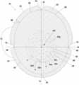
- FIG. 1is a top plan view of an exemplary embodiment of a soft tissue repair graft
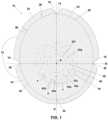
- FIG. 2 Ais a perspective view of a subject having a breast to be reconstructed
- FIG. 2 Bis a cross-sectional side view of the subject of FIG. 2 A , taken along the plane P, showing the breast B after reconstruction by a pre-pectoral reconstruction technique with a breast implant and a graft according to FIG. 1 ;
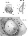
- FIG. 2 Cis a perspective view of the subject and reconstructed breast of FIG. 2 B , where the skin flap S has been removed to show the chest muscle and implanted graft;
- FIG. 3is a perspective schematic view of a section of human skin and the various components thereof, from which acellular dermal matrices (ADMs) may be fabricated;
- ADMsacellular dermal matrices
- FIG. 4is perspective schematic view of the section of human skin shown in FIG. 3 , showing the cutting steps performed according to an improved fabrication process to produce improved ADMs;
- FIG. 5 ais a perspective schematic view of a section of human skin showing where cuts may be made according to a previously known fabrication process for preparing ADMs useful for making soft tissue repair grafts as described herein;
- FIG. 5 bis a perspective schematic view of a section of human skin showing where cuts may be made according to an improved process for preparing ADMs useful for making soft tissue repair grafts as described herein.
- graftrefers to a biologically compatible material, tissue, or substance which is introduced into the body of a subject, either permanently or temporarily, to replace, improve or supplement the structure or function of tissue, an organ, or other body feature of the subject and includes, but is not limited to, those used for the administration or delivery of a therapeutic agent or substance.
- planar graftsare generally most suitable, however, the term “graft” as used herein is not limited only to those having planar configurations. Grafts may be integrated into a patient's body after implantation.
- a graftis made of material obtained from the same individual into whom it is implanted, it is “autologous.” Where the graft is made of material obtained from a different individual of the same species than the individual into whom it is implanted, it is “allogeneic.” Where the graft is made of material obtained from an individual of a different species than the individual into whom it is implanted, it is “xenogeneic.”
- the soft tissue graftsmay be autologous, allogeneic or xenogeneic.
- an implantmeans a device or material that replaces a missing body feature or portion thereof, which may be lost through trauma, disease, or congenital conditions, and is intended to restore the normal function(s) of the missing body part.
- an implantcan be any material, device or substance which is introduced into the body of a subject, either permanently or temporarily, to replace, improve or supplement the structure or function of tissue, an organ, or other body feature of the subject and includes, but is not limited to, those used for the administration or delivery of a therapeutic agent or substance.
- biocompatiblemeans that the graft or implant, when implanted in a subject, does not cause adverse effects such as, for example without limitation, toxicity, foreign body reaction, or cellular disruption.
- Grafts for soft tissue repair described hereinare suitable for supporting, covering, retaining, or any combination thereof, an implant positioned in the body of a subject. More particularly, the soft tissue repair grafts are capable of being more accurately positioned in a subject and more securely attached to adjacent tissues than previously known grafts. Furthermore, the soft tissue repair grafts are capable of greater expansion without tearing during tissue expansion (i.e., using breast implant and/or tissue expander), while concurrently conforming more closely to the shapes and contours of both the implant and adjacent body tissues, than previously known grafts.
- the improved ability of the soft tissue graft to conform closely to the shapes and contours of the implant and adjacent body tissuesis more significant and apparent when those contours are more rounded, curved, protruding, or recessed (e.g., concave, convex, projecting, etc.), such as, without limitation, for a breast, knee joint, elbow joint, chin, fingertip, toe, heel, other similar body features, and implants for such body features.
- soft tissue graftswill be described in detail hereinafter as used in surgical procedures for breast reconstruction, their utility is not limited to such surgical procedures. Rather, persons of ordinary skill will recognize that the soft tissue grafts are advantageous for other surgical procedures as well, particularly those involving repair, reconstruction or modification of body features such as those mentioned above and others.
- FIG. 1is a top plan view of an exemplary soft tissue repair graft 10 suitable for use in a surgical procedure such as breast reconstruction.
- the soft tissue repair graft 10has an arcuate peripheral edge 12 and a focal point F which is located generally at or near the geometric center of the graft 10 .
- An imaginary vertical axis Vpasses through the focal point F.
- An imaginary horizontal axis Halso passes through the focal point F, with the vertical and horizontal axes V, H intersecting at the focal point F.
- the focal point Fwill be positioned at the nipple of the breast undergoing reconstruction.
- the graft 10has a generally circular or slightly oval shape.
- the graft 10will have dimensions suitable for the location and size of the surgical site with which it is intended for use. For example, a larger sized graft 10 will be suitable and selected for a larger sized breast or breast pocket, and a smaller sized graft 10 will be suitable and selected for a smaller breast size.
- the vertical axis Vwill be of greater length than the horizontal axis H.
- the ratio of the length (L v ) of the vertical axis V to the length (L H ) of the horizontal axis Hmay be from about 1.05 to about 1.30, such as from about 1.10 to about 1.20, or about 1.15.
- the graft 10has at least three notches 14 , 16 , 18 at the peripheral edge 12 , including a top notch 14 located where the vertical axis V meets the peripheral edge 12 above the focal point F, and first and second side notches 16 , 18 located where the vertical axis V meets the peripheral edge 12 on opposite sides of the focal point F.
- a top notch 14located where the vertical axis V meets the peripheral edge 12 above the focal point F
- first and second side notches 16 , 18located where the vertical axis V meets the peripheral edge 12 on opposite sides of the focal point F.
- a usere.g., surgeon
- handling the graft 10 prior to implantation in a subjectwill be able to readily discern which is the top notch 14 and, therefore, which way to orient the graft 10 to ensure it is positioned properly to align with the shape and anatomy of the breast or breast pocket being reconstructed.
- a usermay determine a sequence of the notches 14 , 16 , 18 which extends the shortest overall distance and the notch 14 positioned in the middle of the other two 16 , 18 is the top notch 14 .
- each cuff element 20 , 22 , 24is foldable along a respective imaginary arcuate line 26 , 28 , 30 (see dotted lines in FIG. 1 ) which extends from the base of a respective pair of notches which forms each cuff element 20 , 22 , 24 to form a folded edge (see folded edge 32 shown in FIG. 2 C ).
- one cuff element 20is foldable along its respective imaginary arcuate line 26 between the notches 14 , 16 which form that cuff element 20 .
- a reinforced folded edge 32extends substantially around the entire graft 10 .
- the cuff elementsprovide a superior ring (i.e., the reinforced folded edge 32 ) of contact with the muscle M for improved long term support of the implant I (see, particularly, FIGS. 2 B and 2 C ).
- Each cuff element 20 , 22 , 24may, for example without limitation, have a width W, measured from the peripheral edge 12 of the graft 10 (see FIG. 1 ), of from about 7 millimeters to about 20 millimeters.
- each cuff element 20 , 22 , 24has a width of from about 10 millimeters to about 20 millimeters, or from about 15 millimeters to about 20 millimeters, or from about 10 millimeters to about 15 millimeters, or even from about 12 millimeters to about 18 millimeters.
- the width of each cuff element 20 , 22 , 24will typically be based on the size of the breast implant being used, as is readily determinable by persons of ordinary skill in the relevant art. It should be further noted that where the graft 10 has more than one cuff element, they need not all have the same widths as one another.
- the graft 10may be symmetrical, such that the vertical and horizontal axes V, H each extend to the farthest and oppositely positioned points on the reinforced folded edge 32 and are substantially perpendicular with one another. In other embodiments such as that shown in FIG.
- the graft 10may be symmetrical only along the vertical axis V, such that the vertical and horizontal axes V, H each extend to the farthest and oppositely positioned points 34 , 36 , 38 , 40 , respectively, on the folded edge 32 and are perpendicular with one another, but the focal point F is a shorter distance from a bottommost (i.e., inferior) point 36 than from a topmost (i.e., superior) point 34 on the folded edge 32 .
- the distance between focal point F and the bottommost point 36is from about 40% to about 50%, such as from about 42% to about 48%, or such as about 45%, of the total distance between the topmost point 34 and the bottommost point 36 , thus optimizing distribution of breast volume around the nipple for improved breast shape and aesthetic outcome.
- the graft 10may be asymmetrical, such that the vertical and horizontal axes V, H each extend to the farthest and oppositely positioned points on the folded edge 32 , but are not perpendicular with one another.
- Embodiments of the graft 10which are either symmetrical only about the vertical axis V or asymmetrical may align more closely with a breast and, therefore, may be more suitable for use in breast reconstruction procedures.
- the focal point Fwill be located at the intersection of the vertical axis V and horizontal axis H.
- the focal point Fis also positioned at the midpoint of the horizontal axis H.
- the graft 10may have different quantities of notches and cuff elements.
- the graft 10may not have any notches, in which case the cuff element may also be entirely absent.
- the graft 10may, for example without limitation, include only two notches or even a single notch (such as, but not necessarily, positioned at the topmost (i.e., superior) point 34 of the graft 10 ), which could form two cuff elements (by two notches), or a single cuff element or even no cuff element at all (by a single notch).
- a graftmight include one or more notches which are too shallow or small to form cuff elements wide enough to be used and beneficial in the manner described above, but the notches would still perform the function of providing guidance for properly orienting the graft during its placement in a breast undergoing reconstruction.
- the graft 10may include a plurality of arcuate slots or openings 42 a , 42 b , 42 c , 42 d , 42 e , 42 f , 42 g at least partially through the graft 10 , which form a plurality of circular patterns 44 , 46 , 48 which are concentric about the focal point F.
- the concentric, circular pattern of slots or openings 42 , 44 , 46 , 48 about focal point Fallow for expansion of the two dimensional graft 10 to reshape into a three dimensional structure which conforms in least in part to the spherical shape of the breast implant.
- the arcuate slots or openings 42 a , 42 b , 42 c , 42 d , 42 e , 42 f , 42 gare entirely through the graft 10 .
- the slots or openings 42 a , 42 b , 42 c , 42 d , 42 e , 42 f , 42 gare not mere holes or perforations, but rather, each of them 42 a , 42 b , 42 c , 42 d , 42 e , 42 f , 42 g is elongated.
- each individual slot(e.g., 42 a , 42 b , 42 c , 42 d , 42 e , 42 f , 42 g ), for example without limitation, is typically from about 5 millimeters and about 15 millimeters, with longer slots (e.g., slot 42 a , 42 b ) forming the outer circular patterns (e.g., pattern 44 ) and shorter slots (e.g., slot 42 e ) forming inner circular patterns (e.g., pattern 48 ).
- the distance x(see FIG. 1 ) between successive slots (end-to-end) (e.g., slots 42 a , 42 b ) should be from about 5 millimeters to about 15 millimeters, such as about 10 millimeters.
- the slots 42 a , 42 b forming the outermost circular pattern 44are not closer than about 1.75 centimeters, such as without limitation, not closer than about 1.25 centimeter, or even about 1.5 centimeters, from the imaginary arcuate lines 26 , 28 , 30 , between the notches 14 , 16 , 18 (or from the reinforced folded edge 32 of the graft 10 after implanting).
- This placement of the slots 42 a , 42 b of the outermost circular pattern 44minimizes the risk of unnecessarily weakening the tensile strength of the graft 10 during and after implantation.
- the distance d (see FIG. 1 ) between the slots (e.g., slots 42 b , 42 d ) forming adjacent circular patterns (e.g., patterns 44 , 46 )should be from about 10 millimeters to about 20 millimeters, such as about 15 millimeters. In some exemplary embodiments, for larger sized grafts (such as having a largest diameter of from about 26 to about 22 centimeters, such as about 25 centimeters), the distance d between the slots (e.g., slots 42 a , 42 c ) forming adjacent circular patterns (e.g., patterns 44 , 46 ) should be from about 15 millimeters to about 20 millimeters.
- the distance d between the slots (e.g., slots 42 a , 42 c ) forming adjacent circular patterns (e.g., patterns 44 , 46 )should be from about 13 millimeters to about 17 millimeters.
- the distance d between the slots (e.g., slots 42 a , 42 c ) forming adjacent circular patterns (e.g., patterns 44 , 46 )should be from about 10 millimeters to about 15 millimeters.
- Embodiments of the soft tissue graft 10 intended for use in breast reconstruction proceduresare generally implanted such that they at least partially cover, support and retain a breast implant I within the breast B of a subject.
- FIG. 2 Ashows a perspective view of a subject having a breast B to be reconstructed.
- FIG. 2 Bprovides a cross-sectional side view of the subject of FIG. 2 A , taken along the plane P, showing the breast B, after reconstruction using a pre-pectoral reconstruction technique to implant a breast implant I and an exemplary embodiment of the graft 10 .
- the chest muscle M as well as the skin flap S and nipple N of the breast Bare shown, with the graft 10 positioned in front of the chest muscle M and adjacent to the skin flap S, and the breast implant I positioned in a pocket formed between the chest muscle and the graft 10 .
- One technique for performing pre-pectoral breast reconstructionis to lift the skin flap S away from the chest muscle M of the breast, fold the cuff elements 20 , 22 , 24 of a graft 10 to form a reinforced folded edge 32 and insert the graft 10 superior to the chest muscle (pectoralis major) M and anterior and adjacent to the skin flap S of the breast B.
- the graft 10is oriented and inserted in the pocket between the chest muscle M and skin flap S with its top notch 14 vertically aligned above the nipple N, and its focal point F directly underlying the nipple N.
- This cuffallows for some surface area of the graft that is folded under the implant to come in contact with the muscle and function as an anchor providing extra support for the graft-implant construct resulting in improved positioning of the implant long-term, thus counteracting forces of gravity long-term. Without the cuff, the graft-implant construct would only be in contact and supported by the breast skin, which stretches with time.
- the graft 10is affixed to the chest muscle M by suturing along almost the entire length of the reinforced folded edge 32 from the 4 o'clock position to the 8 o'clock position along the superior edge [ 26 , 28 , 30 ] and leaving a short portion (for example without limitation, from about 4.5 centimeters to about 8.5 centimeters in length) of the folded edge 32 unsutured so that a pocket (not shown per se) is formed between the chest muscle M and the graft 10 .
- a breast implant I or other biocompatible medical devicee.g., tissue expander
- Suture failuresometimes referred to as suture “tear-out,” often results in post-operative complications including, without limitation, the graft 10 and/or breast implant I shifting position relative to the natural breast B and nipple N, which may cause undesirable cosmetic changes and pain.
- the reinforced folded edge 32 formed by folding the cuff elements 20 , 22 , 24 of the graft 10provides a location for suturing the graft 10 to the chest muscle M which reduces the risk of suture tear-out and corresponding complications.
- the reinforced folded edge 32 of the graftalso provides an area for tissue ingrowth and stabilization of the pocket beyond sutures.
- a portion of the graph proximate the peripheral edge 12may nonetheless be folded against the graft 10 , in a single continuous cuff element, to form a reinforced folded edge 32 at which the graft 10 may be affixed to the chest muscle M with sutures (or staples, etc.), although there may be some slight puckering or gathering of the continuous cuff element portion of the peripheral edge 12 .
- the notches 14 , 16 , 18serve not only as orientation guides as described above, but also minimize puckering and gathering along the folded edge 32 of the graft 10 .
- FIG. 2 Cprovides a perspective view of the subject and reconstructed breast B, where the skin flap S has been removed to render the chest muscle M and implanted graft 10 visible.
- FIG. 2 Calso shows the nipple N artificially superimposed on the graft 10 to show its location relative to the graft 10 and its plurality of slots 42 a , 42 b , 42 c , 42 d , 42 e , 42 f , 42 g and circular patterns 44 , 46 , 48 .
- the elongate and arcuate shape of the plurality of slots 42 a , 42 b , 42 c , 42 d , 42 e , 42 f , 42 genables the graft 10 to expand and stretch to a greater extent than if only holes or perforations were provided in the graft 10 , which allows the graft 10 to cover and more closely conform to the shape and contours of the implant I while avoiding failure (i.e., tearing) of the graft 10 itself
- the circular patterns 44 , 46 , 48 and concentric arrangement of the plurality of slots 42 a , 42 b , 42 c , 42 d , 42 e , 42 f , 42 g around the focal point Ffurther enable the graft 10 to conform more closely to the shapes and contours of both the breast implant I and the skin flap S and minimize post-operative complications such as rippling and puckering.
- the graft 10may include a plurality of both notches 14 , 16 , 18 and cuff elements 20 , 22 , 24 .
- the graft 10may instead include one or more notches, or one or more or cuff elements, or one or more of both notches and cuff elements, and the quantities of notches and cuff elements need not be the same.
- the graft 10may include a plurality of slots 42 a , 42 b , 42 c , 42 d , 42 e , 42 f , 42 g which are arranged in a plurality of concentric circular patterns 44 , 46 , 48 , as described above, regardless of whether or not the graft 10 includes also includes any notches, slots, or both.
- the graft 10may include such a plurality of slots 42 a , 42 b , 42 c , 42 d , 42 e , 42 f , 42 g , but not have any notches or cuff elements.
- Suitable materials for making the soft tissue grafts 10 described hereininclude various tissues such as, without limitation, amnion, chorion, dermal, duodenum, dura, fascia lata, gastrointestinal, intestinal mucosa, intestinal submucosa, pericardium, peritoneum, placenta, and umbilical cord.
- tissuesuch as, without limitation, amnion, chorion, dermal, duodenum, dura, fascia lata, gastrointestinal, intestinal mucosa, intestinal submucosa, pericardium, peritoneum, placenta, and umbilical cord.
- the most suitable materials for breast reconstruction and similar plastic surgery procedureswill possess sufficient tensile strength to minimize or avoid suture tear-out, both during implantation and expansion through the post-operative phase, and allow rapid and efficient cellular ingrowth equally from either side of the graft.
- acellular dermal matriceshave been known and used to make grafts for soft tissue repair procedures, including without limitation breast reconstruction and other cosmetic surgical procedures.
- Such materialsare known to have suitable structural and biomechanical properties including, but not limited to, predictable suppleness, flexibility, uniform pliability sufficient to stretch and expand without tearing during tissue expansion (i.e., using a breast implant and/or tissue expander), and sufficient tensile strength.
- FIG. 3illustrates the microstructure of human skin.
- Human skinis recovered from either live or deceased donors after receiving consent from the individual donor or donor's family.
- human skinis made of several layer-like components, including the outer-most epidermis E, and the dermis D, which lies beneath the epidermis.
- the hypodermis H(also referred to as the subcutis) lies beneath the dermis D, but is not part of the skin. Rather, the hypodermis H contains adipose and muscle tissue.
- the dermis Ditself includes the papillary dermis PD, which lies adjacent the epidermis E, and the reticular dermis RD, which lies between the papillary dermis PD and the hypodermis H.
- the papillary-reticular dermis interface PRIlies between the papillary dermis PD and the reticular dermis RD.
- the dermis-epidermis junction (“the DEJ”)lies between the papillary dermis PD and epidermis E.
- the process for deriving the foregoing ADMs from dermal tissueinvolves removing the epidermis E (e.g., by a chemical process that causes the epidermis to slough off), and thereby exposing the DEJ that was adjacent the epidermis E. Beneath the DEJ lies the papillary dermis PD, the papillary-reticular dermal interface PRI, and the reticular dermis RD.
- the dermal tissue that is recovered for the ADMsmay therefore include the DEJ, papillary dermis PD and at least part of the reticular dermis RD.
- the recovered dermal tissueis decellularized and aseptically processed to meet sterility testing requirements.
- the foregoing ADMsare derived from recovered tissue that includes the entire papillary dermis PD.
- the microstructure of the papillary dermis PDis not uniform. More particularly, the papillary dermis PD has an upper portion, or side, that was immediately adjacent the DEJ and therefore closer to the epidermis E (i.e., “the epidermal portion”), and a structurally different lower portion, or side, that was farther from the DEJ and epidermis E, and adjacent the deeper reticular dermis RD (i.e., “the dermal portion”).
- the epidermal portion of the papillary dermis PDcontains a more densely-packed collagen matrix than the relatively more open collagen matrix contained in the dermal portion.
- the dermal portionis more porous than the epidermal portion.
- This dual structureis also a property of the foregoing ADMs, and is ideal for repairing ventral abdominal hernias and other abdominal wall defects, as the more densely-packed epidermal portion of the ADM (i.e., incorporating the epidermal portion of the papillary dermis PD) possesses the tensile strength and stiffness required for such load-bearing tissue repairs, and the more porous dermal portion of the ADM (i.e., incorporating the dermal portion of the papillary dermis PD, as well as at least a portion of the loosely-packed and porous underlying reticular dermis RD) provides an open collagen structure that promotes vascularization, cellular attachment and tissue ingrowth. Nevertheless, this dual structure, which may only be visible on a microscopic scale, presents concerns about identifying and maintaining the side orientation of the ADM, i.e., during a surgical procedure.
- an ADMis derived from allograft dermal tissue that is recovered from deeper within the dermis, and is therefore farther from, and not adjacent the epidermis.
- Recovery of portions of the dermis D from the skin suitable for making such ADMsmay be accomplished by various techniques and devices, such as, for example, a manual dermatome technique, or dissection with a scalpel.
- a first cut 50is made into the reticular dermis RD of the skin (e.g., a section of skin cut from the entire donor skin) proximate the underlying hypodermis H in order to remove it from the dermis D.
- a second cut 52is then made into the epidermal portion of the papillary dermis PD containing the dense collagen matrix, as discussed above, in order to remove the epidermis E, the DEJ, and the underlying epidermal portion of the papillary dermis PD.
- the remaining portion of the dermis Di.e., the deeper dermal portion of the papillary dermis PD and the reticular dermis RD) constitutes a collagen matrix (“the tissue”) having substantially uniform density and porosity.
- tissuemay then be minimally processed, e.g., according to the process disclosed in U.S. Pat. No. 7,723,108, the disclosure of which is incorporated by reference herein in its entirety.
- the tissuemay be decellularized by chemically treating it with saline, detergent, peracetic acid, ethanol and propylene glycol. The tissue may then be washed with sterile water to remove residual processing chemicals. The resulting disinfected and acellular tissue (ADM) may be cut into rectangular-shaped sheets suitable for clinical uses. The tissue sheets may be further treated with aqueous ethanol and packaged to provide a hydrated ADM.
- ADMdisinfected and acellular tissue
- the ADM derived using the improved process(es) disclosed aboveexhibits properties that are ideal for its use as a sling in breast reconstruction, and its use in other plastic surgery applications. Use of this improved ADM minimizes adhesions and foreign body reactions while promoting vascularization, cellular attachment, and tissue ingrowth at the surgical site. Compared to the previously known ADMs (i.e., described above), this improved ADM possesses more uniform tensile properties (i.e., strength, pliability, stretchability and handling characteristics) that are optimal for its use in breast reconstruction and other plastic surgery applications. This improved ADM also possesses improved suture retention strength, and elasticity and deformability that are optimal for its intended use.
- this improved elasticity of this improved ADMpromotes better expansion of the tissue in breast reconstruction.
- This improved ADMis therefore very strong and closely mimics the biomechanical properties of the tissue that it is intended to replace.
- this improved ADMis resistant to bacterial colonization and non-immunogenic, as a result of the treatment thereto and decellularization thereof.
- FIG. 5 aillustrates a previously known process for fabricating the previously known ADMs, including those commercially available under the names FlexHD® StructuralTM ADM, AlloDerm® ADM and AlloDerm® RTU ADM), namely, cutting the lower portion of the dermis and hypodermis (represented by straight line 54 ), and chemically treating the tissue to remove only the epidermis (represented by uneven line 56 ) and expose the DEJ.
- FIG. 5 billustrates the improved fabrication process mentioned hereinabove which produces improved ADMs having more uniform density and porosity, namely, the lower portion of the dermis and hypodermis are cut (represented by straight line 50 ), and then a second cut (represented by straight line 52 ) is made deeper into the dermis than the aforementioned chemical treatment used to fabricate previously known ADMs.
- the second cutresults in the removal of the epidermis, the DEJ, and the upper, epidermal portion of the papillary dermis.
- the substantially uniform density and porosity of the improved ADMs produced by this alternative fabrication processpromotes more rapid and efficient cellular ingrowth equally from either side of the ADM grafts as compared to the previously known ADMs (i.e., the FlexHD Structural® ADM, FlexHD Pliable® ADM, AlloDerm® ADM and AlloDerm® RTU ADM).
- Example 1Surgical Placement of an ADM Graft During Pre-Pectoral Breast Reconstruction
- the graftis suitable for use in pre-pectoral breast reconstruction procedures and:
Landscapes
- Health & Medical Sciences (AREA)
- Life Sciences & Earth Sciences (AREA)
- Engineering & Computer Science (AREA)
- Biomedical Technology (AREA)
- Oral & Maxillofacial Surgery (AREA)
- Transplantation (AREA)
- Veterinary Medicine (AREA)
- Public Health (AREA)
- General Health & Medical Sciences (AREA)
- Animal Behavior & Ethology (AREA)
- Cardiology (AREA)
- Heart & Thoracic Surgery (AREA)
- Vascular Medicine (AREA)
- Chemical & Material Sciences (AREA)
- Molecular Biology (AREA)
- Epidemiology (AREA)
- Medicinal Chemistry (AREA)
- Dermatology (AREA)
- Chemical Kinetics & Catalysis (AREA)
- Botany (AREA)
- Urology & Nephrology (AREA)
- Zoology (AREA)
- Prostheses (AREA)
Abstract
Description
- is made of ADM;
- has a vertical axis V and a horizontal axis H, where the ratio of the length of the vertical axis to the length of the horizontal axis is 1.15;
- is symmetrical about its vertical axis;
- has three notches (i.e., at 9, 12 and 3 o'clock positions on the peripheral edge of the graft);
- has three cuff elements formed by the three notches;
- has a focal point positioned at the midpoint of the horizontal axis and at a point on the vertical axis which is 45% above the bottommost point on the peripheral edge of the graft;
- has a plurality of slits which form a plurality of circular patterns which are concentric about the focal point.
In use, the foregoing ADM graft is placed and secured within a reconstructed breast to support a breast implant positioned anteriorly to the chest muscles of a patient during a pre-pectoral breast reconstruction procedure. The following steps are performed:
- 1. Make markings on the breast and draw horizontal and vertical lines centered around the nipple, mark inframammary fold, medial, lateral and superior portions of the breast to outline the breast footprint;
- 2. Prepare the Breast Pocket with hemostasis and irrigation;
- 3. Place pocket defining sutures in the lateral aspect and inframammary fold;
- 4. Use a breast implant sizer to determine the appropriate implant volume;
- 5. Mark the medial and lateral borders of the implant sizer in the breast pocket;
- 6. Mark the superior and midpoint of the breast to define the position on the breast at which the topmost (superior) point (34) of the graft, which is proximate the
top notch 14, will be anchored - 7. Triple wash the ADM graft with triple antibiotic solution alternating with betadine and squeeze excess fluid out of the ADM graft (a suitable triple antibiotic solution includes a mixture of 1 gram of cefazolin, 80 milligrams of gentamicin, and 50,000 International Units of bacitracin, in 500 milliliters of normal saline;
- 8. Mark the ADM graft to establish the X (i.e., horizontal H) axis by connecting the notches at
points topmost point 34. Marking of these axes allows for orientation of the ADM graft by correlating the markings on the ADM graft to the external markings on the patient's skin, thereby facilitating symmetrical inset and positioning of the ADM graft into breast pocket; - 9. Drape the ADM graft over the implant sizer and mark the edge of the implant circumferentially on the ADM graft adjusting the folds (e.g., at imaginary
arcuate lines graft 10, seeFIG.1 ) and widths of the cuff elements to the size of the implant; - 10. Fold the cuff elements (edges) of the ADM graft according to the markings then carefully place the marked ADM into the prepared breast pocket without touching the skin;
- 11. Adjust the ADM graft position accordingly by correlating the external markings of the aforesaid axes;
- 12. Find the superior point of the Y (vertical V) axis at the top of the pectoralis (Point A, e.g., the a topmost (i.e., superior)
point 34 of the ADM graft, seeFIGS.1 and2C ); - 13. Use a continuous 2-0 Monocryl (a commercially available suture manufactured and marketed by Ethicon of Cornelia, Ga., USA) to suture the medial edge of the ADM graft to the muscle from Point (A) to a Point (B) proximate to or on the inframammary fold, leaving an opening at the inferior edge of adequate size for insertion of the implant or tissue expander;
- 14. Use a continuous 2-0 Monocryl to suture the lateral edge of the ADM to the muscle, again starting from Point (A), and continuing to a Point (C), which is proximate to or on the inframammary fold and some distance away from Point (B), thereby leaving an opening adequate for placement of the implant or tissue expander;
- 15. Inject a pain relief agent (e.g., Exparel commercially available from Pacira Pharmaceuticals of Parsippany, N.J., USA) circumferentially to provide an long lasting intercostal block;
- 16. Open the final, permanent breast implant (I) and wash in the triple antibiotic solution;
- 17. Change gloves and place the permanent breast implant into the breast pocket utilizing a Keller funnel no touch technique;
- 18. Use a ribbon to protect and retract the implant while interrupted suture 2-0 Monocryl sutures are placed to close the aforesaid opening of the breast pocket at the inframammary fold;
- 19. Place two drains at the lateral aspect of the inframammary fold incision; and
- 20. Suture the incision in three layers using 2-0 Moncryl deeper interrupted sutures, followed by 3-0 Moncryl dermal and subcuticular sutures.
Claims (11)
Priority Applications (1)
| Application Number | Priority Date | Filing Date | Title |
|---|---|---|---|
| US17/069,092US11642216B2 (en) | 2018-09-07 | 2020-10-13 | Soft tissue repair grafts and processes for preparing and using same |
Applications Claiming Priority (2)
| Application Number | Priority Date | Filing Date | Title |
|---|---|---|---|
| US16/125,435US10813743B2 (en) | 2018-09-07 | 2018-09-07 | Soft tissue repair grafts and processes for preparing and using same |
| US17/069,092US11642216B2 (en) | 2018-09-07 | 2020-10-13 | Soft tissue repair grafts and processes for preparing and using same |
Related Parent Applications (1)
| Application Number | Title | Priority Date | Filing Date |
|---|---|---|---|
| US16/125,435ContinuationUS10813743B2 (en) | 2018-09-07 | 2018-09-07 | Soft tissue repair grafts and processes for preparing and using same |
Publications (2)
| Publication Number | Publication Date |
|---|---|
| US20210022848A1 US20210022848A1 (en) | 2021-01-28 |
| US11642216B2true US11642216B2 (en) | 2023-05-09 |
Family
ID=67587615
Family Applications (2)
| Application Number | Title | Priority Date | Filing Date |
|---|---|---|---|
| US16/125,435Active2038-10-08US10813743B2 (en) | 2018-09-07 | 2018-09-07 | Soft tissue repair grafts and processes for preparing and using same |
| US17/069,092Active2039-01-12US11642216B2 (en) | 2018-09-07 | 2020-10-13 | Soft tissue repair grafts and processes for preparing and using same |
Family Applications Before (1)
| Application Number | Title | Priority Date | Filing Date |
|---|---|---|---|
| US16/125,435Active2038-10-08US10813743B2 (en) | 2018-09-07 | 2018-09-07 | Soft tissue repair grafts and processes for preparing and using same |
Country Status (5)
| Country | Link |
|---|---|
| US (2) | US10813743B2 (en) |
| EP (2) | EP3628272B1 (en) |
| AU (2) | AU2019204393B2 (en) |
| CA (1) | CA3053144C (en) |
| ES (1) | ES2878337T3 (en) |
Families Citing this family (13)
| Publication number | Priority date | Publication date | Assignee | Title |
|---|---|---|---|---|
| US10945831B2 (en) | 2016-06-03 | 2021-03-16 | Musculoskeletal Transplant Foundation | Asymmetric tissue graft |
| US20190201580A1 (en)* | 2016-08-31 | 2019-07-04 | Lifecell Corporation | Breast treatment device |
| US10813743B2 (en) | 2018-09-07 | 2020-10-27 | Musculoskeletal Transplant Foundation | Soft tissue repair grafts and processes for preparing and using same |
| USD895812S1 (en)* | 2018-09-07 | 2020-09-08 | Musculoskeletal Transplant Foundation | Soft tissue repair graft |
| USD938579S1 (en)* | 2019-03-21 | 2021-12-14 | Salts Healthcare Limited | Ostomy bag |
| US20210085443A1 (en)* | 2019-09-25 | 2021-03-25 | Allosource | Pre-shaped allograft implant for reconstructive surgical use and methods of manufacture and use |
| KR102704134B1 (en)* | 2021-02-15 | 2024-09-09 | 주식회사 엘앤씨바이오 | Acellular skin substitute for breast reconstruction and method of making the same |
| WO2022173066A1 (en)* | 2021-02-15 | 2022-08-18 | 주식회사 엘앤씨바이오 | Acellular skin substitute for breast reconstruction and preparation method therefor |
| AU2022334424A1 (en)* | 2021-08-25 | 2024-04-04 | Musculoskeletal Transplant Foundation | Diversified grafts having heterogenous features and methods for making and using same |
| WO2023164304A2 (en)* | 2022-02-28 | 2023-08-31 | The Methodist Hospital System | Devices and methods for breast tissue expansion |
| US20250049988A1 (en)* | 2023-08-09 | 2025-02-13 | Allosource | Acellular dermal matrix sheet allografts having specialized mesh patterns |
| US12364590B2 (en) | 2023-10-27 | 2025-07-22 | Melodi Health, Inc. | Devices for providing tissue support during breast reconstruction, and systems and methods thereof |
| WO2025128946A1 (en)* | 2023-12-15 | 2025-06-19 | The Methodist Hospital | Devices and methods of making and use thereof |
Citations (199)
| Publication number | Priority date | Publication date | Assignee | Title |
|---|---|---|---|---|
| WO1984004880A1 (en) | 1983-06-10 | 1984-12-20 | University Patents Inc | Body implants of extracellular matrix and means and methods of making and using such implants |
| US4627429A (en) | 1986-02-28 | 1986-12-09 | American Home Products Corporation | Storage-stable transdermal adhesive patch |
| US4776853A (en) | 1986-07-28 | 1988-10-11 | Hsc Research Development Corporation | Process for preparing biological mammalian implants |
| USD298355S (en) | 1986-03-03 | 1988-11-01 | Young Bette J | Multiple I.V. holder |
| EP0322194A1 (en) | 1987-12-22 | 1989-06-28 | Walter Joseph Ledergerber | Implantable prosthetic device |
| US4917112A (en) | 1988-08-22 | 1990-04-17 | Kalt Medical Corp. | Universal bandage with transparent dressing |
| US5314471A (en) | 1991-07-24 | 1994-05-24 | Baxter International Inc. | Tissue inplant systems and methods for sustaining viable high cell densities within a host |
| US5336616A (en) | 1990-09-12 | 1994-08-09 | Lifecell Corporation | Method for processing and preserving collagen-based tissues for transplantation |
| US5344454A (en) | 1991-07-24 | 1994-09-06 | Baxter International Inc. | Closed porous chambers for implanting tissue in a host |
| US5453278A (en) | 1991-07-24 | 1995-09-26 | Baxter International Inc. | Laminated barriers for tissue implants |
| US5545233A (en) | 1992-10-02 | 1996-08-13 | Biedermann Motech Gmbh | Swing phase control device |
| FR2746298A1 (en) | 1996-03-25 | 1997-09-26 | Bellity Philippe | Surgical support prosthesis for human breast |
| US5713888A (en) | 1990-10-31 | 1998-02-03 | Baxter International, Inc. | Tissue implant systems |
| US5733336A (en) | 1990-10-31 | 1998-03-31 | Baxter International, Inc. | Ported tissue implant systems and methods of using same |
| US5741330A (en) | 1990-10-31 | 1998-04-21 | Baxter International, Inc. | Close vascularization implant material |
| USD404134S (en) | 1997-05-08 | 1999-01-12 | Minnesota Mining And Manufacturing Company | Clover-shaped adhesive bandage |
| WO1999065470A1 (en) | 1998-06-19 | 1999-12-23 | Lifecell Corporation | Particulate acellular tissue matrix |
| US6293970B1 (en) | 1998-06-30 | 2001-09-25 | Lifenet | Plasticized bone and soft tissue grafts and methods of making and using same |
| USD452121S1 (en) | 2000-05-30 | 2001-12-18 | Emery C. Teichelman | Tool for removing a sewer pop-up valve cap |
| US6497875B1 (en) | 1996-04-26 | 2002-12-24 | Case Western Reserve University | Multilayer skin or dermal equivalent having a layer containing mesenchymal stem cells |
| US6616685B2 (en)* | 2001-06-06 | 2003-09-09 | Ethicon, Inc. | Hernia repair device |
| US6734018B2 (en) | 1999-06-07 | 2004-05-11 | Lifenet | Process for decellularizing soft-tissue engineered medical implants, and decellularized soft-tissue medical implants produced |
| US6743574B1 (en) | 2000-09-12 | 2004-06-01 | Lifenet | Process for devitalizing soft-tissue engineered medical implants, and devitalized soft-tissue medical implants produced |
| US6773458B1 (en) | 1991-07-24 | 2004-08-10 | Baxter International Inc. | Angiogenic tissue implant systems and methods |
| US20040162512A1 (en)* | 2003-02-19 | 2004-08-19 | 3M Innovative Properties Company | Conformable wound dressing |
| US20040260315A1 (en) | 2003-06-17 | 2004-12-23 | Dell Jeffrey R. | Expandable tissue support member and method of forming the support member |
| US20050028228A1 (en) | 2003-07-21 | 2005-02-03 | Lifecell Corporation | Acellular tissue matrices made from alpa-1,3-galactose-deficient tissue |
| US6866686B2 (en) | 2000-01-28 | 2005-03-15 | Cryolife, Inc. | Tissue graft |
| WO2005063314A1 (en) | 2003-12-26 | 2005-07-14 | Cardio Incorporated | Decellularized tissue and method of preparing the same |
| US6933326B1 (en) | 1998-06-19 | 2005-08-23 | Lifecell Coporation | Particulate acellular tissue matrix |
| US20050186286A1 (en) | 2004-02-25 | 2005-08-25 | Yoshihiro Takami | Skin decellularization method, acellular dermal matrix and production method therefore employing said decellularization method, and composite cultured skin employing said matrix |
| US7049478B1 (en) | 2004-03-16 | 2006-05-23 | Patricia Ann Smith | Tri-lobe planar heel wound dressing |
| US20060210960A1 (en) | 1990-09-12 | 2006-09-21 | Lifecell Corporation, A Texas Corporation | Method for processing and preserving collagen-based tissues for transplantation |
| WO2007004214A2 (en) | 2005-07-04 | 2007-01-11 | Sergey Popov | Implant assembly |
| USD537948S1 (en) | 2005-01-31 | 2007-03-06 | Patricia Ann Smith | Tri-lobe planar heel wound dressing |
| US20070207125A1 (en) | 2002-04-12 | 2007-09-06 | Bothwell Alfred L | Vascularized human skin equivalent |
| US20070269791A1 (en) | 2003-12-25 | 2007-11-22 | Yoshisiro Takami | Method of Preparing Isolated Cell-Free Skin, Cell-Free Dermal Matrix, Method of Producing the Same and Composite Cultured Skin with The Use of the Cell-Free Dermal Matrix |
| US20080058692A1 (en)* | 2005-08-23 | 2008-03-06 | Propp Donald J | Window dressing |
| US20080097601A1 (en) | 2006-07-31 | 2008-04-24 | Jeanne Codori-Hurff | Mastopexy and Breast Reconstruction Prostheses and Method |
| WO2008066883A2 (en) | 2006-11-29 | 2008-06-05 | Frank Robert E | Implantable prosthesis for positioning and supporting a breast implant |
| US20080154366A1 (en) | 2006-12-21 | 2008-06-26 | Frank Robert E | Implantable prosthesis for periareolar mastopexy |
| US20080281419A1 (en) | 2007-05-10 | 2008-11-13 | Matheny Robert G | Breast implants and compositions of extracellular matrix |
| WO2008148026A1 (en) | 2007-05-24 | 2008-12-04 | The Trustees Of Columbia University In The City Of New York | Hybrid soft tissue implants from progenitor cells and biomaterials |
| WO2008154623A2 (en) | 2007-06-12 | 2008-12-18 | Musculoskeletal Transplant Foundation | Process for sterilizing acellular soft tissue with irradiation |
| US7476249B2 (en) | 2004-08-06 | 2009-01-13 | Frank Robert E | Implantable prosthesis for positioning and supporting a breast implant |
| US20090065014A1 (en) | 2007-09-06 | 2009-03-12 | Tomiyo Nagata | Adhesive patch with unique shape for the heel of the foot |
| WO2009065013A1 (en) | 2007-11-14 | 2009-05-22 | Maxwell G Patrick | Interfaced medical implant assembly |
| US20090198332A1 (en) | 2008-02-05 | 2009-08-06 | Hilton Becker | Method for texturing the surface of a synthetic implant |
| US20090198333A1 (en) | 2008-02-05 | 2009-08-06 | Hilton Becker | Method for texturing the surface of a synthetic implant |
| US7582309B2 (en) | 2002-11-15 | 2009-09-01 | Etex Corporation | Cohesive demineralized bone compositions |
| US20090312685A1 (en) | 2006-04-25 | 2009-12-17 | Hans Olsen | Adhesive Wafer |
| US20100003306A1 (en) | 2008-06-08 | 2010-01-07 | Mast Biosurgery Ag | Pre-shaped user-formable micro-membrane implants |
| US20100028396A1 (en) | 2008-07-30 | 2010-02-04 | Ward Brian Roderick | Tissue scaffolds derived from forestomach extracellular matrix |
| USD609802S1 (en) | 2008-02-04 | 2010-02-09 | Inova Medical Ag | Self-closing external vascular closure |
| US20100040687A1 (en) | 2008-08-14 | 2010-02-18 | Kci Licensing, Inc. | Tissue Scaffolds |
| WO2010027613A2 (en) | 2008-09-05 | 2010-03-11 | Ethicon, Inc. | Method of manufacturing acellular matrix glue |
| US20100067106A1 (en) | 2005-11-08 | 2010-03-18 | PeriOptix, Inc. | Locking inter-pupillary distance and convergence adjustment mechanism |
| US20100082048A1 (en)* | 2006-06-06 | 2010-04-01 | Luiz Gonzaga Granja Filho | Prosthesis for anastomosis |
| US20100112543A1 (en) | 2005-03-16 | 2010-05-06 | Manh-Dan Ngo | Processing soft tissue, methods and compositions related thereto |
| US7723108B2 (en) | 2005-03-16 | 2010-05-25 | Musculoskeletal Transplant Foundation | Soft tissue processing |
| WO2010071624A1 (en) | 2008-12-19 | 2010-06-24 | C. R. Bard, Inc. | Implantable prosthesis |
| US20100191330A1 (en) | 2007-06-24 | 2010-07-29 | Gary Pierre Lauryssen | Human mammary prosthetic support and method of implanting |
| US20100216206A1 (en) | 2007-05-31 | 2010-08-26 | Maurizio Marzaro | Method for Preparing an Acellular Organic Tissue for Revitalisation and Device for Implementing Said Method |
| US7799325B2 (en) | 1999-11-05 | 2010-09-21 | Kleinsek Donald A | Removal of hypertrophic scars |
| US20100272782A1 (en) | 2007-07-10 | 2010-10-28 | Owens Rick T | Acellular tissue matrix compositions for tissue repair |
| US20100310628A1 (en) | 2009-06-08 | 2010-12-09 | Mast Biosurgery Ag | Pre-shaped user-formable micro-membrane implants |
| US7875074B2 (en) | 2007-09-19 | 2011-01-25 | Ethicon, Inc. | Naturally contoured, preformed, three dimensional mesh device for breast implant support |
| WO2011011394A2 (en) | 2009-07-21 | 2011-01-27 | Lifecell Corporation | Graft materials for surgical breast procedures |
| US20110035004A1 (en) | 2007-11-14 | 2011-02-10 | Maxwell G | Interfaced medical implant |
| WO2011019361A1 (en) | 2009-08-11 | 2011-02-17 | Tissue Banks International | Acellular dermal allografts and method of preparation |
| US20110054605A1 (en) | 2009-09-02 | 2011-03-03 | Hilton Becker | Self supporting implant in a human body and method for making the same without capsular contracture |
| US20110054604A1 (en) | 2009-09-02 | 2011-03-03 | Hilton Becker | Self supporting and forming breast implant and method for forming and supporting an implant in a human body |
| US20110106249A1 (en) | 2009-09-02 | 2011-05-05 | Hilton Becker | Self supporting and forming breast implant and method for forming and supporting an implant in a human body |
| US20110167602A1 (en) | 2001-11-16 | 2011-07-14 | Allergan, Inc. | Immunoneutral silk-fiber-based medical devices |
| US20110184227A1 (en) | 2009-09-11 | 2011-07-28 | Allergan, Inc. | Prosthetic device and method of manufacturing the same |
| US20110276039A1 (en) | 2010-05-05 | 2011-11-10 | Barry Markman | Method and apparatus for a process creating an internal tissue graft for animal and human reconstructive purposes |
| US20120010728A1 (en) | 2010-07-08 | 2012-01-12 | Lifecell Corporation | Method for shaping tissue matrices |
| US20120034191A1 (en) | 2007-05-10 | 2012-02-09 | Matheny Robert G | Extracellular matrix compositions for tissue regeneration |
| US20120040013A1 (en) | 2010-08-10 | 2012-02-16 | LifeCell Corporation. | Regenerative Tissue Scaffolds |
| US20120059411A1 (en) | 2009-07-02 | 2012-03-08 | Sun Wenquan | Device and method for treatment of incision or hernia |
| WO2012031162A1 (en) | 2010-09-01 | 2012-03-08 | Doris Taylor | Methods of recellularizing a tissue or organ for improved transplantability |
| US20120065649A1 (en)* | 2010-09-09 | 2012-03-15 | Towler Jeffrey C | Surgical Mesh |
| US20120061004A1 (en) | 2010-09-09 | 2012-03-15 | Towler Jeffrey C | Method of Increasing Film Tear Strength |
| US20120226352A1 (en) | 2009-09-02 | 2012-09-06 | Hilton Becker | Self supporting and forming breast implant and method for forming and supporting an implant in a human body |
| US8263101B2 (en) | 2010-08-26 | 2012-09-11 | Lifecell Corporation | Passive methods for anti-microbial biological meshes |
| US8268361B2 (en) | 2005-10-26 | 2012-09-18 | Ahlfors Jan-Eric W | Acellular bioabsorbable tissue regeneration matrices |
| US20120263763A1 (en) | 2011-04-14 | 2012-10-18 | Lifecell Corporation | Regenerative materials |
| US20120265218A1 (en) | 2011-04-15 | 2012-10-18 | Chen Eugene G | Devices and methods for laparoscopic hernia repair |
| US20120276213A1 (en) | 2011-04-28 | 2012-11-01 | Lifecell Corporation | Method for enzymatic treatment of tissue products |
| US20120283826A1 (en) | 2011-03-09 | 2012-11-08 | Arikha Moses | Systems and methods for mastopexy |
| US20120310367A1 (en) | 2011-05-31 | 2012-12-06 | Lifecell Corporation | Adipose tissue matrices |
| US20120329034A1 (en) | 2010-02-26 | 2012-12-27 | Wook Chun | Method For Producing An Acellular Dermal Matrix, And Acellular Dermal Matrix Produced By Same |
| US20130103061A1 (en) | 2009-07-02 | 2013-04-25 | John Harper | Device and method for treatment of incision or hernia |
| US20130121970A1 (en) | 2011-11-10 | 2013-05-16 | Lifecell Corporation | Method for elimination of space through tissue approximation |
| USD683858S1 (en) | 2011-05-05 | 2013-06-04 | Smith & Nephew Plc | Multisite dressing |
| US20130144356A1 (en) | 2010-07-31 | 2013-06-06 | Cook Medical Technologies Llc | Methods and systems for generating a tissue pocket in a patient |
| US20130158658A1 (en) | 2011-12-20 | 2013-06-20 | Lifecell Corporation | Sheet tissue products |
| WO2013106556A2 (en) | 2012-01-13 | 2013-07-18 | Lifecell Corporation | Breast prostheses, methods of manufacturing breast prostheses, and methods of treatment using breast prostheses |
| US20130211591A1 (en) | 2012-02-14 | 2013-08-15 | Electronics And Telecommunications Research Institute | Autonomous robot and method of controlling the same |
| US20130211519A1 (en)* | 2010-10-01 | 2013-08-15 | Cook Biotech Incorporated | Kits, components and methods for tissue reconstruction |
| WO2013126062A2 (en) | 2012-02-23 | 2013-08-29 | Tousimis Anastasios J | Critical point drying systems and methods for in situ tissue preservation |
| WO2013137664A1 (en) | 2012-03-15 | 2013-09-19 | 주식회사 엘앤씨바이오 | Cell-free dermal tissue implant |
| US8563232B2 (en) | 2000-09-12 | 2013-10-22 | Lifenet Health | Process for devitalizing soft-tissue engineered medical implants, and devitalized soft-tissue medical implants produced |
| US20130287741A1 (en) | 2011-12-19 | 2013-10-31 | Allosource | Flowable matrix compositions and methods |
| USD693888S1 (en) | 2011-08-01 | 2013-11-19 | Dolly D. Webster | Elastic connector |
| US20130317610A1 (en) | 2008-01-29 | 2013-11-28 | Walter J. Ledergerber | Gel-simulating and modulating buttress prosthesis |
| WO2013192197A1 (en) | 2012-06-21 | 2013-12-27 | Lifecell Corporation | Implantable prosthesis having acellular tissue attachments |
| WO2014008184A1 (en) | 2012-07-05 | 2014-01-09 | Lifecell Corporation | Tissue-based drain manifolds |
| EP2692364A1 (en) | 2012-07-31 | 2014-02-05 | Geistlich Pharma AG | Non-plasticized hydrophilic phosphate group containing dehydrated partially purified bone replacement material |
| EP2692363A1 (en) | 2012-07-31 | 2014-02-05 | Geistlich Pharma AG | Hydrophilic phosphate group containing dehydrated partially purified bone replacement material |
| WO2014019672A1 (en) | 2012-07-31 | 2014-02-06 | Geistlich Pharma Ag | Hydrophilic phosphate group containing dehydrated partially purified bone replacement material |
| US20140081397A1 (en) | 2012-09-19 | 2014-03-20 | R&D Concepts, LLC | Surgical devices, kits, and related intra-operative methods for selecting breast implants |
| WO2014041577A1 (en) | 2012-09-17 | 2014-03-20 | Decomed Srl | Medical device for breast reconstruction |
| US20140100656A1 (en) | 2012-10-04 | 2014-04-10 | Innovative Biologics LLC | Restorative post-lumpectomy implant device |
| USD705429S1 (en) | 2012-08-29 | 2014-05-20 | Merit Medical Systems, Inc. | Medical compression bandage |
| US8735054B1 (en) | 2008-01-04 | 2014-05-27 | Lifecell Corporation | Acellular tissue matrix preservation solution |
| US8746014B2 (en) | 2008-12-15 | 2014-06-10 | Allergan, Inc. | Method for making a knitted mesh |
| US8764824B2 (en) | 2008-01-29 | 2014-07-01 | Walter J. Ledergerber | Modulating buttress saline mammary prosthesis including limpet fill port |
| US8784486B2 (en)* | 2008-04-28 | 2014-07-22 | Allergan, Inc. | Breast implants having a flush patch and methods of using same to augment or reconstruct a breast |
| US8784499B2 (en) | 2010-03-25 | 2014-07-22 | Lifecell Corporation | Preparation of regenerative tissue scaffolds |
| US20140257481A1 (en) | 2010-04-29 | 2014-09-11 | BioStruxs, LLC | Breast Reconstruction Device and Methods |
| WO2014145462A1 (en) | 2013-03-15 | 2014-09-18 | Lifenet Health | Soft tissue pouch and methods of use thereof |
| US20140276957A1 (en) | 2013-03-14 | 2014-09-18 | Musculoskeletal Transplant Foundation | Soft tissue repair allografts and methods for preparing same |
| WO2014160124A1 (en) | 2013-03-14 | 2014-10-02 | Lifenet Health | Crosslinked soft tissue graft and methods of use thereof |
| US8876899B2 (en) | 2007-11-14 | 2014-11-04 | G. Patrick Maxwell | Breast implant assembly |
| US8916742B2 (en) | 2006-07-19 | 2014-12-23 | Joseph O. Smith | Anatomically engineered configured article |
| US8936651B2 (en) | 2013-03-14 | 2015-01-20 | Ethicon, Inc. | Decellularized omentum matrix and uses thereof |
| US20150037436A1 (en) | 2013-07-30 | 2015-02-05 | Musculoskeletal Transplant Foundation | Acellular soft tissue-derived matrices and methods for preparing same |
| WO2015021807A1 (en) | 2013-08-14 | 2015-02-19 | 北京瑞健高科生物科技有限公司 | Breast prosthesis support device based on tissue matrix material, and preparation method therefor |
| WO2015065923A1 (en) | 2013-10-28 | 2015-05-07 | The Regents Of The University Of California | Tissue grafts with fenestrations |
| US20150159066A1 (en) | 2011-11-25 | 2015-06-11 | Smith & Nephew Plc | Composition, apparatus, kit and method and uses thereof |
| US9089523B2 (en) | 2011-07-28 | 2015-07-28 | Lifecell Corporation | Natural tissue scaffolds as tissue fillers |
| US20150209128A1 (en) | 2010-05-05 | 2015-07-30 | Barry Markman | Method and apparatus for creating a reconstructive graft |
| US20150223928A1 (en) | 2013-07-11 | 2015-08-13 | Tepha, Inc. | Absorbable implants for plastic surgery |
| WO2015121686A1 (en) | 2014-02-17 | 2015-08-20 | The University Of Manchester | Textured surfaces for breast implants |
| US20150250582A1 (en) | 2014-03-05 | 2015-09-10 | E. Skott Greenhalgh | Breast reconstruction device |
| WO2015148932A1 (en) | 2014-03-28 | 2015-10-01 | Microaire Surgical Instruments, Llc | Endotine breast reconstruction devices and methods |
| US9150318B1 (en) | 2009-01-02 | 2015-10-06 | Lifecell Corporation | Method for sterilizing an acellular tissue matrix |
| EP2926840A1 (en) | 2014-04-02 | 2015-10-07 | Biotronik AG | Method for the treatment of biological tissue for dry use in an implant |
| US20150297798A1 (en) | 2014-01-24 | 2015-10-22 | University Of Pittsburgh - Of The Commonwealth System Of Higher Education | Extracellular Matrix Mesh Coating |
| WO2015164728A1 (en) | 2014-04-24 | 2015-10-29 | University Of Pittsburgh - Of The Commonwealth System Of Higher Education | Fractionating extracellular matrix to modulate bioactivity and the host response |
| US9180143B2 (en) | 2006-03-29 | 2015-11-10 | Tissue Regenix Limited | Decellularisation of tissue matrices for bladder implantation |
| WO2015176014A1 (en) | 2014-05-16 | 2015-11-19 | Allergan, Inc. | Soft filled prosthesis shell with variable texture |
| US9204953B2 (en) | 2008-12-15 | 2015-12-08 | Allergan, Inc. | Biocompatible surgical scaffold with varying stretch |
| US9204954B2 (en) | 2008-12-15 | 2015-12-08 | Allergan, Inc. | Knitted scaffold with diagonal yarn |
| US9238793B2 (en) | 2011-04-28 | 2016-01-19 | Lifecell Corporation | Method for enzymatic treatment of tissue products |
| US20160022416A1 (en) | 2013-07-11 | 2016-01-28 | Tepha, Inc. | Absorbable Implants for Plastic Surgery |
| US20160030636A1 (en) | 2013-03-15 | 2016-02-04 | University Of Florida Research Foundation, Inc. | Method for Decellularization of Tissue Grafts |
| US20160030487A1 (en) | 2014-07-30 | 2016-02-04 | Lifecell Corporation | Methods for selection of age-appropriate tissues |
| US20160045198A1 (en) | 2014-08-14 | 2016-02-18 | Lifecell Corporation | Tissue matrices and methods of treatment |
| US9271821B2 (en) | 2012-01-24 | 2016-03-01 | Lifecell Corporation | Elongated tissue matrices |
| US9308070B2 (en) | 2008-12-15 | 2016-04-12 | Allergan, Inc. | Pliable silk medical device |
| US9326840B2 (en) | 2008-12-15 | 2016-05-03 | Allergan, Inc. | Prosthetic device and method of manufacturing the same |
| US9336435B1 (en) | 2012-11-21 | 2016-05-10 | Ozog Media, LLC | System, method, and computer program product for performing processing based on object recognition |
| US20160151062A1 (en) | 2014-08-14 | 2016-06-02 | Lifecell Corporation | Tissue matrices and methods of treatment |
| US9370536B2 (en) | 2012-09-26 | 2016-06-21 | Lifecell Corporation | Processed adipose tissue |
| US9375017B2 (en) | 2009-01-02 | 2016-06-28 | Lifecell Corporation | Method for debristling animal skin |
| EP3056168A1 (en) | 2015-02-11 | 2016-08-17 | Novus Scientific AB | Breast implant support device with large back surface area |
| WO2016130559A1 (en) | 2015-02-10 | 2016-08-18 | Lifenet Health | Biologically functional soft tissue scaffolds and implants |
| WO2016144475A1 (en) | 2015-03-12 | 2016-09-15 | Mentor Worldwide Llc | Tissue expander with pectoral attachment |
| US20160287747A1 (en) | 2015-03-31 | 2016-10-06 | Mark Schallenberger | Shaped collagen tissue implant and method of manufacturing thereof |
| US20160331504A1 (en) | 2015-05-15 | 2016-11-17 | Lifecell Corporation | Tissue matrices and methods of treatment |
| US9532866B2 (en) | 2012-03-15 | 2017-01-03 | L&C Bio Co., Ltd. | Acellular dermal graft |
| US9549805B2 (en) | 2011-12-20 | 2017-01-24 | Lifecell Corporation | Flowable tissue products |
| US20170021058A1 (en) | 2015-07-24 | 2017-01-26 | Musculoskeletal Transplant Foundation | Acellular soft tissue-derived matrices and methods for preparing same |
| US20170065742A1 (en) | 2015-09-03 | 2017-03-09 | The University Of Kansas | Methacrylated devitalized cartilage and devitalized cartilage particles |
| US9592254B2 (en) | 2013-02-06 | 2017-03-14 | Lifecell Corporation | Methods for localized modification of tissue products |
| US9592278B2 (en) | 2012-03-08 | 2017-03-14 | Lifecell Corporation | Enzyme-activated collagen and tissue matrices |
| US20170072110A1 (en) | 2015-09-11 | 2017-03-16 | Lifecell Corporation | Perforated tissue matrix |
| US20170071725A1 (en) | 2015-08-21 | 2017-03-16 | Lifecell Corporation | Breast treatment device |
| US9622854B2 (en) | 2013-03-08 | 2017-04-18 | Abbott Medical Optics Inc. | Apparatus, system, and method for providing an optical filter for an implantable lens |
| US20170189165A1 (en)* | 2014-05-19 | 2017-07-06 | Mentor Worldwide Llc | Injection zone markers for biomedical implants |
| US20170224869A1 (en) | 2016-02-08 | 2017-08-10 | Lifecell Corporation | Biologic breast implant |
| US20170224460A1 (en) | 2015-09-11 | 2017-08-10 | Lifecell Corporation | Perforated tissue matrix |
| US20170231753A1 (en) | 2016-02-11 | 2017-08-17 | Gilbert W. Lee | Method and device for suspension, lifting, and augmentation of the breast, face, and neck |
| US9782436B2 (en) | 2012-04-24 | 2017-10-10 | Lifecell Corporation | Flowable tissue matrices |
| US20170348088A1 (en)* | 2016-06-03 | 2017-12-07 | Musculoskeletal Transplant Foundation | Asymmetric tissue graft |
| US20170348460A1 (en) | 2016-06-03 | 2017-12-07 | Lifecell Corporation | Methods for localized modification of tissue products |
| US20170367807A1 (en) | 2015-10-16 | 2017-12-28 | Lifenet Health | Soft tissue grafts, and methods of making and using same |
| US20180008745A1 (en) | 2016-07-05 | 2018-01-11 | Lifecell Corporation | Tissue matrices incorporating multiple tissue types |
| US20180055624A1 (en) | 2016-08-31 | 2018-03-01 | Lifecell Corporation | Breast treatment device |
| US20180092737A1 (en) | 2016-10-03 | 2018-04-05 | Lifecell Corporation | Breast treatment device |
| US9956072B2 (en) | 2012-10-04 | 2018-05-01 | Lifecell Corporation | Surgical template and delivery device |
| US9999637B2 (en) | 2012-04-24 | 2018-06-19 | Lifecell Corporation | Functionalized tissue matrices |
| US20180221136A1 (en)* | 2017-02-09 | 2018-08-09 | Ivor Barry Kaplan | Cover device and method of applying cover device for constructing and protecting a nipple/areola complex |
| WO2018195476A1 (en) | 2017-04-20 | 2018-10-25 | The Regents Of The University Of California | Systems and methods for acellular dermal matrix fenestrations in prepectoral breast reconstruction |
| USRE47100E1 (en) | 2012-05-15 | 2018-10-30 | Smith & Nephew Plc | Medical dressing |
| USD841172S1 (en) | 2017-01-23 | 2019-02-19 | Medela Holding Ag | Wound dressing |
| US10231874B2 (en) | 2010-11-08 | 2019-03-19 | Smith & Nephew Plc | Wound dressing and method of treatment |
| USD851261S1 (en) | 2018-05-11 | 2019-06-11 | Avery Dennison Corporation | Medical connection pad |
| USD856517S1 (en) | 2016-06-03 | 2019-08-13 | Musculoskeletal Transplant Foundation | Asymmetric tissue graft |
| WO2019157048A1 (en) | 2018-02-06 | 2019-08-15 | The Trustees Of The University Of Pennsylvania | The kirigami modification of biomedical tissue reinforcing meshes and matrices for expansile two-to-three dimensional conversion |
| WO2019175911A2 (en) | 2018-03-13 | 2019-09-19 | CARA, Nello | Mesh or membrane covering based on biological or biosynthetic material for prosthesis, provided with fixing system for fixing to the same prosthesis, and corresponding manufacturing process thereof |
| US20200054429A1 (en)* | 2015-03-24 | 2020-02-20 | Hexagon Health, Inc. | Gender-specific mesh implant with barrier for inguinal hernia repair |
| USD876645S1 (en) | 2018-01-04 | 2020-02-25 | Shuting Zhang | Breast feeding nipple shield |
| USD876646S1 (en) | 2018-06-01 | 2020-02-25 | Kinesio Ip Llc | Set of adhesive tapes |
| CA3053144A1 (en) | 2018-09-07 | 2020-03-07 | Musculoskeletal Transplant Foundation | Soft tissue repair grafts and processes for preparing and using same |
| US10869745B2 (en) | 2016-10-06 | 2020-12-22 | Lifecell Corporation | Tissue matrix with preformed openings or pilot openings |
| US20210085443A1 (en) | 2019-09-25 | 2021-03-25 | Allosource | Pre-shaped allograft implant for reconstructive surgical use and methods of manufacture and use |
- 2018
- 2018-09-07USUS16/125,435patent/US10813743B2/enactiveActive
- 2019
- 2019-06-21AUAU2019204393Apatent/AU2019204393B2/enactiveActive
- 2019-08-09EPEP19190980.3Apatent/EP3628272B1/enactiveActive
- 2019-08-09EPEP21164297.0Apatent/EP3861962A1/enactivePending
- 2019-08-09ESES19190980Tpatent/ES2878337T3/enactiveActive
- 2019-08-27CACA3053144Apatent/CA3053144C/enactiveActive
- 2020
- 2020-10-13USUS17/069,092patent/US11642216B2/enactiveActive
- 2020-12-20AUAU2020289883Apatent/AU2020289883B2/enactiveActive
Patent Citations (332)
| Publication number | Priority date | Publication date | Assignee | Title |
|---|---|---|---|---|
| WO1984004880A1 (en) | 1983-06-10 | 1984-12-20 | University Patents Inc | Body implants of extracellular matrix and means and methods of making and using such implants |
| US4627429A (en) | 1986-02-28 | 1986-12-09 | American Home Products Corporation | Storage-stable transdermal adhesive patch |
| USD298355S (en) | 1986-03-03 | 1988-11-01 | Young Bette J | Multiple I.V. holder |
| US4776853A (en) | 1986-07-28 | 1988-10-11 | Hsc Research Development Corporation | Process for preparing biological mammalian implants |
| EP0322194A1 (en) | 1987-12-22 | 1989-06-28 | Walter Joseph Ledergerber | Implantable prosthetic device |
| US4917112A (en) | 1988-08-22 | 1990-04-17 | Kalt Medical Corp. | Universal bandage with transparent dressing |
| US20060210960A1 (en) | 1990-09-12 | 2006-09-21 | Lifecell Corporation, A Texas Corporation | Method for processing and preserving collagen-based tissues for transplantation |
| US5336616A (en) | 1990-09-12 | 1994-08-09 | Lifecell Corporation | Method for processing and preserving collagen-based tissues for transplantation |
| US8067149B2 (en) | 1990-09-12 | 2011-11-29 | Lifecell Corporation | Acellular dermal matrix and method of use thereof for grafting |
| US5800529A (en) | 1990-10-31 | 1998-09-01 | Baxter International, Inc. | Close vascularization implant material |
| US5882354A (en) | 1990-10-31 | 1999-03-16 | Baxter International Inc. | Close vascularization implant material |
| US5593440A (en) | 1990-10-31 | 1997-01-14 | Baxter International Inc. | Tissue implant systems and methods for sustaining viable high cell densities within a host |
| US5653756A (en) | 1990-10-31 | 1997-08-05 | Baxter International Inc. | Closed porous chambers for implanting tissue in a host |
| US5964804A (en) | 1990-10-31 | 1999-10-12 | Baxter International Inc. | Close vascularization implant material |
| US5713888A (en) | 1990-10-31 | 1998-02-03 | Baxter International, Inc. | Tissue implant systems |
| US5733336A (en) | 1990-10-31 | 1998-03-31 | Baxter International, Inc. | Ported tissue implant systems and methods of using same |
| US5741330A (en) | 1990-10-31 | 1998-04-21 | Baxter International, Inc. | Close vascularization implant material |
| US5782912A (en) | 1990-10-31 | 1998-07-21 | Baxter International, Inc. | Close vascularization implant material |
| US5453278A (en) | 1991-07-24 | 1995-09-26 | Baxter International Inc. | Laminated barriers for tissue implants |
| US5344454A (en) | 1991-07-24 | 1994-09-06 | Baxter International Inc. | Closed porous chambers for implanting tissue in a host |
| US5314471A (en) | 1991-07-24 | 1994-05-24 | Baxter International Inc. | Tissue inplant systems and methods for sustaining viable high cell densities within a host |
| US6773458B1 (en) | 1991-07-24 | 2004-08-10 | Baxter International Inc. | Angiogenic tissue implant systems and methods |
| US5545233A (en) | 1992-10-02 | 1996-08-13 | Biedermann Motech Gmbh | Swing phase control device |
| FR2746298A1 (en) | 1996-03-25 | 1997-09-26 | Bellity Philippe | Surgical support prosthesis for human breast |
| US6497875B1 (en) | 1996-04-26 | 2002-12-24 | Case Western Reserve University | Multilayer skin or dermal equivalent having a layer containing mesenchymal stem cells |
| USD404134S (en) | 1997-05-08 | 1999-01-12 | Minnesota Mining And Manufacturing Company | Clover-shaped adhesive bandage |
| US7358284B2 (en) | 1998-06-19 | 2008-04-15 | Lifecell Corporation | Particulate acellular tissue matrix |
| WO1999065470A1 (en) | 1998-06-19 | 1999-12-23 | Lifecell Corporation | Particulate acellular tissue matrix |
| US6933326B1 (en) | 1998-06-19 | 2005-08-23 | Lifecell Coporation | Particulate acellular tissue matrix |
| US20030083752A1 (en) | 1998-06-30 | 2003-05-01 | Lloyd Wolfinbarger | Plasticized bone and soft tissue grafts and methods of making and using same |
| US9579420B2 (en) | 1998-06-30 | 2017-02-28 | Lifenet Health | Plasticized bone and soft tissue grafts and methods of making and using same |
| US6293970B1 (en) | 1998-06-30 | 2001-09-25 | Lifenet | Plasticized bone and soft tissue grafts and methods of making and using same |
| US9585986B2 (en) | 1998-06-30 | 2017-03-07 | Lifenet Health | Plasticized bone and soft tissue grafts and methods of making and using same |
| US20050246035A1 (en) | 1998-06-30 | 2005-11-03 | Wolfinbarger Lloyd Jr | Plasticized bone and soft tissue grafts and methods of making and using same |
| US6734018B2 (en) | 1999-06-07 | 2004-05-11 | Lifenet | Process for decellularizing soft-tissue engineered medical implants, and decellularized soft-tissue medical implants produced |
| US7799325B2 (en) | 1999-11-05 | 2010-09-21 | Kleinsek Donald A | Removal of hypertrophic scars |
| US6866686B2 (en) | 2000-01-28 | 2005-03-15 | Cryolife, Inc. | Tissue graft |
| US20100285587A1 (en) | 2000-01-28 | 2010-11-11 | Cryolife, Inc. | Tissue Graft |
| USD452121S1 (en) | 2000-05-30 | 2001-12-18 | Emery C. Teichelman | Tool for removing a sewer pop-up valve cap |
| US6743574B1 (en) | 2000-09-12 | 2004-06-01 | Lifenet | Process for devitalizing soft-tissue engineered medical implants, and devitalized soft-tissue medical implants produced |
| US8563232B2 (en) | 2000-09-12 | 2013-10-22 | Lifenet Health | Process for devitalizing soft-tissue engineered medical implants, and devitalized soft-tissue medical implants produced |
| US9936688B2 (en) | 2000-09-12 | 2018-04-10 | Lifenet Health | Process for devitalizing soft-tissue engineered medical implants, and devitalized soft-tissue medical implants produced |
| US6616685B2 (en)* | 2001-06-06 | 2003-09-09 | Ethicon, Inc. | Hernia repair device |
| US9066884B2 (en) | 2001-11-16 | 2015-06-30 | Allergan, Inc. | Sericin extracted fabrics |
| US20110167602A1 (en) | 2001-11-16 | 2011-07-14 | Allergan, Inc. | Immunoneutral silk-fiber-based medical devices |
| US9089501B2 (en) | 2001-11-16 | 2015-07-28 | Allergan, Inc. | Sericin extracted fabrics |
| US8628791B2 (en) | 2001-11-16 | 2014-01-14 | Allergan, Inc. | Method of forming an implantable knitted fabric comprising silk fibroin fibers |
| US8623398B2 (en) | 2001-11-16 | 2014-01-07 | Allergan, Inc. | Method for generating connective tissue by implanting a biodegradable silk fabric |
| US8685426B2 (en) | 2001-11-16 | 2014-04-01 | Allergan, Inc. | Methods for making biocompatible, implantable, substantially sericin free silk fabric |
| US8633027B2 (en) | 2001-11-16 | 2014-01-21 | Allergan, Inc. | Knitted biodegradable silk fabric comprising yarn promoting ingrowth of cells and methods of making |
| US20070207125A1 (en) | 2002-04-12 | 2007-09-06 | Bothwell Alfred L | Vascularized human skin equivalent |
| US7582309B2 (en) | 2002-11-15 | 2009-09-01 | Etex Corporation | Cohesive demineralized bone compositions |
| US20040162512A1 (en)* | 2003-02-19 | 2004-08-19 | 3M Innovative Properties Company | Conformable wound dressing |
| US20040260315A1 (en) | 2003-06-17 | 2004-12-23 | Dell Jeffrey R. | Expandable tissue support member and method of forming the support member |
| US20050028228A1 (en) | 2003-07-21 | 2005-02-03 | Lifecell Corporation | Acellular tissue matrices made from alpa-1,3-galactose-deficient tissue |
| US8802920B2 (en) | 2003-07-21 | 2014-08-12 | Lifecell Corporation | Acellular tissue matrices made from α-1,3-galactose-deficient tissue |
| US8324449B2 (en) | 2003-07-21 | 2012-12-04 | Lifecell Corporation | Acellular tissue matrices made from alpha-1,3-galactose-deficient tissue |
| US20070269791A1 (en) | 2003-12-25 | 2007-11-22 | Yoshisiro Takami | Method of Preparing Isolated Cell-Free Skin, Cell-Free Dermal Matrix, Method of Producing the Same and Composite Cultured Skin with The Use of the Cell-Free Dermal Matrix |
| WO2005063314A1 (en) | 2003-12-26 | 2005-07-14 | Cardio Incorporated | Decellularized tissue and method of preparing the same |
| US20070244568A1 (en) | 2003-12-26 | 2007-10-18 | Cardio Incorporated | Decellularized Tissue and Method of Preparing the Same |
| US20050186286A1 (en) | 2004-02-25 | 2005-08-25 | Yoshihiro Takami | Skin decellularization method, acellular dermal matrix and production method therefore employing said decellularization method, and composite cultured skin employing said matrix |
| US7049478B1 (en) | 2004-03-16 | 2006-05-23 | Patricia Ann Smith | Tri-lobe planar heel wound dressing |
| US8007531B2 (en) | 2004-08-06 | 2011-08-30 | Frank Robert E | Implantable prosthesis for positioning and supporting a breast implant |
| US7476249B2 (en) | 2004-08-06 | 2009-01-13 | Frank Robert E | Implantable prosthesis for positioning and supporting a breast implant |
| US20120053690A1 (en) | 2004-08-06 | 2012-03-01 | Frank Robert E | Implantable prosthesis for positioning and supporting a breast implant |
| USD537948S1 (en) | 2005-01-31 | 2007-03-06 | Patricia Ann Smith | Tri-lobe planar heel wound dressing |
| US20100112543A1 (en) | 2005-03-16 | 2010-05-06 | Manh-Dan Ngo | Processing soft tissue, methods and compositions related thereto |
| US7723108B2 (en) | 2005-03-16 | 2010-05-25 | Musculoskeletal Transplant Foundation | Soft tissue processing |
| US8557581B2 (en) | 2005-03-16 | 2013-10-15 | Musculoskeletal Transplant Foundation | Soft tissue processing |
| WO2007004214A2 (en) | 2005-07-04 | 2007-01-11 | Sergey Popov | Implant assembly |
| US20080058692A1 (en)* | 2005-08-23 | 2008-03-06 | Propp Donald J | Window dressing |
| US8268361B2 (en) | 2005-10-26 | 2012-09-18 | Ahlfors Jan-Eric W | Acellular bioabsorbable tissue regeneration matrices |
| US20100067106A1 (en) | 2005-11-08 | 2010-03-18 | PeriOptix, Inc. | Locking inter-pupillary distance and convergence adjustment mechanism |
| US9180143B2 (en) | 2006-03-29 | 2015-11-10 | Tissue Regenix Limited | Decellularisation of tissue matrices for bladder implantation |
| US20090312685A1 (en) | 2006-04-25 | 2009-12-17 | Hans Olsen | Adhesive Wafer |
| US20100082048A1 (en)* | 2006-06-06 | 2010-04-01 | Luiz Gonzaga Granja Filho | Prosthesis for anastomosis |
| US8916742B2 (en) | 2006-07-19 | 2014-12-23 | Joseph O. Smith | Anatomically engineered configured article |
| US20120158134A1 (en) | 2006-07-31 | 2012-06-21 | Jeanne Codori-Hurff | Mastopexy and Breast Reconstruction Prostheses and Method |
| US20080097601A1 (en) | 2006-07-31 | 2008-04-24 | Jeanne Codori-Hurff | Mastopexy and Breast Reconstruction Prostheses and Method |
| WO2008066883A2 (en) | 2006-11-29 | 2008-06-05 | Frank Robert E | Implantable prosthesis for positioning and supporting a breast implant |
| US20080154366A1 (en) | 2006-12-21 | 2008-06-26 | Frank Robert E | Implantable prosthesis for periareolar mastopexy |
| US20080281419A1 (en) | 2007-05-10 | 2008-11-13 | Matheny Robert G | Breast implants and compositions of extracellular matrix |
| US20100010627A1 (en) | 2007-05-10 | 2010-01-14 | Matheny Robert G | Breast Implants and Compositions of Extracellular Matrix |
| US20120034191A1 (en) | 2007-05-10 | 2012-02-09 | Matheny Robert G | Extracellular matrix compositions for tissue regeneration |
| US9199002B2 (en) | 2007-05-24 | 2015-12-01 | The Trustees Of Columbia University In The City Of New York | Hybrid soft tissue implants from progenitor cells and biomaterials |
| WO2008148026A1 (en) | 2007-05-24 | 2008-12-04 | The Trustees Of Columbia University In The City Of New York | Hybrid soft tissue implants from progenitor cells and biomaterials |
| US20100216206A1 (en) | 2007-05-31 | 2010-08-26 | Maurizio Marzaro | Method for Preparing an Acellular Organic Tissue for Revitalisation and Device for Implementing Said Method |
| WO2008154623A2 (en) | 2007-06-12 | 2008-12-18 | Musculoskeletal Transplant Foundation | Process for sterilizing acellular soft tissue with irradiation |
| US20100191330A1 (en) | 2007-06-24 | 2010-07-29 | Gary Pierre Lauryssen | Human mammary prosthetic support and method of implanting |
| US20100272782A1 (en) | 2007-07-10 | 2010-10-28 | Owens Rick T | Acellular tissue matrix compositions for tissue repair |
| US9382422B2 (en) | 2007-07-10 | 2016-07-05 | Lifecell Corporation | Acellular tissue matrix compositions for tissue repair |
| US20090065014A1 (en) | 2007-09-06 | 2009-03-12 | Tomiyo Nagata | Adhesive patch with unique shape for the heel of the foot |
| US7875074B2 (en) | 2007-09-19 | 2011-01-25 | Ethicon, Inc. | Naturally contoured, preformed, three dimensional mesh device for breast implant support |
| US20110035004A1 (en) | 2007-11-14 | 2011-02-10 | Maxwell G | Interfaced medical implant |
| WO2009065013A1 (en) | 2007-11-14 | 2009-05-22 | Maxwell G Patrick | Interfaced medical implant assembly |
| US8425600B2 (en) | 2007-11-14 | 2013-04-23 | G. Patrick Maxwell | Interfaced medical implant assembly |
| US8876899B2 (en) | 2007-11-14 | 2014-11-04 | G. Patrick Maxwell | Breast implant assembly |
| US8735054B1 (en) | 2008-01-04 | 2014-05-27 | Lifecell Corporation | Acellular tissue matrix preservation solution |
| US8764824B2 (en) | 2008-01-29 | 2014-07-01 | Walter J. Ledergerber | Modulating buttress saline mammary prosthesis including limpet fill port |
| US20130317610A1 (en) | 2008-01-29 | 2013-11-28 | Walter J. Ledergerber | Gel-simulating and modulating buttress prosthesis |
| US8764825B2 (en) | 2008-01-29 | 2014-07-01 | Walter J. Ledergerber | Gel-simulating and modulating buttress prosthesis |
| USD609802S1 (en) | 2008-02-04 | 2010-02-09 | Inova Medical Ag | Self-closing external vascular closure |
| US20090198332A1 (en) | 2008-02-05 | 2009-08-06 | Hilton Becker | Method for texturing the surface of a synthetic implant |
| US20090198333A1 (en) | 2008-02-05 | 2009-08-06 | Hilton Becker | Method for texturing the surface of a synthetic implant |
| US8784486B2 (en)* | 2008-04-28 | 2014-07-22 | Allergan, Inc. | Breast implants having a flush patch and methods of using same to augment or reconstruct a breast |
| US20100003306A1 (en) | 2008-06-08 | 2010-01-07 | Mast Biosurgery Ag | Pre-shaped user-formable micro-membrane implants |
| US20130224260A1 (en) | 2008-07-30 | 2013-08-29 | Mesynthes Ltd. | Tissue scaffolds derived from forestomach extracellular matrix |
| US20100028396A1 (en) | 2008-07-30 | 2010-02-04 | Ward Brian Roderick | Tissue scaffolds derived from forestomach extracellular matrix |
| US8415159B2 (en) | 2008-07-30 | 2013-04-09 | Mesynthes Ltd. | Tissue scaffolds derived from forestomach extracellular matrix |
| US20140335144A1 (en) | 2008-07-30 | 2014-11-13 | Mesynthes Ltd. | Tissue scaffolds derived from forestomach extracellular matrix |
| US8758781B2 (en) | 2008-07-30 | 2014-06-24 | Mesynthes Ltd. | Tissue scaffolds derived from forestomach extracellular matrix |
| US20140257482A1 (en) | 2008-07-30 | 2014-09-11 | Mesynthes Ltd. | Tissue scaffolds derived from forestomach extracellular matrix |
| US20100040687A1 (en) | 2008-08-14 | 2010-02-18 | Kci Licensing, Inc. | Tissue Scaffolds |
| US7927414B2 (en) | 2008-09-05 | 2011-04-19 | Ethicon, Inc. | Method of manufacturing acellular matrix glue |
| WO2010027613A2 (en) | 2008-09-05 | 2010-03-11 | Ethicon, Inc. | Method of manufacturing acellular matrix glue |
| US9204954B2 (en) | 2008-12-15 | 2015-12-08 | Allergan, Inc. | Knitted scaffold with diagonal yarn |
| US8746014B2 (en) | 2008-12-15 | 2014-06-10 | Allergan, Inc. | Method for making a knitted mesh |
| US9204953B2 (en) | 2008-12-15 | 2015-12-08 | Allergan, Inc. | Biocompatible surgical scaffold with varying stretch |
| US9308070B2 (en) | 2008-12-15 | 2016-04-12 | Allergan, Inc. | Pliable silk medical device |
| US9326840B2 (en) | 2008-12-15 | 2016-05-03 | Allergan, Inc. | Prosthetic device and method of manufacturing the same |
| US9078731B2 (en) | 2008-12-15 | 2015-07-14 | Allergan, Inc. | Method for making a knitted mesh |
| WO2010071624A1 (en) | 2008-12-19 | 2010-06-24 | C. R. Bard, Inc. | Implantable prosthesis |
| US20110288568A1 (en)* | 2008-12-19 | 2011-11-24 | Capuzziello Thomas J | Implantable prosthesis |
| US9375017B2 (en) | 2009-01-02 | 2016-06-28 | Lifecell Corporation | Method for debristling animal skin |
| US9150318B1 (en) | 2009-01-02 | 2015-10-06 | Lifecell Corporation | Method for sterilizing an acellular tissue matrix |
| US20100310628A1 (en) | 2009-06-08 | 2010-12-09 | Mast Biosurgery Ag | Pre-shaped user-formable micro-membrane implants |
| US20130103061A1 (en) | 2009-07-02 | 2013-04-25 | John Harper | Device and method for treatment of incision or hernia |
| US9351819B2 (en) | 2009-07-02 | 2016-05-31 | Lifecell Corporation | Device and method for treatment of incision or hernia |
| US20120059411A1 (en) | 2009-07-02 | 2012-03-08 | Sun Wenquan | Device and method for treatment of incision or hernia |
| US20210015602A1 (en)* | 2009-07-21 | 2021-01-21 | Lifecell Corporation | Graft materials for surgical breast procedures |
| US20110022171A1 (en) | 2009-07-21 | 2011-01-27 | Kinetic Concept, Inc. | Graft Materials for Surgical Breast Procedures |
| US20170340437A1 (en)* | 2009-07-21 | 2017-11-30 | Lifecell Corporation | Graft Materials For Surgical Breast Procedures |
| US10835370B2 (en) | 2009-07-21 | 2020-11-17 | Lifecell Corporation | Graft materials for surgical breast procedures |
| US20150157451A1 (en) | 2009-07-21 | 2015-06-11 | Lifecell Corporation | Graft Materials for Surgical Breast Procedures |
| US10449034B2 (en) | 2009-07-21 | 2019-10-22 | Lifecell Corporation | Graft materials for surgical breast procedures |
| WO2011011394A2 (en) | 2009-07-21 | 2011-01-27 | Lifecell Corporation | Graft materials for surgical breast procedures |
| US8986377B2 (en) | 2009-07-21 | 2015-03-24 | Lifecell Corporation | Graft materials for surgical breast procedures |
| US9888999B2 (en) | 2009-08-11 | 2018-02-13 | Aziyo Biologics, Inc. | Acellular dermal allografts and method of preparation |
| WO2011019361A1 (en) | 2009-08-11 | 2011-02-17 | Tissue Banks International | Acellular dermal allografts and method of preparation |
| US20130013068A1 (en) | 2009-08-11 | 2013-01-10 | James Forsell | Acellular Dermal Allografts And Method Of Preparation |
| US20110106249A1 (en) | 2009-09-02 | 2011-05-05 | Hilton Becker | Self supporting and forming breast implant and method for forming and supporting an implant in a human body |
| US20120226352A1 (en) | 2009-09-02 | 2012-09-06 | Hilton Becker | Self supporting and forming breast implant and method for forming and supporting an implant in a human body |
| US20110054605A1 (en) | 2009-09-02 | 2011-03-03 | Hilton Becker | Self supporting implant in a human body and method for making the same without capsular contracture |
| US8197542B2 (en) | 2009-09-02 | 2012-06-12 | Hilton Becker | Self supporting implant in a human body and method for making the same without capsular contracture |
| US20110054604A1 (en) | 2009-09-02 | 2011-03-03 | Hilton Becker | Self supporting and forming breast implant and method for forming and supporting an implant in a human body |
| US8202317B2 (en) | 2009-09-02 | 2012-06-19 | Hilton Becker | Self supporting and forming breast implant and method for forming and supporting an implant in a human body |
| US20120221105A1 (en) | 2009-09-11 | 2012-08-30 | Allergan, Inc. | Prosthetic device and method of manufacturing the same |
| US20110184227A1 (en) | 2009-09-11 | 2011-07-28 | Allergan, Inc. | Prosthetic device and method of manufacturing the same |
| US20120329034A1 (en) | 2010-02-26 | 2012-12-27 | Wook Chun | Method For Producing An Acellular Dermal Matrix, And Acellular Dermal Matrix Produced By Same |
| US8784499B2 (en) | 2010-03-25 | 2014-07-22 | Lifecell Corporation | Preparation of regenerative tissue scaffolds |
| US20140296623A1 (en) | 2010-03-25 | 2014-10-02 | Lifecell Corporation | Preparation of Regenerative Tissue Scaffolds |
| US20140257481A1 (en) | 2010-04-29 | 2014-09-11 | BioStruxs, LLC | Breast Reconstruction Device and Methods |
| US8858647B2 (en) | 2010-05-05 | 2014-10-14 | Barry Markman | Method and apparatus for a process creating an internal tissue graft for animal and human reconstructive purposes |
| US9622845B2 (en) | 2010-05-05 | 2017-04-18 | Barry Markman | Method and apparatus for creating a reconstructive graft |
| US9050177B2 (en) | 2010-05-05 | 2015-06-09 | Barry Markman | Method and apparatus for a process creating an internal tissue graft for animal and human reconstructive purposes |
| US20110276039A1 (en) | 2010-05-05 | 2011-11-10 | Barry Markman | Method and apparatus for a process creating an internal tissue graft for animal and human reconstructive purposes |
| US20150209128A1 (en) | 2010-05-05 | 2015-07-30 | Barry Markman | Method and apparatus for creating a reconstructive graft |
| US20170216008A1 (en) | 2010-05-05 | 2017-08-03 | Barry Markman | Method and apparatus for creating a reconstructive graft |
| US9636435B2 (en) | 2010-07-08 | 2017-05-02 | Lifecell Corporation | Method for shaping tissue matrices |
| US20120010728A1 (en) | 2010-07-08 | 2012-01-12 | Lifecell Corporation | Method for shaping tissue matrices |
| US20170100509A1 (en) | 2010-07-08 | 2017-04-13 | Lifecell Corporation | Method for shaping tissue matrices |
| US20130144356A1 (en) | 2010-07-31 | 2013-06-06 | Cook Medical Technologies Llc | Methods and systems for generating a tissue pocket in a patient |
| US20120040013A1 (en) | 2010-08-10 | 2012-02-16 | LifeCell Corporation. | Regenerative Tissue Scaffolds |
| US8343717B2 (en) | 2010-08-26 | 2013-01-01 | Lifecell Corporation | Passive methods for anti-microbial biological meshes |
| US8263101B2 (en) | 2010-08-26 | 2012-09-11 | Lifecell Corporation | Passive methods for anti-microbial biological meshes |
| US9220259B2 (en) | 2010-08-26 | 2015-12-29 | Lifecell Corporation | Passive methods for anti-microbial biological meshes |
| US8486616B2 (en) | 2010-08-26 | 2013-07-16 | Lifecell Corporation | Passive methods for anti-microbial biologic meshes |
| US20130156744A1 (en) | 2010-09-01 | 2013-06-20 | Regents Of The University Of Minnesota | Methods of recellularizing a tissue or organ for improved transplantability |
| WO2012031162A1 (en) | 2010-09-01 | 2012-03-08 | Doris Taylor | Methods of recellularizing a tissue or organ for improved transplantability |
| US20120065649A1 (en)* | 2010-09-09 | 2012-03-15 | Towler Jeffrey C | Surgical Mesh |
| US20120061004A1 (en) | 2010-09-09 | 2012-03-15 | Towler Jeffrey C | Method of Increasing Film Tear Strength |
| US20130211519A1 (en)* | 2010-10-01 | 2013-08-15 | Cook Biotech Incorporated | Kits, components and methods for tissue reconstruction |
| US10231874B2 (en) | 2010-11-08 | 2019-03-19 | Smith & Nephew Plc | Wound dressing and method of treatment |
| US8858629B2 (en) | 2011-03-09 | 2014-10-14 | Tepha, Inc. | Systems and methods for mastopexy |
| US20120283826A1 (en) | 2011-03-09 | 2012-11-08 | Arikha Moses | Systems and methods for mastopexy |
| US20150351891A1 (en) | 2011-03-09 | 2015-12-10 | Tepha, Inc. | Systems and methods for mastopexy |
| US20170143475A1 (en) | 2011-03-09 | 2017-05-25 | Tepha, Inc. | Systems and methods for mastopexy |
| US9277986B2 (en) | 2011-03-09 | 2016-03-08 | Tepha, Inc. | Systems and methods for mastopexy |
| US9585744B2 (en) | 2011-03-09 | 2017-03-07 | Tepha, Inc. | Systems and methods for mastopexy |
| US9375513B2 (en) | 2011-04-14 | 2016-06-28 | Lifecell Corporation | Regenerative materials |
| US20120263763A1 (en) | 2011-04-14 | 2012-10-18 | Lifecell Corporation | Regenerative materials |
| US20160271295A1 (en) | 2011-04-14 | 2016-09-22 | Lifecell Corporation | Regenerative materials |
| US8777965B2 (en) | 2011-04-15 | 2014-07-15 | Usgi Medical, Inc. | Devices and methods for laparoscopic hernia repair |
| US20120265218A1 (en) | 2011-04-15 | 2012-10-18 | Chen Eugene G | Devices and methods for laparoscopic hernia repair |
| US9957477B2 (en) | 2011-04-28 | 2018-05-01 | Lifecell Corporation | Method for enzymatic treatment of tissue products |
| US20180216062A1 (en) | 2011-04-28 | 2018-08-02 | Lifecell Corporation | Method for Enzymatic Treatment of Tissue Products |
| US20120276213A1 (en) | 2011-04-28 | 2012-11-01 | Lifecell Corporation | Method for enzymatic treatment of tissue products |
| US9206442B2 (en) | 2011-04-28 | 2015-12-08 | Lifecell Corporation | Method for enzymatic treatment of tissue products |
| US9238793B2 (en) | 2011-04-28 | 2016-01-19 | Lifecell Corporation | Method for enzymatic treatment of tissue products |
| US20180214607A1 (en) | 2011-04-28 | 2018-08-02 | Lifecell Corporation | Method for Enzymatic Treatment of Tissue Products |
| US9956316B2 (en) | 2011-04-28 | 2018-05-01 | Lifecell Corporation | Method for enzymatic treatment of tissue products |
| USD683858S1 (en) | 2011-05-05 | 2013-06-04 | Smith & Nephew Plc | Multisite dressing |
| US20120310367A1 (en) | 2011-05-31 | 2012-12-06 | Lifecell Corporation | Adipose tissue matrices |
| US20170049929A1 (en) | 2011-07-28 | 2017-02-23 | Lifecell Corporation | Natural tissue scaffolds as tissue fillers |
| US9504770B2 (en) | 2011-07-28 | 2016-11-29 | Lifecell Corporation | Natural tissue scaffolds as tissue fillers |
| US9089523B2 (en) | 2011-07-28 | 2015-07-28 | Lifecell Corporation | Natural tissue scaffolds as tissue fillers |
| USD693888S1 (en) | 2011-08-01 | 2013-11-19 | Dolly D. Webster | Elastic connector |
| US20130121970A1 (en) | 2011-11-10 | 2013-05-16 | Lifecell Corporation | Method for elimination of space through tissue approximation |
| US20150159066A1 (en) | 2011-11-25 | 2015-06-11 | Smith & Nephew Plc | Composition, apparatus, kit and method and uses thereof |
| US20130287741A1 (en) | 2011-12-19 | 2013-10-31 | Allosource | Flowable matrix compositions and methods |
| US9162011B2 (en) | 2011-12-19 | 2015-10-20 | Allosource | Flowable matrix compositions and methods |
| US10022214B2 (en) | 2011-12-20 | 2018-07-17 | Lifecell Corporation | Sheet tissue products |
| US20130158658A1 (en) | 2011-12-20 | 2013-06-20 | Lifecell Corporation | Sheet tissue products |
| US9532863B2 (en) | 2011-12-20 | 2017-01-03 | Lifecell Corporation | Sheet tissue products |
| US9549805B2 (en) | 2011-12-20 | 2017-01-24 | Lifecell Corporation | Flowable tissue products |
| US20170007394A1 (en) | 2012-01-13 | 2017-01-12 | Lifecell Corporation | Breast Prostheses, Methods of Manufacturing Breast Prostheses, and Methods of Treatment Using Breast Prostheses |
| US9549812B2 (en) | 2012-01-13 | 2017-01-24 | Lifecell Corporation | Breast prostheses, methods of manufacturing breast prostheses, and methods of treatment using breast prostheses |
| US20150012089A1 (en) | 2012-01-13 | 2015-01-08 | Lifecell Corporation | Breast prostheses, methods of manufacturing breast prostheses, and methods of treatment using breast prostheses |
| US10004590B2 (en) | 2012-01-13 | 2018-06-26 | Lifecell Corporation | Breast prostheses, methods of manufacturing breast prostheses, and methods of treatment using breast prostheses |
| WO2013106556A2 (en) | 2012-01-13 | 2013-07-18 | Lifecell Corporation | Breast prostheses, methods of manufacturing breast prostheses, and methods of treatment using breast prostheses |
| US20180280132A1 (en) | 2012-01-13 | 2018-10-04 | Lifecell Corporation | Breast prostheses, methods of manufacturing breast prostheses, and methods of treatment using breast prostheses |
| US20160135940A1 (en) | 2012-01-24 | 2016-05-19 | Lifecell Corporation | Elongated Tissue Matrices |
| US9271821B2 (en) | 2012-01-24 | 2016-03-01 | Lifecell Corporation | Elongated tissue matrices |
| US20130211591A1 (en) | 2012-02-14 | 2013-08-15 | Electronics And Telecommunications Research Institute | Autonomous robot and method of controlling the same |
| US8563234B2 (en) | 2012-02-23 | 2013-10-22 | Anastasios J. Tousimis | Critical point drying systems and methods for in situ tissue preservation |
| WO2013126062A2 (en) | 2012-02-23 | 2013-08-29 | Tousimis Anastasios J | Critical point drying systems and methods for in situ tissue preservation |
| US9027213B2 (en) | 2012-02-23 | 2015-05-12 | Anastasios J. Tousimis | Critical point drying systems and methods for in situ tissue preservation |
| US9426980B2 (en) | 2012-02-23 | 2016-08-30 | Anastasios J. Tousimis | Critical point drying systems and methods for in situ tissue preservation |
| US9592278B2 (en) | 2012-03-08 | 2017-03-14 | Lifecell Corporation | Enzyme-activated collagen and tissue matrices |
| US9532866B2 (en) | 2012-03-15 | 2017-01-03 | L&C Bio Co., Ltd. | Acellular dermal graft |
| WO2013137664A1 (en) | 2012-03-15 | 2013-09-19 | 주식회사 엘앤씨바이오 | Cell-free dermal tissue implant |
| US9999637B2 (en) | 2012-04-24 | 2018-06-19 | Lifecell Corporation | Functionalized tissue matrices |
| US9782436B2 (en) | 2012-04-24 | 2017-10-10 | Lifecell Corporation | Flowable tissue matrices |
| US20170348353A1 (en) | 2012-04-24 | 2017-12-07 | Lifecell Corporation | Flowable tissue matrices |
| US20180264037A1 (en) | 2012-04-24 | 2018-09-20 | Lifecell Corporation | Functionalized tissue matrices |
| USRE47100E1 (en) | 2012-05-15 | 2018-10-30 | Smith & Nephew Plc | Medical dressing |
| US20180333252A1 (en) | 2012-06-21 | 2018-11-22 | Lifecell Corporation | Implantable prosthesis having acellular tissue attachments |
| WO2013192197A1 (en) | 2012-06-21 | 2013-12-27 | Lifecell Corporation | Implantable prosthesis having acellular tissue attachments |
| US20150150674A1 (en) | 2012-06-21 | 2015-06-04 | Lifecell Corporation | Implantable prosthesis having acellular tissue attachments |
| US10959833B2 (en) | 2012-06-21 | 2021-03-30 | Lifecell Corporation | Implantable prosthesis having acellular tissue attachments |
| US10039633B2 (en) | 2012-06-21 | 2018-08-07 | Lifecell Corporation | Implantable prosthesis having acellular tissue attachments |
| US20150320911A1 (en) | 2012-07-05 | 2015-11-12 | Lifecell Corporation | Tissue-based drain manifolds |
| WO2014008184A1 (en) | 2012-07-05 | 2014-01-09 | Lifecell Corporation | Tissue-based drain manifolds |
| WO2014019672A1 (en) | 2012-07-31 | 2014-02-06 | Geistlich Pharma Ag | Hydrophilic phosphate group containing dehydrated partially purified bone replacement material |
| EP2692363A1 (en) | 2012-07-31 | 2014-02-05 | Geistlich Pharma AG | Hydrophilic phosphate group containing dehydrated partially purified bone replacement material |
| EP2692364A1 (en) | 2012-07-31 | 2014-02-05 | Geistlich Pharma AG | Non-plasticized hydrophilic phosphate group containing dehydrated partially purified bone replacement material |
| CN104640577A (en) | 2012-07-31 | 2015-05-20 | 盖斯特里希医药公司 | Hydrophilic phosphate group containing dehydrated partially purified bone replacement material |
| USD705429S1 (en) | 2012-08-29 | 2014-05-20 | Merit Medical Systems, Inc. | Medical compression bandage |
| WO2014041577A1 (en) | 2012-09-17 | 2014-03-20 | Decomed Srl | Medical device for breast reconstruction |
| US20140081397A1 (en) | 2012-09-19 | 2014-03-20 | R&D Concepts, LLC | Surgical devices, kits, and related intra-operative methods for selecting breast implants |
| US9114003B2 (en) | 2012-09-19 | 2015-08-25 | R & D Concepts, LLC | Surgical methods for breast implants |
| WO2014047234A1 (en) | 2012-09-19 | 2014-03-27 | R&D Concepts, LLC | Surgical devices, kits, and related intra-operative methods for selecting breast implants |
| US20160256606A1 (en) | 2012-09-26 | 2016-09-08 | Lifecell Corporation | Processed Adipose Tissue |
| US9370536B2 (en) | 2012-09-26 | 2016-06-21 | Lifecell Corporation | Processed adipose tissue |
| US9956072B2 (en) | 2012-10-04 | 2018-05-01 | Lifecell Corporation | Surgical template and delivery device |
| US20180214262A1 (en) | 2012-10-04 | 2018-08-02 | Lifecell Corporation | Surgical Template and Delivery Device |
| US20140100656A1 (en) | 2012-10-04 | 2014-04-10 | Innovative Biologics LLC | Restorative post-lumpectomy implant device |
| US9336435B1 (en) | 2012-11-21 | 2016-05-10 | Ozog Media, LLC | System, method, and computer program product for performing processing based on object recognition |
| US9592254B2 (en) | 2013-02-06 | 2017-03-14 | Lifecell Corporation | Methods for localized modification of tissue products |
| US20170209619A1 (en) | 2013-02-06 | 2017-07-27 | Lifecell Corporation | Methods for Localized Modification of Tissue Products |
| US9622854B2 (en) | 2013-03-08 | 2017-04-18 | Abbott Medical Optics Inc. | Apparatus, system, and method for providing an optical filter for an implantable lens |
| WO2014160008A1 (en) | 2013-03-14 | 2014-10-02 | Musculoskeletal Transplant Foundation | Soft tissue repair allografts and methods for preparing same |
| US20160000970A1 (en) | 2013-03-14 | 2016-01-07 | Lifenet Health | Crosslinked Soft Tissue Graft and Methods of Use Thereof |
| US20170281333A1 (en) | 2013-03-14 | 2017-10-05 | Musculoskeletal Transplant Foundation | Soft tissue repair allografts and methods for preparing same |
| US20140276957A1 (en) | 2013-03-14 | 2014-09-18 | Musculoskeletal Transplant Foundation | Soft tissue repair allografts and methods for preparing same |
| WO2014160124A1 (en) | 2013-03-14 | 2014-10-02 | Lifenet Health | Crosslinked soft tissue graft and methods of use thereof |
| US8936651B2 (en) | 2013-03-14 | 2015-01-20 | Ethicon, Inc. | Decellularized omentum matrix and uses thereof |
| US10238485B2 (en) | 2013-03-14 | 2019-03-26 | Musculoskeletal Transplant Foundation | Soft tissue repair allografts and methods for preparing same |
| WO2014145462A1 (en) | 2013-03-15 | 2014-09-18 | Lifenet Health | Soft tissue pouch and methods of use thereof |
| US20160030636A1 (en) | 2013-03-15 | 2016-02-04 | University Of Florida Research Foundation, Inc. | Method for Decellularization of Tissue Grafts |
| US20150223928A1 (en) | 2013-07-11 | 2015-08-13 | Tepha, Inc. | Absorbable implants for plastic surgery |
| US20170216009A1 (en)* | 2013-07-11 | 2017-08-03 | Tepha, Inc. | Absorbable implants for plastic surgery |
| US20160022416A1 (en) | 2013-07-11 | 2016-01-28 | Tepha, Inc. | Absorbable Implants for Plastic Surgery |
| US20150037436A1 (en) | 2013-07-30 | 2015-02-05 | Musculoskeletal Transplant Foundation | Acellular soft tissue-derived matrices and methods for preparing same |
| US9901440B2 (en) | 2013-08-14 | 2018-02-27 | Beijing Ruijian Gaoke Biotechnology Co., Ltd. | Breast prosthesis support device based on tissue matrix material, and preparation method therefor |
| US20160199173A1 (en)* | 2013-08-14 | 2016-07-14 | Beijing Ruijian Gaoke Biotechnology Co., Ltd. | Breast Prosthesis Support Device Based on Tissue Matrix Material, and Preparation Method Therefor |
| WO2015021807A1 (en) | 2013-08-14 | 2015-02-19 | 北京瑞健高科生物科技有限公司 | Breast prosthesis support device based on tissue matrix material, and preparation method therefor |
| EP3034038A1 (en) | 2013-08-14 | 2016-06-22 | Beijing Ruijian Gaoke Biotechnology Co., Ltd. | Breast prosthesis support device based on tissue matrix material, and preparation method therefor |
| US20160256259A1 (en) | 2013-10-28 | 2016-09-08 | The Regents Of The University Of California | Tissue grafts with fenestrations |
| WO2015065923A1 (en) | 2013-10-28 | 2015-05-07 | The Regents Of The University Of California | Tissue grafts with fenestrations |
| US20150297798A1 (en) | 2014-01-24 | 2015-10-22 | University Of Pittsburgh - Of The Commonwealth System Of Higher Education | Extracellular Matrix Mesh Coating |
| US20170049549A1 (en) | 2014-02-17 | 2017-02-23 | The University Of Manchester | Textured surfaces for breast implants |
| WO2015121686A1 (en) | 2014-02-17 | 2015-08-20 | The University Of Manchester | Textured surfaces for breast implants |
| US20150250582A1 (en) | 2014-03-05 | 2015-09-10 | E. Skott Greenhalgh | Breast reconstruction device |
| US9681941B2 (en) | 2014-03-28 | 2017-06-20 | Microaire Surgical Instruments | Endotine breast reconstruction device and methods |
| US20170202661A1 (en) | 2014-03-28 | 2017-07-20 | Microaire Surgical Instruments, Llc | Endotine breast reconstruction device and methods |
| WO2015148932A1 (en) | 2014-03-28 | 2015-10-01 | Microaire Surgical Instruments, Llc | Endotine breast reconstruction devices and methods |
| EP2926840A1 (en) | 2014-04-02 | 2015-10-07 | Biotronik AG | Method for the treatment of biological tissue for dry use in an implant |
| US20170049932A1 (en) | 2014-04-24 | 2017-02-23 | University Of Pittsburgh-Of The Commonwealth System Of Higher Education | Fractionating Extracellular Matrix to Modulate Bioactivity and the Host Response |
| WO2015164728A1 (en) | 2014-04-24 | 2015-10-29 | University Of Pittsburgh - Of The Commonwealth System Of Higher Education | Fractionating extracellular matrix to modulate bioactivity and the host response |
| WO2015176014A1 (en) | 2014-05-16 | 2015-11-19 | Allergan, Inc. | Soft filled prosthesis shell with variable texture |
| US9808338B2 (en) | 2014-05-16 | 2017-11-07 | Allergan, Inc. | Soft filled prosthesis shell with variable texture |
| US20180110612A1 (en) | 2014-05-16 | 2018-04-26 | Allergan, Inc. | Soft filled prosthesis shell with variable texture |
| US9539086B2 (en) | 2014-05-16 | 2017-01-10 | Allergan, Inc. | Soft filled prosthesis shell with variable texture |
| US20170189165A1 (en)* | 2014-05-19 | 2017-07-06 | Mentor Worldwide Llc | Injection zone markers for biomedical implants |
| US20160030487A1 (en) | 2014-07-30 | 2016-02-04 | Lifecell Corporation | Methods for selection of age-appropriate tissues |
| US20160151062A1 (en) | 2014-08-14 | 2016-06-02 | Lifecell Corporation | Tissue matrices and methods of treatment |
| US20160045198A1 (en) | 2014-08-14 | 2016-02-18 | Lifecell Corporation | Tissue matrices and methods of treatment |
| US20180044629A1 (en) | 2015-02-10 | 2018-02-15 | Lifenet Health | Biologically functional soft tissue scaffolds and implants |
| WO2016130559A1 (en) | 2015-02-10 | 2016-08-18 | Lifenet Health | Biologically functional soft tissue scaffolds and implants |
| EP3056168A1 (en) | 2015-02-11 | 2016-08-17 | Novus Scientific AB | Breast implant support device with large back surface area |
| EP3056167A1 (en) | 2015-02-11 | 2016-08-17 | Novus Scientific AB | Breast implant support device with large back surface area |
| US20160262835A1 (en) | 2015-03-12 | 2016-09-15 | Mentor Worldwide Llc | Tissue expander with pectoral attachment |
| WO2016144475A1 (en) | 2015-03-12 | 2016-09-15 | Mentor Worldwide Llc | Tissue expander with pectoral attachment |
| US20200054429A1 (en)* | 2015-03-24 | 2020-02-20 | Hexagon Health, Inc. | Gender-specific mesh implant with barrier for inguinal hernia repair |
| US20160287747A1 (en) | 2015-03-31 | 2016-10-06 | Mark Schallenberger | Shaped collagen tissue implant and method of manufacturing thereof |
| US20160331504A1 (en) | 2015-05-15 | 2016-11-17 | Lifecell Corporation | Tissue matrices and methods of treatment |
| US20170021058A1 (en) | 2015-07-24 | 2017-01-26 | Musculoskeletal Transplant Foundation | Acellular soft tissue-derived matrices and methods for preparing same |
| US20170071725A1 (en) | 2015-08-21 | 2017-03-16 | Lifecell Corporation | Breast treatment device |
| US20210022849A1 (en) | 2015-08-21 | 2021-01-28 | Lifecell Corporation | Breast treatment device |
| US20170065742A1 (en) | 2015-09-03 | 2017-03-09 | The University Of Kansas | Methacrylated devitalized cartilage and devitalized cartilage particles |
| US20170224460A1 (en) | 2015-09-11 | 2017-08-10 | Lifecell Corporation | Perforated tissue matrix |
| US20170072110A1 (en) | 2015-09-11 | 2017-03-16 | Lifecell Corporation | Perforated tissue matrix |
| US20170367807A1 (en) | 2015-10-16 | 2017-12-28 | Lifenet Health | Soft tissue grafts, and methods of making and using same |
| US20170224869A1 (en) | 2016-02-08 | 2017-08-10 | Lifecell Corporation | Biologic breast implant |
| US20170231753A1 (en) | 2016-02-11 | 2017-08-17 | Gilbert W. Lee | Method and device for suspension, lifting, and augmentation of the breast, face, and neck |
| USD856517S1 (en) | 2016-06-03 | 2019-08-13 | Musculoskeletal Transplant Foundation | Asymmetric tissue graft |
| US20170348088A1 (en)* | 2016-06-03 | 2017-12-07 | Musculoskeletal Transplant Foundation | Asymmetric tissue graft |
| US20170348460A1 (en) | 2016-06-03 | 2017-12-07 | Lifecell Corporation | Methods for localized modification of tissue products |
| US20180008745A1 (en) | 2016-07-05 | 2018-01-11 | Lifecell Corporation | Tissue matrices incorporating multiple tissue types |
| US20180055624A1 (en) | 2016-08-31 | 2018-03-01 | Lifecell Corporation | Breast treatment device |
| US20180092737A1 (en) | 2016-10-03 | 2018-04-05 | Lifecell Corporation | Breast treatment device |
| US10869745B2 (en) | 2016-10-06 | 2020-12-22 | Lifecell Corporation | Tissue matrix with preformed openings or pilot openings |
| USD875957S1 (en) | 2017-01-23 | 2020-02-18 | Medela Holding Ag | Wound dressing |
| USD879978S1 (en) | 2017-01-23 | 2020-03-31 | Medela Holding Ag | Wound dressing |
| USD841172S1 (en) | 2017-01-23 | 2019-02-19 | Medela Holding Ag | Wound dressing |
| US20180221136A1 (en)* | 2017-02-09 | 2018-08-09 | Ivor Barry Kaplan | Cover device and method of applying cover device for constructing and protecting a nipple/areola complex |
| WO2018195476A1 (en) | 2017-04-20 | 2018-10-25 | The Regents Of The University Of California | Systems and methods for acellular dermal matrix fenestrations in prepectoral breast reconstruction |
| USD876645S1 (en) | 2018-01-04 | 2020-02-25 | Shuting Zhang | Breast feeding nipple shield |
| US20210052367A1 (en) | 2018-02-06 | 2021-02-25 | The Trustees Of The University Of Pennsylvania | The kirigami modification of biomedical tissue reinforcing meshes and matrices for expansile two-to-three dimensional conversion |
| WO2019157048A1 (en) | 2018-02-06 | 2019-08-15 | The Trustees Of The University Of Pennsylvania | The kirigami modification of biomedical tissue reinforcing meshes and matrices for expansile two-to-three dimensional conversion |
| WO2019175911A2 (en) | 2018-03-13 | 2019-09-19 | CARA, Nello | Mesh or membrane covering based on biological or biosynthetic material for prosthesis, provided with fixing system for fixing to the same prosthesis, and corresponding manufacturing process thereof |
| US20200405473A1 (en) | 2018-03-13 | 2020-12-31 | Flavio NANNI | Mesh or membrane covering based on biological or biosynthetic material for prosthesis, provided with fixing system for fixing to the same prosthesis, and corresponding manufacturing process thereof |
| USD851261S1 (en) | 2018-05-11 | 2019-06-11 | Avery Dennison Corporation | Medical connection pad |
| USD876646S1 (en) | 2018-06-01 | 2020-02-25 | Kinesio Ip Llc | Set of adhesive tapes |
| CA3053144A1 (en) | 2018-09-07 | 2020-03-07 | Musculoskeletal Transplant Foundation | Soft tissue repair grafts and processes for preparing and using same |
| AU2020289883A1 (en) | 2018-09-07 | 2021-01-28 | Musculoskeletal Transplant Foundation | Soft tissue repair grafts and processes for preparing and using same |
| US10813743B2 (en)* | 2018-09-07 | 2020-10-27 | Musculoskeletal Transplant Foundation | Soft tissue repair grafts and processes for preparing and using same |
| EP3628272A2 (en) | 2018-09-07 | 2020-04-01 | Musculoskeletal Transplant Foundation | Soft tissue repair grafts |
| US20200078165A1 (en) | 2018-09-07 | 2020-03-12 | Musculoskeletal Transplant Foundation | Soft tissue repair grafts and processes for preparing and using same |
| EP3861962A1 (en) | 2018-09-07 | 2021-08-11 | Musculoskeletal Transplant Foundation | Soft tissue repair grafts |
| US20210085443A1 (en) | 2019-09-25 | 2021-03-25 | Allosource | Pre-shaped allograft implant for reconstructive surgical use and methods of manufacture and use |
Non-Patent Citations (72)
| Title |
|---|
| Australian Patent Application No. 2019204393, filed on Jun. 21, 2019. |
| Butler et al. Reduction of Adhesions with Composite AlloDerm/Polypropylene Mesh Implants for Abdominal Wall Reconstruction. Plast. Reconstr. Surg.{2004), v114, p. 464-473. |
| Canadian Design Application No. 186423, filed on Mar. 7, 2019. |
| Canadian Patent Application No. 3053144, filed on Aug. 27, 2019. |
| Certificate of Grant of Patent for Australian Patent No. 2020289883, issued by Australian Patent Office dated Mar. 24, 2022. |
| Design U.S. Appl. No. 29/566,994, filed Jun. 3, 2016. |
| Design U.S. Appl. No. 29/662,750, filed Sep. 7, 2018. |
| Erdag, et al., "Fibroblasts Improve Performance of Cultured Composite Skin Substitutes on Athymic Mice", Burns, 30 {2004) pp. 322-328. |
| European Design Application No. 006280178, filed on Mar. 5, 2019. |
| European Patent Application No. 19190980.3, filed on Aug. 9, 2019. |
| Examination Report for corresponding Australian Patent Application No. 2020289883, dated Aug. 6, 2021. |
| Examination Report from corresponding Canadian Design Application No. 186423, dated Feb. 12, 2020. |
| Examination Report No. 1 for corresponding Australian Patent Application No. 20190204393, dated Aug. 20, 2020. |
| Extended European Search Report for corresponding European Patent Application No. 21164297, dated Jul. 13, 2021. |
| Extended European Search Report, for corresponding European Patent Application No. 19190980.3., dated Jun. 24, 2020. |
| Final Office Action for U.S. Appl. No. 14/208,205, dated Aug. 19, 2016. |
| Final Office Action for U.S. Appl. No. 15/173,286, dated Jan. 26, 2018. |
| Final Office Action for U.S. Appl. No. 15/173,286, dated Nov. 25, 2019. |
| Final Office Action for U.S. Appl. No. 15/621,602, dated Nov. 13, 2017. |
| Final Office for U.S. Appl. No. 15/621,602, filed Jul. 12, 2018. |
| Further Examination Report in related New Zealand Patent Application No. 710330, dated Feb. 8, 2017. |
| Further Examination Report in related New Zealand Patent Application No. 710330, dated Oct. 11, 2016. |
| International Preliminary Report on Patentability for PCT/US2014/025619, dated Sep. 15, 2015. |
| International Search Report and Written Opinion for related International {PCT) Application No. PCT/US2014/025619, dated Jun. 30, 2014. |
| Isch et al., Patch Esophagoplasty Using AlloDerm as a Tissue Scaffold.Journal of Pediatric Surgery (2001 ), v36(2), pp. 266-268. |
| Kesmarky G., et al., "Plasma viscosity: A forgotten variable", Clinical Hemorcheology and Microcirculation, 2008, vol. 39, pp. 243-246, IOS Press. |
| Kolker et al., Multilayer Reconstruction of AbdominalWall Defects With Acellular DermalAllograft (AlloDerm) and Component Separation, Annals of Plastic Surgery (2005), v55(1), pp. 36-42. |
| Leung et al., Skin Grafts, UTMJ (2009), v86(2), pp. 61-64. |
| Li Y., et al., "Experimental validation of non-invasive and fluid density independent methods for the determination of local wave speed and arrival time of reflected wave", Journal of Biomechanics, 2011, vol. 44, pp. 1393-1399, Elsevier. |
| Mine et al. Aging Alters Functionally Human Dermal Papillary Fibroblasts but Not Reticular Fibroblasts: A New View of Skin Morphogenesis and Aging. PLoS One (2008), v3(12), e4066, 13 pages. |
| Mulder G. D., "Quantifying wound fluids for the clinician and researcher", Ostomy / Wound Management, 1994, vol. 10, pp. 65-69. |
| New Zealand Intellectual Property Office, First Examination Report in Applicant's related New Zealand Patent application No. 710330, dated Feb. 25, 2016 (4 pages). |
| Non-Final Office Action for U.S. Appl. No. 16/125,435, dated Feb. 28, 2020. |
| Notice of Acceptance for corresponding Australian Patent Application No. 2019204393, dated Sep. 22, 2020. |
| Notice of Acceptance for corresponding Australian Patent Application No. 2020289883, dated Nov. 29, 2021. |
| Notice of Allowance for U.S. Appl. No. 15/173,286, dated Jan. 10, 2021. |
| Notice of Allowance for U.S. Appl. No. 16/125,435, dated Jun. 22, 2020. |
| Notice of Allowance for U.S. Appl. No. 29/566,994, dated Mar. 25, 2019. |
| Notice of Allowance for U.S. Appl. No. 29/662,750. |
| Notice of Allowance issued for Canadian Patent Application No. 3,053,144, dated Jan. 29, 2021. |
| Notice of Grant for Patent for Australian Patent No. 2020289883, issued by Australian Patent Office dated Mar. 24, 2022. |
| Office Action for U.S. Appl. No. 14/208,025, dated Feb. 26, 2016. |
| Office Action for U.S. Appl. No. 14/208,025, dated Mar. 13, 2017. |
| Office Action for U.S. Appl. No. 15/173,286, dated Aug. 3, 2017. |
| Office Action for U.S. Appl. No. 15/173,286, dated Jul. 10, 2018. |
| Office Action for U.S. Appl. No. 15/621,602, dated Aug. 10, 2017. |
| Office Action for U.S. Appl. No. 15/621,602, dated Jul. 12, 2018. |
| Office Action for U.S. Appl. No. 15/858,360 dated May 11, 2018. |
| Office Action in corresponding Canadian Patent Application No. 3,053,144, dated Sep. 1, 2020. |
| Office Action in related Canadian Patent Application No. 2,899,642, dated Oct. 24, 2017. |
| Office Action in related Canadian Patent Application No. 2,899,642, dated Sep. 13, 2016. |
| Office Action issued for related European Patent Application No. 14718250.5, dated Nov. 23, 2016. |
| Oliver, et al., "Reconstruction of Full-Thickness Loss Skin Wounds Using Skin Collagen Allografts", British Journal of Plastic Surgery, 32 (1979), pp. 87-90. |
| Ownby, "The Integument—the skin and all of it's derivatives". |
| Partial European Search Report for EP Patent Application No. 19190980.3 dated Feb. 17, 2020. |
| Patent Examination Report No. 1 in related Australian Patent Application No. 2014244272, dated Mar. 10, 2016. |
| Patent Examination Report No. 1 in related Australian Patent Application No. 2016234904, dated Apr. 28, 2017. |
| Published Specification for Australian Patent No. 2020289883, issued by Australian Patent Office dated Mar. 24, 2022. |
| Response to Examination Report for corresponding Australian Patent Application No. 2020289883, filed on Nov. 4, 2021. |
| Restriction Requirement for U.S. Appl. No. 14/208,025, dated Nov. 3, 2015. |
| Shuster et al. The influence of age and sex on skin thickness, skin collagen and density. British Journal of Dermatolog 1975), v96, p. 639-643. |
| U.S. Appl. No. 12/964,250, filed Dec. 9, 2010. |
| U.S. Appl. No. 14/208,025, filed Mar. 13, 2014. |
| U.S. Appl. No. 15/032,567, filed Apr. 27, 2016. |
| U.S. Appl. No. 15/173,286, filed Jun. 3, 2016. |
| U.S. Appl. No. 15/621,602, filed Jun. 13, 2017. |
| U.S. Appl. No. 15/858,360, filed Dec. 29, 2017. |
| U.S. Appl. No. 15/915,412, filed Mar. 8, 2018. |
| U.S. Appl. No. 16/125,435, filed Sep. 7, 2018. |
| U.S. Appl. No. 61/783,237, filed Mar. 14, 2013. |
| U.S. Appl. No. 62/440,526, filed Dec. 30, 2016. |
| U.S. Appl. No. 62/468,511, filed Mar. 8, 2017. |
Also Published As
| Publication number | Publication date |
|---|---|
| AU2019204393A1 (en) | 2020-03-26 |
| AU2020289883B2 (en) | 2021-12-09 |
| AU2020289883A1 (en) | 2021-01-28 |
| US20200078165A1 (en) | 2020-03-12 |
| US20210022848A1 (en) | 2021-01-28 |
| EP3628272A3 (en) | 2020-07-22 |
| US10813743B2 (en) | 2020-10-27 |
| CA3053144C (en) | 2021-06-08 |
| AU2019204393B2 (en) | 2020-10-01 |
| EP3861962A1 (en) | 2021-08-11 |
| ES2878337T3 (en) | 2021-11-18 |
| EP3628272A2 (en) | 2020-04-01 |
| CA3053144A1 (en) | 2020-03-07 |
| EP3628272B1 (en) | 2021-04-28 |
Similar Documents
| Publication | Publication Date | Title |
|---|---|---|
| US11642216B2 (en) | Soft tissue repair grafts and processes for preparing and using same | |
| US9901440B2 (en) | Breast prosthesis support device based on tissue matrix material, and preparation method therefor | |
| JP2008538964A (en) | Soft tissue support | |
| AU2009316594B2 (en) | Method for treatment and prevention of parastomal hernias | |
| JP2008508956A (en) | Implantable prosthesis for positioning and supporting breast implants | |
| JP2019531133A (en) | Breast treatment instrument | |
| WO2008066883A2 (en) | Implantable prosthesis for positioning and supporting a breast implant | |
| Konior | Facial paralysis reconstruction with Gore-Tex soft-tissue patch | |
| Shestak | Acellular dermal matrix inlays to correct significant implant malposition in patients with compromised local tissues | |
| US20240390553A1 (en) | Meshed dermal tissue matrix products | |
| Soll | Insertion of secondary intraorbital implants | |
| Loonen et al. | Modification of the superomedial pedicle in Wise-pattern breast reduction: new sling suspension technique to prevent (pseudo) ptosis | |
| KR20240031337A (en) | Methods and implantable prostheses for reconstruction and/or augmentation of anatomical features | |
| RU2813811C1 (en) | Method of surgical removal of closed ear deformation | |
| US20230338106A1 (en) | Tissue expander device creating a scarless neo-umbilicus in the surgical field | |
| US20250288403A1 (en) | Pre-shaped allograft implant for pelvic organ prolapse | |
| US11324578B2 (en) | Draining biological patch | |
| US10492902B1 (en) | Mammary papilla implant and surgical method | |
| HK1110190A (en) | Implantable prosthesis for positioning and supporting a breast implant |
Legal Events
| Date | Code | Title | Description |
|---|---|---|---|
| FEPP | Fee payment procedure | Free format text:ENTITY STATUS SET TO UNDISCOUNTED (ORIGINAL EVENT CODE: BIG.); ENTITY STATUS OF PATENT OWNER: LARGE ENTITY | |
| STPP | Information on status: patent application and granting procedure in general | Free format text:APPLICATION DISPATCHED FROM PREEXAM, NOT YET DOCKETED | |
| AS | Assignment | Owner name:MUSCULOSKELETAL TRANSPLANT FOUNDATION, NEW JERSEY Free format text:ASSIGNMENT OF ASSIGNORS INTEREST;ASSIGNOR:WANG, KAI-ROY;REEL/FRAME:054889/0921 Effective date:20181018 Owner name:THE METHODIST HOSPITAL, TEXAS Free format text:ASSIGNMENT OF ASSIGNORS INTEREST;ASSIGNOR:SPIEGEL, ALDONA JEDRYSIAK;REEL/FRAME:054890/0239 Effective date:20181026 | |
| STPP | Information on status: patent application and granting procedure in general | Free format text:DOCKETED NEW CASE - READY FOR EXAMINATION | |
| STPP | Information on status: patent application and granting procedure in general | Free format text:NON FINAL ACTION MAILED | |
| STPP | Information on status: patent application and granting procedure in general | Free format text:RESPONSE TO NON-FINAL OFFICE ACTION ENTERED AND FORWARDED TO EXAMINER | |
| STPP | Information on status: patent application and granting procedure in general | Free format text:FINAL REJECTION MAILED | |
| STPP | Information on status: patent application and granting procedure in general | Free format text:ADVISORY ACTION MAILED | |
| STCF | Information on status: patent grant | Free format text:PATENTED CASE | |
| AS | Assignment | Owner name:M&T BANK, NEW YORK Free format text:SECURITY INTEREST;ASSIGNOR:MUSCULOSKELETAL TRANSPLANT FOUNDATION, INC.;REEL/FRAME:064401/0369 Effective date:20230630 |