US11612426B2 - Immunologic treatment of cancer - Google Patents
Immunologic treatment of cancerDownload PDFInfo
- Publication number
- US11612426B2 US11612426B2US16/070,072US201716070072AUS11612426B2US 11612426 B2US11612426 B2US 11612426B2US 201716070072 AUS201716070072 AUS 201716070072AUS 11612426 B2US11612426 B2US 11612426B2
- Authority
- US
- United States
- Prior art keywords
- ablation
- tumor
- cells
- inhibitor
- cytokine
- Prior art date
- Legal status (The legal status is an assumption and is not a legal conclusion. Google has not performed a legal analysis and makes no representation as to the accuracy of the status listed.)
- Active, expires
Links
Images


Classifications
- A—HUMAN NECESSITIES
- A61—MEDICAL OR VETERINARY SCIENCE; HYGIENE
- A61K—PREPARATIONS FOR MEDICAL, DENTAL OR TOILETRY PURPOSES
- A61K39/00—Medicinal preparations containing antigens or antibodies
- A61K39/395—Antibodies; Immunoglobulins; Immune serum, e.g. antilymphocytic serum
- A61K39/39533—Antibodies; Immunoglobulins; Immune serum, e.g. antilymphocytic serum against materials from animals
- A61K39/3955—Antibodies; Immunoglobulins; Immune serum, e.g. antilymphocytic serum against materials from animals against proteinaceous materials, e.g. enzymes, hormones, lymphokines
- A—HUMAN NECESSITIES
- A61—MEDICAL OR VETERINARY SCIENCE; HYGIENE
- A61B—DIAGNOSIS; SURGERY; IDENTIFICATION
- A61B18/00—Surgical instruments, devices or methods for transferring non-mechanical forms of energy to or from the body
- A61B18/02—Surgical instruments, devices or methods for transferring non-mechanical forms of energy to or from the body by cooling, e.g. cryogenic techniques
- A61B18/0218—Surgical instruments, devices or methods for transferring non-mechanical forms of energy to or from the body by cooling, e.g. cryogenic techniques with open-end cryogenic probe, e.g. for spraying fluid directly on tissue or via a tissue-contacting porous tip
- A—HUMAN NECESSITIES
- A61—MEDICAL OR VETERINARY SCIENCE; HYGIENE
- A61B—DIAGNOSIS; SURGERY; IDENTIFICATION
- A61B18/00—Surgical instruments, devices or methods for transferring non-mechanical forms of energy to or from the body
- A61B18/04—Surgical instruments, devices or methods for transferring non-mechanical forms of energy to or from the body by heating
- A61B18/12—Surgical instruments, devices or methods for transferring non-mechanical forms of energy to or from the body by heating by passing a current through the tissue to be heated, e.g. high-frequency current
- A61B18/14—Probes or electrodes therefor
- A—HUMAN NECESSITIES
- A61—MEDICAL OR VETERINARY SCIENCE; HYGIENE
- A61K—PREPARATIONS FOR MEDICAL, DENTAL OR TOILETRY PURPOSES
- A61K38/00—Medicinal preparations containing peptides
- A61K38/16—Peptides having more than 20 amino acids; Gastrins; Somatostatins; Melanotropins; Derivatives thereof
- A61K38/17—Peptides having more than 20 amino acids; Gastrins; Somatostatins; Melanotropins; Derivatives thereof from animals; from humans
- A61K38/19—Cytokines; Lymphokines; Interferons
- A61K38/193—Colony stimulating factors [CSF]
- A—HUMAN NECESSITIES
- A61—MEDICAL OR VETERINARY SCIENCE; HYGIENE
- A61K—PREPARATIONS FOR MEDICAL, DENTAL OR TOILETRY PURPOSES
- A61K39/00—Medicinal preparations containing antigens or antibodies
- A61K39/395—Antibodies; Immunoglobulins; Immune serum, e.g. antilymphocytic serum
- A61K39/39533—Antibodies; Immunoglobulins; Immune serum, e.g. antilymphocytic serum against materials from animals
- A61K39/39558—Antibodies; Immunoglobulins; Immune serum, e.g. antilymphocytic serum against materials from animals against tumor tissues, cells, antigens
- A—HUMAN NECESSITIES
- A61—MEDICAL OR VETERINARY SCIENCE; HYGIENE
- A61K—PREPARATIONS FOR MEDICAL, DENTAL OR TOILETRY PURPOSES
- A61K48/00—Medicinal preparations containing genetic material which is inserted into cells of the living body to treat genetic diseases; Gene therapy
- A—HUMAN NECESSITIES
- A61—MEDICAL OR VETERINARY SCIENCE; HYGIENE
- A61K—PREPARATIONS FOR MEDICAL, DENTAL OR TOILETRY PURPOSES
- A61K9/00—Medicinal preparations characterised by special physical form
- A61K9/0012—Galenical forms characterised by the site of application
- A61K9/0019—Injectable compositions; Intramuscular, intravenous, arterial, subcutaneous administration; Compositions to be administered through the skin in an invasive manner
- A—HUMAN NECESSITIES
- A61—MEDICAL OR VETERINARY SCIENCE; HYGIENE
- A61N—ELECTROTHERAPY; MAGNETOTHERAPY; RADIATION THERAPY; ULTRASOUND THERAPY
- A61N1/00—Electrotherapy; Circuits therefor
- A61N1/18—Applying electric currents by contact electrodes
- A61N1/32—Applying electric currents by contact electrodes alternating or intermittent currents
- A61N1/327—Applying electric currents by contact electrodes alternating or intermittent currents for enhancing the absorption properties of tissue, e.g. by electroporation
- A—HUMAN NECESSITIES
- A61—MEDICAL OR VETERINARY SCIENCE; HYGIENE
- A61P—SPECIFIC THERAPEUTIC ACTIVITY OF CHEMICAL COMPOUNDS OR MEDICINAL PREPARATIONS
- A61P35/00—Antineoplastic agents
- C—CHEMISTRY; METALLURGY
- C07—ORGANIC CHEMISTRY
- C07K—PEPTIDES
- C07K14/00—Peptides having more than 20 amino acids; Gastrins; Somatostatins; Melanotropins; Derivatives thereof
- C07K14/435—Peptides having more than 20 amino acids; Gastrins; Somatostatins; Melanotropins; Derivatives thereof from animals; from humans
- C07K14/52—Cytokines; Lymphokines; Interferons
- C—CHEMISTRY; METALLURGY
- C07—ORGANIC CHEMISTRY
- C07K—PEPTIDES
- C07K16/00—Immunoglobulins [IGs], e.g. monoclonal or polyclonal antibodies
- C07K16/18—Immunoglobulins [IGs], e.g. monoclonal or polyclonal antibodies against material from animals or humans
- C07K16/28—Immunoglobulins [IGs], e.g. monoclonal or polyclonal antibodies against material from animals or humans against receptors, cell surface antigens or cell surface determinants
- C07K16/2803—Immunoglobulins [IGs], e.g. monoclonal or polyclonal antibodies against material from animals or humans against receptors, cell surface antigens or cell surface determinants against the immunoglobulin superfamily
- C07K16/2818—Immunoglobulins [IGs], e.g. monoclonal or polyclonal antibodies against material from animals or humans against receptors, cell surface antigens or cell surface determinants against the immunoglobulin superfamily against CD28 or CD152
- C—CHEMISTRY; METALLURGY
- C07—ORGANIC CHEMISTRY
- C07K—PEPTIDES
- C07K16/00—Immunoglobulins [IGs], e.g. monoclonal or polyclonal antibodies
- C07K16/18—Immunoglobulins [IGs], e.g. monoclonal or polyclonal antibodies against material from animals or humans
- C07K16/28—Immunoglobulins [IGs], e.g. monoclonal or polyclonal antibodies against material from animals or humans against receptors, cell surface antigens or cell surface determinants
- C07K16/2863—Immunoglobulins [IGs], e.g. monoclonal or polyclonal antibodies against material from animals or humans against receptors, cell surface antigens or cell surface determinants against receptors for growth factors, growth regulators
- C—CHEMISTRY; METALLURGY
- C07—ORGANIC CHEMISTRY
- C07K—PEPTIDES
- C07K16/00—Immunoglobulins [IGs], e.g. monoclonal or polyclonal antibodies
- C07K16/18—Immunoglobulins [IGs], e.g. monoclonal or polyclonal antibodies against material from animals or humans
- C07K16/28—Immunoglobulins [IGs], e.g. monoclonal or polyclonal antibodies against material from animals or humans against receptors, cell surface antigens or cell surface determinants
- C07K16/2878—Immunoglobulins [IGs], e.g. monoclonal or polyclonal antibodies against material from animals or humans against receptors, cell surface antigens or cell surface determinants against the NGF-receptor/TNF-receptor superfamily, e.g. CD27, CD30, CD40, CD95
- C—CHEMISTRY; METALLURGY
- C07—ORGANIC CHEMISTRY
- C07K—PEPTIDES
- C07K16/00—Immunoglobulins [IGs], e.g. monoclonal or polyclonal antibodies
- C07K16/18—Immunoglobulins [IGs], e.g. monoclonal or polyclonal antibodies against material from animals or humans
- C07K16/28—Immunoglobulins [IGs], e.g. monoclonal or polyclonal antibodies against material from animals or humans against receptors, cell surface antigens or cell surface determinants
- C07K16/2896—Immunoglobulins [IGs], e.g. monoclonal or polyclonal antibodies against material from animals or humans against receptors, cell surface antigens or cell surface determinants against molecules with a "CD"-designation, not provided for elsewhere
- A—HUMAN NECESSITIES
- A61—MEDICAL OR VETERINARY SCIENCE; HYGIENE
- A61B—DIAGNOSIS; SURGERY; IDENTIFICATION
- A61B18/00—Surgical instruments, devices or methods for transferring non-mechanical forms of energy to or from the body
- A61B2018/00571—Surgical instruments, devices or methods for transferring non-mechanical forms of energy to or from the body for achieving a particular surgical effect
- A61B2018/00577—Ablation
- A—HUMAN NECESSITIES
- A61—MEDICAL OR VETERINARY SCIENCE; HYGIENE
- A61B—DIAGNOSIS; SURGERY; IDENTIFICATION
- A61B18/00—Surgical instruments, devices or methods for transferring non-mechanical forms of energy to or from the body
- A61B2018/00994—Surgical instruments, devices or methods for transferring non-mechanical forms of energy to or from the body combining two or more different kinds of non-mechanical energy or combining one or more non-mechanical energies with ultrasound
- A—HUMAN NECESSITIES
- A61—MEDICAL OR VETERINARY SCIENCE; HYGIENE
- A61B—DIAGNOSIS; SURGERY; IDENTIFICATION
- A61B18/00—Surgical instruments, devices or methods for transferring non-mechanical forms of energy to or from the body
- A61B18/02—Surgical instruments, devices or methods for transferring non-mechanical forms of energy to or from the body by cooling, e.g. cryogenic techniques
- A61B2018/0293—Surgical instruments, devices or methods for transferring non-mechanical forms of energy to or from the body by cooling, e.g. cryogenic techniques using an instrument interstitially inserted into the body, e.g. needle
- A—HUMAN NECESSITIES
- A61—MEDICAL OR VETERINARY SCIENCE; HYGIENE
- A61K—PREPARATIONS FOR MEDICAL, DENTAL OR TOILETRY PURPOSES
- A61K2300/00—Mixtures or combinations of active ingredients, wherein at least one active ingredient is fully defined in groups A61K31/00 - A61K41/00
- A—HUMAN NECESSITIES
- A61—MEDICAL OR VETERINARY SCIENCE; HYGIENE
- A61K—PREPARATIONS FOR MEDICAL, DENTAL OR TOILETRY PURPOSES
- A61K45/00—Medicinal preparations containing active ingredients not provided for in groups A61K31/00 - A61K41/00
- A61K45/06—Mixtures of active ingredients without chemical characterisation, e.g. antiphlogistics and cardiaca
Definitions
- This inventionrelates to methods, compositions, and devices for the immunologic treatment of cancer. More specifically, the present invention relates to the intratumoral administration of immunologic cancer agents and treatments to provide an optimal cancer immune response.
- Canceris the second most common cause of death in the US, claiming 580,000 Americans per year, more than 1,500 people each day.
- the National Institutes of Health (NIH)estimated the overall annual costs of cancer care at more than $227 billion (in 2007); including $89 billion for direct medical costs.
- Much of the overall healthcare costs of treating cancerare derived from management of the deleterious side effects of radiation and conventional chemotherapy.
- Immunologic cancer treatmentis poised to completely change the landscape of oncologic therapeutics.
- Checkpoint inhibitorssuch as CTLA-4 and PD-1, are already making a major impact in the treatment of metastatic melanoma and non-small cell lung cancer.
- These drugsare now being used in combination in an attempt to improve their efficacy.
- the delivery of these drugsis most commonly performed intravenously which can have serious and sometimes fatal systemic toxicities as a result of non-specific distribution of these cytocidal agents in the body, which kill both cancer cells and normal cells and can negatively impact the treatment regimen and patient outcome.
- Ablationis a surgical technique used to destroy cells, organs, or abnormal growths (such as cancers). Cryoablation has been known to illicit an immune response in patients through the presentation of a unique array of tumor associated antigens to a patient's antigen presenting cells and dendritic cells. This “cryoimmunologic effect”, however, has been known to be variable and in some instances even detrimental.
- This disclosureprovides for a novel method that reduces the toxicities associated with traditional systemic cancer treatments and provides for stimulation of the immune system to the cancer, leading to a tumor targeted immune response.
- compositions and methods disclosed hereincan allow for smaller than traditional doses to be administered to the subject (e.g., in embodiments wherein the compositions are administered directly into the tumor), a stimulation of the immune system against the tumor antigens, and improved results by placing the drugs in direct proximity to the tumor antigens and the immune inflammatory process.
- the present disclosureprovides pharmaceutical compositions comprising, consisting essentially of, or consisting of, a combination of at least two immune checkpoint inhibitors and at least one cytokine; each being present in the composition in therapeutically effective amounts, and a pharmaceutically acceptable carrier.
- the at least two checkpoint inhibitorscan comprise inhibitors such as inhibitors of CD137, CD134, PD-1, KIR, LAG-3, PD-L1, CTLA-4, B7.1, B7H3, CCRY, OX-40, and/or CD40.
- the compositioncomprises two checkpoint inhibitors and the two checkpoint inhibitors are a CTLA-4 inhibitor and a PD-1 inhibitor.
- the CTLA-4 inhibitorcan be ipilimumab, tremelimumab or a combination thereof, and the PD-1 inhibitor can be selected from the group consisting of pembrolizumab, nivolumab, pidilizumab, MK-3475, MED 14736 and a combination thereof.
- the CTLA-4 inhibitoris ipilimumab and the PD-1 inhibitor is pembrolizumab.
- the at least two immune checkpoint inhibitors, and the at least one cytokineare formulated for intra-tumoral administration. A combination of two checkpoint inhibitors and a cytokine produces fewer adverse side effects and/or immune-related adverse events than a combination of the two checkpoint inhibitors (without the cytokine).
- the at least one cytokinecan be selected from the group consisting of GM-CSF, IL-12, IL-6, IL-4, IL-12, TNF, IFN ⁇ , IFN ⁇ , and/or a combination thereof.
- the cytokinecan be a recombinant granulocyte macrophage colony-stimulating factor (GM-CSF)(e.g., sargramostim).
- the compositionscan include a first cytokine and a second cytokine. In some instances, the first and the second cytokine are the same and in others they are different.
- the compositioncomprises, consists essentially of, or consists of the CTLA-4 inhibitor at a concentration of about 0.5 to 10 mg/ml, the PD-1 inhibitor at a concentration of about 0.5 to 20 mg/ml, and the cytokine at a concentration of approximately 10 to 500 ⁇ g/ml.
- the compositioncomprises the CTLA-4 inhibitor at a concentration of about 1 to 2 mg/ml, the PD-1 inhibitor at a concentration of about 1 to 10 mg/ml and the cytokine at a concentration of about 250 ⁇ g/ml.
- the compositioncan comprise the CTLA-4 inhibitor at a concentration of about 3.3 mg/ml, the PD-1 inhibitor at a concentration of about 6.6 mg/ml, and the cytokine at a concentration of approximately 16.6 ⁇ g/ml.
- the compositionis of a volume of at least or approximately 15 ml. In some instances, the composition is of a volume of less than approximately 15 ml.
- the compositioncomprises about 10 to 300 mg of the CTLA-4 inhibitor, about 10 to 200 mg of the PD-1 inhibitor and about 250 to 500 ⁇ gof the cytokine based on a 100 kg subject.
- the compositioncan comprise about 50 mg of the CTLA-4 inhibitor, about 100 mg of the PD-1 inhibitor and about 250 ⁇ gof the cytokine.
- the pharmaceutical compositioncomprises, consists essentially of, or consists of a combination of at least two immune checkpoint inhibitors, and at least one cytokine; each being present in the composition in therapeutically effective amounts, a pharmaceutically acceptable carrier; and a therapeutically effective amount of a nucleic acid drug.
- the nucleic acid drugcan be, e.g. DNA, DNA plasmid, nDNA, mtDNA, gDNA, RNA, siRNA, miRNA, mRNA, piRNA, antisense RNA, snRNA, snoRNA, vRNA, etc.
- the nucleic acid drugcan be a DNA plasmid.
- the DNA plasmidcan comprise, consist essentially of, or consist of a nucleotide sequence encoding a gene selected from the group consisting of GM-CSF, IL-12, IL-6, IL-4, IL-12, TNF, IFN ⁇ , IFN ⁇ , and/or a combination thereof.
- the nucleic acid drugcan have clinical usefulness, for example, in enhancing the therapeutic effects of the cells or providing a patient with a therapeutic agent.
- the nucleic acid drugmay function as a marker or resistance gene.
- the nucleotide sequencecan encode a gene that can be secreted from the cells or cannot be secreted from the cells.
- the nucleic acid drugcan encode a gene and a promoter sequence to increase expression of the gene.
- the specificationprovides methods of treating tumor in a patient.
- the methodcan comprise, consist essentially of, or consist of administering to the patient intratumorally a composition comprising a combination of at least two immune checkpoint inhibitors and at least one cytokine, each being present in the composition in therapeutically effective amounts, and a pharmaceutically acceptable carrier, in an amount sufficient to treat the tumor.
- the administered compositionmay be the compositions described herein.
- the methodcomprises, consists essentially of, or consists of administering to the patient intratumorally a composition comprising a combination of a CTLA-4 inhibitor, a PD-1 inhibitor, and at least one cytokine, in an amount sufficient to treat the tumor.
- the cytokineis GM-CSF.
- the methodfurther comprises administering a therapeutically effective amount of a nucleic acid drug to the tumor or to the lesion.
- Administering the combination of two checkpoint inhibitors and a cytokineproduces fewer side effects and/or immune-related adverse events than administering the combination of two checkpoint inhibitors (e.g., without a cytokine).
- the intratumoral administration of the combinations described hereinproduces fewer side effects and/or immune-related adverse events, when compared to conventional IV administration.
- administeringcomprises administering the composition to the patient's tumor using an injection device comprising multiple tines. In some instances, administering comprises administering the composition to the patient's tumor using an injection device comprising a single tine.
- the compositioncan be administered in a single dose or can be administered in more than one dose.
- the compositionscan be administered using a probe described herein.
- the compositioncan comprise the concentrations described herein.
- the compositioncomprises the CTLA-4 inhibitor at a concentration of approximately 0.5 to 10 mg/ml, the PD-1 inhibitor at a concentration of approximately 0.5 to 20 mg/ml, and the cytokine at a concentration of approximately 10 to 500 ⁇ g/ml.
- the compositionis of a volume of less than approximately 15 ml. In some embodiments, the composition is of a volume of approximately 15 ml.
- the at least two immune checkpoint inhibitors, and the at least one cytokineare formulated for intra-tumoral administration.
- the intratumoral administration of a compositionproduces fewer adverse side effects and/or immune-related adverse events, when compared to the conventional IV administration of the composition.
- Adverse side effects and immune-related adverse events of conventional IV administrationinclude gastrointestinal, respiratory, neurologic, endocrine, dermatologic, fatigue, renal, and hepatic effects.
- the administration of a composition comprising at least two immune checkpoint inhibitors and at least one cytokineproduces fewer adverse side effects and/or immune-related adverse events in vivo, when compared to the administration of a composition comprising at least two immune checkpoint inhibitors and no cytokine.
- a composition comprising at least two immune checkpoint inhibitors and at least one cytokineproduces fewer adverse side effects and/or immune-related adverse events in vivo, when compared to a composition comprising at least two checkpoint inhibitors without the at least one cytokine.
- the methodcomprises, consists essentially of, or consists of ablating at least a portion of the tumor thereby creating a zone of lesion. The ablating can be performed, e.g., prior to, concurrently with and/or after administration of the compositions as described herein.
- the ablatingcan be performed, e.g., using one or more combinations of ablation methods known in the art, including, for example, cryoablation, thermal ablation, IRE, radiofrequency electrical membrane breakdown (RF-EMB), RF-EMB type ablation, ultrasonic ablation, high-intensity focused ultrasound ablation, ablation using photodynamic therapy, ablation using non-thermal shock waves, cavitation, other mechanical physical cell disruption, or any combination thereof.
- ablation methodsknown in the art, including, for example, cryoablation, thermal ablation, IRE, radiofrequency electrical membrane breakdown (RF-EMB), RF-EMB type ablation, ultrasonic ablation, high-intensity focused ultrasound ablation, ablation using photodynamic therapy, ablation using non-thermal shock waves, cavitation, other mechanical physical cell disruption, or any combination thereof.
- the methods described hereinfurther comprise ablating at least a portion of the tumor, thereby creating a zone of lesion.
- a first portion or all of a tumoris ablated using a first ablation method and a second portion or all of the tumor is ablated using a second ablation method.
- the first and the second ablation methodscan be different.
- the first and the second portions of the tumorcan be the same or different portions of the tumor.
- the ablatingis performed prior to administration of the composition. In some cases, ablating is performed concurrently with administration of the composition or performed after administration of the composition. In some cases, ablating is performed concurrently to and after administration of the composition.
- ablatingis performed using cryoablation, thermal ablation, IRE, RF-EMB, RF-EMB type ablation, ultrasonic ablation, high-intensity focused ultrasound ablation, ablation using photodynamic therapy, ablation using non-thermal shock waves, cavitation, other mechanical physical cell disruption, or any combination thereof.
- ablating of at least a portionis performed using both RF-EMB and cryoablation.
- the ablatingis, at least in part, performed using cryoablation, e.g., using a cryoprobe.
- the cryoablationcan be performed using more than one cryoprobe.
- the cryoablationcan also be performed using any of the probes described herein.
- the ablatingis performed using both cyroablation and RF-EMB.
- the cryoablation stepcan comprise, consist essentially of, or consist of at least 1 freeze-thaw cycle.
- the cryoablationcan comprise between 1 and 4 freeze-thaw cycles.
- the freeze portion of the freeze-thaw cyclecan be, e.g., at least or about 30 seconds long.
- the freeze portion of the freeze-thaw cyclecan be, e.g., about 30 seconds to 15 minutes long.
- the freeze portion of the freeze-thaw cyclecan be performed, e.g., at a temperature between about ⁇ 30° C. and ⁇ 196° C.
- the thaw portion of the freeze-thaw cyclecan be an active thaw process, i.e., with the addition of heat, and/or a passive thaw process, i.e., without the addition of heat.
- the methodsfurther comprise, consist essentially of, or consist of administering a series of electrical pulses, thereby reversibly electroporating the cells adjacent to the zone of lesion.
- the administration of the electrical pulsesis performed concurrently with the ablation.
- the administration of electrical pulsesis performed before the ablation.
- the administration of electrical pulsesis performed after the ablation.
- the electrical pulsescan be administered via the cryoprobe.
- the series of electrical pulsescomprise approximately 1 to 1000 pulses and/or comprise a frequency between 100 and 500 kHz.
- the series of electrical pulsescomprise approximately 1 to 4000 pulses and/or comprise a frequency between 100 and 500 kHz.
- the series of electrical pulsescomprise approximately 1 to 4000 pulses. In some cases, the series of electrical pulses comprises a frequency between 100 and 500 kHz.
- the electrical pulsescan be, e.g., bipolar and/or have instant charge reversal.
- the methodsfurther comprise, consist essentially of, or consist of administering a therapeutically effective amount of a nucleic acid drug to the tumor. In some instances, the methods further comprise, consist essentially of, or consist of administering a therapeutically effective amount of a nucleic acid drug to the lesion.
- the administration of the nucleic acid drugcan be performed, e.g., before the administration of electric pulses and/or concurrently with the administration of electric pulses.
- the nucleic acid drugis a therapeutic nucleic acid disclosed herein. In some instances, the nucleic acid drug is a DNA plasmid.
- the DNA plasmidcan comprise a nucleotide sequence encoding a gene selected from the group consisting of GM-CSF, IL-12, IL-6, IL-4, IL-12, TNF, INF ⁇ , IFN ⁇ , and/or a combination thereof.
- Ablating of at least a portionmay be performed using RF-EMB, e.g., using a probe.
- the probecan be any of the probes disclosed herein.
- the probeadministers a series of electrical pulses, thereby creating a zone of lesion immediately adjacent or in relation to the probe and reversibly electroporating the cells adjacent or in relation to the zone of lesion.
- the series of electrical pulsescomprise approximately 1 to 1000 pulses. In some instances, the series of electrical pulses comprises approximately 1 to 4000 pulses. In some instances, the electrical pulses comprise a frequency between 100 and 500 kHz.
- the electrical pulsescan be bipolar. The electrical pulses can also have an instant charge reversal.
- the methodsfurther comprise administering a therapeutically effective amount of a nucleic acid drug to the tumor.
- the nucleic acid drugcan be any of the therapeutic nucleic acids described herein.
- the nucleic acid drugis a DNA plasmid.
- the DNA plasmidcan comprise a nucleotide sequence encoding a gene selected from the group consisting of GM-CSF, IL-12, IL-6, IL-4, IL-12, TNF, IFN ⁇ , IFN ⁇ , and/or a combination thereof.
- the portion of the tumorcomprises cancer cells, and wherein the ablating is performed under conditions that disrupt cellular membranes of the cells and expose the intracellular components and membrane antigens of the cells.
- the RF-EMB ablation methodcreates a unique tissue necrosis characterized by the destruction of cell membrane. Upon destruction of the cellular membrane, the intracellular components and constituent parts of the cell membrane disperse into the extracellular space whereby immunologic identification and response is enhanced. Imaging of a lesion created by RF-EMB ablation on liver tissue shows a unique form of cellular damage with disruption of the cellular membrane and loss of internal organelles such as mitochondria. This is different than other types of ablation methods, such as, for example, IRE, in which the cell membrane remains intact, the cells dies an apoptotic death, and the cell does not expose cellular antigens. In some cases, the degree of cell membrane destruction decreases as distance from the point of ablation increases.
- RF-EMB type ablationrefers to any ablation technique or combination of techniques which, when performed, yields essentially the same results as RF-EMB ablation.
- RF-EMB ablation and RF-EMB type ablationform lesions having any one or more of the following characteristics: destroyed cellular membranes, non-denatured cellular proteins, non-denatured membrane antigens, enhanced antigen presentation, being capable of co-stimulating the immune system, and the immediate surroundings of the lesion being able to conduct immunologic capable cells and signaling molecules.
- the portion of the tumor that is ablatedcomprises cancer cells, and the ablating is performed under conditions that disrupt cellular membranes of the cells and expose the intracellular components and membrane antigens of the cells, e.g., to the body's immune system.
- the ablationcan be performed, e.g., such that intracellular components and membrane antigens of the cells are not denatured by the ablation and/or such that the immediate surroundings of the ablated portion of the tumor are capable of conducting immunologic capable cells and signaling molecules into and out of the ablated tissue.
- the ablationis performed such that the antigens stimulate the immune system.
- the ablationcan be performed, e.g., such that the amount of exposed intracellular components and membrane antigens of the cells is sufficient to stimulate the immune system and/or such that the amount of exposed intracellular components and membrane antigens of the cells do not create immune tolerance.
- the methods disclosed hereinfurther comprise administering a therapeutically effective amount of a nucleic acid drug to the tumor.
- the nucleic acid drugcan be a therapeutic nucleic acid described herein.
- the nucleic acid drugis a DNA plasmid.
- the DNA plasmidcomprises a nucleotide sequence encoding a gene selected from the group consisting of GM-CSF, IL-12, IL-6, IL-4, IL-12, TNF, IFN ⁇ , IFN ⁇ , and any combination thereof.
- the methods disclosed hereinfurther comprise reversibly electroporating cells immediately surrounding the ablated portion of the tumor.
- the present disclosureprovides methods of treating a tumor in a patient wherein the method comprises, consists essentially of, or consists of ablating at least a portion of the tumor and administering a therapeutically effective amount of a nucleic acid drug to the tumor.
- the ablatingcan be performed, e.g. using RF-EMB, e.g., using a probe.
- the RF-EMBcan comprise administering a series of electrical pulses, thereby creating the zone of lesion immediately adjacent to the probe and reversibly electroporating the cells adjacent to the zone of lesion.
- the series of electrical pulsescomprise approximately 1 to 1000 pulses. In some instances, the series of electrical pulses comprise approximately 1 to 4000 pulses. In some instances, the electrical pulses comprise a frequency between 100 and 500 kHz. In some instances, the electrical pulses are bipolar and/or have instant charge reversal.
- the nucleic acid drugis a DNA plasmid.
- the DNA plasmidcan comprise nucleotide sequence encoding a gene selected from the group consisting of GM-CSF, IL-12, IL-6, IL-4, IL-12, TNF, IFN ⁇ , IFN ⁇ , or any combination thereof.
- the ablationis performed, e.g., using cryoablation, e.g., using a probe.
- the methodfurther comprises administering a series of electrical pulses, thereby creating a zone of lesion immediately adjacent to the probe and reversibly electroporating the cells adjacent to the zone of lesion.
- the series of electrical pulsescomprise approximately 1 to 1000 pulses and/or comprise a frequency between 100 and 500 kHz.
- the series of electrical pulsescomprise approximately 1 to 4000 pulses and/or comprise a frequency between 100 and 500 kHz.
- the electrical pulsesare bipolar and/or have instant charge reversal.
- the nucleic acidis a DNA and in some instances the DNA plasmid comprises a nucleotide sequence encoding a gene selected from the group consisting of GM-CSF, IL-12, IL-6, IL-4, IL-12, TNF, IFN ⁇ , IFN ⁇ , or any combination thereof.
- the disclosureprovides for methods wherein administering the composition comprises administering the composition using an ablation probe that comprises an injection device.
- the ablation probecan further comprise a pump for controlling the speed at which the composition is administered.
- the disclosureprovides for methods wherein the at least one cytokine is a first cytokine, and further comprising administering a therapeutically effect amount of a second cytokine.
- the second cytokinecan be the same or different as the first cytokine.
- the second cytokinecan be injected into the tumor.
- the second cytokinecan be injected into the tumor after ablating the tumor.
- the second cytokinecan be administered intravenously, intramuscularly, subcutaneously, and/or a combination thereof.
- the disclosureprovides for methods that further comprise a step of testing the location of the probe prior to administering the composition.
- the testing of the location of the probecan comprise intratumorally administering a test injection via the probe and measuring the intratumoral pressure during administration of the test injection.
- the methodscomprise re-locating the probe when increased or decreased intratumoral pressure is detected during the test injection as compared to pressure of the surrounding tumor tissue. For example, increased pressure can be indicative that the probe is within scar tissue and decreased pressure can be indicative that the probe is within a vessel.
- the present disclosureprovides methods of treating a metastatic cancer in a patient wherein the method comprises, consists essentially of, or consists of administering to the patient intratumorally a composition comprising a combination of at least two immune checkpoint inhibitors, and at least one cytokine, in an amount sufficient to treat the tumor; and ablating at least a portion of the tumor thereby creating a zone of lesion; wherein the ablating is performed under conditions that disrupt cellular membranes of the cells and expose the intracellular components and membrane antigens of the cells such that the antigens stimulate the immune system.
- the ablationis performed such that intracellular components and membrane antigens of the cells are not denatured by the ablation.
- the ablationis performed such that immediate surroundings of the ablated portion of the tumor are capable of conducting immunologic capable cells and signaling molecules into and out of the ablated tissue.
- the methodcan further comprise administering a therapeutically effective amount of a nucleic acid drug to the tumor; and administering a series of electrical pulses, thereby electroporating the cells adjacent to the zone of lesion.
- the nucleic acid drugcan be a DNA plasmid.
- the DNA plasmidcan comprise, consist essentially of, or consist of a nucleotide sequence encoding a gene selected from the group consisting of GM-CSF, IL-12, IL-6, IL-4, IL-12, TNF, IFN ⁇ , IFN ⁇ , or any combination thereof.
- a skilled practitionercan use a system, e.g., a computer system, computational unit, software and/or algorithm; to plan, target, position, deliver, monitor, adjust, image, and/or test a treatment protocol.
- RF-EMBinvolves a number of parameters and variables including, for example, strength of the electric field, frequency, polarity, shape duration, number and spacing, etc..
- a skilled practitionercould use an algorithm to control and design the ablation. Any algorithm known in the art can be used in the methods described herein. Examples of computer systems, computational units, software and/or algorithms for use in ablation techniques are known in the art. Ablation techniques and systems are known in the art including for example at least in U.S.
- a probein another aspect, is provided.
- a cryoprobe toolin another aspect, includes, a tool body, a first end which is insertable into a tumor, a second end connectable to a source of gas and to a source of electricity, a cooling head attached to the first end, and at least one electrode attached to the first end, wherein the at least one electrode is configured to ablate a first portion of the tumor and the cooling head is configured to freeze a second portion of the tumor when the first end of the tool is inserted in the tumor.
- at least one electrodeis a wire connected to the source of electricity and to the first end of the probe. At least one electrode is extendable from the tool body. At least one electrode is the body of the probe.
- the probealso includes at least one needle extendable from the first end of the tool and fluidly connected to a fluid reservoir attached to the second end of the tool.
- the at least one needleis configured to deliver fluid from the fluid reservoir to the portion of the tumor.
- the fluid reservoiris plasmids.
- the at least one needleterminates in multiple tines.
- the cooling headis extendable from the tool body.
- the at least one electrodeis extendable from the tool body.
- the probehas thermal insulation covering the body of the tool.
- the probehas electrical insulation covering the body of the tool. The first portion of the tumor overlaps the second portion of the tumor.
- a probehas a central tool body, a first end connected to the central tool body, the first end being insertable into a tumor and having a cooling head, a second end connected to the tool body, the second end connectable to a source of gas, and a sheath configured to enclose a portion of the central tool body.
- the removable sheathhas an electrically insulated body, connectors configured to attach to an electrical source, and electrical contacts configured to connect with an electrically conductive portion of the central tool body, wherein the removable sheath is configured to ablate a first portion of the tumor by transmitting electrical impulses from the electrical source along the central tool body and to the first end, and wherein the cooling head is configured to freeze a second portion of the tumor when the first end of the tool is inserted in the tumor.
- the sheathis removable from the central tool body.
- the probeis attachable to an indifferent electrode.
- the systemhas a second tool that has a second tool body, a first end of the second tool which is insertable into a tumor, a second end of the second tool connectable to the cryomachine and electric pulse generator, a cooling head attached to the first end of the second tool and a second electrode attached to the first end of the second tool.
- the second electrode and the at least one electrodeare configured to ablate the first portion of the tumor which extends between the second electrode and the at least one electrode, and the cooling head of the second tool is configured to freeze a third portion of the tumor when the first end of the tool and the first end of the second tool are inserted in the tumor.
- the systemcan have a second tool, the second tool having a tool body, a first end which is insertable into a tumor, a second end connectable to a source of electricity, a second electrode attached to the first end, wherein the first portion of the tumor extends between the at least one electrode and the second electrode.
- the systemcan have an indifferent electrode electrically connected to the source of electricity.
- nucleic acid drugor “therapeutic nucleic acid” refers to a nucleotide, nucleoside, oligonucleotide or polynucleotide that is used to achieve a desired therapeutic effect.
- exemplary nucleic acid drugsinclude, e.g., DNA, nDNA, mtDNA, gDNA, RNA, siRNA, miRNA, mRNA, piRNA, antisense RNA, snRNA, snoRNA, vRNA, etc.
- the nucleic acid drugcan be a DNA plasmid.
- subjectis used throughout the specification to describe an animal, human or non-human, to whom treatment according to the methods of the present invention is provided.
- Veterinary applicationsare clearly anticipated by the present invention.
- the termincludes but is not limited to birds, reptiles, amphibians, and mammals, e.g., humans, other primates, pigs, rodents such as mice and rats, rabbits, guinea pigs, hamsters, cows, horses, cats, dogs, sheep and goats.
- Preferred subjectsare humans, farm animals, and domestic pets such as cats and dogs.
- the term “treat(ment),”is used herein to denote delaying the onset of, inhibiting, alleviating the effects of, or prolonging the life of a patient suffering from, a condition, e.g., cancer.
- an “effective amount”is an amount sufficient to effect beneficial or desired results.
- a therapeutically effective amountis one that achieves the desired therapeutic effect.
- Effective amounts of compositions described herein for use in the present inventioninclude, for example, amounts that enhance the immune response against tumors and/or tumor cells, improve the outcome for a patient suffering from or at risk for cancer, and improve the outcome of other cancer treatments.
- An effective amountcan be administered in one or more administrations, applications or dosages.
- a therapeutically effective amount of a pharmaceutical compositioni.e., an effective dosage) depends on the pharmaceutical composition selected.
- a therapeutically effective amount of a pharmaceutical compositiondepends on the method of administration selected.
- intra-tumoral administration of a compositionreduces the therapeutically effective amount of a composition, when compared to intraveneous administration (e.g., conventional IV administration).
- intraveneous administratione.g., conventional IV administration
- certain factorsmay influence the dosage and timing required to effectively treat a subject, including but not limited to the severity of the disease or disorder, previous treatments, the general health and/or age of the subject, and other diseases present.
- treatment of a subject with a therapeutically effective amount of the pharmaceutical compositions described hereincan include a single treatment or a series of treatments.
- FIGS. 1 A-Bare images of CT scans of Patient A's pelvic region before and after treatment with a CTLA-4 inhibitor, a PD-1 inhibitor, and a cytokine in addition to an RF-EMB type ablation. Arrows point to locations of the initial tumor structures before treatment ( FIG. 1 A ) and after treatment ( FIG. 1 B ).
- FIG. 1 Ais a CT scan of Patient A's pelvic region before treatment.
- FIG. 1 Bis a CT scan of Patient A's pelvic region after treatment.
- FIG. 2is a graph illustrating the decline in prostrate-specific antigen (PSA) blood levels after the treatment described in Example 1.
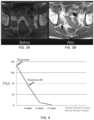
- FIGS. 3 A-Bare images of CT scans of Patient B's pelvic region before and after treatment with a CTLA-4 inhibitor, a PD-1 inhibitor, and a cytokine in addition to ablation. Arrows point to location of the initial tumor structure before treatment ( FIG. 3 A ) and after treatment ( FIG. 3 B ).
- FIG. 3 Ais a CT scan of Patient B's pelvic region before treatment.
- FIG. 3 Bis a CT scan of Patient B's pelvic region after treatment.
- FIG. 4is a graph illustrating the decline in PSA blood levels in Patient B after treatment.
- FIG. 5is a device having two cryoprobe electrodes and the ability to deliver electrical pulses and create reversible electroporation.
- FIG. 6is an embodiment of a device having one cryoprobe electrode and one non-cryoprobe electrode.
- FIG. 7is an embodiment of a device having one cryoprobe and two retractable electrode needles.
- FIG. 8 Ais an embodiment of a device having one cryoprobe and two retractable electrode needles configured to inject plasmids.
- FIG. 8 Bis an embodiment of a device having one cryoprobe and retractable electrode needles configured to inject plasmids.
- FIG. 9is an embodiment of a device having one cryoprobe and two electrodes on the single cryoprobe.
- FIG. 10is an embodiment of a device having one cryoprobe electrode and one indifferent electrode.
- FIG. 11is an embodiment of a device having a cryoprobe treatment portion detachable from an electric therapy delivery portion.
- the present disclosureis based, at least in part, on new compositions for cancer treatment that include at least two immune checkpoint inhibitors and at least one cytokine, each being present in the combination in therapeutically effective amounts and in a pharmaceutically acceptable carrier.
- This combinationcan in some instances further comprise a nucleic acid drug.
- the present disclosureis also based, at least in part, on the development of a new method for the treatment of cancer that comprises administering to a patient intra-tumorally a composition as disclosed herein. Further described are devices configured for performing certain methods described herein.
- compositions, methods, and devices described hereinare particularly useful for treating cancer in subjects.
- cancerrefers to cells having the capacity for autonomous growth. Examples of such cells include cells having an abnormal state or condition characterized by rapidly proliferating cell growth.
- the termis meant to include cancerous growths, e.g., tumors; metastatic tissues, and malignantly transformed cells, tissues, or organs, irrespective of histopathologic type or stage of invasiveness.
- malignancies of the various organ systemssuch as respiratory, cardiovascular, renal, reproductive, hematological, neurological, hepatic, gastrointestinal, and endocrine systems; as well as adenocarcinomas which include malignancies such as most colon cancers, renal-cell carcinoma, prostate cancer and/or testicular tumors, non-small cell carcinoma of the lung, cancer of the small intestine, and cancer of the esophagus.
- compositions, methods, and devices described hereincan be used to treat naturally arising cancer in a subject.
- Cancer that is “naturally arising”includes any cancer that is not experimentally induced by implantation of cancer cells into a subject, and includes, for example, spontaneously arising cancer, cancer caused by exposure of a patient to a carcinogen(s), cancer resulting from insertion of a transgenic oncogene or knockout of a tumor suppressor gene, and cancer caused by infections, e.g., viral infections.
- carcinomasTreatment of carcinomas, adenocarcinomas, and sarcomas is within the present invention.
- the term “carcinoma”is art recognized and refers to malignancies of epithelial or endocrine tissues.
- the termalso includes carcinosarcomas, which include malignant tumors composed of carcinomatous and sarcomatous tissues.
- An “adenocarcinoma”refers to a carcinoma derived from glandular tissue or in which the tumor cells form recognizable glandular structures.
- sarcomais art recognized and refers to malignant tumors of mesenchymal derivation.
- Cancersthat may be treated using the methods, compositions, and devices of the present invention include, for example, cancers, e.g., tumors, of the stomach, colon, rectum, mouth/pharynx, esophagus, larynx, liver, pancreas, lung, breast, cervix uteri, corpus uteri, ovary, prostate, testis, bladder, skin, bone, kidney, brain/central nervous system, head, neck and throat; sarcomas, choriocarcinomas, and lymphomas, among others.
- cancerse.g., tumors, of the stomach, colon, rectum, mouth/pharynx, esophagus, larynx, liver, pancreas, lung, breast, cervix uteri, corpus uteri, ovary, prostate, testis, bladder, skin, bone, kidney, brain/central nervous system, head, neck and throat; sarcomas, choriocarcinomas, and
- Metastatic tumorscan be treated using methods described herein. For example, performing a treatment method described herein on a tumor located at one site in the subject's body (e.g., a primary tumor), can stimulate the subject's immune defenses against the tumor and cause an immune attack on tumors of the same or even different type of at another site(s) in the subject's body (e.g., a metastatic tumor).
- a metastatic tumorcan arise from a multitude of primary tumor types, including but not limited to those of prostate, colon, lung, breast, bone, and liver origin. Metastases develop, e.g., when tumor cells shed from a primary tumor adhere to vascular endothelium, penetrate into surrounding tissues, and grow to form independent tumors at sites separate from a primary tumor.
- non-cancerous growthse.g., non-cancerous tumors.
- exemplary non-cancerous growthsinclude, e.g., benign tumors, adenomas, adenomyoeptheliomas, ductal or lobular hyperplasia, fibroadenomas, fibromas, fibrosis and simple cysts, adenosis tumor, hematomas, hamartomas, intraductal papillomas, papillomas, granular cell tumors, hemangiomas, lipomas, meningiomas, myomas, nevi, osteochondromas, phyllodes tumors, neuromas (e.g., acoustic neuromas, neurofibromas, and pyogenic granulomas), or warts (e.g., plantar warts, genital warts, flat warts, peri
- a subjectcan be diagnosed by a physician (or veterinarian, as appropriate for the subject being diagnosed) as suffering from or at risk for a condition described herein, e.g., cancer, by any method known in the art, e.g., by assessing a patient's medical history, performing diagnostic tests, and/or by employing imaging techniques.
- one exemplary method of treating a tumor in a patientcomprises the steps of: (i) optionally, prior to performance of the method, identifying the location of the tumor within the patient; (ii) intratumorally administering a pharmaceutical composition described herein to the tumor (e.g., a pharmaceutical composition comprising at least two immune checkpoint inhibitors and at least one cytokine); (iii) optionally ablating at least a portion of the tumor; (iv) optionally administering a therapeutically effective amount of a nucleic acid drug to the tumor; and (v) optionally administering a series of electric pulses to the tumor such that the area around the lesion is reversibly electroporated.
- a pharmaceutical composition described hereine.g., a pharmaceutical composition comprising at least two immune checkpoint inhibitors and at least one cytokine
- optionally ablating at least a portion of the tumore.g., a pharmaceutical composition comprising at least two immune checkpoint inhibitors and at least one cytokine
- Identifying a location of the tumorcan be performed by techniques known in the art (e.g., X-ray radiography, magnetic resonance imaging, medical ultrasonography or ultrasound, endoscopy, elastography, tactile imaging, thermography, medical photograph, nuclear medicine imaging techniques including positron emission tomography and single-photon emission computed tomography, photoacoustic imaging, thermography, tomography including computer-assisted tomography, echocardiography and functional near-infrared spectroscopy, etc.).
- the optional step of ablating the tumor (iii)can occur before, concurrently, or after administering a pharmaceutical composition (ii), and the ablation can create an area of lesion exposing intracellular components and membrane antigens of the tumor.
- Ablationcan be performed using a technique described herein on a portion or all of the tumor.
- administering a therapeutically effective amount of a nucleic acid drug to the tumor (iv)can occur before, concurrently or after the of steps (ii) and (iii).
- administering a series of electric pulses to the tumor (v)can occur concurrently or after the administration of the nucleic acid drug (iv); or before, concurrently and/or after steps (ii) and (iii).
- compositionscomprising the mixture of checkpoint inhibitors and cytokine(s).
- Check point inhibitorswork to activate the immune system to attack tumors, inhibiting the immune response proteins responsible for down regulating the immune system.
- the check point inhibitorscan be, e.g., inhibitors of CD137, CD134, PD-1 , KIR, LAG-3, PD-L1, CTLA-4 , B7.1, B7H3, CCRY, OX-40, and/or CD40.
- the pharmaceutical compositionscan comprise any combination of check point inhibitors. For example, particularly useful in is a combination of a PD-1 inhibitor and a CTLA-4 inhibitor. A skilled practitioner would appreciate that many other combination are also useful.
- a non-limiting list of combinationsinclude a CD137 inhibitor and a CD134 inhibitor; a PD-1 inhibitor and a MR inhibitor; a LAD-3 inhibitor and a PD-L1 inhibitor; a CTLA-4 inhibitor and a CD40 inhibitor; a CD134 inhibitor and a PD-1 inhibitor; a MR inhibitor and a LAG-3 inhibitor; a PD-L1 inhibitor and a CTLA-4 inhibitor; a CD40 inhibitor and a CD137 inhibitor; a CTLA-4 inhibitor and a PD-L1 inhibitor; a PD-1 inhibitor and a CD40 inhibitor, or any combination of two or more checkpoint inhibitors known in the art.
- the pharmaceutical compositionscan also comprise at least cytokine.
- the at least one cytokinecan comprise GM-CSF, IL-12, IL-6, IL-4, IL-12, TNF, IFN ⁇ , IFN ⁇ , and/or a combination thereof.
- the compositionscan include a first cytokine and a second cytokine. A skilled practitioner would appreciate that in some instances the first and the second cytokine can be different.
- checkpoint inhibitorsare administered intravenously, which can result in serious and sometimes fatal systemic toxicities as a result of non-specific distribution of these cytocidal agents in the body.
- the non-specific distribution of these agentskills both cancer cells and normal cells and can negatively impact the treatment regimen and patient outcome.
- the present intra-tumoral methodscan reduce systemic toxicity and produce fewer side effects by sequestering the drugs in the tumor microenvironment and sparing normal cells and tissues from the toxicity of the drugs (Intratumoral Immunization: A New Paradigm for Cancer Therapy. Clin Cancer Res. 2014 April 1; 20(7): 1747-1756. doi:10.1158/1078-0432.CCR-139-2116).
- the present intra-tumoral methodscan reduce systemic toxicity and product fewer side effects by also lowering the amount of the administered compositions necessary to be therapeutically effective.
- the pharmaceutical compositioncan be more effective at not only inhibiting the cancer but also triggering an effective antitumor immune response. This antitumor immune response may then target metastatic sites and eliminate cancer throughout the subject.
- compositionscan further include one or more therapeutic and/or biologic agents known in the art to be effective in treating cancer, i.e., an anti-cancer agent, or known in the art to be effective in stimulating the immune system, i.e., immunostimulant or immunomodulator.
- therapeutic and/or biologic agentsknown in the art to be effective in treating cancer, i.e., an anti-cancer agent, or known in the art to be effective in stimulating the immune system, i.e., immunostimulant or immunomodulator.
- Such pharmaceutical compositionscan be used to treat cancer as described above.
- the pharmaceutical compositionfurther comprises a therapeutically effective amount of a nucleic acid drug.
- the nucleic acid drugcan be, e.g. DNA, nDNA, mtDNA, gDNA, RNA, siRNA, miRNA, mRNA, piRNA, antisense RNA, snRNA, snoRNA, vRNA, etc .
- the nucleic acid drugcan be a DNA plasmid.
- Such a DNA plasmidcan comprise, consist essentially of, or consist of a nucleotide sequence encoding a gene selected from the group consisting of GM-CSF, IL-12, IL-6, IL-4, IL-12, TNF, IFN ⁇ , IFN ⁇ , and/or a combination thereof.
- the nucleic acid drugcan have clinical usefulness, for example, enhancing the therapeutic effects of the cells or providing a patient with a therapeutic agent. In other instances, the nucleic acid drug may function as a marker or resistance gene.
- the nucleotide sequencecan encode a gene that can be secreted from the cells or cannot be secreted from the cells.
- the nucleic acid drugcan encode a gene and a promoter sequence to increase expression of the gene.
- compositionscan be adapted according to the individual aspects of the cancer and/or the subject, e.g., size of the tumor, location of the tumor, subject, clinical evidence of drug response, etc.
- a pharmaceutical composition provided hereincan include a delivery agent or pharmaceutically acceptable carrier.
- pharmaceutically acceptable carrierincludes solvents, dispersion media, coatings, antibacterial and antifungal agents, isotonic and absorption delaying agents, and the like, compatible with pharmaceutical administration.
- Supplementary active compoundscan also be incorporated into pharmaceutical formulations that contain an antibody or antigen-binding fragment thereof as described herein.
- solutions or suspensions used for parenteral, intradermal, or subcutaneous applicationcan include the following components: a sterile diluent such as water for injection, saline solution, fixed oils, polyethylene glycols, glycerin, propylene glycol or other synthetic solvents; antibacterial agents such as benzyl alcohol or methyl parabens; antioxidants such as ascorbic acid or sodium bisulfite; chelating agents such as ethylenediaminetetraacetic acid; buffers such as acetates, citrates or phosphates and agents for the adjustment of tonicity such as sodium chloride or dextrose. pH can be adjusted with acids or bases, such as hydrochloric acid or sodium hydroxide.
- the parenteral preparationcan be enclosed in ampoules, disposable syringes or multiple dose vials made of glass or plastic.
- compositions described hereinmay be intra-tumorally delivered via an injection device, wherein the injection device may be part of a probe.
- the probes as described hereincan be configured for the various ablation methods. Further, the probe can also be configured to combine the methods described herein, e.g., a cryoprobe can be configured to administer an electric pulse, a cryogen and/or a composition of drugs.
- compositions suitable for injectioncan include sterile aqueous solutions (where water soluble), dispersions, and sterile powders for the extemporaneous preparation of sterile injectable solutions or dispersion.
- suitable carriersinclude physiological saline, bacteriostatic water, Cremophor ELTM (BASF, Parsippany, N.J.), or phosphate buffered saline (PBS).
- the compositionmust be sterile and should be fluid to the extent that easy syringability exists. It should be stable under the conditions of manufacture and storage and must be preserved against the contaminating action of microorganisms such as bacteria and fungi.
- the carriercan be a solvent or dispersion medium containing, for example, water, ethanol, polyol (for example, glycerol, propylene glycol, and liquid polyetheylene glycol, and the like), and suitable mixtures thereof.
- the proper fluiditycan be maintained, for example, by the use of a coating such as lecithin, by the maintenance of the required particle size in the case of dispersion and by the use of surfactants.
- Prevention of the action of microorganismscan be achieved by various antibacterial and antifungal agents, for example, parabens, chlorobutanol, phenol, ascorbic acid, thimerosal, and the like.
- isotonic agentsfor example, sugars, polyalcohols such as mannitol, sorbitol, and sodium chloride in the composition.
- Prolonged absorption of the injectable compositionscan be brought about by including in the composition an agent that delays absorption, for example, aluminum monostearate and gelatin.
- Sterile injectable solutionscan be prepared by incorporating the active compound in the required amount in an appropriate solvent with one or a combination of ingredients enumerated above, as required, followed by filtered sterilization.
- dispersionsare prepared by incorporating the active compound into a sterile vehicle, which contains a basic dispersion medium and the required other ingredients from those enumerated above.
- the preferred methods of preparationare vacuum drying and freeze-drying, which yield a powder of the active ingredient plus any additional desired ingredient from a previously sterile-filtered solution thereof.
- the therapeutic compoundscan be prepared with carriers that will protect the therapeutic compounds against rapid elimination from the body, such as a controlled release formulation, including implants and microencapsulated delivery systems.
- compositionscan be included in a container, pack, cartridge, or dispenser together with instructions for administration.
- the therapeutic and/or biologic agentscan be administered in an effective amount, at dosages and for periods of time necessary to achieve the desired result.
- An effective amountcan be administered in one or more administrations, applications or dosages.
- a therapeutically effective amount of a pharmaceutical compositiondepends on the pharmaceutical composition selected.
- the compositionscan be administered from one or more times per day to one or more times per week; including once every other day.
- treatment of a subject with a therapeutically effective amount of the pharmaceutical compositions described hereincan include a single treatment or a series of treatments.
- the compositions described hereincan be administered in one or more administrations. These one or more administrations can be of the same or different methods of administration, including, for example, intravenously, intramuscularly, subcutaneously, intra-tumorally or any combination thereof.
- a first compositionis administered intra-tumorally and a second composition is administered subcutaneously.
- first and the second compositionsare administered simultaneously, in sequence, or in a series of treatments.
- first and the second compositionsare the same, different, or the same in part.
- the methods described hereininclude two or more administrations.
- a first administrationis an intra-tumoral administration of at least two checkpoint inhibitors (e.g., a PD-1 inhibitor and a CTLA-4 inhibitor) and at least one cytokine (e.g., GM-CSF).
- Dosage regimenscan be adjusted to provide the optimum therapeutic response. For example, several divided doses can be administered daily or the dose can be proportionally reduced as indicated by the exigencies of the therapeutic situation.
- Those skilled in the artwill be aware of dosages and dosing regimens suitable for administration of the new monoclonal antibodies disclosed herein or antigen-binding fragments thereof to a subject. See e.g., Physicians' Desk Reference, 63rd edition, Thomson Reuters, Nov. 30, 2008.
- Dosage, toxicity and therapeutic efficacy of the therapeutic compoundscan be determined by standard pharmaceutical procedures in cell cultures or experimental animals, e.g., for determining the LD50 (the dose lethal to 50% of the population) and the ED50 (the dose therapeutically effective in 50% of the population).
- the dose ratio between toxic and therapeutic effectsis the therapeutic index and it can be expressed as the ratio LD50/ED50.
- Compounds which exhibit high therapeutic indicesare preferred. While compounds that exhibit toxic side effects may be used, care should be taken to design a delivery system that targets such compounds to the site of affected tissue in order to minimize potential damage to uninfected cells and, thereby, reduce side effects.
- the data obtained from cell culture assays and animal studiescan be used in formulating a range of dosage for use in humans.
- the dosage of such compoundslies preferably within a range of circulating concentrations that include the ED50 with little or no toxicity.
- the dosagemay vary within this range depending upon the dosage form employed and the route of administration utilized.
- the therapeutically effective dosecan be estimated initially from cell culture assays.
- a dosemay be formulated in animal models to achieve a circulating plasma concentration range that includes the IC50 (i.e., the concentration of the test compound which achieves a half-maximal inhibition of symptoms) as determined in cell culture.
- IC50i.e., the concentration of the test compound which achieves a half-maximal inhibition of symptoms
- levels in plasmamay be measured, for example, by high performance liquid chromatography.
- Methods of treating cancer disclosed hereinoptionally employ ablation of at least a portion of a tumor.
- ablationversus surgical removal, is that the tumor is left in situ for the body's defense and healing mechanisms to remove it. This creates an opportunity to harness the body's immune defense mechanisms to recognize the dead tumor and essentially auto-immunize the patient to their own cancer.
- the methods disclosed hereincan (i) treat primary tumors; (ii) activate the immune response to cancer cell antigens; and (iii) induce immune system targeting of metastatic lesions.
- the method of ablationinfluences at least two factors that are known to influence the immunologic response to an ablated tumor.
- Oneis the effect of the ablation process on the protein structure and therefore the antigenicity of the tumor proteins.
- the second factoris the mechanism of cell death related to the ablation modality. Necrosis, under certain conditions, ruptures the cell and spills a wide range of intracellular contents into the extracellular environment that causes co-stimulation of dendritic cells, leading to T Cell proliferation and activation. Apoptosis, which leaves the cells intact, confines the cellular contents and prevents co-stimulation. This lack of intracellular exposure and co-stimulation mutes the immunologic effect by preventing T cell activation and proliferation.
- cryoablationthermal ablation, IRE, RF-EMB, RF-EMB type ablation, ultrasonic ablation, high-intensity focused ultrasound ablation, ablation using photodynamic therapy, ablation using non-thermal shock waves, cavitation, other mechanical physical cell disruption, or any combination thereof.
- thermal ablationIRE
- RF-EMBRF-EMB type ablation
- ultrasonic ablationhigh-intensity focused ultrasound ablation
- ablation using photodynamic therapyablation using non-thermal shock waves
- cavitationother mechanical physical cell disruption, or any combination thereof.
- These different types of ablation methodscan have different outcomes on the protein structures and mechanism of cell death. For example, heat ablation destroys structures due to denaturing proteins and it also destroys the underlying collagen matrix of the tissue. This disruption of the proteins and tissue makes a robust immunologic response unlikely.
- Colde.g. cryoablation, can denature proteins and can disrupt both protein and tissue structure.
- Irreversible electroporation (IRE) and non-thermal ablation modalitiesare structure sparing and can therefore be used to treat cancers in the pancreas, central liver, and other areas such as the head and neck.
- IREis a technique where an electrical field is applied to cells in order to increase the permeability of the cell membrane. The high voltage of IRE destroys the target cells while leaving neighboring cells unaffected. IRE, however, causes apoptotic cell death, and as described above, this is not optimal for an immunologic reaction.
- Radiofrequency electrical membrane breakdown RF-EMBis another non-thermal modality that produces necrosis by complete breakdown of the cell membrane electrically (see, Onik PCT/US2014/068774, which is incorporated herein in its entirety). Under certain conditions, RF-EMB can also be used to deliver DNA plasmids.
- Reversible electroporation (RE)can also be used to deliver DNA plasmids. RE is similar to IRE, however the electricity applied to the target cells is below the electric field threshold of the target cells. Therefore, the cells can recover when the electric field is removed and rebuild their cellular membranes and continue with cellular functions. RE can be used as a tool for gene therapy as the reversible element allows for entry of nucleic acids (e.g. DNA plasmids) into a viable cell.
- nucleic acidse.g. DNA plasmids
- An ablation method described hereincan be used alone or in combination with other ablation methods. Two or more ablation methods can be used in combination. The methods may be applied sequentially or concurrently. In some cases, a combination of ablation methods has a synergistic effect on the tissue.
- a non-limiting list of combinationsincludes, for example, heat ablation and RF-EMB, cryoablation and RF-EMB, IRE and RF-EMB, RE and RF-EMB, IRE and cryoablation, heat ablation and cryoablation, heat ablation and IRE, RE and IRE, heat ablation with RE, and any combination in which two or more methods are used.
- the two or more ablation methodscan be used concurrently or sequentially.
- methods described hereincreate an RF-EMB type lesion using a combination of RF-EMB and cryoablation techniques.
- This combination of ablation methodscan produce a synergistic effect on the tissue.
- the synergistic effectcan be the creation of an RF-EMB type lesion with less required energy input than with other means.
- the result, for instance in liver tissueincludes: in areas adjacent to aseptic non-inflammatory coagulative necrosis, there is alteration of liver architecture, including dilation of bile duct canaliculi, as well as unique diffuse alteration of cytoplasmic organelles, including distortion of mitochondrial cristae and vacuolization of endoplasmic reticulum.
- compositions or treatmentscan be adapted according to the individual aspects of the cancer, e.g., size of the tumor, location of the tumor, the subject.
- variables of each of the various methods of ablationare known and described in the art (including, for example, Percutaneous Prostate Cryoablation. Edited by Gary Onik, Boris Rubinsky, Graham Watson, and Richard Ablin. Quality Medical Publishing, St Louis, Mo. 1995 which is incorporated herein in its entirety).
- the process of cryoablationincludes variables that can be adjusted, e.g. the number of freeze-thaw cycles, the speed of the freeze, the thaw portion of the cycle, etc, to influence the outcome of the ablation, e.g., the size of the lesion, damage to surrounding tissue, and the immune response to the lesion.
- the process of RF-EMBincludes variables such as strength of the electric field, frequency, polarity, shape duration, number and spacing, etc., which can similarly influence the outcome of the ablation. The proximity of a tumor cell to the electric pulse will determine the strength and outcome of the RF-EMB on any particular cell.
- the cells furthest from the point of administrationare treated with a lower strength electric field and as such may not be ablated but rather reversibly electroporated.
- reversible electroporationfor the delivery of gene therapy can be modified to determine the range, reversibility and delivery of the electroporation around the lesion.
- REreversible electroporation
- the nucleic acid drugcan be administered before, after or during the process of ablation.
- the nucleic acid drugcan be administered before, after or during the administration of the pharmaceutical mixture.
- the nucleic acid drugcan also be administered before or during the process of electroporation.
- the methodscan be used alone or in combination with other methods for treating cancer in patients. Accordingly, in some instances, the methods described herein can further include treating the patient using surgery (e.g., to remove a portion of the tumor), chemotherapy, immunotherapy, gene therapy, and/or radiation therapy. Compositions and methods described herein can be administered to a patient at any point, e.g., before, during, and/or after the surgery, chemotherapy, immunotherapy, gene therapy, and/or radiation therapy.
- kitsthat include one or more of the pharmaceutical compositions described herein.
- Kitsgenerally include the following major elements: packaging, reagents comprising binding compositions as described above, optionally a control, and instructions.
- Packagingcan be a box-like structure for holding a vial (or number of vials) containing said binding compositions, a vial (or number of vials) containing a control, and/or instructions for use in a method described herein.
- the packagingcontains a cartridge that can be controlled by a digital device following systematic instructions. Individuals skilled in the art can readily modify the packaging to suit individual needs.
- a kit provided hereincan include at least one (e.g., one, two, three, four, five, or more) composition containing at least one (e.g., one, two, three, four, five, or more) of the compositions described herein, and at least one (e.g., one, two, three, four, five, or more) other composition in a separate vial containing a therapeutic or biologic agent known in the art to be effective in treating cancer.
- at least onee.g., one, two, three, four, five, or more
- compositioncontaining at least one (e.g., one, two, three, four, five, or more) of the compositions described herein, and at least one (e.g., one, two, three, four, five, or more) other composition in a separate vial containing a therapeutic or biologic agent known in the art to be effective in treating cancer.
- compositions and kits as provided hereincan be used in accordance with any of the methods (e.g., treatment methods) described above.
- compositions and kitscan be used to treat cancer.
- Those skilled in the artwill be aware of other suitable uses for compositions and kits provided herein, and will be able to employ the compositions and kits for such uses.
- an injection deviceis a cryoprobe that can emit electric pulses and also deliver plasmids.
- an injection device 100is part of a system 101 that is capable of administering both extreme cold as well as electric pulses to tissues and/or tumors.
- the injection device 100has two electrode cryoprobes, including a positively-charged cryoprobe 110 and a negatively-charged cryoprobe 130 .
- Each cryoprobe 110 , 130is a generally cylindrical probe that is inserted into a target tissue 102 ata first end 111 , 131 and grasped by a user ata second end 112 , 132 .
- Each cryoprobe 110 , 130can be individually manipulated by a user.
- both cryoprobes 110 , 130can be contained within a larger housing (not shown for clarity) that permits the user to insert both cryoprobes 110 , 130 into the target tissue 102 simultaneously at a known distance from each other.
- the two cryoprobes 110 , 130 contained within a housingcan be arranged such that the distance separating the two cryoprobe electrodes 110 , 130 can be increased or decreased by the user.
- Each cryoprobe 110 , 130has a central gas supply cannula 114 , 134 running from the first ends 111 , 131 to the second ends 112 , 132 of the cryoprobes 110 , 130 .
- Each central gas supply cannula 114 , 134is attached at the second end 112 , 132 of each probe to a cryomachine 190 .
- the cryomachine 190serves as a source of cooled gas that is pumped via gas supply lines 192 to enter the central gas supply cannulas 114 , 134 at the second ends 112 , 132 of the cryoprobes and be delivered to cooling heads 116 , 136 at the first ends 111 , 131 of the cryoprobes and thereby to the tissue 102 .
- the cooling heads 116 , 136are configured to pierce and be inserted into the tissue 102 as is known in the art, and can be flat or pointed in shape.
- the cooling heads 116 , 136are generally made of metal or other material that has a high conductance so as to allow the cold gas entering the cooling heads 116 , 136 via the central gas supply cannulas 114 , 134 to thermally interact with the tissue 102 .
- Gas return channels 118 , 138concentrically surround the central gas supply cannulas 114 , 134 and are fluidly connected to the cannulas such that cooled gas enters the cooling heads 116 , 136 and then flows back through the gas return channels 118 , 138 to return to the cryomachine 190 via gas return lines 194 .
- Layers of thermal insulation 120 , 140protect the user grasping the cryoprobes 110 , 130 from the cold gas running through the gas return channels 118 , 138 .
- Layers of electrical insulation 122 , 142 and the layers of thermal insulation 120 , 140concentrically surround the outer surfaces of the gas return channels 118 , 138 .
- the layers of electrical insulation 122 , 142protect the user and electrically isolate the body of each cryoprobe 110 , 130 from electrical pulses generated by an electrical pulse generator 180 .
- the order of layers of electrical insulation 122 , 142 , thermal insulation 120 , 140 and the outer surfaces of the gas return channels 118 , 138may be placed in differing orders.
- the electrical pulse generator 180is connected by wires 182 to the second ends 112 , 132 of the cryoprobes 110 , 130 such that electrical pulses are transmitted to the cooling heads 116 , 136 and in turn administered to the tissue 102 .
- the cooling heads 116 , 136therefore serve the dual function of administering cold as well as the electrical impulses to the target tissue 102 .
- the electrical pulsescan be transmitted along the length of the cryoprobes 110 , 130 via wires layered between the layers of electrical insulation 122 , 142 and the layers of thermal insulation 120 , 140 .
- at least a portion of the gas return channels 118 , 138are electrically conductive and also serve the function of transmitting the electrical pulses to the tissue 102 via the cooling heads 116 , 136 .
- the electrical pulse generator 180is arranged to generate a positive charge via the positively-charged cryoprobe 110 and a negative charge via the negatively-charged cryoprobe 130 .
- the injection device 100is therefore capable of delivering electrical pulses as well as cold temperatures to the target tissue 102 .
- the positively-charged cryoprobe 110 and the negatively-charged cryoprobe 130can be identical in structure.
- the two cryoprobes 110 , 130are inserted into the target tissue 102 at a desired distance of separation from each other (e.g., 2 mm, 5 mm, 10 mm), thereby creating a cryolesion zone 104 that surrounds and extends between the tips of the cryoprobes 110 , 130 .
- This arrangement of the two cryoprobes 110 , 130also creates an RE (Reversible Electroporation) zone 106 in relation to the cryolesion zone 104 .
- REReversible Electroporation
- the configuration of the cryolesion zone 104can be varied by the user.
- the cooling heads 116 , 136are retractable into the bodies of the cryoprobes 110 , 130 , e.g., the length of the cooling heads 116 , 136 extending from the end of the thermal insulation layers 120 , 140 can be reduced by retracting the cooling heads 116 , 136 such that more or all of their surface area is covered by the thermal insulation layers 120 , 140 .
- the length of the cooling heads 116 , 136 extending from the end of the thermal insulation layers 120 , 140can be increased by extending the cooling heads 116 , 136 such that less of their surface area is covered by the thermal insulation layers 120 , 140 .
- the insulation layers 120 , 140 and 122 , 144are repositionable during use of the injection device 100 .
- the usercan also modify the temperature of the gas exiting the cryomachine 190 and entering the tissue 102 .
- the configuration of the RE zone 106can be varied by the user by modulating the electrical pulses exiting the electrical pulse generator 180 .
- the variablescan be altered such that the cryolesion zone 104 is smaller than, the same size as, or larger than the RE zone 106 .
- FIG. 6an additional embodiment of an injection device 200 that is capable of delivering both cold and electrical pulses to a target tissue 202 is shown.
- a positively-charged cryoprobe 210has a first end 211 and a second end 212 , and a central gas supply cannula 214 running from the first end 211 to the second end 212 .
- the central gas supply cannula 214is attached at the second end 212 to a cryomachine 290 that is a source of cooled gas that is pumped via a gas supply line 292 to enter the central gas supply cannula 214 and be delivered to a cooling head 216 at the first end 211 of the cryoprobe and thus to the tissue 202 .
- the cooling head 216is configured to pierce and be inserted into the tissue 202 as is known in the art, and can be flat or pointed in shape, and is generally made of metal or other material that has a high conductance.
- a gas return channel 218concentrically surrounds the central gas supply cannula 214 and is fluidly connected to the cannula 214 such that cooled gas enters the cooling head 216 and then flows back through the gas return channel 218 to return to the cryomachine 290 via a gas return line 294 .
- a layer of thermal insulation 220protects the user grasping the cryoprobe 210 from the cold gas running through the gas return channel 218 .
- a layer of electrical insulation 222concentrically layers the outer surface of the gas return channel 218 which also concentrically surrounded by the layer of thermal insulation 220 .
- An electrical pulse generator 280is connected by wires 282 to the second end 212 of the cryoprobe 210 and also to the second end 232 of an electric probe 230 .
- the electric probe 230is similar to cryoprobe 210 , having a first end 231 that is insertable into the tissue 202 and a second end 232 that connects to the electrical pulse generator 280 .
- the electric probe 230is not connected to the cryomachine 290 and does not have the structure (e.g., a central gas supply cannula, a gas return channel, gas supply and return lines) to administer cryotherapy to the tissue 202 .
- the electric probe 230has a tissue insertion head 236 that does not cool the tissue 202 but does administer the electric therapy.
- the electric pulse generator 280transmits electrical pulses to the cooling head 216 and tissue insertion head 236 and in turn to the tissue 202 .
- the cooling head 216therefore serves the dual function of administering cold as well as the electrical impulses to the target tissue 202 while the tissue insertion head 236 administers the electrical impulses only.
- the electrical pulsescan be transmitted along the length of the cryoprobe 210 and electric probe 230 via wires attached to layers of electrical insulation 222 , 242 .
- at least a portion of the bodies of the cryoprobe 210 and electric probe 230are electrically conductive and also serve the function of transmitting the electrical pulses to the tissue 202 .
- the electrical pulse generator 280is arranged to generate a positive charge via the positively-charged cryoprobe 210 and a negative charge via the negatively-charged electric probe 230 .
- the cryoprobe 210 and electric probe 230are inserted into the target tissue 202 at a desired distance of separation from each other (e.g., 2 mm, 5 mm, 10 mm), thereby creating an RE zone 206 that surrounds and extends between the cryoprobe 210 and electric probe 230 .
- a created cryolesion zone 204is smaller than the cryolesion zone 104 created with two cryoprobes and surrounds the first end 211 of the cryoprobe 210 .
- the configuration of the cryolesion zone 204can be varied by the user as for cryoprobe injection device 200 by arranging the cooling head 216 to be retractable into the body of the cryoprobes 210 .
- the usercan also modify the temperature of the gas exiting the cryomachine and entering the tissue 202 .
- the size of the RE zone 206can be varied by modulating the electrical pulses exiting the electrical pulse generator 180 .
- FIG. 7Shown in FIG. 7 is an embodiment of an injection device 300 that has a single cryoprobe 310 .
- the elements of the injection deviceare similar to the previous embodiments, however the injection device 300 has a single cryoprobe 310 .
- the cryoprobe 310is capable of delivering both cold and electrical pulses to a target tissue 302 and has a first end 311 , a second end 312 , a central gas supply cannula 314 running between them and attached to a cryomachine 390 (not shown) that is a source of cooled gas pumped via a gas supply line 392 to the cryoprobe 310 and delivered to a cooling head 316 and removed by a gas return channel 318 concentrically surrounding and fluidly connected to the central gas supply cannula 314 .
- a layer of thermal insulation 320 and a layer of electrical insulation 322are also present.
- One or two electrical pulse generators 380are connected by wires 382 to the second end 312 of the cryoprobe 310 .
- the wires 382attach to a pair of wires 350 , 352 that terminate in electrodes 356 , 358 that exit the body of the cryoprobe and enter the tissue 302 alongside the cooling head 316 .
- the wires 350 , 352are embedded in the electrical insulation layer 322 , e.g., by piercing the electrical insulation layer 322 or by insertion into channels that run the length of the electrical insulation layer 322 .
- the wires 350 , 352 and electrodes 356 , 358can attach to each other, respectively, or in some embodiments the positive wire 350 and positive electrode 356 are the same continuous wire and the negative wire 352 and negative electrode 358 are the same continuous wire.
- the electrodes 356 , 358are shaped such that when extended into the tissue 302 the electrodes curve away from the body of the cryoprobe 310 . When retracted, the electrodes 356 , 358 are held in a linear shape to better align with the body of the cryoprobe.
- the electrodes 356 , 358can be formed of e.g., nickel titanium (also known as nitinol). The curvature of the electrodes 356 , 358 allows the user to extend the resulting RE zone 306 beyond the cryolesion zone 304 . The user can extend the electrodes 356 , 358 and transmit electric pulses before, during, or after the cryotherapy treatment.
- FIG. 8 Ashows an injection device 400 similar to that of FIG. 7 (with reference labels referring to the same elements as in FIG. 7 but raised by 100 ).
- injection device 400is capable of injecting plasmids into tissue 402 as well as administering electrotherapy and cryotherapy.
- the cryoprobe 410has needles 460 , 462 that extend approximately parallel with electrodes 456 , 458 and are inserted into tissue 402 .
- the needles 460 , 462are fluidly connected to tubes 472 which receives fluid from a fluid reservoir 470 .
- the fluid reservoir 470can be a syringe.
- Fluid, e.g., plasmids, inside the fluid reservoir 470can therefore be administered to the tissue 402 .
- the needles 460 , 462are fully or partially retractable into the body of the cryoprobe 410 as are the electrodes 456 , 458 .
- the needles 460 , 462 and electrodes 456 , 458can be retracted simultaneously or independently of each other.
- the needles 460 , 462are also repositionable within the tissue 402 .
- the needles 460 , 462can have multiple tines 466 . Multiple tines 466 can allow the user greater control over the spread and distribution of the injected materials or medications in a more quickly and precisely controllable pattern and at a specific distance from the central probe.
- FIG. 9shows a cryoprobe 500 with two layers of electrical insulation 522 , 524 .
- Wires or electrical conduits 550 , 552are sandwiched between the two layers of electrical insulation 522 , 524 and carry positive charge from the electric pulse generator 580 .
- the body of the cryoprobe 500terminates in the cooling head 516 and acts as an electrical conduit for the negative charge generated by the electrical pulse generator 580 .
- Each of the two layers of electrical insulation 522 , 524is independently positionable and retractable.
- FIG. 10Shown in FIG. 10 is an embodiment of an injection device 600 that has a single cryoprobe 610 .
- the elements of the injection deviceare similar to the previous embodiments, however the injection device 600 has a single cryoprobe 610 that works with an indifferent electrode 696 , which is a remote electrode placed either upon a single limb or connected with the central terminal and paired with an exploring electrode of cryorobe 610 .
- the cryoprobe 610is capable of delivering both cold and electrical pulses to a target tissue 602 and has a first end 611 , a second end 612 , and a central gas supply cannula 614 running between them and attached to a cryomachine 690 (not shown) that is a source of cooled gas pumped via a gas supply line 692 to the cryoprobe 610 and delivered to a cooling head 616 and removed by a gas return channel 618 concentrically surrounding and fluidly connected to the central gas supply cannula 614 .
- a layer of thermal insulation 620 and a layer of electrical insulation 622are also present.
- One or two electrical pulse generators 680(as shown in FIG. 10 ) are connected by wires 682 to the second end 612 of the cryoprobe 610 , and also to the indifferent electrode 696 .
- an additional embodiment of an injection device 700is described.
- the elements of the injection device 700are similar to the previous embodiments.
- the injection device 700has a single probe 710 that can be configured to work with an indifferent electrode 796 .
- the injection device 700includes a cryoprobe which is capable of delivering both cold and electrical pulses to a target tissue 702 , and has a first end 711 and a second end 712 .
- Probe 710is made of two different portions, a central portion 770 and concentric portion 772 .
- the central portionhas central gas supply cannula 714 running between the first and second ends of the probe 710 and is attached to a source of cooled gas pumped via a gas supply line 692 to the central portion 770 and delivered to a cooling head 716 , and removed by a gas return channel 718 concentrically surrounding and fluidly connected to the central gas supply cannula 714 .
- a layer of thermal insulation 720surrounds the gas channels.
- the concentric portion 772surrounds the central portion 770 , and is surrounded by a layer of electrical insulation 722 .
- One or two electrical pulse generators 780(two are shown in FIG. 11 ) are connected by wires 782 to the second end 712 of the probe 710 , specifically at concentric portion 772 , and also to the indifferent electrode 796 .
- the concentric portion 772is attachable to and removable from the central portion 770 .
- Concentric portion 772has the form of a sheath that surrounds the internal central portion 772 and the concentric portion 772 can be slid onto and off of the central portion 770 by repositioning the concentric portion 772 relative to the axial length of the central portion 770 .
- Electrical contacts 774are included on the concentric portion 772 , (e.g., on its inner surface). The electrical contacts 774 bring the wires 782 attached to the electrical pulse generator(s) 780 and indifferent electrode 796 into electric contact with an electrically conducting part of the central portion 770 . If the central portion 770 is made of metal, or other conducting material, the electric impulses are thereby transmitted along the body of the central portion to the cooling head 716 to administer the electric therapy to the tissue 702 . Alternatively, the central portion 770 can have wires configured to transmit current from the pulse generator(s) along the length of the central portion 770 .
- the embodiment shown in FIG. 11is particularly advantageous.
- the concentric portion 772can be manufactured separately from the central portion 770 .
- central portion 770can be a complete cryoprobe that is traditionally used in such therapies. Attaching the concentric portion 772 to the outside of the central portion 770 increases the functionality of the probe, allowing the previously single-use cryoprobe to additionally provide electric RF-EMB treatment capability.
- FIG. 11allows a user to perform combined electric RF-EMB treatment and cryotherapy in a highly precise manner, and with increased flexibility.
- the probe 710can be inserted into the tumor or target tissue 702 as desired. Only the concentric portion 722 , the central portion 772 , or both the portions can be positioned as desired. In one embodiment, the user inserts the probe 710 with bother inner and outer portions, and performs the desired therapeutic protocol. The user then can remove the central portion 770 from the tissue 702 by sliding it out of the concentric portion 772 while the concentric portion 772 remains in place.
- a different central portione.g., a needle for delivering plasmids as described above, a tool that has neither cryo nor electricity-delivering capability such as a measurement tool, an acidity sensing or bioactive device, a tissue collection tool, a biopsy tool, or a hypothermia probe.
- the concentric portion 772 remaining in placeallows the user to insert the new central portion with high accuracy, precisely returning to the previous location of the first end of the central portion 772 before it was removed from the tissue 702 .
- the concentric portion 772 of the probe 710can be used in conjunction with tools other than a probe inserted within the concentric portion 772 .
- the concentric portion 772acts as a guidance device so that a different tool is inserted into the precise same location with the benefit of the next tool being placed in the same location as the prior tool.
- the replacement inner toolcan be any tool that fits within concentric portion 772 (such a measurement tool).
- the replacement toolcan be energized through the electric contacts 774 on the concentric portion 772 .
- tools that replace the inner portion to work with concentric portion 772can be tools that have corresponding electrical contacts on the body of the tool to mate with the electric contacts 774 on the centric probe portion 772 .
- Such inner toolscan be previously existing tools that are modified to have such electrical contacts, or tools designed to include such contacts.
- each tool functioncan be used to cause a desired effect in the tissue 702 , and depending on the characteristic of the replacement inner tool and the parameters used each tool can cause an effect in only a part of the tissue 702 .
- probe 710has a locking mechanism or alignment mechanism between the concentric portion 772 and the central portion 770 (e.g., a lever, spring, clip, or luer-type lock). Once the central portion 770 is inserted into the concentric portion 772 , the locking mechanism keeps the inner and outer portions aligned and stationary relative to each other. In some embodiments, the probe 710 will only function once the locking mechanism between the inner and outer portions are engaged. For example, the user would have to twist the central portion 770 into engagement with a ridge on the concentric mechanism, and completing the movement would bring electrical contacts on the central portion into contact with the electrical contacts 774 of the concentric portion.
- a locking mechanism or alignment mechanism between the concentric portion 772 and the central portion 770e.g., a lever, spring, clip, or luer-type lock.
- FIGS. 5 and 6additional pairs of cryoprobes can be used as well.
- FIG. 7additional pairs of wires can be employed.
- the needles or tines 466are configured to measure local pressure within the target tissue. Accordingly, other embodiments are within the scope of the following claims.
- a compositioncomprising a CTLA-4 inhibitor, a PD-1 inhibitor, and a cytokine was intra-tumorally administered to Patient A. Additionally the tumor was ablated to create an RF-EMB type lesion. Before this drug and ablation treatment, Patient A had a prostate-specific antigen (PSA) of approximately 28 ( FIG. 2 ). 9 months after treatment, the PSA remains 0 with complete radiographic resolution of their disease ( FIG. 1 B ).
- PSAprostate-specific antigen
- Biopsies of the areashowed no cancer but inflammatory cell infiltrates in the area of previous tumors are indicative of an immunologic response.
- a 65 year old malewas diagnosed with prostate cancer. Masses were visible in the pelvis ( FIG. 3 A ) of Patient B, and they had PSA levels of 130 and a Gleason score of 8. Metastases were identified in the lymph nodes.
- Patient Bwas intra-tumorally administered the composition comprising a CTLA-4 inhibitor, a PD-1 inhibitor, and a cytokine; and the tumor was ablated to create an RF-EMB type lesion.
- Patient Bhas a PSA level of 1.8 and visible decrease in pelvic masses ( FIG. 3 B ). 8 weeks after the second treatment, Patient B is reported to have normal urinary and sexual function in addition to lymph nodes back to a normal size ( FIG. 4 ).
Landscapes
- Health & Medical Sciences (AREA)
- Life Sciences & Earth Sciences (AREA)
- Chemical & Material Sciences (AREA)
- General Health & Medical Sciences (AREA)
- Engineering & Computer Science (AREA)
- Animal Behavior & Ethology (AREA)
- Public Health (AREA)
- Veterinary Medicine (AREA)
- Medicinal Chemistry (AREA)
- Immunology (AREA)
- Nuclear Medicine, Radiotherapy & Molecular Imaging (AREA)
- Surgery (AREA)
- Organic Chemistry (AREA)
- Molecular Biology (AREA)
- Pharmacology & Pharmacy (AREA)
- Biomedical Technology (AREA)
- Proteomics, Peptides & Aminoacids (AREA)
- Biophysics (AREA)
- Epidemiology (AREA)
- Genetics & Genomics (AREA)
- Bioinformatics & Cheminformatics (AREA)
- Otolaryngology (AREA)
- Heart & Thoracic Surgery (AREA)
- Medical Informatics (AREA)
- Biochemistry (AREA)
- Gastroenterology & Hepatology (AREA)
- Zoology (AREA)
- Chemical Kinetics & Catalysis (AREA)
- General Chemical & Material Sciences (AREA)
- Radiology & Medical Imaging (AREA)
- Physics & Mathematics (AREA)
- Plasma & Fusion (AREA)
- Mycology (AREA)
- Microbiology (AREA)
- Oncology (AREA)
- Biotechnology (AREA)
- Endocrinology (AREA)
- Dermatology (AREA)
- Toxicology (AREA)
- Medicines That Contain Protein Lipid Enzymes And Other Medicines (AREA)
Abstract
Description
Claims (28)
Priority Applications (1)
| Application Number | Priority Date | Filing Date | Title |
|---|---|---|---|
| US16/070,072US11612426B2 (en) | 2016-01-15 | 2017-01-13 | Immunologic treatment of cancer |
Applications Claiming Priority (3)
| Application Number | Priority Date | Filing Date | Title |
|---|---|---|---|
| US201662279579P | 2016-01-15 | 2016-01-15 | |
| US16/070,072US11612426B2 (en) | 2016-01-15 | 2017-01-13 | Immunologic treatment of cancer |
| PCT/US2017/013486WO2017123981A1 (en) | 2016-01-15 | 2017-01-13 | Immunologic treatment of cancer |
Related Parent Applications (1)
| Application Number | Title | Priority Date | Filing Date |
|---|---|---|---|
| PCT/US2017/013486A-371-Of-InternationalWO2017123981A1 (en) | 2013-08-02 | 2017-01-13 | Immunologic treatment of cancer |
Related Child Applications (4)
| Application Number | Title | Priority Date | Filing Date |
|---|---|---|---|
| PCT/US2014/068774Continuation-In-PartWO2015085162A1 (en) | 2013-08-02 | 2014-12-05 | Cancer immunotherapy by radiofrequency electrical membrane breakdown (rf-emb) |
| US15/102,120Continuation-In-PartUS10849678B2 (en) | 2013-12-05 | 2014-12-05 | Cancer immunotherapy by radiofrequency electrical membrane breakdown (RF-EMB) |
| US16/597,230ContinuationUS11497544B2 (en) | 2016-01-15 | 2019-10-09 | Immunologic treatment of cancer |
| US18/190,425ContinuationUS20240058049A1 (en) | 2016-01-15 | 2023-03-27 | Immunologic Treatment of Cancer |
Publications (2)
| Publication Number | Publication Date |
|---|---|
| US20190023804A1 US20190023804A1 (en) | 2019-01-24 |
| US11612426B2true US11612426B2 (en) | 2023-03-28 |
Family
ID=59311634
Family Applications (3)
| Application Number | Title | Priority Date | Filing Date |
|---|---|---|---|
| US16/070,072Active2037-07-16US11612426B2 (en) | 2016-01-15 | 2017-01-13 | Immunologic treatment of cancer |
| US16/597,230ActiveUS11497544B2 (en) | 2016-01-15 | 2019-10-09 | Immunologic treatment of cancer |
| US18/190,425PendingUS20240058049A1 (en) | 2016-01-15 | 2023-03-27 | Immunologic Treatment of Cancer |
Family Applications After (2)
| Application Number | Title | Priority Date | Filing Date |
|---|---|---|---|
| US16/597,230ActiveUS11497544B2 (en) | 2016-01-15 | 2019-10-09 | Immunologic treatment of cancer |
| US18/190,425PendingUS20240058049A1 (en) | 2016-01-15 | 2023-03-27 | Immunologic Treatment of Cancer |
Country Status (8)
| Country | Link |
|---|---|
| US (3) | US11612426B2 (en) |
| EP (1) | EP3402517B1 (en) |
| JP (2) | JP7611642B2 (en) |
| KR (2) | KR20250099278A (en) |
| CN (1) | CN109069624A (en) |
| CA (1) | CA3011460A1 (en) |
| TW (2) | TW202428236A (en) |
| WO (1) | WO2017123981A1 (en) |
Families Citing this family (57)
| Publication number | Priority date | Publication date | Assignee | Title |
|---|---|---|---|---|
| US10219815B2 (en) | 2005-09-22 | 2019-03-05 | The Regents Of The University Of Michigan | Histotripsy for thrombolysis |
| ES2668464T3 (en) | 2005-12-02 | 2018-05-18 | Icahn School Of Medicine At Mount Sinai | Chimeric Newcastle disease viruses that present unnatural surface proteins and their uses |
| US11254926B2 (en) | 2008-04-29 | 2022-02-22 | Virginia Tech Intellectual Properties, Inc. | Devices and methods for high frequency electroporation |
| US10238447B2 (en) | 2008-04-29 | 2019-03-26 | Virginia Tech Intellectual Properties, Inc. | System and method for ablating a tissue site by electroporation with real-time monitoring of treatment progress |
| US9867652B2 (en) | 2008-04-29 | 2018-01-16 | Virginia Tech Intellectual Properties, Inc. | Irreversible electroporation using tissue vasculature to treat aberrant cell masses or create tissue scaffolds |
| US11272979B2 (en) | 2008-04-29 | 2022-03-15 | Virginia Tech Intellectual Properties, Inc. | System and method for estimating tissue heating of a target ablation zone for electrical-energy based therapies |
| US8992517B2 (en) | 2008-04-29 | 2015-03-31 | Virginia Tech Intellectual Properties Inc. | Irreversible electroporation to treat aberrant cell masses |
| US10702326B2 (en) | 2011-07-15 | 2020-07-07 | Virginia Tech Intellectual Properties, Inc. | Device and method for electroporation based treatment of stenosis of a tubular body part |
| US8632534B2 (en) | 2009-04-03 | 2014-01-21 | Angiodynamics, Inc. | Irreversible electroporation (IRE) for congestive obstructive pulmonary disease (COPD) |
| EP2627274B1 (en) | 2010-10-13 | 2022-12-14 | AngioDynamics, Inc. | System for electrically ablating tissue of a patient |
| US9414881B2 (en) | 2012-02-08 | 2016-08-16 | Angiodynamics, Inc. | System and method for increasing a target zone for electrical ablation |
| US20160015760A1 (en) | 2013-03-14 | 2016-01-21 | Icahn School Of Medicine At Mount Sinai | Newcastle disease viruses and uses thereof |
| US10780298B2 (en) | 2013-08-22 | 2020-09-22 | The Regents Of The University Of Michigan | Histotripsy using very short monopolar ultrasound pulses |
| MX390311B (en) | 2013-12-05 | 2025-03-20 | Rfemb Holdings Llc | PRESENTATION OF ENHANCED CANCER ANTIGEN TO CELLS PRESENTING BY ELECTRICAL MEMBRANE BREAKDOWN BY RADIOFREQUENCY (RF-RMB) AS AN ADJUVANT MECHANISM FOR IMMUNOTHERAPY. |
| WO2015109391A1 (en) | 2014-01-24 | 2015-07-30 | Children's Hospital Of Eastern Ontario Research Institute Inc. | Smc combination therapy for the treatment of cancer |
| US12114911B2 (en) | 2014-08-28 | 2024-10-15 | Angiodynamics, Inc. | System and method for ablating a tissue site by electroporation with real-time pulse monitoring |
| US10694972B2 (en) | 2014-12-15 | 2020-06-30 | Virginia Tech Intellectual Properties, Inc. | Devices, systems, and methods for real-time monitoring of electrophysical effects during tissue treatment |
| EP3250142A4 (en) | 2015-01-30 | 2018-11-21 | Rfemb Holdings LLC | Radio-frequency electrical membrane breakdown for the treatment of tissues |
| JP6979882B2 (en) | 2015-06-24 | 2021-12-15 | ザ リージェンツ オブ ザ ユニヴァシティ オブ ミシガン | Tissue disruption therapy systems and methods for the treatment of brain tissue |
| US11612426B2 (en) | 2016-01-15 | 2023-03-28 | Immunsys, Inc. | Immunologic treatment of cancer |
| JP7066696B2 (en) | 2016-10-11 | 2022-05-13 | アジェナス インコーポレイテッド | Anti-LAG-3 antibody and its usage |
| US10905492B2 (en) | 2016-11-17 | 2021-02-02 | Angiodynamics, Inc. | Techniques for irreversible electroporation using a single-pole tine-style internal device communicating with an external surface electrode |
| DK3618863T3 (en) | 2017-05-01 | 2023-08-21 | Agenus Inc | ANTI-TIGIT ANTIBODIES AND METHODS OF USING THEREOF |
| JOP20190256A1 (en) | 2017-05-12 | 2019-10-28 | Icahn School Med Mount Sinai | Newcastle disease viruses and uses thereof |
| US11389642B2 (en) | 2017-08-24 | 2022-07-19 | Cardiac Pacemakers, Inc. | Electroporation delivery systems and methods of using electroporation delivery systems |
| US11338135B2 (en) | 2017-10-23 | 2022-05-24 | Cardiac Pacemakers, Inc. | Medical devices for cancer therapy with electric field shaping elements |
| US12403306B2 (en) | 2017-10-23 | 2025-09-02 | Cardiac Pacemakers, Inc. | Electric field shaping leads for treatment of cancer |
| US12390262B2 (en)* | 2018-03-13 | 2025-08-19 | Virginia Tech Intellectual Properties, Inc. | Treatment planning system for immunotherapy enhancement via non-thermal ablation |
| US11925405B2 (en)* | 2018-03-13 | 2024-03-12 | Virginia Tech Intellectual Properties, Inc. | Treatment planning system for immunotherapy enhancement via non-thermal ablation |
| US12391957B2 (en) | 2018-08-17 | 2025-08-19 | Icahn School Of Medicine At Mount Sinai | Recombinant Newcastle disease viruses and uses thereof for the prevention of RSV disease or human metapneumovirus disease |
| DK3880186T3 (en)* | 2018-11-14 | 2024-06-03 | Regeneron Pharma | INTRALESIONAL ADMINISTRATION OF PD-1 INHIBITORS FOR THE TREATMENT OF SKIN CANCER |
| AU2019389001B2 (en) | 2018-11-28 | 2025-08-14 | Histosonics, Inc. | Histotripsy systems and methods |
| WO2020131885A1 (en)* | 2018-12-17 | 2020-06-25 | Eamonn Hobbs | In situ therapeutic cancer vaccine creation system and method |
| WO2020150255A1 (en)* | 2019-01-14 | 2020-07-23 | Cryoelectric Science Ltd. | Methods, systems, and apparatuses for cryosurgery, coldsurgery, and electrolysis |
| KR20210136058A (en) | 2019-03-01 | 2021-11-16 | 램파트 헬스, 엘.엘.씨. | Pharmaceutical composition combining immunological and chemotherapeutic methods for the treatment of cancer |
| WO2020219339A1 (en) | 2019-04-22 | 2020-10-29 | Boston Scientific Scimed, Inc. | Combination electrical and chemotherapeutic treatment of cancer |
| WO2020219337A1 (en) | 2019-04-22 | 2020-10-29 | Boston Scientific Scimed, Inc. | Systems for administering electrical stimulation to treat cancer |
| CN113727753B (en) | 2019-04-22 | 2024-09-13 | 波士顿科学国际有限公司 | Electrical stimulation devices for cancer treatment |
| US11850422B2 (en) | 2019-04-23 | 2023-12-26 | Boston Scientific Scimed, Inc. | Electrodes for electrical stimulation to treat cancer |
| US11607542B2 (en) | 2019-04-23 | 2023-03-21 | Boston Scientific Scimed, Inc. | Electrical stimulation for cancer treatment with internal and external electrodes |
| US11712561B2 (en) | 2019-04-23 | 2023-08-01 | Boston Scientific Scimed, Inc. | Electrical stimulation with thermal treatment or thermal monitoring |
| US12214189B2 (en) | 2019-07-24 | 2025-02-04 | Virginia Tech Intellectual Properties, Inc. | Fourier analysis spectroscopy for monitoring tissue impedance changes and treatment outcome during electroporation-based-therapies |
| EP4025130B1 (en)* | 2019-09-04 | 2024-11-06 | Arga' Medtech Sa | Ablation assembly to treat target regions of tissue in organs |
| US20210069503A1 (en)* | 2019-09-10 | 2021-03-11 | David Tran | Methods for Reducing Viability of Cancer Cells by Activation of the STING Pathway with TTFields |
| US11813485B2 (en)* | 2020-01-28 | 2023-11-14 | The Regents Of The University Of Michigan | Systems and methods for histotripsy immunosensitization |
| CN115515674A (en) | 2020-02-24 | 2022-12-23 | 波士顿科学国际有限公司 | Systems and methods for treating pancreatic cancer |
| CN115666712A (en)* | 2020-06-02 | 2023-01-31 | 诺沃库勒有限责任公司 | Implantable array for providing a field of tumor therapy |
| JP2023540482A (en) | 2020-08-27 | 2023-09-25 | ザ リージェンツ オブ ザ ユニバーシティー オブ ミシガン | Ultrasonic transducer with transmitting and receiving functions for histotripsy |
| CN114163508B (en)* | 2020-09-11 | 2024-07-16 | 王月志 | Amino acid sequence capable of destroying cells, related nucleotide sequence and related application |
| US12263219B2 (en) | 2021-02-12 | 2025-04-01 | Rampart Health, L.L.C. | Therapeutic composition and method combining multiplex immunotherapy with cancer vaccine for the treatment of cancer |
| CR20230475A (en) | 2021-04-07 | 2024-01-19 | Btl Medical Dev A S | Pulsed field ablation device and method |
| AR125753A1 (en) | 2021-05-04 | 2023-08-09 | Agenus Inc | ANTI-TIGIT ANTIBODIES, ANTI-CD96 ANTIBODIES AND METHODS OF USE OF THESE |
| EP4366637B1 (en) | 2021-07-06 | 2025-06-25 | BTL Medical Development A.S. | Pulsed field ablation device |
| KR102569592B1 (en)* | 2021-09-13 | 2023-08-22 | 재단법인 아산사회복지재단 | Catheter with electroporation and drug release |
| CN114849060B (en)* | 2022-05-07 | 2023-08-25 | 江苏省人民医院(南京医科大学第一附属医院) | Application of Electrical Stimulation in Regulation of MDK in Organisms |
| IL319700A (en) | 2022-10-05 | 2025-05-01 | Btl Medical Dev A S | Pulsed field ablation device and method |
| EP4608504A1 (en) | 2022-10-28 | 2025-09-03 | Histosonics, Inc. | Histotripsy systems and methods |
Citations (187)
| Publication number | Priority date | Publication date | Assignee | Title |
|---|---|---|---|---|
| JPS5341287B2 (en) | 1972-09-19 | 1978-11-01 | ||
| US4483338A (en) | 1981-06-12 | 1984-11-20 | Raychem Corporation | Bi-Polar electrocautery needle |
| US5139496A (en) | 1990-12-20 | 1992-08-18 | Hed Aharon Z | Ultrasonic freeze ablation catheters and probes |
| US5575811A (en) | 1993-07-08 | 1996-11-19 | Urologix, Inc. | Benign prostatic hyperplasia treatment catheter with urethral cooling |
| US5672174A (en) | 1995-08-15 | 1997-09-30 | Rita Medical Systems, Inc. | Multiple antenna ablation apparatus and method |
| US5931807A (en) | 1997-04-10 | 1999-08-03 | Sonique Surgical Systems, Inc. | Microwave-assisted liposuction apparatus |
| US6120493A (en) | 1998-01-27 | 2000-09-19 | Genetronics, Inc. | Method for the introduction of therapeutic agents utilizing an electroporation apparatus |
| US6135990A (en) | 1997-12-17 | 2000-10-24 | University Of South Florida | Electroporation device and method |
| US6214297B1 (en) | 1999-03-24 | 2001-04-10 | The Ohio State University | High voltage pulse generator |
| US6241702B1 (en) | 1992-08-12 | 2001-06-05 | Vidamed, Inc. | Radio frequency ablation device for treatment of the prostate |
| US20010044596A1 (en) | 2000-05-10 | 2001-11-22 | Ali Jaafar | Apparatus and method for treatment of vascular restenosis by electroporation |
| JP2001523987A (en) | 1996-11-08 | 2001-11-27 | サークス,インコーポレイテッド | Apparatus, method and system for contracting tissue |
| US20020019644A1 (en) | 1999-07-12 | 2002-02-14 | Hastings Roger N. | Magnetically guided atherectomy |
| US6379348B1 (en)* | 2000-03-15 | 2002-04-30 | Gary M. Onik | Combined electrosurgical-cryosurgical instrument |
| US6408199B1 (en) | 2000-07-07 | 2002-06-18 | Biosense, Inc. | Bipolar mapping of intracardiac potentials with electrode having blood permeable covering |
| US20020086014A1 (en)* | 1999-08-24 | 2002-07-04 | Korman Alan J. | Human CTLA-4 antibodies and their uses |
| US20020087151A1 (en) | 2000-12-29 | 2002-07-04 | Afx, Inc. | Tissue ablation apparatus with a sliding ablation instrument and method |
| US20020095124A1 (en) | 1999-12-08 | 2002-07-18 | Maria Palasis | Lateral needle-less injection apparatus and method |
| US20020111617A1 (en) | 2001-02-09 | 2002-08-15 | Cosman Eric R. | Adjustable trans-urethral radio-frequency ablation |
| US20020128640A1 (en) | 2001-03-07 | 2002-09-12 | Scimed Life Systems, Inc. | Internal indifferent electrode device for use with lesion creation apparatus and method of forming lesions using the same |
| US6482619B1 (en) | 1999-07-21 | 2002-11-19 | The Regents Of The University Of California | Cell/tissue analysis via controlled electroporation |
| US20020183684A1 (en) | 1996-06-24 | 2002-12-05 | Genetronics | Electroporation-enhanced inhibition of vascular neointimal hyperplasia |
| US20020193784A1 (en) | 2001-03-07 | 2002-12-19 | Mchale Anthony Patrick | Ultrasound therapy for selective cell ablation |
| US20020193789A1 (en) | 1995-11-22 | 2002-12-19 | Arthrocare Corporation | Systems and methods for electrosurgical assisted lipectomy |
| US6505629B1 (en) | 1996-07-23 | 2003-01-14 | Endocare, Inc. | Cryosurgical system with protective warming feature |
| US20030018329A1 (en) | 2000-04-27 | 2003-01-23 | Hooven Michael D. | Transmural ablation device with EKG sensor and pacing electrode |
| US20030045495A1 (en)* | 2001-06-14 | 2003-03-06 | Chuan-Yuan Li | Method for selective expression of therapeutic genes by hyperthermia |
| US20030055471A1 (en) | 2000-04-13 | 2003-03-20 | Fenn Alan J. | Thermotherapy method for treatment and prevention of cancer in male and female patients and cosmetic ablation of tissue |
| US20030093067A1 (en) | 2001-11-09 | 2003-05-15 | Scimed Life Systems, Inc. | Systems and methods for guiding catheters using registered images |
| US6575969B1 (en) | 1995-05-04 | 2003-06-10 | Sherwood Services Ag | Cool-tip radiofrequency thermosurgery electrode system for tumor ablation |
| US20030153960A1 (en) | 2001-08-17 | 2003-08-14 | Chornenky Victor I. | Apparatus and method for reducing subcutaneous fat deposits by electroporation |
| US20030163040A1 (en) | 2002-02-28 | 2003-08-28 | Philip Gildenberg | Audible feedback from positional guidance systems |
| US6623480B1 (en) | 1998-07-24 | 2003-09-23 | University Of Kentucky Research Foundation | Flexible recording/high energy electrode catheter with anchor for ablation of atrial flutter by radio frequency energy |
| US20030204161A1 (en) | 2002-04-25 | 2003-10-30 | Bozidar Ferek-Petric | Implantable electroporation therapy device and method for using same |
| US20030216792A1 (en) | 2002-04-08 | 2003-11-20 | Levin Howard R. | Renal nerve stimulation method and apparatus for treatment of patients |
| US20030216722A1 (en) | 2002-05-20 | 2003-11-20 | Scimed Life Systems, Inc. | Systems and methods for RF ablation using jet injection of a conductive fluid |
| WO2004037313A2 (en) | 2002-07-25 | 2004-05-06 | Novasys Medical, Inc. | Apparatus and methods for treating female urinary incontinence |
| US6733485B1 (en) | 2001-05-25 | 2004-05-11 | Advanced Bionics Corporation | Microstimulator-based electrochemotherapy methods and systems |
| US20040143261A1 (en) | 2003-01-21 | 2004-07-22 | Hartley Amanda April | Surgical device with pressure monitoring ability |
| US20040248842A1 (en)* | 1997-06-20 | 2004-12-09 | Boehringer Ingelheim Internation Gmbh | Complexes for transporting nucleic acid into eukaryotic higher-cells |
| US20050038422A1 (en) | 2002-08-06 | 2005-02-17 | Medically Advanced Designs, Llc | Cryo-surgical apparatus and methods |
| US20050182462A1 (en) | 2000-08-17 | 2005-08-18 | Chornenky Victor I. | Apparatus and method for reducing subcutaneous fat deposits, virtual face lift and body sculpturing by electroporation |
| US20050214268A1 (en) | 2004-03-25 | 2005-09-29 | Cavanagh William A Iii | Methods for treating tumors and cancerous tissues |
| US20050221270A1 (en) | 2001-03-07 | 2005-10-06 | Connelly Patrick R | Process for identifying and treating cells types within a living organism |
| US20050261672A1 (en) | 2004-05-18 | 2005-11-24 | Mark Deem | Systems and methods for selective denervation of heart dysrhythmias |
| US6978174B2 (en) | 2002-04-08 | 2005-12-20 | Ardian, Inc. | Methods and devices for renal nerve blocking |
| US20050288667A1 (en) | 2002-05-03 | 2005-12-29 | Scimed Life Systems, Inc. | Ablation systems including insulated energy transmitting elements |
| US20050288730A1 (en) | 2002-04-08 | 2005-12-29 | Mark Deem | Methods and apparatus for renal neuromodulation |
| US20060041277A1 (en) | 2002-04-08 | 2006-02-23 | Mark Deem | Methods and apparatus for renal neuromodulation |
| US20060073589A1 (en)* | 2002-04-19 | 2006-04-06 | Filippo Belardelli | Rapid generation of activated mononuclear antigen presenting cells from monocytes |
| US20060149147A1 (en) | 2003-06-18 | 2006-07-06 | Yanof Jeffrey H | Remotely held needle guide for ct fluoroscopy |
| US20060161246A1 (en) | 2003-02-19 | 2006-07-20 | Hyun Chul Rhim | Stent for high frequency thermotherapy |
| JP2006187446A (en) | 2005-01-06 | 2006-07-20 | Olympus Corp | Endoscopic instrument |
| US7113821B1 (en) | 1999-08-25 | 2006-09-26 | Johnson & Johnson Consumer Companies, Inc. | Tissue electroperforation for enhanced drug delivery |
| US20060293725A1 (en) | 2005-06-24 | 2006-12-28 | Boris Rubinsky | Methods and systems for treating fatty tissue sites using electroporation |
| US20060293730A1 (en) | 2005-06-24 | 2006-12-28 | Boris Rubinsky | Methods and systems for treating restenosis sites using electroporation |
| US20070031338A1 (en)* | 2005-08-02 | 2007-02-08 | Zabinski Peter P | Embolized cryoablation for treatment of tumors |
| US20070043345A1 (en) | 2003-12-24 | 2007-02-22 | Rafael Davalos | Tissue ablation with irreversible electroporation |
| US20070049919A1 (en) | 2004-05-11 | 2007-03-01 | Lee Fred T Jr | Radiofrequency ablation with independently controllable ground pad conductors |
| US20070060989A1 (en) | 2005-09-07 | 2007-03-15 | Deem Mark E | Apparatus and method for disrupting subcutaneous structures |
| US20070066957A1 (en) | 2004-11-02 | 2007-03-22 | Ardian, Inc. | Methods and apparatus for inducing controlled renal neuromodulation |
| US20070083239A1 (en) | 2005-09-23 | 2007-04-12 | Denise Demarais | Methods and apparatus for inducing, monitoring and controlling renal neuromodulation |
| US20070083193A1 (en) | 2005-08-22 | 2007-04-12 | Werneth Randell L | User interface for tissue ablation system |
| US20070129760A1 (en) | 2002-04-08 | 2007-06-07 | Ardian, Inc. | Methods and apparatus for intravasculary-induced neuromodulation or denervation |
| US20070129720A1 (en) | 2002-04-08 | 2007-06-07 | Ardian, Inc. | Methods and apparatus for performing a non-continuous circumferential treatment of a body lumen |
| US20070233057A1 (en) | 2006-04-04 | 2007-10-04 | Namiki Seimitsu Houseki Kabushiki Kaisha | Radio frequency medical treatment device and system and usage method thereof |
| US20080033417A1 (en) | 2006-08-04 | 2008-02-07 | Nields Morgan W | Apparatus for planning and performing thermal ablation |
| US20080058706A1 (en) | 2004-06-30 | 2008-03-06 | Genetronics, Inc. | Modular electroporation device with disposable electrode and drug delivery components |
| US20080071265A1 (en) | 2006-09-14 | 2008-03-20 | Larry Azure | Device and method for destruction of cancer cells |
| US20080132884A1 (en) | 2006-12-01 | 2008-06-05 | Boris Rubinsky | Systems for treating tissue sites using electroporation |
| US20080132885A1 (en) | 2006-12-01 | 2008-06-05 | Boris Rubinsky | Methods for treating tissue sites using electroporation |
| US20080208052A1 (en) | 2004-04-16 | 2008-08-28 | Lepivert Patrick | Systems and methods for improving image-guided tissue ablation |
| US20080247506A1 (en) | 2006-12-22 | 2008-10-09 | Siemens Aktiengesellschaft | System for carrying out and monitoring minimally-invasive interventions |
| US20080306476A1 (en) | 2005-05-18 | 2008-12-11 | Cooltouch Incorporated | Thermally mediated tissue molding |
| US20080319375A1 (en) | 2007-06-06 | 2008-12-25 | Biovaluation & Analysis, Inc. | Materials, Methods, and Systems for Cavitation-mediated Ultrasonic Drug Delivery in vivo |
| US20090028857A1 (en)* | 2007-07-23 | 2009-01-29 | Cell Genesys, Inc. | Pd-1 antibodies in combination with a cytokine-secreting cell and methods of use thereof |
| US20090088648A1 (en) | 2007-06-18 | 2009-04-02 | Ronen Jaffe | Methods and devices for image-guided manipulation or sensing or anatomic structures |
| US20090118727A1 (en) | 2007-11-05 | 2009-05-07 | Robert Pearson | Ablation devices and methods of using the same |
| US20090143717A1 (en) | 1996-08-08 | 2009-06-04 | Starion Instruments Corporation | Device For Suction-Assisted Lipectomy And Method Of Using Same |
| US20090177094A1 (en) | 2008-01-08 | 2009-07-09 | Oncoscope, Inc. | Systems and methods for tissue examination, diagnostic, treatment, and/or monitoring |
| US7572623B2 (en) | 1997-09-04 | 2009-08-11 | Joseph Mangano | Cell separation using electric fields |
| US7620451B2 (en) | 2005-12-29 | 2009-11-17 | Ardian, Inc. | Methods and apparatus for pulsed electric field neuromodulation via an intra-to-extravascular approach |
| US20090292342A1 (en) | 2005-06-24 | 2009-11-26 | Boris Rubinsky | Methods and Systems for Treating BPH Using Electroporation |
| JP2009297527A (en) | 2002-07-25 | 2009-12-24 | Novasys Medical Inc | Apparatus and method for treating female urinary incontinence |
| US20090326366A1 (en) | 2008-06-25 | 2009-12-31 | Robert Krieg | Method for visually monitoring an irreversible electroporation treatment, and magnetic resonance imaging apparatus with integrated electroporation treatment device |
| JP2010500153A (en) | 2006-08-14 | 2010-01-07 | ノベリス・インコーポレーテッド | Imaging apparatus, imaging system, and imaging method |
| US20100023004A1 (en) | 2008-07-28 | 2010-01-28 | David Francischelli | Systems and methods for cardiac tissue electroporation ablation |
| US20100030211A1 (en) | 2008-04-29 | 2010-02-04 | Rafael Davalos | Irreversible electroporation to treat aberrant cell masses |
| US20100049031A1 (en) | 2008-08-20 | 2010-02-25 | Ionix Medical, Inc. | Non-Thermal Ablation System for Treating BPH and Other Growths |
| US20100049178A1 (en) | 2007-04-19 | 2010-02-25 | Deem Mark E | Methods and apparatus for reducing sweat production |
| US20100069833A1 (en) | 2008-05-06 | 2010-03-18 | Corindus Ltd. | Catheter system |
| US20100100092A1 (en) | 2007-05-14 | 2010-04-22 | Bsd Medical Corporation | Apparatus and Method for Selectively Heating a Deposit in Fatty Tissue in a Body |
| US20100152725A1 (en) | 2008-12-12 | 2010-06-17 | Angiodynamics, Inc. | Method and system for tissue treatment utilizing irreversible electroporation and thermal track coagulation |
| US7744878B2 (en) | 2001-10-16 | 2010-06-29 | Raven Biotechnologies, Inc. | Antibodies that bind to cancer-associated antigen CD46 and methods of use thereof |
| US20100179530A1 (en) | 2009-01-12 | 2010-07-15 | Ethicon Endo-Surgery, Inc. | Electrical ablation devices |
| US20100221351A1 (en)* | 2009-02-27 | 2010-09-02 | University Of South Carolina | Controlled Drug Delivery Using a Thermally Responsive Nanocapsule to Augment Cryoablation |
| US20100249771A1 (en) | 2009-03-31 | 2010-09-30 | Pearson Robert M | System and method for interactively planning and controlling a treatment of a patient with a medical treatment device |
| US20100262067A1 (en) | 2001-08-13 | 2010-10-14 | Chornenky Victor I | Method for Treatment of Neoplastic Cells in the Prostate of a Patient |
| US20100261944A1 (en) | 2007-07-31 | 2010-10-14 | Nikolai Nesterenko | Use of Phosphorus Modified Molecular Sieves in Conversion of Organics to Olefins |
| US20100274178A1 (en) | 2007-07-30 | 2010-10-28 | Lepivert Patrick | Fluid flowing device and method for tissue diagnosis or therapy |
| US7853333B2 (en) | 2002-04-08 | 2010-12-14 | Ardian, Inc. | Methods and apparatus for multi-vessel renal neuromodulation |
| US20110082534A1 (en) | 2009-10-06 | 2011-04-07 | Wallace Michael P | Ultrasound-enhanced stenosis therapy |
| US20110106221A1 (en) | 2008-04-29 | 2011-05-05 | Neal Ii Robert E | Treatment planning for electroporation-based therapies |
| US20110112520A1 (en) | 2009-11-11 | 2011-05-12 | Invasix Corporation | Method and device for fat treatment |
| US20110160614A1 (en) | 2006-01-05 | 2011-06-30 | Panasonic Corporation | Blood test apparatus |
| US20110160514A1 (en) | 2009-12-31 | 2011-06-30 | Ethicon Endo-Surgery, Inc. | Electrical ablation devices |
| US20110190764A1 (en) | 2010-01-29 | 2011-08-04 | Ethicon Endo-Surgery, Inc. | Surgical instrument comprising an electrode |
| US20110190659A1 (en) | 2010-01-29 | 2011-08-04 | Ethicon Endo-Surgery, Inc. | Surgical instrument comprising an electrode |
| US20110202053A1 (en) | 2010-02-16 | 2011-08-18 | Angiodynamics, Inc. | Ablation Device with Guide Sleeves |
| US20120021481A1 (en) | 2010-07-20 | 2012-01-26 | Board Of Regents, The University Of Texas System | Electromechanical lysing of algae cells |
| US20120041525A1 (en) | 2009-04-30 | 2012-02-16 | Alma Lasers Ltd. | Devices and methods for dermatological treatment |
| US8131371B2 (en) | 2002-04-08 | 2012-03-06 | Ardian, Inc. | Methods and apparatus for monopolar renal neuromodulation |
| US20120071749A1 (en) | 2009-06-05 | 2012-03-22 | Koninklijke Philips Electronics N.V. | System and method for integrated biopsy and therapy |
| US20120109122A1 (en) | 2009-04-09 | 2012-05-03 | Arena Christopher B | High-frequency electroporation for cancer therapy |
| US8226648B2 (en) | 2007-12-31 | 2012-07-24 | St. Jude Medical, Atrial Fibrillation Division, Inc. | Pressure-sensitive flexible polymer bipolar electrode |
| US20120190040A1 (en) | 2009-08-02 | 2012-07-26 | Qvella Corporation | Cell concentration, capture and lysis devices and methods of use thereof |
| WO2012099974A2 (en) | 2011-01-19 | 2012-07-26 | Fractyl Laboratories, Inc. | Devices and methods for the treatment of tissue |
| US8231603B2 (en) | 2009-02-10 | 2012-07-31 | Angiodynamics, Inc. | Irreversible electroporation and tissue regeneration |
| US20120215221A1 (en) | 2007-01-05 | 2012-08-23 | Arthrocare Corporation | Electrosurgical system with suction control apparatus, system and method |
| US20120215218A1 (en) | 2011-02-23 | 2012-08-23 | Lipani John D | System and Method for Electrical Stimulation of the Lumbar Vertebral Column |
| US20120221002A1 (en) | 2011-02-28 | 2012-08-30 | Ethicon Endo-Surgery, Inc. | Electrical ablation devices and methods |
| US20120220999A1 (en) | 2011-02-28 | 2012-08-30 | Ethicon Endo-Surgery, Inc. | Electrical ablation devices and methods |
| US20120220998A1 (en) | 2011-02-28 | 2012-08-30 | Ethicon Endo-Surgery, Inc. | Electrical ablation devices and methods |
| US20120230939A1 (en)* | 2010-07-09 | 2012-09-13 | The Board Of Trustees Of The University Of Illinois | Methods for treating cancer using prostate specific antigen and tumor endothelial marker peptides |
| US20120252087A1 (en) | 2011-04-04 | 2012-10-04 | Board Of Regents, The University Of Texas System | Bipolar Flyback Power Supply |
| US20120253188A1 (en) | 2011-03-29 | 2012-10-04 | University Of Rochester | Reducing risk of complications associated with tissue ablation |
| US20120277763A1 (en) | 2009-12-30 | 2012-11-01 | Koninklijke Philips Electronics N.V. | Dynamic ablation device |
| US20130071905A1 (en) | 2010-05-25 | 2013-03-21 | National University Corporation Kumamoto University | Plasma irradiation device for substance introduction and substance introduction method using plasma irradiation device |
| WO2013053775A1 (en) | 2011-10-11 | 2013-04-18 | Universität Zürich Prorektorat Mnw | Combination medicament comprising il-12 and an agent for blockade of t-cell inhibitory molecules for tumour therapy |
| US20130101551A1 (en)* | 2011-05-03 | 2013-04-25 | Immunovative Therapies Ltd. | Induction of IL-12 using immunotherapy |
| US20130110098A1 (en) | 2011-10-27 | 2013-05-02 | Medtronic Cryocath Lp | Cryogenic balloon device with radiofrequency tip |
| US20130108667A1 (en) | 2011-10-27 | 2013-05-02 | Soiwisa Soikum | Method, apparatus and system for electroporation |
| CN103118678A (en) | 2010-07-16 | 2013-05-22 | 约翰斯·霍普金斯大学 | Methods and compositions for cancer immunotherapy |
| WO2013079980A1 (en) | 2011-12-02 | 2013-06-06 | Immodulon Therapeutics Limited | Immunogenic treatment of cancer |
| US20130184702A1 (en) | 2011-07-15 | 2013-07-18 | II Robert E. Neal | Device and Method for Electroporation Based Treatment of Stenosis of a Tubular Body Part |
| US20130189301A1 (en) | 2006-11-13 | 2013-07-25 | Michael Har-Noy | Ablative immunotherapy |
| US20130211230A1 (en) | 2012-02-08 | 2013-08-15 | Convergent Life Sciences, Inc. | System and method for using medical image fusion |
| US20130237984A1 (en) | 2010-05-05 | 2013-09-12 | Martin J. Sklar | Anchored ablation catheter |
| US20130289369A1 (en) | 2012-04-27 | 2013-10-31 | Volcano Corporation | Methods and Apparatus for Renal Neuromodulation |
| US20130295110A1 (en) | 2012-05-04 | 2013-11-07 | Pfizer Inc | Prostate-associated antigens and vaccine-based immunotherapy regimens |
| JP5341287B2 (en) | 1998-12-03 | 2013-11-13 | ザ リージェンツ オブ ザ ユニバーシティ オブ カリフォルニア | Stimulation of T cells against self antigens using CTLA-4 blockers |
| US20130304062A1 (en) | 2012-05-14 | 2013-11-14 | Biosense Webster (Irael), Ltd. | Catheter with helical end section for vessel ablation |
| US20130310823A1 (en) | 2012-04-24 | 2013-11-21 | Mark Gelfand | Endovascular Catheters and Methods for Carotid Body Ablation |
| US20130345697A1 (en) | 2008-04-29 | 2013-12-26 | Virginia Tech Intellectual Properties, Inc. | System and method for estimating a treatment volume for administering electrical-energy based therapies |
| US20140012251A1 (en) | 2011-03-07 | 2014-01-09 | Tidal Wave Technology, Inc. | Ablation devices and methods |
| US20140039491A1 (en) | 2012-08-02 | 2014-02-06 | Ethicon Endo-Surgery, Inc. | Flexible expandable electrode and method of intraluminal delivery of pulsed power |
| US20140088578A1 (en) | 2011-04-01 | 2014-03-27 | The Regents Of The University Of California | Cryoelectric systems and methods for treatment of biological matter |
| US20140172054A1 (en) | 2002-04-08 | 2014-06-19 | Medtronic Ardian Luxembourg S.A.R.L. | Methods and apparatus for bilateral renal neuromodulation |
| US20140205609A1 (en)* | 2013-01-24 | 2014-07-24 | Fred T. Valentine | Methods for inducing systemic immune responses to cancer |
| US20140234296A1 (en)* | 2011-03-31 | 2014-08-21 | Merck Sharp & Dohme Corp. | Stable formulations of antibodies to human programmed death receptor pd-1 and related treatments |
| US20140257272A1 (en) | 2009-08-07 | 2014-09-11 | Ulthera, Inc. | Handpiece and methods for performing subcutaneous surgery |
| WO2014149690A2 (en) | 2013-03-15 | 2014-09-25 | Boston Scientific Scimed, Inc. | Medical devices and methods for treatment of hypertension that utilize impedance compensation |
| US20140328833A1 (en) | 2005-05-09 | 2014-11-06 | Medarex, L.L.C. | Methods for treating cancer using anti-pd-1 antibodies |
| US20140350537A1 (en) | 2013-03-15 | 2014-11-27 | Endocare, Inc. | Cryogenic system and methods |
| JP2015038492A (en) | 2007-11-21 | 2015-02-26 | ソナケアー メディカル,エルエルシー | Method of controlling operation of diagnosis system and method of controlling operation of tumor treatment system |
| US20150150618A1 (en)* | 2013-08-02 | 2015-06-04 | Gary M. Onik | System and Method For Creating Radio-Frequency Energy Electrical Membrane Breakdown For Tissue Ablation |
| WO2015085162A1 (en) | 2013-12-05 | 2015-06-11 | Rfemb Holdings, Llc | Cancer immunotherapy by radiofrequency electrical membrane breakdown (rf-emb) |
| US20150190505A1 (en)* | 2012-07-30 | 2015-07-09 | Alex Wah Hin Yeung | Live and in-vivo tumor specific cancer vaccine system developed by co-administration of either at least two or all three of the following components such as tumor cells, an oncolytic virus vector with transgenic expression of gm-csf and an immune checkpoint modulator |
| US20150201996A1 (en) | 2005-04-27 | 2015-07-23 | The Regents Of The University Of California | Electroporation controlled with real time imaging |
| US20150230850A1 (en) | 2013-03-15 | 2015-08-20 | Warsaw Orthopedic, Inc. | Nerve and soft tissue ablation device having release instrument |
| WO2015125159A1 (en) | 2014-02-21 | 2015-08-27 | Nektar Therapeutics (India) Pvt. Ltd. | Il-2rbeta-selective agonists in combination with an anti-ctla-4 antibody or an an anti-pd-1 antibody |
| WO2015140150A1 (en) | 2014-03-17 | 2015-09-24 | Piotr Jachimczak | Combination for use in a method of treating cancer |
| WO2015153639A1 (en) | 2014-03-31 | 2015-10-08 | The Johns Hopkins University | Use of bacteria, bacterial products, and other immunoregulatory entities in combination with anti-ctla-4 and/or anti-pd-1 antibodies to treat solid tumor malignancies |
| US20150374436A1 (en) | 2011-09-14 | 2015-12-31 | Boston Scientific Scimed Inc. | Ablation device with multiple ablation modes |
| US20160128767A1 (en) | 2013-06-05 | 2016-05-12 | Metavention, Inc. | Modulation of targeted nerve fibers |
| WO2016123608A2 (en) | 2015-01-30 | 2016-08-04 | Rfemb Holdings, Llc | Radio-frequency electrical membrane breakdown for the treatment of high risk and recurrent prostate cancer, unresectable pancreatic cancer, tumors of the breast, melanoma or other skin malignancies, sarcoma, soft tissue tumors, ductal carcinoma, neoplasia, and intra and extra luminal abnormal tissue |
| WO2016126811A1 (en) | 2015-02-04 | 2016-08-11 | Rfemb Holdings, Llc | Radio-frequency electrical membrane breakdown for the treatment of adipose tissue and removal of unwanted body fat |
| WO2016127162A1 (en) | 2015-02-06 | 2016-08-11 | Rfemb Holdings, Llc | Radio-frequency electrical membrane breakdown for the treatment of cardiac rhythm disorders and for renal neuromodulation |
| WO2016126905A2 (en) | 2015-02-04 | 2016-08-11 | Rfemb Holdings, Llc | Radio-frequency electrical membrane breakdown for reducing restenosis |
| WO2016126778A1 (en) | 2015-02-04 | 2016-08-11 | Rfemb Holdings, Llc | Radio-frequency electrical membrane breakdown for the treatment of benign prostatic hyperplasia |
| US20160338754A1 (en)* | 2008-09-03 | 2016-11-24 | Endocare, Inc. | Modular pulsed pressure device for the transport of liquid cryogen to a cryoprobe |
| US20160346354A1 (en)* | 2014-02-05 | 2016-12-01 | Reponex Pharmaceuticals Aps | Compositions to promote the healing of skin ulcers and wounds |
| US9545523B2 (en) | 2013-03-14 | 2017-01-17 | Zeltiq Aesthetics, Inc. | Multi-modality treatment systems, methods and apparatus for altering subcutaneous lipid-rich tissue |
| US9598491B2 (en)* | 2008-11-28 | 2017-03-21 | Emory University | Methods for the treatment of infections and tumors |
| US20170143780A1 (en)* | 2014-07-16 | 2017-05-25 | Institut Gustave-Roussy | Combination of oncolytic virus with immune checkpoint modulators |
| WO2017123981A1 (en) | 2016-01-15 | 2017-07-20 | Rfemb Holdings, Llc | Immunologic treatment of cancer |
| US20170274011A1 (en)* | 2014-08-28 | 2017-09-28 | The General Hospital Corporation | Injectable slurries and methods of manufacturing and using the same |
| US20180133319A1 (en)* | 2015-06-22 | 2018-05-17 | Duke University | Synergistic nanotherapy systems and methods of use thereof |
| US20180154142A1 (en)* | 2016-12-05 | 2018-06-07 | Old Dominion University Research Foundation | Methods and devices for treatment of tumors with nano-pulse stimulation |
| US20180318393A1 (en)* | 2015-03-26 | 2018-11-08 | Oncosec Medical Incorporated | Method for the treatment of malignancies |
| US20180318365A1 (en)* | 2015-10-19 | 2018-11-08 | Cold Genesys, Inc. | Methods of treating solid or lymphatic tumors by combination therapy |
| US20190183561A1 (en) | 2013-08-02 | 2019-06-20 | Immunsys, Inc. | In situ therapeutic cancer vaccine creation system and method |
| US20190209652A1 (en)* | 2016-09-23 | 2019-07-11 | Oncosec Medical Incorporated | Modulating responses to checkpoint inhibitor therapy |
| US20190241658A1 (en)* | 2016-01-10 | 2019-08-08 | Modernatx, Inc. | Therapeutic mRNAs encoding anti CTLA-4 antibodies |
| US20190298770A1 (en)* | 2016-07-07 | 2019-10-03 | Iovance Biotherapeutics, Inc. | Programmed death 1 ligand 1 (pd-l1) binding proteins and mehtods of use thereof |
| US20200277379A1 (en)* | 2019-03-01 | 2020-09-03 | Rampart Health, L.L.C. | Pharmaceutical composition combining immunologic and chemotherapeutic method for the treatment of cancer |
| US20220257761A1 (en)* | 2021-02-12 | 2022-08-18 | Rampart Health, L.L.C. | Therapeutic composition and method combining multiplex immunotherapy with cancer vaccine for the treatment of cancer |
- 2017
- 2017-01-13USUS16/070,072patent/US11612426B2/enactiveActive
- 2017-01-13TWTW112145372Apatent/TW202428236A/enunknown
- 2017-01-13WOPCT/US2017/013486patent/WO2017123981A1/ennot_activeCeased
- 2017-01-13CACA3011460Apatent/CA3011460A1/enactivePending
- 2017-01-13EPEP17739066.3Apatent/EP3402517B1/enactiveActive
- 2017-01-13JPJP2018555838Apatent/JP7611642B2/enactiveActive
- 2017-01-13TWTW106101466Apatent/TWI834598B/enactive
- 2017-01-13KRKR1020257020793Apatent/KR20250099278A/enactivePending
- 2017-01-13CNCN201780012169.0Apatent/CN109069624A/enactivePending
- 2017-01-13KRKR1020187023079Apatent/KR20180108655A/ennot_activeCeased
- 2019
- 2019-10-09USUS16/597,230patent/US11497544B2/enactiveActive
- 2023
- 2023-03-27USUS18/190,425patent/US20240058049A1/enactivePending
- 2023-03-28JPJP2023050754Apatent/JP2023089015A/enactivePending
Patent Citations (229)
| Publication number | Priority date | Publication date | Assignee | Title |
|---|---|---|---|---|
| JPS5341287B2 (en) | 1972-09-19 | 1978-11-01 | ||
| US4483338A (en) | 1981-06-12 | 1984-11-20 | Raychem Corporation | Bi-Polar electrocautery needle |
| US5139496A (en) | 1990-12-20 | 1992-08-18 | Hed Aharon Z | Ultrasonic freeze ablation catheters and probes |
| US6241702B1 (en) | 1992-08-12 | 2001-06-05 | Vidamed, Inc. | Radio frequency ablation device for treatment of the prostate |
| US5575811A (en) | 1993-07-08 | 1996-11-19 | Urologix, Inc. | Benign prostatic hyperplasia treatment catheter with urethral cooling |
| US6575969B1 (en) | 1995-05-04 | 2003-06-10 | Sherwood Services Ag | Cool-tip radiofrequency thermosurgery electrode system for tumor ablation |
| US5672174A (en) | 1995-08-15 | 1997-09-30 | Rita Medical Systems, Inc. | Multiple antenna ablation apparatus and method |
| US20020193789A1 (en) | 1995-11-22 | 2002-12-19 | Arthrocare Corporation | Systems and methods for electrosurgical assisted lipectomy |
| US20020183684A1 (en) | 1996-06-24 | 2002-12-05 | Genetronics | Electroporation-enhanced inhibition of vascular neointimal hyperplasia |
| US6505629B1 (en) | 1996-07-23 | 2003-01-14 | Endocare, Inc. | Cryosurgical system with protective warming feature |
| US20090143717A1 (en) | 1996-08-08 | 2009-06-04 | Starion Instruments Corporation | Device For Suction-Assisted Lipectomy And Method Of Using Same |
| JP2001523987A (en) | 1996-11-08 | 2001-11-27 | サークス,インコーポレイテッド | Apparatus, method and system for contracting tissue |
| US5931807A (en) | 1997-04-10 | 1999-08-03 | Sonique Surgical Systems, Inc. | Microwave-assisted liposuction apparatus |
| US20040248842A1 (en)* | 1997-06-20 | 2004-12-09 | Boehringer Ingelheim Internation Gmbh | Complexes for transporting nucleic acid into eukaryotic higher-cells |
| US7572623B2 (en) | 1997-09-04 | 2009-08-11 | Joseph Mangano | Cell separation using electric fields |
| US6135990A (en) | 1997-12-17 | 2000-10-24 | University Of South Florida | Electroporation device and method |
| US6120493A (en) | 1998-01-27 | 2000-09-19 | Genetronics, Inc. | Method for the introduction of therapeutic agents utilizing an electroporation apparatus |
| US6623480B1 (en) | 1998-07-24 | 2003-09-23 | University Of Kentucky Research Foundation | Flexible recording/high energy electrode catheter with anchor for ablation of atrial flutter by radio frequency energy |
| JP5341287B2 (en) | 1998-12-03 | 2013-11-13 | ザ リージェンツ オブ ザ ユニバーシティ オブ カリフォルニア | Stimulation of T cells against self antigens using CTLA-4 blockers |
| US6214297B1 (en) | 1999-03-24 | 2001-04-10 | The Ohio State University | High voltage pulse generator |
| US20020019644A1 (en) | 1999-07-12 | 2002-02-14 | Hastings Roger N. | Magnetically guided atherectomy |
| US6482619B1 (en) | 1999-07-21 | 2002-11-19 | The Regents Of The University Of California | Cell/tissue analysis via controlled electroporation |
| US20020086014A1 (en)* | 1999-08-24 | 2002-07-04 | Korman Alan J. | Human CTLA-4 antibodies and their uses |
| US7113821B1 (en) | 1999-08-25 | 2006-09-26 | Johnson & Johnson Consumer Companies, Inc. | Tissue electroperforation for enhanced drug delivery |
| US20020095124A1 (en) | 1999-12-08 | 2002-07-18 | Maria Palasis | Lateral needle-less injection apparatus and method |
| US6379348B1 (en)* | 2000-03-15 | 2002-04-30 | Gary M. Onik | Combined electrosurgical-cryosurgical instrument |
| US20030055471A1 (en) | 2000-04-13 | 2003-03-20 | Fenn Alan J. | Thermotherapy method for treatment and prevention of cancer in male and female patients and cosmetic ablation of tissue |
| US20030018329A1 (en) | 2000-04-27 | 2003-01-23 | Hooven Michael D. | Transmural ablation device with EKG sensor and pacing electrode |
| US20010044596A1 (en) | 2000-05-10 | 2001-11-22 | Ali Jaafar | Apparatus and method for treatment of vascular restenosis by electroporation |
| US6408199B1 (en) | 2000-07-07 | 2002-06-18 | Biosense, Inc. | Bipolar mapping of intracardiac potentials with electrode having blood permeable covering |
| US7938824B2 (en) | 2000-08-17 | 2011-05-10 | Angiodynamics, Inc. | Apparatus and method for reducing subcutaneous fat deposits, virtual face lift and body sculpturing by electroporation |
| US20050182462A1 (en) | 2000-08-17 | 2005-08-18 | Chornenky Victor I. | Apparatus and method for reducing subcutaneous fat deposits, virtual face lift and body sculpturing by electroporation |
| US20020087151A1 (en) | 2000-12-29 | 2002-07-04 | Afx, Inc. | Tissue ablation apparatus with a sliding ablation instrument and method |
| US20020111617A1 (en) | 2001-02-09 | 2002-08-15 | Cosman Eric R. | Adjustable trans-urethral radio-frequency ablation |
| US20020193784A1 (en) | 2001-03-07 | 2002-12-19 | Mchale Anthony Patrick | Ultrasound therapy for selective cell ablation |
| US20020128640A1 (en) | 2001-03-07 | 2002-09-12 | Scimed Life Systems, Inc. | Internal indifferent electrode device for use with lesion creation apparatus and method of forming lesions using the same |
| US20050221270A1 (en) | 2001-03-07 | 2005-10-06 | Connelly Patrick R | Process for identifying and treating cells types within a living organism |
| US6733485B1 (en) | 2001-05-25 | 2004-05-11 | Advanced Bionics Corporation | Microstimulator-based electrochemotherapy methods and systems |
| US20030045495A1 (en)* | 2001-06-14 | 2003-03-06 | Chuan-Yuan Li | Method for selective expression of therapeutic genes by hyperthermia |
| US20100262067A1 (en) | 2001-08-13 | 2010-10-14 | Chornenky Victor I | Method for Treatment of Neoplastic Cells in the Prostate of a Patient |
| US20030153960A1 (en) | 2001-08-17 | 2003-08-14 | Chornenky Victor I. | Apparatus and method for reducing subcutaneous fat deposits by electroporation |
| US7744878B2 (en) | 2001-10-16 | 2010-06-29 | Raven Biotechnologies, Inc. | Antibodies that bind to cancer-associated antigen CD46 and methods of use thereof |
| US20030093067A1 (en) | 2001-11-09 | 2003-05-15 | Scimed Life Systems, Inc. | Systems and methods for guiding catheters using registered images |
| US20030163040A1 (en) | 2002-02-28 | 2003-08-28 | Philip Gildenberg | Audible feedback from positional guidance systems |
| US8145316B2 (en) | 2002-04-08 | 2012-03-27 | Ardian, Inc. | Methods and apparatus for renal neuromodulation |
| US20140172054A1 (en) | 2002-04-08 | 2014-06-19 | Medtronic Ardian Luxembourg S.A.R.L. | Methods and apparatus for bilateral renal neuromodulation |
| US20070129760A1 (en) | 2002-04-08 | 2007-06-07 | Ardian, Inc. | Methods and apparatus for intravasculary-induced neuromodulation or denervation |
| US20050288730A1 (en) | 2002-04-08 | 2005-12-29 | Mark Deem | Methods and apparatus for renal neuromodulation |
| US20060041277A1 (en) | 2002-04-08 | 2006-02-23 | Mark Deem | Methods and apparatus for renal neuromodulation |
| US7853333B2 (en) | 2002-04-08 | 2010-12-14 | Ardian, Inc. | Methods and apparatus for multi-vessel renal neuromodulation |
| US20070129720A1 (en) | 2002-04-08 | 2007-06-07 | Ardian, Inc. | Methods and apparatus for performing a non-continuous circumferential treatment of a body lumen |
| US20030216792A1 (en) | 2002-04-08 | 2003-11-20 | Levin Howard R. | Renal nerve stimulation method and apparatus for treatment of patients |
| US20120143181A1 (en) | 2002-04-08 | 2012-06-07 | Ardian, Inc. | Apparatuses for monopolar renal neuromodulation |
| US7162303B2 (en) | 2002-04-08 | 2007-01-09 | Ardian, Inc. | Renal nerve stimulation method and apparatus for treatment of patients |
| US6978174B2 (en) | 2002-04-08 | 2005-12-20 | Ardian, Inc. | Methods and devices for renal nerve blocking |
| US7653438B2 (en) | 2002-04-08 | 2010-01-26 | Ardian, Inc. | Methods and apparatus for renal neuromodulation |
| US8131371B2 (en) | 2002-04-08 | 2012-03-06 | Ardian, Inc. | Methods and apparatus for monopolar renal neuromodulation |
| US20060073589A1 (en)* | 2002-04-19 | 2006-04-06 | Filippo Belardelli | Rapid generation of activated mononuclear antigen presenting cells from monocytes |
| US20030204161A1 (en) | 2002-04-25 | 2003-10-30 | Bozidar Ferek-Petric | Implantable electroporation therapy device and method for using same |
| US20050288667A1 (en) | 2002-05-03 | 2005-12-29 | Scimed Life Systems, Inc. | Ablation systems including insulated energy transmitting elements |
| US20030216722A1 (en) | 2002-05-20 | 2003-11-20 | Scimed Life Systems, Inc. | Systems and methods for RF ablation using jet injection of a conductive fluid |
| JP2009297527A (en) | 2002-07-25 | 2009-12-24 | Novasys Medical Inc | Apparatus and method for treating female urinary incontinence |
| WO2004037313A2 (en) | 2002-07-25 | 2004-05-06 | Novasys Medical, Inc. | Apparatus and methods for treating female urinary incontinence |
| US20050038422A1 (en) | 2002-08-06 | 2005-02-17 | Medically Advanced Designs, Llc | Cryo-surgical apparatus and methods |
| US20040143261A1 (en) | 2003-01-21 | 2004-07-22 | Hartley Amanda April | Surgical device with pressure monitoring ability |
| US20060161246A1 (en) | 2003-02-19 | 2006-07-20 | Hyun Chul Rhim | Stent for high frequency thermotherapy |
| US20060149147A1 (en) | 2003-06-18 | 2006-07-06 | Yanof Jeffrey H | Remotely held needle guide for ct fluoroscopy |
| US20070043345A1 (en) | 2003-12-24 | 2007-02-22 | Rafael Davalos | Tissue ablation with irreversible electroporation |
| US20120071874A1 (en) | 2003-12-24 | 2012-03-22 | The Regents Of The University Of California | Tissue ablation with irreversible electroporation |
| US20120277741A1 (en) | 2003-12-24 | 2012-11-01 | The Regents Of The University Of California | Tissue ablation with irreversible electroporation |
| US8282631B2 (en) | 2003-12-24 | 2012-10-09 | The Regents Of The University Of California | Tissue ablation with irreversible electroporation |
| US8048067B2 (en) | 2003-12-24 | 2011-11-01 | The Regents Of The University Of California | Tissue ablation with irreversible electroporation |
| US20110217270A1 (en)* | 2004-03-25 | 2011-09-08 | Cavanagh Iii William Aloysius | Methods for treating tumors and cancerous tissues |
| US20050214268A1 (en) | 2004-03-25 | 2005-09-29 | Cavanagh William A Iii | Methods for treating tumors and cancerous tissues |
| US20080208052A1 (en) | 2004-04-16 | 2008-08-28 | Lepivert Patrick | Systems and methods for improving image-guided tissue ablation |
| US20070049919A1 (en) | 2004-05-11 | 2007-03-01 | Lee Fred T Jr | Radiofrequency ablation with independently controllable ground pad conductors |
| US20050261672A1 (en) | 2004-05-18 | 2005-11-24 | Mark Deem | Systems and methods for selective denervation of heart dysrhythmias |
| US20080058706A1 (en) | 2004-06-30 | 2008-03-06 | Genetronics, Inc. | Modular electroporation device with disposable electrode and drug delivery components |
| US20070066957A1 (en) | 2004-11-02 | 2007-03-22 | Ardian, Inc. | Methods and apparatus for inducing controlled renal neuromodulation |
| US7937143B2 (en) | 2004-11-02 | 2011-05-03 | Ardian, Inc. | Methods and apparatus for inducing controlled renal neuromodulation |
| JP2006187446A (en) | 2005-01-06 | 2006-07-20 | Olympus Corp | Endoscopic instrument |
| US20150201996A1 (en) | 2005-04-27 | 2015-07-23 | The Regents Of The University Of California | Electroporation controlled with real time imaging |
| US20140328833A1 (en) | 2005-05-09 | 2014-11-06 | Medarex, L.L.C. | Methods for treating cancer using anti-pd-1 antibodies |
| US20080306476A1 (en) | 2005-05-18 | 2008-12-11 | Cooltouch Incorporated | Thermally mediated tissue molding |
| US20090292342A1 (en) | 2005-06-24 | 2009-11-26 | Boris Rubinsky | Methods and Systems for Treating BPH Using Electroporation |
| US20060293725A1 (en) | 2005-06-24 | 2006-12-28 | Boris Rubinsky | Methods and systems for treating fatty tissue sites using electroporation |
| US8114070B2 (en) | 2005-06-24 | 2012-02-14 | Angiodynamics, Inc. | Methods and systems for treating BPH using electroporation |
| US20060293730A1 (en) | 2005-06-24 | 2006-12-28 | Boris Rubinsky | Methods and systems for treating restenosis sites using electroporation |
| US20070031338A1 (en)* | 2005-08-02 | 2007-02-08 | Zabinski Peter P | Embolized cryoablation for treatment of tumors |
| US20070083193A1 (en) | 2005-08-22 | 2007-04-12 | Werneth Randell L | User interface for tissue ablation system |
| US20070060989A1 (en) | 2005-09-07 | 2007-03-15 | Deem Mark E | Apparatus and method for disrupting subcutaneous structures |
| US20110166499A1 (en) | 2005-09-20 | 2011-07-07 | Ardian, Inc. | Methods and apparatus for inducing controlled renal neuromodulation |
| US20070083239A1 (en) | 2005-09-23 | 2007-04-12 | Denise Demarais | Methods and apparatus for inducing, monitoring and controlling renal neuromodulation |
| US7620451B2 (en) | 2005-12-29 | 2009-11-17 | Ardian, Inc. | Methods and apparatus for pulsed electric field neuromodulation via an intra-to-extravascular approach |
| US20110160614A1 (en) | 2006-01-05 | 2011-06-30 | Panasonic Corporation | Blood test apparatus |
| US20070233057A1 (en) | 2006-04-04 | 2007-10-04 | Namiki Seimitsu Houseki Kabushiki Kaisha | Radio frequency medical treatment device and system and usage method thereof |
| US20080033417A1 (en) | 2006-08-04 | 2008-02-07 | Nields Morgan W | Apparatus for planning and performing thermal ablation |
| JP2010500153A (en) | 2006-08-14 | 2010-01-07 | ノベリス・インコーポレーテッド | Imaging apparatus, imaging system, and imaging method |
| US7680543B2 (en) | 2006-09-14 | 2010-03-16 | Lazure Technologies, Llc | Tissue ablation and removal |
| WO2008034103A2 (en) | 2006-09-14 | 2008-03-20 | Lazure Technologies, Llc | Device and method for destruction of cancer cells |
| US20080071265A1 (en) | 2006-09-14 | 2008-03-20 | Larry Azure | Device and method for destruction of cancer cells |
| US20110015630A1 (en) | 2006-09-14 | 2011-01-20 | Lazure Technologies, Llc | Device and method for destruction of cancer cells |
| US20130189301A1 (en) | 2006-11-13 | 2013-07-25 | Michael Har-Noy | Ablative immunotherapy |
| US20080132884A1 (en) | 2006-12-01 | 2008-06-05 | Boris Rubinsky | Systems for treating tissue sites using electroporation |
| US20120071872A1 (en) | 2006-12-01 | 2012-03-22 | Boris Rubinsky | Systems for Treating Tissue Sites Using Electroporation |
| US20080132885A1 (en) | 2006-12-01 | 2008-06-05 | Boris Rubinsky | Methods for treating tissue sites using electroporation |
| US20080247506A1 (en) | 2006-12-22 | 2008-10-09 | Siemens Aktiengesellschaft | System for carrying out and monitoring minimally-invasive interventions |
| US20120215221A1 (en) | 2007-01-05 | 2012-08-23 | Arthrocare Corporation | Electrosurgical system with suction control apparatus, system and method |
| US20100049178A1 (en) | 2007-04-19 | 2010-02-25 | Deem Mark E | Methods and apparatus for reducing sweat production |
| US20100100092A1 (en) | 2007-05-14 | 2010-04-22 | Bsd Medical Corporation | Apparatus and Method for Selectively Heating a Deposit in Fatty Tissue in a Body |
| US20080319375A1 (en) | 2007-06-06 | 2008-12-25 | Biovaluation & Analysis, Inc. | Materials, Methods, and Systems for Cavitation-mediated Ultrasonic Drug Delivery in vivo |
| US20090088648A1 (en) | 2007-06-18 | 2009-04-02 | Ronen Jaffe | Methods and devices for image-guided manipulation or sensing or anatomic structures |
| US20090028857A1 (en)* | 2007-07-23 | 2009-01-29 | Cell Genesys, Inc. | Pd-1 antibodies in combination with a cytokine-secreting cell and methods of use thereof |
| US20150265705A1 (en)* | 2007-07-23 | 2015-09-24 | Aduro Gvax Inc. | Pd-1 antibodies in combination with a cytokine-secreting cell and methods of use thereof |
| US20100274178A1 (en) | 2007-07-30 | 2010-10-28 | Lepivert Patrick | Fluid flowing device and method for tissue diagnosis or therapy |
| US20100261944A1 (en) | 2007-07-31 | 2010-10-14 | Nikolai Nesterenko | Use of Phosphorus Modified Molecular Sieves in Conversion of Organics to Olefins |
| US20090118727A1 (en) | 2007-11-05 | 2009-05-07 | Robert Pearson | Ablation devices and methods of using the same |
| JP2015038492A (en) | 2007-11-21 | 2015-02-26 | ソナケアー メディカル,エルエルシー | Method of controlling operation of diagnosis system and method of controlling operation of tumor treatment system |
| US8226648B2 (en) | 2007-12-31 | 2012-07-24 | St. Jude Medical, Atrial Fibrillation Division, Inc. | Pressure-sensitive flexible polymer bipolar electrode |
| US20090177094A1 (en) | 2008-01-08 | 2009-07-09 | Oncoscope, Inc. | Systems and methods for tissue examination, diagnostic, treatment, and/or monitoring |
| US8814860B2 (en) | 2008-04-29 | 2014-08-26 | Virginia Tech Intellectual Properties, Inc. | Irreversible electroporation using nanoparticles |
| US8465484B2 (en) | 2008-04-29 | 2013-06-18 | Virginia Tech Intellectual Properties, Inc. | Irreversible electroporation using nanoparticles |
| US20100030211A1 (en) | 2008-04-29 | 2010-02-04 | Rafael Davalos | Irreversible electroporation to treat aberrant cell masses |
| US20100331758A1 (en) | 2008-04-29 | 2010-12-30 | Davalos Rafael V | Irreversible electroporation using nanoparticles |
| US20110106221A1 (en) | 2008-04-29 | 2011-05-05 | Neal Ii Robert E | Treatment planning for electroporation-based therapies |
| US20130345697A1 (en) | 2008-04-29 | 2013-12-26 | Virginia Tech Intellectual Properties, Inc. | System and method for estimating a treatment volume for administering electrical-energy based therapies |
| US20100069833A1 (en) | 2008-05-06 | 2010-03-18 | Corindus Ltd. | Catheter system |
| US20090326366A1 (en) | 2008-06-25 | 2009-12-31 | Robert Krieg | Method for visually monitoring an irreversible electroporation treatment, and magnetic resonance imaging apparatus with integrated electroporation treatment device |
| US20100023004A1 (en) | 2008-07-28 | 2010-01-28 | David Francischelli | Systems and methods for cardiac tissue electroporation ablation |
| US8221411B2 (en) | 2008-07-28 | 2012-07-17 | Medtronic, Inc. | Systems and methods for cardiac tissue electroporation ablation |
| US20100049031A1 (en) | 2008-08-20 | 2010-02-25 | Ionix Medical, Inc. | Non-Thermal Ablation System for Treating BPH and Other Growths |
| US20160338754A1 (en)* | 2008-09-03 | 2016-11-24 | Endocare, Inc. | Modular pulsed pressure device for the transport of liquid cryogen to a cryoprobe |
| US9598491B2 (en)* | 2008-11-28 | 2017-03-21 | Emory University | Methods for the treatment of infections and tumors |
| US20100152725A1 (en) | 2008-12-12 | 2010-06-17 | Angiodynamics, Inc. | Method and system for tissue treatment utilizing irreversible electroporation and thermal track coagulation |
| WO2010080974A1 (en) | 2009-01-12 | 2010-07-15 | Ethicon Endo-Surgery Inc. | Electrical ablation devices |
| US20100179530A1 (en) | 2009-01-12 | 2010-07-15 | Ethicon Endo-Surgery, Inc. | Electrical ablation devices |
| US8231603B2 (en) | 2009-02-10 | 2012-07-31 | Angiodynamics, Inc. | Irreversible electroporation and tissue regeneration |
| US20100221351A1 (en)* | 2009-02-27 | 2010-09-02 | University Of South Carolina | Controlled Drug Delivery Using a Thermally Responsive Nanocapsule to Augment Cryoablation |
| US20100249771A1 (en) | 2009-03-31 | 2010-09-30 | Pearson Robert M | System and method for interactively planning and controlling a treatment of a patient with a medical treatment device |
| US20100250209A1 (en) | 2009-03-31 | 2010-09-30 | Pearson Robert M | System and method for estimating a treatment region for a medical treatment device |
| US10448989B2 (en) | 2009-04-09 | 2019-10-22 | Virginia Tech Intellectual Properties, Inc. | High-frequency electroporation for cancer therapy |
| US20120109122A1 (en) | 2009-04-09 | 2012-05-03 | Arena Christopher B | High-frequency electroporation for cancer therapy |
| US20120041525A1 (en) | 2009-04-30 | 2012-02-16 | Alma Lasers Ltd. | Devices and methods for dermatological treatment |
| US20120071749A1 (en) | 2009-06-05 | 2012-03-22 | Koninklijke Philips Electronics N.V. | System and method for integrated biopsy and therapy |
| US20120190040A1 (en) | 2009-08-02 | 2012-07-26 | Qvella Corporation | Cell concentration, capture and lysis devices and methods of use thereof |
| US20140257272A1 (en) | 2009-08-07 | 2014-09-11 | Ulthera, Inc. | Handpiece and methods for performing subcutaneous surgery |
| US20110082534A1 (en) | 2009-10-06 | 2011-04-07 | Wallace Michael P | Ultrasound-enhanced stenosis therapy |
| US20110112520A1 (en) | 2009-11-11 | 2011-05-12 | Invasix Corporation | Method and device for fat treatment |
| US20120277763A1 (en) | 2009-12-30 | 2012-11-01 | Koninklijke Philips Electronics N.V. | Dynamic ablation device |
| US20110160514A1 (en) | 2009-12-31 | 2011-06-30 | Ethicon Endo-Surgery, Inc. | Electrical ablation devices |
| US20110190659A1 (en) | 2010-01-29 | 2011-08-04 | Ethicon Endo-Surgery, Inc. | Surgical instrument comprising an electrode |
| US20110190764A1 (en) | 2010-01-29 | 2011-08-04 | Ethicon Endo-Surgery, Inc. | Surgical instrument comprising an electrode |
| US20110202053A1 (en) | 2010-02-16 | 2011-08-18 | Angiodynamics, Inc. | Ablation Device with Guide Sleeves |
| US20130237984A1 (en) | 2010-05-05 | 2013-09-12 | Martin J. Sklar | Anchored ablation catheter |
| US20130071905A1 (en) | 2010-05-25 | 2013-03-21 | National University Corporation Kumamoto University | Plasma irradiation device for substance introduction and substance introduction method using plasma irradiation device |
| US20120230939A1 (en)* | 2010-07-09 | 2012-09-13 | The Board Of Trustees Of The University Of Illinois | Methods for treating cancer using prostate specific antigen and tumor endothelial marker peptides |
| CN103118678A (en) | 2010-07-16 | 2013-05-22 | 约翰斯·霍普金斯大学 | Methods and compositions for cancer immunotherapy |
| US20130302409A1 (en) | 2010-07-16 | 2013-11-14 | Yale University | Methods and compositions for cancer immunotherapy |
| JP2013531043A (en) | 2010-07-16 | 2013-08-01 | ザ・ジョンズ・ホプキンス・ユニバーシティー | Methods and compositions for cancer immunotherapy |
| US20120021481A1 (en) | 2010-07-20 | 2012-01-26 | Board Of Regents, The University Of Texas System | Electromechanical lysing of algae cells |
| WO2012088149A2 (en) | 2010-12-20 | 2012-06-28 | Virginia Tech Intellectual Properties, Inc. | High-frequency electroporation for cancer therapy |
| WO2012099974A2 (en) | 2011-01-19 | 2012-07-26 | Fractyl Laboratories, Inc. | Devices and methods for the treatment of tissue |
| US20120215218A1 (en) | 2011-02-23 | 2012-08-23 | Lipani John D | System and Method for Electrical Stimulation of the Lumbar Vertebral Column |
| US20120221002A1 (en) | 2011-02-28 | 2012-08-30 | Ethicon Endo-Surgery, Inc. | Electrical ablation devices and methods |
| US20120220998A1 (en) | 2011-02-28 | 2012-08-30 | Ethicon Endo-Surgery, Inc. | Electrical ablation devices and methods |
| US20120220999A1 (en) | 2011-02-28 | 2012-08-30 | Ethicon Endo-Surgery, Inc. | Electrical ablation devices and methods |
| US20140012251A1 (en) | 2011-03-07 | 2014-01-09 | Tidal Wave Technology, Inc. | Ablation devices and methods |
| US20120253188A1 (en) | 2011-03-29 | 2012-10-04 | University Of Rochester | Reducing risk of complications associated with tissue ablation |
| US20140234296A1 (en)* | 2011-03-31 | 2014-08-21 | Merck Sharp & Dohme Corp. | Stable formulations of antibodies to human programmed death receptor pd-1 and related treatments |
| US20140088578A1 (en) | 2011-04-01 | 2014-03-27 | The Regents Of The University Of California | Cryoelectric systems and methods for treatment of biological matter |
| US20120252087A1 (en) | 2011-04-04 | 2012-10-04 | Board Of Regents, The University Of Texas System | Bipolar Flyback Power Supply |
| US20130101551A1 (en)* | 2011-05-03 | 2013-04-25 | Immunovative Therapies Ltd. | Induction of IL-12 using immunotherapy |
| US20130184702A1 (en) | 2011-07-15 | 2013-07-18 | II Robert E. Neal | Device and Method for Electroporation Based Treatment of Stenosis of a Tubular Body Part |
| US20150374436A1 (en) | 2011-09-14 | 2015-12-31 | Boston Scientific Scimed Inc. | Ablation device with multiple ablation modes |
| WO2013053775A1 (en) | 2011-10-11 | 2013-04-18 | Universität Zürich Prorektorat Mnw | Combination medicament comprising il-12 and an agent for blockade of t-cell inhibitory molecules for tumour therapy |
| US20130110098A1 (en) | 2011-10-27 | 2013-05-02 | Medtronic Cryocath Lp | Cryogenic balloon device with radiofrequency tip |
| US20130108667A1 (en) | 2011-10-27 | 2013-05-02 | Soiwisa Soikum | Method, apparatus and system for electroporation |
| US20140356397A1 (en) | 2011-12-02 | 2014-12-04 | Immodulon Therapeutics Limited | Immunogenic Treatment Of Cancer |
| WO2013079980A1 (en) | 2011-12-02 | 2013-06-06 | Immodulon Therapeutics Limited | Immunogenic treatment of cancer |
| US20130211230A1 (en) | 2012-02-08 | 2013-08-15 | Convergent Life Sciences, Inc. | System and method for using medical image fusion |
| US20130310823A1 (en) | 2012-04-24 | 2013-11-21 | Mark Gelfand | Endovascular Catheters and Methods for Carotid Body Ablation |
| US20130289369A1 (en) | 2012-04-27 | 2013-10-31 | Volcano Corporation | Methods and Apparatus for Renal Neuromodulation |
| US20130295110A1 (en) | 2012-05-04 | 2013-11-07 | Pfizer Inc | Prostate-associated antigens and vaccine-based immunotherapy regimens |
| US20130304062A1 (en) | 2012-05-14 | 2013-11-14 | Biosense Webster (Irael), Ltd. | Catheter with helical end section for vessel ablation |
| US20150190505A1 (en)* | 2012-07-30 | 2015-07-09 | Alex Wah Hin Yeung | Live and in-vivo tumor specific cancer vaccine system developed by co-administration of either at least two or all three of the following components such as tumor cells, an oncolytic virus vector with transgenic expression of gm-csf and an immune checkpoint modulator |
| US20140039491A1 (en) | 2012-08-02 | 2014-02-06 | Ethicon Endo-Surgery, Inc. | Flexible expandable electrode and method of intraluminal delivery of pulsed power |
| US20140205609A1 (en)* | 2013-01-24 | 2014-07-24 | Fred T. Valentine | Methods for inducing systemic immune responses to cancer |
| US9545523B2 (en) | 2013-03-14 | 2017-01-17 | Zeltiq Aesthetics, Inc. | Multi-modality treatment systems, methods and apparatus for altering subcutaneous lipid-rich tissue |
| WO2014149690A2 (en) | 2013-03-15 | 2014-09-25 | Boston Scientific Scimed, Inc. | Medical devices and methods for treatment of hypertension that utilize impedance compensation |
| US20150230850A1 (en) | 2013-03-15 | 2015-08-20 | Warsaw Orthopedic, Inc. | Nerve and soft tissue ablation device having release instrument |
| US20140350537A1 (en) | 2013-03-15 | 2014-11-27 | Endocare, Inc. | Cryogenic system and methods |
| US20160128767A1 (en) | 2013-06-05 | 2016-05-12 | Metavention, Inc. | Modulation of targeted nerve fibers |
| US20190183561A1 (en) | 2013-08-02 | 2019-06-20 | Immunsys, Inc. | In situ therapeutic cancer vaccine creation system and method |
| US10154869B2 (en) | 2013-08-02 | 2018-12-18 | Gary M. Onik | System and method for creating radio-frequency energy electrical membrane breakdown for tissue ablation |
| US20150150618A1 (en)* | 2013-08-02 | 2015-06-04 | Gary M. Onik | System and Method For Creating Radio-Frequency Energy Electrical Membrane Breakdown For Tissue Ablation |
| US20210177491A1 (en) | 2013-12-05 | 2021-06-17 | Immunsys, Inc. | Cancer Immunotherapy by Radiofrequency Electrical Membrane Breakdown (RF-EMB) |
| US10849678B2 (en)* | 2013-12-05 | 2020-12-01 | Immunsys, Inc. | Cancer immunotherapy by radiofrequency electrical membrane breakdown (RF-EMB) |
| US20200038093A1 (en)* | 2013-12-05 | 2020-02-06 | Immunsys, Inc. | Cancer Immunotherapy by Radiofrequency Electrical Membrane Breakdown (RF-EMB) |
| WO2015085162A1 (en) | 2013-12-05 | 2015-06-11 | Rfemb Holdings, Llc | Cancer immunotherapy by radiofrequency electrical membrane breakdown (rf-emb) |
| US20160367310A1 (en) | 2013-12-05 | 2016-12-22 | Rfemb Holdings, Llc | Cancer immunotherapy by radiofrequency electrical membrane breakdown (rf-emb) |
| US20160346354A1 (en)* | 2014-02-05 | 2016-12-01 | Reponex Pharmaceuticals Aps | Compositions to promote the healing of skin ulcers and wounds |
| WO2015125159A1 (en) | 2014-02-21 | 2015-08-27 | Nektar Therapeutics (India) Pvt. Ltd. | Il-2rbeta-selective agonists in combination with an anti-ctla-4 antibody or an an anti-pd-1 antibody |
| WO2015140150A1 (en) | 2014-03-17 | 2015-09-24 | Piotr Jachimczak | Combination for use in a method of treating cancer |
| WO2015153639A1 (en) | 2014-03-31 | 2015-10-08 | The Johns Hopkins University | Use of bacteria, bacterial products, and other immunoregulatory entities in combination with anti-ctla-4 and/or anti-pd-1 antibodies to treat solid tumor malignancies |
| US20170020931A1 (en) | 2014-03-31 | 2017-01-26 | The Johns Hopkins University | Use of bacteria, bacterial products, and other immunoregulatory entities in combination with anti-ctla-4 and/or anti-pd-1 antibodies to treat solid tumor malignancies |
| US20170143780A1 (en)* | 2014-07-16 | 2017-05-25 | Institut Gustave-Roussy | Combination of oncolytic virus with immune checkpoint modulators |
| US20170274011A1 (en)* | 2014-08-28 | 2017-09-28 | The General Hospital Corporation | Injectable slurries and methods of manufacturing and using the same |
| US20180263685A1 (en) | 2015-01-30 | 2018-09-20 | Rfemb Holdings, Llc | Radio-frequency electrical membrane breakdown for the treatment of high risk and recurrent prostate cancer, unresectable pancreatic cancer, tumors of the breast, melanoma or other skin malignancies, sarcoma, soft tissue tumors, ductal carcinoma, neoplasia, and intra and extra luminal abnormal tissue |
| US11141216B2 (en) | 2015-01-30 | 2021-10-12 | Immunsys, Inc. | Radio-frequency electrical membrane breakdown for the treatment of high risk and recurrent prostate cancer, unresectable pancreatic cancer, tumors of the breast, melanoma or other skin malignancies, sarcoma, soft tissue tumors, ductal carcinoma, neoplasia, and intra and extra luminal abnormal tissue |
| WO2016123608A2 (en) | 2015-01-30 | 2016-08-04 | Rfemb Holdings, Llc | Radio-frequency electrical membrane breakdown for the treatment of high risk and recurrent prostate cancer, unresectable pancreatic cancer, tumors of the breast, melanoma or other skin malignancies, sarcoma, soft tissue tumors, ductal carcinoma, neoplasia, and intra and extra luminal abnormal tissue |
| WO2016126811A1 (en) | 2015-02-04 | 2016-08-11 | Rfemb Holdings, Llc | Radio-frequency electrical membrane breakdown for the treatment of adipose tissue and removal of unwanted body fat |
| WO2016126778A1 (en) | 2015-02-04 | 2016-08-11 | Rfemb Holdings, Llc | Radio-frequency electrical membrane breakdown for the treatment of benign prostatic hyperplasia |
| US20180021084A1 (en) | 2015-02-04 | 2018-01-25 | Rfemb Holdings, Llc | Radio-frequency electrical membrane breakdown for reducing restenosis |
| WO2016126905A2 (en) | 2015-02-04 | 2016-08-11 | Rfemb Holdings, Llc | Radio-frequency electrical membrane breakdown for reducing restenosis |
| US20180028267A1 (en) | 2015-02-04 | 2018-02-01 | Rfemb Holdings, Llc | Radio-frequency electrical membrane breakdown for the treatment of benign prostatic hyperplasia |
| US20180028260A1 (en) | 2015-02-04 | 2018-02-01 | Rfemb Holdings, Llc | Radio-frequency electrical membrane breakdown for the treatment of adipose tissue and removal of unwanted body fat |
| WO2016127162A1 (en) | 2015-02-06 | 2016-08-11 | Rfemb Holdings, Llc | Radio-frequency electrical membrane breakdown for the treatment of cardiac rhythm disorders and for renal neuromodulation |
| US20180318393A1 (en)* | 2015-03-26 | 2018-11-08 | Oncosec Medical Incorporated | Method for the treatment of malignancies |
| US20180133319A1 (en)* | 2015-06-22 | 2018-05-17 | Duke University | Synergistic nanotherapy systems and methods of use thereof |
| US20180318365A1 (en)* | 2015-10-19 | 2018-11-08 | Cold Genesys, Inc. | Methods of treating solid or lymphatic tumors by combination therapy |
| US20190241658A1 (en)* | 2016-01-10 | 2019-08-08 | Modernatx, Inc. | Therapeutic mRNAs encoding anti CTLA-4 antibodies |
| US11497544B2 (en)* | 2016-01-15 | 2022-11-15 | Immunsys, Inc. | Immunologic treatment of cancer |
| US20200040095A1 (en)* | 2016-01-15 | 2020-02-06 | Immunsys, Inc. | Immunologic Treatment of Cancer |
| WO2017123981A1 (en) | 2016-01-15 | 2017-07-20 | Rfemb Holdings, Llc | Immunologic treatment of cancer |
| US20190298770A1 (en)* | 2016-07-07 | 2019-10-03 | Iovance Biotherapeutics, Inc. | Programmed death 1 ligand 1 (pd-l1) binding proteins and mehtods of use thereof |
| US20190209652A1 (en)* | 2016-09-23 | 2019-07-11 | Oncosec Medical Incorporated | Modulating responses to checkpoint inhibitor therapy |
| US20180154142A1 (en)* | 2016-12-05 | 2018-06-07 | Old Dominion University Research Foundation | Methods and devices for treatment of tumors with nano-pulse stimulation |
| US20200277379A1 (en)* | 2019-03-01 | 2020-09-03 | Rampart Health, L.L.C. | Pharmaceutical composition combining immunologic and chemotherapeutic method for the treatment of cancer |
| US20220257761A1 (en)* | 2021-02-12 | 2022-08-18 | Rampart Health, L.L.C. | Therapeutic composition and method combining multiplex immunotherapy with cancer vaccine for the treatment of cancer |
Non-Patent Citations (156)
| Title |
|---|
| Aarts et al., "Cryoablation and immunotherapy: an overview of evidence on its synergy," Insights Into Imaging, Dec. 2019, 10(1), 12 pages. |
| Abdo et al., "Immunotherapy plus cryotherapy: potential augmented abscopal effect for advanced cancers," Frontiers in Oncology, Mar. 28, 2018, 8:85. |
| Al Sakere et al., "A study of the immunological response to tumor ablation with irreversible electroporation," Technol Cancer Res Treat, Aug. 2007, 6(4):301-306. |
| AMGen.com, "FDA Approves IMLYGIC™ (Talimogene Laherparepvec) As First Oncolytic Viral Therapy In The US," retrieved Aug. 13, 2020 from URL <https://www.amgen.com/media/news-releases/2015/10/fda-approves-imlygic-talimogene-laherparepvec-as-first-oncolytic-viral-therapy-in-the-us/>, Oct. 27, 2015, 7 pages. |
| Ammar et al., "Impact of a pulsed electric field on damage of plant tissues: effects of cell size and tissue electrical conductivity," J Food Sci., Jan-Feb. 2011, 76(1):E90-7. |
| Arena et al. "High-Frequency Irreversible Electroporation (H-FIRE) for Non-Thermal Ablation without Muscle Contraction", Biomed Eng. Online Nov. 21, 2011, 10:102. |
| Aronsson et al., "Inactivation of Escherichia coli, Listeria innocua and Saccharomyces cerevisiae in relation to membrane permeabilization and subsequent leakage of intracellular compounds due to pulsed electric field processing," Int J Food Microbiol., Mar. 1, 2005, 99(1):19-32. |
| Arora et al., "Neoadjuvant Intratumoral Cytokine-Loaded Microspheres are Superior to Postoperative Autologous Cellular Vaccines in Generating Systematic Anti-Tumor Immunity," J. Surgical Oncology 94(5):403-412, dated Oct. 1, 2006. |
| Asavasanti et al., "Critical electric field strengths of onion tissues treated by pulsed electric fields," J Food Sci., Sep. 2010, 75(7):E433-43. |
| Asavasanti et al., "Permeabilization of plant tissues by monopolar pulsed electric fields: effect of frequency," J Food Sci. Jan-Feb. 2011, 76(1):E98-111. |
| Au et al. (2013) Surgery 154: 496-503.* |
| Barnett et al., "Surgical ablation as treatment for the elimination of atrial fibrillation: a meta-analysis," The Journal of thoracic and cardiovascular surgery, May 1, 2006, 131(5):1029-35. |
| Bastianpillai et al. (2015) Tumor Biol. 36: 9137-9146.* |
| Beebe et al., "Non-ionizing radiation with nanosecond pulsed electric fields as a cancer treatment: in vitro studies," Conf Proc IEEE Eng Med Biol Soc., Sep. 2-6, 2009, pp. 6509-6512. |
| Bertacchini et al., "Design of an irreversible electroporation system for clinical use," Technol Cancer Res Treat., Aug. 2007, 6(4):313-20. |
| Brooks et al., "Intratumoral injection of GM-CSF in perspective—a review," J Medi., Jan. 1, 2003, 34(1-6):149-153. |
| Bulvik et al. (2016) Radiology: vol. 280, No. 2, 413-424.* |
| CA Office Action in Canadian Appln. No. 2,932,765, dated Jul. 5, 2022, 7 pages. |
| CA Office Action in Canadian Appln. No. 2,975,123, dated Feb. 8, 2022, 4 pages. |
| Carosella et al. (2015) European Urology 68: 267-279.* |
| Chang et al. (2015) Angew. Chem. Int. Ed., 54, 11760-11764.* |
| Chang et al. "Changes in Membrane Structure Induced by Electroporation as Revealed by Rapid-Freezing Electron Microscopy", Biophys J., Jul. 1, 1990, 58(1):1-12. |
| Chang et al., "Construction of a Genomic Map of H. pylori by Pulsed-Field Gel Electrophoresis (PFGE)," Methods Mol Med., 1997, 8:165-176. |
| Chen et al., "Intratumoural GM-CSF microspheres and CTLA-4 blockade enhance the antitumour immunity induced by thermal ablation in a subcutaneous murine hepatoma model," International Journal of Hyperthermia, Jan. 1, 2009, 25(5):374-82. |
| Chen et al., "Leukemic cell intracellular responses to nanosecond electric fields," Biochem Biophys Res Commun., Apr. 30, 2004, 317(2):421-427. |
| Chen et al., "Membrane electroporation theories: a review," Med Biol Eng Comput., 2006, 44:5-14. |
| Chen et al., "Nanosecond electric pulses penetrate the nucleus and enhance speckle formation," Biochem Biophys Res Commun., Dec. 14, 2007, 364(2):220-225. |
| Chen et al., "Picosecond pulsed electric fields induce apoptosis in HeLa cells via the endoplasmic reticulum stress and caspase-dependent signaling pathways," Int J Oncol., Mar. 2013, 42(3):963-70. |
| CN Office Action in Chinese Appln. No, 201780012169.0, dated Mar. 14, 2022, 11 pages. |
| Cox et al,. "The surgical treatment of atrial fibrillation," The Journal of thoracic and cardiovascular surgery. 1991, 101(1-4):402-426, 569-592. |
| Crowley "Electrical breakdown of bimolecular lipid membranes as an electromechanical instability," Biophys J., Jul. 1973, 13(7):711-724. |
| Cummings et al., "Alternative energy sources for the ablation of arrhythmias," Pacing and clinical electrophysiology, May 2005, 28(5):434-43. |
| Djunzenova et al., "Effect of electric field pulses on the viability and on the membrane-bound immunoglobulins of LPS-activated murine B-lymphocytes: correlation with the cell cycle," Cymetry, Jan. 1, 1994, 15(1):35-45. |
| Doll et al., "Esophageal perforation during left atrial radiofrequency ablation: is the risk too high?," The Journal of Thoracic and Cardiovascular Surgery, Apr. 1, 2003, 125(4):836-42. |
| Dortch et al., "Characterization of pulsed magnetic field therapy in a rat model for rheumatoid arthritis," Biomed Sci Instrum., 2006, 42:302-307, Abstract Only. |
| Dyson et al., "Kinetic and physical studies of cell death induced by chemotherapeutic agents or hyperthermia," Cell Tissue Kinet., May 1986, 19(3):311-324. |
| EP Extended Search Report in European Appln. No. 17739066.3, dated Dec. 16, 2019, 20 pages. |
| Eppich et al., "Pulsed electric fields for selection of hematopoietic cells and depletion of tumor cell contaminants," Nat Biotechnol., Aug. 2000, 18(8):882-887. |
| Ersus et al., "Disintegration efficiency of pulsed electric field induced effects on onion (Allium cepa L.) tissues as a function of pulse protocol and determination of cell integrity by 1H-NMR relaxometry," J Food Sci. Sep. 2010 ,75(7):E444-52. |
| European Search Report in European Application No. 16744266.4, dated Oct. 18, 2018, 10 pages. |
| European Search Report in European Application. No. 17739066, dated Aug. 13, 2019, 23 pages. |
| Fehres et al., "Understanding the Biology of Antigen Cross-Presentation for the Design of Vaccines Against Cancer," Frontiers in Immunology, Apr. 8, 2014, 5(149):1-10. |
| Foltz, "Algae Lysis with Pulsed Electric Fields," California State Polytechnic University, San Luis Obispo 2012, [retrieved on May 13, 2019]retrieved from URL <http://digitalcommons.-calpoly.edu/theses/732/>, 76 pages. |
| García et al., "Biosynthetic requirements for the repair of sublethal membrane damage in Escherichia coli cells after pulsed electric fields," J Appl Microbiol., Mar. 2006, 100(3):428-435. |
| Garilevich et al., "Outlook for the use of focused shock waves and pulsed electric fields in the complex treatment of malignant neoplasms," Conf Proc IEEE Eng Med Biol Soc. 2006, 1:6370-6372. |
| Gómez-Ochoa et al., "Pulsed electromagnetic fields decrease proinflammatory cytokine secretion (IL-1 β and TNF-α) on human fibroblast-like cell culture," Rheumatol Int., Oct. 2011, 31(10):1283-1289. |
| Gong et al., "Cancer Patient T Cells Genetically Targeted to Prostate-Specific Membrane Antigen Specifically Lyse Prostate Cancer Cells and Release Cytokines in Response to Prostate-Specific Membrane Antigen," Neoplasia, Jun. 1999, 1(2):123-127. |
| Gordon et al., "Intracellular hyperthermia. A biophysical approach to cancer treatment via intracellular temperature and biophysical alterations," Med Hypotheses., Jan. 1979, 5(1):83-102. |
| Grys et al., "Decreasing the thresholds for electroporation by sensitizing cells with local cationic anesthetics and substances that decrease the surface negative electric charge," Cell Mol Biol Lett., Mar. 2014, 19(1):65-76. |
| Hamid et al., "Safety and tumor responses with lambrolizumab (anti-PD-1) in melanoma," N Engl J Med., Jul. 11, 2013, 369(2):134-144. |
| Harris "Effects of Tumor-like assay conditions, lonizing radiation, and hyperthermia on immune lysis of tumor cells by cytotoxic T-lymphocytes," Cancer Res., Aug. 1976, 36(8):2733-2739. |
| Hebb et al., "Systemic Antitumor Effects of Intratumoral Administration of the Novel Immunotherapeutic Combination Anti-CTLA4, Anti-CD137, and Anti-0X40 in Mouse Models of Lymphoma and Solid Tumor," Blood Journal, Dec. 3, 2015 126(23):1552. |
| Hebb Jonathan et al., "Systemic Antitumor Effects of Intratumoral Administration of the Novel Immunotherapeutic Combination Anti-CTLA4, Anti-CD137, and Anti-OX40 in Mouse Models of Lymphoma and Solid Tumor," Blood, Jan. 1, 2015, 126(23):1552. |
| Hillen et al., "Treatment of Metastatic Posterior Vertebral Body Osseous Tumors by Using a Targeted Bipolar Radiofrequency Ablation Device: Technical Note," Radiology, Jun. 13, 2014, 273(1):261-267. |
| Hua et al., "Intense picosecond pulsed electric fields induce apoptosis through a mitochondrial-mediated pathway in HeLa cells," Mol Med Rep., Apr. 2012, 5(4):981-987. |
| Immuno-Oncology News (2015), 2 pages.* |
| International Preliminary Report on Patentability in International Application No. PCT/US2017/013486, dated Jul. 17, 2018, 33 pages. |
| International Search Report and Written Opinion in International Application No. PCT/US2017/013486, dated May 19, 2017, 38 pages. |
| Ito et al. (2015) PLoS ONE 10(11): e0143370. https://doi.org/10.1371/journal.pone.0143370; 23 pages.* |
| Iu et al., "Reduction in levels of Escherichia coli O157:H7 in apple cider by pulsed electric fields," J Food Prot., Jul. 2001, 64(7):964-969. |
| Jaeger et al., "Protective effect of milk constituents and sublethal injuries limiting process effectiveness during PEF inactivation of Lb. rhamnosus," Int J Food Microbiol., Aug. 31, 2009, 134(1-2):154-161. |
| Jeffers et al., "Dimethylformamide as an enhancer of cavitation-induced cell lysis in vitro," J Acoust Soc Am., Jan. 1995, 97(1):669-676. |
| Jia et al., "Crystal structure of human grancalcin, a member of the penta-EF-hand protein family," J Mol Biol., Jul. 28, 2000, 300(5):1271-81. |
| JP Japanese Office Action in Japanese Appln. No. 2018-555838, dated Jan. 20, 2022, 9 pages (with English translation). |
| JP Japanese Office Action in Japanese Appln. No. 2018-555838, dated Nov. 28, 2022, 6 pages (with English translation). |
| JP Office Action in Japanese Appln. No. 2018-555838, dated Jan. 26, 2021, 8 pages (with English translation). |
| Kawano et al., "Cryoimmunologic Antitumor Effects Enhanced by Dendritic Cells in Osteosarcoma", Clin Orthop Relat Res., May 2010, 468(5)1373-1383. |
| Kennedy et al., "Cationic Peptide Exposure Enhances Pulsed-Electric-Field-Mediated Membrane Disruption," PLoS One, Mar. 26, 2014, 9(3):e92528. |
| Kennedy et al., "Quantification of electroporative uptake kinetics and electric field heterogeneity effects in cells," Biophys J., Jun. 2008, 94(12):5018-5027. |
| Kim et al., "Changes of apoptosis in tumor tissues with time after irreversible electroporation," Biochem Biophys Res Commun., Jun. 14, 2013, 435(4):651-656. |
| Koga et al., "Interstitial Radiofrequency Hyperthermia for Brain Tumors," Neurol Med Chir., May 1993, 33(5):290-294. |
| Koster et el., "Recent developments and future challenges in immune checkpoint inhibitory cancer treatment," Curr Opin Oncol., Nov. 2015, 27(6):482-488. |
| Laufer et al., "Tissue Characterization Using Electrical Impedance Spectroscopy Data: A Linear Algebra Approach," Physiol Measu., 2012, 33:997-1013. |
| Lee et al., "Electron microscopic demonstration and evaluation of irreversible electroporation-induced nanopores on hepatocyte membranes," J Vase Interv Radiol., Jan. 2012, 23(1)107-113. |
| Li et al., "Immunologic Response to Tumor Ablation with Irreversible Electroporation", PLOS One, Nov. 6, 2012, 7(11):e48749. |
| Li et al., "The effect of lipid molecular packing stress on cationic liposome-induced rabbit erythrocyte fusion," Biochim Biophys Acta., Jan. 14, 1997, 1323(1)105-116. |
| Lin et al., "Preparation of antioxidant peptide from egg white protein and improvement of its activities assisted by high-intensity pulsed electric field," J Sci Food Agric., May 2012, 92(7):1554-1561. |
| Ma et al., "Experimental Study on Residual Tumor Angiogenesis after Cryoablation," Asian Pac J Cancer Prev., 2014, 15(6):2491-2494. |
| Machlenkin et al., "Combined dendritic cell cryotherapy of tumor induces systemic antimetastatic immunity," Clinical Cancer Research, Jul. 1, 2005, 11(13):4955-61. |
| Maor et al., "Irreversible electroporation attenuates neointimal formation after angioplasty," IEEE Trans Biomed Eng., Sep. 2008, 55(9):2268-2274. |
| Marabelle et al., "Intratumoral anti-CTLA-4 therapy: enhancing efficacy while avoiding toxicity," Clinical Cancer Research, Oct. 1, 2013, 19(19):5261, 4 pages. |
| Marabelle et al., "Intratumoral immunization: a new paradigm for cancer therapy," Clinical Cancer Research, Apr. 1, 2014, 20(7):1747-56. |
| Marabelle et al., "Intratumoral immunotherapy: using the tumor as the remedy," Annals of Oncology, Dec. 1, 2017, 28:xii33-43, 11 pages. |
| Marabelle et al., "Starting the fight in the tumor: expert recommendations for the development of human intratumoral immunotherapy (HIT-IT)," Annals of Oncology, Nov. 1, 2018, 29(11):2163-74. |
| Marx et al., "A comparative study on the structure of Saccharomyces cerevisiae under nonthermal technologies: high hydrostatic pressure, pulsed electric fields and thermo-sonication," Int J Food Microbiol., Dec. 15, 2011, 151(3)327-337. |
| Mi et al., "[Effect of steep pulsed electric fields on the immune response of tumor-bearing Wistar mice]," Sheng Wu Yi Xue Gong Cheng Xue Za Zhi., Apr. 2007, 24(2):253-256, Abstract Only. |
| Miller et al., "Integrated Carbon Fiber Electrodes within Hollow Polymer Microneedles for Transdermal Electrochemical Sensing," Biomicrofluidics., Mar. 30, 2011, 5(1):13415. |
| Miller et al., "Multiplexed microneedle-based biosensor array for characterization of metabolic acidosis," Talanta., Jan. 15, 2012, 88:739-742. |
| Milligan et al., "Interstitial Hyperthermia," Med Instrum., May-Jun. 1984, 18(3):175-180, Abstract Only. |
| Mishra et al., "Electric Property Sensing Biopsy Needle for Prostate Cancer Detection," Prostate, Nov. 2013, 73(15):1603-1613. |
| Mizukoshi et al. (2013) Hepatology 57: 1448-1457.* |
| Morshed et al., "Electrical lysis: dynamics revisited and advances in On-chip operation,"Crit Rev Biomed Eng., 2013, 41(1):37-50. |
| Neal et al. "In Vitro and Numerical Support for Combinatorial Irreversible Electroporation and Electrochemotherapy Glioma Treatment", Annals of Biomedical Engineering, Mar. 2014, 42(3):475-487. |
| Neal et al., "Improved local and systemic anti-tumor efficacy for irreversible electroporation in immunocompetent versus immunodeficient mice," PLOS One., May 24, 2013, 8(5):e64559. |
| Neumann et al., "Permeability changes induced by electric impulses in vesicular membranes," J Membr Biol., Dec. 29, 1972, 10(3):279-290. |
| Ninet et al., "Surgical ablation of atrial fibrillation with off-pump, epicardial, high-intensity focused ultrasound: results of a multicenter trial," The Journal of thoracic and cardiovascular surgery, Sep. 1, 2005, 130(3):803-809. |
| O'Dowd et al., "An assessment of the effect of pulsed electrical fields on tenderness and selected quality attributes of post rigour beef muscle," Meat Sci., Feb. 2013, 93(2):303-309. |
| Onik et al. "Irreversible Electroporation: Implications for Prostate Ablation", Technology in Cancer Res. and Treatment, Aug. 2007, 6(4): 295-300. |
| Onik et al. "Long-Term Results of Optimized Focal Therapy for Prostate Cancer: Average 10-Year Follow-Up in 70 Patients," Journal of Men's Health, Jun. 2014, 11(2):64-74. |
| Onik et al. Regression of metastatic cancer and abscopal effects following in situ vaccination by cryosurgical tumor cell lysis and intratumoral immunotherapy: A case series. Cancer Res 2020; 80(16 Suppl): Abstr. 6540 (4 pages).* |
| Onik et al., "Three-Dimensional Sonographically Monitoring Cryosurgery in a Prostate Phantom," Journal of Ultrasound, 1996, 16:267-270. |
| Oshima et al., "Bacterial sterilization and intracellular protein release by a pulsed electric field," Adv Biochem Eng Biotechnol., Adv Biochem Eng Biotechnol., 2004, 90:113-33. |
| Paiella et al. (2016) Gastroenterology Research and Practice, vol. 2016, Article ID 4508376, 10 pages.* |
| PCT International Preliminary Report on Patentability in International Application No. PCT/US2014/068774, dated Jun. 7, 2016, 13 pages. |
| PCT International Preliminary Report on Patentability in International Application No. PCT/US2016/015944, dated Aug. 1, 2017, 9 pages. |
| PCT International Preliminary Report on Patentability in International Application No. PCT/US2016/016300, dated Aug. 8, 2017, 6 pages. |
| PCT International Preliminary Report on Patentability in International Application No. PCT/US2016/016352, dated Aug. 8, 2017, 6 pages. |
| PCT International Preliminary Report on Patentability in International Application No. PCT/US2016/016501, dated Aug. 8, 2017, 7 pages. |
| PCT International Preliminary Report on Patentability in international Appln. No. PCT/US2016/016955, dated Aug. 8, 2017, 6 pages. |
| PCT International Preliminary Report on Patentability in International Appln. No. PCT/US2019/066876, dated Jul. 1, 2021, 8 pages. |
| PCT International Search Report and Written Opinion in International Application No. PCT/US2014/068774, dated Mar. 19, 2015, 15 pages. |
| PCT International Search Report and Written Opinion in International Application No. PCT/US2016/015944, dated Jul. 29, 2016, 13 pages. |
| PCT International Search Report and Written Opinion in International Application No. PCT/US2016/016300, dated Jul. 8, 2016, 9 pages. |
| PCT International Search Report and Written Opinion in International Application No. PCT/US2016/016352, dated Jul. 18, 2016, 9 pages. |
| PCT International Search Report and Written Opinion in International Application No. PCT/US2016/016501, dated Sep. 2, 2016, 10 pages. |
| PCT International Search Report and Written Opinion in international Appln. No. PCT/US2016/016955, dated Jul. 1, 2016, 9 pages. |
| PCT International Search Report and Written Opinion in PCT Appln. No. PCT/US2019/066876, dated Jun. 2, 2020, 11 pages. |
| Peper et al., "An impedance-based cytotoxicity assay for real-time and label-free assessment of T-cell-mediated killing of adherent cells," J Immunol Methods, Mar. 2014, 405:192-198. |
| Persson et al., "A model for evaluating therapeutic response of combined cancer treatment modalities: applied to treatment of subcutaneously implanted brain tumors (N32 and N29) in Fischer rats with pulsed electric fields (PEF) and 60Co-gamma radiation (RT)," Technol Cancer Res Treat., Oct. 1, 2003, 2(5):459-470. |
| Poudineh et al., "Three-dimensional, sharp-tipped electrodes concentrate applied fields to enable direct electrical release of intact biomarkers from cells," Lab Chip., May 21, 2014, 14(10):1785-1790. |
| PRNewswire.com, "Image Guided Cancer Specialists Reports Successful Early Results from Cryoablation and Intra-tumoral Injection of Immunotherapy Drug Combination Yerivoy and Keytruda for the Treatment of Cancer," retrieved Jul. 15, 2020 from URL <https://www.prnewswire.com/news-releases/image-guided-cancer-specialists-reports-successful-early-results-from-cryoablation-and-intra-tumoral-injection-of-immunotherapy-drug-combination-yerivoy-and-keytruda-for-the-treatment-of-cancer-300029347.html>, Feb. 23, 2015, 4 pages. |
| Ribas et al., "Dendritic cell vaccination combined with CTLA4 blockade in patients with metastatic melanoma," Clin Cancer Res., Oct. 1, 2019, 15(19):6267-6275. |
| Rosenberg et. al. (2015) Journal for ImmunoTherapy of Cancer 2015, 3(Suppl 2): P142.* |
| Royal, et al., "Phase 2 trial of single agent ipilimumab (anti-CLTA-4) for locally advanced or metastatic pancreatic adenocarcinoma," J. Immunother., 2010, 33(8):828-833. |
| Sabel et al. "Immunologic Response To Cryoablation Of Breast Cancer," Breast Cancer Research and Treatment, Mar. 2005, 90(1):97-104. |
| Sabel et al., "Cryo-Immunology: A review of the literature and proposed mechanisms for stumulatory versus suppressive immune responses," Cryobiology, 2009, 58:1-11. |
| Sale et al., "Effects of high electric fields on micro-organisms. 3. Lysis of erythrocytes and protoplasts," Biochim Biophys Acta., Aug. 1968, 163(1):37-43. |
| Schaft et al., "A new way to generate cytolytic tumor-specific T cells: electroporation of RNA coding for a T cell receptor into T lymphocytes," Cancer Immunol Immunother., Sep. 2006,55(9):1132-1141. |
| Shen et al., "Abstract 4746: Modulation of suppressive myeloid populations by tasquinimod," Cancer Research, Apr. 15, 2013, 73:4746. |
| Shi et al. (2016) Clin Cancer Res; 22(5): 1173-1184.* |
| Shipman, "Microneedle Sensors May Allow Real-Time Monitoring of Body Chemistry," Dec. 13, 2011, [retrieved on May 15, 2019] retrieved from URL <https://news.ncsu.edu/2011/12/wmsnarayanmnsensors/>, 3 pages. |
| Sidana, "Cancer immunotherapy using tumor cryoablation," Immunotherapy, Jan. 2014, 6(1):85-93. |
| Somasundaram et al., "Nivolumab in Combination with Ipilimumab for the Treatment of Melanoma. Expert Rev. Anti-Cancer Therapy," 15(10):1-13, dated Oct. 2015. |
| Somolinos et al., "Inactivation of Escherichia coli by citral," J Appl Microbiol., Jun. 2010 ,108(6):1928-1939. |
| Somolinos et al., "sigB absence decreased Listeria monocytogenes EGD-e heat resistance but not its Pulsed Electric Fields resistance," Int J Food Microbiol., Jun. 30, 2010, 141(1-2):32-38. |
| Stevenson et al., "Relationship between cell membrane potential and natural killer cell cytolysis in human hepatocellular carcinoma cells," Cancer Res., Sep. 1, 1989, 49(17):4842-4845. |
| Tang et al., "Steep pulsed electric fields modulate cell apoptosis through the change of intracellular calcium concentration," Colloids Surf B Biointerfaces., Jun. 15, 2007, 57(2):209-214. |
| Tarek, "Membrane Electroporation: a molecular dynamics Simulation," Biophys J., 2005, 88:4045-4053. |
| Traitcheva et al., "Electroporation and alternating current cause membrane permeation of photodynamic cytotoxins yielding necrosis and apoptosis of cancer cells," Bioelectrochemistry., Oct. 2010, 79(2):257-260. |
| U.S. Appl. No. 17/101,434, filed Nov. 2020, Onik; Gary.* |
| U.S. Food and Drug Administration, "Kinetics of Microbial Inactivation for Alternative Food Processing Technologies—Pulsed Electric Fields", A Report of the Institute of Food Technologists for the Food and Drug Administration of the U.S. Department of Health and Human Services, Mar. 29, 2000, 108 pages. |
| Veiga et al., "Exposure of human leukemic cells to direct electric current: generation of toxic compounds inducing cell death by different mechanisms," Cell Biochem Biophys., 2005, 42(1):61-74. |
| Viola et al., "The technology in use for the surgical ablation of atrial fibrillation," InSeminars in Thoracic and Cardiovascular Surgery, Jul. 1, 2002, 14(3):198-205. |
| Vora et al., "Interstitial implant with interstitial hyperthermia," Cancer, Dec. 1, 1982, 50(11):2518-2523. |
| Waitz et al. (2012) Cancer Res; 72(2); 430-439.* |
| Waitz et al. (2012) OncoImmunology, 1:4, 544-546.* |
| Wang et al., "In Vitro Characterization of the Anti-PD-1 Antibody Nivolumab, BMS-936558, and In Vivo Toxicology in Non-Human Primates," Cancer Immunology Research 2(9):846-856, dated May 28, 2014. |
| Wikipedia.com [online], "Irreversible Electroporation," Sep. 8, 2018, retrieved on Sep. 11, 2019, retrieved from URL <https://en.wikipedia.org/wiki/Irreversible_electroporation>, 13 pages. |
| Williams et al., "Gene therapy approaches to prolonging corneal allograft survival," Expert Opin Biol Ther., Jul. 2004, 4(7):1059-1071. |
| Williams, "The Immunotherapy Revolution: The Best New Hope For Saving Cancer Patients' Lives," Gatekeeper Press, Nov. 30, 2019, 115 pages. |
| Woolley et al., "Effect of Freezing Parameters (Freeze Cycle and Thaw Process) on Tissue Destruction Following Renal Cryoablation," Journal of Endourology 16(7):519-522, dated Sep. 2002. |
| Wouters et al., "Membrane permeabilization in relation to inactivation kinetics of Lactobacillus species due to pulsed electric fields," Appl Environ Microbiol., Jul. 2001, 67(7):3092-3101. |
| Yu et al. (2014) Oncotarget, vol. 5, No. 15, 6526-6539.* |
| Yuan et al., "Immunologic responses to xenogeneic tyrosinase DNA vaccine administered by electroporation in patients with malignant melanoma," J Immunother Cancer, Nov. 18, 2013, 1:20. |
| Zheng et al., "Membrane microfilter device for selective capture, electrolysis and genomic analysis of human circulating tumor cells," J Chromatogr A., Aug. 31, 2007, 1162(2):154-61. |
| Zhu et al. Cell (2015) 161: 1280-1292.* |
Also Published As
| Publication number | Publication date |
|---|---|
| EP3402517B1 (en) | 2025-08-20 |
| EP3402517A4 (en) | 2020-01-15 |
| WO2017123981A1 (en) | 2017-07-20 |
| EP3402517A1 (en) | 2018-11-21 |
| KR20180108655A (en) | 2018-10-04 |
| JP2019503398A (en) | 2019-02-07 |
| CA3011460A1 (en) | 2017-07-20 |
| JP2023089015A (en) | 2023-06-27 |
| US11497544B2 (en) | 2022-11-15 |
| JP7611642B2 (en) | 2025-01-10 |
| US20200040095A1 (en) | 2020-02-06 |
| US20190023804A1 (en) | 2019-01-24 |
| TW202428236A (en) | 2024-07-16 |
| CN109069624A (en) | 2018-12-21 |
| TW201735951A (en) | 2017-10-16 |
| TWI834598B (en) | 2024-03-11 |
| US20240058049A1 (en) | 2024-02-22 |
| KR20250099278A (en) | 2025-07-01 |
Similar Documents
| Publication | Publication Date | Title |
|---|---|---|
| US20240058049A1 (en) | Immunologic Treatment of Cancer | |
| US12059197B2 (en) | Blood-brain barrier disruption using reversible or irreversible electroporation | |
| Neal et al. | Improved local and systemic anti-tumor efficacy for irreversible electroporation in immunocompetent versus immunodeficient mice | |
| AU2007224275B2 (en) | Method and device for treating microscopic residual tumors remaining in tissues following surgical resection | |
| Larkin et al. | Electrochemotherapy: aspects of preclinical development and early clinical experience | |
| US20170189098A1 (en) | Immune mediated cancer cell destruction, systems and methods | |
| Kis et al. | Electrochemotherapy of cutaneous metastases of melanoma—a case series study and systematic review of the evidence | |
| JP2022524979A (en) | A pharmaceutical composition that combines immunological methods and chemotherapy for the treatment of cancer | |
| US20190183561A1 (en) | In situ therapeutic cancer vaccine creation system and method | |
| US9399101B1 (en) | Needle system for tissue perfusion | |
| US20070135877A1 (en) | Use of microwave energy for thermotherapy | |
| Sadadcharam et al. | Electrochemotherapy: an emerging cancer treatment | |
| Campelo et al. | High-frequency irreversible electroporation improves survival and immune cell infiltration in rodents with malignant gliomas | |
| US20250000569A1 (en) | Blood-brain barrier disruption using reversible or irreversible electroporation | |
| WO2020131885A1 (en) | In situ therapeutic cancer vaccine creation system and method | |
| Osada et al. | The use of histotripsy as intratumoral immunotherapy beyond tissue ablation—the rationale for exploring the immune effects of histotripsy | |
| Meng et al. | Chemo-immunoablation of solid tumors: A new concept in tumor ablation | |
| Liang et al. | Neuroprotective effect of electric conduction treatment on hippocampus cell apoptosis in KA induced acute temporal lobe epileptic rats | |
| Partridge et al. | Irreversible Electroporation Applications | |
| McDaniel et al. | Nano-Pulse Stimulation Treatment Inhibits Pan02 Murine Pancreatic Tumor Growth and Induces a Long-Term Adaptive Immune Response with Abscopal Effects When Combined with Immune-Enhancing Agents | |
| Partridge | Overcoming therapeutic resistance in glioblastoma using novel electroporation-based therapies | |
| Campana et al. | Electrochemotherapy for superficially metastatic melanoma | |
| HK40046842A (en) | Method and device for treating microscopic residual tumors remaining in tissues following surgical resection | |
| EP4208208A1 (en) | In vivo lymphovenous anastomosis |
Legal Events
| Date | Code | Title | Description |
|---|---|---|---|
| FEPP | Fee payment procedure | Free format text:ENTITY STATUS SET TO UNDISCOUNTED (ORIGINAL EVENT CODE: BIG.); ENTITY STATUS OF PATENT OWNER: SMALL ENTITY | |
| STPP | Information on status: patent application and granting procedure in general | Free format text:APPLICATION UNDERGOING PREEXAM PROCESSING | |
| FEPP | Fee payment procedure | Free format text:ENTITY STATUS SET TO SMALL (ORIGINAL EVENT CODE: SMAL); ENTITY STATUS OF PATENT OWNER: SMALL ENTITY | |
| AS | Assignment | Owner name:IMMUNSYS, INC., NEW YORK Free format text:ASSIGNMENT OF ASSIGNORS INTEREST;ASSIGNOR:RFEMB HOLDINGS, LLC;REEL/FRAME:047412/0284 Effective date:20180619 Owner name:RFEMB HOLDINGS, LLC, FLORIDA Free format text:ASSIGNMENT OF ASSIGNORS INTEREST;ASSIGNORS:ONIK, GARY M.;MIESSAU, JAMES A.;BOSTWICK, DAVID G.;SIGNING DATES FROM 20180602 TO 20180706;REEL/FRAME:047408/0754 | |
| STPP | Information on status: patent application and granting procedure in general | Free format text:APPLICATION DISPATCHED FROM PREEXAM, NOT YET DOCKETED | |
| STPP | Information on status: patent application and granting procedure in general | Free format text:DOCKETED NEW CASE - READY FOR EXAMINATION | |
| STPP | Information on status: patent application and granting procedure in general | Free format text:NON FINAL ACTION MAILED | |
| STPP | Information on status: patent application and granting procedure in general | Free format text:RESPONSE TO NON-FINAL OFFICE ACTION ENTERED AND FORWARDED TO EXAMINER | |
| STPP | Information on status: patent application and granting procedure in general | Free format text:FINAL REJECTION MAILED | |
| STPP | Information on status: patent application and granting procedure in general | Free format text:DOCKETED NEW CASE - READY FOR EXAMINATION | |
| STPP | Information on status: patent application and granting procedure in general | Free format text:NON FINAL ACTION MAILED | |
| STCF | Information on status: patent grant | Free format text:PATENTED CASE | |
| AS | Assignment | Owner name:PHOENIX IMMUNO, LLC, FLORIDA Free format text:SECURITY INTEREST;ASSIGNOR:IMMUNSYS, INC.;REEL/FRAME:063949/0493 Effective date:20230609 | |
| CC | Certificate of correction |