US11596461B2 - System and method for orthopedic implant configuration - Google Patents
System and method for orthopedic implant configurationDownload PDFInfo
- Publication number
- US11596461B2 US11596461B2US17/147,720US202117147720AUS11596461B2US 11596461 B2US11596461 B2US 11596461B2US 202117147720 AUS202117147720 AUS 202117147720AUS 11596461 B2US11596461 B2US 11596461B2
- Authority
- US
- United States
- Prior art keywords
- rod
- extender
- passageway
- extenders
- pedicle
- Prior art date
- Legal status (The legal status is an assumption and is not a legal conclusion. Google has not performed a legal analysis and makes no representation as to the accuracy of the status listed.)
- Active, expires
Links
Images


Classifications
- A—HUMAN NECESSITIES
- A61—MEDICAL OR VETERINARY SCIENCE; HYGIENE
- A61B—DIAGNOSIS; SURGERY; IDENTIFICATION
- A61B17/00—Surgical instruments, devices or methods
- A61B17/56—Surgical instruments or methods for treatment of bones or joints; Devices specially adapted therefor
- A61B17/58—Surgical instruments or methods for treatment of bones or joints; Devices specially adapted therefor for osteosynthesis, e.g. bone plates, screws or setting implements
- A61B17/88—Osteosynthesis instruments; Methods or means for implanting or extracting internal or external fixation devices
- A61B17/8863—Apparatus for shaping or cutting osteosynthesis equipment by medical personnel
- A—HUMAN NECESSITIES
- A61—MEDICAL OR VETERINARY SCIENCE; HYGIENE
- A61B—DIAGNOSIS; SURGERY; IDENTIFICATION
- A61B17/00—Surgical instruments, devices or methods
- A61B17/56—Surgical instruments or methods for treatment of bones or joints; Devices specially adapted therefor
- A61B17/58—Surgical instruments or methods for treatment of bones or joints; Devices specially adapted therefor for osteosynthesis, e.g. bone plates, screws or setting implements
- A61B17/68—Internal fixation devices, including fasteners and spinal fixators, even if a part thereof projects from the skin
- A61B17/70—Spinal positioners or stabilisers, e.g. stabilisers comprising fluid filler in an implant
- A61B17/7074—Tools specially adapted for spinal fixation operations other than for bone removal or filler handling
Definitions
- the present inventionrelates generally to the selection and/or configuration of implantable devices, and more precisely, to posterior spinal fusion systems.
- implantsare typically anchored to bones within the body. Every person has different bone structure; accordingly, implants must vary considerably in geometry to meet the needs of a broad range of patients.
- visualization methodssuch as X-Rays and fluoroscopy can be utilized to help determine bone geometry
- contact with the bonesmust often be made in order to provide a sufficiently accurate measurement of bony landmarks.
- Current proceduresoften involve the exposure of a relatively large area to permit such measurement.
- MISminimally invasive surgical
- FIG. 1is a perspective view of two adjacent vertebrae of a spine, with guide wires implanted in the pedicles of the right side.
- FIG. 2is a perspective view of three guide wires in isolation, positioned as though implanted in the pedicles of the right sides of three adjacent vertebrae.
- FIG. 3is a perspective view of the guide wires of FIG. 2 , with dilators advanced along the guide wires to dilate surrounding tissue.
- FIG. 4is a perspective view the guide wires and dilators of FIG. 3 , with cannulas positioned around the dilators.
- FIG. 5is a perspective view of the guide wires and cannulas of FIG. 3 , with pedicle screws implanted in the pedicles along the guide wires through the use of an insertion tool.
- FIG. 6is a perspective view of the cannulas and pedicle screws of FIG. 5 , with extenders positioned in engagement with the cannulas and pedicle screws.
- FIG. 7 Ais a side elevation view of the first cannula, pedicle screw, and extender of FIG. 6 .
- FIG. 7 Bis a front elevation view of the first cannula, pedicle screw, and extender of FIG. 6 .
- FIG. 7 Cis a front elevation, section view of the first cannula, pedicle screw, and extender of FIG. 6 .
- FIG. 8is a perspective view of the cannulas, pedicle screws, and extenders of FIG. 6 , with bridges used to keep the extenders in a parallel configuration.
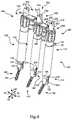
- FIG. 9is a perspective view of the cannulas, pedicle screws, extenders, and bridges of FIG. 8 , with a rod seated in the rod interfaces of the extenders for contouring.
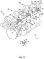
- FIG. 10is a perspective view of three adjacent vertebrae of the spine, with the rod secured to the pedicle screws to provide posterior spinal fusion.
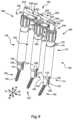
- FIG. 11is a perspective view of the guide wires of FIG. 2 , with pedicle screws installed and extenders engaging the pedicle screws and guide wires without the use of cannulas, according to one alternative method of the invention.
- FIG. 12is a perspective view of the pedicle screws and extenders of FIG. 11 , with bridges used to keep the extenders in a parallel configuration.
- FIG. 13is a perspective view of the pedicle screws, extenders, and bridges of FIG. 12 , with a rod seated in the rod interfaces of the extenders for contouring.
- the present inventionrelates to systems and methods for configuring and/or selecting devices to be implanted in the body.
- the examples provided hereingenerally relate to contouring a rod for a posterior spinal fusion system
- the present inventionmay be applied to any procedure in which the relative position and/or orientations of internal anatomic locations are to be measured or used to configure or select an implant. Accordingly, the scope of the present invention is not intended to be limited by the examples discussed herein, but only by the appended claims.
- an “anatomic point”is a location within the body. An anatomic point need not be located on any specific anatomic structure.
- proximalrefers to a position relatively closer to the center of the body
- distalrefers to a position relatively further from the center of the body.
- proximalrefers to a portion relatively nearer the operator of the tool or similar implement
- distalrefers to a portion relatively further from the operator.
- spatial transformationrefers to any mathematical procedure in which one or more coordinates can be transformed in a manner that permits the original coordinates to be determined based on the results of the transformation. Accordingly, a spatial transformation may involve any combination of translation and rotation of the original coordinates, as long as the transformation can be analytically reversed to permit the original coordinates to be obtained.
- a “translational spatial transformation”is a spatial transformation in which the original coordinates are all uniformly translated along the same vector.
- materefers to any type of connection in which cooperating features engage each other to restrict relative motion of the mating parts.
- the term “couple”is not limited to fixed attachment, but also includes sliding attachment and the like.
- the term “receive”does not require one item to completely capture another; rather, one item receives another if the first item engages the second item in a manner that restricts relative motion of the items.
- FIG. 1a perspective view illustrates a portion of a spine 10 .
- FIG. 1illustrates only the bony structures; accordingly, ligaments, cartilage, and other soft tissues are omitted for clarity.
- the spine 10has a cephalad direction 12 , a caudal direction 14 , an anterior direction 16 , a posterior direction 18 , and a medial/lateral axis 20 , all of which are oriented as shown by the arrows bearing the same reference numerals.
- “left” and “right”are used with reference to a posterior view, i.e., a view from behind the spine 10 .
- Medialrefers to a position or orientation toward a sagittal plane (i.e., plane of symmetry that separates left and right sides from each other) of the spine 10
- lateralrefers to a position or orientation relatively further from the sagittal plane.
- the portion of the spine 10 illustrated in FIG. 1includes a first vertebra 24 , which may be the L5 (Fifth Lumbar) vertebra of a patient, and a second vertebra 26 , which may be the L4 (Fourth Lumbar) vertebra of the patient.
- the systems and methodsmay be applicable to any vertebra or vertebrae of the spine 10 and/or the sacrum (not shown).
- the term “vertebra”may be broadly interpreted to include the sacrum.
- the first vertebra 24has a body 28 with a generally disc-like shape and two pedicles 30 that extend posteriorly from the body 28 .
- a posterior arch, or lamina 32extends between the posterior ends of the pedicles 30 to couple the pedicles 30 together.
- the first vertebra 24also has a pair of transverse processes 34 that extend laterally from the pedicles 30 generally along the medial/lateral axis 20 , and a spinous process 36 that extends from the lamina 32 along the posterior direction 18 .
- the first vertebra 24also has a pair of superior facets 38 , which are positioned toward the top of the first vertebra 24 and face generally medially. Additionally, the first vertebra 24 has inferior facets 40 , which are positioned toward the bottom of the first vertebra 24 and face generally laterally. Each of the pedicles 30 of the first vertebra 24 has a saddle point 42 , which is positioned generally at the center of the juncture of each superior facet 38 with the adjacent transverse process 34 .
- the second vertebra 26has a body 48 from which two pedicles 50 extend posteriorly.
- a posterior arch, or lamina 52extends between the posterior ends of the pedicles 50 to couple the pedicles 50 together.
- the second vertebra 26also has a pair of transverse processes 54 , each of which extends from the corresponding pedicle 50 generally along the medial/lateral axis 20 , and a spinous process 56 that extends from the lamina 52 along the posterior direction 18 .
- the second vertebra 26also has a pair of superior facets 58 , which are positioned toward the top of the second vertebra 26 and face generally inward. Additionally, the second vertebra 26 has inferior facets 60 , which are positioned toward the bottom of the second vertebra 26 and face generally outward. Each of the pedicles 60 of the second vertebra 26 has a saddle point 62 , which is positioned generally at the center of the juncture of each superior facet 58 with the adjacent transverse process 54 .
- the superior facets 38 of the first vertebra 24articulate (i.e., slide and/or press) with the inferior facets 60 of the second vertebra 26 to limit relative motion between the first and second vertebrae 24 , 26 .
- the combination of each superior facet 38 with the adjacent inferior facet 60provides a facet joint 64 .
- the first and second vertebrae 24 , 26thus define two facet joints 64 that span the distance between the first and second vertebrae 24 , 26 .
- the inferior facets 40 of the first vertebra 40 and the superior facets 58 of the second vertebra 26are part of other facet joints that control motion between the first and second vertebrae 24 , 26 and adjacent vertebrae (not shown) and/or the sacrum (also not shown).
- FIGS. 1 through 10illustrate one method of configuring and installing a posterior spinal fusion system.
- FIGS. 11 through 13illustrate steps that may be employed in place of the steps of FIGS. 6 through 9 .
- a first guide wire 70has been inserted into the right-side pedicle 30 of the first vertebra 24
- a second guide wire 72has been inserted into the right-side pedicle 50 of the second vertebra 26 .
- the guide wires 70 , 72pass through the saddle points 42 , 62 , respectively, of the pedicles 30 , 50 .
- Each of the guide wires 70 , 72has a proximal end 74 and a distal end 76 . As shown, the proximal ends 74 are exposed, and the distal ends 76 are implanted in the pedicles 30 , 50 .
- the distal ends 76may be implanted by methods known in the surgical arts.
- FIG. 2a perspective view illustrates the first and second guide wires 70 , 72 of FIG. 1 , with the vertebrae 24 , 26 removed for clarity.
- a third guide wire 78is also shown.
- the third guide wire 78is positioned adjacent to the first and second guide wires 70 , 72 as though the third guide wire 78 were implanted in the right-hand pedicle of a vertebra (not shown) directly superior to the second vertebra 26 .
- the method of FIGS. 1 through 10may be used to secure together vertebrae on multiple levels, not just two adjacent vertebrae.
- a perspective viewillustrates the guide wires 70 , 72 , 78 , in conjunction with a first dilator 80 , a second dilator 82 , and a third dilator 88 .
- Each of the dilators 80 , 82 , 88has a proximal end 92 and a distal end 94 .
- the proximal ends 92may be shaped for gripping by hand, or for attachment to a handle or the like.
- the distal ends 94are rounded to permit relatively gentle spreading of tissues surrounding the guide wires 70 , 72 , 78 by the dilators 80 , 82 , 88 .
- Each of the dilators 80 , 82 , 88has a bore sized to receive the proximal end 74 of the corresponding guide wire 70 , 72 , or 78 , so that the dilators 80 , 82 , 88 are able to slide along the guide wires 70 , 72 , 78 toward the distal ends 74 , thereby spreading the tissues away from the guide wires 70 , 72 , 78 .
- a variety of other guiding devices and/or dilation devicesmay be used within the scope of the present invention.
- a perspective viewillustrates the guide wires 70 , 72 , 78 and dilators 80 , 82 , 88 , with the addition of a first cannula 100 , a second cannula 102 , and a third cannula 108 .
- Each of the cannulas 100 , 102 , 108has a proximal end 112 , a distal end 114 , with a bore passing between the proximal and distal ends 112 , 114 .
- Each proximal end 112has a port 116 in communication with the bore, and a tab 118 that may facilitate manipulation or securement of the corresponding cannula 100 , 102 , or 108 .
- Each distal end 114has a taper 122 that provides a reduction in the diameter of the cannula 100 , 102 , or 108 toward the distal end 114 . Additionally, each distal end 114 has a pair of arms 124 that extend generally parallel to the axis of the corresponding cannula 100 , 102 , or 108 . The arms 124 define a first slot 126 and a second slot 128 that also extend parallel to the axis of the corresponding cannula 100 , 102 , 108 . The ends of the arms 124 define a port 130 that also communicates with the bore of the cannula 100 , 102 , or 108 .
- the cannulas 100 , 102 , 108are inserted around the guide wires 70 , 72 , 78 .
- the cannulas 100 , 102 , 108may be placed by withdrawing dilators 80 , 82 , 88 , inserting the cannulas 100 , 102 , 108 around the proximal ends 74 of the guide wires 70 , 72 , 78 , inserting the distal ends 94 of the dilators 80 , 82 , 88 into the ports 116 of the proximal end 112 of the cannulas 100 , 102 , 108 , and then advancing the dilators 80 , 82 , 88 along the guide wires 70 , 72 , 78 to urge the cannulas 100 , 102 , 108 toward the distal ends 76 of the guide wires 70 , 72 , 78 , into the dilated tissue.
- the dilators 80 , 82 , 88are removed to permit placement of the cannulas 100 , 102 , 108 , and are not re-inserted.
- cannulasmay be modular, or may have dilatable distal ends that enable placement of the cannulas around the dilators 80 , 82 , 88 , so that the dilators 80 , 82 , 88 need not be removed from the guide wires 70 , 72 , 78 until the cannulas are properly positioned.
- the present inventionis not limited to use of cannulas like those of FIG. 4 ; rather, any of a variety of cannulas may be used.
- connection elementsmay be fixation members designed to anchor a rod to the first vertebra 24 , the second vertebra 26 , and the third vertebra (not shown in FIG. 5 ). More precisely, the connection elements may be pedicle screws 140 , 142 , and 148 implantable in vertebral pedicles.
- the pedicle screws 140 , 142 , 148may be designed to provide polyaxial coupling to the associated pedicles.
- Each of the pedicle screws 140 , 142 , 148has a cage 152 shaped to receive a rod, and a screw 154 that passes through an aperture (not visible) of the cage 152 in such a manner that the screw 154 is able to extend from the cage 152 along a plurality of relative orientations.
- the orientation of the cage 152 with respect to the screw 154can still be altered.
- Each of the screws 154has a lumen passing along the axis of the screw 154 so that the screws 154 can slide along the guide wires 70 , 72 , 78 for accurate implantation in the pedicles.
- Each cage 152has two arms 156 that extend generally away from the screw 154 and define a first slot 158 and a second slot 160 through which a rod (not shown in FIG. 5 ) can pass.
- the closed ends of the slots 158 , 160are rounded in a manner that corresponds to the radius of the rod to be retained within the cage 152 to facilitate secure retention of the rod.
- the inward-facing surfaces of the arms 156may be threaded to enable the arms 156 may be threaded to enable the arms 156 to receive a nut (not shown in FIG. 5 ). Tightening of the nut then presses the rod against the head (not shown) of the screw 154 to keep the rod in place within the slots 158 , 160 , and to lock the orientation of the screw 154 with respect to the cage 152 .
- the pedicle screws 140 , 142 , 148represent only one of many types of connection elements that may be used in connection with the present invention.
- a variety of known devicesmay be used to secure a rod to a plurality of vertebra to provide posterior fusion.
- the pedicle screws 140 , 142 , 148are positioned such that a first anatomic point 164 , a second anatomic point 166 , and a third anatomic point 168 are within the cages 152 of the first pedicle screw 140 , the second pedicle screw 142 , and the third pedicle screw 148 , respectively.
- the axis of the rodis to pass through the anatomic points 164 , 166 , 168 .
- the pedicle screws 140 , 142 , 148may be installed in a variety of ways. According to one method, the dilators 80 , 82 , 88 are first removed. Then, each of the pedicle screws 140 , 142 , 148 is implanted through the use of an insertion tool 170 .
- the insertion tool 170has a handle 172 designed to be gripped by hand, and a stem 174 extending from the handle 172 .
- the stem 174has a distal end 176 shaped to engage the head of each of the screws 154 .
- the head of each of the screws 154has a hexagonal recess (not visible), and the distal end 176 has a corresponding hexagonal male feature (not visible).
- torque applied to the handle 172can be transmitted to each of the screws 154 .
- the stem 174also has a lumen (not shown) sized to fit around each of the guide wires 70 , 72 , 78 so that the guide wires 70 , 72 , 78 can be used to guide implantation of the screws 154 through the use of the insertion tool 170 .
- Slots 178provide access to the lumen for cleaning.
- Each of the screws 154is coupled to the insertion tool 170 by connecting the head of the screw 154 to the distal end 176 of the stem 174 .
- the insertion tool 170is then moved to insert the proximal end 74 of the corresponding guide wire 70 , 72 , 78 through the lumen of the screw 154 and into the lumen of the stem 174 .
- the insertion tool 170is used to insert the pedicle screw 140 , 142 , or 148 through the corresponding cannula 100 , 102 , or 108 until the screw 154 contacts the first pedicle 30 , the second pedicle 50 , or the third pedicle.
- the cages 152may be sized to fit relatively snugly within the ports 130 at the distal ends 114 of the cannulas 100 , 102 , 108 .
- the arms 124 of each distal end 114thus form a docking feature that enables the corresponding pedicle screw 140 , 142 , or 148 to dock with the distal end 114 of the corresponding cannula 100 , 102 , 108 .
- the cages 152are then constrained to be coaxial with the cannulas 100 , 102 , 108 .
- FIG. 6a perspective illustrates the cannulas 100 , 102 , 108 and the pedicle screws 140 , 142 , 148 of FIG. 5 , with a first extender 180 , a second extender 182 , and a third extender 188 inserted into engagement with the cannulas 100 , 102 , 108 and pedicle screws 140 , 142 , 148 .
- the extenders 180 , 182 , 188are used to project the anatomic points 164 , 166 , 168 outside the patient's body to facilitate proper contouring of the rod (not shown in FIG. 6 ).
- the space between the cannulas 100 , 102 , 108need not be accessed to obtain the proper rod configuration.
- each of the extenders 180 , 182 , 188has a proximal portion 192 , a distal portion 194 , and a stem 196 extending between the proximal and distal portions 192 , 194 .
- the proximal portion 192 of each of the extenders 180 , 182 , 188has a handle 198 that may be grasped by hand or by a tool.
- each proximal portion 192has an implant interface, which may take the form of a rod interface 200 .
- Each rod interface 200is shaped to receive a portion of a rod to facilitate contouring of the rod so that the contoured rod will pass through the anatomic points 164 , 166 , 168 within the cages 152 of the implanted pedicle screws 140 , 142 , 148 .
- Each of the rod interfaces 200has two arms 202 that extend generally away from the remainder of the corresponding extender 180 , 182 , or 188 .
- the arms 202 of each rod interface 200define a trough 204 through which a rod (not shown in FIG. 5 ) can pass.
- the base of the trough 204is rounded in a manner that corresponds to the radius of the rod to be retained within the cage 152 to facilitate secure retention of the rod.
- the arms 202are similar in configuration to the arms 156 of the cage 152 of the corresponding pedicle screw 140 , 142 , 148 , and the trough 204 is similar to a trough defined by the first and second slots 158 , 160 of the cage 152 . Accordingly, the rod interfaces 200 mimic the geometry of the cages 152 of the pedicle screws 140 , 142 , 148 .
- each of the extenders 180 , 182 , 188has a docking element 208 that may be used to facilitate engagement and relative orientation of the extenders 180 , 182 , 188 with the cages 152 of the pedicle screws 140 , 142 , 148 .
- Each docking element 208may include an axial stud 210 that extends along the axis of the extender 180 , 182 , or 188 , and a transverse stud 212 proximate the distal end of the axial stud 210 , that extends perpendicular to the axis of the extender 180 , 182 , or 188 .
- the extenders 180 , 182 , 188represent only one of many potential extender configurations that may be used in connection with the present invention. Other extender configurations may be advantageous, particularly if the cannulas, dilators, connection elements, or guidance members employed are different from those of FIG. 6 .
- extenders according to the inventionmay simply be used to provide a numeric measurement of relative positions or orientations of the corresponding anatomic points. Extenders may thus be incorporated into one or more measurement instruments (not shown). For example, extenders may register on the pedicle screws 140 , 142 , 148 and may be coupled to a series of sliders and/or rotary elements that provide linear and/or rotary measurements of the relative positions of the cages 152 . Such measurements may be used to configure or select an implant. According to one example, such a measurement instrument may measure displacements between all three of the implanted cages 152 to provide a triangle, two sides of which define the path that should be followed by the axis of the rod.
- each transverse stud 212may extend through the first and second slots 158 , 160 of the corresponding cage 152 to restrict relative rotation between the cage 152 and the extender 180 , 182 , 188 .
- the axial stud 210may be sized to have relatively little clearance with the inward-facing surfaces of the arms 156 of the cage 152 so that the extenders 180 , 182 , 188 are constrained to remain coaxial with the cages 152 . Further, the axial stud 210 may have exterior threads that threadably engage the threads on the inward-facing surfaces of the arms 156 of the corresponding cage 152 .
- the cannulas 100 , 102 , 108may receive the stems 196 of the extenders 180 , 182 , 188 with relatively little clearance such that each extender 180 , 182 , 188 is constrained to be coaxial with the corresponding cannula 100 , 102 , 108 .
- the cannulas 100 , 102 , 108are docked with the cages 152 , the cannulas 100 , 102 , 108 are coaxial with the cages 152 , and the extenders 180 , 182 , 188 are coaxial with the cannulas 100 , 102 , 108 and the cages 152 .
- the coaxiality of the extenders 180 , 182 , 188 with the cages 152enables the rod interfaces 200 to provide a linear transformation of each of the first, second, and third anatomic points 164 , 166 , 168 to points outside the body. More precisely, the first extender 180 projects the first anatomic point 164 along the length of the first extender 180 to a first projected point 214 within the rod interface 200 of the first extender. The second and third anatomic points 166 , 168 are similarly projected to second and third projected points 216 , 218 .
- the extenders 180 , 182 , 188are not parallel to each other, the projected points 214 , 216 , 218 do not have the same spatial relationship (i.e., relative positioning) as the anatomic points 164 , 166 , 168 .
- a side elevation viewillustrates the first cannula 100 , the first pedicle screw 140 , and the first extender 180 of FIG. 6 .
- the ends of the transverse stud 212 of the first extender 180may extend through the first and second slots 126 , 128 of the distal end 114 of the first cannula 100 as well as through the first and second slots 158 , 160 of the cage 152 of the first pedicle screw 140 .
- FIG. 7 Ba front elevation view illustrates the first cannula 100 , the first pedicle screw 140 , and the first extender 180 of FIG. 6 .
- FIG. 7 Bprovides an end view of the transverse stud 212 , illustrating how it engages the first and second slots 126 , 128 of the distal end 114 of the first cannula 100 and the first and second slots 158 , 160 of the cage 152 of the first pedicle screw 140 .
- a front elevation, section viewillustrates the first cannula 100 , the first pedicle screw 140 , and the first extender 180 of FIG. 6 .
- the first extender 180may include two separate parts: a central member 220 and a sleeve member 222 .
- the sleeve member 222has a countersink 224 at the proximal portion 192 , a taper 226 at the distal portion 194 , and a lumen 228 extending between the proximal and distal portions 192 , 194 to receive the central member 220 .
- the central member 220may include an enlarged head 234 that fits within the countersink 224 of the sleeve member 222 , a stem 238 that extends through the lumen 228 of the sleeve member 222 , and a lumen 240 that passes through the stem 238 .
- the lumen 228is optional, and may be used to receive the first guide wire 70 , particularly for the implantation method that will be set forth in connection with FIGS. 11 , 12 , and 13 .
- the lumen 228may optionally be omitted because the guide wires 70 , 72 , 78 may be removed prior to insertion of the extenders 180 , 182 , 188 .
- each of the extenders 180 , 182 , 188Usage of two separate members to provide each of the extenders 180 , 182 , 188 enables the transverse studs 212 to be seated within the first and second slots 158 , 160 of each cage 152 while the sleeve members 222 are rotated axially to threadably engage the axial studs 210 with the inward-facing surfaces of the arms 156 of the cages.
- the sleeve members 222may be rotated until they press the corresponding transverse studs 212 into the first and second slots 158 , 160 of the cages 152 .
- the transverse studs 212are then seated tightly within the cages 152 in a manner that very closely simulates the ultimate position of the rod.
- a perspective viewillustrates the cannulas 100 , 102 , 108 , pedicle screws 140 , 142 , 148 , and extenders 180 , 182 , 188 of FIG. 6 , with the addition of a first bridge 250 and a second bridge 252 .
- the bridges 250 , 252are used to keep the extenders 180 , 182 , 188 substantially parallel to each other to constrain the spatial transformation of the anatomic points 164 , 166 , 168 .
- the bridges 250 , 252are designed to constrain the extenders 180 , 182 , 188 only to parallelism. Thus, the bridges 250 , 252 do not limit relative translation or relative axial rotation of the extenders 180 , 182 , 188 .
- Each of the first and second bridges 250 , 252has a first slider 254 and a second slider 256 .
- the first slider 254 of each of the bridges 250 , 252has a pair of grooves 258 that face inward.
- the second slider 256 of each of the bridges 250 , 252has a pair of flanges 260 that extend outward into the grooves 258 of the corresponding first slider 254 so that the first and second sliders 254 , 256 are linearly slidable relative to each other to permit lengthening or shortening of the bridges 250 , 252 .
- Each of the sliders 254 , 256also has an aperture 262 that fits around the enlarged head 234 of the central member 220 of the corresponding extender 180 , 182 , or 188 .
- the apertures 262are sized to fit around the enlarged heads 234 with relatively little clearance so that the bridges 250 , 252 keep the extenders 180 , 182 , 188 parallel to each other without restricting relative axial rotation.
- each bridge 250 , 252embody only one of many possible configurations that may be used in connection with the invention.
- each bridgedoes not have two sliders, but has two members that are rotatably coupled to each other.
- Each of the membershas an aperture like the apertures 262 of the bridges 250 , 252 , so that the bridges can permit relatively free relative translation and axial rotation of the extenders 180 , 182 , 188 , while keeping the extenders 180 , 182 , 188 parallel to each other.
- the bridgeswould simply elongate and contract through the use of rotary motion instead of linear motion.
- the extenders 180 , 182 , 188are parallel.
- the projected points 214 , 216 , 218then mimic the relative positioning of the anatomic points 164 , 166 , 168 within the body.
- the extenders 180 , 182 , 188apply a translational spatial transformation to the anatomic points 164 , 166 , 168 to move them to a more accessible location without altering their positions relative to each other.
- a rod contoured such that its axis passes through the projected points 214 , 216 , 218may be installed such that its axis passes through the anatomic points 164 , 166 , 168 to properly extend through the cages 152 of the pedicle screws 140 , 142 , 148 .
- a perspective viewillustrates the cannulas 100 , 102 , 108 , the pedicle screws 140 , 142 , 148 , the extenders 180 , 182 , 188 , and the bridges 250 , 252 of FIG. 8 , with a rod 270 seated in the rod interfaces 200 of the extenders 180 , 182 , 188 for contouring.
- the rod 270has a first end 272 , a second end 274 , and a central portion 276 .
- the first end 272is positioned in the rod interface 200 of the first extender 180
- the central portion 276is positioned in the rod interface 200 of the second extender 182
- the second end 274is positioned in the rod interface 200 of the third extender 188 .
- the cages 152 of the pedicle screws 140 , 142 , 148may not be arranged in a straight line.
- the rod interfaces 200may not be arranged in a straight line.
- the rod 270may need to be bent into the proper shape, for example, through the use of tooling such as pliers, a vice, or the like, so that it will lie properly within the rod interfaces 200 .
- the process of deforming the rod 270 to the required shapemay be termed “contouring.”
- Contouringmay be carried out by, first, placing the undeformed rod 270 in the rod interfaces 200 to determine how the rod 270 should be deformed to lie properly within the rod interfaces 200 . Then, the rod 270 is deformed, and again placed in the rod interfaces 200 to check the fit. This process is repeated until the rod 270 is shaped to provide an optimal fit with the rod interfaces 200 .
- the rod 270may simply be selected from a kit or the like.
- a kit(not shown) may include rods bent at a variety of angles.
- the rod interfaces 200could be used to select the proper rod from the kit by placing each rod, in turn, on the rod interfaces 200 until one is identified that has the proper fit.
- the rod 270may be custom fabricated, for example, by measuring the relative positions of the rod interfaces 200 and using a CNC procedure to form the rod 270 .
- the rod 270 and the extenders 180 , 182 , 188may be removed from the operating site, leaving the pedicle screws 140 , 142 , 148 in place.
- the cannulas 100 , 102 , 108may also be removed at this stage, depending on the method that will be used to implant the rod 270 .
- the rod 270may be inserted subcutaneously and placed on the cages 152 by making additional incisions to connect the access passageways provided by the cannulas 100 , 102 , 108 .
- MISMinimally Invasive Surgical
- MISSmall Invasive Surgical
- FIG. 10a perspective view illustrates the completed posterior spinal fusion system.
- FIG. 10illustrates a third vertebra 278 superior to the second vertebra 26 .
- the third vertebra 278has features similar to those set forth in the description of the first and second vertebrae 24 , 26 .
- the third vertebra 278has pedicles 280 with saddle points 282 .
- the first pedicle screw 140is implanted in the pedicle 30 of the right side of the first vertebra 24
- the second pedicle screw 142is implanted in the pedicle 50 of the right side of the second vertebra 26
- the third pedicle screw 148is implanted in the pedicle 280 of the right side of the third vertebra 278 .
- the rod 270passes through the slots 158 , 160 of the cages 152 in such a manner that the axis (not shown) of the rod 270 passes through the anatomic points 164 , 166 , 168 .
- First, second, and third nuts 290 , 292 , 298have been rotated into engagement with the inward-facing surfaces of the arms 156 of the cages 152 of the first, second, and third pedicle screws 140 , 142 , 148 , respectively.
- the nuts 290 , 292 , 298have been tightened to press the first end 272 , central portion 276 , and second end 274 , respectively, against the heads of the screws 154 of the pedicle screws 140 , 142 , 148 , respectively.
- the cages 152are no longer freely rotatable with respect to the screws 154 , but are instead locked in their current orientations.
- the pedicle screws 140 , 142 , 148thus cooperate with the rod 270 to restrict relative motion of the vertebrae 24 , 26 , 278 to form a posterior vertebral fusion system.
- a similar systemmay be implanted in the left-side pedicles 30 , 50 , 280 of the vertebrae 24 , 26 , 278 through the method set forth previously to provide a bilateral system.
- the present inventionis not limited to a three-level fusion system, but may be used to fuse any number of vertebrae together. To fuse more than three vertebrae together, the steps set forth above may simply be repeated for each additional vertebra, and the rod may be placed on four or more rod interfaces for configuration or selection.
- the cannulas 100 , 102 , 108may be omitted entirely from the procedure. Such a method may commence with the steps outlined above in the descriptions of FIGS. 1 , 2 , and 3 , but may then include the steps illustrated in FIGS. 11 , 12 , and 13 .
- FIG. 11a perspective view illustrates the guide wires 70 , 72 , 78 of FIG. 2 , with the pedicle screws 140 , 142 , 148 and extenders 180 , 182 , 188 installed.
- the dilators 80 , 82 , 88may be removed, and the distal ends of the extenders 180 , 182 , 188 may be mated to the pedicle screws 140 , 142 , 148 .
- the transverse studs 212may be inserted into the slots 158 , 160 , and the axial studs 210 may be threadably engaged with the inward-facing surfaces of the arms 156 in the manner set forth previously.
- the extenders 180 , 182 , 188may be used as insertion tools to implant the pedicle screws 140 , 142 , 148 in the pedicles 30 , 50 , 280 . More precisely, the extenders 180 , 182 , 188 are positioned to insert the proximal ends 74 of the guide wires 70 , 72 , 78 through the pedicle screws 140 , 142 , 148 , and into the lumens 240 of the central members 220 of the extenders 180 , 182 , 188 .
- the extenders 180 , 182 , 188are advanced until the screws 154 contact the pedicles 30 , 50 , 280 , and then the extenders 180 , 182 , 188 are subjected to torque and axial pressure, which may be applied to the handles 198 , to implant the screws 154 in the pedicles 30 , 50 , 280 .
- the guide wires 70 , 72 , 78may sufficiently guide implantation of the pedicle screws 140 , 142 , 148 without requiring the use of the cannulas 100 , 102 , 108 .
- the insertion tool 170may be used in the manner described previously to implant the pedicle screws 140 , 142 , 148 , without the use of the cannulas 100 , 102 , 108 .
- the distal portions 194 of the extenders 180 , 182 , 188may then be mated to the cages 152 as set forth above, after implantation of the pedicle screws 140 , 142 , 148 .
- FIG. 12a perspective view illustrates the pedicle screws 140 , 142 , 148 and the extenders 180 , 182 , 188 , with the addition of the bridges 250 , 252 .
- the bridges 250 , 252are applied to constrain the extenders 180 , 182 , 188 to parallel orientations.
- a perspective viewillustrates the pedicle screws 140 , 142 , 148 , the extenders 180 , 182 , 188 , and the bridges 250 , 252 , with the rod 270 seated in the rod interfaces 200 of the extenders 180 , 182 , 188 for contouring.
- the rod 270may be configured or selected by placing it in the rod interfaces 200 to ensure that it will fit properly in the cages 152 of the pedicle screws 140 , 142 , 148 upon implantation.
- the additional steps set forth in the description of FIG. 9(aside from removal of the cannulas 100 , 102 , 108 ) may be followed to obtain the fully implanted and assembled posterior spinal fusion system illustrated in FIG. 10 .
- the foregoing descriptiondiscloses a number of different elements, any of which may be components of a system for configuring or selecting one or more implants for implantation in a body of a patient.
- the foregoing examplesrelate to the assembly and implantation of a posterior spinal fusion system, the present invention may be applied to a wide variety of implants, within and outside the orthopedic area.
- the present inventionhas particular benefits when an implant is to be configured or selected for a given patient, with reference to two or more anatomic points within the body.
Landscapes
- Health & Medical Sciences (AREA)
- Orthopedic Medicine & Surgery (AREA)
- Surgery (AREA)
- Life Sciences & Earth Sciences (AREA)
- Neurology (AREA)
- Medical Informatics (AREA)
- Biomedical Technology (AREA)
- Heart & Thoracic Surgery (AREA)
- Engineering & Computer Science (AREA)
- Molecular Biology (AREA)
- Animal Behavior & Ethology (AREA)
- General Health & Medical Sciences (AREA)
- Public Health (AREA)
- Veterinary Medicine (AREA)
- Nuclear Medicine, Radiotherapy & Molecular Imaging (AREA)
- Surgical Instruments (AREA)
- Prostheses (AREA)
Abstract
Description
Claims (19)
Priority Applications (1)
| Application Number | Priority Date | Filing Date | Title |
|---|---|---|---|
| US17/147,720US11596461B2 (en) | 2005-05-18 | 2021-01-13 | System and method for orthopedic implant configuration |
Applications Claiming Priority (5)
| Application Number | Priority Date | Filing Date | Title |
|---|---|---|---|
| US68234405P | 2005-05-18 | 2005-05-18 | |
| US11/178,035US8177817B2 (en) | 2005-05-18 | 2005-07-08 | System and method for orthopedic implant configuration |
| US13/444,231US9895182B2 (en) | 2005-05-18 | 2012-04-11 | System and method for orthopedic implant configuration |
| US15/932,285US10898251B2 (en) | 2005-05-18 | 2018-02-16 | System and method for orthopedic implant configuration |
| US17/147,720US11596461B2 (en) | 2005-05-18 | 2021-01-13 | System and method for orthopedic implant configuration |
Related Parent Applications (1)
| Application Number | Title | Priority Date | Filing Date |
|---|---|---|---|
| US15/932,285ContinuationUS10898251B2 (en) | 2005-05-18 | 2018-02-16 | System and method for orthopedic implant configuration |
Publications (2)
| Publication Number | Publication Date |
|---|---|
| US20210128215A1 US20210128215A1 (en) | 2021-05-06 |
| US11596461B2true US11596461B2 (en) | 2023-03-07 |
Family
ID=37035863
Family Applications (4)
| Application Number | Title | Priority Date | Filing Date |
|---|---|---|---|
| US11/178,035Active2027-10-05US8177817B2 (en) | 2005-05-18 | 2005-07-08 | System and method for orthopedic implant configuration |
| US13/444,231Active2027-09-26US9895182B2 (en) | 2005-05-18 | 2012-04-11 | System and method for orthopedic implant configuration |
| US15/932,285Expired - LifetimeUS10898251B2 (en) | 2005-05-18 | 2018-02-16 | System and method for orthopedic implant configuration |
| US17/147,720Active2025-10-31US11596461B2 (en) | 2005-05-18 | 2021-01-13 | System and method for orthopedic implant configuration |
Family Applications Before (3)
| Application Number | Title | Priority Date | Filing Date |
|---|---|---|---|
| US11/178,035Active2027-10-05US8177817B2 (en) | 2005-05-18 | 2005-07-08 | System and method for orthopedic implant configuration |
| US13/444,231Active2027-09-26US9895182B2 (en) | 2005-05-18 | 2012-04-11 | System and method for orthopedic implant configuration |
| US15/932,285Expired - LifetimeUS10898251B2 (en) | 2005-05-18 | 2018-02-16 | System and method for orthopedic implant configuration |
Country Status (5)
| Country | Link |
|---|---|
| US (4) | US8177817B2 (en) |
| EP (2) | EP2255739B1 (en) |
| AT (2) | ATE537772T1 (en) |
| DE (1) | DE602006018597D1 (en) |
| WO (1) | WO2006125029A2 (en) |
Families Citing this family (143)
| Publication number | Priority date | Publication date | Assignee | Title |
|---|---|---|---|---|
| US7833250B2 (en) | 2004-11-10 | 2010-11-16 | Jackson Roger P | Polyaxial bone screw with helically wound capture connection |
| US10729469B2 (en) | 2006-01-09 | 2020-08-04 | Roger P. Jackson | Flexible spinal stabilization assembly with spacer having off-axis core member |
| US7862587B2 (en) | 2004-02-27 | 2011-01-04 | Jackson Roger P | Dynamic stabilization assemblies, tool set and method |
| US8353932B2 (en) | 2005-09-30 | 2013-01-15 | Jackson Roger P | Polyaxial bone anchor assembly with one-piece closure, pressure insert and plastic elongate member |
| US8292926B2 (en) | 2005-09-30 | 2012-10-23 | Jackson Roger P | Dynamic stabilization connecting member with elastic core and outer sleeve |
| US10258382B2 (en) | 2007-01-18 | 2019-04-16 | Roger P. Jackson | Rod-cord dynamic connection assemblies with slidable bone anchor attachment members along the cord |
| US8876868B2 (en) | 2002-09-06 | 2014-11-04 | Roger P. Jackson | Helical guide and advancement flange with radially loaded lip |
| US7887539B2 (en) | 2003-01-24 | 2011-02-15 | Depuy Spine, Inc. | Spinal rod approximators |
| US7621918B2 (en) | 2004-11-23 | 2009-11-24 | Jackson Roger P | Spinal fixation tool set and method |
| US7377923B2 (en) | 2003-05-22 | 2008-05-27 | Alphatec Spine, Inc. | Variable angle spinal screw assembly |
| US8366753B2 (en) | 2003-06-18 | 2013-02-05 | Jackson Roger P | Polyaxial bone screw assembly with fixed retaining structure |
| US7776067B2 (en) | 2005-05-27 | 2010-08-17 | Jackson Roger P | Polyaxial bone screw with shank articulation pressure insert and method |
| US8926670B2 (en) | 2003-06-18 | 2015-01-06 | Roger P. Jackson | Polyaxial bone screw assembly |
| US7967850B2 (en) | 2003-06-18 | 2011-06-28 | Jackson Roger P | Polyaxial bone anchor with helical capture connection, insert and dual locking assembly |
| US7766915B2 (en) | 2004-02-27 | 2010-08-03 | Jackson Roger P | Dynamic fixation assemblies with inner core and outer coil-like member |
| US8002798B2 (en) | 2003-09-24 | 2011-08-23 | Stryker Spine | System and method for spinal implant placement |
| US7955355B2 (en) | 2003-09-24 | 2011-06-07 | Stryker Spine | Methods and devices for improving percutaneous access in minimally invasive surgeries |
| US7179261B2 (en) | 2003-12-16 | 2007-02-20 | Depuy Spine, Inc. | Percutaneous access devices and bone anchor assemblies |
| US11419642B2 (en) | 2003-12-16 | 2022-08-23 | Medos International Sarl | Percutaneous access devices and bone anchor assemblies |
| US7527638B2 (en) | 2003-12-16 | 2009-05-05 | Depuy Spine, Inc. | Methods and devices for minimally invasive spinal fixation element placement |
| US8333789B2 (en)* | 2007-01-10 | 2012-12-18 | Gmedelaware 2 Llc | Facet joint replacement |
| JP2007525274A (en) | 2004-02-27 | 2007-09-06 | ロジャー・ピー・ジャクソン | Orthopedic implant rod reduction instrument set and method |
| US7160300B2 (en) | 2004-02-27 | 2007-01-09 | Jackson Roger P | Orthopedic implant rod reduction tool set and method |
| US11241261B2 (en) | 2005-09-30 | 2022-02-08 | Roger P Jackson | Apparatus and method for soft spinal stabilization using a tensionable cord and releasable end structure |
| US8152810B2 (en) | 2004-11-23 | 2012-04-10 | Jackson Roger P | Spinal fixation tool set and method |
| EP1814472B1 (en)* | 2004-09-08 | 2018-10-24 | NuVasive, Inc. | Systems for performing spinal fixation |
| US7651502B2 (en) | 2004-09-24 | 2010-01-26 | Jackson Roger P | Spinal fixation tool set and method for rod reduction and fastener insertion |
| US7935134B2 (en) | 2004-10-20 | 2011-05-03 | Exactech, Inc. | Systems and methods for stabilization of bone structures |
| US8162985B2 (en) | 2004-10-20 | 2012-04-24 | The Board Of Trustees Of The Leland Stanford Junior University | Systems and methods for posterior dynamic stabilization of the spine |
| US8226690B2 (en) | 2005-07-22 | 2012-07-24 | The Board Of Trustees Of The Leland Stanford Junior University | Systems and methods for stabilization of bone structures |
| US8025680B2 (en) | 2004-10-20 | 2011-09-27 | Exactech, Inc. | Systems and methods for posterior dynamic stabilization of the spine |
| US8267969B2 (en) | 2004-10-20 | 2012-09-18 | Exactech, Inc. | Screw systems and methods for use in stabilization of bone structures |
| US8926672B2 (en) | 2004-11-10 | 2015-01-06 | Roger P. Jackson | Splay control closure for open bone anchor |
| US9168069B2 (en) | 2009-06-15 | 2015-10-27 | Roger P. Jackson | Polyaxial bone anchor with pop-on shank and winged insert with lower skirt for engaging a friction fit retainer |
| US9216041B2 (en) | 2009-06-15 | 2015-12-22 | Roger P. Jackson | Spinal connecting members with tensioned cords and rigid sleeves for engaging compression inserts |
| US8444681B2 (en) | 2009-06-15 | 2013-05-21 | Roger P. Jackson | Polyaxial bone anchor with pop-on shank, friction fit retainer and winged insert |
| WO2006057837A1 (en) | 2004-11-23 | 2006-06-01 | Jackson Roger P | Spinal fixation tool attachment structure |
| WO2006058221A2 (en) | 2004-11-24 | 2006-06-01 | Abdou Samy M | Devices and methods for inter-vertebral orthopedic device placement |
| US7901437B2 (en) | 2007-01-26 | 2011-03-08 | Jackson Roger P | Dynamic stabilization member with molded connection |
| US7951175B2 (en) | 2005-03-04 | 2011-05-31 | Depuy Spine, Inc. | Instruments and methods for manipulating a vertebra |
| US7951172B2 (en) | 2005-03-04 | 2011-05-31 | Depuy Spine Sarl | Constrained motion bone screw assembly |
| ES2318917B1 (en)* | 2005-03-30 | 2010-02-04 | Sdgi Holdings Inc. | SYSTEM FOR THE THREE-DIMENSIONAL CORRECTION OF THE CURVATURE OF THE VERTEBRAL COLUMN IN PROBLEMS OF SCHOLIOSIS BY COPLANAR ALIGNMENT OF THE PEDICULAR SCREWS. |
| US8177817B2 (en) | 2005-05-18 | 2012-05-15 | Stryker Spine | System and method for orthopedic implant configuration |
| US8523865B2 (en) | 2005-07-22 | 2013-09-03 | Exactech, Inc. | Tissue splitter |
| EP1767161A1 (en)* | 2005-09-22 | 2007-03-28 | Zimmer Spine, Inc. | Spinal fixation rod contouring system |
| US8105368B2 (en) | 2005-09-30 | 2012-01-31 | Jackson Roger P | Dynamic stabilization connecting member with slitted core and outer sleeve |
| US7704271B2 (en) | 2005-12-19 | 2010-04-27 | Abdou M Samy | Devices and methods for inter-vertebral orthopedic device placement |
| AU2013200549C1 (en)* | 2006-02-06 | 2016-10-06 | Stryker European Holdings I, Llc | Rod contouring apparatus and method for percutaneous pedicle screw extension |
| EP1981422B1 (en) | 2006-02-06 | 2018-10-24 | Stryker European Holdings I, LLC | Rod contouring apparatus for percutaneous pedicle screw extension |
| WO2007121271A2 (en) | 2006-04-11 | 2007-10-25 | Synthes (U.S.A) | Minimally invasive fixation system |
| US8663292B2 (en) | 2006-08-22 | 2014-03-04 | DePuy Synthes Products, LLC | Reduction sleeve |
| DE602007008112D1 (en)* | 2006-09-25 | 2010-09-09 | Stryker Spine | ALIGNMENT CONNECTION FOR BAR CONTOURING |
| US8157809B2 (en)* | 2006-09-25 | 2012-04-17 | Stryker Spine | Percutaneous compression and distraction system |
| US8096996B2 (en) | 2007-03-20 | 2012-01-17 | Exactech, Inc. | Rod reducer |
| CA2670988C (en) | 2006-12-08 | 2014-03-25 | Roger P. Jackson | Tool system for dynamic spinal implants |
| US8475498B2 (en) | 2007-01-18 | 2013-07-02 | Roger P. Jackson | Dynamic stabilization connecting member with cord connection |
| US8366745B2 (en) | 2007-05-01 | 2013-02-05 | Jackson Roger P | Dynamic stabilization assembly having pre-compressed spacers with differential displacements |
| US10383660B2 (en) | 2007-05-01 | 2019-08-20 | Roger P. Jackson | Soft stabilization assemblies with pretensioned cords |
| US8979904B2 (en) | 2007-05-01 | 2015-03-17 | Roger P Jackson | Connecting member with tensioned cord, low profile rigid sleeve and spacer with torsion control |
| US7846211B2 (en)* | 2007-07-09 | 2010-12-07 | Moximed, Inc. | Surgical implantation method and devices for an extra-articular mechanical energy absorbing apparatus |
| US20090018665A1 (en)* | 2007-07-09 | 2009-01-15 | Exploramed Nc4, Inc. | Surgical implantation method and devices for an extra-articular mechanical energy absorbing apparatus |
| US8057518B2 (en) | 2007-08-31 | 2011-11-15 | Depuy Spine, Inc. | Spanning connector for connecting a spinal fixation element and an offset bone anchor |
| US8512343B2 (en)* | 2007-08-31 | 2013-08-20 | DePuy Synthes Products, LLC | Methods and instruments for approximating misaligned vertebra |
| US8900237B2 (en)* | 2007-08-31 | 2014-12-02 | DePuy Synthes Products, LLC | Minimally invasive guide system |
| US20090062822A1 (en)* | 2007-08-31 | 2009-03-05 | Frasier William J | Adaptable clamping mechanism for coupling a spinal fixation element to a bone anchor |
| US8894690B2 (en)* | 2007-08-31 | 2014-11-25 | DePuy Synthes Products, LLC | Offset connection bone anchor assembly |
| US8025682B2 (en)* | 2007-08-31 | 2011-09-27 | Depuy Spine, Inc. | Method and system for securing a rod to a bone anchor with a connector |
| US8439922B1 (en) | 2008-02-06 | 2013-05-14 | NiVasive, Inc. | Systems and methods for holding and implanting bone anchors |
| US8709015B2 (en)* | 2008-03-10 | 2014-04-29 | DePuy Synthes Products, LLC | Bilateral vertebral body derotation system |
| US8608746B2 (en) | 2008-03-10 | 2013-12-17 | DePuy Synthes Products, LLC | Derotation instrument with reduction functionality |
| US8549888B2 (en) | 2008-04-04 | 2013-10-08 | Nuvasive, Inc. | System and device for designing and forming a surgical implant |
| US10973556B2 (en) | 2008-06-17 | 2021-04-13 | DePuy Synthes Products, Inc. | Adjustable implant assembly |
| AU2010260521C1 (en) | 2008-08-01 | 2013-08-01 | Roger P. Jackson | Longitudinal connecting member with sleeved tensioned cords |
| US8211012B2 (en) | 2008-09-30 | 2012-07-03 | Aesculap Implant Systems, Llc | Tissue retractor system |
| US8388659B1 (en) | 2008-10-17 | 2013-03-05 | Theken Spine, Llc | Spondylolisthesis screw and instrument for implantation |
| CN102497828B (en) | 2009-05-20 | 2015-09-09 | 斯恩蒂斯有限公司 | What patient installed retracts part |
| US8998959B2 (en) | 2009-06-15 | 2015-04-07 | Roger P Jackson | Polyaxial bone anchors with pop-on shank, fully constrained friction fit retainer and lock and release insert |
| US9668771B2 (en) | 2009-06-15 | 2017-06-06 | Roger P Jackson | Soft stabilization assemblies with off-set connector |
| CN103826560A (en) | 2009-06-15 | 2014-05-28 | 罗杰.P.杰克逊 | Polyaxial Bone Anchor with Socket Stem and Winged Inserts with Friction Fit Compression Collars |
| US11229457B2 (en) | 2009-06-15 | 2022-01-25 | Roger P. Jackson | Pivotal bone anchor assembly with insert tool deployment |
| US8657856B2 (en) | 2009-08-28 | 2014-02-25 | Pioneer Surgical Technology, Inc. | Size transition spinal rod |
| EP2485654B1 (en) | 2009-10-05 | 2021-05-05 | Jackson P. Roger | Polyaxial bone anchor with non-pivotable retainer and pop-on shank, some with friction fit |
| US8663289B2 (en)* | 2009-10-29 | 2014-03-04 | Warsaw Orthopedic, Inc. | Pedicle screw head extender |
| US8986349B1 (en) | 2009-11-11 | 2015-03-24 | Nuvasive, Inc. | Systems and methods for correcting spinal deformities |
| US8764806B2 (en) | 2009-12-07 | 2014-07-01 | Samy Abdou | Devices and methods for minimally invasive spinal stabilization and instrumentation |
| US8540719B2 (en) | 2010-02-09 | 2013-09-24 | Aesculap Implant Systems, Llc | Percutaneous rod insertion system and method |
| US8535318B2 (en) | 2010-04-23 | 2013-09-17 | DePuy Synthes Products, LLC | Minimally invasive instrument set, devices and related methods |
| US8298242B2 (en) | 2010-04-30 | 2012-10-30 | Warsaw Orthopedic, Inc. | Systems, devices and methods for bending an elongate member |
| US8607603B2 (en) | 2010-04-30 | 2013-12-17 | Warsaw Orthopedic, Inc. | Systems, devices and methods for multi-dimensional bending of an elongate member |
| CN102293680B (en)* | 2010-06-24 | 2014-04-16 | 华沙整形外科股份有限公司 | Coplanar straightening system |
| US8500741B2 (en) | 2010-09-02 | 2013-08-06 | Stephen M. Hansen | Pedicle screw extension alignment |
| AU2011299558A1 (en) | 2010-09-08 | 2013-05-02 | Roger P. Jackson | Dynamic stabilization members with elastic and inelastic sections |
| US8623022B2 (en) | 2010-09-20 | 2014-01-07 | Zimmer Spine, Inc. | Surgical instrument support system and method |
| US9198698B1 (en) | 2011-02-10 | 2015-12-01 | Nuvasive, Inc. | Minimally invasive spinal fixation system and related methods |
| FR2971698B1 (en)* | 2011-02-18 | 2014-01-24 | Spineway | SURGICAL DEVICE FOR THE CORRECTION OF DEFORMATION OF THE VERTEBRAL COLUMN |
| US8690878B2 (en) | 2011-04-11 | 2014-04-08 | Warsaw Orthopedic, Inc. | Flexible anchor extenders |
| US9907582B1 (en)* | 2011-04-25 | 2018-03-06 | Nuvasive, Inc. | Minimally invasive spinal fixation system and related methods |
| US8721651B2 (en) | 2011-04-27 | 2014-05-13 | Warsaw Orthopedic, Inc. | Templates and methods |
| CN103717159B (en) | 2011-05-27 | 2016-08-17 | 新特斯有限责任公司 | Minimally Invasive Spinal Fixation System Including Vertebral Alignment Features |
| US8845728B1 (en) | 2011-09-23 | 2014-09-30 | Samy Abdou | Spinal fixation devices and methods of use |
| US9125703B2 (en)* | 2012-01-16 | 2015-09-08 | K2M, Inc. | Rod reducer, compressor, distractor system |
| US8936626B1 (en) | 2012-02-17 | 2015-01-20 | Nuvasive, Inc. | Bi-cortical screw fixation |
| US20130226240A1 (en) | 2012-02-22 | 2013-08-29 | Samy Abdou | Spinous process fixation devices and methods of use |
| US11207132B2 (en) | 2012-03-12 | 2021-12-28 | Nuvasive, Inc. | Systems and methods for performing spinal surgery |
| EP2846718B1 (en)* | 2012-05-11 | 2019-11-20 | OrthoPediatrics Corp. | Surgical connectors and instrumentation |
| US9198767B2 (en) | 2012-08-28 | 2015-12-01 | Samy Abdou | Devices and methods for spinal stabilization and instrumentation |
| US9320617B2 (en) | 2012-10-22 | 2016-04-26 | Cogent Spine, LLC | Devices and methods for spinal stabilization and instrumentation |
| US8911478B2 (en) | 2012-11-21 | 2014-12-16 | Roger P. Jackson | Splay control closure for open bone anchor |
| US10058354B2 (en) | 2013-01-28 | 2018-08-28 | Roger P. Jackson | Pivotal bone anchor assembly with frictional shank head seating surfaces |
| US8852239B2 (en) | 2013-02-15 | 2014-10-07 | Roger P Jackson | Sagittal angle screw with integral shank and receiver |
| US9827020B2 (en) | 2013-03-14 | 2017-11-28 | Stryker European Holdings I, Llc | Percutaneous spinal cross link system and method |
| CA2846149C (en)* | 2013-03-14 | 2018-03-20 | Stryker Spine | Systems and methods for percutaneous spinal fusion |
| US9173687B2 (en)* | 2013-03-15 | 2015-11-03 | DePuy Synthes Products, Inc. | Fulcrum cap for spinal constructs |
| US9486256B1 (en) | 2013-03-15 | 2016-11-08 | Nuvasive, Inc. | Rod reduction assemblies and related methods |
| US9402659B2 (en) | 2013-08-06 | 2016-08-02 | Warsaw Orthopedic, Inc. | Spinal implant system |
| US9402661B2 (en) | 2013-09-23 | 2016-08-02 | Stryker European Holdings I, LCC | Lumbar-sacral screw insertion and manipulation |
| US9848922B2 (en) | 2013-10-09 | 2017-12-26 | Nuvasive, Inc. | Systems and methods for performing spine surgery |
| DE102013111683A1 (en) | 2013-10-23 | 2015-04-23 | Aesculap Ag | Spine stabilization system, medical instrumentation and medical device for aligning medical instruments in parallel |
| US9566092B2 (en) | 2013-10-29 | 2017-02-14 | Roger P. Jackson | Cervical bone anchor with collet retainer and outer locking sleeve |
| US9744050B1 (en)* | 2013-12-06 | 2017-08-29 | Stryker European Holdings I, Llc | Compression and distraction system for percutaneous posterior spinal fusion |
| US9408716B1 (en) | 2013-12-06 | 2016-08-09 | Stryker European Holdings I, Llc | Percutaneous posterior spinal fusion implant construction and method |
| US10159579B1 (en) | 2013-12-06 | 2018-12-25 | Stryker European Holdings I, Llc | Tubular instruments for percutaneous posterior spinal fusion systems and methods |
| US9717533B2 (en) | 2013-12-12 | 2017-08-01 | Roger P. Jackson | Bone anchor closure pivot-splay control flange form guide and advancement structure |
| CA2874390C (en) | 2013-12-13 | 2018-03-06 | Stryker European Holdings I, Llc | Tissue retraction and vertebral displacement devices, systems, and methods for posterior spinal fusion |
| US9451993B2 (en) | 2014-01-09 | 2016-09-27 | Roger P. Jackson | Bi-radial pop-on cervical bone anchor |
| US10064658B2 (en) | 2014-06-04 | 2018-09-04 | Roger P. Jackson | Polyaxial bone anchor with insert guides |
| US9597119B2 (en) | 2014-06-04 | 2017-03-21 | Roger P. Jackson | Polyaxial bone anchor with polymer sleeve |
| US10709509B2 (en) | 2014-06-17 | 2020-07-14 | Nuvasive, Inc. | Systems and methods for planning, performing, and assessing spinal correction during surgery |
| US10433893B1 (en) | 2014-10-17 | 2019-10-08 | Nuvasive, Inc. | Systems and methods for performing spine surgery |
| WO2016088130A1 (en) | 2014-12-04 | 2016-06-09 | Mazor Robotics Ltd. | Shaper for vertebral fixation rods |
| CA3008161C (en) | 2014-12-09 | 2023-09-26 | John A. Heflin | Spine alignment system |
| US9974577B1 (en) | 2015-05-21 | 2018-05-22 | Nuvasive, Inc. | Methods and instruments for performing leveraged reduction during single position spine surgery |
| US10857003B1 (en) | 2015-10-14 | 2020-12-08 | Samy Abdou | Devices and methods for vertebral stabilization |
| CN105232134B (en)* | 2015-10-29 | 2017-11-07 | 创辉医疗器械江苏有限公司 | Minimally invasive spine surgical pitman in site measurement device |
| EP4241709B1 (en) | 2016-06-23 | 2025-05-21 | Mazor Robotics Ltd. | Minimally invasive intervertebral rod insertion |
| US10398481B2 (en) | 2016-10-03 | 2019-09-03 | Nuvasive, Inc. | Spinal fixation system |
| US10973648B1 (en) | 2016-10-25 | 2021-04-13 | Samy Abdou | Devices and methods for vertebral bone realignment |
| US10744000B1 (en) | 2016-10-25 | 2020-08-18 | Samy Abdou | Devices and methods for vertebral bone realignment |
| US10779866B2 (en) | 2016-12-29 | 2020-09-22 | K2M, Inc. | Rod reducer assembly |
| EP3618741A1 (en) | 2017-05-03 | 2020-03-11 | EOS Imaging | Surgery control tool for spinal correction rod |
| US11051861B2 (en) | 2018-06-13 | 2021-07-06 | Nuvasive, Inc. | Rod reduction assemblies and related methods |
| US11179248B2 (en) | 2018-10-02 | 2021-11-23 | Samy Abdou | Devices and methods for spinal implantation |
| USD1045082S1 (en) | 2022-01-18 | 2024-10-01 | Mirus Llc | Rod attachment extension |
Citations (108)
| Publication number | Priority date | Publication date | Assignee | Title |
|---|---|---|---|---|
| US3002798A (en) | 1958-10-30 | 1961-10-03 | Corley Clifton | Toothbrush holder and paste dispenser |
| SU839513A1 (en) | 1979-09-14 | 1981-06-23 | Центральный Ордена Трудовогокрасного Знамени Научно-Исследова-Тельский Институт Травматологии Иортопедии Им. H.H.Приорова | Device for guiding wires |
| US4409968A (en) | 1980-02-04 | 1983-10-18 | Drummond Denis S | Method and apparatus for engaging a hook assembly to a spinal column |
| US4474046A (en) | 1982-06-18 | 1984-10-02 | Zimmer, Inc. | Rod bender |
| US4653481A (en) | 1985-07-24 | 1987-03-31 | Howland Robert S | Advanced spine fixation system and method |
| US4887595A (en) | 1987-07-29 | 1989-12-19 | Acromed Corporation | Surgically implantable device for spinal columns |
| US4957495A (en) | 1987-04-01 | 1990-09-18 | Patrick Kluger | Device for setting the spinal column |
| US5171279A (en) | 1992-03-17 | 1992-12-15 | Danek Medical | Method for subcutaneous suprafascial pedicular internal fixation |
| EP0528177A2 (en) | 1991-08-17 | 1993-02-24 | Aesculap Ag | Internal fixator for the correction of a lumbar spondyldisthesis |
| US5242443A (en) | 1991-08-15 | 1993-09-07 | Smith & Nephew Dyonics, Inc. | Percutaneous fixation of vertebrae |
| USD346217S (en) | 1992-07-13 | 1994-04-19 | Acromed Corporation | Combined hook holder and rod mover for spinal surgery |
| DE4238339A1 (en) | 1992-11-13 | 1994-05-19 | Peter Brehm | Fastening screw for spinal column support rod - has hollow slotted head with female thread to accommodate grub-screw to firmly clamp rod in place |
| US5360431A (en) | 1990-04-26 | 1994-11-01 | Cross Medical Products | Transpedicular screw system and method of use |
| US5373860A (en) | 1992-08-25 | 1994-12-20 | Catone; Guy A. | Apparatus for and method of contouring plates for bone fixation |
| US5409488A (en) | 1992-01-16 | 1995-04-25 | Ulrich; Heinrich | Spondylodesis implant |
| WO1995014437A1 (en) | 1993-11-25 | 1995-06-01 | Sofamor Danek Group, Inc. | Implant for an osteosynthesis device, particularly for the spine, and positioning instrument therefor |
| US5425732A (en) | 1992-01-16 | 1995-06-20 | Ulrich; Heinrich | Implant for internal fixation, particularly spondylodesis implant |
| US5490409A (en) | 1994-11-07 | 1996-02-13 | K-Medic, Inc. | Adjustable cam action rod bender for surgical rods |
| US5591165A (en) | 1992-11-09 | 1997-01-07 | Sofamor, S.N.C. | Apparatus and method for spinal fixation and correction of spinal deformities |
| US5658286A (en) | 1996-02-05 | 1997-08-19 | Sava; Garard A. | Fabrication of implantable bone fixation elements |
| DE29710979U1 (en) | 1997-06-24 | 1997-08-21 | Aesculap AG & Co. KG, 78532 Tuttlingen | Implant for fixing bone parts and tool for this implant |
| US5720751A (en) | 1996-11-27 | 1998-02-24 | Jackson; Roger P. | Tools for use in seating spinal rods in open ended implants |
| JPH10248855A (en)* | 1997-03-11 | 1998-09-22 | Mizuho Ika Kogyo Kk | Vertebral body fixture fitting device for fixing vertebral body fixture in-vitro to spine and spine correcting device using vertebral body fixture fitting device |
| US5814046A (en) | 1992-11-13 | 1998-09-29 | Sofamor S.N.C. | Pedicular screw and posterior spinal instrumentation |
| USRE36221E (en) | 1989-02-03 | 1999-06-01 | Breard; Francis Henri | Flexible inter-vertebral stabilizer as well as process and apparatus for determining or verifying its tension before installation on the spinal column |
| US5938662A (en) | 1998-02-24 | 1999-08-17 | Beere Precision Medical Instruments, Inc. | Human spine fixation template and method of making same |
| US5964761A (en)* | 1997-07-15 | 1999-10-12 | Kambin; Parviz | Method and instruments for percutaneous arthroscopic disc removal, bone biopsy and fixation of vertebrae |
| US5976146A (en) | 1997-07-11 | 1999-11-02 | Olympus Optical Co., Ltd. | Surgical operation system and method of securing working space for surgical operation in body |
| US6015409A (en) | 1994-05-25 | 2000-01-18 | Sdgi Holdings, Inc. | Apparatus and method for spinal fixation and correction of spinal deformities |
| US6035691A (en) | 1999-08-10 | 2000-03-14 | Lin; Ruey-Mo | Adjustable rod bending device for a corrective spinal rod which is used in a surgical operation |
| US6090113A (en)* | 1996-12-27 | 2000-07-18 | Stryker France S.A. | Adjustable osteosynthesis system of the rachis |
| US6123707A (en) | 1999-01-13 | 2000-09-26 | Spinal Concepts, Inc. | Reduction instrument |
| US6159179A (en) | 1999-03-12 | 2000-12-12 | Simonson; Robert E. | Cannula and sizing and insertion method |
| US6175758B1 (en) | 1997-07-15 | 2001-01-16 | Parviz Kambin | Method for percutaneous arthroscopic disc removal, bone biopsy and fixation of the vertebrae |
| US6183472B1 (en) | 1998-04-09 | 2001-02-06 | Howmedica Gmbh | Pedicle screw and an assembly aid therefor |
| US6200322B1 (en) | 1999-08-13 | 2001-03-13 | Sdgi Holdings, Inc. | Minimal exposure posterior spinal interbody instrumentation and technique |
| US6235028B1 (en)* | 2000-02-14 | 2001-05-22 | Sdgi Holdings, Inc. | Surgical guide rod |
| WO2001041681A1 (en) | 1999-12-10 | 2001-06-14 | Nuvasive, Inc. | Facet screw and bone allograft intervertebral support and fusion system |
| US6332780B1 (en) | 1997-11-21 | 2001-12-25 | Synthes (U.S.A.) | Implant simulating device |
| DE10027988A1 (en) | 2000-06-06 | 2002-01-10 | Arkadiusz Kosmala | Appliance for percutaneous insertion of connection of pedicle screws has two arms of equal length with transverse connection, circular arm and circular support, |
| US20020068975A1 (en) | 2000-06-23 | 2002-06-06 | Teitelbaum George P. | Formable orthopedic fixation system with cross linking |
| US20020082600A1 (en) | 2000-06-23 | 2002-06-27 | Shaolian Samuel M. | Formable orthopedic fixation system |
| US20020082598A1 (en) | 2000-06-23 | 2002-06-27 | Teitelbaum George P. | Percutaneous vertebral fusion system |
| US20020161368A1 (en) | 1999-10-20 | 2002-10-31 | Foley Kevin T. | Instruments and methods for stabilization of bony structures |
| US20020161367A1 (en) | 2001-03-27 | 2002-10-31 | Ferree Bret A. | Anatomic posterior lumbar plate |
| US20020198526A1 (en) | 2000-06-23 | 2002-12-26 | Shaolian Samuel M. | Formed in place fixation system with thermal acceleration |
| US6506151B2 (en) | 1998-04-09 | 2003-01-14 | Sdgi Holdings, Inc. | Method and instrumentation for posterior interbody fusion |
| US20030045874A1 (en)* | 2001-08-31 | 2003-03-06 | Thomas James C. | Transverse connector assembly for spine fixation system |
| US20030060824A1 (en) | 2000-01-18 | 2003-03-27 | Guy Viart | Linking rod for spinal instrumentation |
| US6607530B1 (en) | 1999-05-10 | 2003-08-19 | Highgate Orthopedics, Inc. | Systems and methods for spinal fixation |
| US20030199884A1 (en) | 1998-08-20 | 2003-10-23 | Endius Incorporated | Method for performing a surgical procedure and a cannula for use in performing the surgical procedure |
| US20030225408A1 (en) | 2002-06-04 | 2003-12-04 | Howmedica Osteonics Corp. | Apparatus for securing a spinal rod system |
| US20040006341A1 (en) | 2000-06-23 | 2004-01-08 | Shaolian Samuel M. | Curable media for implantable medical device |
| US20040006344A1 (en) | 2002-07-02 | 2004-01-08 | Nguyen Thanh Van | Expandable percutaneous sheath |
| US20040034351A1 (en) | 2002-08-14 | 2004-02-19 | Sherman Michael C. | Techniques for spinal surgery and attaching constructs to vertebral elements |
| US20040039384A1 (en) | 2002-08-21 | 2004-02-26 | Boehm Frank H. | Device and method for pertcutaneous placement of lumbar pedicle screws and connecting rods |
| US6723095B2 (en) | 2001-12-28 | 2004-04-20 | Hemodynamics, Inc. | Method of spinal fixation using adhesive media |
| US20040092934A1 (en) | 2002-04-24 | 2004-05-13 | Howland Robert S. | Multi selective axis spinal fixation system |
| WO2004041100A1 (en) | 2002-10-30 | 2004-05-21 | Spinal Concepts, Inc. | Spinal stabilization system insertion and methods |
| US6740089B2 (en) | 2002-01-10 | 2004-05-25 | Thomas T. Haider | Orthopedic hook system |
| US20040147928A1 (en) | 2002-10-30 | 2004-07-29 | Landry Michael E. | Spinal stabilization system using flexible members |
| EP1468652A1 (en) | 2003-04-16 | 2004-10-20 | Paul M. Tsou | Apparatus for endoscopic spinal surgery |
| US20040215190A1 (en) | 2003-04-25 | 2004-10-28 | Nguyen Thanh V. | System and method for minimally invasive posterior fixation |
| US20040260287A1 (en) | 2001-03-26 | 2004-12-23 | Nuvasive, Inc. | Spinal alignment system and related methods |
| US20040267279A1 (en) | 2003-04-24 | 2004-12-30 | Simon Casutt | Distance measuring instrument for pedicle screws |
| US20050010220A1 (en) | 2003-04-24 | 2005-01-13 | Simon Casutt | Instrument system for pedicle screws |
| US20050021040A1 (en)* | 2003-07-21 | 2005-01-27 | Rudolf Bertagnoli | Vertebral retainer-distracter and method of using same |
| US20050043742A1 (en) | 2003-08-21 | 2005-02-24 | Aurelian Bruneau | Systems and methods for positioning implants relative to bone anchors in surgical approaches to the spine |
| US20050065517A1 (en)* | 2003-09-24 | 2005-03-24 | Chin Kingsley Richard | Methods and devices for improving percutaneous access in minimally invasive surgeries |
| US20050070917A1 (en) | 2003-09-29 | 2005-03-31 | Justis Jeff R. | Instruments and methods for securing a connecting element along a bony segment |
| WO2005032358A2 (en) | 2003-10-02 | 2005-04-14 | Endius, Inc. | Methods, systems and apparatuses for performing minimally invasive spinal procedures |
| US20050124991A1 (en) | 2003-12-05 | 2005-06-09 | Tae-Ahn Jahng | Method and apparatus for flexible fixation of a spine |
| US20050131421A1 (en) | 2003-12-16 | 2005-06-16 | Anderson David G. | Methods and devices for minimally invasive spinal fixation element placement |
| US20050131422A1 (en) | 2003-12-16 | 2005-06-16 | Anderson David G. | Methods and devices for spinal fixation element placement |
| US20050131408A1 (en) | 2003-12-16 | 2005-06-16 | Sicvol Christopher W. | Percutaneous access devices and bone anchor assemblies |
| US20050131407A1 (en) | 2003-12-16 | 2005-06-16 | Sicvol Christopher W. | Flexible spinal fixation elements |
| JP2005169064A (en)* | 2003-05-22 | 2005-06-30 | Sohei Ebara | Surgical device for correction of spinal deformity, and method for using the same |
| WO2005060534A2 (en) | 2003-12-16 | 2005-07-07 | Depuy Spine, Inc. | Methods and devices for minimally invasive spinal fixation element placement |
| US20050165396A1 (en) | 2001-07-18 | 2005-07-28 | Frederic Fortin | Flexible vertebral linking device |
| US20050171540A1 (en) | 2004-01-30 | 2005-08-04 | Roy Lim | Instruments and methods for minimally invasive spinal stabilization |
| US6929647B2 (en) | 2001-02-21 | 2005-08-16 | Howmedica Osteonics Corp. | Instrumentation and method for implant insertion |
| US20050245928A1 (en) | 2004-05-03 | 2005-11-03 | Innovative Spinal Technologies | System and method for displacement of bony structures |
| US20050251139A1 (en) | 2004-05-07 | 2005-11-10 | Roh Jeffrey S | Systems and methods that facilitate minimally invasive spine surgery |
| US20050277934A1 (en) | 2004-06-10 | 2005-12-15 | Vardiman Arnold B | Rod delivery device and method |
| US20060030861A1 (en) | 2004-07-21 | 2006-02-09 | Simonson Robert E | Methods and devices for retracting tissue in minimally invasive surgery |
| US20060030839A1 (en) | 2004-07-21 | 2006-02-09 | Solco Biomedical Co., Ltd. | Pedicle screw and operating device thereof |
| US20060095035A1 (en) | 2004-11-03 | 2006-05-04 | Jones Robert J | Instruments and methods for reduction of vertebral bodies |
| US20060111713A1 (en) | 2004-11-23 | 2006-05-25 | Jackson Roger P | Spinal fixation tool set and method |
| US20060217735A1 (en) | 2005-03-11 | 2006-09-28 | Macdonald Joel | Bone repair device and method |
| US20060247658A1 (en) | 2005-04-28 | 2006-11-02 | Pond John D Jr | Instrument and method for guiding surgical implants and instruments during surgery |
| US20060264962A1 (en) | 2003-09-24 | 2006-11-23 | Chin Kingsley R | System and method for spinal implant placement |
| US20060264934A1 (en) | 2005-05-18 | 2006-11-23 | Medicinelodge, Inc. | System and method for orthopedic implant configuration |
| US20060293680A1 (en) | 2004-02-27 | 2006-12-28 | Jackson Roger P | Orthopedic implant rod reduction tool set and method |
| US7160300B2 (en) | 2004-02-27 | 2007-01-09 | Jackson Roger P | Orthopedic implant rod reduction tool set and method |
| US20070043359A1 (en) | 2005-07-22 | 2007-02-22 | Moti Altarac | Systems and methods for stabilization of bone structures |
| US20070083210A1 (en) | 2005-09-16 | 2007-04-12 | Zimmer Spine, Inc. | Apparatus and method for minimally invasive spine surgery |
| US20070118119A1 (en) | 2005-11-18 | 2007-05-24 | Zimmer Spine, Inc. | Methods and device for dynamic stabilization |
| US7476240B2 (en) | 2004-02-06 | 2009-01-13 | Depuy Spine, Inc. | Devices and methods for inserting a spinal fixation element |
| US20090099605A1 (en) | 2006-02-06 | 2009-04-16 | Stryker Spine | Rod contouring apparatus for percutaneous pedicle screw extension |
| US20090216328A1 (en) | 2004-03-19 | 2009-08-27 | Depuy Spine, Inc. | Spinal fixation element and methods |
| US7666189B2 (en)* | 2004-09-29 | 2010-02-23 | Synthes Usa, Llc | Less invasive surgical system and methods |
| US7758617B2 (en)* | 2005-04-27 | 2010-07-20 | Globus Medical, Inc. | Percutaneous vertebral stabilization system |
| US7811288B2 (en) | 2004-12-02 | 2010-10-12 | Zimmer Spine, Inc. | Instruments and methods for adjusting separation distance of vertebral bodies with a minimally invasive spinal stabilization procedure |
| US20110015678A1 (en) | 2004-11-23 | 2011-01-20 | Jackson Roger P | Spinal fixation tool set and method |
| US20110077692A1 (en) | 2004-02-27 | 2011-03-31 | Jackson Roger P | Dynamic spinal stabilization assemblies, tool set and method |
| US20110152940A1 (en)* | 2005-08-25 | 2011-06-23 | Robert Frigg | Methods of spinal fixation and instrumentation |
| US20110245884A9 (en) | 2005-01-26 | 2011-10-06 | Warsaw Orthopedic, Inc. | Reducing Instrument for Spinal Surgery |
| US20120089191A1 (en) | 2005-07-22 | 2012-04-12 | Exactech, Inc. | Methods for stabilizing bone structures |
Family Cites Families (4)
| Publication number | Priority date | Publication date | Assignee | Title |
|---|---|---|---|---|
| FR2648079B1 (en) | 1989-06-09 | 1991-11-15 | Boulet D Auria Teruzzi Cie | METHOD FOR MANUFACTURING ELECTRO-WELDABLE SLEEVE, DEVICE FOR IMPLEMENTING SAME AND SLEEVES OBTAINED ACCORDING TO THIS PROCESS |
| US6644087B1 (en)* | 2002-07-26 | 2003-11-11 | Third Millennium Engineering, Llc | Rod bender for bending surgical rods |
| US6923011B2 (en) | 2003-09-02 | 2005-08-02 | Tecumseh Products Company | Multi-stage vapor compression system with intermediate pressure vessel |
| GB0513225D0 (en) | 2005-06-29 | 2005-08-03 | Ibm | Method and system for building and contracting a linguistic dictionary |
- 2005
- 2005-07-08USUS11/178,035patent/US8177817B2/enactiveActive
- 2006
- 2006-05-17WOPCT/US2006/019140patent/WO2006125029A2/enactiveApplication Filing
- 2006-05-17ATAT10177453Tpatent/ATE537772T1/enactive
- 2006-05-17EPEP10177453Apatent/EP2255739B1/enactiveActive
- 2006-05-17EPEP06760048Apatent/EP1881794B1/enactiveActive
- 2006-05-17DEDE602006018597Tpatent/DE602006018597D1/enactiveActive
- 2006-05-17ATAT06760048Tpatent/ATE489908T1/ennot_activeIP Right Cessation
- 2012
- 2012-04-11USUS13/444,231patent/US9895182B2/enactiveActive
- 2018
- 2018-02-16USUS15/932,285patent/US10898251B2/ennot_activeExpired - Lifetime
- 2021
- 2021-01-13USUS17/147,720patent/US11596461B2/enactiveActive
Patent Citations (164)
| Publication number | Priority date | Publication date | Assignee | Title |
|---|---|---|---|---|
| US3002798A (en) | 1958-10-30 | 1961-10-03 | Corley Clifton | Toothbrush holder and paste dispenser |
| SU839513A1 (en) | 1979-09-14 | 1981-06-23 | Центральный Ордена Трудовогокрасного Знамени Научно-Исследова-Тельский Институт Травматологии Иортопедии Им. H.H.Приорова | Device for guiding wires |
| US4409968A (en) | 1980-02-04 | 1983-10-18 | Drummond Denis S | Method and apparatus for engaging a hook assembly to a spinal column |
| US4474046A (en) | 1982-06-18 | 1984-10-02 | Zimmer, Inc. | Rod bender |
| US4653481A (en) | 1985-07-24 | 1987-03-31 | Howland Robert S | Advanced spine fixation system and method |
| US4957495A (en) | 1987-04-01 | 1990-09-18 | Patrick Kluger | Device for setting the spinal column |
| US4887595A (en) | 1987-07-29 | 1989-12-19 | Acromed Corporation | Surgically implantable device for spinal columns |
| USRE36221E (en) | 1989-02-03 | 1999-06-01 | Breard; Francis Henri | Flexible inter-vertebral stabilizer as well as process and apparatus for determining or verifying its tension before installation on the spinal column |
| US5360431A (en) | 1990-04-26 | 1994-11-01 | Cross Medical Products | Transpedicular screw system and method of use |
| US5624442A (en) | 1990-04-26 | 1997-04-29 | Cross Medical Products, Inc. | Transverse link for use with a spinal implant system |
| US5242443A (en) | 1991-08-15 | 1993-09-07 | Smith & Nephew Dyonics, Inc. | Percutaneous fixation of vertebrae |
| EP0528177A2 (en) | 1991-08-17 | 1993-02-24 | Aesculap Ag | Internal fixator for the correction of a lumbar spondyldisthesis |
| US5409488A (en) | 1992-01-16 | 1995-04-25 | Ulrich; Heinrich | Spondylodesis implant |
| US5425732A (en) | 1992-01-16 | 1995-06-20 | Ulrich; Heinrich | Implant for internal fixation, particularly spondylodesis implant |
| US5357983A (en) | 1992-03-17 | 1994-10-25 | Danek Medical, Inc. | Method for subcutaneous suprafascial pedicular internal fixation |
| US5728097A (en) | 1992-03-17 | 1998-03-17 | Sdgi Holding, Inc. | Method for subcutaneous suprafascial internal fixation |
| US6033406A (en) | 1992-03-17 | 2000-03-07 | Sdgi Holdings, Inc. | Method for subcutaneous suprafascial pedicular internal fixation |
| US5171279A (en) | 1992-03-17 | 1992-12-15 | Danek Medical | Method for subcutaneous suprafascial pedicular internal fixation |
| US5496322A (en) | 1992-03-17 | 1996-03-05 | Danek Medical Inc. | Method for subcutaneous suprafascial pedicular internal fixation |
| US5569248A (en) | 1992-03-17 | 1996-10-29 | Danek Medical, Inc. | Apparatus for subcutaneous suprafascial pedicular internal fixation |
| USD346217S (en) | 1992-07-13 | 1994-04-19 | Acromed Corporation | Combined hook holder and rod mover for spinal surgery |
| US5373860A (en) | 1992-08-25 | 1994-12-20 | Catone; Guy A. | Apparatus for and method of contouring plates for bone fixation |
| US5591165A (en) | 1992-11-09 | 1997-01-07 | Sofamor, S.N.C. | Apparatus and method for spinal fixation and correction of spinal deformities |
| DE4238339A1 (en) | 1992-11-13 | 1994-05-19 | Peter Brehm | Fastening screw for spinal column support rod - has hollow slotted head with female thread to accommodate grub-screw to firmly clamp rod in place |
| US5814046A (en) | 1992-11-13 | 1998-09-29 | Sofamor S.N.C. | Pedicular screw and posterior spinal instrumentation |
| WO1995014437A1 (en) | 1993-11-25 | 1995-06-01 | Sofamor Danek Group, Inc. | Implant for an osteosynthesis device, particularly for the spine, and positioning instrument therefor |
| US6015409A (en) | 1994-05-25 | 2000-01-18 | Sdgi Holdings, Inc. | Apparatus and method for spinal fixation and correction of spinal deformities |
| US5490409A (en) | 1994-11-07 | 1996-02-13 | K-Medic, Inc. | Adjustable cam action rod bender for surgical rods |
| US5658286A (en) | 1996-02-05 | 1997-08-19 | Sava; Garard A. | Fabrication of implantable bone fixation elements |
| US5720751A (en) | 1996-11-27 | 1998-02-24 | Jackson; Roger P. | Tools for use in seating spinal rods in open ended implants |
| US6090113A (en)* | 1996-12-27 | 2000-07-18 | Stryker France S.A. | Adjustable osteosynthesis system of the rachis |
| JPH10248855A (en)* | 1997-03-11 | 1998-09-22 | Mizuho Ika Kogyo Kk | Vertebral body fixture fitting device for fixing vertebral body fixture in-vitro to spine and spine correcting device using vertebral body fixture fitting device |
| DE19726754A1 (en) | 1997-06-24 | 1999-02-04 | Aesculap Ag & Co Kg | Implant for fixing parts of bones in particular at spine |
| DE29710979U1 (en) | 1997-06-24 | 1997-08-21 | Aesculap AG & Co. KG, 78532 Tuttlingen | Implant for fixing bone parts and tool for this implant |
| US5976146A (en) | 1997-07-11 | 1999-11-02 | Olympus Optical Co., Ltd. | Surgical operation system and method of securing working space for surgical operation in body |
| US6175758B1 (en) | 1997-07-15 | 2001-01-16 | Parviz Kambin | Method for percutaneous arthroscopic disc removal, bone biopsy and fixation of the vertebrae |
| US6596008B1 (en) | 1997-07-15 | 2003-07-22 | Parviz Kambin | Method and instruments for percutaneous arthroscopic disc removal, bone biopsy and fixation of the vertebral |
| US5964761A (en)* | 1997-07-15 | 1999-10-12 | Kambin; Parviz | Method and instruments for percutaneous arthroscopic disc removal, bone biopsy and fixation of vertebrae |
| US6332780B1 (en) | 1997-11-21 | 2001-12-25 | Synthes (U.S.A.) | Implant simulating device |
| US5938662A (en) | 1998-02-24 | 1999-08-17 | Beere Precision Medical Instruments, Inc. | Human spine fixation template and method of making same |
| US6183472B1 (en) | 1998-04-09 | 2001-02-06 | Howmedica Gmbh | Pedicle screw and an assembly aid therefor |
| US6506151B2 (en) | 1998-04-09 | 2003-01-14 | Sdgi Holdings, Inc. | Method and instrumentation for posterior interbody fusion |
| US20030199884A1 (en) | 1998-08-20 | 2003-10-23 | Endius Incorporated | Method for performing a surgical procedure and a cannula for use in performing the surgical procedure |
| US6123707A (en) | 1999-01-13 | 2000-09-26 | Spinal Concepts, Inc. | Reduction instrument |
| US6159179A (en) | 1999-03-12 | 2000-12-12 | Simonson; Robert E. | Cannula and sizing and insertion method |
| US6923811B1 (en) | 1999-05-10 | 2005-08-02 | Spray Venture Partners | Systems and methods for spinal fixation |
| US20040059333A1 (en) | 1999-05-10 | 2004-03-25 | Allen Carl | Systems, methods, devices and device kits for fixation of bones and spinal vertebrae |
| US6607530B1 (en) | 1999-05-10 | 2003-08-19 | Highgate Orthopedics, Inc. | Systems and methods for spinal fixation |
| US6035691A (en) | 1999-08-10 | 2000-03-14 | Lin; Ruey-Mo | Adjustable rod bending device for a corrective spinal rod which is used in a surgical operation |
| US6200322B1 (en) | 1999-08-13 | 2001-03-13 | Sdgi Holdings, Inc. | Minimal exposure posterior spinal interbody instrumentation and technique |
| US7011660B2 (en) | 1999-10-20 | 2006-03-14 | Sdgi Holdings, Inc. | Instruments and methods for stabilization of bony structures |
| US20060111714A1 (en) | 1999-10-20 | 2006-05-25 | Foley Kevin T | Instruments and methods for stabilization of bony structures |
| US7188626B2 (en) | 1999-10-20 | 2007-03-13 | Warsaw Orthopedic, Inc. | Instruments and methods for stabilization of bony structures |
| US20060200135A1 (en) | 1999-10-20 | 2006-09-07 | Sherman Michael C | Instruments and methods for stabilization of bony structures |
| US20020161368A1 (en) | 1999-10-20 | 2002-10-31 | Foley Kevin T. | Instruments and methods for stabilization of bony structures |
| US20050021031A1 (en) | 1999-10-20 | 2005-01-27 | Foley Kevin T. | Instruments and methods for stabilization of bony structures |
| US6530929B1 (en) | 1999-10-20 | 2003-03-11 | Sdgi Holdings, Inc. | Instruments for stabilization of bony structures |
| US7008422B2 (en) | 1999-10-20 | 2006-03-07 | Sdgi Holdings, Inc. | Instruments and methods for stabilization of bony structures |
| US20030060826A1 (en) | 1999-10-20 | 2003-03-27 | Foley Kevin T. | Instruments and methods for stabilization of bony structures |
| WO2001041681A1 (en) | 1999-12-10 | 2001-06-14 | Nuvasive, Inc. | Facet screw and bone allograft intervertebral support and fusion system |
| US6485518B1 (en) | 1999-12-10 | 2002-11-26 | Nuvasive | Facet screw and bone allograft intervertebral support and fusion system |
| US20030060824A1 (en) | 2000-01-18 | 2003-03-27 | Guy Viart | Linking rod for spinal instrumentation |
| US6235028B1 (en)* | 2000-02-14 | 2001-05-22 | Sdgi Holdings, Inc. | Surgical guide rod |
| DE10027988A1 (en) | 2000-06-06 | 2002-01-10 | Arkadiusz Kosmala | Appliance for percutaneous insertion of connection of pedicle screws has two arms of equal length with transverse connection, circular arm and circular support, |
| US20040082954A1 (en) | 2000-06-23 | 2004-04-29 | Teitelbaum George P. | Formable orthopedic fixation system with cross linking |
| US20020082600A1 (en) | 2000-06-23 | 2002-06-27 | Shaolian Samuel M. | Formable orthopedic fixation system |
| US20020198526A1 (en) | 2000-06-23 | 2002-12-26 | Shaolian Samuel M. | Formed in place fixation system with thermal acceleration |
| US6875212B2 (en) | 2000-06-23 | 2005-04-05 | Vertelink Corporation | Curable media for implantable medical device |
| US6899713B2 (en) | 2000-06-23 | 2005-05-31 | Vertelink Corporation | Formable orthopedic fixation system |
| US20020082598A1 (en) | 2000-06-23 | 2002-06-27 | Teitelbaum George P. | Percutaneous vertebral fusion system |
| US20040006341A1 (en) | 2000-06-23 | 2004-01-08 | Shaolian Samuel M. | Curable media for implantable medical device |
| US6821277B2 (en) | 2000-06-23 | 2004-11-23 | University Of Southern California Patent And Copyright Administration | Percutaneous vertebral fusion system |
| US20040082961A1 (en) | 2000-06-23 | 2004-04-29 | Teitelbaum George P. | Percutaneous vertebral fusion system |
| US20040215193A1 (en) | 2000-06-23 | 2004-10-28 | Shaolian Samuel M. | Formable orthopedic fixation system |
| US20040087950A1 (en) | 2000-06-23 | 2004-05-06 | Teitelbaum George P. | Percutaneous vertebral fusion system |
| US7008424B2 (en) | 2000-06-23 | 2006-03-07 | University Of Southern California | Percutaneous vertebral fusion system |
| US20020068975A1 (en) | 2000-06-23 | 2002-06-06 | Teitelbaum George P. | Formable orthopedic fixation system with cross linking |
| US6964667B2 (en) | 2000-06-23 | 2005-11-15 | Sdgi Holdings, Inc. | Formed in place fixation system with thermal acceleration |
| US6749614B2 (en) | 2000-06-23 | 2004-06-15 | Vertelink Corporation | Formable orthopedic fixation system with cross linking |
| US20050149022A1 (en) | 2000-06-23 | 2005-07-07 | Shaolian Samuel M. | Curable media for implantable medical device |
| US6929647B2 (en) | 2001-02-21 | 2005-08-16 | Howmedica Osteonics Corp. | Instrumentation and method for implant insertion |
| US20040260287A1 (en) | 2001-03-26 | 2004-12-23 | Nuvasive, Inc. | Spinal alignment system and related methods |
| US20020161367A1 (en) | 2001-03-27 | 2002-10-31 | Ferree Bret A. | Anatomic posterior lumbar plate |
| US20050165396A1 (en) | 2001-07-18 | 2005-07-28 | Frederic Fortin | Flexible vertebral linking device |
| WO2003020110A2 (en) | 2001-08-29 | 2003-03-13 | Vertelink Corporation | Formed in place fixation system with thermal acceleration |
| US20030045874A1 (en)* | 2001-08-31 | 2003-03-06 | Thomas James C. | Transverse connector assembly for spine fixation system |
| US6723095B2 (en) | 2001-12-28 | 2004-04-20 | Hemodynamics, Inc. | Method of spinal fixation using adhesive media |
| US6740089B2 (en) | 2002-01-10 | 2004-05-25 | Thomas T. Haider | Orthopedic hook system |
| US20040092934A1 (en) | 2002-04-24 | 2004-05-13 | Howland Robert S. | Multi selective axis spinal fixation system |
| US20030225408A1 (en) | 2002-06-04 | 2003-12-04 | Howmedica Osteonics Corp. | Apparatus for securing a spinal rod system |
| US20040006344A1 (en) | 2002-07-02 | 2004-01-08 | Nguyen Thanh Van | Expandable percutaneous sheath |
| WO2004004584A1 (en) | 2002-07-02 | 2004-01-15 | Vertelink Corporation | Expandable percutaneous sheath |
| US20040034351A1 (en) | 2002-08-14 | 2004-02-19 | Sherman Michael C. | Techniques for spinal surgery and attaching constructs to vertebral elements |
| WO2004017847A2 (en) | 2002-08-21 | 2004-03-04 | Boehm Frank H Jr | Device and method for percutaneous placement of lumbar pedicle screws and connecting rods |
| US20040039384A1 (en) | 2002-08-21 | 2004-02-26 | Boehm Frank H. | Device and method for pertcutaneous placement of lumbar pedicle screws and connecting rods |
| US20040138662A1 (en) | 2002-10-30 | 2004-07-15 | Landry Michael E. | Spinal stabilization systems and methods |
| US20040172022A1 (en) | 2002-10-30 | 2004-09-02 | Landry Michael E. | Bone fastener assembly for a spinal stabilization system |
| US20080009864A1 (en) | 2002-10-30 | 2008-01-10 | Charlie Forton | Instruments and methods for reduction of vertebral bodies |
| WO2004041100A1 (en) | 2002-10-30 | 2004-05-21 | Spinal Concepts, Inc. | Spinal stabilization system insertion and methods |
| US20120123477A1 (en) | 2002-10-30 | 2012-05-17 | Zimmer Spine, Inc. | Spinal stabilization systems and methods |
| US20040143265A1 (en) | 2002-10-30 | 2004-07-22 | Landry Michael E. | Spinal stabilization systems and methods using minimally invasive surgical procedures |
| US7250052B2 (en) | 2002-10-30 | 2007-07-31 | Abbott Spine Inc. | Spinal stabilization systems and methods |
| US20040147928A1 (en) | 2002-10-30 | 2004-07-29 | Landry Michael E. | Spinal stabilization system using flexible members |
| WO2004058045A2 (en) | 2002-12-20 | 2004-07-15 | Vertelink Corporation | Curable media for implantable medical device |
| EP1468652A1 (en) | 2003-04-16 | 2004-10-20 | Paul M. Tsou | Apparatus for endoscopic spinal surgery |
| US20040267279A1 (en) | 2003-04-24 | 2004-12-30 | Simon Casutt | Distance measuring instrument for pedicle screws |
| US20050010220A1 (en) | 2003-04-24 | 2005-01-13 | Simon Casutt | Instrument system for pedicle screws |
| US20040215190A1 (en) | 2003-04-25 | 2004-10-28 | Nguyen Thanh V. | System and method for minimally invasive posterior fixation |
| US7083621B2 (en) | 2003-04-25 | 2006-08-01 | Sdgi Holdings, Inc. | Articulating spinal fixation rod and system |
| US20050038432A1 (en) | 2003-04-25 | 2005-02-17 | Shaolian Samuel M. | Articulating spinal fixation rod and system |
| JP2005169064A (en)* | 2003-05-22 | 2005-06-30 | Sohei Ebara | Surgical device for correction of spinal deformity, and method for using the same |
| US20050021040A1 (en)* | 2003-07-21 | 2005-01-27 | Rudolf Bertagnoli | Vertebral retainer-distracter and method of using same |
| US20050043742A1 (en) | 2003-08-21 | 2005-02-24 | Aurelian Bruneau | Systems and methods for positioning implants relative to bone anchors in surgical approaches to the spine |
| WO2005020832A1 (en) | 2003-08-21 | 2005-03-10 | Sdgi Holdings, Inc. | Systems and methods for positioning implants relative to bone anchors in surgical approaches to the spine |
| US20050065517A1 (en)* | 2003-09-24 | 2005-03-24 | Chin Kingsley Richard | Methods and devices for improving percutaneous access in minimally invasive surgeries |
| US7955355B2 (en) | 2003-09-24 | 2011-06-07 | Stryker Spine | Methods and devices for improving percutaneous access in minimally invasive surgeries |
| US20110238120A1 (en) | 2003-09-24 | 2011-09-29 | Stryker Spine | Methods and devices for improving percutaneous access in minimally invasive surgeries |
| US20060264962A1 (en) | 2003-09-24 | 2006-11-23 | Chin Kingsley R | System and method for spinal implant placement |
| US20050070917A1 (en) | 2003-09-29 | 2005-03-31 | Justis Jeff R. | Instruments and methods for securing a connecting element along a bony segment |
| WO2005032358A2 (en) | 2003-10-02 | 2005-04-14 | Endius, Inc. | Methods, systems and apparatuses for performing minimally invasive spinal procedures |
| US20050124991A1 (en) | 2003-12-05 | 2005-06-09 | Tae-Ahn Jahng | Method and apparatus for flexible fixation of a spine |
| US20050154389A1 (en) | 2003-12-16 | 2005-07-14 | Depuy Spine, Inc. | Methods and devices for minimally invasive spinal fixation element placement |
| US20050131422A1 (en) | 2003-12-16 | 2005-06-16 | Anderson David G. | Methods and devices for spinal fixation element placement |
| US20050131421A1 (en) | 2003-12-16 | 2005-06-16 | Anderson David G. | Methods and devices for minimally invasive spinal fixation element placement |
| US20100137915A1 (en) | 2003-12-16 | 2010-06-03 | Depuy Spine, Inc. | Methods and devices for spinal fixation element placement |
| US20050131408A1 (en) | 2003-12-16 | 2005-06-16 | Sicvol Christopher W. | Percutaneous access devices and bone anchor assemblies |
| US20050131407A1 (en) | 2003-12-16 | 2005-06-16 | Sicvol Christopher W. | Flexible spinal fixation elements |
| WO2005060534A2 (en) | 2003-12-16 | 2005-07-07 | Depuy Spine, Inc. | Methods and devices for minimally invasive spinal fixation element placement |
| US8105361B2 (en) | 2003-12-16 | 2012-01-31 | Depuy Spine, Inc. | Methods and devices for minimally invasive spinal fixation element placement |
| US20050171540A1 (en) | 2004-01-30 | 2005-08-04 | Roy Lim | Instruments and methods for minimally invasive spinal stabilization |
| US7476240B2 (en) | 2004-02-06 | 2009-01-13 | Depuy Spine, Inc. | Devices and methods for inserting a spinal fixation element |
| US20060293680A1 (en) | 2004-02-27 | 2006-12-28 | Jackson Roger P | Orthopedic implant rod reduction tool set and method |
| US20120158070A1 (en) | 2004-02-27 | 2012-06-21 | Jackson Roger P | Orthopedic implant rod reductiion tool set and method |
| US20110077692A1 (en) | 2004-02-27 | 2011-03-31 | Jackson Roger P | Dynamic spinal stabilization assemblies, tool set and method |
| US7160300B2 (en) | 2004-02-27 | 2007-01-09 | Jackson Roger P | Orthopedic implant rod reduction tool set and method |
| US20090228056A1 (en) | 2004-02-27 | 2009-09-10 | Jackson Roger P | Orthopedic implant rod reduction tool set and method |
| US20090216328A1 (en) | 2004-03-19 | 2009-08-27 | Depuy Spine, Inc. | Spinal fixation element and methods |
| US20050245928A1 (en) | 2004-05-03 | 2005-11-03 | Innovative Spinal Technologies | System and method for displacement of bony structures |
| US20050251139A1 (en) | 2004-05-07 | 2005-11-10 | Roh Jeffrey S | Systems and methods that facilitate minimally invasive spine surgery |
| US20050277934A1 (en) | 2004-06-10 | 2005-12-15 | Vardiman Arnold B | Rod delivery device and method |
| US20060030861A1 (en) | 2004-07-21 | 2006-02-09 | Simonson Robert E | Methods and devices for retracting tissue in minimally invasive surgery |
| US20060030839A1 (en) | 2004-07-21 | 2006-02-09 | Solco Biomedical Co., Ltd. | Pedicle screw and operating device thereof |
| US20060030858A1 (en) | 2004-07-21 | 2006-02-09 | Simonson Robert E | Methods and devices for retracting tissue in minimally invasive surgery |
| US7666189B2 (en)* | 2004-09-29 | 2010-02-23 | Synthes Usa, Llc | Less invasive surgical system and methods |
| US20060095035A1 (en) | 2004-11-03 | 2006-05-04 | Jones Robert J | Instruments and methods for reduction of vertebral bodies |
| US20110015678A1 (en) | 2004-11-23 | 2011-01-20 | Jackson Roger P | Spinal fixation tool set and method |
| US20060111713A1 (en) | 2004-11-23 | 2006-05-25 | Jackson Roger P | Spinal fixation tool set and method |
| US7811288B2 (en) | 2004-12-02 | 2010-10-12 | Zimmer Spine, Inc. | Instruments and methods for adjusting separation distance of vertebral bodies with a minimally invasive spinal stabilization procedure |
| US8192440B2 (en) | 2004-12-02 | 2012-06-05 | Zimmer Spine, Inc. | Instruments and methods for adjusting separation distance of vertebral bodies with a minimally invasive spinal stabilization procedure |
| US20110245884A9 (en) | 2005-01-26 | 2011-10-06 | Warsaw Orthopedic, Inc. | Reducing Instrument for Spinal Surgery |
| US20060217735A1 (en) | 2005-03-11 | 2006-09-28 | Macdonald Joel | Bone repair device and method |
| US7758617B2 (en)* | 2005-04-27 | 2010-07-20 | Globus Medical, Inc. | Percutaneous vertebral stabilization system |
| US20100331901A1 (en) | 2005-04-27 | 2010-12-30 | Andrew Iott | Percutaneous Vertebral Stabilization System |
| US20060247658A1 (en) | 2005-04-28 | 2006-11-02 | Pond John D Jr | Instrument and method for guiding surgical implants and instruments during surgery |
| US8177817B2 (en) | 2005-05-18 | 2012-05-15 | Stryker Spine | System and method for orthopedic implant configuration |
| US20060264934A1 (en) | 2005-05-18 | 2006-11-23 | Medicinelodge, Inc. | System and method for orthopedic implant configuration |
| US10898251B2 (en)* | 2005-05-18 | 2021-01-26 | Stryker European Operations Holdings Llc | System and method for orthopedic implant configuration |
| US20120089191A1 (en) | 2005-07-22 | 2012-04-12 | Exactech, Inc. | Methods for stabilizing bone structures |
| US20070043359A1 (en) | 2005-07-22 | 2007-02-22 | Moti Altarac | Systems and methods for stabilization of bone structures |
| US20110152940A1 (en)* | 2005-08-25 | 2011-06-23 | Robert Frigg | Methods of spinal fixation and instrumentation |
| US20070083210A1 (en) | 2005-09-16 | 2007-04-12 | Zimmer Spine, Inc. | Apparatus and method for minimally invasive spine surgery |
| US20070118119A1 (en) | 2005-11-18 | 2007-05-24 | Zimmer Spine, Inc. | Methods and device for dynamic stabilization |
| US20090099605A1 (en) | 2006-02-06 | 2009-04-16 | Stryker Spine | Rod contouring apparatus for percutaneous pedicle screw extension |
| US8894655B2 (en) | 2006-02-06 | 2014-11-25 | Stryker Spine | Rod contouring apparatus and method for percutaneous pedicle screw extension |
Non-Patent Citations (15)
| Title |
|---|
| Charles Hartjen; The Atavi System, Surgical Technique Brochure. Endius, p. 1-17, undated. |
| Communication from corresponding European Application, 06 76 0048, dated Sep. 29, 2009. |
| Examination report from corresponding European Application, 06 76 0048, dated Aug. 20, 2008. |
| Kambin et al., Anterior Column Support for Failed Fusion, Revision Spine Surgery, pp. 589-600, published Jan. 1999. |
| Kambin, Minimally Invasive Techniques in Spinal Surgery Current Practice, Neurosurgical Focus, wwwspineuniversecom, 16 pages, printed Aug. 24, 2005. |
| Kambin, The Role of Minimally Invasive Surgery in Spinal Disorders, Advance Operative Orthopedics, vol. 3, pp. 147-171, Dec. 1994. |
| Office Action from U.S. Appl. No. 11/526,785, dated Jan. 8, 2009. |
| Office Action from U.S. Appl. No. 11/526,785, dated Sep. 3, 2009. |
| Pathfinder; Minimally Invasive Pedicie Fixation System. Spinal Concepts Product Brochure p. 1-4, May 2003. |
| Pathfinder; Minimally invasive Spinal Fixation System and Surgical Technique. Spinal Concepts Product Brochure, p. 1-26, undated. |
| Sofamor Danek; Sextant CD Horizon Sextant Rod Insertion System, Surgical Technique, Techniques, p. 1-29, 2003. |
| U.S. Appl. No. 11/526,785, filed Sep. 25, 2006. |
| U.S. Appl. No. 11/904,029, filed Sep. 25, 2007. |
| U.S. Appl. No. 11/904,030, filed Sep. 25, 2007. |
| U.S. Appl. No. 12/316,637, filed Dec. 15, 2008. |
Also Published As
| Publication number | Publication date |
|---|---|
| DE602006018597D1 (en) | 2011-01-13 |
| EP1881794A2 (en) | 2008-01-30 |
| US10898251B2 (en) | 2021-01-26 |
| WO2006125029A3 (en) | 2006-12-28 |
| ATE537772T1 (en) | 2012-01-15 |
| US20120197302A1 (en) | 2012-08-02 |
| WO2006125029A2 (en) | 2006-11-23 |
| EP1881794B1 (en) | 2010-12-01 |
| ATE489908T1 (en) | 2010-12-15 |
| US9895182B2 (en) | 2018-02-20 |
| US20180243024A1 (en) | 2018-08-30 |
| US20060264934A1 (en) | 2006-11-23 |
| EP2255739B1 (en) | 2011-12-21 |
| EP2255739A1 (en) | 2010-12-01 |
| US20210128215A1 (en) | 2021-05-06 |
| US8177817B2 (en) | 2012-05-15 |
Similar Documents
| Publication | Publication Date | Title |
|---|---|---|
| US11596461B2 (en) | System and method for orthopedic implant configuration | |
| US10765488B2 (en) | Rod contouring apparatus for percutaneous pedicle screw extension | |
| US10085807B2 (en) | Rod contouring alignment linkage | |
| US10729472B2 (en) | Surgical connectors and instrumentation | |
| AU2016204369B2 (en) | Rod contouring apparatus and method for percutaneous pedicle screw extension | |
| AU2006337673B2 (en) | Rod contouring apparatus and method for percutaneous pedicle screw extension | |
| AU2013200549B8 (en) | Rod contouring apparatus and method for percutaneous pedicle screw extension | |
| AU2013200549C1 (en) | Rod contouring apparatus and method for percutaneous pedicle screw extension |
Legal Events
| Date | Code | Title | Description |
|---|---|---|---|
| FEPP | Fee payment procedure | Free format text:ENTITY STATUS SET TO UNDISCOUNTED (ORIGINAL EVENT CODE: BIG.); ENTITY STATUS OF PATENT OWNER: LARGE ENTITY | |
| STPP | Information on status: patent application and granting procedure in general | Free format text:APPLICATION DISPATCHED FROM PREEXAM, NOT YET DOCKETED | |
| AS | Assignment | Owner name:MEDICINELODGE, INC., UTAH Free format text:ASSIGNMENT OF ASSIGNORS INTEREST;ASSIGNOR:FALLIN, T. WADE;REEL/FRAME:055835/0656 Effective date:20050708 | |
| AS | Assignment | Owner name:MANTIS, L.L.S., PENNSYLVANIA Free format text:ASSIGNMENT OF ASSIGNORS INTEREST;ASSIGNOR:MEDICINELODGE, INC.;REEL/FRAME:055875/0604 Effective date:20051102 | |
| AS | Assignment | Owner name:STRYKER SPINE, FRANCE Free format text:ASSIGNMENT OF ASSIGNORS INTEREST;ASSIGNOR:MANTIS, L.L.S.;REEL/FRAME:055913/0440 Effective date:20051104 | |
| AS | Assignment | Owner name:STRYKER EUROPEAN HOLDINGS VI, LLC, MICHIGAN Free format text:NUNC PRO TUNC ASSIGNMENT;ASSIGNOR:STRYKER SPINE;REEL/FRAME:056061/0135 Effective date:20151002 | |
| AS | Assignment | Owner name:STRYKER EUROPEAN HOLDINGS I, LLC, MICHIGAN Free format text:NUNC PRO TUNC ASSIGNMENT;ASSIGNOR:STRYKER EUROPEAN HOLDINGS VI, LLC;REEL/FRAME:055952/0631 Effective date:20151008 | |
| AS | Assignment | Owner name:STRYKER EUROPEAN HOLDINGS III, LLC, DELAWARE Free format text:NUNC PRO TUNC ASSIGNMENT;ASSIGNOR:STRYKER EUROPEAN HOLDINGS I, LLC;REEL/FRAME:056063/0001 Effective date:20200519 Owner name:STRYKER EUROPEAN OPERATIONS HOLDINGS LLC, MICHIGAN Free format text:CHANGE OF NAME;ASSIGNOR:STRYKER EUROPEAN HOLDINGS III, LLC;REEL/FRAME:056061/0732 Effective date:20190226 | |
| STPP | Information on status: patent application and granting procedure in general | Free format text:DOCKETED NEW CASE - READY FOR EXAMINATION | |
| STPP | Information on status: patent application and granting procedure in general | Free format text:NON FINAL ACTION MAILED | |
| STPP | Information on status: patent application and granting procedure in general | Free format text:RESPONSE TO NON-FINAL OFFICE ACTION ENTERED AND FORWARDED TO EXAMINER | |
| STPP | Information on status: patent application and granting procedure in general | Free format text:NOTICE OF ALLOWANCE MAILED -- APPLICATION RECEIVED IN OFFICE OF PUBLICATIONS | |
| STCF | Information on status: patent grant | Free format text:PATENTED CASE | |
| AS | Assignment | Owner name:STRYKER EUROPEAN OPERATIONS HOLDINGS LLC, MICHIGAN Free format text:CHANGE OF ADDRESS;ASSIGNOR:STRYKER EUROPEAN OPERATIONS HOLDINGS LLC;REEL/FRAME:069730/0754 Effective date:20241217 | |
| AS | Assignment | Owner name:VB SPINE US OPCO LLC, DELAWARE Free format text:ASSIGNMENT OF ASSIGNORS INTEREST;ASSIGNOR:STRYKER EUROPEAN OPERATIONS HOLDINGS LLC;REEL/FRAME:071267/0099 Effective date:20250401 | |
| AS | Assignment | Owner name:ANKURA TRUST COMPANY, LLC, AS COLLATERAL AGENT, CONNECTICUT Free format text:PATENT SECURITY AGREEMENT;ASSIGNORS:K2M, INC.;VB SPINE US OPCO LLC;VB SPINE LLC;REEL/FRAME:071682/0116 Effective date:20250616 | |
| AS | Assignment | Owner name:TEXAS CAPITAL BANK, AS COLLATERAL AGENT, TEXAS Free format text:SECURITY INTEREST;ASSIGNORS:K2M, INC.;VB SPINE US OPCO LLC;REEL/FRAME:072340/0397 Effective date:20250401 |