US11529190B2 - Enhanced ablation and visualization techniques for percutaneous surgical procedures - Google Patents
Enhanced ablation and visualization techniques for percutaneous surgical proceduresDownload PDFInfo
- Publication number
- US11529190B2 US11529190B2US15/865,560US201815865560AUS11529190B2US 11529190 B2US11529190 B2US 11529190B2US 201815865560 AUS201815865560 AUS 201815865560AUS 11529190 B2US11529190 B2US 11529190B2
- Authority
- US
- United States
- Prior art keywords
- flexible
- catheter
- flexible catheter
- balloon
- medical instrument
- Prior art date
- Legal status (The legal status is an assumption and is not a legal conclusion. Google has not performed a legal analysis and makes no representation as to the accuracy of the status listed.)
- Active, expires
Links
Images


Classifications
- A—HUMAN NECESSITIES
- A61—MEDICAL OR VETERINARY SCIENCE; HYGIENE
- A61B—DIAGNOSIS; SURGERY; IDENTIFICATION
- A61B18/00—Surgical instruments, devices or methods for transferring non-mechanical forms of energy to or from the body
- A61B18/04—Surgical instruments, devices or methods for transferring non-mechanical forms of energy to or from the body by heating
- A61B18/12—Surgical instruments, devices or methods for transferring non-mechanical forms of energy to or from the body by heating by passing a current through the tissue to be heated, e.g. high-frequency current
- A61B18/14—Probes or electrodes therefor
- A61B18/1492—Probes or electrodes therefor having a flexible, catheter-like structure, e.g. for heart ablation
- A—HUMAN NECESSITIES
- A61—MEDICAL OR VETERINARY SCIENCE; HYGIENE
- A61B—DIAGNOSIS; SURGERY; IDENTIFICATION
- A61B1/00—Instruments for performing medical examinations of the interior of cavities or tubes of the body by visual or photographical inspection, e.g. endoscopes; Illuminating arrangements therefor
- A61B1/012—Instruments for performing medical examinations of the interior of cavities or tubes of the body by visual or photographical inspection, e.g. endoscopes; Illuminating arrangements therefor characterised by internal passages or accessories therefor
- A—HUMAN NECESSITIES
- A61—MEDICAL OR VETERINARY SCIENCE; HYGIENE
- A61B—DIAGNOSIS; SURGERY; IDENTIFICATION
- A61B1/00—Instruments for performing medical examinations of the interior of cavities or tubes of the body by visual or photographical inspection, e.g. endoscopes; Illuminating arrangements therefor
- A61B1/267—Instruments for performing medical examinations of the interior of cavities or tubes of the body by visual or photographical inspection, e.g. endoscopes; Illuminating arrangements therefor for the respiratory tract, e.g. laryngoscopes, bronchoscopes
- A61B1/2676—Bronchoscopes
- A—HUMAN NECESSITIES
- A61—MEDICAL OR VETERINARY SCIENCE; HYGIENE
- A61B—DIAGNOSIS; SURGERY; IDENTIFICATION
- A61B10/00—Instruments for taking body samples for diagnostic purposes; Other methods or instruments for diagnosis, e.g. for vaccination diagnosis, sex determination or ovulation-period determination; Throat striking implements
- A61B10/02—Instruments for taking cell samples or for biopsy
- A61B10/0233—Pointed or sharp biopsy instruments
- A—HUMAN NECESSITIES
- A61—MEDICAL OR VETERINARY SCIENCE; HYGIENE
- A61B—DIAGNOSIS; SURGERY; IDENTIFICATION
- A61B17/00—Surgical instruments, devices or methods
- A61B17/34—Trocars; Puncturing needles
- A61B17/3478—Endoscopic needles, e.g. for infusion
- A—HUMAN NECESSITIES
- A61—MEDICAL OR VETERINARY SCIENCE; HYGIENE
- A61B—DIAGNOSIS; SURGERY; IDENTIFICATION
- A61B5/00—Measuring for diagnostic purposes; Identification of persons
- A61B5/0033—Features or image-related aspects of imaging apparatus, e.g. for MRI, optical tomography or impedance tomography apparatus; Arrangements of imaging apparatus in a room
- A61B5/0036—Features or image-related aspects of imaging apparatus, e.g. for MRI, optical tomography or impedance tomography apparatus; Arrangements of imaging apparatus in a room including treatment, e.g., using an implantable medical device, ablating, ventilating
- A—HUMAN NECESSITIES
- A61—MEDICAL OR VETERINARY SCIENCE; HYGIENE
- A61B—DIAGNOSIS; SURGERY; IDENTIFICATION
- A61B5/00—Measuring for diagnostic purposes; Identification of persons
- A61B5/01—Measuring temperature of body parts ; Diagnostic temperature sensing, e.g. for malignant or inflamed tissue
- A—HUMAN NECESSITIES
- A61—MEDICAL OR VETERINARY SCIENCE; HYGIENE
- A61B—DIAGNOSIS; SURGERY; IDENTIFICATION
- A61B8/00—Diagnosis using ultrasonic, sonic or infrasonic waves
- A61B8/08—Clinical applications
- A61B8/0833—Clinical applications involving detecting or locating foreign bodies or organic structures
- A61B8/085—Clinical applications involving detecting or locating foreign bodies or organic structures for locating body or organic structures, e.g. tumours, calculi, blood vessels, nodules
- A—HUMAN NECESSITIES
- A61—MEDICAL OR VETERINARY SCIENCE; HYGIENE
- A61B—DIAGNOSIS; SURGERY; IDENTIFICATION
- A61B8/00—Diagnosis using ultrasonic, sonic or infrasonic waves
- A61B8/12—Diagnosis using ultrasonic, sonic or infrasonic waves in body cavities or body tracts, e.g. by using catheters
- A—HUMAN NECESSITIES
- A61—MEDICAL OR VETERINARY SCIENCE; HYGIENE
- A61B—DIAGNOSIS; SURGERY; IDENTIFICATION
- A61B10/00—Instruments for taking body samples for diagnostic purposes; Other methods or instruments for diagnosis, e.g. for vaccination diagnosis, sex determination or ovulation-period determination; Throat striking implements
- A61B10/02—Instruments for taking cell samples or for biopsy
- A61B10/04—Endoscopic instruments, e.g. catheter-type instruments
- A—HUMAN NECESSITIES
- A61—MEDICAL OR VETERINARY SCIENCE; HYGIENE
- A61B—DIAGNOSIS; SURGERY; IDENTIFICATION
- A61B18/00—Surgical instruments, devices or methods for transferring non-mechanical forms of energy to or from the body
- A61B18/18—Surgical instruments, devices or methods for transferring non-mechanical forms of energy to or from the body by applying electromagnetic radiation, e.g. microwaves
- A61B18/1815—Surgical instruments, devices or methods for transferring non-mechanical forms of energy to or from the body by applying electromagnetic radiation, e.g. microwaves using microwaves
- A—HUMAN NECESSITIES
- A61—MEDICAL OR VETERINARY SCIENCE; HYGIENE
- A61B—DIAGNOSIS; SURGERY; IDENTIFICATION
- A61B18/00—Surgical instruments, devices or methods for transferring non-mechanical forms of energy to or from the body
- A61B2018/00315—Surgical instruments, devices or methods for transferring non-mechanical forms of energy to or from the body for treatment of particular body parts
- A61B2018/00541—Lung or bronchi
- A—HUMAN NECESSITIES
- A61—MEDICAL OR VETERINARY SCIENCE; HYGIENE
- A61B—DIAGNOSIS; SURGERY; IDENTIFICATION
- A61B18/00—Surgical instruments, devices or methods for transferring non-mechanical forms of energy to or from the body
- A61B2018/00571—Surgical instruments, devices or methods for transferring non-mechanical forms of energy to or from the body for achieving a particular surgical effect
- A61B2018/00577—Ablation
- A—HUMAN NECESSITIES
- A61—MEDICAL OR VETERINARY SCIENCE; HYGIENE
- A61B—DIAGNOSIS; SURGERY; IDENTIFICATION
- A61B18/00—Surgical instruments, devices or methods for transferring non-mechanical forms of energy to or from the body
- A61B2018/00982—Surgical instruments, devices or methods for transferring non-mechanical forms of energy to or from the body combined with or comprising means for visual or photographic inspections inside the body, e.g. endoscopes
- A—HUMAN NECESSITIES
- A61—MEDICAL OR VETERINARY SCIENCE; HYGIENE
- A61B—DIAGNOSIS; SURGERY; IDENTIFICATION
- A61B18/00—Surgical instruments, devices or methods for transferring non-mechanical forms of energy to or from the body
- A61B18/04—Surgical instruments, devices or methods for transferring non-mechanical forms of energy to or from the body by heating
- A61B18/12—Surgical instruments, devices or methods for transferring non-mechanical forms of energy to or from the body by heating by passing a current through the tissue to be heated, e.g. high-frequency current
- A61B18/14—Probes or electrodes therefor
- A61B2018/1475—Electrodes retractable in or deployable from a housing
- A—HUMAN NECESSITIES
- A61—MEDICAL OR VETERINARY SCIENCE; HYGIENE
- A61B—DIAGNOSIS; SURGERY; IDENTIFICATION
- A61B34/00—Computer-aided surgery; Manipulators or robots specially adapted for use in surgery
- A61B34/20—Surgical navigation systems; Devices for tracking or guiding surgical instruments, e.g. for frameless stereotaxis
- A61B2034/2046—Tracking techniques
- A61B2034/2051—Electromagnetic tracking systems
- A—HUMAN NECESSITIES
- A61—MEDICAL OR VETERINARY SCIENCE; HYGIENE
- A61B—DIAGNOSIS; SURGERY; IDENTIFICATION
- A61B34/00—Computer-aided surgery; Manipulators or robots specially adapted for use in surgery
- A61B34/20—Surgical navigation systems; Devices for tracking or guiding surgical instruments, e.g. for frameless stereotaxis
- A61B2034/2072—Reference field transducer attached to an instrument or patient
- A—HUMAN NECESSITIES
- A61—MEDICAL OR VETERINARY SCIENCE; HYGIENE
- A61B—DIAGNOSIS; SURGERY; IDENTIFICATION
- A61B2218/00—Details of surgical instruments, devices or methods for transferring non-mechanical forms of energy to or from the body
- A61B2218/001—Details of surgical instruments, devices or methods for transferring non-mechanical forms of energy to or from the body having means for irrigation and/or aspiration of substances to and/or from the surgical site
- A61B2218/002—Irrigation
- A—HUMAN NECESSITIES
- A61—MEDICAL OR VETERINARY SCIENCE; HYGIENE
- A61B—DIAGNOSIS; SURGERY; IDENTIFICATION
- A61B2218/00—Details of surgical instruments, devices or methods for transferring non-mechanical forms of energy to or from the body
- A61B2218/001—Details of surgical instruments, devices or methods for transferring non-mechanical forms of energy to or from the body having means for irrigation and/or aspiration of substances to and/or from the surgical site
- A61B2218/007—Aspiration
- A—HUMAN NECESSITIES
- A61—MEDICAL OR VETERINARY SCIENCE; HYGIENE
- A61B—DIAGNOSIS; SURGERY; IDENTIFICATION
- A61B5/00—Measuring for diagnostic purposes; Identification of persons
- A61B5/05—Detecting, measuring or recording for diagnosis by means of electric currents or magnetic fields; Measuring using microwaves or radio waves
- A61B5/053—Measuring electrical impedance or conductance of a portion of the body
- A61B5/0538—Measuring electrical impedance or conductance of a portion of the body invasively, e.g. using a catheter
- A—HUMAN NECESSITIES
- A61—MEDICAL OR VETERINARY SCIENCE; HYGIENE
- A61B—DIAGNOSIS; SURGERY; IDENTIFICATION
- A61B5/00—Measuring for diagnostic purposes; Identification of persons
- A61B5/06—Devices, other than using radiation, for detecting or locating foreign bodies ; Determining position of diagnostic devices within or on the body of the patient
- A61B5/061—Determining position of a probe within the body employing means separate from the probe, e.g. sensing internal probe position employing impedance electrodes on the surface of the body
- A—HUMAN NECESSITIES
- A61—MEDICAL OR VETERINARY SCIENCE; HYGIENE
- A61B—DIAGNOSIS; SURGERY; IDENTIFICATION
- A61B5/00—Measuring for diagnostic purposes; Identification of persons
- A61B5/48—Other medical applications
- A61B5/4836—Diagnosis combined with treatment in closed-loop systems or methods
- A—HUMAN NECESSITIES
- A61—MEDICAL OR VETERINARY SCIENCE; HYGIENE
- A61B—DIAGNOSIS; SURGERY; IDENTIFICATION
- A61B5/00—Measuring for diagnostic purposes; Identification of persons
- A61B5/74—Details of notification to user or communication with user or patient; User input means
- A61B5/742—Details of notification to user or communication with user or patient; User input means using visual displays
- A61B5/7445—Display arrangements, e.g. multiple display units
- A—HUMAN NECESSITIES
- A61—MEDICAL OR VETERINARY SCIENCE; HYGIENE
- A61B—DIAGNOSIS; SURGERY; IDENTIFICATION
- A61B6/00—Apparatus or devices for radiation diagnosis; Apparatus or devices for radiation diagnosis combined with radiation therapy equipment
- A61B6/02—Arrangements for diagnosis sequentially in different planes; Stereoscopic radiation diagnosis
- A61B6/03—Computed tomography [CT]
- A61B6/032—Transmission computed tomography [CT]
Definitions
- the present disclosurerelates to surgical systems, and more particularly, to systems and methods of ablation techniques.
- Diseases such as emphysemaresult in poor airflow due to a breakdown of lung tissues.
- the alveoliare no longer elastic and can become enlarged due to walls between the alveoli breaking down. As a result, the alveoli lose their shape and become floppy. This damage from emphysema leads to fewer and larger air sacs instead of many tiny ones. These large alveoli may be called bullae.
- lesionsform within the damaged floppy alveoli.
- the lesions that form within the alveolialso comprise the trapped air inside of the damaged floppy alveoli. Therefore, any medical instrument that is inserted into the lesion is then surrounded by air that is within the lesions. Additionally, lesions may also form within areas of increased connective tissue or scarring as well, resulting in increasing the amount of force necessary to insert a medical instrument through the tissue and into the lesion.
- tissuedoes not divide along a straight line, inserting a medical instrument into tissue or penetrating a medical instrument through tissue often results in air-gaps being created between the tissue and the instrument itself. Further, the mere act of inserting an instrument into a target area must, necessarily, divide the tissue resulting in space for air to collect in the instruments.
- the air that surrounds the medical instrumentcreates numerous problems including inefficient delivery of energy or drug to attack the lesions. Further this air creates passageways for leakage of therapeutic substance away from the intended site. Additionally, the air from the lesions also affects medical devices that use ultrasound to generate images, resulting in poor or inaccurate images of the tissue of the lesion and surrounding the lesion.
- the present disclosureseeks to address at least some of the above-identified air related problems.
- a medical instrumentcomprising a handle, an ablation probe extending from the handle, a catheter extending from the handle, the catheter defining a lumen, the ablation probe located within the lumen of the catheter, a port in communication with the lumen of the catheter, the port configured for connection to a vacuum or fluid source, and application of a vacuum or an injection of fluid creates a consistent zone of permittivity around the ablation probe.
- the portis proximal to the handle.
- the vacuum sourceis configured to draw air into the port.
- the vacuum sourceis at least one of a pump or a syringe.
- the fluidis injected on at least a portion of a target tissue in a patient.
- the fluid sourcecomprises saline.
- the handleincludes a housing unit.
- the catheteris configured to extend from the housing unit and retract at least partially into the housing unit.
- the cathetersurrounds at least a portion of the ablation probe of the medical instrument when the catheter is at least partially extended from the housing unit.
- a controlleris operably coupled to the catheter, and the controller is configured to extend the catheter from the housing unit and retract into the housing unit, in response to a movement of the controller.
- the length of the catheteris shorter than the length of the ablation probe.
- a further aspect of the present disclosureis directed to a method including percutaneously inserting a medical instrument configured with an ablation probe and a catheter into a patient; and generating a consistent zone of permittivity around the ablation probe inserted into the patient, wherein the consistent zone is created by one of: injecting a fluid on at least a portion of a target tissue in the patient using the catheter; or apposing tissue on at least a portion of the medical instrument to isolate at least the portion of the target tissue in the patient.
- the step of apposing tissue on at least the portion of the medical instrumentincludes applying a vacuum source to an opening of the catheter.
- the vacuum sourcereduces the air in the airway of the patient by drawing air into an opening on the distal end of the catheter and away from the distal end of the medical instrument.
- the vacuum sourceis at least one of a pump or a syringe.
- the fluid injected on the target tissueis saline.
- extending the catheterat least partially, from a housing unit within the medical instrument using a controller.
- retracting the catheter, at least partially, into the housing unit within the medical instrument using a controllerin still another aspect of the present disclosure, retracting the catheter, at least partially, into the housing unit within the medical instrument using a controller.
- the controlleris a wheel.
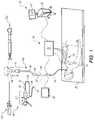
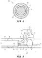
- FIG. 1is a perspective view of a system provided in accordance with the present disclosure configured for navigating a tool to a surgical site;
- FIG. 2is an enlarged view of a portion of an extended working channel connected to valve
- FIG. 3illustrates flow of air through the valve connected to the extended working channel
- FIG. 4illustrates flow of fluid through the valve connected to the extended working channel
- FIG. 5is a cross-sectional view of a portion of extended working channel
- FIG. 6is an enlarged view of a portion of an extended working channel comprising a port or opening and balloons within an airway of a patient;
- FIG. 7 ais an enlarged view of a medical instrument configured for percutaneously accessing and treating a target tissue
- FIG. 7 billustrates flow of air through a catheter housed within the medical instrument configured for percutaneously accessing and treating a target tissue
- FIG. 7 cillustrates flow of fluid through the catheter housed within the medical instrument configured for percutaneously accessing and treating a target tissue
- FIG. 7 dis an enlarged view of a catheter with an opening on its side
- FIG. 7 eis an enlarged cross-sectional view of the medical instrument configured for percutaneously accessing and treating a target tissue with an opening on its handle portion;
- FIG. 7 fis an enlarged view of a catheter fastened into the medical instrument configured for percutaneously accessing and treating a target tissue and flow of air through the catheter;
- FIG. 7 gis an enlarged view of a catheter fastened into the medical instrument configured for percutaneously accessing and treating a target tissue and flow of fluid through the catheter.
- the present disclosureis directed to devices and systems for enhanced treatment options and effectiveness of surgical procedure. Embodiments of the present disclosure are now described in detail with reference to the drawings in which like reference numerals designate identical or corresponding elements in each of the several views.
- the term “clinician”refers to a doctor, a nurse, or any other care provider and may include support personnel. Throughout this description, the term “proximal” will refer to the portion of the device or component thereof that is closer to the clinician and the term “distal” will refer to the portion of the device or component thereof that is farther from the clinician.
- the term “consistent field” or “consistent zone”refers to an area in space within the body of a patient, where the permittivity of the area to microwave energy is less than the permittivity of air to microwave energy.
- Lesionsmay form in various regions within the body. Some lesions may be accessible within an area of the body that is reachable using the airways of the patient. To approach or treat lesions that are within an area of the body of the patient that is reachable using the airways of the patient, a clinician may navigate a medical instrument, such as an ablation catheter; to the area affected by the lesion using airway based medical navigation procedures. But lesions may form in areas of the body that are difficult or impossible to reach by navigating only within the airways of the patient.
- a medical instrumentsuch as an ablation catheter
- a clinicianmay navigate a medical instrument, such as a biopsy or treatment catheter to the area affected by the lesion or the desired surgical site using off-airway surgical procedures involving puncturing a portion of the tissue of luminal network of the lungs (e.g. airways) and tunneling to the desired surgical site.
- This tunnelingmay be done either using a separate tunneling device, or in some instances using the medical instrument itself.
- Lesionstypically have different tissue density than tissue not affected by the disease. Each lesion may have a different tissue density from other lesions. For example, some lesions such as leipidic adenocarcinomas are partially solid. Lesions, as described herein, may be a nonspecific finding on a computed tomography (CT) scans typically described as ground-glass opacification or opacity. Further, scar tissue can give rise to new malignancies thus necessitating biopsy and treatment of this denser tissue. Still further, lesions may also be partially filled with air. Therefore, puncturing a lesion for treatment or performing a biopsy of the lesion may result in the medical instrument being surrounded by the air of the lesion potentially affecting the effectiveness.
- CTcomputed tomography
- the air in the lesionscreates problems for ultrasound devices in producing images as ultrasound energy, like microwave energy, does not traverse air well.
- the resultis an inefficient zone of treatment or inaccurate depiction of the tissue involved in the surgery, both of which may cause issues for clinicians in successfully treating the lesions.
- the present disclosureprovides apparatus, systems and techniques for creating a tissue-to-tool interface between the medical instrument and the lesion or the tissue affected by the lesion.
- the tissue-to-tool interfaceimproves the efficiency of the zone of treatment, resulting in a more consistent field or consistent zone around the medical instrument.
- the improved field around the medical instrumentallows for a more efficient delivery of energy or chemical therapies. Additionally, the improved field increases the accuracy and clarity of images of ultrasound devices, improving the visualization of the procedure for the clinicians.
- one way to improve the consistent field or zone around the energy or treatment sourceis by the apposition of the target tissue to the medical instrument.
- the target tissue against the medical instrumentreduces the leakage of therapeutic substance away from the medical instrument or source or away from the target area can be minimized.
- the energy transfer from modalities such as microwave, cryogenic, and RF ablationcan be increased due to a reduction in resistance caused by the removal of air, which commonly acts as an insulator limiting efficient energy transfer.
- apposition of the tissue against the medical instrumentimproves imaging clarity and accuracy.
- the tissue-to-tool interface between the medical instrument and the tissuemay be created by suctioning air around the medical instrument such that the tissue is drawn closer to the medical instrument.
- the vacuum used in suctioning the air around medical instrumentmay be sufficient to draw the tissue closer to the medical instrument, however, may not be sufficient to induce atelectasis (i.e., the complete collapse of the tissue as a result of the withdrawal of the air in a given portion of the organ).
- the tissue-to-tool interface between the medical instrument and the tissuemay be created by injecting fluid that provides a more consistent environment or zone around medical instrument for delivering energy.
- the apposition of the tissue to the therapy or treatment devicecan limit the ability of fluid or gas therapies or treatments from dispersing beyond the intended treatment site.
- images captured around the surgical site or the medical instrumentmay be projected on to one or more video monitors.
- FIG. 1depicts an electromagnetic navigation (EMN) system 10 is provided in accordance with the present disclosure.
- EMN system 10may be employed in accordance with various example embodiments herein.
- An example of the EMN systemis the ELECTROMAGNETIC NAVIGATION BRONCHOSCOPY® system currently sold by Medtronic, Inc.
- the specific number of components of the system 10 depicted in FIG. 1 and the arrangement and configuration thereofare provided for illustrative purposes only, and should not be construed as limiting.
- EMN system 10may be used to plan a pathway to target tissue, navigate a positioning assembly to the target tissue, navigate a biopsy tool to the target tissue to obtain a tissue sample from the target tissue and use the biopsy tool to digitally mark the location where the tissue sample was obtained, and place one or more echogenic markers at or around the target.
- EMN system 10includes an operating table 40 configured to support a patient, a bronchoscope 50 configured for insertion through the patient's mouth and/or nose into the patient's airways, monitoring equipment 60 coupled to bronchoscope 50 for displaying video images received from bronchoscope 50 , a tracking system 70 including a tracking module 72 , a plurality of reference sensors 74 , an electromagnetic field generator 76 , and a workstation 80 including software and/or hardware used to facilitate pathway planning, identification of target tissue, navigation to target tissue, and digitally marking the biopsy location.
- FIG. 1depicts two types of catheter guide assemblies 90 , 100 .
- Catheter guide assemblies 90 , 100are usable with the EMN system 10 and share a number of common components.
- Each of the catheter guide assemblies 90 , 100includes a handle 91 , which is connected to an extended working channel (EWC) 96 .
- the EWC 96may be sized for placement into the working channel of a bronchoscope 50 .
- a locatable guide (LG) 92including an electromagnetic (EM) sensor 94 , is inserted into the EWC 96 and locked into position such that the sensor 94 extends a desired distance beyond the distal tip of the EWC 96 .
- LGlocatable guide
- EMelectromagnetic
- catheter guide assemblies 90 , 100have different operating mechanisms.
- catheter guide assemblies 90 , 100contain a handle 91 that can be manipulated by rotation and compression to steer the distal tip 93 of the LG 92 and extended working channel 96 .
- Valve 120may be attached to EWC 96 .
- valve 120may be used to suction air out of the patient's airways.
- a vacuum source(not shown) may be attached to valve 120 and may be used in suctioning air out of a certain area within the patient's airway.
- valve 120may permit injecting fluid, such as saline, into a certain area within the patient. The area within the patient to which the fluid is injected may be based on the area where a medical instrument is to be used.
- valve 120may be attached perpendicularly to EWC 96 . Additional details of valve 120 are described with respect to FIG. 2 .
- one or more balloonsmay extend from EWC 96 .
- the balloonsmay extend laterally from EWC 96 .
- a balloonmay be located proximal to the distal end of the EWC 96 .
- a balloonmay also be located distally from the distal end of the EWC 96 .
- the one or more balloonsmay be used to seal at least a portion of an airway. Upon sealing a portion of the airway, a medical instrument may be inserted in to EWC 96 and extended through the distal end of EWC 96 and to the target tissue.
- a port or openingmay be located proximal to the distal end of EWC 96 .
- the openingmay be used to suction air from the space around the distal end of EWC 96 or to suction air surrounding a medical instrument. By suctioning a sufficient amount of air out from the distal end of EWC 96 or surrounding the medical instrument, the medical instrument is placed in apposition with the target or surgical site. Additional details of the opening or port proximal to the distal end of a EWC are described in FIG. 6 .
- Bronchoscope 50includes a source of illumination and a video imaging system (not explicitly shown) and is coupled to monitoring equipment 60 , e.g., a video display, for displaying the video images received from the video imaging system of bronchoscope 50 .
- monitoring equipment 60e.g., a video display
- Catheter guide assemblies 90 , 100 including LG 92 and EWC 96are configured for insertion through a working channel of bronchoscope 50 into the patient's airways (although the catheter guide assemblies 90 , 100 may alternatively be used without bronchoscope 50 ).
- the LG 92 and EWC 96are selectively lockable relative to one another via a locking mechanism 99 .
- a six degrees-of-freedom electromagnetic tracking system 70e.g., similar to those disclosed in U.S. Pat. No. 6,188,355 and published PCT Application Nos. WO 00/10456 and WO 01/67035, the entire contents of each of which is incorporated herein by reference, or any other suitable positioning measuring system is utilized for performing navigation, although other configurations are also contemplated.
- Tracking system 70is configured for use with catheter guide assemblies 90 , 100 to track the position of the EM sensor 94 as it moves in conjunction with the EWC 96 through the airways of the patient, as detailed below.
- electromagnetic field generator 76is positioned beneath the patient. Electromagnetic field generator 76 and the plurality of reference sensors 74 are interconnected with tracking module 72 , which derives the location of each reference sensor 74 in six degrees of freedom. One or more of reference sensors 74 are attached to the chest of the patient. The six degrees of freedom coordinates of reference sensors 74 are sent to workstation 80 , which includes an application 81 where sensors 74 are used to calculate a patient coordinate frame of reference.
- a clinicianmay use the catheter guide assemblies 90 , 100 to navigate the EWC 96 using the LG 92 to reach the desired surgical site or an exit location from the luminal network of the lungs (e.g. the airways).
- the desired surgical sitemay be the area within the body of the patient that is affected by a lesion.
- An exit location from the luminal network of the lungsmay be an area within the body that is closest to a lesion such as bronchial wall nearest to the lesion. If lesions are within an area of the body where they are difficult to reach from within the luminal network of the lungs then the lesions may be reached by piercing the exit location.
- the LG 92is removed and bronchial piercing catheter 101 is inserted into the EWC 96 .
- Bronchial piercing catheter 101is then advanced forward to pierce the bronchial walls while tracking its proximity to the target.
- the EWC 96is also advanced forward.
- Bronchial piercing catheter 101can then be removed, and a biopsy tool 102 can be inserted into the EWC 96 and advanced to the target such as the lesion to be treated.
- the use of the bronchial piercing catheter 101allows the navigation of the EWC 96 to a target outside the airways.
- the LG 92is integrated with the bronchial piercing catheter 101 and the bronchial piercing catheter 101 may be locked inside the EWC 96 so the distal end of the bronchial piercing catheter 101 is positioned inside the distal end of the EWC 96 .
- Bronchial piercing catheter 101(with the integrated LG 92 ) is then navigated with the EWC 96 to a desired exit location within the luminal network of the lungs. Once the exit location is reached, the bronchial piercing catheter 101 is advanced forward and locked relative to the EWC 96 in a second position in which the distal end of the bronchial piercing catheter 101 is just beyond the distal end of the EWC 96 .
- the bronchial piercing catheter 101 and the EWC 96are then advanced forward to pierce the bronchial walls while tracking its proximity to the target.
- Using a sensor integrated with a bronchial piercing cathetereliminates the step of removing the LG 92 in order to place the bronchial piercing catheter 101 through the EWC 96 .
- a biopsy toolsuch as biopsy tool 102 may be used.
- Biopsy tool 102may be inserted into the catheter guide assemblies 90 , 100 following navigation to a target and removal of the LG 92 .
- the biopsy tool 102is used to collect one or more tissue sample from the target tissue.
- the biopsy tool 102may be further configured for use in conjunction with tracking system 70 to facilitate navigation of biopsy tool 102 to the target tissue, tracking of a location of biopsy tool 102 as it is manipulated relative to the target tissue to obtain the tissue sample, and/or marking the location where the tissue sample was obtained.
- EM sensor 94in conjunction with tracking system 70 , enables tracking of EM sensor 94 and/or biopsy tool 102 as EM sensor 94 or biopsy tool 102 is advanced through the patient's airways.
- workstation 80utilizes computed tomographic (CT) image data for generating and viewing a three-dimensional model (“3D model”) of the patient's airways, enables the identification of target tissue on the 3D model (automatically, semi-automatically or manually), and allows for the selection of a pathway through the patient's airways to the target tissue. More specifically, the CT scans are processed and assembled into a 3D volume, which is then utilized to generate the 3D model of the patient's airways.
- the 3D modelmay be presented on a display monitor 81 associated with workstation 80 , or in any other suitable fashion.
- various slices of the 3D volume and views of the 3D modelmay be presented and/or may be manipulated by a clinician to facilitate identification of a target and selection of a suitable pathway through the patient's airways to access the target.
- the 3D modelmay also show marks of the locations where previous biopsies were performed, including the dates, times, and other identifying information regarding the tissue samples obtained. These marks may also be selected as targets to which a pathway can be planned. Once selected, the pathway is saved for use during the navigation procedure.
- An example of a suitable pathway planning system and methodis described in U.S. Patent Publication Nos. 2014/0281961; 2014/0270441; and 2014/0282216, all entitled PATHWAY PLANNING SYSTEM AND METHOD, filed on Mar. 15, 2014, the entire contents of each of which are incorporated herein by reference.
- FIG. 2there is shown an enlarged view of a portion of EWC 96 connected to valve 120 , in accordance with aspects of the present disclosure, and a portion of a medical instrument 19 .
- Medical instrument 19may be part of an ablation catheter, in which case medical instrument 19 is referred to herein as an ablation probe. Medical instrument 19 may be configured to be received within EWC 96 . Medical instrument 19 may extend through an entire length of the EWC 96 and into bronchoscope 50 .
- medical instrument 19may include a microwave antenna (not shown). Examples of microwave antenna construction may be found in commonly assigned U.S. Patent Pub. Nos.
- valve 120may be connected to EWC 96 perpendicularly.
- the EWC 96After navigating the EWC 96 proximate the lesion it may be desirable to secure the EWC within the airways of the patient. In accordance with one embodiment of the present disclosure, this may be done by applying a vacuum to valve 120 , as will be described in greater specificity below with respect to treatment.
- the application of a vacuum to the EWCremoves some of the air within the luminal network and can cause the luminal network to collapse onto the EWC, at least in the area proximate an opening operably connected to the valve 120 .
- This collapsing or appositionallows for the tissue of the luminal network itself to secure the orientation of the EWC 96 .
- This secured orientationcan be verified using imaging techniques including ultrasound and fluoroscopy. Once confirmed the clinician can take the biopsy with confidence that the EWC is secured relative to the target and will acquire the desired tissue sample.
- a biopsyAfter a biopsy, in facilities utilizing Rapid Onsite Evaluation or (ROSE) the sample is analyzed for evidence of cancer or other diseases.
- ROSERapid Onsite Evaluation
- the sampleis cancerous it may be desirable to treat the affected tissue.
- Treatmentrequires re-navigation of the EWC 96 to the target and insertion of a medical instrument 19 for treatment, such as a microwave ablation catheter through the EWC 96 and into the lesion or near the lesion to ablate the lesion.
- bronchial piercing catheter 101may first be inserted into a desired location in the target tissue (e.g.
- the EWC 96may be advanced over the top of the bronchial piercing catheter 101 to secure the EWC 96 in the target tissue.
- the bronchial piercing catheter 101is then removed and the medical instrument 19 can be navigated to the target tissue through the EWC 96 .
- the EWC 96may have to be retracted after placement of the microwave ablation catheter to enable operation of the ablation catheter.
- lesionsmay be partially filled with air, and puncturing a lesion or performing a biopsy of the lesion may result in the medical instrument used to navigate to the lesion to be surrounded by air.
- a tissue-to-tool interface between energy source, medical instrument 19 , and target tissue, such as tissue of the target lesion, providing a consistent environment for energy deliverymay be created suctioning air surrounding the energy source, medical instrument 19 .
- the airmay be suctioned out of the luminal network by application of suction to valve 120 .
- FIG. 3illustrates the flow of air 301 away from distal end of EWC 96 and through valve 120 .
- a vacuum source(not shown) may be connected to valve 120 to suction some or all of the air surrounding the EWC 96 or medical instrument 19 .
- the vacuum sourcemay include, but not limited to, a syringe or a pump.
- a tissue-to-tool interface between medical instrument 19 and target tissuemay be created by injecting a fluid into valve 120 .
- the injected fluidmay provide a more consistent environment for energy delivery by a medical instrument 19 such as a microwave ablation catheter.
- the flow of fluid injected into valve 120is depicted in FIG. 4 as flow 401 .
- the fluid injected into valve 120may be any conductive fluid including, but not limited to, saline.
- valve 120may be a one-way valve.
- One-way valve 120may include a duckbill seal. The duckbill seal may be configured with two states, a biased state and an unbiased state.
- the biased statefluid is allowed to pass through the duckbill, valve 120 , towards the distal end of medical instrument 19 .
- the biased statemay also be referred to herein as when the duckbill is in an open state. In the unbiased state, no fluid passes through the duckbill or valve 120 .
- the tissue-to-tool interface described in combination with isolation of the medical instrument 19 and the target tissue from other areas of the luminal networkincreases efficiency in delivery of energy, such as microwave energy.
- energysuch as microwave energy
- the tissue-to-tool interface created by the apposition of the tool and the target tissueimproves delivery of therapeutic substances to the target tissue, and can eliminate or reduce leakage of therapeutic substance away from the target area.
- the apposition of the tool with the target tissueallows for improved delivery of energy (e.g., microwave energy) and sonic waves (e.g., ultrasound waves) by eliminating nonconductive interfaces between the tool and the target tissue.
- isolation and injection of a fluid into the luminal networkhomogenizes the environment through which energy (e.g., microwave or ultrasound) must travel and improves the transfer of energy for better treatment and imaging.
- air flow 301 and fluid flow 401flows while medical instrument 19 is positioned within EWC 96 .
- air flow 301 and fluid flow 401flows in a space defined between medical instrument 19 and EWC 96 . Additional details of a flow in a space provided between an energy source and an EWC is provided herein in FIG. 5 .
- FIG. 5there is shown a cross-sectional view of EWC 96 that illustrates a space between medical instrument 19 and EWC 96 .
- the cross-sectional view 500illustrates the EWC 96 and medical instrument 19 positioned within the EWC 96 .
- Space 501is annularly defined between the EWC 96 and the medical instrument 19 .
- Flow Awhich may be air flow 301 or fluid flow 401 flows within space 501 .
- Flow Atravels along the length of bronchoscope 50 , via the EWC 96 .
- Flow Adoes not directly contact the bronchoscope 50 , but stays within EWC 96 until it exits EWC 96 .
- EWC 96(or primary channel) achieves the dual purpose of allowing a clinician to simultaneously use/manipulate a surgical instrument and create a tool-to-tissue interface between instrument and tissue through EWC 96 .
- annular space 501may also terminate in one or more openings formed on the side of the EWC 96 . As such, the apposition of tissue to the EWC 96 and medical instrument 19 may be enhanced and promote greater sealing characteristics.
- FIG. 6there is shown an enlarged view of a portion of an extended working channel 96 comprising a port or opening 18 and balloons 14 , 21 within an airway of a patient.
- FIG. 6depicts an airway 10 , an alveolus 11 that branches off airway 10 , and a lesion 28 that is formed within alveolus 11 .
- EWC 96is inserted into airway 10 and a proximal balloon 14 extends from EWC 96 .
- Balloon 14may extend laterally from EWC 96 , as depicted in FIG. 6 , and seal the portion of the airway 10 proximal of the distal end of EWC 96 .
- Medical instrument 19extends from the distal end of EWC 96 .
- Medical instrument 19as described above, may be a microwave ablation catheter.
- medical instrument 19may be a catheter configured to deliver therapeutic substance, such as a chemotherapy substance, to the target tissue.
- a balloon catheter 20extends from EWC 96 and includes balloon 21 at the distal end of catheter 20 .
- Fluid line 22is in fluid communication with balloon 21 .
- fluid line 22may be a dual fluid line that includes a first fluid line 24 and a second fluid line 26 .
- balloon 21may be expanded by application of fluid through fluid line 22 .
- Fluidmay be applied to the balloon 21 through a fluid line attached to the balloon, such as fluid line 24 .
- Fluid line 22passes through distal balloon 21 and fluidly connects a portion of airway 10 with atmosphere or a ventilation device (not shown) that permits lung comprising airway 10 to receive and expel air. Balloons 14 and 21 create an area in airway 10 that is effectively sealed from the atmosphere.
- Port 18is located on the distal portion of EWC 96 and is in fluid communication through EWC 96 to a vacuum source, such as a pump or syringe (not shown).
- a vacuum sourcesuch as a pump or syringe (not shown).
- a tissue-to-tool interface between medical instrument 19 and lesion 28providing a consistent environment or zone may be created by suctioning air surrounding the medical instrument 19 .
- the vacuum generated by vacuum sourceis sufficient to place medical instrument 19 in apposition with tissue of lesion 28 , but not necessarily enough to cause atelectasis or collapse of alveolus 11 .
- the tissue-to-tool interface created between medical instrument 19 and lesion 28improves delivery of energy to the lesion 28 because the non-conductive interface, (i.e., the air between medical instrument 19 and lesion 28 ), is substantially eliminated.
- the tissue-to-tool interface created between medical instrument 19 and lesion 28improves delivery of the therapeutic substance to lesion 28 by eliminating leakage of the therapeutic substance beyond the portion of the airway 10 isolated by balloons 14 and 21 , thus limiting the therapeutic substance to those areas where they will be most effective, namely alveolus 11 and a small section of airway 10 .
- Medical instrument 700 aincludes a handle 704 coupled with an ablation probe 702 .
- ablation probe 702extends from handle 704 .
- Ablation probe 702includes a microwave ablation antenna (not shown) that is used to ablate tissue.
- Handle 704includes a housing unit (not shown) configured to house catheter 701 .
- Catheter 701is optionally configured to extend out from the housing unit within handle 704 and retract into the housing unit within handle 704 .
- Catheter 701defines a lumen 701 a and ablation probe 702 is located within lumen 701 a.
- a control unitsuch as wheel 703 , is configured to control the extension and retraction of catheter 701 such that rotating wheel 703 away from the clinician holding handle 704 or in the direction X′ extends catheter 701 by moving catheter 701 in the direction X′ and rotating wheel 703 towards the clinician holding handle 704 or in the direction X retracts catheter 701 into the housing unit of handle 704 by moving catheter 701 in the direction X.
- the control unitmay be a slider, where sliding the slider away from the clinician holding handle 704 or in the direction X′ extends the catheter 701 and sliding the slider towards the clinician holding handle 704 or in the direction X retracts the catheter 701 .
- the catheter 701may be formed of a trocar or other component inserted into the patient proximate the target tissue through which the ablation probe 702 is inserted in accordance with embodiments of the present disclosure.
- Medical instrument 700 amay include one or more markers, such as markers 710 a , 710 b , to help guide a clinician in determining how far to extend the catheter in order to most efficiently create the tissue-to-tool interface, described herein, between the ablation probe 702 and a target tissue, such as a lesion.
- markers 710 a , 710 bto help guide a clinician in determining how far to extend the catheter in order to most efficiently create the tissue-to-tool interface, described herein, between the ablation probe 702 and a target tissue, such as a lesion.
- Each of the markersindicates a different length of extension and based on how deep ablation probe 702 is inserted into a patient, a marker may more effectively create the desired tissue-to-tool interface than other markers.
- Catheter 701via port 705 a and tube 705 , is in fluid communication with a vacuum source (not shown) or a liquid source (not shown).
- Port 705 ais in fluid communication with lumen 701 a of the catheter 701 .
- Tube 705is configured and coupled with catheter 701 , via port 705 a , such that one end of tube 705 is attached to an end of lumen 701 a of catheter 701 that is proximal to the clinician, via port 705 a , and the other end of tube 705 is coupled with the vacuum or liquid source.
- the vacuum source described hereinincludes, but is not limited to, a pump or a syringe.
- the vacuum sourcegenerates a vacuum by suctioning air, through tube 705 , via lumen 701 a , around ablation probe 702 .
- the application of vacuumis sufficient to place ablation probe 702 in apposition with the target tissue, thus, creating a consistent zone around ablation probe 702 , but not enough to cause atelectasis.
- a liquid sourceas described herein, provides a fluid such as saline. The fluid from the liquid source helps create a consistent zone around ablation probe 702 by injecting fluid from the liquid source through tube 705 and onto at least a portion of the target tissue, via lumen 701 a.
- FIG. 7 Bthere is shown a medical instrument 700 a with the catheter 701 extended to marker 710 a and in a fluid communication with a vacuum source (not shown).
- a vacuum sourcenot shown
- airflow 712air is suctioned away from target tissue through lumen 701 a , which is in fluid communication with tube 705 , via port 705 a .
- suctioning sufficient amount of air away from the proximity of the target tissueplaces the target tissue in apposition with ablation probe 702 , which creates a more consistent zone around ablation probe 702 .
- FIG. 7 Cillustrates medical instrument 700 a with the catheter 701 in a fluid communication with a liquid source, which provides a fluid, such as saline.
- Fluid flow 713depicts the flow of fluid, for example saline, onto the target tissue, through lumen 701 a , which is in fluid communication with tube 705 , via port 705 a , in order to form a consistent zone around ablation probe 702 .
- fluidfor example saline
- FIG. 7 Dthere is shown an embodiment of a catheter that may be attached to the medical instrument 700 a instead of being housed in medical instrument 700 a .
- Catheter 706defines a lumen 706 a and includes a port 714 , which is configured to be in fluid communication with a vacuum source or a liquid source. Port 714 is in fluid communication with lumen 706 a of catheter 706 . While a single catheter 706 is depicted in FIG. 7 D , a plurality of catheters 706 , each of a different length may be used and selection of a particular catheter 706 may be based on how deep ablation probe 702 is inserted into a patient. Catheter 706 may be attached to the medical instrument 700 a by fastening it into medical instrument 700 a using opening 707 on the handle 704 , as depicted in FIG. 7 E .
- FIG. 7 Ethere is shown a cross-sectional view of medical instrument 700 a without attachment of catheter 706 to medical instrument 700 a .
- the end of catheter 706 that is proximal to port 714may be configured with an external thread profile (not shown) to allow catheter 706 to be fastened into opening 707 on the handle 704 .
- Opening 707may be configured with an internal thread profile (not shown) to accept catheter 706 .
- Catheter 706may be attached to the medical instrument 700 a by fastening it into medical instrument 700 a using opening 707 .
- Other connection mechanismsincluding press fittings, suction fittings, and the like are within the scope of the present disclosure.
- FIG. 7 Fdepicts port 714 being in fluid communication with a vacuum source (not shown) via tube 705 .
- the tissue-to-tool interfacedescribed above, is created by suctioning air away from target tissue through lumen 706 a , using the vacuum source in fluid communication with port 714 via tube 705 , as depicted by air flow 715 .
- FIG. 7 Gdepicts port 714 being in fluid communication with a liquid source, which provides fluid, such as saline. The fluid may be injected onto at least a portion of target tissue using tube 705 and lumen 706 a of catheter 706 , as depicted by liquid flow 716 .
- a consistent zone around ablation probe 702may be injected onto at least a portion of target tissue using tube 705 and lumen 706 a of catheter 706 , as depicted by liquid flow 716 .
- a phrase in the form “A or B”means “(A), (B), or (A and B).”
- a phrase in the form “at least one of A, B, or C”means “(A); (B); (C); (A and B); (A and C); (B and C); or (A, B, and C).”
Landscapes
- Health & Medical Sciences (AREA)
- Life Sciences & Earth Sciences (AREA)
- Surgery (AREA)
- Engineering & Computer Science (AREA)
- Veterinary Medicine (AREA)
- Public Health (AREA)
- General Health & Medical Sciences (AREA)
- Animal Behavior & Ethology (AREA)
- Biomedical Technology (AREA)
- Heart & Thoracic Surgery (AREA)
- Medical Informatics (AREA)
- Molecular Biology (AREA)
- Pathology (AREA)
- Physics & Mathematics (AREA)
- Nuclear Medicine, Radiotherapy & Molecular Imaging (AREA)
- Biophysics (AREA)
- Radiology & Medical Imaging (AREA)
- Otolaryngology (AREA)
- Pulmonology (AREA)
- Optics & Photonics (AREA)
- Cardiology (AREA)
- Plasma & Fusion (AREA)
- Vascular Medicine (AREA)
- Physiology (AREA)
- Surgical Instruments (AREA)
Abstract
Description
Claims (12)
Priority Applications (3)
| Application Number | Priority Date | Filing Date | Title |
|---|---|---|---|
| US15/865,560US11529190B2 (en) | 2017-01-30 | 2018-01-09 | Enhanced ablation and visualization techniques for percutaneous surgical procedures |
| EP18744404.7AEP3573555A4 (en) | 2017-01-30 | 2018-01-25 | Enhanced ablation and visualization techniques for percutaneous surgical procedures |
| PCT/US2018/015155WO2018140557A1 (en) | 2017-01-30 | 2018-01-25 | Enhanced ablation and visualization techniques for percutaneous surgical procedures |
Applications Claiming Priority (3)
| Application Number | Priority Date | Filing Date | Title |
|---|---|---|---|
| US201762451836P | 2017-01-30 | 2017-01-30 | |
| US201762451832P | 2017-01-30 | 2017-01-30 | |
| US15/865,560US11529190B2 (en) | 2017-01-30 | 2018-01-09 | Enhanced ablation and visualization techniques for percutaneous surgical procedures |
Publications (2)
| Publication Number | Publication Date |
|---|---|
| US20180214203A1 US20180214203A1 (en) | 2018-08-02 |
| US11529190B2true US11529190B2 (en) | 2022-12-20 |
Family
ID=62977390
Family Applications (1)
| Application Number | Title | Priority Date | Filing Date |
|---|---|---|---|
| US15/865,560Active2039-03-23US11529190B2 (en) | 2017-01-30 | 2018-01-09 | Enhanced ablation and visualization techniques for percutaneous surgical procedures |
Country Status (3)
| Country | Link |
|---|---|
| US (1) | US11529190B2 (en) |
| EP (1) | EP3573555A4 (en) |
| WO (1) | WO2018140557A1 (en) |
Citations (135)
| Publication number | Priority date | Publication date | Assignee | Title |
|---|---|---|---|---|
| US4202352A (en) | 1978-04-06 | 1980-05-13 | Research Development Corporation | Apparatus for measurement of expired gas concentration in infants |
| US5057494A (en) | 1988-08-03 | 1991-10-15 | Ethicon, Inc. | Method for preventing tissue damage after an ischemic episode |
| US5321113A (en) | 1993-05-14 | 1994-06-14 | Ethicon, Inc. | Copolymers of an aromatic anhydride and aliphatic ester |
| US5358496A (en) | 1991-10-18 | 1994-10-25 | Ethicon, Inc. | Endoscopic tissue manipulator |
| US5458574A (en)* | 1994-03-16 | 1995-10-17 | Heartport, Inc. | System for performing a cardiac procedure |
| US5478309A (en)* | 1994-05-27 | 1995-12-26 | William P. Sweezer, Jr. | Catheter system and method for providing cardiopulmonary bypass pump support during heart surgery |
| US5830222A (en)* | 1995-10-13 | 1998-11-03 | Transvascular, Inc. | Device, system and method for intersititial transvascular intervention |
| US6003517A (en) | 1998-04-30 | 1999-12-21 | Ethicon Endo-Surgery, Inc. | Method for using an electrosurgical device on lung tissue |
| WO2000010456A1 (en) | 1998-08-02 | 2000-03-02 | Super Dimension Ltd. | Intrabody navigation system for medical applications |
| US6086586A (en) | 1998-09-14 | 2000-07-11 | Enable Medical Corporation | Bipolar tissue grasping apparatus and tissue welding method |
| US6188355B1 (en) | 1997-12-12 | 2001-02-13 | Super Dimension Ltd. | Wireless six-degree-of-freedom locator |
| WO2001067035A1 (en) | 2000-03-09 | 2001-09-13 | Super Dimension Ltd. | Object tracking using a single sensor or a pair of sensors |
| US20020147462A1 (en) | 2000-09-11 | 2002-10-10 | Closure Medical Corporation | Bronchial occlusion method and apparatus |
| US6475216B2 (en) | 1995-02-22 | 2002-11-05 | Medtronic, Inc. | Fluid-assisted electrocautery method |
| US20030013972A1 (en) | 2001-05-29 | 2003-01-16 | Makin Inder Raj. S. | Treatment of lung lesions using ultrasound |
| US6533784B2 (en) | 2001-02-24 | 2003-03-18 | Csaba Truckai | Electrosurgical working end for transecting and sealing tissue |
| BR0013237A (en) | 1999-08-12 | 2003-07-15 | Johnson & Johnson | Antibacterial heterobicyclic substituted phenyl oxazolidinones |
| US6629951B2 (en)* | 1999-08-05 | 2003-10-07 | Broncus Technologies, Inc. | Devices for creating collateral in the lungs |
| US6656177B2 (en) | 2000-10-23 | 2003-12-02 | Csaba Truckai | Electrosurgical systems and techniques for sealing tissue |
| US20030233099A1 (en) | 2000-10-17 | 2003-12-18 | Broncus Technologies, Inc. | Modification of airways by application of energy |
| MXPA03005028A (en) | 2000-12-06 | 2004-01-29 | Johnson & Johnson | 6-0-carbamoyl ketolide derivatives of erythromycin useful as antibacterials. |
| US6726651B1 (en)* | 1999-08-04 | 2004-04-27 | Cardeon Corporation | Method and apparatus for differentially perfusing a patient during cardiopulmonary bypass |
| US20040120981A1 (en) | 2002-12-20 | 2004-06-24 | Aruna Nathan | Crosslinked alkyd polyesters for medical applications |
| US6770070B1 (en) | 2000-03-17 | 2004-08-03 | Rita Medical Systems, Inc. | Lung treatment apparatus and method |
| MXPA03006874A (en) | 2002-07-31 | 2004-09-03 | Johnson & Johnson | Long term oxygen therapy system. |
| MXPA03000137A (en) | 2000-06-30 | 2004-09-13 | Johnson & Johnson | Dna encoding human serine protease d-g. |
| US6802843B2 (en) | 2001-09-13 | 2004-10-12 | Csaba Truckai | Electrosurgical working end with resistive gradient electrodes |
| BR0307259A (en) | 2002-01-28 | 2004-12-07 | Ortho Mcneil Pharm Inc | Heterocyclically substituted phenyl oxazolidinone antibacterials and related compositions and methods |
| US20040249343A1 (en)* | 2002-12-06 | 2004-12-09 | Wit Ip Corporation | Combination treatment catheters and post treatment stents |
| US6835336B2 (en) | 1997-10-03 | 2004-12-28 | Ethicon, Inc. | Methods for making biopolymer sponge tubes |
| US6913579B2 (en) | 2001-05-01 | 2005-07-05 | Surgrx, Inc. | Electrosurgical working end and method for obtaining tissue samples for biopsy |
| MXPA03010507A (en) | 2001-05-15 | 2005-07-25 | Johnson & Johnson | EX-VIVO ACTIVATION TO GENERATE SPECIFIC C CYTO-TOXIC LYMPHOCYTES FOR NON-TUMOR ANTIGENS TO TREAT AUTOIMMUNE AND ALLERGIC DISEASES. |
| US20050165276A1 (en) | 2004-01-28 | 2005-07-28 | Amir Belson | Methods and apparatus for accessing and treating regions of the body |
| MXPA05011725A (en) | 2003-04-30 | 2006-05-17 | Johnson & Johnson | Cngh0010 specific polynucleotides, polypeptides, antibodies, compositions, methods and uses. |
| US20060200191A1 (en)* | 1996-05-20 | 2006-09-07 | Gholam-Reza Zadno-Azizi | Method and apparatuses for treating an intravascular occlusion |
| US20060235457A1 (en) | 2005-04-15 | 2006-10-19 | Amir Belson | Instruments having a rigidizable external working channel |
| US20070135803A1 (en) | 2005-09-14 | 2007-06-14 | Amir Belson | Methods and apparatus for performing transluminal and other procedures |
| MX2007006441A (en) | 2004-11-30 | 2007-08-14 | Johnson & Johnson | Lung cancer prognostics. |
| US20080045938A1 (en) | 2006-07-14 | 2008-02-21 | Micrablate | Energy delivery systems and uses thereof |
| EP1644519B1 (en) | 2003-07-04 | 2008-12-31 | Johnson & Johnson Research Pty Limited | Method for detection of alkylated cytosine in dna |
| US20090024125A1 (en)* | 2007-07-18 | 2009-01-22 | Docimo Steven G | Automatically retracting needle-tip electrocautery device |
| EP2141497A1 (en) | 2008-07-03 | 2010-01-06 | Amic AB | Method for the analysis of circulating antibodies |
| WO2010004570A1 (en) | 2008-07-10 | 2010-01-14 | Superdimension Ltd. | Integrated multi-functional endoscopic tool |
| US20110098694A1 (en)* | 2009-10-28 | 2011-04-28 | Ethicon Endo-Surgery, Inc. | Methods and instruments for treating cardiac tissue through a natural orifice |
| US7947000B2 (en) | 2003-09-12 | 2011-05-24 | Intuitive Surgical Operations, Inc. | Cannula system for free-space navigation and method of use |
| US20120197245A1 (en)* | 2011-02-01 | 2012-08-02 | Channel Medsystems, Inc. | Methods and apparatus for cyrogenic treatment of a body cavity or lumen |
| US20120226271A1 (en)* | 2005-03-25 | 2012-09-06 | Peter Callas | Vacuum Ablation Apparatus and Method |
| US20130096385A1 (en) | 2011-10-14 | 2013-04-18 | Intuitive Surgical Operations, Inc. | Vision probe and catheter systems |
| US20140046315A1 (en) | 2012-08-07 | 2014-02-13 | Covidien Lp | Microwave ablation catheter and method of utilizing the same |
| US20140052018A1 (en) | 2012-08-15 | 2014-02-20 | Intuitive Surgical Operations, Inc. | Specimen removal bag and methods of using same |
| US20140088457A1 (en) | 2012-09-26 | 2014-03-27 | Covidien Lp | Bleeding containment device |
| US20140235943A1 (en) | 2013-02-15 | 2014-08-21 | Intuitive Surgical Operations, Inc. | Vision Probe with Access Port |
| US20140281961A1 (en) | 2013-03-15 | 2014-09-18 | Covidien Lp | Pathway planning system and method |
| US20140276033A1 (en) | 2013-03-15 | 2014-09-18 | Covidien Lp | Microwave energy-device and system |
| US20140270441A1 (en) | 2013-03-15 | 2014-09-18 | Covidien Lp | Pathway planning system and method |
| US20140282216A1 (en) | 2013-03-15 | 2014-09-18 | Covidien Lp | Pathway planning system and method |
| US20150073211A1 (en) | 2013-09-06 | 2015-03-12 | Covidien Lp | Microwave ablation catheter, handle, and system |
| US20150141869A1 (en) | 2013-11-20 | 2015-05-21 | Covidien Lp | Devices, systems, and methods for navigating a biopsy tool to a target location and obtaining a tissue sample using the same |
| US20150141809A1 (en) | 2013-11-20 | 2015-05-21 | Covidien Lp | Devices, systems, and methods for navigating a biopsy tool to a target location and obtaining a tissue sample using the same |
| US20150209107A1 (en)* | 2014-01-24 | 2015-07-30 | Denervx LLC | Cooled microwave denervation catheter configuration |
| US20150265257A1 (en) | 2014-03-19 | 2015-09-24 | Covidien Lp | Devices, systems, and methods for navigating a biopsy tool to a target location and obtaining a tissue sample using the same |
| US9192426B2 (en)* | 2012-06-26 | 2015-11-24 | Covidien Lp | Ablation device having an expandable chamber for anchoring the ablation device to tissue |
| US20160001038A1 (en) | 2014-07-01 | 2016-01-07 | Auris Surgical Robotics, Inc. | Tool and method for using surgical endoscope with spiral lumens |
| US20160051327A1 (en)* | 2014-08-20 | 2016-02-25 | Covidien Lp | Systems and methods for spherical ablations |
| US20160067450A1 (en) | 2014-09-10 | 2016-03-10 | Intuitive Surgical Operations, Inc. | Flexible instrument with nested conduits |
| US9375268B2 (en) | 2007-02-15 | 2016-06-28 | Ethicon Endo-Surgery, Inc. | Electroporation ablation apparatus, system, and method |
| US20160184013A1 (en) | 2014-12-31 | 2016-06-30 | Covidien Lp | System and method for treating copd and emphysema |
| US20160331358A1 (en) | 2013-12-13 | 2016-11-17 | Intuitive Surgical Operations, Inc. | Telescoping biopsy needle |
| US20160374676A1 (en) | 2011-02-15 | 2016-12-29 | Intuitive Surgical Operations, Inc. | Methods and systems for detecting staple cartridge misfire or failure |
| US20170020628A1 (en) | 2013-11-25 | 2017-01-26 | Body Vision Medical Ltd. | Surgical devices and methods of use thereof |
| US20170112588A1 (en) | 2015-10-26 | 2017-04-27 | Neuwave Medical, Inc. | Apparatuses for securing a medical device and related methods thereof |
| US20170112571A1 (en) | 2015-10-26 | 2017-04-27 | Neuwave Medical, Inc. | Energy delivery systems and uses thereof |
| US20170202543A1 (en) | 2016-01-15 | 2017-07-20 | Covidien Lp | Navigable endobronchial tool to access tissue outside a bronchus |
| US20170224338A1 (en) | 2016-02-08 | 2017-08-10 | Ethicon, Inc. | Elastic Tissue Reinforcing Fastener |
| US20170238795A1 (en) | 2014-08-14 | 2017-08-24 | Intuitive Surgical Operations, Inc. | Systems and Methods for Cleaning an Endoscopic Instrument |
| US20170245740A1 (en) | 2016-02-29 | 2017-08-31 | Covidien Lp | Navigation system for ablation catheters |
| US20170258309A1 (en) | 2016-03-14 | 2017-09-14 | Intuitive Surgical Operations, Inc. | Endoscopic instrument with compliant thermal interface |
| US20170274189A1 (en) | 2016-03-24 | 2017-09-28 | Ethicon, Inc. | Single lumen balloon delivery catheter with lumen bypass at balloon |
| US9801630B2 (en) | 2014-06-10 | 2017-10-31 | Ethicon Llc | Methods and devices for reinforcing a staple line |
| US20180001058A1 (en) | 2015-01-12 | 2018-01-04 | Intuitive Surgical Operations, Inc. | Devices, Systems, and Methods for Anchoring Actuation Wires to a Steerable Instrument |
| US20180064904A1 (en) | 2002-09-12 | 2018-03-08 | Intuitive Surgical Operations, Inc. | Shape-transferring cannula system and method of use |
| US20180144092A1 (en) | 2016-11-21 | 2018-05-24 | Johnson & Johnson Vision Care, Inc. | Biomedical sensing methods and apparatus for the detection and prevention of lung cancer states |
| EP3326551A1 (en) | 2011-02-15 | 2018-05-30 | Intuitive Surgical Operations Inc. | Systems for detecting clamping or firing failure |
| US10004558B2 (en) | 2009-01-12 | 2018-06-26 | Ethicon Endo-Surgery, Inc. | Electrical ablation devices |
| US20180214138A9 (en) | 2014-04-02 | 2018-08-02 | Intuitive Surgical Operations, Inc. | Devices, Systems, and Methods Using a Steerable Stylet and Flexible Needle |
| US20180221039A1 (en) | 2015-10-30 | 2018-08-09 | Auris Health, Inc. | Basket apparatus |
| EP3367915A1 (en) | 2015-10-30 | 2018-09-05 | Auris Health, Inc. | Process for percutaneous operations |
| BR112018003862A2 (en) | 2015-08-31 | 2018-10-02 | Ethicon Llc | staple cartridge assembly for use with a surgical stapler, method of using a staple cartridge assembly, end actuator for a surgical instrument, and method of using an end actuator |
| US10172973B2 (en) | 2015-08-31 | 2019-01-08 | Ethicon Llc | Surgical adjuncts and medicants for promoting lung function |
| US10194897B2 (en) | 2014-07-18 | 2019-02-05 | Ethicon, Inc. | Mechanical retraction via tethering for lung volume reduction |
| US10206686B2 (en) | 2014-06-10 | 2019-02-19 | Ethicon Llc | Bronchus sealants and methods of sealing bronchial tubes |
| US20190076143A1 (en) | 2015-11-13 | 2019-03-14 | Intuitive Surgical Operations, Inc. | Stapler anvil with compliant tip |
| US20190175799A1 (en) | 2017-12-08 | 2019-06-13 | Auris Health, Inc. | Directed fluidics |
| US20190200984A1 (en) | 2017-12-28 | 2019-07-04 | Ethicon Llc | Safety systems for smart powered surgical stapling |
| US10349938B2 (en) | 2015-08-31 | 2019-07-16 | Ethicon Llc | Surgical adjuncts with medicants affected by activator materials |
| US20190223693A1 (en) | 2002-09-12 | 2019-07-25 | Intuitive Surgical Operations, Inc. | Shape-transferring cannula system and method of use |
| US20190231449A1 (en) | 2016-07-01 | 2019-08-01 | Intuitive Surgical Operations, Inc. | Systems and methods for flexible computer-assisted instrument control |
| US20190239724A1 (en) | 2016-10-07 | 2019-08-08 | Body Vision Medical Ltd. | Devices for use in interventional and surgical procedures and methods of use thereof |
| US20190246876A1 (en) | 2018-02-15 | 2019-08-15 | Neuwave Medical, Inc. | Compositions and methods for directing endoscopic devices |
| US20190269818A1 (en) | 2018-03-05 | 2019-09-05 | Ethicon Llc | Sealant foam compositions for lung applications |
| US20190269819A1 (en) | 2018-03-05 | 2019-09-05 | Ethicon Llc | Sealant foam compositions for lung applications |
| US20190269885A1 (en) | 2018-03-05 | 2019-09-05 | Intuitive Surgical Operations, Inc. | Deployable bellows for delivery of a flexible, elongate device and methods of use |
| US20190290375A1 (en) | 2016-04-29 | 2019-09-26 | Intuitive Surgical Operations, Inc. | Compliant mechanisms having inverted tool members |
| US20190328213A1 (en) | 2017-04-07 | 2019-10-31 | Auris Health, Inc. | Superelastic medical instrument |
| EP3576598A1 (en) | 2017-02-01 | 2019-12-11 | Intuitive Surgical Operations Inc. | Systems and methods of registration for image-guided procedures |
| US20200008827A1 (en) | 2016-09-14 | 2020-01-09 | Intuitive Surgical Operations, Inc. | Joint assemblies with cross-axis flexural pivots |
| US20200022767A1 (en) | 2017-12-11 | 2020-01-23 | Auris Health, Inc. | Systems and methods for instrument based insertion architectures |
| US20200030575A1 (en) | 2018-07-25 | 2020-01-30 | Intuitive Surgical Operations, Inc. | Systems and methods for use of a variable stiffness flexible elongate device |
| US20200029948A1 (en) | 2018-07-26 | 2020-01-30 | Intuitive Surgical Operations, Inc. | Systems and methods of steerable elongate device |
| US20200054408A1 (en) | 2018-08-15 | 2020-02-20 | Auris Health, Inc. | Medical instruments for tissue cauterization |
| US10569071B2 (en) | 2015-08-31 | 2020-02-25 | Ethicon Llc | Medicant eluting adjuncts and methods of using medicant eluting adjuncts |
| US20200069384A1 (en) | 2014-07-28 | 2020-03-05 | Intuitive Surgical Operations, Inc. | Guide apparatus for delivery of a flexible instrument and methods of use |
| US20200077991A1 (en) | 2016-05-31 | 2020-03-12 | Intuitive Surgical Operations, Inc. | Pliant biopsy needle system |
| US20200078023A1 (en) | 2014-07-18 | 2020-03-12 | Ethicon, Inc. | Methods and devices for controlling the size of emphysematous bullae |
| US10603106B2 (en) | 2010-05-03 | 2020-03-31 | Neuwave Medical, Inc. | Energy delivery systems and uses thereof |
| US20200100776A1 (en) | 2017-02-09 | 2020-04-02 | Intuitive Surgical Operations, Inc. | System and method of accessing encapsulated targets |
| US20200107894A1 (en) | 2018-10-08 | 2020-04-09 | Auris Health, Inc. | Systems and instruments for tissue sealing |
| US20200121170A1 (en) | 2014-09-10 | 2020-04-23 | Intuitive Surgical Operations, Inc. | Devices, systems, and methods using mating catheter tips and tools |
| US10639114B2 (en) | 2018-08-17 | 2020-05-05 | Auris Health, Inc. | Bipolar medical instrument |
| US10638953B2 (en) | 2012-02-03 | 2020-05-05 | Intuitive Surgical Operations, Inc. | Steerable flexible needle with embedded shape sensing |
| US20200142013A1 (en) | 2018-11-02 | 2020-05-07 | Intuitive Surgical Operations, Inc. | Cooled antenna with fluid cooling |
| US20200138514A1 (en) | 2018-11-02 | 2020-05-07 | Intuitive Surgical Operations, Inc. | Tissue penetrating device tips |
| US20200146757A1 (en) | 2011-10-14 | 2020-05-14 | Intuitive Surgical Operations, Inc. | Catheter sensor systems |
| US20200155232A1 (en) | 2018-10-31 | 2020-05-21 | Intuitive Surgical Operations, Inc. | Antenna systems and methods of use |
| US20200188021A1 (en) | 2018-11-13 | 2020-06-18 | Intuitive Surgical Operations, Inc. | Cooled chokes for ablation systems and methods of use |
| US20200222666A1 (en) | 2017-07-21 | 2020-07-16 | Intuitive Surgical Operations, Inc. | Flexible elongate device systems and methods |
| US10716637B2 (en) | 2013-10-25 | 2020-07-21 | Intuitive Surgical Operations, Inc. | Flexible instrument with grooved steerable tube |
| US10729886B2 (en) | 2016-08-24 | 2020-08-04 | Intuitive Surgical Operations, Inc. | Axial support structure for a flexible elongate device |
| US20200261175A1 (en) | 2016-09-30 | 2020-08-20 | Intuitive Surgical Operations, Inc. | Variable-length guide apparatus for delivery of a flexible instrument and methods of use |
| US20200305983A1 (en) | 2019-03-29 | 2020-10-01 | Auris Health, Inc. | Systems and methods for optical strain sensing in medical instruments |
| US10792022B2 (en) | 2009-05-08 | 2020-10-06 | Broncus Medical Inc. | Tissue sampling devices, systems and methods |
| US20200383750A1 (en) | 2017-11-14 | 2020-12-10 | Intuitive Surgical Operations, Inc. | Systems and methods for cleaning endoscopic instruments |
| EP3749239A1 (en) | 2018-02-05 | 2020-12-16 | Broncus Medical Inc. | Image-guided lung tumor planning and ablation system |
| US10881385B2 (en) | 2016-09-13 | 2021-01-05 | Intuitive Surgical Operations, Inc. | Radial telescoping guide apparatus for delivery of a flexible instrument and methods of use |
| US20210000524A1 (en) | 2004-11-16 | 2021-01-07 | Uptake Medical Technology Inc. | Device and method for lung treatment |
- 2018
- 2018-01-09USUS15/865,560patent/US11529190B2/enactiveActive
- 2018-01-25WOPCT/US2018/015155patent/WO2018140557A1/ennot_activeCeased
- 2018-01-25EPEP18744404.7Apatent/EP3573555A4/ennot_activeWithdrawn
Patent Citations (153)
| Publication number | Priority date | Publication date | Assignee | Title |
|---|---|---|---|---|
| US4202352A (en) | 1978-04-06 | 1980-05-13 | Research Development Corporation | Apparatus for measurement of expired gas concentration in infants |
| US5057494A (en) | 1988-08-03 | 1991-10-15 | Ethicon, Inc. | Method for preventing tissue damage after an ischemic episode |
| US5358496A (en) | 1991-10-18 | 1994-10-25 | Ethicon, Inc. | Endoscopic tissue manipulator |
| US5321113A (en) | 1993-05-14 | 1994-06-14 | Ethicon, Inc. | Copolymers of an aromatic anhydride and aliphatic ester |
| US5458574A (en)* | 1994-03-16 | 1995-10-17 | Heartport, Inc. | System for performing a cardiac procedure |
| US5478309A (en)* | 1994-05-27 | 1995-12-26 | William P. Sweezer, Jr. | Catheter system and method for providing cardiopulmonary bypass pump support during heart surgery |
| US6475216B2 (en) | 1995-02-22 | 2002-11-05 | Medtronic, Inc. | Fluid-assisted electrocautery method |
| US5830222A (en)* | 1995-10-13 | 1998-11-03 | Transvascular, Inc. | Device, system and method for intersititial transvascular intervention |
| US20040133225A1 (en)* | 1995-10-13 | 2004-07-08 | Transvascular, Inc. | Device, system and method for interstitial transvascular intervention |
| US20060200191A1 (en)* | 1996-05-20 | 2006-09-07 | Gholam-Reza Zadno-Azizi | Method and apparatuses for treating an intravascular occlusion |
| US6835336B2 (en) | 1997-10-03 | 2004-12-28 | Ethicon, Inc. | Methods for making biopolymer sponge tubes |
| US6188355B1 (en) | 1997-12-12 | 2001-02-13 | Super Dimension Ltd. | Wireless six-degree-of-freedom locator |
| US6003517A (en) | 1998-04-30 | 1999-12-21 | Ethicon Endo-Surgery, Inc. | Method for using an electrosurgical device on lung tissue |
| WO2000010456A1 (en) | 1998-08-02 | 2000-03-02 | Super Dimension Ltd. | Intrabody navigation system for medical applications |
| US6086586A (en) | 1998-09-14 | 2000-07-11 | Enable Medical Corporation | Bipolar tissue grasping apparatus and tissue welding method |
| US6726651B1 (en)* | 1999-08-04 | 2004-04-27 | Cardeon Corporation | Method and apparatus for differentially perfusing a patient during cardiopulmonary bypass |
| US6629951B2 (en)* | 1999-08-05 | 2003-10-07 | Broncus Technologies, Inc. | Devices for creating collateral in the lungs |
| BR0013237A (en) | 1999-08-12 | 2003-07-15 | Johnson & Johnson | Antibacterial heterobicyclic substituted phenyl oxazolidinones |
| WO2001067035A1 (en) | 2000-03-09 | 2001-09-13 | Super Dimension Ltd. | Object tracking using a single sensor or a pair of sensors |
| US6770070B1 (en) | 2000-03-17 | 2004-08-03 | Rita Medical Systems, Inc. | Lung treatment apparatus and method |
| MXPA03000137A (en) | 2000-06-30 | 2004-09-13 | Johnson & Johnson | Dna encoding human serine protease d-g. |
| US20020147462A1 (en) | 2000-09-11 | 2002-10-10 | Closure Medical Corporation | Bronchial occlusion method and apparatus |
| US20030233099A1 (en) | 2000-10-17 | 2003-12-18 | Broncus Technologies, Inc. | Modification of airways by application of energy |
| US6656177B2 (en) | 2000-10-23 | 2003-12-02 | Csaba Truckai | Electrosurgical systems and techniques for sealing tissue |
| BR0116004A (en) | 2000-12-06 | 2004-06-22 | Ortho Mcneil Pharm Inc | Erythromycin 6-o-carbamoyl ketolide derivatives useful as antibacterials |
| MXPA03005028A (en) | 2000-12-06 | 2004-01-29 | Johnson & Johnson | 6-0-carbamoyl ketolide derivatives of erythromycin useful as antibacterials. |
| US6533784B2 (en) | 2001-02-24 | 2003-03-18 | Csaba Truckai | Electrosurgical working end for transecting and sealing tissue |
| US6913579B2 (en) | 2001-05-01 | 2005-07-05 | Surgrx, Inc. | Electrosurgical working end and method for obtaining tissue samples for biopsy |
| MXPA03010507A (en) | 2001-05-15 | 2005-07-25 | Johnson & Johnson | EX-VIVO ACTIVATION TO GENERATE SPECIFIC C CYTO-TOXIC LYMPHOCYTES FOR NON-TUMOR ANTIGENS TO TREAT AUTOIMMUNE AND ALLERGIC DISEASES. |
| US20030013972A1 (en) | 2001-05-29 | 2003-01-16 | Makin Inder Raj. S. | Treatment of lung lesions using ultrasound |
| US6802843B2 (en) | 2001-09-13 | 2004-10-12 | Csaba Truckai | Electrosurgical working end with resistive gradient electrodes |
| BR0307259A (en) | 2002-01-28 | 2004-12-07 | Ortho Mcneil Pharm Inc | Heterocyclically substituted phenyl oxazolidinone antibacterials and related compositions and methods |
| MXPA03006874A (en) | 2002-07-31 | 2004-09-03 | Johnson & Johnson | Long term oxygen therapy system. |
| US20180064904A1 (en) | 2002-09-12 | 2018-03-08 | Intuitive Surgical Operations, Inc. | Shape-transferring cannula system and method of use |
| US20190223693A1 (en) | 2002-09-12 | 2019-07-25 | Intuitive Surgical Operations, Inc. | Shape-transferring cannula system and method of use |
| US20040249343A1 (en)* | 2002-12-06 | 2004-12-09 | Wit Ip Corporation | Combination treatment catheters and post treatment stents |
| US20040120981A1 (en) | 2002-12-20 | 2004-06-24 | Aruna Nathan | Crosslinked alkyd polyesters for medical applications |
| MXPA05011725A (en) | 2003-04-30 | 2006-05-17 | Johnson & Johnson | Cngh0010 specific polynucleotides, polypeptides, antibodies, compositions, methods and uses. |
| EP1644519B1 (en) | 2003-07-04 | 2008-12-31 | Johnson & Johnson Research Pty Limited | Method for detection of alkylated cytosine in dna |
| US7947000B2 (en) | 2003-09-12 | 2011-05-24 | Intuitive Surgical Operations, Inc. | Cannula system for free-space navigation and method of use |
| US20050165276A1 (en) | 2004-01-28 | 2005-07-28 | Amir Belson | Methods and apparatus for accessing and treating regions of the body |
| US20210000524A1 (en) | 2004-11-16 | 2021-01-07 | Uptake Medical Technology Inc. | Device and method for lung treatment |
| MX2007006441A (en) | 2004-11-30 | 2007-08-14 | Johnson & Johnson | Lung cancer prognostics. |
| US20120226271A1 (en)* | 2005-03-25 | 2012-09-06 | Peter Callas | Vacuum Ablation Apparatus and Method |
| US20060235457A1 (en) | 2005-04-15 | 2006-10-19 | Amir Belson | Instruments having a rigidizable external working channel |
| US20070135803A1 (en) | 2005-09-14 | 2007-06-14 | Amir Belson | Methods and apparatus for performing transluminal and other procedures |
| US20080045938A1 (en) | 2006-07-14 | 2008-02-21 | Micrablate | Energy delivery systems and uses thereof |
| US9375268B2 (en) | 2007-02-15 | 2016-06-28 | Ethicon Endo-Surgery, Inc. | Electroporation ablation apparatus, system, and method |
| US20090024125A1 (en)* | 2007-07-18 | 2009-01-22 | Docimo Steven G | Automatically retracting needle-tip electrocautery device |
| EP2141497A1 (en) | 2008-07-03 | 2010-01-06 | Amic AB | Method for the analysis of circulating antibodies |
| WO2010004570A1 (en) | 2008-07-10 | 2010-01-14 | Superdimension Ltd. | Integrated multi-functional endoscopic tool |
| US10004558B2 (en) | 2009-01-12 | 2018-06-26 | Ethicon Endo-Surgery, Inc. | Electrical ablation devices |
| US10792022B2 (en) | 2009-05-08 | 2020-10-06 | Broncus Medical Inc. | Tissue sampling devices, systems and methods |
| US20110098694A1 (en)* | 2009-10-28 | 2011-04-28 | Ethicon Endo-Surgery, Inc. | Methods and instruments for treating cardiac tissue through a natural orifice |
| US10603106B2 (en) | 2010-05-03 | 2020-03-31 | Neuwave Medical, Inc. | Energy delivery systems and uses thereof |
| US20120197245A1 (en)* | 2011-02-01 | 2012-08-02 | Channel Medsystems, Inc. | Methods and apparatus for cyrogenic treatment of a body cavity or lumen |
| US20160374676A1 (en) | 2011-02-15 | 2016-12-29 | Intuitive Surgical Operations, Inc. | Methods and systems for detecting staple cartridge misfire or failure |
| EP3326551A1 (en) | 2011-02-15 | 2018-05-30 | Intuitive Surgical Operations Inc. | Systems for detecting clamping or firing failure |
| US20130096385A1 (en) | 2011-10-14 | 2013-04-18 | Intuitive Surgical Operations, Inc. | Vision probe and catheter systems |
| US20200146757A1 (en) | 2011-10-14 | 2020-05-14 | Intuitive Surgical Operations, Inc. | Catheter sensor systems |
| US10638953B2 (en) | 2012-02-03 | 2020-05-05 | Intuitive Surgical Operations, Inc. | Steerable flexible needle with embedded shape sensing |
| US20200289023A1 (en) | 2012-02-03 | 2020-09-17 | Intuitive Surgical Operations, Inc. | Steerable flexible needle with embedded shape sensing |
| US9192426B2 (en)* | 2012-06-26 | 2015-11-24 | Covidien Lp | Ablation device having an expandable chamber for anchoring the ablation device to tissue |
| US20140046174A1 (en)* | 2012-08-07 | 2014-02-13 | Covidien Lp | Microwave ablation catheter and method of utilizing the same |
| US20140046315A1 (en) | 2012-08-07 | 2014-02-13 | Covidien Lp | Microwave ablation catheter and method of utilizing the same |
| US20140052018A1 (en) | 2012-08-15 | 2014-02-20 | Intuitive Surgical Operations, Inc. | Specimen removal bag and methods of using same |
| US20140088457A1 (en) | 2012-09-26 | 2014-03-27 | Covidien Lp | Bleeding containment device |
| US20140235943A1 (en) | 2013-02-15 | 2014-08-21 | Intuitive Surgical Operations, Inc. | Vision Probe with Access Port |
| US20140282216A1 (en) | 2013-03-15 | 2014-09-18 | Covidien Lp | Pathway planning system and method |
| US20140281961A1 (en) | 2013-03-15 | 2014-09-18 | Covidien Lp | Pathway planning system and method |
| US20140276033A1 (en) | 2013-03-15 | 2014-09-18 | Covidien Lp | Microwave energy-device and system |
| US20140270441A1 (en) | 2013-03-15 | 2014-09-18 | Covidien Lp | Pathway planning system and method |
| US20150073211A1 (en) | 2013-09-06 | 2015-03-12 | Covidien Lp | Microwave ablation catheter, handle, and system |
| US20200345436A1 (en) | 2013-10-25 | 2020-11-05 | Intuitive Surgical Operations, Inc. | Flexible instrument with grooved steerable tube |
| US10716637B2 (en) | 2013-10-25 | 2020-07-21 | Intuitive Surgical Operations, Inc. | Flexible instrument with grooved steerable tube |
| US20150141869A1 (en) | 2013-11-20 | 2015-05-21 | Covidien Lp | Devices, systems, and methods for navigating a biopsy tool to a target location and obtaining a tissue sample using the same |
| US20150141809A1 (en) | 2013-11-20 | 2015-05-21 | Covidien Lp | Devices, systems, and methods for navigating a biopsy tool to a target location and obtaining a tissue sample using the same |
| US20200352675A1 (en) | 2013-11-25 | 2020-11-12 | Body Vision Medical Ltd. | Surgical devices and methods of use thereof |
| US20170020628A1 (en) | 2013-11-25 | 2017-01-26 | Body Vision Medical Ltd. | Surgical devices and methods of use thereof |
| US10856855B2 (en) | 2013-12-13 | 2020-12-08 | Intuitive Surgical Operations, Inc. | Telescoping biopsy needle |
| US20160331358A1 (en) | 2013-12-13 | 2016-11-17 | Intuitive Surgical Operations, Inc. | Telescoping biopsy needle |
| US20150209107A1 (en)* | 2014-01-24 | 2015-07-30 | Denervx LLC | Cooled microwave denervation catheter configuration |
| US20150265257A1 (en) | 2014-03-19 | 2015-09-24 | Covidien Lp | Devices, systems, and methods for navigating a biopsy tool to a target location and obtaining a tissue sample using the same |
| US10779803B2 (en) | 2014-04-02 | 2020-09-22 | Intuitive Surgical Operations, Inc. | Devices, systems, and methods using a steerable stylet and flexible needle |
| US20180214138A9 (en) | 2014-04-02 | 2018-08-02 | Intuitive Surgical Operations, Inc. | Devices, Systems, and Methods Using a Steerable Stylet and Flexible Needle |
| US9801630B2 (en) | 2014-06-10 | 2017-10-31 | Ethicon Llc | Methods and devices for reinforcing a staple line |
| US10206686B2 (en) | 2014-06-10 | 2019-02-19 | Ethicon Llc | Bronchus sealants and methods of sealing bronchial tubes |
| US20160001038A1 (en) | 2014-07-01 | 2016-01-07 | Auris Surgical Robotics, Inc. | Tool and method for using surgical endoscope with spiral lumens |
| US10792464B2 (en) | 2014-07-01 | 2020-10-06 | Auris Health, Inc. | Tool and method for using surgical endoscope with spiral lumens |
| US20200406002A1 (en) | 2014-07-01 | 2020-12-31 | Auris Health, Inc. | Tool and method for using surgical endoscope with spiral lumens |
| US10194897B2 (en) | 2014-07-18 | 2019-02-05 | Ethicon, Inc. | Mechanical retraction via tethering for lung volume reduction |
| US20200078023A1 (en) | 2014-07-18 | 2020-03-12 | Ethicon, Inc. | Methods and devices for controlling the size of emphysematous bullae |
| US20200069384A1 (en) | 2014-07-28 | 2020-03-05 | Intuitive Surgical Operations, Inc. | Guide apparatus for delivery of a flexible instrument and methods of use |
| US20170238795A1 (en) | 2014-08-14 | 2017-08-24 | Intuitive Surgical Operations, Inc. | Systems and Methods for Cleaning an Endoscopic Instrument |
| US20200268240A1 (en) | 2014-08-14 | 2020-08-27 | Intuitive Surgical Operations, Inc. | Systems and methods for cleaning an endoscopic instrument |
| US20160051327A1 (en)* | 2014-08-20 | 2016-02-25 | Covidien Lp | Systems and methods for spherical ablations |
| US20200121170A1 (en) | 2014-09-10 | 2020-04-23 | Intuitive Surgical Operations, Inc. | Devices, systems, and methods using mating catheter tips and tools |
| US20160067450A1 (en) | 2014-09-10 | 2016-03-10 | Intuitive Surgical Operations, Inc. | Flexible instrument with nested conduits |
| US20160184013A1 (en) | 2014-12-31 | 2016-06-30 | Covidien Lp | System and method for treating copd and emphysema |
| US20180001058A1 (en) | 2015-01-12 | 2018-01-04 | Intuitive Surgical Operations, Inc. | Devices, Systems, and Methods for Anchoring Actuation Wires to a Steerable Instrument |
| US10172973B2 (en) | 2015-08-31 | 2019-01-08 | Ethicon Llc | Surgical adjuncts and medicants for promoting lung function |
| US10349938B2 (en) | 2015-08-31 | 2019-07-16 | Ethicon Llc | Surgical adjuncts with medicants affected by activator materials |
| US10569071B2 (en) | 2015-08-31 | 2020-02-25 | Ethicon Llc | Medicant eluting adjuncts and methods of using medicant eluting adjuncts |
| US20190192143A1 (en) | 2015-08-31 | 2019-06-27 | Ethicon Llc | Inducing tissue adhesions using surgical adjuncts and medicants |
| BR112018003862A2 (en) | 2015-08-31 | 2018-10-02 | Ethicon Llc | staple cartridge assembly for use with a surgical stapler, method of using a staple cartridge assembly, end actuator for a surgical instrument, and method of using an end actuator |
| US20170112571A1 (en) | 2015-10-26 | 2017-04-27 | Neuwave Medical, Inc. | Energy delivery systems and uses thereof |
| US20170112588A1 (en) | 2015-10-26 | 2017-04-27 | Neuwave Medical, Inc. | Apparatuses for securing a medical device and related methods thereof |
| US20180221039A1 (en) | 2015-10-30 | 2018-08-09 | Auris Health, Inc. | Basket apparatus |
| EP3367915A4 (en) | 2015-10-30 | 2019-07-10 | Auris Health, Inc. | METHOD FOR PERCUTANEOUS OPERATIONS |
| EP3367915A1 (en) | 2015-10-30 | 2018-09-05 | Auris Health, Inc. | Process for percutaneous operations |
| US20190076143A1 (en) | 2015-11-13 | 2019-03-14 | Intuitive Surgical Operations, Inc. | Stapler anvil with compliant tip |
| US20170202543A1 (en) | 2016-01-15 | 2017-07-20 | Covidien Lp | Navigable endobronchial tool to access tissue outside a bronchus |
| US20170224338A1 (en) | 2016-02-08 | 2017-08-10 | Ethicon, Inc. | Elastic Tissue Reinforcing Fastener |
| US20170245740A1 (en) | 2016-02-29 | 2017-08-31 | Covidien Lp | Navigation system for ablation catheters |
| US20170258309A1 (en) | 2016-03-14 | 2017-09-14 | Intuitive Surgical Operations, Inc. | Endoscopic instrument with compliant thermal interface |
| US10702137B2 (en) | 2016-03-14 | 2020-07-07 | Intuitive Surgical Operations, Inc.. | Endoscopic instrument with compliant thermal interface |
| US20200352427A1 (en) | 2016-03-14 | 2020-11-12 | Intuitive Surgical Operations, Inc. | Endoscopic instrument with compliant thermal interface |
| US20170274189A1 (en) | 2016-03-24 | 2017-09-28 | Ethicon, Inc. | Single lumen balloon delivery catheter with lumen bypass at balloon |
| US20190290375A1 (en) | 2016-04-29 | 2019-09-26 | Intuitive Surgical Operations, Inc. | Compliant mechanisms having inverted tool members |
| US20200077991A1 (en) | 2016-05-31 | 2020-03-12 | Intuitive Surgical Operations, Inc. | Pliant biopsy needle system |
| US20190231449A1 (en) | 2016-07-01 | 2019-08-01 | Intuitive Surgical Operations, Inc. | Systems and methods for flexible computer-assisted instrument control |
| US20200391010A1 (en) | 2016-08-24 | 2020-12-17 | Intuitive Surgical Operations, Inc. | Axial support structure for a flexible elongate device |
| US10729886B2 (en) | 2016-08-24 | 2020-08-04 | Intuitive Surgical Operations, Inc. | Axial support structure for a flexible elongate device |
| US10881385B2 (en) | 2016-09-13 | 2021-01-05 | Intuitive Surgical Operations, Inc. | Radial telescoping guide apparatus for delivery of a flexible instrument and methods of use |
| US20200008827A1 (en) | 2016-09-14 | 2020-01-09 | Intuitive Surgical Operations, Inc. | Joint assemblies with cross-axis flexural pivots |
| US20200261175A1 (en) | 2016-09-30 | 2020-08-20 | Intuitive Surgical Operations, Inc. | Variable-length guide apparatus for delivery of a flexible instrument and methods of use |
| US20190239724A1 (en) | 2016-10-07 | 2019-08-08 | Body Vision Medical Ltd. | Devices for use in interventional and surgical procedures and methods of use thereof |
| US20180144092A1 (en) | 2016-11-21 | 2018-05-24 | Johnson & Johnson Vision Care, Inc. | Biomedical sensing methods and apparatus for the detection and prevention of lung cancer states |
| EP3576598A1 (en) | 2017-02-01 | 2019-12-11 | Intuitive Surgical Operations Inc. | Systems and methods of registration for image-guided procedures |
| US20200100776A1 (en) | 2017-02-09 | 2020-04-02 | Intuitive Surgical Operations, Inc. | System and method of accessing encapsulated targets |
| US20190328213A1 (en) | 2017-04-07 | 2019-10-31 | Auris Health, Inc. | Superelastic medical instrument |
| US10743751B2 (en) | 2017-04-07 | 2020-08-18 | Auris Health, Inc. | Superelastic medical instrument |
| US20200367726A1 (en) | 2017-04-07 | 2020-11-26 | Auris Health, Inc. | Superelastic medical instrument |
| US20200222666A1 (en) | 2017-07-21 | 2020-07-16 | Intuitive Surgical Operations, Inc. | Flexible elongate device systems and methods |
| US20200383750A1 (en) | 2017-11-14 | 2020-12-10 | Intuitive Surgical Operations, Inc. | Systems and methods for cleaning endoscopic instruments |
| US20190175799A1 (en) | 2017-12-08 | 2019-06-13 | Auris Health, Inc. | Directed fluidics |
| US20200022767A1 (en) | 2017-12-11 | 2020-01-23 | Auris Health, Inc. | Systems and methods for instrument based insertion architectures |
| US20190200984A1 (en) | 2017-12-28 | 2019-07-04 | Ethicon Llc | Safety systems for smart powered surgical stapling |
| EP3749239A1 (en) | 2018-02-05 | 2020-12-16 | Broncus Medical Inc. | Image-guided lung tumor planning and ablation system |
| US20190246876A1 (en) | 2018-02-15 | 2019-08-15 | Neuwave Medical, Inc. | Compositions and methods for directing endoscopic devices |
| US20190269885A1 (en) | 2018-03-05 | 2019-09-05 | Intuitive Surgical Operations, Inc. | Deployable bellows for delivery of a flexible, elongate device and methods of use |
| US20190269819A1 (en) | 2018-03-05 | 2019-09-05 | Ethicon Llc | Sealant foam compositions for lung applications |
| US20190269818A1 (en) | 2018-03-05 | 2019-09-05 | Ethicon Llc | Sealant foam compositions for lung applications |
| US20200030575A1 (en) | 2018-07-25 | 2020-01-30 | Intuitive Surgical Operations, Inc. | Systems and methods for use of a variable stiffness flexible elongate device |
| US20200029948A1 (en) | 2018-07-26 | 2020-01-30 | Intuitive Surgical Operations, Inc. | Systems and methods of steerable elongate device |
| US20200054408A1 (en) | 2018-08-15 | 2020-02-20 | Auris Health, Inc. | Medical instruments for tissue cauterization |
| US10639114B2 (en) | 2018-08-17 | 2020-05-05 | Auris Health, Inc. | Bipolar medical instrument |
| US20200107894A1 (en) | 2018-10-08 | 2020-04-09 | Auris Health, Inc. | Systems and instruments for tissue sealing |
| US20200155232A1 (en) | 2018-10-31 | 2020-05-21 | Intuitive Surgical Operations, Inc. | Antenna systems and methods of use |
| US20200142013A1 (en) | 2018-11-02 | 2020-05-07 | Intuitive Surgical Operations, Inc. | Cooled antenna with fluid cooling |
| US20200138514A1 (en) | 2018-11-02 | 2020-05-07 | Intuitive Surgical Operations, Inc. | Tissue penetrating device tips |
| US20200188021A1 (en) | 2018-11-13 | 2020-06-18 | Intuitive Surgical Operations, Inc. | Cooled chokes for ablation systems and methods of use |
| US20200305983A1 (en) | 2019-03-29 | 2020-10-01 | Auris Health, Inc. | Systems and methods for optical strain sensing in medical instruments |
Non-Patent Citations (5)
| Title |
|---|
| Communication pursuant to Article 94(3) EPC issued in European Patent Application No. 18744404.7 dated Dec. 3, 2021. |
| Examination report issued in European Patent Application No. 18744404.7 dated Jun. 22, 2021. |
| Extended European Search Report dated Jun. 18, 2018 and issued in corresponding European Patent Application No. 18153816. |
| Extended European Search Report issued in EP Application No. 18744404.7 dated Oct. 14, 2020, 7 pages. |
| International Search Report dated May 4, 2018 in corresponding International Application No. PCT/US2018/015155. |
Also Published As
| Publication number | Publication date |
|---|---|
| EP3573555A4 (en) | 2020-11-11 |
| WO2018140557A1 (en) | 2018-08-02 |
| US20180214203A1 (en) | 2018-08-02 |
| EP3573555A1 (en) | 2019-12-04 |
Similar Documents
| Publication | Publication Date | Title |
|---|---|---|
| US12082793B2 (en) | Systems and methods of steerable elongate device | |
| US12178506B2 (en) | System and method for treating COPD and emphysema | |
| US10874376B2 (en) | Endobronchial catheter | |
| US10238455B2 (en) | Pathway planning for use with a navigation planning and procedure system | |
| US10716525B2 (en) | System and method for navigating to target and performing procedure on target utilizing fluoroscopic-based local three dimensional volume reconstruction | |
| US8821376B2 (en) | Devices and methods for performing medical procedures in tree-like luminal structures | |
| CN107072736B (en) | Computed tomography enhanced fluoroscopy systems, devices, and methods of use thereof | |
| US11419490B2 (en) | System and method of using an endoscopic catheter as a port in laparoscopic surgery | |
| US10631933B2 (en) | Pathway planning for use with a navigation planning and procedure system | |
| US11559291B2 (en) | Cytology sampling system and method of utilizing the same | |
| US10881780B2 (en) | Vascular isolation systems and methods | |
| CN115998429A (en) | System and method for planning and navigating a lumen network | |
| US10973396B2 (en) | Enhanced ablation and visualization techniques | |
| US11529190B2 (en) | Enhanced ablation and visualization techniques for percutaneous surgical procedures | |
| Cleary et al. | Computer-assisted and image-guided abdominal interventions | |
| CN117396126A (en) | Deflectable lung access tool | |
| CN109620302A (en) | A kind of lung biopsy apparatus and system |
Legal Events
| Date | Code | Title | Description |
|---|---|---|---|
| AS | Assignment | Owner name:COVIDIEN LP, MASSACHUSETTS Free format text:ASSIGNMENT OF ASSIGNORS INTEREST;ASSIGNOR:KRIMSKY, WILLIAM S.;REEL/FRAME:044572/0234 Effective date:20180108 | |
| FEPP | Fee payment procedure | Free format text:ENTITY STATUS SET TO UNDISCOUNTED (ORIGINAL EVENT CODE: BIG.); ENTITY STATUS OF PATENT OWNER: LARGE ENTITY | |
| STPP | Information on status: patent application and granting procedure in general | Free format text:DOCKETED NEW CASE - READY FOR EXAMINATION | |
| STPP | Information on status: patent application and granting procedure in general | Free format text:NON FINAL ACTION MAILED | |
| STPP | Information on status: patent application and granting procedure in general | Free format text:NON FINAL ACTION MAILED | |
| STPP | Information on status: patent application and granting procedure in general | Free format text:RESPONSE TO NON-FINAL OFFICE ACTION ENTERED AND FORWARDED TO EXAMINER | |
| STPP | Information on status: patent application and granting procedure in general | Free format text:ADVISORY ACTION MAILED | |
| STPP | Information on status: patent application and granting procedure in general | Free format text:DOCKETED NEW CASE - READY FOR EXAMINATION | |
| STPP | Information on status: patent application and granting procedure in general | Free format text:NON FINAL ACTION MAILED | |
| STPP | Information on status: patent application and granting procedure in general | Free format text:RESPONSE TO NON-FINAL OFFICE ACTION ENTERED AND FORWARDED TO EXAMINER | |
| STPP | Information on status: patent application and granting procedure in general | Free format text:FINAL REJECTION MAILED | |
| STPP | Information on status: patent application and granting procedure in general | Free format text:RESPONSE AFTER FINAL ACTION FORWARDED TO EXAMINER | |
| STPP | Information on status: patent application and granting procedure in general | Free format text:DOCKETED NEW CASE - READY FOR EXAMINATION | |
| STPP | Information on status: patent application and granting procedure in general | Free format text:NON FINAL ACTION MAILED | |
| STPP | Information on status: patent application and granting procedure in general | Free format text:RESPONSE TO NON-FINAL OFFICE ACTION ENTERED AND FORWARDED TO EXAMINER | |
| STPP | Information on status: patent application and granting procedure in general | Free format text:NOTICE OF ALLOWANCE MAILED -- APPLICATION RECEIVED IN OFFICE OF PUBLICATIONS | |
| STPP | Information on status: patent application and granting procedure in general | Free format text:PUBLICATIONS -- ISSUE FEE PAYMENT VERIFIED | |
| STCF | Information on status: patent grant | Free format text:PATENTED CASE |