US11523942B2 - Medical suction tool for a Eustachian tube - Google Patents
Medical suction tool for a Eustachian tubeDownload PDFInfo
- Publication number
- US11523942B2 US11523942B2US15/674,369US201715674369AUS11523942B2US 11523942 B2US11523942 B2US 11523942B2US 201715674369 AUS201715674369 AUS 201715674369AUS 11523942 B2US11523942 B2US 11523942B2
- Authority
- US
- United States
- Prior art keywords
- tube
- suction tool
- eustachian tube
- medical suction
- eustachian
- Prior art date
- Legal status (The legal status is an assumption and is not a legal conclusion. Google has not performed a legal analysis and makes no representation as to the accuracy of the status listed.)
- Active, expires
Links
Images


Classifications
- A—HUMAN NECESSITIES
- A61—MEDICAL OR VETERINARY SCIENCE; HYGIENE
- A61F—FILTERS IMPLANTABLE INTO BLOOD VESSELS; PROSTHESES; DEVICES PROVIDING PATENCY TO, OR PREVENTING COLLAPSING OF, TUBULAR STRUCTURES OF THE BODY, e.g. STENTS; ORTHOPAEDIC, NURSING OR CONTRACEPTIVE DEVICES; FOMENTATION; TREATMENT OR PROTECTION OF EYES OR EARS; BANDAGES, DRESSINGS OR ABSORBENT PADS; FIRST-AID KITS
- A61F11/00—Methods or devices for treatment of the ears or hearing sense; Non-electric hearing aids; Methods or devices for enabling ear patients to achieve auditory perception through physiological senses other than hearing sense; Protective devices for the ears, carried on the body or in the hand
- A61F11/20—Ear surgery
- A61F11/202—Surgical middle-ear ventilation or drainage, e.g. permanent; Implants therefor
- A—HUMAN NECESSITIES
- A61—MEDICAL OR VETERINARY SCIENCE; HYGIENE
- A61F—FILTERS IMPLANTABLE INTO BLOOD VESSELS; PROSTHESES; DEVICES PROVIDING PATENCY TO, OR PREVENTING COLLAPSING OF, TUBULAR STRUCTURES OF THE BODY, e.g. STENTS; ORTHOPAEDIC, NURSING OR CONTRACEPTIVE DEVICES; FOMENTATION; TREATMENT OR PROTECTION OF EYES OR EARS; BANDAGES, DRESSINGS OR ABSORBENT PADS; FIRST-AID KITS
- A61F11/00—Methods or devices for treatment of the ears or hearing sense; Non-electric hearing aids; Methods or devices for enabling ear patients to achieve auditory perception through physiological senses other than hearing sense; Protective devices for the ears, carried on the body or in the hand
- A61F11/20—Ear surgery
- A—HUMAN NECESSITIES
- A61—MEDICAL OR VETERINARY SCIENCE; HYGIENE
- A61B—DIAGNOSIS; SURGERY; IDENTIFICATION
- A61B34/00—Computer-aided surgery; Manipulators or robots specially adapted for use in surgery
- A61B34/20—Surgical navigation systems; Devices for tracking or guiding surgical instruments, e.g. for frameless stereotaxis
- A61M1/0023—
- A—HUMAN NECESSITIES
- A61—MEDICAL OR VETERINARY SCIENCE; HYGIENE
- A61M—DEVICES FOR INTRODUCING MEDIA INTO, OR ONTO, THE BODY; DEVICES FOR TRANSDUCING BODY MEDIA OR FOR TAKING MEDIA FROM THE BODY; DEVICES FOR PRODUCING OR ENDING SLEEP OR STUPOR
- A61M1/00—Suction or pumping devices for medical purposes; Devices for carrying-off, for treatment of, or for carrying-over, body-liquids; Drainage systems
- A61M1/71—Suction drainage systems
- A61M1/73—Suction drainage systems comprising sensors or indicators for physical values
- A—HUMAN NECESSITIES
- A61—MEDICAL OR VETERINARY SCIENCE; HYGIENE
- A61M—DEVICES FOR INTRODUCING MEDIA INTO, OR ONTO, THE BODY; DEVICES FOR TRANSDUCING BODY MEDIA OR FOR TAKING MEDIA FROM THE BODY; DEVICES FOR PRODUCING OR ENDING SLEEP OR STUPOR
- A61M1/00—Suction or pumping devices for medical purposes; Devices for carrying-off, for treatment of, or for carrying-over, body-liquids; Drainage systems
- A61M1/80—Suction pumps
- A—HUMAN NECESSITIES
- A61—MEDICAL OR VETERINARY SCIENCE; HYGIENE
- A61M—DEVICES FOR INTRODUCING MEDIA INTO, OR ONTO, THE BODY; DEVICES FOR TRANSDUCING BODY MEDIA OR FOR TAKING MEDIA FROM THE BODY; DEVICES FOR PRODUCING OR ENDING SLEEP OR STUPOR
- A61M1/00—Suction or pumping devices for medical purposes; Devices for carrying-off, for treatment of, or for carrying-over, body-liquids; Drainage systems
- A61M1/84—Drainage tubes; Aspiration tips
- A—HUMAN NECESSITIES
- A61—MEDICAL OR VETERINARY SCIENCE; HYGIENE
- A61M—DEVICES FOR INTRODUCING MEDIA INTO, OR ONTO, THE BODY; DEVICES FOR TRANSDUCING BODY MEDIA OR FOR TAKING MEDIA FROM THE BODY; DEVICES FOR PRODUCING OR ENDING SLEEP OR STUPOR
- A61M25/00—Catheters; Hollow probes
- A61M25/0009—Making of catheters or other medical or surgical tubes
- A61M25/0012—Making of catheters or other medical or surgical tubes with embedded structures, e.g. coils, braids, meshes, strands or radiopaque coils
- A—HUMAN NECESSITIES
- A61—MEDICAL OR VETERINARY SCIENCE; HYGIENE
- A61M—DEVICES FOR INTRODUCING MEDIA INTO, OR ONTO, THE BODY; DEVICES FOR TRANSDUCING BODY MEDIA OR FOR TAKING MEDIA FROM THE BODY; DEVICES FOR PRODUCING OR ENDING SLEEP OR STUPOR
- A61M25/00—Catheters; Hollow probes
- A61M25/0043—Catheters; Hollow probes characterised by structural features
- A61M25/005—Catheters; Hollow probes characterised by structural features with embedded materials for reinforcement, e.g. wires, coils, braids
- A—HUMAN NECESSITIES
- A61—MEDICAL OR VETERINARY SCIENCE; HYGIENE
- A61M—DEVICES FOR INTRODUCING MEDIA INTO, OR ONTO, THE BODY; DEVICES FOR TRANSDUCING BODY MEDIA OR FOR TAKING MEDIA FROM THE BODY; DEVICES FOR PRODUCING OR ENDING SLEEP OR STUPOR
- A61M25/00—Catheters; Hollow probes
- A61M25/01—Introducing, guiding, advancing, emplacing or holding catheters
- A61M25/0105—Steering means as part of the catheter or advancing means; Markers for positioning
- A61M25/0127—Magnetic means; Magnetic markers
- A—HUMAN NECESSITIES
- A61—MEDICAL OR VETERINARY SCIENCE; HYGIENE
- A61B—DIAGNOSIS; SURGERY; IDENTIFICATION
- A61B34/00—Computer-aided surgery; Manipulators or robots specially adapted for use in surgery
- A61B34/20—Surgical navigation systems; Devices for tracking or guiding surgical instruments, e.g. for frameless stereotaxis
- A61B2034/2046—Tracking techniques
- A61B2034/2051—Electromagnetic tracking systems
- A—HUMAN NECESSITIES
- A61—MEDICAL OR VETERINARY SCIENCE; HYGIENE
- A61M—DEVICES FOR INTRODUCING MEDIA INTO, OR ONTO, THE BODY; DEVICES FOR TRANSDUCING BODY MEDIA OR FOR TAKING MEDIA FROM THE BODY; DEVICES FOR PRODUCING OR ENDING SLEEP OR STUPOR
- A61M25/00—Catheters; Hollow probes
- A61M2025/0004—Catheters; Hollow probes having two or more concentrically arranged tubes for forming a concentric catheter system
- A—HUMAN NECESSITIES
- A61—MEDICAL OR VETERINARY SCIENCE; HYGIENE
- A61M—DEVICES FOR INTRODUCING MEDIA INTO, OR ONTO, THE BODY; DEVICES FOR TRANSDUCING BODY MEDIA OR FOR TAKING MEDIA FROM THE BODY; DEVICES FOR PRODUCING OR ENDING SLEEP OR STUPOR
- A61M25/00—Catheters; Hollow probes
- A61M25/0021—Catheters; Hollow probes characterised by the form of the tubing
- A61M25/0023—Catheters; Hollow probes characterised by the form of the tubing by the form of the lumen, e.g. cross-section, variable diameter
- A61M25/0026—Multi-lumen catheters with stationary elements
- A61M2025/0039—Multi-lumen catheters with stationary elements characterized by lumina being arranged coaxially
- A—HUMAN NECESSITIES
- A61—MEDICAL OR VETERINARY SCIENCE; HYGIENE
- A61M—DEVICES FOR INTRODUCING MEDIA INTO, OR ONTO, THE BODY; DEVICES FOR TRANSDUCING BODY MEDIA OR FOR TAKING MEDIA FROM THE BODY; DEVICES FOR PRODUCING OR ENDING SLEEP OR STUPOR
- A61M2205/00—General characteristics of the apparatus
- A61M2205/02—General characteristics of the apparatus characterised by a particular materials
- A61M2205/0266—Shape memory materials
- A—HUMAN NECESSITIES
- A61—MEDICAL OR VETERINARY SCIENCE; HYGIENE
- A61M—DEVICES FOR INTRODUCING MEDIA INTO, OR ONTO, THE BODY; DEVICES FOR TRANSDUCING BODY MEDIA OR FOR TAKING MEDIA FROM THE BODY; DEVICES FOR PRODUCING OR ENDING SLEEP OR STUPOR
- A61M2207/00—Methods of manufacture, assembly or production
- A—HUMAN NECESSITIES
- A61—MEDICAL OR VETERINARY SCIENCE; HYGIENE
- A61M—DEVICES FOR INTRODUCING MEDIA INTO, OR ONTO, THE BODY; DEVICES FOR TRANSDUCING BODY MEDIA OR FOR TAKING MEDIA FROM THE BODY; DEVICES FOR PRODUCING OR ENDING SLEEP OR STUPOR
- A61M2210/00—Anatomical parts of the body
- A61M2210/06—Head
- A61M2210/0618—Nose
- A—HUMAN NECESSITIES
- A61—MEDICAL OR VETERINARY SCIENCE; HYGIENE
- A61M—DEVICES FOR INTRODUCING MEDIA INTO, OR ONTO, THE BODY; DEVICES FOR TRANSDUCING BODY MEDIA OR FOR TAKING MEDIA FROM THE BODY; DEVICES FOR PRODUCING OR ENDING SLEEP OR STUPOR
- A61M2210/00—Anatomical parts of the body
- A61M2210/06—Head
- A61M2210/0662—Ears
- A61M2210/0675—Eustachian tube
Definitions
- the present inventionrelates generally to medical devices, and particularly to methods and systems for suctioning material from a Eustachian tube of a patient.
- ENTear-nose-throat
- U.S. Patent Application Publication 2013/0303968issued as U.S. Pat. No. 9,072,626 on Jul. 7, 2015, describes methods and devices for providing a gas pathway between the nasopharynx and the Eustachian tube.
- One devicemay include a lumen with a valve. A portion of the valve may be tethered to adjacent muscle. Another portion of the valve may be tethered to adjacent cartilage.
- An embodiment of the present inventionthat is described herein provides a method that includes inserting into a patient body a medical suction tool, which includes a hollow first tube for removing material away from a Eustachian tube of a patient, and a hollow second tube disposed around the first tube.
- the medical suction toolis navigated to the Eustachian tube.
- the Eustachian tubeis sealed by coupling an outer surface of the second tube to an inner surface of the Eustachian tube. While the Eustachian tube is sealed by the second tube, the material is removed away from the Eustachian tube via the first tube.
- navigating the medical suction toolincludes tracking a position of the medical suction tool using a position sensor of a position tracking system, which is coupled to a distal end of the medical suction tool and produces position signals that are indicative of the position of the position sensor.
- tracking the positionincludes tracking a magnetic position sensor that includes a single coil.
- inserting the medical suction toolincludes inserting the medical suction tool through a nose of the patient.
- the methodincludes cleaning the Eustachian tube by moving the medical suction tool along sidewalls of the Eustachian tube.
- removing the materialincludes drawing the material away from the Eustachian tube by applying suction using a suction apparatus coupled to the medical suction tool.
- a medical suction toolthat includes a hollow first tube and a hollow second tube.
- the hollow first tubeis coupled to a suction apparatus and is configured to remove material away from a Eustachian tube of a patient.
- the hollow second tubeis disposed around the first tube, and configured to seal the Eustachian tube by coupling an outer surface of the second tube to an inner surface of the Eustachian tube.
- a method for producing a medical suction toolincludes coupling to a suction apparatus a hollow first tube for removing material away from a Eustachian tube of a patient.
- a hollow second tubeis disposed around the first tube for sealing the Eustachian tube by coupling an outer surface of the second tube to an inner surface of the Eustachian tube.
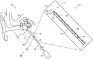
- FIG. 1is a schematic, pictorial illustration of a sinuplasty procedure using a sinuplasty system, in accordance with an embodiment of the present invention
- FIG. 2is a sectional side view of an ear and a suction module, in accordance with an embodiment of the present invention.
- FIG. 3is a flow chart that schematically illustrates a method for suctioning material from a Eustachian tube, in accordance with an embodiment of the present invention.
- Some medical proceduressuch as sinuplasty, require removing material from anatomical cavities, such as a Eustachian tube of a patient inner ear.
- the Eustachian tubeis typically narrow and cannot be accessed through the outer ear without damaging the ear drum and organs of the inner ear.
- a suction deviceinto a cavity (e.g., sinus) located some distance away from the Eustachian tube, but the suction will be ineffective due to lack of sealing between the suction device and the Eustachian tube.
- a suction module for suctioning the undesired materialcomprises a suction tool coupled to a suction apparatus, such as a medical suction pump.
- the suction toolcomprises a hollow flexible internal tube, which is coupled, at its proximal end, to the suction pump, and configured to remove the undesired material away from the Eustachian tube.
- the suction toolfurther comprises a hollow flexible external tube, which is disposed around the internal tube. The external tube is configured to seal the Eustachian tube by coupling its outer surface to an inner surface of the Eustachian tube.
- a position sensor of a position tracking systemis coupled to the distal tip of the suction tube and configured to produce position signals that are indicative of a position of the suction tool in an organ, such as an ear-nose-throat (ENT) system, of the patient.
- ENTear-nose-throat
- the suction moduleis electrically connected to a processor, which is configured to receive pre-acquired anatomical images, such as computerized tomography (CT) images depicting respective segmented two-dimensional (2D) slices of the patient ENT, from a CT system, and to register between coordinate systems of the CT system and the position tracking system.
- CTcomputerized tomography
- the processoris further configured to track the position of the distal tip in the patient body, and to display a marker on a respective anatomical image, indicative of the position of the position sensor in the anatomical image.
- the physicianinserts the suction tool, through the patient nose, and navigates the suction tool to an ostium of the Eustachian tube, using the marker displayed on the anatomical image.
- the physicianseals the Eustachian tube by coupling the outer surface of the external tube to the inner surface of the Eustachian tube.
- the physicianremoves the undesired material from the Eustachian tube by applying suction using the suction pump coupled to the proximal end of the suction tool. After concluding the material removal, the physician extracts the suction tool, through the patient nose, out of the body of the patient.
- FIG. 1is a schematic pictorial illustration of a sinuplasty procedure using a sinuplasty system 20 , in accordance with an embodiment of the present invention.
- sinuplasty system 20comprises an ear-nose-throat (ENT) suction module 28 , which is configured to remove matter, such as infection, liquid or mucus, from a Eustachian tube 50 of a patient 22 .
- ENTear-nose-throat
- suction module 28comprises a distal end, such as an ENT suction tool 38 , which a physician 24 inserts into a nose 26 of patient 22 .
- Module 28further comprises a handheld suction apparatus 30 , coupled to a proximal end of suction tool 38 and configured to assist physician 24 in navigating tool 38 into Eustachian tube 50 and in applying suction, so as to remove the matter away from Eustachian tube 50 .
- ENT suction toolis depicted in further detail in FIG. 2 below.
- system 20further comprises a magnetic position tracking system, which is configured to track the position of one or more position sensors in the head of patient 22 .
- the magnetic position tracking systemcomprises magnetic field-generators 44 and one or more position sensors shown in FIG. 2 below.
- the position sensorsgenerate position signals in response to the sensed external magnetic fields from the field generators, thereby enabling a processor 34 to map the position of each sensor as will be described below.
- System 20further comprises a location pad 40 , which comprises field-generators 44 fixed on a frame 46 .
- pad 40comprises five field-generators 44 but may comprise any other suitable number of generators 44 .
- Pad 40further comprises a pillow (not shown) placed under a head 41 of patient 22 , such that generators 44 are located at fixed, known positions external to patient 22 .
- system 20comprises a console 33 , which comprises a processor 34 , typically a general-purpose computer, with suitable front end and interface circuits for receiving signals from tool 28 having a magnetic sensor attached thereon (shown in FIG. 2 below), via a cable 32 , and for controlling other components of system 20 described herein.
- a processor 34typically a general-purpose computer, with suitable front end and interface circuits for receiving signals from tool 28 having a magnetic sensor attached thereon (shown in FIG. 2 below), via a cable 32 , and for controlling other components of system 20 described herein.
- processor 34is configured to map the position of each position sensor so as to estimate the position and orientation of a distal tip (shown in FIG. 2 below) of tool 28 in the coordinate system of the optical position tracking system.
- processor 34is configured to receive one or more anatomical images, such as computerized tomography (CT) images depicting respective segmented two-dimensional (2D) slices of a head 41 of a patient 22 , obtained using an external CT system (not shown).
- CTcomputerized tomography
- 2Dtwo-dimensional
- Console 33further comprises input devices 39 and a user display 36 , which is configured to display the data (e.g., images) received from processor 34 or inputs inserted by a user (e.g., physician 24 ).
- datae.g., images
- a usere.g., physician 24
- processor 34is configured to display from among the CT images, one or more selected slices, such as an image 35 , on user display 36 .
- image 35is a sectional view of an ear 48 of patient 22 , such that image 35 comprises Eustachian tube 50 .
- Console 33comprises a driver circuit (not shown), which is configured to drive field-generators 44 with suitable signals so as to generate magnetic fields in a predefined working volume around head 41 .
- FIG. 1shows only elements related to the disclosed techniques, for the sake of simplicity and clarity.
- System 20typically comprises additional modules and elements that are not directly related to the disclosed techniques, and thus, intentionally omitted from FIG. 1 and from the corresponding description.
- Processor 34may be programmed in software to carry out the functions that are used by the system, and to store data in a memory (not shown) to be processed or otherwise used by the software.
- the softwaremay be downloaded to the processor in electronic form, over a network, for example, or it may be provided on non-transitory tangible media, such as optical, magnetic or electronic memory media.
- some or all of the functions of processor 34may be carried out by dedicated or programmable digital hardware components.
- FIG. 2is a sectional side view of ear 48 and suction module 28 , in accordance with an embodiment of the present invention.
- physician 24inserts suction tool 38 , typically through nose 26 , into an ostium 66 of Eustachian tube 50 .
- suction tool 38is coupled to suction apparatus 30 , located externally to patient 22 and may be used by physician 24 for navigating suction tool 38 from nose 26 to Eustachian tube 50 of ear 48 . Additionally or alternatively, any other suitable apparatus may be used by physician for the navigation of suction tool 38 .
- suction tool 38comprises an internal hollow tube 54 , which is typically flexible but can also be rigid, and an external hollow tube 58 disposed around internal hollow tube 54 .
- External hollow tube 58is typically flexible but may also be rigid.
- tubes 54 and 58are typically made from polymers, such as polyurethane and polyamide, or from any other suitable biocompatible material.
- suction tube 38is produced such that an outer diameter 80 of the distal end of tube 38 has a similar value to an inner diameter (i.e., between inner surfaces 68 ) of Eustachian tube 50 , e.g., about 1 mm.
- the inner diameter of Eustachian tube 50may be measured, e.g., in image 35 , before performing the sinuplasty procedure, so that physician 24 may select a suction tool having a suitable external diameter that can fit into Eustachian tube 50 .
- suction tool 38is configured to seal Eustachian tube 50 by coupling an outer surface 70 of external tube 58 to inner surface 68 of Eustachian tube 50 .
- openings in a distal tip 74 of hollow tubes 54 and 58form an opening 72 , through which suction tool 38 removes material away (e.g., liquid, mucus, infection, dirt) from cavities in an inner ear 60 , and/or from Eustachian tube 50 .
- material awaye.g., liquid, mucus, infection, dirt
- suction tool 38comprises a strengthening element, such as a wire 56 that extends along at least part (e.g., the distal end) of external tube 58 and is coupled thereto.
- wire 56is made from an alloy of nickel-titanium, such as nitinol, or any other suitable material, and is configured to mechanically strengthen external tube 58 .
- suction tool 38further comprises a position sensor 64 of the position tracking system described in FIG. 1 above.
- position sensor 64comprises a single coil configured to generate position signals, or any other suitable number of coils.
- position sensor 64is coupled to distal tip 74 of suction tool 38 , such that sensor 64 does not block opening 72 .
- sensor 64may be coupled between an outer surface of tube 54 and an inner surface of tube 58 , such that electrical leads (not shown) connected to sensor 64 are disposed between these surfaces and configured to conduct the position signal sensed by position sensor 64 to cable 32 .
- sensor 64may be coupled to surface 70 of tube 58 , or in any other suitable configuration in distal tip 74 .
- sensor 64may be coupled along suction tool 38 at any suitable offset relative to distal tip 74 , such that processor 34 applies the offset to calculate the position of distal tip 74 in the coordinate system of the position tracking system.
- processor 34is configured to register between coordinate systems of the CT system and the position tracking system. In an embodiment, processor 34 is configured to display, based on the registered coordinate systems, an indication of the position of distal tip 74 in image 35 , so as to assist physician 24 in navigating distal tip 74 into ostium 66 .
- suction tool 38is further configured to clean Eustachian tube 50 from any undesired material (e.g., dirt, mucus, infectious fluid), for example, by peeling and suctioning the dirt from surface 68 .
- the cleaningmay be carried out by an operator, such as physician 24 , which moves suction tool 38 , e.g., back and forth between ostium 66 and an ostium 76 , along surface 68 of Eustachian tube 50 .
- physician 24can see the position of distal tip 74 in image 35 , so as to pull suction tool 38 back before reaching ostium 76 .
- suction module 28and particularly, suction tool 38 described in FIGS. 1 and 2 above, are depicted purely by way of example.
- module 28 and tool 38may comprise any suitable configuration, having any suitable size and shape and arranged so that suction tool 38 can be navigated to, and snugly fitted into Eustachian tube 50 to enable suctioning material therefrom and cleaning surface 68 and other parts of Eustachian tube 50 .
- opening 72may be replaced by one or more openings along the distal end of suction tool 38 , this configuration enables removing material from other cavities or anatomical tubes having a different geometry.
- tubes 54 and 58provides physician 24 with the capability to navigate and insert suction tool to various organs, such as in brain surgical applications or in small blood vessels in the body of patient 22 .
- FIG. 3is a flow chart that schematically illustrates a method for suctioning material from Eustachian tube 50 , in accordance with an embodiment of the present invention.
- the methodbegins with an insertion step 100 , in which physician 24 insert suction tool 38 , which comprises internal hollow tube 54 and external hollow tube 58 disposed around tube 54 , into patient nose 26 .
- physician 24navigates suction tool 38 to ostium 66 of Eustachian tube 50 , using the tracked position of sensor 64 displayed in image 35 .
- physician 24cleans inner walls 68 of Eustachian tube 50 by moving suction tool 38 back and forth in along walls 68 , thereby peeling undesired material off walls 68 of Eustachian tube 50 .
- physician 24seals Eustachian tube 50 by coupling outer surface 70 of external tube 58 to inner surface 68 of Eustachian tube 50 .
- physician 24removes undesired material, such as dirt, mucus, and infectious fluid, from Eustachian tube 50 , using suction apparatus 30 , which is coupled to the proximal end of suction tool 38 .
- physician 24extracts suction tool 38 out of the body of patient 22 , through patient nose 26 .
- physician 24may change the order of at least some of the steps of the method. For example, physician 24 may carry out cleaning step 104 after suctioning step 108 , and then repeat sealing step 106 and suctioning step 108 so as to clean the material peeled off walls 68 at cleaning step 104 .
- the embodiments described hereinmainly address removing material from the Eustachian tube
- the methods and systems described hereincan also be used in other applications, such as in suctioning undesired material from any other cavity of the ENT system or any other anatomical system of the body.
Landscapes
- Health & Medical Sciences (AREA)
- Life Sciences & Earth Sciences (AREA)
- Heart & Thoracic Surgery (AREA)
- Engineering & Computer Science (AREA)
- Veterinary Medicine (AREA)
- Biomedical Technology (AREA)
- Animal Behavior & Ethology (AREA)
- General Health & Medical Sciences (AREA)
- Public Health (AREA)
- Anesthesiology (AREA)
- Hematology (AREA)
- Vascular Medicine (AREA)
- Surgery (AREA)
- Biophysics (AREA)
- Pulmonology (AREA)
- Psychology (AREA)
- Physics & Mathematics (AREA)
- Acoustics & Sound (AREA)
- Otolaryngology (AREA)
- Medical Informatics (AREA)
- Molecular Biology (AREA)
- Robotics (AREA)
- Nuclear Medicine, Radiotherapy & Molecular Imaging (AREA)
- Surgical Instruments (AREA)
- Media Introduction/Drainage Providing Device (AREA)
- External Artificial Organs (AREA)
Abstract
Description
Claims (18)
Priority Applications (7)
| Application Number | Priority Date | Filing Date | Title |
|---|---|---|---|
| US15/674,369US11523942B2 (en) | 2017-08-10 | 2017-08-10 | Medical suction tool for a Eustachian tube |
| AU2018214020AAU2018214020A1 (en) | 2017-08-10 | 2018-08-07 | A medical suction tool for a eustachian tube |
| IL261053AIL261053B (en) | 2017-08-10 | 2018-08-08 | Medical suction tool for a eustachian tube |
| JP2018150088AJP2019037759A (en) | 2017-08-10 | 2018-08-09 | Medical suction device for eustachian tube |
| EP18188211.9AEP3441050B1 (en) | 2017-08-10 | 2018-08-09 | A medical suction tool for a eustachian tube |
| CA3013716ACA3013716A1 (en) | 2017-08-10 | 2018-08-09 | A medical suction tool for a eustachian tube |
| CN201810906664.2ACN109381753A (en) | 2017-08-10 | 2018-08-10 | Medical aspirator for Eustachian tube |
Applications Claiming Priority (1)
| Application Number | Priority Date | Filing Date | Title |
|---|---|---|---|
| US15/674,369US11523942B2 (en) | 2017-08-10 | 2017-08-10 | Medical suction tool for a Eustachian tube |
Publications (2)
| Publication Number | Publication Date |
|---|---|
| US20190046358A1 US20190046358A1 (en) | 2019-02-14 |
| US11523942B2true US11523942B2 (en) | 2022-12-13 |
Family
ID=63207598
Family Applications (1)
| Application Number | Title | Priority Date | Filing Date |
|---|---|---|---|
| US15/674,369Active2039-01-16US11523942B2 (en) | 2017-08-10 | 2017-08-10 | Medical suction tool for a Eustachian tube |
Country Status (7)
| Country | Link |
|---|---|
| US (1) | US11523942B2 (en) |
| EP (1) | EP3441050B1 (en) |
| JP (1) | JP2019037759A (en) |
| CN (1) | CN109381753A (en) |
| AU (1) | AU2018214020A1 (en) |
| CA (1) | CA3013716A1 (en) |
| IL (1) | IL261053B (en) |
Families Citing this family (1)
| Publication number | Priority date | Publication date | Assignee | Title |
|---|---|---|---|---|
| US12042233B2 (en) | 2020-05-12 | 2024-07-23 | Acclarent, Inc. | Malleable suction instrument with offset position sensor |
Citations (29)
| Publication number | Priority date | Publication date | Assignee | Title |
|---|---|---|---|---|
| US5391199A (en) | 1993-07-20 | 1995-02-21 | Biosense, Inc. | Apparatus and method for treating cardiac arrhythmias |
| US5462529A (en) | 1993-09-29 | 1995-10-31 | Technology Development Center | Adjustable treatment chamber catheter |
| WO1996005768A1 (en) | 1994-08-19 | 1996-02-29 | Biosense, Inc. | Medical diagnosis, treatment and imaging systems |
| US6239724B1 (en) | 1997-12-30 | 2001-05-29 | Remon Medical Technologies, Ltd. | System and method for telemetrically providing intrabody spatial position |
| US6332089B1 (en) | 1996-02-15 | 2001-12-18 | Biosense, Inc. | Medical procedures and apparatus using intrabody probes |
| US20020065455A1 (en) | 1995-01-24 | 2002-05-30 | Shlomo Ben-Haim | Medical diagnosis, treatment and imaging systems |
| US6484118B1 (en) | 2000-07-20 | 2002-11-19 | Biosense, Inc. | Electromagnetic position single axis system |
| US20030120150A1 (en) | 2001-12-21 | 2003-06-26 | Assaf Govari | Wireless position sensor |
| US6618612B1 (en) | 1996-02-15 | 2003-09-09 | Biosense, Inc. | Independently positionable transducers for location system |
| US20040068178A1 (en) | 2002-09-17 | 2004-04-08 | Assaf Govari | High-gradient recursive locating system |
| US20050240147A1 (en)* | 2004-04-21 | 2005-10-27 | Exploramed Ii, Inc. | Devices, systems and methods for diagnosing and treating sinusitus and other disorders of the ears, nose and/or throat |
| US20070233036A1 (en)* | 2006-02-27 | 2007-10-04 | Aditi H Mandpe | Eustachian Tube Device and Method |
| JP2008539973A (en) | 2005-05-10 | 2008-11-20 | クック・インコーポレイテッド | Catheter reinforcement member |
| US20080287908A1 (en) | 2004-04-21 | 2008-11-20 | Acclarent, Inc. | Ethmoidotomy System and Implantable Spacer Devices Having Therapeutic Substance Delivery Capability for Treatment of Paranasal Sinusitis |
| CN101861184A (en) | 2007-08-29 | 2010-10-13 | 阿克罗斯塔克公司 | Method and kit for delivering brachytherapy to a patient |
| US20110144571A1 (en)* | 2009-12-15 | 2011-06-16 | Ahluwalia Prabhat K | Suction device |
| CN102488955A (en) | 2011-12-07 | 2012-06-13 | 湖南埃普特医疗器械有限公司 | Balloon guide catheter, and preparation method thereof |
| EP2535079A2 (en) | 2005-01-18 | 2012-12-19 | Acclarent, Inc. | Paranasal sinus lavage device |
| JP2013515591A (en) | 2009-12-29 | 2013-05-09 | アクラレント インコーポレイテッド | System for treating target tissue in the ear canal |
| US20130303968A1 (en) | 2009-03-31 | 2013-11-14 | Acclarent, Inc. | System and method for treatment of non-ventilating middle ear by providing a gas pathway through the nasopharynx |
| US20140200444A1 (en)* | 2004-04-21 | 2014-07-17 | Acclarent, Inc. | Guidewires for performing image guided procedures |
| US20140276654A1 (en)* | 2013-03-15 | 2014-09-18 | Acclarent, Inc. | Nasal Suction Device |
| US20140296898A1 (en)* | 2004-04-21 | 2014-10-02 | Acclarent, Inc. | Devices, systems and methods useable for treating sinusitis |
| US20150202089A1 (en) | 2007-12-20 | 2015-07-23 | Acclarent, Inc. | Eustachian tube dilation balloon with ventilation path |
| CN105263556A (en) | 2013-06-03 | 2016-01-20 | 柯惠有限合伙公司 | Method for making non-prolapsed catheters from unlined tubing |
| US20160310042A1 (en)* | 2015-04-22 | 2016-10-27 | Acclarent, Inc. | System and method to map structures of nasal cavity |
| US20170119993A1 (en)* | 2015-10-30 | 2017-05-04 | Acclarent, Inc. | System and method for anesthetizing eustachian tube |
| US20170119473A1 (en)* | 2015-10-30 | 2017-05-04 | Acclarent, Inc. | System and method for navigation of surgical instruments |
| US20170143938A1 (en)* | 2015-11-23 | 2017-05-25 | Mivi Neuroscience, Inc. | Catheter systems for applying effective suction in remote vessels and thrombectomy procedures facilitated by catheter systems |
Family Cites Families (3)
| Publication number | Priority date | Publication date | Assignee | Title |
|---|---|---|---|---|
| US9782297B2 (en)* | 2006-02-27 | 2017-10-10 | Aditi H. Mandpe | Eustachian tube stents, introducers and methods for their use |
| US10335319B2 (en)* | 2015-03-30 | 2019-07-02 | Acclarent, Inc. | Method and apparatus for cleaning isthmus of eustachian tube |
| CN206007466U (en)* | 2016-05-25 | 2017-03-15 | 中山大学孙逸仙纪念医院 | A kind of contained eustachian tube |
- 2017
- 2017-08-10USUS15/674,369patent/US11523942B2/enactiveActive
- 2018
- 2018-08-07AUAU2018214020Apatent/AU2018214020A1/ennot_activeAbandoned
- 2018-08-08ILIL261053Apatent/IL261053B/enactiveIP Right Grant
- 2018-08-09CACA3013716Apatent/CA3013716A1/enactivePending
- 2018-08-09JPJP2018150088Apatent/JP2019037759A/enactivePending
- 2018-08-09EPEP18188211.9Apatent/EP3441050B1/enactiveActive
- 2018-08-10CNCN201810906664.2Apatent/CN109381753A/enactivePending
Patent Citations (31)
| Publication number | Priority date | Publication date | Assignee | Title |
|---|---|---|---|---|
| US5391199A (en) | 1993-07-20 | 1995-02-21 | Biosense, Inc. | Apparatus and method for treating cardiac arrhythmias |
| US5462529A (en) | 1993-09-29 | 1995-10-31 | Technology Development Center | Adjustable treatment chamber catheter |
| WO1996005768A1 (en) | 1994-08-19 | 1996-02-29 | Biosense, Inc. | Medical diagnosis, treatment and imaging systems |
| US20020065455A1 (en) | 1995-01-24 | 2002-05-30 | Shlomo Ben-Haim | Medical diagnosis, treatment and imaging systems |
| US6690963B2 (en) | 1995-01-24 | 2004-02-10 | Biosense, Inc. | System for determining the location and orientation of an invasive medical instrument |
| US6618612B1 (en) | 1996-02-15 | 2003-09-09 | Biosense, Inc. | Independently positionable transducers for location system |
| US6332089B1 (en) | 1996-02-15 | 2001-12-18 | Biosense, Inc. | Medical procedures and apparatus using intrabody probes |
| US6239724B1 (en) | 1997-12-30 | 2001-05-29 | Remon Medical Technologies, Ltd. | System and method for telemetrically providing intrabody spatial position |
| US6484118B1 (en) | 2000-07-20 | 2002-11-19 | Biosense, Inc. | Electromagnetic position single axis system |
| US20030120150A1 (en) | 2001-12-21 | 2003-06-26 | Assaf Govari | Wireless position sensor |
| JP2003299634A (en) | 2001-12-21 | 2003-10-21 | Biosense Inc | Wireless position sensor |
| US20040068178A1 (en) | 2002-09-17 | 2004-04-08 | Assaf Govari | High-gradient recursive locating system |
| US20050240147A1 (en)* | 2004-04-21 | 2005-10-27 | Exploramed Ii, Inc. | Devices, systems and methods for diagnosing and treating sinusitus and other disorders of the ears, nose and/or throat |
| US20140296898A1 (en)* | 2004-04-21 | 2014-10-02 | Acclarent, Inc. | Devices, systems and methods useable for treating sinusitis |
| US20080287908A1 (en) | 2004-04-21 | 2008-11-20 | Acclarent, Inc. | Ethmoidotomy System and Implantable Spacer Devices Having Therapeutic Substance Delivery Capability for Treatment of Paranasal Sinusitis |
| US20140200444A1 (en)* | 2004-04-21 | 2014-07-17 | Acclarent, Inc. | Guidewires for performing image guided procedures |
| EP2535079A2 (en) | 2005-01-18 | 2012-12-19 | Acclarent, Inc. | Paranasal sinus lavage device |
| JP2008539973A (en) | 2005-05-10 | 2008-11-20 | クック・インコーポレイテッド | Catheter reinforcement member |
| US20070233036A1 (en)* | 2006-02-27 | 2007-10-04 | Aditi H Mandpe | Eustachian Tube Device and Method |
| CN101861184A (en) | 2007-08-29 | 2010-10-13 | 阿克罗斯塔克公司 | Method and kit for delivering brachytherapy to a patient |
| US20150202089A1 (en) | 2007-12-20 | 2015-07-23 | Acclarent, Inc. | Eustachian tube dilation balloon with ventilation path |
| US20130303968A1 (en) | 2009-03-31 | 2013-11-14 | Acclarent, Inc. | System and method for treatment of non-ventilating middle ear by providing a gas pathway through the nasopharynx |
| US20110144571A1 (en)* | 2009-12-15 | 2011-06-16 | Ahluwalia Prabhat K | Suction device |
| JP2013515591A (en) | 2009-12-29 | 2013-05-09 | アクラレント インコーポレイテッド | System for treating target tissue in the ear canal |
| CN102488955A (en) | 2011-12-07 | 2012-06-13 | 湖南埃普特医疗器械有限公司 | Balloon guide catheter, and preparation method thereof |
| US20140276654A1 (en)* | 2013-03-15 | 2014-09-18 | Acclarent, Inc. | Nasal Suction Device |
| CN105263556A (en) | 2013-06-03 | 2016-01-20 | 柯惠有限合伙公司 | Method for making non-prolapsed catheters from unlined tubing |
| US20160310042A1 (en)* | 2015-04-22 | 2016-10-27 | Acclarent, Inc. | System and method to map structures of nasal cavity |
| US20170119993A1 (en)* | 2015-10-30 | 2017-05-04 | Acclarent, Inc. | System and method for anesthetizing eustachian tube |
| US20170119473A1 (en)* | 2015-10-30 | 2017-05-04 | Acclarent, Inc. | System and method for navigation of surgical instruments |
| US20170143938A1 (en)* | 2015-11-23 | 2017-05-25 | Mivi Neuroscience, Inc. | Catheter systems for applying effective suction in remote vessels and thrombectomy procedures facilitated by catheter systems |
Non-Patent Citations (3)
| Title |
|---|
| Chinese Office Action and Search Report dated May 30, 2022, for Application No. 201810906664.2, 13 pages. |
| European Search Report dated Oct. 12, 2018 from corresponding European Patent Application No. 18188211.9. |
| Japanese Notification of Reasons for Refusal dated Aug. 23, 2022, for Application No. 2018-150088, 4 pages. |
Also Published As
| Publication number | Publication date |
|---|---|
| CN109381753A (en) | 2019-02-26 |
| JP2019037759A (en) | 2019-03-14 |
| EP3441050B1 (en) | 2021-03-24 |
| IL261053A (en) | 2019-01-31 |
| CA3013716A1 (en) | 2019-02-10 |
| US20190046358A1 (en) | 2019-02-14 |
| IL261053B (en) | 2021-06-30 |
| AU2018214020A1 (en) | 2019-02-28 |
| EP3441050A1 (en) | 2019-02-13 |
Similar Documents
| Publication | Publication Date | Title |
|---|---|---|
| EP3860423B1 (en) | Computerized tomography (ct) image correction using position and direction (p&d) tracking assisted optical visualization | |
| EP3395282B1 (en) | Endoscopic view of invasive procedures in narrow passages | |
| JP7350470B2 (en) | Display of endoscope position and optical axis in anatomical images | |
| JP7282858B2 (en) | Registration of heads with individual grippers | |
| US11707179B2 (en) | Automatic probe reinsertion | |
| EP3392835B1 (en) | Improving registration of an anatomical image with a position-tracking coordinate system based on visual proximity to bone tissue | |
| EP3184035B1 (en) | Ascertaining a position and orientation for visualizing a tool | |
| CN106901719A (en) | For making the registration between the visual coordinate system of instrument | |
| EP3705036B1 (en) | Showing catheter in brain | |
| EP3441050B1 (en) | A medical suction tool for a eustachian tube | |
| BR102018073334A2 (en) | CALIBRATION OF A RIGID TOOL | |
| US20210196230A1 (en) | Position registered sideview ultrasound (us) imager inserted into brain via trocar |
Legal Events
| Date | Code | Title | Description |
|---|---|---|---|
| STPP | Information on status: patent application and granting procedure in general | Free format text:DOCKETED NEW CASE - READY FOR EXAMINATION | |
| AS | Assignment | Owner name:BIOSENSE WEBSTER (ISRAEL) LTD., ISRAEL Free format text:ASSIGNMENT OF ASSIGNORS INTEREST;ASSIGNOR:GLINER, VADIM;REEL/FRAME:046516/0155 Effective date:20180729 | |
| STPP | Information on status: patent application and granting procedure in general | Free format text:NON FINAL ACTION MAILED | |
| STPP | Information on status: patent application and granting procedure in general | Free format text:RESPONSE TO NON-FINAL OFFICE ACTION ENTERED AND FORWARDED TO EXAMINER | |
| STPP | Information on status: patent application and granting procedure in general | Free format text:FINAL REJECTION MAILED | |
| STPP | Information on status: patent application and granting procedure in general | Free format text:DOCKETED NEW CASE - READY FOR EXAMINATION | |
| STPP | Information on status: patent application and granting procedure in general | Free format text:NON FINAL ACTION MAILED | |
| STPP | Information on status: patent application and granting procedure in general | Free format text:RESPONSE TO NON-FINAL OFFICE ACTION ENTERED AND FORWARDED TO EXAMINER | |
| STPP | Information on status: patent application and granting procedure in general | Free format text:FINAL REJECTION MAILED | |
| STPP | Information on status: patent application and granting procedure in general | Free format text:RESPONSE AFTER FINAL ACTION FORWARDED TO EXAMINER | |
| STPP | Information on status: patent application and granting procedure in general | Free format text:NOTICE OF ALLOWANCE MAILED -- APPLICATION RECEIVED IN OFFICE OF PUBLICATIONS | |
| STPP | Information on status: patent application and granting procedure in general | Free format text:AWAITING TC RESP., ISSUE FEE NOT PAID | |
| STPP | Information on status: patent application and granting procedure in general | Free format text:AWAITING TC RESP, ISSUE FEE PAYMENT VERIFIED | |
| STPP | Information on status: patent application and granting procedure in general | Free format text:AWAITING TC RESP, ISSUE FEE PAYMENT VERIFIED | |
| STPP | Information on status: patent application and granting procedure in general | Free format text:PUBLICATIONS -- ISSUE FEE PAYMENT VERIFIED | |
| STCF | Information on status: patent grant | Free format text:PATENTED CASE |