US11455754B2 - System and method for synthesizing low-dimensional image data from high-dimensional image data using an object grid enhancement - Google Patents
System and method for synthesizing low-dimensional image data from high-dimensional image data using an object grid enhancementDownload PDFInfo
- Publication number
- US11455754B2 US11455754B2US16/497,766US201816497766AUS11455754B2US 11455754 B2US11455754 B2US 11455754B2US 201816497766 AUS201816497766 AUS 201816497766AUS 11455754 B2US11455754 B2US 11455754B2
- Authority
- US
- United States
- Prior art keywords
- breast tissue
- dimensional
- objects
- image
- tissue objects
- Prior art date
- Legal status (The legal status is an assumption and is not a legal conclusion. Google has not performed a legal analysis and makes no representation as to the accuracy of the status listed.)
- Active
Links
Images


Classifications
- A—HUMAN NECESSITIES
- A61—MEDICAL OR VETERINARY SCIENCE; HYGIENE
- A61B—DIAGNOSIS; SURGERY; IDENTIFICATION
- A61B6/00—Apparatus or devices for radiation diagnosis; Apparatus or devices for radiation diagnosis combined with radiation therapy equipment
- A61B6/02—Arrangements for diagnosis sequentially in different planes; Stereoscopic radiation diagnosis
- A61B6/025—Tomosynthesis
- G—PHYSICS
- G06—COMPUTING OR CALCULATING; COUNTING
- G06T—IMAGE DATA PROCESSING OR GENERATION, IN GENERAL
- G06T11/00—2D [Two Dimensional] image generation
- G06T11/003—Reconstruction from projections, e.g. tomography
- G06T11/005—Specific pre-processing for tomographic reconstruction, e.g. calibration, source positioning, rebinning, scatter correction, retrospective gating
- A—HUMAN NECESSITIES
- A61—MEDICAL OR VETERINARY SCIENCE; HYGIENE
- A61B—DIAGNOSIS; SURGERY; IDENTIFICATION
- A61B6/00—Apparatus or devices for radiation diagnosis; Apparatus or devices for radiation diagnosis combined with radiation therapy equipment
- A61B6/50—Apparatus or devices for radiation diagnosis; Apparatus or devices for radiation diagnosis combined with radiation therapy equipment specially adapted for specific body parts; specially adapted for specific clinical applications
- A61B6/502—Apparatus or devices for radiation diagnosis; Apparatus or devices for radiation diagnosis combined with radiation therapy equipment specially adapted for specific body parts; specially adapted for specific clinical applications for diagnosis of breast, i.e. mammography
- A—HUMAN NECESSITIES
- A61—MEDICAL OR VETERINARY SCIENCE; HYGIENE
- A61B—DIAGNOSIS; SURGERY; IDENTIFICATION
- A61B6/00—Apparatus or devices for radiation diagnosis; Apparatus or devices for radiation diagnosis combined with radiation therapy equipment
- A61B6/52—Devices using data or image processing specially adapted for radiation diagnosis
- A61B6/5211—Devices using data or image processing specially adapted for radiation diagnosis involving processing of medical diagnostic data
- G—PHYSICS
- G06—COMPUTING OR CALCULATING; COUNTING
- G06N—COMPUTING ARRANGEMENTS BASED ON SPECIFIC COMPUTATIONAL MODELS
- G06N20/00—Machine learning
- G—PHYSICS
- G06—COMPUTING OR CALCULATING; COUNTING
- G06T—IMAGE DATA PROCESSING OR GENERATION, IN GENERAL
- G06T11/00—2D [Two Dimensional] image generation
- G06T11/003—Reconstruction from projections, e.g. tomography
- G06T11/008—Specific post-processing after tomographic reconstruction, e.g. voxelisation, metal artifact correction
- G—PHYSICS
- G06—COMPUTING OR CALCULATING; COUNTING
- G06T—IMAGE DATA PROCESSING OR GENERATION, IN GENERAL
- G06T15/00—3D [Three Dimensional] image rendering
- G06T15/10—Geometric effects
- G06T15/20—Perspective computation
- G06T15/205—Image-based rendering
- G—PHYSICS
- G06—COMPUTING OR CALCULATING; COUNTING
- G06T—IMAGE DATA PROCESSING OR GENERATION, IN GENERAL
- G06T7/00—Image analysis
- G06T7/0002—Inspection of images, e.g. flaw detection
- G06T7/0012—Biomedical image inspection
- G—PHYSICS
- G16—INFORMATION AND COMMUNICATION TECHNOLOGY [ICT] SPECIALLY ADAPTED FOR SPECIFIC APPLICATION FIELDS
- G16H—HEALTHCARE INFORMATICS, i.e. INFORMATION AND COMMUNICATION TECHNOLOGY [ICT] SPECIALLY ADAPTED FOR THE HANDLING OR PROCESSING OF MEDICAL OR HEALTHCARE DATA
- G16H50/00—ICT specially adapted for medical diagnosis, medical simulation or medical data mining; ICT specially adapted for detecting, monitoring or modelling epidemics or pandemics
- G16H50/20—ICT specially adapted for medical diagnosis, medical simulation or medical data mining; ICT specially adapted for detecting, monitoring or modelling epidemics or pandemics for computer-aided diagnosis, e.g. based on medical expert systems
- G—PHYSICS
- G06—COMPUTING OR CALCULATING; COUNTING
- G06T—IMAGE DATA PROCESSING OR GENERATION, IN GENERAL
- G06T2207/00—Indexing scheme for image analysis or image enhancement
- G06T2207/10—Image acquisition modality
- G06T2207/10072—Tomographic images
- G—PHYSICS
- G06—COMPUTING OR CALCULATING; COUNTING
- G06T—IMAGE DATA PROCESSING OR GENERATION, IN GENERAL
- G06T2207/00—Indexing scheme for image analysis or image enhancement
- G06T2207/10—Image acquisition modality
- G06T2207/10116—X-ray image
- G—PHYSICS
- G06—COMPUTING OR CALCULATING; COUNTING
- G06T—IMAGE DATA PROCESSING OR GENERATION, IN GENERAL
- G06T2207/00—Indexing scheme for image analysis or image enhancement
- G06T2207/30—Subject of image; Context of image processing
- G06T2207/30004—Biomedical image processing
- G06T2207/30068—Mammography; Breast
Definitions
- the presently disclosed inventionsrelate to systems and methods for processing and displaying breast tissue images, and in particular to representing high-dimensional (e.g., 3D) structures present in breast tissue image data with a high-dimensional object grid, and then reducing the high-dimensional data to a low-dimensional (e.g., 2D) format version that can be incorporated within a synthesized image to be displayed to a medical professional.
- high-dimensionale.g., 3D
- 2Dlow-dimensional
- Mammographyhas long been used to screen for breast cancer and other abnormalities.
- mammogramshave been formed on x-ray film.
- flat panel digital imagershave been introduced that acquire a mammogram in digital form, and thereby facilitate analysis and storage of the acquired image data, and to also provide other benefits.
- substantial attention and technological developmenthave been dedicated to obtaining three-dimensional images of the breast using methods such as breast tomosynthesis.
- breast tomosynthesis systemsconstruct a 3D image volume from a series of 2D projection images, each projection image obtained at a different angular displacement of an x-ray source relative to the image detector as the x-ray source is scanned over the detector.
- the constructed 3D image volumeis typically presented as a plurality of slices of image data, the slices being mathematically reconstructed on planes typically parallel to the imaging detector.
- the reconstructed tomosynthesis slicesreduce or eliminate the problems caused by tissue overlap and structure noise present in single slice, two-dimensional mammography imaging, by permitting a user (e.g., a radiologist or other medical professional) to scroll through the image slices to view only the structures in that slice.
- Hologic, Inc.has developed a fused, multimode mammography/tomosynthesis system that acquires one or both types of mammogram and tomosynthesis images, either while the breast remains immobilized or in different compressions of the breast.
- Other companieshave introduced systems that include tomosynthesis imaging; e.g., which do not include the ability to also acquire a mammogram in the same compression.
- U.S. Pat. No. 7,760,924describes a method of generating a synthesized 2D image, which may optionally be displayed along with tomosynthesis projection or reconstructed images, in order to assist in screening and diagnosis.
- a 2D synthesized imageis designed to provide a concise representation of the 3D reconstruction slices, including any clinically important and meaningful information, such as abnormal lesions and normal breast structures, while representing in relevant part a traditional 2D image.
- lesions and breast structureswhich may be defined as different types of image objects having different characteristics.
- image characteristicse.g., micro-calcifications, architectural distortions, etc.
- the synthesized imagemay appear crowded and visually confusing. Accordingly, there exists a need for more effectively processing, synthesizing and displaying breast image data.
- a method for processing breast tissue image dataincludes obtaining image data of a patient's breast tissue; processing the image data to generate a high-dimensional grid depicting one or more high-dimensional objects in the patient's breast tissue; determining a probability or confidence of each of the one or more high-dimensional objects depicted in the high-dimensional grid; and generating a lower-dimensional format version of the one or more high-dimensional objects for display in a synthesized image of the patient's breast tissue.
- FIG. 1is a block diagram illustrating the flow of data through an exemplary breast image acquisition and processing system in accordance with embodiments of the disclosed inventions;
- FIG. 2is a block diagram illustrating the flow of data through a 2D synthesizer employing a 3D object grid and various modules that reduce objects of the grid to a 2D format for display;
- FIG. 3illustrates a first synthesized image formed from the 3D object grid of FIG. 2 without manipulating overlapping objects, and a second synthesized image formed from the same 3D object grid, but with manipulation of overlapping objects;
- FIG. 4A-4Dillustrate exemplary techniques for combining objects onto one or more 2D synthesized images
- FIG. 5illustrates an exemplary flow diagram depicting combining objects from a 3D object grid onto a 2D synthesized image
- FIG. 6illustrates an exemplary flow diagram depicting generating one or more 2D synthesized images using a 3D object grid.
- an “acquired image”refers to an image generated while visualizing a patient's tissue. Acquired images can be generated by radiation from a radiation source impacting on a radiation detector disposed on opposite sides of a patient's tissue, as in a conventional mammogram.
- a “reconstructed image”refers to an image generated from data derived from a plurality of acquired images.
- a reconstructed imagesimulates an acquired image not included in the plurality of acquired images.
- a “synthesized image”refers to an artificial image generated from data derived from a plurality of acquired and/or reconstructed images.
- a synthesized imageincludes elements (e.g., objects and regions) from the acquired and/or reconstructed images, but does not necessarily correspond to an image that can be acquired during visualization. Synthesized images are constructed analysis tools.
- An “Mp” imageis a conventional mammogram or contrast enhanced mammogram, which are two-dimensional (2D) projection images of a breast, and encompasses both a digital image as acquired by a flat panel detector or another imaging device, and the image after conventional processing to prepare it for display (e.g., to a health professional), storage (e.g., in the PACS system of a hospital), and/or other use.
- 2Dtwo-dimensional
- a “Tp” imageis an image that is similarly two-dimensional (2D), but is acquired at a respective tomosynthesis angle between the breast and the origin of the imaging x-rays (typically the focal spot of an x-ray tube), and encompasses the image as acquired, as well as the image data after being processed for display, storage, and/or other use.
- Tr imageis a type (or subset) of a reconstructed image that is reconstructed from tomosynthesis projection images Tp, for example, in the manner described in one or more of U.S. Pat. Nos. 7,577,282, 7,606,801, 7,760,924, and 8,571,289, the disclosures of which are fully incorporated by reference herein in their entirety, wherein a Tr image represents a slice of the breast as it would appear in a projection x-ray image of that slice at any desired angle, not only at an angle used for acquiring Tp or Mp images.
- An “Ms” imageis a type (or subset) of a synthesized image, in particular, a synthesized 2D projection image, which simulates mammography images, such as craniocaudal (CC) or mediolateral oblique (MLO) images, and is constructed using tomosynthesis projection images Tp, tomosynthesis reconstructed images Tr, or a combination thereof.
- Ms imagesmay be provided for display to a health professional or for storage in the PACS system of a hospital or another institution. Examples of methods that may be used to generate Ms images are described in the above-incorporated U.S. Pat. Nos. 7,760,924 and 8,571,289.
- Tp, Tr, Ms and Mp image dataencompasses information, in whatever form, that is sufficient to describe the respective image for display, further processing, or storage.
- the respective Mp, Ms. Tp and Tr imagesare typically provided in digital form prior to being displayed, with each image being defined by information that identifies the properties of each pixel in a two-dimensional array of pixels.
- the pixel valuestypically relate to respective measured, estimated, or computed responses to X-rays of corresponding volumes in the breast, i.e., voxels or columns of tissue.
- the geometry of the tomosynthesis images (Tr and Tp) and mammography images (Ms and Mp)are matched to a common coordinate system, as described in U.S. Pat. No. 7,702,142. Unless otherwise specified, such coordinate system matching is assumed to be implemented with respect to the embodiments described in the ensuing detailed description of this patent specification.
- generating an imageand “transmitting an image” respectively refer to generating and transmitting information that is sufficient to describe the image for display.
- the generated and transmitted informationis typically digital information.
- a synthesized 2D image displayed to an end-usere.g., an Ms image
- an end-usere.g., an Ms image
- this informationmay be used to create a high-dimensional grid, e.g., a 3D grid, that helps create a more accurate and enhanced rendering of the most important features in the synthesized 2D image.
- the high-dimensional object gridmay then be used to collapse the most clinically-significant information pertaining to the identified objects to a 2D format onto one or more synthesized 2D images.
- Various data reduction techniquesmay be applied to the identified 3D objects to ensure that the most clinically-significant objects are emphasized, and less significant objects are omitted and/or de-emphasized. Additionally, or alternatively, data reduction techniques are applied to ensure that significant features of a 3D object are enhanced, while less significant features of the 3D object are de-emphasized, especially when two objects are competing for display and prominence on the one or more 2D synthesized images.
- a 3D object gridis utilized, i.e., as a component of an algorithm, for reducing high-dimensional data (e.g., 3D tomosynthesis image data) to low-dimensional data (e.g. a 2D synthesized image).
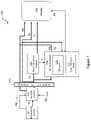
- FIG. 1illustrates the flow of data in an exemplary image generation and display system 100 , which incorporates each of synthesized image generation, object identification, and display technology. It should be understood that, while FIG. 1 illustrates a particular embodiment of a flow diagram with certain processes taking place in a particular serial order or in parallel, the claims and various other embodiments described herein are not limited to the performance of the image processing steps in any particular order, unless so specified.
- the image generation and display system 100includes an image acquisition system 101 that acquires tomosynthesis image data for generating Tp images of a patient's breasts, using the respective three-dimensional and/or tomosynthesis acquisition methods of any of the currently available systems. If the acquisition system is a combined tomosynthesis/mammography system, Mp images may also be generated. Some dedicated tomosynthesis systems or combined tomosynthesis/mammography systems may be adapted to accept and store legacy mammogram images, (indicated by a dashed line and legend “Mp legacy ” in FIG. 1 ) in a storage device 102 , which is preferably a DICOM-compliant Picture Archiving and Communication System (PACS) storage device.
- PPSPicture Archiving and Communication System
- the tomosynthesis projection images Tpmay also be transmitted to the storage device 102 (as shown in FIG. 1 ).
- the storage device 102may further store a library of known 3D objects that may be used to identify significant 3D image patterns to the end-user.
- a separate dedicated storage device(not shown) may be used to store the library of known 3D objects with which to identify 3D image patterns or objects.
- the Tp imagesare transmitted from either the acquisition system 101 , or from the storage device 102 , or both, to a computer system configured as a reconstruction engine 103 that reconstructs the Tp images into reconstructed image “slices” Tr, representing breast slices of selected thickness and at selected orientations, as disclosed in the above-incorporated patents and applications.
- Mode filters 107are disposed between image acquisition and image display.
- the filters 107may additionally include customized filters for each type of image (i.e., Tp, Mp, and Tr images) arranged to identify and highlight certain aspects of the respective image types.
- each imaging modecan be tuned or configured in an optimal way for a specific purpose.
- filters programmed for recognizing objects across various 2D image slicesmay be applied in order to detect image patterns that may belong to a particular high-dimensional object.
- the tuning or configurationmay be automatic, based on the type of the image, or may be defined by manual input, for example through a user interface coupled to a display. In the illustrated embodiment of FIG.
- the mode filters 107are selected to highlight particular characteristics of the images that are best displayed in respective imaging modes, for example, geared towards identifying objects, highlighting masses or calcifications, identifying certain image patterns that may be constructed into a 3D object, or for creating 2D synthesized images (described below).
- FIG. 1illustrates only one mode filter 107 , it should be appreciated that any number of mode filters may be utilized in order to identify structures of interest in the breast tissue.
- the imaging and display system 100further includes a 2D image synthesizer 104 that operates substantially in parallel with the reconstruction engine 103 for generating 2D synthesized images using a combination of one or more Tp, Mp, and/or Tr images.
- the 2D image synthesizer 104consumes a set of input images (e.g., Mp, Tr and/or Tp images), determines a set of most relevant features from each of the input images, and outputs one or more synthesized 2D images.
- the synthesized 2D imagerepresents a consolidated synthesized image that condenses significant portions of various slices onto one image. This provides an end-user (e.g., medical personnel, radiologist, etc.) with the most clinically-relevant image data in an efficient manner, and reduces time spent on other images that may not have significant data.
- One type of relevant image data to highlight in the synthesized 2D imageswould be relevant objects found across one or more Mp, Tr and/or Tp images. Rather than simply assessing image patterns of interest in each of the 2D image slices, it may be helpful to determine whether any of the 2D image patterns of interest belong to a larger high-dimensional structure, and if so, to combine the identified 2D image patterns into a higher-dimensional structure.
- This approachhas several advantages, but in particular, by identifying high-dimensional structures across various slices/depths of the breast tissue, the end-user may be better informed as to the presence of a potentially significant structure that may not be easily visible in various 2D slices of the breast.
- identifying both image patterns as belonging to the same high-dimensional structuremay allow the system to make a more accurate assessment pertaining to the nature of the structure, and consequently provide significantly more valuable information to the end-user. Also, by identifying the high-dimensional structure, the structure can be more accurately depicted on the synthesized 2D image. Yet another advantage of identifying high-dimensional structures within the various captured 2D slices of the breast tissue relates to identifying a possible size/scope of the identified higher-dimensional structure.
- the 2D image synthesizer 104generates high-dimensional object grids 120 (e.g., 3D object grids) comprising one or more high-dimensional structures (e.g., 3D objects) present in the patient's breast tissue.
- high-dimensional object grids 120e.g., 3D object grids
- 3D object grids 120identify various objects in the breast tissue. It should be appreciated that this disclosure is not limited to 3D objects and/or structures, and may refer to even higher-dimensional structures, but for simplicity, the remaining disclosure will refer to 3D objects populated in a 3D object grid 120 .
- the 3D object grid 120is in the form of a 3D (volumetric) coordinate space representing a patient's breast mass, and identifies a location, identity, size, scope, and/or other characteristics of any objects or structures found in the breast mass. Examples of such objects or structures include calcifications, spiculated lesions, benign tumors, irregular masses, dense objects, etc.
- the end-usere.g., a medical professional such as a radiologist
- the 3D object grid 120is solely used by the system processor for constructing synthesized 2D images, and the end-user may not be aware of, or have access to, the 3D object grid 120 .
- the 2D image synthesizer 104also includes a data reduction module 122 configured to reduce the high-dimensional data populated in the 3D object grid 120 to a lower-dimensional format suitable for representation in a 2D synthesized image.
- the data reduction module 122evaluates the various objects of the 3D object grid 120 , and determines what objects (or what portions of objects) should be enhanced or emphasized in a final 2D synthesized image to be displayed to the end-user. For example, a clinically significant object and a routine background breast tissue object may have regions of overlap, in which case the data reduction module 122 is preferably configured to de-emphasize portions of the background breast tissue in order to highlight the clinically significant object. Further details on various data reduction techniques that may be employed by the data reduction module 122 are described below.
- the synthesized 2D imagesmay be viewed at a display system 105 .
- the reconstruction engine 103 and 2D image synthesizer 104are preferably connected to a display system 105 via a fast transmission link.
- the display system 105may be part of a standard acquisition workstation (e.g., of acquisition system 101 ), or of a standard (multi-display) review station (not shown) that is physically remote from the acquisition system 101 .
- a display connected via a communication networkmay be used, for example, a display of a personal computer or of a so-called tablet, smart phone or other hand-held device.
- the display 105 of the systemis preferably able to display respective Ms, Mp, Tr, and/or Tp images concurrently, e.g., in separate side-by-side monitors of a review workstation, although the invention may still be implemented with a single display monitor, by toggling between images.
- the imaging and display system 100which is described as for purposes of illustration and not limitation, is capable of receiving and selectively displaying tomosynthesis projection images Tp, tomosynthesis reconstruction images Tr, synthesized mammogram images Ms, and/or mammogram (including contrast mammogram) images Mp, or any one or sub combination of these image types.
- the system 100employs software to convert (i.e., reconstruct) tomosynthesis images Tp into images Tr, software for synthesizing mammogram images Ms, software for decomposing 3D objects, software for creating feature maps and object maps.
- An object of interest or feature in a source imagemay be considered a ‘most relevant’ feature for inclusion in a 2D synthesized image based upon the application of the object maps along with one or more algorithms and/or heuristics, wherein the algorithms assign numerical values, weights or thresholds, to pixels or regions of the respective source images based upon identified/detected objects and features of interest within the respective region or between features.
- the objects and features of interestmay include, for example, spiculated lesions, calcifications, and the like.
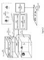
- FIG. 2illustrates the 2D image synthesizer 104 in further detail.
- various image slices 218 of a tomosynthesis data set (or “stack”) 202are input into the 2D image synthesizer 104 , and then processed to determine portions of the images to highlight in a synthesized 2D image that will be displayed on the display 105 .
- the image slices 218may be consecutively-captured cross-sections of a patient's breast tissue. Or, the image slices 218 may be cross-sectional images of the patient's breast tissue captured at known intervals.
- the tomosynthesis image stack 202 comprising the image slices 218may be forwarded to the 2D image synthesizer 104 , which evaluates each of the source images in order to (1) identify various types of objects (Tr) for possible inclusion in one or more 2D synthesized images, and/or (2) identify respective pixel regions in the images that contain the identified objects.
- Trvarious types of objects
- the tomosynthesis stack 202comprises a plurality of images 218 taken at various depths/cross-sections of the patient's breast tissue. Some of the images 218 in the tomosynthesis stack 202 comprise 2D image patterns. Thus, the tomosynthesis stack 202 comprises a large number of input images containing various image patterns within the images of the stack.
- the tomosynthesis stack 202may comprise one hundred images 218 captured at various depths/cross sections of the patient's breast tissue, only a few of the images 218 may include any information of significance. Also, it should be noted that the tomosynthesis stack 202 contains 2D image patterns when viewed at differing z-dimension (depth) locations of the otherwise same x, y locations in the image slices 218 , but it may be difficult to determine the 3D structures based only on the various individual images, each representing a finite cross-sectional image of the breast tissue. However, the tomosynthesis stack 202 may be effectively utilized to create the 3D object grid 120 . In any event, for purposes of this patent specification, it is assumed that the 3D object grid 120 is constructed by any means, including but not limited to being created from the tomosynthesis stack 202 .
- the 3D object grid 120may be considered a 3D volumetric coordinate space representing a patient's breast mass. Rather than depicting 2D image patterns at various image slices, the 3D object grid 120 preferably depicts any identified 3D objects in the entire mass (or portion thereof) that represents the patient's breast tissue.
- the 3D object grid 120provides fuller detail regarding various objects in the breast mass as compared to the tomosynthesis stack 202 .
- the 3D object grid 120may use simulation techniques to infer a shape of the 3D object, even though an image slice may not have necessarily been reconstructed at every cross-sectional depth covering the respective 3D object.
- the 3D object grid 120may comprise one or more objects, as shown in the illustrated embodiment. It should be appreciated that these objects may be predefined objects that the system has been trained to identify. However, even in healthy breast tissue that does not necessarily comprise any abnormal objects or structures, the target object recognition/enhancement modules may identify a breast background object. For example, all breast linear tissue and density tissue structures can be displayed as the breast background object. In other embodiments, “healthy” objects such as spherical shapes, oval shapes, etc., may simply be identified through one or more target object recognition/enhancement modules 210 .
- These identified 3D objectsmay then be displayed on the 2D synthesized image 206 ; of course, out of all identified 2D objects, more clinically-significant objects may be prioritized and/or enhanced when displaying the respective object on the 2D synthesized image, as will be discussed in further detail below.
- the 2D synthesizer 104utilizes both the tomosynthesis image stack 202 along with the created 3D object grid 120 in order to merge the relevant features into one or more 2D synthesized images 206 .
- the 3D objects identified in the 3D object grid 120are collapsed into a 2D format, but provide more detail when compared to individual image slices of the tomosynthesis image stack 202 .
- identifying them as separate 3D objectsallows the system to depict both objects clearly and efficiently.
- simply utilizing legacy image recognizing techniques on the tomosynthesis image stack 202may or may not necessarily provide such an accurate synthesized 2D image 206 .
- the two structuresare essentially competing with each other for display on the 2D synthesized image 206 .
- important aspects of both structuresmay be compromised.
- only one of the two structuresmay be highlighted at all in the 2D synthesized image 206 .
- the 2D synthesized imagemay depict both structures as one amorphous structure such that an important structure goes entirely undetectable for the end-user.
- identifying 3D objects as separate objects with predefined types in the 3D object grid 120allows the system to depict the structures more accurately on the 2D synthesized image 206 , and allows for various objects to be depicted simultaneously, even if there is an overlap of various objects in the coordinate space.
- utilizing the 3D object grid 120has many advantages for producing a more accurate and visually-effective 2D synthesized image 206 .
- data from the tomosynthesis stack 202 and the 3D object grid 120are processed by one or more modules to produce the 2D synthesized image 206 .
- an object combination module 210may be configured to identify the various objects of the 3D object grid 120 , and determine a most optimal method to collapse all the objects on a 2D plane/format. For example, the object combination module 210 may determine x and y coordinates for the plurality of objects and determine whether there are overlaps between multiple objects to be displayed on the 2D synthesized image 206 . In some embodiments, the object combination module 210 may further be configured to determine which of the identified objects should be displayed on the 2D synthesized image 206 .
- the training databasebecomes more knowledgeable with the processing of each new patent breast image data, as the system derives 3D object models and (subsequently) detection mechanisms from this database, which will grow to include various samples of the same types of objects.
- the weighting mechanismhelps to combine the objects in the synthesis/data reduction process. For example, a dense spherical object may be weighed higher than a calcification (weighed 0.95 and 0.6 respectively in the illustrated embodiment), such that the dense spherical object may be enhanced to a greater degree as compared to a calcification. If the weight of an object is close to zero, the object combination module 210 may determine that the object need not be displayed at all, in some embodiments.
- an image details synthesis module 212may be configured to determine what 3D objects or what areas within a 3D object should be emphasized in the 2D synthesized image 206 . For example, if there is an overlap between two objects, the image details synthesis module 212 may emphasize portions of both objects, and de-emphasize other portions of both objects such that both objects are clearly viewable on the 2D synthesized image. By manipulating aspects of both objects, the end-user may be able to identify both objects clearly. It should be appreciated that without this manipulation, both objects may simply be overlayed on top of each other, such that one object may simply be masked out and missed by the end-user.
- the 3D object grid 120may include a calcification area and a spiculated lesion that overlap in the z direction.
- a collapsed 2D format of the spiculated lesion and a collapsed 2D format of the calcificationwould be displayed on top of each other.
- the spiculated massmay envelop the calcification entirely such that it is not visible to the end-user.
- the image details synthesis module 212may emphasize the outline of the center portion of the spiculated mass, while deemphasizing the middle portion of the spiculated mass such that the calcification area is visible. This image manipulation allows the end-user a clearer picture of significant objects on the 2D synthesized image 206 .
- FIG. 3illustrated below, illustrates this system feature in further detail.
- the image details synthesis module 212may comprise several algorithms and/or heuristics that are programmed with rules to determine what parts of an object to emphasize/de-emphasize based on the object database 216 .
- each object in the database 216may correspond to metadata that defines most prominent and least-prominent features of the respective object. This metadata may be used by the various algorithms to determine which objects and/or which parts of objects to emphasize in the 2D synthesized images 206 .
- a difference in weight between two overlapping objectsmay be calculated in order to determine whether both objects should be displayed. If the difference in weight is smaller than a predetermined threshold value, both objects may be displayed, but the assigned weight may be used to determine which of the two objects to emphasize over the other.
- the difference in weightis larger than the predetermined threshold value, only the object corresponding to the higher weight may be displayed at all.
- the threshold valueis set at 0.4, both objects may be displayed, but the spiculated mass (or parts of the spiculated mass) may be highlighted relative to the calcification area.
- the spiculated mass and a benign semi-spherical massare overlapping (difference of 0.65 in weight as per the illustrated embodiment), only the dense spherical mass may be displayed at all.
- Other rulesmay be defined to allow the system to modify the objects or portions thereof.
- the 2D image synthesizer 104further includes a data reduction engine 122 configured to receive the data input from the respective image details synthesis module 212 and object combination module 210 , and to reduce any 3D objects identified therein into a low level 2D format that may be inserted into the 2D synthesized image 206 .
- the data reduction engine 122accurately reduces the identified high-dimensional object of the 3D object grid 120 to a 2D format based on input received from the image details synthesis module 212 , the database 216 and the object combination module 210 .
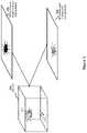
- FIG. 3depicts an example of how the 3D object grid may be utilized to generate the 2D synthesized images.
- 3D object grid 304includes at least two 3D objects: a spiculated mass 310 , and a calcification area 312 .
- the object combination modulemay determine that both objects are important to display in the 2D synthesized image, the spiculated mass 310 being more significant than the calcification.
- the imagesmay have to be manipulated such that both objects are still optimally displayed on the 2D synthesized image.
- the 2D synthesized image 306displays a synthesized image that does not use any image manipulation techniques described in this disclosure. As shown in 2D synthesized image 306 , both 3D objects 310 and 312 are competing to be displayed, and neither object is displayed very clearly. More specifically, the calcification 312 is barely visible in the 2D synthesized image 306 .
- 2D synthesized image 308the techniques described with respect to FIG. 2 are utilized in order to determine what parts of the respective 3D object should be emphasized and de-emphasized such that both objects are clearly discernible in the 2D synthesized image. More particularly, although spiculated mass 310 is more significant than the calcification 312 , the center portion of the spiculated mass 310 is slightly de-emphasized such that the calcification area is clearly visible. Similarly, it may be determined that the linear lines radiating from the center portion should be emphasized such that the end-user understands a size or scope of the spiculated mass. In light of the modified image corresponding to the spiculated mass 310 , the calcification 312 is now visible even though both objects overlap. Thus, as shown in FIG. 3 , the 2D synthesized image 308 provides more details about both 3D objects 310 and 312 when compared to 2D synthesized image 306 .
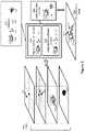
- FIGS. 4A-4Ddepict exemplary embodiments of displaying various objects of the 3D object grid, while preserving clinically-significant information.
- the collapsing of a 3D object into the 2D synthesized imagemay be achieved by the respective object combination module, image synthesis module and data reduction module that work together to display as much clinically-significant information as possible.
- FIG. 4Ashows an example embodiment of an intra-object combination.
- Intra-object combinationmay refer to techniques used to represent a single object (that is captured on multiple Tr image slices 404 ) onto the 2D synthesized image. More particularly, an identified 3D object may appear in many consecutive Tr image slices as 408 a , 408 b and 408 c . In theory these image patterns compete with each other for representation on the 2D synthesized image. Thus, an intra-object combination requires recognizing that all the images slices belong to the same 3D object, and only showing relevant information pertaining to the 3D object on the 2D synthesized image 406 . Notably, as shown in FIG.
- the systemmay determine that all the image patterns 408 a , 408 b and 408 c from the Tr stack 404 belong to the same 3D object, and may collapse them together such that they appear as one object 410 in the 2D synthesized image 406 .
- techniquessuch as averaging, MIP (maximum intensity projection), filtering, etc. may be used for intra-object combination.
- Intra-object combination techniquesaim to preserve the structure of the 3D object without losing valuable information from any of the image slices, while minimizing competing information from multiple image slices that do not provide valuable information and/or visually confuse the end-user.
- FIG. 4Billustrates an example embodiment of an object vs background combination.
- Object vs. background combinationmay be important for creating a natural-looking 2D synthesized image.
- the goal of this techniqueis to maintain useful information from objects together with meaningful background information representative of breast tissue.
- the Tr stack 414comprises two Tr image slices.
- the first image slicecomprises a background image pattern 412 .
- the second image slicecomprises an object or a portion of an object 411 .
- some aspects of both image slicesare emphasized while other aspects are de-emphasized. For example, in 2D synthesized image 416 , the object 411 is preserved, and the background 412 is also rendered, but the middle portion of the background 412 is de-emphasized.
- FIG. 4Cillustrates an example embodiment of inter-object combination without overlapping.
- Tr stack 424comprises two Tr image slices.
- One Tr image slicecomprises object 422
- the other Tr image slicecomprises object 421 .
- these objectsdo not overlap in the z direction.
- both objects 421 and 422are represented clearly at their respective x-y locations.
- FIG. 4Dillustrates an example embodiment of inter-object combination with overlap.
- This techniquemay be performed when two or more objects overlap to some degree.
- the two objectsmay be of the same type or of different types.
- a hierarchical approachmay be used to determine which object should be given precedence over the other. For example, if a higher weight is assigned to a first object, the first object may be emphasized in the 2D synthesized object, while the second object (or portions of the second object) may be de-emphasized. Or, if both objects are equally, or almost equally, important, both objects may be represented equally even if they are overlapping (and portions of both objects may be emphasized/de-emphasized such that both objects are clear in the synthesized 2D image).
- Tr image stack 434comprises two Tr image slices.
- One Tr image slicecomprises object 432
- the other Tr image slicecomprises object 431 .
- these objectsoverlap in the z direction.
- both objects 431 and 432are represented, but are shown to overlap.
- one objectmay be highlighted while the other is de-emphasized.
- both objectsare represented somewhat equally, even though it is clear that they represent two separate objects.
- object 431is assigned a higher weight/priority
- object 431may be emphasized in the foreground, while object 432 may be relegated to the background.
- other combination techniquesmay be utilized to optimally represent clinically-significant information to the end-user.
- FIG. 5depicts an exemplary embodiment of collapsing information from a plurality of Tr images into a 2D synthesized image.
- a Tr image stack 504may be used to create a 3D object grid similar to the 3D grid shown in FIG. 2 .
- the Tr stack 504illustrates four distinct objects including two calcification areas 510 and 512 , a spiculated mass 514 , and a spherical mass 516 . As discussed in detail above, identifying these four objects as separate and distinct objects allows the system to accurately depict the objects as a whole on the 2D synthesized image 506 .
- the spiculated mass 514is shown most prominently, while the calcifications and the spherical mass 516 are not as emphasized. This allows an end-user to easily identify the most clinically significant part of the 2D synthesized image without being overwhelmed with objects that are less-significant.
- FIG. 6is a flow diagram 600 provided to illustrate exemplary steps that may be performed in an image merge process carried out in accordance with one embodiment of the disclosed inventions.
- an image data setis acquired.
- the image data setmay be acquired by a tomosynthesis acquisition system, a combination tomosynthesis/mammography system, or by retrieving pre-existing image data from a storage device, whether locally or remotely located relative to an image display device.
- a 3D object gridmay be constructed by identifying various objects that are present in a 3D coordinate space representative of a patient's breast tissue.
- the objects of the 3D object gridare recognized, and a relative weight/priority of each of the objects is determined. As discussed above, in some embodiments, all objects of the 3D object grid may be displayed, with some objects emphasized more than others. In other embodiments, only a subset of the recognized objects may be displayed at all, while less-significant objects are omitted.
- the 3D objectsmay be reduced to a 2D format to create the 2D synthesized image.
- This reduction processmay highlight one object over another, in some embodiments. In other embodiment, the reduction process may highlight an outline of an object while de-emphasizing an interior of the object.
- the reduction processmay emphasize one or more features that are deemed to be significant, while de-emphasizing less significant aspects of the same object. For example, in the case of a spiculated lesion, it may be important to display the blood supply lines emanating from the center of the spiculated mass, but the center of the spiculated mass, even if dense may be displayed with less emphasis. Any number of such enhancement techniques may be used in the data reduction process.
- the synthesized 2D imageis displayed to the end-user.
Landscapes
- Engineering & Computer Science (AREA)
- Health & Medical Sciences (AREA)
- Physics & Mathematics (AREA)
- Medical Informatics (AREA)
- Life Sciences & Earth Sciences (AREA)
- Theoretical Computer Science (AREA)
- General Physics & Mathematics (AREA)
- General Health & Medical Sciences (AREA)
- Public Health (AREA)
- Biomedical Technology (AREA)
- Nuclear Medicine, Radiotherapy & Molecular Imaging (AREA)
- Radiology & Medical Imaging (AREA)
- Pathology (AREA)
- Optics & Photonics (AREA)
- Molecular Biology (AREA)
- Surgery (AREA)
- Animal Behavior & Ethology (AREA)
- Heart & Thoracic Surgery (AREA)
- High Energy & Nuclear Physics (AREA)
- Veterinary Medicine (AREA)
- Biophysics (AREA)
- Computer Vision & Pattern Recognition (AREA)
- Computing Systems (AREA)
- Software Systems (AREA)
- Geometry (AREA)
- Computer Graphics (AREA)
- Dentistry (AREA)
- Oral & Maxillofacial Surgery (AREA)
- Data Mining & Analysis (AREA)
- Quality & Reliability (AREA)
- Artificial Intelligence (AREA)
- Evolutionary Computation (AREA)
- General Engineering & Computer Science (AREA)
- Mathematical Physics (AREA)
- Epidemiology (AREA)
- Primary Health Care (AREA)
- Databases & Information Systems (AREA)
- Apparatus For Radiation Diagnosis (AREA)
- Image Analysis (AREA)
Abstract
Description
Claims (23)
Priority Applications (1)
| Application Number | Priority Date | Filing Date | Title |
|---|---|---|---|
| US16/497,766US11455754B2 (en) | 2017-03-30 | 2018-03-28 | System and method for synthesizing low-dimensional image data from high-dimensional image data using an object grid enhancement |
Applications Claiming Priority (3)
| Application Number | Priority Date | Filing Date | Title |
|---|---|---|---|
| US201762479008P | 2017-03-30 | 2017-03-30 | |
| US16/497,766US11455754B2 (en) | 2017-03-30 | 2018-03-28 | System and method for synthesizing low-dimensional image data from high-dimensional image data using an object grid enhancement |
| PCT/US2018/024912WO2018183549A1 (en) | 2017-03-30 | 2018-03-28 | System and method for synthesizing low-dimensional image data from high-dimensional image data using an object grid enhancement |
Related Parent Applications (1)
| Application Number | Title | Priority Date | Filing Date |
|---|---|---|---|
| PCT/US2018/024912A-371-Of-InternationalWO2018183549A1 (en) | 2017-03-30 | 2018-03-28 | System and method for synthesizing low-dimensional image data from high-dimensional image data using an object grid enhancement |
Related Child Applications (1)
| Application Number | Title | Priority Date | Filing Date |
|---|---|---|---|
| US17/871,129ContinuationUS11983799B2 (en) | 2017-03-30 | 2022-07-22 | System and method for synthesizing low-dimensional image data from high-dimensional image data using an object grid enhancement |
Publications (2)
| Publication Number | Publication Date |
|---|---|
| US20210118199A1 US20210118199A1 (en) | 2021-04-22 |
| US11455754B2true US11455754B2 (en) | 2022-09-27 |
Family
ID=61972644
Family Applications (4)
| Application Number | Title | Priority Date | Filing Date |
|---|---|---|---|
| US16/497,766ActiveUS11455754B2 (en) | 2017-03-30 | 2018-03-28 | System and method for synthesizing low-dimensional image data from high-dimensional image data using an object grid enhancement |
| US17/871,129ActiveUS11983799B2 (en) | 2017-03-30 | 2022-07-22 | System and method for synthesizing low-dimensional image data from high-dimensional image data using an object grid enhancement |
| US18/631,216ActiveUS12211124B2 (en) | 2017-03-30 | 2024-04-10 | System and method for synthesizing low-dimensional image data from high-dimensional image data using an object grid enhancement |
| US18/983,948PendingUS20250225694A1 (en) | 2017-03-30 | 2024-12-17 | System and method for synthesizing low-dimensional image data from high-dimensional image data using an object grid enhancement |
Family Applications After (3)
| Application Number | Title | Priority Date | Filing Date |
|---|---|---|---|
| US17/871,129ActiveUS11983799B2 (en) | 2017-03-30 | 2022-07-22 | System and method for synthesizing low-dimensional image data from high-dimensional image data using an object grid enhancement |
| US18/631,216ActiveUS12211124B2 (en) | 2017-03-30 | 2024-04-10 | System and method for synthesizing low-dimensional image data from high-dimensional image data using an object grid enhancement |
| US18/983,948PendingUS20250225694A1 (en) | 2017-03-30 | 2024-12-17 | System and method for synthesizing low-dimensional image data from high-dimensional image data using an object grid enhancement |
Country Status (6)
| Country | Link |
|---|---|
| US (4) | US11455754B2 (en) |
| EP (1) | EP3600051B1 (en) |
| JP (2) | JP7169986B2 (en) |
| CN (1) | CN110621233B (en) |
| ES (1) | ES2987687T3 (en) |
| WO (1) | WO2018183549A1 (en) |
Families Citing this family (31)
| Publication number | Priority date | Publication date | Assignee | Title |
|---|---|---|---|---|
| WO2007095330A2 (en) | 2006-02-15 | 2007-08-23 | Hologic Inc | Breast biopsy and needle localization using tomosynthesis systems |
| ES2862525T3 (en) | 2009-10-08 | 2021-10-07 | Hologic Inc | Needle Breast Biopsy System and Method of Use |
| US20120133600A1 (en) | 2010-11-26 | 2012-05-31 | Hologic, Inc. | User interface for medical image review workstation |
| JP6057922B2 (en) | 2011-03-08 | 2017-01-11 | ホロジック, インコーポレイテッドHologic, Inc. | System and method for dual energy and / or contrast enhanced breast imaging for screening, diagnosis and biopsy |
| EP2782505B1 (en) | 2011-11-27 | 2020-04-22 | Hologic, Inc. | System and method for generating a 2d image using mammography and/or tomosynthesis image data |
| JP6240097B2 (en) | 2012-02-13 | 2017-11-29 | ホロジック インコーポレイティッド | How to navigate a tomosynthesis stack using composite image data |
| CN105451657A (en) | 2013-03-15 | 2016-03-30 | 霍罗吉克公司 | System and method for navigating tomosynthesis stack including automatic focusing |
| US10092358B2 (en) | 2013-03-15 | 2018-10-09 | Hologic, Inc. | Tomosynthesis-guided biopsy apparatus and method |
| EP3060132B1 (en) | 2013-10-24 | 2019-12-04 | Hologic, Inc. | System and method for navigating x-ray guided breast biopsy |
| JP6506769B2 (en) | 2014-02-28 | 2019-04-24 | ホロジック, インコーポレイテッドHologic, Inc. | System and method for generating and displaying tomosynthesis image slabs |
| CN110621233B (en) | 2017-03-30 | 2023-12-12 | 豪洛捷公司 | Method for processing breast tissue image data |
| EP3600052A1 (en) | 2017-03-30 | 2020-02-05 | Hologic, Inc. | System and method for targeted object enhancement to generate synthetic breast tissue images |
| EP3600047A1 (en) | 2017-03-30 | 2020-02-05 | Hologic, Inc. | System and method for hierarchical multi-level feature image synthesis and representation |
| WO2018236565A1 (en) | 2017-06-20 | 2018-12-27 | Hologic, Inc. | METHOD AND SYSTEM FOR MEDICAL IMAGING WITH DYNAMIC SELF-LEARNING |
| WO2020068851A1 (en) | 2018-09-24 | 2020-04-02 | Hologic, Inc. | Breast mapping and abnormality localization |
| WO2020068767A1 (en) | 2018-09-28 | 2020-04-02 | Hologic, Inc. | System and method for synthetic breast tissue image generation by high density element suppression |
| EP3657442B1 (en)* | 2018-11-23 | 2022-10-26 | Siemens Healthcare GmbH | Synthetic mammogramm with embossed image impression |
| WO2020107019A1 (en) | 2018-11-25 | 2020-05-28 | Hologic, Inc. | Multimodality hanging protocols |
| DE202020006044U1 (en) | 2019-03-29 | 2024-07-02 | Hologic Inc. | Report generation for cropped digital images |
| JP7302368B2 (en)* | 2019-08-20 | 2023-07-04 | コニカミノルタ株式会社 | Medical information processing device and program |
| GB201912784D0 (en)* | 2019-09-05 | 2019-10-23 | Volpara Health Tech Limited | Method and system for image normalization |
| US20230045859A1 (en)* | 2020-01-24 | 2023-02-16 | The Regents Of The University Of California | Biomarker Prediction Using Optical Coherence Tomography |
| US12387296B2 (en)* | 2020-01-31 | 2025-08-12 | The General Hospital Corporation | Systems and methods for artifact reduction in tomosynthesis with multi-scale deep learning image processing |
| JP7446410B2 (en) | 2020-03-18 | 2024-03-08 | 富士フイルム株式会社 | Image processing device, method and program |
| CN113520416A (en)* | 2020-04-21 | 2021-10-22 | 上海联影医疗科技股份有限公司 | A method and system for generating a two-dimensional image of an object |
| CN111598896B (en)* | 2020-04-28 | 2023-12-05 | 腾讯科技(深圳)有限公司 | Image detection method, device, equipment and storage medium |
| DE102020209706A1 (en) | 2020-07-31 | 2022-02-03 | Siemens Healthcare Gmbh | Synthetic mammogram with reduced overlay of tissue changes |
| WO2022070570A1 (en) | 2020-09-30 | 2022-04-07 | 富士フイルム株式会社 | Image-processing device, image-processing method, and image-processing program |
| US12186119B2 (en) | 2021-10-05 | 2025-01-07 | Hologic, Inc. | Interactive model interface for image selection in medical imaging systems |
| US12254586B2 (en) | 2021-10-25 | 2025-03-18 | Hologic, Inc. | Auto-focus tool for multimodality image review |
| WO2023097279A1 (en) | 2021-11-29 | 2023-06-01 | Hologic, Inc. | Systems and methods for correlating objects of interest |
Citations (402)
| Publication number | Priority date | Publication date | Assignee | Title |
|---|---|---|---|---|
| US3502878A (en) | 1967-09-22 | 1970-03-24 | Us Health Education & Welfare | Automatic x-ray apparatus for limiting the field size of a projected x-ray beam in response to film size and to source-to-film distance |
| US3863073A (en) | 1973-04-26 | 1975-01-28 | Machlett Lab Inc | Automatic system for precise collimation of radiation |
| US3971950A (en) | 1975-04-14 | 1976-07-27 | Xerox Corporation | Independent compression and positioning device for use in mammography |
| US4160906A (en) | 1977-06-23 | 1979-07-10 | General Electric Company | Anatomically coordinated user dominated programmer for diagnostic x-ray apparatus |
| US4310766A (en) | 1978-09-06 | 1982-01-12 | Siemens Aktiengesellschaft | Motor driven x-ray grid and film-holder assembly |
| US4496557A (en) | 1981-08-27 | 1985-01-29 | Adir | Tricyclic ethers, their preparation and the pharmaceutical compositions containing them |
| US4559641A (en) | 1983-06-24 | 1985-12-17 | Thomson-Cgr | Retractable cassette holder for a radiological and radiographic examination apparatus |
| US4706269A (en) | 1985-03-11 | 1987-11-10 | Reina Leo J | Anti-scatter grid structure |
| US4744099A (en) | 1983-11-03 | 1988-05-10 | Siemens Aktiengesellschaft | X-ray diagnostic apparatus comprising radiation filters |
| US4773087A (en) | 1986-04-14 | 1988-09-20 | University Of Rochester | Quality of shadowgraphic x-ray images |
| US4773086A (en) | 1983-12-16 | 1988-09-20 | Yokogawa Medical Systems, Limited | Operator console for X-ray tomographs |
| US4819258A (en) | 1986-11-28 | 1989-04-04 | Bennett X-Ray Corp. | Auto-setting of KV in an x-ray machine after selection of technic factors |
| US4821727A (en) | 1986-10-30 | 1989-04-18 | Elscint Ltd. | Mammographic biopsy needle holder system |
| US4907156A (en) | 1987-06-30 | 1990-03-06 | University Of Chicago | Method and system for enhancement and detection of abnormal anatomic regions in a digital image |
| WO1990005485A1 (en) | 1988-11-23 | 1990-05-31 | Nrt-Nordisk Roentgen Teknik A/S | X-ray apparatus |
| US4969174A (en) | 1989-09-06 | 1990-11-06 | General Electric Company | Scanning mammography system with reduced scatter radiation |
| US4989227A (en) | 1989-04-28 | 1991-01-29 | General Electric Cgr S.A. | Cassette carrier adaptable in size and position for mammography |
| US5018176A (en) | 1989-03-29 | 1991-05-21 | General Electric Cgr S.A. | Mammograph equipped with an integrated device for taking stereotaxic photographs and a method of utilization of said mammograph |
| US5029193A (en) | 1989-07-03 | 1991-07-02 | Siemens Aktiengesellschaft | X-ray diagnostic installation for mammography exposures |
| USRE33634E (en) | 1986-09-23 | 1991-07-09 | Method and structure for optimizing radiographic quality by controlling X-ray tube voltage, current focal spot size and exposure time | |
| US5051904A (en) | 1988-03-24 | 1991-09-24 | Olganix Corporation | Computerized dynamic tomography system |
| US5078142A (en) | 1989-11-21 | 1992-01-07 | Fischer Imaging Corporation | Precision mammographic needle biopsy system |
| US5099846A (en) | 1988-12-23 | 1992-03-31 | Hardy Tyrone L | Method and apparatus for video presentation from a variety of scanner imaging sources |
| US5129911A (en) | 1991-03-11 | 1992-07-14 | Siczek Bernard W | Orbital aiming device |
| US5133020A (en) | 1989-07-21 | 1992-07-21 | Arch Development Corporation | Automated method and system for the detection and classification of abnormal lesions and parenchymal distortions in digital medical images |
| US5163075A (en) | 1991-08-08 | 1992-11-10 | Eastman Kodak Company | Contrast enhancement of electrographic imaging |
| US5164976A (en) | 1989-09-06 | 1992-11-17 | General Electric Company | Scanning mammography system with improved skin line viewing |
| US5199056A (en) | 1989-11-28 | 1993-03-30 | Darrah Carol J | Mammography compression paddle |
| US5219351A (en) | 1990-10-24 | 1993-06-15 | General Electric Cgr S.A. | Mammograph provided with an improved needle carrier |
| US5240011A (en) | 1991-11-27 | 1993-08-31 | Fischer Imaging Corporation | Motorized biopsy needle positioner |
| WO1993017620A1 (en) | 1992-03-12 | 1993-09-16 | Fischer Imaging Corporation | Isocentric puncture instrument aiming device |
| US5280427A (en) | 1989-11-27 | 1994-01-18 | Bard International, Inc. | Puncture guide for computer tomography |
| US5279309A (en) | 1991-06-13 | 1994-01-18 | International Business Machines Corporation | Signaling device and method for monitoring positions in a surgical operation |
| US5289520A (en) | 1991-11-27 | 1994-02-22 | Lorad Corporation | Stereotactic mammography imaging system with prone position examination table and CCD camera |
| WO1994006352A1 (en) | 1992-09-23 | 1994-03-31 | Fischer Imaging Corporation | Mammographic screening and biopsy apparatus |
| US5343390A (en) | 1992-02-28 | 1994-08-30 | Arch Development Corporation | Method and system for automated selection of regions of interest and detection of septal lines in digital chest radiographs |
| US5359637A (en) | 1992-04-28 | 1994-10-25 | Wake Forest University | Self-calibrated tomosynthetic, radiographic-imaging system, method, and device |
| US5365562A (en) | 1993-09-20 | 1994-11-15 | Fischer Imaging Corporation | Digital imaging apparatus |
| US5415169A (en) | 1989-11-21 | 1995-05-16 | Fischer Imaging Corporation | Motorized mammographic biopsy apparatus |
| US5452367A (en) | 1993-11-29 | 1995-09-19 | Arch Development Corporation | Automated method and system for the segmentation of medical images |
| US5491627A (en) | 1993-05-13 | 1996-02-13 | Arch Development Corporation | Method and system for the detection of microcalcifications in digital mammograms |
| US5499097A (en) | 1994-09-19 | 1996-03-12 | Neopath, Inc. | Method and apparatus for checking automated optical system performance repeatability |
| US5506877A (en) | 1994-11-23 | 1996-04-09 | The General Hospital Corporation | Mammography breast compression device and method |
| US5526394A (en) | 1993-11-26 | 1996-06-11 | Fischer Imaging Corporation | Digital scan mammography apparatus |
| US5539797A (en) | 1993-03-29 | 1996-07-23 | Ge Medical Systems Sa | Method and apparatus for digital stereotaxic mammography |
| US5553111A (en) | 1994-10-26 | 1996-09-03 | The General Hospital Corporation | Apparatus and method for improved tissue imaging |
| US5592562A (en) | 1994-01-19 | 1997-01-07 | International Business Machines Corporation | Inspection system for cross-sectional imaging |
| WO1997000649A1 (en) | 1995-06-20 | 1997-01-09 | Wan Sing Ng | Articulated arm for medical procedures |
| US5594769A (en) | 1991-11-27 | 1997-01-14 | Thermotrex Corporation | Method and apparatus for obtaining stereotactic mammographic guided needle breast biopsies |
| US5596200A (en) | 1992-10-14 | 1997-01-21 | Primex | Low dose mammography system |
| US5598454A (en) | 1994-04-26 | 1997-01-28 | Siemens Aktiengesellschaft | X-ray diagnostics installation |
| US5627869A (en) | 1995-11-22 | 1997-05-06 | Thermotrex Corporation | Mammography apparatus with proportional collimation |
| EP0775467A1 (en) | 1995-11-23 | 1997-05-28 | Planmed Oy | Method and system for controlling the functions of a mammography apparatus |
| US5642433A (en) | 1995-07-31 | 1997-06-24 | Neopath, Inc. | Method and apparatus for image contrast quality evaluation |
| US5642441A (en) | 1995-10-24 | 1997-06-24 | Neopath, Inc. | Separation apparatus and method for measuring focal plane |
| US5647025A (en) | 1994-09-20 | 1997-07-08 | Neopath, Inc. | Automatic focusing of biomedical specimens apparatus |
| JPH09198490A (en) | 1996-01-22 | 1997-07-31 | Hitachi Medical Corp | Three-dimensional discrete data projector |
| US5657362A (en) | 1995-02-24 | 1997-08-12 | Arch Development Corporation | Automated method and system for computerized detection of masses and parenchymal distortions in medical images |
| US5668889A (en) | 1990-04-19 | 1997-09-16 | Fuji Photo Film Co., Ltd. | Apparatus for determining an image position, and method for adjusting read-out conditions and/or image processing conditions for a radiation image |
| JPH09238934A (en) | 1996-03-11 | 1997-09-16 | Toshiba Medical Eng Co Ltd | Image display system |
| US5671288A (en) | 1995-05-31 | 1997-09-23 | Neopath, Inc. | Method and apparatus for assessing slide and specimen preparation quality |
| US5712890A (en) | 1994-11-23 | 1998-01-27 | Thermotrex Corp. | Full breast digital mammography device |
| JPH1033523A (en) | 1996-07-24 | 1998-02-10 | Hitachi Medical Corp | X-ray ct device |
| WO1998016903A1 (en) | 1996-10-16 | 1998-04-23 | Vital Images, Inc. | Advanced diagnostic viewer |
| US5763871A (en) | 1994-09-20 | 1998-06-09 | Neopath, Inc. | Cytological system autofocus integrity checking apparatus |
| US5769086A (en) | 1995-12-06 | 1998-06-23 | Biopsys Medical, Inc. | Control system and method for automated biopsy device |
| US5773832A (en) | 1995-11-21 | 1998-06-30 | Loral Fairchild Corporation | Advanced CCD-based x-ray image sensor system |
| US5818898A (en) | 1995-11-07 | 1998-10-06 | Kabushiki Kaisha Toshiba | X-ray imaging apparatus using X-ray planar detector |
| US5828722A (en) | 1996-05-17 | 1998-10-27 | Sirona Dental Systems Gmbh & Co., Kg | X-ray diagnostic apparatus for tomosynthesis having a detector that detects positional relationships |
| US5835079A (en) | 1996-06-13 | 1998-11-10 | International Business Machines Corporation | Virtual pointing device for touchscreens |
| US5841124A (en) | 1996-06-19 | 1998-11-24 | Neopath, Inc. | Cytological system autofocus integrity checking apparatus |
| US5872828A (en) | 1996-07-23 | 1999-02-16 | The General Hospital Corporation | Tomosynthesis system for breast imaging |
| US5875258A (en) | 1994-09-20 | 1999-02-23 | Neopath, Inc. | Biological specimen analysis system processing integrity checking apparatus |
| US5878104A (en) | 1996-05-17 | 1999-03-02 | Sirona Dental Systems Gmbh & Co. Kg | Method for producing tomosynthesis exposures employing a reference object formed by a region of the examination subject |
| US5878746A (en) | 1993-08-25 | 1999-03-09 | Lemelson; Jerome H. | Computerized medical diagnostic system |
| US5896437A (en) | 1996-05-17 | 1999-04-20 | Sirona Dental Systems Gmbh & Co. Kg | X-ray diagnostics apparatus for tomosynthesis having a reference object in fixed relationship to a radiation emitter |
| US5941832A (en) | 1991-09-27 | 1999-08-24 | Tumey; David M. | Method and apparatus for detection of cancerous and precancerous conditions in a breast |
| US5954650A (en) | 1996-11-13 | 1999-09-21 | Kabushiki Kaisha Toshiba | Medical image processing apparatus |
| US6005907A (en) | 1996-05-17 | 1999-12-21 | Sirona Dental Systems Gmbh & Co. Kg | Method and apparatus for producing tomosynthesis exposures employing a reference object composed of a number of sub-objects |
| EP0982001A1 (en) | 1998-08-25 | 2000-03-01 | General Electric Company | Protocol driven image reconstruction, display, and processing in a multislice imaging system |
| US6067079A (en) | 1996-06-13 | 2000-05-23 | International Business Machines Corporation | Virtual pointing device for touchscreens |
| US6075879A (en) | 1993-09-29 | 2000-06-13 | R2 Technology, Inc. | Method and system for computer-aided lesion detection using information from multiple images |
| US6091841A (en) | 1997-09-04 | 2000-07-18 | Qualia Computing, Inc. | Method and system for segmenting desired regions in digital mammograms |
| JP2000200340A (en) | 1999-01-06 | 2000-07-18 | Ge Yokogawa Medical Systems Ltd | Method and device for displaying image and ct system |
| US6101236A (en) | 1998-10-02 | 2000-08-08 | University Of Iowa Research Foundation | Iterative method and apparatus for x-ray computed tomographic fluoroscopy |
| US6102866A (en) | 1996-10-15 | 2000-08-15 | Fischer Imaging Corporation | Enhanced breast imaging/biopsy system employing targeted ultrasound |
| WO2000051484A2 (en) | 1998-11-25 | 2000-09-08 | Fischer Imaging Corporation | User interface system for mammographic imager |
| US6137527A (en) | 1996-12-23 | 2000-10-24 | General Electric Company | System and method for prompt-radiology image screening service via satellite |
| US6149301A (en) | 1998-12-30 | 2000-11-21 | General Electric Company | X-ray target centering apparatus for radiographic imaging system |
| US6175117B1 (en) | 1998-01-23 | 2001-01-16 | Quanta Vision, Inc. | Tissue analysis apparatus |
| US6196715B1 (en) | 1959-04-28 | 2001-03-06 | Kabushiki Kaisha Toshiba | X-ray diagnostic system preferable to two dimensional x-ray detection |
| US6215892B1 (en) | 1995-11-30 | 2001-04-10 | Chromavision Medical Systems, Inc. | Method and apparatus for automated image analysis of biological specimens |
| US6216540B1 (en) | 1995-06-06 | 2001-04-17 | Robert S. Nelson | High resolution device and method for imaging concealed objects within an obscuring medium |
| US6233473B1 (en) | 1999-02-16 | 2001-05-15 | Hologic, Inc. | Determining body composition using fan beam dual-energy x-ray absorptiometry |
| US6243441B1 (en) | 1999-07-13 | 2001-06-05 | Edge Medical Devices | Active matrix detector for X-ray imaging |
| US6245028B1 (en) | 1999-11-24 | 2001-06-12 | Marconi Medical Systems, Inc. | Needle biopsy system |
| US6256370B1 (en) | 2000-01-24 | 2001-07-03 | General Electric Company | Method and apparatus for performing tomosynthesis |
| US6272207B1 (en) | 1999-02-18 | 2001-08-07 | Creatv Microtech, Inc. | Method and apparatus for obtaining high-resolution digital X-ray and gamma ray images |
| US6289235B1 (en) | 1998-03-05 | 2001-09-11 | Wake Forest University | Method and system for creating three-dimensional images using tomosynthetic computed tomography |
| US6292530B1 (en) | 1999-04-29 | 2001-09-18 | General Electric Company | Method and apparatus for reconstructing image data acquired by a tomosynthesis x-ray imaging system |
| US6293282B1 (en) | 1996-11-05 | 2001-09-25 | Jerome Lemelson | System and method for treating select tissue in living being |
| US20010038861A1 (en) | 1999-12-16 | 2001-11-08 | Tsung-Min Hsu | Transdermal administration of nonsteroidal anti-inflammatory drugs using hydroxide-releasing agents as permeation enhancers |
| US20010038681A1 (en) | 2000-02-11 | 2001-11-08 | Brandeis University | Method and system for low-dose three-dimensional imaging of a scene |
| US6327377B1 (en) | 1988-04-08 | 2001-12-04 | Autocyte North Carolina, L.L.C. | Automated cytological specimen classification system and method |
| US6327336B1 (en) | 2000-06-05 | 2001-12-04 | Direct Radiography Corp. | Radiogram showing location of automatic exposure control sensor |
| US6341156B1 (en) | 1999-05-14 | 2002-01-22 | Siemens Aktiengesellschaft | X-ray diagnostic apparatus with relatively moved x-ray source and detector |
| US20020012450A1 (en) | 1998-01-09 | 2002-01-31 | Osamu Tsujii | Image processing apparatus and method |
| US6375352B1 (en) | 1999-10-01 | 2002-04-23 | General Electric Company | Apparatus and method for obtaining x-ray tomosynthesis data for mammography |
| US20020050986A1 (en) | 2000-08-11 | 2002-05-02 | Hitoshi Inoue | Image display apparatus and method, and storage medium |
| US6389104B1 (en) | 2000-06-30 | 2002-05-14 | Siemens Corporate Research, Inc. | Fluoroscopy based 3-D neural navigation based on 3-D angiography reconstruction data |
| US20020075997A1 (en) | 2000-12-18 | 2002-06-20 | Unger Christopher David | Medical diagnostic method and apparatus to control dual energy exposure techniques based on image information |
| US6411836B1 (en) | 1999-12-30 | 2002-06-25 | General Electric Company | Method and apparatus for user preferences configuring in an image handling system |
| US6415015B2 (en) | 1999-12-28 | 2002-07-02 | Ge Medical Systems Sa | Method and system of compensation of thickness of an organ |
| US6424332B1 (en) | 1999-01-29 | 2002-07-23 | Hunter Innovations, Inc. | Image comparison apparatus and method |
| US20020113681A1 (en) | 2001-02-16 | 2002-08-22 | Byram Robert James | Rotary position sensor |
| US6442288B1 (en) | 1997-12-17 | 2002-08-27 | Siemens Aktiengesellschaft | Method for reconstructing a three-dimensional image of an object scanned in the context of a tomosynthesis, and apparatus for tomosynthesis |
| US20020122533A1 (en) | 2000-12-19 | 2002-09-05 | Alain Marie | Mammography apparatus and method |
| JP2002282248A (en) | 2000-11-27 | 2002-10-02 | Ge Medical Systems Global Technology Co Llc | Color parametric synthesizing map for ct perfusion method |
| US6463181B2 (en) | 2000-12-22 | 2002-10-08 | The United States Of America As Represented By The Secretary Of The Navy | Method for optimizing visual display of enhanced digital images |
| US6468226B1 (en) | 2000-11-22 | 2002-10-22 | Mcintyre, Iv John J. | Remote tissue biopsy apparatus and associated methods |
| US6480565B1 (en) | 1999-11-18 | 2002-11-12 | University Of Rochester | Apparatus and method for cone beam volume computed tomography breast imaging |
| US20020188466A1 (en) | 2001-04-18 | 2002-12-12 | Barrette Pierre Philip | Secure digital medical intellectual property (IP) distribution, market applications, and mobile devices |
| US20020193676A1 (en) | 2001-05-29 | 2002-12-19 | Anke Bodicker | Method and computer system for screening of medical cases |
| US20030007598A1 (en) | 2000-11-24 | 2003-01-09 | U-Systems, Inc. | Breast cancer screening with adjunctive ultrasound mammography |
| US20030018272A1 (en) | 2001-06-28 | 2003-01-23 | Treado Patrick J. | Method for Raman chemical imaging and characterization of calcification in tissue |
| US20030026386A1 (en) | 2001-02-01 | 2003-02-06 | Cha-Mei Tang | Anti-scatter grids and collimator designs, and their motion, fabrication and assembly |
| US20030048260A1 (en) | 2001-08-17 | 2003-03-13 | Alec Matusis | System and method for selecting actions based on the identification of user's fingers |
| WO2003020114A2 (en) | 2001-08-31 | 2003-03-13 | Analogic Corporation | Image positioning method and system for tomosynthesis in a digital x-ray radiography system |
| US6556655B1 (en) | 1998-11-27 | 2003-04-29 | Ge Medical Systems Sa | Method for automatic detection of glandular tissue |
| US20030095624A1 (en) | 2001-11-21 | 2003-05-22 | Eberhard Jeffrey Wayne | Dose management system for mammographic tomosynthesis |
| US20030097055A1 (en) | 2001-11-21 | 2003-05-22 | Philips Medical Systems(Cleveland), Inc. | Method of reviewing tomographic scans with a large number of images |
| US6574304B1 (en) | 2002-09-13 | 2003-06-03 | Ge Medical Systems Global Technology Company, Llc | Computer aided acquisition of medical images |
| JP2003189179A (en) | 2001-12-14 | 2003-07-04 | Konica Corp | Abnormal shade detector and image output device |
| US20030128893A1 (en) | 2001-11-19 | 2003-07-10 | Alfio Castorina | Method for merging digital images to obtain a high dynamic range digital image |
| JP2003199737A (en) | 2001-10-12 | 2003-07-15 | General Electric Co <Ge> | Reconstruction method for tomosynthesis |
| US20030135115A1 (en) | 1997-11-24 | 2003-07-17 | Burdette Everette C. | Method and apparatus for spatial registration and mapping of a biopsy needle during a tissue biopsy |
| US6597762B1 (en) | 2002-11-27 | 2003-07-22 | Ge Medical Systems Global Technology Co., Llc | Method and apparatus of lesion detection and validation based on multiple reviews of a CT image |
| US6611575B1 (en) | 2001-07-27 | 2003-08-26 | General Electric Company | Method and system for high resolution 3D visualization of mammography images |
| US20030169847A1 (en) | 2001-11-21 | 2003-09-11 | University Of Massachusetts Medical Center | System and method for x-ray fluoroscopic imaging |
| US6620111B2 (en) | 2001-04-20 | 2003-09-16 | Ethicon Endo-Surgery, Inc. | Surgical biopsy device having automatic rotation of the probe for taking multiple samples |
| US6626849B2 (en) | 2001-11-01 | 2003-09-30 | Ethicon Endo-Surgery, Inc. | MRI compatible surgical biopsy device |
| US6633674B1 (en) | 1999-11-24 | 2003-10-14 | General Electric Company | Picture archiving and communication system employing improved data compression |
| US20030195433A1 (en) | 2002-04-16 | 2003-10-16 | Roman Turovskiy | Localization element with energized tip |
| US20030194121A1 (en) | 2002-04-15 | 2003-10-16 | General Electric Company | Computer aided detection (CAD) for 3D digital mammography |
| US20030194050A1 (en) | 2002-04-15 | 2003-10-16 | General Electric Company | Multi modality X-ray and nuclear medicine mammography imaging system and method |
| JP2003531516A (en) | 2000-04-18 | 2003-10-21 | リットン システムズ、 インコーポレーテッド | Enhanced visualization of live breast biopsy locations for medical documentation |
| US6638235B2 (en) | 2000-11-06 | 2003-10-28 | Suros Surgical Systems, Inc. | Biopsy apparatus |
| US6647092B2 (en) | 2002-01-18 | 2003-11-11 | General Electric Company | Radiation imaging system and method of collimation |
| US20030210254A1 (en) | 2002-05-13 | 2003-11-13 | Doan William D. | Method, system and computer product for displaying axial images |
| US20030212327A1 (en) | 2000-11-24 | 2003-11-13 | U-Systems Inc. | Adjunctive ultrasound processing and display for breast cancer screening |
| US20030215120A1 (en) | 2002-05-15 | 2003-11-20 | Renuka Uppaluri | Computer aided diagnosis of an image set |
| US20040008809A1 (en) | 1998-07-24 | 2004-01-15 | Webber Richard L. | Method and system for creating task-dependent three-dimensional images |
| US20040008900A1 (en) | 2002-07-12 | 2004-01-15 | Jabri Kadri N. | System and method for efficiently customizing an imaging system |
| US20040008901A1 (en) | 2002-07-11 | 2004-01-15 | Avinash Gopal B. | Interpolated image filtering method and apparatus |
| US6683934B1 (en) | 2000-06-05 | 2004-01-27 | General Electric Company | Dual energy x-ray imaging system and method for radiography and mammography |
| US20040036680A1 (en) | 2002-08-26 | 2004-02-26 | Mark Davis | User-interface features for computers with contact-sensitive displays |
| US20040047518A1 (en) | 2002-08-28 | 2004-03-11 | Carlo Tiana | Image fusion system and method |
| US20040052328A1 (en) | 2002-09-13 | 2004-03-18 | Sabol John M. | Computer assisted analysis of tomographic mammography data |
| US20040064037A1 (en) | 2002-09-27 | 2004-04-01 | Confirma, Inc. | Rules-based approach for processing medical images |
| US20040066884A1 (en) | 2002-10-07 | 2004-04-08 | Hermann Claus Bernhard Erich | Continuous scan tomosynthesis system and method |
| US20040070582A1 (en) | 2002-10-11 | 2004-04-15 | Matthew Warren Smith To Sonocine, Inc. | 3D modeling system |
| US20040077938A1 (en) | 2002-10-07 | 2004-04-22 | Mark Joseph L. | Introduction system for minimally invasive surgical instruments |
| US20040081273A1 (en) | 1999-11-18 | 2004-04-29 | Ruola Ning | Apparatus and method for cone beam volume computed tomography breast imaging |
| US20040094167A1 (en) | 2000-03-17 | 2004-05-20 | Brady John Michael | Three-dimensional reconstructions of a breast from two x-ray mammographics |
| US20040101095A1 (en) | 2002-11-27 | 2004-05-27 | Hologic Inc. | Full field mammography with tissue exposure control, tomosynthesis, and dynamic field of view processing |
| US20040109028A1 (en) | 2002-12-10 | 2004-06-10 | Siemens Medical Solutions Usa, Inc. | Medical imaging programmable custom user interface system and method |
| US20040109529A1 (en) | 2002-12-10 | 2004-06-10 | General Electric Company | Full field digital tomosynthesis method and apparatus |
| US20040127789A1 (en) | 2002-12-17 | 2004-07-01 | Kabushiki Kaisha Toshiba | Method and system for X-ray diagnosis of object in which X-ray contrast agent is injected |
| US20040138569A1 (en) | 1999-08-20 | 2004-07-15 | Sorin Grunwald | User interface for handheld imaging devices |
| US20040171933A1 (en) | 2002-11-25 | 2004-09-02 | Milton Stoller | Mammography needle biopsy system and method |
| US20040171986A1 (en) | 1999-04-26 | 2004-09-02 | Scimed Life System, Inc. | Apparatus and methods for guiding a needle |
| US6813334B2 (en) | 2000-10-20 | 2004-11-02 | Koninklijke Philips Electronics N.V. | Tomosynthesis in a limited angular range |
| US20050047636A1 (en) | 2003-08-29 | 2005-03-03 | David Gines | System and method for performing auto-focused tomosynthesis |
| US20050063509A1 (en) | 2001-10-19 | 2005-03-24 | Defreitas Kenneth F | Mammography system and method employing offset compression paddles automatic collimation and retractable anti-scatter grid |
| US20050078797A1 (en) | 2002-03-01 | 2005-04-14 | Mats Danielsson | X-ray protection device |
| US6882700B2 (en) | 2002-04-15 | 2005-04-19 | General Electric Company | Tomosynthesis X-ray mammogram system and method with automatic drive system |
| US20050084060A1 (en) | 2003-10-15 | 2005-04-21 | Seppi Edward J. | Systems and methods for functional imaging using contrast-enhanced multiple-energy computed tomography |
| US6885724B2 (en) | 2003-08-22 | 2005-04-26 | Ge Medical Systems Global Technology Company, Llc | Radiographic tomosynthesis image acquisition utilizing asymmetric geometry |
| US20050089205A1 (en) | 2003-10-23 | 2005-04-28 | Ajay Kapur | Systems and methods for viewing an abnormality in different kinds of images |
| US20050105679A1 (en) | 2003-02-12 | 2005-05-19 | Tao Wu | Tomosynthesis imaging system and method |
| US20050107689A1 (en) | 2003-11-14 | 2005-05-19 | Konica Minolta Meical & Graphic, Inc. | Medical image management system |
| US20050113715A1 (en) | 2000-11-06 | 2005-05-26 | Jeffrey Schwindt | Biopsy apparatus |
| US20050111718A1 (en) | 2003-11-26 | 2005-05-26 | University Of Chicago | Automated method and system for the evaluation of disease and registration accuracy in the subtraction of temporally sequential medical images |
| US20050113681A1 (en) | 2002-11-27 | 2005-05-26 | Defreitas Kenneth F. | X-ray mammography with tomosynthesis |
| US6901156B2 (en) | 2000-02-04 | 2005-05-31 | Arch Development Corporation | Method, system and computer readable medium for an intelligent search workstation for computer assisted interpretation of medical images |
| US20050124845A1 (en) | 2003-09-18 | 2005-06-09 | Thomadsen Bruce R. | Device for placement of needles and radioactive seeds in radiotherapy |
| WO2005051197A2 (en) | 2003-11-26 | 2005-06-09 | Koninklijke Philips Electronics, N.V. | Workflow optimization for high throughput imaging environment |
| US20050135664A1 (en) | 2003-12-23 | 2005-06-23 | Kaufhold John P. | Methods and apparatus for reconstruction of volume data from projection data |
| US20050135555A1 (en) | 2003-12-23 | 2005-06-23 | Claus Bernhard Erich H. | Method and system for simultaneously viewing rendered volumes |
| US20050226375A1 (en) | 2004-03-31 | 2005-10-13 | Eberhard Jeffrey W | Enhanced X-ray imaging system and method |
| WO2005110230A1 (en) | 2004-05-14 | 2005-11-24 | Philips Intellectual Property & Standards Gmbh | System and method for diagnosing breast cancer |
| WO2005112767A1 (en) | 2004-05-21 | 2005-12-01 | Tissuomics Limited | Apparatus and method for penetrating radiation measurements |
| US6978040B2 (en) | 2001-12-19 | 2005-12-20 | Canon Kabushiki Kaisha | Optical recovery of radiographic geometry |
| US20060009693A1 (en) | 2004-04-08 | 2006-01-12 | Techniscan, Inc. | Apparatus for imaging and treating a breast |
| US6987331B2 (en) | 1999-01-29 | 2006-01-17 | American Superconductor Corporation | Voltage recovery device for use with a utility power network |
| US20060018526A1 (en) | 2004-07-23 | 2006-01-26 | Avinash Gopal B | Methods and apparatus for noise reduction filtering of images |
| US20060025680A1 (en) | 2004-07-29 | 2006-02-02 | Fanny Jeune-Iomme | Method and apparatus for contrast enhanced medical imaging |
| US6999554B2 (en) | 2003-11-17 | 2006-02-14 | Siemens Aktiengesellschaft | X-ray diagnostic apparatus for mammography examinations |
| US20060074288A1 (en) | 2004-10-04 | 2006-04-06 | Thomas Kelly | Estimating visceral fat by dual-energy x-ray absorptiometry |
| US7025725B2 (en) | 2002-03-28 | 2006-04-11 | Ultrasound Detection Systems, Llc | Three-dimensional ultrasound computed tomography imaging system |
| US7030861B1 (en) | 2001-02-10 | 2006-04-18 | Wayne Carl Westerman | System and method for packing multi-touch gestures onto a hand |
| US20060098855A1 (en) | 2002-11-27 | 2006-05-11 | Gkanatsios Nikolaos A | Image handling and display in X-ray mammography and tomosynthesis |
| WO2006055830A2 (en) | 2004-11-15 | 2006-05-26 | Hologic, Inc. | Matching geometry generation and display of mammograms and tomosynthesis images |
| WO2006058160A2 (en) | 2004-11-26 | 2006-06-01 | Hologic, Inc. | Integrated multi-mode mammography/tomosynthesis x-ray system and method |
| US20060132508A1 (en) | 2004-12-16 | 2006-06-22 | Navid Sadikali | Multi-planar image viewing system and method |
| US20060147099A1 (en) | 2004-12-30 | 2006-07-06 | R2 Technology, Inc. | Medical image review workstation with integrated content-based resource retrieval |
| JP2006519634A (en) | 2003-03-11 | 2006-08-31 | シーメンス メディカル ソリューションズ ユーエスエー インコーポレイテッド | System and method for performing automatic three-dimensional lesion segmentation and measurement |
| US20060197753A1 (en) | 2005-03-04 | 2006-09-07 | Hotelling Steven P | Multi-functional hand-held device |
| US7110502B2 (en) | 2003-05-12 | 2006-09-19 | Canon Kabushiki Kaisha | Radiographic apparatus and method for switching a grid |
| US20060210131A1 (en)* | 2005-03-15 | 2006-09-21 | Wheeler Frederick W Jr | Tomographic computer aided diagnosis (CAD) with multiple reconstructions |
| US7117098B1 (en) | 1997-02-27 | 2006-10-03 | Cellomics, Inc. | Machine-readable storage medium for analyzing distribution of macromolecules between the cell membrane and the cell cytoplasm |
| US20060228012A1 (en) | 2005-04-06 | 2006-10-12 | Kabushiki Kaisha Toshiba | Image display apparatus and image display method |
| US7127091B2 (en) | 2000-12-22 | 2006-10-24 | Koninklijke Philips Electronics, N.V. | Method and apparatus for visualizing a limited part of a 3D medical image-point-related data set, through basing a rendered image on an intermediate region between first and second clipping planes, and including spectroscopic viewing of such region |
| US20060238546A1 (en) | 2005-03-08 | 2006-10-26 | Carrie Handley | Comparative image review system and method |
| US20060257009A1 (en) | 2000-11-24 | 2006-11-16 | Shih-Ping Wang | Controlling thick-slice viewing of breast ultrasound data |
| JP2006312026A (en) | 2005-04-06 | 2006-11-16 | Toshiba Corp | Image display device |
| US20060269040A1 (en) | 2005-05-17 | 2006-11-30 | Thomas Mertelmeier | Mammography method and apparatus for forming a tomosynthetic 3-D X-ray image |
| US20070019846A1 (en) | 2003-08-25 | 2007-01-25 | Elizabeth Bullitt | Systems, methods, and computer program products for analysis of vessel attributes for diagnosis, disease staging, and surfical planning |
| US20070036265A1 (en) | 2005-08-15 | 2007-02-15 | Zhenxue Jing | X-ray mammography/tomosynthesis of patient's breast |
| US20070046649A1 (en) | 2005-08-30 | 2007-03-01 | Bruce Reiner | Multi-functional navigational device and method |
| US20070052700A1 (en) | 2005-09-07 | 2007-03-08 | Wheeler Frederick W | System and method for 3D CAD using projection images |
| US20070118400A1 (en) | 2005-11-22 | 2007-05-24 | General Electric Company | Method and system for gesture recognition to drive healthcare applications |
| US20070114424A1 (en) | 2005-11-18 | 2007-05-24 | Sectra Mamea Ab | Method and arrangement relating to x-ray imaging |
| US20070156451A1 (en) | 2006-01-05 | 2007-07-05 | Gering David T | System and method for portable display of relevant healthcare information |
| WO2007095330A2 (en) | 2006-02-15 | 2007-08-23 | Hologic Inc | Breast biopsy and needle localization using tomosynthesis systems |
| US20070223651A1 (en) | 2006-03-21 | 2007-09-27 | Wagenaar Douglas J | Dual modality mammography device |
| US20070236490A1 (en) | 2005-11-25 | 2007-10-11 | Agfa-Gevaert | Medical image display and review system |
| US7289825B2 (en) | 2004-03-15 | 2007-10-30 | General Electric Company | Method and system for utilizing wireless voice technology within a radiology workflow |
| US20070263765A1 (en) | 2003-12-03 | 2007-11-15 | The General Hospital Corporation D/B/A Massachusetts General Hospital | Multi-Segment Cone-Beam Reconstruction System and Method for Tomosynthesis Imaging |
| US7298881B2 (en) | 2004-02-13 | 2007-11-20 | University Of Chicago | Method, system, and computer software product for feature-based correlation of lesions from multiple images |
| US20070274585A1 (en) | 2006-05-25 | 2007-11-29 | Zhang Daoxian H | Digital mammography system with improved workflow |
| JP2007330334A (en) | 2006-06-12 | 2007-12-27 | Toshiba Corp | X-ray imaging apparatus and method thereof |
| US7315607B2 (en) | 2005-09-02 | 2008-01-01 | Siemens Aktiengesellschaft | Mammograph system with a face shield |
| US20080019581A1 (en) | 2002-11-27 | 2008-01-24 | Gkanatsios Nikolaos A | Image Handling and display in X-ray mammography and tomosynthesis |
| US7323692B2 (en) | 2004-08-10 | 2008-01-29 | Research Foundation Of State University Of New York | Flat-panel detector with avalanche gain |
| WO2008014670A1 (en) | 2006-07-25 | 2008-02-07 | Xiangshen Ni | Micro-invasive surgery x-ray puncturing and locating device and method |
| US20080043905A1 (en) | 2006-08-21 | 2008-02-21 | Bamdad Hassanpourgol | Portable Prone Stereotactic Mammography System for Biopsies, Image Guided Lumpectomies, and Radiation Treatment |
| US7346381B2 (en) | 2002-11-01 | 2008-03-18 | Ge Medical Systems Global Technology Company Llc | Method and apparatus for medical intervention procedure planning |
| JP2008068032A (en) | 2006-09-15 | 2008-03-27 | Toshiba Corp | Image display device |
| WO2008047270A1 (en) | 2006-10-17 | 2008-04-24 | Koninklijke Philips Electronics N.V. | Visualization of 3d images in combination with 2d projection images |
| US20080101537A1 (en) | 2006-10-26 | 2008-05-01 | Fujifilm Corporation | Tomographic image obtainment apparatus and method |
| WO2008054436A2 (en) | 2006-01-05 | 2008-05-08 | United Technologies Corporation | Damped coil pin for attachment hanger hinge |
| US20080114614A1 (en) | 2006-11-15 | 2008-05-15 | General Electric Company | Methods and systems for healthcare application interaction using gesture-based interaction enhanced with pressure sensitivity |
| US20080125643A1 (en) | 2006-11-24 | 2008-05-29 | Qview, Inc. | Processing and displaying dynamic contrast-enhanced magnetic resonance imaging information |
| US20080139896A1 (en) | 2006-10-13 | 2008-06-12 | Siemens Medical Solutions Usa, Inc. | System and Method for Graphical Annotation of Anatomical Images Using a Touch Screen Display |
| US20080152086A1 (en) | 2006-12-21 | 2008-06-26 | Sectra Ab | Synchronized viewing of tomosynthesis and/or mammograms |
| US20080165136A1 (en) | 2007-01-07 | 2008-07-10 | Greg Christie | System and Method for Managing Lists |
| US7406150B2 (en) | 2002-11-29 | 2008-07-29 | Hologic, Inc. | Distributed architecture for mammographic image acquisition and processing |
| US20080187095A1 (en) | 2005-05-03 | 2008-08-07 | The Regents Of The University Of California | Biopsy Systems For Breast Computed Tomography |
| US20080198966A1 (en) | 2007-01-31 | 2008-08-21 | Sectra Mamea Ab | Method and Arrangement Relating to X-Ray Imaging |
| US20080221479A1 (en) | 2007-03-07 | 2008-09-11 | Ritchie Paul G | Integrated Imaging and Biopsy System with Integrated Utilities |
| US20080229256A1 (en) | 2007-03-12 | 2008-09-18 | Fuji Xerox Co., Ltd. | Image processing apparatus, image processing method and computer readable medium |
| US20080240533A1 (en) | 2007-03-27 | 2008-10-02 | Cameron Anthony Piron | Post-acquisition adaptive reconstruction of mri data |
| US20080297482A1 (en) | 2007-05-30 | 2008-12-04 | Microsoft Corporation | Recognizing selection regions from multiple simultaneous inputs |
| US20090005668A1 (en) | 2007-06-30 | 2009-01-01 | West Jay B | Non-invasive method for using 2D angiographic images for radiosurgical target definition |
| US20090037821A1 (en) | 2004-07-23 | 2009-02-05 | O'neal David Sheldon | System And Method For Electronic Presentations |
| US20090034684A1 (en) | 2007-08-02 | 2009-02-05 | Sylvain Bernard | Method and system for displaying tomosynthesis images |
| WO2009026587A1 (en) | 2007-08-23 | 2009-02-26 | Fischer Medical Technologies, Inc. | Improved computed tomography breast imaging and biopsy system |
| US20090080602A1 (en) | 2006-08-03 | 2009-03-26 | Kenneth Brooks | Dedicated breast radiation imaging/therapy system |
| US20090079705A1 (en) | 2007-09-14 | 2009-03-26 | Steven Sizelove | Portable User Control Device and Method for Vehicle Information Systems |
| US20090080752A1 (en) | 2007-09-20 | 2009-03-26 | Chris Ruth | Breast tomosynthesis with display of highlighted suspected calcifications |
| US20090080765A1 (en) | 2007-09-20 | 2009-03-26 | General Electric Company | System and method to generate a selected visualization of a radiological image of an imaged subject |
| US20090087067A1 (en) | 2007-10-02 | 2009-04-02 | George Allen Khorasani | Displaying breast tomosynthesis computer-aided detection results |
| US20090123052A1 (en) | 2002-11-27 | 2009-05-14 | Chris Ruth | System and Method for Generating a 2D Image from a Tomosynthesis Data Set |
| US20090129644A1 (en) | 2007-11-21 | 2009-05-21 | Confirma Inc | System and method for efficient workflow in reading medical image data |
| US20090138280A1 (en) | 2007-11-26 | 2009-05-28 | The General Electric Company | Multi-stepped default display protocols |
| US20090135997A1 (en) | 2006-03-27 | 2009-05-28 | Hologic, Inc. | Breast Compression For Digital Mammography, Tomosynthesis And Other Modalities |
| JP2009522005A (en) | 2005-12-29 | 2009-06-11 | ケアストリーム ヘルス インク | Cross-time and cross-modality medical diagnosis |
| US20090171244A1 (en) | 2007-12-21 | 2009-07-02 | Koning Corporation | Methods and apparatus of cone beam ct imaging and image-guided procedures |
| US20090167702A1 (en) | 2008-01-02 | 2009-07-02 | Nokia Corporation | Pointing device detection |
| JP2009207545A (en) | 2008-02-29 | 2009-09-17 | Fujifilm Corp | Image display method and apparatus |
| US20090238424A1 (en) | 2008-03-19 | 2009-09-24 | Kazumasa Arakita | Image processing apparatus and image processing method |
| US20090259958A1 (en) | 2008-04-10 | 2009-10-15 | Pantech Co., Ltd. | Mobile terminal and method for controlling widget arrangement on standby screen |
| US7606801B2 (en) | 2005-06-07 | 2009-10-20 | Varonis Inc. | Automatic management of storage access control |
| US20090268865A1 (en) | 2003-11-26 | 2009-10-29 | Baorui Ren | X-ray imaging with X-ray markers that provide adjunct information but preserve image quality |
| US20090278812A1 (en) | 2008-05-09 | 2009-11-12 | Synaptics Incorporated | Method and apparatus for control of multiple degrees of freedom of a display |
| US7634050B2 (en) | 2007-11-23 | 2009-12-15 | General Electric Company | Mammography devices |
| US7640051B2 (en) | 2003-06-25 | 2009-12-29 | Siemens Medical Solutions Usa, Inc. | Systems and methods for automated diagnosis and decision support for breast imaging |
| US20100034348A1 (en) | 2008-08-07 | 2010-02-11 | Xcision Medical Systems Llc | Method and system for translational digital tomosynthesis mammography |
| US20100049046A1 (en) | 2008-08-21 | 2010-02-25 | General Electric Company | System and method for touch screen control of an ultrasound system |
| US20100054400A1 (en) | 2008-08-29 | 2010-03-04 | Hologic, Inc. | Multi-mode tomosynthesis/mammography gain calibration and image correction using gain map information from selected projection angles |
| WO2010028208A1 (en) | 2008-09-04 | 2010-03-11 | Hologic, Inc. | Integrated multi-mode mammography/tomosynthesis x-ray system and method |
| US20100079405A1 (en) | 2008-09-30 | 2010-04-01 | Jeffrey Traer Bernstein | Touch Screen Device, Method, and Graphical User Interface for Moving On-Screen Objects Without Using a Cursor |
| US20100088346A1 (en) | 2008-10-08 | 2010-04-08 | General Electric Company | Method and system for attaching objects to a data repository |
| US20100098214A1 (en) | 2008-10-22 | 2010-04-22 | Varian Medical Systems, Inc. | Methods and Systems for Treating Breast Cancer Using External Beam Radiation |
| US20100105879A1 (en) | 2007-03-28 | 2010-04-29 | Jsr Corporation | Support having protein immobilized thereon and method of producing the same |
| US20100131482A1 (en) | 2008-11-26 | 2010-05-27 | General Electric Company | Adaptive user interface systems and methods for healthcare applications |
| US20100131294A1 (en) | 2008-11-26 | 2010-05-27 | Medhi Venon | Mobile medical device image and series navigation |
| US20100135558A1 (en) | 2002-11-27 | 2010-06-03 | Chris Ruth | System and Method for Generating a 2D Image from a Tomosynthesis Data Set |
| US20100152570A1 (en) | 2006-04-12 | 2010-06-17 | Nassir Navab | Virtual Penetrating Mirror Device for Visualizing Virtual Objects in Angiographic Applications |
| JP2010137004A (en) | 2008-12-15 | 2010-06-24 | Fujifilm Corp | Radiation image processing system and processing method |
| US20100166267A1 (en) | 2008-12-26 | 2010-07-01 | Three Palm Software | Computer-aided diagnosis and visualization of tomosynthesis mammography data |
| US7769219B2 (en) | 2006-12-11 | 2010-08-03 | Cytyc Corporation | Method for assessing image focus quality |
| US20100208037A1 (en) | 2009-02-19 | 2010-08-19 | Fujifilm Corporation | Image displaying system and image capturing and displaying system |
| US7787936B2 (en) | 2004-01-23 | 2010-08-31 | Traxyz Medical, Inc. | Methods and apparatus for performing procedures on target locations in the body |
| US20100231522A1 (en) | 2005-02-23 | 2010-09-16 | Zienon, Llc | Method and apparatus for data entry input |
| US20100246909A1 (en) | 2009-03-26 | 2010-09-30 | Thomas Blum | Medical imaging device and method to evaluate a test bolus image series |
| US7809175B2 (en) | 2005-07-01 | 2010-10-05 | Hologic, Inc. | Displaying and navigating computer-aided detection results on a review workstation |
| EP2236085A1 (en) | 2009-03-31 | 2010-10-06 | Fujifilm Corporation | Biopsy apparatus and biopsy method |
| US20100259645A1 (en) | 2009-04-13 | 2010-10-14 | Pure Digital Technologies | Method and system for still image capture from video footage |
| US20100259561A1 (en) | 2009-04-10 | 2010-10-14 | Qualcomm Incorporated | Virtual keypad generator with learning capabilities |
| US20100260316A1 (en) | 2009-04-13 | 2010-10-14 | Jay Stein | Integrated Breast X-Ray and Molecular Imaging System |
| US20100280375A1 (en) | 2003-11-28 | 2010-11-04 | U-Systems, Inc. | Breast Ultrasound Examination Including Scanning Through Chestwardly Compressing Membrane And Processing And Displaying Acquired Ultrasound Image Information |
| US7828733B2 (en) | 2000-11-24 | 2010-11-09 | U-Systems Inc. | Coronal and axial thick-slice ultrasound images derived from ultrasonic scans of a chestwardly-compressed breast |
| US20100293500A1 (en) | 2009-05-13 | 2010-11-18 | International Business Machines Corporation | Multi-finger touch adaptations for medical imaging systems |
| WO2011008239A1 (en) | 2009-06-29 | 2011-01-20 | Thomson Licensing | Contrast enhancement |
| US20110019891A1 (en) | 2009-07-27 | 2011-01-27 | Sylvie Puong | Medical imaging process for triple-energy modeling, and device for implementing such a process |
| US20110018817A1 (en) | 2007-06-28 | 2011-01-27 | Panasonic Corporation | Touchpad-enabled remote controller and user interaction methods |
| US20110054944A1 (en) | 1999-12-30 | 2011-03-03 | Sandberg Dale E | Systems and methods for providing and maintaining electronic medical records |
| US20110069906A1 (en) | 2009-09-23 | 2011-03-24 | Samsung Electronics Co., Ltd. | Method and apparatus for blending images |
| EP2301432A1 (en) | 2008-06-03 | 2011-03-30 | FUJIFILM Corporation | Projection image creation device, method, and program |
| WO2011043838A1 (en) | 2009-10-08 | 2011-04-14 | Hologic, Inc . | Needle breast biopsy system and method of use |
| US20110105879A1 (en) | 2009-09-15 | 2011-05-05 | Fujifilm Corporation | Diagnosis assisting system, computer readable recording medium having diagnosis assisting program recorded thereon, and diagnosis assisting method |
| US20110110576A1 (en)* | 2009-10-07 | 2011-05-12 | Hologic, Inc. | Selective Display Of Computer-Aided Detection Findings With Associated Breast X-Ray Mammogram and/or Tomosynthesis Image Information |
| WO2011065950A1 (en) | 2009-11-27 | 2011-06-03 | Qview Medical, Inc. | Interactive display of computer aided detection radiological screening results combined with quantitative prompts |
| JP2011110175A (en) | 2009-11-26 | 2011-06-09 | Konica Minolta Medical & Graphic Inc | Medical image display system and program |
| US20110150447A1 (en) | 2009-12-21 | 2011-06-23 | Sony Corporation | Autofocus with confidence measure |
| WO2011073864A2 (en) | 2009-12-17 | 2011-06-23 | Koninklijke Philips Electronics N.V. | Reconstructing an object of interest |
| US7974924B2 (en) | 2006-07-19 | 2011-07-05 | Mvisum, Inc. | Medical data encryption for communication over a vulnerable system |
| US20110163939A1 (en) | 2010-01-05 | 2011-07-07 | Rovi Technologies Corporation | Systems and methods for transferring content between user equipment and a wireless communications device |
| US20110178389A1 (en) | 2008-05-02 | 2011-07-21 | Eigen, Inc. | Fused image moldalities guidance |
| US20110182402A1 (en) | 2010-01-28 | 2011-07-28 | Varian Medical Systems, Inc. | Imaging breast cancerous lesions with microcalcifications |
| WO2011091300A2 (en) | 2010-01-24 | 2011-07-28 | Mistretta Medical, Llc | System and method for implementation of 4d time-energy subtraction computed tomography |
| DE102010009295A1 (en) | 2010-02-25 | 2011-08-25 | Siemens Aktiengesellschaft, 80333 | Method for displaying a region to be examined and / or treated |
| US20110234630A1 (en) | 2008-11-28 | 2011-09-29 | Fujifilm Medical Systems USA, Inc | Active Overlay System and Method for Accessing and Manipulating Imaging Displays |
| US20110242092A1 (en) | 2010-03-30 | 2011-10-06 | Fujifilm Corporation | Image display system |
| US8051386B2 (en) | 2006-12-21 | 2011-11-01 | Sectra Ab | CAD-based navigation of views of medical image data stacks or volumes |
| US20110310126A1 (en) | 2010-06-22 | 2011-12-22 | Emil Markov Georgiev | Method and system for interacting with datasets for display |
| WO2012001572A1 (en) | 2010-06-28 | 2012-01-05 | Koninklijke Philips Electronics N.V. | Medical tomosynthesis system |
| US20120014504A1 (en) | 2010-07-19 | 2012-01-19 | Samsung Electronics Co., Ltd. | Apparatus and method for imaging breast |
| US20120014578A1 (en)* | 2010-07-19 | 2012-01-19 | Qview Medical, Inc. | Computer Aided Detection Of Abnormalities In Volumetric Breast Ultrasound Scans And User Interface |
| JP2012011255A (en) | 2011-10-20 | 2012-01-19 | Toshiba Corp | Image display device |
| CN202161328U (en) | 2011-03-16 | 2012-03-14 | 西门子公司 | Compression plate for tomosynthesis shooting |
| US20120069951A1 (en) | 2010-09-17 | 2012-03-22 | Fujifilm Corporation | Tomographic image displaying method and apparatus |
| CN102429678A (en) | 2010-08-27 | 2012-05-02 | 富士胶片株式会社 | Radiographic image capturing system and bioptic method using radiographic image capturing system |
| WO2012063653A1 (en) | 2010-11-12 | 2012-05-18 | 株式会社 日立メディコ | Medical image display device and medical image display method |
| US20120131488A1 (en) | 2010-11-23 | 2012-05-24 | David Karlsson | Gui controls with movable touch-control objects for alternate interactions |
| WO2012068373A1 (en) | 2010-11-18 | 2012-05-24 | Hologic Inc. | Table for performing medical procedures |
| US20120133600A1 (en) | 2010-11-26 | 2012-05-31 | Hologic, Inc. | User interface for medical image review workstation |
| US20120134464A1 (en) | 2010-10-04 | 2012-05-31 | Mathias Hoernig | Method to show a concentration of a contrast agent in a predetermined volume segment by means of tomosynthesis, and corresponding tomosynthesis apparatus |
| US20120148151A1 (en) | 2010-12-10 | 2012-06-14 | Casio Computer Co., Ltd. | Image processing apparatus, image processing method, and storage medium |
| US20120189092A1 (en) | 2011-01-25 | 2012-07-26 | Siemens Aktiengesellschaft | Imaging Method with Improved Display of a Tissue Region, Imaging Device, and Computer Program Product |
| US20120194425A1 (en) | 2009-10-05 | 2012-08-02 | Koninklijke Philips Electronics N.V. | Interactive selection of a region of interest in an image |
| WO2012112627A2 (en) | 2011-02-14 | 2012-08-23 | University Of Rochester | Method and apparatus of cone beam breast ct image-based computer-aided detection and diagnosis |
| EP2491863A1 (en) | 2011-02-25 | 2012-08-29 | I.M.S. Internazionale Medico Scientifica S.r.l. | Apparatus for tomosynthesis and mammography |
| WO2012122399A1 (en) | 2011-03-08 | 2012-09-13 | Hologic, Inc. | System and method for dual energy and/or contrast enhanced breast imaging for screening, diagnosis and biopsy |
| WO2013001439A2 (en) | 2011-06-27 | 2013-01-03 | Koninklijke Philips Electronics N.V. | Method of anatomical tagging of findings in image data |
| US20130022165A1 (en) | 2011-07-22 | 2013-01-24 | Samsung Electronics Co., Ltd. | X-ray imaging apparatus and method |
| US20130044861A1 (en) | 2011-02-15 | 2013-02-21 | Serge Muller | Method for acquiring morphology of a breast |
| US20130059758A1 (en)* | 2010-05-23 | 2013-03-07 | Technion Research And Development Foundation Ltd. | Detection, Staging and Grading of Benign and Malignant Tumors |
| WO2013035026A1 (en) | 2011-09-07 | 2013-03-14 | Koninklijke Philips Electronics N.V. | Interactive live segmentation with automatic selection of optimal tomography slice |
| US20130108138A1 (en) | 2010-06-30 | 2013-05-02 | Fujifilm Corporation | Radiological image radiographing display method and system thereof |
| US20130121618A1 (en) | 2011-05-27 | 2013-05-16 | Vikas Yadav | Seamless Image Composition |
| US20130121569A1 (en) | 2009-09-14 | 2013-05-16 | Vikas Yadav | Methods and Apparatus for Blending Images |
| DE102011087127A1 (en) | 2011-11-25 | 2013-05-29 | Siemens Aktiengesellschaft | Determination of acquisition parameters in a dual-energy tomosynthesis |
| WO2013078476A1 (en) | 2011-11-27 | 2013-05-30 | Hologic, Inc. | System and method for generating a 2d image using mammography and/or tomosynthesis image data |
| US20130202168A1 (en)* | 2012-02-03 | 2013-08-08 | Siemens Aktiengesellschaft | Plane-by-plane iterative reconstruction for digital breast tomosynthesis |
| WO2013123091A1 (en)* | 2012-02-13 | 2013-08-22 | Hologic, Inc. | System and method for navigating a tomosynthesis stack using synthesized image data |
| US20130259193A1 (en) | 2012-04-03 | 2013-10-03 | Nathan J. Packard | Apparatus and method for breast imaging |
| EP2656789A1 (en) | 2012-04-24 | 2013-10-30 | I.M.S. Internazionale Medico Scientifica S.r.l. | Apparatus for carrying out an examination on the breast of a patient |
| JP2013244211A (en) | 2012-05-25 | 2013-12-09 | Toshiba Corp | Medical image processor, medical image processing method and control program |
| US20140033126A1 (en) | 2008-12-08 | 2014-01-30 | Hologic, Inc. | Displaying Computer-Aided Detection Information With Associated Breast Tomosynthesis Image Information |
| US20140035811A1 (en) | 2012-08-06 | 2014-02-06 | Jens Guehring | Control module and control method to determine perspective in the rendering of medical image data sets |
| US20140064444A1 (en) | 2012-09-05 | 2014-03-06 | Samsung Life Public Welfare Foundation | X-ray imaging device and x-ray image forming method |
| US20140219548A1 (en) | 2013-02-07 | 2014-08-07 | Siemens Aktiengesellschaft | Method and System for On-Site Learning of Landmark Detection Models for End User-Specific Diagnostic Medical Image Reading |
| WO2014149554A1 (en) | 2013-03-15 | 2014-09-25 | Hologic, Inc. | System and method for navigating a tomosynthesis stack including automatic focusing |
| US20140327702A1 (en) | 2005-11-10 | 2014-11-06 | Hologic, Inc | System and method for generating a 2d image using mammography and/or tomosynthesis image data |
| US20140328517A1 (en) | 2011-11-30 | 2014-11-06 | Rush University Medical Center | System and methods for identification of implanted medical devices and/or detection of retained surgical foreign objects from medical images |
| WO2014207080A1 (en) | 2013-06-28 | 2014-12-31 | Koninklijke Philips N.V. | Methods for generation of edge-preserving synthetic mammograms from tomosynthesis data |
| EP2823464A2 (en) | 2012-03-08 | 2015-01-14 | Koninklijke Philips N.V. | Intelligent landmark selection to improve registration accuracy in multimodal image fusion |
| EP2823765A1 (en) | 2012-03-05 | 2015-01-14 | FUJIFILM Corporation | Medical image display device and medical image display method as well as medical image display program |
| KR20150010515A (en) | 2013-07-19 | 2015-01-28 | 삼성전자주식회사 | Apparatus and method for photographing a medical image |
| US20150061582A1 (en) | 2012-03-28 | 2015-03-05 | Panasonic Corporation | Power supply apparatus |
| US8983156B2 (en) | 2012-11-23 | 2015-03-17 | Icad, Inc. | System and method for improving workflow efficiences in reading tomosynthesis medical image data |
| WO2015061582A2 (en) | 2013-10-24 | 2015-04-30 | Smith Andrew P | System and method for navigating x-ray guided breast biopsy |
| WO2015066650A1 (en) | 2013-11-04 | 2015-05-07 | Terarecon, Inc. | Evolving contextual clinical data engine for medical information |
| JP2015144632A (en) | 2014-01-31 | 2015-08-13 | 国立大学法人 熊本大学 | Image processing method, image processing apparatus, and program |
| US20150238148A1 (en) | 2013-10-17 | 2015-08-27 | Siemens Aktiengesellschaft | Method and system for anatomical object detection using marginal space deep neural networks |
| WO2015130916A1 (en) | 2014-02-28 | 2015-09-03 | Hologic, Inc. | System and method for generating and displaying tomosynthesis image slabs |
| US9129362B2 (en) | 2013-05-22 | 2015-09-08 | Siemens Aktiengesellschaft | Semantic navigation and lesion mapping from digital breast tomosynthesis |
| US20150331995A1 (en) | 2014-05-14 | 2015-11-19 | Tiecheng Zhao | Evolving contextual clinical data engine for medical data processing |
| US20160000399A1 (en) | 2014-07-02 | 2016-01-07 | General Electric Company | Method and apparatus for ultrasound needle guidance |
| US20160022364A1 (en) | 2013-03-15 | 2016-01-28 | Kenneth F. Defreitas | Tomosynthesis-guided biopsy in prone |
| US20160078645A1 (en) | 2014-09-12 | 2016-03-17 | Siemens Aktiengesellschaft | Method and apparatus for projection image generation from tomographic images |
| WO2016103094A1 (en) | 2014-12-24 | 2016-06-30 | Koninklijke Philips N.V. | Needle trajectory prediction for target biopsy |
| US9451924B2 (en) | 2009-12-30 | 2016-09-27 | General Electric Company | Single screen multi-modality imaging displays |
| WO2016184746A1 (en) | 2015-05-18 | 2016-11-24 | Koninklijke Philips N.V. | Intra-procedural accuracy feedback for image-guided biopsy |
| JP2016198197A (en) | 2015-04-08 | 2016-12-01 | 株式会社エクスメディオ | Diagnosis support system and application program |
| US9589374B1 (en) | 2016-08-01 | 2017-03-07 | 12 Sigma Technologies | Computer-aided diagnosis system for medical images using deep convolutional neural networks |
| US9592019B2 (en) | 2014-02-04 | 2017-03-14 | Toshiba Medical Systems Corporation | Medical image processing apparatus and medical image diagnostic apparatus for associating a positional relation of a breast between pieces of image data |
| US20170071562A1 (en) | 2014-01-15 | 2017-03-16 | Alara Systems, Inc | Converting low-dose to higher dose 3d tomosynthesis images through machine-learning processes |
| KR20170062839A (en) | 2015-11-30 | 2017-06-08 | 임욱빈 | diagnostics system for cell using Deep Neural Network learning, diagnostics management method of the same, and computer-readable recording medium for the same |
| US20170262737A1 (en) | 2016-03-11 | 2017-09-14 | Magic Leap, Inc. | Structure learning in convolutional neural networks |
| CN107440730A (en) | 2016-05-12 | 2017-12-08 | 太豪生医股份有限公司 | Breast image alignment method and image processing device |
| US20180144244A1 (en) | 2016-11-23 | 2018-05-24 | Vital Images, Inc. | Distributed clinical workflow training of deep learning neural networks |
| WO2018183548A1 (en) | 2017-03-30 | 2018-10-04 | Hologic, Inc. | System and method for hierarchical multi-level feature image synthesis and representation |
| WO2018183549A1 (en) | 2017-03-30 | 2018-10-04 | Hologic, Inc. | System and method for synthesizing low-dimensional image data from high-dimensional image data using an object grid enhancement |
| WO2018183550A1 (en) | 2017-03-30 | 2018-10-04 | Hologic, Inc. | System and method for targeted object enhancement to generate synthetic breast tissue images |
| WO2018236565A1 (en) | 2017-06-20 | 2018-12-27 | Hologic, Inc. | METHOD AND SYSTEM FOR MEDICAL IMAGING WITH DYNAMIC SELF-LEARNING |
| US10242490B2 (en) | 2016-11-25 | 2019-03-26 | Screenpoint Medical | Displaying system for displaying digital breast tomosynthesis data |
| US20210000553A1 (en) | 2018-05-04 | 2021-01-07 | Hologic, Inc. | Introducer and localization wire visualization |
| US20210100626A1 (en) | 2018-05-04 | 2021-04-08 | Hologic, Inc. | Biopsy needle visualization |
Family Cites Families (97)
| Publication number | Priority date | Publication date | Assignee | Title |
|---|---|---|---|---|
| SE8306243L (en) | 1983-11-14 | 1985-05-15 | Cytex Medicinteknik Ab | LOCATION METHODOLOGY |
| US4559557A (en) | 1984-06-01 | 1985-12-17 | General Electric Company | Region-of-interest digital subtraction angiography |
| US5660185A (en) | 1995-04-13 | 1997-08-26 | Neovision Corporation | Image-guided biopsy apparatus with enhanced imaging and methods |
| JP3617698B2 (en) | 1995-07-17 | 2005-02-09 | 東芝医用システムエンジニアリング株式会社 | Diagnosis support device |
| US5709206A (en) | 1995-11-27 | 1998-01-20 | Teboul; Michel | Imaging system for breast sonography |
| US5757880A (en) | 1997-01-08 | 1998-05-26 | Colomb; Denis | Apparatus, article of manufacture, and method for creation of an uncompressed image of compressed matter |
| US6091981A (en) | 1997-09-16 | 2000-07-18 | Assurance Medical Inc. | Clinical tissue examination |
| JP2002109510A (en) | 2000-09-27 | 2002-04-12 | Fuji Photo Film Co Ltd | Possible abnormal shadow detecting and processing system |
| FR2829918A1 (en) | 2001-09-25 | 2003-03-28 | Ge Med Sys Global Tech Co Llc | Imaging method in mammography apparatus, involves moving compression paddle in compression position, in projection to different directions perpendicular to X-ray propagation direction |
| WO2003077202A1 (en) | 2002-03-06 | 2003-09-18 | Siemens Corporate Research, Inc. | Visualization of volume-volume fusion |
| US6819790B2 (en) | 2002-04-12 | 2004-11-16 | The University Of Chicago | Massive training artificial neural network (MTANN) for detecting abnormalities in medical images |
| US6707878B2 (en) | 2002-04-15 | 2004-03-16 | General Electric Company | Generalized filtered back-projection reconstruction in digital tomosynthesis |
| US10638994B2 (en) | 2002-11-27 | 2020-05-05 | Hologic, Inc. | X-ray mammography with tomosynthesis |
| US7286634B2 (en)* | 2002-12-23 | 2007-10-23 | Select Technologies, Llc | Method and apparatus for improving baggage screening examination |
| JP4604451B2 (en) | 2003-02-24 | 2011-01-05 | コニカミノルタホールディングス株式会社 | Medical image processing apparatus and malignancy determination method |
| US6999553B2 (en) | 2003-03-04 | 2006-02-14 | Livingston Products, Inc. | Method and apparatus for x-ray mammography imaging |
| EP1620003B1 (en) | 2003-05-06 | 2015-04-15 | Victor John Yannacone, Jr. | System and method for identifying and classifying dynamic thermodynamic processes in mammals and discriminating between and among such processes |
| WO2005028681A1 (en) | 2003-09-19 | 2005-03-31 | Arcturus Bioscience, Inc. | Predicting breast cancer treatment outcome |
| JP2005110843A (en) | 2003-10-06 | 2005-04-28 | Canon Inc | Radiation image processing apparatus and processing method |
| US20050113680A1 (en) | 2003-10-29 | 2005-05-26 | Yoshihiro Ikeda | Cerebral ischemia diagnosis assisting apparatus, X-ray computer tomography apparatus, and apparatus for aiding diagnosis and treatment of acute cerebral infarct |
| JP2005227350A (en) | 2004-02-10 | 2005-08-25 | Olympus Corp | Electric revolver controller and program for controlling electric revolver |
| WO2006042077A2 (en) | 2004-10-09 | 2006-04-20 | Viatronix Incorporated | Sampling medical images for virtual histology |
| WO2006055251A2 (en) | 2004-11-02 | 2006-05-26 | Metrohealth System | Method and apparatus for determining correlation between spatial coordinates in breast |
| US20090005693A1 (en) | 2004-12-22 | 2009-01-01 | Biotree Systems, Inc. | Medical Imaging Methods and Apparatus for Diagnosis and Monitoring of Diseases and Uses Therefor |
| JP3891442B2 (en) | 2005-05-25 | 2007-03-14 | 株式会社日立メディコ | 3D image processing method |
| EP1893077A4 (en) | 2005-06-02 | 2011-02-09 | Medipattern Corp | SYSTEM AND METHOD FOR COMPUTER-ASSISTED DETECTION |
| US20070014468A1 (en) | 2005-07-12 | 2007-01-18 | Gines David L | System and method for confidence measures for mult-resolution auto-focused tomosynthesis |
| US7764820B2 (en) | 2005-08-24 | 2010-07-27 | The General Hospital Corporation | Multi-threshold peripheral equalization method and apparatus for digital mammography and breast tomosynthesis |
| US20150258271A1 (en) | 2005-12-01 | 2015-09-17 | Dr. Susan Love Research Foundation | Methods of assessing intraductal disease |
| FR2897461A1 (en) | 2006-02-16 | 2007-08-17 | Gen Electric | X-RAY DEVICE AND IMAGE PROCESSING METHOD |
| US8471866B2 (en) | 2006-05-05 | 2013-06-25 | General Electric Company | User interface and method for identifying related information displayed in an ultrasound system |
| FR2902218A1 (en) | 2006-06-07 | 2007-12-14 | Gen Electric | METHOD FOR PROCESSING TOMOSYNTHESIS IMAGES FOR DETECTION OF RADIOLOGICAL SIGNS |
| US7840046B2 (en)* | 2006-06-27 | 2010-11-23 | Siemens Medical Solutions Usa, Inc. | System and method for detection of breast masses and calcifications using the tomosynthesis projection and reconstructed images |
| WO2008050823A1 (en) | 2006-10-26 | 2008-05-02 | Hitachi Medical Corporation | Medical image display device |
| JP2008253401A (en) | 2007-04-02 | 2008-10-23 | Toshiba Corp | Data management system |
| JP2010068929A (en) | 2008-09-17 | 2010-04-02 | Fujifilm Corp | Mamma x-ray transmission plane image and tomographic image radiographing apparatus |
| US8942342B2 (en) | 2008-12-29 | 2015-01-27 | Analogic Corporation | Multi-modality image acquisition |
| CN101783129B (en) | 2009-01-04 | 2012-12-05 | 虹软(杭州)科技有限公司 | Image scaling device and image scaling method |
| JP5355110B2 (en) | 2009-01-27 | 2013-11-27 | キヤノン株式会社 | Diagnosis support apparatus and diagnosis support method |
| US8290227B2 (en) | 2009-03-27 | 2012-10-16 | Carestream Health, Inc. | Method and system for diagnostics support |
| WO2011006257A1 (en) | 2009-07-16 | 2011-01-20 | Karim Karim S | Multi-layer flat panel x-ray detector |
| JP5632913B2 (en) | 2009-07-17 | 2014-11-26 | コーニンクレッカ フィリップス エヌ ヴェ | Multi-modality chest imaging system, method and program |
| US20110110570A1 (en)* | 2009-11-10 | 2011-05-12 | Avi Bar-Shalev | Apparatus and methods for generating a planar image |
| US8687860B2 (en) | 2009-11-24 | 2014-04-01 | Penrad Technologies, Inc. | Mammography statistical diagnostic profiler and prediction system |
| US9019262B2 (en) | 2009-11-27 | 2015-04-28 | Hologic, Inc. | Systems and methods for tracking positions between imaging modalities and transforming a displayed three-dimensional image corresponding to a position and orientation of a probe |
| US20140082542A1 (en) | 2010-07-19 | 2014-03-20 | Qview, Inc. | Viewing and correlating between breast ultrasound and mammogram or breast tomosynthesis images |
| US8433120B2 (en) | 2010-02-25 | 2013-04-30 | Carestream Health, Inc. | Method for image processing of mammographic images |
| WO2012004742A1 (en) | 2010-07-09 | 2012-01-12 | Koninklijke Philips Electronics N.V. | Automatic point-wise validation of respiratory motion estimation |
| US9125570B2 (en) | 2010-07-16 | 2015-09-08 | The Board Of Trustees Of The Leland Stanford Junior University | Real-time tomosynthesis guidance for radiation therapy |
| US10282840B2 (en) | 2010-07-21 | 2019-05-07 | Armin Moehrle | Image reporting method |
| CA2813591C (en) | 2010-10-05 | 2020-09-22 | Hologic, Inc. | Upright x-ray breast imaging with a ct mode, multiple tomosynthesis modes, and a mammography mode |
| US20120127297A1 (en) | 2010-11-24 | 2012-05-24 | Baxi Vipul A | Digital microscopy with focus grading in zones distinguished for comparable image structures |
| US9901320B2 (en) | 2010-12-14 | 2018-02-27 | Hologic, Inc. | System and method for fusing three dimensional image data from a plurality of different imaging systems for use in diagnostic imaging |
| JP5355619B2 (en) | 2011-04-27 | 2013-11-27 | 富士フイルム株式会社 | Radiation imaging equipment |
| US9715751B2 (en) | 2011-07-13 | 2017-07-25 | Apple Inc. | Zooming to faces depicted in images |
| CA2851590A1 (en) | 2011-10-10 | 2013-04-18 | Tractus Corporation | Method, apparatus and system for complete examination of tissue with hand-held imaging devices |
| US9743893B2 (en) | 2011-12-21 | 2017-08-29 | Carestream Health, Inc. | Dental imaging with photon-counting detector |
| US20150042658A1 (en) | 2012-03-12 | 2015-02-12 | Koninklijke Philips N.V. | Providing image information of an object |
| GB201221069D0 (en) | 2012-11-23 | 2013-01-09 | Univ Reading | Control method |
| KR102154733B1 (en) | 2013-01-16 | 2020-09-11 | 삼성전자주식회사 | Apparatus and method for estimating whether malignant tumor is in object by using medical image |
| US20140276061A1 (en) | 2013-03-15 | 2014-09-18 | Cubuus, Inc. | System and Method for Identification of Breast Ductal Network |
| WO2014210430A1 (en) | 2013-06-27 | 2014-12-31 | Tractus Corporation | Systems and methods for tissue mapping |
| KR20150074304A (en) | 2013-12-23 | 2015-07-02 | 삼성전자주식회사 | Method for Providing Medical Image and Apparatus Thereof |
| US9460499B2 (en) | 2014-05-30 | 2016-10-04 | Shenzhen Mindray Bio-Medical Electronics Co., Ltd. | Systems and methods for selective enhancement of a region of interest in an image |
| JP6331922B2 (en) | 2014-09-22 | 2018-05-30 | コニカミノルタ株式会社 | Medical image system and program |
| US20160210774A1 (en) | 2015-01-15 | 2016-07-21 | QT Ultrasound LLC | Breast density estimation |
| US9922433B2 (en) | 2015-05-29 | 2018-03-20 | Moira F. Schieke | Method and system for identifying biomarkers using a probability map |
| JP2017000364A (en) | 2015-06-09 | 2017-01-05 | コニカミノルタ株式会社 | Ultrasonograph and ultrasonic image processing method |
| US10169863B2 (en) | 2015-06-12 | 2019-01-01 | International Business Machines Corporation | Methods and systems for automatically determining a clinical image or portion thereof for display to a diagnosing physician |
| CN107787203B (en) | 2015-06-25 | 2021-04-27 | 皇家飞利浦有限公司 | Image registration |
| US20180008236A1 (en) | 2015-10-08 | 2018-01-11 | Zmk Medical Technologies Inc. | 3d multi-parametric ultrasound imaging |
| CN105193447A (en) | 2015-10-09 | 2015-12-30 | 汕头市超声仪器研究所有限公司 | X-ray and ultrasound combined breast examination device and fusion imaging method thereof |
| DE102015221998B4 (en) | 2015-11-09 | 2019-01-17 | Siemens Healthcare Gmbh | A method of assisting a finder in locating a target structure in a breast, apparatus and computer program |
| EP3416560A4 (en) | 2015-12-28 | 2019-12-25 | Metritrack, Inc. | SYSTEM AND METHOD FOR CO-RECORDING MEDICAL IMAGE DATA |
| JP6748434B2 (en) | 2016-01-18 | 2020-09-02 | キヤノン株式会社 | Image processing apparatus, estimation method, system and program |
| KR20170091913A (en) | 2016-02-02 | 2017-08-10 | 삼성전자주식회사 | Method and apparatus for providing video service |
| KR20180115725A (en) | 2016-02-08 | 2018-10-23 | 이마고 시스템즈, 인크. | System and method for visualization and characterization of objects in an image |
| US10984528B2 (en) | 2016-07-19 | 2021-04-20 | Volpara Health Technologies Limited | System and apparatus for clinical decision optimisation |
| US10276265B2 (en) | 2016-08-31 | 2019-04-30 | International Business Machines Corporation | Automated anatomically-based reporting of medical images via image annotation |
| EP3665620A4 (en) | 2017-08-07 | 2021-04-21 | Imago Systems, Inc. | SYSTEM AND PROCESS FOR VISUALIZATION AND CHARACTERIZATION OF OBJECTS IN PICTURES |
| US11361868B2 (en) | 2017-08-16 | 2022-06-14 | The Johns Hopkins University | Abnormal tissue detection via modal upstream data fusion |
| JP7431729B2 (en) | 2017-11-08 | 2024-02-15 | コーニンクレッカ フィリップス エヌ ヴェ | Ultrasound system and method for correlating ultrasound chest images with chest images from other imaging modalities |
| US10796430B2 (en) | 2018-04-24 | 2020-10-06 | General Electric Company | Multimodality 2D to 3D imaging navigation |
| WO2020068851A1 (en) | 2018-09-24 | 2020-04-02 | Hologic, Inc. | Breast mapping and abnormality localization |
| WO2021021329A1 (en) | 2019-07-31 | 2021-02-04 | Google Llc | System and method for interpretation of multiple medical images using deep learning |
| WO2021084060A1 (en) | 2019-11-01 | 2021-05-06 | Koninklijke Philips N.V. | Systems and methods for vascular imaging |
| US11817204B2 (en) | 2019-12-09 | 2023-11-14 | Case Western Reserve University | Specialized computer-aided diagnosis and disease characterization with a multi-focal ensemble of convolutional neural networks |
| AU2021206666A1 (en) | 2020-01-07 | 2022-07-21 | The Regents Of The University Of Michigan | Systems and methods for robotically-assisted histotripsy targeting based on MRI/CT scans taken prior to treatment |
| US11890142B2 (en) | 2020-01-13 | 2024-02-06 | GE Precision Healthcare LLC | System and methods for automatic lesion characterization |
| EP4107752A1 (en) | 2020-02-21 | 2022-12-28 | Hologic, Inc. | Real-time ai for physical biopsy marker detection |
| JP2023519878A (en) | 2020-03-27 | 2023-05-15 | ホロジック, インコーポレイテッド | Systems and methods for correlating regions of interest in multiple imaging modalities |
| JP7671776B2 (en) | 2020-03-27 | 2025-05-02 | ホロジック, インコーポレイテッド | System and method for identifying regions of interest in multiple imaging modalities - Patents.com |
| AU2021244562A1 (en) | 2020-03-27 | 2022-09-01 | Hologic, Inc. | Systems and methods to produce tissue imaging biomarkers |
| CN112561908B (en) | 2020-12-24 | 2021-11-23 | 北京医准智能科技有限公司 | Mammary gland image focus matching method, device and storage medium |
| US12254586B2 (en) | 2021-10-25 | 2025-03-18 | Hologic, Inc. | Auto-focus tool for multimodality image review |
| WO2023097279A1 (en) | 2021-11-29 | 2023-06-01 | Hologic, Inc. | Systems and methods for correlating objects of interest |
| US12191895B2 (en) | 2022-04-25 | 2025-01-07 | Airoha Technology Corp. | Bluetooth transmitter, bluetooth device, and transmitter capable of improving success rate of broadcast operation |
- 2018
- 2018-03-28CNCN201880031575.6Apatent/CN110621233B/enactiveActive
- 2018-03-28JPJP2019553374Apatent/JP7169986B2/enactiveActive
- 2018-03-28USUS16/497,766patent/US11455754B2/enactiveActive
- 2018-03-28EPEP18718082.3Apatent/EP3600051B1/enactiveActive
- 2018-03-28WOPCT/US2018/024912patent/WO2018183549A1/ennot_activeCeased
- 2018-03-28ESES18718082Tpatent/ES2987687T3/enactiveActive
- 2022
- 2022-07-22USUS17/871,129patent/US11983799B2/enactiveActive
- 2022-10-31JPJP2022174424Apatent/JP2023009098A/ennot_activeWithdrawn
- 2024
- 2024-04-10USUS18/631,216patent/US12211124B2/enactiveActive
- 2024-12-17USUS18/983,948patent/US20250225694A1/enactivePending
Patent Citations (554)
| Publication number | Priority date | Publication date | Assignee | Title |
|---|---|---|---|---|
| US6196715B1 (en) | 1959-04-28 | 2001-03-06 | Kabushiki Kaisha Toshiba | X-ray diagnostic system preferable to two dimensional x-ray detection |
| US3502878A (en) | 1967-09-22 | 1970-03-24 | Us Health Education & Welfare | Automatic x-ray apparatus for limiting the field size of a projected x-ray beam in response to film size and to source-to-film distance |
| US3863073A (en) | 1973-04-26 | 1975-01-28 | Machlett Lab Inc | Automatic system for precise collimation of radiation |
| US3971950A (en) | 1975-04-14 | 1976-07-27 | Xerox Corporation | Independent compression and positioning device for use in mammography |
| US4160906A (en) | 1977-06-23 | 1979-07-10 | General Electric Company | Anatomically coordinated user dominated programmer for diagnostic x-ray apparatus |
| US4310766A (en) | 1978-09-06 | 1982-01-12 | Siemens Aktiengesellschaft | Motor driven x-ray grid and film-holder assembly |
| US4496557A (en) | 1981-08-27 | 1985-01-29 | Adir | Tricyclic ethers, their preparation and the pharmaceutical compositions containing them |
| US4559641A (en) | 1983-06-24 | 1985-12-17 | Thomson-Cgr | Retractable cassette holder for a radiological and radiographic examination apparatus |
| US4744099A (en) | 1983-11-03 | 1988-05-10 | Siemens Aktiengesellschaft | X-ray diagnostic apparatus comprising radiation filters |
| US4773086A (en) | 1983-12-16 | 1988-09-20 | Yokogawa Medical Systems, Limited | Operator console for X-ray tomographs |
| US4706269A (en) | 1985-03-11 | 1987-11-10 | Reina Leo J | Anti-scatter grid structure |
| US4773087A (en) | 1986-04-14 | 1988-09-20 | University Of Rochester | Quality of shadowgraphic x-ray images |
| USRE33634E (en) | 1986-09-23 | 1991-07-09 | Method and structure for optimizing radiographic quality by controlling X-ray tube voltage, current focal spot size and exposure time | |
| US4821727A (en) | 1986-10-30 | 1989-04-18 | Elscint Ltd. | Mammographic biopsy needle holder system |
| US4819258A (en) | 1986-11-28 | 1989-04-04 | Bennett X-Ray Corp. | Auto-setting of KV in an x-ray machine after selection of technic factors |
| US4907156A (en) | 1987-06-30 | 1990-03-06 | University Of Chicago | Method and system for enhancement and detection of abnormal anatomic regions in a digital image |
| US5051904A (en) | 1988-03-24 | 1991-09-24 | Olganix Corporation | Computerized dynamic tomography system |
| US6327377B1 (en) | 1988-04-08 | 2001-12-04 | Autocyte North Carolina, L.L.C. | Automated cytological specimen classification system and method |
| WO1990005485A1 (en) | 1988-11-23 | 1990-05-31 | Nrt-Nordisk Roentgen Teknik A/S | X-ray apparatus |
| US5099846A (en) | 1988-12-23 | 1992-03-31 | Hardy Tyrone L | Method and apparatus for video presentation from a variety of scanner imaging sources |
| US5018176A (en) | 1989-03-29 | 1991-05-21 | General Electric Cgr S.A. | Mammograph equipped with an integrated device for taking stereotaxic photographs and a method of utilization of said mammograph |
| US4989227A (en) | 1989-04-28 | 1991-01-29 | General Electric Cgr S.A. | Cassette carrier adaptable in size and position for mammography |
| US5029193A (en) | 1989-07-03 | 1991-07-02 | Siemens Aktiengesellschaft | X-ray diagnostic installation for mammography exposures |
| US5133020A (en) | 1989-07-21 | 1992-07-21 | Arch Development Corporation | Automated method and system for the detection and classification of abnormal lesions and parenchymal distortions in digital medical images |
| US5164976A (en) | 1989-09-06 | 1992-11-17 | General Electric Company | Scanning mammography system with improved skin line viewing |
| US4969174A (en) | 1989-09-06 | 1990-11-06 | General Electric Company | Scanning mammography system with reduced scatter radiation |
| US6022325A (en) | 1989-11-21 | 2000-02-08 | Fischer Imaging Corporation | Mammographic biopsy apparatus |
| US5803912A (en) | 1989-11-21 | 1998-09-08 | Fischer Imaging Corporation | Positioning function mammographic biopsy function system with offset |
| US5415169A (en) | 1989-11-21 | 1995-05-16 | Fischer Imaging Corporation | Motorized mammographic biopsy apparatus |
| US5078142A (en) | 1989-11-21 | 1992-01-07 | Fischer Imaging Corporation | Precision mammographic needle biopsy system |
| US5735264A (en) | 1989-11-21 | 1998-04-07 | Fischer Imaging Corporation | Motorized mammographic biopsy apparatus |
| US5280427A (en) | 1989-11-27 | 1994-01-18 | Bard International, Inc. | Puncture guide for computer tomography |
| US5199056A (en) | 1989-11-28 | 1993-03-30 | Darrah Carol J | Mammography compression paddle |
| US5668889A (en) | 1990-04-19 | 1997-09-16 | Fuji Photo Film Co., Ltd. | Apparatus for determining an image position, and method for adjusting read-out conditions and/or image processing conditions for a radiation image |
| US5219351A (en) | 1990-10-24 | 1993-06-15 | General Electric Cgr S.A. | Mammograph provided with an improved needle carrier |
| US5129911A (en) | 1991-03-11 | 1992-07-14 | Siczek Bernard W | Orbital aiming device |
| US5279309A (en) | 1991-06-13 | 1994-01-18 | International Business Machines Corporation | Signaling device and method for monitoring positions in a surgical operation |
| US5163075A (en) | 1991-08-08 | 1992-11-10 | Eastman Kodak Company | Contrast enhancement of electrographic imaging |
| US5941832A (en) | 1991-09-27 | 1999-08-24 | Tumey; David M. | Method and apparatus for detection of cancerous and precancerous conditions in a breast |
| US5609152A (en) | 1991-11-27 | 1997-03-11 | Thermotrex Corporation | Prone position stereotactic mammography needle biopsy apparatus and method for using the same |
| US5594769A (en) | 1991-11-27 | 1997-01-14 | Thermotrex Corporation | Method and apparatus for obtaining stereotactic mammographic guided needle breast biopsies |
| US5240011A (en) | 1991-11-27 | 1993-08-31 | Fischer Imaging Corporation | Motorized biopsy needle positioner |
| US5289520A (en) | 1991-11-27 | 1994-02-22 | Lorad Corporation | Stereotactic mammography imaging system with prone position examination table and CCD camera |
| US5426685A (en) | 1991-11-27 | 1995-06-20 | Thermotrex Corporation | Stereotactic mammography system imaging |
| US5343390A (en) | 1992-02-28 | 1994-08-30 | Arch Development Corporation | Method and system for automated selection of regions of interest and detection of septal lines in digital chest radiographs |
| WO1993017620A1 (en) | 1992-03-12 | 1993-09-16 | Fischer Imaging Corporation | Isocentric puncture instrument aiming device |
| US5359637A (en) | 1992-04-28 | 1994-10-25 | Wake Forest University | Self-calibrated tomosynthetic, radiographic-imaging system, method, and device |
| US5386447A (en) | 1992-09-23 | 1995-01-31 | Fischer Imaging Corporation | Mammographic screening and biopsy apparatus |
| WO1994006352A1 (en) | 1992-09-23 | 1994-03-31 | Fischer Imaging Corporation | Mammographic screening and biopsy apparatus |
| US5596200A (en) | 1992-10-14 | 1997-01-21 | Primex | Low dose mammography system |
| US5539797A (en) | 1993-03-29 | 1996-07-23 | Ge Medical Systems Sa | Method and apparatus for digital stereotaxic mammography |
| US5491627A (en) | 1993-05-13 | 1996-02-13 | Arch Development Corporation | Method and system for the detection of microcalcifications in digital mammograms |
| US5878746A (en) | 1993-08-25 | 1999-03-09 | Lemelson; Jerome H. | Computerized medical diagnostic system |
| US5365562A (en) | 1993-09-20 | 1994-11-15 | Fischer Imaging Corporation | Digital imaging apparatus |
| US6075879A (en) | 1993-09-29 | 2000-06-13 | R2 Technology, Inc. | Method and system for computer-aided lesion detection using information from multiple images |
| US5526394A (en) | 1993-11-26 | 1996-06-11 | Fischer Imaging Corporation | Digital scan mammography apparatus |
| US5452367A (en) | 1993-11-29 | 1995-09-19 | Arch Development Corporation | Automated method and system for the segmentation of medical images |
| US5719952A (en) | 1994-01-19 | 1998-02-17 | International Business Machines Corporation | Inspection system for cross-sectional imaging |
| US5592562A (en) | 1994-01-19 | 1997-01-07 | International Business Machines Corporation | Inspection system for cross-sectional imaging |
| US5598454A (en) | 1994-04-26 | 1997-01-28 | Siemens Aktiengesellschaft | X-ray diagnostics installation |
| US5499097A (en) | 1994-09-19 | 1996-03-12 | Neopath, Inc. | Method and apparatus for checking automated optical system performance repeatability |
| US5647025A (en) | 1994-09-20 | 1997-07-08 | Neopath, Inc. | Automatic focusing of biomedical specimens apparatus |
| US5763871A (en) | 1994-09-20 | 1998-06-09 | Neopath, Inc. | Cytological system autofocus integrity checking apparatus |
| US5875258A (en) | 1994-09-20 | 1999-02-23 | Neopath, Inc. | Biological specimen analysis system processing integrity checking apparatus |
| US5553111A (en) | 1994-10-26 | 1996-09-03 | The General Hospital Corporation | Apparatus and method for improved tissue imaging |
| US5712890A (en) | 1994-11-23 | 1998-01-27 | Thermotrex Corp. | Full breast digital mammography device |
| US5506877A (en) | 1994-11-23 | 1996-04-09 | The General Hospital Corporation | Mammography breast compression device and method |
| US5657362A (en) | 1995-02-24 | 1997-08-12 | Arch Development Corporation | Automated method and system for computerized detection of masses and parenchymal distortions in medical images |
| US5671288A (en) | 1995-05-31 | 1997-09-23 | Neopath, Inc. | Method and apparatus for assessing slide and specimen preparation quality |
| US6216540B1 (en) | 1995-06-06 | 2001-04-17 | Robert S. Nelson | High resolution device and method for imaging concealed objects within an obscuring medium |
| WO1997000649A1 (en) | 1995-06-20 | 1997-01-09 | Wan Sing Ng | Articulated arm for medical procedures |
| US5642433A (en) | 1995-07-31 | 1997-06-24 | Neopath, Inc. | Method and apparatus for image contrast quality evaluation |
| US5642441A (en) | 1995-10-24 | 1997-06-24 | Neopath, Inc. | Separation apparatus and method for measuring focal plane |
| US5818898A (en) | 1995-11-07 | 1998-10-06 | Kabushiki Kaisha Toshiba | X-ray imaging apparatus using X-ray planar detector |
| US5773832A (en) | 1995-11-21 | 1998-06-30 | Loral Fairchild Corporation | Advanced CCD-based x-ray image sensor system |
| US5627869A (en) | 1995-11-22 | 1997-05-06 | Thermotrex Corporation | Mammography apparatus with proportional collimation |
| EP0775467A1 (en) | 1995-11-23 | 1997-05-28 | Planmed Oy | Method and system for controlling the functions of a mammography apparatus |
| US6215892B1 (en) | 1995-11-30 | 2001-04-10 | Chromavision Medical Systems, Inc. | Method and apparatus for automated image analysis of biological specimens |
| US5769086A (en) | 1995-12-06 | 1998-06-23 | Biopsys Medical, Inc. | Control system and method for automated biopsy device |
| JPH09198490A (en) | 1996-01-22 | 1997-07-31 | Hitachi Medical Corp | Three-dimensional discrete data projector |
| JPH09238934A (en) | 1996-03-11 | 1997-09-16 | Toshiba Medical Eng Co Ltd | Image display system |
| US5828722A (en) | 1996-05-17 | 1998-10-27 | Sirona Dental Systems Gmbh & Co., Kg | X-ray diagnostic apparatus for tomosynthesis having a detector that detects positional relationships |
| US5896437A (en) | 1996-05-17 | 1999-04-20 | Sirona Dental Systems Gmbh & Co. Kg | X-ray diagnostics apparatus for tomosynthesis having a reference object in fixed relationship to a radiation emitter |
| US5878104A (en) | 1996-05-17 | 1999-03-02 | Sirona Dental Systems Gmbh & Co. Kg | Method for producing tomosynthesis exposures employing a reference object formed by a region of the examination subject |
| US6005907A (en) | 1996-05-17 | 1999-12-21 | Sirona Dental Systems Gmbh & Co. Kg | Method and apparatus for producing tomosynthesis exposures employing a reference object composed of a number of sub-objects |
| US5835079A (en) | 1996-06-13 | 1998-11-10 | International Business Machines Corporation | Virtual pointing device for touchscreens |
| US6067079A (en) | 1996-06-13 | 2000-05-23 | International Business Machines Corporation | Virtual pointing device for touchscreens |
| US5841124A (en) | 1996-06-19 | 1998-11-24 | Neopath, Inc. | Cytological system autofocus integrity checking apparatus |
| US5872828A (en) | 1996-07-23 | 1999-02-16 | The General Hospital Corporation | Tomosynthesis system for breast imaging |
| JPH1033523A (en) | 1996-07-24 | 1998-02-10 | Hitachi Medical Corp | X-ray ct device |
| US20030073895A1 (en) | 1996-10-15 | 2003-04-17 | Nields Morgan W. | User interface system for mammographic imager |
| US20090143674A1 (en) | 1996-10-15 | 2009-06-04 | Nields Morgan W | User interface system for mammographic imager |
| US6102866A (en) | 1996-10-15 | 2000-08-15 | Fischer Imaging Corporation | Enhanced breast imaging/biopsy system employing targeted ultrasound |
| US6219059B1 (en) | 1996-10-16 | 2001-04-17 | Vital Images, Inc. | Interactive control of voxel attributes using selectable characteristics |
| US5986662A (en) | 1996-10-16 | 1999-11-16 | Vital Images, Inc. | Advanced diagnostic viewer employing automated protocol selection for volume-rendered imaging |
| WO1998016903A1 (en) | 1996-10-16 | 1998-04-23 | Vital Images, Inc. | Advanced diagnostic viewer |
| US6293282B1 (en) | 1996-11-05 | 2001-09-25 | Jerome Lemelson | System and method for treating select tissue in living being |
| US5954650A (en) | 1996-11-13 | 1999-09-21 | Kabushiki Kaisha Toshiba | Medical image processing apparatus |
| US6137527A (en) | 1996-12-23 | 2000-10-24 | General Electric Company | System and method for prompt-radiology image screening service via satellite |
| US7117098B1 (en) | 1997-02-27 | 2006-10-03 | Cellomics, Inc. | Machine-readable storage medium for analyzing distribution of macromolecules between the cell membrane and the cell cytoplasm |
| US6091841A (en) | 1997-09-04 | 2000-07-18 | Qualia Computing, Inc. | Method and system for segmenting desired regions in digital mammograms |
| US20030135115A1 (en) | 1997-11-24 | 2003-07-17 | Burdette Everette C. | Method and apparatus for spatial registration and mapping of a biopsy needle during a tissue biopsy |
| US6442288B1 (en) | 1997-12-17 | 2002-08-27 | Siemens Aktiengesellschaft | Method for reconstructing a three-dimensional image of an object scanned in the context of a tomosynthesis, and apparatus for tomosynthesis |
| US20020012450A1 (en) | 1998-01-09 | 2002-01-31 | Osamu Tsujii | Image processing apparatus and method |
| US6175117B1 (en) | 1998-01-23 | 2001-01-16 | Quanta Vision, Inc. | Tissue analysis apparatus |
| US6289235B1 (en) | 1998-03-05 | 2001-09-11 | Wake Forest University | Method and system for creating three-dimensional images using tomosynthetic computed tomography |
| US20040008809A1 (en) | 1998-07-24 | 2004-01-15 | Webber Richard L. | Method and system for creating task-dependent three-dimensional images |
| US6141398A (en) | 1998-08-25 | 2000-10-31 | General Electric Company | Protocol driven image reconstruction, display, and processing in a multislice imaging system |
| EP0982001A1 (en) | 1998-08-25 | 2000-03-01 | General Electric Company | Protocol driven image reconstruction, display, and processing in a multislice imaging system |
| US6101236A (en) | 1998-10-02 | 2000-08-08 | University Of Iowa Research Foundation | Iterative method and apparatus for x-ray computed tomographic fluoroscopy |
| US6459925B1 (en) | 1998-11-25 | 2002-10-01 | Fischer Imaging Corporation | User interface system for mammographic imager |
| WO2000051484A2 (en) | 1998-11-25 | 2000-09-08 | Fischer Imaging Corporation | User interface system for mammographic imager |
| US6556655B1 (en) | 1998-11-27 | 2003-04-29 | Ge Medical Systems Sa | Method for automatic detection of glandular tissue |
| US6149301A (en) | 1998-12-30 | 2000-11-21 | General Electric Company | X-ray target centering apparatus for radiographic imaging system |
| JP2000200340A (en) | 1999-01-06 | 2000-07-18 | Ge Yokogawa Medical Systems Ltd | Method and device for displaying image and ct system |
| US6424332B1 (en) | 1999-01-29 | 2002-07-23 | Hunter Innovations, Inc. | Image comparison apparatus and method |
| US6987331B2 (en) | 1999-01-29 | 2006-01-17 | American Superconductor Corporation | Voltage recovery device for use with a utility power network |
| US6233473B1 (en) | 1999-02-16 | 2001-05-15 | Hologic, Inc. | Determining body composition using fan beam dual-energy x-ray absorptiometry |
| US6272207B1 (en) | 1999-02-18 | 2001-08-07 | Creatv Microtech, Inc. | Method and apparatus for obtaining high-resolution digital X-ray and gamma ray images |
| US20040171986A1 (en) | 1999-04-26 | 2004-09-02 | Scimed Life System, Inc. | Apparatus and methods for guiding a needle |
| US6292530B1 (en) | 1999-04-29 | 2001-09-18 | General Electric Company | Method and apparatus for reconstructing image data acquired by a tomosynthesis x-ray imaging system |
| US6341156B1 (en) | 1999-05-14 | 2002-01-22 | Siemens Aktiengesellschaft | X-ray diagnostic apparatus with relatively moved x-ray source and detector |
| US6243441B1 (en) | 1999-07-13 | 2001-06-05 | Edge Medical Devices | Active matrix detector for X-ray imaging |
| US20040138569A1 (en) | 1999-08-20 | 2004-07-15 | Sorin Grunwald | User interface for handheld imaging devices |
| US7022075B2 (en) | 1999-08-20 | 2006-04-04 | Zonare Medical Systems, Inc. | User interface for handheld imaging devices |
| US6375352B1 (en) | 1999-10-01 | 2002-04-23 | General Electric Company | Apparatus and method for obtaining x-ray tomosynthesis data for mammography |
| US20040081273A1 (en) | 1999-11-18 | 2004-04-29 | Ruola Ning | Apparatus and method for cone beam volume computed tomography breast imaging |
| US7697660B2 (en) | 1999-11-18 | 2010-04-13 | University Of Rochester | Apparatus and method for cone beam computed tomography breast imaging |
| US6480565B1 (en) | 1999-11-18 | 2002-11-12 | University Of Rochester | Apparatus and method for cone beam volume computed tomography breast imaging |
| US6633674B1 (en) | 1999-11-24 | 2003-10-14 | General Electric Company | Picture archiving and communication system employing improved data compression |
| US6245028B1 (en) | 1999-11-24 | 2001-06-12 | Marconi Medical Systems, Inc. | Needle biopsy system |
| US6912319B1 (en) | 1999-11-24 | 2005-06-28 | Ge Medical Systems Information Technologies, Inc. | Method and system for lossless wavelet decomposition, compression and decompression of data |
| US20010038861A1 (en) | 1999-12-16 | 2001-11-08 | Tsung-Min Hsu | Transdermal administration of nonsteroidal anti-inflammatory drugs using hydroxide-releasing agents as permeation enhancers |
| US6415015B2 (en) | 1999-12-28 | 2002-07-02 | Ge Medical Systems Sa | Method and system of compensation of thickness of an organ |
| US20110054944A1 (en) | 1999-12-30 | 2011-03-03 | Sandberg Dale E | Systems and methods for providing and maintaining electronic medical records |
| US6411836B1 (en) | 1999-12-30 | 2002-06-25 | General Electric Company | Method and apparatus for user preferences configuring in an image handling system |
| US6256370B1 (en) | 2000-01-24 | 2001-07-03 | General Electric Company | Method and apparatus for performing tomosynthesis |
| US6901156B2 (en) | 2000-02-04 | 2005-05-31 | Arch Development Corporation | Method, system and computer readable medium for an intelligent search workstation for computer assisted interpretation of medical images |
| US20010038681A1 (en) | 2000-02-11 | 2001-11-08 | Brandeis University | Method and system for low-dose three-dimensional imaging of a scene |
| US6744848B2 (en) | 2000-02-11 | 2004-06-01 | Brandeis University | Method and system for low-dose three-dimensional imaging of a scene |
| US20040094167A1 (en) | 2000-03-17 | 2004-05-20 | Brady John Michael | Three-dimensional reconstructions of a breast from two x-ray mammographics |
| JP2003531516A (en) | 2000-04-18 | 2003-10-21 | リットン システムズ、 インコーポレーテッド | Enhanced visualization of live breast biopsy locations for medical documentation |
| US6327336B1 (en) | 2000-06-05 | 2001-12-04 | Direct Radiography Corp. | Radiogram showing location of automatic exposure control sensor |
| US6683934B1 (en) | 2000-06-05 | 2004-01-27 | General Electric Company | Dual energy x-ray imaging system and method for radiography and mammography |
| US6389104B1 (en) | 2000-06-30 | 2002-05-14 | Siemens Corporate Research, Inc. | Fluoroscopy based 3-D neural navigation based on 3-D angiography reconstruction data |
| US20020050986A1 (en) | 2000-08-11 | 2002-05-02 | Hitoshi Inoue | Image display apparatus and method, and storage medium |
| US6813334B2 (en) | 2000-10-20 | 2004-11-02 | Koninklijke Philips Electronics N.V. | Tomosynthesis in a limited angular range |
| US20040267157A1 (en) | 2000-11-06 | 2004-12-30 | Miller Michael E | Biopsy apparatus |
| US20060129062A1 (en) | 2000-11-06 | 2006-06-15 | Nicoson Zachary R | Fluid control element for biopsy apparatus |
| US20050049521A1 (en) | 2000-11-06 | 2005-03-03 | Suros Surgical Systems, Inc. | Collection filter for biopsy apparatus |
| US20050113715A1 (en) | 2000-11-06 | 2005-05-26 | Jeffrey Schwindt | Biopsy apparatus |
| US20060155209A1 (en) | 2000-11-06 | 2006-07-13 | Miller Michael E | Selectively detachable outer cannula hub |
| US20060030784A1 (en) | 2000-11-06 | 2006-02-09 | Miller Michael E | Collection filter |
| US6758824B1 (en) | 2000-11-06 | 2004-07-06 | Suros Surgical Systems, Inc. | Biopsy apparatus |
| US6638235B2 (en) | 2000-11-06 | 2003-10-28 | Suros Surgical Systems, Inc. | Biopsy apparatus |
| US6468226B1 (en) | 2000-11-22 | 2002-10-22 | Mcintyre, Iv John J. | Remote tissue biopsy apparatus and associated methods |
| US20030007598A1 (en) | 2000-11-24 | 2003-01-09 | U-Systems, Inc. | Breast cancer screening with adjunctive ultrasound mammography |
| US20030212327A1 (en) | 2000-11-24 | 2003-11-13 | U-Systems Inc. | Adjunctive ultrasound processing and display for breast cancer screening |
| US20060257009A1 (en) | 2000-11-24 | 2006-11-16 | Shih-Ping Wang | Controlling thick-slice viewing of breast ultrasound data |
| US7828733B2 (en) | 2000-11-24 | 2010-11-09 | U-Systems Inc. | Coronal and axial thick-slice ultrasound images derived from ultrasonic scans of a chestwardly-compressed breast |
| US6650928B1 (en) | 2000-11-27 | 2003-11-18 | Ge Medical Systems Global Technology Company, Llc | Color parametric and composite maps for CT perfusion |
| JP2002282248A (en) | 2000-11-27 | 2002-10-02 | Ge Medical Systems Global Technology Co Llc | Color parametric synthesizing map for ct perfusion method |
| US20020075997A1 (en) | 2000-12-18 | 2002-06-20 | Unger Christopher David | Medical diagnostic method and apparatus to control dual energy exposure techniques based on image information |
| US6501819B2 (en) | 2000-12-18 | 2002-12-31 | Ge Medical Systems Global Technology Company, Llc | Medical diagnostic method and apparatus to control dual energy exposure techniques based on image information |
| US20020122533A1 (en) | 2000-12-19 | 2002-09-05 | Alain Marie | Mammography apparatus and method |
| US7127091B2 (en) | 2000-12-22 | 2006-10-24 | Koninklijke Philips Electronics, N.V. | Method and apparatus for visualizing a limited part of a 3D medical image-point-related data set, through basing a rendered image on an intermediate region between first and second clipping planes, and including spectroscopic viewing of such region |
| US6463181B2 (en) | 2000-12-22 | 2002-10-08 | The United States Of America As Represented By The Secretary Of The Navy | Method for optimizing visual display of enhanced digital images |
| US20030026386A1 (en) | 2001-02-01 | 2003-02-06 | Cha-Mei Tang | Anti-scatter grids and collimator designs, and their motion, fabrication and assembly |
| US7030861B1 (en) | 2001-02-10 | 2006-04-18 | Wayne Carl Westerman | System and method for packing multi-touch gestures onto a hand |
| US7705830B2 (en) | 2001-02-10 | 2010-04-27 | Apple Inc. | System and method for packing multitouch gestures onto a hand |
| US20020113681A1 (en) | 2001-02-16 | 2002-08-22 | Byram Robert James | Rotary position sensor |
| US20020188466A1 (en) | 2001-04-18 | 2002-12-12 | Barrette Pierre Philip | Secure digital medical intellectual property (IP) distribution, market applications, and mobile devices |
| US6620111B2 (en) | 2001-04-20 | 2003-09-16 | Ethicon Endo-Surgery, Inc. | Surgical biopsy device having automatic rotation of the probe for taking multiple samples |
| US20020193676A1 (en) | 2001-05-29 | 2002-12-19 | Anke Bodicker | Method and computer system for screening of medical cases |
| US20030018272A1 (en) | 2001-06-28 | 2003-01-23 | Treado Patrick J. | Method for Raman chemical imaging and characterization of calcification in tissue |
| US6611575B1 (en) | 2001-07-27 | 2003-08-26 | General Electric Company | Method and system for high resolution 3D visualization of mammography images |
| US20030048260A1 (en) | 2001-08-17 | 2003-03-13 | Alec Matusis | System and method for selecting actions based on the identification of user's fingers |
| WO2003020114A2 (en) | 2001-08-31 | 2003-03-13 | Analogic Corporation | Image positioning method and system for tomosynthesis in a digital x-ray radiography system |
| JP2003199737A (en) | 2001-10-12 | 2003-07-15 | General Electric Co <Ge> | Reconstruction method for tomosynthesis |
| US7443949B2 (en) | 2001-10-19 | 2008-10-28 | Hologic, Inc. | Mammography system and method employing offset compression paddles, automatic collimation, and retractable anti-scatter grid |
| US7319735B2 (en) | 2001-10-19 | 2008-01-15 | Hologic, Inc. | Mammography system and method employing offset compression paddles, automatic collimation, and retractable anti-scatter grid |
| US20050063509A1 (en) | 2001-10-19 | 2005-03-24 | Defreitas Kenneth F | Mammography system and method employing offset compression paddles automatic collimation and retractable anti-scatter grid |
| US20070076844A1 (en) | 2001-10-19 | 2007-04-05 | Defreitas Kenneth F | Mammography system and method employing offset compression paddles, automatic collimation, and retractable anti-scatter grid |
| US6626849B2 (en) | 2001-11-01 | 2003-09-30 | Ethicon Endo-Surgery, Inc. | MRI compatible surgical biopsy device |
| US20030128893A1 (en) | 2001-11-19 | 2003-07-10 | Alfio Castorina | Method for merging digital images to obtain a high dynamic range digital image |
| US20030097055A1 (en) | 2001-11-21 | 2003-05-22 | Philips Medical Systems(Cleveland), Inc. | Method of reviewing tomographic scans with a large number of images |
| US20030095624A1 (en) | 2001-11-21 | 2003-05-22 | Eberhard Jeffrey Wayne | Dose management system for mammographic tomosynthesis |
| US6751285B2 (en) | 2001-11-21 | 2004-06-15 | General Electric Company | Dose management system for mammographic tomosynthesis |
| US20030169847A1 (en) | 2001-11-21 | 2003-09-11 | University Of Massachusetts Medical Center | System and method for x-ray fluoroscopic imaging |
| JP2003189179A (en) | 2001-12-14 | 2003-07-04 | Konica Corp | Abnormal shade detector and image output device |
| US6978040B2 (en) | 2001-12-19 | 2005-12-20 | Canon Kabushiki Kaisha | Optical recovery of radiographic geometry |
| US6647092B2 (en) | 2002-01-18 | 2003-11-11 | General Electric Company | Radiation imaging system and method of collimation |
| US20040066904A1 (en) | 2002-01-18 | 2004-04-08 | Eberhard Jeffrey Wayne | Radiation imaging system and method of collimation |
| US20050078797A1 (en) | 2002-03-01 | 2005-04-14 | Mats Danielsson | X-ray protection device |
| US7025725B2 (en) | 2002-03-28 | 2006-04-11 | Ultrasound Detection Systems, Llc | Three-dimensional ultrasound computed tomography imaging system |
| US7218766B2 (en)* | 2002-04-15 | 2007-05-15 | General Electric Company | Computer aided detection (CAD) for 3D digital mammography |
| US20030194050A1 (en) | 2002-04-15 | 2003-10-16 | General Electric Company | Multi modality X-ray and nuclear medicine mammography imaging system and method |
| US6882700B2 (en) | 2002-04-15 | 2005-04-19 | General Electric Company | Tomosynthesis X-ray mammogram system and method with automatic drive system |
| US20030194121A1 (en) | 2002-04-15 | 2003-10-16 | General Electric Company | Computer aided detection (CAD) for 3D digital mammography |
| US20030195433A1 (en) | 2002-04-16 | 2003-10-16 | Roman Turovskiy | Localization element with energized tip |
| US20030210254A1 (en) | 2002-05-13 | 2003-11-13 | Doan William D. | Method, system and computer product for displaying axial images |
| US20030215120A1 (en) | 2002-05-15 | 2003-11-20 | Renuka Uppaluri | Computer aided diagnosis of an image set |
| US20040008901A1 (en) | 2002-07-11 | 2004-01-15 | Avinash Gopal B. | Interpolated image filtering method and apparatus |
| US20040008900A1 (en) | 2002-07-12 | 2004-01-15 | Jabri Kadri N. | System and method for efficiently customizing an imaging system |
| US20040036680A1 (en) | 2002-08-26 | 2004-02-26 | Mark Davis | User-interface features for computers with contact-sensitive displays |
| US20040047518A1 (en) | 2002-08-28 | 2004-03-11 | Carlo Tiana | Image fusion system and method |
| US20040052328A1 (en) | 2002-09-13 | 2004-03-18 | Sabol John M. | Computer assisted analysis of tomographic mammography data |
| US6574304B1 (en) | 2002-09-13 | 2003-06-03 | Ge Medical Systems Global Technology Company, Llc | Computer aided acquisition of medical images |
| US6748044B2 (en) | 2002-09-13 | 2004-06-08 | Ge Medical Systems Global Technology Company, Llc | Computer assisted analysis of tomographic mammography data |
| US20040064037A1 (en) | 2002-09-27 | 2004-04-01 | Confirma, Inc. | Rules-based approach for processing medical images |
| US20040077938A1 (en) | 2002-10-07 | 2004-04-22 | Mark Joseph L. | Introduction system for minimally invasive surgical instruments |
| US6940943B2 (en) | 2002-10-07 | 2005-09-06 | General Electric Company | Continuous scan tomosynthesis system and method |
| US20040066884A1 (en) | 2002-10-07 | 2004-04-08 | Hermann Claus Bernhard Erich | Continuous scan tomosynthesis system and method |
| US20040070582A1 (en) | 2002-10-11 | 2004-04-15 | Matthew Warren Smith To Sonocine, Inc. | 3D modeling system |
| US7346381B2 (en) | 2002-11-01 | 2008-03-18 | Ge Medical Systems Global Technology Company Llc | Method and apparatus for medical intervention procedure planning |
| US20040171933A1 (en) | 2002-11-25 | 2004-09-02 | Milton Stoller | Mammography needle biopsy system and method |
| US20050113681A1 (en) | 2002-11-27 | 2005-05-26 | Defreitas Kenneth F. | X-ray mammography with tomosynthesis |
| US20060098855A1 (en) | 2002-11-27 | 2006-05-11 | Gkanatsios Nikolaos A | Image handling and display in X-ray mammography and tomosynthesis |
| US20040101095A1 (en) | 2002-11-27 | 2004-05-27 | Hologic Inc. | Full field mammography with tissue exposure control, tomosynthesis, and dynamic field of view processing |
| US20070030949A1 (en) | 2002-11-27 | 2007-02-08 | Zhenxue Jing | Full field mammography with tissue exposure control, tomosynthesis, and dynamic field of view processing |
| US20100135558A1 (en) | 2002-11-27 | 2010-06-03 | Chris Ruth | System and Method for Generating a 2D Image from a Tomosynthesis Data Set |
| US20090296882A1 (en) | 2002-11-27 | 2009-12-03 | Hologic, Inc. | Image Handling And Display In X-Ray Mammography And Tomosynthess |
| US7831296B2 (en) | 2002-11-27 | 2010-11-09 | Hologic, Inc. | X-ray mammography with tomosynthesis |
| US7616801B2 (en) | 2002-11-27 | 2009-11-10 | Hologic, Inc. | Image handling and display in x-ray mammography and tomosynthesis |
| US10413263B2 (en) | 2002-11-27 | 2019-09-17 | Hologic, Inc. | System and method for generating a 2D image from a tomosynthesis data set |
| US7577282B2 (en) | 2002-11-27 | 2009-08-18 | Hologic, Inc. | Image handling and display in X-ray mammography and tomosynthesis |
| US20090123052A1 (en) | 2002-11-27 | 2009-05-14 | Chris Ruth | System and Method for Generating a 2D Image from a Tomosynthesis Data Set |
| US7760924B2 (en) | 2002-11-27 | 2010-07-20 | Hologic, Inc. | System and method for generating a 2D image from a tomosynthesis data set |
| US10010302B2 (en) | 2002-11-27 | 2018-07-03 | Hologic, Inc. | System and method for generating a 2D image from a tomosynthesis data set |
| US9808215B2 (en) | 2002-11-27 | 2017-11-07 | Hologic, Inc. | System and method for generating a 2D image from a tomosynthesis data set |
| US20090010384A1 (en) | 2002-11-27 | 2009-01-08 | Hologic, Inc. | Full field mammography with tissue exposure control, tomosynthesis, and dynamic field of view processing |
| US9456797B2 (en) | 2002-11-27 | 2016-10-04 | Hologic, Inc. | System and method for generating a 2D image from a tomosynthesis data set |
| US8897535B2 (en) | 2002-11-27 | 2014-11-25 | Hologic, Inc. | System and method for generating a 2D image from a tomosynthesis data set |
| US6597762B1 (en) | 2002-11-27 | 2003-07-22 | Ge Medical Systems Global Technology Co., Llc | Method and apparatus of lesion detection and validation based on multiple reviews of a CT image |
| US20080019581A1 (en) | 2002-11-27 | 2008-01-24 | Gkanatsios Nikolaos A | Image Handling and display in X-ray mammography and tomosynthesis |
| US8571289B2 (en) | 2002-11-27 | 2013-10-29 | Hologic, Inc. | System and method for generating a 2D image from a tomosynthesis data set |
| US7430272B2 (en) | 2002-11-27 | 2008-09-30 | Hologic, Inc. | Full field mammography with tissue exposure control, tomosynthesis, and dynamic field of view processing |
| US7123684B2 (en) | 2002-11-27 | 2006-10-17 | Hologic, Inc. | Full field mammography with tissue exposure control, tomosynthesis, and dynamic field of view processing |
| US20110069808A1 (en) | 2002-11-27 | 2011-03-24 | Hologic, Inc. | X-ray mammography with tomosynthesis |
| US7406150B2 (en) | 2002-11-29 | 2008-07-29 | Hologic, Inc. | Distributed architecture for mammographic image acquisition and processing |
| US7110490B2 (en) | 2002-12-10 | 2006-09-19 | General Electric Company | Full field digital tomosynthesis method and apparatus |
| US7466795B2 (en) | 2002-12-10 | 2008-12-16 | General Electric Company | Tomographic mammography method |
| EP1428473A2 (en) | 2002-12-10 | 2004-06-16 | General Electric Company | Full field digital tomosynthesis method and apparatus |
| US20040109529A1 (en) | 2002-12-10 | 2004-06-10 | General Electric Company | Full field digital tomosynthesis method and apparatus |
| US20060291618A1 (en) | 2002-12-10 | 2006-12-28 | General Electric Company | Tomographic mammography method |
| US20040109028A1 (en) | 2002-12-10 | 2004-06-10 | Siemens Medical Solutions Usa, Inc. | Medical imaging programmable custom user interface system and method |
| US20040127789A1 (en) | 2002-12-17 | 2004-07-01 | Kabushiki Kaisha Toshiba | Method and system for X-ray diagnosis of object in which X-ray contrast agent is injected |
| US20050105679A1 (en) | 2003-02-12 | 2005-05-19 | Tao Wu | Tomosynthesis imaging system and method |
| JP2006519634A (en) | 2003-03-11 | 2006-08-31 | シーメンス メディカル ソリューションズ ユーエスエー インコーポレイテッド | System and method for performing automatic three-dimensional lesion segmentation and measurement |
| US7110502B2 (en) | 2003-05-12 | 2006-09-19 | Canon Kabushiki Kaisha | Radiographic apparatus and method for switching a grid |
| US20100121178A1 (en) | 2003-06-25 | 2010-05-13 | Sriram Krishnan | Systems and Methods for Automated Diagnosis and Decision Support for Breast Imaging |
| US7640051B2 (en) | 2003-06-25 | 2009-12-29 | Siemens Medical Solutions Usa, Inc. | Systems and methods for automated diagnosis and decision support for breast imaging |
| US6885724B2 (en) | 2003-08-22 | 2005-04-26 | Ge Medical Systems Global Technology Company, Llc | Radiographic tomosynthesis image acquisition utilizing asymmetric geometry |
| US20070019846A1 (en) | 2003-08-25 | 2007-01-25 | Elizabeth Bullitt | Systems, methods, and computer program products for analysis of vessel attributes for diagnosis, disease staging, and surfical planning |
| US20050047636A1 (en) | 2003-08-29 | 2005-03-03 | David Gines | System and method for performing auto-focused tomosynthesis |
| US20050124845A1 (en) | 2003-09-18 | 2005-06-09 | Thomadsen Bruce R. | Device for placement of needles and radioactive seeds in radiotherapy |
| US20050084060A1 (en) | 2003-10-15 | 2005-04-21 | Seppi Edward J. | Systems and methods for functional imaging using contrast-enhanced multiple-energy computed tomography |
| US20050089205A1 (en) | 2003-10-23 | 2005-04-28 | Ajay Kapur | Systems and methods for viewing an abnormality in different kinds of images |
| US20050107689A1 (en) | 2003-11-14 | 2005-05-19 | Konica Minolta Meical & Graphic, Inc. | Medical image management system |
| US6999554B2 (en) | 2003-11-17 | 2006-02-14 | Siemens Aktiengesellschaft | X-ray diagnostic apparatus for mammography examinations |
| US20050111718A1 (en) | 2003-11-26 | 2005-05-26 | University Of Chicago | Automated method and system for the evaluation of disease and registration accuracy in the subtraction of temporally sequential medical images |
| WO2005051197A2 (en) | 2003-11-26 | 2005-06-09 | Koninklijke Philips Electronics, N.V. | Workflow optimization for high throughput imaging environment |
| US20090268865A1 (en) | 2003-11-26 | 2009-10-29 | Baorui Ren | X-ray imaging with X-ray markers that provide adjunct information but preserve image quality |
| US20100280375A1 (en) | 2003-11-28 | 2010-11-04 | U-Systems, Inc. | Breast Ultrasound Examination Including Scanning Through Chestwardly Compressing Membrane And Processing And Displaying Acquired Ultrasound Image Information |
| US20070263765A1 (en) | 2003-12-03 | 2007-11-15 | The General Hospital Corporation D/B/A Massachusetts General Hospital | Multi-Segment Cone-Beam Reconstruction System and Method for Tomosynthesis Imaging |
| US20050135664A1 (en) | 2003-12-23 | 2005-06-23 | Kaufhold John P. | Methods and apparatus for reconstruction of volume data from projection data |
| US20050135555A1 (en) | 2003-12-23 | 2005-06-23 | Claus Bernhard Erich H. | Method and system for simultaneously viewing rendered volumes |
| US7787936B2 (en) | 2004-01-23 | 2010-08-31 | Traxyz Medical, Inc. | Methods and apparatus for performing procedures on target locations in the body |
| US7298881B2 (en) | 2004-02-13 | 2007-11-20 | University Of Chicago | Method, system, and computer software product for feature-based correlation of lesions from multiple images |
| US7289825B2 (en) | 2004-03-15 | 2007-10-30 | General Electric Company | Method and system for utilizing wireless voice technology within a radiology workflow |
| US7142633B2 (en) | 2004-03-31 | 2006-11-28 | General Electric Company | Enhanced X-ray imaging system and method |
| US20050226375A1 (en) | 2004-03-31 | 2005-10-13 | Eberhard Jeffrey W | Enhanced X-ray imaging system and method |
| US20060009693A1 (en) | 2004-04-08 | 2006-01-12 | Techniscan, Inc. | Apparatus for imaging and treating a breast |
| JP2007536968A (en) | 2004-05-14 | 2007-12-20 | コーニンクレッカ フィリップス エレクトロニクス エヌ ヴィ | Breast cancer diagnosis system and method |
| US20070225600A1 (en) | 2004-05-14 | 2007-09-27 | Koninklijke Philips Electronics, N.V. | System And Method For Diagnosing Breast Cancer |
| WO2005110230A1 (en) | 2004-05-14 | 2005-11-24 | Philips Intellectual Property & Standards Gmbh | System and method for diagnosing breast cancer |
| WO2005112767A1 (en) | 2004-05-21 | 2005-12-01 | Tissuomics Limited | Apparatus and method for penetrating radiation measurements |
| US20090037821A1 (en) | 2004-07-23 | 2009-02-05 | O'neal David Sheldon | System And Method For Electronic Presentations |
| US20060018526A1 (en) | 2004-07-23 | 2006-01-26 | Avinash Gopal B | Methods and apparatus for noise reduction filtering of images |
| US20060025680A1 (en) | 2004-07-29 | 2006-02-02 | Fanny Jeune-Iomme | Method and apparatus for contrast enhanced medical imaging |
| US7323692B2 (en) | 2004-08-10 | 2008-01-29 | Research Foundation Of State University Of New York | Flat-panel detector with avalanche gain |
| US20060074288A1 (en) | 2004-10-04 | 2006-04-06 | Thomas Kelly | Estimating visceral fat by dual-energy x-ray absorptiometry |
| US20080130979A1 (en) | 2004-11-15 | 2008-06-05 | Baorui Ren | Matching Geometry Generation and Display of Mammograms and Tomosynthesis Images |
| US9084579B2 (en) | 2004-11-15 | 2015-07-21 | Hologic, Inc. | Matching geometry generation and display of mammograms and tomosynthesis |
| US20150317538A1 (en) | 2004-11-15 | 2015-11-05 | Hologic, Inc. | Matching geometry generation and display of mammograpms and tomosynthesis |
| US8712127B2 (en) | 2004-11-15 | 2014-04-29 | Hologic, Inc. | Matching geometry generation and display of mammograms and tomosynthesis images |
| US8155421B2 (en) | 2004-11-15 | 2012-04-10 | Hologic, Inc. | Matching geometry generation and display of mammograms and tomosynthesis images |
| US9811758B2 (en) | 2004-11-15 | 2017-11-07 | Hologic, Inc. | Matching geometry generation and display of mammograms and tomosynthesis |
| US7702142B2 (en) | 2004-11-15 | 2010-04-20 | Hologic, Inc. | Matching geometry generation and display of mammograms and tomosynthesis images |
| US20180137385A1 (en) | 2004-11-15 | 2018-05-17 | Hologic, Inc. | Matching geometry generation and display of mammograms and tomosynthesis images |
| US20100195882A1 (en) | 2004-11-15 | 2010-08-05 | Hologic, Inc. | Matching Geometry Generation And Display Of Mammograms And Tomosynthesis Images |
| WO2006055830A2 (en) | 2004-11-15 | 2006-05-26 | Hologic, Inc. | Matching geometry generation and display of mammograms and tomosynthesis images |
| US7869563B2 (en) | 2004-11-26 | 2011-01-11 | Hologic, Inc. | Integrated multi-mode mammography/tomosynthesis x-ray system and method |
| WO2006058160A2 (en) | 2004-11-26 | 2006-06-01 | Hologic, Inc. | Integrated multi-mode mammography/tomosynthesis x-ray system and method |
| US20090003519A1 (en) | 2004-11-26 | 2009-01-01 | Kenneth Defreitas | Integrated Multi-Mode Mammography/Tomosynthesis X-Ray System And Method |
| US20060132508A1 (en) | 2004-12-16 | 2006-06-22 | Navid Sadikali | Multi-planar image viewing system and method |
| US20060147099A1 (en) | 2004-12-30 | 2006-07-06 | R2 Technology, Inc. | Medical image review workstation with integrated content-based resource retrieval |
| US20100231522A1 (en) | 2005-02-23 | 2010-09-16 | Zienon, Llc | Method and apparatus for data entry input |
| US20060197753A1 (en) | 2005-03-04 | 2006-09-07 | Hotelling Steven P | Multi-functional hand-held device |
| US20060238546A1 (en) | 2005-03-08 | 2006-10-26 | Carrie Handley | Comparative image review system and method |
| US20060210131A1 (en)* | 2005-03-15 | 2006-09-21 | Wheeler Frederick W Jr | Tomographic computer aided diagnosis (CAD) with multiple reconstructions |
| US20060228012A1 (en) | 2005-04-06 | 2006-10-12 | Kabushiki Kaisha Toshiba | Image display apparatus and image display method |
| CN1846622A (en) | 2005-04-06 | 2006-10-18 | 株式会社东芝 | Image display device and image display method |
| JP2006312026A (en) | 2005-04-06 | 2006-11-16 | Toshiba Corp | Image display device |
| US20080187095A1 (en) | 2005-05-03 | 2008-08-07 | The Regents Of The University Of California | Biopsy Systems For Breast Computed Tomography |
| US20060269040A1 (en) | 2005-05-17 | 2006-11-30 | Thomas Mertelmeier | Mammography method and apparatus for forming a tomosynthetic 3-D X-ray image |
| US7606801B2 (en) | 2005-06-07 | 2009-10-20 | Varonis Inc. | Automatic management of storage access control |
| US7809175B2 (en) | 2005-07-01 | 2010-10-05 | Hologic, Inc. | Displaying and navigating computer-aided detection results on a review workstation |
| US20070242800A1 (en) | 2005-08-15 | 2007-10-18 | Zhenxue Jing | X-ray mammography/tomosynthesis of patient's breast |
| US20090304147A1 (en) | 2005-08-15 | 2009-12-10 | Hologic, Inc. | X-ray mammography/tomosynthesis of patient's breast |
| US7245694B2 (en) | 2005-08-15 | 2007-07-17 | Hologic, Inc. | X-ray mammography/tomosynthesis of patient's breast |
| US20070036265A1 (en) | 2005-08-15 | 2007-02-15 | Zhenxue Jing | X-ray mammography/tomosynthesis of patient's breast |
| US20070046649A1 (en) | 2005-08-30 | 2007-03-01 | Bruce Reiner | Multi-functional navigational device and method |
| US7315607B2 (en) | 2005-09-02 | 2008-01-01 | Siemens Aktiengesellschaft | Mammograph system with a face shield |
| US20070052700A1 (en) | 2005-09-07 | 2007-03-08 | Wheeler Frederick W | System and method for 3D CAD using projection images |
| US20140327702A1 (en) | 2005-11-10 | 2014-11-06 | Hologic, Inc | System and method for generating a 2d image using mammography and/or tomosynthesis image data |
| JP2007130487A (en) | 2005-11-10 | 2007-05-31 | Hologic Inc | Image processing and display in X-ray mammography and tomosynthesis |
| US10008184B2 (en) | 2005-11-10 | 2018-06-26 | Hologic, Inc. | System and method for generating a 2D image using mammography and/or tomosynthesis image data |
| US20070114424A1 (en) | 2005-11-18 | 2007-05-24 | Sectra Mamea Ab | Method and arrangement relating to x-ray imaging |
| US20070118400A1 (en) | 2005-11-22 | 2007-05-24 | General Electric Company | Method and system for gesture recognition to drive healthcare applications |
| US20070236490A1 (en) | 2005-11-25 | 2007-10-11 | Agfa-Gevaert | Medical image display and review system |
| JP2009522005A (en) | 2005-12-29 | 2009-06-11 | ケアストリーム ヘルス インク | Cross-time and cross-modality medical diagnosis |
| US20070156451A1 (en) | 2006-01-05 | 2007-07-05 | Gering David T | System and method for portable display of relevant healthcare information |
| WO2008054436A2 (en) | 2006-01-05 | 2008-05-08 | United Technologies Corporation | Damped coil pin for attachment hanger hinge |
| US20140073913A1 (en) | 2006-02-15 | 2014-03-13 | Hologic, Inc. | Breast biopsy and needle localization using tomosynthesis systems |
| JP2009526618A (en) | 2006-02-15 | 2009-07-23 | ホロジック, インコーポレイテッド | Breast biopsy and needle localization using tomosynthesis system |
| US8532745B2 (en) | 2006-02-15 | 2013-09-10 | Hologic, Inc. | Breast biopsy and needle localization using tomosynthesis systems |
| US20080045833A1 (en) | 2006-02-15 | 2008-02-21 | Defreitas Kenneth F | Breast biopsy and needle localization using tomosynthesis systems |
| US20200046303A1 (en) | 2006-02-15 | 2020-02-13 | Hologic, Inc. | Breast biopsy and needle localization using tomosynthesis systems |
| US10335094B2 (en) | 2006-02-15 | 2019-07-02 | Hologic, Inc. | Breast biopsy and needle localization using tomosynthesis systems |
| WO2007095330A2 (en) | 2006-02-15 | 2007-08-23 | Hologic Inc | Breast biopsy and needle localization using tomosynthesis systems |
| EP1986548A2 (en) | 2006-02-15 | 2008-11-05 | Hologic, Inc. | Breast biopsy and needle localization using tomosynthesis systems |
| US20180256118A1 (en) | 2006-02-15 | 2018-09-13 | Hologic, Inc. | Breast biopsy and needle localization using tomosynthesis systems |
| US9901309B2 (en) | 2006-02-15 | 2018-02-27 | Hologic Inc. | Breast biopsy and needle localization using tomosynthesis systems |
| US20200390404A1 (en) | 2006-02-15 | 2020-12-17 | Hologic, Inc. | Breast biopsy and needle localization using tomosynthesis systems |
| US20070223651A1 (en) | 2006-03-21 | 2007-09-27 | Wagenaar Douglas J | Dual modality mammography device |
| US20090135997A1 (en) | 2006-03-27 | 2009-05-28 | Hologic, Inc. | Breast Compression For Digital Mammography, Tomosynthesis And Other Modalities |
| US20100152570A1 (en) | 2006-04-12 | 2010-06-17 | Nassir Navab | Virtual Penetrating Mirror Device for Visualizing Virtual Objects in Angiographic Applications |
| US20070274585A1 (en) | 2006-05-25 | 2007-11-29 | Zhang Daoxian H | Digital mammography system with improved workflow |
| JP2007330334A (en) | 2006-06-12 | 2007-12-27 | Toshiba Corp | X-ray imaging apparatus and method thereof |
| US7974924B2 (en) | 2006-07-19 | 2011-07-05 | Mvisum, Inc. | Medical data encryption for communication over a vulnerable system |
| WO2008014670A1 (en) | 2006-07-25 | 2008-02-07 | Xiangshen Ni | Micro-invasive surgery x-ray puncturing and locating device and method |
| US20090080594A1 (en) | 2006-08-03 | 2009-03-26 | Kenneth Brooks | Dedicated breast radiation imaging/therapy system |
| US20090080602A1 (en) | 2006-08-03 | 2009-03-26 | Kenneth Brooks | Dedicated breast radiation imaging/therapy system |
| US20080043905A1 (en) | 2006-08-21 | 2008-02-21 | Bamdad Hassanpourgol | Portable Prone Stereotactic Mammography System for Biopsies, Image Guided Lumpectomies, and Radiation Treatment |
| JP2008068032A (en) | 2006-09-15 | 2008-03-27 | Toshiba Corp | Image display device |
| US20080139896A1 (en) | 2006-10-13 | 2008-06-12 | Siemens Medical Solutions Usa, Inc. | System and Method for Graphical Annotation of Anatomical Images Using a Touch Screen Display |
| WO2008047270A1 (en) | 2006-10-17 | 2008-04-24 | Koninklijke Philips Electronics N.V. | Visualization of 3d images in combination with 2d projection images |
| US20080101537A1 (en) | 2006-10-26 | 2008-05-01 | Fujifilm Corporation | Tomographic image obtainment apparatus and method |
| US20080114614A1 (en) | 2006-11-15 | 2008-05-15 | General Electric Company | Methods and systems for healthcare application interaction using gesture-based interaction enhanced with pressure sensitivity |
| US20080125643A1 (en) | 2006-11-24 | 2008-05-29 | Qview, Inc. | Processing and displaying dynamic contrast-enhanced magnetic resonance imaging information |
| US7769219B2 (en) | 2006-12-11 | 2010-08-03 | Cytyc Corporation | Method for assessing image focus quality |
| US8051386B2 (en) | 2006-12-21 | 2011-11-01 | Sectra Ab | CAD-based navigation of views of medical image data stacks or volumes |
| US20080152086A1 (en) | 2006-12-21 | 2008-06-26 | Sectra Ab | Synchronized viewing of tomosynthesis and/or mammograms |
| US8044972B2 (en) | 2006-12-21 | 2011-10-25 | Sectra Mamea Ab | Synchronized viewing of tomosynthesis and/or mammograms |
| US20080165136A1 (en) | 2007-01-07 | 2008-07-10 | Greg Christie | System and Method for Managing Lists |
| US20080198966A1 (en) | 2007-01-31 | 2008-08-21 | Sectra Mamea Ab | Method and Arrangement Relating to X-Ray Imaging |
| US20080221479A1 (en) | 2007-03-07 | 2008-09-11 | Ritchie Paul G | Integrated Imaging and Biopsy System with Integrated Utilities |
| US20080229256A1 (en) | 2007-03-12 | 2008-09-18 | Fuji Xerox Co., Ltd. | Image processing apparatus, image processing method and computer readable medium |
| US20080240533A1 (en) | 2007-03-27 | 2008-10-02 | Cameron Anthony Piron | Post-acquisition adaptive reconstruction of mri data |
| US20100105879A1 (en) | 2007-03-28 | 2010-04-29 | Jsr Corporation | Support having protein immobilized thereon and method of producing the same |
| US20080297482A1 (en) | 2007-05-30 | 2008-12-04 | Microsoft Corporation | Recognizing selection regions from multiple simultaneous inputs |
| US20110018817A1 (en) | 2007-06-28 | 2011-01-27 | Panasonic Corporation | Touchpad-enabled remote controller and user interaction methods |
| US20090005668A1 (en) | 2007-06-30 | 2009-01-01 | West Jay B | Non-invasive method for using 2D angiographic images for radiosurgical target definition |
| US8165365B2 (en) | 2007-08-02 | 2012-04-24 | General Electric Company | Method and system for displaying tomosynthesis images |
| JP2009034503A (en) | 2007-08-02 | 2009-02-19 | General Electric Co <Ge> | Method and system for displaying tomosynthesis images |
| US20090034684A1 (en) | 2007-08-02 | 2009-02-05 | Sylvain Bernard | Method and system for displaying tomosynthesis images |
| US20110237927A1 (en) | 2007-08-03 | 2011-09-29 | Hologic, Inc. | Dedicated breast radiation imaging/therapy system |
| WO2009026587A1 (en) | 2007-08-23 | 2009-02-26 | Fischer Medical Technologies, Inc. | Improved computed tomography breast imaging and biopsy system |
| US20090080604A1 (en) | 2007-08-23 | 2009-03-26 | Fischer Medical Technologies, Inc. | Computed tomography breast imaging and biopsy system |
| US20090079705A1 (en) | 2007-09-14 | 2009-03-26 | Steven Sizelove | Portable User Control Device and Method for Vehicle Information Systems |
| US7630533B2 (en) | 2007-09-20 | 2009-12-08 | Hologic, Inc. | Breast tomosynthesis with display of highlighted suspected calcifications |
| US8126226B2 (en) | 2007-09-20 | 2012-02-28 | General Electric Company | System and method to generate a selected visualization of a radiological image of an imaged subject |
| US20090080765A1 (en) | 2007-09-20 | 2009-03-26 | General Electric Company | System and method to generate a selected visualization of a radiological image of an imaged subject |
| US20100086188A1 (en) | 2007-09-20 | 2010-04-08 | Hologic, Inc. | Breast Tomosynthesis With Display Of Highlighted Suspected Calcifications |
| US20090080752A1 (en) | 2007-09-20 | 2009-03-26 | Chris Ruth | Breast tomosynthesis with display of highlighted suspected calcifications |
| US20090087067A1 (en) | 2007-10-02 | 2009-04-02 | George Allen Khorasani | Displaying breast tomosynthesis computer-aided detection results |
| US20090129644A1 (en) | 2007-11-21 | 2009-05-21 | Confirma Inc | System and method for efficient workflow in reading medical image data |
| US7634050B2 (en) | 2007-11-23 | 2009-12-15 | General Electric Company | Mammography devices |
| US20090138280A1 (en) | 2007-11-26 | 2009-05-28 | The General Electric Company | Multi-stepped default display protocols |
| US20090171244A1 (en) | 2007-12-21 | 2009-07-02 | Koning Corporation | Methods and apparatus of cone beam ct imaging and image-guided procedures |
| US20090167702A1 (en) | 2008-01-02 | 2009-07-02 | Nokia Corporation | Pointing device detection |
| JP2009207545A (en) | 2008-02-29 | 2009-09-17 | Fujifilm Corp | Image display method and apparatus |
| US20090238424A1 (en) | 2008-03-19 | 2009-09-24 | Kazumasa Arakita | Image processing apparatus and image processing method |
| US20090259958A1 (en) | 2008-04-10 | 2009-10-15 | Pantech Co., Ltd. | Mobile terminal and method for controlling widget arrangement on standby screen |
| US20110178389A1 (en) | 2008-05-02 | 2011-07-21 | Eigen, Inc. | Fused image moldalities guidance |
| US20090278812A1 (en) | 2008-05-09 | 2009-11-12 | Synaptics Incorporated | Method and apparatus for control of multiple degrees of freedom of a display |
| EP2301432A1 (en) | 2008-06-03 | 2011-03-30 | FUJIFILM Corporation | Projection image creation device, method, and program |
| US20100034348A1 (en) | 2008-08-07 | 2010-02-11 | Xcision Medical Systems Llc | Method and system for translational digital tomosynthesis mammography |
| US20100049046A1 (en) | 2008-08-21 | 2010-02-25 | General Electric Company | System and method for touch screen control of an ultrasound system |
| US20100054400A1 (en) | 2008-08-29 | 2010-03-04 | Hologic, Inc. | Multi-mode tomosynthesis/mammography gain calibration and image correction using gain map information from selected projection angles |
| US7991106B2 (en) | 2008-08-29 | 2011-08-02 | Hologic, Inc. | Multi-mode tomosynthesis/mammography gain calibration and image correction using gain map information from selected projection angles |
| WO2010028208A1 (en) | 2008-09-04 | 2010-03-11 | Hologic, Inc. | Integrated multi-mode mammography/tomosynthesis x-ray system and method |
| JP2012501750A (en) | 2008-09-04 | 2012-01-26 | ホロジック,インコーポレイテッド | Integrated multi-mode mammography / tomosynthesis x-ray system and method |
| US20100079405A1 (en) | 2008-09-30 | 2010-04-01 | Jeffrey Traer Bernstein | Touch Screen Device, Method, and Graphical User Interface for Moving On-Screen Objects Without Using a Cursor |
| US20100088346A1 (en) | 2008-10-08 | 2010-04-08 | General Electric Company | Method and system for attaching objects to a data repository |
| US20100098214A1 (en) | 2008-10-22 | 2010-04-22 | Varian Medical Systems, Inc. | Methods and Systems for Treating Breast Cancer Using External Beam Radiation |
| EP2215600A2 (en) | 2008-11-21 | 2010-08-11 | Hologic Inc. | System and method for generating a 2d image from a tomosynthesis data set |
| WO2010059920A2 (en) | 2008-11-21 | 2010-05-27 | Hologic, Inc. | System and method for generating a 2d image from a tomosynthesis data set |
| US20100131294A1 (en) | 2008-11-26 | 2010-05-27 | Medhi Venon | Mobile medical device image and series navigation |
| US20100131482A1 (en) | 2008-11-26 | 2010-05-27 | General Electric Company | Adaptive user interface systems and methods for healthcare applications |
| US20110234630A1 (en) | 2008-11-28 | 2011-09-29 | Fujifilm Medical Systems USA, Inc | Active Overlay System and Method for Accessing and Manipulating Imaging Displays |
| US20140033126A1 (en) | 2008-12-08 | 2014-01-30 | Hologic, Inc. | Displaying Computer-Aided Detection Information With Associated Breast Tomosynthesis Image Information |
| JP2010137004A (en) | 2008-12-15 | 2010-06-24 | Fujifilm Corp | Radiation image processing system and processing method |
| US20100166267A1 (en) | 2008-12-26 | 2010-07-01 | Three Palm Software | Computer-aided diagnosis and visualization of tomosynthesis mammography data |
| US20100208037A1 (en) | 2009-02-19 | 2010-08-19 | Fujifilm Corporation | Image displaying system and image capturing and displaying system |
| US20100246909A1 (en) | 2009-03-26 | 2010-09-30 | Thomas Blum | Medical imaging device and method to evaluate a test bolus image series |
| EP2236085A1 (en) | 2009-03-31 | 2010-10-06 | Fujifilm Corporation | Biopsy apparatus and biopsy method |
| US20100259561A1 (en) | 2009-04-10 | 2010-10-14 | Qualcomm Incorporated | Virtual keypad generator with learning capabilities |
| US20100260316A1 (en) | 2009-04-13 | 2010-10-14 | Jay Stein | Integrated Breast X-Ray and Molecular Imaging System |
| US20100259645A1 (en) | 2009-04-13 | 2010-10-14 | Pure Digital Technologies | Method and system for still image capture from video footage |
| US8677282B2 (en) | 2009-05-13 | 2014-03-18 | International Business Machines Corporation | Multi-finger touch adaptations for medical imaging systems |
| US20100293500A1 (en) | 2009-05-13 | 2010-11-18 | International Business Machines Corporation | Multi-finger touch adaptations for medical imaging systems |
| WO2011008239A1 (en) | 2009-06-29 | 2011-01-20 | Thomson Licensing | Contrast enhancement |
| US20110019891A1 (en) | 2009-07-27 | 2011-01-27 | Sylvie Puong | Medical imaging process for triple-energy modeling, and device for implementing such a process |
| US20130121569A1 (en) | 2009-09-14 | 2013-05-16 | Vikas Yadav | Methods and Apparatus for Blending Images |
| US20110105879A1 (en) | 2009-09-15 | 2011-05-05 | Fujifilm Corporation | Diagnosis assisting system, computer readable recording medium having diagnosis assisting program recorded thereon, and diagnosis assisting method |
| US20110069906A1 (en) | 2009-09-23 | 2011-03-24 | Samsung Electronics Co., Ltd. | Method and apparatus for blending images |
| US20120194425A1 (en) | 2009-10-05 | 2012-08-02 | Koninklijke Philips Electronics N.V. | Interactive selection of a region of interest in an image |
| US20110110576A1 (en)* | 2009-10-07 | 2011-05-12 | Hologic, Inc. | Selective Display Of Computer-Aided Detection Findings With Associated Breast X-Ray Mammogram and/or Tomosynthesis Image Information |
| US20110109650A1 (en) | 2009-10-07 | 2011-05-12 | Hologic, Inc. | Displaying Computer-Aided Detection Information With Associated Breast Tomosynthesis Image Information |
| US10595954B2 (en) | 2009-10-08 | 2020-03-24 | Hologic, Inc. | Needle breast biopsy system and method for use |
| US20200205928A1 (en) | 2009-10-08 | 2020-07-02 | Hologic, Inc. | Needle breast biopsy system and method of use |
| US20110087132A1 (en) | 2009-10-08 | 2011-04-14 | Hologic, Inc. | Needle Breast Biopsy System and Method for Use |
| WO2011043838A1 (en) | 2009-10-08 | 2011-04-14 | Hologic, Inc . | Needle breast biopsy system and method of use |
| JP2011110175A (en) | 2009-11-26 | 2011-06-09 | Konica Minolta Medical & Graphic Inc | Medical image display system and program |
| US9289183B2 (en) | 2009-11-27 | 2016-03-22 | Qview Medical, Inc. | Interactive display of computer aided detection results in combination with quantitative prompts |
| WO2011065950A1 (en) | 2009-11-27 | 2011-06-03 | Qview Medical, Inc. | Interactive display of computer aided detection radiological screening results combined with quantitative prompts |
| WO2011073864A2 (en) | 2009-12-17 | 2011-06-23 | Koninklijke Philips Electronics N.V. | Reconstructing an object of interest |
| US20110150447A1 (en) | 2009-12-21 | 2011-06-23 | Sony Corporation | Autofocus with confidence measure |
| US9451924B2 (en) | 2009-12-30 | 2016-09-27 | General Electric Company | Single screen multi-modality imaging displays |
| US20110163939A1 (en) | 2010-01-05 | 2011-07-07 | Rovi Technologies Corporation | Systems and methods for transferring content between user equipment and a wireless communications device |
| WO2011091300A2 (en) | 2010-01-24 | 2011-07-28 | Mistretta Medical, Llc | System and method for implementation of 4d time-energy subtraction computed tomography |
| US20110182402A1 (en) | 2010-01-28 | 2011-07-28 | Varian Medical Systems, Inc. | Imaging breast cancerous lesions with microcalcifications |
| US20120293511A1 (en) | 2010-02-25 | 2012-11-22 | Siemens Aktiengesellschaft | Method for displaying an area to be medically examined and/or treated |
| DE102010009295A1 (en) | 2010-02-25 | 2011-08-25 | Siemens Aktiengesellschaft, 80333 | Method for displaying a region to be examined and / or treated |
| US20110242092A1 (en) | 2010-03-30 | 2011-10-06 | Fujifilm Corporation | Image display system |
| US20130059758A1 (en)* | 2010-05-23 | 2013-03-07 | Technion Research And Development Foundation Ltd. | Detection, Staging and Grading of Benign and Malignant Tumors |
| US20110310126A1 (en) | 2010-06-22 | 2011-12-22 | Emil Markov Georgiev | Method and system for interacting with datasets for display |
| WO2012001572A1 (en) | 2010-06-28 | 2012-01-05 | Koninklijke Philips Electronics N.V. | Medical tomosynthesis system |
| US20130108138A1 (en) | 2010-06-30 | 2013-05-02 | Fujifilm Corporation | Radiological image radiographing display method and system thereof |
| US20120014504A1 (en) | 2010-07-19 | 2012-01-19 | Samsung Electronics Co., Ltd. | Apparatus and method for imaging breast |
| US20120014578A1 (en)* | 2010-07-19 | 2012-01-19 | Qview Medical, Inc. | Computer Aided Detection Of Abnormalities In Volumetric Breast Ultrasound Scans And User Interface |
| CN102429678A (en) | 2010-08-27 | 2012-05-02 | 富士胶片株式会社 | Radiographic image capturing system and bioptic method using radiographic image capturing system |
| US20120069951A1 (en) | 2010-09-17 | 2012-03-22 | Fujifilm Corporation | Tomographic image displaying method and apparatus |
| JP2012061196A (en) | 2010-09-17 | 2012-03-29 | Fujifilm Corp | Tomographic image displaying method and apparatus |
| US20120134464A1 (en) | 2010-10-04 | 2012-05-31 | Mathias Hoernig | Method to show a concentration of a contrast agent in a predetermined volume segment by means of tomosynthesis, and corresponding tomosynthesis apparatus |
| US8594274B2 (en) | 2010-10-04 | 2013-11-26 | Siemens Aktiengesellschaft | Method to show a concentration of a contrast agent in a predetermined volume segment by means of tomosynthesis, and corresponding tomosynthesis apparatus |
| WO2012063653A1 (en) | 2010-11-12 | 2012-05-18 | 株式会社 日立メディコ | Medical image display device and medical image display method |
| WO2012068373A1 (en) | 2010-11-18 | 2012-05-24 | Hologic Inc. | Table for performing medical procedures |
| US20120131488A1 (en) | 2010-11-23 | 2012-05-24 | David Karlsson | Gui controls with movable touch-control objects for alternate interactions |
| US20120133600A1 (en) | 2010-11-26 | 2012-05-31 | Hologic, Inc. | User interface for medical image review workstation |
| US9075903B2 (en) | 2010-11-26 | 2015-07-07 | Hologic, Inc. | User interface for medical image review workstation |
| US20150309712A1 (en) | 2010-11-26 | 2015-10-29 | Hologic, Inc. | User interface for medical image review workstation |
| US20150302146A1 (en) | 2010-11-26 | 2015-10-22 | Hologic, Inc. | User interface for medical image review workstation |
| US10444960B2 (en) | 2010-11-26 | 2019-10-15 | Hologic, Inc. | User interface for medical image review workstation |
| US20120133601A1 (en) | 2010-11-26 | 2012-05-31 | Hologic, Inc. | User interface for medical image review workstation |
| US20120148151A1 (en) | 2010-12-10 | 2012-06-14 | Casio Computer Co., Ltd. | Image processing apparatus, image processing method, and storage medium |
| US20120189092A1 (en) | 2011-01-25 | 2012-07-26 | Siemens Aktiengesellschaft | Imaging Method with Improved Display of a Tissue Region, Imaging Device, and Computer Program Product |
| WO2012112627A2 (en) | 2011-02-14 | 2012-08-23 | University Of Rochester | Method and apparatus of cone beam breast ct image-based computer-aided detection and diagnosis |
| US20130044861A1 (en) | 2011-02-15 | 2013-02-21 | Serge Muller | Method for acquiring morphology of a breast |
| EP2491863A1 (en) | 2011-02-25 | 2012-08-29 | I.M.S. Internazionale Medico Scientifica S.r.l. | Apparatus for tomosynthesis and mammography |
| US20120238870A1 (en) | 2011-03-08 | 2012-09-20 | Hologic, Inc. | System and method for dual energy and/or contrast enhanced breast imaging for screening, diagnosis and biopsy |
| JP2014507250A (en) | 2011-03-08 | 2014-03-27 | ホロジック,インコーポレイテッド | System and method for dual energy and / or contrast enhanced breast imaging for screening, diagnosis and biopsy |
| WO2012122399A1 (en) | 2011-03-08 | 2012-09-13 | Hologic, Inc. | System and method for dual energy and/or contrast enhanced breast imaging for screening, diagnosis and biopsy |
| US20190290221A1 (en) | 2011-03-08 | 2019-09-26 | Hologic, Inc. | System and Method for Dual Energy and/or Contrast Enhanced Breast Imaging for Screening, Diagnosis and Biopsy |
| US10357211B2 (en) | 2011-03-08 | 2019-07-23 | Hologic, Inc. | Method for dual energy and/or contrast enhanced breast imaging for screening, diagnosis and biopsy |
| US9020579B2 (en) | 2011-03-08 | 2015-04-28 | Hologic, Inc. | System and method for dual energy and/or contrast enhanced breast imaging for screening, diagnosis and biopsy |
| CN202161328U (en) | 2011-03-16 | 2012-03-14 | 西门子公司 | Compression plate for tomosynthesis shooting |
| US20130121618A1 (en) | 2011-05-27 | 2013-05-16 | Vikas Yadav | Seamless Image Composition |
| WO2013001439A2 (en) | 2011-06-27 | 2013-01-03 | Koninklijke Philips Electronics N.V. | Method of anatomical tagging of findings in image data |
| US20130022165A1 (en) | 2011-07-22 | 2013-01-24 | Samsung Electronics Co., Ltd. | X-ray imaging apparatus and method |
| US20140219534A1 (en) | 2011-09-07 | 2014-08-07 | Koninklijke Philips N.V. | Interactive live segmentation with automatic selection of optimal tomography slice |
| WO2013035026A1 (en) | 2011-09-07 | 2013-03-14 | Koninklijke Philips Electronics N.V. | Interactive live segmentation with automatic selection of optimal tomography slice |
| JP2012011255A (en) | 2011-10-20 | 2012-01-19 | Toshiba Corp | Image display device |
| DE102011087127A1 (en) | 2011-11-25 | 2013-05-29 | Siemens Aktiengesellschaft | Determination of acquisition parameters in a dual-energy tomosynthesis |
| US20220013089A1 (en) | 2011-11-27 | 2022-01-13 | Hologic, Inc. | System and method for generating a 2d image using mammography and/or tomosynthesis image data |
| WO2013078476A1 (en) | 2011-11-27 | 2013-05-30 | Hologic, Inc. | System and method for generating a 2d image using mammography and/or tomosynthesis image data |
| US20190043456A1 (en) | 2011-11-27 | 2019-02-07 | Hologic, Inc. | System and method for generating a 2d image using mammography and/or tomosynthesis image data |
| JP2014534042A (en) | 2011-11-27 | 2014-12-18 | ホロジック, インコーポレイテッドHologic, Inc. | System and method for generating 2D images using mammography and / or tomosynthesis image data |
| US10978026B2 (en) | 2011-11-27 | 2021-04-13 | Hologic, Inc. | System and method for generating a 2D image using mammography and/or tomosynthesis image data |
| US10573276B2 (en) | 2011-11-27 | 2020-02-25 | Hologic, Inc. | System and method for generating a 2D image using mammography and/or tomosynthesis image data |
| US20160228034A1 (en) | 2011-11-30 | 2016-08-11 | Rush University Medical Center | System and methods for identification of implanted medical devices and/or detection of retained surgical foreign objects from medical images |
| US20140328517A1 (en) | 2011-11-30 | 2014-11-06 | Rush University Medical Center | System and methods for identification of implanted medical devices and/or detection of retained surgical foreign objects from medical images |
| US20130202168A1 (en)* | 2012-02-03 | 2013-08-08 | Siemens Aktiengesellschaft | Plane-by-plane iterative reconstruction for digital breast tomosynthesis |
| US10410417B2 (en) | 2012-02-13 | 2019-09-10 | Hologic, Inc. | System and method for navigating a tomosynthesis stack using synthesized image data |
| US9805507B2 (en) | 2012-02-13 | 2017-10-31 | Hologic, Inc | System and method for navigating a tomosynthesis stack using synthesized image data |
| US20180047211A1 (en) | 2012-02-13 | 2018-02-15 | Hologic, Inc. | System and method for navigating a tomosynthesis stack using synthesized image data |
| US20150052471A1 (en) | 2012-02-13 | 2015-02-19 | Hologic, Inc. | System and method for navigating a tomosynthesis stack using synthesized image data |
| US20220005277A1 (en) | 2012-02-13 | 2022-01-06 | Hologic, Inc. | System and method for navigating a tomosynthesis stack using synthesized image data |
| US10977863B2 (en) | 2012-02-13 | 2021-04-13 | Hologic, Inc. | System and method for navigating a tomosynthesis stack using synthesized image data |
| WO2013123091A1 (en)* | 2012-02-13 | 2013-08-22 | Hologic, Inc. | System and method for navigating a tomosynthesis stack using synthesized image data |
| JP2015506794A (en) | 2012-02-13 | 2015-03-05 | ホロジック インコーポレイティッド | System and method for navigating a tomosynthesis stack using composite image data |
| US9119599B2 (en) | 2012-03-05 | 2015-09-01 | Fujifilm Corporation | Medical image display apparatus, medical image display method and non-transitory computer-readable recording medium having stored therein medical image display program |
| EP2823765A1 (en) | 2012-03-05 | 2015-01-14 | FUJIFILM Corporation | Medical image display device and medical image display method as well as medical image display program |
| US9478028B2 (en) | 2012-03-08 | 2016-10-25 | Koninklijke Philips N.V. | Intelligent landmark selection to improve registration accuracy in multimodal image fusion |
| EP2823464A2 (en) | 2012-03-08 | 2015-01-14 | Koninklijke Philips N.V. | Intelligent landmark selection to improve registration accuracy in multimodal image fusion |
| US20150061582A1 (en) | 2012-03-28 | 2015-03-05 | Panasonic Corporation | Power supply apparatus |
| US20130259193A1 (en) | 2012-04-03 | 2013-10-03 | Nathan J. Packard | Apparatus and method for breast imaging |
| EP2656789A1 (en) | 2012-04-24 | 2013-10-30 | I.M.S. Internazionale Medico Scientifica S.r.l. | Apparatus for carrying out an examination on the breast of a patient |
| JP2013244211A (en) | 2012-05-25 | 2013-12-09 | Toshiba Corp | Medical image processor, medical image processing method and control program |
| US20140035811A1 (en) | 2012-08-06 | 2014-02-06 | Jens Guehring | Control module and control method to determine perspective in the rendering of medical image data sets |
| US20140064444A1 (en) | 2012-09-05 | 2014-03-06 | Samsung Life Public Welfare Foundation | X-ray imaging device and x-ray image forming method |
| US8983156B2 (en) | 2012-11-23 | 2015-03-17 | Icad, Inc. | System and method for improving workflow efficiences in reading tomosynthesis medical image data |
| US20140219548A1 (en) | 2013-02-07 | 2014-08-07 | Siemens Aktiengesellschaft | Method and System for On-Site Learning of Landmark Detection Models for End User-Specific Diagnostic Medical Image Reading |
| US10624598B2 (en) | 2013-03-15 | 2020-04-21 | Hologic, Inc. | System and method for navigating a tomosynthesis stack including automatic focusing |
| WO2014149554A1 (en) | 2013-03-15 | 2014-09-25 | Hologic, Inc. | System and method for navigating a tomosynthesis stack including automatic focusing |
| US20160022364A1 (en) | 2013-03-15 | 2016-01-28 | Kenneth F. Defreitas | Tomosynthesis-guided biopsy in prone |
| US20200345320A1 (en) | 2013-03-15 | 2020-11-05 | Hologic, Inc. | System and method for navigating a tomosynthesis stack including automatic focusing |
| US20190015173A1 (en) | 2013-03-15 | 2019-01-17 | Hologic, Inc. | Tomosynthesis-guided biopsy apparatus and method |
| US10092358B2 (en) | 2013-03-15 | 2018-10-09 | Hologic, Inc. | Tomosynthesis-guided biopsy apparatus and method |
| US20160051215A1 (en) | 2013-03-15 | 2016-02-25 | Hologic, Inc. | System and method for navigating a tomosynthesis stack including automatic focusing |
| US20200093562A1 (en) | 2013-03-15 | 2020-03-26 | Hologic, Inc. | Tomosynthesis-guided biopsy apparatus and method |
| US10456213B2 (en) | 2013-03-15 | 2019-10-29 | Hologic, Inc. | Tomosynthesis-guided biopsy apparatus and method |
| US9129362B2 (en) | 2013-05-22 | 2015-09-08 | Siemens Aktiengesellschaft | Semantic navigation and lesion mapping from digital breast tomosynthesis |
| WO2014207080A1 (en) | 2013-06-28 | 2014-12-31 | Koninklijke Philips N.V. | Methods for generation of edge-preserving synthetic mammograms from tomosynthesis data |
| US20160140749A1 (en) | 2013-06-28 | 2016-05-19 | Koninklijke Philips N.V. | Methods for generation of edge=preserving synthetic mammograms from tomosynthesis data |
| KR20150010515A (en) | 2013-07-19 | 2015-01-28 | 삼성전자주식회사 | Apparatus and method for photographing a medical image |
| US20150238148A1 (en) | 2013-10-17 | 2015-08-27 | Siemens Aktiengesellschaft | Method and system for anatomical object detection using marginal space deep neural networks |
| WO2015061582A2 (en) | 2013-10-24 | 2015-04-30 | Smith Andrew P | System and method for navigating x-ray guided breast biopsy |
| AU2014339982A1 (en)* | 2013-10-24 | 2016-05-12 | Hologic, Inc. | System and method for navigating x-ray guided breast biopsy |
| US20160235380A1 (en) | 2013-10-24 | 2016-08-18 | Andrew P. Smith | System and method for navigating x-ray guided breast biopsy |
| EP3060132A2 (en) | 2013-10-24 | 2016-08-31 | Inc. Hologic | System and method for navigating x-ray guided breast biopsy |
| WO2015066650A1 (en) | 2013-11-04 | 2015-05-07 | Terarecon, Inc. | Evolving contextual clinical data engine for medical information |
| US20170071562A1 (en) | 2014-01-15 | 2017-03-16 | Alara Systems, Inc | Converting low-dose to higher dose 3d tomosynthesis images through machine-learning processes |
| JP2015144632A (en) | 2014-01-31 | 2015-08-13 | 国立大学法人 熊本大学 | Image processing method, image processing apparatus, and program |
| US9592019B2 (en) | 2014-02-04 | 2017-03-14 | Toshiba Medical Systems Corporation | Medical image processing apparatus and medical image diagnostic apparatus for associating a positional relation of a breast between pieces of image data |
| US20200253573A1 (en) | 2014-02-28 | 2020-08-13 | Hologic, Inc. | System and method for generating and displaying tomosynthesis image slabs |
| WO2015130916A1 (en) | 2014-02-28 | 2015-09-03 | Hologic, Inc. | System and method for generating and displaying tomosynthesis image slabs |
| US10111631B2 (en) | 2014-02-28 | 2018-10-30 | Hologic, Inc. | System and method for generating and displaying tomosynthesis image slabs |
| US10575807B2 (en) | 2014-02-28 | 2020-03-03 | Hologic, Inc. | System and method for generating and displaying tomosynthesis image slabs |
| US20160367210A1 (en) | 2014-02-28 | 2016-12-22 | Hologic, Inc. | System and method for generating and displaying tomosynthesis image slabs |
| US20150331995A1 (en) | 2014-05-14 | 2015-11-19 | Tiecheng Zhao | Evolving contextual clinical data engine for medical data processing |
| US20160000399A1 (en) | 2014-07-02 | 2016-01-07 | General Electric Company | Method and apparatus for ultrasound needle guidance |
| US20160078645A1 (en) | 2014-09-12 | 2016-03-17 | Siemens Aktiengesellschaft | Method and apparatus for projection image generation from tomographic images |
| WO2016103094A1 (en) | 2014-12-24 | 2016-06-30 | Koninklijke Philips N.V. | Needle trajectory prediction for target biopsy |
| JP2016198197A (en) | 2015-04-08 | 2016-12-01 | 株式会社エクスメディオ | Diagnosis support system and application program |
| WO2016184746A1 (en) | 2015-05-18 | 2016-11-24 | Koninklijke Philips N.V. | Intra-procedural accuracy feedback for image-guided biopsy |
| KR20170062839A (en) | 2015-11-30 | 2017-06-08 | 임욱빈 | diagnostics system for cell using Deep Neural Network learning, diagnostics management method of the same, and computer-readable recording medium for the same |
| US20170262737A1 (en) | 2016-03-11 | 2017-09-14 | Magic Leap, Inc. | Structure learning in convolutional neural networks |
| CN107440730A (en) | 2016-05-12 | 2017-12-08 | 太豪生医股份有限公司 | Breast image alignment method and image processing device |
| US9589374B1 (en) | 2016-08-01 | 2017-03-07 | 12 Sigma Technologies | Computer-aided diagnosis system for medical images using deep convolutional neural networks |
| US20180144244A1 (en) | 2016-11-23 | 2018-05-24 | Vital Images, Inc. | Distributed clinical workflow training of deep learning neural networks |
| US10242490B2 (en) | 2016-11-25 | 2019-03-26 | Screenpoint Medical | Displaying system for displaying digital breast tomosynthesis data |
| US20210100518A1 (en) | 2017-03-30 | 2021-04-08 | Hologic, Inc. | System and method for targeted object enhancement to generate synthetic breast tissue images |
| WO2018183550A1 (en) | 2017-03-30 | 2018-10-04 | Hologic, Inc. | System and method for targeted object enhancement to generate synthetic breast tissue images |
| WO2018183549A1 (en) | 2017-03-30 | 2018-10-04 | Hologic, Inc. | System and method for synthesizing low-dimensional image data from high-dimensional image data using an object grid enhancement |
| US20210113167A1 (en) | 2017-03-30 | 2021-04-22 | Hologic, Inc. | System and method for hierarchical multi-level feature image synthesis and representation |
| WO2018183548A1 (en) | 2017-03-30 | 2018-10-04 | Hologic, Inc. | System and method for hierarchical multi-level feature image synthesis and representation |
| US20220192615A1 (en) | 2017-03-30 | 2022-06-23 | Hologic, Inc. | System and method for hierarchical multi-level feature image synthesis and representation |
| WO2018236565A1 (en) | 2017-06-20 | 2018-12-27 | Hologic, Inc. | METHOD AND SYSTEM FOR MEDICAL IMAGING WITH DYNAMIC SELF-LEARNING |
| US20200184262A1 (en) | 2017-06-20 | 2020-06-11 | Hologic, Inc. | Dynamic self-learning medical image method and system |
| US20210000553A1 (en) | 2018-05-04 | 2021-01-07 | Hologic, Inc. | Introducer and localization wire visualization |
| US20210100626A1 (en) | 2018-05-04 | 2021-04-08 | Hologic, Inc. | Biopsy needle visualization |
Non-Patent Citations (75)
| Title |
|---|
| "Filtered Back Projection", (Nygren), published May 8, 2007, URL: http://web.archive.org/web/19991010131715/http://www.owlnet.rice.edu/˜elec539/Projects97/cult/node2.html, 2 pgs. |
| "Supersonic to feature Aixplorer Ultimate at ECR", AuntiMinnie.com, 3 pages (Feb. 2018). |
| Al Sallab et al., "Self Learning Machines Using Deep Networks", Soft Computing and Pattern Recognition (SoCPaR), 2011 Int'l. Conference of IEEE, Oct. 14, 2011, pp. 21-26. |
| B. E. Caroline and N. Vaijayanthi, "Computer aided detection of masses in digital breast tomosynthesis: A review," 2012 International Conference on Emerging Trends in Science, Engineering and Technology (INCOSET), Tiruchirappalli, 2012, pp. 186-191, doi: 10.1109/INCOSET.2012.6513903 (Year: 2012).* |
| Berg, WA et al., "Combined screening with ultrasound and mammography vs mammography alone in women at elevated risk of breast cancer", JAMA 299:2151-2163, 2008. |
| Burbank, Fred, "Stereotactic Breast Biopsy: Its History, Its Present, and Its Future", published in 1996 at the Southeastern Surgical Congress, 24 pages. |
| Bushberg, Jerrold et al., "The Essential Physics of Medical Imaging", 3rd ed., In: "The Essential Physics of Medical Imaging, Third Edition", Dec. 28, 2011, Lippincott & Wilkins, Philadelphia, PA, USA, XP05579051, pp. 270-272. |
| Carton, AK, et al., "Dual-energy contrast-enhanced digital breast tomosynthesis—a feasibility study", BR J Radiol. Apr. 2010;83 (988):344-50. |
| Chan, Heang-Ping et al., "Computer-aided detection system for breast masses on digital tomosynthesis mammograms: Preliminary Experience", Radiology, Dec. 2005, 1075-1080. |
| Chan, Heang-Ping et al., "ROC Study of the effect of stereoscopic imaging on assessment of breast lesions," Medical Physics, vol. 32, No. 4, Apr. 2005, 1001-1009. |
| Chen, SC, et al., "Initial clinical experience with contrast-enhanced digital breast tomosynthesis", Acad Radio. Feb. 2007 14(2):229-38. |
| Conner, Peter, "Breast Response to Menopausal Hormone Therapy—Aspects on Proliferation, apoptosis and Mammographic Density", 2007 Annals of Medicine, 39;1, 28-41. |
| Diekmann, F., et al., "Digital mammography using iodine-based contrast media: initial clinical experience with dynamic contrast medium enhancement", Invest Radiol 2005; 40:397-404. |
| Diekmann, Felix et al., "Thick Slices from Tomosynthesis Data Sets: Phantom Study for the Evaluation of Different Algorithms", Journal of Digital Imaging, Springer, vol. 22, No. 5, Oct. 23, 2007, pp. 519-526. |
| Dromain C., et al., "Contrast enhanced spectral mammography: a multi-reader study", RSNA 2010, 96th Scientific Assembly and Scientific Meeting. |
| Dromain, C., et al., "Contrast-enhanced digital mammography", Eur J Radiol. 2009; 69:34-42. |
| Dromain, Clarisse et al., "Dual-energy contrast-enhanced digital mammography: initial clinical results", European Radiology, Sep. 14, 2010, vol. 21, pp. 565-574. |
| Dromain, Clarisse, et al., "Evaluation of tumor angiogenesis of breast carcinoma using contrast-enhanced digital mammography", AJR: 187, Nov. 2006, 16 pages. |
| E. Shaw de Paredes et al., "Interventional Breast Procedure", published Sep./Oct. 1998 in Curr Probl Diagn Radiol, pp. 138-184. (D15 in oppo). |
| eFilm Mobile HD by Merge Healthcare, web site: http://itunes.apple.com/bw/app/efilm-mobile-hd/id405261243?mt=8, accessed on Nov. 3, 2011 (2 pages). |
| eFilm Solutions, eFilm Workstation (tm) 3.4, website: http://estore.merge.com/na/estore/content.aspx?productID=405, accessed on Nov. 3, 2011 (2 pages). |
| Fischer Imaging Corp, Mammotest Plus manual on minimally invasive breast biopsy system, 2002, 8 pages. (Reference labeled D13 in 01 Opposition). |
| Fischer Imaging Corporation, Installation Manual, MammoTest Family of Breast Biopsy Systems, 86683G, 86684G, P-55957-IM, Issue 1, Revision 3, Jul. 2005, 98 pages. (Reference labeled D12 in 01 Opposition). |
| Fischer Imaging Corporation, Operator Manual, MammoTest Family of Breast Biopsy Systems, 86683G, 86684G, P-55956-OM, Issue 1, Revision 6, Sep. 2005, 258 pages. (Reference labeled D11 in 01 Opposition). |
| Freiherr, G., "Breast tomosynthesis trials show promise", Diagnostic Imaging—San Francisco 2005, V27; N4:42-48. |
| Georgian-Smith, Dianne, et al., "Stereotactic Biopsy of the Breast Using an Upright Unit, a Vacuum-Suction Needle, and a Lateral Arm-Support System", 2001, at the American Roentgen Ray Society meeting, 8 pages. (Reference labeled D10 in 0020 Opposition). |
| Ghiassi, M. et al., "A Dynamic Architecture for Artificial Networks", Neurocomputing, vol. 63, Aug. 20, 2004, pp. 397-413. |
| Giger et al. "Development of a smart workstation for use in mammography", in Proceedings of SPIE, vol. 1445 (1991), p. 101103; 4 pages. |
| Giger et al., "An Intelligent Workstation for Computer-aided Diagnosis", in RadioGraphics, May 1993, 13:3 pp. 647-656; 10 pages. |
| Glick, Stephen J., "Breast CT", Annual Rev. Biomed. Eng., 2007, 9;501-26. |
| Hologic, "Lorad StereoLoc II" Operator's Manual 9-500-0261, Rev. 005, 2004, 78 pgs. |
| Hologic, Inc., 510(k) Summary, prepared Aug. 14, 2012, for Affirm Breast Biopsy Guidance System Special 510(k) Premarket Notification, 5 pages. |
| Hologic, Inc., 510(k) Summary, prepared Nov. 28, 2010, for Affirm Breast Biopsy Guidance System Special 510(k) Premarket Notification, 5 pages. |
| ICRP Publication 60: 1990 Recommendations of the International Commission on Radiological Protection, 12 pages. |
| Ijaz, Umer Zeeshan, et al., "Mammography phantom studies using 3D electrical impedance tomography with numerical forward solver", Frontiers in the Convergence of Bioscience and Information Technologies 2007, 379-383. |
| International Search Report and Written Opinion dated Jun. 8, 2018 for PCT application No. PCT/US2018/024912, applicant Hologic, Inc., 10 pages. |
| Jochelson, M., et al., "Bilateral Dual Energy contrast-enhanced digital mammography: Initial Experience", RSNA 2010, 96th Scientific Assembly and Scientific Meeting, 1 page. |
| Jong, RA, et al., Contrast-enhanced digital mammography: initial clinical experience. Radiology 2003; 228:842-850. |
| Kao, Tzu-Jen et al., "Regional admittivity spectra with tomosynthesis images for breast cancer detection", Proc. Of the 29th Annual Int'l. Conf. of the IEEE EMBS, Aug. 23-26, 2007, 4142-4145. |
| Koechli, Ossi R., "Available Sterotactic Systems for Breast Biopsy", Renzo Brun del Re (Ed.), Minimally Invasive Breast Biopsies, Recent Results in Cancer Research 173:105-113; Springer-Verlag, 2009. (Reference labeled D10 in 01 Opposition). |
| Kopans, et al. Will tomosynthesis replace conventional mammography? Plenary Session SFN08: RSNA 2005. |
| Lehman, CD, et al. MRI evaluation of the contralateral breast in women with recently diagnosed breast cancer. N Engl J Med 2007; 356:1295-1303. |
| Lewin,JM, et al., Dual-energy contrast-enhanced digital subtraction mammography: feasibility. Radiology 2003; 229:261-268. |
| Lilja, Mikko, "Fast and accurate voxel projection technique in free-form cone-beam geometry with application to algebraic reconstruction," Applies Sciences on Biomedical and Communication Technologies, 2008, Isabel '08, first international symposium on, IEEE, Piscataway, NJ, Oct. 25, 2008. |
| Lindfors, KK, et al., Dedicated breast CT: initial clinical experience. Radiology 2008; 246(3): 725-733. |
| M. Ertas, A. Akan, I. Yildirim, A. Dinler and M. Kamasak, "2D versus 3D total variation minimization in digital breast tomosynthesis," 2015 IEEE International Conference on Imaging Systems and Techniques (IST), Macau, 2015, pp. 1-4, doi: 10.1109/IST.2015.7294553. (Year: 2015).* |
| Mahesh, Mahadevappa, "AAPM/RSNA Physics Tutorial for Residents—Digital Mammography: An Overview", Nov.-Dec. 2004, vol. 24, No. 6, 1747-1760. |
| Metheany, Kathrine G. et al., "Characterizing anatomical variability in breast CT images", Oct. 2008, Med. Phys. 35 (10); 4685-4694. |
| Niklason, L., et al., Digital tomosynthesis in breast imaging. Radiology. Nov. 1997; 205(2):399-406. |
| Non-Final Office Action for U.S. Appl. No. 16/497,767 dated Feb. 19, 2021. |
| Pathmanathan et al., "Predicting tumour location by simulating large deformations of the breast using a 3D finite element model and nonlinear elasticity", Medical Image Computing and Computer-Assisted Intervention, pp. 217-224, vol. 3217 (2004). |
| PCT International Preliminary Report on Patentability in International Application PCT/US2018/024912, dated Oct. 10, 2019, 7 pages. |
| Pediconi, "Color-coded automated signal intensity-curve for detection and characterization of breast lesions: Preliminary evaluation of new software for MR-based breast imaging," International Congress Series 1281 (2005) 1081-1086. |
| Poplack, SP, et al., Digital breast tomosynthesis: initial experience in 98 women with abnormal digital screening mammography. AJR Am J Roentgenology Sep. 2007 189(3):616-23. |
| Prionas, ND, et al., Contrast-enhanced dedicated breast CT: initial clinical experience. Radiology. Sep. 2010 256(3):714-723. |
| Rafferty, E et al., "Assessing Radiologist Performance Using Combined Full-Field Digital Mammography and Breast Tomosynthesis Versus Full-Field Digital Mammography Alone: Results" . . . presented at 2007 Radiological Society of North America meeting, Chicago IL. |
| Reynolds, April, "Stereotactic Breast Biopsy: A Review", Radiologic Technology, vol. 80, No. 5, Jun. 1, 2009, pp. 447M-464M, XP055790574. |
| Sakic et al., "Mammogram synthesis using a 3D simulation. I. breast tissue model and image acquisition simulation" Medical Physics. 29, pp. 2131-2139 (2002). |
| Samani, A. et al., "Biomechanical 3-D Finite Element Modeling of the Human Breast Using MRI Data", 2001, IEEE Transactions on Medical Imaging, vol. 20, No. 4, pp. 271-279. |
| Sechopoulos, et al., "Glandular radiation dose in tomosynthesis of the breast using tungsten targets", Journal of Applied Clinical Medical Physics, vol. 8, No. 4, Fall 2008, 161-171. |
| Shrading, Simone et al., "Digital Breast Tomosynthesis-guided Vacuum-assisted Breast Biopsy: Initial Experiences and Comparison with Prone Stereotactic Vacuum-assisted Biopsy", the Department of Diagnostic and Interventional Radiology, Univ. of Aachen, Germany, published Nov. 12, 2014, 10 pgs. |
| Smith, A., "Full field breast tomosynthesis", Radiol Manage. Sep.-Oct. 2005; 27(5):25-31. |
| SMITH, M.A. PREVILL, L.A. CROSBY, M.E. ZHU, X. BOWSER, R. PERRY, G.: "P3-229", ALZHEIMER'S & DEMENTIA, ELSEVIER, NEW YORK, NY, US, vol. 2, no. 3, 1 July 2006 (2006-07-01), US , pages S443, XP005579051, ISSN: 1552-5260, DOI: 10.1016/j.jalz.2006.05.1498 |
| Taghibakhsh, f. et al., "High dynamic range 2-TFT amplified pixel sensor architecture for digital mammography tomosynthesis", IET Circuits Devices Syst., 2007, 1(10, pp. 87-92. |
| U.S. Appl. No. 16/497,767, Office Action dated Jul. 16, 2021, 26 pages. |
| Van Schie, Guido, et al., "Generating Synthetic Mammograms from Reconstructed Tomosynthesis Volumes", IEEE Transactions on Medical Imaging, vol. 32, No. 12, Dec. 2013, 2322-2331. |
| Van Schie, Guido, et al., "Mass detection in reconstructed digital breast tomosynthesis volumes with a computer-aided detection system trained on 2D mammograms", Med. Phys. 40(4), Apr. 2013, 41902-1-41902-11. |
| Varjonen, Mari, "Three-Dimensional Digital Breast Tomosynthesis in the Early Diagnosis and Detection of Breast Cancer", IWDM 2006, LNCS 4046, 152-159. |
| Weidner N, et al., "Tumor angiogenesis and metastasis: correlation in invasive breast carcinoma", New England Journal of Medicine 1991; 324:1-8. |
| Weidner, N, "The importance of tumor angiogenesis: the evidence continues to grow", AM J Clin Pathol. Nov. 2004 122(5):696-703. |
| Wen, Junhai et al., "A study on truncated cone-beam sampling strategies for 3D mammography", 2004, IEEE, 3200-3204. |
| Wodajo, Felasfa, MD, "Now Playing: Radiology Images from Your Hospital PACS on your iPad," Mar. 17, 2010; web site: http://www.imedicalapps.com/2010/03/now-playing-radiology-images-from-your-hospital-pacs-on-your-ipad/, accessed on Nov. 3, 2011 (3 pages). |
| Yin, H.M., et al., "Image Parser: a tool for finite element generation from three-dimensional medical images", BioMedical Engineering Online. 3:31, pp. 1-9, Oct. 1, 2004. |
| Zhang, Yiheng et al., "A comparative study of limited-angle cone-beam reconstruction methods for breast tomosythesis", Med Phys., Oct. 2006, 33(10): 3781-3795. |
| Zhao, Bo, et al., "Imaging performance of an amorphous selenium digital mammography detector in a breast tomosynthesis system", May 2008, Med. Phys 35(5); 1978-1987. |
Also Published As
| Publication number | Publication date |
|---|---|
| CN110621233B (en) | 2023-12-12 |
| US20210118199A1 (en) | 2021-04-22 |
| WO2018183549A1 (en) | 2018-10-04 |
| CN110621233A (en) | 2019-12-27 |
| US20250225694A1 (en) | 2025-07-10 |
| EP3600051A1 (en) | 2020-02-05 |
| US20240338864A1 (en) | 2024-10-10 |
| US20230054121A1 (en) | 2023-02-23 |
| ES2987687T3 (en) | 2024-11-15 |
| US11983799B2 (en) | 2024-05-14 |
| EP3600051B1 (en) | 2024-05-01 |
| JP7169986B2 (en) | 2022-11-11 |
| US12211124B2 (en) | 2025-01-28 |
| JP2023009098A (en) | 2023-01-19 |
| JP2020512129A (en) | 2020-04-23 |
Similar Documents
| Publication | Publication Date | Title |
|---|---|---|
| US12211124B2 (en) | System and method for synthesizing low-dimensional image data from high-dimensional image data using an object grid enhancement | |
| US12070349B2 (en) | System and method for targeted object enhancement to generate synthetic breast tissue images | |
| US11957497B2 (en) | System and method for hierarchical multi-level feature image synthesis and representation | |
| US12207963B2 (en) | Image generation by high density element suppression | |
| US20240169958A1 (en) | System and method for generating a 2d image using mammography and/or tomosynthesis image data | |
| US20190125286A1 (en) | System and method for generating and displaying tomosynthesis image slabs |
Legal Events
| Date | Code | Title | Description |
|---|---|---|---|
| FEPP | Fee payment procedure | Free format text:ENTITY STATUS SET TO UNDISCOUNTED (ORIGINAL EVENT CODE: BIG.); ENTITY STATUS OF PATENT OWNER: LARGE ENTITY | |
| AS | Assignment | Owner name:BANK OF AMERICA, N.A., AS COLLATERAL AGENT, NORTH CAROLINA Free format text:SECURITY INTEREST;ASSIGNORS:HOLOGIC, INC.;CYNOSURE, LLC;CYTYC CORPORATION;AND OTHERS;REEL/FRAME:050719/0701 Effective date:20191011 | |
| AS | Assignment | Owner name:HOLOGIC, INC., MASSACHUSETTS Free format text:ASSIGNMENT OF ASSIGNORS INTEREST;ASSIGNORS:CHUI, HAILI;ZHANG, XIANGWEI;LIU, XIAOMIN;SIGNING DATES FROM 20191106 TO 20191112;REEL/FRAME:050993/0517 Owner name:HOLOGIC, INC., MASSACHUSETTS Free format text:ASSIGNMENT OF ASSIGNORS INTEREST;ASSIGNORS:CHUI, HAILI;ZHANG, XIANGWEI;LIU, XIAOMIN;SIGNING DATES FROM 20191106 TO 20191112;REEL/FRAME:050993/0658 | |
| STPP | Information on status: patent application and granting procedure in general | Free format text:RESPONSE TO NON-FINAL OFFICE ACTION ENTERED AND FORWARDED TO EXAMINER | |
| STPP | Information on status: patent application and granting procedure in general | Free format text:FINAL REJECTION MAILED | |
| STPP | Information on status: patent application and granting procedure in general | Free format text:DOCKETED NEW CASE - READY FOR EXAMINATION | |
| STPP | Information on status: patent application and granting procedure in general | Free format text:NON FINAL ACTION MAILED | |
| STPP | Information on status: patent application and granting procedure in general | Free format text:FINAL REJECTION MAILED | |
| STPP | Information on status: patent application and granting procedure in general | Free format text:RESPONSE AFTER FINAL ACTION FORWARDED TO EXAMINER | |
| STPP | Information on status: patent application and granting procedure in general | Free format text:NOTICE OF ALLOWANCE MAILED -- APPLICATION RECEIVED IN OFFICE OF PUBLICATIONS | |
| STPP | Information on status: patent application and granting procedure in general | Free format text:DOCKETED NEW CASE - READY FOR EXAMINATION | |
| STPP | Information on status: patent application and granting procedure in general | Free format text:PUBLICATIONS -- ISSUE FEE PAYMENT RECEIVED | |
| STPP | Information on status: patent application and granting procedure in general | Free format text:PUBLICATIONS -- ISSUE FEE PAYMENT VERIFIED | |
| STCF | Information on status: patent grant | Free format text:PATENTED CASE |