US11452486B2 - Breast biopsy and needle localization using tomosynthesis systems - Google Patents
Breast biopsy and needle localization using tomosynthesis systemsDownload PDFInfo
- Publication number
- US11452486B2 US11452486B2US16/434,064US201916434064AUS11452486B2US 11452486 B2US11452486 B2US 11452486B2US 201916434064 AUS201916434064 AUS 201916434064AUS 11452486 B2US11452486 B2US 11452486B2
- Authority
- US
- United States
- Prior art keywords
- needle
- breast
- verification
- ray beam
- patient
- Prior art date
- Legal status (The legal status is an assumption and is not a legal conclusion. Google has not performed a legal analysis and makes no representation as to the accuracy of the status listed.)
- Active, expires
Links
- 210000000481breastAnatomy0.000titleclaimsabstractdescription126
- 238000001574biopsyMethods0.000titleclaimsabstractdescription88
- 230000004807localizationEffects0.000titleclaimsdescription16
- 238000000034methodMethods0.000claimsabstractdescription44
- 238000003384imaging methodMethods0.000claimsdescription52
- 238000012795verificationMethods0.000claimsdescription33
- 230000015654memoryEffects0.000claimsdescription2
- 238000009607mammographyMethods0.000abstractdescription16
- 230000003902lesionEffects0.000description27
- 230000006835compressionEffects0.000description13
- 238000007906compressionMethods0.000description13
- 239000000463materialSubstances0.000description13
- 230000033001locomotionEffects0.000description12
- 210000000779thoracic wallAnatomy0.000description10
- 208000004434CalcinosisDiseases0.000description9
- 230000002308calcificationEffects0.000description9
- 238000013459approachMethods0.000description7
- 230000007246mechanismEffects0.000description7
- 239000002184metalSubstances0.000description6
- 229910052751metalInorganic materials0.000description6
- 238000010304firingMethods0.000description5
- 238000003780insertionMethods0.000description5
- 230000037431insertionEffects0.000description5
- 238000013188needle biopsyMethods0.000description5
- 230000007170pathologyEffects0.000description5
- 238000012545processingMethods0.000description5
- 238000005070samplingMethods0.000description5
- 239000012780transparent materialSubstances0.000description5
- 230000008901benefitEffects0.000description4
- 238000004195computer-aided diagnosisMethods0.000description4
- 238000013461designMethods0.000description4
- 230000036541healthEffects0.000description4
- 238000012216screeningMethods0.000description4
- 230000006854communicationEffects0.000description3
- 238000004891communicationMethods0.000description3
- 230000006870functionEffects0.000description3
- 230000008569processEffects0.000description3
- 230000003444anaesthetic effectEffects0.000description2
- 239000000919ceramicSubstances0.000description2
- 239000002872contrast mediaSubstances0.000description2
- 230000000881depressing effectEffects0.000description2
- 230000000994depressogenic effectEffects0.000description2
- 238000010586diagramMethods0.000description2
- 230000000694effectsEffects0.000description2
- 238000005516engineering processMethods0.000description2
- 239000003193general anesthetic agentSubstances0.000description2
- 239000004033plasticSubstances0.000description2
- 229920003023plasticPolymers0.000description2
- 230000009467reductionEffects0.000description2
- 239000007787solidSubstances0.000description2
- 230000001629suppressionEffects0.000description2
- 230000001360synchronised effectEffects0.000description2
- 238000002604ultrasonographyMethods0.000description2
- 238000012800visualizationMethods0.000description2
- 241000272517AnseriformesSpecies0.000description1
- OKTJSMMVPCPJKN-UHFFFAOYSA-NCarbonChemical compound[C]OKTJSMMVPCPJKN-UHFFFAOYSA-N0.000description1
- 206010028980NeoplasmDiseases0.000description1
- 206010042618Surgical procedure repeatedDiseases0.000description1
- 230000001133accelerationEffects0.000description1
- 230000001154acute effectEffects0.000description1
- 230000007175bidirectional communicationEffects0.000description1
- 238000004364calculation methodMethods0.000description1
- 201000011510cancerDiseases0.000description1
- 229910052799carbonInorganic materials0.000description1
- 230000008859changeEffects0.000description1
- 239000003795chemical substances by applicationSubstances0.000description1
- 210000000038chestAnatomy0.000description1
- 238000002591computed tomographyMethods0.000description1
- 238000013170computed tomography imagingMethods0.000description1
- 238000010276constructionMethods0.000description1
- 238000012937correctionMethods0.000description1
- 230000008878couplingEffects0.000description1
- 238000010168coupling processMethods0.000description1
- 238000005859coupling reactionMethods0.000description1
- 238000001514detection methodMethods0.000description1
- 238000011161developmentMethods0.000description1
- 230000009977dual effectEffects0.000description1
- 230000008030eliminationEffects0.000description1
- 238000003379elimination reactionMethods0.000description1
- 238000011010flushing procedureMethods0.000description1
- 239000011521glassSubstances0.000description1
- 239000011159matrix materialSubstances0.000description1
- 150000002739metalsChemical class0.000description1
- 210000002445nippleAnatomy0.000description1
- 238000002355open surgical procedureMethods0.000description1
- 238000012805post-processingMethods0.000description1
- 230000000717retained effectEffects0.000description1
- 238000010561standard procedureMethods0.000description1
- 238000001356surgical procedureMethods0.000description1
- 230000001225therapeutic effectEffects0.000description1
- 238000003325tomographyMethods0.000description1
Images


Classifications
- A—HUMAN NECESSITIES
- A61—MEDICAL OR VETERINARY SCIENCE; HYGIENE
- A61B—DIAGNOSIS; SURGERY; IDENTIFICATION
- A61B6/00—Apparatus or devices for radiation diagnosis; Apparatus or devices for radiation diagnosis combined with radiation therapy equipment
- A61B6/02—Arrangements for diagnosis sequentially in different planes; Stereoscopic radiation diagnosis
- A61B6/025—Tomosynthesis
- A—HUMAN NECESSITIES
- A61—MEDICAL OR VETERINARY SCIENCE; HYGIENE
- A61B—DIAGNOSIS; SURGERY; IDENTIFICATION
- A61B10/00—Instruments for taking body samples for diagnostic purposes; Other methods or instruments for diagnosis, e.g. for vaccination diagnosis, sex determination or ovulation-period determination; Throat striking implements
- A61B10/0041—Detection of breast cancer
- A—HUMAN NECESSITIES
- A61—MEDICAL OR VETERINARY SCIENCE; HYGIENE
- A61B—DIAGNOSIS; SURGERY; IDENTIFICATION
- A61B10/00—Instruments for taking body samples for diagnostic purposes; Other methods or instruments for diagnosis, e.g. for vaccination diagnosis, sex determination or ovulation-period determination; Throat striking implements
- A61B10/02—Instruments for taking cell samples or for biopsy
- A61B10/0233—Pointed or sharp biopsy instruments
- A61B10/0266—Pointed or sharp biopsy instruments means for severing sample
- A61B10/0275—Pointed or sharp biopsy instruments means for severing sample with sample notch, e.g. on the side of inner stylet
- A—HUMAN NECESSITIES
- A61—MEDICAL OR VETERINARY SCIENCE; HYGIENE
- A61B—DIAGNOSIS; SURGERY; IDENTIFICATION
- A61B6/00—Apparatus or devices for radiation diagnosis; Apparatus or devices for radiation diagnosis combined with radiation therapy equipment
- A61B6/04—Positioning of patients; Tiltable beds or the like
- A61B6/0407—Supports, e.g. tables or beds, for the body or parts of the body
- A61B6/0435—Supports, e.g. tables or beds, for the body or parts of the body with means for imaging suspended breasts
- A—HUMAN NECESSITIES
- A61—MEDICAL OR VETERINARY SCIENCE; HYGIENE
- A61B—DIAGNOSIS; SURGERY; IDENTIFICATION
- A61B6/00—Apparatus or devices for radiation diagnosis; Apparatus or devices for radiation diagnosis combined with radiation therapy equipment
- A61B6/46—Arrangements for interfacing with the operator or the patient
- A61B6/461—Displaying means of special interest
- A61B6/463—Displaying means of special interest characterised by displaying multiple images or images and diagnostic data on one display
- A—HUMAN NECESSITIES
- A61—MEDICAL OR VETERINARY SCIENCE; HYGIENE
- A61B—DIAGNOSIS; SURGERY; IDENTIFICATION
- A61B6/00—Apparatus or devices for radiation diagnosis; Apparatus or devices for radiation diagnosis combined with radiation therapy equipment
- A61B6/46—Arrangements for interfacing with the operator or the patient
- A61B6/461—Displaying means of special interest
- A61B6/466—Displaying means of special interest adapted to display 3D data
- A—HUMAN NECESSITIES
- A61—MEDICAL OR VETERINARY SCIENCE; HYGIENE
- A61B—DIAGNOSIS; SURGERY; IDENTIFICATION
- A61B6/00—Apparatus or devices for radiation diagnosis; Apparatus or devices for radiation diagnosis combined with radiation therapy equipment
- A61B6/46—Arrangements for interfacing with the operator or the patient
- A61B6/467—Arrangements for interfacing with the operator or the patient characterised by special input means
- A61B6/469—Arrangements for interfacing with the operator or the patient characterised by special input means for selecting a region of interest [ROI]
- A—HUMAN NECESSITIES
- A61—MEDICAL OR VETERINARY SCIENCE; HYGIENE
- A61B—DIAGNOSIS; SURGERY; IDENTIFICATION
- A61B6/00—Apparatus or devices for radiation diagnosis; Apparatus or devices for radiation diagnosis combined with radiation therapy equipment
- A61B6/50—Apparatus or devices for radiation diagnosis; Apparatus or devices for radiation diagnosis combined with radiation therapy equipment specially adapted for specific body parts; specially adapted for specific clinical applications
- A61B6/502—Apparatus or devices for radiation diagnosis; Apparatus or devices for radiation diagnosis combined with radiation therapy equipment specially adapted for specific body parts; specially adapted for specific clinical applications for diagnosis of breast, i.e. mammography
- A—HUMAN NECESSITIES
- A61—MEDICAL OR VETERINARY SCIENCE; HYGIENE
- A61B—DIAGNOSIS; SURGERY; IDENTIFICATION
- A61B6/00—Apparatus or devices for radiation diagnosis; Apparatus or devices for radiation diagnosis combined with radiation therapy equipment
- A61B6/54—Control of apparatus or devices for radiation diagnosis
- A61B6/542—Control of apparatus or devices for radiation diagnosis involving control of exposure
- A—HUMAN NECESSITIES
- A61—MEDICAL OR VETERINARY SCIENCE; HYGIENE
- A61B—DIAGNOSIS; SURGERY; IDENTIFICATION
- A61B90/00—Instruments, implements or accessories specially adapted for surgery or diagnosis and not covered by any of the groups A61B1/00 - A61B50/00, e.g. for luxation treatment or for protecting wound edges
- A61B90/10—Instruments, implements or accessories specially adapted for surgery or diagnosis and not covered by any of the groups A61B1/00 - A61B50/00, e.g. for luxation treatment or for protecting wound edges for stereotaxic surgery, e.g. frame-based stereotaxis
- A61B90/11—Instruments, implements or accessories specially adapted for surgery or diagnosis and not covered by any of the groups A61B1/00 - A61B50/00, e.g. for luxation treatment or for protecting wound edges for stereotaxic surgery, e.g. frame-based stereotaxis with guides for needles or instruments, e.g. arcuate slides or ball joints
- A—HUMAN NECESSITIES
- A61—MEDICAL OR VETERINARY SCIENCE; HYGIENE
- A61B—DIAGNOSIS; SURGERY; IDENTIFICATION
- A61B90/00—Instruments, implements or accessories specially adapted for surgery or diagnosis and not covered by any of the groups A61B1/00 - A61B50/00, e.g. for luxation treatment or for protecting wound edges
- A61B90/10—Instruments, implements or accessories specially adapted for surgery or diagnosis and not covered by any of the groups A61B1/00 - A61B50/00, e.g. for luxation treatment or for protecting wound edges for stereotaxic surgery, e.g. frame-based stereotaxis
- A61B90/14—Fixators for body parts, e.g. skull clamps; Constructional details of fixators, e.g. pins
- A61B90/17—Fixators for body parts, e.g. skull clamps; Constructional details of fixators, e.g. pins for soft tissue, e.g. breast-holding devices
- A—HUMAN NECESSITIES
- A61—MEDICAL OR VETERINARY SCIENCE; HYGIENE
- A61B—DIAGNOSIS; SURGERY; IDENTIFICATION
- A61B90/00—Instruments, implements or accessories specially adapted for surgery or diagnosis and not covered by any of the groups A61B1/00 - A61B50/00, e.g. for luxation treatment or for protecting wound edges
- A61B90/36—Image-producing devices or illumination devices not otherwise provided for
- A61B90/37—Surgical systems with images on a monitor during operation
- A61B2090/376—Surgical systems with images on a monitor during operation using X-rays, e.g. fluoroscopy
- A—HUMAN NECESSITIES
- A61—MEDICAL OR VETERINARY SCIENCE; HYGIENE
- A61B—DIAGNOSIS; SURGERY; IDENTIFICATION
- A61B6/00—Apparatus or devices for radiation diagnosis; Apparatus or devices for radiation diagnosis combined with radiation therapy equipment
- A61B6/04—Positioning of patients; Tiltable beds or the like
- A61B6/0407—Supports, e.g. tables or beds, for the body or parts of the body
- A61B6/0414—Supports, e.g. tables or beds, for the body or parts of the body with compression means
- A—HUMAN NECESSITIES
- A61—MEDICAL OR VETERINARY SCIENCE; HYGIENE
- A61B—DIAGNOSIS; SURGERY; IDENTIFICATION
- A61B6/00—Apparatus or devices for radiation diagnosis; Apparatus or devices for radiation diagnosis combined with radiation therapy equipment
- A61B6/12—Arrangements for detecting or locating foreign bodies
- A—HUMAN NECESSITIES
- A61—MEDICAL OR VETERINARY SCIENCE; HYGIENE
- A61B—DIAGNOSIS; SURGERY; IDENTIFICATION
- A61B6/00—Apparatus or devices for radiation diagnosis; Apparatus or devices for radiation diagnosis combined with radiation therapy equipment
- A61B6/48—Diagnostic techniques
- A61B6/481—Diagnostic techniques involving the use of contrast agents
Definitions
- Mammographyis a well-established method of breast imaging. Using mammograms of the breast, radiologists identify areas suspicious of pathologies. Further identification, such as the determination of cancer is usually done through the taking of a breast biopsy. This is done in several ways. One way is to use mammography to place a wire or needle into the breast, marking the suspected pathology's location. The patient then undergoes an open surgical procedure, and the surgeon can remove tissue from the suspicious area marked by the wire or needle. This is an open surgical biopsy. Another method is known as stereotactic breast biopsy. In this method, using image guidance, a hollow needle is inserted into the breast, and a tissue sample is taken from the area of interest, without a separate surgical procedure. As stated above both methods require some method of localization of the area of interest and a method to direct a wire or needle into the breast so it resides at the already identified area of interest's location.
- This patent disclosurecovers methods of wire and/or needle guidance into the breast using breast tomosynthesis imaging technology. It covers both upright and prone biopsy equipment.
- Tomosynthesisis a method of performing three dimensional (3D) breast x-ray imaging. It generates images of cross sectional slices through a compressed breast, and also is used to identify breast pathologies.
- 3Dthree dimensional
- tomosynthesisis a method of performing three dimensional (3D) breast x-ray imaging. It generates images of cross sectional slices through a compressed breast, and also is used to identify breast pathologies.
- One of the advantages of tomosynthesisis that the images are three-dimensional, so that once an area of interest is identified in an image, its exact 3D coordinate in the breast can be calculated or estimated, e.g. from the x, y coordinate in the image of a slice and from the z, or depth, coordinate given by the image slice depth location.
- Another advantage of tomosynthesisis its ability to provide high contrast visibility of objects by the suppression of images from objects at different heights in the breast.
- This patent disclosureaddresses both systems and methods for tomosynthesis imaging, and devices and methods for needle and wire localization using tomosynthesis imaging systems.
- the new approach described in this patent disclosureis based on conventional tomosynthesis designs, e.g. as described in U.S. patent application Ser. No. 10/305,480, filed Nov. 27, 2002, now U.S. Pat. No. 7,123,684, issued Oct. 17, 2006, U.S. patent application Ser. No. 10/723,486, filed Nov. 26, 2003, now U.S. Pat. No. 7,831,296 issued Nov. 9, 2010, U.S. Provisional Patent Application Ser. No. 60/628,516, filed Nov. 15, 2004, International PCT Application Serial No. PCT/US2005/0491941, filed Nov.
- the breastis compressed between a breast platform and a compression paddle.
- the paddlemay be one of the standard paddles used for screening mammography, or one with holes and guide marks used for needle localization or biopsy procedures with conventional mammography equipment, e.g. as described in U.S. Pat. No. 5,078,142 filed Nov. 21, 1989, U.S. Pat. No. 5,240,011 filed Nov.
- the x-ray tubeis mechanically designed so that it moves along a path that images the breast from differing angles, making a sequence of exposures at differing locations along the path.
- a digital x-ray image receptoracquires the images.
- the detectorcan be stationary during the scan, or it can move during the scan such as if it was mounted on a c-arm connected with the x-ray tube or is otherwise connected to move in sync with the x-ray tube, though not necessarily through the same angle.
- the entire systemcan be oriented so that the patient is either upright or lying on a table with her breast pendulant and protruding through a hole in the table and positioned properly on the detector to access the area of interest as needed.
- One system designwould be using a relatively small field of view, such as approximately 5 ⁇ 5 cm. This would correspond to developing a tomosynthesis biopsy system with similar field of view to standard prone table stereo localization systems.
- a significantly larger detector field of viewis to use a significantly larger detector field of view.
- the field of viewcan be at least 10 ⁇ 10 cm, in another at least 20 ⁇ 25 cm, in another approximately 24 ⁇ 29 cm.
- Localization of an area of interestcan start with breast acquisition carried out in a standard way used in breast tomosynthesis.
- the datais reconstructed, and reviewed.
- the area of interestis identified either on the reconstructed images of slices, or in the raw projection images.
- the 3D coordinates of the area of interestcan be computed or estimated from the identification of the area of interest on the images.
- tomo scans during biopsy procedures from screening mammographyThere might be some differences in tomo scans during biopsy procedures from screening mammography.
- the dosemight be higher, to get lower noise images.
- the angular rangemight be wider or shallower, and the number of projections might be larger or smaller.
- a biopsy system used with a tomosynthesis systemcan include a needle gun assembly with motorized or non-motorized stage that can direct a needle to a specific 3D coordinate in the breast. This stage may be swung or otherwise moved out of the way of the acquisition system during the initial tomosynthesis scan, so that if desired it does not shadow or interfere with the visualization of the breast or breast area of interest.
- the stageis moved into place.
- the needleis moved to the 3D coordinate previously identified.
- the needlemay access in the breast via a left or right lateral access (e.g. with the needle roughly parallel to the compression paddle and the patient's chest wall), or it could access the breast with the needle roughly normal to the compression paddle, through an opening in the compression paddle.
- the needlemay enter the breast at an angle between the normal and parallel paths (in relation to the compression paddle and detector) through a hole in the breast compression paddle. It may also come from the front of the breast, directing the needle rearwards towards the chest wall. It can also come from between the paddle and the breast platform but at an angle rather than through the hole in the paddle.
- the biopsy systemshould be capable of working with the tomosynthesis system in all orientations of the tomosynthesis system, including, but not limited to, CC, MLO, and ML and LM imaging orientations. These systems can rotate 360° around the breast and take images from any angle.
- Standard techniques of breast biopsytypically involve verification of the needle's location before tissue sampling, known as pre- and post-fire verification.
- pre-firethe needle is inserted into the breast approximately 2 cm short of the center of the area of interest and x-ray exposures are made and images are generated and viewed to verify proper pre-fire needle location relative to the area of interest before tissue sampling.
- post-fireat least one additional exposure is made and the resulting image is viewed to verify proper needle location relative to the area of interest after the firing of the needle and before the tissue is sampled.
- These verification imagescan be images from tomosynthesis scans, or they can be stereo x-ray pairs or individual images.
- the tomosynthesis scanscan be done with different angular ranges and different number of projections and a different dose from conventional tomosynthesis imaging.
- Post-fire needle verificationcan be accomplished in a variety of ways, which may depend on whether the needle access was lateral or tangential.
- the examples of embodiments disclosed in this patent specificationcan include user interfaces to mark the area of interest location on either the projection tomo images or the reconstructed tomo images of breast slices.
- Signals directing the needle to the correct location in the breastcan be generated automatically based on identifying the location of the area of interest in the images, or the coordinates of the area of interest can be displayed and the needle can be guided to the appropriate location under manual control.
- a facilitycan be provided to mark the previously identified area of interest location on the current images. This can help visualization of proper needle placement, in case the area of interest becomes harder to see because it has been removed or in case the needle creates large artifacts.
- the orientation of the needle relative to this markcan provide assurance as to proper location placement.
- the 3D nature of the tomosynthesis imagesallows for calculation of the 3D volume of the area of interest, once it has been identified on the tomosynthesis projections or reconstructed images of slices. This can be part of the display and used to help verify that the correct lesion has been targeted.
- FIGS. 1-5illustrate various ways of positioning a biopsy needle relative to a breast and imaging positions of an x-ray source and an image plane for reducing undesirable image artifact due to the presence of the needle or other radio-opaque material.
- FIGS. 6A-6D, 7A-7E, 8A-8E, 9 and 11illustrate various biopsy sampling needle designs for reducing undesirable image artifacts.
- FIG. 10shows a block diagram of a system with additional optional features.
- FIGS. 12-14illustrate a tomosynthesis system and its operation.
- FIGS. 15-18illustrate a biopsy stage and its operation.
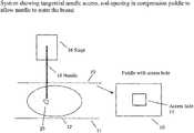
- FIG. 1illustrates lateral needle access, where a breast compression paddle 10 and a breast support plate 14 can be a part of an otherwise known tomosynthesis system such as described in the co-pending patent applications identified above and incorporated by reference in this patent specification, and a biopsy needle stage 16 and a needle 18 such as used, for example, in the patents identified above that pertain to prone biopsy.
- a biopsy needle stage 16 and a needle 18such as used, for example, in the patents identified above that pertain to prone biopsy.
- FIG. 1illustrates lateral needle access, where a breast compression paddle 10 and a breast support plate 14 can be a part of an otherwise known tomosynthesis system such as described in the co-pending patent applications identified above and incorporated by reference in this patent specification, and a biopsy needle stage 16 and a needle 18 such as used, for example, in the patents identified above that pertain to prone biopsy.
- FIG. 1illustrates lateral needle access, where a breast compression paddle 10 and a breast support plate 14 can be a part of an otherwise known
- image reconstructioncan be performed using filtered back projection (for rapid speed of reconstruction) and/or artifact reduction methods (such as ordered statistics backprojection), as disclosed, for example, in U.S. Patent Application Publication No. 2002/0113681, the entire contents of which are incorporated by reference herein.
- a patient's breast 12is compressed between paddle 10 and support plate 14 and a needle biopsy stage 16 has been used to position the tip of a biopsy needle 18 near an area of interest 20 in breast 12 .
- needle 18enters the breast 12 generally laterally, i.e. along the plane of support plate 14 and along the chest wall of the patient, and from the left as seen in the drawing.
- the needle 18can enter instead from the right, and need not be exactly parallel to support plate 14 or to the chest wall, but can be at any angle thereto that the health professional doing the needle biopsy finds suitable for the particular patient or area of interest location.
- the location of area of interest 20has been determined based on tomosynthesis images that can be tomo projection images and/or tomo reconstructed slice images.
- the patient's chestis behind the illustrated structure and is generally along the plane of the sheet. If upright biopsy is used, the patient's chest wall would be generally vertical; if a prone biopsy table is used, the patient's chest wall would be generally horizontal.
- FIG. 2illustrates frontal needle access in which needle 18 accesses area of interest 20 from the front of breast 12 , in a direction generally along the plane of support plate 14 and normal to chest wall 22 of the patient.
- the needle 18 directionneed not be exactly parallel to support plate 14 or normal to chest wall 22 , but can be at any convenient angle thereto that would allow the tip of needle 18 to reach area of interest 20 generally from the front of the breast 12 , at either side of the nipple.
- the patient's chest wall 22is generally normal to the sheet.
- FIG. 3 aillustrates tangential needle access, where a breast compression paddle 10 has, as seen in FIG. 3 b , one or more needle access holes 11 .
- FIG. 3 aillustrates breast compression paddle 10 , breast 12 and support plate 14 in a view similar to that of FIG. 1 , but a needle stage 16 and needle 18 at a position above the breast. Needle 18 accesses area of interest 20 in a direction generally normal to support plate 14 and along the chest wall (not shown) of the patient. Again, needle 18 need not be at the angles shown but may be at any angle that the health professional doing the biopsy finds suitable.
- FIG. 4illustrates one type of a tomo scan that can be used to reduce undesirable image artifacts due to the presence of a biopsy needle 18 and possibly other radio-opaque materials. While FIG. 4 illustrates tangential needle access similar to that of FIG. 3 , the principles discussed below in connection with FIGS. 4 and 5 apply to any other type of access to area of interest 20 .
- FIG. 4illustrates positions 24 a , 24 b , . . . , 24 n of an x-ray tube (not shown) from which the tube emits x-ray beams for taking tomo projection images.
- positions 24 a - 24 nare illustrated as being along an arcuate path, they can be along a differently shaped path, and scanning can start from either end of the path, or from an intermediate positions along the path. As evident from FIG. 4 , it is likely that at some positions of the scan, needle stage 16 would obscure at least a significant part of the imaging x-ray beam and the resulting projection image is likely to have significant and probably unacceptable artifacts.
- FIG. 5is otherwise similar to FIG. 4 but illustrates a gap region 26 in the path of x-ray tube positions 24 a - 24 n .
- No x-ray tomo exposuresare taken from positions in this gap region 26 .
- Exposuresare taken from positions outside this region to minimize or at least significantly reduce the extent to which the needle stage 16 and any other x-ray opaque materials affect the imaging x-ray beams and thus reduce undesirable artifacts in the images relative to images that could have been obtained with exposures taken from positions in gap 26 .
- Sufficient tomo projection imagescan be taken from positions outside gap 26 from which acceptable tomo reconstructed images of breast slices can be computed to localize needle 18 relative to area of interest 20 .
- Gap 26can be at an end of the path of positions 24 a - 24 n or it can be intermediate positions 24 a - 24 n .
- Different x-ray dosecan be used for different ones of positions 24 a - 24 n , e.g. less dose for exposure positions in which radio-opaque materials in the path of the x-ray beam are likely to generate more undesirable artifacts, and greater dose for positions in which such material are less likely to produce such artifacts. It is possible to take exposures even from positions in gap 26 , preferably at low x-ray dose, but not use the resulting projection images for reconstructing tomo images of breast slices.
- metallic breast biopsy needlescan obstruct the sampled lesion or cause other undesirable artifacts such as, for example, streaking artifacts in reconstructed tomosynthesis images. This is especially acute where the sampled lesions are calcifications. This obstruction can reduce the accuracy of biopsy.
- Embodiments of the present disclosureinclude a needle design that allows for better visibility of the sampled lesion.
- FIGS. 6-8Several embodiments of such needles are shown in FIGS. 6-8 .
- x-ray transparent materialis used in the construction of the stem of the needle to a significant extent so the sampled lesions can be seen more clearly when imaged with tomosynthesis or 2D mammography.
- the needle stemshould still be solid enough to cut the tissue and the lesion.
- the x-ray transparent materialneed not be perfectly transparent but only sufficiently transparent to minimize or at least significantly reduce undesired image artifacts as compared with the use of metallic needles without such material.
- the term “x-ray transparent”is used in this sense in this patent disclosure.
- FIGS. 6A-6Dillustrates the use of a breast biopsy needle with an x-ray transparent body according to embodiments of the present disclosure.
- the needle 30consists of two metallic tips 32 for cutting the tissue and lesion 38 , and two needle stems 34 made of x-ray transparent material so as not to block x-rays. Because the needle stems 34 are x-ray transparent, the position of the needles may be determined by the position of the needle tips 32 in x-ray images and the known needle geometry.
- FIG. 6Aillustrates the needle 30 prior to its firing. The relative location of the needle and the lesion 38 are confirmed using x-ray tomosynthesis (or 2D x-ray mammography).
- FIG. 6Billustrates that one of the two needle stems 34 may have a notch 36 .
- the notched needle stem 34 amay be within the lumen or cannula of the un-notched needle stem 34 b .
- the notched needle stem 34 amay be fired from the un-notched needle stem 34 b such that the notch is placed in proximity to the lesion 38 .
- FIG. 6Cillustrates that the un-notched stem 34 b may be pushed to close around the notched stem 34 a thereby cutting and trapping the lesion 38 , or at least a part thereof, within the notch 36 and the cannula of the un-notched stem 34 b .
- FIG. 6Dillustrates that the needle may be removed from the patient with the trapped lesion 38 .
- Tomosynthesis or 2D mammographymay then be used to confirm that the lesion 38 has been correctly sampled.
- FIGS. 7A-7Eillustrate the use of a breast biopsy needle with an x-ray transparent body stiffened with metal according to another embodiment of the present disclosure.
- the needle 40comprises two metallic tips 42 (to cut the tissue and lesion 48 ), and two needle stems 44 made of x-ray transparent material (so as not to block or scatter x-rays excessively) and removable solid metallic wires or ribs 50 to enhance the structural integrity of the needle stems during the firing.
- the wires or ribs 50can be removed from the stems, after firing, to allow the needle stems 44 to be x-ray transparent and the taking of x-ray images after the wires or ribs 50 have been withdrawn. As seen in FIG.
- the relative location of the needle 40 and the lesion 48may be confirmed using tomosynthesis or 2D mammography.
- a notched needle stem 44 amay be fired from an un-notched needle stem 44 b .
- the un-notched needle stem 44 bmay be pushed to the notched needle stem 44 a so as to cut and trap the lesion 48 between the notch 46 of the notched needle stem 44 a and the un-notched needle stem 44 b .
- the metallic wires 50can be removed form the needle stems 44 prior to performing tomosynthesis or 2D mammography to confirm the location of the lesion 48 .
- the needle 40may be removed from the patient with the trapped lesion 48 . Tomosynthesis or 2D mammography may then be used to confirm that the lesion 48 has been correctly sampled.
- FIGS. 8A-8Eillustrate another embodiment of a new breast biopsy needle that comprises two coaxial bodies each having an x-ray transparent (e.g. plastic) layer and an x-ray opaque (e.g. metal) layer that adds mechanical strength or stiffness but can be withdrawn, if desired, after the needle is in place in the breast but before x-ray images are taken.
- needle 60is inserted into the breast until its cutting tips 62 are close to but spaced from suspected lesion 68 .
- the relative locations of the needle and the lesioncan be confirmed by taking tomosynthesis or 2d mammography images. Then, as seen in FIG.
- the notched stylet 64 b(the notch shown as 66 ) of the needle can be fired into lesion 68 to sample it and, as seen in FIG. 8C the cannula 64 a can be pushed in to slice the lesion or at least a part of it into the notch. Then the radio-opaque metal layers can be withdrawn from each of the cannula and the stylet to leave the x-ray transparent structure seen in FIG. 8D (except for its cutting tips 62 ). At this time, post-fire tomosynthesis or 2D mammography images can be taken to confirm that the lesion or a part of it is in the notch.
- the core systemcan then be pulled back with the lesion sample, e.g. to the position illustrated in FIG. 8E .
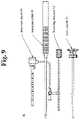
- FIG. 9illustrate another example of a new breast biopsy needle.
- Biopsy needle 90is configured as “tube-within-a-tube” cutting device and includes an outer cannula 91 , an inner cannula (or localizing obturator) 92 , an introducer stylet 93 and an introducer sheath 94 .
- the outer cannula 91 , localizing obturator 92 , introducer stylet 93 and introducer sheath 94can be mounted to a handpiece (not shown) or an attachment (not shown) which is in turn coupled to a support fixture or positioning device for moving the biopsy needle to a desired position.
- the outer cannula 91defines an outer lumen and terminates in a tip which is preferably a trocar tip that can be used to penetrate the patient's skin.
- the localizing obturator 92fits concentrically within the outer cannula 91 .
- the localizing obturator 92can be driven by a rotary motor and a reciprocating motor drive to translate the localizing obturator 92 axially within the outer cannula 91 , while rotating the localizing obturator 92 about its longitudinal axis (or the localizing obturator 92 can be rotated and/or translated manually).
- the introducer stylet 93which is inserted in the annular introducer sheath 94 can be inserted.
- the introducer stylet 93 and/or sheath 94can be radiolucent with a radio-opaque band at a distal end thereof.
- breast biopsy needlesare disclosed in U.S. Pat. Nos. 6,638,235, 6,758,824, 6,620,111 and 6,626,849 and U.S. Publications Nos. 2006/0155209 A1, 2006/0129062 A1, 2006/0030784 A1, 2005/0113715 A1, 2005/0049521 A1, and 2004/0267157 A1, the entire contents of which are incorporated herein by reference.
- this patent specificationdiscloses a method and a system in which tomosynthesis reconstructed images of slices of a patient's breast and/or tomosynthesis projection images of the breast are used to (1) identify the location of a suspected area of interest in the breast, (2) guide needle biopsy of the area of interest, (3) confirm pre-fire position of the needle relative to the area of interest, and/or (4) confirm post-fire position of the needle relative to the area of interest.
- One unique benefit of this approachis with respect to suspected pathologies that can be seen or assessed better in tomosynthesis images than in conventional mammograms or in conventional ultrasound images of breast tissue.
- the method and systeminvolve taking a series of tomosynthesis projection images at respective different angles of the imaging x-ray beam relative to the breast, for example in the manner disclosed in said patent applications that are incorporated by reference in this patent specification.
- the information from these projection imagesis reconstructed into images of slices through the breast, which may represent slices of selected thickness and selected angles relative to the breast platform or the imaging plane(s) of the projection images.
- the reconstructed imagesrepresent slices that are parallel to the breast platform and thus to the plane of a conventional mammogram.
- These imagesare used to identify the location of the area of interest in the breast in three dimensions, for example by having the health professional point to the location of the area of interest in one or more images and using the system to compute the 3D coordinates of the location in a manner similar to that used in said biopsy system patents identified above and incorporated by reference in this patent specification, or in a different manner, such as by pointing to the area of interest in a reconstructed slice image to thereby identify the location of the area of interest in two dimensions in the plane of the slice and to provide the third dimension from knowledge of the depth of the slice in the breast.
- This 3D information of the area of interest locationcan be used together with information regarding a geometrical relationship between the equipment in which the breast of compressed and immobilized to determine the direction and extent of biopsy needle motion executed by a needle stage in a manner similar to that disclosed in said patents incorporated by reference herein, to position the needle, to sample the area of interest and to confirm pre-fire and post-fire locations of the needle relative to the area of interest.
- a first new approach in this respectpertains to selection of tomosynthesis images and involves taking projection tomosynthesis images only at angles in which the radio-opaque objects are not in the imaging x-ray beam or, if they are in the beam, their effect in the image is significantly less than it would have been for other possible beam angles. This may involve not taking projection images at angles that would produce more undesirable artifacts and/or taking such projection images but not using them in reconstructing slice images.
- a second new approach that can be used instead of or in addition to the first oneis to carry out post-processing of the tomo images to reduce artifact therein due to the presence of radio-opaque objects in the beam. This can involve processing of the reconstructed slice image, e.g. by using streak artifact removal algorithms similar to those conventionally used in CT (computerized tomography) technology, and/or image processing of the tomo projection images to remove or reduce such artifacts.
- a third new approach that can be used instead of one or more of the first and second, or together with one or both of the first and second,is to use biopsy equipment that reduces or avoids such image artifacts, e.g.
- a biopsy needlethat is made at least partly of a material that is significantly more x-ray transparent than conventional biopsy needles.
- a needle made of such materialcan be used as is for insertion into the breast and for tissue sampling, or it may be stiffened by portions of an x-ray opaque material such as metal that are used for insertion and/or tissue sampling but are withdrawn from the breast or at least from the immediate vicinity of the area of interest before pre-fire and/or post-fire x-ray images can be taken to thereby avoid the image artifacts that such metal would cause if not withdrawn.
- such stiffening portionscan be in the form of pins or ribs inside a cannula.
- theycan be sleeves coaxial with a cannula and/or a stylet.
- Other examples of stiffening portions that are withdrawn before pre-fire and/or post fire imagingalso are contemplated.
- a calcification detector 104is added in a sample delivery path 103 between a biopsy needle 101 and a collection chamber (or filter) 102 .
- the sample delivery path 103typically includes a tube or other channel for delivery of the extracted sample to the collection filter 102 .
- the calcification detector 104can be coupled to the sample delivery path 103 to determine whether the samples include calcifications and estimate an amount of the calcifications.
- the calcification detector 104can include, as an example, an x-ray source and detector for imaging the samples passing through the sample delivery path 103 , and a CAD (computer aided diagnosis) component configured to detect and count the number of calcifications in the samples.
- a tissue biopsy apparatus 110configured as a handheld device (although the apparatus can also be mounted to a support fixture that is used to position the biopsy needle) includes a biopsy needle mounted to a handpiece.
- the biopsy needleincludes an outer cannula 115 terminating in a tip 116 .
- a tissue-receiving opening 125is provided (relatively) near the tip 116 .
- An inner cannula 117fits concentrically within the outer lumen of the outer cannula 115 .
- the inner cannula 117is rotated (for example, by a rotary motor) about its longitudinal axis and is translated axially within the outer cannula 115 (for example, by a reciprocating motor drive).
- the outer cannula 115 terminating, tip 116 , inner cannula 117 and tissue-receiving opening 125interoperate similar to the other examples discussed above to extract biopsy samples of a patient's breast.
- the inner cannula 117provides an avenue for aspiration of the biopsy samples to the tissue aspiration path which also includes aspiration tube 150 coupling the tissue aspiration path to a collection chamber 155 .
- Aspirator 121applied vacuum or aspiration pressure to the collection chamber to draw samples through the tissue aspiration path to the collection chamber 155 .
- X-ray tube 111 and detector 112operate under appropriate control of a controller 114 , and a detection signal representing the x-rays received by the detector 112 from the source 11 I is output to CAD component 113 which decodes the signal to determine whether the samples include calcifications and estimates an amount of the calcifications.
- CAD systems and techniquesare well-known in the art, and therefore a detailed discussion of such systems and techniques is omitted from this disclosure in the interest of clarity and brevity.
- the x-ray tube and detectorwould be small-scaled.
- An example of a small scale detectoris available from Hamamatsu, Corporation, Bridgewater, N.J. (see http://sales.hamamatsu.com/en/products/electron-tube-division/x-ray-products/x-ray-flat-panel-sensor.php).
- Information regarding a small scale x-ray tube(40 kV metal-ceramic X-ray tube from Newton Scientific Inc., Cambridge, Mass.) is available at http://www.newtonscientificinc.com/swans.htm.
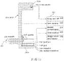
- FIG. 12schematically illustrates a side view of a system comprising x-ray source 100 a at one end 200 a of a C-arm 200 that is supported for selective rotation about an axis 202 , independently of a support 204 for compression paddle 104 a and breast platform 106 a .
- Support 204also selectively rotates about the same axis 202 .
- the other end 200 b of C-arm 200interacts with x-ray receptor 110 a through a motion translator schematically illustrated at 206 that translates the rotational motion of the end 200 b about axis 202 to a substantially translational motion of receptor 110 a that substantially maintains the distance of receptor 110 a from breast platform 106 a while x-ray data is being taken.
- FIG. 12schematically illustrates a side view of a system comprising x-ray source 100 a at one end 200 a of a C-arm 200 that is supported for selective rotation about an axis 202 , independently of a support 204 for compression
- C-arm 200 and support 204are rotated to desired angular positions, either manually or by motor drives, patient breast 102 a is positioned on platform 106 a and is immobilized by bringing paddle 104 a toward platform 106 a and compressing breast 102 a , with typically the same or less force than for a typical conventional mammogram, such as between one to one-third the conventional force.
- imagingstarts, and with C-arm at a selected angle relative to a normal to platform 106 a and receptor 110 a , such as +15°, imaging starts, and a projection image is taken for each of a number of selected angular positions of source 100 a while C-arm 200 rotates, continuously or intermittently, through a selected angle, such as an angle of 30°, i.e. from +15° to ⁇ 15°.
- a selected anglesuch as an angle of 30°, i.e. from +15° to ⁇ 15°.
- the motioncan be in the opposite direction, from ⁇ 15° to +15°, or can be over a different angular interval, such as over less than a total of 30°, e.g. 25°, 20°, etc., or more than 30°, such as 35°, 40°, etc.
- a set of image datacan be taken at selected angular positions, such as every degree, or every fraction of a degree, or every several degrees of angle.
- the angular increments between the different positions for sets of image dataneed not be the same. For example, the increments around 0° can be less than those at the extremes of the angular positions, or vice versa.
- the preferred angular incrementis 3°.
- the sets of image datacan be taken after an incremental motion from one angular position of source 100 a to another, and from one translational position of receptor 110 a to another, such that source 100 a and receptor 110 a are stationary while a set of image data is being taken.
- one or both of source 100 a and receptor 110 acan move continuously while sets of image data are being taken, one set for each increment of continuous motion.
- both source 100 a and receptor 110 amove while image data are being taken.
- FIG. 12also illustrates schematically an electrical/electronic system 208 that interacts with the components discussed above.
- System 208includes a control 210 for selectively energizing and otherwise controlling x-ray source 100 a , an arm rotation control 212 for selectively rotating C-arm 200 and support 204 , a breast compression control 214 for selectively moving compression paddle 104 a toward and away from breast platform 106 a , data readout electronics 216 coupled with x-ray receptor 110 a to read out the sets of image data at the respective positions of source 100 a and receptor 110 a relative to immobilized breast 102 a , and an image reconstruction and display unit 218 coupled with data readout electronics 216 to receive the sets of image data from electronics 216 and to process the image data for reconstruction and other purposes and display images.
- a control 210for selectively energizing and otherwise controlling x-ray source 100 a
- an arm rotation control 212for selectively rotating C-arm 200 and support 204
- source 100 a and receptor 110 acan be positioned relative to immobilized breast 102 a such that at the 0° position a center ray of the x-ray beam from source 100 a would be substantially normal to receptor breast platform 106 a and receptor 110 a .
- source 100 ais at +(or ⁇ ) 15° in a preferred example, and is gradually moved, continuously or intermittently to ⁇ (or +) 15°, with a set of image data taken every 3°.
- the angular range and the increment over which data sets are takencan each be selectively set by the operator, depending of characteristics of the breast being imaged and the screening and diagnostic needs, and can be different for different patients or from one to the other breast of the same patient.
- the sourcecan move through angles that range from a fraction to a degree to several degrees from one imaging position to the next.
- Each set of image datais supplied by image readout 216 for processing at image reconstruction and display unit 218 .
- Each set of image datacan be taken at the same x-ray dose to the breast, and the dose at any one of the different imaging positions can be substantially less than that for a conventional mammogram.
- the x-ray dosecan be substantially the same for each imaging position, but preferably the dose at one of the position, e.g., at or close to the 0° position, is the same or similar to dose for a conventional mammogram while the dose at the each of the other positions is less, preferably much less.
- the scancan begin with or end with an exposure close to the 0° position at a dose similar to a conventional mammogram, and the rest of the set of image data can be over the angular range with each exposure at an x-ray dose that is substantially less than that for a conventional mammogram.
- two types of imagescan be produced in accordance with the currently preferred embodiment while breast 102 a is immobilized in the same position.
- One typeis the same or is at least similar to a conventional mammogram, which can be read and interpreted in the manner familiar to health professionals.
- the other typeis tomosynthetic images reconstructed from the image data and displayed either separately or as an adjunct to the display of the image that is the same or similar to a conventional mammogram.
- the process described above for one position of breast 102 acan be repeated for another position.
- one processcan be for a breast position in a manner that is the same or similar to positioning the breast for a conventional CC view, the breast can then be released, the support 204 and C-arm 200 rotated to other angular positions and the breast repositioned in a manner that is the same and similar to the position for an MLO view, and the procedure repeated.
- receptor 110 ais affixed to the end 200 b of C-arm 200 that is opposite x-ray source 100 a .
- receptor 110 amoves relative to immobilized breast 102 a along an arcuate path from one imaging position to another. Because the change in angle between receptor 110 a and breast platform is small, it can be disregarded in processing the sets of x-ray image data.
- a geometric correction known to those skilled in the artcan be applied to each set of image data to convert it to interpolated pixel values that would correspond to those that would have been obtained if receptor 110 a had been parallel to and at the same distance from platform 106 a at all imaging positions.
- the so corrected sets of image datacan then be used in filtered back projections as described above.
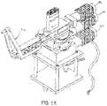
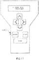
- FIG. 15illustrates a biopsy needle positioning mechanism that is typically employed as a component of an overall mammographic needle biopsy system, and comprises a conventional puncture instrument 10 a for retaining a biopsy needle or other biopsy or therapeutic delivery device (not illustrated).
- Three conventional DC motors 12 a , 14 a , and 16 aare provided for moving the biopsy needle retained by the puncture instrument 10 a in the rotation and angulation axes and for setting a stop position along the depth axis, respectively.
- Positional feedbackis provided to the biopsy needle positioning motor controller by the three DC motors 12 a , 14 a , and 16 a .
- the operator hand controllerallows the clinician user to control the motorized biopsy needle positioning system.
- Controlsare provided to permit the user to initiate movement of the biopsy needle into a position for insertion to the identified point of interest within the patient's breast, in accordance with the computed spatial coordinates of that point of interest.
- the position of the biopsy needlemay be monitored by the user with reference to a 32-character display on the operator hand controller.
- An enable switchis provided to prevent inadvertent motion of the biopsy needle.
- the remote view and display boxreceives the spatial coordinates of rotation, angulation, and depth from the biopsy needle positioning motor controller and displays them for the benefit of the clinician user or others on a 40-character alphanumeric display.
- the remote view and display boxmay be conveniently mounted on a table that includes means for mounting and lighting x-ray reference films to be viewed during a breast biopsy procedure.
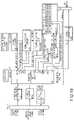
- the biopsy needle positioning motor controllerreceives the spatial coordinates of the identified point of interest within the patient's breast from the film digitizer and coordinates calculator and computes the variables required to drive the three DC motors 12 a , 14 a , and 16 a that form part of the biopsy needle positioning mechanism. Information regarding the position of the biopsy needle is continuously provided by the biopsy needle positioning motor controller to the LED displays in the operator hand controller.
- the biopsy needle positioning motor controllerreceives commands from the operator hand controller and drives the biopsy needle positioning mechanism in the direction specified for as long as the user simultaneously depresses one of the direction arrow keys and the enable switch located on the operator hand controller illustrated in FIG. 17 .
- a central processing unit (8032 CPU) within the biopsy needle positioning motor controllerhas a direct serial communications link with the remote view and display box through an RS422 serial transmitter U29.
- the 8032 CPUalso has two bi-directional communications links through a dual synchronous universal transmitter/receiver DUART, which provides serial communications between the biopsy needle positioning motor controller and both the film digitizer and coordinates calculator (serial channel B) and the operator hand controller (serial channel A).
- the 8032 CPUloads the three DC motor controller sections (rotation, angulation, and depth) with high level initial conditions data.
- This initial conditions dataincludes velocity constants, acceleration constants, PID filter information, and sample period.
- the 8032 CPUreads these spatial coordinates and calculates the corresponding motor control values.
- the 8032 CPUthen sends this data to the three motor control sections.
- the motor control sectionscalculate the actual motor drive voltages and provide the drive voltages to motors 12 a , 14 a , and 16 a through separate H-bridge circuits.
- the motor control sectionsmonitor the encoder feedback from the biopsy needle positioning mechanism to determine the position of the biopsy needle and to adjust the motor drive voltages as the biopsy needle reaches the identified point of interest.
- a typical motor voltage and velocity profileis trapezoidal in nature, ramping up to a start voltage, then holding constant, and finally ramping down to a stop voltage when the biopsy needle has reached the position required for insertion to the identified point of interest.
- the 8032 CPU support circuitsinclude operating and debug program data in erasable programmable read-only memories EPROMs U1 and U6. Fourteen status bits plus a six-bit DIP switch are monitored through an input port and a random access memory RAM U15. The status bits include +/ ⁇ limit switches and a home switch associated with each coordinate axis. Two additional status bits serve to monitor the +5-volt (+5ENC) and +24-volt (+24VOK) power supplies.
- a reset circuit U23provides a reset signal to reset the 8032 CPU when power is initially applied. The reset circuit also monitors program execution by counting a pulse associated with each cycle of the program and by executing a CPU reset command if the pulses stop, as may occur during a software lockup.
- FIGS. 17 and 18it will be understood how the operator hand controller of FIG. 17 transmits data to and receives data and instructions from the biopsy needle positioning motor controller via an RS422 serial transmitter/receiver bus (serial channel A). While the operator hand controller is described herein as being a hand-held unit, it may also comprise a console or table-mounted unit.
- the principal functions of the operator hand controllerare to 1) transmit switch closure data resulting from actuation of the direction arrow keys and the MANUAL, OFFSET, and TARGET keys to the biopsy needle positioning motor controller; 2) illuminate button LEDs in accordance with information received from the biopsy needle positioning motor controller, and 3) display the spatial coordinates of the identified point of interest within the patient's breast, as provided by the biopsy needle positioning motor controller. Additionally, the operator hand controller provides a safety interlock through the ENABLE switch SW9, which must be simultaneously depressed by the user with a selected one of the function keys in order to initiate any of the functions of the operator hand controller.
- the ENABLE switchis mounted on the side of the operator hand controller and, when depressed, energizes a relay in the biopsy needle positioning motor controller that enables movement of the biopsy needle positioning mechanism. When this switch opens, the relay removes power from the three DC motors 12 a , 14 a , and 16 a of the biopsy needle positioning mechanism.
- the clinician userinitiates control of the biopsy needle positioning mechanism in either an automatic or manual mode by depressing control switches on the operator hand controller.
- Depressing one of the arrow keys or one of the MANUAL, OFFSET or TARGET keyshas the effect of grounding a corresponding input of serial encoder U13.
- serial encoder U13to apply an INTERRUPT 0 (INTOO) to the CPU U9 and place the serial data in 12C protocol on the serial lines SDA and SCL to the CPU U9.
- the CPU U9converts the switch information to RS422 protocol and sends it to the biopsy needle positioning motor controller via serial transmitter U15.
- Each of the keys on the operator hand controllercontains a light emitting diode LED that is illuminated under the control of the biopsy needle positioning motor controller.
- the biopsy needle positioning motor controllerselects a particular LED to be illuminated, sets the brightness of that LED, and determines how long that LED is to remain illuminated. This information is sent to the CPU U9 via serial receiver U15. The CPU U9 then places the information in 12C protocol on the serial lines SDA and SCL to be transmitted to serial decoder/driver U14. Ser. decoder/driver U14 pulls a corresponding output to its low state, thereby illuminating the selected LED. The CPU U9 controls the brightness of the LEDs on the operator hand controller by setting the duty cycle of BRIGHTNESS (BL) pulses applied to the LEDS. A 50% duty cycle illuminates the LEDs at half brightness and a 100% duty cycle illuminates the LEDs at full brightness.
- BRIGHTNESSBRIGHTNESS
- the position readout displays U1-U8 in the operator hand controllerprovide two rows of displayed information comprising 16 ASCII characters in each row. Each row comprises four display devices, and each display device contains four 5.times.7 dot matrix character displays.
- the top line of the position readout displayindicates target number 2 (2:), a rotation axis angle of 10.32 degrees right (10.32R), and an angulation axis angle of 9.72 degrees up (9.72U).
- the bottom line of the position readout displayindicates a depth stop setting of 135.6 millimeters (135.6 mm depth).
- the biopsy needle positioning motor controllersimilarly controls the position readout displays through serial communications with the operator hand controller CPU U9.
- the CPU U9provides segment selection control and character display using two data buses DDO-DD7 and DAO-DA4. To display a selected ASCII character, the CPU U9 puts data describing the character on the DDO-DD7 (P3.0-P3.6 outputs of the CPU U9) bus. The CPU U9 transmits a low signal FNABLE (DWR) to segment decoder U10, which decodes bits DA2-DA4 and applies a low enable signal to the appropriate ones of display device U1-US. The enabled display device then decodes the character select bit DAO and DA1 to select the character position which displays the ASCII character defined by data bus DDO-DD6. As with the LEDs, the biopsy needle positioning motor controller defines the brightness of the position readout display. The biopsy needle positioning motor controller communicates the brightness level to the CPU U9, which then switches the BRIGHTNESS (BL) signal on and off, producing the designated duty cycle.
- BLBRIGHTNESS
- an additional line 106can be added for introducing anesthetic and/or contrast agents, for example, along with a flushing agent or lavage.
- the introduction of the anesthetic and/or contrast agentscan be automated and synchronized to the imaging sequence.
Landscapes
- Health & Medical Sciences (AREA)
- Life Sciences & Earth Sciences (AREA)
- Engineering & Computer Science (AREA)
- Medical Informatics (AREA)
- Surgery (AREA)
- Veterinary Medicine (AREA)
- Heart & Thoracic Surgery (AREA)
- Molecular Biology (AREA)
- Biomedical Technology (AREA)
- Animal Behavior & Ethology (AREA)
- General Health & Medical Sciences (AREA)
- Public Health (AREA)
- Pathology (AREA)
- Nuclear Medicine, Radiotherapy & Molecular Imaging (AREA)
- Physics & Mathematics (AREA)
- Biophysics (AREA)
- High Energy & Nuclear Physics (AREA)
- Optics & Photonics (AREA)
- Radiology & Medical Imaging (AREA)
- Human Computer Interaction (AREA)
- Oral & Maxillofacial Surgery (AREA)
- Dentistry (AREA)
- Neurosurgery (AREA)
- Oncology (AREA)
- Apparatus For Radiation Diagnosis (AREA)
Abstract
Description
- a. Development of needle and other sheathing materials that are sufficiently radiolucent that they will not create significant image artifacts. Possible materials are plastics, ceramics, glasses, carbon tubes, and low atomic number metals and other materials. If these materials are used, they can be marked with fiducial markings such as radio-opaque rings or dots allowing visibility in the tomosynthesis images so they can be differentiated from breast tissue or breast area of interest. Alternately, a needle can be used where only the tip (last 1-3 cm) is radiolucent and rest of the needle is radio-opaque.
- b. Scanning through angles that do not shadow these objects. This can entail an asymmetric scan geometry, whereby all or an important part of the x-ray beam path does not pass through the needle or other radio-opaque parts. An example is scanning to just one side of the needle.
- c. Scanning over a large range, and generally or always avoiding x-ray exposures when the stage or other radio-opaque parts shadow the breast, area of interest or image receptor. Alternatively, x-ray imaging can be done even in angular areas with this shadow problem, but these exposures can be eliminated from viewing or reconstruction, either automatically or through manual elimination via a user interface. Another alternate method involves artifact suppression algorithms used during reconstruction, as in known in tomosynthesis and CT scanning.
- d. Stereotactic imaging. Conventional stereotactic imaging involves using a pair of x-ray images at, for example, ±15° to the normal to the compression paddle. This geometry involves sufficiently large angles to typically avoid the stage shadows on the image receptor. A tomo system can be used to take tomo projection images at angles that avoid undesirable shadows at relevant parts of the images.
- e. Scan angle changes. A larger scan angle than used in conventional tomo imaging can better avoid artifacts from the stage.
- f. Bringing the needle to a fixed distance from the lesion. An image can then be taken that does not obscure the area of interest, and the proper distance between needle and area of interest can be verified from imaging. The needle can then be advanced into the correct location within the area of interest based on information from the tomo or conventional imaging while the needle is spaced from the area of interest.
- g. In many if not most cases the projection images and perhaps the reconstructed images of breast slices will contain at some location an image of the needle. The needle image can create artifacts in reconstructed images, which can be removed via artifact reduction algorithms as in known in conventional tomosynthesis and CT imaging. One algorithm can involve skipping projection images with extensive shadowing in the projections. Another algorithm can involve segmenting out the needle and other high contrast objects and avoiding reconstruction using these pixels, as has been used in CT and other imaging. Other alternatives include viewing the projection images, which can have images of the needles but no other significant artifacts.
Claims (20)
Priority Applications (3)
| Application Number | Priority Date | Filing Date | Title |
|---|---|---|---|
| US16/434,064US11452486B2 (en) | 2006-02-15 | 2019-06-06 | Breast biopsy and needle localization using tomosynthesis systems |
| US16/936,550US11918389B2 (en) | 2006-02-15 | 2020-07-23 | Breast biopsy and needle localization using tomosynthesis systems |
| US18/417,682US12193853B2 (en) | 2006-02-15 | 2024-01-19 | Breast biopsy and needle localization using tomosynthesis systems |
Applications Claiming Priority (5)
| Application Number | Priority Date | Filing Date | Title |
|---|---|---|---|
| US77414206P | 2006-02-15 | 2006-02-15 | |
| US11/707,587US8532745B2 (en) | 2006-02-15 | 2007-02-15 | Breast biopsy and needle localization using tomosynthesis systems |
| US14/021,624US9901309B2 (en) | 2006-02-15 | 2013-09-09 | Breast biopsy and needle localization using tomosynthesis systems |
| US15/904,735US10335094B2 (en) | 2006-02-15 | 2018-02-26 | Breast biopsy and needle localization using tomosynthesis systems |
| US16/434,064US11452486B2 (en) | 2006-02-15 | 2019-06-06 | Breast biopsy and needle localization using tomosynthesis systems |
Related Parent Applications (1)
| Application Number | Title | Priority Date | Filing Date |
|---|---|---|---|
| US15/904,735ContinuationUS10335094B2 (en) | 2006-02-15 | 2018-02-26 | Breast biopsy and needle localization using tomosynthesis systems |
Related Child Applications (1)
| Application Number | Title | Priority Date | Filing Date |
|---|---|---|---|
| US16/936,550ContinuationUS11918389B2 (en) | 2006-02-15 | 2020-07-23 | Breast biopsy and needle localization using tomosynthesis systems |
Publications (2)
| Publication Number | Publication Date |
|---|---|
| US20200046303A1 US20200046303A1 (en) | 2020-02-13 |
| US11452486B2true US11452486B2 (en) | 2022-09-27 |
Family
ID=38372135
Family Applications (6)
| Application Number | Title | Priority Date | Filing Date |
|---|---|---|---|
| US11/707,587Active2032-02-22US8532745B2 (en) | 2006-02-15 | 2007-02-15 | Breast biopsy and needle localization using tomosynthesis systems |
| US14/021,624ActiveUS9901309B2 (en) | 2006-02-15 | 2013-09-09 | Breast biopsy and needle localization using tomosynthesis systems |
| US15/904,735ActiveUS10335094B2 (en) | 2006-02-15 | 2018-02-26 | Breast biopsy and needle localization using tomosynthesis systems |
| US16/434,064Active2027-05-10US11452486B2 (en) | 2006-02-15 | 2019-06-06 | Breast biopsy and needle localization using tomosynthesis systems |
| US16/936,550Active2027-08-07US11918389B2 (en) | 2006-02-15 | 2020-07-23 | Breast biopsy and needle localization using tomosynthesis systems |
| US18/417,682ActiveUS12193853B2 (en) | 2006-02-15 | 2024-01-19 | Breast biopsy and needle localization using tomosynthesis systems |
Family Applications Before (3)
| Application Number | Title | Priority Date | Filing Date |
|---|---|---|---|
| US11/707,587Active2032-02-22US8532745B2 (en) | 2006-02-15 | 2007-02-15 | Breast biopsy and needle localization using tomosynthesis systems |
| US14/021,624ActiveUS9901309B2 (en) | 2006-02-15 | 2013-09-09 | Breast biopsy and needle localization using tomosynthesis systems |
| US15/904,735ActiveUS10335094B2 (en) | 2006-02-15 | 2018-02-26 | Breast biopsy and needle localization using tomosynthesis systems |
Family Applications After (2)
| Application Number | Title | Priority Date | Filing Date |
|---|---|---|---|
| US16/936,550Active2027-08-07US11918389B2 (en) | 2006-02-15 | 2020-07-23 | Breast biopsy and needle localization using tomosynthesis systems |
| US18/417,682ActiveUS12193853B2 (en) | 2006-02-15 | 2024-01-19 | Breast biopsy and needle localization using tomosynthesis systems |
Country Status (5)
| Country | Link |
|---|---|
| US (6) | US8532745B2 (en) |
| EP (1) | EP1986548B1 (en) |
| JP (1) | JP5554927B2 (en) |
| DE (1) | DE202007019497U1 (en) |
| WO (1) | WO2007095330A2 (en) |
Families Citing this family (149)
| Publication number | Priority date | Publication date | Assignee | Title |
|---|---|---|---|---|
| US8109885B2 (en) | 2002-03-19 | 2012-02-07 | C. R. Bard, Inc. | Biopsy device for removing tissue specimens using a vacuum |
| EP1524940B1 (en) | 2002-03-19 | 2011-08-24 | Bard Dublin ITC Limited | Biopsy device and biopsy needle module that can be inserted into the biopsy device |
| US8565372B2 (en) | 2003-11-26 | 2013-10-22 | Hologic, Inc | System and method for low dose tomosynthesis |
| US7616801B2 (en) | 2002-11-27 | 2009-11-10 | Hologic, Inc. | Image handling and display in x-ray mammography and tomosynthesis |
| US8571289B2 (en) | 2002-11-27 | 2013-10-29 | Hologic, Inc. | System and method for generating a 2D image from a tomosynthesis data set |
| US7123684B2 (en)* | 2002-11-27 | 2006-10-17 | Hologic, Inc. | Full field mammography with tissue exposure control, tomosynthesis, and dynamic field of view processing |
| US7577282B2 (en) | 2002-11-27 | 2009-08-18 | Hologic, Inc. | Image handling and display in X-ray mammography and tomosynthesis |
| US10638994B2 (en) | 2002-11-27 | 2020-05-05 | Hologic, Inc. | X-ray mammography with tomosynthesis |
| DE10314240B4 (en) | 2003-03-29 | 2025-05-28 | Bard Dublin Itc Ltd. | Pressure generation unit |
| JP4814229B2 (en) | 2004-07-09 | 2011-11-16 | バード ペリフェラル ヴァスキュラー インコーポレイテッド | Transport device for biopsy device |
| US7662082B2 (en) | 2004-11-05 | 2010-02-16 | Theragenics Corporation | Expandable brachytherapy device |
| US7702142B2 (en) | 2004-11-15 | 2010-04-20 | Hologic, Inc. | Matching geometry generation and display of mammograms and tomosynthesis images |
| EP1816965B1 (en) | 2004-11-26 | 2016-06-29 | Hologic, Inc. | Integrated multi-mode mammography/tomosynthesis x-ray system |
| US7517321B2 (en) | 2005-01-31 | 2009-04-14 | C. R. Bard, Inc. | Quick cycle biopsy system |
| JP4991723B2 (en) | 2005-08-10 | 2012-08-01 | シー・アール・バード・インコーポレーテッド | Single insertion multiple sampling biopsy device with integrated marker |
| ES2539578T3 (en) | 2005-08-10 | 2015-07-02 | C.R. Bard, Inc. | Multi-sample biopsy device and single insert with various transport systems |
| EP1921998B8 (en) | 2005-08-10 | 2021-07-07 | C.R.Bard, Inc. | Single-insertion, multiple sampling biopsy device with linear drive |
| US7465268B2 (en) | 2005-11-18 | 2008-12-16 | Senorx, Inc. | Methods for asymmetrical irradiation of a body cavity |
| WO2007095330A2 (en) | 2006-02-15 | 2007-08-23 | Hologic Inc | Breast biopsy and needle localization using tomosynthesis systems |
| US8386013B2 (en)* | 2006-04-13 | 2013-02-26 | The Regents Of The University Of California | Magnetic resonance imaging (MRI) using ultra short echo times and spiral sampling in K-space |
| US8120358B2 (en) | 2006-04-13 | 2012-02-21 | The Regents Of The University Of California | Magnetic resonance imaging with high spatial and temporal resolution |
| EP3417792B1 (en) | 2006-08-21 | 2022-03-02 | C. R. Bard, Inc. | Self-contained handheld biopsy needle |
| SI2086418T1 (en) | 2006-10-06 | 2011-05-31 | Bard Peripheral Vascular Inc | Tissue handling system with reduced operator exposure |
| US8262586B2 (en) | 2006-10-24 | 2012-09-11 | C. R. Bard, Inc. | Large sample low aspect ratio biopsy needle |
| CN101534715B (en) | 2006-11-10 | 2012-01-11 | 皇家飞利浦电子股份有限公司 | Metal artefact prevention during needle guidance under (xper) ct |
| US20080221478A1 (en)* | 2007-03-07 | 2008-09-11 | Ritchie Paul G | Integrated Imaging and Biopsy System with Integrated Control Interface |
| US7602184B2 (en)* | 2007-04-30 | 2009-10-13 | The Regents Of The University Of California | Magnetic resonance spectroscopic imaging with short echo times |
| US7630533B2 (en) | 2007-09-20 | 2009-12-08 | Hologic, Inc. | Breast tomosynthesis with display of highlighted suspected calcifications |
| US8241225B2 (en) | 2007-12-20 | 2012-08-14 | C. R. Bard, Inc. | Biopsy device |
| US7854706B2 (en) | 2007-12-27 | 2010-12-21 | Devicor Medical Products, Inc. | Clutch and valving system for tetherless biopsy device |
| DE102008006358A1 (en)* | 2008-01-28 | 2009-07-30 | Siemens Aktiengesellschaft | X-ray image recording method and apparatus for stereotactic biopsy |
| US20090253997A1 (en)* | 2008-04-03 | 2009-10-08 | Convergent Medical Solutions, Inc. | Skin biopsy with automated lesion stabilization and resection |
| US7792245B2 (en) | 2008-06-24 | 2010-09-07 | Hologic, Inc. | Breast tomosynthesis system with shifting face shield |
| US7991106B2 (en) | 2008-08-29 | 2011-08-02 | Hologic, Inc. | Multi-mode tomosynthesis/mammography gain calibration and image correction using gain map information from selected projection angles |
| US8942342B2 (en)* | 2008-12-29 | 2015-01-27 | Analogic Corporation | Multi-modality image acquisition |
| US9579524B2 (en) | 2009-02-11 | 2017-02-28 | Hologic, Inc. | Flexible multi-lumen brachytherapy device |
| US9248311B2 (en) | 2009-02-11 | 2016-02-02 | Hologic, Inc. | System and method for modifying a flexibility of a brachythereapy catheter |
| WO2010107424A1 (en) | 2009-03-16 | 2010-09-23 | C.R. Bard, Inc. | Biopsy device having rotational cutting |
| JP5346654B2 (en)* | 2009-03-31 | 2013-11-20 | キヤノン株式会社 | Radiation imaging apparatus and control method thereof |
| JP5373450B2 (en)* | 2009-03-31 | 2013-12-18 | 富士フイルム株式会社 | Biopsy device and method of operating biopsy device |
| JP5334657B2 (en)* | 2009-04-14 | 2013-11-06 | 富士フイルム株式会社 | Radiation image processing apparatus and method, and program |
| AU2009344276B2 (en) | 2009-04-15 | 2014-06-05 | C.R. Bard, Inc. | Biopsy apparatus having integrated fluid management |
| US10207126B2 (en) | 2009-05-11 | 2019-02-19 | Cytyc Corporation | Lumen visualization and identification system for multi-lumen balloon catheter |
| US8206316B2 (en) | 2009-06-12 | 2012-06-26 | Devicor Medical Products, Inc. | Tetherless biopsy device with reusable portion |
| US8529468B2 (en) | 2009-07-01 | 2013-09-10 | Suros Surgical Systems, Inc. | Surgical system |
| JP5355271B2 (en)* | 2009-07-24 | 2013-11-27 | 富士フイルム株式会社 | Radiation imaging equipment |
| US9173641B2 (en) | 2009-08-12 | 2015-11-03 | C. R. Bard, Inc. | Biopsy apparatus having integrated thumbwheel mechanism for manual rotation of biopsy cannula |
| US8430824B2 (en) | 2009-10-29 | 2013-04-30 | Bard Peripheral Vascular, Inc. | Biopsy driver assembly having a control circuit for conserving battery power |
| US8485989B2 (en) | 2009-09-01 | 2013-07-16 | Bard Peripheral Vascular, Inc. | Biopsy apparatus having a tissue sample retrieval mechanism |
| ES2862525T3 (en)* | 2009-10-08 | 2021-10-07 | Hologic Inc | Needle Breast Biopsy System and Method of Use |
| US8597206B2 (en) | 2009-10-12 | 2013-12-03 | Bard Peripheral Vascular, Inc. | Biopsy probe assembly having a mechanism to prevent misalignment of components prior to installation |
| US8597201B2 (en) | 2010-03-30 | 2013-12-03 | Siteselect Medical Technologies, Inc. | Tissue excision device with a flexible transection blade |
| FR2959409B1 (en) | 2010-05-03 | 2012-06-29 | Gen Electric | METHOD FOR DETERMINING A TOOL INSERTION PATH IN A DEFORMABLE TISSUE MATRIX AND ROBOTIC SYSTEM USING THE METHOD |
| JP5986994B2 (en)* | 2010-06-28 | 2016-09-06 | コーニンクレッカ フィリップス エヌ ヴェKoninklijke Philips N.V. | Medical tomosynthesis system |
| DE102010031737A1 (en)* | 2010-07-21 | 2012-01-26 | Siemens Aktiengesellschaft | Device for tissue removal |
| DE102010031738A1 (en)* | 2010-07-21 | 2012-01-26 | Siemens Aktiengesellschaft | Image assisted biopsy removal |
| JP5650467B2 (en) | 2010-08-27 | 2015-01-07 | 富士フイルム株式会社 | Radiation imaging system |
| US9352172B2 (en) | 2010-09-30 | 2016-05-31 | Hologic, Inc. | Using a guide member to facilitate brachytherapy device swap |
| CA2813591C (en) | 2010-10-05 | 2020-09-22 | Hologic, Inc. | Upright x-ray breast imaging with a ct mode, multiple tomosynthesis modes, and a mammography mode |
| US9730659B2 (en) | 2010-11-16 | 2017-08-15 | Analogic Corporation | Multi-modality image acquisition |
| US9492130B2 (en) | 2010-11-24 | 2016-11-15 | Hologic, Inc. | System for improved tissue-handling and in line analysis of the tissue |
| US20120133600A1 (en) | 2010-11-26 | 2012-05-31 | Hologic, Inc. | User interface for medical image review workstation |
| US10342992B2 (en) | 2011-01-06 | 2019-07-09 | Hologic, Inc. | Orienting a brachytherapy applicator |
| JP6057922B2 (en) | 2011-03-08 | 2017-01-11 | ホロジック, インコーポレイテッドHologic, Inc. | System and method for dual energy and / or contrast enhanced breast imaging for screening, diagnosis and biopsy |
| US20120245486A1 (en)* | 2011-03-25 | 2012-09-27 | Anthony Melchiorri | Ghost-core biopsy needle |
| JP5743684B2 (en)* | 2011-04-27 | 2015-07-01 | 富士フイルム株式会社 | Radiographic imaging apparatus and method of operating radiographic imaging apparatus |
| JP5864891B2 (en)* | 2011-04-27 | 2016-02-17 | 富士フイルム株式会社 | Radiation imaging equipment |
| JP5806847B2 (en)* | 2011-04-27 | 2015-11-10 | 富士フイルム株式会社 | Radiographic imaging apparatus and method of operating radiographic imaging apparatus |
| JP5638466B2 (en)* | 2011-06-02 | 2014-12-10 | 富士フイルム株式会社 | Image generating apparatus, radiographic image capturing system, image generating program, and image generating method |
| ES2795416T3 (en) | 2011-09-16 | 2020-11-23 | Hologic Inc | Lateral Arm System for Breast Biopsy |
| US11284869B2 (en) | 2011-09-16 | 2022-03-29 | Hologic, Inc. | Breast biopsy lateral arm system |
| US12042134B2 (en) | 2011-09-16 | 2024-07-23 | Hologic, Inc. | Breast biopsy lateral arm system |
| JP6157491B2 (en) | 2011-11-18 | 2017-07-05 | ホロジック, インコーポレイテッドHologic, Inc. | X-ray mammography and / or breast tomosynthesis using a compression paddle with an inflatable jacket to improve contrast and patient comfort |
| US11259759B2 (en) | 2011-11-18 | 2022-03-01 | Hologic Inc. | X-ray mammography and/or breast tomosynthesis using a compression paddle |
| EP2782505B1 (en) | 2011-11-27 | 2020-04-22 | Hologic, Inc. | System and method for generating a 2d image using mammography and/or tomosynthesis image data |
| JP6240097B2 (en) | 2012-02-13 | 2017-11-29 | ホロジック インコーポレイティッド | How to navigate a tomosynthesis stack using composite image data |
| EP2852352B1 (en)* | 2012-05-04 | 2021-09-01 | Roger Khouri | Surgical tools |
| US20130345550A1 (en)* | 2012-06-25 | 2013-12-26 | Tzachi Rafaeli | Systems and methods for localizing an opaque medical device with nuclear medicine imaging |
| CN105451657A (en) | 2013-03-15 | 2016-03-30 | 霍罗吉克公司 | System and method for navigating tomosynthesis stack including automatic focusing |
| US10092358B2 (en) | 2013-03-15 | 2018-10-09 | Hologic, Inc. | Tomosynthesis-guided biopsy apparatus and method |
| CA2902221A1 (en) | 2013-03-20 | 2014-09-25 | Bard Peripheral Vascular, Inc. | Biopsy device |
| CN113768529A (en)* | 2013-04-26 | 2021-12-10 | 蒂莫西·R·斯坦戈 | X-ray breast imaging system and compression paddle for X-ray breast imaging system |
| CA2925907C (en) | 2013-10-09 | 2022-03-15 | Hologic, Inc. | X-ray breast tomosynthesis enhancing spatial resolution including in the thickness direction of a flattened breast |
| CN103593869B (en)* | 2013-10-12 | 2016-08-10 | 沈阳东软医疗系统有限公司 | A kind of scanning device and method for displaying image thereof |
| EP3060132B1 (en) | 2013-10-24 | 2019-12-04 | Hologic, Inc. | System and method for navigating x-ray guided breast biopsy |
| ES2726985T3 (en) | 2013-11-05 | 2019-10-11 | Bard Inc C R | Biopsy device that has integrated vacuum |
| JP6506769B2 (en) | 2014-02-28 | 2019-04-24 | ホロジック, インコーポレイテッドHologic, Inc. | System and method for generating and displaying tomosynthesis image slabs |
| WO2015147009A1 (en)* | 2014-03-28 | 2015-10-01 | 富士フイルム株式会社 | Radiological image photography system, radiological image photography method, and radiological image photography program |
| JP6162324B2 (en)* | 2014-03-28 | 2017-07-12 | 富士フイルム株式会社 | Radiographic imaging system, radiographic imaging method, and radiographic imaging program |
| US20170049503A1 (en)* | 2014-05-15 | 2017-02-23 | Cosman Medical, Inc. | Electrosurgical system |
| US9713450B2 (en) | 2014-12-15 | 2017-07-25 | General Electric Company | Iterative reconstruction of projection data |
| JP6275030B2 (en) | 2014-12-24 | 2018-02-07 | 富士フイルム株式会社 | Biopsy device and method of operating the same |
| US10568694B2 (en) | 2015-04-22 | 2020-02-25 | General Electric Company | Method and system for performing a guided biopsy using digital tomosynthesis |
| WO2016178656A1 (en) | 2015-05-01 | 2016-11-10 | C. R. Bard, Inc. | Biopsy device |
| WO2017040977A1 (en) | 2015-09-04 | 2017-03-09 | Faxitron Bioptics, Llc | Multi-axis specimen imaging device with embedded orientation markers |
| WO2017059078A1 (en) | 2015-09-30 | 2017-04-06 | Devicor Medical Products, Inc. | Breast support compression pillow |
| WO2017185028A1 (en) | 2016-04-22 | 2017-10-26 | Hologic, Inc. | Tomosynthesis with shifting focal spot x-ray system using an addressable array |
| US10157460B2 (en) | 2016-10-25 | 2018-12-18 | General Electric Company | Interpolated tomosynthesis projection images |
| US11083426B2 (en) | 2016-11-04 | 2021-08-10 | Hologic, Inc. | Specimen radiography system comprising cabinet and a specimen drawer positionable by a controller in the cabinet |
| US10096106B2 (en) | 2016-11-10 | 2018-10-09 | General Electric Company | Combined medical imaging |
| EP3413801B1 (en)* | 2016-11-15 | 2020-01-08 | Koninklijke Philips N.V. | Apparatus for tomosynthesis image reconstruction |
| US10463333B2 (en)* | 2016-12-13 | 2019-11-05 | General Electric Company | Synthetic images for biopsy control |
| EP3378401B1 (en) | 2017-03-23 | 2025-02-12 | Siemens Healthineers AG | Representation of an area of interest |
| EP3600052A1 (en) | 2017-03-30 | 2020-02-05 | Hologic, Inc. | System and method for targeted object enhancement to generate synthetic breast tissue images |
| EP3600047A1 (en) | 2017-03-30 | 2020-02-05 | Hologic, Inc. | System and method for hierarchical multi-level feature image synthesis and representation |
| CN110621233B (en) | 2017-03-30 | 2023-12-12 | 豪洛捷公司 | Method for processing breast tissue image data |
| CA3062311A1 (en) | 2017-05-03 | 2018-11-08 | Hologic, Inc. | Device for reducing fluid in the imaging field of a tissue handling apparatus for improving biopsy system imaging quality |
| US12279901B2 (en) | 2017-05-03 | 2025-04-22 | 3Dio, Inc. | Three dimensional X-ray imaging system |
| EP3618718A4 (en) | 2017-05-03 | 2020-11-11 | Turner Innovations, LLC | THREE-DIMENSIONAL X-RAY IMAGING SYSTEM |
| US11116483B2 (en) | 2017-05-19 | 2021-09-14 | Merit Medical Systems, Inc. | Rotating biopsy needle |
| US11844500B2 (en) | 2017-05-19 | 2023-12-19 | Merit Medical Systems, Inc. | Semi-automatic biopsy needle device and methods of use |
| US11793498B2 (en) | 2017-05-19 | 2023-10-24 | Merit Medical Systems, Inc. | Biopsy needle devices and methods of use |
| JP6937163B2 (en)* | 2017-05-29 | 2021-09-22 | キヤノンメディカルシステムズ株式会社 | Mammography device and puncture support system |
| WO2018236565A1 (en) | 2017-06-20 | 2018-12-27 | Hologic, Inc. | METHOD AND SYSTEM FOR MEDICAL IMAGING WITH DYNAMIC SELF-LEARNING |
| US11672493B2 (en) | 2017-08-11 | 2023-06-13 | Hologic, Inc. | Breast compression paddle with access corners |
| US11439360B2 (en)* | 2017-08-16 | 2022-09-13 | Hologic, Inc. | Medical procedure draping system |
| EP4129188A1 (en) | 2017-08-16 | 2023-02-08 | Hologic, Inc. | Techniques for breast imaging patient motion artifact compensation |
| EP3449835B1 (en) | 2017-08-22 | 2023-01-11 | Hologic, Inc. | Computed tomography system and method for imaging multiple anatomical targets |
| WO2019112998A1 (en)* | 2017-12-05 | 2019-06-13 | Devicor Medical Products, Inc. | Biopsy device with applied imaging |
| WO2019126180A1 (en)* | 2017-12-19 | 2019-06-27 | Devicor Medical Products, Inc. | Tissue collection and processing cassette with applied imaging |
| US12121304B2 (en) | 2018-05-04 | 2024-10-22 | Hologic, Inc. | Introducer and localization wire visualization |
| EP3787520B1 (en) | 2018-05-04 | 2024-09-25 | Hologic, Inc. | Biopsy needle visualization |
| US11596368B2 (en) | 2018-05-25 | 2023-03-07 | Hologic, Inc. | Breast compression paddles utilizing pivoting foam elements |
| US20190388152A1 (en)* | 2018-06-21 | 2019-12-26 | General Electric Company | System and method for contact management of a biopsy apparatus |
| US11090017B2 (en) | 2018-09-13 | 2021-08-17 | Hologic, Inc. | Generating synthesized projection images for 3D breast tomosynthesis or multi-mode x-ray breast imaging |
| WO2020068851A1 (en) | 2018-09-24 | 2020-04-02 | Hologic, Inc. | Breast mapping and abnormality localization |
| WO2020068767A1 (en) | 2018-09-28 | 2020-04-02 | Hologic, Inc. | System and method for synthetic breast tissue image generation by high density element suppression |
| WO2020107019A1 (en) | 2018-11-25 | 2020-05-28 | Hologic, Inc. | Multimodality hanging protocols |
| DE202020006044U1 (en) | 2019-03-29 | 2024-07-02 | Hologic Inc. | Report generation for cropped digital images |
| US11883206B2 (en) | 2019-07-29 | 2024-01-30 | Hologic, Inc. | Personalized breast imaging system |
| EP4439580A3 (en) | 2019-09-27 | 2024-12-25 | Hologic, Inc. | Ai system for predicting reading time and reading complexity for reviewing 2d/3d breast images |
| US12295556B2 (en) | 2019-09-27 | 2025-05-13 | Merit Medical Systems, Inc. | Rotation biopsy system and handle |
| EP3832689A3 (en) | 2019-12-05 | 2021-08-11 | Hologic, Inc. | Systems and methods for improved x-ray tube life |
| US12150627B2 (en) | 2019-12-11 | 2024-11-26 | Merit Medical Systems, Inc. | Bone biopsy device and related methods |
| JP7742349B2 (en) | 2020-01-24 | 2025-09-19 | ホロジック, インコーポレイテッド | Horizontally displaceable foam breast compression paddles |
| US11514624B2 (en)* | 2020-03-16 | 2022-11-29 | GE Precision Healthcare LLC | Methods and systems for biopsy needle reconstruction error assessment |
| US11471118B2 (en) | 2020-03-27 | 2022-10-18 | Hologic, Inc. | System and method for tracking x-ray tube focal spot position |
| KR20220158719A (en) | 2020-03-27 | 2022-12-01 | 홀로직, 인크. | Systems and methods for measuring deflection of a foam breast compression paddle |
| US11481038B2 (en) | 2020-03-27 | 2022-10-25 | Hologic, Inc. | Gesture recognition in controlling medical hardware or software |
| JP7629025B2 (en) | 2020-03-31 | 2025-02-12 | ホロジック, インコーポレイテッド | System and method for x-ray imaging of tissue samples - Patents.com |
| EP4214672B1 (en) | 2020-09-16 | 2025-01-01 | Hologic, Inc. | Systems and methods for confirming tissue specimens removed using contrast-enhanced x-ray imaging |
| JP7674860B2 (en)* | 2021-03-08 | 2025-05-12 | キヤノンメディカルシステムズ株式会社 | Diagnosis Support System |
| US11786191B2 (en) | 2021-05-17 | 2023-10-17 | Hologic, Inc. | Contrast-enhanced tomosynthesis with a copper filter |
| CN113662594B (en)* | 2021-09-10 | 2024-04-23 | 上海联影医疗科技股份有限公司 | Breast puncture positioning/biopsy method, device, computer equipment and storage medium |
| US12186119B2 (en) | 2021-10-05 | 2025-01-07 | Hologic, Inc. | Interactive model interface for image selection in medical imaging systems |
| US12254586B2 (en) | 2021-10-25 | 2025-03-18 | Hologic, Inc. | Auto-focus tool for multimodality image review |
| WO2023097279A1 (en) | 2021-11-29 | 2023-06-01 | Hologic, Inc. | Systems and methods for correlating objects of interest |
| US12414217B2 (en) | 2022-02-07 | 2025-09-09 | Hologic, Inc. | Systems and methods for adaptively controlling filament current in an X-ray tube |
| WO2024091430A1 (en)* | 2022-10-27 | 2024-05-02 | Devicor Medical Products, Inc. | Detection of calcification in a targeted breast biopsy tissue |
Citations (394)
| Publication number | Priority date | Publication date | Assignee | Title |
|---|---|---|---|---|
| US3502878A (en) | 1967-09-22 | 1970-03-24 | Us Health Education & Welfare | Automatic x-ray apparatus for limiting the field size of a projected x-ray beam in response to film size and to source-to-film distance |
| US3863073A (en) | 1973-04-26 | 1975-01-28 | Machlett Lab Inc | Automatic system for precise collimation of radiation |
| US3971950A (en) | 1975-04-14 | 1976-07-27 | Xerox Corporation | Independent compression and positioning device for use in mammography |
| US4160906A (en) | 1977-06-23 | 1979-07-10 | General Electric Company | Anatomically coordinated user dominated programmer for diagnostic x-ray apparatus |
| US4310766A (en) | 1978-09-06 | 1982-01-12 | Siemens Aktiengesellschaft | Motor driven x-ray grid and film-holder assembly |
| US4496557A (en) | 1981-08-27 | 1985-01-29 | Adir | Tricyclic ethers, their preparation and the pharmaceutical compositions containing them |
| US4559641A (en) | 1983-06-24 | 1985-12-17 | Thomson-Cgr | Retractable cassette holder for a radiological and radiographic examination apparatus |
| US4706269A (en) | 1985-03-11 | 1987-11-10 | Reina Leo J | Anti-scatter grid structure |
| US4744099A (en) | 1983-11-03 | 1988-05-10 | Siemens Aktiengesellschaft | X-ray diagnostic apparatus comprising radiation filters |
| US4773086A (en) | 1983-12-16 | 1988-09-20 | Yokogawa Medical Systems, Limited | Operator console for X-ray tomographs |
| US4773087A (en) | 1986-04-14 | 1988-09-20 | University Of Rochester | Quality of shadowgraphic x-ray images |
| US4819258A (en) | 1986-11-28 | 1989-04-04 | Bennett X-Ray Corp. | Auto-setting of KV in an x-ray machine after selection of technic factors |
| US4821727A (en) | 1986-10-30 | 1989-04-18 | Elscint Ltd. | Mammographic biopsy needle holder system |
| US4907156A (en) | 1987-06-30 | 1990-03-06 | University Of Chicago | Method and system for enhancement and detection of abnormal anatomic regions in a digital image |
| WO1990005485A1 (en) | 1988-11-23 | 1990-05-31 | Nrt-Nordisk Roentgen Teknik A/S | X-ray apparatus |
| US4969174A (en) | 1989-09-06 | 1990-11-06 | General Electric Company | Scanning mammography system with reduced scatter radiation |
| US4989227A (en) | 1989-04-28 | 1991-01-29 | General Electric Cgr S.A. | Cassette carrier adaptable in size and position for mammography |
| US5018176A (en) | 1989-03-29 | 1991-05-21 | General Electric Cgr S.A. | Mammograph equipped with an integrated device for taking stereotaxic photographs and a method of utilization of said mammograph |
| US5029193A (en) | 1989-07-03 | 1991-07-02 | Siemens Aktiengesellschaft | X-ray diagnostic installation for mammography exposures |
| USRE33634E (en) | 1986-09-23 | 1991-07-09 | Method and structure for optimizing radiographic quality by controlling X-ray tube voltage, current focal spot size and exposure time | |
| US5051904A (en) | 1988-03-24 | 1991-09-24 | Olganix Corporation | Computerized dynamic tomography system |
| US5078142A (en) | 1989-11-21 | 1992-01-07 | Fischer Imaging Corporation | Precision mammographic needle biopsy system |
| US5099846A (en) | 1988-12-23 | 1992-03-31 | Hardy Tyrone L | Method and apparatus for video presentation from a variety of scanner imaging sources |
| US5129911A (en) | 1991-03-11 | 1992-07-14 | Siczek Bernard W | Orbital aiming device |
| US5133020A (en) | 1989-07-21 | 1992-07-21 | Arch Development Corporation | Automated method and system for the detection and classification of abnormal lesions and parenchymal distortions in digital medical images |
| US5163075A (en) | 1991-08-08 | 1992-11-10 | Eastman Kodak Company | Contrast enhancement of electrographic imaging |
| US5164976A (en) | 1989-09-06 | 1992-11-17 | General Electric Company | Scanning mammography system with improved skin line viewing |
| US5199056A (en) | 1989-11-28 | 1993-03-30 | Darrah Carol J | Mammography compression paddle |
| US5219351A (en) | 1990-10-24 | 1993-06-15 | General Electric Cgr S.A. | Mammograph provided with an improved needle carrier |
| US5240011A (en) | 1991-11-27 | 1993-08-31 | Fischer Imaging Corporation | Motorized biopsy needle positioner |
| WO1993017620A1 (en) | 1992-03-12 | 1993-09-16 | Fischer Imaging Corporation | Isocentric puncture instrument aiming device |
| US5279309A (en) | 1991-06-13 | 1994-01-18 | International Business Machines Corporation | Signaling device and method for monitoring positions in a surgical operation |
| US5280427A (en) | 1989-11-27 | 1994-01-18 | Bard International, Inc. | Puncture guide for computer tomography |
| US5289520A (en) | 1991-11-27 | 1994-02-22 | Lorad Corporation | Stereotactic mammography imaging system with prone position examination table and CCD camera |
| WO1994006352A1 (en) | 1992-09-23 | 1994-03-31 | Fischer Imaging Corporation | Mammographic screening and biopsy apparatus |
| US5343390A (en) | 1992-02-28 | 1994-08-30 | Arch Development Corporation | Method and system for automated selection of regions of interest and detection of septal lines in digital chest radiographs |
| US5359637A (en) | 1992-04-28 | 1994-10-25 | Wake Forest University | Self-calibrated tomosynthetic, radiographic-imaging system, method, and device |
| US5365562A (en) | 1993-09-20 | 1994-11-15 | Fischer Imaging Corporation | Digital imaging apparatus |
| US5415169A (en) | 1989-11-21 | 1995-05-16 | Fischer Imaging Corporation | Motorized mammographic biopsy apparatus |
| US5452367A (en) | 1993-11-29 | 1995-09-19 | Arch Development Corporation | Automated method and system for the segmentation of medical images |
| US5491627A (en) | 1993-05-13 | 1996-02-13 | Arch Development Corporation | Method and system for the detection of microcalcifications in digital mammograms |
| US5499097A (en) | 1994-09-19 | 1996-03-12 | Neopath, Inc. | Method and apparatus for checking automated optical system performance repeatability |
| US5506877A (en) | 1994-11-23 | 1996-04-09 | The General Hospital Corporation | Mammography breast compression device and method |
| US5526394A (en) | 1993-11-26 | 1996-06-11 | Fischer Imaging Corporation | Digital scan mammography apparatus |
| US5539797A (en) | 1993-03-29 | 1996-07-23 | Ge Medical Systems Sa | Method and apparatus for digital stereotaxic mammography |
| US5553111A (en) | 1994-10-26 | 1996-09-03 | The General Hospital Corporation | Apparatus and method for improved tissue imaging |
| US5592562A (en) | 1994-01-19 | 1997-01-07 | International Business Machines Corporation | Inspection system for cross-sectional imaging |
| WO1997000649A1 (en) | 1995-06-20 | 1997-01-09 | Wan Sing Ng | Articulated arm for medical procedures |
| US5594769A (en) | 1991-11-27 | 1997-01-14 | Thermotrex Corporation | Method and apparatus for obtaining stereotactic mammographic guided needle breast biopsies |
| US5596200A (en) | 1992-10-14 | 1997-01-21 | Primex | Low dose mammography system |
| US5598454A (en) | 1994-04-26 | 1997-01-28 | Siemens Aktiengesellschaft | X-ray diagnostics installation |
| US5627869A (en) | 1995-11-22 | 1997-05-06 | Thermotrex Corporation | Mammography apparatus with proportional collimation |
| EP0775467A1 (en) | 1995-11-23 | 1997-05-28 | Planmed Oy | Method and system for controlling the functions of a mammography apparatus |
| US5642441A (en) | 1995-10-24 | 1997-06-24 | Neopath, Inc. | Separation apparatus and method for measuring focal plane |
| US5642433A (en) | 1995-07-31 | 1997-06-24 | Neopath, Inc. | Method and apparatus for image contrast quality evaluation |
| US5647025A (en) | 1994-09-20 | 1997-07-08 | Neopath, Inc. | Automatic focusing of biomedical specimens apparatus |
| JPH09198490A (en) | 1996-01-22 | 1997-07-31 | Hitachi Medical Corp | Three-dimensional discrete data projector |
| US5657362A (en) | 1995-02-24 | 1997-08-12 | Arch Development Corporation | Automated method and system for computerized detection of masses and parenchymal distortions in medical images |
| US5668889A (en) | 1990-04-19 | 1997-09-16 | Fuji Photo Film Co., Ltd. | Apparatus for determining an image position, and method for adjusting read-out conditions and/or image processing conditions for a radiation image |
| JPH09238934A (en) | 1996-03-11 | 1997-09-16 | Toshiba Medical Eng Co Ltd | Image display system |
| US5671288A (en) | 1995-05-31 | 1997-09-23 | Neopath, Inc. | Method and apparatus for assessing slide and specimen preparation quality |
| US5712890A (en) | 1994-11-23 | 1998-01-27 | Thermotrex Corp. | Full breast digital mammography device |
| JPH1033523A (en) | 1996-07-24 | 1998-02-10 | Hitachi Medical Corp | X-ray ct device |
| WO1998016903A1 (en) | 1996-10-16 | 1998-04-23 | Vital Images, Inc. | Advanced diagnostic viewer |
| US5763871A (en) | 1994-09-20 | 1998-06-09 | Neopath, Inc. | Cytological system autofocus integrity checking apparatus |
| US5769086A (en) | 1995-12-06 | 1998-06-23 | Biopsys Medical, Inc. | Control system and method for automated biopsy device |
| US5773832A (en) | 1995-11-21 | 1998-06-30 | Loral Fairchild Corporation | Advanced CCD-based x-ray image sensor system |
| US5818898A (en) | 1995-11-07 | 1998-10-06 | Kabushiki Kaisha Toshiba | X-ray imaging apparatus using X-ray planar detector |
| US5828722A (en) | 1996-05-17 | 1998-10-27 | Sirona Dental Systems Gmbh & Co., Kg | X-ray diagnostic apparatus for tomosynthesis having a detector that detects positional relationships |
| US5835079A (en) | 1996-06-13 | 1998-11-10 | International Business Machines Corporation | Virtual pointing device for touchscreens |
| US5841124A (en) | 1996-06-19 | 1998-11-24 | Neopath, Inc. | Cytological system autofocus integrity checking apparatus |
| US5872828A (en) | 1996-07-23 | 1999-02-16 | The General Hospital Corporation | Tomosynthesis system for breast imaging |
| US5875258A (en) | 1994-09-20 | 1999-02-23 | Neopath, Inc. | Biological specimen analysis system processing integrity checking apparatus |
| US5878104A (en) | 1996-05-17 | 1999-03-02 | Sirona Dental Systems Gmbh & Co. Kg | Method for producing tomosynthesis exposures employing a reference object formed by a region of the examination subject |
| US5878746A (en) | 1993-08-25 | 1999-03-09 | Lemelson; Jerome H. | Computerized medical diagnostic system |
| US5896437A (en) | 1996-05-17 | 1999-04-20 | Sirona Dental Systems Gmbh & Co. Kg | X-ray diagnostics apparatus for tomosynthesis having a reference object in fixed relationship to a radiation emitter |
| US5941832A (en) | 1991-09-27 | 1999-08-24 | Tumey; David M. | Method and apparatus for detection of cancerous and precancerous conditions in a breast |
| US5954650A (en) | 1996-11-13 | 1999-09-21 | Kabushiki Kaisha Toshiba | Medical image processing apparatus |
| US6005907A (en) | 1996-05-17 | 1999-12-21 | Sirona Dental Systems Gmbh & Co. Kg | Method and apparatus for producing tomosynthesis exposures employing a reference object composed of a number of sub-objects |
| EP0982001A1 (en) | 1998-08-25 | 2000-03-01 | General Electric Company | Protocol driven image reconstruction, display, and processing in a multislice imaging system |
| US6067079A (en) | 1996-06-13 | 2000-05-23 | International Business Machines Corporation | Virtual pointing device for touchscreens |
| US6075879A (en) | 1993-09-29 | 2000-06-13 | R2 Technology, Inc. | Method and system for computer-aided lesion detection using information from multiple images |
| JP2000200340A (en) | 1999-01-06 | 2000-07-18 | Ge Yokogawa Medical Systems Ltd | Method and device for displaying image and ct system |
| US6091841A (en) | 1997-09-04 | 2000-07-18 | Qualia Computing, Inc. | Method and system for segmenting desired regions in digital mammograms |
| US6101236A (en) | 1998-10-02 | 2000-08-08 | University Of Iowa Research Foundation | Iterative method and apparatus for x-ray computed tomographic fluoroscopy |
| US6102866A (en) | 1996-10-15 | 2000-08-15 | Fischer Imaging Corporation | Enhanced breast imaging/biopsy system employing targeted ultrasound |
| WO2000051484A2 (en) | 1998-11-25 | 2000-09-08 | Fischer Imaging Corporation | User interface system for mammographic imager |
| US6137527A (en) | 1996-12-23 | 2000-10-24 | General Electric Company | System and method for prompt-radiology image screening service via satellite |
| US6149301A (en) | 1998-12-30 | 2000-11-21 | General Electric Company | X-ray target centering apparatus for radiographic imaging system |
| US6175117B1 (en) | 1998-01-23 | 2001-01-16 | Quanta Vision, Inc. | Tissue analysis apparatus |
| US6196715B1 (en) | 1959-04-28 | 2001-03-06 | Kabushiki Kaisha Toshiba | X-ray diagnostic system preferable to two dimensional x-ray detection |
| US6215892B1 (en) | 1995-11-30 | 2001-04-10 | Chromavision Medical Systems, Inc. | Method and apparatus for automated image analysis of biological specimens |
| US6216540B1 (en) | 1995-06-06 | 2001-04-17 | Robert S. Nelson | High resolution device and method for imaging concealed objects within an obscuring medium |
| US6233473B1 (en) | 1999-02-16 | 2001-05-15 | Hologic, Inc. | Determining body composition using fan beam dual-energy x-ray absorptiometry |
| US6243441B1 (en) | 1999-07-13 | 2001-06-05 | Edge Medical Devices | Active matrix detector for X-ray imaging |
| US6245028B1 (en) | 1999-11-24 | 2001-06-12 | Marconi Medical Systems, Inc. | Needle biopsy system |
| US6256370B1 (en) | 2000-01-24 | 2001-07-03 | General Electric Company | Method and apparatus for performing tomosynthesis |
| US6272207B1 (en) | 1999-02-18 | 2001-08-07 | Creatv Microtech, Inc. | Method and apparatus for obtaining high-resolution digital X-ray and gamma ray images |
| US6289235B1 (en) | 1998-03-05 | 2001-09-11 | Wake Forest University | Method and system for creating three-dimensional images using tomosynthetic computed tomography |
| US6292530B1 (en) | 1999-04-29 | 2001-09-18 | General Electric Company | Method and apparatus for reconstructing image data acquired by a tomosynthesis x-ray imaging system |
| US6293282B1 (en) | 1996-11-05 | 2001-09-25 | Jerome Lemelson | System and method for treating select tissue in living being |
| US20010038681A1 (en) | 2000-02-11 | 2001-11-08 | Brandeis University | Method and system for low-dose three-dimensional imaging of a scene |
| US20010038861A1 (en) | 1999-12-16 | 2001-11-08 | Tsung-Min Hsu | Transdermal administration of nonsteroidal anti-inflammatory drugs using hydroxide-releasing agents as permeation enhancers |
| US6327336B1 (en) | 2000-06-05 | 2001-12-04 | Direct Radiography Corp. | Radiogram showing location of automatic exposure control sensor |
| US6327377B1 (en) | 1988-04-08 | 2001-12-04 | Autocyte North Carolina, L.L.C. | Automated cytological specimen classification system and method |
| US6341156B1 (en) | 1999-05-14 | 2002-01-22 | Siemens Aktiengesellschaft | X-ray diagnostic apparatus with relatively moved x-ray source and detector |
| US20020012450A1 (en) | 1998-01-09 | 2002-01-31 | Osamu Tsujii | Image processing apparatus and method |
| US6375352B1 (en) | 1999-10-01 | 2002-04-23 | General Electric Company | Apparatus and method for obtaining x-ray tomosynthesis data for mammography |
| US20020050986A1 (en) | 2000-08-11 | 2002-05-02 | Hitoshi Inoue | Image display apparatus and method, and storage medium |
| US6389104B1 (en) | 2000-06-30 | 2002-05-14 | Siemens Corporate Research, Inc. | Fluoroscopy based 3-D neural navigation based on 3-D angiography reconstruction data |
| US20020075997A1 (en) | 2000-12-18 | 2002-06-20 | Unger Christopher David | Medical diagnostic method and apparatus to control dual energy exposure techniques based on image information |
| US6411836B1 (en) | 1999-12-30 | 2002-06-25 | General Electric Company | Method and apparatus for user preferences configuring in an image handling system |
| US6415015B2 (en) | 1999-12-28 | 2002-07-02 | Ge Medical Systems Sa | Method and system of compensation of thickness of an organ |
| US6424332B1 (en) | 1999-01-29 | 2002-07-23 | Hunter Innovations, Inc. | Image comparison apparatus and method |
| US20020113681A1 (en) | 2001-02-16 | 2002-08-22 | Byram Robert James | Rotary position sensor |
| US6442288B1 (en) | 1997-12-17 | 2002-08-27 | Siemens Aktiengesellschaft | Method for reconstructing a three-dimensional image of an object scanned in the context of a tomosynthesis, and apparatus for tomosynthesis |
| US20020122533A1 (en) | 2000-12-19 | 2002-09-05 | Alain Marie | Mammography apparatus and method |
| JP2002282248A (en) | 2000-11-27 | 2002-10-02 | Ge Medical Systems Global Technology Co Llc | Color parametric synthesizing map for ct perfusion method |
| US6463181B2 (en) | 2000-12-22 | 2002-10-08 | The United States Of America As Represented By The Secretary Of The Navy | Method for optimizing visual display of enhanced digital images |
| US6468226B1 (en) | 2000-11-22 | 2002-10-22 | Mcintyre, Iv John J. | Remote tissue biopsy apparatus and associated methods |
| US6480565B1 (en) | 1999-11-18 | 2002-11-12 | University Of Rochester | Apparatus and method for cone beam volume computed tomography breast imaging |
| US20020188466A1 (en) | 2001-04-18 | 2002-12-12 | Barrette Pierre Philip | Secure digital medical intellectual property (IP) distribution, market applications, and mobile devices |
| US20020193676A1 (en) | 2001-05-29 | 2002-12-19 | Anke Bodicker | Method and computer system for screening of medical cases |
| US20030007598A1 (en) | 2000-11-24 | 2003-01-09 | U-Systems, Inc. | Breast cancer screening with adjunctive ultrasound mammography |
| US20030018272A1 (en) | 2001-06-28 | 2003-01-23 | Treado Patrick J. | Method for Raman chemical imaging and characterization of calcification in tissue |
| US20030026386A1 (en) | 2001-02-01 | 2003-02-06 | Cha-Mei Tang | Anti-scatter grids and collimator designs, and their motion, fabrication and assembly |
| WO2003020114A2 (en) | 2001-08-31 | 2003-03-13 | Analogic Corporation | Image positioning method and system for tomosynthesis in a digital x-ray radiography system |
| US20030048260A1 (en) | 2001-08-17 | 2003-03-13 | Alec Matusis | System and method for selecting actions based on the identification of user's fingers |
| US6556655B1 (en) | 1998-11-27 | 2003-04-29 | Ge Medical Systems Sa | Method for automatic detection of glandular tissue |
| US20030097055A1 (en) | 2001-11-21 | 2003-05-22 | Philips Medical Systems(Cleveland), Inc. | Method of reviewing tomographic scans with a large number of images |
| US20030095624A1 (en) | 2001-11-21 | 2003-05-22 | Eberhard Jeffrey Wayne | Dose management system for mammographic tomosynthesis |
| US6574304B1 (en) | 2002-09-13 | 2003-06-03 | Ge Medical Systems Global Technology Company, Llc | Computer aided acquisition of medical images |
| JP2003189179A (en) | 2001-12-14 | 2003-07-04 | Konica Corp | Abnormal shade detector and image output device |
| US20030128893A1 (en) | 2001-11-19 | 2003-07-10 | Alfio Castorina | Method for merging digital images to obtain a high dynamic range digital image |
| JP2003199737A (en) | 2001-10-12 | 2003-07-15 | General Electric Co <Ge> | Reconstruction method for tomosynthesis |
| US20030135115A1 (en) | 1997-11-24 | 2003-07-17 | Burdette Everette C. | Method and apparatus for spatial registration and mapping of a biopsy needle during a tissue biopsy |
| US6597762B1 (en) | 2002-11-27 | 2003-07-22 | Ge Medical Systems Global Technology Co., Llc | Method and apparatus of lesion detection and validation based on multiple reviews of a CT image |
| US6611575B1 (en) | 2001-07-27 | 2003-08-26 | General Electric Company | Method and system for high resolution 3D visualization of mammography images |
| US20030169847A1 (en) | 2001-11-21 | 2003-09-11 | University Of Massachusetts Medical Center | System and method for x-ray fluoroscopic imaging |
| US6620111B2 (en) | 2001-04-20 | 2003-09-16 | Ethicon Endo-Surgery, Inc. | Surgical biopsy device having automatic rotation of the probe for taking multiple samples |
| US6626849B2 (en) | 2001-11-01 | 2003-09-30 | Ethicon Endo-Surgery, Inc. | MRI compatible surgical biopsy device |
| US6633674B1 (en) | 1999-11-24 | 2003-10-14 | General Electric Company | Picture archiving and communication system employing improved data compression |
| US20030194050A1 (en) | 2002-04-15 | 2003-10-16 | General Electric Company | Multi modality X-ray and nuclear medicine mammography imaging system and method |
| US20030195433A1 (en)* | 2002-04-16 | 2003-10-16 | Roman Turovskiy | Localization element with energized tip |
| US20030194121A1 (en) | 2002-04-15 | 2003-10-16 | General Electric Company | Computer aided detection (CAD) for 3D digital mammography |
| JP2003531516A (en) | 2000-04-18 | 2003-10-21 | リットン システムズ、 インコーポレーテッド | Enhanced visualization of live breast biopsy locations for medical documentation |
| US6638235B2 (en) | 2000-11-06 | 2003-10-28 | Suros Surgical Systems, Inc. | Biopsy apparatus |
| US6647092B2 (en) | 2002-01-18 | 2003-11-11 | General Electric Company | Radiation imaging system and method of collimation |
| US20030212327A1 (en) | 2000-11-24 | 2003-11-13 | U-Systems Inc. | Adjunctive ultrasound processing and display for breast cancer screening |
| US20030210254A1 (en) | 2002-05-13 | 2003-11-13 | Doan William D. | Method, system and computer product for displaying axial images |
| US20030215120A1 (en) | 2002-05-15 | 2003-11-20 | Renuka Uppaluri | Computer aided diagnosis of an image set |
| US20040008809A1 (en) | 1998-07-24 | 2004-01-15 | Webber Richard L. | Method and system for creating task-dependent three-dimensional images |
| US20040008901A1 (en) | 2002-07-11 | 2004-01-15 | Avinash Gopal B. | Interpolated image filtering method and apparatus |
| US20040008900A1 (en) | 2002-07-12 | 2004-01-15 | Jabri Kadri N. | System and method for efficiently customizing an imaging system |
| US6683934B1 (en) | 2000-06-05 | 2004-01-27 | General Electric Company | Dual energy x-ray imaging system and method for radiography and mammography |
| US20040036680A1 (en) | 2002-08-26 | 2004-02-26 | Mark Davis | User-interface features for computers with contact-sensitive displays |
| US20040047518A1 (en) | 2002-08-28 | 2004-03-11 | Carlo Tiana | Image fusion system and method |
| US20040052328A1 (en) | 2002-09-13 | 2004-03-18 | Sabol John M. | Computer assisted analysis of tomographic mammography data |
| US20040066884A1 (en) | 2002-10-07 | 2004-04-08 | Hermann Claus Bernhard Erich | Continuous scan tomosynthesis system and method |
| US20040070582A1 (en) | 2002-10-11 | 2004-04-15 | Matthew Warren Smith To Sonocine, Inc. | 3D modeling system |
| US20040077938A1 (en) | 2002-10-07 | 2004-04-22 | Mark Joseph L. | Introduction system for minimally invasive surgical instruments |
| US20040081273A1 (en) | 1999-11-18 | 2004-04-29 | Ruola Ning | Apparatus and method for cone beam volume computed tomography breast imaging |
| US20040094167A1 (en) | 2000-03-17 | 2004-05-20 | Brady John Michael | Three-dimensional reconstructions of a breast from two x-ray mammographics |
| US20040101095A1 (en) | 2002-11-27 | 2004-05-27 | Hologic Inc. | Full field mammography with tissue exposure control, tomosynthesis, and dynamic field of view processing |
| US20040109529A1 (en) | 2002-12-10 | 2004-06-10 | General Electric Company | Full field digital tomosynthesis method and apparatus |
| US20040109028A1 (en) | 2002-12-10 | 2004-06-10 | Siemens Medical Solutions Usa, Inc. | Medical imaging programmable custom user interface system and method |
| US20040127789A1 (en) | 2002-12-17 | 2004-07-01 | Kabushiki Kaisha Toshiba | Method and system for X-ray diagnosis of object in which X-ray contrast agent is injected |
| US20040138569A1 (en) | 1999-08-20 | 2004-07-15 | Sorin Grunwald | User interface for handheld imaging devices |
| US20040171933A1 (en) | 2002-11-25 | 2004-09-02 | Milton Stoller | Mammography needle biopsy system and method |
| US20040171986A1 (en) | 1999-04-26 | 2004-09-02 | Scimed Life System, Inc. | Apparatus and methods for guiding a needle |
| US6813334B2 (en) | 2000-10-20 | 2004-11-02 | Koninklijke Philips Electronics N.V. | Tomosynthesis in a limited angular range |
| US20050047636A1 (en) | 2003-08-29 | 2005-03-03 | David Gines | System and method for performing auto-focused tomosynthesis |
| US20050063509A1 (en) | 2001-10-19 | 2005-03-24 | Defreitas Kenneth F | Mammography system and method employing offset compression paddles automatic collimation and retractable anti-scatter grid |
| US20050078797A1 (en) | 2002-03-01 | 2005-04-14 | Mats Danielsson | X-ray protection device |
| US6882700B2 (en) | 2002-04-15 | 2005-04-19 | General Electric Company | Tomosynthesis X-ray mammogram system and method with automatic drive system |
| US20050084060A1 (en) | 2003-10-15 | 2005-04-21 | Seppi Edward J. | Systems and methods for functional imaging using contrast-enhanced multiple-energy computed tomography |
| US6885724B2 (en) | 2003-08-22 | 2005-04-26 | Ge Medical Systems Global Technology Company, Llc | Radiographic tomosynthesis image acquisition utilizing asymmetric geometry |
| US20050089205A1 (en) | 2003-10-23 | 2005-04-28 | Ajay Kapur | Systems and methods for viewing an abnormality in different kinds of images |
| US20050107689A1 (en) | 2003-11-14 | 2005-05-19 | Konica Minolta Meical & Graphic, Inc. | Medical image management system |
| US20050105679A1 (en) | 2003-02-12 | 2005-05-19 | Tao Wu | Tomosynthesis imaging system and method |
| US20050111718A1 (en) | 2003-11-26 | 2005-05-26 | University Of Chicago | Automated method and system for the evaluation of disease and registration accuracy in the subtraction of temporally sequential medical images |
| US20050113715A1 (en) | 2000-11-06 | 2005-05-26 | Jeffrey Schwindt | Biopsy apparatus |
| US20050113681A1 (en) | 2002-11-27 | 2005-05-26 | Defreitas Kenneth F. | X-ray mammography with tomosynthesis |
| US6901156B2 (en) | 2000-02-04 | 2005-05-31 | Arch Development Corporation | Method, system and computer readable medium for an intelligent search workstation for computer assisted interpretation of medical images |
| WO2005051197A2 (en) | 2003-11-26 | 2005-06-09 | Koninklijke Philips Electronics, N.V. | Workflow optimization for high throughput imaging environment |
| US20050124845A1 (en) | 2003-09-18 | 2005-06-09 | Thomadsen Bruce R. | Device for placement of needles and radioactive seeds in radiotherapy |
| US20050135664A1 (en) | 2003-12-23 | 2005-06-23 | Kaufhold John P. | Methods and apparatus for reconstruction of volume data from projection data |
| US20050135555A1 (en) | 2003-12-23 | 2005-06-23 | Claus Bernhard Erich H. | Method and system for simultaneously viewing rendered volumes |
| US20050226375A1 (en) | 2004-03-31 | 2005-10-13 | Eberhard Jeffrey W | Enhanced X-ray imaging system and method |
| WO2005110230A1 (en) | 2004-05-14 | 2005-11-24 | Philips Intellectual Property & Standards Gmbh | System and method for diagnosing breast cancer |
| WO2005112767A1 (en) | 2004-05-21 | 2005-12-01 | Tissuomics Limited | Apparatus and method for penetrating radiation measurements |
| US6978040B2 (en) | 2001-12-19 | 2005-12-20 | Canon Kabushiki Kaisha | Optical recovery of radiographic geometry |
| US20060009693A1 (en) | 2004-04-08 | 2006-01-12 | Techniscan, Inc. | Apparatus for imaging and treating a breast |
| US6987331B2 (en) | 1999-01-29 | 2006-01-17 | American Superconductor Corporation | Voltage recovery device for use with a utility power network |
| US20060018526A1 (en) | 2004-07-23 | 2006-01-26 | Avinash Gopal B | Methods and apparatus for noise reduction filtering of images |
| US20060025680A1 (en) | 2004-07-29 | 2006-02-02 | Fanny Jeune-Iomme | Method and apparatus for contrast enhanced medical imaging |
| US6999554B2 (en) | 2003-11-17 | 2006-02-14 | Siemens Aktiengesellschaft | X-ray diagnostic apparatus for mammography examinations |
| US20060074288A1 (en) | 2004-10-04 | 2006-04-06 | Thomas Kelly | Estimating visceral fat by dual-energy x-ray absorptiometry |
| US7025725B2 (en) | 2002-03-28 | 2006-04-11 | Ultrasound Detection Systems, Llc | Three-dimensional ultrasound computed tomography imaging system |
| US7030861B1 (en) | 2001-02-10 | 2006-04-18 | Wayne Carl Westerman | System and method for packing multi-touch gestures onto a hand |
| US20060098855A1 (en) | 2002-11-27 | 2006-05-11 | Gkanatsios Nikolaos A | Image handling and display in X-ray mammography and tomosynthesis |
| WO2006055830A2 (en) | 2004-11-15 | 2006-05-26 | Hologic, Inc. | Matching geometry generation and display of mammograms and tomosynthesis images |
| WO2006058160A2 (en) | 2004-11-26 | 2006-06-01 | Hologic, Inc. | Integrated multi-mode mammography/tomosynthesis x-ray system and method |
| US20060132508A1 (en) | 2004-12-16 | 2006-06-22 | Navid Sadikali | Multi-planar image viewing system and method |
| US20060147099A1 (en) | 2004-12-30 | 2006-07-06 | R2 Technology, Inc. | Medical image review workstation with integrated content-based resource retrieval |
| JP2006519634A (en) | 2003-03-11 | 2006-08-31 | シーメンス メディカル ソリューションズ ユーエスエー インコーポレイテッド | System and method for performing automatic three-dimensional lesion segmentation and measurement |
| US20060197753A1 (en) | 2005-03-04 | 2006-09-07 | Hotelling Steven P | Multi-functional hand-held device |
| US7110502B2 (en) | 2003-05-12 | 2006-09-19 | Canon Kabushiki Kaisha | Radiographic apparatus and method for switching a grid |
| US20060210131A1 (en) | 2005-03-15 | 2006-09-21 | Wheeler Frederick W Jr | Tomographic computer aided diagnosis (CAD) with multiple reconstructions |
| US7117098B1 (en) | 1997-02-27 | 2006-10-03 | Cellomics, Inc. | Machine-readable storage medium for analyzing distribution of macromolecules between the cell membrane and the cell cytoplasm |
| US20060228012A1 (en) | 2005-04-06 | 2006-10-12 | Kabushiki Kaisha Toshiba | Image display apparatus and image display method |
| US7127091B2 (en) | 2000-12-22 | 2006-10-24 | Koninklijke Philips Electronics, N.V. | Method and apparatus for visualizing a limited part of a 3D medical image-point-related data set, through basing a rendered image on an intermediate region between first and second clipping planes, and including spectroscopic viewing of such region |
| US20060238546A1 (en) | 2005-03-08 | 2006-10-26 | Carrie Handley | Comparative image review system and method |
| US20060257009A1 (en) | 2000-11-24 | 2006-11-16 | Shih-Ping Wang | Controlling thick-slice viewing of breast ultrasound data |
| JP2006312026A (en) | 2005-04-06 | 2006-11-16 | Toshiba Corp | Image display device |
| US20060269040A1 (en) | 2005-05-17 | 2006-11-30 | Thomas Mertelmeier | Mammography method and apparatus for forming a tomosynthetic 3-D X-ray image |
| US20070019846A1 (en) | 2003-08-25 | 2007-01-25 | Elizabeth Bullitt | Systems, methods, and computer program products for analysis of vessel attributes for diagnosis, disease staging, and surfical planning |
| US20070036265A1 (en) | 2005-08-15 | 2007-02-15 | Zhenxue Jing | X-ray mammography/tomosynthesis of patient's breast |
| US20070046649A1 (en) | 2005-08-30 | 2007-03-01 | Bruce Reiner | Multi-functional navigational device and method |
| US20070052700A1 (en) | 2005-09-07 | 2007-03-08 | Wheeler Frederick W | System and method for 3D CAD using projection images |
| US20070118400A1 (en) | 2005-11-22 | 2007-05-24 | General Electric Company | Method and system for gesture recognition to drive healthcare applications |
| US20070114424A1 (en) | 2005-11-18 | 2007-05-24 | Sectra Mamea Ab | Method and arrangement relating to x-ray imaging |
| US20070156451A1 (en) | 2006-01-05 | 2007-07-05 | Gering David T | System and method for portable display of relevant healthcare information |
| WO2007095330A2 (en) | 2006-02-15 | 2007-08-23 | Hologic Inc | Breast biopsy and needle localization using tomosynthesis systems |
| US20070223651A1 (en) | 2006-03-21 | 2007-09-27 | Wagenaar Douglas J | Dual modality mammography device |
| US20070236490A1 (en) | 2005-11-25 | 2007-10-11 | Agfa-Gevaert | Medical image display and review system |
| US7289825B2 (en) | 2004-03-15 | 2007-10-30 | General Electric Company | Method and system for utilizing wireless voice technology within a radiology workflow |
| US20070263765A1 (en) | 2003-12-03 | 2007-11-15 | The General Hospital Corporation D/B/A Massachusetts General Hospital | Multi-Segment Cone-Beam Reconstruction System and Method for Tomosynthesis Imaging |
| US7298881B2 (en) | 2004-02-13 | 2007-11-20 | University Of Chicago | Method, system, and computer software product for feature-based correlation of lesions from multiple images |
| US20070274585A1 (en) | 2006-05-25 | 2007-11-29 | Zhang Daoxian H | Digital mammography system with improved workflow |
| JP2007330334A (en) | 2006-06-12 | 2007-12-27 | Toshiba Corp | X-ray imaging apparatus and method thereof |
| US7315607B2 (en) | 2005-09-02 | 2008-01-01 | Siemens Aktiengesellschaft | Mammograph system with a face shield |
| US20080019581A1 (en) | 2002-11-27 | 2008-01-24 | Gkanatsios Nikolaos A | Image Handling and display in X-ray mammography and tomosynthesis |
| US7323692B2 (en) | 2004-08-10 | 2008-01-29 | Research Foundation Of State University Of New York | Flat-panel detector with avalanche gain |
| WO2008014670A1 (en) | 2006-07-25 | 2008-02-07 | Xiangshen Ni | Micro-invasive surgery x-ray puncturing and locating device and method |
| US7346381B2 (en) | 2002-11-01 | 2008-03-18 | Ge Medical Systems Global Technology Company Llc | Method and apparatus for medical intervention procedure planning |
| JP2008068032A (en) | 2006-09-15 | 2008-03-27 | Toshiba Corp | Image display device |
| WO2008047270A1 (en) | 2006-10-17 | 2008-04-24 | Koninklijke Philips Electronics N.V. | Visualization of 3d images in combination with 2d projection images |
| US20080101537A1 (en) | 2006-10-26 | 2008-05-01 | Fujifilm Corporation | Tomographic image obtainment apparatus and method |
| WO2008054436A2 (en) | 2006-01-05 | 2008-05-08 | United Technologies Corporation | Damped coil pin for attachment hanger hinge |
| US20080114614A1 (en) | 2006-11-15 | 2008-05-15 | General Electric Company | Methods and systems for healthcare application interaction using gesture-based interaction enhanced with pressure sensitivity |
| US20080125643A1 (en) | 2006-11-24 | 2008-05-29 | Qview, Inc. | Processing and displaying dynamic contrast-enhanced magnetic resonance imaging information |
| US20080139896A1 (en) | 2006-10-13 | 2008-06-12 | Siemens Medical Solutions Usa, Inc. | System and Method for Graphical Annotation of Anatomical Images Using a Touch Screen Display |
| US20080152086A1 (en) | 2006-12-21 | 2008-06-26 | Sectra Ab | Synchronized viewing of tomosynthesis and/or mammograms |
| US20080165136A1 (en) | 2007-01-07 | 2008-07-10 | Greg Christie | System and Method for Managing Lists |
| US7406150B2 (en) | 2002-11-29 | 2008-07-29 | Hologic, Inc. | Distributed architecture for mammographic image acquisition and processing |
| US20080187095A1 (en) | 2005-05-03 | 2008-08-07 | The Regents Of The University Of California | Biopsy Systems For Breast Computed Tomography |
| US20080198966A1 (en) | 2007-01-31 | 2008-08-21 | Sectra Mamea Ab | Method and Arrangement Relating to X-Ray Imaging |
| US20080229256A1 (en) | 2007-03-12 | 2008-09-18 | Fuji Xerox Co., Ltd. | Image processing apparatus, image processing method and computer readable medium |
| US20080240533A1 (en) | 2007-03-27 | 2008-10-02 | Cameron Anthony Piron | Post-acquisition adaptive reconstruction of mri data |
| US20080297482A1 (en) | 2007-05-30 | 2008-12-04 | Microsoft Corporation | Recognizing selection regions from multiple simultaneous inputs |
| US20090005668A1 (en) | 2007-06-30 | 2009-01-01 | West Jay B | Non-invasive method for using 2D angiographic images for radiosurgical target definition |
| US20090037821A1 (en) | 2004-07-23 | 2009-02-05 | O'neal David Sheldon | System And Method For Electronic Presentations |
| US20090034684A1 (en) | 2007-08-02 | 2009-02-05 | Sylvain Bernard | Method and system for displaying tomosynthesis images |
| WO2009026587A1 (en) | 2007-08-23 | 2009-02-26 | Fischer Medical Technologies, Inc. | Improved computed tomography breast imaging and biopsy system |
| US20090080594A1 (en) | 2006-08-03 | 2009-03-26 | Kenneth Brooks | Dedicated breast radiation imaging/therapy system |
| US20090080765A1 (en) | 2007-09-20 | 2009-03-26 | General Electric Company | System and method to generate a selected visualization of a radiological image of an imaged subject |
| US20090080752A1 (en) | 2007-09-20 | 2009-03-26 | Chris Ruth | Breast tomosynthesis with display of highlighted suspected calcifications |
| US20090079705A1 (en) | 2007-09-14 | 2009-03-26 | Steven Sizelove | Portable User Control Device and Method for Vehicle Information Systems |
| US20090087067A1 (en) | 2007-10-02 | 2009-04-02 | George Allen Khorasani | Displaying breast tomosynthesis computer-aided detection results |
| US20090123052A1 (en) | 2002-11-27 | 2009-05-14 | Chris Ruth | System and Method for Generating a 2D Image from a Tomosynthesis Data Set |
| US20090129644A1 (en) | 2007-11-21 | 2009-05-21 | Confirma Inc | System and method for efficient workflow in reading medical image data |
| US20090138280A1 (en) | 2007-11-26 | 2009-05-28 | The General Electric Company | Multi-stepped default display protocols |
| US20090135997A1 (en) | 2006-03-27 | 2009-05-28 | Hologic, Inc. | Breast Compression For Digital Mammography, Tomosynthesis And Other Modalities |
| JP2009522005A (en) | 2005-12-29 | 2009-06-11 | ケアストリーム ヘルス インク | Cross-time and cross-modality medical diagnosis |
| US20090167702A1 (en) | 2008-01-02 | 2009-07-02 | Nokia Corporation | Pointing device detection |
| US20090171244A1 (en) | 2007-12-21 | 2009-07-02 | Koning Corporation | Methods and apparatus of cone beam ct imaging and image-guided procedures |
| JP2009207545A (en) | 2008-02-29 | 2009-09-17 | Fujifilm Corp | Image display method and apparatus |
| US20090238424A1 (en) | 2008-03-19 | 2009-09-24 | Kazumasa Arakita | Image processing apparatus and image processing method |
| US20090259958A1 (en) | 2008-04-10 | 2009-10-15 | Pantech Co., Ltd. | Mobile terminal and method for controlling widget arrangement on standby screen |
| US7606801B2 (en) | 2005-06-07 | 2009-10-20 | Varonis Inc. | Automatic management of storage access control |
| US20090268865A1 (en) | 2003-11-26 | 2009-10-29 | Baorui Ren | X-ray imaging with X-ray markers that provide adjunct information but preserve image quality |
| US20090278812A1 (en) | 2008-05-09 | 2009-11-12 | Synaptics Incorporated | Method and apparatus for control of multiple degrees of freedom of a display |
| US7634050B2 (en) | 2007-11-23 | 2009-12-15 | General Electric Company | Mammography devices |
| US7640051B2 (en) | 2003-06-25 | 2009-12-29 | Siemens Medical Solutions Usa, Inc. | Systems and methods for automated diagnosis and decision support for breast imaging |
| US20100034348A1 (en) | 2008-08-07 | 2010-02-11 | Xcision Medical Systems Llc | Method and system for translational digital tomosynthesis mammography |
| US20100049046A1 (en) | 2008-08-21 | 2010-02-25 | General Electric Company | System and method for touch screen control of an ultrasound system |
| US20100054400A1 (en) | 2008-08-29 | 2010-03-04 | Hologic, Inc. | Multi-mode tomosynthesis/mammography gain calibration and image correction using gain map information from selected projection angles |
| WO2010028208A1 (en) | 2008-09-04 | 2010-03-11 | Hologic, Inc. | Integrated multi-mode mammography/tomosynthesis x-ray system and method |
| US20100079405A1 (en) | 2008-09-30 | 2010-04-01 | Jeffrey Traer Bernstein | Touch Screen Device, Method, and Graphical User Interface for Moving On-Screen Objects Without Using a Cursor |
| US20100088346A1 (en) | 2008-10-08 | 2010-04-08 | General Electric Company | Method and system for attaching objects to a data repository |
| US20100098214A1 (en) | 2008-10-22 | 2010-04-22 | Varian Medical Systems, Inc. | Methods and Systems for Treating Breast Cancer Using External Beam Radiation |
| US20100105879A1 (en) | 2007-03-28 | 2010-04-29 | Jsr Corporation | Support having protein immobilized thereon and method of producing the same |
| US20100131482A1 (en) | 2008-11-26 | 2010-05-27 | General Electric Company | Adaptive user interface systems and methods for healthcare applications |
| US20100131294A1 (en) | 2008-11-26 | 2010-05-27 | Medhi Venon | Mobile medical device image and series navigation |
| US20100135558A1 (en) | 2002-11-27 | 2010-06-03 | Chris Ruth | System and Method for Generating a 2D Image from a Tomosynthesis Data Set |
| US20100152570A1 (en) | 2006-04-12 | 2010-06-17 | Nassir Navab | Virtual Penetrating Mirror Device for Visualizing Virtual Objects in Angiographic Applications |
| JP2010137004A (en) | 2008-12-15 | 2010-06-24 | Fujifilm Corp | Radiation image processing system and processing method |
| US20100166267A1 (en) | 2008-12-26 | 2010-07-01 | Three Palm Software | Computer-aided diagnosis and visualization of tomosynthesis mammography data |
| US7769219B2 (en) | 2006-12-11 | 2010-08-03 | Cytyc Corporation | Method for assessing image focus quality |
| US20100208037A1 (en) | 2009-02-19 | 2010-08-19 | Fujifilm Corporation | Image displaying system and image capturing and displaying system |
| US7787936B2 (en) | 2004-01-23 | 2010-08-31 | Traxyz Medical, Inc. | Methods and apparatus for performing procedures on target locations in the body |
| US20100231522A1 (en) | 2005-02-23 | 2010-09-16 | Zienon, Llc | Method and apparatus for data entry input |
| US7809175B2 (en) | 2005-07-01 | 2010-10-05 | Hologic, Inc. | Displaying and navigating computer-aided detection results on a review workstation |
| EP2236085A1 (en) | 2009-03-31 | 2010-10-06 | Fujifilm Corporation | Biopsy apparatus and biopsy method |
| US20100259645A1 (en) | 2009-04-13 | 2010-10-14 | Pure Digital Technologies | Method and system for still image capture from video footage |
| US20100259561A1 (en) | 2009-04-10 | 2010-10-14 | Qualcomm Incorporated | Virtual keypad generator with learning capabilities |
| US20100260316A1 (en) | 2009-04-13 | 2010-10-14 | Jay Stein | Integrated Breast X-Ray and Molecular Imaging System |
| US20100280375A1 (en) | 2003-11-28 | 2010-11-04 | U-Systems, Inc. | Breast Ultrasound Examination Including Scanning Through Chestwardly Compressing Membrane And Processing And Displaying Acquired Ultrasound Image Information |
| US7828733B2 (en) | 2000-11-24 | 2010-11-09 | U-Systems Inc. | Coronal and axial thick-slice ultrasound images derived from ultrasonic scans of a chestwardly-compressed breast |
| US20100293500A1 (en) | 2009-05-13 | 2010-11-18 | International Business Machines Corporation | Multi-finger touch adaptations for medical imaging systems |
| WO2011008239A1 (en) | 2009-06-29 | 2011-01-20 | Thomson Licensing | Contrast enhancement |
| US20110019891A1 (en) | 2009-07-27 | 2011-01-27 | Sylvie Puong | Medical imaging process for triple-energy modeling, and device for implementing such a process |
| US20110018817A1 (en) | 2007-06-28 | 2011-01-27 | Panasonic Corporation | Touchpad-enabled remote controller and user interaction methods |
| US20110054944A1 (en) | 1999-12-30 | 2011-03-03 | Sandberg Dale E | Systems and methods for providing and maintaining electronic medical records |
| US20110069906A1 (en) | 2009-09-23 | 2011-03-24 | Samsung Electronics Co., Ltd. | Method and apparatus for blending images |
| EP2301432A1 (en) | 2008-06-03 | 2011-03-30 | FUJIFILM Corporation | Projection image creation device, method, and program |
| WO2011043838A1 (en) | 2009-10-08 | 2011-04-14 | Hologic, Inc . | Needle breast biopsy system and method of use |
| US20110105879A1 (en) | 2009-09-15 | 2011-05-05 | Fujifilm Corporation | Diagnosis assisting system, computer readable recording medium having diagnosis assisting program recorded thereon, and diagnosis assisting method |
| US20110110576A1 (en) | 2009-10-07 | 2011-05-12 | Hologic, Inc. | Selective Display Of Computer-Aided Detection Findings With Associated Breast X-Ray Mammogram and/or Tomosynthesis Image Information |
| WO2011065950A1 (en) | 2009-11-27 | 2011-06-03 | Qview Medical, Inc. | Interactive display of computer aided detection radiological screening results combined with quantitative prompts |
| US20110150447A1 (en) | 2009-12-21 | 2011-06-23 | Sony Corporation | Autofocus with confidence measure |
| WO2011073864A2 (en) | 2009-12-17 | 2011-06-23 | Koninklijke Philips Electronics N.V. | Reconstructing an object of interest |
| US7974924B2 (en) | 2006-07-19 | 2011-07-05 | Mvisum, Inc. | Medical data encryption for communication over a vulnerable system |
| US20110163939A1 (en) | 2010-01-05 | 2011-07-07 | Rovi Technologies Corporation | Systems and methods for transferring content between user equipment and a wireless communications device |
| US20110178389A1 (en) | 2008-05-02 | 2011-07-21 | Eigen, Inc. | Fused image moldalities guidance |
| US20110182402A1 (en) | 2010-01-28 | 2011-07-28 | Varian Medical Systems, Inc. | Imaging breast cancerous lesions with microcalcifications |
| WO2011091300A2 (en) | 2010-01-24 | 2011-07-28 | Mistretta Medical, Llc | System and method for implementation of 4d time-energy subtraction computed tomography |
| DE102010009295A1 (en) | 2010-02-25 | 2011-08-25 | Siemens Aktiengesellschaft, 80333 | Method for displaying a region to be examined and / or treated |
| US20110234630A1 (en) | 2008-11-28 | 2011-09-29 | Fujifilm Medical Systems USA, Inc | Active Overlay System and Method for Accessing and Manipulating Imaging Displays |
| US20110242092A1 (en) | 2010-03-30 | 2011-10-06 | Fujifilm Corporation | Image display system |
| US8051386B2 (en) | 2006-12-21 | 2011-11-01 | Sectra Ab | CAD-based navigation of views of medical image data stacks or volumes |
| US20110310126A1 (en) | 2010-06-22 | 2011-12-22 | Emil Markov Georgiev | Method and system for interacting with datasets for display |
| WO2012001572A1 (en) | 2010-06-28 | 2012-01-05 | Koninklijke Philips Electronics N.V. | Medical tomosynthesis system |
| JP2012011255A (en) | 2011-10-20 | 2012-01-19 | Toshiba Corp | Image display device |
| US20120014578A1 (en) | 2010-07-19 | 2012-01-19 | Qview Medical, Inc. | Computer Aided Detection Of Abnormalities In Volumetric Breast Ultrasound Scans And User Interface |
| US20120014504A1 (en) | 2010-07-19 | 2012-01-19 | Samsung Electronics Co., Ltd. | Apparatus and method for imaging breast |
| CN202161328U (en) | 2011-03-16 | 2012-03-14 | 西门子公司 | Compression plate for tomosynthesis shooting |
| US20120069951A1 (en) | 2010-09-17 | 2012-03-22 | Fujifilm Corporation | Tomographic image displaying method and apparatus |
| CN102429678A (en) | 2010-08-27 | 2012-05-02 | 富士胶片株式会社 | Radiographic image capturing system and bioptic method using radiographic image capturing system |
| WO2012063653A1 (en) | 2010-11-12 | 2012-05-18 | 株式会社 日立メディコ | Medical image display device and medical image display method |
| WO2012068373A1 (en) | 2010-11-18 | 2012-05-24 | Hologic Inc. | Table for performing medical procedures |
| US20120131488A1 (en) | 2010-11-23 | 2012-05-24 | David Karlsson | Gui controls with movable touch-control objects for alternate interactions |
| US20120134464A1 (en) | 2010-10-04 | 2012-05-31 | Mathias Hoernig | Method to show a concentration of a contrast agent in a predetermined volume segment by means of tomosynthesis, and corresponding tomosynthesis apparatus |
| US20120133600A1 (en) | 2010-11-26 | 2012-05-31 | Hologic, Inc. | User interface for medical image review workstation |
| US20120148151A1 (en) | 2010-12-10 | 2012-06-14 | Casio Computer Co., Ltd. | Image processing apparatus, image processing method, and storage medium |
| US20120189092A1 (en) | 2011-01-25 | 2012-07-26 | Siemens Aktiengesellschaft | Imaging Method with Improved Display of a Tissue Region, Imaging Device, and Computer Program Product |
| US20120194425A1 (en) | 2009-10-05 | 2012-08-02 | Koninklijke Philips Electronics N.V. | Interactive selection of a region of interest in an image |
| WO2012112627A2 (en) | 2011-02-14 | 2012-08-23 | University Of Rochester | Method and apparatus of cone beam breast ct image-based computer-aided detection and diagnosis |
| EP2491863A1 (en) | 2011-02-25 | 2012-08-29 | I.M.S. Internazionale Medico Scientifica S.r.l. | Apparatus for tomosynthesis and mammography |
| WO2012122399A1 (en) | 2011-03-08 | 2012-09-13 | Hologic, Inc. | System and method for dual energy and/or contrast enhanced breast imaging for screening, diagnosis and biopsy |
| WO2013001439A2 (en) | 2011-06-27 | 2013-01-03 | Koninklijke Philips Electronics N.V. | Method of anatomical tagging of findings in image data |
| US20130022165A1 (en) | 2011-07-22 | 2013-01-24 | Samsung Electronics Co., Ltd. | X-ray imaging apparatus and method |
| US20130044861A1 (en) | 2011-02-15 | 2013-02-21 | Serge Muller | Method for acquiring morphology of a breast |
| US20130059758A1 (en) | 2010-05-23 | 2013-03-07 | Technion Research And Development Foundation Ltd. | Detection, Staging and Grading of Benign and Malignant Tumors |
| WO2013035026A1 (en) | 2011-09-07 | 2013-03-14 | Koninklijke Philips Electronics N.V. | Interactive live segmentation with automatic selection of optimal tomography slice |
| US20130108138A1 (en) | 2010-06-30 | 2013-05-02 | Fujifilm Corporation | Radiological image radiographing display method and system thereof |
| US20130121618A1 (en) | 2011-05-27 | 2013-05-16 | Vikas Yadav | Seamless Image Composition |
| US20130121569A1 (en) | 2009-09-14 | 2013-05-16 | Vikas Yadav | Methods and Apparatus for Blending Images |
| DE102011087127A1 (en) | 2011-11-25 | 2013-05-29 | Siemens Aktiengesellschaft | Determination of acquisition parameters in a dual-energy tomosynthesis |
| WO2013078476A1 (en) | 2011-11-27 | 2013-05-30 | Hologic, Inc. | System and method for generating a 2d image using mammography and/or tomosynthesis image data |
| US20130202168A1 (en) | 2012-02-03 | 2013-08-08 | Siemens Aktiengesellschaft | Plane-by-plane iterative reconstruction for digital breast tomosynthesis |
| WO2013123091A1 (en) | 2012-02-13 | 2013-08-22 | Hologic, Inc. | System and method for navigating a tomosynthesis stack using synthesized image data |
| US20130259193A1 (en) | 2012-04-03 | 2013-10-03 | Nathan J. Packard | Apparatus and method for breast imaging |
| EP2656789A1 (en) | 2012-04-24 | 2013-10-30 | I.M.S. Internazionale Medico Scientifica S.r.l. | Apparatus for carrying out an examination on the breast of a patient |
| JP2013244211A (en) | 2012-05-25 | 2013-12-09 | Toshiba Corp | Medical image processor, medical image processing method and control program |
| US20140033126A1 (en) | 2008-12-08 | 2014-01-30 | Hologic, Inc. | Displaying Computer-Aided Detection Information With Associated Breast Tomosynthesis Image Information |
| US20140035811A1 (en) | 2012-08-06 | 2014-02-06 | Jens Guehring | Control module and control method to determine perspective in the rendering of medical image data sets |
| US20140064444A1 (en) | 2012-09-05 | 2014-03-06 | Samsung Life Public Welfare Foundation | X-ray imaging device and x-ray image forming method |
| US20140219548A1 (en) | 2013-02-07 | 2014-08-07 | Siemens Aktiengesellschaft | Method and System for On-Site Learning of Landmark Detection Models for End User-Specific Diagnostic Medical Image Reading |
| WO2014149554A1 (en) | 2013-03-15 | 2014-09-25 | Hologic, Inc. | System and method for navigating a tomosynthesis stack including automatic focusing |
| US20140327702A1 (en) | 2005-11-10 | 2014-11-06 | Hologic, Inc | System and method for generating a 2d image using mammography and/or tomosynthesis image data |
| US20140328517A1 (en) | 2011-11-30 | 2014-11-06 | Rush University Medical Center | System and methods for identification of implanted medical devices and/or detection of retained surgical foreign objects from medical images |
| WO2014207080A1 (en) | 2013-06-28 | 2014-12-31 | Koninklijke Philips N.V. | Methods for generation of edge-preserving synthetic mammograms from tomosynthesis data |
| EP2823765A1 (en) | 2012-03-05 | 2015-01-14 | FUJIFILM Corporation | Medical image display device and medical image display method as well as medical image display program |
| EP2823464A2 (en) | 2012-03-08 | 2015-01-14 | Koninklijke Philips N.V. | Intelligent landmark selection to improve registration accuracy in multimodal image fusion |
| KR20150010515A (en) | 2013-07-19 | 2015-01-28 | 삼성전자주식회사 | Apparatus and method for photographing a medical image |
| US20150061582A1 (en) | 2012-03-28 | 2015-03-05 | Panasonic Corporation | Power supply apparatus |
| US8983156B2 (en) | 2012-11-23 | 2015-03-17 | Icad, Inc. | System and method for improving workflow efficiences in reading tomosynthesis medical image data |
| WO2015061582A2 (en) | 2013-10-24 | 2015-04-30 | Smith Andrew P | System and method for navigating x-ray guided breast biopsy |
| WO2015066650A1 (en) | 2013-11-04 | 2015-05-07 | Terarecon, Inc. | Evolving contextual clinical data engine for medical information |
| US20150238148A1 (en) | 2013-10-17 | 2015-08-27 | Siemens Aktiengesellschaft | Method and system for anatomical object detection using marginal space deep neural networks |
| WO2015130916A1 (en) | 2014-02-28 | 2015-09-03 | Hologic, Inc. | System and method for generating and displaying tomosynthesis image slabs |
| US9129362B2 (en) | 2013-05-22 | 2015-09-08 | Siemens Aktiengesellschaft | Semantic navigation and lesion mapping from digital breast tomosynthesis |
| US20150331995A1 (en) | 2014-05-14 | 2015-11-19 | Tiecheng Zhao | Evolving contextual clinical data engine for medical data processing |
| US20160000399A1 (en) | 2014-07-02 | 2016-01-07 | General Electric Company | Method and apparatus for ultrasound needle guidance |
| US20160022364A1 (en) | 2013-03-15 | 2016-01-28 | Kenneth F. Defreitas | Tomosynthesis-guided biopsy in prone |
| US20160078645A1 (en) | 2014-09-12 | 2016-03-17 | Siemens Aktiengesellschaft | Method and apparatus for projection image generation from tomographic images |
| WO2016103094A1 (en) | 2014-12-24 | 2016-06-30 | Koninklijke Philips N.V. | Needle trajectory prediction for target biopsy |
| US9451924B2 (en) | 2009-12-30 | 2016-09-27 | General Electric Company | Single screen multi-modality imaging displays |
| WO2016184746A1 (en) | 2015-05-18 | 2016-11-24 | Koninklijke Philips N.V. | Intra-procedural accuracy feedback for image-guided biopsy |
| JP2016198197A (en) | 2015-04-08 | 2016-12-01 | 株式会社エクスメディオ | Diagnosis support system and application program |
| US9589374B1 (en) | 2016-08-01 | 2017-03-07 | 12 Sigma Technologies | Computer-aided diagnosis system for medical images using deep convolutional neural networks |
| US9592019B2 (en) | 2014-02-04 | 2017-03-14 | Toshiba Medical Systems Corporation | Medical image processing apparatus and medical image diagnostic apparatus for associating a positional relation of a breast between pieces of image data |
| US20170071562A1 (en) | 2014-01-15 | 2017-03-16 | Alara Systems, Inc | Converting low-dose to higher dose 3d tomosynthesis images through machine-learning processes |
| KR20170062839A (en) | 2015-11-30 | 2017-06-08 | 임욱빈 | diagnostics system for cell using Deep Neural Network learning, diagnostics management method of the same, and computer-readable recording medium for the same |
| US20170262737A1 (en) | 2016-03-11 | 2017-09-14 | Magic Leap, Inc. | Structure learning in convolutional neural networks |
| CN107440730A (en) | 2016-05-12 | 2017-12-08 | 太豪生医股份有限公司 | Breast image alignment method and image processing device |
| US20180144244A1 (en) | 2016-11-23 | 2018-05-24 | Vital Images, Inc. | Distributed clinical workflow training of deep learning neural networks |
| WO2018183549A1 (en) | 2017-03-30 | 2018-10-04 | Hologic, Inc. | System and method for synthesizing low-dimensional image data from high-dimensional image data using an object grid enhancement |
| WO2018183550A1 (en) | 2017-03-30 | 2018-10-04 | Hologic, Inc. | System and method for targeted object enhancement to generate synthetic breast tissue images |
| WO2018183548A1 (en) | 2017-03-30 | 2018-10-04 | Hologic, Inc. | System and method for hierarchical multi-level feature image synthesis and representation |
| WO2018236565A1 (en) | 2017-06-20 | 2018-12-27 | Hologic, Inc. | METHOD AND SYSTEM FOR MEDICAL IMAGING WITH DYNAMIC SELF-LEARNING |
| US10242490B2 (en) | 2016-11-25 | 2019-03-26 | Screenpoint Medical | Displaying system for displaying digital breast tomosynthesis data |
Family Cites Families (107)
| Publication number | Priority date | Publication date | Assignee | Title |
|---|---|---|---|---|
| DE2945057C2 (en)* | 1979-11-08 | 1984-06-07 | Philips Patentverwaltung Gmbh, 2000 Hamburg | Process for reducing image defects in layer images of a three-dimensional object produced with the aid of penetrating radiation |
| SE8306243L (en)* | 1983-11-14 | 1985-05-15 | Cytex Medicinteknik Ab | LOCATION METHODOLOGY |
| US4559557A (en) | 1984-06-01 | 1985-12-17 | General Electric Company | Region-of-interest digital subtraction angiography |
| US5660185A (en) | 1995-04-13 | 1997-08-26 | Neovision Corporation | Image-guided biopsy apparatus with enhanced imaging and methods |
| JP3617698B2 (en) | 1995-07-17 | 2005-02-09 | 東芝医用システムエンジニアリング株式会社 | Diagnosis support device |
| US5709206A (en) | 1995-11-27 | 1998-01-20 | Teboul; Michel | Imaging system for breast sonography |
| US5757880A (en) | 1997-01-08 | 1998-05-26 | Colomb; Denis | Apparatus, article of manufacture, and method for creation of an uncompressed image of compressed matter |
| SE9700117D0 (en)* | 1997-01-17 | 1997-01-17 | Siemens Elema Ab | A method for modifying at least one computational algorithm for a biopsy system and a biopsy system |
| US6091981A (en) | 1997-09-16 | 2000-07-18 | Assurance Medical Inc. | Clinical tissue examination |
| FR2792749B1 (en)* | 1999-04-22 | 2001-06-01 | Ge Medical Syst Sa | METHOD OF LOCATING AND THREE-DIMENSIONAL REPRESENTATION OF ELEMENTS OF INTEREST OF AN ORGAN |
| DE10005628B4 (en)* | 2000-02-09 | 2006-04-20 | Siemens Ag | Method and device for aligning a medical instrument on the body of a patient using a computer tomograph |
| JP4750922B2 (en)* | 2000-04-27 | 2011-08-17 | 株式会社東芝 | Radiation diagnostic equipment |
| JP2002109510A (en) | 2000-09-27 | 2002-04-12 | Fuji Photo Film Co Ltd | Possible abnormal shadow detecting and processing system |
| FR2829918A1 (en) | 2001-09-25 | 2003-03-28 | Ge Med Sys Global Tech Co Llc | Imaging method in mammography apparatus, involves moving compression paddle in compression position, in projection to different directions perpendicular to X-ray propagation direction |
| WO2003077202A1 (en) | 2002-03-06 | 2003-09-18 | Siemens Corporate Research, Inc. | Visualization of volume-volume fusion |
| US6819790B2 (en) | 2002-04-12 | 2004-11-16 | The University Of Chicago | Massive training artificial neural network (MTANN) for detecting abnormalities in medical images |
| US6707878B2 (en) | 2002-04-15 | 2004-03-16 | General Electric Company | Generalized filtered back-projection reconstruction in digital tomosynthesis |
| US7260249B2 (en) | 2002-09-27 | 2007-08-21 | Confirma Incorporated | Rules-based approach for processing medical images |
| US7286634B2 (en) | 2002-12-23 | 2007-10-23 | Select Technologies, Llc | Method and apparatus for improving baggage screening examination |
| JP4604451B2 (en) | 2003-02-24 | 2011-01-05 | コニカミノルタホールディングス株式会社 | Medical image processing apparatus and malignancy determination method |
| US6999553B2 (en) | 2003-03-04 | 2006-02-14 | Livingston Products, Inc. | Method and apparatus for x-ray mammography imaging |
| EP1620003B1 (en) | 2003-05-06 | 2015-04-15 | Victor John Yannacone, Jr. | System and method for identifying and classifying dynamic thermodynamic processes in mammals and discriminating between and among such processes |
| WO2005028681A1 (en) | 2003-09-19 | 2005-03-31 | Arcturus Bioscience, Inc. | Predicting breast cancer treatment outcome |
| JP2005110843A (en) | 2003-10-06 | 2005-04-28 | Canon Inc | Radiation image processing apparatus and processing method |
| US20050113680A1 (en) | 2003-10-29 | 2005-05-26 | Yoshihiro Ikeda | Cerebral ischemia diagnosis assisting apparatus, X-ray computer tomography apparatus, and apparatus for aiding diagnosis and treatment of acute cerebral infarct |
| AU2003289401A1 (en) | 2003-12-17 | 2005-07-05 | Seijiro Tomita | Individual authentication system using cardiac sound waveform and/or breathing waveform pattern |
| JP2005227350A (en) | 2004-02-10 | 2005-08-25 | Olympus Corp | Electric revolver controller and program for controlling electric revolver |
| WO2006042077A2 (en) | 2004-10-09 | 2006-04-20 | Viatronix Incorporated | Sampling medical images for virtual histology |
| WO2006055251A2 (en) | 2004-11-02 | 2006-05-26 | Metrohealth System | Method and apparatus for determining correlation between spatial coordinates in breast |
| US20090005693A1 (en) | 2004-12-22 | 2009-01-01 | Biotree Systems, Inc. | Medical Imaging Methods and Apparatus for Diagnosis and Monitoring of Diseases and Uses Therefor |
| JP3891442B2 (en) | 2005-05-25 | 2007-03-14 | 株式会社日立メディコ | 3D image processing method |
| EP1893077A4 (en) | 2005-06-02 | 2011-02-09 | Medipattern Corp | SYSTEM AND METHOD FOR COMPUTER-ASSISTED DETECTION |
| US20070014468A1 (en) | 2005-07-12 | 2007-01-18 | Gines David L | System and method for confidence measures for mult-resolution auto-focused tomosynthesis |
| US7764820B2 (en) | 2005-08-24 | 2010-07-27 | The General Hospital Corporation | Multi-threshold peripheral equalization method and apparatus for digital mammography and breast tomosynthesis |
| US20150258271A1 (en) | 2005-12-01 | 2015-09-17 | Dr. Susan Love Research Foundation | Methods of assessing intraductal disease |
| FR2897461A1 (en) | 2006-02-16 | 2007-08-17 | Gen Electric | X-RAY DEVICE AND IMAGE PROCESSING METHOD |
| US8471866B2 (en) | 2006-05-05 | 2013-06-25 | General Electric Company | User interface and method for identifying related information displayed in an ultrasound system |
| FR2902218A1 (en) | 2006-06-07 | 2007-12-14 | Gen Electric | METHOD FOR PROCESSING TOMOSYNTHESIS IMAGES FOR DETECTION OF RADIOLOGICAL SIGNS |
| US20080043905A1 (en) | 2006-08-21 | 2008-02-21 | Bamdad Hassanpourgol | Portable Prone Stereotactic Mammography System for Biopsies, Image Guided Lumpectomies, and Radiation Treatment |
| WO2008050823A1 (en) | 2006-10-26 | 2008-05-02 | Hitachi Medical Corporation | Medical image display device |
| US20080221478A1 (en) | 2007-03-07 | 2008-09-11 | Ritchie Paul G | Integrated Imaging and Biopsy System with Integrated Control Interface |
| JP2008253401A (en) | 2007-04-02 | 2008-10-23 | Toshiba Corp | Data management system |
| JP2010068929A (en) | 2008-09-17 | 2010-04-02 | Fujifilm Corp | Mamma x-ray transmission plane image and tomographic image radiographing apparatus |
| US8942342B2 (en) | 2008-12-29 | 2015-01-27 | Analogic Corporation | Multi-modality image acquisition |
| CN101783129B (en) | 2009-01-04 | 2012-12-05 | 虹软(杭州)科技有限公司 | Image scaling device and image scaling method |
| JP5355110B2 (en) | 2009-01-27 | 2013-11-27 | キヤノン株式会社 | Diagnosis support apparatus and diagnosis support method |
| DE102009015007A1 (en) | 2009-03-26 | 2010-10-21 | Siemens Aktiengesellschaft | Method for evaluating a time series of two-dimensional images of a test bolus measurement and medical image recording device |
| US8290227B2 (en) | 2009-03-27 | 2012-10-16 | Carestream Health, Inc. | Method and system for diagnostics support |
| WO2011006257A1 (en) | 2009-07-16 | 2011-01-20 | Karim Karim S | Multi-layer flat panel x-ray detector |
| JP5632913B2 (en) | 2009-07-17 | 2014-11-26 | コーニンクレッカ フィリップス エヌ ヴェ | Multi-modality chest imaging system, method and program |
| US20110110570A1 (en) | 2009-11-10 | 2011-05-12 | Avi Bar-Shalev | Apparatus and methods for generating a planar image |
| US8687860B2 (en) | 2009-11-24 | 2014-04-01 | Penrad Technologies, Inc. | Mammography statistical diagnostic profiler and prediction system |
| JP2011110175A (en) | 2009-11-26 | 2011-06-09 | Konica Minolta Medical & Graphic Inc | Medical image display system and program |
| US20140082542A1 (en) | 2010-07-19 | 2014-03-20 | Qview, Inc. | Viewing and correlating between breast ultrasound and mammogram or breast tomosynthesis images |
| US9019262B2 (en) | 2009-11-27 | 2015-04-28 | Hologic, Inc. | Systems and methods for tracking positions between imaging modalities and transforming a displayed three-dimensional image corresponding to a position and orientation of a probe |
| US8433120B2 (en) | 2010-02-25 | 2013-04-30 | Carestream Health, Inc. | Method for image processing of mammographic images |
| WO2012004742A1 (en) | 2010-07-09 | 2012-01-12 | Koninklijke Philips Electronics N.V. | Automatic point-wise validation of respiratory motion estimation |
| US9125570B2 (en) | 2010-07-16 | 2015-09-08 | The Board Of Trustees Of The Leland Stanford Junior University | Real-time tomosynthesis guidance for radiation therapy |
| US10282840B2 (en) | 2010-07-21 | 2019-05-07 | Armin Moehrle | Image reporting method |
| CA2813591C (en) | 2010-10-05 | 2020-09-22 | Hologic, Inc. | Upright x-ray breast imaging with a ct mode, multiple tomosynthesis modes, and a mammography mode |
| US20120127297A1 (en) | 2010-11-24 | 2012-05-24 | Baxi Vipul A | Digital microscopy with focus grading in zones distinguished for comparable image structures |
| US9901320B2 (en) | 2010-12-14 | 2018-02-27 | Hologic, Inc. | System and method for fusing three dimensional image data from a plurality of different imaging systems for use in diagnostic imaging |
| JP5355619B2 (en) | 2011-04-27 | 2013-11-27 | 富士フイルム株式会社 | Radiation imaging equipment |
| US9715751B2 (en) | 2011-07-13 | 2017-07-25 | Apple Inc. | Zooming to faces depicted in images |
| CA2851590A1 (en) | 2011-10-10 | 2013-04-18 | Tractus Corporation | Method, apparatus and system for complete examination of tissue with hand-held imaging devices |
| US9743893B2 (en) | 2011-12-21 | 2017-08-29 | Carestream Health, Inc. | Dental imaging with photon-counting detector |
| US20150042658A1 (en) | 2012-03-12 | 2015-02-12 | Koninklijke Philips N.V. | Providing image information of an object |
| GB201221069D0 (en) | 2012-11-23 | 2013-01-09 | Univ Reading | Control method |
| KR102154733B1 (en) | 2013-01-16 | 2020-09-11 | 삼성전자주식회사 | Apparatus and method for estimating whether malignant tumor is in object by using medical image |
| US20140276061A1 (en) | 2013-03-15 | 2014-09-18 | Cubuus, Inc. | System and Method for Identification of Breast Ductal Network |
| WO2014210430A1 (en) | 2013-06-27 | 2014-12-31 | Tractus Corporation | Systems and methods for tissue mapping |
| KR20150074304A (en) | 2013-12-23 | 2015-07-02 | 삼성전자주식회사 | Method for Providing Medical Image and Apparatus Thereof |
| JP6283850B2 (en) | 2014-01-31 | 2018-02-28 | 国立大学法人 熊本大学 | Image processing method, image processing apparatus, and program |
| US9460499B2 (en) | 2014-05-30 | 2016-10-04 | Shenzhen Mindray Bio-Medical Electronics Co., Ltd. | Systems and methods for selective enhancement of a region of interest in an image |
| JP6331922B2 (en) | 2014-09-22 | 2018-05-30 | コニカミノルタ株式会社 | Medical image system and program |
| US20160210774A1 (en) | 2015-01-15 | 2016-07-21 | QT Ultrasound LLC | Breast density estimation |
| US9922433B2 (en) | 2015-05-29 | 2018-03-20 | Moira F. Schieke | Method and system for identifying biomarkers using a probability map |
| JP2017000364A (en) | 2015-06-09 | 2017-01-05 | コニカミノルタ株式会社 | Ultrasonograph and ultrasonic image processing method |
| US10169863B2 (en) | 2015-06-12 | 2019-01-01 | International Business Machines Corporation | Methods and systems for automatically determining a clinical image or portion thereof for display to a diagnosing physician |
| CN107787203B (en) | 2015-06-25 | 2021-04-27 | 皇家飞利浦有限公司 | Image registration |
| US20180008236A1 (en) | 2015-10-08 | 2018-01-11 | Zmk Medical Technologies Inc. | 3d multi-parametric ultrasound imaging |
| CN105193447A (en) | 2015-10-09 | 2015-12-30 | 汕头市超声仪器研究所有限公司 | X-ray and ultrasound combined breast examination device and fusion imaging method thereof |
| DE102015221998B4 (en) | 2015-11-09 | 2019-01-17 | Siemens Healthcare Gmbh | A method of assisting a finder in locating a target structure in a breast, apparatus and computer program |
| EP3416560A4 (en) | 2015-12-28 | 2019-12-25 | Metritrack, Inc. | SYSTEM AND METHOD FOR CO-RECORDING MEDICAL IMAGE DATA |
| JP6748434B2 (en) | 2016-01-18 | 2020-09-02 | キヤノン株式会社 | Image processing apparatus, estimation method, system and program |
| KR20170091913A (en) | 2016-02-02 | 2017-08-10 | 삼성전자주식회사 | Method and apparatus for providing video service |
| KR20180115725A (en) | 2016-02-08 | 2018-10-23 | 이마고 시스템즈, 인크. | System and method for visualization and characterization of objects in an image |
| US10984528B2 (en) | 2016-07-19 | 2021-04-20 | Volpara Health Technologies Limited | System and apparatus for clinical decision optimisation |
| US10276265B2 (en) | 2016-08-31 | 2019-04-30 | International Business Machines Corporation | Automated anatomically-based reporting of medical images via image annotation |
| EP3665620A4 (en) | 2017-08-07 | 2021-04-21 | Imago Systems, Inc. | SYSTEM AND PROCESS FOR VISUALIZATION AND CHARACTERIZATION OF OBJECTS IN PICTURES |
| US11361868B2 (en) | 2017-08-16 | 2022-06-14 | The Johns Hopkins University | Abnormal tissue detection via modal upstream data fusion |
| JP7431729B2 (en) | 2017-11-08 | 2024-02-15 | コーニンクレッカ フィリップス エヌ ヴェ | Ultrasound system and method for correlating ultrasound chest images with chest images from other imaging modalities |
| US10796430B2 (en) | 2018-04-24 | 2020-10-06 | General Electric Company | Multimodality 2D to 3D imaging navigation |
| WO2020068851A1 (en) | 2018-09-24 | 2020-04-02 | Hologic, Inc. | Breast mapping and abnormality localization |
| WO2021021329A1 (en) | 2019-07-31 | 2021-02-04 | Google Llc | System and method for interpretation of multiple medical images using deep learning |
| WO2021084060A1 (en) | 2019-11-01 | 2021-05-06 | Koninklijke Philips N.V. | Systems and methods for vascular imaging |
| US11817204B2 (en) | 2019-12-09 | 2023-11-14 | Case Western Reserve University | Specialized computer-aided diagnosis and disease characterization with a multi-focal ensemble of convolutional neural networks |
| AU2021206666A1 (en) | 2020-01-07 | 2022-07-21 | The Regents Of The University Of Michigan | Systems and methods for robotically-assisted histotripsy targeting based on MRI/CT scans taken prior to treatment |
| US11890142B2 (en) | 2020-01-13 | 2024-02-06 | GE Precision Healthcare LLC | System and methods for automatic lesion characterization |
| EP4107752A1 (en) | 2020-02-21 | 2022-12-28 | Hologic, Inc. | Real-time ai for physical biopsy marker detection |
| JP2023519878A (en) | 2020-03-27 | 2023-05-15 | ホロジック, インコーポレイテッド | Systems and methods for correlating regions of interest in multiple imaging modalities |
| AU2021244562A1 (en) | 2020-03-27 | 2022-09-01 | Hologic, Inc. | Systems and methods to produce tissue imaging biomarkers |
| JP7671776B2 (en) | 2020-03-27 | 2025-05-02 | ホロジック, インコーポレイテッド | System and method for identifying regions of interest in multiple imaging modalities - Patents.com |
| CN112561908B (en) | 2020-12-24 | 2021-11-23 | 北京医准智能科技有限公司 | Mammary gland image focus matching method, device and storage medium |
| US12254586B2 (en) | 2021-10-25 | 2025-03-18 | Hologic, Inc. | Auto-focus tool for multimodality image review |
| WO2023097279A1 (en) | 2021-11-29 | 2023-06-01 | Hologic, Inc. | Systems and methods for correlating objects of interest |
| US12191895B2 (en) | 2022-04-25 | 2025-01-07 | Airoha Technology Corp. | Bluetooth transmitter, bluetooth device, and transmitter capable of improving success rate of broadcast operation |
- 2007
- 2007-02-15WOPCT/US2007/004006patent/WO2007095330A2/enactiveApplication Filing
- 2007-02-15JPJP2008555349Apatent/JP5554927B2/enactiveActive
- 2007-02-15USUS11/707,587patent/US8532745B2/enactiveActive
- 2007-02-15EPEP07750818Apatent/EP1986548B1/enactiveActive
- 2007-02-15DEDE202007019497Upatent/DE202007019497U1/ennot_activeExpired - Lifetime
- 2013
- 2013-09-09USUS14/021,624patent/US9901309B2/enactiveActive
- 2018
- 2018-02-26USUS15/904,735patent/US10335094B2/enactiveActive
- 2019
- 2019-06-06USUS16/434,064patent/US11452486B2/enactiveActive
- 2020
- 2020-07-23USUS16/936,550patent/US11918389B2/enactiveActive
- 2024
- 2024-01-19USUS18/417,682patent/US12193853B2/enactiveActive
Patent Citations (543)
| Publication number | Priority date | Publication date | Assignee | Title |
|---|---|---|---|---|
| US6196715B1 (en) | 1959-04-28 | 2001-03-06 | Kabushiki Kaisha Toshiba | X-ray diagnostic system preferable to two dimensional x-ray detection |
| US3502878A (en) | 1967-09-22 | 1970-03-24 | Us Health Education & Welfare | Automatic x-ray apparatus for limiting the field size of a projected x-ray beam in response to film size and to source-to-film distance |
| US3863073A (en) | 1973-04-26 | 1975-01-28 | Machlett Lab Inc | Automatic system for precise collimation of radiation |
| US3971950A (en) | 1975-04-14 | 1976-07-27 | Xerox Corporation | Independent compression and positioning device for use in mammography |
| US4160906A (en) | 1977-06-23 | 1979-07-10 | General Electric Company | Anatomically coordinated user dominated programmer for diagnostic x-ray apparatus |
| US4310766A (en) | 1978-09-06 | 1982-01-12 | Siemens Aktiengesellschaft | Motor driven x-ray grid and film-holder assembly |
| US4496557A (en) | 1981-08-27 | 1985-01-29 | Adir | Tricyclic ethers, their preparation and the pharmaceutical compositions containing them |
| US4559641A (en) | 1983-06-24 | 1985-12-17 | Thomson-Cgr | Retractable cassette holder for a radiological and radiographic examination apparatus |
| US4744099A (en) | 1983-11-03 | 1988-05-10 | Siemens Aktiengesellschaft | X-ray diagnostic apparatus comprising radiation filters |
| US4773086A (en) | 1983-12-16 | 1988-09-20 | Yokogawa Medical Systems, Limited | Operator console for X-ray tomographs |
| US4706269A (en) | 1985-03-11 | 1987-11-10 | Reina Leo J | Anti-scatter grid structure |
| US4773087A (en) | 1986-04-14 | 1988-09-20 | University Of Rochester | Quality of shadowgraphic x-ray images |
| USRE33634E (en) | 1986-09-23 | 1991-07-09 | Method and structure for optimizing radiographic quality by controlling X-ray tube voltage, current focal spot size and exposure time | |
| US4821727A (en) | 1986-10-30 | 1989-04-18 | Elscint Ltd. | Mammographic biopsy needle holder system |
| US4819258A (en) | 1986-11-28 | 1989-04-04 | Bennett X-Ray Corp. | Auto-setting of KV in an x-ray machine after selection of technic factors |
| US4907156A (en) | 1987-06-30 | 1990-03-06 | University Of Chicago | Method and system for enhancement and detection of abnormal anatomic regions in a digital image |
| US5051904A (en) | 1988-03-24 | 1991-09-24 | Olganix Corporation | Computerized dynamic tomography system |
| US6327377B1 (en) | 1988-04-08 | 2001-12-04 | Autocyte North Carolina, L.L.C. | Automated cytological specimen classification system and method |
| WO1990005485A1 (en) | 1988-11-23 | 1990-05-31 | Nrt-Nordisk Roentgen Teknik A/S | X-ray apparatus |
| US5099846A (en) | 1988-12-23 | 1992-03-31 | Hardy Tyrone L | Method and apparatus for video presentation from a variety of scanner imaging sources |
| US5018176A (en) | 1989-03-29 | 1991-05-21 | General Electric Cgr S.A. | Mammograph equipped with an integrated device for taking stereotaxic photographs and a method of utilization of said mammograph |
| US4989227A (en) | 1989-04-28 | 1991-01-29 | General Electric Cgr S.A. | Cassette carrier adaptable in size and position for mammography |
| US5029193A (en) | 1989-07-03 | 1991-07-02 | Siemens Aktiengesellschaft | X-ray diagnostic installation for mammography exposures |
| US5133020A (en) | 1989-07-21 | 1992-07-21 | Arch Development Corporation | Automated method and system for the detection and classification of abnormal lesions and parenchymal distortions in digital medical images |
| US5164976A (en) | 1989-09-06 | 1992-11-17 | General Electric Company | Scanning mammography system with improved skin line viewing |
| US4969174A (en) | 1989-09-06 | 1990-11-06 | General Electric Company | Scanning mammography system with reduced scatter radiation |
| US5735264A (en) | 1989-11-21 | 1998-04-07 | Fischer Imaging Corporation | Motorized mammographic biopsy apparatus |
| US5078142A (en) | 1989-11-21 | 1992-01-07 | Fischer Imaging Corporation | Precision mammographic needle biopsy system |
| US5415169A (en) | 1989-11-21 | 1995-05-16 | Fischer Imaging Corporation | Motorized mammographic biopsy apparatus |
| US6022325A (en) | 1989-11-21 | 2000-02-08 | Fischer Imaging Corporation | Mammographic biopsy apparatus |
| US5803912A (en) | 1989-11-21 | 1998-09-08 | Fischer Imaging Corporation | Positioning function mammographic biopsy function system with offset |
| US5280427A (en) | 1989-11-27 | 1994-01-18 | Bard International, Inc. | Puncture guide for computer tomography |
| US5199056A (en) | 1989-11-28 | 1993-03-30 | Darrah Carol J | Mammography compression paddle |
| US5668889A (en) | 1990-04-19 | 1997-09-16 | Fuji Photo Film Co., Ltd. | Apparatus for determining an image position, and method for adjusting read-out conditions and/or image processing conditions for a radiation image |
| US5219351A (en) | 1990-10-24 | 1993-06-15 | General Electric Cgr S.A. | Mammograph provided with an improved needle carrier |
| US5129911A (en) | 1991-03-11 | 1992-07-14 | Siczek Bernard W | Orbital aiming device |
| US5279309A (en) | 1991-06-13 | 1994-01-18 | International Business Machines Corporation | Signaling device and method for monitoring positions in a surgical operation |
| US5163075A (en) | 1991-08-08 | 1992-11-10 | Eastman Kodak Company | Contrast enhancement of electrographic imaging |
| US5941832A (en) | 1991-09-27 | 1999-08-24 | Tumey; David M. | Method and apparatus for detection of cancerous and precancerous conditions in a breast |
| US5240011A (en) | 1991-11-27 | 1993-08-31 | Fischer Imaging Corporation | Motorized biopsy needle positioner |
| US5426685A (en) | 1991-11-27 | 1995-06-20 | Thermotrex Corporation | Stereotactic mammography system imaging |
| US5289520A (en) | 1991-11-27 | 1994-02-22 | Lorad Corporation | Stereotactic mammography imaging system with prone position examination table and CCD camera |
| US5609152A (en) | 1991-11-27 | 1997-03-11 | Thermotrex Corporation | Prone position stereotactic mammography needle biopsy apparatus and method for using the same |
| US5594769A (en) | 1991-11-27 | 1997-01-14 | Thermotrex Corporation | Method and apparatus for obtaining stereotactic mammographic guided needle breast biopsies |
| US5343390A (en) | 1992-02-28 | 1994-08-30 | Arch Development Corporation | Method and system for automated selection of regions of interest and detection of septal lines in digital chest radiographs |
| WO1993017620A1 (en) | 1992-03-12 | 1993-09-16 | Fischer Imaging Corporation | Isocentric puncture instrument aiming device |
| US5359637A (en) | 1992-04-28 | 1994-10-25 | Wake Forest University | Self-calibrated tomosynthetic, radiographic-imaging system, method, and device |
| WO1994006352A1 (en) | 1992-09-23 | 1994-03-31 | Fischer Imaging Corporation | Mammographic screening and biopsy apparatus |
| US5386447A (en) | 1992-09-23 | 1995-01-31 | Fischer Imaging Corporation | Mammographic screening and biopsy apparatus |
| US5596200A (en) | 1992-10-14 | 1997-01-21 | Primex | Low dose mammography system |
| US5539797A (en) | 1993-03-29 | 1996-07-23 | Ge Medical Systems Sa | Method and apparatus for digital stereotaxic mammography |
| US5491627A (en) | 1993-05-13 | 1996-02-13 | Arch Development Corporation | Method and system for the detection of microcalcifications in digital mammograms |
| US5878746A (en) | 1993-08-25 | 1999-03-09 | Lemelson; Jerome H. | Computerized medical diagnostic system |
| US5365562A (en) | 1993-09-20 | 1994-11-15 | Fischer Imaging Corporation | Digital imaging apparatus |
| US6075879A (en) | 1993-09-29 | 2000-06-13 | R2 Technology, Inc. | Method and system for computer-aided lesion detection using information from multiple images |
| US5526394A (en) | 1993-11-26 | 1996-06-11 | Fischer Imaging Corporation | Digital scan mammography apparatus |
| US5452367A (en) | 1993-11-29 | 1995-09-19 | Arch Development Corporation | Automated method and system for the segmentation of medical images |
| US5592562A (en) | 1994-01-19 | 1997-01-07 | International Business Machines Corporation | Inspection system for cross-sectional imaging |
| US5719952A (en) | 1994-01-19 | 1998-02-17 | International Business Machines Corporation | Inspection system for cross-sectional imaging |
| US5598454A (en) | 1994-04-26 | 1997-01-28 | Siemens Aktiengesellschaft | X-ray diagnostics installation |
| US5499097A (en) | 1994-09-19 | 1996-03-12 | Neopath, Inc. | Method and apparatus for checking automated optical system performance repeatability |
| US5647025A (en) | 1994-09-20 | 1997-07-08 | Neopath, Inc. | Automatic focusing of biomedical specimens apparatus |
| US5763871A (en) | 1994-09-20 | 1998-06-09 | Neopath, Inc. | Cytological system autofocus integrity checking apparatus |
| US5875258A (en) | 1994-09-20 | 1999-02-23 | Neopath, Inc. | Biological specimen analysis system processing integrity checking apparatus |
| US5553111A (en) | 1994-10-26 | 1996-09-03 | The General Hospital Corporation | Apparatus and method for improved tissue imaging |
| US5506877A (en) | 1994-11-23 | 1996-04-09 | The General Hospital Corporation | Mammography breast compression device and method |
| US5712890A (en) | 1994-11-23 | 1998-01-27 | Thermotrex Corp. | Full breast digital mammography device |
| US5657362A (en) | 1995-02-24 | 1997-08-12 | Arch Development Corporation | Automated method and system for computerized detection of masses and parenchymal distortions in medical images |
| US5671288A (en) | 1995-05-31 | 1997-09-23 | Neopath, Inc. | Method and apparatus for assessing slide and specimen preparation quality |
| US6216540B1 (en) | 1995-06-06 | 2001-04-17 | Robert S. Nelson | High resolution device and method for imaging concealed objects within an obscuring medium |
| WO1997000649A1 (en) | 1995-06-20 | 1997-01-09 | Wan Sing Ng | Articulated arm for medical procedures |
| US5642433A (en) | 1995-07-31 | 1997-06-24 | Neopath, Inc. | Method and apparatus for image contrast quality evaluation |
| US5642441A (en) | 1995-10-24 | 1997-06-24 | Neopath, Inc. | Separation apparatus and method for measuring focal plane |
| US5818898A (en) | 1995-11-07 | 1998-10-06 | Kabushiki Kaisha Toshiba | X-ray imaging apparatus using X-ray planar detector |
| US5773832A (en) | 1995-11-21 | 1998-06-30 | Loral Fairchild Corporation | Advanced CCD-based x-ray image sensor system |
| US5627869A (en) | 1995-11-22 | 1997-05-06 | Thermotrex Corporation | Mammography apparatus with proportional collimation |
| EP0775467A1 (en) | 1995-11-23 | 1997-05-28 | Planmed Oy | Method and system for controlling the functions of a mammography apparatus |
| US6215892B1 (en) | 1995-11-30 | 2001-04-10 | Chromavision Medical Systems, Inc. | Method and apparatus for automated image analysis of biological specimens |
| US5769086A (en) | 1995-12-06 | 1998-06-23 | Biopsys Medical, Inc. | Control system and method for automated biopsy device |
| JPH09198490A (en) | 1996-01-22 | 1997-07-31 | Hitachi Medical Corp | Three-dimensional discrete data projector |
| JPH09238934A (en) | 1996-03-11 | 1997-09-16 | Toshiba Medical Eng Co Ltd | Image display system |
| US6005907A (en) | 1996-05-17 | 1999-12-21 | Sirona Dental Systems Gmbh & Co. Kg | Method and apparatus for producing tomosynthesis exposures employing a reference object composed of a number of sub-objects |
| US5896437A (en) | 1996-05-17 | 1999-04-20 | Sirona Dental Systems Gmbh & Co. Kg | X-ray diagnostics apparatus for tomosynthesis having a reference object in fixed relationship to a radiation emitter |
| US5878104A (en) | 1996-05-17 | 1999-03-02 | Sirona Dental Systems Gmbh & Co. Kg | Method for producing tomosynthesis exposures employing a reference object formed by a region of the examination subject |
| US5828722A (en) | 1996-05-17 | 1998-10-27 | Sirona Dental Systems Gmbh & Co., Kg | X-ray diagnostic apparatus for tomosynthesis having a detector that detects positional relationships |
| US5835079A (en) | 1996-06-13 | 1998-11-10 | International Business Machines Corporation | Virtual pointing device for touchscreens |
| US6067079A (en) | 1996-06-13 | 2000-05-23 | International Business Machines Corporation | Virtual pointing device for touchscreens |
| US5841124A (en) | 1996-06-19 | 1998-11-24 | Neopath, Inc. | Cytological system autofocus integrity checking apparatus |
| US5872828A (en) | 1996-07-23 | 1999-02-16 | The General Hospital Corporation | Tomosynthesis system for breast imaging |
| JPH1033523A (en) | 1996-07-24 | 1998-02-10 | Hitachi Medical Corp | X-ray ct device |
| US20030073895A1 (en) | 1996-10-15 | 2003-04-17 | Nields Morgan W. | User interface system for mammographic imager |
| US6102866A (en) | 1996-10-15 | 2000-08-15 | Fischer Imaging Corporation | Enhanced breast imaging/biopsy system employing targeted ultrasound |
| US20090143674A1 (en) | 1996-10-15 | 2009-06-04 | Nields Morgan W | User interface system for mammographic imager |
| US6219059B1 (en) | 1996-10-16 | 2001-04-17 | Vital Images, Inc. | Interactive control of voxel attributes using selectable characteristics |
| WO1998016903A1 (en) | 1996-10-16 | 1998-04-23 | Vital Images, Inc. | Advanced diagnostic viewer |
| US5986662A (en) | 1996-10-16 | 1999-11-16 | Vital Images, Inc. | Advanced diagnostic viewer employing automated protocol selection for volume-rendered imaging |
| US6293282B1 (en) | 1996-11-05 | 2001-09-25 | Jerome Lemelson | System and method for treating select tissue in living being |
| US5954650A (en) | 1996-11-13 | 1999-09-21 | Kabushiki Kaisha Toshiba | Medical image processing apparatus |
| US6137527A (en) | 1996-12-23 | 2000-10-24 | General Electric Company | System and method for prompt-radiology image screening service via satellite |
| US7117098B1 (en) | 1997-02-27 | 2006-10-03 | Cellomics, Inc. | Machine-readable storage medium for analyzing distribution of macromolecules between the cell membrane and the cell cytoplasm |
| US6091841A (en) | 1997-09-04 | 2000-07-18 | Qualia Computing, Inc. | Method and system for segmenting desired regions in digital mammograms |
| US20030135115A1 (en) | 1997-11-24 | 2003-07-17 | Burdette Everette C. | Method and apparatus for spatial registration and mapping of a biopsy needle during a tissue biopsy |
| US6442288B1 (en) | 1997-12-17 | 2002-08-27 | Siemens Aktiengesellschaft | Method for reconstructing a three-dimensional image of an object scanned in the context of a tomosynthesis, and apparatus for tomosynthesis |
| US20020012450A1 (en) | 1998-01-09 | 2002-01-31 | Osamu Tsujii | Image processing apparatus and method |
| US6175117B1 (en) | 1998-01-23 | 2001-01-16 | Quanta Vision, Inc. | Tissue analysis apparatus |
| US6289235B1 (en) | 1998-03-05 | 2001-09-11 | Wake Forest University | Method and system for creating three-dimensional images using tomosynthetic computed tomography |
| US20040008809A1 (en) | 1998-07-24 | 2004-01-15 | Webber Richard L. | Method and system for creating task-dependent three-dimensional images |
| EP0982001A1 (en) | 1998-08-25 | 2000-03-01 | General Electric Company | Protocol driven image reconstruction, display, and processing in a multislice imaging system |
| US6141398A (en) | 1998-08-25 | 2000-10-31 | General Electric Company | Protocol driven image reconstruction, display, and processing in a multislice imaging system |
| US6101236A (en) | 1998-10-02 | 2000-08-08 | University Of Iowa Research Foundation | Iterative method and apparatus for x-ray computed tomographic fluoroscopy |
| US6459925B1 (en) | 1998-11-25 | 2002-10-01 | Fischer Imaging Corporation | User interface system for mammographic imager |
| WO2000051484A2 (en) | 1998-11-25 | 2000-09-08 | Fischer Imaging Corporation | User interface system for mammographic imager |
| US6556655B1 (en) | 1998-11-27 | 2003-04-29 | Ge Medical Systems Sa | Method for automatic detection of glandular tissue |
| US6149301A (en) | 1998-12-30 | 2000-11-21 | General Electric Company | X-ray target centering apparatus for radiographic imaging system |
| JP2000200340A (en) | 1999-01-06 | 2000-07-18 | Ge Yokogawa Medical Systems Ltd | Method and device for displaying image and ct system |
| US6424332B1 (en) | 1999-01-29 | 2002-07-23 | Hunter Innovations, Inc. | Image comparison apparatus and method |
| US6987331B2 (en) | 1999-01-29 | 2006-01-17 | American Superconductor Corporation | Voltage recovery device for use with a utility power network |
| US6233473B1 (en) | 1999-02-16 | 2001-05-15 | Hologic, Inc. | Determining body composition using fan beam dual-energy x-ray absorptiometry |
| US6272207B1 (en) | 1999-02-18 | 2001-08-07 | Creatv Microtech, Inc. | Method and apparatus for obtaining high-resolution digital X-ray and gamma ray images |
| US20040171986A1 (en) | 1999-04-26 | 2004-09-02 | Scimed Life System, Inc. | Apparatus and methods for guiding a needle |
| US6292530B1 (en) | 1999-04-29 | 2001-09-18 | General Electric Company | Method and apparatus for reconstructing image data acquired by a tomosynthesis x-ray imaging system |
| US6341156B1 (en) | 1999-05-14 | 2002-01-22 | Siemens Aktiengesellschaft | X-ray diagnostic apparatus with relatively moved x-ray source and detector |
| US6243441B1 (en) | 1999-07-13 | 2001-06-05 | Edge Medical Devices | Active matrix detector for X-ray imaging |
| US7022075B2 (en) | 1999-08-20 | 2006-04-04 | Zonare Medical Systems, Inc. | User interface for handheld imaging devices |
| US20040138569A1 (en) | 1999-08-20 | 2004-07-15 | Sorin Grunwald | User interface for handheld imaging devices |
| US6375352B1 (en) | 1999-10-01 | 2002-04-23 | General Electric Company | Apparatus and method for obtaining x-ray tomosynthesis data for mammography |
| US6480565B1 (en) | 1999-11-18 | 2002-11-12 | University Of Rochester | Apparatus and method for cone beam volume computed tomography breast imaging |
| US20040081273A1 (en) | 1999-11-18 | 2004-04-29 | Ruola Ning | Apparatus and method for cone beam volume computed tomography breast imaging |
| US7697660B2 (en) | 1999-11-18 | 2010-04-13 | University Of Rochester | Apparatus and method for cone beam computed tomography breast imaging |
| US6912319B1 (en) | 1999-11-24 | 2005-06-28 | Ge Medical Systems Information Technologies, Inc. | Method and system for lossless wavelet decomposition, compression and decompression of data |
| US6633674B1 (en) | 1999-11-24 | 2003-10-14 | General Electric Company | Picture archiving and communication system employing improved data compression |
| US6245028B1 (en) | 1999-11-24 | 2001-06-12 | Marconi Medical Systems, Inc. | Needle biopsy system |
| US20010038861A1 (en) | 1999-12-16 | 2001-11-08 | Tsung-Min Hsu | Transdermal administration of nonsteroidal anti-inflammatory drugs using hydroxide-releasing agents as permeation enhancers |
| US6415015B2 (en) | 1999-12-28 | 2002-07-02 | Ge Medical Systems Sa | Method and system of compensation of thickness of an organ |
| US20110054944A1 (en) | 1999-12-30 | 2011-03-03 | Sandberg Dale E | Systems and methods for providing and maintaining electronic medical records |
| US6411836B1 (en) | 1999-12-30 | 2002-06-25 | General Electric Company | Method and apparatus for user preferences configuring in an image handling system |
| US6256370B1 (en) | 2000-01-24 | 2001-07-03 | General Electric Company | Method and apparatus for performing tomosynthesis |
| US6901156B2 (en) | 2000-02-04 | 2005-05-31 | Arch Development Corporation | Method, system and computer readable medium for an intelligent search workstation for computer assisted interpretation of medical images |
| US6744848B2 (en) | 2000-02-11 | 2004-06-01 | Brandeis University | Method and system for low-dose three-dimensional imaging of a scene |
| US20010038681A1 (en) | 2000-02-11 | 2001-11-08 | Brandeis University | Method and system for low-dose three-dimensional imaging of a scene |
| US20040094167A1 (en) | 2000-03-17 | 2004-05-20 | Brady John Michael | Three-dimensional reconstructions of a breast from two x-ray mammographics |
| JP2003531516A (en) | 2000-04-18 | 2003-10-21 | リットン システムズ、 インコーポレーテッド | Enhanced visualization of live breast biopsy locations for medical documentation |
| US6683934B1 (en) | 2000-06-05 | 2004-01-27 | General Electric Company | Dual energy x-ray imaging system and method for radiography and mammography |
| US6327336B1 (en) | 2000-06-05 | 2001-12-04 | Direct Radiography Corp. | Radiogram showing location of automatic exposure control sensor |
| US6389104B1 (en) | 2000-06-30 | 2002-05-14 | Siemens Corporate Research, Inc. | Fluoroscopy based 3-D neural navigation based on 3-D angiography reconstruction data |
| US20020050986A1 (en) | 2000-08-11 | 2002-05-02 | Hitoshi Inoue | Image display apparatus and method, and storage medium |
| US6813334B2 (en) | 2000-10-20 | 2004-11-02 | Koninklijke Philips Electronics N.V. | Tomosynthesis in a limited angular range |
| US20050049521A1 (en) | 2000-11-06 | 2005-03-03 | Suros Surgical Systems, Inc. | Collection filter for biopsy apparatus |
| US6758824B1 (en) | 2000-11-06 | 2004-07-06 | Suros Surgical Systems, Inc. | Biopsy apparatus |
| US20050113715A1 (en) | 2000-11-06 | 2005-05-26 | Jeffrey Schwindt | Biopsy apparatus |
| US20060129062A1 (en) | 2000-11-06 | 2006-06-15 | Nicoson Zachary R | Fluid control element for biopsy apparatus |
| US6638235B2 (en) | 2000-11-06 | 2003-10-28 | Suros Surgical Systems, Inc. | Biopsy apparatus |
| US20060030784A1 (en) | 2000-11-06 | 2006-02-09 | Miller Michael E | Collection filter |
| US20040267157A1 (en) | 2000-11-06 | 2004-12-30 | Miller Michael E | Biopsy apparatus |
| US20060155209A1 (en) | 2000-11-06 | 2006-07-13 | Miller Michael E | Selectively detachable outer cannula hub |
| US6468226B1 (en) | 2000-11-22 | 2002-10-22 | Mcintyre, Iv John J. | Remote tissue biopsy apparatus and associated methods |
| US20030212327A1 (en) | 2000-11-24 | 2003-11-13 | U-Systems Inc. | Adjunctive ultrasound processing and display for breast cancer screening |
| US20060257009A1 (en) | 2000-11-24 | 2006-11-16 | Shih-Ping Wang | Controlling thick-slice viewing of breast ultrasound data |
| US20030007598A1 (en) | 2000-11-24 | 2003-01-09 | U-Systems, Inc. | Breast cancer screening with adjunctive ultrasound mammography |
| US7828733B2 (en) | 2000-11-24 | 2010-11-09 | U-Systems Inc. | Coronal and axial thick-slice ultrasound images derived from ultrasonic scans of a chestwardly-compressed breast |
| US6650928B1 (en) | 2000-11-27 | 2003-11-18 | Ge Medical Systems Global Technology Company, Llc | Color parametric and composite maps for CT perfusion |
| JP2002282248A (en) | 2000-11-27 | 2002-10-02 | Ge Medical Systems Global Technology Co Llc | Color parametric synthesizing map for ct perfusion method |
| US6501819B2 (en) | 2000-12-18 | 2002-12-31 | Ge Medical Systems Global Technology Company, Llc | Medical diagnostic method and apparatus to control dual energy exposure techniques based on image information |
| US20020075997A1 (en) | 2000-12-18 | 2002-06-20 | Unger Christopher David | Medical diagnostic method and apparatus to control dual energy exposure techniques based on image information |
| US20020122533A1 (en) | 2000-12-19 | 2002-09-05 | Alain Marie | Mammography apparatus and method |
| US7127091B2 (en) | 2000-12-22 | 2006-10-24 | Koninklijke Philips Electronics, N.V. | Method and apparatus for visualizing a limited part of a 3D medical image-point-related data set, through basing a rendered image on an intermediate region between first and second clipping planes, and including spectroscopic viewing of such region |
| US6463181B2 (en) | 2000-12-22 | 2002-10-08 | The United States Of America As Represented By The Secretary Of The Navy | Method for optimizing visual display of enhanced digital images |
| US20030026386A1 (en) | 2001-02-01 | 2003-02-06 | Cha-Mei Tang | Anti-scatter grids and collimator designs, and their motion, fabrication and assembly |
| US7030861B1 (en) | 2001-02-10 | 2006-04-18 | Wayne Carl Westerman | System and method for packing multi-touch gestures onto a hand |
| US7705830B2 (en) | 2001-02-10 | 2010-04-27 | Apple Inc. | System and method for packing multitouch gestures onto a hand |
| US20020113681A1 (en) | 2001-02-16 | 2002-08-22 | Byram Robert James | Rotary position sensor |
| US20020188466A1 (en) | 2001-04-18 | 2002-12-12 | Barrette Pierre Philip | Secure digital medical intellectual property (IP) distribution, market applications, and mobile devices |
| US6620111B2 (en) | 2001-04-20 | 2003-09-16 | Ethicon Endo-Surgery, Inc. | Surgical biopsy device having automatic rotation of the probe for taking multiple samples |
| US20020193676A1 (en) | 2001-05-29 | 2002-12-19 | Anke Bodicker | Method and computer system for screening of medical cases |
| US20030018272A1 (en) | 2001-06-28 | 2003-01-23 | Treado Patrick J. | Method for Raman chemical imaging and characterization of calcification in tissue |
| US6611575B1 (en) | 2001-07-27 | 2003-08-26 | General Electric Company | Method and system for high resolution 3D visualization of mammography images |
| US20030048260A1 (en) | 2001-08-17 | 2003-03-13 | Alec Matusis | System and method for selecting actions based on the identification of user's fingers |
| WO2003020114A2 (en) | 2001-08-31 | 2003-03-13 | Analogic Corporation | Image positioning method and system for tomosynthesis in a digital x-ray radiography system |
| JP2003199737A (en) | 2001-10-12 | 2003-07-15 | General Electric Co <Ge> | Reconstruction method for tomosynthesis |
| US7319735B2 (en) | 2001-10-19 | 2008-01-15 | Hologic, Inc. | Mammography system and method employing offset compression paddles, automatic collimation, and retractable anti-scatter grid |
| US7443949B2 (en) | 2001-10-19 | 2008-10-28 | Hologic, Inc. | Mammography system and method employing offset compression paddles, automatic collimation, and retractable anti-scatter grid |
| US20050063509A1 (en) | 2001-10-19 | 2005-03-24 | Defreitas Kenneth F | Mammography system and method employing offset compression paddles automatic collimation and retractable anti-scatter grid |
| US20070076844A1 (en) | 2001-10-19 | 2007-04-05 | Defreitas Kenneth F | Mammography system and method employing offset compression paddles, automatic collimation, and retractable anti-scatter grid |
| US6626849B2 (en) | 2001-11-01 | 2003-09-30 | Ethicon Endo-Surgery, Inc. | MRI compatible surgical biopsy device |
| US20030128893A1 (en) | 2001-11-19 | 2003-07-10 | Alfio Castorina | Method for merging digital images to obtain a high dynamic range digital image |
| US6751285B2 (en) | 2001-11-21 | 2004-06-15 | General Electric Company | Dose management system for mammographic tomosynthesis |
| US20030095624A1 (en) | 2001-11-21 | 2003-05-22 | Eberhard Jeffrey Wayne | Dose management system for mammographic tomosynthesis |
| US20030097055A1 (en) | 2001-11-21 | 2003-05-22 | Philips Medical Systems(Cleveland), Inc. | Method of reviewing tomographic scans with a large number of images |
| US20030169847A1 (en) | 2001-11-21 | 2003-09-11 | University Of Massachusetts Medical Center | System and method for x-ray fluoroscopic imaging |
| JP2003189179A (en) | 2001-12-14 | 2003-07-04 | Konica Corp | Abnormal shade detector and image output device |
| US6978040B2 (en) | 2001-12-19 | 2005-12-20 | Canon Kabushiki Kaisha | Optical recovery of radiographic geometry |
| US6647092B2 (en) | 2002-01-18 | 2003-11-11 | General Electric Company | Radiation imaging system and method of collimation |
| US20040066904A1 (en) | 2002-01-18 | 2004-04-08 | Eberhard Jeffrey Wayne | Radiation imaging system and method of collimation |
| US20050078797A1 (en) | 2002-03-01 | 2005-04-14 | Mats Danielsson | X-ray protection device |
| US7025725B2 (en) | 2002-03-28 | 2006-04-11 | Ultrasound Detection Systems, Llc | Three-dimensional ultrasound computed tomography imaging system |
| US20030194050A1 (en) | 2002-04-15 | 2003-10-16 | General Electric Company | Multi modality X-ray and nuclear medicine mammography imaging system and method |
| US6882700B2 (en) | 2002-04-15 | 2005-04-19 | General Electric Company | Tomosynthesis X-ray mammogram system and method with automatic drive system |
| US20030194121A1 (en) | 2002-04-15 | 2003-10-16 | General Electric Company | Computer aided detection (CAD) for 3D digital mammography |
| US7218766B2 (en) | 2002-04-15 | 2007-05-15 | General Electric Company | Computer aided detection (CAD) for 3D digital mammography |
| US20030195433A1 (en)* | 2002-04-16 | 2003-10-16 | Roman Turovskiy | Localization element with energized tip |
| US20030210254A1 (en) | 2002-05-13 | 2003-11-13 | Doan William D. | Method, system and computer product for displaying axial images |
| US20030215120A1 (en) | 2002-05-15 | 2003-11-20 | Renuka Uppaluri | Computer aided diagnosis of an image set |
| US20040008901A1 (en) | 2002-07-11 | 2004-01-15 | Avinash Gopal B. | Interpolated image filtering method and apparatus |
| US20040008900A1 (en) | 2002-07-12 | 2004-01-15 | Jabri Kadri N. | System and method for efficiently customizing an imaging system |
| US20040036680A1 (en) | 2002-08-26 | 2004-02-26 | Mark Davis | User-interface features for computers with contact-sensitive displays |
| US20040047518A1 (en) | 2002-08-28 | 2004-03-11 | Carlo Tiana | Image fusion system and method |
| US6748044B2 (en) | 2002-09-13 | 2004-06-08 | Ge Medical Systems Global Technology Company, Llc | Computer assisted analysis of tomographic mammography data |
| US20040052328A1 (en) | 2002-09-13 | 2004-03-18 | Sabol John M. | Computer assisted analysis of tomographic mammography data |
| US6574304B1 (en) | 2002-09-13 | 2003-06-03 | Ge Medical Systems Global Technology Company, Llc | Computer aided acquisition of medical images |
| US20040066884A1 (en) | 2002-10-07 | 2004-04-08 | Hermann Claus Bernhard Erich | Continuous scan tomosynthesis system and method |
| US6940943B2 (en) | 2002-10-07 | 2005-09-06 | General Electric Company | Continuous scan tomosynthesis system and method |
| US20040077938A1 (en) | 2002-10-07 | 2004-04-22 | Mark Joseph L. | Introduction system for minimally invasive surgical instruments |
| US20040070582A1 (en) | 2002-10-11 | 2004-04-15 | Matthew Warren Smith To Sonocine, Inc. | 3D modeling system |
| US7346381B2 (en) | 2002-11-01 | 2008-03-18 | Ge Medical Systems Global Technology Company Llc | Method and apparatus for medical intervention procedure planning |
| US20040171933A1 (en) | 2002-11-25 | 2004-09-02 | Milton Stoller | Mammography needle biopsy system and method |
| US8897535B2 (en) | 2002-11-27 | 2014-11-25 | Hologic, Inc. | System and method for generating a 2D image from a tomosynthesis data set |
| US20060098855A1 (en) | 2002-11-27 | 2006-05-11 | Gkanatsios Nikolaos A | Image handling and display in X-ray mammography and tomosynthesis |
| US7430272B2 (en) | 2002-11-27 | 2008-09-30 | Hologic, Inc. | Full field mammography with tissue exposure control, tomosynthesis, and dynamic field of view processing |
| US7616801B2 (en) | 2002-11-27 | 2009-11-10 | Hologic, Inc. | Image handling and display in x-ray mammography and tomosynthesis |
| US9456797B2 (en) | 2002-11-27 | 2016-10-04 | Hologic, Inc. | System and method for generating a 2D image from a tomosynthesis data set |
| US20090296882A1 (en) | 2002-11-27 | 2009-12-03 | Hologic, Inc. | Image Handling And Display In X-Ray Mammography And Tomosynthess |
| US7831296B2 (en) | 2002-11-27 | 2010-11-09 | Hologic, Inc. | X-ray mammography with tomosynthesis |
| US9808215B2 (en) | 2002-11-27 | 2017-11-07 | Hologic, Inc. | System and method for generating a 2D image from a tomosynthesis data set |
| US20100135558A1 (en) | 2002-11-27 | 2010-06-03 | Chris Ruth | System and Method for Generating a 2D Image from a Tomosynthesis Data Set |
| US10010302B2 (en) | 2002-11-27 | 2018-07-03 | Hologic, Inc. | System and method for generating a 2D image from a tomosynthesis data set |
| US20070030949A1 (en) | 2002-11-27 | 2007-02-08 | Zhenxue Jing | Full field mammography with tissue exposure control, tomosynthesis, and dynamic field of view processing |
| US7123684B2 (en) | 2002-11-27 | 2006-10-17 | Hologic, Inc. | Full field mammography with tissue exposure control, tomosynthesis, and dynamic field of view processing |
| US7760924B2 (en) | 2002-11-27 | 2010-07-20 | Hologic, Inc. | System and method for generating a 2D image from a tomosynthesis data set |
| US7577282B2 (en) | 2002-11-27 | 2009-08-18 | Hologic, Inc. | Image handling and display in X-ray mammography and tomosynthesis |
| US20090010384A1 (en) | 2002-11-27 | 2009-01-08 | Hologic, Inc. | Full field mammography with tissue exposure control, tomosynthesis, and dynamic field of view processing |
| US8571289B2 (en) | 2002-11-27 | 2013-10-29 | Hologic, Inc. | System and method for generating a 2D image from a tomosynthesis data set |
| US6597762B1 (en) | 2002-11-27 | 2003-07-22 | Ge Medical Systems Global Technology Co., Llc | Method and apparatus of lesion detection and validation based on multiple reviews of a CT image |
| US20110069808A1 (en) | 2002-11-27 | 2011-03-24 | Hologic, Inc. | X-ray mammography with tomosynthesis |
| US20040101095A1 (en) | 2002-11-27 | 2004-05-27 | Hologic Inc. | Full field mammography with tissue exposure control, tomosynthesis, and dynamic field of view processing |
| US20090123052A1 (en) | 2002-11-27 | 2009-05-14 | Chris Ruth | System and Method for Generating a 2D Image from a Tomosynthesis Data Set |
| US20080019581A1 (en) | 2002-11-27 | 2008-01-24 | Gkanatsios Nikolaos A | Image Handling and display in X-ray mammography and tomosynthesis |
| US10413263B2 (en) | 2002-11-27 | 2019-09-17 | Hologic, Inc. | System and method for generating a 2D image from a tomosynthesis data set |
| US20050113681A1 (en) | 2002-11-27 | 2005-05-26 | Defreitas Kenneth F. | X-ray mammography with tomosynthesis |
| US7406150B2 (en) | 2002-11-29 | 2008-07-29 | Hologic, Inc. | Distributed architecture for mammographic image acquisition and processing |
| US7110490B2 (en) | 2002-12-10 | 2006-09-19 | General Electric Company | Full field digital tomosynthesis method and apparatus |
| EP1428473A2 (en) | 2002-12-10 | 2004-06-16 | General Electric Company | Full field digital tomosynthesis method and apparatus |
| US20040109529A1 (en) | 2002-12-10 | 2004-06-10 | General Electric Company | Full field digital tomosynthesis method and apparatus |
| US7466795B2 (en) | 2002-12-10 | 2008-12-16 | General Electric Company | Tomographic mammography method |
| US20040109028A1 (en) | 2002-12-10 | 2004-06-10 | Siemens Medical Solutions Usa, Inc. | Medical imaging programmable custom user interface system and method |
| US20060291618A1 (en) | 2002-12-10 | 2006-12-28 | General Electric Company | Tomographic mammography method |
| US20040127789A1 (en) | 2002-12-17 | 2004-07-01 | Kabushiki Kaisha Toshiba | Method and system for X-ray diagnosis of object in which X-ray contrast agent is injected |
| US20050105679A1 (en) | 2003-02-12 | 2005-05-19 | Tao Wu | Tomosynthesis imaging system and method |
| JP2006519634A (en) | 2003-03-11 | 2006-08-31 | シーメンス メディカル ソリューションズ ユーエスエー インコーポレイテッド | System and method for performing automatic three-dimensional lesion segmentation and measurement |
| US7110502B2 (en) | 2003-05-12 | 2006-09-19 | Canon Kabushiki Kaisha | Radiographic apparatus and method for switching a grid |
| US7640051B2 (en) | 2003-06-25 | 2009-12-29 | Siemens Medical Solutions Usa, Inc. | Systems and methods for automated diagnosis and decision support for breast imaging |
| US20100121178A1 (en) | 2003-06-25 | 2010-05-13 | Sriram Krishnan | Systems and Methods for Automated Diagnosis and Decision Support for Breast Imaging |
| US6885724B2 (en) | 2003-08-22 | 2005-04-26 | Ge Medical Systems Global Technology Company, Llc | Radiographic tomosynthesis image acquisition utilizing asymmetric geometry |
| US20070019846A1 (en) | 2003-08-25 | 2007-01-25 | Elizabeth Bullitt | Systems, methods, and computer program products for analysis of vessel attributes for diagnosis, disease staging, and surfical planning |
| US20050047636A1 (en) | 2003-08-29 | 2005-03-03 | David Gines | System and method for performing auto-focused tomosynthesis |
| US20050124845A1 (en) | 2003-09-18 | 2005-06-09 | Thomadsen Bruce R. | Device for placement of needles and radioactive seeds in radiotherapy |
| US20050084060A1 (en) | 2003-10-15 | 2005-04-21 | Seppi Edward J. | Systems and methods for functional imaging using contrast-enhanced multiple-energy computed tomography |
| US20050089205A1 (en) | 2003-10-23 | 2005-04-28 | Ajay Kapur | Systems and methods for viewing an abnormality in different kinds of images |
| US20050107689A1 (en) | 2003-11-14 | 2005-05-19 | Konica Minolta Meical & Graphic, Inc. | Medical image management system |
| US6999554B2 (en) | 2003-11-17 | 2006-02-14 | Siemens Aktiengesellschaft | X-ray diagnostic apparatus for mammography examinations |
| US20090268865A1 (en) | 2003-11-26 | 2009-10-29 | Baorui Ren | X-ray imaging with X-ray markers that provide adjunct information but preserve image quality |
| WO2005051197A2 (en) | 2003-11-26 | 2005-06-09 | Koninklijke Philips Electronics, N.V. | Workflow optimization for high throughput imaging environment |
| US20050111718A1 (en) | 2003-11-26 | 2005-05-26 | University Of Chicago | Automated method and system for the evaluation of disease and registration accuracy in the subtraction of temporally sequential medical images |
| US20100280375A1 (en) | 2003-11-28 | 2010-11-04 | U-Systems, Inc. | Breast Ultrasound Examination Including Scanning Through Chestwardly Compressing Membrane And Processing And Displaying Acquired Ultrasound Image Information |
| US20070263765A1 (en) | 2003-12-03 | 2007-11-15 | The General Hospital Corporation D/B/A Massachusetts General Hospital | Multi-Segment Cone-Beam Reconstruction System and Method for Tomosynthesis Imaging |
| US20050135555A1 (en) | 2003-12-23 | 2005-06-23 | Claus Bernhard Erich H. | Method and system for simultaneously viewing rendered volumes |
| US20050135664A1 (en) | 2003-12-23 | 2005-06-23 | Kaufhold John P. | Methods and apparatus for reconstruction of volume data from projection data |
| US7787936B2 (en) | 2004-01-23 | 2010-08-31 | Traxyz Medical, Inc. | Methods and apparatus for performing procedures on target locations in the body |
| US7298881B2 (en) | 2004-02-13 | 2007-11-20 | University Of Chicago | Method, system, and computer software product for feature-based correlation of lesions from multiple images |
| US7289825B2 (en) | 2004-03-15 | 2007-10-30 | General Electric Company | Method and system for utilizing wireless voice technology within a radiology workflow |
| US7142633B2 (en) | 2004-03-31 | 2006-11-28 | General Electric Company | Enhanced X-ray imaging system and method |
| US20050226375A1 (en) | 2004-03-31 | 2005-10-13 | Eberhard Jeffrey W | Enhanced X-ray imaging system and method |
| US20060009693A1 (en) | 2004-04-08 | 2006-01-12 | Techniscan, Inc. | Apparatus for imaging and treating a breast |
| JP2007536968A (en) | 2004-05-14 | 2007-12-20 | コーニンクレッカ フィリップス エレクトロニクス エヌ ヴィ | Breast cancer diagnosis system and method |
| WO2005110230A1 (en) | 2004-05-14 | 2005-11-24 | Philips Intellectual Property & Standards Gmbh | System and method for diagnosing breast cancer |
| US20070225600A1 (en) | 2004-05-14 | 2007-09-27 | Koninklijke Philips Electronics, N.V. | System And Method For Diagnosing Breast Cancer |
| WO2005112767A1 (en) | 2004-05-21 | 2005-12-01 | Tissuomics Limited | Apparatus and method for penetrating radiation measurements |
| US20090037821A1 (en) | 2004-07-23 | 2009-02-05 | O'neal David Sheldon | System And Method For Electronic Presentations |
| US20060018526A1 (en) | 2004-07-23 | 2006-01-26 | Avinash Gopal B | Methods and apparatus for noise reduction filtering of images |
| US20060025680A1 (en) | 2004-07-29 | 2006-02-02 | Fanny Jeune-Iomme | Method and apparatus for contrast enhanced medical imaging |
| US7323692B2 (en) | 2004-08-10 | 2008-01-29 | Research Foundation Of State University Of New York | Flat-panel detector with avalanche gain |
| US20060074288A1 (en) | 2004-10-04 | 2006-04-06 | Thomas Kelly | Estimating visceral fat by dual-energy x-ray absorptiometry |
| WO2006055830A2 (en) | 2004-11-15 | 2006-05-26 | Hologic, Inc. | Matching geometry generation and display of mammograms and tomosynthesis images |
| US20100195882A1 (en) | 2004-11-15 | 2010-08-05 | Hologic, Inc. | Matching Geometry Generation And Display Of Mammograms And Tomosynthesis Images |
| US8155421B2 (en) | 2004-11-15 | 2012-04-10 | Hologic, Inc. | Matching geometry generation and display of mammograms and tomosynthesis images |
| US20180137385A1 (en) | 2004-11-15 | 2018-05-17 | Hologic, Inc. | Matching geometry generation and display of mammograms and tomosynthesis images |
| US20150317538A1 (en) | 2004-11-15 | 2015-11-05 | Hologic, Inc. | Matching geometry generation and display of mammograpms and tomosynthesis |
| US20080130979A1 (en) | 2004-11-15 | 2008-06-05 | Baorui Ren | Matching Geometry Generation and Display of Mammograms and Tomosynthesis Images |
| US9811758B2 (en) | 2004-11-15 | 2017-11-07 | Hologic, Inc. | Matching geometry generation and display of mammograms and tomosynthesis |
| US8712127B2 (en) | 2004-11-15 | 2014-04-29 | Hologic, Inc. | Matching geometry generation and display of mammograms and tomosynthesis images |
| US7702142B2 (en) | 2004-11-15 | 2010-04-20 | Hologic, Inc. | Matching geometry generation and display of mammograms and tomosynthesis images |
| US9084579B2 (en) | 2004-11-15 | 2015-07-21 | Hologic, Inc. | Matching geometry generation and display of mammograms and tomosynthesis |
| US20090003519A1 (en) | 2004-11-26 | 2009-01-01 | Kenneth Defreitas | Integrated Multi-Mode Mammography/Tomosynthesis X-Ray System And Method |
| WO2006058160A2 (en) | 2004-11-26 | 2006-06-01 | Hologic, Inc. | Integrated multi-mode mammography/tomosynthesis x-ray system and method |
| US7869563B2 (en) | 2004-11-26 | 2011-01-11 | Hologic, Inc. | Integrated multi-mode mammography/tomosynthesis x-ray system and method |
| US20060132508A1 (en) | 2004-12-16 | 2006-06-22 | Navid Sadikali | Multi-planar image viewing system and method |
| US20060147099A1 (en) | 2004-12-30 | 2006-07-06 | R2 Technology, Inc. | Medical image review workstation with integrated content-based resource retrieval |
| US20100231522A1 (en) | 2005-02-23 | 2010-09-16 | Zienon, Llc | Method and apparatus for data entry input |
| US20060197753A1 (en) | 2005-03-04 | 2006-09-07 | Hotelling Steven P | Multi-functional hand-held device |
| US20060238546A1 (en) | 2005-03-08 | 2006-10-26 | Carrie Handley | Comparative image review system and method |
| US20060210131A1 (en) | 2005-03-15 | 2006-09-21 | Wheeler Frederick W Jr | Tomographic computer aided diagnosis (CAD) with multiple reconstructions |
| US20060228012A1 (en) | 2005-04-06 | 2006-10-12 | Kabushiki Kaisha Toshiba | Image display apparatus and image display method |
| JP2006312026A (en) | 2005-04-06 | 2006-11-16 | Toshiba Corp | Image display device |
| CN1846622A (en) | 2005-04-06 | 2006-10-18 | 株式会社东芝 | Image display device and image display method |
| US20080187095A1 (en) | 2005-05-03 | 2008-08-07 | The Regents Of The University Of California | Biopsy Systems For Breast Computed Tomography |
| US20060269040A1 (en) | 2005-05-17 | 2006-11-30 | Thomas Mertelmeier | Mammography method and apparatus for forming a tomosynthetic 3-D X-ray image |
| US7606801B2 (en) | 2005-06-07 | 2009-10-20 | Varonis Inc. | Automatic management of storage access control |
| US7809175B2 (en) | 2005-07-01 | 2010-10-05 | Hologic, Inc. | Displaying and navigating computer-aided detection results on a review workstation |
| US20090304147A1 (en) | 2005-08-15 | 2009-12-10 | Hologic, Inc. | X-ray mammography/tomosynthesis of patient's breast |
| US20070036265A1 (en) | 2005-08-15 | 2007-02-15 | Zhenxue Jing | X-ray mammography/tomosynthesis of patient's breast |
| US20070242800A1 (en) | 2005-08-15 | 2007-10-18 | Zhenxue Jing | X-ray mammography/tomosynthesis of patient's breast |
| US7245694B2 (en) | 2005-08-15 | 2007-07-17 | Hologic, Inc. | X-ray mammography/tomosynthesis of patient's breast |
| US20070046649A1 (en) | 2005-08-30 | 2007-03-01 | Bruce Reiner | Multi-functional navigational device and method |
| US7315607B2 (en) | 2005-09-02 | 2008-01-01 | Siemens Aktiengesellschaft | Mammograph system with a face shield |
| US20070052700A1 (en) | 2005-09-07 | 2007-03-08 | Wheeler Frederick W | System and method for 3D CAD using projection images |
| JP2007130487A (en) | 2005-11-10 | 2007-05-31 | Hologic Inc | Image processing and display in X-ray mammography and tomosynthesis |
| US20140327702A1 (en) | 2005-11-10 | 2014-11-06 | Hologic, Inc | System and method for generating a 2d image using mammography and/or tomosynthesis image data |
| US10008184B2 (en) | 2005-11-10 | 2018-06-26 | Hologic, Inc. | System and method for generating a 2D image using mammography and/or tomosynthesis image data |
| US20070114424A1 (en) | 2005-11-18 | 2007-05-24 | Sectra Mamea Ab | Method and arrangement relating to x-ray imaging |
| US20070118400A1 (en) | 2005-11-22 | 2007-05-24 | General Electric Company | Method and system for gesture recognition to drive healthcare applications |
| US20070236490A1 (en) | 2005-11-25 | 2007-10-11 | Agfa-Gevaert | Medical image display and review system |
| JP2009522005A (en) | 2005-12-29 | 2009-06-11 | ケアストリーム ヘルス インク | Cross-time and cross-modality medical diagnosis |
| WO2008054436A2 (en) | 2006-01-05 | 2008-05-08 | United Technologies Corporation | Damped coil pin for attachment hanger hinge |
| US20070156451A1 (en) | 2006-01-05 | 2007-07-05 | Gering David T | System and method for portable display of relevant healthcare information |
| JP2009526618A (en) | 2006-02-15 | 2009-07-23 | ホロジック, インコーポレイテッド | Breast biopsy and needle localization using tomosynthesis system |
| EP1986548A2 (en) | 2006-02-15 | 2008-11-05 | Hologic, Inc. | Breast biopsy and needle localization using tomosynthesis systems |
| US8532745B2 (en) | 2006-02-15 | 2013-09-10 | Hologic, Inc. | Breast biopsy and needle localization using tomosynthesis systems |
| US20180256118A1 (en) | 2006-02-15 | 2018-09-13 | Hologic, Inc. | Breast biopsy and needle localization using tomosynthesis systems |
| US9901309B2 (en) | 2006-02-15 | 2018-02-27 | Hologic Inc. | Breast biopsy and needle localization using tomosynthesis systems |
| WO2007095330A2 (en) | 2006-02-15 | 2007-08-23 | Hologic Inc | Breast biopsy and needle localization using tomosynthesis systems |
| US20080045833A1 (en) | 2006-02-15 | 2008-02-21 | Defreitas Kenneth F | Breast biopsy and needle localization using tomosynthesis systems |
| US10335094B2 (en) | 2006-02-15 | 2019-07-02 | Hologic, Inc. | Breast biopsy and needle localization using tomosynthesis systems |
| US20140073913A1 (en) | 2006-02-15 | 2014-03-13 | Hologic, Inc. | Breast biopsy and needle localization using tomosynthesis systems |
| US20070223651A1 (en) | 2006-03-21 | 2007-09-27 | Wagenaar Douglas J | Dual modality mammography device |
| US20090135997A1 (en) | 2006-03-27 | 2009-05-28 | Hologic, Inc. | Breast Compression For Digital Mammography, Tomosynthesis And Other Modalities |
| US20100152570A1 (en) | 2006-04-12 | 2010-06-17 | Nassir Navab | Virtual Penetrating Mirror Device for Visualizing Virtual Objects in Angiographic Applications |
| US20070274585A1 (en) | 2006-05-25 | 2007-11-29 | Zhang Daoxian H | Digital mammography system with improved workflow |
| JP2007330334A (en) | 2006-06-12 | 2007-12-27 | Toshiba Corp | X-ray imaging apparatus and method thereof |
| US7974924B2 (en) | 2006-07-19 | 2011-07-05 | Mvisum, Inc. | Medical data encryption for communication over a vulnerable system |
| WO2008014670A1 (en) | 2006-07-25 | 2008-02-07 | Xiangshen Ni | Micro-invasive surgery x-ray puncturing and locating device and method |
| US20090080594A1 (en) | 2006-08-03 | 2009-03-26 | Kenneth Brooks | Dedicated breast radiation imaging/therapy system |
| US20090080602A1 (en) | 2006-08-03 | 2009-03-26 | Kenneth Brooks | Dedicated breast radiation imaging/therapy system |
| JP2008068032A (en) | 2006-09-15 | 2008-03-27 | Toshiba Corp | Image display device |
| US20080139896A1 (en) | 2006-10-13 | 2008-06-12 | Siemens Medical Solutions Usa, Inc. | System and Method for Graphical Annotation of Anatomical Images Using a Touch Screen Display |
| WO2008047270A1 (en) | 2006-10-17 | 2008-04-24 | Koninklijke Philips Electronics N.V. | Visualization of 3d images in combination with 2d projection images |
| US20080101537A1 (en) | 2006-10-26 | 2008-05-01 | Fujifilm Corporation | Tomographic image obtainment apparatus and method |
| US20080114614A1 (en) | 2006-11-15 | 2008-05-15 | General Electric Company | Methods and systems for healthcare application interaction using gesture-based interaction enhanced with pressure sensitivity |
| US20080125643A1 (en) | 2006-11-24 | 2008-05-29 | Qview, Inc. | Processing and displaying dynamic contrast-enhanced magnetic resonance imaging information |
| US7769219B2 (en) | 2006-12-11 | 2010-08-03 | Cytyc Corporation | Method for assessing image focus quality |
| US8044972B2 (en) | 2006-12-21 | 2011-10-25 | Sectra Mamea Ab | Synchronized viewing of tomosynthesis and/or mammograms |
| US20080152086A1 (en) | 2006-12-21 | 2008-06-26 | Sectra Ab | Synchronized viewing of tomosynthesis and/or mammograms |
| US8051386B2 (en) | 2006-12-21 | 2011-11-01 | Sectra Ab | CAD-based navigation of views of medical image data stacks or volumes |
| US20080165136A1 (en) | 2007-01-07 | 2008-07-10 | Greg Christie | System and Method for Managing Lists |
| US20080198966A1 (en) | 2007-01-31 | 2008-08-21 | Sectra Mamea Ab | Method and Arrangement Relating to X-Ray Imaging |
| US20080229256A1 (en) | 2007-03-12 | 2008-09-18 | Fuji Xerox Co., Ltd. | Image processing apparatus, image processing method and computer readable medium |
| US20080240533A1 (en) | 2007-03-27 | 2008-10-02 | Cameron Anthony Piron | Post-acquisition adaptive reconstruction of mri data |
| US20100105879A1 (en) | 2007-03-28 | 2010-04-29 | Jsr Corporation | Support having protein immobilized thereon and method of producing the same |
| US20080297482A1 (en) | 2007-05-30 | 2008-12-04 | Microsoft Corporation | Recognizing selection regions from multiple simultaneous inputs |
| US20110018817A1 (en) | 2007-06-28 | 2011-01-27 | Panasonic Corporation | Touchpad-enabled remote controller and user interaction methods |
| US20090005668A1 (en) | 2007-06-30 | 2009-01-01 | West Jay B | Non-invasive method for using 2D angiographic images for radiosurgical target definition |
| US8165365B2 (en) | 2007-08-02 | 2012-04-24 | General Electric Company | Method and system for displaying tomosynthesis images |
| JP2009034503A (en) | 2007-08-02 | 2009-02-19 | General Electric Co <Ge> | Method and system for displaying tomosynthesis images |
| US20090034684A1 (en) | 2007-08-02 | 2009-02-05 | Sylvain Bernard | Method and system for displaying tomosynthesis images |
| US20110237927A1 (en) | 2007-08-03 | 2011-09-29 | Hologic, Inc. | Dedicated breast radiation imaging/therapy system |
| US20090080604A1 (en) | 2007-08-23 | 2009-03-26 | Fischer Medical Technologies, Inc. | Computed tomography breast imaging and biopsy system |
| WO2009026587A1 (en) | 2007-08-23 | 2009-02-26 | Fischer Medical Technologies, Inc. | Improved computed tomography breast imaging and biopsy system |
| US20090079705A1 (en) | 2007-09-14 | 2009-03-26 | Steven Sizelove | Portable User Control Device and Method for Vehicle Information Systems |
| US20090080752A1 (en) | 2007-09-20 | 2009-03-26 | Chris Ruth | Breast tomosynthesis with display of highlighted suspected calcifications |
| US20100086188A1 (en) | 2007-09-20 | 2010-04-08 | Hologic, Inc. | Breast Tomosynthesis With Display Of Highlighted Suspected Calcifications |
| US7630533B2 (en) | 2007-09-20 | 2009-12-08 | Hologic, Inc. | Breast tomosynthesis with display of highlighted suspected calcifications |
| US20090080765A1 (en) | 2007-09-20 | 2009-03-26 | General Electric Company | System and method to generate a selected visualization of a radiological image of an imaged subject |
| US8126226B2 (en) | 2007-09-20 | 2012-02-28 | General Electric Company | System and method to generate a selected visualization of a radiological image of an imaged subject |
| US20090087067A1 (en) | 2007-10-02 | 2009-04-02 | George Allen Khorasani | Displaying breast tomosynthesis computer-aided detection results |
| US20090129644A1 (en) | 2007-11-21 | 2009-05-21 | Confirma Inc | System and method for efficient workflow in reading medical image data |
| US7634050B2 (en) | 2007-11-23 | 2009-12-15 | General Electric Company | Mammography devices |
| US20090138280A1 (en) | 2007-11-26 | 2009-05-28 | The General Electric Company | Multi-stepped default display protocols |
| US20090171244A1 (en) | 2007-12-21 | 2009-07-02 | Koning Corporation | Methods and apparatus of cone beam ct imaging and image-guided procedures |
| US20090167702A1 (en) | 2008-01-02 | 2009-07-02 | Nokia Corporation | Pointing device detection |
| JP2009207545A (en) | 2008-02-29 | 2009-09-17 | Fujifilm Corp | Image display method and apparatus |
| US20090238424A1 (en) | 2008-03-19 | 2009-09-24 | Kazumasa Arakita | Image processing apparatus and image processing method |
| US20090259958A1 (en) | 2008-04-10 | 2009-10-15 | Pantech Co., Ltd. | Mobile terminal and method for controlling widget arrangement on standby screen |
| US20110178389A1 (en) | 2008-05-02 | 2011-07-21 | Eigen, Inc. | Fused image moldalities guidance |
| US20090278812A1 (en) | 2008-05-09 | 2009-11-12 | Synaptics Incorporated | Method and apparatus for control of multiple degrees of freedom of a display |
| EP2301432A1 (en) | 2008-06-03 | 2011-03-30 | FUJIFILM Corporation | Projection image creation device, method, and program |
| US20100034348A1 (en) | 2008-08-07 | 2010-02-11 | Xcision Medical Systems Llc | Method and system for translational digital tomosynthesis mammography |
| US20100049046A1 (en) | 2008-08-21 | 2010-02-25 | General Electric Company | System and method for touch screen control of an ultrasound system |
| US20100054400A1 (en) | 2008-08-29 | 2010-03-04 | Hologic, Inc. | Multi-mode tomosynthesis/mammography gain calibration and image correction using gain map information from selected projection angles |
| US7991106B2 (en) | 2008-08-29 | 2011-08-02 | Hologic, Inc. | Multi-mode tomosynthesis/mammography gain calibration and image correction using gain map information from selected projection angles |
| JP2012501750A (en) | 2008-09-04 | 2012-01-26 | ホロジック,インコーポレイテッド | Integrated multi-mode mammography / tomosynthesis x-ray system and method |
| WO2010028208A1 (en) | 2008-09-04 | 2010-03-11 | Hologic, Inc. | Integrated multi-mode mammography/tomosynthesis x-ray system and method |
| US20100079405A1 (en) | 2008-09-30 | 2010-04-01 | Jeffrey Traer Bernstein | Touch Screen Device, Method, and Graphical User Interface for Moving On-Screen Objects Without Using a Cursor |
| US20100088346A1 (en) | 2008-10-08 | 2010-04-08 | General Electric Company | Method and system for attaching objects to a data repository |
| US20100098214A1 (en) | 2008-10-22 | 2010-04-22 | Varian Medical Systems, Inc. | Methods and Systems for Treating Breast Cancer Using External Beam Radiation |
| WO2010059920A2 (en) | 2008-11-21 | 2010-05-27 | Hologic, Inc. | System and method for generating a 2d image from a tomosynthesis data set |
| EP2215600A2 (en) | 2008-11-21 | 2010-08-11 | Hologic Inc. | System and method for generating a 2d image from a tomosynthesis data set |
| US20100131482A1 (en) | 2008-11-26 | 2010-05-27 | General Electric Company | Adaptive user interface systems and methods for healthcare applications |
| US20100131294A1 (en) | 2008-11-26 | 2010-05-27 | Medhi Venon | Mobile medical device image and series navigation |
| US20110234630A1 (en) | 2008-11-28 | 2011-09-29 | Fujifilm Medical Systems USA, Inc | Active Overlay System and Method for Accessing and Manipulating Imaging Displays |
| US20140033126A1 (en) | 2008-12-08 | 2014-01-30 | Hologic, Inc. | Displaying Computer-Aided Detection Information With Associated Breast Tomosynthesis Image Information |
| JP2010137004A (en) | 2008-12-15 | 2010-06-24 | Fujifilm Corp | Radiation image processing system and processing method |
| US20100166267A1 (en) | 2008-12-26 | 2010-07-01 | Three Palm Software | Computer-aided diagnosis and visualization of tomosynthesis mammography data |
| US20100208037A1 (en) | 2009-02-19 | 2010-08-19 | Fujifilm Corporation | Image displaying system and image capturing and displaying system |
| EP2236085A1 (en) | 2009-03-31 | 2010-10-06 | Fujifilm Corporation | Biopsy apparatus and biopsy method |
| US20100259561A1 (en) | 2009-04-10 | 2010-10-14 | Qualcomm Incorporated | Virtual keypad generator with learning capabilities |
| US20100259645A1 (en) | 2009-04-13 | 2010-10-14 | Pure Digital Technologies | Method and system for still image capture from video footage |
| US20100260316A1 (en) | 2009-04-13 | 2010-10-14 | Jay Stein | Integrated Breast X-Ray and Molecular Imaging System |
| US20100293500A1 (en) | 2009-05-13 | 2010-11-18 | International Business Machines Corporation | Multi-finger touch adaptations for medical imaging systems |
| US8677282B2 (en) | 2009-05-13 | 2014-03-18 | International Business Machines Corporation | Multi-finger touch adaptations for medical imaging systems |
| WO2011008239A1 (en) | 2009-06-29 | 2011-01-20 | Thomson Licensing | Contrast enhancement |
| US20110019891A1 (en) | 2009-07-27 | 2011-01-27 | Sylvie Puong | Medical imaging process for triple-energy modeling, and device for implementing such a process |
| US20130121569A1 (en) | 2009-09-14 | 2013-05-16 | Vikas Yadav | Methods and Apparatus for Blending Images |
| US20110105879A1 (en) | 2009-09-15 | 2011-05-05 | Fujifilm Corporation | Diagnosis assisting system, computer readable recording medium having diagnosis assisting program recorded thereon, and diagnosis assisting method |
| US20110069906A1 (en) | 2009-09-23 | 2011-03-24 | Samsung Electronics Co., Ltd. | Method and apparatus for blending images |
| US20120194425A1 (en) | 2009-10-05 | 2012-08-02 | Koninklijke Philips Electronics N.V. | Interactive selection of a region of interest in an image |
| US20110109650A1 (en) | 2009-10-07 | 2011-05-12 | Hologic, Inc. | Displaying Computer-Aided Detection Information With Associated Breast Tomosynthesis Image Information |
| US20110110576A1 (en) | 2009-10-07 | 2011-05-12 | Hologic, Inc. | Selective Display Of Computer-Aided Detection Findings With Associated Breast X-Ray Mammogram and/or Tomosynthesis Image Information |
| US20110087132A1 (en) | 2009-10-08 | 2011-04-14 | Hologic, Inc. | Needle Breast Biopsy System and Method for Use |
| US20200205928A1 (en) | 2009-10-08 | 2020-07-02 | Hologic, Inc. | Needle breast biopsy system and method of use |
| WO2011043838A1 (en) | 2009-10-08 | 2011-04-14 | Hologic, Inc . | Needle breast biopsy system and method of use |
| US10595954B2 (en) | 2009-10-08 | 2020-03-24 | Hologic, Inc. | Needle breast biopsy system and method for use |
| WO2011065950A1 (en) | 2009-11-27 | 2011-06-03 | Qview Medical, Inc. | Interactive display of computer aided detection radiological screening results combined with quantitative prompts |
| US9289183B2 (en) | 2009-11-27 | 2016-03-22 | Qview Medical, Inc. | Interactive display of computer aided detection results in combination with quantitative prompts |
| WO2011073864A2 (en) | 2009-12-17 | 2011-06-23 | Koninklijke Philips Electronics N.V. | Reconstructing an object of interest |
| US20110150447A1 (en) | 2009-12-21 | 2011-06-23 | Sony Corporation | Autofocus with confidence measure |
| US9451924B2 (en) | 2009-12-30 | 2016-09-27 | General Electric Company | Single screen multi-modality imaging displays |
| US20110163939A1 (en) | 2010-01-05 | 2011-07-07 | Rovi Technologies Corporation | Systems and methods for transferring content between user equipment and a wireless communications device |
| WO2011091300A2 (en) | 2010-01-24 | 2011-07-28 | Mistretta Medical, Llc | System and method for implementation of 4d time-energy subtraction computed tomography |
| US20110182402A1 (en) | 2010-01-28 | 2011-07-28 | Varian Medical Systems, Inc. | Imaging breast cancerous lesions with microcalcifications |
| US20120293511A1 (en) | 2010-02-25 | 2012-11-22 | Siemens Aktiengesellschaft | Method for displaying an area to be medically examined and/or treated |
| DE102010009295A1 (en) | 2010-02-25 | 2011-08-25 | Siemens Aktiengesellschaft, 80333 | Method for displaying a region to be examined and / or treated |
| US20110242092A1 (en) | 2010-03-30 | 2011-10-06 | Fujifilm Corporation | Image display system |
| US20130059758A1 (en) | 2010-05-23 | 2013-03-07 | Technion Research And Development Foundation Ltd. | Detection, Staging and Grading of Benign and Malignant Tumors |
| US20110310126A1 (en) | 2010-06-22 | 2011-12-22 | Emil Markov Georgiev | Method and system for interacting with datasets for display |
| WO2012001572A1 (en) | 2010-06-28 | 2012-01-05 | Koninklijke Philips Electronics N.V. | Medical tomosynthesis system |
| US20130108138A1 (en) | 2010-06-30 | 2013-05-02 | Fujifilm Corporation | Radiological image radiographing display method and system thereof |
| US20120014578A1 (en) | 2010-07-19 | 2012-01-19 | Qview Medical, Inc. | Computer Aided Detection Of Abnormalities In Volumetric Breast Ultrasound Scans And User Interface |
| US20120014504A1 (en) | 2010-07-19 | 2012-01-19 | Samsung Electronics Co., Ltd. | Apparatus and method for imaging breast |
| CN102429678A (en) | 2010-08-27 | 2012-05-02 | 富士胶片株式会社 | Radiographic image capturing system and bioptic method using radiographic image capturing system |
| US20120069951A1 (en) | 2010-09-17 | 2012-03-22 | Fujifilm Corporation | Tomographic image displaying method and apparatus |
| JP2012061196A (en) | 2010-09-17 | 2012-03-29 | Fujifilm Corp | Tomographic image displaying method and apparatus |
| US20120134464A1 (en) | 2010-10-04 | 2012-05-31 | Mathias Hoernig | Method to show a concentration of a contrast agent in a predetermined volume segment by means of tomosynthesis, and corresponding tomosynthesis apparatus |
| US8594274B2 (en) | 2010-10-04 | 2013-11-26 | Siemens Aktiengesellschaft | Method to show a concentration of a contrast agent in a predetermined volume segment by means of tomosynthesis, and corresponding tomosynthesis apparatus |
| WO2012063653A1 (en) | 2010-11-12 | 2012-05-18 | 株式会社 日立メディコ | Medical image display device and medical image display method |
| WO2012068373A1 (en) | 2010-11-18 | 2012-05-24 | Hologic Inc. | Table for performing medical procedures |
| US20120131488A1 (en) | 2010-11-23 | 2012-05-24 | David Karlsson | Gui controls with movable touch-control objects for alternate interactions |
| US9075903B2 (en) | 2010-11-26 | 2015-07-07 | Hologic, Inc. | User interface for medical image review workstation |
| US20120133601A1 (en) | 2010-11-26 | 2012-05-31 | Hologic, Inc. | User interface for medical image review workstation |
| US20120133600A1 (en) | 2010-11-26 | 2012-05-31 | Hologic, Inc. | User interface for medical image review workstation |
| US10444960B2 (en) | 2010-11-26 | 2019-10-15 | Hologic, Inc. | User interface for medical image review workstation |
| US20150309712A1 (en) | 2010-11-26 | 2015-10-29 | Hologic, Inc. | User interface for medical image review workstation |
| US20150302146A1 (en) | 2010-11-26 | 2015-10-22 | Hologic, Inc. | User interface for medical image review workstation |
| US20120148151A1 (en) | 2010-12-10 | 2012-06-14 | Casio Computer Co., Ltd. | Image processing apparatus, image processing method, and storage medium |
| US20120189092A1 (en) | 2011-01-25 | 2012-07-26 | Siemens Aktiengesellschaft | Imaging Method with Improved Display of a Tissue Region, Imaging Device, and Computer Program Product |
| WO2012112627A2 (en) | 2011-02-14 | 2012-08-23 | University Of Rochester | Method and apparatus of cone beam breast ct image-based computer-aided detection and diagnosis |
| US20130044861A1 (en) | 2011-02-15 | 2013-02-21 | Serge Muller | Method for acquiring morphology of a breast |
| EP2491863A1 (en) | 2011-02-25 | 2012-08-29 | I.M.S. Internazionale Medico Scientifica S.r.l. | Apparatus for tomosynthesis and mammography |
| JP2014507250A (en) | 2011-03-08 | 2014-03-27 | ホロジック,インコーポレイテッド | System and method for dual energy and / or contrast enhanced breast imaging for screening, diagnosis and biopsy |
| US10357211B2 (en) | 2011-03-08 | 2019-07-23 | Hologic, Inc. | Method for dual energy and/or contrast enhanced breast imaging for screening, diagnosis and biopsy |
| US20120238870A1 (en) | 2011-03-08 | 2012-09-20 | Hologic, Inc. | System and method for dual energy and/or contrast enhanced breast imaging for screening, diagnosis and biopsy |
| US20190290221A1 (en) | 2011-03-08 | 2019-09-26 | Hologic, Inc. | System and Method for Dual Energy and/or Contrast Enhanced Breast Imaging for Screening, Diagnosis and Biopsy |
| WO2012122399A1 (en) | 2011-03-08 | 2012-09-13 | Hologic, Inc. | System and method for dual energy and/or contrast enhanced breast imaging for screening, diagnosis and biopsy |
| US9020579B2 (en) | 2011-03-08 | 2015-04-28 | Hologic, Inc. | System and method for dual energy and/or contrast enhanced breast imaging for screening, diagnosis and biopsy |
| CN202161328U (en) | 2011-03-16 | 2012-03-14 | 西门子公司 | Compression plate for tomosynthesis shooting |
| US20130121618A1 (en) | 2011-05-27 | 2013-05-16 | Vikas Yadav | Seamless Image Composition |
| WO2013001439A2 (en) | 2011-06-27 | 2013-01-03 | Koninklijke Philips Electronics N.V. | Method of anatomical tagging of findings in image data |
| US20130022165A1 (en) | 2011-07-22 | 2013-01-24 | Samsung Electronics Co., Ltd. | X-ray imaging apparatus and method |
| WO2013035026A1 (en) | 2011-09-07 | 2013-03-14 | Koninklijke Philips Electronics N.V. | Interactive live segmentation with automatic selection of optimal tomography slice |
| US20140219534A1 (en) | 2011-09-07 | 2014-08-07 | Koninklijke Philips N.V. | Interactive live segmentation with automatic selection of optimal tomography slice |
| JP2012011255A (en) | 2011-10-20 | 2012-01-19 | Toshiba Corp | Image display device |
| DE102011087127A1 (en) | 2011-11-25 | 2013-05-29 | Siemens Aktiengesellschaft | Determination of acquisition parameters in a dual-energy tomosynthesis |
| WO2013078476A1 (en) | 2011-11-27 | 2013-05-30 | Hologic, Inc. | System and method for generating a 2d image using mammography and/or tomosynthesis image data |
| US20220013089A1 (en) | 2011-11-27 | 2022-01-13 | Hologic, Inc. | System and method for generating a 2d image using mammography and/or tomosynthesis image data |
| US10573276B2 (en) | 2011-11-27 | 2020-02-25 | Hologic, Inc. | System and method for generating a 2D image using mammography and/or tomosynthesis image data |
| US10978026B2 (en) | 2011-11-27 | 2021-04-13 | Hologic, Inc. | System and method for generating a 2D image using mammography and/or tomosynthesis image data |
| JP2014534042A (en) | 2011-11-27 | 2014-12-18 | ホロジック, インコーポレイテッドHologic, Inc. | System and method for generating 2D images using mammography and / or tomosynthesis image data |
| US20190043456A1 (en) | 2011-11-27 | 2019-02-07 | Hologic, Inc. | System and method for generating a 2d image using mammography and/or tomosynthesis image data |
| US20160228034A1 (en) | 2011-11-30 | 2016-08-11 | Rush University Medical Center | System and methods for identification of implanted medical devices and/or detection of retained surgical foreign objects from medical images |
| US20140328517A1 (en) | 2011-11-30 | 2014-11-06 | Rush University Medical Center | System and methods for identification of implanted medical devices and/or detection of retained surgical foreign objects from medical images |
| US20130202168A1 (en) | 2012-02-03 | 2013-08-08 | Siemens Aktiengesellschaft | Plane-by-plane iterative reconstruction for digital breast tomosynthesis |
| US20180047211A1 (en) | 2012-02-13 | 2018-02-15 | Hologic, Inc. | System and method for navigating a tomosynthesis stack using synthesized image data |
| US20220005277A1 (en) | 2012-02-13 | 2022-01-06 | Hologic, Inc. | System and method for navigating a tomosynthesis stack using synthesized image data |
| US10410417B2 (en) | 2012-02-13 | 2019-09-10 | Hologic, Inc. | System and method for navigating a tomosynthesis stack using synthesized image data |
| US9805507B2 (en) | 2012-02-13 | 2017-10-31 | Hologic, Inc | System and method for navigating a tomosynthesis stack using synthesized image data |
| WO2013123091A1 (en) | 2012-02-13 | 2013-08-22 | Hologic, Inc. | System and method for navigating a tomosynthesis stack using synthesized image data |
| US20150052471A1 (en) | 2012-02-13 | 2015-02-19 | Hologic, Inc. | System and method for navigating a tomosynthesis stack using synthesized image data |
| JP2015506794A (en) | 2012-02-13 | 2015-03-05 | ホロジック インコーポレイティッド | System and method for navigating a tomosynthesis stack using composite image data |
| US10977863B2 (en) | 2012-02-13 | 2021-04-13 | Hologic, Inc. | System and method for navigating a tomosynthesis stack using synthesized image data |
| EP2823765A1 (en) | 2012-03-05 | 2015-01-14 | FUJIFILM Corporation | Medical image display device and medical image display method as well as medical image display program |
| US9119599B2 (en) | 2012-03-05 | 2015-09-01 | Fujifilm Corporation | Medical image display apparatus, medical image display method and non-transitory computer-readable recording medium having stored therein medical image display program |
| EP2823464A2 (en) | 2012-03-08 | 2015-01-14 | Koninklijke Philips N.V. | Intelligent landmark selection to improve registration accuracy in multimodal image fusion |
| US9478028B2 (en) | 2012-03-08 | 2016-10-25 | Koninklijke Philips N.V. | Intelligent landmark selection to improve registration accuracy in multimodal image fusion |
| US20150061582A1 (en) | 2012-03-28 | 2015-03-05 | Panasonic Corporation | Power supply apparatus |
| US20130259193A1 (en) | 2012-04-03 | 2013-10-03 | Nathan J. Packard | Apparatus and method for breast imaging |
| EP2656789A1 (en) | 2012-04-24 | 2013-10-30 | I.M.S. Internazionale Medico Scientifica S.r.l. | Apparatus for carrying out an examination on the breast of a patient |
| JP2013244211A (en) | 2012-05-25 | 2013-12-09 | Toshiba Corp | Medical image processor, medical image processing method and control program |
| US20140035811A1 (en) | 2012-08-06 | 2014-02-06 | Jens Guehring | Control module and control method to determine perspective in the rendering of medical image data sets |
| US20140064444A1 (en) | 2012-09-05 | 2014-03-06 | Samsung Life Public Welfare Foundation | X-ray imaging device and x-ray image forming method |
| US8983156B2 (en) | 2012-11-23 | 2015-03-17 | Icad, Inc. | System and method for improving workflow efficiences in reading tomosynthesis medical image data |
| US20140219548A1 (en) | 2013-02-07 | 2014-08-07 | Siemens Aktiengesellschaft | Method and System for On-Site Learning of Landmark Detection Models for End User-Specific Diagnostic Medical Image Reading |
| US20160051215A1 (en) | 2013-03-15 | 2016-02-25 | Hologic, Inc. | System and method for navigating a tomosynthesis stack including automatic focusing |
| US20200345320A1 (en) | 2013-03-15 | 2020-11-05 | Hologic, Inc. | System and method for navigating a tomosynthesis stack including automatic focusing |
| WO2014149554A1 (en) | 2013-03-15 | 2014-09-25 | Hologic, Inc. | System and method for navigating a tomosynthesis stack including automatic focusing |
| US20160022364A1 (en) | 2013-03-15 | 2016-01-28 | Kenneth F. Defreitas | Tomosynthesis-guided biopsy in prone |
| US10456213B2 (en) | 2013-03-15 | 2019-10-29 | Hologic, Inc. | Tomosynthesis-guided biopsy apparatus and method |
| US10092358B2 (en) | 2013-03-15 | 2018-10-09 | Hologic, Inc. | Tomosynthesis-guided biopsy apparatus and method |
| US20200093562A1 (en) | 2013-03-15 | 2020-03-26 | Hologic, Inc. | Tomosynthesis-guided biopsy apparatus and method |
| US10624598B2 (en) | 2013-03-15 | 2020-04-21 | Hologic, Inc. | System and method for navigating a tomosynthesis stack including automatic focusing |
| US20190015173A1 (en) | 2013-03-15 | 2019-01-17 | Hologic, Inc. | Tomosynthesis-guided biopsy apparatus and method |
| US9129362B2 (en) | 2013-05-22 | 2015-09-08 | Siemens Aktiengesellschaft | Semantic navigation and lesion mapping from digital breast tomosynthesis |
| WO2014207080A1 (en) | 2013-06-28 | 2014-12-31 | Koninklijke Philips N.V. | Methods for generation of edge-preserving synthetic mammograms from tomosynthesis data |
| KR20150010515A (en) | 2013-07-19 | 2015-01-28 | 삼성전자주식회사 | Apparatus and method for photographing a medical image |
| US20150238148A1 (en) | 2013-10-17 | 2015-08-27 | Siemens Aktiengesellschaft | Method and system for anatomical object detection using marginal space deep neural networks |
| WO2015061582A2 (en) | 2013-10-24 | 2015-04-30 | Smith Andrew P | System and method for navigating x-ray guided breast biopsy |
| EP3060132A2 (en) | 2013-10-24 | 2016-08-31 | Inc. Hologic | System and method for navigating x-ray guided breast biopsy |
| US20160235380A1 (en) | 2013-10-24 | 2016-08-18 | Andrew P. Smith | System and method for navigating x-ray guided breast biopsy |
| AU2014339982A1 (en) | 2013-10-24 | 2016-05-12 | Hologic, Inc. | System and method for navigating x-ray guided breast biopsy |
| WO2015066650A1 (en) | 2013-11-04 | 2015-05-07 | Terarecon, Inc. | Evolving contextual clinical data engine for medical information |
| US20170071562A1 (en) | 2014-01-15 | 2017-03-16 | Alara Systems, Inc | Converting low-dose to higher dose 3d tomosynthesis images through machine-learning processes |
| US9592019B2 (en) | 2014-02-04 | 2017-03-14 | Toshiba Medical Systems Corporation | Medical image processing apparatus and medical image diagnostic apparatus for associating a positional relation of a breast between pieces of image data |
| US10111631B2 (en) | 2014-02-28 | 2018-10-30 | Hologic, Inc. | System and method for generating and displaying tomosynthesis image slabs |
| US20200253573A1 (en) | 2014-02-28 | 2020-08-13 | Hologic, Inc. | System and method for generating and displaying tomosynthesis image slabs |
| US10575807B2 (en) | 2014-02-28 | 2020-03-03 | Hologic, Inc. | System and method for generating and displaying tomosynthesis image slabs |
| WO2015130916A1 (en) | 2014-02-28 | 2015-09-03 | Hologic, Inc. | System and method for generating and displaying tomosynthesis image slabs |
| US20160367210A1 (en) | 2014-02-28 | 2016-12-22 | Hologic, Inc. | System and method for generating and displaying tomosynthesis image slabs |
| US20150331995A1 (en) | 2014-05-14 | 2015-11-19 | Tiecheng Zhao | Evolving contextual clinical data engine for medical data processing |
| US20160000399A1 (en) | 2014-07-02 | 2016-01-07 | General Electric Company | Method and apparatus for ultrasound needle guidance |
| US20160078645A1 (en) | 2014-09-12 | 2016-03-17 | Siemens Aktiengesellschaft | Method and apparatus for projection image generation from tomographic images |
| WO2016103094A1 (en) | 2014-12-24 | 2016-06-30 | Koninklijke Philips N.V. | Needle trajectory prediction for target biopsy |
| JP2016198197A (en) | 2015-04-08 | 2016-12-01 | 株式会社エクスメディオ | Diagnosis support system and application program |
| WO2016184746A1 (en) | 2015-05-18 | 2016-11-24 | Koninklijke Philips N.V. | Intra-procedural accuracy feedback for image-guided biopsy |
| KR20170062839A (en) | 2015-11-30 | 2017-06-08 | 임욱빈 | diagnostics system for cell using Deep Neural Network learning, diagnostics management method of the same, and computer-readable recording medium for the same |
| US20170262737A1 (en) | 2016-03-11 | 2017-09-14 | Magic Leap, Inc. | Structure learning in convolutional neural networks |
| CN107440730A (en) | 2016-05-12 | 2017-12-08 | 太豪生医股份有限公司 | Breast image alignment method and image processing device |
| US9589374B1 (en) | 2016-08-01 | 2017-03-07 | 12 Sigma Technologies | Computer-aided diagnosis system for medical images using deep convolutional neural networks |
| US20180144244A1 (en) | 2016-11-23 | 2018-05-24 | Vital Images, Inc. | Distributed clinical workflow training of deep learning neural networks |
| US10242490B2 (en) | 2016-11-25 | 2019-03-26 | Screenpoint Medical | Displaying system for displaying digital breast tomosynthesis data |
| WO2018183549A1 (en) | 2017-03-30 | 2018-10-04 | Hologic, Inc. | System and method for synthesizing low-dimensional image data from high-dimensional image data using an object grid enhancement |
| US20210100518A1 (en) | 2017-03-30 | 2021-04-08 | Hologic, Inc. | System and method for targeted object enhancement to generate synthetic breast tissue images |
| WO2018183550A1 (en) | 2017-03-30 | 2018-10-04 | Hologic, Inc. | System and method for targeted object enhancement to generate synthetic breast tissue images |
| WO2018183548A1 (en) | 2017-03-30 | 2018-10-04 | Hologic, Inc. | System and method for hierarchical multi-level feature image synthesis and representation |
| US20210118199A1 (en) | 2017-03-30 | 2021-04-22 | Hologic, Inc. | System and method for synthesizing low-dimensional image data from high-dimensional image data using an object grid enhancement |
| US20210113167A1 (en) | 2017-03-30 | 2021-04-22 | Hologic, Inc. | System and method for hierarchical multi-level feature image synthesis and representation |
| US20200184262A1 (en) | 2017-06-20 | 2020-06-11 | Hologic, Inc. | Dynamic self-learning medical image method and system |
| WO2018236565A1 (en) | 2017-06-20 | 2018-12-27 | Hologic, Inc. | METHOD AND SYSTEM FOR MEDICAL IMAGING WITH DYNAMIC SELF-LEARNING |
Non-Patent Citations (75)
| Title |
|---|
| "Filtered Back Projection", (NYGREN), published May 8, 2007, URL: http://web.archive.org/web/19991010131715/http://www.owlnet.rice.edu/˜elec539/Projects97/cult/node2.html, 2 pgs. |
| "Supersonic to feature Aixplorer Ultimate at ECR", AuntiMinnie.com, 3 pages (Feb. 2018). |
| Al Sallab et al., "Self Learning Machines Using Deep Networks", Soft Computing and Pattern Recognition (SoCPaR), 2011 Int'l. Conference of IEEE, Oct. 14, 2011, pp. 21-26. |
| Berg WA et al., "Combined screening with ultrasound and mammography vs mammography alone in women at elevated risk of breast cancer", JAMA 299:2151-2163, 2008. |
| Burbank, Fred, "Stereotactic Breast Biopsy: Its History, Its Present, and Its Future", published in 1996 at the Southeastern Surgical Congress, 24 pages. |
| Bushberg, Jerrold et al., "The Essential Physics of Medical Imaging", 3rd ed., In: "The Essential Physics of Medical Imaging, Third Edition", Dec. 28, 2011, Lippincott & Wilkins, Philadelphia, PA, USA, XP05579051, pp. 270-272. |
| Canadian Office Action in Application 2829349, dated Oct. 15, 2018, 4 pages. |
| Caroline, B.E. et al., "Computer aided detection of masses in digital breast tomosynthesis: A review", 2012 International Conference on Emerging Trenss in Science, Engineering and Technology (INCOSET), Tiruchirappalli, 2012, pp. 186-191. |
| Carton AK, et al., "Dual-energy contrast-enhanced digital breast tomosynthesis—a feasibility study", BR J Radiol. Apr. 2010;83 (988):344-50. |
| Chan, "ROC Study of the effect of stereoscopic imaging on assessment of breast lesions," Medical Physics, vol. 32, No. 4, Apr. 2005. |
| Chen SC, et al., "Initial clinical experience with contrast-enhanced digital breast tomosynthesis", Acad Radio. Feb. 2007 14(2):229-38. |
| Chinese 2nd Office Action in Application 201480058064.5, dated Jul. 16, 2019, 5 pgs. |
| Diekmann F., et al., "Digital mammography using iodine-based contrast media: initial clinical experience with dynamic contrast medium enhancement", Invest Radiol 2005; 40:397-404. |
| Dromain C., et al., "Contrast enhanced spectral mammography: a multi-reader study", RSNA 2010, 96th Scientific Assembly and Scientific Meeting. |
| Dromain C., et al., "Contrast-enhanced digital mammography", Eur J Radiol. 2009; 69:34-42. |
| Dromain, Clarisse et al., "Dual-energy contrast-enhanced digital mammography: initial clinical results", European Radiology, Sep. 14, 2010, vol. 21, pp. 565-574. |
| E. Shaw de Paredes et al., "Interventional Breast Procedure", published Sep./Oct. 1998 in Curr Probl Diagn Radiol, pp. 138-184. |
| EFilm Mobile HD by Merge Healthcare, web site: http://itunes.apple.com/bw/app/efilm-mobile-hd/id405261243?mt=8, accessed on Nov. 3, 2011 (2 pages). |
| EFilm Solutions, eFilm Workstation (tm) 3.4, website: http://estore.merge.com/na/estore/content.aspx?productID=405, accessed on Nov. 3, 2011 (2 pages). |
| EP Extended Search Report dated Jul. 18, 2014 in EP App 12754521.8. |
| Ertas, M. et al., "2D versus 3D total variation minimization in digital breast tomosynthesis", 2015 IEEE International Conference on Imaging Systems and Techniques (IST), Macau, 2015, pp. 1-4. |
| European Communication in Application 10707751.3, dated Aug. 7, 2019, 6 pages. |
| European Communication in Application 10707751.3, dated Oct. 4, 2018, 5 pages. |
| European Extended Search Report for European Patent Application No. 14770362.3 dated Sep. 28, 2016, 8 pgs. |
| European Extended Search Report in Application 14855181.5, dated May 15, 2017, 7 pages. |
| European extended Search Report in Application 18153706.9, dated Jun. 1, 2018, 8 pages. |
| European Mar. 23, 2009 European Search Report in connection with counterpart European patent Application No. 07750818. |
| European Office Action in Application 10707751.3, dated Feb. 19, 2018, 5 pgs. |
| Fischer Imaging Corp, Mammotest Plus manual on minimally invasive breast biopsy system, 2002, 8 pages. |
| Fischer Imaging Corporation, Installation Manual, MammoTest Family of Breast Biopsy Systems, 86683G, 86684G, P-55957-IM, Issue 1, Revision 3, Jul. 2005, 98 pages. |
| Fischer Imaging Corporation, Operator Manual, MammoTest Family of Breast Biopsy Systems, 86683G, 86684G, P-55956-OM, Issue 1, Revision 6, Sep. 2005, 258 pages. |
| Freiherr G., "Breast tomosynthesis trials show promise", Diagnostic Imaging—San Francisco 2005, V27; N4:42-48. |
| Georgian-Smith, Dianne, et al., "Stereotactic Biopsy of the Breast Using an Upright Unit, a Vacuum-Suction Needle, and a Lateral Arm-Support System", 2001, at the American Roentgen Ray Society meeting, 8 pages. |
| Ghiassi, M. et al., "A Dynamic Architecture for Artificial Networks", Neurocomputing, vol. 63, Aug. 20, 2004, pp. 397-413. |
| Giger, M. et al., "An "Intelligent" Workstation for Computer-aided Diagnosis", RadioGraphics, (1993), 13(3): 647-656. |
| Giger, M. et al., "Development of a "smart" workstation for use in mammography", Proceedings of SPIE, (1991), 45: 101-103. |
| Hologic, "Lorad StereoLoc II" Operator's Manual 9-500-0261, Rev. 005, 2004, 78 pgs. |
| Hologic, Inc., 510(k) Summary, prepared Aug. 14, 2012, for Affirm Breast Biopsy Guidance System Special 510(k) Premarket Notification, 5 pages. |
| Hologic, Inc., 510(k) Summary, prepared Nov. 28, 2010, for Affirm Breast Biopsy Guidance System Special 510(k) Premarket Notification, 5 pages. |
| ICRP Publication 60: 1990 Recommendations of the International Commission on Radiological Protection. |
| Japanese Notice of Final Rejection in Application 2016-526115, dated Jun. 24, 2019, 5 pages. |
| Jochelson M., et al, Bilateral, RSNA 2010, 96th Scientific Assembly and Scientific Meeting. |
| Jong, RA, et al., Contrast-enhanced digital mammography: initial clinical experience. Radiology 2003; 228:842-850. |
| Koechli, Ossi R., "Available Sterotactic Systems for Breast Biopsy", Renzo Brun del Re (Ed.), Minimally Invasive Breast Biopsies, Recent Results in Cancer Research 173:105-113; Springer-Verlag, 2009. |
| Kopans, et.al. Will tomosynthesis replace conventional mammography? Plenary Session SFN08: RSNA 2005. |
| Lehman CD, et al. MRI evaluation of the contralateral breast in women with recently diagnosed breast cancer. N Engl J Med 2007; 356:1295-1303. |
| Lewin JM, et al., Dual-energy contrast-enhanced digital subtraction mammography: feasibility. Radiology 2003; 229:261-268. |
| Lilja, Mikko, "Fast and accurate voxel projection technique in free-form cone-beam geometry with application to algebraic reconstruction," Applies Sciences on Biomedical and Communication Technologies, 2008, Isabel '08, first international symposium on, IEEE, Piscataway, NJ, Oct. 25, 2008. |
| Lindfors KK, et al., Dedicated breast CT: initial clinical experience. Radiology 2008; 246(3): 725-733. |
| Niklason, L., et al., Digital tomosynthesis in breast imaging. Radiology. Nov. 1997; 205(2):399-406. |
| Observations by Third Party, Remarks concerning European patent application No. 10707751.3 according to Article 115 EPC, dated Apr. 24, 2014, 8 pgs. |
| Pathmanathan et al., "Predicting tumour location by simulating large deformations of the breast using a 3D finite element model and nonlinear elasticity", Medical Image Computing and Computer-Assisted Intervention, pp. 217-224, vol. 3217 (2004). |
| PCT Feb. 20, 2008 International Search Report and Written Opinion in connection with corresponding International patent application No. PCT/US2007/04006, 7 pgs. |
| PCT International Preliminary Report on Patentability in International Application PCT/US2014/061994, dated Apr. 26, 2016, 5 pages. |
| PCT International Search Report and Written Opinion in Application PCT/US2010/025873, dated Aug. 2, 2010, 19 pgs. |
| PCT International Search Report in Application PCT/US2014/026164, dated Jul. 28, 2014, 1 page. |
| PCT International Written Report for International Application PCT/US2014/026164, dated Jul. 28, 2014, 12 pgs. |
| PCT Written Opinion in International Application PCT/US2014/061994, dated Jan. 22, 2015, 4 pages. |
| PCT/US12/28334 International Search Report and Written Opinion, dated Jul. 5, 2012. |
| Pediconi, "Color-coded automated signal intensity-curve for detection and characterization of breast lesions: Preliminary evaluation of new software for MR-based breast imaging," International Congress Series 1281 (2005) 1081-1086. |
| Poplack SP, et al., Digital breast tomosynthesis: initial experience in 98 women with abnormal digital screening mammography. AJR Am J Roentgenology Sep. 2007 189(3):616-23. |
| Prionas ND, et al., Contrast-enhanced dedicated breast CT: initial clinical experience. Radiology. Sep. 2010 256(3):714-723. |
| Rafferty E. et al. Assessing Radiologist Performance Using Combined Full-Field Digital Mammography and Breast Tomosynthesis Versus Full-Field Digital Mammography Alone: Results . . . presented at 2007 Radiological Society of North America meeting, Chicago IL. |
| Reynolds, April, "Stereotactic Breast Biopsy: A Review", Radiologic Technology, vol. 80, No. 5, Jun. 1, 2009, pp. 447M-464M, XP055790574. |
| Sakic et al., "Mammogram synthesis using a 3D simulation. I. breast tissue model and image acquisition simulation" Medical Physics. 29, pp. 2131-2139 (2002). |
| Samani, A. et al., "Biomechanical 3-D Finite Element Modeling of the Human Breast Using MRI Data", 2001, IEEE Transactions on Medical Imaging, vol. 20, No. 4, pp. 271-279. |
| Shrading, Simone et al., "Digital Breast Tomosynthesis-guided Vacuum-assisted Breast Biopsy: Initial Experiences and Comparison with Prone Stereotactic Vacuum-assisted Biopsy", the Department of Diagnostic and Interventional Radiology, Univ, of Aachen, Germany, published Nov. 12, 2014, 10 pgs. |
| Smith, A., Full field breast tomosynthesis. Radiol Manage. Sep.-Oct. 2005; 27(5):25-31. |
| SMITH, M.A. PREVILL, L.A. CROSBY, M.E. ZHU, X. BOWSER, R. PERRY, G.: "P3-229", ALZHEIMER'S & DEMENTIA, ELSEVIER, NEW YORK, NY, US, vol. 2, no. 3, 1 July 2006 (2006-07-01), US , pages S443, XP005579051, ISSN: 1552-5260, DOI: 10.1016/j.jalz.2006.05.1498 |
| Van Schie, Guido, et al., "Generating Synthetic Mammograms from Reconstructed Tomosynthesis Volumes", IEEE Transactions on Medical Imaging, vol. 32, No. 12, Dec. 2013, 2322-2331. |
| Van Schie, Guido, et al., "Mass detection in reconstructed digital breast tomosynthesis volumes with a computer-aided detection system trained on 2D mammograms", Med. Phys. 40(4), Apr. 2013, 41902-1-41902-11. |
| Weidner N, Semple JP, Welch WR, Folkman J. Tumor angiogenesis and metastasis: correlation in invasive breast carcinoma. New England Journal of Medicine 1991; 324:1-8. |
| Weidner N, The importance of tumor angiogenesis: the evidence continues to grow. AM J Clin Pathol. Nov. 2004 122(5):696-703. |
| Wodajo, Felasfa, MD, "Now Playing: Radiology Images from Your Hospital PACS on your iPad," Mar. 17, 2010; web site: http://www.imedicalapps.com/2010/03/now-playing-radiology-images-from-your-hospital-pacs-on-your-ipad/, accessed on Nov. 3, 2011 (3 pages). |
| Yin, H.M., et al., "Image Parser: a tool for finite element generation from three-dimensional medical images", BioMedical Engineering Online. 3:31, pp. 1-9, Oct. 1, 2004. |
Also Published As
| Publication number | Publication date |
|---|---|
| US20200390404A1 (en) | 2020-12-17 |
| EP1986548A4 (en) | 2009-04-22 |
| US11918389B2 (en) | 2024-03-05 |
| JP5554927B2 (en) | 2014-07-23 |
| WO2007095330A2 (en) | 2007-08-23 |
| US9901309B2 (en) | 2018-02-27 |
| US20240341698A1 (en) | 2024-10-17 |
| EP1986548A2 (en) | 2008-11-05 |
| EP1986548B1 (en) | 2013-01-02 |
| US20180256118A1 (en) | 2018-09-13 |
| US10335094B2 (en) | 2019-07-02 |
| US8532745B2 (en) | 2013-09-10 |
| US20140073913A1 (en) | 2014-03-13 |
| US12193853B2 (en) | 2025-01-14 |
| JP2009526618A (en) | 2009-07-23 |
| WO2007095330A3 (en) | 2008-04-17 |
| US20200046303A1 (en) | 2020-02-13 |
| US20080045833A1 (en) | 2008-02-21 |
| DE202007019497U1 (en) | 2013-03-06 |
Similar Documents
| Publication | Publication Date | Title |
|---|---|---|
| US12193853B2 (en) | Breast biopsy and needle localization using tomosynthesis systems | |
| US20250177082A1 (en) | Needle breast biopsy system and method of use | |
| EP3106094B1 (en) | Integrated multi-mode mammography/tomosynthesis x-ray system | |
| EP2584968B1 (en) | Medical tomosynthesis system | |
| EP3285650B1 (en) | Method and system for performing a guided biopsy using digital tomosynthesis | |
| EP3143937B1 (en) | System to image biopsy samples during an x-ray guided biopsy procedure | |
| WO2004110242A2 (en) | Remotely held needle guide for ct fluoroscopy | |
| EP3240482B1 (en) | Contrast-enhanced x-ray image guided biopsy system and method | |
| EP3254627A1 (en) | Fluoroscopic guidance system with offset light source and method of use | |
| EP3871608B1 (en) | Methods and systems for dynamic collimation | |
| CN117017449A (en) | Mammary gland puncture system, control method and device thereof and storage medium |
Legal Events
| Date | Code | Title | Description |
|---|---|---|---|
| FEPP | Fee payment procedure | Free format text:ENTITY STATUS SET TO UNDISCOUNTED (ORIGINAL EVENT CODE: BIG.); ENTITY STATUS OF PATENT OWNER: LARGE ENTITY | |
| AS | Assignment | Owner name:BANK OF AMERICA, N.A., AS COLLATERAL AGENT, NORTH CAROLINA Free format text:SECURITY INTEREST;ASSIGNORS:HOLOGIC, INC.;CYNOSURE, LLC;CYTYC CORPORATION;AND OTHERS;REEL/FRAME:050719/0701 Effective date:20191011 | |
| STPP | Information on status: patent application and granting procedure in general | Free format text:DOCKETED NEW CASE - READY FOR EXAMINATION | |
| STPP | Information on status: patent application and granting procedure in general | Free format text:NON FINAL ACTION MAILED | |
| STPP | Information on status: patent application and granting procedure in general | Free format text:RESPONSE TO NON-FINAL OFFICE ACTION ENTERED AND FORWARDED TO EXAMINER | |
| STPP | Information on status: patent application and granting procedure in general | Free format text:NOTICE OF ALLOWANCE MAILED -- APPLICATION RECEIVED IN OFFICE OF PUBLICATIONS | |
| AS | Assignment | Owner name:HOLOGIC, INC., MASSACHUSETTS Free format text:ASSIGNMENT OF ASSIGNORS INTEREST;ASSIGNORS:DEFREITAS, KENNETH F.;SHAW, IAN;LAVIOLA, JOHN;AND OTHERS;SIGNING DATES FROM 20070709 TO 20070807;REEL/FRAME:059826/0662 | |
| STPP | Information on status: patent application and granting procedure in general | Free format text:NOTICE OF ALLOWANCE MAILED -- APPLICATION RECEIVED IN OFFICE OF PUBLICATIONS | |
| STPP | Information on status: patent application and granting procedure in general | Free format text:PUBLICATIONS -- ISSUE FEE PAYMENT RECEIVED | |
| STPP | Information on status: patent application and granting procedure in general | Free format text:PUBLICATIONS -- ISSUE FEE PAYMENT VERIFIED | |
| STCF | Information on status: patent grant | Free format text:PATENTED CASE |