US11395630B2 - Ocular devices and methods for the employment thereof - Google Patents
Ocular devices and methods for the employment thereofDownload PDFInfo
- Publication number
- US11395630B2 US11395630B2US15/947,127US201815947127AUS11395630B2US 11395630 B2US11395630 B2US 11395630B2US 201815947127 AUS201815947127 AUS 201815947127AUS 11395630 B2US11395630 B2US 11395630B2
- Authority
- US
- United States
- Prior art keywords
- sensor material
- tear
- ocular device
- person
- radiation
- Prior art date
- Legal status (The legal status is an assumption and is not a legal conclusion. Google has not performed a legal analysis and makes no representation as to the accuracy of the status listed.)
- Active
Links
- 238000000034methodMethods0.000titleclaimsabstractdescription30
- 239000000463materialSubstances0.000claimsabstractdescription115
- 230000005855radiationEffects0.000claimsabstractdescription36
- 239000000126substanceSubstances0.000claimsabstractdescription25
- 239000003086colorantSubstances0.000claimsabstractdescription20
- 241000083513PunctumSpecies0.000claimsabstractdescription18
- HXITXNWTGFUOAU-UHFFFAOYSA-Nphenylboronic acidChemical compoundOB(O)C1=CC=CC=C1HXITXNWTGFUOAU-UHFFFAOYSA-N0.000claimsabstractdescription14
- 239000000203mixtureSubstances0.000claimsabstractdescription7
- 230000008859changeEffects0.000claimsdescription16
- 241000894006BacteriaSpecies0.000claimsdescription15
- 241000233866FungiSpecies0.000claimsdescription15
- 241000700605VirusesSpecies0.000claimsdescription15
- 239000000090biomarkerSubstances0.000claimsdescription15
- 210000000744eyelidAnatomy0.000claimsdescription15
- 238000001228spectrumMethods0.000claimsdescription12
- 230000002159abnormal effectEffects0.000claimsdescription8
- 150000001875compoundsChemical class0.000claimsdescription8
- 239000012530fluidSubstances0.000claimsdescription3
- 208000031888MycosesDiseases0.000claims7
- 208000031662Noncommunicable diseaseDiseases0.000claims7
- 230000001419dependent effectEffects0.000claims3
- 238000002835absorbanceMethods0.000claims2
- 239000002105nanoparticleSubstances0.000claims2
- 230000003595spectral effectEffects0.000claims2
- 239000000975dyeSubstances0.000claims1
- 239000007850fluorescent dyeSubstances0.000claims1
- 239000002245particleSubstances0.000claims1
- 239000002096quantum dotSubstances0.000claims1
- 238000002310reflectometryMethods0.000claims1
- WQZGKKKJIJFFOK-GASJEMHNSA-NGlucoseNatural productsOC[C@H]1OC(O)[C@H](O)[C@@H](O)[C@@H]1OWQZGKKKJIJFFOK-GASJEMHNSA-N0.000description39
- 239000008103glucoseSubstances0.000description39
- 238000012544monitoring processMethods0.000description16
- 210000001508eyeAnatomy0.000description11
- 239000008280bloodSubstances0.000description10
- 210000004369bloodAnatomy0.000description10
- 201000010099diseaseDiseases0.000description10
- 208000037265diseases, disorders, signs and symptomsDiseases0.000description10
- 208000003556Dry Eye SyndromesDiseases0.000description9
- 206010012601diabetes mellitusDiseases0.000description9
- 238000003780insertionMethods0.000description9
- 230000037431insertionEffects0.000description9
- 206010013774Dry eyeDiseases0.000description8
- ZADPBFCGQRWHPN-UHFFFAOYSA-Nboronic acidChemical compoundOBOZADPBFCGQRWHPN-UHFFFAOYSA-N0.000description6
- 239000002207metaboliteSubstances0.000description6
- 235000018102proteinsNutrition0.000description5
- 102000004169proteins and genesHuman genes0.000description5
- 108090000623proteins and genesProteins0.000description5
- XEEYBQQBJWHFJM-UHFFFAOYSA-NIronChemical compound[Fe]XEEYBQQBJWHFJM-UHFFFAOYSA-N0.000description4
- 230000008901benefitEffects0.000description4
- 238000003745diagnosisMethods0.000description4
- VYFYYTLLBUKUHU-UHFFFAOYSA-NdopamineChemical compoundNCCC1=CC=C(O)C(O)=C1VYFYYTLLBUKUHU-UHFFFAOYSA-N0.000description4
- 239000003814drugSubstances0.000description4
- 239000000017hydrogelSubstances0.000description4
- 239000002858neurotransmitter agentSubstances0.000description4
- 239000011782vitaminSubstances0.000description4
- 229940088594vitaminDrugs0.000description4
- 229930003231vitaminNatural products0.000description4
- 235000013343vitaminNutrition0.000description4
- 238000004458analytical methodMethods0.000description3
- 150000001642boronic acid derivativesChemical class0.000description3
- 230000000875corresponding effectEffects0.000description3
- 238000001514detection methodMethods0.000description3
- 229940079593drugDrugs0.000description3
- 239000011159matrix materialSubstances0.000description3
- 230000007246mechanismEffects0.000description3
- 238000012986modificationMethods0.000description3
- 230000004048modificationEffects0.000description3
- 210000004083nasolacrimal ductAnatomy0.000description3
- 230000004044responseEffects0.000description3
- 235000000346sugarNutrition0.000description3
- 150000008163sugarsChemical class0.000description3
- 238000012360testing methodMethods0.000description3
- 208000035143Bacterial infectionDiseases0.000description2
- 206010006187Breast cancerDiseases0.000description2
- 208000026310Breast neoplasmDiseases0.000description2
- 108010015776Glucose oxidaseProteins0.000description2
- 239000004366Glucose oxidaseSubstances0.000description2
- 206010028980NeoplasmDiseases0.000description2
- 208000022873Ocular diseaseDiseases0.000description2
- 208000018737Parkinson diseaseDiseases0.000description2
- XSQUKJJJFZCRTK-UHFFFAOYSA-NUreaChemical compoundNC(N)=OXSQUKJJJFZCRTK-UHFFFAOYSA-N0.000description2
- 239000013543active substanceSubstances0.000description2
- 150000001413amino acidsChemical class0.000description2
- 239000003963antioxidant agentSubstances0.000description2
- 235000006708antioxidantsNutrition0.000description2
- 125000005620boronic acid groupChemical class0.000description2
- 239000004202carbamideSubstances0.000description2
- 238000004891communicationMethods0.000description2
- 230000002596correlated effectEffects0.000description2
- 229960003638dopamineDrugs0.000description2
- -1dopamine)Chemical class0.000description2
- 229940116332glucose oxidaseDrugs0.000description2
- 235000019420glucose oxidaseNutrition0.000description2
- 108091005996glycated proteinsProteins0.000description2
- 230000006872improvementEffects0.000description2
- 208000015181infectious diseaseDiseases0.000description2
- 229910052500inorganic mineralInorganic materials0.000description2
- 229910052742ironInorganic materials0.000description2
- 210000004561lacrimal apparatusAnatomy0.000description2
- 238000004519manufacturing processMethods0.000description2
- 235000012054mealsNutrition0.000description2
- 229910021645metal ionInorganic materials0.000description2
- 239000011707mineralSubstances0.000description2
- 102000039446nucleic acidsHuman genes0.000description2
- 108020004707nucleic acidsProteins0.000description2
- 150000007523nucleic acidsChemical class0.000description2
- 150000003904phospholipidsChemical class0.000description2
- 239000002243precursorSubstances0.000description2
- 230000003236psychic effectEffects0.000description2
- 230000011514reflexEffects0.000description2
- 230000009885systemic effectEffects0.000description2
- 230000004489tear productionEffects0.000description2
- 230000000007visual effectEffects0.000description2
- 241000251538Branchiostoma lanceolatumSpecies0.000description1
- 102000019034ChemokinesHuman genes0.000description1
- 108010012236ChemokinesProteins0.000description1
- 206010009944Colon cancerDiseases0.000description1
- 206010010356Congenital anomalyDiseases0.000description1
- 206010010695Conjunctival abrasionDiseases0.000description1
- 206010010741ConjunctivitisDiseases0.000description1
- 208000031973Conjunctivitis infectiveDiseases0.000description1
- 206010011469CryingDiseases0.000description1
- 201000003883Cystic fibrosisDiseases0.000description1
- 102000004127CytokinesHuman genes0.000description1
- 108090000695CytokinesProteins0.000description1
- 206010012689Diabetic retinopathyDiseases0.000description1
- 206010060742Endocrine ophthalmopathyDiseases0.000description1
- 108090000790EnzymesProteins0.000description1
- 102000004190EnzymesHuman genes0.000description1
- 241000192125FirmicutesSpecies0.000description1
- 208000010412GlaucomaDiseases0.000description1
- 206010020751HypersensitivityDiseases0.000description1
- 208000009319Keratoconjunctivitis SiccaDiseases0.000description1
- 201000002287KeratoconusDiseases0.000description1
- 102000010445LactoferrinHuman genes0.000description1
- 108010063045LactoferrinProteins0.000description1
- 206010058467Lung neoplasm malignantDiseases0.000description1
- 102000005722Mammaglobin BHuman genes0.000description1
- 108010031029Mammaglobin BProteins0.000description1
- 102000016943MuramidaseHuman genes0.000description1
- 108010014251MuramidaseProteins0.000description1
- 108010062010N-Acetylmuramoyl-L-alanine AmidaseProteins0.000description1
- 208000012902Nervous system diseaseDiseases0.000description1
- 208000025966Neurological diseaseDiseases0.000description1
- 206010033128Ovarian cancerDiseases0.000description1
- 206010061535Ovarian neoplasmDiseases0.000description1
- 206010060862Prostate cancerDiseases0.000description1
- 208000000236Prostatic NeoplasmsDiseases0.000description1
- 206010039710SclerodermaDiseases0.000description1
- 210000001015abdomenAnatomy0.000description1
- 201000001028acute contagious conjunctivitisDiseases0.000description1
- 230000006978adaptationEffects0.000description1
- 208000026935allergic diseaseDiseases0.000description1
- 230000007815allergyEffects0.000description1
- 239000012491analyteSubstances0.000description1
- 208000007502anemiaDiseases0.000description1
- 208000008303aniridiaDiseases0.000description1
- 238000013459approachMethods0.000description1
- 208000022362bacterial infectious diseaseDiseases0.000description1
- 210000000481breastAnatomy0.000description1
- 210000005252bulbus oculiAnatomy0.000description1
- 201000011510cancerDiseases0.000description1
- 210000001072colonAnatomy0.000description1
- 208000029742colonic neoplasmDiseases0.000description1
- 230000000295complement effectEffects0.000description1
- 238000004132cross linkingMethods0.000description1
- 230000007812deficiencyEffects0.000description1
- 238000013461designMethods0.000description1
- 239000006185dispersionSubstances0.000description1
- 238000012377drug deliveryMethods0.000description1
- 230000000694effectsEffects0.000description1
- 239000003792electrolyteSubstances0.000description1
- 238000004453electron probe microanalysisMethods0.000description1
- 238000005516engineering processMethods0.000description1
- 229940088598enzymeDrugs0.000description1
- 210000003722extracellular fluidAnatomy0.000description1
- 239000000499gelSubstances0.000description1
- 210000003128headAnatomy0.000description1
- 239000005556hormoneSubstances0.000description1
- 229940088597hormoneDrugs0.000description1
- 201000001421hyperglycemiaDiseases0.000description1
- 238000003018immunoassayMethods0.000description1
- 201000001371inclusion conjunctivitisDiseases0.000description1
- 238000001746injection mouldingMethods0.000description1
- 208000014674injuryDiseases0.000description1
- 238000009434installationMethods0.000description1
- 230000003993interactionEffects0.000description1
- 230000003834intracellular effectEffects0.000description1
- 230000007794irritationEffects0.000description1
- 206010023365keratopathyDiseases0.000description1
- 210000003734kidneyAnatomy0.000description1
- 230000003907kidney functionEffects0.000description1
- CSSYQJWUGATIHM-IKGCZBKSSA-Nl-phenylalanyl-l-lysyl-l-cysteinyl-l-arginyl-l-arginyl-l-tryptophyl-l-glutaminyl-l-tryptophyl-l-arginyl-l-methionyl-l-lysyl-l-lysyl-l-leucylglycyl-l-alanyl-l-prolyl-l-seryl-l-isoleucyl-l-threonyl-l-cysteinyl-l-valyl-l-arginyl-l-arginyl-l-alanyl-l-phenylalChemical compoundC([C@H](N)C(=O)N[C@@H](CCCCN)C(=O)N[C@@H](CS)C(=O)N[C@@H](CCCNC(N)=N)C(=O)N[C@@H](CCCNC(N)=N)C(=O)N[C@@H](CC=1C2=CC=CC=C2NC=1)C(=O)N[C@@H](CCC(N)=O)C(=O)N[C@@H](CC=1C2=CC=CC=C2NC=1)C(=O)N[C@@H](CCCNC(N)=N)C(=O)N[C@@H](CCSC)C(=O)N[C@@H](CCCCN)C(=O)N[C@@H](CCCCN)C(=O)N[C@@H](CC(C)C)C(=O)NCC(=O)N[C@@H](C)C(=O)N1CCC[C@H]1C(=O)N[C@@H](CO)C(=O)N[C@@H]([C@@H](C)CC)C(=O)N[C@@H]([C@@H](C)O)C(=O)N[C@@H](CS)C(=O)N[C@@H](C(C)C)C(=O)N[C@@H](CCCNC(N)=N)C(=O)N[C@@H](CCCNC(N)=N)C(=O)N[C@@H](C)C(=O)N[C@@H](CC=1C=CC=CC=1)C(O)=O)C1=CC=CC=C1CSSYQJWUGATIHM-IKGCZBKSSA-N0.000description1
- 235000021242lactoferrinNutrition0.000description1
- 229940078795lactoferrinDrugs0.000description1
- 150000002632lipidsChemical class0.000description1
- 230000003908liver functionEffects0.000description1
- 230000007774longtermEffects0.000description1
- 210000004072lungAnatomy0.000description1
- 201000005202lung cancerDiseases0.000description1
- 208000020816lung neoplasmDiseases0.000description1
- 229960000274lysozymeDrugs0.000description1
- 235000010335lysozymeNutrition0.000description1
- 239000004325lysozymeSubstances0.000description1
- 229920002521macromoleculePolymers0.000description1
- 238000012423maintenanceMethods0.000description1
- 230000036244malformationEffects0.000description1
- 238000007726management methodMethods0.000description1
- 239000003550markerSubstances0.000description1
- 238000012806monitoring deviceMethods0.000description1
- 201000006417multiple sclerosisDiseases0.000description1
- 229940023490ophthalmic productDrugs0.000description1
- 230000008520organizationEffects0.000description1
- IMACFCSSMIZSPP-UHFFFAOYSA-Nphenacyl chlorideChemical compoundClCC(=O)C1=CC=CC=C1IMACFCSSMIZSPP-UHFFFAOYSA-N0.000description1
- 229920001296polysiloxanePolymers0.000description1
- 238000002360preparation methodMethods0.000description1
- 230000003449preventive effectEffects0.000description1
- 210000002307prostateAnatomy0.000description1
- 208000020016psychiatric diseaseDiseases0.000description1
- 210000001747pupilAnatomy0.000description1
- 238000011160researchMethods0.000description1
- 230000002441reversible effectEffects0.000description1
- 238000012552reviewMethods0.000description1
- 201000000980schizophreniaDiseases0.000description1
- 210000003786scleraAnatomy0.000description1
- 238000012216screeningMethods0.000description1
- 230000000638stimulationEffects0.000description1
- 238000003786synthesis reactionMethods0.000description1
- 239000003491tear gasSubstances0.000description1
- 239000003053toxinSubstances0.000description1
- 231100000765toxinToxicity0.000description1
- 108700012359toxinsProteins0.000description1
- 206010044325trachomaDiseases0.000description1
- 230000008733traumaEffects0.000description1
- 208000001072type 2 diabetes mellitusDiseases0.000description1
Images


Classifications
- A—HUMAN NECESSITIES
- A61—MEDICAL OR VETERINARY SCIENCE; HYGIENE
- A61B—DIAGNOSIS; SURGERY; IDENTIFICATION
- A61B5/00—Measuring for diagnostic purposes; Identification of persons
- A61B5/68—Arrangements of detecting, measuring or recording means, e.g. sensors, in relation to patient
- A61B5/6801—Arrangements of detecting, measuring or recording means, e.g. sensors, in relation to patient specially adapted to be attached to or worn on the body surface
- A61B5/6813—Specially adapted to be attached to a specific body part
- A61B5/6814—Head
- A61B5/6821—Eye
- A—HUMAN NECESSITIES
- A61—MEDICAL OR VETERINARY SCIENCE; HYGIENE
- A61B—DIAGNOSIS; SURGERY; IDENTIFICATION
- A61B3/00—Apparatus for testing the eyes; Instruments for examining the eyes
- A61B3/10—Objective types, i.e. instruments for examining the eyes independent of the patients' perceptions or reactions
- A61B3/101—Objective types, i.e. instruments for examining the eyes independent of the patients' perceptions or reactions for examining the tear film
- A—HUMAN NECESSITIES
- A61—MEDICAL OR VETERINARY SCIENCE; HYGIENE
- A61B—DIAGNOSIS; SURGERY; IDENTIFICATION
- A61B5/00—Measuring for diagnostic purposes; Identification of persons
- A61B5/0059—Measuring for diagnostic purposes; Identification of persons using light, e.g. diagnosis by transillumination, diascopy, fluorescence
- A61B5/0071—Measuring for diagnostic purposes; Identification of persons using light, e.g. diagnosis by transillumination, diascopy, fluorescence by measuring fluorescence emission
- A—HUMAN NECESSITIES
- A61—MEDICAL OR VETERINARY SCIENCE; HYGIENE
- A61B—DIAGNOSIS; SURGERY; IDENTIFICATION
- A61B5/00—Measuring for diagnostic purposes; Identification of persons
- A61B5/145—Measuring characteristics of blood in vivo, e.g. gas concentration or pH-value ; Measuring characteristics of body fluids or tissues, e.g. interstitial fluid or cerebral tissue
- A61B5/14507—Measuring characteristics of blood in vivo, e.g. gas concentration or pH-value ; Measuring characteristics of body fluids or tissues, e.g. interstitial fluid or cerebral tissue specially adapted for measuring characteristics of body fluids other than blood
- A—HUMAN NECESSITIES
- A61—MEDICAL OR VETERINARY SCIENCE; HYGIENE
- A61B—DIAGNOSIS; SURGERY; IDENTIFICATION
- A61B5/00—Measuring for diagnostic purposes; Identification of persons
- A61B5/145—Measuring characteristics of blood in vivo, e.g. gas concentration or pH-value ; Measuring characteristics of body fluids or tissues, e.g. interstitial fluid or cerebral tissue
- A61B5/14532—Measuring characteristics of blood in vivo, e.g. gas concentration or pH-value ; Measuring characteristics of body fluids or tissues, e.g. interstitial fluid or cerebral tissue for measuring glucose, e.g. by tissue impedance measurement
- A—HUMAN NECESSITIES
- A61—MEDICAL OR VETERINARY SCIENCE; HYGIENE
- A61B—DIAGNOSIS; SURGERY; IDENTIFICATION
- A61B5/00—Measuring for diagnostic purposes; Identification of persons
- A61B5/145—Measuring characteristics of blood in vivo, e.g. gas concentration or pH-value ; Measuring characteristics of body fluids or tissues, e.g. interstitial fluid or cerebral tissue
- A61B5/14546—Measuring characteristics of blood in vivo, e.g. gas concentration or pH-value ; Measuring characteristics of body fluids or tissues, e.g. interstitial fluid or cerebral tissue for measuring analytes not otherwise provided for, e.g. ions, cytochromes
- A—HUMAN NECESSITIES
- A61—MEDICAL OR VETERINARY SCIENCE; HYGIENE
- A61B—DIAGNOSIS; SURGERY; IDENTIFICATION
- A61B5/00—Measuring for diagnostic purposes; Identification of persons
- A61B5/145—Measuring characteristics of blood in vivo, e.g. gas concentration or pH-value ; Measuring characteristics of body fluids or tissues, e.g. interstitial fluid or cerebral tissue
- A61B5/1455—Measuring characteristics of blood in vivo, e.g. gas concentration or pH-value ; Measuring characteristics of body fluids or tissues, e.g. interstitial fluid or cerebral tissue using optical sensors, e.g. spectral photometrical oximeters
Definitions
- the present inventiongenerally relates to new ocular devices and methods for diagnosing diseases and monitoring medical conditions.
- Punctal plugsare ocular devices that may be inserted into the puncta (i.e., tear ducts) at the upper and/or lower eyelids to prevent drainage of basal tears, helping to treat for dry eye syndrome. These plugs are typically silicone-based, cause minimal-to-no irritation to the patient, and may be safely left in the puncta for years if not physically dislodged, providing a positive role in the treatment of patients suffering from dry eyes with little to no negative side effects.
- CGMcontinuous glucose monitoring
- the sensorscould communicate with an external transmitter (e.g., a pager), adding complexity and cost to the monitoring system as well as introducing the potential for error.
- an external transmittere.g., a pager
- tearscan be used to monitor various physiological and/or biochemical states (e.g., presence of vitamins, glucose level, etc.), and even to diagnose diseases. Similar to blood, tears are representative of the biochemical composition of the body. There are various kinds of tears including basal tears, reflex tears (e.g., resulting from exposure to tear gas), and psychic tears (e.g., released when crying). Basal tears are continually produced, and their components include, but are not limited to, glucose, minerals (e.g., iron), vitamins, neurotransmitters, metabolites, amino acids, urea, anti-oxidants, and many proteins and/or their associated metabolites. The correlation between blood glucose and basal tear glucose has been identified.
- Other eye deviceshave also been proposed for CGM of non-stimulated tears including, for example, contact lenses having enzyme-based glucose electrodes (i.e., glucose oxidase) embedded therein. See, e.g., Honore, Frank, U.S. Patent Application Publication No. 2015/0061837 entitled “Reader Communication With Contact Lens Sensors and Display Device,” which is hereby incorporated by reference herein in its entirety. Placing such components on contact lenses could be impractical, however, since the lenses can shift about the user's eyes and interfere with vision. Also, such contact lenses may not be practical for patients who do not otherwise use contact lenses or tolerate contact lenses due to, for example, prevalent dry eye conditions, which affect about 15% of Americans.
- enzyme-based glucose electrodesi.e., glucose oxidase
- PBAphenylboronic acid
- Adaptable boronic acid chemistrycould allow the sensor material to be customized across a wide range of glucose concentrations and even personalized for individual patients.
- boronic acid derivativeshave been utilized to track levels of sugars other than glucose as well as metal ions, nucleic acids, phospholipids, neurotransmitters (e.g. dopamine), and even Gram-Positive bacteria which concerns more so with eye-care diagnosis of bacterial infection (e.g., conjunctivitis or “Pink Eye”) rather than monitoring of substance levels. See, e.g., Whyte, Gillian F., “Molecular recognition with boronic acids—applications in chemical biology,” Journal of Chemical Biology, Vol. 6, Issue 4, Oct. 2013, pp.
- glycated-proteinsi.e., proteins bound to sugars
- glycated-proteinsi.e., proteins bound to sugars
- Braundoes not disclose employing the sensor material in tear analysis for a target compound (e.g., glucose).
- a target compounde.g., glucose
- such a sensor materialmay be adapted for the monitoring of medical conditions and diagnosing diseases since the color of the material can change depending on the concentration level of the target compound correlated to the condition or disease of interest.
- basal tears and basal tear glucosethere are numerous other medical conditions that require monitoring the presence of chemicals in the body such as the monitoring of iron level in anemic patients, the levels of vitamins, the presence of chemicals relating to kidney or liver function, etc. Unbalanced dopamine levels may, for example, contribute to mental illnesses such as schizophrenia and Parkinson's Disease.
- Punctal plugsare particularly well-suited for adaptation with the sensor material, given that they are familiar to many patients and often remain in continual contact with a patient's tears for prolonged periods. These devices can thus be adapted as vehicles for the sensor material to provide a new gel-based disease monitoring and/or screening device as described herein.
- the high operational stability in PBA-hydrogelsSee e.g., Hall, Dennis G., “Boronic Acids: Preparation and Applications in Organic Synthesis, Medicine and Materials,” Wiley, 2nd ed., Vol. 1-2, Nov. 2011; and Wu, Qiao, “Organization of Glucose-Responsive Systems and Their Properties,” Chemical Reviews, 111(12), Sep. 2011, pp. 7855-75, both of which are hereby incorporated by reference herein in their entireties), can also complement the long-term application of punctal plugs. Contact lenses can also be adapted as vehicles for the sensor material.
- Embodiments of the inventive concepts disclosed hereinare directed to ocular devices and methods for the employment thereof to provide one with the ability to flexibly screen diseases and/or monitor a patient's medical conditions.
- An ocular devicecould include a sensor material, the color of which could change in response to a change in a patient's physiological and/or biochemical state represented in a chemical composition of tears.
- the ocular devicecould be inserted or placed into a lacrimal punctum or conjunctival sac to accurately and safely detect the condition of interest.
- the devicecould indirectly monitor blood glucose levels for a long time (e.g., up to several years) through monitoring of basal tear glucose levels.
- the ocular devicecan include a punctal plug, a strip, or contact lens with the sensor material.
- an ocular devicewhich may be comprised of a body, a first region located at a distal end of the body, and a first region located at a proximal end of the body.
- the body and the first regionmay be configured for insertion into a lacrimal punctum or conjunctival sac of a patient by qualified personal.
- the second regioncould include one or more surfaces visible to an observer (e.g., the third-party observer or patient that is able to view his/her punctum or conjunctival sac).
- the surface(s)could be partially comprised of a sensor material configured to provide one or more tear-based colors.
- the sensor materialcould be comprised of boronic acid configurable to change colors in response to a level of a patient's tear glucose or other compound of interest.
- the color of the surface(s)may provide the observer with an indication of a chemical composition of the patient's tears.
- the second regioncould include sensor material that emits visible or near-infrared radiation of the electromagnetic spectrum when excited by a source of ultraviolet or visible radiation, where the one or more tear-based colors may correspond to one or more colors of radiation emitted when the sensor material is excited.
- embodiments of the inventive concepts disclosed hereinare directed to a system to employ the ocular device, which may include qualified medical personnel and the patient, where the ocular device may be inserted or placed into the punctum or conjunctival sac of the patient.
- embodiments of the inventive concepts disclosed hereinare directed to a method to employ the ocular device, which may include the qualified medical personnel or other medical personnel inserting or placing the ocular device into the punctum or conjunctival sac of the patient and observing the color of one or more surfaces of the device.
- embodiments of the inventive concepts disclosed hereinare directed to another method to employ the ocular device, which may a person such as qualified medical personnel observing an indication provided by the ocular device once it has been inserted or placed into the punctum or conjunctival sac of the patient and observing the color of emitted radiation from the device, for example, upon exciting the device with a particular radiation of the electromagnetic spectrum.
- the emitted radiationmay be visible or measurable through the close eyelid of the patient.
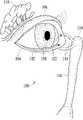
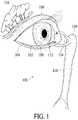
- FIG. 1illustrates a tear system of a person for tear production and drainage, in accordance with some embodiments
- FIG. 2illustrate perspective and cross-sectional views of a punctal plug, in accordance with some embodiments
- FIG. 3Aillustrates components of the tear system without punctal plugs, in accordance with some embodiments
- FIG. 3Billustrates components of the tear system with punctal plugs, in accordance with some embodiments
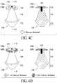
- FIG. 4Aillustrates an ocular device of which a second region is comprised entirely of a sensor material, in accordance with some embodiments
- FIG. 4Billustrates an ocular device of which a second region is comprised of sensor and non-sensor materials, in accordance with some embodiments
- FIG. 4Cillustrates an ocular device of which a second region is comprised of two tiers, where one tier is comprised of a sensor material, in accordance with some embodiments;
- FIG. 4Dillustrates an ocular device of which a second region is comprised of two tiers, where a first tier is comprised of one sensor material and a second comprised of a second sensor material, in accordance with some embodiments;
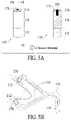
- FIG. 5Aillustrates an ocular device of which a second region is comprised entirely of radiation-responsive sensor material, in accordance with some embodiments
- FIG. 5Billustrates components of the tear system with the ocular devices of FIG. 5A , in accordance with some embodiments
- FIG. 5Cillustrates an ocular device of which a portion of a second region is comprised of radiation-responsive sensor material, in accordance with some embodiments.
- FIGS. 6A-6Dillustrate an ocular device of exemplary contact lenses that include multiple sensor materials coupled thereto, in accordance with various embodiments of the present invention.
- the tear system 100 of a person for tear production and drainageis illustrated along with the human eye 102 , a lower eyelid 104 , and an upper eyelid 106 ; a conjunctival sac 108 is found between the human eye 102 and lower eyelid 104 .
- the tear system 100could include the lacrimal gland 110 , lacrimal puncta 112 , lacrimal ducts 114 , lacrimal sac 116 , and nasolacrimal duct 118 .
- the lacrimal gland 108secretes tears which are conveyed to the tail of the upper eyelid 104 .
- the tearspass over the surface of the eye 102 to the lacrimal puncta 112 (singularly, lacrimal punctum), small holes found in the inner corner of the upper and lower eyelids 104 and 106 .
- the tearsare then passed through the lacrimal ducts 114 to the lacrimal sac 116 , before being passed into the nose via the nasolacrimal duct 118 .
- Basal tearsare continually produced and include components of glucose, minerals, vitamins, neurotransmitters, metabolites, amino acids, urea, anti-oxidants, and many proteins and/or their associated metabolites.
- the punctal plug 120may include a first region corresponding to a tail 122 , a second region corresponding to a head 124 , and a body corresponding to a shaft 126 .
- An upper surface 128 of second region 124could include a tool receptacle 129 to facilitate the use of an insertion/removal tool.
- the punctal plug 120has been used to treat dry eye syndrome as disclosed, for example, by Freeman in U.S. Pat. No. 3,949,750 entitled “Punctum Plug and Method for Treating Keratoconjunctivitis Sicca (Dry Eye) and Other Ophthalmic Aliments Using Same,” and Herrick et al. in U.S. Pat. No. 4,660,546 entitled “Method for Treating for Deficiency of Tears,” both of which are hereby incorporated by reference herein in their entireties. More recently, plugs have also been used for the administration of ocular medication and delivery of active agents through many shapes as disclosed respectively by Odrich, Steven, U.S. Patent Application Publication No.
- the punctal plug 120may be inserted or placed in one of the lacrimal puncta 112 .
- FIG. 3Aa view of the lacrimal puncta 112 , lacrimal ducts 114 , lacrimal sac 116 , and nasolacrimal duct 118 are shown without punctal plugs 120 .
- FIG. 3Ba view of the lacrimal puncta 112 , lacrimal ducts 114 , and lacrimal sac 116 are shown with punctal plugs 120 inserted into the lacrimal puncta 112 .
- the location of insertionmay not be limited to these.
- one or both of the lacrimal puncta 112may not be functional because of congenital malformation, trauma, or other causes. In such instances or for other reasons, the ocular device could be inserted in the conjunctival sac 108 .
- FIGS. 4A through 4DSome advantages and benefits of embodiments discussed herein are shown in FIGS. 4A through 4D by illustrating how a punctal plug may be used as a monitoring and/or diagnosis device of medical conditions.
- diabetic patientswith or without dry eyes
- a punctal pluginserted into one or both lacrimal puncta as an easy way to monitor changes their condition (e.g., their glucose levels) in real-time by observing the color to their punctal plug(s) as discussed below.
- a punctal plugmay be manufactured by injection molding or simple layering (e.g., press-fit).
- sensor materialmay be configured and calibrated to detect a compound or substance of interest (hereinafter substance).
- substancea compound or substance of interest
- the embodiments hereinare not limited to sensor material such as boronic acid but could include any sensor material suitable for the detection of substances such as, without limitation, sugars other than glucose, metal ions, nucleic acids, phospholipids, neurotransmitters, and precursors/metabolites of other macromolecules.
- applicable sensor material(s)may be used in eye-care diagnosis of an infection(s) such as, but not limited to, Gram-positive bacterial infection rather than monitoring of substances levels.
- Tear biomarkersmay be used to detect ocular disease and systemic disease. See Hagan, Suzanne et al., “Tear fluid biomarkers in ocular and system disease: Potential use for predictive, preventive and personalised medicine.” The EPMA Journal (2016) 7:15. Tear biomarkers may include proteins such as lysozyme and lactoferrin, lipids, metabolites, cytokines, chemokine, neuromediators, and lacryglobin. Ocular diseases could include dry eye disease, ocular allergy, keratoconus, thyroid-associated orbitopathy, aniridia, glaucoma, trachoma, keratopathy, and diabetic retinopathy.
- Systemic diseasecould include cystic fibrosis, scleroderma, and diabetes.
- Tear biomarkersmay be used to detect neurological diseases such as Parkinson disease and multiple sclerosis.
- tear biomarkersmay be used to detect cancers, such as breast, colon, prostate, lung, and ovarian cancer; bacteria, viruses, and fungi.
- substancessuch as drugs, toxins, and hormones may excrete from tears.
- a punctal plug 130may include first region 132 , a second region 134 , a body 136 , a surface 138 of the second region 134 , and a receptacle 139 for an insertion tool, where the second region 134 may be integrated with or otherwise coupled to the body 136 .
- the second region 134is comprised entirely of sensor material, where the first region 132 and the body 136 are comprised of non-sensor material.
- the entire punctal plug 130may be coated with or comprised of sensor material.
- a punctal plug 140may include a first region 142 , a second region 144 , a body 146 , a surface 148 of the second region 144 , and an insertion tool receptacle 149 , where the second region 144 may be comprised of sensor and non-sensor materials.
- the non-sensor materialmay be the same material as the first region 142 and/or the body 146 .
- the entire punctal plug 140may be coated with or comprised of sensor material.
- a punctal plug 150may include a first region 152 ; a second region 154 having an upper tier and a lower tier; a body 156 ; surfaces 158 a and 158 b of the upper and lower tiers, respectively; and an insertion tool receptacle 159 , where the upper and lower tiers may be comprised of sensor and non-sensor materials, respectively.
- the entire punctal plug 150may be coated with or comprised of sensor material.
- a punctal plug 160may include a first region 162 ; a second region 164 having an upper tier and a lower tier; a body 166 ; surfaces 168 a and 168 b of the upper and lower tiers, respectively; and an insertion tool receptacle 169 , where the upper tier may have color which is visually conspicuous from a color of the lower tier comprised of sensor and/or non-sensor materials, respectively, where the color of the sensor material(s) may change from a baseline color during normal conditions to another visually conspicuous color with a detection of an abnormal condition such as, for example, abnormal glucose levels or indication of an infection.
- the baseline color or changes thereto madebe readily visible to a third-party observer and/or the patient looking through a mirror.
- the color changeis associated with a matrix of embedded boronic acid derivative(s) which causes the material to swell upon interaction with the substances.
- a specific substance thresholdmay be calibrated to induce the desired color change at a prescribed level with a target compound.
- the sensor materialcould be white or clear, so that relative changes in color may be readily observable.
- non-diabetics in the studyshowed blood glucose increasing from about 6 mmol/L to about 9 mmol/L thirty minutes after the same meal, where tear glucose maintained a steady level of about 0.1 mmol/L which is below the threshold and indicated by no change of the baseline color.
- Radiation-responsive sensor materialcould include materials which emit visible or near-infrared radiation of different wavelengths (i.e., different colors) of the electromagnetic spectrum when excited (or irradiated) with radiation from a source providing a different wavelength of the electromagnetic spectrum.
- the radiationmay be visible or non-visible (such as ultraviolet).
- the sensor materialmay be excited with radiation passing through the lower and/or upper eyelids 104 and 106 , respectively; that is, the sensor material being excited with a patient's eyes closed. Such approach may be employed with the other variations of the ocular device described herein.
- the radiation-responsive sensor materialcould include molecules whose fluorescence intensity or wavelength may provide an indication of the chemical composition of a person's tears. Fluorescent molecules may detect chemicals relevant to tear fluid. See Yetisen, Ali et al., “Paper-based Microfluidic System for Tear Electrolyte Analysis.” Lap Chip, 2017, 17, 1137 (Yetisen), which is hereby incorporated by reference herein in its entirety.
- a punctal plug 170may include first region 172 , a second region 174 , a body 176 , a surface 178 of the second region 172 , and a receptacle 179 for an insertion tool, where the second region 174 may be integrated with or otherwise coupled to the body 176 .
- the second region 174is comprised entirely of sensor material, where the first region 172 and the body 176 are comprised of non-sensor material.
- visible or near-infrared radiationmay be emitted through the sides of the plug 170 in addition or as an alternative to being emitted through the surface 178 .
- the punctal plug 170may be inserted in one or both of the lacrimal puncta 112 .
- FIG. 5Ba view of the lacrimal puncta 112 , lacrimal ducts 114 , and lacrimal sac 116 are shown with punctal plugs 170 inserted into the lacrimal puncta 112 .
- a punctal plug 180may include first region 182 , a second region 184 having an upper region 184 a and a lower region 184 b , a body 186 , and outer surface 188 , and an insertion tool receptacle 159 .
- the upper region 184 acould be comprised of a non-sensor, translucent material that may not noticeable to an observer of the punctum but allows radiation to pass through to the lower region comprised of sensor material.
- visible or near-infrared radiationmay be emitted through the sides of the plug 180 .
- FIGS. 6A through 6DSome advantages and benefits of embodiments discussed herein are shown in FIGS. 6A through 6D by illustrating how ocular devices including contact lenses may be used as a monitoring and/or diagnosis device of medical conditions.
- two componentsmay be coupled to one another in any suitable manner, such as by placing the boronic acid derivatives in a matrix composed of hydrogel, a common material used in the production of contact lenses.
- the sensor materialcould be directly incorporated into the lens and isolated to a variety of convenient shapes.
- the sensor materialcould be disposed over the entirety of the contact lens; over select portions or surfaces thereof such as, for example, at one or more sides or edges of the lens to generally cover the whites of a user's eye); or in other regions of the lens.
- the color of the sensor materialcould change to one that contrasts with the color of the user's eyes so that the color change may be observable by a third party and/or wearer of the contact lenses.
- the color of the sensor materialcan be configured to change from a baseline color during normal conditions to another color in abnormal conditions.
- the sensor materialcan be configured to be clear or white, for example, to match the color of the patient's iris or a cosmetically desired color when the condition of interest is not present, and a different color in the abnormal condition.
- the color of the sensor material configured to be responsive to changes in tear glucose levelscould be transparent when glucose levels are normal, and may be configured to have visible color changes (e.g., purple or pink) if the glucose levels are abnormal, providing a visual warning to the patient.
- the sensor materialif the sensor material is placed in a region where it is visible if not transparent, then the user could receive a clear indication of the change in glucose levels.
- the sensor materialif the sensor material is located in an area not visible to the user (e.g., the margin of the contact lens on the sclera), then a change in color may be visible to others looking at the user's eyes or to the user if looking in a mirror.
- an ocular device 200could include a contact lens 202 and sensor material 204 configured in the shape of a dot.
- an ocular device 210could include a contact lens 212 and sensor material 214 configured in the shape of ring concentric to the contact lens 212 .
- an ocular device 220could include a contact lens 222 and sensor material 224 configured in the shape of a wedge that, at least, partially overlays the pupil. With the wedge configuration, a patient could be provided with a direct visual cue upon change from transparent to visible color, advantageously circumventing use of a mirror.
- an ocular device 230could include a contact lens 232 and sensor materials 234 through 248 configured in multiple regions to detect multiple chemicals in the tears to monitor multiple conditions.
- the sensor materials 234 through 248may be specifically calibrated to a variety of substances for boronic acid to exhibit different colors as described above and allow for multiple continuous monitoring and/or diagnoses simultaneously. It should be noted that, although eight sensor materials 234 through 248 are illustrated, any suitable number of sensor materials could be configured on coupled to the contact lens 232 .
- the ocular device comprising sensor materialmay come in other forms known to those skilled in the art suitable for placement into the eye on a temporary or semi-permanent basis, such as test strips.
Landscapes
- Health & Medical Sciences (AREA)
- Life Sciences & Earth Sciences (AREA)
- Physics & Mathematics (AREA)
- Veterinary Medicine (AREA)
- Animal Behavior & Ethology (AREA)
- Public Health (AREA)
- Engineering & Computer Science (AREA)
- Biomedical Technology (AREA)
- Heart & Thoracic Surgery (AREA)
- Medical Informatics (AREA)
- Molecular Biology (AREA)
- Surgery (AREA)
- Biophysics (AREA)
- General Health & Medical Sciences (AREA)
- Pathology (AREA)
- Optics & Photonics (AREA)
- Ophthalmology & Optometry (AREA)
- Spectroscopy & Molecular Physics (AREA)
- Emergency Medicine (AREA)
- Eye Examination Apparatus (AREA)
- Pharmaceuticals Containing Other Organic And Inorganic Compounds (AREA)
Abstract
Description
Claims (41)
Priority Applications (1)
| Application Number | Priority Date | Filing Date | Title |
|---|---|---|---|
| US15/947,127US11395630B2 (en) | 2017-04-06 | 2018-04-06 | Ocular devices and methods for the employment thereof |
Applications Claiming Priority (2)
| Application Number | Priority Date | Filing Date | Title |
|---|---|---|---|
| US201762482319P | 2017-04-06 | 2017-04-06 | |
| US15/947,127US11395630B2 (en) | 2017-04-06 | 2018-04-06 | Ocular devices and methods for the employment thereof |
Publications (2)
| Publication Number | Publication Date |
|---|---|
| US20180289326A1 US20180289326A1 (en) | 2018-10-11 |
| US11395630B2true US11395630B2 (en) | 2022-07-26 |
Family
ID=63710093
Family Applications (1)
| Application Number | Title | Priority Date | Filing Date |
|---|---|---|---|
| US15/947,127ActiveUS11395630B2 (en) | 2017-04-06 | 2018-04-06 | Ocular devices and methods for the employment thereof |
Country Status (5)
| Country | Link |
|---|---|
| US (1) | US11395630B2 (en) |
| EP (1) | EP3585309A4 (en) |
| CN (1) | CN110536654A (en) |
| IL (1) | IL269679B2 (en) |
| WO (1) | WO2018187693A1 (en) |
Cited By (1)
| Publication number | Priority date | Publication date | Assignee | Title |
|---|---|---|---|---|
| WO2025056486A1 (en) | 2023-09-13 | 2025-03-20 | Roche Diabetes Care Gmbh | Non-invasive analyte sensor for on-skin wearing |
Families Citing this family (2)
| Publication number | Priority date | Publication date | Assignee | Title |
|---|---|---|---|---|
| WO2021172861A1 (en)* | 2020-02-24 | 2021-09-02 | 주식회사 화이바이오메드 | Smart contact lens for diagnosis and treatment of dry eye syndrome |
| CN114446131B (en)* | 2022-03-08 | 2022-09-16 | 四川大学华西医院 | lacrimal duct irrigation model |
Citations (46)
| Publication number | Priority date | Publication date | Assignee | Title |
|---|---|---|---|---|
| US4597392A (en) | 1983-04-12 | 1986-07-01 | Max Planck Gesellschaft | Arrangement for measuring diffusing particles |
| WO2002003855A1 (en) | 2000-07-07 | 2002-01-17 | Christopher John Stanley | Optical device for measurement of analytes in tears |
| WO2003096876A2 (en) | 2001-04-01 | 2003-11-27 | Sommer Phillipe L | Continuously operational diagnostic device |
| US6850786B2 (en) | 1999-08-26 | 2005-02-01 | Novartis Ag | Ocular analyte sensor |
| US20050232972A1 (en) | 2004-04-15 | 2005-10-20 | Steven Odrich | Drug delivery via punctal plug |
| US7105352B2 (en) | 1996-11-06 | 2006-09-12 | University Of Pittsburgh | Intelligent polymerized crystalline colloidal array carbohydrate sensors |
| US20060226402A1 (en) | 2005-04-08 | 2006-10-12 | Beon-Kyu Kim | Ophthalmic devices comprising photochromic materials having extended PI-conjugated systems |
| US20070030443A1 (en) | 2003-08-07 | 2007-02-08 | Chapoy Lawrence L | Opthalmic sensor |
| US20070154395A1 (en) | 2002-11-20 | 2007-07-05 | Morris Carol A | Methods and kits for assays of rapid screening of diabetes |
| US20090099626A1 (en)* | 2007-09-07 | 2009-04-16 | Qlt Plug Delivery, Inc. - Qpdi | Lacrimal implant detection |
| US20090251693A1 (en) | 2004-10-29 | 2009-10-08 | The University Of Akron | Analysis for Glucose Products Using Pyridinylboronic Acid |
| US20100185066A1 (en) | 2001-04-27 | 2010-07-22 | Eyesense Ag | Apparatus for measuring blood glucose concentrations |
| US20100222657A1 (en) | 2005-09-28 | 2010-09-02 | The Texas A&M University System | Method for glucose monitoring using fluorescence quenching |
| US20110136929A1 (en) | 2008-07-09 | 2011-06-09 | Pei Yong Edwin Chow | Trapping glucose probe in pores of polymer |
| US7998412B2 (en) | 2000-01-07 | 2011-08-16 | Smart Holograms Limited | Ophthalmic device comprising a holographic sensor |
| US20110305872A1 (en) | 2010-06-09 | 2011-12-15 | Jun Li | Non-fouling, anti-microbial, anti-thrombogenic graft-from compositons |
| US20110305898A1 (en) | 2010-06-09 | 2011-12-15 | Zheng Zhang | Non-fouling, anti-microbial, anti-thrombogenic graft compositions |
| US20110305881A1 (en) | 2010-06-09 | 2011-12-15 | Schultz Karen A | Articles having non-fouling surfaces and processes for preparing the same including applying a primer coat |
| US20120177576A1 (en) | 2009-09-18 | 2012-07-12 | Jun Jack Hu | Optical device and method for non-invasive real-time testing of blood sugar levels |
| US20120259188A1 (en) | 2011-04-08 | 2012-10-11 | Nxp B.V. | Flexible Eye Insert and Glucose Measuring System |
| US8334140B2 (en) | 2005-11-08 | 2012-12-18 | Smart Holograms Limited | Boronate complex and its use in a glucose sensor |
| US20130176530A1 (en) | 2010-07-30 | 2013-07-11 | Coopervision International Holding Company, Lp | Silicone Hydrogel Ophthalmic Devices Molded In Vinyl Alcohol Copolymer Molds And Related Methods |
| US20130258275A1 (en) | 2012-04-03 | 2013-10-03 | Johnson & Johnson | Lens driver for variable-optic electronic ophthalmic lens |
| US20130317133A1 (en) | 2012-05-25 | 2013-11-28 | Johnson & Johnson Vision Care, Inc. | Polymers and nanogel materials and methods for making and using the same |
| US20140343387A1 (en) | 2013-05-17 | 2014-11-20 | Johnson & Johnson Vision Care, Inc. | Ophthalmic lens with a microfluidic system |
| US20150065905A1 (en) | 2013-08-27 | 2015-03-05 | Johnson & Johnson Vision Care, Inc. | Ophthalmic lens with a neural frequency detection system |
| US20150061837A1 (en) | 2013-06-28 | 2015-03-05 | Google Inc. | Reader Communication with Contact Lens Sensors and Display Device |
| US20150087945A1 (en) | 2012-03-13 | 2015-03-26 | Babak Ziaie | Laser-scribed ferrogel sensor with magnetic particles |
| US20150148648A1 (en) | 2013-11-22 | 2015-05-28 | Johnson & Johnson Vision Care, Inc. | Ophthalmic lens with intraocular pressure monitoring system |
| US20150148650A1 (en) | 2013-11-22 | 2015-05-28 | Johnson & Johnson Vision Care, Inc. | Ophthalmic lens with retinal vascularization monitoring system |
| BR102013007949A2 (en) | 2012-04-03 | 2015-06-16 | Johnson & Johnson Vision Care | Variable Optics Electronic Ophthalmic Lens Lens Trigger |
| US20150217129A1 (en) | 2014-01-08 | 2015-08-06 | Circuit Therapeutics, Inc. | System and method for therapeutic management of ocular hypertension |
| US20150289790A1 (en)* | 2014-04-10 | 2015-10-15 | Mission Biomedical Scientific, Inc. | Wearable metabolic physical activity monitor and method |
| WO2015192064A1 (en) | 2014-06-12 | 2015-12-17 | The Trustees Of Columbia University In The City Of New York | Graphene-based nanosensor for identifying target analytes |
| US20160003786A1 (en) | 2014-05-09 | 2016-01-07 | Ascendant Diagnostics, LLC | Methods Of Detecting Cancer |
| WO2016015270A1 (en) | 2014-07-31 | 2016-02-04 | Empire Technology Development Llc | Real-time glucose monitoring |
| US20160045144A1 (en) | 2014-08-15 | 2016-02-18 | International Business Machines Corporation | Metal-oxide-semiconductor capacitor based sensor |
| WO2016115369A1 (en) | 2015-01-14 | 2016-07-21 | Northwestern University | Compositions, systems and methods for patient specific ophthalmic device |
| US20160252505A1 (en) | 2013-10-07 | 2016-09-01 | The Board Of Trustees Of The University Of Illinoi S | Volume response sensors having analyte controlled reversible crosslinking |
| US9474645B2 (en) | 2006-06-21 | 2016-10-25 | Johnson & Johnson Vision Care, Inc. | Punctal plugs for the delivery of active agents |
| WO2016176252A1 (en) | 2015-04-27 | 2016-11-03 | Khan Taj H | Biosensor canalicular stent |
| US20160361001A1 (en) | 2015-06-12 | 2016-12-15 | California Institute Of Technology | Medical sensor having a nanoscale tapered waveguide for spectroscopy-based analysis of fluid |
| WO2016205190A1 (en) | 2015-06-16 | 2016-12-22 | The Trustees Of Columbia University In The City Of New York | Affinity nanosensor for detection of low-charge and low-molecular-weight molecules |
| WO2017007819A1 (en) | 2015-07-06 | 2017-01-12 | The Regents Of The University Of Colorado, A Body Corporate | Lacrimal drainage system diagnostic implant |
| US20170020390A1 (en) | 2015-07-24 | 2017-01-26 | Johnson & Johnson Vision Care, Inc. | Biomedical devices for biometric based information communication |
| US20170086676A1 (en) | 2015-09-24 | 2017-03-30 | Johnson & Johnson Vision Care, Inc. | Quantum-dot spectrometers for use in biomedical devices and methods of use |
- 2018
- 2018-04-06USUS15/947,127patent/US11395630B2/enactiveActive
- 2018-04-06WOPCT/US2018/026464patent/WO2018187693A1/ennot_activeCeased
- 2018-04-06EPEP18781815.8Apatent/EP3585309A4/ennot_activeWithdrawn
- 2018-04-06CNCN201880023669.9Apatent/CN110536654A/enactivePending
- 2019
- 2019-09-26ILIL269679Apatent/IL269679B2/enunknown
Patent Citations (49)
| Publication number | Priority date | Publication date | Assignee | Title |
|---|---|---|---|---|
| US4597392A (en) | 1983-04-12 | 1986-07-01 | Max Planck Gesellschaft | Arrangement for measuring diffusing particles |
| US7105352B2 (en) | 1996-11-06 | 2006-09-12 | University Of Pittsburgh | Intelligent polymerized crystalline colloidal array carbohydrate sensors |
| US6850786B2 (en) | 1999-08-26 | 2005-02-01 | Novartis Ag | Ocular analyte sensor |
| US7998412B2 (en) | 2000-01-07 | 2011-08-16 | Smart Holograms Limited | Ophthalmic device comprising a holographic sensor |
| US20110286064A1 (en) | 2000-01-07 | 2011-11-24 | Barry Burles | Ophthalmic device comprising a holographic sensor |
| WO2002003855A1 (en) | 2000-07-07 | 2002-01-17 | Christopher John Stanley | Optical device for measurement of analytes in tears |
| WO2003096876A2 (en) | 2001-04-01 | 2003-11-27 | Sommer Phillipe L | Continuously operational diagnostic device |
| US20100185066A1 (en) | 2001-04-27 | 2010-07-22 | Eyesense Ag | Apparatus for measuring blood glucose concentrations |
| US20070154395A1 (en) | 2002-11-20 | 2007-07-05 | Morris Carol A | Methods and kits for assays of rapid screening of diabetes |
| US20070030443A1 (en) | 2003-08-07 | 2007-02-08 | Chapoy Lawrence L | Opthalmic sensor |
| US20050232972A1 (en) | 2004-04-15 | 2005-10-20 | Steven Odrich | Drug delivery via punctal plug |
| US20090251693A1 (en) | 2004-10-29 | 2009-10-08 | The University Of Akron | Analysis for Glucose Products Using Pyridinylboronic Acid |
| US20060226402A1 (en) | 2005-04-08 | 2006-10-12 | Beon-Kyu Kim | Ophthalmic devices comprising photochromic materials having extended PI-conjugated systems |
| US20100222657A1 (en) | 2005-09-28 | 2010-09-02 | The Texas A&M University System | Method for glucose monitoring using fluorescence quenching |
| US8334140B2 (en) | 2005-11-08 | 2012-12-18 | Smart Holograms Limited | Boronate complex and its use in a glucose sensor |
| US9474645B2 (en) | 2006-06-21 | 2016-10-25 | Johnson & Johnson Vision Care, Inc. | Punctal plugs for the delivery of active agents |
| US20090099626A1 (en)* | 2007-09-07 | 2009-04-16 | Qlt Plug Delivery, Inc. - Qpdi | Lacrimal implant detection |
| CN101861135A (en) | 2007-09-07 | 2010-10-13 | Qlt栓塞输送公司 | Lacrimal implant testing |
| US20110136929A1 (en) | 2008-07-09 | 2011-06-09 | Pei Yong Edwin Chow | Trapping glucose probe in pores of polymer |
| US20120177576A1 (en) | 2009-09-18 | 2012-07-12 | Jun Jack Hu | Optical device and method for non-invasive real-time testing of blood sugar levels |
| US20110305872A1 (en) | 2010-06-09 | 2011-12-15 | Jun Li | Non-fouling, anti-microbial, anti-thrombogenic graft-from compositons |
| US20110305898A1 (en) | 2010-06-09 | 2011-12-15 | Zheng Zhang | Non-fouling, anti-microbial, anti-thrombogenic graft compositions |
| US20110305881A1 (en) | 2010-06-09 | 2011-12-15 | Schultz Karen A | Articles having non-fouling surfaces and processes for preparing the same including applying a primer coat |
| US20130176530A1 (en) | 2010-07-30 | 2013-07-11 | Coopervision International Holding Company, Lp | Silicone Hydrogel Ophthalmic Devices Molded In Vinyl Alcohol Copolymer Molds And Related Methods |
| CN102727218A (en) | 2011-04-08 | 2012-10-17 | Nxp股份有限公司 | Flexible eye insert and glucose measuring system |
| US20120259188A1 (en) | 2011-04-08 | 2012-10-11 | Nxp B.V. | Flexible Eye Insert and Glucose Measuring System |
| US20150087945A1 (en) | 2012-03-13 | 2015-03-26 | Babak Ziaie | Laser-scribed ferrogel sensor with magnetic particles |
| US20130258275A1 (en) | 2012-04-03 | 2013-10-03 | Johnson & Johnson | Lens driver for variable-optic electronic ophthalmic lens |
| BR102013007949A2 (en) | 2012-04-03 | 2015-06-16 | Johnson & Johnson Vision Care | Variable Optics Electronic Ophthalmic Lens Lens Trigger |
| US20130317133A1 (en) | 2012-05-25 | 2013-11-28 | Johnson & Johnson Vision Care, Inc. | Polymers and nanogel materials and methods for making and using the same |
| US20140343387A1 (en) | 2013-05-17 | 2014-11-20 | Johnson & Johnson Vision Care, Inc. | Ophthalmic lens with a microfluidic system |
| US20150061837A1 (en) | 2013-06-28 | 2015-03-05 | Google Inc. | Reader Communication with Contact Lens Sensors and Display Device |
| US20150065905A1 (en) | 2013-08-27 | 2015-03-05 | Johnson & Johnson Vision Care, Inc. | Ophthalmic lens with a neural frequency detection system |
| US20160252505A1 (en) | 2013-10-07 | 2016-09-01 | The Board Of Trustees Of The University Of Illinoi S | Volume response sensors having analyte controlled reversible crosslinking |
| US20150148650A1 (en) | 2013-11-22 | 2015-05-28 | Johnson & Johnson Vision Care, Inc. | Ophthalmic lens with retinal vascularization monitoring system |
| US20150148648A1 (en) | 2013-11-22 | 2015-05-28 | Johnson & Johnson Vision Care, Inc. | Ophthalmic lens with intraocular pressure monitoring system |
| US20150217129A1 (en) | 2014-01-08 | 2015-08-06 | Circuit Therapeutics, Inc. | System and method for therapeutic management of ocular hypertension |
| US20150289790A1 (en)* | 2014-04-10 | 2015-10-15 | Mission Biomedical Scientific, Inc. | Wearable metabolic physical activity monitor and method |
| US20160003786A1 (en) | 2014-05-09 | 2016-01-07 | Ascendant Diagnostics, LLC | Methods Of Detecting Cancer |
| WO2015192064A1 (en) | 2014-06-12 | 2015-12-17 | The Trustees Of Columbia University In The City Of New York | Graphene-based nanosensor for identifying target analytes |
| WO2016015270A1 (en) | 2014-07-31 | 2016-02-04 | Empire Technology Development Llc | Real-time glucose monitoring |
| US20160045144A1 (en) | 2014-08-15 | 2016-02-18 | International Business Machines Corporation | Metal-oxide-semiconductor capacitor based sensor |
| WO2016115369A1 (en) | 2015-01-14 | 2016-07-21 | Northwestern University | Compositions, systems and methods for patient specific ophthalmic device |
| WO2016176252A1 (en) | 2015-04-27 | 2016-11-03 | Khan Taj H | Biosensor canalicular stent |
| US20160361001A1 (en) | 2015-06-12 | 2016-12-15 | California Institute Of Technology | Medical sensor having a nanoscale tapered waveguide for spectroscopy-based analysis of fluid |
| WO2016205190A1 (en) | 2015-06-16 | 2016-12-22 | The Trustees Of Columbia University In The City Of New York | Affinity nanosensor for detection of low-charge and low-molecular-weight molecules |
| WO2017007819A1 (en) | 2015-07-06 | 2017-01-12 | The Regents Of The University Of Colorado, A Body Corporate | Lacrimal drainage system diagnostic implant |
| US20170020390A1 (en) | 2015-07-24 | 2017-01-26 | Johnson & Johnson Vision Care, Inc. | Biomedical devices for biometric based information communication |
| US20170086676A1 (en) | 2015-09-24 | 2017-03-30 | Johnson & Johnson Vision Care, Inc. | Quantum-dot spectrometers for use in biomedical devices and methods of use |
Non-Patent Citations (50)
| Title |
|---|
| Adam J. Paulsen et al., "Dry Eye in the Beaver Dam Offspring Study: Prevalence, Risk Factors, and Health-Related Quality of Life" NIH Public Access Author Manuscript, Am J Ophthalmol, Apr. 2014, 157(4): 799-806. DOI: 10.1016/j.ajo.2013.12.023. |
| Ali K. Yetisen et al., "Glucose-Sensitive Hydrogel Optical Fibers Functionalized with Phenylboronic Acid," Advanced Science News, 2017 The Authors. Published by Wiley-VCH Verlag GmbH & Co. KGaA, Weinheim, Adv. Mater. 2017 1606380, pp. 1-11. |
| Ali K. Yetisen et al., "Paper-based Microfluidic System for Tear Electrolyte Analysis," The Royal Society of Chemistry 2017, Lab Chip. 2017, 17, pp. 1137-1148. |
| Ali K. Yetisen et al., "Reusable, Robust, and Accurate Laser-Generated Photonic Nanosensor," ACS Publications, 2014 American Chemical Society, Department of Chemical Engineering and Biotechnology, University of Cambridge, UK, dx.doi.org/10.1021/nl5012504, Nano Letters 2014, 14, pp. 3587-3593. |
| Andrew G. Mayes et al., "Metal Ion-Sensitive Holographic Sensors," Institutue of Biotechnology, University of Cambridge, Cambridge, UK, 2002 American Chemical Society, Analytical Chemistry, vol. 74, No. 15, Aug. 1, 2002, pp. 3649-3657. |
| Angelika Domschke et al., "Initial Clinical Testing of a Holographic Non-Invasive Contact Lens Glucose Sensor," Diabetes Tecnology & Therapeutics, vol. 8, No. 1, 2006, Mary Ann Liebert, Inc., pp. 89-93. |
| Carolyn G. Begley et al., "Responses of Contact Lens Wearers to a Dry Eye Survey", Optometry and Vision Science, vol. 77, No. 1, Jan. 2000, pp. 40-46. |
| Cheng Chen, "Current and Emerging Technology for Continuous Glucose Monitoring," Academic Editors, Sensors 2017, 17, 182; doi:10.3390/s17010182, pp. 1-19. |
| Chinese Office Action dated Apr. 2, 2021, Chinese Patent Application No. 201880023669.9. |
| Daniel Harvey et al., "Fibre Optics Sensors in Tear Electrolyte Analysis: Towards a Novel Point of Care Potassium Sensor," Elsevier, Contact Lens & Anterior Eye 35 (2012) pp. 137-144. |
| David Spero, BSN, RN, "Is Continuous Glucose Monitoring Worth It?" Diabetes Self-Management. N.p., Sep. 28, 2011. https://www.diabetesselfmanagement.com/blog/is-continuous-glucose-monitoring-worth-it/print/ pp. 1-3. |
| Dennis G. Hall, "Boronic Acids: Preparation and Applications in Organic Synthesis, Medicine and Materials" 2nd Ed. vol. 1-2. 2012, 1:1-134, 13:591-620, 14:621-676, v-xxi. |
| Examination Report dated Mar. 17, 2021, European Patent Application No. 18781815.8-1113. |
| Extended European Search Report Application No. 18781815.8 , dated Jul. 31, 2020. |
| Gillian F. Whyte, "Molecular Recognition with Boronic Acids—Applications in Chemical Biology" Springer-Verlag Berlin Heidelberg 2013, J. Chem Biol (2013) 6:161-174. DOI: 10.1007/s12154-013-0099-0. |
| Graham J. Worsley et al., "Measurement of Glucose in Blood with a Phenylboronic Acid Optical Sensor," Journal of Diabetes Science and Technology, vol. 2, Issue 2, Mar. 2008, Diabetes Technology Society, pp. 213-220. |
| Grant R. Hendrickson and L. Andrew Lyon, "Bioresponsive Hydrogels for Sensing Applications" The Royal Society of Chemistry 2009, Soft Matter, 2009, 5, 29-35. DOI: 10.1039/b811620b. |
| Guangjie Song et al., "A Highly Selective, Colorimetric, and Environment-Sensitive Optical Potassium Ion Sensor," ChemComm, The Royal Society of Chemistry 2017, vol. 53, No. 41, pp. 5602-5605. |
| Huanfen Yao et al., "A Contact Lens with Embedded Sensor for Monitoring Tear Glucose Level," NIH Public Access, Author Manuscript, Biosens Bioelectron. Mar. 15, 2011, 26(7), pp. 3290-3296. |
| International Search Report PCT/US18/26464, dated Jul. 3, 2018. |
| Invitrogen by Thermo Fisher Scientific, Molecular Probes Handbook, "A Guide to Fluorescent Probes and Labeling Technologies," 11th Edition 2010, Chapter 21, Indicators for Na+, K+, Cl− and Miscellaneous Ions, pp. 903-922. |
| Jason J. Nichols and Loraine T. Sinnott, "Tear Film, Contact Lens, and Patient-Related Factors Associated with Contact Lens-Related Dry Eye," Investigate Ophthalmology & Visual Science, Apr. 2006, vol. 47, No. 4, Association for Research in Vision and Ophthalmology, pp. 1319-1328. |
| Jeffrey P. Gilbard and Scott R. Rossi, "Changes in Tear Ion Concentrations in Dry-Eye Disorders," Cornea Research Unit, Schepens Eye Research Institute, Department of Ophthalmology, Harvard Medical School, Boston, MA, pp. 529-533. |
| Jennifer D. Lane et al., "Tear Glucose Dynamics in Diabetes Mellitus" (2006) Current Eye Research, 31:11, 895-901, DOI:10.1080/02713680600976552. |
| Jin Zhang, Ph.D. et al., "Noninvasive Diagnostic Devices for Diabetes through Measuring Tear Glucose," Journal of Diabetes Science and Technology, vol. 5, Issue 1, Jan. 2011, pp. 166-172. |
| Jon Stefan Hansen et al., "Recent Advances in Fluorescent Arylboronic Acids for Glucose Sensing," Open Access, Biosensors, ISSN 2079-6374, Biosensors 2013, 3, pp. 400-418; DOI: 10.3390/bios3040400. |
| Justin T. Baca, Ph.D., et al., "Tear Glucose Analysis for the Noninvasive Detection and Monitoring of Diabetes Mellitus" Clinical Science, The Ocular Surface vol. 5 No. 4 (Oct. 2007): pp. 280-293. www.theocularsurface.com. |
| Liqiang Zhang et al., "A Polymer-based Ratiometric Intracellular Glucose Sensor" The Royal Society of Chemistry 2014, RSC Publishing, Chemical Communications, Jul. 4, 2014, 50(52): 6920-6922. |
| Mark D.P. Willcox, PhD. et al., "TFOS Dews II Tear Film Report," The Ocular Surface 15, (2017 Elsevier Inc.), pp. 366-403. |
| Martin Mesch et al., "Functionalized Hydrogel on Plasmonic Nanoantennas for Noninvasive Glucose Sensing," ACS Photonics, 4th Physics Institute and Research Center SCoPE, University of Stuttgart, Germany, ACS Publications, 2015 American Chemical Society, vol. 2, pp. 475-480. |
| Matti Ben-Moshe et al., "Fast Responsive Crystalline Colloidal Array Photonic Crystal Glucose Sensors," Department of Chemistry, University of Pittsburgh, Analytical Chemistry, vol. 78, No. 14, Jul. 15, 2006, pp. 5149-5157. |
| Nicholas M. Farandos et al., "Contact Lens Sensors in Ocular Diagnostics," 2014 Wiley-VCH Verlag GmbH & Co. KGaA, Weinheim, Adv. Healthcare Mater. 2015, pp. 792-810. |
| P. Versura et al., "Performance of Tear Osmolarity Compared to Previous Diagnostic Tests for Dry Eye Diseases," Current Eye Research, 35(7), pp. 553-564, 2010 Informa Healthcare USA, Inc., ISSN: 0271-3683. |
| Paul J. Beisswenger MD et al., "Glycated Proteins in Diabetes" Clinics in Laboratory Medicine, Diabetes Mellitus, ISSN 0272-2712, Mar. 2011, vol. 21(1):53-78, vi. |
| Qian Wu et al., "Organization of Glucose-Responsive Systems and Their Properties" Chemical Reviews, ACS Publications, 2011 American Chemical Society, dx.doi.org/10.1021/cr200027j, Chem. Rev. 2011, 111, 7855-7875. |
| Ramachandram Badugu et al., "Boronic Acid Fluorescent Sensors for Monosaccharide Signaling Based on the 6-methoxyquinolinium Heterocyclic Nucleus: Progress Toward Noninvasive and Continuous Glucose Monitoring," Science & Direct, Elsevier, Bioorganic & Medicinal Chemistry vol. 13, 2005, pp. 113-119. |
| Ramachandram Badugu et al., "Contact Lens to Measure Individual Ion Concentrations in Tears and Applications to Dry Eye Disease," 2017 Elsevier Inc., Analytical Biochemistry 542 (2018) pp. 84-94. |
| Ramachandram Badugu et al., "Fluorescence Sensors for Monosaccharides Based on the 6-methylquinolinium Nucleus and Boronic Acid Moiety: Potential Application to Ophthalmic Diagnostics," HHS Public Access, Author Manuscript, Available in PMC Jun. 6, 2016; Talanta, Feb. 15, 2005; 65(3): 762-768. |
| Ramachandram Badugu et al., "Noninvasive Continuous Monitoring of Physiological Glucose Using a Monosaccharide-Sensing Contact Lens," Center for Fluorescence Spectroscopy, Department of Biochemistry and Molecular Biology, Medical Biotechnology Center, University of Maryland School of Medicine, Baltimore, Maryland, Analytical Chemistry, vol. 76, No. 3, Feb. 1, 2004, pp. 610-618. |
| Ramachandram Badugu et al., "Ophthalmic Glucose Monitoring Using Disposable Contact Lenses," Center for Fluorescence Spectroscopy, Department of Biochemistry and Molecular Biology, pp. 363-397. |
| Ramachandram Badugu et al., "Ophthalmic Glucose Monitoring Using Disposable Contact Lenses—A Review," HHS Public Access, J Fluoresc. Author Manuscript ; available in PMC Jun. 13, 2016; Sep. 2004; 14(5): 617-633; pp. 1-37. |
| Ramachandram Badugu et al., "Ophthalmic Glucose Sensing: A Novel Monosaccharide Sensing Disposable and Colorless Contact Lens," HHS Public Access, Department of Chemistry, University of Copenhagen, Author Manuscript, Analyst. Available in PMC Jun. 4, 2016; 129(6): 516-521. DOI: 10.1039/b314463c, pp. 1-20. |
| Research Notes, Implanted Optics, "Structure Regained in supercooled Liquid Silicon," 2004 Nature Publishing Group, Nature Materials, vol. 3, Issue 2, Feb. 2004, www.nature.com/naturematerials, p. 76. |
| Sandra Ast et al., "A Highly K+-Selective Phenylaza-[18]crown-6-Lariat-Ether-Based Fluoroionophore and Its Application in the Sensing of K+ Ions with an Optical Sensor Film and in Cells," 2013 Wiley-VCH Verlag GmbH & Co., KGaA, Weinheim, Chem. Eur. J. 2013, 19, pp. 14911-14917. |
| Satyamoorthy Kabilan et al., "Glucose-Sensitive Holographic Sensors," ResearchGate, Journal of Molecular Recognition 2004; 17: pp. 162-166, DOI:10.1002/jmr.663, Institute of Biotechnology, Cambridge, UK. |
| Suzanne Hagan et al., "Tear Fluid Biomarkers in Ocular and Systemic Disease: Potential Use for Predictive, Preventive and Personalised Medicine," EPMA Journal, 2016 7:15, DOI:10.1186/s13167-016-0065-3. |
| Taira Kajisa et al., "Glucose-Responsive Hydrogel Electrode for Biocompatible Glucose Transistor," Provigate Inc., Department of Research and Development, Tokyo, Japan, Science and Technology of Advanced Materials, 2017, vol. 18, No. 1 pp. 26-33. |
| Tsung-Han Tsai et al., "Dynamic Gradient Directed Molecular Transport and Concentration in Hydrogel Films," Angewandte Chemie Int. Ed. 2017 Wiley-VCH Verlag GmbH & Co., KGaA, Weinheim, pp. 5001-5006. |
| Ulrike Stahl et al., "Osmolality and Tear Film Dynamics," 2011 Vision Co-operative Research Centre, Clinical and Experimental Optometry 2011 Optometrists Association Australia, Clin. Exp. Optom. 2012; 95: 1: pp. 3-11. |
| Vladimir L. Alexeev et al., "Photonic Crystal Glucose-Sensing Material for Noninvasive Monitoring of Glucose in Tear Fluid," Automation and Analytical Techniques, American Association for Clinical Chemistry 2004, 50:12, pp. 2353-2360. |
Cited By (1)
| Publication number | Priority date | Publication date | Assignee | Title |
|---|---|---|---|---|
| WO2025056486A1 (en) | 2023-09-13 | 2025-03-20 | Roche Diabetes Care Gmbh | Non-invasive analyte sensor for on-skin wearing |
Also Published As
| Publication number | Publication date |
|---|---|
| IL269679B2 (en) | 2023-03-01 |
| WO2018187693A1 (en) | 2018-10-11 |
| US20180289326A1 (en) | 2018-10-11 |
| IL269679B (en) | 2022-11-01 |
| CN110536654A (en) | 2019-12-03 |
| EP3585309A1 (en) | 2020-01-01 |
| IL269679A (en) | 2019-11-28 |
| EP3585309A4 (en) | 2020-09-02 |
Similar Documents
| Publication | Publication Date | Title |
|---|---|---|
| Mahabadi et al. | Open angle glaucoma | |
| Lois et al. | Fundus autofluorescence | |
| Jentsch et al. | Retinal fluorescence lifetime imaging ophthalmoscopy measures depend on the severity of Alzheimer's disease | |
| Schmitz-Valckenberg et al. | Fundus autofluorescence and fundus perimetry in the junctional zone of geographic atrophy in patients with age-related macular degeneration | |
| EP1385423B1 (en) | Kit for measuring blood glucose concentrations | |
| US11395630B2 (en) | Ocular devices and methods for the employment thereof | |
| Cesareo et al. | Association between Alzheimer's disease and glaucoma: a study based on Heidelberg retinal tomography and frequency doubling technology perimetry | |
| Safi et al. | Contrast sensitivity to spatial gratings in moderate and dim light conditions in patients with diabetes in the absence of diabetic retinopathy | |
| TW201717851A (en) | Biomedical devices for biometric based information communication in vehicular environments | |
| Prunty et al. | In vivo imaging of retinal oxidative stress using a reactive oxygen species–activated fluorescent probe | |
| Klemp et al. | The multifocal ERG in diabetic patients without retinopathy during euglycemic clamping | |
| Kong et al. | Damage to the blood-aqueous barrier in eyes with primary angle closure glaucoma | |
| Featherstone et al. | Feline corneal sequestrum: laboratory analysis of ocular samples from 12 cats | |
| WO2002003855A1 (en) | Optical device for measurement of analytes in tears | |
| Garaszczuk et al. | The tear turnover and tear clearance tests–a review | |
| Phan et al. | The use of contact lenses as biosensors | |
| Yao et al. | Hyperspectral ophthalmoscope images for the diagnosis of diabetic retinopathy stage | |
| Ismail et al. | HbA1c and retinal sensitivity in diabetics using microperimetry | |
| Tugal-Tutkun et al. | Laser flare photometry and its use in uveitis | |
| Tan et al. | Retinal neural dysfunction in diabetes revealed with handheld chromatic pupillometry | |
| Vitale et al. | Fluorescence lifetime imaging ophthalmoscopy (FLIO) in patients with choroideremia | |
| Stolwijk et al. | Corneal autofluorescence in diabetic and penetrating keratoplasty patients as measured by fluorophotometry | |
| RU2088940C1 (en) | Method for diagnosing choroid melanomans | |
| JP4724883B2 (en) | In vivo tissue identification device | |
| US11446399B2 (en) | System and method for quantifying the presence of chemicals and/or physical conditions in ocular tissues |
Legal Events
| Date | Code | Title | Description |
|---|---|---|---|
| FEPP | Fee payment procedure | Free format text:ENTITY STATUS SET TO UNDISCOUNTED (ORIGINAL EVENT CODE: BIG.); ENTITY STATUS OF PATENT OWNER: SMALL ENTITY | |
| FEPP | Fee payment procedure | Free format text:ENTITY STATUS SET TO SMALL (ORIGINAL EVENT CODE: SMAL); ENTITY STATUS OF PATENT OWNER: SMALL ENTITY | |
| AS | Assignment | Owner name:TEARDX LLC, NEW YORK Free format text:ASSIGNMENT OF ASSIGNORS INTEREST;ASSIGNORS:ORLOFF, EUGENE;BRAUN, PAUL V.;ORLOFF, ELEONORA;AND OTHERS;SIGNING DATES FROM 20180407 TO 20180502;REEL/FRAME:045767/0455 | |
| STPP | Information on status: patent application and granting procedure in general | Free format text:DOCKETED NEW CASE - READY FOR EXAMINATION | |
| STPP | Information on status: patent application and granting procedure in general | Free format text:NON FINAL ACTION MAILED | |
| STPP | Information on status: patent application and granting procedure in general | Free format text:RESPONSE TO NON-FINAL OFFICE ACTION ENTERED AND FORWARDED TO EXAMINER | |
| STPP | Information on status: patent application and granting procedure in general | Free format text:NON FINAL ACTION MAILED | |
| STPP | Information on status: patent application and granting procedure in general | Free format text:FINAL REJECTION MAILED | |
| STPP | Information on status: patent application and granting procedure in general | Free format text:DOCKETED NEW CASE - READY FOR EXAMINATION | |
| STPP | Information on status: patent application and granting procedure in general | Free format text:NON FINAL ACTION MAILED | |
| STPP | Information on status: patent application and granting procedure in general | Free format text:RESPONSE TO NON-FINAL OFFICE ACTION ENTERED AND FORWARDED TO EXAMINER | |
| STPP | Information on status: patent application and granting procedure in general | Free format text:NOTICE OF ALLOWANCE MAILED -- APPLICATION RECEIVED IN OFFICE OF PUBLICATIONS | |
| STPP | Information on status: patent application and granting procedure in general | Free format text:PUBLICATIONS -- ISSUE FEE PAYMENT VERIFIED | |
| STCF | Information on status: patent grant | Free format text:PATENTED CASE |