US11377640B2 - Differentiation of human embryonic stem cells into single hormonal insulin positive cells - Google Patents
Differentiation of human embryonic stem cells into single hormonal insulin positive cellsDownload PDFInfo
- Publication number
- US11377640B2 US11377640B2US16/435,428US201916435428AUS11377640B2US 11377640 B2US11377640 B2US 11377640B2US 201916435428 AUS201916435428 AUS 201916435428AUS 11377640 B2US11377640 B2US 11377640B2
- Authority
- US
- United States
- Prior art keywords
- cells
- stage
- expression
- days
- pancreatic
- Prior art date
- Legal status (The legal status is an assumption and is not a legal conclusion. Google has not performed a legal analysis and makes no representation as to the accuracy of the status listed.)
- Active, expires
Links
Images


Classifications
- C—CHEMISTRY; METALLURGY
- C12—BIOCHEMISTRY; BEER; SPIRITS; WINE; VINEGAR; MICROBIOLOGY; ENZYMOLOGY; MUTATION OR GENETIC ENGINEERING
- C12N—MICROORGANISMS OR ENZYMES; COMPOSITIONS THEREOF; PROPAGATING, PRESERVING, OR MAINTAINING MICROORGANISMS; MUTATION OR GENETIC ENGINEERING; CULTURE MEDIA
- C12N5/00—Undifferentiated human, animal or plant cells, e.g. cell lines; Tissues; Cultivation or maintenance thereof; Culture media therefor
- C12N5/06—Animal cells or tissues; Human cells or tissues
- C12N5/0602—Vertebrate cells
- C12N5/0676—Pancreatic cells
- C—CHEMISTRY; METALLURGY
- C12—BIOCHEMISTRY; BEER; SPIRITS; WINE; VINEGAR; MICROBIOLOGY; ENZYMOLOGY; MUTATION OR GENETIC ENGINEERING
- C12N—MICROORGANISMS OR ENZYMES; COMPOSITIONS THEREOF; PROPAGATING, PRESERVING, OR MAINTAINING MICROORGANISMS; MUTATION OR GENETIC ENGINEERING; CULTURE MEDIA
- C12N2500/00—Specific components of cell culture medium
- C12N2500/30—Organic components
- C12N2500/34—Sugars
- C—CHEMISTRY; METALLURGY
- C12—BIOCHEMISTRY; BEER; SPIRITS; WINE; VINEGAR; MICROBIOLOGY; ENZYMOLOGY; MUTATION OR GENETIC ENGINEERING
- C12N—MICROORGANISMS OR ENZYMES; COMPOSITIONS THEREOF; PROPAGATING, PRESERVING, OR MAINTAINING MICROORGANISMS; MUTATION OR GENETIC ENGINEERING; CULTURE MEDIA
- C12N2500/00—Specific components of cell culture medium
- C12N2500/30—Organic components
- C12N2500/38—Vitamins
- C—CHEMISTRY; METALLURGY
- C12—BIOCHEMISTRY; BEER; SPIRITS; WINE; VINEGAR; MICROBIOLOGY; ENZYMOLOGY; MUTATION OR GENETIC ENGINEERING
- C12N—MICROORGANISMS OR ENZYMES; COMPOSITIONS THEREOF; PROPAGATING, PRESERVING, OR MAINTAINING MICROORGANISMS; MUTATION OR GENETIC ENGINEERING; CULTURE MEDIA
- C12N2501/00—Active agents used in cell culture processes, e.g. differentation
- C12N2501/10—Growth factors
- C12N2501/117—Keratinocyte growth factors (KGF-1, i.e. FGF-7; KGF-2, i.e. FGF-12)
- C—CHEMISTRY; METALLURGY
- C12—BIOCHEMISTRY; BEER; SPIRITS; WINE; VINEGAR; MICROBIOLOGY; ENZYMOLOGY; MUTATION OR GENETIC ENGINEERING
- C12N—MICROORGANISMS OR ENZYMES; COMPOSITIONS THEREOF; PROPAGATING, PRESERVING, OR MAINTAINING MICROORGANISMS; MUTATION OR GENETIC ENGINEERING; CULTURE MEDIA
- C12N2501/00—Active agents used in cell culture processes, e.g. differentation
- C12N2501/10—Growth factors
- C12N2501/15—Transforming growth factor beta (TGF-β)
- C—CHEMISTRY; METALLURGY
- C12—BIOCHEMISTRY; BEER; SPIRITS; WINE; VINEGAR; MICROBIOLOGY; ENZYMOLOGY; MUTATION OR GENETIC ENGINEERING
- C12N—MICROORGANISMS OR ENZYMES; COMPOSITIONS THEREOF; PROPAGATING, PRESERVING, OR MAINTAINING MICROORGANISMS; MUTATION OR GENETIC ENGINEERING; CULTURE MEDIA
- C12N2501/00—Active agents used in cell culture processes, e.g. differentation
- C12N2501/10—Growth factors
- C12N2501/155—Bone morphogenic proteins [BMP]; Osteogenins; Osteogenic factor; Bone inducing factor
- C—CHEMISTRY; METALLURGY
- C12—BIOCHEMISTRY; BEER; SPIRITS; WINE; VINEGAR; MICROBIOLOGY; ENZYMOLOGY; MUTATION OR GENETIC ENGINEERING
- C12N—MICROORGANISMS OR ENZYMES; COMPOSITIONS THEREOF; PROPAGATING, PRESERVING, OR MAINTAINING MICROORGANISMS; MUTATION OR GENETIC ENGINEERING; CULTURE MEDIA
- C12N2501/00—Active agents used in cell culture processes, e.g. differentation
- C12N2501/10—Growth factors
- C12N2501/16—Activin; Inhibin; Mullerian inhibiting substance
- C—CHEMISTRY; METALLURGY
- C12—BIOCHEMISTRY; BEER; SPIRITS; WINE; VINEGAR; MICROBIOLOGY; ENZYMOLOGY; MUTATION OR GENETIC ENGINEERING
- C12N—MICROORGANISMS OR ENZYMES; COMPOSITIONS THEREOF; PROPAGATING, PRESERVING, OR MAINTAINING MICROORGANISMS; MUTATION OR GENETIC ENGINEERING; CULTURE MEDIA
- C12N2501/00—Active agents used in cell culture processes, e.g. differentation
- C12N2501/10—Growth factors
- C12N2501/19—Growth and differentiation factors [GDF]
- C—CHEMISTRY; METALLURGY
- C12—BIOCHEMISTRY; BEER; SPIRITS; WINE; VINEGAR; MICROBIOLOGY; ENZYMOLOGY; MUTATION OR GENETIC ENGINEERING
- C12N—MICROORGANISMS OR ENZYMES; COMPOSITIONS THEREOF; PROPAGATING, PRESERVING, OR MAINTAINING MICROORGANISMS; MUTATION OR GENETIC ENGINEERING; CULTURE MEDIA
- C12N2501/00—Active agents used in cell culture processes, e.g. differentation
- C12N2501/30—Hormones
- C12N2501/38—Hormones with nuclear receptors
- C12N2501/385—Hormones with nuclear receptors of the family of the retinoic acid recptor, e.g. RAR, RXR; Peroxisome proliferator-activated receptor [PPAR]
- C—CHEMISTRY; METALLURGY
- C12—BIOCHEMISTRY; BEER; SPIRITS; WINE; VINEGAR; MICROBIOLOGY; ENZYMOLOGY; MUTATION OR GENETIC ENGINEERING
- C12N—MICROORGANISMS OR ENZYMES; COMPOSITIONS THEREOF; PROPAGATING, PRESERVING, OR MAINTAINING MICROORGANISMS; MUTATION OR GENETIC ENGINEERING; CULTURE MEDIA
- C12N2501/00—Active agents used in cell culture processes, e.g. differentation
- C12N2501/40—Regulators of development
- C12N2501/41—Hedgehog proteins; Cyclopamine (inhibitor)
- C—CHEMISTRY; METALLURGY
- C12—BIOCHEMISTRY; BEER; SPIRITS; WINE; VINEGAR; MICROBIOLOGY; ENZYMOLOGY; MUTATION OR GENETIC ENGINEERING
- C12N—MICROORGANISMS OR ENZYMES; COMPOSITIONS THEREOF; PROPAGATING, PRESERVING, OR MAINTAINING MICROORGANISMS; MUTATION OR GENETIC ENGINEERING; CULTURE MEDIA
- C12N2501/00—Active agents used in cell culture processes, e.g. differentation
- C12N2501/40—Regulators of development
- C12N2501/415—Wnt; Frizzeled
- C—CHEMISTRY; METALLURGY
- C12—BIOCHEMISTRY; BEER; SPIRITS; WINE; VINEGAR; MICROBIOLOGY; ENZYMOLOGY; MUTATION OR GENETIC ENGINEERING
- C12N—MICROORGANISMS OR ENZYMES; COMPOSITIONS THEREOF; PROPAGATING, PRESERVING, OR MAINTAINING MICROORGANISMS; MUTATION OR GENETIC ENGINEERING; CULTURE MEDIA
- C12N2501/00—Active agents used in cell culture processes, e.g. differentation
- C12N2501/70—Enzymes
- C12N2501/72—Transferases [EC 2.]
- C12N2501/727—Kinases (EC 2.7.)
- C—CHEMISTRY; METALLURGY
- C12—BIOCHEMISTRY; BEER; SPIRITS; WINE; VINEGAR; MICROBIOLOGY; ENZYMOLOGY; MUTATION OR GENETIC ENGINEERING
- C12N—MICROORGANISMS OR ENZYMES; COMPOSITIONS THEREOF; PROPAGATING, PRESERVING, OR MAINTAINING MICROORGANISMS; MUTATION OR GENETIC ENGINEERING; CULTURE MEDIA
- C12N2506/00—Differentiation of animal cells from one lineage to another; Differentiation of pluripotent cells
- C12N2506/02—Differentiation of animal cells from one lineage to another; Differentiation of pluripotent cells from embryonic cells
Definitions
- the present inventionis in the field of cell differentiation. More specifically, the invention provides single hormonal insulin producing cells differentiated from pluripotent stem cells using defined conditions at each step of a stepwise differentiation. Greater than 10% of the differentiated insulin producing cells in the population express markers characteristic of single hormonal pancreatic beta cells.
- ⁇ cellsinsulin-producing cells
- ⁇ cellsappropriate for engraftment.
- One approachis the generation of functional ⁇ cells from pluripotent stem cells, such as, for example, embryonic stem cells.
- a pluripotent cellgives rise to a group of cells comprising three germ layers (ectoderm, mesoderm, and endoderm) in a process known as gastrulation. Tissues such as, thyroid, thymus, pancreas, gut, and liver, will develop from the endoderm, via an intermediate stage. An intermediate stage in this process is the formation of definitive endoderm.
- Definitive endoderm cellsexpress a number of markers, such as, HNF3beta, GATA4, MIXL1, CXCR4 and SOX17.
- the endodermis partitioned into anterior-posterior domains that can be recognized by the expression of a panel of factors that uniquely mark anterior, mid, and posterior regions of the endoderm. For example, Hhex, and Sox2 identify the anterior region while Cdx1, 2, and 4 identify the posterior half of the endoderm.
- FGFsFGFs, Wnts, TGF-Bs, retinoic acid (RA), and BMP ligands and their antagonists.
- FGF4 and BMPpromote Cdx2 expression in the presumptive hindgut endoderm and repress expression of the anterior genes Hhex and SOX2 (Development 2000, 127:1563-1567).
- WNT signalinghas also been shown to work in parallel to FGF signaling to promote hindgut development and inhibit foregut fate (Development 2007, 134:2207-2217).
- secreted retinoic acid by mesenchyme regulates the foregut-hindgut boundaryCurr Biol 2002, 12:1215-1220).
- the level of expression of specific transcription factorsmay be used to designate the identity of a tissue.
- the gut tubebecomes regionalized into broad domains that can be observed at the molecular level by restricted gene expression patterns.
- the regionalized pancreas domain in the gut tubeshows a very high expression of PDX-1 and very low expression of CDX2 and SOX2.
- NKX2.1highly expressed in the lung tissue
- SOX2/Odd1are highly expressed in stomach tissue
- expression of PROX1/Hhex/AFPis high in liver tissue
- SOX17is highly expressed in biliary structure tissues
- PDX1, NKX6.1/PTf1a, and NKX2.2are highly expressed in pancreatic tissue
- expression of CDX2is high in intestine tissue.
- pancreasarises from the differentiation of definitive endoderm into pancreatic endoderm (Annu Rev Cell Dev Biol 2009, 25:221-251; Dev Dyn 2009, 238:29-42).
- Dorsal and ventral pancreatic domainsarise from the foregut epithelium. Foregut also gives rise to the esophagus, trachea, lungs, thyroid, stomach, liver, pancreas, and bile duct system.
- pancreatic-duodenal homeobox gene PDX1PDX1
- pancreasfails to develop beyond the formation of ventral and dorsal buds.
- PDX1 expressionmarks a critical step in pancreatic organogenesis.
- the mature pancreascontains, among other cell types, exocrine tissue and endocrine tissue. Exocrine and endocrine tissues arise from the differentiation of pancreatic endoderm.
- D'Amour et al.describes the production of enriched cultures of human embryonic stem cell-derived definitive endoderm in the presence of a high concentration of activin and low serum (Nature Biotechnol 2005, 23:1534-1541; U.S. Pat. No. 7,704,738). Transplanting these cells under the kidney capsule of mice resulted in differentiation into more mature cells with characteristics of endodermal tissue (U.S. Pat. No. 7,704,738). Human embryonic stem cell-derived definitive endoderm cells can be further differentiated into PDX1 positive cells after addition of FGF-10 and retinoic acid (U.S. Patent Publication No. 2005/0266554A1).
- pancreatic precursor cellsunder the kidney capsule of immune deficient mice resulted in formation of functional pancreatic endocrine cells following a 3-4 month maturation phase (U.S. Pat. Nos. 7,993,920 and 7,534,608).
- Fisk et al.report a system for producing pancreatic islet cells from human embryonic stem cells (U.S. Pat. No. 7,033,831).
- the differentiation pathwaywas divided into three stages. Human embryonic stem cells were first differentiated to endoderm using a combination of sodium butyrate and activin A (U.S. Pat. No. 7,326,572). The cells were then cultured with BMP antagonists, such as Noggin, in combination with EGF or betacellulin to generate PDX1 positive cells. The terminal differentiation was induced by nicotinamide.
- Small molecule inhibitorshave also been used for induction of pancreatic endocrine precursor cells.
- small molecule inhibitors of TGF-B receptor and BMP receptors(Development 2011, 138:861-871; Diabetes 2011, 60:239-247) have been used to significantly enhance number of pancreatic endocrine cells.
- small molecule activatorshave also been used to generate definitive endoderm cells or pancreatic precursor cells (Curr Opin Cell Biol 2009, 21:727-732; Nature Chem Biol 2009, 5:258-265).
- pancreatic precursor cells from human embryonic stem cellshave highlighted the importance of co-expression of PDX-1 and NKX6.1 in correctly identifying pancreatic endoderm.
- SOX2which marks the anterior endoderm, is not expressed in adult islets and is expressed at a very low level in developing pancreas (Diabetes 2005, 54:3402-4309).
- some of the examples in this applicationdisclose cell populations where at least 30% of the pancreatic endoderm cells generated from human embryonic stem cells are positive for the expression of PDX-1 and NKX6.1, and negative for the expression of CDX2 and SOX2.
- pancreatic beta cellsAll of the previous attempts to generate functional pancreatic beta cells have fallen short of attaining cells with characteristics of mature beta cells. Hallmarks of mature beta cells include expression of single hormonal insulin, correct processing of proinsulin into insulin and C-peptide, strong expression of PDX-1 and NKX6.1, appropriate insulin release in response to glucose, expression of glucose transporters, and high expression of glucokinase. All of the previous reports have resulted in endocrine cells that produce two or more of the pancreatic hormones. For example, D'Amour et al (Nature Biotech 2006, 24:1392-1401) report the generation of a cell population comprising ⁇ 10% insulin positive cells and ⁇ 20% endocrine cells as measured by synaptophysin.
- pancreatic endocrine cellsAs the burgeoning field of regenerative medicine continues to mature, a method for the formation of terminally differentiated, appropriately regulated pancreatic endocrine cells is highly desirable.
- precise timing of BMP inhibition, using a gradient of retinoic acid along with the use of Vitamin Cproved effective in generation of single hormonal pancreatic endocrine cells.
- the present inventionprovides a population of cells of the pancreatic endoderm lineage obtained in vitro by the stepwise differentiation of pluripotent cells.
- the medium used at each step of differentiationis supplemented with glucose.
- the cellsare cultured in medium comprising 5 mM to 20 mM glucose.
- differentiation of pluripotent stem cellsgenerates a pancreatic endoderm cell population where greater than 10% of the cells in the differentiated population express markers characteristic of single hormonal pancreatic beta cells.
- differentiation of pluripotent stem cellsgenerates a pancreatic endoderm cell population where greater than 30% of the differentiated population is positive for the expression of PDX-1 and NKX6.1 while being negative for the expression of CDX2 and SOX2.
- the stepwise differentiationcomprises culturing undifferentiated human embryonic stem cells in medium further supplemented with a TGF-B ligand. In some embodiments, the stepwise differentiation comprises culturing undifferentiated human embryonic stem cells in medium further supplemented with a WNT activator. In some embodiments, the stepwise differentiation comprises culturing definitive endoderm cells in medium further supplemented with a FGF ligand. In some embodiments, the stepwise differentiation comprises culturing gut tube cells in medium further supplemented with a shh inhibitor, a FGF ligand, a PKC activator, a TGF-B ligand, a retinoid, and a gradient of a BMP inhibitor.
- the stepwise differentiationcomprises culturing posterior foregut cells in medium further supplemented with a PKC activator, a shh inhibitor, a retinoid, and a BMP inhibitor. In some embodiments, the stepwise differentiation comprises culturing cells in medium further supplemented with ascorbic acid.
- the inventionprovides an in vitro method for the stepwise differentiation of pluripotent cells into a population of cells of the pancreatic endoderm lineage, which comprises culturing the cells at each stage of differentiation in medium comprising 5 mM to 20 mM glucose.
- the in vitro method for the stepwise differentiation of pluripotent cellsfurther comprises differentiating the pluripotent cells into definitive endoderm (DE) cells by culturing the pluripotent cells in medium supplemented with a TGF-B ligand and a WNT activator.
- DEdefinitive endoderm
- the in vitro method for the stepwise differentiation of pluripotent cellsfurther comprises differentiating the DE cells into gut tube cells by culturing the DE cells in medium supplemented with a FGF ligand. In some embodiments, the in vitro method for the stepwise differentiation of pluripotent cells further comprises differentiating the gut tube cells into posterior foregut endoderm cells by culturing the gut tube cells in medium supplemented with a shh inhibitor, a FGF ligand, a PKC activator, a TGF-B ligand, a retinoid, and a BMP inhibitor.
- the in vitro method for the stepwise differentiation of pluripotent cellsfurther comprises differentiating the gut tube cells into posterior foregut endoderm cells by culturing the gut tube cells in medium supplemented with a shh inhibitor, a FGF ligand, a PKC activator, a TGF-B ligand, a retinoid, and a BMP inhibitor.
- the in vitro method for the stepwise differentiation of pluripotent cellsfurther comprises differentiating the posterior foregut endoderm cells into pancreatic foregut cells by culturing the posterior foregut endoderm cells in medium supplemented with a PKC activator, a shh inhibitor, a retinoid, and a BMP inhibitor.
- the in vitro method for the stepwise differentiation of pluripotent cellsfurther comprises differentiating the pancreatic foregut cells into pancreatic endoderm cells by culturing the pancreatic foregut cells in medium supplemented with a shh inhibitor, a TGF-B inhibitor, and a retinoid. In some embodiments, the in vitro method for the stepwise differentiation of pluripotent cells further comprises differentiating the pancreatic endoderm cells into a pancreatic beta cell population.
- the mediumin at least one step of the in vitro method for the stepwise differentiation of pluripotent cells the medium is further supplemented with ascorbic acid.
- greater than 10% of the cells in the differentiated populationare single hormonal insulin positive cells.
- greater than 30% of pancreatic endoderm cells in culture generated by the methods of the inventionare PDX-1+, NKX6.1+, SOX2 ⁇ , and CDX2 ⁇ .
- the inventionrelates to an in vitro method for differentiating human embryonic stem cells into pancreatic beta cells comprising: a) culturing undifferentiated human embryonic stem cells in medium supplemented with glucose, a TGF-B ligand, and a WNT activator, to generate a population of definite endoderm (DE) cells; b) culturing the DE cells in medium supplemented with glucose, and a FGF ligand to generate a population of gut tube cells; c) culturing the gut tube cells in medium supplemented with glucose, a shh inhibitor, a FGF ligand, a PKC activator, a TGF-B ligand, a retinoid, and a gradient of a BMP inhibitor to generate a population of posterior foregut endoderm cells expressing PDX-1 and SOX2; d) culturing the posterior foregut cells in medium supplemented with glucose, a PKC activator, a shh inhibitor, a WNT activator
- the pancreatic beta cell population generated by the methods of the inventionis PDX-1+, NKX6.1+, SOX2 ⁇ , and CDX2 ⁇ .
- the medium in at least one step of the stepwise differentiation methodis further supplemented with ascorbic acid.
- the pancreatic beta cells obtained by the methods of the inventionare single hormonal insulin-producing cells which are also NKX6.1+ and PDX-1+.
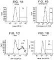
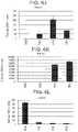
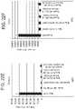
- FIG. 1A to FIG. 1Gshow the FACS histogram expression profiles of the following markers at S3 day 2 of cells differentiated according to Example 1.
- FIG. 1AIsotype control
- FIG. 1Bchromogranin
- FIG. 1CKI-67
- FIG. 1DNKX6.1
- FIG. 1ESOX2
- FIG. 1FCDX2
- FIG. 1GPDX-1. Percentage expression for each marker is shown on each histogram.
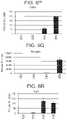
- FIG. 2A to FIG. 2Gshow the FACS histogram expression profiles of the following markers in cells differentiated according to Example 1, and harvested at S4 day 2.
- FIG. 2AIsotype control
- FIG. 2Bchromogranin
- FIG. 2CKI-67
- FIG. 2DNKX6.1
- FIG. 2ESOX2
- FIG. 2FCDX2
- FIG. 2GPDX-1. Percentage expression for each marker is shown on each histogram.
- FIG. 3A to FIG. 3Gshow the FACS histogram expression profiles of the following markers in cells differentiated according to Example 1 and harvested at S5 day 2.
- FIG. 3AIsotype control
- FIG. 3Bchromogranin
- FIG. 3CKI-67
- FIG. 3DNKX6.1
- FIG. 3ESOX2
- FIG. 3FCDX2
- FIG. 3GPDX-1. Percentage expression for each marker is shown on each histogram.
- FIG. 4A to FIG. 4Gshow FACS histogram expression profiles of the following markers of cells differentiated according to Example 1 and harvested at S5 day 7.
- FIG. 4AIsotype control
- FIG. 4Bchromogranin
- FIG. 4CKI-67
- FIG. 4DNKX6.1
- FIG. 4ESOX2
- FIG. 4FCDX2
- FIG. 4GPDX-1. Percentage expression for each marker is shown on each histogram.
- FIG. 5A to FIG. 5Cdepict FACS histogram expression profiles of the following markers in cells differentiated according to Example 1 and harvested at cells harvested at S5 day 2.
- FIG. 5Achromogranin (y-axis) and CDX2 (x axis);
- FIG. 5Bchromogranin (y-axis) and SOX2 (x axis);
- FIG. 5Cchromogranin (y-axis) and NKX6.1 (x axis). Percentage co-expression for each plot is shown on each histogram.
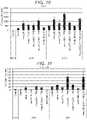
- FIG. 6A to FIG. 6Tshow data from real-time PCR analyses of the expression of the following genes in cells of the human embryonic stem cell line H1 differentiated according to the Example 1 and harvested at S2, S3, S4, and S5.
- FIG. 6ACDX2; FIG. 6B : CD142; FIG. 6C : FOXE1; FIG. 6D : HNF4-alpha; FIG. 6E : NKX2.1; FIG. 6F : NKX2.2; FIG. 6G : NKX6.1; FIG. 6H : OSR1; FIG. 6I : PDX-1; FIG. 6J : PROX1; FIG. 6K : PTF1a; FIG. 6L : SOX17; FIG.
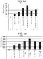
- FIG. 7A to FIG. 7Gdepict data from real-time PCR analyses of the expression of the following genes in cells of the H1 cell line differentiated according to Example 2 and harvested at day 3 of S2, S3, or S4.
- FIG. 7ANKX6.1;
- FIG. 7BPDX-1;
- FIG. 7Cchromogranin;
- FIG. 7DNGN3;
- FIG. 7ECDX2;
- FIG. 7Falbumin;
- FIG. 7GSOX2.
- FIG. 8A to FIG. 8Gdepict data from real-time PCR analyses of the expression of the following markers in H1 cells differentiated according to Example 3 and harvested at S2, S3, S4, or S5.
- FIG. 8ANKX6.1
- FIG. 8BPDX-1
- FIG. 8CNGN3
- FIG. 8DNeuroD
- FIG. 8Echromogranin
- FIG. 8FCDX2
- FIG. 8GSOX2.
- FIG. 9A to FIG. 9Hdepict data from real-time PCR analyses of the expression of the following markers in H1 cells differentiated according to Example 4 and harvested at day 4 of S3 and S4.
- FIG. 9ANKX6.1;
- FIG. 9BPDX-1;
- FIG. 9Cchromogranin;
- FIG. 9DNGN3;
- FIG. 9ENeuroD;
- FIG. 9FCDX2;
- FIG. 9Galbumin;
- FIG. 9HSOX2.
- FIG. 10A to FIG. 10Hdepict data from real-time PCR analyses of the expression of the following genes in cells of the human embryonic stem cell line H1 differentiated according to the Example 5 and harvested at stage 4.
- FIG. 10ANKX6.1;
- FIG. 10BPDX-1;
- FIG. 10Cchromogranin;
- FIG. 10DNGN3;
- FIG. 10ENeuroD;
- FIG. 10FCDX2;
- FIG. 10Galbumin;
- FIG. 10HSOX2.
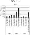
- FIG. 11A to FIG. 11Hshow data from real-time PCR analysis of the expression of the following genes in cells of the human embryonic stem cell line H1 differentiated according to the Example 6 and harvested at day 3 of S3 or S6.
- FIG. 11ANKX6.1;
- FIG. 11BPDX-1;
- FIG. 11Cchromogranin;
- FIG. 11DNGN3;
- FIG. 11ENeuroD;
- FIG. 11FCDX2;
- FIG. 11Galbumin;
- FIG. 11HSOX2.
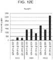
- FIG. 12A to FIG. 12Gdepict data from real-time PCR analysis of the expression of the following genes in cells of the human embryonic stem cell line H1 differentiated according to the Example 7 and harvested at day 3 of S3, S4, or S5.
- FIG. 12ANKX6.1
- FIG. 12BPDX-1
- FIG. 12Cchromogranin
- FIG. 12DNGN3
- FIG. 12ENeuroD
- FIG. 12FCDX2
- FIG. 12GSOX2.
- FIG. 13A to FIG. 13Gdepict data from real-time PCR analyses of the expression of the following genes in cells of the human embryonic stem cell line H1 differentiated according to the Example 8 and harvested at S3, S4, S5, or S6.
- FIG. 13ANKX6.1
- FIG. 13BPDX-1
- FIG. 13Cchromogranin
- FIG. 13DNGN3
- FIG. 13ENeuroD
- FIG. 13FCDX2
- FIG. 13GSOX2.
- FIG. 14A to FIG. 14Hshow FACS histogram expression profiles of the following markers at S3 day 4 of cells differentiated according to Example 9.
- FIG. 14AIsotype control
- FIG. 14Bchromogranin
- FIG. 14CKI-67
- FIG. 14DNKX6.1
- FIG. 14ESOX2
- FIG. 14FHNF3B
- FIG. 14GCDX2
- FIG. 14HPDX-1. Percentage expression for each marker is shown on each histogram.
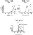
- FIG. 15A to FIG. 15Gshow FACS histogram expression profiles of the following markers at S4 day 2 of cells differentiated according to Example 9.
- FIG. 15AIsotype control
- FIG. 15BNKX6.1
- FIG. 15CKI-67
- FIG. 15Dchromogranin
- FIG. 15ESOX2
- FIG. 15FCDX2
- FIG. 15GPDX-1. Percentage expression for each marker is shown on each histogram.
- FIG. 16A to FIG. 16Fshow FACS histogram expression profiles of the following markers at S4 day 4 of cells differentiated according to Example 9.
- FIG. 16AIsotype control
- FIG. 16BNKX6.1
- FIG. 16Cchromogranin
- FIG. 16DSOX2
- FIG. 16ECDX2
- FIG. 16FPDX-1. Percentage expression for each marker is shown on each histogram.
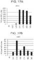
- FIG. 17A to FIG. 17Jshow data from real-time PCR analysis of the expression of the following genes in cells of the human embryonic stem cell line H1 differentiated according to the Example 9 and harvested at S1D3, S2D3, S3D4, S4D2, and S4D4.
- FIG. 17ACDX2; FIG. 17B : HHex; FIG. 17C : FOXE1; FIG. 17D : IPF1 (PDX-1); FIG. 17E : NKX2.1; FIG. 17F : NKX2.2; FIG. 17G : NKX6.1; FIG. 17H : PROX1; FIG. 17I : SOX2; FIG. 17J : SOX9.
- FIG. 18A to FIG. 18Gshow FACS histogram expression profiles of the following markers at S3 day 3 of cells differentiated according to Example 10.
- FIG. 18AIsotype control
- FIG. 18BNKX6.1
- FIG. 18Cchromogranin
- FIG. 18DSOX2
- FIG. 18ECDX2
- FIG. 18FKI-67
- FIG. 18GPDX-1. Percentage expression for each marker is shown on each histogram.
- FIG. 19A to FIG. 19Gshow FACS histogram expression profiles of the following markers at S4 day 5 of cells differentiated according to Example 10.
- FIG. 19AIsotype control
- FIG. 19BNKX6.1
- FIG. 19Cchromogranin
- FIG. 19DSOX2
- FIG. 19ECDX2
- FIG. 19FKI-67
- FIG. 19GPDX-1. Percentage expression for each marker is shown on each histogram.
- FIG. 20A to FIG. 20Jshow real-time PCR analysis of the expression of the following genes in cells of the human embryonic stem cell line H1 differentiated according to the Example 11.
- FIG. 20Asomatostatin
- FIG. 20BPDX1
- FIG. 20CPax6
- FIG. 20DPax4
- FIG. 20ENKX6.1
- FIG. 20FNGN3
- FIG. 20Gglucagon
- FIG. 20HNeuroD
- FIG. 20Iinsulin
- FIG. 20Jchromogranin.
- FIG. 21A to FIG. 21Jshow data from real-time PCR analysis of the expression of the following genes in cells of the human embryonic stem cell line H1 differentiated according to the Example 12 and harvested at S4 day 2, S5 day 2, and S5 day 9.
- FIG. 21Asomatostatin
- FIG. 21BPDX1
- FIG. 21CPax6
- FIG. 21DPax4
- FIG. 21ENKX6.1
- FIG. 21FNGN3
- FIG. 21GNeuroD
- FIG. 21Hinsulin
- FIG. 21Iglucagon
- FIG. 21Jchromogranin.
- FIG. 22A to FIG. 22Lshow data from real-time PCR analyses of the expression of the following genes in cells of the embryonic stem cell line H1 differentiated according to example 13 and harvested at S5 day 3.
- FIG. 22APax4; FIG. 22B : Pax6;
- FIG. 22CPDX1;
- FIG. 22DPTF1a;
- FIG. 22Eglucagon;
- FIG. 22Finsulin;
- FIG. 22GNeuroD;
- FIG. 22Hngn3;
- FIG. 22IZic1;
- FIG. 22JCDX2;
- FIG. 22Kalbumin;
- FIG. 22LNKX6.1.
- Stem cellsare undifferentiated cells defined by their ability, at the single cell level, to both self-renew and differentiate. Stem cells may produce progeny cells, including self-renewing progenitors, non-renewing progenitors, and terminally differentiated cells. Stem cells are also characterized by their ability to differentiate in vitro into functional cells of various cell lineages from multiple germ layers (endoderm, mesoderm and ectoderm). Stem cells also give rise to tissues of multiple germ layers following transplantation and contribute substantially to most, if not all, tissues following injection into blastocysts.
- Stem cellsare classified by their developmental potential as: (1) totipotent, meaning able to give rise to all embryonic and extraembryonic cell types; (2) pluripotent, meaning able to give rise to all embryonic cell types; (3) multipotent, meaning able to give rise to a subset of cell lineages but all within a particular tissue, organ, or physiological system (for example, hematopoietic stem cells (HSC) can produce progeny that include HSC (self-renewal), blood cell restricted oligopotent progenitors, and all cell types and elements (e.g., platelets) that are normal components of the blood); (4) oligopotent, meaning able to give rise to a more restricted subset of cell lineages than multipotent stem cells; and (5) unipotent, meaning able to give rise to a single cell lineage (e.g., spermatogenic stem cells).
- HSChematopoietic stem cells
- Differentiationis the process by which an unspecialized (“uncommitted”) or less specialized cell acquires the features of a specialized cell such as, for example, a nerve cell or a muscle cell.
- a differentiated cell or a differentiation-induced cellis one that has taken on a more specialized (“committed”) position within the lineage of a cell.
- the term “committed”, when applied to the process of differentiation,refers to a cell that has proceeded in the differentiation pathway to a point where, under normal circumstances, it will continue to differentiate into a specific cell type or subset of cell types, and cannot, under normal circumstances, differentiate into a different cell type or revert to a less differentiated cell type.
- De-differentiationrefers to the process by which a cell reverts to a less specialized (or committed) position within the lineage of a cell.
- the lineage of a celldefines the heredity of the cell, i.e., which cells it came from and what cells it can give rise to.
- the lineage of a cellplaces the cell within a hereditary scheme of development and differentiation.
- a lineage-specific markerrefers to a characteristic specifically associated with the phenotype of cells of a lineage of interest and can be used to assess the differentiation of an uncommitted cell to the lineage of interest.
- Markersare nucleic acid or polypeptide molecules that are differentially expressed in a cell of interest.
- differential expressionmeans an increased level for a positive marker and a decreased level for a negative marker.
- the detectable level of the marker nucleic acid or polypeptideis sufficiently higher or lower in the cells of interest compared to other cells, such that the cell of interest can be identified and distinguished from other cells using any of a variety of methods known in the art.
- a cellis “positive for” a specific marker or “positive” when the specific marker is detected in the cell. Similarly, the cell is “negative for” a specific marker, or “negative” when the specific marker is not detected in the cell.
- stage 1and “S1” are used interchangeably to identify cells expressing markers characteristic of the definitive endoderm (DE).
- Definitive endodermrefers to cells which bear the characteristics of cells arising from the epiblast during gastrulation and which form the gastrointestinal tract and its derivatives. Definitive endoderm cells express at least one of the following markers: HNF3 beta, GATA4, SOX17, CXCR4, Cerberus, OTX2, goosecoid, C-Kit, CD99, and MIXL1.
- Gut tuberefers to cells derived from definitive endoderm that express at least one of the following markers: HNF3-beta, HNF1-beta, or HNF4-alpha. Gut tube cells can give rise to all endodermal organs, such as lungs, liver, pancreas, stomach, and intestine.
- stage 2Used herein interchangeably are “stage 2” and “S2” which identify cells expressing markers characteristic of the primitive gut tube.
- Formgut endodermrefers to endoderm cells that give rise to esophagus, lungs, stomach, liver, pancreas, gall bladder, and a portion of the duodenum.
- Posterior foregutrefers to endoderm cells that can give rise to posterior stomach, pancreas, liver, and a portion of the duodenum.
- Mid-gut endodermrefers to endoderm cells that can give rise to the intestines, portions of the duodenum, appendix, and ascending colon.
- Hind-gut endodermrefers to endoderm cells that can give rise to the distal third of the transverse colon, the descending colon, sigmoid colon and rectum.
- Cells expressing markers characteristic of the foregut lineagerefers to cells expressing at least one of the following markers: PDX-1, FOXA2, CDX2, SOX2, and HNF4 alpha.
- stage 4used interchangeably herein are “stage 4” and “S4” to identify cells expressing markers characteristic of the pancreatic foregut precursor.
- Cells expressing markers characteristic of the pancreatic foregut precursor lineagerefers to cells expressing at least one of the following markers: PDX-1, NKX6.1, HNF6, FOXA2, PTF1a, Prox1 and HNF4 alpha.
- stage 5 and “S5”are used interchangeably to identify cells expressing markers characteristic of the pancreatic endoderm and pancreatic endocrine precursor cells.
- Cells expressing markers characteristic of the pancreatic endoderm lineagerefers to cells expressing at least one of the following markers: PDX1, NKX6.1, HNF1 beta, PTF1 alpha, HNF6, HNF4 alpha, SOX9, HB9 or PROX1. Cells expressing markers characteristic of the pancreatic endoderm lineage do not substantially express CDX2 or SOX2.
- stage 6and “S6” are used interchangeably to identify cells enriched in pancreatic endocrine cells.
- Pancreatic endocrine cellor “Pancreatic hormone expressing cell”, or “Cells expressing markers characteristic of the pancreatic endocrine lineage” as used herein, refers to a cell capable of expressing at least one of the following hormones: insulin, glucagon, somatostatin, ghrelin, and pancreatic polypeptide.
- Pantendocrine precursor cellor “Pancreatic endocrine progenitor cell” refers to pancreatic endoderm cells capable of becoming a pancreatic hormone expressing cell. Such a cell can express at least one of the following markers: NGN3, NKX2.2, NeuroD, ISL-1, Pax4, Pax6, or ARX.
- “Functional pancreatic beta cell” as used herein,refers to a single hormonal insulin positive cell capable of being glucose responsive and positive for PDX-1 and NKX6.1.
- “Ascorbic acid” and “Vitamin C”are used interchangeably herein and relate to an essential nutrient for humans and other animal species.
- Glucoseand “D-Glucose” are used interchangeably herein and refer to dextrose, a sugar commonly found in nature.
- a cell “positive” for a specific marker or which is marker “+”is a cell in which the particular marker may be detected.
- a cell “negative” for a specific marker or which is marker “ ⁇ ”is a cell in which the marker is not detected by the methods taught in the instant specification.
- chromograninand “CHGN” are used interchangeably to identify the gene endcoding the acidic secretory glycoprotein chromogranin.
- NeuroDNeuroD 1
- NeuroD 1NeuroD 1
- LDNand “LDN-193189” to indicate a BMP receptor inhibitor available from Stemgent, CA, USA.
- Pluripotent stem cellsmay express one or more of the stage-specific embryonic antigens (SSEA) 3 and 4, and markers detectable using antibodies designated Tra-1-60 and Tra-1-81 (Thomson et al., Science 282:1145, 1998). Differentiation of pluripotent stem cells in vitro results in the loss of SSEA-4, Tra-1-60, and Tra-1-81 expression. Undifferentiated pluripotent stem cells typically have alkaline phosphatase activity, which can be detected by fixing the cells with 4% paraformaldehyde, and then developing with Vector Red as a substrate, as described by the manufacturer (Vector Laboratories, CA, USA). Undifferentiated pluripotent stem cells also typically express OCT4 and TERT, as detected by RT-PCR.
- SSEAstage-specific embryonic antigens
- pluripotent stem cellsAnother desirable phenotype of propagated pluripotent stem cells is a potential to differentiate into cells of all three germinal layers: endoderm, mesoderm, and ectoderm tissues.
- Pluripotency of stem cellscan be confirmed, for example, by injecting cells into SCID mice, fixing the teratomas that form using 4% paraformaldehyde, and then examining them histologically for evidence of cell types from the three germ layers.
- pluripotencymay be determined by the creation of embryoid bodies and assessing the embryoid bodies for the presence of markers associated with the three germinal layers.
- Propagated pluripotent stem cell linesmay be karyotyped using a standard G-banding technique and compared to published karyotypes of the corresponding primate species. It is desirable to obtain cells that have a “normal karyotype,” which means that the cells are euploid, wherein all human chromosomes are present and not noticeably altered.
- Pluripotent cellsmay be readily expanded in culture using various feeder layers or by using matrix protein coated vessels. Alternatively, chemically defined surfaces in combination with defined media such as mTesrTM 1 media (StemCell Technologies, Vancouver, Canada) may be used for routine expansion of the cells.
- Pluripotent cellsmay be readily removed from culture plates using enzymatic, mechanical or use of various calcium chelators such as EDTA (Ethylenediaminetetraacetic acid). Alternatively, pluripotent cells may be expanded in suspension in the absence of any matrix proteins or a feeder layer.
- EDTAEthylenediaminetetraacetic acid
- pluripotent stem cellsinclude established lines of pluripotent cells derived from tissue formed after gestation, including pre-embryonic tissue (such as, for example, a blastocyst), embryonic tissue, or fetal tissue taken any time during gestation, typically but not necessarily, before approximately 10 to 12 weeks gestation.
- pre-embryonic tissuesuch as, for example, a blastocyst
- embryonic tissueor fetal tissue taken any time during gestation, typically but not necessarily, before approximately 10 to 12 weeks gestation.
- hESCshuman embryonic stem cells
- H1, H7, and H9WiCell Research Institute, Madison, Wis., USA.
- cells taken from a pluripotent stem cell population already cultured in the absence of feeder cellsare also suitable.
- IPSinducible pluripotent cells
- reprogrammed pluripotent cellsthat can be derived from adult somatic cells using forced expression of a number of pluripotent related transcription factors, such as OCT4, Nanog, Sox2, KLF4, and ZFP42 (Annu Rev Genomics Hum Genet, 2011, 12:165-185).
- the human embryonic stem cells used in the methods of the inventionmay also be prepared as described by Thomson et al. (U.S. Pat. No. 5,843,780; Science, 1998, 282:1145; Curr. Top. Dev. Biol., 1998, 38:133; Proc. Natl. Acad. Sci. U.S.A., 1995: 92:7844).
- pluripotent stem cell markersinclude, for example, the expression of one or more of the following: ABCG2, cripto, FOXD3, CONNEXIN43, CONNEXIN45, OCT4, SOX2, NANOG, hTERT, UTF1, ZFP42, SSEA-3, SSEA-4, Tra 1-60, Tra 1-81.
- Pluripotent stem cells suitable for use in the present inventioninclude, for example, the human embryonic stem cell line H9 (NIH code: WA09), the human embryonic stem cell line H1 (NIH code: WA01), the human embryonic stem cell line H7 (NIH code: WA07), and the human embryonic stem cell line SA002 (Cellartis, Sweden). Also suitable for use in the present invention are cells that express at least one of the following markers characteristic of pluripotent cells: ABCG2, cripto, CD9, FOXD3, CONNEXIN43, CONNEXIN45, OCT4, SOX2, NANOG, hTERT, UTF1, ZFP42, SSEA-3, SSEA-4, Tra 1-60, and Tra 1-81.
- markers characteristic of pluripotent cellsABCG2, cripto, CD9, FOXD3, CONNEXIN43, CONNEXIN45, OCT4, SOX2, NANOG, hTERT, UTF1, ZFP42, SSEA-3, SSEA-4, Tra 1-60, and Tra 1-81
- Markers characteristic of the definitive endoderm lineageare selected from the group consisting of SOX17, GATA4, HNF3 beta, GSC, CER1, Nodal, FGF8, Brachyury, Mix-like homeobox protein, FGF4, CD48, eomesodermin (EOMES), DKK4, FGF17, GATA6, CXCR4, C-Kit, CD99, and OTX2.
- Suitable for use in the present inventionis a cell that expresses at least one of the markers characteristic of the definitive endoderm lineage.
- a cell expressing markers characteristic of the definitive endoderm lineageis a primitive streak precursor cell.
- a cell expressing markers characteristic of the definitive endoderm lineageis a mesendoderm cell.
- a cell expressing markers characteristic of the definitive endoderm lineageis a definitive endoderm cell.
- Markers characteristic of the pancreatic endoderm lineageare selected from the group consisting of PDX1, NKX6.1, HNF1 beta, PTF1 alpha, HNF6, HNF4 alpha, SOX9, HB9 and PROX1.
- Suitable for use in the present inventionis a cell that expresses at least one of the markers characteristic of the pancreatic endoderm lineage.
- a cell expressing markers characteristic of the pancreatic endoderm lineageis a pancreatic endoderm cell wherein the expression of PDX-1 and NKX6.1 are substantially higher than the expression of CDX2 and SOX2.
- a pancreatic endocrine cellis capable of expressing at least one of the following hormones: insulin, glucagon, somatostatin, and pancreatic polypeptide.
- Suitable for use in the present inventionis a cell that expresses at least one of the markers characteristic of the pancreatic endocrine lineage.
- a cell expressing markers characteristic of the pancreatic endocrine lineageis a pancreatic endocrine cell.
- the pancreatic endocrine cellmay be a pancreatic hormone-expressing cell.
- the pancreatic endocrine cellmay be a pancreatic hormone-secreting cell.
- the pancreatic endocrine cellis a cell expressing markers characteristic of the ⁇ cell lineage.
- a cell expressing markers characteristic of the ⁇ cell lineageexpresses PDX1 and at least one of the following transcription factors: NKX2.2, NKX6.1, NEUROD, ISL1, HNF3 beta, MAFA, PAX4, and PAX6.
- a cell expressing markers characteristic of the ⁇ cell lineageis a ⁇ cell.
- This inventiondescribes an in vitro method and a cell population that can generate single hormonal insulin positive cells which are also PDX-1 and NKX6.1 positive.
- the method used in this inventionincludes a series of stages that direct, in a stepwise manner, the differentiation of human pluripotent cells to single hormonal cells through the following intermediate stages:
- media formulations used to generate gut tube stage cells and all subsequent stepscontain ascorbic acid (also known as Vitamin C).
- ascorbic acidalso known as Vitamin C.
- the concentration of ascorbic acidis about 0.01 mM to about 1 mM. In an embodiment, the concentration of ascorbic acid is from about 0.1 mM to about 0.5 mM.
- pancreatic endoderm culturescan be generated having very high expression levels of PDX-1 and NKX6.1 while having low level expression of CDX2 and SOX2.
- hESC H1human embryonic stem cell line H1
- MATRIGELTM1:30 dilution; BD Biosciences, NJ, USA
- mTeSR®1 mediaStemCell Technologies, Vancouver, Canada
- Y27632Rock inhibitor, Catalog #Y0503, SigmaAldrich, MO, USA
- Forty-eight hours post seedingcultures were washed and incubated in incomplete PBS (phosphate buffered saline without Mg or Ca) for approximately 30 seconds. Cultures were differentiated into pancreatic endocrine lineage as follows:
- sampleswere collected and analyzed by real-time PCR, immune-histochemistry, or fluorescent activated cell sorting (FACS).
- FACSfluorescent activated cell sorting
- the hESC-derived cellswere released into single-cell suspension by incubation in TrypLE Express (Catalog No. 12604, Invitrogen) at 37° C. for 3-5 minutes. Cells were then washed twice in staining buffer (PBS containing 0.2% fatty acid-free BSA) (Catalog No. 554657, BD Biosciences, NJ, USA). For intracellular antibody staining, cells were first incubated for 20 minutes at 4° C. with Green Fluorescent LIVE/DEAD cell dye (Invitrogen Catalog No. L23101), to allow for live/dead cell discrimination during analysis, followed by a single wash in cold PBS.
- TrypLE ExpressCatalog No. 12604, Invitrogen
- Cellswere fixed in 250 ⁇ l of Cytofix/Cytoperm Buffer (BD Biosciences Catalog No. 554722) for 20 minutes at 4° C. followed by two washes in BD Perm/Wash Buffer Solution (BD Biosciences Catalog No. 554723). Cells were resuspended in 100 ⁇ l staining/blocking solution consisting of Perm/Wash buffer supplemented with 2% normal serum (of the appropriate species of the secondary antibody). Cells were then incubated for 30 minutes at 4° C. with primary antibodies at empirically pre-determined dilutions followed by two washes in Perm/Wash buffer. Lastly, cells were incubated with the appropriate secondary antibodies for 30 minutes at 4° C. followed by two washes with Perm/Wash buffer prior to analyses on the BD FACS Canto II.
- Cytofix/Cytoperm BufferBD Biosciences Catalog No. 554722
- BD Biosciences Catalog No. 554723BD Biosciences Catalog No. 55
- rabbit anti-insulin(1:100; Catalog No. C27C9; Cell Signaling, MA, USA)
- mouse anti-insulin(1:100; Catalog NO. ab6999, Abcam, MA, USA
- mouse anti-glucagon(1:1250; Catalog No. G2654; SigmaAldrich)
- rabbit anti-synaptophysin(1:100; Catalog No.
- FIG. 1A to FIG. 1Gdepict FACS histogram expression profiles of Isotype control ( FIG. 1A ), chromogranin ( FIG. 1B ), KI-67 ( FIG. 1C ), NKX6.1 ( FIG. 1D ), SOX2 ( FIG. 1E ), CDX2 ( FIG. 1F ), PDX-1 ( FIG. 1G ) of cells differentiated according to Example 1 and analyzed at S3 day 2. Percentage expression for each marker is shown on each histogram. At day 2 of stage 3, over 95% of the cells were positive for expression of PDX-1 ( FIG. 1G ), and about 60% of the cells in the population were positive for expression of SOX2 ( FIG.
- FIG. 1EWhile less than 10% of the cells were positive for expression of CDX2 ( FIG. 1F ) or NKX6.1 ( FIG. 1D ), or chromogranin ( FIG. 1B ).
- FIG. 1CA significant percentage of cells at stage 3 were in active cell cycle as shown by high percentage of KI-67 positive cells ( FIG. 1C ).
- FIG. 2A to FIG. 2Gdepict the expression profiles of Isotype control ( FIG. 2A ), chromogranin ( FIG. 2B ), KI-67 ( FIG. 2C ), NKX6.1 ( FIG. 2D ), SOX2 ( FIG. 2E ), CDX2 ( FIG. 2F ), PDX-1 ( FIG. 2G ), as determined by FACS staining, of cells differentiated according to Example 1, and harvested at day 2 of S4. Percentage expression for each marker is shown on each histogram. Similar to stage 3, over 95% of the cells were positive for PDX-1 expression ( FIG. 2G ), while about 10% of the cells were positive for CDX2 expression ( FIG.
- FIG. 2FNKX6.1 expression
- FIG. 2ENKX6.1 expression
- FIG. 2Ea drop from 60% at S3. Chromogranin expression was approximately 3%
- FIG. 2CA significant percentage of cells at stage 4 were in active cell cycle as shown by high percentage of KI-67 positive cells ( FIG. 2C ).
- FIG. 3A to 3Gdepict the relative expression profiles, as determined by FACS analyses, of cells harvested at day 2 of stage 5 following the differentiation protocol outlined in this example.
- FIG. 3Aisotype control
- FIG. 3Bchromogranin
- FIG. 3CKI-67
- FIG. 3DNKX6.1
- FIG. 3ESOX2
- FIG. 3FCDX2
- FIG. 3GPDX-1.
- Percentage expression for each markeris shown on each histogram. Similar to stages 3 and 4, over 95% of the cells were positive for expression of PDX-1, while approximately 10% of the cells were positive for CDX2 expression, and over 67% of the cells were positive for NKX6.1 expression.
- SOX2 expressionat approximately 50%, was lower when compared to stage 3, but similar to its expression at S4.
- FIG. 4A to FIG. 4Gdepict the expression of PDX-1 ( FIG. 4G ), NKX6.1 ( FIG. 4D ), CDX2 ( FIG. 4F ), SOX2 ( FIG. 4E ), Ki-67 (proliferation marker; FIG. 4C ) and chromogranin (pan endocrine marker; FIG. 4B ) as measured by FACS staining of cells harvested and analyzed at day 7 of stage 5 of differentiation following the protocol outlined in this example. Similar to stages 3 and 4, >90% of the cells were positive for the expression of PDX-1, while CDX2 expression was less than 10%, and NKX6.1 expression significantly increased to >70% and SOX2 expression dramatically decreased to about 2%.
- FIG. 6A to FIG. 6Tdepict mRNA expression profiles measured by real-time PCR of S2, S3, S4, and S5 cells differentiated following the protocol outlined in this example, and reported as fold change over the expression in undifferentiated H1 cells.
- stage 3there was a very low expression of anterior foregut markers such as FOXe1 ( FIG. 6C ) and NKX2.1 ( FIG. 6E ).
- SOX2FIG. 6M
- OSR1FIG. 6H
- Pancreatic endoderm markerssuch as PTF1a ( FIG. 6K ), NKX6.1 ( FIG. 6G ), and PDX-1 ( FIG.
- cells at stage 5 day 2which were differentiated following the protocol outlined in this example express low levels of CDX2 and SOX2 while maintaining a high expression level of NKX6.1 and PDX-1. It is believed that the unique combination of timely BMP inhibition, use of low dose RA at S4-S5, use of high glucose at S1-S2 results in the population of cells described in Example 1.
- the protocol outlined in this examplewas performed to shed light on the effects of BMP inhibition, addition of FGF7 along with PKC activation on SOX2 expression at S3-S4.
- Cells of the human embryonic stem cell line H1were harvested at various passages (passage 40 to passage 52) and were seeded as single cells at a density of 100,000 cells/cm 2 on MATRIGELTM (1:30 dilution) coated dishes in mTesrTM1 media supplemented with 10 ⁇ M of Y27632. Forty eight hours post-seeding, cultures were differentiated into cells of the pancreatic endocrine lineage as follows:
- Control conditions, at S3,refer to cultures where FGF7, AA, SANT, RA, and 200 nM LDN-193189 were used at the concentrations listed at step c, above.
- endocrine markerssuch as NGN3 ( FIG. 7D )
- pan-endocrine markersuch as chromogranin
- BMP inhibitionis required at day 1 of stage 3 to induce formation of endocrine precursor cells at stages 4-5 and to maintain expression of PDX-1 and NKX6.1 while suppressing SOX2 expression. Furthermore, addition of a PKC activator at stages 3 further enhanced expression of PDX-1 and NKX6.1.
- FIG. 9A to FIG. 9Hdepict the gene expression profile of pancreatic endoderm, endocrine precursor, and foregut endoderm markers for the combinations of culture conditions listed above. Consistent with previous example, blocking of BMP pathway on the first day of stage 3 is critical for the subsequent induction of the endocrine program as measured by expression of the pan-endocrine marker, chromogranin. (See FIG. 9C .) However, addition of the BMP inhibitor at day one of stage 3 triggers expression of endocrine markers at subsequent stages. Furthermore, addition of the BMP inhibitor at day one of stage 3 also decreased expression of foregut marker, SOX2, at stages 3-4 ( FIG. 9H ).
- FIG. 10A to FIG. 10HThe results of real-time PCR analyses of cells harvested after the treatments above are shown in FIG. 10A to FIG. 10H .
- This figureshows that addition of 50 nM or 100 nM of LDN-193189 at days 1, 2, 3, or 4 of S4 can prolong the expression of endocrine markers while maintaining a low expression of SOX2 at S4-S5. (See FIG. 10A to FIG. 10H .)
- This exampleidentifies the optimal dose of BMP inhibition at stage 3 and subsequent effects on endocrine markers at stage 6.
- FIG. 11A to FIG. 11Hshow that a low to moderate inhibition of BMP on the first day of stage 3 is required to trigger expression of endocrine markers while maintaining a low expression of SOX2. Furthermore, BMP inhibition at stage 5 while enhancing endocrine markers also led to upregulation of SOX2 expression.
- the data in this Examplefurther confirms the results presented in the previous examples.
- the dataconfirms that a precise modulation of the BMP pathway at stages 3-5 is required to trigger induction of pancreatic endocrine markers while suppressing SOX2 expression.
- This exampleidentifies the optimal window of time at stage 3 for inhibition of BMP signaling while preserving endocrine induction at later stages and lowering expression fo SOX2.
- FIG. 12A to FIG. 12Gshow the real time PCR analyses for data gathered in this example.
- Treatment at stage 3 for at least 2 hours with a BMP inhibitorcan trigger expression of pro-endocrine transcription factors such as Ngn3 ( FIG. 12D ) and NeuroD ( FIG. 12E ) while maintaining a very low expression of SOX2 ( FIG. 12G ) and significantly increases expression of NKX6.1 ( FIG. 12A ) and PDX-1 ( FIG. 12B ) at S4-S5.
- CDX2 expressionwas higher in cells treated for 2 or 6 hours with BMP inhibitor than in cells treated for 24 hours with the inhibitor ( FIG. 12F ).
- This examplewas carried out to determine the optimal duration of S3 and S4 in the stepwise differentiation of pluripotent cells to a population of cells of pancreatic endocrine lineage.
- FIG. 13A to FIG. 13GData from real time PCR analyses of samples harvested at stage 3, stage 4, stage 5, or stage 6 is shown in FIG. 13A to FIG. 13G .
- This datashows that extending S3 and S4 to three days enhances expression of NKX6.1, when compared with cultures having a two day S3 and S4 ( FIG. 13A ).
- Cells treated for three days at stage 3show down-regulation of the expression of pro-endocrine markers when compared to cultures where duration of S3 and S4 were only two days ( FIG. 13D and FIG. 13E ).
- prolonging stage 4 to three daysdid significantly enhance expression of SOX2 ( FIG. 13G ).
- stage 3 and stage 4are two days.
- the ideal protocolwill result in differentiated cells with high levels of expression of pro-endocrine markers, high NKX6.1, low CDX2 and low SOX2 expression.
- This protocolwas performed to determine the factors that affect SOX2 expression at S3 and S4 during the stepwise differentiation of pluripotent cells into hormone producing cells.
- FIG. 14A to FIG. 14Hdepict FACS histograms for cells harvested at stage 3 day 4 obtained for the following markers: Isotype control ( FIG. 14A ), chromogranin ( FIG. 14B ), KI-67 ( FIG. 14C ), NKX6.1 ( FIG. 14D ), SOX2 ( FIG. 14E ), HNF3B ( FIG. 14F ), CDX2 ( FIG. 14G ), PDX-1 ( FIG. 14H ). Percentage expression for each marker is shown on each histogram. The majority of the cells at stage 3 were positive for expression of PDX-1 ( FIG. 14H ), and HNF3B ( FIG. 14F ), negative for expression of NKX6.1 ( FIG.
- FIG. 14Dshows low expression of chromogranin ( FIG. 14B ), and CDX2 ( FIG. 14G ).
- FIG. 14Eshows that at stage 3, the majority of cells were positive for PDX-1 and SOX2 and negative for NKX6.1 suggesting establishment of an endoderm population consistent with a foregut population anterior to the PDX-1 domain of pancreas.
- FIG. 15A through FIG. 15Gshow FACS histogram expression profiles of the following markers at S4 day 2 of cells differentiated according to Example 9: FIG. 15A : Isotype control, FIG. 15B : NKX6.1, FIG. 15C : KI-67, FIG. 15D : chromogranin, FIG. 15E : SOX2, FIG. 15F : CDX2, FIG. 15G : PDX-1. Percentage expression for each marker is shown on each histogram.
- FIG. 16A to FIG. 16Fshow FACS histogram expression profiles of the following markers at S4 day 4 of cells differentiated according to Example 9.
- FIG. 16AIsotype control
- FIG. 16BNKX6.1
- FIG. 16Cchromogranin
- FIG. 16DSOX2
- FIG. 16ECDX2
- FIG. 16FPDX-1. Percentage expression for each marker is shown on each histogram.
- Table Vsummarizes the data obtained for the % expression of endoderm markers at S3 and S4 for cells differentiated according to the protocol outlined in this example.
- FIG. 17A to FIG. 17Jdepict the results of real-time PCR analyses of the expression of the following genes in cells of the human embryonic stem cell line H1 differentiated according to the Example 9.
- FIG. 17ACDX2, FIG. 17B : HHex, FIG. 17C : FOXE1, FIG. 17D : IPF1 (PDX-1), FIG. 17E : NKX2.1, FIG. 17F : NKX2.2, FIG. 17G : NKX6.1, FIG. 17H : PROX1, FIG. 17I : SOX2, FIG. 17J : SOX9.
- Example 1This is in contrast to the cell population generated in Example 1 which had 40-70% PDX-1+ NKX6.1+ SOX2 ⁇ , CDX2 ⁇ , chromogranin negative fraction and 2-25% PDX-1+ NKX6.1+ SOX2+ at S4-S5.
- cells generated using the protocol in Example 1had far higher percentage of pancreatic endoderm, as defined as a population that is PDX-1+ and NXK6.1+ while being low or negative for SOX2 and CDX2, as compared to cells generated in Example 9.
- the data obtained in this Exampleprovides support that prolonged exposure to BMP inhibition in the presence of high glucose and B27 supplement significantly increases expression of SOX2 at stages 3 and 4 of differentiation.
- Kroon et al.have published a protocol for preparing cells of the pancreatic endoderm lineage from human embryonic stem cells (Nature Biotech 2008, 26: 443-452; hereinafter “Kroon”).
- human embryonic stem cellswere differentiated following the Kroon protocol and assayed for expression of markers characteristic of the different stages of differentiation.
- FIG. 18A to FIG. 18Gshow FACS histogram expression profiles of the following markers at S3 day 3 of cells differentiated according to Example 10: Isotype control ( FIG. 18A ), NKX6.1 ( FIG. 18B ), chromogranin ( FIG. 18C ), SOX2 ( FIG. 18D ), CDX2 ( FIG. 18E ), KI-67 ( FIG. 18F ), PDX-1 ( FIG. 18G ). Percentage expression for each marker is shown on each histogram.
- FIG. 19A to FIG. 19Gshow FACS histogram expression profiles of the following markers at S4 day 5 of cells differentiated according to Example 10. Isotype control ( FIG. 19A ), NKX6.1 ( FIG. 19B ), chromogranin ( FIG. 19C ), SOX2 ( FIG. 19D ), CDX2 ( FIG. 19E ), KI-67 ( FIG. 19F ), PDX-1 ( FIG. 19G ). Percentage expression for each marker is shown on each histogram
- Table VIshown below, summarizes the percentages of endoderm markers at S3-S4 of cells generated in this example.
- FIG. 20A to FIG. 20Jdepict real-time PCR analyses of the expression of the following genes in cells of the human embryonic stem cell line H1 differentiated according to the Example 11.
- FIG. 20Asomatostatin
- FIG. 20BPDX1
- FIG. 20CPax6,
- FIG. 20DPax4,
- FIG. 20ENKX6.1
- FIG. 20FNGN3
- FIG. 20Gglucagon
- FIG. 20HNeuroD
- FIG. 20Iinsulin
- FIG. 20Jchromogranin.
- This Figureshows that addition of ascorbic acid at stage 3 or at stages 3 and 4 significantly decreased expression of somatostatin and glucagon at stages 4-5 while increasing expression of insulin (see FIG.
- pancreatic endoderm markerssuch as PDX-1 and NKX6.1 was not significantly altered by addition of 0.25 mM ascorbic acid (see FIG. 20B and FIG. 20D ).
- Pax6 expressionwas down regulated and Pax4 expression was maintained (see FIG. 20C and FIG. 20D ).
- cultures treated +/ ⁇ ascorbic acid at S3-S5were immune stained for insulin, glucagon, and somatostatin hormones.
- Table VIIsummarizes average percentage of insulin positive cells, glucagon and somatostatin positive cells, and polyhormonal cells (two more hormone expression in one cell).
- This Examplewas carried out to determine the optimal dose of ascorbic acid to be used to generate insulin positive cells that are single hormonal, PDX-1 positive, and NKX6.1 positive.
- FIG. 21A to FIG. 21Jdepict data from real-time PCR analyses of the expression of the following genes in cells of the human embryonic stem cell line H1 differentiated according to the Example 12.
- FIG. 21Asomatostatin
- FIG. 21BPDX1
- FIG. 21CPax6,
- FIG. 21DPax4,
- FIG. 21ENKX6.1
- FIG. 21FNGN3
- gFIG 21 GNeuroD
- FIG. 21Hinsulin
- FIG. 21Iglucagon
- FIG. 21Jchromogranin.
- This Examplewas carried out to shed light on requirements to generate single hormonal insulin positive cells during differentiation of pluripotent cells.
- FIG. 22A through FIG. 22Lshow data from real-time PCR analyses of the expression of Pax4 ( FIG. 22A ); Pax6 ( FIG. 22B ); PDX1 ( FIG. 22C ); PTF1a ( FIG. 22D ); glucagon ( FIG. 22E ); insulin ( FIG. 22F ); NeuroD ( FIG. 22G ); ngn3 ( FIG. 22H ); Zic1 ( FIG. 22I ); CDX2 ( FIG. 22J ); albumin ( FIG. 22K ); NKX6.1 ( FIG. 22L ) in cells of the embryonic stem cell line H1 differentiated according to example 13 and harvested at S5 day 3.
Landscapes
- Engineering & Computer Science (AREA)
- Health & Medical Sciences (AREA)
- Biomedical Technology (AREA)
- Life Sciences & Earth Sciences (AREA)
- Chemical & Material Sciences (AREA)
- Biotechnology (AREA)
- Organic Chemistry (AREA)
- Bioinformatics & Cheminformatics (AREA)
- Genetics & Genomics (AREA)
- Wood Science & Technology (AREA)
- Zoology (AREA)
- Microbiology (AREA)
- Biochemistry (AREA)
- General Engineering & Computer Science (AREA)
- General Health & Medical Sciences (AREA)
- Cell Biology (AREA)
- Micro-Organisms Or Cultivation Processes Thereof (AREA)
- Medicines Containing Material From Animals Or Micro-Organisms (AREA)
Abstract
Description
- a) generation of definite endoderm (DE) cells from undifferentiated human embryonic stem cells comprising culturing pluripotent cells in medium comprising glucose, a TGF-B ligand and a WNT activator;
- b) differentiation of DE cells into gut tube cells comprising culturing DE cells in medium comprising glucose, Vitamin C, and a FGF ligand;
- c) differentiation of gut tube cells into posterior foregut endoderm cells expressing PDX-1 and SOX2. This differentiation is accomplished by culturing the gut tube cells in the presence of a shh inhibitor, a BMP inhibitor, a TGF-B ligand, a FGF ligand, retinoic acid, vitamin C and a PKC activator;
- d) differentiating the posterior foregut cells into pancreatic foregut cells expressing PDX-1 and NKX6.1, and expressing lower level of SOX2 as compared to posterior foregut cells. This differentiation is accomplished by culturing the posterior foregut cells in the presence of a shh inhibitor, a BMP inhibitor, low dose of retinoic acid, vitamin C and a PKC activator.
- e) differentiating pancreatic foregut cells into pancreatic endoderm cells expressing PDX-1, a higher level of NKX6.1, and a lower level of SOX2 as compared to pancreatic foregut cells. The differentiation is accomplished by culturing the pancreatic foregut cells in medium supplemented with a shh inhibitor, a TGF-B inhibitor, low dose of retinoic acid, and vitamin C; and
- f) differentiating pancreatic endoderm cells into pancreatic endocrine precursor cells followed by single-hormonal pancreatic endocrine cells. The differentiation is accomplished by culturing the pancreatic endoderm cells in medium supplemented with a shh inhibitor, low dose of retinoic acid, and vitamin C In an embodiment, the cells in all stages of stepwise differentiation are cultured in a media formulation containing less than 25 mM glucose. In some embodiments, the glucose concentration is in the range of about 8 mM to about 20 mM glucose.
- a. Stage 1 (Definitive Endoderm (DE)—3 days): Cells were cultured for one day in
stage 1 media: MCDB-131 medium (Catalog #10372-019, Invitrogen, CA, USA) supplemented with 0.1% fatty acid-free BSA (Catalog #68700, Proliant, IA, USA), 0.0012 g/ml sodium bicarbonate (Catalog #S3187, SigmaAldrich, MO, USA), 1× GLUTAMAX™ (Catalog #35050-079, Invitrogen), 5 mM D-Glucose (Catalog#G8769, SigmaAldrich, MO, USA), containing 100 ng/ml GDF8 (R&D Systems, MN, USA) and 1 μM MCX compound (a GSK3B inhibitor, 14-Prop-2-en-1-yl-3,5,7,14,17,23,27-heptaazatetracyclo [19.3.1.1˜2,6˜0.1˜8,12˜]heptacosa-1(25),2(27),3,5,8(26),9,11,21,23-nonaen-16-one, U.S. patent application Ser. No. 12/494,789; incorporated herein by reference in its entirety). Cells were then cultured for one day in MCDB-131 medium supplemented with 0.1% fatty acid-free BSA, 0.0012 g/ml sodium bicarbonate, 1× GLUTAMAX™, 5 mM D-Glucose, 100 ng/ml GDF8, and 100 nM MCX compound. Cells were then cultured for one day in MCDB-131 medium to which 0.1% fatty acid-free BSA, 0.0012 g/ml sodium bicarbonate, 1× GLUTAMAX™, 5 mM D-Glucose, and 100 ng/ml GDF8 had been added. - b. Stage 2 (Primitive gut tube—2 days):
Stage 1 cells were treated for two days with MCDB-131 medium supplemented with 0.1% fatty acid-free BSA, 0.0012 g/ml sodium bicarbonate, 1× GLUTAMAX™, 5 mM D Glucose, and 25 ng/ml FGF7. - c. Stage 3 (Foregut-2 days):
Stage 2 cells were cultured for one day inStage 3 medium: MCDB-131 medium supplemented with 1:200 dilution of ITS-X (Invitrogen), 2.5 mM Glucose, 1× GLUTAMAX™, 0.0015 g/ml sodium bicarbonate, 2% fatty acid-free BSA, 25 ng/ml FGF7, 10 ng/ml activin-A (R & D systems), 0.25 μM SANT-1 (shh inhibitor, SigmaAldrich), 1 μM Retinoic acid (RA) (SigmaAldrich), and 200 nM TPB (PKC activator; Catalog#565740; EMD, NJ, USA), containing 100 nM LDN-193189 (BMP receptor inhibitor; Catalog #04-0019; Stemgent, CA, USA). The cells were then cultured for an additional day in theStage 3 medium supplement with 10 nM LDN-193189. - d. Stage 4 (Pancreatic foregut precursor-2 days):
Stage 3 cells were cultured for two days in MCDB-131 medium supplemented with a 1:200 dilution of ITS-X, 2.5 mM Glucose, 1× GLUTAMAX™, 0.0015 g/ml sodium bicarbonate, 2% fatty acid-free BSA, 0.25 μM SANT-1, 50 nM RA, 200 nM TPB, and 50 nM LDN-193189. - e. Stage 5 (Pancreatic endoderm, 2-7 days):
Stage 4 cells were cultured for 2-7 days in MCDB-131 medium supplemented with a 1:200 dilution of ITS-X, 2.5 mM Glucose, 1× GLUTAMAX™, 0.0015 g/ml sodium bicarbonate, 2% fatty acid-free BSA, 0.25 μM SANT-1, and 50 nM RA.
- a. Stage 1 (Definitive Endoderm (DE)—3 days): Cells were cultured for one day in
| TABLE I |
| Average Expression of Endoderm Markers at S3 through S5 |
| % PDX- | |||||||
| 1+ | % | ||||||
| % PDX- | NKX6.1+ | % | PDX-1+ | ||||
| % | 1+ and | and | PDX-1+ | NKX6.1+ | |||
| Total* | PDX-1+ | SOX2+ | SOX2− | SOX2+ | |||
| Stage |
| 3 | 7 | 96 | 60 | 0 | <5 | 0 |
| 2 | ||||||
| Stage | ||||||
| 4 | 9 | 97 | 45 | ~40 | <5 | <5 |
| 2 | ||||||
| Stage | ||||||
| 5 | 11 | 95 | 50 | ~42 | <10 | <25 |
| 2 | ||||||
| Stage | ||||||
| 5 | 16 | 92 | <2 | ~70 | <5 | <2 |
| 7 days | ||||||
| *Total number of days since start of differentiation. |
- a. Stage 1 (Definitive Endoderm (DE)—3 days): Prior to start of DE, the cultures were washed and incubated with incomplete PBS (no Mg or Ca) for 30 seconds followed by addition of the
stage 1 media. Human embryonic stem cells cultured as single cells on MATRIGEL™-coated dishes were treated with MCDB-131 medium supplemented with 0.1% fatty acid-free BSA, 0.0012 g/ml sodium bicarbonate, 1× GLUTAMAX™, and 2.5 mM D-Glucose, 100 ng/ml GDF8, and 1.5 μM MCX compound (GSK3B inhibitor) for one day. Cells were then treated with MCDB-131 medium supplemented with 0.1% fatty acid-free BSA, 0.0012 g/ml sodium bicarbonate, 1× GLUTAMAX™, 2.5 mM glucose, and 100 ng/ml GDF8 for days 2-3. - b. Stage 2 (Primitive gut tube—3 days):
Stage 1 cells were treated with MCDB-131 medium supplemented with 0.1% fatty acid-free BSA, 0.0012 g/ml sodium bicarbonate, 1× GLUTAMAX™, 2.5 mM D-Glucose, and 50 ng/ml FGF7 and for three days. - c. Stage 3 (Foregut—3 days):
Stage 2 cells were treated with MCDB131 medium supplemented with a 1:200 dilution of ITS-X, 2.5 mM Glucose, 1× GLUTAMAX™, 0.0015 g/ml sodium bicarbonate, 2% fatty acid-free BSA, 0.25 μM SANT-1, 20 ng/ml of activin-A, 2 μM RA, in the presence or absence of 50 ng/ml FGF7, 50 nM or 200 nM LDN-193189, and/or 200 nM TPB. Cells were incubated for three days in medium using the combinations listed in Table II, below:
- a. Stage 1 (Definitive Endoderm (DE)—3 days): Prior to start of DE, the cultures were washed and incubated with incomplete PBS (no Mg or Ca) for 30 seconds followed by addition of the
| Stage |
| 3 Treatments |
| LDN- | |||||||
| Treatment | AA | FGF7 | 193189 | RA | TPB | ||
| 1 | + | + | 200 nM+ | + | + | − | |
| 2 | + | + | − | + | + | − | |
| 3 | + | − | − | + | + | − | |
| 4 | + | + | − | + | + | + | |
| 5 | + | − | − | + | + | + | |
| 6 | + | + | 50 nM | + | + | − | |
- d. Stage 4 (Pancreatic foregut precursor—3 days):
Stage 3 cells were treated with MCDB131 medium supplemented with 1:200 dilution of ITS-X, 2.5 mM Glucose, 1× GLUTAMAX™, 0.0015 g/ml sodium bicarbonate, 2% fatty acid-free BSA, 0.25 μM SANT-1, 200 nM TPB, 400 nM LDN-193189, 2 μM ALk5 inhibitor (SD-208, disclosed in Molecular Pharmacology 2007, 72:152-161), and 100 nM CYP26A inhibitor (N-{4-[2-Ethyl-1-(1H-1,2,4-triazol-1-yl)butyl]phenyl}-1,3-benzothiazol-2-amine, Janssen, Belgium) for three days.
- d. Stage 4 (Pancreatic foregut precursor—3 days):
- a. Stage 1 (Definitive Endoderm (DE)—4 days): Prior to start of DE, the cells were washed and incubated with incomplete PBS (no Mg or Ca) for 30 seconds followed by incubation in S1 media. Human embryonic stem cells cultured as single cells on MATRIGEL™-coated dishes were treated for one day with MCDB-131 medium supplemented with 0.1% fatty acid-free BSA, 0.0012 g/ml sodium bicarbonate, 1× GLUTAMAX™, 2.5 mM D-Glucose, 100 ng/ml GDF8, 1.5 μM MCX compound (GSK3B inhibitor). Cells were then treated for two days (days 2-4) with MCDB-131 medium supplemented with 0.1% fatty acid-free BSA, 0.0012 g/ml sodium bicarbonate, 1× GLUTAMAX™, 2.5 mM glucose, and 100 ng/ml GDF8.
- b. Stage 2 (Primitive gut tube—3 days):
Stage 1 cells were treated for three days with MCDB-131 medium supplemented with 0.1% fatty acid-free BSA, 0.0012 g/ml sodium bicarbonate, 1× GLUTAMAX™, 2.5 mM D-Glucose, and 50 ng/ml FGF7. - c. Stage 3 (Foregut—3 days):
Stage 2 cells were treated with MCDB-131 medium supplemented with a 1:200 dilution of ITS-X, 2.5 mM Glucose, 1× GLUTAMAX™, 0.0015 g/ml sodium bicarbonate, 2% fatty acid-free BSA, 0.25 μM SANT-1, 20 ng/ml of Activin-A, 2 μM RA, 50 ng/ml FGF7, 100 nM LDN-193189 (onday 1 only or for the duration of stage 3), and 200 nM TPB. In some cultures, LDN-193189 was removed fromstage 3. - d. Stage 4 (Pancreatic foregut precursor—3 days):
Stage 3 cells were treated with MCDB-131 medium supplemented with a 1:200 dilution of ITS-X, 2.5 mM Glucose, 1× GLUTAMAX™, 0.0015 g/ml sodium bicarbonate, 2% fatty acid-free BSA, 0.25 μM SANT-1, 100 nM TPB, 200 nM LDN-193189, 2 μM ALk5 inhibitor, and 100 nM CYP26A inhibitor for three days. - e. Stage 5 (Pancreatic endoderm/endocrine—4 days):
Stage 4 cells were treated with MCDB-131 medium supplemented with a 1:200 dilution of ITS-X, 2.5 mM Glucose, 1× GLUTAMAX™, 0.0015 g/ml sodium bicarbonate, 2% fatty acid-free BSA, 200 nM LDN-193189, and 2 μM ALk5 for four days.
- a. Stage 1 (Definitive Endoderm (DE)—4 days): Prior to start of DE, the cultures were washed and incubated with incomplete PBS (no Mg or Ca) for 30 seconds followed by addition of the
stage 1 media. Human embryonic stem cells cultured as single cells on MATRIGEL™-coated dishes were treated for one day with MCDB-131 medium supplemented with 0.1% fatty acid-free BSA, 0.0012 g/ml sodium bicarbonate, 1× GLUTAMAX™, 2.5 mM D-Glucose, 100 ng/ml GDF8 and 1.5 μM MCX compound (GSK3B inhibitor). Cells were then treated for three days with MCDB-131 medium supplemented with 0.1% fatty acid-free BSA, 0.0012 g/ml sodium bicarbonate, 1× GLUTAMAX™, 2.5 mM glucose, and 100 ng/ml GDF8. - b. Stage 2 (Primitive gut tube—3 days):
Stage 1 cells were treated for three days with MCDB-131 supplemented with 0.1% fatty acid-free BSA, 0.0012 g/ml sodium bicarbonate, 1× GLUTAMAX™, 2.5 mM D-Glucose, and 50 ng/ml FGF7. - c. Stage 3 (Foregut—3 days):
Stage 2 cells were treated with MCDB-131 medium supplemented with a 1:200 dilution of ITS-X, 2.5 mM Glucose, 1× GLUTAMAX™, 0.0015 g/ml sodium bicarbonate, 2% fatty acid-free BSA, 0.25 μM SANT-1, 50 ng/ml FGF7, 2 μM RA and the combinations listed in Table III, below for three days.
- a. Stage 1 (Definitive Endoderm (DE)—4 days): Prior to start of DE, the cultures were washed and incubated with incomplete PBS (no Mg or Ca) for 30 seconds followed by addition of the
| Stage |
| 3 |
| 20 ng/ | 1 μM Alk5 inhibitor | ||||
| AA | LDN-193189 | (SCIO compound) | TPB | ||
| A | S3D1 | + | − | − | +200 nM |
| D2-3 | + | − | − | +200 nM | |
| B | D1 | − | +100 nM | +1 μM | − |
| D2-3 | + | − | − | +200 nM | |
| C | D1 | − | +100 nM | +1 μM | +100 nM |
| D2-3 | + | − | − | +200 nM | |
| D | D1 | − | +100 nM | +1 μM | +200 nM |
| D2-3 | + | − | − | +200 nM | |
| E | D1 | + | − | − | +100 nM |
| D2-3 | + | +100 nM (on | − | +100 | |
| day | |||||
| 3 only) | |||||
- d. Stage 4 (Pancreatic foregut precursor—3 days):
Stage 3 cells were treated with MCDB-131 medium supplemented with a 1:200 dilution of ITS-X, 2.5 mM Glucose, 1× GLUTAMAX™, 0.0015 g/ml sodium bicarbonate, 2% fatty acid-free BSA, 0.25 μM SANT-1, 100 nM TPB, 200 nM LDN-193189, 2 μM ALk5 inhibitor, and 100 nM CYP26A inhibitor for three days.
- d. Stage 4 (Pancreatic foregut precursor—3 days):
- a. Stage 1 (Definitive Endoderm (DE)—4 days): Prior to start of DE, the cultures were washed and incubated with incomplete PBS (no Mg or Ca) for 30 seconds followed by addition of the
stage 1 media. Human embryonic stem cells cultured as single cells on MATRIGEL™-coated dishes were treated with MCDB-131 medium supplemented with 0.1% fatty acid-free BSA, 0.0012 g/ml sodium bicarbonate, 1× GLUTAMAX™, 2.5 mM D-Glucose, 100 ng/ml GDF8, and 1 μM MCX compound (GSK3B inhibitor) for one day. Cells were then treated with MCDB-131 medium supplemented with 0.1% fatty acid-free BSA, 0.0012 g/ml sodium bicarbonate, 1× GLUTAMAX™, 2.5 mM glucose, and 100 ng/ml GDF8 for days 2-4. - b. Stage 2 (Primitive gut tube—3 days):
Stage 1 cells were treated with MCDB-131 medium supplemented with 0.1% fatty acid-free BSA, 0.0012 g/ml sodium bicarbonate, 1× GLUTAMAX™, 2.5 mM D-Glucose, and 50 ng/ml FGF7 for three days. - c. Stage 3 (Foregut—4 days):
Stage 2 cells were treated with MCDB-131 medium supplemented with a 1:200 dilution of ITS-X, 2.5 mM, Glucose, 1× GLUTAMAX™, 0.0015 g/ml sodium bicarbonate, 2% fatty acid-free BSA, 0.25 μM SANT-1, 50 ng/ml FGF7, 2 μM RA, 20 ng/ml of Activin-A, 100 nM LDN-193189, and 100 nM TPB for one day. Cells were then cultured in MCDB-131 medium supplemented with a 1:200 dilution of ITS-X, 2.5 mM, Glucose, 1× GLUTAMAX™, 0.0015 g/ml sodium bicarbonate, 2% fatty acid-free BSA, 0.25 μM SANT-1, 50 ng/ml FGF7, 2 μM RA, 20 ng/ml of Activin-A, and 100 nM TPB for three days. - d. Stage 4 (Pancreatic foregut precursor—4 days):
Stage 3 cells were treated with MCDB-131 medium supplemented with a 1:200 dilution of ITS-X, 2.5 mM Glucose, 1× GLUTAMAX™, 0.0015 g/ml sodium bicarbonate, 2% fatty acid-free BSA, 200 nM LDN-193189-193189, 2 μM ALk5 inhibitor, 100 nM CYP26A inhibitor, and the concentrations of LDN-193189 listed on Table IV (below) for days 1-4 of stage 4:
- a. Stage 1 (Definitive Endoderm (DE)—4 days): Prior to start of DE, the cultures were washed and incubated with incomplete PBS (no Mg or Ca) for 30 seconds followed by addition of the
| TABLE IV |
| Concentrations of LDN-193189 Used at S4 |
| Condition | Condition | Condition | Condition | Condition | ||
| A | B | C | E | |||
| S4D1 |
| 100 | 50 | 10 | 10 | 10 nM | ||
| S4D2 | 50 | 10 | 50 | 10 | 10 nM | |
| S4D3 | 10 | 10 | 100 | 50 | 10 nM | |
| S4D4 | 10 | 10 | 100 | 50 | 10 nM | |
- d. Stage 5 (Pancreatic endoderm/endocrine precursor-3 days):
Stage 4 cells were treated with MCDB-131 medium supplemented with a 1:200 dilution of ITS-X, 2.5 mM Glucose, 1× GLUTAMAX™, 0.0015 g/ml sodium bicarbonate, 2% fatty acid-free BSA, 50 nM LDN-193189, and 1 μM ALk5 inhibitor for three days.
- d. Stage 5 (Pancreatic endoderm/endocrine precursor-3 days):
- a. Stage 1 (Definitive Endoderm (DE)—4 days): Prior to start of DE, the cultures were washed and incubated with incomplete PBS (no Mg or Ca) for 30 seconds followed by addition of the
stage 1 media. Human embryonic stem cells cultured as single cells on MATRIGEL™-coated dishes were treated with MCDB-131 medium supplemented with 0.1% fatty acid-free BSA, 0.0012 g/ml sodium bicarbonate, 1× GLUTAMAX™, 2.5 mM D-Glucose, 100 ng/ml GDF8, and 1.5 μM MCX compound (GSK3B inhibitor) for one day. Cells were then treated with MCDB-131 medium supplemented with 0.1% fatty acid-free BSA, 0.0012 g/ml sodium bicarbonate, 1× GLUTAMAX™, 2.5 mM glucose, and 100 ng/ml GDF8 for days 2-4. - b. Stage 2 (Primitive gut tube—3 days):
Stage 1 cells were treated with MCDB-131 medium supplemented with 0.1% fatty acid-free BSA, 0.0012 g/ml sodium bicarbonate, 1× GLUTAMAX™, 2.5 mM D-Glucose, and 50 ng/ml FGF7 for three days. - c. Stage 3 (Foregut—3 days):
Stage 3 cells were treated with MCDB-131 medium supplemented with a 1:200 dilution of ITS-X, 2.5 mM Glucose, 1× GLUTAMAX™, 0.0015 g/ml sodium bicarbonate, 2% fatty acid-free BSA, 0.25 μM SANT-1, 50 ng/ml FGF7, 2 μM RA, 20 ng/ml Activin-A, 100 nM TPB, and 10-50 nM LDN-193189 for one day. Cells were then treated with MCDB-131 medium supplemented with a 1:200 dilution of ITS-X, 2.5 mM Glucose, 1× GLUTAMAX™, 0.0015 g/ml sodium bicarbonate, 2% fatty acid-free BSA, 0.25 μM SANT-1, 50 ng/ml FGF7, 2 μM RA, 20 ng/ml Activin-A, and 100 nM TPB for two days. - d. Stage 4 (Pancreatic foregut precursor—3 days):
Stage 3 cells were treated with MCDB-131 medium supplemented with a 1:200 dilution of ITS-X, 2.5 mM Glucose, 1× GLUTAMAX™, 0.0015 g/ml sodium bicarbonate, 2% fatty acid-free BSA, 20 nM LDN-193189; 2 μM ALk5 inhibitor; 100 nM CYP26 A inhibitor, and 100 nM TPB for three days. - e. Stage 5 (Pancreatic endoderm/endocrine precursor-3 days):
Stage 4 cells were treated with MCDB-131 medium supplemented with a 1:200 dilution of ITS-X; 2.5 mM Glucose; 1× GLUTAMAX™; 0.0015 g/ml sodium bicarbonate; 2% fatty acid-free BSA; +/−25 nM LDN-193189 and/or 2 μM ALk5 inhibitor for three days. - f. Stage 6 (Pancreatic endocrine hormone producing-3 days):
Stage 5 cells were treated with MCDB-131 medium supplemented with a 1:200 dilution of ITS-X; 2.5 mM Glucose; 1× GLUTAMAX™; 0.0015 g/ml sodium bicarbonate; and 2% fatty acid-free BSA for three days.
- a. Stage 1 (Definitive Endoderm (DE)—4 days): Prior to start of DE, the cultures were washed and incubated with incomplete PBS (no Mg or Ca) for 30 seconds followed by addition of the
- a. Stage 1 (Definitive Endoderm (DE)—4 days): Prior to start of DE, the cultures were washed and incubated with incomplete PBS (no Mg or Ca) for 30 seconds followed by addition of the
stage 1 media. Human embryonic stem cells cultured as single cells on MATRIGEL™-coated dishes were treated with MCDB-131 medium supplemented with 0.1% fatty acid-free BSA, 0.0012 g/ml sodium bicarbonate, 1× GLUTAMAX™, 2.5 mM D-Glucose, 100 ng/ml GDF8 and 1.5 μM MCX compound for one day. Cells were then treated with MCDB-131 medium supplemented with 0.1% fatty acid-free BSA, 0.0012 g/ml sodium bicarbonate, 1× GLUTAMAX™, 2.5 mM glucose, and 100 ng/ml GDF8 for days 2-4. - b. Stage 2 (Primitive gut tube—3 days):
Stage 1 cells were treated with MCDB-131 medium supplemented with 0.1% fatty acid-free BSA, 0.0012 g/ml sodium bicarbonate, 1× GLUTAMAX™, 2.5 mM D-Glucose, and 50 ng/ml FGF7 for three days. - c. Stage 3 (Foregut—3 days):
Stage 2 cells were treated with MCDB-131 medium supplemented with a 1:200 dilution of ITS-X, 2.5 mM Glucose, 1× GLUTAMAX™, 0.0015 g/ml sodium bicarbonate, 2% fatty acid-free BSA, 0.25 μM SANT-1, 50 ng/ml FGF7, 2 μM RA, 20 ng/ml Activin-A, and 100 nM TPB, containing 100 nM LDN-193189 for only the first 2 hours, 6 hours, or 24 hours ofstage 3. - d. Stage 4 (Pancreatic foregut precursor—3 days):
Stage 3 cells were treated with MCDB-131 medium supplemented with a 1:200 dilution of ITS-X, 2.5 mM Glucose, 1× GLUTAMAX™, 0.0015 g/ml sodium bicarbonate, 2% fatty acid-free BSA, 25 nM LDN-193189, 2 μM ALk5 inhibitor, 100 nM CYP26 A inhibitor, and 100 nM TPB for three days. - e. Stage 5 (Pancreatic endoderm/endocrine precursor-3 days):
Stage 4 cells were treated with MCDB-131 medium supplemented with a 1:200 dilution of ITS-X, 2.5 mM Glucose, 1× GLUTAMAX™, 0.0015 g/ml sodium bicarbonate, 2% fatty acid-free BSA, and 2 μM ALk5 inhibitor for three days.
- a. Stage 1 (Definitive Endoderm (DE)—4 days): Prior to start of DE, the cultures were washed and incubated with incomplete PBS (no Mg or Ca) for 30 seconds followed by addition of the
- a. Stage 1 (Definitive Endoderm (DE)—4 days): Prior to start of DE, the cultures were washed and incubated with incomplete PBS (no Mg or Ca) for 30 seconds followed by addition of the
stage 1 media. Human embryonic stem cells cultured as single cells on MATRIGEL™-coated dishes were treated with MCDB-131 medium supplemented with 0.1% fatty acid-free BSA, 0.0012 g/ml sodium bicarbonate, 1× GLUTAMAX™; 2.5 mM D-Glucose; 100 ng/ml GDF8 and 1.5 μM MCX compound (GSK3B inhibitor) for one day. Cells were then treated with MCDB-131 medium supplemented with 0.1% fatty acid-free BSA; 0.0012 g/ml sodium bicarbonate; 1× GLUTAMAX™; 2.5 mM glucose, and 100 ng/ml GDF8 for days 2-4; then - b. Stage 2 (Primitive gut tube—2 days):
Stage 1 cells were treated with MCDB-131 medium supplemented with 0.1% fatty acid-free BSA, 0.0012 g/ml sodium bicarbonate, 1× GLUTAMAX™, 2.5 mM D-Glucose, and 50 ng/ml FGF7 for two days. - c. Stage 3 (Foregut—2-3 days):
Stage 2 cells were treated with MCDB-131 medium supplemented with a 1:200 dilution of ITS-X, 2.5 mM Glucose, 1× GLUTAMAX™, 0.0015 g/ml sodium bicarbonate, 2% fatty acid-free BSA, 0.25 μM SANT-1, 50 ng/ml FGF7, 2 μM RA, 20 ng/ml Activin-A, and 100 nM TPB, containing 100 nM LDN-193189 for one day. Cells were then treated with MCDB-131 medium supplemented with a 1:200 dilution of ITS-X, 2.5 mM Glucose, 1× GLUTAMAX™, 0.0015 g/ml sodium bicarbonate, 2% fatty acid-free BSA, 0.25 μM SANT-1, 50 ng/ml FGF7, 2 μM RA, 20 ng/ml Activin-A, and 100 nM TPB. - d. Stage 4 (Pancreatic foregut precursor—2-3 days):
Stage 3 cells were treated with MCDB-131 medium supplemented with a 1:200 dilution of ITS-X, 2.5 mM Glucose, 1× GLUTAMAX™, 0.0015 g/ml sodium bicarbonate, 2% fatty acid-free BSA, 25 nM LDN-193189, 100 nM CYP26 A inhibitor, and 100 nM TPB for two or three days. - e. Stage 5 (Pancreatic endoderm/endocrine precursor-2 days):
Stage 4 cells were treated with MCDB-131 medium supplemented with a 1:200 dilution of ITS-X, 2.5 mM Glucose, 1× GLUTAMAX™, 0.0015 g/ml sodium bicarbonate, 2% fatty acid-free BSA, and 1 μM ALk5 inhibitor for two days. - f. Stage 6 (Pancreatic endocrine precursor/hormone-2 days):
Stage 5 cells were treated with MCDB-131 medium supplemented with a 1:200 dilution of ITS-X, 2.5 mM Glucose, 1× GLUTAMAX™, 0.0015 g/ml sodium bicarbonate, and 2% fatty acid-free BSA for two days.
- a. Stage 1 (Definitive Endoderm (DE)—4 days): Prior to start of DE, the cultures were washed and incubated with incomplete PBS (no Mg or Ca) for 30 seconds followed by addition of the
- a. Undifferentiated cells were cultured in RPMI medium (Invitrogen) supplemented with 0.2% FBS; 100 ng/ml activin A; 20 ng/ml WNT-3a for one day. Cells were then treated with RPMI medium supplemented with 0.5% FBS; 100 ng/ml activin A for an additional two days (Stage 1).
- b.
Stage 1 cells were treated with DMEM/F12 medium supplemented with 2% FBS; 50 ng/ml FGF7 for three days (Stage 2). - c.
Stage 2 cells were cultured in DMEM-High glucose medium supplemented with 1% B27; 0.25 μM SANT-1; 2 μM RA; 100 ng/ml Noggin (R & D systems, MN, USA) for four days (Stage 3). - d.
Stage 3 cells were treated with DMEM-High glucose medium supplemented with 1% B27; 100 ng/ml Noggin; 1 μM ALK5 inhibitor II (Axxora, CA, USA); and 50 nM TPB for four days (Stage 4).
| TABLE V |
| % Expression of Endoderm Markers at S3-S4 |
| Total number | ||||||
| of days since | % PDX- | % PDX-1+ | % PDX- | % PDX-1+ | ||
| Stage | start of | % PDX- | 1+ | NKX6.1+ | 1+ | NKX6.1+ |
| (Days) | 1+ | SOX2+ | SOX2− | SOX2+ | ||
| Stage |
| 3 | 10 | 95 | 92 | <1 | <5 | <1 |
| 4 | ||||||
| Stage | ||||||
| 4 | 12 | 85 | 83 | <1 | ~6 | ~30 |
| 2 | ||||||
| Stage | ||||||
| 4 | 14 | 95 | 75 | <5 | ~7 | ~50 |
| 4 days | ||||||
- a) Undifferentiated cells were exposed to RPMI medium supplemented with 0.2% FBS, 100 ng/ml activin A, 20 ng/ml WNT-3a for one day followed by treatment with RPMI medium supplemented with 0.5% FBS, 100 ng/ml activin A for an additional two days (Stage 1).
- b)
Stage 1 cells were exposed to RPMI medium supplemented with 2% FBS, 50 ng/ml FGF7 for three days (Stage 2). - c)
Stage 2 cells were treated with DMEM-High glucose medium supplemented with 1% B27, 0.25 μM SANT-1, 2 μM RA, 50 ng/ml of Noggin (R & D systems, MN) for three days (Stage 3). - d)
Stage 3 cells were cultured in DMEM-High glucose medium supplemented with 1% B27 for three days (Stage 4). - e)
Stage 4 cells were scraped from the wells and resuspended as clusters in DMEM-High glucose medium supplemented with 1% B27 for two days.
| TABLE VI |
| Expression of Endoderm Markers at S3-S4 in Cells Differentiated According to Kroon |
| Total number | ||||||
| of days since | % PDX- | % PDX-1+ | % PDX- | % PDX-1+ | ||
| Stage | start of | % PDX- | 1+ | NKX6.1+ | 1+ | NKX6.1+ |
| (days) | 1+ | SOX2+ | SOX2− | SOX2+ | ||
| Stage |
| 3 | 9 | 75 | 75 | <1 | <2 | <1 |
| 3 | ||||||
| Stage | ||||||
| 4 | 14 | 89 | 52 | ~20 | ~10 | ~20 |
| 5 days* | ||||||
| *Last two days in suspension culture. |
- a. Stage 1 (Definitive Endoderm (DE)—3 days): Prior to start of DE, the cultures were washed and incubated with incomplete PBS (no Mg or Ca) for 30 seconds followed by addition of the
stage 1 media. Human embryonic stem cells cultured as single cells on MATRIGEL™-coated dishes were treated with MCDB-131 medium supplemented with 0.1% fatty acid-free BSA, 0.0012 g/ml sodium bicarbonate, 1× GLUTAMAX™, 5 mM D-Glucose, 100 ng/ml GDF8, and 1 μM MCX compound (GSK3B inhibitor) for one day. Cells were then treated with MCDB-131 medium supplemented with 0.1% fatty acid-free BSA, 0.0012 g/ml sodium bicarbonate, 1× GLUTAMAX™, 5 mM glucose, 100 ng/ml GDF8, and 100 nM MCX compound for day two, followed by an additional day in MCDB-131 medium supplemented with 0.1% fatty acid-free BSA, 0.0012 g/ml sodium bicarbonate, 1× GLUTAMAX™, 5 mM Glucose, and 100 ng/ml GDF8. - b. Stage 2 (Primitive gut tube—2 days):
Stage 1 cells were treated with MCDB-131 medium supplemented with 0.1% fatty acid-free BSA, 0.0012 g/ml sodium bicarbonate, 1× GLUTAMAX™, 5 mM D-Glucose, and 25 ng/ml FGF7 for two days. - c. Stage 3 (Foregut-2 days):
Stage 2 cells were treated with MCDB-131 medium supplemented with 1:200 dilution of ITS-X, 2.5 mM Glucose, 1× GLUTAMAX™, 0.0015 g/ml sodium bicarbonate, 2% fatty acid-free BSA, 10 ng/ml Activin A, 25 ng/ml FGF7, 0.25 μM SANT-1, 1 μM RA, 200 nM TPB (PKC activator), 100 nM LDN-193189 (BMP receptor inhibitor) for one day. Cells were then treated with MCDB-131 medium supplemented with 1:200 dilution of ITS-X, 2.5 mM Glucose, 1× GLUTAMAX™, 0.0015 g/ml sodium bicarbonate, 2% fatty acid-free BSA, 10 ng/ml Activin A, 25 ng/ml FGF7, 0.25 μM SANT-1, 1 μM RA, 200 nM TPB (PKC activator), 10 nM LDN-193189 for an additional day. Some cultures were treated with 0.25 mM ascorbic acid (Catalog #A4544, Sigma, MO, USA) for the duration ofstage 3. - d. Stage 4 (Pancreatic foregut precursor-2 days):
Stage 3 cells were treated with MCDB-131 medium supplemented with 1:200 dilution of ITS-X, 2.5 mM Glucose, 1× GLUTAMAX™, 0.0015 g/ml sodium bicarbonate, 2% fatty acid-free BSA, 0.25 μM SANT-1, 50 nM RA, 200 nM TPB, 50 nM LDN-193189, with or without 0.25 mM ascorbic acid for two days. - e. Stage 5 (Pancreatic endoderm, 2-7 days):
Stage 4 cells were treated with MCDB-131 medium supplemented with a 1:200 dilution of ITS-X, 2.5 mM Glucose, 1× GLUTAMAX™, 0.0015 g/ml sodium bicarbonate, 2% fatty acid-free BSA, 0.25 μM SANT-1, 50 nM RA, with or without 0.25 mM ascorbic acid for 2-7 days.
- a. Stage 1 (Definitive Endoderm (DE)—3 days): Prior to start of DE, the cultures were washed and incubated with incomplete PBS (no Mg or Ca) for 30 seconds followed by addition of the
| TABLE VII |
| Expression of hormones as a percentage |
| of the entire hormone count |
| % single | % glucagon + | ||
| hormonal | plus % | ||
| Treatment | Insulin+ | somatostatin+ | % Polyhormonal |
| Control | 16 | 83 | 50 |
| +ascorbic acid at S3- | 55 | 44 | 36 |
- a. Stage 1 (Definitive Endoderm (DE)—3 days): Prior to start of DE, the cultures were washed and incubated with incomplete PBS (no Mg or Ca) for 30 seconds followed by addition of the
stage 1 media. Human embryonic stem cells cultured as single cells on MATRIGEL™-coated dishes were treated with MCDB-131 medium supplemented with 0.1% fatty acid-free BSA, 0.0012 g/ml sodium bicarbonate, 1× GLUTAMAX™, 5 mM D-Glucose, and 100 ng/ml GDF8 plus 1 μM MCX compound (GSK3B inhibitor) for one day. Cells were then treated with MCDB-131 medium supplemented with 0.1% fatty acid-free BSA, 0.0012 g/ml sodium bicarbonate, 1× GLUTAMAX™, 5 mM glucose, and 100 ng/ml GDF8 plus 100 nM MCX compound for day two, followed by an additional day in MCDB-131 medium supplemented with 0.1% fatty acid-free BSA, 0.0012 g/ml sodium bicarbonate, 1× GLUTAMAX™, 5 mM Glucose, and 100 ng/ml GDF8. - b. Stage 2 (Primitive gut tube—2 days):
Stage 1 cells were treated with MCDB-131 medium supplemented with 0.1% fatty acid-free BSA, 0.0012 g/ml sodium bicarbonate, 1× GLUTAMAX™, 5 mM D-Glucose, with or without the addition of 0.25 mM ascorbic acid and 25 ng/ml FGF7 for two days. - c. Stage 3 (Foregut-2 days):
Stage 2 cells were treated with MCDB131 medium supplemented with 1:200 dilution of ITS-X, 2.5 mM Glucose, 1× GLUTAMAX™, 0.0015 g/ml sodium bicarbonate, 2% fatty acid-free BSA, 10 ng/ml Activin A, 25 ng/ml FGF7, 0.25 μM SANT-1, +/−0.25 mM ascorbic acid, 1 μM RA, 200 nM TPB, 100 nM LDN-193189 forday 1, followed by treatment with MCDB131 medium supplemented with 1:200 dilution of ITS-X, 2.5 mM Glucose, 1× GLUTAMAX™, 0.0015 g/ml sodium bicarbonate, 2% fatty acid-free BSA, 10 ng/ml Activin A, 25 ng/ml FGF7, 0.25 μM SANT-1, +/−0.25 mM ascorbic acid, 1 μM RA, 200 nM TPB, 10 nM LDN-193189 for an additional day. - d. Stage 4 (Pancreatic foregut precursor-2 days):
Stage 3 cells were treated with MCDB131 medium supplemented with 1:200 dilution of ITS-X, 2.5 mM Glucose, 1× GLUTAMAX™, 0.0015 g/ml sodium bicarbonate, 2% fatty acid-free BSA, 0.25 μM SANT-1, 50 nM RA, 200 nM TPB, 50 nM LDN-193189, with or without the addition of 0.25 mM to 1 mM ascorbic acid for two days. - e. Stage 5 (Pancreatic endoderm, 2-9 days):
Stage 4 cells were treated with MCDB131 medium supplemented with a 1:200 dilution of ITS-X, 2.5 mM Glucose, 1× GLUTAMAX™, 0.0015 g/ml sodium bicarbonate, 2% fatty acid-free BSA, 0.25 μM SANT-1, 50 nM RA, with or without the addition of 0.25 mM ascorbic acid for 2-9 days.
- a. Stage 1 (Definitive Endoderm (DE)—3 days): Prior to start of DE, the cultures were washed and incubated with incomplete PBS (no Mg or Ca) for 30 seconds followed by addition of the
- a. Stage 1 (Definitive Endoderm (DE)—3 days): Prior to start of DE, the cultures were washed and incubated with incomplete PBS (no Mg or Ca) for 30 seconds followed by addition of the
stage 1 media. Human embryonic stem cells cultured as single cells on MATRIGEL™-coated dishes were treated with MCDB-131 medium supplemented with 0.1% fatty acid-free BSA, 0.0012 g/ml sodium bicarbonate, 1× GLUTAMAX™, 5 mM D-Glucose, 100 ng/ml GDF8, 1 μM MCX compound (GSK3B inhibitor) for one day. Cells were then treated with MCDB-131 medium supplemented with 0.1% fatty acid-free BSA, sodium bicarbonate, GLUTAMAX™, extra 5 mM glucose, 100 ng/ml GDF8, and 100 nM MCX compound for day two followed by an additional day in MCDB-131 medium supplemented with 0.1% fatty acid-free BSA, 0.0012 g/ml sodium bicarbonate, 1× GLUTAMAX™, 5 mM Glucose, and 100 ng/ml GDF8. - b. Stage 2 (Primitive gut tube—2 days): Cells were treated with MCDB-131 medium supplemented with 0.1% fatty acid-free BSA, 0.0012 g/ml sodium bicarbonate, 1× GLUTAMAX™, 5 mM D-Glucose, 0.25 mM ascorbic acid, and 25 ng/ml FGF7 and for two days, then
- c. Stage 3 (Foregut-2 days): Cells were treated with MCDB131 supplemented with 1:200 dilution of ITS-X, 2.5 mM Glucose, 1× GLUTAMAX™, 0.0015 g/ml sodium bicarbonate, 2% fatty acid-free BSA, 10 ng/ml Activin A, 25 ng/ml FGF7, 0.25 mM ascorbic acid, 0.25 μM SANT-1, 1 μM RA, 200 nM TPB, 100 nM LDN-193189 for
day 1, followed by treatment with tMCDB131 supplemented with 1:200 dilution of ITS-X, 2.5 mM Glucose, 1× GLUTAMAX™, 0.0015 g/ml sodium bicarbonate, 2% fatty acid-free BSA, 10 ng/ml Activin A, 25 ng/ml FGF7, 0.25 mM ascorbic acid, 0.25 μM SANT-1, 1 μM RA, 200 nM TPB, 10 nM LDN-193189 for an additional day. - d. Stage 4 (Pancreatic foregut precursor-2 days): Cells were treated with MCDB-131 medium supplemented with 1:200 dilution of ITS-X, 2.5 mM Glucose, 1× GLUTAMAX™, 0.0015 g/ml sodium bicarbonate, 2% fatty acid-free BSA, 0.25 μM SANT-1, 50 nM RA, 200 nM TPB, 50 nM LDN-193189, 0.1 mM ascorbic acid for two days, then
- e. Stage 5 (Pancreatic endoderm, 3 days): Cells were treated with MCDB-131 medium supplemented with 1:200 dilution of ITS-X, 2.5 mM Glucose, 1× GLUTAMAX™, 0.0015 g/ml sodium bicarbonate, 2% fatty acid-free BSA and the following culture conditions for 3 days:
- +0.1 mM ascorbic acid
- 0.1 mM ascorbic acid+50 nM RA
- 0.1 mM ascorbic acid+50 nM RA+0.25 μM SANT-1
- 0.1 mM ascorbic acid+50 nM RA+0.25 μM SANT-1+50 nM LDN-193189
- 0.1 mM ascorbic acid+50 nM RA+0.25 μM SANT-1+1 μM Alk5 inh
- 0.1 mM ascorbic acid+50 nM RA+0.25 μM SANT-1+1 μM Alk5 inh+50 nM LDN-193189.
- a. Stage 1 (Definitive Endoderm (DE)—3 days): Prior to start of DE, the cultures were washed and incubated with incomplete PBS (no Mg or Ca) for 30 seconds followed by addition of the
| TABLE VIII |
| Expression of hormones as a percentage of |
| the entire hormone count at |
| % single | % glucagon + | ||
| Treatment at | hormonal | Plus % | |
| S5 | Insulin+ | somatostatin+ | % Polyhormonal |
| +Vitamin C | 12 | 44 | 44 |
| +RA + | 27 | 26 | 43 |
| Vitamin C | |||
| RA + | 44 | 21 | 34 |
| Vitamin C + Alk5 | |||
| inh + Shh Inh | |||
Claims (16)
Priority Applications (3)
| Application Number | Priority Date | Filing Date | Title |
|---|---|---|---|
| US16/435,428US11377640B2 (en) | 2011-12-22 | 2019-06-07 | Differentiation of human embryonic stem cells into single hormonal insulin positive cells |
| US17/749,735US12215354B2 (en) | 2011-12-22 | 2022-05-20 | Differentiation of human embryonic stem cells into single hormonal insulin positive cells |
| US18/988,062US20250230414A1 (en) | 2011-12-22 | 2024-12-19 | Differentiation of human embryonic stem cells into single hormonal insulin positive cells |
Applications Claiming Priority (4)
| Application Number | Priority Date | Filing Date | Title |
|---|---|---|---|
| US201161579351P | 2011-12-22 | 2011-12-22 | |
| US13/708,369US9388386B2 (en) | 2011-12-22 | 2012-12-07 | Differentiation of human embryonic stem cells into single hormonal insulin positive cells |
| US14/831,115US10358628B2 (en) | 2011-12-22 | 2015-08-20 | Differentiation of human embryonic stem cells into single hormonal insulin positive cells |
| US16/435,428US11377640B2 (en) | 2011-12-22 | 2019-06-07 | Differentiation of human embryonic stem cells into single hormonal insulin positive cells |
Related Parent Applications (1)
| Application Number | Title | Priority Date | Filing Date |
|---|---|---|---|
| US14/831,115ContinuationUS10358628B2 (en) | 2011-12-22 | 2015-08-20 | Differentiation of human embryonic stem cells into single hormonal insulin positive cells |
Related Child Applications (1)
| Application Number | Title | Priority Date | Filing Date |
|---|---|---|---|
| US17/749,735ContinuationUS12215354B2 (en) | 2011-12-22 | 2022-05-20 | Differentiation of human embryonic stem cells into single hormonal insulin positive cells |
Publications (2)
| Publication Number | Publication Date |
|---|---|
| US20190292523A1 US20190292523A1 (en) | 2019-09-26 |
| US11377640B2true US11377640B2 (en) | 2022-07-05 |
Family
ID=48669355
Family Applications (5)
| Application Number | Title | Priority Date | Filing Date |
|---|---|---|---|
| US13/708,369Active2033-03-12US9388386B2 (en) | 2011-12-22 | 2012-12-07 | Differentiation of human embryonic stem cells into single hormonal insulin positive cells |
| US14/831,115ActiveUS10358628B2 (en) | 2011-12-22 | 2015-08-20 | Differentiation of human embryonic stem cells into single hormonal insulin positive cells |
| US16/435,428Active2033-06-16US11377640B2 (en) | 2011-12-22 | 2019-06-07 | Differentiation of human embryonic stem cells into single hormonal insulin positive cells |
| US17/749,735Active2033-08-02US12215354B2 (en) | 2011-12-22 | 2022-05-20 | Differentiation of human embryonic stem cells into single hormonal insulin positive cells |
| US18/988,062PendingUS20250230414A1 (en) | 2011-12-22 | 2024-12-19 | Differentiation of human embryonic stem cells into single hormonal insulin positive cells |
Family Applications Before (2)
| Application Number | Title | Priority Date | Filing Date |
|---|---|---|---|
| US13/708,369Active2033-03-12US9388386B2 (en) | 2011-12-22 | 2012-12-07 | Differentiation of human embryonic stem cells into single hormonal insulin positive cells |
| US14/831,115ActiveUS10358628B2 (en) | 2011-12-22 | 2015-08-20 | Differentiation of human embryonic stem cells into single hormonal insulin positive cells |
Family Applications After (2)
| Application Number | Title | Priority Date | Filing Date |
|---|---|---|---|
| US17/749,735Active2033-08-02US12215354B2 (en) | 2011-12-22 | 2022-05-20 | Differentiation of human embryonic stem cells into single hormonal insulin positive cells |
| US18/988,062PendingUS20250230414A1 (en) | 2011-12-22 | 2024-12-19 | Differentiation of human embryonic stem cells into single hormonal insulin positive cells |
Country Status (15)
| Country | Link |
|---|---|
| US (5) | US9388386B2 (en) |
| EP (1) | EP2794857A4 (en) |
| JP (5) | JP6441080B2 (en) |
| KR (2) | KR102203056B1 (en) |
| CN (1) | CN105143446B (en) |
| AR (1) | AR089426A1 (en) |
| AU (3) | AU2012355698B2 (en) |
| BR (1) | BR112014015417A8 (en) |
| CA (1) | CA2860107C (en) |
| HK (1) | HK1203552A1 (en) |
| MX (1) | MX2014007744A (en) |
| PH (1) | PH12014501429A1 (en) |
| RU (2) | RU2668798C2 (en) |
| SG (2) | SG10201608914WA (en) |
| WO (1) | WO2013095953A1 (en) |
Families Citing this family (83)
| Publication number | Priority date | Publication date | Assignee | Title |
|---|---|---|---|---|
| US9080145B2 (en) | 2007-07-01 | 2015-07-14 | Lifescan Corporation | Single pluripotent stem cell culture |
| US9096832B2 (en) | 2007-07-31 | 2015-08-04 | Lifescan, Inc. | Differentiation of human embryonic stem cells |
| CA3123528A1 (en) | 2007-11-27 | 2009-06-04 | Lifescan, Inc. | Differentiation of human embryonic stem cells to pancreatic cells |
| KR101597731B1 (en) | 2008-02-21 | 2016-02-26 | 센토코 오르토 바이오테크 인코포레이티드 | Methods, surface modified plates and compositions for cell attachment, cultivation and detachment |
| CA2729121C (en) | 2008-06-30 | 2019-04-09 | Centocor Ortho Biotech Inc. | Differentiation of pluripotent stem cells |
| BRPI0919883A2 (en)* | 2008-10-31 | 2015-08-11 | Centocor Ortho Biotech Inc | Differentiation of human embryonic stem cells into pancreatic endocrine lineage |
| KR101712085B1 (en) | 2008-10-31 | 2017-03-03 | 얀센 바이오테크 인코포레이티드 | Differentiation of human embryonic stem cells to the pancreatic endocrine lineage |
| WO2010059778A1 (en) | 2008-11-20 | 2010-05-27 | Centocor Ortho Biotech Inc. | Methods and compositions for cell attachment and cultivation on planar substrates |
| CA2744234C (en) | 2008-11-20 | 2018-11-06 | Centocor Ortho Biotech Inc. | Pluripotent stem cell culture on micro-carriers |
| WO2011011300A2 (en) | 2009-07-20 | 2011-01-27 | Centocor Ortho Biotech Inc. | Differentiation of human embryonic stem cells |
| SG10201408552YA (en) | 2009-12-23 | 2015-02-27 | Janssen Biotech Inc | Differentiation of human embryonic stem cells |
| SG183535A1 (en) | 2010-03-01 | 2012-10-30 | Janssen Biotech Inc | Methods for purifying cells derived from pluripotent stem cells |
| EP2569419B1 (en) | 2010-05-12 | 2019-03-20 | Janssen Biotech, Inc. | Differentiation of human embryonic stem cells |
| SG10201506852VA (en) | 2010-08-31 | 2015-10-29 | Janssen Biotech Inc | Differentiation of human embryonic stem cells |
| PL2611909T3 (en) | 2010-08-31 | 2018-05-30 | Janssen Biotech, Inc | DIVERSITY OF HUMAN EMBRIONAL STEM CELLS |
| SG10201608914WA (en) | 2011-12-22 | 2016-12-29 | Janssen Biotech Inc | Differentiation of human embryonic stem cells into single hormonal insulin positive cells |
| AU2013230020B2 (en) | 2012-03-07 | 2018-08-09 | Janssen Biotech, Inc. | Defined media for expansion and maintenance of pluripotent stem cells |
| EP3957714A1 (en) | 2012-06-08 | 2022-02-23 | Janssen Biotech, Inc. | Differentiation of human embryonic stem cells into pancreatic endocrine cells |
| KR20150103203A (en) | 2012-12-31 | 2015-09-09 | 얀센 바이오테크 인코포레이티드 | Suspension and clustering of human pluripotent cells for differentiation into pancreatic endocrine cells |
| US10370644B2 (en) | 2012-12-31 | 2019-08-06 | Janssen Biotech, Inc. | Method for making human pluripotent suspension cultures and cells derived therefrom |
| RU2768963C2 (en) | 2012-12-31 | 2022-03-25 | Янссен Байотек, Инк. | Cultivation of human embryonic stem cells in an air-fluid interaction zone for the purpose of their differentiation into pancreatic endocrine cells |
| MX2015008578A (en) | 2012-12-31 | 2015-09-07 | Janssen Biotech Inc | Differentiation of human embryonic stem cells into pancreatic endocrine cells using hb9 regulators. |
| US8859286B2 (en) | 2013-03-14 | 2014-10-14 | Viacyte, Inc. | In vitro differentiation of pluripotent stem cells to pancreatic endoderm cells (PEC) and endocrine cells |
| KR102640729B1 (en) | 2013-06-11 | 2024-02-27 | 프레지던트 앤드 펠로우즈 오브 하바드 칼리지 | SC-β CELLS AND COMPOSITIONS AND METHODS FOR GENERATING THE SAME |
| CN107223019A (en) | 2013-09-25 | 2017-09-29 | 胺细拉健康公司 | Composition and preparation and its generation and application method for maintaining and improving muscle quality, intensity and performance |
| AU2014342995C1 (en)* | 2013-11-01 | 2018-08-09 | Janssen Biotech, Inc. | Suspension and clustering of human pluripotent stem cells for differentiation into pancreatic endocrine cells |
| RU2694311C2 (en)* | 2014-05-16 | 2019-07-11 | Янссен Байотек, Инк. | Use of small molecules to increase expression of mafa in pancreatic endocrine cells |
| US10457916B2 (en) | 2014-05-20 | 2019-10-29 | Tokyo Institute Of Technology | Method for inducing differentiation of insulin-producing cells |
| CN106536718B (en)* | 2014-05-21 | 2021-04-27 | 国立大学法人京都大学 | Method for producing pancreatic bud cells and therapeutic agent for pancreatic diseases containing pancreatic bud cells |
| CN107075471A (en)* | 2014-10-08 | 2017-08-18 | 新加坡科技研究局 | Stem cell is divided into the method for hepatocyte lineage |
| ES2985135T3 (en) | 2014-12-18 | 2024-11-04 | Harvard College | Procedures for generating stem cell-derived ß cells and their uses |
| US20160175363A1 (en)* | 2014-12-18 | 2016-06-23 | President And Fellows Of Harvard College | Methods for generating stem cell-derived beta cells and uses thereof |
| US10253298B2 (en) | 2014-12-18 | 2019-04-09 | President And Fellows Of Harvard College | Methods for generating stem cell-derived beta cells and methods of use thereof |
| US10443042B2 (en) | 2014-12-18 | 2019-10-15 | President And Fellows Of Harvard College | Serum-free in vitro directed differentiation protocol for generating stem cell-derived beta cells and uses thereof |
| WO2016100035A1 (en)* | 2014-12-19 | 2016-06-23 | Janssen Biotech, Inc. | Suspension culturing of pluripotent stem cells |
| MA45479A (en) | 2016-04-14 | 2019-02-20 | Janssen Biotech Inc | DIFFERENTIATION OF PLURIPOTENT STEM CELLS IN ENDODERMAL CELLS OF MIDDLE INTESTINE |
| MA45502A (en) | 2016-06-21 | 2019-04-24 | Janssen Biotech Inc | GENERATION OF FUNCTIONAL BETA CELLS DERIVED FROM HUMAN PLURIPOTENT STEM CELLS WITH GLUCOSE-DEPENDENT MITOCHONDRIAL RESPIRATION AND TWO-PHASE INSULIN SECRETION RESPONSE |
| CN111630155B (en)* | 2017-11-15 | 2024-05-03 | 沃泰克斯药物股份有限公司 | Islet cell preparation compositions and methods of use |
| EP3790589A4 (en)* | 2018-05-07 | 2022-03-02 | President and Fellows of Harvard College | STEM CELL-DERIVED ALPHA CELLS AND METHODS OF GENERATION THEREOF |
| US12421499B2 (en) | 2018-05-11 | 2025-09-23 | Cornell University | Method of differentiation of human pluripotent stem cells to monohormonal cells |
| AU2019320072A1 (en) | 2018-08-10 | 2021-02-25 | Vertex Pharmaceuticals Incorporated | Stem cell derived islet differentiation |
| US10724052B2 (en) | 2018-09-07 | 2020-07-28 | Crispr Therapeutics Ag | Universal donor cells |
| MX2021003147A (en)* | 2018-09-19 | 2021-05-14 | Takeda Pharmaceuticals Co | Insulin-producing cells. |
| CN109749986B (en)* | 2019-03-13 | 2021-04-02 | 武汉大学 | Method for obtaining pancreatic precursor cells and pancreatic beta cells by differentiation of human pluripotent stem cells |
| JP2020146307A (en)* | 2019-03-14 | 2020-09-17 | 株式会社三洋物産 | Game machine |
| JP2020146311A (en)* | 2019-03-14 | 2020-09-17 | 株式会社三洋物産 | Game machine |
| JP2020146298A (en)* | 2019-03-14 | 2020-09-17 | 株式会社三洋物産 | Game machine |
| JP2020146315A (en)* | 2019-03-14 | 2020-09-17 | 株式会社三洋物産 | Game machine |
| JP2020146312A (en)* | 2019-03-14 | 2020-09-17 | 株式会社三洋物産 | Game machine |
| JP2020146308A (en)* | 2019-03-14 | 2020-09-17 | 株式会社三洋物産 | Game machine |
| JP2020146299A (en)* | 2019-03-14 | 2020-09-17 | 株式会社三洋物産 | Game machine |
| JP2020146302A (en)* | 2019-03-14 | 2020-09-17 | 株式会社三洋物産 | Game machine |
| JP2020146297A (en)* | 2019-03-14 | 2020-09-17 | 株式会社三洋物産 | Game machine |
| JP2020146305A (en)* | 2019-03-14 | 2020-09-17 | 株式会社三洋物産 | Game machine |
| JP2020146317A (en)* | 2019-03-14 | 2020-09-17 | 株式会社三洋物産 | Game machine |
| JP2020146314A (en)* | 2019-03-14 | 2020-09-17 | 株式会社三洋物産 | Game machine |
| JP2020146309A (en)* | 2019-03-14 | 2020-09-17 | 株式会社三洋物産 | Game machine |
| JP2020146310A (en)* | 2019-03-14 | 2020-09-17 | 株式会社三洋物産 | Game machine |
| JP2020146300A (en)* | 2019-03-14 | 2020-09-17 | 株式会社三洋物産 | Game machine |
| JP2020146306A (en)* | 2019-03-14 | 2020-09-17 | 株式会社三洋物産 | Game machine |
| JP2020146301A (en)* | 2019-03-14 | 2020-09-17 | 株式会社三洋物産 | Game machine |
| JP2020146304A (en)* | 2019-03-14 | 2020-09-17 | 株式会社三洋物産 | Game machine |
| JP2020146318A (en)* | 2019-03-14 | 2020-09-17 | 株式会社三洋物産 | Game machine |
| JP2020146303A (en)* | 2019-03-14 | 2020-09-17 | 株式会社三洋物産 | Game machine |
| JP2020146313A (en)* | 2019-03-14 | 2020-09-17 | 株式会社三洋物産 | Game machine |
| US12310830B2 (en) | 2019-05-31 | 2025-05-27 | W. L. Gore & Associates, Inc. | Biocompatible membrane composite |
| US20220233299A1 (en) | 2019-05-31 | 2022-07-28 | W. L. Gore & Associates, Inc. | Cell encapsulation devices with controlled oxygen diffusion distances |
| WO2020243666A1 (en) | 2019-05-31 | 2020-12-03 | W. L. Gore & Associates, Inc. | A biocompatible membrane composite |
| AU2020282355B2 (en) | 2019-05-31 | 2023-11-02 | Viacyte, Inc. | A biocompatible membrane composite |
| PH12022550542A1 (en) | 2019-09-05 | 2023-03-20 | Crispr Therapeutics Ag | Universal donor cells |
| PH12022550543A1 (en) | 2019-09-05 | 2023-03-20 | Crispr Therapeutics Ag | Universal donor cells |
| TW202130805A (en)* | 2019-10-21 | 2021-08-16 | 日商武田藥品工業股份有限公司 | Inhibitor for proliferation |
| JP7721105B2 (en)* | 2020-01-17 | 2025-08-12 | 国立大学法人京都大学 | Method and kit for promoting proliferation of pancreatic endoderm cells |
| CA3186967A1 (en) | 2020-07-31 | 2022-02-03 | George Harb | Differentiation of pancreatic endocrine cells |
| CN112251396B (en)* | 2020-10-09 | 2022-08-16 | 北京呈诺医学科技有限公司 | Culture medium and application thereof, and method for differentiating induced pluripotent stem cells into pancreatic islets |
| WO2022144855A1 (en) | 2020-12-31 | 2022-07-07 | Crispr Therapeutics Ag | Universal donor cells |
| CN113234664B (en)* | 2021-05-11 | 2024-05-10 | 澳门大学 | Preparation method and application of pancreatic progenitor cells |
| EP4426814A2 (en) | 2021-11-01 | 2024-09-11 | Vertex Pharmaceuticals Incorporated | Stem cell derived pancreatic islet differentiation |
| CN119384700A (en) | 2022-04-15 | 2025-01-28 | 艾斯本神经科学公司 | Method for classifying cell differentiation states and related compositions of differentiated cells |
| CN114836369B (en)* | 2022-05-20 | 2023-01-17 | 呈诺再生医学科技(北京)有限公司 | Method for differentiating induced pluripotent stem cells into pancreatic islets and application of induced pluripotent stem cells in treatment of type I diabetes |
| WO2024151541A1 (en) | 2023-01-09 | 2024-07-18 | Sana Biotechnology, Inc. | Type-1 diabetes autoimmune mouse |
| WO2024243236A2 (en) | 2023-05-22 | 2024-11-28 | Sana Biotechnology, Inc. | Methods of delivery of islet cells and related methods |
| WO2025117331A1 (en) | 2023-12-01 | 2025-06-05 | Eli Lilly And Company | Methods of making stem cell-derived islet-like cells, as well as populations and compositions including the same |
Citations (274)
| Publication number | Priority date | Publication date | Assignee | Title |
|---|---|---|---|---|
| US3209652A (en) | 1961-03-30 | 1965-10-05 | Burgsmueller Karl | Thread whirling method |
| US3845641A (en) | 1968-08-26 | 1974-11-05 | Binder Fa G | Method and apparatus for producing a knitted fabric with projecting barbs |
| US3935067A (en) | 1974-11-22 | 1976-01-27 | Wyo-Ben Products, Inc. | Inorganic support for culture media |
| US4499802A (en) | 1982-09-29 | 1985-02-19 | Container Graphics Corporation | Rotary cutting die with scrap ejection |
| US4537773A (en) | 1983-12-05 | 1985-08-27 | E. I. Du Pont De Nemours And Company | α-Aminoboronic acid derivatives |
| US4557264A (en) | 1984-04-09 | 1985-12-10 | Ethicon Inc. | Surgical filament from polypropylene blended with polyethylene |
| EP0092302B1 (en) | 1982-04-16 | 1988-01-07 | Becton Dickinson and Company | Chemically specific surfaces for influencing cell activity during culture |
| US4737578A (en) | 1986-02-10 | 1988-04-12 | The Salk Institute For Biological Studies | Human inhibin |
| EP0363125A2 (en) | 1988-10-03 | 1990-04-11 | Hana Biologics Inc. | Proliferated pancreatic endocrine cell product and process |
| WO1992019759A1 (en) | 1991-04-25 | 1992-11-12 | Chugai Seiyaku Kabushiki Kaisha | Reconstituted human antibody against human interleukin 6 receptor |
| EP0348969B1 (en) | 1988-07-01 | 1993-05-12 | Becton, Dickinson and Company | Method for rapid adherence of endothelial cells onto a surface and surfaces prepared thereby |
| US5215893A (en) | 1985-10-03 | 1993-06-01 | Genentech, Inc. | Nucleic acid encoding the ba chain prodomains of inhibin and method for synthesizing polypeptides using such nucleic acid |
| US5449383A (en) | 1992-03-18 | 1995-09-12 | Chatelier; Ronald C. | Cell growth substrates |
| US5525488A (en) | 1985-10-03 | 1996-06-11 | Genentech, Inc. | Nucleic acid encoding the mature α chain of inhibin and method for synthesizing polypeptides using such nucleic acid |
| US5567612A (en) | 1986-11-20 | 1996-10-22 | Massachusetts Institute Of Technology | Genitourinary cell-matrix structure for implantation into a human and a method of making |
| WO1996040172A1 (en) | 1995-06-07 | 1996-12-19 | Life Medical Sciences, Inc. | Compositions and methods for improving autologous fat grafting |
| US5686090A (en) | 1993-01-28 | 1997-11-11 | Ethicon, Inc. | Multi-layered implant |
| US5713957A (en) | 1993-11-19 | 1998-02-03 | Ciba Vision Corporation | Corneal onlays |
| US5718922A (en) | 1995-05-31 | 1998-02-17 | Schepens Eye Research Institute, Inc. | Intravitreal microsphere drug delivery and method of preparation |
| US5759830A (en) | 1986-11-20 | 1998-06-02 | Massachusetts Institute Of Technology | Three-dimensional fibrous scaffold containing attached cells for producing vascularized tissue in vivo |
| US5770417A (en) | 1986-11-20 | 1998-06-23 | Massachusetts Institute Of Technology Children's Medical Center Corporation | Three-dimensional fibrous scaffold containing attached cells for producing vascularized tissue in vivo |
| US5780454A (en) | 1994-10-28 | 1998-07-14 | Proscript, Inc. | Boronic ester and acid compounds |
| WO1998030679A1 (en) | 1997-01-10 | 1998-07-16 | Life Technologies, Inc. | Embryonic stem cell serum replacement |
| WO1998047892A1 (en) | 1997-04-24 | 1998-10-29 | Ortho-Mcneil Corporation, Inc. | Substituted imidazoles useful in the treatment of inflammatory diseases |
| US5834308A (en) | 1994-04-28 | 1998-11-10 | University Of Florida Research Foundation, Inc. | In vitro growth of functional islets of Langerhans |
| US5843780A (en) | 1995-01-20 | 1998-12-01 | Wisconsin Alumni Research Foundation | Primate embryonic stem cells |
| US5888816A (en) | 1993-04-08 | 1999-03-30 | Human Cell Cultures Inc. | Cell cultures of and cell culturing method for nontransformed pancreatic, thyroid, and parathyroid cells |
| WO1999020741A1 (en) | 1997-10-23 | 1999-04-29 | Geron Corporation | Methods and materials for the growth of primate-derived primordial stem cells |
| US5908782A (en) | 1995-06-05 | 1999-06-01 | Osiris Therapeutics, Inc. | Chemically defined medium for human mesenchymal stem cells |
| US5914262A (en) | 1993-05-21 | 1999-06-22 | Smithkline Beecham P.L.C. | Process and apparatus for cell sorting |
| US5942435A (en) | 1993-05-14 | 1999-08-24 | The Board Of Trustees Of The University Of Illinois | Transgenic swine compositions and methods |
| US6001647A (en) | 1994-04-28 | 1999-12-14 | Ixion Biotechnology, Inc. | In vitro growth of functional islets of Langerhans and in vivo uses thereof |
| US6022743A (en) | 1986-04-18 | 2000-02-08 | Advanced Tissue Sciences, Inc. | Three-dimensional culture of pancreatic parenchymal cells cultured living stromal tissue prepared in vitro |
| WO2000029549A2 (en) | 1998-11-18 | 2000-05-25 | California Institute Of Technology | Low oxygen culturing of cells |
| US6087113A (en) | 1991-06-18 | 2000-07-11 | Case Western Reserve University | Monoclonal antibodies for human mesenchymal stem cells |
| WO2000047717A1 (en) | 1999-02-11 | 2000-08-17 | Hanmi Pharm. Co., Ltd. | Avian pluripotent embryonic germ cell line |
| WO2001023528A1 (en) | 1999-09-27 | 2001-04-05 | University Of Florida Research Foundation | Reversal of insulin-dependent diabetes by islet-producing stem cells, islet progenitor cells and islet-like structures |
| US6261549B1 (en) | 1997-07-03 | 2001-07-17 | Osiris Therapeutics, Inc. | Human mesenchymal stem cells from peripheral blood |
| WO2001051616A2 (en) | 2000-01-11 | 2001-07-19 | Geron Corporation | Techniques for growth and differentiation of human pluripotent stem cells |
| US6281012B1 (en) | 1999-10-12 | 2001-08-28 | Osiris Therapeutics, Inc. | Method of preparing suppressor T cells with allogeneic mesenchymal stem cells |
| US6306424B1 (en) | 1999-06-30 | 2001-10-23 | Ethicon, Inc. | Foam composite for the repair or regeneration of tissue |
| WO2001081549A2 (en) | 2000-04-27 | 2001-11-01 | Geron Corporation | Hepatocyte lineage cells derived from pluripotent stem cells |
| US6328960B1 (en) | 1998-03-18 | 2001-12-11 | Osiris Therapeutics, Inc. | Mesenchymal stem cells for prevention and treatment of immune responses in transplantation |
| US6331298B1 (en) | 1992-03-28 | 2001-12-18 | Renovo Limited | Wound healing and treatment of fibrotic disorders |
| US6333029B1 (en) | 1999-06-30 | 2001-12-25 | Ethicon, Inc. | Porous tissue scaffoldings for the repair of regeneration of tissue |
| EP0800829B1 (en) | 1994-12-29 | 2002-03-20 | Chugai Seiyaku Kabushiki Kaisha | Antitumor agent potentiator comprising il-6 antagonist |
| WO2002046197A1 (en) | 2000-12-08 | 2002-06-13 | Ortho-Mcneil Pharmaceutical, Inc. | Macroheterocylic compounds useful as kinase inhibitors |
| WO2002046183A2 (en) | 2000-12-08 | 2002-06-13 | Ortho-Mcneil Pharmaceutical, Inc. | Indazolyl-substituted pyrroline compounds as kinase inhibitors |
| US6413773B1 (en) | 1998-06-01 | 2002-07-02 | The Regents Of The University Of California | Phosphatidylinositol 3-kinase inhibitors as stimulators of endocrine differentiation |
| US6436704B1 (en) | 2000-04-10 | 2002-08-20 | Raven Biotechnologies, Inc. | Human pancreatic epithelial progenitor cells and methods of isolation and use thereof |
| US6458593B1 (en) | 1999-01-21 | 2002-10-01 | Vitro Diagnostics, Inc. | Immortalized cell lines and methods of making the same |
| WO2002086107A2 (en) | 2001-04-19 | 2002-10-31 | DeveloGen Aktiengesellschaft für entwicklungsbiologische Forschung | A method for differentiating stem cells into insulin-producing cells |
| WO2002092756A2 (en) | 2001-05-15 | 2002-11-21 | Rappaport Family Institute For Research In The Medical Sciences | Insulin producing cells derived from human embryonic stem cells |
| CN1389565A (en) | 2002-07-08 | 2003-01-08 | 徐如祥 | Culture process of human nerve stem cell |
| WO2003005049A1 (en) | 2001-07-05 | 2003-01-16 | Woori Technology Inc. | Apparatus and method for testing insulation of power cables in multiple manners |
| US6509369B2 (en) | 1997-12-29 | 2003-01-21 | Ortho-Mcneil Pharmaceutical, Inc. | Anti-inflammatory compounds |
| US6521427B1 (en) | 1997-09-16 | 2003-02-18 | Egea Biosciences, Inc. | Method for the complete chemical synthesis and assembly of genes and genomes |
| WO2003026584A2 (en) | 2001-09-26 | 2003-04-03 | The General Hospital Corporation | Stem cells of the islets of langerhans and their use in treating diabetes mellitus |
| WO2003029445A1 (en) | 2001-09-27 | 2003-04-10 | Cardion Ag | Process for the production of cells exhibiting an islet-beta-cell-like state |
| WO2003033697A1 (en) | 2001-10-18 | 2003-04-24 | Ixion Biotechnology, Inc. | Conversion of liver stem and progenitor cells to pancreatic functional cells |
| WO2003042405A2 (en) | 2001-11-15 | 2003-05-22 | Children's Medical Center Corporation | Methods of isolation, expansion and differentiation of fetal stem cells from chorionic villus, amniotic fluid, and placenta and therapeutic uses thereof |
| WO2003054169A1 (en) | 2001-12-21 | 2003-07-03 | Thromb-X Nv | Compositions for the in vitro derivation and culture of embryonic stem (es) cell lines with germline transmission capability |
| US20030138948A1 (en) | 2001-12-07 | 2003-07-24 | Fisk Gregory J. | Islet cells from human embryonic stem cells |
| US6599323B2 (en) | 2000-12-21 | 2003-07-29 | Ethicon, Inc. | Reinforced tissue implants and methods of manufacture and use |
| WO2003062405A2 (en) | 2002-01-25 | 2003-07-31 | Yugengaisha Okuma Contactlens Kenkyujo | Method for inducing differentiation of embryonic stem cells |
| US6617152B2 (en) | 2001-09-04 | 2003-09-09 | Corning Inc | Method for creating a cell growth surface on a polymeric substrate |
| US20030180268A1 (en) | 2002-02-05 | 2003-09-25 | Anthony Atala | Tissue engineered construct for supplementing or replacing a damaged organ |
| US6626950B2 (en) | 2001-06-28 | 2003-09-30 | Ethicon, Inc. | Composite scaffold with post anchor for the repair and regeneration of tissue |
| WO2003095452A1 (en) | 2002-05-08 | 2003-11-20 | Janssen Pharmaceutica N.V. | Substituted pyrroline kinase inhibitors |
| US6656488B2 (en) | 2001-04-11 | 2003-12-02 | Ethicon Endo-Surgery, Inc. | Bioabsorbable bag containing bioabsorbable materials of different bioabsorption rates for tissue engineering |
| EP0617126B1 (en) | 1993-02-17 | 2003-12-03 | Ajinomoto Co., Inc. | Polypeptide capable of inhibiting the binding between human IL-6 and its receptor |
| WO2003102134A2 (en) | 2002-05-28 | 2003-12-11 | Becton, Dickinson And Company | Pancreatic acinar cells into insulin producing cells |
| WO2003103972A1 (en) | 2002-06-06 | 2003-12-18 | Tonejet Limited | Ejection method and apparatus |
| US6670127B2 (en) | 1997-09-16 | 2003-12-30 | Egea Biosciences, Inc. | Method for assembly of a polynucleotide encoding a target polypeptide |
| US20040015805A1 (en) | 2002-07-22 | 2004-01-22 | Kidd David A. | Layout quality analyzer |
| WO2004011621A2 (en) | 2002-07-29 | 2004-02-05 | Es Cell International Pte Ltd. | Multi-step method for the differentiation of insulin positive, glucose |
| WO2004016747A2 (en) | 2002-08-14 | 2004-02-26 | University Of Florida | Bone marrow cell differentiation |
| US6703017B1 (en) | 1994-04-28 | 2004-03-09 | Ixion Biotechnology, Inc. | Reversal of insulin-dependent diabetes by islet-producing stem cells, islet progenitor cells and islet-like structures |
| US20040058412A1 (en) | 2002-09-20 | 2004-03-25 | Neuronyx, Inc. | Cell populations which co-express CD49c and CD90 |
| US6713446B2 (en) | 2001-01-25 | 2004-03-30 | The United States Of America As Represented By The Secretary Of The Department Of Health And Human Services | Formulation of boronic acid compounds |
| US20040062753A1 (en) | 2002-09-27 | 2004-04-01 | Alireza Rezania | Composite scaffolds seeded with mammalian cells |
| WO2004044158A2 (en) | 2002-11-08 | 2004-05-27 | The Johns Hopkins University | Human embryonic stem cell cultures, and compositions and methods for growing same |
| US20040106196A1 (en) | 2001-12-07 | 2004-06-03 | Fraser John K. | Systems and methods for treating patients with processed lipoaspirate cells |
| WO2004050827A2 (en) | 2002-12-05 | 2004-06-17 | Technion Research & Development Foundation Ltd. | Cultured human pancreatic islets, and uses thereof |
| US20040121461A1 (en) | 2000-06-26 | 2004-06-24 | Renomedix Institute Inc. | Cell fractions containing cells capable of differentiating into neural cells |
| US20040121460A1 (en) | 2001-01-24 | 2004-06-24 | Lumelsky Nadya L | Differentiation of stem cells to pancreatic endocrine cells |
| WO2004055155A2 (en) | 2002-12-16 | 2004-07-01 | Technion Research & Development Foundation Ltd. | Methods of preparing feeder cells-free, xeno-free human embryonic stem cells and stem cell cultures prepared using same |
| US20040132729A1 (en) | 1998-05-11 | 2004-07-08 | Francesco Salituro | Inhibitors of p38 |
| WO2004067001A1 (en) | 2003-01-29 | 2004-08-12 | Takeda Pharmaceutical Company Limited | Process for producing coated preparation |
| US20040161419A1 (en) | 2002-04-19 | 2004-08-19 | Strom Stephen C. | Placental stem cells and uses thereof |
| US20040171623A1 (en) | 2001-07-19 | 2004-09-02 | Reynolds Rachel Heulwen | Chemical compounds |
| WO2004073633A2 (en) | 2003-02-14 | 2004-09-02 | The Board Of Trustees Of The Leland Stanford Junior University | Methods and compositions for modulating the development of stem cells |
| US6793945B2 (en) | 1999-01-08 | 2004-09-21 | Sky High, Llc | Aqueous anti-apoptotic compositions |
| WO2004087885A2 (en) | 2003-03-27 | 2004-10-14 | Ixion Biotechnology, Inc. | Method for transdifferentiation of non-pancreatic stem cells to the pancreatic pathway |
| WO2004090110A2 (en) | 2003-03-31 | 2004-10-21 | Bresagen Inc. | Compositions and methods for the control, differentiation and/or manipulation of pluripotent cells through a gamma-secretase signaling pathway |
| US20040209901A1 (en) | 2000-10-23 | 2004-10-21 | Smithkline Beecham Corporation | Novel compounds |
| US20040220393A1 (en) | 2002-11-23 | 2004-11-04 | Ward Donna T. | Modulation of HIF1alpha and HIF2alpha expression |
| US6815203B1 (en) | 1999-06-23 | 2004-11-09 | Joslin Diabetes Center, Inc. | Methods of making pancreatic islet cells |
| US20040241761A1 (en) | 1999-12-13 | 2004-12-02 | Nora Sarvetnick | Markers for identification and isolation of pancreatic islet alpha and beta cell progenitors |
| WO2005001077A2 (en) | 2003-06-27 | 2005-01-06 | Ethicon, Incorporated | Repair and regeneration of ocular tissue using postpartum-derived cells |
| US20050037488A1 (en) | 2001-08-06 | 2005-02-17 | Maisam Mitalipova | Alternative compositions and methods for the culture of stem cells |
| WO2005014799A1 (en) | 2003-08-12 | 2005-02-17 | Istituto Nazionale Per Le Malattie Infettive 'lazzaro Spallanzani' Irccs | Conditioned cell culture medium, method to obtain the same and use of it for maintenance, proliferation and differentiation of mammalian cells |
| WO2005017117A2 (en) | 2003-08-14 | 2005-02-24 | Martin Haas | Multipotent amniotic fetal stem cells (mafsc) and banking of same |
| US20050063961A1 (en) | 2002-07-25 | 2005-03-24 | The Scripps Research Institute | Hematopoietic stem cells and methods of treatment of neovascular eye diseases therewith |
| US20050118148A1 (en) | 2002-12-20 | 2005-06-02 | Roland Stein | Compositions and methods related to mammalian Maf-A |
| WO2005058301A1 (en) | 2003-12-17 | 2005-06-30 | Allergan, Inc. | Methods for treating retinoid responsive disorders using selective inhibitors of cyp26a and cyp26b |
| US20050148070A1 (en) | 2000-03-09 | 2005-07-07 | Thomson James A. | Cultivation of primate embryonic stem cells |
| WO2005063971A2 (en) | 2003-12-23 | 2005-07-14 | Cythera, Inc | Definitive endoderm |
| US20050158852A1 (en) | 2003-12-31 | 2005-07-21 | Industrial Technology Research Institute | Methods for culturing human embryonic stem cells |
| WO2005065354A2 (en) | 2003-12-31 | 2005-07-21 | The Burnham Institute | Defined media for pluripotent stem cell culture |
| WO2005080598A1 (en) | 2004-02-19 | 2005-09-01 | Dainippon Sumitomo Pharma Co., Ltd. | Method of screening somatic cell nucleus initializer |
| WO2005080551A2 (en) | 2004-02-12 | 2005-09-01 | University Of Newcastle Upon Tyne | Stem cells |
| CN1671835A (en) | 2001-12-28 | 2005-09-21 | 塞拉提斯股份公司 | Method for establishing pluripotent human blastocyst-derived stem cells |
| US20050208029A1 (en) | 2002-04-17 | 2005-09-22 | Akihiro Umezawa | Method of forming pancreatic beta cells from mesenchymal cells |
| WO2005086845A2 (en) | 2004-03-10 | 2005-09-22 | Regents Of The University Of California | Compositions and methods for growth of embryonic stem cells |
| WO2005097980A2 (en) | 2004-03-26 | 2005-10-20 | Geron Corporation | New protocols for making hepatocytes from embryonic stem cells |
| WO2005097977A2 (en) | 2004-04-01 | 2005-10-20 | Wisconsin Alumni Research Foundation | Differentiation of stem cells to endoderm and pancreatic lineage |
| US20050244962A1 (en) | 2000-03-09 | 2005-11-03 | Thomson James A | Cultivation of primate embryonic stem cells |
| US20050266554A1 (en) | 2004-04-27 | 2005-12-01 | D Amour Kevin A | PDX1 expressing endoderm |
| WO2005116073A2 (en) | 2004-04-27 | 2005-12-08 | Cythera, Inc. | Pdx1 expressing endoderm |
| JP2005537803A (en) | 2002-09-06 | 2005-12-15 | アムサイト インコーポレーティッド | CD56 positive human adult pancreatic endocrine precursor cells |
| US20060003313A1 (en) | 2003-12-23 | 2006-01-05 | D Amour Kevin A | Methods for identifying factors for differentiating definitive endoderm |
| US20060003446A1 (en) | 2002-05-17 | 2006-01-05 | Gordon Keller | Mesoderm and definitive endoderm cell populations |
| US6987110B2 (en) | 2002-06-05 | 2006-01-17 | Janssen Pharmaceutica N.V. | Substituted pyrrolines as kinase inhibitors |
| US20060030042A1 (en) | 2003-12-19 | 2006-02-09 | Ali Brivanlou | Maintenance of embryonic stem cells by the GSK-3 inhibitor 6-bromoindirubin-3'-oxime |
| WO2006016999A1 (en) | 2004-07-09 | 2006-02-16 | Cythera, Inc. | Methods for identifying factors for differentiating definitive endoderm |
| WO2006020919A2 (en) | 2004-08-13 | 2006-02-23 | University Of Georgia Research Foundation, Inc. | Compositions and methods for self-renewal and differentiation in human embryonic stem cells |
| WO2006026473A2 (en) | 2004-08-25 | 2006-03-09 | University Of Georgia Research Foundation, Inc. | METHODS AND COMPOSITIONS UTILIZING MYC AND GSK3ß TO MANIPULATE THE PLURIPOTENCY OF EMBRYONIC STEM CELLS |
| WO2006029197A1 (en) | 2004-09-08 | 2006-03-16 | Wisconsin Alumni Research Foundation | Medium and culture of embryonic stem cells |
| US20060148081A1 (en) | 2003-12-23 | 2006-07-06 | Olivia Kelly | Expansion of definitive endoderm cells |
| WO2006080952A2 (en) | 2005-01-24 | 2006-08-03 | Advanced Cell Technology, Inc. | Improved modalities for the treatment of degenerative diseases of the retina |
| WO2006083782A2 (en) | 2005-01-31 | 2006-08-10 | Es Cell International Pte Ltd. | Directed differentiation of embryonic stem cells and uses thereof |
| WO2006088867A2 (en) | 2005-02-15 | 2006-08-24 | Medistem Laboratories, Incorporated | Method for expansion of stem cells |
| WO2006094286A2 (en) | 2005-03-04 | 2006-09-08 | John O'neil | Adult pancreatic derived stromal cells |
| WO2006100490A1 (en) | 2005-03-23 | 2006-09-28 | The University Court Of The University Of Edinburgh | Culture medium containing kinase inhibitor, and use thereof |
| WO2006105152A2 (en) | 2005-03-31 | 2006-10-05 | Stemnion, Inc. | Amnion-derived cell compositions, methods of making and uses thereof |
| WO2006108361A1 (en) | 2005-04-15 | 2006-10-19 | Peking University | Method of inducing embryonic stem cells into pancreatic cells |
| WO2006113470A2 (en) | 2005-04-15 | 2006-10-26 | Geron Corporation | Cancer treatment by combined inhibition of proteasome and telomerase activities |
| WO2006114098A2 (en) | 2005-04-26 | 2006-11-02 | Aarhus Universitet | Biocompatible material for surgical implants and cell guiding tissue culture surfaces |
| WO2006126574A1 (en) | 2005-05-24 | 2006-11-30 | Kumamoto University | Method for induction of es cell differentiation |
| US20060281174A1 (en) | 2004-03-09 | 2006-12-14 | Gang Xu | Methods for generating insulin-producing cells |
| WO2006135824A1 (en) | 2005-06-10 | 2006-12-21 | Irm Llc | Compounds that maintain pluripotency of embryonic stem cells |
| WO2006137787A1 (en) | 2005-06-21 | 2006-12-28 | Ge Healthcare Bio-Sciences Ab | Method for cell culture |
| WO2006138433A2 (en) | 2005-06-14 | 2006-12-28 | The Regents Of The University Of California | Induction of cell differentiation by class i bhlh polypeptides |
| US7157275B2 (en) | 2003-08-15 | 2007-01-02 | Becton, Dickinson And Company | Peptides for enhanced cell attachment and growth |
| WO2007002086A2 (en) | 2005-06-22 | 2007-01-04 | Geron Corporation | Suspension culture of human embryonic stem cells |
| WO2007003525A2 (en) | 2005-06-30 | 2007-01-11 | Janssen Pharmaceutica N.V. | Cyclic anilino-pyridinotriazines as gsk-3 inhibitors |
| WO2007012144A1 (en) | 2005-07-29 | 2007-02-01 | Australian Stem Cell Centre Limited | Compositions and methods for growth of pluripotent cells |
| WO2007016485A2 (en) | 2005-07-29 | 2007-02-08 | Athersys, Inc. | Use of a gsk-3 inhibitor to maintain potency of cultured cells |
| WO2007027157A1 (en) | 2005-09-02 | 2007-03-08 | Agency For Science, Technology And Research | Method of deriving progenitor cell line |
| WO2007026353A2 (en) | 2005-08-29 | 2007-03-08 | Technion Research & Development Foundation Ltd. | Media for culturing stem cells |
| WO2007030870A1 (en) | 2005-09-12 | 2007-03-22 | Es Cell International Pte Ltd | Cardiomyocyte production |
| CN1946838A (en) | 2003-12-23 | 2007-04-11 | 赛瑟拉公司 | Definitive endoderm |
| US20070082397A1 (en) | 2003-07-17 | 2007-04-12 | Arik Hasson | Ex vivo progenitor and stem cell expansion for use in the treatment of disease of endodermally-derived organs |
| WO2007047509A2 (en) | 2005-10-14 | 2007-04-26 | Regents Of The University Of Minnesota | Differentiation of non-embryonic stem cells to cells having a pancreatic phenotype |
| WO2007051038A2 (en) | 2005-10-27 | 2007-05-03 | Cythera, Inc. | Pdx1-expressing dorsal and ventral foregut endoderm |
| US20070122903A1 (en) | 2005-05-27 | 2007-05-31 | Alireza Rezania | Amniotic fluid derived cells |
| US20070141702A1 (en) | 2005-10-21 | 2007-06-21 | Lifeline Cell Technology | Parthenogenic activation of human oocytes for the production of human embryonic stem cells |
| WO2007069666A1 (en) | 2005-12-13 | 2007-06-21 | Kyoto University | Nuclear reprogramming factor |
| US20070154981A1 (en) | 2003-02-14 | 2007-07-05 | The Board Of Trustees Of The Leland Stanford Junior University | Insulin-producing cells derived from stem cells |
| US20070155013A1 (en) | 2004-03-23 | 2007-07-05 | Toshihiro Akaike | Pluripotent stem cell growing method |
| WO2007082963A1 (en) | 2006-01-18 | 2007-07-26 | Fundación Instituto Valenciano De Infertilidad | Human embryo stem-cell lines and methods for using same |
| WO2007101130A2 (en) | 2006-02-23 | 2007-09-07 | Novocell, Inc. | Compositions and methods useful for culturing differentiable cells |
| WO2007103282A2 (en) | 2006-03-02 | 2007-09-13 | Cythera, Inc. | Endocrine precursor cells, pancreatic hormone-expressing cells and methods of production |
| US20070254359A1 (en) | 2006-04-28 | 2007-11-01 | Lifescan, Inc. | Differentiation of human embryonic stem cells |
| WO2007127927A2 (en) | 2006-04-28 | 2007-11-08 | Lifescan, Inc. | Differentiation of human embryonic stem cells |
| US20070259423A1 (en) | 2006-05-02 | 2007-11-08 | Jon Odorico | Method of differentiating stem cells into cells of the endoderm and pancreatic lineage |
| US20070264713A1 (en) | 2004-09-07 | 2007-11-15 | Rheinische Friedrich-Wilhelms-Universität | Scalable Process for Cultivating Undifferentiated Stem Cells in Suspension |
| US7297539B2 (en) | 2000-01-11 | 2007-11-20 | Geron Corporation | Medium for growing human embryonic stem cells |
| WO2007136673A2 (en) | 2006-05-19 | 2007-11-29 | Medistem Laboratories, Inc. | Treatment of disc degenerative disease and compositions for same |
| WO2007139929A2 (en) | 2006-05-25 | 2007-12-06 | The Burnham Institute For Medical Research | Methods for culture and production of single cell populations of human embryonic stem cells |
| WO2007143193A1 (en) | 2006-06-02 | 2007-12-13 | University Of Georgia Research Foundation, Inc. | Pancreatic and liver endoderm cells and tissue by differentiation of definitive endoderm cells obtained from human embryonic stems |
| CN101092606A (en) | 2006-06-23 | 2007-12-26 | 中日友好医院 | Method for in vitro amplifying, and in 3D solid culturing nerve stem |
| WO2007149182A2 (en) | 2006-06-19 | 2007-12-27 | Geron Corporation | Differentiation and enrichment of islet-like cells from human pluripotent stem cells |
| EP1873237A1 (en) | 2006-06-26 | 2008-01-02 | Millipore Corporation | Growth of embryonic stem cells |
| WO2008004990A2 (en) | 2006-07-06 | 2008-01-10 | Es Cell International Pte Ltd | Method for stem cell culture and cells derived therefrom |
| WO2008013664A2 (en) | 2006-07-26 | 2008-01-31 | Cythera, Inc. | Methods of producing pancreatic hormones |
| WO2008015682A2 (en) | 2006-08-02 | 2008-02-07 | Technion Research & Development Foundation Ltd. | Methods of expanding embryonic stem cells in a suspension culture |
| KR20080020098A (en) | 2006-08-30 | 2008-03-05 | 동아제약주식회사 | A medium composition for human embryonic stem cells containing low concentration of glucose and a method for differentiating human embryonic stem cells into insulin-producing cells or cell masses using the same, and insulin-producing cells or cell masses derived therefrom |
| WO2008036447A2 (en) | 2006-06-26 | 2008-03-27 | Lifescan, Inc. | Pluripotent stem cell culture |
| WO2008035110A1 (en) | 2006-09-22 | 2008-03-27 | Riken | Stem cell culture medium and method |
| US20080091234A1 (en) | 2006-09-26 | 2008-04-17 | Kladakis Stephanie M | Method for modifying a medical implant surface for promoting tissue growth |
| WO2008048671A1 (en) | 2006-10-18 | 2008-04-24 | University Of Illinois | Embryonic-like stem cells derived from adult human peripheral blood and methods of use |
| WO2008048647A1 (en) | 2006-10-17 | 2008-04-24 | Cythera, Inc. | Modulation of the phosphatidylinositol-3-kinase pathway in the differentiation of human embryonic stem cells |
| US20080139662A1 (en) | 2003-12-19 | 2008-06-12 | Volker Brinkmann | Use of Sphingosine-1-Phosphate (S1P) Receptor Agonists For the Treatment of Brain Degenerative Diseases |
| US20080159994A1 (en) | 2005-01-28 | 2008-07-03 | Sakis Mantalaris | Methods for embryonic stem cell culture |
| WO2008086005A1 (en) | 2007-01-09 | 2008-07-17 | University Of South Florida | Compositions including triciribine and bortezomib and derivatives thereof and methods of use thereof |
| WO2008094597A2 (en) | 2007-01-30 | 2008-08-07 | University Of Georgia Research Foundation, Inc. | Early mesoderm cells, a stable population of mesendoderm cells that has utility for generation of endoderm and mesoderm lineages and multipotent migratory cells (mmc) |
| WO2008102118A1 (en) | 2007-02-19 | 2008-08-28 | Stem Cell Sciences (Uk) Ltd | Large scale production of stem cells |
| US20080241107A1 (en) | 2004-01-23 | 2008-10-02 | Copland Iii John A | Methods and Compositions For Preparing Pancreatic Insulin Secreting Cells |
| US20080260700A1 (en) | 2005-08-26 | 2008-10-23 | The Trustees Of Columbia University In The City Of New York | Generation of pancreatic endocrine cells from primary duct cell cultures and methods of use for treatment of diabetes |
| US7442548B2 (en) | 2004-09-08 | 2008-10-28 | Wisconsin Alumni Research Foundation | Culturing human embryonic stem cells in medium containing pipecholic acid and gamma amino butyric acid |
| US20090004152A1 (en) | 2006-03-02 | 2009-01-01 | Cythera, Inc. | Methods of producing pancreatic hormones |
| WO2009006399A1 (en) | 2007-07-01 | 2009-01-08 | Lifescan | Single pluripotent stem cell culture |
| WO2009012428A2 (en) | 2007-07-18 | 2009-01-22 | Lifescan, Inc. | Differentiation of human embryonic stem cells |
| EP1391505B1 (en) | 2001-04-24 | 2009-01-28 | Ajinomoto Co., Inc. | Stem cells and method of separating the same |
| US20090029462A1 (en) | 2007-06-29 | 2009-01-29 | Cellular Dynamics International, Inc. | Automated method and apparatus for embryonic stem cell culture |
| US20090029947A1 (en) | 2006-12-21 | 2009-01-29 | Wallace Grier A | Sphingosine-1-phosphate receptor agonist and antagonist compounds |
| WO2009018453A1 (en) | 2007-07-31 | 2009-02-05 | Lifescan, Inc. | Differentiation of human embryonic stem cells |
| US20090053182A1 (en) | 2007-05-25 | 2009-02-26 | Medistem Laboratories, Inc. | Endometrial stem cells and methods of making and using same |
| WO2009027644A2 (en) | 2007-08-24 | 2009-03-05 | Stichting Het Nederlands Kanker Instituut | Composition |
| WO2009048675A1 (en) | 2007-07-31 | 2009-04-16 | Lifescan, Inc. | Pluripotent stem cell differentiation by using human feeder cells |
| WO2009061442A1 (en) | 2007-11-06 | 2009-05-14 | Children's Medical Center Corporation | Method to produce induced pluripotent stem (ips) cells form non-embryonic human cells |
| WO2009070592A2 (en) | 2007-11-27 | 2009-06-04 | Lifescan, Inc. | Differentiation of human embryonic stem cells |
| RU2359030C1 (en) | 2008-03-19 | 2009-06-20 | Общество С Ограниченной Ответственностью "Лаборатория Клеточных Технологий" | Method for obtaining endotheliocytes from human embryonic stem cells (versions) |
| RU2359671C2 (en) | 2003-01-29 | 2009-06-27 | Такеда Фармасьютикал Компани Лимитед | Method of obtaining of preparation with covering |
| WO2009096049A1 (en) | 2008-02-01 | 2009-08-06 | Kyoto University | Differentiated cells originating in artificial pluripotent stem cells |
| WO2009096902A1 (en) | 2008-01-31 | 2009-08-06 | Es Cell Internatioanal Pte Ltd | Method of differentiating stem cells |
| EP2088190A1 (en) | 2006-11-09 | 2009-08-12 | Japan as represented by President of International Medical Center of Japan | Method for culture and passage of primate embryonic stem cell, and method for induction of differentiation of the embryonic stem cell |
| US20090203141A1 (en) | 2003-05-15 | 2009-08-13 | Shi-Lung Lin | Generation of tumor-free embryonic stem-like pluripotent cells using inducible recombinant RNA agents |
| WO2009101407A2 (en) | 2008-02-11 | 2009-08-20 | Cambridge Enterprise Limited | Improved reprogramming of mammalian cells, and the cells obtained |
| WO2009105570A2 (en) | 2008-02-21 | 2009-08-27 | Centocor Ortho Biotech Inc. | Methods, surface modified plates and compositions for cell attachment, cultivation and detachment |
| WO2009110215A1 (en) | 2008-03-03 | 2009-09-11 | 独立行政法人 科学技術振興機構 | Method for induction of ciliated cell differentiation |
| CN101541953A (en) | 2006-06-02 | 2009-09-23 | 佐治亚大学研究基金会 | Pancreatic and liver endoderm cells and tissue by differentiation of definitive endoderm cells obtained from human embryonic stems |
| US20090263896A1 (en) | 2008-04-21 | 2009-10-22 | Olivia Kelly | Methods for purifying endoderm and pancreatic endoderm cells derived from human embryonic stem cells |
| US20090269845A1 (en) | 2008-04-24 | 2009-10-29 | Alireza Rezania | Pluripotent cells |
| WO2009132083A2 (en) | 2008-04-22 | 2009-10-29 | President And Fellows Of Harvard College | Compositions and methods for promoting the generation of pdx1+ pancreatic cells |
| WO2009131568A1 (en) | 2008-04-21 | 2009-10-29 | Cythera, Inc. | Methods for purifying endoderm and pancreatic endoderm cells derived from human embryonic stem cells |
| US20090298178A1 (en) | 2008-06-03 | 2009-12-03 | D Amour Kevin Allen | Growth factors for production of definitive endoderm |
| CN101611016A (en) | 2006-10-17 | 2009-12-23 | 斯蒂菲尔实验室公司 | Talarazole metabolites |
| WO2009154606A1 (en) | 2008-06-03 | 2009-12-23 | Cythera, Inc. | Growth factors for production of definitive endoderm |
| US20090325293A1 (en) | 2008-04-24 | 2009-12-31 | Janet Davis | Treatment of pluripotent cells |
| WO2010002846A1 (en) | 2008-06-30 | 2010-01-07 | Centocor Ortho Biotech Inc. | Differentiation of pluripotent stem cells |
| US20100003749A1 (en) | 2003-08-27 | 2010-01-07 | Nobuko Uchida | Enriched Pancreatic Stem Cell and Progenitor Cell, Populations, and Methods For Identifying, Isolating and Enriching For Such Populations |
| WO2010000415A1 (en) | 2008-06-30 | 2010-01-07 | Eberhard-Karls-Universität Tübingen | Isolation and/or identification of stem cells having adipocytic, chondrocytic and pancreatic differentiation potential |
| US20100028307A1 (en) | 2008-07-31 | 2010-02-04 | O'neil John J | Pluripotent stem cell differentiation |
| US20100093053A1 (en) | 2008-03-17 | 2010-04-15 | Agency For Science, Technology And Research | Microcarriers for Stem Cell Culture |
| US20100112693A1 (en) | 2008-10-31 | 2010-05-06 | Alireza Rezania | Differentiation of Human Embryonic Stem Cells |
| US20100112691A1 (en) | 2008-11-04 | 2010-05-06 | Chad Green | Stem Cell Aggregate Suspension Compositions and Methods of Differentiation Thereof |
| WO2010051213A1 (en) | 2008-10-31 | 2010-05-06 | Centocor Ortho Biotech Inc. | Differentiation of human embryonic stem cells to the pancreatic endocrine lineage |
| WO2010053472A1 (en) | 2008-11-04 | 2010-05-14 | Novocell, Inc. | Stem cell aggregate suspension compositions and methods for differentiation thereof |
| WO2010057039A2 (en) | 2008-11-14 | 2010-05-20 | Cythera, Inc. | Encapsulation of pancreatic cells derived from human pluripotent stem cells |
| WO2010059775A1 (en) | 2008-11-20 | 2010-05-27 | Centocor Ortho Biotech Inc. | Pluripotent stem cell culture on micro-carriers |
| US20110014703A1 (en) | 2009-07-20 | 2011-01-20 | Jean Xu | Differentiation of Human Embryonic Stem Cells |
| US20110014702A1 (en) | 2009-07-20 | 2011-01-20 | Jean Xu | Differentiation of Human Embryonic Stem Cells |
| WO2011011300A2 (en) | 2009-07-20 | 2011-01-27 | Centocor Ortho Biotech Inc. | Differentiation of human embryonic stem cells |
| WO2011019092A1 (en) | 2009-08-12 | 2011-02-17 | Kyoto University | Method for inducing differentiation of pluripotent stem cells into neural precursor cells |
| US20110104805A1 (en) | 2009-10-29 | 2011-05-05 | Centocor Ortho Biotech Inc. | Pluripotent Stem Cells |
| WO2011058558A2 (en) | 2009-11-12 | 2011-05-19 | Technion Research & Development Foundation Ltd. | Culture media, cell cultures and methods of culturing pluripotent stem cells in an undifferentiated state |
| US20110151561A1 (en) | 2009-12-23 | 2011-06-23 | Janet Davis | Differentiation of human embryonic stem cells |
| US20110151560A1 (en) | 2009-12-23 | 2011-06-23 | Jean Xu | Differentiation of human embryonic stem cells |
| WO2011081222A1 (en) | 2009-12-29 | 2011-07-07 | 武田薬品工業株式会社 | Method for manufacturing pancreatic-hormone-producing cells |
| WO2011087465A1 (en) | 2010-01-13 | 2011-07-21 | Ahmet Akdeniz | A measuring device for measuring and controlling a slotting that opens to an axle |
| WO2011096223A1 (en) | 2010-02-03 | 2011-08-11 | 独立行政法人国立がん研究センター | Induced hepatic stem cell and process for production thereof, and applications of the cell |
| JP2011172586A (en) | 2005-10-05 | 2011-09-08 | Osaka Univ | Method for preparing adipose tissue-originated cell |
| WO2011108993A1 (en) | 2010-03-02 | 2011-09-09 | National University Of Singapore | Culture additives to boost stem cell proliferation and differentiation response |
| US20110229441A1 (en) | 2008-12-05 | 2011-09-22 | Association Francaise Contre Les Myopathies | Method and Medium for Neural Differentiation of Pluripotent Cells |
| WO2011123572A1 (en) | 2010-03-31 | 2011-10-06 | The Scripps Research Institute | Reprogramming cells |
| WO2011139628A1 (en) | 2010-04-25 | 2011-11-10 | Mount Sinai School Of Medicine | Generation of anterior foregut endoderm from pluripotent cells |
| US20110281355A1 (en) | 2010-05-12 | 2011-11-17 | Centocor Ortho Biotech Inc. | Differentiation of Human Embryonic Stem Cells |
| US20110280842A1 (en) | 2008-08-22 | 2011-11-17 | Presidents And Fellows Of Harvard College | Methods of reprogramming cells |
| WO2011158960A1 (en) | 2010-06-15 | 2011-12-22 | Kyoto University | Method for selecting human induced pluripotent stem cells |
| WO2011160066A1 (en) | 2010-06-17 | 2011-12-22 | Regents Of The University Of Minnesota | Production of insulin producing cells |
| WO2012019122A2 (en) | 2010-08-05 | 2012-02-09 | Wisconsin Alumni Research Foundation | Simplified basic media for human pluripotent cell culture |
| US20120052576A1 (en) | 2010-08-31 | 2012-03-01 | Janssen Biotech, Inc. | Differentiation of Pluripotent Stem Cells |
| WO2012117333A1 (en) | 2011-02-28 | 2012-09-07 | Stempeutics Research Malaysia Sdn Bhd | Isolation and expansion of adult stem cells, their therapeutic composition and uses thereof |
| US20120264209A1 (en) | 2006-05-02 | 2012-10-18 | Jon Odorico | Methods and devices for differentiating pluripotent stem cells into cells of the pancreatic lineage |
| WO2013055397A1 (en) | 2011-10-14 | 2013-04-18 | Children's Medical Center Corporation | Inhibition and enhancement of reprogramming by chromatin modifying enzymes |
| WO2013056072A1 (en) | 2011-10-13 | 2013-04-18 | Wisconsin Alumni Research Foundation | Generation of cardiomyocytes from human pluripotent stem cells |
| WO2013055834A2 (en) | 2011-10-11 | 2013-04-18 | The New York Stem Cell Foundation | Er stress relievers in beta cell protection |
| EP2604685A1 (en) | 2010-08-09 | 2013-06-19 | Takeda Pharmaceutical Company Limited | Method of producing pancreatic hormone-producing cells |
| WO2013095593A1 (en) | 2011-12-22 | 2013-06-27 | Intel Corporation | Always-available embedded theft reaction subsystem |
| WO2013095953A1 (en) | 2011-12-22 | 2013-06-27 | Janssen Biotech, Inc. | Differentiation of human embryonic stem cells into single hormonal insulin positive cells |
| US20130224156A1 (en) | 2012-02-29 | 2013-08-29 | Riken | Method of producing human retinal pigment epithelial cells |
| WO2013184888A1 (en) | 2012-06-08 | 2013-12-12 | Janssen Biotech, Inc. | Differentiation of human embryonic stem cells into pancreatic endocrine cells |
| WO2014033322A1 (en) | 2012-09-03 | 2014-03-06 | Novo Nordisk A/S | Generation of pancreatic endoderm from pluripotent stem cells using small molecules |
| WO2014105546A1 (en) | 2012-12-31 | 2014-07-03 | Janssen Biotech, Inc. | Differentiation of human embryonic stem cells into pancreatic endocrine cells using hb9 regulators |
| WO2014105543A1 (en) | 2012-12-31 | 2014-07-03 | Janssen Biotech, Inc. | Culturing of human embryonic stem cells at the air-liquid interface for differentiation into pancreatic endocrine cells |
| US20140228324A1 (en) | 2013-02-14 | 2014-08-14 | Allergan, Inc. | Substituted dihydropyrazoles as sphingosine receptor modulators |
| WO2014152321A1 (en) | 2013-03-15 | 2014-09-25 | The Jackson Laboratory | Isolation of non-embryonic stem cells and uses thereof |
| US8859286B2 (en) | 2013-03-14 | 2014-10-14 | Viacyte, Inc. | In vitro differentiation of pluripotent stem cells to pancreatic endoderm cells (PEC) and endocrine cells |
| WO2015002724A2 (en) | 2013-06-11 | 2015-01-08 | President And Fellows Of Harvard College | SC-β CELLS AND COMPOSITIONS AND METHODS FOR GENERATING THE SAME |
| US9528090B2 (en) | 2010-08-31 | 2016-12-27 | Janssen Biotech, Inc. | Differentiation of human embryonic stem cells |
| WO2017144695A1 (en) | 2016-02-24 | 2017-08-31 | Novo Nordisk A/S | Generation of functional beta cells from human pluripotent stem cell-derived endocrine progenitors |
Family Cites Families (7)
| Publication number | Priority date | Publication date | Assignee | Title |
|---|---|---|---|---|
| SU1767433A1 (en) | 1989-11-27 | 1992-10-07 | Пермский государственный медицинский институт | Method of determining insulin resistance of immunogenesis in patients with type 1 diabetes mellitus |
| US20020018787A1 (en) | 1999-05-21 | 2002-02-14 | Roger V. Kendall | Methods and compositions for modulating immune response and for the treatment of inflammatory disease |
| JP4913730B2 (en)* | 2005-04-26 | 2012-04-11 | 一人 里村 | Pancreatic β-cell regeneration method and apparatus |
| CA2724839A1 (en) | 2008-05-21 | 2009-11-26 | Bioe Llc | Differentiation of multi-lineage progenitor cells to pancreatic cells |
| US20110262956A1 (en) | 2008-10-07 | 2011-10-27 | Guillermo Munoz Elias | Co-culture compositions and methods |
| FI20096288A0 (en) | 2009-12-04 | 2009-12-04 | Kristiina Rajala | Formulations and Methods for Culturing Stem Cells |
| SG183535A1 (en)* | 2010-03-01 | 2012-10-30 | Janssen Biotech Inc | Methods for purifying cells derived from pluripotent stem cells |
- 2012
- 2012-12-07SGSG10201608914WApatent/SG10201608914WA/enunknown
- 2012-12-07WOPCT/US2012/068439patent/WO2013095953A1/enactiveApplication Filing
- 2012-12-07CACA2860107Apatent/CA2860107C/enactiveActive
- 2012-12-07AUAU2012355698Apatent/AU2012355698B2/enactiveActive
- 2012-12-07CNCN201280070187.1Apatent/CN105143446B/enactiveActive
- 2012-12-07RURU2014130042Apatent/RU2668798C2/enactive
- 2012-12-07KRKR1020207007316Apatent/KR102203056B1/enactiveActive
- 2012-12-07USUS13/708,369patent/US9388386B2/enactiveActive
- 2012-12-07SGSG11201403473QApatent/SG11201403473QA/enunknown
- 2012-12-07HKHK15104026.3Apatent/HK1203552A1/enunknown
- 2012-12-07JPJP2014549094Apatent/JP6441080B2/enactiveActive
- 2012-12-07EPEP12860751.2Apatent/EP2794857A4/enactivePending
- 2012-12-07BRBR112014015417Apatent/BR112014015417A8/ennot_activeApplication Discontinuation
- 2012-12-07KRKR1020147020267Apatent/KR102090751B1/enactiveActive
- 2012-12-07RURU2018132199Apatent/RU2705001C2/enactive
- 2012-12-07MXMX2014007744Apatent/MX2014007744A/enunknown
- 2012-12-21ARARP120104934Apatent/AR089426A1/enunknown
- 2014
- 2014-06-20PHPH12014501429Apatent/PH12014501429A1/enunknown
- 2015
- 2015-08-20USUS14/831,115patent/US10358628B2/enactiveActive
- 2018
- 2018-11-21JPJP2018218327Apatent/JP6851358B2/enactiveActive
- 2019
- 2019-02-25AUAU2019201293Apatent/AU2019201293B2/enactiveActive
- 2019-06-07USUS16/435,428patent/US11377640B2/enactiveActive
- 2020
- 2020-12-18JPJP2020210764Apatent/JP7191925B2/enactiveActive
- 2021
- 2021-08-23AUAU2021221411Apatent/AU2021221411B2/enactiveActive
- 2022
- 2022-05-20USUS17/749,735patent/US12215354B2/enactiveActive
- 2022-12-06JPJP2022194566Apatent/JP7702929B2/enactiveActive
- 2024
- 2024-12-19USUS18/988,062patent/US20250230414A1/enactivePending
- 2025
- 2025-06-23JPJP2025105215Apatent/JP2025134890A/enactivePending
Patent Citations (358)
| Publication number | Priority date | Publication date | Assignee | Title |
|---|---|---|---|---|
| US3209652A (en) | 1961-03-30 | 1965-10-05 | Burgsmueller Karl | Thread whirling method |
| US3845641A (en) | 1968-08-26 | 1974-11-05 | Binder Fa G | Method and apparatus for producing a knitted fabric with projecting barbs |
| US3935067A (en) | 1974-11-22 | 1976-01-27 | Wyo-Ben Products, Inc. | Inorganic support for culture media |
| EP0092302B1 (en) | 1982-04-16 | 1988-01-07 | Becton Dickinson and Company | Chemically specific surfaces for influencing cell activity during culture |
| US4499802A (en) | 1982-09-29 | 1985-02-19 | Container Graphics Corporation | Rotary cutting die with scrap ejection |
| US4537773A (en) | 1983-12-05 | 1985-08-27 | E. I. Du Pont De Nemours And Company | α-Aminoboronic acid derivatives |
| US4557264A (en) | 1984-04-09 | 1985-12-10 | Ethicon Inc. | Surgical filament from polypropylene blended with polyethylene |
| US5665568A (en) | 1985-10-03 | 1997-09-09 | Genentech, Inc. | Nucleic acid encoding the mature βA chain of inhibin and method for synthesizing polypeptides using such nucleic acid |
| US5716810A (en) | 1985-10-03 | 1998-02-10 | Genentech, Inc. | Nucleic acid encoding the mature βB chain of inhibin and method for synthesizing polypeptides using such nucleic acid |
| US5525488A (en) | 1985-10-03 | 1996-06-11 | Genentech, Inc. | Nucleic acid encoding the mature α chain of inhibin and method for synthesizing polypeptides using such nucleic acid |
| US5215893A (en) | 1985-10-03 | 1993-06-01 | Genentech, Inc. | Nucleic acid encoding the ba chain prodomains of inhibin and method for synthesizing polypeptides using such nucleic acid |
| US4737578A (en) | 1986-02-10 | 1988-04-12 | The Salk Institute For Biological Studies | Human inhibin |
| US6022743A (en) | 1986-04-18 | 2000-02-08 | Advanced Tissue Sciences, Inc. | Three-dimensional culture of pancreatic parenchymal cells cultured living stromal tissue prepared in vitro |
| US5759830A (en) | 1986-11-20 | 1998-06-02 | Massachusetts Institute Of Technology | Three-dimensional fibrous scaffold containing attached cells for producing vascularized tissue in vivo |
| US5567612A (en) | 1986-11-20 | 1996-10-22 | Massachusetts Institute Of Technology | Genitourinary cell-matrix structure for implantation into a human and a method of making |
| US5770417A (en) | 1986-11-20 | 1998-06-23 | Massachusetts Institute Of Technology Children's Medical Center Corporation | Three-dimensional fibrous scaffold containing attached cells for producing vascularized tissue in vivo |
| EP0348969B1 (en) | 1988-07-01 | 1993-05-12 | Becton, Dickinson and Company | Method for rapid adherence of endothelial cells onto a surface and surfaces prepared thereby |
| EP0363125A2 (en) | 1988-10-03 | 1990-04-11 | Hana Biologics Inc. | Proliferated pancreatic endocrine cell product and process |
| WO1992019759A1 (en) | 1991-04-25 | 1992-11-12 | Chugai Seiyaku Kabushiki Kaisha | Reconstituted human antibody against human interleukin 6 receptor |
| US6087113A (en) | 1991-06-18 | 2000-07-11 | Case Western Reserve University | Monoclonal antibodies for human mesenchymal stem cells |
| US5449383A (en) | 1992-03-18 | 1995-09-12 | Chatelier; Ronald C. | Cell growth substrates |
| US6331298B1 (en) | 1992-03-28 | 2001-12-18 | Renovo Limited | Wound healing and treatment of fibrotic disorders |
| US5686090A (en) | 1993-01-28 | 1997-11-11 | Ethicon, Inc. | Multi-layered implant |
| EP0617126B1 (en) | 1993-02-17 | 2003-12-03 | Ajinomoto Co., Inc. | Polypeptide capable of inhibiting the binding between human IL-6 and its receptor |
| US5888816A (en) | 1993-04-08 | 1999-03-30 | Human Cell Cultures Inc. | Cell cultures of and cell culturing method for nontransformed pancreatic, thyroid, and parathyroid cells |
| US5942435A (en) | 1993-05-14 | 1999-08-24 | The Board Of Trustees Of The University Of Illinois | Transgenic swine compositions and methods |
| US5914262A (en) | 1993-05-21 | 1999-06-22 | Smithkline Beecham P.L.C. | Process and apparatus for cell sorting |
| US5713957A (en) | 1993-11-19 | 1998-02-03 | Ciba Vision Corporation | Corneal onlays |
| US5834308A (en) | 1994-04-28 | 1998-11-10 | University Of Florida Research Foundation, Inc. | In vitro growth of functional islets of Langerhans |
| US6703017B1 (en) | 1994-04-28 | 2004-03-09 | Ixion Biotechnology, Inc. | Reversal of insulin-dependent diabetes by islet-producing stem cells, islet progenitor cells and islet-like structures |
| US6001647A (en) | 1994-04-28 | 1999-12-14 | Ixion Biotechnology, Inc. | In vitro growth of functional islets of Langerhans and in vivo uses thereof |
| US6083903A (en) | 1994-10-28 | 2000-07-04 | Leukosite, Inc. | Boronic ester and acid compounds, synthesis and uses |
| US6617317B1 (en) | 1994-10-28 | 2003-09-09 | Millennium Pharmaceuticals, Inc. | Boronic ester and acid compositions |
| US6297217B1 (en) | 1994-10-28 | 2001-10-02 | Millennium Pharmaceuticals, Inc. | Boronic ester and acid compounds, synthesis and uses |
| US5780454A (en) | 1994-10-28 | 1998-07-14 | Proscript, Inc. | Boronic ester and acid compounds |
| EP0800829B1 (en) | 1994-12-29 | 2002-03-20 | Chugai Seiyaku Kabushiki Kaisha | Antitumor agent potentiator comprising il-6 antagonist |
| US5843780A (en) | 1995-01-20 | 1998-12-01 | Wisconsin Alumni Research Foundation | Primate embryonic stem cells |
| US6200806B1 (en) | 1995-01-20 | 2001-03-13 | Wisconsin Alumni Research Foundation | Primate embryonic stem cells |
| US5718922A (en) | 1995-05-31 | 1998-02-17 | Schepens Eye Research Institute, Inc. | Intravitreal microsphere drug delivery and method of preparation |
| US5908782A (en) | 1995-06-05 | 1999-06-01 | Osiris Therapeutics, Inc. | Chemically defined medium for human mesenchymal stem cells |
| WO1996040172A1 (en) | 1995-06-07 | 1996-12-19 | Life Medical Sciences, Inc. | Compositions and methods for improving autologous fat grafting |
| WO1998030679A1 (en) | 1997-01-10 | 1998-07-16 | Life Technologies, Inc. | Embryonic stem cell serum replacement |
| WO1998047892A1 (en) | 1997-04-24 | 1998-10-29 | Ortho-Mcneil Corporation, Inc. | Substituted imidazoles useful in the treatment of inflammatory diseases |
| US6261549B1 (en) | 1997-07-03 | 2001-07-17 | Osiris Therapeutics, Inc. | Human mesenchymal stem cells from peripheral blood |
| US6670127B2 (en) | 1997-09-16 | 2003-12-30 | Egea Biosciences, Inc. | Method for assembly of a polynucleotide encoding a target polypeptide |
| US6521427B1 (en) | 1997-09-16 | 2003-02-18 | Egea Biosciences, Inc. | Method for the complete chemical synthesis and assembly of genes and genomes |
| WO1999020741A1 (en) | 1997-10-23 | 1999-04-29 | Geron Corporation | Methods and materials for the growth of primate-derived primordial stem cells |
| US6800480B1 (en) | 1997-10-23 | 2004-10-05 | Geron Corporation | Methods and materials for the growth of primate-derived primordial stem cells in feeder-free culture |
| US6509369B2 (en) | 1997-12-29 | 2003-01-21 | Ortho-Mcneil Pharmaceutical, Inc. | Anti-inflammatory compounds |
| US6328960B1 (en) | 1998-03-18 | 2001-12-11 | Osiris Therapeutics, Inc. | Mesenchymal stem cells for prevention and treatment of immune responses in transplantation |
| US20040132729A1 (en) | 1998-05-11 | 2004-07-08 | Francesco Salituro | Inhibitors of p38 |
| US6413773B1 (en) | 1998-06-01 | 2002-07-02 | The Regents Of The University Of California | Phosphatidylinositol 3-kinase inhibitors as stimulators of endocrine differentiation |
| WO2000029549A2 (en) | 1998-11-18 | 2000-05-25 | California Institute Of Technology | Low oxygen culturing of cells |
| US6793945B2 (en) | 1999-01-08 | 2004-09-21 | Sky High, Llc | Aqueous anti-apoptotic compositions |
| US6458593B1 (en) | 1999-01-21 | 2002-10-01 | Vitro Diagnostics, Inc. | Immortalized cell lines and methods of making the same |
| RU2215029C2 (en) | 1999-02-11 | 2003-10-27 | Ханми Фарм. Ко., Лтд. | Method for obtaining established embryonic germ cell line in poultry, line of chicken embryonic germ cells, method for obtaining somatic or sexual chimeras, method for transfection of foreign gene in embryonic germ cells |
| WO2000047717A1 (en) | 1999-02-11 | 2000-08-17 | Hanmi Pharm. Co., Ltd. | Avian pluripotent embryonic germ cell line |
| US6815203B1 (en) | 1999-06-23 | 2004-11-09 | Joslin Diabetes Center, Inc. | Methods of making pancreatic islet cells |
| US6306424B1 (en) | 1999-06-30 | 2001-10-23 | Ethicon, Inc. | Foam composite for the repair or regeneration of tissue |
| US6534084B1 (en) | 1999-06-30 | 2003-03-18 | Ethicon, Inc. | Porous tissue scaffoldings for the repair or regeneration of tissue |
| US6365149B2 (en) | 1999-06-30 | 2002-04-02 | Ethicon, Inc. | Porous tissue scaffoldings for the repair or regeneration of tissue |
| US6333029B1 (en) | 1999-06-30 | 2001-12-25 | Ethicon, Inc. | Porous tissue scaffoldings for the repair of regeneration of tissue |
| WO2001023528A1 (en) | 1999-09-27 | 2001-04-05 | University Of Florida Research Foundation | Reversal of insulin-dependent diabetes by islet-producing stem cells, islet progenitor cells and islet-like structures |
| US6281012B1 (en) | 1999-10-12 | 2001-08-28 | Osiris Therapeutics, Inc. | Method of preparing suppressor T cells with allogeneic mesenchymal stem cells |
| US20030082155A1 (en) | 1999-12-06 | 2003-05-01 | Habener Joel F. | Stem cells of the islets of langerhans and their use in treating diabetes mellitus |
| US20040241761A1 (en) | 1999-12-13 | 2004-12-02 | Nora Sarvetnick | Markers for identification and isolation of pancreatic islet alpha and beta cell progenitors |
| WO2001051616A2 (en) | 2000-01-11 | 2001-07-19 | Geron Corporation | Techniques for growth and differentiation of human pluripotent stem cells |
| US20020072117A1 (en) | 2000-01-11 | 2002-06-13 | Chunhui Xu | Human feeder cells that support proliferation of undifferentiated pluripotent stem cells |
| US7297539B2 (en) | 2000-01-11 | 2007-11-20 | Geron Corporation | Medium for growing human embryonic stem cells |
| US6642048B2 (en) | 2000-01-11 | 2003-11-04 | Geron Corporation | Conditioned media for propagating human pluripotent stem cells |
| US7005252B1 (en) | 2000-03-09 | 2006-02-28 | Wisconsin Alumni Research Foundation | Serum free cultivation of primate embryonic stem cells |
| US20050244962A1 (en) | 2000-03-09 | 2005-11-03 | Thomson James A | Cultivation of primate embryonic stem cells |
| US20050148070A1 (en) | 2000-03-09 | 2005-07-07 | Thomson James A. | Cultivation of primate embryonic stem cells |
| US6436704B1 (en) | 2000-04-10 | 2002-08-20 | Raven Biotechnologies, Inc. | Human pancreatic epithelial progenitor cells and methods of isolation and use thereof |
| WO2001081549A2 (en) | 2000-04-27 | 2001-11-01 | Geron Corporation | Hepatocyte lineage cells derived from pluripotent stem cells |
| US6458589B1 (en) | 2000-04-27 | 2002-10-01 | Geron Corporation | Hepatocyte lineage cells derived from pluripotent stem cells |
| WO2001081549A3 (en) | 2000-04-27 | 2002-05-23 | Geron Corp | Hepatocyte lineage cells derived from pluripotent stem cells |
| US20040121461A1 (en) | 2000-06-26 | 2004-06-24 | Renomedix Institute Inc. | Cell fractions containing cells capable of differentiating into neural cells |
| US20040209901A1 (en) | 2000-10-23 | 2004-10-21 | Smithkline Beecham Corporation | Novel compounds |
| WO2002046197A1 (en) | 2000-12-08 | 2002-06-13 | Ortho-Mcneil Pharmaceutical, Inc. | Macroheterocylic compounds useful as kinase inhibitors |
| WO2002046183A2 (en) | 2000-12-08 | 2002-06-13 | Ortho-Mcneil Pharmaceutical, Inc. | Indazolyl-substituted pyrroline compounds as kinase inhibitors |
| US6599323B2 (en) | 2000-12-21 | 2003-07-29 | Ethicon, Inc. | Reinforced tissue implants and methods of manufacture and use |
| US7410798B2 (en) | 2001-01-10 | 2008-08-12 | Geron Corporation | Culture system for rapid expansion of human embryonic stem cells |
| US20040121460A1 (en) | 2001-01-24 | 2004-06-24 | Lumelsky Nadya L | Differentiation of stem cells to pancreatic endocrine cells |
| US6958319B2 (en) | 2001-01-25 | 2005-10-25 | The United States Of America As Represented By The Department Of Health And Human Services | Formulation of boronic acid compounds |
| US6713446B2 (en) | 2001-01-25 | 2004-03-30 | The United States Of America As Represented By The Secretary Of The Department Of Health And Human Services | Formulation of boronic acid compounds |
| US6656488B2 (en) | 2001-04-11 | 2003-12-02 | Ethicon Endo-Surgery, Inc. | Bioabsorbable bag containing bioabsorbable materials of different bioabsorption rates for tissue engineering |
| US20050054102A1 (en) | 2001-04-19 | 2005-03-10 | Anna Wobus | Method for differentiating stem cells into insulin-producing cells |
| WO2002086107A2 (en) | 2001-04-19 | 2002-10-31 | DeveloGen Aktiengesellschaft für entwicklungsbiologische Forschung | A method for differentiating stem cells into insulin-producing cells |
| EP1391505B1 (en) | 2001-04-24 | 2009-01-28 | Ajinomoto Co., Inc. | Stem cells and method of separating the same |
| WO2002092756A2 (en) | 2001-05-15 | 2002-11-21 | Rappaport Family Institute For Research In The Medical Sciences | Insulin producing cells derived from human embryonic stem cells |
| US6626950B2 (en) | 2001-06-28 | 2003-09-30 | Ethicon, Inc. | Composite scaffold with post anchor for the repair and regeneration of tissue |
| WO2003005049A1 (en) | 2001-07-05 | 2003-01-16 | Woori Technology Inc. | Apparatus and method for testing insulation of power cables in multiple manners |
| US20040171623A1 (en) | 2001-07-19 | 2004-09-02 | Reynolds Rachel Heulwen | Chemical compounds |
| US20050037488A1 (en) | 2001-08-06 | 2005-02-17 | Maisam Mitalipova | Alternative compositions and methods for the culture of stem cells |
| US6617152B2 (en) | 2001-09-04 | 2003-09-09 | Corning Inc | Method for creating a cell growth surface on a polymeric substrate |
| US20030180903A1 (en) | 2001-09-04 | 2003-09-25 | Bryhan Marie D. | Cell growth surface on a polymeric substrate |
| WO2003026584A2 (en) | 2001-09-26 | 2003-04-03 | The General Hospital Corporation | Stem cells of the islets of langerhans and their use in treating diabetes mellitus |
| WO2003029445A1 (en) | 2001-09-27 | 2003-04-10 | Cardion Ag | Process for the production of cells exhibiting an islet-beta-cell-like state |
| JP2005506074A (en) | 2001-10-18 | 2005-03-03 | イクシオン・バイオテクノロジー・インコーポレーテッド | Conversion of hepatic stem and progenitor cells into functional pancreatic cells |
| US20050053588A1 (en) | 2001-10-18 | 2005-03-10 | Li Yin | Conversion of liver stem and progenitor cells to pancreatic functional cells |
| WO2003033697A1 (en) | 2001-10-18 | 2003-04-24 | Ixion Biotechnology, Inc. | Conversion of liver stem and progenitor cells to pancreatic functional cells |
| WO2003042405A2 (en) | 2001-11-15 | 2003-05-22 | Children's Medical Center Corporation | Methods of isolation, expansion and differentiation of fetal stem cells from chorionic villus, amniotic fluid, and placenta and therapeutic uses thereof |
| US20090093055A1 (en) | 2001-12-07 | 2009-04-09 | Fisk Gregory J | Islet Cells from Human Embryonic Stem Cells |
| US20030138948A1 (en) | 2001-12-07 | 2003-07-24 | Fisk Gregory J. | Islet cells from human embryonic stem cells |
| US7326572B2 (en) | 2001-12-07 | 2008-02-05 | Geron Corporation | Endoderm cells from human embryonic stem cells |
| US7033831B2 (en) | 2001-12-07 | 2006-04-25 | Geron Corporation | Islet cells from human embryonic stem cells |
| JP2006500003A (en) | 2001-12-07 | 2006-01-05 | ジェロン・コーポレーション | Islet cells derived from human embryonic stem cells |
| US20040106196A1 (en) | 2001-12-07 | 2004-06-03 | Fraser John K. | Systems and methods for treating patients with processed lipoaspirate cells |
| CN1602351A (en) | 2001-12-07 | 2005-03-30 | 杰龙公司 | Islet cells derived from human embryonic stem cells |
| US20060040387A1 (en) | 2001-12-07 | 2006-02-23 | Fisk Gregory J | Islet cells from human embryonic stem cells |
| WO2003054169A1 (en) | 2001-12-21 | 2003-07-03 | Thromb-X Nv | Compositions for the in vitro derivation and culture of embryonic stem (es) cell lines with germline transmission capability |
| CN1671835A (en) | 2001-12-28 | 2005-09-21 | 塞拉提斯股份公司 | Method for establishing pluripotent human blastocyst-derived stem cells |
| WO2003062405A2 (en) | 2002-01-25 | 2003-07-31 | Yugengaisha Okuma Contactlens Kenkyujo | Method for inducing differentiation of embryonic stem cells |
| US20030180268A1 (en) | 2002-02-05 | 2003-09-25 | Anthony Atala | Tissue engineered construct for supplementing or replacing a damaged organ |
| US20050208029A1 (en) | 2002-04-17 | 2005-09-22 | Akihiro Umezawa | Method of forming pancreatic beta cells from mesenchymal cells |
| US20040161419A1 (en) | 2002-04-19 | 2004-08-19 | Strom Stephen C. | Placental stem cells and uses thereof |
| WO2003095452A1 (en) | 2002-05-08 | 2003-11-20 | Janssen Pharmaceutica N.V. | Substituted pyrroline kinase inhibitors |
| US20060003446A1 (en) | 2002-05-17 | 2006-01-05 | Gordon Keller | Mesoderm and definitive endoderm cell populations |
| WO2003102134A2 (en) | 2002-05-28 | 2003-12-11 | Becton, Dickinson And Company | Pancreatic acinar cells into insulin producing cells |
| US6987110B2 (en) | 2002-06-05 | 2006-01-17 | Janssen Pharmaceutica N.V. | Substituted pyrrolines as kinase inhibitors |
| WO2003103972A1 (en) | 2002-06-06 | 2003-12-18 | Tonejet Limited | Ejection method and apparatus |
| CN1389565A (en) | 2002-07-08 | 2003-01-08 | 徐如祥 | Culture process of human nerve stem cell |
| US20040015805A1 (en) | 2002-07-22 | 2004-01-22 | Kidd David A. | Layout quality analyzer |
| US20050063961A1 (en) | 2002-07-25 | 2005-03-24 | The Scripps Research Institute | Hematopoietic stem cells and methods of treatment of neovascular eye diseases therewith |
| WO2004011621A2 (en) | 2002-07-29 | 2004-02-05 | Es Cell International Pte Ltd. | Multi-step method for the differentiation of insulin positive, glucose |
| WO2004016747A2 (en) | 2002-08-14 | 2004-02-26 | University Of Florida | Bone marrow cell differentiation |
| US7371576B2 (en) | 2002-09-06 | 2008-05-13 | Reneuron, Inc. | CD56 positive human adult pancreatic endocrine progenitor cells |
| JP2005537803A (en) | 2002-09-06 | 2005-12-15 | アムサイト インコーポレーティッド | CD56 positive human adult pancreatic endocrine precursor cells |
| US20040058412A1 (en) | 2002-09-20 | 2004-03-25 | Neuronyx, Inc. | Cell populations which co-express CD49c and CD90 |
| US20040062753A1 (en) | 2002-09-27 | 2004-04-01 | Alireza Rezania | Composite scaffolds seeded with mammalian cells |
| WO2004044158A2 (en) | 2002-11-08 | 2004-05-27 | The Johns Hopkins University | Human embryonic stem cell cultures, and compositions and methods for growing same |
| US20040220393A1 (en) | 2002-11-23 | 2004-11-04 | Ward Donna T. | Modulation of HIF1alpha and HIF2alpha expression |
| WO2004050827A2 (en) | 2002-12-05 | 2004-06-17 | Technion Research & Development Foundation Ltd. | Cultured human pancreatic islets, and uses thereof |
| WO2004055155A2 (en) | 2002-12-16 | 2004-07-01 | Technion Research & Development Foundation Ltd. | Methods of preparing feeder cells-free, xeno-free human embryonic stem cells and stem cell cultures prepared using same |
| US20050118148A1 (en) | 2002-12-20 | 2005-06-02 | Roland Stein | Compositions and methods related to mammalian Maf-A |
| WO2004067001A1 (en) | 2003-01-29 | 2004-08-12 | Takeda Pharmaceutical Company Limited | Process for producing coated preparation |
| RU2359671C2 (en) | 2003-01-29 | 2009-06-27 | Такеда Фармасьютикал Компани Лимитед | Method of obtaining of preparation with covering |
| WO2004073633A2 (en) | 2003-02-14 | 2004-09-02 | The Board Of Trustees Of The Leland Stanford Junior University | Methods and compositions for modulating the development of stem cells |
| US20070155661A1 (en) | 2003-02-14 | 2007-07-05 | The Board Of Trustees Of The Leland Standord Junior University | Methods and compositions for modulating the development of stem cells |
| US20070154981A1 (en) | 2003-02-14 | 2007-07-05 | The Board Of Trustees Of The Leland Stanford Junior University | Insulin-producing cells derived from stem cells |
| WO2004087885A2 (en) | 2003-03-27 | 2004-10-14 | Ixion Biotechnology, Inc. | Method for transdifferentiation of non-pancreatic stem cells to the pancreatic pathway |
| US20060194315A1 (en) | 2003-03-31 | 2006-08-31 | Condie Brian G | Compositions and methods for the control, differentiaton and/or manipulation of pluripotent cells through a gamma-secretase signaling pathway |
| WO2004090110A2 (en) | 2003-03-31 | 2004-10-21 | Bresagen Inc. | Compositions and methods for the control, differentiation and/or manipulation of pluripotent cells through a gamma-secretase signaling pathway |
| US20090203141A1 (en) | 2003-05-15 | 2009-08-13 | Shi-Lung Lin | Generation of tumor-free embryonic stem-like pluripotent cells using inducible recombinant RNA agents |
| US20050054098A1 (en) | 2003-06-27 | 2005-03-10 | Sanjay Mistry | Postpartum cells derived from umbilical cord tissue, and methods of making and using the same |
| US7413734B2 (en) | 2003-06-27 | 2008-08-19 | Ethicon, Incorporated | Treatment of retinitis pigmentosa with human umbilical cord cells |
| WO2005001077A2 (en) | 2003-06-27 | 2005-01-06 | Ethicon, Incorporated | Repair and regeneration of ocular tissue using postpartum-derived cells |
| US7510873B2 (en) | 2003-06-27 | 2009-03-31 | Ethicon, Incorporated | Postpartum cells isolated from umbilical cord tissue, and methods of making and using the same |
| US20050037491A1 (en) | 2003-06-27 | 2005-02-17 | Sanjay Mistry | Repair and regeneration of ocular tissue using postpartum-derived cells |
| US20050058631A1 (en) | 2003-06-27 | 2005-03-17 | Kihm Anthony J. | Postpartum cells derived from placental tissue, and methods of making and using the same |
| US20070082397A1 (en) | 2003-07-17 | 2007-04-12 | Arik Hasson | Ex vivo progenitor and stem cell expansion for use in the treatment of disease of endodermally-derived organs |
| WO2005014799A1 (en) | 2003-08-12 | 2005-02-17 | Istituto Nazionale Per Le Malattie Infettive 'lazzaro Spallanzani' Irccs | Conditioned cell culture medium, method to obtain the same and use of it for maintenance, proliferation and differentiation of mammalian cells |
| US20050054093A1 (en) | 2003-08-14 | 2005-03-10 | Martin Haas | Multipotent amniotic fetal stem cells |
| US7569385B2 (en) | 2003-08-14 | 2009-08-04 | The Regents Of The University Of California | Multipotent amniotic fetal stem cells |
| WO2005017117A2 (en) | 2003-08-14 | 2005-02-24 | Martin Haas | Multipotent amniotic fetal stem cells (mafsc) and banking of same |
| US7157275B2 (en) | 2003-08-15 | 2007-01-02 | Becton, Dickinson And Company | Peptides for enhanced cell attachment and growth |
| US20100003749A1 (en) | 2003-08-27 | 2010-01-07 | Nobuko Uchida | Enriched Pancreatic Stem Cell and Progenitor Cell, Populations, and Methods For Identifying, Isolating and Enriching For Such Populations |
| US20050187298A1 (en) | 2003-12-17 | 2005-08-25 | Allergan, Inc. | Methods for treating retinoid responsive disorders using selective inhibitors of CYP26A and CYP26B |
| WO2005058301A1 (en) | 2003-12-17 | 2005-06-30 | Allergan, Inc. | Methods for treating retinoid responsive disorders using selective inhibitors of cyp26a and cyp26b |
| US20060030042A1 (en) | 2003-12-19 | 2006-02-09 | Ali Brivanlou | Maintenance of embryonic stem cells by the GSK-3 inhibitor 6-bromoindirubin-3'-oxime |
| US20080139662A1 (en) | 2003-12-19 | 2008-06-12 | Volker Brinkmann | Use of Sphingosine-1-Phosphate (S1P) Receptor Agonists For the Treatment of Brain Degenerative Diseases |
| US7510876B2 (en) | 2003-12-23 | 2009-03-31 | Cythera, Inc. | Definitive endoderm |
| US20060148081A1 (en) | 2003-12-23 | 2006-07-06 | Olivia Kelly | Expansion of definitive endoderm cells |
| US7541185B2 (en) | 2003-12-23 | 2009-06-02 | Cythera, Inc. | Methods for identifying factors for differentiating definitive endoderm |
| CN1946838A (en) | 2003-12-23 | 2007-04-11 | 赛瑟拉公司 | Definitive endoderm |
| WO2005063971A2 (en) | 2003-12-23 | 2005-07-14 | Cythera, Inc | Definitive endoderm |
| US7704738B2 (en) | 2003-12-23 | 2010-04-27 | Cythera, Inc. | Definitive endoderm |
| US20060003313A1 (en) | 2003-12-23 | 2006-01-05 | D Amour Kevin A | Methods for identifying factors for differentiating definitive endoderm |
| WO2005065354A2 (en) | 2003-12-31 | 2005-07-21 | The Burnham Institute | Defined media for pluripotent stem cell culture |
| US20050158852A1 (en) | 2003-12-31 | 2005-07-21 | Industrial Technology Research Institute | Methods for culturing human embryonic stem cells |
| US20050233446A1 (en) | 2003-12-31 | 2005-10-20 | Parsons Xuejun H | Defined media for stem cell culture |
| US20070010011A1 (en) | 2003-12-31 | 2007-01-11 | The Burnham Institute | Defined media for pluripotent stem cell culture |
| US20080241107A1 (en) | 2004-01-23 | 2008-10-02 | Copland Iii John A | Methods and Compositions For Preparing Pancreatic Insulin Secreting Cells |
| WO2005080551A2 (en) | 2004-02-12 | 2005-09-01 | University Of Newcastle Upon Tyne | Stem cells |
| WO2005080598A1 (en) | 2004-02-19 | 2005-09-01 | Dainippon Sumitomo Pharma Co., Ltd. | Method of screening somatic cell nucleus initializer |
| JP2008500809A (en) | 2004-03-09 | 2008-01-17 | ライフスキャン・インコーポレイテッド | Methods for generating insulin producing cells |
| US20060281174A1 (en) | 2004-03-09 | 2006-12-14 | Gang Xu | Methods for generating insulin-producing cells |
| WO2005086845A2 (en) | 2004-03-10 | 2005-09-22 | Regents Of The University Of California | Compositions and methods for growth of embryonic stem cells |
| US20070155013A1 (en) | 2004-03-23 | 2007-07-05 | Toshihiro Akaike | Pluripotent stem cell growing method |
| WO2005097980A2 (en) | 2004-03-26 | 2005-10-20 | Geron Corporation | New protocols for making hepatocytes from embryonic stem cells |
| US7585672B2 (en) | 2004-04-01 | 2009-09-08 | Wisconsin Alumni Research Foundation | Differentiation of stem cells to endoderm and pancreatic lineage |
| US20050260749A1 (en) | 2004-04-01 | 2005-11-24 | Jon Odorico | Differentiation of stem cells to endoderm and pancreatic lineage |
| WO2005097977A2 (en) | 2004-04-01 | 2005-10-20 | Wisconsin Alumni Research Foundation | Differentiation of stem cells to endoderm and pancreatic lineage |
| US20050266554A1 (en) | 2004-04-27 | 2005-12-01 | D Amour Kevin A | PDX1 expressing endoderm |
| WO2005116073A2 (en) | 2004-04-27 | 2005-12-08 | Cythera, Inc. | Pdx1 expressing endoderm |
| WO2005116073A3 (en) | 2004-04-27 | 2006-12-21 | Cythera Inc | Pdx1 expressing endoderm |
| WO2006016999A1 (en) | 2004-07-09 | 2006-02-16 | Cythera, Inc. | Methods for identifying factors for differentiating definitive endoderm |
| US8187878B2 (en) | 2004-08-13 | 2012-05-29 | University Of Georgia Research Foundation, Inc. | Methods for increasing definitive endoderm differentiation of pluripotent human embryonic stem cells with PI-3 kinase inhibitors |
| WO2006020919A2 (en) | 2004-08-13 | 2006-02-23 | University Of Georgia Research Foundation, Inc. | Compositions and methods for self-renewal and differentiation in human embryonic stem cells |
| US20080268533A1 (en) | 2004-08-25 | 2008-10-30 | University Of Georgia Research Foundation, Inc. | Methods and Compositions Utilizing Myc and Gsk3Beta to Manipulate the Pluripotency of Embryonic Stem Cells |
| WO2006026473A2 (en) | 2004-08-25 | 2006-03-09 | University Of Georgia Research Foundation, Inc. | METHODS AND COMPOSITIONS UTILIZING MYC AND GSK3ß TO MANIPULATE THE PLURIPOTENCY OF EMBRYONIC STEM CELLS |
| US20070264713A1 (en) | 2004-09-07 | 2007-11-15 | Rheinische Friedrich-Wilhelms-Universität | Scalable Process for Cultivating Undifferentiated Stem Cells in Suspension |
| US7442548B2 (en) | 2004-09-08 | 2008-10-28 | Wisconsin Alumni Research Foundation | Culturing human embryonic stem cells in medium containing pipecholic acid and gamma amino butyric acid |
| WO2006029197A1 (en) | 2004-09-08 | 2006-03-16 | Wisconsin Alumni Research Foundation | Medium and culture of embryonic stem cells |
| US7449334B2 (en) | 2004-09-08 | 2008-11-11 | Wisconsin Alumni Research Foundation | Medium containing pipecholic acid and gamma amino butyric acid and culture of embryonic stem cells |
| WO2006036925A1 (en) | 2004-09-28 | 2006-04-06 | Wicell Research Institute, Inc. | Cultivation of primate embryonic stem cells |
| WO2006080952A2 (en) | 2005-01-24 | 2006-08-03 | Advanced Cell Technology, Inc. | Improved modalities for the treatment of degenerative diseases of the retina |
| US20080159994A1 (en) | 2005-01-28 | 2008-07-03 | Sakis Mantalaris | Methods for embryonic stem cell culture |
| WO2006083782A2 (en) | 2005-01-31 | 2006-08-10 | Es Cell International Pte Ltd. | Directed differentiation of embryonic stem cells and uses thereof |
| US20060194321A1 (en) | 2005-01-31 | 2006-08-31 | Alan Colman | Directed differentiation of embryonic stem cells and uses thereof |
| WO2006088867A2 (en) | 2005-02-15 | 2006-08-24 | Medistem Laboratories, Incorporated | Method for expansion of stem cells |
| WO2006094286A2 (en) | 2005-03-04 | 2006-09-08 | John O'neil | Adult pancreatic derived stromal cells |
| WO2006100490A1 (en) | 2005-03-23 | 2006-09-28 | The University Court Of The University Of Edinburgh | Culture medium containing kinase inhibitor, and use thereof |
| WO2006105152A2 (en) | 2005-03-31 | 2006-10-05 | Stemnion, Inc. | Amnion-derived cell compositions, methods of making and uses thereof |
| WO2006105152A3 (en) | 2005-03-31 | 2009-06-18 | Stemnion Inc | Amnion-derived cell compositions, methods of making and uses thereof |
| WO2006108361A1 (en) | 2005-04-15 | 2006-10-19 | Peking University | Method of inducing embryonic stem cells into pancreatic cells |
| WO2006113470A2 (en) | 2005-04-15 | 2006-10-26 | Geron Corporation | Cancer treatment by combined inhibition of proteasome and telomerase activities |
| WO2006114098A2 (en) | 2005-04-26 | 2006-11-02 | Aarhus Universitet | Biocompatible material for surgical implants and cell guiding tissue culture surfaces |
| WO2006126574A1 (en) | 2005-05-24 | 2006-11-30 | Kumamoto University | Method for induction of es cell differentiation |
| US20070122903A1 (en) | 2005-05-27 | 2007-05-31 | Alireza Rezania | Amniotic fluid derived cells |
| WO2006135824A1 (en) | 2005-06-10 | 2006-12-21 | Irm Llc | Compounds that maintain pluripotency of embryonic stem cells |
| WO2006138433A2 (en) | 2005-06-14 | 2006-12-28 | The Regents Of The University Of California | Induction of cell differentiation by class i bhlh polypeptides |
| WO2006137787A1 (en) | 2005-06-21 | 2006-12-28 | Ge Healthcare Bio-Sciences Ab | Method for cell culture |
| WO2007002086A2 (en) | 2005-06-22 | 2007-01-04 | Geron Corporation | Suspension culture of human embryonic stem cells |
| WO2007003525A2 (en) | 2005-06-30 | 2007-01-11 | Janssen Pharmaceutica N.V. | Cyclic anilino-pyridinotriazines as gsk-3 inhibitors |
| WO2007016485A2 (en) | 2005-07-29 | 2007-02-08 | Athersys, Inc. | Use of a gsk-3 inhibitor to maintain potency of cultured cells |
| WO2007012144A1 (en) | 2005-07-29 | 2007-02-01 | Australian Stem Cell Centre Limited | Compositions and methods for growth of pluripotent cells |
| US20080260700A1 (en) | 2005-08-26 | 2008-10-23 | The Trustees Of Columbia University In The City Of New York | Generation of pancreatic endocrine cells from primary duct cell cultures and methods of use for treatment of diabetes |
| WO2007026353A2 (en) | 2005-08-29 | 2007-03-08 | Technion Research & Development Foundation Ltd. | Media for culturing stem cells |
| WO2007027157A1 (en) | 2005-09-02 | 2007-03-08 | Agency For Science, Technology And Research | Method of deriving progenitor cell line |
| WO2007030870A1 (en) | 2005-09-12 | 2007-03-22 | Es Cell International Pte Ltd | Cardiomyocyte production |
| JP2011172586A (en) | 2005-10-05 | 2011-09-08 | Osaka Univ | Method for preparing adipose tissue-originated cell |
| US20130109092A1 (en) | 2005-10-05 | 2013-05-02 | Osaka University | Method for obtaining pancreatic endocrine cells from adipose tissue-origin cells |
| CN101310012A (en) | 2005-10-14 | 2008-11-19 | 明尼苏达大学董事会 | Differentiation of non-embryonic stem cells into cells with pancreatic phenotype |
| WO2007047509A2 (en) | 2005-10-14 | 2007-04-26 | Regents Of The University Of Minnesota | Differentiation of non-embryonic stem cells to cells having a pancreatic phenotype |
| US20070141702A1 (en) | 2005-10-21 | 2007-06-21 | Lifeline Cell Technology | Parthenogenic activation of human oocytes for the production of human embryonic stem cells |
| EP2674485A1 (en) | 2005-10-27 | 2013-12-18 | Viacyte, Inc. | PDX1-expressing dorsal and ventral foregut endoderm |
| WO2007051038A2 (en) | 2005-10-27 | 2007-05-03 | Cythera, Inc. | Pdx1-expressing dorsal and ventral foregut endoderm |
| JP2009513143A (en) | 2005-10-27 | 2009-04-02 | サイセラ,インコーポレイテッド | PDX1-expressing dorsal and ventral foregut endoderm |
| US20070122905A1 (en) | 2005-10-27 | 2007-05-31 | D Amour Kevin A | PDX1-expressing dorsal and ventral foregut endoderm |
| WO2007069666A1 (en) | 2005-12-13 | 2007-06-21 | Kyoto University | Nuclear reprogramming factor |
| WO2007082963A1 (en) | 2006-01-18 | 2007-07-26 | Fundación Instituto Valenciano De Infertilidad | Human embryo stem-cell lines and methods for using same |
| CN101410509A (en) | 2006-02-23 | 2009-04-15 | 诺沃塞尔公司 | Compositions and methods for culturing differentiable cells |
| WO2007101130A2 (en) | 2006-02-23 | 2007-09-07 | Novocell, Inc. | Compositions and methods useful for culturing differentiable cells |
| US20080268534A1 (en) | 2006-02-23 | 2008-10-30 | Novocell, Inc. | Compositions and methods useful for culturing differentiable cells |
| US20070259421A1 (en) | 2006-03-02 | 2007-11-08 | D Amour Kevin A | Endocrine precursor cells, pancreatic hormone-expressing cells and methods of production |
| US20090004152A1 (en) | 2006-03-02 | 2009-01-01 | Cythera, Inc. | Methods of producing pancreatic hormones |
| WO2007103282A2 (en) | 2006-03-02 | 2007-09-13 | Cythera, Inc. | Endocrine precursor cells, pancreatic hormone-expressing cells and methods of production |
| US7993920B2 (en) | 2006-03-02 | 2011-08-09 | Viacyte, Inc. | Methods of producing pancreatic hormones |
| US20070254359A1 (en) | 2006-04-28 | 2007-11-01 | Lifescan, Inc. | Differentiation of human embryonic stem cells |
| WO2007127927A2 (en) | 2006-04-28 | 2007-11-08 | Lifescan, Inc. | Differentiation of human embryonic stem cells |
| US20120264209A1 (en) | 2006-05-02 | 2012-10-18 | Jon Odorico | Methods and devices for differentiating pluripotent stem cells into cells of the pancreatic lineage |
| US20070259423A1 (en) | 2006-05-02 | 2007-11-08 | Jon Odorico | Method of differentiating stem cells into cells of the endoderm and pancreatic lineage |
| WO2007136673A2 (en) | 2006-05-19 | 2007-11-29 | Medistem Laboratories, Inc. | Treatment of disc degenerative disease and compositions for same |
| WO2007139929A2 (en) | 2006-05-25 | 2007-12-06 | The Burnham Institute For Medical Research | Methods for culture and production of single cell populations of human embryonic stem cells |
| WO2007143193A1 (en) | 2006-06-02 | 2007-12-13 | University Of Georgia Research Foundation, Inc. | Pancreatic and liver endoderm cells and tissue by differentiation of definitive endoderm cells obtained from human embryonic stems |
| CN101541953A (en) | 2006-06-02 | 2009-09-23 | 佐治亚大学研究基金会 | Pancreatic and liver endoderm cells and tissue by differentiation of definitive endoderm cells obtained from human embryonic stems |
| WO2007149182A2 (en) | 2006-06-19 | 2007-12-27 | Geron Corporation | Differentiation and enrichment of islet-like cells from human pluripotent stem cells |
| CN101092606A (en) | 2006-06-23 | 2007-12-26 | 中日友好医院 | Method for in vitro amplifying, and in 3D solid culturing nerve stem |
| WO2008036447A2 (en) | 2006-06-26 | 2008-03-27 | Lifescan, Inc. | Pluripotent stem cell culture |
| EP1873237A1 (en) | 2006-06-26 | 2008-01-02 | Millipore Corporation | Growth of embryonic stem cells |
| WO2008004990A2 (en) | 2006-07-06 | 2008-01-10 | Es Cell International Pte Ltd | Method for stem cell culture and cells derived therefrom |
| US7534608B2 (en) | 2006-07-26 | 2009-05-19 | Cythera, Inc. | Methods of producing pancreatic hormones |
| WO2008013664A2 (en) | 2006-07-26 | 2008-01-31 | Cythera, Inc. | Methods of producing pancreatic hormones |
| US20080267926A1 (en) | 2006-07-26 | 2008-10-30 | Laura Martinson | Methods of producing pancreatic hormones |
| WO2008015682A2 (en) | 2006-08-02 | 2008-02-07 | Technion Research & Development Foundation Ltd. | Methods of expanding embryonic stem cells in a suspension culture |
| KR20080020098A (en) | 2006-08-30 | 2008-03-05 | 동아제약주식회사 | A medium composition for human embryonic stem cells containing low concentration of glucose and a method for differentiating human embryonic stem cells into insulin-producing cells or cell masses using the same, and insulin-producing cells or cell masses derived therefrom |
| WO2008035110A1 (en) | 2006-09-22 | 2008-03-27 | Riken | Stem cell culture medium and method |
| US20080091234A1 (en) | 2006-09-26 | 2008-04-17 | Kladakis Stephanie M | Method for modifying a medical implant surface for promoting tissue growth |
| CN101611016A (en) | 2006-10-17 | 2009-12-23 | 斯蒂菲尔实验室公司 | Talarazole metabolites |
| WO2008048647A1 (en) | 2006-10-17 | 2008-04-24 | Cythera, Inc. | Modulation of the phosphatidylinositol-3-kinase pathway in the differentiation of human embryonic stem cells |
| WO2008048671A1 (en) | 2006-10-18 | 2008-04-24 | University Of Illinois | Embryonic-like stem cells derived from adult human peripheral blood and methods of use |
| EP2088190A1 (en) | 2006-11-09 | 2009-08-12 | Japan as represented by President of International Medical Center of Japan | Method for culture and passage of primate embryonic stem cell, and method for induction of differentiation of the embryonic stem cell |
| US20090029947A1 (en) | 2006-12-21 | 2009-01-29 | Wallace Grier A | Sphingosine-1-phosphate receptor agonist and antagonist compounds |
| WO2008086005A1 (en) | 2007-01-09 | 2008-07-17 | University Of South Florida | Compositions including triciribine and bortezomib and derivatives thereof and methods of use thereof |
| WO2008094597A2 (en) | 2007-01-30 | 2008-08-07 | University Of Georgia Research Foundation, Inc. | Early mesoderm cells, a stable population of mesendoderm cells that has utility for generation of endoderm and mesoderm lineages and multipotent migratory cells (mmc) |
| WO2008102118A1 (en) | 2007-02-19 | 2008-08-28 | Stem Cell Sciences (Uk) Ltd | Large scale production of stem cells |
| US20090053182A1 (en) | 2007-05-25 | 2009-02-26 | Medistem Laboratories, Inc. | Endometrial stem cells and methods of making and using same |
| US20090029462A1 (en) | 2007-06-29 | 2009-01-29 | Cellular Dynamics International, Inc. | Automated method and apparatus for embryonic stem cell culture |
| EP2559756A1 (en) | 2007-07-01 | 2013-02-20 | Lifescan, Inc. | Single pluripotent stem cell culture |
| WO2009006399A1 (en) | 2007-07-01 | 2009-01-08 | Lifescan | Single pluripotent stem cell culture |
| WO2009012428A2 (en) | 2007-07-18 | 2009-01-22 | Lifescan, Inc. | Differentiation of human embryonic stem cells |
| US20100255580A1 (en) | 2007-07-18 | 2010-10-07 | Lifesccan, Inc. | Differentiation of Human Embryonic Stem Cells |
| WO2009018453A1 (en) | 2007-07-31 | 2009-02-05 | Lifescan, Inc. | Differentiation of human embryonic stem cells |
| WO2009048675A1 (en) | 2007-07-31 | 2009-04-16 | Lifescan, Inc. | Pluripotent stem cell differentiation by using human feeder cells |
| US20100015100A1 (en) | 2007-07-31 | 2010-01-21 | Jean Xu | Differentiation of human embryonic stem cells |
| WO2009027644A2 (en) | 2007-08-24 | 2009-03-05 | Stichting Het Nederlands Kanker Instituut | Composition |
| WO2009061442A1 (en) | 2007-11-06 | 2009-05-14 | Children's Medical Center Corporation | Method to produce induced pluripotent stem (ips) cells form non-embryonic human cells |
| WO2009070592A2 (en) | 2007-11-27 | 2009-06-04 | Lifescan, Inc. | Differentiation of human embryonic stem cells |
| US20090170198A1 (en) | 2007-11-27 | 2009-07-02 | Alireza Rezania | Differentiation of human embryonic stem cells |
| WO2009096902A1 (en) | 2008-01-31 | 2009-08-06 | Es Cell Internatioanal Pte Ltd | Method of differentiating stem cells |
| WO2009096049A1 (en) | 2008-02-01 | 2009-08-06 | Kyoto University | Differentiated cells originating in artificial pluripotent stem cells |
| WO2009101407A2 (en) | 2008-02-11 | 2009-08-20 | Cambridge Enterprise Limited | Improved reprogramming of mammalian cells, and the cells obtained |
| WO2009105570A2 (en) | 2008-02-21 | 2009-08-27 | Centocor Ortho Biotech Inc. | Methods, surface modified plates and compositions for cell attachment, cultivation and detachment |
| WO2009110215A1 (en) | 2008-03-03 | 2009-09-11 | 独立行政法人 科学技術振興機構 | Method for induction of ciliated cell differentiation |
| EP2479260B1 (en) | 2008-03-17 | 2016-01-06 | Agency For Science, Technology And Research | Microcarriers for stem cell culture |
| US20100093053A1 (en) | 2008-03-17 | 2010-04-15 | Agency For Science, Technology And Research | Microcarriers for Stem Cell Culture |
| RU2359030C1 (en) | 2008-03-19 | 2009-06-20 | Общество С Ограниченной Ответственностью "Лаборатория Клеточных Технологий" | Method for obtaining endotheliocytes from human embryonic stem cells (versions) |
| WO2009131568A1 (en) | 2008-04-21 | 2009-10-29 | Cythera, Inc. | Methods for purifying endoderm and pancreatic endoderm cells derived from human embryonic stem cells |
| US20090263896A1 (en) | 2008-04-21 | 2009-10-22 | Olivia Kelly | Methods for purifying endoderm and pancreatic endoderm cells derived from human embryonic stem cells |
| WO2009132083A2 (en) | 2008-04-22 | 2009-10-29 | President And Fellows Of Harvard College | Compositions and methods for promoting the generation of pdx1+ pancreatic cells |
| US20090269845A1 (en) | 2008-04-24 | 2009-10-29 | Alireza Rezania | Pluripotent cells |
| US20090325293A1 (en) | 2008-04-24 | 2009-12-31 | Janet Davis | Treatment of pluripotent cells |
| US20090298178A1 (en) | 2008-06-03 | 2009-12-03 | D Amour Kevin Allen | Growth factors for production of definitive endoderm |
| WO2009154606A1 (en) | 2008-06-03 | 2009-12-23 | Cythera, Inc. | Growth factors for production of definitive endoderm |
| WO2010000415A1 (en) | 2008-06-30 | 2010-01-07 | Eberhard-Karls-Universität Tübingen | Isolation and/or identification of stem cells having adipocytic, chondrocytic and pancreatic differentiation potential |
| US20120190111A1 (en) | 2008-06-30 | 2012-07-26 | Janet Davis | Differentiation of Pluripotent Stem Cells |
| US20100015711A1 (en) | 2008-06-30 | 2010-01-21 | Janet Davis | Differentiation of Pluripotent Stem Cells |
| WO2010002846A1 (en) | 2008-06-30 | 2010-01-07 | Centocor Ortho Biotech Inc. | Differentiation of pluripotent stem cells |
| US20100028307A1 (en) | 2008-07-31 | 2010-02-04 | O'neil John J | Pluripotent stem cell differentiation |
| US20110280842A1 (en) | 2008-08-22 | 2011-11-17 | Presidents And Fellows Of Harvard College | Methods of reprogramming cells |
| WO2010051213A1 (en) | 2008-10-31 | 2010-05-06 | Centocor Ortho Biotech Inc. | Differentiation of human embryonic stem cells to the pancreatic endocrine lineage |
| US20100112693A1 (en) | 2008-10-31 | 2010-05-06 | Alireza Rezania | Differentiation of Human Embryonic Stem Cells |
| WO2010051223A1 (en) | 2008-10-31 | 2010-05-06 | Centocor Ortho Biotech Inc. | Differentiation of human embryonic stem cells to the pancreatic endocrine lineage |
| US20100112691A1 (en) | 2008-11-04 | 2010-05-06 | Chad Green | Stem Cell Aggregate Suspension Compositions and Methods of Differentiation Thereof |
| US20120045830A1 (en) | 2008-11-04 | 2012-02-23 | Viacyte, Inc. | Stem cell aggregate suspension compositions and methods of differentiation thereof |
| WO2010053472A1 (en) | 2008-11-04 | 2010-05-14 | Novocell, Inc. | Stem cell aggregate suspension compositions and methods for differentiation thereof |
| WO2010057039A2 (en) | 2008-11-14 | 2010-05-20 | Cythera, Inc. | Encapsulation of pancreatic cells derived from human pluripotent stem cells |
| WO2010059775A1 (en) | 2008-11-20 | 2010-05-27 | Centocor Ortho Biotech Inc. | Pluripotent stem cell culture on micro-carriers |
| US20110229441A1 (en) | 2008-12-05 | 2011-09-22 | Association Francaise Contre Les Myopathies | Method and Medium for Neural Differentiation of Pluripotent Cells |
| WO2011011300A2 (en) | 2009-07-20 | 2011-01-27 | Centocor Ortho Biotech Inc. | Differentiation of human embryonic stem cells |
| US20110014702A1 (en) | 2009-07-20 | 2011-01-20 | Jean Xu | Differentiation of Human Embryonic Stem Cells |
| US20110014703A1 (en) | 2009-07-20 | 2011-01-20 | Jean Xu | Differentiation of Human Embryonic Stem Cells |
| GB2484873B (en) | 2009-07-20 | 2014-04-30 | Janssen Biotech Inc | Differentiation of pancreatic endoderm cells. |
| KR20120039025A (en) | 2009-08-12 | 2012-04-24 | 고쿠리츠 다이가쿠 호진 교토 다이가쿠 | Method for inducing differentiation of pluripotent stem cells into neural precursor cells |
| WO2011019092A1 (en) | 2009-08-12 | 2011-02-17 | Kyoto University | Method for inducing differentiation of pluripotent stem cells into neural precursor cells |
| US20110104805A1 (en) | 2009-10-29 | 2011-05-05 | Centocor Ortho Biotech Inc. | Pluripotent Stem Cells |
| WO2011058558A2 (en) | 2009-11-12 | 2011-05-19 | Technion Research & Development Foundation Ltd. | Culture media, cell cultures and methods of culturing pluripotent stem cells in an undifferentiated state |
| WO2011079017A2 (en) | 2009-12-23 | 2011-06-30 | Centocor Ortho Biotech Inc. | Differentiation of human embryonic stem cells |
| US20110151560A1 (en) | 2009-12-23 | 2011-06-23 | Jean Xu | Differentiation of human embryonic stem cells |
| US20110151561A1 (en) | 2009-12-23 | 2011-06-23 | Janet Davis | Differentiation of human embryonic stem cells |
| WO2011081222A1 (en) | 2009-12-29 | 2011-07-07 | 武田薬品工業株式会社 | Method for manufacturing pancreatic-hormone-producing cells |
| WO2011087465A1 (en) | 2010-01-13 | 2011-07-21 | Ahmet Akdeniz | A measuring device for measuring and controlling a slotting that opens to an axle |
| WO2011096223A1 (en) | 2010-02-03 | 2011-08-11 | 独立行政法人国立がん研究センター | Induced hepatic stem cell and process for production thereof, and applications of the cell |
| WO2011108993A1 (en) | 2010-03-02 | 2011-09-09 | National University Of Singapore | Culture additives to boost stem cell proliferation and differentiation response |
| WO2011123572A1 (en) | 2010-03-31 | 2011-10-06 | The Scripps Research Institute | Reprogramming cells |
| WO2011139628A1 (en) | 2010-04-25 | 2011-11-10 | Mount Sinai School Of Medicine | Generation of anterior foregut endoderm from pluripotent cells |
| US20110281355A1 (en) | 2010-05-12 | 2011-11-17 | Centocor Ortho Biotech Inc. | Differentiation of Human Embryonic Stem Cells |
| WO2011158960A1 (en) | 2010-06-15 | 2011-12-22 | Kyoto University | Method for selecting human induced pluripotent stem cells |
| JP2013528356A (en) | 2010-06-15 | 2013-07-11 | 国立大学法人京都大学 | Selection method of human induced pluripotent stem cells |
| WO2011160066A1 (en) | 2010-06-17 | 2011-12-22 | Regents Of The University Of Minnesota | Production of insulin producing cells |
| WO2012019122A2 (en) | 2010-08-05 | 2012-02-09 | Wisconsin Alumni Research Foundation | Simplified basic media for human pluripotent cell culture |
| EP2604685A1 (en) | 2010-08-09 | 2013-06-19 | Takeda Pharmaceutical Company Limited | Method of producing pancreatic hormone-producing cells |
| WO2012030540A2 (en) | 2010-08-31 | 2012-03-08 | Janssen Biotech, Inc. | Differentiation of pluripotent stem cells |
| US20120052576A1 (en) | 2010-08-31 | 2012-03-01 | Janssen Biotech, Inc. | Differentiation of Pluripotent Stem Cells |
| US9528090B2 (en) | 2010-08-31 | 2016-12-27 | Janssen Biotech, Inc. | Differentiation of human embryonic stem cells |
| WO2012117333A1 (en) | 2011-02-28 | 2012-09-07 | Stempeutics Research Malaysia Sdn Bhd | Isolation and expansion of adult stem cells, their therapeutic composition and uses thereof |
| WO2013055834A2 (en) | 2011-10-11 | 2013-04-18 | The New York Stem Cell Foundation | Er stress relievers in beta cell protection |
| WO2013056072A1 (en) | 2011-10-13 | 2013-04-18 | Wisconsin Alumni Research Foundation | Generation of cardiomyocytes from human pluripotent stem cells |
| WO2013055397A1 (en) | 2011-10-14 | 2013-04-18 | Children's Medical Center Corporation | Inhibition and enhancement of reprogramming by chromatin modifying enzymes |
| WO2013095593A1 (en) | 2011-12-22 | 2013-06-27 | Intel Corporation | Always-available embedded theft reaction subsystem |
| WO2013095953A1 (en) | 2011-12-22 | 2013-06-27 | Janssen Biotech, Inc. | Differentiation of human embryonic stem cells into single hormonal insulin positive cells |
| US20130189777A1 (en) | 2011-12-22 | 2013-07-25 | Janssen Biotech, Inc. | Differentiation of Human Embryonic Stem Cells into Single Hormonal Insulin Positive Cells |
| US10358628B2 (en)* | 2011-12-22 | 2019-07-23 | Janssen Biotech, Inc. | Differentiation of human embryonic stem cells into single hormonal insulin positive cells |
| US20130224156A1 (en) | 2012-02-29 | 2013-08-29 | Riken | Method of producing human retinal pigment epithelial cells |
| WO2013184888A1 (en) | 2012-06-08 | 2013-12-12 | Janssen Biotech, Inc. | Differentiation of human embryonic stem cells into pancreatic endocrine cells |
| US10066210B2 (en) | 2012-06-08 | 2018-09-04 | Janssen Biotech, Inc. | Differentiation of human embryonic stem cells into pancreatic endocrine cells |
| WO2014033322A1 (en) | 2012-09-03 | 2014-03-06 | Novo Nordisk A/S | Generation of pancreatic endoderm from pluripotent stem cells using small molecules |
| US20140186953A1 (en) | 2012-12-31 | 2014-07-03 | Janssen Biotech, Inc. | Differentiation of Human Embryonic Stem Cells Into Pancreatic Endocrine Cells Using HB9 Regulators |
| WO2014105543A1 (en) | 2012-12-31 | 2014-07-03 | Janssen Biotech, Inc. | Culturing of human embryonic stem cells at the air-liquid interface for differentiation into pancreatic endocrine cells |
| WO2014105546A1 (en) | 2012-12-31 | 2014-07-03 | Janssen Biotech, Inc. | Differentiation of human embryonic stem cells into pancreatic endocrine cells using hb9 regulators |
| US20140228324A1 (en) | 2013-02-14 | 2014-08-14 | Allergan, Inc. | Substituted dihydropyrazoles as sphingosine receptor modulators |
| US8987471B2 (en) | 2013-02-14 | 2015-03-24 | Allergan, Inc. | Substituted dihydropyrazoles as sphingosine receptor modulators |
| US8859286B2 (en) | 2013-03-14 | 2014-10-14 | Viacyte, Inc. | In vitro differentiation of pluripotent stem cells to pancreatic endoderm cells (PEC) and endocrine cells |
| WO2014152321A1 (en) | 2013-03-15 | 2014-09-25 | The Jackson Laboratory | Isolation of non-embryonic stem cells and uses thereof |
| WO2015002724A2 (en) | 2013-06-11 | 2015-01-08 | President And Fellows Of Harvard College | SC-β CELLS AND COMPOSITIONS AND METHODS FOR GENERATING THE SAME |
| WO2017144695A1 (en) | 2016-02-24 | 2017-08-31 | Novo Nordisk A/S | Generation of functional beta cells from human pluripotent stem cell-derived endocrine progenitors |
Non-Patent Citations (494)
| Title |
|---|
| Abe, et al., Evidence That P13K, Rac, Rho, and Rho Kinase are Involved in Basic Fibroblast Growth Factor-Stimulated Fibroblast-Collagen Matrix Contraction, Journal of Cellular Biochemistry, 2007, pp. 1290-1299, vol. 102. |
| Abeyta, et al., Unique Gene Expression Signatures of Independently-Derived Human Embryonic Stem Cells Lines, Human Molecular Genetics, Jan. 28, 2004, pp. 601-608, vol. 13, No. 6, Oxford University Press. |
| Abranches, et al., Expansion of Mouse Embryonic Stem Cells on Microcarriers, Biotechnology Bioengineering, Apr. 15, 2007, pp. 1211-1221, vol. 96, No. 6, Wiley InterScience. |
| Ackermann, et al., Molecular Regulation of Pancreatic B-Cell Mass Development, Maintenance, and Expansion, Journal of Molecular Endocrinology, 2007, pp. 193-206, vol. 38. |
| Adams, et al., Proteasome Inhibition in Cancer: Development of PS-341, Seminars in Oncology, 2001, pp. 613-619, vol. 28, No. 6. |
| Age-Related Eye Disease Study Research Group, A Randomized, Palcebo-Controlled, Clinical Trial of High-Dose Supplementation with Vitamins C and E, Beta Carotene, and Zinc for Age-Related Macular Degeneration and Vision Loss, Arch Ophthalmology, 2001, pp. 1417-1436, AREDS Report No. 8, vol. 119. |
| Ali, et al., Exploitation of Protein Kinase C: A Useful Target for Cancer Therapy, Cancer Treatment Reviews, 2009, pp. 1-8, vol. 35. |
| Allegrucci, et al., Differences between Human Embryonic Stem Cell Lines, Human Reproduction Update, Aug. 26, 2006, pp. 1-18, Advance Access. |
| Almond, et al., The Proteasome: A Novel Target for Cancer Chemotherapy, Leukemia, 2002, pp. 433-443, vol. 16. |
| Altirriba, et al. "The Role of Transmembrane Protein 27 (TMEM27) in islet physiology and its potential use as a beta cell mass biomarker." Diabetologia (2010): 53: 1406-1414. |
| Amit et al., Human Feeder Layers for Human Embryonic Stem Cells, Biology of Reproduction, Jan. 22, 2003, 2150-2156, vol. 68, No. 6, Society for the Study of Reproduction, Inc. |
| Amit, et al., Clonally Derived Human Embryonic Stem Cell Lines Maintain Pluripotency and Proliferative Potential for Prolonged Periods of Culture, Developmental Biology, 2000, pp. 271-278, vol. 227. |
| Amit, et al., Dynamic Suspension Culture for Scalable Expansion of Undifferentiated Human Pluripotent Stem Cells, Nature Protocols, Apr. 7, 2011, pp. 572-579, vol. 6, No. 5. |
| Amit, et al., Feeder Layer-and Serum-Free Culture of Human Embryonic Stem Cells, Biology of Reproduction, 2004, pp. 837-845, vol. 70. |
| ANONYMOUS: "MeSH Descriptor Data: Point Mutation", NATIONAL LIBRARY OF MEDCINE - MEDICAL SUBJECT HEADINGS, pages 1, XP002553615, Retrieved from the Internet <URL:http://www.nlm.nih.gov/cgi/mesh/2009/MB_cgi?mode=&index=16339&view=expanded> |
| Arai, et al., Purification of Recombinant Activin A Using the Second Follistatin Domain of Follistatin-Related Gene (FLRG), Protein Expression & Purification, 2006, pp. 78-82, vol. 49. |
| Armstrong, et al., The Role of P13K/AKT, MAPK/ERK and NFκβ Signalling in the Maintenance of Human Embryonic Stem Cell Pluripotency and Viability Highlighted by Transcriptional Profiling and Functional Analysis, Human Molecular Genetics, 2006, pp. 1894-1913, vol. 15, No. 11. |
| Assady, et al., Insulin Production by Human Embryonic Stem Cells, Diabetes, 2001, pp. 1691-1697, vol. 50. |
| Baertschiger, et al., Mesenchymal Stem Cells Derived From Human Exocrine Pancreas Express Transcription Factors Implicated in Beta-Cell Development, Pancreas, 2008, pp. 75-84, vol. 37, No. 1. |
| Baetge, Production of B-Cells from Human Embryonic Stem Cells, Diabetes, Obesity, Metabolism, 2008, pp. 186-194, vol. 10, Supplement 4. |
| Bai, et al., Glucagon-Like Peptide-1 Enhances Production of Insulin in Insulin-Producing cells Derived from Mouse Embryonic Stem Cells, Journal of Endocrinology, 2005, pp. 343-352, vol. 186, No. 2. |
| Balajthy, et al., Chapter 8. 8. Embryonic and adult stem cells in regenerative medicine I, Molecular therapies, 2011, pp. 1-6. |
| Balsam, et al., Haematopoeitic Stem Cells Adopt Mature Haeatopoietic Fates in Ischaemic Myocardium, Nature, Apr. 8, 2004, pp. 668-673, ?, Nature Publishing Group. |
| Bandyopadhyay, et al., Inhibition of Pulmonary and Skeletal Metastasis by a Transforming Growth Factor-B Type I Receptor Kinase Inhibitor, Cancer Research, 2006, pp. 6714-6721, vol. 66, No. 13. |
| Banerjee and Otonkoski, "A simple two-step protocol for the purification of human pancreatic beta cells." Diabetologia (2009) 52: 621-625. |
| Barclay, et al., The Leucocyte Antigen Facts Book, The Leucocyte Antigen Facts Book, 1997, Textbook, 2[sup] edition, Academic Press. |
| Beers, et al., Passaging and Colony Expansion of Human Pluripotent Stem Cells by Enzyme-Free Dissociation in Chemically Defined Culture Conditions, Nature Protocols, 2012, pp. 2029-2040, vol. 7, No. 11. |
| Bellinger, et al., Swine Models of Type 2 Diabetes Mellitus: Insulin Resistance, Glucose Tolerance, and Cardiovascular Complications, ILAR Journal, 2006, pp. 243-258, vol. 47, No. 3. |
| Beltrami, et al., Adult Cardiac Stem Cells are Multipotent and Support Myocardial Regeneration, Cell, Sep. 19, 2003, pp. 763-776, vol. 114, Cell Press. |
| Best, et al., Embryonic Stem Cells to Beta-Cells by Understanding Pancreas Development, Molecular and Cellular Endorinology, 2008, pp. 86-94, vol. 288. |
| Bigdeli, et al., Adaptation of Human Embryonic Stem Cells to Feeder-Free and Matrix-Free Culture Conditions Directly on Plastic Surfaces, Journal of Biotechnology, 2008, pp. 146-153, vol. 133. |
| Blazhevich Kul'tivirovanie kletok. Kurs lektsij.—Mn.: BGU, (78 pages); pp. 56, 57, 59, with English translation (9 pages) (2004). |
| Blin, et al., A Purified Population of Multipotent Cardiovascular Progenitors Derived from Primate Pluripotent Stem Cells Engrafts in Postmyocardial Infarcted Nonhumans Primates, The Journal of Clinical Investigation, Apr. 2010, pp. 1125-1139, vol. 120, No. 4. |
| Blyszczuk et al., Expression of Pax4 in embryonic stem cells promotes differentiation of nestin-positive progenitor and insulin-producing cells, Proceedings of the National Academy of Sciences, Feb. 4, 2003, pp. 998-1003, vol. 100-3, National Academy of Sciences. |
| Bo, et al., Research Progress of Pancreatic Islet Development and Pancreatic Stem Cells, Journal of Clinical Surgery, 2009, pp. 208-210, vol. 17, No. 3. |
| Bocian-Sobkowska, et al., Polyhormonal Aspect of the Endocrine Cells of the Human Fetal Pancreas, Histochem Cell Biol, 1999, pp. 147-153, vol. 112, Issue 2. |
| Bonner-Weir et al., In vitro cultivation of human islets from expanded ductal tissue, Proceedings of the National Academy of Sciences, Jul. 5, 2000, pp. 7999-8004, vol. 97-14, National Academy of Sciences. |
| Borowiak, et al., How to Make AB Cells, Current Opinion Cell Biology, 2009, pp. 727-732, vol. 21, Issue 6. |
| Borowitz, et al., Prognostic Significance of Fluorescence Intensity of Surface Marker . . . , Blood, Jun. 1, 1997, pp. 3960-3966, vol. 89-11, American Society of Hematology, Washington, D.C., US. |
| Braam, et al., Improved Genetic Manipulation of Human Embryonic Stem Cells, Nature Methods, May 2008, pp. 389-392, vol. 5, No. 5. |
| Brakenhoff et al., Development of a Human Interleukin-6 Receptor Antagonist, Journal of Biological Chemistry, Jan. 7, 1994, pp. 86-93, vol. 269-1, US. |
| Brambrink, et al., Sequential Expression of Pluripotency Markers During Direct Reprogramming of Mouse Somatic Cells, Cell Stem Cell, 2008, pp. 151-159, vol. 2. |
| Brevig, et al., The Recognition of Absorbed and Denatured Proteins of Different Topographies by β2 Integrins and Effects on Leukocyte Adhesion and Activation, Biomaterials, 2005, pp. 3039-3053, vol. 26. |
| Brevini, et al., No Shortcuts to Pig Embryonic Stem Cells, Theriogenology, 2010, pp. 544-550, vol. 74. |
| Brewer, et al. "Optimized Survivial of Hippocampal Neurons in B27-Supplemented Neurobasal, a New Serum-free Medium Combination." Journal of Neuroscience Research 35: 567-576 (1993). |
| Brimble, S., et al., The Cell Surface Glycosphingolipis SSEA-3 and SSEA-4 Are Not Essential for Human ESC Pluripotency, Stem Cells, Jan. 2007, pp. 54-62, vol. 25. |
| Bross, et al., Approval Summary for Bortezomib for Injection in the Treatment of Multiple Myeloma, Clinical Cancer Research, Jun. 15, 2004, pp. 3954-3964, vol. 10. |
| Brown, et al., Optimal Control of Blood Glucose: The Diabetic Patient or the Machine?, Science Translation Medicine, Apr. 14, 2010, pp. 1-5, vol. 2 Issue 27. |
| Burkard et al, Conditional Neuronal Nitric Oxide Synthase Overexpression Impairs Myocardial Contractility, Circulation Reseach, Jan. 18, 2007, pp. e32-e44, vol. 100. |
| Buta, et al., Reconsidering pluripotency tests: Do we still need teratoma assays?, Stem Cell Research, Mar. 26, 2013, pp. 552-562, vol. 11. |
| Buzzard et al., Karyotype of human ES cells during extended culture, Nature Biotechnology, Apr. 1, 2004, pp. 381-382, vol. 22-4, Nature Publishing Group. |
| Cai, et al., Generation of Homogeneous PDX1+Pancreatic Progenitors from Human ES Cell-derived Endoderm Cells, Journal of Molecular Cell Biology, Nov. 12, 2009, pp. 50-60, vol. 2. |
| Cameron et al., "Improved development of human embryonic stem cell-derived embryoid bodies by stirred vessel cultivation," Biotechnol Bioeng. 94: 938-948 (2006). |
| Cao, et al., High Glucose is Necssary for Complete Maturation of Pdx1-VP16-Expressing Hepatic Cells into Functional Insulin-Producing Cells, Diabetes, 2004, pp. 3168-3176, vol. 53. |
| Castaing, et al., Blood Glucose Normalization Upon Transplantation of Human Embryonic Pancreas into Beta-Cell-Deficient SCID Mice, Diabetologica, 2001, pp. 2066-2076, vol. 44. |
| Chambers, et al., Functional Expression Cloning of Nanog, a Pluripotency Sustaining Factor in Embryonic Stem Cells, Cell, May 30, 2003, pp. 643-655, vol. 113. |
| Chapple, et al., Unfolding Retinal Dystrophies: A Role for Molecular Chaperones?, Trends in Molecluar Medicine, 2001, pp. 414-421, vol. 7, No. 9. |
| Chen, et al., A Small Molecule that Directs Differentiation of Human ESCs into the Pancreatic Lineage, Nature Chemical Biology, Apr. 11, 2009, pp. 258-265, vol. 5, No. 4. |
| Chen, et al., Chemically Defined Conditions for Human iPSC Derivation and Culture, Nature Methods, 2011, pp. 424-429, vol. 8, Issue 5. |
| Chen, et al., Differentiation of Embryonic Stem Cells Towards Pancreatic Progenitor Cells and their Transplantation into Strepozotocin-Induced Diabetic Mice, Cell Biology International, 2008, pp. 456-461, vol. 32. |
| Chen, et al., Differentiation of Rat Marrow Mesencymal Stem Cells in Pancreatic Islet Beta-Cells, World Journal of Gastroenterology, Oct. 15, 2004, pp. 3016-3020, vol. 10. |
| Chen, et al., Retinoic Acid Signaling is Essential for Pancreas Development and Promotes Endocrine at the Expense of Exocrine Cell Differentiation in Xenopus, Developmental Biology, 2004, pp. 144-160, vol. 271. |
| Chen, et al., Retinoic acid signaling is essential for pancreas development and promotes endocrine at the expense of exocrine cell differentiation in Xenopus, Developmental Biology, May 4, 2004, pp. 144-160, vol. 271. |
| Chen, et al., Scalable GMP Compliant Suspension Culture System for Human ES Cells, Stem Cell Research, 2012, pp. 388-402, vol. 8. |
| Cheon et al., Secretory Leukocyte Protease Inhibitor (SLPI) Regulate the Embryonic Differentiation During Periimplantation Stage, Biology of Reproduction, 2007, pp. 64, vol. 77, Society for the Study of Reproduction, Inc. |
| Cheon, et al., Defined Feeder-Free Culture System of Human Embryonic Stem Cells, Biol Reprod, 2005, pp. 611, vol. 74. |
| Chetty, et al., A Simple Tool ti Improve Pluripotent Stem Cell Differentiation, Nature Methods, 2013, pp. 553-558, vol. 10, No. 6. |
| Cho, et al. "Inhibition of Activin/Nodal Signalling is necessary for pancreatic differentiation of human pluripotent stem cells." Diabetologia (2012) 55: 3284-3295. |
| Choi, et al., In Vitro Trans-Differentiation of Rat Mesenchymal Cells into Insulin-Producing Cells by Rat Pancreatic Extract, Biochemical and Biophysical ResearchCommunications, 2005, pp. 1299-1305, vol. 330. |
| Chung, et al., Human Embryonic Stem Cell Lines Generated without Embryo Destruction, Cell Stem Cell, 2008, pp. 113-117, vol. 2. |
| Cimbaljuk et al. "Spinnoj mozg. Jelegija nadezhdy: monografija", Novaja kniga. 944 pages (p. 245) (2010). Relevance is based on English translation of Russian OA, No. 2018108851, dated Oct. 17, 2018. |
| Cirulli, et al., Netrins: beyond the brain, Molecular Cell Biology, Apr. 2007, pp. 296-306, vol. 8. |
| Cohick, et al., The Insulin-Like Growth Factors, Annual Reviews Physiol, 1993, pp. 131-153, vol. 55, Annual Reviews Inc. |
| Condic, et al., Alternative Sources of Pluripotent Stem Cells: Ethical and Scientific Issues Revisited, Stem Cells and Development, 2010, pp. 1121-1129, vol. 19 Issue 8, Mary Ann Liebert, Inc. |
| Corbeil, et al., Rat Prominin, Like its Mouse and Human Orthologues, is a Pentaspan Membrane Glycoprotein, Biochemical and Biophysical Research Communications, 2001, pp. 939-944, vol. 285, No. 4. |
| Crane, et al., An Embryogenic Model to Explain Cytogenetic Inconsistencies Observed in Chorionic Villus Versus Fetal Tissue, Prenatal Diagnosis, 1988, pp. 119-129, vol. 8. |
| Cresta, et al., Phase I Study of Bortezomib with Weekly Paclitaxel in Patients with Advanced Solid Tumours, European Journal of Cancer, 2008, pp. 1829-1834, vol. 44. |
| Cuny, et al. Structure-activity relationship study of bone morphogenetic protein (BMP) signaling inhibitors. Bioorg Med Chem Lett. Aug. 1, 2008; 18(15): 4388-4392. |
| Cure, et al., Improved Metabolic Control and Quality of Life in Seven Patients with Type 1 Diabetes Following Islet After Kidney Transplantation, Cell Therapy and Islet Transplantation, Mar. 27, 2008, pp. 801-812, vol. 85, No. 6. |
| Daheron, et al., LIF/STAT3 Signaling Fails to Maintain Self-Renewal of Human Embryonic Stem Cells, Stem Cells, 2004, pp. 770-778, vol. 22. |
| D'Amour et al., Efficient differentiation of human embryonic stem cells to definitive endoderm, Nature Biotechnology, Oct. 28, 2005, 1-8, :W.1038/nbt1163, Nature Publishing Group. |
| D'Amour et al., Production of pancreatic hormone expressing endocrine cells from human embryonic stem cells, Nature Biotechnology, 2006, pp. 1392-1401, vol. 24. |
| D'Amour et al., Production of pancreatic hormone-expressing endocrine cells from human embryonic stem cells, Nature Biotechnology, Oct. 19, 2006, 1392-1401, 24-11, Nature Publishing Group, US. |
| Damy, et al., Increased Neuronal Nitric Oxide Synthase-Derived NO Production in the Failing Human Heart, Research Letters, Apr. 24, 2004, pp. 1365-1367, vol. 363. |
| DATABASE UniProt 1 July 1989 (1989-07-01), BURKART H: "RecName: Full=Inhibin beta B chain; AltName: Full=Activin beta-B chain; Flags: Precursor;", XP002553616 |
| David M. Chacko, et al., Survival and Differentiation of Cultured Retinal Progenitors Transplanted in the Subretinal Space of the Rat, Biochemical and Biophysical Research Communications, 2000, pp. 842-846, vol. 268, Academic Press. |
| De Coppi, et al., Isolation of Amniotic Stem Cell Lines with Potential for Therapy, Nature Biotechnology, 2007, pp. 100-106, vol. 25, No. 1. |
| De Rosa, 11-color, 13-parameter flow cytometry: Identification of . . . , Nature, Feb. 1, 2001, pp. 245-248, vol. 7-2, Nature Publishing Group, US. |
| Dekker, et al., Adhesion of Endothelial Cells and Adsorption of Serum Proteins on Gas Plasma-Treated Polytetrafluoroethylene, Biomaterials, 1991, pp. 130-138, vol. 12. |
| Denning, et al., Common Culture Conditions for Maintenance and Cardiomyocyte Differentiation of the Human Embryonic Stem Cell Lines, BG01 and HUES-7, Int. J. Del. Biol., 2006, pp. 27-37, vol. 50. |
| Deramaudt, et al., The PDX1 Homeodomain Transcription Factor Negatively Regulates the Pancreatic Ductal Cell-specific Keratin 19 Promoter*, Journal of Biological Chemistry, 2006, pp. 38385-38395, vol. 281, No. 50. |
| Donovan, et al., The End of the Beginning for Pluripotent Stem Cells, Nature, Nov. 2001, pp. 92-97, vol. 414. |
| Dorrell, et al., Editorial, Stem Cell Research, 2008, pp. 155-156, vol. 1. |
| Doyle, et al., Cell and Tissue Culture: Laboratory Procedures in Biotechnology, Cell and Tiossue Culture: Laboratory Procedures in Biotechnology, 1995, Textbook, Textbook, Wiley. |
| Draper, et al., Recurrent Gain of Chromosomes 17q and 12 in Cultured Human Embryonic Stem Cells, Nature Biotechnology, 2004, pp. 53-54, vol. 22, No. 1. |
| Draper, et al., Surface Antigens of Human Embryonic Stem Cells: Changes Upon Differentiation in Culture, Journal Anatomy, 2002, pp. 249-258, vol. 200, Anatomical Society of Great Britain and Ireland. |
| Dufour, et al., Development of an Ectopic Site for Islet Transplantation Using Biodegradable Scaffolds, Tissue Engineering, 2005, pp. 1323-1331, vol. 11, No. 9/10. |
| Dupont-Gillain, et al., Plasma-Oxidized Polystyrene: Wetting Properties and Surface Reconstruction, Langmuir, 2000, pp. 8194-8200, vol. 16. |
| Edlund, Pancreatic Organogenesis—Pancreatic Mechanisims and Implications for Therapy, Nature, Jul. 1, 2002, pp. 524-532, vol. 3, Nature Publishing Group, US. |
| Eguizabal, et al., Embryonic Stem Cells/Induced Pluriptent Stem Complete Meiosis from Human Induced Pluripotent Stem Cells, Stem Cells, 2011, pp. 1186-1195, vol. 29. |
| Ellerstrom, et al., Derivation of a Xeno-Free Human Embryonic Stem Cell Line, Stem Cells, 2006, pp. 2170-2176, vol. 24. |
| Ellerstrom, et al., Facilitated Expansion of Human Embryonic Stem Cells by Single-Cell Enzymatic Dissociation, Stem Cells, 2007, pp. 1690-1696, vol. 25, No. 7. |
| Ellmers, et al., Transforming Growth Factor-B Stockade Down-Regulates the Renin-Angiotensin System and Modifies Cardiac Remodling after Myoardial Infarction, Endocrinology, Jul. 24, 2008, pp. 5828-5834, vol. 149—Issue 11, The Endocrine Society. |
| Enzmann, et al., Enhanced Induction of RPE Lineage Markers in Pluripootent Neural Stem Cells Engrafted into the Adult Rat Subretinal Space, Ophthamology & Visual Science, Dec. 2003, pp. 5417-5422, vol. 44, No. 12, Association for Research in Vision and Ophthamology. |
| European Search Report, dated Jun. 8 2015, for EP Application No. 12860751. |
| Eventov-Friedman, et al., Embryonic Pig Pancreatic Tissue Transplantation for the Treatment of Diabetes, PLoS Medicine, Jul. 2006, e215, pp. 1165-1177, vol. 3, Issue 7. |
| Ezashi, et al., Low 02 Tensions and the Prevention of Differentiation of hES Cells, Proceedings of the National Academy of Sciences of USA, Mar. 29, 2005, pp. 4783-4788, vol. 102, No. 13. |
| Fauza, Amniotic Fluid and Placental Stem Cells, Ballieres Best Practice and Research Clinical Obsterics and Gynaecology, 2004, pp. 877-891, vol. 18, No. 6. |
| Fidler et al., Selective Immunomodulation by the Antineoplastic Agent Mitoxantrone, Journal of Immunology, Jul. 15, 1986, pp. 727-732, vol. 137-2, American Society of Immunologists, US. |
| Findikli, et al., Establishment and characterization of new human embryonic stem cell lines, Reproductive BioMedicine Online, Mar. 3, 2005, pp. 617-627, vol. 10 Issue 5. |
| Fischer, et al., Residues in the C-Terminal Region of Activin A Determine Specificity for Follistatin and Type II Receptor Binding, Journal of Endocrinology, 2003, pp. 61-68, vol. 176, Society for Endocrinology. |
| Florio, et al., Activin A Stimulates Insulin Secretion in Cultured Human Pancreatic Islets, J. Endocrinol. Invest., 2000, pp. 231-234, vol. 23. |
| Fok, et al., Shear-Controlled Single-Step Mouse Embryonic Stem Cell Expansion and Embryoid Body-Based Differentiation, Stem Cells, 2005, pp. 1333-1342, vol. 23. |
| Foster, et al., Differentiation of Transplanted Microencapsulated Fetal Pancreatic Cells, Experimental Transplantation, Jun. 15, 2007, pp. 1440-1448, vol. 83, No. 11. |
| Foster, et al., Differentiation of Transplanted Microencapsulated Fetal Pancreatic Cells, Transplantation, Jun. 15, 2007, pp. 1440-1448, vol. 83 Issue 11. |
| Fraker, et al. "Enhanced Oxygenation Promotes B-Cell Differentiation in Vitro." Stem Cells 2007; 25: 3155-3164. |
| Frandsen et al., Activin B mediated induction of Pdx1 in human embryonic stemcell derived embryoid bodies, Biochemical and Biophysical Research Communications, Aug. 15, 2007, 568-574, 362, Elsevier Inc. |
| Frigui, et al., A Robust Competitive Clustering Algorithm With Applications in Computer Vision, IEEE Transactions on Pattern Analysis and Machine Intelligence, May 1, 1999, pp. 450-465, vol. 21, No. 5, IEEE, US. |
| Fung, et al., The Effect of Medical Therapy and Islet Cell Transplantation on Diabetic Nephropathy: An Interim Report, Transplantation, Jul. 15, 2007, pp. 17-22, vol. 84, No. 1. |
| Furue, et al., Heparin Promotes the Growth of Human Embryonic Stem Cells in a Defined Serum-Free Medium, Proceedings of the National Academy of Sciences, Sep. 9, 2008, pp. 13409-13414, vol. 105, No. 36. |
| Furue, et al., Heparin propotes the growth of human embryonic stem cells in a defined serum-free medium, PNAS, Sep. 9, 2008, pp. 13409-13414, vol. 105, Issue 36. |
| Gadue, et al., Wnt and TGB-B Signaling are Required for the Induction of an in vitro Model of Primitive Streak Formation Using Embryonic Stem Cells, Proceedings of the National Academy of Sciences, Nov. 7, 2006, pp. 6806-16311, vol. 103-45, National Academy of Sciences, US. |
| Gaspar, et al., Inhibition of Transforming Growth Factor Signaling Reduces Pancreatic Adenocarcinoma Growth and Invasiveness, Molecular Pharmacology, 2007, pp. 152-161, vol. 72, Issue 1. |
| Gellibert, et al., Identification of 1,5-Naphthyridine Derivatives as a Novel Series of Potent and Selective TGF-B Type I Receptor Inhibitor, J. Med. Chem, 2004, pp. 4494-4506, vol. 47, No. 18. |
| Gerecht-Nir et al., "Bioreactor cultivation enhances the efficiency of human embryoid body (hEB) formation and differentiation," Biotechnol Bioeng. 86: 493-502 (2004). |
| Gershengorn et al., Epithelial-to-Mesenchymal Transition Generates Proliferative Human Islet Precursor Cells, Science, Dec. 24, 2004, pp. 2261-2264, vol. 306, US. |
| Gibco, Insulin-Transferin-Selenium-X 100X, Invitrogen Cell Culture, Apr. 2005, pp. 1, Form No. 3032. |
| Gibco, Solutions for Life Science Research and Drug Discovery, Catalogue Cell Culture Products, 2004-2005, pp. 4E, 281406 26 5 27. |
| Gilbert, "Developmental Biology," 3 volumes, the first volume: translation from English—M.: Mir, (228 pages): p. 187, with English translation (5 pages) (1993). |
| Gilltaire, et al., The CYP26 Inhibitor R115866 Potentiates the Effects of All-Trans Retinoic Acid on Cultured Human Epidermal Keratinocytes, British Journal of Dermatology, 2009, pp. 505-513, vol. 160. |
| Ginis, et al., Differences Between Human and Mouse Embryonic Stem Cells, Developmental Biology, 2004, pp. 360-380, vol. 269. |
| Gittes, Developmental Biology of the Pancreas: A comprehensive Review, Developmental Biology, 2009, pp. 4-35, vol. 326, No. 1. |
| Gomez, et al., Derivation of cat embryonic stem-like cells from in vitro-produced blastocysts on homologous and heterologous feeder cells, Theriogenology, May 11, 2010, pp. 498-515, vol. 74. |
| Gordon Weir, Do stem cells hold the key to creation of a cure for diabetes?, Diabetes Voice, 2008, pp. 29-31, Edition 53, No. 2. |
| Gordon Weir., Do stem cells hold the key to a future cure for diabetes?, DiabetesVoice, Jun. 2008, pp. 29-31, vol. 53, Issue 2. |
| Gosden, et al., Amniotic Fluid Cell Types and Culture, British Medical Bulletin, 1983, pp. 348-354, vol. 39, No. 4. |
| Graham, et al., Characteristics of a Human Cell Line Transformed by DNA from Human Adenovirus Type 5, Journal General Virology, 1977, pp. 59-72, vol. 36. |
| Gregg Duester, Retionoic Acid Synthesis and Signaling During Early Organogenesis, Cell, 2008, pp. 921-931, vol. 134. |
| Guillemain, et al., Glucose is Necessary For Embryonic Pancreatic Endocrine Cell Differentiation*, The Journal of Biological Chemistry, May 18, 2007, pp. 15228-15237, vol. 282 Issue 20. |
| Guo, et al., Efficient differentiation of insulin-producing cells from skin-derived stem cells, Cell Proliferation, 2009, pp. 49-62, vol. 42. |
| Guo, et al., Stem Cells to Pancreatic B-Cells: New Sources for Diabetes Cell Therapy, Endocrine Reviews, May 2009, pp. 214-227, vol. 30, No. 3, The Endocrine Society. |
| Haamann, et al., Phenotypic and Functional Separation of Memory and and Effector Human CD8+ T Cells, Journal of Experimental Medicine, Mar. 11, 1997, 1407-1418, 186-9, Rockefeller University Press, US. |
| Hadley, et al., Extracellular Matrix Regulates Sertoli Cell Differentiation, Testicular Cord Formation, and Germ Cell Development In Vitro, The Journal of Cell Biology, Oct. 1985, pp. 1511-1522, vol. 101, Rockefeller University Press. |
| Hainsworth, et al., Retinal Capillar Basement Membrane Thickening in a Porcine Model of Diabetes Mellitus, Comp Med, 2002, pp. 523-529, vol. 52. |
| Hald, et al."Pancreatic Islet and Progenitor Cell Surface Markers with Cell Sorting Potential". Diabetologia (2012) 55: 154-165. |
| Harb, et al., The Rho-Rack-Myosin Signaling Axis Determines Cell-Cell Integrity of Self-Renewing Pluripotent Stem Cells, Plos One, 2008, Article e3001, vol. 3, Issue 8. |
| Harmon, et al., GDF11 Modulates NGN3+ Islet Progenitor Cell Number and Promotes B-Cell Differentiation in Pancreas Development, Development, 2004, pp. 6163-6174, vol. 131. |
| Haruta, et al., In Vitro and In Vivo Characterization of Pigment Epithelieal Cells Differentiated from Primate Embryonic Stem Cells, Investigative Ophthalmology & Visual Science, Mar. 2004, pp. 1020-1025, vol. 45, No. 3, Association for Research in Vision and Ophthalmology. |
| Hasegawa, et al., A Method for the Selection of Human Embryonic Stem Cell Sublines with High Replating Efficiency After Single-Cell Dissociation, Stem Cells, 2006, pp. 2649-2660, vol. 24. |
| Hashemi, et al., A Placebo Controlled, Dose-Ranging, Safety Study of Allogenic Mesenchymal Stem Cells Injected by Endomyocardial Delivery after an Acute Myocardial Infarction, European Heart Journal, Dec. 11, 2007, pp. 251-259, vol. 29. |
| Hay, et al., Highly Effiicient Differentiation of hESCs to Functional Hepatic Endoderm Requires ActivinA and Wnt3a Signaling, PNAS, 2008, pp. 12301-12306, vol. 105, No. 34. |
| Hebrok, et al., Notochord repression of endodermal Sonic hedgehog permits pancreas development, Genes & Development, Jun. 1, 1998, pp. 1705-1713, vol. 12 , Issue 11, Cold Spring Harbor Laboratory Press. |
| Heinis, et al., HIF1a and Pancreatic Beta-Cell Development, The FASEB Journal, 2012, pp. 2734-2742, vol. 26. |
| Heinis, et al., Oxygen Tension Regulates Pancreatic Beta-Cell Differentiation Through Hypoxia-Inducible Factor 1x, Diabetes, 2010, pp. 662-669, vol. 59. |
| Heit, et al., Embryonic Stem Cells and Islet Replacement in Diabetes Mellitus, Pediatric Diabetes, 2004, pp. 5-15, vol. 5. |
| Held, et al., The Effect of Oxygen Tension on Colony Formation and Cell Proliferation of Amniotic Fluid Cells In-Vitro, Prenatal Diagnosis, 1984, pp. 171-180, vol. 4, No. 3. |
| Henderson, et al., Preimplantation Human Embryos and Embryonic Stem Cells Show Comparable Expression of Stage-Specific Embryonic Antigens, Stem Cells, 2002, pp. 329-337, vol. 20. |
| Heng, et al., Mechanical dissociation of human embryonic stem cell colonies by manual scraping after collagenase treatment is much more detrimental to cellular viability than is trypsinization with gentle pipetting, Biotechnol. Appl. Biochem., 2007, pp. 33-37, vol. 47, Portland Press Ltd., GB. |
| Heremans, et al., Recapitulation of Embryonic Neuroendocrine Differentiation in Adult Human Pancreatic Duct Cells Expressing Neurogenin 3, The Journal of Cell Biology, 2002, pp. 303-311, vol. 159. |
| Herrera, Adult-Insulin-and Glucagon-Producing Cells Differentiate from Two Independent Cell Lineages, Development, 2000, pp. 2317-2322, vol. 127, No. 11. |
| Herzenberg, et al., Fluorescence-activated Cell Sorting, Scientific American, 1976, pp. 108-117, vol. 234, Scientific American, US. |
| Hess, et al., Bone Marrow-Derived Stem Cells Initiate Pancreatic Regeneration, Nature Biotechnology, Jul. 2003, pp. 763-770, vol. 21, No. 7. |
| Hiemisch, H., et al., Transcriptional Regulation in Endoderm Develoment: Characterization of an Enhancer Controlling Hnf3g Expression by Transgenesis and Targeted Mutagenesis, The EMBO Journal, 1997, pp. 3995-4006, vol. 16(13). |
| Ho, et al., Animal Cell Bioreactors, Animal Cell Bioreactors, 1991, 1-512, Hardcover, Butterworth-Heinemann. |
| Hoehn, et al., Morphological and Biochemical Heterogeneity of Amniotic Fluid Cells in Culture, Methods in Cell Biology, 1982, pp. 11-34, vol. 26, Academic Press, Inc. |
| Hoffman, et al., Characterization and Culture of Human Embryonic Stem Cells, Nature Biotechnology, 2005, pp. 399-708, vol. 23, No. 6. |
| Hori, et al., Growth inhibitors promote differentiation of insulin-producing tissue from embryonic stem cells, Proceedings of the National Academy of Sciences, Dec. 10, 2002, pp. 16105-16110, vol. 99-25, National Academy of Sciences. |
| Hosoya, et al., Induction of Differentiation of Undifferentiated Cells into Pancreatic Beta-Cells in Vertebrates, Int. J. Dev. Biol., 2012, pp. 313-323, vol. 56. |
| Hussain, et al., Stem-Cell Therapy for Diabetes Mellitus, Lancet, 2004, pp. 203-205, vol. 364. |
| Ianus, et al., In Vivo Derivation of Glucose-Competent Pancreatic Endocrine Cells from Bone Marrow Without Evidence of Cell Fusion, The Journal of Clinical Investigation, Mar. 2003, pp. 843-850, vol. 111, No. 6. |
| Inami, et al., Differentiation of Induced Pluripotent Stem Cells to Thymic Epithelial Cells by Phenotype, Immunology and Cell Biology, Jun. 24, 2010, pp. 1-8. |
| Inman, et al., SB-431542 is a Potent and Specific Inhibitor of Transforming Growth Factor-B Superfamily Type I Activing Receptor-Like Kinase (ALK) Receptors ALK4, ALK5, and ALK7, Molecular Pharmacology, 2002, pp. 65-74, vol. 62, No. 1. |
| Int' Anker, et al., Amniotic Fluid as a Novel Source of Mesenchymal Stem Cells for Therapeutic Transplantation, Blood, Aug. 15, 2003, pp. 1548-1549, vol. 102, No. 4. |
| Inzunza, et al., Derivation of Human Embryonic Stem Cell Lines in Serum Replacement Medium Using Postnatal Human Fibroblasts as Feeder Cells, Stem Cells, 2005, pp. 544-549, vol. 23, AlphaMed Press. |
| Itkin-Ansari, et al., Cell-Based Therapies for Diabetes: Progress Towards a Transplantable Human B Cell Line, Annals of the New York Academy of Sciences, 2003, pp. 138-147, vol. 1005, No. 1. |
| Iype, et al. "The Transcriptional Repressor Nkx6.1 Also Functions as Deoxyribonucleic Acid Context-Dependent Transcriptional Activator During Pancreatic B-cell Differentiation: Evidence for Feedback Activation of the nkx6.1 Gene by Nkx6.1" Molecular Endocrinology 18(6): 1363-1375. |
| Jaenisch, et al., Stem Cells, the Molecular Circuitry of Pluripotency and Nuclear Reprogramming, cell, Feb. 22, 2008, pp. 567-582, vol. 132, Elsevier Inc. |
| Jafary, et al., Differential effect of activin on mouse embryonic stem cell differentiation in insulin-secreting cells under nestin-positive selection and spontaneous differentiation protocols, Cell Biology International, 2008, pp. 278-286, vol. 32, Elsevier. |
| Jean, et al., Pluripotent genes in avian stem cells, Development Growth & Differentitaion, 2013, pp. 41-51, vol. 55. |
| Jeon, et al., Endocrine Cell Clustering During Human Pancreas Development, J Histochem Cytochem, 2009, pp. 811-824, vol. 57, Issue 9. |
| Jiang, et al., Generation of Insulin-Producing Islet-Like Clusters from Human Embryonic Stem Cells, Stem Cells, 2007, pp. 1940-1953, vol. 25, Issue 8. |
| Jiang, et al., In Vitro Derivation of Functional Insulin-Producing Cells from Human Embryonic Stem Cells, Cell Research, 2007, pp. 333-344, vol. 17. |
| Johansson, et al., Temporal Control of Neurogenin3 Activity in Pancreas Progenitors Reveals Competence Windows for the Generation of Different Endocrine Cell Types, Developmental Cell, Mar. 2007, pp. 457-485, vol. 12. |
| Jorda et al., "How selective are pharmacological inhibitors of cell-cycle-regulating cyclin-dependent kinases?," Journal of Medical Chemistry 61(20): 9105-9120 (Sep. 20, 2018). |
| Journal of Japan Pharmaceutical Society, Apr. 10, 2012, vol. 101, No. 4, pp. 1000-1006 (original in Japanese-English Abstract). |
| Kahan, Pancreatic Precursors and Differentiated Islet Cell Types from Murine Embryonic Stem Cells, Diabetes, Aug. 2003, pp. 2016-2042, vol. 52. |
| Kang, et al., Plasma treatment of textiles—Synthetic Polymer—Based Textiles, AATCC Review, 2004, pp. 29-33. |
| Karvonen, et al., Incidene of Childhood Type 1 Diabetes Worldwide, Diabetes Care, 2000, pp. 1516-1526, vol. 23, No. 10. |
| Kehoe, et al., Scalable Stirred-Suspension Bioreactor Culture of Human Pluripotent Stem Cells, Tissue Eng Part A, 2010, pp. 405-421, vol. 16 Issue 2. |
| Kelly, et al., Cell-Surface Markers for the Isolation of Pancreatic Cell Types Derived from Human Embryonic Stem Cells, Nature Biotechnology, 2011, pp. 750-756, vol. 29, Issue 8. |
| Kicic, et al. Differentiation of Marrow Stromal Cells into Photoreceptors in the Rat Eye, The Journal of Neuroscience, Aug. 27, 2003, pp. 7742-7749, vol. 23, Issue 21. |
| Kim, et al., Reprogrammed Pluripotent Stem Cells from Somatic Cells, International Journal of Stem Cells, 2011, pp. 1-8, vol. 4 Issue 1. |
| King, et al., Bioreactor development for stem cell expansion and controlled differentiation, Current Opinion in Chemical Biology, Jul. 25, 2007, pp. 394-398, vol. 11, Elsevier Ltd. |
| Kingsley, The TGF-B Superfamily: New Members, New Receptors, and New Genetic Tests of Function in Different Organisms, Genes & Development, 1994, pp. 133-146, vol. 8, Cold Spring Harbor Laboratory Press. |
| Kinkel, et al., Cyp26 Enzymes Function in Endoderm to Regulate Pancreatic Field Size, PNAS, May 12, 2009, pp. 7864-7869, vol. 106, No. 19. |
| Klajnert, et al., Fluorescence studies on PAMAM dendrimers interactions with bovine serum albumin, Bioelectrochemistry, 2002, pp. 33-35, vol. 55. |
| Kleinman et al., Basement Membrane Complexes with Biological Activity, Biochemistry, 1986, pp. 312-318, vol. 25, American Chemical Society. |
| Klimanskaya, et al., Human Embryonic Stem Cells Derived without Feeder Cells, Lancet, May 2005, pp. 1636-1641, vol. 365, No. 9471. |
| Koblas, et al., Differentiation of CD133-Positive Parcreatic Cells Into Insulin-Producing Islet-Like Cell Clusters, Transplantation Proceedings, 2008, pp. 415-418, vol. 40. |
| Kohen, et al., Characterization of Matrigel Interfaces During Defined Human Embryonic Stem Cell Culture, Characterization of Matrigel Interfaces During Defined Human Embryonic Stem Cell Culture, Sep. 3, 2010, pp. 6979, vol. 4. |
| Koller, et al., Effects of Synergistic Cytokine Combinations, Low Oxygen, and Irradiated Stroma on the Expansion of Human Cord Blood Progenitors, Blood, Jul. 15, 1992, pp. 403-411, vol. 80, No. 2. |
| Konstantinova_et_al_2007, EphA-Ephrin-A-Mediated Beta Cell Communiration Regulates Insulin Secretion from Pancreatic Islets, Cell, Apr. 20, 2007, pp. 359-370, vol. 129. |
| Korytnikov, et al. "Generation of Polyhormonal and Multipotent pancreatic progenitor lineages from human pluripotent stem cells." Methods, vol. 101, May 15, 2016, pp. 56-64. |
| Koyangi et al., Inhibitio nof the Rho/ROCK Pathway Reduces Apoptosis During Transplantatation of Embryonic Stem Cell-Derived Neural Precursors, Journal of Neurosciene Research, Sep. 7, 2007, pp. 270-280, vol. 86, Wiley-Liss, Inc. |
| Kozhukharova I.V., "Novye linii ehmbrional'nykh stvolovykh kletok cheloveka S612 I S90," Tsitologiya 51: 551-558 (2009). (Relevance is based on the English translations of two Office Actions from Russian Application No. 2018108850 and Russian Application No. 2018108847, both attached). |
| Kozikowski, et al., New Amide-Bearing Benzolactam-Based Protein Kinase C Modulators Induce Enhanced Secretion of the Amyloid Precuros Protein Metabolite sAPPa, J. Med. Chem., 2003, pp. 364-373, vol. 46, No. 3. |
| Krapcho et al., Synthesis and Antineoplastio Evaluations of 5,8-Bis[(aminoalkyl)amino]-1-azaanthracene-9,10-diones, Journal of Medical Chemistry, 1985, pp. 1124-1126, vol. 28, American Chemical Society. |
| Krawetz, et al., Human Embryonic Stem Cells: Caught Between a ROCK Inhibitor and a Hard Place, BioEssays: News and Reviews in Molecular Cellular and Developmental Biology, 2009, pp. 336-343, vol. 31. |
| Kron, et al., Expression of Human Activin C Protein in Insect Larvae Infected with a Recombinant Baculovirus, Journal of Virological Methods, 1998, pp. 9-14, vol. 72. |
| Kroon, et al., Pancreatic Endoderm Derived from Human Embryonic Stem Cells Generates Glucose-Responsive Insulin-Secreting Cells in vivo, Nature Biotechnology, Apr. 2008, pp. 443-452, vol. 26, No. 4. |
| Krutzik, et al., Coordinate Analysis of Murine Immune Cell Surface Markers and Intracellular Phosphoproteins by Flow Cytometry, Journal of Immunology, May 30, 2005, pp. 2357-2365, vol. 175, American Association of Immunologists, Inc., US. |
| Ku et al., Committing Embryonic Stem Cells to Early Endocrine Pancreas In Vitro, Stem Cells, 2004, pp. 1205-1217, vol. 22, AlphaMed Press. |
| Kubo et al., Development of definitive endoderm from embryonic stem cells in culture, Development, 2004, pp. 1651-1662, vol. 131, The Company of Biologists. |
| Kubota,et al., Growth factors essential for self-renewal and expansion of mouse spermatogonial stem cells, cell Biology, Nov. 23, 2004, pp. 16489-16494, vol. 101 , Issue 47. |
| Kunisada, et al., Small molecules induce efficient differentiation into insulin-producing cells from human induced pluripotent stem cells, Stem Cell Research, Oct. 11, 2011, pp. 274-284, vol. 8. |
| Kurihara-Bergstrom, et al., Characterization of the Yucatan Miniature Pig Skin and Small Intestine for Pharmaceutical Applications, Laboratory Animal Science, 1986, pp. 396-399, vol. 36, No. 4. |
| Lanza, et al., Characteristics and Characterization of Human Pluripotent Stem Cells, Stem Cell Anthology, 2010, pp. 141, 142, 144 and 146, 1st Edition. |
| Laplante, et al., RhoA/ROCK and Cde42 Regulate Cell-Cell Contact and N-Cadherin Protein Level During Neurodetermination of P19 Embryonal Stem Cells, Journal of Neurobiology, 2004, pp. 289-307, vol. 60, No. 3. |
| Larsen, et al., Evaluation of B-Cel Mass and Function in the Gottingen Minipig, Diabetes, Obesity and Metabolism, 2007, pp. 170-179, vol. 9, Supplement 2, Blackwell Publishing Ltd. |
| Larsen, et al., Use of the Gootingen Minipig as a Model of Diabetes, with Special Focus on Type 1 Diabetes Research, ILAR Journal, 2004, pp. 303-313, vol. 45, No. 3. |
| Lavial, et al., Chicken Embryonic Stem Cells as a Non-Mammalian Ebryonic Stem Cell Model, Development Growth Differentiation, Jan. 2010, pp. 101-114, vol. 52(1). |
| Lavon et al., The Effect of Overexpression of Pdx1 and Foxa2 on the Differentiation of Human Embryonic Stem Cells into Pancreatic Cells, Stem Cells, 2006, pp. 1923-1930, vol. 24, Alpha Med Press, IL. |
| Le Blanc, et al., Mesenchymal Stem Cells Inhibit and Stimulate Mixed Lymphocyte Cultures and Mitogenic Responses Independently of the Major Histocompatibility Complex, Scandinavian Journal of Immunology, 2003, pp. 11-20, vol. 57, Blackwell Publishing Ltd. |
| Lee et al., Establishment and Maintenance of Human Embryonic Stem Cell Lines on Human Feeder Cells Derived from Uterine Endometrium under Serum-Free Condition, Biology of Reproduction, Aug. 18, 2004, pp. 42-49, vol. 72. |
| Lee, et al., Available human feeder cells for the maintenance of human embryonic stem cells, Reproduction, 2004, pp. 727-735, vol. 128. |
| Lee, et al., Human B-cell Precursors Mature into Functional Insulin-Producing Cells in an Immunoisolation Device: Implications for Diabetes Cell Thereapies, Transplantation, Apr. 15, 2009, pp. 983-991, vol. 87, No. 7. |
| Lee, et al., PKC-Inhibitors Sustain Self-Renewal of Mouse Embryonic Stem Cells Under Hypoxia in Vitro, Experimental and Molecular Medicine, Apr. 2010, pp. 294-301, vol. 43, No. 4. |
| Lee, et al., Protein Kinase A- and C-Induced Insulin Release from Ca2+-Insensitive Pools, Cellular Signalling, 2003, pp. 529-537, vol. 15. |
| Lee, et al., Retionic Acid-Induced Human Secretin Gene Expression in Neuronal Cells is Mediated by Cyclin-Dependent Kinase 1, Annals of the New York Academy of Sciences, 2006, pp. 393-398, vol. 1070. |
| Leeper, et al., Stem Cell Therapy for Vascular Regeneration Adult, Embryonic, and Induced Pluripotent Stem Cells, Circulation, Aug. 3, 2010, pp. 517-526, vol. 122, No. 5. |
| Leon-Quinto, et al., In Vitro Directed Differentiation of Mouse Embryonic Stem Cells into Insulin-Producing Cells, Diabetologia, 2004, pp. 1442-1451, vol. 47, No. 8. |
| Leontovyc, et al. The Effect of Epigenetic Factors on Differentiation of Pancreatic Progenitor Cells into Insulin-Producing Cells. Transplant. Proc., 2011, vol. 43, pp. 3212-3216. |
| Levenstein et al., Basic Fibroblast Growth Factor Support of Human Embryonic Stem Cell Self-Renewal, Stem Cells, Nov. 10, 2005, pp. 568-574, vol. 24, AlphaMed Press. |
| Li, et al., Generation of Rat and Human Induced Pluripotent Stem Cells by Combining Genetic Reprogramming and Chemical Inhibitors, Cell Stem Cell, Jan. 9, 2009, pp. 16-19, vol. 4. |
| Li, et al., Pluripotency Can be Rapidly and Efficiently Induced in Human Amniotic Fluid-Derived Cells, Human Molecular Genetics, 2009, pp. 4340-4349, vol. 18, No. 22. |
| Lian et al., "Directed cardiomyocyte differentiation from human pluripotent stem cells by modulating Wnt/β-catenin signaling under fully defined conditions," Nat. Protoc. 8: 162-175 (2013). |
| Lilja et al., Cyclin-dependent Kinase 5 Promotes Insulin Exocytosis, Journal of Biological Chemistry, Jul. 6, 2001, pp. 34199-34205, vol. 36-7, JBC Papers in Press. |
| Lim, et al., Proteome Analysis of Conditioned Medium from Mouse Embryonic Fibroblast Feeder Layers which Support the Growth of Human Embryonic Stem Cells, Proteomics, 2002, pp. 1187-1203, vol. 2. |
| Lin, C., et al., Coagulation Dysregulatin as a Barrier to Xenotransplantation in the Primate, Transplant Immunology, 2009, pp. 75-80, vol. 21. |
| Liu, et al., A Novel Chemical-Defined Medium with bFGF and N2B27 Supplements Supports Undifferentiated Growth in Human Embryonic Stem Cells, Biochemical and Biophysical Research Communications, 2006, pp. 131-139, vol. 346. |
| Loh, et al., Genomic Approaches to Deconstruct Puripotency, Annu Rev Genomics Hum Genet, 2011, pp. 165-185, vol. 12. |
| Ludwig, et al., Defined Culture Media for Human Embryonic Stem Cells, Embryonic Stem Cells, 2007, pp. 1-16, Springer. |
| Ludwig, et al., Defined, Feeder-Independent Medium For human Embryonic Stem Cell Culture, Current Protocols in Stem Cell Biology, 2007, pp. 1C.2.1-1C.2.16, vol. 1, John Wiley & Sons, Inc. |
| Ludwig, et al., Derivation of Human Embryonic Stem Cells in Defined Conditions, Nature Biotechnology, Feb. 2006, pp. 185-187, vol. 24 No. 2. |
| Lumelsky, et al., Differentiation of Embryonic Stem Cells to Insulin-Secreting Structures Similar to Pancreatic Islets. Science, 2001, pp. 1389-1394, vol. 292, HighWire Press. |
| Lund, et al., Cell Transplantation as a Treatment for Retinal Disease, Progress in Retinal and Eye Research, 2001, pp. 415-449, vol. 20, No. 4, Elsevier Science Ltd. |
| Lund, et al., Retinal Transplantation: Progress and Problems in Clinical Application, Journal of Leukocyte Biology, Aug. 2003, pp. 151-160, vol. 74. |
| Lyttle, et al., Transcription Factor Expression in the Developing Human Fetal Endocrine Pancreas, Diabetologica, 2008, pp. 1169-1180, vol. 51, Spring-Verlag. |
| Macfarlane, et al., Glucose Stimulates Translocation of the Homeodomain Transcription Factor PDX1 from the Cytoplasm to the Nucleus in Pancreatic B-Cells, The Journal of Biological Chemistry, 1999, pp. 1011-1016, vol. 274, No. 2. |
| Maherali, et al., Directly Reprogrammed Fibroblasts Show Global Epigenetic Remodeling and Widespread Tissue Contribution, Cell Stem Cell, Jul. 2007, pp. 55-70, vol. 1, Elsevier, Inc. |
| Maimets, et al., Activation of p53 by nutlin leads to rapid differentiation of human embryonic stem cells, Oncogene, Jun. 2, 2008, pp. 5277-5287, vol. 27. |
| Mao, et al., The Reversal of Hyperglycaemia in Diabetic Mice Using PLGA Scaffolds Seeded with Islet-like Cells Derived from Human Embyonica Stem Cells, Biomaterials, 2009, pp. 1706-1714, vol. 30. |
| Maria-Jesus Obregon, Thyroid hormone and adipocyte differentiation, Thyroid, 2008, pp. 185-195, vol. 18 Issue 2. |
| Marshall, et al., Early Micro-and Macro-Angiopathy in the Streptozotocin, Research in Experimental Medicine, 1980, pp. 145-158, vol. 177, Springer-Verlag. |
| Marshall, et al., Isolation and Maintenance of Primate Embryonic Stem Cells, Methods in Molecular Biology, 2001, pp. 11-18, vol. 158. |
| Martin, et al., Bioreactors for Tissue Mass Culture: Design, Characterization, and Recent Advances, Biomaterials, Jul. 14, 2005, pp. 7481-7503, vol. 26. |
| Marzo, et al., Pancreatic Islets from Cyclin-Dependent Kinase 4/R24C (Cdk4) Knockin Mice have Signtficantly Increased Beta Cell Mass and are Physiologically Functional, Indicating that Cdk4 is a Potential Target for Pancreatic . . . , Diabetologia, 2004, pp. 686-694, vol. 47. |
| Mathis, et al., B-Cell Death During Progression to Diabetes, Nature, 2001, pp. 792-798, vol. 414. |
| Matveyenko, et al., Inconsistent Formation and Nonfunction of Insulin-Positive Cells from Pancreatic Endoderm Derived from Human Embyonic Stem Cells in Athymic Nude Rats, American Journal of Physiol Endocrinol Metab, 2010, pp. E713-E720, vol. 299. |
| McKiernan, et al., Directed Differentiation of Mouse Embryonic Stem Cells into Pancreatic-Like or Neuronal-and Glial-Like Phenotypes, Tissue Engineering, 2007, pp. 2419-2430, vol. 13, No. 10. |
| McLean et al., Activin A Efficiently Specifies Definitive Endoderm from Human Embryonic Stem Cells Only When Phosphatidylinositol 3-Kinase Signaling is Suppressed, Stem Cells, 2007, pp. 29-38, vol. 25, AlphaMed Press. |
| McLin, et al., Repression of WNT/(szligbeta)-6atenin Signaling in the Anterior Endoderm is Essential for Liver and Pancreas Development, Development, 2007, pp. 2207-2217, vol. 134, Issue 12. |
| McMahon, et al., Noggin-mediated antagonsim of BMP signaling is required for growth and patterning of the neural tube and somite, Genes & Development, Mar. 16, 1998, pp. 1438-1452, vol. 12. |
| Meijer, et al., Pharmacological Inhibitors of Glycogen Synthase Kinase 3, Trends in Pharmacological Sciences, Sep. 2004, pp. 471-480, vol. 25, No. 9. |
| Menzorov "Embryonic Stem Cells of the Mouse and Human," Vavilov J Genet Breed. 17: 234-245(p. 237) with English translation (5 pages) (2013). |
| Mfopou, et al. "Noggin, Retinoids, and Fibroblast Growth Factor Regulates Hepatic or Pancreatic Fate of Human Embryonic Stem Cells." Gastroenterology 2010; 138:2233-2245. |
| Miazaki et al., Regulated Expression of pdx-1 Promotes In Vitro Differentiation of Insulin-Producing Cells From Embryonic Stem Cells, Diabetes, Apr. 2004, pp. 1030-1037, vol. 53, American Diabetes Association. |
| Micallef et al., Pancreas Differentiation of Mouse ES Cells, Current Protocols in Stem Cell Biology, 2007, pp. 1G.1.1-1.2.8, John Wiley & Sons, Inc. |
| Micallef et al., Retinoic Acid Induces Pdx1-Positive Endoderm in Differentiating Mouse Embryonic Stem Cells, Diabetes, Feb. 2005, pp. 301-305, vol. 54, American Diabetes Association. |
| Micallef, et al. "INSGFP/W Human Embryonic Stem Cells Facilitate Isolation of in vitro derived insulin-producing cells." Diabetologia (2012) 55: 694-706. |
| Miller, et al., The Pig as a Model for Human Nutrition, Annual Review of Nutrition, 1987, pp. 361-382, vol. 7, Annual Reviews Inc. |
| Milunsky, et al., Genetic Disorders and the Fetus: Diagnosis Prevention and Treatment, Pediatric and Developmental Pathology, 2011, pp. 84, vol. 14, Society for Pediatric Pathology. |
| Minami, et al., A Small Molecule that Promotes Cardiac Differentiation of Human Pluripotent Stem Cells Under Defined, Cytokine-and Xeno-free Conditions, Cell Reports, 2012, pp. 1448-1460, vol. 2, No. 5. |
| Misiti, et al., 3,5,30-Triiodo-L-Thyronine Enhances the Differentiation of a Human Pancreatic Duct Cell Line (hPANC-1) Towards a b-Cell-Like Phenotype, Journal of Cellular Physiology, 2005, pp. 286-296, vol. 204. |
| Mitalipova, et al., Preserving the Genetic Integrity of Human Embyonic Stem Cells, Nature Biotechnology, 2005, pp. 19-20, vol. 23, No. 1. |
| Mitsui, et al., The Homeoprotein Nanog is Required for Maintenance of Pluripotency in Mouse Epiblast and ES Cells, Cell, May 30, 2003, pp. 631-642, vol. 113, Cell Press. |
| Miyamoto et al., Human Placenta Feeder Layers Support Undifferentiated Growth of Primate Embryonic Stem Cells, Stem Cells, 2004, pp. 433-440, vol. 22, AlphaMed Press. |
| Moore, et al., The Corneal Epithelial Stem Cell, DNA and Cell Biology, 2002, pp. 443-451, vol. 21, No. 5/6. |
| Moran, et al., Bidirectional-Barbed Sutured Knotless Running Anastomosis v Classic van Velthoven in a Model System, Journal of Endourology, 2007, pp. 1175-1177, vol. 21, No. 10. |
| Morrison, et al., Culture in Reduced Levels of Oxygen Promotes Clonogenic Sympathoadrenal Differentiation by Isolated Neural Crest Stem Cells, Journal of Neuroscience, Oct. 1, 2000, pp. 7370-7376, vol. 20, No. 19. |
| Movassat, et al., Keratinocyte Growth Factor and Beta-Cell Differentiation in Human Fetal Pancreatic Endocrine Precursor Cells, Diabetologia, 2003, pp. 822-829, vol. 46. |
| Muchamuel, et al., Preclinical Pharmacology and in Vitro Characterization of PR-047, An Oral Inhibitor of the 20s Proteasome, Blood, Nov. 16, 2008, p. 1257, vol. 112, No. 11. |
| Munoz et al, Conventional Pluripotency Markers are Unspecific for Bovine Embryonic-Derived Cell-Lines, Thierogenology, 2008, pp. 1159-1164, vol. 69. |
| Murtaugh, et al., Notch Signaling Controls Multiple Steps of Pancreatic Differentiation, 2003, PNAS, vol. 100, No. 25, pp. 14928-14925. |
| Murtha, et al., Evaluation of a Novel Technique for Wound Closure Using a Barbed Suture, Cosmetic, Aug. 2, 2005, pp. 1769-1780, vol. 117, No. 6. |
| Nakagawa, et al., Generation of Induced Pluripotent Stem Cells without Myc from Mouse and Human Fibroblasts, Generation of Induced Pluripotent Stem Cells without Myc from Mouse and Human Fibroblasts, Jan. 2008, pp. 101-106, vol. 26, No. 1. |
| Nakamura, et al., Ocular Surface Reconstruction Using Cultivated Mucosal Epithelial Stem Cells, Cornea, Oct. 2003, S75-S80, vol. 22, Supplement 1. |
| Nakanishi, et al., Pancreatic tissue formation from murine embryonic stem cells in vitro, Differentiation, 2007, pp. 1-11, vol. 75. |
| Nakase, et al., Myeliod Antigen, CD13, CD14, and/ or CD33 Expression is Restricted to Certain Lymphiod Neoplasms, Hematopathology, Jun. 1996, pp. 761-768, vol. 105, Issue 6. |
| Narang, A., et al., Biological and Biomaterial Approaches for Improved Islet Transplantation, Pharmacological Review, Jun. 2006, pp. 194-243, vol. 58(2). |
| Nekrasov, et al., Induced pluripotent stem cells as a model for studying human diseases, Cellular Transplantology and Tissue Engineering, 2011, pp. 32-37, vol. 6 Issue 2. |
| Nelson et al., "Therapeutic Potential of the Inhibition of the Retinoic Acid Hydroxylases CYP26A1 and CYP26B1 by Xenobiotics," Curr Top Med Chem. 13:1402-1428 (2013). |
| Nelson, et al., The Transcription Factors Nkx6.1 and Nkx6.2 Possess Equivalent Activities in Promoting Beta-Cell Fate Specification in Pdx1+ Pancreatic Progenitor Cells, Development, 2007, pp. 2491-2500, vol. 134. |
| Nicholas et al., A Method for Single-Cell Sorting and Expansion of Genetically modified Human Embryonic Stem Cells, Stem Cells and Development, 2007, 109-117, 16, Mary Ann Liebert, Inc. |
| Nie, et al., Scalable Passaging of Adherent Human Pluripotent Stem Cells, PLOS One, 2014, pp. 1-9, vol. 9, Issue 1. |
| Nishimura et al., A Switch from MafB to MafA Expression Accompanies Differentiation to B-Cells, Developmental Biology, 2006, vol. 293, pp. 526-539. |
| Nishimura, et al., Expression of MafA in Pancreatic Progenitors is Detrimental for Pancreatic Development, Developmental Biology, 2009, pp. 108-120, vol. 333. |
| Nostro, et al., Generation of Beta Cells from Human Pluripotent Stem Cells: Potential for Regenerative Medicine, Seminars in Cell & Developmental Biology, 2012, pp. 701-710, vol. 23. |
| Nostro, et al., Stage-Specific Signaling Through TGF Family Members and WNT Regulates Patterning and Pancreatic Specification of Human Pluripotent Stem Cells, Development, 2011, pp. 861-871, vol. 138, Issue 5. |
| Odom, et al., Control of Pancreas and Liver Gene Expression by HNF Transcription Factors, Science, 2004, pp. 1378-1381, vol. 303, No. 5662. |
| Oh, et al., Human Embryonic Stem Cells: Technological Challenges Towards Therapy, Clinical and Experimental Pharmacology and Physiology, 2006, pp. 489-495, vol. 33. |
| Okita, et al., Generation of Germline-Competent Induced Pluripotent Stem Cells, Nature, Jul. 19, 2007, pp. 313-317, vol. 448. |
| Olmer, et al., Long Term Expansion of Undifferentiated Human iPS and ES Cells in Suspension Culture Using Defined Medium, Stem Cell Research, 2010, pp. 51-64, vol. 5. |
| Orlowski, et al., Safety and Antitumor Efficacy of the Proteasome Inhibitor Carfilzomib (PR-171) Dosed for Five Consecutive Days in Hematologic Malignancies: Phase 1 Results, Blood, 2007, Part 1, vol. 110, No. 11. |
| Osafune, et al., Marked differences in differentiation propensity among human embryonic stem cell lines, Nature Biotechnology, Feb. 17, 2008, pp. 313-315, vol. 26 Issue 3. |
| Osborne, et al., Some Current Ideas on the Pathogenesis and the Role of Neuroprotectton in Glaucomatous Optic Neuropathy, European Journal of Ophthalmology, 2003, pp. S19-S26, vol. 13, Supplement 3, Wichtig Editore. |
| Ostrom, et al., Retinoic Acid Promotes the Generation of Pancreatic Endocrine Progenitor Cells and Their Further Differentiation into B-Cells, PLOS One, Jul. 30, 2008, e2841, pp. 1-7, vol. 3, No. 7. |
| Ouziel-Yahalom, et al., Expansion and Redifferentiation of Adult Human Pancreatic islet Cells, Biochemical and Biophysical Research Communications, 2006, pp. 291-298, vol. 341. |
| Ouziel-Yahalom, et al., Expansion and redifferentiation of adult human pancreatic islet cells, Biochemical and Biophysical Research Communications, Jan. 19, 2006, pp. 291-298, vol. 341. |
| Pagliuca, F.W., et al., How to Make a Functional Beta-Cell, Development, Jun. 15, 2013, pp. 2472-2483, vol. 140, No. 12. |
| Paling, et al., Regulation of Embryonic Stem Cell, Self-Renewal by Phosphoinositide 3-kinase-dependent Signaling, Journal of Biological Chemistry, 2004, pp. 48063-48070, vol. 279, No. 46. |
| Panchision, et al., Optimized Row Cytometric Analysis of Central Nervous System Tissue Reveals Novel Functional Relationships Among Cells Expressing CD133, CD15, and CD24, Stem Cells, 2007, pp. 1560-1570, vol. 25. |
| Panepinto, et al., The Yucatan Miniature Pig: Characterization and Utilization in Biomedical Research, Laboratory Animal Science, Aug. 1986, pp. 344-347, vol. 36, No. 4, American Association for Laboratory Animal Science. |
| Pangas, et al., Production and Purification of Recombinant Human Inhibin and Activin, Journal of Endocrinology, 2002, pp. 199-210, vol. 172. |
| Pardo, et al., Corning CellBIND Surface: An Improved Surface for Enhanced Cell Attachment, Corning Technical Report, 2005, 8 page report. |
| Paris, et al., Equine Embryos and Embryonic Stem Cells: Defining Reliable Markers of Pluripotency, Theriogeneology, 2010, pp. 516-524, vol. 74. |
| Park, et al., Effects of Activin A on Pancreatic Ductal Cells in Streptozotocin-Inducted Diabetic Rats, Experimental Transplantation, 2007, pp. 925-930, vol. 83. |
| Peerani, et al., Niche-Mediated Control of Human Embryonic Stem Cell Self-Renewal and Differentiation, The EMBO Journal, 2007, pp. 4744-4755, vol. 26. |
| Perrier, et al., Derivation of Midbrain Dopamine Neurons from Human Embryonic Stem Cells, PNAS, Aug. 24, 2004, pp. 12543-12548, vol. 101, No. 34. |
| Petitte, J., et al., Avian Pluripotent Stem Cells, Mechanisms of Development, 2004, pp. 1159-1168, vol. 121. |
| Phillips, et al., Attachment and Growth of Human Embryonic Stem Cells on Microcarriers, Journal of Biotechnology, 2008, pp. 24-32, vol. 138. |
| Phillips, et al., Directed Differentiation of Human Embryonic Stem Cells into the Pancreatic Endocrine Lineage, Stem Cells and Development, 2007, pp. 561-578, vol. 16, No. 4. |
| Pouton, et al., Embryonic Stem Cells as a Source of Models for Drug Discovery, Nature Reviews Drug Discovery, Aug. 2007, pp. 1474-1776, vol. 6, No. 8. |
| Prichard, et al., Adult Adipose Derived Stem Cell Attachment to Biomaterials, Biomaterials, 2006, pp. 936-946, vol. 28, No. 6. |
| Prowse, et al., A Proteome Analysis of Conditioned Media from Human Neonatal Fibroblasts Used in the Maintenance of Human Embryonic Stem Cells, Proteomics, 2005, pp. 978-989, vol. 5. |
| Prusa, et al., Oct-4-Expressing Cells in Human Amniotic Fluid: a New Source for Stem Cell Research?, Human Reproduction, 2003, pp. 1489-1493, vol. 18, No. 7. |
| Ptasznik, et al., Phosphatidylinositol 3-Kinase is a Negative Regulator of Cellular Differentiation, The Journal of Cell Biology, 1997, pp. 1127-1136, vol. 137, No. 5. |
| R&D Systems, Embryonic & Induced Pluripotent Stem Cell Transcription Factors, Embryonic & Induced Pluripotent Stem Cell Transcription Factors, 2013, http://www.mdsystems.com/molecule_group.aspx?r=1&g-3041, 2 page web printout. |
| R&D SYSTEMS, Pancreatic Endoderm, Pancreatic Endoderm, Jun. 24, 2013, http://www.rndsystems.com/molecule_group.aspx?g=801&r, 1 page web printout. |
| Rajala, et al., Testing of Nine Different Xeno-free Culture Media for Human Embryonic Stem Cell Cultures, Human Reproduction, Jan. 24, 2007, pp. 1231-1238, vol. 22, No. 5. |
| Ramiya, et al., Reversal of Insulin-Dependent Diabetes Using Islets Generated in vitro from Pancreatic Stem Cells, Nature Medicine, 2000, pp. 278-281, vol. 6. |
| Ramiya, et al., Reversal of insulin-dependent diabetes using islets generated in vitro from pancreatic stem cells, Nature Medicine, Mar. 2000, pp. 278-282, vol. 6, Issue 3. |
| Rao, Conserved and Divergent Paths that Regulate Self-Renewal in Mouse and Human Embryonic Stem Cells, Developmental Biology, Aug. 10, 2004, pp. 269-286, vol. 275, Elsevier, Inc. |
| Ratanasavanh, et al., Immunocytochemical Evidence for the Maintenance of Cytochrome P450 Isozymes, NADPH Cytochrome C Reductase, and Epoxide Hydrolase in Pure and Mixed Primary Cultures of Adult Human Hepatocytes1, The Journal of Histochemistry and Cytocheinistry, 1986, pp. 527-533, vol. 34 , Issue 4. |
| Rawjagopal, et al., Insulin Staining of ES Cell Progeny from Insulin Uptake, Science, Jan. 17, 2003, pp. 363, vol. 299. |
| Rebbapragada, et al., Myostatin Signals Through a Transforming Growth Factor B-Like Signaling Pathway to Stock Adipogenesis, Molecular and Cellular Biology, 2003, pp. 7230-7242, vol. 23, No. 20. |
| Rebollar, et al., Proliferation of Aligned Mammalian Cells on Laser-Nanostructured Polystyrene, Biomaterials, 2008, pp. 1796-1806, vol. 29. |
| Reisner, Growing Organs for Transplantation form Embryonic Precursor Tissues, Immunol. Res., 2007, pp. 261-273, vol. 38. |
| Reubinoff et al., Embryonic stem cell lines from human blastocysts: somatic differentiation in vitro, Nature Biotech, Apr. 18, 2000, pp. 399-404, vol. 18, Nature America Inc. |
| Rezania, e al., Maturation of Human Embryonic Stem Cell-Derived Pancreatic Progenitors Into Functional Islets Capable of Treating Pre-Existing Diabetes in Mice, Diabetes, 2012, pp. 2016-2029, vol. 61. |
| Rezania, et al., Enrichman of Human Embryonic Stem Cell-Derived NKX6.1—Expressing Pancreatic Progenitor Cells Accelerates the Maturation of Insulin-Secreting Cells In Vivo, Stem Cells, 2013, pp. 2432-2442, vol. 31. |
| Rezania, et al., Reversal of Diabetes with Insulin-Producing Cells Derived in vitro from Human Pluripotent Stem Cells, Nature Biotechnology, 2014, pp. 1121-1133, vol. 32, No. 11. |
| Rezania, Production of Functional Glucagon-Secreting-Cells from Human Embryonic Stem Cells, Diabetes, 2011, pp. 239-247, vol. 60, Issue 1. |
| Richards et al., Comparative Evaluation of Various Human Feeders for Prolonged Undifferentiated Growth of Human Embryonic Stem Cells, Stem Cells, 2003, pp. 546-556, vol. 21, AlphaMed Publlishing. |
| Richards, et al., Development of Defined Media for the Serum-Free Expansion of Primary Keratinocytes and Human Embryonic Stem Cells, Tissue Engineering, 2008, pp. 221-232, vol. 14, No. 3. |
| Richardson, et al., Bortezomid (PS-341): A Novel, First-in-Class Proteasome Inhibitor for the Treatement of Multiple Myeloma and Other Cancers, Cancer Control, 2003, pp. 361-369, vol. 10, No. 5. |
| Ricordi et al., Automated Method for Isolation of Human Pancreatic Islets, Diabetes, Apr. 1988, pp. 413-420, vol. 37, American Diabetes Association. |
| Ross, et al., Cytochrome P450s in the Regulation of Cellular Retinoic Acid Metabolism, Annu. Rev. Nutr., 2011, pp. 65-87, vol. 31. |
| Rother, et al., Challenges facing islet transplantation for the treatment of type 1 diabetes mellitus, The Journal of Clinical Investigation, 2004, pp. 877-883, vol. 114, Issue 7. |
| Rowely, et al., Meeting Lot-size Challenges of Manufacturing Adherent Cells for Therapy, Bio Process International, Mar. 2012, pp. 16-22, vol. 10, Issue 3. |
| Rowley, et al., Meeting Lot-Size Challenges of Manufacturing Adherent Cells for Therapy, Cell Therapies Manufacturing, 2012, pp. 16-22, vol. 10, No. 3. |
| Ryan, et al., Clinical Outcomes and Insulin Secretion After Islet Transplantation with the Edmonton Protocol, Diabetes, Apr. 2001, pp. 710-719, vol. 50. |
| Sakaguchi, et al., Integration of Adultmesenchymal Stem Cells in the CNS, Society for Neuroscience Abstract Viewer and Itineray Planner, 2002, Program 237.18. |
| Sander, et al., Homeobox Gene Nkk6.1 Lies Downstream of Nkx2.2 in the Major Pathway of Betta-Cell Formation in the Pancreats, Development, 2000, pp. 5533-5540, vol. 127. |
| Sato, et al., Maintenance of Pluripotency in Human and Mouse Embryonic Stem Cells Through Activation of Wnt Signaling by a Pharmacological GSK-3-specific Inhibitor, Nature Medicine, Jan. 2004, pp. 55-63, vol. 10, No. 1. |
| Sato, et al., Manipulation of Self-Renewal in Human Embryonic Stem Cells Through a Novel Pharmacological GSK-3 Inhibitor, Methods in Molecular Biology, 2006, pp. 115-128, vol. 331. |
| Sato, et al., Molecular Signature of Human Embryonic Stem Cells and its Comparison with the Mouse, Developmental Biology, Apr. 23, 2003, pp. 404-413, vol. 260. |
| Savino et al., Generation of Interleukin-6 Receptor Antagonists by Molecular-Modeling Guided Mutagenesis of Residues Important for gp130 Activation, EMBO Journal, 1994, pp. 1357-1367, vol. 13-6, IT. |
| Schindler et al., A synthetic nanofibrillar matrix promotes in vivo-like organization and morphogenesis for cells in culture, Biomaterials, Apr. 18, 2005, pp. 5624-5631, vol. 26, Elsevier. |
| Schisler, et al., The Nkx6.1 Homeodomain Transcription Factor Suppresses Glucagon Expression and Regulates Glucose-Stimsulated Insulin Secretion in Islet Beta Cells, Proceedings of the National Academy of Sciences of the USA, 2005, pp. 7297-7302, vol. 102, No. 20. |
| Schnier, et al., G1 Arrest and Down-Regulation of Cyclin E/cyclin-dependent Kinase 2 by the Protein Kinase Inhibitor Staurosporine are Dependent on the Retinoblastoma Protein in the Bladder Carcinoma Cell Line 5637, Proceedings of the National Academy of Sciences, 1996, pp. 5941-5946, vol. 93. |
| Schraermeyer, et al., Subretinally Transplanted Embryonic Stem Cells Rescue Photoreceptor Cells From Degeneration in the RCS Rats, Cell Transplantation, 2001, pp. 673-680, vol. 10. |
| Schroeder, et al., Differentiation of Mouse Embryonic Stem Cells to Insulin-Producing Cells, Nature Protocols, 2005, pp. 495-507, vol. 1, No. 2. |
| Schuldiner, et al., Induced Neuronal Differentiation of Human Embryonic Stem Cells, Brain Research, 2001, pp. 201-205, vol. 913. |
| Schulz, et al., A Scalable System for Production of Functional Pancreatic Progenitors from Human Embryonic Stem Cells, PLOS One, 2012, pp. 1-17, vol. 7, Issue 5. |
| Scullica, et al., Diagnosis and Classification of Macular Degenerations: an Approach Based on Retinal Function Testing, Documenta Ophthalmologica, 2001, pp. 237-250, vol. 102. |
| Seaberg et al., Cfonal identification of multipotent precursors from adult ˜ mouse pancreas that generate neural and pancreatic lineages, Nature Biotechnology, Sep. 2004, pp. 1115-1124, vol. 22, No. 9, Nature Publishing Group. |
| Segev, et al., Differentiation of Human Embryonic Stem Cells into Insulin-Producing Clusters, Stem Cells, Jan. 2004, pp. 265-274. |
| Serafimidis et al., "G Protein-Coupled Receptor Signaling and Sphingosine-1-Phosphate Play a Phylogenetically Conserved Role in Endocrine Pancreas Morphogenesis," Mol Cell Biol. 31:4442-4453 (2011). |
| Serafimidis et al., "Novel effectors of directed and Ngn3-mediated differentiation of mouse embryonic stem cells into endocrine pancreas progenitors," Stem Cells 26(1): 3-16 (e-Pub Oct. 11, 2007). |
| Serafimidis, et al., Novel Effectors of Directed and Ngn3-Mediated Differentiation of Mouse Embryonic Stem Cells into Endocrine Pancreas Progenitors, Stem Cells, 2008, pp. 3-16, vol. 26. |
| Sghaefer-Graf, et al., Patterns of congenital anomalies and relationship to initial maternal fasting glucose levels in pregnancies complicated by type 2 and gestational diabetes, Am J Obstet Gynecol, 2000, pp. 313-320, vol. 182 , Issue 2. |
| Shackleton, et al., Generation of a Functional Mammary Gland from a Single Stem Cell, Nature, Jan. 5, 2006, pp. 84-88, vol. 439. |
| Shamblott et al., Derivation of pluripotent stem cells from cultured human primordial germ cells, Developmental Biology, Nov. 1998, 13726-13731, 95, National Academy of Sciences. |
| Shapiro, et al., Islet Transplantation in Seven Patients with Type 1 Diabetes Mellitus Using a Glucocorticoid-Free Immunosuppressive Regimen, The New England Journal of Medicine, Jul. 27, 2000, pp. 230-238, vol. 343, No. 4, The Massachusetts Medical Society. |
| Shen, et al., The Effects of Surface Chemistry and Adsorbed Proteins on Monocyte/Macrophage Adhesion to Chemically Modified Polystyrene Surfaces, Journal of Biomedical Matter Research, 2001, pp. 336-345, vol. 57. |
| Sherwood, et al., Transcriptional Dynamics of Endodermal Organ Formation, Developmental Dynamics, 2009, pp. 29-42, vol. 238, Issue 1. |
| Shi et al., Inducing Embryonic Stem Cells to Differentiate into Pancreatic β Cells by a Novel Three-Step Approach with Activin A and All-Trans Retinoic Acid, Stem Cells, 2005, 656-662, 23, AlphaMed Press. |
| Shim, et al., Directed Differentiation of Human Embryonic Stem Cells Towards a Pancreatic Cell Fate, Diabetologia, 2007, pp. 1228-1238, vol. 50. |
| Shiraki et al., TGF-B Signaling Potentiates Differentiation of Embryonic Stem Cells to Pdx-1 Expressing Endodermal Cells, Genes to Cells, 2005, pp. 503-516, vol. 10, Blackwell Publishing Limited. |
| Shiraki, et al., Guided Differentiation of Embryonic Stem Cells into Pdx1-Expressing Regional-Specific Definitive Endoderm, Stem Cells, 2008, pp. 874-885, vol. 26. |
| Sidhu et al., Derivation of Three Clones from Human Embryonic Stem Cell Lines by FACS Sorting and Their Characterization, Stem Cells and Development, 2006, pp. 61-69, vol. 15, Mary Ann Liebert, Inc. |
| Sigma-Aldrich, MCBD-131 product description, Sigma-Aldrich, 2007. |
| Simandi, et al., Retinoid Signaling is a Context-Dependent Regulator of Embryonic Stem Cells, Embryonic Stem Cells—Differentiation and Pluripotent Alternatives, 2011, pp. 55-79, Chapter 3. |
| Simons, et al., Assembly of Protein Tertiary Structures from Fragments with Similar Local Sequences Using Simulated Annealing and Bayesian Scoring Functions, Journal of Molecular Biology, 1997, pp. 209-225, vol. 268. |
| Simons, et al., Improved Recognition of Native-Like Protein Structures Using a Combination of Sequence-Dependent and Sequence-Independent Features of Proteins, Proteins: Structure, Function, and Genetics, 1999, pp. 82-95, vol. 34, Wiley-Liss, Inc. |
| Sjögren-Jansson, et al., Large-Scale Propagation of Four Undifferentiated Human Embryonic Stem Cell Lines in a Feeder-Free Culture System, Developmental Dynamics, Jun. 17, 2005, pp. 1304-1314, vol. 233. |
| Skoudy et al., Transforming growth factor (TGF)β, fibroblast growth factor (FGF) and retinoid signalling pathways promote pancreatic exocrine gene expression in mouse embryonic stem cells, Journal of Biochemistry, 2004, pp. 749-756, vol. 379, Biochemical Society, GB. |
| Smith et al., Anti-Interleukin-6 Monocolnal Antibody Induces Regression of Human Prostate Cancer Xenografts in Nude Mice, The Prostate, Mar. 2, 2001, 47-53, 48, Wiley-Liss, Inc. |
| Sneddon, et al., Self-Renewal of Embryonic-Stem-Cell-Derived Progenitors by Organ-Matched Mesenchyme, Nature, Nov. 29, 2012, pp. 765-770, vol. 491. |
| Soria et al., Insulin-Secreting Cells Derived From Embryonic Stem Cells Normalize Glycemia in Streptozotocin-Induced Diabetic Mice, Diabetes, Feb. 2000, 1-6, 49, American Diabetes Association. |
| Soria, et al., From Stem Cells to Beta Cells: New Strategies in Cell Therapy of Diabetes Mellitus, Diabetologia, 2001, pp. 407-415, vol. 44. |
| Spence, et al., Translation Embryology: Using Embryonic Principles to Generate Pancreatic Endocrine Cells from Embryonic Stem Cells, Developmental Dynamics, 2007, pp. 3218-3227, vol. 236. |
| Stacpoole et al., "Efficient derivation of neural precursor cells, spinal motor neurons and midbrain dopaminergic neurons from human ES cells at 3% oxygen," Nat Protoc. 6: 1229-1240 (2012). |
| Stacpoole, et al., Efficient Derivation of Neural Precuros Cells, Spinal Motor Neurons and Midbr, Nat Protoc, 2012, pp. 1-26, vol. 6, Issue 8. |
| Stadtfeld, et al., Defining Molecular Cornerstones During Fibroblast to iPS Cell Reprogramming in Mouse, Cell Stem Cell, Mar. 2008, pp. 230-240, vol. 2. |
| Stafford, et al., Retinoic Acid Signaling is Required for a Critical Early Step in Zebrafish Pancreatic Development, Current Biology, 2002, pp. 1215-1220, vol. 12, Issue 14. |
| Stafford, et al., Retinoids Signal Directly to Zebrafish Endoderm to Specify Insuilin-Expressing B-cells, Development, 2005, pp. 949-956, vol. 133. |
| Stanford et al., "Sphingosine 1-phosphate S1P regulates glucose-stimulated insulin secretion in pancreatic beta cells," J Biol. Chem. 287: 13457-13464 (2012). |
| Stassi, et al. "Expression of Apoptosis-Inducing CD95 (Fas/Apo-1) on Human B-Cells Sorted by Flow-Cytometry and Cultured in Vitro." Transplantation Proceedings, vol. 27, No. 6 (December), 1995: 3271-3275. |
| Stoffel, et al., Navigating the Pathway from Embryonic Stem Cells to Beta Cells, Seminars in Cell & Developmental Biology, 2004, pp. 327-336, vol. 15. |
| Stojkovic et al., An Autogeneic Feeder Cell System That Efficiently Supports Growth of Undifferentiated Human Embryonic Stem Cells, Stem Cells, 2005, pp. 306-314, vol. 23, AlphaMed Press. |
| Strizzi, et al., Netrin-1 regulates invasion and migration of mouse mammary epithelial cells overexpressing Cripto-1 in vitro and in vivo, Journal of Cell Science, Jul. 7, 2005, pp. 4633-4643, vol. 118, Issue 20. |
| Sugiyama, et al., Conserved Markers of Fetal Pancreatic Epithelium Permit Prospective Isolation of Islet Progenitor Cells by FACS, PNAS, Jan. 2, 2007, pp. 175-180, vol. 104, No. 1. |
| Sugiyama, et al., Fluorescence-Activated Cell Sorting Purification of Pancreatic Progenitor Cells, Diabetes, Obesity and Metabolism, 2008, pp. 179-185, vol. 10, Supplement 4. |
| Suh, et al., Characterization of His-X3-His Sites in a-Helices of Synthetic Metal-Binding Bovine Somatotropin, Protein Engineering, 1991, pp. 301-305, vol. 4, No. 3. |
| Sui, et al. Stem Cell Therapy for Diabetes: A Call for Efficient Differentiation of Pancreatic Progenitors, J. Regenerative Medicine 2013, vol. 2, No. 1. |
| Sulzbacher, et al., Activin A-Induced Differentiation of Embryonic Stem Cells into Endoderm and Pancreatic Progenitors—The Influence of Differentiation Factors and Culture Conditions, Stem Cell Rev, 2009, pp. 159-173, vol. 5. |
| Sun, et al., Feeder-Free Derivation of Induced Pluripotent Stem Cells from Adult Human Adipose Stem Cells, Proceedings and the National Academy of Sciences, 2009, pp. 15720-15725, vol. 106, No. 37. |
| Suzuken, Differentiation of Multifunctional Stem Cells Using Human Feeder Cells, Research Papers of the Suzuken Memorial Foundation, 2007, pp. 193-197, vol. 24, JP. |
| Suzuken., Differentiation of Mullifunctional Stem Cells Using Human Feeder Cells, Research Papers of the Suzuken Memorial Foundation, 2007, pp. 193-197, vol. 2. |
| Swindle, et al., Swine in Biomedical Research: Management and Models, ILAR News, 1994, pp. 1-5, vol. 36, No. 1. |
| Takahashi, et al., Homogenous Seeding of Mesenchymal Stem Cells into Nonwoven Fabric for Tissue Engineering, Tissue Engineering, 2003, pp. 931-938, vol. 9, No. 5. |
| Takahashi, et al., Induction of Pluripotent Stem Cells from Adult Human Fibroblasts by Defined Factors, Cell, 2007, pp. 861-872, vol. 131. |
| Takehara, et al., Rho-Associate Kinase Inhibitor Y-27632 Promotes Survival of Cynomolgus Monkey Embryonic Stem Cells, Molecular Human Reproduction, 2008, pp. 627-634, vol. 14, No. 11. |
| Tang, et al., Reprogramming Liver-Stem WB Cells into Functional Insulin-Producing Cells by Persistent Expression of Pdx1-and Pdx1-VP16 Mediated by Lentivral Vectors, Laboratory Investigation, 2006, pp. 83-93, vol. 86. |
| Tannock, et al., Chemotherapy with Mitoxantrone Plus Prednisone or Prednisone Alone for Symptomatic Hormone-Resistant Prostate Cancer: A Canadian Randomized Trial With Palliative End Points, Journal of Clinical Oncology, 1996, pp. 1756-1764, vol. 14-6, American Society of Clinical Oncology, US. |
| Teare, et al., Cellular Attachment to Ultraviolet Ozone Modified Polystyrene Surfaces, Langmuir, 2000, pp. 2818-2824, vol. 16. |
| Thermofisher Scientific, B-27 Serum-Free Supplement (50x) Liquid, Technical Resources, 2016, URL:https://www.thermofisher.com/nl/en/home/technicalresources/media-formulation.250.html, retrieved from the internet. |
| Thomson et al., Embryonic Stem Cell Lines Derived from Human Blastocysts, Science, Nov. 6, 1998, 1145-1147, 282, HighWire Press. |
| Thomson et al., Isolation of a primate embryonic stem cell line, Developmental Biology, Aug. 1995, 7844-7848, 92, Proc. Natl. Acad. Sci, US. |
| Thomson et al., Primate Embryonic Stem Cells, Current Topics in Developmental Biology, 1998, 133-154, 38, Academic Press, US. |
| Thomson, Bioprocessing of Embryonic Stem Cells for Drug Discovery, Trends in Biotechnology, 2007, pp. 224-230, vol. 25, No. 5. |
| Tomita, et al., Bone Marrow-Derived Stem Cells Can Differentiate into Retinal Cells in Injured Rat Retina, Stem Cells, 2002, pp. 279-283, vol. 20. |
| Totonchi, et al., Feeder-and Seum-Free Establishment and Expansion of Human Induced Pluripotent Stem Cells, Int. J. Dev. Biol., 2010, pp. 8770886, vol. 54. |
| Tsai, et al., Isolation of Human Multipotent Mesenchymal Stem Cells from Second-Trimester Amniotic Fluid Using a Novel Two-Stage Culture Protocol, Human Reproduction, Apr. 22, 2004, pp. 1450-1456, vol. 19, No. 6. |
| Tsuchida, et al., Activin Isoforms Signal Through Type I Receptor Serine/Threonin Kinase ALK7, Molecular and Cellular Endocrinology, 2004, pp. 59-65, vol. 22. |
| Tulachan et al., TGF-β isoform signaling regulates secondary transition and mesenchymal-induced endocrine development in the embryonic mouse pancreas, Developmental Biology, 2007, pp. 508-521, vol. 365, Elsevier. |
| Ubeda et al., Inhibition of Cyclin-dependent Kinase 5 Activity Protects Pancreatic Beta Cells from Glucotoxicity, Journal of Biological Chemistry, Aug. 3, 2006, pp. 28858-28864, vol. 39, JBC Papers in Press. |
| Uludag, et al., Technology of Mammalian Cell Encapsulation, Advanced Drug Delivery Reviews, 2000, pp. 29-64, vol. 42. |
| Ungrin, et al., Reproducible, Ultra High-Throughput Formation of Multicellular Organization from Single Cell Suspension-Derived Human Embryonic Stem Cell Aggregates, Plos ONE, 2008, e1565, pp. 1-12, vol. 3, Issue 2. |
| Unknown, MeSH Descriptor Data, National Library of Medicine—Medical Subject Headings, Feb. 26, 1992, XP002553615. |
| Unknown, Preserve the Stability of Your Stem Cells, Stem Cells, 2006, Internet Citation, XP002496166. |
| Vacanti, et al., Selective Cell Transplantation Using Bioabsorbable Artificial Polymers as Matrices, Journal of Pediactric Surgery, Jan. 1988, 3-9, 23-1. |
| Valet, et al., Pretherapeutic Identification of High-Risk Acute Myeloid Leukemia (AML) Patients from . . . , Clinical Cytometry, Feb. 17, 2003, 4-10, 53B, Wiley-Liss, Inc., US. |
| Vallier, et al., Activin/Nodal and FGF Pathways Cooperate to Maintain Pluripotency of Human Embryonic Stem Cells, Journal of Cell Sciences, 2005, pp. 4495-4509, vol. 118. |
| Van Der Greef et al., Rescuing drug discovery: in vivo systems pathology and systems pharmacology, Nature, Dec. 1, 2005, pp. 961-967, vol. 4-1, Nature Reviews, US. |
| Van Der Windt, et al., The Chioce of Anatomical Site for Islet Transplantation, Cell Transplantation, 2008, pp. 1005-1014, vol. 17. |
| Van Kooten, et al., Plasma-Treated Polystyrene Surfaces: Model Surfaces for Studying Cell-Biomaterial Interactions, Biomaterials, 2004, pp. 1735-1747, vol. 25. |
| Van Wachem, et al., Method for the Fast Application of an Evenly Distributed Cell Layer on Porous Vascular Grafts, Biomaterials, 1990, pp. 602-606, vol. 11. |
| Vanderford et al., Multiple kinases regulate mafA expression in the pancreatic beta cell line MIN6, Biochemistry and Biophysics, 2008, pp. 138-142, vol. 480, Elsevier. |
| Verfaillie, et al., Stem Cells: Hype and Reality, Hematology, 2002, pp. 369-391. |
| Verkhovskaya, et al., Effect of alkoxy-substituted of glycerin on the morphofunctional properties of continuous cell culture, Cryobiology, 1990, pp. 30-33, vol. 1 (English Abstract). |
| Vieira, et al., Modulation of Neuronal Stem Cell Differentiation by Hypoxia and Reactive Oxygen Species, Progress in Neurobiology, 2011, pp. 444-455, vol. 93. |
| Vodicka, et al., The Miniature Pig as an Animal Model in Biomedical Research, Annals New York Academy of Sciences, 2005, pp. 161-171, vol. 1049. |
| Vunjak-Novakovic, et al., Dynamic Cell Seeding of Polymer Scaffolds for Cartilage Tissue Engineering, Biotechnology Program, 1998, pp. 193-202, vol. 14, Issue 2. |
| Wachs, et al., High Efficacy of Clonal Growth and Expansion of Adult Neural Stem Cells, Laboratory Investigation, 2003, pp. 949-962, vol. 83, No. 7. |
| Wang et al., Derivation and Growing Human Embryonic Stem Cells on Feeders Derived from Themselves, Stem Cells, 2005, pp. 1221-1227, vol. 23, AlphaMed Press. |
| Wang et al., Relationship of Chemical Structurs of Anthraquinones with their Effects onthe Suppression of Immune Responses, International Journal of Immunopharmacology, 1987, pp. 733-739, vol. 9-6, International Society for Immunopharmacology, GB. |
| Wang, et al., Cultivation and identification of pancreatic endocrine progenitor cells, National Medical Journal of China, 2006, pp. 1850-1853, vol. 86 Issue 26 (English Abstract). |
| Wang, et al., Noggin and bFGF Cooperate to Maintain the Pluripotency of Human Embryonic Stem Cells in the Absence of Feeder Layers, Biochemical and Biophysical Research Communications, 2005, pp. 934-942, vol. 33, No. 3. |
| Wang, et al., Scalable expansion of human induced pluripotent stem cells in the defined xeno-free E8 medium under adherent and suspension culture conditions, Stem Cell Research, Nov. 2013, pp. 1103-1116, vol. 11 Issue 3. |
| Wang, et al., Three-Dimensional Differentiation of Embryonic Stem Cells into islet-Like Insulin-Producing Clusters, Tissue Engineering: Part A, 2009, pp. 1941-1952, vol. 15, No. 8. |
| Want, et al., Large-Scale Expansion and Exploitation of Pluripotent Stem Cells for Regenerative Medicine Purposes: beyond the T Flask, Loughborough University Institutional Repository, 2012, pp. 71-84, vol. 7, Issue 1. |
| Watanabe, et al., A Rock Inhibitor Permits Survival of Dissociated Human Embryonic Stem Cells, Nature Biotechnology, 2007, pp. 681-686, vol. 25, No. 6. |
| Wei et al., Cdk5-dependent regulation of glucose-stimulated insulin secretion, Nature Medicine, Sep. 11, 2005, pp. 1104-1108, vol. 11-10, Nature Publishing Group. |
| Wei, et al., Human Amnion-Isolated Cells Normalize Blood Glucose in Strepozotocin Induced Diabetic Mice, Cell Transplantation, 2003, pp. 545-552, vol. 12, No. 5. |
| Wei, et al., Transcriptome Profiling of Human and Murine ESCs Identifies Divergent Paths Required to Maintain the Stem Cell State, Stem Cells, 2005, pp. 166-185, vol. 23. |
| Wells, et al., Early Mouse Endoderm is Patterned by Soluble Factors from Adjacent Germ Layers, Development, 2000, pp. 1563-1572, vol. 127, Issue 8. |
| Wernig, et al., c-Myc is Dispensable for Direct Reprogramming of Mouse Fibroblasts, Cell Stem Cell, Jan. 2008, pp. 10-12, vol. 2. |
| White, et al., Complex Regulation of cyp26a1 Creates a Robust Retinoic Acid Gradient in the Zebrafish Embryo, PLOS Biology, 2007, pp. 2522-2533, vol. 5, Issue 11. |
| Wiles et al., Embryonic Stem Cell Development in a Chemically Defined Medium, Experimental Cell Research, 1999, 241-248, 247, Academic Press. |
| Wilson, et al., The HMG Box Transcription Factor Sox4 Contributes to the Development of the Endcrine Pancreas, Diabetes, 2005, pp. 3402-4309, vol. 54, Issue 12. |
| Wong, et al., Directed Differentiation of Human Pluripotent Stem Cells into Mature Airway Epithelia Expressing Functional CFTR Protein, Nature Biotechnology, 2012, pp. 876-884, vol. 30, No. 9. |
| XP002553616_1989, RecName: Full=Inhibin beta B Chain; AltName: Full=Activin beta-B chain; Flags; Precursor, Database UniProt [Online], Jul. 1, 1989, Database Accession No. P09529, EBI Accession No. Uniprot: P09529. |
| Xu et al., Immortalized Fibroblast-Like Cells Derived from Human Embryonic Stem Cells Support Undifferentiated Cell Growth, Stem Cells, 2004, pp. 972-980, vol. 22, AlphaMed Press. |
| Xu, et al., Basic FGF and Suppression of BMP Signalling Sustain Undifferentiated Proliferation of Human ES Cells, Nature Methods, 2005, pp. 185-189, vol. 2, Issue 3. |
| Xu, et al., Feeder-free Growth of Undifferentiated Human Embryonic Stem Cells, Nature Biotechnology, 2001, pp. 971-974, vol. 19. |
| Xudong, et al., Research Progress in Inducing Stem Cels to Differentiate toward the B-like Cells of Pancreatic islet, Chinese Bulletin of Life Sciences, 2007, pp. 526-530, vol. 19, No. 5 (English Abstract). |
| Yadlin, et al., Small-molecule inducers of insulin expression in pancreatic α-cells, PNAS, Aug. 24, 2010, pp. 15099-15104, vol. 107, Issue 34. |
| Yang et al., Novel cell immobilization method utilizing centrifugal force to achieve high-density hepatocyte culture in porous scaffold, Journal of Biomed Materials Research, Feb. 27, 2001, pp. 379-386, vol. 55, John Wiley & Sons, Inc. |
| Yang JW, et al., Evaluation of human MSCs cell cycle, viability and differentiation in micromass culture, Biorheology, 2006, pp. 1-2, vol. 43, Issue (3-4). |
| Yang, et al., Evaluation of Humam MSCs Cell Cycle, Viability and Differentiation in Micromass Culture, Biorheology, 2006, p. 489-496, vol. 43 (Abstract Only). |
| Yang, et al., Survival of Pancreatic Islet Xenografts in NOD Mice with the Theracyte Device, Transplantation Proceedings, 2002, pp. 3349-3350, vol. 34. |
| Yasuda, et al., Development of Cystic Embryoid Bodies with Visceral Yolk-Sac-Like Structures from Mouse Embryonic Stem Cells Using Low-Adherence 96-Well Plate, Journal of Bioscience and Bioengineering, Apr. 4, 2009, pp. 442-446, vol. 107, No. 4. |
| Yim, et al., Proliferation and differentiation of human embryonic germ cell derivatives in bioactive polymeric fibrous scaffold, J.Biomater.Sci.Polymer Edn,, Jan. 19, 2005, pp. 1193-1217, vol. 16, Issue 10. |
| Yoneda, et al., The Rho Kineses I and II Regulate Different Aspects of Myosin II Activity, The Journal of Cell Biology, 2005, pp. 443-445, vol. 170, No. 3. |
| Young, et al., Three-Dimensional Culture of Human Uterine Smooth Muscle Nyocytes on a Resorbably Scaffolding, Tissue Engineering, 2003, pp. 451-459, vol. 9, No. 3. |
| Yu, et al., Induced Pluripotent Stem Cell Lines Derived from Human Somatic Cells, Science, Dec. 21, 2007, pp. 917-1920, vol. 318. |
| Yu, et al., Isolation of a Novel Population of Multipotent Adult Stem Cells from Human Hair Follicles, American Journal of Pathology, Jun. 6, 2006, pp. 1879-1888, vol. 168, No. 6. |
| Zalzman, et al., Differentiation of Human Liver-Derived, Insulin-Producing Cells Toward the B-Cell Phenotype, Diabetes, 2005, pp. 2568-2575, vol. 54. |
| Zembower, et al., Peptide Boronic Adds Versatile Synthetic Ligands for Affinity Chromatography of Serine Proteinases, International Journal Peptide Protein, 1996, pp. 405-413, vol. 47. |
| Zhang et al., MafA is a Key Regulator of Glucose-Stimulated Insulin Secretion, Molecular and Cellular Biology, Jun. 2005, pp. 4969-4976, vol. 25-12, American Society for Microbiology. |
| Zhang, et al., Differentiation Potential of Bone Marrow Mesenchymal Stem Cells into Retina in Normal and Laser-Injured Rat Eye, Science in China Series, 2004, pp. 241-250, vol. 47, No. 3 (English Abstract). |
| Zhang, Jie, The Differentiation of Bone Marrow Mesenchymal Stem Cells into Retina in Rat Eye and the Therapeutical Effect on Severe Injured Retina, A Doctoral Thesis of Chinese PLA Acadamey of Military Medical Sciences, 2003, 1-127, 1-127 (English Abstract). |
| Zhang_et_al, Highly Efficient Differentiation of Human ES Cells and IPS Cells into Mature Pancreatic Insulin-Producing Cells, Cell Research, 2009, pp. 429-438, vol. 19, Issue 14. |
| Zhao et al., The Islet B Cell-enriched MafA Activator is a Key Regulator of Insulin Gene Transcription, Journal of Biological Chemistry, Mar. 25, 2005, 11887-11894, 280-12, The Amerian Society for Biochemistry and molecular Biology, Inc. |
| Zhao, et al., Derivation and Characterization of Hepatic Progenitor Cells from Human Embryonic Stem Cells, PLoS One Hepatic Progenitors from hESCs, Jul. 2009, e6468 pp. 1-10, vol. 4, Issue 7. |
| Zhu et al., A Small Molecule Primes Embryonic Stem Cells for Differentiation, Cell Stem Cell, May 8, 2009, pp. 416-426, vol. 4. |
| Zorn, et al., Vertebrate Endoderm Development and Organ Formation, Annual Review Cell Development Biology, 2009, pp. 221-251, vol. 25. |
| Zorn, et al., Vertebrate Endoderm Development and Organ Formation, Annual Review Cell Development Biology, Aug. 12, 2009, pp. 221-251, vol. 25. |
| Zubaty, et al., Transplantation of Mesenchymal Stem Cells into RCS Rats for Retinal Repair, Investigative Ophthalmology and Visual Science, 2005, pp. 4160-B518, vol. 46, Supplement S. |
| Zulewski, et al., Multipotentital Nestin-Positive Stem Cells Iasolated From Adult Pancreatic Islets Differentiate Ex Vivo Into Pancreatic Endocrine, Exocrine, and Hepatic Phenotypes, Diabetes, 2001, pp. 521-533, vol. 50. |
| Zuscik, et al. Regulation of Chondrogenesis and Chondrocyte Differentiation by Stress, J Clin Invest 2008, pp. 429-438, vol. 118, Issue 2. |
Also Published As
Similar Documents
| Publication | Publication Date | Title |
|---|---|---|
| US20220275340A1 (en) | Differentiation of human embryonic stem cells into single hormonal insulin positive cells | |
| CA2875786C (en) | Differentiation of human embryonic stem cells into pancreatic endocrine cells | |
| US9458430B2 (en) | Differentiation of pluripotent stem cells | |
| AU2018282310A1 (en) | Differentiation of human embryonic stem cells into pancreatic endocrine cells using HB9 regulators | |
| US20140162359A1 (en) | Differentiation of Human Embryonic Stem Cells into Pancreatic Endoderm |
Legal Events
| Date | Code | Title | Description |
|---|---|---|---|
| FEPP | Fee payment procedure | Free format text:ENTITY STATUS SET TO UNDISCOUNTED (ORIGINAL EVENT CODE: BIG.); ENTITY STATUS OF PATENT OWNER: LARGE ENTITY | |
| STPP | Information on status: patent application and granting procedure in general | Free format text:DOCKETED NEW CASE - READY FOR EXAMINATION | |
| STPP | Information on status: patent application and granting procedure in general | Free format text:NON FINAL ACTION MAILED | |
| STPP | Information on status: patent application and granting procedure in general | Free format text:RESPONSE TO NON-FINAL OFFICE ACTION ENTERED AND FORWARDED TO EXAMINER | |
| STPP | Information on status: patent application and granting procedure in general | Free format text:NON FINAL ACTION MAILED | |
| AS | Assignment | Owner name:JANSSEN BIOTECH, INC., PENNSYLVANIA Free format text:ASSIGNMENT OF ASSIGNORS INTEREST;ASSIGNOR:REZANIA, ALIREZA;REEL/FRAME:058545/0028 Effective date:20120209 | |
| STPP | Information on status: patent application and granting procedure in general | Free format text:NOTICE OF ALLOWANCE MAILED -- APPLICATION RECEIVED IN OFFICE OF PUBLICATIONS | |
| STPP | Information on status: patent application and granting procedure in general | Free format text:PUBLICATIONS -- ISSUE FEE PAYMENT RECEIVED | |
| STPP | Information on status: patent application and granting procedure in general | Free format text:PUBLICATIONS -- ISSUE FEE PAYMENT VERIFIED | |
| STCF | Information on status: patent grant | Free format text:PATENTED CASE |