US11331120B2 - Cannula assembly kit - Google Patents
Cannula assembly kitDownload PDFInfo
- Publication number
- US11331120B2 US11331120B2US15/589,476US201715589476AUS11331120B2US 11331120 B2US11331120 B2US 11331120B2US 201715589476 AUS201715589476 AUS 201715589476AUS 11331120 B2US11331120 B2US 11331120B2
- Authority
- US
- United States
- Prior art keywords
- cannula
- pattern
- projector
- assembly kit
- shaft portion
- Prior art date
- Legal status (The legal status is an assumption and is not a legal conclusion. Google has not performed a legal analysis and makes no representation as to the accuracy of the status listed.)
- Active, expires
Links
- 230000002596correlated effectEffects0.000claimsdescription18
- 230000003287optical effectEffects0.000claimsdescription13
- 230000001427coherent effectEffects0.000claimsdescription8
- 208000002847Surgical WoundDiseases0.000claimsdescription5
- 238000004891communicationMethods0.000claimsdescription4
- 230000001105regulatory effectEffects0.000claimsdescription4
- 238000002324minimally invasive surgeryMethods0.000abstractdescription33
- 238000001356surgical procedureMethods0.000description27
- 230000008859changeEffects0.000description16
- 238000000034methodMethods0.000description14
- 239000013307optical fiberSubstances0.000description8
- 238000004140cleaningMethods0.000description7
- 238000005259measurementMethods0.000description7
- 230000004438eyesightEffects0.000description6
- 239000000463materialSubstances0.000description5
- 238000001454recorded imageMethods0.000description5
- 238000012876topographyMethods0.000description5
- VYPSYNLAJGMNEJ-UHFFFAOYSA-NSilicium dioxideChemical compoundO=[Si]=OVYPSYNLAJGMNEJ-UHFFFAOYSA-N0.000description3
- 230000003187abdominal effectEffects0.000description3
- 230000000875corresponding effectEffects0.000description3
- 229920001971elastomerPolymers0.000description3
- 230000006870functionEffects0.000description3
- 238000003780insertionMethods0.000description3
- 230000037431insertionEffects0.000description3
- 238000009413insulationMethods0.000description3
- 230000007246mechanismEffects0.000description3
- 238000012545processingMethods0.000description3
- 239000004065semiconductorSubstances0.000description3
- 230000000007visual effectEffects0.000description3
- CURLTUGMZLYLDI-UHFFFAOYSA-NCarbon dioxideChemical compoundO=C=OCURLTUGMZLYLDI-UHFFFAOYSA-N0.000description2
- 230000008901benefitEffects0.000description2
- 238000001574biopsyMethods0.000description2
- 230000015271coagulationEffects0.000description2
- 238000005345coagulationMethods0.000description2
- 239000011248coating agentSubstances0.000description2
- 238000000576coating methodMethods0.000description2
- 239000000806elastomerSubstances0.000description2
- 239000000835fiberSubstances0.000description2
- 239000012530fluidSubstances0.000description2
- 238000002357laparoscopic surgeryMethods0.000description2
- 239000002184metalSubstances0.000description2
- 229920000642polymerPolymers0.000description2
- 239000002861polymer materialSubstances0.000description2
- 229920001296polysiloxanePolymers0.000description2
- 239000005060rubberSubstances0.000description2
- 229920002725thermoplastic elastomerPolymers0.000description2
- 241001631457CannulaSpecies0.000description1
- 208000032544CicatrixDiseases0.000description1
- 208000004550Postoperative PainDiseases0.000description1
- 230000002238attenuated effectEffects0.000description1
- 238000005452bendingMethods0.000description1
- 230000000903blocking effectEffects0.000description1
- 238000007664blowingMethods0.000description1
- 229910002092carbon dioxideInorganic materials0.000description1
- 239000001569carbon dioxideSubstances0.000description1
- 238000005660chlorination reactionMethods0.000description1
- 230000000295complement effectEffects0.000description1
- 230000001276controlling effectEffects0.000description1
- 150000001993dienesChemical class0.000description1
- 239000006185dispersionSubstances0.000description1
- 238000006073displacement reactionMethods0.000description1
- 238000005516engineering processMethods0.000description1
- 238000001914filtrationMethods0.000description1
- 239000005350fused silica glassSubstances0.000description1
- 239000011521glassSubstances0.000description1
- 238000010438heat treatmentMethods0.000description1
- GJTGYNPBJNRYKI-UHFFFAOYSA-Nhex-1-ene;prop-1-eneChemical groupCC=C.CCCCC=CGJTGYNPBJNRYKI-UHFFFAOYSA-N0.000description1
- 238000011065in-situ storageMethods0.000description1
- 208000014674injuryDiseases0.000description1
- 238000002955isolationMethods0.000description1
- 239000007788liquidSubstances0.000description1
- 238000012544monitoring processMethods0.000description1
- 210000005036nerveAnatomy0.000description1
- 210000000056organAnatomy0.000description1
- 238000003909pattern recognitionMethods0.000description1
- 230000000737periodic effectEffects0.000description1
- 238000009832plasma treatmentMethods0.000description1
- 239000004033plasticSubstances0.000description1
- 229920000052poly(p-xylylene)Polymers0.000description1
- 229920001195polyisoprenePolymers0.000description1
- 229920001343polytetrafluoroethylenePolymers0.000description1
- 239000004810polytetrafluoroethyleneSubstances0.000description1
- 230000008569processEffects0.000description1
- 238000011084recoveryMethods0.000description1
- 231100000241scarToxicity0.000description1
- 230000037387scarsEffects0.000description1
- 238000007789sealingMethods0.000description1
- 239000000377silicon dioxideSubstances0.000description1
- 239000007921spraySubstances0.000description1
- 238000005507sprayingMethods0.000description1
- 230000001954sterilising effectEffects0.000description1
- 238000004659sterilization and disinfectionMethods0.000description1
- 229920003048styrene butadiene rubberPolymers0.000description1
- 230000008733traumaEffects0.000description1
- 210000003462veinAnatomy0.000description1
- 230000016776visual perceptionEffects0.000description1
- 238000005406washingMethods0.000description1
Images


Classifications
- A—HUMAN NECESSITIES
- A61—MEDICAL OR VETERINARY SCIENCE; HYGIENE
- A61B—DIAGNOSIS; SURGERY; IDENTIFICATION
- A61B17/00—Surgical instruments, devices or methods
- A61B17/34—Trocars; Puncturing needles
- A61B17/3417—Details of tips or shafts, e.g. grooves, expandable, bendable; Multiple coaxial sliding cannulas, e.g. for dilating
- A61B17/3421—Cannulas
- A—HUMAN NECESSITIES
- A61—MEDICAL OR VETERINARY SCIENCE; HYGIENE
- A61B—DIAGNOSIS; SURGERY; IDENTIFICATION
- A61B1/00—Instruments for performing medical examinations of the interior of cavities or tubes of the body by visual or photographical inspection, e.g. endoscopes; Illuminating arrangements therefor
- A61B1/04—Instruments for performing medical examinations of the interior of cavities or tubes of the body by visual or photographical inspection, e.g. endoscopes; Illuminating arrangements therefor combined with photographic or television appliances
- A61B1/05—Instruments for performing medical examinations of the interior of cavities or tubes of the body by visual or photographical inspection, e.g. endoscopes; Illuminating arrangements therefor combined with photographic or television appliances characterised by the image sensor, e.g. camera, being in the distal end portion
- A—HUMAN NECESSITIES
- A61—MEDICAL OR VETERINARY SCIENCE; HYGIENE
- A61B—DIAGNOSIS; SURGERY; IDENTIFICATION
- A61B1/00—Instruments for performing medical examinations of the interior of cavities or tubes of the body by visual or photographical inspection, e.g. endoscopes; Illuminating arrangements therefor
- A61B1/06—Instruments for performing medical examinations of the interior of cavities or tubes of the body by visual or photographical inspection, e.g. endoscopes; Illuminating arrangements therefor with illuminating arrangements
- A61B1/0605—Instruments for performing medical examinations of the interior of cavities or tubes of the body by visual or photographical inspection, e.g. endoscopes; Illuminating arrangements therefor with illuminating arrangements for spatially modulated illumination
- A—HUMAN NECESSITIES
- A61—MEDICAL OR VETERINARY SCIENCE; HYGIENE
- A61B—DIAGNOSIS; SURGERY; IDENTIFICATION
- A61B1/00—Instruments for performing medical examinations of the interior of cavities or tubes of the body by visual or photographical inspection, e.g. endoscopes; Illuminating arrangements therefor
- A61B1/313—Instruments for performing medical examinations of the interior of cavities or tubes of the body by visual or photographical inspection, e.g. endoscopes; Illuminating arrangements therefor for introducing through surgical openings, e.g. laparoscopes
- A—HUMAN NECESSITIES
- A61—MEDICAL OR VETERINARY SCIENCE; HYGIENE
- A61B—DIAGNOSIS; SURGERY; IDENTIFICATION
- A61B90/00—Instruments, implements or accessories specially adapted for surgery or diagnosis and not covered by any of the groups A61B1/00 - A61B50/00, e.g. for luxation treatment or for protecting wound edges
- A61B90/30—Devices for illuminating a surgical field, the devices having an interrelation with other surgical devices or with a surgical procedure
- A—HUMAN NECESSITIES
- A61—MEDICAL OR VETERINARY SCIENCE; HYGIENE
- A61B—DIAGNOSIS; SURGERY; IDENTIFICATION
- A61B90/00—Instruments, implements or accessories specially adapted for surgery or diagnosis and not covered by any of the groups A61B1/00 - A61B50/00, e.g. for luxation treatment or for protecting wound edges
- A61B90/36—Image-producing devices or illumination devices not otherwise provided for
- A61B90/37—Surgical systems with images on a monitor during operation
- A—HUMAN NECESSITIES
- A61—MEDICAL OR VETERINARY SCIENCE; HYGIENE
- A61B—DIAGNOSIS; SURGERY; IDENTIFICATION
- A61B17/00—Surgical instruments, devices or methods
- A61B17/34—Trocars; Puncturing needles
- A61B17/3417—Details of tips or shafts, e.g. grooves, expandable, bendable; Multiple coaxial sliding cannulas, e.g. for dilating
- A61B17/3421—Cannulas
- A61B2017/3445—Cannulas used as instrument channel for multiple instruments
- A61B2017/3447—Linked multiple cannulas
- A—HUMAN NECESSITIES
- A61—MEDICAL OR VETERINARY SCIENCE; HYGIENE
- A61B—DIAGNOSIS; SURGERY; IDENTIFICATION
- A61B17/00—Surgical instruments, devices or methods
- A61B17/34—Trocars; Puncturing needles
- A61B17/3417—Details of tips or shafts, e.g. grooves, expandable, bendable; Multiple coaxial sliding cannulas, e.g. for dilating
- A61B17/3421—Cannulas
- A61B2017/3445—Cannulas used as instrument channel for multiple instruments
- A61B2017/3449—Cannulas used as instrument channel for multiple instruments whereby the instrument channels merge into one single channel
- A—HUMAN NECESSITIES
- A61—MEDICAL OR VETERINARY SCIENCE; HYGIENE
- A61B—DIAGNOSIS; SURGERY; IDENTIFICATION
- A61B34/00—Computer-aided surgery; Manipulators or robots specially adapted for use in surgery
- A61B34/20—Surgical navigation systems; Devices for tracking or guiding surgical instruments, e.g. for frameless stereotaxis
- A61B2034/2046—Tracking techniques
- A61B2034/2065—Tracking using image or pattern recognition
- A—HUMAN NECESSITIES
- A61—MEDICAL OR VETERINARY SCIENCE; HYGIENE
- A61B—DIAGNOSIS; SURGERY; IDENTIFICATION
- A61B34/00—Computer-aided surgery; Manipulators or robots specially adapted for use in surgery
- A61B34/30—Surgical robots
- A61B2034/302—Surgical robots specifically adapted for manipulations within body cavities, e.g. within abdominal or thoracic cavities
- A—HUMAN NECESSITIES
- A61—MEDICAL OR VETERINARY SCIENCE; HYGIENE
- A61B—DIAGNOSIS; SURGERY; IDENTIFICATION
- A61B90/00—Instruments, implements or accessories specially adapted for surgery or diagnosis and not covered by any of the groups A61B1/00 - A61B50/00, e.g. for luxation treatment or for protecting wound edges
- A61B90/30—Devices for illuminating a surgical field, the devices having an interrelation with other surgical devices or with a surgical procedure
- A61B2090/306—Devices for illuminating a surgical field, the devices having an interrelation with other surgical devices or with a surgical procedure using optical fibres
- A—HUMAN NECESSITIES
- A61—MEDICAL OR VETERINARY SCIENCE; HYGIENE
- A61B—DIAGNOSIS; SURGERY; IDENTIFICATION
- A61B90/00—Instruments, implements or accessories specially adapted for surgery or diagnosis and not covered by any of the groups A61B1/00 - A61B50/00, e.g. for luxation treatment or for protecting wound edges
- A61B90/30—Devices for illuminating a surgical field, the devices having an interrelation with other surgical devices or with a surgical procedure
- A61B2090/309—Devices for illuminating a surgical field, the devices having an interrelation with other surgical devices or with a surgical procedure using white LEDs
- A—HUMAN NECESSITIES
- A61—MEDICAL OR VETERINARY SCIENCE; HYGIENE
- A61B—DIAGNOSIS; SURGERY; IDENTIFICATION
- A61B90/00—Instruments, implements or accessories specially adapted for surgery or diagnosis and not covered by any of the groups A61B1/00 - A61B50/00, e.g. for luxation treatment or for protecting wound edges
- A61B90/36—Image-producing devices or illumination devices not otherwise provided for
- A61B90/37—Surgical systems with images on a monitor during operation
- A61B2090/373—Surgical systems with images on a monitor during operation using light, e.g. by using optical scanners
Definitions
- the inventionrelates to a cannula assembly kit for a trocar suitable for use in minimally invasive surgery, a trocar assembly kit comprising such cannula assembly kit, a system comprising the cannula assembly kit and a sleeve assembly for such cannula assembly kit.
- Minimally invasive surgeryhas been used increasingly in recent years due to the benefits compared to conventional open surgery as it reduces the trauma to the patient tissue, leaves smaller scars, minimizes post-surgical pain and enables a faster recovery of the patient.
- a body cavitysuch as the abdominal or pelvic cavity
- a laparoscopeis inserted through an incision, and conventionally connected to a monitor, thereby enabling the surgeon to see the inside of the abdominal or pelvic cavity.
- surgical instrumentsare inserted through other incisions.
- the body cavity around the surgical siteis inflated with a fluid, preferably gas e.g. carbon dioxide in order to create an ‘air’ space within the cavity to make space for the surgeon to view the surgical site and move the laparoscopic instruments.
- Invasive surgeriesare generally performed through rather small openings in a patient's skin and the surgical site is visualized for the surgeon by inserting a camera, such as an endoscope into the body cavity and displaying the images on a screen.
- a camerasuch as an endoscope
- US2014276097describes a system and method for performing optical measurements within a body cavity during minimal surgery.
- the systemcomprises a light source configured to emit a light beam, a first pattern generator defining a first longitudinal axis and configured to project a first generated pattern, and a second pattern generator defining a second longitudinal axis and configured to project a second generated pattern.
- the first and second generated patternshave different angular divergences.
- the first pattern generatoris a diffractive circle pattern generator, whereas the second pattern generator is a diffractive cross pattern generator. Adjustment of the first and second generated patterns with respect to each other causes the system to serve as an optical ruler for performing the optical measurements when the first and second generate patterns overlap or coincide with each other at certain points.
- EP 2630915describes a light instrument for use in minimally invasive surgery, where the instrument includes an elongate tubular member and a metrology system mounted on the elongate tubular member.
- the metrology systemincludes a mask, a zoom lens assembly and a light element arranged such that the light element propagates light beams through the mask and the zoom lens assembly to project the patterns of the mask onto the surgical site of interest to provide markings as references used for measuring by the surgeon.
- US 2013/0296712describes an apparatus for determining endoscopic dimensional measurements, including a light source for projecting light patterns on a surgical sight including shapes with actual dimensional measurements and fiducials, and means for analyzing the projecting light patterns on the surgical site by comparing the actual dimensional measurements of the projected light patterns to the surgical site.
- WO 2013/163391describes at system for generating an image, which the surgeon can use for measuring the size of or distance between structures in the surgical field by using an invisible light for marking a pattern to the surgical field.
- the systemcomprises a first camera; a second camera; a light source producing light at a frequency invisible to human eye; a dispersion unit projecting a predetermined pattern of light from the invisible light source; an instrument projecting the predetermined pattern of invisible light onto a target area; a band pass filter directing visible light to the first camera and the predetermined pattern of invisible light to the second camera; wherein the second camera images the target area and predetermined pattern of invisible light, and computes a three-dimensional image.
- a solution for providing good visibility of a body cavity during minimally invasive surgeryin particular with respect to providing good visual information to the surgeon about the position of the surgical instrument relative to the surgical site.
- a tool for use in minimally invasive surgerywhich tool can increase the visibility of a body cavity to thereby make it simpler for a surgeon to determine the position of a surgical instrument relative to the surgical site and thereby to increase the surgeon's control of movements of the surgical instrument relative to the surgical site.
- a tool for providing an increased visibility of the position of a surgical instrument relative to the surgical sitewhich tool is simple to use and can be produced at an adequate cost.
- a surgeoncan obtain a very good or even excellent determination and/or visibility of the position of a surgical instrument relative to the surgical site or a point or area at the surgical site, which thereby results in an increased control of the instrument, which both reduce the risk of making mistakes during the surgery and at the same time may reduce the required time for a minimal surgery procedure.
- a cannula(sometimes also called a sleeve) to provide an access port through the incision.
- the cannulafunctions as a portal for the subsequent placement of a surgical instrument, such as graspers, scissors, staplers, etc.
- a surgical instrumentsuch as graspers, scissors, staplers, etc.
- the cannulais inserted through the incision by using an obturator which is temporarily inserted through the access port of the cannula.
- a set comprising an obturator and a cannulais called a trocar.
- the obturatormay be a metal or plastic sharpened or non-bladed tip.
- the tipmay be used by the operator to make the incision.
- the obturatoris of the non-bladed tip type
- the operatoruses a scalpel to cut through at least a skin top layer where after the trocar can be pressed through the incision.
- the cannulanow forms an access port.
- the cannulausually comprises one or more seals to seal against gas slip-out and to accommodate an instrument.
- the cannula assembly kit of the inventionadvantageously comprises one or more seals e.g. such as the seals described in the article “Trends in Laparoscopy: Sealing Technology” Posted on Medical Device And Diagnostic Industry www.mddionline.com by mddiadmin on Aug. 1, 2009.
- distal and proximalshould be interpreted in relation to the orientation of the cannula.
- distal tomeans “arranged at a position in distal direction to”.
- distal arrangedmeans arranged distally to the distal end of the surgical instrument.
- real timeis herein used to mean the time it requires the computer to receive and process constantly changing data optionally in combination with other data, such as predetermined data, reference data, estimated data which may be non-real time data such as constant data or data changing with a frequency of above 1 minute to return the real time information to the operator.
- Real timemay include a short delay, such as up to 5 seconds, preferably within 1 second, more preferably within 0.1 second of an occurrence.
- the Term “operator”is used to designate a surgeon or a robotic surgeon i.e. a robot programmed to perform a laparoscopic procedure on a patient.
- surgical instrumentmeans herein a laparoscopic tool comprising a surgical tool adapted for performing surgery onto the tissue within the surgery cavity e.g. a grasper, a suture grasper, a cutter, a sealer, a stapler, a clip applier, a dissector, scissors, shears, a suction instrument, a clamp instrument, an electrode, a coagulation device, a curette, ablators, scalpels, a needle holder, a needle driver, a spatula, forceps, a biopsy and retractor instrument or a combination thereof.
- a graspere.g. a suture grasper, a cutter, a sealer, a stapler, a clip applier, a dissector, scissors, shears, a suction instrument, a clamp instrument, an electrode, a coagulation device, a curette, ablators, scalpels, a needle holder, a needle driver, a spatula, forceps, a biopsy
- the cannula assembly kit of the inventionis adapted to constitute a part of a trocar suitable for use in minimally invasive surgery.
- the cannula assembly kitcomprises a cannula and a pattern generating member.
- the cannulahas a distal end and a proximal end and comprises an elongate cannula shaft portion extending from the proximal end to the distal end and an access port through said elongate cannula shaft portion, such that a surgical tool of a surgical instrument can be inserted through the access port.
- the cannula assembly kitcomprises a flange portion at its proximal end for holding the cannula assembly kit in position after it has been inserted through an incision.
- the distal end of the cannula shaft portionis inserted through the incision e.g. together with a distal end of an obturator and the proximal end optionally comprising a flange portion remains outside the incision to ensure a safe positioning of the cannula.
- the flange portionmay have any shape or size.
- the cannula shaft portionmay have any cross-sectional shapes e.g. round, oval or angular for example as the cross-sectional shapes of prior art cannulas.
- cannula assembly kitis described with a flange portion at its proximal end, however it should be understood that such flange portion may be omitted, in particular where the cannula assembly kit is part of or adapted to be handled by a robot as described below.
- the pattern generating membercomprises a pattern light source and a projector, wherein the pattern light source is operatively connected to the projector for projecting a light pattern.
- At least the projector of the pattern generating memberis configured to be at least temporarily fixed to the cannula shaft portion of said cannula.
- Preferably at least the projector of the pattern generating memberis configured to be fixed to the cannula shaft portion of said cannula to form a substantially rigid connection between said projector and said cannula shaft portion.
- the term “access port”means a port through which a surgical instrument can be inserted.
- the access portmay comprise a seal or an insulation, a lid and/or similar which fully or partly locks or fills out the access port when the surgical instrument is not inserted in the access port.
- the seal, insulation and/or sealensure that undesired amounts of gasses do not escape and deflate the body cavity.
- the seal or the insulationadvantageously seals against undesired outslip of gas.
- connectionmeans a connection which ensures that the relative position between rigidly connected elements is substantially constant during normal use.
- assembly kits of the inventionare mainly described in an unassembled state, the invention should be interpreted to also include the assembled corresponding version(s) of the assembly kits.
- the projectorIn use the projector will be moved in a correlated way with at least some of the movement of a surgical instrument inserted through the access port of the cannula shaft portion and thereby images of the projected light pattern from the projector will change, thereby providing the operator with visual information about the position of the surgical instrument relative to the surgical site.
- the projector of the pattern generating memberis configured to be at least temporarily fixed to the cannula shaft portion of the cannula, such that any lateral movement of a surgical tool of a surgical instrument inserted through the access port results in a correlated movement of the projector and thereby a correlated change of the reflections of the projected light pattern which can be imaged onto a screen via a camera e.g. of a scope, such as an endoscope.
- the change of the reflected patternis herein also referred to as the recorded or recordable pattern.
- a correlated changeor “a correlated movement” means a change or movement which corresponds to the lateral movements of the surgical instrument such that a given lateral movement of the surgical instrument results in a given and/or corresponding change or movement of respectively the projected light pattern and the projector.
- the movement of the surgical instrumentmay result in a gearing of the change or movement of respectively the projected light pattern and the projector.
- a change of the projected light patternis herein used to mean a change of reflections of the projected light seen on a surface arranged distal to the cannula.
- the patternwill reach an inner wall of a body cavity in which the minimally invasive surgery is performed—herein also referred to as the surgical site.
- the surgical siteoften comprises a very curved and uneven surface, which will be visible from the shape of the pattern as it is reflected on the surgical site.
- the projectorwill project the light pattern on an area of the surgical site such that the contours and/or the topography of the surgical site and the position of the surgical instrument can be deduced by the operator based on indirect vision of the light pattern.
- the surgical sitemay comprise one or more sensitive point or areas which advantageously may be protected against damage, such a one or more veins and/or nerves.
- the surgeonmay ensure that such sensitive point or areas are not accidently damaged by a surgical instrument inserted in to the access port of the cannula.
- the projector of the pattern generating memberis configured to be at least temporarily fixed to the cannula shaft portion of the cannula, such that any tilting movements of the cannula result in a correlated movement of the projector and thereby a correlated change of the reflections of the projected light pattern.
- the reflections of the projected light patternis received by a camera.
- the cameramay in an embodiment be mounted to or form part of the cannula.
- the cameramay optionally be mounted at the distal end of the cannula arranged for monitoring reflected light.
- any risk of damaging tissue by the cameramay be reduced. Further the camera need not be held by the operator or an assisting operator.
- the camerais mounted to be pivotally moved relative to the shaft portion of the cannula. Thereby the operator can angle the camera in any desired direction.
- the camerais mounted to a scope, such as an endoscope.
- the cameramay transmit the recorded signal to a screen and/or transmit the recorded signal to a robot configured for maneuvering the surgical instrument.
- the cameraforms part of a robot configured for maneuvering the surgical instrument.
- the recorded signalmay preferably be transmitted in real life speed to ensure that the time delay becomes as small as possibly.
- the camerais configured for transmitting the recorded signal in real life to a screen which is visible to a surgeon or an observer of the minimally invasive surgery.
- the projector of the pattern generating memberis configured to be at least temporarily fixed to the cannula shaft portion of the cannula such that at least a portion of the light pattern is projected in a distal direction.
- Distal directionmeans herein in a direction, which is parallel with or up to 90 degrees in any direction relative to a center axis of the access port or—where the center axis is not straight—relative to tangent to the center axis at the access port exit.
- distal directionis used to mean a direction, which is parallel with or up to 90 degrees in any direction relative to a center axis of the access port or—where the center axis is not straight—relative to tangent to the center axis at the access port exit.
- At least a portion of the light patternis projected in a direction which is parallel with or up to 30 degree relative to a direction parallel with the center axis of the access port or relative to tangent to the center axis of the access port at the access port exit and preferably at least a portion of the light pattern is projected in a direction which is parallel with or up to 15 degree relative to a direction parallel with the center axis of the access port or tangent to the center axis at the access port exit, such as up to 10 degree relative to a direction parallel with the center axis of the access port or tangent to the center axis at the access port exit.
- the access port exitis the exit of the access port at the distal end of the cannula shaft portion.
- the cannulais bendable and/or bent and in such situations the distal direction should be determined in respect to the distal end of the cannula.
- the cannulais bent or bendable to a very high degree e.g. up to 180 degrees, such as up to about 90 degrees.
- the distal directionis determined as the direction parallel to tangent to the center axis of the access port at the access port exit ⁇ up to 45 degrees, such as ⁇ up to 30 degrees, such as ⁇ up to 15 degrees.
- the tangent to the center axis of the access port at the access port exitis identical to the center axis at the end section comprising the access port exit.
- the projector of the pattern generating memberis configured to at least temporarily fixed to the cannula shaft portion of the cannula such that at least a portion of the light pattern is projected in a proximal direction, such as in a direction opposite to a distal direction.
- a proximal directionsuch as in a direction opposite to a distal direction.
- the projector of the pattern generating memberis configured to be at least temporarily fixed at the distal end of the cannula shaft portion.
- the distal end of the cannula shaft portionhas a distal access port exit and comprises an end edge in the vicinity of the distal access port exit.
- the end edgeoptionally frames the distal access port exit.
- the projector of the pattern generating memberis configured to be at least temporarily fixed at the end edge, preferably to form a rigid connection thereto.
- the projectoris preferably arranged for projecting the light pattern in a distal direction, preferably such that at least a portion of the light pattern is projected in a direction which is parallel with or up to 45 degrees relative to a direction parallel with the center axis of the access port.
- the projectoris arranged for projecting the light pattern in a distal direction, which is parallel with or up 30 degrees relative to a direction parallel with the center axis of the access port, such as in a direction which is parallel with or up to 15 degrees relative to a direction parallel with the center axis of the access port, such as in a direction which is parallel with or up to 10 degrees relative to a direction parallel with the center axis of the access port.
- At least a portion of the light patternis projected in a direction which is parallel with or up to 45 degrees relative to a direction parallel with tangent to the center axis of the access port at the access port exit.
- the projectoris arranged for projecting the light pattern in a distal direction, which is parallel with or up 30 degrees relative to tangent to the center axis of the access port at the access port exit, such as in a direction which is parallel with or up to 15 degrees relative to tangent to the center axis of the access port at the access port exit, such as in a direction which is parallel with or up to 10 degrees relative to tangent to the center axis of the access port at the access port exit.
- the end edgeis advantageously an edge extending between an inner surface defining the access port and an outer surface of the cannula shaft portion at the distal end of the cannula shaft portion.
- the end edgeis substantially orthogonal to the center axis of the access port and/or to tangent to the access port at the access port exit.
- the end edgehas an angle of larger than 90 degrees to the center axis and/or to tangent to the access port at the access port exit, such as an angle of from 100 to 125 degrees to the center axis and/or to tangent to the access port at the access port exit.
- the cannula assembly kitmay comprise several projectors and/or several pattern generating members.
- the pattern generating membercomprises two or more projectors wherein the pattern light source is operatively connected to the projectors for projecting light patterns. At least the projectors of the pattern generating member are configured to be at least temporarily fixed to the cannula shaft portion of the cannula, preferably at a distance from each other, such as at the end edge at diagonal sides of the distal access port exit.
- the pattern light sourcemay comprise a splitter and/or a filter splitting and or filtering the light into two or more fractions for said respective projectors, where said two or more light portions may be equal or different e.g. with respect to power, wavelengths, wavelength profiles.
- the two or more projectorsmay be equal or different from each other e.g. with respect to pattern shape or size.
- the cannula assembly kitcomprises two or more pattern generating members.
- the two or more pattern generating membersmay be equal or different from each other e.g. with respect to power, wavelengths, wavelength profiles pattern shape or size.
- the cannula shaft portioncomprises an access section adapted to be inserted through a surgical incision for allowing a surgical instrument to be inserted through the access port, wherein the access section is at least partially rigid, preferably the entire access section of the cannula shaft portion or the entire cannula shaft portion is substantially rigid.
- the rigidity of the cannula shaft portionensures that when the cannula shaft portion is subjected to a tilting movement—e.g. by tilting of an instrument inserted into the access port—the projector will be moved in a correlated way and thereby resulting in a correlated change of the reflections of the projected light pattern which can be imaged onto a screen or transmitted to a robot via a camera e.g. mounted to or integrated with the cannula and/or a camera of a scope.
- the recorded signalmay preferably be transmitted in real time speed to ensure that the time delay becomes as small as possibly.
- access sectionis used to denote the length section of the cannula shaft portion, which is adapted to be fully or partly inserted into the incision. It should be noted that a rigid section may comprise one or more layers, e.g. seals of non-rigid material.
- At least said access sectionis collapsible by comprising a seal and or an isolation which collapses when the access port is free of an inserted instrument, to thereby prevent gas escape via the access port and thus prevent deflation of the abdominal or other surgical cavity inside the patient.
- At least the access sectionis collapsible by being of a collapsible material, such at least at the access section is at least partly collapsed when the access port is free of an inserted instrument.
- the cannula shaft portioncomprises an access section adapted to be inserted through a surgical incision for allowing a surgical instrument to be inserted through the access port, wherein the access section is collapsible.
- the access section of the cannula shaft portionis of a non-rigid material, such as an elastomer e.g. rubber.
- the rigidityis determined at 25° C.
- the cannula shaft portionmay be straight or bent. Where the cannula shaft portion is substantially rigid, it is desired that the cannula shaft portion is relatively straight or optionally bent in a relatively soft curve.
- the access portwill usually be straight as well.
- the pattern generating memberis detachable from the cannula shaft portion.
- At least the projector of the pattern generating memberis configured to be temporarily fixed to the cannula shaft portion by a click lock, a sleeve lock, a screw lock, a turn lock, a wedge lock or combinations thereof.
- the pattern light sourceis not fixed or adapted to be fixed to the cannula shaft portion.
- the pattern light sourceis fixed or adapted to be fixed to the flange portion of the cannula.
- the operative connectioncan in principle be any kind of wave guiding element or elements, such as an optical fiber, one or more lenses, mirrors, splitters, collimators, amplifiers or any other suitable optical element.
- the optical connection between the pattern light source and the projectoris preferably provided by an optical fiber.
- the remaining part of the pattern generating member including the pattern light sourcemay be reused without requiring sterilization.
- At least the projector and the pattern light source of the pattern generating memberare temporarily fixed to the cannula by a sleeve.
- all elements of the pattern generating memberare temporarily fixed to the sleeve.
- the elements of the pattern generating membercomprise the projector and the pattern light source and optional power source and/or one or more controlling elements such as the pattern light source control unit described below.
- At least the projector of the pattern generating memberis permanently fixed to the cannula.
- the pattern light source and an optional batteryare fixed or adapted to be fixed to the cannula shaft portion.
- the pattern light source and an optional batteryare arranged in an external light source unit and are optically connected to the projector e.g. via an optical fiber, directly connected or by use of free space optics.
- the pattern light sourceis adapted to be arranged at a distance to the projector, the pattern light source is preferably incorporated into a pattern light source housing arranged to be positioned at a distance to the cannula and advantageously connected to the projector via connection means comprising an optical fiber, preferably the optical fiber is protected by a polymer cover.
- the projector of the pattern generating memberis incorporated in or mounted to a sleeve.
- the sleevepreferably comprises a sleeve end edge portion comprising the projector.
- the sleeveis configured to be mounted onto the cannula shaft portion.
- the sleeveconstitutes an outer and/or an inner seal for minimizing undesired gas leak.
- the sleeveis advantageously fixed or fixable to the flange portion.
- the pattern light sourceis incorporated in or mounted to a sleeve at its proximal end where the sleeve is mounted to the flange portion.
- the sleevemay comprise any material including polymer material and/or metal.
- the sleevehas an outer surface which is hydrophilic and advantageously has a low friction.
- the sleevecomprises a coating for reducing friction e.g. a coating of PTFE or parylene.
- the surface of the sleevehas been subjected to a plasma treatment and/or chlorination.
- the sleeveis of elastomer, such as one more thermoplastic elastomers, rubber and/or silicone.
- Preferred materialscomprise polyisoprene, silicone, butyl-ethylene propylene (diene) polymer and/or styrene butadiene rubber.
- the cannulacomprises a mounting through hole for mounting the projector, preferably the mounting through hole extends through the cannula shaft portion such that the projector can be mounted at or adjacent to the distal end of the cannula shaft portion. Thereby the projector can be mounted via said mounting through hole after the cannula has been inserted through a surgical incision.
- the sleevecomprises a mounting through hole for mounting the projector prior to or after the cannula has been inserted through a surgical incision.
- At least the projector of the pattern generating memberis permanently fixed to the cannula shaft portion, preferably the projector is integrated with the cannula to form an integrated cannula assembly.
- the pattern light sourcecan in principle be any kind of light source capable of providing a desired pattern.
- the light sourcemay be a coherent light source or an incoherent light source.
- coherent lightis herein used to denote laser light whereas “incoherent light” includes any non-laser light irrespectively of its degree of coherence.
- Incoherent light with a relatively high degree of coherence(sometimes called partially coherent light) is often preferred because the coherent light provides a highly bright pattern, while the incoherent light source generally can be obtained at a much lower cost than coherent light.
- the pattern light sourceis a coherent light source, such as a semiconductor light source, such as a laser diode and/or a VCSEL light source.
- the pattern light sourceis an incoherent light source, preferably the light source is a semiconductor light source, such as a light emitting diode (LED).
- LEDlight emitting diode
- the light patternis generated by at least one laser and/or LED.
- Lasers and LEDsare advantageous as they can generate light patterns that are well defined and it is possible to choose the wavelength and thus color such that the pattern is enhanced in the remote vision, for example such that the light pattern is clearly visible and enhanced on the monitor and/or easily detectable for computer recognition, decoding and/or vision procession.
- the pattern light sourceadvantageously has a relatively narrow band width thereby providing a bright light in the narrow bandwidth, while simultaneously emitting a relatively low light energy. It is advantageous both to avoid undesired heating of the surgical target site and simultaneously have low risk of blinding and/or result in undesired side or error reflections which may distort the recording by the camera.
- the pattern light sourcehas a band width (full width at half maximum—FWHM) of up to about 50 nm, such as from 1 nm to about 40 nm.
- FWHMfull width at half maximum
- the narrow band width of the pattern light sourceis about 25 nm or less, such as about 10 nm or less.
- the pattern light sourceis a broad band light source such as a supercontinuum light source e.g. spanning at least an octave within the bandwidth range from 400 nm to 2600 nm. Above 2600 nm light transmitted in a silica fiber will be strongly attenuated.
- the pattern light sourceis configured for emitting at least one electromagnetic wavelength within the UV range of from about 10 nm to about 400 nm, such as from about 200 to about 400 nm.
- the pattern light sourceis configured for emitting at least one electromagnetic wavelength within the visible range of from about 400 nm to about 700 nm, such as from about 500 to about 600 nm.
- the pattern light sourceis configured for emitting at least one electromagnetic wavelength within the IR range of from about 700 nm to about 1 mm, such as from about 800 to about 2500 nm.
- the pattern light sourceis configured for emitting two or more distinguished wavelengths or wavelength bandwidths and advantageously the pattern light source is configured for switching between said distinguished wavelengths or wavelength bandwidths.
- Light in the lower wavelengths e.g. below 600 nmrequires relatively high power in order to be visually distinguished from illuminating light emitted to light up the area under surgery—usually emitted from a scope, such as an endoscope.
- the pattern light sourcecomprises at least one wavelength within the visible range.
- the pattern light sourcecomprises at least one wavelength within the invisible range, such as the UV or the IR range.
- the pattern light sourcecomprises wavelengths in the invisible range, such wavelengths are advantageously detected by the camera system and converted to a visible wavelength for displaying to the operator e.g. by digital processing.
- the pattern generating membercomprises two or more pattern light sources having equal or different bandwidths, wherein the two or more pattern light sources preferably are operatively connected to the projector.
- the two or more pattern light sourcescan be connected to separate projectors.
- the pattern light source(or sources) can be switched on and off and optionally be modified in wavelengths and/or intensity, using a pattern light source control unit.
- the pattern light source control unitis a non-hand held unit, such as a pedal or a voice activated control unit—thereby in a simple manner the operator can control the light pattern.
- the pattern light source control unitis incorporated into the flange portion.
- the pattern light source(or sources) is controlled by a robot—e.g. by being part of the robot.
- the pattern light source control unitmay advantageously be computer controlled.
- the pattern light sourceis arranged to provide a pattern output power which is sufficient to generate a visible pattern, but not too high such that an undesired amount of heat may be generated.
- the pattern light sourceis arranged to provide a pattern output power up to about 100 mW such as at least about 0.1 mW, such as from about 1 to about 100 mW, such as from about 3 my to about 50 mW.
- the pattern output poweris adjustable. The pattern output power is determined as the output power of the projector.
- the pattern light sourceis tunable in wavelength and/or power and the cannula assembly kit comprises a regulator for tuning the pattern light source, preferably such that movements of a surgical instrument inserted through the access port trigger the regulator to a coordinated tuning of the pattern generating member.
- the tuningis advantageously performed such that movements in the distal towards proximal direction or vice versa of a surgical instrument inserted through the access port trigger the regulator to a coordinated tuning of the pattern generating member.
- the regulatoris a tunable button arranged at an inner wall of the cannula shaft portion such that movement of a surgical instrument in the distal towards proximal direction or vice versa tunes the light intensity of the pattern light source up and down and/or changes the wavelengths of the pattern light source.
- the pattern light sourceor sources
- the robotmay advantageously control or comprise the surgical instrument inserted through the access port and the regulator for tuning the pattern light source may advantageously also be part of or at least controlled by the robot.
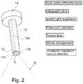
- the projector of the pattern generating membercomprises a phase optic element, a spatial light modulator, a multi-order diffractive lens, a holographic lens, a mirror arrangement, a computer regulated optical element, and/or a computer regulated mechanically optical element e.g. a mems (micro-electro-mechanical) element.
- the phase optic elementmay advantageously be a diffractive optic element (DOE).
- DOEdiffractive optic element
- phase optics elementis capable of producing an image having periodic intensity distribution.
- Diffractive optic elementsare well known in the art and may for example utilize a surface with a complex microstructure for its optical function.
- the micro-structured surface relief profilehas two or more surface levels.
- the surface structuresare either etched in fused silica or other glass types, or embossed in various polymer materials.
- diffractive opticscan realize almost the same optical functions as refractive optics such as lenses, prisms or aspheres, but they are much smaller and lighter.
- DOEsare not limited to laser applications; partially coherent light from LEDs or other light sources can also be modulated.
- the DOEis as described in US 2013/0038836 e.g. as shown in FIG. 1 and/or as described in section [0015] of US 2013/0038836.
- the diffractive optic elementscomprise a “multi-order diffractive” lens, such as a conventional diffractive-optic lens utilizing a single diffraction order in which the optical power of the lens is directly proportional to the wavelength of light.
- the projectormay comprise any type of beam manipulating element for providing the desired pattern e.g. one or more lenses and/or mirrors and/or splitters and/or filters and/or collimators.
- the projectorcomprises a spatial light modulator.
- the spatial light modulatoris configured for modulating the light pattern for example by modulating the transparency of a pattern cover e.g. by a computer modulation.
- the spatial light modulatoris arranged for modulating the intensity and/or the phase of the light from the pattern light source to thereby modulate the emitted light pattern.
- the projector of the pattern generating memberhas a maximally extending area perpendicular to the center axis of the access port when the pattern generating member is fixed to the cannula shaft portion, which maximally extending area is up to about 8 cm2, such as up to about 4 cm2, such as up to about 2 cm2, such as from about 0.01 to about 1 cm2, such as from about 0.1 to about 0.5 cm2.
- the projector of the pattern generating memberis configured to be at least temporarily fixed at the end edge in the vicinity of the distal access port exit and the projector is preferably shaped such that the projector does not extend laterally beyond the end edge or up to 5 mm laterally beyond the end edge.
- the projector of the pattern generating memberhas a projector face from where the light is to be emitted and the projector is pivotable, so it can be pivotally unfolded from a first folded position where the projector face is not facing in the distal direction to a second position where the projector face is facing in the distal direction.
- the cannulacan be inserted into an incision when the projector is in a first folded position and there after the projector can be unfolded to its second position.
- the unfoldingcan be provided by triggering a release button at the inner wall of the cannula shaft portion e.g. by the surgical instrument—e.g. by a tilting of the surgical instrument—after the cannula has been inserted through the incision such that the projector is unfolded to its second position e.g. by a spring mechanism.
- the patternmay have any desired shape.
- the projectoris fixed or adapted to be fixed to the cannula shaft portion such that the pattern remains substantially stationary when the surgical instrument is subjected exclusively to a circumferential movement with the longitudinal axis of the surgical instrument.
- the projector when fixed to the cannula shaft portionis configured to emitting a pattern, which pattern when projected to a surface orthogonal to the center axis of the access port has at most 10 fold rotational symmetry, preferably the pattern has at most 8 fold rotational symmetry.
- Such patternwhich is not fully rotational symmetrical but has up to 10 fold rotational symmetry gives the user an even better visual information about the position of the surgical instrument relative to the surgical site.
- the projector of the pattern generating memberis configured to emit a pattern comprising an arch shape, ring or semi-ring shaped lines, a plurality of angled lines and/or a coded structured light configuration.
- the patterncomprises a grid of lines, e.g. a crosshatched pattern optionally comprising substantially parallel lines when emitted to a planar surface.
- the changes in the grid lines due to lateral movements of the surgical instrumentcan for example be used to deduce the contours of the body cavity such as projected surface and/or the contours and/or topographic shape of the surgical field.
- the changes in the angle and distance between crossing and/or parallel grid lines during movement(s) of the surgical instrumentcan for example be used to determine the orientation of the surgical instrument.
- the light patterncomprises a plurality of light dots.
- the position and/or the distance between the dotswill change, which enhances the operator's ability even further to deduce the position of the surgical instrument and the area contours of the surgical field.
- the pattern generating memberis configured to emit a pattern comprising a coded structured light configuration comprising a plurality of light dots with different shapes and/or sizes arranged in a preselected configuration.
- the pattern comprising a coded structured light configurationis in particular suitable for determining a topographic shape of the target surface.
- Patterns comprising coded structured light configurationsare for example described in “Pattern codification strategies in structured light systems” by Salvi et al. Pattern Recognition, Volume 37, Issue 4, April 2004, Pages 827-849.
- the projector fixed to the cannula shaft portionis configured to emit a pattern which pattern, when projected to a surface perpendicular to the longitudinal axis of the body portion of the surgical instrument, comprises a plurality of angled lines.
- the patterncomprises a grid of lines, such as a grid comprising one or more sets of parallel lines.
- a tilting of the surgical instrument inserted into the access portcan for example be observed by a change of such angled lines e.g. by a deformation of one or more of the lines, by change of line angles and/or by change of distance between lines.
- the patternis advantageously sufficiently large to ensure good visual perception of the surgical instrument and movement thereof.
- the projector of the pattern generating memberis configured to emit a pattern, which pattern when emitted towards a plane surface at a distance of about 80 mm from the distal end of the cannula shaft portion and normal to a center axis of the cannula shaft portion, has a grid area of up to about 225 cm 2 , such as of up to about 100 cm 2 , such as of up to about 9 cm 2 .
- the flange portioncomprises a handle part, the flange portion comprising means for being temporally fixed to an obturator.
- the cannulacomprises two or more access ports through the flange portion and the cannula shaft portion. Thereby several surgical instruments can be inserted simultaneously.
- the cannulacomprises two or more cannula shaft portions and an access ports through the flange portion and the cannula shaft portions suitable for inserting a surgical instrument through each of the respective access ports.
- the cannula assembly kitcomprises a cleaning element for cleaning the projector.
- the cleaning elementis in the form of a wiping element arranged for wiping and/or washing the projector.
- the cleaning elementis in the form of a spray element arranged for spraying and/or blowing the projector with a fluid such as gas or liquid.
- a suitable cleaning elementis as the cleaning device described in U.S. Pat. No. 8,397,335.
- the cannulais adapted for being handled by a surgeon—i.e. to be mounted in an incision of a patient to provide the access port to the surgical site.
- the cannulais adapted for being maneuvered by a robot—i.e. to be mounted in an incision of a patient using a robot to provide the access port to the surgical site.
- the cannulais a part of the robot.
- the inventionalso comprises a trocar assembly kit for use in minimally invasive surgery.
- the trocar assembly kitcomprises a cannula assembly as described above and an obturator.
- the obturatormay in principle be any kind of obturator configured to be used with a cannula.
- the obturatorhas a distal end and a proximal end and comprises a head portion at its proximal end, a tip portion at its distal end and a rigid obturator shaft portion extending between the head portion and the tip portion, wherein the cannula and the obturator are correlated to each other such that the tip portion of the obturator can be instead through the access port of the cannula and the head portion of the obturator can be temporally fixed to the flange portion of the cannula, preferably such that a seal is formed in the access opening between the cannula and the obturator.
- the obturatorcomprises a projector protection arrangement correlated with the projector of the cannula assembly kit to at least partly cover the projector when the cannula assembly kit and the obturator are in an assembled state.
- the projectorcan be protected by the projector protection arrangement during insertion of the trocar assembly through an incision.
- the projector protection arrangementis advantageously arranged to be at least partly passed into a cavity of the obturator upon withdrawing of the obturator from said cannula access port.
- the cannula assembly kit and the obturatorare in an assembled state when the tip portion of the obturator is inserted substantially fully through the access port of the cannula shaft portion, and the cannula assembly kit and the obturator are disassembled upon withdrawing of the obturator from said cannula access port.
- the projector protection arrangementis arranged to be pivotally folded from a first position where it, at least partly covers the projector to a second position where it at least partly is passed into a cavity of the obturator.
- the folding from the second position to the first positionmay for example be performed manually after having inserted the tip portion of the obturator substantially fully through the access port of the cannula shaft portion and the folding from the first position to the second position may for example be performed simply by withdrawing the obturator from the access port and/or by releasing a holding mechanism temporarily holding the projector protection arrangement in the first position.
- the projector protection arrangementis arranged to be radially displaced from a first position where it, at least partly covers the projector to a second position where it at least partly is passed into a cavity of the obturator.
- the radial displacementmay for example be provided by a spring arrangement and/or a holding mechanism temporarily holding the projector protection arrangement in one of the first position and the second positions.
- the inventionalso comprises a sleeve assembly suitable for a cannula assembly kit as described above.
- the sleeve assemblycomprises a sleeve and a pattern generating member.
- the pattern generating membercomprises a pattern light source and a projector, wherein the pattern light source is operatively connected to the projector for projecting a light pattern.
- At least the projector of the pattern generating memberis configured to be at least temporarily and rigidly fixed to the sleeve, the sleeve preferably comprises a sleeve end edge portion comprising the projector.
- the sleevemay advantageously be as described above.
- the sleeveis configured to substantially fully cover at least a cannula shaft portion of a cannula. In an embodiment the sleeve is configured to cover at least a part of a cannula flange portion of the cannula.
- the inventionalso relates to a minimally invasive surgery system comprising a cannula assembly kit preferably as described above, a surgical instrument, a camera and a computer system.
- the cameramay be a mono camera or a stereo camera.
- the minimally invasive surgery systemcomprises two or more camera adapted for recording image data.
- the minimally invasive surgery systemmay be configured to combining or multiplexing said image data.
- the camerais mounted to or integrated with the cannula e.g. as described above.
- the cameramay advantageously comprise a charge-coupled device (CDD) image sensor, or a complementary metal-oxide-semiconductor (CMOS) image sensor.
- CDDcharge-coupled device
- CMOScomplementary metal-oxide-semiconductor
- the camerais mounted to a scope.
- a Scopeis herein used to mean any suitable scope, such as an endoscope, a laparoscope anarthroscope, a thoracoscope, a gastroscope, a colonoscope, a laryngoscope, a broncoscope, a cystoscope or a combination thereof.
- the scopeis an endoscope.
- the scopeis a laparoscope.
- the computeris configured for wide baseline operation comprising a wide angle between the camera and the cannula assembly.
- the angle between the camera and the cannula assembly and in particularly the center axis of the projected light patternmay be stationary or variable and the computer may preferably be configured for determining the angle and compensate therefore.
- the systemmay e.g. comprise one or more further sensors for determining the angle between the camera and the cannula assembly.
- the angle between the camera and the cannula assembly and in particularly the center axis of the projected light patternmay for example be at least 5 degrees, such as from 10 to 50 degrees, such as from 25 to 45 degrees.
- the minimally invasive surgery systemcomprises two or more cameras, such as at least one camera mounted to or integrated with the cannula and at least one camera mounted to or integrated with a scope.
- the computer systemis programmed to determining a spatially position and orientation of the projector. This may for example be performed by ensuring that the project pattern is a coded pattern e.g. an asymmetrical pattern where different areas of the projected pattern seen in cross sectional view may be uniquely identified to determine a spatially position and orientation of the projector.
- a coded patterne.g. an asymmetrical pattern where different areas of the projected pattern seen in cross sectional view may be uniquely identified to determine a spatially position and orientation of the projector.
- the minimally invasive surgery systemmay further comprise one or more illuminating element, such as an illuminating element mounted to or integrated with the scope.
- the minimally invasive surgery systemmay additionally comprise one or more sensors which may be used in the generation of minimally invasive surgery data and/or performing a minimally invasive surgery.
- Such one or more sensorsmay include light emitting based sensors, mechanical sensors, electrical sensors and etc.
- the one or more sensorscomprises position tracking sensor(s), accelerometer(s), gyroscope(s) and/or other motion-sensing devices.
- the projected light pattern of the cannula assembly kitcomprises at least one wavelength not comprised by the optional other illuminating light sources and/or sensor light sources.
- the surgical instrument of the minimally invasive surgery systemis advantageously selected from a grasper, a suture grasper, a cutter, a sealer, a stapler, a clip applier, a dissector, scissors, shears, a suction instrument, a clamp instrument, an electrode, a coagulation device, a curette, ablators, scalpels, a needle holder, a needle driver, a spatula, forceps, a biopsy and retractor instrument or a combination thereof.
- the computer systemmay comprise hardware and software for collecting minimally invasive surgery data and/or for performing minimally invasive surgery.
- the computer systemmay comprise one or more hardware elements which are or are adapted to be in data communication.
- the computer systemis in data communication with the camera to receive image data from the camera.
- the computer systemis programmed to determining real time position data of the surgical instrument, to determining real time topography data of a surface reflecting the light pattern emitted by the cannula assembly kit and/or to determining real time contours of a surface reflecting the light pattern emitted by the cannula assembly kit.
- the computer systemis configured to transmitting the determined data to a robot, a database and/or a monitor for being displayed.
- the surgical instrumentforms part of the robot or is adapted for being maneuvered by the robot.
- the computer systemis preferably configured to transmitting the determined data to the robot.
- the computer system or at least a part of the computer systemforms part of the robot.
- the cannula assembly kitis adapted for being controlled by a computer or forms part of the computer.
- the surgical instrumentforms part of a robot or is adapted for being maneuvered by a robot and the computer system is configured for determining a spatially position of the instrument and based at least partly on said real time topography data and said spatially position of the instrument to control the robot to perform movements of the instrument.
- the inventionalso relates to a method of performing a minimal invasive surgery of a target surgical site in an internal body structure below a skin area of a patient.
- the methodcomprises,
- the methodcomprises,
- the methodcomprising performing the minimally invasive surgery by using the minimally invasive surgery system as described above.
- the method of performing a minimal invasive surgerymay be performed by an operator i.e. a surgeon and/or a robot.
- a surgeonis performing an incision and inserting the cannula assembly kit and a robot is performing the remaining method steps.
- the entire method of performing a minimal invasive surgeryis performed by a robot.
- the recorded image datamay simultaneously be transmitted to a monitor for being displaced such than an observer, such as a supervisor, a surgeon and/or a trainee can observe the minimally invasive surgery performed by the robot.
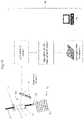
- FIG. 1 ais a schematic view of an embodiment of a cannula assembly kit.
- FIG. 1 bis a schematic view of an embodiment of an obturator adapted to be used together with the cannula assembly kit of FIG. 1 a.
- FIG. 1 c and FIG. 1 dare schematic views of a trocar assembly kit comprising the cannula assembly kit of FIG. 1 and the obturator of FIG. 2 is partly or fully in an assembled state.
- FIG. 2is a schematic view of an embodiment of a cannula assembly kit, where the shaft portion of the cannula comprises a mounting through hole through which the projector has been mounted.
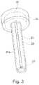
- FIG. 3is a schematic view of an embodiment of a cannula assembly kit comprising a sleeve.
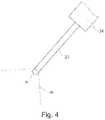
- FIG. 4is a schematic view of an embodiment of a cannula assembly kit with a relatively large flange portion for comprising the pattern light source.
- FIG. 5is a schematic view of a distal end portion of an assembled trocar assembly kit, where the obturator comprises a projector protection arrangement.
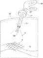
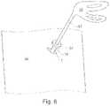
- FIG. 6is a schematic view of an embodiment of a cannula assembly kit during use in a surgical procedure seen from outside the body cavity.
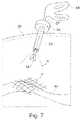
- FIG. 7is a schematic view of an embodiment of a cannula assembly kit during use in a surgical procedure seen in a cross-sectional view through the body cavity.
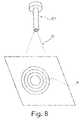
- FIG. 8is a schematic view of an embodiment of a cannula assembly kit configured for emitting a bullseye shaped pattern.
- FIG. 9is a schematic view of an embodiment of a cannula assembly kit with a bent cannula shaft portion.
- FIG. 10is a schematic view of an embodiment of a cannula assembly kit where the cannula comprises two cannula shaft portions and one cannula flange portion.
- FIG. 11is a schematic view of another cannula assembly kit where the cannula comprises two cannula shaft portions and one cannula flange portion.
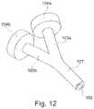
- FIG. 12is a schematic view of an embodiment of a cannula assembly kit where the cannula comprises two cannula flange portions and one cannula shaft portion.
- FIG. 13is a schematic illustration of an embodiment of a minimally invasive surgery system of the invention where the projected light pattern comprises a coded structured light configuration comprising a plurality of light dots with different sizes.
- FIG. 14is a schematic illustration of an embodiment of a minimally invasive surgery system of the invention where the projected light pattern comprises a coded structured light configuration comprising a plurality of light dots with different shapes and sizes.
- FIG. 15is a schematic illustration of an embodiment of a minimally invasive surgery system of the invention where the projected light pattern comprises a crosshatched pattern
- FIG. 16is a schematic illustration of an embodiment of a minimally invasive surgery system of the invention where the projected light pattern comprises a plurality of parallel lines.
- FIG. 1 aillustrates an embodiment of a cannula assembly kit of the invention.
- the cannula assembly kitcomprises a cannula 1 and a pattern generating member wherein only the projector 2 is shown.
- the cannulahas a distal end D and a proximal end P and comprises a flange portion 4 at its proximal end and an elongate cannula shaft portion 3 extending from the flange portion 4 to its distal D end and an access port A through the flange portion 4 and the elongate cannula shaft portion 3 , such that a surgical tool of a surgical instrument can be inserted through the access port.
- the pattern generating membercomprises a not shown pattern light source and a projector 2 at least temporarily fixed to the cannula shaft portion 3 of the cannula.
- the cannula flange portion 4comprises an insufflation port 5 for insufflating the body cavity.
- the obturator and the cannula assembly kit of FIG. 1are correlated to each other.
- the obturator 9 shown in FIG. 1 bhas a distal end D and a proximal end P and comprises a head portion 6 at its proximal end, a tip portion 8 at its distal end and a rigid obturator shaft portion 7 extending between said head portion 6 and said tip portion 8 .
- the tip portioncan be bladed or non-bladed.
- the obturator of FIG. 1 b and the cannula assembly kit of FIG. 1are correlated to each other such that the obturator can be inserted into the access port A of the cannula 1 .
- the obturator 9is partly inserted into the access port A of the cannula 1 .
- the obturator 9is fully inserted into the access port A of the cannula 1 to thereby assemble the trocar assembly kit.
- the cannula assembly kit shown in FIG. 2comprises a cannula and a pattern generating member wherein only the projector 12 is shown.
- the cannulacomprises a flange portion 14 and an elongate cannula shaft portion 13 extending from the flange portion 14 to its distal end and an access port A.
- the cannula shaft portion 13has an access port exit 13 a and comprises an end edge 13 b in the vicinity of said distal access port exit 13 a.
- the shaft portion 13 of the cannulacomprises a mounting through hole 12 a indicated on the drawing with dotted lines.
- the projector 12has been mounted via the mounting through hole 12 a and a not shown optical fiber extends through the mounting through hole 12 a for transmitting light to the projector 12 .
- the cannula assembly kit shown in FIG. 3comprises a cannula and a pattern generating member wherein only the projector 22 is shown.
- the cannulacomprises a flange portion 24 and an elongate cannula shaft portion 23 extending from the flange portion 24 to its distal end and an access port A.
- the shaft portion 23 and the flange portion 24are covered by a sleeve 26 which is mounted to the cannula.
- the projector 22is mounted to or integrated in the sleeve 26 and the sleeve also comprises a fiber covering line 22 a comprising a not shown optical fiber arranged for transmitting light to the projector 22 .
- the cannula assembly kits shown in FIG. 4comprises a flange portion 34 and an elongate cannula shaft portion 33 extending from the flange portion 34 to its distal end and an access port A.
- the cannula assembly kitalso comprises a not shown pattern generating member.
- the rays Rindicate that the not shown projector is positioned at the distal end of the cannula shaft portion 33 .
- the cannula flange portion 34is relatively large such that a not shown light source and/or battery can be incorporated into the cannula flange portion 34 .
- the distal end portion of an assembled trocar assembly kit shown in FIG. 5comprises distal end portions of the correlated cannula assembly kit and obturator.
- the cannula assembly kitcomprises a cannula shaft portion 43 and a projector 42 arranged for projecting a light pattern.
- the obturatorcomprises a rigid obturator shaft portion 47 and a tip portion 48 .
- the obturatorfurther comprises a projector protection arrangement 47 a correlated with the projector 42 of the cannula assembly kit to at least partly cover the projector 42 , such that the projector is at least partly projected during the insertion during surgery.
- the projector protection arrangementis shaped to align with the shape of the tip portion of the obturator such that there will be a more gradually increase of the diameter of the assembled trocar assembly kit from the tip portion of the obturator to the cannula shaft portion of the cannula assembly kit.
- the projector protection arrangement 47 aWhen the obturator is withdrawn from the access port of the cannula assembly kit, the projector protection arrangement 47 a will at least partly be passed into a cavity of the obturator, such that the projector protection arrangement 47 a is not blocking for the withdrawal.
- the projector protection arrangement 47 amay for example be pivotally folded into a cavity of the obturator, by folding towards the tip portion 48 .
- FIG. 6 and FIG. 7show a cannula assembly kit in use during a surgical procedure.
- the figuresshow a body part of a patient in surgery, where an incision I is made through the skin 50 of the patient, the cannula assembly kit comprises a shaft portion 53 and a flange portion 54 , and the shaft portion 53 is inserted through the incision I.
- the cannula assembly kitcomprises a pattern generating member comprising a projector from where a light pattern P in the form of rays R of light is emitted.
- a surgical instrumentcomprising a handle portion 56 , a body portion 57 and a surgical tool 58 is inserted through the access port of the cannula assembly kit and the pattern P is projected onto a surgical site 60 .
- the patterncan for example be recorded by an image recorder on a scope inserted via the same or another incision through the skin.
- the cannula assembly kit 61 shown in FIG. 8comprises a not shown projector operatively connected to a light source and configured for emitting light rays R arranged to form a bullseye shaped pattern P.
- the various rings of the bullseye shaped pattern Pcould for example have different wavelength profile.
- the cannula assembly kit shown in FIG. 9comprises a cannula and a pattern generating member wherein only the projector 72 is shown.
- the cannulacomprises a flange portion 74 and an elongate cannula shaft portion 73 extending from the flange portion 74 to its distal end and an access port A.
- the cannula shaft portion 73is bent in a soft curve to thereby make is easier for an operator to insert the cannula shaft portion 73 through an incision of a patient.
- the cannula shaft portion 73is for example pre-bent to the shown bending curve and is further bendable or flexible i.e. in unloaded condition the cannula shaft portion 73 is bent.
- the cannula shaft portion 73is substantially rigid in the bent position.
- the cannula shaft portion 73comprises an end edge 73 b and the projector 72 is mounted at the end edge 73 b and a not shown optical fiber is arranged to guide light along a channel 72 a in the wall of the cannula shaft portion 73 .
- the cannula assembly kit shown in FIG. 10comprises a cannula and at least one pattern generating member wherein only the two projectors 82 are shown.
- the two projectors 82can be of a common pattern generating member or they can be of separate pattern generating members.
- the cannulacomprises a flange portion 84 and a double cannula shaft portion 83 , 83 a , 83 b .
- the double cannula shaft portion 83 , 83 a , 83 bcomprises a common shaft portion section 83 and two branch shaft portion sections 83 a and 83 b each comprising a distal access port section A, such that the cannula has a common access port section through the flange portion 84 and through the common shaft portion section 83 and two separate distal access port sections through said respective branch shaft portion sections 83 a and 83 b.
- the cannula assembly kit shown in FIG. 11comprises a cannula and at least one pattern generating member wherein only the two projectors 92 are shown.
- the two projectors 92can be of a common pattern generating member or they can be of separate pattern generating members.
- the cannulacomprises a flange portion 94 and two cannula shaft portions 93 a , 93 b providing two access ports through the cannula flange portion 94 .
- the cannula assembly kit shown in FIG. 12comprises a cannula and at least one pattern generating member wherein only the projector 102 is shown.
- the cannulacomprises two flange portions 104 a , 104 b and a double cannula shaft portion 104 , 104 a , 104 b .
- the double cannula shaft portion 104 , 104 a , 104 bcomprises a common shaft portion section 104 and two branch shaft portion sections 104 a and 104 b .
- the respective branch shaft portion sections 104 a and 104 bare connected to the respective flange portions 104 a , 104 b and are merged in the common shaft portion section 104 in the distal end section of the cannula.
- the minimally invasive surgery system shown in the respective FIGS. 13, 14, 15 and 16comprises a cannula assembly kit 110 , a surgical instrument 115 , a camera 116 and a computer system 118 .
- the cannula assembly kit 110comprises a flange portion 114 , an elongate cannula shaft portion 117 and a projector 112 for projecting a light pattern at its distal end.
- An access portis provided via the cannula shaft portion 117 .
- the surgical instrument 115comprises its actual operation tool 115 a at its distal end.
- the distal end comprising the operation tool 115 ais inserted through the access port of the cannula assembly kit 110 .
- the projector 112projects a light pattern towards a distally arranged surface 111 and the reflected light pattern 113 is recorded by the camera 116 .
- this distally arranged surface 111will be a surgery site which may be very uneven as described above.
- the cannula assembly kitwill be moved accordingly and thereby also the projector 112 will be moved and the reflected pattern 113 will change accordingly at least when the surgical instrument 115 is subjected to tilting movements.
- the camerarecords the reflected light and generates recorded image data.
- the recorded image datais transmitted to the computer system 118 .
- the computer systemcomprises a calibration unit for calibration of the camera, a processing unit comprising algorithms for 3D data set generation and decoding of the recorded and calibrated image data, a processing unit for determine topography data in real time and a PC for storing and/or displaying the determined topography data.
- the various units of the computer system 118may be integrated in a common hardware box.
- the surgical instrument 115may advantageously form part of a robot for performing the minimally invasive surgery and the computer system may provide feedback to the robot and/or at least a part of the computer system may be an integrated part of the robot.
- the projected light patterncomprises a coded structured light configuration comprising a plurality of light dots with different sizes.
- the projected light patterncomprises a coded structured light configuration comprising a plurality of light dots with different shapes and sizes.
- the projected light patterncomprises crosshatched pattern
- the projected light patterncomprises a plurality of parallel lines.
Landscapes
- Health & Medical Sciences (AREA)
- Life Sciences & Earth Sciences (AREA)
- Surgery (AREA)
- Nuclear Medicine, Radiotherapy & Molecular Imaging (AREA)
- Heart & Thoracic Surgery (AREA)
- Animal Behavior & Ethology (AREA)
- Veterinary Medicine (AREA)
- Public Health (AREA)
- Engineering & Computer Science (AREA)
- Biomedical Technology (AREA)
- General Health & Medical Sciences (AREA)
- Medical Informatics (AREA)
- Molecular Biology (AREA)
- Pathology (AREA)
- Radiology & Medical Imaging (AREA)
- Physics & Mathematics (AREA)
- Biophysics (AREA)
- Optics & Photonics (AREA)
- Oral & Maxillofacial Surgery (AREA)
- Gynecology & Obstetrics (AREA)
- Endoscopes (AREA)
- Surgical Instruments (AREA)
Abstract
Description
- the two or more pattern light sources can be operated independently of each other i.e. they can independently be switched on and off e.g. using a non-hand held unit or by a unit incorporated into the flange portion.
- providing an access port to the surgery target site comprising providing an incision through the skin area inserting a cannula assembly kit as described above,
- inserting a surgical instrument through the access port,
- inserting a camera element through the access port or through an additional access port to the surgery target site,
- providing that the projector if the cannula assembly kit is emitting a light pattern,
- recording image data of the light pattern reflected from the surgery target site by the camera, and
- moving the surgical instrument for performing the minimal invasive surgery while simultaneously receiving feedback based on the recorded image data.
- providing an access port to the surgery target site comprising providing an incision through the skin area inserting a cannula assembly kit with a camera as described above,
- inserting a surgical instrument through the access port,
- providing that the projector if the cannula assembly kit is emitting a light pattern,
- recording image data of the light pattern reflected from the surgery target site by the camera, and
- moving the surgical instrument for performing the minimal invasive surgery while simultaneously receiving feedback based on the recorded image data.
Claims (16)
Priority Applications (2)
| Application Number | Priority Date | Filing Date | Title |
|---|---|---|---|
| US15/717,088US11020144B2 (en) | 2015-07-21 | 2017-09-27 | Minimally invasive surgery system |
| US17/320,417US20210338268A1 (en) | 2015-07-21 | 2021-05-14 | Minimally invasive surgery system |
Applications Claiming Priority (3)
| Application Number | Priority Date | Filing Date | Title |
|---|---|---|---|
| DKPA201570483 | 2015-07-21 | ||
| DKPA201570483 | 2015-07-21 | ||
| PCT/DK2016/050234WO2017012624A1 (en) | 2015-07-21 | 2016-07-01 | Cannula assembly kit, trocar assembly kit, sleeve assembly, minimally invasive surgery system and method therefor |
Related Parent Applications (1)
| Application Number | Title | Priority Date | Filing Date |
|---|---|---|---|
| PCT/DK2016/050234ContinuationWO2017012624A1 (en) | 2015-07-21 | 2016-07-01 | Cannula assembly kit, trocar assembly kit, sleeve assembly, minimally invasive surgery system and method therefor |
Related Child Applications (1)
| Application Number | Title | Priority Date | Filing Date |
|---|---|---|---|
| US15/717,088Continuation-In-PartUS11020144B2 (en) | 2015-07-21 | 2017-09-27 | Minimally invasive surgery system |
Publications (2)
| Publication Number | Publication Date |
|---|---|
| US20170238962A1 US20170238962A1 (en) | 2017-08-24 |
| US11331120B2true US11331120B2 (en) | 2022-05-17 |
Family
ID=57833809
Family Applications (1)
| Application Number | Title | Priority Date | Filing Date |
|---|---|---|---|
| US15/589,476Active2038-05-23US11331120B2 (en) | 2015-07-21 | 2017-05-08 | Cannula assembly kit |
Country Status (5)
| Country | Link |
|---|---|
| US (1) | US11331120B2 (en) |
| EP (1) | EP3145419B1 (en) |
| JP (1) | JP6776327B2 (en) |
| CN (1) | CN108024806B (en) |
| WO (1) | WO2017012624A1 (en) |
Cited By (1)
| Publication number | Priority date | Publication date | Assignee | Title |
|---|---|---|---|---|
| US12419719B2 (en) | 2023-03-10 | 2025-09-23 | Arthrex, Inc. | Camera apparatus for coordinated operation with surgical tools |
Families Citing this family (377)
| Publication number | Priority date | Publication date | Assignee | Title |
|---|---|---|---|---|
| US9060770B2 (en) | 2003-05-20 | 2015-06-23 | Ethicon Endo-Surgery, Inc. | Robotically-driven surgical instrument with E-beam driver |
| US20070084897A1 (en) | 2003-05-20 | 2007-04-19 | Shelton Frederick E Iv | Articulating surgical stapling instrument incorporating a two-piece e-beam firing mechanism |
| US8215531B2 (en) | 2004-07-28 | 2012-07-10 | Ethicon Endo-Surgery, Inc. | Surgical stapling instrument having a medical substance dispenser |
| US9072535B2 (en) | 2011-05-27 | 2015-07-07 | Ethicon Endo-Surgery, Inc. | Surgical stapling instruments with rotatable staple deployment arrangements |
| US11890012B2 (en) | 2004-07-28 | 2024-02-06 | Cilag Gmbh International | Staple cartridge comprising cartridge body and attached support |
| US11998198B2 (en) | 2004-07-28 | 2024-06-04 | Cilag Gmbh International | Surgical stapling instrument incorporating a two-piece E-beam firing mechanism |
| US7669746B2 (en) | 2005-08-31 | 2010-03-02 | Ethicon Endo-Surgery, Inc. | Staple cartridges for forming staples having differing formed staple heights |
| US11484312B2 (en) | 2005-08-31 | 2022-11-01 | Cilag Gmbh International | Staple cartridge comprising a staple driver arrangement |
| US10159482B2 (en) | 2005-08-31 | 2018-12-25 | Ethicon Llc | Fastener cartridge assembly comprising a fixed anvil and different staple heights |
| US11246590B2 (en) | 2005-08-31 | 2022-02-15 | Cilag Gmbh International | Staple cartridge including staple drivers having different unfired heights |
| US7934630B2 (en) | 2005-08-31 | 2011-05-03 | Ethicon Endo-Surgery, Inc. | Staple cartridges for forming staples having differing formed staple heights |
| US9237891B2 (en) | 2005-08-31 | 2016-01-19 | Ethicon Endo-Surgery, Inc. | Robotically-controlled surgical stapling devices that produce formed staples having different lengths |
| US20070106317A1 (en) | 2005-11-09 | 2007-05-10 | Shelton Frederick E Iv | Hydraulically and electrically actuated articulation joints for surgical instruments |
| US7845537B2 (en) | 2006-01-31 | 2010-12-07 | Ethicon Endo-Surgery, Inc. | Surgical instrument having recording capabilities |
| US8708213B2 (en) | 2006-01-31 | 2014-04-29 | Ethicon Endo-Surgery, Inc. | Surgical instrument having a feedback system |
| US8820603B2 (en) | 2006-01-31 | 2014-09-02 | Ethicon Endo-Surgery, Inc. | Accessing data stored in a memory of a surgical instrument |
| US20110295295A1 (en) | 2006-01-31 | 2011-12-01 | Ethicon Endo-Surgery, Inc. | Robotically-controlled surgical instrument having recording capabilities |
| US7753904B2 (en) | 2006-01-31 | 2010-07-13 | Ethicon Endo-Surgery, Inc. | Endoscopic surgical instrument with a handle that can articulate with respect to the shaft |
| US8186555B2 (en) | 2006-01-31 | 2012-05-29 | Ethicon Endo-Surgery, Inc. | Motor-driven surgical cutting and fastening instrument with mechanical closure system |
| US11224427B2 (en) | 2006-01-31 | 2022-01-18 | Cilag Gmbh International | Surgical stapling system including a console and retraction assembly |
| US11793518B2 (en) | 2006-01-31 | 2023-10-24 | Cilag Gmbh International | Powered surgical instruments with firing system lockout arrangements |
| US20120292367A1 (en) | 2006-01-31 | 2012-11-22 | Ethicon Endo-Surgery, Inc. | Robotically-controlled end effector |
| US11278279B2 (en) | 2006-01-31 | 2022-03-22 | Cilag Gmbh International | Surgical instrument assembly |
| US20110024477A1 (en) | 2009-02-06 | 2011-02-03 | Hall Steven G | Driven Surgical Stapler Improvements |
| US8992422B2 (en) | 2006-03-23 | 2015-03-31 | Ethicon Endo-Surgery, Inc. | Robotically-controlled endoscopic accessory channel |
| US8322455B2 (en) | 2006-06-27 | 2012-12-04 | Ethicon Endo-Surgery, Inc. | Manually driven surgical cutting and fastening instrument |
| US10568652B2 (en) | 2006-09-29 | 2020-02-25 | Ethicon Llc | Surgical staples having attached drivers of different heights and stapling instruments for deploying the same |
| US11980366B2 (en) | 2006-10-03 | 2024-05-14 | Cilag Gmbh International | Surgical instrument |
| US8684253B2 (en) | 2007-01-10 | 2014-04-01 | Ethicon Endo-Surgery, Inc. | Surgical instrument with wireless communication between a control unit of a robotic system and remote sensor |
| US8632535B2 (en) | 2007-01-10 | 2014-01-21 | Ethicon Endo-Surgery, Inc. | Interlock and surgical instrument including same |
| US11291441B2 (en) | 2007-01-10 | 2022-04-05 | Cilag Gmbh International | Surgical instrument with wireless communication between control unit and remote sensor |
| US11039836B2 (en) | 2007-01-11 | 2021-06-22 | Cilag Gmbh International | Staple cartridge for use with a surgical stapling instrument |
| US20080169333A1 (en) | 2007-01-11 | 2008-07-17 | Shelton Frederick E | Surgical stapler end effector with tapered distal end |
| US7673782B2 (en) | 2007-03-15 | 2010-03-09 | Ethicon Endo-Surgery, Inc. | Surgical stapling instrument having a releasable buttress material |
| US11564682B2 (en) | 2007-06-04 | 2023-01-31 | Cilag Gmbh International | Surgical stapler device |
| US8931682B2 (en) | 2007-06-04 | 2015-01-13 | Ethicon Endo-Surgery, Inc. | Robotically-controlled shaft based rotary drive systems for surgical instruments |
| US7753245B2 (en) | 2007-06-22 | 2010-07-13 | Ethicon Endo-Surgery, Inc. | Surgical stapling instruments |
| US11849941B2 (en) | 2007-06-29 | 2023-12-26 | Cilag Gmbh International | Staple cartridge having staple cavities extending at a transverse angle relative to a longitudinal cartridge axis |
| US9179912B2 (en) | 2008-02-14 | 2015-11-10 | Ethicon Endo-Surgery, Inc. | Robotically-controlled motorized surgical cutting and fastening instrument |
| US8636736B2 (en) | 2008-02-14 | 2014-01-28 | Ethicon Endo-Surgery, Inc. | Motorized surgical cutting and fastening instrument |
| US8573465B2 (en) | 2008-02-14 | 2013-11-05 | Ethicon Endo-Surgery, Inc. | Robotically-controlled surgical end effector system with rotary actuated closure systems |
| US11986183B2 (en) | 2008-02-14 | 2024-05-21 | Cilag Gmbh International | Surgical cutting and fastening instrument comprising a plurality of sensors to measure an electrical parameter |
| US7819298B2 (en) | 2008-02-14 | 2010-10-26 | Ethicon Endo-Surgery, Inc. | Surgical stapling apparatus with control features operable with one hand |
| JP5410110B2 (en) | 2008-02-14 | 2014-02-05 | エシコン・エンド−サージェリィ・インコーポレイテッド | Surgical cutting / fixing instrument with RF electrode |
| US7866527B2 (en) | 2008-02-14 | 2011-01-11 | Ethicon Endo-Surgery, Inc. | Surgical stapling apparatus with interlockable firing system |
| US9585657B2 (en) | 2008-02-15 | 2017-03-07 | Ethicon Endo-Surgery, Llc | Actuator for releasing a layer of material from a surgical end effector |
| US11648005B2 (en) | 2008-09-23 | 2023-05-16 | Cilag Gmbh International | Robotically-controlled motorized surgical instrument with an end effector |
| US9005230B2 (en) | 2008-09-23 | 2015-04-14 | Ethicon Endo-Surgery, Inc. | Motorized surgical instrument |
| US9386983B2 (en) | 2008-09-23 | 2016-07-12 | Ethicon Endo-Surgery, Llc | Robotically-controlled motorized surgical instrument |
| US8210411B2 (en) | 2008-09-23 | 2012-07-03 | Ethicon Endo-Surgery, Inc. | Motor-driven surgical cutting instrument |
| US8608045B2 (en) | 2008-10-10 | 2013-12-17 | Ethicon Endo-Sugery, Inc. | Powered surgical cutting and stapling apparatus with manually retractable firing system |
| US8517239B2 (en) | 2009-02-05 | 2013-08-27 | Ethicon Endo-Surgery, Inc. | Surgical stapling instrument comprising a magnetic element driver |
| RU2525225C2 (en) | 2009-02-06 | 2014-08-10 | Этикон Эндо-Серджери, Инк. | Improvement of drive surgical suturing instrument |
| US8220688B2 (en) | 2009-12-24 | 2012-07-17 | Ethicon Endo-Surgery, Inc. | Motor-driven surgical cutting instrument with electric actuator directional control assembly |
| US8851354B2 (en) | 2009-12-24 | 2014-10-07 | Ethicon Endo-Surgery, Inc. | Surgical cutting instrument that analyzes tissue thickness |
| US8783543B2 (en) | 2010-07-30 | 2014-07-22 | Ethicon Endo-Surgery, Inc. | Tissue acquisition arrangements and methods for surgical stapling devices |
| US11298125B2 (en) | 2010-09-30 | 2022-04-12 | Cilag Gmbh International | Tissue stapler having a thickness compensator |
| US9016542B2 (en) | 2010-09-30 | 2015-04-28 | Ethicon Endo-Surgery, Inc. | Staple cartridge comprising compressible distortion resistant components |
| US9386988B2 (en) | 2010-09-30 | 2016-07-12 | Ethicon End-Surgery, LLC | Retainer assembly including a tissue thickness compensator |
| US11925354B2 (en) | 2010-09-30 | 2024-03-12 | Cilag Gmbh International | Staple cartridge comprising staples positioned within a compressible portion thereof |
| US10945731B2 (en) | 2010-09-30 | 2021-03-16 | Ethicon Llc | Tissue thickness compensator comprising controlled release and expansion |
| US11812965B2 (en) | 2010-09-30 | 2023-11-14 | Cilag Gmbh International | Layer of material for a surgical end effector |
| US9788834B2 (en) | 2010-09-30 | 2017-10-17 | Ethicon Llc | Layer comprising deployable attachment members |
| US12213666B2 (en) | 2010-09-30 | 2025-02-04 | Cilag Gmbh International | Tissue thickness compensator comprising layers |
| US9629814B2 (en) | 2010-09-30 | 2017-04-25 | Ethicon Endo-Surgery, Llc | Tissue thickness compensator configured to redistribute compressive forces |
| US9351730B2 (en) | 2011-04-29 | 2016-05-31 | Ethicon Endo-Surgery, Llc | Tissue thickness compensator comprising channels |
| US8695866B2 (en) | 2010-10-01 | 2014-04-15 | Ethicon Endo-Surgery, Inc. | Surgical instrument having a power control circuit |
| AU2012250197B2 (en) | 2011-04-29 | 2017-08-10 | Ethicon Endo-Surgery, Inc. | Staple cartridge comprising staples positioned within a compressible portion thereof |
| US11207064B2 (en) | 2011-05-27 | 2021-12-28 | Cilag Gmbh International | Automated end effector component reloading system for use with a robotic system |
| JP6224070B2 (en) | 2012-03-28 | 2017-11-01 | エシコン・エンド−サージェリィ・インコーポレイテッドEthicon Endo−Surgery,Inc. | Retainer assembly including tissue thickness compensator |
| BR112014024098B1 (en) | 2012-03-28 | 2021-05-25 | Ethicon Endo-Surgery, Inc. | staple cartridge |
| MX358135B (en) | 2012-03-28 | 2018-08-06 | Ethicon Endo Surgery Inc | Tissue thickness compensator comprising a plurality of layers. |
| US9101358B2 (en) | 2012-06-15 | 2015-08-11 | Ethicon Endo-Surgery, Inc. | Articulatable surgical instrument comprising a firing drive |
| BR112014032776B1 (en) | 2012-06-28 | 2021-09-08 | Ethicon Endo-Surgery, Inc | SURGICAL INSTRUMENT SYSTEM AND SURGICAL KIT FOR USE WITH A SURGICAL INSTRUMENT SYSTEM |
| US9408606B2 (en) | 2012-06-28 | 2016-08-09 | Ethicon Endo-Surgery, Llc | Robotically powered surgical device with manually-actuatable reversing system |
| US20140001231A1 (en) | 2012-06-28 | 2014-01-02 | Ethicon Endo-Surgery, Inc. | Firing system lockout arrangements for surgical instruments |
| US9282974B2 (en) | 2012-06-28 | 2016-03-15 | Ethicon Endo-Surgery, Llc | Empty clip cartridge lockout |
| US11278284B2 (en) | 2012-06-28 | 2022-03-22 | Cilag Gmbh International | Rotary drive arrangements for surgical instruments |
| JP6290201B2 (en) | 2012-06-28 | 2018-03-07 | エシコン・エンド−サージェリィ・インコーポレイテッドEthicon Endo−Surgery,Inc. | Lockout for empty clip cartridge |
| US12383267B2 (en) | 2012-06-28 | 2025-08-12 | Cilag Gmbh International | Robotically powered surgical device with manually-actuatable reversing system |
| US9289256B2 (en) | 2012-06-28 | 2016-03-22 | Ethicon Endo-Surgery, Llc | Surgical end effectors having angled tissue-contacting surfaces |
| BR112015021082B1 (en) | 2013-03-01 | 2022-05-10 | Ethicon Endo-Surgery, Inc | surgical instrument |
| RU2672520C2 (en) | 2013-03-01 | 2018-11-15 | Этикон Эндо-Серджери, Инк. | Hingedly turnable surgical instruments with conducting ways for signal transfer |
| US9629629B2 (en) | 2013-03-14 | 2017-04-25 | Ethicon Endo-Surgey, LLC | Control systems for surgical instruments |
| US9808244B2 (en) | 2013-03-14 | 2017-11-07 | Ethicon Llc | Sensor arrangements for absolute positioning system for surgical instruments |
| BR112015026109B1 (en) | 2013-04-16 | 2022-02-22 | Ethicon Endo-Surgery, Inc | surgical instrument |
| US9826976B2 (en) | 2013-04-16 | 2017-11-28 | Ethicon Llc | Motor driven surgical instruments with lockable dual drive shafts |
| MX369362B (en) | 2013-08-23 | 2019-11-06 | Ethicon Endo Surgery Llc | Firing member retraction devices for powered surgical instruments. |
| US9775609B2 (en) | 2013-08-23 | 2017-10-03 | Ethicon Llc | Tamper proof circuit for surgical instrument battery pack |
| US9962161B2 (en) | 2014-02-12 | 2018-05-08 | Ethicon Llc | Deliverable surgical instrument |
| US11033182B2 (en) | 2014-02-21 | 2021-06-15 | 3Dintegrated Aps | Set comprising a surgical instrument |
| US12232723B2 (en) | 2014-03-26 | 2025-02-25 | Cilag Gmbh International | Systems and methods for controlling a segmented circuit |
| US10013049B2 (en) | 2014-03-26 | 2018-07-03 | Ethicon Llc | Power management through sleep options of segmented circuit and wake up control |
| US10004497B2 (en) | 2014-03-26 | 2018-06-26 | Ethicon Llc | Interface systems for use with surgical instruments |
| US20150272580A1 (en) | 2014-03-26 | 2015-10-01 | Ethicon Endo-Surgery, Inc. | Verification of number of battery exchanges/procedure count |
| BR112016021943B1 (en) | 2014-03-26 | 2022-06-14 | Ethicon Endo-Surgery, Llc | SURGICAL INSTRUMENT FOR USE BY AN OPERATOR IN A SURGICAL PROCEDURE |
| US20150297225A1 (en) | 2014-04-16 | 2015-10-22 | Ethicon Endo-Surgery, Inc. | Fastener cartridges including extensions having different configurations |
| US10470768B2 (en) | 2014-04-16 | 2019-11-12 | Ethicon Llc | Fastener cartridge including a layer attached thereto |
| CN106456176B (en) | 2014-04-16 | 2019-06-28 | 伊西康内外科有限责任公司 | Fastener Cartridge Including Extensions With Different Configurations |
| US10327764B2 (en) | 2014-09-26 | 2019-06-25 | Ethicon Llc | Method for creating a flexible staple line |
| BR112016023825B1 (en) | 2014-04-16 | 2022-08-02 | Ethicon Endo-Surgery, Llc | STAPLE CARTRIDGE FOR USE WITH A SURGICAL STAPLER AND STAPLE CARTRIDGE FOR USE WITH A SURGICAL INSTRUMENT |
| CN106456159B (en) | 2014-04-16 | 2019-03-08 | 伊西康内外科有限责任公司 | Fastener Cartridge Assembly and Nail Retainer Cover Arrangement |
| US10135242B2 (en) | 2014-09-05 | 2018-11-20 | Ethicon Llc | Smart cartridge wake up operation and data retention |
| BR112017004361B1 (en) | 2014-09-05 | 2023-04-11 | Ethicon Llc | ELECTRONIC SYSTEM FOR A SURGICAL INSTRUMENT |
| US11311294B2 (en) | 2014-09-05 | 2022-04-26 | Cilag Gmbh International | Powered medical device including measurement of closure state of jaws |
| US10105142B2 (en) | 2014-09-18 | 2018-10-23 | Ethicon Llc | Surgical stapler with plurality of cutting elements |
| CN107427300B (en) | 2014-09-26 | 2020-12-04 | 伊西康有限责任公司 | Surgical suture buttresses and auxiliary materials |
| US11523821B2 (en) | 2014-09-26 | 2022-12-13 | Cilag Gmbh International | Method for creating a flexible staple line |
| US9924944B2 (en) | 2014-10-16 | 2018-03-27 | Ethicon Llc | Staple cartridge comprising an adjunct material |
| US10517594B2 (en) | 2014-10-29 | 2019-12-31 | Ethicon Llc | Cartridge assemblies for surgical staplers |
| US11141153B2 (en) | 2014-10-29 | 2021-10-12 | Cilag Gmbh International | Staple cartridges comprising driver arrangements |
| US9844376B2 (en) | 2014-11-06 | 2017-12-19 | Ethicon Llc | Staple cartridge comprising a releasable adjunct material |
| US10736636B2 (en) | 2014-12-10 | 2020-08-11 | Ethicon Llc | Articulatable surgical instrument system |
| MX389118B (en) | 2014-12-18 | 2025-03-20 | Ethicon Llc | SURGICAL INSTRUMENT WITH AN ANVIL THAT CAN BE SELECTIVELY MOVED ON A DISCRETE, NON-MOBILE AXIS RELATIVE TO A STAPLE CARTRIDGE. |
| US9844375B2 (en) | 2014-12-18 | 2017-12-19 | Ethicon Llc | Drive arrangements for articulatable surgical instruments |
| US9943309B2 (en) | 2014-12-18 | 2018-04-17 | Ethicon Llc | Surgical instruments with articulatable end effectors and movable firing beam support arrangements |
| US10085748B2 (en) | 2014-12-18 | 2018-10-02 | Ethicon Llc | Locking arrangements for detachable shaft assemblies with articulatable surgical end effectors |
| US9987000B2 (en) | 2014-12-18 | 2018-06-05 | Ethicon Llc | Surgical instrument assembly comprising a flexible articulation system |
| US9844374B2 (en) | 2014-12-18 | 2017-12-19 | Ethicon Llc | Surgical instrument systems comprising an articulatable end effector and means for adjusting the firing stroke of a firing member |
| US11154301B2 (en) | 2015-02-27 | 2021-10-26 | Cilag Gmbh International | Modular stapling assembly |
| JP2020121162A (en) | 2015-03-06 | 2020-08-13 | エシコン エルエルシーEthicon LLC | Time dependent evaluation of sensor data to determine stability element, creep element and viscoelastic element of measurement |
| US10245033B2 (en) | 2015-03-06 | 2019-04-02 | Ethicon Llc | Surgical instrument comprising a lockable battery housing |
| US10441279B2 (en) | 2015-03-06 | 2019-10-15 | Ethicon Llc | Multiple level thresholds to modify operation of powered surgical instruments |
| US9993248B2 (en) | 2015-03-06 | 2018-06-12 | Ethicon Endo-Surgery, Llc | Smart sensors with local signal processing |
| US10548504B2 (en) | 2015-03-06 | 2020-02-04 | Ethicon Llc | Overlaid multi sensor radio frequency (RF) electrode system to measure tissue compression |
| US10433844B2 (en) | 2015-03-31 | 2019-10-08 | Ethicon Llc | Surgical instrument with selectively disengageable threaded drive systems |
| US11020144B2 (en) | 2015-07-21 | 2021-06-01 | 3Dintegrated Aps | Minimally invasive surgery system |
| US10835249B2 (en) | 2015-08-17 | 2020-11-17 | Ethicon Llc | Implantable layers for a surgical instrument |
| US10105139B2 (en) | 2015-09-23 | 2018-10-23 | Ethicon Llc | Surgical stapler having downstream current-based motor control |
| US10238386B2 (en) | 2015-09-23 | 2019-03-26 | Ethicon Llc | Surgical stapler having motor control based on an electrical parameter related to a motor current |
| US10299878B2 (en) | 2015-09-25 | 2019-05-28 | Ethicon Llc | Implantable adjunct systems for determining adjunct skew |
| US10980539B2 (en) | 2015-09-30 | 2021-04-20 | Ethicon Llc | Implantable adjunct comprising bonded layers |
| US10433846B2 (en) | 2015-09-30 | 2019-10-08 | Ethicon Llc | Compressible adjunct with crossing spacer fibers |
| US10478188B2 (en) | 2015-09-30 | 2019-11-19 | Ethicon Llc | Implantable layer comprising a constricted configuration |
| US11890015B2 (en) | 2015-09-30 | 2024-02-06 | Cilag Gmbh International | Compressible adjunct with crossing spacer fibers |
| DK178899B1 (en) | 2015-10-09 | 2017-05-08 | 3Dintegrated Aps | A depiction system |
| US10265068B2 (en) | 2015-12-30 | 2019-04-23 | Ethicon Llc | Surgical instruments with separable motors and motor control circuits |
| DK178989B1 (en) | 2015-12-30 | 2017-07-31 | 3Dintegrated Aps | A surgical instrument assembly |
| US10292704B2 (en) | 2015-12-30 | 2019-05-21 | Ethicon Llc | Mechanisms for compensating for battery pack failure in powered surgical instruments |
| US10368865B2 (en) | 2015-12-30 | 2019-08-06 | Ethicon Llc | Mechanisms for compensating for drivetrain failure in powered surgical instruments |
| CN108472096B (en) | 2015-12-31 | 2021-11-16 | 史赛克公司 | System and method for performing a procedure on a patient at a target site defined by a virtual object |
| US11213293B2 (en) | 2016-02-09 | 2022-01-04 | Cilag Gmbh International | Articulatable surgical instruments with single articulation link arrangements |
| BR112018016098B1 (en) | 2016-02-09 | 2023-02-23 | Ethicon Llc | SURGICAL INSTRUMENT |
| US10448948B2 (en) | 2016-02-12 | 2019-10-22 | Ethicon Llc | Mechanisms for compensating for drivetrain failure in powered surgical instruments |
| US11224426B2 (en) | 2016-02-12 | 2022-01-18 | Cilag Gmbh International | Mechanisms for compensating for drivetrain failure in powered surgical instruments |
| US10492783B2 (en) | 2016-04-15 | 2019-12-03 | Ethicon, Llc | Surgical instrument with improved stop/start control during a firing motion |
| US10426467B2 (en) | 2016-04-15 | 2019-10-01 | Ethicon Llc | Surgical instrument with detection sensors |
| US11179150B2 (en) | 2016-04-15 | 2021-11-23 | Cilag Gmbh International | Systems and methods for controlling a surgical stapling and cutting instrument |
| US10335145B2 (en) | 2016-04-15 | 2019-07-02 | Ethicon Llc | Modular surgical instrument with configurable operating mode |
| US11607239B2 (en) | 2016-04-15 | 2023-03-21 | Cilag Gmbh International | Systems and methods for controlling a surgical stapling and cutting instrument |
| US10828028B2 (en) | 2016-04-15 | 2020-11-10 | Ethicon Llc | Surgical instrument with multiple program responses during a firing motion |
| US10456137B2 (en) | 2016-04-15 | 2019-10-29 | Ethicon Llc | Staple formation detection mechanisms |
| US10357247B2 (en) | 2016-04-15 | 2019-07-23 | Ethicon Llc | Surgical instrument with multiple program responses during a firing motion |
| US10363037B2 (en) | 2016-04-18 | 2019-07-30 | Ethicon Llc | Surgical instrument system comprising a magnetic lockout |
| US20170296173A1 (en) | 2016-04-18 | 2017-10-19 | Ethicon Endo-Surgery, Llc | Method for operating a surgical instrument |
| US11317917B2 (en) | 2016-04-18 | 2022-05-03 | Cilag Gmbh International | Surgical stapling system comprising a lockable firing assembly |
| US10500000B2 (en) | 2016-08-16 | 2019-12-10 | Ethicon Llc | Surgical tool with manual control of end effector jaws |
| US10898186B2 (en) | 2016-12-21 | 2021-01-26 | Ethicon Llc | Staple forming pocket arrangements comprising primary sidewalls and pocket sidewalls |
| CN110087565A (en) | 2016-12-21 | 2019-08-02 | 爱惜康有限责任公司 | Surgical stapling system |
| US10973516B2 (en) | 2016-12-21 | 2021-04-13 | Ethicon Llc | Surgical end effectors and adaptable firing members therefor |
| US20180168615A1 (en) | 2016-12-21 | 2018-06-21 | Ethicon Endo-Surgery, Llc | Method of deforming staples from two different types of staple cartridges with the same surgical stapling instrument |
| US10542982B2 (en) | 2016-12-21 | 2020-01-28 | Ethicon Llc | Shaft assembly comprising first and second articulation lockouts |
| US10582928B2 (en) | 2016-12-21 | 2020-03-10 | Ethicon Llc | Articulation lock arrangements for locking an end effector in an articulated position in response to actuation of a jaw closure system |
| US10813638B2 (en) | 2016-12-21 | 2020-10-27 | Ethicon Llc | Surgical end effectors with expandable tissue stop arrangements |
| US20180168625A1 (en) | 2016-12-21 | 2018-06-21 | Ethicon Endo-Surgery, Llc | Surgical stapling instruments with smart staple cartridges |
| JP7010956B2 (en) | 2016-12-21 | 2022-01-26 | エシコン エルエルシー | How to staple tissue |
| US11090048B2 (en) | 2016-12-21 | 2021-08-17 | Cilag Gmbh International | Method for resetting a fuse of a surgical instrument shaft |
| US11134942B2 (en) | 2016-12-21 | 2021-10-05 | Cilag Gmbh International | Surgical stapling instruments and staple-forming anvils |
| MX2019007295A (en) | 2016-12-21 | 2019-10-15 | Ethicon Llc | Surgical instrument system comprising an end effector lockout and a firing assembly lockout. |
| JP2020501815A (en) | 2016-12-21 | 2020-01-23 | エシコン エルエルシーEthicon LLC | Surgical stapling system |
| US11419606B2 (en) | 2016-12-21 | 2022-08-23 | Cilag Gmbh International | Shaft assembly comprising a clutch configured to adapt the output of a rotary firing member to two different systems |
| US10568625B2 (en) | 2016-12-21 | 2020-02-25 | Ethicon Llc | Staple cartridges and arrangements of staples and staple cavities therein |
| JP6983893B2 (en) | 2016-12-21 | 2021-12-17 | エシコン エルエルシーEthicon LLC | Lockout configuration for surgical end effectors and replaceable tool assemblies |
| JP7010957B2 (en) | 2016-12-21 | 2022-01-26 | エシコン エルエルシー | Shaft assembly with lockout |
| US10980536B2 (en) | 2016-12-21 | 2021-04-20 | Ethicon Llc | No-cartridge and spent cartridge lockout arrangements for surgical staplers |
| WO2018189742A1 (en)* | 2017-04-13 | 2018-10-18 | V.T.M. (Virtual Tape Measure) Technologies Ltd. | Endoscopic measurement methods and tools |
| US10779820B2 (en) | 2017-06-20 | 2020-09-22 | Ethicon Llc | Systems and methods for controlling motor speed according to user input for a surgical instrument |
| US10881399B2 (en) | 2017-06-20 | 2021-01-05 | Ethicon Llc | Techniques for adaptive control of motor velocity of a surgical stapling and cutting instrument |
| US11517325B2 (en) | 2017-06-20 | 2022-12-06 | Cilag Gmbh International | Closed loop feedback control of motor velocity of a surgical stapling and cutting instrument based on measured displacement distance traveled over a specified time interval |
| US11382638B2 (en) | 2017-06-20 | 2022-07-12 | Cilag Gmbh International | Closed loop feedback control of motor velocity of a surgical stapling and cutting instrument based on measured time over a specified displacement distance |
| US10980537B2 (en) | 2017-06-20 | 2021-04-20 | Ethicon Llc | Closed loop feedback control of motor velocity of a surgical stapling and cutting instrument based on measured time over a specified number of shaft rotations |
| US11090046B2 (en) | 2017-06-20 | 2021-08-17 | Cilag Gmbh International | Systems and methods for controlling displacement member motion of a surgical stapling and cutting instrument |
| US10307170B2 (en) | 2017-06-20 | 2019-06-04 | Ethicon Llc | Method for closed loop control of motor velocity of a surgical stapling and cutting instrument |
| US11653914B2 (en) | 2017-06-20 | 2023-05-23 | Cilag Gmbh International | Systems and methods for controlling motor velocity of a surgical stapling and cutting instrument according to articulation angle of end effector |
| US11071554B2 (en) | 2017-06-20 | 2021-07-27 | Cilag Gmbh International | Closed loop feedback control of motor velocity of a surgical stapling and cutting instrument based on magnitude of velocity error measurements |
| US10993716B2 (en) | 2017-06-27 | 2021-05-04 | Ethicon Llc | Surgical anvil arrangements |
| US11090049B2 (en) | 2017-06-27 | 2021-08-17 | Cilag Gmbh International | Staple forming pocket arrangements |
| US11266405B2 (en) | 2017-06-27 | 2022-03-08 | Cilag Gmbh International | Surgical anvil manufacturing methods |
| US11324503B2 (en) | 2017-06-27 | 2022-05-10 | Cilag Gmbh International | Surgical firing member arrangements |
| US10758232B2 (en) | 2017-06-28 | 2020-09-01 | Ethicon Llc | Surgical instrument with positive jaw opening features |
| US11564686B2 (en) | 2017-06-28 | 2023-01-31 | Cilag Gmbh International | Surgical shaft assemblies with flexible interfaces |
| US10903685B2 (en) | 2017-06-28 | 2021-01-26 | Ethicon Llc | Surgical shaft assemblies with slip ring assemblies forming capacitive channels |
| US11259805B2 (en) | 2017-06-28 | 2022-03-01 | Cilag Gmbh International | Surgical instrument comprising firing member supports |
| US11484310B2 (en) | 2017-06-28 | 2022-11-01 | Cilag Gmbh International | Surgical instrument comprising a shaft including a closure tube profile |
| US11246592B2 (en) | 2017-06-28 | 2022-02-15 | Cilag Gmbh International | Surgical instrument comprising an articulation system lockable to a frame |
| USD906355S1 (en) | 2017-06-28 | 2020-12-29 | Ethicon Llc | Display screen or portion thereof with a graphical user interface for a surgical instrument |
| EP3420947B1 (en) | 2017-06-28 | 2022-05-25 | Cilag GmbH International | Surgical instrument comprising selectively actuatable rotatable couplers |
| US10765427B2 (en) | 2017-06-28 | 2020-09-08 | Ethicon Llc | Method for articulating a surgical instrument |
| US10932772B2 (en) | 2017-06-29 | 2021-03-02 | Ethicon Llc | Methods for closed loop velocity control for robotic surgical instrument |
| US11471155B2 (en) | 2017-08-03 | 2022-10-18 | Cilag Gmbh International | Surgical system bailout |
| US11974742B2 (en) | 2017-08-03 | 2024-05-07 | Cilag Gmbh International | Surgical system comprising an articulation bailout |
| US11304695B2 (en) | 2017-08-03 | 2022-04-19 | Cilag Gmbh International | Surgical system shaft interconnection |
| US11944300B2 (en) | 2017-08-03 | 2024-04-02 | Cilag Gmbh International | Method for operating a surgical system bailout |
| US10743872B2 (en) | 2017-09-29 | 2020-08-18 | Ethicon Llc | System and methods for controlling a display of a surgical instrument |
| US11399829B2 (en) | 2017-09-29 | 2022-08-02 | Cilag Gmbh International | Systems and methods of initiating a power shutdown mode for a surgical instrument |
| US11134944B2 (en) | 2017-10-30 | 2021-10-05 | Cilag Gmbh International | Surgical stapler knife motion controls |
| US11090075B2 (en) | 2017-10-30 | 2021-08-17 | Cilag Gmbh International | Articulation features for surgical end effector |
| US10842490B2 (en) | 2017-10-31 | 2020-11-24 | Ethicon Llc | Cartridge body design with force reduction based on firing completion |
| US10779826B2 (en) | 2017-12-15 | 2020-09-22 | Ethicon Llc | Methods of operating surgical end effectors |
| US10966718B2 (en) | 2017-12-15 | 2021-04-06 | Ethicon Llc | Dynamic clamping assemblies with improved wear characteristics for use in connection with electromechanical surgical instruments |
| US11033267B2 (en) | 2017-12-15 | 2021-06-15 | Ethicon Llc | Systems and methods of controlling a clamping member firing rate of a surgical instrument |
| US11071543B2 (en) | 2017-12-15 | 2021-07-27 | Cilag Gmbh International | Surgical end effectors with clamping assemblies configured to increase jaw aperture ranges |
| US11197670B2 (en) | 2017-12-15 | 2021-12-14 | Cilag Gmbh International | Surgical end effectors with pivotal jaws configured to touch at their respective distal ends when fully closed |
| US11020112B2 (en) | 2017-12-19 | 2021-06-01 | Ethicon Llc | Surgical tools configured for interchangeable use with different controller interfaces |
| USD910847S1 (en) | 2017-12-19 | 2021-02-16 | Ethicon Llc | Surgical instrument assembly |
| US10835330B2 (en) | 2017-12-19 | 2020-11-17 | Ethicon Llc | Method for determining the position of a rotatable jaw of a surgical instrument attachment assembly |
| US11129680B2 (en)* | 2017-12-21 | 2021-09-28 | Cilag Gmbh International | Surgical instrument comprising a projector |
| US12336705B2 (en) | 2017-12-21 | 2025-06-24 | Cilag Gmbh International | Continuous use self-propelled stapling instrument |
| US11311290B2 (en) | 2017-12-21 | 2022-04-26 | Cilag Gmbh International | Surgical instrument comprising an end effector dampener |
| US11076853B2 (en) | 2017-12-21 | 2021-08-03 | Cilag Gmbh International | Systems and methods of displaying a knife position during transection for a surgical instrument |
| US11179151B2 (en) | 2017-12-21 | 2021-11-23 | Cilag Gmbh International | Surgical instrument comprising a display |
| WO2019147756A1 (en)* | 2018-01-26 | 2019-08-01 | Maine Medical Center | Angled surgical trocars |
| US11571205B2 (en) | 2018-07-16 | 2023-02-07 | Cilag Gmbh International | Surgical visualization feedback system |
| US11083458B2 (en) | 2018-08-20 | 2021-08-10 | Cilag Gmbh International | Powered surgical instruments with clutching arrangements to convert linear drive motions to rotary drive motions |
| US11207065B2 (en) | 2018-08-20 | 2021-12-28 | Cilag Gmbh International | Method for fabricating surgical stapler anvils |
| US11045192B2 (en) | 2018-08-20 | 2021-06-29 | Cilag Gmbh International | Fabricating techniques for surgical stapler anvils |
| US20200054321A1 (en) | 2018-08-20 | 2020-02-20 | Ethicon Llc | Surgical instruments with progressive jaw closure arrangements |
| US11291440B2 (en) | 2018-08-20 | 2022-04-05 | Cilag Gmbh International | Method for operating a powered articulatable surgical instrument |
| US10912559B2 (en) | 2018-08-20 | 2021-02-09 | Ethicon Llc | Reinforced deformable anvil tip for surgical stapler anvil |
| US11253256B2 (en) | 2018-08-20 | 2022-02-22 | Cilag Gmbh International | Articulatable motor powered surgical instruments with dedicated articulation motor arrangements |
| USD914878S1 (en) | 2018-08-20 | 2021-03-30 | Ethicon Llc | Surgical instrument anvil |
| US11324501B2 (en) | 2018-08-20 | 2022-05-10 | Cilag Gmbh International | Surgical stapling devices with improved closure members |
| US11039834B2 (en) | 2018-08-20 | 2021-06-22 | Cilag Gmbh International | Surgical stapler anvils with staple directing protrusions and tissue stability features |
| CN113795187A (en)* | 2019-03-06 | 2021-12-14 | 诺亚医疗集团公司 | Single use endoscope, cannula and obturator with integrated vision and illumination |
| US12257013B2 (en) | 2019-03-15 | 2025-03-25 | Cilag Gmbh International | Robotic surgical systems with mechanisms for scaling camera magnification according to proximity of surgical tool to tissue |
| US11696761B2 (en) | 2019-03-25 | 2023-07-11 | Cilag Gmbh International | Firing drive arrangements for surgical systems |
| US11172929B2 (en) | 2019-03-25 | 2021-11-16 | Cilag Gmbh International | Articulation drive arrangements for surgical systems |
| US11147551B2 (en) | 2019-03-25 | 2021-10-19 | Cilag Gmbh International | Firing drive arrangements for surgical systems |
| US11147553B2 (en) | 2019-03-25 | 2021-10-19 | Cilag Gmbh International | Firing drive arrangements for surgical systems |
| US11471157B2 (en) | 2019-04-30 | 2022-10-18 | Cilag Gmbh International | Articulation control mapping for a surgical instrument |
| US11432816B2 (en) | 2019-04-30 | 2022-09-06 | Cilag Gmbh International | Articulation pin for a surgical instrument |
| US11648009B2 (en) | 2019-04-30 | 2023-05-16 | Cilag Gmbh International | Rotatable jaw tip for a surgical instrument |
| US11426251B2 (en) | 2019-04-30 | 2022-08-30 | Cilag Gmbh International | Articulation directional lights on a surgical instrument |
| US11253254B2 (en) | 2019-04-30 | 2022-02-22 | Cilag Gmbh International | Shaft rotation actuator on a surgical instrument |
| US11452528B2 (en) | 2019-04-30 | 2022-09-27 | Cilag Gmbh International | Articulation actuators for a surgical instrument |
| US11903581B2 (en) | 2019-04-30 | 2024-02-20 | Cilag Gmbh International | Methods for stapling tissue using a surgical instrument |
| US11660163B2 (en) | 2019-06-28 | 2023-05-30 | Cilag Gmbh International | Surgical system with RFID tags for updating motor assembly parameters |
| US11523822B2 (en) | 2019-06-28 | 2022-12-13 | Cilag Gmbh International | Battery pack including a circuit interrupter |
| US11497492B2 (en) | 2019-06-28 | 2022-11-15 | Cilag Gmbh International | Surgical instrument including an articulation lock |
| US11224497B2 (en) | 2019-06-28 | 2022-01-18 | Cilag Gmbh International | Surgical systems with multiple RFID tags |
| US11684434B2 (en) | 2019-06-28 | 2023-06-27 | Cilag Gmbh International | Surgical RFID assemblies for instrument operational setting control |
| US11638587B2 (en) | 2019-06-28 | 2023-05-02 | Cilag Gmbh International | RFID identification systems for surgical instruments |
| US11298132B2 (en) | 2019-06-28 | 2022-04-12 | Cilag GmbH Inlernational | Staple cartridge including a honeycomb extension |
| US11464601B2 (en) | 2019-06-28 | 2022-10-11 | Cilag Gmbh International | Surgical instrument comprising an RFID system for tracking a movable component |
| US11399837B2 (en) | 2019-06-28 | 2022-08-02 | Cilag Gmbh International | Mechanisms for motor control adjustments of a motorized surgical instrument |
| US11627959B2 (en) | 2019-06-28 | 2023-04-18 | Cilag Gmbh International | Surgical instruments including manual and powered system lockouts |
| US11426167B2 (en) | 2019-06-28 | 2022-08-30 | Cilag Gmbh International | Mechanisms for proper anvil attachment surgical stapling head assembly |
| US11219455B2 (en) | 2019-06-28 | 2022-01-11 | Cilag Gmbh International | Surgical instrument including a lockout key |
| US11051807B2 (en) | 2019-06-28 | 2021-07-06 | Cilag Gmbh International | Packaging assembly including a particulate trap |
| US11246678B2 (en) | 2019-06-28 | 2022-02-15 | Cilag Gmbh International | Surgical stapling system having a frangible RFID tag |
| US11298127B2 (en) | 2019-06-28 | 2022-04-12 | Cilag GmbH Interational | Surgical stapling system having a lockout mechanism for an incompatible cartridge |
| US11771419B2 (en) | 2019-06-28 | 2023-10-03 | Cilag Gmbh International | Packaging for a replaceable component of a surgical stapling system |
| US11376098B2 (en) | 2019-06-28 | 2022-07-05 | Cilag Gmbh International | Surgical instrument system comprising an RFID system |
| US11291451B2 (en) | 2019-06-28 | 2022-04-05 | Cilag Gmbh International | Surgical instrument with battery compatibility verification functionality |
| US11478241B2 (en) | 2019-06-28 | 2022-10-25 | Cilag Gmbh International | Staple cartridge including projections |
| US11241235B2 (en) | 2019-06-28 | 2022-02-08 | Cilag Gmbh International | Method of using multiple RFID chips with a surgical assembly |
| US11553971B2 (en) | 2019-06-28 | 2023-01-17 | Cilag Gmbh International | Surgical RFID assemblies for display and communication |
| US11259803B2 (en) | 2019-06-28 | 2022-03-01 | Cilag Gmbh International | Surgical stapling system having an information encryption protocol |
| US12004740B2 (en) | 2019-06-28 | 2024-06-11 | Cilag Gmbh International | Surgical stapling system having an information decryption protocol |
| WO2021003401A1 (en) | 2019-07-03 | 2021-01-07 | Stryker Corporation | Obstacle avoidance techniques for surgical navigation |
| US11529734B2 (en)* | 2019-10-31 | 2022-12-20 | Verb Surgical Inc. | Systems and methods for visual sensing of and docking with a trocar |
| US11944356B2 (en) | 2019-11-22 | 2024-04-02 | Medos International Sarl | Control member for adjusting access tube position, and related systems and methods |
| US11504122B2 (en) | 2019-12-19 | 2022-11-22 | Cilag Gmbh International | Surgical instrument comprising a nested firing member |
| US11576672B2 (en) | 2019-12-19 | 2023-02-14 | Cilag Gmbh International | Surgical instrument comprising a closure system including a closure member and an opening member driven by a drive screw |
| US11291447B2 (en) | 2019-12-19 | 2022-04-05 | Cilag Gmbh International | Stapling instrument comprising independent jaw closing and staple firing systems |
| US11559304B2 (en) | 2019-12-19 | 2023-01-24 | Cilag Gmbh International | Surgical instrument comprising a rapid closure mechanism |
| US11931033B2 (en) | 2019-12-19 | 2024-03-19 | Cilag Gmbh International | Staple cartridge comprising a latch lockout |
| US11844520B2 (en) | 2019-12-19 | 2023-12-19 | Cilag Gmbh International | Staple cartridge comprising driver retention members |
| US11304696B2 (en) | 2019-12-19 | 2022-04-19 | Cilag Gmbh International | Surgical instrument comprising a powered articulation system |
| US11529139B2 (en) | 2019-12-19 | 2022-12-20 | Cilag Gmbh International | Motor driven surgical instrument |
| US12035913B2 (en) | 2019-12-19 | 2024-07-16 | Cilag Gmbh International | Staple cartridge comprising a deployable knife |
| US11701111B2 (en) | 2019-12-19 | 2023-07-18 | Cilag Gmbh International | Method for operating a surgical stapling instrument |
| US11234698B2 (en) | 2019-12-19 | 2022-02-01 | Cilag Gmbh International | Stapling system comprising a clamp lockout and a firing lockout |
| US11529137B2 (en) | 2019-12-19 | 2022-12-20 | Cilag Gmbh International | Staple cartridge comprising driver retention members |
| US11464512B2 (en) | 2019-12-19 | 2022-10-11 | Cilag Gmbh International | Staple cartridge comprising a curved deck surface |
| US11446029B2 (en) | 2019-12-19 | 2022-09-20 | Cilag Gmbh International | Staple cartridge comprising projections extending from a curved deck surface |
| US11911032B2 (en) | 2019-12-19 | 2024-02-27 | Cilag Gmbh International | Staple cartridge comprising a seating cam |
| US11607219B2 (en) | 2019-12-19 | 2023-03-21 | Cilag Gmbh International | Staple cartridge comprising a detachable tissue cutting knife |
| US11744667B2 (en) | 2019-12-30 | 2023-09-05 | Cilag Gmbh International | Adaptive visualization by a surgical system |
| US11284963B2 (en) | 2019-12-30 | 2022-03-29 | Cilag Gmbh International | Method of using imaging devices in surgery |
| US11896442B2 (en) | 2019-12-30 | 2024-02-13 | Cilag Gmbh International | Surgical systems for proposing and corroborating organ portion removals |
| US12207881B2 (en) | 2019-12-30 | 2025-01-28 | Cilag Gmbh International | Surgical systems correlating visualization data and powered surgical instrument data |
| US12053223B2 (en) | 2019-12-30 | 2024-08-06 | Cilag Gmbh International | Adaptive surgical system control according to surgical smoke particulate characteristics |
| US12002571B2 (en) | 2019-12-30 | 2024-06-04 | Cilag Gmbh International | Dynamic surgical visualization systems |
| US11776144B2 (en) | 2019-12-30 | 2023-10-03 | Cilag Gmbh International | System and method for determining, adjusting, and managing resection margin about a subject tissue |
| US11759283B2 (en) | 2019-12-30 | 2023-09-19 | Cilag Gmbh International | Surgical systems for generating three dimensional constructs of anatomical organs and coupling identified anatomical structures thereto |
| US11648060B2 (en) | 2019-12-30 | 2023-05-16 | Cilag Gmbh International | Surgical system for overlaying surgical instrument data onto a virtual three dimensional construct of an organ |
| US11219501B2 (en)* | 2019-12-30 | 2022-01-11 | Cilag Gmbh International | Visualization systems using structured light |
| US11832996B2 (en) | 2019-12-30 | 2023-12-05 | Cilag Gmbh International | Analyzing surgical trends by a surgical system |
| USD966512S1 (en) | 2020-06-02 | 2022-10-11 | Cilag Gmbh International | Staple cartridge |
| USD967421S1 (en) | 2020-06-02 | 2022-10-18 | Cilag Gmbh International | Staple cartridge |
| USD974560S1 (en) | 2020-06-02 | 2023-01-03 | Cilag Gmbh International | Staple cartridge |
| USD975278S1 (en) | 2020-06-02 | 2023-01-10 | Cilag Gmbh International | Staple cartridge |
| USD976401S1 (en) | 2020-06-02 | 2023-01-24 | Cilag Gmbh International | Staple cartridge |
| USD975850S1 (en) | 2020-06-02 | 2023-01-17 | Cilag Gmbh International | Staple cartridge |
| USD975851S1 (en) | 2020-06-02 | 2023-01-17 | Cilag Gmbh International | Staple cartridge |
| US11871925B2 (en) | 2020-07-28 | 2024-01-16 | Cilag Gmbh International | Surgical instruments with dual spherical articulation joint arrangements |
| CN111888004B (en)* | 2020-08-17 | 2024-12-13 | 温州医科大学附属第二医院、温州医科大学附属育英儿童医院 | Radiofrequency pen-type stereotactic puncture device for trigeminal neuralgia through the foramen rotundum |
| US11896217B2 (en) | 2020-10-29 | 2024-02-13 | Cilag Gmbh International | Surgical instrument comprising an articulation lock |
| US11617577B2 (en) | 2020-10-29 | 2023-04-04 | Cilag Gmbh International | Surgical instrument comprising a sensor configured to sense whether an articulation drive of the surgical instrument is actuatable |
| US11452526B2 (en) | 2020-10-29 | 2022-09-27 | Cilag Gmbh International | Surgical instrument comprising a staged voltage regulation start-up system |
| USD1013170S1 (en) | 2020-10-29 | 2024-01-30 | Cilag Gmbh International | Surgical instrument assembly |
| US11517390B2 (en) | 2020-10-29 | 2022-12-06 | Cilag Gmbh International | Surgical instrument comprising a limited travel switch |
| US11717289B2 (en) | 2020-10-29 | 2023-08-08 | Cilag Gmbh International | Surgical instrument comprising an indicator which indicates that an articulation drive is actuatable |
| US11931025B2 (en) | 2020-10-29 | 2024-03-19 | Cilag Gmbh International | Surgical instrument comprising a releasable closure drive lock |
| USD980425S1 (en) | 2020-10-29 | 2023-03-07 | Cilag Gmbh International | Surgical instrument assembly |
| US11779330B2 (en) | 2020-10-29 | 2023-10-10 | Cilag Gmbh International | Surgical instrument comprising a jaw alignment system |
| US11844518B2 (en) | 2020-10-29 | 2023-12-19 | Cilag Gmbh International | Method for operating a surgical instrument |
| US11534259B2 (en) | 2020-10-29 | 2022-12-27 | Cilag Gmbh International | Surgical instrument comprising an articulation indicator |
| US12053175B2 (en) | 2020-10-29 | 2024-08-06 | Cilag Gmbh International | Surgical instrument comprising a stowed closure actuator stop |
| US11744581B2 (en) | 2020-12-02 | 2023-09-05 | Cilag Gmbh International | Powered surgical instruments with multi-phase tissue treatment |
| US11944296B2 (en) | 2020-12-02 | 2024-04-02 | Cilag Gmbh International | Powered surgical instruments with external connectors |
| US11890010B2 (en) | 2020-12-02 | 2024-02-06 | Cllag GmbH International | Dual-sided reinforced reload for surgical instruments |
| US11849943B2 (en) | 2020-12-02 | 2023-12-26 | Cilag Gmbh International | Surgical instrument with cartridge release mechanisms |
| US11627960B2 (en) | 2020-12-02 | 2023-04-18 | Cilag Gmbh International | Powered surgical instruments with smart reload with separately attachable exteriorly mounted wiring connections |
| US11653920B2 (en) | 2020-12-02 | 2023-05-23 | Cilag Gmbh International | Powered surgical instruments with communication interfaces through sterile barrier |
| US11678882B2 (en) | 2020-12-02 | 2023-06-20 | Cilag Gmbh International | Surgical instruments with interactive features to remedy incidental sled movements |
| US11737751B2 (en) | 2020-12-02 | 2023-08-29 | Cilag Gmbh International | Devices and methods of managing energy dissipated within sterile barriers of surgical instrument housings |
| US11653915B2 (en) | 2020-12-02 | 2023-05-23 | Cilag Gmbh International | Surgical instruments with sled location detection and adjustment features |
| WO2022167938A1 (en)* | 2021-02-05 | 2022-08-11 | Alcon Inc. | Tailored light window technique for eye surgery |
| US11751869B2 (en) | 2021-02-26 | 2023-09-12 | Cilag Gmbh International | Monitoring of multiple sensors over time to detect moving characteristics of tissue |
| US11749877B2 (en) | 2021-02-26 | 2023-09-05 | Cilag Gmbh International | Stapling instrument comprising a signal antenna |
| US11744583B2 (en) | 2021-02-26 | 2023-09-05 | Cilag Gmbh International | Distal communication array to tune frequency of RF systems |
| US11701113B2 (en) | 2021-02-26 | 2023-07-18 | Cilag Gmbh International | Stapling instrument comprising a separate power antenna and a data transfer antenna |
| US11950777B2 (en) | 2021-02-26 | 2024-04-09 | Cilag Gmbh International | Staple cartridge comprising an information access control system |
| US11980362B2 (en) | 2021-02-26 | 2024-05-14 | Cilag Gmbh International | Surgical instrument system comprising a power transfer coil |
| US11925349B2 (en) | 2021-02-26 | 2024-03-12 | Cilag Gmbh International | Adjustment to transfer parameters to improve available power |
| US11696757B2 (en) | 2021-02-26 | 2023-07-11 | Cilag Gmbh International | Monitoring of internal systems to detect and track cartridge motion status |
| US12108951B2 (en) | 2021-02-26 | 2024-10-08 | Cilag Gmbh International | Staple cartridge comprising a sensing array and a temperature control system |
| US11793514B2 (en) | 2021-02-26 | 2023-10-24 | Cilag Gmbh International | Staple cartridge comprising sensor array which may be embedded in cartridge body |
| US12324580B2 (en) | 2021-02-26 | 2025-06-10 | Cilag Gmbh International | Method of powering and communicating with a staple cartridge |
| US11950779B2 (en) | 2021-02-26 | 2024-04-09 | Cilag Gmbh International | Method of powering and communicating with a staple cartridge |
| US11730473B2 (en) | 2021-02-26 | 2023-08-22 | Cilag Gmbh International | Monitoring of manufacturing life-cycle |
| US11812964B2 (en) | 2021-02-26 | 2023-11-14 | Cilag Gmbh International | Staple cartridge comprising a power management circuit |
| US11723657B2 (en) | 2021-02-26 | 2023-08-15 | Cilag Gmbh International | Adjustable communication based on available bandwidth and power capacity |
| US11826042B2 (en) | 2021-03-22 | 2023-11-28 | Cilag Gmbh International | Surgical instrument comprising a firing drive including a selectable leverage mechanism |
| US11723658B2 (en) | 2021-03-22 | 2023-08-15 | Cilag Gmbh International | Staple cartridge comprising a firing lockout |
| US11717291B2 (en) | 2021-03-22 | 2023-08-08 | Cilag Gmbh International | Staple cartridge comprising staples configured to apply different tissue compression |
| US11759202B2 (en) | 2021-03-22 | 2023-09-19 | Cilag Gmbh International | Staple cartridge comprising an implantable layer |
| US11806011B2 (en) | 2021-03-22 | 2023-11-07 | Cilag Gmbh International | Stapling instrument comprising tissue compression systems |
| US11737749B2 (en) | 2021-03-22 | 2023-08-29 | Cilag Gmbh International | Surgical stapling instrument comprising a retraction system |
| US11826012B2 (en) | 2021-03-22 | 2023-11-28 | Cilag Gmbh International | Stapling instrument comprising a pulsed motor-driven firing rack |
| US11944336B2 (en) | 2021-03-24 | 2024-04-02 | Cilag Gmbh International | Joint arrangements for multi-planar alignment and support of operational drive shafts in articulatable surgical instruments |
| US11744603B2 (en) | 2021-03-24 | 2023-09-05 | Cilag Gmbh International | Multi-axis pivot joints for surgical instruments and methods for manufacturing same |
| US11793516B2 (en) | 2021-03-24 | 2023-10-24 | Cilag Gmbh International | Surgical staple cartridge comprising longitudinal support beam |
| US11832816B2 (en) | 2021-03-24 | 2023-12-05 | Cilag Gmbh International | Surgical stapling assembly comprising nonplanar staples and planar staples |
| US11896218B2 (en) | 2021-03-24 | 2024-02-13 | Cilag Gmbh International | Method of using a powered stapling device |
| US11849944B2 (en) | 2021-03-24 | 2023-12-26 | Cilag Gmbh International | Drivers for fastener cartridge assemblies having rotary drive screws |
| US11786243B2 (en) | 2021-03-24 | 2023-10-17 | Cilag Gmbh International | Firing members having flexible portions for adapting to a load during a surgical firing stroke |
| US11857183B2 (en) | 2021-03-24 | 2024-01-02 | Cilag Gmbh International | Stapling assembly components having metal substrates and plastic bodies |
| US11786239B2 (en) | 2021-03-24 | 2023-10-17 | Cilag Gmbh International | Surgical instrument articulation joint arrangements comprising multiple moving linkage features |
| US11896219B2 (en) | 2021-03-24 | 2024-02-13 | Cilag Gmbh International | Mating features between drivers and underside of a cartridge deck |
| US11903582B2 (en) | 2021-03-24 | 2024-02-20 | Cilag Gmbh International | Leveraging surfaces for cartridge installation |
| US12102323B2 (en) | 2021-03-24 | 2024-10-01 | Cilag Gmbh International | Rotary-driven surgical stapling assembly comprising a floatable component |
| US11849945B2 (en) | 2021-03-24 | 2023-12-26 | Cilag Gmbh International | Rotary-driven surgical stapling assembly comprising eccentrically driven firing member |
| US11826047B2 (en) | 2021-05-28 | 2023-11-28 | Cilag Gmbh International | Stapling instrument comprising jaw mounts |
| US11980363B2 (en) | 2021-10-18 | 2024-05-14 | Cilag Gmbh International | Row-to-row staple array variations |
| US11877745B2 (en) | 2021-10-18 | 2024-01-23 | Cilag Gmbh International | Surgical stapling assembly having longitudinally-repeating staple leg clusters |
| US11957337B2 (en) | 2021-10-18 | 2024-04-16 | Cilag Gmbh International | Surgical stapling assembly with offset ramped drive surfaces |
| US12239317B2 (en) | 2021-10-18 | 2025-03-04 | Cilag Gmbh International | Anvil comprising an arrangement of forming pockets proximal to tissue stop |
| US12089841B2 (en) | 2021-10-28 | 2024-09-17 | Cilag CmbH International | Staple cartridge identification systems |
| US11937816B2 (en) | 2021-10-28 | 2024-03-26 | Cilag Gmbh International | Electrical lead arrangements for surgical instruments |
| US12432790B2 (en) | 2021-10-28 | 2025-09-30 | Cilag Gmbh International | Method and device for transmitting UART communications over a security short range wireless communication |
| CN119156178A (en)* | 2022-03-24 | 2024-12-17 | 赛速股份有限公司 | Surgical tool with positioning guidance |
| CN114699181A (en)* | 2022-04-14 | 2022-07-05 | 春风化雨(苏州)智能医疗科技有限公司 | Imaging method and imaging system for surgical implantation |
| US12127734B2 (en)* | 2022-04-29 | 2024-10-29 | Cilag Gmbh International | Apparatus and method for 3D surgical imaging |
| EP4298984A3 (en)* | 2022-06-27 | 2024-02-28 | Cook Medical Technologies LLC | Endoscope lighting control with camera extension |
Citations (333)
| Publication number | Priority date | Publication date | Assignee | Title |
|---|---|---|---|---|
| US3919541A (en) | 1974-07-11 | 1975-11-11 | Albert A Chao | Screw driver{3 s light |
| US4694434A (en) | 1984-06-12 | 1987-09-15 | Von Ramm Olaf T | Three-dimensional imaging system |
| US4877016A (en) | 1988-07-29 | 1989-10-31 | Kantor Edward A | Video endoscopic microscope |
| US4887222A (en) | 1987-07-01 | 1989-12-12 | Hitachi, Ltd. | Method for controlling operation of industrial robot |
| WO1995010218A1 (en) | 1993-10-08 | 1995-04-20 | Heartport, Inc. | Stereoscopic percutaneous visualization system |
| US5457439A (en) | 1993-05-28 | 1995-10-10 | Mercedes-Benz Ag | Apparatus for displaying the level of danger of the instantaneous driving situation of a motor vehicle |
| US5651783A (en) | 1995-12-20 | 1997-07-29 | Reynard; Michael | Fiber optic sleeve for surgical instruments |
| US5710870A (en) | 1995-09-07 | 1998-01-20 | California Institute Of Technology | Decoupled six degree-of-freedom robot manipulator |
| US5725523A (en) | 1996-03-29 | 1998-03-10 | Mueller; Richard L. | Lateral-and posterior-aspect method and apparatus for laser-assisted transmyocardial revascularization and other surgical applications |
| US5747953A (en) | 1996-03-29 | 1998-05-05 | Stryker Corporation | Cordless, battery operated surical tool |
| US5754717A (en) | 1995-10-31 | 1998-05-19 | Indigo Medical, Incorporated | Light-diffusing device for an optical fiber, methods of producing and using same, and apparatus for diffusing light from an optical fiber |
| US5769791A (en)* | 1992-09-14 | 1998-06-23 | Sextant Medical Corporation | Tissue interrogating device and methods |
| US5836869A (en) | 1994-12-13 | 1998-11-17 | Olympus Optical Co., Ltd. | Image tracking endoscope system |
| US5888194A (en) | 1995-12-14 | 1999-03-30 | Mitsubishi Cable Industries, Inc. | Endoscope including an improved lighting apparatus |
| US5933223A (en) | 1996-12-13 | 1999-08-03 | Board Of Trustees Of The University Of Arkansas | Optical device for measuring small dimensions in vivo |
| US5951142A (en) | 1998-06-09 | 1999-09-14 | Wang; Scott Chien-Kuo | Adjustable illuminating/retrieving apparatus |
| US5976077A (en) | 1996-08-06 | 1999-11-02 | Olympus Winter & Ibe Gmbh | Surgical endoscopic instrument |
| US5989181A (en) | 1996-11-08 | 1999-11-23 | Richard Wolf Gmbh | Device for endoscopic diagnosis and treatment of tissue |
| US6006127A (en) | 1997-02-28 | 1999-12-21 | U.S. Philips Corporation | Image-guided surgery system |
| US6017354A (en) | 1996-08-15 | 2000-01-25 | Stryker Corporation | Integrated system for powered surgical tools |
| US6036636A (en) | 1996-11-18 | 2000-03-14 | Olympus Optical Co., Ltd. | Endoscope with tip portion disposed on distal side of insertion portion |
| JP2000131623A (en) | 1998-10-28 | 2000-05-12 | Olympus Optical Co Ltd | Endoscopic device |
| WO2000042906A2 (en) | 1999-01-22 | 2000-07-27 | Massachusetts Institute Of Technology | Fiber optic needle probes for optical coherence tomography imaging |
| DE20002770U1 (en) | 2000-02-16 | 2000-10-19 | Bachmann, Karl-Heinz, 78667 Villingendorf | Adapters for optical trocars to ensure that they can be equipped with uniform optical systems |
| US6165184A (en)* | 1996-11-18 | 2000-12-26 | Smith & Nephew, Inc. | Systems methods and instruments for minimally invasive surgery |
| JP2001025469A (en) | 1999-07-14 | 2001-01-30 | Olympus Optical Co Ltd | Trocar mantle tube instrument |
| WO2001052720A1 (en) | 1999-12-31 | 2001-07-26 | Lee Hee Young | Portable lighting device for medical treatment |
| US20010025174A1 (en) | 1996-03-29 | 2001-09-27 | Daniel Steven A. | Viewing surgical scope for minimally invasive procedures |
| US20010027272A1 (en) | 1999-03-30 | 2001-10-04 | Olympus Optical Co., Ltd. | Navigation apparatus and surgical operation image acquisition/display apparatus using the same |
| US20010040990A1 (en) | 1997-04-15 | 2001-11-15 | Michel Dadi | Display methods and apparatus particularly useful for displaying surgical working operations |
| US20010055462A1 (en) | 2000-06-19 | 2001-12-27 | Seibel Eric J. | Medical imaging, diagnosis, and therapy using a scanning single optical fiber system |
| US20020028986A1 (en) | 2000-09-07 | 2002-03-07 | Thompson Robert Lee | Light source for use with scopes |
| US6361530B1 (en) | 2000-03-22 | 2002-03-26 | Indigo Medical Incorporated | Durable fiber optic diffuser tip and method of making same |
| US20020049464A1 (en) | 2000-10-20 | 2002-04-25 | Ethicon Endo-Surgery , Inc. | Finger operated switch for controlling a surgical handpiece |
| US6387044B1 (en)* | 1998-12-02 | 2002-05-14 | J. Morita Manufacturing Corporation | Laparoscope apparatus |
| US20020123665A1 (en) | 2000-12-29 | 2002-09-05 | Ethicon, Inc. | Medical scope having sheath made from superelastic material |
| US20020137987A1 (en) | 2001-03-22 | 2002-09-26 | Olympus Optical Co., Ltd. | Endoscopic battery-powered light source having rotationally-changing relative positional relationship with control section of endoscope and endoscope apparatus comprising the endoscopic battery-powered light source |
| WO2002080773A1 (en) | 2001-04-05 | 2002-10-17 | Johns Hopkins University | Augmentet reality apparatus and ct method |
| US6471638B1 (en) | 2000-04-28 | 2002-10-29 | Origin Medsystems, Inc. | Surgical apparatus |
| US6494827B1 (en) | 1998-10-29 | 2002-12-17 | Olympus Optical Co., Ltd. | Endoscope device and operation apparatus |
| US20030013960A1 (en) | 2001-05-29 | 2003-01-16 | Makin Inder Raj. S. | Guiding ultrasound end effector for medical treatment |
| CN2533818Y (en) | 2002-03-21 | 2003-02-05 | 钟春霞 | Disposable laryngoscope cover |
| US20030029464A1 (en) | 2000-12-21 | 2003-02-13 | Chen David T. | Video-based surgical targeting system |
| US6522806B1 (en) | 2001-02-16 | 2003-02-18 | Ethicon Endo-Surgury, Inc. | Optical fiber including a diffuser portion and continuous sleeve for the transmission of light |
| JP2003061970A (en) | 2001-08-23 | 2003-03-04 | Olympus Optical Co Ltd | Trocar mantle tube |
| US6527704B1 (en) | 1999-03-10 | 2003-03-04 | Stryker Corporation | Endoscopic camera system integrated with a trocar sleeve |
| US6537290B2 (en) | 2001-03-05 | 2003-03-25 | Edwards Lifesciences Corporation | Sealing access cannula system |
| US6549288B1 (en) | 1998-05-14 | 2003-04-15 | Viewpoint Corp. | Structured-light, triangulation-based three-dimensional digitizer |
| US20030095781A1 (en) | 1997-07-02 | 2003-05-22 | Williams Jeffrey B. | Illuminated surgical retractor |
| US6631271B1 (en) | 2000-08-29 | 2003-10-07 | James D. Logan | Rules based methods and apparatus |
| US20040022527A1 (en) | 2002-08-01 | 2004-02-05 | Steve Carusillo | Cordless, powered surgical tool |
| US20040064019A1 (en) | 1996-08-26 | 2004-04-01 | Chang Huei Liang | Endoscope assembly useful with a scope-sensing light cable |
| US6741883B2 (en) | 2002-02-28 | 2004-05-25 | Houston Stereotactic Concepts, Inc. | Audible feedback from positional guidance systems |
| US20040122292A1 (en) | 2001-03-14 | 2004-06-24 | Philip Dey | Laryngoscope |
| US20040145746A1 (en) | 2001-05-22 | 2004-07-29 | Seungwoo Kim | System and method for determining coordinates on three-dimensional space using beam phase interference |
| FR2851701A1 (en) | 2003-02-21 | 2004-08-27 | Jung Tsung Wei | Intelligent electric wire switch for electric device, has control device controlling to put load in service or out of service by movement when hand of operator approaches or touches detector sensitive to approach/touch of object |
| US6791601B1 (en) | 1999-11-11 | 2004-09-14 | Stryker Corporation | Multi-function image and video capture device for use in an endoscopic camera system |
| US20040188616A1 (en) | 2003-03-28 | 2004-09-30 | Chunwu Wu | Position sensing sensor, method and system |
| US6810184B2 (en) | 2002-01-09 | 2004-10-26 | Ceram Optec Industries Inc. | Device and method to scatter optical fiber output |
| US6811546B1 (en) | 2000-08-25 | 2004-11-02 | Origin Medsystems, Inc. | Endoscopic surgical access port and method |
| US20050004592A1 (en)* | 2003-05-08 | 2005-01-06 | Criscuolo Christopher J. | Balloon dissector with balloon tip cannula |
| US20050005024A1 (en) | 2002-10-30 | 2005-01-06 | Allen Samuels | Method of determining path maximum transmission unit |
| US20050054894A1 (en) | 2003-09-04 | 2005-03-10 | Amram Aizenfeld | Sleeve for endoscopic tools |
| CN2691487Y (en) | 2004-03-30 | 2005-04-13 | 谭振花 | Disposable laryngoscope sleeve |
| US20050085718A1 (en) | 2003-10-21 | 2005-04-21 | Ramin Shahidi | Systems and methods for intraoperative targetting |
| US20050131426A1 (en) | 2003-12-10 | 2005-06-16 | Moctezuma De La Barrera Jose L. | Adapter for surgical navigation trackers |
| US20050135749A1 (en) | 2003-12-19 | 2005-06-23 | Nield Scott A. | Optical fiber tip diffuser and method of making same |
| WO2005122940A1 (en) | 2004-06-15 | 2005-12-29 | Aesculap Ag & Co. Kg | Surgical instrument and surgical navigation system |
| US20060004406A1 (en) | 2004-07-05 | 2006-01-05 | Helmut Wehrstein | Surgical instrument |
| CN1729938A (en) | 2005-06-28 | 2006-02-08 | 上海市激光技术研究所 | A semiconductor lighting surgical instrument |
| US20060036162A1 (en) | 2004-02-02 | 2006-02-16 | Ramin Shahidi | Method and apparatus for guiding a medical instrument to a subsurface target site in a patient |
| US20060069314A1 (en) | 2004-09-24 | 2006-03-30 | Mina Farr | Solid state illumination for endoscopy |
| US7037314B2 (en) | 2001-01-09 | 2006-05-02 | Armstrong David N | Multiple band ligator and anoscope system and method for using same |
| US20060114473A1 (en) | 2004-11-29 | 2006-06-01 | The General Hospital Corporation | Arrangements, devices, endoscopes, catheters and methods for performing optical imaging by simultaneously illuminating and detecting multiple points on a sample |
| US7063695B2 (en) | 2003-12-19 | 2006-06-20 | Ethicon Endo-Surgery, Inc. | Optical fiber for a laser device having an improved diffuser slug and method of making same |
| US20060171693A1 (en) | 2005-01-28 | 2006-08-03 | Stryker Corporation | Endoscope with integrated light source |
| JP2006271600A (en) | 2005-03-29 | 2006-10-12 | Hakko Co Ltd | Trocar-mounted instrument |
| US20060235270A1 (en) | 2005-04-14 | 2006-10-19 | Lisa Laser Products Ohg Fuhrberg & Teichmann | Endoscope with an optical fiber embedded in a flexible sleeve and method for using the same |
| US20060235279A1 (en)* | 2005-03-18 | 2006-10-19 | Hawkes David T | Less invasive access port system and method for using the same |
| US20060241347A1 (en) | 2004-10-12 | 2006-10-26 | Peter Whitehead | Systems and methods relating to colposcopic viewing tubes for enhanced viewing and examination |
| US20060258938A1 (en) | 2005-05-16 | 2006-11-16 | Intuitive Surgical Inc. | Methods and system for performing 3-D tool tracking by fusion of sensor and/or camera derived data during minimally invasive robotic surgery |
| US20070060098A1 (en) | 2005-08-03 | 2007-03-15 | Innerwireless | Radio frequency location determination system and method with wireless mesh sensor networks |
| DE102005045706B3 (en) | 2005-09-20 | 2007-04-26 | Aesculap Ag & Co. Kg | Surgical marker element for a surgical navigation system comprises a radiation source for producing radiation and an element for producing a luminescent mark detected by a detector |
| US7211044B2 (en) | 2001-05-29 | 2007-05-01 | Ethicon Endo-Surgery, Inc. | Method for mapping temperature rise using pulse-echo ultrasound |
| US20070112337A1 (en) | 2005-11-17 | 2007-05-17 | Stryker Gi Ltd. | Endoscope with Protective Sleeve |
| US20070112336A1 (en) | 2005-11-17 | 2007-05-17 | Stryker Gi Ltd. | Protective Cover for Endoscopic Tool |
| US20070147707A1 (en) | 2003-05-22 | 2007-06-28 | Inria Institut National De Recherche En Informatique Et En Automatique | Device and method for superimposing patterns on images in real-time, particularly for guiding by localisation |
| US20070151390A1 (en) | 2005-03-30 | 2007-07-05 | Blumenkranz Stephen J | Force and Torque Sensing For Surgical Instruments |
| US20070167679A1 (en) | 2006-01-13 | 2007-07-19 | Olympus Medical Systems Corp. | Medical treatment endoscope |
| US20070175964A1 (en) | 2006-01-31 | 2007-08-02 | Shelton Frederick E Iv | Surgical instrument having recording capabilities |
| US20070179488A1 (en) | 2006-02-02 | 2007-08-02 | Trusty Robert M | Diffuser assembly for controlling a light intensity profile |
| US20070206275A1 (en) | 2006-02-17 | 2007-09-06 | Paul Hemmer | System for macroscopic and confocal imaging of tissue |
| US20070225550A1 (en) | 2006-03-24 | 2007-09-27 | Abhishek Gattani | System and method for 3-D tracking of surgical instrument in relation to patient body |
| CA2603353A1 (en) | 2005-03-31 | 2007-09-28 | Depuy Spine, Inc. | Integrated access device and light source for surgical procedures |
| US20070250006A1 (en) | 2006-03-22 | 2007-10-25 | Conmed Endoscopic Technologies, Inc. | Measuring movement of an elongated instrument |
| US20070255101A1 (en) | 2005-03-04 | 2007-11-01 | Sightline Technologies Ltd. | Endoscope with Protective Sleeve |
| US20070260121A1 (en) | 2006-05-08 | 2007-11-08 | Ethicon Endo-Surgery, Inc. | Endoscopic Translumenal Surgical Systems |
| US20080009677A1 (en) | 2006-07-04 | 2008-01-10 | Olympus Medical Systems Corp. | Endoscope |
| US20080058989A1 (en) | 2006-04-13 | 2008-03-06 | Board Of Regents Of The University Of Nebraska | Surgical camera robot |
| US20080071140A1 (en) | 2006-09-18 | 2008-03-20 | Abhishek Gattani | Method and apparatus for tracking a surgical instrument during surgery |
| US20080159653A1 (en) | 2006-12-28 | 2008-07-03 | Microvision | Rotation compensation and image stabilization system |
| US20080167522A1 (en) | 2007-01-10 | 2008-07-10 | Giordano James R | Surgical instrument with wireless communication between control unit and sensor transponders |
| US20080167671A1 (en) | 2007-01-10 | 2008-07-10 | Giordano James R | Surgical instrument with elements to communicate between control unit and end effector |
| US20080188716A1 (en) | 2007-01-31 | 2008-08-07 | Richard Wolf Gmbh | Endoscope system |
| DE102007007742A1 (en) | 2007-02-08 | 2008-08-14 | Aesculap Ag & Co. Kg | Medically and surgically sterile coating, particularly for sterile covering of devices and units used in sterile area, has switch element for operating unit |
| US20080208041A1 (en) | 2006-03-30 | 2008-08-28 | Activiews Ltd. | System and Method For Optical Position Measurement And Guidance Of A Rigid Or Semi-Flexible Tool To A Target |
| US20080208006A1 (en) | 2004-09-24 | 2008-08-28 | Mina Farr | Opto-electronic illumination and vision module for endoscopy |
| US7422327B2 (en)* | 2005-12-31 | 2008-09-09 | Alcon, Inc. | Retinal topography diffractive fundus lens |
| CN101305901A (en) | 2007-05-14 | 2008-11-19 | 奥林巴斯医疗株式会社 | Tubular member and treatment tool for endoscope |
| US7464846B2 (en) | 2006-01-31 | 2008-12-16 | Ethicon Endo-Surgery, Inc. | Surgical instrument having a removable battery |
| US20080319311A1 (en) | 2007-06-22 | 2008-12-25 | General Electric Company | System and method for accuracy verification for image based surgical navigation |
| US20090017430A1 (en) | 2007-05-15 | 2009-01-15 | Stryker Trauma Gmbh | Virtual surgical training tool |
| US20090036744A1 (en)* | 2005-04-04 | 2009-02-05 | Invuity, Inc. | Illuminated Telescoping Cannula |
| US20090054767A1 (en) | 2007-03-14 | 2009-02-26 | Nicholas Alexander Telischak | Surgical method and apparatus for identification of fluorescence |
| US7505808B2 (en) | 2004-04-28 | 2009-03-17 | Sunnybrook Health Sciences Centre | Catheter tracking with phase information |
| US20090192519A1 (en) | 2008-01-29 | 2009-07-30 | Terumo Kabushiki Kaisha | Surgical system |
| US20090203991A1 (en) | 2006-04-21 | 2009-08-13 | Cedars-Sinai Medical Center | Multiple imaging and/or spectroscopic modality probe |
| US20090209998A1 (en) | 2006-03-17 | 2009-08-20 | Heinrich Widmann | Surgical Instrument |
| US20090218527A1 (en) | 2006-04-03 | 2009-09-03 | Imperial Innovations Limited | Confocal Microscopy with a Two-Dimensional Array of Light Emitting Diodes |
| US20090225320A1 (en) | 2008-03-05 | 2009-09-10 | Clark Alexander Bendall | Fringe Projection System and Method for a Probe using a Coherent Fiber Bundle |
| WO2009116969A1 (en) | 2008-03-20 | 2009-09-24 | Invuity, Inc. | Illuminated cannula |
| US20090240111A1 (en) | 2008-03-18 | 2009-09-24 | Joshua Kessler | Light Source and Lens Cleaner for Laparoscopic Surgery |
| CN201316257Y (en) | 2008-12-16 | 2009-09-30 | 李其英 | Double-mirror amplifying stomatoscope with light source |
| US20090248041A1 (en) | 2008-03-31 | 2009-10-01 | Intuitive Surgical, Inc. | Robotic surgical tools for laser marking and laser cutting |
| US20090244260A1 (en) | 2008-03-31 | 2009-10-01 | Hoya Corporation | Endoscope measuring 3-d profile |
| US20090270682A1 (en) | 2005-03-14 | 2009-10-29 | Koninklijke Philips Electronics N.V. | Surgical instrument |
| WO2009134634A2 (en) | 2008-04-28 | 2009-11-05 | Vivid Medical, Inc. | Pluggable vision module and portable display for endoscopy |
| US20090318756A1 (en) | 2008-06-23 | 2009-12-24 | Southwest Research Institute | System And Method For Overlaying Ultrasound Imagery On A Laparoscopic Camera Display |
| US20090318763A1 (en) | 2008-06-19 | 2009-12-24 | Richard Wolf Gmbh | Endoscope and shaft system |
| US20090323053A1 (en) | 2008-06-25 | 2009-12-31 | Dov Furman | Optical Inspection Tools Featuring Light Shaping Diffusers |
| CN201393995Y (en) | 2009-04-30 | 2010-02-03 | 石学银 | Electronic video multifunctional laryngoscope |
| CN201393999Y (en) | 2009-04-30 | 2010-02-03 | 石学银 | Electronic video laryngoscope with function of anti-fogging |
| US20100036393A1 (en) | 2007-03-01 | 2010-02-11 | Titan Medical Inc. | Methods, systems and devices for threedimensional input, and control methods and systems based thereon |
| US20100065609A1 (en) | 2008-09-18 | 2010-03-18 | Ethicon Endo-Surgery, Inc. | Surgical stapling instrument with device for indicating when the instrument has cut through tissue |
| US20100069920A1 (en) | 2008-09-12 | 2010-03-18 | Naylor Michael P | Seven or more degrees of freedom robotic manipulator having at least one redundant joint |
| US20100065605A1 (en) | 2008-09-18 | 2010-03-18 | Ethicon Endo-Surgery, Inc. | End effector for use with a surgical cutting and stapling instrument |
| US20100096435A1 (en) | 2008-10-16 | 2010-04-22 | Ethicon Endo-Surgery, Inc. | Surgical stapling instrument with apparatus for providing anvil position feedback |
| US20100106015A1 (en) | 2008-10-23 | 2010-04-29 | Norris Perry R | Medical device alignment |
| DE102008056830A1 (en) | 2008-11-11 | 2010-05-20 | Olympus Winter & Ibe Gmbh | Endoscope optical unit has lenses, which are shifted to flexibly extended slit pip, where inner diameter of slit pipe is larger than diameter of lenses |
| US7725214B2 (en) | 2006-06-13 | 2010-05-25 | Intuitive Surgical Operations, Inc. | Minimally invasive surgical system |
| US20100200636A1 (en) | 2009-02-12 | 2010-08-12 | Tyco Healthcare Group Lp | Powered surgical instrument with secondary circuit board |
| US7784947B2 (en) | 2002-06-13 | 2010-08-31 | Carl Zeiss Surgical Gmbh | Cataract surgery microscopy system and method therefor |
| US20100228092A1 (en) | 2009-03-06 | 2010-09-09 | Ethicon Endo-Surgery, Inc. | Surgical access devices and methods providing seal movement in predefined paths |
| CN201602746U (en) | 2010-01-02 | 2010-10-13 | 李琳 | Improved nasopharyngoscope |
| US20100268067A1 (en) | 2009-02-17 | 2010-10-21 | Inneroptic Technology Inc. | Systems, methods, apparatuses, and computer-readable media for image guided surgery |
| US7912532B2 (en) | 2002-06-13 | 2011-03-22 | Moeller-Wedel Gmbh | Method and instrument for surgical navigation |
| US20110069159A1 (en) | 2009-06-10 | 2011-03-24 | Luc Soler | System for orientation assistance and display of an instrument in an object under examination particularly for use in human body |
| CN101991399A (en) | 2010-09-27 | 2011-03-30 | 徐州雷奥医疗设备有限公司 | Small-diameter endoscope optical resin protective sleeve |
| US7927272B2 (en) | 2006-08-04 | 2011-04-19 | Avantis Medical Systems, Inc. | Surgical port with embedded imaging device |
| US20110110114A1 (en) | 2009-11-11 | 2011-05-12 | Papac Michael J | Structured illumination probe and method |
| US20110112377A1 (en)* | 2009-11-11 | 2011-05-12 | Papac Michael J | Structured illumination probe and method |
| CN201861616U (en) | 2010-12-02 | 2011-06-15 | 赵玉红 | Improved peritoneoscope |
| US20110161054A1 (en) | 2004-12-29 | 2011-06-30 | Woolf Tod M | Systems And Methods For Computer Aided Inventing |
| US20110165535A1 (en) | 2010-01-06 | 2011-07-07 | Biolase Technology, Inc. | Handpiece finger switch for actuation of handheld medical instrumentation |
| US7976459B2 (en) | 2006-10-17 | 2011-07-12 | Intra L.L.C. | Portable endoscope for intubation |
| US20110208004A1 (en) | 2008-11-18 | 2011-08-25 | Benjamin Hyman Feingold | Endoscopic led light source having a feedback control system |
| US20110237915A1 (en) | 2010-03-26 | 2011-09-29 | Hiroshi Yamaguchi | Electronic endoscope system and processor unit thereof, and method for obtaining blood vessel information |
| US20110275901A1 (en) | 2010-05-07 | 2011-11-10 | Ethicon Endo-Surgery, Inc. | Laparoscopic devices with articulating end effectors |
| DE202011103007U1 (en) | 2011-07-08 | 2011-11-16 | Wael Khoder | Fiber optic guide instrument for endoscopic and laparoscopic surgical applications |
| US20110282160A1 (en) | 2010-05-13 | 2011-11-17 | Doheny Eye Institute | Self contained illuminated infusion cannula systems and methods and devices |
| US8064819B2 (en) | 2009-04-17 | 2011-11-22 | Apple Inc. | Seamless switching between radio and local media |
| US20110319714A1 (en) | 2010-06-24 | 2011-12-29 | Hansen Medical, Inc. | Methods and devices for controlling a shapeable medical device |
| EP2412290A1 (en) | 2010-07-30 | 2012-02-01 | Stephan Flegler | Endoscope and endoscope system |
| US20120024934A1 (en) | 2010-07-30 | 2012-02-02 | Ethicon Endo-Surgery, Inc. | Transwall visualization arrangements and methods for surgical circular staplers |
| EP1961372B1 (en) | 2007-02-22 | 2012-03-07 | Aesculap AG | Endoscope for medicinal purposes |
| US20120062724A1 (en) | 2010-09-15 | 2012-03-15 | Olympus Corporation | Endoscope apparatus |
| US20120071757A1 (en) | 2010-09-17 | 2012-03-22 | University Of British Columbia | Ultrasound Registration |
| US20120071711A1 (en) | 2010-09-17 | 2012-03-22 | Ethicon Endo-Surgery, Inc. | Surgical instruments and batteries for surgical instruments |
| CN102401646A (en) | 2010-07-19 | 2012-04-04 | 通用电气公司 | Method of structured light-based measurement |
| US20120082970A1 (en) | 2010-10-01 | 2012-04-05 | Applied Medical Resources Corporation | Portable laparoscopic trainer |
| US8162826B2 (en) | 2007-03-11 | 2012-04-24 | Truphatek International Ltd. | Laryngoscopes and rechargeable illumination units for use therewith |
| US20120101497A1 (en) | 2008-11-14 | 2012-04-26 | Prash Jayaraj | Surgical Pencil Enabling Suction |
| US20120108901A1 (en) | 2010-11-03 | 2012-05-03 | Tyco Healthcare Group Lp | Mirrored arthroscope |
| US20120116369A1 (en) | 2010-11-10 | 2012-05-10 | Viola Frank J | Surgical instrument including accessory powering feature |
| US8182422B2 (en) | 2005-12-13 | 2012-05-22 | Avantis Medical Systems, Inc. | Endoscope having detachable imaging device and method of using |
| US20120130162A1 (en) | 2010-11-18 | 2012-05-24 | Richard Wolf Gmbh | Endoscope system |
| WO2012072112A1 (en) | 2010-11-30 | 2012-06-07 | Elekta Ab (Publ) | Tracking of a medical instrument |
| US20120143049A1 (en) | 2009-08-20 | 2012-06-07 | Timo Neubauer | Integrated surgical device combining instrument, tracking system and navigation system |
| WO2012083247A1 (en) | 2010-12-16 | 2012-06-21 | Invuity, Inc. | Illuminated suction apparatus |
| US8211044B2 (en) | 2006-09-11 | 2012-07-03 | Tyler Liebowitz | Medical device for an athlete's digits |
| US20120184951A1 (en) | 2011-01-19 | 2012-07-19 | Viola Frank J | Surgical Instrument Including Inductively Coupled Accessory |
| CN102626301A (en) | 2012-04-28 | 2012-08-08 | 无锡市第二人民医院 | Multifunctional portable wireless medical endoscope |
| US8242398B2 (en) | 2007-06-11 | 2012-08-14 | Sra Developments Limited | Switch for ultrasonic surgical tool |
| US8242390B2 (en) | 2009-05-26 | 2012-08-14 | Apple Inc. | Dome switch array |
| US20120209123A1 (en) | 2011-02-10 | 2012-08-16 | Timothy King | Surgeon's Aid for Medical Display |
| US20120238808A1 (en) | 2011-03-19 | 2012-09-20 | Richard Wolf Gmbh | Medical instrument |
| US20120265009A1 (en) | 2008-11-12 | 2012-10-18 | Xiaolong Ou Yang | Minimally Invasive Tissue Modification Systems With Integrated Visualization |
| US20120265071A1 (en) | 2011-03-22 | 2012-10-18 | Kuka Laboratories Gmbh | Medical Workstation |
| US20120265022A1 (en)* | 2011-04-18 | 2012-10-18 | Tyco Healthcare Group Lp | Trocar with integrated light and/or scope optical fibers |
| US20120265010A1 (en) | 2011-04-12 | 2012-10-18 | Endo Optiks, Inc. | Laser Video Endoscope |
| US20120296163A1 (en) | 2011-05-19 | 2012-11-22 | Tyco Healthcare Group Lp | Integrated visualization apparatus, systems and methods thereof |
| US20120302828A1 (en) | 2009-09-14 | 2012-11-29 | Memorial Sloan Kettering Cancer Center | Apparatus, system and method for providing laser steering and focusing for incision, excision and ablation of tissue in minimally-invasive surgery |
| US8340379B2 (en) | 2008-03-07 | 2012-12-25 | Inneroptic Technology, Inc. | Systems and methods for displaying guidance data based on updated deformable imaging data |
| WO2013002050A1 (en) | 2011-06-29 | 2013-01-03 | 大研医器株式会社 | Laser light irradiation device |
| EP2554103A1 (en) | 2010-03-31 | 2013-02-06 | FUJIFILM Corporation | Endoscope observation supporting system and method, and device and programme |
| US20130038836A1 (en)* | 2011-08-12 | 2013-02-14 | Ronald T. Smith | Portable pattern-generating ophthalmic probe |
| US20130053835A1 (en) | 2011-08-24 | 2013-02-28 | Uwe Bacher | Tool for a micro-invasive surgical instrument |
| US20130053782A1 (en)* | 2011-08-25 | 2013-02-28 | Frederick E. Shelton, IV | Surgical Access Device with Adjustable Cannula |
| US20130060084A1 (en) | 2011-09-02 | 2013-03-07 | Brian FOUTS | Arrangement for minimal access surgery |
| US8397335B2 (en) | 2006-10-03 | 2013-03-19 | Virtual Ports Ltd. | Device and method for lens cleaning for surgical procedures |
| US20130070070A1 (en) | 2011-03-18 | 2013-03-21 | Olympus Medical Systems Corp. | Diffractive Optical Element and Endoscope |
| US8403843B2 (en) | 2007-02-07 | 2013-03-26 | Fernando Antonio Cepeda Bruto Da Costa | Attachable portable illumination apparatus for surgical instruments |
| US8409076B2 (en) | 2005-11-28 | 2013-04-02 | Mport Pte Ltd | Device for laparoscopic or thoracoscopic surgery |
| US20130103023A1 (en) | 2011-10-24 | 2013-04-25 | Ethicon Endo-Surgery, Inc. | Litz wire battery powered device |
| US20130110005A1 (en)* | 2011-10-27 | 2013-05-02 | Covidien Lp | Point size light illumination in metrology systems for in-situ surgical applications |
| US20130110006A1 (en) | 2011-10-27 | 2013-05-02 | Covidien Lp | Collimated beam metrology systems for in-situ surgical applications |
| US20130110129A1 (en) | 2011-10-26 | 2013-05-02 | Intuitive Surgical Operations, Inc. | Surgical Instrument Motor Pack Latch |
| US8443007B1 (en) | 2006-10-24 | 2013-05-14 | Slacker, Inc. | Systems and devices for personalized rendering of digital media content |
| US20130123800A1 (en) | 2011-11-11 | 2013-05-16 | Joel Jules Louis Leroy | Surgical Instrument |
| US20130123801A1 (en) | 2011-11-15 | 2013-05-16 | Macdonald Dettwiler & Associates | Method of real-time tracking of moving/flexible surfaces |
| US20130144267A1 (en) | 2011-12-05 | 2013-06-06 | Stryker Nv Operations Limited | Reinforced elongate medical device and method of manufacture |
| WO2013096896A1 (en) | 2011-12-21 | 2013-06-27 | Sherwinter Danny A | Laser guided trocar system for use in laparoscopic surgery |
| US20130190759A1 (en) | 2012-01-25 | 2013-07-25 | Tyco Healthcare Group Lp | Portable Surgical Instrument |
| US20130197317A1 (en) | 2012-02-01 | 2013-08-01 | InLight Medical, Inc. | Detachably-mountable, compact, light for surgical and diagnostic devices |
| US8512368B2 (en) | 2009-03-04 | 2013-08-20 | Hoya Corporation | Puncture needle device for ultrasonic endoscope |
| US20130218024A1 (en) | 2011-10-09 | 2013-08-22 | Clear Guide Medical, Llc | Interventional In-Situ Image-Guidance by Fusing Ultrasound and Video |
| EP2630915A1 (en) | 2012-02-27 | 2013-08-28 | Covidien LP | Ultra-wide angle zoom projection system for real time in-situ surgical metrology |
| US20130226156A1 (en) | 2012-02-27 | 2013-08-29 | Covidien Lp | Device and method for optical image correction in metrology systems |
| US8527033B1 (en) | 2010-07-01 | 2013-09-03 | Sonosite, Inc. | Systems and methods for assisting with internal positioning of instruments |
| CN103299355A (en) | 2010-11-04 | 2013-09-11 | 约翰霍普金斯大学 | System and method for the evaluation of or improvement of minimally invasive surgery skills |
| US8554307B2 (en) | 2010-04-12 | 2013-10-08 | Inneroptic Technology, Inc. | Image annotation in image-guided medical procedures |
| US20130267787A1 (en) | 2012-04-05 | 2013-10-10 | Innospan Enterprises, Inc. | Illuminated apparatus for electrocautery and devices and method of use |
| US20130267975A1 (en) | 2012-04-09 | 2013-10-10 | Ethicon Endo-Surgery, Inc. | Switch arrangements for ultrasonic surgical instruments |
| US20130281845A1 (en) | 2012-04-18 | 2013-10-24 | Oncofluor, Inc. | Light emitting diode endoscopic devices for visualization of diseased tissue in humans and animals |
| WO2013163391A1 (en) | 2012-04-25 | 2013-10-31 | The Trustees Of Columbia University In The City Of New York | Surgical structured light system |
| US20130296712A1 (en) | 2012-05-03 | 2013-11-07 | Covidien Lp | Integrated non-contact dimensional metrology tool |
| US20130317351A1 (en) | 2012-05-22 | 2013-11-28 | Vivant Medical, Inc. | Surgical Navigation System |
| US20130317352A1 (en) | 2012-05-22 | 2013-11-28 | Vivant Medical, Inc. | Systems and Methods for Planning and Navigation |
| US20130317353A1 (en) | 2012-05-22 | 2013-11-28 | Vivant Medical, Inc. | Surgical Planning System and Navigation System |
| DE102012209450A1 (en) | 2012-06-05 | 2013-12-05 | Siemens Aktiengesellschaft | Device for determining inclination of drill relative to e.g. drill sleeve and/or bone support for directly attaching bone implant in human body, has data processing unit including fixed data connection to camera for determining position |
| DE102012209448A1 (en) | 2012-06-05 | 2013-12-05 | Siemens Aktiengesellschaft | Device for determining inclination of e.g. hinge implant relative to bone of patient, has data processing unit including data connection to camera for determination of position by detecting instrument and determining orientation in image |
| US20130324999A1 (en) | 2012-05-31 | 2013-12-05 | Daniel W. Price | Surgical instrument with orientation sensing |
| US20130345513A1 (en) | 2012-02-17 | 2013-12-26 | Olympus Medical Systems Corp. | Endoscope apparatus and medical system |
| US20140005682A1 (en) | 2012-06-29 | 2014-01-02 | Ethicon Endo-Surgery, Inc. | Haptic feedback devices for surgical robot |
| US20140005484A1 (en) | 2012-06-27 | 2014-01-02 | CamPlex LLC | Interface for viewing video from cameras on a surgical visualization system |
| CN203379100U (en) | 2013-05-30 | 2014-01-08 | 臧传善 | Otolaryngological inspectoscope |
| US20140012286A1 (en) | 2012-07-05 | 2014-01-09 | Samsung Electronics Co., Ltd. | Surgical tool, surgical robot having the same, and remote control robot system |
| US20140012078A1 (en) | 2012-07-05 | 2014-01-09 | Raymond Coussa | Accelorometer Based Endoscopic Light Source Safety System |
| US8632535B2 (en) | 2007-01-10 | 2014-01-21 | Ethicon Endo-Surgery, Inc. | Interlock and surgical instrument including same |
| US20140025067A1 (en) | 2012-07-19 | 2014-01-23 | Covidien Lp | Surgical instrument with fiber bragg grating |
| US20140030669A1 (en) | 2011-02-25 | 2014-01-30 | Joachim Hey | Surgical Instrument Having Integrated Navigation Control |
| US20140031665A1 (en) | 2012-07-25 | 2014-01-30 | Covidien Lp | Telecentric Scale Projection System for Real-Time In-Situ Surgical Metrology |
| US20140051994A1 (en) | 2011-04-20 | 2014-02-20 | Siemens Aktiengesellschaft | Method for determining a target position for a medical procedure |
| US20140052005A1 (en) | 2011-04-27 | 2014-02-20 | Olympus Corporation | Endoscope apparatus and measuring method |
| US8657809B2 (en) | 2010-09-29 | 2014-02-25 | Stryker Leibinger Gmbh & Co., Kg | Surgical navigation system |
| US20140066784A1 (en) | 2011-05-24 | 2014-03-06 | Olympus Corporation | Endoscope and image acquisition method |
| US8670816B2 (en) | 2012-01-30 | 2014-03-11 | Inneroptic Technology, Inc. | Multiple medical device guidance |
| US20140074116A1 (en) | 2012-09-08 | 2014-03-13 | John Craig Collins | Apparatus, systems, and methods for identifying instruments in laparoscopic and other minimally invasive surgery |
| US20140071239A1 (en) | 2011-05-24 | 2014-03-13 | Olympus Corporation | Endoscope device, and measurement method |
| US20140071257A1 (en) | 2011-05-24 | 2014-03-13 | Olympus Corporation | Endoscope |
| US20140088410A1 (en) | 2012-09-26 | 2014-03-27 | Stryker Corporation | Navigation System Including Optical and Non-Optical Sensors |
| US20140107685A1 (en) | 2012-10-16 | 2014-04-17 | Covidien Lp | Surgical instrument |
| US20140106626A1 (en) | 2012-10-16 | 2014-04-17 | Covidien Lp | Electrical contact pins for electrically coupling electronic devices, batteries, and/or battery chargers |
| US20140107417A1 (en) | 2009-03-27 | 2014-04-17 | EndoSphere Surgical, Inc. | Cannula with Integrated Camera and Illumination |
| US20140107471A1 (en) | 2011-06-27 | 2014-04-17 | Hani Haider | On-board tool tracking system and methods of computer assisted surgery |
| US20140114327A1 (en) | 2012-10-22 | 2014-04-24 | Ethicon Endo-Surgery, Inc. | Surgeon feedback sensing and display methods |
| US20140121508A1 (en) | 2012-10-31 | 2014-05-01 | Covidien Lp | Surgical devices and methods utilizing optical coherence tomography (oct) to monitor and control tissue sealing |
| US20140121507A1 (en) | 2012-10-31 | 2014-05-01 | Covidien Lp | Surgical devices and methods utilizing optical coherence tomography (oct) to monitor and control tissue sealing |
| US20140148799A1 (en) | 2012-11-27 | 2014-05-29 | Covidien Lp | Surgical instruments |
| US8750568B2 (en) | 2012-05-22 | 2014-06-10 | Covidien Lp | System and method for conformal ablation planning |
| US20140163359A1 (en) | 2011-08-21 | 2014-06-12 | Mordehai Sholev | Device and method for assisting laparoscopic surgery - rule based approach |
| US20140171962A1 (en) | 2012-12-13 | 2014-06-19 | Mako Surgical Corp. | Registration and navigation using a three-dimensional tracking sensor |
| US20140180001A1 (en) | 2012-12-20 | 2014-06-26 | avanteramedical GmBH | Endoscope Comprising a System with Multiple Cameras for Use in Minimal-Invasive Surgery |
| US20140194896A1 (en) | 2011-08-21 | 2014-07-10 | M.S.T. Medical Surgery Technologies Ltd. | Device and method for assisting laparoscopic surgery - rule based approach |
| US8780362B2 (en) | 2011-05-19 | 2014-07-15 | Covidien Lp | Methods utilizing triangulation in metrology systems for in-situ surgical applications |
| US20140200406A1 (en) | 2013-01-17 | 2014-07-17 | David B. Bennett | Anti-fogging device for endoscope |
| US20140200561A1 (en) | 2013-01-16 | 2014-07-17 | Covidien Lp | Hand held electromechanical surgical system including battery compartment diagnostic display |
| US20140207002A1 (en) | 2013-01-18 | 2014-07-24 | Covidien Lp | Ring laser for use with imaging probe as a safe margin indicator |
| US20140204702A1 (en) | 2013-01-24 | 2014-07-24 | General Electric Company | Transforming a-scan data samples into a three-dimensional space for facilitating visualization of flaws |
| US20140235946A1 (en) | 2013-02-21 | 2014-08-21 | Covidien Lp | Optical obturator visualization system |
| US20140236177A1 (en) | 2013-02-15 | 2014-08-21 | Intuitive Surgical Operations, Inc. | Actuated cannula seal |
| US20140243658A1 (en) | 2013-02-25 | 2014-08-28 | Stryker Leibinger Gmbh & Co. Kg | Computer-Implemented Technique for Calculating a Position of a Surgical Device |
| EP2551698B1 (en) | 2011-07-29 | 2014-09-17 | Richard Wolf GmbH | Endoscopic instrument |
| US20140276532A1 (en)* | 2013-03-13 | 2014-09-18 | Swan Valley Medical Incorporated | Method and Apparatus for Placing a Cannula in a Bladder |
| US20140276097A1 (en) | 2013-03-12 | 2014-09-18 | Covidien Lp | Systems and methods for optical measurement for in-situ surgical applications |
| US20140275764A1 (en) | 2013-03-13 | 2014-09-18 | John T. SHEN | System for obtaining clear endoscope images |
| WO2014140813A1 (en) | 2013-03-11 | 2014-09-18 | Fondation De Cooperation Scientifique | Anatomical site relocalisation using dual data synchronisation |
| US20140275771A1 (en) | 2013-03-15 | 2014-09-18 | Olive Medical Corporation | Viewing trocar with integrated prism for use with angled endoscope |
| CN203852327U (en) | 2014-04-15 | 2014-10-01 | 杨绍明 | Anoscope |
| US20140296636A1 (en) | 2013-03-05 | 2014-10-02 | Olympus Medical Systems Corp. | Endoscope |
| US20140303491A1 (en) | 2013-04-04 | 2014-10-09 | Children's National Medical Center | Device and method for generating composite images for endoscopic surgery of moving and deformable anatomy |
| US20140316206A1 (en)* | 2010-07-30 | 2014-10-23 | Nilesh R. Vasan | Disposable, self-contained laryngoscope and method of using same |
| WO2014174726A1 (en) | 2013-04-26 | 2014-10-30 | オリンパス株式会社 | Endoscope |
| US8880151B1 (en) | 2013-11-27 | 2014-11-04 | Clear Guide Medical, Llc | Surgical needle for a surgical system with optical recognition |
| US8892191B2 (en) | 2009-03-08 | 2014-11-18 | Oprobe, Llc | Methods of determining motion and distance during medical and veterinary procedures |
| US20140357945A1 (en) | 2013-05-30 | 2014-12-04 | Edward Duckworth | Laparoscopic Trocar with Ventriculoperitoneal Shunt Entry Port |
| US20140367591A1 (en) | 2012-02-02 | 2014-12-18 | Ecole Polytechnique | Multicolour excitation module for a multiphoton imaging system and associated method and system |
| WO2014198675A1 (en) | 2013-06-12 | 2014-12-18 | Sayed Nour | Positioning system for a medical device |
| US20140367444A1 (en) | 2013-06-14 | 2014-12-18 | Covidien Lp | Anvil assembly with sliding sleeve |
| US20150005748A1 (en) | 2013-06-28 | 2015-01-01 | Covidien Lp | Articulating apparatus with shipping wedge |
| US20150018622A1 (en) | 2013-03-13 | 2015-01-15 | Camplex, Inc. | Surgical visualization systems |
| US20150049907A1 (en) | 2012-04-27 | 2015-02-19 | Daegu Gyeongbuk Institute Of Science And Technology | High accuracy image matching apparatus and high accuracy image matching method using a skin marker and a feature point in a body |
| US8968347B2 (en) | 2010-08-13 | 2015-03-03 | Alcon Research, Ltd. | Dual-mode illumination for surgical instrument |
| US20150065875A1 (en) | 2013-08-30 | 2015-03-05 | Surgiceye Gmbh | Navigation attachment and utilization procedure |
| US20150069108A1 (en) | 2013-09-11 | 2015-03-12 | Covidien Lp | Anvil assembly with sliding sleeve |
| US20150073398A1 (en) | 2009-09-14 | 2015-03-12 | Memorial Sloan Kettering Cancer Center | Apparatus, system and method for providing laser steering and focusing for incision, excision and ablation of tissue in minimally-invasive surgery |
| US20150076211A1 (en) | 2013-09-17 | 2015-03-19 | Covidien Lp | Surgical instrument controls with illuminated feedback |
| US20150080660A1 (en) | 2013-09-18 | 2015-03-19 | New Wave Surgical Corp. | Laparoscopic Visualization System |
| US20150080764A1 (en) | 2013-07-22 | 2015-03-19 | Children's Medical Center Corporation | Disposable instrument including working channels for endoscopy |
| US8988505B2 (en) | 2009-10-20 | 2015-03-24 | Imris Inc | Imaging system using markers |
| US20150086162A1 (en) | 2013-09-26 | 2015-03-26 | Olympus Corporation | Optical transmission module and endoscope |
| US20150088115A1 (en) | 2013-09-24 | 2015-03-26 | Covidien Lp | Surgical instruments, systems, and methods incorporating wireless bi-directional communication |
| JP2015073663A (en) | 2013-10-08 | 2015-04-20 | アドバンストヘルスケア株式会社 | Trocar, surgery support system, image processing method, port |
| US20150109427A1 (en) | 2013-03-15 | 2015-04-23 | Synaptive Medical (Barbados) Inc. | Surgical imaging systems |
| US20150119637A1 (en) | 2013-10-24 | 2015-04-30 | Auris Surgical Robotics, Inc. | System for robotic-assisted endolumenal surgery and related methods |
| US20150133960A1 (en) | 2012-04-27 | 2015-05-14 | Kuka Laboratories Gmbh | Robotic Surgery System |
| US20150173591A1 (en) | 2012-09-05 | 2015-06-25 | Qingdao Novelbeam Technology Co., Ltd. | Device of anti-fogging endoscope system and its method |
| US20150230866A1 (en) | 2014-02-20 | 2015-08-20 | Hiwin Technologies Corp. | Medical instrument holding apparatus |
| WO2015124159A1 (en) | 2014-02-21 | 2015-08-27 | 3Dintegrated Aps | A set comprising a surgical instrument |
| US20150238276A1 (en) | 2012-09-30 | 2015-08-27 | M.S.T. Medical Surgery Technologies Ltd. | Device and method for assisting laparoscopic surgery - directing and maneuvering articulating tool |
| WO2015133958A1 (en) | 2014-03-06 | 2015-09-11 | Aktiebolaget Micropol Fiberoptic | Collimating lens |
| WO2015135055A1 (en) | 2014-03-14 | 2015-09-17 | Synaptive Medical (Barbados) Inc. | System and method for projected tool trajectories for surgical navigation systems |
| US20150265792A1 (en) | 2012-11-14 | 2015-09-24 | The Trustees Of The University Of Pennsylvania | Ventilating bite block for use in endoscopy procedures |
| WO2015149041A1 (en) | 2014-03-28 | 2015-10-01 | Dorin Panescu | Quantitative three-dimensional visualization of instruments in a field of view |
| US9179984B2 (en) | 2007-02-19 | 2015-11-10 | Medtronic Navigation, Inc. | Multi-configuration tracking array and related method |
| US20150359418A1 (en) | 2013-01-21 | 2015-12-17 | Siemens Aktiengesellschaft | Endoscope, particularly for minimally invasive surgery |
| WO2016018815A1 (en) | 2014-07-29 | 2016-02-04 | Intuitive Surgical Operations, Inc. | Cannula with sensors to measure patient bodywall forces |
| WO2016019424A1 (en) | 2014-08-05 | 2016-02-11 | D Urso Paul S | Stereotactic template |
| US20160081712A1 (en) | 2013-04-29 | 2016-03-24 | The Charltte-Mecklenburg Hospital Authority D/B/A Carolinas Healthcare System | Device, system, and method for insertion of a medical device into a subject |
| WO2016057989A2 (en) | 2014-10-10 | 2016-04-14 | Transenterix, Inc. | Electromechanical surgical system |
| US20160166345A1 (en) | 1998-11-20 | 2016-06-16 | Intuitive Surgical Operations, Inc. | Multi-user medical robotic system for collaboration or training in minimally invasive surgical procedures |
| EP3056934A1 (en) | 2015-01-29 | 2016-08-17 | Rolls-Royce Deutschland Ltd & Co KG | Measuring head of an endoscopic device and method of inspecting and measuring an object |
| US20160270757A1 (en) | 2012-11-15 | 2016-09-22 | Konica Minolta, Inc. | Image-processing apparatus, image-processing method, and program |
| US20160278611A1 (en) | 2013-03-20 | 2016-09-29 | Covidien Lp | System and method for enhancing picture-in-picture display for imaging devices used for surgical procedures |
| US20160360954A1 (en)* | 2015-06-15 | 2016-12-15 | The University Of British Columbia | Imagery System |
| US20170095269A1 (en) | 2014-03-17 | 2017-04-06 | Intuitive Surgical Operations, Inc. | Cannula seal assembly |
| US20170105802A1 (en) | 2014-03-27 | 2017-04-20 | Bresmedical Pty Limited | Computer aided surgical navigation and planning in implantology |
| US20170172382A1 (en) | 2014-04-02 | 2017-06-22 | M.S.T. Medical Surgery Technologies Ltd | An articulated structured light based-laparoscope |
| US20170251900A1 (en) | 2015-10-09 | 2017-09-07 | 3Dintegrated Aps | Depiction system |
| US20180014851A1 (en) | 2015-07-21 | 2018-01-18 | 3Dintegrated Aps | Minimally invasive surgery system |
| US20180042686A1 (en) | 2015-02-26 | 2018-02-15 | Covidien Lp | Robotically controlling remote center of motion with software and guide tube |
| US20180075612A1 (en)* | 2015-03-31 | 2018-03-15 | Sony Corporation | Imaging system using structured light for depth recovery |
| US20180325604A1 (en) | 2014-07-10 | 2018-11-15 | M.S.T. Medical Surgery Technologies Ltd | Improved interface for laparoscopic surgeries - movement gestures |
Family Cites Families (5)
| Publication number | Priority date | Publication date | Assignee | Title |
|---|---|---|---|---|
| JP3009521B2 (en)* | 1990-10-23 | 2000-02-14 | オリンパス光学工業株式会社 | Measurement endoscope |
| JP2000245689A (en)* | 1999-02-26 | 2000-09-12 | Fuji Photo Optical Co Ltd | Auxiliary tool for endoscope insertion |
| US7510524B2 (en)* | 2005-04-04 | 2009-03-31 | Invuity, Inc. | Optical waveguide sheath |
| CN102421384B (en)* | 2009-03-27 | 2014-11-12 | 内球外科股份有限公司 | Cannula with integrated camera and illumination |
| US9131957B2 (en)* | 2012-09-12 | 2015-09-15 | Gyrus Acmi, Inc. | Automatic tool marking |
- 2016
- 2016-07-01CNCN201680054362.6Apatent/CN108024806B/enactiveActive
- 2016-07-01WOPCT/DK2016/050234patent/WO2017012624A1/ennot_activeCeased
- 2016-07-01JPJP2018502654Apatent/JP6776327B2/enactiveActive
- 2016-07-01EPEP16781653.7Apatent/EP3145419B1/enactiveActive
- 2017
- 2017-05-08USUS15/589,476patent/US11331120B2/enactiveActive
Patent Citations (361)
| Publication number | Priority date | Publication date | Assignee | Title |
|---|---|---|---|---|
| US3919541A (en) | 1974-07-11 | 1975-11-11 | Albert A Chao | Screw driver{3 s light |
| US4694434A (en) | 1984-06-12 | 1987-09-15 | Von Ramm Olaf T | Three-dimensional imaging system |
| US4887222A (en) | 1987-07-01 | 1989-12-12 | Hitachi, Ltd. | Method for controlling operation of industrial robot |
| US4877016A (en) | 1988-07-29 | 1989-10-31 | Kantor Edward A | Video endoscopic microscope |
| US5769791A (en)* | 1992-09-14 | 1998-06-23 | Sextant Medical Corporation | Tissue interrogating device and methods |
| US5457439A (en) | 1993-05-28 | 1995-10-10 | Mercedes-Benz Ag | Apparatus for displaying the level of danger of the instantaneous driving situation of a motor vehicle |
| WO1995010218A1 (en) | 1993-10-08 | 1995-04-20 | Heartport, Inc. | Stereoscopic percutaneous visualization system |
| US5588949A (en)* | 1993-10-08 | 1996-12-31 | Heartport, Inc. | Stereoscopic percutaneous visualization system |
| US5836869A (en) | 1994-12-13 | 1998-11-17 | Olympus Optical Co., Ltd. | Image tracking endoscope system |
| US5710870A (en) | 1995-09-07 | 1998-01-20 | California Institute Of Technology | Decoupled six degree-of-freedom robot manipulator |
| US5754717A (en) | 1995-10-31 | 1998-05-19 | Indigo Medical, Incorporated | Light-diffusing device for an optical fiber, methods of producing and using same, and apparatus for diffusing light from an optical fiber |
| US5888194A (en) | 1995-12-14 | 1999-03-30 | Mitsubishi Cable Industries, Inc. | Endoscope including an improved lighting apparatus |
| US5651783A (en) | 1995-12-20 | 1997-07-29 | Reynard; Michael | Fiber optic sleeve for surgical instruments |
| US5725523A (en) | 1996-03-29 | 1998-03-10 | Mueller; Richard L. | Lateral-and posterior-aspect method and apparatus for laser-assisted transmyocardial revascularization and other surgical applications |
| US5747953A (en) | 1996-03-29 | 1998-05-05 | Stryker Corporation | Cordless, battery operated surical tool |
| US20010025174A1 (en) | 1996-03-29 | 2001-09-27 | Daniel Steven A. | Viewing surgical scope for minimally invasive procedures |
| US5976077A (en) | 1996-08-06 | 1999-11-02 | Olympus Winter & Ibe Gmbh | Surgical endoscopic instrument |
| US20020087179A1 (en) | 1996-08-15 | 2002-07-04 | Culp Jerry A. | Powered surgical handpiece with removable control switch |
| US6017354A (en) | 1996-08-15 | 2000-01-25 | Stryker Corporation | Integrated system for powered surgical tools |
| US20040064019A1 (en) | 1996-08-26 | 2004-04-01 | Chang Huei Liang | Endoscope assembly useful with a scope-sensing light cable |
| US5989181A (en) | 1996-11-08 | 1999-11-23 | Richard Wolf Gmbh | Device for endoscopic diagnosis and treatment of tissue |
| US6036636A (en) | 1996-11-18 | 2000-03-14 | Olympus Optical Co., Ltd. | Endoscope with tip portion disposed on distal side of insertion portion |
| US6165184A (en)* | 1996-11-18 | 2000-12-26 | Smith & Nephew, Inc. | Systems methods and instruments for minimally invasive surgery |
| US5933223A (en) | 1996-12-13 | 1999-08-03 | Board Of Trustees Of The University Of Arkansas | Optical device for measuring small dimensions in vivo |
| US6006127A (en) | 1997-02-28 | 1999-12-21 | U.S. Philips Corporation | Image-guided surgery system |
| US20010040990A1 (en) | 1997-04-15 | 2001-11-15 | Michel Dadi | Display methods and apparatus particularly useful for displaying surgical working operations |
| US20030095781A1 (en) | 1997-07-02 | 2003-05-22 | Williams Jeffrey B. | Illuminated surgical retractor |
| US6549288B1 (en) | 1998-05-14 | 2003-04-15 | Viewpoint Corp. | Structured-light, triangulation-based three-dimensional digitizer |
| US5951142A (en) | 1998-06-09 | 1999-09-14 | Wang; Scott Chien-Kuo | Adjustable illuminating/retrieving apparatus |
| JP2000131623A (en) | 1998-10-28 | 2000-05-12 | Olympus Optical Co Ltd | Endoscopic device |
| US6494827B1 (en) | 1998-10-29 | 2002-12-17 | Olympus Optical Co., Ltd. | Endoscope device and operation apparatus |
| US20160166345A1 (en) | 1998-11-20 | 2016-06-16 | Intuitive Surgical Operations, Inc. | Multi-user medical robotic system for collaboration or training in minimally invasive surgical procedures |
| US6387044B1 (en)* | 1998-12-02 | 2002-05-14 | J. Morita Manufacturing Corporation | Laparoscope apparatus |
| WO2000042906A2 (en) | 1999-01-22 | 2000-07-27 | Massachusetts Institute Of Technology | Fiber optic needle probes for optical coherence tomography imaging |
| US6527704B1 (en) | 1999-03-10 | 2003-03-04 | Stryker Corporation | Endoscopic camera system integrated with a trocar sleeve |
| US20010027272A1 (en) | 1999-03-30 | 2001-10-04 | Olympus Optical Co., Ltd. | Navigation apparatus and surgical operation image acquisition/display apparatus using the same |
| JP2001025469A (en) | 1999-07-14 | 2001-01-30 | Olympus Optical Co Ltd | Trocar mantle tube instrument |
| US6791601B1 (en) | 1999-11-11 | 2004-09-14 | Stryker Corporation | Multi-function image and video capture device for use in an endoscopic camera system |
| WO2001052720A1 (en) | 1999-12-31 | 2001-07-26 | Lee Hee Young | Portable lighting device for medical treatment |
| DE20002770U1 (en) | 2000-02-16 | 2000-10-19 | Bachmann, Karl-Heinz, 78667 Villingendorf | Adapters for optical trocars to ensure that they can be equipped with uniform optical systems |
| US20020049435A1 (en) | 2000-03-22 | 2002-04-25 | Mersch Steven H. | Durable fiber optic diffuser tip and method of making same |
| US6361530B1 (en) | 2000-03-22 | 2002-03-26 | Indigo Medical Incorporated | Durable fiber optic diffuser tip and method of making same |
| US6471638B1 (en) | 2000-04-28 | 2002-10-29 | Origin Medsystems, Inc. | Surgical apparatus |
| US20010055462A1 (en) | 2000-06-19 | 2001-12-27 | Seibel Eric J. | Medical imaging, diagnosis, and therapy using a scanning single optical fiber system |
| US6811546B1 (en) | 2000-08-25 | 2004-11-02 | Origin Medsystems, Inc. | Endoscopic surgical access port and method |
| US6631271B1 (en) | 2000-08-29 | 2003-10-07 | James D. Logan | Rules based methods and apparatus |
| US20020028986A1 (en) | 2000-09-07 | 2002-03-07 | Thompson Robert Lee | Light source for use with scopes |
| US20020049464A1 (en) | 2000-10-20 | 2002-04-25 | Ethicon Endo-Surgery , Inc. | Finger operated switch for controlling a surgical handpiece |
| US6945981B2 (en) | 2000-10-20 | 2005-09-20 | Ethicon-Endo Surgery, Inc. | Finger operated switch for controlling a surgical handpiece |
| US20030029464A1 (en) | 2000-12-21 | 2003-02-13 | Chen David T. | Video-based surgical targeting system |
| US20020123665A1 (en) | 2000-12-29 | 2002-09-05 | Ethicon, Inc. | Medical scope having sheath made from superelastic material |
| US7037314B2 (en) | 2001-01-09 | 2006-05-02 | Armstrong David N | Multiple band ligator and anoscope system and method for using same |
| US6522806B1 (en) | 2001-02-16 | 2003-02-18 | Ethicon Endo-Surgury, Inc. | Optical fiber including a diffuser portion and continuous sleeve for the transmission of light |
| US20030118302A1 (en) | 2001-02-16 | 2003-06-26 | James Benjamin F. | Optical fiber including a diffuser portion and continuous sleeve for the transmission of light |
| US6537290B2 (en) | 2001-03-05 | 2003-03-25 | Edwards Lifesciences Corporation | Sealing access cannula system |
| US20040122292A1 (en) | 2001-03-14 | 2004-06-24 | Philip Dey | Laryngoscope |
| US6659943B2 (en) | 2001-03-22 | 2003-12-09 | Olympus Optical Co., Ltd. | Endoscopic battery-powered light source having rotationally-changing relative positional relationship with control section of endoscope and endoscope apparatus comprising the endoscopic battery-powered light source |
| US20020137987A1 (en) | 2001-03-22 | 2002-09-26 | Olympus Optical Co., Ltd. | Endoscopic battery-powered light source having rotationally-changing relative positional relationship with control section of endoscope and endoscope apparatus comprising the endoscopic battery-powered light source |
| WO2002080773A1 (en) | 2001-04-05 | 2002-10-17 | Johns Hopkins University | Augmentet reality apparatus and ct method |
| US20040145746A1 (en) | 2001-05-22 | 2004-07-29 | Seungwoo Kim | System and method for determining coordinates on three-dimensional space using beam phase interference |
| US7211044B2 (en) | 2001-05-29 | 2007-05-01 | Ethicon Endo-Surgery, Inc. | Method for mapping temperature rise using pulse-echo ultrasound |
| US20030013960A1 (en) | 2001-05-29 | 2003-01-16 | Makin Inder Raj. S. | Guiding ultrasound end effector for medical treatment |
| JP2003061970A (en) | 2001-08-23 | 2003-03-04 | Olympus Optical Co Ltd | Trocar mantle tube |
| US6810184B2 (en) | 2002-01-09 | 2004-10-26 | Ceram Optec Industries Inc. | Device and method to scatter optical fiber output |
| US6741883B2 (en) | 2002-02-28 | 2004-05-25 | Houston Stereotactic Concepts, Inc. | Audible feedback from positional guidance systems |
| CN2533818Y (en) | 2002-03-21 | 2003-02-05 | 钟春霞 | Disposable laryngoscope cover |
| US7784947B2 (en) | 2002-06-13 | 2010-08-31 | Carl Zeiss Surgical Gmbh | Cataract surgery microscopy system and method therefor |
| US7912532B2 (en) | 2002-06-13 | 2011-03-22 | Moeller-Wedel Gmbh | Method and instrument for surgical navigation |
| US20040022527A1 (en) | 2002-08-01 | 2004-02-05 | Steve Carusillo | Cordless, powered surgical tool |
| US20050005024A1 (en) | 2002-10-30 | 2005-01-06 | Allen Samuels | Method of determining path maximum transmission unit |
| DE102004008488A1 (en) | 2003-02-21 | 2004-09-09 | Jung-Tsung Wei | Intelligent switch |
| FR2851701A1 (en) | 2003-02-21 | 2004-08-27 | Jung Tsung Wei | Intelligent electric wire switch for electric device, has control device controlling to put load in service or out of service by movement when hand of operator approaches or touches detector sensitive to approach/touch of object |
| US7049594B2 (en) | 2003-03-28 | 2006-05-23 | Howmedica Leibinger | Position sensing sensor, method and system |
| US20040188616A1 (en) | 2003-03-28 | 2004-09-30 | Chunwu Wu | Position sensing sensor, method and system |
| US20050004592A1 (en)* | 2003-05-08 | 2005-01-06 | Criscuolo Christopher J. | Balloon dissector with balloon tip cannula |
| US20070147707A1 (en) | 2003-05-22 | 2007-06-28 | Inria Institut National De Recherche En Informatique Et En Automatique | Device and method for superimposing patterns on images in real-time, particularly for guiding by localisation |
| US8086008B2 (en) | 2003-05-22 | 2011-12-27 | Intuitive Surgical Operations, Inc. | Device and method for superimposing patterns on images in real-time, particularly for guiding by localisation |
| US6908428B2 (en) | 2003-09-04 | 2005-06-21 | Sightline Technologies Ltd. | Sleeve for endoscopic tools |
| US20050054894A1 (en) | 2003-09-04 | 2005-03-10 | Amram Aizenfeld | Sleeve for endoscopic tools |
| US20050085718A1 (en) | 2003-10-21 | 2005-04-21 | Ramin Shahidi | Systems and methods for intraoperative targetting |
| US20050131426A1 (en) | 2003-12-10 | 2005-06-16 | Moctezuma De La Barrera Jose L. | Adapter for surgical navigation trackers |
| US20050135749A1 (en) | 2003-12-19 | 2005-06-23 | Nield Scott A. | Optical fiber tip diffuser and method of making same |
| US7063695B2 (en) | 2003-12-19 | 2006-06-20 | Ethicon Endo-Surgery, Inc. | Optical fiber for a laser device having an improved diffuser slug and method of making same |
| US7113675B2 (en) | 2003-12-19 | 2006-09-26 | Ethicon Endo-Surgery, Inc. | Optical fiber tip diffuser and method of making the same |
| US20060036162A1 (en) | 2004-02-02 | 2006-02-16 | Ramin Shahidi | Method and apparatus for guiding a medical instrument to a subsurface target site in a patient |
| CN2691487Y (en) | 2004-03-30 | 2005-04-13 | 谭振花 | Disposable laryngoscope sleeve |
| US7505808B2 (en) | 2004-04-28 | 2009-03-17 | Sunnybrook Health Sciences Centre | Catheter tracking with phase information |
| WO2005122940A1 (en) | 2004-06-15 | 2005-12-29 | Aesculap Ag & Co. Kg | Surgical instrument and surgical navigation system |
| US20060004406A1 (en) | 2004-07-05 | 2006-01-05 | Helmut Wehrstein | Surgical instrument |
| US20090318758A1 (en)* | 2004-09-24 | 2009-12-24 | Vivid Medical, Inc. | Pluggable vision module and portable display for endoscopy |
| US8480566B2 (en) | 2004-09-24 | 2013-07-09 | Vivid Medical, Inc. | Solid state illumination for endoscopy |
| US20080208006A1 (en) | 2004-09-24 | 2008-08-28 | Mina Farr | Opto-electronic illumination and vision module for endoscopy |
| US20060069314A1 (en) | 2004-09-24 | 2006-03-30 | Mina Farr | Solid state illumination for endoscopy |
| US20060241347A1 (en) | 2004-10-12 | 2006-10-26 | Peter Whitehead | Systems and methods relating to colposcopic viewing tubes for enhanced viewing and examination |
| US20060114473A1 (en) | 2004-11-29 | 2006-06-01 | The General Hospital Corporation | Arrangements, devices, endoscopes, catheters and methods for performing optical imaging by simultaneously illuminating and detecting multiple points on a sample |
| US8922781B2 (en) | 2004-11-29 | 2014-12-30 | The General Hospital Corporation | Arrangements, devices, endoscopes, catheters and methods for performing optical imaging by simultaneously illuminating and detecting multiple points on a sample |
| US20110161054A1 (en) | 2004-12-29 | 2011-06-30 | Woolf Tod M | Systems And Methods For Computer Aided Inventing |
| US20060171693A1 (en) | 2005-01-28 | 2006-08-03 | Stryker Corporation | Endoscope with integrated light source |
| US20070255101A1 (en) | 2005-03-04 | 2007-11-01 | Sightline Technologies Ltd. | Endoscope with Protective Sleeve |
| US20090270682A1 (en) | 2005-03-14 | 2009-10-29 | Koninklijke Philips Electronics N.V. | Surgical instrument |
| US20060235279A1 (en)* | 2005-03-18 | 2006-10-19 | Hawkes David T | Less invasive access port system and method for using the same |
| JP2006271600A (en) | 2005-03-29 | 2006-10-12 | Hakko Co Ltd | Trocar-mounted instrument |
| US20070151390A1 (en) | 2005-03-30 | 2007-07-05 | Blumenkranz Stephen J | Force and Torque Sensing For Surgical Instruments |
| CA2603353A1 (en) | 2005-03-31 | 2007-09-28 | Depuy Spine, Inc. | Integrated access device and light source for surgical procedures |
| US20090036744A1 (en)* | 2005-04-04 | 2009-02-05 | Invuity, Inc. | Illuminated Telescoping Cannula |
| US7775969B2 (en) | 2005-04-14 | 2010-08-17 | Lisa Laser Products Ohg Fuhrberg & Teichmann | Endoscope with an optical fiber embedded in a flexible sleeve and method for using the same |
| US20060235270A1 (en) | 2005-04-14 | 2006-10-19 | Lisa Laser Products Ohg Fuhrberg & Teichmann | Endoscope with an optical fiber embedded in a flexible sleeve and method for using the same |
| US20060258938A1 (en) | 2005-05-16 | 2006-11-16 | Intuitive Surgical Inc. | Methods and system for performing 3-D tool tracking by fusion of sensor and/or camera derived data during minimally invasive robotic surgery |
| CN1729938A (en) | 2005-06-28 | 2006-02-08 | 上海市激光技术研究所 | A semiconductor lighting surgical instrument |
| US20070060098A1 (en) | 2005-08-03 | 2007-03-15 | Innerwireless | Radio frequency location determination system and method with wireless mesh sensor networks |
| DE102005045706B3 (en) | 2005-09-20 | 2007-04-26 | Aesculap Ag & Co. Kg | Surgical marker element for a surgical navigation system comprises a radiation source for producing radiation and an element for producing a luminescent mark detected by a detector |
| US20070112336A1 (en) | 2005-11-17 | 2007-05-17 | Stryker Gi Ltd. | Protective Cover for Endoscopic Tool |
| US20070112337A1 (en) | 2005-11-17 | 2007-05-17 | Stryker Gi Ltd. | Endoscope with Protective Sleeve |
| US8409076B2 (en) | 2005-11-28 | 2013-04-02 | Mport Pte Ltd | Device for laparoscopic or thoracoscopic surgery |
| US8182422B2 (en) | 2005-12-13 | 2012-05-22 | Avantis Medical Systems, Inc. | Endoscope having detachable imaging device and method of using |
| US7422327B2 (en)* | 2005-12-31 | 2008-09-09 | Alcon, Inc. | Retinal topography diffractive fundus lens |
| US20070167679A1 (en) | 2006-01-13 | 2007-07-19 | Olympus Medical Systems Corp. | Medical treatment endoscope |
| US7845537B2 (en) | 2006-01-31 | 2010-12-07 | Ethicon Endo-Surgery, Inc. | Surgical instrument having recording capabilities |
| US7464846B2 (en) | 2006-01-31 | 2008-12-16 | Ethicon Endo-Surgery, Inc. | Surgical instrument having a removable battery |
| US20070175964A1 (en) | 2006-01-31 | 2007-08-02 | Shelton Frederick E Iv | Surgical instrument having recording capabilities |
| US20070179488A1 (en) | 2006-02-02 | 2007-08-02 | Trusty Robert M | Diffuser assembly for controlling a light intensity profile |
| US20070206275A1 (en) | 2006-02-17 | 2007-09-06 | Paul Hemmer | System for macroscopic and confocal imaging of tissue |
| US20090209998A1 (en) | 2006-03-17 | 2009-08-20 | Heinrich Widmann | Surgical Instrument |
| US20070250006A1 (en) | 2006-03-22 | 2007-10-25 | Conmed Endoscopic Technologies, Inc. | Measuring movement of an elongated instrument |
| US20070225550A1 (en) | 2006-03-24 | 2007-09-27 | Abhishek Gattani | System and method for 3-D tracking of surgical instrument in relation to patient body |
| US20080208041A1 (en) | 2006-03-30 | 2008-08-28 | Activiews Ltd. | System and Method For Optical Position Measurement And Guidance Of A Rigid Or Semi-Flexible Tool To A Target |
| US7876942B2 (en) | 2006-03-30 | 2011-01-25 | Activiews Ltd. | System and method for optical position measurement and guidance of a rigid or semi-flexible tool to a target |
| US20090218527A1 (en) | 2006-04-03 | 2009-09-03 | Imperial Innovations Limited | Confocal Microscopy with a Two-Dimensional Array of Light Emitting Diodes |
| US20080058989A1 (en) | 2006-04-13 | 2008-03-06 | Board Of Regents Of The University Of Nebraska | Surgical camera robot |
| US20090203991A1 (en) | 2006-04-21 | 2009-08-13 | Cedars-Sinai Medical Center | Multiple imaging and/or spectroscopic modality probe |
| US20070260121A1 (en) | 2006-05-08 | 2007-11-08 | Ethicon Endo-Surgery, Inc. | Endoscopic Translumenal Surgical Systems |
| US7725214B2 (en) | 2006-06-13 | 2010-05-25 | Intuitive Surgical Operations, Inc. | Minimally invasive surgical system |
| US20080009677A1 (en) | 2006-07-04 | 2008-01-10 | Olympus Medical Systems Corp. | Endoscope |
| US7927272B2 (en) | 2006-08-04 | 2011-04-19 | Avantis Medical Systems, Inc. | Surgical port with embedded imaging device |
| US8211044B2 (en) | 2006-09-11 | 2012-07-03 | Tyler Liebowitz | Medical device for an athlete's digits |
| US20080071140A1 (en) | 2006-09-18 | 2008-03-20 | Abhishek Gattani | Method and apparatus for tracking a surgical instrument during surgery |
| US8397335B2 (en) | 2006-10-03 | 2013-03-19 | Virtual Ports Ltd. | Device and method for lens cleaning for surgical procedures |
| US7976459B2 (en) | 2006-10-17 | 2011-07-12 | Intra L.L.C. | Portable endoscope for intubation |
| US8443007B1 (en) | 2006-10-24 | 2013-05-14 | Slacker, Inc. | Systems and devices for personalized rendering of digital media content |
| US20080159653A1 (en) | 2006-12-28 | 2008-07-03 | Microvision | Rotation compensation and image stabilization system |
| US8632535B2 (en) | 2007-01-10 | 2014-01-21 | Ethicon Endo-Surgery, Inc. | Interlock and surgical instrument including same |
| US20080167522A1 (en) | 2007-01-10 | 2008-07-10 | Giordano James R | Surgical instrument with wireless communication between control unit and sensor transponders |
| US20080167671A1 (en) | 2007-01-10 | 2008-07-10 | Giordano James R | Surgical instrument with elements to communicate between control unit and end effector |
| US20080188716A1 (en) | 2007-01-31 | 2008-08-07 | Richard Wolf Gmbh | Endoscope system |
| US8721525B2 (en) | 2007-01-31 | 2014-05-13 | Richard Wolf Gmbh | Endoscope system with a modulated radiation source |
| US8403843B2 (en) | 2007-02-07 | 2013-03-26 | Fernando Antonio Cepeda Bruto Da Costa | Attachable portable illumination apparatus for surgical instruments |
| DE102007007742A1 (en) | 2007-02-08 | 2008-08-14 | Aesculap Ag & Co. Kg | Medically and surgically sterile coating, particularly for sterile covering of devices and units used in sterile area, has switch element for operating unit |
| US9179984B2 (en) | 2007-02-19 | 2015-11-10 | Medtronic Navigation, Inc. | Multi-configuration tracking array and related method |
| EP1961372B1 (en) | 2007-02-22 | 2012-03-07 | Aesculap AG | Endoscope for medicinal purposes |
| US20100036393A1 (en) | 2007-03-01 | 2010-02-11 | Titan Medical Inc. | Methods, systems and devices for threedimensional input, and control methods and systems based thereon |
| US8162826B2 (en) | 2007-03-11 | 2012-04-24 | Truphatek International Ltd. | Laryngoscopes and rechargeable illumination units for use therewith |
| US20090054767A1 (en) | 2007-03-14 | 2009-02-26 | Nicholas Alexander Telischak | Surgical method and apparatus for identification of fluorescence |
| CN101305901A (en) | 2007-05-14 | 2008-11-19 | 奥林巴斯医疗株式会社 | Tubular member and treatment tool for endoscope |
| US20090017430A1 (en) | 2007-05-15 | 2009-01-15 | Stryker Trauma Gmbh | Virtual surgical training tool |
| US8242398B2 (en) | 2007-06-11 | 2012-08-14 | Sra Developments Limited | Switch for ultrasonic surgical tool |
| US20080319311A1 (en) | 2007-06-22 | 2008-12-25 | General Electric Company | System and method for accuracy verification for image based surgical navigation |
| US20090192519A1 (en) | 2008-01-29 | 2009-07-30 | Terumo Kabushiki Kaisha | Surgical system |
| US20090225320A1 (en) | 2008-03-05 | 2009-09-10 | Clark Alexander Bendall | Fringe Projection System and Method for a Probe using a Coherent Fiber Bundle |
| US8340379B2 (en) | 2008-03-07 | 2012-12-25 | Inneroptic Technology, Inc. | Systems and methods for displaying guidance data based on updated deformable imaging data |
| US20090240111A1 (en) | 2008-03-18 | 2009-09-24 | Joshua Kessler | Light Source and Lens Cleaner for Laparoscopic Surgery |
| WO2009116969A1 (en) | 2008-03-20 | 2009-09-24 | Invuity, Inc. | Illuminated cannula |
| US20090244260A1 (en) | 2008-03-31 | 2009-10-01 | Hoya Corporation | Endoscope measuring 3-d profile |
| US20090248041A1 (en) | 2008-03-31 | 2009-10-01 | Intuitive Surgical, Inc. | Robotic surgical tools for laser marking and laser cutting |
| WO2009134634A2 (en) | 2008-04-28 | 2009-11-05 | Vivid Medical, Inc. | Pluggable vision module and portable display for endoscopy |
| US20090318763A1 (en) | 2008-06-19 | 2009-12-24 | Richard Wolf Gmbh | Endoscope and shaft system |
| US20090318756A1 (en) | 2008-06-23 | 2009-12-24 | Southwest Research Institute | System And Method For Overlaying Ultrasound Imagery On A Laparoscopic Camera Display |
| US20090323053A1 (en) | 2008-06-25 | 2009-12-31 | Dov Furman | Optical Inspection Tools Featuring Light Shaping Diffusers |
| US7843558B2 (en) | 2008-06-25 | 2010-11-30 | Applied Materials South East Asia Pte. Ltd. | Optical inspection tools featuring light shaping diffusers |
| US20100069920A1 (en) | 2008-09-12 | 2010-03-18 | Naylor Michael P | Seven or more degrees of freedom robotic manipulator having at least one redundant joint |
| US20100065609A1 (en) | 2008-09-18 | 2010-03-18 | Ethicon Endo-Surgery, Inc. | Surgical stapling instrument with device for indicating when the instrument has cut through tissue |
| US20100065605A1 (en) | 2008-09-18 | 2010-03-18 | Ethicon Endo-Surgery, Inc. | End effector for use with a surgical cutting and stapling instrument |
| US20100096435A1 (en) | 2008-10-16 | 2010-04-22 | Ethicon Endo-Surgery, Inc. | Surgical stapling instrument with apparatus for providing anvil position feedback |
| US20100106015A1 (en) | 2008-10-23 | 2010-04-29 | Norris Perry R | Medical device alignment |
| DE102008056830A1 (en) | 2008-11-11 | 2010-05-20 | Olympus Winter & Ibe Gmbh | Endoscope optical unit has lenses, which are shifted to flexibly extended slit pip, where inner diameter of slit pipe is larger than diameter of lenses |
| US20120265009A1 (en) | 2008-11-12 | 2012-10-18 | Xiaolong Ou Yang | Minimally Invasive Tissue Modification Systems With Integrated Visualization |
| US20120101497A1 (en) | 2008-11-14 | 2012-04-26 | Prash Jayaraj | Surgical Pencil Enabling Suction |
| US20110208004A1 (en) | 2008-11-18 | 2011-08-25 | Benjamin Hyman Feingold | Endoscopic led light source having a feedback control system |
| CN201316257Y (en) | 2008-12-16 | 2009-09-30 | 李其英 | Double-mirror amplifying stomatoscope with light source |
| US8708211B2 (en) | 2009-02-12 | 2014-04-29 | Covidien Lp | Powered surgical instrument with secondary circuit board |
| US20100200636A1 (en) | 2009-02-12 | 2010-08-12 | Tyco Healthcare Group Lp | Powered surgical instrument with secondary circuit board |
| US20100268067A1 (en) | 2009-02-17 | 2010-10-21 | Inneroptic Technology Inc. | Systems, methods, apparatuses, and computer-readable media for image guided surgery |
| US8512368B2 (en) | 2009-03-04 | 2013-08-20 | Hoya Corporation | Puncture needle device for ultrasonic endoscope |
| US20100228092A1 (en) | 2009-03-06 | 2010-09-09 | Ethicon Endo-Surgery, Inc. | Surgical access devices and methods providing seal movement in predefined paths |
| US8892191B2 (en) | 2009-03-08 | 2014-11-18 | Oprobe, Llc | Methods of determining motion and distance during medical and veterinary procedures |
| US20140107417A1 (en) | 2009-03-27 | 2014-04-17 | EndoSphere Surgical, Inc. | Cannula with Integrated Camera and Illumination |
| US8064819B2 (en) | 2009-04-17 | 2011-11-22 | Apple Inc. | Seamless switching between radio and local media |
| CN201393999Y (en) | 2009-04-30 | 2010-02-03 | 石学银 | Electronic video laryngoscope with function of anti-fogging |
| CN201393995Y (en) | 2009-04-30 | 2010-02-03 | 石学银 | Electronic video multifunctional laryngoscope |
| US8242390B2 (en) | 2009-05-26 | 2012-08-14 | Apple Inc. | Dome switch array |
| US20110069159A1 (en) | 2009-06-10 | 2011-03-24 | Luc Soler | System for orientation assistance and display of an instrument in an object under examination particularly for use in human body |
| US20120143049A1 (en) | 2009-08-20 | 2012-06-07 | Timo Neubauer | Integrated surgical device combining instrument, tracking system and navigation system |
| US20120302828A1 (en) | 2009-09-14 | 2012-11-29 | Memorial Sloan Kettering Cancer Center | Apparatus, system and method for providing laser steering and focusing for incision, excision and ablation of tissue in minimally-invasive surgery |
| US20150073398A1 (en) | 2009-09-14 | 2015-03-12 | Memorial Sloan Kettering Cancer Center | Apparatus, system and method for providing laser steering and focusing for incision, excision and ablation of tissue in minimally-invasive surgery |
| US8988505B2 (en) | 2009-10-20 | 2015-03-24 | Imris Inc | Imaging system using markers |
| US20110112377A1 (en)* | 2009-11-11 | 2011-05-12 | Papac Michael J | Structured illumination probe and method |
| US20110110114A1 (en) | 2009-11-11 | 2011-05-12 | Papac Michael J | Structured illumination probe and method |
| CN201602746U (en) | 2010-01-02 | 2010-10-13 | 李琳 | Improved nasopharyngoscope |
| US8525059B2 (en) | 2010-01-06 | 2013-09-03 | Biolase, Inc. | Handpiece finger switch for actuation of handheld medical instrumentation |
| US20110165535A1 (en) | 2010-01-06 | 2011-07-07 | Biolase Technology, Inc. | Handpiece finger switch for actuation of handheld medical instrumentation |
| US20110237915A1 (en) | 2010-03-26 | 2011-09-29 | Hiroshi Yamaguchi | Electronic endoscope system and processor unit thereof, and method for obtaining blood vessel information |
| EP2554103A1 (en) | 2010-03-31 | 2013-02-06 | FUJIFILM Corporation | Endoscope observation supporting system and method, and device and programme |
| US8554307B2 (en) | 2010-04-12 | 2013-10-08 | Inneroptic Technology, Inc. | Image annotation in image-guided medical procedures |
| US20110275901A1 (en) | 2010-05-07 | 2011-11-10 | Ethicon Endo-Surgery, Inc. | Laparoscopic devices with articulating end effectors |
| US20110282160A1 (en) | 2010-05-13 | 2011-11-17 | Doheny Eye Institute | Self contained illuminated infusion cannula systems and methods and devices |
| US20110319714A1 (en) | 2010-06-24 | 2011-12-29 | Hansen Medical, Inc. | Methods and devices for controlling a shapeable medical device |
| US8527033B1 (en) | 2010-07-01 | 2013-09-03 | Sonosite, Inc. | Systems and methods for assisting with internal positioning of instruments |
| US8165351B2 (en) | 2010-07-19 | 2012-04-24 | General Electric Company | Method of structured light-based measurement |
| CN102401646A (en) | 2010-07-19 | 2012-04-04 | 通用电气公司 | Method of structured light-based measurement |
| US20120024934A1 (en) | 2010-07-30 | 2012-02-02 | Ethicon Endo-Surgery, Inc. | Transwall visualization arrangements and methods for surgical circular staplers |
| US20140316206A1 (en)* | 2010-07-30 | 2014-10-23 | Nilesh R. Vasan | Disposable, self-contained laryngoscope and method of using same |
| EP2412290A1 (en) | 2010-07-30 | 2012-02-01 | Stephan Flegler | Endoscope and endoscope system |
| US8672207B2 (en) | 2010-07-30 | 2014-03-18 | Ethicon Endo-Surgery, Inc. | Transwall visualization arrangements and methods for surgical circular staplers |
| US8968347B2 (en) | 2010-08-13 | 2015-03-03 | Alcon Research, Ltd. | Dual-mode illumination for surgical instrument |
| US20120062724A1 (en) | 2010-09-15 | 2012-03-15 | Olympus Corporation | Endoscope apparatus |
| US20120071711A1 (en) | 2010-09-17 | 2012-03-22 | Ethicon Endo-Surgery, Inc. | Surgical instruments and batteries for surgical instruments |
| US20120071757A1 (en) | 2010-09-17 | 2012-03-22 | University Of British Columbia | Ultrasound Registration |
| CN101991399A (en) | 2010-09-27 | 2011-03-30 | 徐州雷奥医疗设备有限公司 | Small-diameter endoscope optical resin protective sleeve |
| US8657809B2 (en) | 2010-09-29 | 2014-02-25 | Stryker Leibinger Gmbh & Co., Kg | Surgical navigation system |
| US20120082970A1 (en) | 2010-10-01 | 2012-04-05 | Applied Medical Resources Corporation | Portable laparoscopic trainer |
| US20120108901A1 (en) | 2010-11-03 | 2012-05-03 | Tyco Healthcare Group Lp | Mirrored arthroscope |
| CN103299355A (en) | 2010-11-04 | 2013-09-11 | 约翰霍普金斯大学 | System and method for the evaluation of or improvement of minimally invasive surgery skills |
| US20120116369A1 (en) | 2010-11-10 | 2012-05-10 | Viola Frank J | Surgical instrument including accessory powering feature |
| US20120130162A1 (en) | 2010-11-18 | 2012-05-24 | Richard Wolf Gmbh | Endoscope system |
| WO2012072112A1 (en) | 2010-11-30 | 2012-06-07 | Elekta Ab (Publ) | Tracking of a medical instrument |
| CN201861616U (en) | 2010-12-02 | 2011-06-15 | 赵玉红 | Improved peritoneoscope |
| WO2012083247A1 (en) | 2010-12-16 | 2012-06-21 | Invuity, Inc. | Illuminated suction apparatus |
| US8568304B2 (en) | 2010-12-16 | 2013-10-29 | Invuity, Inc. | Illuminated suction apparatus |
| US20120184951A1 (en) | 2011-01-19 | 2012-07-19 | Viola Frank J | Surgical Instrument Including Inductively Coupled Accessory |
| US20120209123A1 (en) | 2011-02-10 | 2012-08-16 | Timothy King | Surgeon's Aid for Medical Display |
| US20140030669A1 (en) | 2011-02-25 | 2014-01-30 | Joachim Hey | Surgical Instrument Having Integrated Navigation Control |
| US20130070070A1 (en) | 2011-03-18 | 2013-03-21 | Olympus Medical Systems Corp. | Diffractive Optical Element and Endoscope |
| US8531511B2 (en) | 2011-03-18 | 2013-09-10 | Olympus Medical Systems Corp. | Diffractive optical element and endoscope |
| US20120238808A1 (en) | 2011-03-19 | 2012-09-20 | Richard Wolf Gmbh | Medical instrument |
| US20120265071A1 (en) | 2011-03-22 | 2012-10-18 | Kuka Laboratories Gmbh | Medical Workstation |
| US20120265010A1 (en) | 2011-04-12 | 2012-10-18 | Endo Optiks, Inc. | Laser Video Endoscope |
| US20120265022A1 (en)* | 2011-04-18 | 2012-10-18 | Tyco Healthcare Group Lp | Trocar with integrated light and/or scope optical fibers |
| US20140051994A1 (en) | 2011-04-20 | 2014-02-20 | Siemens Aktiengesellschaft | Method for determining a target position for a medical procedure |
| US20140052005A1 (en) | 2011-04-27 | 2014-02-20 | Olympus Corporation | Endoscope apparatus and measuring method |
| US8780362B2 (en) | 2011-05-19 | 2014-07-15 | Covidien Lp | Methods utilizing triangulation in metrology systems for in-situ surgical applications |
| US20120296163A1 (en) | 2011-05-19 | 2012-11-22 | Tyco Healthcare Group Lp | Integrated visualization apparatus, systems and methods thereof |
| US20140071239A1 (en) | 2011-05-24 | 2014-03-13 | Olympus Corporation | Endoscope device, and measurement method |
| US20140071257A1 (en) | 2011-05-24 | 2014-03-13 | Olympus Corporation | Endoscope |
| US20140066784A1 (en) | 2011-05-24 | 2014-03-06 | Olympus Corporation | Endoscope and image acquisition method |
| US20140107471A1 (en) | 2011-06-27 | 2014-04-17 | Hani Haider | On-board tool tracking system and methods of computer assisted surgery |
| WO2013002050A1 (en) | 2011-06-29 | 2013-01-03 | 大研医器株式会社 | Laser light irradiation device |
| DE202011103007U1 (en) | 2011-07-08 | 2011-11-16 | Wael Khoder | Fiber optic guide instrument for endoscopic and laparoscopic surgical applications |
| EP2551698B1 (en) | 2011-07-29 | 2014-09-17 | Richard Wolf GmbH | Endoscopic instrument |
| US20130038836A1 (en)* | 2011-08-12 | 2013-02-14 | Ronald T. Smith | Portable pattern-generating ophthalmic probe |
| US20140194896A1 (en) | 2011-08-21 | 2014-07-10 | M.S.T. Medical Surgery Technologies Ltd. | Device and method for assisting laparoscopic surgery - rule based approach |
| US20140163359A1 (en) | 2011-08-21 | 2014-06-12 | Mordehai Sholev | Device and method for assisting laparoscopic surgery - rule based approach |
| US20130053835A1 (en) | 2011-08-24 | 2013-02-28 | Uwe Bacher | Tool for a micro-invasive surgical instrument |
| US20130053782A1 (en)* | 2011-08-25 | 2013-02-28 | Frederick E. Shelton, IV | Surgical Access Device with Adjustable Cannula |
| US20130060084A1 (en) | 2011-09-02 | 2013-03-07 | Brian FOUTS | Arrangement for minimal access surgery |
| US20130218024A1 (en) | 2011-10-09 | 2013-08-22 | Clear Guide Medical, Llc | Interventional In-Situ Image-Guidance by Fusing Ultrasound and Video |
| US20130103023A1 (en) | 2011-10-24 | 2013-04-25 | Ethicon Endo-Surgery, Inc. | Litz wire battery powered device |
| US20130110129A1 (en) | 2011-10-26 | 2013-05-02 | Intuitive Surgical Operations, Inc. | Surgical Instrument Motor Pack Latch |
| US20130110005A1 (en)* | 2011-10-27 | 2013-05-02 | Covidien Lp | Point size light illumination in metrology systems for in-situ surgical applications |
| US20130110006A1 (en) | 2011-10-27 | 2013-05-02 | Covidien Lp | Collimated beam metrology systems for in-situ surgical applications |
| US20130123800A1 (en) | 2011-11-11 | 2013-05-16 | Joel Jules Louis Leroy | Surgical Instrument |
| US20130123801A1 (en) | 2011-11-15 | 2013-05-16 | Macdonald Dettwiler & Associates | Method of real-time tracking of moving/flexible surfaces |
| US20130144267A1 (en) | 2011-12-05 | 2013-06-06 | Stryker Nv Operations Limited | Reinforced elongate medical device and method of manufacture |
| WO2013096896A1 (en) | 2011-12-21 | 2013-06-27 | Sherwinter Danny A | Laser guided trocar system for use in laparoscopic surgery |
| US20130190759A1 (en) | 2012-01-25 | 2013-07-25 | Tyco Healthcare Group Lp | Portable Surgical Instrument |
| US8670816B2 (en) | 2012-01-30 | 2014-03-11 | Inneroptic Technology, Inc. | Multiple medical device guidance |
| US20130197317A1 (en) | 2012-02-01 | 2013-08-01 | InLight Medical, Inc. | Detachably-mountable, compact, light for surgical and diagnostic devices |
| US20140367591A1 (en) | 2012-02-02 | 2014-12-18 | Ecole Polytechnique | Multicolour excitation module for a multiphoton imaging system and associated method and system |
| US20130345513A1 (en) | 2012-02-17 | 2013-12-26 | Olympus Medical Systems Corp. | Endoscope apparatus and medical system |
| EP2630915A1 (en) | 2012-02-27 | 2013-08-28 | Covidien LP | Ultra-wide angle zoom projection system for real time in-situ surgical metrology |
| US20130226037A1 (en) | 2012-02-27 | 2013-08-29 | Covidien Lp | Ultra-wide angle zoom projection system for real time in-situ surgical metrology |
| US20130226156A1 (en) | 2012-02-27 | 2013-08-29 | Covidien Lp | Device and method for optical image correction in metrology systems |
| US20130267787A1 (en) | 2012-04-05 | 2013-10-10 | Innospan Enterprises, Inc. | Illuminated apparatus for electrocautery and devices and method of use |
| US20130267975A1 (en) | 2012-04-09 | 2013-10-10 | Ethicon Endo-Surgery, Inc. | Switch arrangements for ultrasonic surgical instruments |
| US20130281845A1 (en) | 2012-04-18 | 2013-10-24 | Oncofluor, Inc. | Light emitting diode endoscopic devices for visualization of diseased tissue in humans and animals |
| WO2013163391A1 (en) | 2012-04-25 | 2013-10-31 | The Trustees Of Columbia University In The City Of New York | Surgical structured light system |
| US20150133960A1 (en) | 2012-04-27 | 2015-05-14 | Kuka Laboratories Gmbh | Robotic Surgery System |
| US20150049907A1 (en) | 2012-04-27 | 2015-02-19 | Daegu Gyeongbuk Institute Of Science And Technology | High accuracy image matching apparatus and high accuracy image matching method using a skin marker and a feature point in a body |
| CN102626301A (en) | 2012-04-28 | 2012-08-08 | 无锡市第二人民医院 | Multifunctional portable wireless medical endoscope |
| US20130296712A1 (en) | 2012-05-03 | 2013-11-07 | Covidien Lp | Integrated non-contact dimensional metrology tool |
| US8750568B2 (en) | 2012-05-22 | 2014-06-10 | Covidien Lp | System and method for conformal ablation planning |
| US20130317353A1 (en) | 2012-05-22 | 2013-11-28 | Vivant Medical, Inc. | Surgical Planning System and Navigation System |
| US20130317351A1 (en) | 2012-05-22 | 2013-11-28 | Vivant Medical, Inc. | Surgical Navigation System |
| US20130317352A1 (en) | 2012-05-22 | 2013-11-28 | Vivant Medical, Inc. | Systems and Methods for Planning and Navigation |
| US20130324999A1 (en) | 2012-05-31 | 2013-12-05 | Daniel W. Price | Surgical instrument with orientation sensing |
| DE102012209448A1 (en) | 2012-06-05 | 2013-12-05 | Siemens Aktiengesellschaft | Device for determining inclination of e.g. hinge implant relative to bone of patient, has data processing unit including data connection to camera for determination of position by detecting instrument and determining orientation in image |
| DE102012209450A1 (en) | 2012-06-05 | 2013-12-05 | Siemens Aktiengesellschaft | Device for determining inclination of drill relative to e.g. drill sleeve and/or bone support for directly attaching bone implant in human body, has data processing unit including fixed data connection to camera for determining position |
| US20140005484A1 (en) | 2012-06-27 | 2014-01-02 | CamPlex LLC | Interface for viewing video from cameras on a surgical visualization system |
| US20140005682A1 (en) | 2012-06-29 | 2014-01-02 | Ethicon Endo-Surgery, Inc. | Haptic feedback devices for surgical robot |
| US20140012286A1 (en) | 2012-07-05 | 2014-01-09 | Samsung Electronics Co., Ltd. | Surgical tool, surgical robot having the same, and remote control robot system |
| US20140012078A1 (en) | 2012-07-05 | 2014-01-09 | Raymond Coussa | Accelorometer Based Endoscopic Light Source Safety System |
| US20140025067A1 (en) | 2012-07-19 | 2014-01-23 | Covidien Lp | Surgical instrument with fiber bragg grating |
| US20140031665A1 (en) | 2012-07-25 | 2014-01-30 | Covidien Lp | Telecentric Scale Projection System for Real-Time In-Situ Surgical Metrology |
| US20150173591A1 (en) | 2012-09-05 | 2015-06-25 | Qingdao Novelbeam Technology Co., Ltd. | Device of anti-fogging endoscope system and its method |
| US20140074116A1 (en) | 2012-09-08 | 2014-03-13 | John Craig Collins | Apparatus, systems, and methods for identifying instruments in laparoscopic and other minimally invasive surgery |
| US9008757B2 (en) | 2012-09-26 | 2015-04-14 | Stryker Corporation | Navigation system including optical and non-optical sensors |
| US20140088410A1 (en) | 2012-09-26 | 2014-03-27 | Stryker Corporation | Navigation System Including Optical and Non-Optical Sensors |
| US20150238276A1 (en) | 2012-09-30 | 2015-08-27 | M.S.T. Medical Surgery Technologies Ltd. | Device and method for assisting laparoscopic surgery - directing and maneuvering articulating tool |
| US20140107685A1 (en) | 2012-10-16 | 2014-04-17 | Covidien Lp | Surgical instrument |
| US20140106626A1 (en) | 2012-10-16 | 2014-04-17 | Covidien Lp | Electrical contact pins for electrically coupling electronic devices, batteries, and/or battery chargers |
| US20140114327A1 (en) | 2012-10-22 | 2014-04-24 | Ethicon Endo-Surgery, Inc. | Surgeon feedback sensing and display methods |
| US20140121508A1 (en) | 2012-10-31 | 2014-05-01 | Covidien Lp | Surgical devices and methods utilizing optical coherence tomography (oct) to monitor and control tissue sealing |
| US20140121507A1 (en) | 2012-10-31 | 2014-05-01 | Covidien Lp | Surgical devices and methods utilizing optical coherence tomography (oct) to monitor and control tissue sealing |
| US20150265792A1 (en) | 2012-11-14 | 2015-09-24 | The Trustees Of The University Of Pennsylvania | Ventilating bite block for use in endoscopy procedures |
| US20160270757A1 (en) | 2012-11-15 | 2016-09-22 | Konica Minolta, Inc. | Image-processing apparatus, image-processing method, and program |
| US20140148799A1 (en) | 2012-11-27 | 2014-05-29 | Covidien Lp | Surgical instruments |
| US20140171962A1 (en) | 2012-12-13 | 2014-06-19 | Mako Surgical Corp. | Registration and navigation using a three-dimensional tracking sensor |
| US20140180001A1 (en) | 2012-12-20 | 2014-06-26 | avanteramedical GmBH | Endoscope Comprising a System with Multiple Cameras for Use in Minimal-Invasive Surgery |
| US20140200561A1 (en) | 2013-01-16 | 2014-07-17 | Covidien Lp | Hand held electromechanical surgical system including battery compartment diagnostic display |
| US20140200406A1 (en) | 2013-01-17 | 2014-07-17 | David B. Bennett | Anti-fogging device for endoscope |
| US20140207002A1 (en) | 2013-01-18 | 2014-07-24 | Covidien Lp | Ring laser for use with imaging probe as a safe margin indicator |
| US20150359418A1 (en) | 2013-01-21 | 2015-12-17 | Siemens Aktiengesellschaft | Endoscope, particularly for minimally invasive surgery |
| US20140204702A1 (en) | 2013-01-24 | 2014-07-24 | General Electric Company | Transforming a-scan data samples into a three-dimensional space for facilitating visualization of flaws |
| US20140236177A1 (en) | 2013-02-15 | 2014-08-21 | Intuitive Surgical Operations, Inc. | Actuated cannula seal |
| US20140235946A1 (en) | 2013-02-21 | 2014-08-21 | Covidien Lp | Optical obturator visualization system |
| US20140243658A1 (en) | 2013-02-25 | 2014-08-28 | Stryker Leibinger Gmbh & Co. Kg | Computer-Implemented Technique for Calculating a Position of a Surgical Device |
| US20140296636A1 (en) | 2013-03-05 | 2014-10-02 | Olympus Medical Systems Corp. | Endoscope |
| WO2014140813A1 (en) | 2013-03-11 | 2014-09-18 | Fondation De Cooperation Scientifique | Anatomical site relocalisation using dual data synchronisation |
| US20140276097A1 (en) | 2013-03-12 | 2014-09-18 | Covidien Lp | Systems and methods for optical measurement for in-situ surgical applications |
| US20140276532A1 (en)* | 2013-03-13 | 2014-09-18 | Swan Valley Medical Incorporated | Method and Apparatus for Placing a Cannula in a Bladder |
| US20150018622A1 (en) | 2013-03-13 | 2015-01-15 | Camplex, Inc. | Surgical visualization systems |
| US20140275764A1 (en) | 2013-03-13 | 2014-09-18 | John T. SHEN | System for obtaining clear endoscope images |
| US20140275771A1 (en) | 2013-03-15 | 2014-09-18 | Olive Medical Corporation | Viewing trocar with integrated prism for use with angled endoscope |
| US20150109427A1 (en) | 2013-03-15 | 2015-04-23 | Synaptive Medical (Barbados) Inc. | Surgical imaging systems |
| US20160278611A1 (en) | 2013-03-20 | 2016-09-29 | Covidien Lp | System and method for enhancing picture-in-picture display for imaging devices used for surgical procedures |
| US20140303491A1 (en) | 2013-04-04 | 2014-10-09 | Children's National Medical Center | Device and method for generating composite images for endoscopic surgery of moving and deformable anatomy |
| WO2014174726A1 (en) | 2013-04-26 | 2014-10-30 | オリンパス株式会社 | Endoscope |
| US20160081712A1 (en) | 2013-04-29 | 2016-03-24 | The Charltte-Mecklenburg Hospital Authority D/B/A Carolinas Healthcare System | Device, system, and method for insertion of a medical device into a subject |
| CN203379100U (en) | 2013-05-30 | 2014-01-08 | 臧传善 | Otolaryngological inspectoscope |
| US20140357945A1 (en) | 2013-05-30 | 2014-12-04 | Edward Duckworth | Laparoscopic Trocar with Ventriculoperitoneal Shunt Entry Port |
| WO2014198675A1 (en) | 2013-06-12 | 2014-12-18 | Sayed Nour | Positioning system for a medical device |
| US20140367444A1 (en) | 2013-06-14 | 2014-12-18 | Covidien Lp | Anvil assembly with sliding sleeve |
| US20150005748A1 (en) | 2013-06-28 | 2015-01-01 | Covidien Lp | Articulating apparatus with shipping wedge |
| US20150080764A1 (en) | 2013-07-22 | 2015-03-19 | Children's Medical Center Corporation | Disposable instrument including working channels for endoscopy |
| US20150065875A1 (en) | 2013-08-30 | 2015-03-05 | Surgiceye Gmbh | Navigation attachment and utilization procedure |
| US20150069108A1 (en) | 2013-09-11 | 2015-03-12 | Covidien Lp | Anvil assembly with sliding sleeve |
| US20150076211A1 (en) | 2013-09-17 | 2015-03-19 | Covidien Lp | Surgical instrument controls with illuminated feedback |
| US20150080660A1 (en) | 2013-09-18 | 2015-03-19 | New Wave Surgical Corp. | Laparoscopic Visualization System |
| US20150088115A1 (en) | 2013-09-24 | 2015-03-26 | Covidien Lp | Surgical instruments, systems, and methods incorporating wireless bi-directional communication |
| US20150086162A1 (en) | 2013-09-26 | 2015-03-26 | Olympus Corporation | Optical transmission module and endoscope |
| JP2015073663A (en) | 2013-10-08 | 2015-04-20 | アドバンストヘルスケア株式会社 | Trocar, surgery support system, image processing method, port |
| US20150119637A1 (en) | 2013-10-24 | 2015-04-30 | Auris Surgical Robotics, Inc. | System for robotic-assisted endolumenal surgery and related methods |
| US8880151B1 (en) | 2013-11-27 | 2014-11-04 | Clear Guide Medical, Llc | Surgical needle for a surgical system with optical recognition |
| US20150230866A1 (en) | 2014-02-20 | 2015-08-20 | Hiwin Technologies Corp. | Medical instrument holding apparatus |
| WO2015124159A1 (en) | 2014-02-21 | 2015-08-27 | 3Dintegrated Aps | A set comprising a surgical instrument |
| US20170055819A1 (en) | 2014-02-21 | 2017-03-02 | 3Dintegrated Aps | Set comprising a surgical instrument |
| WO2015133958A1 (en) | 2014-03-06 | 2015-09-11 | Aktiebolaget Micropol Fiberoptic | Collimating lens |
| WO2015135055A1 (en) | 2014-03-14 | 2015-09-17 | Synaptive Medical (Barbados) Inc. | System and method for projected tool trajectories for surgical navigation systems |
| US20170095269A1 (en) | 2014-03-17 | 2017-04-06 | Intuitive Surgical Operations, Inc. | Cannula seal assembly |
| US20170105802A1 (en) | 2014-03-27 | 2017-04-20 | Bresmedical Pty Limited | Computer aided surgical navigation and planning in implantology |
| WO2015149041A1 (en) | 2014-03-28 | 2015-10-01 | Dorin Panescu | Quantitative three-dimensional visualization of instruments in a field of view |
| US20170172382A1 (en) | 2014-04-02 | 2017-06-22 | M.S.T. Medical Surgery Technologies Ltd | An articulated structured light based-laparoscope |
| CN203852327U (en) | 2014-04-15 | 2014-10-01 | 杨绍明 | Anoscope |
| US20180325604A1 (en) | 2014-07-10 | 2018-11-15 | M.S.T. Medical Surgery Technologies Ltd | Improved interface for laparoscopic surgeries - movement gestures |
| WO2016018815A1 (en) | 2014-07-29 | 2016-02-04 | Intuitive Surgical Operations, Inc. | Cannula with sensors to measure patient bodywall forces |
| WO2016019424A1 (en) | 2014-08-05 | 2016-02-11 | D Urso Paul S | Stereotactic template |
| WO2016057989A2 (en) | 2014-10-10 | 2016-04-14 | Transenterix, Inc. | Electromechanical surgical system |
| EP3056934A1 (en) | 2015-01-29 | 2016-08-17 | Rolls-Royce Deutschland Ltd & Co KG | Measuring head of an endoscopic device and method of inspecting and measuring an object |
| US20180042686A1 (en) | 2015-02-26 | 2018-02-15 | Covidien Lp | Robotically controlling remote center of motion with software and guide tube |
| US20180075612A1 (en)* | 2015-03-31 | 2018-03-15 | Sony Corporation | Imaging system using structured light for depth recovery |
| US20160360954A1 (en)* | 2015-06-15 | 2016-12-15 | The University Of British Columbia | Imagery System |
| US20180014851A1 (en) | 2015-07-21 | 2018-01-18 | 3Dintegrated Aps | Minimally invasive surgery system |
| US20170251900A1 (en) | 2015-10-09 | 2017-09-07 | 3Dintegrated Aps | Depiction system |
Non-Patent Citations (33)
| Title |
|---|
| "International Search Report and Written Opinion received for PCT/DK2016/050180", dated Apr. 11, 2016, 18 pages. |
| Ackerman et al., "Surface reconstruction of abdominal organs using laparoscopic structured light for augmented reality", Proceedings of SPIE, 2002, 4661:39-46. |
| Albitar, C. et al. (2007) "Robust Structured Light Coding for 3D Reconstruction" Proceedings of the 2007 IEEE 11th International Conference on Computer Vision (ICCV 2007), Oct. 14-21, 2007, Rio de Janeiro, Brazil; pp. 1-6, DOI: 10.1109/ICCV.2007.4408982. |
| Bauer et al., "Real-Time Range Imaging in Health Care: A Survey" Time-of-Flight and Depth Imaging. Sensors, Algorithms, and Applications. Dagstuhl Seminar 2012 and GCPR Workshop on Imaging New Modalities, Lecture Notes in Computer Science, 2013, 8200:188-212. |
| Choi et al., "An effective visualization technique for depth perception in augmented reality-based surgical navigation", International Journal of Medical Robotics and Computer Assisted Surgery, May 5, 2015, 12(1). |
| Clancy et al., "Spectrally encoded fiber-based structured lighting probe for intraoperative 3D imaging", Biomedical Optics Express, 2011, 2(11):3119-3128. |
| Edgcumbe, P. et al. (2014) "Pico Lantern: A Pick-up Projector for Augmented Reality in Laparoscopic Surgery" Medical Image Computing and Computer-Assisted Intervention (MICCAI) 17th International Conference, Boston, MA, USA, Sep. 14-18, 2014, Proceedings. P. Golland et al. (Eds.): MICCAI 2014, Part 1, LNCS 8673, pp. 432-439. |
| European Patent Application No. 16781653.7, by 3DIntegrated ApS: Supplementary European Search Report, dated May 22, 2017 (4 pages). |
| European Patent Application No. 17169285.8 by 3dintegrated ApS: Extended European Search Report and European Search Opinion, dated Aug. 31, 2017 (7 pages). |
| Extended European Search Report received for European Patent Application No. 17884041.9, dated Aug. 12, 2020, 11 pages. |
| Geng et al., "Structured-light 3D surface imaging: a tutorial", Advances in Optics and Photonics, 2011, 3:128-160. |
| Hao, W. (Dec. 2011) Wide Baseline Stereo Image Rectification and Matching. Dissertation. The University of Tennessee, Knoxville, Tennessee; 175 pages. |
| Kang, H. (May 2002) Robotic Assisted Suturing in Minimally Invasive Surgery. Thesis. Rensselaer Polytechnic Institute, Troy, New York; 183 pages. |
| Karjee et al., "Spatial Data Estimation in Three-Dimensional Distributed Wireless Sensor Networks", International Conference on Embedded Systems, Jul. 3-4, 2014. |
| Krupa, A. et al. (2002) "Autonomous retrieval and positioning of surgical instruments in robotized laparoscopic surgery using visual servoing and laser pointers" Proceedings of the 2002 IEEE International Conference on Robotics and Automation, May 11-15, 2002, Washington, DC; vol. 4, pp. 3769-3774. |
| Louw, M. and F. Nicolls (2005) "An approximate EM Homographical Iterative Closest Point algorithm" PRASA2005, Langebaan, Cape Town; p. 89-92 [online]. Available from: http://dip.ee.uct.ac.za/˜nicolls/publish/ml05-prasa.pdf. |
| Maier-Hein et al., "Comparative Validation of Single-shot Optical Techniques for Laparoscopic 30 Surface Reconstruction", IEEE Transactions on Medical Imaging, Oct. 2014, 33(10):1913-1930. |
| Maier-Hein et al., "Optical techniques for 30 surface reconstructions in computer-assisted laparoscopic surgery", Medical Image Analysis, 2013, 17(8):974-996. |
| Maurice et al., "Real-time structured light coding for adaptive patterns", J. Real-Time Image Proc, 2013, 8:169-178. |
| Nicolau et al., "Augmented reality in laparoscopic surgical oncology", Surgical Oncology, Sep. 2011, 20(3):189-201. |
| Pellicanò, N. et al. (2016) "Robust Wide Baseline Pose Estimation from Video (Supplementary Material)" Proceedings of the International Conference on Pattern Recognition (ICPR), Dec. 4-8, 2016; 2 pages. |
| Pennington et al. (Jul. 2001) "Miniaturized 3-D Surface Profilometer using a Fiber Optic Coupler", Optics & Laser Technology, 33(5):313-320. |
| Pritchett, P. and A. Zisserman (1998) "Wide Baseline Stereo Matching" Proceedings of the Sixth International Conference on Computer Vision, Jan. 4-7, 1998; pp. 754-760. |
| Reiter, A. et al. (2014) "Surgical Structured Light for 3D Minimally Invasive Surgical Imaging" Proceedings of the IEEE/RSJ International Conference on Intelligent Robots and Systems (IROS 2014); pp. 1282-1287. |
| Salvi et al., "Pattern codification strategies in structure light systems", Pattern Recognition, Apr. 2004, 37(4):827-849. |
| Schick et al. (May 26, 2011) "3D Measuring in the Field of Endoscopy", Proceedings of SPIE, 8082:808216-1-80821: 6-12. |
| Schmalz et al., "An endoscopic 30 scanner based on structured light", Medical Image Analysis, 2012, 16:1063-1072. |
| Su et al. (Aug. 26, 2015) "Three-dimensional Shape Measurements using Endoscopes", Proceedings of SPIE, 9586:95861H-1-95861H-6. |
| Supplementary European Search Report issued in European Patent Application No. 16853132.5, dated Oct. 27, 2017, 4 pages. |
| U.S. Appl. No. 15/120,256, filed Aug. 19, 2016, Set Comprising a Surgical Instrument. |
| U.S. Appl. No. 15/601,276, filed May 22, 2017, Depiction System. |
| U.S. Appl. No. 15/717,088, filed Sep. 27, 2017, Minimally Invasive Surgery System. |
| Zhang, K. et al. (Sep. 2009) "A Surface Topology and Motion Compensation System for Microsurgery Guidance and Intervention based on Common-Path Optical Coherence Tomography" IEEE Trans Biomed Eng, 56(9):2318-2321. NIH Public Access Author Manuscript; available in PMC Sep. 1, 2010 (12 pages). |
Cited By (1)
| Publication number | Priority date | Publication date | Assignee | Title |
|---|---|---|---|---|
| US12419719B2 (en) | 2023-03-10 | 2025-09-23 | Arthrex, Inc. | Camera apparatus for coordinated operation with surgical tools |
Also Published As
| Publication number | Publication date |
|---|---|
| JP6776327B2 (en) | 2020-10-28 |
| EP3145419A1 (en) | 2017-03-29 |
| US20170238962A1 (en) | 2017-08-24 |
| WO2017012624A1 (en) | 2017-01-26 |
| EP3145419B1 (en) | 2019-11-27 |
| JP2018520797A (en) | 2018-08-02 |
| CN108024806A (en) | 2018-05-11 |
| EP3145419A4 (en) | 2017-06-21 |
| CN108024806B (en) | 2022-07-01 |
Similar Documents
| Publication | Publication Date | Title |
|---|---|---|
| US11331120B2 (en) | Cannula assembly kit | |
| US20210338268A1 (en) | Minimally invasive surgery system | |
| US12075981B2 (en) | Set comprising a surgical instrument | |
| US5448990A (en) | Endoscope viewing cannula and surgical techniques | |
| US20190008603A1 (en) | A surgical instrument assembly | |
| US20190053691A1 (en) | A laparoscopic tool system for minimally invasive surgery | |
| US20130296712A1 (en) | Integrated non-contact dimensional metrology tool | |
| CA2962695A1 (en) | Thoracic endoscope for surface scanning | |
| EP2631697B1 (en) | Device and Method For Optical Image Correction In Metrology Systems | |
| EP2586364A1 (en) | Point size light illumination in metrology systems for in-situ surgical applications | |
| WO2018113885A1 (en) | A 3 d sensor system comprising an optical transmitter device, a detector and a computer system | |
| WO2013096896A1 (en) | Laser guided trocar system for use in laparoscopic surgery | |
| JP3704103B2 (en) | Endoscope guide tube | |
| KR20210074442A (en) | Tissue dissection device, module for generating image and system including the same |
Legal Events
| Date | Code | Title | Description |
|---|---|---|---|
| STPP | Information on status: patent application and granting procedure in general | Free format text:DOCKETED NEW CASE - READY FOR EXAMINATION | |
| STPP | Information on status: patent application and granting procedure in general | Free format text:NON FINAL ACTION MAILED | |
| FEPP | Fee payment procedure | Free format text:ENTITY STATUS SET TO UNDISCOUNTED (ORIGINAL EVENT CODE: BIG.); ENTITY STATUS OF PATENT OWNER: LARGE ENTITY | |
| STPP | Information on status: patent application and granting procedure in general | Free format text:RESPONSE TO NON-FINAL OFFICE ACTION ENTERED AND FORWARDED TO EXAMINER | |
| STPP | Information on status: patent application and granting procedure in general | Free format text:NON FINAL ACTION MAILED | |
| STPP | Information on status: patent application and granting procedure in general | Free format text:RESPONSE TO NON-FINAL OFFICE ACTION ENTERED AND FORWARDED TO EXAMINER | |
| STPP | Information on status: patent application and granting procedure in general | Free format text:FINAL REJECTION MAILED | |
| STPP | Information on status: patent application and granting procedure in general | Free format text:RESPONSE AFTER FINAL ACTION FORWARDED TO EXAMINER | |
| STPP | Information on status: patent application and granting procedure in general | Free format text:ADVISORY ACTION MAILED | |
| STPP | Information on status: patent application and granting procedure in general | Free format text:DOCKETED NEW CASE - READY FOR EXAMINATION | |
| STPP | Information on status: patent application and granting procedure in general | Free format text:NON FINAL ACTION MAILED | |
| STPP | Information on status: patent application and granting procedure in general | Free format text:RESPONSE TO NON-FINAL OFFICE ACTION ENTERED AND FORWARDED TO EXAMINER | |
| STPP | Information on status: patent application and granting procedure in general | Free format text:NOTICE OF ALLOWANCE MAILED -- APPLICATION RECEIVED IN OFFICE OF PUBLICATIONS | |
| AS | Assignment | Owner name:3DINTEGRATED APS, DENMARK Free format text:ASSIGNMENT OF ASSIGNORS INTEREST;ASSIGNORS:HANSEN, STEEN MOELLER;KIRKEGAARD, HENRIETTE SCHULTZ;SIGNING DATES FROM 20220203 TO 20220207;REEL/FRAME:059171/0635 | |
| STPP | Information on status: patent application and granting procedure in general | Free format text:PUBLICATIONS -- ISSUE FEE PAYMENT RECEIVED | |
| STPP | Information on status: patent application and granting procedure in general | Free format text:PUBLICATIONS -- ISSUE FEE PAYMENT VERIFIED | |
| STCF | Information on status: patent grant | Free format text:PATENTED CASE | |
| AS | Assignment | Owner name:3DINTEGRATED APS, DENMARK Free format text:ASSIGNMENT OF ASSIGNORS INTEREST;ASSIGNOR:ETHICON ENDO-SURGERY, INC.;REEL/FRAME:060274/0505 Effective date:20211118 Owner name:3DINTEGRATED APS, SWITZERLAND Free format text:ASSIGNMENT OF ASSIGNORS INTEREST;ASSIGNOR:ETHICON LLC;REEL/FRAME:060274/0645 Effective date:20211118 Owner name:3DINTEGRATED APS, DENMARK Free format text:ASSIGNMENT OF ASSIGNORS INTEREST;ASSIGNOR:CILAG GMBH INTERNATIONAL;REEL/FRAME:060274/0758 Effective date:20211118 | |
| AS | Assignment | Owner name:CILAG GMBH INTERNATIONAL, SWITZERLAND Free format text:ASSIGNMENT OF ASSIGNORS INTEREST;ASSIGNOR:3DINTEGRATED APS;REEL/FRAME:061607/0631 Effective date:20221027 |