US11318218B2 - Targeted enzymatic degradation of quorum-sensing peptides - Google Patents
Targeted enzymatic degradation of quorum-sensing peptidesDownload PDFInfo
- Publication number
- US11318218B2 US11318218B2US16/447,173US201916447173AUS11318218B2US 11318218 B2US11318218 B2US 11318218B2US 201916447173 AUS201916447173 AUS 201916447173AUS 11318218 B2US11318218 B2US 11318218B2
- Authority
- US
- United States
- Prior art keywords
- species
- quorum
- biofilm
- sensing
- molecule
- Prior art date
- Legal status (The legal status is an assumption and is not a legal conclusion. Google has not performed a legal analysis and makes no representation as to the accuracy of the status listed.)
- Active, expires
Links
- 108090000765processed proteins & peptidesProteins0.000titleclaimsdescription14
- 230000018612quorum sensingEffects0.000titledescription37
- 102000004196processed proteins & peptidesHuman genes0.000titledescription8
- 230000007515enzymatic degradationEffects0.000titledescription6
- 230000001588bifunctional effectEffects0.000claimsabstractdescription58
- 239000003446ligandSubstances0.000claimsabstractdescription58
- 238000000034methodMethods0.000claimsabstractdescription49
- 108091005804PeptidasesProteins0.000claimsabstractdescription29
- 239000004365ProteaseSubstances0.000claimsabstractdescription29
- 102100037486Reverse transcriptase/ribonuclease HHuman genes0.000claimsabstractdescription28
- 230000032770biofilm formationEffects0.000claimsabstractdescription14
- 210000001519tissueAnatomy0.000claimsdescription93
- 230000002829reductive effectEffects0.000claimsdescription25
- 241000894006BacteriaSpecies0.000claimsdescription20
- 101000693619Starmerella bombicola Lactone esteraseProteins0.000claimsdescription16
- -1acylated homoserine lactonesChemical class0.000claimsdescription16
- 230000002401inhibitory effectEffects0.000claimsdescription10
- 241000192125FirmicutesSpecies0.000claimsdescription9
- 239000012876carrier materialSubstances0.000claimsdescription8
- 240000004808Saccharomyces cerevisiaeSpecies0.000claimsdescription7
- 241000194017StreptococcusSpecies0.000claimsdescription7
- 238000007789sealingMethods0.000claimsdescription7
- 241000894007speciesSpecies0.000claimsdescription7
- 241000193738Bacillus anthracisSpecies0.000claimsdescription6
- 241000191940StaphylococcusSpecies0.000claimsdescription6
- 210000002615epidermisAnatomy0.000claimsdescription6
- 238000009423ventilationMethods0.000claimsdescription6
- 241000589517Pseudomonas aeruginosaSpecies0.000claimsdescription5
- 241000191967Staphylococcus aureusSpecies0.000claimsdescription5
- 241000193998Streptococcus pneumoniaeSpecies0.000claimsdescription5
- 239000003242anti bacterial agentSubstances0.000claimsdescription5
- 230000003301hydrolyzing effectEffects0.000claimsdescription5
- 241000222122Candida albicansSpecies0.000claimsdescription4
- 241000193163Clostridioides difficileSpecies0.000claimsdescription4
- 241000193449Clostridium tetaniSpecies0.000claimsdescription4
- 241000588914EnterobacterSpecies0.000claimsdescription4
- 241000194033EnterococcusSpecies0.000claimsdescription4
- 241000588724Escherichia coliSpecies0.000claimsdescription4
- 241000588747Klebsiella pneumoniaeSpecies0.000claimsdescription4
- 241000186779Listeria monocytogenesSpecies0.000claimsdescription4
- 241000588655Moraxella catarrhalisSpecies0.000claimsdescription4
- 241000588653NeisseriaSpecies0.000claimsdescription4
- 241000588769Proteus <enterobacteria>Species0.000claimsdescription4
- 241000607142SalmonellaSpecies0.000claimsdescription4
- 241000607720SerratiaSpecies0.000claimsdescription4
- 241000607768ShigellaSpecies0.000claimsdescription4
- 241000193985Streptococcus agalactiaeSpecies0.000claimsdescription4
- 241000194049Streptococcus equinusSpecies0.000claimsdescription4
- 241000193996Streptococcus pyogenesSpecies0.000claimsdescription4
- 229940065181bacillus anthracisDrugs0.000claimsdescription4
- 229940095731candida albicansDrugs0.000claimsdescription4
- 229940031000streptococcus pneumoniaeDrugs0.000claimsdescription4
- 241001480043ArthrodermataceaeSpecies0.000claimsdescription3
- 230000003115biocidal effectEffects0.000claimsdescription3
- 230000008878couplingEffects0.000claimsdescription3
- 238000010168coupling processMethods0.000claimsdescription3
- 238000005859coupling reactionMethods0.000claimsdescription3
- 230000037304dermatophytesEffects0.000claimsdescription3
- 241000228212AspergillusSpecies0.000claimsdescription2
- 241001225321Aspergillus fumigatusSpecies0.000claimsdescription2
- 241000335423BlastomycesSpecies0.000claimsdescription2
- 241000222173Candida parapsilosisSpecies0.000claimsdescription2
- 241000222178Candida tropicalisSpecies0.000claimsdescription2
- 241000223205Coccidioides immitisSpecies0.000claimsdescription2
- 201000007336CryptococcosisDiseases0.000claimsdescription2
- 241000221204Cryptococcus neoformansSpecies0.000claimsdescription2
- 201000004624DermatitisDiseases0.000claimsdescription2
- 241001480036Epidermophyton floccosumSpecies0.000claimsdescription2
- 241000223218FusariumSpecies0.000claimsdescription2
- 241000228404Histoplasma capsulatumSpecies0.000claimsdescription2
- 241000555688Malassezia furfurSpecies0.000claimsdescription2
- 241000893980Microsporum canisSpecies0.000claimsdescription2
- 241000235395MucorSpecies0.000claimsdescription2
- 241000526686Paracoccidioides brasiliensisSpecies0.000claimsdescription2
- 241000235527RhizopusSpecies0.000claimsdescription2
- 241001149963Sporothrix schenckiiSpecies0.000claimsdescription2
- 241001045770Trichophyton mentagrophytesSpecies0.000claimsdescription2
- 241000223229Trichophyton rubrumSpecies0.000claimsdescription2
- 150000001413amino acidsChemical class0.000claimsdescription2
- 229940091771aspergillus fumigatusDrugs0.000claimsdescription2
- 241000589876CampylobacterSpecies0.000claims1
- 241000186227Corynebacterium diphtheriaeSpecies0.000claims1
- 241000222126[Candida] glabrataSpecies0.000claims1
- 208000032343candida glabrata infectionDiseases0.000claims1
- 229940055022candida parapsilosisDrugs0.000claims1
- 239000000203mixtureSubstances0.000abstractdescription50
- 238000011282treatmentMethods0.000abstractdescription24
- 230000005764inhibitory processEffects0.000abstractdescription2
- 206010052428WoundDiseases0.000description45
- 208000027418Wounds and injuryDiseases0.000description45
- 238000002560therapeutic procedureMethods0.000description27
- 238000009826distributionMethods0.000description22
- 239000012530fluidSubstances0.000description19
- 239000000463materialSubstances0.000description16
- 244000005700microbiomeSpecies0.000description14
- 150000003384small moleculesChemical class0.000description12
- 230000001225therapeutic effectEffects0.000description12
- 239000006260foamSubstances0.000description11
- 239000000243solutionSubstances0.000description11
- 230000015572biosynthetic processEffects0.000description10
- 230000012010growthEffects0.000description10
- 230000011664signalingEffects0.000description10
- 210000004027cellAnatomy0.000description9
- 238000005755formation reactionMethods0.000description9
- BVIYGXUQVXBHQS-IUYQGCFVSA-N(2R,4S)-2-methyltetrahydrofuran-2,3,3,4-tetrolChemical compoundC[C@@]1(O)OC[C@H](O)C1(O)OBVIYGXUQVXBHQS-IUYQGCFVSA-N0.000description8
- YEJRWHAVMIAJKC-UHFFFAOYSA-N4-ButyrolactoneChemical classO=C1CCCO1YEJRWHAVMIAJKC-UHFFFAOYSA-N0.000description8
- 108090000623proteins and genesProteins0.000description8
- 108020003175receptorsProteins0.000description8
- 102000005962receptorsHuman genes0.000description8
- 101710082261Competence-stimulating peptideProteins0.000description7
- 229920000954PolyglycolidePolymers0.000description7
- 239000004633polyglycolic acidSubstances0.000description7
- 229920000642polymerPolymers0.000description7
- 239000000126substanceSubstances0.000description7
- 108091000080PhosphotransferaseProteins0.000description6
- 239000004599antimicrobialSubstances0.000description6
- 208000015181infectious diseaseDiseases0.000description6
- 102000020233phosphotransferaseHuman genes0.000description6
- 102000008186CollagenHuman genes0.000description5
- 108010035532CollagenProteins0.000description5
- 230000010261cell growthEffects0.000description5
- 239000003795chemical substances by applicationSubstances0.000description5
- 229920001436collagenPolymers0.000description5
- 230000035876healingEffects0.000description5
- 238000001802infusionMethods0.000description5
- 230000009467reductionEffects0.000description5
- 241000193830Bacillus <bacterium>Species0.000description4
- 241000222120Candida <Saccharomycetales>Species0.000description4
- 241000186216CorynebacteriumSpecies0.000description4
- 241000233866FungiSpecies0.000description4
- 108010010803GelatinProteins0.000description4
- 102000008394Immunoglobulin FragmentsHuman genes0.000description4
- 108010021625Immunoglobulin FragmentsProteins0.000description4
- 230000008901benefitEffects0.000description4
- 230000007613environmental effectEffects0.000description4
- 238000009472formulationMethods0.000description4
- 239000000499gelSubstances0.000description4
- 229920000159gelatinPolymers0.000description4
- 239000008273gelatinSubstances0.000description4
- 235000019322gelatineNutrition0.000description4
- 235000011852gelatine dessertsNutrition0.000description4
- 239000000017hydrogelSubstances0.000description4
- 239000007788liquidSubstances0.000description4
- 239000011159matrix materialSubstances0.000description4
- 238000009581negative-pressure wound therapyMethods0.000description4
- 239000004626polylactic acidSubstances0.000description4
- 238000001356surgical procedureMethods0.000description4
- 230000000699topical effectEffects0.000description4
- XLYOFNOQVPJJNP-UHFFFAOYSA-NwaterSubstancesOXLYOFNOQVPJJNP-UHFFFAOYSA-N0.000description4
- KIUKXJAPPMFGSW-DNGZLQJQSA-N(2S,3S,4S,5R,6R)-6-[(2S,3R,4R,5S,6R)-3-Acetamido-2-[(2S,3S,4R,5R,6R)-6-[(2R,3R,4R,5S,6R)-3-acetamido-2,5-dihydroxy-6-(hydroxymethyl)oxan-4-yl]oxy-2-carboxy-4,5-dihydroxyoxan-3-yl]oxy-5-hydroxy-6-(hydroxymethyl)oxan-4-yl]oxy-3,4,5-trihydroxyoxane-2-carboxylic acidChemical compoundCC(=O)N[C@H]1[C@H](O)O[C@H](CO)[C@@H](O)[C@@H]1O[C@H]1[C@H](O)[C@@H](O)[C@H](O[C@H]2[C@@H]([C@@H](O[C@H]3[C@@H]([C@@H](O)[C@H](O)[C@H](O3)C(O)=O)O)[C@H](O)[C@@H](CO)O2)NC(C)=O)[C@@H](C(O)=O)O1KIUKXJAPPMFGSW-DNGZLQJQSA-N0.000description3
- 241000589875Campylobacter jejuniSpecies0.000description3
- 229920001661ChitosanPolymers0.000description3
- 108010037362Extracellular Matrix ProteinsProteins0.000description3
- 102000010834Extracellular Matrix ProteinsHuman genes0.000description3
- 229920000331PolyhydroxybutyratePolymers0.000description3
- 239000004372Polyvinyl alcoholSubstances0.000description3
- 235000010443alginic acidNutrition0.000description3
- 229920000615alginic acidPolymers0.000description3
- 230000000845anti-microbial effectEffects0.000description3
- 229940088710antibiotic agentDrugs0.000description3
- 230000001580bacterial effectEffects0.000description3
- 238000001804debridementMethods0.000description3
- 230000007423decreaseEffects0.000description3
- 206010013023diphtheriaDiseases0.000description3
- 239000003814drugSubstances0.000description3
- 229940079593drugDrugs0.000description3
- 229920002674hyaluronanPolymers0.000description3
- 229960003160hyaluronic acidDrugs0.000description3
- 230000002706hydrostatic effectEffects0.000description3
- 230000003993interactionEffects0.000description3
- 238000003973irrigationMethods0.000description3
- 230000002262irrigationEffects0.000description3
- 238000004806packaging method and processMethods0.000description3
- 238000002823phage displayMethods0.000description3
- 229920001308poly(aminoacid)Polymers0.000description3
- 239000005015poly(hydroxybutyrate)Substances0.000description3
- 229920002463poly(p-dioxanone) polymerPolymers0.000description3
- 239000000622polydioxanoneSubstances0.000description3
- 229920001296polysiloxanePolymers0.000description3
- 229920002635polyurethanePolymers0.000description3
- 239000004814polyurethaneSubstances0.000description3
- 229920002451polyvinyl alcoholPolymers0.000description3
- 102000004169proteins and genesHuman genes0.000description3
- 238000003860storageMethods0.000description3
- 239000003981vehicleSubstances0.000description3
- 230000029663wound healingEffects0.000description3
- FHVDTGUDJYJELY-UHFFFAOYSA-N6-{[2-carboxy-4,5-dihydroxy-6-(phosphanyloxy)oxan-3-yl]oxy}-4,5-dihydroxy-3-phosphanyloxane-2-carboxylic acidChemical compoundO1C(C(O)=O)C(P)C(O)C(O)C1OC1C(C(O)=O)OC(OP)C(O)C1OFHVDTGUDJYJELY-UHFFFAOYSA-N0.000description2
- 241000589158AgrobacteriumSpecies0.000description2
- 241000589155Agrobacterium tumefaciensSpecies0.000description2
- 241000186063ArthrobacterSpecies0.000description2
- 241000193755Bacillus cereusSpecies0.000description2
- 241000006378Bacillus cereus groupSpecies0.000description2
- 241000194106Bacillus mycoidesSpecies0.000description2
- 101000755687Bacillus sp N-acyl homoserine lactonaseProteins0.000description2
- 241000193388Bacillus thuringiensisSpecies0.000description2
- VTYYLEPIZMXCLO-UHFFFAOYSA-LCalcium carbonateChemical compound[Ca+2].[O-]C([O-])=OVTYYLEPIZMXCLO-UHFFFAOYSA-L0.000description2
- 241000194032Enterococcus faecalisSpecies0.000description2
- 102000004190EnzymesHuman genes0.000description2
- 108090000790EnzymesProteins0.000description2
- LFQSCWFLJHTTHZ-UHFFFAOYSA-NEthanolChemical compoundCCOLFQSCWFLJHTTHZ-UHFFFAOYSA-N0.000description2
- LYCAIKOWRPUZTN-UHFFFAOYSA-NEthylene glycolChemical compoundOCCOLYCAIKOWRPUZTN-UHFFFAOYSA-N0.000description2
- 206010063560Excessive granulation tissueDiseases0.000description2
- 241000588748KlebsiellaSpecies0.000description2
- 229920001710PolyorthoesterPolymers0.000description2
- 241000589516PseudomonasSpecies0.000description2
- 241000589157RhizobialesSpecies0.000description2
- 241000316848Rhodococcus <scale insect>Species0.000description2
- 229920002125Sokalan®Polymers0.000description2
- 241000191963Staphylococcus epidermidisSpecies0.000description2
- 241000187747StreptomycesSpecies0.000description2
- 208000025865UlcerDiseases0.000description2
- 239000004480active ingredientSubstances0.000description2
- 229940072056alginateDrugs0.000description2
- 238000013459approachMethods0.000description2
- 230000004888barrier functionEffects0.000description2
- OSGAYBCDTDRGGQ-UHFFFAOYSA-Lcalcium sulfateChemical compound[Ca+2].[O-]S([O-])(=O)=OOSGAYBCDTDRGGQ-UHFFFAOYSA-L0.000description2
- 230000001413cellular effectEffects0.000description2
- 230000001684chronic effectEffects0.000description2
- 239000011248coating agentSubstances0.000description2
- 238000000576coating methodMethods0.000description2
- 230000003247decreasing effectEffects0.000description2
- 238000010586diagramMethods0.000description2
- 230000000694effectsEffects0.000description2
- 229940032049enterococcus faecalisDrugs0.000description2
- 210000002744extracellular matrixAnatomy0.000description2
- 210000000416exudates and transudateAnatomy0.000description2
- 239000004744fabricSubstances0.000description2
- 239000006261foam materialSubstances0.000description2
- 150000004676glycansChemical class0.000description2
- 210000001126granulation tissueAnatomy0.000description2
- 230000036541healthEffects0.000description2
- 230000002209hydrophobic effectEffects0.000description2
- 229910052588hydroxylapatiteInorganic materials0.000description2
- 230000036039immunityEffects0.000description2
- 238000000338in vitroMethods0.000description2
- 238000002347injectionMethods0.000description2
- 239000007924injectionSubstances0.000description2
- 208000014674injuryDiseases0.000description2
- 231100000636lethal doseToxicity0.000description2
- 230000007246mechanismEffects0.000description2
- 230000005012migrationEffects0.000description2
- 238000013508migrationMethods0.000description2
- 238000012986modificationMethods0.000description2
- 230000004048modificationEffects0.000description2
- 239000006072pasteSubstances0.000description2
- 230000037361pathwayEffects0.000description2
- XYJRXVWERLGGKC-UHFFFAOYSA-Dpentacalcium;hydroxide;triphosphateChemical compound[OH-].[Ca+2].[Ca+2].[Ca+2].[Ca+2].[Ca+2].[O-]P([O-])([O-])=O.[O-]P([O-])([O-])=O.[O-]P([O-])([O-])=OXYJRXVWERLGGKC-UHFFFAOYSA-D0.000description2
- 229920000515polycarbonatePolymers0.000description2
- 239000004417polycarbonateSubstances0.000description2
- 229920001282polysaccharidePolymers0.000description2
- 239000005017polysaccharideSubstances0.000description2
- 239000011148porous materialSubstances0.000description2
- 238000012545processingMethods0.000description2
- 239000004627regenerated celluloseSubstances0.000description2
- 230000002441reversible effectEffects0.000description2
- 239000007921spraySubstances0.000description2
- 238000003786synthesis reactionMethods0.000description2
- 230000017423tissue regenerationEffects0.000description2
- 108091008023transcriptional regulatorsProteins0.000description2
- 230000008733traumaEffects0.000description2
- 231100000397ulcerToxicity0.000description2
- 230000002792vascularEffects0.000description2
- 239000002699waste materialSubstances0.000description2
- VFFNZZXXTGXBOG-LURJTMIESA-N(+)-a(S)-butyr-amido-r-butyrolactoneChemical compoundCCCC(=O)N[C@H]1CCOC1=OVFFNZZXXTGXBOG-LURJTMIESA-N0.000description1
- UKAUYVFTDYCKQA-UHFFFAOYSA-N-2-Amino-4-hydroxybutanoic acidNatural productsOC(=O)C(N)CCOUKAUYVFTDYCKQA-UHFFFAOYSA-N0.000description1
- UHPMCKVQTMMPCG-UHFFFAOYSA-N5,8-dihydroxy-2-methoxy-6-methyl-7-(2-oxopropyl)naphthalene-1,4-dioneChemical compoundCC1=C(CC(C)=O)C(O)=C2C(=O)C(OC)=CC(=O)C2=C1OUHPMCKVQTMMPCG-UHFFFAOYSA-N0.000description1
- 241000607620Aliivibrio fischeriSpecies0.000description1
- 108091023037AptamerProteins0.000description1
- 244000197813Camelina sativaSpecies0.000description1
- 229920002134Carboxymethyl cellulosePolymers0.000description1
- 108010065152CoagulaseProteins0.000description1
- 108020004414DNAProteins0.000description1
- 241000282326Felis catusSpecies0.000description1
- 102000009123FibrinHuman genes0.000description1
- 108010073385FibrinProteins0.000description1
- BWGVNKXGVNDBDI-UHFFFAOYSA-NFibrin monomerChemical compoundCNC(=O)CNC(=O)CNBWGVNKXGVNDBDI-UHFFFAOYSA-N0.000description1
- 102000016359FibronectinsHuman genes0.000description1
- 108010067306FibronectinsProteins0.000description1
- WQZGKKKJIJFFOK-GASJEMHNSA-NGlucoseNatural productsOC[C@H]1OC(O)[C@H](O)[C@@H](O)[C@@H]1OWQZGKKKJIJFFOK-GASJEMHNSA-N0.000description1
- 241000282412HomoSpecies0.000description1
- 208000003539HyalohyphomycosisDiseases0.000description1
- UFHFLCQGNIYNRP-UHFFFAOYSA-NHydrogenChemical compound[H][H]UFHFLCQGNIYNRP-UHFFFAOYSA-N0.000description1
- 229920000663Hydroxyethyl cellulosePolymers0.000description1
- 239000004354Hydroxyethyl celluloseSubstances0.000description1
- QJPWUUJVYOJNMH-VKHMYHEASA-NL-homoserine lactoneChemical groupN[C@H]1CCOC1=OQJPWUUJVYOJNMH-VKHMYHEASA-N0.000description1
- 241000555676MalasseziaSpecies0.000description1
- 241000124008MammaliaSpecies0.000description1
- 241000699666Mus <mouse, genus>Species0.000description1
- PHSRRHGYXQCRPU-AWEZNQCLSA-NN-(3-oxododecanoyl)-L-homoserine lactoneChemical compoundCCCCCCCCCC(=O)CC(=O)N[C@H]1CCOC1=OPHSRRHGYXQCRPU-AWEZNQCLSA-N0.000description1
- 239000004677NylonSubstances0.000description1
- 241000283973Oryctolagus cuniculusSpecies0.000description1
- 102000035195PeptidasesHuman genes0.000description1
- 241000009328PerroSpecies0.000description1
- 206010035502Plasmodium ovale infectionDiseases0.000description1
- 229920003171Poly (ethylene oxide)Polymers0.000description1
- 239000004952PolyamideSubstances0.000description1
- 229920002732PolyanhydridePolymers0.000description1
- 239000004698PolyethyleneSubstances0.000description1
- 239000004721Polyphenylene oxideSubstances0.000description1
- 239000004793PolystyreneSubstances0.000description1
- 229920005830Polyurethane FoamPolymers0.000description1
- 239000004820Pressure-sensitive adhesiveSubstances0.000description1
- 241000288906PrimatesSpecies0.000description1
- 241000700159RattusSpecies0.000description1
- 108020004511Recombinant DNAProteins0.000description1
- 206010040070Septic ShockDiseases0.000description1
- 206010072170Skin woundDiseases0.000description1
- 201000005010Streptococcus pneumoniaDiseases0.000description1
- 241000187392Streptomyces griseusSpecies0.000description1
- 241000282898Sus scrofaSpecies0.000description1
- 229920006362Teflon®Polymers0.000description1
- 241000223238TrichophytonSpecies0.000description1
- 241000607618Vibrio harveyiSpecies0.000description1
- 238000010521absorption reactionMethods0.000description1
- 239000003522acrylic cementSubstances0.000description1
- 230000001154acute effectEffects0.000description1
- 125000002252acyl groupChemical group0.000description1
- 239000000853adhesiveSubstances0.000description1
- 230000001070adhesive effectEffects0.000description1
- 210000000577adipose tissueAnatomy0.000description1
- 239000000443aerosolSubstances0.000description1
- 230000000844anti-bacterial effectEffects0.000description1
- 230000000843anti-fungal effectEffects0.000description1
- 230000000840anti-viral effectEffects0.000description1
- 229910052586apatiteInorganic materials0.000description1
- 238000003556assayMethods0.000description1
- 238000002820assay formatMethods0.000description1
- 244000052616bacterial pathogenSpecies0.000description1
- 239000003139biocideSubstances0.000description1
- 239000005312bioglassSubstances0.000description1
- 229920001400block copolymerPolymers0.000description1
- 239000008280bloodSubstances0.000description1
- 210000004369bloodAnatomy0.000description1
- 230000017531blood circulationEffects0.000description1
- 210000000988bone and boneAnatomy0.000description1
- DQXBYHZEEUGOBF-UHFFFAOYSA-Nbut-3-enoic acid;etheneChemical compoundC=C.OC(=O)CC=CDQXBYHZEEUGOBF-UHFFFAOYSA-N0.000description1
- 239000000648calcium alginateSubstances0.000description1
- 235000010410calcium alginateNutrition0.000description1
- 229960002681calcium alginateDrugs0.000description1
- 229910000019calcium carbonateInorganic materials0.000description1
- 239000001506calcium phosphateSubstances0.000description1
- 229910000389calcium phosphateInorganic materials0.000description1
- 235000011010calcium phosphatesNutrition0.000description1
- OKHHGHGGPDJQHR-YMOPUZKJSA-Lcalcium;(2s,3s,4s,5s,6r)-6-[(2r,3s,4r,5s,6r)-2-carboxy-6-[(2r,3s,4r,5s,6r)-2-carboxylato-4,5,6-trihydroxyoxan-3-yl]oxy-4,5-dihydroxyoxan-3-yl]oxy-3,4,5-trihydroxyoxane-2-carboxylateChemical compound[Ca+2].O[C@@H]1[C@H](O)[C@H](O)O[C@@H](C([O-])=O)[C@H]1O[C@H]1[C@@H](O)[C@@H](O)[C@H](O[C@H]2[C@H]([C@@H](O)[C@H](O)[C@H](O2)C([O-])=O)O)[C@H](C(O)=O)O1OKHHGHGGPDJQHR-YMOPUZKJSA-L0.000description1
- 229940015062campylobacter jejuniDrugs0.000description1
- 150000004649carbonic acid derivativesChemical class0.000description1
- 239000001768carboxy methyl celluloseSubstances0.000description1
- 235000010948carboxy methyl celluloseNutrition0.000description1
- 239000008112carboxymethyl-celluloseSubstances0.000description1
- 210000000845cartilageAnatomy0.000description1
- 230000015556catabolic processEffects0.000description1
- 230000021164cell adhesionEffects0.000description1
- 239000001913celluloseSubstances0.000description1
- 229920002678cellulosePolymers0.000description1
- 229920002301cellulose acetatePolymers0.000description1
- 239000000919ceramicSubstances0.000description1
- 230000008859changeEffects0.000description1
- 238000012412chemical couplingMethods0.000description1
- 238000006243chemical reactionMethods0.000description1
- 229960005188collagenDrugs0.000description1
- 239000002131composite materialSubstances0.000description1
- 210000002808connective tissueAnatomy0.000description1
- 229920001577copolymerPolymers0.000description1
- 230000002596correlated effectEffects0.000description1
- 239000006071creamSubstances0.000description1
- 230000007547defectEffects0.000description1
- 230000002950deficientEffects0.000description1
- 230000007850degenerationEffects0.000description1
- 238000006731degradation reactionMethods0.000description1
- 230000000593degrading effectEffects0.000description1
- 238000013461designMethods0.000description1
- 238000011161developmentMethods0.000description1
- 230000018109developmental processEffects0.000description1
- 206010012601diabetes mellitusDiseases0.000description1
- 239000003937drug carrierSubstances0.000description1
- 230000002500effect on skinEffects0.000description1
- 239000013536elastomeric materialSubstances0.000description1
- 238000004520electroporationMethods0.000description1
- 239000000839emulsionSubstances0.000description1
- 238000005538encapsulationMethods0.000description1
- 238000005516engineering processMethods0.000description1
- 210000000981epitheliumAnatomy0.000description1
- 239000005038ethylene vinyl acetateSubstances0.000description1
- 229950003499fibrinDrugs0.000description1
- 239000011888foilSubstances0.000description1
- 230000002538fungal effectEffects0.000description1
- 239000007789gasSubstances0.000description1
- 239000008103glucoseSubstances0.000description1
- 238000005469granulationMethods0.000description1
- 230000003179granulationEffects0.000description1
- 239000003102growth factorSubstances0.000description1
- 238000013537high throughput screeningMethods0.000description1
- 239000000416hydrocolloidSubstances0.000description1
- 239000001257hydrogenSubstances0.000description1
- 229910052739hydrogenInorganic materials0.000description1
- WGCNASOHLSPBMP-UHFFFAOYSA-NhydroxyacetaldehydeNatural productsOCC=OWGCNASOHLSPBMP-UHFFFAOYSA-N0.000description1
- 235000019447hydroxyethyl celluloseNutrition0.000description1
- 229920003063hydroxymethyl cellulosePolymers0.000description1
- 229940031574hydroxymethyl celluloseDrugs0.000description1
- 239000001866hydroxypropyl methyl celluloseSubstances0.000description1
- 229920003088hydroxypropyl methyl cellulosePolymers0.000description1
- 235000010979hydroxypropyl methyl celluloseNutrition0.000description1
- UFVKGYZPFZQRLF-UHFFFAOYSA-Nhydroxypropyl methyl celluloseChemical compoundOC1C(O)C(OC)OC(CO)C1OC1C(O)C(O)C(OC2C(C(O)C(OC3C(C(O)C(O)C(CO)O3)O)C(CO)O2)O)C(CO)O1UFVKGYZPFZQRLF-UHFFFAOYSA-N0.000description1
- 239000007943implantSubstances0.000description1
- 239000000411inducerSubstances0.000description1
- 239000004615ingredientSubstances0.000description1
- 230000000977initiatory effectEffects0.000description1
- 102000006495integrinsHuman genes0.000description1
- 108010044426integrinsProteins0.000description1
- 230000009545invasionEffects0.000description1
- 230000001788irregularEffects0.000description1
- 230000002427irreversible effectEffects0.000description1
- 238000002372labellingMethods0.000description1
- 150000002596lactonesChemical class0.000description1
- 231100000518lethalToxicity0.000description1
- 230000001665lethal effectEffects0.000description1
- 210000003041ligamentAnatomy0.000description1
- 230000000670limiting effectEffects0.000description1
- 239000006210lotionSubstances0.000description1
- 238000004519manufacturing processMethods0.000description1
- 210000004080milkAnatomy0.000description1
- 235000013336milkNutrition0.000description1
- 239000002991molded plasticSubstances0.000description1
- 210000003205muscleAnatomy0.000description1
- 230000001537neural effectEffects0.000description1
- 235000015097nutrientsNutrition0.000description1
- 229920001778nylonPolymers0.000description1
- 239000002674ointmentSubstances0.000description1
- 150000002894organic compoundsChemical class0.000description1
- 206010033675panniculitisDiseases0.000description1
- 230000035515penetrationEffects0.000description1
- VSIIXMUUUJUKCM-UHFFFAOYSA-Dpentacalcium;fluoride;triphosphateChemical compound[F-].[Ca+2].[Ca+2].[Ca+2].[Ca+2].[Ca+2].[O-]P([O-])([O-])=O.[O-]P([O-])([O-])=O.[O-]P([O-])([O-])=OVSIIXMUUUJUKCM-UHFFFAOYSA-D0.000description1
- 230000000737periodic effectEffects0.000description1
- 230000002093peripheral effectEffects0.000description1
- 230000035699permeabilityEffects0.000description1
- 210000001539phagocyteAnatomy0.000description1
- 239000008194pharmaceutical compositionSubstances0.000description1
- 238000007747platingMethods0.000description1
- 229920001983poloxamerPolymers0.000description1
- 229920002006poly(N-vinylimidazole) polymerPolymers0.000description1
- 229920001200poly(ethylene-vinyl acetate)Polymers0.000description1
- 229920000218poly(hydroxyvalerate)Polymers0.000description1
- 229920000747poly(lactic acid)Polymers0.000description1
- 229920002627poly(phosphazenes)Polymers0.000description1
- 229920000058polyacrylatePolymers0.000description1
- 239000004584polyacrylic acidSubstances0.000description1
- 229920002647polyamidePolymers0.000description1
- 229920001610polycaprolactonePolymers0.000description1
- 239000004632polycaprolactoneSubstances0.000description1
- 229920002721polycyanoacrylatePolymers0.000description1
- 229920000570polyetherPolymers0.000description1
- 229920000573polyethylenePolymers0.000description1
- 229920001223polyethylene glycolPolymers0.000description1
- 229920000098polyolefinPolymers0.000description1
- 229920006324polyoxymethylenePolymers0.000description1
- 229920001451polypropylene glycolPolymers0.000description1
- 229920002223polystyrenePolymers0.000description1
- 239000011496polyurethane foamSubstances0.000description1
- 239000004800polyvinyl chlorideSubstances0.000description1
- 229920000915polyvinyl chloridePolymers0.000description1
- 229920002620polyvinyl fluoridePolymers0.000description1
- 229920000036polyvinylpyrrolidonePolymers0.000description1
- 239000001267polyvinylpyrrolidoneSubstances0.000description1
- 235000013855polyvinylpyrrolidoneNutrition0.000description1
- 239000000843powderSubstances0.000description1
- 239000002244precipitateSubstances0.000description1
- 230000002265preventionEffects0.000description1
- 239000000047productSubstances0.000description1
- 230000010069protein adhesionEffects0.000description1
- 230000004850protein–protein interactionEffects0.000description1
- 238000010791quenchingMethods0.000description1
- 230000000171quenching effectEffects0.000description1
- 238000011069regeneration methodMethods0.000description1
- 230000004044responseEffects0.000description1
- 230000000717retained effectEffects0.000description1
- 238000012216screeningMethods0.000description1
- 230000036303septic shockEffects0.000description1
- 210000003491skinAnatomy0.000description1
- 239000007787solidSubstances0.000description1
- 238000001179sorption measurementMethods0.000description1
- 125000006850spacer groupChemical group0.000description1
- 230000000638stimulationEffects0.000description1
- 210000004304subcutaneous tissueAnatomy0.000description1
- 238000013268sustained releaseMethods0.000description1
- 239000012730sustained-release formSubstances0.000description1
- 238000001308synthesis methodMethods0.000description1
- 239000008399tap waterSubstances0.000description1
- 235000020679tap waterNutrition0.000description1
- 230000008685targetingEffects0.000description1
- 210000002435tendonAnatomy0.000description1
- 229920001897terpolymerPolymers0.000description1
- 238000012360testing methodMethods0.000description1
- 238000011287therapeutic doseMethods0.000description1
- 230000009974thixotropic effectEffects0.000description1
- 238000013518transcriptionMethods0.000description1
- 230000035897transcriptionEffects0.000description1
- 230000007704transitionEffects0.000description1
- 230000000472traumatic effectEffects0.000description1
- QORWJWZARLRLPR-UHFFFAOYSA-Htricalcium bis(phosphate)Chemical compound[Ca+2].[Ca+2].[Ca+2].[O-]P([O-])([O-])=O.[O-]P([O-])([O-])=OQORWJWZARLRLPR-UHFFFAOYSA-H0.000description1
- 238000002604ultrasonographyMethods0.000description1
- 241001515965unidentified phageSpecies0.000description1
- 201000002282venous insufficiencyDiseases0.000description1
Images
Classifications
- A—HUMAN NECESSITIES
- A61—MEDICAL OR VETERINARY SCIENCE; HYGIENE
- A61L—METHODS OR APPARATUS FOR STERILISING MATERIALS OR OBJECTS IN GENERAL; DISINFECTION, STERILISATION OR DEODORISATION OF AIR; CHEMICAL ASPECTS OF BANDAGES, DRESSINGS, ABSORBENT PADS OR SURGICAL ARTICLES; MATERIALS FOR BANDAGES, DRESSINGS, ABSORBENT PADS OR SURGICAL ARTICLES
- A61L2/00—Methods or apparatus for disinfecting or sterilising materials or objects other than foodstuffs or contact lenses; Accessories therefor
- A61L2/16—Methods or apparatus for disinfecting or sterilising materials or objects other than foodstuffs or contact lenses; Accessories therefor using chemical substances
- A—HUMAN NECESSITIES
- A61—MEDICAL OR VETERINARY SCIENCE; HYGIENE
- A61K—PREPARATIONS FOR MEDICAL, DENTAL OR TOILETRY PURPOSES
- A61K38/00—Medicinal preparations containing peptides
- A61K38/16—Peptides having more than 20 amino acids; Gastrins; Somatostatins; Melanotropins; Derivatives thereof
- A61K38/43—Enzymes; Proenzymes; Derivatives thereof
- A61K38/46—Hydrolases (3)
- A61K38/48—Hydrolases (3) acting on peptide bonds (3.4)
- A—HUMAN NECESSITIES
- A61—MEDICAL OR VETERINARY SCIENCE; HYGIENE
- A61K—PREPARATIONS FOR MEDICAL, DENTAL OR TOILETRY PURPOSES
- A61K39/00—Medicinal preparations containing antigens or antibodies
- A61K39/395—Antibodies; Immunoglobulins; Immune serum, e.g. antilymphocytic serum
- A—HUMAN NECESSITIES
- A61—MEDICAL OR VETERINARY SCIENCE; HYGIENE
- A61K—PREPARATIONS FOR MEDICAL, DENTAL OR TOILETRY PURPOSES
- A61K39/00—Medicinal preparations containing antigens or antibodies
- A61K39/395—Antibodies; Immunoglobulins; Immune serum, e.g. antilymphocytic serum
- A61K39/40—Antibodies; Immunoglobulins; Immune serum, e.g. antilymphocytic serum bacterial
- A—HUMAN NECESSITIES
- A61—MEDICAL OR VETERINARY SCIENCE; HYGIENE
- A61K—PREPARATIONS FOR MEDICAL, DENTAL OR TOILETRY PURPOSES
- A61K45/00—Medicinal preparations containing active ingredients not provided for in groups A61K31/00 - A61K41/00
- A61K45/06—Mixtures of active ingredients without chemical characterisation, e.g. antiphlogistics and cardiaca
- A—HUMAN NECESSITIES
- A61—MEDICAL OR VETERINARY SCIENCE; HYGIENE
- A61L—METHODS OR APPARATUS FOR STERILISING MATERIALS OR OBJECTS IN GENERAL; DISINFECTION, STERILISATION OR DEODORISATION OF AIR; CHEMICAL ASPECTS OF BANDAGES, DRESSINGS, ABSORBENT PADS OR SURGICAL ARTICLES; MATERIALS FOR BANDAGES, DRESSINGS, ABSORBENT PADS OR SURGICAL ARTICLES
- A61L29/00—Materials for catheters, medical tubing, cannulae, or endoscopes or for coating catheters
- A61L29/14—Materials characterised by their function or physical properties, e.g. lubricating compositions
- A61L29/16—Biologically active materials, e.g. therapeutic substances
- C—CHEMISTRY; METALLURGY
- C07—ORGANIC CHEMISTRY
- C07K—PEPTIDES
- C07K14/00—Peptides having more than 20 amino acids; Gastrins; Somatostatins; Melanotropins; Derivatives thereof
- C07K14/001—Peptides having more than 20 amino acids; Gastrins; Somatostatins; Melanotropins; Derivatives thereof by chemical synthesis
- C—CHEMISTRY; METALLURGY
- C07—ORGANIC CHEMISTRY
- C07K—PEPTIDES
- C07K16/00—Immunoglobulins [IGs], e.g. monoclonal or polyclonal antibodies
- C07K16/12—Immunoglobulins [IGs], e.g. monoclonal or polyclonal antibodies against material from bacteria
- A—HUMAN NECESSITIES
- A61—MEDICAL OR VETERINARY SCIENCE; HYGIENE
- A61L—METHODS OR APPARATUS FOR STERILISING MATERIALS OR OBJECTS IN GENERAL; DISINFECTION, STERILISATION OR DEODORISATION OF AIR; CHEMICAL ASPECTS OF BANDAGES, DRESSINGS, ABSORBENT PADS OR SURGICAL ARTICLES; MATERIALS FOR BANDAGES, DRESSINGS, ABSORBENT PADS OR SURGICAL ARTICLES
- A61L2300/00—Biologically active materials used in bandages, wound dressings, absorbent pads or medical devices
- A61L2300/40—Biologically active materials used in bandages, wound dressings, absorbent pads or medical devices characterised by a specific therapeutic activity or mode of action
- A61L2300/404—Biocides, antimicrobial agents, antiseptic agents
- Y—GENERAL TAGGING OF NEW TECHNOLOGICAL DEVELOPMENTS; GENERAL TAGGING OF CROSS-SECTIONAL TECHNOLOGIES SPANNING OVER SEVERAL SECTIONS OF THE IPC; TECHNICAL SUBJECTS COVERED BY FORMER USPC CROSS-REFERENCE ART COLLECTIONS [XRACs] AND DIGESTS
- Y02—TECHNOLOGIES OR APPLICATIONS FOR MITIGATION OR ADAPTATION AGAINST CLIMATE CHANGE
- Y02A—TECHNOLOGIES FOR ADAPTATION TO CLIMATE CHANGE
- Y02A50/00—TECHNOLOGIES FOR ADAPTATION TO CLIMATE CHANGE in human health protection, e.g. against extreme weather
- Y02A50/30—Against vector-borne diseases, e.g. mosquito-borne, fly-borne, tick-borne or waterborne diseases whose impact is exacerbated by climate change
Definitions
- the present inventionrelates generally to the fields of microbiology and wound care. More particularly, it concerns methods and compositions for inhibiting biofilms in wounds and on medical devices.
- Biofilmsmay form on living tissue or inert, nonliving material.
- Biofilm-associated microorganismsbehave differently from planktonic organisms.
- biofilmsare characterized by their ability to become increasingly resistant to antimicrobial treatments. Accordingly, there is a need for methods and compositions that can be used in the treatment of biofilms and/or in inhibiting the formation of biofilms.
- the present inventionprovides methods and compositions for the treatment of biofilms and/or the inhibition of biofilm formation.
- the present inventionprovides a method for treating a biofilm and/or inhibiting biofilm formation comprising contacting a biofilm or a surface with a bifunctional ligand comprising a quorum-sensing-peptide-binding region and a protease-binding region, whereby the biofilm is treated and/or biofilm formation on the surface is inhibited.
- the biofilmmay be located on a surface, such as a surgical instrument, infected hardware, or an implanted device, including an indwelling medical device such as a catheter or a ventilation tube, and the tissue may be in contact with the device.
- the patientis a human patient.
- the methodcomprises contacting the biofilm or the surface with two or more bifunctional ligands.
- the two or more bifunctional ligandsmay be applied simultaneously or consecutively.
- the methodfurther comprises contacting the surface with a protease, an antibiotic, or a combination thereof.
- the surfacemay be a tissue site is or includes a wound.
- the biofilmcomprises Gram-positive bacteria and/or Gram-negative bacteria and in particular embodiments, the biofilm comprises Staphylococcus species such as S. epidermidis and S. aureus; Candida species such as Candida albicans ; enterococci species such as Enterococcus faecalis, Streptococcus species; P. aeruginosa; K pneumoniae ; and diphtheroids.
- the methodfurther comprises applying reduced pressure to the tissue site.
- the present inventionprovides a bifunctional ligand comprising a quorum-sensing-molecule-binding region and a protease-binding region.
- the present inventionprovides a composition comprising a bifunctional ligand and a pharmaceutically acceptable carrier.
- the compositioncomprises two or more bifunctional ligands.
- the biofilmmay comprise one or more of a bacteria, fungi, yeast, or protozoa.
- the biofilmcomprises Gram-positive bacteria.
- the Gram-positive bacteriamay be, for example, one or more of Staphylococcus aureus, Staphylococcus epidermis, Streptococcus pyogenes (group A), Streptococcus species ( viridans group), Streptococcus agalactiae (group B), Streptococcus bovis, Streptococcus pneumoniae, Enterococcus species, Bacillus anthracis, Corynebacterium diphtherias, Listeria monocytogenes, Clostridium tetani , or Clostridium difficile .
- the Gram-positive bacteriamay be selected from the group consisting of Staphylococcus aureus, Staphylococcus epidermis, Streptococcus pyogenes (group A), Streptococcus species ( viridans group), Streptococcus agalactiae (group B), Streptococcus bovis, Streptococcus pneumoniae, Enterococcus species, Bacillus anthracis, Corynebacterium diphtherias, Listeria monocytogenes, Clostridium tetani , and Clostridium difficile.
- the biofilmcomprises Gram-negative bacteria.
- the Gram-negative bacteriamay be, for example, one or more of Escherichia coli, Enterobacter species, Proteus mirablis, Pseudomonas aeruginosa, Klebsiella pneumoniae, Salmonella species, Shigella species, Serratia species, Campylobacterjejuni species, Neisseria species, or Branhamella catarrhalis .
- the Gram-negative bacteriamay be selected from the group consisting of Escherichia coli, Enterobacter species, Proteus mirablis, Pseudomonas aeruginosa, Klebsiella pneumoniae, Salmonella species, Shigella species, Serratia species, Campylobacterjejuni species, Neisseria species, and Branhamella catarrhalis.
- the surface being treatedmay be any surface on which a biofilm has formed or may form.
- the surfaceis living tissue.
- living tissueis in contact with an object such as an indwelling medical device or a wound dressing.
- indwelling medical devicesinclude catheters, ventilation tubes, and feeding tubes.
- the biofilm and/or the surfaceis located in a wound.
- the quorum-sensing-molecule-binding region of the bifunctional ligandis a region that binds quorum-sensing molecules.
- the quorum sensing moleculemay be of bacterial origin, fungal origin, including yeast origin, or protozoa origin.
- Non-limiting examples of quorum-sensing moleculesinclude acylated homoserine lactones (AHLs), autoinducing peptides (AIP), AI-2 (a furanosyl borate diester), ⁇ -butyrolactones, or competence stimulating peptide (CSP).
- the quorum-sensing-molecule-binding region of the bifunctional ligandmay be a small molecule, RNA- or DNA-based aptamer, or it may be derived from the naturally occurring transporter, receptor, or sensor of the quorum-sensing molecule.
- LuxRis a sensor of AHL, ABC exporter and AgrC transmembrane sensor kinase bind AIP
- Lsr ABC-type transporter and LuxQ transmembrane sensor kinasebind AI-2
- ArpAbinds ⁇ -butyrolactone
- ComAB and ComD transmembrane sensor kinasebind CSP.
- the quorum-sensing-molecule-binding region of the bifunctional ligandmay be a peptide or an antibody or antibody fragment that binds the quorum-sensing molecule.
- the protease-binding region of the bifunctional ligandis a region that binds a protease capable of hydrolyzing a quorum-sensing peptide.
- the proteaseis derived from a microbe, such as a bacteria, fungi, or protozoa.
- the proteaseis a lactonase (also known as acyl-homoserine lactonase).
- the protease-binding regionmay be a peptide or an antibody or antibody fragment that binds the quorum-sensing molecule.
- the regions of the bifunctional ligandmay be chemically joined.
- the bifunctional ligandsmay be provided in a variety of forms, particularly forms suitable for topical delivery to wound sites or application to medical devices that will be in contact with tissues.
- the bifunctional ligandmay be applied by coating the tissue with a liquid, gel, or foam formulation and injected into the tissue site.
- the bifunctional ligandmay also be applied on or around medical devices inserted in the body.
- the woundmay be covered with a wound dressing after the bifunctional ligand is applied.
- the wound dressingcomprises the bifunctional ligand (e.g., foam comprising the bifunctional ligand or gauze soaked in or coated with the bifunctional ligand), in which case the bifunctional ligand may be applied to the wound by applying the wound dressing to the wound.
- compositionsmay also include additional active ingredients, such as a protease or an additional antibiotic agent.
- additional active ingredientssuch as a protease or an additional antibiotic agent.
- the compositions disclosed hereinmay be provided in a kit.
- the lethal dosage for treatment of biofilm-forming microorganismsmay be significantly higher than the standard therapeutically effective amount determined for planktonic microorganisms (i.e., a lethal amount or a lethal dosage) typically used by one of ordinary skill in the art.
- the “standard therapeutically effective” amountwould be the amount of antimicrobial agent necessary to treat biofilm-forming microorganisms.
- a “standard therapeutic amount” or “standard therapeutic dose”may also refer to an amount of an agent sufficient to reduce or eliminate planktonic microorganisms.
- treatment of biofilms and biofilm-forming microorganismsmay require two or more doses of the bifunctional ligand.
- the bifunctional ligandis used in combination with other treatments.
- the method of treatmentcomprises contacting the biofilm or the surface with the bifunctional ligand and a protease or an additional antibiotic agent. Where the area being treated is at a tissue site, the treatment may further comprise applying reduced pressure to a wound.
- tissue siteincludes, without limitation, a wound or defect located on or within any tissue, including but not limited to, bone tissue, adipose tissue, muscle tissue, neural tissue, dermal tissue, vascular tissue, connective tissue, cartilage, tendons, or ligaments.
- a woundmay include chronic, acute, traumatic, subacute, and dehisced wounds, partial-thickness burns, ulcers (such as diabetic, pressure, or venous insufficiency ulcers), flaps, and grafts, for example.
- tissue sitemay further refer to areas of any tissue that are not necessarily wounded or defective, but are instead areas in which it is desired to add or promote the growth of additional tissue.
- reduced pressure tissue treatmentmay be used in certain tissue areas to grow additional tissue that may be harvested and transplanted to another tissue location.
- the tissuemay be that of any mammal, such as a mouse, rat, rabbit, cat, dog, pig, or primate, including humans, that are being treated as patients.
- the wound at the tissue sitemay be due to a variety of causes, including trauma, surgery, degeneration, and other causes.
- medical indwelling devicerefers to any medical device implanted or inserted in the human body. Such devices can be temporarily or permanently implanted or inserted.
- reduced pressuregenerally refers to a pressure less than the ambient pressure at a tissue site that is being subjected to treatment. In most cases, the reduced pressure will be less than the atmospheric pressure at which the patient is located. Alternatively, the reduced pressure may be less than a hydrostatic pressure associated with tissue at the tissue site. Although the terms “vacuum” and “negative pressure” may be used to describe the pressure applied to the tissue site, the actual pressure reduction applied to the tissue site may be significantly less than the pressure reduction normally associated with a complete vacuum. Reduced pressure may initially generate fluid flow in the area of the tissue site. As the hydrostatic pressure around the tissue site approaches the desired reduced pressure, the flow may subside, and the reduced pressure is then maintained.
- Instillation of a tissue sitewhich generally refers to the slow introduction of a solution to the tissue site, can expose a tissue site to temperature variations, drugs, or other substances that may further promote healing or growth of tissue. Instillation may also be referred to as irrigation or infusion in some contexts. Instillation may be continuous or intermittent and may take place prior to, subsequent to, or simultaneously with the application of reduced pressure. In some embodiments, instillation and negative pressure may be coordinated by a central controller.
- a method, composition, kit, or system that “comprises,” “has,” “contains,” or “includes” one or more recited steps or elementspossesses those recited steps or elements, but is not limited to possessing only those steps or elements; it may possess (i.e., cover) elements or steps that are not recited.
- an element of a method, composition, kit, or system that “comprises,” “has,” “contains,” or “includes” one or more recited featurespossesses those features, but is not limited to possessing only those features; it may possess features that are not recited.
- any embodiment of any of the present methods, composition, kit, and systemsmay consist of or consist essentially of—rather than comprise/include/contain/have—the described steps and/or features.
- the term “consisting of” or “consisting essentially of”may be substituted for any of the open-ended linking verbs recited above, in order to change the scope of a given claim from what it would otherwise be using the open-ended linking verb.
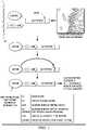
- FIG. 1Targeted enzymatic degradation of quorum-sensing peptides using a bifunctional ligand.
- FIG. 2shows a block diagram of a representative embodiment of a therapy system.
- a biofilmforms when microorganisms adhere to each other and to a surface, and produce extracellular polymers that facilitate adhesion and provide a structural matrix.
- This surfacemay be living tissue or inert, nonliving material.
- Formation of the biofilmbegins with the initial attachment of microorganisms to a surface. The attachment is initially through weak, reversible adhesion, but the microorganisms will proceed to anchor themselves more permanently using cell adhesion structures such as pili if they are not removed from the surface. Other cells then arrive to build the matrix that holds the biofilm together. Some species are not able to attach to a surface on their own, but are able to anchor themselves to the matrix or directly to earlier colonists. Thus, the early colonists provide adhesion sites that facilitate the adhesion of additional cells.
- quorum sensingis a system of stimulus and response correlated to population density.
- signaling moleculesare also referred to as “quorum-sensing molecules,” “inducer molecules,” or “autoinducers.”
- Microbesalso have receptors that “sense” the presence or absence of quorum-sensing molecules.
- Common classes of quorum-sensing moleculesinclude N-Acyl Homoserine Lactones (AHL) in Gram-negative bacteria, and a family of autoinducers known as autoinducer-2 (AI-2) in both Gram-negative and Gram-positive bacteria.
- P. aeruginosarelies on quorum sensing via production of signaling molecules such as lactones N-butanoyl-1-homoserine (C4-HSL) and N-(3-oxododecanoyl)-1-HSL (3-oxo-C12-HSL) (Smith et al. (2003) Current Opinion in Microbiology 1(1): 56-60).
- signaling moleculessuch as lactones N-butanoyl-1-homoserine (C4-HSL) and N-(3-oxododecanoyl)-1-HSL (3-oxo-C12-HSL)
- the microorganismsalso have receptors for the signaling molecules.
- the receptor LuxPis a receptor for AI-2.
- the signaling moleculebinds the receptor, it activates transcription of certain genes, including those for signaling molecule synthesis forming a positive feedback loop.
- Quorum quenchingmay be achieved by degrading the signaling molecule, such as with a protease.
- Lactonasealso known as acyl-homoserine lactonase
- AHLsacylated homoserine lactones
- Lactonasehydrolyzes the ester bond of the homoserine lactone ring of acylated homoserine lactones. By hydrolysing AHL, lactonase prevents these signaling molecules from binding to their target transcriptional regulators, thereby inhibiting quorum sensing.
- Lactonaseshave been reported for Bacillus, Agrobacterium, Rhodococcus, Streptomyces, Arthrobacter, Pseudomonas , and Klebsiella (Schipper et al. (2009) Molecular Plant - Microbe Interactions 75: 224-233).
- the Bacillus cereus group( B. cereus, B. thuringiensis, B. mycoides , and B. anthracis ) was found to contain nine genes homologous to the AiiA gene that encode AHL-inactivating enzymes (Dong et al. (2002). Applied and Environmental Microbiology 68(4): 1754-1759).
- lactonasesare AiiA produced by Bacillus species, AttM and AiiB produced by Agrobacterium tumefaciens , and QIcA produced by Rhizobiales species (Riaz et al. (2008) Environmental Microbiology 10(3): 560-570).
- the direct therapeutic application of lactonasesis complicated by the fact that lactonolysis is a reversible reaction (Rasmussen and Givskov (2006) International Journal of Medical Microbiology 296(2-3):149-161).
- the bifunctional ligand disclosed hereinprovides a solution to this problem by targeting proteases, such as lactonases, to the quorum sensing signaling molecules and thereby mediating irreversible degradation of the molecule.
- Biofilm-associated microorganismsbehave differently from planktonic organisms. Additionally, biofilms are characterized by their ability to become increasingly resistant to antimicrobial treatments. Resistance of biofilms to antimicrobial agents is believed to be due to the extracellular matrix in which the bacterial cells are embedded providing a barrier toward penetration by the biocides (Costerton et al., 1999). However, it is also possible that a majority of the cells in a biofilm are in a slow-growing, nutrient-starved state, and therefore not as susceptible to the effects of anti-microbial agents.
- the resistance to antimicrobial agentsmay be due to the cells in a biofilm adopting a distinct and protected biofilm phenotype, e.g., by elevated expression of drug-efflux pumps.
- Antimicrobial concentrations sufficient to inactivate planktonic organismsare generally inadequate to inactivate biofilm organisms, especially those deep within the biofilm. Biofilm formations also make it difficult for host phagocytic cells to gain access to and kill the organisms.
- Biofilmscan be comprised of bacteria, fungi, yeast, protozoa, and other microorganisms. Biofilms may be composed of single species or multiple species of microorganism. The most common biofilms have been found to be bacterial biofilms. Both Gram-negative and Gram-positive bacteria are capable of forming biofilms. Examples of Gram-positive bacteria that are capable of forming biofilms include, but are not limited to, Staphylococcus aureus , coagulase negative staphylocci such as Staphylococcus epidermis, Streptococcus pyogenes (group A), Streptococcus species (viridans group), Streptococcus agalactiae (group B), S.
- Streptococcusanaerobic species
- Streptococcus pneumoniaeStreptococcus pneumoniae
- Enterococcus speciesOther Gram-positive bacilli capable of forming biofilms include Bacillus anthracis, Corynebacterium diphtherias and Corynebacterium species which are diptheroids (aerobic and anerobic), Listeria monocytogenes, Clostridium tetani , and Clostridium difficile .
- Gram-negative bacteriathat are capable of forming biofilms are bacteria from the genus Escherichia coli, Enterobacter species, Proteus mirablis and other species, Pseudomonas aeruginosa, Klebsiella pneumoniae, Salmonella, Shigella, Serratia , and Campylobacterjejuni, Neisseria and Branhamella catarrhalis.
- Additional organisms capable of forming biofilmmay include dermatophytes ( Microsporum canis and other M . spp.; and Trichophyton spp. such as T. rubrum , and T. mentagrophytes ), yeasts (e.g., Candida albicans, C. Parapsilosis, C. glabrata, C. tropicalis , or other Candida species including drug resistant Candida species), Epidermophytonfloccosum, Malassezia fuurfur ( Pityropsporon orbiculare , or P.
- dermatophytesMicrosporum canis and other M . spp.; and Trichophyton spp. such as T. rubrum , and T. mentagrophytes
- yeastse.g., Candida albicans, C. Parapsilosis, C. glabrata, C. tropicalis , or other Candida species including drug resistant Candida species
- EpidermophytonfloccosumMalassezia fuurfur ( Pityropsporon or
- the organisms causing biofilm growth formationthat are most commonly isolated from indwelling medical devices include Staphylococcus species such as S. epidermidis and S. aureus; Candida species such as Candida albicans ; enterococci species such as Enterococcus faecalis, Streptococcus species; P. aeruginosa; K pneumoniae ; and diphtheroids. These organisms may originate from the skin of patients or healthcare workers, tap water to which entry ports are exposed, or other sources in the environment.
- P. aeruginosais highly associated with biofilm growth and catheter obstruction.
- biofilms of P. aeruginosahave been isolated from medical implants, such as indwelling urethral, venous or peritoneal catheters (Stickler et al., 1998). Burn wounds often become infected with P. aeruginosa from which life-threatening invasion of the blood stream and septic shock can arise. These infections are believed to involve the formation of biofilms.
- topical application to the wounds of a bifunctional ligand composition as described hereincan be used in to inhibit initiation of infection by preventing or inhibiting biofilm formation.
- the bifunctional ligands disclosed hereincomprise a first region that binds to a quorum-sensing molecule and a second region that binds a protease.
- the quorum-sensing-molecule-binding region of the bifunctional ligandmay be a small molecule or derived from, for example, the naturally occurring receptor of the quorum-sensing molecule or an antibody or antibody fragment that binds the quorum-sensing molecule.
- the protease-binding regionbinds a protease capable of hydrolyzing the quorum-sensing molecule that binds to the quorum-sensing-molecule-binding region of the bifunctional ligand.
- Quorum-sensing moleculesinclude, for example, acylated homoserine lactones (AHLs), autoinducing peptides (AIP), AI-2 (a furanosyl borate diester), ⁇ -butyrolactones, or competence stimulating peptide (CSP).
- AHLsacylated homoserine lactones
- AIPautoinducing peptides
- AI-2a furanosyl borate diester
- ⁇ -butyrolactonesa competence stimulating peptide
- CSPcompetence stimulating peptide
- Various microbesmay use the above-mentioned quorum-sensing systems.
- Non-limiting examples of bacteria that use each of the above-mentioned systemsare: Vibrio fischeri (AHL), Staphylococcus aureus (AIP), Vibrio harveyi (AI-2), Streptomyces griseus ( ⁇ -butyrolactone), and Streptococcus pneumonia
- the quorum-sensing-molecule-binding region of the bifunctional ligandmay be a small molecule or it may be derived from the naturally occurring transporter, receptor, or sensor of the quorum-sensing molecule.
- LuxRis a sensor of AHL, ABC exporter and AgrC transmembrane sensor kinase bind AIP
- Lsr ABC-type transporter and LuxQ transmembrane sensor kinasebind AI-2
- ArpAbinds ⁇ -butyrolactone
- ComAB and ComD transmembrane sensor kinasebind CSP.
- the quorum-sensing-molecule-binding region of the bifunctional ligandmay be a peptide or an antibody or antibody fragment that binds the quorum-sensing molecule.
- the protease-binding regionmay, for example, bind a lactonase.
- Lactonasesare metalloenzymes that target and inactivate acylated homoserine lactones (AHLs).
- AHLsacylated homoserine lactones
- a bifunctional ligandmay comprise a quorum-sensing-molecule-binding region that binds an AHL and a protease-binding region that binds a lactonase that hydrolyzes the AHL.
- Such a bifunctional ligandwould, therefore, promote the interaction of lactonase and AHL.
- lactonaseprevents these signaling molecules from binding to their target transcriptional regulators, thereby inhibiting quorum sensing.
- Lactonaseshave been reported for Bacillus, Agrobacterium, Rhodococcus, Streptomyces, Arthrobacter, Pseudomonas , and Klebsiella (Schipper et al. (2009) Molecular Plant - Microbe Interactions 75: 224-233).
- the Bacillus cereus group( B. cereus, B. thuringiensis, B. mycoides , and B. anthracis ) was found to contain nine genes homologous to the AiiA gene that encode AHL-inactivating enzymes (Dong et al. (2002) Applied and Environmental Microbiology 68 (4): 1754-1759).
- lactonasesare AiiA produced by Bacillus species, AttM and AiiB produced by Agrobacterium tumefaciens , and QIcA produced by Rhizobiales species (Riaz et al. (2008) Environmental Microbiology 10 (3): 560-570).
- the bifunctional ligandmay further comprise a linker region between the quorum-sensing-molecule-binding region and the protease-binding region.
- the linkercan be used to modify the distance and flexibility between the quorum-sensing-molecule-binding region and the protease-binding region.
- the linkermay be, for example, a peptide of about 1 to about 50 amino acids in length.
- the bifunctional ligandsmay be produced by techniques know to those in the art.
- the bifunctional ligandsmay be prepared by chemical synthesis, recombinant DNA techniques, or by chemical linkage of the domains.
- the quorum-sensing-molecule-binding region of the bifunctional ligandmay comprise a small molecule.
- a small moleculeis a low molecular weight (less than approximately 800 Daltons) organic compound. Small molecules are not polymers, but they may bind to polymers such as proteins. High-through put screening of small molecule libraries may be performed to identify small molecules that bind a particular quorum-sensing molecule.
- a wide variety of small molecule librariesare available from commercial vendors as well as through the National Institutes of Health. Alternatively, one may create a small molecule library using, for example, combinatorial synthesis methods.
- a variety of assay formatsare known for screening small molecule libraries, and these assays may be employed for identifying small molecules that bind quorum-sensing molecules.
- Phage displayis a well-known technique that uses bacteriophages for the high-throughput study of protein-protein interactions. Using phage display, large libraries of proteins can be screened for their ability to bind a particular protease.
- the bifunctional ligandsmay be provided in a variety of forms, particularly forms suitable for topical delivery to wound sites or application to medical devices that will be in contact with tissues.
- the bifunctional ligandmay be formulated in a pharmaceutical composition.
- the bifunctional ligandmay be formulated into all types of vehicles.
- Non-limiting examples of suitable vehiclesinclude emulsions (e.g., oil-in-water, water-in-oil, silicone-in-water, water-in-silicone, water-in-oil-in-water, oil-in-water, oil-in-water-in-oil, oil-in-water-in-silicone, etc.), creams, lotions, solutions (both aqueous and hydro-alcoholic), anhydrous bases (such as lipsticks and powders), gels, ointments, pastes, milks, liquids, aerosols, solid forms, sprays, hydrogels, or electroporation device cartridges.
- emulsionse.g., oil-in-water, water-in-oil, silicone-in-water, water-in-silicone, water-in-oil-in-water, oil-in-water, oil-in-water-in-silicone, etc.
- creamslotions, solutions (both aqueous and hydro-alcoholic), anhydrous
- the formulationmay be a hydrophilic solution, a thixotropic spray, or other hydrophillic topical. Variations and other appropriate vehicles will be apparent to the skilled artisan and are appropriate for use in the present invention.
- the concentrations and combinations of the ingredientscan be selected in such a way that the combinations are chemically compatible and do not form complexes which precipitate from the finished product.
- the formulationmay be immobilized on a surface, such as a dressing, and activated by a glucose wash.
- the compositionmay be applied through infusion within, injection into, absorption by, layering on, encapsulation within, or coating on, a carrier material, such as a bandage, gauze, wound dressing, adhesive bandage, scaffold, or hydrogel, for instance, comprising cellulose derivatives, including hydroxyethyl cellulose, hydroxymethyl cellulose, carboxymethyl cellulose, hydroxypropylmethyl cellulose and mixtures thereof; and hydrogels containing polyacrylic acid (Carbopols) as well as gelatin.
- a bifunctional ligand compositionmay be applied to a woven, non-woven, or knitted fabric material, such as gauze, dispersed within film, sponge, or foam for sustained release at a tissue site.
- a “carrier material” as used hereinrefers to a material suitable for a bifunctional ligand composition.
- the carrier materialmay be either bioresorbable, for instance comprising polyglycolic acid, polylactic acid, polydioxanone, polyhydroxybutyrate, polyhydrozyvalerate, polyaminoacids polyorthoesters, polyvinly alcohol, collagen, gelatin, chitosan, oxidized regenerated cellulose, hyaluronic acid, alginate or derivatives thereof, or may be non-bioresorbable, comprising for instance, polyurethane, polyvinyl alcohol, or gauze.
- the compositionmay be bound to the carrier material, such as through hydrogen binding, covalent binding or ionic binding.
- the bifunctional ligandmay be formulated in a composition with one or more additional active ingredients.
- the bifunctional ligandmay be formulated in a composition with one or more antibiotics.
- Such a formulationwill be advantageous in treating or preventing infection because the bifunctional ligand will inhibit biofilm formation thus allowing the antibiotic to more effectively kill the microbes.
- two or more different bifunctional ligandsmay be combined in a composition.
- the compositionmay also be an instillation composition.
- the compositionsmay be delivered to a tissue site by continuous instillation and/or periodic instillation. The instillant provides fresh bifunctional ligand to a tissue site such as a wound.
- the method for applying the bifunctional ligand to tissue site or woundmay vary depending on factors such as the location and the size and shape of the area to be treated. A health care provider will be able to determine an appropriate method for applying the bifunctional ligand to the area in view of such factors.
- the treatment methods of the present inventionmay be used on their own or in combination with additional methods of treatment.
- additional methods of treatmentIn order to increase the effectiveness of a treatment with the compositions and methods of treatment of the present invention or to augment the protection of another (second) therapy, it may be desirable to combine these compositions and methods with other agents and methods effective in the treatment, reduction of risk, or prevention of infections, for example, anti-bacterial, anti-viral, and/or anti-fungal treatments.
- iontophoresiscan be used to drive agents into tissues for the purpose of labeling or eradicating biolfilms.
- NPWTnegative pressure wound therapy
- V.A.C.® TherapyKCI International, San Antonio, Tex.
- fluid instillation therapyor both.
- Clinical studies and practicehave shown that NPWT can augment and accelerate growth of new tissue at a tissue site. The applications of this phenomenon are numerous, but it has proven particularly advantageous for treating wounds. Regardless of the etiology of a wound, whether trauma, surgery, or another cause, proper care of the wound is important to the outcome.
- Negative-pressure therapymay provide a number of benefits, including migration of epithelial and subcutaneous tissues, improved blood flow, and micro-deformation of tissue at a tissue site. Together, these benefits can increase development of granulation tissue and reduce healing times.
- Instillation of a tissue sitewhich generally refers to the slow introduction of a solution to the tissue site, can expose a tissue site to temperature variations, drugs, or other substances that may further promote healing or growth of tissue. Instillation may also be referred to as irrigation or infusion in some contexts. Instillation may be continuous or intermittent and may take place prior to, subsequent to, or simultaneously with the application of negative pressure. In some embodiments, instillation and negative pressure may be coordinated by a central controller.
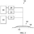
- FIG. 2is a simplified functional block diagram of an example embodiment of a therapy system 100 that can provide therapeutic pressure and instillation in accordance with this specification.
- the therapy system 100may include a dressing 102 fluidly coupled to a negative-pressure source 104 .
- a regulator or controller, such as regulator 106may also be fluidly coupled to the dressing 102 and the negative-pressure source 104 .
- the dressing 102generally includes a drape, such as drape 108 , and a manifold, such as distribution manifold 110 .
- the therapy system 100may also include fluid containers, such as container 112 and container 114 , coupled to the dressing 102 .
- container 112may be also be fluidly coupled to the negative-pressure source 104 in some embodiments, and container 114 may be coupled to a fluid-delivery device, such as a pump 116 .
- components of the therapy system 100may be coupled directly or indirectly.
- the negative-pressure source 104may be directly coupled to regulator 106 and indirectly coupled to dressing 102 through regulator 106 .
- Componentsmay be fluidly coupled to each other to provide a path for transferring fluids (i.e., liquid and/or gas) between the components.
- componentsmay be fluidly coupled with a tube, for example.
- a tubeis an elongated, cylindrical structure with some flexibility, but the geometry and rigidity may vary.
- componentsmay additionally or alternatively be coupled by virtue of physical proximity, being integral to a single structure, or being formed from the same piece of material. Coupling may also include mechanical, thermal, electrical, or chemical coupling (such as a chemical bond) in some contexts.
- the distribution manifold 110may be placed within, over, on, or otherwise proximate to a tissue site.
- the drape 108may be placed over the distribution manifold 110 and sealed to tissue proximate to the tissue site.
- the tissue proximate to the tissue siteis often undamaged epidermis peripheral to the tissue site.
- the dressing 102can provide a sealed therapeutic environment proximate to a tissue site, substantially isolated from the external, ambient environment.
- the negative-pressure source 104can reduce the pressure in the sealed therapeutic environment, and the pump 116 can apply therapeutic solutions, including the embodiments of the bifunctional ligand compositions described herein.
- Reduced pressure and/or fluidscan be applied substantially uniformly through the distribution manifold 110 in the sealed therapeutic environment. Reduced pressure can induce macrostrain and microstrain in the tissue site, as well as remove exudates and other fluids from the tissue site, which can be collected in the container 112 and disposed of properly.
- Integrating negative pressure therapy and instillation therapy with embodiments of bifunctional ligand compositions described hereincan further promote healing and growth of tissue by removing barriers to normal healing, such as the presence of biofilm and biofilm-forming bacteria.
- Functionally coupling infusion of the composition with reduced pressure therapy as disclosed hereinprovides unexpected decreases in wound bioburden and wound healing trajectories.
- the ability of a gelatin-based composition to operate with a reduced pressure therapy systemallows for the use of the composition as an instillate to treat a tissue site, in particular a chronic wound, with a composition that treats and/or inhibits biofilm formation.
- the negative pressure with the bifunctional ligand compositioncan be applied during debridement of a tissue site, for example, by using a dressing having a self-contained debriding mechanism such as ultrasound or pulsed lavage.
- negative pressure therapymay be applied after debridement, to promote vascular stimulation and the formation of granulation tissue.
- the transition from debridement to negative pressure therapyis seamless, as well as from negative pressure therapy to passive infusion with the composition, that is, without disrupting the integrity of the tissue site.
- the negative pressure with the bifunctional ligand compositionmay also be applied during cleansing or irrigation of the wound in some embodiments. Alternatively, the negative pressure may be applied prior to or after the cleansing of the wound with the composition.
- compositions provided hereincan be used in conjunction with all current NPWT devices, and delivered in either the inpatient or outpatient setting.
- exemplary negative pressure devicesinclude V.A.C.® Therapy, V.A.C.® Instill, or V.A.C.® Ulta therapy systems (Kinetic Concepts, Inc.). These devices, or devices having similar or equivalent designs may be used.
- reduced pressureor “negative pressure” generally refer to a pressure less than a local ambient pressure, such as the ambient pressure in a local environment external to a sealed therapeutic environment provided by the dressing 102 .
- the local ambient pressuremay also be the atmospheric pressure in the vicinity of a tissue site.
- the pressuremay be less than a hydrostatic pressure associated with tissue at the tissue site.
- values of pressure stated hereinare gauge pressures.
- references to increases in reduced pressuretypically refer to a decrease in absolute pressure, while decreases in reduced pressure typically refer to an increase in absolute pressure.
- a reduced-pressure sourcesuch as the reduced-pressure source 104
- a reduced-pressure sourcemay be housed within or used in conjunction with other components, such as sensors, processing units, alarm indicators, memory, databases, software, display devices, or user interfaces that further facilitate reduced-pressure therapy.
- the pressuretypically ranges between ⁇ 5 mm Hg ( ⁇ 667 Pa) and ⁇ 500 mm Hg ( ⁇ 66.7 kPa). Common therapeutic ranges are between ⁇ 75 mm Hg ( ⁇ 9.9 kPa) and ⁇ 300 mm Hg ( ⁇ 39.9 kPa).
- a fluid-delivery devicesuch as the pump 116 , may be a rotary-delivery pump, or other pump that can supply an instillation solution to a sealed space or the distribution manifold 110 .
- a fluid-delivery devicemay be housed within a therapy device or used in conjunction with other components, such as sensors, processing units, alarm indicators, memory, databases, software, display devices, or user interfaces that further facilitate instillation therapy.
- a fluid-delivery device and a negative-pressure sourcemay be integrated into a single unit to provide both negative pressure and instillation, or to alternatingly supply negative pressure and instillation.
- a manifoldsuch as the distribution manifold 110
- the distribution manifold 110can generally be adapted to contact a tissue site.
- the distribution manifold 110may be adapted to be placed partially or fully in contact with the tissue site. If the tissue site is a wound, for example, the distribution manifold 110 may partially or completely fill the wound, or may be placed over the wound.
- the distribution manifold 110may take many forms, and may be many sizes, shapes, or thicknesses depending on a variety of factors, such as the type of treatment being implemented or the nature and size of a tissue site. For example, the size and shape of the distribution manifold 110 may be adapted to the contours of deep and irregular shaped tissue sites.
- a manifoldis a substance or structure adapted to distribute negative pressure to or remove fluids from a tissue site, or both.
- a manifoldmay also facilitate delivering fluids to a tissue site, if the fluid path is reversed or a secondary fluid path is provided, for example when instillation solution is applied.
- a manifoldmay include flow channels or pathways that distribute fluids provided to and removed from a tissue site around the manifold.
- the flow channels or pathwaysmay be interconnected to improve distribution of fluids provided to or removed from a tissue site.
- cellular foam, open-cell foam, porous tissue collections, and other porous materialsuch as gauze or felted mat generally include structural elements arranged to form flow channels. Liquids, gels, and other foams may also include or be cured to include flow channels.
- the distribution manifold 110may be a porous foam material having interconnected cells or pores adapted to uniformly (or quasi-uniformly) distribute reduced pressure to a tissue site.
- the foam materialmay be either hydrophobic or hydrophilic.
- the distribution manifold 110may be an open-cell, reticulated polyurethane foam such as GranuFoam® dressing available from Kinetic Concepts, Inc. of San Antonio, Tex.
- the distribution manifold 110may also wick fluid away from a tissue site while continuing to distribute reduced pressure to the tissue site.
- the wicking properties of the distribution manifold 110may draw fluid away from a tissue site by capillary flow or other wicking mechanisms.
- An example of a hydrophilic foamis a polyvinyl alcohol, open-cell foam such as V.A.C. WhiteFoam® dressing available from Kinetic Concepts, Inc. of San Antonio, Tex.
- Other hydrophilic foamsmay include those made from polyether.
- Other foams that may exhibit hydrophilic characteristicsinclude hydrophobic foams that have been treated or coated to provide hydrophilicity.
- the distribution manifold 110may further promote granulation at a tissue site if pressure within a sealed therapeutic environment is reduced.
- any or all of the surfaces of the distribution manifold 110may have an uneven, coarse, or jagged profile that can induce microstrains and stresses at a tissue site if reduced pressure is applied through the distribution manifold 110 .
- the distribution manifold 110may be constructed from bioresorbable materials.
- Suitable bioresorbable materialsmay include, without limitation, a polymeric blend of polylactic acid (PLA) and polyglycolic acid (PGA).
- the polymeric blendmay also include without limitation polycarbonates, polyfumarates, and capralactones.
- bioresorbable materialsinclude, but are not limited to, polydioxanone, polyhydroxybutyrate, polyhydrozyvalerate, polyaminoacids polyorthoesters, polyvinly alcohol, chitosan, oxidized regenerated cellulose, hyaluronic acid, alginate, collagen, a modified collagen, such as gelatin or derivatives of any of the above.
- the distribution manifold 110may further serve as a scaffold for new cell-growth, or a scaffold material may be used in conjunction with the distribution manifold 110 to promote cell-growth.
- a scaffold materialmay be a substance or structure used to enhance or promote the growth of cells or formation of tissue, such as a three-dimensional porous structure that provides a template for cell growth.
- a scaffold and/or manifoldmay be also be infused with, coated with, or comprised of cells, growth factors, extracellular matrix components, nutrients, integrins, or other substances to promote cell growth in addition to embodiments of the compositions described herein.
- the manifold or scaffoldmay serve as a carrier material for the composition described herein.
- Scaffoldsmay be formed from biologic or synthetic scaffold materials, and are used in the field of tissue engineering to support protein adhesion and cellular ingrowth for tissue repair and regeneration.
- the current state of the art in scaffold technologyrelies upon the inherent characteristics of the surrounding tissue space for the adsorption of proteins and migration of cells.
- Nonlimiting examples of suitable scaffold materialsinclude extracellular matrix proteins such as fibrin, collagen or fibronectin, and synthetic or naturally occurring polymers, including bioabsorbable or non-absorbable polymers, such as polylactic acid (PLA), polyglycolic acid (PGA), polylactide-co-glycolide (PLGA), polyvinylpyrrolidone, polycaprolactone, polycarbonates, polyfumarates, caprolactones, polyamides, polysaccharides (including alginates (e.g., calcium alginate) and chitosan), hyaluronic acid, polyhydroxybutyrate, polyhydroxyvalerate, polydioxanone, polyorthoesthers, polyethylene glycols, poloxamers, polyphosphazenes, polyanhydrides, polyamino acids, polyacetals, polycyanoacrylates, polyurethanes (e.g., GranuFoam®), polyacrylates, ethylene-vinyl acetate
- the scaffoldcan also comprise ceramics such as hydroxyapatite, coralline apatite, calcium phosphate, calcium sulfate, calcium carbonate or other carbonates, bioglass, allografts, autografts, xenografts, decellularized tissues, or composites of any of the above.
- the scaffoldmay comprise collagen (e.g., BiostepTM or PomogranTM scaffolds), polylactic acid (PLA), polyglycolic acid (PGA), polylactide-co-glycolide (PLGA), a polyurethane, a polysaccharide, an hydroxyapatite, or a polytherylene glycol.
- the scaffoldcan comprise combinations of any two, three or more materials, either in separate or multiple areas of the scaffold, combined noncovalently or covalently (e.g., copolymers such as a polyethylene oxide-polypropylene glycol block copolymers, or terpolymers), or combinations thereof.
- noncovalently or covalentlye.g., copolymers such as a polyethylene oxide-polypropylene glycol block copolymers, or terpolymers
- the drape 108is an example of a sealing member.
- a sealing membermay be constructed from a material that can provide a fluid seal between two environments or components, such as between a therapeutic environment and a local external environment.
- the sealing membermay be, for example, an impermeable or semi-permeable, elastomeric material that can provide a seal adequate to maintain a negative pressure at a tissue site for a given negative-pressure source.
- the permeabilitygenerally should be low enough that a desired negative pressure may be maintained.
- An attachment devicemay be used to attach a sealing member to an attachment surface, such as undamaged epidermis, a gasket, or another sealing member.
- the attachment devicemay take many forms.
- an attachment devicemay be a medically-acceptable, pressure-sensitive adhesive that extends about a periphery, a portion, or an entire sealing member.
- Other example embodiments of an attachment devicemay include a double-sided tape, paste, hydrocolloid, hydrogel, silicone gel, organogel, or an acrylic adhesive.
- the container 112is representative of a container, canister, pouch, or other storage component, which can be used to manage exudates and other fluids withdrawn from a tissue site.
- a rigid containermay be preferred or required for collecting, storing, and disposing of fluids.
- fluidsmay be properly disposed of without rigid container storage, and a re-usable container could reduce waste and costs associated with negative-pressure therapy.
- the container 114is representative of another container, canister, pouch, cartridge, or other storage component, which can be used to manage instillation solution to be supplied to a tissue site.
- a rigid containermay be preferred or required for delivering, storing, and supplying of the instillation solution.
- instillation solutionmay be provided in a non-rigid container, and a re-usable container could reduce waste and costs associated with instillation.
- kitsmay also be provides as one or more kits.
- a kitcomprises components for inhibiting biofilms. Any of the components disclosed here in may be combined in a kit.
- the kitcomprises a biofilm-inhibiting composition disclosed herein.
- the kitmay further comprise a wound dressing, such as a gauze, cloth, or film.
- kitswill generally include at least one vial, test tube, flask, bottle, syringe, foil package, or other container, into which a component may be placed, and preferably, suitably aliquoted. Where there is more than one component in the kit, the kit also will generally contain a second, third or other additional containers into which the additional components may be separately placed. However, various combinations of components may be comprised in a container.
- the kits of the present inventionalso will typically include packaging for containing the various containers in close confinement for commercial sale. Such packaging may include cardboard or injection or blow molded plastic packaging into which the desired containers are retained.
- a kitmay also include instructions for employing the kit components. Instructions may include variations that can be implemented.
- the effectiveness of the treating a biofilm or inhibiting biofilm-formationmay be determined using methods known in the art.
- One methodis to extract any bacteria remaining in the biofilm after treatment, plating the bacteria on a suitable media and allowing the bacteria to grow for 24-48 hours or as appropriate, then counting any bacteria.
- a reduction in bacteriais reflects effectiveness of the bifunctional ligand and the method of treatment.
- treatment between groups using in vitro methodssuch as described in Phillips et al., “Effects of antimicrobial Agents on an in vitro biofilm model of skin wounds,” Advances in Wound Care, 1:299-304 (2010), or as otherwise known in the art.
- a subjectis identified as having a wound that has become infected with S. aureus that has formed a biofilm in the wound.
- the woundis treated with a bifunctional ligand that comprises a first region that binds to a quorum-sensing molecule and a second region that binds a protease with a standard therapeutically effective amount for a period of time sufficient to reduce biofilm in the wound.
- a subjectis identified as having a wound that has become infected with a P. aeruginosa that has formed a biofilm in the wound.
- the woundis treated with a bifunctional ligand that comprises a first region that binds to a quorum-sensing molecule and a second region that binds a protease with a standard therapeutically effective amount for a period of time in combination with VAC® Therapy at ⁇ 125 mmHg to reduce biofilm in the wound.
- P. aeruginosaforms a biofilm on a ventilation tube.
- the ventilation tubeis treated with a bifunctional ligand that comprises a first region that binds to a quorum-sensing molecule and a second region that binds a protease in an amount and for a period of time sufficient to reduce the biofilm on the ventilation tube.
- compositions and methods disclosed and claimed hereincan be made and executed without undue experimentation in light of the present disclosure. While the compositions and methods of this invention have been described in terms of certain embodiments, it will be apparent to those of skill in the art that variations may be applied to the compositions and methods and in the steps or in the sequence of steps of the methods described herein without departing from the concept, spirit and scope of the invention. More specifically, it will be apparent that certain agents which are both chemically and physiologically related may be substituted for the agents described herein while the same or similar results would be achieved. All such similar substitutes and modifications apparent to those skilled in the art are deemed to be within the spirit, scope and concept of the invention as defined by the appended claims.
Landscapes
- Health & Medical Sciences (AREA)
- Chemical & Material Sciences (AREA)
- Life Sciences & Earth Sciences (AREA)
- Medicinal Chemistry (AREA)
- General Health & Medical Sciences (AREA)
- Animal Behavior & Ethology (AREA)
- Public Health (AREA)
- Veterinary Medicine (AREA)
- Epidemiology (AREA)
- Engineering & Computer Science (AREA)
- Immunology (AREA)
- Pharmacology & Pharmacy (AREA)
- Organic Chemistry (AREA)
- Proteomics, Peptides & Aminoacids (AREA)
- Molecular Biology (AREA)
- Bioinformatics & Cheminformatics (AREA)
- Gastroenterology & Hepatology (AREA)
- Genetics & Genomics (AREA)
- Biophysics (AREA)
- Biochemistry (AREA)
- Mycology (AREA)
- General Chemical & Material Sciences (AREA)
- Microbiology (AREA)
- Chemical Kinetics & Catalysis (AREA)
- Biomedical Technology (AREA)
- Heart & Thoracic Surgery (AREA)
- Medicines That Contain Protein Lipid Enzymes And Other Medicines (AREA)
- Vascular Medicine (AREA)
- Anesthesiology (AREA)
- Hematology (AREA)
- Pharmaceuticals Containing Other Organic And Inorganic Compounds (AREA)
Abstract
Description
Claims (15)
Priority Applications (1)
| Application Number | Priority Date | Filing Date | Title |
|---|---|---|---|
| US16/447,173US11318218B2 (en) | 2012-03-20 | 2019-06-20 | Targeted enzymatic degradation of quorum-sensing peptides |
Applications Claiming Priority (5)
| Application Number | Priority Date | Filing Date | Title |
|---|---|---|---|
| US201261613306P | 2012-03-20 | 2012-03-20 | |
| US13/841,200US9169319B2 (en) | 2012-03-20 | 2013-03-15 | Targeted enzymatic degradation of quorum-sensing peptides |
| US14/851,178US9833528B2 (en) | 2012-03-20 | 2015-09-11 | Targeted enzymatic degradation of quorum-sensing peptides |
| US15/802,817US10369240B2 (en) | 2012-03-20 | 2017-11-03 | Targeted enzymatic degradation of quorum-sensing peptides |
| US16/447,173US11318218B2 (en) | 2012-03-20 | 2019-06-20 | Targeted enzymatic degradation of quorum-sensing peptides |
Related Parent Applications (1)
| Application Number | Title | Priority Date | Filing Date |
|---|---|---|---|
| US15/802,817ContinuationUS10369240B2 (en) | 2012-03-20 | 2017-11-03 | Targeted enzymatic degradation of quorum-sensing peptides |
Publications (2)
| Publication Number | Publication Date |
|---|---|
| US20190298872A1 US20190298872A1 (en) | 2019-10-03 |
| US11318218B2true US11318218B2 (en) | 2022-05-03 |
Family
ID=48050931
Family Applications (4)
| Application Number | Title | Priority Date | Filing Date |
|---|---|---|---|
| US13/841,200ActiveUS9169319B2 (en) | 2012-03-20 | 2013-03-15 | Targeted enzymatic degradation of quorum-sensing peptides |
| US14/851,178Active2033-04-25US9833528B2 (en) | 2012-03-20 | 2015-09-11 | Targeted enzymatic degradation of quorum-sensing peptides |
| US15/802,817ActiveUS10369240B2 (en) | 2012-03-20 | 2017-11-03 | Targeted enzymatic degradation of quorum-sensing peptides |
| US16/447,173Active2033-06-15US11318218B2 (en) | 2012-03-20 | 2019-06-20 | Targeted enzymatic degradation of quorum-sensing peptides |
Family Applications Before (3)
| Application Number | Title | Priority Date | Filing Date |
|---|---|---|---|
| US13/841,200ActiveUS9169319B2 (en) | 2012-03-20 | 2013-03-15 | Targeted enzymatic degradation of quorum-sensing peptides |
| US14/851,178Active2033-04-25US9833528B2 (en) | 2012-03-20 | 2015-09-11 | Targeted enzymatic degradation of quorum-sensing peptides |
| US15/802,817ActiveUS10369240B2 (en) | 2012-03-20 | 2017-11-03 | Targeted enzymatic degradation of quorum-sensing peptides |
Country Status (2)
| Country | Link |
|---|---|
| US (4) | US9169319B2 (en) |
| WO (1) | WO2013142372A1 (en) |
Families Citing this family (4)
| Publication number | Priority date | Publication date | Assignee | Title |
|---|---|---|---|---|
| CA2684150C (en) | 2007-05-14 | 2016-10-04 | Research Foundation Of State University Of New York | Decenoic acid dispersion inducers in the treatment of biofilms |
| WO2013142372A1 (en)* | 2012-03-20 | 2013-09-26 | Kci Licensing, Inc. | Targeted enzymatic degradation of quorum-sensing peptides |
| US11541105B2 (en) | 2018-06-01 | 2023-01-03 | The Research Foundation For The State University Of New York | Compositions and methods for disrupting biofilm formation and maintenance |
| US20220227814A1 (en)* | 2019-05-22 | 2022-07-21 | Board Of Regents Of The Nevada System Of Higher Education On Behalf Of The University Of Nevada, Ren | Cyclic dominant negative competence stimulating peptide analogs and methods of treating streptococcus pneumoniae infections |
Citations (133)
| Publication number | Priority date | Publication date | Assignee | Title |
|---|---|---|---|---|
| US1355846A (en) | 1920-02-06 | 1920-10-19 | David A Rannells | Medical appliance |
| US2547758A (en) | 1949-01-05 | 1951-04-03 | Wilmer B Keeling | Instrument for treating the male urethra |
| US2632443A (en) | 1949-04-18 | 1953-03-24 | Eleanor P Lesher | Surgical dressing |
| GB692578A (en) | 1949-09-13 | 1953-06-10 | Minnesota Mining & Mfg | Improvements in or relating to drape sheets for surgical use |
| US2682873A (en) | 1952-07-30 | 1954-07-06 | Johnson & Johnson | General purpose protective dressing |
| US2910763A (en) | 1955-08-17 | 1959-11-03 | Du Pont | Felt-like products |
| US2969057A (en) | 1957-11-04 | 1961-01-24 | Brady Co W H | Nematodic swab |
| US3066672A (en) | 1960-09-27 | 1962-12-04 | Jr William H Crosby | Method and apparatus for serial sampling of intestinal juice |
| US3367332A (en) | 1965-08-27 | 1968-02-06 | Gen Electric | Product and process for establishing a sterile area of skin |
| US3520300A (en) | 1967-03-15 | 1970-07-14 | Amp Inc | Surgical sponge and suction device |
| US3568675A (en) | 1968-08-30 | 1971-03-09 | Clyde B Harvey | Fistula and penetrating wound dressing |
| US3648692A (en) | 1970-12-07 | 1972-03-14 | Parke Davis & Co | Medical-surgical dressing for burns and the like |
| US3682180A (en) | 1970-06-08 | 1972-08-08 | Coilform Co Inc | Drain clip for surgical drain |
| US3826254A (en) | 1973-02-26 | 1974-07-30 | Verco Ind | Needle or catheter retaining appliance |
| DE2640413A1 (en) | 1976-09-08 | 1978-03-09 | Wolf Gmbh Richard | CATHETER MONITORING DEVICE |
| US4080970A (en) | 1976-11-17 | 1978-03-28 | Miller Thomas J | Post-operative combination dressing and internal drain tube with external shield and tube connector |
| US4096853A (en) | 1975-06-21 | 1978-06-27 | Hoechst Aktiengesellschaft | Device for the introduction of contrast medium into an anus praeter |
| US4139004A (en) | 1977-02-17 | 1979-02-13 | Gonzalez Jr Harry | Bandage apparatus for treating burns |
| US4165748A (en) | 1977-11-07 | 1979-08-28 | Johnson Melissa C | Catheter tube holder |
| US4184510A (en) | 1977-03-15 | 1980-01-22 | Fibra-Sonics, Inc. | Valued device for controlling vacuum in surgery |
| WO1980002182A1 (en) | 1979-04-06 | 1980-10-16 | J Moss | Portable suction device for collecting fluids from a closed wound |
| US4233969A (en) | 1976-11-11 | 1980-11-18 | Lock Peter M | Wound dressing materials |
| US4245630A (en) | 1976-10-08 | 1981-01-20 | T. J. Smith & Nephew, Ltd. | Tearable composite strip of materials |
| US4256109A (en) | 1978-07-10 | 1981-03-17 | Nichols Robert L | Shut off valve for medical suction apparatus |
| US4261363A (en) | 1979-11-09 | 1981-04-14 | C. R. Bard, Inc. | Retention clips for body fluid drains |
| US4275721A (en) | 1978-11-28 | 1981-06-30 | Landstingens Inkopscentral Lic, Ekonomisk Forening | Vein catheter bandage |
| US4284079A (en) | 1979-06-28 | 1981-08-18 | Adair Edwin Lloyd | Method for applying a male incontinence device |
| US4297995A (en) | 1980-06-03 | 1981-11-03 | Key Pharmaceuticals, Inc. | Bandage containing attachment post |
| US4333468A (en) | 1980-08-18 | 1982-06-08 | Geist Robert W | Mesentery tube holder apparatus |
| US4373519A (en) | 1981-06-26 | 1983-02-15 | Minnesota Mining And Manufacturing Company | Composite wound dressing |
| US4382441A (en) | 1978-12-06 | 1983-05-10 | Svedman Paul | Device for treating tissues, for example skin |
| US4392858A (en) | 1981-07-16 | 1983-07-12 | Sherwood Medical Company | Wound drainage device |
| US4392853A (en) | 1981-03-16 | 1983-07-12 | Rudolph Muto | Sterile assembly for protecting and fastening an indwelling device |
| US4419097A (en) | 1981-07-31 | 1983-12-06 | Rexar Industries, Inc. | Attachment for catheter tube |
| EP0100148A1 (en) | 1982-07-06 | 1984-02-08 | Dow Corning Limited | Medical-surgical dressing and a process for the production thereof |
| US4465485A (en) | 1981-03-06 | 1984-08-14 | Becton, Dickinson And Company | Suction canister with unitary shut-off valve and filter features |
| EP0117632A2 (en) | 1983-01-27 | 1984-09-05 | Johnson & Johnson Products Inc. | Adhesive film dressing |
| US4475909A (en) | 1982-05-06 | 1984-10-09 | Eisenberg Melvin I | Male urinary device and method for applying the device |
| US4480638A (en) | 1980-03-11 | 1984-11-06 | Eduard Schmid | Cushion for holding an element of grafted skin |
| US4525374A (en) | 1984-02-27 | 1985-06-25 | Manresa, Inc. | Treating hydrophobic filters to render them hydrophilic |
| US4525166A (en) | 1981-11-21 | 1985-06-25 | Intermedicat Gmbh | Rolled flexible medical suction drainage device |
| US4540412A (en) | 1983-07-14 | 1985-09-10 | The Kendall Company | Device for moist heat therapy |
| US4543100A (en) | 1983-11-01 | 1985-09-24 | Brodsky Stuart A | Catheter and drain tube retainer |
| US4548202A (en) | 1983-06-20 | 1985-10-22 | Ethicon, Inc. | Mesh tissue fasteners |
| US4551139A (en) | 1982-02-08 | 1985-11-05 | Marion Laboratories, Inc. | Method and apparatus for burn wound treatment |
| EP0161865A2 (en) | 1984-05-03 | 1985-11-21 | Smith and Nephew Associated Companies p.l.c. | Adhesive wound dressing |
| US4569348A (en) | 1980-02-22 | 1986-02-11 | Velcro Usa Inc. | Catheter tube holder strap |
| AU550575B2 (en) | 1981-08-07 | 1986-03-27 | Richard Christian Wright | Wound drainage device |
| US4605399A (en) | 1984-12-04 | 1986-08-12 | Complex, Inc. | Transdermal infusion device |
| US4608041A (en) | 1981-10-14 | 1986-08-26 | Frese Nielsen | Device for treatment of wounds in body tissue of patients by exposure to jets of gas |
| US4640688A (en) | 1985-08-23 | 1987-02-03 | Mentor Corporation | Urine collection catheter |
| US4655754A (en) | 1984-11-09 | 1987-04-07 | Stryker Corporation | Vacuum wound drainage system and lipids baffle therefor |
| US4664662A (en) | 1984-08-02 | 1987-05-12 | Smith And Nephew Associated Companies Plc | Wound dressing |
| WO1987004626A1 (en) | 1986-01-31 | 1987-08-13 | Osmond, Roger, L., W. | Suction system for wound and gastro-intestinal drainage |
| US4710165A (en) | 1985-09-16 | 1987-12-01 | Mcneil Charles B | Wearable, variable rate suction/collection device |
| US4733659A (en) | 1986-01-17 | 1988-03-29 | Seton Company | Foam bandage |
| GB2195255A (en) | 1986-09-30 | 1988-04-07 | Vacutec Uk Limited | Method and apparatus for vacuum treatment of an epidermal surface |
| US4743232A (en) | 1986-10-06 | 1988-05-10 | The Clinipad Corporation | Package assembly for plastic film bandage |
| GB2197789A (en) | 1986-11-28 | 1988-06-02 | Smiths Industries Plc | Anti-foaming disinfectants used in surgical suction apparatus |
| US4758220A (en) | 1985-09-26 | 1988-07-19 | Alcon Laboratories, Inc. | Surgical cassette proximity sensing and latching apparatus |
| US4787888A (en) | 1987-06-01 | 1988-11-29 | University Of Connecticut | Disposable piezoelectric polymer bandage for percutaneous delivery of drugs and method for such percutaneous delivery (a) |
| US4826494A (en) | 1984-11-09 | 1989-05-02 | Stryker Corporation | Vacuum wound drainage system |
| US4838883A (en) | 1986-03-07 | 1989-06-13 | Nissho Corporation | Urine-collecting device |
| US4840187A (en) | 1986-09-11 | 1989-06-20 | Bard Limited | Sheath applicator |
| US4863449A (en) | 1987-07-06 | 1989-09-05 | Hollister Incorporated | Adhesive-lined elastic condom cathether |
| US4872450A (en) | 1984-08-17 | 1989-10-10 | Austad Eric D | Wound dressing and method of forming same |
| US4878901A (en) | 1986-10-10 | 1989-11-07 | Sachse Hans Ernst | Condom catheter, a urethral catheter for the prevention of ascending infections |
| GB2220357A (en) | 1988-05-28 | 1990-01-10 | Smiths Industries Plc | Medico-surgical containers |
| US4897081A (en) | 1984-05-25 | 1990-01-30 | Thermedics Inc. | Percutaneous access device |
| US4906233A (en) | 1986-05-29 | 1990-03-06 | Terumo Kabushiki Kaisha | Method of securing a catheter body to a human skin surface |
| US4906240A (en) | 1988-02-01 | 1990-03-06 | Matrix Medica, Inc. | Adhesive-faced porous absorbent sheet and method of making same |
| US4919654A (en) | 1988-08-03 | 1990-04-24 | Kalt Medical Corporation | IV clamp with membrane |
| CA2005436A1 (en) | 1988-12-13 | 1990-06-13 | Glenda G. Kalt | Transparent tracheostomy tube dressing |
| US4941882A (en) | 1987-03-14 | 1990-07-17 | Smith And Nephew Associated Companies, P.L.C. | Adhesive dressing for retaining a cannula on the skin |
| US4953565A (en) | 1986-11-26 | 1990-09-04 | Shunro Tachibana | Endermic application kits for external medicines |
| WO1990010424A1 (en) | 1989-03-16 | 1990-09-20 | Smith & Nephew Plc | Absorbent devices and precursors therefor |
| US4969880A (en) | 1989-04-03 | 1990-11-13 | Zamierowski David S | Wound dressing and treatment method |
| US4985019A (en) | 1988-03-11 | 1991-01-15 | Michelson Gary K | X-ray marker |
| GB2235877A (en) | 1989-09-18 | 1991-03-20 | Antonio Talluri | Closed wound suction apparatus |
| US5037397A (en) | 1985-05-03 | 1991-08-06 | Medical Distributors, Inc. | Universal clamp |
| US5086170A (en) | 1989-01-16 | 1992-02-04 | Roussel Uclaf | Process for the preparation of azabicyclo compounds |
| US5092858A (en) | 1990-03-20 | 1992-03-03 | Becton, Dickinson And Company | Liquid gelling agent distributor device |
| US5100396A (en) | 1989-04-03 | 1992-03-31 | Zamierowski David S | Fluidic connection system and method |
| US5134994A (en) | 1990-02-12 | 1992-08-04 | Say Sam L | Field aspirator in a soft pack with externally mounted container |
| US5149331A (en) | 1991-05-03 | 1992-09-22 | Ariel Ferdman | Method and device for wound closure |
| US5167613A (en) | 1992-03-23 | 1992-12-01 | The Kendall Company | Composite vented wound dressing |
| US5176663A (en) | 1987-12-02 | 1993-01-05 | Pal Svedman | Dressing having pad with compressibility limiting elements |
| WO1993009727A1 (en) | 1991-11-14 | 1993-05-27 | Wake Forest University | Method and apparatus for treating tissue damage |
| US5215522A (en) | 1984-07-23 | 1993-06-01 | Ballard Medical Products | Single use medical aspirating device and method |
| US5232453A (en) | 1989-07-14 | 1993-08-03 | E. R. Squibb & Sons, Inc. | Catheter holder |
| US5261893A (en) | 1989-04-03 | 1993-11-16 | Zamierowski David S | Fastening system and method |
| US5278100A (en) | 1991-11-08 | 1994-01-11 | Micron Technology, Inc. | Chemical vapor deposition technique for depositing titanium silicide on semiconductor wafers |
| US5279550A (en) | 1991-12-19 | 1994-01-18 | Gish Biomedical, Inc. | Orthopedic autotransfusion system |
| US5298015A (en) | 1989-07-11 | 1994-03-29 | Nippon Zeon Co., Ltd. | Wound dressing having a porous structure |
| US5342376A (en) | 1993-05-03 | 1994-08-30 | Dermagraphics, Inc. | Inserting device for a barbed tissue connector |
| US5344415A (en) | 1993-06-15 | 1994-09-06 | Deroyal Industries, Inc. | Sterile system for dressing vascular access site |
| DE4306478A1 (en) | 1993-03-02 | 1994-09-08 | Wolfgang Dr Wagner | Drainage device, in particular pleural drainage device, and drainage method |
| WO1994020041A1 (en) | 1993-03-09 | 1994-09-15 | Wake Forest University | Wound treatment employing reduced pressure |
| US5358494A (en) | 1989-07-11 | 1994-10-25 | Svedman Paul | Irrigation dressing |
| US5437622A (en) | 1992-04-29 | 1995-08-01 | Laboratoire Hydrex (Sa) | Transparent adhesive dressing with reinforced starter cuts |
| US5437651A (en) | 1993-09-01 | 1995-08-01 | Research Medical, Inc. | Medical suction apparatus |
| DE29504378U1 (en) | 1995-03-15 | 1995-09-14 | MTG Medizinisch, technische Gerätebau GmbH, 66299 Friedrichsthal | Electronically controlled low-vacuum pump for chest and wound drainage |
| WO1996005873A1 (en) | 1994-08-22 | 1996-02-29 | Kinetic Concepts Inc. | Wound drainage equipment |
| US5527293A (en) | 1989-04-03 | 1996-06-18 | Kinetic Concepts, Inc. | Fastening system and method |
| US5549584A (en) | 1994-02-14 | 1996-08-27 | The Kendall Company | Apparatus for removing fluid from a wound |
| US5556375A (en) | 1994-06-16 | 1996-09-17 | Hercules Incorporated | Wound dressing having a fenestrated base layer |
| US5607388A (en) | 1994-06-16 | 1997-03-04 | Hercules Incorporated | Multi-purpose wound dressing |
| WO1997018007A1 (en) | 1995-11-14 | 1997-05-22 | Kci Medical Limited | Portable wound treatment apparatus |
| GB2329127A (en) | 1997-09-12 | 1999-03-17 | Kci Medical Ltd | Suction head and drape wound treatment assembly |
| US6071267A (en) | 1998-02-06 | 2000-06-06 | Kinetic Concepts, Inc. | Medical patient fluid management interface system and method |
| US6135116A (en) | 1997-07-28 | 2000-10-24 | Kci Licensing, Inc. | Therapeutic method for treating ulcers |
| US6241747B1 (en) | 1993-05-03 | 2001-06-05 | Quill Medical, Inc. | Barbed Bodily tissue connector |
| US6287316B1 (en) | 1999-03-26 | 2001-09-11 | Ethicon, Inc. | Knitted surgical mesh |
| US20020077661A1 (en) | 2000-12-20 | 2002-06-20 | Vahid Saadat | Multi-barbed device for retaining tissue in apposition and methods of use |
| US20020115951A1 (en) | 2001-02-22 | 2002-08-22 | Core Products International, Inc. | Ankle brace providing upper and lower ankle adjustment |
| US20020120185A1 (en) | 2000-05-26 | 2002-08-29 | Kci Licensing, Inc. | System for combined transcutaneous blood gas monitoring and vacuum assisted wound closure |
| US20020143286A1 (en) | 2001-03-05 | 2002-10-03 | Kci Licensing, Inc. | Vacuum assisted wound treatment apparatus and infection identification system and method |
| US6488643B1 (en) | 1998-10-08 | 2002-12-03 | Kci Licensing, Inc. | Wound healing foot wrap |
| US6493568B1 (en) | 1994-07-19 | 2002-12-10 | Kci Licensing, Inc. | Patient interface system |
| AU755496B2 (en) | 1997-09-12 | 2002-12-12 | Kci Licensing, Inc. | Surgical drape and suction head for wound treatment |
| US20080075730A1 (en)* | 2003-04-23 | 2008-03-27 | University Technologies International Inc. | Methods and compositions for treating biofilms |
| JP4129536B2 (en) | 2000-02-24 | 2008-08-06 | ヴェネテック インターナショナル,インコーポレイテッド | Highly compatible catheter anchoring system |
| US20090326416A1 (en)* | 2008-06-04 | 2009-12-31 | Mcnulty Amy | Detecting infection in reduced pressure wound treatment |
| US7846141B2 (en) | 2002-09-03 | 2010-12-07 | Bluesky Medical Group Incorporated | Reduced pressure treatment system |
| US8216198B2 (en) | 2009-01-09 | 2012-07-10 | Tyco Healthcare Group Lp | Canister for receiving wound exudate in a negative pressure therapy system |
| US8251979B2 (en) | 2009-05-11 | 2012-08-28 | Tyco Healthcare Group Lp | Orientation independent canister for a negative pressure wound therapy device |
| US8257327B2 (en) | 2003-10-28 | 2012-09-04 | Smith & Nephew Plc | Wound cleansing apparatus with actives |
| US8398614B2 (en) | 2002-10-28 | 2013-03-19 | Smith & Nephew Plc | Apparatus for aspirating, irrigating and cleansing wounds |
| US8449509B2 (en) | 2004-04-05 | 2013-05-28 | Bluesky Medical Group Incorporated | Flexible reduced pressure treatment appliance |
| US8529548B2 (en) | 2004-04-27 | 2013-09-10 | Smith & Nephew Plc | Wound treatment apparatus and method |
| US8551060B2 (en) | 2008-07-17 | 2013-10-08 | Smith & Nephew, Inc. | Subatmospheric pressure mechanism for wound therapy system and related methods therefor |
| US8926592B2 (en) | 2003-10-28 | 2015-01-06 | Smith & Nephew Plc | Wound cleansing apparatus with heat |
| US9017302B2 (en) | 2008-07-21 | 2015-04-28 | Smith & Nephew, Inc. | Thin film wound dressing |
Family Cites Families (11)
| Publication number | Priority date | Publication date | Assignee | Title |
|---|---|---|---|---|
| US6703513B1 (en) | 2000-06-02 | 2004-03-09 | K-Quay Enterprises Llc | Production and use of derivatized homoserine lactones |
| US7109294B2 (en)* | 2001-09-18 | 2006-09-19 | Trustees Of Dartmouth College | Bacterial glycosyltransferase polypeptides involved in antibiotic resistance |
| WO2003064592A2 (en)* | 2001-10-26 | 2003-08-07 | Quorex Pharmaceuticals, Inc. | Inhibitors of autoinducer transporters |
| US7976519B2 (en) | 2002-12-31 | 2011-07-12 | Kci Licensing, Inc. | Externally-applied patient interface system and method |
| US20050014932A1 (en)* | 2003-05-15 | 2005-01-20 | Iogenetics, Llc | Targeted biocides |
| DE10361457A1 (en)* | 2003-12-23 | 2005-07-28 | Henkel Kgaa | New alkoxylactones, alkoxylactams and alkoxythiolactams |
| CN101511382B (en)* | 2006-09-06 | 2013-11-13 | 加利福尼亚大学董事会 | Selectively targeted antimicrobial peptides and the use thereof |
| US7910622B2 (en)* | 2007-03-19 | 2011-03-22 | Wisconsin Alumni Research Foundation | Modulation of bacterial quorum sensing with synthetic ligands |
| US20110105421A1 (en)* | 2008-03-14 | 2011-05-05 | The Florida International University Board Of Trustees | Ellagitannins as inhibitors of bacterial quorum sensing |
| AU2010328283A1 (en)* | 2009-12-09 | 2012-06-14 | Kci Licensing, Inc. | Inhibiting bacterial infection and biofilm formation |
| WO2013142372A1 (en)* | 2012-03-20 | 2013-09-26 | Kci Licensing, Inc. | Targeted enzymatic degradation of quorum-sensing peptides |
- 2013
- 2013-03-15WOPCT/US2013/032500patent/WO2013142372A1/enactiveApplication Filing
- 2013-03-15USUS13/841,200patent/US9169319B2/enactiveActive
- 2015
- 2015-09-11USUS14/851,178patent/US9833528B2/enactiveActive
- 2017
- 2017-11-03USUS15/802,817patent/US10369240B2/enactiveActive
- 2019
- 2019-06-20USUS16/447,173patent/US11318218B2/enactiveActive
Patent Citations (153)
| Publication number | Priority date | Publication date | Assignee | Title |
|---|---|---|---|---|
| US1355846A (en) | 1920-02-06 | 1920-10-19 | David A Rannells | Medical appliance |
| US2547758A (en) | 1949-01-05 | 1951-04-03 | Wilmer B Keeling | Instrument for treating the male urethra |
| US2632443A (en) | 1949-04-18 | 1953-03-24 | Eleanor P Lesher | Surgical dressing |
| GB692578A (en) | 1949-09-13 | 1953-06-10 | Minnesota Mining & Mfg | Improvements in or relating to drape sheets for surgical use |
| US2682873A (en) | 1952-07-30 | 1954-07-06 | Johnson & Johnson | General purpose protective dressing |
| US2910763A (en) | 1955-08-17 | 1959-11-03 | Du Pont | Felt-like products |
| US2969057A (en) | 1957-11-04 | 1961-01-24 | Brady Co W H | Nematodic swab |
| US3066672A (en) | 1960-09-27 | 1962-12-04 | Jr William H Crosby | Method and apparatus for serial sampling of intestinal juice |
| US3367332A (en) | 1965-08-27 | 1968-02-06 | Gen Electric | Product and process for establishing a sterile area of skin |
| US3520300A (en) | 1967-03-15 | 1970-07-14 | Amp Inc | Surgical sponge and suction device |
| US3568675A (en) | 1968-08-30 | 1971-03-09 | Clyde B Harvey | Fistula and penetrating wound dressing |
| US3682180A (en) | 1970-06-08 | 1972-08-08 | Coilform Co Inc | Drain clip for surgical drain |
| US3648692A (en) | 1970-12-07 | 1972-03-14 | Parke Davis & Co | Medical-surgical dressing for burns and the like |
| US3826254A (en) | 1973-02-26 | 1974-07-30 | Verco Ind | Needle or catheter retaining appliance |
| US4096853A (en) | 1975-06-21 | 1978-06-27 | Hoechst Aktiengesellschaft | Device for the introduction of contrast medium into an anus praeter |
| DE2640413A1 (en) | 1976-09-08 | 1978-03-09 | Wolf Gmbh Richard | CATHETER MONITORING DEVICE |
| US4245630A (en) | 1976-10-08 | 1981-01-20 | T. J. Smith & Nephew, Ltd. | Tearable composite strip of materials |
| US4233969A (en) | 1976-11-11 | 1980-11-18 | Lock Peter M | Wound dressing materials |
| US4080970A (en) | 1976-11-17 | 1978-03-28 | Miller Thomas J | Post-operative combination dressing and internal drain tube with external shield and tube connector |
| US4139004A (en) | 1977-02-17 | 1979-02-13 | Gonzalez Jr Harry | Bandage apparatus for treating burns |
| US4184510A (en) | 1977-03-15 | 1980-01-22 | Fibra-Sonics, Inc. | Valued device for controlling vacuum in surgery |
| US4165748A (en) | 1977-11-07 | 1979-08-28 | Johnson Melissa C | Catheter tube holder |
| US4256109A (en) | 1978-07-10 | 1981-03-17 | Nichols Robert L | Shut off valve for medical suction apparatus |
| US4275721A (en) | 1978-11-28 | 1981-06-30 | Landstingens Inkopscentral Lic, Ekonomisk Forening | Vein catheter bandage |
| US4382441A (en) | 1978-12-06 | 1983-05-10 | Svedman Paul | Device for treating tissues, for example skin |
| WO1980002182A1 (en) | 1979-04-06 | 1980-10-16 | J Moss | Portable suction device for collecting fluids from a closed wound |
| US4284079A (en) | 1979-06-28 | 1981-08-18 | Adair Edwin Lloyd | Method for applying a male incontinence device |
| US4261363A (en) | 1979-11-09 | 1981-04-14 | C. R. Bard, Inc. | Retention clips for body fluid drains |
| US4569348A (en) | 1980-02-22 | 1986-02-11 | Velcro Usa Inc. | Catheter tube holder strap |
| US4480638A (en) | 1980-03-11 | 1984-11-06 | Eduard Schmid | Cushion for holding an element of grafted skin |
| US4297995A (en) | 1980-06-03 | 1981-11-03 | Key Pharmaceuticals, Inc. | Bandage containing attachment post |
| US4333468A (en) | 1980-08-18 | 1982-06-08 | Geist Robert W | Mesentery tube holder apparatus |
| US4465485A (en) | 1981-03-06 | 1984-08-14 | Becton, Dickinson And Company | Suction canister with unitary shut-off valve and filter features |
| US4392853A (en) | 1981-03-16 | 1983-07-12 | Rudolph Muto | Sterile assembly for protecting and fastening an indwelling device |
| US4373519A (en) | 1981-06-26 | 1983-02-15 | Minnesota Mining And Manufacturing Company | Composite wound dressing |
| US4392858A (en) | 1981-07-16 | 1983-07-12 | Sherwood Medical Company | Wound drainage device |
| US4419097A (en) | 1981-07-31 | 1983-12-06 | Rexar Industries, Inc. | Attachment for catheter tube |
| AU550575B2 (en) | 1981-08-07 | 1986-03-27 | Richard Christian Wright | Wound drainage device |
| US4608041A (en) | 1981-10-14 | 1986-08-26 | Frese Nielsen | Device for treatment of wounds in body tissue of patients by exposure to jets of gas |
| US4525166A (en) | 1981-11-21 | 1985-06-25 | Intermedicat Gmbh | Rolled flexible medical suction drainage device |
| US4551139A (en) | 1982-02-08 | 1985-11-05 | Marion Laboratories, Inc. | Method and apparatus for burn wound treatment |
| US4475909A (en) | 1982-05-06 | 1984-10-09 | Eisenberg Melvin I | Male urinary device and method for applying the device |
| EP0100148A1 (en) | 1982-07-06 | 1984-02-08 | Dow Corning Limited | Medical-surgical dressing and a process for the production thereof |
| EP0117632A2 (en) | 1983-01-27 | 1984-09-05 | Johnson & Johnson Products Inc. | Adhesive film dressing |
| US4548202A (en) | 1983-06-20 | 1985-10-22 | Ethicon, Inc. | Mesh tissue fasteners |
| US4540412A (en) | 1983-07-14 | 1985-09-10 | The Kendall Company | Device for moist heat therapy |
| US4543100A (en) | 1983-11-01 | 1985-09-24 | Brodsky Stuart A | Catheter and drain tube retainer |
| US4525374A (en) | 1984-02-27 | 1985-06-25 | Manresa, Inc. | Treating hydrophobic filters to render them hydrophilic |
| EP0161865A2 (en) | 1984-05-03 | 1985-11-21 | Smith and Nephew Associated Companies p.l.c. | Adhesive wound dressing |
| US4897081A (en) | 1984-05-25 | 1990-01-30 | Thermedics Inc. | Percutaneous access device |
| US5215522A (en) | 1984-07-23 | 1993-06-01 | Ballard Medical Products | Single use medical aspirating device and method |
| US4664662A (en) | 1984-08-02 | 1987-05-12 | Smith And Nephew Associated Companies Plc | Wound dressing |
| US4872450A (en) | 1984-08-17 | 1989-10-10 | Austad Eric D | Wound dressing and method of forming same |
| US4655754A (en) | 1984-11-09 | 1987-04-07 | Stryker Corporation | Vacuum wound drainage system and lipids baffle therefor |
| US4826494A (en) | 1984-11-09 | 1989-05-02 | Stryker Corporation | Vacuum wound drainage system |
| US4605399A (en) | 1984-12-04 | 1986-08-12 | Complex, Inc. | Transdermal infusion device |
| US5037397A (en) | 1985-05-03 | 1991-08-06 | Medical Distributors, Inc. | Universal clamp |
| US4640688A (en) | 1985-08-23 | 1987-02-03 | Mentor Corporation | Urine collection catheter |
| US4710165A (en) | 1985-09-16 | 1987-12-01 | Mcneil Charles B | Wearable, variable rate suction/collection device |
| US4758220A (en) | 1985-09-26 | 1988-07-19 | Alcon Laboratories, Inc. | Surgical cassette proximity sensing and latching apparatus |
| US4733659A (en) | 1986-01-17 | 1988-03-29 | Seton Company | Foam bandage |
| WO1987004626A1 (en) | 1986-01-31 | 1987-08-13 | Osmond, Roger, L., W. | Suction system for wound and gastro-intestinal drainage |
| US4838883A (en) | 1986-03-07 | 1989-06-13 | Nissho Corporation | Urine-collecting device |
| US4906233A (en) | 1986-05-29 | 1990-03-06 | Terumo Kabushiki Kaisha | Method of securing a catheter body to a human skin surface |
| US4840187A (en) | 1986-09-11 | 1989-06-20 | Bard Limited | Sheath applicator |
| GB2195255A (en) | 1986-09-30 | 1988-04-07 | Vacutec Uk Limited | Method and apparatus for vacuum treatment of an epidermal surface |
| US4743232A (en) | 1986-10-06 | 1988-05-10 | The Clinipad Corporation | Package assembly for plastic film bandage |
| US4878901A (en) | 1986-10-10 | 1989-11-07 | Sachse Hans Ernst | Condom catheter, a urethral catheter for the prevention of ascending infections |
| US4953565A (en) | 1986-11-26 | 1990-09-04 | Shunro Tachibana | Endermic application kits for external medicines |
| GB2197789A (en) | 1986-11-28 | 1988-06-02 | Smiths Industries Plc | Anti-foaming disinfectants used in surgical suction apparatus |
| US4941882A (en) | 1987-03-14 | 1990-07-17 | Smith And Nephew Associated Companies, P.L.C. | Adhesive dressing for retaining a cannula on the skin |
| US4787888A (en) | 1987-06-01 | 1988-11-29 | University Of Connecticut | Disposable piezoelectric polymer bandage for percutaneous delivery of drugs and method for such percutaneous delivery (a) |
| US4863449A (en) | 1987-07-06 | 1989-09-05 | Hollister Incorporated | Adhesive-lined elastic condom cathether |
| US5176663A (en) | 1987-12-02 | 1993-01-05 | Pal Svedman | Dressing having pad with compressibility limiting elements |
| US4906240A (en) | 1988-02-01 | 1990-03-06 | Matrix Medica, Inc. | Adhesive-faced porous absorbent sheet and method of making same |
| US4985019A (en) | 1988-03-11 | 1991-01-15 | Michelson Gary K | X-ray marker |
| GB2220357A (en) | 1988-05-28 | 1990-01-10 | Smiths Industries Plc | Medico-surgical containers |
| EP0358302A2 (en) | 1988-05-28 | 1990-03-14 | Smiths Industries Public Limited Company | Medico-surgical suction container |
| US4919654A (en) | 1988-08-03 | 1990-04-24 | Kalt Medical Corporation | IV clamp with membrane |
| CA2005436A1 (en) | 1988-12-13 | 1990-06-13 | Glenda G. Kalt | Transparent tracheostomy tube dressing |
| US5086170A (en) | 1989-01-16 | 1992-02-04 | Roussel Uclaf | Process for the preparation of azabicyclo compounds |
| WO1990010424A1 (en) | 1989-03-16 | 1990-09-20 | Smith & Nephew Plc | Absorbent devices and precursors therefor |
| US5100396A (en) | 1989-04-03 | 1992-03-31 | Zamierowski David S | Fluidic connection system and method |
| US4969880A (en) | 1989-04-03 | 1990-11-13 | Zamierowski David S | Wound dressing and treatment method |
| US5261893A (en) | 1989-04-03 | 1993-11-16 | Zamierowski David S | Fastening system and method |
| US5527293A (en) | 1989-04-03 | 1996-06-18 | Kinetic Concepts, Inc. | Fastening system and method |
| US5298015A (en) | 1989-07-11 | 1994-03-29 | Nippon Zeon Co., Ltd. | Wound dressing having a porous structure |
| US5358494A (en) | 1989-07-11 | 1994-10-25 | Svedman Paul | Irrigation dressing |
| US5232453A (en) | 1989-07-14 | 1993-08-03 | E. R. Squibb & Sons, Inc. | Catheter holder |
| GB2235877A (en) | 1989-09-18 | 1991-03-20 | Antonio Talluri | Closed wound suction apparatus |
| US5134994A (en) | 1990-02-12 | 1992-08-04 | Say Sam L | Field aspirator in a soft pack with externally mounted container |
| US5092858A (en) | 1990-03-20 | 1992-03-03 | Becton, Dickinson And Company | Liquid gelling agent distributor device |
| US5149331A (en) | 1991-05-03 | 1992-09-22 | Ariel Ferdman | Method and device for wound closure |
| US5278100A (en) | 1991-11-08 | 1994-01-11 | Micron Technology, Inc. | Chemical vapor deposition technique for depositing titanium silicide on semiconductor wafers |
| WO1993009727A1 (en) | 1991-11-14 | 1993-05-27 | Wake Forest University | Method and apparatus for treating tissue damage |
| US5645081A (en) | 1991-11-14 | 1997-07-08 | Wake Forest University | Method of treating tissue damage and apparatus for same |
| US5636643A (en) | 1991-11-14 | 1997-06-10 | Wake Forest University | Wound treatment employing reduced pressure |
| US5279550A (en) | 1991-12-19 | 1994-01-18 | Gish Biomedical, Inc. | Orthopedic autotransfusion system |
| US5167613A (en) | 1992-03-23 | 1992-12-01 | The Kendall Company | Composite vented wound dressing |
| US5437622A (en) | 1992-04-29 | 1995-08-01 | Laboratoire Hydrex (Sa) | Transparent adhesive dressing with reinforced starter cuts |
| DE4306478A1 (en) | 1993-03-02 | 1994-09-08 | Wolfgang Dr Wagner | Drainage device, in particular pleural drainage device, and drainage method |
| WO1994020041A1 (en) | 1993-03-09 | 1994-09-15 | Wake Forest University | Wound treatment employing reduced pressure |
| US5342376A (en) | 1993-05-03 | 1994-08-30 | Dermagraphics, Inc. | Inserting device for a barbed tissue connector |
| US6241747B1 (en) | 1993-05-03 | 2001-06-05 | Quill Medical, Inc. | Barbed Bodily tissue connector |
| US5344415A (en) | 1993-06-15 | 1994-09-06 | Deroyal Industries, Inc. | Sterile system for dressing vascular access site |
| US5437651A (en) | 1993-09-01 | 1995-08-01 | Research Medical, Inc. | Medical suction apparatus |
| US5549584A (en) | 1994-02-14 | 1996-08-27 | The Kendall Company | Apparatus for removing fluid from a wound |
| US5556375A (en) | 1994-06-16 | 1996-09-17 | Hercules Incorporated | Wound dressing having a fenestrated base layer |
| US5607388A (en) | 1994-06-16 | 1997-03-04 | Hercules Incorporated | Multi-purpose wound dressing |
| US6493568B1 (en) | 1994-07-19 | 2002-12-10 | Kci Licensing, Inc. | Patient interface system |
| WO1996005873A1 (en) | 1994-08-22 | 1996-02-29 | Kinetic Concepts Inc. | Wound drainage equipment |
| DE29504378U1 (en) | 1995-03-15 | 1995-09-14 | MTG Medizinisch, technische Gerätebau GmbH, 66299 Friedrichsthal | Electronically controlled low-vacuum pump for chest and wound drainage |
| WO1997018007A1 (en) | 1995-11-14 | 1997-05-22 | Kci Medical Limited | Portable wound treatment apparatus |
| US6135116A (en) | 1997-07-28 | 2000-10-24 | Kci Licensing, Inc. | Therapeutic method for treating ulcers |
| US6814079B2 (en) | 1997-09-12 | 2004-11-09 | Kci Licensing, Inc. | Surgical drape and suction head for wound treatment |
| EP1018967A1 (en) | 1997-09-12 | 2000-07-19 | KCI Medical Ltd. | Surgical drape and suction head for wound treatment |
| GB2333965A (en) | 1997-09-12 | 1999-08-11 | Kci Medical Ltd | Surgical drape |
| WO1999013793A1 (en) | 1997-09-12 | 1999-03-25 | Kci Medical Limited | Surgical drape and suction head for wound treatment |
| US6553998B2 (en) | 1997-09-12 | 2003-04-29 | Kci Licensing, Inc. | Surgical drape and suction head for wound treatment |
| US6345623B1 (en) | 1997-09-12 | 2002-02-12 | Keith Patrick Heaton | Surgical drape and suction head for wound treatment |
| AU745271B2 (en) | 1997-09-12 | 2002-03-14 | Kci Licensing, Inc. | Surgical drape and suction head for wound treatment |
| AU755496B2 (en) | 1997-09-12 | 2002-12-12 | Kci Licensing, Inc. | Surgical drape and suction head for wound treatment |
| GB2329127A (en) | 1997-09-12 | 1999-03-17 | Kci Medical Ltd | Suction head and drape wound treatment assembly |
| US6071267A (en) | 1998-02-06 | 2000-06-06 | Kinetic Concepts, Inc. | Medical patient fluid management interface system and method |
| US6488643B1 (en) | 1998-10-08 | 2002-12-03 | Kci Licensing, Inc. | Wound healing foot wrap |
| US6287316B1 (en) | 1999-03-26 | 2001-09-11 | Ethicon, Inc. | Knitted surgical mesh |
| JP4129536B2 (en) | 2000-02-24 | 2008-08-06 | ヴェネテック インターナショナル,インコーポレイテッド | Highly compatible catheter anchoring system |
| US20020120185A1 (en) | 2000-05-26 | 2002-08-29 | Kci Licensing, Inc. | System for combined transcutaneous blood gas monitoring and vacuum assisted wound closure |
| US20020077661A1 (en) | 2000-12-20 | 2002-06-20 | Vahid Saadat | Multi-barbed device for retaining tissue in apposition and methods of use |
| US20020115951A1 (en) | 2001-02-22 | 2002-08-22 | Core Products International, Inc. | Ankle brace providing upper and lower ankle adjustment |
| US20020143286A1 (en) | 2001-03-05 | 2002-10-03 | Kci Licensing, Inc. | Vacuum assisted wound treatment apparatus and infection identification system and method |
| US8062273B2 (en) | 2002-09-03 | 2011-11-22 | Bluesky Medical Group Incorporated | Reduced pressure treatment system |
| US9211365B2 (en) | 2002-09-03 | 2015-12-15 | Bluesky Medical Group, Inc. | Reduced pressure treatment system |
| US7846141B2 (en) | 2002-09-03 | 2010-12-07 | Bluesky Medical Group Incorporated | Reduced pressure treatment system |
| US8535296B2 (en) | 2002-10-28 | 2013-09-17 | Smith & Nephew Plc | Apparatus for aspirating, irrigating and cleansing wounds |
| US8834451B2 (en) | 2002-10-28 | 2014-09-16 | Smith & Nephew Plc | In-situ wound cleansing apparatus |
| US8398614B2 (en) | 2002-10-28 | 2013-03-19 | Smith & Nephew Plc | Apparatus for aspirating, irrigating and cleansing wounds |
| US20080075730A1 (en)* | 2003-04-23 | 2008-03-27 | University Technologies International Inc. | Methods and compositions for treating biofilms |
| US9289542B2 (en) | 2003-10-28 | 2016-03-22 | Smith & Nephew Plc | Wound cleansing apparatus |
| US8257327B2 (en) | 2003-10-28 | 2012-09-04 | Smith & Nephew Plc | Wound cleansing apparatus with actives |
| US8926592B2 (en) | 2003-10-28 | 2015-01-06 | Smith & Nephew Plc | Wound cleansing apparatus with heat |
| US8449509B2 (en) | 2004-04-05 | 2013-05-28 | Bluesky Medical Group Incorporated | Flexible reduced pressure treatment appliance |
| US9198801B2 (en) | 2004-04-05 | 2015-12-01 | Bluesky Medical Group, Inc. | Flexible reduced pressure treatment appliance |
| US8529548B2 (en) | 2004-04-27 | 2013-09-10 | Smith & Nephew Plc | Wound treatment apparatus and method |
| US20150080788A1 (en) | 2004-04-27 | 2015-03-19 | Smith & Nephew Plc | Wound treatment apparatus and method |
| US20090326416A1 (en)* | 2008-06-04 | 2009-12-31 | Mcnulty Amy | Detecting infection in reduced pressure wound treatment |
| US8551060B2 (en) | 2008-07-17 | 2013-10-08 | Smith & Nephew, Inc. | Subatmospheric pressure mechanism for wound therapy system and related methods therefor |
| US20140163491A1 (en) | 2008-07-17 | 2014-06-12 | Smith & Nephew, Inc. | Subatmospheric pressure mechanism for wound therapy system and related methods therefor |
| US9017302B2 (en) | 2008-07-21 | 2015-04-28 | Smith & Nephew, Inc. | Thin film wound dressing |
| US8679081B2 (en) | 2009-01-09 | 2014-03-25 | Smith & Nephew, Inc. | Canister for receiving wound exudate in a negative pressure therapy system |
| US8216198B2 (en) | 2009-01-09 | 2012-07-10 | Tyco Healthcare Group Lp | Canister for receiving wound exudate in a negative pressure therapy system |
| US8568386B2 (en) | 2009-05-11 | 2013-10-29 | Smith & Nephew, Inc. | Orientation independent canister for a negative pressure wound therapy device |
| US8251979B2 (en) | 2009-05-11 | 2012-08-28 | Tyco Healthcare Group Lp | Orientation independent canister for a negative pressure wound therapy device |
Non-Patent Citations (41)
| Title |
|---|
| A.A. Safronov, Dissertation Abstract, Vacuum Therapy of Trophic Ulcers of the Lower Leg with Simultaneous Autoplasty of the Skin (Central Scientific Research Institute of Traumatology and Orthopedics, Moscow, U.S.S.R. 1967) (copy and certified translation). |
| Arnljots, Björn et al.: "Irrigation Treatment in Split-Thickness Skin Grafting of Intractable Leg Ulcers", Scand J. Plast Reconstr. Surg., No. 19, 1985, pp. 211-213. |
| C.E. Tennants, "The Use of Hypermia in the Postoperative Treatment of Lesions of the Extremities and Thorax," Journal of the American Medical Association 64 (1915), pp. 1548-1549. |
| Chariker, Mark E., M.D., et al; "Effective Management of incisional and cutaneous fistulae with closed suction wound drainage"; Contemporary Surgery, vol. 34, Jun. 1989, pp. 59-63. |
| Chinn, Steven D. et al.: "Closed Wound Suction Drainage", The Journal of Foot Surgery, vol. 24, No. 1, 1985, pp. 76-81. |
| D.E. Tribble, An Improved Sump Drain-Irrigation Device of Simple Construction, Archives of Surgery 105 (1972) pp. 511-513. |
| Dattilo, Philip P., Jr., et al; "Medical Textiles: Application of an Absorbable Barbed Bi-directional Surgical Suture"; Journal of Textile and Apparel, Technology and Management, vol. 2, Issue 2, Spring 2002, pp. 1-5. |
| Davydov, Yu. A., et al; "Vacuum Therapy in the Treatment of Purulent Lactation Mastitis"; Vestnik Khirurgi, May 14, 1986, pp. 66-70, and 9 page English translation thereof. |
| Davydov, Yu.A., et al; "Bacteriological and Cytological Assessment of Vacuum Therapy for Purulent Wounds"; Vestnik Khirugi, Oct. 1988, pp. 48-52, and 8 page English translation thereof. |
| Davydov, Yu.A., et al; "Concepts for the Clinical-Biological Management of the Wound Process in the Treatment of Purulent Wounds by Means of Vacuum Therapy"; Vestnik Khirurgi, Jul. 7, 1980, pp. 132-136, and 8 page English translation thereof. |
| Egnell Minor, Instruction Book, First Edition, 300 7502, Feb. 1975, pp. 24. |
| Egnell Minor: Addition to the Users Manual Concerning Overflow Protection—Concerns all Egnell Pumps, Feb. 3, 1983, pp. 2. |
| F.E. Johnson, "An Improved Technique for Skin Graft Placement Using a Suction Drain," Surgery, Gynecology, and Obstetrics 159 (1984), pp. 584-585. |
| G. {hacek over (Z)}ivadinovi?, V. ?uki?, {hacek over (Z)}. Maksimovi?, ?. Radak, and P. Pe{hacek over (s)}ka, "Vacuum Therapy in the Treatment of Peripheral Blood Vessels," Timok Medical Journal 11 (1986), pp. 161-164 (copy and certified translation). |
| George V. Letsou, MD., et al; "Stimulation of Adenylate Cyclase Activity in Cultured Endothelial Cells Subjected to Cyclic Stretch"; Journal of Cardiovascular Surgery, 31, 1990, pp. 634-639. |
| International Search Report for PCT International Application PCT/GB95/01983; dated Nov. 23, 1995. |
| James H. Blackburn II, MD et al.: Negative-Pressure Dressings as a Bolster for Skin Grafts; Annals of Plastic Surgery, vol. 40, No. 5, May 1998, pp. 453-457; Lippincott Williams & Wilkins, Inc., Philidelphia, PA, USA. |
| John Masters; "Reliable, Inexpensive and Simple Suction Dressings"; Letter to the Editor, British Journal of Plastic Surgery, 1998, vol. 51 (3), p. 267; Elsevier Science/The British Association of Plastic Surgeons, UK. |
| K.F. Jeter, T.E. Tintle, and M. Chariker, "Managing Draining Wounds and Fistulae: New and Established Methods," Chronic Wound Care, edited by D. Krasner (Health Management Publications, Inc., King of Prussia, PA 1990), pp. 240-246. |
| Kostyuchenok, B.M., et al; "Vacuum Treatment in the Surgical Management of Purulent Wounds"; Vestnik Khirurgi, Sep. 1986, pp. 18-21 and 6 page English translation thereof. |
| Louis C. Argenta, Md and Michael J. Morykwas, PHD; Vacuum-Assisted Closure: A New Method for Wound Control and Treatment: Clinical Experience; Annals of Plastic Surgery; vol. 38, No. 6, Jun. 1997; pp. 563-576. |
| M. Schein, R. Saadia, J.R. Jamieson, and G.A.G. Decker, "The ‘Sandwich Technique’ in the Management of the Open Abdomen," British Journal of Surgery 73 (1986), pp. 369-370. |
| M.J. Morykwas, L.C. Argenta, E.I. Shelton-Brown, and W. McGuirt, "Vacuum-Assisted Closure: A New Method for Wound Control and Treatment: Animal Studies and Basic Foundation," Annals of Plastic Surgery 38 (1997), pp. 553-562 (Morykwas I). |
| N.A. Bagautdinov, "Variant of External Vacuum Aspiration in the Treatment of Purulent Diseases of Soft Tissues," Current Problems in Modern Clinical Surgery: Interdepartmental Collection, edited by V. Ye Volkov et al. (Chuvashia State University, Cheboksary, U.S.S.R. 1986); pp. 94-96 (copy and certified translation). |
| Orringer, Jay, et al; "Management of Wounds in Patients with Complex Enterocutaneous Fistulas"; Surgery, Gynecology & Obstetrics, Jul. 1987, vol. 165, pp. 79-80. |
| PCT International Examination and Search Report, PCT International Application PCT/GB96/02802; dated Jan. 15, 1998 & Apr. 29, 1997. |
| PCT International Search Report for PCT International Application PCT/GB98/02713; dated Jan. 8, 1999. |
| PCT Written Opinion, PCT International Application PCT/GB96/02802; dated Sep. 3, 1997. |
| PCT Written Opinion; PCT International Application PCT/GB98/02713; dated Jun. 8, 1999. |
| S.E. Greer, et al. "The Use of Subatmospheric Pressure Dressing Therapy to Close Lymphocutaneous Fistulas of the Groin" British Journal of Plastic Surgery (2000), 53, pp. 484-487. |
| Schipper et al., 2009, Metagenome-Derived Clones Encoding Two Novel Lactonase Family Proteins Involved in Biofilm Inhibition in Pseudomonas aeruginosa, Applied and Environmental Microbiology, 75(1): 224-233.* |
| Selections from W. Meyer and V. Schmieden, Bier's Hyperemic Treatment in Surgery, Medicine, and the Specialties: A Manual of Its Practical Application, (W.B. Saunders Co., Philadelphia, PA 1909), pp. 17-25, 44-64, 90-96, 167-170, and 210-211. |
| Susan Mendez-Eatmen, RN; "When wounds Won't Heal" RN Jan. 1998, vol. 61 (1); Medical Economics Company, Inc., Montvale, NJ, USA; pp. 20-24. |
| Svedman, P. et al: "A Dressing System Providing Fluid Supply and Suction Drainage Used for Continuous of Intermittent Irrigation", Annals of Plastic Surgery, vol. 17, No. 2, Aug. 1986, pp. 125-133. |
| Svedman, P.: "A Dressing Allowing Continuous Treatment of a Biosurface", IRCS Medical Science: Biomedical Technology, Clinical Medicine, Surgery and Transplantation, vol. 7, 1979, p. 221. |
| Svedman, P.: "Irrigation Treatment of Leg Ulcers", The Lancet, Sep. 3, 1983, pp. 532-534. |
| V.A. Kuznetsov & N.a. Bagautdinov, "Vacuum and Vacuum-Sorption Treatment of Open Septic Wounds," in II All-Union Conference on Wounds and Wound Infections: Presentation Abstracts, edited by B.M. Kostyuchenok et al. (Moscow, U.S.S.R. Oct. 28-29, 1986) pp. 91-92 ("Bagautdinov II"). |
| V.A. Solovev et al., Guidelines, The Method of Treatment of Immature External Fistulas in the Upper Gastrointestinal Tract, editor-in-chief Prov. V.I. Parahonyak (S.M. Kirov Gorky State Medical Institute, Gorky, U.S.S.R. 1987) ("Solovev Guidelines"). |
| V.A. Solovev, Dissertation Abstract, Treatment and Prevention of Suture Failures after Gastric Resection (S.M. Kirov Gorky State Medical Institute, Gorky, U.S.S.R. 1988) ("Solovev Abstract"). |
| V.A.C.® Therapy Clinical Guidelines: A Reference Source for Clinicians; Jul. 2007. |
| Yusupov. Yu.N., et al; "Active Wound Drainage", Vestnki Khirurgi, vol. 138, Issue 4, 1987, and 7 page English translation thereof. |
Also Published As
| Publication number | Publication date |
|---|---|
| US20180064834A1 (en) | 2018-03-08 |
| US10369240B2 (en) | 2019-08-06 |
| US20160000952A1 (en) | 2016-01-07 |
| WO2013142372A1 (en) | 2013-09-26 |
| US20130253382A1 (en) | 2013-09-26 |
| US20190298872A1 (en) | 2019-10-03 |
| US9833528B2 (en) | 2017-12-05 |
| US9169319B2 (en) | 2015-10-27 |
Similar Documents
| Publication | Publication Date | Title |
|---|---|---|
| US11318218B2 (en) | Targeted enzymatic degradation of quorum-sensing peptides | |
| Smith et al. | Antibiotic delivery strategies to treat skin infections when innate antimicrobial defense fails | |
| US11517655B2 (en) | Wound dressing apparatus | |
| Dunn et al. | The role of Acticoat™ with nanocrystalline silver in the management of burns | |
| US20200397869A1 (en) | Wound healing compositions | |
| CN104524642B (en) | The composition and wound dressing of active composition, apparatus and method | |
| CA3049779C (en) | Medication infusion components and systems | |
| JP6189473B2 (en) | Devices, methods and compositions for treating and preventing chronic wounds | |
| EP3315145B2 (en) | Multi-layer wound care product with perforated collagen layer | |
| Shahrour et al. | AMPs as anti-biofilm agents for human therapy and prophylaxis | |
| CN101421001B (en) | multilayer wound dressing | |
| US9655840B2 (en) | Wound care products | |
| CN106176046A (en) | The stimulation of fluid used for drip treatment and the system and method for activation | |
| US20100087769A1 (en) | Biocidic medical devices, implants and wound dressings | |
| Rawson et al. | Antibiofilm potential of a negative pressure wound therapy foam loaded with a first‐in‐class tri‐alkyl norspermidine‐biaryl antibiotic | |
| Herron et al. | Interfacial stacks of polymeric nanofilms on soft biological surfaces that release multiple agents | |
| Sarafconn | Nanotechnology Meets the Clinic: Using Chitosan/Silver Nanocomposites to Target Pseudomonas aeruginosa Infections During Burn Wound Therapy |
Legal Events
| Date | Code | Title | Description |
|---|---|---|---|
| FEPP | Fee payment procedure | Free format text:ENTITY STATUS SET TO UNDISCOUNTED (ORIGINAL EVENT CODE: BIG.); ENTITY STATUS OF PATENT OWNER: LARGE ENTITY | |
| STPP | Information on status: patent application and granting procedure in general | Free format text:DOCKETED NEW CASE - READY FOR EXAMINATION | |
| AS | Assignment | Owner name:KCI LICENSING, INC., TEXAS Free format text:ASSIGNMENT OF ASSIGNORS INTEREST;ASSIGNORS:ALARCON, RODOLFO M.;MCNULTY, AMY K.;SIGNING DATES FROM 20091019 TO 20140213;REEL/FRAME:049657/0680 | |
| STPP | Information on status: patent application and granting procedure in general | Free format text:NON FINAL ACTION MAILED | |
| STPP | Information on status: patent application and granting procedure in general | Free format text:RESPONSE TO NON-FINAL OFFICE ACTION ENTERED AND FORWARDED TO EXAMINER | |
| STPP | Information on status: patent application and granting procedure in general | Free format text:ADVISORY ACTION COUNTED, NOT YET MAILED | |
| STPP | Information on status: patent application and granting procedure in general | Free format text:ADVISORY ACTION MAILED | |
| STPP | Information on status: patent application and granting procedure in general | Free format text:DOCKETED NEW CASE - READY FOR EXAMINATION | |
| STPP | Information on status: patent application and granting procedure in general | Free format text:NON FINAL ACTION MAILED | |
| STPP | Information on status: patent application and granting procedure in general | Free format text:RESPONSE TO NON-FINAL OFFICE ACTION ENTERED AND FORWARDED TO EXAMINER | |
| STPP | Information on status: patent application and granting procedure in general | Free format text:NOTICE OF ALLOWANCE MAILED -- APPLICATION RECEIVED IN OFFICE OF PUBLICATIONS | |
| STPP | Information on status: patent application and granting procedure in general | Free format text:PUBLICATIONS -- ISSUE FEE PAYMENT VERIFIED | |
| STCF | Information on status: patent grant | Free format text:PATENTED CASE | |
| AS | Assignment | Owner name:3M INNOVATIVE PROPERTIES COMPANY, MINNESOTA Free format text:ASSIGNMENT OF ASSIGNORS INTEREST;ASSIGNOR:KCI LICENSING, INC.;REEL/FRAME:064718/0544 Effective date:20230824 | |
| AS | Assignment | Owner name:SOLVENTUM INTELLECTUAL PROPERTIES COMPANY, MINNESOTA Free format text:ASSIGNMENT OF ASSIGNORS INTEREST;ASSIGNOR:3M INNOVATIVE PROPERTIES COMPANY;REEL/FRAME:066430/0333 Effective date:20240201 |