US11299770B2 - On-slide staining by primer extension - Google Patents
On-slide staining by primer extensionDownload PDFInfo
- Publication number
- US11299770B2 US11299770B2US16/240,463US201916240463AUS11299770B2US 11299770 B2US11299770 B2US 11299770B2US 201916240463 AUS201916240463 AUS 201916240463AUS 11299770 B2US11299770 B2US 11299770B2
- Authority
- US
- United States
- Prior art keywords
- oligonucleotide
- nucleotide
- antibodies
- composition
- linked
- Prior art date
- Legal status (The legal status is an assumption and is not a legal conclusion. Google has not performed a legal analysis and makes no representation as to the accuracy of the status listed.)
- Active, expires
Links
Images


Classifications
- C—CHEMISTRY; METALLURGY
- C12—BIOCHEMISTRY; BEER; SPIRITS; WINE; VINEGAR; MICROBIOLOGY; ENZYMOLOGY; MUTATION OR GENETIC ENGINEERING
- C12Q—MEASURING OR TESTING PROCESSES INVOLVING ENZYMES, NUCLEIC ACIDS OR MICROORGANISMS; COMPOSITIONS OR TEST PAPERS THEREFOR; PROCESSES OF PREPARING SUCH COMPOSITIONS; CONDITION-RESPONSIVE CONTROL IN MICROBIOLOGICAL OR ENZYMOLOGICAL PROCESSES
- C12Q1/00—Measuring or testing processes involving enzymes, nucleic acids or microorganisms; Compositions therefor; Processes of preparing such compositions
- C12Q1/68—Measuring or testing processes involving enzymes, nucleic acids or microorganisms; Compositions therefor; Processes of preparing such compositions involving nucleic acids
- C12Q1/6813—Hybridisation assays
- C12Q1/6816—Hybridisation assays characterised by the detection means
- C12Q1/6818—Hybridisation assays characterised by the detection means involving interaction of two or more labels, e.g. resonant energy transfer
- C—CHEMISTRY; METALLURGY
- C12—BIOCHEMISTRY; BEER; SPIRITS; WINE; VINEGAR; MICROBIOLOGY; ENZYMOLOGY; MUTATION OR GENETIC ENGINEERING
- C12Q—MEASURING OR TESTING PROCESSES INVOLVING ENZYMES, NUCLEIC ACIDS OR MICROORGANISMS; COMPOSITIONS OR TEST PAPERS THEREFOR; PROCESSES OF PREPARING SUCH COMPOSITIONS; CONDITION-RESPONSIVE CONTROL IN MICROBIOLOGICAL OR ENZYMOLOGICAL PROCESSES
- C12Q1/00—Measuring or testing processes involving enzymes, nucleic acids or microorganisms; Compositions therefor; Processes of preparing such compositions
- C12Q1/68—Measuring or testing processes involving enzymes, nucleic acids or microorganisms; Compositions therefor; Processes of preparing such compositions involving nucleic acids
- C12Q1/6804—Nucleic acid analysis using immunogens
- C—CHEMISTRY; METALLURGY
- C12—BIOCHEMISTRY; BEER; SPIRITS; WINE; VINEGAR; MICROBIOLOGY; ENZYMOLOGY; MUTATION OR GENETIC ENGINEERING
- C12Q—MEASURING OR TESTING PROCESSES INVOLVING ENZYMES, NUCLEIC ACIDS OR MICROORGANISMS; COMPOSITIONS OR TEST PAPERS THEREFOR; PROCESSES OF PREPARING SUCH COMPOSITIONS; CONDITION-RESPONSIVE CONTROL IN MICROBIOLOGICAL OR ENZYMOLOGICAL PROCESSES
- C12Q2565/00—Nucleic acid analysis characterised by mode or means of detection
- C12Q2565/10—Detection mode being characterised by the assay principle
- C12Q2565/101—Interaction between at least two labels
- C12Q2565/1015—Interaction between at least two labels labels being on the same oligonucleotide
Definitions
- single-cell antigen cytometrySeveral major approaches have been used so far for single-cell antigen cytometry. Among the most popular are single cell PCR, fluorescence activated flow cytometry, mass cytometry and single cell sequencing. These (fluorescence and mass-based cytometry) approaches are limited from either inability to breach the multiplexing levels of more than 100 parameters per analyte (cell in this case) or from inability to achieve high throughput (single cell sequencing). Also these methods are not appropriate or readily modified to enable cell multiplexed analysis of archived tissues and slide based samples.
- the methodcomprises: (a) incubating the planar sample (e.g., a tissue section such as a formalin-fixed, paraffin-embedded (FFPE) section) with a capture agent under conditions by which the capture agent specifically binds to complementary sites in the planar sample, wherein: (i) the capture agent is linked to a double-stranded oligonucleotide that comprises a first strand and a second strand; (ii) the capture agent is linked to a double-stranded oligonucleotide by the 5′ end of the first strand; and (iii) the 3′ end of the first strand is recessed relative to the 5′ end of the second strand, thereby producing an 5′-overhang; (b) crosslinking the capture agent to planar sample; (c) contacting the planar sample with a polymerase and a nucleotide mix, thereby adding one or more
- the methodincludes contacting the planar sample with a polymerase and a nucleotide mix that comprises a fluorescent nucleotide, thereby adding the fluorescent nucleotide to the overhang; and reading a fluorescent signal generated by addition of the fluorescent nucleotide to the overhang.
- the fluorescent signal that is readmay be, for example, emitted directly from the added nucleotide or may be a FRET signal generated by energy transfer, e.g., between two fluorescent nucleotides that are added to the overhang or between a first fluorescent nucleotide added to overhang and a second fluorescent nucleotide that is present in the second strand.
- extension of the first strandmay remove a quencher from a quenched fluorescently labeled oligonucleotide that is hybridized to the second strand, downstream from the first strand.
- an capture agentthat is linked to a double-stranded oligonucleotide, wherein: (i) the double-stranded oligonucleotide comprises a first strand and a second strand; (ii) the capture agent is linked to the 5′ end of the first strand; and (iii) the 3′ end of the first strand is recessed relative to the 5′ end of the second strand, thereby producing an 5′-overhang.
- an capture agent compositioncomprising a plurality of capture agents that recognize different complementary sites.
- each of the capture agentsis linked to a double-stranded oligonucleotide that comprises a first strand and a second strand; the capture agents are linked to a double-stranded oligonucleotide by the 5′ end of first strand; the 3′ end of the first strand in each of the double-stranded oligonucleotides is recessed relative to the 5′ end of the second strand, thereby producing an 5′-overhang; and the overhang is different for each of the capture agents.
- the sequence of the first strandis the same for each of the capture agents; and the sequence of the second strand is different for each of the capture agents.
- DNA sequencesare routinely set forth in 5′ to 3′ direction, for the ease description, certain DNA sequences in the text below are described in the 3′ to 5′direction. In each such case the directionalty is specifically annotated.
- the overhangsmay be of the formula 3′-N 4n N 1 /N 2 /N 3 -5′optionally followed by short stretch (e.g., 1-5 residues) of random nucleotides on the 5′ end to increase the overall polymerase residence on the DNA duplex, where N 1 , N 2 , N 3 and N 4 are different nucleotides selected from G, A, T and C and n is 0, 1 or more (i.e., population contains single nucleotide overhangs of nucleotides N 1 , N 2 and N 3 or the population of overhangs comprises two nucleotide overhangs of sequence 3′-N 4 N 1 -5′, 3′-N 4 N 2 -5′ and 3′-N 4 N 3 -5′-5′ and, optionally overhangs of sequence, 3′-N 4 N 4 N 1 -5′, 3′-N 4 N 4 N 2 -5′ and 3′-
- the overhangsmay be of a more general formula 3′-XN 1 /N 2 /N 3 -5′, where N 1 , N 2 , N 3 are different nucleotides selected from G, A, T and C and X is a nucleotide stretch of bases Xi (such that Xi are different nucleotides selected from G, A, T and C) of random composition and length (i.e., the population of overhangs comprises two nucleotide overhangs of sequence 3′-X 1 N 1 -5′, 3′-X 1 N 2 -5′ and 3′-X 1 N 3 -5′ and, optionally overhangs of sequence, 3′-N 1 X 1 X 2 -5′, 3′-N 2 X 1 X 2 -5′ and 3′-N 3 X 1 X 2 -5′ and so on (e.g., four nucleotide overhangs of sequence 3′-N 1 X 1 X 2 X 3 -5′,
- the overhangsmay be of the formula 3′-YN 1 /N 2 -5′, optionally followed by short stretch (e.g., 1-5 residues) of random nucleotides on the 5′ end to increase the overall polymerase residence on the DNA duplex, wherein Y is a nucleotide sequence of length n (n is 0, 1 or more) composed of bases N 3 and N 4 , wherein nucleotide N 3 is in odd positions and nucleotide N 4 is in even positions, counting from the start of the overhang and N 1 , N 2 , N 3 and N 4 are different nucleotides selected from G, A, T and C.
- the population of overhangscomprises 5′ overhangs of sequence 3′-N 1 -5′ and 3′-N 2 -5′ or optionally 3′-N 3 N 1 -5′ and 3′-N 3 N 2 -5′ or 3′-N 3 N 4 N 1 -5′ and 3′-N 3 N 4 N 2 -5′ and, optionally, overhangs of sequence 3′-N 3 N 4 N 3 N 1 -5′ and 3′-N 3 N 4 N 3 N 2 -5′ and so on (e.g., overhangs of sequence 3′-N 3 N 4 N 3 N 4 N 1 -5′ and 3′-N 3 N 4 N 3 N 4 N 2 -5′ and then 3′-N 3 N 4 N 3 N 4 N 3 N 1 -5′ and 3′-N 3 N 4 N 3 N 2 -5′).
- the overhangsmay also be of a more general formula 3′-YN 1 /N 2 -5′, wherein Y is a nucleotide sequence of length n (n is 0, 1 or more) composed of alternating random length stretches of bases N 3 and N 4 such that the order number of N 3 -stretches is odd and of N 4 stretches is even and wherein N 1 , N 2 , N 3 and N 4 are different nucleotides selected from G, A, T and C.
- the population of overhangscomprises overhangs of sequence 3′-N 1 -5′ and 3′-N 2 -5′ or optionally 3′-N 3 N 3 N 1 -5′ and 3′-N 3 N 3 N 2 -5′ or 3′-N 3 N 3 N 4 N 1 -5′ and 3′-N 3 N 3 N 4 N 2 -5′ and, optionally, overhangs of sequence 3′-N 3 N 3 N 3 N 3 N 4 N 4 N 3 N 3 N 1 -5′ and 3′-N 3 N 3 N 3 N 3 N 4 N 4 N 3 N 3 N 3 N-5′ 2 and so on).
- this methodcomprises: (a) incubating the planar sample with the above-summarized capture agent composition under conditions by which the capture agents specifically bind to complementary sites in the planar sample; (b) crosslinking the capture agent to planar sample; (c) contacting the planar sample with a polymerase and either an incomplete nucleotide mix of labeled and unlabeled bases or a nucleotide mix where some or all bases are fluorescent and some or all bases constitute reversible terminator nucleotides or fluorescent reversible terminator nucleotides; and (d) reading, using fluorescence microscopy, a fluorescent signal generated by addition of a fluorescent nucleotide to some but not all of the capture agents.
- the methodcomprises: (c) contacting the planar sample with a polymerase and: (i) a nucleotide mix that comprises fluorescent nucleotides that are complementary to N 1 , N 2 and N 3 and a reversible terminator nucleotide that is complementary to N 4 or (ii) a nucleotide mix that comprises fluorescent reversible terminator nucleotides that are complementary to N 1 , N 2 and N 3 and a reversible terminator nucleotide that is complementary to N 4 or (iii) a nucleotide mix that comprises fluorescent nucleotides that are complementary to N 1 , and N 2 , an unlabeled nucleotide that is complementary to N 3 , and no nucleotide that is complementary to N 4 , thereby adding fluorescent nucleotides onto the double-stranded oligonucleotides of some but not all of the capture agents; and (d) reading, using fluorescence microscopy a
- the overhangsmay be of the formula 3′-N 4n N 1 /N 2 /N 3 -5′ optionally followed by short stretch (e.g., 1-5 residues) of random nucleotides on the 5′ end to increase the overall polymerase residence on the DNA duplex, or 3′-XN 1 /N 2 /N 3 -5′, where N 1 , N 2 , N 3 are different nucleotides selected from G, A, T and C and n is 1 or more and X is a nucleotide stretch of bases Xi (such that Xi are different nucleotides selected from G, A, T and C) of random composition and length; and step (c) comprises contacting the planar sample with a polymerase and a nucleotide mix that comprises fluorescent nucleotides (being reversible terminators or not) that are complementary to N 1 , N 2 and N 3 and an unlabeled reversible terminator nucleotide that is complementary to
- inventionsmay further comprise (e) inactivating the fluorescent signal, simultaneously deprotecting the reversible terminator nucleotide (f) blocking the planar sample; and (g) repeating steps (c), (d), (e) and (f).
- step (g)may comprise repeating steps (c), (d), (e) and (f) multiple times.
- the overhangsmay be of the formula 3′-YN 1 /N 2 -5′, optionally followed by short stretch (e.g., 1-5 residues) of random nucleotides on the 5′ end to increase the overall polymerase residence on the DNA duplex, wherein Y is a nucleotide sequence of length n (n is 0, 1 or more) composed of bases N 3 and N 4 , wherein nucleotide N 3 is in odd positions and nucleotide N 4 is in even positions, counting from the start of the overhang and N 1 , N 2 , N 3 and N 4 are different nucleotides selected from G, A, T and C, and step (c) comprises contacting the planar sample with a polymerase and a nucleotide mix that comprises fluorescent nucleotides that are complementary to N 1 , and N 2 , an unlabeled nucleotide that is complementary to N 3 and no nucleotide that is complementary to N 4 .
- Yis a nucle
- inventionsmay further comprise (e) inactivating the fluorescent signal, (f) blocking the planar sample and (g) contacting the planar sample with a polymerase and an unlabeled nucleotide that is complementary to N 4 ; and (h) repeating steps (c), (d), (e) and (f).
- step (g)may comprise repeating steps (c), (d), (e) and (f) multiple times.
- the double-stranded oligonucleotidesmay each comprise a fluorescently labeled oligonucleotide hybridized to the second strand downstream from first strand, wherein the fluorescently labeled oligonucleotide comprises a quencher and extension of the first strand removes the quencher from some but not all of the quenched fluorescently labeled oligonucleotides, thereby generating a fluorescent signal for some but not all of the capture agents.
- the capture agentis linked to a single stranded oligonucleotide, which can be either unlabeled or labeled with FRET acceptor fluorophore.
- a single stranded nucleotideincorporates a dedicated sequence that hybridizes to a complementary oligonucleotide which is to be extended with unlabeled base or with a base labeled with a FRET excitation fluorophore, thereby generating a fluorescent signal for some but not all of the capture agents.
- FIG. 1A-1B(A) schematically illustrates a detection reagent composed of a combination of a capture agent that is conjugated to a double-stranded oligonucleotide. Upon detection and removal of unbound detection reagent the binding pattern is rendered by polymerase driven primer extension.
- Panel (B)schematically illustrates three approaches for linking the capture agent (an antibody in this case, but not exclusing other possible capture agents) to a double stranded oligonucleotide (i.e., by chemical conjugation of the upper strand oligonucleotide to the capture agent; using streptavidin as an intermediate to connect biotinylated antibody and biotinylated oligonucleotide; and by linking biotinlated oligonucleotide to antibody chemically conjugated to streptavidin).
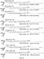
- FIG. 2schematically illustrates examples of capture agents that are bound to double-stranded oligonucleotides that have different overhangs. Such different overhangs represent a strategy to increase signal harvested from a particular capture agent by multiplication of positions in lower strand oligonucleotide complementary to detector base (dU in this case).
- the lower panelalso shows how a different base labeled with a different fluorophore can be used as a FRET excitation pair for the “Detector” base.
- SEQ ID NOS: 1-4SEQ ID NOS: 1-4.
- FIG. 3schematically illustrates several cycles of a multiplexed detection method that relies on reversible dye terminators.
- FIG. 4schematically illustrates several cycles of a multiplexed detection method that relies on leaving out one of the four nucleotides per cycle.
- FIG. 5A-5Dschematically illustrates an exemplary design of oligonucleotide duplexes is for “reversible terminator” and “missing base” multiplexing methods.
- FIG. 6schematically illustrates an exemplary design of oligonucleotide duplexes for a strategy that allows one to reduce the length of the lower strand oligonucleotide, creating an overhang in the case of highly multiplexed capture agent panels.
- FIG. 7schematically illustrates an example of a detection method that relies on removing a quencher from a labeled oligonucleotide by nick translation.
- SEQ ID NOS: 31-35SEQ ID NOS: 31-35.
- FIG. 8schematically illustrates a multiplexed detection method that relies on removing quenchers from labeled oligonucleotides.
- Step 1SEQ ID NOS 36-44
- Step 2SEQ ID NOS: 45-52
- Step 3SEQ ID NOS: 53-60
- Step 4SEQ ID NOS: 61-67.
- FIGS. 9A and 9Bschematically illustrate an embodiment that relies on cyclical re-annealing of polymerase priming nucleotides and a variant of the same approach that utilizes FRET.
- SEQ ID NOS: 68-80schematically illustrate an embodiment that relies on cyclical re-annealing of polymerase priming nucleotides and a variant of the same approach that utilizes FRET.
- FIG. 10schematically illustrate an embodiment that relies on cyclical re-annealing of polymerase priming nucleotides and a variant of the same approach that utilizes FRET.
- FIGS. 11A-11Cshows an anti-CD4 antibody linked to oligonucleotide duplex designed for rendering staining by primer extension (panel A) and data obtained from labeled population of spleen cells in suspension in the absence of polymerase (panel B) and in the presence of polymerase (panel C).
- panel Aprimer extension
- panel Bdata obtained from labeled population of spleen cells in suspension in the absence of polymerase
- panel Cpanel C
- SEQ ID NOS: 87 and 88SEQ ID NOS: 87 and 88.
- FIGS. 12A-12Dshows data obtained from labeling by primer extension a population of spleen cells preattached on the slide. Cells were co-stained with “regular” TCRb-FITC antibody and CD4 antibody linked to oligonucleotide duplex designed for rendering staining by primer extension.
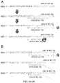
- FIGS. 13A-13Dshow schematic illustration of two capture agents CD4 and CD8 linked to oligonucleotide duplexes (panel A) and data obtained from a multiplexed method whereby staining by this capture agents was sequentially detected on spleen cells smeared on a slide using a “reversible terminator” method (panels C-D).
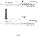
- FIG. 14shows a chematic diagram of an experiment testing multiplexed staining by “missing base” approach.
- Mouse spleen sampleswere barcoded by pan-leukocytic CD45 antibody conjugated to per sample specific oligonucleotide duplexes. Samples were mixed after staining and mixture was resolved by sequential rendering of CD45-oligonucleotide variants.
- FIG. 15is 12 panels of images showing the first 6 cycles of rendering the 30 populations barcoded by CD45 (as per scheme on FIG. 14 ). Two populations were co-detected per cycle of rendering. In each cycle control image was acquired after fluorescence inactivation.
- nucleic acidsare written left to right in 5′ to 3′ orientation; amino acid sequences are written left to right in amino to carboxy orientation, respectively.
- biological feature of interestrefers to any part of a cell that can be indicated by binding to a capture agent.
- exemplary biological features of interestinclude cell walls, nuclei, cytoplasm, membrane, keratin, muscle fibers, collagen, bone, proteins, nucleic acid (e.g., mRNA or genomic DNA, etc). fat, etc.
- a biological feature of interestcan also be indicated by immunohistological methods, e.g., a capture agent that is linked to an oligonucleotide. In these embodiments, the capture agent binds to an site, e.g., a protein epitope, in the sample.
- Exemplary epitopesinclude, but are not limited to carcinoembryonic antigen (for identification of adenocarcinomas, cytokeratins (for identification of carcinomas but may also be expressed in some sarcomas) CD15 and CD30 (for Hodgkin's disease), alpha fetoprotein (for yolk sac tumors and hepatocellular carcinoma), CD117 (for gastrointestinal stromal tumors), CD10 (for renal cell carcinoma and acute lymphoblastic leukemia), prostate specific antigen (for prostate cancer), estrogens and progesterone (for tumour identification), CD20 (for identification of B-cell lymphomas), CD3 (for identification of T-cell lymphomas).
- Complementary nucleic acid moleculese.g., DNA and/or RNA
- in the sampleprovide binding complementary sites for oligonucleotide probes.
- multiplexingrefers to using more than one label for the simultaneous or sequential detection and measurement of biologically active material.
- antibodyand “immunoglobulin” are used interchangeably herein and are well understood by those in the field. Those terms refer to a protein consisting of one or more polypeptides that specifically binds an antigen.
- One form of antibodyconstitutes the basic structural unit of an antibody. This form is a tetramer and consists of two identical pairs of antibody chains, each pair having one light and one heavy chain. In each pair, the light and heavy chain variable regions are together responsible for binding to an antigen, and the constant regions are responsible for the antibody effector functions.
- the recognized immunoglobulin polypeptidesinclude the kappa and lambda light chains and the alpha, gamma (IgG 1 , IgG 2 , IgG 3 , IgG 4 ), delta, epsilon and mu heavy chains or equivalents in other species.
- Full-length immunoglobulin “light chains”(of about 25 kDa or about 214 amino acids) comprise a variable region of about 110 amino acids at the NH 2 -terminus and a kappa or lambda constant region at the COOH-terminus.
- Full-length immunoglobulin “heavy chains”(of about 50 kDa or about 446 amino acids), similarly comprise a variable region (of about 116 amino acids) and one of the aforementioned heavy chain constant regions, e.g., gamma (of about 330 amino acids).
- antibodies and immunoglobulininclude antibodies or immunoglobulins of any isotype, fragments of antibodies which retain specific binding to antigen, including, but not limited to, Fab, Fv, scFv, and Fd fragments, chimeric antibodies, humanized antibodies, minibodies, single-chain antibodies, and fusion proteins comprising an antigen-binding portion of an antibody and a non-antibody protein. Also encompassed by the term are Fab′, Fv, F(ab′) 2 , and or other antibody fragments that retain specific binding to antigen, and monoclonal antibodies.
- Antibodiesmay exist in a variety of other forms including, for example, Fv, Fab, and (Fab′) 2 , as well as bi-functional (i.e. bi-specific) hybrid antibodies (e.g., Lanzavecchia et al., Eur. J. Immunol. 17, 105 (1987)) and in single chains (e. g., Huston et al., Proc. Natl. Acad. Sci. U.S.A., 85, 5879-5883 (1988) and Bird et al., Science, 242, 423-426 (1988), which are incorporated herein by reference).
- Hood et al.“Immunology”, Benjamin, N.Y., 2nd ed. (1984), and Hunkapiller and Hood, Nature, 323, 15-16 (1986),).
- bindingrefers to the ability of a binding reagent to preferentially bind to a particular analyte that is present in a homogeneous mixture of different analytes. In certain embodiments, a specific binding interaction will discriminate between desirable and undesirable analytes in a sample, in some embodiments more than about 10 to 100-fold or more (e.g., more than about 1000- or 10,000-fold).
- the affinity between a binding reagent and analyte when they are specifically bound in a capture agent/analyte complexis characterized by a K D (dissociation constant) of less than 10 ⁇ 6 M, less than 10 ⁇ 7 M, less than 10 ⁇ 8 M, less than 10 ⁇ 9 M, less than 10 ⁇ 9 M, less than 10 ⁇ 11 M, or less than about 10 ⁇ 12 M or less.
- a “plurality”contains at least 2 members. In certain cases, a plurality may have at least 2, at least 5, at least 10, at least 100, at least 1000, at least 10,000, at least 100,000, at least 10 6 , at least 10 7 , at least 10 8 or at least 10 9 or more members.
- labelingrefers to attaching a detectable fluorophore to specific sites in a sample (e.g., sites containing an epitope for the antibody being used, for example) such that the presence and/or abundance of the sites can be determined by evaluating the presence and/or abundance of the label.
- planar samplerefers to a substantially planar, i.e., two dimensional, material that contains cells.
- a planar cellular samplecan be made by, e.g., growing cells on a planar surface, depositing cells on a planar surface, e.g., by centrifugation, or by cutting a three dimensional object that contains cells into sections and mounting the sections onto a planar surface, i.e., producing a tissue section.
- the cellsmay be fixed using any number of reagents including formalin, methanol, paraformaldehyde, methanol:acetic acid etc.
- tissue sectionrefers to a piece of tissue that has been obtained from a subject, fixed, sectioned, and mounted on a planar surface, e.g., a microscope slide.
- FFPE tissue sectionrefers to a piece of tissue, e.g., a biopsy that has been obtained from a subject, fixed in formaldehyde (e.g., 3%-5% formaldehyde in phosphate buffered saline) or Bouin solution, embedded in wax, cut into thin sections, and then mounted on a microscope slide.
- formaldehydee.g., 3%-5% formaldehyde in phosphate buffered saline
- Bouin solutionembedded in wax
- spatialally-addressable measurementsrefers to a set of values that are each associated with a specific position on a surface. Spatially-addressable measurements can be mapped to a position in a sample and can be used to reconstruct an image of the sample.
- a “diagnostic marker”is a specific biochemical in the body which has a particular molecular feature that makes it useful for detecting a disease, measuring the progress of disease or the effects of treatment, or for measuring a process of interest.
- a “pathoindicative” cellis a cell which, when present in a tissue, indicates that the animal in which the tissue is located (or from which the tissue was obtained) is afflicted with a disease or disorder.
- the presence of one or more breast cells in a lung tissue of an animalis an indication that the animal is afflicted with metastatic breast cancer.
- complementary siteis used to refer to an epitope for an antibody or aptamer, or a nucleic acid molecule if the capture agent is an oligonucleotide probe. Specifically, if the capture agent is an antibody, then the complementary site for the capture agent is the epitope in the sample to which the antibody binds. If the capture agent is an oligonucleotide probe, then the complementary site for the capture agent is a complementary sequence in a DNA or RNA molecule in the sample.
- epitopeas used herein is defined as small chemical groups on the antigen molecule that is bound to by an antibody.
- An antigencan have one or more epitopes. In many cases, an epitope is roughly five amino acids or sugars in size.
- an epitopeis roughly five amino acids or sugars in size.
- One skilled in the artunderstands that generally the overall three-dimensional structure or the specific linear sequence of the molecule can be the main criterion of antigenic specificity.
- a “subject” of diagnosis or treatmentis a plant or animal, including a human.
- Non-human animals subject to diagnosis or treatmentinclude, for example, livestock and pets.
- the term “incubating”refers to maintaining a planar sample and capture agent under conditions (which conditions include a period of time, a temperature, an appropriate binding buffer and a wash) that are suitable for specific binding of the capture agent to molecules (e.g., epitopes or complementary nucleic acid) in the planar sample.
- capture agentrefers to an agent that can specifically bind to complementary sites in a planar sample.
- exemplary capture agentsinclude, e.g., an antibody, an aptamer, and an oligonucleotide probe (which may be DNA or RNA) that hybridizes to a binding site.
- the term “capture agent that is linked to a double stranded oligonucleotide”refers to a capture agent, e.g., an antibody or an oligonucleotide probe, that is non-covalently (e.g., via a streptavidin/biotin interaction) or covalently (e.g., via a click reaction or the like) linked to an oligonucleotide (which may be composed of two single-stranded oligonucleotide strands that are hybridized together) in a way that the capture agent can still bind to its binding site and the 3′ end of one of the oligonucleotides is accessible to a polymerase.
- the oligonucleotide and the capture agentmay be linked via a number of different methods, including those that use maleimide or halogen-containing group, which are cysteine-reactive.
- oligonucleotiderefers to a multimer of at least 10, e.g., at least 15 or at least 30 nucleotides. In some embodiments, an oligonucleotide may be in the range of 15-200 nucleotides in length.
- readingin the context of reading a fluorescent signal, refers to obtaining an image by scanning or by microscopy, where the image shows the pattern of fluorescence as well as the intensity of fluorescence in a field of view.
- primeris an oligonucleotide, either natural or synthetic, that is capable, upon forming a duplex with a polynucleotide template, of acting as a point of initiation of nucleic acid synthesis and being extended from its 3′ end along the template so that an extended duplex is formed.
- the sequence of nucleotides added during the extension processis determined by the sequence of the template polynucleotide.
- primersare extended by a DNA polymerase.
- a primermay be at least 10, e.g., at least 15 or at least 30 nucleotides in length.
- single nucleotide 5′ overhangrefers to a 5′ overhang, where the overhang is a single nucleotide in length.
- a “two nucleotide 5′ overhang”is a 5′ overhang, where the overhang is two nucleotides in length. The 3′ end is recessed in a 5′ overhang.
- the various nucleotides of an overhangmay be referred to by their position, e.g., “first position” and “second position”.
- the “position”is relative to the recessed 3′ end.
- the “first” position of the overhangis immediately adjacent to the recessed 3′ end and the “second” position of the overhang is immediately adjacent to the first position.
- the complementary strands of a double stranded oligonucleotidemay be referred to herein as being the “first” and “second” or the “top” and “bottom” strands.
- the assignment of a strand as being a “top” or “bottom” strandis arbitrary and does not imply any particular orientation, function or structure.
- fluorescently labeled oligonucleotide comprising a quencherrefers to an oligonucleotide that contains a fluorophore and a quencher, wherein the quencher quenches the fluorophore in the same oligonucleotide.
- the term “different” in the context of different 5′ overhangs that are differentrefers to overhangs that have a different sequence. Overhangs of different lengths (e.g., GATC vs GAT) implicitly have a different sequence, even through one sequence may be encompassed by the other.
- the term “adding to an overhang”, in the context of adding one or more nucleotides to an overhang,refers to adding nucleotides to the recessed 3′ end of a 5′ overhang using the overhang as a template.
- alternating stretchesrefers to two nucleotides stretches, where one “stretch” is a contiguous sequence of, e.g., up to 10, of the same nucleotide (e.g., a G, A, T or C), and the second stretch is contiguous sequence of, e.g., up to 10, of a different nucleotide, that alternate with one another, i.e., one stretch (e.g., a string of T's) occupies the odd positions and the other stretch (e.g., a string of A's) occupies the even positions.
- one stretche.g., a string of T's
- the other stretche.g., a string of A's
- incomplete nucleotide mixcomprises a nucleotide mix that contains one, two or three nucleotides (but not all four nucleotides) selected from G, A, T and C.
- the nucleotidesmay be labeled or unlabeled.
- reversible terminatorrefers to a chemically modified nucleotide base that when incorporated into growing DNA strand by DNA polymerase blocks further incorporation of bases. Such “reversible terminator” base and DNA strand can be deprotected by chemical treatment and following such deprotection DNA strand can be further extended by DNA polymerase.
- fluorescently labeled reversible terminatorrefers to a “reversible terminator” base which is labeled by fluorophore through linker cleavable by same treatment which is used to deprotect the DNA strand which ends with this base. Deprotecting the “fluorescently labeled reversible terminator” simultaneously activates the DNA strand for further extension and removes the fluorescent label from it.
- the methodcomprises labeling a planar sample (e.g., an FFPE section mounted on a planar surface such as a microscope slide) with a capture agent that specifically binds to complementary sites in the planar sample. This step is done under conditions by which the capture agent binds to complementary sites in the planar sample, methods for which are well known.
- a planar samplee.g., an FFPE section mounted on a planar surface such as a microscope slide
- the capture agentis linked to a double-stranded oligonucleotide that comprises a first strand and a second strand (e.g., two oligonucleotide that are hybridized together) and the capture agent is linked (covalently or non-covalently via a biotin) to the double-stranded oligonucleotide by the 5′ end of the first strand, and the 3′ end of the first strand is recessed relative to the 5′ end of the second strand, thereby defining an overhang.
- the capture agentis cross-linked the planar sample. This crosslinking step may be done using any amine-to-amine crosslinker (e.g.
- the methodcomprises reading a fluorescent signal generated by addition of a nucleotide in the overhang. This step may be done by contacting the planar sample with a polymerase and a nucleotide mix, thereby adding one or more nucleotides to the overhang; and reading a fluorescent signal generated by addition of the one or more nucleotides to the overhang.
- the fluorescent signalmay be generated by a variety of different methods.
- the flouorescent signalmay be fluorescence from a fluorescent nucleotide added to the end of the primer, or a FRET (fluorescence resonance energy transfer) signal resulting from the same.
- the signalmay generated by removing a quencher from a fluorescently labeled oligonucleotide that is also hybridized to the oligonucleotide.
- the reading stepmay be followed by inactivating the fluorescence after reading so that other binding events can be detected and read.
- the fluorescencemay be inactivated by peroxide-based bleaching, cleavage of fluorophore linked to nucleotide through cleavable linker (e.g. using TCEP as a cleaving reagent), base-exchange by exo+ polymerase such as Vent, or subsequent incorporation of quencher, for example.
- the methodmay be multiplexed in a way that a single planar sample can be interrogated by a plurality of different capture agents, where each antibody is linked to different oligonucleotides (i.e., oligonucleotides of different sequence).
- the planar samplemay be labeled using at least 5, at least 10, at least 20, at least 30, at least 50, or at least 100, up to 150 or more capture agents that are each linked to a different oligonucleotide, and binding of the capture agents can be separately read using a fluorescence microscope equipped with an appropriate filter for each fluorophore, or by using dual or triple band-pass filter sets to observe multiple fluorophores. See, e.g., U.S. Pat. No. 5,776,688.
- a capture agent used in the methodmay be linked to a double-stranded oligonucleotide that contains a 5′ overhang (i.e., a recessed 3′ end that can be extended by a polymerase).
- a capture agentis shown in FIGS. 1 and 2 .
- the overhangis a single nucleotide overhang (e.g., an A), although a longer overhang (e.g., at least 2, at least 3, at least 4, at least 5, at least 6, at least 8, at least 10, at least 20, or at least at least 30, may be useful for other applications (e.g., multiplexed applications).
- a longer overhange.g., at least 2, at least 3, at least 4, at least 5, at least 6, at least 8, at least 10, at least 20, or at least at least 30, may be useful for other applications (e.g., multiplexed applications).
- the overhangmay contain a repeated sequence, e.g., 2, 3, 4, 5, or 6 or more repeats of the same sequence of 2, 3, 4, 5 or 6 nucleotides, thereby allowing the capture agent to be used in multiplexed applications as described below.
- the double stranded oligonucleotidemay have a recessed 3′ end at the other end of the oligonucleotide (i.e., at the end closest to the capture agent). However, this end is not extendible.
- the double-stranded oligonucleotidemay contain one or more third oligonucleotides that are hybridized to the overhang.
- the plurality of capture agentsmay be distinguished by the sequence of the overhang and not by the sequence of the first strand of the double stranded oligonucleotide.
- the second strand of the double stranded oligonucleotidesis different for each of the capture agents.
- the fluorophore usedmay be a coumarin, a cyanine, a benzofuran, a quinoline, a quinazolinone, an indole, a benzazole, a borapolyazaindacene and or a xanthene including fluorescein, rhodamine and rhodol.
- fluorophoresmay be chosen so that they are distinguishable, i.e., independently detectable, from one another, meaning that the labels can be independently detected and measured, even when the labels are mixed.
- the amounts of label present (e.g., the amount of fluorescence) for each of the labelsare separately determinable, even when the labels are co-located (e.g., in the same tube or in the same area of the section).
- fluorescent dyes of interestinclude: xanthene dyes, e.g., fluorescein and rhodamine dyes, such as fluorescein isothiocyanate (FITC), 6-carboxyfluorescein (commonly known by the abbreviations FAM and F), 6-carboxy-2′,4′,7′,4,7-hexachlorofluorescein (HEX), 6-carboxy-4′, 5′-dichloro-2′, 7′-dimethoxyfluorescein (JOE or J), N,N,N′,N′-tetramethyl-6-carboxyrhodamine (TAMRA or T), 6-carboxy-X-rhodamine (ROX or R), 5-carboxyrhodamine-6G (R6G 5 or G 5 ), 6-carboxyrhodamine-6G (R6G 6 or G 6 ), and rhodamine 110; cyanine dyes, e.g., Cy3, Cy5
- phenanthridine dyese.g., Texas Red
- ethidium dyese.g., acridine dyes
- carbazole dyese.g., phenoxazine dyes
- porphyrin dyese.g., polymethine dyes, e.g., cyanine dyes such as Cy3, Cy5, etc.
- BODIPY dyes and quinoline dyese.g., BODIPY dyes and quinoline dyes.
- fluorophores of interestthat are commonly used in subject applications include: Pyrene, Coumarin, Diethylaminocoumarin, FAM, Fluorescein Chlorotriazinyl, Fluorescein, R110, Eosin, JOE, R6G, Tetramethylrhodamine, TAMRA, Lissamine, Napthofluorescein, Texas Red, Cy3, and Cy5, etc.
- Suitable distinguishable fluorescent label pairs useful in the subject methodsinclude Cy-3 and Cy-5 (Amersham Inc., Piscataway, N.J.), Quasar 570 and Quasar 670 (Biosearch Technology, Novato Calif.), Alexafluor555 and Alexafluor647 (Molecular Probes, Eugene, Oreg.), BODIPY V-1002 and BODIPY V1005 (Molecular Probes, Eugene, Oreg.), POPO-3 and TOTO-3 (Molecular Probes, Eugene, Oreg.), and POPRO3 and TOPRO3 (Molecular Probes, Eugene, Oreg.). Further suitable distinguishable detectable labels may be found in Kricka et al. (Ann Clin Biochem. 39:114-29, 2002), Ried et al. (Proc. Natl. Acad. Sci. 1992: 89: 1388-1392) and Tanke et al. (Eur. J. Hum. Genet. 1999 7:2-11) and others.
- the fluorescent signalmay be produced by a fluorescent nucleotide that is added to the 3′ end of the primer.
- This methodmay comprise reading a signal from the added fluorescent nucleotide, or reading a FRET signal generated by energy transfer between two fluorescent nucleotides that are added to the primer.
- FIGS. 1 and 2shows how an antibody can be linked to a oligonucleotide chemically, or via biotin/streptavidin interactions ( FIG. 1B ) and how a fluorescent signal can be generated by adding a fluorescent nucleotide to the end of the primer ( FIG. 2 ).
- the antigenis stained by an antibody that is coupled to a DNA dimer with an overhanging 5′ end (lower strand) and recessed 3′ end (upper strand) either chemically ( FIG. 1 top panel) or through streptavidin ( FIG. 1 bottom and middle panels).
- the pattern of binding of the capture agentmay be determined using an on-slide end fill-in reaction by using a suitable polymerase (e.g., by exo ⁇ Klenow, Bst, Taq, Klentaq, or an exo ⁇ Klenow-Vent mixture) and fluorescently labeled nucleotide ( FIG. 1 and FIG. 2 top panel).
- a suitable polymerasee.g., by exo ⁇ Klenow, Bst, Taq, Klentaq, or an exo ⁇ Klenow-Vent mixture
- fluorescently labeled nucleotideFIG. 1 and FIG. 2 top panel
- the signal-to-noise ratiocan be increased by: a) multimerization of position complementary to labeling nucleotide ( FIG. 2 , middle panel); or b) by generating a FRET between two nucleotides are incorporated, whereby the emission wavelength of one of the nucleotides ( FIG. 2 , bottom panel C on the figure) serves as an excitation wavelength for another ( FIG. 2 , bottom panel U on the figure).
- Fluorescencemay be inactivated before addition of subsequent staining reagents by any convenient method including, but not limited to photobleaching, peroxide-based bleaching, inactivation by ozone, cleavage of fluorophore linked to nucleotide through cleavable linker (e.g. using TCEP as a cleaving reagent), base-exchange by exo+ polymerase such as Vent, subsequent incorporation of quencher.
- any convenient methodincluding, but not limited to photobleaching, peroxide-based bleaching, inactivation by ozone, cleavage of fluorophore linked to nucleotide through cleavable linker (e.g. using TCEP as a cleaving reagent), base-exchange by exo+ polymerase such as Vent, subsequent incorporation of quencher.
- the methodcan be repeated, i.e., the planar sample may be re-stained using a different antibody and fluorescence can be read.
- oligonucleotidescan be implemented using specially designed oligonucleotides using two different approaches, referred to as the “reversible terminator” and “missing base” approaches, which are described in greater detail below. Both of these methods rely on a composition comprising a plurality of (e.g., at least 5, at least 10, at least 20, at least 30, at least 50, or at least 100, up to 150 or more) capture agents that recognize different complementary sites, wherein: each of the capture agents is linked to a double-stranded oligonucleotide that comprises a first strand and a second strand; the capture agents are linked to a double-stranded oligonucleotide by the 5′ end of first strand; the 3′ end of the first strand in each of the double-stranded oligonucleotides is recessed relative to the 5′ end of the second strand, thereby producing an overhang; and the overhang is different for each of the capture agents.
- a compositioncomprising a
- FIG. 3shows a population of capture agents that have an overhang defined by the formula: 3′-N 4n N 1 /N 2 /N 3 -5′ followed by short stretch of random composition on the 5′ end to increase the overall polymerase residence on the DNA duplex, where N 1 , N 2 , N 3 and N 4 are different nucleotides selected from G, A, T and C and n is 0, 1 or more.
- N 1 , N 2 , N 3 and N 4are different nucleotides selected from G, A, T and C and n is 0, 1 or more.
- Yis a nucleotide sequence of length n (n is 0, 1 or more) composed of bases N 3 and N 4 , wherein nucleotide N 3 is in odd positions and nucleotide N 4 is in even positions, counting from the start of the overhang and N 1 , N 2 , N 3 and N 4 are different nucleotides selected from G, A, T and C.
- the sequence of the first strandis the same for each of the capture agents; and the sequence of the second strand is different for each of the capture agents.
- the different second strandsmake the overhangs different between the different capture agents.
- the multiplex methodsgenerally comprise: (a) incubating a planar sample with an above-described antibody composition under conditions by which the capture agents bind to complementary sites in the planar sample; (b) cross-linking the capture agents to the planar sample; (c) contacting the planar sample with a polymerase and either an incomplete nucleotide mix of labeled and unlabeled bases or a nucleotide mix where some or all bases are fluorescent and some or all bases constitute reversible terminator nucleotides or fluorescent reversible terminator nucleotides; and (d) reading, using fluorescence microscopy, a fluorescent signal generated by addition a nucleotide to some but not all of the capture agents.
- Step (c) of this methodmay comprise (c) contacting the planar sample with a polymerase and:
- nucleotide mixthat comprises fluorescent nucleotides that are complementary to N 1 , N 2 and N 3 and a reversible terminator nucleotide that is complementary to N 4 or (ii) a nucleotide mix that comprises fluorescent reversible terminator nucleotides that are complementary to N 1 , N 2 and N 3 and a reversible terminator nucleotide that is complementary to N 4 or (iii) a nucleotide mix that comprises fluorescent nucleotides that are complementary to N 1 , and N 2 , an unlabeled nucleotide that is complementary to N 3 , and no nucleotide that is complementary to N 4 , thereby adding fluorescent nucleotides onto the double-stranded oligonucleotides of some but not all of the capture agents thereby adding fluorescent nucleotides onto the double-stranded oligonucleotides of some but not all of the capture agents; and (d) reading, using fluor fluor flu
- the length of the read over the oligonucleotide overhangsmay increase accordingly. This may or may not reduce the efficiency of staining due to accumulation of primer extension errors along the length of the oligonucleotide duplex.
- the plurality of capture agentscan be divided in sets such that number of capture agents in the set does exceed the capacity of the multiplexing protocol to render staining without significant signal loss (e.g. 30).
- Each such set of capture agentswill be conjugated to “terminated” (the last 3′ base is dideoxy- or propyl-modified) upper strand oligonucleotide of the same sequence as in the original version of the “missing base” approach.
- the lower strand oligoswill incorporate an additional set-specific region which will serve as a landing spot for an additional primer which is to be on-slide hybridized to the particular subset of the total plurality of the antibodies at the time when they are to be rendered. This approach allows not to extend the reads beyond certain threshold and at the same time have an unlimited potential number of capture agents in the sample.
- This implementation of the methodrelies on reversible terminators, i.e., chain terminator nucleotides that can be de-protected after incorporation, thereby allowing further nucleotides to be added to that nucleotide.
- This methodcan be implemented using a composition comprising a plurality of capture agents that are linked to double stranded oligonucleotides, as illustrated in FIG. 3 .
- the top strand of the double stranded oligonucleotideis linked to the capture agent and is same for each antibody, and the sequence of the bottom strand varies between capture agents. As shown on FIG. 3
- the 5′ end of the lower strand of the double-stranded oligonucleotide(which forms the overhang) is of the general 3′-N 4 —N 1 /N 2 /N 3 -5′ followed by short stretch of random nucleotides on the 5′ end to increase the overall polymerase residence on the DNA duplex, where N 1 , N 2 , N 3 and N 4 are different nucleotides selected from G, A, T and C and n is 0, 1 or more. As shown on FIG.
- 5Ba more general formula of lower oligonucleotide overhang 3′-XN 1 /N 2 /N 3 -5′, where N 1 , N 2 , N 3 are different nucleotides selected from G, A, T and C and X is a nucleotide stretch of bases Xi (such that Xi are different nucleotides selected from G, A, T and C) of random composition and length is also applicable in this method.
- this methodmay comprise: (a) incubating a planar sample with a multiplex antibody composition in which the overhangs are of the formula 5′-N 1 /N 2 /N 3 N 4n , wherein N 1 , N 2 , N 3 and N 4 are different nucleotides selected from G, A, T and C and n is 1 or more; under conditions by which the capture agents specifically bind to complementary sites in the planar sample; (b) cross-linking the capture agent to the planar sample; (c) contacting the planar sample with a polymerase and a nucleotide mix that comprises fluorescent nucleotides that are complementary to N 1 , N 2 and N 3 and a reversible terminator nucleotide that is complementary to N 4 ; and (d) reading, using fluorescence microscopy, a fluorescent signal generated by addition of a nucleotide to some but not all of the capture agents.
- This cyclemay be repeated by (e) inactivating the fluorescent signal, deprotecting the reversible terminator nucleotide and (f) blocking the planar sampe; and repeating steps (c) and (d).
- the methodmay comprise repeating steps (c), (d) (e) and (f) multiple times.
- the reagent used for blockingmay vary depending on the chemistry used.
- the samplemay be blocked with a thiol-reactive compounds such as cysteine, glutathione or iodoacetamide.
- this methodcan be implemented using a composition comprising: a first antibody linked to a first double stranded oligonucleotide, wherein the first double stranded oligonucleotide comprises a single nucleotide 5′ overhang comprising base N 1 ; a second antibody linked to a second double stranded oligonucleotide, wherein the second double stranded oligonucleotide comprises a single nucleotide 5′ overhang comprising base N 2 ; a third antibody linked to a third double stranded oligonucleotide, wherein the third double stranded oligonucleotide comprises a single nucleotide 5′ overhang comprising base N 3 ; a fourth antibody linked to a fourth double stranded oligonucleotide comprises a two nucleotide 5′ overhang, wherein the first position of the overhang comprises base N 4 and the second position of the overhang is base N
- the compositionmay also contain a seventh antibody linked to a seventh double stranded oligonucleotide, wherein the seventh double stranded oligonucleotide comprises a multiple nucleotide 5′ overhang, wherein the first position of the overhang comprises base N 4 , the second position of the overhang is base N 4 and third is selected from N 1 , N 2 , and N 3 .
- the planar samplecan be co-stained simultaneously using a panel of capture agents, each labeled with one oligonucleotide duplex designed according to the strategy outlined on FIG. 3 .
- the duplexesare designed in such a way that each antibody has the same upper strand sequence linked, covalently or through streptavidin, to an antibody through the 5′ end.
- the lower strandchanges from antibody to antibody.
- the general formula for the lower strandis 3′-dideoxydC-sequence-complimentary-to-upper-strand G n A/T/C-5′.
- nucleotide Gis reserved for step-wise progression and its complementary pair on the upper strand is never used in labeled form.
- the other three basesare complementary to labeled nucleotides and can be used to identify three capture agents per cycle.
- the general formula for the lower strandis 3′-dideoxydC-sequence-complimentary-to-upper-strand-X—N 1 /N 2 /N 3 -5′ where X i of X is any nucleotide excluding one reserved for “walking base” of this particular cycle and X is any base as shown on FIG. 5B .
- Each cycleincludes: (a) a labeling step in which the three capture agents are labeled and duplexes on the rest are extended one base at a time, (b) an imaging step and (c) a destaining/deprotection step.
- the added fluorescent labels from the previous cycleare inactivated by any of the suitable methods, including but not limited to: cleavage of fluorophore off the nucleotide (if the labeled nucleotide is linked to the fluorophore through a cleavable linker); peroxide based bleaching; photobleaching; chemically-assisted photobleaching; labeled base replacement by exo+ polymerase, etc.
- the unlabeled “extension” nucleotide that has been added to the remainder of the capture agentsis activated by cleavage of the protective group off its 3′ end.
- This implementation of the methodrelies on a “missing” base design in which, in each cycle, two labeled and one unlabeled nucleotides are added to the reaction, and the “missing base” prevents the primers from being extended by more than a single nucleotide.
- This methodcan be implemented using a composition comprising a plurality of capture agents that are linked to double stranded oligonucleotides, as illustrated in FIG. 4 .
- the top strand of the double stranded oligonucleotideis linked to the capture agent and is same for each antibody, and the sequence of the bottom strand varies between capture agents. As shown in FIG. 4
- the 5′ end of the lower strand of the double-stranded oligonucleotide(which forms the overhang) is of the general formula 3′-YN 1 /N 2 -5′, optionally followed by short stretch (e.g., 1-5 residues) of random nucleotides on the 5′ end to increase the overall polymerase residence on the DNA duplex, wherein Y is a nucleotide sequence of length n (n is 0, 1 or more) composed of bases N 3 and N 4 , wherein nucleotide N 3 is in odd positions and nucleotide N 4 is in even positions, counting from the start of the overhang and N 1 , N 2 , N 3 and N 4 are different nucleotides selected from G, A, T and C.
- N 1 , N 2 , N 3 and N 4are different nucleotides selected from G, A, T and C and Y is a nucleotide sequence of length n (n is 0, 1 or more) composed of alternating random length stretches of bases N 3 and N 4 such that the order number of N 3 —stretches is odd and of N 4 stretches is even, may be applicable in this method
- this methodmay comprise: (a) incubating a planar sample with a multiplex antibody composition in which the overhangs are of the formula (3′-YN 1 /N 2 -5′) described in the prior paragraph; under conditions by which the capture agents specifically bind complementary sites in the planar sample; (b) cross-linking the capture agent to the planar sample; (c) contacting the planar sample with a polymerase and a nucleotide mix that comprises fluorescent nucleotides that are complementary to N 1 , and N 2 , an unlabeled nucleotide that is complementary to N 3 and no nucleotide that is complementary to N 4 ; and (d) reading, using fluorescence microscopy, a fluorescent signal generated by addition of a nucleotide to some but not all of the capture agents.
- This cyclemay be repeated by (e) inactivating the fluorescent signal, (f) blocking the sample and contacting the planar sample with a polymerase and an unlabeled nucleotide that is complementary to N 4 ; and repeating steps (c) (d).
- the methodmay comprise repeating steps (c), (d), (e) and (f) multiple times.
- This methodcan be implemented using a capture agent composition that comprises: a first antibody linked to a first double stranded oligonucleotide, wherein the first double stranded oligonucleotide comprises a single nucleotide 5′ overhang comprising base N 1 ; a second antibody linked to a second double stranded oligonucleotide, wherein the second double stranded oligonucleotide comprises a single nucleotide 5′ overhang comprising base N 2 ; a third antibody linked to a fourth double stranded oligonucleotide, wherein the third double stranded oligonucleotide comprises a two nucleotide 5′ overhang, wherein the first from the 3′ position of the overhang comprises base N 4 and the second position comprises N 1 ; and a fourth antibody linked to a fourth double stranded oligonucleotide, wherein the fourth double stranded oligonucleot
- the compositionmay also contain a fifth antibody linked to a fifth double stranded oligonucleotide, wherein the fifth double stranded oligonucleotide comprises a multiple nucleotide 5′ overhang, wherein the first position of the overhang comprises base N 4 , the second position comprises base N 3 , and the third position comprises N 1 or N 2 .
- the missing base approachdoes not use reversible terminators. Instead, extension of a single nucleotide is ensured by using two interchanging bases (e.g., T and C as shown in FIG. 4 instead of the corresponding G in the “reversible terminators” approach) and adding only one of the two dNTPs at a time in the primer extension reaction. After the incorporation of the first nucleotide, the absence of the second dNTP causes strand elongation to stall, thereby ensuring that the primers are extended by only a single nucleotide. As in the previous strategy, all complementary sites can be co-stained simultaneously using capture agents, each labeled with a specific oligonucleotide duplex.
- two interchanging basese.g., T and C as shown in FIG. 4 instead of the corresponding G in the “reversible terminators” approach
- the duplexescan be designed using the strategy shown in FIG. 4 , i.e., in such a way that each antibody has the same upper stand oligonucleotide sequence linked to it via covalent bond or through a streptavidin-biotin interaction.
- the lower strandchanges from antibody to antibody.
- the general formula for the lower strandis 3′ ddC-sequence-complimentary-to-upper-strand -YA/N 2 -5′ where Y is composed of bases T and C such that T can be found only in even and C only at odd positions.
- N 1 , N 2 , N 3 and N 4are different nucleotides selected from G, A, T and C and Y is a nucleotide sequence of length n (n is 0, 1 or more) composed of alternating random length stretches of bases N 3 and N 4 such that the order number of N 3 -stretches is odd and of N 4 stretches is even.
- nis 0, 1 or more
- two base pairs of the lower strand(T and C as in exemplary design on FIG. 4 ) are reserved for step-wise progression and their complementary pair on the upper strand is never labeled.
- the other two basesare complementary to labeled nucleotides and can render the staining with two different capture agents per cycle.
- each cyclecan have three steps: a labeling step in which the two capture agents are labeled by incorporation of fluorescent dNTPs and all of the other duplexes are extended one base at a time, an imaging step, and a de-staining/reactivation step.
- the labeled capture agents from the prior cyclecan be bleached/destained in the same way as described above.
- a quencher labeled nucleotidecan be incorporated after the labeled base. Because, in this embodiment, the position that is labeled is the last position in the overhang, the labeled capture agents from prior cycle cannot be re-labeled in later cycles because all nucleotide positions in the overhang have been filled in.
- the performance of “reversible terminator method” as exemplified in sequential detection of CD4 and CD8 positive T-cells in smears of mouse splenocytesis illustrated in FIG. 13 .
- extension of a primer by nick translationremoves a quencher from a fluorescently labeled “detector” oligonucleotide that is hybridized to the lower strand oligonucleotide in such a way that is positioned downstream from the upper strand primer.
- detectoroligonucleotide that is hybridized to the lower strand oligonucleotide in such a way that is positioned downstream from the upper strand primer.
- the multiplexed implementationsmay comprise: (a) incubating the planar sample with a plurality of capture agents that are linked to a double-stranded oligonucleotide; (b) crosslinking the capture agents to the planar sample;
- the architecture of the double-stranded oligonucleotides linked to the capture agenthas a specific design which is effectively enabling rendering of the capture agent binding pattern by “nick translation”.
- the duplex of the upper strand and the lower strand oligo with long 5′ overhang of the lower strandis further hybridized to a small detector oligonucleotide labeled both by fluorescent and the quencher.
- nick translating polymerasesuch as DNA pol I removes the quencher from some but not all of the quenched fluorescently labeled oligonucleotides, thereby generating a fluorescent signal for some but not all of the capture agents.
- the methodgenerally comprises: (i) labeling a planar sample with: i. a first antibody, wherein the first antibody is linked to a first oligonucleotide duplex comprising, lower strand oligonucleotide with a unique sequence hybridized thereto: (i) an oligonucleotide upper strand “primer” and (ii) a labeled upper strand oligonucleotide comprising a 5′ quencher at a site that is downstream from the primer; and a fluorophore downstream from the quencher and ii.
- a second antibodywherein the second antibody is linked to a second oligonucleotide duplex comprising, lower strand oligonucleotide with unique sequence hybridized thereto: (i) an oligonucleotide upper strand “primer” and (ii) an upper strand oligonucleotide labeled both by fluorophore and a quencher; wherein the gap between the 3′ end of the primer and the 5′ end of the labeled oligonucleotide is different for the first and second oligonucleotides; (ii) incubating the tissue sample with a first nucleotide mix and a polymerase, thereby removing the quencher from only the labeled oligonucleotide that is hybridized to the first oligonucleotide and producing a first fluorescent signal; (iii) reading the first fluorescent signal using fluorescence microscopy; (iv) inactivating the fluorescent signal by further progression of nick-trans
- FIGS. 7 and 8show an example of this method.
- the multiplexing method shown in FIG. 8has the following steps:
- Step 1The planar sample is stained by capture agents that are coupled to a DNA double-stranded oligonucleotide chemically or through streptavidin (as described in FIG. 1 ) such that the top strand of the duplex contains a nick or a single base deletion followed by a nucleotide stretch bordered by a fluorophore and its quencher on two ends (“molecular beacon” or Taqman based design).
- capture agentsthat are coupled to a DNA double-stranded oligonucleotide chemically or through streptavidin (as described in FIG. 1 ) such that the top strand of the duplex contains a nick or a single base deletion followed by a nucleotide stretch bordered by a fluorophore and its quencher on two ends (“molecular beacon” or Taqman based design).
- Step 2Staining pattern is rendered by a nick-translation reaction carried out by any 5′ exo+ polymerase such as DnaPoll Klenow fragment in the presence of a single letter (A as in FIG. 5 for example).
- nick translationremoves the quencher but stops before removing the part of the duplex with the fluorophore.
- Step 3For rendering of other staining reagents, the fluorescence is removed by continuing nick translation in the presence of the letters of the stretch bearing the fluorophore.
- Step 4When multiplexing is desired, multiplexing can be achieved by special design of oligo duplexes attached to detection reagents.
- each antibody set(two or three per cycle) has a gap of an increasing length between the top strand priming and the detector oligonucleotide. This sequence gap on the strand bearing the quencher/fluorophore pair is filled up to final nick in such a way that single base is extended per cycle, similar to how it is achieved in method 1 (see FIG. 8 ).
- the methodcomprises rending antibody staining by primer extension with a fluorophore labeled base or otherwise reading a FRET signal generated by energy transfer between a first fluorescent nucleotide added to the primer by primer extension and a second nucleotide that is present in the oligonucleotide FIG. 10 .
- the principles of this methodare illustrated in FIG. 9A .
- the multiplexingis achieved by removing the extension priming oligo by melting the duplex or by exonuclease and reannealing another primer oligo which is extendable on a different antibody.
- FIG. 9BA multiplexed version of this method is shown in FIG. 9B .
- the multiplexed implementationsmay comprise: (a) incubating the planar sample with a plurality of capture agents; (b) cross-linking the capture agents to the planar sample; (c) extending a primer that is hybridized to the oligonucleotide of a first set of capture agents of the plurality (e.g., wherein the 3′ end of the first primer anneals to only the oligonucleotide of the first population), thereby generating a first set of fluorescent signals; (d) reading the first set of fluorescent signals using fluorescence microscopy; (e) inactivating the fluorescence; (f) extending a primer that is hybridized to the oligonucleotide of a second set of capture agents of the plurality (e.g., wherein the 3′ end of the first primer anneals to only the oligonucleotide of the second population), thereby generating a second set of fluorescent signals; (g) reading the second set of fluorescent signals using fluorescence
- this methodcomprises: (a) incubating the planar sample with (i) a first antibody that is linked to a first labeled oligonucleotide and (ii) a second antibody that is linked to a second labeled oligonucleotide, (b) cross-linking the capture agents to the planar sample; (c) hybridizing the first and second labeled oligonucleotides with a first primer, wherein the 3′ end of the first primer anneals to only the first labeled oligonucleotide; (d) extending the primer with a fluorescent nucleotide; (e) reading, by fluorescence microscopy, a FRET signal generated by energy transfer between the label of the first oligonucleotide and the fluorescent nucleotide added to the first primer; (f) inactivating the fluorescent nucleotide added to the first primer; (g) hybridizing the first and second labeled oligonucleotides with a second primer
- FIGS. 9-10shows an example of this method.
- the method shown in FIGS. 8-11has the following steps:
- Step 1The planar sample is stained using a capture agent that is coupled to a single stranded oligonucleotide.
- the oligonucleotidecould be either unlabeled or labeled by FRET acceptor (e.g. Cy5) fluorophore on the 3′ end.
- Step 2The binding pattern can be determined by an on-slide hybridization of a complementary probe followed a primer extension reaction in which a fluorescently labeled nucleotide fills in the overhang in the extended strand.
- the extended baseis labeled by a FRET donor (e.g. Cy3), which can increase the signal to noise ratio. If the oligonucleotide that is linked to the capture agent is unlabeled, then the fluorescent emission of the nucleotide that has been incorporated by DNA synthesis can be detected directly, without FRET FIG. 9 .
- Step 3The binding pattern of other capture agents can be determined by removing the fluorescence by cleavage of lower strand by exo+ DNA polymerase such as Vent ( FIG. 9 ). Alternatively, the fluorescence can be removed by raising the temperature beyond the melting point of the DNA strands or by one of the de-staining techniques described previously.
- Step 4Multiplexing can be achieved by staining of the sample with a library of capture agents each labeled with specific oligonucleotides and cycling through Steps 1-3, as described above, each time using a different detection oligonucleotide that is complementary to one of the capture agent-conjugated oligonucleotides. Only duplexes where primers are annealed specifically will be properly extended ( FIG. 11 ). In these embodiments, each primers is designed so that its 3′ end hybridizes to only one of the oligonucleotides that are linked to a capture agent.
- the planar samplemay be a section of a tissue biopsy obtained from a patient.
- Biopsies of interestinclude both tumor and non-neoplastic biopsies of skin (melanomas, carcinomas, etc.), soft tissue, bone, breast, colon, liver, kidney, adrenal, gastrointestinal, pancreatic, gall bladder, salivary gland, cervical, ovary, uterus, testis, prostate, lung, thymus, thyroid, parathyroid, pituitary (adenomas, etc.), brain, spinal cord, ocular, nerve, and skeletal muscle, etc.
- capture agentsspecifically bind to biomarkers, including cancer biomarkers, that may be proteinaceous or a nucleic acid.
- cancer biomarkersinclude, but are not limited to carcinoembryonic antigen (for identification of adenocarcinomas), cytokeratins (for identification of carcinomas but may also be expressed in some sarcomas), CD15 and CD30 (for Hodgkin's disease), alpha fetoprotein (for yolk sac tumors and hepatocellular carcinoma), CD117 (for gastrointestinal stromal tumors), CD10 (for renal cell carcinoma and acute lymphoblastic leukemia), prostate specific antigen (for prostate cancer), estrogens and progesterone (for tumour identification), CD20 (for identification of B-cell lymphomas) and CD3 (for identification of T-cell lymphomas).
- carcinoembryonic antigenfor identification of adenocarcinomas
- cytokeratinsfor identification of carcinomas but may also be expressed in some sarcomas
- the above-described methodcan be used to analyze cells from a subject to determine, for example, whether the cell is normal or not or to determine whether the cells are responding to a treatment.
- the methodmay be employed to determine the degree of dysplasia in cancer cells.
- the cellsmay be a sample from a multicellular organism.
- a biological samplemay be isolated from an individual, e.g., from a soft tissue.
- the methodmay be used to distinguish different types of cancer cells in FFPE samples.
- the method described abovefinds particular utility in examining planar samples using a plurality of antibodies, each antibodies recognizing a different marker. Examples of cancers, and biomarkers that can be used to identify those cancers, are shown below. In these embodiments, one does not need to examine all of the markers listed below in order to make a diagnosis.
- Acute Leukemia IHC PanelCD3, CD7, CD20, CD34, CD45, CD56, CD117, MPO, PAX-5, and TdT.
- Adenocarcinomavs. Mesothelioma IHC Pan-CK, CEA, MOC-31, BerEP4, TTF1, calretinin, and WT-1.
- Panel Bladdervs. Prostate Carcinoma IHC Panel CK7, CK20, PSA, CK 903, and p63.
- Burkittvs. DLBC Lymphoma IHC panel BCL-2, c-MYC, Ki-67.
- Carcinoma Unknown Primary SiteFemale CK7, CK20, mammaglobin, ER, TTF1, CEA, CA19-9, S100, (CUPS IHC Panel - Female) synaptophysin, and WT-1.
- Carcinoma Unknown Primary SiteMale CK7, CK20, TTF1, PSA, CEA, CA19-9, S100, and (CUPS IHC Panel - Male) synaptophysin.
- Metastatic HSAHepPar 1
- Carcinoma IHC Panel CEApolyclonal
- the methodmay involve obtaining an image as described above (an electronic form of which may have been forwarded from a remote location) and may be analyzed by a doctor or other medical professional to determine whether a patient has abnormal cells (e.g., cancerous cells) or which type of abnormal cells are present.
- the imagemay be used as a diagnostic to determine whether the subject has a disease or condition, e.g., a cancer.
- the methodmay be used to determine the stage of a cancer, to identify metastasized cells, or to monitor a patient's response to a treatment, for example.
- datacan be forwarded to a “remote location”, where “remote location,” means a location other than the location at which the image is examined.
- a remote locationcould be another location (e.g., office, lab, etc.) in the same city, another location in a different city, another location in a different state, another location in a different country, etc.
- office, lab, etc.another location in the same city
- another location in a different citye.g., another location in a different city
- another location in a different statee.g., another location in a different state
- another location in a different countryetc.
- the two itemscan be in the same room but separated, or at least in different rooms or different buildings, and can be at least one mile, ten miles, or at least one hundred miles apart.
- “Communicating” informationreferences transmitting the data representing that information as electrical signals over a suitable communication channel (e.g., a private or public network).
- “Forwarding” an itemrefers to any means of getting that item from one location to the next, whether by physically transporting that item or otherwise (where that is possible) and includes, at least in the case of data, physically transporting a medium carrying the data or communicating the data. Examples of communicating media include radio or infra-red transmission channels as well as a network connection to another computer or networked device, and the internet or including email transmissions and information recorded on websites and the like.
- the imagemay be analyzed by an MD or other qualified medical professional, and a report based on the results of the analysis of the image may be forwarded to the patient from which the sample was obtained.
- the methodmay be employed in a variety of diagnostic, drug discovery, and research applications that include, but are not limited to, diagnosis or monitoring of a disease or condition (where the image identifies a marker for the disease or condition), discovery of drug targets (where the a marker in the image may be targeted for drug therapy), drug screening (where the effects of a drug are monitored by a marker shown in the image), determining drug susceptibility (where drug susceptibility is associated with a marker) and basic research (where is it desirable to measure the differences between cells in a sample).
- diagnosis or monitoring of a disease or conditionwhere the image identifies a marker for the disease or condition
- discovery of drug targetswhere the a marker in the image may be targeted for drug therapy
- drug screeningwhere the effects of a drug are monitored by a marker shown in the image
- determining drug susceptibilitywhere drug susceptibility is associated with a marker
- basic researchwhere is it desirable to measure the differences between cells in a sample).
- two different samplesmay be compared using the above methods.
- the different samplesmay be composed of an “experimental” sample, i.e., a sample of interest, and a “control” sample to which the experimental sample may be compared.
- the different samplesare pairs of cell types or fractions thereof, one cell type being a cell type of interest, e.g., an abnormal cell, and the other a control, e.g., normal, cell. If two fractions of cells are compared, the fractions are usually the same fraction from each of the two cells. In certain embodiments, however, two fractions of the same cell may be compared.
- Exemplary cell type pairsinclude, for example, cells isolated from a tissue biopsy (e.g., from a tissue having a disease such as colon, breast, prostate, lung, skin cancer, or infected with a pathogen etc.) and normal cells from the same tissue, usually from the same patient; cells grown in tissue culture that are immortal (e.g., cells with a proliferative mutation or an immortalizing transgene), infected with a pathogen, or treated (e.g., with environmental or chemical agents such as peptides, hormones, altered temperature, growth condition, physical stress, cellular transformation, etc.), and a normal cell (e.g., a cell that is otherwise identical to the experimental cell except that it is not immortal, infected, or treated, etc.); a cell isolated from a mammal with a cancer, a disease, a geriatric mammal, or a mammal exposed to a condition, and a cell from a mammal of the same species, preferably from the same family, that is healthy
- cells of different typese.g., neuronal and non-neuronal cells, or cells of different status (e.g., before and after a stimulus on the cells) may be employed.
- the experimental materialis cells susceptible to infection by a pathogen such as a virus, e.g., human immunodeficiency virus (HIV), etc.
- the control materialis cells resistant to infection by the pathogen.
- the sample pairis represented by undifferentiated cells, e.g., stem cells, and differentiated cells.
- the images produced by the methodmay be viewed side-by-side or, in some embodiments, the images may be superimposed or combined.
- the imagesmay be in color, where the colors used in the images may correspond to the labels used.
- Cellsany organism, e.g., from bacteria, yeast, plants and animals, such as fish, birds, reptiles, amphibians and mammals may be used in the subject methods.
- mammalian cellsi.e., cells from mice, rabbits, primates, or humans, or cultured derivatives thereof, may be used.
- FIG. 11To explore the possibility of in situ staining by primer extension expression of CD4 was visualized in mouse spleen cells in suspension ( FIG. 11 ) or immobilized on a slide. ( FIG. 12 ).
- T lymphocytesTo visualize the T lymphocytes spleen cells were co-stained with conventional TcrB-Ax488 antibody. Both samples were stained with CD4 antibody conjugated to oligo duplex as in ( FIG. 11 A). No Klenow polymerase was added in control samples which results in no separation of TcrB positive T-cells into subsets ( FIG. 11 B). When Klenow polymerase was supplied.
- CD4 positive T-cellscould be observed as a Cy5 positive subset of TcrB positive T-cells ( FIG. 11 C and FIG. 12 ). Clear membrane staining pattern was observed by confocal imaging of cells stained on-slide ( FIG. 12 A). Taken together this data shows that on-slide primer extension reaction can be used for rendering the capture agent binding pattern
- FIG. 11Flow cytometric analysis of mouse spleen cells stained by primer extension.
- Mouse spleen cellswere fixed and permeabilized with methanol as done for intracellular protein staining. Cells were co-stained with conventional TcrB-Ax488 antibody and CD4 antibody conjugated to oligo duplex as in (A). After staining cells were either incubated in extension buffer with dUTP-Cy5 without (B) or with (C) Klenow exo ⁇ polymerase. Note that TcrB positive T-cells in (B) are indicated by Ax-488 staining. Dependent upon the addition of Klenow, TcrB positive CD4 positive T-cells are seen as a Cy5 positive subset of TcrB positive T-cells in (C).
- FIG. 12On-slide analysis of mouse spleen cells stained by primer extension. Mouse spleen cells were fixed and permeabilized with methanol as done for intracellular protein staining. Cells were attached to poly-Lysine coated slide and co-stained with conventional TcrB-Ax488 antibody and CD4 antibody conjugated to oligo duplex as in FIG. 12 A. After staining, cells were incubated in extension buffer with dUTP-Cy5 Klenow exo ⁇ polymerase and visualized by confocal microscopy. Shown are DIC image in C, Cy5 channel in A, Ax488 channel in B and merged Ax488 and Cy5 channels in D.
- TcrB-Ax488 positive T-cells in (B)are rendered Cy5 positive CD4 positive T-cells by primer extension as seen in (A).
- the membrane pattern of CD4points to specificity of staining by primer extension as it takes palce at a particular expected subcellular localization.
- CD4 and CD8were co-analyzed in mouse spleen cells immobilized on a slide by Method 1 and, specifically, the multiplexing approach based on “reversible terminators”.
- the cellswere simultaneously stained by CD4 and CD8 antibodies conjugated to oligo duplexes as in ( FIG. 14 A) simultaneously.
- Two cycles of renderingwere performed such that CD8 was visualized in the first cycle ( FIG. 14 C) and CD4 in the second ( FIG. 14 D).
- Cellswere counterstained with TcrB-Ax488 to delineate T-lynphocytes in the spleen cells.
- CD4 positive cellswere rendered as a subset of TcrB positive T-cells mutually exclusive with CD8-positive subset of T-lymphocytes ( FIG. 14 A-D).
- Our datasuggests that rendering antibody staining by polymer (DNA-duplex) extension is an approach enabling sensitive antigen detection and multiplexing.
- FIG. 13Two cycle analysis of CD4 and CD8 staining in mouse spleen using Method 1 with reversible terminators.
- Mouse spleen cellswere fixed and permeabilized with methanol as done for intracellular protein staining. Cells were attached to poly-Lysine coated slide and co-stained with conventional TcrB-Ax488 antibody and a mixture of CD4 and CD8 antibodies conjugated to oligos as indicated on (A).
- the cellswere incubated in extension buffer with Illumina reversible terminators and Klenow exo ⁇ polymerase and visualized by confocal microscopy (C). Following the imaging after the first cycle, cells were destained by Illumina cleavage buffer containing TCEP.
- the “missing base” multiplexing approachwas tested on a model of heterogeneous tissue containing multiplicity of distinct cellular subsets ( FIG. 14 ).
- leukocytes from homogenized mouse spleenwere divided into 30 samples.
- 30 different versions of CD45were made by conjugating purified CD45 to common upper strand oligo and then separately annealing 30 different lower strand oligonucleotides designed to create overhangs that can be sequentially rendered (two per cycle) in the multiplexed version of “missing base approach”.
- the sampleswere individually stained (barcoded) by 30 CD45 antibody conjugates, the unbound CD45 was washed off the barcoded samples were mixed and attached to a slide.
- Spleen cells fixed in 2% formaldehyde, permeablized and stored in methanol at ⁇ 80were spun from methanol, resuspended and washed with buffer 4 (10 mM Tris & 0.5, 10 mM MgCl2, 150 mM NaCl, 0.1% Triton ⁇ 100) for 5 min on a rotator.
- buffer 410 mM Tris & 0.5, 10 mM MgCl2, 150 mM NaCl, 0.1% Triton ⁇ 100
- the fluorophoreswere cleaved off cells by incubation in 50 mM TCEP for 2 min in buffer 405E (10 mM Tris 7.5, 0.5M NaCl, 0.1% Triton ⁇ 100, 5 mM EDTA). After cleavage cells were washed in 405E and blocked for for 1 min in iodoacetamide solution (FRESHLY made 100 mM iodoacetamide in buffer 405E). The blocking solution was removed by two washes with buffer#4. Before proceeding to next cycle cells were again imaged by confocal microscopy. Even cycles (2,4,6,8,10,12, 14) were performed same as odd cycles except for substitution of dG with dA in labeling step and extension of cleavage to 4 min at room temperature.
- buffer 405E10 mM Tris 7.5, 0.5M NaCl, 0.1% Triton ⁇ 100, 5 mM EDTA.
- FRESHLYmade 100 mM io
- Mouse CD45 antibodywas chemically conjugated to an “upper strand” oligo (oligo id-146). The conjugated antibody was further divided and separately annealed (by 30 min co-incubation at 40 C) to 30 different “lower strand” oligonucleotides—thus effectively creating 30 different species of CD45 antibody.
- the 30 “lower strand” oligonucleotideswere designed in accordance with “missing base” strategy and in addition in such a way that 2 antibodies could be rendered per cycle using two bases (dUTP and dCTP) reversibly (through s-s linker) coupled with distinct fluorophores (Cy5 and Cy3).
- 30 samples of homogenized mouse spleenhave been “barcoded” with these CD45-oligo duplex complexes such way that majority of cells in each sample became labeled with a particular CD45-upper/lower oligo combination. Following staining and washing the samples were combined to mimic a tissue with 30 different cellular subsets. The mixture was smeared on a slide and rendered by cycling staining with a “missing base” approach such that two subsets per staining cycle were co-visualized on different imaging channels.
Landscapes
- Chemical & Material Sciences (AREA)
- Life Sciences & Earth Sciences (AREA)
- Organic Chemistry (AREA)
- Health & Medical Sciences (AREA)
- Zoology (AREA)
- Wood Science & Technology (AREA)
- Engineering & Computer Science (AREA)
- Proteomics, Peptides & Aminoacids (AREA)
- Analytical Chemistry (AREA)
- Immunology (AREA)
- Molecular Biology (AREA)
- Genetics & Genomics (AREA)
- Bioinformatics & Cheminformatics (AREA)
- Biochemistry (AREA)
- General Health & Medical Sciences (AREA)
- General Engineering & Computer Science (AREA)
- Biotechnology (AREA)
- Microbiology (AREA)
- Biophysics (AREA)
- Physics & Mathematics (AREA)
- Pathology (AREA)
- Measuring Or Testing Involving Enzymes Or Micro-Organisms (AREA)
- Investigating Or Analysing Materials By The Use Of Chemical Reactions (AREA)
- Peptides Or Proteins (AREA)
- Chemical Kinetics & Catalysis (AREA)
Abstract
Description
| Acute Leukemia IHC Panel | CD3, CD7, CD20, CD34, CD45, CD56, CD117, MPO, PAX-5, |
| and TdT. | |
| Adenocarcinoma vs. Mesothelioma IHC | Pan-CK, CEA, MOC-31, BerEP4, TTF1, calretinin, and WT-1. |
| Panel | |
| Bladder vs. Prostate Carcinoma IHC Panel | CK7, CK20, PSA, CK 903, and p63. |
| Breast IHC Panel | ER, PR, Ki-67, and HER2. Reflex to HER2 FISH after HER2 |
| IHC is available. | |
| Burkitt vs. DLBC Lymphoma IHC panel | BCL-2, c-MYC, Ki-67. |
| Carcinoma Unknown Primary Site, Female | CK7, CK20, mammaglobin, ER, TTF1, CEA, CA19-9, S100, |
| (CUPS IHC Panel - Female) | synaptophysin, and WT-1. |
| Carcinoma Unknown Primary Site, Male | CK7, CK20, TTF1, PSA, CEA, CA19-9, S100, and |
| (CUPS IHC Panel - Male) | synaptophysin. |
| GIST IHC Panel | CD117, DOG-1, CD34, and desmin. |
| Hepatoma/Cholangio vs. Metastatic | HSA (HepPar 1), CDX2, CK7, CK20, CAM 5.2, TTF-1, and |
| Carcinoma IHC Panel | CEA (polyclonal). |
| Hodgkin vs. NHL IHC Panel | BOB-1, BCL-6, CD3, CD10, CD15, CD20, CD30, CD45 LCA, |
| CD79a, MUM1, OCT-2, PAX-5, and EBER ISH. | |
| Lung Cancer IHC Panel | chromogranin A, synaptophysin, CK7, p63, and TTF-1. |
| Lung vs. Metastatic Breast Carcinoma IHC | TTF1, mammaglobin, GCDFP-15 (BRST-2), and ER. |
| Panel | |
| Lymphoma Phenotype IHC Panel | BCL-2, BCL-6, CD3, CD4, CD5, CD7, CD8, CD10, CD15, |
| CD20, CD30, CD79a, CD138, cyclin D1, Ki67, MUM1, PAX-5, | |
| TdT, and EBER ISH. | |
| Lymphoma vs. Carcinoma IHC Panel | CD30, CD45, CD68, CD117, pan-keratin, MPO, S100, and |
| synaptophysin. | |
| Lymphoma vs. Reactive Hyperplasia IHC | BCL-2, BCL-6, CD3, CD5, CD10, CD20, CD23, CD43, cyclin |
| Panel | D1, and Ki-67. |
| Melanoma vs. Squamous Cell Carcinoma | CD68, Factor XIIIa, CEA (polyclonal), S-100, melanoma |
| IHC Panel | cocktail (HMB-45, MART-1/Melan-A, tyrosinase) and Pan-CK. |
| Mismatch Repair Proteins IHC Panel | MLH1, MSH2, MSH6, and PMS2. |
| (MMR/Colon Cancer) | |
| Neuroendocrine Neoplasm IHC Panel | CD56, synaptophysin, chromogranin A, TTF-1, Pan-CK, and |
| CEA (polyclonal). | |
| Plasma Cell Neoplasm IHC Panel | CD19, CD20, CD38, CD43, CD56, CD79a, CD138, cyclin D1, |
| EMA, kappa, lambda, and MUM1. | |
| Prostate vs. Colon Carcinoma IHC Panel | CDX2, |
| PSA. | |
| Soft Tissue Tumor IHC Panel | Pan-CK, SMA, desmin, S100, CD34, vimentin, and CD68. |
| T-Cell Lymphoma IHC panel | ALK1, CD2, CD3, CD4, CD5, CD7, CD8, CD10, CD20, CD21, |
| CD30, CD56, TdT, and EBER ISH. | |
| T-LGL Leukemia IHC panel | CD3, CD8, granzyme B, and TIA-1. |
| Undifferentiated Tumor IHC Panel | Pan-CK, S100, CD45, and vimentin. |
Claims (20)
Priority Applications (2)
| Application Number | Priority Date | Filing Date | Title |
|---|---|---|---|
| US16/240,463US11299770B2 (en) | 2014-06-23 | 2019-01-04 | On-slide staining by primer extension |
| US17/672,552US11926865B2 (en) | 2014-06-23 | 2022-02-15 | On-slide staining by primer extension |
Applications Claiming Priority (5)
| Application Number | Priority Date | Filing Date | Title |
|---|---|---|---|
| US201462015799P | 2014-06-23 | 2014-06-23 | |
| US14/560,921US9909167B2 (en) | 2014-06-23 | 2014-12-04 | On-slide staining by primer extension |
| US15/832,595US10017808B2 (en) | 2014-06-23 | 2017-12-05 | On-slide staining by primer extension |
| US201815986594A | 2018-05-22 | 2018-05-22 | |
| US16/240,463US11299770B2 (en) | 2014-06-23 | 2019-01-04 | On-slide staining by primer extension |
Related Parent Applications (1)
| Application Number | Title | Priority Date | Filing Date |
|---|---|---|---|
| US201815986594AContinuation | 2014-06-23 | 2018-05-22 |
Related Child Applications (1)
| Application Number | Title | Priority Date | Filing Date |
|---|---|---|---|
| US17/672,552ContinuationUS11926865B2 (en) | 2014-06-23 | 2022-02-15 | On-slide staining by primer extension |
Publications (2)
| Publication Number | Publication Date |
|---|---|
| US20190330689A1 US20190330689A1 (en) | 2019-10-31 |
| US11299770B2true US11299770B2 (en) | 2022-04-12 |
Family
ID=54869107
Family Applications (11)
| Application Number | Title | Priority Date | Filing Date |
|---|---|---|---|
| US14/560,921Active2036-02-24US9909167B2 (en) | 2014-06-23 | 2014-12-04 | On-slide staining by primer extension |
| US15/317,019AbandonedUS20170107563A1 (en) | 2014-06-23 | 2015-06-19 | On-slide staining by primer extension |
| US15/832,627ActiveUS10006082B2 (en) | 2014-06-23 | 2017-12-05 | On-slide straining by primer extension |
| US15/832,595ActiveUS10017808B2 (en) | 2014-06-23 | 2017-12-05 | On-slide staining by primer extension |
| US15/832,621ActiveUS10000796B2 (en) | 2014-06-23 | 2017-12-05 | On-slide staining by primer extension |
| US16/240,463Active2036-08-04US11299770B2 (en) | 2014-06-23 | 2019-01-04 | On-slide staining by primer extension |
| US16/679,769ActiveUS10982263B2 (en) | 2014-06-23 | 2019-11-11 | On-slide staining by primer extension |
| US17/185,757Active2034-12-25US11634753B2 (en) | 2014-06-23 | 2021-02-25 | On-slide staining by primer extension |
| US17/672,552ActiveUS11926865B2 (en) | 2014-06-23 | 2022-02-15 | On-slide staining by primer extension |
| US18/177,750Active2035-04-23US12331344B2 (en) | 2014-06-23 | 2023-03-02 | On-slide staining by primer extension |
| US19/192,218PendingUS20250313883A1 (en) | 2014-06-23 | 2025-04-28 | On-slide staining by primer extension |
Family Applications Before (5)
| Application Number | Title | Priority Date | Filing Date |
|---|---|---|---|
| US14/560,921Active2036-02-24US9909167B2 (en) | 2014-06-23 | 2014-12-04 | On-slide staining by primer extension |
| US15/317,019AbandonedUS20170107563A1 (en) | 2014-06-23 | 2015-06-19 | On-slide staining by primer extension |
| US15/832,627ActiveUS10006082B2 (en) | 2014-06-23 | 2017-12-05 | On-slide straining by primer extension |
| US15/832,595ActiveUS10017808B2 (en) | 2014-06-23 | 2017-12-05 | On-slide staining by primer extension |
| US15/832,621ActiveUS10000796B2 (en) | 2014-06-23 | 2017-12-05 | On-slide staining by primer extension |
Family Applications After (5)
| Application Number | Title | Priority Date | Filing Date |
|---|---|---|---|
| US16/679,769ActiveUS10982263B2 (en) | 2014-06-23 | 2019-11-11 | On-slide staining by primer extension |
| US17/185,757Active2034-12-25US11634753B2 (en) | 2014-06-23 | 2021-02-25 | On-slide staining by primer extension |
| US17/672,552ActiveUS11926865B2 (en) | 2014-06-23 | 2022-02-15 | On-slide staining by primer extension |
| US18/177,750Active2035-04-23US12331344B2 (en) | 2014-06-23 | 2023-03-02 | On-slide staining by primer extension |
| US19/192,218PendingUS20250313883A1 (en) | 2014-06-23 | 2025-04-28 | On-slide staining by primer extension |
Country Status (7)
| Country | Link |
|---|---|
| US (11) | US9909167B2 (en) |
| EP (2) | EP3822362B1 (en) |
| JP (2) | JP6910145B2 (en) |
| CN (2) | CN106574301B (en) |
| AU (2) | AU2015280326B2 (en) |
| CA (1) | CA2952872A1 (en) |
| WO (1) | WO2015200139A1 (en) |
Families Citing this family (99)
| Publication number | Priority date | Publication date | Assignee | Title |
|---|---|---|---|---|
| WO2013067451A2 (en) | 2011-11-04 | 2013-05-10 | Population Diagnostics Inc. | Methods and compositions for diagnosing, prognosing, and treating neurological conditions |
| ES2991004T3 (en) | 2011-12-22 | 2024-12-02 | Harvard College | Methods for the detection of analytes |
| DK2812452T3 (en) | 2012-02-09 | 2020-06-29 | Population Bio Inc | METHODS AND COMPOSITIONS FOR SCREENING AND TREATING DEVELOPMENT DISORDERS |
| EP3578666A1 (en) | 2013-03-12 | 2019-12-11 | President and Fellows of Harvard College | Method of generating a three-dimensional nucleic acid containing matrix |
| CN105849275B (en) | 2013-06-25 | 2020-03-17 | 普罗格诺西斯生物科学公司 | Method and system for detecting spatial distribution of biological targets in a sample |
| DK3511422T3 (en) | 2013-11-12 | 2023-02-06 | Population Bio Inc | METHODS AND COMPOSITIONS FOR DIAGNOSING, PROGNOSIS AND TREATMENT OF ENDOMETRIOSIS |
| US9909167B2 (en) | 2014-06-23 | 2018-03-06 | The Board Of Trustees Of The Leland Stanford Junior University | On-slide staining by primer extension |
| US10179932B2 (en) | 2014-07-11 | 2019-01-15 | President And Fellows Of Harvard College | Methods for high-throughput labelling and detection of biological features in situ using microscopy |
| CA2982146A1 (en) | 2015-04-10 | 2016-10-13 | Spatial Transcriptomics Ab | Spatially distinguished, multiplex nucleic acid analysis of biological specimens |
| CN108350486B (en) | 2015-07-17 | 2021-09-03 | 纳米线科技公司 | Simultaneous quantification of gene expression in user-defined regions of cross-sectional tissue |
| AU2016297513B2 (en) | 2015-07-17 | 2021-02-25 | Board Of Regents, The University Of Texas System | Simultaneous quantification of a plurality of proteins in a user-defined region of a cross-sectioned tissue |
| DK4015647T3 (en) | 2016-02-26 | 2023-12-04 | Univ Leland Stanford Junior | Multiplexed single-molecule RNA visualization with a two-probe proximity ligation system |
| CA3022290A1 (en) | 2016-04-25 | 2017-11-02 | President And Fellows Of Harvard College | Hybridization chain reaction methods for in situ molecular detection |
| CN109477834A (en)* | 2016-05-19 | 2019-03-15 | 凸版印刷株式会社 | Target molecule detection method and target molecule detection kit |
| EP3472359B1 (en) | 2016-06-21 | 2022-03-16 | 10X Genomics, Inc. | Nucleic acid sequencing |
| CN109789228B (en)* | 2016-07-27 | 2022-10-21 | 斯坦福大学托管董事会 | Highly Multiplexed Fluorescence Imaging |
| DK3929304T3 (en) | 2016-12-16 | 2024-04-08 | Aratome Llc | MOLECULAR DETECTION BY LIGATION AMPLIFICATION |
| CA3078158A1 (en) | 2017-10-06 | 2019-04-11 | Cartana Ab | Rna templated ligation |
| AU2019214845B2 (en)* | 2018-01-30 | 2025-09-25 | Rush University Medical Center | Sequential staining for multiplex analyses of tissues and cells |
| SG11202007501SA (en) | 2018-02-12 | 2020-09-29 | Nanostring Technologies Inc | Biomolecular probes and methods of detecting gene and protein expression |
| WO2019200326A1 (en) | 2018-04-13 | 2019-10-17 | Rarecyte, Inc. | Kits for labeling of biomarkers and methods of using the same |
| US11519033B2 (en) | 2018-08-28 | 2022-12-06 | 10X Genomics, Inc. | Method for transposase-mediated spatial tagging and analyzing genomic DNA in a biological sample |
| WO2020076976A1 (en) | 2018-10-10 | 2020-04-16 | Readcoor, Inc. | Three-dimensional spatial molecular indexing |
| WO2020082087A1 (en) | 2018-10-19 | 2020-04-23 | Akoya Biosciences, Inc. | Detection of co-occurring receptor-coding nucleic acid segments |
| EP3873513A4 (en)* | 2018-10-31 | 2022-08-17 | The Board of Trustees of the Leland Stanford Junior University | METHODS AND KITS FOR DETECTING CELLS USING OLIGONUCLEOTIDE CONJUGATED ANTIBODIES |
| WO2020123319A2 (en) | 2018-12-10 | 2020-06-18 | 10X Genomics, Inc. | Methods of using master / copy arrays for spatial detection |
| EP3903104A4 (en)* | 2018-12-27 | 2022-09-21 | Bio-Rad Laboratories, Inc. | Sequential multiplex western blotting |
| US11926867B2 (en) | 2019-01-06 | 2024-03-12 | 10X Genomics, Inc. | Generating capture probes for spatial analysis |
| US11649485B2 (en) | 2019-01-06 | 2023-05-16 | 10X Genomics, Inc. | Generating capture probes for spatial analysis |
| BR112021016040A2 (en)* | 2019-04-25 | 2021-10-05 | Agilent Technologies, Inc. | HYBRIDIZATION COMPOSITIONS AND METHODS FOR PREPARING AND USING COMPOSITIONS |
| EP3976820A1 (en) | 2019-05-30 | 2022-04-06 | 10X Genomics, Inc. | Methods of detecting spatial heterogeneity of a biological sample |
| EP3976817A1 (en) | 2019-05-31 | 2022-04-06 | 10X Genomics, Inc. | Method of detecting target nucleic acid molecules |
| CN114729391A (en) | 2019-09-30 | 2022-07-08 | 阿科亚生物科学股份有限公司 | Multiplex imaging with enzyme-mediated amplification |
| EP4051810B1 (en)* | 2019-10-28 | 2024-08-28 | Pixelgen Technologies Ab | Method for mapping rolling circle amplification products |
| WO2021092433A2 (en) | 2019-11-08 | 2021-05-14 | 10X Genomics, Inc. | Enhancing specificity of analyte binding |
| GB201919032D0 (en) | 2019-12-20 | 2020-02-05 | Cartana Ab | Method of detecting an analyte |
| WO2021133842A1 (en) | 2019-12-23 | 2021-07-01 | 10X Genomics, Inc. | Compositions and methods for using fixed biological samples in partition-based assays |
| AU2020412766C1 (en) | 2019-12-23 | 2025-01-02 | 10X Genomics, Inc. | Methods for spatial analysis using RNA-templated ligation |
| US12365942B2 (en) | 2020-01-13 | 2025-07-22 | 10X Genomics, Inc. | Methods of decreasing background on a spatial array |
| US12405264B2 (en) | 2020-01-17 | 2025-09-02 | 10X Genomics, Inc. | Electrophoretic system and method for analyte capture |
| US11732299B2 (en) | 2020-01-21 | 2023-08-22 | 10X Genomics, Inc. | Spatial assays with perturbed cells |
| US11702693B2 (en) | 2020-01-21 | 2023-07-18 | 10X Genomics, Inc. | Methods for printing cells and generating arrays of barcoded cells |
| US20210230681A1 (en) | 2020-01-24 | 2021-07-29 | 10X Genomics, Inc. | Methods for spatial analysis using proximity ligation |
| US12076701B2 (en) | 2020-01-31 | 2024-09-03 | 10X Genomics, Inc. | Capturing oligonucleotides in spatial transcriptomics |
| US11898205B2 (en) | 2020-02-03 | 2024-02-13 | 10X Genomics, Inc. | Increasing capture efficiency of spatial assays |
| US12110541B2 (en) | 2020-02-03 | 2024-10-08 | 10X Genomics, Inc. | Methods for preparing high-resolution spatial arrays |
| US12110548B2 (en) | 2020-02-03 | 2024-10-08 | 10X Genomics, Inc. | Bi-directional in situ analysis |
| US11732300B2 (en) | 2020-02-05 | 2023-08-22 | 10X Genomics, Inc. | Increasing efficiency of spatial analysis in a biological sample |
| WO2021158925A1 (en) | 2020-02-07 | 2021-08-12 | 10X Genomics, Inc. | Quantitative and automated permeabilization performance evaluation for spatial transcriptomics |
| US12281357B1 (en) | 2020-02-14 | 2025-04-22 | 10X Genomics, Inc. | In situ spatial barcoding |
| EP4107284A1 (en) | 2020-02-17 | 2022-12-28 | 10X Genomics, Inc. | In situ analysis of chromatin interaction |
| CA3168202A1 (en) | 2020-02-21 | 2021-08-26 | Felice Alessio BAVA | Methods and compositions for integrated in situ spatial assay |
| US11891654B2 (en) | 2020-02-24 | 2024-02-06 | 10X Genomics, Inc. | Methods of making gene expression libraries |
| US12188085B2 (en) | 2020-03-05 | 2025-01-07 | 10X Genomics, Inc. | Three-dimensional spatial transcriptomics with sequencing readout |
| ES3014967T3 (en) | 2020-04-22 | 2025-04-28 | 10X Genomics Inc | Methods for spatial analysis using targeted rna depletion |
| AU2021275906A1 (en) | 2020-05-22 | 2022-12-22 | 10X Genomics, Inc. | Spatial analysis to detect sequence variants |
| EP4414459B1 (en) | 2020-05-22 | 2025-09-03 | 10X Genomics, Inc. | Simultaneous spatio-temporal measurement of gene expression and cellular activity |
| US12031177B1 (en) | 2020-06-04 | 2024-07-09 | 10X Genomics, Inc. | Methods of enhancing spatial resolution of transcripts |
| EP4162074B1 (en) | 2020-06-08 | 2024-04-24 | 10X Genomics, Inc. | Methods of determining a surgical margin and methods of use thereof |
| US12209273B2 (en) | 2020-06-12 | 2025-01-28 | 10X Genomics, Inc. | Nucleic acid assays using click chemistry bioconjugation |
| AU2021294334A1 (en) | 2020-06-25 | 2023-02-02 | 10X Genomics, Inc. | Spatial analysis of DNA methylation |
| US11761038B1 (en) | 2020-07-06 | 2023-09-19 | 10X Genomics, Inc. | Methods for identifying a location of an RNA in a biological sample |
| US12209280B1 (en) | 2020-07-06 | 2025-01-28 | 10X Genomics, Inc. | Methods of identifying abundance and location of an analyte in a biological sample using second strand synthesis |
| US11981960B1 (en) | 2020-07-06 | 2024-05-14 | 10X Genomics, Inc. | Spatial analysis utilizing degradable hydrogels |
| AU2021314130A1 (en) | 2020-07-20 | 2023-02-23 | Akoya Biosciences, Inc. | Processing and imaging tissue samples |
| WO2022032194A1 (en) | 2020-08-06 | 2022-02-10 | Singular Genomics Systems, Inc. | Methods for in situ transcriptomics and proteomics |
| US20220049303A1 (en) | 2020-08-17 | 2022-02-17 | Readcoor, Llc | Methods and systems for spatial mapping of genetic variants |
| US12297499B2 (en) | 2020-08-17 | 2025-05-13 | 10X Genomics, Inc. | Multicomponent nucleic acid probes for sample analysis |
| US11981958B1 (en) | 2020-08-20 | 2024-05-14 | 10X Genomics, Inc. | Methods for spatial analysis using DNA capture |
| US11926822B1 (en) | 2020-09-23 | 2024-03-12 | 10X Genomics, Inc. | Three-dimensional spatial analysis |
| US12071667B2 (en) | 2020-11-04 | 2024-08-27 | 10X Genomics, Inc. | Sequence analysis using meta-stable nucleic acid molecules |
| WO2022109196A1 (en) | 2020-11-18 | 2022-05-27 | Akoya Biosciences, Inc. | Multiplexed imaging with nanobody probes |
| US11827935B1 (en) | 2020-11-19 | 2023-11-28 | 10X Genomics, Inc. | Methods for spatial analysis using rolling circle amplification and detection probes |
| AU2021409136A1 (en) | 2020-12-21 | 2023-06-29 | 10X Genomics, Inc. | Methods, compositions, and systems for capturing probes and/or barcodes |
| US12060603B2 (en) | 2021-01-19 | 2024-08-13 | 10X Genomics, Inc. | Methods for internally controlled in situ assays using padlock probes |
| US12275984B2 (en) | 2021-03-02 | 2025-04-15 | 10X Genomics, Inc. | Sequential hybridization and quenching |
| EP4305196B1 (en) | 2021-04-14 | 2025-04-02 | 10X Genomics, Inc. | Methods of measuring mislocalization of an analyte |
| EP4334706A4 (en) | 2021-05-05 | 2025-08-06 | Singular Genomics Systems Inc | Multiomic analysis device and methods of use thereof |
| EP4320271B1 (en) | 2021-05-06 | 2025-03-19 | 10X Genomics, Inc. | Methods for increasing resolution of spatial analysis |
| CN113024665A (en)* | 2021-05-31 | 2021-06-25 | 泛肽生物科技(浙江)有限公司 | Method for labeling antibodies by oligos and antibodies obtained by labeling antibodies |
| EP4347879B1 (en) | 2021-06-03 | 2025-02-19 | 10X Genomics, Inc. | Methods, compositions, kits, and systems for enhancing analyte capture for spatial analysis |
| US12139751B2 (en) | 2021-07-30 | 2024-11-12 | 10X Genomics, Inc. | Circularizable probes for in situ analysis |
| WO2023009842A1 (en) | 2021-07-30 | 2023-02-02 | 10X Genomics, Inc. | Methods and compositions for synchronizing reactions in situ |
| US12391984B2 (en) | 2021-08-03 | 2025-08-19 | 10X Genomics, Inc. | Compositions and methods for rolling circle amplification |
| CN117858958A (en) | 2021-08-16 | 2024-04-09 | 10X基因组学有限公司 | Probes comprising segmented barcode regions and methods of use |
| EP4196605B1 (en) | 2021-09-01 | 2024-12-04 | 10X Genomics, Inc. | Methods for blocking a capture probe on a spatial array |
| EP4419707A1 (en) | 2021-11-10 | 2024-08-28 | 10X Genomics, Inc. | Methods, compositions, and kits for determining the location of an analyte in a biological sample |
| EP4305195A2 (en) | 2021-12-01 | 2024-01-17 | 10X Genomics, Inc. | Methods, compositions, and systems for improved in situ detection of analytes and spatial analysis |
| CN118974836A (en) | 2022-03-08 | 2024-11-15 | 10X基因组学有限公司 | In-situ code design method to minimize optical crowding |
| EP4505177A1 (en) | 2022-04-01 | 2025-02-12 | 10x Genomics, Inc. | Compositions and methods for targeted masking of autofluorescence |
| EP4522769A1 (en) | 2022-05-11 | 2025-03-19 | 10X Genomics, Inc. | Compositions and methods for in situ sequencing |
| EP4540607A1 (en) | 2022-06-17 | 2025-04-23 | 10X Genomics, Inc. | Catalytic de-crosslinking of samples for in situ analysis |
| WO2024020122A1 (en)* | 2022-07-20 | 2024-01-25 | President And Fellows Of Harvard College | Method for highly multiplexed, thermal controllable dna extension and its applications |
| CN119677873A (en) | 2022-08-16 | 2025-03-21 | 10X基因组学有限公司 | AP50 polymerase and its use |
| WO2024249961A1 (en) | 2023-06-01 | 2024-12-05 | Singular Genomics Systems, Inc. | Methods and probes for detecting polynucleotide sequences in cells and tissues |
| WO2025029831A1 (en) | 2023-07-31 | 2025-02-06 | 10X Genomics, Inc. | Methods and systems for targeted rna cleavage and target rna-primed rolling circle amplification |
| EP4560319A1 (en) | 2023-11-24 | 2025-05-28 | Pierre Fabre Medicament | Immune and tumour cells expression of vista in a panel of cancer indications |
| CN118496347B (en)* | 2024-05-30 | 2024-10-25 | 北京航空航天大学杭州创新研究院 | Method for coupling antibody oligonucleotides |
| CN120005963A (en)* | 2025-02-28 | 2025-05-16 | 青岛农业大学 | A method for quickly counting the total number of bacterial colonies in milk and a staining reagent combination |
Citations (92)
| Publication number | Priority date | Publication date | Assignee | Title |
|---|---|---|---|---|
| US5063151A (en)* | 1985-09-20 | 1991-11-05 | Biometallics, Inc. | Immunoassay method and kit |
| US5068178A (en)* | 1988-12-06 | 1991-11-26 | Genetic Systems Corporation | Method of enhancing direct immunofluorescence staining of cells |
| WO1999050460A1 (en) | 1998-03-31 | 1999-10-07 | Tularik, Inc. | HIGH THROUGHPUT ASSAY FOR DETECTION OF mRNA IN CELLS |
| US5985548A (en) | 1993-02-04 | 1999-11-16 | E. I. Du Pont De Nemours And Company | Amplification of assay reporters by nucleic acid replication |
| WO2001097616A1 (en) | 2000-06-20 | 2001-12-27 | Molecular Staging, Inc. | Protein expression profiling |
| US20020072053A1 (en) | 2000-12-08 | 2002-06-13 | Mcnally Alan J. | Immunoassay based on DNA replication using labeled primer |
| US20020197694A1 (en) | 2001-06-20 | 2002-12-26 | Weiping Shao | Conjugates of reduced antibodies and biomolecules |
| EP1270738A1 (en) | 2001-06-18 | 2003-01-02 | chimera biotec GmbH | Method for detecting substances in liquids |
| US20030032024A1 (en) | 1995-11-21 | 2003-02-13 | Yale University | Rolling circle replication reporter systems |
| US20040023271A1 (en) | 2002-03-29 | 2004-02-05 | Nurith Kurn | Single primer isothermal nucleic acid amplification-enhanced analyte detection and quantification |
| US20040091857A1 (en) | 2001-07-20 | 2004-05-13 | Nallur Girish N. | Gene expression profiling |
| US6743592B1 (en) | 2000-07-25 | 2004-06-01 | The Trustees Of The University Of Pennsylvania | Methods, systems and kits for immuno-detection of epitopes expressed on molecules |
| US20040185453A1 (en) | 2003-03-21 | 2004-09-23 | Joel Myerson | Affinity based methods for separating homologous parental genetic material and uses thereof |
| US20050009050A1 (en) | 2003-04-18 | 2005-01-13 | James Nadeau | Immuno-amplification |
| US20050074774A1 (en) | 2003-08-29 | 2005-04-07 | Applera Corporation | Multiplex detection compositions, methods, and kits |
| WO2005054514A1 (en) | 2003-12-02 | 2005-06-16 | Agency For Science, Technology And Research | Immuno-amplification rna assay |
| US20050186572A1 (en) | 1999-08-13 | 2005-08-25 | Michael Egholm | Polymerase extension at 3' terminus of PNA-DNA chimera |
| US6998239B1 (en)* | 1999-10-12 | 2006-02-14 | Chemocentryx, Inc. | Method for identifying a modulator of the binding of CCX CKR polypeptide to a chemokine |
| CN1737160A (en) | 2003-12-03 | 2006-02-22 | 三星电子株式会社 | The method of polynucleotide microarray and this microarray assay target polynucleotide of use |
| US20060046249A1 (en)* | 2002-01-18 | 2006-03-02 | Fei Huang | Identification of polynucleotides and polypetide for predicting activity of compounds that interact with protein tyrosine kinase and or protein tyrosine kinase pathways |
| WO2006137932A2 (en) | 2004-11-03 | 2006-12-28 | Leucadia Technologies, Inc. | Homogeneous analyte detection |
| US20070020650A1 (en) | 2005-04-01 | 2007-01-25 | Avak Kahvejian | Methods for detecting proteins |
| US20070026430A1 (en) | 2005-06-30 | 2007-02-01 | Applera Corporation | Proximity probing of target proteins comprising restriction and/or extension |
| US20070148645A1 (en) | 2003-02-21 | 2007-06-28 | Hoser Mark J | Nucleic acid sequencing methods, kits and reagents |
| EP1851331A2 (en) | 2005-02-25 | 2007-11-07 | Commissariat A L'energie Atomique | Method and device for separating molecular targets in a complex mixture |
| US7341831B2 (en) | 2001-07-18 | 2008-03-11 | The Trustees Of The University Of Pennsylvania | Method for immuno-detection of epitopes |
| WO2008052774A2 (en) | 2006-10-31 | 2008-05-08 | Noxxon Pharma Ag | Methods for detection of a single- or double-stranded nucleic acid molecule |
| CN101210270A (en) | 2006-12-31 | 2008-07-02 | 陕西北美基因股份有限公司 | High-flux fast detecting method for virus pathogen |
| US20080317325A1 (en) | 1999-01-25 | 2008-12-25 | Amnis Corporation | Detection of circulating tumor cells using imaging flow cytometry |
| US20090023593A1 (en) | 2006-04-07 | 2009-01-22 | The Trustees Of The University Of Pennsylvania | In situ cloning from pathological tissue specimens |
| WO2009012220A2 (en) | 2007-07-17 | 2009-01-22 | Agilent Technologies Inc. | Analyte detection assays |
| US20100021904A1 (en) | 2008-05-21 | 2010-01-28 | Pierce Niles A | Shielded cross-linking probes |
| CN101680029A (en) | 2007-03-01 | 2010-03-24 | 奥西泰克有限公司 | Nucleic acid detection |
| US20100081134A1 (en) | 1997-07-21 | 2010-04-01 | Mirkin Chad A | Bio-barcode based detection of target analytes |
| US20100120043A1 (en) | 2006-11-16 | 2010-05-13 | General Electric Company | Sequential analysis of biological samples |
| US20100234450A1 (en) | 2008-08-09 | 2010-09-16 | Schultz Michael K | Molecular targeting agents |
| US20100261781A1 (en) | 2009-04-14 | 2010-10-14 | Gmeiner William H | Multivalent aptamer complexes |
| US20100285052A1 (en) | 2009-05-05 | 2010-11-11 | Mullis Kary B | Chemically Programmable Immunity |
| US7846746B2 (en) | 2001-04-10 | 2010-12-07 | Children's Medical Center Corporation | Methods of analysis and labeling of protein-protein interactions |
| US20110033846A1 (en) | 2006-12-13 | 2011-02-10 | Nanibhushan Dattagupta | Concurrent Analysis of Multiple Patient Samples Using Solid Phase Addressable Multiplex Test with High Signal-to-Noise Ratio |
| US20110046359A1 (en) | 2006-06-30 | 2011-02-24 | Applied Biosystems, Llc | Reversible terminator nucleotides and methods of use |
| US20110086780A1 (en) | 2008-09-23 | 2011-04-14 | Quantalife, Inc. | System for forming an array of emulsions |
| US20110092381A1 (en) | 2009-10-21 | 2011-04-21 | General Electric Company | Detection of plurality of targets in biological samples |
| US20110136116A1 (en) | 2008-04-08 | 2011-06-09 | Cornell University | Detection of target nucleic acid sequences using fluorescence resonance energy transfer |
| US8088715B2 (en) | 2007-11-13 | 2012-01-03 | Ikonisys, Inc. | Detection of circulating tumor cells in peripheral blood with an automated scanning fluorescence microscope |
| US20120028242A1 (en) | 2008-11-21 | 2012-02-02 | Mediomics Llc | Biosensor for detecting multiple epitopes on a target |
| WO2012057689A1 (en) | 2010-10-29 | 2012-05-03 | Ge Healthcare Bio-Sciences Ab | Proximity ligation technology for western blot applications |
| WO2012058638A2 (en) | 2010-10-29 | 2012-05-03 | President And Fellows Of Harvard College | Nucleic acid nanostructure barcode probes |
| WO2012071428A2 (en) | 2010-11-22 | 2012-05-31 | Solulink, Inc. | Methods and/or use of oligonucleotide conjugates for assays and detections |
| WO2012134602A2 (en) | 2011-04-01 | 2012-10-04 | Centrillion Technology Holding Corporation | Methods and systems for sequencing long nucleic acids |
| US8305579B2 (en) | 2006-11-16 | 2012-11-06 | Thomas Pirrie Treynor | Sequential analysis of biological samples |
| US8309306B2 (en) | 2008-11-12 | 2012-11-13 | Nodality, Inc. | Detection composition |
| US20130059741A1 (en) | 2010-05-13 | 2013-03-07 | Illumina, Inc. | Binding assays for markers |
| US20130172213A1 (en) | 2010-08-06 | 2013-07-04 | Ariosa Diagnostics, Inc. | Ligation-based detection of genetic variants |
| WO2013113699A2 (en) | 2012-01-30 | 2013-08-08 | Olink Ab | Hyperthermophilic polymerase enabled proximity extension assay |
| US20130225420A1 (en) | 2010-08-02 | 2013-08-29 | The Regents Of The University Of California | Molecular subtyping of oral squamous cell carcinoma to distinguish a subtype that is unlikely to metastasize |
| US8530156B2 (en) | 2008-08-08 | 2013-09-10 | President And Fellows Of Harvard College | Chemically cleavable phosphoramidite linkers for sequencing by ligation |
| CN103429331A (en) | 2010-11-01 | 2013-12-04 | 伯乐生命医学产品有限公司 | Systems for forming emulsions |
| US20130330722A1 (en) | 2012-06-12 | 2013-12-12 | Pacific Biosciences Of California, Inc. | Methods and Compositions for Isolating Template Nucleic Acids |
| WO2013188756A1 (en) | 2012-06-15 | 2013-12-19 | The University Of Chicago | Oligonucleotide-mediated quantitative multiplexed immunoassays |
| US20140030721A1 (en) | 2011-01-31 | 2014-01-30 | Olink Ab | Exonuclease Enabled Proximity Extension Assays |
| US8658780B2 (en) | 2010-05-18 | 2014-02-25 | California Institute Of Technology | Triggered covalent probes for imaging and silencing genetic expression |
| US8658381B2 (en) | 2006-12-01 | 2014-02-25 | Alf Mansson | Detection conjugate |
| US20140080126A1 (en) | 2005-05-26 | 2014-03-20 | Trustees Of Boston University | Quantification of nucleic acids and proteins using oligonucleotide mass tags |
| US8753824B2 (en) | 2009-04-03 | 2014-06-17 | University Of Virginia Patent Foundation | Serial multiple antigen colocalization in paraffin-embedded tissue |
| US20140194311A1 (en) | 2011-05-24 | 2014-07-10 | Olink Ab | Multiplexed Proximity Ligation Assay |
| WO2014200767A1 (en) | 2013-06-12 | 2014-12-18 | The General Hospital Corporation | Methods, kits, and systems for multiplexed detection of target molecules and uses thereof |
| US20150004598A1 (en) | 2011-04-25 | 2015-01-01 | University Of Washington | Compositions and methods for multiplex biomarker profiling |
| US20150005188A1 (en) | 2011-12-22 | 2015-01-01 | President And Fellows Of Harvard College | Compositions and methods for analyte detection |
| WO2015017586A1 (en) | 2013-07-30 | 2015-02-05 | President And Fellows Of Harvard College | Quantitative dna-based imaging and super-resolution imaging |
| WO2015052287A1 (en) | 2013-10-11 | 2015-04-16 | Ventana Medical Systems, Inc. | Multiplex her2 and estrogen receptor co-staining assays for detecting tumor heterogeneity |
| US20150111788A1 (en) | 2011-11-22 | 2015-04-23 | Active Motif | Multiplex isolation of protein-associated nucleic acids |
| US20150148239A1 (en) | 2013-11-22 | 2015-05-28 | Agilent Technologies, Inc. | Spatial molecular barcoding of in situ nucleic acids |
| WO2015138653A1 (en) | 2014-03-11 | 2015-09-17 | President And Fellows Of Harvard College | High-throughput and highly multiplexed imaging with programmable nucleic acid probes |
| US20150309028A1 (en) | 2014-04-29 | 2015-10-29 | Edp Biotech Corporation | Methods and compositions for screening and detecting biomarkers |
| WO2015188839A2 (en) | 2014-06-13 | 2015-12-17 | Immudex Aps | General detection and isolation of specific cells by binding of labeled molecules |
| US20150368697A1 (en) | 2014-06-23 | 2015-12-24 | The Board Of Trustees Of The Leland Stanford Junior University | On-slide staining by primer extension |
| US20150376692A1 (en) | 2013-03-15 | 2015-12-31 | Genapsys, Inc. | Systems and methods for biological analysis |
| US20160009805A1 (en) | 2014-07-11 | 2016-01-14 | Genentech, Inc. | Anti-pd-l1 antibodies and diagnostic uses thereof |
| US20160115474A1 (en) | 2011-11-22 | 2016-04-28 | Active Motif, Inc. | Targeted transposition for use in epigenetic studies |
| US20160169903A1 (en) | 2014-12-15 | 2016-06-16 | President And Fellows Of Harvard College | Methods and compositions relating to super-resolution imaging and modification |
| US20160346330A1 (en) | 2014-11-14 | 2016-12-01 | San Diego State University (SDSU) Foundation, dba San Diego State University Research Foundation | Cardiac, mesenchymal and endothelial progenitor cell (cpc) chimeras and methods for making and using them |
| US20170009278A1 (en) | 2014-02-04 | 2017-01-12 | Olink Bioscience Ab | Proximity assay with detection based on hybridisation chain reaction (hcr) |
| US20170038391A1 (en) | 2015-08-07 | 2017-02-09 | President And Fellows Of Harvard College | Super resolution imaging of protein-protein interactions |
| US9625387B2 (en) | 2014-09-16 | 2017-04-18 | Lawrence Livermore National Security, Llc | System and method for controlling depth of imaging in tissues using fluorescence microscopy under ultraviolet excitation following staining with fluorescing agents |
| US20170151569A1 (en) | 2013-03-13 | 2017-06-01 | Denovo Sciences, Inc. | System and method for capturing and analyzing cells |
| US9772431B2 (en) | 2013-05-07 | 2017-09-26 | Empire Technology Development Llc | Terahertz frequency tags and methods for their preparation and use |
| US20170349949A1 (en) | 2016-06-03 | 2017-12-07 | Partikula Llc | Methods of selecting subjects for treatment with metabolic modulators |
| US20180095067A1 (en) | 2015-04-03 | 2018-04-05 | Abbott Laboratories | Devices and methods for sample analysis |
| US10370698B2 (en) | 2016-07-27 | 2019-08-06 | The Board Of Trustees Of The Leland Stanford Junior University | Highly-multiplexed fluorescent imaging |
| US10378063B2 (en) | 2014-06-10 | 2019-08-13 | Blueprint Medicines Corporation | RAF1 fusions |
| US10731202B2 (en) | 2014-11-24 | 2020-08-04 | Battelle Memorial Institute | Chromatin activity precipitation method and system |
Family Cites Families (30)
| Publication number | Priority date | Publication date | Assignee | Title |
|---|---|---|---|---|
| US1000796A (en) | 1910-11-08 | 1911-08-15 | Jacob Whitfield Graham | Spring-tire. |
| US5491224A (en) | 1990-09-20 | 1996-02-13 | Bittner; Michael L. | Direct label transaminated DNA probe compositions for chromosome identification and methods for their manufacture |
| AU3618593A (en)* | 1992-02-04 | 1993-09-01 | E.I. Du Pont De Nemours And Company | Amplification of assay reporters by nucleic acid replication |
| US6027890A (en) | 1996-01-23 | 2000-02-22 | Rapigene, Inc. | Methods and compositions for enhancing sensitivity in the analysis of biological-based assays |
| US6117631A (en)* | 1996-10-29 | 2000-09-12 | Polyprobe, Inc. | Detection of antigens via oligonucleotide antibody conjugates |
| US5948902A (en) | 1997-11-20 | 1999-09-07 | South Alabama Medical Science Foundation | Antisense oligonucleotides to human serine/threonine protein phosphatase genes |
| AU4097500A (en) | 2000-05-11 | 2001-11-20 | Synthes Ag | Push-connection for the detachable connection of two bodies, in particular a pedicle screw and a screwdriver |
| US6511809B2 (en)* | 2000-06-13 | 2003-01-28 | E. I. Du Pont De Nemours And Company | Method for the detection of an analyte by means of a nucleic acid reporter |
| US6790636B1 (en) | 2000-06-14 | 2004-09-14 | The United States Of America As Represented By The Department Of Health And Human Services | Rapid fluorescent labeling of tissue for microdissection using fluorescent specific binding agents |
| US20040038194A1 (en) | 2000-10-06 | 2004-02-26 | Hofmann Scott Daniel | Diagnostic polymerase chain reaction process utilizing simultaneous capture and detection of amplicons |
| US6658780B2 (en) | 2001-09-06 | 2003-12-09 | Aris Papadimas | Revolver device |
| JP3926178B2 (en)* | 2001-11-02 | 2007-06-06 | 富士通株式会社 | Protein detection device |
| US7341837B2 (en)* | 2003-09-02 | 2008-03-11 | Lawton Robert L | Soluble analyte detection and amplification |
| US20050233340A1 (en) | 2004-04-20 | 2005-10-20 | Barrett Michael T | Methods and compositions for assessing CpG methylation |
| US20080124735A1 (en)* | 2006-10-16 | 2008-05-29 | Matthias Schuster | Method for detection of one or more CpG positions |
| US8003314B2 (en) | 2007-04-16 | 2011-08-23 | Diagnostic Hybrids, Inc. | Methods for direct fluorescent antibody virus detection in liquids |
| US8262879B2 (en) | 2008-09-03 | 2012-09-11 | Nabsys, Inc. | Devices and methods for determining the length of biopolymers and distances between probes bound thereto |
| DE102008062372B3 (en) | 2008-12-17 | 2010-06-17 | Medizinische Hochschule Hannover | Detecting conjugate and method of analysis |
| US20120004132A1 (en) | 2010-07-02 | 2012-01-05 | Affymetrix, Inc. | Detection of Nucleic Acids and Proteins |
| US20120178081A1 (en) | 2010-12-31 | 2012-07-12 | Affymetrix. Inc. | Methods of Labeling Cells, Labeled Cells, and uses Thereof |
| EP2665833B1 (en)* | 2011-01-17 | 2017-04-19 | Life Technologies Corporation | Workflow for detection of ligands using nucleic acids |
| US20140170653A1 (en)* | 2011-04-15 | 2014-06-19 | Life Technologies Corporation | Chemical ligation |
| GB201107863D0 (en)* | 2011-05-11 | 2011-06-22 | Olink Ab | Method and product |
| EP3024948B1 (en)* | 2013-07-25 | 2020-01-15 | Bio-rad Laboratories, Inc. | Genetic assays for detecting viral recombination rate |
| US9193359B2 (en) | 2013-08-12 | 2015-11-24 | GM Global Technology Operations LLC | Vehicle systems and methods for identifying a driver |
| WO2015048173A2 (en)* | 2013-09-24 | 2015-04-02 | The Regents Of The University Of California | Encapsulated sensors and sensing systems for bioassays and diagnostics and methods for making and using them |
| EP3283656A4 (en) | 2015-04-17 | 2018-12-05 | Centrillion Technology Holdings Corporation | Methods for performing spatial profiling of biological molecules |
| CN108350486B (en) | 2015-07-17 | 2021-09-03 | 纳米线科技公司 | Simultaneous quantification of gene expression in user-defined regions of cross-sectional tissue |
| JP2019521703A (en) | 2016-05-15 | 2019-08-08 | アルティヴュー, インク. | Multiple imaging using strand displacement. |
| US11099170B2 (en) | 2018-06-13 | 2021-08-24 | The Board Of Trustees Of The University Of Illinois | Parallel DNA detection by solid-state multi-pore systems and associated methods |
- 2014
- 2014-12-04USUS14/560,921patent/US9909167B2/enactiveActive
- 2015
- 2015-06-19EPEP20199757.4Apatent/EP3822362B1/enactiveActive
- 2015-06-19CNCN201580040230.3Apatent/CN106574301B/enactiveActive
- 2015-06-19EPEP15811607.9Apatent/EP3158089B1/enactiveActive
- 2015-06-19AUAU2015280326Apatent/AU2015280326B2/enactiveActive
- 2015-06-19USUS15/317,019patent/US20170107563A1/ennot_activeAbandoned
- 2015-06-19CACA2952872Apatent/CA2952872A1/enactivePending
- 2015-06-19CNCN202110180443.3Apatent/CN113151396A/enactivePending
- 2015-06-19WOPCT/US2015/036763patent/WO2015200139A1/enactiveApplication Filing
- 2015-06-19JPJP2016574911Apatent/JP6910145B2/enactiveActive
- 2017
- 2017-12-05USUS15/832,627patent/US10006082B2/enactiveActive
- 2017-12-05USUS15/832,595patent/US10017808B2/enactiveActive
- 2017-12-05USUS15/832,621patent/US10000796B2/enactiveActive
- 2019
- 2019-01-04USUS16/240,463patent/US11299770B2/enactiveActive
- 2019-11-11USUS16/679,769patent/US10982263B2/enactiveActive
- 2021
- 2021-02-25USUS17/185,757patent/US11634753B2/enactiveActive
- 2021-07-05JPJP2021111337Apatent/JP2021164471A/enactivePending
- 2022
- 2022-02-15USUS17/672,552patent/US11926865B2/enactiveActive
- 2022-03-03AUAU2022201480Apatent/AU2022201480A1/ennot_activeAbandoned
- 2023
- 2023-03-02USUS18/177,750patent/US12331344B2/enactiveActive
- 2025
- 2025-04-28USUS19/192,218patent/US20250313883A1/enactivePending
Patent Citations (111)
| Publication number | Priority date | Publication date | Assignee | Title |
|---|---|---|---|---|
| US5063151A (en)* | 1985-09-20 | 1991-11-05 | Biometallics, Inc. | Immunoassay method and kit |
| US5068178A (en)* | 1988-12-06 | 1991-11-26 | Genetic Systems Corporation | Method of enhancing direct immunofluorescence staining of cells |
| US5985548A (en) | 1993-02-04 | 1999-11-16 | E. I. Du Pont De Nemours And Company | Amplification of assay reporters by nucleic acid replication |
| US20030032024A1 (en) | 1995-11-21 | 2003-02-13 | Yale University | Rolling circle replication reporter systems |
| US20100081134A1 (en) | 1997-07-21 | 2010-04-01 | Mirkin Chad A | Bio-barcode based detection of target analytes |
| WO1999050460A1 (en) | 1998-03-31 | 1999-10-07 | Tularik, Inc. | HIGH THROUGHPUT ASSAY FOR DETECTION OF mRNA IN CELLS |
| US20080317325A1 (en) | 1999-01-25 | 2008-12-25 | Amnis Corporation | Detection of circulating tumor cells using imaging flow cytometry |
| US20050186572A1 (en) | 1999-08-13 | 2005-08-25 | Michael Egholm | Polymerase extension at 3' terminus of PNA-DNA chimera |
| US6998239B1 (en)* | 1999-10-12 | 2006-02-14 | Chemocentryx, Inc. | Method for identifying a modulator of the binding of CCX CKR polypeptide to a chemokine |
| US6531283B1 (en) | 2000-06-20 | 2003-03-11 | Molecular Staging, Inc. | Protein expression profiling |
| WO2001097616A1 (en) | 2000-06-20 | 2001-12-27 | Molecular Staging, Inc. | Protein expression profiling |
| US7361464B2 (en) | 2000-07-25 | 2008-04-22 | The Trustees Of The University Of Pennsylvania | Method, systems and kits for immuno-detection of epitopes expressed on molecules |
| US6743592B1 (en) | 2000-07-25 | 2004-06-01 | The Trustees Of The University Of Pennsylvania | Methods, systems and kits for immuno-detection of epitopes expressed on molecules |
| US8241858B2 (en) | 2000-07-25 | 2012-08-14 | The Trustees Of The University Of Pennsylvania | Rapid, sensitive and quantitative methods for tissue and cell-based proteomics via consecutive addition of quantifiable extenders |
| US20020072053A1 (en) | 2000-12-08 | 2002-06-13 | Mcnally Alan J. | Immunoassay based on DNA replication using labeled primer |
| US7846746B2 (en) | 2001-04-10 | 2010-12-07 | Children's Medical Center Corporation | Methods of analysis and labeling of protein-protein interactions |
| EP1270738A1 (en) | 2001-06-18 | 2003-01-02 | chimera biotec GmbH | Method for detecting substances in liquids |
| US20020197694A1 (en) | 2001-06-20 | 2002-12-26 | Weiping Shao | Conjugates of reduced antibodies and biomolecules |
| US7341831B2 (en) | 2001-07-18 | 2008-03-11 | The Trustees Of The University Of Pennsylvania | Method for immuno-detection of epitopes |
| US20040091857A1 (en) | 2001-07-20 | 2004-05-13 | Nallur Girish N. | Gene expression profiling |
| US20060046249A1 (en)* | 2002-01-18 | 2006-03-02 | Fei Huang | Identification of polynucleotides and polypetide for predicting activity of compounds that interact with protein tyrosine kinase and or protein tyrosine kinase pathways |
| US20040023271A1 (en) | 2002-03-29 | 2004-02-05 | Nurith Kurn | Single primer isothermal nucleic acid amplification-enhanced analyte detection and quantification |
| US20070148645A1 (en) | 2003-02-21 | 2007-06-28 | Hoser Mark J | Nucleic acid sequencing methods, kits and reagents |
| US20040185453A1 (en) | 2003-03-21 | 2004-09-23 | Joel Myerson | Affinity based methods for separating homologous parental genetic material and uses thereof |
| US20050009050A1 (en) | 2003-04-18 | 2005-01-13 | James Nadeau | Immuno-amplification |
| US20050074774A1 (en) | 2003-08-29 | 2005-04-07 | Applera Corporation | Multiplex detection compositions, methods, and kits |
| WO2005054514A1 (en) | 2003-12-02 | 2005-06-16 | Agency For Science, Technology And Research | Immuno-amplification rna assay |
| CN1737160A (en) | 2003-12-03 | 2006-02-22 | 三星电子株式会社 | The method of polynucleotide microarray and this microarray assay target polynucleotide of use |
| WO2006137932A2 (en) | 2004-11-03 | 2006-12-28 | Leucadia Technologies, Inc. | Homogeneous analyte detection |
| EP1851331A2 (en) | 2005-02-25 | 2007-11-07 | Commissariat A L'energie Atomique | Method and device for separating molecular targets in a complex mixture |
| US20070020650A1 (en) | 2005-04-01 | 2007-01-25 | Avak Kahvejian | Methods for detecting proteins |
| US20140080126A1 (en) | 2005-05-26 | 2014-03-20 | Trustees Of Boston University | Quantification of nucleic acids and proteins using oligonucleotide mass tags |
| US20070026430A1 (en) | 2005-06-30 | 2007-02-01 | Applera Corporation | Proximity probing of target proteins comprising restriction and/or extension |
| US20090023593A1 (en) | 2006-04-07 | 2009-01-22 | The Trustees Of The University Of Pennsylvania | In situ cloning from pathological tissue specimens |
| US20110046359A1 (en) | 2006-06-30 | 2011-02-24 | Applied Biosystems, Llc | Reversible terminator nucleotides and methods of use |
| WO2008052774A2 (en) | 2006-10-31 | 2008-05-08 | Noxxon Pharma Ag | Methods for detection of a single- or double-stranded nucleic acid molecule |
| US8305579B2 (en) | 2006-11-16 | 2012-11-06 | Thomas Pirrie Treynor | Sequential analysis of biological samples |
| US20100120043A1 (en) | 2006-11-16 | 2010-05-13 | General Electric Company | Sequential analysis of biological samples |
| US8658381B2 (en) | 2006-12-01 | 2014-02-25 | Alf Mansson | Detection conjugate |
| US20110033846A1 (en) | 2006-12-13 | 2011-02-10 | Nanibhushan Dattagupta | Concurrent Analysis of Multiple Patient Samples Using Solid Phase Addressable Multiplex Test with High Signal-to-Noise Ratio |
| CN101210270A (en) | 2006-12-31 | 2008-07-02 | 陕西北美基因股份有限公司 | High-flux fast detecting method for virus pathogen |
| CN101680029A (en) | 2007-03-01 | 2010-03-24 | 奥西泰克有限公司 | Nucleic acid detection |
| US20100075307A1 (en) | 2007-07-17 | 2010-03-25 | Alexander Belyaev | Analyte detection assays |
| WO2009012220A2 (en) | 2007-07-17 | 2009-01-22 | Agilent Technologies Inc. | Analyte detection assays |
| US8088715B2 (en) | 2007-11-13 | 2012-01-03 | Ikonisys, Inc. | Detection of circulating tumor cells in peripheral blood with an automated scanning fluorescence microscope |
| US8445411B2 (en) | 2007-11-13 | 2013-05-21 | Ikonisys, Inc. | Detection of circulating tumor cells in peripheral blood with an automated scanning fluorescence microscope |
| US20110136116A1 (en) | 2008-04-08 | 2011-06-09 | Cornell University | Detection of target nucleic acid sequences using fluorescence resonance energy transfer |
| US20100021904A1 (en) | 2008-05-21 | 2010-01-28 | Pierce Niles A | Shielded cross-linking probes |
| US8530156B2 (en) | 2008-08-08 | 2013-09-10 | President And Fellows Of Harvard College | Chemically cleavable phosphoramidite linkers for sequencing by ligation |
| US20100234450A1 (en) | 2008-08-09 | 2010-09-16 | Schultz Michael K | Molecular targeting agents |
| US20110086780A1 (en) | 2008-09-23 | 2011-04-14 | Quantalife, Inc. | System for forming an array of emulsions |
| US8309306B2 (en) | 2008-11-12 | 2012-11-13 | Nodality, Inc. | Detection composition |
| US20120028242A1 (en) | 2008-11-21 | 2012-02-02 | Mediomics Llc | Biosensor for detecting multiple epitopes on a target |
| US8753824B2 (en) | 2009-04-03 | 2014-06-17 | University Of Virginia Patent Foundation | Serial multiple antigen colocalization in paraffin-embedded tissue |
| US20100261781A1 (en) | 2009-04-14 | 2010-10-14 | Gmeiner William H | Multivalent aptamer complexes |
| US20100285052A1 (en) | 2009-05-05 | 2010-11-11 | Mullis Kary B | Chemically Programmable Immunity |
| US20110092381A1 (en) | 2009-10-21 | 2011-04-21 | General Electric Company | Detection of plurality of targets in biological samples |
| US20130059741A1 (en) | 2010-05-13 | 2013-03-07 | Illumina, Inc. | Binding assays for markers |
| US8658780B2 (en) | 2010-05-18 | 2014-02-25 | California Institute Of Technology | Triggered covalent probes for imaging and silencing genetic expression |
| US20130225420A1 (en) | 2010-08-02 | 2013-08-29 | The Regents Of The University Of California | Molecular subtyping of oral squamous cell carcinoma to distinguish a subtype that is unlikely to metastasize |
| US20130172213A1 (en) | 2010-08-06 | 2013-07-04 | Ariosa Diagnostics, Inc. | Ligation-based detection of genetic variants |
| WO2012057689A1 (en) | 2010-10-29 | 2012-05-03 | Ge Healthcare Bio-Sciences Ab | Proximity ligation technology for western blot applications |
| US20130323729A1 (en) | 2010-10-29 | 2013-12-05 | Olink Ab | Proximity Ligation Technology for Western Blot Applications |
| WO2012058638A2 (en) | 2010-10-29 | 2012-05-03 | President And Fellows Of Harvard College | Nucleic acid nanostructure barcode probes |
| CN103429331A (en) | 2010-11-01 | 2013-12-04 | 伯乐生命医学产品有限公司 | Systems for forming emulsions |
| US20120258880A1 (en) | 2010-11-22 | 2012-10-11 | The University Of Chicago | Methods and/or Use of Oligonucleotide Conjugates for Assays and Flow Cytometry Detections |
| WO2012071428A2 (en) | 2010-11-22 | 2012-05-31 | Solulink, Inc. | Methods and/or use of oligonucleotide conjugates for assays and detections |
| US20140030721A1 (en) | 2011-01-31 | 2014-01-30 | Olink Ab | Exonuclease Enabled Proximity Extension Assays |
| WO2012134602A2 (en) | 2011-04-01 | 2012-10-04 | Centrillion Technology Holding Corporation | Methods and systems for sequencing long nucleic acids |
| US20120252682A1 (en) | 2011-04-01 | 2012-10-04 | Maples Corporate Services Limited | Methods and systems for sequencing nucleic acids |
| CN103917654A (en) | 2011-04-01 | 2014-07-09 | 桑特里莱恩科技控股公司 | Methods and systems for sequencing long nucleic acids |
| US20150004598A1 (en) | 2011-04-25 | 2015-01-01 | University Of Washington | Compositions and methods for multiplex biomarker profiling |
| US9376717B2 (en) | 2011-04-25 | 2016-06-28 | University Of Washington Through Its Center For Commercialization | Compositions and methods for multiplex biomarker profiling |
| US8946389B2 (en) | 2011-04-25 | 2015-02-03 | University Of Washington | Compositions and methods for multiplex biomarker profiling |
| US20140194311A1 (en) | 2011-05-24 | 2014-07-10 | Olink Ab | Multiplexed Proximity Ligation Assay |
| US20150111788A1 (en) | 2011-11-22 | 2015-04-23 | Active Motif | Multiplex isolation of protein-associated nucleic acids |
| US20160115474A1 (en) | 2011-11-22 | 2016-04-28 | Active Motif, Inc. | Targeted transposition for use in epigenetic studies |
| US20150005188A1 (en) | 2011-12-22 | 2015-01-01 | President And Fellows Of Harvard College | Compositions and methods for analyte detection |
| CN104114718A (en) | 2012-01-30 | 2014-10-22 | 欧凌科公司 | Proximity extension assays enabled by hyperthermophilic polymerases |
| WO2013113699A2 (en) | 2012-01-30 | 2013-08-08 | Olink Ab | Hyperthermophilic polymerase enabled proximity extension assay |
| US20130330722A1 (en) | 2012-06-12 | 2013-12-12 | Pacific Biosciences Of California, Inc. | Methods and Compositions for Isolating Template Nucleic Acids |
| WO2013188756A1 (en) | 2012-06-15 | 2013-12-19 | The University Of Chicago | Oligonucleotide-mediated quantitative multiplexed immunoassays |
| US20170151569A1 (en) | 2013-03-13 | 2017-06-01 | Denovo Sciences, Inc. | System and method for capturing and analyzing cells |
| US20150376692A1 (en) | 2013-03-15 | 2015-12-31 | Genapsys, Inc. | Systems and methods for biological analysis |
| US9772431B2 (en) | 2013-05-07 | 2017-09-26 | Empire Technology Development Llc | Terahertz frequency tags and methods for their preparation and use |
| WO2014200767A1 (en) | 2013-06-12 | 2014-12-18 | The General Hospital Corporation | Methods, kits, and systems for multiplexed detection of target molecules and uses thereof |
| WO2015017586A1 (en) | 2013-07-30 | 2015-02-05 | President And Fellows Of Harvard College | Quantitative dna-based imaging and super-resolution imaging |
| US20160161472A1 (en) | 2013-07-30 | 2016-06-09 | President And Fellows Of Harvard College | Quantitative dna-based imaging and super-resolution imaging |
| WO2015052287A1 (en) | 2013-10-11 | 2015-04-16 | Ventana Medical Systems, Inc. | Multiplex her2 and estrogen receptor co-staining assays for detecting tumor heterogeneity |
| US20150148239A1 (en) | 2013-11-22 | 2015-05-28 | Agilent Technologies, Inc. | Spatial molecular barcoding of in situ nucleic acids |
| US20170009278A1 (en) | 2014-02-04 | 2017-01-12 | Olink Bioscience Ab | Proximity assay with detection based on hybridisation chain reaction (hcr) |
| WO2015138653A1 (en) | 2014-03-11 | 2015-09-17 | President And Fellows Of Harvard College | High-throughput and highly multiplexed imaging with programmable nucleic acid probes |
| US20160319328A1 (en) | 2014-03-11 | 2016-11-03 | President And Fellows Of Harvard College | High-throughput and highly multiplexed imaging with programmable nucleic acid probes |
| US20170137864A1 (en) | 2014-03-11 | 2017-05-18 | President And Fellows Of Harvard College | High-throughput and highly multiplexed imaging with programmable nucleic acid probes |
| US20150309028A1 (en) | 2014-04-29 | 2015-10-29 | Edp Biotech Corporation | Methods and compositions for screening and detecting biomarkers |
| US10378063B2 (en) | 2014-06-10 | 2019-08-13 | Blueprint Medicines Corporation | RAF1 fusions |
| WO2015188839A2 (en) | 2014-06-13 | 2015-12-17 | Immudex Aps | General detection and isolation of specific cells by binding of labeled molecules |
| WO2015200139A1 (en) | 2014-06-23 | 2015-12-30 | The Board Of Trustees Of The Leland Stanford Junior University | On-slide staining by primer extension |
| US10006082B2 (en) | 2014-06-23 | 2018-06-26 | The Board Of Trustees Of The Leland Stanford Junior University | On-slide straining by primer extension |
| US10000796B2 (en) | 2014-06-23 | 2018-06-19 | The Board Of Trustees Of The Leland Stanford Junior University | On-slide staining by primer extension |
| US10017808B2 (en) | 2014-06-23 | 2018-07-10 | The Board Of Trustees Of The Leland Stanford Junior University | On-slide staining by primer extension |
| US20150368697A1 (en) | 2014-06-23 | 2015-12-24 | The Board Of Trustees Of The Leland Stanford Junior University | On-slide staining by primer extension |
| US20160009805A1 (en) | 2014-07-11 | 2016-01-14 | Genentech, Inc. | Anti-pd-l1 antibodies and diagnostic uses thereof |
| US9625387B2 (en) | 2014-09-16 | 2017-04-18 | Lawrence Livermore National Security, Llc | System and method for controlling depth of imaging in tissues using fluorescence microscopy under ultraviolet excitation following staining with fluorescing agents |
| US20160346330A1 (en) | 2014-11-14 | 2016-12-01 | San Diego State University (SDSU) Foundation, dba San Diego State University Research Foundation | Cardiac, mesenchymal and endothelial progenitor cell (cpc) chimeras and methods for making and using them |
| US10731202B2 (en) | 2014-11-24 | 2020-08-04 | Battelle Memorial Institute | Chromatin activity precipitation method and system |
| US20160169903A1 (en) | 2014-12-15 | 2016-06-16 | President And Fellows Of Harvard College | Methods and compositions relating to super-resolution imaging and modification |
| US20180095067A1 (en) | 2015-04-03 | 2018-04-05 | Abbott Laboratories | Devices and methods for sample analysis |
| US20170038391A1 (en) | 2015-08-07 | 2017-02-09 | President And Fellows Of Harvard College | Super resolution imaging of protein-protein interactions |
| US20170349949A1 (en) | 2016-06-03 | 2017-12-07 | Partikula Llc | Methods of selecting subjects for treatment with metabolic modulators |
| US10370698B2 (en) | 2016-07-27 | 2019-08-06 | The Board Of Trustees Of The Leland Stanford Junior University | Highly-multiplexed fluorescent imaging |
Non-Patent Citations (32)
Also Published As
Similar Documents
| Publication | Publication Date | Title |
|---|---|---|
| US11926865B2 (en) | On-slide staining by primer extension | |
| US20220267827A1 (en) | Highly-multiplexed fluorescent imaging | |
| US20220186296A1 (en) | Multiplexed signal amplification methods using enzymatic based chemical deposition | |
| HK40057000A (en) | On-slide staining by primer extension | |
| US20250101503A1 (en) | Detection of proximity assay products in situ | |
| HK40009147B (en) | Highly-multiplexed fluorescent imaging |
Legal Events
| Date | Code | Title | Description |
|---|---|---|---|
| FEPP | Fee payment procedure | Free format text:ENTITY STATUS SET TO UNDISCOUNTED (ORIGINAL EVENT CODE: BIG.); ENTITY STATUS OF PATENT OWNER: LARGE ENTITY | |
| STPP | Information on status: patent application and granting procedure in general | Free format text:DOCKETED NEW CASE - READY FOR EXAMINATION | |
| FEPP | Fee payment procedure | Free format text:PETITION RELATED TO MAINTENANCE FEES GRANTED (ORIGINAL EVENT CODE: PTGR); ENTITY STATUS OF PATENT OWNER: LARGE ENTITY | |
| AS | Assignment | Owner name:INNOVATUS LIFE SCIENCES LENDING FUND I, LP, NEW YO Free format text:SECURITY INTEREST;ASSIGNOR:AKOYA BIOSCIENCES, INC.;REEL/FRAME:050582/0916 Effective date:20190927 Owner name:INNOVATUS LIFE SCIENCES LENDING FUND I, LP, NEW YORK Free format text:SECURITY INTEREST;ASSIGNOR:AKOYA BIOSCIENCES, INC.;REEL/FRAME:050582/0916 Effective date:20190927 | |
| AS | Assignment | Owner name:THE BOARD OF TRUSTEES OF THE LELAND STANFORD JUNIOR UNIVERSITY, CALIFORNIA Free format text:ASSIGNMENT OF ASSIGNORS INTEREST;ASSIGNORS:SAMUSIK, NIKOLAY;NOLAN, GARRY P.;GOLTSEV, YURY;REEL/FRAME:051048/0538 Effective date:20150128 Owner name:THE BOARD OF TRUSTEES OF THE LELAND STANFORD JUNIO Free format text:ASSIGNMENT OF ASSIGNORS INTEREST;ASSIGNORS:SAMUSIK, NIKOLAY;NOLAN, GARRY P.;GOLTSEV, YURY;REEL/FRAME:051048/0538 Effective date:20150128 | |
| AS | Assignment | Owner name:AKOYA BIOSCIENCES, INC., CALIFORNIA Free format text:RELEASE BY SECURED PARTY;ASSIGNOR:INNOVATUS LIFE SCIENCES LENDING FUND I, LP;REEL/FRAME:054327/0264 Effective date:20201023 | |
| STPP | Information on status: patent application and granting procedure in general | Free format text:NOTICE OF ALLOWANCE MAILED -- APPLICATION RECEIVED IN OFFICE OF PUBLICATIONS | |
| STPP | Information on status: patent application and granting procedure in general | Free format text:NOTICE OF ALLOWANCE MAILED -- APPLICATION RECEIVED IN OFFICE OF PUBLICATIONS | |
| STPP | Information on status: patent application and granting procedure in general | Free format text:PUBLICATIONS -- ISSUE FEE PAYMENT VERIFIED | |
| STCF | Information on status: patent grant | Free format text:PATENTED CASE | |
| AS | Assignment | Owner name:MIDCAP FINANCIAL TRUST, MARYLAND Free format text:SECURITY INTEREST;ASSIGNOR:AKOYA BIOSCIENCES, INC.;REEL/FRAME:071484/0056 Effective date:20250528 | |
| AS | Assignment | Owner name:AKOYA BIOSCIENCES, INC., MASSACHUSETTS Free format text:RELEASE BY SECURED PARTY;ASSIGNOR:MIDCAP FINANCIAL TRUST;REEL/FRAME:071893/0033 Effective date:20250708 | |
| MAFP | Maintenance fee payment | Free format text:PAYMENT OF MAINTENANCE FEE, 4TH YEAR, LARGE ENTITY (ORIGINAL EVENT CODE: M1551); ENTITY STATUS OF PATENT OWNER: LARGE ENTITY Year of fee payment:4 |