US11291852B2 - Accurate patient-specific targeting of cranial therapy using a brain atlas - Google Patents
Accurate patient-specific targeting of cranial therapy using a brain atlasDownload PDFInfo
- Publication number
- US11291852B2 US11291852B2US16/269,407US201916269407AUS11291852B2US 11291852 B2US11291852 B2US 11291852B2US 201916269407 AUS201916269407 AUS 201916269407AUS 11291852 B2US11291852 B2US 11291852B2
- Authority
- US
- United States
- Prior art keywords
- patient
- scan
- brain
- optical
- tms
- Prior art date
- Legal status (The legal status is an assumption and is not a legal conclusion. Google has not performed a legal analysis and makes no representation as to the accuracy of the status listed.)
- Active, expires
Links
Images


Classifications
- A—HUMAN NECESSITIES
- A61—MEDICAL OR VETERINARY SCIENCE; HYGIENE
- A61B—DIAGNOSIS; SURGERY; IDENTIFICATION
- A61B5/00—Measuring for diagnostic purposes; Identification of persons
- A61B5/0059—Measuring for diagnostic purposes; Identification of persons using light, e.g. diagnosis by transillumination, diascopy, fluorescence
- A61B5/0077—Devices for viewing the surface of the body, e.g. camera, magnifying lens
- A—HUMAN NECESSITIES
- A61—MEDICAL OR VETERINARY SCIENCE; HYGIENE
- A61B—DIAGNOSIS; SURGERY; IDENTIFICATION
- A61B5/00—Measuring for diagnostic purposes; Identification of persons
- A61B5/68—Arrangements of detecting, measuring or recording means, e.g. sensors, in relation to patient
- A61B5/6801—Arrangements of detecting, measuring or recording means, e.g. sensors, in relation to patient specially adapted to be attached to or worn on the body surface
- A61B5/6802—Sensor mounted on worn items
- A61B5/6803—Head-worn items, e.g. helmets, masks, headphones or goggles
- A—HUMAN NECESSITIES
- A61—MEDICAL OR VETERINARY SCIENCE; HYGIENE
- A61N—ELECTROTHERAPY; MAGNETOTHERAPY; RADIATION THERAPY; ULTRASOUND THERAPY
- A61N2/00—Magnetotherapy
- A61N2/004—Magnetotherapy specially adapted for a specific therapy
- A61N2/006—Magnetotherapy specially adapted for a specific therapy for magnetic stimulation of nerve tissue
- A—HUMAN NECESSITIES
- A61—MEDICAL OR VETERINARY SCIENCE; HYGIENE
- A61N—ELECTROTHERAPY; MAGNETOTHERAPY; RADIATION THERAPY; ULTRASOUND THERAPY
- A61N2/00—Magnetotherapy
- A61N2/02—Magnetotherapy using magnetic fields produced by coils, including single turn loops or electromagnets
- G—PHYSICS
- G06—COMPUTING OR CALCULATING; COUNTING
- G06T—IMAGE DATA PROCESSING OR GENERATION, IN GENERAL
- G06T7/00—Image analysis
- G06T7/0002—Inspection of images, e.g. flaw detection
- G06T7/0012—Biomedical image inspection
- A—HUMAN NECESSITIES
- A61—MEDICAL OR VETERINARY SCIENCE; HYGIENE
- A61B—DIAGNOSIS; SURGERY; IDENTIFICATION
- A61B2576/00—Medical imaging apparatus involving image processing or analysis
- A61B2576/02—Medical imaging apparatus involving image processing or analysis specially adapted for a particular organ or body part
- A61B2576/026—Medical imaging apparatus involving image processing or analysis specially adapted for a particular organ or body part for the brain
- A—HUMAN NECESSITIES
- A61—MEDICAL OR VETERINARY SCIENCE; HYGIENE
- A61B—DIAGNOSIS; SURGERY; IDENTIFICATION
- A61B5/00—Measuring for diagnostic purposes; Identification of persons
- A61B5/0033—Features or image-related aspects of imaging apparatus, e.g. for MRI, optical tomography or impedance tomography apparatus; Arrangements of imaging apparatus in a room
- A61B5/004—Features or image-related aspects of imaging apparatus, e.g. for MRI, optical tomography or impedance tomography apparatus; Arrangements of imaging apparatus in a room adapted for image acquisition of a particular organ or body part
- A61B5/0042—Features or image-related aspects of imaging apparatus, e.g. for MRI, optical tomography or impedance tomography apparatus; Arrangements of imaging apparatus in a room adapted for image acquisition of a particular organ or body part for the brain
- A—HUMAN NECESSITIES
- A61—MEDICAL OR VETERINARY SCIENCE; HYGIENE
- A61B—DIAGNOSIS; SURGERY; IDENTIFICATION
- A61B5/00—Measuring for diagnostic purposes; Identification of persons
- A61B5/0059—Measuring for diagnostic purposes; Identification of persons using light, e.g. diagnosis by transillumination, diascopy, fluorescence
- A61B5/0062—Arrangements for scanning
- A61B5/0064—Body surface scanning
- A—HUMAN NECESSITIES
- A61—MEDICAL OR VETERINARY SCIENCE; HYGIENE
- A61B—DIAGNOSIS; SURGERY; IDENTIFICATION
- A61B5/00—Measuring for diagnostic purposes; Identification of persons
- A61B5/05—Detecting, measuring or recording for diagnosis by means of electric currents or magnetic fields; Measuring using microwaves or radio waves
- A61B5/055—Detecting, measuring or recording for diagnosis by means of electric currents or magnetic fields; Measuring using microwaves or radio waves involving electronic [EMR] or nuclear [NMR] magnetic resonance, e.g. magnetic resonance imaging
- A—HUMAN NECESSITIES
- A61—MEDICAL OR VETERINARY SCIENCE; HYGIENE
- A61B—DIAGNOSIS; SURGERY; IDENTIFICATION
- A61B5/00—Measuring for diagnostic purposes; Identification of persons
- A61B5/40—Detecting, measuring or recording for evaluating the nervous system
- A61B5/4058—Detecting, measuring or recording for evaluating the nervous system for evaluating the central nervous system
- A61B5/4064—Evaluating the brain
- A—HUMAN NECESSITIES
- A61—MEDICAL OR VETERINARY SCIENCE; HYGIENE
- A61B—DIAGNOSIS; SURGERY; IDENTIFICATION
- A61B5/00—Measuring for diagnostic purposes; Identification of persons
- A61B5/68—Arrangements of detecting, measuring or recording means, e.g. sensors, in relation to patient
- A61B5/6801—Arrangements of detecting, measuring or recording means, e.g. sensors, in relation to patient specially adapted to be attached to or worn on the body surface
- A61B5/6813—Specially adapted to be attached to a specific body part
- A61B5/6814—Head
- G—PHYSICS
- G06—COMPUTING OR CALCULATING; COUNTING
- G06T—IMAGE DATA PROCESSING OR GENERATION, IN GENERAL
- G06T2207/00—Indexing scheme for image analysis or image enhancement
- G06T2207/30—Subject of image; Context of image processing
- G06T2207/30004—Biomedical image processing
- G06T2207/30016—Brain
Definitions
- This inventionrelates to improved positioning and consistency for cranial therapy.
- TMStranscranial magnetic stimulation
- Different approachessuch as the “5 cm rule”, scalp measurements and marker-based navigation devices exist that allow one to place the TMS coil with varying levels of accuracy.
- all current techniquesthat increase targeting accuracy beyond the standard “5 cm rule” increase setup time and require extra training for the clinician. Due to the regular treatment schedule with 20-30 treatment sessions, most TMS operators therefore stick to the fast but inaccurate “5 cm rule” targeting method. This leads to two distinct problems: 1) poor accuracy of the therapy relative to the brain structures being targeted for treatment, and 2) poor consistency of the targeting between various therapy sessions. Accordingly, it would be an advance in the art to alleviate these difficulties in a cost-effective manner.
- Cost effective and accurate targeting of cranial therapyis provided by taking measurements of the patient's head shape, and then fitting a brain atlas to these head measurements to provide customized targeting to brain structures of the patient. In this manner, expensive imaging for targeting, such as magnetic resonance imaging (MRI), can be avoided.
- MRImagnetic resonance imaging
- This approachcan be further improved by measuring therapy device locations relative to the patient's face (either facial landmarks and/or a facial point cloud). By saving this information in a database and using it to position the therapy devices in subsequent therapy sessions, consistency of therapy targeting from one session to the next is improved.
- This approachfurther allows one to track and save therapy device location and orientation without the need for a prior MRI scan, allowing one to collect valuable data that allows one to relate therapy device targeting with treatment outcome. Such a dataset that contains head shape, coil placement and treatment outcome can then be used to develop more effective therapy protocols.
- TMStranscranial magnetic stimulation
- TMSis a clinical treatment for depression where the same localized brain region needs to be treated 5 times a week over the course of 4-6 weeks.
- the recurring need to localize the same brain areamakes it an ideal application for this approach.
- the same principlescan be extended to any other kind of cranial therapy, such as temporally interfering electric field therapy.
- the internal anatomical locationonly has to be calibrated to the head coordinate system once during the initial treatment session, and this is efficiently done by warping a brain atlas as needed to fit head measurements from the patient.
- Registration of facial landmarks and/or a facial point cloud to TMS coil positionsefficiently provides improved consistency of treatment locations for all treatments in the treatment plan.
- significant advantages provided by the present approachinclude: Low cost; Ease-of-use compared to current marker-based clinical navigation devices; Short setup time; and Increased consistency, accuracy and treatment outcome compared to no navigation or scalp measurements.
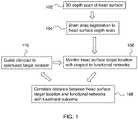
- FIG. 1is a flow diagram relating to embodiments of the invention.
- FIGS. 2A-Bschematically show making head shape measurements in connection with embodiments of the invention.
- FIGS. 3A-Cschematically show warping a brain atlas to match patient head shape measurements.
- FIG. 4Ais an example of a brain atlas image after registering the atlas to patient head shape measurements.
- FIG. 4Bshows (in gray shading) a functional network in a brain atlas image.
- FIG. 5Ais an image showing distance from a functional node of interest.
- FIG. 5Bshows the result of intersecting the distance function of FIG. 5A with the edge of the head.
- FIG. 5Cshows an image of the resulting closest scalp point to the functional node of interest.
- FIGS. 6A-Brelate to validation experiments of the present approach.
- FIG. 7is a flow diagram relating to an exemplary initial TMS therapy session.
- FIGS. 8A-Bschematically show the method of FIG. 7 .
- FIG. 9is a flow diagram relating to an exemplary subsequent TMS therapy session.
- FIG. 10schematically shows the method of FIG. 9 .
- FIG. 1is a flow diagram relating to embodiments of the invention. An overview of this process is as follows. Step 102 is obtaining a 3D depth scan of the patient's head surface. Step 104 is registering a brain atlas to the 3D head surface depth scan. Step 106 is monitoring head surface target location relative to functional networks in the brain (that are to be treated). Step 108 is correlating distance between head surface target location and functional networks with treatment outcome. Step 110 is guiding the clinician to optimized target locations.
- steps 102 and 104are preliminary, and steps 106 , 108 , 110 could be repeated as needed as part of a research program.
- FIG. 1is an exemplary larger setting within which the invention may be practiced, and the invention itself relates mainly to steps 102 , 104 of this figure.
- an exemplary embodiment of the inventionis a method of targeting cranial therapy to a patient. This method includes:
- Brain atlases of interest for this workare 3D volumetric brain atlases which provide 3D information on locations of brain structures (which can be defined functionally and/or anatomically) in the atlas coordinates.
- Such brain atlasesare compilations of results from many representative instances of relevant brain data. Therefore it is not expected that such a brain atlas would precisely match the brain of any specific patient. Mapping the brain atlas to individual patients is how this issue is dealt with, and is described in greater detail below.
- the brain atlascan be warped to the head using a non-linear surface matching algorithm. This warped atlas can then deliver an estimate of the location of the brain region of interest without the need for manual scalp measurements or prior MRI.
- This approachis applicable to any kind of cranial therapy, including but not limited to: transcranial magnetic stimulation and temporally interfering electric field therapy.
- FIGS. 2A-Bschematically show making head shape measurements in connection with embodiments of the invention.
- camera 202measures the full head shape of patient 204 .
- a cap 206e.g., a swim cap or the like.
- FIGS. 2A-Bschematically show use of a front view and a back view to get full head shape information. Alternatively, multiple cameras can be used to obtain this information without needing the patient to change position.
- camera 202is a depth camera (e.g., an RGBD camera providing RGB color channels and a Depth channel).
- a depth camerae.g., an RGBD camera providing RGB color channels and a Depth channel.
- improved computer vision algorithmscan make it possible to perform the same task (optical depth imaging) with a stereoscopic or even single RGB camera.
- Suitable image processing algorithms for this taskare known in the art (e.g., Kinect® fusion algorithm). Tracing the head surface with a marker on cap 206 can optionally be done to improve the quality of the head shape measurements.
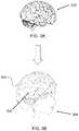
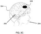
- FIGS. 3A-Cschematically show warping a brain atlas to match patient head shape measurements.
- 302 on FIG. 3Ais the brain atlas before any processing.
- 304 on FIG. 3Bis the head surface shape obtained from the 3D scan of the head of patient 204 .
- the result of fitting brain atlas 302 to head surface shape 304is schematically shown by 306 on FIG. 3B .
- the location of a region of interest 308 on FIG. 3C for patient 204is obtained by taking the coordinates of region of interest 308 in the atlas coordinates, known from the atlas, and applying the atlas->patient transformation on these coordinates.
- the resultis the location of region of interest 308 in patient coordinates. Since the patient coordinates have the head surface shape 304 as a readily available reference, the end result is that treatment can be accurately targeted to region of interest 308 without the need for any individualized imaging of the brain of patient 204 .
- the registering the brain atlas to the 3D scan of the head of the patientcan include performing an affine transformation of the brain atlas to best fit the 3D scan of the head of a patient.
- further steps of this part of the methodcan include voxelizing the 3D scan of the head of the patient to a binary 3D volume to provide a voxelized head scan and/or using externally visible anatomical landmarks such as the nasian, tragi, inian, apex or eyes as landmarks to help determine the best fit for a landmark based affine transformation.
- the thickness of skull and scalpis accounted for in the brain atlas, so the shape fitting is of the scalp surface in the atlas to the measured scalp surface of the patient.
- FIG. 4Ais an example of a brain atlas image after registering the atlas to patient head shape measurements.
- FIG. 4Bshows (in gray shading) a functional network in the registered brain atlas image of FIG. 4A .
- FIG. 5Ais an image showing distance from a functional node of interest (i.e., a brain region to be treated). This is a simple schematic with smaller distances being shaded darker.
- FIG. 5Bshows the result of intersecting the distance function of FIG. 5A with the edge of the head. Here the point of least distance on the edge of the head is shown with a black disk. This can be better seen in the 3D image of FIG. 5C , which shows an image of the resulting closest scalp point to the functional node of interest. Again, it should be noted that this information on the location of the functional node of interest is obtained without any imaging of the patient's brain.
- FIGS. 6A-Brelate to validation experiments of the present approach.
- a subject's individual MRI scanwas registered to the same subject's 3D depth camera head shape measurement and the location of the closest scalp location to specific cortical areas was compared, leading to a mean error of 14 mm.
- FIG. 7is a flow diagram relating to an exemplary initial TMS therapy session.
- step 702is performing a 3D optical scan of the patient's head by measuring facial landmarks or by measuring a facial point cloud.
- step 704is estimating the location of a brain region to be treated by using a brain atlas as described above.
- Step 706is placing TMS coil(s) to target the brain region of interest, relying on the location estimate of step 704 .
- Step 708is determining the position and orientation of the TMS coil(s) with respect to the 3D optical scan of step 702 . This can be done either by directly tracking the TMS coil locations and/or by placing markers on the TMS coils to improve this location determination.
- Step 710is saving the TMS coil location information in a database.
- this databaseis a patient-specific database of facial features (landmarks and/or point cloud) of the patient relative to positions of TMS coils for delivering the TMS therapy.
- the database of facial features of the patient relative to positions of TMS coilsis preferably used to improve consistency of TMS coil placement from one TMS session to another TMS session.
- FIGS. 8A-Bschematically show the method of FIG. 7 .
- 802is the brain region of interest and 804 schematically shows the facial landmarks (or facial point cloud) used for the 3D optical scan of the patient's head.
- 806is a TMS coil and 808 is an optical marker placed on the TMS coil to aid its location determination.
- Externally visible anatomical landmarks on the patientcan be tracked using an RGBD camera (camera with color and depth channel) and a computer vision algorithm that automatically determines the coordinates of these landmarks in world space.
- RGBD cameracamera with color and depth channel
- a computer vision algorithmthat automatically determines the coordinates of these landmarks in world space.
- These landmarksform a point cloud that spans up a coordinate system in head space. While the external landmark coordinates in head space stay constant, their variation in world space delivers information about the pose of the head.
- the location of the relevant brain regionis estimated with a brain atlas as described above and the coil location and orientation with respect to the head landmarks is recorded.
- TMS coil 806is positioned with respect to brain region of interest 802 according to its location estimate from the brain atlas.
- FIG. 9is a flow diagram relating to an exemplary subsequent TMS therapy session.
- step 902is performing a 3D optical scan of the patient's head by measuring facial landmarks or by measuring a facial point cloud.
- step 904is determining the desired position of the TMS coils with respect to the 3D optical scan based on the database results from the initial TMS treatment.
- Step 906is providing a display that allows a user (typically the clinician) to accurately position the TMS coils to reproduce their position and orientation from the initial TMS session.
- This displaycan be a conventional 2D display or an augmented reality display, and its main purpose is to facilitate positioning of the TMS coils in accordance with the database.
- FIG. 10schematically shows the method of FIG. 9 . This is similar to FIG. 8B , except that here TMS coil 806 is positioned with respect to brain region of interest 802 according to the database results from the initial session relating TMS coil position and orientation to facial features of the patient.
- the same cameratracks the head and shows the exact same coil location and orientation from the first treatment session to the TMS operator, allowing him to accurately reproduce the setup of the first treatment session.
- the head shape and coil positioningis then recorded and saved together with the TMS operator's feedback on patient response or remission values. Data analysis of such a brain stimulation map can then allow one to develop more effective and individualized TMS treatment methods.
- this head tracking techniqueallows one to locate the same scalp location during follow-up sessions within 2.4 mm accuracy for 30 cm camera distance and within 5.8 mm for 60 cm camera distance if the face is looking at the camera within an angle of 20 degrees.
- These testshave been performed with a low-cost depth sensing camera ($150 Intel Realsense® camera).
- Higher resolution cameras, such as professional tracking cameras (Optitrack®, $3000+)are expected to improve this accuracy even more.
- Optitrack®, $3000+are expected to improve this accuracy even more.
- a point cloud of the facecan be measured. Registration of the point cloud to a template of the person's face then allows one to also estimate the head orientation for angles higher than 40 degrees at high accuracy.
- the common “5 cm rule”has been shown to lead to errors of up to 22 mm for estimating the dorsolateral prefrontal cortex location.
Landscapes
- Health & Medical Sciences (AREA)
- Life Sciences & Earth Sciences (AREA)
- Engineering & Computer Science (AREA)
- Biomedical Technology (AREA)
- General Health & Medical Sciences (AREA)
- Animal Behavior & Ethology (AREA)
- Veterinary Medicine (AREA)
- Public Health (AREA)
- Physics & Mathematics (AREA)
- Medical Informatics (AREA)
- Nuclear Medicine, Radiotherapy & Molecular Imaging (AREA)
- Radiology & Medical Imaging (AREA)
- Surgery (AREA)
- Molecular Biology (AREA)
- Heart & Thoracic Surgery (AREA)
- Pathology (AREA)
- Biophysics (AREA)
- Neurology (AREA)
- Quality & Reliability (AREA)
- Computer Vision & Pattern Recognition (AREA)
- General Physics & Mathematics (AREA)
- Theoretical Computer Science (AREA)
- Magnetic Resonance Imaging Apparatus (AREA)
Abstract
Description
Claims (14)
Priority Applications (1)
| Application Number | Priority Date | Filing Date | Title |
|---|---|---|---|
| US16/269,407US11291852B2 (en) | 2018-02-06 | 2019-02-06 | Accurate patient-specific targeting of cranial therapy using a brain atlas |
Applications Claiming Priority (2)
| Application Number | Priority Date | Filing Date | Title |
|---|---|---|---|
| US201862626929P | 2018-02-06 | 2018-02-06 | |
| US16/269,407US11291852B2 (en) | 2018-02-06 | 2019-02-06 | Accurate patient-specific targeting of cranial therapy using a brain atlas |
Publications (2)
| Publication Number | Publication Date |
|---|---|
| US20190240499A1 US20190240499A1 (en) | 2019-08-08 |
| US11291852B2true US11291852B2 (en) | 2022-04-05 |
Family
ID=67475293
Family Applications (1)
| Application Number | Title | Priority Date | Filing Date |
|---|---|---|---|
| US16/269,407Active2040-01-20US11291852B2 (en) | 2018-02-06 | 2019-02-06 | Accurate patient-specific targeting of cranial therapy using a brain atlas |
Country Status (1)
| Country | Link |
|---|---|
| US (1) | US11291852B2 (en) |
Cited By (2)
| Publication number | Priority date | Publication date | Assignee | Title |
|---|---|---|---|---|
| US11497924B2 (en)* | 2019-08-08 | 2022-11-15 | Realize MedTech LLC | Systems and methods for enabling point of care magnetic stimulation therapy |
| US12366583B2 (en) | 2022-10-24 | 2025-07-22 | West Virginia University Board of Governors on behalf of West Virginia University | Methods of improving neurodegenerative disorders by targeted delivery of therapeutic agents |
Families Citing this family (4)
| Publication number | Priority date | Publication date | Assignee | Title |
|---|---|---|---|---|
| CN111657947B (en)* | 2020-05-21 | 2022-07-05 | 四川大学华西医院 | Positioning method of nerve regulation target area |
| KR102367904B1 (en)* | 2021-02-26 | 2022-03-03 | 주식회사 에이티앤씨 | Navigation device for guiding the position of coil, a brain stimulating device including the same and a bio-navigation robot system |
| CN113925606B (en)* | 2021-10-25 | 2023-07-07 | 四川大学华西医院 | A neuromodulation navigation positioning method, device and neuromodulation treatment system |
| CN119565032B (en)* | 2025-02-06 | 2025-07-15 | 景昱医疗科技(苏州)股份有限公司 | Electrode stimulation electric field simulation method and device, electronic equipment and storage medium |
Citations (20)
| Publication number | Priority date | Publication date | Assignee | Title |
|---|---|---|---|---|
| US6167295A (en) | 1991-01-28 | 2000-12-26 | Radionics, Inc. | Optical and computer graphic stereotactic localizer |
| US20050113630A1 (en)* | 2001-05-04 | 2005-05-26 | Peter Fox | Apparatus and methods for delivery of transcranial magnetic stimulation |
| US20090220136A1 (en)* | 2006-02-03 | 2009-09-03 | University Of Florida Research Foundation | Image Guidance System for Deep Brain Stimulation |
| US20100185042A1 (en) | 2007-08-05 | 2010-07-22 | Schneider M Bret | Control and coordination of transcranial magnetic stimulation electromagnets for modulation of deep brain targets |
| US7783132B2 (en)* | 2005-05-02 | 2010-08-24 | Agency For Science, Technology And Research | Method and apparatus for atlas-assisted interpretation of magnetic resonance diffusion and perfusion images |
| US8050475B2 (en)* | 2006-09-06 | 2011-11-01 | Agency For Science, Technology And Research | Detection and localization of vascular occlusion from angiography data |
| US8125223B2 (en)* | 2006-10-03 | 2012-02-28 | Singapore Agency For Science, Technology And Research Act | Segmenting infarct in diffusion-weighted imaging volume |
| US8314815B2 (en) | 2006-04-12 | 2012-11-20 | Nassir Navab | Virtual penetrating mirror device for visualizing of virtual objects within an augmented reality environment |
| US20120314924A1 (en) | 2011-03-29 | 2012-12-13 | Boston Scientific Neuromodulation Corporation | System and method for atlas registration |
| US8588491B2 (en)* | 2009-02-04 | 2013-11-19 | Kabushiki Kaisha Toshiba | Medical image processing apparatus |
| US20130345491A1 (en) | 2011-03-09 | 2013-12-26 | A School Corporation Kansai University | Image data processing device and transcranial magnetic stimulation apparatus |
| US20140179981A1 (en)* | 2010-11-01 | 2014-06-26 | Neuronix Ltd. | Method and system for positioning a transcranial magnetic stimulation (tms) device |
| US8774481B2 (en)* | 2010-03-25 | 2014-07-08 | Emory University | Atlas-assisted synthetic computed tomography using deformable image registration |
| US20150099252A1 (en) | 2013-10-03 | 2015-04-09 | Autodesk, Inc. | Enhancing movement training with an augmented reality mirror |
| US20150119689A1 (en) | 2012-05-16 | 2015-04-30 | Beth Israel Deaconess Medical Center, Inc. | Identifying individual target sites for transcranial magnetic stimulation applications |
| US20160256069A1 (en)* | 2007-09-24 | 2016-09-08 | MRI Interventions, Inc. | Computer programs and circuits for visualizations using image data and predefined data of surgical tools |
| US20160284132A1 (en) | 2015-03-23 | 2016-09-29 | Electronics And Telecommunications Research Institute | Apparatus and method for providing augmented reality-based realistic experience |
| US20170128737A1 (en)* | 2013-12-24 | 2017-05-11 | Osaka University | Operation teaching device and transcranial magnetic stimulation device |
| US9715753B2 (en) | 2013-01-23 | 2017-07-25 | Orca Health, Inc. | Personalizing medical conditions with augmented reality |
| US10074173B2 (en)* | 2013-12-06 | 2018-09-11 | The Johns Hopkins University | Methods and systems for analyzing anatomy from multiple granularity levels |
- 2019
- 2019-02-06USUS16/269,407patent/US11291852B2/enactiveActive
Patent Citations (21)
| Publication number | Priority date | Publication date | Assignee | Title |
|---|---|---|---|---|
| US6167295A (en) | 1991-01-28 | 2000-12-26 | Radionics, Inc. | Optical and computer graphic stereotactic localizer |
| US20050113630A1 (en)* | 2001-05-04 | 2005-05-26 | Peter Fox | Apparatus and methods for delivery of transcranial magnetic stimulation |
| US7783132B2 (en)* | 2005-05-02 | 2010-08-24 | Agency For Science, Technology And Research | Method and apparatus for atlas-assisted interpretation of magnetic resonance diffusion and perfusion images |
| US20090220136A1 (en)* | 2006-02-03 | 2009-09-03 | University Of Florida Research Foundation | Image Guidance System for Deep Brain Stimulation |
| US8314815B2 (en) | 2006-04-12 | 2012-11-20 | Nassir Navab | Virtual penetrating mirror device for visualizing of virtual objects within an augmented reality environment |
| US8050475B2 (en)* | 2006-09-06 | 2011-11-01 | Agency For Science, Technology And Research | Detection and localization of vascular occlusion from angiography data |
| US8125223B2 (en)* | 2006-10-03 | 2012-02-28 | Singapore Agency For Science, Technology And Research Act | Segmenting infarct in diffusion-weighted imaging volume |
| US20100185042A1 (en) | 2007-08-05 | 2010-07-22 | Schneider M Bret | Control and coordination of transcranial magnetic stimulation electromagnets for modulation of deep brain targets |
| US20160256069A1 (en)* | 2007-09-24 | 2016-09-08 | MRI Interventions, Inc. | Computer programs and circuits for visualizations using image data and predefined data of surgical tools |
| US8588491B2 (en)* | 2009-02-04 | 2013-11-19 | Kabushiki Kaisha Toshiba | Medical image processing apparatus |
| US8774481B2 (en)* | 2010-03-25 | 2014-07-08 | Emory University | Atlas-assisted synthetic computed tomography using deformable image registration |
| US20140179981A1 (en)* | 2010-11-01 | 2014-06-26 | Neuronix Ltd. | Method and system for positioning a transcranial magnetic stimulation (tms) device |
| US20130345491A1 (en) | 2011-03-09 | 2013-12-26 | A School Corporation Kansai University | Image data processing device and transcranial magnetic stimulation apparatus |
| EP2684518A1 (en) | 2011-03-09 | 2014-01-15 | Osaka University | Image data processing device and transcranial magnetic stimulation apparatus |
| US20120314924A1 (en) | 2011-03-29 | 2012-12-13 | Boston Scientific Neuromodulation Corporation | System and method for atlas registration |
| US20150119689A1 (en) | 2012-05-16 | 2015-04-30 | Beth Israel Deaconess Medical Center, Inc. | Identifying individual target sites for transcranial magnetic stimulation applications |
| US9715753B2 (en) | 2013-01-23 | 2017-07-25 | Orca Health, Inc. | Personalizing medical conditions with augmented reality |
| US20150099252A1 (en) | 2013-10-03 | 2015-04-09 | Autodesk, Inc. | Enhancing movement training with an augmented reality mirror |
| US10074173B2 (en)* | 2013-12-06 | 2018-09-11 | The Johns Hopkins University | Methods and systems for analyzing anatomy from multiple granularity levels |
| US20170128737A1 (en)* | 2013-12-24 | 2017-05-11 | Osaka University | Operation teaching device and transcranial magnetic stimulation device |
| US20160284132A1 (en) | 2015-03-23 | 2016-09-29 | Electronics And Telecommunications Research Institute | Apparatus and method for providing augmented reality-based realistic experience |
Non-Patent Citations (14)
Cited By (4)
| Publication number | Priority date | Publication date | Assignee | Title |
|---|---|---|---|---|
| US11497924B2 (en)* | 2019-08-08 | 2022-11-15 | Realize MedTech LLC | Systems and methods for enabling point of care magnetic stimulation therapy |
| US20230014217A1 (en)* | 2019-08-08 | 2023-01-19 | Realize MedTech LLC | Systems and methods for enabling point of care magnetic stimulation therapy |
| US12102838B2 (en)* | 2019-08-08 | 2024-10-01 | Realize MedTech LLC | Systems and methods for enabling point of care magnetic stimulation therapy |
| US12366583B2 (en) | 2022-10-24 | 2025-07-22 | West Virginia University Board of Governors on behalf of West Virginia University | Methods of improving neurodegenerative disorders by targeted delivery of therapeutic agents |
Also Published As
| Publication number | Publication date |
|---|---|
| US20190240499A1 (en) | 2019-08-08 |
Similar Documents
| Publication | Publication Date | Title |
|---|---|---|
| US11291852B2 (en) | Accurate patient-specific targeting of cranial therapy using a brain atlas | |
| US11024096B2 (en) | 3D-perceptually accurate manual alignment of virtual content with the real world with an augmented reality device | |
| CN111627521B (en) | Enhanced utility in radiotherapy | |
| CN106920234B (en) | Combined automatic radiotherapy planning method | |
| CN107530552B (en) | Three-dimensional localization of moving targets for adaptive radiotherapy | |
| CN108042918B (en) | Positioning device and method for personalized transcranial magnetic stimulation treatment based on 3D printing | |
| Noirhomme et al. | Registration and real-time visualization of transcranial magnetic stimulation with 3-D MR images | |
| CN111729200B (en) | Transcranial magnetic stimulation automatic navigation system and method based on depth camera and magnetic resonance | |
| CN111657947B (en) | Positioning method of nerve regulation target area | |
| CN111249622B (en) | Accurate transcranial magnetic stimulation on-line navigation method based on augmented reality | |
| WO2015098155A1 (en) | Operation teaching apparatus and transcranial magnetic stimulation apparatus | |
| CN117138239B (en) | A nerve positioning method, device, system and electronic equipment for transcranial magnetic stimulation | |
| CN114288559B (en) | Transcranial magnetic stimulation navigation method, system and computer equipment | |
| Song et al. | Augmented reality-based electrode guidance system for reliable electroencephalography | |
| Comeau | Neuronavigation for transcranial magnetic stimulation | |
| CN116650113A (en) | Automatic search system for hand hot spots based on optical navigation | |
| Westwood | Planning and analyzing robotized TMS using virtual reality | |
| Wang et al. | Real-time marker-free patient registration and image-based navigation using stereovision for dental surgery | |
| US12303204B2 (en) | Automated pre-operative assessment of implant placement in human bone | |
| US20210068695A1 (en) | Method Providing ECG Analysis Interface and System | |
| CN114224486B (en) | Nerve navigation positioning system for orthogonal positioning of sound field and magnetic field | |
| KR102186551B1 (en) | A method of placing a stimulator for non-invasive brain stimulation using augmented reality device | |
| CN114176776B (en) | Nerve navigation positioning system for synchronous double-coil magnetic stimulation | |
| US20230211168A1 (en) | Systems and methods for integrated electric field simulation and neuronavigation for transcranial magnetic stimulation | |
| CN120713635A (en) | High-precision transcranial magnetic stimulation positioning method, device and equipment based on optical navigation |
Legal Events
| Date | Code | Title | Description |
|---|---|---|---|
| FEPP | Fee payment procedure | Free format text:ENTITY STATUS SET TO UNDISCOUNTED (ORIGINAL EVENT CODE: BIG.); ENTITY STATUS OF PATENT OWNER: SMALL ENTITY | |
| AS | Assignment | Owner name:THE BOARD OF TRUSTEES OF THE LELAND STANFORD JUNIOR UNIVERSITY, CALIFORNIA Free format text:ASSIGNMENT OF ASSIGNORS INTEREST;ASSIGNORS:LEUZE, CHRISTOPH;MCNAB, JENNIFER;REEL/FRAME:048270/0996 Effective date:20180206 Owner name:THE BOARD OF TRUSTEES OF THE LELAND STANFORD JUNIO Free format text:ASSIGNMENT OF ASSIGNORS INTEREST;ASSIGNORS:LEUZE, CHRISTOPH;MCNAB, JENNIFER;REEL/FRAME:048270/0996 Effective date:20180206 | |
| FEPP | Fee payment procedure | Free format text:ENTITY STATUS SET TO SMALL (ORIGINAL EVENT CODE: SMAL); ENTITY STATUS OF PATENT OWNER: SMALL ENTITY | |
| STPP | Information on status: patent application and granting procedure in general | Free format text:PRE-INTERVIEW COMMUNICATION MAILED | |
| STPP | Information on status: patent application and granting procedure in general | Free format text:RESPONSE TO NON-FINAL OFFICE ACTION ENTERED AND FORWARDED TO EXAMINER | |
| STPP | Information on status: patent application and granting procedure in general | Free format text:NON FINAL ACTION MAILED | |
| STPP | Information on status: patent application and granting procedure in general | Free format text:RESPONSE TO NON-FINAL OFFICE ACTION ENTERED AND FORWARDED TO EXAMINER | |
| STPP | Information on status: patent application and granting procedure in general | Free format text:PUBLICATIONS -- ISSUE FEE PAYMENT VERIFIED | |
| STCF | Information on status: patent grant | Free format text:PATENTED CASE | |
| MAFP | Maintenance fee payment | Free format text:PAYMENT OF MAINTENANCE FEE, 4TH YR, SMALL ENTITY (ORIGINAL EVENT CODE: M2551); ENTITY STATUS OF PATENT OWNER: SMALL ENTITY Year of fee payment:4 |