US11259870B2 - Interactive user interfaces for minimally invasive telesurgical systems - Google Patents
Interactive user interfaces for minimally invasive telesurgical systemsDownload PDFInfo
- Publication number
- US11259870B2 US11259870B2US15/725,271US201715725271AUS11259870B2US 11259870 B2US11259870 B2US 11259870B2US 201715725271 AUS201715725271 AUS 201715725271AUS 11259870 B2US11259870 B2US 11259870B2
- Authority
- US
- United States
- Prior art keywords
- tool
- surgeon
- surgical
- images
- image
- Prior art date
- Legal status (The legal status is an assumption and is not a legal conclusion. Google has not performed a legal analysis and makes no representation as to the accuracy of the status listed.)
- Active, expires
Links
Images


Classifications
- A—HUMAN NECESSITIES
- A61—MEDICAL OR VETERINARY SCIENCE; HYGIENE
- A61B—DIAGNOSIS; SURGERY; IDENTIFICATION
- A61B1/00—Instruments for performing medical examinations of the interior of cavities or tubes of the body by visual or photographical inspection, e.g. endoscopes; Illuminating arrangements therefor
- A61B1/00002—Operational features of endoscopes
- A61B1/00043—Operational features of endoscopes provided with output arrangements
- A61B1/00045—Display arrangement
- A61B1/0005—Display arrangement combining images e.g. side-by-side, superimposed or tiled
- A—HUMAN NECESSITIES
- A61—MEDICAL OR VETERINARY SCIENCE; HYGIENE
- A61B—DIAGNOSIS; SURGERY; IDENTIFICATION
- A61B34/00—Computer-aided surgery; Manipulators or robots specially adapted for use in surgery
- A61B34/10—Computer-aided planning, simulation or modelling of surgical operations
- A—HUMAN NECESSITIES
- A61—MEDICAL OR VETERINARY SCIENCE; HYGIENE
- A61B—DIAGNOSIS; SURGERY; IDENTIFICATION
- A61B1/00—Instruments for performing medical examinations of the interior of cavities or tubes of the body by visual or photographical inspection, e.g. endoscopes; Illuminating arrangements therefor
- A61B1/00002—Operational features of endoscopes
- A61B1/00004—Operational features of endoscopes characterised by electronic signal processing
- A61B1/00009—Operational features of endoscopes characterised by electronic signal processing of image signals during a use of endoscope
- A61B1/000094—Operational features of endoscopes characterised by electronic signal processing of image signals during a use of endoscope extracting biological structures
- A—HUMAN NECESSITIES
- A61—MEDICAL OR VETERINARY SCIENCE; HYGIENE
- A61B—DIAGNOSIS; SURGERY; IDENTIFICATION
- A61B1/00—Instruments for performing medical examinations of the interior of cavities or tubes of the body by visual or photographical inspection, e.g. endoscopes; Illuminating arrangements therefor
- A61B1/00002—Operational features of endoscopes
- A61B1/00039—Operational features of endoscopes provided with input arrangements for the user
- A61B1/0004—Operational features of endoscopes provided with input arrangements for the user for electronic operation
- A—HUMAN NECESSITIES
- A61—MEDICAL OR VETERINARY SCIENCE; HYGIENE
- A61B—DIAGNOSIS; SURGERY; IDENTIFICATION
- A61B1/00—Instruments for performing medical examinations of the interior of cavities or tubes of the body by visual or photographical inspection, e.g. endoscopes; Illuminating arrangements therefor
- A61B1/00163—Optical arrangements
- A61B1/00193—Optical arrangements adapted for stereoscopic vision
- A—HUMAN NECESSITIES
- A61—MEDICAL OR VETERINARY SCIENCE; HYGIENE
- A61B—DIAGNOSIS; SURGERY; IDENTIFICATION
- A61B1/00—Instruments for performing medical examinations of the interior of cavities or tubes of the body by visual or photographical inspection, e.g. endoscopes; Illuminating arrangements therefor
- A61B1/00163—Optical arrangements
- A61B1/00194—Optical arrangements adapted for three-dimensional imaging
- A—HUMAN NECESSITIES
- A61—MEDICAL OR VETERINARY SCIENCE; HYGIENE
- A61B—DIAGNOSIS; SURGERY; IDENTIFICATION
- A61B34/00—Computer-aided surgery; Manipulators or robots specially adapted for use in surgery
- A61B34/30—Surgical robots
- A—HUMAN NECESSITIES
- A61—MEDICAL OR VETERINARY SCIENCE; HYGIENE
- A61B—DIAGNOSIS; SURGERY; IDENTIFICATION
- A61B34/00—Computer-aided surgery; Manipulators or robots specially adapted for use in surgery
- A61B34/30—Surgical robots
- A61B34/37—Leader-follower robots
- A—HUMAN NECESSITIES
- A61—MEDICAL OR VETERINARY SCIENCE; HYGIENE
- A61B—DIAGNOSIS; SURGERY; IDENTIFICATION
- A61B34/00—Computer-aided surgery; Manipulators or robots specially adapted for use in surgery
- A61B34/70—Manipulators specially adapted for use in surgery
- A—HUMAN NECESSITIES
- A61—MEDICAL OR VETERINARY SCIENCE; HYGIENE
- A61B—DIAGNOSIS; SURGERY; IDENTIFICATION
- A61B8/00—Diagnosis using ultrasonic, sonic or infrasonic waves
- A61B8/08—Clinical applications
- A61B8/0833—Clinical applications involving detecting or locating foreign bodies or organic structures
- A61B8/085—Clinical applications involving detecting or locating foreign bodies or organic structures for locating body or organic structures, e.g. tumours, calculi, blood vessels, nodules
- A—HUMAN NECESSITIES
- A61—MEDICAL OR VETERINARY SCIENCE; HYGIENE
- A61B—DIAGNOSIS; SURGERY; IDENTIFICATION
- A61B8/00—Diagnosis using ultrasonic, sonic or infrasonic waves
- A61B8/12—Diagnosis using ultrasonic, sonic or infrasonic waves in body cavities or body tracts, e.g. by using catheters
- A—HUMAN NECESSITIES
- A61—MEDICAL OR VETERINARY SCIENCE; HYGIENE
- A61B—DIAGNOSIS; SURGERY; IDENTIFICATION
- A61B8/00—Diagnosis using ultrasonic, sonic or infrasonic waves
- A61B8/44—Constructional features of the ultrasonic, sonic or infrasonic diagnostic device
- A61B8/4416—Constructional features of the ultrasonic, sonic or infrasonic diagnostic device related to combined acquisition of different diagnostic modalities, e.g. combination of ultrasound and X-ray acquisitions
- G—PHYSICS
- G16—INFORMATION AND COMMUNICATION TECHNOLOGY [ICT] SPECIALLY ADAPTED FOR SPECIFIC APPLICATION FIELDS
- G16H—HEALTHCARE INFORMATICS, i.e. INFORMATION AND COMMUNICATION TECHNOLOGY [ICT] SPECIALLY ADAPTED FOR THE HANDLING OR PROCESSING OF MEDICAL OR HEALTHCARE DATA
- G16H20/00—ICT specially adapted for therapies or health-improving plans, e.g. for handling prescriptions, for steering therapy or for monitoring patient compliance
- G16H20/40—ICT specially adapted for therapies or health-improving plans, e.g. for handling prescriptions, for steering therapy or for monitoring patient compliance relating to mechanical, radiation or invasive therapies, e.g. surgery, laser therapy, dialysis or acupuncture
- G—PHYSICS
- G16—INFORMATION AND COMMUNICATION TECHNOLOGY [ICT] SPECIALLY ADAPTED FOR SPECIFIC APPLICATION FIELDS
- G16H—HEALTHCARE INFORMATICS, i.e. INFORMATION AND COMMUNICATION TECHNOLOGY [ICT] SPECIALLY ADAPTED FOR THE HANDLING OR PROCESSING OF MEDICAL OR HEALTHCARE DATA
- G16H30/00—ICT specially adapted for the handling or processing of medical images
- G16H30/40—ICT specially adapted for the handling or processing of medical images for processing medical images, e.g. editing
- G—PHYSICS
- G16—INFORMATION AND COMMUNICATION TECHNOLOGY [ICT] SPECIALLY ADAPTED FOR SPECIFIC APPLICATION FIELDS
- G16H—HEALTHCARE INFORMATICS, i.e. INFORMATION AND COMMUNICATION TECHNOLOGY [ICT] SPECIALLY ADAPTED FOR THE HANDLING OR PROCESSING OF MEDICAL OR HEALTHCARE DATA
- G16H50/00—ICT specially adapted for medical diagnosis, medical simulation or medical data mining; ICT specially adapted for detecting, monitoring or modelling epidemics or pandemics
- G16H50/50—ICT specially adapted for medical diagnosis, medical simulation or medical data mining; ICT specially adapted for detecting, monitoring or modelling epidemics or pandemics for simulation or modelling of medical disorders
- A—HUMAN NECESSITIES
- A61—MEDICAL OR VETERINARY SCIENCE; HYGIENE
- A61B—DIAGNOSIS; SURGERY; IDENTIFICATION
- A61B17/00—Surgical instruments, devices or methods
- A61B2017/00017—Electrical control of surgical instruments
- A61B2017/00203—Electrical control of surgical instruments with speech control or speech recognition
- A—HUMAN NECESSITIES
- A61—MEDICAL OR VETERINARY SCIENCE; HYGIENE
- A61B—DIAGNOSIS; SURGERY; IDENTIFICATION
- A61B34/00—Computer-aided surgery; Manipulators or robots specially adapted for use in surgery
- A61B34/10—Computer-aided planning, simulation or modelling of surgical operations
- A61B2034/101—Computer-aided simulation of surgical operations
- A61B2034/105—Modelling of the patient, e.g. for ligaments or bones
- A—HUMAN NECESSITIES
- A61—MEDICAL OR VETERINARY SCIENCE; HYGIENE
- A61B—DIAGNOSIS; SURGERY; IDENTIFICATION
- A61B34/00—Computer-aided surgery; Manipulators or robots specially adapted for use in surgery
- A61B34/20—Surgical navigation systems; Devices for tracking or guiding surgical instruments, e.g. for frameless stereotaxis
- A61B2034/2046—Tracking techniques
- A61B2034/2055—Optical tracking systems
- A—HUMAN NECESSITIES
- A61—MEDICAL OR VETERINARY SCIENCE; HYGIENE
- A61B—DIAGNOSIS; SURGERY; IDENTIFICATION
- A61B34/00—Computer-aided surgery; Manipulators or robots specially adapted for use in surgery
- A61B34/20—Surgical navigation systems; Devices for tracking or guiding surgical instruments, e.g. for frameless stereotaxis
- A61B2034/2046—Tracking techniques
- A61B2034/2059—Mechanical position encoders
- A—HUMAN NECESSITIES
- A61—MEDICAL OR VETERINARY SCIENCE; HYGIENE
- A61B—DIAGNOSIS; SURGERY; IDENTIFICATION
- A61B34/00—Computer-aided surgery; Manipulators or robots specially adapted for use in surgery
- A61B34/20—Surgical navigation systems; Devices for tracking or guiding surgical instruments, e.g. for frameless stereotaxis
- A61B2034/2046—Tracking techniques
- A61B2034/2065—Tracking using image or pattern recognition
- A—HUMAN NECESSITIES
- A61—MEDICAL OR VETERINARY SCIENCE; HYGIENE
- A61B—DIAGNOSIS; SURGERY; IDENTIFICATION
- A61B34/00—Computer-aided surgery; Manipulators or robots specially adapted for use in surgery
- A61B34/25—User interfaces for surgical systems
- A61B2034/256—User interfaces for surgical systems having a database of accessory information, e.g. including context sensitive help or scientific articles
- A—HUMAN NECESSITIES
- A61—MEDICAL OR VETERINARY SCIENCE; HYGIENE
- A61B—DIAGNOSIS; SURGERY; IDENTIFICATION
- A61B90/00—Instruments, implements or accessories specially adapted for surgery or diagnosis and not covered by any of the groups A61B1/00 - A61B50/00, e.g. for luxation treatment or for protecting wound edges
- A61B90/36—Image-producing devices or illumination devices not otherwise provided for
- A61B2090/364—Correlation of different images or relation of image positions in respect to the body
- A61B2090/365—Correlation of different images or relation of image positions in respect to the body augmented reality, i.e. correlating a live optical image with another image
- A—HUMAN NECESSITIES
- A61—MEDICAL OR VETERINARY SCIENCE; HYGIENE
- A61B—DIAGNOSIS; SURGERY; IDENTIFICATION
- A61B90/00—Instruments, implements or accessories specially adapted for surgery or diagnosis and not covered by any of the groups A61B1/00 - A61B50/00, e.g. for luxation treatment or for protecting wound edges
- A61B90/36—Image-producing devices or illumination devices not otherwise provided for
- A61B90/37—Surgical systems with images on a monitor during operation
- A61B2090/371—Surgical systems with images on a monitor during operation with simultaneous use of two cameras
- A—HUMAN NECESSITIES
- A61—MEDICAL OR VETERINARY SCIENCE; HYGIENE
- A61B—DIAGNOSIS; SURGERY; IDENTIFICATION
- A61B90/00—Instruments, implements or accessories specially adapted for surgery or diagnosis and not covered by any of the groups A61B1/00 - A61B50/00, e.g. for luxation treatment or for protecting wound edges
- A61B90/36—Image-producing devices or illumination devices not otherwise provided for
- A61B90/37—Surgical systems with images on a monitor during operation
- A61B2090/378—Surgical systems with images on a monitor during operation using ultrasound
- A61B2090/3782—Surgical systems with images on a monitor during operation using ultrasound transmitter or receiver in catheter or minimal invasive instrument
- A—HUMAN NECESSITIES
- A61—MEDICAL OR VETERINARY SCIENCE; HYGIENE
- A61B—DIAGNOSIS; SURGERY; IDENTIFICATION
- A61B34/00—Computer-aided surgery; Manipulators or robots specially adapted for use in surgery
- A61B34/25—User interfaces for surgical systems
- A—HUMAN NECESSITIES
- A61—MEDICAL OR VETERINARY SCIENCE; HYGIENE
- A61B—DIAGNOSIS; SURGERY; IDENTIFICATION
- A61B34/00—Computer-aided surgery; Manipulators or robots specially adapted for use in surgery
- A61B34/70—Manipulators specially adapted for use in surgery
- A61B34/71—Manipulators operated by drive cable mechanisms
- A—HUMAN NECESSITIES
- A61—MEDICAL OR VETERINARY SCIENCE; HYGIENE
- A61B—DIAGNOSIS; SURGERY; IDENTIFICATION
- A61B34/00—Computer-aided surgery; Manipulators or robots specially adapted for use in surgery
- A61B34/70—Manipulators specially adapted for use in surgery
- A61B34/76—Manipulators having means for providing feel, e.g. force or tactile feedback
- A—HUMAN NECESSITIES
- A61—MEDICAL OR VETERINARY SCIENCE; HYGIENE
- A61B—DIAGNOSIS; SURGERY; IDENTIFICATION
- A61B8/00—Diagnosis using ultrasonic, sonic or infrasonic waves
- A—HUMAN NECESSITIES
- A61—MEDICAL OR VETERINARY SCIENCE; HYGIENE
- A61B—DIAGNOSIS; SURGERY; IDENTIFICATION
- A61B8/00—Diagnosis using ultrasonic, sonic or infrasonic waves
- A61B8/42—Details of probe positioning or probe attachment to the patient
- A61B8/4245—Details of probe positioning or probe attachment to the patient involving determining the position of the probe, e.g. with respect to an external reference frame or to the patient
- A—HUMAN NECESSITIES
- A61—MEDICAL OR VETERINARY SCIENCE; HYGIENE
- A61B—DIAGNOSIS; SURGERY; IDENTIFICATION
- A61B90/00—Instruments, implements or accessories specially adapted for surgery or diagnosis and not covered by any of the groups A61B1/00 - A61B50/00, e.g. for luxation treatment or for protecting wound edges
- A61B90/36—Image-producing devices or illumination devices not otherwise provided for
- A61B90/361—Image-producing devices, e.g. surgical cameras
Definitions
- U.S. patent application Ser. No. 12/189,615is a non-provisional application and claims the benefit of provisional U.S. Patent Application No. 60/954,869 filed on Aug. 9, 2007 by inventors Christopher J. Hasser, et al., entitled ROBOTIC MINIMALLY INVASIVE SURGICAL SYSTEMS, which is incorporated herein by reference.
- U.S. patent application Ser. No. 12/189,615also claims the benefit of and is a continuation-in-part (CIP) of U.S.
- U.S. patent application Ser. No. 11/447,668further incorporates by reference U.S. patent application Ser. No. 11/130,471 entitled METHODS AND SYSTEM FOR PERFORMING 3-D TOOL TRACKING BY FUSION OF SENSOR AND/OR CAMERA DERIVED DATA DURING MINIMALLY INVASIVE SURGERY, filed on May 16, 2005 by Brian David Hoffman et al.; U.S. Pat. No. 6,659,939 entitled COOPERATIVE MINIMALLY INVASIVE TELESURGICAL SYSTEM, issued on Dec. 9, 2003 to Frederic H. Moll et al.; and U.S. Pat. No.
- aspects of the inventionare related to user interfaces for a surgeon's workstation in robotic surgical systems.
- Minimally invasive robotic surgical systemssuch as the da Vinci® Surgical System, are manufactured by Intuitive Surgical, Inc., of Sunnyvale, Calif.
- the Johns Hopkins University Engineering Research Center for Computer-Integrated Surgical Systems and Technology(ERC-CISST) conducts research in aspects of minimally invasive surgical systems.
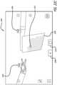
- FIG. 1illustrates a top view of an operating room employing a laparoscopic ultrasound robotic surgical system utilizing aspects of the embodiments of the invention.
- FIG. 2illustrates a block diagram of a laparoscopic ultrasound robotic surgical system utilizing aspects of the embodiments of the invention.
- FIG. 3illustrates a laparoscopic ultrasound probe utilizing aspects of the embodiments of the invention.
- FIG. 4illustrates a flow diagram of a method for training a LUS robotic surgical system to robotically move a LUS probe in a trained manner upon command, utilizing aspects of the embodiments of the invention.
- FIG. 5illustrates a flow diagram of a method for generating a clickable thumbnail image that allows a user to command that a LUS probe be automatically moved to a position and orientation from which the image was captured, utilizing aspects of the embodiments of the invention.
- FIG. 6illustrates a flow diagram of a method for automatically moving s LUS probe to a position and orientation associated with a clickable thumbnail image, utilizing aspects of the embodiments of the invention.
- FIG. 7illustrates a flow diagram of a method for robotically assisted needle guidance to a marked lesion of a cancerous structure, utilizing aspects of the embodiments of the invention.
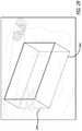
- FIG. 8illustrates a perspective view of a 3D ultrasound image of an anatomic structure in a camera reference frame with selectable 2D image slices as used in a medical robotic system utilizing aspects of the embodiments of the invention.
- FIG. 9illustrates a perspective view of a 3D camera view of an anatomic structure in a camera reference as used in a medical robotic system utilizing aspects of the embodiments of the invention.
- FIG. 10illustrates a perspective view of a frontal 2D slice of a 3D ultrasound view of an anatomic structure that overlays a 3D camera view of the anatomic structure, as displayable in a medical robotic system utilizing aspects of the embodiments of the invention.
- FIG. 11illustrates a perspective view of an inner 2D slice of a 3D ultrasound view of an anatomic structure that overlays a 3D camera view of the anatomic structure, as displayable in a medical robotic system utilizing aspects of the embodiments of the invention.
- FIG. 12is a diagrammatic physical view of a subsystem architecture for a surgical assistant workstation.
- FIG. 13is a diagrammatic view of a surgical assistant workstation for teleoperated surgical robots architecture.
- FIG. 14is a diagrammatic view of an illustrative data flow.
- FIG. 15is a diagrammatic view of a subsystem architecture (logical view).
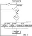
- FIG. 16is a diagrammatic view of basic three dimensional (3D) pointer interaction logic—event handling.
- FIG. 17is a diagrammatic view of basic 3D pointer interaction logic—move event.
- FIG. 18is a diagrammatic view of basic 3D pointer interaction logic—grab event.
- FIG. 19is a diagrammatic view of a subsystem architecture (process view).
- FIG. 20is a diagrammatic view of a subsystem architecture (development view).
- FIG. 21is a diagrammatic view of a 3D interface display for a surgeon console in a minimally invasive surgical system.
- FIGS. 22A-22Care exemplary diagrammatic views of invoking the graphical user interface to overlay menu systems, icons, a pointer, and a flashlight view of images onto the captured camera images displayed in the 3D interface display of the surgeon console.
- FIG. 23is an exemplary diagrammatic view of a pointer being used to select menu items to select context sensitive menu items from a menu overlaid onto the captured camera images adjacent a surgical instrument.
- FIGS. 24A-24Dare exemplary diagrammatic views of invoking the graphical user interface to overlay menu systems, icons, a pointer, and a medical image volume onto the captured camera images displayed in the 3D interface display of the surgeon console.
- FIGS. 25A-25Eare exemplary diagrammatic views of manipulating a medical image volume and selecting menu items overlaid on the captured camera images displayed in the 3D interface display of the surgeon console.
- FIGS. 26A-26Bare exemplary diagrammatic views of selecting menu items to display sagittal image slices of the medical image volume overlaid on the captured camera images displayed in the 3D interface display of the surgeon console.
- FIGS. 27A-27Bare exemplary diagrammatic views of selecting a axial slice plane and manipulating the axial slice plane to display different image slices of the medical image volume overlaid on the captured camera images displayed in the 3D interface display of the surgeon console.
- FIG. 28is an exemplary diagrammatic view of menu selection to close a medical image volume.
- FIG. 29is an exemplary diagrammatic view of boundaries of a virtual fixture overlaid onto the camera images of the surgical site in the 3D interface display of the surgeon console.
- a minimally invasive surgical master/slave robotic systemallow ultrasonic image display, image manipulation, supervisor/trainee master consoles, automatic movement limitation, and interchangeable slave consoles.
- U.S. patent application Ser. No. 11/447,668 entitled ULTRASOUND GUIDANCE FOR A LAPAROSCOPIC SURGICAL ROBOT to which priority is claimeddescribes a minimally invasive surgical robotic system with a laparoscopic ultrasonic robotic tool.
- FIG. 1illustrates, as an example, a top view of an operating room employing a robotic surgical system.
- the robotic surgical systemin this case is a laparoscopic ultrasound robotic surgical system 100 including a console (“C”) (also may be referred to herein as a surgeon console, master console, master surgeon console, or surgical console) utilized by a surgeon (“S”) while performing a minimally invasive diagnostic or surgical procedure with assistance from one or more assistants (“A”) on a patient (“P”) who is reclining on an operating table (“O”).
- Cconsole
- Ssurgeon
- Aminimally invasive diagnostic or surgical procedure with assistance from one or more assistants (“A”) on a patient (“P”) who is reclining on an operating table (“O”).
- the console Cincludes a master display 104 (also referred to herein as a display screen or display device) for displaying one or more images of a surgical site within the patient as well as perhaps other information to the surgeon. Also included are master input devices 107 and 108 (also referred to herein as master manipulators or master tool manipulators (MTM), master grips, hand control devices), one or more foot pedals 105 and 106 , a microphone 103 for receiving voice commands from the surgeon, and a processor 102 .
- the master input devices 107 and 108may include any one or more of a variety of input devices such as joysticks, gloves, trigger-guns, hand-operated controllers, or the like.
- the processor 102may be a computer or a part of a computer that may be integrated into the surgeon console or otherwise connected to the surgeon console in a conventional manner.
- the surgeonperforms a minimally invasive surgical procedure by manipulating the master input devices 107 and 108 so that the processor 102 causes their respectively associated slave arms 121 and 122 (also referred to herein as slave manipulators or slave robots) of the patient side cart (PSC) 120 to manipulate their respective removeably coupled and held surgical instruments 138 and 139 (also referred to herein as tools or minimally invasive surgical instruments) accordingly, while the surgeon views three-dimensional (“3D”) images of the surgical site on the master display 104 .
- slave manipulators or slave robotsalso referred to herein as slave manipulators or slave robots
- PSCpatient side cart
- the tools 138 and 139are Intuitive Surgical Inc.'s proprietary ENDOWRISTTM articulating instruments, which are modeled after the human wrist so that when added to the motions of the robot arm holding the tool, they allow a full six degrees of freedom of motion, which is comparable to the natural motions of open surgery. Additional details on such tools may be found in U.S. Pat. No. 5,797,900 entitled WRIST MECHANISM FOR SURGICAL INSTRUMENT FOR PERFORMING MINIMALLY INVASIVE SURGERY WITH ENHANCED DEXTERITY AND SENSITIVITY, issued on Aug. 25, 1998 to Akhil J. Madhani et al. which is incorporated herein by this reference.
- a manipulatable end effectorsuch as a clamp, grasper, scissor, stapler, blade, needle, or needle holder.
- the master display 104is a high-resolution stereoscopic video display device.
- the high-resolution stereoscopic video display deviceis formed of two progressive scan cathode ray tubes (“CRTs”).
- the high-resolution stereoscopic video display deviceis formed of two liquid crystal display (“LCDs”) devices.
- the systemoffers higher fidelity than polarization, shutter eyeglass, or other techniques. Each eye views a separate display presenting the left or right eye perspective, through an objective lens and a series of mirrors. The surgeon sits comfortably and looks into this display throughout surgery, making it an ideal place for the surgeon to display and manipulate 3-D intraoperative imagery.
- a stereoscopic endoscope 140(also referred to herein as an endoscopic camera) provides right and left camera views to the processor 102 so that it may process the information according to programmed instructions and cause it to be displayed on the master display 104 .
- a laparoscopic ultrasound (“LUS”) probe 150provides two-dimensional (“2D”) ultrasound image slices of an anatomic structure to the processor 102 so that the processor 102 may generate a 3D ultrasound computer model of the anatomic structure and cause the 3D computer model (or alternatively, 2D “cuts” of it) to be displayed on the master display 104 as an overlay to the endoscope derived 3D images or within a picture-in-picture (“PIP”) in either 2D or 3D and from various angles and/or perspectives according to surgeon or stored program instructions.
- PIPpicture-in-picture
- Each of the tools 138 and 139is preferably inserted through a cannula or trocar (not shown) or other tool guide into the patient so as to extend down to the surgical site through a corresponding minimally invasive incision such as Incision 166 .
- Each of the slave arms 121 - 124is conventionally formed of linkages which are coupled together and manipulated through motor controlled joints (also referred to as “active joints”).
- Setup arms (not shown)comprising linkages and setup joints are used to position the slave arms 121 - 124 vertically and horizontally so that their respective surgical related instruments may be coupled for insertion into the cannulae.
- the number of surgical tools used at one time and consequently, the number of slave arms being used in the system 100will generally depend on the diagnostic or surgical procedure and the space constraints within the operating room, among other factors. If it is necessary to change one or more of the tools being used during a procedure, the Assistant may remove the tool no longer being used from its slave arm, and replace it with another tool, such as a minimally invasive surgical tool 131 , from a tray (“T”) in the operating room.
- Tminimally invasive surgical tool
- the master display 104is positioned near the surgeon's hands so that it will display a projected image that is oriented so that the surgeon feels that he or she is actually looking directly down onto the surgical site.
- an image of the tools 138 and 139preferably appear to be located substantially where the surgeon's hands are located even though the observation points (i.e., that of the endoscope 140 and LUS probe 150 ) may not be from the point of view of the image.
- the real-time imageis preferably projected into a perspective image such that the surgeon can manipulate the end effector of a tool, 138 or 139 , through its associated master input device, 107 or 108 , as if viewing the workspace in substantially true presence.
- true presenceit is meant that the presentation of an image is a true perspective image simulating the viewpoint of an operator that is physically manipulating the tools.
- the processor 102transforms the coordinates of the tools to a perceived position so that the perspective image is the image that one would see if the endoscope 140 was looking directly at the tools from a surgeon's eye-level during an open cavity procedure.
- the processor 102performs various functions in the system 100 .
- One function that it performsis to translate and transfer the mechanical motion of master input devices 107 and 108 to their associated slave arms 121 and 122 through control signals over bus 110 so that the surgeon can effectively manipulate their respective tools 138 and 139 .
- Another function of the processor 102is to implement the various methods and functions described herein, including providing a robotic assisted LUS capability.
- processor 102may be implemented in practice by any combination of hardware, software and firmware. Also, its functions as described herein may be performed by one unit, or divided up among different components, each of which may be implemented in turn by any combination of hardware, software and firmware.
- Program code or instructions for the processor 102 to implement the various methods and functions described hereinmay be stored in processor readable storage media, such as memory (e.g., memory 240 illustrated in FIG. 2 ).
- ultrasound images captured by the LUS probe 150Prior to performing a minimally invasive surgical procedure, ultrasound images captured by the LUS probe 150 , right and left 2D camera images captured by the stereoscopic endoscope 140 , and end effector positions and orientations as determined using kinematics of the slave arms 121 - 124 and their sensed joint positions, are calibrated and registered with each other.
- the LUS probe 150is either labeled with markers and tracked by a tracking device such as the OPTORAK® position sensing system manufactured by Northern Digital Inc. of Ontario, Canada, or held by a robot with precise joint encoders. Then the rigid transformation between the ultrasound image and the frame being tracked is determined (which is typically referred to as the ultrasound calibration).
- a tracking devicesuch as the OPTORAK® position sensing system manufactured by Northern Digital Inc. of Ontario, Canada, or held by a robot with precise joint encoders. Then the rigid transformation between the ultrasound image and the frame being tracked is determined (which is typically referred to as the ultrasound calibration).
- the mathematical techniquesare well known. See, e.g., E. Boctor, A. Viswanathan, M. Chioti, R. Taylor, G. Fichtinger, and G. Hager, “A Novel Closed Form Solution for Ultrasound Calibration,” International Symposium on Biomedical Imaging, Arlington, Va., 2004, pp. 527-530.
- “A” and “B” in this case,are transformations between poses of the OPTOTRAK® rigid body (A) and the ultrasound image (B).
- “X”is the transformation from the ultrasound image to the rigid body.
- the ultrasound image framemay then be defined by three fiducials which appear in each of the three poses.
- Camera calibrationis a common procedure in computer vision applications.
- a checkerboard phantom with a multi-plane formulationmay be provided by the Caltech's camera calibration toolbox.
- OPTOTRAK® markersare added to a typical checkerboard video calibration phantom, and each corner of the checkerboard is digitized using a calibrated OPTOTRAK® pointer.
- the corner positionsmay be reported with respect to the OPTOTRAK®.
- the calibrationmay then be performed by placing the phantom in view of the endoscope 140 in several dozen orientations, and recording both stereo image data and OPTOTRAK® readings of the four checkerboard corners.
- the imagesmay then be fed into the calibration toolbox, which determines the intrinsic and extrinsic camera parameters, as well as the 3D coordinates of the grid corners in the camera frame. These coordinates may then be used with the OPTOTRAK® readings to perform a point-cloud to point-cloud registration between the endoscope 140 rigid body and camera frame.
- the processor/controller 102is configured to use the robot kinematics to report a coordinate frame for the LUS probe 150 tip relative to the endoscope 140 .
- both of these coordinate framesmay be offset from their correct values.
- itmay be necessary to register the offsets between the real camera frame of the endoscope 140 and the camera frame calculated from the kinematics as well as between the real and kinematic LUS probe 150 frames.
- the kinematicsmay be used in place of the OPTOTRAK® readings to determine ultrasound image overlay placement.
- the LUS probe 150may be moved, for example, to several positions, and the static offset between the tool tip and OPTOTRAK® rigid body registered.
- C CDD LusD ( C LusUrb ) ⁇ 1 T OUrb ( T OErb ) ⁇ 1 F CErb (1)
- C CDis the camera offset from the real endoscope 140 (also referred to herein simply as the “camera”) frame to the camera frame calculated from the kinematics
- F CErbis the transformation from the camera to the endoscope rigid body
- T OUrb *(T OErb ) ⁇ 1is the transformation from the camera rigid body to the LUS rigid body
- C LusUrbis the transformation from the LUS rigid body to the kinematic ultrasound tool tip
- D LusDis the reading from the processor/controller 102 giving the transformation from the kinematic ultrasound tool tip to a fixed reference point associated with the slave arms 121 - 124 .
- registrationmay be redone each time the camera is moved.
- the registrationmay be better performed using video tracking of a visual marker on the LUS probe 150 instead of the OPTOTRAK® readings.
- the registrationcan be corrected on the fly as the tool is tracked.
- manual registration of ultrasound and camera imagesmay be performed using conventional grab, move and rotate actions on a 3D ultrasound computer model of an anatomic structure, so that the computer model is properly registered over a camera model of the anatomic structure in the master display 104 .
- Slave arms 123 and 124may manipulate the endoscope 140 and LUS probe 150 in similar manners as slave arms 121 and 122 manipulate tools 138 and 139 .
- master input devices 107 and 108in order for the surgeon to manually control movement of either the endoscope 140 or LUS probe 150 , it may be required to temporarily associate one of the master input devices 107 and 108 with the endoscope 140 or the LUS probe 150 that the surgeon desires manual control over, while its previously associated tool and slave manipulator are locked in position.
- FIG. 2illustrates, as an example, a block diagram of the LUS robotic surgical system 100 .
- the master input device 107controls movement of either a tool 138 or a stereoscopic endoscope 140 , depending upon which mode its control switch mechanism 211 is in, and master input device 108 controls movement of either a tool 139 or a LUS probe 150 , depending upon which mode its control switch mechanism 231 is in.
- the control switch mechanisms 211 and 231may be placed in either a first or second mode by a surgeon using voice commands, switches physically placed on or near the master input devices 107 and 108 , foot pedals 105 and 106 on the console, or surgeon selection of appropriate icons or other graphical user interface selection means displayed on the master display 104 or an auxiliary display (not shown).
- control switch mechanism 211When control switch mechanism 211 is placed in the first mode, it causes master controller 202 to communicate with slave controller 203 so that manipulation of the master input 107 by the surgeon results in corresponding movement of tool 138 by slave arm 121 , while the endoscope 140 is locked in position. On the other hand, when control switch mechanism 211 is placed in the second mode, it causes master controller 202 to communicate with slave controller 233 so that manipulation of the master input 107 by the surgeon results in corresponding movement of endoscope 140 by slave arm 123 , while the tool 138 is locked in position.
- control switch mechanism 231when control switch mechanism 231 is placed in the first mode, it causes master controller 222 to communicate with slave controller 223 so that manipulation of the master input 108 by the surgeon results in corresponding movement of tool 139 by slave arm 122 .
- the LUS probe 150is not necessarily locked in position. Its movement may be guided by an auxiliary controller 242 according to stored instructions in memory 240 .
- the auxiliary controller 242also provides haptic feedback to the surgeon through master input 108 that reflects readings of a LUS probe force sensor 247 .
- control switch mechanism 231when control switch mechanism 231 is placed in the second mode, it causes master controller 222 to communicate with slave controller 243 so that manipulation of the master input 222 by the surgeon results in corresponding movement of LUS probe 150 by slave arm 124 , while the tool 139 is locked in position.
- the master input device 107 or 108Before switching back to the first or normal mode, the master input device 107 or 108 is preferably repositioned to where it was before the switch to the second mode of Control Switch 211 or 231 , as the case may be, or kinematic relationships between the master input device 107 or 108 and its respective tool slave arm 121 or 122 is readjusted so that upon switching back to the first or normal mode, abrupt movement of the tool 138 or 139 does not occur.
- control switchingsee, e.g., U.S. Pat. No. 6,659,939 entitled COOPERATIVE MINIMALLY INVASIVE TELESURGICAL SYSTEM, issued on Dec. 9, 2003 to Frederic H. Moll et al., which is incorporated herein by this reference.
- the auxiliary controller 242also performs other functions related to the LUS probe 150 and the endoscope 140 . It receives output from a LUS probe force sensor 247 , which senses forces being exerted against the LUS probe 150 , and feeds the force information back to the master input device 108 through the master controller 222 so that the surgeon may feel those forces even if he or she is not directly controlling movement of the LUS probe 150 at the time. Thus, potential injury to the patient is minimized since the surgeon has the capability to immediately stop any movement of the LUS probe 150 as well as the capability to take over manual control of its movement.
- auxiliary control 242Another key function of the auxiliary control 242 is to cause processed information from the endoscope 140 and the LUS probe 150 to be displayed on the master display 104 according to user selected display options. As will be described in more detail below, such processing includes generating a 3D ultrasound image from 2D ultrasound image slices received from the LUS probe 150 through an Ultrasound processor 246 , causing either 3D or 2D ultrasound images corresponding to a selected position and orientation to be displayed in a picture-in-picture window of the master display 104 , and causing either 3D or 2D ultrasound images of an anatomic structure to overlay a camera captured image of the anatomic structure being displayed on the master display 104 .
- the master controllers 202 and 222 , slave controllers 203 , 233 , 223 , and 243 , and auxiliary controller 242are preferably implemented as software modules executed by the processor 102 , as well as certain mode switching aspects of the control switch mechanisms 211 and 231 .
- the Ultrasound processor 246 and Video processor 236are separate boards or cards typically provided by the manufacturers of the LUS probe 150 and endoscope 140 that are inserted into appropriate slots coupled to or otherwise integrated with the processor 102 to convert signals received from these image capturing devices into signals suitable for display on the master display 104 and/or for additional processing by the auxiliary controller 242 before being displayed on the master display 104 .
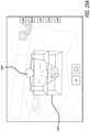
- FIG. 3illustrates a side view of one embodiment of the laparoscopic ultrasound (LUS) probe 150 .
- the LUS probe 150is a dexterous tool with preferably two distal degrees of freedom, permitting reorientation of laparoscopic ultrasound (LUS) sensor 301 through, for example, approximately ⁇ 80° in distal “pitch” and “yaw”, and ⁇ 240° in “roll” about a ball joint type, pitch-yaw mechanism 311 (functioning as and also referred to herein as a “Wrist” mechanism).
- Opposing pairs of Drive Rods or cables(not shown) physically connected to a proximal end of the LUS sensor 301 and extending through an internal passage of elongated shaft 312 mechanically control pitch and yaw movement of the LUS sensor 301 using conventional push-pull type action.
- This flexibility of the LUS probe 150(provided by the pitch/yaw wrist mechanism) is especially useful in optimally orienting the LUS probe 150 for performing ultrasonography on an anatomic structure during a minimally invasive surgical procedure.
- the LUS sensor 301captures 2D ultrasound slices of a proximate anatomic structure, and transmits the information back to the processor 102 through LUS cable 304 .
- the LUS cable 304may also extend within it.
- a clamshell sheath 321encloses the elongated shaft 312 and LUS cable 304 to provide a good seal passing through a cannula 331 (or trocar). Fiducial marks 302 and 322 are placed on the LUS sensor 301 and the sheath 321 for video tracking purposes.
- a force sensing capabilityis provided by strain gauges 303 which provide direct feedback of how hard the LUS probe 150 is pushing on a structure being sonographed, supplementing whatever limited feedback is available from joint motor torques. Potential uses of this information include: providing a redundant safety threshold check warning the surgeon or preventing motion into the structure if forces get too great; providing the surgeon with an approved haptic appreciation of how hard he or she is pushing on a structure; and possibly permitting some measure of compensation for unmodeled deflections of the pitch-yaw or wrist mechanism 311 which are not detected for some reason by joint position sensors or encoders.
- the strain gauges 303in this case serve the function of the LUS probe force sensor 247 as previously described in reference to FIG. 2 .
- Robotic assisted LUShas the potential to reduce variability in the ultrasound images produced, compared to freehand scanning, and can reduce operator workload and difficulty. Behaviors as simple as rocking the LUS probe 150 back and forth can maintain an updated 3D ultrasound image without operator intervention. More complicated behaviors can include movement of the LUS probe 150 along the surface of a target anatomical structure in a methodical pattern to generate a full image of the target, or reliably returning to a previously scanned probe location and orientation.
- FIG. 4illustrates, as an example, a flow diagram of a method for training the auxiliary controller 242 (i.e., providing it with stored instructions) to cause the LUS probe 150 to be robotically moved in the trained manner upon command, in order to capture a sequence of 2D ultrasound image slices of an anatomic structure, which are used by the auxiliary controller 242 to generate a 3D computer model of the structure.
- the control switch mechanism 231Prior to performing the training, the control switch mechanism 231 is placed in its second mode so that the surgeon may move the LUS probe 150 for training purposes by manipulating the master input device 108 . After performing training, the control switch mechanism 231 is then placed back into its first or normal mode so that the surgeon may manipulate the tool 139 to perform a minimally invasive surgical procedure using the master input device 108 .
- the training moduleis initially idle (i.e., it is not being executed by the processor 102 ).
- the processor 102(or a training module agent running in the background) may periodically check whether a start of training indication is received.
- the start of training indicationmay act as an interrupt which initiates running of the training module.
- the start of training indicationmay be initiated by a surgeon through a recognized voice command, selection of a training option on a graphical user interface displayed on the master display 104 , a switch mechanism that may physically be located on the corresponding master Control Input 108 or other convenient location accessible to the surgeon, or any other conventional means.
- the training moduleAfter the start of training indication is detected, in process 403 , the training module records or stores the current LUS probe 150 position and orientation, and periodically (or upon surgeon command) continues to do so by looping around processes 403 and 404 until a stop training indication is detected or received.
- the stop training indicationin this case may also be initiated by the surgeon in the same manner as the start of training indication, or it may be initiated in a different, but other conventional manner.
- a last position and orientation of the LUS probe 150is recorded or stored.
- the surgeonmoves the LUS probe 150 and the processor 102 stores its trajectory of points and orientations so that they may be retraced later upon command.
- the surgeonmoves the LUS probe 150 back and forth near an anatomic structure in order to capture a sequence of 2D ultrasound image slices from which a 3D version (or computer model) of the anatomic structure may be rendered by the processor 102 .
- the surgeonmove the LUS probe 150 once or more times along the surface of the anatomic structure in order to capture a different sequence of 2D ultrasound image slices from which a 3D version (or computer model) of the anatomic structure may be rendered by the processor 102 .
- the active joint positions of its slave arm 124are stored instead since their measurements are directly obtainable through encoders attached to each of the joints and their positions correspond to the LUS probe 150 positions and orientations.
- the trajectoryis then associated with a means for the surgeon to command the auxiliary controller 242 to move the LUS probe 150 in the desired fashion.
- the trajectorymay be associated with a voice command which upon its detection, the auxiliary controller 242 causes the slave arm 124 to move the LUS probe 150 back and forth along the stored trajectory of positions and orientations.
- the trajectorymay also be associated with a user selectable option on a graphical user interface displayed on the master display 104 , or it may be associated with a switch mechanism such as a button or unused control element on the master input device 108 .
- auxiliary controller 242causes the slave arm 124 to move the LUS probe 150 back and forth along the stored trajectory of positions and orientations as long as the foot pedal 106 is being depressed, and stops such motion once the surgeon takes his or her foot off the foot pedal 106 .
- FIG. 5illustrates, as an example, a flow diagram of a method for generating clickable thumbnail images corresponding to LUS probe 150 positions and orientations that are stored in memory 240 , so that when the surgeon clicks on one of the thumbnail images, the auxiliary controller 242 causes the slave arm 124 to move the LUS probe 150 to its stored position and orientation.
- Thisallows the surgeon to move the LUS probe 150 to see different views of an anatomic structure while the control switch mechanism 231 is in its first or normal mode.
- the surgeoncan continue to perform a minimally invasive surgical procedure by manipulating tool 139 using the master input device 108 .
- the methodmay then be combined with that described in reference to FIG. 4 in order to generate a sequence of 2D ultrasound image slices starting from that position and orientation, from which the auxiliary controller 242 may generate a 3D computer model rendition of the anatomic structure.
- control switch mechanism 231Prior to performing the method, however, the control switch mechanism 231 is placed in its second mode so that the surgeon may move the LUS probe 150 into the desired positions and orientations by manipulating the master input device 108 . After generating the clickable thumbnail images, the control switch mechanism 231 is then placed back into its first or normal mode so that the surgeon may manipulate the tool 139 to perform the minimally invasive surgical procedure using the master input device 108 .
- the auxiliary controller 242receives a snapshot command from the surgeon.
- the snapshot commandmay be, for example, a voice command, graphical user interface selection, or switch position.
- the auxiliary controller 242causes the LUS probe 150 to capture a 2D ultrasound image slice, and in process 503 , a thumbnail of the image is generated.
- the thumbnailin this case may include a simple JPEG or GIF file of the captured image.
- the current position and orientation of the LUS probe 150is stored in memory 240 along with information of its association with the thumbnail.
- a clickable version of the thumbnailis displayed on the master display 104 , so that the surgeon may command the auxiliary controller 242 to cause the LUS probe to be positioned and oriented at the stored position and orientation at any time upon clicking with his or her mouse or other pointing device on the clickable thumbnail. The surgeon may then move the LUS probe 150 to other positions and/or orientations, and repeat processes 501 - 505 to generate additional thumbnail images.
- FIG. 6illustrates, as an example, a flow diagram of a method for automatically moving the LUS probe 150 to a position and orientation associated with a clickable thumbnail upon command to do so by a surgeon while performing a minimally invasive surgical procedure using tool 139 .
- the clicking of a thumbnail generated by the method described in reference to FIG. 5is detected by, for example, a conventional interrupt handling process.
- the auxiliary controller 242is instructed by, for example, stored instructions corresponding to the interrupt handling process, to retrieve the position and orientation stored in memory 240 which is associated with the thumbnail.
- the auxiliary controller 242then causes the LUS probe 150 to move to that position and orientation by appropriately controlling slave arm 124 in process 603 .
- the surgeonis able to move the LUS probe 150 to a desired position without having to change modes of the control switch mechanism 231 and halt operation of the tool 139 until the LUS probe 150 is moved.
- the processor 102may generate a virtual fixture, such as a guidance virtual fixture or a forbidden region virtual fixture.
- a virtual fixturecan limit movement of a surgical instrument or tool.
- a guidance virtual fixturemay be generated to assist in electronically constraining a tool to travel over a predetermined path.
- a forbidden region virtual fixturemay be generated to
- virtual fixturesmay be generated to limit movement of a minimally invasive surgical tool such as virtual planes, virtual chamfers, virtual springs, detents, etc. With these virtual fixtures based on position in mind, virtual dampers may be generated by adding velocity terms.
- FIG. 7illustrates, as an example, a flow diagram of a method for robotically assisted needle guidance and penetration into a marked lesion of a cancerous structure, which allows appreciation for the aspects of robotic assisted LUS described herein.
- a selected 2D ultrasound image slice view of a cancerous structuresuch as a liver is displayed at the proper depth on the master display 104 as an overlay to a 3D camera view of the cancerous structure.
- the selected 2D ultrasound image slice viewmay be a frontal view or an inner slice view as taken from a previously generated 3D ultrasound computer model of the cancerous structure.
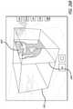
- FIG. 8illustrates a simplified perspective view of a 3D ultrasound computer model 800 of the cancerous structure, which has been generated, for example, using the method described in reference to FIG. 4 , and has been translated into the camera reference frame (EX, EY, EZ).
- FIG. 9illustrates a simplified perspective view of a 3D camera view 900 of the cancerous structure as taken by the stereoscopic endoscope 140 . If the surgeon selects a frontal slice 801 of the 3D ultrasound computer model 800 to be viewed as an overlay to the 3D camera view 900 , then the overlay will appear as shown in FIG. 10 .
- the surgeonselects one of the inner slices 802 - 804 of the 3D ultrasound computer model 800 , such as inner slice 803 , to be viewed as an overlay to the 3D camera view 900 , then the overlay will appear as shown in FIG. 11 with the 2D ultrasound image slice 803 displayed at the proper depth. To avoid confusion, the portion of the 3D camera view above that depth is made transparent.
- the surgeonmay manually control movement of the LUS probe 150 so that 2D ultrasound image slices captured by it appear as emanating in proper perspective and direction from the 3D camera image of the LUS probe 150 in the master display 104 .
- the emanated 2D image slices being displayed in the master display 104do not occlude the anatomic structure being probed. This manual approach may be particularly useful to the surgeon for quickly spotting lesions in the anatomic structure.
- process 702the surgeon marks lesions on the cancerous structure displayed as a result of process 701 .
- Each marked lesionis preferably marked using a designated color in order to clearly show that the surgeon has already identified it, thereby avoiding double counting.
- the location in the camera reference frame (EX, EY, EZ) of each marked lesionis stored in memory 240 , and in process 703 , the processor 102 determines an optimal needle tip path to that location.
- the processor 102generates a virtual fixture to help guide the needle to the marked lesion.
- local kinematic constraints on the slave arm manipulating the needle toolmay be specified by providing a table of constraints of the form: ( ⁇ right arrow over ( x ) ⁇ right arrow over ( x ) ⁇ 0 ) T A K ( ⁇ right arrow over ( x ) ⁇ right arrow over ( x ) ⁇ 0 )+ ⁇ right arrow over ( b ) ⁇ K ( ⁇ right arrow over ( x ) ⁇ right arrow over ( x ) ⁇ 0 ) ⁇ c (2)
- ⁇ right arrow over (x) ⁇represents, in simplified terms, the current 6 DOF kinematic pose of a master arm, or, in more general terms, a parameterization of a Cartesian pose F linearized about some nominal pose F 0 so that ( ⁇ right arrow over (x) ⁇ right arrow over (x) ⁇ 0 ) ⁇ F 0 ⁇ 1 F.
- equation (2)can be easily checked and enforced.
- ⁇corresponds, roughly, to a distance from a surface in state space and the function f ( ⁇ ) corresponds to a (non-linear) stiffness.
- the above formulationsuffices to support a variety of virtual chamfers, virtual springs, detents, etc.
- the formulationcan be easily extended to virtual dampers by adding velocity terms.
- ⁇ ⁇vector ⁇ ⁇ representation ⁇ ⁇ for ⁇ ⁇ small ⁇ ⁇ rotation ( 6 )
- V TARGETposition ⁇ ⁇ of ⁇ ⁇ target ⁇ ⁇ wrt ⁇ ⁇ LUS ⁇ ⁇ sensor ( 8 )
- the needle axis(which is assumed for this example to be the ⁇ right arrow over (Z) ⁇ axis of the needle driver) should be aimed at the target lesion, which will be given by F LUS ⁇ right arrow over (V) ⁇ TARGET .
- One metric for the aiming direction errorwill be:
- ⁇ BEYOND( R 0 R ( ⁇ right arrow over ( ⁇ ) ⁇ ) ⁇ right arrow over ( z ) ⁇ ) ⁇ ( F LUS ⁇ right arrow over (v) ⁇ TARGET ⁇ right arrow over ( P ) ⁇ TIP ) (10) which can easily be transcribed into a virtual detent or barrier preventing over-penetration.
- a simple spherical attractor virtual fixturecan be developed to minimize ⁇ F LUS ⁇ right arrow over (v) ⁇ TARGET ⁇ right arrow over (P) ⁇ TIP ⁇ .
- the processor 102determines the needle tip position as it moves towards the target lesion, and in process 706 , the processor 102 determines the distance between the needle tip position and the target lesion.
- the needle tip positionmay be determined from the slave arm kinematics and/or through visual tracking in the camera image.
- the color of the lesion or some other object in the displaychanges as the needle tip gets closer to the target.
- the colormay start off as blue when the needle tip is still far away from the target, and it may change through color spectrum so that it becomes red as it nears the target.
- a bar graph or other visual indicatormay be used to give a quick sense of the distance.
- a threshold distanceusually specified as some distance close to or even at the surface of the target lesion.
- Virtual fixturesmay be defined or manipulated through an interactive user interface at a surgeon console as more fully described below.
- Robotic surgical systemsallow a surgeon to operate in situ.
- the benefits of non invasive surgeryare well documented and continuing improvements in laparoscopic surgery are advancing the medical profession in a new and exciting direction.
- One of the many challenges of laparoscopic surgeryis working within the confined space of a body cavity.
- Surgical instruments, endoscopes, ultrasound probes, etc.need to be directed with precision and celerity, or risk complications from accidental tissue damage and extended surgery times.
- robot assisted laparoscopic surgerymay benefit from an interactive user interface that provides a unified assistive environment for surgery.
- the interactive user interfaceintegrates robotic devices, preoperative and intra-operative data sets, surgical task models, and human-machine cooperative manipulation.
- a surgical assistant workstation (SAW) for teleoperated surgical robotscan enhance the capabilities of robot-assisted laparoscopic surgery by providing fully integrated image guidance and data-enhanced intra-operative assistance to the surgical team and to the surgeon in particular.
- SAWsurgical assistant workstation
- Master tool manipulators(e.g., master tool manipulators 107 - 108 illustrated in FIG. 1 ) are input devices of a surgical console (e.g., surgeon console C illustrated in FIG. 1 ) that constitute the primary means of input and control for the surgeon. Details of a master tool manipulator are described in U.S. Pat. No. 6,714,939 entitled MASTER HAVING REDUNDANT DEGREES OF FREEDOM, issued on Mar. 30, 2004 to Salisbury et al. which is incorporated herein by reference.
- the master tool manipulatorscan be switched to operate in different modes.
- the patient-side slave manipulators(also referred to sometimes as robotic arms) follow the motion of the master tool manipulators and are teleoperated. That is, the MTMs may couple motion into the patient-side slave manipulators.
- a third patient-side slave manipulator(PSM- 3 ) can be activated by tapping the clutch pedal. This allows the surgeon to toggle between PSM- 3 and either PSM- 1 or PSM- 2 , depending on which side PSM- 3 is positioned.
- a master clutch pedalIn a master clutch mode, the master clutch pedal is depressed and the system is taken out of following mode.
- the PSM motionis no longer coupled to MTM motion. During surgery, this allows the operator to re-center the MTMs within their range of motion, and thus increase the surgical workspace.
- a camera control modethe camera clutch pedal is depressed and the PSMs are taken out of following mode and control is transferred to the endoscopic control manipulator (ECM) for camera repositioning.
- ECMendoscopic control manipulator
- the SAW frameworkadds another alternative mode (referred to as masters-as-mice mode) for the MTMs that overlaps with master clutch mode, allowing the surgeon to interact with the SAW graphical user interface (GUI).
- GUIgraphical user interface
- each MTMoperates as a 3D mouse, such that it can be used to position a graphical cursor overlaid on the stereo display console, while gripper open/close motions are used to emulate click and drag operations. In this way, the surgeon is able to interact with graphical objects and menus displayed by the SAW application.
- This modeis called a masters-as-mice (MaM) mode.
- the master tool manipulatorsare used as input devices for the graphical user interface within the surgical console.
- the MaM modeoverlaps with the existing master clutch mode of the surgeon console in the following way:
- Saw Console ModeMaM and/or GUI modes
- FIG. 12A diagrammatic view of a teleoperated surgical system including a surgical assistant workstation (SAW) is shown in FIG. 12 .
- SAWsurgical assistant workstation
- FIG. 12shows a generic deployment view that illustrates a number of common components and sub-systems.
- a user interface 1202 of a teleoperated surgical systemis connected via a communication network to a SAW 1210 .
- SAW 1210will support at least two types of video sources, namely: stereo endoscopy 1204 and ultrasound 1206 .
- Stereo endoscopymay be provided by two laparoscopic cameras (endoscopes or endoscopic cameras) transmitting independent video images to stereo displays 1216 , such as at the master display 104 of the console C illustrated in FIG. 1 or head mounted displays or visors.
- Ultrasound 1206may be an ultrasound probe attached to the end of a wristed robotic surgical arm inserted into the surgical site.
- Stereo endoscopy 1204may be connected to SAW 1210 by analog or digital video.
- ultrasound 1206may be connected to SAW 1210 by network interface.
- Video imagesmay also be provided by a medical image database 1212 connected to the SAW 1210 .
- the medical image database 1212is a source of medical images, models, surgical plans, and other application data.
- the medical images databasecould include preoperative images or a clinical Picture Archiving and Communication system (PACS).
- PACSPicture Archiving and Communication system
- Master robot 1208 and slave robot 1214are research-grade interface devices generally with robotic surgical arms operating various surgical tools.
- Examples of the master robot 1208include CISST MTMs and steady hand Robot.
- Examples of slave robot 1214include CISST PSMs, and a snake robot.
- FIG. 13is a diagrammatic illustration of a surgical assistant workstation (SAW) system architecture 1300 for teleoperated surgical robots.
- the SAW system architecture 1300includes multiple interconnected subsystems, which are briefly described hereafter.
- a video subsystem 1301provides mechanisms for acquiring and processing streams of images, including ultrasound and stereo endoscopic video.
- Such image processing pipelinescan be used to implement tool and tissue tracking algorithms.
- Tool tracking 1302is a specialized image processing pipeline provided for tracking the positions of surgical instruments using a combination of kinematic and stereo vision feedback.
- This subsystemmay provide software tools for determining device calibration, as well as methods for computing coordinate transformations between data sources (i.e., registration). Such tools may include kinematic calibration, camera calibration, ultrasound calibration, pre-operative and intra-operative image registration, video registration and overlay, etc.
- the data management subsystem 1304provides means to both import and export archived application data, including medical images, models, surgical plans and annotations.
- this subsystemcould accommodate data in various formats, including medical reality markup language (MRML), DICOM and clinical PACS.
- MRMLmedical reality markup language
- DICOMDICOM
- clinical PACSclinical PACS
- the communication interface 1305facilitates interactive manipulation and visualization of 2D and 3D data objects, including medical images and video, directly within the surgical console.
- a 3D graphical user interfacemanages user interaction from various input devices (including the master tool manipulators MTMs) and renders a menu system and graphical overlays to the stereo display of the surgical console.
- a 3D brick manager(as opposed to a 2D Window Manager) provides application-level widgets and interaction logic.
- a secondary user interface, called the staff console,will be provided to support the surgical interface. This is a conventional 2D interface that is intended for planning and monitoring outside of the surgical console.
- FIG. 14shows an illustrative data flow diagram, focusing on the robot application program interface (API) and the pipeline for video processing and visualization. This figure also shows the tool tracking and volume viewer subsystems. Although not specifically shown, calibration and registration functions may also be performed.
- APIapplication program interface
- Robot system block 1402 and collaborative robot block 1404transmit kinematic motion data to volume viewer block 1408 and tool tracking block 1410 .
- Volume viewer block 1408also receives preoperative image/model data 1406 from the data management subsystem 1304 in FIG. 13 .
- image data from cameras 1426 and LapUS 1428is captured by their respective image capture modules, stereo image capture module 1420 and ultrasound (US) image capture module 1422 .
- Video image data from the endoscopic cameras 1426is further rectified in the rectification block 1416 before being coupled into the stereo processor block 1412 for processing from 2D to 3D images.
- the 3D images of block 1412are then transmitted to the tool tracking subsystem 1410 and used in conjunction with the kinematic data provided by collaborative robot block 1404 to monitor the surgical tools.
- the LapUS datais transmitted to the image fusion block 1414 .
- the image fusion block 1414fuses the ultrasound images with the 3D endoscopic images that are then coupled into the overlay block 1418 .
- the overlay block 1418selectively overlays the graphical user interface and the medical image volume onto the fused ultrasound and endoscopic images.
- the combined image data including the overlaid graphics and images onto the fused imagesis coupled to the rendering block 1424 for rendering onto the hardware display 1430 .
- FIG. 15is a logical view of the subsystem architecture of SAW 1500 .
- a robot manipulatormaster device 1501 and a slave device 1502
- image sourcesendoscope image source 1504 and ultrasound image source 1505
- external consoles 1506staff console
- other peripherals 1507are categorized as devices, and as such are interfaced to the application framework by means of device interfaces.
- These device-specific blockscreate a layer of abstraction between external hardware or software modules in order to present a uniform interface to the application logic.
- the collaborative control block 1508couples the master and slave devices together. In a single-slave, single-master configuration, this block implements teleoperation control. In general, an application may include multiple masters and/or slaves; therefore, the collaborative control block provides a means to coordinate multiple manipulators. It contains a synchronous real-time loop for implementing control systems.
- a video processing pipelineis used to implement visual tool/instrument tracking 1510 .
- the visual tool/instrument tracking block 1510receives state information from the collaborative control block 1508 in order to incorporate kinematic information into the tool tracking algorithm.
- Exemplary tool tracking algorithms and systemsthat may be used are described in U.S. patent application Ser. No. 11/130,471 entitled METHODS AND SYSTEM FOR PERFORMING 3-D TOOL TRACKING BY FUSION OF SENSOR AND/OR CAMERA DERIVED DATA DURING MINIMALLY INVASIVE SURGERY, filed on May 16, 2005 by Brian David Hoffman et al.
- the master interaction block 1512facilitates user interaction with menu widgets and graphical scene objects represented by the brick manager 1514 . It provides the interface logic between the master manipulators and the brick manager 1514 when in masters-as-mice mode. Typical 2D windowing systems use the mouse input to create events (e.g., motion, click, release events) and bind callbacks to these events.
- the master Interaction block 1512provides a similar mechanism for the 3D MTM inputs by querying the state of the manipulators and listening for clutch events. The interaction logic transforms these inputs into pointer motion, button click events and specific behaviors such as object selection, dragging, rotation, resizing, etc.
- the brick manager 1514is the three dimensional analog of a standard window manager, in that it supports 3D user input and interaction with 3D graphical objects, such as image volumes and models, markers, annotations and in-situ video streams.
- the visual scene that is maintained by the brick manager 1514is ultimately rendered in stereo for overlay onto the surgical console display. It can be used to provide intraoperative visualization and graphical user interface (GUI).
- GUIgraphical user interface
- the brick manager 1514renders the fixed/augmented view into an interactive window the surgeon can interact with.
- a display driver 1524drives image data onto the left and right channels of the stereoscopic display.
- Application-specific logicis encapsulated in SAW application block 1516 and is defined by the application developer within the scope of the SAW application framework. Once the “master Interaction” component has determined which widget is currently active, all events will be forwarded to the widget and its logical layer. If the application requires a more direct access to the MTMs, the application will be able to access the MTM's state and disable the event forwarding from the master interaction component.
- Data block 1518contains images, text, and other data which can be called by surgeon via the master interaction block 1512 and SAW Application Logic 1516 .
- System calibrationis performed in calibration block 1520 .
- Typical calibration tasksinclude kinematic calibration of the robot manipulators, calibration of the navigation system, ultrasound calibration, and model to video registration.
- Calibration block 1520may also align a video image such as an ultrasound to the coordinate frame of a surgical instrument as seen under an endoscope.
- FIGS. 16, 17, and 18are logic trees for basic 3D cursor interactions. Two events Move and Grab are diagrammed in more detail in FIGS. 17 and 18 respectively.
- FIG. 19depicts the concurrent units of execution in the system.
- these execution unitsare provided by threads (e.g., multi-threading), rather than by multiple processes.
- the “low-level robot control”may be provided externally (e.g., when using the research API). In this case, it would be a separate process, possibly on a separate computer.
- signal and image processing pipelinesmay be distributed as external processes on separate computing hardware.
- Surgical console block 1902is an interactive intraoperative 3D graphical user interface.
- the GUImay augment the master surgical interface for enhanced image visualization and control by the surgeon. Augmentation is accomplished by video overlay of medical volume data or overlay of live images from a video source such as a LapUS probe or other imaging device. Content specific interactive menus and icons are also placed on the GUI allowing the surgeon to rotate images, pan, or zoom images, and establish virtual operating boundaries for surgical tools.
- Scene rendering block 1904is a graphical rendering pipeline responsible for stereo visualization and overlay in the surgeon's console.
- video signals from a video sourcesuch as an ultrasound may be overlaid onto the coordinate frame of a surgical instrument operating in the field of view of the endoscope.
- Video from the endoscopesare also processed into 3D images and displayed on the surgeon console or head mounted display.
- Signal/image processing pipeline 1906is a processing pipeline that is used for video processing such as instrument tracking and image overlay and other signal processing tasks.
- This pipelinemay include the acquisition of images, video, and signals that originate from external devices or distributed system components. For some applications, computationally demanding or specialized processing may be performed on a dedicated hardware system. Thus, the signal/image processing pipeline 1906 component may also be performed by an external signal processing system.
- FIG. 20depicts a hierarchical view of the core SAW software libraries and their dependencies.
- the bottom rowscontain the CISST foundation libraries, as well as external packages such as Python, LAPACK, and the research API.
- the cisstDevice Interface libraryincludes the abstract base class for all device interfaces, whether Device Tasks or Device Wrappers. Specific device interfaces are derived from this class.
- cisstRobotdefines generic robot capabilities, whereas robot-specific implementations are provided by modules such as cisstISI (for the Intuitive Surgical daVinci robot).
- the figurealso shows higher-level functionality such as video processing, instrument tracking, and collaborative robot control. All of this is encompassed by the SAW application framework.
- cisstISI 2002is a wrapper class that encapsulates ISI API functions with cisstlibrary-compatible interfaces and emulating these functions for non-daVinci hardware, where appropriate. Wrappers are device interfaces that do not include a thread of execution and are “wrappers” around the device drivers or vendor APIs.
- CisstStereoVision 2004is an algorithm for managing stereo image pairs and geometry, used in presenting stereo endoscope images to the surgeon console or headset.
- Open GLstands for Open Graphics Library and is a standard specification defining a cross-language cross-platform API for writing applications that produce 3D computer graphics.
- the visualization toolkit VTK 2008is an open source, freely available software system for 3D computer graphics, image processing, and visualization.
- brick manager 2010is a 3D scene manager for the surgeon console similar to a 2D window manager.
- Block 2012is the user interface (UI) interaction module.
- the UI interaction module 2012is the core interaction logic that defines the operation of the user interface at the surgeon console. This component manages user input from the master interface and interprets this input with respect to scene objects managed by the brick manager. Movements of the MTMs in combination with grip open and close motions are correlated with scene objects such as icons and menus to produce a predefined result.
- a dynamic laparoscopic ultrasound (LapUs) imageis overlaid on a tracked LapUS instrument in the stereo endoscope view provided by the surgeon console of the surgical system.
- LapUsdynamic laparoscopic ultrasound
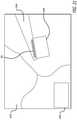
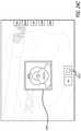
- FIG. 21is a diagrammatic view of a stereoscopic interface display for a surgeon's console in a minimally invasive surgical system for a user to view images in three dimensions.
- the displayshows tissue 2102 at a surgical site that is within the field of view of an endoscope (not shown).
- the displayalso shows an exemplary minimally invasive surgical instrument 2104 (e.g., a laparoscopic ultrasound instrument) that appears to extend into the visual field.
- an ultrasound image of tissueis displayed within an inset window 2106 of the display (LapUS inset view).
- the inset window 2106may be displayed outside the boundaries of the live video image.
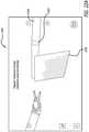
- a flashlight image window 2108that shows an ultrasound image of tissue is electronically attached to the image of surgical instrument 2104 within the display (LapUS flashlight view).
- the images in the flashlight image window 2108may be live image data (intra-operative images) from a LapUS probe or some other imaging source.
- the effect of the flashlight image window 2108is that it appears attached to the instrument 2104 similar to how a flag is attached to a flagpole.
- the flashlight image window 2108may be other shapes, and in some aspects is not necessarily attached to the image of surgical instrument 2104 .
- U.S. Pat. No. 6,799,065 entitled IMAGE SHIFTING APPARATUS AND METHOD FOR A TELEROBOTIC SYSTEM, issued on Sep. 28, 2004 to Gunter D. Niemeyer, incorporated herein by referencedescribes an image shifting mechanism that may be used to facilitate the appearance of the flashlight image window 2108 being substantially connected to the LapUS probe.
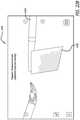
- the flashlight image window 2108moves as the surgeon moves instrument 2104 .
- the image displayed in the flashlight image window 2108may become foreshortened as the surgeon moves instrument 2104 , e.g., the surgeon points instrument 2104 more deeply into the surgical site.
- the flashlight image 2108may change angle and orientation corresponding to the movement of the surgical instrument 2104 to indicate the orientation of the ultrasound sensor 301 . That is, the ultrasound images slices captured by the ultrasound probe may be overlaid into the camera images so as to appear as to be emanating in proper perspective from the ultrasound sensor.
- the effectis of a flashlight that can be shined at various positions within the displayed image to provide enhanced visual information (e.g., the ultrasound image) to a surgeon.
- a laparoscopic ultrasound instrumentPrior to engaging the SAW, a laparoscopic ultrasound instrument is attached to one of the active patient side manipulators (PSMs) and is inserted through a cannula into the body cavity.
- the ultrasound transduceris calibrated to the surgical instrument.
- endoscopic video outputs from the surgical systemare connected to the SAW
- video output from a diagnostic ultrasound deviceis connected to the SAW
- video output of the SAWis connected to the master surgeon console
- the SAWmay be connected to the surgical system through a network interconnection (e.g., Ethernet).
- a surgeonoperates the master surgeon console which displays the live endoscopic images and allows the surgeon to operate the PSMs via the master tool manipulators (MTMs).
- MTMsmaster tool manipulators
- FIGS. 22A-22Care illustrations of a surgery performed while in the LapUS flashlight view mode.
- a flashlight image window 2108is attached to the surgical instrument 2104 as shown in FIG. 21 .
- the surgical instrument 2104is a LapUS probe (see laproscopic ultrasound probe 150 illustrated in FIG. 3 ) with a wristed joint 2202 for increased degrees of freedom of movement.
- FIG. 22Ba first orientation of surgical instrument and flashlight image window 2108 are shown.
- the orientation of the flashlight image window 2108has slightly changed with a slight change in the orientation of the surgical instrument from that shown in FIG. 22A .
- the flashlight image window 2108is slightly away from the viewer compared to the flashlight image window 2108 illustrated in FIG. 22A .
- the video image displayed in flashlight view 22 Bhas changed slightly as well due to foreshortening.
- LapUS probe 2104captures slices of images under the ultrasound sensor 301 in the probe head. Thus, the captured image slices and the flashlight image window change as the probe head moves the ultrasound sensor 301 around the surgical site.
- a surgeondepresses the master clutch pedal on the surgeon console and closes both master input devices (e.g., MTMs) in order to enter a masters-as-mice mode.
- the master input devicesmay be held closed for a predetermined period of time in order to enter the masters-as-mice mode in another embodiment.
- GUIgraphic user interface
- a 3D pointer/cursor 2212 and a first menu systemmay be overlaid onto the camera images of the surgical site displayed at the surgeon console.
- Graphical tool icons 2204 and 2206may also be overlaid near each PSM instrument.
- various icons in the graphical user interfacemay be overlaid onto the images of the surgical site in the display 2200 .
- An iconmay provide information, open a menu system to provide additional control or functionality, and be context specific depending on what surgical instrument 2104 is being used.
- graphical tool icons 2204 and 2206indicate a masters-as-mice mode and a graphical user interface (GUI) mode has been entered for the master input devices.
- GUIgraphical user interface
- the graphical tool icons 2204 and 2206 adjacent their respective instrumentmay be selected to provide additional information or further control and/or functionality depending upon the type of surgical instrument.
- the first menu system including menu buttons 2208 and 2210may be used to further interact with the graphical user interface.
- menu button 2208may be used to open and close the LapUS flashlight image window 2108 while menu button 2210 may be used to open and close a medical image view mode.
- FIG. 22Calso illustrates the 3D pointer 2212 overlaid upon images within the display.
- the surgeonhas moved the pointer 2212 over the flagpole image window 2108 in the display with the master input devices in the masters-as-mice mode.
- the size of the pointermay vary as its depth varies in response to the master input devices in the masters-as-mice mode. This may be seen in the comparison of pointer 2212 in FIG. 22C and pointer 2304 in FIG. 23 .
- FIG. 23depicts a menu system 2302 which may be displayed in response to selection of an icon or menu button by the 3D pointer/cursor 2304 and the master input devices.
- the menu system 2302 that is overlaid onto the imagesmay be context sensitive, such as being responsive to the type of surgical instrument 2104 .
- the surgeonmay move the 3D pointer by manipulating the primary MTM.
- the surgeonmoves the 3D pointer 2304 over the tool icon (e.g., icons 2201 - 2202 in FIG. 22C ) attached to the ultrasound instrument and closes the grip on the MTM to signal click or select.
- a pull-down menu 2302opens and may display options, such as option 1, a LapUS flashlight view, and option 2, a LapUS inset view.
- the surgeonmoves the primary MTM to highlight the first option, the LapUS flashlight view.
- the surgeonreleases the grip on the primary MTM and the ultrasound flashlight image (a plane) is overlaid onto the camera images in a flashlight image window 2108 adjacent the ultrasound instrument 2104 .
- the menu system and tool iconsdisappear, while the ultrasound overlay remains.
- the overlaid ultrasound flashlight image window 2108moves with the LapUS instrument, fixed to the coordinate frame of the ultrasound transducer/sensor 301 .
- the surgeonmay select the second option, the LapUS inset view.
- the ultrasound imageis overlaid onto the endoscopic image within an inset window 2106 in the stereoscopic display at the surgical console.
- the LapUS inset window 2106may be resized by using the MTMs.
- the LapUS inset window 2106may also be moved to different positions within the display on the master console.
- the SAWBy overlaying a GUI over live images from the endoscope and further overlaying ultrasound images captured by the ultrasound instrument onto the live images, the SAW fuses graphical objects with physical objects in a physical coordinate frame.
- FIG. 12shows an illustrative data flow diagram focusing on the robot API and the pipeline for video processing and visualization. This figure also shows the tool tracking and volume viewer subsystems. Although not specifically shown, calibration and registration functions may be performed.
- a medical image volumeis opened at the surgical console of the surgical system.
- a medical image volumemay be a pre-operative image including magnetic resonance images, computer tomography images, ultrasound images, positron emission tomography images, or other known medical image volumes that may be stored in known formats.
- the medical image volumeis overlaid onto live endoscopic imagery displayed on the surgical console.
- FIGS. 24A-24Dillustrate a step-by-step procedure to open the medical image database.
- the master controller(s)also referred to as master tool manipulators or input devices
- the slave instrument(s)may be used to control a pointer 2212 in the display.
- the pointermay appear to operate in three dimensions rather than in two dimensions as generally appears on most graphical user interfaces. Since the display has a three dimensional appearance, the user interface can be thought of as having volume (like a brick) instead of being flat (like a two-dimensional window).
- the surgeonmay move an overlaid ultrasound image or other pre- or intra-operative image by using one or more master controllers.
- the overlaid imageis on, e.g., a live video image from an endoscope at the surgical site.
- the surgeonmay move the overlaid image to align with the live video image of a particular tissue structure.
- one mastercontrols the overlaid image position, and another master controls the overlaid image orientation.
- image movement controlmay be somewhat analogous to that of the endoscopic camera control mode in a robotic surgical system.
- the overlaid imageis not automatically registered with a tissue structure seen in the live video.
- endoscopic video outputs from the robotic surgical systemare connected to the SAW, video output of the SAW is connected to the master surgeon console, and the SAW may also be connected to the robotic surgical system via Ethernet.
- the surgeonoperates the master console showing stereo display of live endoscopic images.
- FIG. 24Athe surgeon depresses the master clutch and enters the masters-as-mice mode activating the GUI mode wherein a 3D pointer/cursor 2212 and a menu system 2412 are overlaid onto camera images displayed in the display device of the surgical console.
- Graphical tool iconse.g., icons 2204 and 2206 in FIG. 22C
- the surgeonmoves the 3D pointer 2212 to a pull-down menu button 2422 visible on the overlaid menu system 2412 in FIG. 24B by manipulating the primary MTM.
- the buttonmay be marked with text such as “View Image Volume” or an icon of an image volume.
- the surgeonmay select the menu button in the GUI by closing the grip on the primary MTM.
- the pull-down menumay then open to display a list of predetermined image data sets listed by their unique identifiers.
- the pull-down menusmay be context sensitive, such as the pull down menu 2302 overlaid onto the live camera images from the endoscopic camera as shown in FIG. 23 .
- the surgeonhighlights the desired image volume and releases the grip on the primary MTM causing an annotated image volume bounding box 2432 to be overlaid onto the camera images in the display of the surgeon console as may be seen in FIGS. 24C and 24D .
- an annotated image volume bounding box 2432may be overlaid onto the camera images in the display of the surgeon console as may be seen in FIGS. 24C and 24D .
- a three dimensional imagemay be displayed in the bounding box 2432 .
- the surgeonmay desire to display a single image slice within the bounding box.
- Crosshairsmay optionally be used to indicate the location of the origin and eight corners of the image volume in the display.
- Crosshairsmay also be optionally used to indicate the location of the four corners of an active slice plane in the display.
- the surgeonmay manipulate the selected image volume by operating the MTMs singularly or conjunctively together in the masters-as-mice and GUI modes.
- the primary and/or secondary MTMsmay be used to pan, zoom and rotate the image volume.
- the surgeonmay move the 3D pointer 2302 over the image volume 2532 as shown in FIG. 25A and close the grip of the primary MTM to select the image volume.
- the primary MTMmay be moved to translate the image volume from one position of origin to another.
- the image volumemay be rotated along any axis thereby changing the perspective. Relative motion between the primary and secondary MTM can control the image volume orientation. A surgeon may rotate the image volume until the perspective view of the image volume matches the perspective of the live endoscopic camera.
- FIGS. 25A-25Dillustrate the rotation of an image volume in a counterclockwise direction by manipulating the primary master input device (MTM) and the secondary master input device (MTM) together to form a relative motion there between.
- MTMprimary master input device
- MTMsecondary master input device
- Menu options 2502 , 2503 , 2445 A- 2445 C of a menu system 2444 overlaid onto the camera images displayed on the display as shown in FIG. 25Dmay allow the surgeon to further manipulate the image volume 2532 .
- the surgeonmay opt to remove a surface layer or skin from the displayed image volume 2532 by clicking on a skin menu button 2502 with the cursor/pointer 2302 in a menu system 2444 associated with the image volume.
- the modified image volume 2532 ′is shown in FIG. 25E .
- an image slice 2632 of the image volume 2532 defined by a slice plane 2645may be displayed within the bounding box 2532 of the image volume.
- Orientation of the slice plane 2645may be controlled by movement of the MTMs in the MaM and GUI modes to view different image slices of the image volume.
- the surgeonmoves the 3D pointer over one of the corners of the slice plane and closes the grip of the primary MTM to select the slice plane.
- the secondary MTMmay be used to select another par of the slice plane by being positioned over a second corner.
- the relative motion between the primary and secondary input devices (MTMs)may be used to control the orientation of the slice plane.
- the primary and second MTMsmay also be used to reformat the slice plane.
- the slice planefollows the motion of the primary MTM. Different slice planes may also be displayed as desired by the surgeon.
- FIGS. 26A and 26Billustrate sagittal slice planes 2645 , 2645 ′ with different positions to form different image slices 2632 , 2632 ′ of the same image volume 2532 .
- the slice plane 2632 ′ illustrated in FIG. 26Bis a slice plane taken further from the center of the skull, or more laterally than the slice plane 2632 illustrated in FIG. 26A .
- axial slice planes 2745 , 2745 ′are displayed slicing through the same three dimensional image volume 2532 with different positions to form different image slices 2732 , 2732 ′. Similar to sagittal view, the axial slices may be rotated or orientated by manipulating the MTMs. Also, different slices closer or further away from the crown of the skull may be displayed as the surgeon desires. In FIG. 27B , the surgeon has replaced the skin by selecting the appropriate menu button 2502 of the menu system 2444 while still in axial view.
- the primary and secondary MTMsmay both be used. With the pointer over the image volume, the primary MTM is selected by closing its grip to select the image volume. The secondary MTM is also selected by closing its grip. A relative distance between the primary and secondary MTMs may then be used to control the level of zoom of a selected image volume or image slice. Thus, by selecting the image volume and then moving the MTMs to change their relative distance of separation, the surgeon may zoom in on or out from the image volume to display a desired level of detail.
- a surgeonmay re-select an image volume icon or menu button (e.g., menu button 2210 ). As depicted in FIG. 28 , the surgeon selects the menu button 2210 once again with the pointer 2212 over it. This removes the overlay of the image volume 2532 and returns the initial display of the GUI in the MaM mode, such as shown in FIG. 24B .
- an image volume icon or menu buttone.g., menu button 2210
- the surgeonmay press the clutch pedal and click both master input devices (MTMs) so that the master input devices may return to control and couple motion into the minimally invasive surgical tools of the surgical system.
- MTMsmaster input devices
- Another embodimentallows surgeons versed in the operation of a robotic surgical system to mentor another surgeon as a trainee.
- Two surgeon consolesare coupled with a single patient-side cart for mentoring purposes.
- One surgeon consoleis designated the supervisor console, while the second is the trainee console.
- a simplified case of mentoringis now described.
- One surgeon consolesare interfaced with the SAW.
- One patient side cart(PSC) is interfaced with the SAW and stereo endoscopic video output is connected to the SAW.
- PSCpatient side cart
- stereo endoscopic video outputis connected to the SAW.
- Two sets of stereo video outputs of the SAWare connected, one to each of the da Vinci master consoles.
- GUI modebecomes active and visible on both surgical consoles, a 3D pointer/cursor and menu system are overlaid onto the surgical consoles.
- Graphical tool iconsappear at each of the PSM tools.
- the supervisory surgeonmoves the 3D pointer and selects “mentor mode” by closing the primary MTM grip while the pointer is appropriately positioned on the graphical menu system.
- the menu systemdisappears from the trainee console and PSM control is transferred to the trainee.
- a telestration menuappears on the supervisory console.
- the camera clutch on the supervisory surgeonhas shared control of the ECM. If both camera clutches are activated, then the trainee console takes precedence in order to direct the camera view. Menu options on the supervisory console allow the supervising surgeon to regain control of the PSMs, request control of the fourth arm to control telestration and to overlay pre-operative image volumes.
- Another embodimentincludes manipulation of virtual fixtures through an Interactive Graphical User Interface.
- virtual fixturesan interaction mode is formed in which the surgeon shares control of the robot with the computer process.
- These task-dependent computer processesmay provide assistance to the surgeon by limiting the robot's motion within restricted regions and/or by influencing it to move along desired paths.
- VFsmay be generally classified as either forbidden region virtual fixtures (FRVFs) or guidance virtual fixtures (GVFs) (e.g., haptic guidance).
- FRVFsallow desired motion only in a predetermined task space, whereas GVFs provide assistance in keeping the motion on desired paths or surfaces.
- GVFsare defined using a fixed number of virtual planes, whereas GVFs can be selected from a predefined set of primitives.
- an exemplary virtual fixture 2902 in the shape of a bounding boxis illustrated as being placed around a surgical gripper 2904 .
- the bounding boxis open at the bottom allowing freedom of movement of the gripper 2904 towards the direction of the tissue. Lateral movement of the gripper 2904 , as well as upward movement and forward movement of the gripper 2904 , are curtailed by the sides of the bounding box in those directions.
- the forbidden region virtual fixturesset predefined limits on instrument movement to prevent undesired tissue collision.
- guidance virtual fixturesmay be used to constrain a surgical instrument to move on a fixed trajectory. For example, a suturing trajectory may assist the surgeon to perform suturing movements along a curved path.
- the surgeonmoves the 3D pointer by manipulating one of the primary MTM, and selects “virtual fixture mode” by closing the primary MTM grip while the pointer is appropriately positioned on the graphical menu system.
- a new menu systemappears on the surgeon console that allows adjustment of planes that define the boundary of forbidden regions. In this menu system, a number of 3D planes are visible to the surgeon.
- the surgeoncan grab the available planes as previously discussed in masters-as-mice mode (See also medical image overlay, similar to adjustment of slice plane image). By moving the planes to desired locations, the surgeon may create boundaries in which robotic surgical tools will not traverse. It may be advantageous to define boundaries, especially in delicate surgeries to prevent accidental tissue destruction by sharp instruments.
- the surgeonmay choose to add fixtures to a surface plane or a predetermined path. These fixtures may be used to guide the surgical tool along the surface or path, automating certain steps and also allowing precise placement of surgical tools prior to actual cutting or shearing of tissue.
- context based menusmay allow the surgeon to select from a list of predefined virtual fixtures listed by unique identifiers. Using this function may expedite placing boundaries and defining fixtures for routine surgeries and procedures.
- the surgeonselects the “done” button by closing the primary MTM grip while the pointer is appropriately positioned on the graphical menu system. The surgeon releases the master clutch and returns to normal operating mode.
- the surgical assistant workstationprovides a modular robotic master/slave control system.
- the modular robotic master/slave control systemallows a single master controller (surgeon console) to be used to control two or more different types of patient-side slave manipulators (PSM).
- PSMpatient-side slave manipulators
- a single master control stationmay be used to control a slave station or robot (patient side manipulator) with rigid surgical instruments (similar to the da Vinci® Surgical System instruments manufactured by Intuitive Surgical Inc.) that may be used for abdominal surgery.
- the same master control stationmay be used to control a different slave station or robot (patient side manipulator) with flexible, snake-like surgical instruments that may be used for laryngeal surgery.
- the master control stationmay be used to coincidentally control different types of slave stations (different patient side carts (PSC) with different patient-side slave manipulators (PSM)) that are coupled to it.
- a minimally invasive surgical instrument systemmay comprise a master console, a first slave station, and a second slave station coupled together by the surgical assistant workstation.
- the robotic arm configuration of the first slave stationis different from the robotic arm configuration of the second slave station such that different control signals to each are used in their control.
- the surgical assistant workstationadapts the master console to interchangeably control either the first slave station or the second slave station.
Landscapes
- Health & Medical Sciences (AREA)
- Life Sciences & Earth Sciences (AREA)
- Surgery (AREA)
- Engineering & Computer Science (AREA)
- Public Health (AREA)
- Medical Informatics (AREA)
- General Health & Medical Sciences (AREA)
- Nuclear Medicine, Radiotherapy & Molecular Imaging (AREA)
- Biomedical Technology (AREA)
- Veterinary Medicine (AREA)
- Animal Behavior & Ethology (AREA)
- Heart & Thoracic Surgery (AREA)
- Molecular Biology (AREA)
- Robotics (AREA)
- Radiology & Medical Imaging (AREA)
- Pathology (AREA)
- Physics & Mathematics (AREA)
- Biophysics (AREA)
- Optics & Photonics (AREA)
- Primary Health Care (AREA)
- Epidemiology (AREA)
- Urology & Nephrology (AREA)
- Vascular Medicine (AREA)
- Signal Processing (AREA)
- Data Mining & Analysis (AREA)
- Databases & Information Systems (AREA)
- Endoscopes (AREA)
- Ultra Sonic Daignosis Equipment (AREA)
Abstract
Description
CCD=DLusD(CLusUrb)−1TOUrb(TOErb)−1FCErb (1)
where CCDis the camera offset from the real endoscope140 (also referred to herein simply as the “camera”) frame to the camera frame calculated from the kinematics, FCErbis the transformation from the camera to the endoscope rigid body, TOUrb*(TOErb)−1is the transformation from the camera rigid body to the LUS rigid body, CLusUrbis the transformation from the LUS rigid body to the kinematic ultrasound tool tip, and DLusDis the reading from the processor/
({right arrow over (x)}−{right arrow over (x)}0)TAK({right arrow over (x)}−{right arrow over (x)}0)+{right arrow over (b)}K({right arrow over (x)}−{right arrow over (x)}0)≤c (2)
where {right arrow over (x)} represents, in simplified terms, the current 6 DOF kinematic pose of a master arm, or, in more general terms, a parameterization of a Cartesian pose F linearized about some nominal pose F0so that ({right arrow over (x)}−{right arrow over (x)}0)˜F0−1F. The tables are to be updated periodically based on visual feedback, user interaction, etc.
which can be approximated as a quadratic form in {right arrow over (α)} and converted to a virtual fixture using the method described above. Similarly, if the position of the needle tip is {right arrow over (P)}TIP, the penetration depth beyond the LUS target will be given by:
εBEYOND=(R0R({right arrow over (α)}){right arrow over (z)})●(FLUS{right arrow over (v)}TARGET−{right arrow over (P)}TIP) (10)
which can easily be transcribed into a virtual detent or barrier preventing over-penetration. Alternatively, a simple spherical attractor virtual fixture can be developed to minimize ∥FLUS{right arrow over (v)}TARGET−{right arrow over (P)}TIP∥.
| Process MTM Event(Event) |
| 1 | if Event == MASTER CLUTCH PRESSED | ||
| 2 | then InitMTM Pos = GetMTMPos( ) | ||
| 3 | Wait(3 seconds) | ||
| 4 | ClutchState = GetMasterClutchState( ) | ||
| 5 | MTM Pos = GetMTMPos( ) | ||
| 6 | if (ClutchState == PRESSED) and ((MTM Pos − InitMTM | ||
| Pos) < epsilon) | |||
| 7 | then EnterSAWConsoleMode( ) | ||
| 8 | else return | ||
| 9 | else return | ||
While in Saw Console Mode (MaM and/or GUI modes) the position and orientation of the MTM is used to drive the 3D pointer, while its gripper handle is used as a button.
Claims (9)
Priority Applications (2)
| Application Number | Priority Date | Filing Date | Title |
|---|---|---|---|
| US15/725,271US11259870B2 (en) | 2005-06-06 | 2017-10-04 | Interactive user interfaces for minimally invasive telesurgical systems |
| US17/468,198US20220015832A1 (en) | 2005-06-06 | 2021-09-07 | Minimally invasive telesurgical systems with interative user interfaces for 3d operative images |
Applications Claiming Priority (6)
| Application Number | Priority Date | Filing Date | Title |
|---|---|---|---|
| US68801305P | 2005-06-06 | 2005-06-06 | |
| US11/447,668US20070021738A1 (en) | 2005-06-06 | 2006-06-06 | Laparoscopic ultrasound robotic surgical system |
| US95486907P | 2007-08-09 | 2007-08-09 | |
| US12/189,615US8398541B2 (en) | 2006-06-06 | 2008-08-11 | Interactive user interfaces for robotic minimally invasive surgical systems |
| US13/775,574US9795446B2 (en) | 2005-06-06 | 2013-02-25 | Systems and methods for interactive user interfaces for robotic minimally invasive surgical systems |
| US15/725,271US11259870B2 (en) | 2005-06-06 | 2017-10-04 | Interactive user interfaces for minimally invasive telesurgical systems |
Related Parent Applications (1)
| Application Number | Title | Priority Date | Filing Date |
|---|---|---|---|
| US13/775,574DivisionUS9795446B2 (en) | 2005-06-06 | 2013-02-25 | Systems and methods for interactive user interfaces for robotic minimally invasive surgical systems |
Related Child Applications (1)
| Application Number | Title | Priority Date | Filing Date |
|---|---|---|---|
| US17/468,198DivisionUS20220015832A1 (en) | 2005-06-06 | 2021-09-07 | Minimally invasive telesurgical systems with interative user interfaces for 3d operative images |
Publications (2)
| Publication Number | Publication Date |
|---|---|
| US20180042680A1 US20180042680A1 (en) | 2018-02-15 |
| US11259870B2true US11259870B2 (en) | 2022-03-01 |
Family
ID=79291491
Family Applications (2)
| Application Number | Title | Priority Date | Filing Date |
|---|---|---|---|
| US15/725,271Active2028-10-12US11259870B2 (en) | 2005-06-06 | 2017-10-04 | Interactive user interfaces for minimally invasive telesurgical systems |
| US17/468,198PendingUS20220015832A1 (en) | 2005-06-06 | 2021-09-07 | Minimally invasive telesurgical systems with interative user interfaces for 3d operative images |
Family Applications After (1)
| Application Number | Title | Priority Date | Filing Date |
|---|---|---|---|
| US17/468,198PendingUS20220015832A1 (en) | 2005-06-06 | 2021-09-07 | Minimally invasive telesurgical systems with interative user interfaces for 3d operative images |
Country Status (1)
| Country | Link |
|---|---|
| US (2) | US11259870B2 (en) |
Cited By (55)
| Publication number | Priority date | Publication date | Assignee | Title |
|---|---|---|---|---|
| US20200405419A1 (en)* | 2019-06-26 | 2020-12-31 | Auris Health, Inc. | Systems and methods for robotic arm alignment and docking |
| US20210401527A1 (en)* | 2020-06-30 | 2021-12-30 | Auris Health, Inc. | Robotic medical systems including user interfaces with graphical representations of user input devices |
| US20220031395A1 (en)* | 2016-11-17 | 2022-02-03 | Capstone Surgical Technologies, Llc | Surgical image system and method |
| US20220087763A1 (en)* | 2020-09-23 | 2022-03-24 | Verb Surgical Inc. | Deep disengagement detection during telesurgery |
| US11432890B2 (en) | 2018-01-04 | 2022-09-06 | Covidien Lp | Systems and assemblies for mounting a surgical accessory to robotic surgical systems, and providing access therethrough |
| USD963851S1 (en) | 2020-07-10 | 2022-09-13 | Covidien Lp | Port apparatus |
| US11446099B2 (en) | 2016-06-03 | 2022-09-20 | Covidien Lp | Control arm for robotic surgical systems |
| US11529203B2 (en) | 2015-09-25 | 2022-12-20 | Covidien Lp | Robotic surgical assemblies and instrument drive connectors thereof |
| US11576739B2 (en) | 2018-07-03 | 2023-02-14 | Covidien Lp | Systems, methods, and computer-readable media for detecting image degradation during surgical procedures |
| US11576562B2 (en) | 2016-04-07 | 2023-02-14 | Titan Medical Inc. | Camera positioning method and apparatus for capturing images during a medical procedure |
| US11576733B2 (en) | 2019-02-06 | 2023-02-14 | Covidien Lp | Robotic surgical assemblies including electrosurgical instruments having articulatable wrist assemblies |
| US11596489B2 (en) | 2015-03-10 | 2023-03-07 | Covidien Lp | Measuring health of a connector member of a robotic surgical system |
| US11612446B2 (en) | 2016-06-03 | 2023-03-28 | Covidien Lp | Systems, methods, and computer-readable program products for controlling a robotically delivered manipulator |
| US11618171B2 (en) | 2013-12-11 | 2023-04-04 | Covidien Lp | Wrist and jaw assemblies for robotic surgical systems |
| US11628022B2 (en) | 2017-09-05 | 2023-04-18 | Covidien Lp | Collision handling algorithms for robotic surgical systems |
| US20230125934A1 (en)* | 2021-10-27 | 2023-04-27 | Suzhou Engoal Intelligent Technologies Co., Ltd | Dual-mainpulator control method and storage medium |
| US11666395B2 (en) | 2015-06-23 | 2023-06-06 | Covidien Lp | Robotic surgical assemblies |
| US11690691B2 (en) | 2017-02-15 | 2023-07-04 | Covidien Lp | System and apparatus for crush prevention for medical robot applications |
| US11717361B2 (en) | 2017-05-24 | 2023-08-08 | Covidien Lp | Electrosurgical robotic system having tool presence detection |
| US11717365B2 (en) | 2005-06-06 | 2023-08-08 | Intuitive Surgical Operations, Inc. | Laparoscopic ultrasound robotic surgical system |
| US20230285096A1 (en)* | 2020-07-28 | 2023-09-14 | Intuitive Surgical Operations, Inc. | Systems and method for selecting assignments for components of computer-assisted devices |
| US11779413B2 (en) | 2015-11-19 | 2023-10-10 | Covidien Lp | Optical force sensor for robotic surgical system |
| US11839441B2 (en) | 2017-05-25 | 2023-12-12 | Covidien Lp | Robotic surgical system with automated guidance |
| US11925429B2 (en) | 2015-02-19 | 2024-03-12 | Covidien Lp | Repositioning method of input device for robotic surgical system |
| US11948226B2 (en) | 2021-05-28 | 2024-04-02 | Covidien Lp | Systems and methods for clinical workspace simulation |
| US11986261B2 (en) | 2018-04-20 | 2024-05-21 | Covidien Lp | Systems and methods for surgical robotic cart placement |
| US11998288B2 (en) | 2018-09-17 | 2024-06-04 | Covidien Lp | Surgical robotic systems |
| US12029510B2 (en) | 2018-01-10 | 2024-07-09 | Covidien Lp | Determining positions and conditions of tools of a robotic surgical system utilizing computer vision |
| US12102403B2 (en) | 2018-02-02 | 2024-10-01 | Coviden Lp | Robotic surgical systems with user engagement monitoring |
| US12144537B2 (en) | 2017-08-16 | 2024-11-19 | Covidien Lp | End effector including wrist assembly and electrosurgical tool for robotic surgical systems |
| US12157238B2 (en) | 2018-12-20 | 2024-12-03 | Auris Health, Inc. | Systems and methods for robotic arm alignment and docking |
| US12178528B2 (en) | 2018-09-14 | 2024-12-31 | Covidien Lp | Surgical robotic systems and methods of tracking usage of surgical instruments thereof |
| US12186040B2 (en) | 2018-09-17 | 2025-01-07 | Covidien Lp | Surgical robotic systems |
| US12223629B2 (en) | 2019-09-11 | 2025-02-11 | Covidien Lp | Systems and methods for smoke-reduction in images |
| USD1066380S1 (en)* | 2023-01-13 | 2025-03-11 | Covidien Lp | Display screen with graphical user interface |
| USD1066382S1 (en)* | 2023-01-13 | 2025-03-11 | Covidien Lp | Display screen with graphical user interface |
| USD1066378S1 (en)* | 2023-01-13 | 2025-03-11 | Covidien Lp | Display screen with graphical user interface |
| USD1066383S1 (en)* | 2023-01-13 | 2025-03-11 | Covidien Lp | Display screen with graphical user interface |
| USD1066404S1 (en)* | 2023-01-13 | 2025-03-11 | Covidien Lp | Display screen with graphical user interface |
| USD1066405S1 (en)* | 2023-01-13 | 2025-03-11 | Covidien Lp | Display screen with graphical user interface |
| USD1066381S1 (en)* | 2023-01-13 | 2025-03-11 | Covidien Lp | Display screen with graphical user interface |
| USD1066379S1 (en)* | 2023-01-13 | 2025-03-11 | Covidien Lp | Display screen with graphical user interface |
| US12256890B2 (en) | 2019-12-23 | 2025-03-25 | Covidien Lp | Systems and methods for guiding surgical procedures |
| US12262964B2 (en) | 2020-02-26 | 2025-04-01 | Covidien Lp | Robotic surgical instrument including linear encoders for measuring cable displacement |
| US12262863B2 (en) | 2020-05-12 | 2025-04-01 | Covidien Lp | Systems and methods for image mapping and fusion during surgical procedures |
| US12315109B2 (en) | 2019-09-11 | 2025-05-27 | Covidien Lp | Systems and methods for neural-network based color restoration |
| US12329475B2 (en) | 2016-03-04 | 2025-06-17 | Covidien Lp | Inverse kinematic control systems for robotic surgical system |
| US12350828B2 (en) | 2019-12-16 | 2025-07-08 | Covidien Lp | Surgical robotic systems including surgical instruments with articulation |
| US12369998B2 (en) | 2021-05-28 | 2025-07-29 | Covidien Lp | Real time monitoring of a robotic drive module |
| USD1087135S1 (en) | 2023-08-02 | 2025-08-05 | Covidien Lp | Surgeon display screen with a graphical user interface having spent staple icon |
| US12376934B2 (en) | 2019-05-22 | 2025-08-05 | Covidien Lp | Surgical robotic arm storage assemblies and methods of replacing surgical robotic arms using the storage assemblies |
| USD1087995S1 (en) | 2023-08-02 | 2025-08-12 | Covidien Lp | Surgeon display screen with a transitional graphical user interface having staple firing icon |
| US12390294B2 (en) | 2021-12-14 | 2025-08-19 | Covidien Lp | Robotic surgical assemblies including surgical instruments having articulatable wrist assemblies |
| US12409003B2 (en) | 2021-05-14 | 2025-09-09 | Covidien Lp | Instrument cassette assemblies for robotic surgical instruments |
| US12433699B2 (en) | 2022-02-10 | 2025-10-07 | Covidien Lp | Surgical robotic systems and robotic arm carts thereof |
Families Citing this family (33)
| Publication number | Priority date | Publication date | Assignee | Title |
|---|---|---|---|---|
| US9295379B2 (en)* | 2005-04-18 | 2016-03-29 | M.S.T. Medical Surgery Technologies Ltd. | Device and methods of improving laparoscopic surgery |
| US10866783B2 (en)* | 2011-08-21 | 2020-12-15 | Transenterix Europe S.A.R.L. | Vocally activated surgical control system |
| WO2018220797A1 (en)* | 2017-06-01 | 2018-12-06 | オリンパス株式会社 | Flexible tube insertion assistance device and flexible tube insertion device |
| GB2571319B (en)* | 2018-02-23 | 2022-11-23 | Cmr Surgical Ltd | Concurrent control of an end effector in a master-slave robotic system using multiple input devices |
| BR102018007667A2 (en)* | 2018-04-16 | 2019-10-29 | Mendes Alves Pereira Ricardo | clinical-surgical data storage device, system and process |
| US20190361592A1 (en)* | 2018-05-23 | 2019-11-28 | Alcon Inc. | System and method of utilizing surgical tooling equipment with graphical user interfaces |
| EP3838209A1 (en) | 2019-12-20 | 2021-06-23 | Koninklijke Philips N.V. | Generating interactive zones for interventional medical devices |
| US12193750B2 (en)* | 2019-12-23 | 2025-01-14 | Mazor Robotics Ltd. | Multi-arm robotic system for spine surgery with imaging guidance |
| WO2021138262A1 (en)* | 2019-12-30 | 2021-07-08 | Intuitive Surgical Operations, Inc. | Systems and methods for telestration with spatial memory |
| WO2021181502A1 (en)* | 2020-03-10 | 2021-09-16 | オリンパス株式会社 | Surgery assistance system, operating method for surgery assistance system, and control device for surgery assistance system |
| EP4125676A1 (en)* | 2020-03-31 | 2023-02-08 | Intuitive Surgical Operations, Inc. | Systems and methods for facilitating automated operation of a device in a surgical space |
| US20230172674A1 (en)* | 2020-05-29 | 2023-06-08 | Covidien Lp | System and method for integrated control of 3d visualization through a surgical robotic system |
| US12413682B2 (en) | 2020-08-07 | 2025-09-09 | Brainlab Se | Real time augmentation |
| US20220087643A1 (en)* | 2020-09-23 | 2022-03-24 | 3Dintegrated Aps | Patient bearing system, a robotic system |
| CN114795489B (en)* | 2021-01-06 | 2025-08-26 | 深圳市精锋医疗科技股份有限公司 | Surgical robot and method and control device for guiding movement of surgical arm thereof |
| WO2022208414A1 (en) | 2021-03-31 | 2022-10-06 | Moon Surgical Sas | Co-manipulation surgical system for use with surgical instruments for performing laparoscopic surgery |
| US12178418B2 (en) | 2021-03-31 | 2024-12-31 | Moon Surgical Sas | Co-manipulation surgical system having a coupling mechanism removeably attachable to surgical instruments |
| US11844583B2 (en) | 2021-03-31 | 2023-12-19 | Moon Surgical Sas | Co-manipulation surgical system having an instrument centering mode for automatic scope movements |
| US12167900B2 (en) | 2021-03-31 | 2024-12-17 | Moon Surgical Sas | Co-manipulation surgical system having automated preset robot arm configurations |
| US12042241B2 (en) | 2021-03-31 | 2024-07-23 | Moon Surgical Sas | Co-manipulation surgical system having automated preset robot arm configurations |
| US11832909B2 (en) | 2021-03-31 | 2023-12-05 | Moon Surgical Sas | Co-manipulation surgical system having actuatable setup joints |
| US11819302B2 (en) | 2021-03-31 | 2023-11-21 | Moon Surgical Sas | Co-manipulation surgical system having user guided stage control |
| US11812938B2 (en) | 2021-03-31 | 2023-11-14 | Moon Surgical Sas | Co-manipulation surgical system having a coupling mechanism removeably attachable to surgical instruments |
| US20230062782A1 (en)* | 2021-09-02 | 2023-03-02 | Cilag Gmbh International | Ultrasound and stereo imaging system for deep tissue visualization |
| US11937799B2 (en) | 2021-09-29 | 2024-03-26 | Cilag Gmbh International | Surgical sealing systems for instrument stabilization |
| CN114099005B (en)* | 2021-11-24 | 2023-09-15 | 重庆金山医疗机器人有限公司 | Method for judging whether instrument is in visual field or is shielded or not and energy display method |
| CN114795494A (en)* | 2022-04-22 | 2022-07-29 | 上海微创医疗机器人(集团)股份有限公司 | Auxiliary device and surgical system |
| WO2024009132A1 (en)* | 2022-07-07 | 2024-01-11 | Gi Compass Ltd. | Enhancing endoscopic ultrasound procedures with tracking and visual intelligence |
| EP4561460A1 (en)* | 2022-07-29 | 2025-06-04 | Covidien LP | Graphic display for powered surgical staplers |
| CN115517615A (en)* | 2022-10-11 | 2022-12-27 | 中国医学科学院北京协和医院 | Endoscope master-slave motion control method and surgical robot system |
| US11839442B1 (en) | 2023-01-09 | 2023-12-12 | Moon Surgical Sas | Co-manipulation surgical system for use with surgical instruments for performing laparoscopic surgery while estimating hold force |
| US11986165B1 (en) | 2023-01-09 | 2024-05-21 | Moon Surgical Sas | Co-manipulation surgical system for use with surgical instruments for performing laparoscopic surgery while estimating hold force |
| US12370001B2 (en) | 2023-01-09 | 2025-07-29 | Moon Surgical Sas | Co-manipulation surgical system having automated user override detection |
Citations (105)
| Publication number | Priority date | Publication date | Assignee | Title |
|---|---|---|---|---|
| US4642687A (en) | 1983-02-10 | 1987-02-10 | United Kingdom Atomic Energy Authority | Superimposition of non-visual signals upon an image signal in a video display system |
| US4672963A (en) | 1985-06-07 | 1987-06-16 | Israel Barken | Apparatus and method for computer controlled laser surgery |
| EP0514584A2 (en) | 1991-05-21 | 1992-11-25 | Hewlett-Packard Company | Transducer positioning system |
| US5279309A (en) | 1991-06-13 | 1994-01-18 | International Business Machines Corporation | Signaling device and method for monitoring positions in a surgical operation |
| JPH06110543A (en) | 1992-09-30 | 1994-04-22 | Nippon Telegr & Teleph Corp <Ntt> | Direct teaching device |
| US5397323A (en) | 1992-10-30 | 1995-03-14 | International Business Machines Corporation | Remote center-of-motion robot for surgery |
| US5408409A (en) | 1990-05-11 | 1995-04-18 | International Business Machines Corporation | Image-directed robotic system for precise robotic surgery including redundant consistency checking |
| US5417210A (en) | 1992-05-27 | 1995-05-23 | International Business Machines Corporation | System and method for augmentation of endoscopic surgery |
| JPH07184923A (en) | 1993-12-28 | 1995-07-25 | Hitachi Ltd | Remote microsurgery support device |
| US5482029A (en) | 1992-06-26 | 1996-01-09 | Kabushiki Kaisha Toshiba | Variable flexibility endoscope system |
| JPH0819975A (en) | 1994-07-06 | 1996-01-23 | Tokico Ltd | Robot teaching method |
| US5493595A (en) | 1982-02-24 | 1996-02-20 | Schoolman Scientific Corp. | Stereoscopically displayed three dimensional medical imaging |
| JPH08215211A (en) | 1995-02-16 | 1996-08-27 | Hitachi Ltd | Remote surgery support device and method |
| US5551432A (en) | 1995-06-19 | 1996-09-03 | New York Eye & Ear Infirmary | Scanning control system for ultrasound biomicroscopy |
| JPH08299363A (en) | 1995-05-02 | 1996-11-19 | Olympus Optical Co Ltd | Manipulator system for operation |
| US5601549A (en) | 1994-11-17 | 1997-02-11 | Machida Endoscope Co., Ltd. | Medical observing instrument |
| US5759153A (en) | 1992-06-30 | 1998-06-02 | Cardiovascular Imaging Systems, Inc. | Automated longitudinal position translator for ultrasonic imaging probes, and methods of using same |
| JPH10146341A (en) | 1996-09-18 | 1998-06-02 | Hitachi Ltd | Surgical equipment |
| US5765561A (en) | 1994-10-07 | 1998-06-16 | Medical Media Systems | Video-based surgical targeting system |
| US5788688A (en) | 1992-11-05 | 1998-08-04 | Bauer Laboratories, Inc. | Surgeon's command and control |
| US5797900A (en) | 1996-05-20 | 1998-08-25 | Intuitive Surgical, Inc. | Wrist mechanism for surgical instrument for performing minimally invasive surgery with enhanced dexterity and sensitivity |
| US5797849A (en) | 1995-03-28 | 1998-08-25 | Sonometrics Corporation | Method for carrying out a medical procedure using a three-dimensional tracking and imaging system |
| US5808665A (en) | 1992-01-21 | 1998-09-15 | Sri International | Endoscopic surgical instrument and method for use |
| US5810008A (en) | 1996-12-03 | 1998-09-22 | Isg Technologies Inc. | Apparatus and method for visualizing ultrasonic images |
| US5810007A (en) | 1995-07-26 | 1998-09-22 | Associates Of The Joint Center For Radiation Therapy, Inc. | Ultrasound localization and image fusion for the treatment of prostate cancer |
| US5817022A (en) | 1995-03-28 | 1998-10-06 | Sonometrics Corporation | System for displaying a 2-D ultrasound image within a 3-D viewing environment |
| US5823958A (en) | 1990-11-26 | 1998-10-20 | Truppe; Michael | System and method for displaying a structural data image in real-time correlation with moveable body |
| US5836880A (en) | 1995-02-27 | 1998-11-17 | Micro Chemical, Inc. | Automated system for measuring internal tissue characteristics in feed animals |
| US5842473A (en) | 1993-11-29 | 1998-12-01 | Life Imaging Systems | Three-dimensional imaging system |
| US5842993A (en) | 1997-12-10 | 1998-12-01 | The Whitaker Corporation | Navigable ultrasonic imaging probe assembly |
| US5851183A (en) | 1990-10-19 | 1998-12-22 | St. Louis University | System for indicating the position of a surgical probe within a head on an image of the head |
| US5853367A (en) | 1997-03-17 | 1998-12-29 | General Electric Company | Task-interface and communications system and method for ultrasound imager control |
| US5876325A (en) | 1993-11-02 | 1999-03-02 | Olympus Optical Co., Ltd. | Surgical manipulation system |
| US5887121A (en) | 1995-04-21 | 1999-03-23 | International Business Machines Corporation | Method of constrained Cartesian control of robotic mechanisms with active and passive joints |
| US6019724A (en) | 1995-02-22 | 2000-02-01 | Gronningsaeter; Aage | Method for ultrasound guidance during clinical procedures |
| US6129670A (en) | 1997-11-24 | 2000-10-10 | Burdette Medical Systems | Real time brachytherapy spatial registration and visualization system |
| US6167297A (en) | 1999-05-05 | 2000-12-26 | Benaron; David A. | Detecting, localizing, and targeting internal sites in vivo using optical contrast agents |
| JP2001000448A (en) | 1999-06-21 | 2001-01-09 | Hitachi Ltd | Surgical equipment |
| US6241725B1 (en) | 1993-12-15 | 2001-06-05 | Sherwood Services Ag | High frequency thermal ablation of cancerous tumors and functional targets with image data assistance |
| US6256529B1 (en) | 1995-07-26 | 2001-07-03 | Burdette Medical Systems, Inc. | Virtual reality 3D visualization for surgical procedures |
| DE10015826A1 (en) | 2000-03-30 | 2001-10-11 | Siemens Ag | Image generating system for medical surgery |
| US20010037064A1 (en) | 2000-02-25 | 2001-11-01 | Ramin Shahidi | Method and apparatuses for maintaining a trajectory in sterotaxi for tracking a target inside a body |
| US6325758B1 (en) | 1997-10-27 | 2001-12-04 | Nomos Corporation | Method and apparatus for target position verification |
| US6402737B1 (en) | 1998-03-19 | 2002-06-11 | Hitachi, Ltd. | Surgical apparatus |
| US6425865B1 (en) | 1998-06-12 | 2002-07-30 | The University Of British Columbia | Robotically assisted medical ultrasound |
| US6440072B1 (en) | 2000-03-30 | 2002-08-27 | Acuson Corporation | Medical diagnostic ultrasound imaging system and method for transferring ultrasound examination data to a portable computing device |
| US6459926B1 (en) | 1998-11-20 | 2002-10-01 | Intuitive Surgical, Inc. | Repositioning and reorientation of master/slave relationship in minimally invasive telesurgery |
| JP2002287613A (en) | 2001-03-28 | 2002-10-04 | Toshiba Corp | Operation training equipment |
| US6468212B1 (en)* | 1997-04-19 | 2002-10-22 | Adalberto Vara | User control interface for an ultrasound processor |
| US6493608B1 (en) | 1999-04-07 | 2002-12-10 | Intuitive Surgical, Inc. | Aspects of a control system of a minimally invasive surgical apparatus |
| US20020193800A1 (en) | 2001-06-11 | 2002-12-19 | Kienzle Thomas C. | Surgical drill for use with a computer assisted surgery system |
| US6505063B2 (en) | 1999-12-15 | 2003-01-07 | Koninklijke Philips Electronics N.V. | Diagnostic imaging system with ultrasound probe |
| US6522906B1 (en) | 1998-12-08 | 2003-02-18 | Intuitive Surgical, Inc. | Devices and methods for presenting and regulating auxiliary information on an image display of a telesurgical system to assist an operator in performing a surgical procedure |
| JP2003053684A (en) | 2001-08-10 | 2003-02-26 | Toshiba Corp | Medical manipulator system |
| EP1310844A1 (en) | 2001-11-09 | 2003-05-14 | Fanuc Ltd | Simulation device |
| US6599247B1 (en) | 2000-07-07 | 2003-07-29 | University Of Pittsburgh | System and method for location-merging of real-time tomographic slice images with human vision |
| US6602185B1 (en) | 1999-02-18 | 2003-08-05 | Olympus Optical Co., Ltd. | Remote surgery support system |
| US20030176778A1 (en)* | 2002-03-15 | 2003-09-18 | Scimed Life Systems, Inc. | Medical device control systems |
| US6642836B1 (en) | 1996-08-06 | 2003-11-04 | Computer Motion, Inc. | General purpose distributed operating room control system |
| JP2003339725A (en) | 2002-05-22 | 2003-12-02 | Mitsubishi Heavy Ind Ltd | Fracture reintegration induction apparatus and method |
| US6659939B2 (en)* | 1998-11-20 | 2003-12-09 | Intuitive Surgical, Inc. | Cooperative minimally invasive telesurgical system |
| US6714839B2 (en) | 1998-12-08 | 2004-03-30 | Intuitive Surgical, Inc. | Master having redundant degrees of freedom |
| JP2004105638A (en) | 2002-09-20 | 2004-04-08 | Shimadzu Corp | Ultrasound diagnostic equipment |
| US6725080B2 (en) | 2000-03-01 | 2004-04-20 | Surgical Navigation Technologies, Inc. | Multiple cannula image guided tool for image guided procedures |
| EP1424173A2 (en) | 2002-11-27 | 2004-06-02 | Fanuc Ltd | Device for graphically monitoring the operation state of a robot |
| US20040143181A1 (en) | 1999-05-26 | 2004-07-22 | Damasco Sanford D. | Computer guided ablation of tissue using integrated ablative/temperature sensing devices |
| JP2004223128A (en) | 2003-01-27 | 2004-08-12 | Hitachi Ltd | Medical practice support apparatus and method |
| US6799065B1 (en) | 1998-12-08 | 2004-09-28 | Intuitive Surgical, Inc. | Image shifting apparatus and method for a telerobotic system |
| US20040201752A1 (en) | 2003-04-11 | 2004-10-14 | Parulski Kenneth A. | Using favorite digital images to organize and identify electronic albums |
| US6819785B1 (en) | 1999-08-09 | 2004-11-16 | Wake Forest University Health Sciences | Image reporting method and system |
| US20050085718A1 (en) | 2003-10-21 | 2005-04-21 | Ramin Shahidi | Systems and methods for intraoperative targetting |
| JP2005110878A (en) | 2003-10-06 | 2005-04-28 | Olympus Corp | Operation supporting system |
| WO2005039391A2 (en) | 2003-10-21 | 2005-05-06 | The Board Of Trustees Of The Leland Stanford Junior University | Systems and methods for intraoperative targetting |
| US6918876B1 (en) | 1999-10-29 | 2005-07-19 | Kabushiki Kaisha Toshiba | Ultrasound diagnostic apparatus |
| US20050187432A1 (en)* | 2004-02-20 | 2005-08-25 | Eric Lawrence Hale | Global endoscopic viewing indicator |
| US20050187472A1 (en) | 2004-01-30 | 2005-08-25 | Peter Lysyansky | Protocol-driven ultrasound examination |
| JP2005303327A (en) | 1998-09-03 | 2005-10-27 | Micron Technol Inc | Low dielectric constant materials for integrated circuit manufacturing |
| US6961405B2 (en) | 2002-10-07 | 2005-11-01 | Nomos Corporation | Method and apparatus for target position verification |
| US6968224B2 (en) | 1999-10-28 | 2005-11-22 | Surgical Navigation Technologies, Inc. | Method of detecting organ matter shift in a patient |
| US20050267359A1 (en) | 2004-05-27 | 2005-12-01 | General Electric Company | System, method, and article of manufacture for guiding an end effector to a target position within a person |
| US20050281444A1 (en) | 2004-06-22 | 2005-12-22 | Vidar Lundberg | Methods and apparatus for defining a protocol for ultrasound imaging |
| US20060041180A1 (en) | 2004-06-04 | 2006-02-23 | Viswanathan Raju R | User interface for remote control of medical devices |
| US20060058988A1 (en) | 2001-05-29 | 2006-03-16 | Defranoux Nadine A | Method and apparatus for computer modeling a joint |
| US7103399B2 (en) | 2003-09-08 | 2006-09-05 | Vanderbilt University | Apparatus and methods of cortical surface registration and deformation tracking for patient-to-image alignment in relation to image-guided surgery |
| US7107124B2 (en) | 1992-01-21 | 2006-09-12 | Sri International | Roll-pitch-roll wrist methods for minimally invasive robotic surgery |
| US20060258938A1 (en) | 2005-05-16 | 2006-11-16 | Intuitive Surgical Inc. | Methods and system for performing 3-D tool tracking by fusion of sensor and/or camera derived data during minimally invasive robotic surgery |
| US7144367B2 (en) | 1995-07-24 | 2006-12-05 | Chen David T | Anatomical visualization system |
| US7155316B2 (en) | 2002-08-13 | 2006-12-26 | Microbotics Corporation | Microsurgical robot system |
| US7155042B1 (en) | 1999-04-21 | 2006-12-26 | Auckland Uniservices Limited | Method and system of measuring characteristics of an organ |
| US20070021738A1 (en) | 2005-06-06 | 2007-01-25 | Intuitive Surgical Inc. | Laparoscopic ultrasound robotic surgical system |
| US7319897B2 (en) | 2002-12-02 | 2008-01-15 | Aesculap Ag & Co. Kg | Localization device display method and apparatus |
| US7413565B2 (en) | 2002-01-16 | 2008-08-19 | Intuitive Surgical, Inc. | Minimally invasive surgical training using robotics and telecollaboration |
| US7522953B2 (en) | 2001-09-25 | 2009-04-21 | Nuvasive, Inc. | System and methods for performing surgical procedures and assessments |
| US7831292B2 (en) | 2002-03-06 | 2010-11-09 | Mako Surgical Corp. | Guidance system and method for surgical procedures with improved feedback |
| US7835785B2 (en) | 2005-10-04 | 2010-11-16 | Ascension Technology Corporation | DC magnetic-based position and orientation monitoring system for tracking medical instruments |
| US8155479B2 (en) | 2008-03-28 | 2012-04-10 | Intuitive Surgical Operations Inc. | Automated panning and digital zooming for robotic surgical systems |
| US8152756B2 (en) | 2005-11-15 | 2012-04-10 | The Johns Hopkins University | Active cannula for bio-sensing and surgical intervention |
| US8206299B2 (en) | 2003-12-16 | 2012-06-26 | University Of Washington | Image guided high intensity focused ultrasound treatment of nerves |
| US8267853B2 (en) | 2008-06-23 | 2012-09-18 | Southwest Research Institute | System and method for overlaying ultrasound imagery on a laparoscopic camera display |
| US8398541B2 (en) | 2006-06-06 | 2013-03-19 | Intuitive Surgical Operations, Inc. | Interactive user interfaces for robotic minimally invasive surgical systems |
| US8700123B2 (en) | 2008-10-20 | 2014-04-15 | The Johns Hopkins University | Environment property estimaton and graphical display |
| US20150065793A1 (en) | 2008-06-27 | 2015-03-05 | Intuitive Surgical Operations, Inc. | Medical robotic system providing computer generated auxiliary views of a camera instrument for controlling the position and orienting of its tip |
| US9101397B2 (en) | 1999-04-07 | 2015-08-11 | Intuitive Surgical Operations, Inc. | Real-time generation of three-dimensional ultrasound image using a two-dimensional ultrasound transducer in a robotic system |
| US9603508B2 (en) | 2003-04-21 | 2017-03-28 | Karl Storz Imaging, Inc. | Method for capturing and displaying endoscopic maps |
| US9718190B2 (en) | 2006-06-29 | 2017-08-01 | Intuitive Surgical Operations, Inc. | Tool position and identification indicator displayed in a boundary area of a computer display screen |
Family Cites Families (3)
| Publication number | Priority date | Publication date | Assignee | Title |
|---|---|---|---|---|
| US6720988B1 (en)* | 1998-12-08 | 2004-04-13 | Intuitive Surgical, Inc. | Stereo imaging system and method for use in telerobotic systems |
| JP2004070920A (en)* | 2002-06-11 | 2004-03-04 | Sony Computer Entertainment Inc | Information processing program, computer readable recording medium recording information processing program, information processing method and information processor |
| US8069420B2 (en)* | 2004-12-29 | 2011-11-29 | Karl Storz Endoscopy-America, Inc. | System for controlling the communication of medical imaging data |
- 2017
- 2017-10-04USUS15/725,271patent/US11259870B2/enactiveActive
- 2021
- 2021-09-07USUS17/468,198patent/US20220015832A1/enactivePending
Patent Citations (123)
| Publication number | Priority date | Publication date | Assignee | Title |
|---|---|---|---|---|
| US5493595A (en) | 1982-02-24 | 1996-02-20 | Schoolman Scientific Corp. | Stereoscopically displayed three dimensional medical imaging |
| US4642687A (en) | 1983-02-10 | 1987-02-10 | United Kingdom Atomic Energy Authority | Superimposition of non-visual signals upon an image signal in a video display system |
| US4672963A (en) | 1985-06-07 | 1987-06-16 | Israel Barken | Apparatus and method for computer controlled laser surgery |
| US5408409A (en) | 1990-05-11 | 1995-04-18 | International Business Machines Corporation | Image-directed robotic system for precise robotic surgery including redundant consistency checking |
| US5851183A (en) | 1990-10-19 | 1998-12-22 | St. Louis University | System for indicating the position of a surgical probe within a head on an image of the head |
| US5823958A (en) | 1990-11-26 | 1998-10-20 | Truppe; Michael | System and method for displaying a structural data image in real-time correlation with moveable body |
| EP0514584A2 (en) | 1991-05-21 | 1992-11-25 | Hewlett-Packard Company | Transducer positioning system |
| US5279309A (en) | 1991-06-13 | 1994-01-18 | International Business Machines Corporation | Signaling device and method for monitoring positions in a surgical operation |
| US6201984B1 (en) | 1991-06-13 | 2001-03-13 | International Business Machines Corporation | System and method for augmentation of endoscopic surgery |
| US6547782B1 (en) | 1991-06-13 | 2003-04-15 | International Business Machines, Corp. | System and method for augmentation of surgery |
| US7107124B2 (en) | 1992-01-21 | 2006-09-12 | Sri International | Roll-pitch-roll wrist methods for minimally invasive robotic surgery |
| US5808665A (en) | 1992-01-21 | 1998-09-15 | Sri International | Endoscopic surgical instrument and method for use |
| US5749362A (en) | 1992-05-27 | 1998-05-12 | International Business Machines Corporation | Method of creating an image of an anatomical feature where the feature is within a patient's body |
| US5572999A (en) | 1992-05-27 | 1996-11-12 | International Business Machines Corporation | Robotic system for positioning a surgical instrument relative to a patient's body |
| US5417210A (en) | 1992-05-27 | 1995-05-23 | International Business Machines Corporation | System and method for augmentation of endoscopic surgery |
| US5482029A (en) | 1992-06-26 | 1996-01-09 | Kabushiki Kaisha Toshiba | Variable flexibility endoscope system |
| US5759153A (en) | 1992-06-30 | 1998-06-02 | Cardiovascular Imaging Systems, Inc. | Automated longitudinal position translator for ultrasonic imaging probes, and methods of using same |
| JPH06110543A (en) | 1992-09-30 | 1994-04-22 | Nippon Telegr & Teleph Corp <Ntt> | Direct teaching device |
| US5397323A (en) | 1992-10-30 | 1995-03-14 | International Business Machines Corporation | Remote center-of-motion robot for surgery |
| US5788688A (en) | 1992-11-05 | 1998-08-04 | Bauer Laboratories, Inc. | Surgeon's command and control |
| US5876325A (en) | 1993-11-02 | 1999-03-02 | Olympus Optical Co., Ltd. | Surgical manipulation system |
| US5842473A (en) | 1993-11-29 | 1998-12-01 | Life Imaging Systems | Three-dimensional imaging system |
| US6241725B1 (en) | 1993-12-15 | 2001-06-05 | Sherwood Services Ag | High frequency thermal ablation of cancerous tumors and functional targets with image data assistance |
| JPH07184923A (en) | 1993-12-28 | 1995-07-25 | Hitachi Ltd | Remote microsurgery support device |
| JPH0819975A (en) | 1994-07-06 | 1996-01-23 | Tokico Ltd | Robot teaching method |
| US5765561A (en) | 1994-10-07 | 1998-06-16 | Medical Media Systems | Video-based surgical targeting system |
| US5601549A (en) | 1994-11-17 | 1997-02-11 | Machida Endoscope Co., Ltd. | Medical observing instrument |
| EP0732082B1 (en) | 1995-02-16 | 2002-09-18 | Hitachi, Ltd. | Remote surgery support system |
| JPH08215211A (en) | 1995-02-16 | 1996-08-27 | Hitachi Ltd | Remote surgery support device and method |
| US6019724A (en) | 1995-02-22 | 2000-02-01 | Gronningsaeter; Aage | Method for ultrasound guidance during clinical procedures |
| US5836880A (en) | 1995-02-27 | 1998-11-17 | Micro Chemical, Inc. | Automated system for measuring internal tissue characteristics in feed animals |
| US5817022A (en) | 1995-03-28 | 1998-10-06 | Sonometrics Corporation | System for displaying a 2-D ultrasound image within a 3-D viewing environment |
| US5797849A (en) | 1995-03-28 | 1998-08-25 | Sonometrics Corporation | Method for carrying out a medical procedure using a three-dimensional tracking and imaging system |
| US6226566B1 (en) | 1995-04-21 | 2001-05-01 | International Business Machines Corporation | Method of constrained cartesian control of robotic mechanisms with active and passive joints |
| US5887121A (en) | 1995-04-21 | 1999-03-23 | International Business Machines Corporation | Method of constrained Cartesian control of robotic mechanisms with active and passive joints |
| JPH08299363A (en) | 1995-05-02 | 1996-11-19 | Olympus Optical Co Ltd | Manipulator system for operation |
| US5551432A (en) | 1995-06-19 | 1996-09-03 | New York Eye & Ear Infirmary | Scanning control system for ultrasound biomicroscopy |
| US7144367B2 (en) | 1995-07-24 | 2006-12-05 | Chen David T | Anatomical visualization system |
| US6256529B1 (en) | 1995-07-26 | 2001-07-03 | Burdette Medical Systems, Inc. | Virtual reality 3D visualization for surgical procedures |
| US5810007A (en) | 1995-07-26 | 1998-09-22 | Associates Of The Joint Center For Radiation Therapy, Inc. | Ultrasound localization and image fusion for the treatment of prostate cancer |
| JP2000500679A (en) | 1995-11-24 | 2000-01-25 | ライフ イマジング スィステムズ インク. | 3D image forming system |
| US5797900A (en) | 1996-05-20 | 1998-08-25 | Intuitive Surgical, Inc. | Wrist mechanism for surgical instrument for performing minimally invasive surgery with enhanced dexterity and sensitivity |
| US6642836B1 (en) | 1996-08-06 | 2003-11-04 | Computer Motion, Inc. | General purpose distributed operating room control system |
| JPH10146341A (en) | 1996-09-18 | 1998-06-02 | Hitachi Ltd | Surgical equipment |
| US5810008A (en) | 1996-12-03 | 1998-09-22 | Isg Technologies Inc. | Apparatus and method for visualizing ultrasonic images |
| US5853367A (en) | 1997-03-17 | 1998-12-29 | General Electric Company | Task-interface and communications system and method for ultrasound imager control |
| US6468212B1 (en)* | 1997-04-19 | 2002-10-22 | Adalberto Vara | User control interface for an ultrasound processor |
| US6325758B1 (en) | 1997-10-27 | 2001-12-04 | Nomos Corporation | Method and apparatus for target position verification |
| US6129670A (en) | 1997-11-24 | 2000-10-10 | Burdette Medical Systems | Real time brachytherapy spatial registration and visualization system |
| US5842993A (en) | 1997-12-10 | 1998-12-01 | The Whitaker Corporation | Navigable ultrasonic imaging probe assembly |
| US6402737B1 (en) | 1998-03-19 | 2002-06-11 | Hitachi, Ltd. | Surgical apparatus |
| US6425865B1 (en) | 1998-06-12 | 2002-07-30 | The University Of British Columbia | Robotically assisted medical ultrasound |
| JP2005303327A (en) | 1998-09-03 | 2005-10-27 | Micron Technol Inc | Low dielectric constant materials for integrated circuit manufacturing |
| US6459926B1 (en) | 1998-11-20 | 2002-10-01 | Intuitive Surgical, Inc. | Repositioning and reorientation of master/slave relationship in minimally invasive telesurgery |
| US6837883B2 (en) | 1998-11-20 | 2005-01-04 | Intuitive Surgical, Inc. | Arm cart for telerobotic surgical system |
| US6659939B2 (en)* | 1998-11-20 | 2003-12-09 | Intuitive Surgical, Inc. | Cooperative minimally invasive telesurgical system |
| US6799065B1 (en) | 1998-12-08 | 2004-09-28 | Intuitive Surgical, Inc. | Image shifting apparatus and method for a telerobotic system |
| US6522906B1 (en) | 1998-12-08 | 2003-02-18 | Intuitive Surgical, Inc. | Devices and methods for presenting and regulating auxiliary information on an image display of a telesurgical system to assist an operator in performing a surgical procedure |
| US6714839B2 (en) | 1998-12-08 | 2004-03-30 | Intuitive Surgical, Inc. | Master having redundant degrees of freedom |
| US7107090B2 (en) | 1998-12-08 | 2006-09-12 | Intuitive Surgical | Devices and methods for presenting and regulating auxiliary information on an image display of a telesurgical system to assist an operator in performing a surgical procedure |
| US6602185B1 (en) | 1999-02-18 | 2003-08-05 | Olympus Optical Co., Ltd. | Remote surgery support system |
| US6493608B1 (en) | 1999-04-07 | 2002-12-10 | Intuitive Surgical, Inc. | Aspects of a control system of a minimally invasive surgical apparatus |
| US9101397B2 (en) | 1999-04-07 | 2015-08-11 | Intuitive Surgical Operations, Inc. | Real-time generation of three-dimensional ultrasound image using a two-dimensional ultrasound transducer in a robotic system |
| US7155042B1 (en) | 1999-04-21 | 2006-12-26 | Auckland Uniservices Limited | Method and system of measuring characteristics of an organ |
| US6167297A (en) | 1999-05-05 | 2000-12-26 | Benaron; David A. | Detecting, localizing, and targeting internal sites in vivo using optical contrast agents |
| US20040143181A1 (en) | 1999-05-26 | 2004-07-22 | Damasco Sanford D. | Computer guided ablation of tissue using integrated ablative/temperature sensing devices |
| JP2001000448A (en) | 1999-06-21 | 2001-01-09 | Hitachi Ltd | Surgical equipment |
| US6819785B1 (en) | 1999-08-09 | 2004-11-16 | Wake Forest University Health Sciences | Image reporting method and system |
| US6968224B2 (en) | 1999-10-28 | 2005-11-22 | Surgical Navigation Technologies, Inc. | Method of detecting organ matter shift in a patient |
| US6918876B1 (en) | 1999-10-29 | 2005-07-19 | Kabushiki Kaisha Toshiba | Ultrasound diagnostic apparatus |
| US6505063B2 (en) | 1999-12-15 | 2003-01-07 | Koninklijke Philips Electronics N.V. | Diagnostic imaging system with ultrasound probe |
| US20010037064A1 (en) | 2000-02-25 | 2001-11-01 | Ramin Shahidi | Method and apparatuses for maintaining a trajectory in sterotaxi for tracking a target inside a body |
| US6725080B2 (en) | 2000-03-01 | 2004-04-20 | Surgical Navigation Technologies, Inc. | Multiple cannula image guided tool for image guided procedures |
| US7881770B2 (en) | 2000-03-01 | 2011-02-01 | Medtronic Navigation, Inc. | Multiple cannula image guided tool for image guided procedures |
| US6440072B1 (en) | 2000-03-30 | 2002-08-27 | Acuson Corporation | Medical diagnostic ultrasound imaging system and method for transferring ultrasound examination data to a portable computing device |
| DE10015826A1 (en) | 2000-03-30 | 2001-10-11 | Siemens Ag | Image generating system for medical surgery |
| US6599247B1 (en) | 2000-07-07 | 2003-07-29 | University Of Pittsburgh | System and method for location-merging of real-time tomographic slice images with human vision |
| JP2002287613A (en) | 2001-03-28 | 2002-10-04 | Toshiba Corp | Operation training equipment |
| US20060058988A1 (en) | 2001-05-29 | 2006-03-16 | Defranoux Nadine A | Method and apparatus for computer modeling a joint |
| US20020193800A1 (en) | 2001-06-11 | 2002-12-19 | Kienzle Thomas C. | Surgical drill for use with a computer assisted surgery system |
| JP2003053684A (en) | 2001-08-10 | 2003-02-26 | Toshiba Corp | Medical manipulator system |
| US7522953B2 (en) | 2001-09-25 | 2009-04-21 | Nuvasive, Inc. | System and methods for performing surgical procedures and assessments |
| EP1310844A1 (en) | 2001-11-09 | 2003-05-14 | Fanuc Ltd | Simulation device |
| US7413565B2 (en) | 2002-01-16 | 2008-08-19 | Intuitive Surgical, Inc. | Minimally invasive surgical training using robotics and telecollaboration |
| US7831292B2 (en) | 2002-03-06 | 2010-11-09 | Mako Surgical Corp. | Guidance system and method for surgical procedures with improved feedback |
| US20030176778A1 (en)* | 2002-03-15 | 2003-09-18 | Scimed Life Systems, Inc. | Medical device control systems |
| JP2003339725A (en) | 2002-05-22 | 2003-12-02 | Mitsubishi Heavy Ind Ltd | Fracture reintegration induction apparatus and method |
| US7155316B2 (en) | 2002-08-13 | 2006-12-26 | Microbotics Corporation | Microsurgical robot system |
| JP2004105638A (en) | 2002-09-20 | 2004-04-08 | Shimadzu Corp | Ultrasound diagnostic equipment |
| US6961405B2 (en) | 2002-10-07 | 2005-11-01 | Nomos Corporation | Method and apparatus for target position verification |
| EP1424173A2 (en) | 2002-11-27 | 2004-06-02 | Fanuc Ltd | Device for graphically monitoring the operation state of a robot |
| US7319897B2 (en) | 2002-12-02 | 2008-01-15 | Aesculap Ag & Co. Kg | Localization device display method and apparatus |
| JP2004223128A (en) | 2003-01-27 | 2004-08-12 | Hitachi Ltd | Medical practice support apparatus and method |
| US20040201752A1 (en) | 2003-04-11 | 2004-10-14 | Parulski Kenneth A. | Using favorite digital images to organize and identify electronic albums |
| US9603508B2 (en) | 2003-04-21 | 2017-03-28 | Karl Storz Imaging, Inc. | Method for capturing and displaying endoscopic maps |
| US7103399B2 (en) | 2003-09-08 | 2006-09-05 | Vanderbilt University | Apparatus and methods of cortical surface registration and deformation tracking for patient-to-image alignment in relation to image-guided surgery |
| JP2005110878A (en) | 2003-10-06 | 2005-04-28 | Olympus Corp | Operation supporting system |
| WO2005043319A2 (en) | 2003-10-21 | 2005-05-12 | The Board Of Trustees Of The Leland Stanford Junior University | Systems and methods for intraoperative targeting |
| JP2007531553A (en) | 2003-10-21 | 2007-11-08 | ザ ボード オブ トラスティーズ オブ ザ リーランド スタンフォード ジュニア ユニヴァーシティ | Intraoperative targeting system and method |
| WO2005039391A2 (en) | 2003-10-21 | 2005-05-06 | The Board Of Trustees Of The Leland Stanford Junior University | Systems and methods for intraoperative targetting |
| US20050085718A1 (en) | 2003-10-21 | 2005-04-21 | Ramin Shahidi | Systems and methods for intraoperative targetting |
| JP2007508913A (en) | 2003-10-21 | 2007-04-12 | ザ ボード オブ トラスティーズ オブ ザ リーランド スタンフォード ジュニア ユニヴァーシティ | Intraoperative targeting system and method |
| US8206299B2 (en) | 2003-12-16 | 2012-06-26 | University Of Washington | Image guided high intensity focused ultrasound treatment of nerves |
| US20050187472A1 (en) | 2004-01-30 | 2005-08-25 | Peter Lysyansky | Protocol-driven ultrasound examination |
| US20050187432A1 (en)* | 2004-02-20 | 2005-08-25 | Eric Lawrence Hale | Global endoscopic viewing indicator |
| US20050267359A1 (en) | 2004-05-27 | 2005-12-01 | General Electric Company | System, method, and article of manufacture for guiding an end effector to a target position within a person |
| JP2005334650A (en) | 2004-05-27 | 2005-12-08 | General Electric Co <Ge> | System, method, and article of manufacture for guiding end effector to target position within subject |
| US20060041180A1 (en) | 2004-06-04 | 2006-02-23 | Viswanathan Raju R | User interface for remote control of medical devices |
| US20050281444A1 (en) | 2004-06-22 | 2005-12-22 | Vidar Lundberg | Methods and apparatus for defining a protocol for ultrasound imaging |
| US20060258938A1 (en) | 2005-05-16 | 2006-11-16 | Intuitive Surgical Inc. | Methods and system for performing 3-D tool tracking by fusion of sensor and/or camera derived data during minimally invasive robotic surgery |
| US20170128145A1 (en) | 2005-06-06 | 2017-05-11 | Intuitive Surgical Operations, Inc. | Laparoscopic Ultrasound Robotic Surgical System |
| US20070021738A1 (en) | 2005-06-06 | 2007-01-25 | Intuitive Surgical Inc. | Laparoscopic ultrasound robotic surgical system |
| US20170128041A1 (en) | 2005-06-06 | 2017-05-11 | Intuitive Surgical Operations, Inc. | Laparoscopic Ultrasound Robotic Surgical System |
| US20170128144A1 (en) | 2005-06-06 | 2017-05-11 | Intuitive Surgical Operations, Inc. | Laparoscopic Ultrasound Robotic Surgical System |
| US9795446B2 (en) | 2005-06-06 | 2017-10-24 | Intuitive Surgical Operations, Inc. | Systems and methods for interactive user interfaces for robotic minimally invasive surgical systems |
| US7835785B2 (en) | 2005-10-04 | 2010-11-16 | Ascension Technology Corporation | DC magnetic-based position and orientation monitoring system for tracking medical instruments |
| US8152756B2 (en) | 2005-11-15 | 2012-04-10 | The Johns Hopkins University | Active cannula for bio-sensing and surgical intervention |
| US8398541B2 (en) | 2006-06-06 | 2013-03-19 | Intuitive Surgical Operations, Inc. | Interactive user interfaces for robotic minimally invasive surgical systems |
| US9718190B2 (en) | 2006-06-29 | 2017-08-01 | Intuitive Surgical Operations, Inc. | Tool position and identification indicator displayed in a boundary area of a computer display screen |
| US8155479B2 (en) | 2008-03-28 | 2012-04-10 | Intuitive Surgical Operations Inc. | Automated panning and digital zooming for robotic surgical systems |
| US8267853B2 (en) | 2008-06-23 | 2012-09-18 | Southwest Research Institute | System and method for overlaying ultrasound imagery on a laparoscopic camera display |
| US20150065793A1 (en) | 2008-06-27 | 2015-03-05 | Intuitive Surgical Operations, Inc. | Medical robotic system providing computer generated auxiliary views of a camera instrument for controlling the position and orienting of its tip |
| US8700123B2 (en) | 2008-10-20 | 2014-04-15 | The Johns Hopkins University | Environment property estimaton and graphical display |
Non-Patent Citations (302)
| Title |
|---|
| 3D Slicer, http://slicer.org/welcome.html, downloaded Oct. 25, 2006, p. 1; and Introduction, http:/slicer.org/intro/index.html, downloaded Oct. 25, 2006, pp. 1-4. |
| Abolmaesumi, Purang et al., "A User Interface for Robot-Assisted Diagnostic Ultrasound," IEEE Robotics and Automation Conference, 2001, pp. 1549-1554, vol. 2, IEEE. |
| Abolmaesumi, Purang et al., "Image Guided Control of a Robot for Medical Ultrasound," IEEE Transactions on Robotics and Automation, 2002, pp. 11-23, vol. 18—Issue 1, IEEE. |
| Ahlering, Thomas. E. et al., "Robotic radical prostatectomy: a technique to reduce pT2 positive margins," Urology, 2004, pp. 1224-1228, vol. 64 Issue 6, Elsevier Inc. |
| Arun, K.S. et al., "Least-Squares Fitting of Two 3-D Point Sets," IEEE Transactions on Pattern Analysis and Machine Intelligence (PAMI), vol. 9, No. 5, pp. 698-700, Sep. 1987. |
| Azuma, Ronald T., "A Survey of Augmented Reality," Teleoperators and Virtual Environments, 1997, pp. 355-385, vol. 6—No. 4. |
| Bajura, Michael et al., "Merging Virtual Objects with the Real World: Seeing Ultrasound Imagery within the Patient," Computer Graphics, Jul. 26, 1992, pp. 203-210, vol. 26, Issue 2, ACM Press. |
| Banovac, Filip et al., "Liver Tumor Biopsy in a Respiring Phantom with the Assistance of a Novel Electromagnetic Navigation Device," 2002, pp. 200-207, Springer-Verlag. |
| Bartels, Richard H. et al., "An Introduction to Splines for use in Computer Graphics and Geometric Modeling," 1987, 6 Pages total , Morgan kaufmann publishers, INC. |
| Bartels, Richard H. et al., "Solution of the Matrix Equation AX+XB=C," Communications of the ACM, 1972, pp. 820-826, vol. 15—Issue 9, ACM Press. |
| Berkelman, Peter J. et al., "A Compact Compliant Laparoscopic Endoscope Manipulator," IEEE International Conference on Robotics and Automation, 2002, pp. 1870-1875, vol. 2, IEEE. |
| Berkelman, Peter J. et al., "A miniature Instrument Tip Force Sensor for Robot/Human Cooperative Micro surgical Manipulation with Enhanced Force Feedback," Proceedings of the Third International Conference on Medical Image Computing and Computer-Assisted Intervention, Springer-Verlag, 2000, pp. 897-906, vol. 1935. |
| Berkelman, Peter J. et al., "A miniature microsurgical instrument tip force sensor for enhanced force feedback during robot-assisted manipulation," IEEE Transactions on Robotics and Automation, 2000, pp. 917-922, vol. 19—Issue 5, IEEE. |
| Berkelman, Peter J. et al., "Performance Evaluation of a Cooperative Manipulation Microsurgical Assistant Robot Applied to Stapedotomy," Medical Image Computing and Computer-Assisted Interventions, Lecture Notes in Computer Science, 2001, pp. 1426-1429, vol. 2208. |
| Besl, Paul J. et al., "A Method for Registration of 3-D Shapes," IEEE Transactions on Pattern Analysis and Machine Intelligence (PAMI), vol. 14, Issue 2, pp. 239-256, Feb. 1992. |
| Bettini , A. et al., "Vision Assisted Control for Manipulation Using Virtual Fixtures," IEEE/RSJ International Conference on Intelligent Robots and Systems (IROS), Oct. 29-Nov. 3, 2001, pp. 1171-1176, vol. 2. |
| Bettini , A. et al., "Vision Assisted Control for Manipulation Using Virtual Fixtures: Experiments at Macro and Micro Scales," IEEE Conference on Robots and Automation (ICRA '02), May 11-15, 2002, pp. 3354-3361, vol. 4, IEEE. |
| Bettini, Alessandro et al., "Vision Assisted Control for Manipulation Using Virtual Fixtures," IEEE Transactions on Robotics, 2004, pp. 953-966, vol. 20—Issue 6, IEEE. |
| Birkett, Desmond H., "Three-Dimensional Video Imaging Systems," Chapter 1 in Primer of Robotic & Telerobotic Surgery, Eds. Garth H. Ballantyne et al., Pub. by Lippincott Williams & Wilkins, Philadelphia, 2004, pp. 7-11. |
| Boctor, Emad et al., "A Novel Closed Form Solution for Ultrasound Calibration," IEEE International Symposium on Biomedical Imaging (ISBI), Arlington, VA, vol. 1, pp. 527-530, Apr. 15-18, 2004. |
| Boctor, Emad, M. et al., "A dual-armed robotic system for intraoperative ultrasound guided hepatic ablative therapy: a prospective study," Proc of IEEE 2004 International Conference on Robotics & Automation, 2004, pp. 2517-2522, vol. 3, IEEE. |
| Boctor, Emad, M. et al., "A Rapid calibration method for registration and 3D tracking of ultrasound images using spatial localizer," Ultrasonic Imaging and Signal Processing, 2003, pp. 521-532, vol. 5035, SPIE. |
| Boctor, Emad, M. et al., "CISUS: An integrated 3D ultrasound system for IGT using a modular tracking API," Proceedings of the SPIE, 2004, pp. 247-256, vol. 5367, SPIE. |
| Boctor, Emad, M. et al., "Development of a Robotically-Assisted 3-D Ultrasound System for Radiofrequency Ablation of Liver Tumors," 6th World Congress of the Hepato-Pancreato-Biliary Association, Abstract No. 167, 2004, pp. 46, vol. 6—Supplement 1, Taylor & Francis Health Science. |
| Boctor, Emad, M. et al., "PC Based system for calibration, Reconstruction Processing and Visualization of 3D Ultrasound Data Based on a Magnetic-Field Position and Orientation Sensing System," Proceedings of the International Conference on Computational Science—Part II, Lecture Notes in Computer Science , 2001, pp. 13-22, vol. 2074, Springer. |
| Boctor, Emad, M. et al., "Robot-assisted 3D strain imaging for monitoring thermal ablation of liver," Annual congress of the Society of American Gastrointestinal Endoscopic Surgeons (SAGES),Emerging Technology Lunch Poster TP004, 2005, pp. 240-241. |
| Boctor, Emad, M. et al., "Robotic Strain Imaging for Monitoring Thermal Ablation of Liver," Medical Image Computing and Computer-Assisted Intervention MICCAI, 2004, pp. 81-88, vol. 2, Springer-Verlag. |
| Boctor, Emad, M. et al., "Robotically assisted intraoperative ultrasound with application to ablative therapy of liver cancer," Medical Imaging:Visualization, Image Guided Procedures, and Display, 2003, pp. 281-291, vol. 5029, SPIE. |
| Boctor, Emad, M. et al., "Tracked 3D ultrasound in radio-frequency liver ablation," in Medical Imaging 2003:Ultrasonic Imaging and Signal Processing, 2003, pp. 174-182, vol. 5035, SPIE. |
| Boudet,Sylvie et al., "An Integrated Robotics and Medical Control Device to Quantify Atheromatous Plaques: Experiments on the Arteries of a Patient," Proc of IEE/RSH International Conference on Intelligent Robots and Systems, 1997, pp. 1533-1538, vol. 3. |
| Brown, Myron M. et al., "Advances in Computational Stereo," IEEE Transactions on Pattern Analysis and Machine Intelligence (PAMI), 2003, pp. 993-1008, vol. 25 Issue, IEEE. |
| Burschka, Darius et al., "Navigating Inner Space: 3-D Assistance for Minimally Invasive Surgery," Robotics and Autonomous Systems, 2005, pp. 5-26, vol. 52—Issue 1, Elsevier. |
| Burschka, Darius et al., "Principle and Practice of Real-Time Visual Tracking for Navigation and Mapping," IEEE Workshop on Robotic Sensing: Robotics in the Automotive Industry, 2004, pp. 1-8, IEEE. |
| Burschka, Darius et al., "Scale-Invariant Registration of Monocular Endoscopic Images to CT-Scans for Sinus Surgery," Med Image Anal, 2004, pp. 413-421, vol. 2, Springer-Verlag. |
| Burschka, Darius et al., "Scale-Invariant Registration of Monocular Stereo Images to 3D Surface Models," IEEE Int. Conf. on Robots and Systems, 2004, pp. 2581-2586, vol. 3, IEEE. |
| Bzostek, Andrew et al., "A Testbed System for Robotically Assisted Percutaneous Pattern Therapy," Medical Image Computing and Computer-Assisted Surgery, Lecture Notes In Computer Science, 1999, pp. 1098-1107, vol. 1679, Springer. |
| Bzostek, Andrew et al., "An automated system for precise percutaneous access of the renal collecting system," Proceedings of the First Joint Conference on Computer Vision, Virtual Reality and Robotics in Medicine and Medial Robotics and Computer-Assisted Surgery, Lecture Notes In Computer Science, 1997, pp. 299-308, vol. 1205, Springer-Verlag. |
| Bzostek, Andrew, "Computer-Integrated needle therapy systems: Implementation and Analysis," Computer Science, 2005, 379 pages. |
| Bzostek, Andrew, "Image Guided Percutaneous Pattern Placement in Soft Tissue," The Johns Hopkins University Dept. of Computer Science: Baltimore, 1997, pp. Jan. 22, 2007. |
| Cadeddu, Jeffrey A. et al., "A Robotic System for Percutaneous Renal Access," The Journal of Urology, 1997, pp. 1589-1593, vol. 158—Issue 4. |
| Cadeddu, Jeffrey et al., "A robotic system for percutaneous renal access incorporating a remote center of motion design," Journal of Endourolog, 1998, S237, vol. 12. |
| Cannon, Jeremy W. et al., "Real-time three-dimensional ultrasound for guiding surgical tasks," Computer Aided Surgery, 2003, pp. 82-90, vol. 8—No. 2, John Wiley & Sons. |
| Carr, J., "Surface reconstruction in 3D medical imaging," PhD Thesis, Part 1, University of Canterbury, Christchurch, New Zealand, 1996, 112 Pages. |
| Carr, J., "Surface reconstruction in 3D medical imaging," PhD Thesis, Part 2, University of Canterbury, Christchurch, New Zealand, 1996, 112 Pages. |
| Cash, David M. et al., "Incorporation of a laser range scanner into an image-guided surgical system," The International Society for Optical Engineering (SPIE), Medical Imaging 2003: Visualization, Image-Guided Procedures, and Display; San Diego, CA, Ed. Robert L. Galloway, 2003, pp. 269-280, vol. 5029. |
| Chang, Jun Keun et al., "Intravascular micro active catheter for minimal invasive surgery," 1st Annual International Conference on Microtechnologies in Medicine and Biology, 2000, pp. 243-246. |
| Chen, Homer H. "A Screw Motion Approach to Uniqueness Analysis of Head-Eye Geometry," Computer Vision and Pattern Recognition, 1991, pp. 145-151, IEEE. |
| Chinzei, Kiyoyuki et al., "MR Compatible Surgical Assist Robot: System Integration and Preliminary Feasibility Study," in Proceedings of Third International Conference On Medical Imaging and Computer Assisted Surgery (MICCAI), 2000, pp. 921-930, vol. 1935, Springer-Verlag. |
| Choti, Michael A. et al., "Trends in Long Term Survival Following Liver Resection for Hepatic Colorectal Metastases," Ana Surg, 2002, pp. 759-766, vol. 235—No. 6, Lippincott Williams & Wilkins. |
| Choti, Michael A., "Hepatic Radiofrequency Ablation," Cancer Journal, 2000, pp. S291-S292, vol. 6—issue 4, Jones and Bartlett. |
| Choti, Michael A., "Surgical Management of Hepatocellular Carcinoma: Resection and Ablation," Journal of Vascular and Interventional Radiology, 2002, pp. S197-S203, vol. 13—No. 9. |
| Chung, Mathew et al., "Laparascopic Radiofrequency Ablation of Unresectable Hepatic Malignancies," Surg Endosc, 2001, pp. 1020-1026, vol. 15—No. 9, Springer-Verlag. |
| Cleary, Kevin et al., "State of the Art in Surgical Robotics:Clinical Applications and Technology Challenges," Computer Aided Surgery, 2001 [retrieved on Feb. 24, 2002], pp. 1-26. |
| Cleary, Kevin et al., "State of the art surgical robotics clinical applications and technology challenges," Computer Aided Surgery, 2001, pp. 312-328, vol. 6; Part 6, John Wiley & Sons. |
| Cleary,K. et al., "Robotically-assisted spine nerve blocks," Radiology, 2001, 1 page, vol. 221—No. 618,. |
| D'Angelica M., "Staging Laparoscopy for Potentially Respectable Noncolorectal," Ann Surg Oncol, 2002, pp. 204-209, vol. 9—No. 2, Lippincott Williams & Wilkins. |
| Daniilidis, Konstantinos, Hand-Eye Calibration Using Dual Quaternions, Int. J. of Robotics Research, 2000, pp. 286-298, vol. 19—No. 3, Sage Publications, Inc. |
| Davies, Brain L. et al., "A Robotic system for tkr surgery," Proceedings of 3rd Annual North American Program on Computer Assisted Orthopaedic Surgery (CAOS USA), University of Pittsburgh Medical Center, Pittsburgh, Pennsylvania,published in Computer Aided Surgery, Jun. 17-19, 1999, p. 339, vol. 4—Iss. 6. |
| Davies, S. C.et al., "Ultrasound quantitaion of respiratory organ motion in the upper abdomen," British Journal of Radiology, 1994, pp. 1096-1102, vol. 37—Iss. 803. |
| De Cunha, D. et al., The MIDSTEP System for Ultrasound guided Remote Telesurgery, Proceedings of the 20th Annual International Conference of the IEEE Engineering in Medicine and Biology Society, 1998, pp. 1266-1269, vol. 3—No. 29, IEEE. |
| Degoulange, E. et al., "HIPPOCRATE: an intrinsically safe robot for medical applications," IEEE/RSH International Conference on Intelligent Biomedicine, 1998, pp. 959-964, vol. 2, IEEE. |
| Delgorge, Cecile et al., "A Tele-Operated Mobile Ultrasound Scanner Using a Light-Weight Robo," IEEE Transactions on Information Technology in Biomedicine, 2005, pp. 50-58, vol. 9 No 1, IEEE. |
| Dewan, Maneesh et al., "Vision-Based Assistance for Ophthalmic Micro-Surgery," Proceedings of Seventh International Conference on Medical Image Computing and Computer-Assisted Intervention (MICCAI), 2004, pp. 49-57, vol. 3217, Springer-Verlag. |
| Dodds, Zachary et al., "A hierarchical architecture for vision-based robotic manipulation tasks," in Proceedings of the International Conference on Vision Systems, 1999, pp. 312-330, vol. 542, Springer-Verlag. |
| Doggett, Stephen W., "Image Registered Real Time Intra-Operative Treatment Planning: Permanent Seed Brachytherapy," 2000, pp. 4. |
| Eldridge, B. et al., "A Remote Center of Motion Robotic Arm for Computer Assisted Surgery," Robotica, 1996, pp. 103-109, vol. 14 Issue 1. |
| Ellsmere, James et al., "A navigation system for augmenting laparoscopic ultrasound," Medical Image Computing and Computer-Assisted Intervention, Lecture Notes in Computer Science, 2003, pp. 184-191, Springer. |
| Extended European Search Report for Application No. 11150207.6, dated Jan. 12, 2015, 6 pages. |
| Extended European Search Report for Application No. EP11150208, dated Feb. 17, 2014, 5 pages. |
| Extended European Search Report for Application No. EP20158283.0 dated Jun. 17, 2020, 6 pages. |
| Fattal, Lischinsk, "Variational Classification for Visualization of 3D Ultrasound Data," Proceedings of the conference on Visualization, 2001, pp. 403-410, IEEE Computer Society. |
| Fenster, Aaron, et al., "3-D Ultrasound Imaging:A Review," IEEE Engineering and Medicine and Biology Magazine, Nov.-Dec. 1996, pp. 41-51, vol. 15—Issue 6, IEEE. |
| Fenster, Aaron, et al., "Three-dimensional ultrasound imaging of the prostate," SPIE International Symposium on Medical Imaging,San Diego, California,Published in SPIE: Medical Physics, Feb. 20-26, 1999, pp. 2-11, vol. 3859, SPIE. |
| Fichtinger, Gabor et al., "Robotically Assisted Percutaneous Local Therapy and Biopsy," 10th International Conference of Advance Robotics, 2001, pp. 133-151, IEEE. |
| Fichtinger, Gabor et al., "Surgical CAD/CAM and its application for robotically assisted percutaneous procedures," 30th Applied Imagery Pattern Recognition Workshop (AIPR), 2001, pp. 3-8, IEEE. |
| Fichtinger, Gabor et al., "System For Robotically Assisted Prostate Biopsy And Therapy With intraOperative CT Guidance," Journal of Academic Radiology, 2002, pp. 60-74, vol. 9 No. 1, Elsevier. |
| Fichtinger, Gabor et al., "Transrectal prostate biopsy inside closed MRI scanner with remote actuation under real-time image guidance," Medical Image Computing and Computer-Assisted Intervention, Lecture Notes in Computer Science, 2002, pp. 91-98, vol. 2488, Springer Verlag. |
| Frantz D.D et al., "Accuracy assessment protocols for electromagnetic tracking systems," Physics in Medicine and Biology, 2003, pp. 2241-2251, Issue 48. |
| Fuchs, Henry et al., "Augmented Reality Visualization for Laparoscopic Surgery," Medical Image Computing and Computer-Assisted Intervention, 1998, pp. 934-943, vol. 1496, Springer-Verlag. |
| Funda, Janez et al., "Comparison of two manipulator designs for laparoscopic surgery," SPIE International Symposium on Optical Tools for Manufacturing and Advanced Automation, 1994, pp. 172-183, vol. 2351, Telemanipulator and Telepresence Technologies. |
| Funda, Janez et al., "Constrained Cartesian Motion Control for Teleoperated Surgical Robots," IEEE Transactions on Robotics and Automation, IEEE, Jun. 1996, vol. 12, No. 3, pp. 453-465. |
| Funda, Janez et al., "Control and evaluation of a 7-axis surgical robot for laparoscopy," IEEE Int. Conf. on Robotics and Automation, 1995, pp. 1477-1484, vol. 2, IEEE. |
| Funda, Janez et al., "Image-Guided Command and Control of a Surgical Robot," Proc. Medicine Meets Virtual Reality II, 1994, pp. 52-57. |
| Funda, Janez et al., "Optimal Motion Control for Teleoperated Surgical Robots," Intl. Symp. on Optical Tools for Manuf. & Adv Autom,Telemanipulator Technology and Space Telerobotics, 1993, pp. 211-222, vol. 2057, SPIE. |
| Funda, Janez, "An experimental user interface for an interactive surgical robot," In 1st International Symposium on Medical Robotics and Computer Assisted Surgery (MRCAS 94), Pittsburgh, 1994, pp. 196-201, 203. |
| Garrett, William F. et al., "Real-Time Incremental Visualization of Dynamic Ultrasound Volumes Using Parallel BSP Trees," IEEE Proceedings Visualization, 1996, pp. 235-240, 490, IEEE. |
| Gee, Andrew et al., "Processing and visualizing three-dimensional ultrasound data," Journal of Radiology, 2004, pp. 186-193, vol. 77. |
| Gelb, A., et al., Table of Contents for"Applied Optimal Estimation," The Analytic Science Corporation, MIT Press, Cambridge, Massachusetts,1974, 4 pages. |
| Gennari, G. et al., "Probabilistic data association methods in visual tracking of groups," IEEE Conference on Computer Vision and Pattern Recognition, 2004, pp. I-790-1-797, vol. 1—issue. 27, IEEE. |
| Gigot, Jean-Francois et al., "Laparoscopic Liver Resection for Malignant Liver Tumors Prclimary Results of a Multicenter European Study," Ann Surg, 2002, pp. 90-97, vol. 236—issue 1. |
| Gonzales, Adriana Vilchis et al., "A System for Robotic Tele-echography," Medical Image Computing and Computer-Assisted Intervention, 2001, pp. 326-334, vol. 2208, Springer. |
| Grimson, W. Eric et al., "Automated Registration for Enhanced Reality Visualization in Surgery," 1st International Symposium on Medical Robotic and Computer Assisted Surgery (MRCAS), Pittsburgh, 1994, pp. 82-89. |
| Grimson, W.E.L., et al., "An automatic registration method for frameless stereotaxy, image guided surgery, and enhanced reality visualization," IEEE Transactions on Medical Imaging, vol. 15, No. 2, Apr. 1996, pp. 129-140. |
| Hager Gregory D. et al., "Multiple Kernel Tracking with SSD," Proceedings of the IEEE Conference on Computer Vision and Pattern Recognition (CVPR 2004), 2004, pp. I-790-I-797, vol. 1—issue 27, IEEE. |
| Hager, Gregory D. et al., "Efficient Region Tracking With Parametric Models of Geometry and Illumination," IEEE Transactions on Pattern Analysis and Machine Intelligence, 1998, pp. 1025-1039, vol. 20—issue. 10, IEEE. |
| Hager, Gregory D. et al., "The XVision System: A Portable Substrate for Real Time Vision Applications," 1998, pp. 23-37, vol. 69—issue 1. |
| Hager, Gregory D., "A Modular System for Robust Hand Eye Coordination Using Feedback from Stereo Vision," IEEE Transactions on Robotics and Automation, 1997, pp. 582-595, vol. 13—issue(4), IEEE. |
| Hannaford, Blake et al., "Performance Evaluation of a Six-Axis Generalized Force-Reflecting Teleoperator," IEEE Transactions on Systems, Man, and Cybernetics, 1991, pp. 620-633, vol. 21—No. 3, IEEE. |
| Harris, S.J. et al., "Experiences with Robotic Systems for Knee Surgery," First Joint Conference of CVRMed and MRCAS. Mar. 19-22, 1997, Grenoble, France; Springer, 1997, pp. 757-766, . |
| Herline, Alan J. et al., "Image-Guided Surgery: Preliminary Feasibility Studies of Frameless Stereotactic Liver Surgery," Archives of Surgery, 1999, pp. 644-650, vol. 134—No. 6. |
| Herline, Alan J. et al., "Surface Registration for Use in Interactive," Image-Guided Liver Surgery, Computer Aided Surgery, 2000, pp. 11-17, vol. 5—No. 2. |
| Herman, Barry C., "On the Role of Three Dimensional Visualization for Surgical Applications in Interactive Human Machine Systems," Masters of Science Thesis in Computer Science, The Johns Hopkins University, Baltimore, 2005, 216 pages. |
| Herman, Barry C., et al, "Telerobotic surgery creates opportunity for augmented reality surgery," Abstract No. T1F2, Telemedicine Journal and E-Health, vol. 11, Issue 2, p. 203, Apr. 2005. |
| Hespanha J.P. et al., "What Tasks Can Be Performed with an Uncalibrated Stereo Vision System", International Journal of Computer Vision, 1999, pp. 65-85, vol. 35—issue. (1). |
| Ho, S. C.et al., "Robot Assisted Knee Surgery," IEEE Engineering in Medicine and Biology Magazine, 1995, pp. 292-300, vol. 14—Iss. 3, IEEE. |
| Hong, Jae-Sung et al., "A Motion Adaptable Needle Placement Instrument Based on Tumor Specific Ultrasonic Image Segmentation," Fifth International Conference on Medical Image Computing and Computer Assisted Intervention, MICCAI '02, Tokyo, Japan, Jul. 2002, pp. 122-129. |
| Horn, Berthold K.P., "Closed-form solution of absolute orientation using unit quaternions," Journal of the Optical Society of America A, vol. 4, No. 4, pp. 629-642, Apr. 1987. |
| Hutchinson, Seth et al., "A Tutorial Visual Servo Control," IEEE Transactions on Robotics and Automation, 1996, pp. 651-670, vol. 12 issue.5, IEEE. |
| IEEE Systems and Software Engineering—Recommended Practice for Architectural Description of Software-Intensive Systems, IEEE Std 1471-2000, 34 pages, First Edition, Jul. 15, 2007. |
| Intuitive Surgical, Inc., "Intuitive Surgical daVinci API v5.0 Reference Manual," generated Jul. 17, 2006, 149 pages. |
| Jain, Ameet Kumar et al., "Understanding Bone Responses in B-mode Ultrasound Images and Automatic Bone Surface Extraction using a BayesianProbabilistic Framework," SPIE Medical Imaging, 2004, pp. 131-142, vol. 5373. |
| Johns Hopkins University and Intuitive Surgical, Inc., "System Requirements for the Surgical Assistant Workstation," Rev. 2, Jan. 29, 2007, 17 pages. |
| Joskowicz, Leo et al., "Computers in Imaging and Guided Surgery," Computing in Science and Engineering, 2001, pp. 65-72, vol. 3—Issue: 5, IEEE. |
| Jurie, Frederic et al., "Hyperplane Approximation for Template Matching," IEEE Transactions on Pattern Analysis and Machine Intelligence(PAMI), 2002, pp. 996-1000, vol. 24—Issue 7, IEEE. |
| Kane, Robert A., "Intraoperative Ultrasonography, History, Current State of the Art, and Future Directions," J Ultrasound Med, 2004, pp. 1407-1420, vol. 23. |
| Kaplan, Irving, "Minimizing Rectal and Urinary Complications in Prostate Brachytherapy," Journal of Endourology, 2000, pp. 381-383. |
| Kapoor, Ankur and Russell H. Taylor, "A constrained optimization approach to virtual fixtures for multi-handed tasks," 2008 International Conference on Robotics and Automation (ICRA 2008), May 19-23, 2008, Pasadena, California, pp. 3401-3406. |
| Kapoor, Ankur et al., "Constrained Control for Surgical Assistant Robots," 2006 IEEE International Conference on Robotics and Automation (ICRA 2006), Orlando, Florida, May 15-19, 2006, pp. 231-236. |
| Kapoor, Ankur et al., "Simple Biomanipulation Tasks with a Steady Hand Cooperative Manipulator," In Proceedings of the Sixth International Conference on Medical Image Computing and Computer Assisted Intervention—MICCAI,, Lecture Notes in Computer Science, 2003, vol. 1, Springer. |
| Kapoor, Ankur et al., "Suturing in Confined Spaces: Constrained Motion Control of a Hybrid 8-DOF Robot," Proceedings, 12th International Conference on Advanced Robotics, 2005, pp. 452-459. |
| Kapoor, Ankur, Motion Constrained Control of Robots for Dexterous Surgical Tasks, Ph.D. Dissertation, The Johns Hopkins University, Department of Computer Science, Baltimore, Maryland, Sep. 2007, 351 pages. |
| Kavoussi, Louis R., "Laparoscopic donor nephrectomy," Kidney International, 2000, pp. 2175-2186, vol. 57. |
| Kazanzides, Peter et al., "A cooperatively-controlled image guided robot system for skull base surgery," Medicine Meets Virtual Reality 16 (MMVR 16) Conference, Jan. 30-Feb. 1, 2008, Long Beach, California, J.D. Westwood et al., eds., IOS Press, 2008, pp. 198-203. |
| Kazanzides, Peter et al., "Force Sensing and Control for a Surgical Robot," Int. Conference on Robotics and Automation, May 1992, Nice, France; pp. 612-617, vol. 1, IEEE. |
| Kazerooni, H. , "Human Extenders," ASME J. Dynamic Systems, Measurements and Control, 1993, pp. 281-290, vol. 115 No. 2(B). |
| Koizumi, Naoshi et al., "Development of Three-Dimensional Endoscopic Ultrasound System with Optical Tracking," Medical Image Computing and Computer-Assisted Intervention—MICCAI '02, Tokyo, 2002, pp. 60-65, vol. 2488, Springer-Verlag. |
| Koizumi, Norihiro et al., "Continuous Path Controller of Slave Manipulator in Remote Ultrasound Diagnostic System," Int. Conference on Robotics and Automation (ICRA 2002), 2002, pp. 3368-3373, vol. 4, IEEE. |
| Kon, Ryan et al., "An open-source ultrasound calibration toolkit," Medical Imaging Ultrasonic Imaging and Signal Processing, 2005, pp. 516-523, vol. 5750, SPIE. |
| Korein James U. et al., "A Configurable System for Automation Programming and Control," IEEE Conf. on Robotics and Automation. San Francisco, 1986, pp. 1871-1877, vol. 3, IEEE. |
| Kragic D. et al., "Human-Machine Collaborative Systems for Microsurgical Applications," International Symposium on Robotics Research, 2005, pp. 731-741, vol. 24—Issue 9, Sage Publications. |
| Kruchten, Philippe B., "The 4+1 View Model of Architecture," IEEE Software, vol. 12, Issue 6, pp. 42-50, Nov. 1995. |
| Krupa, A. et al., "Automatic 3-D Positioning of Surgical Instruments during Laparoscopic Surgery Using Automatic Visual Feedback," Proceedings of the 5th International Conference on Medical Image Computing and Computer-Assisted Intervention-Part , Lecture Notes In Computer Science, 2002, pp. 9-16, vol. 2488, Springer Verlag. |
| Kumar, Rajesh et al., "An Augmentation System for Fine Manipulation," Proceedings of the Third International Conference on Medical Image Computing and Computer-Assisted Intervention, Lecture Notes In Computer Science, 2000, pp. 956-964, vol. 1935, Springer Verlang. |
| Kumar, Rajesh et al., "Application of Task-Level Augmentation for Cooperative Fine Manipulation Tasks in Surgery," Proceedings of the 4th International Conference on Medical Image Computing and Computer-Assisted Intervention, Lecture Notes In Computer Science, 2001, pp. 1417-1418, vol. 2208, Springer Verlang. |
| Kumar, Rajesh et al., "Experiments with a Steady Hand Robot in Constrained Compliant Motion and Path Following", 1999, pp. 92-97, IEEE. |
| Kumar, Rajesh et al., "Preliminary Experiments in Cooperative Human/Robot Force Control for Robot Assisted Microsurgical Manipulation," Conference on Robotics and Automation, 2000, pp. 610-617, vol. 1, IEEE. |
| Kumar, Rajesh et al., "Preliminary experiments in robot/human microinjection," IEEE/RSJ International Conference on Intelligent Robots and Systems, 2003, pp. 3186-3191, vol. 3, IEEE. |
| Kumar, Rajesh, "An Augmented Steady Hand System for Precise Micromanipulation," 2001, 109 pages. |
| Lacroute, Philippe G., "Fast Volume Rendering Using a Shear-Warp Factorization of the Viewing Transformation PhD Thesis," Computer Science, Stanford, California, 1995, 236 Pages. |
| Lang, Samuel J., Xvision 2—A Framework for Dynamic Vision. Masters Thesis, Computer Science, Johns Hopkins University, Baltimore, 2001, pp. 1-49. |
| Lange, Thomas et al., Augmenting Intraoperative 3D Ultrasound with Preoperative Models for Navigation in Liver Surgery, Medical Image Computing and Computer-Assisted Interventions, 2004, pp. 534-541, vol. 3217, Springer Verlag. |
| Lau, William W. et al., "Stereo-Based Endoscopic Tracking of Cardiac Surface Deformation," Proceedings of Seventh International Conference on Medical Image Computing and Computer-Assisted Intervention (MICCAI), Lecture Notes in Computer Science, 2004, pp. 494-501, vol. 2, Springer Verlag. |
| Lavonius, Maija I. et al., "Staging of Gastric Cancer: A Study with Spiral Computed Tomography,Ultrasonography, Laparoscopy, and Laparoscopic Ultrasonography," Surgical Laparoscopy, Endoscopy & Percutaneous Techniques, 2002, pp. 77-81, vol. 12—No. 2, Lippincott Williams & Wilkins, Inc. |
| Lawson, Charles L. et al., "Linear least squares with linear inequality constraints Solving Least Squares Problems," 1974, pp. 158-173, Prentice Hall Inc. |
| Lee Jr, Fred T. et al., "CT-monitored percutaneous cryoablation in a pig liver model," Radiology, 1999, pp. 687-692, vol. 211(3). |
| Leven, Joshua et al. "DaVinci Canvas: A Telerobotic Surgical System with Integrated, Robot-Assisted, Laparoscopic Ultrasound Capability," Medical Image Computing and Computer-Assisted Intervention (MICCAI), Lecture Notes in Computer Science, J. Duncan et al. Eds., Palm Spring, Springer Verlag, 2005, vol. 3749, pp. 811-818. |
| Leven, Joshua, "A Telerobotic Surgical System With Integrated Robot-Assisted Laparoscopic Ultrasound Capability," Thesis for Master of Science in Engineering in Computer Science, The Johns Hopkins University, Baltimore, Maryland, May 2005, 63 pages. |
| Levoy, Marc, "Display of Surfaces from Volume Data," IEEE Computer Graphics and Applications, 1988, pp. 29-37, vol. 8—Iss. 3, IEEE. |
| Li, M., "Intelligent Robotic Surgical Assistance for Sinus Surgery," Ph.D. Dissertation, Johns Hopkins University, Baltimore, Aug. 2005, 246 pages. |
| Li, Ming and Russell H. Taylor, "Performance of surgical robots with automatically generated spatial virtual fixtures," IEEE International Conference on Robotics and Automation, Barcelona, Spain, Apr. 2005, pp. 217-222. |
| Li, Ming and Russell H. Taylor, "Spatial Motion Constraints in Medical Robots Using Virtual Fixtures Generated by Anatomy," IEEE International Conference on Robotics and Automation, New Orleans, Apr. 2004, pp. 1270-1275. |
| Li, Ming et al, "A Constrained Optimization Approach to Virtual Fixtures," IEEE/RSJ International Conference on Intelligent Robots and Systems (IROS 2005), Edmonton, Alberta, Canada, Aug. 2-6, 2005, pp. 1408-1413. |
| Li, Ming et al., "Optimal Robot Control for 3D Virtual Fixture inConstrained ENT Surgery," Proceedings of the Sixth International Conference on Medical Image Computing and Computer Assisted Intervention—MICCAI, Lecture Notes in Computer Science, 2003, pp. 165-172, vol. I, Springer Verlag. |
| Li, Ming et al., "Recognition of Operator Motions for Real-Time Assistance using Virtual Fixtures," IEEE, Haptics 2003, 11th Symposium on Haptic Interfaces for Virtual Environment and Teleoperator Systems, Mar. 22-23, 2003, pp. 125-131, IEEE. |
| Loser, Michael H. et al., "A New Robotic System for Visually Controlled Percutaneous Interventions under CT Fluoroscopy," Medical Image Computing and Computer-Assisted Interventions,Lecture Notes in Computer Science, 2000, pp. 887-896, vol. 1935, Springer Verlag. |
| Loser, Michael H. et al., "Visual servoing for automatic and uncalibrated percutaneous procedures," SPIE Medical Imaging, 2000, pp. 270-281, vol. 3976, SPIE. |
| Maehara, S. et al., "Laparoscopy-Assisted Hepatectomy Using the Endoclose," Surg Endosc, 2002, pp. 1362-1365, vol. 16(9), Springer Verlag. |
| Maier, Georg, E. et al., "A Dynamically Configurable General Purpose Automation Controller," Proceedings of IFAC/IFIP Symp. on Software for Computer Control, 1986, pp. 47-52, Pergamon Press. |
| Mala, T. et al., "A Comparative Study of the Short-Term Outcome Following Open and Laparoscopic Liver Resection of Colorectal Metastases," Surg Endosc, 2002, pp. 1059-1063, vol. 16(7), Springer Verlag. |
| Marayong, Panadda et al., "Spatial Motion Constraints: Theory and Demonstrations for Robot Guidance Using Virtual Fixtures," IEEE International Conference on Robotics and Automation Robotics and Automation, 2003, pp. 1954-1959, vol. 2, No. 14-19, IEEE. |
| Marescaux, Jadques and Francesco Rubino, "Virtual Operative Fields for Surgical Simulation," Chapter 4 in Primer of Robotic & Telerobotic Surgery, Eds. Garth H. Ballantyne et al., Pub. by Lippincott Williams & Wilkins, Philadelphia, 2004, pp. 26-31. |
| Masamune K., et al., "Development of a MRI Compatible Needle Insertion Manipulator for Stereotactic Neurosurgery," Journal of Image Guided Surgery, 1995, vol. 1, pp. 242-248. |
| Masamune Ken et al., "Development of CT-PAKY frame system—CT image guided needle puncturing manipulator and a single slice registration for urological surgery," Proc. 8th annual meeting of Japanese Society for Computer Aided Surgery (JSCAS), 1999, pp. 89-90. |
| Masamune, Ken et al., "Development of a MRI Compatible Needle Insertion Manipulator for Stereotactic Neurosurgery," Image Guid Surg, 1995, pp. 165-172. |
| Masamune, Ken et al., "System for Robotically Assisted Percutaneous Procedures With Computed Tomography Guidance," Journal of Computer-Assisted Surgery, 2001, pp. 370-383, vol. 6—No. 6, Wiley-Liss, Inc. |
| Masamune, Ken H. et al., "A Newly Developed Stereotactic Robot with Detachable Drive for Neurosurgery," 1st International Conference on Medical Image Computing and Computer-Assisted Intervention—MICCAI,Cambridge, Massachusetts; Springer, Oct. 11-13, 1998, pp. 215-222, vol. 1496. |
| Mayer, Hermann et al., "Skill Transfer and Learning by Demonstration in a Realistic Scenario of Laparoscopic Surgery," International Conference on Humanoids, 2003, 17 pages, IEEE. |
| Mayer, Hermann et al., "The Endo [PA]R System for Minimally Invasive Robotic Surgery," IEEE/RSJ International Conference on Intelligent Robots and Systems (IROS), 2004, pp. 3637-3642, vol. 4, IEEE. |
| Megali, Giusepp et al., "A Computer-Assisted Robotic Ultrasound-Guided Biopsy System for Video-Assisted Surgery," Proceedings of the 4th International Conference on Medical Image Computing and Computer-Assisted Intervention, Lecture Notes in Computer Science, 2001, pp. 343-350, vol. 2208, Springer-Verlag. |
| Menack, M. et al., "Staging of pancreatic and ampullary cancers for resectability using laparoscopy with laparoscopic ultrasound," Surg Endosc, 2001, pp. 1129-1134, vol. 15—No. 10, Springer-Verlag. |
| Menon, Mani, "Vattikuti Institute prostatectomy, a technique of robotic radical prostatectomy for management of localized carcinoma of the prostate: experience of over 1100 cases," Urol Clin N Am, 2004, pp. 701-717, vol. 31. |
| Merola, Stephen et al., "Comparison of Laparoscopic Colectomy With and Without the Aid of a Robotic Camera Holder," Surg Laparosc Endosc Percutan Tech, 2002, pp. 45-61, vol. 12—No. 1, Lippincott Williams & Wilkins, Inc. |
| Migga, Michael I. et al., "Intraoperative Registration of the Liver for Image-Guided Surgery System," The International Society for Optical Engineering (SPIE), Medical Imaging 2003: Visualization, Image-Guided Procedures, and Display; San Diego, CA, Ed. Robert L. Galloway, 2003, pp. 350-359, vol. 5029. |
| Mitsuishi, Mamoru et al., "A tele-micro-surgery system with co-located view and operation points and a rotational-force-feedback-free master manipulator," 2nd Annual Intl. Symposium on Medical robotics and Computer Assisted Surgery Baltimore Maryland, Nov. 4-7, 1995, pp. 111-118. |
| Mitsuishi, Mamoru et al., "Remote Ultrasound Diagnostic System," Conf. on Robotics and Automation, 2001, pp. 1567-1574, vol. 2, IEEE. |
| Mourgues, Fabienet al., "Flexible Calibrations of Actuated Stereoscopic Endoscope for Overlay in Robot Assisted Surgery," Proceedings of the 5th International Conference on Medical Image Computing and Computer-Assisted Intervention—Part I, Lecture Notes In Computer Science, 2002, pp. 25-34, vol. 2488, Springer-Verlag. |
| Muratore, Diane M. et al., "Beam Calibration Without a Phantom for Creating a 3D Free-hand Ultrasound System," Ultrasound in Medicine and Biology, 2001, pp. 1557-1566, vol. 27—No. 11, Elsevier. |
| Nakakura, Eric K et al., "Hepatocellular Carcinoma: Current Management Recommendations," Advances on Oncology, 2000, pp. 12-18, vol. 16—No. 2. |
| Nelson, Thomas R. et al., "Interactive Acquisition, Analysis, and Visualization of Sonographic Volume Data," International Journal of Imaging Systems and Technology, 1997, pp. 26-37, vol. 8, John Wiley & Sons, Inc. |
| Nelson, Thomas, R. et al., "Three-dimensional ultrasound imaging," Ultrasound in Medicine & Biology, 1998, pp. 1243-1270, vol. 24—No. 9, Elsevier. |
| Novotny Paul M. et al., "Tool Localization in 3D Ultrasound Images," Medical Image Computing and Computer-Assisted Intervention, 2003, pp. 969-970, vol. 2879, Springer. |
| Office Action dated Aug. 28, 2015 for Japanese Application No. 20140086093, filed Apr. 18, 2014, 3 page. |
| Office Action dated Aug. 31, 2011 for Japanese Application No. 20080515812 filed Jun. 5, 2005, 10 pages. |
| Office Action dated Dec. 27, 2013 for Japanese Application No. 20110272384 filed Dec. 13, 2011, 5 pages. |
| Office Action dated Dec. 27, 2013 for Japanese Application No. 20110272386 filed Dec. 13, 2011, 8 pages. |
| Office Action dated Feb. 13, 2015 for Japanese Application No. 20140086093 filed Apr. 18, 2014, 1 page. |
| Office Action dated Feb. 27, 2015 for Japanese Application No. 20130194017 filed Sep. 19, 2013, 4 pages. |
| Office Action dated Jan. 12, 2012 for Japanese Application No. 20080515812 filed Jun. 5, 2005, 5 pages. |
| Office Action dated Jan. 5, 2009 for European Application No. 20060784595 filed Jun. 5, 2006, 2 pages. |
| Office Action dated Jul. 31, 2014 for Japanese Application No. 2013-194017 filed Sep. 19, 2013. |
| Office Action dated Jun. 19, 2014 for Japanese Application No. 20110272384 filed Dec. 13, 2011, 3 pages. |
| Office Action dated Jun. 20, 2013 for Japanese Application No. 20110272385 filed Dec. 13, 2011, 5 pages. |
| Office Action dated Jun. 20, 2013 for Japanese Application No. 20110272386 filed Dec. 13, 2011, 8 pages. |
| Office Action dated Jun. 20, 2013 for Japanese Application No. JP20110272384 filed Dec. 13, 2011, 7 pages. |
| Office Action dated Mar. 16, 2015 for Japanese Application No. 20110272384 filed Dec. 13, 2011, 22 pages. |
| Office Action dated Nov. 19, 2013 for Chinese Application No. 2006820112 filed Jun. 5, 2006, 9 pages. |
| Ohbuchi, Ryutarou et al., "Incremental Volume Reconstruction and Rendering for 3D Ultrasound Imaging," The International Society of Optical Engineering, 1992, pp. 312-323, vol. 1808, SPIE. |
| Park, Shinsuk et al., "Virtual Fixtures for Robotic Cardiac Surgery," Proceedings of the 4th International Conference on Medical Image Computing and Computer-Assisted Intervention, 2001, pp. 1419-1420, vol. 2208, Springer-Verlag. |
| Patriciu Alexandru et al., "Motion-based robotic instrument targeting under c-arm fluoroscopy," Medical Image Computing and Computer-Assisted Interventions, 2000, pp. 988-998, vol. 1935, Springer. |
| PCT/US06/21852 International Search Report dated Nov. 2, 2006, 3 pages. |
| PCT/US06/21852 Written Opinion of the International Search Authority dated Nov. 2, 2006, 6 pages. |
| PCT/US06/40754 International Search Report dated Jul. 11, 2007, 4 pages. |
| PCT/US06/40754 Written Opinion of the International Search Authority dated Apr. 23, 2008, 8 pages. |
| Podnos, Yale, D. et al., "Laparoscopic Ultrasound with Radiofrequency Ablation in Cirrhotic Patients with Hepatocellular Carcinoma," Am Surg, 2001, pp. 1181-1184, vol. 67—No. 12. |
| Poulose P. K et al., "Human vs Robotic Organ Retraction During Laparoscopic Nissen Fundoplication," Surgical Endoscopy, 1999, pp. 461-465, vol. 13, Springer-Verlag. |
| Prager Richard et al., "Practical segmentation of 3D ultrasound," In Proceedings of Medical Image Understanding and Analysis, 1999, pp. 161-164. |
| Prager Richard et al., "Rapid Calibration for 3D Freehand Ultrasound," Ultrasound in Medicine and Biology, 1998, pp. 855-869, vol. 24—No. 6, Elsevier. |
| Prasad Srinivas K. et al., "A Modular 2-DOF Force-Sensing Instrument for Laparoscopic Surgery," Proceedings of the Sixth International Conference on Medical Image Computing and Computer Assisted Intervention—MICCAI,Lecture Notes in Computer Science, 2003, pp. 279-286, vol. I, Springer. |
| Prasad, Srinivas K. et al., "A minimally invasive approach to pelvic osteolysis," 2002, in Proc. Computer-Assisted Orthopaedic Surgery (CAOS), pp. 349-350. |
| Ramey, N. A., "Stereo-Based Direct Surface Tracking with Deformable Parametric Models," Thesis submitted to The Johns Hopkins University, Maryland, Apr. 2003, 104 pages. |
| Ramey, Nicholas A. et al., "Evaluation of Registration Techniques in a robotic approach to pelvic osteolysis," International Proceedings of Computer Assisted Orthopaedic Surgery (CAOS), 2004, pp. 26-27. |
| Rasmussen, Christopher et al., "Joint Probabilistic Techniques for Tracking Multi-Part Objects," Computer Vision and Pattern Recognition, 1998. Proceedings. 1998 IEEE Computer Society Conference on Jun. 23-25, 1998, pp. 16-21. |
| Rasmussen, Christopher et al., "Probabilistic data association methods for tracking complex visual objects," IEEE Transactions on Pattern Analysis and Machine Intelligence, 2001, pp. 560-576, vol. 23, Issue 6, IEEE. |
| Ratner, Lloyd E. et al., "Laparoscopic live donor nephrectomy removes disincentives to live donation," Transplantation, 1997, pp. 3402-3403, vol. 29—Issue 8, Elsevier. |
| Ratner, Lloyd E. et al., "Laparoscopic live donor nephrectomy," Transplantation, 1995, pp. 1047-1049. |
| Rau, Beate, M. eta al., "Is There Additional Information From Laparoscopic Ultrasound in Tumor Staging", Digestive Surgery, 2002, pp. 479-483, vol. 19—No. 6. |
| Rockall, Timothy A., "The da Vinci Telerobotic Surgical System," Chapter 8 in Primer of Robotic & Telerobotic Surgery, Eds. Garth H. Ballantyne et al., Pub. by Lippincott Williams & Wilkins, Philadelphia, 2004, pp. 57-60. |
| Rohling, Robert et al., "Three-dimensional spatial compounding of ultrasound images," Medical Image Analysis, 1996, pp. 177-193, vol. 1—No. 3, Oxford University Press. |
| Rohling, Robert N. et al., "Radial basis function interpolation for 3-d ultrasound," CUED/F-INFENG/TR 327, Cambridge University, Jul. 1998, 28 Pages. |
| Rosen, Jacob et al., "The BlueDRAGON—A System for Measuring the Kinematics and the Dynamics of Minimally Invasive Surgical Tools In-Viva," Proceedings of the 2002 IEEE International Conference on Robotics 8 Automation, 2002, pp. 1876-1881, IEEE. |
| Rosenberg, Louis B., "Virtual Fixtures: Perceptual Tools for Telerobotic Manipulation," IEEE Virtual Reality International Symposium, 1993, pp. 76-82, IEEE. |
| Rothbaum Daniel L. et al., "Robot-assisted stapedotomy: micropick fenestration of the stapes footplate," Otolaryngology—Head and NeckSurgery, 2002, pp. 417-426, vol. 127. |
| Rothbaum Daniel L. et al., "Task Performance in stapedotomy: Comparison between surgeons of different experience levels," Otolaryngology—Head and Neck Surgery, 2003, pp. 71-77, vol. 128—No. 1. |
| Roy, Jaydeep, "Advances in the design, analysis and control of force controlled robots," Master's Thesis, Mechanical Engineering, Johns Hopkins University, Baltimore, 2001, 210 Pages. |
| Sakas, Georgios et al., "Extracting surfaces from fuzzy 3D-Ultrasound data," Proceedings of the 22nd annual conference on Computer graphics and interactive techniques, 1995, pp. 465-474. |
| Salcudean, Septimiu E. et al., "A Robot System for Medical Ultrasound," 9th International Symposium of Robotics Research (ISRR'99), 1999, pp. 195-202. |
| Santambrogio, R. et al., "Ultrasound-Guided Interventional Procedures of the Liver During Laparoscopy: Technical Considerations," Surg Endosc, 2002, pp. 349-354, Springer-Verlag. |
| Schorr, Oliver et al., "Distributed Modular Computer-Integrated Surgical Robotic Systems: Architecture for Intelligent Object Distribution," Proceedings of the Third International Conference on Medical Image Computing and Computer-Assisted Intervention, Lecture Notes In Computer Science, 2000, pp. 979-978, vol. 1935, Springer. |
| Schreiner, Steve et al., "A system for percutaneous delivery of treatment with a fluoroscopically-guided robot," Proceedings of the First Joint Conference on Computer Vision, Virtual Reality and Robotics in Medicine and Medial Robotics and Computer-Assisted Surgery,Lecture Notes in Computer Science, 1997, pp. 747-756, Springer-Verlag. |
| Schweikard, Achim et al., "Motion Planning in Stereotaxic Radiosurgery," IEEE Transactions on Robotics and Automation, 1993, pp. 909-916, vol. 1, IEEE. |
| Scott, D.J., "Accuracy and Effectiveness of Laparoscopic vs. Open Hepatic Radiofrequency Ablation," Surg Endosc, 2001, pp. 349-354, vol. 16—No. 2, Springer. |
| Shahram, Payandeh, et al., "On Application of Virtual Fixtures as an Aid for Telemanipulation and Training," IEEE 10th Symposium on Haptic Interfaces for Virtual Environment and Teleoperator systems, Mar. 24-25, 2002, pp. 18-23, IEEE. |
| Simaan, Nabil et al., "A Dexterous System for Laryngeal Surgery: Multi-Backbone Bending Snake-like Slaves for Teleoperated Dextrous Surgical Tool Manipulation," IEEE International Conference on Robotics and Automation, 2004, pp. 351-357, IEEE. |
| Simaan, Nabil et al., "High Dexterity Snake-Like Robotic Slaves for Minimally Invasive Telesurgery of the Upper Airway," MICCAI 2004—the 7th International Conference on Medical Image Computing and Computer-Assisted Intervention, 2004, pp. 17-24. |
| Solomon, S. B. et al., "Robotically Driven Interventions: A Method of Using CT Fluoroscopy without Radiation Exposure to the Physician," Radiology, 2002, vol. 225, pp. 277-282. |
| Solus-3D Ultrasound Project in Obstetrics and Gynaecology, University of Cambridge, http://mi.eng.cam.ac.uk/research/projects/Solus/, downloaded Jul. 5, 2007, 4 pages. |
| Sommer, Graham et al., "Liver tumors: utility of characterization at dual frequency US," Radiology, 1999, pp. 629-636, vol. 211—No. 3. |
| Steele, Micah R. et al., "Shared control between human and machine: using a haptic steering wheel to aid in land vehicle guidance," Human Factors and Ergonomics Society 45th Annual Meeting , Minneapolis, Minnesota, 2001, pp. 1671-1675. |
| Steen, Erik et al., "Volume Rendering of 3D Medical Ultrasound Data Using Direct Feature Mapping," IEEE Transactions on Medical Imaging, 1994, pp. 517-525, vol. 13—Iss. 3, IEEE. |
| Stefansic, James D. et al., "Registration of Physical Space to Laparoscopic Image Space for Use in Minimally Invasive Hepatic Surgery," IEEE Transactions on Medical Imaging, 2000, pp. 1012-1023, vol. 19—No. 10, IEEE. |
| Stetten, George D et al., "Overlaying Ultrasound Images on Direct Vision," Journal of Ultrasound in Medicine, 2001, pp. 235-240, vol. 20—No. 3. |
| Stewart, Charles V. et al., "The Dual-Bootstrap Iterative Closest Point Algorithm With Application to Retinal Image Registration," IEEE Transactions on Medical Imaging, Nov. 2003, pp. 1379-1394, vol. 22—No. 11, IEEE. |
| Stoainovici D., et al., "Robotic Telemanipulation for Percutaneous Renal Access," in 16th World Congress On Endourology, New York City, Sep. 3-6, 1998, Poster Session 17-5, p. S201. |
| Stoianovici, Dan et al., "Robotic For Precise Percutaneous Needle Insertion," In Thirteenth Annual Meeting of the Society for Urology and Engineering. San Diego, May 1998, pp. 4. |
| Stoianovici, Dan, "A Modular Surgical Robotic System for Image Guided Percutaneous Procedures," Proceedings of the First International Conference on Medical Image Computing and Computer-Assisted Intervention, pp. 404-410, vol. 1496, Springer-Verlag, 1998. |
| Stoll, Jeff, "Ultrasound-based servoing of manipulators for telesurgery," Telemanipulator and Telepresence Technologies VIII Conference, 2001, pp. 78-85, SPIE. |
| Sublett, John W. et al. "Design and implementation of a digital teleultrasound system for real-time remote diagnosis," 8th IEEE Symposium on Computer-Based Medical Systems, IEEE Computer Society Press, Jun. 9-10, 1995, pp. 292-298. |
| Suramo, I. et al., "Cranio-caudal movements of the liver, pancreas and kidneys in respiration," Acta Radiologica: Diagnosis, 1984, pp. 129-131, vol. 25, Radiological Societies. |
| Susil, Robert, C. et al., "A Single Image Registration Method for CT Guided Interventions," 2nd International Symposium on Medical Image Computing and Computer-Assisted Interventions (MICCAI' 99),Lecture Notes in Computer Science, 1999, pp. 798-808, vol. 1679, Springer-Verlag. |
| Szeliski, Richard, "Motion Estimation with Quadtree Splines," IEEE 5th International Conference on Computer Vision, 1995, pp. 757-763, vol. 18—Issue. 12, IEEE Computer Society Washington, DC, USA . |
| Taylor R.H., et al., Table of Contents,"Computer-Integrated Surgery," Technology and Clinical Applications, The MIT Press, Cambridge, MA,1996, 8 pages. |
| Taylor, Russell H. "An Image-directed Robotic System for Precise Orthopaedic Surgery," IEEE Transactions on Robotics mid Automation, 1994, pp. 261-275, vol. 10—No. 3, IEEE. |
| Taylor, Russell H. "Medical Robotics and Computer-Integrated Surgery," Handbook of Industrial Robotics, Second Edition, 1999, pp. 1213-1230, Chapter 65, John Wiley & Sons. |
| Taylor, Russell H. "Medical Robots," in Computer and Robotic Assisted Knee and Hip Surgery, 2004, pp. 54-59, Oxford Press. |
| Taylor, Russell H. "The Planning and Execution of Straight Line Manipulator Trajectories," IBM Journal of Research and Development, 1979, pp. 424-436, vol. 23—Issue 4. |
| Taylor, Russell H. and Christopher Hasser, "Development of a Surgical Assistant Workstation for Teleoperated Surgical Robots," NSF Proposal No. 0646678, Aug. 2006, 16 pages. |
| Taylor, Russell H. and Dan Stoianovici, "Medical Robotic Systems in Computer-Integrated Surgery," Problems in General Surgery, by Lippincott Williams & Wilkins, Inc., Philadelphia, Pennsylvania. vol. 20, No. 2, pp. 1-9, 2003. |
| Taylor, Russell H. and Peter Kazanzides, "Medical Robotics and Computer-Integrated Interventional Medicine," Chapter 18: Biomedical Information Technology, David Dagan Feng, Ed., Academic Press (Elsevier), 2008, pp. 393-416. |
| Taylor, Russell H. et al., "A Computational Architecture for Programmable Automation Research," Conference on Intelligent Robots and Computer Vision, 1986, pp. 438-440, vol. 726, SPIE. |
| Taylor, Russell H. et al., "A General Purpose Control Architecture for Programmable Automation Research," Proceedings of the Third International Symposium on Robotics, 1986, pp. 165-174, MIT Press. |
| Taylor, Russell H. et al., "A Telerobotic Assistant for Laparoscopic Surgery," IEEE Engineering in Medicine and Biology, May/Jun. 1995, pp. 279-288, vol. 14, Issue 3, IEEE. |
| Taylor, Russell H. et al., "A Telerobotic System for Augmentation of Endoscopic Surgery," in IEEE Conference on Engineering in Medicine and Biology, vol. 14, 1992, pp. 1054-1056, vol. 3, IEEE. |
| Taylor, Russell H. et al., "An Image-directed Robotic System for Hip Replacement Surgery," J. Robotics Society of Japan, 1990, pp. 615-620, vol. 8—issue 5. |
| Taylor, Russell H. et al., "Computer-Integrated Revision Total Hip Replacement Surgery: Concept and Preliminary Results," 1999, Medical image analysis, pp. 301-319, vol. 3—Issue 3, Oxford University Press. |
| Taylor, Russell H. et al., "Medical Robotics and Computer-Integrated Surgery," Chapter 52 in Springer Handbook of Robotics, Springer, 2008, pp. 1199-1222. |
| Taylor, Russell H. et al., "Medical Robotics in Computer-Integrated Surgery," IEEE Transactions on Robotics md Automation, 2003, pp. 765-781, vol. 19—No. 5, IEEE. |
| Taylor, Russell H., "A Perspective on Medical Robotics," Proceedings of the IEEE, vol. 94, No. 9, Sep. 2006, pp. 1652-1664. |
| Taylor, Russell H., "Robotics in Orthopedic Surgery," In Computer Assisted Orthopaedic Surgery (CAOS), L.P. Nolte and R. Ganz, Editors. 1999, Hogrefe and Huber, 1999, pp. 35-41. |
| Taylor, Russell H., "Ultrasound Assistant for a Laparoscopic Surgical Robot," NIH STTR Phase II Proposal R42-RR019159, revised May 2001, 54 pages. |
| Taylor, Russell H., et al., "An overview of computer-integrated surgery at the IBM Thomas J. Watson Research Center," IBM J Research and Development, 1996, pp. 163-183, vol. 40, Issue 2, IBM Corp. |
| Taylor, Russell H., et al., "Chapter 46: A Telerobotic Assistant for Laparoscopic Surgery," in Computer-Integrated Surgery, R. H. Taylor, et al., Editors, 1996, MIT Press, pp. 581-592. |
| Taylor, Russell H., Videotape: "Computer Assisted Surgery at IBM T. J. Watson Research Center," 22 minutes 10 seconds, 1994 and 1995. |
| Taylor, Russell, H et al., "A Steady-Hand Robotic System for Microsurgical Augmentation," International Journal of Robotics Research, 1999, pp. 1201-1210, vol. 18—No. 12, Springer-Verlag. |
| Taylor, Russell, H et al., "AML A Manufacturing Language," The International Journal of Robotics Research, 1982, pp. 19-41, vol. 1—No. 3, SAGE Publications. |
| Taylor, Russell, H et al., "The Architecture of an Integrated Robot System," First Int. Conf. on Advanced Robotics (ICAR)., 1983, pp. 389-398. |
| Taylor, Russell, H. et al., "An Integrated Robot Systems Architecture," Proceedings of the IEEE, 1983, pp. 842-856, vol. 71—Issue 7, IEEE. |
| Taylor, Russell, H. et al., "Redundant Consistency Checking in a Precise Surgical Robot," in 12'th Annual Conference on Engineering in Medicine and Biology, 1990, pp. 1933-1935, vol. 12—No. 5, IEEE. |
| Teistler, Michael et al., "Virtual Tomography: A New Approach to Efficient Human-Computer Interaction for Medical Imaging," Proc. of SPIE,, The International Society for Optical Engineering (SPIE), Medical Imaging 2003: Visualization, Image-Guided Procedures, and Display; San Diego, CA, Ed. Robert L. Galloway, 2003, pp. 512-519, vol. 5029. |
| Tewari, Ashutosh et al., "Technique of da Vinci Robot-Assisted Anatomic Radical Prostatectomy," Urology, 2002, pp. 569-572,vol. 60—No. 4, Elsevier. |
| The VolPack Volume Rendering Library, https://graphics.stanford.edu/software/volpack/, 1995, 4 pages. |
| Toyama, Kentaro et al., "Incremental Focus of Attention for Robust Vision-based Tracking," International Journal of Computer Vision, 1999, pp. 45-63, vol. 35—No. 1, Kluwer Academic Publishers. |
| Troccaz, Jocelyne et al., "The use of localizers, robots, and synergistic devices in CAS," Proceedings of the First Joint Conference on Computer Vision, Virtual Reality and Robotics in Medicine and Medial Robotics and Computer-Assisted Surgery,Lecture Notes in Computer Science, 1997, pp. 727-736, vol. 1205, Springer-Verlag. |
| U.S. Appl. No. 11/583,963 Non-Final Office Action dated Jul. 9, 2009, 40 pages. |
| Uecker, Darrin R. et al., "A Speech-Directed Multi-Modal Man-Machine Interface for Robotically Enhanced Surgery," 1994, pp. 176-183. |
| Umeyama, Shinji, "Least-Squares Estimation of Transformation Parameters between Two Point Patterns," IEEE Transactions on Pattern Analysis and Machine Intelligence (PAMI), vol. 13, No. 4, pp. 376-380, Apr. 1991. |
| Vertut, Jean and Phillipe Coiffet, Robot Technology: Teleoperation and Robotics Evolution and Development, English translation, Prentice-Hall, Inc., Inglewood Cliffs, NJ, USA 1986, vol. 3A, 332 pages. |
| Vilchis, Adriana et al., "A New Robot Architecture for Tele-Echography," IEEE Trans. Robotics & Automation, pp. 922-926, 2003, vol. 19—No. 5, IEEE. |
| Viswanathan, Anand et al., "Immediate Ultrasound Calibration with Three Poses and Minimal Image Processing," MICCAI, 2004, pp. 446-454, vol. 2, Springer-Verlag. |
| Webster Robert J. et al "Nonholonomic Modeling of Needle Steering," The International Journal of Robotics Research, 2004, pp. 509-525, vol. 25—No. 5-6, SAGE Publications. |
| Webster Robert J. et al., "Design Considerations for Robotic Needle Steering," International Conference on Robotics and Automation, 2005, pp. 3588-3594, IEEE. |
| Wei, Zhouping et al "Robot-assisted 3D-TRUS guided prostate brachytherapy: system integration and validation," Medical Physics, 2004, pp. 539-548, vol. 31—No. 3. |
| Wengert, C., "Camera Calibration Toolbox for Matlab," http://www.vision.caltech.edu/bouguetj/calib_doc/, downloaded Oct. 24, 2006, 9 pages. |
| Wilhelm, Dirk et al., "Electromagnetically Navigated Laparoscopic Ultrasound," Surg. Technol. Int, 2003, pp. 50-54, vol. 11. |
| Wood Thomas F. et al., "Radiofrequency ablation of 231 Unresectable hepatic tumors:indications, limitations, and complications," Ann. Surg. Oncol, 2000, pp. 593-600, vol. 7, Lippincott Williams & Wilkins. |
| Wu, Xiaohui et al., "A Framework for Calibration of Electromagnetic Surgical Navigation Systems," IEEE RSJ International Conference on Intelligent Robot Systems (IROS), 2003, pp. 547-552, vol. 1, IEEE. |
| Xu, Sheng et al., "3D Motion Tracking of Pulmonary Lesions Using CT Fluoroscopy Images for Robotically Assisted Lung Biopsy," Proc. SPIE. 5367, Medical Imaging 2004: Visualization, Image-Guided Procedures, and Display, 394. (May 5, 2004), pp. 394-402. |
| Yamagata, Hitoshi, "Development of a New Display Method for Compound 3D Ultrasound Images: Fusion 3D Images From B-mode and 3D Doppler Images," 1999, pp. 43-46, vol. 70. |
| Yao, Jianhua et al., "A C-arm fluoroscopy-guided progressive cut refinement strategy using a surgical robot," Computer Aided Surgery, 2000, pp. 373-390, vol. 5—No. 6, Wiley-Liss, Inc. |
| Yao, Jianhua et al., "Deformable registration between a statistical born density atlas and X-ray images," Second International Conference on Computer Assisted Orthopaedic Surgery, 2002, pp. 168-169. |
| Yao, Jianhua, et al., "A Progressive Cut Refinement Scheme for Revision Total Hip Replacement Surgery Using C-arm Fluoroscopy," Proceedings of the 2nd International Conference on Medical Image and Computer-Assisted Intervention (MICCAI'99), Springer-Verlag, 1999, pp. 1010-1019, vol. 1679. |
| Zacherl, Johannes et al., "Current value of intraoperative sonography during surgery for hepatic neoplasms," World J Surg, 2002, pp. 550-554, vol. 26—No. 5. |
| Zhang, Z., "A Flexible New Technique for Camera Calibration," Technical report MSR-TR-98-71, Microsoft Research, Microsoft Corporation, Redmond, WA, Dec. 1998, pp. 1-21. |
Cited By (68)
| Publication number | Priority date | Publication date | Assignee | Title |
|---|---|---|---|---|
| US12108998B2 (en) | 2005-06-06 | 2024-10-08 | Intuitive Surgical Operations, Inc. | Laparoscopic ultrasound robotic surgical system |
| US11717365B2 (en) | 2005-06-06 | 2023-08-08 | Intuitive Surgical Operations, Inc. | Laparoscopic ultrasound robotic surgical system |
| US11618171B2 (en) | 2013-12-11 | 2023-04-04 | Covidien Lp | Wrist and jaw assemblies for robotic surgical systems |
| US11925429B2 (en) | 2015-02-19 | 2024-03-12 | Covidien Lp | Repositioning method of input device for robotic surgical system |
| US11596489B2 (en) | 2015-03-10 | 2023-03-07 | Covidien Lp | Measuring health of a connector member of a robotic surgical system |
| US12433700B2 (en) | 2015-06-23 | 2025-10-07 | Covidien Lp | Robotic surgical assemblies |
| US11666395B2 (en) | 2015-06-23 | 2023-06-06 | Covidien Lp | Robotic surgical assemblies |
| US11529203B2 (en) | 2015-09-25 | 2022-12-20 | Covidien Lp | Robotic surgical assemblies and instrument drive connectors thereof |
| US12137995B2 (en) | 2015-09-25 | 2024-11-12 | Covidien Lp | Robotic surgical assemblies and instrument drive connectors thereof |
| US11779413B2 (en) | 2015-11-19 | 2023-10-10 | Covidien Lp | Optical force sensor for robotic surgical system |
| US12329475B2 (en) | 2016-03-04 | 2025-06-17 | Covidien Lp | Inverse kinematic control systems for robotic surgical system |
| US11576562B2 (en) | 2016-04-07 | 2023-02-14 | Titan Medical Inc. | Camera positioning method and apparatus for capturing images during a medical procedure |
| US11612446B2 (en) | 2016-06-03 | 2023-03-28 | Covidien Lp | Systems, methods, and computer-readable program products for controlling a robotically delivered manipulator |
| US11446099B2 (en) | 2016-06-03 | 2022-09-20 | Covidien Lp | Control arm for robotic surgical systems |
| US12408984B2 (en)* | 2016-11-17 | 2025-09-09 | Globus Medical, Inc. | Surgical image system and method |
| US20220031395A1 (en)* | 2016-11-17 | 2022-02-03 | Capstone Surgical Technologies, Llc | Surgical image system and method |
| US11690691B2 (en) | 2017-02-15 | 2023-07-04 | Covidien Lp | System and apparatus for crush prevention for medical robot applications |
| US12419711B2 (en) | 2017-02-15 | 2025-09-23 | Covidien Lp | System and apparatus for crush prevention for medical robot applications |
| US11717361B2 (en) | 2017-05-24 | 2023-08-08 | Covidien Lp | Electrosurgical robotic system having tool presence detection |
| US11839441B2 (en) | 2017-05-25 | 2023-12-12 | Covidien Lp | Robotic surgical system with automated guidance |
| US12144537B2 (en) | 2017-08-16 | 2024-11-19 | Covidien Lp | End effector including wrist assembly and electrosurgical tool for robotic surgical systems |
| US11628022B2 (en) | 2017-09-05 | 2023-04-18 | Covidien Lp | Collision handling algorithms for robotic surgical systems |
| US12011238B2 (en) | 2018-01-04 | 2024-06-18 | Covidien Lp | Systems and assemblies for mounting a surgical accessory to robotic surgical systems, and providing access therethrough |
| US11432890B2 (en) | 2018-01-04 | 2022-09-06 | Covidien Lp | Systems and assemblies for mounting a surgical accessory to robotic surgical systems, and providing access therethrough |
| US12029510B2 (en) | 2018-01-10 | 2024-07-09 | Covidien Lp | Determining positions and conditions of tools of a robotic surgical system utilizing computer vision |
| US12102403B2 (en) | 2018-02-02 | 2024-10-01 | Coviden Lp | Robotic surgical systems with user engagement monitoring |
| US11986261B2 (en) | 2018-04-20 | 2024-05-21 | Covidien Lp | Systems and methods for surgical robotic cart placement |
| US12029517B2 (en) | 2018-07-03 | 2024-07-09 | Covidien Lp | Systems, methods, and computer-readable media for detecting image degradation during surgical procedures |
| US11576739B2 (en) | 2018-07-03 | 2023-02-14 | Covidien Lp | Systems, methods, and computer-readable media for detecting image degradation during surgical procedures |
| US12178528B2 (en) | 2018-09-14 | 2024-12-31 | Covidien Lp | Surgical robotic systems and methods of tracking usage of surgical instruments thereof |
| US11998288B2 (en) | 2018-09-17 | 2024-06-04 | Covidien Lp | Surgical robotic systems |
| US12186040B2 (en) | 2018-09-17 | 2025-01-07 | Covidien Lp | Surgical robotic systems |
| US12157238B2 (en) | 2018-12-20 | 2024-12-03 | Auris Health, Inc. | Systems and methods for robotic arm alignment and docking |
| US11576733B2 (en) | 2019-02-06 | 2023-02-14 | Covidien Lp | Robotic surgical assemblies including electrosurgical instruments having articulatable wrist assemblies |
| US12376934B2 (en) | 2019-05-22 | 2025-08-05 | Covidien Lp | Surgical robotic arm storage assemblies and methods of replacing surgical robotic arms using the storage assemblies |
| US20200405419A1 (en)* | 2019-06-26 | 2020-12-31 | Auris Health, Inc. | Systems and methods for robotic arm alignment and docking |
| US12023119B2 (en)* | 2019-06-26 | 2024-07-02 | Auris Health, Inc. | Systems and methods for robotic arm alignment and docking |
| US12315109B2 (en) | 2019-09-11 | 2025-05-27 | Covidien Lp | Systems and methods for neural-network based color restoration |
| US12223629B2 (en) | 2019-09-11 | 2025-02-11 | Covidien Lp | Systems and methods for smoke-reduction in images |
| US12350828B2 (en) | 2019-12-16 | 2025-07-08 | Covidien Lp | Surgical robotic systems including surgical instruments with articulation |
| US12256890B2 (en) | 2019-12-23 | 2025-03-25 | Covidien Lp | Systems and methods for guiding surgical procedures |
| US12262964B2 (en) | 2020-02-26 | 2025-04-01 | Covidien Lp | Robotic surgical instrument including linear encoders for measuring cable displacement |
| US12262863B2 (en) | 2020-05-12 | 2025-04-01 | Covidien Lp | Systems and methods for image mapping and fusion during surgical procedures |
| US20210401527A1 (en)* | 2020-06-30 | 2021-12-30 | Auris Health, Inc. | Robotic medical systems including user interfaces with graphical representations of user input devices |
| USD1035870S1 (en) | 2020-07-10 | 2024-07-16 | Covidien Lp | Port apparatus |
| USD963851S1 (en) | 2020-07-10 | 2022-09-13 | Covidien Lp | Port apparatus |
| US12426969B2 (en)* | 2020-07-28 | 2025-09-30 | Intuitive Surgical Operations, Inc. | Systems and methods for selecting assignments for components of computer-assisted devices |
| US20230285096A1 (en)* | 2020-07-28 | 2023-09-14 | Intuitive Surgical Operations, Inc. | Systems and method for selecting assignments for components of computer-assisted devices |
| US20220087763A1 (en)* | 2020-09-23 | 2022-03-24 | Verb Surgical Inc. | Deep disengagement detection during telesurgery |
| US12364563B2 (en) | 2020-09-23 | 2025-07-22 | Verb Surgical Inc. | Deep disengagement detection during telesurgery |
| US12029522B2 (en)* | 2020-09-23 | 2024-07-09 | Verb Surgical Inc. | Deep disengagement detection during telesurgery |
| US12409003B2 (en) | 2021-05-14 | 2025-09-09 | Covidien Lp | Instrument cassette assemblies for robotic surgical instruments |
| US12369998B2 (en) | 2021-05-28 | 2025-07-29 | Covidien Lp | Real time monitoring of a robotic drive module |
| US11948226B2 (en) | 2021-05-28 | 2024-04-02 | Covidien Lp | Systems and methods for clinical workspace simulation |
| US20230125934A1 (en)* | 2021-10-27 | 2023-04-27 | Suzhou Engoal Intelligent Technologies Co., Ltd | Dual-mainpulator control method and storage medium |
| US12128565B2 (en)* | 2021-10-27 | 2024-10-29 | Suzhou Engoal Intelligent Technologies Co., Ltd | Dual-mainpulator control method and storage medium |
| US12390294B2 (en) | 2021-12-14 | 2025-08-19 | Covidien Lp | Robotic surgical assemblies including surgical instruments having articulatable wrist assemblies |
| US12433699B2 (en) | 2022-02-10 | 2025-10-07 | Covidien Lp | Surgical robotic systems and robotic arm carts thereof |
| USD1066382S1 (en)* | 2023-01-13 | 2025-03-11 | Covidien Lp | Display screen with graphical user interface |
| USD1066404S1 (en)* | 2023-01-13 | 2025-03-11 | Covidien Lp | Display screen with graphical user interface |
| USD1066383S1 (en)* | 2023-01-13 | 2025-03-11 | Covidien Lp | Display screen with graphical user interface |
| USD1066378S1 (en)* | 2023-01-13 | 2025-03-11 | Covidien Lp | Display screen with graphical user interface |
| USD1066379S1 (en)* | 2023-01-13 | 2025-03-11 | Covidien Lp | Display screen with graphical user interface |
| USD1066380S1 (en)* | 2023-01-13 | 2025-03-11 | Covidien Lp | Display screen with graphical user interface |
| USD1066405S1 (en)* | 2023-01-13 | 2025-03-11 | Covidien Lp | Display screen with graphical user interface |
| USD1066381S1 (en)* | 2023-01-13 | 2025-03-11 | Covidien Lp | Display screen with graphical user interface |
| USD1087995S1 (en) | 2023-08-02 | 2025-08-12 | Covidien Lp | Surgeon display screen with a transitional graphical user interface having staple firing icon |
| USD1087135S1 (en) | 2023-08-02 | 2025-08-05 | Covidien Lp | Surgeon display screen with a graphical user interface having spent staple icon |
Also Published As
| Publication number | Publication date |
|---|---|
| US20220015832A1 (en) | 2022-01-20 |
| US20180042680A1 (en) | 2018-02-15 |
Similar Documents
| Publication | Publication Date | Title |
|---|---|---|
| US20220015832A1 (en) | Minimally invasive telesurgical systems with interative user interfaces for 3d operative images | |
| US9795446B2 (en) | Systems and methods for interactive user interfaces for robotic minimally invasive surgical systems | |
| US11865729B2 (en) | Tool position and identification indicator displayed in a boundary area of a computer display screen | |
| US20220071721A1 (en) | Auxiliary image display and manipulation on a computer display in a medical robotic system | |
| US20230338101A1 (en) | Laparoscopic ultrasound robotic surgical system | |
| CN110800033B (en) | Virtual reality laparoscope type tool | |
| CN102448680B (en) | Synthetic characterization of surgical robots | |
| US20140114480A1 (en) | Methods, Devices, and Systems for Non-Mechanically Restricting and/or Programming Movement of a Tool of a Manipulator Along a Single Axis | |
| Bihlmaier et al. | Learning dynamic spatial relations | |
| Abdurahiman et al. | Human-computer interfacing for control of angulated scopes in robotic scope assistant systems | |
| WO2023150449A1 (en) | Systems and methods for remote mentoring in a robot assisted medical system | |
| WO2023220108A1 (en) | Systems and methods for content aware user interface overlays | |
| CN119014982A (en) | System and method for generating workspace geometry of an instrument |
Legal Events
| Date | Code | Title | Description |
|---|---|---|---|
| FEPP | Fee payment procedure | Free format text:ENTITY STATUS SET TO UNDISCOUNTED (ORIGINAL EVENT CODE: BIG.); ENTITY STATUS OF PATENT OWNER: LARGE ENTITY | |
| STPP | Information on status: patent application and granting procedure in general | Free format text:DOCKETED NEW CASE - READY FOR EXAMINATION | |
| STPP | Information on status: patent application and granting procedure in general | Free format text:RESPONSE TO NON-FINAL OFFICE ACTION ENTERED AND FORWARDED TO EXAMINER | |
| AS | Assignment | Owner name:THE JOHN HOPKINS UNIVERSITY, MARYLAND Free format text:ASSIGNMENT OF ASSIGNORS INTEREST;ASSIGNORS:TAYLOR, RUSSELL H.;KAZANZIDES, PETER;DEGUET, ANTON;AND OTHERS;SIGNING DATES FROM 20080930 TO 20081014;REEL/FRAME:052705/0762 Owner name:INTUITIVE SURGICAL, INC., CALIFORNIA Free format text:ASSIGNMENT OF ASSIGNORS INTEREST;ASSIGNORS:DIMAIO, SIMON P.;HASSER, CHRISTOPHER J.;LARKIN, DAVID Q.;SIGNING DATES FROM 20080912 TO 20081001;REEL/FRAME:052705/0682 Owner name:INTUITIVE SURGICAL OPERATIONS, INC., CALIFORNIA Free format text:ASSIGNMENT OF ASSIGNORS INTEREST;ASSIGNOR:INTUITIVE SURGICAL, INC.;REEL/FRAME:052705/0791 Effective date:20080219 | |
| STPP | Information on status: patent application and granting procedure in general | Free format text:RESPONSE TO NON-FINAL OFFICE ACTION ENTERED AND FORWARDED TO EXAMINER | |
| STPP | Information on status: patent application and granting procedure in general | Free format text:NON FINAL ACTION MAILED | |
| STPP | Information on status: patent application and granting procedure in general | Free format text:RESPONSE TO NON-FINAL OFFICE ACTION ENTERED AND FORWARDED TO EXAMINER | |
| STPP | Information on status: patent application and granting procedure in general | Free format text:FINAL REJECTION MAILED | |
| STPP | Information on status: patent application and granting procedure in general | Free format text:RESPONSE AFTER FINAL ACTION FORWARDED TO EXAMINER | |
| STPP | Information on status: patent application and granting procedure in general | Free format text:ADVISORY ACTION MAILED | |
| STPP | Information on status: patent application and granting procedure in general | Free format text:DOCKETED NEW CASE - READY FOR EXAMINATION | |
| STPP | Information on status: patent application and granting procedure in general | Free format text:NOTICE OF ALLOWANCE MAILED -- APPLICATION RECEIVED IN OFFICE OF PUBLICATIONS | |
| STCF | Information on status: patent grant | Free format text:PATENTED CASE | |
| CC | Certificate of correction | ||
| MAFP | Maintenance fee payment | Free format text:PAYMENT OF MAINTENANCE FEE, 4TH YEAR, LARGE ENTITY (ORIGINAL EVENT CODE: M1551); ENTITY STATUS OF PATENT OWNER: LARGE ENTITY Year of fee payment:4 |