US11254926B2 - Devices and methods for high frequency electroporation - Google Patents
Devices and methods for high frequency electroporationDownload PDFInfo
- Publication number
- US11254926B2 US11254926B2US16/404,392US201916404392AUS11254926B2US 11254926 B2US11254926 B2US 11254926B2US 201916404392 AUS201916404392 AUS 201916404392AUS 11254926 B2US11254926 B2US 11254926B2
- Authority
- US
- United States
- Prior art keywords
- pulse
- pulses
- tissue
- treatment
- electrodes
- Prior art date
- Legal status (The legal status is an assumption and is not a legal conclusion. Google has not performed a legal analysis and makes no representation as to the accuracy of the status listed.)
- Active
Links
Images


Classifications
- C—CHEMISTRY; METALLURGY
- C12—BIOCHEMISTRY; BEER; SPIRITS; WINE; VINEGAR; MICROBIOLOGY; ENZYMOLOGY; MUTATION OR GENETIC ENGINEERING
- C12N—MICROORGANISMS OR ENZYMES; COMPOSITIONS THEREOF; PROPAGATING, PRESERVING, OR MAINTAINING MICROORGANISMS; MUTATION OR GENETIC ENGINEERING; CULTURE MEDIA
- C12N13/00—Treatment of microorganisms or enzymes with electrical or wave energy, e.g. magnetism, sonic waves
- A—HUMAN NECESSITIES
- A61—MEDICAL OR VETERINARY SCIENCE; HYGIENE
- A61B—DIAGNOSIS; SURGERY; IDENTIFICATION
- A61B18/00—Surgical instruments, devices or methods for transferring non-mechanical forms of energy to or from the body
- A61B18/04—Surgical instruments, devices or methods for transferring non-mechanical forms of energy to or from the body by heating
- A61B18/12—Surgical instruments, devices or methods for transferring non-mechanical forms of energy to or from the body by heating by passing a current through the tissue to be heated, e.g. high-frequency current
- A61B18/14—Probes or electrodes therefor
- A61B18/1477—Needle-like probes
- A—HUMAN NECESSITIES
- A61—MEDICAL OR VETERINARY SCIENCE; HYGIENE
- A61B—DIAGNOSIS; SURGERY; IDENTIFICATION
- A61B34/00—Computer-aided surgery; Manipulators or robots specially adapted for use in surgery
- A61B34/10—Computer-aided planning, simulation or modelling of surgical operations
- A—HUMAN NECESSITIES
- A61—MEDICAL OR VETERINARY SCIENCE; HYGIENE
- A61N—ELECTROTHERAPY; MAGNETOTHERAPY; RADIATION THERAPY; ULTRASOUND THERAPY
- A61N1/00—Electrotherapy; Circuits therefor
- A61N1/18—Applying electric currents by contact electrodes
- A61N1/32—Applying electric currents by contact electrodes alternating or intermittent currents
- A61N1/327—Applying electric currents by contact electrodes alternating or intermittent currents for enhancing the absorption properties of tissue, e.g. by electroporation
- G—PHYSICS
- G16—INFORMATION AND COMMUNICATION TECHNOLOGY [ICT] SPECIALLY ADAPTED FOR SPECIFIC APPLICATION FIELDS
- G16H—HEALTHCARE INFORMATICS, i.e. INFORMATION AND COMMUNICATION TECHNOLOGY [ICT] SPECIALLY ADAPTED FOR THE HANDLING OR PROCESSING OF MEDICAL OR HEALTHCARE DATA
- G16H20/00—ICT specially adapted for therapies or health-improving plans, e.g. for handling prescriptions, for steering therapy or for monitoring patient compliance
- G16H20/40—ICT specially adapted for therapies or health-improving plans, e.g. for handling prescriptions, for steering therapy or for monitoring patient compliance relating to mechanical, radiation or invasive therapies, e.g. surgery, laser therapy, dialysis or acupuncture
- G—PHYSICS
- G16—INFORMATION AND COMMUNICATION TECHNOLOGY [ICT] SPECIALLY ADAPTED FOR SPECIFIC APPLICATION FIELDS
- G16H—HEALTHCARE INFORMATICS, i.e. INFORMATION AND COMMUNICATION TECHNOLOGY [ICT] SPECIALLY ADAPTED FOR THE HANDLING OR PROCESSING OF MEDICAL OR HEALTHCARE DATA
- G16H30/00—ICT specially adapted for the handling or processing of medical images
- G16H30/20—ICT specially adapted for the handling or processing of medical images for handling medical images, e.g. DICOM, HL7 or PACS
- G—PHYSICS
- G16—INFORMATION AND COMMUNICATION TECHNOLOGY [ICT] SPECIALLY ADAPTED FOR SPECIFIC APPLICATION FIELDS
- G16H—HEALTHCARE INFORMATICS, i.e. INFORMATION AND COMMUNICATION TECHNOLOGY [ICT] SPECIALLY ADAPTED FOR THE HANDLING OR PROCESSING OF MEDICAL OR HEALTHCARE DATA
- G16H30/00—ICT specially adapted for the handling or processing of medical images
- G16H30/40—ICT specially adapted for the handling or processing of medical images for processing medical images, e.g. editing
- G—PHYSICS
- G16—INFORMATION AND COMMUNICATION TECHNOLOGY [ICT] SPECIALLY ADAPTED FOR SPECIFIC APPLICATION FIELDS
- G16H—HEALTHCARE INFORMATICS, i.e. INFORMATION AND COMMUNICATION TECHNOLOGY [ICT] SPECIALLY ADAPTED FOR THE HANDLING OR PROCESSING OF MEDICAL OR HEALTHCARE DATA
- G16H40/00—ICT specially adapted for the management or administration of healthcare resources or facilities; ICT specially adapted for the management or operation of medical equipment or devices
- G16H40/60—ICT specially adapted for the management or administration of healthcare resources or facilities; ICT specially adapted for the management or operation of medical equipment or devices for the operation of medical equipment or devices
- G16H40/63—ICT specially adapted for the management or administration of healthcare resources or facilities; ICT specially adapted for the management or operation of medical equipment or devices for the operation of medical equipment or devices for local operation
- G—PHYSICS
- G16—INFORMATION AND COMMUNICATION TECHNOLOGY [ICT] SPECIALLY ADAPTED FOR SPECIFIC APPLICATION FIELDS
- G16H—HEALTHCARE INFORMATICS, i.e. INFORMATION AND COMMUNICATION TECHNOLOGY [ICT] SPECIALLY ADAPTED FOR THE HANDLING OR PROCESSING OF MEDICAL OR HEALTHCARE DATA
- G16H50/00—ICT specially adapted for medical diagnosis, medical simulation or medical data mining; ICT specially adapted for detecting, monitoring or modelling epidemics or pandemics
- G16H50/50—ICT specially adapted for medical diagnosis, medical simulation or medical data mining; ICT specially adapted for detecting, monitoring or modelling epidemics or pandemics for simulation or modelling of medical disorders
- A—HUMAN NECESSITIES
- A61—MEDICAL OR VETERINARY SCIENCE; HYGIENE
- A61B—DIAGNOSIS; SURGERY; IDENTIFICATION
- A61B18/00—Surgical instruments, devices or methods for transferring non-mechanical forms of energy to or from the body
- A61B2018/00053—Mechanical features of the instrument of device
- A61B2018/00059—Material properties
- A61B2018/00071—Electrical conductivity
- A61B2018/00083—Electrical conductivity low, i.e. electrically insulating
- A—HUMAN NECESSITIES
- A61—MEDICAL OR VETERINARY SCIENCE; HYGIENE
- A61B—DIAGNOSIS; SURGERY; IDENTIFICATION
- A61B18/00—Surgical instruments, devices or methods for transferring non-mechanical forms of energy to or from the body
- A61B2018/00315—Surgical instruments, devices or methods for transferring non-mechanical forms of energy to or from the body for treatment of particular body parts
- A61B2018/00333—Breast
- A—HUMAN NECESSITIES
- A61—MEDICAL OR VETERINARY SCIENCE; HYGIENE
- A61B—DIAGNOSIS; SURGERY; IDENTIFICATION
- A61B18/00—Surgical instruments, devices or methods for transferring non-mechanical forms of energy to or from the body
- A61B2018/00315—Surgical instruments, devices or methods for transferring non-mechanical forms of energy to or from the body for treatment of particular body parts
- A61B2018/00434—Neural system
- A61B2018/00446—Brain
- A—HUMAN NECESSITIES
- A61—MEDICAL OR VETERINARY SCIENCE; HYGIENE
- A61B—DIAGNOSIS; SURGERY; IDENTIFICATION
- A61B18/00—Surgical instruments, devices or methods for transferring non-mechanical forms of energy to or from the body
- A61B2018/00315—Surgical instruments, devices or methods for transferring non-mechanical forms of energy to or from the body for treatment of particular body parts
- A61B2018/00529—Liver
- A—HUMAN NECESSITIES
- A61—MEDICAL OR VETERINARY SCIENCE; HYGIENE
- A61B—DIAGNOSIS; SURGERY; IDENTIFICATION
- A61B18/00—Surgical instruments, devices or methods for transferring non-mechanical forms of energy to or from the body
- A61B2018/00315—Surgical instruments, devices or methods for transferring non-mechanical forms of energy to or from the body for treatment of particular body parts
- A61B2018/00547—Prostate
- A—HUMAN NECESSITIES
- A61—MEDICAL OR VETERINARY SCIENCE; HYGIENE
- A61B—DIAGNOSIS; SURGERY; IDENTIFICATION
- A61B18/00—Surgical instruments, devices or methods for transferring non-mechanical forms of energy to or from the body
- A61B2018/00571—Surgical instruments, devices or methods for transferring non-mechanical forms of energy to or from the body for achieving a particular surgical effect
- A61B2018/00577—Ablation
- A—HUMAN NECESSITIES
- A61—MEDICAL OR VETERINARY SCIENCE; HYGIENE
- A61B—DIAGNOSIS; SURGERY; IDENTIFICATION
- A61B18/00—Surgical instruments, devices or methods for transferring non-mechanical forms of energy to or from the body
- A61B2018/00571—Surgical instruments, devices or methods for transferring non-mechanical forms of energy to or from the body for achieving a particular surgical effect
- A61B2018/00613—Irreversible electroporation
- A—HUMAN NECESSITIES
- A61—MEDICAL OR VETERINARY SCIENCE; HYGIENE
- A61B—DIAGNOSIS; SURGERY; IDENTIFICATION
- A61B18/00—Surgical instruments, devices or methods for transferring non-mechanical forms of energy to or from the body
- A61B2018/00636—Sensing and controlling the application of energy
- A61B2018/00773—Sensed parameters
- A61B2018/00791—Temperature
- A—HUMAN NECESSITIES
- A61—MEDICAL OR VETERINARY SCIENCE; HYGIENE
- A61B—DIAGNOSIS; SURGERY; IDENTIFICATION
- A61B18/00—Surgical instruments, devices or methods for transferring non-mechanical forms of energy to or from the body
- A61B2018/00636—Sensing and controlling the application of energy
- A61B2018/00773—Sensed parameters
- A61B2018/00839—Bioelectrical parameters, e.g. ECG, EEG
- A—HUMAN NECESSITIES
- A61—MEDICAL OR VETERINARY SCIENCE; HYGIENE
- A61B—DIAGNOSIS; SURGERY; IDENTIFICATION
- A61B18/00—Surgical instruments, devices or methods for transferring non-mechanical forms of energy to or from the body
- A61B2018/00636—Sensing and controlling the application of energy
- A61B2018/00773—Sensed parameters
- A61B2018/00875—Resistance or impedance
- A—HUMAN NECESSITIES
- A61—MEDICAL OR VETERINARY SCIENCE; HYGIENE
- A61B—DIAGNOSIS; SURGERY; IDENTIFICATION
- A61B18/00—Surgical instruments, devices or methods for transferring non-mechanical forms of energy to or from the body
- A61B18/04—Surgical instruments, devices or methods for transferring non-mechanical forms of energy to or from the body by heating
- A61B18/12—Surgical instruments, devices or methods for transferring non-mechanical forms of energy to or from the body by heating by passing a current through the tissue to be heated, e.g. high-frequency current
- A61B18/1206—Generators therefor
- A61B2018/1246—Generators therefor characterised by the output polarity
- A61B2018/1253—Generators therefor characterised by the output polarity monopolar
- A—HUMAN NECESSITIES
- A61—MEDICAL OR VETERINARY SCIENCE; HYGIENE
- A61B—DIAGNOSIS; SURGERY; IDENTIFICATION
- A61B18/00—Surgical instruments, devices or methods for transferring non-mechanical forms of energy to or from the body
- A61B18/04—Surgical instruments, devices or methods for transferring non-mechanical forms of energy to or from the body by heating
- A61B18/12—Surgical instruments, devices or methods for transferring non-mechanical forms of energy to or from the body by heating by passing a current through the tissue to be heated, e.g. high-frequency current
- A61B18/14—Probes or electrodes therefor
- A61B2018/1475—Electrodes retractable in or deployable from a housing
- A—HUMAN NECESSITIES
- A61—MEDICAL OR VETERINARY SCIENCE; HYGIENE
- A61B—DIAGNOSIS; SURGERY; IDENTIFICATION
- A61B34/00—Computer-aided surgery; Manipulators or robots specially adapted for use in surgery
- A61B34/10—Computer-aided planning, simulation or modelling of surgical operations
- A61B2034/101—Computer-aided simulation of surgical operations
- A61B2034/102—Modelling of surgical devices, implants or prosthesis
- A61B2034/104—Modelling the effect of the tool, e.g. the effect of an implanted prosthesis or for predicting the effect of ablation or burring
- A—HUMAN NECESSITIES
- A61—MEDICAL OR VETERINARY SCIENCE; HYGIENE
- A61B—DIAGNOSIS; SURGERY; IDENTIFICATION
- A61B34/00—Computer-aided surgery; Manipulators or robots specially adapted for use in surgery
- A61B34/10—Computer-aided planning, simulation or modelling of surgical operations
- A61B2034/101—Computer-aided simulation of surgical operations
- A61B2034/105—Modelling of the patient, e.g. for ligaments or bones
- A—HUMAN NECESSITIES
- A61—MEDICAL OR VETERINARY SCIENCE; HYGIENE
- A61N—ELECTROTHERAPY; MAGNETOTHERAPY; RADIATION THERAPY; ULTRASOUND THERAPY
- A61N1/00—Electrotherapy; Circuits therefor
- A61N1/02—Details
- A61N1/04—Electrodes
- A61N1/0404—Electrodes for external use
- A61N1/0408—Use-related aspects
- A61N1/0412—Specially adapted for transcutaneous electroporation, e.g. including drug reservoirs
- A—HUMAN NECESSITIES
- A61—MEDICAL OR VETERINARY SCIENCE; HYGIENE
- A61N—ELECTROTHERAPY; MAGNETOTHERAPY; RADIATION THERAPY; ULTRASOUND THERAPY
- A61N1/00—Electrotherapy; Circuits therefor
- A61N1/02—Details
- A61N1/04—Electrodes
- A61N1/05—Electrodes for implantation or insertion into the body, e.g. heart electrode
Definitions
- the '923 applicationis a CIP of U.S. patent application Ser. No. 12/609,779, filed Oct. 30, 2009 and the '923 application is a CIP of U.S. application Ser. No. 12/491,151, filed Jun. 24, 2009.
- the '151 applicationclaims priority to U.S. Provisional Application Nos. 61/075,216, filed Jun. 24, 2008, 61/171,564, filed Apr. 22, 2009, and 61/167,997, filed Apr. 9, 2009, and is a CIP of U.S. patent application Ser. No. 12/432,295, filed Apr. 29, 2009.
- the '295 applicationclaims priority to U.S. Provisional Application No. 61/125,840, filed Apr. 29, 2008.
- the present inventionprovides systems, methods, and devices for electroporation-based therapies (EBTs).
- EBTselectroporation-based therapies
- Embodimentsprovide patient-specific treatment protocols derived by the numerical modeling of 3D reconstructions of target tissue from images taken of the tissue, and optionally accounting for one or more of physical constraints and/or dynamic tissue properties.
- the present inventionfurther relates to systems, methods, and devices for delivering bipolar electric pulses for irreversible electroporation without damage to tissue typically associated with an EBT-induced excessive charge delivered to the tissue and mitigate electrochemical effects that may distort the treatment region.
- Irreversible electroporation (IRE) and other electroporation-based therapies (EBTs), such as electrogenetransfer or electrochemotherapymay often be administered in a minimally invasive fashion.
- EBTselectroporation-based therapies
- changes in the tissue's permeability, and therefore also its electrical conductivityallow one to visualize and monitor affected regions in real-time. These changes are most pronounced in homogeneous and image-dense tissues, such as hyperechoic ultrasound tissues, where increased permeability decreases the electroporated echogenicity.
- many tumors and other tissuesare far too heterogeneous or exhibit properties that do not allow for simple visualization of the electroporated areas.
- these changes for real-time imagingtypically only designate electroporated regions, not necessarily those killed for IRE therapies.
- EBTsIn applying EBTs, ensuring adequate coverage of the targeted region (e.g., any mass or lesion or undesirable tissue to be affected, including margins beyond the lesion itself), while sparing healthy tissues is vital to therapeutic success. Due to the limitations inherent in treating deep tissues without exposing them, it is critical for practitioners to develop and implement treatment protocols capable of achieving their clinical objectives.
- EBTsElectrode-to-Emitter Electrode-to-Emitter Electrode
- typical electrodes and pulsing parameters(number of pulses, pulse polarity, pulse length, repetition rate, pulse shape, applied voltage, electrode geometry and orientation, etc.) will have a large impact on the affected areas.
- Typical therapeutic geometries dictated by current electrode setupswill be ellipsoidal in general shape.
- many tumorsdo not distinctly fit the shapes created by a single setup of an electrode. Therefore, successful implementation of EBTs typically requires a complex array of electrodes and pulse parameters arranged in a specific manner to ensure complete treatment of the targeted area while minimizing effects to healthy tissue and sparing vital structures. Such predictions of superimposing treatment regions for complex protocols can be cumbersome. Therefore, treatment planning techniques that aid or allow a practitioner to develop general treatment protocols for most clinical tumors are typically used to effectively capitalize on the great therapeutic potential for IRE and other EBTs.
- the interpolation techniquesprovide the physician with diagrams of 3D numerical model solution predicted treatment areas from very specific settings, including an exact number of pulses, pulse length, voltage, and electrode setup (e.g., separation distance, exposure length, and diameter) with dimensions provided for the treatment areas in 2 planes and the general shape.
- the predicted treatment dimensionsare taken from the experimental results of applying that specific set of conditions in experimental subjects, typically in healthy, homogeneous environments. It is from this diagram of expected region, that the physician would set up their electrodes the same way and use the same pulses and arrange multiple applications to the point where they anticipate they will have treated the entire volume.
- the practitionerhas no information about how much to change the physical setup (exposure length, separation distance, etc.), or pulse parameters (voltage, number of pulses, etc.) in order to prevent damaging the surrounding tissue.
- the practitionerwill not be able to adjust the protocol to minimize damage beyond the targeted margin while still treating the targeted area.
- a lookup table of treatment dimensionsor uses a calibrated analytical solution to mimic the shape of numerical simulations.
- the lookup tablemay be taken from a large compilation of simulations run at varying physical and pulse parameters, where dimensions of interest for predicted treatment regions are taken based on a calibrated electric field threshold found to represent the affected margin of interest observed in experiments on healthy tissue (IRE, reversible electroporation, no electroporation, thermal damage).
- IREreversible electroporation, no electroporation, thermal damage
- the practitionermay then adjust the variables such as voltage and separation distance (currently the only two that account for changes in predicted margins in the NanoKnife® embodiment), and see how the predicted affected margins vary in real-time.
- Thisprovides the practitioner a much better method to find and place an appropriate electrode array with variable voltages to treat the entire region.
- There is also an optimization autoset probes functionthat places the probes and sets the voltage based on the number of probes selected and three dimensions input for the targeted region (assuming it to be a perfect ellipsoid).
- the analytical embodimentis a simple cross-sectional view of predicted margins at the center of the electrodes. This means that it cannot account for the falloff of electric field distribution (and therefore affected margins) at the tips of the electrodes.
- this approachcan mimic the shape and size of these regions in 2D, it is not possible to accurately depict 3D scenario shapes in detail. Further, the lookup table cannot easily provide an accurate 3D shape, nor can the analytical solution be adapted.
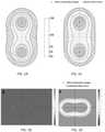
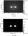
- FIGS. 1A-DA comparison of the electric field distribution (A,C) and conductivity map (B,D) of two identical numerical models without (A,B) and with (C,D) changing conductivity is shown in FIGS. 1A-D . From these figures, one can see how the conductivity increases from 0.1 S/m (the baseline level for the entire tissue domain, constant in part B) up to 0.155 S/m, an increase of 55%, for regions experiencing predicted IRE (deep red in part D), with regions experiencing varying extents of predicted reversible electroporation filling in between this (cyan through bright red).
- Tumorswill often have different electrical and physical properties than their neighboring tissues or even from their native tissues of origin (e.g., cancerous astrocytes which may not behave the same as normal ones).
- surrounding tissues of different tissue typeswill also have different properties from each other (bone, muscle, fat, blood). These differences in electrical properties will alter the electric field distribution for a given application of EBTs. Because the electric field to which the tissue is exposed is the primary determinant in the effect on the cell, these changes will change the shape and size of the affected regions. Numerical simulations are capable of modeling the electric field distribution in such heterogeneous systems. However, the rigid analytical solutions cannot be adjusted to account for such differences, and therefore could not as accurately predict affected regions for the different environments in clinical cases.
- the analytical solutione.g., could not predict the differences between a tumor situated adjacent to the skull, the quadriceps muscle, or the heart.
- lookup tablescould theoretically be developed for the dimensions of the affected regions in a number of environments, the great variability between the anatomy of each patient, each specific tumor, and each exact tumor location relative to its environment is impractical and futile.
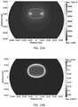
- FIGS. 2A-Jdemonstrate the effect of heterogeneous systems on electric field distribution. These figures show the electric field and temperature distribution for a three-dimensional numerical model. More particularly, FIG. 2J shows the model setup, where two needle electrodes (1 mm in diameter) are placed within the outer borders of a targeted region of tissue, surrounded by a peripheral region. The red and black regions on the electrodes represent the energized surfaces, where 4200 V was applied to one electrode and the other was set to ground. The thermal properties were set to represent a targeted region of a tumor within fat.
- FIGS. 2A-Ishow the numerical model outputs for conductivity ratios ( ⁇ t / ⁇ p ) of 0.1 (A,D,G), 1 (B,E,H), and 10 (C,F,I); showing electric field (A-F) during the pulse and temperature (G-I) distributions 1 second after the first pulse.
- the higher conductivity ratiosshow progressively more area treated by IRE with less thermal effects.
- Targeted tissue boundarymay be seen as the solid black line. Observing the electric field distribution at the boundary shows that the shape is also changing (not just size) as a result of the heterogeneous environment.
- Existing treatment planning systemsare not capable of accounting for such dynamic tissue properties in real time.
- the current embodiment of the treatment planning softwarestill leaves it up to the practitioner to select a desired number of probes, but provides no simple method of showing how the optimized distributions will be shaped if the user wants to directly compare using different numbers of probes for a given lesion.
- the current systemtherefore also does not select the optimal number of probes for the user, a question that may be difficult to answer for more complex electrode geometries.
- Temperature changes associated with Joule-type resistive heating of the tissuewill also affect local regions conductivity based on its temperature (typically increases by approximately 3%/° C.). This will also change the size and shape of the electric field distribution based on the parameters used; including the number of pulses, pulse length, and repetition rate for an entire protocol (more pulses of longer length with higher repetition rates will all increase the thermally-associated conductivity changes, increasing this variation). Because the current treatment planning tools are based on simulations from the electric field distribution of a single application of a pulse, these dynamic conductivity behaviors also cannot be taken into account. Something that does would have to be able to simulate the changes that occur as a result of thermal effects on conductivity.
- embodiments of the inventionprovide treatment planning systems, methods, and devices for determining a patient-specific electroporation-based treatment protocol comprising: a) a module operably configured to receive and process information from medical images of a target structure to prepare a 3-D reconstruction model of the target structure; and b) a module operably configured to perform a numerical model analysis using as inputs in the analysis the 3-D reconstruction and information from one or more of physical constraints, tissue heterogeneities, dynamic effects of electropermeabilization, dynamic thermal effects, or effects resulting from multiple treatments; and c) a module operably configured to construct one or more electrical protocols defining a treatment region and treatment parameters for effectively treating the target structure.
- Such treatment planning systemscan comprise a processing module capable of performing one or more of the stages in real time.
- Information from medical images to be analyzed in treatment systemscan be extracted from one or an array of images obtained from pathologic specimens or one or more imaging modalities chosen from radiographs, tomograms, nuclear scintigraphic scans, CT, MRI, PET, or US.
- the information from one or more of these sourcescan be compiled to prepare a 3D reconstruction of the target area, which is represented by a surface or a solid volume.
- the treatment planning systems according to embodiments of the inventioncan have as a target structure a) a targeted region or mass; or b) a targeted region or mass with neighboring regions; or c) a 3D map of voxels to be treated as independent elements in the finite modeling software.
- Preferred numerical model analysis for treatment systems of the inventioncomprise finite element modeling (FEM). Even more preferred as treatment planning systems, wherein the numerical model analysis involves accounting for physical constraints, tissue heterogeneities, dynamic effects of electropermeabilization, dynamic thermal effects, and multiple treatment effects.
- FEMfinite element modeling
- the treatment planning systemscan comprise a self-optimization algorithm which is capable of repeatedly evaluating one or more of physical constraints, placement of electrodes, electric field distribution simulations, and evaluation of outcome success until one or more effective protocol is constructed. It can also generate a predicted treatment time that will aid the physician in determining the optimal protocol.
- the treatment planning systemscan involve automatically, interactively, or automatically and interactively with or without user input determining the treatment region and parameters for electroporating.
- Such treatment planning systemscan also be capable of constructing protocols for an initial patient treatment or retreatment with or without additional medical images.
- Treatment systems according to embodiments of the inventioncan also be adapted to instruct an electrical waveform generator to perform the protocol.
- Such systemscan further comprise an electrical waveform generator in operable communication with the processing module and capable of receiving and executing the treatment protocol.
- Instructions for implementing the treatment protocolscan comprise specifying a number of bipolar pulses to be delivered, a length of pulse duration, and a length of any delay between pulses. Additionally, the generators of such treatment systems can be operably configured for delivering a bipolar pulse train.
- treatment planning methodscan comprise: a) receiving and processing information from medical images of a target structure and preparing a 3-D reconstruction model of the target structure; b) performing a numerical model analysis using as inputs in the analysis the 3-D reconstruction and information from one or more of physical constraints, tissue heterogeneities, dynamic effects of electropermeabilization, dynamic thermal effects, or effects resulting from multiple treatments; and c) constructing an electroporation protocol based on results of the analyzing; wherein the receiving, processing, analyzing, and constructing is performed in real time.
- Other methodsmay comprise method steps for reducing adverse effects of irreversible electroporation of tissue comprising administering electrical pulses through electrodes to tissue in a manner which causes irreversible electroporation of the tissue but minimizes electrical charge build up on the electrodes, or minimizes charge delivered to the tissue, or both.
- Adverse effects to be avoidedmay include, to name a few, one or more of thermal damage of the tissue, deleterious electrochemical effects, or electrolysis.
- Preferred methods according to the inventionmay comprise electrical pulses comprising a series of unipolar and bipolar pulses with a net charge of zero. More particularly, the net charge of zero can be achieved by a change in potential direction for each pulse, or a change in potential direction within each pulse.
- electrical pulses generated in the methodscan together comprise a pulse protocol comprising a train of unipolar pulses followed by a train of unipolar pulses of opposite polarity, or a train of bipolar pulses, or simultaneous unipolar pulses of opposite polarity which are offset from one another by a desired amount, or a combination of protocols.
- Electrical pulses used in the methods, systems, and devices of the inventioncan have a waveform which is square, triangular, trapezoidal, exponential decay, sawtooth, sinusoidal, or of alternating polarity, or comprise a combination of one or more waveforms.
- Control systems for electroporation devicesare also considered embodiments of the present invention.
- Such systemscan be configured to comprise: a) a processor in operable communication with a control module; b) a control module executable by the processor and in operable communication with an electrical circuit, wherein the control module is operably configured for initiating switching of the circuit at a rate of between 10 ms to 1 ns; and c) an electrical circuit operably configured to enable delivery of a voltage to an electrode and switching of the voltage to a second electrode to cause reversing of the polarity of the electric potential between the two electrodes.
- electroporation system embodiments of the inventioncan comprise: a) an electroporation device capable of delivering a first unipolar electrical pulse; b) the electroporation device further capable of, or a second electroporation device capable of, delivering a second unipolar electrical pulse which is opposite in polarity to the first unipolar pulse; c) a processor in operable communication with a control module; d) a control module executable by the processor and in operable communication with the electroporation device(s), wherein the control module is operably configured for initiating delivery of the first unipolar electrical pulse at a time 1 and for initiating delivery of the second unipolar electrical pulse at time 2 offset from time 1 by 1 second to 1 nanosecond.
- Electroporation devicescan also be operably configured to enable delivery of an electrical pulse to a first electrode, switching of the pulse to a second electrode to cause reversing of the polarity of the electric potential between the two electrodes, and switching of the pulse back to the first electrode or to zero, wherein a cycle of switching is established which cycle is capable of being performed at a rate of between 10 milliseconds to 1 nanosecond.
- Such devices, systems, and methodscan be configured to provide for switching to occur between or within the electrical pulse.
- Devicesfor example, can be configured such that the electrical pulses together comprise a pulse protocol comprising a train of unipolar pulses followed by a train of unipolar pulses of opposite polarity or a train of bipolar pulses.
- FIGS. 1A-Dare schematic diagrams comparing the electric field distribution (A, C) and conductivity map (B,D) of two identical numerical models without (A,B) and with (C,D) changing conductivity.
- FIGS. 2A-Iare schematic diagrams showing the numerical model outputs for conductivity ratios ( ⁇ t / ⁇ p ) of 0.1 (A,D,G), 1 (B,E,H), and 10 (C,F,I); showing electric field (A-F) during the pulse and temperature (G-I) distributions 1 second after the first pulse.
- FIG. 2Jis a schematic diagram showing placement of the electrodes in the targeted tissue for the set up illustrated in FIGS. 2A-I .
- FIG. 3is a series of CT images showing the presence of a tumor in the left thigh of the canine patient of Example I.
- FIG. 4is a CT image from FIG. 3 , within which the region of interest is traced.
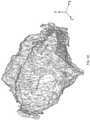
- FIG. 5is a drawing of a 3D reconstruction of the target region of Example I, which was reconstructed by compiling a series of axial traces to create a representative shape of the targeted region in three dimensions.
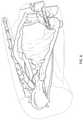
- FIG. 6is the drawing of the 3D reconstructed geometry shown in FIG. 5 visualized relative to the rest of the patient.
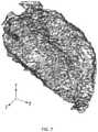
- FIG. 7is a graphic representation of the 3D reconstruction of FIG. 5 as imported into and converted within Comsol Multiphysics.
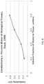
- FIG. 8is a graph from Duck, 1990, showing the relationship between conductivity and %-water, which may also be used to estimate a tissue's electrical properties.
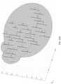
- FIG. 9is the drawing of the 3D reconstruction of the target tumor of FIG. 5 visualized in relation to surrounding structures within the body, which is a tool useful for developing treatment constraints.
- FIG. 10is a graphic representation of the 3D reconstruction of FIG. 5 as imported into and converted within Comsol Multiphysics and further including a demonstrative electrode placement for an exemplary treatment protocol.
- FIG. 11Ais a schematic representation of an electric field distribution map, showing a top view of the electrodes of FIG. 10 in an energized state.
- FIGS. 11B-Dare schematic diagrams demonstrating falloff of the electric field distribution in the third dimension, showing an exemplary electric field distribution in the xz-plane ( FIG. 11B ), in the xy-plane at the midpoint of the electrodes, and in the xy-plane at the tips of the electrodes.
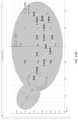
- FIG. 12Ais a schematic drawing showing a representative geometry of the treatment area in which compiled ellipsoids (shown in pink) illustrate the electroporation protocol developed to attain the desired treatment objectives.
- FIG. 12Bis a schematic drawing showing a top view of the treatment area geometry shown in FIG. 12A , and further demonstrating the electrode insertion paths.
- FIGS. 13A-Bare respectively schematic diagrams of an electric field distribution and a corresponding conductivity map demonstrating a homogeneous distribution that only changes by 0.1% for visualization purposes when irreversible electroporation is accomplished.
- FIGS. 14A-Bare respectively schematic diagrams of an electric field distribution and a corresponding cumulative conductivity map demonstrating a treatment region where more than two electrode pairs are energized and homogeneous distribution only changes by 0.1% for visualization purposes when irreversible electroporation is accomplished.
- FIGS. 15A-Bare respectively schematic diagrams of an electric field distribution and a corresponding conductivity map demonstrating a heterogeneous distribution that changes from 0.67 S/m to 0.241 due to electropermeabilization caused by electroporation.
- FIGS. 16A-Bare respectively schematic diagrams of an electric field distribution and a corresponding conductivity map demonstrating a heterogeneous distribution that changes from 0.67 S/m to 0.241 S/m due to electropermeabilization.
- FIGS. 20A-Bare two-dimensional (2-D) diagnostic T1 post-contrast MRI scans in which the tumor was traced.
- FIGS. 21A-His a graphic representation of a three-dimensional (3-D) solid representing a tumor volume and displaying the voltage configurations that would mainly affect tumor tissue in this particular situation.
- FIG. 22is a graph showing a Bipolar IRE pulse (100 ⁇ s duration) with alternating polarity in the middle of the pulse.
- FIG. 23Ais a schematic diagram of a representative circuit model for switching polarity between pulses and multipolar pulses.
- FIG. 23Bis a graph showing the shape of a bipolar pulse that can be created using the electrical circuit of FIG. 23A .
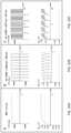
- FIGS. 24A-Gare graphs showing various pulsing protocols according to the invention, demonstrating exemplary frequencies, pulse length, and time delay between pulses.
- FIGS. 25A-Bare schematic diagrams showing variations in techniques for generating bipolar electrical pulses in accordance with embodiments of the invention.
- FIG. 25Cis a schematic diagram of a representative circuit model for generating and administering simultaneous, continuous, but offset pulses as shown in FIG. 25A .
- FIG. 26Ais a photograph showing the N-TIRE electrodes with attached fiber optic probes, which were used in this intracranial treatment of white matter to measure temperature during pulse delivery.
- FIG. 26Bis a graph showing temperature [° C.] distribution during an N-TIRE treatment in the white matter of a canine subject.
- FIGS. 27A-Jdepict electrical field outputs for various combinations of electrodes emitting different charges.
- FIG. 27Adepicts a two-dimensional display for the use of four electrodes of alternating polarity.
- FIG. 27Bdepicts an axis symmetric display for the use of four similar electrodes of alternating polarity.
- FIG. 27Cdepicts a two-dimensional display for the use of four charged electrodes, the center two at 5000V and 0V and the outer two at 2500V.
- FIG. 27Ddepicts an axis symmetric display for the use of a similar electrode set up as FIG. 27C .
- FIG. 27Edepicts a two-dimensional display for the use of three electrodes with the center one at 2500V and the outer two at 0V.
- FIG. 27Fdepicts an axis symmetric display for the use of three electrodes similar to Panel E.
- FIG. 27Gdepicts a two-dimensional display for the use of three charged electrodes, the center at 0V, the left at 5000V, and the right at 2500V.
- FIG. 27Hdepicts an axis symmetric display for the use of a similar electrode set up as Panel G.
- FIG. 27Idepicts a two-dimensional display for the use of three charged electrodes, the center at 1750V, the left at 3000V, and the right at 0V.
- FIG. 27Jdepicts an axis symmetric display for the use of a similar electrode set up as FIG. 27I .
- FIGS. 28A-Bare graphs showing output of the arbitrary function generator prior to signal amplification by the high voltage MOSFET positive and negative polarity switches.
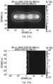
- FIGS. 29A-Bare micrographs showing in vitro experimental results on electroporation with high-frequency bipolar, pulses using a trypan blue dye exclusion assay.
- FIG. 30A-Care waveforms of IRE with unipolar pulses and high-frequency IRE with the corresponding TMP development across the plasma membrane ( ⁇ pm ) for a 1500 V/cm unipolar pulse ( FIG. 30A ) and a 1500 V/cm bipolar burst without a delay ( FIG. 30B ) and with a delay ( FIG. 30C ).
- FIG. 31is a graph comparing time above the critical threshold ( ⁇ cr ) for IRE at various center frequencies.
- FIGS. 32A-Care waveforms of IRE with unipolar pulses and high-frequency IRE with the corresponding TMP development across the plasma membrane ( ⁇ pm ) for a 1500 V/cm unipolar pulse ( FIG. 32A ), a 1500 V/cm bipolar burst without a delay and with a shortened negative phase ( FIG. 32B ), and a 1500 V/cm bipolar burst with a delay and with a shortened, lower amplitude negative phase ( FIG. 32C ).
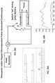
- FIG. 33is a chart showing an exemplary output from an in vivo treatment of the brain with high-frequency, bipolar pulses, where the snapshot is taken within a single burst.
- FIGS. 34A-Care schematic diagrams showing electric field, norm (V/cm) contours predicted by the FEM during a 1000 V amplitude burst with a center frequency of 1 kHz ( FIG. 34A ) and 1 MHz ( FIG. 34B ).
- V/cmelectric field, norm
- FIG. 35depicts electrical field outputs from various configurations employing two electrodes.
- FIG. 35Adepicts the use of two electrodes spaced 0.5 cm apart.
- FIG. 35Bdepicts the use of two electrodes spaced 1.0 cm apart.
- FIG. 35Cdepicts the use of two electrodes spaced 1.5 cm apart.
- FIG. 35Ddepicts the use of two electrodes spaced 1.0 cm apart.
- FIG. 35Edepicts a side view of electrical field outputs from one device having two electrically conductive regions separated by an insulative region of a length of 0.5 cm.
- Irreversible electroporationis a new focal tissue ablation technique.
- the treatmentsare capable of sparing major blood vessels, extracellular matrix and other sensitive or critical structures.
- the procedureinvolves the delivery of low-energy electric pulses through minimally invasive electrodes inserted within the tissue.
- the target tissueis exposed to external electric field distributions around the electrodes, which alter the resting transmembrane potential of the cells.
- the degree of tissue electroporationi.e., no effect, reversible electroporation and/or irreversible electroporation depends on the magnitude of the induced transmembrane potential.
- Numerical models for electric field optimizationare available and typically include the physical properties of the tissue and treatment parameters including electrode geometry and pulse parameters (e.g., duration, number, amplitude, polarity, and repetition rate). These models can also incorporate the dynamic changes in tissue electric conductivity due to electroporation and thermal effects.
- a numerical model to visualize the IRE treated regions using sequential independent combinations of multiple energized and grounded electrodesthere is provided a numerical model to visualize the IRE treated regions using sequential independent combinations of multiple energized and grounded electrodes.
- electric conductivity changes due to electroporation and thermal effects from an IRE pulse sequenceare capable of being incorporated into the analysis for developing and constructing more effective treatment protocols.
- a particular embodimentinvolves setting the resulting conductivity distribution as the initial condition for the next pulse sequence, then repeating this procedure sequentially until all the pulse sequences are completed. In this manner, electric conductivity dependencies from previous pulses are incorporated and more accurate electric field distributions are presented. It is important to note that it is assumed that once a tissue is irreversibly electroporated, the tissue conductivity would not revert back. Consequently, a comprehensive IRE distribution can be presented in which the conductivity changes due to the previous pulses are considered. Such methods are most useful when using three or more electrodes with electrode-pairs being energized independently.
- the electric conductivity map in certain circumstancescan be crucial in the treatment planning of irreversible electroporation and other pulsed electric field therapeutic applications.
- the conductivity mapis what determines how the current generated by the applied voltages/potentials will flow and the magnitude of the electric field. Several factors affect this distribution before, during and after the treatment including tissue heterogeneities, electropermeabilization, thermal effects and multiple treatments.
- each tissuehas its own “resting/unique” electric conductivity before the application of the electric pulses.
- any particular organ or systemthere could be a mixture of conductivities that need to be accounted for in the treatment planning as in the case of white matter, gray matter and tumor tissue in the brain for example.
- each of the tissue's conductivitywill vary with changes in temperature as is the case for brain (3.2% C ⁇ 1 ) or liver (2% C ⁇ 1 ).
- the main region treated by irreversible electroporationdoes not have sufficient increase in temperature to generate thermal damage, however, at the electrode tissue interface (where the electric field is highest) there is a significant increase in temperature and thus the conductivity map is altered. Capturing these and other dynamic effects can be crucial since they represent more accurate/realistic treatment geometries and pulse parameters that are not captured elsewhere. Accounting for these effects in treatment planning software is expected to lead to the optimization of pulse parameters and minimize damage to surrounding healthy tissue.
- Numerical modeling methodssuch as finite element modeling (FEM) are more accurate and are actually where the previous treatment planning systems derive their solutions (the lookup table and analytical solutions are calibrated to mimic the numerical solutions).
- FEMfinite element modeling
- a canine patient with a 360 cm 3 tumor in the left thighwas treated according to a treatment planning embodiment of the invention.
- This treatment planserves to demonstrate the complexity and numerous steps typically involved in developing and implementing a comprehensive treatment plan for electroporation-based therapies.
- This descriptionis intended to provide guidance as to the formulation of a basic treatment planning system, which can be operably configured to include one or more of the following stages:
- Images of the target lesion or of a portion of the body to be treatedcan be acquired by taking an array of medical images using one or more imaging modalities, including CT, MRI, PET, or US to name a few.
- imaging modalitiesincluding CT, MRI, PET, or US to name a few.
- an imaging modalitysuch as computed tomography CT can be used to determine the presence of a tumor.
- an imaging modalitysuch as computed tomography CT
- information about or relating to the region of interestcan be collected and used to determine a targeted region, its location, its position, any important or relevant nearby structures that must be accounted for (such as blood vessels, nerves, collecting ducts, etc.), and any relative basic dimensions (such as depth within tissue, basic cross-sectional sizes, distance from other structures, etc.).
- ROIRegions of Interest
- the target ROIcan be outlined in the images used to identify the tumor, whether manually or by way of a computer program, to identify a potential treatment area.
- a computer program capable of detecting anomaliessuch as the OsiriX open-source image analysis software (Geneva, Switzerland), could be used to outline the targeted region (e.g., a tumor, site for electrogenetransfer, etc.).
- the targeted regione.g., a tumor, site for electrogenetransfer, etc.
- FIG. 4one of the CT scans from FIG. 3 is shown with the region of interest traced. Tracing the region of interest in each of a series of CT images compiling the 2D traces of each slice would allow for compilation of 3D geometry for the target region.
- the traced regions of interest from a series of axial CT slicescan be compiled and interpolated between the steps to create a three-dimensional geometry that the practitioner could use to gain an understanding of the basic shape of the target mass and/or its location relative to other tissues.
- FIG. 5shows a series of axial traces having been compiled to create a representative shape of the targeted region in three dimensions. This reconstruction may be maneuvered to assess its general shape and thus allow determination of potentially efficient electrode insertion approaches.
- the reconstructed geometrycan also be visualized relative to the rest of the patient.
- Thisallows one to assess (in greater detail than the initial FIG. 3 images) physical constraints such as bones preventing electrode insertion, relative location of sensitive structures, and orientation of the lesion relative to the body, allowing a practitioner to evaluate optimal electrode insertion approaches.
- physical constraintssuch as bones preventing electrode insertion, relative location of sensitive structures, and orientation of the lesion relative to the body, allowing a practitioner to evaluate optimal electrode insertion approaches.
- the long axis of the tumoris roughly parallel to the length leg and femur, so a user may consider reducing the number of electrodes and insertions used by orienting the electrodes along this axis, or they may go with more electrodes perpendicular to the top of the leg (since the femur prevents access from the bottom of the leg).
- the 3D geometrycan then be imported into finite element modeling software (FEM). Indeed, several geometries can be imported using software such as Comsol Multiphysics (Comsol, Swiss, Sweden), including: a) just the targeted region or mass; b) the targeted region and other traced neighboring regions (muscle, fat, bone, etc); or a 3D Map of all the voxels to be treated as independent elements in the finite modeling software.
- FEMfinite element modeling software
- FIG. 7shows a model of the 3D target geometry as imported into numerical modeling software. More particularly, the geometry developed and shown in FIG. 5 may be converted to a surface or a solid and imported into numerical modeling software.
- the black shapeis a converted geometry within Comsol Multiphysics for the targeted region reconstructed above. Its dimensions and volume have been normalized to ensure its size matches that of the reconstructed volume.
- any physical and/or thermal properties and/or electrical propertiescan be assigned in numerous ways.
- the propertiescan be assigned arbitrarily; deduced by designating which of the target region or the other traced neighboring regions are of what tissue type and using properties of these tissue types from the literature; experimentally measured with a “pre-pulse” (e.g., as described in U.S. patent application Ser. No. 12/491,151, “Irreversible Electroporation to Treat Aberrant Cell Masses;” or the properties can be derived from an algorithm or coordination scheme based on voxel or pixel value imported from the 3D map.
- the assignment of properties to the modelcan be performed within software and manually accounted for in placements. If such properties are either assigned arbitrarily or are deduced as described above, the different shapes depicted in the model (e.g., FIG. 7 ) may each be assigned a different set of properties to best represent the tissue or material used (such as 0.025 S/m for the fatty tumor, and 0.5 S/m for the surrounding tissue).
- the tissue propertiesare derived from medical images. Due to the properties of tissue and how the tissues are assessed by modern imaging techniques, it may be possible to derive accurate estimations of a tissue's properties based on its response to the various imaging modalities.
- pixel valuesare based on the radiodensity of the tissue at that point in the image (its attenuation). It is common practice to scale these attenuations relative to distilled water according to the equation:
- a tissue's Hounds Unit (HU) valuemay serve as a representation of its relative water content, with larger absolute value HU's (because it can be negative as well) containing less water.
- HUHounds Unit
- Potential physical placement constraintssuch as vital structures (nerves, brain, blood vessels, etc.), access orientation preferences (from head, from rear, supine, prone patient positioning, etc.), and/or physical barriers (bones, sensitive structures, etc.) can be identified. The potential constraints can then be used to guide/constrain what angles are possible for the electrodes and if the electrodes should be placed to avoid certain areas more than others.
- FIG. 9shows a graphic 3D reconstruction of the target tumor in relation to surrounding structures within the body, which is useful for developing treatment constraints.
- the physical location of the tumor relative to the rest of the body(shown in FIG. 9 by arrows pointing out vasculature and nerves, for example) can be demonstrated using the previously prepared 3D geometric representation of the tumor. This information may be used to constrain or direct where the electrodes should be placed and give priority to regions that should be spared relative to regions that would not cause as significant of problems.
- Electrodescould be placed into or around the targeted region. Their number, location, orientation, and size could all be adjusted independently.
- FIG. 10is a graphic 3D representation of the imported tumor geometry with electrodes placed.
- the geometric representation of the targeted regionis depicted in red, while representations of electrodes are shown at two locations in blue. The number, orientation, and location of these electrodes is capable of being manipulated to satisfy the desired treatment objectives.
- Simulation of the electric field distributionare capable of being correlated with experimental data to superimpose predicted volumes of affected regions (treated, untreated, thermal damage).
- FIG. 11shows the electrodes depicted in FIG. 10 in an energized state.
- a section on the endhas been set to a voltage while a section on the rest has been set to ground with a section of insulation between, creating a voltage gradient that surrounds the single needle.
- the entire length of one electrodehas been set to a voltage while the other electrode has been set to ground, creating a voltage gradient between them.
- the color mapsare representative electric field isocontour regions that may be used in determining predicted treatment regions, reversible regions, or safety margins based on electric field thresholds. For example, if the protocol anticipates an IRE electric field threshold of 500 V/cm, then the entire volume of the tissue exposed to this electric field or higher (depicted in green) would be the predicted treatment region. In addition, if it were desired to ensure sparing of a sensitive structure such as a nerve, and an exact resolution of the above-predicted 500 V/cm IRE electric threshold was insufficient to guarantee sparing, a different electric field may be used to predict a safety margin which would be used to ensure that this threshold is not crossed by the sensitive structure (such as 250 V/cm depicted in red).
- the electric field distributionis typically at a maximum at the cross-sectional region midway between the lengths of the electrodes and tapers off toward the ends of the electrodes.
- the image shown in FIG. 11Bshows the electric field distribution between 35000 and 150000 V/m looking at both electrodes simultaneously in the xz-plane.
- the grey rectanglesare the electrodes, running along the z-axis, and separated by 1.5 cm (center-to-center) along the x-axis.
- This optimization stagecan be performed manually (interactively) by a practitioner or automatically.
- the Optimization Quality Function of Formula I discussed in more detail belowcould also be used for manual optimization.
- the optimization phase of the systemwas performed qualitatively and was iterated with the previous four steps until settling on the electrode array shown in FIGS. 12A and B.
- the resultant representative geometry of compiled ellipsoids(shown in pink) illustrates the satisfactory electroporation protocol developed in order to attain the desired treatment objectives.
- FIG. 12Ait can be seen that a highly complex array of electrodes (blue) was selected, where some electrodes are inserted and exposed an amount (such as 1 or 2 cm), to treat an amount of depth with pulsing, before withdrawing them some and repeating the pulsing. This was done to ensure complete treatment along the depth of the treatment.
- the blue cylindersdepict discrete electrode placements for pulsing, and the ones stacked on top of each other represent this aspect.
- FIG. 12Ba top view of the graphic representation of the treatment area of FIG. 12A is provided, in which the electrode insertion paths can be seen. Since the electrodes were all running perpendicular, spacing dimensions have been outlined to aid the placement of the electrodes for the practitioner. The pulses would be administered between each electrode and the electrodes in closest proximity to it. Electric pulse parameters are adjusted between each electrode firing pair based on separation distance and the desired treatment region (based on targeted volume and avoidance of sensitive tissues). The dimensions in red are also used as guidelines for the placement of the outer electrodes relative to the margins of the tumor to prevent excessive treatment of peripheral (untargeted) regions.
- the generator system for applying the designated pulsing protocolcan be set up for implementation of the desired protocol. More particularly, the practitioner could then place the electrodes according to the prescribed protocol and let the generator apply the pulses.
- the systems, methods, and or devices according to the inventioncan be operably configured to monitor certain variables.
- One such variablecan include monitoring the temperature of the electrodes and/or surrounding tissue in real time during treatment to ensure limited to no thermal damage to the tissue being treated. If monitored in real time, adjustments could then be made, if necessary, to avoid damage.
- One, multiple, or all phases of system embodiments according to the inventioncan be performed manually or be performed (in whole or in any number of parts) by an automated system capable of performing the phases for the practitioner. Many of these steps can be performed without user input, and could be blocked off into distinct automated processes (with/without coupling to human-performed processes) or could be linked together through a comprehensive system. All of this is able to be done for an initial treatment, or redone for any retreatments that may be necessary, with or without new images (depending on case circumstances).
- Systems according to embodiments of the inventionare flexible in that such systems can be operably configured to solve many scenarios numerically and to select the best electrode geometry and pulse parameters for a given situation. Alternatively or additionally, solutions may be obtained analytically, with tables, etc.
- Embodiments of the systems according to the inventioncan be operably configured to be run on an independent system well in advance of treatment administration to allow sufficient computation time, review, and possible re-working of the protocol prior to treatment.
- the appropriate protocolcould then be uploaded directly to the pulse generator.
- Preferred embodiments of systems according to the inventioninclude a model creation stage for establishing an initial model of the target area.
- Treatment geometriesmay be input manually, by analyzing medical images that were taken and any reconstructions, from computer analyses of medical images/tomography, or other (2D and 3D) mapping techniques.
- Conductivity values for the model subdomainsmay be obtained by measuring them on the subject directly (placing electrodes within tissue then applying a voltage and measuring the current to get Z/ ⁇ ), by taking typical values found in the literature for the tissue types, or by noninvasive a measuring techniques such as functional Magnetic Resonance Imaging (fMRI), Electrical Impedance Tomography, etc; and combining these with the relevant equations (for E-field distributions, it is the ratio between tissues/regions that alters the field, absolute values will only be important when considering thermal effects).
- fMRIMagnetic Resonance Imaging
- fMRIMagnetic Resonance Imaging
- Medical images that already obtain the conductivity values (fMRI) or coupled to conductivity valuesmay then be used as the geometries for a numerical/analytical model as the various subdomains, to establish the initial model.
- a single or any set of electrode optionsmay be selected to be used or allowed to be selected by the program.
- the practitionerAfter setting up the geometry and electrode options to consider, the practitioner would essentially select a “GO” button to let the program run through the many variations to use and solve each using FEM or advanced analytical methods.
- the programwould solve each scenario for various effects (no effect, reversible electroporation, irreversible electroporation, or thermal) and distributions within the model.
- Thermal considerationswill greatly increase the computational cost of the model, but may be desired to determine thermal damage and scarring, especially in very sensitive structures.
- the systems of embodiments of the inventioncan employ a variety of algorithms (iterative, genetic, etc.) in order to optimize the treatment parameters for the best possible result for a particular patient scenario.
- Such systemscan also be operably configured to employ a function for evaluating the quality of each solution, where desired results, D, (IRE and/or REB throughout the targeted regions) are added; and the undesired results, U, (thermal damage, IRE beyond targeted region, etc.) are subtracted, with each aspect having its own unique scaling (since IRE to entire targeted region is far more important that avoiding IRE to healthy tissues).
- ⁇ (ET, EP, ⁇ , . . . )This is the value function of a given treatment protocol for the modeled domain previously mentioned as a function of electrode type and geometry, electrode positioning, applied voltage, and any other factors. More specifically: 1) ET (style, number, dimensions), with style referring to the style of the pulse, such as single, multi-unipolar, hybrid, proprietary, etc., with number referring to the number of probes used, and dimensions referring to the geometry and dimensions of all exposed and insulated regions in all three directions for each electrode used.
- ETstyle, number, dimensions
- EPrefers to the position of each or all electrodes in relation to a reference point arbitrarily chosen within the (x, y, z) domain of the model (location and orientation).
- the center of the tumorcould be selected as the reference point and arbitrarily set to (0, 0, 0).
- the reference pointmay also be selected ahead of time or afterwards by the practitioner that will be easy for the practitioner to physically use at the time of treatment administration, such as some anatomical landmark that can be used as a reference for where the electrodes are and the electrode orientation. It is also possible to match the coordinate system from the medical images.
- the ⁇ functionmay be solved for altered ET and EP, and the ⁇ may then be scaled accordingly for the geometry (since it the model geometry and properties that will affect the shape of the distribution, the absolute value of it may be scaled to the applied voltage after this shape is found for each ET and EP). This would dramatically reduce the number of iterations and thus the computational cost.
- the systemcan be operably configured to iteratively adjust ET, EP, etc. and obtain the resulting ⁇ , storing the top ones (or all those meeting some type of baseline threshold criterion).
- the resulting stored solutionswould then be saved for presentation to the practitioner for conducting a review and visually assessing the value of each solution for selecting the protocol that best meets the demands of the therapy (could range on their arbitrary criterion such as the best quality, most simple to administer and apply the EP in the treatment, most robust, etc.)
- the electrical parameters usedcan be set as standardized parameters for typical treatments, and optionally these parameters can be flexible in case certain scenarios require different values—such as abdomino-thoracic procedures requiring repetition rate to be synchronized with the patient's heart rate to reduce the risk of pulse-induced arrhythmias.
- standard electrical parameterscan be used to determine the best combination of treatment parameters to use and have been applied to various tissues/tumors to determine the electric field threshold of each for this set of parameters, thus allowing treatment outcome to be reviewed and not just electric field distributions.
- Table IIprovides a list of exemplary electric parameters that can be manipulated within the IRE treatments discussed herein.
- Pulse lengthns-ms range Number of pulses: 1-50,000 pulses Electric Field Distribution: 1-5,000 V/cm Frequency of Pulse 0.001-1000 Hz
- Pulse shapesquare, triangular, trapezoidal, exponential decay, sawtooth, sinusoidal, alternating polarity
- Pulse typePositive, negative, neutral electrode charge pulses (changing polarity within pulse)
- Multiple sets of pulse parameters for a single treatmentchanging any of the above parameters within the same treatment to specialize outcome
- Electrode typeParallel plate: 0.1 mm-70 cm diameter (and larger for applications relating to e.g., whole organ decellularization) Needle electrode(s): 0.001 mm-1 cm diameter
- Single probe with embedded disk electrodes0.001 mm-1 cm diameter
- Spherical electrodes0.0001 mm-1 cm diameter Needle diameter: 0.
- Electrode length0.1 mm to 30 cm
- Electrode separation0.1 mm to 5 cm, or even 5 cm to 20 cm, or 20 cm to 100 cm, and larger (for reversible electroporation, gene delivery, or positive electrode with ground patch on patient's exterior, e.g.)
- FIGS. 13A-Bdemonstrate a situation in which there would be little to no change in the physical properties of the tissue as a result of electroporation. More specifically, as shown in FIG. 13A , an electric field distribution [V/cm] generated by an applied voltage difference of 3000V over the upper two electrodes is shown. FIG. 13B provides a conductivity map [S/m] displaying a homogeneous distribution that only changes by 0.1% for visualization purposes when irreversible electroporation is accomplished. The white outline represents the region of tissue that is exposed to an electric field magnitude that is sufficient for generating irreversible electroporation.
- FIGS. 14A-Bdemonstrate an electric field distribution and conductivity map for a treatment region for a given situation in which more than two electrode pairs are energized.
- FIG. 14Aan electric field distribution [V/cm] generated by an applied voltage difference of 3000V over the right two electrodes is shown.
- FIG. 14Bshows a conductivity map [S/m] displaying a homogeneous distribution that only changes by 0.1% for visualization purposes when irreversible electroporation is accomplished in this set up.
- a cumulative visualization of the treatment regionis shown.
- FIGS. 15A-Bdemonstrate a change in the shape and size of the treatment region due to electropermeabilization. More particularly, in FIG. 15A , an electric field distribution [V/cm] generated by an applied voltage difference of 3000V over the upper two electrodes is shown. FIG. 15B provides a conductivity map [S/m] displaying a heterogeneous distribution that changes from 0.67 S/m to 0.241 due to electropermeabilization as a result of electroporation. Of particular note in this example, the shape and size of the treatment region is consequently adjusted as a result of this change. In FIG. 15B the shape and size of the planned treatment region is different than in the above examples ( FIGS. 13B and 14B ) in which the conductivity was assumed to remain constant throughout the delivery of the pulses.
- FIG. 16Aprovides an electric field distribution [V/cm] generated by an applied voltage difference of 3000V over the right two electrodes
- FIG. 16Bshows a conductivity map [S/m] displaying a heterogeneous distribution that changes from 0.67 S/m to 0.241 S/m due to electropermeabilization.
- the first set of pulses using the top two electrodesincreased the conductivity of the tissue which in turn modified the electric field distribution (i.e., treatment region) for the second application of pulses (right two electrodes) adjacent to the permeabilized region.
- FIGS. 17-19are provided.
- FIG. 17Bshows a thermal damage assessment by the potential increase in temperature due to the electric pulses which occurs when greater than 0.53.
- FIG. 18Bshows some thermal damage visualized at the electrode-tissue interface 0.11 cm 2 .
- FIG. 19Bshows significant thermal damage at the electrode-tissue interface due to thermal effects 0.43 cm 2 .
- the electrical protocol(pulse characteristics, number, sequence, etc.) could be saved and then uploaded to the pulse generator system. At this point, the practitioner would have to do no more than place the electrodes in the predetermined positions and hit “START”, at which point the instrument carries out the prescribed pulsing conditions.
- Open source image analysis software(OsiriX, Geneva, Switzerland) was used to isolate the brain tumor geometry from the normal brain tissue.
- the tumorwas traced in each of the two-dimensional (2-D) diagnostic T1 post-contrast MRI scans as shown in FIGS. 20A-B .
- Attemptswere made to exclude regions of peritumoral edema from the tumor volume by composite modeling of the tumor geometry using all available MRI sequences (T1 pre- and post-contrast, T2, and FLAIR) and image planes.
- a three-dimensional (3-D) solid representation of the tumor volumewas generated using previously reported reconstruction procedures.
- the tumor geometrywas then imported into a numerical modeling software (Comsol Multiphysics, v.3.5a, Sweden) in order to simulate the physical effects of the electric pulses in the tumor and surrounding healthy brain tissue.
- the electric field distributionwas determined in which the tissue conductivity incorporates the dynamic changes that occur during electroporation.
- a 50% increase in conductivitywas assumed when the tissue was exposed to an electric field magnitude greater than 500 V/cm, which has been shown as an IRE threshold for brain tissue using specific experimental conditions.
- the threshold for brain tumor tissueis unknown so the same magnitude as normal tissue was used for treatment planning purposes.
- NanoKnife®AngioDynamics, Queensbury, N.Y. USA
- the NanoKnife®is an electric pulse generator in which the desired IRE pulse parameters (voltage, pulse duration, number of pulses, and pulse frequency) are entered.
- the NanoKnife®is also designed to monitor the resulting current from the treatment and to automatically suspend the delivery of the pulses if a current threshold is exceeded.
- the electrodeswere inserted into the tumor tissue in preparation for pulse delivery.
- the blunt tip electrodeswere connected by way of a 6-foot insulated wire (cable) to the generator. After foot pedal activation, the pulses were conducted from the generator to the exposed electrodes.
- the two sets of pulse strengthswere delivered in perpendicular directions to ensure uniform coverage of the tumor and were synchronized with the electrocardiogram (ECG) signal to prevent ventricular fibrillation or cardiac arrhythmias (Ivy Cardiac Trigger Monitor 3000, Branford, Conn., USA).
- ECGelectrocardiogram
- the sets of pulseswere delivered with alternating polarity between the sets to reduce charge build-up on the surface of the electrodes.
- shorter pulse durations than those used in previous IRE studieswere used in order to reduce the charge delivered to the tissue and decrease resistive heating during the procedure.
- Previous calculations and experimental data from previous intracranial IRE experimentsensured that no thermal damage would be generated in normal brain.
- the temperature measured near the electrodesshowed a maximum 0.5° C. increase after four sets of twenty 50- ⁇ s pulses when using similar parameters to the ones in Table I.
- the charge delivered during the procedurewas typical or lower than that used in humans during electroconvulsive therapy, a treatment for depression that also uses electric pulses.
- alternating polarity of adjacent electrodesminimizes charge build up and provides a more uniform treatment zone. More specifically, in IRE treatments there is an energized and grounded electrode as the pulses are delivered. In embodiments, charge build-up on the surface of the electrodes can be minimized by alternating the polarity between sets of pulses. It is believed that there are still electrode surface effects that can be associated with negative outcomes.
- FIG. 22is a graph showing a Bipolar IRE pulse (100 ⁇ s duration) with alternating polarity in the middle of the pulse in order to minimize charge delivered to the tissue.
- negative effectscan be prevented, reduced, or avoided as part of IRE treatment in the brain, including deleterious electrochemical effects and/or excessive charge delivered to the tissue as in electroconvulsive therapy.
- a superficial focal ablative IRE lesionwas created in the cranial aspect of the temporal lobe (ectosylvian gyrus) using the NanoKnifeB (AngioDynamics, Queensbury, N.Y.) generator, blunt tip bipolar electrode (AngioDynamics, No. 204002XX) by delivering 9 sets of ten 50 ⁇ s pulses (voltage-to-distance ratio 2000 V/cm) with alternating polarity between the sets to prevent charge build-up on the stainless steel electrode surfaces.
- NanoKnifeBAngioDynamics, Queensbury, N.Y.
- blunt tip bipolar electrodeAngioDynamics, No. 204002XX
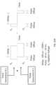
- FIG. 23Ais a schematic diagram of a representative circuit model for switching polarity between pulses and multipolar pulses.
- a basic circuitmay contain a) a generator supply circuit containing a voltage source and capacitor bank to accumulate sufficient charge for pulse delivery; b) a simultaneous switching mechanism; c) electrodes for pulse delivery (here, 2 electrodes are shown); and d) a parallel capacitor-resistor equivalent to represent the behavior of biological tissues.
- FIG. 23Bshows an exemplary bipolar pulse that can be created using the circuit of FIG. 23A .
- the circuitcan be operably configured to function in the following representative manner.
- the switchesare in position 0 .
- the voltage sourcewould be used to charge an array of capacitors to the desired electric potential for a given pulse.
- the switchesmove to position 1 .
- Thiscauses rapid initiation of capacitor discharge, generating a high-slope ⁇ V between the electrodes placed in the tissue (the first half of a square wave).
- Thisgives electrode 1 a “negative” voltage and electrode 2 a “positive” voltage (based on their relative electric potentials).
- the capacitor(s)continue delivering the electric charge over time, causing a logarithmic decay of the electric potential to which the tissue is exposed.
- the switchesmove to position 2 .
- unipolar pulsesmay have their polarity reversed every pulse or after any number of pulses by moving the switches from position 0 to 1 for pulse delivery, then back to 0 (first pulse); then from position 0 to 2 for delivery, then back to 0 (second pulse of opposite polarity).
- a unipolar pulse of any polaritycan be reversed after one or more pulses up to any number of desired pulses for a particular application.
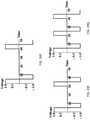
- a time delay between the unipolar pulse and the reversed polarity unipolar pulsecan be any desired duration as well, including from 5 times the pulse length ( FIG. 24A ), to 3 times the pulse length ( FIG. 24B ), to 1 time the pulse length ( FIG. 24C ), to no delay (or effectively no delay) at the time of switching ( FIG. 24D ).
- the number of electrical pulses that can be applied to successfully treat tumorscan be quite high.
- Prior art methods of using IRE for various purposesincluded the use of relatively few pulses, for example 8 pulses or so. Reports of use of up to 80 pulses for IRE have been published; however, to the inventors' knowledge, a higher number of pulses has not been recommended.
- the present inventionprovides for the use of a relatively high number of pulses, on the order of 90 pulses or greater. For example, in exemplary embodiments, 90 pulses are used. Other embodiments include the use of more than 90 pulses, such as 100 pulses, 110 pulses, or more.
- Moderately high numbers of pulsessuch as those in the range of 50-90 pulses can also be used according to the invention, as well as moderate numbers of pulses, such as those in the range of 20-50 pulses.
- voltages traditionally used for IREare too high for beneficial treatment of tumors in situ.
- IREis performed using voltages of between 4000 V/cm to 1500 V/cm.
- the present inventionprovides for use of voltages of much lower power.
- the present methodscan be performed using less than 1500V/cm. Experiments performed by the inventors have shown that 2000 V/cm can cause excessive edema and stroke in patients when applied to brain tissue.
- applied fields of about 500 V/cm to 1000 V/cmare used.
- applied fields of less than 1000 V/cmcan be used.
- the pattern of alternating between pulse polaritiescan be repeated any number of times to accomplish a desired result.
- the bipolar pulse of FIG. 24Dis shown repeated at timing intervals of 3 times the pulse length ( FIG. 24E ), to 2 times the pulse length ( FIG. 24F ), to 1 time the pulse length ( FIG. 24G ).
- the delay between bipolar pulsescan also be zero (or effectively zero) and/or the bipolar pulses can be repeated any number of time to establish a particular desired pulsing protocol or pattern.
- the pulsescould also be made multipolar by switching from position 0 to 1 (first polarity), then to position 2 (reversed polarity), then back to position 1 (returning to initial polarity), and so on, all within the same pulse.
- the bipolar pulsescan be configured in a manner to deliver a charge to the tissue where the net effect of the pulse is something other than zero.
- the magnitude of the positive portion of the pulsecan be different than the magnitude of the negative portion of the pulse.
- the pulsecan be 90% positive and 10% negative or 90% negative and 10% positive.
- any ratio of positive:negative chargecan be used, including from 0:100 (mono-polar and positive) to 100:0 (mono-polar and negative).
- 50:50net charge of zero
- 90:10, 80:20, 75:25, 60:40 and the reversecan be used depending on the desired effect.
- any switchcould be used to alter the length of any pulse or change the pulse repetition rate. And, if varying combinations of different capacitor banks were used in the system, then depending on which ones were connected, it would be possible to change the applied voltage to the electrodes between pulses or within a pulse (of any polarity).
- the individual electric pulsescan be unipolar while in other embodiments, the individual electric pulses can be bipolar.
- a train of unipolar pulsesis delivered in one direction, followed by a subsequent pulse train of opposite polarity.
- the waveforms of the electric pulsesare triangular, square, sinusoidal, exponential, or trapezoidal. Other geometric shapes are contemplated as well.
- an electrodeis connected to a system for employing electrical impedance tomography (EIT), computed tomography (CT), Magnetic Resonance Imaging (MRI), or ultrasound to image the tissue prior to treatment by applying small alternating currents that themselves do not damage the tissue.
- EITelectrical impedance tomography
- CTcomputed tomography
- MRIMagnetic Resonance Imaging
- a large variety of other parameterscan influence the efficiency of membrane poration, such as the shape of the electrical pulses, polarity, size of target cells, and thermal conditions during and after the pulses.
- Another method for avoiding excessive charge build up in tissues being treated by electroporationis to deliver counteracting pulses simultaneously from one or more pulse generator.
- the pulses delivered by the generatorscan overlap in time for some portion of the pulse and be offset from one another.
- FIG. 25Aillustrates the concept of overlapping the equal but opposite charges delivered from separate pulse generators.
- a first pulse generatoradministers a first positive pulse for a desired amount of time.
- the pulsehas a duration in the 10 ns to 10 ms range.
- a second pulse from a second pulse generatoris administered.
- the second pulseis of the same magnitude as the first pulse yet opposite in polarity.
- FIG. 25Billustrates one example of administering opposing polarity pulses from two pulse generators simultaneously, but offset and with no overlap.
- a first positive electrical pulseis initiated by a first pulse generator.
- a second pulseequal in magnitude to the first pulse but opposite in charge is initiated using a second pulse generator.
- pulse durationa delay
- electrical pulsesare delivered in a series of two pulses of alternating polarity (from millisecond to nanosecond range).
- Use of alternating polaritiesreduces or eliminates charge buildup on the electrode(s).
- two NanoKnifeTM (AngioDynamics, Queensbury, N.Y.) devicescan be linked to the same electrode array, and programmed to deliver synched or slightly offset pulses to the electrodes.
- the first pulsecan generate a 2500 V/cm electric field of 500 ns duration.
- This pulseis followed immediately (yet slightly offset) by the onset of a second pulse, which generates a ⁇ 2500 V/cm electric field for 500 ns.
- the net effect of the pulses in the tissueis a net charge of zero and an additional benefit is avoiding the need for complex circuitry as the need for abrupt switching of the polarity is obviated.
- the systems, methods, and or devices according to the inventioncan be operably configured to monitor certain variables, such as temperature of the electrodes and/or surrounding tissue. If monitored during the procedure and in real time, adjustments to the protocol, including adjustments to the type, length, number, and duration of the pulses, could then be made, if necessary, to avoid damage of the tissue being treated.
- Pulse length5 ⁇ s-1 ms
- Pulse shapesquare, exponential decay, sawtooth, sinusoidal, alternating polarity Positive, negative, and neutral electrode charge pulses (changing polarity within probe)
- bipolar pulsesare only effective for electroporation if each pulse within the train is long enough in duration to charge the plasma membrane to a permeabilizing level. If this is not the case, the pulses offset each other from fully charging the plasma, and supra-poration effects dominate when the pulse amplitude is increased. Additionally, a delay can be included between pulses within the train, or the total number of pulses within the train can be controlled, to limit the Joule heating in the tissue while still delivering a lethal dose of energy.
- EBTelectroporation-based therapy
- therapies employing reversible electroporationsuch as gene delivery therapy and electrochemotherapy, to name a few.
- One of skill in the artis equipped with the skills to modify the protocols described herein to apply to certain uses.
- the repetition rate of pulse trainscan also be controlled to minimize interference with, and allow treatment of vital organs that respond to electrical signals, such as the heart.
- this conceptcan be applied using any numbers of electrodes and pulse times to achieve highly directed cell killing.
- N-TIREN-TIRE over other focal ablation techniques
- the pulsesdo not generate thermal damage due to resistive heating, thus major blood vessels, extracellular matrix and other tissue structures are spared.
- B. Al-Sakere, F. Andre, C. Bernat, E. Connault, P. Opolon, R. V. Davalos, B. Rubinsky, and L. M. Mir“Tumor ablation with irreversible electroporation,” PLoS ONE, vol. 2, p. e1135, 2007; and J. F. Edd, L. Horowitz, R. V. Davalos, L. M. Mir, and B.
- FIG. 26Ais a photograph showing the N-TIRE electrodes with attached fiber optic probes, which were used in this intracranial treatment of white matter to measure temperature during pulse delivery.
- FIG. 26Bis a graph showing temperature [° C.] distribution during an N-TIRE treatment in the white matter of a canine subject. More particularly, what is shown is the temperature distribution measured by the probe located at the electrode-tissue interface and 7.5 mm above the insulation. It is important to note that the starting temperature was approximately 33° C. due to the anesthesia effects and this is neuro-protective during brain procedures in general and that the total pulse delivery took around 300 seconds. For the probe at the interface, four sets of mild increase in temperatures are seen. The probe in the insulation also shows some very mild increase in temperature that is probably due to heat conduction from the treatment region.
- FIG. 27A and Bfour charged electrodes of alternating polarity at 2500V and ground were used to develop a 2-D readout ( FIG. 27A ) and axis symmetric electrode configurations ( FIG. 27B ).
- FIG. 27CFour charged electrodes with the center two at 5000V and 0V and the outer two electrodes at 2500V were used to develop a 2-D readout ( FIG. 27C ) and axis symmetric electrode configurations ( FIG. 27D ).
- Three charged electrodes with the center one at 2500V and the outer two at 0Vwere used for 2-D ( FIG. 27E ) and axis symmetric electrode ( FIG. 27F ) configurations.
- FIG. 27GThree charged electrodes with the center at 0V, left electrode at 5000V, and right electrode at 2500V for 2-D
- FIG. 27HThree charged electrodes with the center at 1750V, left electrode at 3000V and right electrode at 0V for 2-D
- FIG. 27JThree charged electrodes with the center at 1750V, left electrode at 3000V and right electrode at 0V for 2-D
- FIG. 27JThree charged electrodes with the center at 1750V, left electrode at 3000V and right electrode at 0V for 2-D
- FIG. 27JThree charged electrodes with the center at 1750V, left electrode at 3000V and right electrode at 0V for 2-D
- a chemical reaction techniquewas performed to fabricate parallel silver electrodes on glass microscope slides with 100 ⁇ m spacing. Briefly, a commercially available mirroring kit was used to deposit pure silver onto the microscope slides (Angel Gilding Stained Glass Ltd, Oak Park, Ill.). A negative thin film photoresist (#146DFR-4, MG Chemicals, Surrey, British Colombia, Canada) was laid on top of the slide and passed through an office laminator (#4, HeatSeal H212, General Binding Corporation, Lincolnshire, Ill.). A photomask printed at 20 k DPI on a transparent film (Output City, Cad/Art Services Inc, Bandon, Oreg.) was placed ink side down onto the photoresist, and slides were exposed to UV light for 45 seconds.
- the slideswere placed in a 200 mL bath containing a 10:1 DI water to negative photo developer (#4170-500 ML, MG Chemicals, Surrey, British Colombia, Canada).
- the slideswere placed in a beaker containing DI water to stop the development process and gently dried using pressurized air.
- Electrode structures on the microscope slideswere fabricated by removing all silver not covered by the patterned photoresist. A two part silver remover was included in the mirroring kit used to deposit the silver. The photoresist was then removed by placing the slide in a bath of acetone.
- Microfluidic channelswere fabricated using the patterned photoresist on a microscope slide that had not undergone the silvering process.
- Liquid phase polydimethylsiloxane (PDMS) in a 10:1 ratio of monomers to curing agent (Sylgrad 184, Dow Corning, USA)was degassed under vacuum prior to being poured onto the photoresist master and cured for 1 hour at 100° C.
- PDMSLiquid phase polydimethylsiloxane
- a 10:1 ratio of monomers to curing agentSylgrad 184, Dow Corning, USA
- fluidic connections to the channelswere punched in the devices using 1.5 mm core borers (Harris Uni-Core, Ted Pella Inc., Redding, Calif.).
- the PDMS moldwas then bonded over the glass slides containing the patterned electrodes by treating with air plasma for 2 minutes in a PDC-001 plasma cleaner (Harrick Plasma, Ithaca, N.Y.).
- High voltage electrical wireswere taped to the glass slide with exposed wire placed in direct contact with the electrical pads.
- a drop of high purity silver paint(Structure Probe Inc., West Chester, Pa.) was placed on the pad and allowed to dry for one hour creating a solid electrical connection.
- a drop of 5 minute epoxy(Devcon, Danvers, Mass.), used to secure the electrical connections, was placed on top of each electrode pad and allowed to cure for 24 hours. Pulses were delivered across the electrodes as described in EXAMPLE 4 prior to the amplification stage. No amplification was needed as the gap between the electrodes was only 100 ⁇ m. Therefore, the output signal of a function generator (GFG-3015, GW Instek, Taipei, Taiwan)+/ ⁇ 10 V can be used to generate an electric field capable of inducing electroporation, as shown in FIGS. 28A-B .
- MDA-MB-231 cellswere resuspended in a PBS solution 1:1 with Trypan Blue (0.4%). Trypan Blue is a determinant of cell membrane integrity, and stains electroporated cells blue, whereas non-electroporated cells remain transparent. Cells at a concentration of 10 6 /ml were injected into the microfluidic channel using a syringe. The function generator was triggered by the microcontroller to deliver 80, 50 kHz bursts with a width of 1 ms and an amplitude of 500 V/cm. Results shown in FIGS.
- TMPThe analytical model for TMP described in the detailed description of the invention was utilized to investigate electroporation of a spherical cell subject to alternative waveforms.
- the critical TMP ( ⁇ cr ) across the plasma membrane required to induce IREis approximately 1 V. Belehradek, J., S. Orlowski, L. H. Ramirez, G. Pron, B. Poddevin, and L. M. Mir, Electropermeabilization of Cells in Tissues Assessed by the Qualitative and Quantitative Electroloading of Bleomycin . Biochimica Et Biophysica Acta-Biomembranes, 1994. 1190(1): p. 155-163. This threshold is illustrated in FIGS.
- the 1500 V/cm unipolar pulsecauses the TMP to rise above the critical threshold for IRE (1 V, dashed line).
- the 1500 V/cm bipolar burst without a delay and with a delaycauses the TMP to oscillate around the same critical threshold.
- Thisis investigated further in FIG. 30 for center frequencies of 0, 100, 250, 500, and 1000 kHz, with the 0 kHz case representing the unipolar pulse, and electric fields of 1000 V/cm and 1500 V/cm.
- FIG. 31provides a comparison of time above the critical threshold ( ⁇ cr ) for IRE at various center frequencies.
- the burst width of the bipolar waveform that included delayswas twice as long (40 ⁇ s) as the corresponding burst with no delays in order to generate an equivalent pulse on-time (20 ⁇ s).
- the amount of time that the TMP was above the critical valuewas normalized by the on-time and converted to a percentage.
- FIG. 30illustrates that, for a given frequency, as the electric field is increased from 1000 V/cm to 1500 V/cm, the percentage of the burst above the critical TMP also increases.
- IREis possible during all waveforms, but at 500 kHz, only the waveforms with amplitudes of 1500 V/cm are capable of inducing IRE.
- Methods of the inventioncan have a pulse duration in the range of about 2 MHz (250 ns) to about 500 kHz (1 ⁇ s).
- the pulse durationcan be about 1 MHz (500 ns).
- the duration of each pulseis in the range of about 100 to 10,000 ns.
- the pulses of any method of the inventioncan be delivered on a short enough timescale to flow through epithelial cells but are long enough to induce electroporation in underlying cells.
- a frequency of 500 kHz or 1 MHz or 250 kHzis used to treat underlying fat cells in a layer of fat disposed under the epidermis.
- the time constant of 345 nsfalls between the 2 MHz (250 ns pulse duration) and 1 MHz (500 ns pulse duration) bursts. Further, the 500 kHz burst (1 ⁇ s pulse duration) is close to the time it takes the TMP to reach steady state.
- the amplitudecan be chosen such that even the highest frequency burst is above the set voltage level for a certain amount of time.
- the function generatorcan be triggered by the microcontroller to deliver 80, 50 kHz bursts with a width of 1 ms and an amplitude of 500 V/cm.
- a 60% transfection efficiencycan be obtained when starting with cells that are 92% viable.
- This efficiency of reversible electroporationcould be improved by either increasing the number of pulses or the burst width.
- IREcould be performed by increasing the applied voltage.
- FIGS. 31A-COther potential waveforms for performing high-frequency electroporation are shown in FIGS. 31A-C , which provide characteristic waveforms of IRE with unipolar pulses and high-frequency IRE with the corresponding TMP development across the plasma membrane ( ⁇ pm ).
- a unipolar pulse with an amplitude of 1500 V/cmis shown for comparison ( FIG. 32A ).
- a waveform without delays between polarity reversalsFIG. 32B
- FIG. 32Bcan maintain a positive TMP throughout the entire treatment if the duration of positive polarity is tuned to be slightly longer than the duration of negative polarity.
- FIG. 31A-COther potential waveforms for performing high-frequency electroporation are shown in FIGS. 31A-C , which provide characteristic waveforms of IRE with unipolar pulses and high-frequency IRE with the corresponding TMP development across the plasma membrane ( ⁇ pm ).
- a unipolar pulse with an amplitude of 1500 V/cmis shown for comparison ( FIG
- FIG. 33is a chart showing an exemplary output from an in vivo treatment of the brain with high-frequency, bipolar pulses, where the snapshot is taken within a single burst.
- a 2D axisymmetric FEM representative of a slab of non-infiltrated fat adjacent to dry skinwas simulated using COMSOL 4.2a (Burlington, Mass.).
- An energized and grounded electrodewere modeled as infinite fins (0.5 mm diameter) separated 0.5 cm from the skin-fat interface, for a total spacing of 1 cm.
- the electric potential distribution within the tissuewas obtained by transiently solving
- the dielectric properties of various tissueswere chosen from data generated by Gabriel et al. available at (http://niremf.ifac.cnr.it/docs/dielectric/home.html).
- Gabriel, S., R. W. Lau, and C. GabrielThe dielectric properties of biological tissues 0.2 . Measurements in the frequency range 10 Hz to 20 GHz. Physics in Medicine and Biology, 1996. 41(11): p. 2251-2269.
- the datawas interpolated in Mathematica 7 (Wolfram Research, Inc.) in order to estimate the dielectric properties at 1 kHz and 1 MHz.
- the electric field distributionis independent of the dielectric properties.
- the energized and grounded electrodeswere subtracted from the skin and fat subdomains, and treated purely as boundary conditions at 1000 V and 0V, respectively.
- FIGS. 34A and 34Bshow the electric field distribution during a bipolar burst with the frequencies given in TABLE IV.
- the system and method of the inventioncan include the use of multiple devices for treatment of tumors.
- the devicescan be implanted in the tumor at varying distances from each other to achieve desired cell killing.
- the system and methodcan include the use of a single device having multiple electrodes along its tip. Modeling of placement of multiple devices or a single device with multiple electrodes in tissue was performed, and exemplary electrical fields generated are depicted in FIG. 35 .
- the outputs depicted in the figuredemonstrate the variability in IRE treatment region that results from altering the separation distance of the conducting electrode surfaces. More specifically, FIG. 35A-C , show three model outputs for 2-dimensional needles (left panels) and an axis symmetric electrode (right panels).
- Numerical models representing two needles and an axis symmetric needle electrode configurationhave been developed to compare the increase in treatment area shown by the electric field distribution for the same thermal effects between 100 and 50 ⁇ s pulse lengths.
- the area/volume of tissue that increased by at least 1 degree Kelvinwas determined for a 100 ⁇ s pulse. This area/volume was then used for the 50 ⁇ s pulse to determine the electric field magnitude that would cause the same increase in temperature.
- a contour linehas been created within these models to represent the region treated with the IRE threshold of 700V/cm.
- the resultsare shown in FIG. 35D . More specifically, 2-D needle electrodes with 3.13 mm2 area of tissue increased by one degree Kelvin for 100 is pulse at 2500V/cm with 226.2 mm2 area treated by IRE ( FIG.
- cycle times for pulsesare set generally about 1 Hz. Furthermore, it has been found that alternating polarity of adjacent electrodes minimizes charge build up and provides a more uniform treatment zone. More specifically, in experiments performed by the inventors, a superficial focal ablative IRE lesion was created in the cranial aspect of the temporal lobe (ectosylvian gyrus) using the NanoKnifeB (Angiodynamics, Queensbury, N.Y.) generator, blunt tip bipolar electrode (Angiodynamics, No.
- pulse durations for ablation of neoplasias, and in particular solid tumorscan be relatively short, thus reducing the probability of generation of thermal conditions and excessive charges that cause collateral damage to healthy tissues.
- the present inventionrecognizes for the first time that, in contrast to prior disclosures relating to IRE, the pulse length for highly efficient tissue ablation can be lower than 100 microseconds (100 ⁇ s). Indeed, it has surprisingly been determined that a pulse length of 25 ⁇ s or lower can successfully cause non-thermal cell death.
- the method of treatmentuses pulse lengths of 10 us, 15 us, 20 us, 25 us, 30 us, 35 us, 40 us, 45 us, 50 us, 55 us, 60 us, 65 us, 70 us, 75 us, 80 us, 85 us, or 90 us.
- pulse lengthsare limited to 90 ⁇ s or less, for example 50 ⁇ s or less, such as 25 us.
- the methods of the inventionallow for treatment of tissues having higher volumes (e.g., larger tumors, non-solid tumors) than possible if prior art methods were to be employed for in vivo treatment of tumors.
- a tissueis exposed to an electrical field that is adequate in time and power to cause killing of cells of the tissue, but not adequate to significantly destroy the scaffolding upon and within which the cells exist. Furthermore, the electrical field does not cause irreversible tissue damage as a result of heating of the tissue.
- an electrical fielddoes not cause irreversible tissue damage as a result of heating of the tissue.
- one or more electrical pulsesare applied to the tissue to cause cell membrane disruption as a result of the electricity and not substantially as a result of heat.
- the pulsesare separated by a period of time that allows, among other things, the tissue to cool so that thermal damage does not occur to a significant extent.
- one or more electrical pulsescan be applied to the tissue of interest for a duration in a range of from about 5 microseconds ( ⁇ s) to about 62 seconds, such as 50 us.
- ⁇ smicroseconds
- electrical pulsesare applied for a period of about 1-10000 ⁇ s.
- from about 1 to about 100 pulsesare applied to the tissue.
- about 10-1000 pulses of about 100 ⁇ s each in durationare applied to the tissue to cause cellular ablation.
Landscapes
- Health & Medical Sciences (AREA)
- Engineering & Computer Science (AREA)
- Life Sciences & Earth Sciences (AREA)
- General Health & Medical Sciences (AREA)
- Surgery (AREA)
- Public Health (AREA)
- Biomedical Technology (AREA)
- Medical Informatics (AREA)
- Nuclear Medicine, Radiotherapy & Molecular Imaging (AREA)
- Veterinary Medicine (AREA)
- Animal Behavior & Ethology (AREA)
- Heart & Thoracic Surgery (AREA)
- Molecular Biology (AREA)
- Bioinformatics & Cheminformatics (AREA)
- Organic Chemistry (AREA)
- Chemical & Material Sciences (AREA)
- Zoology (AREA)
- Wood Science & Technology (AREA)
- Genetics & Genomics (AREA)
- Epidemiology (AREA)
- Primary Health Care (AREA)
- Physics & Mathematics (AREA)
- Plasma & Fusion (AREA)
- Otolaryngology (AREA)
- Radiology & Medical Imaging (AREA)
- Biochemistry (AREA)
- Microbiology (AREA)
- General Engineering & Computer Science (AREA)
- Biotechnology (AREA)
- Robotics (AREA)
- Data Mining & Analysis (AREA)
- General Business, Economics & Management (AREA)
- Biophysics (AREA)
- Pathology (AREA)
- Databases & Information Systems (AREA)
- Business, Economics & Management (AREA)
- Urology & Nephrology (AREA)
- Electrotherapy Devices (AREA)
- Surgical Instruments (AREA)
Abstract
Description
- where μX, μwater, and μairare the linear attenuation coefficients of that point in the tissue, water, and air, respectively. Essentially, this system normalizes the radiodensity of all tissues relative to water.
| TABLE I | |||
| Substance | HU | Conductivity, S/m (from literature) | |
| Air | −1000 | 0 | |
| Fat | −120 | 0.025 | |
| 0 | — | ||
| Muscle | +40 | 0.5 | |
| Bone | >+400 | .0025 | |
ψ(ET,EP,φ, . . . )=A·[IRE]D+B·[REB]D−C·[Therm]D−E·[IRE]U—F·[REB]U−G·[Therm]U Formula I:
- wherein D=Desired/Targeted Volume (done as a percentage);
- U=Undesired/Peripheral Volume (done as an absolute value);
- A, B, C, E, F, G=Scaling factors, with likely situations including: 1) A & B>>C, E, F, and G (treatment success most important); 2) G>>C (thermal to healthy worse than to targeted; 3) B & F may be neglected in purely IRE treatments; and 4) F can typically be assumed to be=0 since no negative effects to the tissue would be associated with this parameter, since it would either have no effect (without chemicals), or will not have an effect on healthy cells (with selective chemicals); but may matter in situations involving nonselective chemicals;
- ET=Electrode Type and geometry (single/dual, diameter, length);
- EP=Electrode Positioning (location and orientation in 3D space);
- φ=Applied voltage;
- ψ=Quality, the value of the protocol on the entire domain of the targeted and surrounding volumes.
| TABLE II |
| Parameters |
| Pulse length: | ns-ms range |
| Number of pulses: | 1-50,000 pulses |
| Electric Field Distribution: | 1-5,000 V/cm |
| Frequency of Pulse | 0.001-1000 Hz |
| Application: | |
| Frequency of pulse signal: | 0-100 MHz |
| Pulse shape: | square, triangular, trapezoidal, exponential |
| decay, sawtooth, sinusoidal, alternating | |
| polarity | |
| Pulse type: | Positive, negative, neutral electrode charge |
| pulses (changing polarity within pulse) | |
| Multiple sets of pulse parameters for a | |
| single treatment (changing any of the above | |
| parameters within the same treatment to | |
| specialize outcome) | |
| Electrode type: | Parallel plate: |
| 0.1 mm-70 cm diameter | |
| (and larger for applications relating to | |
| e.g., whole organ decellularization) | |
| Needle electrode(s): | |
| 0.001 mm-1 cm diameter | |
| Single probe with embedded disk | |
| electrodes: 0.001 mm-1 cm diameter | |
| Spherical electrodes: | |
| 0.0001 mm-1 cm diameter | |
| Needle diameter: 0. 001 mm-1 cm | |
| Electrode length (needle): 0.1 mm to 30 cm | |
| Electrode separation: 0.1 mm to 5 cm, or | |
| even 5 cm to 20 cm, or 20 cm to 100 cm, | |
| and larger (for reversible electroporation, | |
| gene delivery, or positive electrode with | |
| ground patch on patient's exterior, e.g.) | |
| TABLE III | |||||||
| ELECTRODE | ELECTRODE | VOLT-TO-DIST | NUMBER | ||||
| VOLTAGE | GAP | EXPOSURE | RATIO | PULSE | OF | ||
| (V) | (CM) | (CM) | (V/CM) | PULSES | FREQUENCY | ||
| 500 | 0.5 | 0.5 | 1000 | 50 | 2 × 20 | ECG | |
| synchronized | |||||||
| 625 | 0.5 | 0.5 | 1250 | 50 | 4 × 20 | ECG | |
| synchronized | |||||||
Additionally, the homogeneous solution was solved according to the Laplace equation:
−∧·(∧φ)=0
| TABLE IV |
| Dielectric properties of skin and fat tissue at various frequencies. |
| Tissue |
| Frequency | Skin | Fat | ||
| 1 kHz | σ [S/m] | 0.000180 | 0.0246 |
| εr | 1170 | 20800 | |
| 1 MHz | σ [S/m] | 0.0119 | 0.0267 |
| εr | 792 | 25 | |
Claims (20)
Priority Applications (1)
| Application Number | Priority Date | Filing Date | Title |
|---|---|---|---|
| US16/404,392US11254926B2 (en) | 2008-04-29 | 2019-05-06 | Devices and methods for high frequency electroporation |
Applications Claiming Priority (15)
| Application Number | Priority Date | Filing Date | Title |
|---|---|---|---|
| US12584008P | 2008-04-29 | 2008-04-29 | |
| US7521608P | 2008-06-24 | 2008-06-24 | |
| US16799709P | 2009-04-09 | 2009-04-09 | |
| US17156409P | 2009-04-22 | 2009-04-22 | |
| US12/432,295US9598691B2 (en) | 2008-04-29 | 2009-04-29 | Irreversible electroporation to create tissue scaffolds |
| US12/491,151US8992517B2 (en) | 2008-04-29 | 2009-06-24 | Irreversible electroporation to treat aberrant cell masses |
| US25244509P | 2009-10-16 | 2009-10-16 | |
| US12/609,779US8465484B2 (en) | 2008-04-29 | 2009-10-30 | Irreversible electroporation using nanoparticles |
| US28561809P | 2009-12-11 | 2009-12-11 | |
| US12/757,901US8926606B2 (en) | 2009-04-09 | 2010-04-09 | Integration of very short electric pulses for minimally to noninvasive electroporation |
| US12/906,923US9198733B2 (en) | 2008-04-29 | 2010-10-18 | Treatment planning for electroporation-based therapies |
| US201061424872P | 2010-12-20 | 2010-12-20 | |
| US13/332,133US10448989B2 (en) | 2009-04-09 | 2011-12-20 | High-frequency electroporation for cancer therapy |
| US14/808,679US11655466B2 (en) | 2008-04-29 | 2015-07-24 | Methods of reducing adverse effects of non-thermal ablation |
| US16/404,392US11254926B2 (en) | 2008-04-29 | 2019-05-06 | Devices and methods for high frequency electroporation |
Related Parent Applications (2)
| Application Number | Title | Priority Date | Filing Date |
|---|---|---|---|
| US13/332,133Continuation-In-PartUS10448989B2 (en) | 2008-04-29 | 2011-12-20 | High-frequency electroporation for cancer therapy |
| US14/808,679ContinuationUS11655466B2 (en) | 2008-04-29 | 2015-07-24 | Methods of reducing adverse effects of non-thermal ablation |
Related Child Applications (1)
| Application Number | Title | Priority Date | Filing Date |
|---|---|---|---|
| US12/491,151Continuation-In-PartUS8992517B2 (en) | 2008-04-29 | 2009-06-24 | Irreversible electroporation to treat aberrant cell masses |
Publications (2)
| Publication Number | Publication Date |
|---|---|
| US20190256839A1 US20190256839A1 (en) | 2019-08-22 |
| US11254926B2true US11254926B2 (en) | 2022-02-22 |
Family
ID=80249127
Family Applications (2)
| Application Number | Title | Priority Date | Filing Date |
|---|---|---|---|
| US16/404,392ActiveUS11254926B2 (en) | 2008-04-29 | 2019-05-06 | Devices and methods for high frequency electroporation |
| US18/120,158Active2029-05-14US12173280B2 (en) | 2008-04-29 | 2023-03-10 | Methods of reducing adverse effects of non-thermal ablation |
Family Applications After (1)
| Application Number | Title | Priority Date | Filing Date |
|---|---|---|---|
| US18/120,158Active2029-05-14US12173280B2 (en) | 2008-04-29 | 2023-03-10 | Methods of reducing adverse effects of non-thermal ablation |
Country Status (1)
| Country | Link |
|---|---|
| US (2) | US11254926B2 (en) |
Cited By (29)
| Publication number | Priority date | Publication date | Assignee | Title |
|---|---|---|---|---|
| US11382681B2 (en) | 2009-04-09 | 2022-07-12 | Virginia Tech Intellectual Properties, Inc. | Device and methods for delivery of high frequency electrical pulses for non-thermal ablation |
| US11406820B2 (en) | 2014-05-12 | 2022-08-09 | Virginia Tech Intellectual Properties, Inc. | Selective modulation of intracellular effects of cells using pulsed electric fields |
| US11453873B2 (en) | 2008-04-29 | 2022-09-27 | Virginia Tech Intellectual Properties, Inc. | Methods for delivery of biphasic electrical pulses for non-thermal ablation |
| US11607271B2 (en) | 2008-04-29 | 2023-03-21 | Virginia Tech Intellectual Properties, Inc. | System and method for estimating a treatment volume for administering electrical-energy based therapies |
| US11607537B2 (en) | 2017-12-05 | 2023-03-21 | Virginia Tech Intellectual Properties, Inc. | Method for treating neurological disorders, including tumors, with electroporation |
| US11638603B2 (en) | 2009-04-09 | 2023-05-02 | Virginia Tech Intellectual Properties, Inc. | Selective modulation of intracellular effects of cells using pulsed electric fields |
| US11707629B2 (en) | 2009-05-28 | 2023-07-25 | Angiodynamics, Inc. | System and method for synchronizing energy delivery to the cardiac rhythm |
| US11723710B2 (en) | 2016-11-17 | 2023-08-15 | Angiodynamics, Inc. | Techniques for irreversible electroporation using a single-pole tine-style internal device communicating with an external surface electrode |
| US11737810B2 (en) | 2008-04-29 | 2023-08-29 | Virginia Tech Intellectual Properties, Inc. | Immunotherapeutic methods using electroporation |
| US11779395B2 (en) | 2011-09-28 | 2023-10-10 | Angiodynamics, Inc. | Multiple treatment zone ablation probe |
| US11890046B2 (en) | 2008-04-29 | 2024-02-06 | Virginia Tech Intellectual Properties, Inc. | System and method for ablating a tissue site by electroporation with real-time monitoring of treatment progress |
| US11903690B2 (en) | 2014-12-15 | 2024-02-20 | Virginia Tech Intellectual Properties, Inc. | Devices, systems, and methods for real-time monitoring of electrophysical effects during tissue treatment |
| US11925405B2 (en) | 2018-03-13 | 2024-03-12 | Virginia Tech Intellectual Properties, Inc. | Treatment planning system for immunotherapy enhancement via non-thermal ablation |
| US11931096B2 (en) | 2010-10-13 | 2024-03-19 | Angiodynamics, Inc. | System and method for electrically ablating tissue of a patient |
| US11950835B2 (en) | 2019-06-28 | 2024-04-09 | Virginia Tech Intellectual Properties, Inc. | Cycled pulsing to mitigate thermal damage for multi-electrode irreversible electroporation therapy |
| US11957405B2 (en) | 2013-06-13 | 2024-04-16 | Angiodynamics, Inc. | Methods of sterilization and treating infection using irreversible electroporation |
| US11974800B2 (en) | 2008-04-29 | 2024-05-07 | Virginia Tech Intellectual Properties, Inc. | Irreversible electroporation using tissue vasculature to treat aberrant cell masses or create tissue scaffolds |
| US12059197B2 (en) | 2008-04-29 | 2024-08-13 | Virginia Tech Intellectual Properties, Inc. | Blood-brain barrier disruption using reversible or irreversible electroporation |
| US12102376B2 (en) | 2012-02-08 | 2024-10-01 | Angiodynamics, Inc. | System and method for increasing a target zone for electrical ablation |
| US12114911B2 (en) | 2014-08-28 | 2024-10-15 | Angiodynamics, Inc. | System and method for ablating a tissue site by electroporation with real-time pulse monitoring |
| US12173280B2 (en) | 2008-04-29 | 2024-12-24 | Virginia Tech Intellectual Properties, Inc. | Methods of reducing adverse effects of non-thermal ablation |
| US12186011B2 (en) | 2019-10-21 | 2025-01-07 | Endogenex, Inc. | Devices, systems, and methods for pulsed electric field treatment of the duodenum |
| US12201349B2 (en) | 2009-04-03 | 2025-01-21 | Angiodynamics, Inc. | Congestive obstruction pulmonary disease (COPD) |
| US12214189B2 (en) | 2019-07-24 | 2025-02-04 | Virginia Tech Intellectual Properties, Inc. | Fourier analysis spectroscopy for monitoring tissue impedance changes and treatment outcome during electroporation-based-therapies |
| US12232792B2 (en) | 2011-07-15 | 2025-02-25 | Virginia Tech Intellectual Properties, Inc. | Device and method for electroporation based treatment |
| US12239365B2 (en) | 2015-10-07 | 2025-03-04 | Mayo Foundation For Medical Education And Research | Electroporation for obesity or diabetes treatment |
| US12390268B2 (en) | 2008-04-29 | 2025-08-19 | Virginia Tech Intellectual Properties, Inc. | System and method for estimating tissue heating of a target ablation zone for electrical-energy based therapies |
| US12390262B2 (en) | 2018-03-13 | 2025-08-19 | Virginia Tech Intellectual Properties, Inc. | Treatment planning system for immunotherapy enhancement via non-thermal ablation |
| US12426933B2 (en) | 2022-06-10 | 2025-09-30 | Alpfa Medical, Inc. | Apparatus, systems and methods for soft tissue ablation |
Families Citing this family (25)
| Publication number | Priority date | Publication date | Assignee | Title |
|---|---|---|---|---|
| CA2881462C (en) | 2012-08-09 | 2020-07-14 | University Of Iowa Research Foundation | Catheters, catheter systems, and methods for puncturing through a tissue structure |
| EP3495018B1 (en) | 2014-05-07 | 2023-09-06 | Farapulse, Inc. | Apparatus for selective tissue ablation |
| WO2015175944A1 (en) | 2014-05-16 | 2015-11-19 | Gary Long | Methods and apparatus for multi-catheter tissue ablation |
| WO2015192018A1 (en) | 2014-06-12 | 2015-12-17 | Iowa Approach Inc. | Method and apparatus for rapid and selective tissue ablation with cooling |
| EP3206613B1 (en) | 2014-10-14 | 2019-07-03 | Farapulse, Inc. | Apparatus for rapid and safe pulmonary vein cardiac ablation |
| US20170189097A1 (en) | 2016-01-05 | 2017-07-06 | Iowa Approach Inc. | Systems, apparatuses and methods for delivery of ablative energy to tissue |
| US12144541B2 (en) | 2016-01-05 | 2024-11-19 | Boston Scientific Scimed, Inc. | Systems, apparatuses and methods for delivery of ablative energy to tissue |
| US12403305B2 (en) | 2016-06-27 | 2025-09-02 | Galvanize Therapeutics, Inc. | Immunostimulation in the treatment of viral infection |
| US11432871B2 (en) | 2017-04-10 | 2022-09-06 | St. Jude Medical, Cardiology Division, Inc. | Electroporation system and method of preconditioning tissue for electroporation therapy |
| US9987081B1 (en) | 2017-04-27 | 2018-06-05 | Iowa Approach, Inc. | Systems, devices, and methods for signal generation |
| EP3749238B1 (en) | 2018-02-08 | 2023-08-16 | Farapulse, Inc. | Apparatus for controlled delivery of pulsed electric field ablative energy to tissue |
| US11311329B2 (en) | 2018-03-13 | 2022-04-26 | Virginia Tech Intellectual Properties, Inc. | Treatment planning for immunotherapy based treatments using non-thermal ablation techniques |
| CN119074196A (en) | 2018-05-07 | 2024-12-06 | 波士顿科学医学有限公司 | Systems, devices and methods for filtering high voltage noise induced by pulsed electric field ablation |
| WO2019217433A1 (en) | 2018-05-07 | 2019-11-14 | Farapulse, Inc. | Systems, apparatuses and methods for delivery of ablative energy to tissue |
| US11912975B2 (en)* | 2019-09-02 | 2024-02-27 | Mirai Medical Limited | Electroporation apparatus and method |
| US10625080B1 (en) | 2019-09-17 | 2020-04-21 | Farapulse, Inc. | Systems, apparatuses, and methods for detecting ectopic electrocardiogram signals during pulsed electric field ablation |
| US11497541B2 (en) | 2019-11-20 | 2022-11-15 | Boston Scientific Scimed, Inc. | Systems, apparatuses, and methods for protecting electronic components from high power noise induced by high voltage pulses |
| US11065047B2 (en) | 2019-11-20 | 2021-07-20 | Farapulse, Inc. | Systems, apparatuses, and methods for protecting electronic components from high power noise induced by high voltage pulses |
| US10842572B1 (en) | 2019-11-25 | 2020-11-24 | Farapulse, Inc. | Methods, systems, and apparatuses for tracking ablation devices and generating lesion lines |
| CN111437513B (en)* | 2020-03-24 | 2023-05-16 | 杭州维纳安可医疗科技有限责任公司 | Multi-mode high-voltage ultrashort pulse electric field atrial fibrillation treatment electrode and treatment equipment |
| WO2022072385A2 (en) | 2020-09-30 | 2022-04-07 | Boston Scientific Scimed Inc | Pretreatment waveform for irreversible electroporation |
| CN115137470A (en)* | 2021-03-30 | 2022-10-04 | 中国信息通信研究院 | Nanosecond knife calibration method and device |
| FR3133740B1 (en)* | 2022-03-25 | 2025-05-02 | Institut National De Rech En Informatique Et En Automatique | Device and method for assisting a percutaneous tumor ablation procedure |
| CN114869446B (en)* | 2022-04-11 | 2023-04-07 | 天津市鹰泰利安康医疗科技有限责任公司 | Irreversible electroporation ablation system used in cardiac vessels |
| WO2025054502A1 (en)* | 2023-09-06 | 2025-03-13 | Shifamed Holdings, Llc | Methods for creating and maintaining blebs via electroporation and associated systems and devices |
Citations (718)
| Publication number | Priority date | Publication date | Assignee | Title |
|---|---|---|---|---|
| US1329496A (en) | 1918-09-26 | 1920-02-03 | Railway & Mine Supply Company | Mine-car wheel |
| US1351661A (en) | 1918-09-04 | 1920-08-31 | Kaufman Jean Francois | Color-indicating chart |
| US1376652A (en) | 1920-01-31 | 1921-05-03 | Verplex Art Company Inc | Process for the reproduction of oil-paintings |
| US1380272A (en) | 1920-12-17 | 1921-05-31 | Epifanio V N Tomasulo | Automatic hair-cutting machine |
| US1430015A (en) | 1921-10-22 | 1922-09-26 | Icher Joseph | Screen door |
| US1437941A (en) | 1920-02-25 | 1922-12-05 | Francis M Hoover | Toy bed |
| US1442697A (en) | 1922-03-14 | 1923-01-16 | Orthmann Fred | Cornice scaffold |
| US1443360A (en) | 1920-03-20 | 1923-01-30 | Worthington Pump & Mach Corp | Condenser |
| US1445198A (en) | 1921-06-10 | 1923-02-13 | Ansco Company | Finder for folding cameras |
| US1450391A (en) | 1921-06-04 | 1923-04-03 | Ira D Shaw | Fastener |
| US1653819A (en) | 1926-08-07 | 1927-12-27 | Northcott Ephraim | Electrotherapeutical apparatus |
| DE863111C (en) | 1951-07-03 | 1953-01-15 | Walter Hallegger | Instrument for transcutaneous and subcutaneous heating and iontophoresis and method of its use |
| US3730238A (en) | 1971-09-21 | 1973-05-01 | R Butler | Friction type screwdriver |
| US3746004A (en) | 1971-06-14 | 1973-07-17 | B Jankelson | Disposable electrodes for electrical stimulation of muscles and nerves of the head |
| US3871359A (en) | 1973-06-25 | 1975-03-18 | Interscience Technology Corp | Impedance measuring system |
| US4016886A (en) | 1974-11-26 | 1977-04-12 | The United States Of America As Represented By The United States Energy Research And Development Administration | Method for localizing heating in tumor tissue |
| US4037341A (en) | 1973-08-13 | 1977-07-26 | Johns-Manville Corporation | Luminaire for lighting a sign and method |
| US4216860A (en) | 1978-12-11 | 1980-08-12 | Electro-Catheter Corporation | Medical device container and method of manufacture |
| US4226246A (en) | 1977-05-27 | 1980-10-07 | Carba Societe Anonyme | Apparatus for maintaining the negative potential of human, animal, and plant cells |
| US4262672A (en) | 1978-01-02 | 1981-04-21 | Horst Kief | Acupuncture instrument |
| US4267047A (en) | 1977-02-11 | 1981-05-12 | Akzo N.V. Of Arnhem/Nederland | Dialyzing membrane with adsorbent layer |
| US4278092A (en) | 1979-07-05 | 1981-07-14 | American Hospital Supply Corporation | Peritoneal catheter |
| US4299217A (en) | 1977-06-03 | 1981-11-10 | Terumo Corporation | Intravascular catheter |
| US4311148A (en) | 1980-05-19 | 1982-01-19 | Mitchell V. Kaminski, Jr. | Micro-jejunostomy feeding tube |
| US4336881A (en) | 1979-06-14 | 1982-06-29 | Diachem, Inc. | Aqueous acid concentrate for hemodialysis dialysate |
| US4344436A (en) | 1980-01-16 | 1982-08-17 | Yukio Kubota | Device for determining location of the tip of catheter |
| US4392855A (en) | 1980-05-08 | 1983-07-12 | Oreopoulos Dimitrios G | Catheter |
| US4406827A (en) | 1979-09-04 | 1983-09-27 | Minnesota Mining And Manufacturing Company | Cohesive nonsticky electrically conductive gel composition |
| US4407943A (en) | 1976-12-16 | 1983-10-04 | Millipore Corporation | Immobilized antibody or antigen for immunoassay |
| US4416276A (en) | 1981-10-26 | 1983-11-22 | Valleylab, Inc. | Adaptive, return electrode monitoring system |
| US4447235A (en) | 1981-05-07 | 1984-05-08 | John M. Clarke | Thoracentesis device |
| US4469098A (en) | 1978-12-18 | 1984-09-04 | Davi Samantha K | Apparatus for and method of utilizing energy to excise pathological tissue |
| US4489535A (en) | 1980-10-02 | 1984-12-25 | Veltman Preston Leonard | Materials and method for preparing dialysis solutions containing bicarbonate ions |
| US4512765A (en) | 1983-06-09 | 1985-04-23 | Rudolph Muto | Selective tracheal bronchial catheter |
| US4580572A (en) | 1983-06-01 | 1986-04-08 | Bio-Stimu Trend Corp. | Garment apparatus for delivering or receiving electric impulses |
| US4636199A (en) | 1984-07-09 | 1987-01-13 | Victor Lyle D | Device for inserting a catheter within the intercostal space |
| EP0218275A1 (en) | 1985-08-30 | 1987-04-15 | Fijneman, Martinus Jacobus Antonius Johannes | Multi-purpose catheter |
| US4672969A (en) | 1983-10-06 | 1987-06-16 | Sonomo Corporation | Laser healing method |
| US4676258A (en) | 1983-01-24 | 1987-06-30 | Kureha Kagaku Kogyo Kabushiki Kaisha | Device for hyperthermia |
| US4676782A (en) | 1984-09-21 | 1987-06-30 | Vitaphore Corporation | Positionable tissue interfacing device for the management of percutaneous conduits |
| US4687471A (en) | 1985-05-01 | 1987-08-18 | Curators Of The University Of Missouri | Peritoneal dialysis catheter |
| US4716896A (en) | 1986-08-01 | 1988-01-05 | Ackrad Laboratories | Bronchial catheter |
| US4723549A (en) | 1986-09-18 | 1988-02-09 | Wholey Mark H | Method and apparatus for dilating blood vessels |
| USD294519S (en) | 1985-07-29 | 1988-03-01 | Peter LaHaye | Instrument for tattooing |
| US4756838A (en) | 1980-02-21 | 1988-07-12 | Veltman Preston Leonard | Preparation of dry dialysate products |
| US4772269A (en) | 1985-05-01 | 1988-09-20 | Curators Of The University Of Missouri | Peritoneal dialysis catheter |
| US4798585A (en) | 1986-06-06 | 1989-01-17 | Asahi Kogaku Kogyo Kabushiki Kaisha | Support for biomedical implant device |
| US4810963A (en) | 1984-04-03 | 1989-03-07 | Public Health Laboratory Service Board | Method for investigating the condition of a bacterial suspension through frequency profile of electrical admittance |
| US4813929A (en) | 1987-02-19 | 1989-03-21 | Neal Semrad | Chest tube device and method of inserting device |
| US4819637A (en) | 1987-09-01 | 1989-04-11 | Interventional Therapeutics Corporation | System for artificial vessel embolization and devices for use therewith |
| US4822470A (en) | 1987-10-09 | 1989-04-18 | Baylor College Of Medicine | Method of and apparatus for cell poration and cell fusion using radiofrequency electrical pulses |
| US4836204A (en) | 1987-07-06 | 1989-06-06 | Landymore Roderick W | Method for effecting closure of a perforation in the septum of the heart |
| US4840172A (en) | 1986-09-04 | 1989-06-20 | Augustine Scott D | Device for positioning an endotracheal tube |
| US4863426A (en) | 1987-08-18 | 1989-09-05 | Ferragamo Michael C | Percutaneous venous catheter |
| EP0339501A2 (en) | 1988-04-26 | 1989-11-02 | Arturo Dr. Muti | Bronchial examination catheter |
| US4885003A (en) | 1988-07-25 | 1989-12-05 | Cordis Corporation | Double mesh balloon catheter device |
| US4886502A (en) | 1986-12-09 | 1989-12-12 | Thermedics, Inc. | Peritoneal access catheter |
| US4886496A (en) | 1988-02-04 | 1989-12-12 | Conoscenti Craig S | Bronchoscopic balloon tipped catheter and method of making the same |
| US4889634A (en) | 1988-10-04 | 1989-12-26 | Gynex, Inc. | Dialysate solution containing hydroxypropyl-beta-cyclodextrin and method of using same |
| US4903707A (en) | 1988-04-22 | 1990-02-27 | Camino Laboratories | Ventricular catheter assembly |
| US4907601A (en) | 1988-06-15 | 1990-03-13 | Etama Ag | Electrotherapy arrangement |
| US4919148A (en) | 1988-06-13 | 1990-04-24 | Muccio Philip E | Apparatus and method for transcutaneous electrical stimulation |
| US4920978A (en) | 1988-08-31 | 1990-05-01 | Triangle Research And Development Corporation | Method and apparatus for the endoscopic treatment of deep tumors using RF hyperthermia |
| US4921484A (en) | 1988-07-25 | 1990-05-01 | Cordis Corporation | Mesh balloon catheter device |
| EP0378132A2 (en) | 1989-01-09 | 1990-07-18 | S.L. Cit Ionofor | A device for the administration of medication by iontopheresis for local - regional treatment. |
| US4946793A (en) | 1986-05-09 | 1990-08-07 | Electropore, Inc. | Impedance matching for instrumentation which electrically alters vesicle membranes |
| US4976709A (en) | 1988-12-15 | 1990-12-11 | Sand Bruce J | Method for collagen treatment |
| US4981477A (en) | 1988-04-16 | 1991-01-01 | Rudolf Schon | Catheter for introduction into the trachea and the bronchial system |
| US4986810A (en) | 1989-09-01 | 1991-01-22 | Neal Semrad | Toggle catheter |
| US4987895A (en) | 1986-10-06 | 1991-01-29 | Heimlich Henry J | Tracheal tube |
| WO1991004014A1 (en) | 1989-09-21 | 1991-04-04 | Synergen, Inc. | Method for transporting compositions across the blood brain barrier |
| US5019034A (en) | 1988-01-21 | 1991-05-28 | Massachusetts Institute Of Technology | Control of transport of molecules across tissue using electroporation |
| US5031775A (en) | 1990-02-14 | 1991-07-16 | Angeion Corporation | Medical instrument holder |
| DE4000893A1 (en) | 1990-01-15 | 1991-07-18 | Bosch Gmbh Robert | Multichannel appts. for electro-simulation - provides several current circuits for patient with electrodes applying pulse signals |
| US5052391A (en) | 1990-10-22 | 1991-10-01 | R.F.P., Inc. | High frequency high intensity transcutaneous electrical nerve stimulator and method of treatment |
| US5053013A (en) | 1990-03-01 | 1991-10-01 | The Regents Of The University Of Michigan | Implantable infusion device |
| US5058605A (en) | 1989-02-22 | 1991-10-22 | Ceske Vysoke Uceni Technicke | Method and device for the controlled local, non-invasive application of dc pulses to human and animal tissues |
| US5071558A (en) | 1989-08-11 | 1991-12-10 | Nikkiso Co., Ltd. | Sodium bicarbonate dialysate |
| US5098843A (en) | 1987-06-04 | 1992-03-24 | Calvin Noel M | Apparatus for the high efficiency transformation of living cells |
| US5122137A (en) | 1990-04-27 | 1992-06-16 | Boston Scientific Corporation | Temperature controlled rf coagulation |
| US5134070A (en) | 1990-06-04 | 1992-07-28 | Casnig Dael R | Method and device for cell cultivation on electrodes |
| US5137517A (en) | 1989-11-28 | 1992-08-11 | Scimed Life Systems, Inc. | Device and method for gripping medical shaft |
| US5141499A (en) | 1991-10-09 | 1992-08-25 | Zappacosta Anthony R | Peritoneal dialysis catheter |
| US5156597A (en) | 1989-12-30 | 1992-10-20 | B. Braun Melsungen Ag | Transcutaneous implantation catheter |
| US5173158A (en) | 1991-07-22 | 1992-12-22 | Schmukler Robert E | Apparatus and methods for electroporation and electrofusion |
| US5186800A (en) | 1988-04-18 | 1993-02-16 | Bio-Rad Laboratories, Inc. | Electroporation of prokaryotic cells |
| US5186715A (en) | 1990-12-06 | 1993-02-16 | E-Z-Em, Inc. | Biliary drainage method |
| US5188592A (en) | 1991-06-24 | 1993-02-23 | Hakki Sam I | Dynamic pressurized catheter with simultaneous oxygen delivery and suction |
| US5190541A (en) | 1990-10-17 | 1993-03-02 | Boston Scientific Corporation | Surgical instrument and method |
| US5192312A (en) | 1991-03-05 | 1993-03-09 | Colorado State University Research Foundation | Treated tissue for implantation and methods of treatment and use |
| US5193537A (en) | 1990-06-12 | 1993-03-16 | Zmd Corporation | Method and apparatus for transcutaneous electrical cardiac pacing |
| EP0533511A1 (en) | 1991-07-22 | 1993-03-24 | Thomas Schmitz-Rode | Device for maintaining the patency of a bodily duct, and especially of a blood vessel, and uses thereof |
| US5209723A (en) | 1990-01-08 | 1993-05-11 | The Curators Of The University Of Missouri | Multiple lumen catheter for hemodialysis |
| US5215530A (en) | 1991-07-11 | 1993-06-01 | City Of Hope | Sleeved extension and anchoring system for percutaneous catheters |
| US5224933A (en) | 1992-03-23 | 1993-07-06 | C. R. Bard, Inc. | Catheter purge device |
| US5227730A (en) | 1992-09-14 | 1993-07-13 | Kdc Technology Corp. | Microwave needle dielectric sensors |
| US5242415A (en) | 1992-08-14 | 1993-09-07 | L-Vad Technology, Inc. | Percutaneous access device |
| US5273525A (en) | 1992-08-13 | 1993-12-28 | Btx Inc. | Injection and electroporation apparatus for drug and gene delivery |
| US5277201A (en) | 1992-05-01 | 1994-01-11 | Vesta Medical, Inc. | Endometrial ablation apparatus and method |
| US5279564A (en) | 1992-09-11 | 1994-01-18 | Edward Weck Incorporated | Cannula retention device |
| USD343687S (en) | 1992-01-06 | 1994-01-25 | Becton, Dickinson And Company | Biopsy procedure tray |
| US5281213A (en) | 1992-04-16 | 1994-01-25 | Implemed, Inc. | Catheter for ice mapping and ablation |
| US5290263A (en) | 1989-02-02 | 1994-03-01 | Regents Of The University Of Minnesota | Bidirectional check valve catheter |
| US5308338A (en) | 1993-04-22 | 1994-05-03 | Helfrich G Baird | Catheter or the like with medication injector to prevent infection |
| US5308325A (en) | 1991-01-28 | 1994-05-03 | Corpak, Inc. | Retention balloon for percutaneous catheter |
| US5318543A (en) | 1992-10-08 | 1994-06-07 | Abbott Laboratories | Laparoscopic jejunostomy instrumentation kit |
| US5318563A (en) | 1992-06-04 | 1994-06-07 | Valley Forge Scientific Corporation | Bipolar RF generator |
| US5328451A (en) | 1991-08-15 | 1994-07-12 | Board Of Regents, The University Of Texas System | Iontophoretic device and method for killing bacteria and other microbes |
| US5334167A (en) | 1993-11-19 | 1994-08-02 | Cocanower David A | Modified nasogastric tube for use in enteral feeding |
| US5348554A (en) | 1992-12-01 | 1994-09-20 | Cardiac Pathways Corporation | Catheter for RF ablation with cooled electrode |
| US5383917A (en) | 1991-07-05 | 1995-01-24 | Jawahar M. Desai | Device and method for multi-phase radio-frequency ablation |
| US5389069A (en) | 1988-01-21 | 1995-02-14 | Massachusetts Institute Of Technology | Method and apparatus for in vivo electroporation of remote cells and tissue |
| US5391158A (en) | 1994-02-24 | 1995-02-21 | Peters; Michael J. | Nasogastric tube |
| US5403311A (en) | 1993-03-29 | 1995-04-04 | Boston Scientific Corporation | Electro-coagulation and ablation and other electrotherapeutic treatments of body tissue |
| US5405320A (en) | 1990-01-08 | 1995-04-11 | The Curators Of The University Of Missouri | Multiple lumen catheter for hemodialysis |
| US5425752A (en) | 1991-11-25 | 1995-06-20 | Vu'nguyen; Dung D. | Method of direct electrical myostimulation using acupuncture needles |
| US5439440A (en) | 1993-04-01 | 1995-08-08 | Genetronics, Inc. | Electroporation system with voltage control feedback for clinical applications |
| US5458625A (en) | 1994-05-04 | 1995-10-17 | Kendall; Donald E. | Transcutaneous nerve stimulation device and method for using same |
| US5484401A (en) | 1992-11-04 | 1996-01-16 | Denver Biomaterials, Inc. | Treatment method for pleural effusion |
| US5484400A (en) | 1992-08-12 | 1996-01-16 | Vidamed, Inc. | Dual channel RF delivery system |
| US5533999A (en) | 1993-08-23 | 1996-07-09 | Refractec, Inc. | Method and apparatus for modifications of visual acuity by thermal means |
| US5536240A (en) | 1992-08-12 | 1996-07-16 | Vidamed, Inc. | Medical probe device and method |
| US5536267A (en) | 1993-11-08 | 1996-07-16 | Zomed International | Multiple electrode ablation apparatus |
| US5540737A (en) | 1991-06-26 | 1996-07-30 | Massachusetts Institute Of Technology | Minimally invasive monopole phased array hyperthermia applicators and method for treating breast carcinomas |
| US5546940A (en) | 1994-01-28 | 1996-08-20 | Ep Technologies, Inc. | System and method for matching electrical characteristics and propagation velocities in cardiac tissue to locate potential ablation sites |
| US5562720A (en) | 1992-05-01 | 1996-10-08 | Vesta Medical, Inc. | Bipolar/monopolar endometrial ablation device and method |
| WO1996034571A1 (en) | 1995-05-04 | 1996-11-07 | Cosman Eric R | Cool-tip electrode thermosurgery system |
| US5575811A (en) | 1993-07-08 | 1996-11-19 | Urologix, Inc. | Benign prostatic hyperplasia treatment catheter with urethral cooling |
| US5582588A (en) | 1993-04-19 | 1996-12-10 | Olympus Optical Co., Ltd. | Ultrasonic therapeutic apparatus |
| WO1996039531A1 (en) | 1995-06-06 | 1996-12-12 | Massachusetts Institute Of Technology | Delivery of nucleotides into organisms by electroporation |
| US5586982A (en) | 1992-04-10 | 1996-12-24 | Abela; George S. | Cell transfection apparatus and method |
| US5588960A (en) | 1994-12-01 | 1996-12-31 | Vidamed, Inc. | Transurethral needle delivery device with cystoscope and method for treatment of urinary incontinence |
| US5588424A (en) | 1995-06-28 | 1996-12-31 | The Cleveland Clinic Foundation | Bronchial blocker endotracheal apparatus |
| US5599311A (en) | 1994-07-25 | 1997-02-04 | Raulerson; J. Daniel | Subcutaneous catheter stabilizing devices |
| US5616126A (en) | 1995-03-03 | 1997-04-01 | Malekmehr; Farshad | Low residual bladder catheter |
| US5620479A (en) | 1992-11-13 | 1997-04-15 | The Regents Of The University Of California | Method and apparatus for thermal therapy of tumors |
| US5626146A (en) | 1992-12-18 | 1997-05-06 | British Technology Group Limited | Electrical impedance tomography |
| US5634899A (en) | 1993-08-20 | 1997-06-03 | Cortrak Medical, Inc. | Simultaneous cardiac pacing and local drug delivery method |
| US5643197A (en) | 1993-12-21 | 1997-07-01 | Angeion Corporation | Fluid cooled and perfused tip for a catheter |
| US5645855A (en) | 1996-03-13 | 1997-07-08 | Ridge Scientific Enterprises, Inc. | Adhesive compositions including polyvinylpyrrolidone acrylic acid polymers, and polyamines |
| US5672173A (en) | 1995-08-15 | 1997-09-30 | Rita Medical Systems, Inc. | Multiple antenna ablation apparatus and method |
| US5674267A (en) | 1993-03-30 | 1997-10-07 | Centre National De La Recherche Scientifique | Electric pulse applicator using pairs of needle electrodes for the treatment of biological tissue |
| US5683384A (en) | 1993-11-08 | 1997-11-04 | Zomed | Multiple antenna ablation apparatus |
| US5687723A (en) | 1993-12-03 | 1997-11-18 | Avitall; Boaz | Mapping and ablation catheter system |
| US5690620A (en) | 1996-05-14 | 1997-11-25 | Knott; Michael Mcfarland | Anatomically conforming nasogastric tube with normally-curved tip and method for using same |
| US5697905A (en) | 1995-06-19 | 1997-12-16 | Leo T. d'Ambrosio | Triple-lumen intra-aortic catheter |
| US5700252A (en) | 1995-11-01 | 1997-12-23 | Klingenstein; Ralph James | Lumen-seeking nasogastric tube and method |
| US5702359A (en) | 1995-06-06 | 1997-12-30 | Genetronics, Inc. | Needle electrodes for mediated delivery of drugs and genes |
| US5718246A (en) | 1996-01-03 | 1998-02-17 | Preferential, Inc. | Preferential induction of electrically mediated cell death from applied pulses |
| US5720921A (en) | 1995-03-10 | 1998-02-24 | Entremed, Inc. | Flow electroporation chamber and method |
| WO1998010745A1 (en) | 1996-09-11 | 1998-03-19 | Aksys, Ltd. | Batch quantity dialysate chemical formulations |
| US5735847A (en) | 1995-08-15 | 1998-04-07 | Zomed International, Inc. | Multiple antenna ablation apparatus and method with cooling element |
| WO1998014238A1 (en) | 1996-07-18 | 1998-04-09 | Bertil Persson | A method and an apparatus for treating tumoral diseases (cancer) |
| US5752939A (en) | 1992-12-24 | 1998-05-19 | Kabushiki Kaisha Hayashidera Medinooru | Catheter for continuous ambulatory peritoneal dialysis |
| US5778894A (en) | 1996-04-18 | 1998-07-14 | Elizabeth Arden Co. | Method for reducing human body cellulite by treatment with pulsed electromagnetic energy |
| US5782882A (en) | 1995-11-30 | 1998-07-21 | Hewlett-Packard Company | System and method for administering transcutaneous cardiac pacing with transcutaneous electrical nerve stimulation |
| US5800484A (en) | 1995-08-15 | 1998-09-01 | Rita Medical Systems, Inc. | Multiple antenna ablation apparatus with expanded electrodes |
| US5807306A (en) | 1992-11-09 | 1998-09-15 | Cortrak Medical, Inc. | Polymer matrix drug delivery apparatus |
| US5807395A (en) | 1993-08-27 | 1998-09-15 | Medtronic, Inc. | Method and apparatus for RF ablation and hyperthermia |
| US5807272A (en) | 1995-10-31 | 1998-09-15 | Worcester Polytechnic Institute | Impedance spectroscopy system for ischemia monitoring and detection |
| US5810742A (en) | 1994-10-24 | 1998-09-22 | Transcan Research & Development Co., Ltd. | Tissue characterization based on impedance images and on impedance measurements |
| US5810762A (en) | 1995-04-10 | 1998-09-22 | Genetronics, Inc. | Electroporation system with voltage control feedback for clinical applications |
| US5830184A (en) | 1996-03-06 | 1998-11-03 | Medical Components, Inc. | Composite catheter stabilizing devices, methods of making the same and catheter extracting device |
| US5836897A (en) | 1990-02-02 | 1998-11-17 | Olympus Optical Co., Ltd. | Ultrasonic treatment apparatus |
| US5836905A (en) | 1994-06-20 | 1998-11-17 | Lemelson; Jerome H. | Apparatus and methods for gene therapy |
| US5843026A (en) | 1992-08-12 | 1998-12-01 | Vidamed, Inc. | BPH ablation method and apparatus |
| US5843182A (en) | 1994-03-14 | 1998-12-01 | Cryolife, Inc. | Treated tissue for implantation and methods of preparation |
| WO1999001076A1 (en) | 1997-07-02 | 1999-01-14 | Broncus Technologies, Inc. | Bleb reducer |
| WO1999004710A1 (en) | 1997-07-25 | 1999-02-04 | Cosman Eric R | Cluster ablation electrode system |
| US5868708A (en) | 1997-05-07 | 1999-02-09 | Applied Medical Resources Corporation | Balloon catheter apparatus and method |
| US5873849A (en) | 1997-04-24 | 1999-02-23 | Ichor Medical Systems, Inc. | Electrodes and electrode arrays for generating electroporation inducing electrical fields |
| US5904648A (en) | 1996-06-18 | 1999-05-18 | Cook Incorporated | Guided endobronchial blocker catheter |
| US5919142A (en) | 1995-06-22 | 1999-07-06 | Btg International Limited | Electrical impedance tomography method and apparatus |
| US5919191A (en) | 1995-01-30 | 1999-07-06 | Boston Scientific Corporation | Electro-surgical tissue removal |
| US5921982A (en) | 1993-07-30 | 1999-07-13 | Lesh; Michael D. | Systems and methods for ablating body tissue |
| US5944710A (en) | 1996-06-24 | 1999-08-31 | Genetronics, Inc. | Electroporation-mediated intravascular delivery |
| US5947889A (en) | 1995-01-17 | 1999-09-07 | Hehrlein; Christoph | Balloon catheter used to prevent re-stenosis after angioplasty and process for producing a balloon catheter |
| US5947284A (en) | 1998-02-13 | 1999-09-07 | United States Surgical Corporation | Package with guide for flexible medical instruments |
| US5951546A (en) | 1994-12-13 | 1999-09-14 | Lorentzen; Torben | Electrosurgical instrument for tissue ablation, an apparatus, and a method for providing a lesion in damaged and diseased tissue from a mammal |
| US5954745A (en) | 1997-05-16 | 1999-09-21 | Gertler; Jonathan | Catheter-filter set having a compliant seal |
| US5957963A (en) | 1998-01-23 | 1999-09-28 | Del Mar Medical Technologies, Inc. | Selective organ hypothermia method and apparatus |
| US5968006A (en) | 1997-11-04 | 1999-10-19 | Genetronics, Inc. | Method and apparatus for a combination of electroporation and iontophoresis for the delivery of drugs and genes |
| US5983131A (en) | 1995-08-11 | 1999-11-09 | Massachusetts Institute Of Technology | Apparatus and method for electroporation of tissue |
| US5984896A (en) | 1997-10-28 | 1999-11-16 | Ojp #73, Inc. | Fixated catheter |
| US5991697A (en) | 1996-12-31 | 1999-11-23 | The Regents Of The University Of California | Method and apparatus for optical Doppler tomographic imaging of fluid flow velocity in highly scattering media |
| US5999847A (en) | 1997-10-21 | 1999-12-07 | Elstrom; John A. | Apparatus and method for delivery of surgical and therapeutic agents |
| US6004339A (en) | 1996-11-13 | 1999-12-21 | Angiodynamics Incorporated | Balloon catheter with multiple distensibilities |
| US6009347A (en) | 1998-01-27 | 1999-12-28 | Genetronics, Inc. | Electroporation apparatus with connective electrode template |
| US6009877A (en) | 1994-06-24 | 2000-01-04 | Edwards; Stuart D. | Method for treating a sphincter |
| US6010613A (en) | 1995-12-08 | 2000-01-04 | Cyto Pulse Sciences, Inc. | Method of treating materials with pulsed electrical fields |
| US6016452A (en) | 1996-03-19 | 2000-01-18 | Kasevich; Raymond S. | Dynamic heating method and radio frequency thermal treatment |
| US6029090A (en) | 1997-01-27 | 2000-02-22 | Herbst; Ewa | Multi-functional electrical stimulation system |
| US6041252A (en) | 1995-06-07 | 2000-03-21 | Ichor Medical Systems Inc. | Drug delivery system and method |
| US6043066A (en)* | 1997-09-04 | 2000-03-28 | Mangano; Joseph A. | Cell separation using electric fields |
| WO2000020554A1 (en) | 1998-10-08 | 2000-04-13 | Astrazeneca Ab | Microfabricated cell injector |
| US6050994A (en) | 1998-05-05 | 2000-04-18 | Cardiac Pacemakers, Inc. | RF ablation apparatus and method using controllable duty cycle with alternate phasing |
| US6055453A (en) | 1997-08-01 | 2000-04-25 | Genetronics, Inc. | Apparatus for addressing needle array electrodes for electroporation therapy |
| US6059780A (en) | 1995-08-15 | 2000-05-09 | Rita Medical Systems, Inc. | Multiple antenna ablation apparatus and method with cooling element |
| US6066134A (en) | 1992-01-07 | 2000-05-23 | Arthrocare Corporation | Method for electrosurgical cutting and ablation |
| US6068121A (en) | 1998-03-11 | 2000-05-30 | Schneider (Usa) Inc. | Universal catheter tray |
| US6071281A (en) | 1998-05-05 | 2000-06-06 | Ep Technologies, Inc. | Surgical method and apparatus for positioning a diagnostic or therapeutic element within the body and remote power control unit for use with same |
| US6074389A (en) | 1995-03-10 | 2000-06-13 | Seedling Enterprises, Llc | Electrosurgery with cooled electrodes |
| US6074374A (en) | 1998-07-31 | 2000-06-13 | Angiodynamics, Inc. | Catheter with lumen occluding means |
| US6085115A (en) | 1997-05-22 | 2000-07-04 | Massachusetts Institite Of Technology | Biopotential measurement including electroporation of tissue surface |
| EP0528891B1 (en) | 1990-04-23 | 2000-07-05 | Alkermes, Inc. | Method for increasing blood-brain barrier permeability |
| US6090106A (en) | 1996-01-09 | 2000-07-18 | Gyrus Medical Limited | Electrosurgical instrument |
| US6090105A (en) | 1995-08-15 | 2000-07-18 | Rita Medical Systems, Inc. | Multiple electrode ablation apparatus and method |
| US6090016A (en) | 1998-11-18 | 2000-07-18 | Kuo; Hai Pin | Collapsible treader with enhanced stability |
| US6096035A (en) | 1995-08-18 | 2000-08-01 | Sodhi; Chris | Multipolar transmural probe |
| US6102885A (en) | 1996-08-08 | 2000-08-15 | Bass; Lawrence S. | Device for suction-assisted lipectomy and method of using same |
| US6106521A (en) | 1996-08-16 | 2000-08-22 | United States Surgical Corporation | Apparatus for thermal treatment of tissue |
| US6109270A (en) | 1997-02-04 | 2000-08-29 | The United States Of America As Represented By The Administrator Of The National Aeronautics And Space Administration | Multimodality instrument for tissue characterization |
| US6110192A (en) | 1996-09-23 | 2000-08-29 | Boston Scientific Corporation | Catheter balloon having raised radial segments |
| US6113593A (en) | 1999-02-01 | 2000-09-05 | Tu; Lily Chen | Ablation apparatus having temperature and force sensing capabilities |
| US6116330A (en) | 1999-06-23 | 2000-09-12 | The University Of Dayton | Heat storage system utilizing phase change materials government rights |
| US6120493A (en) | 1998-01-27 | 2000-09-19 | Genetronics, Inc. | Method for the introduction of therapeutic agents utilizing an electroporation apparatus |
| US6122599A (en) | 1998-02-13 | 2000-09-19 | Mehta; Shailesh | Apparatus and method for analyzing particles |
| US6123701A (en) | 1997-10-09 | 2000-09-26 | Perfect Surgical Techniques, Inc. | Methods and systems for organ resection |
| US6134460A (en) | 1988-11-02 | 2000-10-17 | Non-Invasive Technology, Inc. | Spectrophotometers with catheters for measuring internal tissue |
| US6132419A (en) | 1992-05-22 | 2000-10-17 | Genetronics, Inc. | Electroporetic gene and drug therapy |
| US6132397A (en) | 1997-05-01 | 2000-10-17 | Chase Medical Inc. | Integral aortic arch infusion clamp catheter |
| US6139545A (en) | 1998-09-09 | 2000-10-31 | Vidaderm | Systems and methods for ablating discrete motor nerve regions |
| US6150148A (en) | 1998-10-21 | 2000-11-21 | Genetronics, Inc. | Electroporation apparatus for control of temperature during the process |
| US6159163A (en) | 1998-05-07 | 2000-12-12 | Cedars-Sinai Medical Center | System for attenuating pain during bone marrow aspiration and method |
| US6178354B1 (en) | 1998-12-02 | 2001-01-23 | C. R. Bard, Inc. | Internal mechanism for displacing a slidable electrode |
| WO2001007585A1 (en) | 1999-07-21 | 2001-02-01 | The Regents Of The University Of California | Electrical impedance tomography to control electroporation |
| WO2001007584A1 (en) | 1999-07-21 | 2001-02-01 | The Regents Of The University Of California | Controlled electroporation and mass transfer across cell membranes |
| WO2001007583A1 (en) | 1999-07-21 | 2001-02-01 | The Regents Of The University Of California | Cell/tissue analysis via controlled electroporation |
| WO2001010319A1 (en) | 1999-08-04 | 2001-02-15 | Eastern Virginia Medical School Of The Medical College Of Hampton Roads | Method and apparatus for intracellular electro-manipulation |
| US6193715B1 (en) | 1999-03-19 | 2001-02-27 | Medical Scientific, Inc. | Device for converting a mechanical cutting device to an electrosurgical cutting device |
| US6198970B1 (en) | 1995-10-27 | 2001-03-06 | Esd Limited Liability Company | Method and apparatus for treating oropharyngeal respiratory and oral motor neuromuscular disorders with electrical stimulation |
| US6200314B1 (en) | 1998-05-05 | 2001-03-13 | Cardiac Pacemakers, Inc. | RF ablation apparatus and method using unipolar and bipolar techniques |
| US6208893B1 (en) | 1998-01-27 | 2001-03-27 | Genetronics, Inc. | Electroporation apparatus with connective electrode template |
| US6212433B1 (en) | 1998-07-28 | 2001-04-03 | Radiotherapeutics Corporation | Method for treating tumors near the surface of an organ |
| US6210402B1 (en) | 1995-11-22 | 2001-04-03 | Arthrocare Corporation | Methods for electrosurgical dermatological treatment |
| US6216034B1 (en) | 1997-08-01 | 2001-04-10 | Genetronics, Inc. | Method of programming an array of needle electrodes for electroporation therapy of tissue |
| AU7656800A (en) | 1999-09-15 | 2001-04-17 | Delaval Holding Ab | Milking arrangement |
| US6219577B1 (en) | 1998-04-14 | 2001-04-17 | Global Vascular Concepts, Inc. | Iontophoresis, electroporation and combination catheters for local drug delivery to arteries and other body tissues |
| US6233490B1 (en) | 1999-02-09 | 2001-05-15 | Kai Technologies, Inc. | Microwave antennas for medical hyperthermia, thermotherapy and diagnosis |
| US6235023B1 (en) | 1995-08-15 | 2001-05-22 | Rita Medical Systems, Inc. | Cell necrosis apparatus |
| US6241725B1 (en) | 1993-12-15 | 2001-06-05 | Sherwood Services Ag | High frequency thermal ablation of cancerous tumors and functional targets with image data assistance |
| US6241702B1 (en) | 1992-08-12 | 2001-06-05 | Vidamed, Inc. | Radio frequency ablation device for treatment of the prostate |
| WO2001048153A1 (en) | 1999-12-29 | 2001-07-05 | Children's Medical Center Corporation | Reconstructing organs from decellularized biomaterial scaffold |
| US6258100B1 (en) | 1999-08-24 | 2001-07-10 | Spiration, Inc. | Method of reducing lung size |
| US6261831B1 (en) | 1999-03-26 | 2001-07-17 | The United States Of America As Represented By The Secretary Of The Air Force | Ultra-wide band RF-enhanced chemotherapy for cancer treatmeat |
| US6277114B1 (en) | 1998-04-03 | 2001-08-21 | Gyrus Medical Limited | Electrode assembly for an electrosurical instrument |
| US6280441B1 (en) | 1997-12-15 | 2001-08-28 | Sherwood Services Ag | Apparatus and method for RF lesioning |
| US6283988B1 (en) | 1997-04-07 | 2001-09-04 | Broncus Technologies, Inc. | Bronchial stenter having expandable electrodes |
| US6283989B1 (en) | 1997-04-07 | 2001-09-04 | Broncus Technolgies, Inc. | Method of treating a bronchial tube with a bronchial stenter having diametrically adjustable electrodes |
| US6284140B1 (en) | 1992-12-18 | 2001-09-04 | Fresenius Ag | Dialysis solution for peritoneal dialysis |
| US6287293B1 (en) | 1999-09-28 | 2001-09-11 | C. R. Bard, Inc. | Method and apparatus for locating the injection point of an implanted medical device |
| US6287304B1 (en) | 1999-10-15 | 2001-09-11 | Neothermia Corporation | Interstitial cauterization of tissue volumes with electrosurgically deployed electrodes |
| WO2001070114A1 (en) | 2000-03-17 | 2001-09-27 | Rita Medical Systems Inc. | Lung treatment apparatus |
| US6296636B1 (en) | 1994-05-10 | 2001-10-02 | Arthrocare Corporation | Power supply and methods for limiting power in electrosurgery |
| US6298726B1 (en) | 1998-06-25 | 2001-10-09 | Olympus Optical Co., Ltd. | Acoustic impedance measuring apparatus using ultrasonic waves |
| US6299633B1 (en) | 1997-04-07 | 2001-10-09 | Broncus Technologies, Inc. | Bronchial stenter |
| WO2001081533A1 (en) | 2000-04-21 | 2001-11-01 | Igea S.R.L. | Electroporation device and method, where amplitude of the electric pulse or pulses is automatically set according to pre-pulse measurement of electric properties of the sample |
| US6312428B1 (en) | 1995-03-03 | 2001-11-06 | Neothermia Corporation | Methods and apparatus for therapeutic cauterization of predetermined volumes of biological tissue |
| US20010039393A1 (en) | 1997-11-05 | 2001-11-08 | Kenji Mori | Apparatus and method for in vivo delivery of therapeutic agents |
| US20010044596A1 (en) | 2000-05-10 | 2001-11-22 | Ali Jaafar | Apparatus and method for treatment of vascular restenosis by electroporation |
| US20010047167A1 (en) | 2000-02-03 | 2001-11-29 | Heggeness Michael H. | Methods and devices for intraosseous nerve ablation |
| US6327505B1 (en) | 1998-05-07 | 2001-12-04 | Medtronic, Inc. | Method and apparatus for rf intraluminal reduction and occlusion |
| US6328689B1 (en) | 2000-03-23 | 2001-12-11 | Spiration, Inc., | Lung constriction apparatus and method |
| US20020002393A1 (en) | 1998-11-16 | 2002-01-03 | James Mitchell | Apparatus for thermal treatment of tissue |
| US20020010491A1 (en) | 1999-08-04 | 2002-01-24 | Schoenbach Karl H. | Method and apparatus for intracellular electro-manipulation |
| US6347247B1 (en) | 1998-05-08 | 2002-02-12 | Genetronics Inc. | Electrically induced vessel vasodilation |
| US6349233B1 (en) | 1993-02-22 | 2002-02-19 | Angeion Corporation | Neuro-stimulation to control pain during cardioversion defibrillation |
| US20020022864A1 (en) | 2000-06-07 | 2002-02-21 | Mahvi David M. | Multipolar electrode system for radiofrequency ablation |
| US6351674B2 (en) | 1998-11-23 | 2002-02-26 | Synaptic Corporation | Method for inducing electroanesthesia using high frequency, high intensity transcutaneous electrical nerve stimulation |
| US20020040204A1 (en) | 1996-06-24 | 2002-04-04 | Dev Nagendu B. | Electroporation-enhanced inhibition of vascular neointimal hyperplasia |
| US6375634B1 (en) | 1997-11-19 | 2002-04-23 | Oncology Innovations, Inc. | Apparatus and method to encapsulate, kill and remove malignancies, including selectively increasing absorption of x-rays and increasing free-radical damage to residual tumors targeted by ionizing and non-ionizing radiation therapy |
| US20020049370A1 (en) | 1999-08-05 | 2002-04-25 | Laufer Michael D. | Devices for creating collateral channels in the lungs |
| US20020052601A1 (en) | 1997-05-30 | 2002-05-02 | Goldberg S. Nahum | System and method for performing plate type radiofrequency ablation |
| US20020055731A1 (en) | 1997-10-24 | 2002-05-09 | Anthony Atala | Methods for promoting cell transfection in vivo |
| US20020065541A1 (en) | 2000-09-07 | 2002-05-30 | Raymond Fredricks | Apparatus and method for treatment of an intervertebral disc |
| US6398779B1 (en) | 1998-10-23 | 2002-06-04 | Sherwood Services Ag | Vessel sealing system |
| US20020072742A1 (en) | 2000-07-06 | 2002-06-13 | Schaefer Dean A. | Tumor ablation needle with independently activated and independently traversing tines |
| US6405732B1 (en) | 1994-06-24 | 2002-06-18 | Curon Medical, Inc. | Method to treat gastric reflux via the detection and ablation of gastro-esophageal nerves and receptors |
| US20020077314A1 (en) | 1991-07-03 | 2002-06-20 | Rudolf E. Falk | Use of hyaluronic acid and forms to prevent arterial restenosis |
| US20020077676A1 (en) | 1999-04-09 | 2002-06-20 | Schroeppel Edward A. | Implantable device and method for the electrical treatment of cancer |
| US6411852B1 (en) | 1997-04-07 | 2002-06-25 | Broncus Technologies, Inc. | Modification of airways by application of energy |
| US20020082543A1 (en) | 2000-12-14 | 2002-06-27 | Jung-Hwan Park | Microneedle devices and production thereof |
| US6419674B1 (en) | 1996-11-27 | 2002-07-16 | Cook Vascular Incorporated | Radio frequency dilator sheath |
| US20020099323A1 (en) | 1998-07-13 | 2002-07-25 | Nagendu B. Dev | Skin and muscle-targeted gene therapy by pulsed electrical field |
| US6428802B1 (en) | 1999-12-29 | 2002-08-06 | Children's Medical Center Corp. | Preparing artificial organs by forming polylayers of different cell populations on a substrate |
| US20020104318A1 (en) | 2001-02-08 | 2002-08-08 | Ali Jaafar | Miniature thermoelectric cooler |
| US20020111615A1 (en) | 1993-12-15 | 2002-08-15 | Eric R. Cosman | Cluster ablation electrode system |
| US20020115208A1 (en) | 2000-08-16 | 2002-08-22 | Shannon Mitchell | Decellularized tissue engineered constructs and tissues |
| US20020112729A1 (en) | 2001-02-21 | 2002-08-22 | Spiration, Inc. | Intra-bronchial obstructing device that controls biological interaction with the patient |
| US20020119437A1 (en) | 2000-09-20 | 2002-08-29 | Grooms Jamie M. | Method of preparing and processing transplant tissue |
| US6443952B1 (en) | 1997-07-29 | 2002-09-03 | Medtronic, Inc. | Tissue sealing electrosurgery device and methods of sealing tissue |
| US20020133324A1 (en) | 2000-11-03 | 2002-09-19 | Weaver James C. | Functional simulation method |
| US20020138117A1 (en) | 2000-06-21 | 2002-09-26 | Son Young Tae | Apparatus and method for selectively removing a body fat mass in human body |
| US20020137121A1 (en) | 1999-07-21 | 2002-09-26 | Boris Rubinsky | Cell viability detection using electrical measurements |
| US6463331B1 (en) | 1999-04-19 | 2002-10-08 | Novasys Medical, Inc. | Application of energy and substances in the treatment of uro-genital disorders |
| US20020147462A1 (en) | 2000-09-11 | 2002-10-10 | Closure Medical Corporation | Bronchial occlusion method and apparatus |
| WO2002078527A2 (en) | 2001-03-30 | 2002-10-10 | Ethicon Endo-Surgery, Inc. | Endoscopic ablation system with sealed sheath |
| US6470211B1 (en) | 1997-06-03 | 2002-10-22 | Uab Research Foundation | Method and apparatus for treating cardiac arrhythmia |
| US20020156472A1 (en) | 2000-06-07 | 2002-10-24 | Lee Fred T. | Radio-frequency ablation system using multiple electrodes |
| US20020161361A1 (en) | 1998-05-05 | 2002-10-31 | Sherman Marshall L. | RF ablation system and method having automatic temperature control |
| WO2002089686A1 (en) | 2001-05-10 | 2002-11-14 | Rita Medical Systems, Inc. | Rf tissue ablation apparatus and method |
| US6482221B1 (en) | 2000-08-21 | 2002-11-19 | Counter Clockwise, Inc. | Manipulatable delivery catheter for occlusive devices (II) |
| US6485487B1 (en) | 1998-05-05 | 2002-11-26 | Cardiac Pacemakers, Inc. | RF ablation apparatus having high output impedance drivers |
| US6488680B1 (en) | 2000-04-27 | 2002-12-03 | Medtronic, Inc. | Variable length electrodes for delivery of irrigated ablation |
| US6488673B1 (en) | 1997-04-07 | 2002-12-03 | Broncus Technologies, Inc. | Method of increasing gas exchange of a lung |
| US20020183735A1 (en) | 2000-04-25 | 2002-12-05 | Edwards Stuart D. | Ablation of rectal and other internal body structures |
| US6491706B1 (en) | 2001-07-10 | 2002-12-10 | Spiration, Inc. | Constriction device including fixation structure |
| US6493592B1 (en) | 1999-12-01 | 2002-12-10 | Vertis Neuroscience, Inc. | Percutaneous electrical therapy system with electrode position maintenance |
| US6493589B1 (en) | 1998-05-07 | 2002-12-10 | Medtronic, Inc. | Methods and apparatus for treatment of pulmonary conditions |
| US20020188242A1 (en) | 2001-06-12 | 2002-12-12 | Allan Wu | Method and invention for the treatment of diseases and disorders of the cervix |
| WO2002100459A2 (en) | 2001-06-11 | 2002-12-19 | Endobionics, Inc. | Electroporation microneedle and methods for its use |
| US20020193831A1 (en) | 2001-04-26 | 2002-12-19 | Smith Edward Dewey | Method and apparatus for the treatment of cosmetic skin conditions |
| US20020193784A1 (en) | 2001-03-07 | 2002-12-19 | Mchale Anthony Patrick | Ultrasound therapy for selective cell ablation |
| US6500173B2 (en) | 1992-01-07 | 2002-12-31 | Ronald A. Underwood | Methods for electrosurgical spine surgery |
| US6503248B1 (en) | 2000-10-30 | 2003-01-07 | Seedling Enterprises, Llc | Cooled, non-sticking electrosurgical devices |
| US20030009110A1 (en) | 2001-07-06 | 2003-01-09 | Hosheng Tu | Device for tumor diagnosis and methods thereof |
| US20030016168A1 (en) | 2001-07-18 | 2003-01-23 | Fastlocation.Net, Llc | Method and system for processing positioning signals in a stand-alone mode |
| US6514248B1 (en) | 1999-10-15 | 2003-02-04 | Neothermia Corporation | Accurate cutting about and into tissue volumes with electrosurgically deployed electrodes |
| US6520183B2 (en) | 2001-06-11 | 2003-02-18 | Memorial Sloan-Kettering Cancer Center | Double endobronchial catheter for one lung isolation anesthesia and surgery |
| USD471640S1 (en) | 2002-02-28 | 2003-03-11 | Kimberly-Clark Worldwide, Inc. | Surgical kit for percutaneous endoscopic gastrostomy procedures |
| USD471641S1 (en) | 2002-02-28 | 2003-03-11 | Kimberly-Clark Worldwide, Inc. | Surgical kit tray |
| US6533784B2 (en) | 2001-02-24 | 2003-03-18 | Csaba Truckai | Electrosurgical working end for transecting and sealing tissue |
| US20030055420A1 (en) | 2001-09-18 | 2003-03-20 | Kadhiresan Veerichetty A | System and method for assessing electrode-tissue contact and lesion quality during RF ablation by measurement of conduction time |
| US20030055220A1 (en) | 2001-01-12 | 2003-03-20 | Pierre Legrain | Protein-protein interactions between Shigella flexneri polypeptides and mammalian polypeptides |
| US6537976B1 (en) | 1997-08-07 | 2003-03-25 | Ajay Gupta | Dialysis solutions containing water soluble vitamins and nutrients |
| US20030060856A1 (en) | 2001-08-13 | 2003-03-27 | Victor Chornenky | Apparatus and method for treatment of benign prostatic hyperplasia |
| US20030059945A1 (en) | 2001-02-21 | 2003-03-27 | Dzekunov Sergey M. | Apparatus and method for flow electroporation of biological samples |
| US6540695B1 (en) | 1998-04-08 | 2003-04-01 | Senorx, Inc. | Biopsy anchor device with cutter |
| US20030078490A1 (en) | 1999-05-26 | 2003-04-24 | Damasco Sanford D. | System for providing computer guided ablation of tissue |
| US20030088189A1 (en) | 2001-11-05 | 2003-05-08 | Hosheng Tu | Apparatus and methods for monitoring tissue impedance |
| US20030088199A1 (en) | 1999-10-01 | 2003-05-08 | Toshikuni Kawaji | Analgesic and anti-inflammatory patches for external use containing 4-biphenylylylacetic acid |
| US20030096407A1 (en) | 2001-11-16 | 2003-05-22 | Anthony Atala | Creation of tissue engineered female reproductive organs |
| US6569162B2 (en) | 2001-03-29 | 2003-05-27 | Ding Sheng He | Passively self-cooled electrode design for ablation catheters |
| US20030105454A1 (en) | 1990-12-14 | 2003-06-05 | Cucin Robert L. | Power-assisted liposuction instrument with cauterizing cannula assembly |
| US6575969B1 (en) | 1995-05-04 | 2003-06-10 | Sherwood Services Ag | Cool-tip radiofrequency thermosurgery electrode system for tumor ablation |
| US20030109871A1 (en) | 2000-07-25 | 2003-06-12 | Johnson Theodore C. | Apparatus for detecting and treating tumors using locaIized impedance measurement |
| WO2003047684A2 (en) | 2001-12-04 | 2003-06-12 | University Of Southern California | Method for intracellular modifications within living cells using pulsed electric fields |
| US6589161B2 (en) | 2001-10-18 | 2003-07-08 | Spiration, Inc. | Constriction device including tear resistant structures |
| US20030127090A1 (en) | 2001-11-14 | 2003-07-10 | Emphasys Medical, Inc. | Active pump bronchial implant devices and methods of use thereof |
| US20030130711A1 (en) | 2001-09-28 | 2003-07-10 | Pearson Robert M. | Impedance controlled tissue ablation apparatus and method |
| US6592594B2 (en) | 2001-10-25 | 2003-07-15 | Spiration, Inc. | Bronchial obstruction device deployment system and method |
| US20030135242A1 (en) | 2000-07-27 | 2003-07-17 | Mongeon Luc R. | Forced deceleration algorithm for synchronization of atrial cardioversion shock and technique for the implementation |
| US20030149451A1 (en) | 2001-08-17 | 2003-08-07 | Chomenky Victor I. | Apparatus and method for reducing subcutaneous fat deposits by electroporation with improved comfort of patients |
| US20030153960A1 (en) | 2001-08-17 | 2003-08-14 | Chornenky Victor I. | Apparatus and method for reducing subcutaneous fat deposits by electroporation |
| US6607529B1 (en) | 1995-06-19 | 2003-08-19 | Medtronic Vidamed, Inc. | Electrosurgical device |
| US20030154988A1 (en) | 2002-02-21 | 2003-08-21 | Spiration, Inc. | Intra-bronchial device that provides a medicant intra-bronchially to the patient |
| US6611706B2 (en) | 1998-11-09 | 2003-08-26 | Transpharma Ltd. | Monopolar and bipolar current application for transdermal drug delivery and analyte extraction |
| US6613211B1 (en) | 1999-08-27 | 2003-09-02 | Aclara Biosciences, Inc. | Capillary electrokinesis based cellular assays |
| US6616657B2 (en) | 1998-05-05 | 2003-09-09 | Cardiac Pacemakers, Inc. | RF ablation catheter tip electrode with multiple thermal sensors |
| US6627421B1 (en) | 1999-04-13 | 2003-09-30 | Imarx Therapeutics, Inc. | Methods and systems for applying multi-mode energy to biological samples |
| USD480816S1 (en) | 2002-02-28 | 2003-10-14 | Kimberly-Clark Worldwide, Inc. | Surgical kit for percutaneous endoscopic gastrostomy procedures |
| US20030195406A1 (en) | 1999-11-22 | 2003-10-16 | Jenkins Thomas R. | Loop structures for supporting diagnostic and therapeutic elements in contact with body tissue |
| US20030195385A1 (en) | 2002-04-16 | 2003-10-16 | Spiration, Inc. | Removable anchored lung volume reduction devices and methods |
| US6634363B1 (en) | 1997-04-07 | 2003-10-21 | Broncus Technologies, Inc. | Methods of treating lungs having reversible obstructive pulmonary disease |
| US6638253B2 (en) | 2001-07-17 | 2003-10-28 | Eugene Michael Breznock | Method and apparatus for chest drainage |
| US20030208200A1 (en) | 2002-05-03 | 2003-11-06 | Palanker Daniel V. | Method and apparatus for plasma-mediated thermo-electrical ablation |
| US20030208236A1 (en) | 2002-05-06 | 2003-11-06 | Cardiac Pacemakers, Inc. | System and method for providing temporary stimulation therapy to optimize chronic electrical performance for electrodes used in conjunction with a cardiac rhythm management system |
| US20030212412A1 (en) | 2002-05-09 | 2003-11-13 | Spiration, Inc. | Intra-bronchial obstructing device that permits mucus transport |
| US6653091B1 (en) | 1998-09-30 | 2003-11-25 | Cyngnus, Inc. | Method and device for predicting physiological values |
| WO2003099382A1 (en) | 2002-05-23 | 2003-12-04 | Gendel Limited | Ablation device |
| US20030225360A1 (en) | 2002-03-11 | 2003-12-04 | Jonathan Eppstein | Transdermal drug delivery patch system, method of making same and method of using same |
| US20030228344A1 (en) | 2002-03-08 | 2003-12-11 | Fields Antony J. | Methods and devices for inducing collapse in lung regions fed by collateral pathways |
| US6666858B2 (en) | 2001-04-12 | 2003-12-23 | Scimed Life Systems, Inc. | Cryo balloon for atrial ablation |
| US6669691B1 (en) | 2000-07-18 | 2003-12-30 | Scimed Life Systems, Inc. | Epicardial myocardial revascularization and denervation methods and apparatus |
| US6673070B2 (en) | 1994-06-24 | 2004-01-06 | Curon Medical, Inc. | Sphincter treatment apparatus |
| US6678558B1 (en) | 1999-03-25 | 2004-01-13 | Genetronics, Inc. | Method and apparatus for reducing electroporation-mediated muscle reaction and pain response |
| US20040009459A1 (en) | 2002-05-06 | 2004-01-15 | Anderson James H. | Simulation system for medical procedures |
| US20040019371A1 (en) | 2001-02-08 | 2004-01-29 | Ali Jaafar | Apparatus and method for reducing subcutaneous fat deposits, virtual face lift and body sculpturing by electroporation |
| US6689096B1 (en) | 1997-10-31 | 2004-02-10 | Soprane S.A. | Multipurpose catheter |
| US6692493B2 (en) | 1998-02-11 | 2004-02-17 | Cosman Company, Inc. | Method for performing intraurethral radio-frequency urethral enlargement |
| US6695861B1 (en) | 2002-09-20 | 2004-02-24 | Interrad Medical, Inc. | Sutureless retention device |
| US6694979B2 (en) | 2000-03-04 | 2004-02-24 | Emphasys Medical, Inc. | Methods and devices for use in performing pulmonary procedures |
| US6694984B2 (en) | 2001-03-27 | 2004-02-24 | Imperial College Innovations Limited | Liver surgery |
| US6702808B1 (en) | 2000-09-28 | 2004-03-09 | Syneron Medical Ltd. | Device and method for treating skin |
| US20040059389A1 (en) | 2002-08-13 | 2004-03-25 | Chornenky Victor I. | Apparatus and method for the treatment of benign prostatic hyperplasia |
| US20040059328A1 (en) | 2001-01-11 | 2004-03-25 | Rita Medical Systems, Inc. | Bone-treatment instrument and method |
| US20040055606A1 (en) | 2001-03-02 | 2004-03-25 | Emphasys Medical, Inc. | Bronchial flow control devices with membrane seal |
| US6712811B2 (en) | 1998-02-20 | 2004-03-30 | Arthrocare Corporation | Methods for electrosurgical spine surgery |
| US20040068228A1 (en) | 2002-10-04 | 2004-04-08 | Jon Cunningham | Device and method for stabilizing catheters |
| WO2004037341A2 (en) | 2002-05-07 | 2004-05-06 | Schroeppel Edward A | Method and device for treating concer with electrical therapy in conjunction with chemotherapeutic agents and radiation therapy |
| US6733516B2 (en) | 2001-01-17 | 2004-05-11 | Scimed Life Systems, Inc. | Method and apparatus for limiting revascularization to viable tissue |
| USD489973S1 (en) | 2003-06-02 | 2004-05-18 | Vascular Solutions, Inc. | Medical device package |
| US20040116965A1 (en) | 2002-12-11 | 2004-06-17 | Eric Falkenberg | Atrial fibrillation therapy with pulmonary vein support |
| US6753171B2 (en) | 1998-03-12 | 2004-06-22 | Center For Advanced Science And Technology Incubation, Ltd. | Site-specific cell perforation technique |
| US20040133194A1 (en) | 2003-01-04 | 2004-07-08 | Eum Jay J. | Open system heat exchange catheters and methods of use |
| US20040138715A1 (en) | 2003-01-13 | 2004-07-15 | Van Groeningen Christianus J.J.E. | Synchronized atrial anti-tachy pacing system and method |
| JP2004203224A (en) | 2002-12-25 | 2004-07-22 | Asmo Co Ltd | Actuator |
| US20040146877A1 (en) | 2001-04-12 | 2004-07-29 | Diss James K.J. | Diagnosis and treatment of cancer:I |
| EP1442765A1 (en) | 2001-10-16 | 2004-08-04 | Daiken Iki Kabushiki Kaisha | IMPLEMENT FOR ASSISTING INFLATION OF MEDICAL IMPLEMENT WITH CUFF, AND BRONCHUS CLOSING IMPLEMENT WITH THE IMPLEMENT |
| US20040153057A1 (en) | 1998-11-20 | 2004-08-05 | Arthrocare Corporation | Electrosurgical apparatus and methods for ablating tissue |
| USD495807S1 (en) | 2003-06-23 | 2004-09-07 | Codman & Shurtleff, Inc. | Tray |
| US20040176855A1 (en) | 2003-03-07 | 2004-09-09 | Acell, Inc. | Decellularized liver for repair of tissue and treatment of organ deficiency |
| WO2004080347A2 (en) | 2003-03-12 | 2004-09-23 | Spiration Inc. | Apparatus, method and assembly for delivery of intra-bronchial devices |
| US20040193042A1 (en) | 2003-03-27 | 2004-09-30 | Steven Scampini | Guidance of invasive medical devices by high resolution three dimensional ultrasonic imaging |
| US20040193097A1 (en) | 1999-05-10 | 2004-09-30 | Hofmann Gunter A. | Devices for needle-free injection and electroporation |
| US6801804B2 (en) | 2002-05-03 | 2004-10-05 | Aciont, Inc. | Device and method for monitoring and controlling electrical resistance at a tissue site undergoing iontophoresis |
| US20040199159A1 (en) | 2001-09-12 | 2004-10-07 | Manoa Medical, Inc., A Delaware Corporation | Devices and methods for tissue severing and removal |
| US20040200484A1 (en) | 2003-04-08 | 2004-10-14 | Springmeyer Steven C. | Bronchoscopic lung volume reduction method |
| US20040206349A1 (en) | 2001-09-11 | 2004-10-21 | Alferness Clifton A. | Removable lung reduction devices, systems, and methods |
| JP2004303590A (en) | 2003-03-31 | 2004-10-28 | Sanyo Electric Co Ltd | Laminated battery, and manufacturing method of the same |
| US6812204B1 (en) | 1999-07-23 | 2004-11-02 | Gendel Limited | Delivery of an agent |
| EP1061983B1 (en) | 1998-03-11 | 2004-11-17 | Oldfield Family Holdings PTY Limited | Endotracheal tube for selective bronchial occlusion |
| US20040236376A1 (en) | 2001-06-04 | 2004-11-25 | Damijan Miklavcic | Electroporation device which reduces muscle contraction and pain sensation |
| US20040243107A1 (en) | 2001-10-01 | 2004-12-02 | Macoviak John A | Methods and devices for treating atrial fibrilation |
| JP3594901B2 (en) | 2000-12-27 | 2004-12-02 | 財団法人電力中央研究所 | Titanium and vanadium do plantans strontium chromite powders and their sintered bodies and solid oxide fuel cells using the same |
| US20040267189A1 (en) | 2001-10-24 | 2004-12-30 | Daniela Mavor | Device and method for controlled delivery of active substance into the skin |
| US20040267340A1 (en) | 2002-12-12 | 2004-12-30 | Wit Ip Corporation | Modular thermal treatment systems with single-use disposable catheter assemblies and related methods |
| US6837886B2 (en) | 2000-05-03 | 2005-01-04 | C.R. Bard, Inc. | Apparatus and methods for mapping and ablation in electrophysiology procedures |
| US20050004507A1 (en) | 2000-03-13 | 2005-01-06 | Oncostim. Inc. | Method and device for treating cancer with electrical therapy in conjunction with chemotherapeutic agents and radiation therapy |
| US20050010209A1 (en) | 2000-06-07 | 2005-01-13 | Lee Fred T. | Radiofrequency ablation system using multiple prong probes |
| US20050010259A1 (en) | 2002-09-06 | 2005-01-13 | Medtronic, Inc. | Method, system and device for treating disorders of the pelvic floor by delivering drugs to various nerves or tissues |
| US20050013870A1 (en) | 2003-07-17 | 2005-01-20 | Toby Freyman | Decellularized extracellular matrix of conditioned body tissues and uses thereof |
| US6847848B2 (en) | 2003-01-07 | 2005-01-25 | Mmtc, Inc | Inflatable balloon catheter structural designs and methods for treating diseased tissue of a patient |
| US20050020965A1 (en) | 2003-03-20 | 2005-01-27 | Scimed Life Systems, Inc. | Devices and methods for delivering agents to tissue region while preventing leakage |
| US20050043726A1 (en) | 2001-03-07 | 2005-02-24 | Mchale Anthony Patrick | Device II |
| US6860847B2 (en) | 2001-07-10 | 2005-03-01 | Spiration, Inc. | Constriction device viewable under X ray fluoroscopy |
| US20050049541A1 (en) | 2001-10-12 | 2005-03-03 | Francine Behar | Device for medicine delivery by intraocular iontophoresis or electroporation |
| US20050048651A1 (en) | 1997-11-06 | 2005-03-03 | Frida Ryttsen | Method and apparatus for spatially confined electroporation |
| US20050061322A1 (en) | 2003-01-20 | 2005-03-24 | Pulmonx | Method and arrangement for reducing the volume of a lung |
| US20050066974A1 (en) | 2002-05-28 | 2005-03-31 | Antony Fields | Modification of lung region flow dynamics using flow control devices implanted in bronchial wall channels |
| US6881213B2 (en) | 2002-06-28 | 2005-04-19 | Ethicon, Inc. | Device and method to expand treatment array |
| US6895267B2 (en) | 2001-10-24 | 2005-05-17 | Scimed Life Systems, Inc. | Systems and methods for guiding and locating functional elements on medical devices positioned in a body |
| US6905480B2 (en) | 2001-02-28 | 2005-06-14 | Rex Medical, L.P. | Apparatus for delivering ablation fluid to treat lesions |
| US6912417B1 (en) | 2002-04-05 | 2005-06-28 | Ichor Medical Systmes, Inc. | Method and apparatus for delivery of therapeutic agents |
| US20050143817A1 (en) | 2003-11-10 | 2005-06-30 | Angiotech International Ag | Medical implants and anti-scarring agents |
| WO2005065284A2 (en) | 2003-12-24 | 2005-07-21 | The Regents Of The University Of California | Tissue ablation with irreversible electroporation |
| US20050165393A1 (en) | 1996-12-31 | 2005-07-28 | Eppstein Jonathan A. | Microporation of tissue for delivery of bioactive agents |
| US20050171522A1 (en) | 2004-01-30 | 2005-08-04 | Christopherson Mark A. | Transurethral needle ablation system with needle position indicator |
| US20050197619A1 (en) | 2003-04-22 | 2005-09-08 | Rule Peter R. | Ultrasound enhanced central venous catheter |
| US6941950B2 (en) | 2001-10-11 | 2005-09-13 | Emphasys Medical, Inc. | Bronchial flow control devices and methods of use |
| US6942681B2 (en) | 2001-02-16 | 2005-09-13 | Cordis Corporation | Method of balloon catheter stent delivery system with ridges |
| US6958062B1 (en) | 1993-11-08 | 2005-10-25 | Rita Medical Systems, Inc. | Multiple antenna ablation apparatus and method |
| US6960189B2 (en) | 2002-03-29 | 2005-11-01 | Gore Enterprise Holdings | Proximal catheter assembly allowing for natural and suction-assisted aspiration |
| US20050261672A1 (en) | 2004-05-18 | 2005-11-24 | Mark Deem | Systems and methods for selective denervation of heart dysrhythmias |
| US20050267407A1 (en) | 2002-02-01 | 2005-12-01 | Vascular Designs, Inc. | Multi-function catheter and use thereof |
| US6972013B1 (en) | 1998-07-13 | 2005-12-06 | Genetronics, Inc. | Enhanced delivery of naked DNA to skin by non-invasive in vivo electroporation |
| US20050282284A1 (en) | 1999-07-21 | 2005-12-22 | The Regents Of The University Of California | Controlled electroporation and mass transfer across cell membranes in tissue |
| US20050283149A1 (en) | 2004-06-08 | 2005-12-22 | Thorne Jonathan O | Electrosurgical cutting instrument |
| US20050288702A1 (en) | 2004-06-16 | 2005-12-29 | Mcgurk Erin | Intra-bronchial lung volume reduction system |
| US20050288730A1 (en) | 2002-04-08 | 2005-12-29 | Mark Deem | Methods and apparatus for renal neuromodulation |
| US20060004356A1 (en) | 2002-11-26 | 2006-01-05 | Bilski W J | Cooling Element for electrosurgery |
| US20060015147A1 (en) | 1998-03-31 | 2006-01-19 | Aditus Medical Ab. | Apparatus for controlling the generation of electric fields |
| US6989010B2 (en) | 2001-04-26 | 2006-01-24 | Medtronic, Inc. | Ablation system and method of use |
| US20060020347A1 (en) | 2004-03-08 | 2006-01-26 | Michael Barrett | Implanted bronchial isolation devices and methods |
| US20060024359A1 (en) | 1995-06-07 | 2006-02-02 | Walker Jeffrey P | Drug delivery system and method |
| US20060025760A1 (en) | 2002-05-06 | 2006-02-02 | Podhajsky Ronald J | Blood detector for controlling anesu and method therefor |
| US6994689B1 (en) | 1995-06-05 | 2006-02-07 | Medtronic Vascular, Inc. | Occlusion of a vessel |
| US7011094B2 (en) | 2001-03-02 | 2006-03-14 | Emphasys Medical, Inc. | Bronchial flow control devices and methods of use |
| US7012061B1 (en) | 1998-10-19 | 2006-03-14 | New York University | Method for increasing the permeability of the blood brain barrier |
| WO2006031541A1 (en) | 2004-09-09 | 2006-03-23 | Vnus Medical Technologies, Inc. | Methods and apparatus for treatment of hollow anatomical structures |
| US20060074413A1 (en) | 2004-06-28 | 2006-04-06 | Kamran Behzadian | Method and apparatus for substantial and uniform ablation about a linear bipolar array of electrodes |
| US7027869B2 (en) | 1998-01-07 | 2006-04-11 | Asthmatx, Inc. | Method for treating an asthma attack |
| US20060079845A1 (en) | 2004-10-08 | 2006-04-13 | Eben Howard And Pamela A. Howard | Movable inflatable anchor for medical devices |
| US20060079883A1 (en) | 2004-10-13 | 2006-04-13 | Ahmed Elmouelhi | Transurethral needle ablation system |
| US20060079838A1 (en) | 2004-10-08 | 2006-04-13 | Walker Steven C | Movable Balloon anchor for medical devices |
| US20060089635A1 (en) | 2004-10-22 | 2006-04-27 | Scimed Life Systems, Inc. | Methods and apparatus for focused bipolar tissue ablation using an insulated shaft |
| US7036510B2 (en) | 2003-04-28 | 2006-05-02 | Cook Critical Care Incorporated | Percutaneous tracheostomy balloon apparatus |
| US7063698B2 (en) | 2002-06-14 | 2006-06-20 | Ncontact Surgical, Inc. | Vacuum coagulation probes |
| US20060142801A1 (en) | 2002-04-08 | 2006-06-29 | Ardian, Inc. | Methods and apparatus for intravascularly-induced neuromodulation |
| US20060149123A1 (en) | 2000-03-21 | 2006-07-06 | Myocor, Inc. | Splint assembly for improving cardiac function in hearts, and method for implanting the splint assembly |
| US20060173490A1 (en) | 2005-02-01 | 2006-08-03 | Boston Scientific Scimed, Inc. | Filter system and method |
| US7087040B2 (en) | 2001-02-28 | 2006-08-08 | Rex Medical, L.P. | Apparatus for delivering ablation fluid to treat lesions |
| US20060182684A1 (en) | 2003-01-03 | 2006-08-17 | Richard Beliveau | Method for transporting a compound across the blood-brain barrier |
| US7097612B2 (en) | 2003-07-29 | 2006-08-29 | Endoscopic Technologies, Inc. | Tissue positioner |
| US20060195146A1 (en) | 2004-02-11 | 2006-08-31 | Tracey Michael R | System and method for selectively stimulating different body parts |
| US20060212078A1 (en) | 2002-04-08 | 2006-09-21 | Ardian, Inc. | Methods and apparatus for treating congestive heart failure |
| US20060212032A1 (en) | 2005-01-18 | 2006-09-21 | Daniel Steven A | Device and method for thermal ablation of biological tissue using spherical ablation patterns |
| US7113821B1 (en) | 1999-08-25 | 2006-09-26 | Johnson & Johnson Consumer Companies, Inc. | Tissue electroperforation for enhanced drug delivery |
| US20060224188A1 (en) | 2005-04-05 | 2006-10-05 | Cardiac Pacemakers, Inc. | Method and apparatus for synchronizing neural stimulation to cardiac cycles |
| US20060235474A1 (en) | 2002-04-08 | 2006-10-19 | Ardian, Inc. | Methods and apparatus for multi-vessel renal neuromodulation |
| US20060247619A1 (en) | 2004-11-05 | 2006-11-02 | Asthmatx, Inc. | Medical device with procedure improvement features |
| US20060264752A1 (en) | 2005-04-27 | 2006-11-23 | The Regents Of The University Of California | Electroporation controlled with real time imaging |
| US20060264807A1 (en) | 2002-03-07 | 2006-11-23 | Advisys, Inc. | Electrode assembly for constant-current electroporation and use |
| US20060269531A1 (en) | 2003-07-18 | 2006-11-30 | Eastern Virginia Medical School | Apparatus for generating electrical pulses and methods of using the same |
| WO2006130194A2 (en) | 2005-05-27 | 2006-12-07 | Boston Scientific Limited | Fiber mesh controlled expansion balloon catheter |
| US20060276710A1 (en) | 2005-03-03 | 2006-12-07 | Krishnan Subramaniam C | Method and apparatus for locating the fossa ovalis, creating a virtual fossa ovalis and performing transseptal puncture |
| US20060278241A1 (en) | 2004-12-14 | 2006-12-14 | Gualberto Ruano | Physiogenomic method for predicting clinical outcomes of treatments in patients |
| US20060293713A1 (en) | 2005-06-24 | 2006-12-28 | Boris Rubinsky | Methods and systems for treating BPH using electroporation |
| US20060293734A1 (en) | 2005-04-27 | 2006-12-28 | Scott David J | Apparatus and method for providing enhanced heat transfer from a body |
| US20060293730A1 (en) | 2005-06-24 | 2006-12-28 | Boris Rubinsky | Methods and systems for treating restenosis sites using electroporation |
| US20060293731A1 (en) | 2005-06-24 | 2006-12-28 | Boris Rubinsky | Methods and systems for treating tumors using electroporation |
| US20060293725A1 (en) | 2005-06-24 | 2006-12-28 | Boris Rubinsky | Methods and systems for treating fatty tissue sites using electroporation |
| US20070010805A1 (en) | 2005-07-08 | 2007-01-11 | Fedewa Russell J | Method and apparatus for the treatment of tissue |
| US20070016185A1 (en) | 2005-04-29 | 2007-01-18 | Tullis Philip J | Medical Bipolar Electrode Assembly With A Cannula Having A Bipolar Active Tip And A Separate Supply Electrode And Medical Monopolar Electrode Assembly With A Cannula Having A Monopolar Active Tip And A Separate Temperature-Transducer Post |
| US20070016183A1 (en) | 2005-07-01 | 2007-01-18 | Bruce Lee | Radio frequency ablation device for the destruction of tissue masses |
| US20070021803A1 (en) | 2005-07-22 | 2007-01-25 | The Foundry Inc. | Systems and methods for neuromodulation for treatment of pain and other disorders associated with nerve conduction |
| US20070025919A1 (en) | 2005-07-22 | 2007-02-01 | The Foundry Inc. | Systems and methods for delivery of a therapeutic agent |
| US20070060989A1 (en) | 2005-09-07 | 2007-03-15 | Deem Mark E | Apparatus and method for disrupting subcutaneous structures |
| US20070078391A1 (en) | 2005-09-30 | 2007-04-05 | Angiodynamics Inc. | Implantable medical device |
| US20070088347A1 (en) | 2005-10-13 | 2007-04-19 | Boston Scientific Scimed, Inc. | Magnetically augmented radio frequency ablation |
| US20070093789A1 (en) | 2005-09-30 | 2007-04-26 | Transcutaneous Technologies Inc. | Iontophoresis apparatus and method for delivery of angiogenic factors to enhance healing of injured tissue |
| US7211083B2 (en) | 2003-03-17 | 2007-05-01 | Minnesota Medical Physics, Llc | Apparatus and method for hair removal by electroporation |
| US20070096048A1 (en) | 2005-10-14 | 2007-05-03 | Claude Clerc | Bronchoscopic lung volume reduction valve |
| US20070129711A1 (en) | 1999-01-08 | 2007-06-07 | Altshuler Gregory B | Cooling system for a photocosmetic device |
| US20070129760A1 (en) | 2002-04-08 | 2007-06-07 | Ardian, Inc. | Methods and apparatus for intravasculary-induced neuromodulation or denervation |
| WO2007067938A2 (en) | 2005-12-06 | 2007-06-14 | St. Jude Medical Atrial Fibrillation Division, Inc. | Method for displaying catheter electrode-tissue contact in electro-anatomic mapping and navigation system |
| WO2007067940A2 (en) | 2005-12-06 | 2007-06-14 | St. Jude Medical, Atrial Fibrillation Division, Inc. | Assessment of electrode coupling for tissue ablation |
| US7232437B2 (en) | 2003-10-30 | 2007-06-19 | Medical Cv, Inc. | Assessment of lesion transmurality |
| WO2007070361A2 (en) | 2005-12-06 | 2007-06-21 | St. Jude Medical, Atrial Fibrillation Division, Inc. | Assessment of electrode coupling for tissue ablation |
| US20070156135A1 (en) | 2006-01-03 | 2007-07-05 | Boris Rubinsky | System and methods for treating atrial fibrillation using electroporation |
| US20070151848A1 (en) | 2006-01-05 | 2007-07-05 | Nano-Proprietary, Inc. | Capacitance based biosensor |
| US7250048B2 (en) | 2001-04-26 | 2007-07-31 | Medtronic, Inc. | Ablation system and method of use |
| US7257450B2 (en) | 2003-02-13 | 2007-08-14 | Coaptus Medical Corporation | Systems and methods for securing cardiovascular tissue |
| US20070191889A1 (en) | 2006-02-15 | 2007-08-16 | Biotronik Crm Patent Ag | Atrial defibrillator |
| USD549332S1 (en) | 2004-04-09 | 2007-08-21 | Olympus Corporation | Endoscope |
| US20070203486A1 (en) | 2004-10-14 | 2007-08-30 | Boston Scientific Scimed, Inc. | Ablation probe with distal inverted electrode array |
| WO2007100727A2 (en) | 2006-02-24 | 2007-09-07 | Eastern Virginia Medical School | Nanosecond pulsed electric fields cause melanomas to self-destruct |
| US20070230757A1 (en) | 2006-04-04 | 2007-10-04 | John Trachtenberg | System and method of guided treatment within malignant prostate tissue |
| US20070239099A1 (en) | 2006-03-03 | 2007-10-11 | Paul Goldfarb | Method and device for treating microscopic tumors remaining in tissues following surgical resection |
| US20070244521A1 (en) | 2002-02-14 | 2007-10-18 | Pacesetter, Inc. | Systems and methods for preventing, detecting, and terminating pacemaker mediated tachycardia in biventricular implantable cardiac stimulation systems |
| WO2007123690A2 (en) | 2006-03-31 | 2007-11-01 | Spiration, Inc. | Articulable anchor |
| US7291146B2 (en) | 2003-09-12 | 2007-11-06 | Minnow Medical, Inc. | Selectable eccentric remodeling and/or ablation of atherosclerotic material |
| WO2007137303A2 (en) | 2006-05-24 | 2007-11-29 | Myelin Repair Foundation, Inc. | Permeability of blood-brain barrier |
| US20070287950A1 (en) | 2006-02-11 | 2007-12-13 | Rune Kjeken | Device and method for single-needle in vivo electroporation |
| US20070295336A1 (en) | 2006-06-22 | 2007-12-27 | Nelson Donald S | Endotracheal cuff and technique for using the same |
| US20070295337A1 (en) | 2006-06-22 | 2007-12-27 | Nelson Donald S | Endotracheal cuff and technique for using the same |
| US20080027314A1 (en) | 2003-10-23 | 2008-01-31 | Osamu Miyazaki | Image Processor for Medical Treatment Support |
| US20080033340A1 (en) | 2000-02-22 | 2008-02-07 | University Of South Florida | Electroporation and Electrophoresis System and Method for Achieving Molecular Penetration into Cells In Vivo |
| US20080033417A1 (en) | 2006-08-04 | 2008-02-07 | Nields Morgan W | Apparatus for planning and performing thermal ablation |
| US7331949B2 (en) | 2003-02-27 | 2008-02-19 | Margaret Grahn Marisi | Urinary catheter with check valve |
| US7331940B2 (en) | 2003-05-30 | 2008-02-19 | Codman & Shurtleff, Inc. | Percutaneous access device |
| US20080045880A1 (en) | 2006-02-11 | 2008-02-21 | Rune Kjeken | Device and method for single-needle in vivo electroporation |
| US20080052786A1 (en) | 2006-08-24 | 2008-02-28 | Pei-Cheng Lin | Animal Model of Prostate Cancer and Use Thereof |
| US7341558B2 (en) | 2003-09-19 | 2008-03-11 | Medcanica, Llc | Pericardial retractor |
| US20080065062A1 (en) | 2002-03-05 | 2008-03-13 | Baylis Medical Company Inc. | Electrosurgical tissue treatment method |
| US20080071262A1 (en) | 2006-09-14 | 2008-03-20 | Larry Azure | Tissue ablation and removal |
| USD565743S1 (en) | 2007-04-12 | 2008-04-01 | Vector Surgical, Inc. | Surgical container with applicators |
| US20080097139A1 (en) | 2006-07-14 | 2008-04-24 | Boston Scientific Scimed, Inc. | Systems and methods for treating lung tissue |
| US20080097422A1 (en) | 1998-02-19 | 2008-04-24 | Curon Medical Inc. | Graphical user interface for association with an electrode structure deployed in contact with a tissue region |
| US20080103529A1 (en) | 2006-10-26 | 2008-05-01 | Old Dominion University | Apparatus and methods for performing cellular electro-manipulations |
| US20080125772A1 (en) | 2004-09-10 | 2008-05-29 | Minnow Medical, Inc | Tuned RF energy and electrical tissue characterization for selective treatment of target tissues |
| US20080121375A1 (en) | 2006-11-27 | 2008-05-29 | Honeywell International Inc. | Systems and methods for passive thermal management using phase change material |
| WO2008063195A1 (en) | 2006-10-12 | 2008-05-29 | St. Jude Medical, Atrial Fibrillation Division, Inc. | Assessment of electrode coupling for tissue ablation |
| US20080132884A1 (en) | 2006-12-01 | 2008-06-05 | Boris Rubinsky | Systems for treating tissue sites using electroporation |
| US20080132826A1 (en) | 2003-01-18 | 2008-06-05 | Shadduck John H | Medical instruments and techniques for treating pulmonary disorders |
| US20080132885A1 (en) | 2006-12-01 | 2008-06-05 | Boris Rubinsky | Methods for treating tissue sites using electroporation |
| EP1406685B1 (en) | 2001-06-01 | 2008-06-11 | Baxter International Inc. | Hemodialyzer having improved dialysate perfusion |
| US20080140064A1 (en) | 2006-12-07 | 2008-06-12 | Cierra, Inc. | Energy delivery apparatus with tissue piercing thermocouple |
| USD571478S1 (en) | 2006-10-25 | 2008-06-17 | American Sterilizer Company | Container for reprocessing medical devices |
| US20080146934A1 (en) | 2006-12-08 | 2008-06-19 | Gerald Czygan | Implantable medical system with acoustic sensor to measure mitral blood flow |
| US20080154259A1 (en) | 1995-08-15 | 2008-06-26 | Angiodynamics, Inc. | Ablation apparatus and method |
| US20080167649A1 (en) | 1994-08-12 | 2008-07-10 | Angiodynamics, Inc. | Ablation apparatus and method |
| US7399747B1 (en) | 1998-11-30 | 2008-07-15 | Synt:Em | Peptides carrying substances across the blood brain barrier |
| US20080171985A1 (en) | 2006-10-16 | 2008-07-17 | Y.K.K. Saglik Hizmetleri Limited Sirketi | Flexible and Rigid Catheter Resector Balloon |
| US20080190434A1 (en) | 2004-05-13 | 2008-08-14 | Anaesthetiq B.V. | Bronchus Blocker and Artificial Respiration System |
| USD575402S1 (en) | 2005-05-23 | 2008-08-19 | Sandor Marilyn C | Disposable prophylaxis |
| US20080200912A1 (en) | 2007-02-15 | 2008-08-21 | Long Gary L | Electroporation ablation apparatus, system, and method |
| US20080200911A1 (en) | 2007-02-15 | 2008-08-21 | Long Gary L | Electrical ablation apparatus, system, and method |
| US20080208052A1 (en) | 2004-04-16 | 2008-08-28 | Lepivert Patrick | Systems and methods for improving image-guided tissue ablation |
| US20080210243A1 (en) | 2007-03-02 | 2008-09-04 | Jessica Clayton | Endotracheal cuff and technique for using the same |
| US20080214986A1 (en) | 2006-10-16 | 2008-09-04 | The Regents Of The University Of California | Gels with predetermined conductivity used in electroporation of tissue |
| US20080236593A1 (en) | 2006-06-22 | 2008-10-02 | Nellcor Puritan Bennett Llc | Endotracheal cuff and technique for using the same |
| US7434578B2 (en) | 2002-05-17 | 2008-10-14 | Spiration, Inc. | Methods of achieving lung volume reduction with removable anchored devices |
| US20080262489A1 (en) | 2007-04-23 | 2008-10-23 | Minnow Medical, Llc | Thrombus removal |
| US20080269838A1 (en) | 2000-02-23 | 2008-10-30 | The Trustees Of The University Of Pennsylvania | Regulation of genes via application of specific and selective electrical and electromagnetic signals |
| US7449019B2 (en) | 1999-01-25 | 2008-11-11 | Smith & Nephew, Inc. | Intervertebral decompression |
| US7451765B2 (en) | 2004-11-18 | 2008-11-18 | Mark Adler | Intra-bronchial apparatus for aspiration and insufflation of lung regions distal to placement or cross communication and deployment and placement system therefor |
| US20080283065A1 (en) | 2007-05-15 | 2008-11-20 | Portaero, Inc. | Methods and devices to maintain patency of a lumen in parenchymal tissue of the lung |
| US7455675B2 (en) | 2002-11-06 | 2008-11-25 | Angiodynamics, Inc. | Device and method for withdrawing a tubular body part |
| US20080306427A1 (en) | 2007-06-05 | 2008-12-11 | Cook Incorporated | Chronic Hemodialysis Catheter with Balloon |
| US20080312599A1 (en) | 2007-06-15 | 2008-12-18 | Interrad Medical, Inc. | Anchor instrumentation and methods |
| US7476203B2 (en) | 2002-09-24 | 2009-01-13 | Spiration, Inc. | Device and method for measuring the diameter of an air passageway |
| US20090018206A1 (en) | 2005-09-06 | 2009-01-15 | Meditor Pharmaceuticals Ltd. | Prevention of hypotension and stabilization of blood pressure in hemodialysis patients |
| US20090029407A1 (en) | 2005-12-19 | 2009-01-29 | Yissum Research Development Company Of The Hebrew University Of Jerusalem | Systems And Methods For Analyzing And Manipulating Biological Samples |
| US20090038752A1 (en) | 2005-02-09 | 2009-02-12 | Adel Weng | Reinforced balloon for a catheter |
| US20090062788A1 (en) | 2007-08-31 | 2009-03-05 | Long Gary L | Electrical ablation surgical instruments |
| US20090062792A1 (en) | 2007-08-31 | 2009-03-05 | Ethicon Endo-Surgery, Inc. | Electrical ablation surgical instruments |
| US20090081272A1 (en) | 2007-09-24 | 2009-03-26 | John Clarke | Medical devices having a metal particulate composition for controlled diffusion |
| WO2009046176A1 (en) | 2007-10-02 | 2009-04-09 | C. R. Bard, Inc. | Drainage catheter with one-way valve |
| US20090105703A1 (en) | 2000-12-09 | 2009-04-23 | Shadduck John H | Method for treating tissue |
| US7533671B2 (en) | 2003-08-08 | 2009-05-19 | Spiration, Inc. | Bronchoscopic repair of air leaks in a lung |
| US20090138014A1 (en) | 1998-02-06 | 2009-05-28 | Bonutti Peter M | Apparatus and method for securing bone |
| US7544301B2 (en) | 2004-08-19 | 2009-06-09 | Hhd Llc | Citrate-based dialysate chemical formulations |
| US20090157166A1 (en) | 2007-12-12 | 2009-06-18 | Boston Scientific Scimed, Inc. | Medical Devices Having Porous Component For Controlled Diffusion |
| US20090163904A1 (en) | 2005-12-06 | 2009-06-25 | St. Jude Medical, Atrial Fibrillation Division, Inc. | System and Method for Assessing Coupling Between an Electrode and Tissue |
| USD595422S1 (en) | 2008-06-18 | 2009-06-30 | Wire • Caddy, LLC | Support device for interventional instruments |
| US20090171280A1 (en) | 2006-05-18 | 2009-07-02 | Peter Samuel | Catheter |
| US20090177111A1 (en) | 2006-12-06 | 2009-07-09 | Miller Stephan P | System and method for displaying contact between a catheter and tissue |
| US7565208B2 (en) | 2004-03-25 | 2009-07-21 | Boston Scientific Scimed, Inc. | Catheter with sensor tips, tool and device and methods of use of same |
| US20090186850A1 (en) | 2002-11-21 | 2009-07-23 | Kowa Co., Ltd. | Peritoneal dialysis method |
| US20090198231A1 (en) | 2007-12-06 | 2009-08-06 | Massachusetts Institute Of Technology | Methods to treat unwanted tissue with electric pulses |
| US7571729B2 (en) | 2004-03-09 | 2009-08-11 | Usgi Medical, Inc. | Apparatus and methods for performing mucosectomy |
| US20090228001A1 (en) | 2005-03-10 | 2009-09-10 | Emcision Limited | Device and method for the treatment of diseased tissue such as tumors |
| US20090247933A1 (en) | 2008-03-27 | 2009-10-01 | The Regents Of The University Of California; Angiodynamics, Inc. | Balloon catheter method for reducing restenosis via irreversible electroporation |
| US20090269317A1 (en) | 2008-04-29 | 2009-10-29 | Davalos Rafael V | Irreversible electroporation to create tissue scaffolds |
| WO2009135070A1 (en) | 2008-05-01 | 2009-11-05 | Spiration, Inc. | Direct lung sensor systems, methods, and apparatuses |
| US20090275827A1 (en) | 2005-12-06 | 2009-11-05 | Aiken Robert D | System and method for assessing the proximity of an electrode to tissue in a body |
| WO2009137800A2 (en) | 2008-05-09 | 2009-11-12 | Angiodynamics, Inc. | Electroporation device and method |
| US20090306545A1 (en) | 2008-06-09 | 2009-12-10 | Mamdouh Elsakka | Bronchoalveolar lavage catheter assembly |
| US20090306544A1 (en) | 2008-06-09 | 2009-12-10 | Ho-Kin Ng | Instillation/aspiration device |
| US20090301480A1 (en) | 2008-06-09 | 2009-12-10 | Mamdouh Elsakka | Diagnostic sample collection system and method of use |
| US7632291B2 (en) | 2003-06-13 | 2009-12-15 | Trivascular2, Inc. | Inflatable implant |
| US20090318905A1 (en) | 2008-06-23 | 2009-12-24 | Angiodynamics, Inc. | Treatment Devices and Methods |
| US20090326436A1 (en) | 2003-12-24 | 2009-12-31 | The Regents Of The University Of California | Electroporation to deliver chemotherapeutics and enhance tumor regression |
| US20090326366A1 (en) | 2008-06-25 | 2009-12-31 | Robert Krieg | Method for visually monitoring an irreversible electroporation treatment, and magnetic resonance imaging apparatus with integrated electroporation treatment device |
| US20090326570A1 (en) | 2008-06-27 | 2009-12-31 | Kenneth Brown | Treated needle holding tube for use in tattooing |
| US20100004623A1 (en) | 2008-03-27 | 2010-01-07 | Angiodynamics, Inc. | Method for Treatment of Complications Associated with Arteriovenous Grafts and Fistulas Using Electroporation |
| US20100006441A1 (en) | 2006-02-01 | 2010-01-14 | Ecole Polytechnique Federale De Lausanne | Apparatus for manipulating, modifying and characterizing particles in a micro channel |
| US20100023004A1 (en) | 2008-07-28 | 2010-01-28 | David Francischelli | Systems and methods for cardiac tissue electroporation ablation |
| US20100030211A1 (en) | 2008-04-29 | 2010-02-04 | Rafael Davalos | Irreversible electroporation to treat aberrant cell masses |
| US20100049190A1 (en) | 2008-08-25 | 2010-02-25 | Ethicon Endo-Surgery, Inc. | Electrical ablation devices |
| US20100057074A1 (en) | 2008-09-02 | 2010-03-04 | Roman Ricardo D | Irrigated Ablation Catheter System and Methods |
| US20100069921A1 (en) | 2006-12-06 | 2010-03-18 | Miller Stephan P | System and method for assessing lesions in tissue |
| USD613418S1 (en) | 2008-07-23 | 2010-04-06 | Smiths Medical Asd, Inc. | Tray assembly |
| WO2010064154A1 (en) | 2008-12-03 | 2010-06-10 | Koninklijke Philips Electronics, N.V. | Feedback system for integrating interventional planning and navigation |
| US20100147701A1 (en) | 2008-12-17 | 2010-06-17 | Tennant Company | Method and apparatus for applying electrical charge through a liquid to enhance sanitizing properties |
| US20100152725A1 (en) | 2008-12-12 | 2010-06-17 | Angiodynamics, Inc. | Method and system for tissue treatment utilizing irreversible electroporation and thermal track coagulation |
| US7742795B2 (en) | 2005-03-28 | 2010-06-22 | Minnow Medical, Inc. | Tuned RF energy for selective treatment of atheroma and other target tissues and/or structures |
| US20100168735A1 (en) | 2005-12-06 | 2010-07-01 | Don Curtis Deno | System and method for assessing coupling between an electrode and tissue |
| US20100174282A1 (en) | 2002-04-08 | 2010-07-08 | Ardian, Inc. | Apparatus for thermal modulation of nerves contributing to renal function |
| US20100179530A1 (en) | 2009-01-12 | 2010-07-15 | Ethicon Endo-Surgery, Inc. | Electrical ablation devices |
| US7771401B2 (en) | 2006-06-08 | 2010-08-10 | Angiodynamics, Inc. | Selective renal cannulation and infusion systems and methods |
| US20100204560A1 (en) | 2008-11-11 | 2010-08-12 | Amr Salahieh | Low profile electrode assembly |
| US20100204638A1 (en) | 2009-02-10 | 2010-08-12 | Angiodynamics, Inc. | Irreversible electroporation and tissue regeneration |
| US20100222677A1 (en) | 2009-02-27 | 2010-09-02 | Gynesonics, Inc. | Needle and tine deployment mechanism |
| US20100228234A1 (en) | 2009-02-25 | 2010-09-09 | Searete Llc, A Limited Liability Corporaiton Of The State Of Delaware | Device for actively removing a target cell from blood or lymph of a vertebrate subject |
| US20100249771A1 (en) | 2009-03-31 | 2010-09-30 | Pearson Robert M | System and method for interactively planning and controlling a treatment of a patient with a medical treatment device |
| US20100256628A1 (en) | 2009-04-03 | 2010-10-07 | Angiodynamics, Inc. | Irreversible Electroporation (IRE) for Congestive Obstructive Pulmonary Disease (COPD) |
| US20100255795A1 (en) | 2007-06-18 | 2010-10-07 | The Regents Of The University Of California | Cellular Phone Enabled Medical Imaging System |
| US20100256630A1 (en) | 2009-04-07 | 2010-10-07 | Angiodynamics, Inc. | Irreversible electroporation (ire) for esophageal disease |
| US20100261994A1 (en)* | 2009-04-09 | 2010-10-14 | Rafael Davalos | Integration of very short electric pulses for minimally to noninvasive electroporation |
| USRE42016E1 (en) | 2001-08-13 | 2010-12-28 | Angiodynamics, Inc. | Apparatus and method for the treatment of benign prostatic hyperplasia |
| USD630321S1 (en) | 2009-05-08 | 2011-01-04 | Angio Dynamics, Inc. | Probe handle |
| US20110017207A1 (en) | 2001-03-02 | 2011-01-27 | Pulmonx Corporation | Bronchial flow control devices with membrane seal |
| US20110034209A1 (en) | 2007-06-18 | 2011-02-10 | Boris Rubinsky | Wireless technology as a data conduit in three-dimensional ultrasonogray |
| US20110064671A1 (en) | 2008-03-10 | 2011-03-17 | Cornell University | Modulation of blood brain barrier permeability |
| WO2011047387A2 (en) | 2009-10-16 | 2011-04-21 | Virginia Tech Intellectual Properties, Inc. | Treatment planning for electroporation-based therapies |
| US7937143B2 (en)* | 2004-11-02 | 2011-05-03 | Ardian, Inc. | Methods and apparatus for inducing controlled renal neuromodulation |
| US20110106221A1 (en)* | 2008-04-29 | 2011-05-05 | Neal Ii Robert E | Treatment planning for electroporation-based therapies |
| US20110112531A1 (en) | 2005-02-25 | 2011-05-12 | Boston Scientific Scimed, Inc. | Dual mode lesion formation apparatus, systems and methods |
| US20110118727A1 (en) | 2005-12-06 | 2011-05-19 | Fish Jeffrey M | System and method for assessing the formation of a lesion in tissue |
| US20110118732A1 (en) | 2009-11-19 | 2011-05-19 | The Regents Of The University Of California | Controlled irreversible electroporation |
| US20110144635A1 (en) | 2009-12-16 | 2011-06-16 | Tyco Healthcare Group Lp | System and Method for Tissue Sealing |
| US20110144524A1 (en) | 2005-12-06 | 2011-06-16 | Fish Jeffrey M | Graphical user interface for real-time rf lesion depth display |
| WO2011072221A1 (en) | 2009-12-11 | 2011-06-16 | St. Jude Medical, Atrial Fibrillation Division, Inc. | Systems and methods for determining the likelihood of endocardial barotrauma in tissue during ablation |
| US20110152678A1 (en) | 2005-01-20 | 2011-06-23 | Pulmonx Corporation | Methods and devices for passive residual lung volume reduction and functional lung volume expansion |
| US20110176037A1 (en) | 2010-01-15 | 2011-07-21 | Benkley Iii Fred G | Electronic Imager Using an Impedance Sensor Grid Array and Method of Making |
| US20110202053A1 (en) | 2010-02-16 | 2011-08-18 | Angiodynamics, Inc. | Ablation Device with Guide Sleeves |
| USD647628S1 (en) | 2008-02-12 | 2011-10-25 | Stryker Leibinger Gmbh & Co. Kg | Storage module for surgical devices, in particular for boneplate templates |
| US20110301587A1 (en) | 2010-04-06 | 2011-12-08 | Innovative Pulmonary Solutions, Inc. | System and method for pulmonary treatment |
| US20120034131A1 (en) | 2009-03-30 | 2012-02-09 | Boris Rubinsky | apparatus, system and method for preventing biological contamination to materials during storage using pulsed electrical energy |
| US20120059255A1 (en) | 2010-09-02 | 2012-03-08 | Saurav Paul | Catheter systems |
| US20120085649A1 (en) | 2010-03-09 | 2012-04-12 | Virginia Tech Intellectual Properties, Inc. | Dielectrophoresis devices and methods therefor |
| US20120089009A1 (en) | 2010-10-11 | 2012-04-12 | Omary Reed A | Methods and apparatus to deliver nanoparticles to tissue usingelectronanotherapy |
| US20120095459A1 (en) | 2010-10-13 | 2012-04-19 | Peter Callas | System and Method for Electrically Ablating Tissue of a Patient |
| US20120090646A1 (en) | 2009-09-03 | 2012-04-19 | Arakawa Chemical Industries, Ltd. | Cleaning agent for removal of, removal method for, and cleaning method for water-soluble, lead-free solder flux |
| US20120109122A1 (en) | 2009-04-09 | 2012-05-03 | Arena Christopher B | High-frequency electroporation for cancer therapy |
| US20120130289A1 (en) | 2002-04-08 | 2012-05-24 | Ardian, Inc. | Methods for renal neuromodulation |
| WO2012071526A2 (en) | 2010-11-23 | 2012-05-31 | Virginia Tech Intellectual Properties, Inc. | Irreversible electroporation using tissue vasculature to treat aberrant cell masses or create tissue scaffolds |
| US20120150172A1 (en) | 2009-08-20 | 2012-06-14 | Mark Ortiz | Multi-Electrode Energy Delivery Device and Method of Using the Same |
| US8240468B2 (en) | 2005-04-22 | 2012-08-14 | Becton, Dickinson And Company | Prepackaged medical device and packaging tray |
| US8251986B2 (en) | 2000-08-17 | 2012-08-28 | Angiodynamics, Inc. | Method of destroying tissue cells by eletroporation |
| US20120226271A1 (en) | 2005-03-25 | 2012-09-06 | Peter Callas | Vacuum Ablation Apparatus and Method |
| US20120226218A1 (en) | 2009-09-04 | 2012-09-06 | Mary Phillips | Extracellular matrix material created using non-thermal irreversible electroporation |
| US8267927B2 (en) | 2007-01-24 | 2012-09-18 | Koninklijke Philips Electronics N.V. | Advanced ablation planning |
| US8267936B2 (en) | 2007-09-28 | 2012-09-18 | Tyco Healthcare Group Lp | Insulating mechanically-interfaced adhesive for electrosurgical forceps |
| US20120265186A1 (en) | 2009-05-20 | 2012-10-18 | Keith Burger | Steerable curvable ablation catheter for vertebroplasty |
| US20130030239A1 (en) | 2010-01-11 | 2013-01-31 | Thomas Weyh | Magnetic stimulation having a freely selectable pulse shape |
| USD677798S1 (en) | 2012-06-25 | 2013-03-12 | Angiodynamics, Inc. | Medical device package tray |
| US20130090646A1 (en) | 2011-09-28 | 2013-04-11 | Angiodynamics, Inc. | Multiple Treatment Zone Ablation Probe |
| US8425455B2 (en) | 2010-03-30 | 2013-04-23 | Angiodynamics, Inc. | Bronchial catheter and method of use |
| US20130110106A1 (en) | 2011-10-28 | 2013-05-02 | Boston Scientific Scimed, Inc. | Expandable structure for off-wall ablation electrode |
| US20130108667A1 (en) | 2011-10-27 | 2013-05-02 | Soiwisa Soikum | Method, apparatus and system for electroporation |
| US8454594B2 (en) | 2002-04-08 | 2013-06-04 | Medtronic Ardian Luxembourg S.A.R.L. | Apparatus for performing a non-continuous circumferential treatment of a body lumen |
| US8465464B2 (en) | 2010-08-06 | 2013-06-18 | WalkMed Infusion LLC | Infusion pump and slide clamp apparatus and method |
| US20130184702A1 (en) | 2011-07-15 | 2013-07-18 | II Robert E. Neal | Device and Method for Electroporation Based Treatment of Stenosis of a Tubular Body Part |
| US20130197425A1 (en) | 2011-12-16 | 2013-08-01 | The Regents Of The University Of California | Current cage for reduction of a non-target tissue exposure to electric fields in electroporation based treatment |
| US20130196441A1 (en) | 2010-06-03 | 2013-08-01 | The Regents Of The University Of California | Electroporation electrode configuration and methods |
| US20130202766A1 (en) | 2012-02-06 | 2013-08-08 | Elmedtech, LLC | Composition, Methods and Devices for Reduction of Cells in a Volume of Matter Using Low Voltage High Electric Field (LVHEF) Electrical Energy |
| US8506564B2 (en) | 2009-12-18 | 2013-08-13 | Ethicon Endo-Surgery, Inc. | Surgical instrument comprising an electrode |
| US8511317B2 (en) | 2006-05-12 | 2013-08-20 | Vytronus, Inc. | Method for ablating body tissue |
| US20130218157A1 (en) | 2012-02-08 | 2013-08-22 | Angiodynamics, Inc. | System and method for increasing a target zone for electrical ablation |
| US8518031B2 (en) | 2008-10-31 | 2013-08-27 | The Invention Science Fund I, Llc | Systems, devices and methods for making or administering frozen particles |
| US20130253415A1 (en) | 2008-04-29 | 2013-09-26 | Virginia Tech Intellectual Properties, Inc. | Irreversible electroporation using tissue vasculature to treat aberrant cell masses or create tissue scaffolds |
| US20130345697A1 (en) | 2008-04-29 | 2013-12-26 | Virginia Tech Intellectual Properties, Inc. | System and method for estimating a treatment volume for administering electrical-energy based therapies |
| US20130345779A1 (en) | 2012-01-12 | 2013-12-26 | The Regents Of The University Of California | Two dimensional and one dimensional field electroporation |
| US20140017218A1 (en) | 2012-07-12 | 2014-01-16 | Canadian Blood Services | Method for Inducing Immune Tolerance Using Viable Polymer-Modified Allogeneic Leukocytes |
| US20140039489A1 (en) | 2008-04-29 | 2014-02-06 | Virginia Tech Intellectual Properties, Inc. | Acute blood-brain barrier disruption using electrical energy based therapy |
| US20140066913A1 (en) | 2012-09-06 | 2014-03-06 | Medtronic Ablation Frontiers Llc | Ablation device and method for electroporating tissue cells |
| US20140088578A1 (en) | 2011-04-01 | 2014-03-27 | The Regents Of The University Of California | Cryoelectric systems and methods for treatment of biological matter |
| US20140121728A1 (en) | 2012-10-25 | 2014-05-01 | Oncosec Medical Incorporated | Electroporation device |
| US8753335B2 (en) | 2009-01-23 | 2014-06-17 | Angiodynamics, Inc. | Therapeutic energy delivery device with rotational mechanism |
| US20140207133A1 (en) | 2013-01-22 | 2014-07-24 | Angiodynamics, Inc. | Integrated pump and generator device and method of use |
| US20140296844A1 (en) | 2010-02-16 | 2014-10-02 | Angiodynamics, Inc. | Dual bracketed energy delivery probe and method of use |
| US8880195B2 (en) | 2007-09-14 | 2014-11-04 | Lazure Technologies, Llc | Transurethral systems and methods for ablation treatment of prostate tissue |
| US8903488B2 (en) | 2009-05-28 | 2014-12-02 | Angiodynamics, Inc. | System and method for synchronizing energy delivery to the cardiac rhythm |
| US20140378964A1 (en) | 2009-06-19 | 2014-12-25 | Angiodynamics, Inc. | Methods of Sterilization and Treating Infection Using Irreversible Electroporation |
| US8968542B2 (en) | 2009-03-09 | 2015-03-03 | Virginia Tech Intellectual Properties, Inc. | Devices and methods for contactless dielectrophoresis for cell or particle manipulation |
| US20150088120A1 (en) | 2008-04-29 | 2015-03-26 | Virginia Tech Intellectual Properties Inc. | System and method for estimating tissue heating of a target ablation zone for electrical-energy based therapies |
| US20150126922A1 (en) | 2011-06-01 | 2015-05-07 | Angiodynamics, Inc. | Coaxial dual function probe and method of use |
| US20150152504A1 (en) | 2012-05-01 | 2015-06-04 | Board Of Regents, The University Of Texas System | Method for determining complete response to anticancer therapy |
| US9149331B2 (en) | 2007-04-19 | 2015-10-06 | Miramar Labs, Inc. | Methods and apparatus for reducing sweat production |
| US20150289923A1 (en) | 2014-04-14 | 2015-10-15 | Virginia Tech Intellectual Properties, Inc. | Treatment planning for electrical-energy based therapies based on cell characteristics |
| US9173704B2 (en) | 2008-06-20 | 2015-11-03 | Angiodynamics, Inc. | Device and method for the ablation of fibrin sheath formation on a venous catheter |
| US20150320999A1 (en) | 2013-06-03 | 2015-11-12 | Nanoblate Corp. | Methods and devices for stimulating an immune response using nanosecond pulsed electric fields |
| US20150320478A1 (en) | 2014-05-06 | 2015-11-12 | Chenes Llc | Electrosurgical generator |
| WO2015175570A1 (en) | 2014-05-12 | 2015-11-19 | Virginia Tech Intellectual Properties, Inc. | Selective modulation of intracellular effects of cells using pulsed electric fields |
| US20160066977A1 (en) | 2008-04-29 | 2016-03-10 | Angiodynamics, Inc. | System and Method for Ablating a Tissue Site by Electroporation with Real-Time monitoring of Treatment Progress |
| WO2016100325A1 (en) | 2014-12-15 | 2016-06-23 | Virginia Tech Intellectual Properties, Inc. | Devices, systems, and methods for real-time monitoring of electrophysical effects during tissue treatment |
| WO2016164930A1 (en) | 2015-04-10 | 2016-10-13 | Angiodynamics Inc. | System and method for irreversible electroporation with thermally controlled electrodes |
| US20160367310A1 (en) | 2013-12-05 | 2016-12-22 | Rfemb Holdings, Llc | Cancer immunotherapy by radiofrequency electrical membrane breakdown (rf-emb) |
| US20170189579A1 (en) | 2008-04-29 | 2017-07-06 | Virginia Tech Intellectual Properties Inc. | Irreversible electroporation to create tissue scaffolds |
| US9943599B2 (en) | 2011-12-22 | 2018-04-17 | Herlev Hospital | Therapeutic applications of calcium electroporation to effectively induce tumor necrosis |
| US20190029749A1 (en) | 2008-04-29 | 2019-01-31 | Virginia Tech Intellectual Properties Inc. | System and method for estimating tissue heating of a target ablation zone for electrical-energy based therapies |
| US20190076528A1 (en) | 2014-05-08 | 2019-03-14 | University College Cork | Method for treating cancer |
| US20190083169A1 (en) | 2008-05-09 | 2019-03-21 | Angiodynamics, Inc. | Techniques for Controlling an Irreversible Electroporation System |
| US20190223938A1 (en) | 2009-04-09 | 2019-07-25 | Virgnia Tech Intellectual Properties, Inc. | Device and methods for delivery of high frequency electrical pulses for non-thermal ablation |
| US20190232048A1 (en) | 2017-12-05 | 2019-08-01 | Eduardo Latouche | Method for treating neurological disorders, including tumors, with electroporation |
| US20190256839A1 (en) | 2008-04-29 | 2019-08-22 | Virginia Tech Intellectual Properties, Inc. | Devices and methods for high frequency electroporation |
| US20190282294A1 (en) | 2018-03-13 | 2019-09-19 | Virginia Tech Intellectual Properties, Inc. | Treatment planning for immunotherapy based treatments using non-thermal ablation techniques |
| US20190328445A1 (en) | 2009-04-09 | 2019-10-31 | Virginia Tech Intellectual Properties, Inc. | High-frequency electroporation for cancer therapy |
| US20200046967A1 (en) | 2016-10-06 | 2020-02-13 | Virginia Tech Intellectual Properties, Inc. | Induced cell morphology electroporation |
| WO2020061192A1 (en) | 2018-09-18 | 2020-03-26 | Virginia Tech Intellectual Properties, Inc. | Treatment planning system for immunotherapy enhancement via non-thermal ablation |
| US20200405373A1 (en) | 2019-06-28 | 2020-12-31 | Virginia Tech Intellectual Properties, Inc. | Cycled Pulsing to Mitigate Thermal Damage for Multi-Electrode Irreversible Electroporation Therapy |
| US20210023362A1 (en) | 2019-07-24 | 2021-01-28 | Virginia Tech Intellectual Properties, Inc. | Fourier analysis spectroscopy for monitoring tissue impedance changes and treatment outcome during electroporation-based-therapies |
| US20210052882A1 (en) | 2019-08-21 | 2021-02-25 | Virginia Tech Intellectual Properties, Inc. | Enhanced treatment volume and selective ablation using electroporation with adjuvant calcium |
| US20210113265A1 (en) | 2019-10-21 | 2021-04-22 | DyaMX, Inc. | Devices, systems, and methods for pulsed electric field treatment of the duodenum |
| US20210137410A1 (en) | 2014-12-15 | 2021-05-13 | Virginia Tech Intellectual Properties, Inc. | Intelligent surgical probe for real-time monitoring of electroporation-based therapies |
Family Cites Families (42)
| Publication number | Priority date | Publication date | Assignee | Title |
|---|---|---|---|---|
| USD329496S (en) | 1990-02-20 | 1992-09-15 | Celia Clarke | Needle depth gauge |
| USD351661S (en) | 1993-02-16 | 1994-10-18 | Ultradent Products, Inc. | Combined organizer and tray for an endodontic dental kit |
| USD450391S1 (en) | 1995-07-07 | 2001-11-13 | Arrow International, Inc. | Medical instrument tray |
| USD380272S (en) | 1995-07-07 | 1997-06-24 | Becton, Dickinson And Company | Skin preparation tray |
| USD376652S (en) | 1995-07-07 | 1996-12-17 | Hunt Ilyssa A | Medical instrument tray |
| JP2002515801A (en) | 1997-02-12 | 2002-05-28 | オーレイテック インターヴェンションズ インコーポレイテッド | Concave tip for arthroscopic surgery |
| USD442697S1 (en) | 1997-10-27 | 2001-05-22 | Mohammed Ali Hajianpour | Accessory tray for hip replacement surgery |
| USD430015S (en) | 1998-06-11 | 2000-08-29 | Pharmacia & Upjohn Ab | Blister pack for a syringe |
| US6478793B1 (en) | 1999-06-11 | 2002-11-12 | Sherwood Services Ag | Ablation treatment of bone metastases |
| US20050112141A1 (en) | 2000-08-30 | 2005-05-26 | Terman David S. | Compositions and methods for treatment of neoplastic disease |
| USD437941S1 (en) | 1999-10-27 | 2001-02-20 | J F Medical L.L.C. | Equipment storage tray |
| USD445198S1 (en) | 1999-10-27 | 2001-07-17 | J F Medical L.L.C. | Equipment storage tray |
| USD443360S1 (en) | 2000-03-22 | 2001-06-05 | Dexterity Surgical Inc. | Distal end of obturator for a trocar |
| US8055323B2 (en) | 2003-08-05 | 2011-11-08 | Imquant, Inc. | Stereotactic system and method for defining a tumor treatment region |
| US20070016125A1 (en) | 2005-06-29 | 2007-01-18 | National Cheng Kung University | Painless electroporating apparatus |
| TW200805682A (en) | 2006-07-07 | 2008-01-16 | Advanced Semiconductor Eng | Method for encapsulating sensor chips |
| US7871406B2 (en) | 2006-08-04 | 2011-01-18 | INTIO, Inc. | Methods for planning and performing thermal ablation |
| US20090062795A1 (en) | 2007-08-31 | 2009-03-05 | Ethicon Endo-Surgery, Inc. | Electrical ablation surgical instruments |
| US20240008911A1 (en) | 2008-04-29 | 2024-01-11 | Virginia Tech Intellectual Properties, Inc. | Immunotherapeutic methods using electroporation |
| US20230157759A1 (en) | 2008-04-29 | 2023-05-25 | Virginia Tech Intellectual Properties Inc. | System and method for estimating a treatment volume for administering electrical-energy based therapies |
| US20240173063A1 (en) | 2008-04-29 | 2024-05-30 | Virginia Tech Intellectual Properties Inc. | System and method for ablating a tissue site by electroporation with real-time monitoring of treatment progress |
| US20230248414A1 (en) | 2009-04-09 | 2023-08-10 | Virginia Tech Intellectual Properties Inc. | Selective modulation of intracellular effects of cells using pulsed electric fields |
| AU2011252976A1 (en) | 2010-05-12 | 2012-11-08 | Shifamed Holdings, Llc | Low profile electrode assembly |
| US8968297B2 (en) | 2011-07-19 | 2015-03-03 | Covidien Lp | Microwave and RF ablation system and related method for dynamic impedance matching |
| US9179974B2 (en) | 2013-03-15 | 2015-11-10 | Medtronic Ardian Luxembourg S.A.R.L. | Helical push wire electrode |
| US11123128B2 (en) | 2013-05-16 | 2021-09-21 | The Regents Of The University Of California | Method and device for electrochemical therapy of skin and related soft tissues |
| US20150257687A1 (en) | 2014-03-13 | 2015-09-17 | Sano Intelligence, Inc. | System for monitoring body chemistry |
| US20180198218A1 (en) | 2014-11-10 | 2018-07-12 | Rhythmlink International, Llc | Electrode cable system for neurological monitoring and imaging |
| CA2975931A1 (en) | 2015-02-04 | 2016-08-11 | Rfemb Holdings, Llc | Radio-frequency electrical membrane breakdown for the treatment of adipose tissue and removal of unwanted body fat |
| WO2016161113A1 (en) | 2015-03-31 | 2016-10-06 | University Of South Florida | System and method for electroporation controlled by electrical impedance measurements |
| JP7019578B2 (en) | 2015-12-30 | 2022-02-15 | セルジーン コーポレイション | Method of producing T lymphocytes and T lymphocytes produced thereby |
| US11612426B2 (en) | 2016-01-15 | 2023-03-28 | Immunsys, Inc. | Immunologic treatment of cancer |
| US20170319851A1 (en) | 2016-05-06 | 2017-11-09 | Pulse Biosciences, Inc. | Low-voltage impedance check pulse generator |
| US10589092B2 (en) | 2016-06-07 | 2020-03-17 | The Board Of Trustees Of The Leland Stanford Junior University | Methods for enhancing and modulating reversible and irreversible electroporation lesions by manipulating pulse waveforms |
| US10905492B2 (en) | 2016-11-17 | 2021-02-02 | Angiodynamics, Inc. | Techniques for irreversible electroporation using a single-pole tine-style internal device communicating with an external surface electrode |
| US11925405B2 (en) | 2018-03-13 | 2024-03-12 | Virginia Tech Intellectual Properties, Inc. | Treatment planning system for immunotherapy enhancement via non-thermal ablation |
| US12390262B2 (en) | 2018-03-13 | 2025-08-19 | Virginia Tech Intellectual Properties, Inc. | Treatment planning system for immunotherapy enhancement via non-thermal ablation |
| US20220290183A1 (en) | 2019-08-30 | 2022-09-15 | Virginia Tech Intellectual Properties, Inc. | Method for intracellular delivery of compounds using cell force and shape with electric fields |
| US20230355968A1 (en) | 2020-09-22 | 2023-11-09 | Virginia Tech Intellectual Properties, Inc. | Electroporation-based platform for generation of tumor-activated t cells |
| US20220161027A1 (en) | 2020-11-25 | 2022-05-26 | Virginia Tech Intellectual Properties Inc. | Methods for modulating temporal infrastructure of pulsed electric fields |
| WO2023172773A1 (en) | 2022-03-11 | 2023-09-14 | Virginia Tech Intellectual Properties, Inc. | Systems and methods of using pulsed electric fields to spatially control cell death mechanisms |
| WO2024081749A2 (en) | 2022-10-11 | 2024-04-18 | Virginia Tech Intellectual Properties, Inc. | Modifying ph of tissue to reverse immunosupression |
- 2019
- 2019-05-06USUS16/404,392patent/US11254926B2/enactiveActive
- 2023
- 2023-03-10USUS18/120,158patent/US12173280B2/enactiveActive
Patent Citations (1026)
| Publication number | Priority date | Publication date | Assignee | Title |
|---|---|---|---|---|
| US1351661A (en) | 1918-09-04 | 1920-08-31 | Kaufman Jean Francois | Color-indicating chart |
| US1329496A (en) | 1918-09-26 | 1920-02-03 | Railway & Mine Supply Company | Mine-car wheel |
| US1376652A (en) | 1920-01-31 | 1921-05-03 | Verplex Art Company Inc | Process for the reproduction of oil-paintings |
| US1437941A (en) | 1920-02-25 | 1922-12-05 | Francis M Hoover | Toy bed |
| US1443360A (en) | 1920-03-20 | 1923-01-30 | Worthington Pump & Mach Corp | Condenser |
| US1380272A (en) | 1920-12-17 | 1921-05-31 | Epifanio V N Tomasulo | Automatic hair-cutting machine |
| US1450391A (en) | 1921-06-04 | 1923-04-03 | Ira D Shaw | Fastener |
| US1445198A (en) | 1921-06-10 | 1923-02-13 | Ansco Company | Finder for folding cameras |
| US1430015A (en) | 1921-10-22 | 1922-09-26 | Icher Joseph | Screen door |
| US1442697A (en) | 1922-03-14 | 1923-01-16 | Orthmann Fred | Cornice scaffold |
| US1653819A (en) | 1926-08-07 | 1927-12-27 | Northcott Ephraim | Electrotherapeutical apparatus |
| DE863111C (en) | 1951-07-03 | 1953-01-15 | Walter Hallegger | Instrument for transcutaneous and subcutaneous heating and iontophoresis and method of its use |
| US3746004A (en) | 1971-06-14 | 1973-07-17 | B Jankelson | Disposable electrodes for electrical stimulation of muscles and nerves of the head |
| US3730238A (en) | 1971-09-21 | 1973-05-01 | R Butler | Friction type screwdriver |
| US3871359A (en) | 1973-06-25 | 1975-03-18 | Interscience Technology Corp | Impedance measuring system |
| US4037341A (en) | 1973-08-13 | 1977-07-26 | Johns-Manville Corporation | Luminaire for lighting a sign and method |
| US4016886A (en) | 1974-11-26 | 1977-04-12 | The United States Of America As Represented By The United States Energy Research And Development Administration | Method for localizing heating in tumor tissue |
| US4407943A (en) | 1976-12-16 | 1983-10-04 | Millipore Corporation | Immobilized antibody or antigen for immunoassay |
| US4267047A (en) | 1977-02-11 | 1981-05-12 | Akzo N.V. Of Arnhem/Nederland | Dialyzing membrane with adsorbent layer |
| US4226246A (en) | 1977-05-27 | 1980-10-07 | Carba Societe Anonyme | Apparatus for maintaining the negative potential of human, animal, and plant cells |
| US4299217A (en) | 1977-06-03 | 1981-11-10 | Terumo Corporation | Intravascular catheter |
| US4262672A (en) | 1978-01-02 | 1981-04-21 | Horst Kief | Acupuncture instrument |
| US4216860A (en) | 1978-12-11 | 1980-08-12 | Electro-Catheter Corporation | Medical device container and method of manufacture |
| US4469098A (en) | 1978-12-18 | 1984-09-04 | Davi Samantha K | Apparatus for and method of utilizing energy to excise pathological tissue |
| US4336881A (en) | 1979-06-14 | 1982-06-29 | Diachem, Inc. | Aqueous acid concentrate for hemodialysis dialysate |
| US4278092A (en) | 1979-07-05 | 1981-07-14 | American Hospital Supply Corporation | Peritoneal catheter |
| US4406827A (en) | 1979-09-04 | 1983-09-27 | Minnesota Mining And Manufacturing Company | Cohesive nonsticky electrically conductive gel composition |
| US4344436A (en) | 1980-01-16 | 1982-08-17 | Yukio Kubota | Device for determining location of the tip of catheter |
| US4756838A (en) | 1980-02-21 | 1988-07-12 | Veltman Preston Leonard | Preparation of dry dialysate products |
| US4392855A (en) | 1980-05-08 | 1983-07-12 | Oreopoulos Dimitrios G | Catheter |
| US4311148A (en) | 1980-05-19 | 1982-01-19 | Mitchell V. Kaminski, Jr. | Micro-jejunostomy feeding tube |
| US4489535A (en) | 1980-10-02 | 1984-12-25 | Veltman Preston Leonard | Materials and method for preparing dialysis solutions containing bicarbonate ions |
| US4447235A (en) | 1981-05-07 | 1984-05-08 | John M. Clarke | Thoracentesis device |
| US4416276A (en) | 1981-10-26 | 1983-11-22 | Valleylab, Inc. | Adaptive, return electrode monitoring system |
| US4676258A (en) | 1983-01-24 | 1987-06-30 | Kureha Kagaku Kogyo Kabushiki Kaisha | Device for hyperthermia |
| US4580572A (en) | 1983-06-01 | 1986-04-08 | Bio-Stimu Trend Corp. | Garment apparatus for delivering or receiving electric impulses |
| US4512765A (en) | 1983-06-09 | 1985-04-23 | Rudolph Muto | Selective tracheal bronchial catheter |
| US4672969A (en) | 1983-10-06 | 1987-06-16 | Sonomo Corporation | Laser healing method |
| US4810963A (en) | 1984-04-03 | 1989-03-07 | Public Health Laboratory Service Board | Method for investigating the condition of a bacterial suspension through frequency profile of electrical admittance |
| US4636199A (en) | 1984-07-09 | 1987-01-13 | Victor Lyle D | Device for inserting a catheter within the intercostal space |
| US4676782A (en) | 1984-09-21 | 1987-06-30 | Vitaphore Corporation | Positionable tissue interfacing device for the management of percutaneous conduits |
| US4772269A (en) | 1985-05-01 | 1988-09-20 | Curators Of The University Of Missouri | Peritoneal dialysis catheter |
| US4687471A (en) | 1985-05-01 | 1987-08-18 | Curators Of The University Of Missouri | Peritoneal dialysis catheter |
| US4772269B1 (en) | 1985-05-01 | 1992-05-19 | Univ Missouri | |
| USD294519S (en) | 1985-07-29 | 1988-03-01 | Peter LaHaye | Instrument for tattooing |
| EP0218275A1 (en) | 1985-08-30 | 1987-04-15 | Fijneman, Martinus Jacobus Antonius Johannes | Multi-purpose catheter |
| US4946793A (en) | 1986-05-09 | 1990-08-07 | Electropore, Inc. | Impedance matching for instrumentation which electrically alters vesicle membranes |
| US4798585A (en) | 1986-06-06 | 1989-01-17 | Asahi Kogaku Kogyo Kabushiki Kaisha | Support for biomedical implant device |
| US4716896A (en) | 1986-08-01 | 1988-01-05 | Ackrad Laboratories | Bronchial catheter |
| US4840172A (en) | 1986-09-04 | 1989-06-20 | Augustine Scott D | Device for positioning an endotracheal tube |
| US4723549A (en) | 1986-09-18 | 1988-02-09 | Wholey Mark H | Method and apparatus for dilating blood vessels |
| US4987895A (en) | 1986-10-06 | 1991-01-29 | Heimlich Henry J | Tracheal tube |
| US4886502A (en) | 1986-12-09 | 1989-12-12 | Thermedics, Inc. | Peritoneal access catheter |
| US4813929A (en) | 1987-02-19 | 1989-03-21 | Neal Semrad | Chest tube device and method of inserting device |
| US5098843A (en) | 1987-06-04 | 1992-03-24 | Calvin Noel M | Apparatus for the high efficiency transformation of living cells |
| US4836204A (en) | 1987-07-06 | 1989-06-06 | Landymore Roderick W | Method for effecting closure of a perforation in the septum of the heart |
| US4863426A (en) | 1987-08-18 | 1989-09-05 | Ferragamo Michael C | Percutaneous venous catheter |
| US4819637A (en) | 1987-09-01 | 1989-04-11 | Interventional Therapeutics Corporation | System for artificial vessel embolization and devices for use therewith |
| US4822470A (en) | 1987-10-09 | 1989-04-18 | Baylor College Of Medicine | Method of and apparatus for cell poration and cell fusion using radiofrequency electrical pulses |
| US5389069A (en) | 1988-01-21 | 1995-02-14 | Massachusetts Institute Of Technology | Method and apparatus for in vivo electroporation of remote cells and tissue |
| US5019034B1 (en) | 1988-01-21 | 1995-08-15 | Massachusetts Inst Technology | Control of transport of molecules across tissue using electroporation |
| US5019034A (en) | 1988-01-21 | 1991-05-28 | Massachusetts Institute Of Technology | Control of transport of molecules across tissue using electroporation |
| US4886496A (en) | 1988-02-04 | 1989-12-12 | Conoscenti Craig S | Bronchoscopic balloon tipped catheter and method of making the same |
| US4981477A (en) | 1988-04-16 | 1991-01-01 | Rudolf Schon | Catheter for introduction into the trachea and the bronchial system |
| US5186800A (en) | 1988-04-18 | 1993-02-16 | Bio-Rad Laboratories, Inc. | Electroporation of prokaryotic cells |
| US4903707A (en) | 1988-04-22 | 1990-02-27 | Camino Laboratories | Ventricular catheter assembly |
| EP0339501A2 (en) | 1988-04-26 | 1989-11-02 | Arturo Dr. Muti | Bronchial examination catheter |
| US4919148A (en) | 1988-06-13 | 1990-04-24 | Muccio Philip E | Apparatus and method for transcutaneous electrical stimulation |
| US4907601A (en) | 1988-06-15 | 1990-03-13 | Etama Ag | Electrotherapy arrangement |
| US4921484A (en) | 1988-07-25 | 1990-05-01 | Cordis Corporation | Mesh balloon catheter device |
| US4885003A (en) | 1988-07-25 | 1989-12-05 | Cordis Corporation | Double mesh balloon catheter device |
| US4920978A (en) | 1988-08-31 | 1990-05-01 | Triangle Research And Development Corporation | Method and apparatus for the endoscopic treatment of deep tumors using RF hyperthermia |
| US4889634A (en) | 1988-10-04 | 1989-12-26 | Gynex, Inc. | Dialysate solution containing hydroxypropyl-beta-cyclodextrin and method of using same |
| US6134460A (en) | 1988-11-02 | 2000-10-17 | Non-Invasive Technology, Inc. | Spectrophotometers with catheters for measuring internal tissue |
| US4976709A (en) | 1988-12-15 | 1990-12-11 | Sand Bruce J | Method for collagen treatment |
| EP0378132A2 (en) | 1989-01-09 | 1990-07-18 | S.L. Cit Ionofor | A device for the administration of medication by iontopheresis for local - regional treatment. |
| US5290263A (en) | 1989-02-02 | 1994-03-01 | Regents Of The University Of Minnesota | Bidirectional check valve catheter |
| US5058605A (en) | 1989-02-22 | 1991-10-22 | Ceske Vysoke Uceni Technicke | Method and device for the controlled local, non-invasive application of dc pulses to human and animal tissues |
| US5071558A (en) | 1989-08-11 | 1991-12-10 | Nikkiso Co., Ltd. | Sodium bicarbonate dialysate |
| US4986810A (en) | 1989-09-01 | 1991-01-22 | Neal Semrad | Toggle catheter |
| WO1991004014A1 (en) | 1989-09-21 | 1991-04-04 | Synergen, Inc. | Method for transporting compositions across the blood brain barrier |
| US5137517A (en) | 1989-11-28 | 1992-08-11 | Scimed Life Systems, Inc. | Device and method for gripping medical shaft |
| US5156597A (en) | 1989-12-30 | 1992-10-20 | B. Braun Melsungen Ag | Transcutaneous implantation catheter |
| US5405320A (en) | 1990-01-08 | 1995-04-11 | The Curators Of The University Of Missouri | Multiple lumen catheter for hemodialysis |
| US5209723A (en) | 1990-01-08 | 1993-05-11 | The Curators Of The University Of Missouri | Multiple lumen catheter for hemodialysis |
| DE4000893A1 (en) | 1990-01-15 | 1991-07-18 | Bosch Gmbh Robert | Multichannel appts. for electro-simulation - provides several current circuits for patient with electrodes applying pulse signals |
| US5836897A (en) | 1990-02-02 | 1998-11-17 | Olympus Optical Co., Ltd. | Ultrasonic treatment apparatus |
| US5031775A (en) | 1990-02-14 | 1991-07-16 | Angeion Corporation | Medical instrument holder |
| US5053013A (en) | 1990-03-01 | 1991-10-01 | The Regents Of The University Of Michigan | Implantable infusion device |
| EP0528891B1 (en) | 1990-04-23 | 2000-07-05 | Alkermes, Inc. | Method for increasing blood-brain barrier permeability |
| US5122137A (en) | 1990-04-27 | 1992-06-16 | Boston Scientific Corporation | Temperature controlled rf coagulation |
| US5134070A (en) | 1990-06-04 | 1992-07-28 | Casnig Dael R | Method and device for cell cultivation on electrodes |
| US5193537A (en) | 1990-06-12 | 1993-03-16 | Zmd Corporation | Method and apparatus for transcutaneous electrical cardiac pacing |
| US5190541A (en) | 1990-10-17 | 1993-03-02 | Boston Scientific Corporation | Surgical instrument and method |
| US5052391A (en) | 1990-10-22 | 1991-10-01 | R.F.P., Inc. | High frequency high intensity transcutaneous electrical nerve stimulator and method of treatment |
| US5186715A (en) | 1990-12-06 | 1993-02-16 | E-Z-Em, Inc. | Biliary drainage method |
| US20030105454A1 (en) | 1990-12-14 | 2003-06-05 | Cucin Robert L. | Power-assisted liposuction instrument with cauterizing cannula assembly |
| US5308325A (en) | 1991-01-28 | 1994-05-03 | Corpak, Inc. | Retention balloon for percutaneous catheter |
| US5192312A (en) | 1991-03-05 | 1993-03-09 | Colorado State University Research Foundation | Treated tissue for implantation and methods of treatment and use |
| US5188592A (en) | 1991-06-24 | 1993-02-23 | Hakki Sam I | Dynamic pressurized catheter with simultaneous oxygen delivery and suction |
| US5540737A (en) | 1991-06-26 | 1996-07-30 | Massachusetts Institute Of Technology | Minimally invasive monopole phased array hyperthermia applicators and method for treating breast carcinomas |
| US20020077314A1 (en) | 1991-07-03 | 2002-06-20 | Rudolf E. Falk | Use of hyaluronic acid and forms to prevent arterial restenosis |
| US5383917A (en) | 1991-07-05 | 1995-01-24 | Jawahar M. Desai | Device and method for multi-phase radio-frequency ablation |
| US5215530A (en) | 1991-07-11 | 1993-06-01 | City Of Hope | Sleeved extension and anchoring system for percutaneous catheters |
| US5173158A (en) | 1991-07-22 | 1992-12-22 | Schmukler Robert E | Apparatus and methods for electroporation and electrofusion |
| US5283194A (en) | 1991-07-22 | 1994-02-01 | Schmukler Robert E | Apparatus and methods for electroporation and electrofusion |
| EP0533511A1 (en) | 1991-07-22 | 1993-03-24 | Thomas Schmitz-Rode | Device for maintaining the patency of a bodily duct, and especially of a blood vessel, and uses thereof |
| US5328451A (en) | 1991-08-15 | 1994-07-12 | Board Of Regents, The University Of Texas System | Iontophoretic device and method for killing bacteria and other microbes |
| US5141499A (en) | 1991-10-09 | 1992-08-25 | Zappacosta Anthony R | Peritoneal dialysis catheter |
| US5425752A (en) | 1991-11-25 | 1995-06-20 | Vu'nguyen; Dung D. | Method of direct electrical myostimulation using acupuncture needles |
| USD343687S (en) | 1992-01-06 | 1994-01-25 | Becton, Dickinson And Company | Biopsy procedure tray |
| US6500173B2 (en) | 1992-01-07 | 2002-12-31 | Ronald A. Underwood | Methods for electrosurgical spine surgery |
| US6066134A (en) | 1992-01-07 | 2000-05-23 | Arthrocare Corporation | Method for electrosurgical cutting and ablation |
| US5224933A (en) | 1992-03-23 | 1993-07-06 | C. R. Bard, Inc. | Catheter purge device |
| US5586982A (en) | 1992-04-10 | 1996-12-24 | Abela; George S. | Cell transfection apparatus and method |
| US5281213A (en) | 1992-04-16 | 1994-01-25 | Implemed, Inc. | Catheter for ice mapping and ablation |
| US5277201A (en) | 1992-05-01 | 1994-01-11 | Vesta Medical, Inc. | Endometrial ablation apparatus and method |
| US5562720A (en) | 1992-05-01 | 1996-10-08 | Vesta Medical, Inc. | Bipolar/monopolar endometrial ablation device and method |
| US6132419A (en) | 1992-05-22 | 2000-10-17 | Genetronics, Inc. | Electroporetic gene and drug therapy |
| US5318563A (en) | 1992-06-04 | 1994-06-07 | Valley Forge Scientific Corporation | Bipolar RF generator |
| US7387626B2 (en) | 1992-08-12 | 2008-06-17 | Medtronic Vidamed, Inc. | Medical probe device and method |
| US6241702B1 (en) | 1992-08-12 | 2001-06-05 | Vidamed, Inc. | Radio frequency ablation device for treatment of the prostate |
| US5484400A (en) | 1992-08-12 | 1996-01-16 | Vidamed, Inc. | Dual channel RF delivery system |
| US5599294A (en) | 1992-08-12 | 1997-02-04 | Vidamed, Inc. | Microwave probe device and method |
| US5536240A (en) | 1992-08-12 | 1996-07-16 | Vidamed, Inc. | Medical probe device and method |
| US20020183740A1 (en) | 1992-08-12 | 2002-12-05 | Vidamed, Inc. | Medical probe device and method relationship to copending application |
| US6610054B1 (en) | 1992-08-12 | 2003-08-26 | Vidamed, Inc. | Medical probe device and method |
| US5843026A (en) | 1992-08-12 | 1998-12-01 | Vidamed, Inc. | BPH ablation method and apparatus |
| US5800378A (en) | 1992-08-12 | 1998-09-01 | Vidamed, Inc. | Medical probe device and method |
| US5273525A (en) | 1992-08-13 | 1993-12-28 | Btx Inc. | Injection and electroporation apparatus for drug and gene delivery |
| US5242415A (en) | 1992-08-14 | 1993-09-07 | L-Vad Technology, Inc. | Percutaneous access device |
| US5279564A (en) | 1992-09-11 | 1994-01-18 | Edward Weck Incorporated | Cannula retention device |
| US5227730A (en) | 1992-09-14 | 1993-07-13 | Kdc Technology Corp. | Microwave needle dielectric sensors |
| US5865787A (en) | 1992-10-06 | 1999-02-02 | Cortrak Medical, Inc. | Simultaneous cardiac pacing and local drug delivery |
| US5318543A (en) | 1992-10-08 | 1994-06-07 | Abbott Laboratories | Laparoscopic jejunostomy instrumentation kit |
| US5484401A (en) | 1992-11-04 | 1996-01-16 | Denver Biomaterials, Inc. | Treatment method for pleural effusion |
| US5807306A (en) | 1992-11-09 | 1998-09-15 | Cortrak Medical, Inc. | Polymer matrix drug delivery apparatus |
| US5620479A (en) | 1992-11-13 | 1997-04-15 | The Regents Of The University Of California | Method and apparatus for thermal therapy of tumors |
| US5348554A (en) | 1992-12-01 | 1994-09-20 | Cardiac Pathways Corporation | Catheter for RF ablation with cooled electrode |
| US5626146A (en) | 1992-12-18 | 1997-05-06 | British Technology Group Limited | Electrical impedance tomography |
| US6284140B1 (en) | 1992-12-18 | 2001-09-04 | Fresenius Ag | Dialysis solution for peritoneal dialysis |
| US5752939A (en) | 1992-12-24 | 1998-05-19 | Kabushiki Kaisha Hayashidera Medinooru | Catheter for continuous ambulatory peritoneal dialysis |
| US6349233B1 (en) | 1993-02-22 | 2002-02-19 | Angeion Corporation | Neuro-stimulation to control pain during cardioversion defibrillation |
| US5403311A (en) | 1993-03-29 | 1995-04-04 | Boston Scientific Corporation | Electro-coagulation and ablation and other electrotherapeutic treatments of body tissue |
| US5674267A (en) | 1993-03-30 | 1997-10-07 | Centre National De La Recherche Scientifique | Electric pulse applicator using pairs of needle electrodes for the treatment of biological tissue |
| US5439440A (en) | 1993-04-01 | 1995-08-08 | Genetronics, Inc. | Electroporation system with voltage control feedback for clinical applications |
| US5582588A (en) | 1993-04-19 | 1996-12-10 | Olympus Optical Co., Ltd. | Ultrasonic therapeutic apparatus |
| US5308338A (en) | 1993-04-22 | 1994-05-03 | Helfrich G Baird | Catheter or the like with medication injector to prevent infection |
| US5575811A (en) | 1993-07-08 | 1996-11-19 | Urologix, Inc. | Benign prostatic hyperplasia treatment catheter with urethral cooling |
| US5921982A (en) | 1993-07-30 | 1999-07-13 | Lesh; Michael D. | Systems and methods for ablating body tissue |
| US5634899A (en) | 1993-08-20 | 1997-06-03 | Cortrak Medical, Inc. | Simultaneous cardiac pacing and local drug delivery method |
| US5533999A (en) | 1993-08-23 | 1996-07-09 | Refractec, Inc. | Method and apparatus for modifications of visual acuity by thermal means |
| US5807395A (en) | 1993-08-27 | 1998-09-15 | Medtronic, Inc. | Method and apparatus for RF ablation and hyperthermia |
| US5536267A (en) | 1993-11-08 | 1996-07-16 | Zomed International | Multiple electrode ablation apparatus |
| US6958062B1 (en) | 1993-11-08 | 2005-10-25 | Rita Medical Systems, Inc. | Multiple antenna ablation apparatus and method |
| US5683384A (en) | 1993-11-08 | 1997-11-04 | Zomed | Multiple antenna ablation apparatus |
| US5334167A (en) | 1993-11-19 | 1994-08-02 | Cocanower David A | Modified nasogastric tube for use in enteral feeding |
| US5687723A (en) | 1993-12-03 | 1997-11-18 | Avitall; Boaz | Mapping and ablation catheter system |
| US20020111615A1 (en) | 1993-12-15 | 2002-08-15 | Eric R. Cosman | Cluster ablation electrode system |
| US6530922B2 (en) | 1993-12-15 | 2003-03-11 | Sherwood Services Ag | Cluster ablation electrode system |
| US6241725B1 (en) | 1993-12-15 | 2001-06-05 | Sherwood Services Ag | High frequency thermal ablation of cancerous tumors and functional targets with image data assistance |
| US5643197A (en) | 1993-12-21 | 1997-07-01 | Angeion Corporation | Fluid cooled and perfused tip for a catheter |
| US5546940A (en) | 1994-01-28 | 1996-08-20 | Ep Technologies, Inc. | System and method for matching electrical characteristics and propagation velocities in cardiac tissue to locate potential ablation sites |
| US5391158A (en) | 1994-02-24 | 1995-02-21 | Peters; Michael J. | Nasogastric tube |
| US5843182A (en) | 1994-03-14 | 1998-12-01 | Cryolife, Inc. | Treated tissue for implantation and methods of preparation |
| US5458625A (en) | 1994-05-04 | 1995-10-17 | Kendall; Donald E. | Transcutaneous nerve stimulation device and method for using same |
| US6296636B1 (en) | 1994-05-10 | 2001-10-02 | Arthrocare Corporation | Power supply and methods for limiting power in electrosurgery |
| US5836905A (en) | 1994-06-20 | 1998-11-17 | Lemelson; Jerome H. | Apparatus and methods for gene therapy |
| US6405732B1 (en) | 1994-06-24 | 2002-06-18 | Curon Medical, Inc. | Method to treat gastric reflux via the detection and ablation of gastro-esophageal nerves and receptors |
| US6009877A (en) | 1994-06-24 | 2000-01-04 | Edwards; Stuart D. | Method for treating a sphincter |
| US6673070B2 (en) | 1994-06-24 | 2004-01-06 | Curon Medical, Inc. | Sphincter treatment apparatus |
| US5599311A (en) | 1994-07-25 | 1997-02-04 | Raulerson; J. Daniel | Subcutaneous catheter stabilizing devices |
| US20080167649A1 (en) | 1994-08-12 | 2008-07-10 | Angiodynamics, Inc. | Ablation apparatus and method |
| US5810742A (en) | 1994-10-24 | 1998-09-22 | Transcan Research & Development Co., Ltd. | Tissue characterization based on impedance images and on impedance measurements |
| US5588960A (en) | 1994-12-01 | 1996-12-31 | Vidamed, Inc. | Transurethral needle delivery device with cystoscope and method for treatment of urinary incontinence |
| US5951546A (en) | 1994-12-13 | 1999-09-14 | Lorentzen; Torben | Electrosurgical instrument for tissue ablation, an apparatus, and a method for providing a lesion in damaged and diseased tissue from a mammal |
| US5947889A (en) | 1995-01-17 | 1999-09-07 | Hehrlein; Christoph | Balloon catheter used to prevent re-stenosis after angioplasty and process for producing a balloon catheter |
| US5919191A (en) | 1995-01-30 | 1999-07-06 | Boston Scientific Corporation | Electro-surgical tissue removal |
| US5616126A (en) | 1995-03-03 | 1997-04-01 | Malekmehr; Farshad | Low residual bladder catheter |
| US6312428B1 (en) | 1995-03-03 | 2001-11-06 | Neothermia Corporation | Methods and apparatus for therapeutic cauterization of predetermined volumes of biological tissue |
| US5720921A (en) | 1995-03-10 | 1998-02-24 | Entremed, Inc. | Flow electroporation chamber and method |
| US6074389A (en) | 1995-03-10 | 2000-06-13 | Seedling Enterprises, Llc | Electrosurgery with cooled electrodes |
| US5810762A (en) | 1995-04-10 | 1998-09-22 | Genetronics, Inc. | Electroporation system with voltage control feedback for clinical applications |
| US6575969B1 (en) | 1995-05-04 | 2003-06-10 | Sherwood Services Ag | Cool-tip radiofrequency thermosurgery electrode system for tumor ablation |
| EP1462065A2 (en) | 1995-05-04 | 2004-09-29 | Sherwood Services AG | Cool-tip electrode thermosurgery system |
| US6506189B1 (en) | 1995-05-04 | 2003-01-14 | Sherwood Services Ag | Cool-tip electrode thermosurgery system |
| WO1996034571A1 (en) | 1995-05-04 | 1996-11-07 | Cosman Eric R | Cool-tip electrode thermosurgery system |
| EP1011495B1 (en) | 1995-05-04 | 2005-11-09 | Sherwood Services AG | Cool-tip electrode thermosurgery system |
| US6994689B1 (en) | 1995-06-05 | 2006-02-07 | Medtronic Vascular, Inc. | Occlusion of a vessel |
| US5702359A (en) | 1995-06-06 | 1997-12-30 | Genetronics, Inc. | Needle electrodes for mediated delivery of drugs and genes |
| WO1996039531A1 (en) | 1995-06-06 | 1996-12-12 | Massachusetts Institute Of Technology | Delivery of nucleotides into organisms by electroporation |
| US6041252A (en) | 1995-06-07 | 2000-03-21 | Ichor Medical Systems Inc. | Drug delivery system and method |
| US20060024359A1 (en) | 1995-06-07 | 2006-02-02 | Walker Jeffrey P | Drug delivery system and method |
| US6607529B1 (en) | 1995-06-19 | 2003-08-19 | Medtronic Vidamed, Inc. | Electrosurgical device |
| US5697905A (en) | 1995-06-19 | 1997-12-16 | Leo T. d'Ambrosio | Triple-lumen intra-aortic catheter |
| US5919142A (en) | 1995-06-22 | 1999-07-06 | Btg International Limited | Electrical impedance tomography method and apparatus |
| US5588424A (en) | 1995-06-28 | 1996-12-31 | The Cleveland Clinic Foundation | Bronchial blocker endotracheal apparatus |
| US5983131A (en) | 1995-08-11 | 1999-11-09 | Massachusetts Institute Of Technology | Apparatus and method for electroporation of tissue |
| US5735847A (en) | 1995-08-15 | 1998-04-07 | Zomed International, Inc. | Multiple antenna ablation apparatus and method with cooling element |
| US5800484A (en) | 1995-08-15 | 1998-09-01 | Rita Medical Systems, Inc. | Multiple antenna ablation apparatus with expanded electrodes |
| US5672173A (en) | 1995-08-15 | 1997-09-30 | Rita Medical Systems, Inc. | Multiple antenna ablation apparatus and method |
| US6235023B1 (en) | 1995-08-15 | 2001-05-22 | Rita Medical Systems, Inc. | Cell necrosis apparatus |
| US20080154259A1 (en) | 1995-08-15 | 2008-06-26 | Angiodynamics, Inc. | Ablation apparatus and method |
| US6059780A (en) | 1995-08-15 | 2000-05-09 | Rita Medical Systems, Inc. | Multiple antenna ablation apparatus and method with cooling element |
| US6090105A (en) | 1995-08-15 | 2000-07-18 | Rita Medical Systems, Inc. | Multiple electrode ablation apparatus and method |
| US6096035A (en) | 1995-08-18 | 2000-08-01 | Sodhi; Chris | Multipolar transmural probe |
| US6198970B1 (en) | 1995-10-27 | 2001-03-06 | Esd Limited Liability Company | Method and apparatus for treating oropharyngeal respiratory and oral motor neuromuscular disorders with electrical stimulation |
| US5807272A (en) | 1995-10-31 | 1998-09-15 | Worcester Polytechnic Institute | Impedance spectroscopy system for ischemia monitoring and detection |
| US5700252A (en) | 1995-11-01 | 1997-12-23 | Klingenstein; Ralph James | Lumen-seeking nasogastric tube and method |
| US6210402B1 (en) | 1995-11-22 | 2001-04-03 | Arthrocare Corporation | Methods for electrosurgical dermatological treatment |
| US5782882A (en) | 1995-11-30 | 1998-07-21 | Hewlett-Packard Company | System and method for administering transcutaneous cardiac pacing with transcutaneous electrical nerve stimulation |
| US6010613A (en) | 1995-12-08 | 2000-01-04 | Cyto Pulse Sciences, Inc. | Method of treating materials with pulsed electrical fields |
| US5718246A (en) | 1996-01-03 | 1998-02-17 | Preferential, Inc. | Preferential induction of electrically mediated cell death from applied pulses |
| US6090106A (en) | 1996-01-09 | 2000-07-18 | Gyrus Medical Limited | Electrosurgical instrument |
| US5830184A (en) | 1996-03-06 | 1998-11-03 | Medical Components, Inc. | Composite catheter stabilizing devices, methods of making the same and catheter extracting device |
| US5645855A (en) | 1996-03-13 | 1997-07-08 | Ridge Scientific Enterprises, Inc. | Adhesive compositions including polyvinylpyrrolidone acrylic acid polymers, and polyamines |
| US6016452A (en) | 1996-03-19 | 2000-01-18 | Kasevich; Raymond S. | Dynamic heating method and radio frequency thermal treatment |
| US5778894A (en) | 1996-04-18 | 1998-07-14 | Elizabeth Arden Co. | Method for reducing human body cellulite by treatment with pulsed electromagnetic energy |
| US5690620A (en) | 1996-05-14 | 1997-11-25 | Knott; Michael Mcfarland | Anatomically conforming nasogastric tube with normally-curved tip and method for using same |
| US5904648A (en) | 1996-06-18 | 1999-05-18 | Cook Incorporated | Guided endobronchial blocker catheter |
| US5944710A (en) | 1996-06-24 | 1999-08-31 | Genetronics, Inc. | Electroporation-mediated intravascular delivery |
| US20020183684A1 (en) | 1996-06-24 | 2002-12-05 | Genetronics | Electroporation-enhanced inhibition of vascular neointimal hyperplasia |
| US20020040204A1 (en) | 1996-06-24 | 2002-04-04 | Dev Nagendu B. | Electroporation-enhanced inhibition of vascular neointimal hyperplasia |
| WO1998014238A1 (en) | 1996-07-18 | 1998-04-09 | Bertil Persson | A method and an apparatus for treating tumoral diseases (cancer) |
| EP0935482B1 (en) | 1996-07-18 | 2005-05-04 | Radinvent AB | An apparatus for treating tumoral diseases (cancer) |
| US6102885A (en) | 1996-08-08 | 2000-08-15 | Bass; Lawrence S. | Device for suction-assisted lipectomy and method of using same |
| US6106521A (en) | 1996-08-16 | 2000-08-22 | United States Surgical Corporation | Apparatus for thermal treatment of tissue |
| WO1998010745A1 (en) | 1996-09-11 | 1998-03-19 | Aksys, Ltd. | Batch quantity dialysate chemical formulations |
| US6110192A (en) | 1996-09-23 | 2000-08-29 | Boston Scientific Corporation | Catheter balloon having raised radial segments |
| US6004339A (en) | 1996-11-13 | 1999-12-21 | Angiodynamics Incorporated | Balloon catheter with multiple distensibilities |
| US6419674B1 (en) | 1996-11-27 | 2002-07-16 | Cook Vascular Incorporated | Radio frequency dilator sheath |
| US20050165393A1 (en) | 1996-12-31 | 2005-07-28 | Eppstein Jonathan A. | Microporation of tissue for delivery of bioactive agents |
| US5991697A (en) | 1996-12-31 | 1999-11-23 | The Regents Of The University Of California | Method and apparatus for optical Doppler tomographic imaging of fluid flow velocity in highly scattering media |
| US6029090A (en) | 1997-01-27 | 2000-02-22 | Herbst; Ewa | Multi-functional electrical stimulation system |
| US20020143365A1 (en) | 1997-01-27 | 2002-10-03 | Ewa Herbst | Multi-functional electrical stimulation system |
| US6109270A (en) | 1997-02-04 | 2000-08-29 | The United States Of America As Represented By The Administrator Of The National Aeronautics And Space Administration | Multimodality instrument for tissue characterization |
| US20030159700A1 (en) | 1997-04-07 | 2003-08-28 | Laufer Michael D. | Method of increasing gas exchange of a lung |
| US6411852B1 (en) | 1997-04-07 | 2002-06-25 | Broncus Technologies, Inc. | Modification of airways by application of energy |
| US6634363B1 (en) | 1997-04-07 | 2003-10-21 | Broncus Technologies, Inc. | Methods of treating lungs having reversible obstructive pulmonary disease |
| US20090143705A1 (en) | 1997-04-07 | 2009-06-04 | Asthmatx, Inc. | Modification of airways by application of ultrasound energy |
| US6283988B1 (en) | 1997-04-07 | 2001-09-04 | Broncus Technologies, Inc. | Bronchial stenter having expandable electrodes |
| US6283989B1 (en) | 1997-04-07 | 2001-09-04 | Broncus Technolgies, Inc. | Method of treating a bronchial tube with a bronchial stenter having diametrically adjustable electrodes |
| US6299633B1 (en) | 1997-04-07 | 2001-10-09 | Broncus Technologies, Inc. | Bronchial stenter |
| US6488673B1 (en) | 1997-04-07 | 2002-12-03 | Broncus Technologies, Inc. | Method of increasing gas exchange of a lung |
| US20090192508A1 (en) | 1997-04-07 | 2009-07-30 | Asthmatx, Inc. | Modification of airways by application of mechanical energy |
| US5873849A (en) | 1997-04-24 | 1999-02-23 | Ichor Medical Systems, Inc. | Electrodes and electrode arrays for generating electroporation inducing electrical fields |
| US6278895B1 (en) | 1997-04-24 | 2001-08-21 | Ichor Medical Systems, Inc. | Electrodes and electrode arrays for generating electroporation inducing electrical fields |
| US6132397A (en) | 1997-05-01 | 2000-10-17 | Chase Medical Inc. | Integral aortic arch infusion clamp catheter |
| US5868708A (en) | 1997-05-07 | 1999-02-09 | Applied Medical Resources Corporation | Balloon catheter apparatus and method |
| US5954745A (en) | 1997-05-16 | 1999-09-21 | Gertler; Jonathan | Catheter-filter set having a compliant seal |
| US6085115A (en) | 1997-05-22 | 2000-07-04 | Massachusetts Institite Of Technology | Biopotential measurement including electroporation of tissue surface |
| US20020052601A1 (en) | 1997-05-30 | 2002-05-02 | Goldberg S. Nahum | System and method for performing plate type radiofrequency ablation |
| US6470211B1 (en) | 1997-06-03 | 2002-10-22 | Uab Research Foundation | Method and apparatus for treating cardiac arrhythmia |
| US5957919A (en) | 1997-07-02 | 1999-09-28 | Laufer; Michael D. | Bleb reducer |
| WO1999001076A1 (en) | 1997-07-02 | 1999-01-14 | Broncus Technologies, Inc. | Bleb reducer |
| CA2297846A1 (en) | 1997-07-25 | 1999-02-04 | Eric R. Cosman | Cluster ablation electrode system |
| WO1999004710A1 (en) | 1997-07-25 | 1999-02-04 | Cosman Eric R | Cluster ablation electrode system |
| EP0998235A1 (en) | 1997-07-25 | 2000-05-10 | Eric Richard Cosman | Cluster ablation electrode system |
| EP1493397A1 (en) | 1997-07-25 | 2005-01-05 | Sherwood Services AG | Cluster ablation electrode system |
| JP2001510702A (en) | 1997-07-25 | 2001-08-07 | エリック アール コスマン | Collective electrode system |
| US6443952B1 (en) | 1997-07-29 | 2002-09-03 | Medtronic, Inc. | Tissue sealing electrosurgery device and methods of sealing tissue |
| US6055453A (en) | 1997-08-01 | 2000-04-25 | Genetronics, Inc. | Apparatus for addressing needle array electrodes for electroporation therapy |
| US6216034B1 (en) | 1997-08-01 | 2001-04-10 | Genetronics, Inc. | Method of programming an array of needle electrodes for electroporation therapy of tissue |
| US6068650A (en) | 1997-08-01 | 2000-05-30 | Gentronics Inc. | Method of Selectively applying needle array configurations |
| US6537976B1 (en) | 1997-08-07 | 2003-03-25 | Ajay Gupta | Dialysis solutions containing water soluble vitamins and nutrients |
| US6043066A (en)* | 1997-09-04 | 2000-03-28 | Mangano; Joseph A. | Cell separation using electric fields |
| US20030199050A1 (en) | 1997-09-04 | 2003-10-23 | Joseph Mangano | Cell separation using electric fields |
| US6123701A (en) | 1997-10-09 | 2000-09-26 | Perfect Surgical Techniques, Inc. | Methods and systems for organ resection |
| US5999847A (en) | 1997-10-21 | 1999-12-07 | Elstrom; John A. | Apparatus and method for delivery of surgical and therapeutic agents |
| US20020055731A1 (en) | 1997-10-24 | 2002-05-09 | Anthony Atala | Methods for promoting cell transfection in vivo |
| US5984896A (en) | 1997-10-28 | 1999-11-16 | Ojp #73, Inc. | Fixated catheter |
| US6689096B1 (en) | 1997-10-31 | 2004-02-10 | Soprane S.A. | Multipurpose catheter |
| US5968006A (en) | 1997-11-04 | 1999-10-19 | Genetronics, Inc. | Method and apparatus for a combination of electroporation and iontophoresis for the delivery of drugs and genes |
| US20010039393A1 (en) | 1997-11-05 | 2001-11-08 | Kenji Mori | Apparatus and method for in vivo delivery of therapeutic agents |
| US20050048651A1 (en) | 1997-11-06 | 2005-03-03 | Frida Ryttsen | Method and apparatus for spatially confined electroporation |
| US6375634B1 (en) | 1997-11-19 | 2002-04-23 | Oncology Innovations, Inc. | Apparatus and method to encapsulate, kill and remove malignancies, including selectively increasing absorption of x-rays and increasing free-radical damage to residual tumors targeted by ionizing and non-ionizing radiation therapy |
| US6280441B1 (en) | 1997-12-15 | 2001-08-28 | Sherwood Services Ag | Apparatus and method for RF lesioning |
| US7027869B2 (en) | 1998-01-07 | 2006-04-11 | Asthmatx, Inc. | Method for treating an asthma attack |
| US5957963A (en) | 1998-01-23 | 1999-09-28 | Del Mar Medical Technologies, Inc. | Selective organ hypothermia method and apparatus |
| US6009347A (en) | 1998-01-27 | 1999-12-28 | Genetronics, Inc. | Electroporation apparatus with connective electrode template |
| US6208893B1 (en) | 1998-01-27 | 2001-03-27 | Genetronics, Inc. | Electroporation apparatus with connective electrode template |
| US6120493A (en) | 1998-01-27 | 2000-09-19 | Genetronics, Inc. | Method for the introduction of therapeutic agents utilizing an electroporation apparatus |
| US20090138014A1 (en) | 1998-02-06 | 2009-05-28 | Bonutti Peter M | Apparatus and method for securing bone |
| US6692493B2 (en) | 1998-02-11 | 2004-02-17 | Cosman Company, Inc. | Method for performing intraurethral radio-frequency urethral enlargement |
| US6122599A (en) | 1998-02-13 | 2000-09-19 | Mehta; Shailesh | Apparatus and method for analyzing particles |
| US5947284A (en) | 1998-02-13 | 1999-09-07 | United States Surgical Corporation | Package with guide for flexible medical instruments |
| US20080097422A1 (en) | 1998-02-19 | 2008-04-24 | Curon Medical Inc. | Graphical user interface for association with an electrode structure deployed in contact with a tissue region |
| US20020138075A1 (en) | 1998-02-19 | 2002-09-26 | Curon Medical, Inc. | Method to treat gastric reflux via the detection and ablation of gastro-esophageal nerves and receptors |
| US6712811B2 (en) | 1998-02-20 | 2004-03-30 | Arthrocare Corporation | Methods for electrosurgical spine surgery |
| EP1061983B1 (en) | 1998-03-11 | 2004-11-17 | Oldfield Family Holdings PTY Limited | Endotracheal tube for selective bronchial occlusion |
| US6068121A (en) | 1998-03-11 | 2000-05-30 | Schneider (Usa) Inc. | Universal catheter tray |
| US6753171B2 (en) | 1998-03-12 | 2004-06-22 | Center For Advanced Science And Technology Incubation, Ltd. | Site-specific cell perforation technique |
| US8187269B2 (en) | 1998-03-27 | 2012-05-29 | Tsunami Medtech, Llc | Medical instruments and techniques for treating pulmonary disorders |
| US20060015147A1 (en) | 1998-03-31 | 2006-01-19 | Aditus Medical Ab. | Apparatus for controlling the generation of electric fields |
| US20070118069A1 (en) | 1998-03-31 | 2007-05-24 | Aditus Medical Ab | Apparatus for controlling the generation of electric fields |
| US6277114B1 (en) | 1998-04-03 | 2001-08-21 | Gyrus Medical Limited | Electrode assembly for an electrosurical instrument |
| US6540695B1 (en) | 1998-04-08 | 2003-04-01 | Senorx, Inc. | Biopsy anchor device with cutter |
| US6219577B1 (en) | 1998-04-14 | 2001-04-17 | Global Vascular Concepts, Inc. | Iontophoresis, electroporation and combination catheters for local drug delivery to arteries and other body tissues |
| US6616657B2 (en) | 1998-05-05 | 2003-09-09 | Cardiac Pacemakers, Inc. | RF ablation catheter tip electrode with multiple thermal sensors |
| US6485487B1 (en) | 1998-05-05 | 2002-11-26 | Cardiac Pacemakers, Inc. | RF ablation apparatus having high output impedance drivers |
| US20020161361A1 (en) | 1998-05-05 | 2002-10-31 | Sherman Marshall L. | RF ablation system and method having automatic temperature control |
| US6071281A (en) | 1998-05-05 | 2000-06-06 | Ep Technologies, Inc. | Surgical method and apparatus for positioning a diagnostic or therapeutic element within the body and remote power control unit for use with same |
| US6558378B2 (en) | 1998-05-05 | 2003-05-06 | Cardiac Pacemakers, Inc. | RF ablation system and method having automatic temperature control |
| US6050994A (en) | 1998-05-05 | 2000-04-18 | Cardiac Pacemakers, Inc. | RF ablation apparatus and method using controllable duty cycle with alternate phasing |
| US6200314B1 (en) | 1998-05-05 | 2001-03-13 | Cardiac Pacemakers, Inc. | RF ablation apparatus and method using unipolar and bipolar techniques |
| US6488678B2 (en) | 1998-05-05 | 2002-12-03 | Cardiac Pacemakers, Inc. | RF ablation apparatus and method using unipolar and bipolar techniques |
| US6493589B1 (en) | 1998-05-07 | 2002-12-10 | Medtronic, Inc. | Methods and apparatus for treatment of pulmonary conditions |
| US6327505B1 (en) | 1998-05-07 | 2001-12-04 | Medtronic, Inc. | Method and apparatus for rf intraluminal reduction and occlusion |
| US6159163A (en) | 1998-05-07 | 2000-12-12 | Cedars-Sinai Medical Center | System for attenuating pain during bone marrow aspiration and method |
| US6347247B1 (en) | 1998-05-08 | 2002-02-12 | Genetronics Inc. | Electrically induced vessel vasodilation |
| US6865416B2 (en)* | 1998-05-08 | 2005-03-08 | Genetronics, Inc. | Electrically induced vessel vasodilation |
| US7264002B2 (en) | 1998-06-10 | 2007-09-04 | Asthmatx, Inc. | Methods of treating reversible obstructive pulmonary disease |
| US7273055B2 (en) | 1998-06-10 | 2007-09-25 | Asthmatx, Inc. | Methods of treating asthma |
| US6298726B1 (en) | 1998-06-25 | 2001-10-09 | Olympus Optical Co., Ltd. | Acoustic impedance measuring apparatus using ultrasonic waves |
| US6697669B2 (en) | 1998-07-13 | 2004-02-24 | Genetronics, Inc. | Skin and muscle-targeted gene therapy by pulsed electrical field |
| US20020099323A1 (en) | 1998-07-13 | 2002-07-25 | Nagendu B. Dev | Skin and muscle-targeted gene therapy by pulsed electrical field |
| US6972013B1 (en) | 1998-07-13 | 2005-12-06 | Genetronics, Inc. | Enhanced delivery of naked DNA to skin by non-invasive in vivo electroporation |
| US6212433B1 (en) | 1998-07-28 | 2001-04-03 | Radiotherapeutics Corporation | Method for treating tumors near the surface of an organ |
| US6074374A (en) | 1998-07-31 | 2000-06-13 | Angiodynamics, Inc. | Catheter with lumen occluding means |
| US6139545A (en) | 1998-09-09 | 2000-10-31 | Vidaderm | Systems and methods for ablating discrete motor nerve regions |
| US6653091B1 (en) | 1998-09-30 | 2003-11-25 | Cyngnus, Inc. | Method and device for predicting physiological values |
| WO2000020554A1 (en) | 1998-10-08 | 2000-04-13 | Astrazeneca Ab | Microfabricated cell injector |
| US7012061B1 (en) | 1998-10-19 | 2006-03-14 | New York University | Method for increasing the permeability of the blood brain barrier |
| US6150148A (en) | 1998-10-21 | 2000-11-21 | Genetronics, Inc. | Electroporation apparatus for control of temperature during the process |
| US6398779B1 (en) | 1998-10-23 | 2002-06-04 | Sherwood Services Ag | Vessel sealing system |
| US6611706B2 (en) | 1998-11-09 | 2003-08-26 | Transpharma Ltd. | Monopolar and bipolar current application for transdermal drug delivery and analyte extraction |
| US20020002393A1 (en) | 1998-11-16 | 2002-01-03 | James Mitchell | Apparatus for thermal treatment of tissue |
| US6526320B2 (en) | 1998-11-16 | 2003-02-25 | United States Surgical Corporation | Apparatus for thermal treatment of tissue |
| US6090016A (en) | 1998-11-18 | 2000-07-18 | Kuo; Hai Pin | Collapsible treader with enhanced stability |
| US20040153057A1 (en) | 1998-11-20 | 2004-08-05 | Arthrocare Corporation | Electrosurgical apparatus and methods for ablating tissue |
| US6351674B2 (en) | 1998-11-23 | 2002-02-26 | Synaptic Corporation | Method for inducing electroanesthesia using high frequency, high intensity transcutaneous electrical nerve stimulation |
| US7399747B1 (en) | 1998-11-30 | 2008-07-15 | Synt:Em | Peptides carrying substances across the blood brain barrier |
| US6178354B1 (en) | 1998-12-02 | 2001-01-23 | C. R. Bard, Inc. | Internal mechanism for displacing a slidable electrode |
| US20070129711A1 (en) | 1999-01-08 | 2007-06-07 | Altshuler Gregory B | Cooling system for a photocosmetic device |
| US7449019B2 (en) | 1999-01-25 | 2008-11-11 | Smith & Nephew, Inc. | Intervertebral decompression |
| US6113593A (en) | 1999-02-01 | 2000-09-05 | Tu; Lily Chen | Ablation apparatus having temperature and force sensing capabilities |
| US6233490B1 (en) | 1999-02-09 | 2001-05-15 | Kai Technologies, Inc. | Microwave antennas for medical hyperthermia, thermotherapy and diagnosis |
| US6193715B1 (en) | 1999-03-19 | 2001-02-27 | Medical Scientific, Inc. | Device for converting a mechanical cutting device to an electrosurgical cutting device |
| US7054685B2 (en) | 1999-03-25 | 2006-05-30 | Genetronics, Inc. | Method and apparatus for reducing electroporation-mediated muscle reaction and pain response |
| US6678558B1 (en) | 1999-03-25 | 2004-01-13 | Genetronics, Inc. | Method and apparatus for reducing electroporation-mediated muscle reaction and pain response |
| US6261831B1 (en) | 1999-03-26 | 2001-07-17 | The United States Of America As Represented By The Secretary Of The Air Force | Ultra-wide band RF-enhanced chemotherapy for cancer treatmeat |
| US20020077676A1 (en) | 1999-04-09 | 2002-06-20 | Schroeppel Edward A. | Implantable device and method for the electrical treatment of cancer |
| US20090024075A1 (en) | 1999-04-09 | 2009-01-22 | Schroeppel Edward A | Method and Device for Treating Abnormal Tissue Growth With Electrical Therapy |
| US6627421B1 (en) | 1999-04-13 | 2003-09-30 | Imarx Therapeutics, Inc. | Methods and systems for applying multi-mode energy to biological samples |
| US6463331B1 (en) | 1999-04-19 | 2002-10-08 | Novasys Medical, Inc. | Application of energy and substances in the treatment of uro-genital disorders |
| US20040193097A1 (en) | 1999-05-10 | 2004-09-30 | Hofmann Gunter A. | Devices for needle-free injection and electroporation |
| US20030078490A1 (en) | 1999-05-26 | 2003-04-24 | Damasco Sanford D. | System for providing computer guided ablation of tissue |
| US6116330A (en) | 1999-06-23 | 2000-09-12 | The University Of Dayton | Heat storage system utilizing phase change materials government rights |
| US7718409B2 (en) | 1999-07-21 | 2010-05-18 | The Regents Of The University Of California | Controlled electroporation and mass transfer across cell membranes |
| US20060121610A1 (en) | 1999-07-21 | 2006-06-08 | The Regents Of The University Of California | Controlled electroporation and mass transfer across cell membranes |
| US6300108B1 (en) | 1999-07-21 | 2001-10-09 | The Regents Of The University Of California | Controlled electroporation and mass transfer across cell membranes |
| EP1196550A2 (en) | 1999-07-21 | 2002-04-17 | The Regents Of The University Of California | Cell/tissue analysis via controlled electroporation |
| US20010046706A1 (en) | 1999-07-21 | 2001-11-29 | Boris Rubinsky | Controlled electroporation and mass transfer across cell membranes |
| US20100196984A1 (en) | 1999-07-21 | 2010-08-05 | The Regents Of The University Of California | Controlled electroporation and mass transfer across cell membranes |
| US20020137121A1 (en) | 1999-07-21 | 2002-09-26 | Boris Rubinsky | Cell viability detection using electrical measurements |
| WO2001007583A1 (en) | 1999-07-21 | 2001-02-01 | The Regents Of The University Of California | Cell/tissue analysis via controlled electroporation |
| US6403348B1 (en) | 1999-07-21 | 2002-06-11 | The Regents Of The University Of California | Controlled electroporation and mass transfer across cell membranes |
| WO2001007584A1 (en) | 1999-07-21 | 2001-02-01 | The Regents Of The University Of California | Controlled electroporation and mass transfer across cell membranes |
| WO2001007585A1 (en) | 1999-07-21 | 2001-02-01 | The Regents Of The University Of California | Electrical impedance tomography to control electroporation |
| US20050282284A1 (en) | 1999-07-21 | 2005-12-22 | The Regents Of The University Of California | Controlled electroporation and mass transfer across cell membranes in tissue |
| US7053063B2 (en) | 1999-07-21 | 2006-05-30 | The Regents Of The University Of California | Controlled electroporation and mass transfer across cell membranes in tissue |
| US7955827B2 (en) | 1999-07-21 | 2011-06-07 | The Regents Of The University Of California | Controlled electroporation and mass transfer across cell membranes |
| US20010051366A1 (en) | 1999-07-21 | 2001-12-13 | Boris Rubinsky | Controlled electroporation and mass transfer across cell membranes |
| JP2003505072A (en) | 1999-07-21 | 2003-02-12 | ザ・レジェンツ・オブ・ザ・ユニバーシティー・オブ・カリフォルニア | Cell / tissue analysis by controlled electroporation |
| CA2378110A1 (en) | 1999-07-21 | 2001-02-01 | The Regents Of The University Of California | Cell/tissue analysis via controlled electroporation |
| US6387671B1 (en) | 1999-07-21 | 2002-05-14 | The Regents Of The University Of California | Electrical impedance tomography to control electroporation |
| US20030194808A1 (en) | 1999-07-21 | 2003-10-16 | Boris Rubinsky | Controlled electroporation and mass transfer across cell membranes |
| US6562604B2 (en) | 1999-07-21 | 2003-05-13 | The Regents Of The University Of California | Controlled electroporation and mass transfer across cell membranes |
| US6482619B1 (en) | 1999-07-21 | 2002-11-19 | The Regents Of The University Of California | Cell/tissue analysis via controlled electroporation |
| US20030166181A1 (en) | 1999-07-21 | 2003-09-04 | The Regents Of The University Of California | Controlled electroporation and mass transfer across cell membranes |
| US6927049B2 (en) | 1999-07-21 | 2005-08-09 | The Regents Of The University Of California | Cell viability detection using electrical measurements |
| US6812204B1 (en) | 1999-07-23 | 2004-11-02 | Gendel Limited | Delivery of an agent |
| ES2300272T3 (en) | 1999-08-04 | 2008-06-16 | Eastern Virginia Medical School Of The Medical College Of Hampton Roads | METHOD AND APPARATUS FOR INTRACELLULAR ELECTROMANIPULATION. |
| WO2001010319A1 (en) | 1999-08-04 | 2001-02-15 | Eastern Virginia Medical School Of The Medical College Of Hampton Roads | Method and apparatus for intracellular electro-manipulation |
| JP2011137025A (en) | 1999-08-04 | 2011-07-14 | Eastern Virginia Medical School Of The Medical College Of Hampton Roads | Method for intracellular electro-manipulation |
| US20020010491A1 (en) | 1999-08-04 | 2002-01-24 | Schoenbach Karl H. | Method and apparatus for intracellular electro-manipulation |
| EP1207797B1 (en) | 1999-08-04 | 2008-02-13 | Eastern Virginia Medical School of the Medical College of Hampton Roads | Method and apparatus for intracellular electro-manipulation |
| US6326177B1 (en) | 1999-08-04 | 2001-12-04 | Eastern Virginia Medical School Of The Medical College Of Hampton Roads | Method and apparatus for intracellular electro-manipulation |
| JP2003506064A (en) | 1999-08-04 | 2003-02-18 | イースタン バージニア メディカル スクール オブ ザ メディカル カレッジ オブ ハンプトン ローズ | Intracellular electromanipulation method |
| DE60038026T2 (en) | 1999-08-04 | 2009-02-05 | Eastern Virginia Medical School Of The Medical College Of Hampton Roads | METHOD OF INTRA-CELLULAR ELECTRO-MANIPULATION |
| US20020049370A1 (en) | 1999-08-05 | 2002-04-25 | Laufer Michael D. | Devices for creating collateral channels in the lungs |
| US6258100B1 (en) | 1999-08-24 | 2001-07-10 | Spiration, Inc. | Method of reducing lung size |
| US7113821B1 (en) | 1999-08-25 | 2006-09-26 | Johnson & Johnson Consumer Companies, Inc. | Tissue electroperforation for enhanced drug delivery |
| US6613211B1 (en) | 1999-08-27 | 2003-09-02 | Aclara Biosciences, Inc. | Capillary electrokinesis based cellular assays |
| AU7656800A (en) | 1999-09-15 | 2001-04-17 | Delaval Holding Ab | Milking arrangement |
| US6287293B1 (en) | 1999-09-28 | 2001-09-11 | C. R. Bard, Inc. | Method and apparatus for locating the injection point of an implanted medical device |
| US20030088199A1 (en) | 1999-10-01 | 2003-05-08 | Toshikuni Kawaji | Analgesic and anti-inflammatory patches for external use containing 4-biphenylylylacetic acid |
| US6287304B1 (en) | 1999-10-15 | 2001-09-11 | Neothermia Corporation | Interstitial cauterization of tissue volumes with electrosurgically deployed electrodes |
| US6514248B1 (en) | 1999-10-15 | 2003-02-04 | Neothermia Corporation | Accurate cutting about and into tissue volumes with electrosurgically deployed electrodes |
| US20030195406A1 (en) | 1999-11-22 | 2003-10-16 | Jenkins Thomas R. | Loop structures for supporting diagnostic and therapeutic elements in contact with body tissue |
| US6493592B1 (en) | 1999-12-01 | 2002-12-10 | Vertis Neuroscience, Inc. | Percutaneous electrical therapy system with electrode position maintenance |
| US6428802B1 (en) | 1999-12-29 | 2002-08-06 | Children's Medical Center Corp. | Preparing artificial organs by forming polylayers of different cell populations on a substrate |
| WO2001048153A1 (en) | 1999-12-29 | 2001-07-05 | Children's Medical Center Corporation | Reconstructing organs from decellularized biomaterial scaffold |
| US20010047167A1 (en) | 2000-02-03 | 2001-11-29 | Heggeness Michael H. | Methods and devices for intraosseous nerve ablation |
| US20080033340A1 (en) | 2000-02-22 | 2008-02-07 | University Of South Florida | Electroporation and Electrophoresis System and Method for Achieving Molecular Penetration into Cells In Vivo |
| US20080269838A1 (en) | 2000-02-23 | 2008-10-30 | The Trustees Of The University Of Pennsylvania | Regulation of genes via application of specific and selective electrical and electromagnetic signals |
| US20090114226A1 (en) | 2000-03-04 | 2009-05-07 | Deem Mark E | Methods and devices for use in performing pulmonary procedures |
| US6694979B2 (en) | 2000-03-04 | 2004-02-24 | Emphasys Medical, Inc. | Methods and devices for use in performing pulmonary procedures |
| US20050004507A1 (en) | 2000-03-13 | 2005-01-06 | Oncostim. Inc. | Method and device for treating cancer with electrical therapy in conjunction with chemotherapeutic agents and radiation therapy |
| WO2001070114A1 (en) | 2000-03-17 | 2001-09-27 | Rita Medical Systems Inc. | Lung treatment apparatus |
| US20060149123A1 (en) | 2000-03-21 | 2006-07-06 | Myocor, Inc. | Splint assembly for improving cardiac function in hearts, and method for implanting the splint assembly |
| US6328689B1 (en) | 2000-03-23 | 2001-12-11 | Spiration, Inc., | Lung constriction apparatus and method |
| WO2001081533A1 (en) | 2000-04-21 | 2001-11-01 | Igea S.R.L. | Electroporation device and method, where amplitude of the electric pulse or pulses is automatically set according to pre-pulse measurement of electric properties of the sample |
| US20020183735A1 (en) | 2000-04-25 | 2002-12-05 | Edwards Stuart D. | Ablation of rectal and other internal body structures |
| US6488680B1 (en) | 2000-04-27 | 2002-12-03 | Medtronic, Inc. | Variable length electrodes for delivery of irrigated ablation |
| US6837886B2 (en) | 2000-05-03 | 2005-01-04 | C.R. Bard, Inc. | Apparatus and methods for mapping and ablation in electrophysiology procedures |
| US20010044596A1 (en) | 2000-05-10 | 2001-11-22 | Ali Jaafar | Apparatus and method for treatment of vascular restenosis by electroporation |
| US20040230187A1 (en) | 2000-06-07 | 2004-11-18 | Lee Fred T. | Multipolar electrode system for volumetric radiofrequency ablation |
| US20020022864A1 (en) | 2000-06-07 | 2002-02-21 | Mahvi David M. | Multipolar electrode system for radiofrequency ablation |
| US20050010209A1 (en) | 2000-06-07 | 2005-01-13 | Lee Fred T. | Radiofrequency ablation system using multiple prong probes |
| US20020156472A1 (en) | 2000-06-07 | 2002-10-24 | Lee Fred T. | Radio-frequency ablation system using multiple electrodes |
| US7520877B2 (en) | 2000-06-07 | 2009-04-21 | Wisconsin Alumni Research Foundation | Radiofrequency ablation system using multiple prong probes |
| US20020138117A1 (en) | 2000-06-21 | 2002-09-26 | Son Young Tae | Apparatus and method for selectively removing a body fat mass in human body |
| US20020072742A1 (en) | 2000-07-06 | 2002-06-13 | Schaefer Dean A. | Tumor ablation needle with independently activated and independently traversing tines |
| US6669691B1 (en) | 2000-07-18 | 2003-12-30 | Scimed Life Systems, Inc. | Epicardial myocardial revascularization and denervation methods and apparatus |
| US6962587B2 (en) | 2000-07-25 | 2005-11-08 | Rita Medical Systems, Inc. | Method for detecting and treating tumors using localized impedance measurement |
| US20140081255A1 (en) | 2000-07-25 | 2014-03-20 | Angiodynamics, Inc. | Method and Apparatuses for Tissue Treatment |
| US7419487B2 (en) | 2000-07-25 | 2008-09-02 | Angiodynamics, Inc. | Apparatus for detecting and treating tumors using localized impedance measurement |
| US20030109871A1 (en) | 2000-07-25 | 2003-06-12 | Johnson Theodore C. | Apparatus for detecting and treating tumors using locaIized impedance measurement |
| US20030135242A1 (en) | 2000-07-27 | 2003-07-17 | Mongeon Luc R. | Forced deceleration algorithm for synchronization of atrial cardioversion shock and technique for the implementation |
| US20020115208A1 (en) | 2000-08-16 | 2002-08-22 | Shannon Mitchell | Decellularized tissue engineered constructs and tissues |
| USRE42277E1 (en) | 2000-08-17 | 2011-04-05 | Angiodynamics, Inc. | Apparatus and method for reducing subcutaneous fat deposits, virtual face lift and body sculpturing by electroporation |
| USRE43009E1 (en) | 2000-08-17 | 2011-12-06 | Angiodynamics, Inc. | Apparatus and method for reducing subcutaneous fat deposits by electroporation |
| US20160338761A1 (en) | 2000-08-17 | 2016-11-24 | Angiodynamics, Inc. | Method of Destroying Tissue Cells by Electroporation |
| US7938824B2 (en) | 2000-08-17 | 2011-05-10 | Angiodynamics, Inc. | Apparatus and method for reducing subcutaneous fat deposits, virtual face lift and body sculpturing by electroporation |
| US20050182462A1 (en) | 2000-08-17 | 2005-08-18 | Chornenky Victor I. | Apparatus and method for reducing subcutaneous fat deposits, virtual face lift and body sculpturing by electroporation |
| US20120303020A1 (en) | 2000-08-17 | 2012-11-29 | Chornenky Victor I | Method of Destroying Tissue Cells by Electroporation |
| US8251986B2 (en) | 2000-08-17 | 2012-08-28 | Angiodynamics, Inc. | Method of destroying tissue cells by eletroporation |
| US8906006B2 (en) | 2000-08-17 | 2014-12-09 | Angiodynamics, Inc. | Method of destroying tissue cells by electroporation |
| US8647338B2 (en) | 2000-08-17 | 2014-02-11 | Angiodynamics, Inc. | Method of destroying tissue cells by electroporation |
| USRE42835E1 (en) | 2000-08-17 | 2011-10-11 | Angiodynamics, Inc. | Apparatus and method for reducing subcutaneous fat deposits by electroporation with improved comfort of patients |
| US6482221B1 (en) | 2000-08-21 | 2002-11-19 | Counter Clockwise, Inc. | Manipulatable delivery catheter for occlusive devices (II) |
| US20020065541A1 (en) | 2000-09-07 | 2002-05-30 | Raymond Fredricks | Apparatus and method for treatment of an intervertebral disc |
| US20020147462A1 (en) | 2000-09-11 | 2002-10-10 | Closure Medical Corporation | Bronchial occlusion method and apparatus |
| US20020119437A1 (en) | 2000-09-20 | 2002-08-29 | Grooms Jamie M. | Method of preparing and processing transplant tissue |
| US6702808B1 (en) | 2000-09-28 | 2004-03-09 | Syneron Medical Ltd. | Device and method for treating skin |
| US6503248B1 (en) | 2000-10-30 | 2003-01-07 | Seedling Enterprises, Llc | Cooled, non-sticking electrosurgical devices |
| US20020133324A1 (en) | 2000-11-03 | 2002-09-19 | Weaver James C. | Functional simulation method |
| US20090105703A1 (en) | 2000-12-09 | 2009-04-23 | Shadduck John H | Method for treating tissue |
| US20020082543A1 (en) | 2000-12-14 | 2002-06-27 | Jung-Hwan Park | Microneedle devices and production thereof |
| JP3594901B2 (en) | 2000-12-27 | 2004-12-02 | 財団法人電力中央研究所 | Titanium and vanadium do plantans strontium chromite powders and their sintered bodies and solid oxide fuel cells using the same |
| US20040059328A1 (en) | 2001-01-11 | 2004-03-25 | Rita Medical Systems, Inc. | Bone-treatment instrument and method |
| US20030055220A1 (en) | 2001-01-12 | 2003-03-20 | Pierre Legrain | Protein-protein interactions between Shigella flexneri polypeptides and mammalian polypeptides |
| US6733516B2 (en) | 2001-01-17 | 2004-05-11 | Scimed Life Systems, Inc. | Method and apparatus for limiting revascularization to viable tissue |
| US20040019371A1 (en) | 2001-02-08 | 2004-01-29 | Ali Jaafar | Apparatus and method for reducing subcutaneous fat deposits, virtual face lift and body sculpturing by electroporation |
| US20020104318A1 (en) | 2001-02-08 | 2002-08-08 | Ali Jaafar | Miniature thermoelectric cooler |
| US6892099B2 (en) | 2001-02-08 | 2005-05-10 | Minnesota Medical Physics, Llc | Apparatus and method for reducing subcutaneous fat deposits, virtual face lift and body sculpturing by electroporation |
| US6942681B2 (en) | 2001-02-16 | 2005-09-13 | Cordis Corporation | Method of balloon catheter stent delivery system with ridges |
| US20030059945A1 (en) | 2001-02-21 | 2003-03-27 | Dzekunov Sergey M. | Apparatus and method for flow electroporation of biological samples |
| US20020112729A1 (en) | 2001-02-21 | 2002-08-22 | Spiration, Inc. | Intra-bronchial obstructing device that controls biological interaction with the patient |
| US6533784B2 (en) | 2001-02-24 | 2003-03-18 | Csaba Truckai | Electrosurgical working end for transecting and sealing tissue |
| US6905480B2 (en) | 2001-02-28 | 2005-06-14 | Rex Medical, L.P. | Apparatus for delivering ablation fluid to treat lesions |
| US7087040B2 (en) | 2001-02-28 | 2006-08-08 | Rex Medical, L.P. | Apparatus for delivering ablation fluid to treat lesions |
| US7011094B2 (en) | 2001-03-02 | 2006-03-14 | Emphasys Medical, Inc. | Bronchial flow control devices and methods of use |
| US20110017207A1 (en) | 2001-03-02 | 2011-01-27 | Pulmonx Corporation | Bronchial flow control devices with membrane seal |
| US20040055606A1 (en) | 2001-03-02 | 2004-03-25 | Emphasys Medical, Inc. | Bronchial flow control devices with membrane seal |
| US20050043726A1 (en) | 2001-03-07 | 2005-02-24 | Mchale Anthony Patrick | Device II |
| US20020193784A1 (en) | 2001-03-07 | 2002-12-19 | Mchale Anthony Patrick | Ultrasound therapy for selective cell ablation |
| US6694984B2 (en) | 2001-03-27 | 2004-02-24 | Imperial College Innovations Limited | Liver surgery |
| US6569162B2 (en) | 2001-03-29 | 2003-05-27 | Ding Sheng He | Passively self-cooled electrode design for ablation catheters |
| WO2002078527A2 (en) | 2001-03-30 | 2002-10-10 | Ethicon Endo-Surgery, Inc. | Endoscopic ablation system with sealed sheath |
| US20040146877A1 (en) | 2001-04-12 | 2004-07-29 | Diss James K.J. | Diagnosis and treatment of cancer:I |
| US6666858B2 (en) | 2001-04-12 | 2003-12-23 | Scimed Life Systems, Inc. | Cryo balloon for atrial ablation |
| US6989010B2 (en) | 2001-04-26 | 2006-01-24 | Medtronic, Inc. | Ablation system and method of use |
| US7250048B2 (en) | 2001-04-26 | 2007-07-31 | Medtronic, Inc. | Ablation system and method of use |
| US20020193831A1 (en) | 2001-04-26 | 2002-12-19 | Smith Edward Dewey | Method and apparatus for the treatment of cosmetic skin conditions |
| CA2445392A1 (en) | 2001-05-10 | 2002-11-14 | Rita Medical Systems, Inc. | Rf tissue ablation apparatus and method |
| CN1525839A (en) | 2001-05-10 | 2004-09-01 | ҽ������ϵͳ����˾ | RF tissue ablation apparatus and method |
| JP2004525726A (en) | 2001-05-10 | 2004-08-26 | リタ メディカル システムズ インコーポレイテッド | RF tissue resection device and method |
| JP4252316B2 (en) | 2001-05-10 | 2009-04-08 | リタ メディカル システムズ インコーポレイテッド | RF tissue excision apparatus and method |
| US20030212394A1 (en) | 2001-05-10 | 2003-11-13 | Rob Pearson | Tissue ablation apparatus and method |
| WO2002089686A1 (en) | 2001-05-10 | 2002-11-14 | Rita Medical Systems, Inc. | Rf tissue ablation apparatus and method |
| EP1406685B1 (en) | 2001-06-01 | 2008-06-11 | Baxter International Inc. | Hemodialyzer having improved dialysate perfusion |
| US20040236376A1 (en) | 2001-06-04 | 2004-11-25 | Damijan Miklavcic | Electroporation device which reduces muscle contraction and pain sensation |
| US6520183B2 (en) | 2001-06-11 | 2003-02-18 | Memorial Sloan-Kettering Cancer Center | Double endobronchial catheter for one lung isolation anesthesia and surgery |
| WO2002100459A2 (en) | 2001-06-11 | 2002-12-19 | Endobionics, Inc. | Electroporation microneedle and methods for its use |
| AU2002315095A1 (en) | 2001-06-11 | 2002-12-23 | Endobionics, Inc. | Electroporation microneedle and methods for its use |
| US20020188242A1 (en) | 2001-06-12 | 2002-12-12 | Allan Wu | Method and invention for the treatment of diseases and disorders of the cervix |
| US20030009110A1 (en) | 2001-07-06 | 2003-01-09 | Hosheng Tu | Device for tumor diagnosis and methods thereof |
| US6860847B2 (en) | 2001-07-10 | 2005-03-01 | Spiration, Inc. | Constriction device viewable under X ray fluoroscopy |
| US6491706B1 (en) | 2001-07-10 | 2002-12-10 | Spiration, Inc. | Constriction device including fixation structure |
| US6638253B2 (en) | 2001-07-17 | 2003-10-28 | Eugene Michael Breznock | Method and apparatus for chest drainage |
| US20030016168A1 (en) | 2001-07-18 | 2003-01-23 | Fastlocation.Net, Llc | Method and system for processing positioning signals in a stand-alone mode |
| USRE42016E1 (en) | 2001-08-13 | 2010-12-28 | Angiodynamics, Inc. | Apparatus and method for the treatment of benign prostatic hyperplasia |
| US20060217703A1 (en) | 2001-08-13 | 2006-09-28 | Chornenky Victor I | Apparatus and method for treatment of benign prostatic hyperplasia |
| US8958888B2 (en) | 2001-08-13 | 2015-02-17 | Angiodynamics, Inc. | Method for relaxing muscle tension on a tubular anatomical structure |
| US20150112333A1 (en) | 2001-08-13 | 2015-04-23 | Angiodynamics, Inc. | Method for Treating a Tubular Anatomical Structure |
| US8634929B2 (en) | 2001-08-13 | 2014-01-21 | Angiodynamics, Inc. | Method for treatment of neoplastic cells in the prostate of a patient |
| US6994706B2 (en) | 2001-08-13 | 2006-02-07 | Minnesota Medical Physics, Llc | Apparatus and method for treatment of benign prostatic hyperplasia |
| US20030060856A1 (en) | 2001-08-13 | 2003-03-27 | Victor Chornenky | Apparatus and method for treatment of benign prostatic hyperplasia |
| US20170035501A1 (en) | 2001-08-13 | 2017-02-09 | Angiodynamics, Inc. | Method for Treating a Tubular Anatomical Structure |
| US7765010B2 (en) | 2001-08-13 | 2010-07-27 | Angiodynamics, Inc. | Apparatus and method for treatment of benign prostatic hyperplasia |
| US20030153960A1 (en) | 2001-08-17 | 2003-08-14 | Chornenky Victor I. | Apparatus and method for reducing subcutaneous fat deposits by electroporation |
| US6795728B2 (en) | 2001-08-17 | 2004-09-21 | Minnesota Medical Physics, Llc | Apparatus and method for reducing subcutaneous fat deposits by electroporation |
| US6697670B2 (en) | 2001-08-17 | 2004-02-24 | Minnesota Medical Physics, Llc | Apparatus and method for reducing subcutaneous fat deposits by electroporation with improved comfort of patients |
| US20030149451A1 (en) | 2001-08-17 | 2003-08-07 | Chomenky Victor I. | Apparatus and method for reducing subcutaneous fat deposits by electroporation with improved comfort of patients |
| EP1439792A1 (en) | 2001-08-28 | 2004-07-28 | Wisconsin Alumni Research Foundation | Radio-frequency ablation system using multiple electrodes |
| WO2003020144A1 (en) | 2001-08-28 | 2003-03-13 | Wisconsin Alumni Research Foundation | Radio-frequency ablation system using multiple electrodes |
| CA2458676A1 (en) | 2001-08-28 | 2003-03-13 | Wisconsin Alumni Research Foundation | Radio-frequency ablation system using multiple electrodes |
| JP2005501596A (en) | 2001-08-28 | 2005-01-20 | ウイスコンシン アラムナイ リサーチ フオンデーシヨン | Radio frequency ablation system using multiple electrodes |
| KR101034682B1 (en) | 2001-08-28 | 2011-05-16 | 위스콘신 얼럼나이 리서어치 화운데이션 | Radiofrequency Ablation System Using Multiple Electrodes |
| US20040206349A1 (en) | 2001-09-11 | 2004-10-21 | Alferness Clifton A. | Removable lung reduction devices, systems, and methods |
| EP1424970B1 (en) | 2001-09-11 | 2008-12-31 | Spiration, Inc. | Removable lung reduction devices and systems |
| US20040199159A1 (en) | 2001-09-12 | 2004-10-07 | Manoa Medical, Inc., A Delaware Corporation | Devices and methods for tissue severing and removal |
| US20030055420A1 (en) | 2001-09-18 | 2003-03-20 | Kadhiresan Veerichetty A | System and method for assessing electrode-tissue contact and lesion quality during RF ablation by measurement of conduction time |
| US6761716B2 (en) | 2001-09-18 | 2004-07-13 | Cardiac Pacemakers, Inc. | System and method for assessing electrode-tissue contact and lesion quality during RF ablation by measurement of conduction time |
| US20030130711A1 (en) | 2001-09-28 | 2003-07-10 | Pearson Robert M. | Impedance controlled tissue ablation apparatus and method |
| US7344533B2 (en) | 2001-09-28 | 2008-03-18 | Angiodynamics, Inc. | Impedance controlled tissue ablation apparatus and method |
| US20040243107A1 (en) | 2001-10-01 | 2004-12-02 | Macoviak John A | Methods and devices for treating atrial fibrilation |
| US6941950B2 (en) | 2001-10-11 | 2005-09-13 | Emphasys Medical, Inc. | Bronchial flow control devices and methods of use |
| US20110130834A1 (en) | 2001-10-11 | 2011-06-02 | Pulmonx Corporation | Bronchial flow control devices and methods of use |
| US20050049541A1 (en) | 2001-10-12 | 2005-03-03 | Francine Behar | Device for medicine delivery by intraocular iontophoresis or electroporation |
| EP1442765A1 (en) | 2001-10-16 | 2004-08-04 | Daiken Iki Kabushiki Kaisha | IMPLEMENT FOR ASSISTING INFLATION OF MEDICAL IMPLEMENT WITH CUFF, AND BRONCHUS CLOSING IMPLEMENT WITH THE IMPLEMENT |
| US6589161B2 (en) | 2001-10-18 | 2003-07-08 | Spiration, Inc. | Constriction device including tear resistant structures |
| US6895267B2 (en) | 2001-10-24 | 2005-05-17 | Scimed Life Systems, Inc. | Systems and methods for guiding and locating functional elements on medical devices positioned in a body |
| US20040267189A1 (en) | 2001-10-24 | 2004-12-30 | Daniela Mavor | Device and method for controlled delivery of active substance into the skin |
| US6592594B2 (en) | 2001-10-25 | 2003-07-15 | Spiration, Inc. | Bronchial obstruction device deployment system and method |
| US20030088189A1 (en) | 2001-11-05 | 2003-05-08 | Hosheng Tu | Apparatus and methods for monitoring tissue impedance |
| US20030127090A1 (en) | 2001-11-14 | 2003-07-10 | Emphasys Medical, Inc. | Active pump bronchial implant devices and methods of use thereof |
| US20030096407A1 (en) | 2001-11-16 | 2003-05-22 | Anthony Atala | Creation of tissue engineered female reproductive organs |
| US20030170898A1 (en) | 2001-12-04 | 2003-09-11 | Gundersen Martin A. | Method for intracellular modifications within living cells using pulsed electric fields |
| WO2003047684A2 (en) | 2001-12-04 | 2003-06-12 | University Of Southern California | Method for intracellular modifications within living cells using pulsed electric fields |
| US20050267407A1 (en) | 2002-02-01 | 2005-12-01 | Vascular Designs, Inc. | Multi-function catheter and use thereof |
| US20070244521A1 (en) | 2002-02-14 | 2007-10-18 | Pacesetter, Inc. | Systems and methods for preventing, detecting, and terminating pacemaker mediated tachycardia in biventricular implantable cardiac stimulation systems |
| US20030154988A1 (en) | 2002-02-21 | 2003-08-21 | Spiration, Inc. | Intra-bronchial device that provides a medicant intra-bronchially to the patient |
| USD480816S1 (en) | 2002-02-28 | 2003-10-14 | Kimberly-Clark Worldwide, Inc. | Surgical kit for percutaneous endoscopic gastrostomy procedures |
| USD471641S1 (en) | 2002-02-28 | 2003-03-11 | Kimberly-Clark Worldwide, Inc. | Surgical kit tray |
| USD471640S1 (en) | 2002-02-28 | 2003-03-11 | Kimberly-Clark Worldwide, Inc. | Surgical kit for percutaneous endoscopic gastrostomy procedures |
| US20080065062A1 (en) | 2002-03-05 | 2008-03-13 | Baylis Medical Company Inc. | Electrosurgical tissue treatment method |
| US20060264807A1 (en) | 2002-03-07 | 2006-11-23 | Advisys, Inc. | Electrode assembly for constant-current electroporation and use |
| US20060283462A1 (en) | 2002-03-08 | 2006-12-21 | Fields Antony J | Methods and devices for inducing collapse in lung regions fed by collateral pathways |
| US20080249503A1 (en) | 2002-03-08 | 2008-10-09 | Fields Antony J | Methods and devices for lung treatment |
| US20080027343A1 (en) | 2002-03-08 | 2008-01-31 | Emphasys Medical, Inc. | Methods and Devices for Lung Treatment |
| US20030228344A1 (en) | 2002-03-08 | 2003-12-11 | Fields Antony J. | Methods and devices for inducing collapse in lung regions fed by collateral pathways |
| US20030225360A1 (en) | 2002-03-11 | 2003-12-04 | Jonathan Eppstein | Transdermal drug delivery patch system, method of making same and method of using same |
| US6960189B2 (en) | 2002-03-29 | 2005-11-01 | Gore Enterprise Holdings | Proximal catheter assembly allowing for natural and suction-assisted aspiration |
| US6912417B1 (en) | 2002-04-05 | 2005-06-28 | Ichor Medical Systmes, Inc. | Method and apparatus for delivery of therapeutic agents |
| US20070129760A1 (en) | 2002-04-08 | 2007-06-07 | Ardian, Inc. | Methods and apparatus for intravasculary-induced neuromodulation or denervation |
| US20060212078A1 (en) | 2002-04-08 | 2006-09-21 | Ardian, Inc. | Methods and apparatus for treating congestive heart failure |
| US20060142801A1 (en) | 2002-04-08 | 2006-06-29 | Ardian, Inc. | Methods and apparatus for intravascularly-induced neuromodulation |
| US20050288730A1 (en) | 2002-04-08 | 2005-12-29 | Mark Deem | Methods and apparatus for renal neuromodulation |
| US20100174282A1 (en) | 2002-04-08 | 2010-07-08 | Ardian, Inc. | Apparatus for thermal modulation of nerves contributing to renal function |
| US20060235474A1 (en) | 2002-04-08 | 2006-10-19 | Ardian, Inc. | Methods and apparatus for multi-vessel renal neuromodulation |
| US8454594B2 (en) | 2002-04-08 | 2013-06-04 | Medtronic Ardian Luxembourg S.A.R.L. | Apparatus for performing a non-continuous circumferential treatment of a body lumen |
| US20120130289A1 (en) | 2002-04-08 | 2012-05-24 | Ardian, Inc. | Methods for renal neuromodulation |
| US20030195385A1 (en) | 2002-04-16 | 2003-10-16 | Spiration, Inc. | Removable anchored lung volume reduction devices and methods |
| US6801804B2 (en) | 2002-05-03 | 2004-10-05 | Aciont, Inc. | Device and method for monitoring and controlling electrical resistance at a tissue site undergoing iontophoresis |
| US20030208200A1 (en) | 2002-05-03 | 2003-11-06 | Palanker Daniel V. | Method and apparatus for plasma-mediated thermo-electrical ablation |
| US20060025760A1 (en) | 2002-05-06 | 2006-02-02 | Podhajsky Ronald J | Blood detector for controlling anesu and method therefor |
| US20040009459A1 (en) | 2002-05-06 | 2004-01-15 | Anderson James H. | Simulation system for medical procedures |
| US20030208236A1 (en) | 2002-05-06 | 2003-11-06 | Cardiac Pacemakers, Inc. | System and method for providing temporary stimulation therapy to optimize chronic electrical performance for electrodes used in conjunction with a cardiac rhythm management system |
| WO2004037341A2 (en) | 2002-05-07 | 2004-05-06 | Schroeppel Edward A | Method and device for treating concer with electrical therapy in conjunction with chemotherapeutic agents and radiation therapy |
| US20030212412A1 (en) | 2002-05-09 | 2003-11-13 | Spiration, Inc. | Intra-bronchial obstructing device that permits mucus transport |
| US7434578B2 (en) | 2002-05-17 | 2008-10-14 | Spiration, Inc. | Methods of achieving lung volume reduction with removable anchored devices |
| JP2005526579A (en) | 2002-05-23 | 2005-09-08 | ジェンデル・リミテッド | Shochu device |
| CA2487284A1 (en) | 2002-05-23 | 2003-12-04 | Gendel Limited | Ablation device |
| EP1506039A1 (en) | 2002-05-23 | 2005-02-16 | Gendel Limited | Ablation device |
| WO2003099382A1 (en) | 2002-05-23 | 2003-12-04 | Gendel Limited | Ablation device |
| AU2003227960A1 (en) | 2002-05-23 | 2003-12-12 | Gendel Limited | Ablation device |
| ES2315493T3 (en) | 2002-05-23 | 2009-04-01 | Gendel Limited | ABLATION DEVICE. |
| US20050066974A1 (en) | 2002-05-28 | 2005-03-31 | Antony Fields | Modification of lung region flow dynamics using flow control devices implanted in bronchial wall channels |
| US7063698B2 (en) | 2002-06-14 | 2006-06-20 | Ncontact Surgical, Inc. | Vacuum coagulation probes |
| US6881213B2 (en) | 2002-06-28 | 2005-04-19 | Ethicon, Inc. | Device and method to expand treatment array |
| US20040059389A1 (en) | 2002-08-13 | 2004-03-25 | Chornenky Victor I. | Apparatus and method for the treatment of benign prostatic hyperplasia |
| US7130697B2 (en) | 2002-08-13 | 2006-10-31 | Minnesota Medical Physics Llc | Apparatus and method for the treatment of benign prostatic hyperplasia |
| US20050010259A1 (en) | 2002-09-06 | 2005-01-13 | Medtronic, Inc. | Method, system and device for treating disorders of the pelvic floor by delivering drugs to various nerves or tissues |
| US6695861B1 (en) | 2002-09-20 | 2004-02-24 | Interrad Medical, Inc. | Sutureless retention device |
| US7476203B2 (en) | 2002-09-24 | 2009-01-13 | Spiration, Inc. | Device and method for measuring the diameter of an air passageway |
| US20040068228A1 (en) | 2002-10-04 | 2004-04-08 | Jon Cunningham | Device and method for stabilizing catheters |
| US7455675B2 (en) | 2002-11-06 | 2008-11-25 | Angiodynamics, Inc. | Device and method for withdrawing a tubular body part |
| US20090186850A1 (en) | 2002-11-21 | 2009-07-23 | Kowa Co., Ltd. | Peritoneal dialysis method |
| US20060004356A1 (en) | 2002-11-26 | 2006-01-05 | Bilski W J | Cooling Element for electrosurgery |
| US20040116965A1 (en) | 2002-12-11 | 2004-06-17 | Eric Falkenberg | Atrial fibrillation therapy with pulmonary vein support |
| US20040267340A1 (en) | 2002-12-12 | 2004-12-30 | Wit Ip Corporation | Modular thermal treatment systems with single-use disposable catheter assemblies and related methods |
| JP2004203224A (en) | 2002-12-25 | 2004-07-22 | Asmo Co Ltd | Actuator |
| US20060182684A1 (en) | 2003-01-03 | 2006-08-17 | Richard Beliveau | Method for transporting a compound across the blood-brain barrier |
| US6972014B2 (en) | 2003-01-04 | 2005-12-06 | Endocare, Inc. | Open system heat exchange catheters and methods of use |
| US20040133194A1 (en) | 2003-01-04 | 2004-07-08 | Eum Jay J. | Open system heat exchange catheters and methods of use |
| US6847848B2 (en) | 2003-01-07 | 2005-01-25 | Mmtc, Inc | Inflatable balloon catheter structural designs and methods for treating diseased tissue of a patient |
| US20040138715A1 (en) | 2003-01-13 | 2004-07-15 | Van Groeningen Christianus J.J.E. | Synchronized atrial anti-tachy pacing system and method |
| US20080132826A1 (en) | 2003-01-18 | 2008-06-05 | Shadduck John H | Medical instruments and techniques for treating pulmonary disorders |
| US20050061322A1 (en) | 2003-01-20 | 2005-03-24 | Pulmonx | Method and arrangement for reducing the volume of a lung |
| US7257450B2 (en) | 2003-02-13 | 2007-08-14 | Coaptus Medical Corporation | Systems and methods for securing cardiovascular tissue |
| US7331949B2 (en) | 2003-02-27 | 2008-02-19 | Margaret Grahn Marisi | Urinary catheter with check valve |
| US20040176855A1 (en) | 2003-03-07 | 2004-09-09 | Acell, Inc. | Decellularized liver for repair of tissue and treatment of organ deficiency |
| US20040210248A1 (en) | 2003-03-12 | 2004-10-21 | Spiration, Inc. | Apparatus, method and assembly for delivery of intra-bronchial devices |
| WO2004080347A2 (en) | 2003-03-12 | 2004-09-23 | Spiration Inc. | Apparatus, method and assembly for delivery of intra-bronchial devices |
| US7267676B2 (en) | 2003-03-17 | 2007-09-11 | Minnesota Medical Physics Llc | Method for hair removal by electroporation |
| US7211083B2 (en) | 2003-03-17 | 2007-05-01 | Minnesota Medical Physics, Llc | Apparatus and method for hair removal by electroporation |
| US20050020965A1 (en) | 2003-03-20 | 2005-01-27 | Scimed Life Systems, Inc. | Devices and methods for delivering agents to tissue region while preventing leakage |
| US20040193042A1 (en) | 2003-03-27 | 2004-09-30 | Steven Scampini | Guidance of invasive medical devices by high resolution three dimensional ultrasonic imaging |
| JP2004303590A (en) | 2003-03-31 | 2004-10-28 | Sanyo Electric Co Ltd | Laminated battery, and manufacturing method of the same |
| US7100616B2 (en) | 2003-04-08 | 2006-09-05 | Spiration, Inc. | Bronchoscopic lung volume reduction method |
| US20040200484A1 (en) | 2003-04-08 | 2004-10-14 | Springmeyer Steven C. | Bronchoscopic lung volume reduction method |
| US20050197619A1 (en) | 2003-04-22 | 2005-09-08 | Rule Peter R. | Ultrasound enhanced central venous catheter |
| US7036510B2 (en) | 2003-04-28 | 2006-05-02 | Cook Critical Care Incorporated | Percutaneous tracheostomy balloon apparatus |
| US7331940B2 (en) | 2003-05-30 | 2008-02-19 | Codman & Shurtleff, Inc. | Percutaneous access device |
| USD489973S1 (en) | 2003-06-02 | 2004-05-18 | Vascular Solutions, Inc. | Medical device package |
| US7632291B2 (en) | 2003-06-13 | 2009-12-15 | Trivascular2, Inc. | Inflatable implant |
| USD495807S1 (en) | 2003-06-23 | 2004-09-07 | Codman & Shurtleff, Inc. | Tray |
| US20050013870A1 (en) | 2003-07-17 | 2005-01-20 | Toby Freyman | Decellularized extracellular matrix of conditioned body tissues and uses thereof |
| US20060269531A1 (en) | 2003-07-18 | 2006-11-30 | Eastern Virginia Medical School | Apparatus for generating electrical pulses and methods of using the same |
| US7097612B2 (en) | 2003-07-29 | 2006-08-29 | Endoscopic Technologies, Inc. | Tissue positioner |
| US7533671B2 (en) | 2003-08-08 | 2009-05-19 | Spiration, Inc. | Bronchoscopic repair of air leaks in a lung |
| US7291146B2 (en) | 2003-09-12 | 2007-11-06 | Minnow Medical, Inc. | Selectable eccentric remodeling and/or ablation of atherosclerotic material |
| US7341558B2 (en) | 2003-09-19 | 2008-03-11 | Medcanica, Llc | Pericardial retractor |
| US20080027314A1 (en) | 2003-10-23 | 2008-01-31 | Osamu Miyazaki | Image Processor for Medical Treatment Support |
| US7232437B2 (en) | 2003-10-30 | 2007-06-19 | Medical Cv, Inc. | Assessment of lesion transmurality |
| US20050143817A1 (en) | 2003-11-10 | 2005-06-30 | Angiotech International Ag | Medical implants and anti-scarring agents |
| US8298222B2 (en) | 2003-12-24 | 2012-10-30 | The Regents Of The University Of California | Electroporation to deliver chemotherapeutics and enhance tumor regression |
| US20120071874A1 (en) | 2003-12-24 | 2012-03-22 | The Regents Of The University Of California | Tissue ablation with irreversible electroporation |
| US8282631B2 (en) | 2003-12-24 | 2012-10-09 | The Regents Of The University Of California | Tissue ablation with irreversible electroporation |
| US9005189B2 (en) | 2003-12-24 | 2015-04-14 | The Regents Of The University Of California | Tissue ablation with irreversible electroporation |
| WO2005065284A2 (en) | 2003-12-24 | 2005-07-21 | The Regents Of The University Of California | Tissue ablation with irreversible electroporation |
| US8048067B2 (en) | 2003-12-24 | 2011-11-01 | The Regents Of The University Of California | Tissue ablation with irreversible electroporation |
| US20150173824A1 (en) | 2003-12-24 | 2015-06-25 | The Regents Of The University Of California | Tissue ablation with irreversible electroporation |
| US20050171523A1 (en) | 2003-12-24 | 2005-08-04 | The Regents Of The University Of California | Irreversible electroporation to control bleeding |
| US20070043345A1 (en) | 2003-12-24 | 2007-02-22 | Rafael Davalos | Tissue ablation with irreversible electroporation |
| US10117701B2 (en) | 2003-12-24 | 2018-11-06 | The Regents Of The University Of California | Tissue ablation with irreversible electroporation |
| US20120277741A1 (en) | 2003-12-24 | 2012-11-01 | The Regents Of The University Of California | Tissue ablation with irreversible electroporation |
| US20050171574A1 (en) | 2003-12-24 | 2005-08-04 | The Regents Of The University Of California | Electroporation to interrupt blood flow |
| US20090326436A1 (en) | 2003-12-24 | 2009-12-31 | The Regents Of The University Of California | Electroporation to deliver chemotherapeutics and enhance tumor regression |
| US20080269586A1 (en) | 2003-12-24 | 2008-10-30 | The Regents Of The University Of California | Electroporation to interrupt blood flow |
| US20190046255A1 (en) | 2003-12-24 | 2019-02-14 | The Regents Of The University Of California | Tissue ablation with irreversible electroporation |
| US20050171522A1 (en) | 2004-01-30 | 2005-08-04 | Christopherson Mark A. | Transurethral needle ablation system with needle position indicator |
| US20060195146A1 (en) | 2004-02-11 | 2006-08-31 | Tracey Michael R | System and method for selectively stimulating different body parts |
| US20060020347A1 (en) | 2004-03-08 | 2006-01-26 | Michael Barrett | Implanted bronchial isolation devices and methods |
| US7571729B2 (en) | 2004-03-09 | 2009-08-11 | Usgi Medical, Inc. | Apparatus and methods for performing mucosectomy |
| US7565208B2 (en) | 2004-03-25 | 2009-07-21 | Boston Scientific Scimed, Inc. | Catheter with sensor tips, tool and device and methods of use of same |
| USD549332S1 (en) | 2004-04-09 | 2007-08-21 | Olympus Corporation | Endoscope |
| USD575399S1 (en) | 2004-04-09 | 2008-08-19 | Olympus Corporation | Endoscope |
| US20080208052A1 (en) | 2004-04-16 | 2008-08-28 | Lepivert Patrick | Systems and methods for improving image-guided tissue ablation |
| US20080190434A1 (en) | 2004-05-13 | 2008-08-14 | Anaesthetiq B.V. | Bronchus Blocker and Artificial Respiration System |
| US20050261672A1 (en) | 2004-05-18 | 2005-11-24 | Mark Deem | Systems and methods for selective denervation of heart dysrhythmias |
| US20050283149A1 (en) | 2004-06-08 | 2005-12-22 | Thorne Jonathan O | Electrosurgical cutting instrument |
| US20060004400A1 (en) | 2004-06-16 | 2006-01-05 | Mcgurk Erin | Method of treating a lung |
| US20050288702A1 (en) | 2004-06-16 | 2005-12-29 | Mcgurk Erin | Intra-bronchial lung volume reduction system |
| US20050288684A1 (en) | 2004-06-16 | 2005-12-29 | Aronson Nathan A | Method of reducing collateral flow in a portion of a lung |
| US7549984B2 (en) | 2004-06-16 | 2009-06-23 | Pneumrx, Inc. | Method of compressing a portion of a lung |
| US20060009748A1 (en) | 2004-06-16 | 2006-01-12 | Mathis Mark L | Method of compressing a portion of a lung |
| US20060074413A1 (en) | 2004-06-28 | 2006-04-06 | Kamran Behzadian | Method and apparatus for substantial and uniform ablation about a linear bipolar array of electrodes |
| CA2575792A1 (en) | 2004-08-05 | 2006-02-16 | Wisconsin Alumni Research Foundation | Radiofrequency ablation system using multiple-prong probes |
| EP1791485B1 (en) | 2004-08-05 | 2014-12-31 | Wisconsin Alumni Research Foundation | Radiofrequency ablation system using multiple-prong probes |
| JP2008508946A (en) | 2004-08-05 | 2008-03-27 | ウイスコンシン アラムナイ リサーチ フオンデーシヨン | High-frequency ablation system using a multi-prong probe |
| WO2006017666A2 (en) | 2004-08-05 | 2006-02-16 | Wisconsin Alumni Research Foundation | Radiofrequency ablation system using multiple-prong probes |
| AU2005271471A2 (en) | 2004-08-05 | 2006-02-16 | Wisconsin Alumni Research Foundation | Radiofrequency ablation system using multiple-prong probes |
| US7544301B2 (en) | 2004-08-19 | 2009-06-09 | Hhd Llc | Citrate-based dialysate chemical formulations |
| WO2006031541A1 (en) | 2004-09-09 | 2006-03-23 | Vnus Medical Technologies, Inc. | Methods and apparatus for treatment of hollow anatomical structures |
| US20090125009A1 (en) | 2004-09-09 | 2009-05-14 | Zikorus Arthur W | Methods and apparatus for treatment of hollow anatomical structures |
| EP1796568A1 (en) | 2004-09-09 | 2007-06-20 | Vnus Medical Technologies, Inc. | Methods and apparatus for treatment of hollow anatomical structures |
| US20060085054A1 (en) | 2004-09-09 | 2006-04-20 | Zikorus Arthur W | Methods and apparatus for treatment of hollow anatomical structures |
| US8715276B2 (en) | 2004-09-09 | 2014-05-06 | Covidien Lp | Methods and apparatus for treatment of hollow anatomical structures |
| US20080125772A1 (en) | 2004-09-10 | 2008-05-29 | Minnow Medical, Inc | Tuned RF energy and electrical tissue characterization for selective treatment of target tissues |
| US20060079845A1 (en) | 2004-10-08 | 2006-04-13 | Eben Howard And Pamela A. Howard | Movable inflatable anchor for medical devices |
| US20060079838A1 (en) | 2004-10-08 | 2006-04-13 | Walker Steven C | Movable Balloon anchor for medical devices |
| US20060079883A1 (en) | 2004-10-13 | 2006-04-13 | Ahmed Elmouelhi | Transurethral needle ablation system |
| US20070203486A1 (en) | 2004-10-14 | 2007-08-30 | Boston Scientific Scimed, Inc. | Ablation probe with distal inverted electrode array |
| US20060089635A1 (en) | 2004-10-22 | 2006-04-27 | Scimed Life Systems, Inc. | Methods and apparatus for focused bipolar tissue ablation using an insulated shaft |
| US7937143B2 (en)* | 2004-11-02 | 2011-05-03 | Ardian, Inc. | Methods and apparatus for inducing controlled renal neuromodulation |
| US20060247619A1 (en) | 2004-11-05 | 2006-11-02 | Asthmatx, Inc. | Medical device with procedure improvement features |
| US7451765B2 (en) | 2004-11-18 | 2008-11-18 | Mark Adler | Intra-bronchial apparatus for aspiration and insufflation of lung regions distal to placement or cross communication and deployment and placement system therefor |
| US20060278241A1 (en) | 2004-12-14 | 2006-12-14 | Gualberto Ruano | Physiogenomic method for predicting clinical outcomes of treatments in patients |
| US8845635B2 (en) | 2005-01-18 | 2014-09-30 | S.D.M.H. Pty. Ltd. | Device and method for thermal ablation of biological tissue using spherical ablation patterns |
| US20060212032A1 (en) | 2005-01-18 | 2006-09-21 | Daniel Steven A | Device and method for thermal ablation of biological tissue using spherical ablation patterns |
| US20110152678A1 (en) | 2005-01-20 | 2011-06-23 | Pulmonx Corporation | Methods and devices for passive residual lung volume reduction and functional lung volume expansion |
| US20060173490A1 (en) | 2005-02-01 | 2006-08-03 | Boston Scientific Scimed, Inc. | Filter system and method |
| US20090038752A1 (en) | 2005-02-09 | 2009-02-12 | Adel Weng | Reinforced balloon for a catheter |
| US20110112531A1 (en) | 2005-02-25 | 2011-05-12 | Boston Scientific Scimed, Inc. | Dual mode lesion formation apparatus, systems and methods |
| US20060276710A1 (en) | 2005-03-03 | 2006-12-07 | Krishnan Subramaniam C | Method and apparatus for locating the fossa ovalis, creating a virtual fossa ovalis and performing transseptal puncture |
| US20090228001A1 (en) | 2005-03-10 | 2009-09-10 | Emcision Limited | Device and method for the treatment of diseased tissue such as tumors |
| US20120226271A1 (en) | 2005-03-25 | 2012-09-06 | Peter Callas | Vacuum Ablation Apparatus and Method |
| US7742795B2 (en) | 2005-03-28 | 2010-06-22 | Minnow Medical, Inc. | Tuned RF energy for selective treatment of atheroma and other target tissues and/or structures |
| US20060224188A1 (en) | 2005-04-05 | 2006-10-05 | Cardiac Pacemakers, Inc. | Method and apparatus for synchronizing neural stimulation to cardiac cycles |
| US8240468B2 (en) | 2005-04-22 | 2012-08-14 | Becton, Dickinson And Company | Prepackaged medical device and packaging tray |
| US20060264752A1 (en) | 2005-04-27 | 2006-11-23 | The Regents Of The University Of California | Electroporation controlled with real time imaging |
| US20060293734A1 (en) | 2005-04-27 | 2006-12-28 | Scott David J | Apparatus and method for providing enhanced heat transfer from a body |
| US20150201996A1 (en) | 2005-04-27 | 2015-07-23 | The Regents Of The University Of California | Electroporation controlled with real time imaging |
| US20070016185A1 (en) | 2005-04-29 | 2007-01-18 | Tullis Philip J | Medical Bipolar Electrode Assembly With A Cannula Having A Bipolar Active Tip And A Separate Supply Electrode And Medical Monopolar Electrode Assembly With A Cannula Having A Monopolar Active Tip And A Separate Temperature-Transducer Post |
| US7918852B2 (en) | 2005-04-29 | 2011-04-05 | Stryker Corporation | Bipolar cannula for use with an electrode assembly having a separate supply electrode |
| USD575402S1 (en) | 2005-05-23 | 2008-08-19 | Sandor Marilyn C | Disposable prophylaxis |
| WO2006130194A2 (en) | 2005-05-27 | 2006-12-07 | Boston Scientific Limited | Fiber mesh controlled expansion balloon catheter |
| US8114070B2 (en) | 2005-06-24 | 2012-02-14 | Angiodynamics, Inc. | Methods and systems for treating BPH using electroporation |
| US20060293725A1 (en) | 2005-06-24 | 2006-12-28 | Boris Rubinsky | Methods and systems for treating fatty tissue sites using electroporation |
| US20090292342A1 (en) | 2005-06-24 | 2009-11-26 | Boris Rubinsky | Methods and Systems for Treating BPH Using Electroporation |
| US20080021371A1 (en) | 2005-06-24 | 2008-01-24 | Boris Rubinsky | Methods and systems for treating restenosis using electroporation |
| US8603087B2 (en) | 2005-06-24 | 2013-12-10 | Angiodynamics, Inc. | Methods and systems for treating restenosis using electroporation |
| US20060293731A1 (en) | 2005-06-24 | 2006-12-28 | Boris Rubinsky | Methods and systems for treating tumors using electroporation |
| US20060293730A1 (en) | 2005-06-24 | 2006-12-28 | Boris Rubinsky | Methods and systems for treating restenosis sites using electroporation |
| US20080015571A1 (en) | 2005-06-24 | 2008-01-17 | Boris Rubinsky | Methods and systems for treating tumors using electroporation |
| US20060293713A1 (en) | 2005-06-24 | 2006-12-28 | Boris Rubinsky | Methods and systems for treating BPH using electroporation |
| US20070016183A1 (en) | 2005-07-01 | 2007-01-18 | Bruce Lee | Radio frequency ablation device for the destruction of tissue masses |
| US20120165813A1 (en) | 2005-07-01 | 2012-06-28 | Halt Medical Inc. | Radio frequency ablation device for the destruction of tissue masses |
| US20070010805A1 (en) | 2005-07-08 | 2007-01-11 | Fedewa Russell J | Method and apparatus for the treatment of tissue |
| US20070025919A1 (en) | 2005-07-22 | 2007-02-01 | The Foundry Inc. | Systems and methods for delivery of a therapeutic agent |
| US20070021803A1 (en) | 2005-07-22 | 2007-01-25 | The Foundry Inc. | Systems and methods for neuromodulation for treatment of pain and other disorders associated with nerve conduction |
| US20090018206A1 (en) | 2005-09-06 | 2009-01-15 | Meditor Pharmaceuticals Ltd. | Prevention of hypotension and stabilization of blood pressure in hemodialysis patients |
| US20070060989A1 (en) | 2005-09-07 | 2007-03-15 | Deem Mark E | Apparatus and method for disrupting subcutaneous structures |
| US20110166499A1 (en) | 2005-09-20 | 2011-07-07 | Ardian, Inc. | Methods and apparatus for inducing controlled renal neuromodulation |
| US20070093789A1 (en) | 2005-09-30 | 2007-04-26 | Transcutaneous Technologies Inc. | Iontophoresis apparatus and method for delivery of angiogenic factors to enhance healing of injured tissue |
| US20070078391A1 (en) | 2005-09-30 | 2007-04-05 | Angiodynamics Inc. | Implantable medical device |
| US20070088347A1 (en) | 2005-10-13 | 2007-04-19 | Boston Scientific Scimed, Inc. | Magnetically augmented radio frequency ablation |
| US20070096048A1 (en) | 2005-10-14 | 2007-05-03 | Claude Clerc | Bronchoscopic lung volume reduction valve |
| WO2007067938A2 (en) | 2005-12-06 | 2007-06-14 | St. Jude Medical Atrial Fibrillation Division, Inc. | Method for displaying catheter electrode-tissue contact in electro-anatomic mapping and navigation system |
| US20080281319A1 (en) | 2005-12-06 | 2008-11-13 | Saurav Paul | Assessment of Electrode Coupling For Tissue Ablation |
| AU2006321918A1 (en) | 2005-12-06 | 2007-06-14 | St. Jude Medical, Atrial Fibrillation Division Inc. | Assessment of electrode coupling for tissue ablation |
| EP1962945B1 (en) | 2005-12-06 | 2016-04-20 | St. Jude Medical, Atrial Fibrillation Division, Inc. | Assessment of electrode coupling for tissue ablation |
| AU2006321574A1 (en) | 2005-12-06 | 2007-06-14 | St. Jude Medical, Atrial Fibrillation Division, Inc. | Assessment of electrode coupling for tissue ablation |
| US20090163904A1 (en) | 2005-12-06 | 2009-06-25 | St. Jude Medical, Atrial Fibrillation Division, Inc. | System and Method for Assessing Coupling Between an Electrode and Tissue |
| EP1962708B1 (en) | 2005-12-06 | 2015-09-09 | St. Jude Medical, Atrial Fibrillation Division, Inc. | Assessment of electrode coupling for tissue ablation |
| US20100168735A1 (en) | 2005-12-06 | 2010-07-01 | Don Curtis Deno | System and method for assessing coupling between an electrode and tissue |
| EP1962710B1 (en) | 2005-12-06 | 2015-08-12 | St. Jude Medical, Atrial Fibrillation Division, Inc. | Apparatus for displaying catheter electrode-tissue contact in electro-anatomic mapping and navigation system |
| CA2632604A1 (en) | 2005-12-06 | 2007-06-14 | St. Jude Medical, Atrial Fibrillation Division, Inc. | Method for displaying catheter electrode-tissue contact in electro-anatomic mapping and navigation system |
| WO2007067937A2 (en) | 2005-12-06 | 2007-06-14 | St. Jude Medical, Atrial Figriliation Division, Inc. | Design of handle set for ablation catheter with indicators of catheter and tissue parameters |
| WO2007067943A2 (en) | 2005-12-06 | 2007-06-14 | St. Jude Medical, Atrial Fibrillation Division, Inc. | Assessment of electrode coupling for tissue ablation |
| WO2007067628A1 (en) | 2005-12-06 | 2007-06-14 | St. Jude Medical, Atrial Fibrillation Division, Inc. | Assessment of electrode coupling for tissue ablation |
| WO2007067939A2 (en) | 2005-12-06 | 2007-06-14 | St. Jude Medical, Arial Fibrillation Division, Inc. | Assessment of electrode coupling for tissue ablation |
| CA2631940A1 (en) | 2005-12-06 | 2007-06-14 | St. Jude Medical, Atrial Fibrillation Division, Inc. | Assessment of electrode coupling for tissue ablation |
| WO2007067940A2 (en) | 2005-12-06 | 2007-06-14 | St. Jude Medical, Atrial Fibrillation Division, Inc. | Assessment of electrode coupling for tissue ablation |
| CN101534736A (en) | 2005-12-06 | 2009-09-16 | 圣朱德医疗有限公司房颤分公司 | Electrode coupling assessment for tissue ablation |
| JP2009518150A (en) | 2005-12-06 | 2009-05-07 | セント・ジュード・メディカル・エイトリアル・フィブリレーション・ディヴィジョン・インコーポレーテッド | Method for displaying catheter electrode-tissue contact in an electroanatomical mapping and navigation system |
| CA2631946A1 (en) | 2005-12-06 | 2007-06-14 | St. Jude Medical, Atrial Fibrillation Division, Inc. | Assessment of electrode coupling for tissue ablation |
| US20100228247A1 (en) | 2005-12-06 | 2010-09-09 | Saurav Paul | Assessment of electrode coupling of tissue ablation |
| US20100241117A1 (en) | 2005-12-06 | 2010-09-23 | Saurav Paul | Assessment of Electrode Coupling for Tissue Ablation |
| US20100286690A1 (en) | 2005-12-06 | 2010-11-11 | Saurav Paul | Assessment of electrode coupling for tissue ablation |
| US20090275827A1 (en) | 2005-12-06 | 2009-11-05 | Aiken Robert D | System and method for assessing the proximity of an electrode to tissue in a body |
| US20100298823A1 (en) | 2005-12-06 | 2010-11-25 | Hong Cao | Assessment of electrode coupling for tissue ablation |
| AU2006321570A1 (en) | 2005-12-06 | 2007-06-14 | St. Jude Medical Atrial Fibrillation Division, Inc. | Method for displaying catheter electrode-tissue contact in electro-anatomic mapping and navigation system |
| US20080275465A1 (en) | 2005-12-06 | 2008-11-06 | Saurav Paul | Design of Handle Set for Ablation Catheter with Indicators of Catheter and Tissue Parameters |
| JP2009518151A (en) | 2005-12-06 | 2009-05-07 | セント・ジュード・メディカル・エイトリアル・フィブリレーション・ディヴィジョン・インコーポレーテッド | Evaluation of electrode coupling for tissue ablation |
| WO2007070361A2 (en) | 2005-12-06 | 2007-06-21 | St. Jude Medical, Atrial Fibrillation Division, Inc. | Assessment of electrode coupling for tissue ablation |
| JP2009518130A (en) | 2005-12-06 | 2009-05-07 | セント・ジュード・メディカル・エイトリアル・フィブリレーション・ディヴィジョン・インコーポレーテッド | Evaluation of electrode coupling for tissue ablation |
| US20080288038A1 (en) | 2005-12-06 | 2008-11-20 | Saurav Paul | Method for Displaying Catheter Electrode-Tissue Contact in Electro-Anatomic Mapping and Navigation System |
| WO2007067941A2 (en) | 2005-12-06 | 2007-06-14 | St. Jude Medical, Atrial Fibrillation Division, Inc. | Assessment of electrode coupling for tissue ablation |
| US20110118727A1 (en) | 2005-12-06 | 2011-05-19 | Fish Jeffrey M | System and method for assessing the formation of a lesion in tissue |
| US20080300589A1 (en) | 2005-12-06 | 2008-12-04 | Saurav Paul | Assessment of Electrode Coupling for Tissue Ablation |
| US20110144524A1 (en) | 2005-12-06 | 2011-06-16 | Fish Jeffrey M | Graphical user interface for real-time rf lesion depth display |
| US7951582B2 (en) | 2005-12-19 | 2011-05-31 | Yissum Research Development Company Of The Hebrew University Of Jerusalem | Systems and methods for analyzing and manipulating biological samples |
| US20110217730A1 (en) | 2005-12-19 | 2011-09-08 | Yissum Research Development Company Of The Hebrew University Of Jerusalem | Systems and methods for analyzing and manipulating biological samples |
| US20090029407A1 (en) | 2005-12-19 | 2009-01-29 | Yissum Research Development Company Of The Hebrew University Of Jerusalem | Systems And Methods For Analyzing And Manipulating Biological Samples |
| US20070156135A1 (en) | 2006-01-03 | 2007-07-05 | Boris Rubinsky | System and methods for treating atrial fibrillation using electroporation |
| US20070151848A1 (en) | 2006-01-05 | 2007-07-05 | Nano-Proprietary, Inc. | Capacitance based biosensor |
| US20100006441A1 (en) | 2006-02-01 | 2010-01-14 | Ecole Polytechnique Federale De Lausanne | Apparatus for manipulating, modifying and characterizing particles in a micro channel |
| US20070287950A1 (en) | 2006-02-11 | 2007-12-13 | Rune Kjeken | Device and method for single-needle in vivo electroporation |
| US20080045880A1 (en) | 2006-02-11 | 2008-02-21 | Rune Kjeken | Device and method for single-needle in vivo electroporation |
| US20070191889A1 (en) | 2006-02-15 | 2007-08-16 | Biotronik Crm Patent Ag | Atrial defibrillator |
| US20110092973A1 (en) | 2006-02-24 | 2011-04-21 | Eastern Virginia Medical School | Nanosecond pulsed electric fields cause melanomas to self-destruct |
| JP2009532077A (en) | 2006-02-24 | 2009-09-10 | イースタン バージニア メディカル スクール | Nanosecond pulsed electric field causing self-destruction of melanoma |
| WO2007100727A2 (en) | 2006-02-24 | 2007-09-07 | Eastern Virginia Medical School | Nanosecond pulsed electric fields cause melanomas to self-destruct |
| US20070239099A1 (en) | 2006-03-03 | 2007-10-11 | Paul Goldfarb | Method and device for treating microscopic tumors remaining in tissues following surgical resection |
| WO2007123690A2 (en) | 2006-03-31 | 2007-11-01 | Spiration, Inc. | Articulable anchor |
| US20070230757A1 (en) | 2006-04-04 | 2007-10-04 | John Trachtenberg | System and method of guided treatment within malignant prostate tissue |
| US8511317B2 (en) | 2006-05-12 | 2013-08-20 | Vytronus, Inc. | Method for ablating body tissue |
| US20090171280A1 (en) | 2006-05-18 | 2009-07-02 | Peter Samuel | Catheter |
| WO2007137303A2 (en) | 2006-05-24 | 2007-11-29 | Myelin Repair Foundation, Inc. | Permeability of blood-brain barrier |
| US7771401B2 (en) | 2006-06-08 | 2010-08-10 | Angiodynamics, Inc. | Selective renal cannulation and infusion systems and methods |
| US20070295336A1 (en) | 2006-06-22 | 2007-12-27 | Nelson Donald S | Endotracheal cuff and technique for using the same |
| US20080236593A1 (en) | 2006-06-22 | 2008-10-02 | Nellcor Puritan Bennett Llc | Endotracheal cuff and technique for using the same |
| US20070295337A1 (en) | 2006-06-22 | 2007-12-27 | Nelson Donald S | Endotracheal cuff and technique for using the same |
| US20080097139A1 (en) | 2006-07-14 | 2008-04-24 | Boston Scientific Scimed, Inc. | Systems and methods for treating lung tissue |
| US20080033417A1 (en) | 2006-08-04 | 2008-02-07 | Nields Morgan W | Apparatus for planning and performing thermal ablation |
| US20080052786A1 (en) | 2006-08-24 | 2008-02-28 | Pei-Cheng Lin | Animal Model of Prostate Cancer and Use Thereof |
| US20080071262A1 (en) | 2006-09-14 | 2008-03-20 | Larry Azure | Tissue ablation and removal |
| WO2008034103A2 (en) | 2006-09-14 | 2008-03-20 | Lazure Technologies, Llc | Device and method for destruction of cancer cells |
| WO2008034103A3 (en) | 2006-09-14 | 2008-11-20 | Lazure Technologies Llc | Device and method for destruction of cancer cells |
| US7722606B2 (en) | 2006-09-14 | 2010-05-25 | LaZúre Technologies, LLC | Device and method for destruction of cancer cells |
| US7680543B2 (en) | 2006-09-14 | 2010-03-16 | Lazure Technologies, Llc | Tissue ablation and removal |
| JP2010503496A (en) | 2006-09-14 | 2010-02-04 | ラジュール・テクノロジーズ・エルエルシイ | Apparatus and method for destroying cancer cells |
| US8109926B2 (en) | 2006-09-14 | 2012-02-07 | Lazure Scientific, Inc. | Ablation probe with deployable electrodes |
| WO2008063195A1 (en) | 2006-10-12 | 2008-05-29 | St. Jude Medical, Atrial Fibrillation Division, Inc. | Assessment of electrode coupling for tissue ablation |
| US20080171985A1 (en) | 2006-10-16 | 2008-07-17 | Y.K.K. Saglik Hizmetleri Limited Sirketi | Flexible and Rigid Catheter Resector Balloon |
| US8162918B2 (en) | 2006-10-16 | 2012-04-24 | The Regents Of The University Of California | Gels with predetermined conductivity used in electroporation of tissue |
| US20100160850A1 (en) | 2006-10-16 | 2010-06-24 | The Regents Of The University Of California | Gels with predetermined conductivity used in electroporation of tissue |
| US8348921B2 (en) | 2006-10-16 | 2013-01-08 | The Regents Of The University Of California | Gels with predetermined conductivity used in electroporation of tissue |
| US20120179091A1 (en) | 2006-10-16 | 2012-07-12 | Antoni Ivorra | Gels with predetermined conductivity used in electroporation of tissue |
| US7674249B2 (en) | 2006-10-16 | 2010-03-09 | The Regents Of The University Of California | Gels with predetermined conductivity used in electroporation of tissue |
| US20080214986A1 (en) | 2006-10-16 | 2008-09-04 | The Regents Of The University Of California | Gels with predetermined conductivity used in electroporation of tissue |
| USD571478S1 (en) | 2006-10-25 | 2008-06-17 | American Sterilizer Company | Container for reprocessing medical devices |
| US20080103529A1 (en) | 2006-10-26 | 2008-05-01 | Old Dominion University | Apparatus and methods for performing cellular electro-manipulations |
| US20080121375A1 (en) | 2006-11-27 | 2008-05-29 | Honeywell International Inc. | Systems and methods for passive thermal management using phase change material |
| US20080132884A1 (en) | 2006-12-01 | 2008-06-05 | Boris Rubinsky | Systems for treating tissue sites using electroporation |
| US20080132885A1 (en) | 2006-12-01 | 2008-06-05 | Boris Rubinsky | Methods for treating tissue sites using electroporation |
| US20120071872A1 (en) | 2006-12-01 | 2012-03-22 | Boris Rubinsky | Systems for Treating Tissue Sites Using Electroporation |
| US20100069921A1 (en) | 2006-12-06 | 2010-03-18 | Miller Stephan P | System and method for assessing lesions in tissue |
| US20090177111A1 (en) | 2006-12-06 | 2009-07-09 | Miller Stephan P | System and method for displaying contact between a catheter and tissue |
| US20080140064A1 (en) | 2006-12-07 | 2008-06-12 | Cierra, Inc. | Energy delivery apparatus with tissue piercing thermocouple |
| US20080146934A1 (en) | 2006-12-08 | 2008-06-19 | Gerald Czygan | Implantable medical system with acoustic sensor to measure mitral blood flow |
| US8267927B2 (en) | 2007-01-24 | 2012-09-18 | Koninklijke Philips Electronics N.V. | Advanced ablation planning |
| US20100130975A1 (en) | 2007-02-15 | 2010-05-27 | Ethicon Endo-Surgery, Inc. | Electroporation ablation apparatus, system, and method |
| US8425505B2 (en) | 2007-02-15 | 2013-04-23 | Ethicon Endo-Surgery, Inc. | Electroporation ablation apparatus, system, and method |
| US20080200912A1 (en) | 2007-02-15 | 2008-08-21 | Long Gary L | Electroporation ablation apparatus, system, and method |
| US7655004B2 (en) | 2007-02-15 | 2010-02-02 | Ethicon Endo-Surgery, Inc. | Electroporation ablation apparatus, system, and method |
| US20100087813A1 (en) | 2007-02-15 | 2010-04-08 | Ethicon Endo-Surgery, Inc. | Electroporation ablation apparatus, system, and method |
| US20080200911A1 (en) | 2007-02-15 | 2008-08-21 | Long Gary L | Electrical ablation apparatus, system, and method |
| US20080210243A1 (en) | 2007-03-02 | 2008-09-04 | Jessica Clayton | Endotracheal cuff and technique for using the same |
| USD565743S1 (en) | 2007-04-12 | 2008-04-01 | Vector Surgical, Inc. | Surgical container with applicators |
| US9149331B2 (en) | 2007-04-19 | 2015-10-06 | Miramar Labs, Inc. | Methods and apparatus for reducing sweat production |
| US20080262489A1 (en) | 2007-04-23 | 2008-10-23 | Minnow Medical, Llc | Thrombus removal |
| US20080283065A1 (en) | 2007-05-15 | 2008-11-20 | Portaero, Inc. | Methods and devices to maintain patency of a lumen in parenchymal tissue of the lung |
| US20080306427A1 (en) | 2007-06-05 | 2008-12-11 | Cook Incorporated | Chronic Hemodialysis Catheter with Balloon |
| US20080312599A1 (en) | 2007-06-15 | 2008-12-18 | Interrad Medical, Inc. | Anchor instrumentation and methods |
| US20100255795A1 (en) | 2007-06-18 | 2010-10-07 | The Regents Of The University Of California | Cellular Phone Enabled Medical Imaging System |
| US20110034209A1 (en) | 2007-06-18 | 2011-02-10 | Boris Rubinsky | Wireless technology as a data conduit in three-dimensional ultrasonogray |
| US20090062788A1 (en) | 2007-08-31 | 2009-03-05 | Long Gary L | Electrical ablation surgical instruments |
| US20090062792A1 (en) | 2007-08-31 | 2009-03-05 | Ethicon Endo-Surgery, Inc. | Electrical ablation surgical instruments |
| US8880195B2 (en) | 2007-09-14 | 2014-11-04 | Lazure Technologies, Llc | Transurethral systems and methods for ablation treatment of prostate tissue |
| US20090081272A1 (en) | 2007-09-24 | 2009-03-26 | John Clarke | Medical devices having a metal particulate composition for controlled diffusion |
| US8267936B2 (en) | 2007-09-28 | 2012-09-18 | Tyco Healthcare Group Lp | Insulating mechanically-interfaced adhesive for electrosurgical forceps |
| WO2009046176A1 (en) | 2007-10-02 | 2009-04-09 | C. R. Bard, Inc. | Drainage catheter with one-way valve |
| US20090198231A1 (en) | 2007-12-06 | 2009-08-06 | Massachusetts Institute Of Technology | Methods to treat unwanted tissue with electric pulses |
| US20090157166A1 (en) | 2007-12-12 | 2009-06-18 | Boston Scientific Scimed, Inc. | Medical Devices Having Porous Component For Controlled Diffusion |
| USD647628S1 (en) | 2008-02-12 | 2011-10-25 | Stryker Leibinger Gmbh & Co. Kg | Storage module for surgical devices, in particular for boneplate templates |
| US20110064671A1 (en) | 2008-03-10 | 2011-03-17 | Cornell University | Modulation of blood brain barrier permeability |
| US20100004623A1 (en) | 2008-03-27 | 2010-01-07 | Angiodynamics, Inc. | Method for Treatment of Complications Associated with Arteriovenous Grafts and Fistulas Using Electroporation |
| US20090247933A1 (en) | 2008-03-27 | 2009-10-01 | The Regents Of The University Of California; Angiodynamics, Inc. | Balloon catheter method for reducing restenosis via irreversible electroporation |
| US20140163551A1 (en) | 2008-03-27 | 2014-06-12 | The Regents Of The University Of California | Irreversible electroporation device and method for attenuating neointimal formation |
| US20140309579A1 (en) | 2008-03-27 | 2014-10-16 | Angiodynamics, Inc. | Balloon catheter method for reducing restenosis via irreversible electroporation |
| US20090248012A1 (en) | 2008-03-27 | 2009-10-01 | The Regents Of The University Of California | Irreversible electroporation device and method for attenuating neointimal |
| US10272178B2 (en) | 2008-04-29 | 2019-04-30 | Virginia Tech Intellectual Properties Inc. | Methods for blood-brain barrier disruption using electrical energy |
| US20190133671A1 (en) | 2008-04-29 | 2019-05-09 | Virginia Tech Intellectual Properties, Inc. | Immunotherapeutic methods using irreversible electroporation |
| US20130345697A1 (en) | 2008-04-29 | 2013-12-26 | Virginia Tech Intellectual Properties, Inc. | System and method for estimating a treatment volume for administering electrical-energy based therapies |
| US20210186600A1 (en) | 2008-04-29 | 2021-06-24 | Virginia Tech Intellectual Properties, Inc. | Electroporation with cooling to treat tissue |
| US20130281968A1 (en) | 2008-04-29 | 2013-10-24 | Virginia Tech Intellectual Properties, Inc. | Irreversible Electroporation using Nanoparticles |
| US10117707B2 (en) | 2008-04-29 | 2018-11-06 | Virginia Tech Intellectual Properties, Inc. | System and method for estimating tissue heating of a target ablation zone for electrical-energy based therapies |
| US20180161086A1 (en) | 2008-04-29 | 2018-06-14 | Virginia Tech Intellectual Properties, Inc. | Immunotherapeutic methods using irreversible electroporation |
| US20140039489A1 (en) | 2008-04-29 | 2014-02-06 | Virginia Tech Intellectual Properties, Inc. | Acute blood-brain barrier disruption using electrical energy based therapy |
| US20180125565A1 (en) | 2008-04-29 | 2018-05-10 | Virginia Tech Intellectual Properties, Inc. | Irreversible electroporation using tissue vasculature to treat aberrant cell masses or create tissue scaffolds |
| US10245098B2 (en) | 2008-04-29 | 2019-04-02 | Virginia Tech Intellectual Properties, Inc. | Acute blood-brain barrier disruption using electrical energy based therapy |
| US9867652B2 (en) | 2008-04-29 | 2018-01-16 | Virginia Tech Intellectual Properties, Inc. | Irreversible electroporation using tissue vasculature to treat aberrant cell masses or create tissue scaffolds |
| US20170209620A1 (en) | 2008-04-29 | 2017-07-27 | Virginia Tech Intellectual Properties Inc. | Irreversible electroporation to create tissue scaffolds |
| US10959772B2 (en) | 2008-04-29 | 2021-03-30 | Virginia Tech Intellectual Properties, Inc. | Blood-brain barrier disruption using electrical energy |
| US20170189579A1 (en) | 2008-04-29 | 2017-07-06 | Virginia Tech Intellectual Properties Inc. | Irreversible electroporation to create tissue scaffolds |
| US20130253415A1 (en) | 2008-04-29 | 2013-09-26 | Virginia Tech Intellectual Properties, Inc. | Irreversible electroporation using tissue vasculature to treat aberrant cell masses or create tissue scaffolds |
| US20210022795A1 (en) | 2008-04-29 | 2021-01-28 | Virginia Tech Intellectual Properties, Inc. | Immunotherapeutic methods using electroporation |
| US9598691B2 (en) | 2008-04-29 | 2017-03-21 | Virginia Tech Intellectual Properties, Inc. | Irreversible electroporation to create tissue scaffolds |
| US10154874B2 (en) | 2008-04-29 | 2018-12-18 | Virginia Tech Intellectual Properties, Inc. | Immunotherapeutic methods using irreversible electroporation |
| US20160338758A9 (en) | 2008-04-29 | 2016-11-24 | Virginia Tech Intellectual Properties, Inc. | Acute blood-brain barrier disruption using electrical energy based therapy |
| EP2280741A1 (en) | 2008-04-29 | 2011-02-09 | Virginia Tech Intellectual Properties, Inc. | Irreversible electroporation to create tissue scaffolds |
| US20160143698A1 (en) | 2008-04-29 | 2016-05-26 | Virginia Tech Intellectual Properties Inc. | System and method for estimating a treatment volume for administering electrical-energy based therapies |
| US20190029749A1 (en) | 2008-04-29 | 2019-01-31 | Virginia Tech Intellectual Properties Inc. | System and method for estimating tissue heating of a target ablation zone for electrical-energy based therapies |
| US20190069945A1 (en) | 2008-04-29 | 2019-03-07 | Virginia Tech Intellectual Properties, Inc. | Immunotherapeutic methods using irreversible electroporation |
| US9283051B2 (en) | 2008-04-29 | 2016-03-15 | Virginia Tech Intellectual Properties, Inc. | System and method for estimating a treatment volume for administering electrical-energy based therapies |
| US20160066977A1 (en) | 2008-04-29 | 2016-03-10 | Angiodynamics, Inc. | System and Method for Ablating a Tissue Site by Electroporation with Real-Time monitoring of Treatment Progress |
| US8814860B2 (en) | 2008-04-29 | 2014-08-26 | Virginia Tech Intellectual Properties, Inc. | Irreversible electroporation using nanoparticles |
| CA2722296A1 (en) | 2008-04-29 | 2009-11-05 | Virginia Tech Intellectual Properties, Inc. | Irreversible electroporation to create tissue scaffolds |
| US10828086B2 (en) | 2008-04-29 | 2020-11-10 | Virginia Tech Intellectual Properties, Inc. | Immunotherapeutic methods using irreversible electroporation |
| US9198733B2 (en) | 2008-04-29 | 2015-12-01 | Virginia Tech Intellectual Properties, Inc. | Treatment planning for electroporation-based therapies |
| US20150327944A1 (en) | 2008-04-29 | 2015-11-19 | Virginia Tech Intellectual Properties, Inc. | Treatment planning for electroporation-based therapies |
| AU2009243079A2 (en) | 2008-04-29 | 2011-01-06 | Virginia Tech Intellectual Properties, Inc. | Irreversible electroporation to create tissue scaffolds |
| US10238447B2 (en) | 2008-04-29 | 2019-03-26 | Virginia Tech Intellectual Properties, Inc. | System and method for ablating a tissue site by electroporation with real-time monitoring of treatment progress |
| US10245105B2 (en) | 2008-04-29 | 2019-04-02 | Virginia Tech Intellectual Properties, Inc. | Electroporation with cooling to treat tissue |
| US8465484B2 (en) | 2008-04-29 | 2013-06-18 | Virginia Tech Intellectual Properties, Inc. | Irreversible electroporation using nanoparticles |
| US20100331758A1 (en) | 2008-04-29 | 2010-12-30 | Davalos Rafael V | Irreversible electroporation using nanoparticles |
| US10828085B2 (en) | 2008-04-29 | 2020-11-10 | Virginia Tech Intellectual Properties, Inc. | Immunotherapeutic methods using irreversible electroporation |
| US20200197073A1 (en) | 2008-04-29 | 2020-06-25 | Virginia Tech Intellectual Properties, Inc. | Irreversible electroporation using tissue vasculature to treat aberrant cell masses or create tissue scaffolds |
| WO2009134876A1 (en) | 2008-04-29 | 2009-11-05 | Virginia Tech Intellectual Properties, Inc. | Irreversible electroporation to create tissue scaffolds |
| US20110106221A1 (en)* | 2008-04-29 | 2011-05-05 | Neal Ii Robert E | Treatment planning for electroporation-based therapies |
| US10286108B2 (en) | 2008-04-29 | 2019-05-14 | Virginia Tech Intellectual Properties, Inc. | Irreversible electroporation to create tissue scaffolds |
| US20190175260A1 (en) | 2008-04-29 | 2019-06-13 | Virginia Tech Intellectual Properties Inc. | Electroporation with cooling to treat tissue |
| US20100030211A1 (en) | 2008-04-29 | 2010-02-04 | Rafael Davalos | Irreversible electroporation to treat aberrant cell masses |
| US20190175248A1 (en) | 2008-04-29 | 2019-06-13 | Virginia Tech Intellectual Properties Inc. | System and method for ablating a tissue site by electroporation with real-time monitoring of treatment progress |
| US20190233809A1 (en) | 2008-04-29 | 2019-08-01 | Virginia Tech Intellectual Properties, Inc. | Device and methods for delivery of biphasic electrical pulses for non-thermal ablation |
| US20190256839A1 (en) | 2008-04-29 | 2019-08-22 | Virginia Tech Intellectual Properties, Inc. | Devices and methods for high frequency electroporation |
| US10470822B2 (en) | 2008-04-29 | 2019-11-12 | Virginia Tech Intellectual Properties, Inc. | System and method for estimating a treatment volume for administering electrical-energy based therapies |
| US20090269317A1 (en) | 2008-04-29 | 2009-10-29 | Davalos Rafael V | Irreversible electroporation to create tissue scaffolds |
| US20200093541A9 (en) | 2008-04-29 | 2020-03-26 | Virginia Tech Intellectual Properties, Inc. | Methods of reducing adverse effects of non-thermal ablation |
| US20150164584A1 (en) | 2008-04-29 | 2015-06-18 | Virginia Tech Intellectual Properties Inc. | Electroporation with cooling to treat tissue |
| US20190376055A1 (en) | 2008-04-29 | 2019-12-12 | Virginia Tech Intellectual Properties, Inc. | Treatment planning for electroporation-based therapies |
| US10537379B2 (en) | 2008-04-29 | 2020-01-21 | Virginia Tech Intellectual Properties, Inc. | Irreversible electroporation using tissue vasculature to treat aberrant cell masses or create tissue scaffolds |
| US20200046432A1 (en) | 2008-04-29 | 2020-02-13 | Virginia Tech Intellectual Properties Inc. | System and method for estimating a treatment volume for administering electrical-energy based therapies |
| US8992517B2 (en) | 2008-04-29 | 2015-03-31 | Virginia Tech Intellectual Properties Inc. | Irreversible electroporation to treat aberrant cell masses |
| US20150088120A1 (en) | 2008-04-29 | 2015-03-26 | Virginia Tech Intellectual Properties Inc. | System and method for estimating tissue heating of a target ablation zone for electrical-energy based therapies |
| WO2009135070A1 (en) | 2008-05-01 | 2009-11-05 | Spiration, Inc. | Direct lung sensor systems, methods, and apparatuses |
| WO2009137800A2 (en) | 2008-05-09 | 2009-11-12 | Angiodynamics, Inc. | Electroporation device and method |
| US20190083169A1 (en) | 2008-05-09 | 2019-03-21 | Angiodynamics, Inc. | Techniques for Controlling an Irreversible Electroporation System |
| US20090281477A1 (en) | 2008-05-09 | 2009-11-12 | Angiodynamics, Inc. | Electroporation device and method |
| USD631154S1 (en) | 2008-05-09 | 2011-01-18 | Angiodynamics, Inc. | Probe handle tip |
| US20090301480A1 (en) | 2008-06-09 | 2009-12-10 | Mamdouh Elsakka | Diagnostic sample collection system and method of use |
| US20090306544A1 (en) | 2008-06-09 | 2009-12-10 | Ho-Kin Ng | Instillation/aspiration device |
| US20090306545A1 (en) | 2008-06-09 | 2009-12-10 | Mamdouh Elsakka | Bronchoalveolar lavage catheter assembly |
| USD595422S1 (en) | 2008-06-18 | 2009-06-30 | Wire • Caddy, LLC | Support device for interventional instruments |
| US9173704B2 (en) | 2008-06-20 | 2015-11-03 | Angiodynamics, Inc. | Device and method for the ablation of fibrin sheath formation on a venous catheter |
| US20160022957A1 (en) | 2008-06-20 | 2016-01-28 | Angiodynamics, Inc. | Device and Method for the Ablation of Fibrin Sheath Formation on a Venous Catheter |
| US20090318905A1 (en) | 2008-06-23 | 2009-12-24 | Angiodynamics, Inc. | Treatment Devices and Methods |
| US20090326366A1 (en) | 2008-06-25 | 2009-12-31 | Robert Krieg | Method for visually monitoring an irreversible electroporation treatment, and magnetic resonance imaging apparatus with integrated electroporation treatment device |
| US20090326570A1 (en) | 2008-06-27 | 2009-12-31 | Kenneth Brown | Treated needle holding tube for use in tattooing |
| USD613418S1 (en) | 2008-07-23 | 2010-04-06 | Smiths Medical Asd, Inc. | Tray assembly |
| US8221411B2 (en) | 2008-07-28 | 2012-07-17 | Medtronic, Inc. | Systems and methods for cardiac tissue electroporation ablation |
| US20100023004A1 (en) | 2008-07-28 | 2010-01-28 | David Francischelli | Systems and methods for cardiac tissue electroporation ablation |
| US20100049190A1 (en) | 2008-08-25 | 2010-02-25 | Ethicon Endo-Surgery, Inc. | Electrical ablation devices |
| US20100057074A1 (en) | 2008-09-02 | 2010-03-04 | Roman Ricardo D | Irrigated Ablation Catheter System and Methods |
| US8518031B2 (en) | 2008-10-31 | 2013-08-27 | The Invention Science Fund I, Llc | Systems, devices and methods for making or administering frozen particles |
| US20100204560A1 (en) | 2008-11-11 | 2010-08-12 | Amr Salahieh | Low profile electrode assembly |
| CN102238921A (en) | 2008-12-03 | 2011-11-09 | 皇家飞利浦电子股份有限公司 | Feedback system for integrating interventional planning and navigation |
| WO2010064154A1 (en) | 2008-12-03 | 2010-06-10 | Koninklijke Philips Electronics, N.V. | Feedback system for integrating interventional planning and navigation |
| EP2373241B1 (en) | 2008-12-03 | 2015-01-07 | Koninklijke Philips N.V. | Feedback system for integrating interventional planning and navigation |
| JP2012510332A (en) | 2008-12-03 | 2012-05-10 | コーニンクレッカ フィリップス エレクトロニクス エヌ ヴィ | A feedback system that integrates interventional planning and navigation |
| US20110251607A1 (en) | 2008-12-03 | 2011-10-13 | Koninklijke Philips Electronics N.V. | Feedback system for integrating interventional planning and navigation |
| US20100152725A1 (en) | 2008-12-12 | 2010-06-17 | Angiodynamics, Inc. | Method and system for tissue treatment utilizing irreversible electroporation and thermal track coagulation |
| US20100147701A1 (en) | 2008-12-17 | 2010-06-17 | Tennant Company | Method and apparatus for applying electrical charge through a liquid to enhance sanitizing properties |
| US20100179530A1 (en) | 2009-01-12 | 2010-07-15 | Ethicon Endo-Surgery, Inc. | Electrical ablation devices |
| WO2010080974A1 (en) | 2009-01-12 | 2010-07-15 | Ethicon Endo-Surgery Inc. | Electrical ablation devices |
| US8361066B2 (en) | 2009-01-12 | 2013-01-29 | Ethicon Endo-Surgery, Inc. | Electrical ablation devices |
| JP2012515018A (en) | 2009-01-12 | 2012-07-05 | エシコン・エンド−サージェリィ・インコーポレイテッド | Electrical ablation device |
| US20150320488A1 (en) | 2009-01-23 | 2015-11-12 | Angiodynamics, Inc. | Therapeutic energy delivery device with rotational mechanism |
| US8753335B2 (en) | 2009-01-23 | 2014-06-17 | Angiodynamics, Inc. | Therapeutic energy delivery device with rotational mechanism |
| US8231603B2 (en) | 2009-02-10 | 2012-07-31 | Angiodynamics, Inc. | Irreversible electroporation and tissue regeneration |
| US20100204638A1 (en) | 2009-02-10 | 2010-08-12 | Angiodynamics, Inc. | Irreversible electroporation and tissue regeneration |
| US8562588B2 (en) | 2009-02-10 | 2013-10-22 | Angiodynamics, Inc. | Irreversible electroporation and tissue regeneration |
| US20100228234A1 (en) | 2009-02-25 | 2010-09-09 | Searete Llc, A Limited Liability Corporaiton Of The State Of Delaware | Device for actively removing a target cell from blood or lymph of a vertebrate subject |
| US20100222677A1 (en) | 2009-02-27 | 2010-09-02 | Gynesonics, Inc. | Needle and tine deployment mechanism |
| US20120310236A1 (en) | 2009-02-27 | 2012-12-06 | Gynesonics, Inc. | Needle and tine deployment mechanism |
| US8968542B2 (en) | 2009-03-09 | 2015-03-03 | Virginia Tech Intellectual Properties, Inc. | Devices and methods for contactless dielectrophoresis for cell or particle manipulation |
| US20120034131A1 (en) | 2009-03-30 | 2012-02-09 | Boris Rubinsky | apparatus, system and method for preventing biological contamination to materials during storage using pulsed electrical energy |
| US20160354142A1 (en) | 2009-03-31 | 2016-12-08 | Angiodynamics, Inc. | System and Method for Estimating A Treatment Region for a Medical Treatment Device |
| CN102421386A (en) | 2009-03-31 | 2012-04-18 | 安吉戴尼克公司 | Systems and methods for estimating a treatment area of a medical device and interactively planning patient treatment |
| US20100249771A1 (en) | 2009-03-31 | 2010-09-30 | Pearson Robert M | System and method for interactively planning and controlling a treatment of a patient with a medical treatment device |
| JP2012521863A (en) | 2009-03-31 | 2012-09-20 | アンジオダイナミツクス・インコーポレイテツド | System and method for treatment area estimation and interactive patient treatment planning of treatment devices |
| US20100250209A1 (en) | 2009-03-31 | 2010-09-30 | Pearson Robert M | System and method for estimating a treatment region for a medical treatment device |
| WO2010117806A1 (en) | 2009-03-31 | 2010-10-14 | Angiodynamics, Inc. | System and method for estimating a treatment region for a medical treatment device and for interactively planning a treatment of a patient |
| EP2413833A1 (en) | 2009-03-31 | 2012-02-08 | AngioDynamics, Inc. | System and method for estimating a treatment region for a medical treatment device and for interactively planning a treatment of a patient |
| US20160074114A1 (en) | 2009-04-03 | 2016-03-17 | Angiodynamics, Inc. | Congestive Obstruction Pulmonary Disease (COPD) |
| US8632534B2 (en) | 2009-04-03 | 2014-01-21 | Angiodynamics, Inc. | Irreversible electroporation (IRE) for congestive obstructive pulmonary disease (COPD) |
| US20140121663A1 (en) | 2009-04-03 | 2014-05-01 | Angiodynamics, Inc. | Irreversible Electroporation (IRE) for Congestive Obstructive Pulmonary Disease (COPD) |
| US20100256628A1 (en) | 2009-04-03 | 2010-10-07 | Angiodynamics, Inc. | Irreversible Electroporation (IRE) for Congestive Obstructive Pulmonary Disease (COPD) |
| US20100256630A1 (en) | 2009-04-07 | 2010-10-07 | Angiodynamics, Inc. | Irreversible electroporation (ire) for esophageal disease |
| US20190223938A1 (en) | 2009-04-09 | 2019-07-25 | Virgnia Tech Intellectual Properties, Inc. | Device and methods for delivery of high frequency electrical pulses for non-thermal ablation |
| US10448989B2 (en) | 2009-04-09 | 2019-10-22 | Virginia Tech Intellectual Properties, Inc. | High-frequency electroporation for cancer therapy |
| US20190328445A1 (en) | 2009-04-09 | 2019-10-31 | Virginia Tech Intellectual Properties, Inc. | High-frequency electroporation for cancer therapy |
| US10292755B2 (en) | 2009-04-09 | 2019-05-21 | Virginia Tech Intellectual Properties, Inc. | High frequency electroporation for cancer therapy |
| US20120109122A1 (en) | 2009-04-09 | 2012-05-03 | Arena Christopher B | High-frequency electroporation for cancer therapy |
| US8926606B2 (en) | 2009-04-09 | 2015-01-06 | Virginia Tech Intellectual Properties, Inc. | Integration of very short electric pulses for minimally to noninvasive electroporation |
| US20100261994A1 (en)* | 2009-04-09 | 2010-10-14 | Rafael Davalos | Integration of very short electric pulses for minimally to noninvasive electroporation |
| WO2010118387A1 (en) | 2009-04-09 | 2010-10-14 | Virginia Tech Intellectual Properties, Inc. | Integration of very short electric pulses for minimally to noninvasive electroporation |
| US20160287314A1 (en) | 2009-04-09 | 2016-10-06 | Virginia Tech Intellectual Properties, Inc. | High frequency electroporation for cancer therapy |
| USD630321S1 (en) | 2009-05-08 | 2011-01-04 | Angio Dynamics, Inc. | Probe handle |
| CA2751462A1 (en) | 2009-05-13 | 2010-11-18 | Robert D. Aiken | System and method for assessing the proximity of an electrode to tissue in a body |
| EP2381829A1 (en) | 2009-05-13 | 2011-11-02 | St. Jude Medical Atrial Fibrillation Division, Inc. | System and method for assessing the proximity of an electrode to tissue in a body |
| WO2010132472A1 (en) | 2009-05-13 | 2010-11-18 | St. Jude Medical, Atrial Fibrillation Division, Inc. | System and method for assessing the proximity of an electrode to tissue in a body |
| US20120265186A1 (en) | 2009-05-20 | 2012-10-18 | Keith Burger | Steerable curvable ablation catheter for vertebroplasty |
| US20150088220A1 (en) | 2009-05-28 | 2015-03-26 | Angiodynamics, Inc. | System and Method for Synchronizing Energy Delivery to the Cardiac Rhythm |
| US8903488B2 (en) | 2009-05-28 | 2014-12-02 | Angiodynamics, Inc. | System and method for synchronizing energy delivery to the cardiac rhythm |
| US9764145B2 (en) | 2009-05-28 | 2017-09-19 | Angiodynamics, Inc. | System and method for synchronizing energy delivery to the cardiac rhythm |
| US20140378964A1 (en) | 2009-06-19 | 2014-12-25 | Angiodynamics, Inc. | Methods of Sterilization and Treating Infection Using Irreversible Electroporation |
| WO2010151277A1 (en) | 2009-06-24 | 2010-12-29 | Virginia Tech Intellectual Properties, Inc. | Irreversible electroporation using nanoparticles |
| US20120150172A1 (en) | 2009-08-20 | 2012-06-14 | Mark Ortiz | Multi-Electrode Energy Delivery Device and Method of Using the Same |
| US20120090646A1 (en) | 2009-09-03 | 2012-04-19 | Arakawa Chemical Industries, Ltd. | Cleaning agent for removal of, removal method for, and cleaning method for water-soluble, lead-free solder flux |
| US8835166B2 (en) | 2009-09-04 | 2014-09-16 | The Regents Of The University Of California | Extracellular matrix material created using non-thermal irreversible electroporation |
| US20120226218A1 (en) | 2009-09-04 | 2012-09-06 | Mary Phillips | Extracellular matrix material created using non-thermal irreversible electroporation |
| EP2488251A2 (en) | 2009-10-16 | 2012-08-22 | Virginia Tech Intellectual Properties, Inc. | Treatment planning for electroporation-based therapies |
| WO2011047387A2 (en) | 2009-10-16 | 2011-04-21 | Virginia Tech Intellectual Properties, Inc. | Treatment planning for electroporation-based therapies |
| US20160287313A1 (en) | 2009-11-19 | 2016-10-06 | The Regents Of The University Of California | Controlled irreversible electroporation |
| US20110118732A1 (en) | 2009-11-19 | 2011-05-19 | The Regents Of The University Of California | Controlled irreversible electroporation |
| WO2011062653A1 (en) | 2009-11-20 | 2011-05-26 | St. Jude Medical, Atrial Fibrillation Division, Inc. | System and method for assessing lesions in tissue |
| WO2011072221A1 (en) | 2009-12-11 | 2011-06-16 | St. Jude Medical, Atrial Fibrillation Division, Inc. | Systems and methods for determining the likelihood of endocardial barotrauma in tissue during ablation |
| US20110144657A1 (en) | 2009-12-11 | 2011-06-16 | Fish Jeffrey M | Systems and methods for determining the likelihood of endocardial barotrauma in tissue during ablation |
| US20110144635A1 (en) | 2009-12-16 | 2011-06-16 | Tyco Healthcare Group Lp | System and Method for Tissue Sealing |
| US8506564B2 (en) | 2009-12-18 | 2013-08-13 | Ethicon Endo-Surgery, Inc. | Surgical instrument comprising an electrode |
| US20130030239A1 (en) | 2010-01-11 | 2013-01-31 | Thomas Weyh | Magnetic stimulation having a freely selectable pulse shape |
| US20110176037A1 (en) | 2010-01-15 | 2011-07-21 | Benkley Iii Fred G | Electronic Imager Using an Impedance Sensor Grid Array and Method of Making |
| US20140296844A1 (en) | 2010-02-16 | 2014-10-02 | Angiodynamics, Inc. | Dual bracketed energy delivery probe and method of use |
| US20110202053A1 (en) | 2010-02-16 | 2011-08-18 | Angiodynamics, Inc. | Ablation Device with Guide Sleeves |
| US20120085649A1 (en) | 2010-03-09 | 2012-04-12 | Virginia Tech Intellectual Properties, Inc. | Dielectrophoresis devices and methods therefor |
| US8425455B2 (en) | 2010-03-30 | 2013-04-23 | Angiodynamics, Inc. | Bronchial catheter and method of use |
| US20110301587A1 (en) | 2010-04-06 | 2011-12-08 | Innovative Pulmonary Solutions, Inc. | System and method for pulmonary treatment |
| US20130196441A1 (en) | 2010-06-03 | 2013-08-01 | The Regents Of The University Of California | Electroporation electrode configuration and methods |
| US8465464B2 (en) | 2010-08-06 | 2013-06-18 | WalkMed Infusion LLC | Infusion pump and slide clamp apparatus and method |
| US20120059255A1 (en) | 2010-09-02 | 2012-03-08 | Saurav Paul | Catheter systems |
| US20120089009A1 (en) | 2010-10-11 | 2012-04-12 | Omary Reed A | Methods and apparatus to deliver nanoparticles to tissue usingelectronanotherapy |
| WO2012051433A2 (en) | 2010-10-13 | 2012-04-19 | Angiodynamics, Inc. | System and method for electrically ablating tissue of a patient |
| US20120095459A1 (en) | 2010-10-13 | 2012-04-19 | Peter Callas | System and Method for Electrically Ablating Tissue of a Patient |
| EP2642937A2 (en) | 2010-11-23 | 2013-10-02 | Virginia Tech Intellectual Properties, Inc. | Irreversible electroporation using tissue vasculature to treat aberrant cell masses or create tissue scaffolds |
| JP2014501574A (en) | 2010-11-23 | 2014-01-23 | ヴァージニア テック インテレクチュアル プロパティーズ,インコーポレーテッド | Irreversible electroporation using tissue vasculature to treat abnormal cell populations or generate tissue scaffolds |
| WO2012071526A2 (en) | 2010-11-23 | 2012-05-31 | Virginia Tech Intellectual Properties, Inc. | Irreversible electroporation using tissue vasculature to treat aberrant cell masses or create tissue scaffolds |
| WO2012088149A2 (en) | 2010-12-20 | 2012-06-28 | Virginia Tech Intellectual Properties, Inc. | High-frequency electroporation for cancer therapy |
| US20140088578A1 (en) | 2011-04-01 | 2014-03-27 | The Regents Of The University Of California | Cryoelectric systems and methods for treatment of biological matter |
| US20150126922A1 (en) | 2011-06-01 | 2015-05-07 | Angiodynamics, Inc. | Coaxial dual function probe and method of use |
| US10702326B2 (en) | 2011-07-15 | 2020-07-07 | Virginia Tech Intellectual Properties, Inc. | Device and method for electroporation based treatment of stenosis of a tubular body part |
| US20200323576A1 (en) | 2011-07-15 | 2020-10-15 | Virginia Tech Intellectual Properties, Inc. | Device and method for electroporation based treatment of stenosis of a tubular body part |
| US20130184702A1 (en) | 2011-07-15 | 2013-07-18 | II Robert E. Neal | Device and Method for Electroporation Based Treatment of Stenosis of a Tubular Body Part |
| US20130090646A1 (en) | 2011-09-28 | 2013-04-11 | Angiodynamics, Inc. | Multiple Treatment Zone Ablation Probe |
| US9078665B2 (en) | 2011-09-28 | 2015-07-14 | Angiodynamics, Inc. | Multiple treatment zone ablation probe |
| US20150265349A1 (en) | 2011-09-28 | 2015-09-24 | Angiodynamics, Inc. | Multiple Treatment Zone Ablation Probe |
| US20160113708A1 (en) | 2011-09-28 | 2016-04-28 | Angiodynamics, Inc. | Multiple Treatment Zone Ablation Probe |
| US20130108667A1 (en) | 2011-10-27 | 2013-05-02 | Soiwisa Soikum | Method, apparatus and system for electroporation |
| US20130110106A1 (en) | 2011-10-28 | 2013-05-02 | Boston Scientific Scimed, Inc. | Expandable structure for off-wall ablation electrode |
| US20130197425A1 (en) | 2011-12-16 | 2013-08-01 | The Regents Of The University Of California | Current cage for reduction of a non-target tissue exposure to electric fields in electroporation based treatment |
| US9943599B2 (en) | 2011-12-22 | 2018-04-17 | Herlev Hospital | Therapeutic applications of calcium electroporation to effectively induce tumor necrosis |
| US20130345779A1 (en) | 2012-01-12 | 2013-12-26 | The Regents Of The University Of California | Two dimensional and one dimensional field electroporation |
| US20130202766A1 (en) | 2012-02-06 | 2013-08-08 | Elmedtech, LLC | Composition, Methods and Devices for Reduction of Cells in a Volume of Matter Using Low Voltage High Electric Field (LVHEF) Electrical Energy |
| US20140046322A1 (en) | 2012-02-08 | 2014-02-13 | Angiodymamics, Inc. | System and Method for Increasing a Target Zone for Electrical Ablation |
| US20130218157A1 (en) | 2012-02-08 | 2013-08-22 | Angiodynamics, Inc. | System and method for increasing a target zone for electrical ablation |
| US9414881B2 (en) | 2012-02-08 | 2016-08-16 | Angiodynamics, Inc. | System and method for increasing a target zone for electrical ablation |
| US20160235470A1 (en) | 2012-02-08 | 2016-08-18 | Angiodynamics, Inc. | System and Method for Increasing a Target Zone for Electrical Ablation |
| US20150152504A1 (en) | 2012-05-01 | 2015-06-04 | Board Of Regents, The University Of Texas System | Method for determining complete response to anticancer therapy |
| USD677798S1 (en) | 2012-06-25 | 2013-03-12 | Angiodynamics, Inc. | Medical device package tray |
| US20140017218A1 (en) | 2012-07-12 | 2014-01-16 | Canadian Blood Services | Method for Inducing Immune Tolerance Using Viable Polymer-Modified Allogeneic Leukocytes |
| US20140066913A1 (en) | 2012-09-06 | 2014-03-06 | Medtronic Ablation Frontiers Llc | Ablation device and method for electroporating tissue cells |
| US20140121728A1 (en) | 2012-10-25 | 2014-05-01 | Oncosec Medical Incorporated | Electroporation device |
| US20140207133A1 (en) | 2013-01-22 | 2014-07-24 | Angiodynamics, Inc. | Integrated pump and generator device and method of use |
| US20150320999A1 (en) | 2013-06-03 | 2015-11-12 | Nanoblate Corp. | Methods and devices for stimulating an immune response using nanosecond pulsed electric fields |
| US20160367310A1 (en) | 2013-12-05 | 2016-12-22 | Rfemb Holdings, Llc | Cancer immunotherapy by radiofrequency electrical membrane breakdown (rf-emb) |
| US20150289923A1 (en) | 2014-04-14 | 2015-10-15 | Virginia Tech Intellectual Properties, Inc. | Treatment planning for electrical-energy based therapies based on cell characteristics |
| US20150320478A1 (en) | 2014-05-06 | 2015-11-12 | Chenes Llc | Electrosurgical generator |
| US20190076528A1 (en) | 2014-05-08 | 2019-03-14 | University College Cork | Method for treating cancer |
| US10471254B2 (en) | 2014-05-12 | 2019-11-12 | Virginia Tech Intellectual Properties, Inc. | Selective modulation of intracellular effects of cells using pulsed electric fields |
| US20190351224A1 (en) | 2014-05-12 | 2019-11-21 | Virginia Tech Intellectual Properties, Inc. | Selective modulation of intracellular effects of cells using pulsed electric fields |
| WO2015175570A1 (en) | 2014-05-12 | 2015-11-19 | Virginia Tech Intellectual Properties, Inc. | Selective modulation of intracellular effects of cells using pulsed electric fields |
| US20170266438A1 (en) | 2014-05-12 | 2017-09-21 | Michael B. Sano | Selective modulation of intracellular effects of cells using pulsed electric fields |
| CN106715682A (en) | 2014-05-12 | 2017-05-24 | 弗吉尼亚暨州立大学知识产权公司 | Selective modulation of intracellular effects of cells using pulsed electric fields |
| EP3143124A1 (en) | 2014-05-12 | 2017-03-22 | Virginia Tech Intellectual Properties, Inc. | Selective modulation of intracellular effects of cells using pulsed electric fields |
| US20200260987A1 (en) | 2014-12-15 | 2020-08-20 | Virginia Tech Intellectual Properties, Inc. | Devices, systems, and methods for real-time monitoring of electrophysical effects during tissue treatment |
| US20210137410A1 (en) | 2014-12-15 | 2021-05-13 | Virginia Tech Intellectual Properties, Inc. | Intelligent surgical probe for real-time monitoring of electroporation-based therapies |
| WO2016100325A1 (en) | 2014-12-15 | 2016-06-23 | Virginia Tech Intellectual Properties, Inc. | Devices, systems, and methods for real-time monitoring of electrophysical effects during tissue treatment |
| US20170360326A1 (en) | 2014-12-15 | 2017-12-21 | Virginia Tech Intellectual Properties, Inc. | Devices, systems, and methods for real-time monitoring of electrophysical effects during tissue treatment |
| US10694972B2 (en) | 2014-12-15 | 2020-06-30 | Virginia Tech Intellectual Properties, Inc. | Devices, systems, and methods for real-time monitoring of electrophysical effects during tissue treatment |
| WO2016164930A1 (en) | 2015-04-10 | 2016-10-13 | Angiodynamics Inc. | System and method for irreversible electroporation with thermally controlled electrodes |
| US20180071014A1 (en) | 2015-04-10 | 2018-03-15 | Angiodynamics, Inc. | System and Methods for Irreversible Electroporation with Thermally Controlled Electrodes |
| US20200046967A1 (en) | 2016-10-06 | 2020-02-13 | Virginia Tech Intellectual Properties, Inc. | Induced cell morphology electroporation |
| US20190232048A1 (en) | 2017-12-05 | 2019-08-01 | Eduardo Latouche | Method for treating neurological disorders, including tumors, with electroporation |
| US20190282294A1 (en) | 2018-03-13 | 2019-09-19 | Virginia Tech Intellectual Properties, Inc. | Treatment planning for immunotherapy based treatments using non-thermal ablation techniques |
| WO2020061192A1 (en) | 2018-09-18 | 2020-03-26 | Virginia Tech Intellectual Properties, Inc. | Treatment planning system for immunotherapy enhancement via non-thermal ablation |
| EP3852868A1 (en) | 2018-09-18 | 2021-07-28 | Virginia Tech Intellectual Properties, Inc. | Treatment planning system for immunotherapy enhancement via non-thermal ablation |
| US20200405373A1 (en) | 2019-06-28 | 2020-12-31 | Virginia Tech Intellectual Properties, Inc. | Cycled Pulsing to Mitigate Thermal Damage for Multi-Electrode Irreversible Electroporation Therapy |
| US20210023362A1 (en) | 2019-07-24 | 2021-01-28 | Virginia Tech Intellectual Properties, Inc. | Fourier analysis spectroscopy for monitoring tissue impedance changes and treatment outcome during electroporation-based-therapies |
| US20210052882A1 (en) | 2019-08-21 | 2021-02-25 | Virginia Tech Intellectual Properties, Inc. | Enhanced treatment volume and selective ablation using electroporation with adjuvant calcium |
| US20210113265A1 (en) | 2019-10-21 | 2021-04-22 | DyaMX, Inc. | Devices, systems, and methods for pulsed electric field treatment of the duodenum |
Non-Patent Citations (611)
| Title |
|---|
| (Davalos, Rafael et al.) Co-Pending Application No. PCT/US21/51551, filed Sep. 22, 2021, Specification, Claims, Figures. |
| (Davalos, Rafael V. et al.) Co-pending Application No. 19861489.3 filed Apr. 16, 2021, Specification, figures (See PCT/US19/51731), and claims (3 pages). |
| (Davalos, Rafael V. et al.) Co-Pending Application No. AU 2009243079, filed Apr. 29, 2009 (see PCT/US2009/042100 for documents as filed), Specification, Claims, Figures. |
| (Davalos, Rafael V. et al.) Co-Pending International Application No. PCT/US15/65792, filed Dec. 15, 2015, Specification, Claims, Drawings. |
| (Davalos, Rafael V. et al.) Co-pending U.S. Appl. No. 15/536,333, filed Jun. 15, 2017, and published as U.S. Publication No. 2017/0360326 on Dec. 21, 2017, Specification, Claims, Figures. |
| (Davalos, Rafael V. et al.) Co-pending U.S. Appl. No. 16/352,759, filed Mar. 13, 2019, Specification, Claims, Figures. |
| (Davalos, Rafael V. et al.) Co-pending U.S. Appl. No. 16/865,031, filed May 1, 2020, Specification, Claims, Figures. |
| (Davalos, Rafael V. et al.) Co-pending U.S. Appl. No. 17/069,359, filed Oct. 13, 2020, Specification, Claims, Drawings. |
| (Davalos, Rafael V. et al.) Co-pending U.S. Appl. No. 17/172,731, filed Feb. 10, 2021, Specification, Claims, Figures. |
| (Davalos, Rafael V. et al.) Co-pending U.S. Appl. No. 17/277,662, filed Mar. 18, 2021, Specification, Claims, Figures. |
| (Davalos, Rafael V.) Co-Pending Application No. CA 2,722,296, filed Apr. 29, 2009, Amended Claims (7 pages), Specification, Figures (See PCT/US2009/042100 for Specification and figures as filed). |
| (Davalos, Rafael V.) Co-Pending Application No. EP 09739678.2 filed Apr. 29, 2009, Amended Claims (3 pages), Specification and Figures (See PCT/US2009/042100). |
| (Latouche, Eduardo et al.) Co-pending U.S. Appl. No. 16/210,771, filed Dec. 5, 2018, and which published as US Patent Publication No. 2019/0232048 on Aug. 1, 2019, Specification, Claims, Figures. |
| (Lorenzo, Melvin F. et al.) Co-pending U.S. Appl. No. 16/938,778, filed Jul. 24, 2020, Specification, Claims, Figures. |
| (Neal, Robert E. et al.) Co-pending U.S. Appl. No. 16/865,772, filed May 4, 2020, Specification, Claims, Figures. |
| (Neal, Robert et al.) Co-Pending Application No. EP 10824248.8, filed May 9, 2012, Amended Claims 3 pages), Specification and Figures (See PCT/US10/53077). |
| (Neal, Robert et al.) Co-pending U.S. Appl. No. 17/338,960, filed Jun. 4, 2021, Specification, Claims, Figures. |
| (O'Brien, Timothy J. et al.) Co-Pending U.S. Appl. No. 16/915,760), filed Jun. 29, 2020, Specification, Claims, Figures. |
| (O'Brien, Timothy J. et al.) Co-Pending U.S. Appl. No. 17/152,379, filed Jan. 19, 2021, Specification, Claims, Figures. |
| (Sano, Michael B. et al.) Co-Pending Application No. AU 2015259303, filed Oct. 24, 2016, Specification, Figures, Claims. |
| (Sano, Michael B. et al.) Co-Pending Application No. CN 201580025135.6, filed Nov. 14, 2016, Specification, Claims, Figures (Chinese language and english language versions). |
| (Sano, Michael B. et al.) Co-Pending Application No. CN 202011281572.3, filed Nov. 16, 2020, Specification, Claims, Figures (Chinese version, 129 pages (see also WO 2015/175570), English Version of claims, 2 pages). |
| (Sano, Michael B. et al.) Co-Pending Application No. EP 11842994.3, filed Jun. 24, 2013, Amended Claims (18 pages), Specification and Figures (See PCT/US11/62067). |
| (Sano, Michael B. et al.) Co-Pending Application No. EP 15793361.5, filed Dec. 12, 2016, Specification, Claims, Figures. |
| (Sano, Michael B. et al.) Co-pending application No. HK 17112121.8, filed Nov. 20, 2017 and published as Publication No. HK1238288 on Apr. 27, 2018, Specification, Claims, Figures (See PCT/US15/30429 for English Version of documents as filed). |
| (Sano, Michael B. et al.) Co-Pending Application No. JP 2013-541050, filed May 22, 2013, Claims, Specification, and Figures (See PCT/US11/62067 for English Version). |
| (Sano, Michael B. et al.) Co-Pending Application No. JP 2016-567747, filed Nov. 10, 2016, Specification, Claims, Figures (see PCT/US15/30429 for English Version of documents as filed). |
| (Sano, Michael B. et al.) Co-pending Application No. JP 2019-133057 filed Jul. 18, 2019, 155 pgs, Specification, Claims, Figures (See PCT/US15/30429 for English Version of documents as filed). |
| (Sano, Michael B. et al.) Co-Pending U.S. Appl. No. 16/747,219, filed Jan. 20, 2020, Specification, Claims, Figures. |
| (Wasson, Elisa M. et al.) Co-pending U.S. Appl. No. 17/000,049, filed Aug. 21, 2020, Specification, Claims, Figures. |
| Abiror, I.G., et al., "Electric Breakdown of Bilayer Lipid-Membranes .1. Main Experimental Facts and Their Qualitative Discussion", Bioelectrochemistry and Bioenergetics, 6(1): p. 37-52 (1979). |
| Agerholm-Larsen, B., et al., "Preclinical Validation of Electrochemotherapy as an Effective Treatment for Brain Tumors", Cancer Research 71: 3753-3762 (2011). |
| Alberts et al., "Molecular Biology of the Cell," 3rd edition, Garland Science, New York, 1994, 1 page. |
| Alinezhadbalalami, N. et al., "Generation of Tumor-activated T cells Using Electroporation", Bioelectrochemistry 142 (2021) 107886, Jul. 13, 2021, 11 pages. |
| Al-Sakere, B. et al., 2007, "Tumor ablation with irreversible electroporation." PLoS ONE 2. |
| Amasha, et al., Quantitative Assessment of Impedance Tomography for Temperature Measurements in Microwave Hyperthermia, Clin. Phys. Physiol. Meas., 1998, Suppl. A, 49-53. |
| Andreason, Electroporation as a Technique for the Transfer of Macromolecules into Mammalian Cell Lines, J. Tiss. Cult. Meth., 15:56-62, 1993. |
| Appelbaum, L., et al., "US Findings after Irreversible Electroporation Ablation: Radiologic-Pathologic Correlation" Radiology 262(1), 117-125 (2012). |
| Arena et al. "High-Frequency Irreversible Electroporation (H-FIRE) for Non-thermal Ablation without Muscle Contraction." Biomed. Eng. Online, vol. 10, 20 pages (2011). |
| Arena, C.B., et al., "A three-dimensional in vitro tumor platform for modeling therapeutic irreversible electroporation." Biophysical Journal, 2012.103(9): p. 2033-2042. |
| Arena, Christopher B., et al., "Towards the development of latent heat storage electrodes for electroporation-based therapies", Applied Physics Letters, 101, 083902 (2012). |
| Arena, Christopher B., et al.,"Phase Change Electrodes for Reducing Joule Heating During Irreversible Electroporation". Proceedings of the ASME 2012 Summer Bioengineering Conference, SBC2012, Jun. 20-23, 2012, Fajardo, Puerto Rico. |
| Asami et al., "Dielectric properties of mouse lymphocytes and erythrocytes." Biochimica et Biophysica Acta (BBA)-Molecular Cell Research, 1010(1989) pp. 49-55. |
| Bagla, S. and Papadouris, D., "Percutaneous Irreversible Electroporation of Surgically Unresectable Pancreatic Dancer: A Case Report" J. Vascular Int. Radiol. 23(1), 142-145 (2012). |
| Baker, et al., Calcium-Dependent Exocytosis in Bovine Adrenal Medullary Cells with Leaky Plasma Membranes, Nature, vol. 276, pp. 620-622, 1978. |
| Ball, C., K.R. Thomson, and H. Kavnoudias, "Irreversible electroporation: a new challenge in "out of-operating theater" anesthesia." Anesth Analg, 2010.110(5): p. 1305-9. |
| Bancroft, et al., Design of a Flow Perfusion Bioreactor System for Bone Tissue-Engineering Applications, Tissue Engineering, vol. 9, No. 3, 2003, p. 549-554. |
| Baptista et al., "The Use of Whole Organ Decellularization for the Generation of a Vascularized Liver Organoid," Heptatology, vol. 53, No. 2, pp. 604-617 (2011). |
| Barber, Electrical Impedance Tomography Applied Potential Tomography, Advances in Biomedical Engineering, Beneken and Thevenin, eds , IOS Press, pp. 165-173, 1993. |
| Beebe, S.J., et al., "Diverse effects of nanosecond pulsed electric fields on cells and tissues", DNA and Cell Biology, 22(12): 785-796 (2003). |
| Beebe, S.J., et al., Nanosecond pulsed electric field (nsPEF) effects on cells and tissues: apoptosis induction and tumor growth inhibition. PPPS-2001 Pulsed Power Plasma Science 2001, 28th IEEE International Conference on Plasma Science and 13th IEEE International Pulsed Power Conference, Digest of Technical Papers (Cat. No. 01CH37251). IEEE, Part vol. 1, 2001, pp. 211-215, vol. I, Piscataway, NJ, USA. |
| Beebe, S.J., et al.,, "Nanosecond, high-intensity pulsed electric fields induce apoptosis in human cells", FASEB J, (9): p. 1493-5 (2003). |
| Beitel-White, N., S. Bhonsle, R. Martin, and R. V. Davalos, "Electrical characterization of human biological tissue for irreversible electroporation treatments," in 2018 40th Annual International Conference of the IEEE Engineering in Medicine and Biology Society (EMBC). IEEE, 2018, pp. 4170-4173. |
| Belehradek, J., et al., "Electropermeabilization of Cells in Tissues Assessed by the Qualitative and Quantitative Electroloading of Bleomycin", Biochimica Et Biophysica Acta-Biomembranes, 1190(1): p. 155-163 (1994). |
| Ben-David, E. et al., "Irreversible Electroporation: Treatment Effect is Susceptible to Local Environment and Tissue Properties," Radiology, vol. 269, No. 3, 2013, 738-747. |
| Ben-David, E., et al., "Characterization of Irreversible Electroporation Ablation in In Vivo Procine Liver" Am. J. Roentgenol. 198(1), W62-W68 (2012). |
| Benz, R., et al. "Reversible electrical breakdown of lipid bilayer membranes: a charge-pulse relaxation study". J Membr Biol, 48(2): p. 181-204 (1979). |
| Bhonsle, S. et al., "Characterization of Irreversible Electroporation Ablation with a Validated Perfused Organ Model," J. Vasc. Interv. Radiol., vol. 27, No. 12, pp. 1913-1922.e2, 2016. |
| Bhonsle, S., M. F. Lorenzo, A. Safaai-Jazi, and R. V. Davalos, "Characterization of nonlinearity and dispersion in tissue impedance during high-frequency electroporation," IEEE Transactions on Biomedical Engineering, vol. 65, No. 10, pp. 2190-2201, 2018. |
| Blad, et al., Impedance Spectra of Tumour Tissue in Comparison with Normal Tissue; a Possible Clinical Application for Electrical Impedance Tomography, Physiol. Meas. 17 (1996) A105-A115. |
| Bolland, F., et al., "Development and characterisation of a full-thickness acellular porcine bladder matrix for tissue engineering". Biomaterials, Elsevier Science Publishers, Barking, GB, vol. 28, No. 6, Nov. 28, 2006, pp. 1061-1070. |
| Bonakdar, M., E. L. Latouche, R. L. Mahajan, and R. V. Davalos, "The feasibility of a smart surgical probe for verification of IRE treatments using electrical impedance spectroscopy," IEEE Trans. Biomed. Eng., vol. 62, No. 11, p. 2674 2684, 2015. |
| Bondarenko, A. and G. Ragoisha, Eis spectrum analyser (the program is available online at http://www.abc.chemistry.bsu.by/vi/analyser/. |
| Boone, K., Barber, D. & Brown, B. Review—Imaging with electricity: report of the European Concerted Action on Impedance Tomography. J. Med. Eng. Technol. 21, 201-232 (1997). |
| Boussetta, N., N. Grimi, N. I. Lebovka, and E. Vorobiev, "Cold" electroporation in potato tissue induced by pulsed electric field, Journal of food engineering, vol. 115, No. 2, pp. 232-236, 2013. |
| Bower et al., "Irreversible electroporation of the pancreas: definitive local therapy without systemic effects." Journal of surgical oncology, 2011. 104(1): p. 22-28. |
| BPH Management Strategies: Improving Patient Satisfaction, Urology Times, May 2001, vol. 29, Supplement 1. |
| Brown, et al., Blood Flow Imaging Using Electrical Impedance Tomography, Clin. Phys. Physiol. Meas., 1992, vol. 13, Suppl. A, 175-179. |
| Brown, S.G., Phototherapy of tumors. World J. Surgery, 1983. 7: p. 700-9. |
| Bulvik, B. E. et al. "Irreversible Electroporation versus Radiofrequency Ablation□: A Comparison of Local and Systemic Effects in a Small Animal Model," Radiology, vol. 280, No. 2, 2016, 413-424. |
| Cannon et al., "Safety and early efficacy of irreversible electroporation for hepatic tumors in proximity to vital structures" Journal of Surgical Oncology, 6 pages (2012). |
| Carpenter A.E et al., "CellProfiler: image analysis software for identifying and quantifying cell phenotypes." Genome Biol. 2006; 7(10): R100. Published online Oct. 31, 2006, 11 pages. |
| Castellvi, Q., B. Mercadal, and A. Ivorra, "Assessment of electroporation by electrical impedance methods," in Handbook of electroporation. Springer-Verlag, 2016, pp. 671-690. |
| Cemazar M, Parkins CS, Holder AL, Chaplin DJ, Tozer GM, et al., "Electroporation of human microvascular endothelial cells: evidence for an anti-vascular mechanism of electrochemotherapy", Br J Cancer 84: 565-570 (2001). |
| Chandrasekar, et al., Transurethral Needle Ablation of the Prostate (TUNA)—a Propsective Study, Six Year Follow Up, (Abstract), Presented at 2001 National Meeting, Anaheim, CA, Jun. 5, 2001. |
| Chang, D.C., "Cell Poration and Cell-Fusion Using an Oscillating Electric-Field". Biophysical Journal, 56(4): p. 641-652 (1989). |
| Charpentier, K.P., et al., "Irreversible electroporation of the pancreas in swine: a pilot study." HPB: the official journal ol the International Hepato Pancreato Biliary Association, 2010.12(5): p. 348-351. |
| Chen et al., "Classification of cell types using a microfluidic device for mechanical and electrical measurement on single cells" Lab on a Chip, vol. 11, pp. 3174-3181 (2011). |
| Chen, M.T., et al., "Two-dimensional nanosecond electric field mapping based on cell electropermeabilization", PMC Biophys, 2(1 ):9 (2009). |
| Clark et al., "The electrical properties of resting and secreting pancreas." The Journal of Physiology, vol. 189, pp. 247-260 (1967). |
| Coates, C.W.,et al, "The Electrical Discharge of the Electric Eel, Electrophorous Electricus," Zoologica, 1937, 22(1), pp. 1-32. |
| Cook, et al., ACT3: A High-Speed, High-Precision Electrical Impedance Tomograph, IEEE Transactions on Biomedical Engineering, vol. 41, No. 8, Aug. 1994. |
| Co-Pending Application No. 12/609,1/9, filed Oct. 30, 2009. |
| Co-pending Application No. JP 2019-133057 filed Jul. 18, 2019,155 pgs. |
| Co-pending Application No. JP 2019-133057 Filing receipt for application filed Jul. 18, 2019, 2 pg. |
| Co-pending Application No. PCT/US04/43477, filed Dec. 21, 2004. |
| Co-pending Application No. PCT/US09/42100, filed Apr. 29, 2009. |
| Co-pending Application No. PCT/US09/62806, filed Oct. 30, 2009. |
| Co-pending Application No. PCT/US10/30629, filed Apr. 9, 2010. |
| Co-pending Application No. PCT/US10/53077, filed Oct. 18, 2010. |
| Co-pending Application No. PCT/US11/62067, filed Nov. 23, 2011. |
| Co-pending Application No. PCT/US11/66239, filed Dec. 20, 2011. |
| Co-pending Application No. PCT/US15/30429, filed May 12, 2015. |
| Co-pending Application No. PCT/US15/30429, International Search Report and Written Opinion dated Oct. 16, 2015, 19 pages. |
| Co-Pending application No. PCT/US19/51731 filed Sep. 18, 2019. |
| Co-pending application No. PCT/US19/51731 International Search Report and Written Opinion dated Feb. 20, 2020, 19 pgs. |
| Co-Pending application No. PCT/US19/51731 Invitation to Pay Additional Search Fees dated Oct. 28, 2019, 2 pgs. |
| Co-Pending Application No. PCT/US2010/029243, filed Mar. 30, 2010, published as WO 2010/117806 on Oct. 14, 2010. |
| Co-Pending Application No. PCT/US2015/030429, Published on Nov. 19, 2015 as WO 2015/175570. |
| Co-Pending U.S. Appl. No. 10/571,162, filed Oct. 18, 2006 (published as 2007/0043345 on Feb. 22, 2007). |
| Co-Pending U.S. Appl. No. 12/432,295, filed Apr. 29, 2009. |
| Co-Pending U.S. Appl. No. 12/491,151, filed Jun. 24, 2009. |
| Co-Pending U.S. Appl. No. 12/751,826, filed Mar. 31, 2010 (published as 2010/0250209 on Sep. 30, 2010). |
| Co-Pending U.S. Appl. No. 12/751,854, filed Mar. 31, 2010 (published as 2010/0249771 on Sep. 30, 2010). |
| Co-Pending U.S. Appl. No. 12/757,901, filed Apr. 9, 2010. |
| Co-Pending U.S. Appl. No. 12/757,901, Issued as U.S. Pat. No. 8,926,606 on Jan. 6, 2015, 42 pages. |
| Co-Pending U.S. Appl. No. 12/906,923, filed Oct. 18, 2010. |
| Co-Pending U.S. Appl. No. 13/332,13, Amendment with RCE after Board Decision, dated Mar. 29, 2019, 16 pages. |
| Co-Pending U.S. Appl. No. 13/332,13, Appeal Brief and Appendices filed Aug. 15, 2017, 26 pages. |
| Co-Pending U.S. Appl. No. 13/332,133, Advisory Action dated Apr. 6, 2016, 3 pages. |
| Co-Pending U.S. Appl. No. 13/332,133, Board Decision dated Jan. 29, 2019, 13 pages. |
| Co-Pending U.S. Appl. No. 13/332,133, Examiner's Answer to Appeal Brief dated Oct. 5, 2017, 16 pages. |
| Co-Pending U.S. Appl. No. 13/332,133, File History through Aug. 1, 2017, 160 pages. |
| Co-Pending U.S. Appl. No. 13/332,133, filed Dec. 20, 2011. |
| Co-Pending U.S. Appl. No. 13/332,133, Final Office Action dated Dec. 26, 2014, 7 pages. |
| Co-Pending U.S. Appl. No. 13/332,133, Final Office Action dated Mar. 15, 2017, 17 pages. |
| Co-Pending U.S. Appl. No. 13/332,133, Final Office Action dated Mar. 30, 2016, 15 pages. |
| Co-Pending U.S. Appl. No. 13/332,133, Interview Summary dated Jan. 22, 2016, 4 pages. |
| Co-Pending U.S. Appl. No. 13/332,133, Non-Final Office Action dated Nov. 22, 2016, 8 pages. |
| Co-Pending U.S. Appl. No. 13/332,133, Non-Final Office Action dated Oct. 16, 2015, 9 pages. |
| Co-Pending U.S. Appl. No. 13/332,133, Non-Final Office Action dated Sep. 4, 2014, 13 pages. |
| Co-Pending U.S. Appl. No. 13/332,133, Notice of Allowance, dated May 31, 2019, 5 pages. |
| Co-Pending U.S. Appl. No. 13/332,133, Office Actions and Responses through Mar. 2018, 221 pages. |
| Co-Pending U.S. Appl. No. 13/332,133, Response to Final Office Action filed Jun. 29, 2016, 9 pages. |
| Co-Pending U.S. Appl. No. 13/332,133, Response to Final Office Action filed Mar. 26, 2015, 11 pages. |
| Co-Pending U.S. Appl. No. 13/332,133, Response to Non-Final Office Action filed Dec. 4, 2014, 9 pages. |
| Co-Pending U.S. Appl. No. 13/332,133, Response to Non-Final Office Action filed Feb. 16, 2016, 12 pages. |
| Co-Pending U.S. Appl. No. 13/332,133, Response to Nov. 22, 2016 Non-Final Office Action filed Feb. 21, 2017, 14 pages. |
| Co-Pending U.S. Appl. No. 13/550,307 Interview Request and Summary, dated Dec. 13, 19, 2019, 4 pages. |
| Co-pending U.S. Appl. No. 13/550,307 Notice of Allowance dated Apr. 28, 2020, 10 pages. |
| Co-pending U.S. Appl. No. 13/550,307 Response to Aug. 22, 2019 Non-final office action dated Dec. 23, 2019, 10 pages. |
| Co-Pending U.S. Appl. No. 13/550,307, filed Jul. 16, 2012. |
| Co-pending U.S. Appl. No. 13/550,307Non-final office action dated Aug. 22, 2019, 19 pages. |
| Co-Pending U.S. Appl. No. 13/919,640, filed Jun. 17, 2013. |
| Co-Pending U.S. Appl. No. 13/958,152, filed Aug. 2, 2013. |
| Co-pending U.S. Appl. No. 13/958,152, Non-Final Office Action dated Dec. 31, 2019, 15 pages. |
| Co-Pending U.S. Appl. No. 13/989,175, filed May 23, 2013. |
| Co-Pending U.S. Appl. No. 14/012,832, filed Aug. 28, 2013. |
| Co-Pending U.S. Appl. No. 14/017,210, filed Sep. 3, 2013. |
| Co-Pending U.S. Appl. No. 14/558,631, filed Dec. 2, 2014. |
| Co-Pending U.S. Appl. No. 14/627,046, filed Feb. 20, 2015. |
| Co-Pending U.S. Appl. No. 14/686,380, filed Apr. 14, 2015 and Published as US 2015/0289923 on Oct. 15, 2015. |
| Co-Pending U.S. Appl. No. 14/686,380, filed Apr. 14, 2015. |
| Co-pending U.S. Appl. No. 14/686,380, Non-Final Office Action dated Feb. 13, 2020, 11 pages. |
| Co-pending U.S. Appl. No. 14/686,380, Non-Final Office Action, dated May 1, 2019, 18 pgs. |
| Co-pending U.S. Appl. No. 14/808,679, 3rd Renewed Petition, Dec. 9, 2019 and Petition Decision Dec. 18, 2019, 11 pages. |
| Co-Pending U.S. Appl. No. 14/808,679, filed Jul. 24, 2015 and Published as U.S. Publication No. 2015/0327944 on Nov. 19, 2015. |
| Co-pending U.S. Appl. No. 14/808,679, Interview Summary, dated Apr. 26, 2019, 3 pages. |
| Co-pending U.S. Appl. No. 14/808,679, Non-Final Office Action dated Jun. 12, 2020,10 pages. |
| Co-pending U.S. Appl. No. 14/808,679, Petition Decision, Dec. 3, 2019, 5 pages. |
| Co-Pending U.S. Appl. No. 14/808,679, Petition Supplement, Sep. 25, 2019, 10 pages. |
| Co-Pending U.S. Appl. No. 14/808,679, Petition, May 8, 2019, 2 pages. |
| Co-pending U.S. Appl. No. 14/808,679, Preliminary Amendment dated Jul. 24, 2015, 6 pages. |
| Co-Pending U.S. Appl. No. 14/808,679, Response to Non-Final Office Action dated Jun. 12, 2020, filed Sep. 14, 2020, 9 pages. |
| Co-Pending U.S. Appl. No. 14/808,679, Restriction Requirement dated Mar. 19, 2018, 7 pages. |
| Co-Pending U.S. Appl. No. 14/808,679, Supplemental Response, May 8, 2019, 16 pages. |
| Co-Pending U.S. Appl. No. 14/940,863, filed Nov. 13, 2015 and Published as US 2016/0066977 on Mar. 10, 2016. |
| Co-Pending U.S. Appl. No. 15/011,752, filed Feb. 1, 2016. |
| Co-pending U.S. Appl. No. 15/186,653, filed Jun. 20, 2016. |
| Co-pending U.S. Appl. No. 15/186,653, Notice of Allowance dated Aug. 1, 2018, 7 pages. |
| Co-pending U.S. Appl. No. 15/186,653, Notice of Allowance dated Mar. 20, 2019, 14 pages. |
| Co-pending U.S. Appl. No. 15/186,653, Preliminary Amendment filed Jun. 21, 2016, 5 pages. |
| Co-pending U.S. Appl. No. 15/310,114, Corrected notice of allowance dated Aug. 6, 2019, 9 pages. |
| Co-pending U.S. Appl. No. 15/310,114, filed Nov. 10, 2016. |
| Co-pending U.S. Appl. No. 15/310,114, NFOA dated Mar. 6, 2019, 13 pages. |
| Co-pending U.S. Appl. No. 15/310,114, Non-Final Office Action dated Mar. 6, 2019 Non-Final Office Action filed Jun. 4, 2019, 153 pages. |
| Co-Pending U.S. Appl. No. 15/310,114, Notice of Allowance, dated Aug. 19, 2019, 3 pages. |
| Co-Pending U.S. Appl. No. 15/310,114, Notice of Allowance, dated Jun. 21, 2019, 6 pages. |
| Co-pending U.S. Appl. No. 15/310,114, Preliminary Amendment filed Nov. 10, 2016, 9 pages. |
| Co-Pending U.S. Appl. No. 15/310,114, Preliminary Amendment, dated Nov. 10, 2016, 9 pages. |
| Co-pending U.S. Appl. No. 15/310,114, Response to Mar. 6, 2019 Non-Final Office Action filed Jun. 4, 2019, 8 pages. |
| Co-pending U.S. Appl. No. 15/423,986, filed Feb. 3, 2017. |
| Co-pending U.S. Appl. No. 15/424,335, filed Feb. 3, 2017. |
| Co-Pending U.S. Appl. No. 15/424,335, Notice of Allowance, dated Feb. 21, 2019, 7 pgs. |
| Co-Pending U.S. Appl. No. 15/536,333, Notice of allowance dated Apr. 13, 2020, 9 pages. |
| Co-Pending U.S. Appl. No. 15/536,333, Office Actions and Responses dated through Jan. 2, 2020, 69 pages. |
| Co-pending U.S. Appl. No. 15/843,888, filed Dec. 15, 2017. |
| Co-pending U.S. Appl. No. 15/843,888, Final Office-Action dated Apr. 5, 2019, 14 pgs. |
| Co-Pending U.S. Appl. No. 15/843,888, Non-Final Office Action dated Sep. 14, 2018, 17 pages. |
| Co-Pending U.S. Appl. No. 15/843,888, Notice of allowance, dated Sep. 3, 2019, 8 pgs. |
| Co-Pending U.S. Appl. No. 15/843,888, Preliminary Amendment dated Jan. 16, 2018, 20 pages. |
| Co-Pending U.S. Appl. No. 15/843,888, Response to Apr. 5, 2019 Final Office Action, filed Jul. 3, 2019, 7 pages. |
| Co-Pending U.S. Appl. No. 15/843,888, Response to Sep. 14, 2018 Non-Final Office Action, filed Dec. 14, 2018, 8 pages. |
| Co-pending U.S. Appl. No. 15/881,414, filed Jan. 26, 2018. |
| Co-pending U.S. Appl. No. 16/152,743, filed Oct. 5, 2018. |
| Co-pending U.S. Appl. No. 16/152,743, Non-Final Office Action dated Sep. 25, 2020, 10 pages. |
| Co-Pending U.S. Appl. No. 16/177,745, Applicant-initiated interview summary dated Dec. 16, 2019, 3 pages. |
| Co-pending U.S. Appl. No. 16/177,745, filed Nov. 1, 2018. |
| Co-Pending U.S. Appl. No. 16/177,745, Final office action dated Jan. 9, 2020, 8 pages. |
| Co-pending U.S. Appl. No. 16/177,745, Non-final office action dated Aug. 20, 2019, 10 pages. |
| Co-Pending U.S. Appl. No. 16/177,745, Notice of Allowance dated Jun. 30, 2020, 7 pages. |
| Co-pending U.S. Appl. No. 16/177,745, Preliminary Amendment dated Dec. 19, 2018, 7 pages. |
| Co-pending U.S. Appl. No. 16/177,745, Response to Aug. 20, 2019 Non-final office action dated Nov. 20, 2019, 10 pages. |
| Co-pending U.S. Appl. No. 16/177,745, Response to Jan. 9, 2020 Final office action, dated Jun. 1, 2020, 11 pages. |
| Co-pending U.S. Appl. No. 16/210,771, Non-Final Office Action dated Sep. 3, 2020, 9 pages. |
| Co-pending U.S. Appl. No. 16/210,771, Response to Restriction Requirement, filed Jul. 8, 2020, 7 pages. |
| Co-pending U.S. Appl. No. 16/210,771, Restriction Requirement, dated Jun. 9, 2020, 7 pages. |
| Co-pending U.S. Appl. No. 16/232,962 Applicant initiated interview summary dated Dec. 16, 2019, 3 pages. |
| Co-pending U.S. Appl. No. 16/232,962 Final office action dated Jan. 9, 2020, 7 pages. |
| Co-pending U.S. Appl. No. 16/232,962 Non-Final office action dated Aug. 20, 2019, 9 pages. |
| Co-pending U.S. Appl. No. 16/232,962 Notice of Allowance dated Jun. 30, 2020, 7 pages. |
| Co-pending U.S. Appl. No. 16/232,962 Response to Aug. 20, 2019 Non-final office action dated Nov. 20, 2019, 10 pages. |
| Co-pending U.S. Appl. No. 16/232,962 Response to Jan. 9, 2020 Final office action dated Jun. 1, 2020, 8 pages. |
| Co-pending U.S. Appl. No. 16/232,962, filed Dec. 26, 2018. |
| Co-pending U.S. Appl. No. 16/275,429 Preliminary Amendment Filed Mar. 28, 2019, 6 pages. |
| Co-pending U.S. Appl. No. 16/275,429, filed Feb. 14, 2019, which published as 2019/0175260 on Jun. 13, 2019. |
| Co-pending U.S. Appl. No. 16/280,511, filed Feb. 20, 2019. |
| Co-pending U.S. Appl. No. 16/372,520 Preliminary Amendment filed Apr. 9, 2019, 7 pages. |
| Co-pending U.S. Appl. No. 16/372,520, filed Apr. 2, 2019, which published as 20190223938 on Jul. 25, 2019. |
| Co-pending U.S. Appl. No. 16/375,878, filed Apr. 5, 2019, which published on Aug. 1, 2019 as US 2019-0233809 A1. |
| Co-pending U.S. Appl. No. 16/375,878, Preliminary Amendment, filed Apr. 9, 2019, 9 pages. |
| Co-pending U.S. Appl. No. 16/375,878, Second Preliminary Amendment, filed Feb. 5, 2020, 3 pages. |
| Co-pending U.S. Appl. No. 16/443,351 filed Jun. 17, 2019 (published as 20190328445 on Oct. 31, 2019). |
| Co-pending U.S. Appl. No. 16/443,351, Preliminary amendment filed Feb. 3, 2020. |
| Co-pending U.S. Appl. No. 16/520,901, filed Jul. 24, 2019. |
| Co-pending U.S. Appl. No. 16/520,901, Preliminary Amendment filed Aug. 14, 2019. |
| Co-pending U.S. Appl. No. 16/520,901, Second Preliminary Amendment filed Feb. 4, 2020. |
| Co-pending U.S. Appl. No. 16/535,451 filed Aug. 8, 2019. |
| Co-pending U.S. Appl. No. 16/535,451 Preliminary Amendment filed Aug. 8, 2019, 3 pages. |
| Co-pending U.S. Appl. No. 16/535,451 Second Preliminary Amendment filed Oct. 9, 2019, 15 pages. |
| Co-pending U.S. Appl. No. 16/535,451 Third Preliminary Amendment filed Nov. 5, 2019, 4 pages. |
| Co-pending U.S. Appl. No. 16/655,845 filed Oct. 17, 2019. |
| Corovic, S., et al., "Analytical and numerical quantification and comparison of the local electric field in the tissue for different electrode configurations," Biomed Eng Online, 6, 2007. |
| Cowley, Good News for Boomers, Newsweek, Dec. 30, 1996/Jan. 6, 1997. |
| Cox, et al., Surgical Treatment of Atrial Fibrillation: A Review, Europace (2004) 5, S20-S-29. |
| Creason, S. C., J. W. Hayes, and D. E. Smith, "Fourier transform faradaic admittance measurements iii. comparison of measurement efficiency for various test signal waveforms," Journal of Electroanalytical chemistry and interfacial electrochemistry, vol. 47, No. 1, pp. 9-46, 1973. |
| Crowley, Electrical Breakdown of Biomolecular Lipid Membranes as an Electromechanical Instability, Biophysical Journal, vol. 13, pp. 711-724, 1973. |
| Dahl et al., "Nuclear shape, mechanics, and mechanotransduction." Circulation Research vol. 102, pp. 1307-1318 (2008). |
| Daskalov, I., et al., "Exploring new instrumentation parameters for electrochemotherapy—Attacking tumors with bursts of biphasic pulses instead of single pulses", IEEE Eng Med Biol Mag, 18(1): p. 62-66 (1999). |
| Daud, A.I., et al., "Phase I Trial of Interleukin-12 Plasmid Electroporation in Patients With Metastatic Melanoma," Journal of Clinical Oncology, 26, 5896-5903, Dec. 20, 2008. |
| Davalos, et al., A Feasibility Study for Electrical Impedance Tomography as a Means to Monitor T issue Electroporation for Molecular Medicine, IEEE Transactions on Biomedical Engineering, vol. 49, No. 4, Apr. 2002. |
| Davalos, et al., Theoretical Analysis of the Thermal Effects During In Vivo Tissue Electroporation, Bioelectrochemistry, vol. 61, pp. 99-107,2003. |
| Davalos, et al., Tissue Ablation with Irreversible Electroporation, Annals of Biomedical Engineering, vol. 33, No. 2, p. 223-231, Feb. 2005. |
| Davalos, R. V. & Rubinsky, B. Temperature considerations during irreversible electroporation. International Journal of Heat and Mass Transfer 51, 5617-5622, doi:10.1016/j.ijheatmasstransfer.2008.04.046 (2008). |
| Davalos, R.V., et al., "Electrical impedance tomography for imaging tissue electroporation," IEEE Transactions on Biomedical Engineering, 51, 761-767, 2004. |
| Davalos, Real-Time Imaging for Molecular Medicine through Electrical Impedance Tomography of Electroporation, Dissertation for Ph D. in Engineering-Mechanical Engineering, Graduate Division of University of California, Berkeley, 2002. |
| De Senneville, B. D. et al., "MR thermometry for monitoring tumor ablation," European radiology, vol. 17, No. 9, pp. 2401-2410, 2007. |
| De Vuyst, E., et al., "In situ bipolar Electroporation for localized cell loading with reporter dyes and investigating gap functional coupling", Biophysical Journal, 94(2): p. 469-479 (2008). |
| Dean, Nonviral Gene Transfer to Skeletal, Smooth, and Cardiac Muscle in Living Animals, Am J. Physiol Cell Physiol 289: 233-245, 2005. |
| Demirbas, M. F., "Thermal Energy Storage and Phase Change Materials: An Overview" Energy Sources Part B 1(1), 35-95 (2006). |
| Dev, et al., Medical Applications of Electroporation, IEEE Transactions of Plasma Science, vol. 28, No. 1, pp. 206-223, Feb. 2000. |
| Dev, et al., Sustained Local Delivery of Heparin to the Rabbit Arterial Wall with an Electroporation Catheter, Catheterization and Cardiovascular Diagnosis, Nov. 1998, vol. 45, No. 3, pp. 337-343. |
| Duraiswami, et al., Boundary Element Techniques for Efficient 2-D and 3-D Electrical Impedance Tomography, Chemical Engineering Science, vol. 52, No. 13, pp. 2185-2196, 1997. |
| Duraiswami, et al., Efficient 2D and 3D Electrical Impedance Tomography Using Dual Reciprocity Boundary Element Techniques, Engineering Analysis with Boundary Elements 22, (1998) 13-31. |
| Duraiswami, et al., Solution of Electrical Impedance Tomography Equations Using Boundary Element Methods, Boundary Element Technology XII, 1997, pp. 226-237. |
| Edd, J., et al., In-Vivo Results of a New Focal Tissue Ablation Technique: Irreversible Electroporaton, IEEE Trans. Biomed. Eng. 53 (2006) p. 1409-1415. |
| Edd, J.F, et al., 2007, "Mathematical modeling of irreversible electroporation fortreatment planning.", Technology in Cancer Research and Treatment, 6:275-286. |
| Ellis TL, Garcia PA, Rossmeisl JH, Jr., Henao-Guerrero N, Robertson J, et al., "Nonthermal irreversible electroporation tor intracranial surgical applications. Laboratory investigation", J Neurosurg 114: 681-688 (2011). |
| Eppich et al., "Pulsed electric fields for selection of hematopoietic cells and depletion of tumor cell contaminants." Nature Biotechnology 18, pp. 882-887 (2000). |
| Erez, et al.. Controlled Destruction and Temperature Distributions in Biological Tissues Subjected to Monoactive Electrocoagulation, Transactions of the ASME: Journal of Mechanical Design, vol. 102, Feb. 1980. |
| Ermolina et al., "Study of normal and malignant white blood cells by time domain dielectric spectroscopy." IEEE Transactions on Dielectrics and Electrical Insulation, 8 (2001) pp. 253-261. |
| Esser, A.T., et al., "Towards solid tumor treatment by irreversible electroporation: intrinsic redistribution of fields and currents in tissue" Technol Cancer Res Treat, 6(4): p. 261-74 (2007). |
| Esser, A.T., et al., "Towards Solid Tumor Treatment by Nanosecond Pulsed Electric Fields". Technology in Cancer Research & Treatment, 8(4): p. 289-306 (2009). |
| Extended European Search Report. dated May 11, 2012. PCT/U.S. Pat. No. 2009042100 from EP 09739678.2. |
| Faroja, M., et al., "Irreversible Electroporation Ablation: Is the entire Damage Nonthermal?", Radiology, 266(2), 462-470 (2013). |
| Fischbach et al., "Engineering tumors with 3D scaffolds." Nat Meth 4, pp. 855-860 (2007). |
| Flanagan et al., "Unique dielectric properties distinguish stem cells and their differentiated progeny." Stem Cells, vol. 26, pp. 656-665 (2008). |
| Fong et al., "Modeling Ewing sarcoma tumors in vitro with 3D scaffolds." Proceedings of the National Academy of Sciences vol. 110, pp. 6500-6505 (2013). |
| Foster RS, "High-intensity focused ultrasound in the treatment of prostatic disease", European Urology, 1993, vol. 23 Suppl 1, pp. 29-33. |
| Foster, R.S., et al., Production of Prostatic Lesions in Canines Using Transrectally Administered High-Intensity Focused Ultrasound. Eur. Urol, 1993; 23: 330-336. |
| Fox, et al., Sampling Conductivity Images via MCMC, Mathematics Department, Auckland University, New Zealand, May 1997. |
| Frandsen, S. K., H. Gissel, P. Hojman, T. Tramm, J. Eriksen, and J. GehL Direct therapeutic applications of calcium electroporation to effectively induce tumor necrosis. Cancer Res 72:1336-41, 2012. |
| Freeman, S.A., et al., Theory of Electroporation of Planar Bilayer-Membranes—Predictions of the Aqueous Area, Change in Capacitance, and Pore-Pore Separation. Biophysical Journal, 67(1): p. 42-56 (1994). |
| Garcia et al., "Irreversible electroporation (IRE) to treat brain cancer." ASME Summer Bioengineering Conference, Marco Island, FL, Jun. 25-29, 2008. |
| Garcia P.A., et al., "7.0-T Magnetic Resonance Imaging Characterization of Acute Blood-Brain-Barrier Disruption Achieved with Intracranial Irreversible Electroporation", Plos One, Nov. 2012, 7:11, e50482. |
| Garcia P.A., et al., "Pilot study of irreversible electroporation for intracranial surgery", Conf Proc IEEE Eng Med Biol Soc, 2009:6513-6516, 2009. |
| Garcia PA, Rossmeisl JH, Jr., Neal RE, 2nd, Ellis TL, Davalos RV, "A Parametric Study Delineating Irreversible Electroporation from Thermal Damage Based on a Minimally Invasive Intracranial Procedure", Biomed Eng Online 10: 34 (2011). |
| Garcia, P. A., et al., "Non-thermal Irreversible Electroporation (N-TIRE) and Adjuvant Fractioned Radiotherapeutic Multimodal Therapy for Intracranial Malignant Glioma in a Canine Patient" Technol. Cancer Res. Treatment 10(1), 73-83 (2011). |
| Garcia, p. A., et al., "Towards a predictive model of electroporation-based therapies using pre-pulse electrical measurements," Conf Proc IEEE Eng Med Biol Soc, vol. 2012, pp. 2575-2578, 2012. |
| Garcia, P. et al. Intracranial nonthermal irreversible electroporation: in vivo analysis. J Membr Biol 236, 127-136 (2010). |
| Garcia, Paulo A., Robert E. Neal II and Rafael V. Davalos, Chapter 3, Non-Thermal Irreversible Electroporation for Tissue Ablation, In: Electroporation in Laboratory and Clinical Investigations ISBN 978-1-61668-327-6 Editors: Enrico P. Spugnini and Alfonso Baldi, 2010, 22 pages. |
| Garcia-Sánchez, T., A. Azan, I. Leray, J. Rosell-Ferrer, R. Bragos, and L. M. Mir, "Interpulse multifrequency electrical impedance measurements during electroporation of adherent differentiated myotubes," Bioelectrochemistry, vol. 105, pp. 123-135, 2015. |
| Gascoyne et al., "Membrane changes accompanying the induced differentiation of Friend murine erythroleukemia cells studied by dielectrophoresis." Biochimica et Biophysica Acta (BBA)-Biomembranes, vol. 1149, pp. 119-126 (1993). |
| Gauger, et al., A Study of Dielectric Membrane Breakdown in the Fucus Egg, J. Membrane Biol., vol. 48, No. 3, pp. 249-264, 1979. |
| Gawad, S., T. Sun, N. G. Green, and H. Morgan, "Impedance spectroscopy using maximum length sequences: Application to single cell analysis," Review of Scientific Instruments, vol. 78, No. 5, p. 054301, 2007. |
| Gehl, et al., In Vivo Electroporation of Skeletal Muscle: Threshold, Efficacy and Relation to Electric Field Distribution, Biochimica et Biphysica Acta 1428, 1999, pp. 233-240. |
| Gençer, et al., Electrical Impedance Tomography: Induced-Current Imaging Achieved with a Multiple Coil System, IEEE Transactions on Biomedical Engineering, vol. 43, No. 2, Feb. 1996. |
| Gilbert, et al., Novel Electrode Designs for Electrochemotherapy, Biochimica et Biophysica Acta 1334, 1997, pp. 9-14. |
| Gilbert, et al., The Use of Ultrasound Imaging for Monitoring Cryosurgery, Proceedings 6th Annual Conference, IEEE Engineering in Medicine and Biology, 107-111, 1984. |
| Gilbert, T. W., et al., "Decellularization of tissues and organs", Biomaterials, Elsevier Science Publishers, Barking, GB, vol. 27, No. 19, Jul. 1, 2006, pp. 3675-3683. |
| Gimsa et al., "Dielectric spectroscopy of single human erythrocytes at physiological ionic strength: dispersion of the cytoplasm." Biophysical Journal, vol. 71, pp. 495-506 (1996). |
| Glidewell, et al., The Use of Magnetic Resonance Imaging Data and the Inclusion of Anisotropic Regions in Electrical Impedance Tomography, Biomed, Sci. Instrum. 1993; 29: 251-7. |
| Golberg, A. and Rubinsky, B., "A statistical model for multidimensional irreversible electroporation cell death in tissue." Biomed Eng Online, 9, 13 pages, 2010. |
| Gothelf, et al., Electrochemotherapy: Results of Cancer Treatment Using Enhanced Delivery of Bleomycin by Electroporation, Cancer Treatment Reviews 2003: 29: 371-387. |
| Gowrishankar T.R., et al., "Microdosimetry for conventional and supra-electroporation in cells with organelles". Biochem Biophys Res Commun, 341(4): p. 1266-76 (2006). |
| Granot, Y., A. Ivorra, E. Maor, and B. Rubinsky, "In vivo imaging of irreversible electroporation by means of electrical impedance tomography," Physics in Medicine & Biology, vol. 54, No. 16, p. 4927, 2009. |
| Griffiths, et al., A Dual-Frequency Electrical Impedance Tomography System, Phys. Med. Biol., 1989, vol. 34, No. 10, pp. 1465-1476. |
| Griffiths, The Importance of Phase Measurement in Electrical Impedance Tomography, Phys. Med. Biol., 1987, vol. 32, No. 11, pp. 1435-1444. |
| Griffiths, Tissue Spectroscopy with Electrical Impedance Tomography: Computer Simulations, IEEE Transactions on Biomedical Engineering, vol. 42, No. 9, Sep. 1995. |
| Gumerov, et al., The Dipole Approximation Method and Its Coupling with the Regular Boundary Element Method for Efficient Electrical Impedance Tomography, Boundary Element Technology XIII, 1999. |
| Hapala, Breaking the Barrier: Methods for Reversible Permeabilization of Cellular Membranes, Critical Reviews in Biotechnology, 17(2): 105-122, 1997. |
| Helczynska et al., "Hypoxia promotes a dedifferentiated phenotype in ductal breast carcinoma in situ." Cancer Research, vol. 63, pp. 1441-1444 (2003). |
| Heller, et al., Clinical Applications of Electrochemotherapy, Advanced Drug Delivery Reviews, vol. 35, pp. 119-129, 1999. |
| Hjouj, M., et al., "Electroporation-Induced BBB Disruption and Tissue Damage Depicted by MRI", Neuro-Oncology 13: Issue suppl 3, abstract ET-32 (2011). |
| Hjouj, M., et al., "MRI Study on Reversible and Irreversible Electroporation Induced Blood Brain Barrier Disruption", Plos One, Aug. 2012, 7:8, e42817. |
| Hjouj, Mohammad et al., "Electroporation-Induced BBB Disruption and Tissue Damage Depicted by MRI," Abstracts from 16th Annual Scientific Meeting of the Society for Neuro-Oncology in Conjunction with the AANS/CNS Section on Tumors, Nov. 17-20, 2011, Orange County California, Neuro-Oncology Supplement, vol. 13, Supplement 3, p. iii114. |
| Ho, et al.. Electroporation of Cell Membranes: A Review, Critical Reviews in Biotechnology, 16(4): 349-362, 1996. |
| Hoejholt, K. L. et al. Calcium electroporation and electrochemotherapy for cancer treatment: Importance of cell membrane composition investigated by lipidomics, calorimetry and in vitro efficacy. Scientific Reports (Mar. 18, 2019) 9:4758, p. 1-12. |
| Holder, et al.. Assessment and Calibration of a Low-Frequency System for Electrical Impedance Tomography (EIT), Optimized for Use in Imaging Brain Function in Ambulant Human Subjects, Annals of the New York Academy of Science, vol. 873, Issue 1, Electrical Bl, pp. 512-519, 1999. |
| Hu, Q., et al., "Simulations of transient membrane behavior in cells subjected to a high-intensity ultrashort electric pulse". Physical Review E, 71(3) (2005). |
| Huang, et al., Micro-Electroporation: Improving the Efficiency and Understanding of Electrical Permeabilization of Cells, Biomedical Microdevices, vol. 2, pp. 145-150, 1999. |
| Hughes, et al., An Analysis of Studies Comparing Electrical Impedance Tomography with X-Ray Videofluoroscopy in the Assessment of Swallowing, Physiol. Meas. 15, 1994, pp. A199-A209. |
| Ibey et al., "Selective cytotoxicity of intense nanosecond-duration electric pulses in mammalian cells." Biochimica Et Biophysica Acta-General Subjects, vol. 1800, pp. 1210-1219 (2010). |
| Issa, et al., The TUNA Procedure for BPH: Review of the Technology: The TUNA Procedure for BPH: Basic Procedure and Clinical Results, Reprinted from Infections in Urology, Jul./Aug. 1998 and Sep./Oct. 1998. |
| Ivanu{hacek over (s)}a, et al., MRI Macromolecular Contrast Agents as Indicators of Changed Tumor Blood Flow, Radiol. Oncol. 2001; 35(2): 139-47. |
| Ivey, J. W., E. L. Latouche, M. B. Sano, J. H. Rossmeisl, R. V. Davalos, and S. S. Verbridge, "Targeted cellular ablation based on the morphology of malignant cells," Sci. Rep., vol. 5, pp. 1-17, 2015. |
| Ivorra et al., "In vivo electric impedance measurements during and after electroporation of rat live." Bioelectrochemistry, vol. 70, pp. 287-295 (2007). |
| Ivorra et al., "In vivo electrical conductivity measurements during and after tumor electroporation: conductivity changes reflect the treatment outcome." Physics in Medicine and Biology, vol. 54, pp. 5949-5963 (2009). |
| Ivorra, A., ed. "Tissue Electroporation as a Bioelectric Phenomenon: Basic Concepts. Irreversible Electroporation", ed. B. Rubinsky., Springer Berlin Heidelberg. 23-61 (2010). |
| Ivorra,"Bioimpedance monitoring for physicians: an overview." Biomedical Applications Group, 35 pages (2002). |
| Jarm et al., "Antivascular effects of electrochemotherapy: implications in treatment of bleeding metastases." Expert Rev Anticancer Ther. vol. 10, pp. 729-746 (2010). |
| Jaroszeski, et al.. In Vivo Gene Delivery by Electroporation, Advanced Drug Delivery Review, vol. 35, pp. 131-137, 1999. |
| Jensen et al., "Tumor volume in subcutaneous mouse xenografts measured by microCT is more accurate and reproducible than determined by 18FFDG-microPET or external caliper." BMC medical Imaging vol. 8:16, 9 Pages (2008). |
| Jordan, D.W., et al., "Effect of pulsed, high-power radiofrequency radiation on electroporation of mammalian cells". Ieee Transactions on Plasma Science, 32(4): p. 1573-1578 (2004). |
| Jossinet et al., Electrical Impedance Endo-Tomography: Imaging Tissue From Inside, IEEE Transactions on Medical Imaging, vol. 21, No. 6, Jun. 2002, pp. 560-565. |
| Katsuki, S., et al., "Biological effects of narrow band pulsed electric fields", Ieee Transactions on Dielectrics and Electrical Insulation,. 14(3): p. 663-668 (2007). |
| Kingham et al., "Ablation of perivascular hepatic malignant tumors with irreversible electroporation." Journal of the American College of Surgeons, 2012. 215(3), p. 379-387. |
| Kinosita and Tsong, "Formation and resealing of pores of controlled sizes in human erythrocyte membrane." Nature, vol. 268 (1977) pp. 438-441. |
| Kinosita and Tsong, "Voltage-induced pore formation and hemolysis of human erythrocytes." Biochimica et Biophysica Acta (BBA)-Biomembranes, 471 (1977) pp. 227-242. |
| Kinosita et al., "Electroporation of cell membrane visualized under a pulsed-laser fluorescence microscope." Biophysical Journal, vol. 53, pp. 1015-1019 (1988). |
| Kinosita, et al.. Hemolysis of Human Erythrocytes by a Transient Electric Field, Proc. Natl. Acad. Sci. USA, vol. 74, No. 5, pp. 1923-1927, 1977. |
| Kirson et al., "Alternating electric fields arrest cell proliferation in animal tumor models and human brain tumors." Proceedings of the National Academy of Sciences vol. 104, p. 10152-10157 (2007). |
| Kolb, J.F., et al., "Nanosecond pulsed electric field generators for the study of subcellular effects", Bioelectromagnetics, 27(3): p. 172-187 (2006). |
| Kotnik and Miklavcic, "Theoretical evaluation of voltage inducement on internal membranes of biological cells exposed to electric fields." Biophysical Journal, vol. 90(2), pp. 480-491 (2006). |
| Kotnik et al., "Sensitivity of transmembrane voltage induced by applied electric fields—A theoretical analysis", Bioelectrochemistry and Bioenergetics, vol. 43, Issue 2, 1997, pp. 285-291. |
| Kotnik, T. and D. Miklavcic, "Theoretical evaluation of the distributed power dissipation in biological cells exposed to electric fields", Bioelectromagnetics, 21(5): p. 385-394 (2000). |
| Kotnik, T., et al., "Cell membrane electropermeabilization by symmetrical bipolar rectangular pulses. Part II. Reduced electrolytic contamination", Bioelectrochemistry, 54(1): p. 91-5 (2001). |
| Kotnik, T., et al., "Role of pulse shape in cell membrane electropermeabilization", Biochimica Et Biophysica Acta-Biomembranes, 1614(2): p. 193-200 (2003). |
| Kranjc, M., S. Kranjc, F. Bajd, G. Sersa, I. Sersa, and D. Miklavcic, "Predicting irreversible electroporation-induced tissue damage by means of magnetic resonance electrical impedance tomography," Scientific reports, vol. 7, No. 1, pp. 1-10, 2017. |
| Labeed et al., "Differences in the biophysical properties of membrane and cytoplasm of apoptotic cells revealed using dielectrophoresis." Biochimica et Biophysica Acta (BBA)-General Subjects, vol. 1760, pp. 922-929 (2006). |
| Lackovic, I., et al., "Three-dimensional Finite-element Analysis of Joule Heating in Electrochemotherapy and in vivo Gene Electrotransfer", leee Transactions on Dielectrics and Electrical Insulation, 16(5): p. 1338-1347 (2009). |
| Latouche, E. L., M. B. Sano, M. F. Lorenzo, R. V. Davalos, and R. C. G. Martin, "Irreversible electroporation for the ablation of pancreatic malignancies: A patient-specific methodology," J. Surg. Oncol., vol. 115, No. 6, pp. 711-717, 2017. |
| Laufer et al., "Electrical impedance characterization of normal and cancerous human hepatic tissue." Physiological Measurement, vol. 31, pp. 995-1009 (2010). |
| Lebar et al., "Inter-pulse interval between rectangular voltage pulses affects electroporation threshold of artificial lipid bilayers." IEEE Transactions on NanoBioscience, vol. 1 (2002) pp. 116-120. |
| Lee, E. W. et al. Advanced Hepatic Ablation Technique for Creating Complete Cell Death : Irreversible Electroporation. Radiology 255, 426-433, doi:10.1148/radiol. 10090337 (2010). |
| Lee, E.W., et al., "Imaging guided percutaneous irreversible electroporation: ultrasound and immunohistological correlation", Technol Cancer Res Treat 6: 287-294 (2007). |
| Lee, R. C., D. J. Canaday, and S. M. Hammer. Transient and stable ionic permeabilization of isolated skeletal muscle cells after electrical shock. J. Burn Care Rehabil. 14:528- 540, 1993. |
| Li, W., et al., "The Effects of Irreversible Electroporation (IRE) on Nerves" PloS One, Apr. 2011, 6(4), e18831. |
| Liu, et al.. Measurement of Pharyngeal Transit Time by Electrical Impedance Tomography, Clin. Phys. Physiol. Meas., 1992, vol. 13, Suppl. A, pp. 197-200. |
| Long, G., et al., "Targeted Tissue Ablation With Nanosecond Pulses", Ieee Transactions on Biomedical Engineering, 58(8) (2011). |
| Lundqvist, et al., Altering the Biochemical State of Individual Cultured Cells and Organelles with Ultramicroelectrodes, Proc. Natl. Acad. Sci. USA, vol. 95, p. 10356-10360, Sep. 1998. |
| Lurquin, Gene Transfer by Electroporation, Molecular Biotechnology, vol. 7, 1997. |
| Lynn, et al., A New Method for the Generation and Use of Focused Ultrasound in Experimental Biology, The Journal of General Physiology, vol. 26, 179-193, 1942. |
| Ma{hacek over (c)}ek Lebar and Miklav{hacek over (c)}i{hacek over (c)}, "Cell electropermeabilization to small molecules in vitro: control by pulse parameters." Radiology and Oncology, vol. 35(3), pp. 193-202 (2001). |
| Mahmood, F., et al., "Diffusion-Weighted MRI for Verification of Electroporation-Based Treatments", Journal of Membrane Biology 240: 131-138 (2011). |
| Mahnic-Kalamiza, S., et al., "Educational application for visualization and analysis of electric field strength in multiple electrode electroporation," BMC Med Educ, vol. 12, p. 102, 2012. |
| Malpica et al., "Grading ovarian serous carcinoma using a two-tier system." The American Journal of Surgical Pathology, vol. 28, pp. 496-504 (2004). |
| Maor et al., The Effect of Irreversible Electroporation on Blood Vessels, Tech. in Cancer Res. and Treatment, vol. 6, No. 4, Aug. 2007, pp. 307-312. |
| Maor, E., A. Ivorra, and B. Rubinsky, Non Thermal Irreversible Electroporation: Novel Technology for Vascular Smooth Muscle Cells Ablation, PLoS ONE, 2009, 4(3): p. e4757. |
| Maor, E., A. Ivorra, J. Leor, and B. Rubinsky, Irreversible electroporation attenuates neointimal formation after angioplasty, IEEE Trans Biomed Eng, Sep. 2008, 55(9): p. 2268-74. |
| Marszalek et al., "Schwan equation and transmembrane potential induced by alternating electric field." Biophysical Journal, vol. 58, pp. 1053-1058 (1990). |
| Martin, n.R.C.G., et al., "Irreversible electroporation therapy in the management of locally advanced pancreatic adenocarcinoma." Journal of the American College of Surgeons, 2012. 215(3): p. 361-369. |
| Martinsen, O. G. and Grimnes, S., Bioimpedance and bioelectricity basics. Academic press, 2011. |
| Marty, M., et al., "Electrochemotherapy—An easy, highly effective and safe treatment of cutaneous and subcutaneous metastases: Results of ESOPE (European Standard Operating Procedures of Electrochemotherapy) study," European Journal of Cancer Supplements, 4, 3-13, 2006. |
| Miklav{hacek over (c)}i{hacek over (c)}, et al., A Validated Model of an in Vivo Electric Field Distribution in Tissues for Electrochemotherapy and for DNA Electrotransfer for Gene Therapy, Biochimica et Biophysica Acta 1523 (2000), pp. 73-83. |
| Miklav{hacek over (c)}i{hacek over (c)}, et al.. The Importance of Electric Field Distribution for Effective in Vivo Electroporation of Tissues, Biophysical Journal, vol. 74, May 1998, pp. 2152-2158. |
| Miller, L., et al.. Cancer cells ablation with irreversible electroporation, Technology in Cancer Research and Treatment 4 (2005) 699-706. |
| Min, M., A. Giannitsis, R. Land, B. Cahill, U. Pliquett, T. Nacke, D. Frense, G. Gastrock, and D. Beckmann, "Comparison of rectangular wave excitations in broad band impedance spectroscopy for microfluidic applications," in World Congress on Medical Physics and Biomedical Engineering, Sep. 7-12, 2009, Munich, Germany. Springer, 2009, pp. 85-88. |
| Min, M., U. Pliquett, T. Nacke, A. Barthel, P. Annus, and R. Land, "Broadband excitation for short-time impedance spectroscopy," Physiological measurement, vol. 29, No. 6, p. S185, 2008. |
| Mir et al., "Mechanisms of Electrochemotherapy" Advanced Drug Delivery Reviews 35:107-118 (1999). |
| Mir, et al.. Effective Treatment of Cutaneous and Subcutaneous Malignant Tumours by Electrochemotherapy, British Journal of Cancer, vol. 77, No. 12, pp. 2336-2342, 1998. |
| Mir, et al.. Electrochemotherapy Potentiation of Antitumour Effect of Bleomycin by Local Electric Pulses, European Journal of Cancer, vol. 27, No. 1, pp. 68-72, 1991. |
| Mir, et al.. Electrochemotherapy, a Novel Antitumor Treatment: First Clinical Trial, C.R. Acad. Sci. Paris, Ser. Ill, vol. 313, pp. 613-618, 1991. |
| Mir, L.M. and Orlowski, S., The basis of electrochemotherapy, in Electrochemotherapy, electrogenetherapy, and transdermal drug delivery: electrically mediated delivery of molecules to cells, M.J. Jaroszeski, R. Heller, R. Gilbert, Editors, 2000, Humana Press, p. 99-118. |
| Mir, L.M., et al.. Electric Pulse-Mediated Gene Delivery to Various Animal Tissues, in Advances in Genetics, Academic Press, 2005, p. 83-114. |
| Mir, Therapeutic Perspectives of In Vivo Cell Electropermeabilization, Bioelectrochemistry, vol. 53, pp. 1-10, 2000. |
| Mulhall et al., "Cancer, pre-cancer and normal oral cells distinguished by dielectrophoresis." Analytical and Bioanalytical Chemistry, vol. 401, pp. 2455-2463 (2011). |
| Narayan, et al., Establishment and Characterization of a Human Primary Prostatic Adenocarcinoma Cell Line (ND-1), The Journal of Urology, vol. 148, 1600-1604, Nov. 1992. |
| Naslund, Cost-Effectiveness of Minimally Invasive Treatments and Transurethral Resection (TURP) in Benign Prostatic Hyperplasia (BPH), (Abstract), Presented at 2001 AUA National Meeting,, Anaheim, CA, Jun. 5, 2001. |
| Naslund, Michael J., Transurethral Needle Ablation of the Prostate, Urology, vol. 50, No. 2, Aug. 1997. |
| Neal II et al., "A Case Report on the Successful Treatment of a Large Soft-Tissue Sarcoma with Irreversible Electroporation," Journal of Clinical Oncology, 29, pp. 1-6, 2011. |
| Neal II, R. E , et al., "Experimental characterization and numerical modeling of tissue electrical conductivity during pulsed electric fields for irreversible electroporation treatment planning," IEEE Trans Biomed Eng., vol. 59:4, pp. 1076-1085. Epub 2012 Jan. 6, 2012. |
| Neal II, R. E. et al. In Vitro and Numerical Support for Combinatorial Irreversible Electroporation and Electrochemotherapy Glioma Treatment. Annals of Biomedical Engineering, Oct. 29, 2013, 13 pages. |
| Neal II, R. E., et al., "Successful Treatment of a Large Soft Tissue Sarcoma with Irreversible Electroporation", Journal of Clinical Oncology, 29:13, e372-e377 (2011). |
| Neal II, R.E., et al., "Treatment of breast cancer through the application of irreversible electroporation using a novel minimally invasive single needle electrode." Breast Cancer Research and Treatment, 2010 123(1): p. 295-301. |
| Neal II, Robert E. and R.V. Davalos, The Feasibility of Irreversible Electroporation for the Treatment of Breast Cancer and Other Heterogeneous Systems, Ann Biomed Eng, 2009, 37(12): p. 2615-2625. |
| Neal RE II, et al. (2013) Improved Local and Systemic Anti-Tumor Efficacy for Irreversible Electroporation in Immunocompetent versus Immunodeficient Mice. PLoS ONE 8(5): e64559. https://doi.org/10.1371/journal.pone.0064559. |
| Nesin et al., "Manipulation of cell volume and membrane pore comparison following single cell permeabilization with 60- and 600-ns electric pulses." Biochimica et Biophysica Acta (BBA)—Biomembranes, vol. 1808, pp. 792-801 (2011). |
| Neumann, et al., Permeability Changes Induced by Electric Impulses in Vesicular Membranes, J. Membrane Biol., vol. 10, pp. 279-290, 1972. |
| Neumann, et al.. Gene Transfer into Mouse Lyoma Cells by Electroporation in High Electric Fields, J. Embo., vol. 1, No 7, pp. 841-845, 1982. |
| Nikolova, B., et al., "Treatment of Melanoma by Electroporation of Bacillus Calmette-Guerin". Biotechnology & Biotechnological Equipment, 25(3): p. 2522-2524 (2011). |
| Nuccitelli, R., et al., "A new pulsed electric field therapy for melanoma disrupts the tumor's blood supply and causes complete remission without recurrence", Int J Cancer, 125(2): p. 438-45 (2009). |
| O'Brien et al., "Investigation of the Alamar Blue (resazurin) fluorescent dye for the assessment of mammalian cell cytotoxicity." European Journal of Biochemistry, vol. 267, pp. 5421-5426 (2000). |
| O'Brien, T. J et al., "Effects of internal electrode cooling on irreversible electroporation using a perfused organ model," Int. J. Hyperth., vol. 35, No. 1, pp. 44-55, 2018. |
| Okino, et al., Effects of High-Voltage Electrical Impulse and an Anticancer Drug on In Vivo Growing Tumors, Japanese Journal of Cancer Research, vol. 78, pp. 1319-1321,1987. |
| Onik, et al., Sonographic Monitoring of Hepatic Cryosurgery in an Experimental Animal Model, AJR American J. of Roentgenology, vol. 144, pp. 1043-1047, May 1985. |
| Onik, et al., Ultrasonic Characteristics of Frozen Liver, Cryobiology, vol. 21, pp. 321-328, 1984. |
| Onik, G. and B. Rubinsky, eds. "Irreversible Electroporation: First Patient Experience Focal Therapy of Prostate Dancer. Irreversible Electroporation", ed B. Rubinsky 2010, Springer Berlin Heidelberg, pp. 235-247. |
| Onik, G., P. Mikus, and B. Rubinsky, "Irreversible electroporation: implications for prostate ablation." Technol Cancer Res Treat, 2007. 6(4): p. 295-300. |
| Organ, L.W., Electrophysiological principles of radiofrequency lesion making, Apply. Neurophysiol., 1976. 39: p. 69-76. |
| Ott, H. C. et al., "Perfusion-decellularized matrix: using nature's platform to engineer a bioartificial heart", Nature Medicine, Nature Publishing Group, New York, NY, US, vol. 14, No. 2, Feb. 1, 2008, pp. 213-221. |
| Pakhomova, O. N., Gregory, B., Semenov I., and Pakhomov, A. G., BBA—Biomembr., 2014, 1838, 2547-2554. |
| Paszek et al., "Tensional homeostasis and the malignant phenotype." Cancer Cell, vol. 8, pp. 241-254 (2005). |
| Pavselj, N et al. The course of tissue permeabilization studied on a mathematical model of a subcutaneous tumor in small animals. IEEE Trans Biomed Eng 52, 1373-1381 (2005). |
| Pavselj, N., et al., "A numerical model of skin electroporation as a method to enhance gene transfection in skin. 11th Mediterranean Conference on Medical and Biological Engineering and Computing", vols. 1 and 2, 16(1-2): p. 597-601 (2007). |
| PCT Application No. PCT/US15/65792, International Search Report (dated Feb. 9, 2016), Written Opinion (dated Feb. 9, 2016), and International Preliminary Report on Patentability (dated Jun. 20, 2017), 15 pages. |
| PCT Application No. PCT/US19/51731, International Preliminary Reporton Patentability dated Mar. 23, 2021, 13 pages. |
| PCT International Preliminary Report on Patentability of Corresponding International Application No. PCT/2011/062067, dated May 28, 2013. |
| PCT International Preliminary Report on Patentability of Corresponding International Application No. PCT/2011/066239, dated Jun. 25, 2013. |
| PCT International Search Report (dated Aug. 2, 2011), Written Opinion (dated Aug. 2, 2011), and International Preliminary Report an Patentability (dated Apr. 17, 2012) of PCT/US10/53077. |
| PCT International Search Report (dated Aug. 22, 2012), and Written Opinion (dated Aug. 22, 2012) of PCT/US11/66239. |
| PCT International Search Report (dated Aug. 26, 2005), Written Opinion (dated Aug. 26, 2005), and International Preliminary Report on Patentability (dated Jun. 26, 2006) of PCT/US2004/043477. |
| PCT International Search Report (dated Jan. 19, 2010), Written Opinion (dated Jan. 19, 2010), and International Preliminary Repor an Patentability (dated Jan. 4, 2010) of PCT/US09/62806, 15 pgs. |
| PCT International Search Report (dated Jul. 15, 2010), Written Opinion (dated Jul. 15, 2010), and International Preliminary Report an Patentability (dated Oct. 11, 2011) from PCT/US2010/030629. |
| PCT International Search Report (dated Jul. 9, 2009), Written Opinion (dated Jul. 9, 2009), and International Preliminary Report or Patentability (dated Nov. 2, 2010) of PCT/US2009/042100. |
| PCT International Search Report and Written Opinion (dated Jul. 25, 2012) of PCT/US2011/062067. |
| PCT International Search Report, 4 pgs, (dated Jul. 30, 2010), Written Opinion, 7 pgs, (dated Jul. 30, 2010), and International Preliminary Report on Patentability, 8 pgs, (dated Oct. 4, 2011) from PCT/US2010/029243. |
| PCT IPRP for PCT/US15/30429 (WO2015175570), dated Nov. 15, 2016. |
| Pending Application No. AU 2009243079, First Examination Report, dateed Jan. 24, 2014, 4 pages. |
| Pending Application No. AU 2009243079, Voluntary Amendment filed Dec. 6, 2010, 35 pages. |
| Pending Application No. AU 2015259303, First Examination Report dated Oct. 26, 2020, 6 pages. |
| Pending Application No. AU 2015259303, Response to First Examination Report dated Sep. 20, 2021, 126 pages. |
| Pending Application No. CA 2,722,296 Examination Report dated Apr. 2, 2015, 6 pages. |
| Pending Application No. CN 201580025135.6 English translation of Apr. 29, 2020 Office action, 7 pages. |
| Pending Application No. CN 201580025135.6 English translation of Sep. 25, 2019 Office action. |
| Pending Application No. CN 201580025135.6 Preliminary Amendment filed with application Nov. 14, 2016. |
| Pending Application No. CN 201580025135.6 Response to Sep. 25, 2019 Office action, filed Feb. 10, 2020, English language version and original document. |
| Pending Application No. CN 201580025135.6, First Office Action, dated Sep. 25, 2019 (Chinese and English Versions, each 6 pages). |
| Pending Application No. CN 201580025135.6, Response to First Office Action, dated Feb. 7, 2020, (Chinese Vrsion, 13 pages, and English Version, 10 pages). |
| Pending Application No. CN 201580025135.6, Second Office Action, dated Apr. 29, 2020 (Chinese Version, 4 pages, and English Version, 7 pages). |
| Pending Application No. CN 202011281572.3, Amendment filed Sep. 8, 2021 (16 pages) with English Version of the Amended Claims (7 pages). |
| Pending Application No. EP 09739678.2, Communication pursuant to Rule 94.3, dated Apr. 16, 2014, 3 pages. |
| Pending Application No. EP 09739678.2, Office Action dated Apr. 16, 2014, 3 pages. |
| Pending Application No. EP 09739678.2, Response to Extended European Search Report and Communication pursuant to Rules 70(2) and 70a(2) EPC, dated Dec. 10, 2012. |
| Pending Application No. EP 10824248.8, Communication Pursuant to Rule 7 0(2) dated Feb. 6, 2014, 1 page. |
| Pending Application No. EP 10824248.8, Extended Search Report (dated Jan. 20, 2014), 6 pages. |
| Pending Application No. EP 10824248.8, Invitation Pursuant to rule 62a(1) EPC (Sep. 25, 2013), 2 pages. |
| Pending Application No. EP 10824248.8, Response to Invitation Pursuant to rule 62a(1) EPC (Sep. 25, 2013), Response filed Nov. 18, 2013. |
| Pending Application No. EP 11842994.3, Communication Pursuant to Rules 70(2) and 70a(2) EPC dated Apr. 28, 2014,1 page. |
| Pending Application No. EP 11842994.3, Extended European Search Report dated Apr. 9, 2014, 34 pages. |
| Pending Application No. EP 15793361.5, Claim amendment filed Jul. 18, 2018, 13 pages. |
| Pending Application No. EP 15793361.5, Communication Pursuant to Article 94(3) EPC, dated May 3, 2021, 4 pages. |
| Pending Application No. EP 15793361.5, European Search Report dated Dec. 4, 2017, 9 pages. |
| Pending Application No. JP 2013-541050, Voluntary Amendment filed Oct. 29, 2013, 4 pages (with English Version of the Claims, 2 pages). |
| Pending Application No. JP 2016-567747 Amendment filed Jul. 18, 2019, 7 pgs. |
| Pending Application No. JP 2016-567747 English translation of amended claims filed Jul. 18, 2019, 6 pgs. |
| Pending Application No. JP 2016-567747, Decision to Grant with English Version of allowed claims, 9 pages. |
| Pending Application No. JP 2016-567747, First Office Action (Translation) dated Feb. 21, 2019, 5 pages. |
| Pending Application No. JP 2016-567747, First Office Action dated Feb. 21, 2019, 4 pages. |
| Pending Application No. JP 2019-133057, amended claims (English language version) filed Aug. 14, 2019, 5 pages. |
| Pending Application No. JP 2019-133057, Office Action dated Sep. 1, 2021, 3 pages (and English translation, 4 pages). |
| Pending Application No. JP 2019-133057, Office Action dated Sep. 14, 2020, 5 pages and English translation, 6 pages). |
| Pending Application No. JP 2019-133057, Response to Sep. 14, 2020 Office Action filed Mar. 18, 2021 (6 pages) with English Version of claims and response (5 pages). |
| Pending U.S. Appl. No. 14/686,380, Amendment after Notice of Appeal, dated Oct. 12, 2021, 6 pages. |
| Pending U.S. Appl. No. 14/686,380, Applicant Initiated Interview Summary dated Feb. 9, 2021, 3 pages. |
| Pending U.S. Appl. No. 14/686,380, Applicant Initiated Interview Summary dated Mar. 8, 2021, 2 pages. |
| Pending U.S. Appl. No. 14/686,380, Final Office Action dated May 9, 2018, 14 pages. |
| Pending U.S. Appl. No. 14/686,380, Final Office Action dated Oct. 6, 2020, 14 pages. |
| Pending U.S. Appl. No. 14/686,380, Final Office Action dated Sep. 3, 2019, 28 pages. |
| Pending U.S. Appl. No. 14/686,380, Non-Final Office Action dated May 1, 2019, 18 pages. |
| Pending U.S. Appl. No. 14/686,380, Non-Final Office Action dated May 7, 2021, 17 pages. |
| Pending U.S. Appl. No. 14/686,380, Non-Final Office Action dated Nov. 22, 2017, 11 pages. |
| Pending U.S. Appl. No. 14/686,380, Response dated Feb. 13, 2020 Non-Final Office Action, filed Jul. 1, 2020, 8 pages. |
| Pending U.S. Appl. No. 14/686,380, Response dated Jul. 19, 2017 Restriction Requirement, dated Sep. 15, 2017, 2 pages. |
| Pending U.S. Appl. No. 14/686,380, Response dated Nov. 22, 2017 Non-Final Office Action dated Mar. 28, 2018, 11 pages. |
| Pending U.S. Appl. No. 14/686,380, Response dated Oct. 6, 2020 Final Office Action with RCE, dated Jan. 6, 2020, 11 pages. |
| Pending U.S. Appl. No. 14/686,380, Response dated Sep. 3, 2019 Final Office Action, filed Jan. 3, 2020, 10pages. |
| Pending U.S. Appl. No. 14/686,380, Response to May 9, 2018 Final Office Action with RCE, dated Aug. 30, 2018, 14 pages. |
| Pending U.S. Appl. No. 14/686,380, Response to Non-Final Office Action Filed Aug. 1, 2019, 11 pages. |
| Pending U.S. Appl. No. 14/686,380, Restriction Requirement dated Jul. 19, 2017, 7 pages. |
| Pending U.S. Appl. No. 14/808,679, Appeal Brief, filed Jun. 3, 2021, 25 pages. |
| Pending U.S. Appl. No. 14/808,679, Examiner's Answer to Appeal Brief, dated Sep. 15, 2021, 6 pages. |
| Pending U.S. Appl. No. 14/808,679, Final Office Action dated Dec. 28, 2020, 11 pages. |
| Pending U.S. Appl. No. 14/808,679, Final Office Action dated Jan. 11, 2019, 12 pages. |
| Pending U.S. Appl. No. 14/808,679, Non-Final Office Action dated Sep. 10, 2018, 12 pages. |
| Pending U.S. Appl. No. 14/808,679, Panel Decision from Pre-Appeal Brief Review, dated Apr. 26, 2021, 2 pages. |
| Pending U.S. Appl. No. 14/808,679, Petition Decision, dated Oct. 1, 2019, 5 pages. |
| Pending U.S. Appl. No. 14/808,679, Petition Decision, dated Oct. 23, 2019, 6 pages. |
| Pending U.S. Appl. No. 14/808,679, Petition for Priority and Supplemental Response, filed May 8, 2019, 25 pages. |
| Pending U.S. Appl. No. 14/808,679, Pre-Appeal Brief Reasons for Request for Review, dated Mar. 29, 2021, 5 pages. |
| Pending U.S. Appl. No. 14/808,679, Preliminary Amendment, filed Jul. 27, 2015, 9 pages. |
| Pending U.S. Appl. No. 14/808,679, RCE filed Apr. 11, 2019, 8 pages. |
| Pending U.S. Appl. No. 14/808,679, Renewed Petition, filed Oct. 9, 2019, 1 pages. |
| Pending U.S. Appl. No. 14/808,679, Response dated Mar. 19, 2018 Restriction Requirement dated May 21, 2018, 2 pages. |
| Pending U.S. Appl. No. 14/808,679, Response to Sep. 10, 2018 Non-Final Office Action dated Dec. 10, 2018, 9 pages. |
| Pending U.S. Appl. No. 14/808,679, Second Renewed Petition, filed Oct. 31, 2019, 3 pages. |
| Pending U.S. Appl. No. 16/152,743 Preliminary Amendment filed Oct. 5, 2018, 7 pages. |
| Pending U.S. Appl. No. 16/152,743, Final Office Action dated Jul. 15, 2021, 8 pages. |
| Pending U.S. Appl. No. 16/152,743, Petition for Delayed Claim for Priority dated Dec. 28, 2020, 2 pages. |
| Pending U.S. Appl. No. 16/152,743, Response to Jul. 15, 2021 Final Office Action, filed Oct. 13, 2021, 6 pages. |
| Pending U.S. Appl. No. 16/152,743, Response to Sep. 25, 2020 Non-Final Office Action dated Dec. 28, 2020, 9 pages. |
| Pending U.S. Appl. No. 16/152,743, Second Preliminary Amendment filed May 2, 2019, 6 pages. |
| Pending U.S. Appl. No. 16/210,771, Applicant-Initiated Interview Summary dated Aug. 13, 2021, 4 pages. |
| Pending U.S. Appl. No. 16/210,771, Final Office Action dated May 14, 2021, 13 pages. |
| Pending U.S. Appl. No. 16/210,771, Non-Final Office Action dated Oct. 7, 2021, 10 pages. |
| Pending U.S. Appl. No. 16/210,771, Preliminary Amendment filed Dec. 5, 2018, 8 pages. |
| Pending U.S. Appl. No. 16/210,771, Response to May 14, 2021 Final Office Action, filed Aug. 16, 2021, 6 pages. |
| Pending U.S. Appl. No. 16/210,771, Response to Sep. 3, 2020 Non-Final Office Action filed Jan. 4, 2021, 11 pages. |
| Pending U.S. Appl. No. 16/210,771, Second Preliminary Amendment filed Oct. 14, 2019, 7 pages. |
| Pending U.S. Appl. No. 16/275,429 Notice of Allowance dated Nov. 10, 2020, 9 pages. |
| Pending U.S. Appl. No. 16/280,511, Non-final Office Action dated Dec. 4, 2020, 10 pgs. |
| Pending U.S. Appl. No. 16/280,511, Notice of Allowance dated Aug. 2, 2021, 7 pgs. |
| Pending U.S. Appl. No. 16/280,511, Preliminary Amendment filed Nov. 2, 2020, 6 pages. |
| Pending U.S. Appl. No. 16/280,511, Response to Dec. 4, 2020 Non-final Office Action dated Jun. 4, 2021, 8 pgs. |
| Pending U.S. Appl. No. 16/352,759, Non-Final Office Action dated Jun. 30, 2021, 7 pages. |
| Pending U.S. Appl. No. 16/352,759, Response to Non-Final Office Action dated Sep. 27, 2021, 6 pages. |
| Pending U.S. Appl. No. 16/375,878, Non-Final Office Action dated Jun. 24, 2021, 8 pages. |
| Pending U.S. Appl. No. 16/520,901, Non-Final Office Action, dated Oct. 13, 2021, 9 pages. |
| Pending U.S. Appl. No. 16/535,451 Non-Final Office Action, dated Jun. 24, 2021, 12 pages. |
| Pending U.S. Appl. No. 16/655,845, Preliminary Amendment filed Oct. 16, 2020, 6 pages. |
| Pending U.S. Appl. No. 16/747,219, Preliminary Amendment filed Jan. 20, 2020, 5 pages. |
| Pending U.S. Appl. No. 16/747,219, Preliminary Amendment filed Jan. 4, 2021, 5 pages. |
| Pending U.S. Appl. No. 16/865,031, Preliminary Amendment filed May 1, 2020, 7 pages. |
| Pending U.S. Appl. No. 16/865,031, Second Preliminary Amendment, filed Sep. 17, 2021, 10 pages. |
| Pending U.S. Appl. No. 16/865,772, Preliminary Amendment filed May 4, 2020, 6 pages. |
| Pending U.S. Appl. No. 16/865,772, Second Preliminary Amendment filed Jun. 30, 2020, 4 pages. |
| Pending U.S. Appl. No. 16/865,772, Third Preliminary Amendment, filed Sep. 17, 2021, 6 pages. |
| Pending U.S. Appl. No. 16/915,760, Preliminary Amendment filed Jul. 6, 2020, 5 pages. |
| Pending U.S. Appl. No. 17/069,359, Preliminary Amendment, filed Sep. 17, 2021, 6 pages. |
| Pending U.S. Appl. No. 17/172,731, Preliminary Amendment, filed Sep. 17, 2021, 7 pages. |
| Pending U.S. Appl. No. 17/277,662 Preliminary Amendment filed Mar. 18, 2021, 8 pages. |
| Pending U.S. Appl. No. 17/338,960, Response to Notice to File Missing Parts and Amendment, filed Aug. 16, 2021, 7 pages. |
| Phillips, M., Maor, E. & Rubinsky, B. Non-Thermal Irreversible Electroporation for Tissue Decellularization. J. Biomech. Eng, doi: 10.1115/1.4001882 (2010). |
| Pi{umlaut over (n)}ero, et al., Apoptotic and Necrotic Cell Death Are Both Induced by Electroporation in HL60 Human Promyeloid Leukaemia Cells, Apoptosis, vol. 2, No. 3, 330-336, Aug. 1997. |
| Polak et al., "On the Electroporation Thresholds of Lipid Bilayers: Molecular Dynamics Simulation Investigations." The Journal of Membrane Biology, vol. 246, pp. 843-850 (2013). |
| Precision Office Tuna System, When Patient Satisfaction is Your Goal, VidaMed 2001. |
| Pucihar et al., "Numerical determination of transmembrane voltage induced on irregularly shaped cells." Annals of Biomedical Engineering, vol. 34, pp. 642-652 (2006). |
| Qiao et al. Electrical properties of breast cancer cells from impedance measurement of cell suspensions, 2010, Journal of Physics, 224, 1-4 (2010). |
| Rajagopal, V. and S.G. Rockson, Coronary restenosis: a review of mechanisms and management, The American Journal of Medicine, 2003, 115(7): p. 547-553. |
| Reber{hacek over (s)}ek, M. and D. Miklav{hacek over (c)}i{hacek over (c)}, "Advantages and Disadvantages of Different Concepts of Electroporation Pulse Generation," AUTOMATIKA 52(2011) 1, 12-19. |
| Ringel-Scaia, V. M et al.. High-frequency irreversible electroporation is an effective tumor ablation strategy that induces immunologic cell death and promotes systemic anti-tumor immunity. EBioMedicine, 2019,44, 112-125. |
| Rols, M.P., et al., Highly Efficient Transfection of Mammalian Cells by Electric Field Pulses: Application to Large Volumes of Cell Culture by Using a Flow System, Eur. J. Biochem. 1992, 206, pp. 115-121. |
| Ron et al., "Cell-based screening for membranal and cytoplasmatic markers using dielectric spectroscopy." Biophysical chemistry, 135 (2008) pp. 59-68. |
| Rossmeisl et al., "Pathology of non-thermal irreversible electroporation (N-TIRE)-induced ablation of the canine brain." Journal of Veterinary Science vol. 14, pp. 433-440 (2013). |
| Rossmeisl, "New Treatment Modalities for Brain Tumors in Dogs and Cats." Veterinary Clinics of North America: Small Animal Practice 44, pp. 1013-1038 (2014). |
| Rossmeisl, John H. et al. Safety and feasibility of the NanoKnife system for irreversible electroporation ablative treatment of canine spontaneous intracranial gliomas J. Neurosurgery 123.4 (2015): 1008-1025. |
| Rubinsky et al., "Optimal Parameters for the Destruction of Prostate Cancer Using Irreversible Electroporation." The Journal of Urology, 180 (2008) pp. 2668-2674. |
| Rubinsky, B., "Irreversible Electroporation in Medicine", Technology in Cancer Research and Treatment, vol. 6, No. 4, Aug. 1, 2007, pp. 255-259. |
| Rubinsky, B., ed, Cryosurgery. Annu Rev. Biomed. Eng. vol. 2 2000. 157-187. |
| Rubinsky, B., et al., "Irreversible Electroporation: A New Ablation Modality—Clinical Implications" Technol. Cancer Res. Treatment 6(1), 37-48 (2007). |
| Sabuncu et al., "Dielectrophoretic separation of mouse melanoma clones." Biomicrofluidics, vol. 4, 7 pages (2010). |
| SAI Infusion Technologies, "Rabbit Ear Vein Catheters", https://www.sai-infusion.com/products/rabbit-ear-catheters, Aug. 10, 2017 webpage printout, 5 pages. |
| Salford, L.G., et al., "A new brain tumour therapy combining bleomycin with in vivo electropermeabilization", Biochem. Biophys Res Commun., 194(2): 938-943 (1993). |
| Salmanzadeh et al., "Dielectrophoretic differentiation of mouse ovarian surface epithelial cells, macrophages, and fibroblasts using contactless dielectrophoresis." Biomicrofiuidics, vol. 6, 13 Pages (2012). |
| Salmanzadeh et al., "Investigating dielectric properties of different stages of syngeneic murine ovarian cancer cells" Biomicrofiuidics 7, 011809 (2013), 12 pages. |
| Salmanzadeh et al., "Sphingolipid Metabolites Modulate Dielectric Characteristics of Cells in a Mouse Ovarian Cancer Progression Model." Integr. Biol., 5(6), pp. 843-852 (2013). |
| Sanchez, B., G. Vandersteen, R. Bragos, and J. Schoukens, "Basics of broadband impedance spectroscopy measurements using periodic excitations," Measurement Science and Technology, vol. 23, No. 10, p. 105501, 2012. |
| Sanchez, B., G. Vandersteen, R. Bragos, and J. Schoukens, "Optimal multisine excitation design for broadband electrical impedance spec-troscopy," Measurement Science and Technology, vol. 22, No. 11, p. 115601, 2011. |
| Sano et al., "Contactless Dielectrophoretic Spectroscopy: Examination of the Dielectric Properties of Cells Found in Blood" Electrophoresis, 32, pp. 3164-3171, 2011. |
| Sano et al., "In-vitro bipolar nano- and microsecond electro-pulse bursts for irreversible electroporation therapies." Bioelectrochemistry vol. 100, pp. 69-79 (2014). |
| Sano et al., "Modeling and Development of a Low Frequency Contactless Dielectrophoresis (cDEP) Platform to Sort Dancer Cells from Dilute Whole Blood Samples." Biosensors & Bioelectronics, 8 pages (2011). |
| Sano, M. B., et al., "Towards the creation of decellularized organ constructs using irreversible electroporation and active mechanical perfusion", Biomedical Engineering Online, Biomed Central LTD, London, GB, vol. 9, No. 1, Dec. 10, 2010, p. 83. |
| Saur et al., "CXCR4 expression increases liver and lung metastasis in a mouse model of pancreatic cancer." Gastroenterology, vol. 129, pp. 1237-1250 (2005). |
| Schmukler, Impedance Spectroscopy of Biological Cells, Engineering in Medicine and Biology Society, Engineering Advances: New Opportunities for Biomedical Engineers, Proceedings of the 16th Annual Internal Conference of the IEEE, vol. 1, p. A74, downloaded from IEEE Xplore website, 1994. |
| Schoenbach et al., "Intracellular effect of ultrashort electrical pulses." Bioelectromagnetics, 22 (2001) pp. 440-448. |
| Sei, D., Lebar, A. M. & Miklavcic, D. Feasibility of employing model-based optimization of pulse amplitude and electrode distance for effective tumor electropermeabilization. IEEE Trans Biomed Eng 54, 773-781 (2007). |
| Seibert et al., "Clonal variation of MCF-7 breast cancer cells in vitro and in athymic nude mice." Cancer Research, vol. 43, pp. 2223-2239 (1983). |
| Seidler et al., "A Cre-loxP-based mouse model for conditional somatic gene expression and knockdown in vivo by using avian retroviral vectors." Proceedings of the National Academy of Sciences, vol. 105, pp. 10137-10142 (2008). |
| Sel, D. et al. Sequential finite element model of tissue electropermeabilization. IEEE Transactions on Biomedical Engineering 52, 816-827, doi:10 1109/tbme.2005.845212 (2005). |
| Sersa, et al., Reduced Blood Flow and Oxygenation in SA-1 Tumours after Electrochemotherapy with Cisplatin, British Journal of Cancer, 87, 1047-1054, 2002. |
| Sersa, et al., Tumour Blood Flow Modifying Effects of Electrochemotherapy: a Potential Vascular Targeted Mechanism, Radiol. Oncol., 37(1): 43-8, 2003. |
| Shao, Qi et al. Engineering T cell response to cancer antigens by choice of focal therapeutic conditions, International Journal of Hyperthermia, 2019, DOI: 10.1080/02656736.2018.1539253. |
| Sharma, A. , et al., "Review on Thermal Energy Storage with Phase Change Materials and Applications", Renewable Sustainable Energy Rev. 13(2), 318-345 (2009). |
| Sharma, et al., Poloxamer 188 Decreases Susceptibility of Artificial Lipid Membranes to Electroporation, Biophysical Journal, vol. 71, No. 6, pp. 3229-3241, Dec. 1996. |
| Shiina, S., et al., Percutaneous ethanol injection therapy for hepatocellular carcinoma: results in 146 patients. AJR, 1993, 160: p. 1023-8. |
| Szot et al., "3D in vitro bioengineered tumors based on collagen I hydrogels." Biomaterials vol. 32, pp. 7905-7912 (2011). |
| Talele, S. and P. Gaynor, "Non-linear time domain model of electropermeabilization: Effect of extracellular conductivity and applied electric field parameters", Journal of Electrostatics,66(5-6): p. 328-334 (2008). |
| Talele, S. and P. Gaynor, "Non-linear time domain model of electropermeabilization: Response of a single cell to an arbitrary applied electric field", Journal of Electrostatics, 65(12): p. 775-784 (2007). |
| Talele, S., et al., "Modelling single cell electroporation with bipolar pulse parameters and dynamic pore radii". Jounal of Electrostatics, 68(3): p. 261-274 (2010). |
| Teissie, J. and T.Y. Tsong, "Electric-Field Induced Transient Pores in Phospholipid-Bilayer Vesicles". Biochemistry, 20(6): p. 1548-1554(1981). |
| Tekle, Ephrem, R. Dean Astumian, and P. Boon Chock, Electroporation by using bipolar oscillating electric field: An improved method for DNA transfection of NIH 3T3 cells, Proc. Natl. Acad. Sci., vol. 88, pp. 4230-4234, May 1991, Biochemistry. |
| Thompson, et al., To determine whether the temperature of 2% lignocaine gel affects the initial discomfort which may be associated with its instillation into the male urethra, BJU International (1999), 84, 1035-1037. |
| Thomson et al., "Investigation of the safety of irreversible electroporation in humans," J Vasc Interv Radiol, 22, pp. 611-621, 2011. |
| Thomson, K. R., et al., "Investigation of the Safety of Irreversible Electroporation in Humans" J. Vascular Int. Radiol. 22 (5), 611-621 (2011). |
| Tibbitt et al., "Hydrogels as Extracellular Matrix Mimics for 3D Cell Culture", Jul. 2009, Biotechnol Bioeng, 103 (4),655-663. |
| TUNA—Suggested Local Anesthesia Guidelines, no date available. |
| U.S. Appl. No. 12/491,151 (U.S. Pat. No. 8,992,517), file history through Feb. 2015, 113 pages. |
| U.S. Appl. No. 12/609,779 (U.S. Pat. No. 8,465,484), file history through May 2013, 100 pages. |
| U.S. Appl. No. 12/757,901 (U.S. Pat. No. 8,926,606), file history through Jan. 2015, 165 pages. |
| U.S. Appl. No. 12/906,923 (U.S. Pat. No. 9,198,733), file history through Nov. 2015, 55 pages. |
| U.S. Appl. No. 13/332,133 (U.S. Pat. No. 10,448,989), file history through Sep. 2019, 226 pages. |
| U.S. Appl. No. 13/550,307 (U.S. Pat. No. 10,702,326), file history through May 2020, 224 pages. |
| U.S. Appl. No. 13/919,640 (U.S. Pat. No. 8,814,860), file history through Jul. 2014, 41 pages. |
| U.S. Appl. No. 13/958,152, file history through Dec. 2019, 391 pages. |
| U.S. Appl. No. 13/989,175 (U.S. Pat. No. 9,867,652), file history through Dec. 2017, 200 pages. |
| U.S. Appl. No. 14/012,832 (U.S. Pat. No. 9,283,051), file history through Nov. 2015, 17 pages. |
| U.S. Appl. No. 14/017,210 (U.S. Pat. No. 10,245,098), file history through Jan. 2019, 294 pages. |
| U.S. Appl. No. 14/558,631 (U.S. Pat. No. 10,117,707), file history through Jul. 2018, 58 pages. |
| U.S. Appl. No. 14/627,046 (U.S. Pat. No. 10,245,105), file history through Feb. 2019, 77 pages. |
| U.S. Appl. No. 14/940,863 (U.S. Pat. No. 10,238,447), file history through Oct. 2019, 23 pages. |
| U.S. Appl. No. 15/011,752 (U.S. Pat. No. 10,470,822), file history through Jul. 2019, 54 pages. |
| U.S. Appl. No. 15/186,653 (U.S. Pat. No. 10,292,755), file history through Mar. 2019, 21 pages. |
| U.S. Appl. No. 15/310,114 (U.S. Pat. No. 10,471,254), file history through Aug. 2019, 44 pages. |
| U.S. Appl. No. 15/423,986 (U.S. Pat. No. 10,286,108), file history through Jan. 2019, 124 pages. |
| U.S. Appl. No. 15/424,335 (U.S. Pat. No. 10,272,178), file history through Feb. 2019, 57 pages. |
| U.S. Appl. No. 15/536,333 (U.S. Pat. No. 10,694,972), file history through Apr. 2020, 78 pages. |
| U.S. Appl. No. 15/843,888 (U.S. Pat. No. 10,537,379), file history through Sep. 2019, 33 pages. |
| U.S. Appl. No. 15/881,414 (U.S. Pat. No. 10,154,874), file history through Nov. 2018, 43 pages. |
| U.S. Appl. No. 16/177,745 (U.S. Pat. No. 10,828,085), file history through Jun. 2020, 57 pages. |
| U.S. Appl. No. 16/232,962 (U.S. Pat. No. 10,828,086), file history through Jun. 2020, 44 pages. |
| U.S. Appl. No. 16/275,429 (U.S. Pat. No. 10,959,772), file history through Feb. 2021, 18 pages. |
| Van Den Bos, W. et al., "MRI and contrast-enhanced ultrasound imaging for evaluation of focal irreversible electroporation treatment: results from a phase i-ii study in patients undergoing ire followed by radical prostatectomy," European radiology, vol. 26, No. 7, pp. 2252-2260, 2016. |
| Verbridge et al., "Oxygen-Controlled Three-Dimensional Cultures to Analyze Tumor Angiogenesis." Tissue Engineering, Part A vol. 16, pp. 2133-2141 (2010). |
| Vernier, P.T., et al., "Nanoelectropulse-driven membrane perturbation and small molecule permeabilization", Bmc Cell Biology, 7 (2006). |
| Vidamed, Inc., Transurethral Needle Ablation (TUNA): Highlights from Worldwide Clinical Studies, Vidamed's Office TUNA System, 2001. |
| Voyer, D., A. Silve, L. M. Mir, R. Scorretti, and C. Poignard, "Dynamical modeling of tissue electroporation," Bioelectrochemistry, vol. 119, pp. 98-110, 2018. |
| Wasson, Elisa M. et al. The Feasibility of Enhancing Susceptibility of Glioblastoma Cells to IRE Using a Calcium Adjuvant. Annals of Biomedical Engineering, vol. 45, No. 11, Nov. 2017 pp. 2535-2547. |
| Weaver et al., "A brief overview of electroporation pulse strength-duration space: A region where additional intracellular effects are expected." Bioelectrochemistry vol. 87, pp. 236-243 (2012). |
| Weaver, Electroporation: A General Phenomenon for Manipulating Cells and Tissues, Journal of Cellular Biochemistry, 51: 426-435, 1993. |
| Weaver, et al.. Theory of Electroporation: A Review, Bioelectrochemistry and Bioenergetics, vol. 41, pp. 136-160, 1996. |
| Weaver, J. C., Electroporation of biological membranes from multicellular to nano scales, IEEE Trns. Dielectr. Electr. Insul. 10, 754-768 (2003). |
| Weaver, J.C., "Electroporation of cells and tissues", IEEE Transactions on Plasma Science, 28(1): p. 24-33 (2000). |
| Weisstein: Cassini Ovals. From MathWorld—A. Wolfram Web Resource; Apr. 30, 2010; http://mathworld.wolfram.com/ (updated May 18, 2011). |
| Wimmer, Thomas, et al., "Planning Irreversible Electroporation (IRE) in the Porcine Kidney: Are Numerical Simulations Reliable for Predicting Empiric Ablation Outcomes?", Cardiovasc Intervent Radiol. Feb. 2015 ; 38(1): 182-190. doi:10.1007/S00270-014-0905-2. |
| Yang et al., "Dielectric properties of human leukocyte subpopulations determined by electrorotation as a cell separation criterion." Biophysical Journal, vol. 76, pp. 3307-3314 (1999). |
| Yao et al., "Study of transmembrane potentials of inner and outer membranes induced by pulsed-electric-field model and simulation." IEEE Trans Plasma Sci, 2007. 35(5): p. 1541-1549. |
| Zhang, Y., et al., MR imaging to assess immediate response to irreversible electroporation for targeted ablation of liver tissues: preclinical feasibility studies in a rodent model. Radiology, 2010.256(2): p. 424-32. |
| Zhao, Y., S. Bhonsle, S. Dong, Y. Lv, H. Liu, A. Safaai-Jazi, R. V. Davalos, and C. Yao, "Characterization of conductivity changes during high-frequency irreversible electroporation for treatment planning," IEEE Transactions on Biomedical Engineering, vol. 65, No. 8, pp. 1810-1819, 2017. |
| Zimmermann, et al., Dielectric Breakdown of Cell Membranes, Biophysical Journal, vol. 14, No. 11, pp. 881-899, 1974. |
| Zlotta, et al., Long-Term Evaluation of Transurethral Needle Ablation of the Prostate (TUNA) for Treatment of Benign Prostatic Hyperplasia (BPH): Clinical Outcome After 5 Years. (Abstract) Presented at 2001 AUA National Meeting, Anaheim, CA—Jun. 5, 2001. |
| Zlotta, et al., Possible Mechanisms of Action of Transurethral Needle Ablation of the Prostate on Benign Prostatic Hyperplasia Symptoms: a Neurohistochemical Study, Reprinted from Journal of Urology, vol. 157, No. 3, Mar. 1997, pp 894-899. |
Cited By (32)
| Publication number | Priority date | Publication date | Assignee | Title |
|---|---|---|---|---|
| US11890046B2 (en) | 2008-04-29 | 2024-02-06 | Virginia Tech Intellectual Properties, Inc. | System and method for ablating a tissue site by electroporation with real-time monitoring of treatment progress |
| US12390268B2 (en) | 2008-04-29 | 2025-08-19 | Virginia Tech Intellectual Properties, Inc. | System and method for estimating tissue heating of a target ablation zone for electrical-energy based therapies |
| US11453873B2 (en) | 2008-04-29 | 2022-09-27 | Virginia Tech Intellectual Properties, Inc. | Methods for delivery of biphasic electrical pulses for non-thermal ablation |
| US11607271B2 (en) | 2008-04-29 | 2023-03-21 | Virginia Tech Intellectual Properties, Inc. | System and method for estimating a treatment volume for administering electrical-energy based therapies |
| US12173280B2 (en) | 2008-04-29 | 2024-12-24 | Virginia Tech Intellectual Properties, Inc. | Methods of reducing adverse effects of non-thermal ablation |
| US12059197B2 (en) | 2008-04-29 | 2024-08-13 | Virginia Tech Intellectual Properties, Inc. | Blood-brain barrier disruption using reversible or irreversible electroporation |
| US11655466B2 (en) | 2008-04-29 | 2023-05-23 | Virginia Tech Intellectual Properties, Inc. | Methods of reducing adverse effects of non-thermal ablation |
| US11974800B2 (en) | 2008-04-29 | 2024-05-07 | Virginia Tech Intellectual Properties, Inc. | Irreversible electroporation using tissue vasculature to treat aberrant cell masses or create tissue scaffolds |
| US11952568B2 (en) | 2008-04-29 | 2024-04-09 | Virginia Tech Intellectual Properties, Inc. | Device and methods for delivery of biphasic electrical pulses for non-thermal ablation |
| US11737810B2 (en) | 2008-04-29 | 2023-08-29 | Virginia Tech Intellectual Properties, Inc. | Immunotherapeutic methods using electroporation |
| US12201349B2 (en) | 2009-04-03 | 2025-01-21 | Angiodynamics, Inc. | Congestive obstruction pulmonary disease (COPD) |
| US11382681B2 (en) | 2009-04-09 | 2022-07-12 | Virginia Tech Intellectual Properties, Inc. | Device and methods for delivery of high frequency electrical pulses for non-thermal ablation |
| US11638603B2 (en) | 2009-04-09 | 2023-05-02 | Virginia Tech Intellectual Properties, Inc. | Selective modulation of intracellular effects of cells using pulsed electric fields |
| US11707629B2 (en) | 2009-05-28 | 2023-07-25 | Angiodynamics, Inc. | System and method for synchronizing energy delivery to the cardiac rhythm |
| US11931096B2 (en) | 2010-10-13 | 2024-03-19 | Angiodynamics, Inc. | System and method for electrically ablating tissue of a patient |
| US12232792B2 (en) | 2011-07-15 | 2025-02-25 | Virginia Tech Intellectual Properties, Inc. | Device and method for electroporation based treatment |
| US11779395B2 (en) | 2011-09-28 | 2023-10-10 | Angiodynamics, Inc. | Multiple treatment zone ablation probe |
| US12102376B2 (en) | 2012-02-08 | 2024-10-01 | Angiodynamics, Inc. | System and method for increasing a target zone for electrical ablation |
| US11957405B2 (en) | 2013-06-13 | 2024-04-16 | Angiodynamics, Inc. | Methods of sterilization and treating infection using irreversible electroporation |
| US11406820B2 (en) | 2014-05-12 | 2022-08-09 | Virginia Tech Intellectual Properties, Inc. | Selective modulation of intracellular effects of cells using pulsed electric fields |
| US12114911B2 (en) | 2014-08-28 | 2024-10-15 | Angiodynamics, Inc. | System and method for ablating a tissue site by electroporation with real-time pulse monitoring |
| US11903690B2 (en) | 2014-12-15 | 2024-02-20 | Virginia Tech Intellectual Properties, Inc. | Devices, systems, and methods for real-time monitoring of electrophysical effects during tissue treatment |
| US12239365B2 (en) | 2015-10-07 | 2025-03-04 | Mayo Foundation For Medical Education And Research | Electroporation for obesity or diabetes treatment |
| US12426948B2 (en) | 2015-10-07 | 2025-09-30 | Mayo Foundation For Medical Education And Research | Electroporation for obesity or diabetes treatment |
| US11723710B2 (en) | 2016-11-17 | 2023-08-15 | Angiodynamics, Inc. | Techniques for irreversible electroporation using a single-pole tine-style internal device communicating with an external surface electrode |
| US11607537B2 (en) | 2017-12-05 | 2023-03-21 | Virginia Tech Intellectual Properties, Inc. | Method for treating neurological disorders, including tumors, with electroporation |
| US11925405B2 (en) | 2018-03-13 | 2024-03-12 | Virginia Tech Intellectual Properties, Inc. | Treatment planning system for immunotherapy enhancement via non-thermal ablation |
| US12390262B2 (en) | 2018-03-13 | 2025-08-19 | Virginia Tech Intellectual Properties, Inc. | Treatment planning system for immunotherapy enhancement via non-thermal ablation |
| US11950835B2 (en) | 2019-06-28 | 2024-04-09 | Virginia Tech Intellectual Properties, Inc. | Cycled pulsing to mitigate thermal damage for multi-electrode irreversible electroporation therapy |
| US12214189B2 (en) | 2019-07-24 | 2025-02-04 | Virginia Tech Intellectual Properties, Inc. | Fourier analysis spectroscopy for monitoring tissue impedance changes and treatment outcome during electroporation-based-therapies |
| US12186011B2 (en) | 2019-10-21 | 2025-01-07 | Endogenex, Inc. | Devices, systems, and methods for pulsed electric field treatment of the duodenum |
| US12426933B2 (en) | 2022-06-10 | 2025-09-30 | Alpfa Medical, Inc. | Apparatus, systems and methods for soft tissue ablation |
Also Published As
| Publication number | Publication date |
|---|---|
| US12173280B2 (en) | 2024-12-24 |
| US20230212551A1 (en) | 2023-07-06 |
| US20190256839A1 (en) | 2019-08-22 |
Similar Documents
| Publication | Publication Date | Title |
|---|---|---|
| US11254926B2 (en) | Devices and methods for high frequency electroporation | |
| US11453873B2 (en) | Methods for delivery of biphasic electrical pulses for non-thermal ablation | |
| EP2488251A2 (en) | Treatment planning for electroporation-based therapies | |
| US11607271B2 (en) | System and method for estimating a treatment volume for administering electrical-energy based therapies | |
| US12390268B2 (en) | System and method for estimating tissue heating of a target ablation zone for electrical-energy based therapies | |
| US12102376B2 (en) | System and method for increasing a target zone for electrical ablation | |
| US20230157759A1 (en) | System and method for estimating a treatment volume for administering electrical-energy based therapies | |
| US10117707B2 (en) | System and method for estimating tissue heating of a target ablation zone for electrical-energy based therapies | |
| US20250120762A1 (en) | Methods of reducing adverse effects of non-thermal ablation | |
| JP2021087778A (en) | Using reversible electroporation on cardiac tissue | |
| US20130197425A1 (en) | Current cage for reduction of a non-target tissue exposure to electric fields in electroporation based treatment | |
| CN106388932A (en) | Irreversible electroporation equipment | |
| Garcia et al. | Electrical conductivity changes during irreversible electroporation treatment of brain cancer | |
| Davids et al. | Optimizing selective stimulation of peripheral nerves with arrays of coils or surface electrodes using a linear peripheral nerve stimulation metric | |
| Kos | Treatment planning for electrochemotherapy and irreversible electroporation of deep-seated tumors | |
| Cindrič et al. | Electrodes and electric field distribution in clinical practice | |
| Montanaro | Multi-scale, image-based modelling and optimization of neurostimulation by extrinsic electric fields and focused ultrasound | |
| Paffi et al. | A Fast 3-D Approach for Electroporation Treatment Planning: Optimal Electrodes Configuration | |
| Földi et al. | Non-vectorial Integration of Intersectional Short-Pulse Stimulation Enables Enhanced Deep Brain Modulation and Effective Seizure Control | |
| KR20150049679A (en) | Multi-channel Intensity Modulated Hyperthermia Treatment | |
| CIOCĂZANU et al. | AN ELECTRICAL ENGINEERING PERSPECTIVE ON NEUROMODULATION–CHARACTERISTICS OF THE" MAGNETIC STIMULATION" PROCEDURE |
Legal Events
| Date | Code | Title | Description |
|---|---|---|---|
| FEPP | Fee payment procedure | Free format text:ENTITY STATUS SET TO UNDISCOUNTED (ORIGINAL EVENT CODE: BIG.); ENTITY STATUS OF PATENT OWNER: LARGE ENTITY | |
| AS | Assignment | Owner name:VIRGINIA POLYTECHNIC INSTITUTE AND STATE UNIVERSITY, VIRGINIA Free format text:ASSIGNMENT OF ASSIGNORS INTEREST;ASSIGNORS:NEAL, ROBERT E, II;GARCIA, PAULO A;DAVALOS, RAFAEL V;AND OTHERS;SIGNING DATES FROM 20101020 TO 20101025;REEL/FRAME:049418/0895 Owner name:VIRGINIA TECH INTELLECTUAL PROPERTIES, INC., VIRGINIA Free format text:ASSIGNMENT OF ASSIGNORS INTEREST;ASSIGNOR:VIRGINIA POLYTECHNIC INSTITUTE AND STATE UNIVERSITY;REEL/FRAME:049419/0085 Effective date:20110112 Owner name:VIRGINIA POLYTECHNIC INSTITUTE AND STATE UNIVERSIT Free format text:ASSIGNMENT OF ASSIGNORS INTEREST;ASSIGNORS:NEAL, ROBERT E, II;GARCIA, PAULO A;DAVALOS, RAFAEL V;AND OTHERS;SIGNING DATES FROM 20101020 TO 20101025;REEL/FRAME:049418/0895 Owner name:VIRGINIA TECH INTELLECTUAL PROPERTIES, INC., VIRGI Free format text:ASSIGNMENT OF ASSIGNORS INTEREST;ASSIGNOR:VIRGINIA POLYTECHNIC INSTITUTE AND STATE UNIVERSITY;REEL/FRAME:049419/0085 Effective date:20110112 | |
| STPP | Information on status: patent application and granting procedure in general | Free format text:NON FINAL ACTION MAILED | |
| STPP | Information on status: patent application and granting procedure in general | Free format text:RESPONSE TO NON-FINAL OFFICE ACTION ENTERED AND FORWARDED TO EXAMINER | |
| STPP | Information on status: patent application and granting procedure in general | Free format text:FINAL REJECTION MAILED | |
| FEPP | Fee payment procedure | Free format text:PETITION RELATED TO MAINTENANCE FEES GRANTED (ORIGINAL EVENT CODE: PTGR); ENTITY STATUS OF PATENT OWNER: LARGE ENTITY | |
| STPP | Information on status: patent application and granting procedure in general | Free format text:NON FINAL ACTION MAILED | |
| STPP | Information on status: patent application and granting procedure in general | Free format text:RESPONSE TO NON-FINAL OFFICE ACTION ENTERED AND FORWARDED TO EXAMINER | |
| STPP | Information on status: patent application and granting procedure in general | Free format text:NOTICE OF ALLOWANCE MAILED -- APPLICATION RECEIVED IN OFFICE OF PUBLICATIONS | |
| STPP | Information on status: patent application and granting procedure in general | Free format text:PUBLICATIONS -- ISSUE FEE PAYMENT VERIFIED | |
| STPP | Information on status: patent application and granting procedure in general | Free format text:AWAITING TC RESP, ISSUE FEE PAYMENT VERIFIED | |
| STPP | Information on status: patent application and granting procedure in general | Free format text:PUBLICATIONS -- ISSUE FEE PAYMENT VERIFIED | |
| STCF | Information on status: patent grant | Free format text:PATENTED CASE | |
| MAFP | Maintenance fee payment | Free format text:PAYMENT OF MAINTENANCE FEE, 4TH YEAR, LARGE ENTITY (ORIGINAL EVENT CODE: M1551); ENTITY STATUS OF PATENT OWNER: LARGE ENTITY Year of fee payment:4 |