US11253635B2 - Three dimensional electrospun biomedical patch for facilitating tissue repair - Google Patents
Three dimensional electrospun biomedical patch for facilitating tissue repairDownload PDFInfo
- Publication number
- US11253635B2 US11253635B2US17/381,792US202117381792AUS11253635B2US 11253635 B2US11253635 B2US 11253635B2US 202117381792 AUS202117381792 AUS 202117381792AUS 11253635 B2US11253635 B2US 11253635B2
- Authority
- US
- United States
- Prior art keywords
- electrospun
- deposited
- biomedical patch
- dimensional
- fibers
- Prior art date
- Legal status (The legal status is an assumption and is not a legal conclusion. Google has not performed a legal analysis and makes no representation as to the accuracy of the status listed.)
- Active
Links
Images


Classifications
- A—HUMAN NECESSITIES
- A61—MEDICAL OR VETERINARY SCIENCE; HYGIENE
- A61L—METHODS OR APPARATUS FOR STERILISING MATERIALS OR OBJECTS IN GENERAL; DISINFECTION, STERILISATION OR DEODORISATION OF AIR; CHEMICAL ASPECTS OF BANDAGES, DRESSINGS, ABSORBENT PADS OR SURGICAL ARTICLES; MATERIALS FOR BANDAGES, DRESSINGS, ABSORBENT PADS OR SURGICAL ARTICLES
- A61L15/00—Chemical aspects of, or use of materials for, bandages, dressings or absorbent pads
- A61L15/16—Bandages, dressings or absorbent pads for physiological fluids such as urine or blood, e.g. sanitary towels, tampons
- A61L15/22—Bandages, dressings or absorbent pads for physiological fluids such as urine or blood, e.g. sanitary towels, tampons containing macromolecular materials
- A61L15/26—Macromolecular compounds obtained otherwise than by reactions only involving carbon-to-carbon unsaturated bonds; Derivatives thereof
- A—HUMAN NECESSITIES
- A61—MEDICAL OR VETERINARY SCIENCE; HYGIENE
- A61L—METHODS OR APPARATUS FOR STERILISING MATERIALS OR OBJECTS IN GENERAL; DISINFECTION, STERILISATION OR DEODORISATION OF AIR; CHEMICAL ASPECTS OF BANDAGES, DRESSINGS, ABSORBENT PADS OR SURGICAL ARTICLES; MATERIALS FOR BANDAGES, DRESSINGS, ABSORBENT PADS OR SURGICAL ARTICLES
- A61L31/00—Materials for other surgical articles, e.g. stents, stent-grafts, shunts, surgical drapes, guide wires, materials for adhesion prevention, occluding devices, surgical gloves, tissue fixation devices
- A61L31/04—Macromolecular materials
- A—HUMAN NECESSITIES
- A61—MEDICAL OR VETERINARY SCIENCE; HYGIENE
- A61F—FILTERS IMPLANTABLE INTO BLOOD VESSELS; PROSTHESES; DEVICES PROVIDING PATENCY TO, OR PREVENTING COLLAPSING OF, TUBULAR STRUCTURES OF THE BODY, e.g. STENTS; ORTHOPAEDIC, NURSING OR CONTRACEPTIVE DEVICES; FOMENTATION; TREATMENT OR PROTECTION OF EYES OR EARS; BANDAGES, DRESSINGS OR ABSORBENT PADS; FIRST-AID KITS
- A61F13/00—Bandages or dressings; Absorbent pads
- A61F13/02—Adhesive bandages or dressings
- A—HUMAN NECESSITIES
- A61—MEDICAL OR VETERINARY SCIENCE; HYGIENE
- A61L—METHODS OR APPARATUS FOR STERILISING MATERIALS OR OBJECTS IN GENERAL; DISINFECTION, STERILISATION OR DEODORISATION OF AIR; CHEMICAL ASPECTS OF BANDAGES, DRESSINGS, ABSORBENT PADS OR SURGICAL ARTICLES; MATERIALS FOR BANDAGES, DRESSINGS, ABSORBENT PADS OR SURGICAL ARTICLES
- A61L15/00—Chemical aspects of, or use of materials for, bandages, dressings or absorbent pads
- A61L15/16—Bandages, dressings or absorbent pads for physiological fluids such as urine or blood, e.g. sanitary towels, tampons
- A61L15/42—Use of materials characterised by their function or physical properties
- A61L15/44—Medicaments
- A—HUMAN NECESSITIES
- A61—MEDICAL OR VETERINARY SCIENCE; HYGIENE
- A61L—METHODS OR APPARATUS FOR STERILISING MATERIALS OR OBJECTS IN GENERAL; DISINFECTION, STERILISATION OR DEODORISATION OF AIR; CHEMICAL ASPECTS OF BANDAGES, DRESSINGS, ABSORBENT PADS OR SURGICAL ARTICLES; MATERIALS FOR BANDAGES, DRESSINGS, ABSORBENT PADS OR SURGICAL ARTICLES
- A61L15/00—Chemical aspects of, or use of materials for, bandages, dressings or absorbent pads
- A61L15/16—Bandages, dressings or absorbent pads for physiological fluids such as urine or blood, e.g. sanitary towels, tampons
- A61L15/42—Use of materials characterised by their function or physical properties
- A61L15/64—Use of materials characterised by their function or physical properties specially adapted to be resorbable inside the body
- A—HUMAN NECESSITIES
- A61—MEDICAL OR VETERINARY SCIENCE; HYGIENE
- A61L—METHODS OR APPARATUS FOR STERILISING MATERIALS OR OBJECTS IN GENERAL; DISINFECTION, STERILISATION OR DEODORISATION OF AIR; CHEMICAL ASPECTS OF BANDAGES, DRESSINGS, ABSORBENT PADS OR SURGICAL ARTICLES; MATERIALS FOR BANDAGES, DRESSINGS, ABSORBENT PADS OR SURGICAL ARTICLES
- A61L27/00—Materials for grafts or prostheses or for coating grafts or prostheses
- A61L27/14—Macromolecular materials
- A61L27/18—Macromolecular materials obtained otherwise than by reactions only involving carbon-to-carbon unsaturated bonds
- A—HUMAN NECESSITIES
- A61—MEDICAL OR VETERINARY SCIENCE; HYGIENE
- A61L—METHODS OR APPARATUS FOR STERILISING MATERIALS OR OBJECTS IN GENERAL; DISINFECTION, STERILISATION OR DEODORISATION OF AIR; CHEMICAL ASPECTS OF BANDAGES, DRESSINGS, ABSORBENT PADS OR SURGICAL ARTICLES; MATERIALS FOR BANDAGES, DRESSINGS, ABSORBENT PADS OR SURGICAL ARTICLES
- A61L27/00—Materials for grafts or prostheses or for coating grafts or prostheses
- A61L27/50—Materials characterised by their function or physical properties, e.g. injectable or lubricating compositions, shape-memory materials, surface modified materials
- A61L27/54—Biologically active materials, e.g. therapeutic substances
- A—HUMAN NECESSITIES
- A61—MEDICAL OR VETERINARY SCIENCE; HYGIENE
- A61L—METHODS OR APPARATUS FOR STERILISING MATERIALS OR OBJECTS IN GENERAL; DISINFECTION, STERILISATION OR DEODORISATION OF AIR; CHEMICAL ASPECTS OF BANDAGES, DRESSINGS, ABSORBENT PADS OR SURGICAL ARTICLES; MATERIALS FOR BANDAGES, DRESSINGS, ABSORBENT PADS OR SURGICAL ARTICLES
- A61L27/00—Materials for grafts or prostheses or for coating grafts or prostheses
- A61L27/50—Materials characterised by their function or physical properties, e.g. injectable or lubricating compositions, shape-memory materials, surface modified materials
- A61L27/58—Materials at least partially resorbable by the body
- A—HUMAN NECESSITIES
- A61—MEDICAL OR VETERINARY SCIENCE; HYGIENE
- A61L—METHODS OR APPARATUS FOR STERILISING MATERIALS OR OBJECTS IN GENERAL; DISINFECTION, STERILISATION OR DEODORISATION OF AIR; CHEMICAL ASPECTS OF BANDAGES, DRESSINGS, ABSORBENT PADS OR SURGICAL ARTICLES; MATERIALS FOR BANDAGES, DRESSINGS, ABSORBENT PADS OR SURGICAL ARTICLES
- A61L27/00—Materials for grafts or prostheses or for coating grafts or prostheses
- A61L27/50—Materials characterised by their function or physical properties, e.g. injectable or lubricating compositions, shape-memory materials, surface modified materials
- A61L27/60—Materials for use in artificial skin
- B—PERFORMING OPERATIONS; TRANSPORTING
- B29—WORKING OF PLASTICS; WORKING OF SUBSTANCES IN A PLASTIC STATE IN GENERAL
- B29C—SHAPING OR JOINING OF PLASTICS; SHAPING OF MATERIAL IN A PLASTIC STATE, NOT OTHERWISE PROVIDED FOR; AFTER-TREATMENT OF THE SHAPED PRODUCTS, e.g. REPAIRING
- B29C48/00—Extrusion moulding, i.e. expressing the moulding material through a die or nozzle which imparts the desired form; Apparatus therefor
- B29C48/16—Articles comprising two or more components, e.g. co-extruded layers
- B29C48/18—Articles comprising two or more components, e.g. co-extruded layers the components being layers
- B—PERFORMING OPERATIONS; TRANSPORTING
- B32—LAYERED PRODUCTS
- B32B—LAYERED PRODUCTS, i.e. PRODUCTS BUILT-UP OF STRATA OF FLAT OR NON-FLAT, e.g. CELLULAR OR HONEYCOMB, FORM
- B32B5/00—Layered products characterised by the non- homogeneity or physical structure, i.e. comprising a fibrous, filamentary, particulate or foam layer; Layered products characterised by having a layer differing constitutionally or physically in different parts
- B32B5/02—Layered products characterised by the non- homogeneity or physical structure, i.e. comprising a fibrous, filamentary, particulate or foam layer; Layered products characterised by having a layer differing constitutionally or physically in different parts characterised by structural features of a fibrous or filamentary layer
- B32B5/022—Non-woven fabric
- B—PERFORMING OPERATIONS; TRANSPORTING
- B32—LAYERED PRODUCTS
- B32B—LAYERED PRODUCTS, i.e. PRODUCTS BUILT-UP OF STRATA OF FLAT OR NON-FLAT, e.g. CELLULAR OR HONEYCOMB, FORM
- B32B5/00—Layered products characterised by the non- homogeneity or physical structure, i.e. comprising a fibrous, filamentary, particulate or foam layer; Layered products characterised by having a layer differing constitutionally or physically in different parts
- B32B5/22—Layered products characterised by the non- homogeneity or physical structure, i.e. comprising a fibrous, filamentary, particulate or foam layer; Layered products characterised by having a layer differing constitutionally or physically in different parts characterised by the presence of two or more layers which are next to each other and are fibrous, filamentary, formed of particles or foamed
- B32B5/24—Layered products characterised by the non- homogeneity or physical structure, i.e. comprising a fibrous, filamentary, particulate or foam layer; Layered products characterised by having a layer differing constitutionally or physically in different parts characterised by the presence of two or more layers which are next to each other and are fibrous, filamentary, formed of particles or foamed one layer being a fibrous or filamentary layer
- B32B5/26—Layered products characterised by the non- homogeneity or physical structure, i.e. comprising a fibrous, filamentary, particulate or foam layer; Layered products characterised by having a layer differing constitutionally or physically in different parts characterised by the presence of two or more layers which are next to each other and are fibrous, filamentary, formed of particles or foamed one layer being a fibrous or filamentary layer another layer next to it also being fibrous or filamentary
- B—PERFORMING OPERATIONS; TRANSPORTING
- B32—LAYERED PRODUCTS
- B32B—LAYERED PRODUCTS, i.e. PRODUCTS BUILT-UP OF STRATA OF FLAT OR NON-FLAT, e.g. CELLULAR OR HONEYCOMB, FORM
- B32B7/00—Layered products characterised by the relation between layers; Layered products characterised by the relative orientation of features between layers, or by the relative values of a measurable parameter between layers, i.e. products comprising layers having different physical, chemical or physicochemical properties; Layered products characterised by the interconnection of layers
- B32B7/02—Physical, chemical or physicochemical properties
- B—PERFORMING OPERATIONS; TRANSPORTING
- B32—LAYERED PRODUCTS
- B32B—LAYERED PRODUCTS, i.e. PRODUCTS BUILT-UP OF STRATA OF FLAT OR NON-FLAT, e.g. CELLULAR OR HONEYCOMB, FORM
- B32B7/00—Layered products characterised by the relation between layers; Layered products characterised by the relative orientation of features between layers, or by the relative values of a measurable parameter between layers, i.e. products comprising layers having different physical, chemical or physicochemical properties; Layered products characterised by the interconnection of layers
- B32B7/04—Interconnection of layers
- B32B7/12—Interconnection of layers using interposed adhesives or interposed materials with bonding properties
- C—CHEMISTRY; METALLURGY
- C08—ORGANIC MACROMOLECULAR COMPOUNDS; THEIR PREPARATION OR CHEMICAL WORKING-UP; COMPOSITIONS BASED THEREON
- C08L—COMPOSITIONS OF MACROMOLECULAR COMPOUNDS
- C08L67/00—Compositions of polyesters obtained by reactions forming a carboxylic ester link in the main chain; Compositions of derivatives of such polymers
- C08L67/04—Polyesters derived from hydroxycarboxylic acids, e.g. lactones
- D—TEXTILES; PAPER
- D01—NATURAL OR MAN-MADE THREADS OR FIBRES; SPINNING
- D01D—MECHANICAL METHODS OR APPARATUS IN THE MANUFACTURE OF ARTIFICIAL FILAMENTS, THREADS, FIBRES, BRISTLES OR RIBBONS
- D01D5/00—Formation of filaments, threads, or the like
- D01D5/0007—Electro-spinning
- D—TEXTILES; PAPER
- D01—NATURAL OR MAN-MADE THREADS OR FIBRES; SPINNING
- D01D—MECHANICAL METHODS OR APPARATUS IN THE MANUFACTURE OF ARTIFICIAL FILAMENTS, THREADS, FIBRES, BRISTLES OR RIBBONS
- D01D5/00—Formation of filaments, threads, or the like
- D01D5/0007—Electro-spinning
- D01D5/0015—Electro-spinning characterised by the initial state of the material
- D01D5/003—Electro-spinning characterised by the initial state of the material the material being a polymer solution or dispersion
- D01D5/0038—Electro-spinning characterised by the initial state of the material the material being a polymer solution or dispersion the fibre formed by solvent evaporation, i.e. dry electro-spinning
- D—TEXTILES; PAPER
- D01—NATURAL OR MAN-MADE THREADS OR FIBRES; SPINNING
- D01D—MECHANICAL METHODS OR APPARATUS IN THE MANUFACTURE OF ARTIFICIAL FILAMENTS, THREADS, FIBRES, BRISTLES OR RIBBONS
- D01D5/00—Formation of filaments, threads, or the like
- D01D5/0007—Electro-spinning
- D01D5/0061—Electro-spinning characterised by the electro-spinning apparatus
- D01D5/0076—Electro-spinning characterised by the electro-spinning apparatus characterised by the collecting device, e.g. drum, wheel, endless belt, plate or grid
- D—TEXTILES; PAPER
- D04—BRAIDING; LACE-MAKING; KNITTING; TRIMMINGS; NON-WOVEN FABRICS
- D04H—MAKING TEXTILE FABRICS, e.g. FROM FIBRES OR FILAMENTARY MATERIAL; FABRICS MADE BY SUCH PROCESSES OR APPARATUS, e.g. FELTS, NON-WOVEN FABRICS; COTTON-WOOL; WADDING ; NON-WOVEN FABRICS FROM STAPLE FIBRES, FILAMENTS OR YARNS, BONDED WITH AT LEAST ONE WEB-LIKE MATERIAL DURING THEIR CONSOLIDATION
- D04H1/00—Non-woven fabrics formed wholly or mainly of staple fibres or like relatively short fibres
- D04H1/70—Non-woven fabrics formed wholly or mainly of staple fibres or like relatively short fibres characterised by the method of forming fleeces or layers, e.g. reorientation of fibres
- D04H1/72—Non-woven fabrics formed wholly or mainly of staple fibres or like relatively short fibres characterised by the method of forming fleeces or layers, e.g. reorientation of fibres the fibres being randomly arranged
- D04H1/728—Non-woven fabrics formed wholly or mainly of staple fibres or like relatively short fibres characterised by the method of forming fleeces or layers, e.g. reorientation of fibres the fibres being randomly arranged by electro-spinning
- D—TEXTILES; PAPER
- D04—BRAIDING; LACE-MAKING; KNITTING; TRIMMINGS; NON-WOVEN FABRICS
- D04H—MAKING TEXTILE FABRICS, e.g. FROM FIBRES OR FILAMENTARY MATERIAL; FABRICS MADE BY SUCH PROCESSES OR APPARATUS, e.g. FELTS, NON-WOVEN FABRICS; COTTON-WOOL; WADDING ; NON-WOVEN FABRICS FROM STAPLE FIBRES, FILAMENTS OR YARNS, BONDED WITH AT LEAST ONE WEB-LIKE MATERIAL DURING THEIR CONSOLIDATION
- D04H3/00—Non-woven fabrics formed wholly or mainly of yarns or like filamentary material of substantial length
- D04H3/08—Non-woven fabrics formed wholly or mainly of yarns or like filamentary material of substantial length characterised by the method of strengthening or consolidating
- D04H3/16—Non-woven fabrics formed wholly or mainly of yarns or like filamentary material of substantial length characterised by the method of strengthening or consolidating with bonds between thermoplastic filaments produced in association with filament formation, e.g. immediately following extrusion
- A—HUMAN NECESSITIES
- A61—MEDICAL OR VETERINARY SCIENCE; HYGIENE
- A61L—METHODS OR APPARATUS FOR STERILISING MATERIALS OR OBJECTS IN GENERAL; DISINFECTION, STERILISATION OR DEODORISATION OF AIR; CHEMICAL ASPECTS OF BANDAGES, DRESSINGS, ABSORBENT PADS OR SURGICAL ARTICLES; MATERIALS FOR BANDAGES, DRESSINGS, ABSORBENT PADS OR SURGICAL ARTICLES
- A61L2300/00—Biologically active materials used in bandages, wound dressings, absorbent pads or medical devices
- A61L2300/40—Biologically active materials used in bandages, wound dressings, absorbent pads or medical devices characterised by a specific therapeutic activity or mode of action
- A61L2300/412—Tissue-regenerating or healing or proliferative agents
- A61L2300/414—Growth factors
- A—HUMAN NECESSITIES
- A61—MEDICAL OR VETERINARY SCIENCE; HYGIENE
- A61L—METHODS OR APPARATUS FOR STERILISING MATERIALS OR OBJECTS IN GENERAL; DISINFECTION, STERILISATION OR DEODORISATION OF AIR; CHEMICAL ASPECTS OF BANDAGES, DRESSINGS, ABSORBENT PADS OR SURGICAL ARTICLES; MATERIALS FOR BANDAGES, DRESSINGS, ABSORBENT PADS OR SURGICAL ARTICLES
- A61L2400/00—Materials characterised by their function or physical properties
- A61L2400/12—Nanosized materials, e.g. nanofibres, nanoparticles, nanowires, nanotubes; Nanostructured surfaces
- A—HUMAN NECESSITIES
- A61—MEDICAL OR VETERINARY SCIENCE; HYGIENE
- A61L—METHODS OR APPARATUS FOR STERILISING MATERIALS OR OBJECTS IN GENERAL; DISINFECTION, STERILISATION OR DEODORISATION OF AIR; CHEMICAL ASPECTS OF BANDAGES, DRESSINGS, ABSORBENT PADS OR SURGICAL ARTICLES; MATERIALS FOR BANDAGES, DRESSINGS, ABSORBENT PADS OR SURGICAL ARTICLES
- A61L2400/00—Materials characterised by their function or physical properties
- A61L2400/18—Modification of implant surfaces in order to improve biocompatibility, cell growth, fixation of biomolecules, e.g. plasma treatment
- A—HUMAN NECESSITIES
- A61—MEDICAL OR VETERINARY SCIENCE; HYGIENE
- A61L—METHODS OR APPARATUS FOR STERILISING MATERIALS OR OBJECTS IN GENERAL; DISINFECTION, STERILISATION OR DEODORISATION OF AIR; CHEMICAL ASPECTS OF BANDAGES, DRESSINGS, ABSORBENT PADS OR SURGICAL ARTICLES; MATERIALS FOR BANDAGES, DRESSINGS, ABSORBENT PADS OR SURGICAL ARTICLES
- A61L2430/00—Materials or treatment for tissue regeneration
- A61L2430/20—Materials or treatment for tissue regeneration for reconstruction of the heart, e.g. heart valves
- A—HUMAN NECESSITIES
- A61—MEDICAL OR VETERINARY SCIENCE; HYGIENE
- A61L—METHODS OR APPARATUS FOR STERILISING MATERIALS OR OBJECTS IN GENERAL; DISINFECTION, STERILISATION OR DEODORISATION OF AIR; CHEMICAL ASPECTS OF BANDAGES, DRESSINGS, ABSORBENT PADS OR SURGICAL ARTICLES; MATERIALS FOR BANDAGES, DRESSINGS, ABSORBENT PADS OR SURGICAL ARTICLES
- A61L2430/00—Materials or treatment for tissue regeneration
- A61L2430/32—Materials or treatment for tissue regeneration for nerve reconstruction
- B—PERFORMING OPERATIONS; TRANSPORTING
- B32—LAYERED PRODUCTS
- B32B—LAYERED PRODUCTS, i.e. PRODUCTS BUILT-UP OF STRATA OF FLAT OR NON-FLAT, e.g. CELLULAR OR HONEYCOMB, FORM
- B32B2250/00—Layers arrangement
- B32B2250/20—All layers being fibrous or filamentary
- B—PERFORMING OPERATIONS; TRANSPORTING
- B32—LAYERED PRODUCTS
- B32B—LAYERED PRODUCTS, i.e. PRODUCTS BUILT-UP OF STRATA OF FLAT OR NON-FLAT, e.g. CELLULAR OR HONEYCOMB, FORM
- B32B2262/00—Composition or structural features of fibres which form a fibrous or filamentary layer or are present as additives
- B32B2262/02—Synthetic macromolecular fibres
- B32B2262/0276—Polyester fibres
- B—PERFORMING OPERATIONS; TRANSPORTING
- B32—LAYERED PRODUCTS
- B32B—LAYERED PRODUCTS, i.e. PRODUCTS BUILT-UP OF STRATA OF FLAT OR NON-FLAT, e.g. CELLULAR OR HONEYCOMB, FORM
- B32B2307/00—Properties of the layers or laminate
- B32B2307/50—Properties of the layers or laminate having particular mechanical properties
- B32B2307/514—Oriented
- B32B2307/518—Oriented bi-axially
- B—PERFORMING OPERATIONS; TRANSPORTING
- B32—LAYERED PRODUCTS
- B32B—LAYERED PRODUCTS, i.e. PRODUCTS BUILT-UP OF STRATA OF FLAT OR NON-FLAT, e.g. CELLULAR OR HONEYCOMB, FORM
- B32B2307/00—Properties of the layers or laminate
- B32B2307/50—Properties of the layers or laminate having particular mechanical properties
- B32B2307/54—Yield strength; Tensile strength
- B—PERFORMING OPERATIONS; TRANSPORTING
- B32—LAYERED PRODUCTS
- B32B—LAYERED PRODUCTS, i.e. PRODUCTS BUILT-UP OF STRATA OF FLAT OR NON-FLAT, e.g. CELLULAR OR HONEYCOMB, FORM
- B32B2307/00—Properties of the layers or laminate
- B32B2307/70—Other properties
- B32B2307/726—Permeability to liquids, absorption
- B32B2307/7265—Non-permeable
- B—PERFORMING OPERATIONS; TRANSPORTING
- B32—LAYERED PRODUCTS
- B32B—LAYERED PRODUCTS, i.e. PRODUCTS BUILT-UP OF STRATA OF FLAT OR NON-FLAT, e.g. CELLULAR OR HONEYCOMB, FORM
- B32B2556/00—Patches, e.g. medical patches, repair patches
- D—TEXTILES; PAPER
- D10—INDEXING SCHEME ASSOCIATED WITH SUBLASSES OF SECTION D, RELATING TO TEXTILES
- D10B—INDEXING SCHEME ASSOCIATED WITH SUBLASSES OF SECTION D, RELATING TO TEXTILES
- D10B2509/00—Medical; Hygiene
Definitions
- Nanofabricated or nanofiber meshes or materials composed of reabsorbable polymer fibers tens to thousands of times smaller than individual human cellshave recently been proposed as a unique substrate for implantable surgical meshes and materials.
- existing nanofiber materialstend to possess suboptimal mechanical performance compared to known surgical meshes.
- Existing nanofiber materialsdo not possess the tensile strength, tear resistance, and burst strength needed for numerous surgical applications or for basic intraoperative handling prior to in vivo placement.
- known meshesare formed using higher fiber densities as a means of improving mechanical strength.
- utilization of such high-density meshescan decrease effective cellular ingrowth into the mesh, decrease mesh integration with native tissue, and reduce the biocompatibility of the polymeric implant.
- nanofiber materials with increased thickness and/or strength and favorable cellular and/or tissue integration and biocompatibilityis needed as well as a method for producing nanofiber materials.
- a three-dimensional electrospun nanofiber scaffold for use in repairing a defect in a tissue substrateincludes a first layer formed by a first plurality of electrospun polymeric fibers and a second layer formed by a second plurality of electrospun polymeric fibers.
- the second layeris coupled to the first layer using a coupling process and includes a plurality of varying densities formed by the second plurality of electrospun polymeric fibers.
- the first and second layersare configured to degrade via hydrolysis after at least one of a predetermined time or an environmental condition.
- the three-dimensional electrospun nanofiber scaffoldis configured to be applied to the tissue substrate containing the defect.
- a three-dimensional electrospun nanofiber scaffold for use in repairing a defect in a tissue substrateincludes a first plurality of electrospun polymeric fibers and a second plurality of electrospun polymeric fibers.
- the second plurality of electrospun polymeric fibersare coupled to the first plurality of electrospun polymeric fibers using a coupling process and form a plurality of varying densities within the three-dimensional electrospun nanofiber scaffold.
- the first plurality of electrospun polymeric fibers and the second plurality of electrospun polymeric fibersare configured to separate after at least one of a predetermined time or an environmental condition.
- the three-dimensional electrospun nanofiber scaffoldis configured to be applied to the tissue substrate containing the defect.
- a biomedical patch for use in repairing a defect in a tissue substrateincludes a first plurality of electrospun polymeric fibers and a second plurality of electrospun polymeric fibers.
- the second plurality of electrospun polymeric fibersare coupled to the first plurality of electrospun polymeric fibers using a coupling process and form a plurality of varying densities within the biomedical patch.
- the first plurality of electrospun polymeric fibers and the second plurality of electrospun polymeric fibersare configured to separate after at least one of a predetermined time or an environmental condition.
- the biomedical patchis configured to be applied to the tissue substrate containing the defect.
- FIG. 1is a diagram illustrating an electrospinning system for producing a structure of spatially arranged fibers.
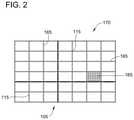
- FIG. 2is a diagram of a collector removed from the electrospinning system of FIG. 1 and having a plurality of fibers deposited thereon forming a patch.
- FIG. 3is an illustration of a biomedical patch including a plurality of spatially arranged electrospun fibers deposited on a collector shown in FIG. 1 .
- FIG. 4is another illustration of a biomedical patch including a plurality of spatially arranged electrospun fibers deposited on a collector shown in FIG. 1 .
- FIG. 5is an illustration of a solid fiber spinneret shown in FIG. 1 .
- FIG. 6is an illustration of a co-axial fiber spinneret shown in FIG. 1 .
- FIG. 7is an illustration of a multi-layer biomedical patch.
- FIG. 8is an illustration of a delamination of patches, such as the patch shown in FIG. 7 , using various fusion strengths over time.
- FIGS. 9 and 10are histological cross-sections of regenerated dura repaired with multi-laminar nanofiber material such as a patch shown in FIG. 8 .
- FIGS. 11 and 12are histological cross-sections of regenerated dura repaired with multi-laminar nanofiber material such as a patch shown in FIG. 8 .
- FIG. 13is an illustration of a delamination of patches, such as the patch shown in FIG. 7 , using various fusion methods and strengths over time.
- FIG. 14is a flowchart of an exemplary method 700 for producing a structure of spatially arranged fibers using system 100 shown in FIG. 1 .
- FIG. 15is a flowchart of an exemplary method 750 for fusing or coupling together structures or patch layers produced by method 700 shown in FIG. 14 .
- FIG. 16is a flowchart of an exemplary method 800 for repairing a defect in a biological tissue using the structures produced by methods 700 and 750 shown in FIGS. 14 and 15 .
- Embodiments provided hereinfacilitate repairing biological tissue or reinforcing biomedical material with the use of a biomedical patch including a plurality of fibers.
- Such fibersmay have a very small cross-sectional diameter (e.g., from 1-3000 nanometers) and, accordingly, may be referred to as nanofibers and/or microfibers.
- biomedical patchesare described herein with reference to dura mater and use as a surgical mesh, embodiments described may be applied to any biological tissue.
- structures with aligned fibersmay be used for other purposes. Accordingly, embodiments described are not limited to biomedical patches.
- biomedical patchesprovided herein facilitate cell growth, provide reinforcement, and may be referred to as “membranes,” “scaffolds,” “matrices,” “meshes”, “implants”, or “substrates.” Biomedical patches with varying densities, as described herein, may promote significantly faster healing and/or regeneration of tissue such as the dura mater than existing patches constructed using conventional designs.
- Dura materis a membranous connective tissue comprising the outermost layer of the meninges surrounding the brain and spinal cord, which covers and supports the dural sinuses.
- Surgical meshesare often needed during neurosurgical, orthopedic, or reconstructive surgical procedures to repair, expand, reinforce, or replace the incised, damaged, or resected dura mater.
- Autograftse.g., fascia lata, temporalis fascia, and pericranium
- graftsare preferable because they do not provoke severe inflammatory or immunologic reactions.
- Potential drawbacks of autograftsinclude the difficulty in achieving a watertight closure, formation of scar tissue, insufficient availability of graft materials to close large dural defects, increased risk of infection, donor site morbidity, and the need for an additional operative site.
- Allografts and xenograft materialsare often associated with adverse effects such as graft dissolution, encapsulation, foreign body reaction, immunological reaction, contracture, scarring, adhesion formation, and toxicity-induced side effects from immunosuppressive regimens.
- Lyophilized human dura mater as a dural substitutehas also been reported as a source of transmittable diseases, specifically involving prions, such as Creutzfeldt-Jakob disease.
- non-absorbable synthetic polymerssuch as silicone and expanded polytetrafluoroethylene (ePTFE)
- ePTFEexpanded polytetrafluoroethylene
- Natural absorbable polymersincluding collagen, fibrin, and cellulose, may present a risk of infection and disease transmission.
- synthetic absorbable polymerssuch as poly (3-hydroxybutyrate-co-3-hydroxyvalerate) (PHBV), poly (lactic acid) (PLA), polyglycolic acid (PGA), poly (lactic-co-glycolic acid) (PLGA), PLA-PCL-PGA ternary copolymers, and hydroxyethylmethacrylate hydrogels have recently attracted attention as biodegradable implant materials for dural repair. Methods and systems described herein may be practiced with these materials and/or any biomedical polymer whether the polymer is non-absorbable or absorbable, or synthetic in origin.
- a synthetic surgical mesh or biomedical patchshould promote: i) adhesion of dural fibroblasts (the primary cell type present in the dura) to the surface of the biomedical patch; ii) migration of dural fibroblasts from the periphery of the biomedical patch into the center of the patch; iii) reinforcement or replacement of existing tissues; iv) minimal immune response; v) water tight closure of the dural membrane/dura mater; vi) mechanical support of the native dural post-operatively and during tissue regeneration/neoduralization; vii) rapid closure of the dural defect; and viii) increased ease of use.
- Electrospinningis an enabling technique which can produce nanoscale fibers from a large number of polymers.
- the electrospun nanofibersare typically collected as a randomly-oriented, nonwoven mat. Uniaxially or radially aligned arrays of nanofibers can also be obtained under certain conditions.
- traditional nanofiber scaffoldsmay lack the optimal mechanical and biological properties necessary for some biomedical or surgical applications post-operatively.
- nanofiber scaffoldsIn order to increase the strength of nanofiber scaffolds, custom fabrication of scaffolds into particular patterns would be highly advantageous. Additionally, multiple layers of nanofiber materials fused/coupled together in a manner that allows for a purposeful degradation of the layers can also provide strength while allowing for cellular penetration and/or tissue integration.
- nanofibers for dura substitutesare produced as the electrospun polymer from poly ( ⁇ -caprolactone) (PCL), an FDA approved, semicrystalline polyester that can degrade via hydrolysis of its ester linkages under physiological conditions with nontoxic degradation products.
- PCLpoly ( ⁇ -caprolactone)
- This polymerhas been extensively utilized and studied in the human body as a material for fabrication of drug delivery carriers, sutures, or adhesion barriers.
- electrospun PCL nanofibersmay be used to generate scaffolds that are useful as surgical meshes.
- Embodiments provided hereinfacilitate producing a novel type of artificial tissue substitute including a polymeric nanofiber material, which is formed through a novel method of electrospinning.
- This polymeric materialincludes non-woven nanofibers (e.g., fibers having a diameter of 1-3000 nanometers) which are arranged or organized and aligned into patterns both within and across a material sheet.
- FIG. 1is a diagram illustrating a perspective view of an exemplary electrospinning system 100 for producing a structure of spatially arranged or organized fibers.
- System 100includes a collector 105 with a predetermined pattern 110 including a plurality of reinforcement features 112 .
- System 100also includes a spinneret 120 .
- System 100is configured to create an electric potential between one or more collectors 105 and one or more spinnerets 120 .
- collector 105 and features 112are configured to be electrically charged at a first amplitude and/or polarity.
- collector 105 and features 112may be electrically coupled to one or more power supplies 130 via one or more conductors 135 .
- Power supply 130is configured to charge collector 105 and features 112 at the first amplitude and/or polarity via conductor 135 .
- collector 105includes pattern 110 that is a grid pattern formed by features 112 such that collector 105 is substantially rectangular.
- collector 105may have any shape including, but not limited to, circular, elliptical, ovular, square, and/or triangular.
- features 112include ribs 114 , seams 116 , and surfaces 118 configured to receive and/or collect polymer fibers.
- rib 114is substantially cylindrical and has a circumference between 5 um-100 cm
- seam 116is substantially rectangular having a thickness between 5 um-100 cm
- surface 118is a filling of a void or feature space 119 formed between ribs 114 and/or seams 116 .
- surfacehas a thickness between 5 um-10 cm.
- features 112are made fabricated from at least a portion of metallic substance, including, but not limited to steel, aluminum, tin, copper, silver, gold, platinum, and any alloy or mixture thereof.
- features 112include a coating applied to collector 105 . Coatings can include, but are not limited to anodization, chemical coatings, material coatings (conductive or non-conductive), and gradient coatings that facilitate the creation of continuous gradients of fibers.
- features 112e.g., ribs 114 , seams 116 , and surface 118
- features 112can have any shape and be fabricated from any material that facilitates producing patches as disclosed herein.
- pattern 110is formed by spatially organizing features 112 .
- features 112e.g., ribs 114 and seams 116
- pattern 110includes a plurality of spaces 119 such that multiple varying distances are formed between features 112 .
- patterncan be formed to be symmetrical, repeating, and asymmetrical.
- the shape of collector 105enables the biomedical patch formed on collector to include additional support and/or reinforcement properties. Such additional support and/or reinforcement properties are achieved by creating high density fiber deposition areas on charged features 112 and having low density fiber deposition areas over feature spaces 119 .
- a diamond shaped collector 105 including a diamond shaped array pattern 110enables a diamond-shaped patch to be produced on the diamond shaped collector 105 to have different mechanical properties from a rectangular-shaped or a circular-shaped patch such as, but not limited to, tensile strength, tear resistance, terminal strain, failure mechanisms or rates, and/or controlled anisotropic properties, such as greater strength in one axis relative to another.
- pattern 110defines a collector plane 127 and spinneret 120 is orthogonally offset from the collector plane 127 at a variable distance.
- spinneret 120may be orthogonally offset from the collector plane 127 at a distance of 50 micrometers to 100 centimeters.
- spinneret 120can be offset from collector 105 in any manner that facilitates creating patches as described herein, including but not limited to, horizontal and diagonal or skew.
- Spinneret 120is configured to dispense a polymer 140 while electrically charged at a second amplitude and/or polarity opposite the first amplitude and/or polarity. As shown in FIG. 1 , spinneret 120 is electrically coupled to one or more power supplies 130 by one or more conductors 145 . Power supply 130 is configured to charge one or more spinnerets 120 at the second amplitude and/or polarity via conductor 145 . In some embodiments, power supplies 130 provides a direct current and/or static or time variant voltage (e.g., between 1-50 kilovolts). In one embodiment, conductor 145 is charged positively, and collector 105 is also charged positively. In all embodiments, power supply 130 is configured to allow adjustment of a current, a voltage, and/or a power.
- a direct current and/or static or time variant voltagee.g., between 1-50 kilovolts.
- spinneret 120is coupled to a dispensing mechanism 150 containing polymer 140 in a liquid solution form.
- dispensing mechanism 150is operated manually by a dispensing pump 155 .
- dispensing mechanism 150can be operated automatically with any mechanism configured to dispense nanofibers as described herein.
- spinneret 120includes a metallic needle having an aperture between 10 micrometers and 3 millimeters in diameter for dispensing nanofibers.
- spinneret 120dispenses polymer 140 as a jet or stream 160 .
- stream 160is dispensed in a horizontal or sideways stream from spinneret 120 .
- Stream 160has a diameter approximately equal to the aperture diameter of spinneret 120 .
- Stream 160descends toward collector 105 forming a Taylor cone. For example, stream 160 may fall downward under the influence of gravity and/or may be attracted downward by charge distributed on the fibers and on features 112 .
- polymer 140forms one or more solid polymeric fibers 165 .
- fibers 165are solid, however it should be noted that fibers 165 can have any structure including by not limited to, core or shell, porous, co-axial, and co-axial.
- polymer 140 depositioncan be accomplished by any other fiber deposition method including but not limited to, solvent electrospinning, force electrospinning, melt electrospinning, extrusion, and melt blowing.
- a mask 164composed of a conducting or non-conducting material is applied to collector 105 to manipulate deposition of fibers 165 .
- mask 164may be positioned between spinneret 120 and collector 105 such that no fibers 165 are deposited on collector 105 beneath mask 164 .
- mask 164may be used as a time-variant mask by adjusting its position between the spinneret and the collector while spinneret 120 dispenses polymer 140 , facilitating spatial variation of fiber density on collector 105 . While mask 164 is shown as circular, mask 164 may have any shape (e.g., rectangular or semi-circular) and size suitable for use with system 100 .
- deposition of fibers 165 on collector 105may be manipulated by adjusting the position of collector 105 with respect to spinneret 120 or by spatially varying the electrical potential applied between the spinneret 120 and/or the electrodes making up the collector 105 .
- positioning one side of collector 105 directly beneath spinneret 120may cause more fibers 165 to be deposited on that side than are deposited on the opposite side of collector 105 in a Gaussian distribution.
- a focusing device 138is utilized to focus fiber deposition in a particular special region.
- focusing device 138is charged with a polarity similar to spinneret 120 and includes an aperture allowing fiber deposition to occur substantially in the space under the aperture.
- Focusing device 138may have any geometry that allows for receipt of nanofibers from spinneret 120 and deposition of the received nanofibers onto collector 105 as described herein.
- FIG. 2is a diagram of collector 105 removed from electrospinning system 100 (shown in FIG. 1 ) and having a plurality of fibers 165 deposited thereon forming a patch 170 . Fibers 165 are oriented such that they correspond to the position of features 112 (shown in FIG. 1 ).
- Patch 170is illustrated with a small quantity of fibers 165 in FIG. 2 for clarity.
- patch 170includes thousands, tens of thousands, hundreds of thousands, or more fibers 165 , distributed on collector 105 . Even with millions of fibers 165 , patch 170 retains predictable properties such as being flexible and/or pliable. As such, the predictable properties facilitate the application of patch 170 to uneven biological tissue surfaces, such as the surface of the dura mater.
- the alignment of fibers 165illustrates a patch 170 with varying densities.
- Patch 170enables reinforcement or structural integrity to be provided in predetermined locations. For example, a larger deposition of fibers occurs in various locations, such as portion 167 , which provide structural reinforcement. Accordingly, system 100 enables the creation of customized materials 170 for individual biomedical or clinical and non-clinical applications.
- fibers 165have a diameter of 1-3000 nanometers. In one embodiment, fibers have a diameter of approximately 220 nanometers (e.g., 215 nm to 225 nm). It should be noted that the diameter of the fibers 165 , thickness of the patch 170 , and/or fiber density within the patch 170 may affect the durability (e.g., tensile strength, suture pullout strength, conformability, etc.) of patch 170 . As such, the diameter of the fibers 165 , thickness of the patch 170 , and/or fiber density within the patch 170 can be selected according to the requirements of the end application of the material.
- Patch 170may be produced with various mechanical properties by varying the thickness and/or the fiber density, spatial patterning, polymer composition, and/or number of layers of the patch 170 by operating electrospinning system 100 for relatively longer or shorter durations, changing the polymeric solution, changing the chemical composition, changing collector 105 , changing collector design, and/or changing the manner of fiber deposition.
- FIG. 3is an illustration 305 of a patch 170 including a plurality of electrospun fibers deposited on collector 105
- FIG. 4is an illustration 405 of a patch 170 including a plurality of electrospun fibers deposited on collector 105
- collectors 105respectively provide an increased deposition of fibers on and substantial near features 112 . Such additional support and/or reinforcement properties are achieved by creating high density fiber deposition areas on charged features 112 and having low density fiber deposition areas over feature spaces 119 .
- collector 105can include any pattern or combination of patterns such as the grid pattern shown in FIG. 3 and the hexagonal or honeycomb pattern shown in FIG. 4 .
- fibers 165may be solid, core/shell, co-axial, or porous.
- the size and/or structure of fibers 165is determined by the design and/or size of spinneret 120 , and/or polymer solution which includes viscosity, solvent or method of preparation of the solution, voltage or electric charge, distance between spinneret 120 and collector 105 , and rate of deposition.
- FIG. 5is an illustration of a solid fiber spinneret 120 A.
- Solid fiber spinneret 120 Aincludes a truncated conical body 180 defining a center line 182 .
- body 180includes an annulus 186 .
- Annulus 186defines a circular aperture 190 A, through which polymer 140 may be dispensed.
- Fibers 165 produced with solid fiber spinneret 120 Ahave a solid composition.
- FIG. 6is an illustration of a co-axial fiber spinneret 120 B.
- co-axial fiber spinneret 120 Bincludes a truncated conical body 180 with an annulus 186 at a dispensing end 184 .
- Co-axial fiber spinneret 120 Balso includes a central body 188 B positioned within annulus 186 .
- Annulus 186 and central body 188 Bdefine an annular aperture 190 B. Accordingly, when polymer 140 is dispensed by co-axial fiber spinneret 120 B, fibers 165 have a co-axial composition, with an exterior wall surrounding a cavity.
- the exterior wall of a fiber 165 dispensed by co-axial fiber spinneret 120 Bdefines an outer diameter corresponding to the inner diameter of annulus 186 and an inner diameter corresponding to the diameter of central body 188 B. Accordingly, the outer diameter and inner diameter of co-axial fibers 165 may be adjusted by adjusting the diameters of annulus 186 and central body 188 B.
- Fiber spinnerets 120 A and 120 Bfacilitate incorporating a substance, such as a biological agent, growth factor, and/or a drug (e.g., a chemotherapeutic substance), into patch 170 .
- a substancesuch as a biological agent, growth factor, and/or a drug (e.g., a chemotherapeutic substance)
- the substancemay be deposited within a cavity defined by co-axial fibers 165 of patch 170 .
- polymer 140is selected to create porous and/or semi-soluble fibers 165 , and the substance is dispensed from the cavity through fibers 165 .
- polymer 140is degradable, and the substance is dispensed as fibers 165 degrade in vivo.
- fibers 165may be configured to degrade within a second to 1 second to 12 months.
- a burst release of the substanceoccurs upon entry into a system and an elution occurs over a predetermined period of time.
- the degradation rate of polymer 140may be manipulated by any loading and/or release method such as adjusting a ratio of constituent polymers within polymer 140 , loading the agent into the bulk of the material, functionalizing the agent to the surface of the fibers, and/or releasing the agent by degradation of the polymer or by diffusion of the agent from the polymer.
- a substanceis delivered by solid fibers 165 .
- a solid fiber 165may be created from a polymer 140 including the substance in solution. As solid fiber 165 degrades, the substance is released into the surrounding tissue.
- annulus 186is perpendicular to center line 182 .
- annulus 186is oblique (e.g., oriented at an acute or obtuse angle) with respect to center line 182 .
- the outside diameter of fibers 165may be determined by the inside diameter of annulus 186 .
- FIG. 7is an illustration of a multi-layer biomedical patch 435 .
- Patch 435includes a biomedical patch layer with a plurality of symmetrical spatially organized fibers 420 and a biomedical patch layer with a plurality of spatially organized fibers having varying densities 425 such as increased density portions 430 .
- biomedical patch layers 420 and 425are combined (e.g., fused, joined, adhered, overlaid) to produce multi-layer biomedical patch 435 with reinforcement fiber layers. It should be noted that any combination, number, or type of fiber layers may be combined to create biomedical patch 435 .
- patch 435can be formed of layers having various densities and/or thicknesses (both individually and collectively), fiber organizations, polymer compositions, surface coatings, and types of concentrations of agents and/or drugs.
- multiple biomedical patch layers 410 - 425may be combined to create a multi-layer biomedical patch.
- first set of fibers 165may be deposited directly over the first set of fibers 165 on collector 105 .
- the first set of fibers 165may be removed from collector 105 , and the second set of fibers 165 may be deposited on conductive surface 162 and/or collector 105 and then removed and adhered/overlaid on the first set of fibers 165 .
- a non nanofiber intermediate layere.g., hydrogel or polymeric scaffold
- biomedical patch layers 400 and/or biomedical patch layers 410may be disposed between biomedical patch layers 400 and/or biomedical patch layers 410 .
- individual layersare fused or coupled together such that the layers delaminate or separate under specific environmental or temporal conditions.
- Such controlled delaminationresults in maximization of mechanical stability of the nanofiber material and the biological interaction (e.g. cellular ingrowth, tissue integration, cellular exposure, etc.) between adjacent layers of nanofibers.
- the process of fusing or coupling layersincludes, but is not limited to, heating, applying mechanical stress/pressure, applying an adhesive, chemical processing, cross-linking, and functionalization.
- adjacent layersare similarly or variably fused, adhered, or joined such that each layer delaminates or separates at a substantially similar rate within patch 435 .
- layerscan be fused together with variable methods such that each layer delaminates at different rates.
- FIG. 8illustrates delamination of patches 440 , 445 , and 450 with various fusion strengths over time.
- a low strength adhesion 455such as but not limited to mild-chemical treatment or crosslinking, low-pressure physical lamination, or low-temperature thermal processing, is used to fuse layers of patch 440 together.
- a moderate strength adhesion 460such as but not limited to moderate chemical crosslinking, prolonged thermal processing, moderate mechanical entanglement, application of moderate adhesives, or high-pressure physical lamination is used to fuse the layers of patch 445 and a high strength adhesion 465 , such as but not limited to extensive chemical crosslinking, extensive high-temperature thermal processing, extensive mechanical entanglement, fiber interweaving or knitting, or application of aggressive adhesives is used to fuse layers of patch 450 together.
- a separation 470 of patches 440 , 445 , and 450is shown after a short increment of time, such as, but not limited to 1 day-30 days and a separation 475 of patches 440 , 445 , and 450 is shown after a long increment of time, such as, but not limited to 30 days-3 years.
- patch 440is substantially separated 470 after a short period of time acting as an accelerated separation
- patch 445is substantially separated 475 after the long period of time acting as a delayed separation
- patch 450provided substantially no separation.
- FIGS. 9 and 10are histological cross-sections 500 and 502 of dura mater repaired with multi-laminar nanofiber material such as patch 440 shown in FIG. 8 .
- patch 440is shown as being inserted into dura 504 two weeks post-operatively.
- Regenerative dural tissue (“neodura”) 504is demonstrated extending on and around the implanted nanofiber material 440 .
- Regenerative dural fibroblastsare also shown to have penetrated the bulk of the nanofiber material 440 , demonstrating progressive cellularization of the implanted nanofiber construct. Two weeks following implantation of the multi-layer nanofiber material 440 no delamination is noted upon histological examination of the explanted tissue.
- the nanofiber material 440is observed as a homogeneous block of material with low to moderate cellular ingrowth, yet no singular nanofiber layer or separation of nanofiber layers is observed.
- FIG. 10illustrates controlled delamination of patch 440 six weeks post-operatively and integration of the patch within the native and/or regenerated dural tissue 504 .
- Regenerative dural tissue (“neodura”) 504is demonstrated extending on and around the implanted nanofiber material 440 .
- regenerative dural tissue (“neodura”)is demonstrated extending in between delaminated layers of the nanofiber material.
- Regenerative dural fibroblastsare shown to have significantly penetrated the bulk of the nanofiber material 440 , demonstrating robust cellularization and integration of the implanted nanofiber construct.
- the nanofiber material 440is observed as two heterogeneous layers of material separated by a thin layer of regenerated dural tissue extending along the adjoining surface of the nanofiber monolayers.
- Evidence of controlled delamination of the implanted material post-operativelyis specifically demonstrated by observation that multiple layers of the material remain fused in proximity of sutures utilized to secure the material to the native tissue.
- FIGS. 11 and 12are histological cross-sections 506 and 508 of regenerated dura repaired with multi-laminar nanofiber material such as patch 450 shown in FIG. 8 .
- patch 450is shown as being inserted into dura 504 two weeks post-operatively.
- Regenerative dural tissue (“neodura”) 504is demonstrated extending on and around the implanted nanofiber material 450 .
- Regenerative dural fibroblastsare also shown to have penetrated the bulk of the nanofiber material 450 , demonstrating cellularization of the implanted nanofiber construct. No delamination is noted upon histological examination of the explanted tissue.
- the nanofiber material 450is observed as a homogeneous block of material with low to moderate cellular ingrowth, yet no singular nanofiber layer or separation of nanofiber layers is observed.
- FIG. 12illustrates that the high strength adhesion has enabled layers of patch 450 to remain substantially fused together six week post-operatively as dural tissue 504 regenerated around patch 450 .
- Regenerative dural tissue (“neodura”) 504is again demonstrated extending on and around the implanted nanofiber material 450 .
- Dural fibroblastssubstantially penetrate the bulk of the nanofiber material 450 , demonstrating robust cellularization of the implanted nanofiber construct.
- no delamination of nanofiber patch 450is noted upon histological examination of the explanted tissue following chronic implantation.
- the nanofiber material 450is observed as a secure composite material demonstrating cellular ingrowth yet no separation or observation of singular nanofiber layers.
- FIG. 13illustrates separation of layers within patches 600 , 602 , and 604 at varying rates.
- Each patch 600 , 602 , and 604includes a first layer 606 , a second layer, 608 , a third layer 610 , and a fourth layer 612 .
- patches 600 , 602 , and 604are shown with four layers, patches can be fabricated to have any number of layers. Referring to patch 600 , low strength adhesion 455 is used to fuse layers 606 , 608 , and 610 together and high strength adhesion 465 is used to fuse layers 610 and 612 together.
- a separation 614 of layers 606 , 608 , and 610has occurred and layers remain substantially fused together.
- moderate strength adhesion 460is used to fuse together layers 606 and 608
- high strength adhesion 465is used to fuse together layers 608 , 610 , and 612 .
- a separation 616 of layers 606 and 608occurs after a long period of time while substantially no separation occurs between layers 608 , 610 , and 612 .
- a high strength adhesion 465is used between layers 606 , 608 , 610 , and 612 such that substantially no separation occurs between the layers.
- a multi-layered biomedical patchmay be useful for dural grafts as well as other tissue engineering applications.
- Sequential layers of fiberscan be created with varying orders (e.g., radially aligned, reinforced, or randomly oriented), densities (e.g., low, high, or mixture of fiber density), patterns or reinforcement, and compositions (polymer), which may allow specific types of cells to infiltrate and populate select layers of the artificial biomedical patch.
- biomedical patches containing a high fiber densitygenerally prohibit cellular migration and infiltration
- biomedical patches containing a low fiber densitygenerally enhance cellular migration and infiltration.
- Such additional support and/or reinforcement propertiesare achieved by creating high density fiber deposition that discourages cellular penetration and having low density fiber deposition areas that promote cellular penetration and/or ingrowth.
- the ability to form multi-layered fiber materialsmay be extremely beneficial in the construction of biomedical patches designed to recapitulate the natural multi-laminar structure of not only dura mater, but also other biological tissues such as skin, heart valve leaflets, pericardium, and/or any other biological tissue.
- one or more layers of a biomedical patchmay be fabricated from bioresorbable polymers such that the resulting nanofiber materials fully resorb following implantation. Manipulation of the chemical composition of the polymers utilized to fabricate these scaffolds may further allow for specific control of the rate of degradation and/or resorption of a biomedical patch following implantation.
- FIG. 14is a flowchart of an exemplary method 700 for producing a structure of spatially organized fibers using system 100 shown in FIG. 1 . While one embodiment of method 700 is shown in FIG. 14 , it is contemplated that any of the operations illustrated may be omitted and that the operations may be performed in a different order than is shown.
- method 700includes electrically charging 705 collector 105 at a first amplitude and/or polarity (e.g., negatively charging or grounding).
- Spinneret 120is electrically charged 710 at a second amplitude and/or polarity opposite the first amplitude and/or polarity (e.g., positively charged).
- a polymere.g., a liquid polymer
- dispensed 715 polymersare collected 720 on collector 105 to form a plurality of polymeric fibers on or substantially near features 112 that creates a structure or patch.
- post-processing 725can include, but is not limited to, lamination, layer stacking, coupling and/or fusing, chemically treating, and applying a biological agent, growth factor, and/or drug.
- FIG. 15is a flowchart of an exemplary method 750 for fusing or coupling together structures or patch layers produced by method 700 shown in FIG. 14 .
- Method 750includes providing 755 a first, second, and third patch layer.
- First patch layeris coupled 760 to second patch layer using a first coupling technique.
- the coupled 760 first and second layersare then coupled 765 to the third patch layer using a second coupling technique different than the first coupling technique.
- coupling techniquesinclude but are not limited to, heating, applying mechanical stress/pressure, chemical processing, cross-linking, and functionalization.
- method 750illustrates a first patch layer coupled to a second patch layer, it should be noted that multiple layers (e.g., 3, 5, 6) can be coupled together simultaneously. Additionally, the process may be repeated to add layers to structures produced by method 750 .
- FIG. 16is a flowchart of an exemplary method 800 for repairing a defect of a substrate using a structure produced by methods 700 and 750 shown in FIGS. 14 and 15 .
- method 800includes providing 805 a substrate substance with a defect.
- the defectmay include a void, tissue defect, injury, insult, and/or any other condition resulting in diminished function of biological tissue.
- the substrateis biological tissue.
- the substratecan be any substrate including but not limited to, filtration media, textiles, membrane media, and coatings.
- the defect provided 805includes a void created by surgical incision to provide access to an underlying tissue (e.g., a tumor).
- a voidis created 805 by excising necrotic tissue (e.g., skin cells).
- necrotic tissuee.g., skin cells
- one or more patches capable of covering the defectare selected 810 .
- a plurality of biomedical patchesmay be selected 810 for a large and/or complex (e.g., irregularly shaped) defect.
- a biomedical patch having a diameter greater than the diameter of an approximately circular defectis selected 810 .
- a patchis selected 810 and customized, pre-operation or intra-operation, to fit the defect. It should be noted that any size, shape, and/or geometry of structure may be used in the selection 810 of the patch.
- a substance such as a growth factor and/or a drugis applied 815 to the biomedical patch.
- growth factor and/or a drugis applied 815 pre-operatively.
- growth factor and/or a drugmay be applied 815 at any time including, but not limited to, intra-operatively and post-operatively.
- the biomedical patchmay be immersed in the substance to allow the substance to occupy a cavity within co-axial fibers of the biomedical patch, dope the polymer comprising the fibers in the biomedical patch, or coat the surface of the fibers within the biomedical patch.
- the patchis applied 820 to (e.g., overlaid on) the biological tissue to cover, repair, reinforce, and/or fill at least a portion of the defect.
- the biomedical patchmay be applied 820 to dura mater tissue, cardiac tissue, and/or any biological tissue including a defect.
- the perimeter of the biomedical patchextends past the perimeter of the defect, such that the entire defect is covered by the biomedical patch.
- the biomedical patchis coupled 825 to the biological tissue with a plurality of sutures, adhesive, and/or any other means of attaching the biomedical patch to the biological tissue (inlay).
- biomedical patchis simply allowed to fuse to the biological tissue, such as by adhesion of biological cells to the biomedical patch (onlay).
- biomedical patchmay be directly coupled to the edge of the tissue with no overlap.
- biomedical patchmay be overlaid on top of a wound/defect or injury covering the entirety of the defect or injury without filling the defect.
- the biological tissueis covered 830 .
- the patchmay be the terminal covering.
- a backing that is either non-permeable or permeablemay be coupled to the patch.
- other tissue overlaying the defecte.g., dermis and/or epidermis
- one or more protective layersare applied over the biological tissue.
- a bandagemay be applied to a skin graft, with or without a protective substance, such as a gel, an ointment, and/or an antibacterial agent.
- the protective layerincludes, but is not limited to, a covering, film tissue, dressing, mesh, and nanofiber structure, such as an additional biomedical patch, as described herein.
- Embodiments described hereinare operable with any surgical procedure involving the repair, replacement, or expansion of the dura mater, including, but not limited to, a transphenoidal procedure (e.g., surgical removal of pituitary adenomas), various types of skull base surgeries, and/or surgical removal of cranial or spinal tumors (e.g., meningiomas and/or astrocytomas).
- a biomedical patchmay be applied to a bone fracture (e.g., a complex fracture).
- a biomedical patchmay be applied to a defect in the skin (e.g. a burn).
- Such embodimentsprovide a dura mater substitute, a biomedical patch for a skin graft (e.g., dermal or epidermal), a biomedical patch for tracheal repair, a scaffold for an artificial heart valve leaflet, an artificial mesh for surgical repair of a gastrointestinal tract (e.g., an abdominal hernia or an ulcer), an artificial mesh for surgical repair of cardiac defects.
- a biological tissuee.g., cardiomyocytes or cardiac tissue, muscle, skin, connective tissue, intestinal tissue, stomach tissue, bone, gastrointestinal tract, and mucosa.
- a biomedical patchhas a thickness greater or less than a thickness of the biological tissue being repaired.
- Biomedical patches with spatially organized polymeric fibersfacilitate reducing the expense of tissue repair, improving tissue healing time, and reducing or eliminating the risk of zoonotic infection.
- biomedical patchesare relatively simple to manufacture, enabling customization of shape, size, and chemical composition and improved availability and non-immunogenicity.
- biomedical patches with spatially organized polymeric fibersexhibit excellent handling properties due to their cloth-like composition, eliminate the need for a second surgery to harvest autologous graft tissue, and reduce the risk of contracture and adhesion when compared with known products.
- the patches described hereinfacilitate reinforcement, buttressing, lamination, and/or sealing in a variety of applications such as but not limited to clinical and non-clinical applications.
Landscapes
- Health & Medical Sciences (AREA)
- Engineering & Computer Science (AREA)
- Chemical & Material Sciences (AREA)
- Life Sciences & Earth Sciences (AREA)
- Public Health (AREA)
- Veterinary Medicine (AREA)
- Animal Behavior & Ethology (AREA)
- General Health & Medical Sciences (AREA)
- Epidemiology (AREA)
- Textile Engineering (AREA)
- Medicinal Chemistry (AREA)
- Dermatology (AREA)
- Oral & Maxillofacial Surgery (AREA)
- Transplantation (AREA)
- Mechanical Engineering (AREA)
- Hematology (AREA)
- Materials Engineering (AREA)
- Chemical Kinetics & Catalysis (AREA)
- Biomedical Technology (AREA)
- Molecular Biology (AREA)
- Vascular Medicine (AREA)
- Heart & Thoracic Surgery (AREA)
- Surgery (AREA)
- Dispersion Chemistry (AREA)
- Nonwoven Fabrics (AREA)
- Materials For Medical Uses (AREA)
- Polymers & Plastics (AREA)
- Organic Chemistry (AREA)
- Prostheses (AREA)
- Spinning Methods And Devices For Manufacturing Artificial Fibers (AREA)
- Surgical Instruments (AREA)
Abstract
Description
Claims (30)
Priority Applications (3)
| Application Number | Priority Date | Filing Date | Title |
|---|---|---|---|
| US17/381,792US11253635B2 (en) | 2012-09-21 | 2021-07-21 | Three dimensional electrospun biomedical patch for facilitating tissue repair |
| US17/576,058US11596717B2 (en) | 2012-09-21 | 2022-01-14 | Three dimensional electrospun biomedical patch for facilitating tissue repair |
| US18/159,412US12109334B2 (en) | 2012-09-21 | 2023-01-25 | Three dimensional electrospun biomedical patch for facilitating tissue repair |
Applications Claiming Priority (6)
| Application Number | Priority Date | Filing Date | Title |
|---|---|---|---|
| PCT/US2012/056548WO2014046669A1 (en) | 2012-09-21 | 2012-09-21 | Biomedical patches with spatially arranged fibers |
| US16/131,887US10441685B2 (en) | 2012-09-21 | 2018-09-14 | Biomedical patches with spatially arranged fibers |
| US16/540,779US10682444B2 (en) | 2012-09-21 | 2019-08-14 | Biomedical patches with spatially arranged fibers |
| US16/872,926US12246114B2 (en) | 2012-09-21 | 2020-05-12 | Biomedical patches with spatially arranged fibers |
| US17/229,226US11173234B2 (en) | 2012-09-21 | 2021-04-13 | Biomedical patches with spatially arranged fibers |
| US17/381,792US11253635B2 (en) | 2012-09-21 | 2021-07-21 | Three dimensional electrospun biomedical patch for facilitating tissue repair |
Related Parent Applications (1)
| Application Number | Title | Priority Date | Filing Date |
|---|---|---|---|
| US17/229,226ContinuationUS11173234B2 (en) | 2012-09-21 | 2021-04-13 | Biomedical patches with spatially arranged fibers |
Related Child Applications (1)
| Application Number | Title | Priority Date | Filing Date |
|---|---|---|---|
| US17/576,058ContinuationUS11596717B2 (en) | 2012-09-21 | 2022-01-14 | Three dimensional electrospun biomedical patch for facilitating tissue repair |
Publications (2)
| Publication Number | Publication Date |
|---|---|
| US20210353834A1 US20210353834A1 (en) | 2021-11-18 |
| US11253635B2true US11253635B2 (en) | 2022-02-22 |
Family
ID=50341804
Family Applications (9)
| Application Number | Title | Priority Date | Filing Date |
|---|---|---|---|
| US14/429,976Active2034-08-27US10124089B2 (en) | 2012-09-21 | 2012-09-21 | Method of making biomedical patches with spatially arranged fibers |
| US16/131,887ActiveUS10441685B2 (en) | 2012-09-21 | 2018-09-14 | Biomedical patches with spatially arranged fibers |
| US16/540,779ActiveUS10682444B2 (en) | 2012-09-21 | 2019-08-14 | Biomedical patches with spatially arranged fibers |
| US16/872,926Active2036-04-28US12246114B2 (en) | 2012-09-21 | 2020-05-12 | Biomedical patches with spatially arranged fibers |
| US17/229,226ActiveUS11173234B2 (en) | 2012-09-21 | 2021-04-13 | Biomedical patches with spatially arranged fibers |
| US17/381,792ActiveUS11253635B2 (en) | 2012-09-21 | 2021-07-21 | Three dimensional electrospun biomedical patch for facilitating tissue repair |
| US17/576,058ActiveUS11596717B2 (en) | 2012-09-21 | 2022-01-14 | Three dimensional electrospun biomedical patch for facilitating tissue repair |
| US18/159,412ActiveUS12109334B2 (en) | 2012-09-21 | 2023-01-25 | Three dimensional electrospun biomedical patch for facilitating tissue repair |
| US19/028,550PendingUS20250170308A1 (en) | 2012-09-21 | 2025-01-17 | Biomedical patches with spatially arranged fibers |
Family Applications Before (5)
| Application Number | Title | Priority Date | Filing Date |
|---|---|---|---|
| US14/429,976Active2034-08-27US10124089B2 (en) | 2012-09-21 | 2012-09-21 | Method of making biomedical patches with spatially arranged fibers |
| US16/131,887ActiveUS10441685B2 (en) | 2012-09-21 | 2018-09-14 | Biomedical patches with spatially arranged fibers |
| US16/540,779ActiveUS10682444B2 (en) | 2012-09-21 | 2019-08-14 | Biomedical patches with spatially arranged fibers |
| US16/872,926Active2036-04-28US12246114B2 (en) | 2012-09-21 | 2020-05-12 | Biomedical patches with spatially arranged fibers |
| US17/229,226ActiveUS11173234B2 (en) | 2012-09-21 | 2021-04-13 | Biomedical patches with spatially arranged fibers |
Family Applications After (3)
| Application Number | Title | Priority Date | Filing Date |
|---|---|---|---|
| US17/576,058ActiveUS11596717B2 (en) | 2012-09-21 | 2022-01-14 | Three dimensional electrospun biomedical patch for facilitating tissue repair |
| US18/159,412ActiveUS12109334B2 (en) | 2012-09-21 | 2023-01-25 | Three dimensional electrospun biomedical patch for facilitating tissue repair |
| US19/028,550PendingUS20250170308A1 (en) | 2012-09-21 | 2025-01-17 | Biomedical patches with spatially arranged fibers |
Country Status (11)
| Country | Link |
|---|---|
| US (9) | US10124089B2 (en) |
| EP (2) | EP2897561B1 (en) |
| JP (1) | JP6295258B2 (en) |
| KR (2) | KR102178233B1 (en) |
| AU (2) | AU2012390291B2 (en) |
| BR (1) | BR112015006301B1 (en) |
| CA (2) | CA3066269C (en) |
| ES (1) | ES2847893T3 (en) |
| IN (1) | IN2015DN02299A (en) |
| SG (1) | SG11201502207WA (en) |
| WO (1) | WO2014046669A1 (en) |
Cited By (3)
| Publication number | Priority date | Publication date | Assignee | Title |
|---|---|---|---|---|
| US20220249743A1 (en)* | 2012-09-21 | 2022-08-11 | Washington University | Three dimensional electrospun biomedical patch for facilitating tissue repair |
| US12144716B2 (en) | 2010-06-17 | 2024-11-19 | Washington University | Biomedical patches with aligned fibers |
| US12201749B2 (en) | 2021-07-29 | 2025-01-21 | Acera Surgical, Inc. | Combined macro and micro-porous hybrid-scale fiber matrix |
Families Citing this family (19)
| Publication number | Priority date | Publication date | Assignee | Title |
|---|---|---|---|---|
| WO2013025819A2 (en) | 2011-08-16 | 2013-02-21 | The University Of Kansas | Fibrous tracheal patch |
| WO2013106822A1 (en) | 2012-01-12 | 2013-07-18 | Johnson Jed K | Nanofiber scaffolds for biological structures |
| US9398928B2 (en)* | 2012-09-28 | 2016-07-26 | DePuy Synthes Products, Inc. | Adjustable height arthroplasty plate |
| EP2943205B1 (en) | 2013-01-14 | 2018-08-22 | Scripps Health | Tissue array printing |
| EP3950019A1 (en) | 2013-03-15 | 2022-02-09 | Nanofiber Solutions, LLC | Biocompatible fiber textiles for implantation |
| AU2014268601B2 (en)* | 2013-05-22 | 2017-06-15 | The Penn State Research Foundation | Wound dressings and applications thereof |
| US11497830B2 (en) | 2014-03-14 | 2022-11-15 | Scripps Health | Electrospinning of cartilage and meniscus matrix polymers |
| CZ308167B6 (en)* | 2014-12-17 | 2020-02-05 | Technická univerzita v Liberci | A process for manufacturing polymeric fibres with a diameter of 100 nm to 10 μm, and a process for producing a linear, planar or spatial formation containing these polymeric fibres |
| CN108135686B (en) | 2015-09-10 | 2021-06-04 | 纳米纤维解决方案股份有限公司 | Polymer electrospinning embolization device and method of use |
| US10632228B2 (en) | 2016-05-12 | 2020-04-28 | Acera Surgical, Inc. | Tissue substitute materials and methods for tissue repair |
| GB2553316B (en)* | 2016-09-01 | 2020-05-13 | Univ Nottingham Trent | Method and apparatus for fabricating a fibre array and structure incorporating a fibre array |
| US10898608B2 (en) | 2017-02-02 | 2021-01-26 | Nanofiber Solutions, Llc | Methods of improving bone-soft tissue healing using electrospun fibers |
| EP3609686A4 (en)* | 2017-04-12 | 2020-09-09 | The Government of the United States of America, as represented by the Secretary of the Navy | TEMPERATURE CONTROLLED ELECTRIC SPINNING SUBSTRATE |
| AU2019397470A1 (en) | 2018-12-11 | 2021-06-10 | Nfs Ip Holdings, Llc | Methods of treating chronic wounds using electrospun fibers |
| JP2022524524A (en)* | 2019-03-11 | 2022-05-06 | ゼルティス アーゲー | Electrospun polymer assembly for medical implants |
| US11208735B2 (en)* | 2019-07-02 | 2021-12-28 | University of Central Oklahoma | Method and apparatus for controlling fiber cross-alignment in a nanofiber membrane |
| JP7356336B2 (en) | 2019-12-23 | 2023-10-04 | 花王株式会社 | Concave and convex plate for electrospinning method |
| US12263269B2 (en) | 2021-07-29 | 2025-04-01 | Acera Surgical, Inc. | Particle-form hybrid-scale fiber matrix |
| US12167853B2 (en) | 2021-09-07 | 2024-12-17 | Acera Surgical, Inc. | Non-woven graft materials for nerve repair and regeneration |
Citations (244)
| Publication number | Priority date | Publication date | Assignee | Title |
|---|---|---|---|---|
| US2068703A (en) | 1935-05-25 | 1937-01-26 | Powdermaker Frank | Bandage |
| US3280229A (en) | 1963-01-15 | 1966-10-18 | Kendall & Co | Process and apparatus for producing patterned non-woven fabrics |
| US3338992A (en) | 1959-12-15 | 1967-08-29 | Du Pont | Process for forming non-woven filamentary structures from fiber-forming synthetic organic polymers |
| US3341394A (en) | 1966-12-21 | 1967-09-12 | Du Pont | Sheets of randomly distributed continuous filaments |
| US3502763A (en) | 1962-02-03 | 1970-03-24 | Freudenberg Carl Kg | Process of producing non-woven fabric fleece |
| US3542615A (en) | 1967-06-16 | 1970-11-24 | Monsanto Co | Process for producing a nylon non-woven fabric |
| GB1286858A (en) | 1968-11-05 | 1972-08-23 | Toray Industries | A textile sheet article of a novel configuration and a method of manufacturing the same |
| US3692618A (en) | 1969-10-08 | 1972-09-19 | Metallgesellschaft Ag | Continuous filament nonwoven web |
| US3740302A (en) | 1966-09-21 | 1973-06-19 | Celanese Corp | Spray spun nonwoven sheets |
| US3802817A (en) | 1969-10-01 | 1974-04-09 | Asahi Chemical Ind | Apparatus for producing non-woven fleeces |
| US3849241A (en) | 1968-12-23 | 1974-11-19 | Exxon Research Engineering Co | Non-woven mats by melt blowing |
| US3909009A (en) | 1974-01-28 | 1975-09-30 | Astatic Corp | Tone arm and phonograph pickup assemblies |
| US4044404A (en) | 1974-08-05 | 1977-08-30 | Imperial Chemical Industries Limited | Fibrillar lining for prosthetic device |
| US4340563A (en) | 1980-05-05 | 1982-07-20 | Kimberly-Clark Corporation | Method for forming nonwoven webs |
| US4468428A (en) | 1982-06-01 | 1984-08-28 | The Procter & Gamble Company | Hydrophilic microfibrous absorbent webs |
| GB2181207A (en) | 1985-10-04 | 1987-04-15 | Ethicon Inc | Electrostatically produced tubular structures |
| GB2195251A (en) | 1986-09-02 | 1988-04-07 | Ethicon Inc | Improvements in synthetic vascular grafts |
| US4738740A (en) | 1985-11-21 | 1988-04-19 | Corvita Corporation | Method of forming implantable vascular grafts |
| US4965110A (en) | 1988-06-20 | 1990-10-23 | Ethicon, Inc. | Electrostatically produced structures and methods of manufacturing |
| WO1991001695A1 (en) | 1989-08-04 | 1991-02-21 | Ethicon, Inc. | Improvements in synthetic vascular grafts and their methods of manufacture |
| US5024789A (en) | 1988-10-13 | 1991-06-18 | Ethicon, Inc. | Method and apparatus for manufacturing electrostatically spun structure |
| JPH03161563A (en) | 1989-11-17 | 1991-07-11 | I C I Japan Kk | Fibrous aggregate |
| US5079080A (en) | 1989-05-26 | 1992-01-07 | Bix Fiberfilm Corporation | Process for forming a superabsorbent composite web from fiberforming thermoplastic polymer and supersorbing polymer and products produced thereby |
| EP0515522B1 (en) | 1990-02-16 | 1993-10-13 | W.L. Gore & Associates, Inc. | Elastomeric composite fabric |
| US5306550A (en) | 1990-06-29 | 1994-04-26 | Director-General Of Agency Of Industrial Science And Technology | Biodegradable composition and shaped article obtained therefrom |
| EP0571415B1 (en) | 1991-02-11 | 1995-07-19 | FIDIA S.p.A. | Biodegradable and bioabsorbable guide channels for use in nerve treatment and regeneration |
| US5464450A (en) | 1991-10-04 | 1995-11-07 | Scimed Lifesystems Inc. | Biodegradable drug delivery vascular stent |
| US5591335A (en) | 1995-05-02 | 1997-01-07 | Memtec America Corporation | Filter cartridges having nonwoven melt blown filtration media with integral co-located support and filtration |
| EP0757127A1 (en) | 1994-11-25 | 1997-02-05 | Polymer Processing Research Institute Limited | Nonwoven cloth of drawn long fiber of different kinds of polymers and method of manufacturing the same |
| US5626611A (en) | 1994-02-10 | 1997-05-06 | United States Surgical Corporation | Composite bioabsorbable materials and surgical articles made therefrom |
| US5795584A (en) | 1993-01-27 | 1998-08-18 | United States Surgical Corporation | Post-surgical anti-adhesion device |
| US5851937A (en) | 1997-03-27 | 1998-12-22 | Clopay Plastic Products Company, Inc. | Cloth-like totally biodegradable and/or compostable composites and method of manufacture |
| US5997568A (en) | 1996-01-19 | 1999-12-07 | United States Surgical Corporation | Absorbable polymer blends and surgical articles fabricated therefrom |
| CA2094908C (en) | 1990-12-06 | 2000-02-08 | Byron Kent Hayes | Implantable bioabsorbable article |
| US6147135A (en) | 1998-12-31 | 2000-11-14 | Ethicon, Inc. | Fabrication of biocompatible polymeric composites |
| US6162535A (en) | 1996-05-24 | 2000-12-19 | Kimberly-Clark Worldwide, Inc. | Ferroelectric fibers and applications therefor |
| US6171338B1 (en) | 1988-11-10 | 2001-01-09 | Biocon, Oy | Biodegradable surgical implants and devices |
| US6180848B1 (en) | 1997-08-27 | 2001-01-30 | Ethicon, Inc. | Prosthesis obturating device for the obturation of a hernial canal |
| US6183670B1 (en) | 1997-09-23 | 2001-02-06 | Leonard Torobin | Method and apparatus for producing high efficiency fibrous media incorporating discontinuous sub-micron diameter fibers, and web media formed thereby |
| WO2001027365A1 (en) | 1999-10-08 | 2001-04-19 | The University Of Akron | Electrospun fibers and an apparatus therefor |
| US6265333B1 (en) | 1998-06-02 | 2001-07-24 | Board Of Regents, University Of Nebraska-Lincoln | Delamination resistant composites prepared by small diameter fiber reinforcement at ply interfaces |
| US6306424B1 (en) | 1999-06-30 | 2001-10-23 | Ethicon, Inc. | Foam composite for the repair or regeneration of tissue |
| WO2002000149A1 (en) | 2000-06-23 | 2002-01-03 | Drexel University | Polymeric, fiber matrix delivery systems for bioactive compounds |
| US6391060B1 (en) | 1997-10-22 | 2002-05-21 | Sofradim Productions | Prosthetic implant for obstructing an anatomical duct, and obstructing assembly comprising same |
| US20020081732A1 (en) | 2000-10-18 | 2002-06-27 | Bowlin Gary L. | Electroprocessing in drug delivery and cell encapsulation |
| US20020090725A1 (en) | 2000-11-17 | 2002-07-11 | Simpson David G. | Electroprocessed collagen |
| US20020173213A1 (en) | 2001-05-16 | 2002-11-21 | Benjamin Chu | Biodegradable and/or bioabsorbable fibrous articles and methods for using the articles for medical applications |
| US20020192251A1 (en) | 2001-03-30 | 2002-12-19 | L'oreal | Cosmetic composition containing a mixture of fibers |
| US20030004579A1 (en) | 2001-06-28 | 2003-01-02 | Ethicon, Inc. | Hernia repair prosthesis and methods for making same |
| US20030054035A1 (en) | 2001-09-14 | 2003-03-20 | Benjamin Chu | Cell storage and delivery system |
| US6596296B1 (en) | 1999-08-06 | 2003-07-22 | Board Of Regents, The University Of Texas System | Drug releasing biodegradable fiber implant |
| US6630231B2 (en) | 1999-02-05 | 2003-10-07 | 3M Innovative Properties Company | Composite articles reinforced with highly oriented microfibers |
| US6649807B2 (en) | 2000-11-21 | 2003-11-18 | Uni-Charm Corporation | Absorbent article |
| JP3487722B2 (en) | 1996-08-13 | 2004-01-19 | 日本バイリーン株式会社 | Composite fibers that can be made into fine fibers |
| US20040013819A1 (en) | 2000-10-26 | 2004-01-22 | Haoqing Hou | Oriented mesotubular and nantotubular non-wovens |
| US20040018226A1 (en) | 1999-02-25 | 2004-01-29 | Wnek Gary E. | Electroprocessing of materials useful in drug delivery and cell encapsulation |
| US20040037813A1 (en) | 1999-02-25 | 2004-02-26 | Simpson David G. | Electroprocessed collagen and tissue engineering |
| WO2004016839A1 (en) | 2002-08-16 | 2004-02-26 | Samshin Creation Co., Ltd. | Apparatus for producing nanofiber utilizing electrospinning and nozzle pack for the apparatus |
| US6713011B2 (en) | 2001-05-16 | 2004-03-30 | The Research Foundation At State University Of New York | Apparatus and methods for electrospinning polymeric fibers and membranes |
| US20040096532A1 (en) | 2000-12-19 | 2004-05-20 | Alexander Dubson | Polymer fiber tubular structure having kinking resistance |
| US20040102614A1 (en) | 2002-01-11 | 2004-05-27 | Shafiul Islam | Methods and apparatus for spinning spider silk protein |
| KR100439871B1 (en) | 2001-04-28 | 2004-07-12 | 주식회사 메타바이오메드 | A composit materials for medical implement reinforced by nano fiber, and a process of preparing for the same |
| US6797655B2 (en) | 2000-05-11 | 2004-09-28 | E. I. Du Pont De Nemours And Company | Meltblown web |
| US20050104258A1 (en) | 2003-07-02 | 2005-05-19 | Physical Sciences, Inc. | Patterned electrospinning |
| US20050167311A1 (en) | 2003-10-14 | 2005-08-04 | Brad Tonsfeldt | Dispenser package arrangement and methods |
| US6946506B2 (en) | 2001-05-10 | 2005-09-20 | The Procter & Gamble Company | Fibers comprising starch and biodegradable polymers |
| US20050222591A1 (en) | 2004-03-30 | 2005-10-06 | Peter Gingras | Medical device |
| US20060014460A1 (en) | 2004-04-19 | 2006-01-19 | Alexander Isele Olaf E | Articles containing nanofibers for use as barriers |
| US20060085063A1 (en) | 2004-10-15 | 2006-04-20 | Shastri V P | Nano- and micro-scale engineering of polymeric scaffolds for vascular tissue engineering |
| US20060094320A1 (en) | 2004-11-02 | 2006-05-04 | Kimberly-Clark Worldwide, Inc. | Gradient nanofiber materials and methods for making same |
| US20060153904A1 (en) | 2002-04-04 | 2006-07-13 | Smith Daniel H | Non-woven fiber assemblies |
| US20060193578A1 (en) | 2005-02-28 | 2006-08-31 | Ouderkirk Andrew J | Composite polymeric optical films with co-continuous phases |
| US20060204539A1 (en) | 2005-03-11 | 2006-09-14 | Anthony Atala | Electrospun cell matrices |
| WO2006096791A2 (en) | 2005-03-07 | 2006-09-14 | Georgia Tech Research Corporation | Nanofilament scaffold for tissue regeneration |
| JP2006283241A (en) | 2005-04-01 | 2006-10-19 | Kanai Hiroaki | Method for producing nano-fiber web, nano-fiber web or laminate, collector electrode and nano-fiber web production apparatus |
| US20060240110A1 (en) | 2005-03-31 | 2006-10-26 | Kiick Kristi L | Multifunctional and biologically active matrices from multicomponent polymeric solutions |
| US20060246798A1 (en) | 2002-04-04 | 2006-11-02 | Reneker Darrell H | Mechanically strong absorbent non-woven fibrous mats |
| US7134857B2 (en) | 2004-04-08 | 2006-11-14 | Research Triangle Institute | Electrospinning of fibers using a rotatable spray head |
| US20060264140A1 (en) | 2005-05-17 | 2006-11-23 | Research Triangle Institute | Nanofiber Mats and production methods thereof |
| WO2006123858A1 (en) | 2005-05-18 | 2006-11-23 | Industrial Cooperation Foundation Chonbuk National University | Method of manufacturing mats consisting of nanofibers by electrospinning and mats manufactured thereby |
| US20060263417A1 (en) | 2005-05-10 | 2006-11-23 | Lelkes Peter I | Electrospun blends of natural and synthetic polymer fibers as tissue engineering scaffolds |
| KR20060118937A (en) | 2005-05-18 | 2006-11-24 | 전북대학교산학협력단 | Method for manufacturing continuous phase mat using electrospinning and mat manufactured therefrom |
| JP2006328562A (en) | 2005-05-24 | 2006-12-07 | National Institute For Materials Science | Nano / microfiber non-woven fabric with micro pattern structure and manufacturing method thereof |
| US7192604B2 (en) | 2000-12-22 | 2007-03-20 | Ethicon, Inc. | Implantable biodegradable devices for musculoskeletal repair or regeneration |
| US20070073344A1 (en) | 2004-01-22 | 2007-03-29 | Jahns Scott E | Vessel sealing devices |
| KR20070047873A (en) | 2005-11-03 | 2007-05-08 | 김학용 | Manufacturing method of fiber laminate having nanofiber layer |
| US20070152378A1 (en) | 2003-12-30 | 2007-07-05 | Kim Hak-Yong | Method of manufacturing nano-fibers with excellent fiber formation |
| US20070155273A1 (en) | 2005-12-16 | 2007-07-05 | Cornell Research Foundation, Inc. | Non-woven fabric for biomedical application based on poly(ester-amide)s |
| WO2007086910A2 (en) | 2005-05-03 | 2007-08-02 | The University Of Akron | Method and device for producing electrospun fibers and fibers produced thereby |
| US20070225631A1 (en) | 2002-10-04 | 2007-09-27 | Bowlin Gary L | Sealants for Skin and Other Tissues |
| JP2007303021A (en) | 2006-05-11 | 2007-11-22 | Hyogo Prefecture | Density gradient type nonwoven fabric and method for producing the same |
| US20070269481A1 (en) | 2006-01-27 | 2007-11-22 | The Regents Of The University Of California | Biomimetic Scaffolds |
| US20080065123A1 (en) | 2004-06-30 | 2008-03-13 | Antti Yli-Urpo | Multilayer Material |
| US20080112998A1 (en) | 2006-11-14 | 2008-05-15 | Hongjun Wang | Innovative bottom-up cell assembly approach to three-dimensional tissue formation using nano-or micro-fibers |
| WO2008069760A1 (en) | 2006-12-05 | 2008-06-12 | Nanyang Technological University | Three-dimensional porous hybrid scaffold and manufacture thereof |
| US20080207798A1 (en) | 2007-02-27 | 2008-08-28 | Ppg Industries Ohio, Inc. | Organic-inorganic electrospun fibers |
| US20080220042A1 (en) | 2006-01-27 | 2008-09-11 | The Regents Of The University Of California | Biomolecule-linked biomimetic scaffolds |
| JP2008223186A (en) | 2007-03-14 | 2008-09-25 | Mecc Co Ltd | Method for producing nanofibers and apparatus therefor |
| US20090028921A1 (en) | 2007-06-18 | 2009-01-29 | New Jersey Institute Of Technology | Electrospun Ceramic-Polymer Composite As A Scaffold For Tissue Repair |
| US20090074832A1 (en) | 2005-04-04 | 2009-03-19 | Technion Research & Development Foundation Ltd. | Medical Scaffold, Methods of Fabrication and Using Thereof |
| US20090075354A1 (en) | 2005-06-07 | 2009-03-19 | The University Of Akron | Nanofiber structures for supporting biological materials |
| JP2009061109A (en) | 2007-09-06 | 2009-03-26 | Yahashi Kogyo Kk | Bone regeneration inducing membrane and method for producing the same |
| US20090155326A1 (en) | 2007-11-12 | 2009-06-18 | Mack Brendan C | Layered drug delivery polymer monofilament fibers |
| US20090162468A1 (en) | 2006-04-07 | 2009-06-25 | Victor Barinov | Controlled Electrospinning of Fibers |
| US20090171467A1 (en) | 2005-08-17 | 2009-07-02 | Oxford Biomaterials Ltd. | Implantable cartilaginous tissue repair device |
| WO2009093023A2 (en) | 2008-01-25 | 2009-07-30 | Smith & Nephew Plc | Multilayer scaffold |
| US20090202616A1 (en) | 2004-09-29 | 2009-08-13 | National University Of Singapore | Composite, Method of Producing the Composite and Uses of the Same |
| US20090214614A1 (en) | 2005-09-02 | 2009-08-27 | Interface Biotech A/S | Method for Cell Implantation |
| US20090228021A1 (en) | 2008-03-06 | 2009-09-10 | Leung Jeffrey C | Matrix material |
| US20090317446A1 (en) | 2006-09-01 | 2009-12-24 | Cornell Research Foundation, Inc. | Calcium Phosphate Nanofibers |
| US20100003305A1 (en) | 2008-05-09 | 2010-01-07 | Asima Pattanaik | Biocompatible and biodegradable elastomeric polymers |
| US7655070B1 (en) | 2006-02-13 | 2010-02-02 | Donaldson Company, Inc. | Web comprising fine fiber and reactive, adsorptive or absorptive particulate |
| US20100047309A1 (en) | 2006-12-06 | 2010-02-25 | Lu Helen H | Graft collar and scaffold apparatuses for musculoskeletal tissue engineering and related methods |
| US20100061962A1 (en) | 2005-06-15 | 2010-03-11 | Government of the US, as represented by the Secretary, Department of Health and Human Sevices | Tissue Engineered Cartilage, Method of Making Same, Therapeutic and Cosmetic Surgical Applications Using Same |
| US20100076377A1 (en) | 2008-09-25 | 2010-03-25 | Ehrenreich Kevin J | Expandable Member Formed Of A Fibrous Matrix Having Hydrogel Polymer For Intraluminal Drug Delivery |
| US20100092687A1 (en) | 2007-02-21 | 2010-04-15 | Hiroto Sumida | Nano-fiber manufacturing apparatus |
| WO2010042651A1 (en) | 2008-10-07 | 2010-04-15 | Nanonerve, Inc. | Multilayer fibrous polymer scaffolds, methods of production and methods of use |
| WO2010041944A1 (en) | 2008-10-09 | 2010-04-15 | Technische Universiteit Eindhoven | Multilayer preform obtained by electro-spinning, method for producing a preform as well as use thereof. |
| US20100120115A1 (en) | 2007-05-04 | 2010-05-13 | University Of Virginia Patent Foundation | Compositions and Methods for Making and Using Laminin Nanofibers |
| US20100166854A1 (en) | 2006-10-30 | 2010-07-01 | Rutgers, The State University Of New Jersey | Electrospun matrices for delivery of hydrophilic and lipophilic compounds |
| US20100174368A1 (en) | 2008-09-09 | 2010-07-08 | Lynch Samuel E | Platelet-derived growth factor compositions and methods for the treatment of tendon and ligament injuries |
| US20100179659A1 (en) | 2006-09-27 | 2010-07-15 | Wan-Ju Li | Cell-nanofiber composite and cell-nanofiber-hydrogel composite amalgam based engineered intervertebral disc |
| US7759082B2 (en) | 1999-02-25 | 2010-07-20 | Virginia Commonwealth University Intellectual Property Foundation | Electroprocessed fibrin-based matrices and tissues |
| US20100185219A1 (en) | 2007-04-25 | 2010-07-22 | Musculosketetal Transplant Foundation | Reinforced biological mesh for surgical reinforcement |
| US7799262B1 (en) | 2005-05-02 | 2010-09-21 | Industrial Cooperation Foundation Chonbuk National University | Method of manufacturing a continuous filament by electrospinning |
| WO2010112564A1 (en) | 2009-04-01 | 2010-10-07 | Centro De Estudios E Investigaciones Técnicas De Gipuzkoa | Template-supported method of forming patterns of nanofibers in the electrospinning process and uses of said nanofibers |
| US20100273258A1 (en) | 2009-04-24 | 2010-10-28 | The Ohio State University | Interactive Microenvironment System |
| US20100292791A1 (en) | 2007-02-12 | 2010-11-18 | Lu Helen H | Fully synthetic implantable multi-phased scaffold |
| US20100291182A1 (en) | 2009-01-21 | 2010-11-18 | Arsenal Medical, Inc. | Drug-Loaded Fibers |
| US20100297208A1 (en) | 2006-05-12 | 2010-11-25 | Nicholas Fry | Scaffold |
| WO2010138619A2 (en) | 2009-05-27 | 2010-12-02 | Amprius, Inc. | Interconnected hollow nanostructures containing high capacity active materials for use in rechargeable batteries |
| US7846466B2 (en) | 2004-06-10 | 2010-12-07 | Northwestern University | Biodegradable scaffolds and uses thereof |
| US20100331980A1 (en) | 2008-02-01 | 2010-12-30 | Sang Jin Lee | Aligned scaffolding system for skeletal muscle regeneration |
| US20100330419A1 (en) | 2009-06-02 | 2010-12-30 | Yi Cui | Electrospinning to fabricate battery electrodes |
| US7879093B2 (en) | 2007-03-26 | 2011-02-01 | University Of Connecticut | Electrospun apatite/polymer nano-composite scaffolds |
| EP2045375B1 (en) | 2007-10-02 | 2011-03-16 | Stem Cell Technology Company | Apparatus and method for electrospinning 2D- or 3D-structures of micro- or nano-fibrous materials |
| US20110087277A1 (en) | 2009-10-08 | 2011-04-14 | Tyco Healthcare Group Lp | Shape Memory Fasteners And Method Of Use |
| US20110098826A1 (en) | 2009-10-28 | 2011-04-28 | The Trustees Of The University Of Pennsylvania | Disc-Like Angle-Ply Structures for Intervertebral Disc Tissue Engineering and Replacement |
| US20110101571A1 (en) | 2006-01-20 | 2011-05-05 | The University Of Akron | Method of making coiled and buckled electrospun fiber structures |
| US20110111012A1 (en) | 2009-11-12 | 2011-05-12 | Hemcon Medical Technologies, Inc. | Nanomaterial wound dressing assembly |
| US20110152897A1 (en) | 2008-09-03 | 2011-06-23 | Bates Brian L | Hernia patch with removable resilient element |
| US20110150973A1 (en) | 2008-04-11 | 2011-06-23 | Bowlin Gary L | Electrospun dextran fibers and devices formed therefrom |
| US7981353B2 (en) | 2005-12-12 | 2011-07-19 | University Of Washington | Method for controlled electrospinning |
| US20110174158A1 (en) | 2008-05-13 | 2011-07-21 | Research Triangle Institute | Particle filter system incorporating electret nanofibers |
| US20110180951A1 (en) | 2006-09-18 | 2011-07-28 | Wee Eong Teo | Fiber structures and process for their preparation |
| WO2011095141A1 (en) | 2010-02-05 | 2011-08-11 | Cpn Spol. S.R.O. | Apparatus for production of two-dimensional or three-dimensional fibrous materials of microfibres and nanofibres |
| EP2358301A1 (en) | 2008-11-25 | 2011-08-24 | The Henry M. Jackson Foundation for the Advancement of Military Medicine, Inc. | Regenerative tissue grafts and methods of making same |
| JP4769871B2 (en) | 2005-09-26 | 2011-09-07 | ハグ−ヨン キム | Composite electrospinning apparatus, composite nanofiber nonwoven fabric and composite nanofiber filament manufactured using the same |
| US20110242310A1 (en) | 2010-01-07 | 2011-10-06 | University Of Delaware | Apparatus and Method for Electrospinning Nanofibers |
| US20110280919A1 (en) | 2010-05-17 | 2011-11-17 | Olajompo Moloye-Olabisi | Reinforced Absorbable Synthethic Matrix for Hemostatic Applications |
| US8066932B2 (en) | 2003-09-05 | 2011-11-29 | Board of Supervisors of Louisiana State Universtiy and Agricultural and Mechanical College, on behalf of The University of New Orleans | Process of fabricating nanofibers by reactive electrospinning |
| CN102260963A (en) | 2011-06-28 | 2011-11-30 | 西南交通大学 | Method for preparing micro-nano-scale patterned electrospun fiber membrane |
| CA2802482A1 (en) | 2010-06-17 | 2011-12-22 | Washington University | Biomedical patches with aligned fibers |
| US20120015331A1 (en) | 2009-03-23 | 2012-01-19 | Simon Wood | Scaffold |
| US20120029654A1 (en) | 2009-03-10 | 2012-02-02 | Medprin Regenerative Medical Technologies Co., Ltd. | Artificial dura mater and manufacturing method thereof |
| US20120123342A1 (en) | 2010-11-16 | 2012-05-17 | Andrews James C | Implantable injection port with tissue in-growth promoter |
| WO2012080706A2 (en) | 2010-12-16 | 2012-06-21 | Imperial Innovations Limited | Layered fibrous construct |
| US20120165957A1 (en) | 2009-07-06 | 2012-06-28 | Hanne Everland | Biodegradable scaffold for soft tissue regeneration and use thereof |
| US8222166B2 (en) | 2004-10-06 | 2012-07-17 | The Research Foundation Of State University Of New York | High flux and low fouling filtration media |
| CN102691176A (en) | 2012-06-14 | 2012-09-26 | 东华大学 | Method for preparing patterning nanofiber membrane by utilizing insulating receiving template static spinning |
| US20120265300A1 (en) | 2009-09-18 | 2012-10-18 | The Trustees Of The University Of Pennsylvania | Aligned Fibrous Materials With Spatially Varying Fiber Orientation and Related Methods |
| US20120310260A1 (en) | 2011-06-06 | 2012-12-06 | C. R. Bard, Inc. | Implantable mesh prostheses and method of manufacturing same |
| US20120330437A1 (en) | 2009-10-28 | 2012-12-27 | Univeristy of Pittsburgh of the Commonwealth System of Highter Education | Bioerodible wraps and uses therefor |
| US20130035704A1 (en) | 2010-04-14 | 2013-02-07 | Moshe Dudai | Surgical spreadable sheet delivery and positioning system and method |
| WO2013025819A2 (en) | 2011-08-16 | 2013-02-21 | The University Of Kansas | Fibrous tracheal patch |
| WO2013050428A1 (en) | 2011-10-07 | 2013-04-11 | Neotherix Limited | Compressible dressing |
| US20130110138A1 (en) | 2010-04-01 | 2013-05-02 | Michigan Technological University | Three-dimensional scaffolds, methods for fabricating the same, and methods of treating a peripheral nerve or spinal cord injury |
| US20130115457A1 (en) | 2010-06-21 | 2013-05-09 | University Of Central Florida Research Foundation, Inc. | Polypeptide electrospun nanofibrils of defined composition |
| WO2013078051A1 (en) | 2011-11-21 | 2013-05-30 | Johnson Jed K | Fiber scaffolds for use in tracheal prostheses |
| WO2013106822A1 (en) | 2012-01-12 | 2013-07-18 | Johnson Jed K | Nanofiber scaffolds for biological structures |
| CA2386810C (en) | 1999-10-08 | 2013-09-03 | The University Of Akron | Electrospun skin masks and uses thereof |
| US20130251762A1 (en) | 2012-03-22 | 2013-09-26 | The University Of Connecticut | Biomimetic scaffold for bone regeneration |
| CN103599562A (en) | 2013-11-15 | 2014-02-26 | 无锡中科光远生物材料有限公司 | Preparation method of micropatterned nanofiber |
| WO2014031721A1 (en) | 2012-08-21 | 2014-02-27 | Johnson Jed K | Fiber scaffolds for enhancing cell proliferation in cell culture |
| US20140081297A1 (en) | 2008-01-25 | 2014-03-20 | The Johns Hopkins University | Biodegradable nerve guides |
| WO2014046669A1 (en) | 2012-09-21 | 2014-03-27 | Washington University | Biomedical patches with spatially arranged fibers |
| US20140128345A1 (en) | 2012-11-06 | 2014-05-08 | University Of Washington | Vaginal matrices: nanofibers for contraception and prevention of hiv infection |
| US8728817B2 (en) | 2007-05-04 | 2014-05-20 | University Of Virginia Patent Foundation | Compositions and methods for making and using laminin nanofibers |
| US8728463B2 (en) | 2005-03-11 | 2014-05-20 | Wake Forest University Health Science | Production of tissue engineered digits and limbs |
| US8809212B1 (en) | 2009-11-10 | 2014-08-19 | Stc.Unm | Electrospun fiber mats from polymers having a low Tm, Tg, or molecular weight |
| WO2014145864A1 (en) | 2013-03-15 | 2014-09-18 | Nanofiber Solutions, Llc | Biocompatible fiber textiles for implantation |
| WO2014152906A2 (en) | 2013-03-14 | 2014-09-25 | Research Institute At Nationwide Children's Hospital, Inc. | Tissue engineered intestine |
| US20140303727A1 (en) | 2011-07-11 | 2014-10-09 | Ear Science Institute Australia | Device for ear drum repair |
| US20140322512A1 (en) | 2013-03-15 | 2014-10-30 | Quynh Pham | Core-sheath fibers and methods of making and using same |
| DE102014107826A1 (en) | 2013-06-21 | 2014-12-24 | Albert-Ludwigs-Universität Freiburg | New laminar adhesives, their production and use |
| US20150045818A1 (en) | 2011-12-16 | 2015-02-12 | Samyang Biopharmaceuticals Corporation | Composition for anti-adhesion, surgical mesh composite with anti-adhesion property comprising the same and method for producing thereof |
| WO2015048224A1 (en) | 2013-09-25 | 2015-04-02 | Johnson Jed K | Fiber scaffolds for use creating implantable structures |
| US9074172B2 (en) | 2012-08-21 | 2015-07-07 | Jed K. Johnson | Culture plate with fiber-coated bottom surface |
| US20150190285A1 (en) | 2012-09-21 | 2015-07-09 | Washington University | Biomedical patches with spatially arranged fibers |
| WO2015116917A1 (en) | 2014-01-30 | 2015-08-06 | Poly-Med, Inc. | Time-dependent synthetic biological barrier material |
| CN104894750A (en) | 2015-06-16 | 2015-09-09 | 青岛大学 | Force-sensitive stretchable electro-spinning patterning conductive nanofiber membrane and preparation method thereof |
| WO2015153011A1 (en) | 2014-04-04 | 2015-10-08 | Nanofiber Solutions, Inc. | Electrospun biocompatible fiber compositions |
| WO2015157485A1 (en) | 2014-04-10 | 2015-10-15 | The Johns Hopkins University | Device and method for a nanofiber wrap to minimize inflamation and scarring |
| US9168231B2 (en) | 2010-12-05 | 2015-10-27 | Nanonerve, Inc. | Fibrous polymer scaffolds having diametrically patterned polymer fibers |
| US20150342719A1 (en) | 2010-04-21 | 2015-12-03 | Taipei Medical University | Electrostatic-assisted fiber spinning method and production of highly aligned and packed hollow fiber assembly and membrane |
| US20160083868A1 (en) | 2013-04-17 | 2016-03-24 | Finetex Ene, Inc. | Electrospinning apparatus |
| US20160083692A1 (en) | 2014-09-19 | 2016-03-24 | University Of Florida Research Foundation, Incorporated | Electroactive polymeric scaffolds and method for delivering nerve growth factor to nerve tissue |
| US20160136330A1 (en) | 2013-07-17 | 2016-05-19 | Institut National De La Sante Et De La Recherche Medicale (Inserm) | Three-Dimensional Scaffold Functionalized with Micro-Tissues for Tissue Regeneration |
| US9345486B2 (en) | 2009-03-16 | 2016-05-24 | University Of Washington | Nanofibrous conduits for nerve regeneration |
| US20160302869A1 (en) | 2013-12-09 | 2016-10-20 | Intuitive Surgical Operations, Inc. | Systems And Methods For Device-Aware Flexible Tool Registration |
| US9476026B2 (en) | 2009-03-12 | 2016-10-25 | New Jersey Institute Of Technology | Method of tissue repair using a piezoelectric scaffold |
| US20160317706A1 (en) | 2015-04-29 | 2016-11-03 | Nanofiber Solutions, Inc. | Multi-component electrospun fiber scaffolds |
| US9487893B2 (en) | 2009-03-31 | 2016-11-08 | 3M Innovative Properties Company | Dimensionally stable nonwoven fibrous webs and methods of making and using the same |
| US9539365B2 (en) | 2009-07-10 | 2017-01-10 | Orthorebirth Co. Ltd. | Fiber wadding for filling bone defects |
| WO2017024263A1 (en) | 2015-08-05 | 2017-02-09 | Nanofiber Solutions, Inc. | Kink-resistant electrospun fiber molds and methods of making the same |
| US9572909B2 (en) | 2009-09-14 | 2017-02-21 | Virginia Commonwealth University | Electrospun nerve guides for nerve regeneration designed to modulate nerve architecture |
| WO2017035500A1 (en) | 2015-08-26 | 2017-03-02 | Childen's National Medical Center | Patient-specific tissue engineered vascular graft utilizing electrospinning |
| US9585666B2 (en) | 2013-06-24 | 2017-03-07 | The Stevens Institute Of Technology | Implantable nerve conduit having a polymer fiber spiral guidance channel |
| WO2017044982A1 (en) | 2015-09-10 | 2017-03-16 | Ikonano Venture Partners, Llc | Polymeric electrospun embolization device and methods of use |
| US20170119886A1 (en) | 2015-11-02 | 2017-05-04 | Nanofiber Solutions, Inc. | Electrospun fibers having contrast agents and methods of making the same |
| US20170326270A1 (en) | 2016-05-12 | 2017-11-16 | Acera Surgical, Inc. | Tissue substitute materials and methods for tissue repair |
| WO2017196325A1 (en) | 2016-05-12 | 2017-11-16 | Acera Surgical, Inc. | Tissue substitute materials and methods for tissue repair |
| US9938373B2 (en) | 2012-12-21 | 2018-04-10 | University of Pittsburgh—of the Commonwealth System of Higher Education | Methods of electrospinning and compositions made therefrom |
| JP6328672B2 (en) | 2010-02-15 | 2018-05-23 | コーネル ユニバーシティ | Electrospinning apparatus and nanofiber produced thereby |
| US20180161185A1 (en) | 2016-12-14 | 2018-06-14 | eLum Technologies, Inc. | Electrospun stents, flow diverters, and occlusion devices and methods of making the same |
| US20180174367A1 (en) | 2016-12-16 | 2018-06-21 | Align Technology, Inc. | Augmented reality planning and viewing of dental treatment outcomes |
| US10016464B2 (en) | 2012-09-10 | 2018-07-10 | Wake Forest University Health Sciences | Amniotic membrane hydrogel and methods of making |
| US20180221537A1 (en) | 2017-02-02 | 2018-08-09 | Nanofiber Solutions, Inc. | Methods of improving bone-soft tissue healing using electrospun fibers |
| US20180263919A1 (en) | 2006-02-02 | 2018-09-20 | The Johns Hopkins University | Therapeutic electrospun fiber compositions |
| US20180368917A1 (en) | 2017-06-21 | 2018-12-27 | Biosense Webster (Israel) Ltd. | Registration with trajectory information with shape sensing |
| US10166315B2 (en) | 2015-05-04 | 2019-01-01 | Nanofiber Solutions, Inc. | Chitosan-enhanced electrospun fiber compositions |
| US20190054036A1 (en) | 2017-08-17 | 2019-02-21 | Nanofiber Solutions, Inc. | Electrospun fibers having a pharmaceutical and methods of making and using the same |
| US10227568B2 (en) | 2011-03-22 | 2019-03-12 | Nanofiber Solutions, Llc | Fiber scaffolds for use in esophageal prostheses |
| US10231821B2 (en) | 2014-05-08 | 2019-03-19 | The Secant Group, Llc | Composite lumen with reinforcing textile and matrix |
| US20190102880A1 (en) | 2017-09-29 | 2019-04-04 | Align Technology, Inc. | Aligner image based quality control system |
| US20190105128A1 (en) | 2017-10-05 | 2019-04-11 | Align Technology, Inc. | Virtual fillers for virtual models of dental arches |
| US20190134570A1 (en) | 2013-03-15 | 2019-05-09 | Vanderbilt University | Nanofiber-based bipolar membranes, fabricating methods and applications of same |
| US20190134267A1 (en) | 2017-10-24 | 2019-05-09 | Embody Llc | Biopolymer scaffold implants and methods for their production |
| US10294449B2 (en) | 2012-08-21 | 2019-05-21 | Nanofiber Solutions, Llc | Fiber scaffolds for enhancing cell proliferation in cell culture |
| US20190175786A1 (en) | 2017-12-08 | 2019-06-13 | Brian S. Cohen | Electrospun fibers for the repair and regrowth of hyaline cartilage |
| US10363041B2 (en) | 2013-06-24 | 2019-07-30 | The Trustees Of The Stevens Institute Of Technology | Implantable nerve guidance conduits having polymer fiber guidance channel |
| US10381672B2 (en) | 2012-12-28 | 2019-08-13 | Kolon Industries, Inc. | Reinforced composite membrane for fuel cell and membrane-electrode assembly for fuel cell comprising the same |
| US20190269829A1 (en) | 2018-03-02 | 2019-09-05 | Nanofiber Solutions, Llc | Bone growth facilitation device and methods of use |
| US20190271098A1 (en) | 2018-03-02 | 2019-09-05 | Nanofiber Solutions, Llc | Flexible electrospun fiber rods and methods of manufacture |
| US10405963B2 (en) | 2015-11-16 | 2019-09-10 | The Trustees Of Princeton University | Method of producing a 3D subject specific biomimetic nerve conduit |
| US10406346B2 (en) | 2009-02-11 | 2019-09-10 | United States Of America As Represented By The Administrator Of Nasa | Device and method for healing wounds |
| US10413574B2 (en) | 2012-08-15 | 2019-09-17 | National University Of Singapore | Wound dressing nanomesh impregnated with human umbilical cord Wharton's jelly stem cells |
| US10441403B1 (en) | 2013-03-15 | 2019-10-15 | Acera Surgical, Inc. | Biomedical patch and delivery system |
| US20190330419A1 (en) | 2016-12-02 | 2019-10-31 | University Of Massachusetts | Biodegradable amphiphilic shape memory polymers and compositions and methods thereof |
| US20190328393A1 (en) | 2013-06-24 | 2019-10-31 | The Trustees Of The Stevens Institute Of Technology | Implantable nerve guidance conduits having polymer fiber guidance channel |
| US20200229679A1 (en) | 2017-02-01 | 2020-07-23 | Intuitive Surgical Operations, Inc. | Systems and methods of registration for image-guided procedures |
| US20200242767A1 (en) | 2016-02-12 | 2020-07-30 | Intuitive Surgical Operations, Inc. | Systems and methods for using registered fluoroscopic images in image-guided surgery |
| US20200277711A1 (en) | 2017-09-19 | 2020-09-03 | Board Of Regents Of The University Of Nebraska | Nanofiber structures and methods of use thereof |
| US11176234B2 (en) | 2017-04-24 | 2021-11-16 | Fingerprint Cards Ab | Method for transitioning a device controller of an electronic device |
Family Cites Families (30)
| Publication number | Priority date | Publication date | Assignee | Title |
|---|---|---|---|---|
| US2241394A (en) | 1938-08-03 | 1941-05-13 | Michael J Duffy | Nonrefillable bottle |
| US4925924A (en) | 1984-03-27 | 1990-05-15 | University Of Medicine And Dentistry Of New Jersey | Biocompatible synthetic and collagen compositions having a dual-type porosity for treatment of wounds and pressure ulcers and therapeutic methods thereof |
| JPH06295258A (en) | 1993-04-07 | 1994-10-21 | Toshiba Corp | Semiconductor memory device and memory device |
| JP3313451B2 (en) | 1993-05-18 | 2002-08-12 | 株式会社小森コーポレーション | Parallel synchronous operation method and apparatus for rotary printing press |
| US5634944A (en) | 1995-02-23 | 1997-06-03 | The Nemours Foundation | Body membrane prosthesis |
| BRPI0410324A (en) | 2003-05-15 | 2006-05-23 | Biomerix Corp | implantable device, elastomeric matrix production lyophilization processes having a cross-linked structure, polymerization for cross-linked elastomeric matrix preparation and cross-linked composite elastomeric implant preparation, and method for treating an orthopedic disorder |
| US7879109B2 (en) | 2004-12-08 | 2011-02-01 | Biomet Manufacturing Corp. | Continuous phase composite for musculoskeletal repair |
| US20190282351A1 (en) | 2004-12-23 | 2019-09-19 | Novus Scientific Ab | Mesh implant for use in reconstruction of soft tissue defects |
| US20090246259A1 (en)* | 2005-12-02 | 2009-10-01 | Kazuyoshi Kita | Biocompatible material having biocompatible non-woven nano- or micro-fiber fabric produced by electrospinning method, and method for production of the material |
| KR20090015077A (en) | 2006-04-25 | 2009-02-11 | 칠드런'즈 메디컬 센터 코포레이션 | Methods and compositions for the treatment of open and closed window spinal cord injury |
| JP2007303031A (en) | 2006-05-12 | 2007-11-22 | Kato Tech Kk | Nozzle for electrospinning and method for producing fine thermoplastic resin fiber using the same |
| CN100569299C (en) | 2006-08-29 | 2009-12-16 | 北京华世本全科技有限公司 | Degradable compound biomaterial membrane for medical purpose |
| US8383092B2 (en) | 2007-02-16 | 2013-02-26 | Knc Ner Acquisition Sub, Inc. | Bioadhesive constructs |
| CN102438682A (en)* | 2009-05-15 | 2012-05-02 | Cr巴德公司 | Universal stabilization device |
| JP2011059786A (en) | 2009-09-07 | 2011-03-24 | Yasushi Koizumi | Electronic medium |
| WO2011065979A2 (en) | 2009-11-30 | 2011-06-03 | Dartmouth Medical Research Limited | Adhesive delivery devices, systems and methods |
| NZ601958A (en) | 2010-03-02 | 2014-12-24 | Equeue Pty Ltd | System and process for managing sale of one or more items |
| US9456893B2 (en) | 2011-07-29 | 2016-10-04 | Southwest Research Institute | Engineered tissue implants and methods of use thereof |
| KR101511047B1 (en) | 2012-05-15 | 2015-04-10 | 서울대학교산학협력단 | Mucoadhesive Nano-structured microparticles |
| US20140004159A1 (en) | 2012-06-29 | 2014-01-02 | Marshall University Research Corporation | Nanofiber scaffolds and methods for repairing skin damage |
| ITFI20130220A1 (en)* | 2013-09-20 | 2015-03-21 | Azienda Ospedaliero Universitaria P Isana | APPARATUS AND PROCESS FOR THE PREPARATION OF A BIOMYMETIC TISSUE PROSTHESIS OF THE TIMPANIC MEMBRANE |
| CA2963273C (en) | 2014-10-02 | 2023-10-17 | Wake Forest University Health Sciences | Amniotic membrane powder and its use in wound healing and tissue engineering constructs |
| US10471181B2 (en) | 2015-08-17 | 2019-11-12 | The Johns Hopkins University | Fiber-hydrogel composite surgical meshes for tissue repair |
| WO2017145163A1 (en) | 2016-02-23 | 2017-08-31 | Bar Ilan University | Methods and kits for guiding growth of cells or cell components and uses thereof in tissue repair |
| GB201622340D0 (en) | 2016-12-28 | 2017-02-08 | Auckland Uniservices Ltd | Product and Method |
| WO2019094526A1 (en) | 2017-11-08 | 2019-05-16 | President And Fellows Of Harvard College | Biomimetic pro-regenerative scaffolds and methods of use thereof |
| US20200324021A1 (en) | 2019-04-11 | 2020-10-15 | University Of Maryland, College Park | 3D Printed Scaffold Structures and Methods of Fabrication |
| US12201749B2 (en) | 2021-07-29 | 2025-01-21 | Acera Surgical, Inc. | Combined macro and micro-porous hybrid-scale fiber matrix |
| US12263269B2 (en) | 2021-07-29 | 2025-04-01 | Acera Surgical, Inc. | Particle-form hybrid-scale fiber matrix |
| US12167853B2 (en) | 2021-09-07 | 2024-12-17 | Acera Surgical, Inc. | Non-woven graft materials for nerve repair and regeneration |
- 2012
- 2012-09-21EPEP12884789.4Apatent/EP2897561B1/enactiveActive
- 2012-09-21AUAU2012390291Apatent/AU2012390291B2/enactiveActive
- 2012-09-21CACA3066269Apatent/CA3066269C/enactiveActive
- 2012-09-21JPJP2015533026Apatent/JP6295258B2/enactiveActive
- 2012-09-21SGSG11201502207WApatent/SG11201502207WA/enunknown
- 2012-09-21CACA2885682Apatent/CA2885682C/enactiveActive
- 2012-09-21EPEP20205022.5Apatent/EP3824853B1/enactiveActive
- 2012-09-21USUS14/429,976patent/US10124089B2/enactiveActive
- 2012-09-21WOPCT/US2012/056548patent/WO2014046669A1/enactiveApplication Filing
- 2012-09-21KRKR1020197033062Apatent/KR102178233B1/enactiveActive
- 2012-09-21ININ2299DEN2015patent/IN2015DN02299A/enunknown
- 2012-09-21BRBR112015006301-2Apatent/BR112015006301B1/enactiveIP Right Grant
- 2012-09-21KRKR1020157009820Apatent/KR102044501B1/enactiveActive
- 2012-09-21ESES12884789Tpatent/ES2847893T3/enactiveActive
- 2017
- 2017-09-22AUAU2017232208Apatent/AU2017232208B2/enactiveActive
- 2018
- 2018-09-14USUS16/131,887patent/US10441685B2/enactiveActive
- 2019
- 2019-08-14USUS16/540,779patent/US10682444B2/enactiveActive
- 2020
- 2020-05-12USUS16/872,926patent/US12246114B2/enactiveActive
- 2021
- 2021-04-13USUS17/229,226patent/US11173234B2/enactiveActive
- 2021-07-21USUS17/381,792patent/US11253635B2/enactiveActive
- 2022
- 2022-01-14USUS17/576,058patent/US11596717B2/enactiveActive
- 2023
- 2023-01-25USUS18/159,412patent/US12109334B2/enactiveActive
- 2025
- 2025-01-17USUS19/028,550patent/US20250170308A1/enactivePending
Patent Citations (339)
| Publication number | Priority date | Publication date | Assignee | Title |
|---|---|---|---|---|
| US2068703A (en) | 1935-05-25 | 1937-01-26 | Powdermaker Frank | Bandage |
| US3338992A (en) | 1959-12-15 | 1967-08-29 | Du Pont | Process for forming non-woven filamentary structures from fiber-forming synthetic organic polymers |
| US3502763A (en) | 1962-02-03 | 1970-03-24 | Freudenberg Carl Kg | Process of producing non-woven fabric fleece |
| US3280229A (en) | 1963-01-15 | 1966-10-18 | Kendall & Co | Process and apparatus for producing patterned non-woven fabrics |
| US3740302A (en) | 1966-09-21 | 1973-06-19 | Celanese Corp | Spray spun nonwoven sheets |
| US3341394A (en) | 1966-12-21 | 1967-09-12 | Du Pont | Sheets of randomly distributed continuous filaments |
| US3542615A (en) | 1967-06-16 | 1970-11-24 | Monsanto Co | Process for producing a nylon non-woven fabric |
| GB1286858A (en) | 1968-11-05 | 1972-08-23 | Toray Industries | A textile sheet article of a novel configuration and a method of manufacturing the same |
| US3849241A (en) | 1968-12-23 | 1974-11-19 | Exxon Research Engineering Co | Non-woven mats by melt blowing |
| US3802817A (en) | 1969-10-01 | 1974-04-09 | Asahi Chemical Ind | Apparatus for producing non-woven fleeces |
| US3692618A (en) | 1969-10-08 | 1972-09-19 | Metallgesellschaft Ag | Continuous filament nonwoven web |
| US3909009A (en) | 1974-01-28 | 1975-09-30 | Astatic Corp | Tone arm and phonograph pickup assemblies |
| US4044404A (en) | 1974-08-05 | 1977-08-30 | Imperial Chemical Industries Limited | Fibrillar lining for prosthetic device |
| US4340563A (en) | 1980-05-05 | 1982-07-20 | Kimberly-Clark Corporation | Method for forming nonwoven webs |
| US4468428A (en) | 1982-06-01 | 1984-08-28 | The Procter & Gamble Company | Hydrophilic microfibrous absorbent webs |
| GB2181207A (en) | 1985-10-04 | 1987-04-15 | Ethicon Inc | Electrostatically produced tubular structures |
| US4738740A (en) | 1985-11-21 | 1988-04-19 | Corvita Corporation | Method of forming implantable vascular grafts |
| GB2195251A (en) | 1986-09-02 | 1988-04-07 | Ethicon Inc | Improvements in synthetic vascular grafts |
| US4965110A (en) | 1988-06-20 | 1990-10-23 | Ethicon, Inc. | Electrostatically produced structures and methods of manufacturing |
| US5024789A (en) | 1988-10-13 | 1991-06-18 | Ethicon, Inc. | Method and apparatus for manufacturing electrostatically spun structure |
| US6171338B1 (en) | 1988-11-10 | 2001-01-09 | Biocon, Oy | Biodegradable surgical implants and devices |
| US5079080A (en) | 1989-05-26 | 1992-01-07 | Bix Fiberfilm Corporation | Process for forming a superabsorbent composite web from fiberforming thermoplastic polymer and supersorbing polymer and products produced thereby |
| WO1991001695A1 (en) | 1989-08-04 | 1991-02-21 | Ethicon, Inc. | Improvements in synthetic vascular grafts and their methods of manufacture |
| JPH03161563A (en) | 1989-11-17 | 1991-07-11 | I C I Japan Kk | Fibrous aggregate |
| EP0515522B1 (en) | 1990-02-16 | 1993-10-13 | W.L. Gore & Associates, Inc. | Elastomeric composite fabric |
| US5306550A (en) | 1990-06-29 | 1994-04-26 | Director-General Of Agency Of Industrial Science And Technology | Biodegradable composition and shaped article obtained therefrom |
| CA2094908C (en) | 1990-12-06 | 2000-02-08 | Byron Kent Hayes | Implantable bioabsorbable article |
| EP0571415B1 (en) | 1991-02-11 | 1995-07-19 | FIDIA S.p.A. | Biodegradable and bioabsorbable guide channels for use in nerve treatment and regeneration |
| US5464450A (en) | 1991-10-04 | 1995-11-07 | Scimed Lifesystems Inc. | Biodegradable drug delivery vascular stent |
| US5795584A (en) | 1993-01-27 | 1998-08-18 | United States Surgical Corporation | Post-surgical anti-adhesion device |
| US5626611A (en) | 1994-02-10 | 1997-05-06 | United States Surgical Corporation | Composite bioabsorbable materials and surgical articles made therefrom |
| EP0757127A1 (en) | 1994-11-25 | 1997-02-05 | Polymer Processing Research Institute Limited | Nonwoven cloth of drawn long fiber of different kinds of polymers and method of manufacturing the same |
| US5591335A (en) | 1995-05-02 | 1997-01-07 | Memtec America Corporation | Filter cartridges having nonwoven melt blown filtration media with integral co-located support and filtration |
| US5997568A (en) | 1996-01-19 | 1999-12-07 | United States Surgical Corporation | Absorbable polymer blends and surgical articles fabricated therefrom |
| US6162535A (en) | 1996-05-24 | 2000-12-19 | Kimberly-Clark Worldwide, Inc. | Ferroelectric fibers and applications therefor |
| JP3487722B2 (en) | 1996-08-13 | 2004-01-19 | 日本バイリーン株式会社 | Composite fibers that can be made into fine fibers |
| US5851937A (en) | 1997-03-27 | 1998-12-22 | Clopay Plastic Products Company, Inc. | Cloth-like totally biodegradable and/or compostable composites and method of manufacture |
| US6180848B1 (en) | 1997-08-27 | 2001-01-30 | Ethicon, Inc. | Prosthesis obturating device for the obturation of a hernial canal |
| US6183670B1 (en) | 1997-09-23 | 2001-02-06 | Leonard Torobin | Method and apparatus for producing high efficiency fibrous media incorporating discontinuous sub-micron diameter fibers, and web media formed thereby |
| US6391060B1 (en) | 1997-10-22 | 2002-05-21 | Sofradim Productions | Prosthetic implant for obstructing an anatomical duct, and obstructing assembly comprising same |
| US6265333B1 (en) | 1998-06-02 | 2001-07-24 | Board Of Regents, University Of Nebraska-Lincoln | Delamination resistant composites prepared by small diameter fiber reinforcement at ply interfaces |
| US6147135A (en) | 1998-12-31 | 2000-11-14 | Ethicon, Inc. | Fabrication of biocompatible polymeric composites |
| US6630231B2 (en) | 1999-02-05 | 2003-10-07 | 3M Innovative Properties Company | Composite articles reinforced with highly oriented microfibers |
| US20040037813A1 (en) | 1999-02-25 | 2004-02-26 | Simpson David G. | Electroprocessed collagen and tissue engineering |
| US7759082B2 (en) | 1999-02-25 | 2010-07-20 | Virginia Commonwealth University Intellectual Property Foundation | Electroprocessed fibrin-based matrices and tissues |
| US20040018226A1 (en) | 1999-02-25 | 2004-01-29 | Wnek Gary E. | Electroprocessing of materials useful in drug delivery and cell encapsulation |
| US6306424B1 (en) | 1999-06-30 | 2001-10-23 | Ethicon, Inc. | Foam composite for the repair or regeneration of tissue |
| US6596296B1 (en) | 1999-08-06 | 2003-07-22 | Board Of Regents, The University Of Texas System | Drug releasing biodegradable fiber implant |
| CA2386810C (en) | 1999-10-08 | 2013-09-03 | The University Of Akron | Electrospun skin masks and uses thereof |
| WO2001027365A1 (en) | 1999-10-08 | 2001-04-19 | The University Of Akron | Electrospun fibers and an apparatus therefor |
| US6753454B1 (en) | 1999-10-08 | 2004-06-22 | The University Of Akron | Electrospun fibers and an apparatus therefor |
| US6797655B2 (en) | 2000-05-11 | 2004-09-28 | E. I. Du Pont De Nemours And Company | Meltblown web |
| WO2002000149A1 (en) | 2000-06-23 | 2002-01-03 | Drexel University | Polymeric, fiber matrix delivery systems for bioactive compounds |
| US20020081732A1 (en) | 2000-10-18 | 2002-06-27 | Bowlin Gary L. | Electroprocessing in drug delivery and cell encapsulation |
| US20040013819A1 (en) | 2000-10-26 | 2004-01-22 | Haoqing Hou | Oriented mesotubular and nantotubular non-wovens |
| US20020090725A1 (en) | 2000-11-17 | 2002-07-11 | Simpson David G. | Electroprocessed collagen |
| US6649807B2 (en) | 2000-11-21 | 2003-11-18 | Uni-Charm Corporation | Absorbent article |
| US20040096532A1 (en) | 2000-12-19 | 2004-05-20 | Alexander Dubson | Polymer fiber tubular structure having kinking resistance |
| US7192604B2 (en) | 2000-12-22 | 2007-03-20 | Ethicon, Inc. | Implantable biodegradable devices for musculoskeletal repair or regeneration |
| US20020192251A1 (en) | 2001-03-30 | 2002-12-19 | L'oreal | Cosmetic composition containing a mixture of fibers |
| KR100439871B1 (en) | 2001-04-28 | 2004-07-12 | 주식회사 메타바이오메드 | A composit materials for medical implement reinforced by nano fiber, and a process of preparing for the same |
| US6946506B2 (en) | 2001-05-10 | 2005-09-20 | The Procter & Gamble Company | Fibers comprising starch and biodegradable polymers |
| US7172765B2 (en) | 2001-05-16 | 2007-02-06 | The Research Foundation Of State University Of New York | Biodegradable and/or bioabsorbable fibrous articles and methods for using the articles for medical applications |
| US6713011B2 (en) | 2001-05-16 | 2004-03-30 | The Research Foundation At State University Of New York | Apparatus and methods for electrospinning polymeric fibers and membranes |
| US20020173213A1 (en) | 2001-05-16 | 2002-11-21 | Benjamin Chu | Biodegradable and/or bioabsorbable fibrous articles and methods for using the articles for medical applications |
| US6685956B2 (en) | 2001-05-16 | 2004-02-03 | The Research Foundation At State University Of New York | Biodegradable and/or bioabsorbable fibrous articles and methods for using the articles for medical applications |
| US6689374B2 (en) | 2001-05-16 | 2004-02-10 | The Research Foundation Of State University Of New York | Biodegradable and/or bioabsorbable fibrous articles and methods for using the articles for medical applications |
| US20030004579A1 (en) | 2001-06-28 | 2003-01-02 | Ethicon, Inc. | Hernia repair prosthesis and methods for making same |
| US20030054035A1 (en) | 2001-09-14 | 2003-03-20 | Benjamin Chu | Cell storage and delivery system |
| US6790455B2 (en) | 2001-09-14 | 2004-09-14 | The Research Foundation At State University Of New York | Cell delivery system comprising a fibrous matrix and cells |
| US20040102614A1 (en) | 2002-01-11 | 2004-05-27 | Shafiul Islam | Methods and apparatus for spinning spider silk protein |
| US20060246798A1 (en) | 2002-04-04 | 2006-11-02 | Reneker Darrell H | Mechanically strong absorbent non-woven fibrous mats |
| US20060153904A1 (en) | 2002-04-04 | 2006-07-13 | Smith Daniel H | Non-woven fiber assemblies |
| JP2005534828A (en) | 2002-08-16 | 2005-11-17 | サンシン クリエーション カンパニーリミテッド | Nanofiber manufacturing apparatus using electrospinning method and spinning nozzle pack employed in the same |
| WO2004016839A1 (en) | 2002-08-16 | 2004-02-26 | Samshin Creation Co., Ltd. | Apparatus for producing nanofiber utilizing electrospinning and nozzle pack for the apparatus |
| US20070225631A1 (en) | 2002-10-04 | 2007-09-27 | Bowlin Gary L | Sealants for Skin and Other Tissues |
| US20050104258A1 (en) | 2003-07-02 | 2005-05-19 | Physical Sciences, Inc. | Patterned electrospinning |
| US8066932B2 (en) | 2003-09-05 | 2011-11-29 | Board of Supervisors of Louisiana State Universtiy and Agricultural and Mechanical College, on behalf of The University of New Orleans | Process of fabricating nanofibers by reactive electrospinning |
| US20050167311A1 (en) | 2003-10-14 | 2005-08-04 | Brad Tonsfeldt | Dispenser package arrangement and methods |
| US20070152378A1 (en) | 2003-12-30 | 2007-07-05 | Kim Hak-Yong | Method of manufacturing nano-fibers with excellent fiber formation |
| US20070073344A1 (en) | 2004-01-22 | 2007-03-29 | Jahns Scott E | Vessel sealing devices |
| US20050222591A1 (en) | 2004-03-30 | 2005-10-06 | Peter Gingras | Medical device |
| US7134857B2 (en) | 2004-04-08 | 2006-11-14 | Research Triangle Institute | Electrospinning of fibers using a rotatable spray head |
| US20060014460A1 (en) | 2004-04-19 | 2006-01-19 | Alexander Isele Olaf E | Articles containing nanofibers for use as barriers |
| US7846466B2 (en) | 2004-06-10 | 2010-12-07 | Northwestern University | Biodegradable scaffolds and uses thereof |
| US20080065123A1 (en) | 2004-06-30 | 2008-03-13 | Antti Yli-Urpo | Multilayer Material |
| US20090202616A1 (en) | 2004-09-29 | 2009-08-13 | National University Of Singapore | Composite, Method of Producing the Composite and Uses of the Same |
| US8222166B2 (en) | 2004-10-06 | 2012-07-17 | The Research Foundation Of State University Of New York | High flux and low fouling filtration media |
| US20060085063A1 (en) | 2004-10-15 | 2006-04-20 | Shastri V P | Nano- and micro-scale engineering of polymeric scaffolds for vascular tissue engineering |
| US20060094320A1 (en) | 2004-11-02 | 2006-05-04 | Kimberly-Clark Worldwide, Inc. | Gradient nanofiber materials and methods for making same |
| US20060193578A1 (en) | 2005-02-28 | 2006-08-31 | Ouderkirk Andrew J | Composite polymeric optical films with co-continuous phases |
| US8652215B2 (en) | 2005-03-07 | 2014-02-18 | Georgia Tech Research Corporation | Nanofilament scaffold for tissue regeneration |
| US20080208358A1 (en) | 2005-03-07 | 2008-08-28 | Georgia Tech Research Corporation | Nanofilament Scaffold for Tissue Regeneration |
| WO2006096791A2 (en) | 2005-03-07 | 2006-09-14 | Georgia Tech Research Corporation | Nanofilament scaffold for tissue regeneration |
| US8728463B2 (en) | 2005-03-11 | 2014-05-20 | Wake Forest University Health Science | Production of tissue engineered digits and limbs |
| US9163331B2 (en) | 2005-03-11 | 2015-10-20 | Wake Forest University Health Sciences | Electrospun cell matrices |
| US20060204539A1 (en) | 2005-03-11 | 2006-09-14 | Anthony Atala | Electrospun cell matrices |
| US20060240110A1 (en) | 2005-03-31 | 2006-10-26 | Kiick Kristi L | Multifunctional and biologically active matrices from multicomponent polymeric solutions |
| JP2006283241A (en) | 2005-04-01 | 2006-10-19 | Kanai Hiroaki | Method for producing nano-fiber web, nano-fiber web or laminate, collector electrode and nano-fiber web production apparatus |
| US20090074832A1 (en) | 2005-04-04 | 2009-03-19 | Technion Research & Development Foundation Ltd. | Medical Scaffold, Methods of Fabrication and Using Thereof |
| US7799262B1 (en) | 2005-05-02 | 2010-09-21 | Industrial Cooperation Foundation Chonbuk National University | Method of manufacturing a continuous filament by electrospinning |
| WO2007086910A2 (en) | 2005-05-03 | 2007-08-02 | The University Of Akron | Method and device for producing electrospun fibers and fibers produced thereby |
| US20080237934A1 (en) | 2005-05-03 | 2008-10-02 | The University Of Akron | Method and Device For Producing Electrospun Fibers and Fibers Produced Thereby |
| US20060263417A1 (en) | 2005-05-10 | 2006-11-23 | Lelkes Peter I | Electrospun blends of natural and synthetic polymer fibers as tissue engineering scaffolds |
| US20060264140A1 (en) | 2005-05-17 | 2006-11-23 | Research Triangle Institute | Nanofiber Mats and production methods thereof |
| KR20060118937A (en) | 2005-05-18 | 2006-11-24 | 전북대학교산학협력단 | Method for manufacturing continuous phase mat using electrospinning and mat manufactured therefrom |
| WO2006123858A1 (en) | 2005-05-18 | 2006-11-23 | Industrial Cooperation Foundation Chonbuk National University | Method of manufacturing mats consisting of nanofibers by electrospinning and mats manufactured thereby |
| JP2006328562A (en) | 2005-05-24 | 2006-12-07 | National Institute For Materials Science | Nano / microfiber non-woven fabric with micro pattern structure and manufacturing method thereof |
| US20090075354A1 (en) | 2005-06-07 | 2009-03-19 | The University Of Akron | Nanofiber structures for supporting biological materials |
| US20100061962A1 (en) | 2005-06-15 | 2010-03-11 | Government of the US, as represented by the Secretary, Department of Health and Human Sevices | Tissue Engineered Cartilage, Method of Making Same, Therapeutic and Cosmetic Surgical Applications Using Same |
| US20120225039A1 (en) | 2005-06-15 | 2012-09-06 | The Government of the United States of America, as represented by the Secretary,Department of Health | Tissue engineered cartilage, method of making same, therapeutic and cosmetic surgical applications using same |
| US20090171467A1 (en) | 2005-08-17 | 2009-07-02 | Oxford Biomaterials Ltd. | Implantable cartilaginous tissue repair device |
| US20090214614A1 (en) | 2005-09-02 | 2009-08-27 | Interface Biotech A/S | Method for Cell Implantation |
| JP4769871B2 (en) | 2005-09-26 | 2011-09-07 | ハグ−ヨン キム | Composite electrospinning apparatus, composite nanofiber nonwoven fabric and composite nanofiber filament manufactured using the same |
| KR20070047873A (en) | 2005-11-03 | 2007-05-08 | 김학용 | Manufacturing method of fiber laminate having nanofiber layer |
| US7981353B2 (en) | 2005-12-12 | 2011-07-19 | University Of Washington | Method for controlled electrospinning |
| US9085830B2 (en) | 2005-12-12 | 2015-07-21 | University Of Washington | Apparatus for controlled electrospinning |
| US20070155273A1 (en) | 2005-12-16 | 2007-07-05 | Cornell Research Foundation, Inc. | Non-woven fabric for biomedical application based on poly(ester-amide)s |
| US20110101571A1 (en) | 2006-01-20 | 2011-05-05 | The University Of Akron | Method of making coiled and buckled electrospun fiber structures |
| EP2599858A2 (en) | 2006-01-27 | 2013-06-05 | The Regents of The University of California | Biomimetic scaffolds |
| US20080220042A1 (en) | 2006-01-27 | 2008-09-11 | The Regents Of The University Of California | Biomolecule-linked biomimetic scaffolds |
| US20070269481A1 (en) | 2006-01-27 | 2007-11-22 | The Regents Of The University Of California | Biomimetic Scaffolds |
| US20180263919A1 (en) | 2006-02-02 | 2018-09-20 | The Johns Hopkins University | Therapeutic electrospun fiber compositions |
| US7655070B1 (en) | 2006-02-13 | 2010-02-02 | Donaldson Company, Inc. | Web comprising fine fiber and reactive, adsorptive or absorptive particulate |
| US20090162468A1 (en) | 2006-04-07 | 2009-06-25 | Victor Barinov | Controlled Electrospinning of Fibers |
| JP4979264B2 (en) | 2006-05-11 | 2012-07-18 | 兵庫県 | Method for producing density gradient nonwoven fabric |
| JP2007303021A (en) | 2006-05-11 | 2007-11-22 | Hyogo Prefecture | Density gradient type nonwoven fabric and method for producing the same |
| US20100297208A1 (en) | 2006-05-12 | 2010-11-25 | Nicholas Fry | Scaffold |
| US20090317446A1 (en) | 2006-09-01 | 2009-12-24 | Cornell Research Foundation, Inc. | Calcium Phosphate Nanofibers |
| US20110180951A1 (en) | 2006-09-18 | 2011-07-28 | Wee Eong Teo | Fiber structures and process for their preparation |
| US20100179659A1 (en) | 2006-09-27 | 2010-07-15 | Wan-Ju Li | Cell-nanofiber composite and cell-nanofiber-hydrogel composite amalgam based engineered intervertebral disc |
| US20100166854A1 (en) | 2006-10-30 | 2010-07-01 | Rutgers, The State University Of New Jersey | Electrospun matrices for delivery of hydrophilic and lipophilic compounds |
| US20080112998A1 (en) | 2006-11-14 | 2008-05-15 | Hongjun Wang | Innovative bottom-up cell assembly approach to three-dimensional tissue formation using nano-or micro-fibers |
| US20100093093A1 (en) | 2006-12-05 | 2010-04-15 | Nanyang Technological University | Manufacturing three-dimensional scaffolds using electrospinning at low temperatures |
| US20100190254A1 (en) | 2006-12-05 | 2010-07-29 | Nanyang Technological University | Three-dimensional porous hybrid scaffold and manufacture thereof |
| WO2008069760A1 (en) | 2006-12-05 | 2008-06-12 | Nanyang Technological University | Three-dimensional porous hybrid scaffold and manufacture thereof |
| US20100047309A1 (en) | 2006-12-06 | 2010-02-25 | Lu Helen H | Graft collar and scaffold apparatuses for musculoskeletal tissue engineering and related methods |
| US20100292791A1 (en) | 2007-02-12 | 2010-11-18 | Lu Helen H | Fully synthetic implantable multi-phased scaffold |
| US20100092687A1 (en) | 2007-02-21 | 2010-04-15 | Hiroto Sumida | Nano-fiber manufacturing apparatus |
| US20080207798A1 (en) | 2007-02-27 | 2008-08-28 | Ppg Industries Ohio, Inc. | Organic-inorganic electrospun fibers |
| JP2008223186A (en) | 2007-03-14 | 2008-09-25 | Mecc Co Ltd | Method for producing nanofibers and apparatus therefor |
| US7879093B2 (en) | 2007-03-26 | 2011-02-01 | University Of Connecticut | Electrospun apatite/polymer nano-composite scaffolds |
| US20100185219A1 (en) | 2007-04-25 | 2010-07-22 | Musculosketetal Transplant Foundation | Reinforced biological mesh for surgical reinforcement |
| US20100120115A1 (en) | 2007-05-04 | 2010-05-13 | University Of Virginia Patent Foundation | Compositions and Methods for Making and Using Laminin Nanofibers |
| US8728817B2 (en) | 2007-05-04 | 2014-05-20 | University Of Virginia Patent Foundation | Compositions and methods for making and using laminin nanofibers |
| US20090028921A1 (en) | 2007-06-18 | 2009-01-29 | New Jersey Institute Of Technology | Electrospun Ceramic-Polymer Composite As A Scaffold For Tissue Repair |
| JP2009061109A (en) | 2007-09-06 | 2009-03-26 | Yahashi Kogyo Kk | Bone regeneration inducing membrane and method for producing the same |
| US20100119564A1 (en) | 2007-09-06 | 2010-05-13 | National University Corporation Nagoya Institute Of Technology | Guided bone regeneration membrane and manufacturing method thereof |
| EP2045375B1 (en) | 2007-10-02 | 2011-03-16 | Stem Cell Technology Company | Apparatus and method for electrospinning 2D- or 3D-structures of micro- or nano-fibrous materials |
| US20090155326A1 (en) | 2007-11-12 | 2009-06-18 | Mack Brendan C | Layered drug delivery polymer monofilament fibers |
| JP2011509786A (en) | 2008-01-25 | 2011-03-31 | スミス アンド ネフュー ピーエルシー | Multi-layer scaffold |
| US20140081297A1 (en) | 2008-01-25 | 2014-03-20 | The Johns Hopkins University | Biodegradable nerve guides |
| WO2009093023A2 (en) | 2008-01-25 | 2009-07-30 | Smith & Nephew Plc | Multilayer scaffold |
| US20110287082A1 (en) | 2008-01-25 | 2011-11-24 | Jennifer Margaret Smith | Multilayer Scaffold |
| US20170182211A1 (en) | 2008-01-25 | 2017-06-29 | Smith & Nephew Plc | Multilayer scaffold |
| US20100331980A1 (en) | 2008-02-01 | 2010-12-30 | Sang Jin Lee | Aligned scaffolding system for skeletal muscle regeneration |
| US20090228021A1 (en) | 2008-03-06 | 2009-09-10 | Leung Jeffrey C | Matrix material |
| US20110150973A1 (en) | 2008-04-11 | 2011-06-23 | Bowlin Gary L | Electrospun dextran fibers and devices formed therefrom |
| US20100003305A1 (en) | 2008-05-09 | 2010-01-07 | Asima Pattanaik | Biocompatible and biodegradable elastomeric polymers |
| US20110174158A1 (en) | 2008-05-13 | 2011-07-21 | Research Triangle Institute | Particle filter system incorporating electret nanofibers |
| US20110152897A1 (en) | 2008-09-03 | 2011-06-23 | Bates Brian L | Hernia patch with removable resilient element |
| US20100174368A1 (en) | 2008-09-09 | 2010-07-08 | Lynch Samuel E | Platelet-derived growth factor compositions and methods for the treatment of tendon and ligament injuries |
| US20100076377A1 (en) | 2008-09-25 | 2010-03-25 | Ehrenreich Kevin J | Expandable Member Formed Of A Fibrous Matrix Having Hydrogel Polymer For Intraluminal Drug Delivery |
| US20150297791A1 (en) | 2008-10-07 | 2015-10-22 | Nanonerve, Inc. | Multilayer Fibrous Polymer Scaffolds, Methods of Production and Methods of Use |
| WO2010042651A1 (en) | 2008-10-07 | 2010-04-15 | Nanonerve, Inc. | Multilayer fibrous polymer scaffolds, methods of production and methods of use |
| US8852621B2 (en) | 2008-10-07 | 2014-10-07 | Nanonerve, Inc. | Multilayer fibrous polymer scaffolds, methods of production and methods of use |
| US20100233115A1 (en) | 2008-10-07 | 2010-09-16 | Nanonerve, Inc. | Multilayer Fibrous Polymer Scaffolds, Methods of Production and Methods of Use |
| WO2010041944A1 (en) | 2008-10-09 | 2010-04-15 | Technische Universiteit Eindhoven | Multilayer preform obtained by electro-spinning, method for producing a preform as well as use thereof. |
| EP2358301A1 (en) | 2008-11-25 | 2011-08-24 | The Henry M. Jackson Foundation for the Advancement of Military Medicine, Inc. | Regenerative tissue grafts and methods of making same |
| US20100291182A1 (en) | 2009-01-21 | 2010-11-18 | Arsenal Medical, Inc. | Drug-Loaded Fibers |
| US10406346B2 (en) | 2009-02-11 | 2019-09-10 | United States Of America As Represented By The Administrator Of Nasa | Device and method for healing wounds |
| US20120029654A1 (en) | 2009-03-10 | 2012-02-02 | Medprin Regenerative Medical Technologies Co., Ltd. | Artificial dura mater and manufacturing method thereof |
| US9476026B2 (en) | 2009-03-12 | 2016-10-25 | New Jersey Institute Of Technology | Method of tissue repair using a piezoelectric scaffold |
| US10420856B2 (en) | 2009-03-12 | 2019-09-24 | New Jersey Institute Of Technology | Scaffold for tissue growth and repair |
| US9345486B2 (en) | 2009-03-16 | 2016-05-24 | University Of Washington | Nanofibrous conduits for nerve regeneration |
| US20120015331A1 (en) | 2009-03-23 | 2012-01-19 | Simon Wood | Scaffold |
| US9487893B2 (en) | 2009-03-31 | 2016-11-08 | 3M Innovative Properties Company | Dimensionally stable nonwoven fibrous webs and methods of making and using the same |
| WO2010112564A1 (en) | 2009-04-01 | 2010-10-07 | Centro De Estudios E Investigaciones Técnicas De Gipuzkoa | Template-supported method of forming patterns of nanofibers in the electrospinning process and uses of said nanofibers |
| US20120040581A1 (en) | 2009-04-01 | 2012-02-16 | Centro De Estudios Investigaciones Tecnicas De Gipuzkoa | Template-supported method of forming patterns of nanofibers in the electrospinning process and uses of said nanofibers |
| US20100273258A1 (en) | 2009-04-24 | 2010-10-28 | The Ohio State University | Interactive Microenvironment System |
| JP2012528464A (en) | 2009-05-27 | 2012-11-12 | アンプリウス、インコーポレイテッド | Interconnected hollow nanostructures containing high capacity active materials used in rechargeable batteries |
| WO2010138619A2 (en) | 2009-05-27 | 2010-12-02 | Amprius, Inc. | Interconnected hollow nanostructures containing high capacity active materials for use in rechargeable batteries |
| US20100330419A1 (en) | 2009-06-02 | 2010-12-30 | Yi Cui | Electrospinning to fabricate battery electrodes |
| US20120165957A1 (en) | 2009-07-06 | 2012-06-28 | Hanne Everland | Biodegradable scaffold for soft tissue regeneration and use thereof |
| US9539365B2 (en) | 2009-07-10 | 2017-01-10 | Orthorebirth Co. Ltd. | Fiber wadding for filling bone defects |
| US9572909B2 (en) | 2009-09-14 | 2017-02-21 | Virginia Commonwealth University | Electrospun nerve guides for nerve regeneration designed to modulate nerve architecture |
| US20120265300A1 (en) | 2009-09-18 | 2012-10-18 | The Trustees Of The University Of Pennsylvania | Aligned Fibrous Materials With Spatially Varying Fiber Orientation and Related Methods |
| US20110087277A1 (en) | 2009-10-08 | 2011-04-14 | Tyco Healthcare Group Lp | Shape Memory Fasteners And Method Of Use |
| US20110098826A1 (en) | 2009-10-28 | 2011-04-28 | The Trustees Of The University Of Pennsylvania | Disc-Like Angle-Ply Structures for Intervertebral Disc Tissue Engineering and Replacement |
| US20120330437A1 (en) | 2009-10-28 | 2012-12-27 | Univeristy of Pittsburgh of the Commonwealth System of Highter Education | Bioerodible wraps and uses therefor |
| US8809212B1 (en) | 2009-11-10 | 2014-08-19 | Stc.Unm | Electrospun fiber mats from polymers having a low Tm, Tg, or molecular weight |
| US20110111012A1 (en) | 2009-11-12 | 2011-05-12 | Hemcon Medical Technologies, Inc. | Nanomaterial wound dressing assembly |
| US20110242310A1 (en) | 2010-01-07 | 2011-10-06 | University Of Delaware | Apparatus and Method for Electrospinning Nanofibers |
| WO2011095141A1 (en) | 2010-02-05 | 2011-08-11 | Cpn Spol. S.R.O. | Apparatus for production of two-dimensional or three-dimensional fibrous materials of microfibres and nanofibres |
| JP2013518996A (en) | 2010-02-05 | 2013-05-23 | コンティプロ ビオテック スポレチノスト エス ルチェニム オメゼニム | Equipment for producing two-dimensional or three-dimensional fiber material of microfiber and nanofiber |
| JP6328672B2 (en) | 2010-02-15 | 2018-05-23 | コーネル ユニバーシティ | Electrospinning apparatus and nanofiber produced thereby |
| US20190350688A1 (en) | 2010-04-01 | 2019-11-21 | The John Hopkins University | Three-dimensional scaffolds, methods for fabricating the same, and methods of treating a peripheral nerve or spinal cord injury |
| US20130110138A1 (en) | 2010-04-01 | 2013-05-02 | Michigan Technological University | Three-dimensional scaffolds, methods for fabricating the same, and methods of treating a peripheral nerve or spinal cord injury |
| US20130035704A1 (en) | 2010-04-14 | 2013-02-07 | Moshe Dudai | Surgical spreadable sheet delivery and positioning system and method |
| US20150342719A1 (en) | 2010-04-21 | 2015-12-03 | Taipei Medical University | Electrostatic-assisted fiber spinning method and production of highly aligned and packed hollow fiber assembly and membrane |
| US20110280919A1 (en) | 2010-05-17 | 2011-11-17 | Olajompo Moloye-Olabisi | Reinforced Absorbable Synthethic Matrix for Hemostatic Applications |
| US8273369B2 (en) | 2010-05-17 | 2012-09-25 | Ethicon, Inc. | Reinforced absorbable synthetic matrix for hemostatic applications |
| WO2011159889A2 (en) | 2010-06-17 | 2011-12-22 | Washington University | Biomedical patches with aligned fibers |
| JP2013534979A (en) | 2010-06-17 | 2013-09-09 | ワシントン・ユニバーシティ | Biomedical patch with aligned fibers |
| US11096772B1 (en) | 2010-06-17 | 2021-08-24 | Washington University | Biomedical patches with aligned fibers |
| CA2802482A1 (en) | 2010-06-17 | 2011-12-22 | Washington University | Biomedical patches with aligned fibers |
| SG186379A1 (en) | 2010-06-17 | 2013-01-30 | Univ Washington | Biomedical patches with aligned fibers |
| US20210338408A1 (en) | 2010-06-17 | 2021-11-04 | Washington University | Biomedical patches with aligned fibers |
| US11000358B2 (en) | 2010-06-17 | 2021-05-11 | Washington University | Biomedical patches with aligned fibers |
| US20210052362A1 (en) | 2010-06-17 | 2021-02-25 | Washington University | Biomedical patches with aligned fibers |
| US20210030525A1 (en) | 2010-06-17 | 2021-02-04 | Washington University | Biomedical patches with aligned fibers |
| KR101703095B1 (en) | 2010-06-17 | 2017-02-06 | 워싱톤 유니버시티 | Biomedical patches with aligned fibers |
| US20170319323A1 (en) | 2010-06-17 | 2017-11-09 | Washington University | Biomedical patches with aligned fibers |
| US10888409B2 (en) | 2010-06-17 | 2021-01-12 | Washington University | Biomedical patches with aligned fibers |
| EP3741896A1 (en) | 2010-06-17 | 2020-11-25 | Washington University | Biomedical patches with aligned fibers |
| US20210267746A1 (en) | 2010-06-17 | 2021-09-02 | Washington University | Biomedical patches with aligned fibers |
| EP3508641B1 (en) | 2010-06-17 | 2020-08-05 | Washington University | Biomedical patches with aligned fibers |
| US20200197153A1 (en) | 2010-06-17 | 2020-06-25 | Washington University | Biomedical patches with aligned fibers |
| EP2582868B1 (en) | 2010-06-17 | 2018-03-28 | Washington University | Biomedical patches with aligned fibers |
| US20130197663A1 (en) | 2010-06-17 | 2013-08-01 | Washington University | Biomedical patches with aligned fibers |
| US10617512B2 (en) | 2010-06-17 | 2020-04-14 | Washington University | Biomedical patches with aligned fibers |
| US20190021837A1 (en) | 2010-06-17 | 2019-01-24 | Washington University | Biomedical patches with aligned fibers |
| US10588734B2 (en) | 2010-06-17 | 2020-03-17 | Washington University | Biomedical patches with aligned fibers |
| US20200000570A1 (en) | 2010-06-17 | 2020-01-02 | Washington University | Biomedical patches with aligned fibers |
| US20190365520A1 (en) | 2010-06-17 | 2019-12-05 | Washington University | Biomedical patches with aligned fibers |
| AU2011268321B2 (en) | 2010-06-17 | 2015-10-01 | Washington University | Biomedical patches with aligned fibers |
| US10149749B2 (en) | 2010-06-17 | 2018-12-11 | Washington University | Biomedical patches with aligned fibers |
| US20130115457A1 (en) | 2010-06-21 | 2013-05-09 | University Of Central Florida Research Foundation, Inc. | Polypeptide electrospun nanofibrils of defined composition |
| US20120123342A1 (en) | 2010-11-16 | 2012-05-17 | Andrews James C | Implantable injection port with tissue in-growth promoter |
| US9168231B2 (en) | 2010-12-05 | 2015-10-27 | Nanonerve, Inc. | Fibrous polymer scaffolds having diametrically patterned polymer fibers |
| US20130338791A1 (en) | 2010-12-16 | 2013-12-19 | Seth Dylan McCullen | Layered firbrous construct |
| WO2012080706A2 (en) | 2010-12-16 | 2012-06-21 | Imperial Innovations Limited | Layered fibrous construct |
| US9393097B2 (en) | 2010-12-16 | 2016-07-19 | Seth Dylan McCullen | Layered fibrous construct |
| US10233427B2 (en) | 2011-03-22 | 2019-03-19 | Nanofiber Solutions, Llc | Fiber scaffolds for use in esophageal prostheses |
| US20190153398A1 (en) | 2011-03-22 | 2019-05-23 | Nanofiber Solutions, Llc | Fiber scaffolds for use in esophageal prostheses |
| US10227568B2 (en) | 2011-03-22 | 2019-03-12 | Nanofiber Solutions, Llc | Fiber scaffolds for use in esophageal prostheses |
| US20120310260A1 (en) | 2011-06-06 | 2012-12-06 | C. R. Bard, Inc. | Implantable mesh prostheses and method of manufacturing same |
| CN102260963A (en) | 2011-06-28 | 2011-11-30 | 西南交通大学 | Method for preparing micro-nano-scale patterned electrospun fiber membrane |
| US20140303727A1 (en) | 2011-07-11 | 2014-10-09 | Ear Science Institute Australia | Device for ear drum repair |
| WO2013025819A2 (en) | 2011-08-16 | 2013-02-21 | The University Of Kansas | Fibrous tracheal patch |
| WO2013050428A1 (en) | 2011-10-07 | 2013-04-11 | Neotherix Limited | Compressible dressing |
| WO2013078051A1 (en) | 2011-11-21 | 2013-05-30 | Johnson Jed K | Fiber scaffolds for use in tracheal prostheses |
| US10239262B2 (en) | 2011-11-21 | 2019-03-26 | Nanofiber Solutions, Llc | Fiber scaffolds for use in tracheal prostheses |
| US20150132423A1 (en) | 2011-11-21 | 2015-05-14 | Jed K. Johnson | System for manufacturing fiber scaffolds for use in tracheal prostheses |
| US20150045818A1 (en) | 2011-12-16 | 2015-02-12 | Samyang Biopharmaceuticals Corporation | Composition for anti-adhesion, surgical mesh composite with anti-adhesion property comprising the same and method for producing thereof |
| US20180116973A1 (en) | 2012-01-12 | 2018-05-03 | Nanofiber Solutions, Inc. | Nanofiber scaffolds for biological structures |
| US20140030315A1 (en) | 2012-01-12 | 2014-01-30 | Jed K. Johnson | Nanofiber scaffolds for biological structures |
| WO2013106822A1 (en) | 2012-01-12 | 2013-07-18 | Johnson Jed K | Nanofiber scaffolds for biological structures |
| US9884027B2 (en) | 2012-01-12 | 2018-02-06 | Nanofiber Solutions, Inc. | Nanofiber scaffolds for biological structures |
| US20130251762A1 (en) | 2012-03-22 | 2013-09-26 | The University Of Connecticut | Biomimetic scaffold for bone regeneration |
| CN102691176A (en) | 2012-06-14 | 2012-09-26 | 东华大学 | Method for preparing patterning nanofiber membrane by utilizing insulating receiving template static spinning |
| US10413574B2 (en) | 2012-08-15 | 2019-09-17 | National University Of Singapore | Wound dressing nanomesh impregnated with human umbilical cord Wharton's jelly stem cells |
| US10294449B2 (en) | 2012-08-21 | 2019-05-21 | Nanofiber Solutions, Llc | Fiber scaffolds for enhancing cell proliferation in cell culture |
| US20190249127A1 (en) | 2012-08-21 | 2019-08-15 | Nanofiber Solutions, Llc | Fiber scaffolds for enhancing cell proliferation in cell culture |
| US9074172B2 (en) | 2012-08-21 | 2015-07-07 | Jed K. Johnson | Culture plate with fiber-coated bottom surface |
| WO2014031721A1 (en) | 2012-08-21 | 2014-02-27 | Johnson Jed K | Fiber scaffolds for enhancing cell proliferation in cell culture |
| US10016464B2 (en) | 2012-09-10 | 2018-07-10 | Wake Forest University Health Sciences | Amniotic membrane hydrogel and methods of making |
| AU2012390291B2 (en) | 2012-09-21 | 2017-09-07 | Washington University | Biomedical patches with spatially arranged fibers |
| US20150250927A1 (en) | 2012-09-21 | 2015-09-10 | Matthew R. MacEwan | Biomedical patches with spatially arranged fibers |
| WO2014046669A1 (en) | 2012-09-21 | 2014-03-27 | Washington University | Biomedical patches with spatially arranged fibers |
| US20190015563A1 (en) | 2012-09-21 | 2019-01-17 | Washington University | Biomedical patches with spatially arranged fibers |
| US20210228782A1 (en)* | 2012-09-21 | 2021-07-29 | Washington University | Biomedical patches with spatially arranged fibers |
| JP6295258B2 (en) | 2012-09-21 | 2018-03-14 | ワシントン・ユニバーシティWashington University | Medical patch with spatially arranged fibers |
| US10441685B2 (en) | 2012-09-21 | 2019-10-15 | Washington University | Biomedical patches with spatially arranged fibers |
| US20190365958A1 (en) | 2012-09-21 | 2019-12-05 | Washington University | Biomedical patches with spatially arranged fibers |
| US10124089B2 (en) | 2012-09-21 | 2018-11-13 | Washington University | Method of making biomedical patches with spatially arranged fibers |
| US10080687B2 (en) | 2012-09-21 | 2018-09-25 | Washington University | Biomedical patches with spatially arranged fibers |
| US20150190285A1 (en) | 2012-09-21 | 2015-07-09 | Washington University | Biomedical patches with spatially arranged fibers |
| US20210001014A1 (en)* | 2012-09-21 | 2021-01-07 | Washington University | Biomedical patches with spatially arranged fibers |
| US10682444B2 (en)* | 2012-09-21 | 2020-06-16 | Washington University | Biomedical patches with spatially arranged fibers |
| EP2897561B1 (en) | 2012-09-21 | 2020-11-04 | Washington University | Biomedical patches with spatially arranged fibers |
| SG11201502207WA (en) | 2012-09-21 | 2015-04-29 | Univ Washington | Biomedical patches with spatially arranged fibers |
| US20140128345A1 (en) | 2012-11-06 | 2014-05-08 | University Of Washington | Vaginal matrices: nanofibers for contraception and prevention of hiv infection |
| US9938373B2 (en) | 2012-12-21 | 2018-04-10 | University of Pittsburgh—of the Commonwealth System of Higher Education | Methods of electrospinning and compositions made therefrom |
| US10738152B2 (en) | 2012-12-21 | 2020-08-11 | Univeristy of Pittsburgh—Of the Commonwealth System of Higher Education | Methods of electrospinning and compositions made therefrom |
| US10381672B2 (en) | 2012-12-28 | 2019-08-13 | Kolon Industries, Inc. | Reinforced composite membrane for fuel cell and membrane-electrode assembly for fuel cell comprising the same |
| WO2014152906A2 (en) | 2013-03-14 | 2014-09-25 | Research Institute At Nationwide Children's Hospital, Inc. | Tissue engineered intestine |
| US20160022873A1 (en) | 2013-03-14 | 2016-01-28 | Research Institute At Nationwide Children's Hospital, Inc. | Tissue engineered intestine |
| US20140322512A1 (en) | 2013-03-15 | 2014-10-30 | Quynh Pham | Core-sheath fibers and methods of making and using same |
| US20200060800A1 (en) | 2013-03-15 | 2020-02-27 | Acera Surgical, Inc. | Biomedical patch and delivery system |
| US20140272225A1 (en) | 2013-03-15 | 2014-09-18 | Nanofiber Solutions, Llc | Biocompatible fiber textiles for implantation |
| US20190134570A1 (en) | 2013-03-15 | 2019-05-09 | Vanderbilt University | Nanofiber-based bipolar membranes, fabricating methods and applications of same |
| WO2014145864A1 (en) | 2013-03-15 | 2014-09-18 | Nanofiber Solutions, Llc | Biocompatible fiber textiles for implantation |
| US10441403B1 (en) | 2013-03-15 | 2019-10-15 | Acera Surgical, Inc. | Biomedical patch and delivery system |
| US20160083868A1 (en) | 2013-04-17 | 2016-03-24 | Finetex Ene, Inc. | Electrospinning apparatus |
| DE102014107826A1 (en) | 2013-06-21 | 2014-12-24 | Albert-Ludwigs-Universität Freiburg | New laminar adhesives, their production and use |
| US9585666B2 (en) | 2013-06-24 | 2017-03-07 | The Stevens Institute Of Technology | Implantable nerve conduit having a polymer fiber spiral guidance channel |
| US10363041B2 (en) | 2013-06-24 | 2019-07-30 | The Trustees Of The Stevens Institute Of Technology | Implantable nerve guidance conduits having polymer fiber guidance channel |
| US20190328393A1 (en) | 2013-06-24 | 2019-10-31 | The Trustees Of The Stevens Institute Of Technology | Implantable nerve guidance conduits having polymer fiber guidance channel |
| US20160136330A1 (en) | 2013-07-17 | 2016-05-19 | Institut National De La Sante Et De La Recherche Medicale (Inserm) | Three-Dimensional Scaffold Functionalized with Micro-Tissues for Tissue Regeneration |
| WO2015048224A1 (en) | 2013-09-25 | 2015-04-02 | Johnson Jed K | Fiber scaffolds for use creating implantable structures |
| US20170319742A1 (en) | 2013-09-25 | 2017-11-09 | Nanofiber Solutions, Inc. | Fiber scaffolds for use creating implantable structures |
| US9737632B2 (en) | 2013-09-25 | 2017-08-22 | Nanofiber Solutions, Inc. | Fiber scaffolds for use creating implantable structures |
| CN103599562A (en) | 2013-11-15 | 2014-02-26 | 无锡中科光远生物材料有限公司 | Preparation method of micropatterned nanofiber |
| US20160302869A1 (en) | 2013-12-09 | 2016-10-20 | Intuitive Surgical Operations, Inc. | Systems And Methods For Device-Aware Flexible Tool Registration |
| WO2015116917A1 (en) | 2014-01-30 | 2015-08-06 | Poly-Med, Inc. | Time-dependent synthetic biological barrier material |
| US20170182206A1 (en) | 2014-04-04 | 2017-06-29 | Nanofiber Solutions, Inc. | Electrospun biocompatible fiber compositions |
| WO2015153011A1 (en) | 2014-04-04 | 2015-10-08 | Nanofiber Solutions, Inc. | Electrospun biocompatible fiber compositions |
| US20170095591A1 (en) | 2014-04-10 | 2017-04-06 | The Johns Hopkins University | Device and method for a nanofiber wrap to minimize inflamation and scarring |
| WO2015157485A1 (en) | 2014-04-10 | 2015-10-15 | The Johns Hopkins University | Device and method for a nanofiber wrap to minimize inflamation and scarring |
| US10231821B2 (en) | 2014-05-08 | 2019-03-19 | The Secant Group, Llc | Composite lumen with reinforcing textile and matrix |
| US20160083692A1 (en) | 2014-09-19 | 2016-03-24 | University Of Florida Research Foundation, Incorporated | Electroactive polymeric scaffolds and method for delivering nerve growth factor to nerve tissue |
| WO2016176559A1 (en) | 2015-04-29 | 2016-11-03 | Nanofiber Solutions, Inc. | Multi-component electrospun fiber scaffolds |
| US20160317706A1 (en) | 2015-04-29 | 2016-11-03 | Nanofiber Solutions, Inc. | Multi-component electrospun fiber scaffolds |
| US10166315B2 (en) | 2015-05-04 | 2019-01-01 | Nanofiber Solutions, Inc. | Chitosan-enhanced electrospun fiber compositions |
| CN104894750A (en) | 2015-06-16 | 2015-09-09 | 青岛大学 | Force-sensitive stretchable electro-spinning patterning conductive nanofiber membrane and preparation method thereof |
| WO2017024263A1 (en) | 2015-08-05 | 2017-02-09 | Nanofiber Solutions, Inc. | Kink-resistant electrospun fiber molds and methods of making the same |
| US20180237952A1 (en) | 2015-08-05 | 2018-08-23 | Nanofiber Solutions, Inc. | Kink-resistant electrospun fiber molds and methods of making the same |
| US20180245243A1 (en) | 2015-08-26 | 2018-08-30 | Children's National Medical Center | Patient-specific tissue engineered vascular graft utilizing electrospinning |
| WO2017035500A1 (en) | 2015-08-26 | 2017-03-02 | Childen's National Medical Center | Patient-specific tissue engineered vascular graft utilizing electrospinning |
| WO2017044982A1 (en) | 2015-09-10 | 2017-03-16 | Ikonano Venture Partners, Llc | Polymeric electrospun embolization device and methods of use |
| US10335154B2 (en) | 2015-09-10 | 2019-07-02 | Ikonano Venture Partners, Llc | Polymeric electrospun embolization device and methods of use |
| US20190374227A1 (en) | 2015-09-10 | 2019-12-12 | Ikonano Venture Partners, Llc | Polymeric Electrospun Embolization Device and Methods of Use |
| WO2017079328A1 (en) | 2015-11-02 | 2017-05-11 | Nanofiber Solutions, Inc. | Electrospun fibers having contrast agents and methods of making the same |
| US20170119886A1 (en) | 2015-11-02 | 2017-05-04 | Nanofiber Solutions, Inc. | Electrospun fibers having contrast agents and methods of making the same |
| US10405963B2 (en) | 2015-11-16 | 2019-09-10 | The Trustees Of Princeton University | Method of producing a 3D subject specific biomimetic nerve conduit |
| US20200242767A1 (en) | 2016-02-12 | 2020-07-30 | Intuitive Surgical Operations, Inc. | Systems and methods for using registered fluoroscopic images in image-guided surgery |
| US20200390932A1 (en) | 2016-05-12 | 2020-12-17 | Acera Surgical, Inc. | Tissue substitute materials and methods for tissue repair |
| US20170326270A1 (en) | 2016-05-12 | 2017-11-16 | Acera Surgical, Inc. | Tissue substitute materials and methods for tissue repair |
| WO2017196325A1 (en) | 2016-05-12 | 2017-11-16 | Acera Surgical, Inc. | Tissue substitute materials and methods for tissue repair |
| US10632228B2 (en) | 2016-05-12 | 2020-04-28 | Acera Surgical, Inc. | Tissue substitute materials and methods for tissue repair |
| US20210236691A1 (en) | 2016-05-12 | 2021-08-05 | Acera Surgical, Inc. | Tissue substitute materials and methods for tissue repair |
| US20190330419A1 (en) | 2016-12-02 | 2019-10-31 | University Of Massachusetts | Biodegradable amphiphilic shape memory polymers and compositions and methods thereof |
| WO2018112203A1 (en) | 2016-12-14 | 2018-06-21 | eLum Technologies, Inc. | Electrospun stents, flow diverters, and occlusion devices and methods of making the same |
| US20180161185A1 (en) | 2016-12-14 | 2018-06-14 | eLum Technologies, Inc. | Electrospun stents, flow diverters, and occlusion devices and methods of making the same |
| US20180174367A1 (en) | 2016-12-16 | 2018-06-21 | Align Technology, Inc. | Augmented reality planning and viewing of dental treatment outcomes |
| US20200229679A1 (en) | 2017-02-01 | 2020-07-23 | Intuitive Surgical Operations, Inc. | Systems and methods of registration for image-guided procedures |
| US20180221537A1 (en) | 2017-02-02 | 2018-08-09 | Nanofiber Solutions, Inc. | Methods of improving bone-soft tissue healing using electrospun fibers |
| WO2018144858A1 (en) | 2017-02-02 | 2018-08-09 | Nanofiber Solutions, Inc. | Methods of improving bone-soft tissue healing using electrospun fibers |
| US11176234B2 (en) | 2017-04-24 | 2021-11-16 | Fingerprint Cards Ab | Method for transitioning a device controller of an electronic device |
| US20180368917A1 (en) | 2017-06-21 | 2018-12-27 | Biosense Webster (Israel) Ltd. | Registration with trajectory information with shape sensing |
| US20190054036A1 (en) | 2017-08-17 | 2019-02-21 | Nanofiber Solutions, Inc. | Electrospun fibers having a pharmaceutical and methods of making and using the same |
| US20200277711A1 (en) | 2017-09-19 | 2020-09-03 | Board Of Regents Of The University Of Nebraska | Nanofiber structures and methods of use thereof |
| US20190102880A1 (en) | 2017-09-29 | 2019-04-04 | Align Technology, Inc. | Aligner image based quality control system |
| US20190105128A1 (en) | 2017-10-05 | 2019-04-11 | Align Technology, Inc. | Virtual fillers for virtual models of dental arches |
| US20190134267A1 (en) | 2017-10-24 | 2019-05-09 | Embody Llc | Biopolymer scaffold implants and methods for their production |
| US20190175786A1 (en) | 2017-12-08 | 2019-06-13 | Brian S. Cohen | Electrospun fibers for the repair and regrowth of hyaline cartilage |
| US20190271098A1 (en) | 2018-03-02 | 2019-09-05 | Nanofiber Solutions, Llc | Flexible electrospun fiber rods and methods of manufacture |
| US20190269829A1 (en) | 2018-03-02 | 2019-09-05 | Nanofiber Solutions, Llc | Bone growth facilitation device and methods of use |
Non-Patent Citations (128)
| Title |
|---|
| 3rd International Conference on Electrospinning Conference Program dated Aug. 4-7, 2004, www.ceramics.org/electrospin2014. |
| ASTM Standard F2450-10, "Guide for Assessing Microstructure of Polymeric Scaffolds for Use in Tissue-Engineered Medical Products" ASTM International, West Conshohocken, PA, 10 pages (Mar. 27, 2013). |
| Australian Examination Report issued for Application No. 2011268321, dated Apr. 17, 2015 (4 pages). |
| Australian Examination Report No. 1 issued for Application No. 2012390291 dated May 31, 2017 (4 pages). |
| Australian Examination Report No. 1 issued for Application No. 2016406314 dated Oct. 29, 2020 (4 pages). |
| Australian Examination Report No. 1 issued for Application No. 2017232208 dated Jan. 8, 2018 (4 pages). |
| Australian Examination Report No. 2 issued for Application No. 2016406314 dated Mar. 12, 2021 (3 pages). |
| Australian Examination Report No. 3 issued for Application No. 2016406314 dated Jul. 5, 2021 (4 pages). |
| Barbol et al., "Biocompatibility evaluation of dura mater substitutes in an animal model," Neurological Research, 23(8): 813-820 (2001). |
| Beachley et al., "Polymer nanofibrous structures: Fabrication, biofunctionalization, and cell interactions," Progress in Polymer Science, 35(7): 868-892 (2010). |
| Beheshtkhoo et al., "Fabrication and Properties of Collagen and Polyurethane Polymeric Nanofibers Using Electrospinning Techniques," Journal of Environmental Treatment Techniques, 7(4): 802-807 (2019). |
| Bhattarai et al. "Electrospun chitosa-based nanofibers and their cellular compatibility", Biomaterials, 26(31): 6176-6184 (2005). |
| Bognitzki et al., "Nanostructured Fibers via Electrospinning," Advanced Materials, 13(1): 70-72 (2001). |
| Bognitzki et al., "Preparation of Fibers with Nanoscaled Morphologies: Electrospinning of Polmer Blends," Polymer Engineering and Science, 41(6): 982-989 (2001). |
| Boland et al., "Tailoring Tissue Engineering Scafforlds Using Elactrostatic Proceedings Techniques: A Study of Poly(Glycolic acid) Electrospinning," Journal of Macromolecular Science 38:1231-1243 (2001), doi: 10.1081/MA-100108380. |
| Boland et al., "Tissue Engineering Scaffolds," Encyclopedia of Biomaterials and Biomedical Engineering 1: 1630-1638 (2004), doi: 10.1081/E-EBBE-120005552. |
| Brazilian Technical Report for related Application No. 112012032169-2, dated Feb. 20, 2019 (4 pages). |
| Brazilian Technical Report for related Application No. BR112015006301-2, dated Oct. 15, 2020, 5 pages. |
| Camposeo et al., "Lobal Mechanical Properties of Electrospun Fibers Correlate to Their Internal Nanostructure," NANO Letters, 13(11): 5056-5062 (2013). |
| Canadian Examiner's Report issued for Application No. 2,885,682, dated Jun. 4, 2018 (5 pages). |
| Chakrapani et al., "Electrospinning of Type I Collagen and PCL Nanofibers Using Acetic Acid," J. Applied Polymer Science, 125(4): 3221-3227 (2012); Wiley Online Library, Feb. 1, 2012. |
| Chen et al. "Electrospun 3D Fibrous Scaffolds for Chronic Wound Repair," 2016, Materials 9(272):1-12. |
| Chen et al., "Preparation and characterization of coaxial electrospun thermoplastic polyurethane/collagen compound nanofibers for tissue engineering applications," Colloids and Surfaces B: Biointerfaces, 79(2): 315-325 (2010). |
| Chen et al., "Preparation and Study of TPU/Collagen Complex Nanofiber via Electrospinning," AATCC Review, 10(2): 59-63 (2010). |
| Cheng et al., "Engineering the Microstructure of Electrospun Fibrous Scaffolds by Microtopography," Biomacromolecules 14(5): 1349-1360 (2013) doi: 10.1021/bm302000n). |
| Chinese Examiner's Report Issued for Application No. 201680087078.9, dated Jan. 20, 2021 with translation (28 pages). |
| Chinese Second Office Action Issued for Application No. 201680087078.9, dated Jul. 14, 2021 with translation (23 pages). |
| Choi et al., "Formation of interfiber bonding in electrospun poly (etherimide) nanofiber web," Journal of Materials Science, 39(4): 1511-1513 (2004). |
| Chong et al., "Evaluation of electrospun PCL/gelatin nanofibrous scaffold for wound healing and layered dermal reconstruction," Acta Biomaterialia 3:321-330 (2007) doi: 10.1016/j.actbio.2007.10.002 (2007). |
| Clark et al., "Investigation of the Effect of Cell Seeding on Neotissue Formation in a Tissue Engineered Trachea," J. Pediatric Surgery, 51(1): 49-55 (2016). |
| Cole et al., "A comparative long-term assessment of four soft tissue substitutes," Aesthetic Surgery Journal, 31(6): 674-681 (2011). |
| Cui et al., "Controlled assembly of poly(vinyl pyrrolidone) fibers through an electric-field-assisted electrospinning method," Applied Physics A, 103(1): 167-172 (2011). |
| Davis, et al., "A biodegradable compsite artifical tendon," Journal of Materials Science: Materials in Medicine 3: 359-364 (1992). |
| Declaration of Gary E. Wnek, Ph.D. in Support of Petition for Inter Partes Review of U.S. Pat. No. 10,632,228 dated Jan. 2021. |
| Defendants' Initial Invalidity Contentions in Civil Action No. 20-980-CFC-JLH dated Nov. 4, 2021 (618 pages). |
| Deitzel et al., "The Effect of Processing Variables on the Morphology of Electrospun Nanofibers and Textiles," Polymer, 42(1): 261-272 (2001). |
| Dempsey et al., "Micropatterning of Electrospun Polyurethane Fibers Through Control of Surface Topography," Macromolecular Materials and Engineering 295: 990-994 (2020), doi: 10.1002/mame.201000152. |
| Dhandayuthapani et al. "Polymeric Scaffolds in Tissue Engineering Application: A Review" 2011, International Journal of Polymer Science 2011, Article ID 290602, 19 pages. |
| Diaz et al., "Fabrication of structured micro and nanofibers by coaxial electrospinning," Journal of Physics, Conference Series 127: 1-8 (2008), goi: 10.1088/1742-6596/127/1/012008. |
| Ding et al., "Fabrication of blend biodegradeable nanofibrous nonwoven mats via multi-jet electrospinning," Polymer 45: 1895-1902 (2004), doi: 10.1016/j.polymer.2004.01.026. |
| Doshi, et al., "Electrospinning Process and Applications of Electrospun Fibers," Journal of Electrostatics, 35: 151-160 (1995). |
| Dubsky et al., "Nanofibers Prepared by Needleless Electrospinning Technology as Scaffolds for Wound Healing," J Materials Science: Materials in Medicine, 23(4): 931-941(2012), DOI: 10.1007/s10856-012-4577-7. |
| Dzenis et al., "Hierarchical nano-/micromaterials based on electrospun polymer fibers: Predictive models for thermomechanical behavior" Journal of Computer-Aided Materials Design, 3: 403-408 (1996). |
| Dzenis et al., "Polymer Hybrid Nano/Micro Composites," Proceedings of the American Society for Composites Ninth Technical Conference, pp. 657-665, 1994. |
| European Examination Report issued for Application No. 12884789.4 dated Feb. 13, 2018 (5 pages). |
| European Extended Search Report issued for Application No. 11796426.2, dated Mar. 27, 2014 (6 pages). |
| European Extended Search Report issued for Application No. 12884789.4, dated Jun. 16, 2016 (12 pages). |
| European Office Action for application No. 16901840.5, dated Sep. 10, 2021 (9 pages). |
| European Partial Search Report issued for Application No. 12884789.4, dated Feb. 29, 2016 (8 pages). |
| European Search Report and Written Opinion for application No. EP20175280.5, dated Sep. 11, 2020 (8 pages). |
| European Search Report and Written Opinion for U.S. Appl. No. 18164340, dated May 17, 2019, (5 pages). |
| European Search Report issued for Application No. 16901840.5, dated Dec. 2, 2019 (15 pages). |
| Fang et al., "Electrospinning: An Advanced Nanofiber Production Technology." In: H. Niu, H. Zhou and H. Wang (Eds.), Energy Harvesting Properties of Electrospun Nanofibers (1st ed. [online], pp. 1-1-1-44). IOP Publishing Ltd. (2020). https://iopscience.iop.org/book/978-0-7503-2005-4/chapter/bk978-0-7503-2005-4ch1 (Accessed Apr. 6, 2021), doi 10.1088/978-0-7503-2005-4ch1. |
| Figallo et al., "Micropatterned biopolymer 3D scaffold for static and dynamic culture of human fibroblasts," Biotechnology Progress, 23(1): 210-216 (2007). |
| Foy et al., "Allergic reaction to a bovine dural substitute following spinal cord untethering," Case Report, Journal of Neurosurgery: Pediatrics, 1(2): 167-169 (2008). |
| Fridrikh, et al., "Controlling the Fiber Diameter during Electrospinning," The American Physical Society, 90(14): 144502-1 to 144502-4 (2003). |
| GCC Examination Report in Application No. 2017-33397, dated Apr. 15, 2019 (4 pages). |
| Gibson et al., "Electrospun Fiber Mats: Transport Properties," AIChE Journal, 45(1): 190-195 (1999). |
| Grafe et al., "Nanofiber Webs from Electrospinning," Nonwovens in Filtration—Fifth International Conference (2003). |
| Huang et al., "A review on polymer nanofibers by electrospinning and their applications in nanocomposites," Composites Science and Technology 63: 2223-2253 (2003), doi: 10.1016/S0266-3538(03)00178-7. |
| Huang et al., "Generation of Synthetic Elastin-Mimetic Small Diameter Fibers and Fiber Networks," Macromolecules 33(8): 2989-2997 (2000). |
| Indian Examination Report issued for Application No. 11141/DELNP/2012, dated Jun. 21, 2018 (7 pages). |
| Indian First Examination Report for Application No. 2299/DELNP/2015, dated Oct. 24, 2019, (6 pages) English translation. |
| Indian Frist Examination Report for IN Applicaton No. 201817046790, dated Sep. 29, 2021, 6 pages. |
| Jaeger et al., "Electrospinning of Ultra-Thin Polymer Fibers," Macromolecular Symposia, 127(1): 141-150 (1998). |
| Japanese Office action issued for Application No. 2013-515511, dated Oct. 28, 2014 (1 page). |
| Japanese Office Action Summary issued for Application No. 2015-533026, dated Oct. 18, 2016 (5 pages). |
| Japanese Office translation issued for Application No. 2015-533026, dated Jun. 27, 2017 (4 pages). |
| Ju et al., "Bilayered scaffold for engineering cellularized blood vessels," Biomaterials 31: 4313-4321 (2010), doi: 10.1016/j.biomaterials.2010.02.002. |
| Kenawy et al., "Release of tetracycline hydrochloride from electrospun poly(ethylene-co-vinylacetate), poly(lactic acid), and a blend," Journal of Controlled Release 81: 57-64 (2002), doi: 10.1016/S0168-3659(02)00041-X. |
| Khil et al., "Novel Fabricated Matrix Via Electrospinning for Tissue Engineering," Journal of Biomedical Materials Research, Part B, Applied Biomaterials, 72B(1): 117-124 (2004), https://doi.org/10.1002/jbm.b.30122. |
| Kidoaki et al., "Mesoscopic spatial designs of nano—and microfiber meshes for tissue-engineering matrix and scaffold based on newly devised multilayering and mixing electrospinning techniques," Biomaterials 26: 37-46 (2005), doi: 10.1016/j.biomaterials.2004.01.063. |
| Kumar et al., "Nanofibers: Effective Generation by Electrospinning and Their Applications," Journal of Nanoscience and Nanotechnology, 12(1): 1-25 (2012). |
| Le et al., "Engineering a Biocompatible Scaffold with Either Micrometre or Nanometre Scale Surface Topography for Promoting Protein Adsorption and Cellular Response," International Journal of Biomaterials 2013: 1-16 (2013), doi: 10.115/2013/782549. |
| Lee et al., "Development of a composite vascular scaffolding system that withstands physiological vascular conditions," Biomaterials 29: 2891-2898 (2008), doi: 10.1016/j.biomaterials.2008.03.032. |
| Li et al., "Direct Fabrication of Composite and Ceramic Hollow Nanofibers by Electrospinning" Nano Letters, 4(5) 933-938 (2004). |
| Li et al., "Electrospinning of Nanofibers: Reinventing the Wheel?" Advanced Materials, 16(14): 1151-1170 (2004). |
| Li, et al., "Electrospinning Nanofibers as Uniaxially Aligned Arrays and Layer-by-Layer Stacked Films" Advanced Materials, 16(4): 361-366 (2004). |
| Li, et al., "Electrospinning of Polymeric and Ceramic Nanofibers as Uniaxially Aligned Arrays" Nano Letters, 3(8): 1167-1171 (2003). |
| Liu et al., "Electrospun Fibrous Mats on Lithographically Micropatterned Collectors to Control Cellular Behaviors," Langmuir 28(49): 17134-17142 (2012), doi: 10.1021/1a303490x. |
| Liu et al., "Tensile Mechanics of Electrospun Multiwalled Nanotube/Poly(methyl methacrylate) Nanofibers," Advanced Materials, 19(9): 1228-1233 (2007). |
| Madhugiri et al., "Electrospun MEH-PPV/SBA-15 Composite Nanofibers Using a Dual Syringe Method," J. American Chemical Society, 125(47): 14531-14538 (2003). |
| Manavitehrani et al. "Biomedical Applications of Biodegradable Polyesters" 2016, Polymers 8(20): 1-32. |
| Martinez-Lage et al. "Accidental transmission of Creutzfeldt-Jakob disease by dural cadaveric grafts" Journal of Neurology, Neurosurgery & Psychiatry, 57(9): 1091-1094 (1994). |
| McClure et al., "The use of air-flow impedance to control fiber deposition patterns during electrospinning," Biomaterials 33: 771-779 (2012), doi: 10.1016/j.biomaterials.2011.10.011. |
| McMillan et al. "Small diameter poro poly (ε-caprolactone) films enhance adhesion and growth of human cultured epidermal keratinocyte and dermal fibroblast cells," Tissue Engineering, 13(4): 789-798 (2007). |
| Merriam-Webster "Fiber" Definition downloaded from https://www.merriam-webster.com/dictionary/fiber on Jul. 11, 2021. |
| Mi, Fwu-Log et al. "Asymmetric chitosan membranes prepared by dry/wet phase separation: a new type of wound dressing for controlled antibacterial release," Journal of Membrane Science, 212(1-2): 237-254 (2003). |
| Murthy et al. "Biodegradation of Polymers," Polymer Science: A Comprehensive Reference, 9: 547-560 (2012). |
| Norris et al., "Electrostatic Fabrication of Ultrafine Conducting Fibers: Polyaniline/Polyethylene Oxide Blends" Synthetic Metals 114(2): 109-114 (2000). |
| Park et al., "Apparat for Preparing Electrospun Nanofibers: Designing and Electrospinning process for Nanofiber Fabrication," Polymer International, 56(11): 1361-1366 (2007). |
| PCT International Preliminary Report on Patentability for PCT/2011/040691, dated Dec. 19, 2012 (9 pages). |
| PCT International Search Report and Written Opinion issued for Application No. PCT/2011/040691, dated Feb. 24, 2012 (14 pages). |
| PCT International Search Report and Written Opinion issued for Application No. PCT/2012/056548, dated Apr. 26, 2013 (12 pages). |
| PCT International Search Report in International Application No. PCT/16/32001, dated Aug. 11, 2016 (1 page). |
| Pepper et al., "Factors Influencing Poor Outcomes in Synthetic Tissue-Engineered Tracheal Replacement" Otolaryngology-Head and Neck Surgery, 161(3): 458-467 (2019). |
| Petition for Inter Partes Review of U.S. Pat. No. 10,632,228, dated May 28, 2021 (91 pages). |
| Pham et al. "Electrospun poly (ε-caprolactone) microfiber and multilayer nanofiber/microfiber scaffold: characterization of scaffolds and measurement of cellular infiltration," Biomacromolecules, 7(10): 2796-2805 (2006). |
| Pham et al., "Electrospinning of Polymeric Nanofibers for Tissue Engineering Applications: A Review," Tissue Engineering, 12(5): 1197-1211 (2006). |
| Ramakrishna et al., "Electrospun nanofibers: solving global issues," Materials Today 9: 40-50 (2006), doi: 10.1016/S1369-7021(06)71389-X. |
| Rieger et al., "Designing Electrospun Nanofiber Mats to Promote Wound Healing—A Review," J. Matererials Chemistry B, 1(36): 4531-4541 (2013). |
| Schneider et al. "Influence of pH on Wound-healing: a New Perspective for Wound-therapy" 2007 Arch. Dermatol. Res. 298:413-420. |
| Shin et al. "Experimental characterization of electrospinning: the electrically forced jet and instabilities" Polymer, 42: 9955-9967 (2001). |
| Shin et al., "Electrospun PLGA nanofiber scaffolds for articular cartilage reconstruction: mechanical stability, degradation and cellular responses under mechanical stimulation in vitro," Journal of Biomaterials Science, Polymer Edition, 17(1-2): 103-119 (2006). |
| Singapore Examination Report issued for Application No. 11201502207W, dated Jun. 13, 2017 (8 pages). |
| Singapore Search and Examination Report for 2012092888, dated Jan. 30, 2015 (8 pages). |
| Singapore Search Report issued for Application No. 201209288.8, dated May 15, 2014 (17 pages). |
| Smith et al., "Suture-reinforced electrospun polydioxanone-elastin small-diameter tubes for use in vascular tissue engineering: a feasibility study," Acta Biomaterialia 4: 58-66 (2008), doi: 10.1016/j.actbio.2007.08.001. |
| Stitzel et al., "Arterial Smooth Muscle Cell Proliferation on a Novel Biomimicking, Biodegradeable Vascular Graft Scaffold," Journal of Biomaterials Applications 16: 22-33 (2001), doi: 10.1106/U2UU-M9QH-Y0BB-5GYL. |
| Subbiah et al. "Electrospinning of Nanofibers," J. of Applied Polymer Science, 96(2): 557-569 (2005). |
| Tan et al., "Tensile test of a single nanofiber using an atomic force microscope tip," Applied Physics Letters, 86(7): 073115-1:3 (2004). |
| Teo et al., "Electrospun scaffold tailored for tissue-specific extracellular matrix," Biotechnology Journal, 1(9): 918-929 (2006). |
| Thomas et al. "Electrospun bioactive nanocomposite scaffolds of polycaprolactone and nanohydroxyapatite for bone tissue engineering," Journal of Nanoscience Nanotechnology, 6(2): 487-93 (2006). |
| Tormala, et al., "Ultra-High-Strength absorbable self-reinforced polyglycolide (SR-PGA) composite rods for internal fixation of bone fractures: In vitro and in vivo study" Journal of Biomedical Materials Research, 25(1): 1-22 (1991). |
| U.S. Appl. No. 62/154,286, filed Apr. 29, 2015, Johnson. |
| Valizadeh et al., "Electrospinning and Electrospun Nanofibres," IET Nanobiotechnology, 8(2): 83-92 (2014). |
| Vaz et al. "Design of scaffold for blood vessel tissue engineering using a multiple-layering electrospinning technique," Acta Biomaterialia, 1(5): 572-582 (2005). |
| Wikipedia, ""Polyhydroxyethylmethacrylate,"" downloaded on Dec. 18, 2019 downloaded from https://en.wikipedia.org/wiki/Polyhydroxyethylmethacrylate (3 pages). |
| Wikipedia, "Polyhydroxyethylmethacrylate," downloaded Sep. 9, 2020 from https://en.wikipedia.org/wiki/Polyhydroxyethyimethacrylate (2 pages). |
| Wise, Histologic proof that acellular dermal matrices (ADM)-Enduragen DermaMalrix and DuraMatrix-are not repopulaled or nonviable and that AlloDerm may be repopulated but degraded synchronoly, Aesthetic Surgery Journal, 32(3): 355-358 (2012). |
| Wulkersdorfer, "Bimodal Porous Scaffolds by Sequential Electrospinning of Poly(glycolic acid) with Sucrose Particles," International Journal of Polymer Science 2010: 1-9 (2010), doi: 10.1155/2010/436178. |
| Xie et al. "Radially Aligned Electrospun Nanofibers as Dural Substitutes for Wound Closure and Tissue Regeneration Applications," ACS Nano, 4(9): 5027-5036 (2010). |
| Xie et al., "Neurites outgrowth on nanofiber scaffolds with different orders, structures, and surface properties," ACS Nano, 3(5): 1151-1159 (2009). |
| Xie et al., "Putting electrospun nanofibers to work for biomedical research," Macromol Rapid Commun., 29(22): 1775-1792 (2008). |
| Xie, et al., "Conductive core-sheath nanofibers and their potential applications in neural tissue engineering," Adv Funct Mater, 19(14): 2312-2318 (2009). |
| Yarin, et al., "Taylor Cone and Jetting from Liquid Driplets in Electrospinning of Nanofibers," Journal of Applied Physics, 90(9): 4836-4846 (2001) https://doi.org/10.1063/1.1408260; College of Polymer Science and Polymer Engineering. 85 (2001). https://ideaexchange.uakron.edu/polymer_ideas/85. |
| Zerris et al. "Repair of the dura mater with processed collagen devices," Journal of Biomedical Materials Research Part B: Applied Biomaterials, 83B(2): 580-588 (2007). |
| Zong et al., "Structure and process relationship of electrospun bioabsorbable nanofiber membranes," Polymer, 43(16): 4403-4412 (2002). |
Cited By (7)
| Publication number | Priority date | Publication date | Assignee | Title |
|---|---|---|---|---|
| US12144716B2 (en) | 2010-06-17 | 2024-11-19 | Washington University | Biomedical patches with aligned fibers |
| US20220249743A1 (en)* | 2012-09-21 | 2022-08-11 | Washington University | Three dimensional electrospun biomedical patch for facilitating tissue repair |
| US11596717B2 (en)* | 2012-09-21 | 2023-03-07 | Washington University | Three dimensional electrospun biomedical patch for facilitating tissue repair |
| US20230347023A1 (en)* | 2012-09-21 | 2023-11-02 | Washington University | Three dimensional electrospun biomedical patch for facilitating tissue repair |
| US12109334B2 (en)* | 2012-09-21 | 2024-10-08 | Washington University | Three dimensional electrospun biomedical patch for facilitating tissue repair |
| US12246114B2 (en) | 2012-09-21 | 2025-03-11 | Washington University | Biomedical patches with spatially arranged fibers |
| US12201749B2 (en) | 2021-07-29 | 2025-01-21 | Acera Surgical, Inc. | Combined macro and micro-porous hybrid-scale fiber matrix |
Also Published As
Similar Documents
| Publication | Publication Date | Title |
|---|---|---|
| US12109334B2 (en) | Three dimensional electrospun biomedical patch for facilitating tissue repair | |
| US10080687B2 (en) | Biomedical patches with spatially arranged fibers | |
| US12144716B2 (en) | Biomedical patches with aligned fibers | |
| JP6722310B2 (en) | Medical patch with spatially arranged fibers | |
| JP6475799B2 (en) | Medical patch with spatially arranged fibers |
Legal Events
| Date | Code | Title | Description |
|---|---|---|---|
| AS | Assignment | Owner name:WASHINGTON UNIVERSITY, MISSOURI Free format text:ASSIGNMENT OF ASSIGNORS INTEREST;ASSIGNOR:MACEWAN, MATTHEW R.;REEL/FRAME:056935/0684 Effective date:20180920 | |
| FEPP | Fee payment procedure | Free format text:ENTITY STATUS SET TO UNDISCOUNTED (ORIGINAL EVENT CODE: BIG.); ENTITY STATUS OF PATENT OWNER: SMALL ENTITY | |
| FEPP | Fee payment procedure | Free format text:ENTITY STATUS SET TO SMALL (ORIGINAL EVENT CODE: SMAL); ENTITY STATUS OF PATENT OWNER: SMALL ENTITY | |
| STPP | Information on status: patent application and granting procedure in general | Free format text:SPECIAL NEW | |
| STPP | Information on status: patent application and granting procedure in general | Free format text:NON FINAL ACTION MAILED | |
| STPP | Information on status: patent application and granting procedure in general | Free format text:RESPONSE TO NON-FINAL OFFICE ACTION ENTERED AND FORWARDED TO EXAMINER | |
| STPP | Information on status: patent application and granting procedure in general | Free format text:NOTICE OF ALLOWANCE MAILED -- APPLICATION RECEIVED IN OFFICE OF PUBLICATIONS | |
| STCF | Information on status: patent grant | Free format text:PATENTED CASE | |
| MAFP | Maintenance fee payment | Free format text:PAYMENT OF MAINTENANCE FEE, 4TH YR, SMALL ENTITY (ORIGINAL EVENT CODE: M2551); ENTITY STATUS OF PATENT OWNER: SMALL ENTITY Year of fee payment:4 |