US11241203B2 - Reducing measurement sensor error - Google Patents
Reducing measurement sensor errorDownload PDFInfo
- Publication number
- US11241203B2 US11241203B2US16/697,064US201916697064AUS11241203B2US 11241203 B2US11241203 B2US 11241203B2US 201916697064 AUS201916697064 AUS 201916697064AUS 11241203 B2US11241203 B2US 11241203B2
- Authority
- US
- United States
- Prior art keywords
- proximal
- elongated member
- data
- signal
- sensor
- Prior art date
- Legal status (The legal status is an assumption and is not a legal conclusion. Google has not performed a legal analysis and makes no representation as to the accuracy of the status listed.)
- Active
Links
Images


Classifications
- A—HUMAN NECESSITIES
- A61—MEDICAL OR VETERINARY SCIENCE; HYGIENE
- A61B—DIAGNOSIS; SURGERY; IDENTIFICATION
- A61B6/00—Apparatus or devices for radiation diagnosis; Apparatus or devices for radiation diagnosis combined with radiation therapy equipment
- A61B6/12—Arrangements for detecting or locating foreign bodies
- G—PHYSICS
- G01—MEASURING; TESTING
- G01B—MEASURING LENGTH, THICKNESS OR SIMILAR LINEAR DIMENSIONS; MEASURING ANGLES; MEASURING AREAS; MEASURING IRREGULARITIES OF SURFACES OR CONTOURS
- G01B11/00—Measuring arrangements characterised by the use of optical techniques
- G01B11/14—Measuring arrangements characterised by the use of optical techniques for measuring distance or clearance between spaced objects or spaced apertures
- A—HUMAN NECESSITIES
- A61—MEDICAL OR VETERINARY SCIENCE; HYGIENE
- A61B—DIAGNOSIS; SURGERY; IDENTIFICATION
- A61B5/00—Measuring for diagnostic purposes; Identification of persons
- A61B5/0059—Measuring for diagnostic purposes; Identification of persons using light, e.g. diagnosis by transillumination, diascopy, fluorescence
- A61B5/0062—Arrangements for scanning
- A61B5/0066—Optical coherence imaging
- A—HUMAN NECESSITIES
- A61—MEDICAL OR VETERINARY SCIENCE; HYGIENE
- A61B—DIAGNOSIS; SURGERY; IDENTIFICATION
- A61B6/00—Apparatus or devices for radiation diagnosis; Apparatus or devices for radiation diagnosis combined with radiation therapy equipment
- A61B6/02—Arrangements for diagnosis sequentially in different planes; Stereoscopic radiation diagnosis
- A61B6/03—Computed tomography [CT]
- A61B6/032—Transmission computed tomography [CT]
- A—HUMAN NECESSITIES
- A61—MEDICAL OR VETERINARY SCIENCE; HYGIENE
- A61B—DIAGNOSIS; SURGERY; IDENTIFICATION
- A61B6/00—Apparatus or devices for radiation diagnosis; Apparatus or devices for radiation diagnosis combined with radiation therapy equipment
- A61B6/48—Diagnostic techniques
- A61B6/485—Diagnostic techniques involving fluorescence X-ray imaging
- A—HUMAN NECESSITIES
- A61—MEDICAL OR VETERINARY SCIENCE; HYGIENE
- A61B—DIAGNOSIS; SURGERY; IDENTIFICATION
- A61B6/00—Apparatus or devices for radiation diagnosis; Apparatus or devices for radiation diagnosis combined with radiation therapy equipment
- A61B6/58—Testing, adjusting or calibrating thereof
- A61B6/586—Detection of faults or malfunction of the device
- A—HUMAN NECESSITIES
- A61—MEDICAL OR VETERINARY SCIENCE; HYGIENE
- A61B—DIAGNOSIS; SURGERY; IDENTIFICATION
- A61B8/00—Diagnosis using ultrasonic, sonic or infrasonic waves
- A61B8/12—Diagnosis using ultrasonic, sonic or infrasonic waves in body cavities or body tracts, e.g. by using catheters
- A—HUMAN NECESSITIES
- A61—MEDICAL OR VETERINARY SCIENCE; HYGIENE
- A61B—DIAGNOSIS; SURGERY; IDENTIFICATION
- A61B90/00—Instruments, implements or accessories specially adapted for surgery or diagnosis and not covered by any of the groups A61B1/00 - A61B50/00, e.g. for luxation treatment or for protecting wound edges
- A61B90/39—Markers, e.g. radio-opaque or breast lesions markers
- A—HUMAN NECESSITIES
- A61—MEDICAL OR VETERINARY SCIENCE; HYGIENE
- A61M—DEVICES FOR INTRODUCING MEDIA INTO, OR ONTO, THE BODY; DEVICES FOR TRANSDUCING BODY MEDIA OR FOR TAKING MEDIA FROM THE BODY; DEVICES FOR PRODUCING OR ENDING SLEEP OR STUPOR
- A61M25/00—Catheters; Hollow probes
- A61M25/01—Introducing, guiding, advancing, emplacing or holding catheters
- A61M25/0105—Steering means as part of the catheter or advancing means; Markers for positioning
- A61M25/0108—Steering means as part of the catheter or advancing means; Markers for positioning using radio-opaque or ultrasound markers
- G—PHYSICS
- G01—MEASURING; TESTING
- G01B—MEASURING LENGTH, THICKNESS OR SIMILAR LINEAR DIMENSIONS; MEASURING ANGLES; MEASURING AREAS; MEASURING IRREGULARITIES OF SURFACES OR CONTOURS
- G01B11/00—Measuring arrangements characterised by the use of optical techniques
- G01B11/24—Measuring arrangements characterised by the use of optical techniques for measuring contours or curvatures
- G—PHYSICS
- G01—MEASURING; TESTING
- G01B—MEASURING LENGTH, THICKNESS OR SIMILAR LINEAR DIMENSIONS; MEASURING ANGLES; MEASURING AREAS; MEASURING IRREGULARITIES OF SURFACES OR CONTOURS
- G01B21/00—Measuring arrangements or details thereof, where the measuring technique is not covered by the other groups of this subclass, unspecified or not relevant
- G01B21/02—Measuring arrangements or details thereof, where the measuring technique is not covered by the other groups of this subclass, unspecified or not relevant for measuring length, width, or thickness
- G01B21/04—Measuring arrangements or details thereof, where the measuring technique is not covered by the other groups of this subclass, unspecified or not relevant for measuring length, width, or thickness by measuring coordinates of points
- G01B21/045—Correction of measurements
- A—HUMAN NECESSITIES
- A61—MEDICAL OR VETERINARY SCIENCE; HYGIENE
- A61B—DIAGNOSIS; SURGERY; IDENTIFICATION
- A61B90/00—Instruments, implements or accessories specially adapted for surgery or diagnosis and not covered by any of the groups A61B1/00 - A61B50/00, e.g. for luxation treatment or for protecting wound edges
- A61B90/39—Markers, e.g. radio-opaque or breast lesions markers
- A61B2090/3966—Radiopaque markers visible in an X-ray image
- A—HUMAN NECESSITIES
- A61—MEDICAL OR VETERINARY SCIENCE; HYGIENE
- A61M—DEVICES FOR INTRODUCING MEDIA INTO, OR ONTO, THE BODY; DEVICES FOR TRANSDUCING BODY MEDIA OR FOR TAKING MEDIA FROM THE BODY; DEVICES FOR PRODUCING OR ENDING SLEEP OR STUPOR
- A61M25/00—Catheters; Hollow probes
- A61M25/01—Introducing, guiding, advancing, emplacing or holding catheters
- A61M25/0105—Steering means as part of the catheter or advancing means; Markers for positioning
- A61M2025/0166—Sensors, electrodes or the like for guiding the catheter to a target zone, e.g. image guided or magnetically guided
- A—HUMAN NECESSITIES
- A61—MEDICAL OR VETERINARY SCIENCE; HYGIENE
- A61M—DEVICES FOR INTRODUCING MEDIA INTO, OR ONTO, THE BODY; DEVICES FOR TRANSDUCING BODY MEDIA OR FOR TAKING MEDIA FROM THE BODY; DEVICES FOR PRODUCING OR ENDING SLEEP OR STUPOR
- A61M2205/00—General characteristics of the apparatus
- A61M2205/70—General characteristics of the apparatus with testing or calibration facilities
- A61M2205/702—General characteristics of the apparatus with testing or calibration facilities automatically during use
Definitions
- shapeable instrumentssuch as steerable devices, flexible catheters or more rigid arms or shafts, to approach and address various tissue structures within the body.
- shapeable instrumentssuch as steerable devices, flexible catheters or more rigid arms or shafts
- Such informationcan be used for a variety of reasons, including, but not limited to: improve device control; to improve mapping of the region; to adapt control system parameters (whether kinematic and/or solid mechanic parameters); to estimate, plan and/or control reaction forces of the device upon the anatomy; and/or to even monitor the system characteristics for determination of mechanical problems.
- shape informationcan be useful to simply visualize the tool with respect to the anatomy or other regions, whether real or virtual.
- the catheter(or other shapeable instrument) is controlled in an open-loop manner, as described in U.S. patent Ser. No. 12/822,876, issued as U.S. Pat. No. 8,460,236, the contents of which are incorporated by reference in its entirety.
- the assumed motion of the catheterdoes not match the actual motion of the catheter.
- One such reason for this issueis the presence of unanticipated or un-modeled constraints imposed by the patient's anatomy.
- Another reason for thismay be that the parameters of the tool do not meet the ideal/anticipated parameters because of manufacturing tolerances or changes in the mechanical properties of the tool from the environment and aging.
- IMSsIncremental Measurement Sensors
- An IMSmeasures a shape or path of an elongate member by combining a sequence of serial orientation and distance measurements. For instance, FOSSL generates a shape by measuring types of strain at discrete points in the fiber; this strain is then translated to the incremental change in roll and bend, which is incremented along all steps to obtain the position and orientation at a given location. As a result, each position and orientation at a point is dependent on the position and orientation of all proceeding points.
- an electromagnetic coil sensormeasures position at points along the elongate member independent of any other measurements.
- IMSsOne drawback of IMSs is that a measurement noise (error) at any location along the path may propagate to all measurements distal to that measurement. While these errors are implicit in the nature of the sensor, orientation errors at a proximal portion of the IMS may result in a large position error at the distal end of the elongate member. In applications that include accurate distal position measurements, this can cause the measured tip position to vary greatly between successive measurements due to noise at a single point in the proximal portion.
- One way of thinking about the issueis to consider the IMS length as a lever arm—small rotations at one end cause large changes in the position at the other end. The longer the lever arm, the more pronounced the conversion from proximal orientation error to distal position error. It should be noted that an orientation error at the proximal end will not tend to cause a large orientation error at the distal end because orientation errors themselves accumulate (sum) over the length of the sensor.
- Incremental Measurement Sensorsthat build a shape using a sequence of small orientation measurements, a small error in orientation at the proximal end of the sensor will cause a large error in position at distal points on the fiber. Accordingly, there is a need for an improved method of using IMSs that reduces measurement errors.
- An exemplary methodincludes providing an incremental sensor measurement at a distal position on an elongated member, and applying registration data at one or more proximal locations along the elongated member. This exemplary method may further include determining a position of the incremental measurement sensor based at least upon the registration data from the one or more proximal locations.
- a proximal signal of the elongated membermay be selectively filtered, e.g., in comparison to a distal portion of the elongated member.
- a distal position of the incremental measurement sensormay be determined based at least upon the filtered proximal signal, thereby reducing a fluctuation of the determined distal position of incremental measurement sensor.
- An exemplary measurement systemmay include an incremental sensor measurement positioned at a distal position on an elongated member, and a processor.
- the processormay be configured to apply registration data at one or more proximal locations along the elongated member, and to determine a position of the incremental measurement sensor based at least upon the registration data from the one or more proximal locations.
- the processormay be configured to selectively filter a proximal signal of the elongated member, and determine a position of the incremental measurement sensor based at least upon the filtered proximal signal.
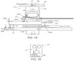
- FIG. 1Aillustrates a variation of a localization system in a typical operation room set up.
- FIG. 1Billustrates a 3D Model frame.
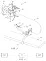
- FIG. 2illustrates an exemplary robotic surgical system.
- FIG. 3is a schematic representation of a first registration technique of correlating a sensor reference frame to selective reference frames.
- FIG. 4is a flow chart that illustrates a method of transforming a reference frame for a sensor of a surgical tool into a target reference frame.
- FIG. 5is a flow chart that illustrates a method of transforming a reference frame associated with a tool into a target reference frame.
- FIG. 6is a flow chart that illustrates a method of transforming a reference frame associated with a tool into a target reference frame utilizing medical appliances.
- FIG. 7is a flow chart that illustrates a method of using a sensor to transform a reference frame associated with a tool into a target reference frame.
- FIG. 8is a schematic illustration of a method of using an intravascular imaging sensor coupled with a shape sensor to transform a reference frame associated with a tool into a target reference frame.
- FIG. 9is a schematic illustration of an exemplary elongate member, e.g., a fiber, having a proximal end and a distal end.
- FIG. 10is a schematic illustration of another exemplary elongate member having a distal end inserted into a patient, with a proximal end remaining outside the patient.
- FIG. 11is a process flow diagram for an exemplary process of reducing measurement error associated with the position of an elongate member.
- Exemplary approaches described hereinprovide full or partial registration along the length of a sensor to reduce the influence of measurement error.
- an exemplary registration processgenerally relates a reference frame of a sensor to another reference frame of interest.
- applying heavier filtering at the proximal end of the sensorcan provide a more stable base for distal measurements reducing the influence of measurement errors.
- FOSSLFiber Optic Shape Sensing and Localization
- Fiber optic shape sensingis a technology that can sense the shape of a flexible body such as a catheter during a surgical procedure to permit visualization of the catheter in the patient's anatomy.
- One exemplary methodologyprovides a solution to this problem by using point registration along the length of the fiber to reduce the effect of the lever arm problem.
- these techniquesapply to all sensors that use incremental measurements, but nevertheless exemplary approaches below are described generally in the context of FOSSL fiber technology.
- Another exemplary approachincludes applying registration data along the fiber as well as at the proximal attachment to reduce error. Additionally, in another example stronger filtering may be utilized on the proximal orientation signal to reduce noticeable fluctuation of the position distally.
- orientation error at the proximal end of an IMSis decoupled from position error at the distal end.
- Exemplary methods of decoupling this errorinclude providing some notion of a registration closer to the distal tip and reduce the effect of orientation error.
- Various localization systems and methods for tracking an elongate instrument or toole.g., a robotically controlled elongate instrument, in real time, in a clinical or other environment, are described herein.
- the term “localization”is used in the art in reference to systems for determining and/or monitoring the position of objects, such as medical instruments or tools in a reference coordinate system.
- Various instrumentsare contemplated for use in the various systems described herein.
- elongate instrumentsare contemplated, such as, e.g., a catheter or vascular catheter.
- the various methods and systemsmay include integrating or registering a localization system or a localization sensor coupled to a surgical tool, with an image.
- a fiber optic tracking or localization systemis just one, non-limiting example of a system that allows for the tracking of a location, position and/or orientation of a localization sensor placed.
- Various other localization sensorsmay be utilized, e.g., electromagnetic sensors, and other sensors for detecting or controlling the movement of medical equipment.
- the localization sensorWhen the localization sensor is integrated into an image, it enhances the capabilities of an instrument control or tracking system by allowing a user or doctor to easily navigate the instrument through the complex anatomy without exposing the patient to excessive radiation over a prolonged period of time.
- the localization data or tracking information of a localization sensormay be registered to the desired image or model to allow for navigation of an elongate instrument through the image or model to accurately represent movement of the elongate instrument within a patient.
- registrationis a process that generally requires relating a reference frame of a sensor to another reference frame of interest. If the positions, orientations or shapes of two or more objects are known in the same reference frame, then the actual positions, orientations or shapes of each object relative to each other may be ascertained. Thus, with this information, one can drive or manipulate one of the objects relative to the other objects.
- the reference frame of interestis the visualization frame.
- the reference frameis the frame that the doctor is viewing, such as a patient or a live 2D/3D image such fluoroscopy, ultrasound or others.
- the goal of registrationis to determine the relationship of a frame of a sensor integrated into a tool or element in the surgical suite within the frame of reference of the patient, as represented in a 2D/3D image.
- the usercan drive and manipulate the tool in the 3D model.
- This techniqueprovides an advantage in that there is no longer a need for live fluoroscopy and radiation during a procedure.
- the toolis localized to the 3D model and the position, shape and orientation of the tool is visible to the user. Since the tool position, shape and orientation is updated in real time by a localization sensor, an image of the tool in the virtual representation of the 3D model will be updated as it is being advanced into the patient.
- the sensoris localized to the reference frame of the 3D model; therefore the orientation of a tip of the tool is known relative to the 3D model. This enables driving of the tool (such as a catheter) within 3 dimensional views of the anatomy and hence improves visualization and control during a surgical procedure.
- exemplary sensorsmay include incremental measurement sensors, where the position and orientation of a particular point is calculated and dependent on the previously calculated orientations and positions of proximal points or (spacing of consecutive points).
- the localization sensor operating in any medical systemneeds to be registered with a coordinate system, frame or image that is useful to an operator, such as the pre-operative 3D model or a fluoroscopic image.
- the pre-operative 3D modelor a fluoroscopic image.
- registrationis challenging because the coordinate system or frame of the sensor is not always easily related to the coordinate system of interest (i.e., the pre-operative 3D model).
- a fiber optic sensormay have its reference frame based physically in a splayer (base) for a catheter.
- basefor a catheter.
- the registration processIn addition to changing positions of reference frames, the registration process often requires information about the imaging system providing the image, such as its physical dimensions and/or the details about the imaging techniques used to acquire a particular 3D model or other image. Due to the variability in equipment used in a clinical environment, in certain situations there may be no guarantee that such information will be available or easily obtainable to an outside party.
- a method for tracking a robotically controlled elongate instrument in real timemay include displaying an image of a patient's anatomy.
- a localization sensormay then be coupled to the robotically controlled instrument.
- the localization sensormay provide localization data of the sensor and/or instrument.
- different sensorsmay be registered to specific tools, thereby enabling tool differentiation.
- the localization data from the localization sensormay be registered to the image.
- Registeringmay include transforming localization data generated by the localization sensor to the coordinate system or frame of the image such that localization data of the elongate instrument, to which the localization sensor is coupled, is overlaid on the image.
- the coordinate system of the localization sensormay be transformed or translated to the coordinate system of the image through one or more transformations, and optionally through other coordinate systems, to register the localization data to the image.
- a continuously or substantially continuously updated location of at least a portion of the elongate instrumentis provided in the image of the anatomy of a patient, which allows for or facilitates robotic navigation or control of the elongate instrument through the anatomy e.g., through the vasculature of a patient.
- the location, position and/or orientation of the localization sensormay be continuously tracked to allow for accurate manipulation of the elongate instrument in or through the anatomy of a patient.
- Various types of imagesmay be utilized in the methods and systems described herein.
- an imagemay be generated by CT or 2D or 3D fluoroscopy.
- An imagemay include a 3D or 2D anatomical model or a 2D or 3D fluoroscopic image or other types of images useful for visualizing an anatomy of a patient to perform various medical procedures.
- an image intensifierWhen using a fluoroscopy image, an image intensifier may be utilized. Localization data from the localization sensor may be registered to a fluoroscopy coordinate system of a fluoroscopy image coupled to the image intensifier. In order to register the localization data from the localization sensor to the fluoroscopy image, various parameters may be ascertained or known. For example, such parameters may include: a distance from an X-ray source to the image intensifier, a distance from the source to a bed, a size of the image intensifier, and/or the axis of rotation of a C-arm of the fluoroscopy system.
- localization datacan be registered to a 3D anatomical model or a fluoroscopy image.
- the techniques used to perform the registrationvary depending on the target. Where localization data is registered to a fluoroscopy image, the 2D nature of the fluoroscopy images may require that multiple images be taken at different angles before the registration process is complete.
- FIG. 1Ais a schematic of a typical operation room set up for a robotic surgical system. More specifically, a typical robotic surgical system 10 includes a table 12 upon which a patient 14 will be placed, a fluoroscopy system 16 , and a surgical tool, such as a catheter 18 (best seen in FIG. 2 ). Attached to the table 12 is a setup joint arm 20 to which a remote catheter manipulator (RCM) 22 is operatively connected. A splayer 24 may be mounted to the RCM 22 . A surgical tool, such as a catheter, is operatively connected to the splayer 24 . A fiber sensor 26 may be operatively connected to the surgical tool.
- the fluoroscopy system 16includes a C-arm 28 .
- a fluoroscopy panel 30is mounted to the C-arm 28 .
- the C-armis selectively moveable during the procedure to permit various images of the patient to be taken by the fluoroscopy panel 30 .
- robotic surgical system 10may further comprise an operator control station 31 , which may be remotely positioned with respect to table 12 .
- a communication link 32transfers signals between the operator control station 31 and the RCM 22 .
- the operator control station 31includes a control console 34 , a computer 36 , a computer interface, such as a mouse, a visual display system 38 and a master input device 40 .
- the master input device 40may include, but is not limited to, a multi-degree of freedom device having multiple joints and associated encoders.
- Each element of the robotic surgical system 10 positioned within the operating suitemay define a separate reference frame to which sensors may be localized. More specifically, separate reference frames may be defined for each of elements of the robotic surgical system 10 .
- Such reference framesmay include the following: a table reference frame TRF for the table 12 , a setup joint frame SJF for the setup joint 20 , an RCM reference frame RRF for the remote catheter manipulator (RCM) 22 , a splayer reference frame SRF, a fluoroscopy reference frame FF.
- Separate reference framesmay also be defined for a patient—patient reference frame PRR, a reference frame FRF for a sensor disposed within a surgical tool, and a pre-operative 3D frame AMF (best seen in FIG. 1B ).
- a first categoryinvolves using image processing or vision techniques to relate a reference frame RFR of a fiber sensor directly to an image or 3D model. This technique may be accomplished manually by a user or done automatically using image processing techniques.
- Another category to coordinate the reference frame FRF of a fiber optic sensorinvolves using knowledge about hardware, and potentially other sensors and or position of the fiber. Further discussion of these techniques is set forth below.
- the first categoryrelates the coordinate system of the sensor reference frame FRF to a fluoroscopy reference frame FF directly.
- This techniqueutilizes fluoroscopy images taken during the surgical procedure by the fluoroscopy system 30 , in real-time.
- an exemplary registration process 200is illustrated in the flow chart of FIG. 4 .
- the process 200may begin by inserting a tool into a patient at block 202 .
- the toolis a catheter 18 , which may be inserted by an RCM 22 .
- an intra-operative imageis taken of the tool 18 .
- the intra-operative imageis a fluoroscopy image taken by fluoroscopy system 30 .
- distinctive elements of the toolmay be identified in the fluoroscopy image at block 206 .
- the block 206may be accomplished by instructing the user to select certain marked points of a catheter 18 in the fluoroscopy image at the work station 31 . Examples of marked points include, but are not limited to, physical features of the catheter 18 such as the tip of the catheter 18 , certain shapes and an articulation band.
- fluoroscopy markersmay be disposed on the catheter.
- coordinates of the selected points of the catheter 18may be compared to corresponding measured points of elements of the catheter.
- measured points from a tool sensor operatively connected to the tool 18may be used.
- the tool sensoris a fiber optic sensor. Information about the fiber optic sensor will be known in relation to the features on the catheter from an in-factory calibration. This comparison can be used to determine a transformation matrix that can be used to transform a reference frame FRF for a sensor disposed within the surgical tool into the fluoroscopy reference frame FF. This transformation then localizes the tool relative to the intra-operative fluoroscopy image.
- the tool operatorcan now move or drive the tool to various, desired points visualized in the fluoroscopy image.
- the computer 36may be configured to track the marked points over time, such that an appropriate transformation may be updated.
- the identifiable markersneed not be on the portion of the tool that is inserted into the patient.
- markersmay be embedded on a splayer 24 , which may allow for larger and more complex markers to provide enhanced registration capabilities.
- the localization sensorcould serve to reduce the use of fluoroscopy during a procedure. More specifically, the use of fluoroscopy would be reduced since fluoroscopy would only be required when re-registration is needed during the procedure due to the loss of accuracy in the data obtained from the sensor.
- Another exemplary technique proposed to register a tool 18 to a desired reference frameinvolves the use of physical components of the medical system 10 and multiplying successive transformations.
- This proposed technique 300is illustrated schematically in FIG. 5 and involves finding a transformation path from a tool reference frame such as a fiber sensor, splayer 24 , or catheter 18 , to the table 12 , as in most surgical suite setups, the table location is generally known with respect to the fluoroscopy system 30 .
- registration technique 300involves determining a tool reference frame at block 302 (where the tool reference frame may be defined as the sensor reference frame FRF, splayer reference frame SRF or a catheter reference frame) and correlating the tool reference frame to a table reference frame TRF at block 304 , thereby registering the tool 18 to the table 12 .
- Registering the tool 18 to the table 12will serve to provide necessary information to permit registration to an additional target frame, such as a fluoroscopy reference frame FF, for example.
- a comparison of set reference points of the table 12 with corresponding reference points in a fluoroscopy imagemay be used to determine a transformation matrix to transform the table reference frame TRF into the fluoroscopy reference frame FF.
- the tool 18is registered to the table reference frame TRF, and thus combining all three transformations localizes the tool relative to the intra-operative fluoroscopy image.
- the present disclosuredoes not require that the tool 18 be registered to the table 12 . Indeed, it is expressly contemplated that registration of the tool 18 to other physical components within the surgical suite may also be utilized. This proposed technique requires the use of other sensors in addition to, or alternative to a fiber sensor, however. Exemplary configurations of registration through physical surgical suite components are discussed in further detail below.
- the registration process 400begins at block 402 , with determining the location of the setup joint 20 with respect to the table 12 .
- Encoders on the RCM 22 and setup joint 20may be used to determine the location of the setup joint 20 with respect to the table 12 . More specifically, the encoders assist with determining the location of the RCM 22 with respect to the table 12 .
- the location of the splayer carriage 24 carried by the RCM 22 with respect to the table 12can be determined; i.e., the setup joint reference frame SJF is localized with the RCM reference frame RRF.
- an evaluation of the splayer carriage 24 information with respect to the RCMcan be used to determine a transformation matrix that can be used to transform the splayer carriage reference frame SRF to the table reference frame TRF.
- inertial sensors on the RCM 22coupled with the information about the initial position of the RCM 22 on the table 12 , may be used to assist in localizing the catheter splayer reference frame SRF to the table reference frame TRF. More specifically, once the RCM 22 is localized to the table reference frame TRF, the catheter splayer reference frame SRF may be localized to the table reference frame TRF, as the position of the catheter splayer 24 with respect to the RCM 22 will be known from in-factory calibration.
- FIG. 7Yet another exemplary method 500 of performing registration through physical components is illustrated in FIG. 7 .
- the method 500uses a second fiber optic sensor.
- a first step 502one end of the fiber optic sensor is fixed to the table 12 .
- the other end of the sensoris fixed to the splayer 24 in a known orientation/position.
- a position and orientation transformation between the tip and base of the fiber sensormay be determined, thereby localizing the catheter splayer reference frame SRF to the table reference frame TRF in step 506 .
- the initial position of the fixed point at the tablemust be known.
- step 508another transformation may be done from the table reference frame TRF to the fluoroscopy reference frame FF.
- This final transformationi.e., from the table reference frame TRF to the fluoroscopy reference frame FF, then localizes the tool relative to the intra-operative fluoroscopy image.
- a further exemplary method of performing registration of a surgical tool to a physical componentincludes using electromagnetic sensors to track the location of the splayer 24 with respect to an electromagnetic sensor at a known location on the table 12 .
- the tool locationis calibrated to the splayer 24 in the factory, once the splayer 24 is localized to the table reference frame TRF, the tool may be localized to the fluoroscopy reference frame FF as the table 12 is known with respect to the fluoroscopy system 30 .
- overhead cameras or other visualization techniquesmay be employed to track distinct features on the splayer 24 and the table 12 to determine the respective orientation and position with regard to each other.
- a further techniquemay use the range sensors (such as, e.g., IR or ultrasound) to find the distance to several distinct and predetermined points on the table 12 and the splayer 24 .
- the range sensorssuch as, e.g., IR or ultrasound
- the toolmay be localized to the fluoroscopy reference frame FF as the table 12 is known with respect to the fluoroscopy system 30 .
- All of the above techniquesserve to register the tool to a physical component within the surgical suite, such as, for example, the table 12 .
- Some of the above techniquesrequire the RCM 22 and setup joint 20 to be registered to the table 12 . That pre-registration step may be achieved by using some known feature on the table 12 that the setup joint 20 may reference. Additionally, the pre-registration step may be achieved if the setup joint is equipped with joint sensors such as encoders. In another exemplary configuration, the tip of a sensor equipped tool may be used to touch or register the known feature on the table 12 to locate the table 12 with respect to other equipment within the surgical suite.
- the kinematics of the RCM 22can also be calculated by holding the tip of a fiber optic equipped tool in an arbitrary fixed location and cycling through the various axes of the RCM 22 . By keeping the tip in a fixed location, the relative changes to the fiber origin can be observed, and thus the kinematics of the system can be determined and localized to the table 12 . Once localized to the table reference frame TRF, the tool may then be localized to the fluoroscopy reference frame FF, as discussed above.
- additional modificationsmay be made to the location of the fiber base to facilitate registering the fiber sensor to the physical structure within the suite, such as, for example, the table 12 .
- one modificationis to extend the length of a fiber in the catheter so that the origin/base can be extended out of the splayer 24 and attached to a fixture having a known location on the table 12 .
- the toolmay then be localized to the fluoroscopy reference frame FF, as discussed above.
- Registration of the tool to a 3D Modelis also contemplated in this disclosure. Such registration may be performed directly from the fiber sensor reference frame FRF to the 3D Model frame AMF.
- the operatoris utilized. When the tool (such as the catheter) is inserted into the patient, tortuosity can be visualized from the fiber sensor data, as well as on the pre-operative 3D Model. To register the tool in the 3D Model, the operator may translate and rotate the 3D Model so that distinct images and/or features in the tortuosity match or line up with the shape of the fibers. However, in using this technique, every time the patient moves, the tool should be re-registered.
- a computer algorithmsuch as automated geometric search or mathematical optimization techniques that segments the model and matches the model and tool shape dynamically may also be used to match various shapes or features from the fiber sensor to the 3D pre-operative Model.
- the algorithmsmay be used to re-register the tool automatically or the user could use an input device, such as a track ball or mouse to move the 3D Model manually.
- Another proposed techniquemay be used to register the fiber sensor to the 3D Model through the fluoroscopy image, as illustrated in FIG. 3 .
- any of the above described techniques for registering the surgical tool 12 to the fluoroscopy reference frame FFmay be utilized.
- To register the fluoroscopy reference frame FF to the 3D Model reference frame AMFin one exemplary configuration, specific anatomical landmarks may be used to provide recognizable reference points. The only requirement for this approach is to have an anatomical landmark that is recognizable in both the fluoroscopy reference frame FF, as well as the pre-operative 3D Model reference frame AMF.
- the 3D Modelmay then be rotated by the operator to line up the recognized points in the fluoroscopy images with the 3D Model images. This action serves to register the fluoroscopy reference frame FF to the frame of the 3D Model AMF.
- the toolhas previously been localized to the fluoroscopy reference plane FF, so once the fluoroscopy reference plane FF is registered, the tool's location within the patient's anatomy may be determined with reference to the 3D Model localizing the tool to the 3D Model.
- a visual representation of the toolbased on the transformation matrix, may be displayed on the 3D Model. In this manner, the tool operator may then navigate the tool through the 3D Model.
- the 3D Modelmay then be rotated by the user to overlay the known anatomical location in the 3D Model with the fluoroscopy image, in which the known anatomical location is visible. Such action will also serve to register the tool with the 3D Model or localize the tool in the reference frame of the 3D model reference frame AMF.
- the computer 36may be programmed to employ a suitable algorithm such as automated geometric search or mathematical optimization techniques configured to match a distinct shape measured by the fiber sensor with a corresponding shape in the 3D Model.
- a suitable algorithmsuch as automated geometric search or mathematical optimization techniques configured to match a distinct shape measured by the fiber sensor with a corresponding shape in the 3D Model.
- the toolmay also be registered with the 3D Model.
- the accuracy of this methodwill depend on the size of vessel that the tool is in, and the degree of curvature of the tool. Accuracy will be improved if the tool is in a smaller vessel and will be worse if the tool is in larger vessels.
- This automated techniquecan also be used in conjunction with the manual techniques described above.
- the computermay be programmed to do automatic registration and suggest a preferred registration but the user may do final adjustments of the model. Once the tool is localized in the 3D Model of the patient's anatomy, the user may then proceed to maneuver the tool in the 3D Model.
- radiopaque markerscan be fixed to the anatomy. However, these markers would need to be present during pre-operative imaging when the 3D Model is created, and remain in the same location during intraoperative fluoroscopy imaging. With this technique, the position of these markers in the fluoroscopy reference frame FF can be used to correlate to the same markers in the 3D Model reference frame AMF, thereby registering the fiber sensor to the 3D Model reference frame AMF. Three-dimensional angiography may also make it easier to register the tool to the 3D Model by facilitating acquisition of the model in realtime, i.e. while the patient is on bed. While the model might drift away from the real anatomy when the operation is carried out, it may be advantageous to obtain the model and perform operations in the same spot.
- Another technique that may be utilized to register the surgical tool to a 3D Modelutilizes intravascular imaging. This technique allows for 3D visualization of a surgical tool, such as, a catheter, in the anatomy, but without the use of fluoroscopic imaging. Such a technique can benefit both physicians and patients by improving the ease of tool navigation, as well as and reducing radiation exposure of personnel inside the operating room.
- the registration technique 600may begin by utilizing a sensor 602 operatively coupled to the tool to sense a shape of the tool 604 while in the patient. This sensed shape is then mathematically correlated against features of the vascular model such as centerlines or walls in which a larger correlation value corresponds to a better match.
- the correlationcan be performed in real-time on each shape or by batch processing a sequence of shapes.
- This proposed techniqueassumes that the tool will always follow a unique configuration through the vasculature, and thus, a global maximum for the correlation exists. However, the correlation may return many local maxima since the tool configuration may follow many different paths between fixed distal and proximal ends. Choosing an incorrect maximum introduces registration error.
- the pre-operative 3D modelmay differ from the actual vasculature for a number of reasons, including, for example, patient motion or inaccuracies in pre-operative sensing. Such situations also may lead to registration error.
- IVUSintravascular ultrasound
- OCToptical coherence tomography
- IVUSintravascular ultrasound
- OCToptical coherence tomography
- IVUSintravascular ultrasound
- OCToptical coherence tomography
- Optical Coherence Tomographyperiodically produces a local 3D view of the vessel into which the tool is inserted.
- the images produced by these technologiesmay be processed to provide an estimate of a curve or surface representing the vessel wall 606 .
- the sensors 604may also determine the location of the catheter's endpoint within the vascular cross-section. Use of the sensors coupled with the tool 602 to provide shape information coupled with information obtainable from sensors 604 configured to provide information about the vessel walls 606 can assist in defining the 3D shape of the blood vessel 608 .
- the shape of the vesselis defined or otherwise reconstructed using the combined sensor data, the shape can be mathematically correlated to the 3D model 610 , thereby registering the tool to the 3D Model 612 .
- the 3D reconstruction and correlation stepsmay be combined into a single recursive filtering algorithm.
- a Bayesian filtere.g. Extended Kalman Filter (EKF), Unscented Kalman Filter (UKF), or Particle Filter
- EKFExtended Kalman Filter
- UDFUnscented Kalman Filter
- Particle Filtermay be used to develop an estimate of the tool's position relative to the pre-op 3D model given both imaging and sensor 602 information.
- the filter's stateis a set of points or a parametric curve representing the position and shape of the tool 602 with respect to the pre-op 3D model, as well as the rate of change of this shape. For accurate registration, patient motion may also be taken into account.
- the filter's statemay also contain warping parameters for the pre-op 3D model. These warping parameters may be evenly distributed, or may be selected based on the structure of the anatomy around the vasculature. The motion of the structure of the anatomy around the vasculature may be measured using visible light tracking technologies such as stereoscopic cameras, structured light tracking technologies, and/or other localization technologies attached to the patient skin.
- the recursive filtering algorithmoperates by predicting the motion of the tool in the 3D model, then performing an update of the filter hypothesis given new sensor measurements.
- a kinematic model of the catheter and control inputssuch as current pull-wire tension and displacement may be used to perform the filter's motion update.
- the filter's measurement updatemay apply a correction to the tool registration and model warping parameters by comparing a predicted vessel wall with the sensed position and orientation of the vessel from the imaging and sensor measurements.
- the updateeffectively executes the correlation between 3-D sensor information and the 3D model. Performing these correlations repeatedly in a recursive filtering framework may provide a real-time catheter position estimate.
- the filter's parametersmay be tuned such that differences between the measurements and the model over a small time constant (ms) will lead to changes in the catheter position estimate in order to filter out high-frequency sensor noise. Differences over a large time constant (seconds) may lead to changes in the model's warping parameters.
- the location of the tool within the 3D modelmay be determined, allowing an operator to drive the tool within the vasculature using the 3D model without requiring intra-operative fluoroscopy.
- Sensors 604may also be utilized to sense the environment around the tool. Thus, once the tool is registered to the 3D model, this environmental information, such as, for example, vascular occlusions may be displayed at the correct position in the 3D Model.
- the intravascular imaging sensor 604provides a mechanism to sense and display features of the environment surrounding the tool without the use of fluoroscopy. There are many ways to display this information. One non-limiting option is to simply provide a display of a real-time view of the imaging sensor's output alongside a view of the catheter's location in the 3D model or superimposed on top of the 3D model. Another option may be to analyze the intravascular image to detect environmental changes. For example, IVUS image processing techniques can be used to detect areas of plaque in the image. This information can be used to annotate the IVUS image in order to alert the physician to environmental conditions.
- the 3D pre-op modelcan also be annotated. In this way, the existing work that has used IVUS to perform vascular sensing may be leveraged by the combined IVUS and sensor system to provide a 3D view of the environment to the physician.
- Registrationis generally the mathematical process of computing the orientation and position of all or portions of a shape (sequence of points) in some coordinate frame. Registration can fix the orientation and position of the fiber at one or more points (for six degrees of freedom of constraints at each point) or registration can be carried out with fewer constraints at a single point or multiple points. Inherently, though, registration is the method of positioning and orienting a sensor frame in a reference frame.
- one type of registrationis to determine the position and orientation of the proximal end (“origin” of the IMS) in the fluoroscopy, or world, coordinate frame and display that to the user in the virtual environment.
- the points in the IMScan be transformed and displayed in the world coordinate system throughout the procedure.
- the accuracy of this registration procedurecan be difficult and inadequate for applications that require sub-mm accuracy at the distal end of the sensor. Inaccuracy will result if the origin of the IMS moves; if the origin does move, it needs to be tracked in the world coordinate frame, which may lead to error. In addition, the error inherent in the sensor could cause errors in the sensor measurement.
- multiple positions of registration along the length of an elongate member 900may be provided to reduce the error in a sensor 902 .
- the sensor 902is an IMS fiber sensor configured to output a position of the elongate member at the sensor 902 .
- the elongate membere.g., a catheter, may be generally fixed at a proximal end 904 such that the position of the proximal end 904 may generally be known.
- a position of the elongate member 900is known at one additional point 906 , which is proximal to the distal portion of the sensor 902 .
- any number of additional points proximal to the sensor 902may be used, as described further below.
- Using the additional piece(s) of information at the point(s) proximal to the sensor 902may be used to register the IMS at a position distal of the “origin,” i.e., at the sensor 902 , helping to reduce orientation error propagated from the proximal end to a position error in the distal end.
- the nature of the fiber and catheteris that the proximal end is the only place that the fiber is attached to the system, but there are a number of ways of acquiring registration along the length of the fiber.
- Registration of a shape or series of points of the IMS sensor 902can be used to improve registration. This can be performed by acquiring 3D shapes or obtaining spatial information from 2D images, i.e., of the elongate member 900 .
- a plurality of points 906 , 908 , 910 , and 912may be employed to determine a shape of the elongate member 900 at each of the points 906 , 908 , 910 , and 912 , such that a shape or position of the elongate member 900 is known.
- modelscould be generated from a rotational angiography, computed tomography scan, or other standard imaging technique.
- any known shape that the catheter passes throughcould also contribute to registration just as in a 3-D model, such as an engineered semitortuous introducer, a curve in the take-off of the sheath splayer, an anti-buckling device feature, etc.
- the catheter and IMSwill be passing through a known shape that can be used to register the position and orientation of a distal portion of the catheter.
- 3D informationmay be to add other localization sensors such as an electromagnetic sensor to various critical points along the catheter. More specifically, at least one of the proximal points 906 , 908 , 910 , and/or 912 may have an electromagnetic sensor. The generally absolute measurements of position at these locations can reduce any error propagated from the fiber at a portion distal of the position of any electromagnetic sensors at points 906 , 908 , 910 , and/or 912 .
- Another exemplary method of obtaining additional shape informationis to use computer vision techniques on the fluoroscopy images to track the catheter and then feed that information into the system to improve registration. Because two-dimensional imaging will provide less information than a 3D model it may not provide as much information to reduce error, but it would likely remove error at least in the plane of the image. This registration might also not be needed constantly during a procedure, but may be used when imaging, e.g., fluoroscopy, is active and the operator wants more accuracy of the tracked catheter.
- a position of a proximal portion of the elongate member 900may be visualized in 2D and used to increase accuracy of measurements at the sensor 902 .
- the above exemplary approaches to optimizing position measurements of the sensor 902provide an alternate registration location to a proximal portion of the elongate member, e.g., at the base of the fiber at the splayer attached to the RCM.
- registering a shape to a 3D modeldoes not necessarily completely replace the registration at the splayer (not shown in FIG. 9 ).
- Using an optimization technique that finds the most likely position and orientation of the sensor given both the anatomical registration and the distal splayer registrationis probably the best way to reduce overall error and achieve a strong overall pose of the catheter in relation to the anatomy.
- This algorithmcan also take advantage of any proximal motion of the catheter, such as shaft insertion, etc.
- this algorithmcan be time consuming and computationally intensive, especially when a good starting point is not given. The registration of the origin of the IMS could be used as such a starting point.
- Another exemplary approach in the case where global registration of position is not neededwould be to allow the user to designate a specific position on the catheter that is constrained laterally, e.g., by the anatomy of a patient.
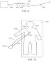
- an elongate member 1000is partially inserted into a patient 1100 , who remains generally fixed in position on a table 1102 .
- a distal portion 1004 of the elongate member 1000which includes an IMS 1006 for measuring position of the elongate member 1000 , is received within the patient 1100 , while a proximal portion 1002 remains outside the body of the patient 1100 .
- the insertion site 1008 of the elongate member 1000 into the patient 1100is known and is substantially fixed, such that a position of the elongate member 1000 is known at the insertion site 1008 in at least two dimensions.
- the insertion site 1008 positionat least in two degrees of freedom, could be registered in addition to the splayer 1010 , while the insertion of the elongate member 1000 is updated accordingly.
- the orientation of the elongate member 1000 and/or a measurement fiber at this designated ‘base’ (i.e., the insertion site 1008 ) of the shorter effective fiberwould depend on the actual orientation of the fiber (including twist) and the insertion would depend on the commanded fiber insertion.
- the shape distal to the insertion site 1008could be updated rapidly and without any error from the shape of the proximal portion 1002 , which is proximal to the designated base position, i.e., the insertion site 1008 . Accordingly, error in position measurements of the elongate member 1000 anywhere along the distal portion 1004 is greatly reduced by effectively reducing the measurement length of the IMS 1006 .
- the above exemplary approachmay be particularly advantageous for relative position display (essentially a coordinate frame not correctly registered to the fluoroscopy/anatomical coordinate frames) but may ultimately make it difficult to determine a global position of the tip of the catheter.
- relative position displayessentially a coordinate frame not correctly registered to the fluoroscopy/anatomical coordinate frames
- filteringis a method of sacrificing responsiveness in the time domain to reduce the shape and position error.
- an operator of an elongate membermay generally be focusing on the distal end of the catheter during a procedure, and the proximal end will generally be stable and not moving quickly.
- selective filtering on proximal shapecan be applied to reduce orientation error in the proximal region while the distal region does not filter the signal.
- position data associated with the distal region of the elongate member 900 between the points 912 and 902is not filtered, while the position data of a proximal region between points 906 and 912 is filtered.
- the selective filtering of the proximal regionreduces the influence of proximal error by filtering it out (for instance, averaging over multiple time steps) while, the distal portion will be updated as fast as possible using the orientation and position of the proximal section as a stable base.
- FOSSLfiber-based measurement system
- proximal filteringwill be a responsive system that exhibits less error.
- the exact location where filtering starts or stopsmay vary based on the application and it is even possible to apply a variable level of filtering along the fiber with the maximum filtering at the proximal end, e.g., between points 906 and 912 , and the minimum at the distal end, e.g., distal of the point 902 .
- the proximal endis filtered heavily in relation to the distal end, it may be useful to detect when the proximal shape changes significantly in a short period of time, such as a prolapsed catheter at the iliac bifurcation (a rapid motion of the shaft of the catheter bulging up into the aorta). This can be accomplished by noting when the new catheter shape is significantly different than the filtered shape. In this case, the filtering on the proximal end can be reduced so that the operator sees the most up to date information and can react accordingly. After the proximal shape remains more constant for a period of time, the filtering can increase, again reducing error at the tip. To prevent sudden jumps in the rendered data, temporarily-variable filtering algorithms can be implemented in such a way as to provide continuity of the filter output, which may increase the appearance of smoothness and reduce noise of the measurement.
- One exemplary approachwould be to apply relatively heavy filtering on the proximal end of the fiber, e.g., between points 906 and 912 , and light filtering on the distal end, e.g., distal of point 902 , as described above.
- Specific aspects of this sensor when used in a robotic systemcould modified, such as updating insertion immediately without filtering since axially the catheter is relatively stiff and low in error (assuming the catheter does not buckle). This is potentially problematic because by filtering different dimensions at different rates, trajectories can become skewed, producing measurement points that do not lie along the true trajectory of the device. Additionally, filtering could be accelerated when the proximal measurement changes significantly in relation to the filtered version to give more responsive feedback during a prolapsed situation.
- instinctiveness computationsare often computed from orientations propagated from the registered base of the fiber.
- Instinctivenessrefers generally to matching and orientation or a location of a device such as a catheter with a visualization device that is used to control the catheter, such as an image of the catheter. Since instinctiveness includes absolute orientation measurements, they may be computed separate from any intermediate registration or clipping techniques. On the flip side, because distance does not magnify orientation errors, there is little or no extra error from the distance from the base to the tip of the fiber in instinctiveness measurements. Furthermore, instinctiveness measurements generally do not require a fast update rate so it is possible to filter heavily to reduce orientation error and noise.
- Process 999may begin at block 1110 , where an incremental measurement sensor may be provided.
- an elongated member 900may have an IMS 902 positioned along a distal portion of the elongate member 900 .
- the elongate member 900includes a fiber, which may be employed to determine position and/or orientation data of the elongate member 900 .
- Process 999may then proceed to block 1112 .
- registration datamay be applied at one or more proximal locations along the elongated member. For example, as described above a position of the elongate member 900 at any one or more of points 906 , 908 , 910 , and/or 912 may be registered. For example, a proximal position of one or more proximal locations may be used to increase accuracy of measured data from the IMS 902 .
- one of the proximal locations used to apply registration data along the elongated member 900includes a proximal attachment of the elongated member 900 , e.g., at a proximal end 904 .
- the proximal attachment at the proximal end 904may general fix a portion of the elongated member at the first one of the locations.
- a position of the proximal location(s)may be determined using an electromagnetic sensor, or by using a two-dimensional image of the one or more proximal locations, e.g., as obtained by fluoroscopy during a procedure.
- applying the registration data at the one or more proximal locationsmay include constraining a lateral position of the one or more proximal locations.
- an insertion site 1008 associated with a patient 1100may generally constrain an elongate member 1000 laterally at the insertion site 1008 .
- the generally fixed insertion site 1008indicates a position of the elongate member 1000 in at least two dimensions at the insertion site 1008 .
- a proximal signal of the elongated membermay be selectively filtered, e.g., in relation to a distal signal of the elongated member. Filtering of the proximal signal may occur at a different rate than the filtering of the distal signal. In one example, heavier or more intrusive filtering of the proximal signal may be employed, especially during a procedure where the proximal portion(s) of the elongate member do not change rapidly. In some cases, filtering may include averaging an incremental orientation position change in the proximal signal. Moreover, variable filtering methodologies may be used.
- a heavier filtering methodologymay be employed only during such times that a position of the proximal portion of the elongate member is not rapidly changing position.
- the filtering of the proximal portionmay cease or be reduced. Filtering at the initially heavier setting may resume after a predetermined period of time expires, in which the proximal portion of the elongate member maintains a same position or does not rapidly change position during such time.
- Process 999may then proceed to block 1116 .
- a position of the incremental measurement sensormay be determined. For example, as described above a position of an IMS 902 may be determined based at least upon the registration data from the one or more proximal locations. In this manner, measurement error may be reduced since less incremental error occurs over the length of the elongate member. Alternatively, or in addition, a position of the incremental measurement sensor may be determined based at least upon a filtered proximal signal. In such cases, selectively filtering may reduce a fluctuation of the determined position of incremental measurement sensor by generally smoothing out position signals relating to the proximal portion(s) of the elongate member.
- the methods described aboveare the examples of registration using known data about the location of the catheter in relation to the anatomy.
- the first exampledoes not include extra localization information while the second example assumes some knowledge of the shape of the anatomy or other features along the path of the catheter.
- computing systems and/or devicesmay include a computer or a computer readable storage medium implementing the operation of drive and implementing the various methods and processes described herein.
- computing systems and/or devicessuch as user input devices included in the workstation 31 or any components thereof, merely as examples, may employ any of a number of computer operating systems, including, but by no means limited to, versions and/or varieties of the Microsoft Windows® operating system, the Unix operating system (e.g., the Solaris® operating system distributed by Oracle Corporation of Redwood Shores, Calif.), the AIX UNIX operating system distributed by International Business Machines of Armonk, N.Y., the Linux operating system, the Mac OS X and iOS operating systems distributed by Apple Inc. of Cupertino, Calif., and the Android operating system developed by the Open Handset Alliance.
- the Unix operating systeme.g., the Solaris® operating system distributed by Oracle Corporation of Redwood Shores, Calif.
- AIX UNIX operating systemdistributed by International Business Machines of Armonk, N.Y.
- Computing devicesgenerally include computer-executable instructions, where the instructions may be executable by one or more computing devices such as those listed above.
- Computer-executable instructionsmay be compiled or interpreted from computer programs created using a variety of programming languages and/or technologies, including, without limitation, and either alone or in combination, Java.TM, C, C++, Visual Basic, Java Script, Perl, etc.
- a processore.g., a microprocessor
- receives instructionse.g., from a memory, a computer-readable medium, etc., and executes these instructions, thereby performing one or more processes, including one or more of the processes described herein.
- Such instructions and other datamay be stored and transmitted using a variety of computer-readable media.
- a computer-readable mediumincludes any non-transitory (e.g., tangible) medium that participates in providing data (e.g., instructions) that may be read by a computer (e.g., by a processor of a computer).
- a mediummay take many forms, including, but not limited to, non-volatile media and volatile media.
- Non-volatile mediamay include, for example, optical or magnetic disks and other persistent memory.
- Volatile mediamay include, for example, dynamic random access memory (DRAM), which typically constitutes a main memory.
- Such instructionsmay be transmitted by one or more transmission media, including coaxial cables, copper wire and fiber optics, including the wires that comprise a system bus coupled to a processor of a computer.
- Computer-readable mediainclude, for example, a floppy disk, a flexible disk, hard disk, magnetic tape, any other magnetic medium, a CD-ROM, DVD, any other optical medium, punch cards, paper tape, any other physical medium with patterns of holes, a RAM, a PROM, an EPROM, a FLASH-EEPROM, any other memory chip or cartridge, or any other medium from which a computer can read.
- Databases, data repositories or other data stores described hereinmay include various kinds of mechanisms for storing, accessing, and retrieving various kinds of data, including a hierarchical database, a set of files in a file system, an application database in a proprietary format, a relational database management system (RDBMS), etc.
- Each such data storeis generally included within a computing device employing a computer operating system such as one of those mentioned above, and are accessed via a network in any one or more of a variety of manners.
- a file systemmay be accessible from a computer operating system, and may include files stored in various formats.
- An RDBMSgenerally employs the Structured Query Language (SQL) in addition to a language for creating, storing, editing, and executing stored procedures, such as the PL/SQL language mentioned above.
- SQLStructured Query Language
- system elementsmay be implemented as computer-readable instructions (e.g., software) on one or more computing devices (e.g., servers, personal computers, etc.), stored on computer readable media associated therewith (e.g., disks, memories, etc.).
- a computer program productmay comprise such instructions stored on computer readable media for carrying out the functions described herein.
Landscapes
- Health & Medical Sciences (AREA)
- Life Sciences & Earth Sciences (AREA)
- Engineering & Computer Science (AREA)
- Medical Informatics (AREA)
- Physics & Mathematics (AREA)
- Veterinary Medicine (AREA)
- Public Health (AREA)
- General Health & Medical Sciences (AREA)
- Animal Behavior & Ethology (AREA)
- Biomedical Technology (AREA)
- Heart & Thoracic Surgery (AREA)
- Surgery (AREA)
- Biophysics (AREA)
- Molecular Biology (AREA)
- Pathology (AREA)
- Nuclear Medicine, Radiotherapy & Molecular Imaging (AREA)
- Radiology & Medical Imaging (AREA)
- General Physics & Mathematics (AREA)
- High Energy & Nuclear Physics (AREA)
- Optics & Photonics (AREA)
- Pulmonology (AREA)
- Anesthesiology (AREA)
- Hematology (AREA)
- Theoretical Computer Science (AREA)
- Oral & Maxillofacial Surgery (AREA)
- Endoscopes (AREA)
- Apparatus For Radiation Diagnosis (AREA)
Abstract
Description
Claims (18)
Priority Applications (3)
| Application Number | Priority Date | Filing Date | Title |
|---|---|---|---|
| US16/697,064US11241203B2 (en) | 2013-03-13 | 2019-11-26 | Reducing measurement sensor error |
| US17/580,732US12156755B2 (en) | 2013-03-13 | 2022-01-21 | Reducing measurement sensor error |
| US18/966,068US20250090113A1 (en) | 2013-03-13 | 2024-12-02 | Reducing measurement sensor error |
Applications Claiming Priority (8)
| Application Number | Priority Date | Filing Date | Title |
|---|---|---|---|
| US201361779742P | 2013-03-13 | 2013-03-13 | |
| US14/208,514US9057600B2 (en) | 2013-03-13 | 2014-03-13 | Reducing incremental measurement sensor error |
| US14/712,587US9289578B2 (en) | 2013-03-13 | 2015-05-14 | Reducing incremental measurement sensor error |
| US15/076,232US20160202053A1 (en) | 2013-03-13 | 2016-03-21 | Reducing incremental measurement sensor error |
| US15/387,347US9844353B2 (en) | 2013-03-13 | 2016-12-21 | Reducing incremental measurement sensor error |
| US15/844,420US10123755B2 (en) | 2013-03-13 | 2017-12-15 | Reducing incremental measurement sensor error |
| US16/165,534US10492741B2 (en) | 2013-03-13 | 2018-10-19 | Reducing incremental measurement sensor error |
| US16/697,064US11241203B2 (en) | 2013-03-13 | 2019-11-26 | Reducing measurement sensor error |
Related Parent Applications (1)
| Application Number | Title | Priority Date | Filing Date |
|---|---|---|---|
| US16/165,534ContinuationUS10492741B2 (en) | 2013-03-13 | 2018-10-19 | Reducing incremental measurement sensor error |
Related Child Applications (1)
| Application Number | Title | Priority Date | Filing Date |
|---|---|---|---|
| US17/580,732ContinuationUS12156755B2 (en) | 2013-03-13 | 2022-01-21 | Reducing measurement sensor error |
Publications (2)
| Publication Number | Publication Date |
|---|---|
| US20200155084A1 US20200155084A1 (en) | 2020-05-21 |
| US11241203B2true US11241203B2 (en) | 2022-02-08 |
Family
ID=51523443
Family Applications (9)
| Application Number | Title | Priority Date | Filing Date |
|---|---|---|---|
| US14/208,514ActiveUS9057600B2 (en) | 2013-03-13 | 2014-03-13 | Reducing incremental measurement sensor error |
| US14/712,587ActiveUS9289578B2 (en) | 2013-03-13 | 2015-05-14 | Reducing incremental measurement sensor error |
| US15/076,232AbandonedUS20160202053A1 (en) | 2013-03-13 | 2016-03-21 | Reducing incremental measurement sensor error |
| US15/387,347ActiveUS9844353B2 (en) | 2013-03-13 | 2016-12-21 | Reducing incremental measurement sensor error |
| US15/844,420ActiveUS10123755B2 (en) | 2013-03-13 | 2017-12-15 | Reducing incremental measurement sensor error |
| US16/165,534ActiveUS10492741B2 (en) | 2013-03-13 | 2018-10-19 | Reducing incremental measurement sensor error |
| US16/697,064ActiveUS11241203B2 (en) | 2013-03-13 | 2019-11-26 | Reducing measurement sensor error |
| US17/580,732ActiveUS12156755B2 (en) | 2013-03-13 | 2022-01-21 | Reducing measurement sensor error |
| US18/966,068PendingUS20250090113A1 (en) | 2013-03-13 | 2024-12-02 | Reducing measurement sensor error |
Family Applications Before (6)
| Application Number | Title | Priority Date | Filing Date |
|---|---|---|---|
| US14/208,514ActiveUS9057600B2 (en) | 2013-03-13 | 2014-03-13 | Reducing incremental measurement sensor error |
| US14/712,587ActiveUS9289578B2 (en) | 2013-03-13 | 2015-05-14 | Reducing incremental measurement sensor error |
| US15/076,232AbandonedUS20160202053A1 (en) | 2013-03-13 | 2016-03-21 | Reducing incremental measurement sensor error |
| US15/387,347ActiveUS9844353B2 (en) | 2013-03-13 | 2016-12-21 | Reducing incremental measurement sensor error |
| US15/844,420ActiveUS10123755B2 (en) | 2013-03-13 | 2017-12-15 | Reducing incremental measurement sensor error |
| US16/165,534ActiveUS10492741B2 (en) | 2013-03-13 | 2018-10-19 | Reducing incremental measurement sensor error |
Family Applications After (2)
| Application Number | Title | Priority Date | Filing Date |
|---|---|---|---|
| US17/580,732ActiveUS12156755B2 (en) | 2013-03-13 | 2022-01-21 | Reducing measurement sensor error |
| US18/966,068PendingUS20250090113A1 (en) | 2013-03-13 | 2024-12-02 | Reducing measurement sensor error |
Country Status (1)
| Country | Link |
|---|---|
| US (9) | US9057600B2 (en) |
Cited By (1)
| Publication number | Priority date | Publication date | Assignee | Title |
|---|---|---|---|---|
| US12156755B2 (en) | 2013-03-13 | 2024-12-03 | Auris Health, Inc. | Reducing measurement sensor error |
Families Citing this family (121)
| Publication number | Priority date | Publication date | Assignee | Title |
|---|---|---|---|---|
| US20090036900A1 (en)* | 2007-02-02 | 2009-02-05 | Hansen Medical, Inc. | Surgery methods using a robotic instrument system |
| ES2465915T3 (en) | 2007-11-26 | 2014-06-09 | C.R. Bard, Inc. | Integrated system for intravascular catheter placement |
| US9521961B2 (en) | 2007-11-26 | 2016-12-20 | C. R. Bard, Inc. | Systems and methods for guiding a medical instrument |
| US8781555B2 (en) | 2007-11-26 | 2014-07-15 | C. R. Bard, Inc. | System for placement of a catheter including a signal-generating stylet |
| US9254123B2 (en) | 2009-04-29 | 2016-02-09 | Hansen Medical, Inc. | Flexible and steerable elongate instruments with shape control and support elements |
| US9532724B2 (en) | 2009-06-12 | 2017-01-03 | Bard Access Systems, Inc. | Apparatus and method for catheter navigation using endovascular energy mapping |
| EP2912999B1 (en) | 2010-05-28 | 2022-06-29 | C. R. Bard, Inc. | Apparatus for use with needle insertion guidance system |
| US8672837B2 (en) | 2010-06-24 | 2014-03-18 | Hansen Medical, Inc. | Methods and devices for controlling a shapeable medical device |
| US20120071752A1 (en) | 2010-09-17 | 2012-03-22 | Sewell Christopher M | User interface and method for operating a robotic medical system |
| US20130030363A1 (en) | 2011-07-29 | 2013-01-31 | Hansen Medical, Inc. | Systems and methods utilizing shape sensing fibers |
| EP2745088A1 (en)* | 2011-09-09 | 2014-06-25 | Koninklijke Philips N.V. | Optical monitoring device for monitoring curvature of a flexible medical instrument |
| US9452276B2 (en) | 2011-10-14 | 2016-09-27 | Intuitive Surgical Operations, Inc. | Catheter with removable vision probe |
| US9387048B2 (en) | 2011-10-14 | 2016-07-12 | Intuitive Surgical Operations, Inc. | Catheter sensor systems |
| US20130303944A1 (en) | 2012-05-14 | 2013-11-14 | Intuitive Surgical Operations, Inc. | Off-axis electromagnetic sensor |
| US10238837B2 (en) | 2011-10-14 | 2019-03-26 | Intuitive Surgical Operations, Inc. | Catheters with control modes for interchangeable probes |
| US10383765B2 (en) | 2012-04-24 | 2019-08-20 | Auris Health, Inc. | Apparatus and method for a global coordinate system for use in robotic surgery |
| US10842461B2 (en)* | 2012-06-21 | 2020-11-24 | Globus Medical, Inc. | Systems and methods of checking registrations for surgical systems |
| US20140148673A1 (en) | 2012-11-28 | 2014-05-29 | Hansen Medical, Inc. | Method of anchoring pullwire directly articulatable region in catheter |
| US10149720B2 (en) | 2013-03-08 | 2018-12-11 | Auris Health, Inc. | Method, apparatus, and a system for facilitating bending of an instrument in a surgical or medical robotic environment |
| US9566414B2 (en) | 2013-03-13 | 2017-02-14 | Hansen Medical, Inc. | Integrated catheter and guide wire controller |
| US11213363B2 (en) | 2013-03-14 | 2022-01-04 | Auris Health, Inc. | Catheter tension sensing |
| US9498601B2 (en) | 2013-03-14 | 2016-11-22 | Hansen Medical, Inc. | Catheter tension sensing |
| US9271663B2 (en) | 2013-03-15 | 2016-03-01 | Hansen Medical, Inc. | Flexible instrument localization from both remote and elongation sensors |
| US9629595B2 (en) | 2013-03-15 | 2017-04-25 | Hansen Medical, Inc. | Systems and methods for localizing, tracking and/or controlling medical instruments |
| US9014851B2 (en) | 2013-03-15 | 2015-04-21 | Hansen Medical, Inc. | Systems and methods for tracking robotically controlled medical instruments |
| US9283046B2 (en) | 2013-03-15 | 2016-03-15 | Hansen Medical, Inc. | User interface for active drive apparatus with finite range of motion |
| US10376672B2 (en) | 2013-03-15 | 2019-08-13 | Auris Health, Inc. | Catheter insertion system and method of fabrication |
| US10849702B2 (en) | 2013-03-15 | 2020-12-01 | Auris Health, Inc. | User input devices for controlling manipulation of guidewires and catheters |
| US11020016B2 (en) | 2013-05-30 | 2021-06-01 | Auris Health, Inc. | System and method for displaying anatomy and devices on a movable display |
| WO2014201165A1 (en) | 2013-06-11 | 2014-12-18 | Auris Surgical Robotics, Inc. | System for robotic assisted cataract surgery |
| US10426661B2 (en) | 2013-08-13 | 2019-10-01 | Auris Health, Inc. | Method and apparatus for laser assisted cataract surgery |
| KR102470468B1 (en)* | 2014-03-17 | 2022-11-25 | 인튜어티브 서지컬 오퍼레이션즈 인코포레이티드 | System and method for aligning with a reference target |
| EP3243476B1 (en) | 2014-03-24 | 2019-11-06 | Auris Health, Inc. | Systems and devices for catheter driving instinctiveness |
| US10046140B2 (en) | 2014-04-21 | 2018-08-14 | Hansen Medical, Inc. | Devices, systems, and methods for controlling active drive systems |
| US9744335B2 (en) | 2014-07-01 | 2017-08-29 | Auris Surgical Robotics, Inc. | Apparatuses and methods for monitoring tendons of steerable catheters |
| US10792464B2 (en) | 2014-07-01 | 2020-10-06 | Auris Health, Inc. | Tool and method for using surgical endoscope with spiral lumens |
| US9561083B2 (en) | 2014-07-01 | 2017-02-07 | Auris Surgical Robotics, Inc. | Articulating flexible endoscopic tool with roll capabilities |
| CN106572887B (en)* | 2014-07-15 | 2020-05-12 | 皇家飞利浦有限公司 | Image integration and robotic endoscope control in an X-ray suite |
| EP3200718A4 (en) | 2014-09-30 | 2018-04-25 | Auris Surgical Robotics, Inc | Configurable robotic surgical system with virtual rail and flexible endoscope |
| US10314463B2 (en) | 2014-10-24 | 2019-06-11 | Auris Health, Inc. | Automated endoscope calibration |
| US11819636B2 (en) | 2015-03-30 | 2023-11-21 | Auris Health, Inc. | Endoscope pull wire electrical circuit |
| CN114795471A (en)* | 2015-04-06 | 2022-07-29 | 直观外科手术操作公司 | System and method for registration compensation in image-guided surgery |
| WO2017028916A1 (en)* | 2015-08-19 | 2017-02-23 | Brainlab Ag | Reference array holder |
| JP6824967B2 (en) | 2015-09-18 | 2021-02-03 | オーリス ヘルス インコーポレイテッド | Tubular net navigation |
| US10058393B2 (en) | 2015-10-21 | 2018-08-28 | P Tech, Llc | Systems and methods for navigation and visualization |
| EP3200719B1 (en)* | 2015-11-02 | 2019-05-22 | Brainlab AG | Determining a configuration of a medical robotic arm |
| US10143526B2 (en) | 2015-11-30 | 2018-12-04 | Auris Health, Inc. | Robot-assisted driving systems and methods |
| US11020563B2 (en) | 2016-07-14 | 2021-06-01 | C. R. Bard, Inc. | Automated catheter-to-vessel size comparison tool and related methods |
| US10463439B2 (en) | 2016-08-26 | 2019-11-05 | Auris Health, Inc. | Steerable catheter with shaft load distributions |
| US11241559B2 (en) | 2016-08-29 | 2022-02-08 | Auris Health, Inc. | Active drive for guidewire manipulation |
| CN106338266A (en)* | 2016-08-29 | 2017-01-18 | 孟玲 | 2D position sensitive sensor based spatial six-freedom-degree object positioning system |
| US9931025B1 (en)* | 2016-09-30 | 2018-04-03 | Auris Surgical Robotics, Inc. | Automated calibration of endoscopes with pull wires |
| US10244926B2 (en) | 2016-12-28 | 2019-04-02 | Auris Health, Inc. | Detecting endolumenal buckling of flexible instruments |
| US11497507B2 (en)* | 2017-02-19 | 2022-11-15 | Orpheus Ventures, Llc | Systems and methods for closing portions of body tissue |
| WO2018183727A1 (en) | 2017-03-31 | 2018-10-04 | Auris Health, Inc. | Robotic systems for navigation of luminal networks that compensate for physiological noise |
| KR102643758B1 (en) | 2017-05-12 | 2024-03-08 | 아우리스 헬스, 인코포레이티드 | Biopsy devices and systems |
| KR102576296B1 (en) | 2017-05-17 | 2023-09-08 | 아우리스 헬스, 인코포레이티드 | Interchangeable working channels |
| US10022192B1 (en) | 2017-06-23 | 2018-07-17 | Auris Health, Inc. | Automatically-initialized robotic systems for navigation of luminal networks |
| US10299870B2 (en) | 2017-06-28 | 2019-05-28 | Auris Health, Inc. | Instrument insertion compensation |
| WO2019005696A1 (en) | 2017-06-28 | 2019-01-03 | Auris Health, Inc. | Electromagnetic distortion detection |
| CN116725667A (en) | 2017-06-28 | 2023-09-12 | 奥瑞斯健康公司 | System for providing positioning information and method for positioning an instrument within an anatomical structure |
| US10426559B2 (en) | 2017-06-30 | 2019-10-01 | Auris Health, Inc. | Systems and methods for medical instrument compression compensation |
| CN107392872B (en)* | 2017-07-27 | 2020-04-28 | 强联智创(北京)科技有限公司 | Generation method and generation system of micro-catheter shaper |
| US10016900B1 (en) | 2017-10-10 | 2018-07-10 | Auris Health, Inc. | Surgical robotic arm admittance control |
| US10145747B1 (en) | 2017-10-10 | 2018-12-04 | Auris Health, Inc. | Detection of undesirable forces on a surgical robotic arm |
| US11058493B2 (en) | 2017-10-13 | 2021-07-13 | Auris Health, Inc. | Robotic system configured for navigation path tracing |
| US10555778B2 (en) | 2017-10-13 | 2020-02-11 | Auris Health, Inc. | Image-based branch detection and mapping for navigation |
| EP3684282B1 (en) | 2017-12-06 | 2024-02-21 | Auris Health, Inc. | Systems to correct for uncommanded instrument roll |
| CN111770736A (en) | 2017-12-11 | 2020-10-13 | 奥瑞斯健康公司 | Systems and methods for instrument-based insertion architectures |
| US11510736B2 (en) | 2017-12-14 | 2022-11-29 | Auris Health, Inc. | System and method for estimating instrument location |
| WO2019125964A1 (en) | 2017-12-18 | 2019-06-27 | Auris Health, Inc. | Methods and systems for instrument tracking and navigation within luminal networks |
| KR20240118200A (en) | 2018-02-13 | 2024-08-02 | 아우리스 헬스, 인코포레이티드 | System and method for driving medical instrument |
| EP3545847A1 (en)* | 2018-03-27 | 2019-10-02 | Koninklijke Philips N.V. | Assessing device for assessing an instrument's shape with respect to its registration suitability |
| WO2019191144A1 (en) | 2018-03-28 | 2019-10-03 | Auris Health, Inc. | Systems and methods for registration of location sensors |
| JP7225259B2 (en) | 2018-03-28 | 2023-02-20 | オーリス ヘルス インコーポレイテッド | Systems and methods for indicating probable location of instruments |
| CN117017505A (en) | 2018-03-28 | 2023-11-10 | 奥瑞斯健康公司 | Composite instrument and robotic system |
| US10905499B2 (en) | 2018-05-30 | 2021-02-02 | Auris Health, Inc. | Systems and methods for location sensor-based branch prediction |
| JP7371026B2 (en) | 2018-05-31 | 2023-10-30 | オーリス ヘルス インコーポレイテッド | Path-based navigation of tubular networks |
| KR102567087B1 (en) | 2018-05-31 | 2023-08-17 | 아우리스 헬스, 인코포레이티드 | Robotic systems and methods for navigation of luminal networks detecting physiological noise |
| MX2020012904A (en) | 2018-05-31 | 2021-02-26 | Auris Health Inc | Image-based airway analysis and mapping. |
| WO2020033318A1 (en)* | 2018-08-07 | 2020-02-13 | Auris Health, Inc. | Combining strain-based shape sensing with catheter control |
| CN112804933B (en) | 2018-09-26 | 2024-10-18 | 奥瑞斯健康公司 | Articulating medical device |
| KR102852843B1 (en) | 2018-09-28 | 2025-09-03 | 아우리스 헬스, 인코포레이티드 | System and method for docking medical devices |
| CN112804959B (en) | 2018-09-28 | 2025-01-28 | 奥瑞斯健康公司 | Robotic systems and methods for accompanying endoscopic and percutaneous medical procedures |
| US10992079B2 (en) | 2018-10-16 | 2021-04-27 | Bard Access Systems, Inc. | Safety-equipped connection systems and methods thereof for establishing electrical connections |
| CN113164206A (en)* | 2018-11-18 | 2021-07-23 | 特瑞格医学有限公司 | Spatial registration method for imaging apparatus |
| WO2020139973A1 (en) | 2018-12-28 | 2020-07-02 | Auris Health, Inc. | Medical instrument with articulable segment |
| US11617627B2 (en) | 2019-03-29 | 2023-04-04 | Auris Health, Inc. | Systems and methods for optical strain sensing in medical instruments |
| US11769257B2 (en)* | 2019-05-30 | 2023-09-26 | Honeywell International Inc. | Systems and methods for image aided navigation |
| WO2021028883A1 (en) | 2019-08-15 | 2021-02-18 | Auris Health, Inc. | Medical device having multiple bending sections |
| US11147633B2 (en) | 2019-08-30 | 2021-10-19 | Auris Health, Inc. | Instrument image reliability systems and methods |
| US11207141B2 (en) | 2019-08-30 | 2021-12-28 | Auris Health, Inc. | Systems and methods for weight-based registration of location sensors |
| EP4025921A4 (en) | 2019-09-03 | 2023-09-06 | Auris Health, Inc. | DETECTION AND COMPENSATION OF ELECTROMAGNETIC DISTORTION |
| US11759166B2 (en) | 2019-09-20 | 2023-09-19 | Bard Access Systems, Inc. | Automatic vessel detection tools and methods |
| WO2021137109A1 (en) | 2019-12-31 | 2021-07-08 | Auris Health, Inc. | Alignment techniques for percutaneous access |
| WO2021137108A1 (en) | 2019-12-31 | 2021-07-08 | Auris Health, Inc. | Alignment interfaces for percutaneous access |
| EP4084721B1 (en) | 2019-12-31 | 2025-10-01 | Auris Health, Inc. | Anatomical feature identification and targeting |
| CN114901188A (en) | 2019-12-31 | 2022-08-12 | 奥瑞斯健康公司 | Dynamic pulley system |
| US11737663B2 (en) | 2020-03-30 | 2023-08-29 | Auris Health, Inc. | Target anatomical feature localization |
| US12402955B2 (en)* | 2020-06-29 | 2025-09-02 | Regents Of The University Of Minnesota | Extended-reality visualization of endovascular navigation |
| US11877810B2 (en) | 2020-07-21 | 2024-01-23 | Bard Access Systems, Inc. | System, method and apparatus for magnetic tracking of ultrasound probe and generation of 3D visualization thereof |
| EP4185209A1 (en) | 2020-08-04 | 2023-05-31 | Bard Access Systems, Inc. | System and method for optimized medical component insertion monitoring and imaging enhancement |
| WO2022035760A1 (en) | 2020-08-10 | 2022-02-17 | Bard Access Systems, Inc. | System and method for generating vessel representations in mixed reality/virtual reality |
| US11890139B2 (en) | 2020-09-03 | 2024-02-06 | Bard Access Systems, Inc. | Portable ultrasound systems |
| US11992363B2 (en) | 2020-09-08 | 2024-05-28 | Bard Access Systems, Inc. | Dynamically adjusting ultrasound-imaging systems and methods thereof |
| CN216257185U (en) | 2020-09-10 | 2022-04-12 | 巴德阿克塞斯系统股份有限公司 | Ultrasound Probes and Ultrasound Systems |
| WO2022067101A1 (en) | 2020-09-25 | 2022-03-31 | Bard Access Systems, Inc. | Minimum catheter length tool |
| WO2022072727A2 (en) | 2020-10-02 | 2022-04-07 | Bard Access Systems, Inc. | Ultrasound systems and methods for sustained spatial attention |
| EP4228516A1 (en) | 2020-10-15 | 2023-08-23 | Bard Access Systems, Inc. | Ultrasound imaging system for generation of a three-dimensional ultrasound image |
| CN216933458U (en) | 2020-11-24 | 2022-07-12 | 巴德阿克塞斯系统股份有限公司 | Object recognition and needle guidance system |
| CN114569155A (en) | 2020-12-01 | 2022-06-03 | 巴德阿克塞斯系统股份有限公司 | Ultrasound imaging system and method for obtaining ultrasound image by the same |
| CN114569156A (en) | 2020-12-01 | 2022-06-03 | 巴德阿克塞斯系统股份有限公司 | Ultrasound imaging system and method for identifying one or more of a plurality of blood vessels |
| EP4258997A1 (en) | 2020-12-14 | 2023-10-18 | Bard Access Systems, Inc. | Securement of hands-free ultrasound probe |
| CN217960146U (en) | 2021-04-15 | 2022-12-06 | 巴德阿克塞斯系统股份有限公司 | Ultrasound imaging system |
| CN116058873A (en) | 2021-11-03 | 2023-05-05 | 巴德阿克塞斯系统股份有限公司 | Interoperation optimization function through Doppler and image-based vessel discrimination |
| US12433567B2 (en) | 2022-03-16 | 2025-10-07 | Bard Access Systems, Inc. | Ultrasound imaging system |
| US12102481B2 (en) | 2022-06-03 | 2024-10-01 | Bard Access Systems, Inc. | Ultrasound probe with smart accessory |
| US12137989B2 (en) | 2022-07-08 | 2024-11-12 | Bard Access Systems, Inc. | Systems and methods for intelligent ultrasound probe guidance |
| US20240041480A1 (en) | 2022-08-02 | 2024-02-08 | Imperative Care, Inc. | Multi catheter system with integrated fluidics management |
| CN115493514A (en)* | 2022-08-16 | 2022-12-20 | 思看科技(杭州)股份有限公司 | Three-dimensional scanning system and three-dimensional scanning method |
| US12377206B2 (en) | 2023-05-17 | 2025-08-05 | Imperative Care, Inc. | Fluidics control system for multi catheter stack |
Citations (476)
| Publication number | Priority date | Publication date | Assignee | Title |
|---|---|---|---|---|
| US4745908A (en) | 1987-05-08 | 1988-05-24 | Circon Corporation | Inspection instrument fexible shaft having deflection compensation means |
| USD307263S (en) | 1987-09-14 | 1990-04-17 | Sharp Corporation | Control unit of programmable controller |
| USD307741S (en) | 1987-11-23 | 1990-05-08 | Carrier Corporation | Electronic control module case |
| US5273025A (en) | 1990-04-13 | 1993-12-28 | Olympus Optical Co., Ltd. | Apparatus for detecting insertion condition of endoscope |
| US5398691A (en) | 1993-09-03 | 1995-03-21 | University Of Washington | Method and apparatus for three-dimensional translumenal ultrasonic imaging |
| US5408409A (en) | 1990-05-11 | 1995-04-18 | International Business Machines Corporation | Image-directed robotic system for precise robotic surgery including redundant consistency checking |
| US5524180A (en) | 1992-08-10 | 1996-06-04 | Computer Motion, Inc. | Automated endoscope system for optimal positioning |
| US5526812A (en) | 1993-06-21 | 1996-06-18 | General Electric Company | Display system for enhancing visualization of body structures during medical procedures |
| US5550953A (en) | 1994-04-20 | 1996-08-27 | The United States Of America As Represented By The Administrator Of The National Aeronautics And Space Administration | On-line method and apparatus for coordinated mobility and manipulation of mobile robots |
| US5553611A (en) | 1994-01-06 | 1996-09-10 | Endocardial Solutions, Inc. | Endocardial measurement method |
| US5558091A (en) | 1993-10-06 | 1996-09-24 | Biosense, Inc. | Magnetic determination of position and orientation |
| US5631973A (en) | 1994-05-05 | 1997-05-20 | Sri International | Method for telemanipulation with telepresence |
| US5636255A (en) | 1996-03-05 | 1997-06-03 | Queen's University At Kingston | Method and apparatus for CT image registration |
| US5713946A (en) | 1993-07-20 | 1998-02-03 | Biosense, Inc. | Apparatus and method for intrabody mapping |
| US5729129A (en) | 1995-06-07 | 1998-03-17 | Biosense, Inc. | Magnetic location system with feedback adjustment of magnetic field generator |
| US5749362A (en) | 1992-05-27 | 1998-05-12 | International Business Machines Corporation | Method of creating an image of an anatomical feature where the feature is within a patient's body |
| US5831614A (en) | 1996-07-01 | 1998-11-03 | Sun Microsystems, Inc. | X-Y viewport scroll using location of display with respect to a point |
| US5873822A (en) | 1994-09-15 | 1999-02-23 | Visualization Technology, Inc. | Automatic registration system for use with position tracking and imaging system for use in medical applications |
| US5876325A (en) | 1993-11-02 | 1999-03-02 | Olympus Optical Co., Ltd. | Surgical manipulation system |
| US5902239A (en) | 1996-10-30 | 1999-05-11 | U.S. Philips Corporation | Image guided surgery system including a unit for transforming patient positions to image positions |
| US5920319A (en) | 1994-10-27 | 1999-07-06 | Wake Forest University | Automatic analysis in virtual endoscopy |
| US5935075A (en) | 1995-09-20 | 1999-08-10 | Texas Heart Institute | Detecting thermal discrepancies in vessel walls |
| US5951475A (en) | 1997-09-25 | 1999-09-14 | International Business Machines Corporation | Methods and apparatus for registering CT-scan data to multiple fluoroscopic images |
| US6019724A (en) | 1995-02-22 | 2000-02-01 | Gronningsaeter; Aage | Method for ultrasound guidance during clinical procedures |
| US6038467A (en) | 1997-01-24 | 2000-03-14 | U.S. Philips Corporation | Image display system and image guided surgery system |
| US6047080A (en) | 1996-06-19 | 2000-04-04 | Arch Development Corporation | Method and apparatus for three-dimensional reconstruction of coronary vessels from angiographic images |
| US6059718A (en) | 1993-10-18 | 2000-05-09 | Olympus Optical Co., Ltd. | Endoscope form detecting apparatus in which coil is fixedly mounted by insulating member so that form is not deformed within endoscope |
| US6063095A (en) | 1996-02-20 | 2000-05-16 | Computer Motion, Inc. | Method and apparatus for performing minimally invasive surgical procedures |
| US6167292A (en) | 1998-06-09 | 2000-12-26 | Integrated Surgical Systems Sa | Registering method and apparatus for robotic surgery, and a registering device constituting an application thereof |
| US6203493B1 (en) | 1996-02-15 | 2001-03-20 | Biosense, Inc. | Attachment with one or more sensors for precise position determination of endoscopes |
| US6226543B1 (en) | 1998-09-24 | 2001-05-01 | Super Dimension Ltd. | System and method of recording and displaying in context of an image a location of at least one point-of-interest in a body during an intra-body medical procedure |
| US6233476B1 (en) | 1999-05-18 | 2001-05-15 | Mediguide Ltd. | Medical positioning system |
| US6246898B1 (en) | 1995-03-28 | 2001-06-12 | Sonometrics Corporation | Method for carrying out a medical procedure using a three-dimensional tracking and imaging system |
| US6246784B1 (en) | 1997-08-19 | 2001-06-12 | The United States Of America As Represented By The Department Of Health And Human Services | Method for segmenting medical images and detecting surface anomalies in anatomical structures |
| US6253770B1 (en) | 1996-02-15 | 2001-07-03 | Biosense, Inc. | Catheter with lumen |
| US6272371B1 (en) | 1997-01-03 | 2001-08-07 | Biosense Inc. | Bend-responsive catheter |
| US20010021843A1 (en) | 2000-03-13 | 2001-09-13 | Siemens Aktiengesellschaft | Medical instrument for insertion into an examination subject, and medical examination/treatment device employing same |
| US20010039421A1 (en) | 1992-04-21 | 2001-11-08 | Sofamor Danek Holdings, Inc. | Apparatus and method for photogrammetric surgical localization |
| US6332089B1 (en) | 1996-02-15 | 2001-12-18 | Biosense, Inc. | Medical procedures and apparatus using intrabody probes |
| US20020065455A1 (en) | 1995-01-24 | 2002-05-30 | Shlomo Ben-Haim | Medical diagnosis, treatment and imaging systems |
| US20020077533A1 (en) | 2000-07-12 | 2002-06-20 | Johannes Bieger | Method and device for visualization of positions and orientation of intracorporeally guided instruments during a surgical intervention |
| US6424885B1 (en) | 1999-04-07 | 2002-07-23 | Intuitive Surgical, Inc. | Camera referenced control in a minimally invasive surgical apparatus |
| US6425865B1 (en) | 1998-06-12 | 2002-07-30 | The University Of British Columbia | Robotically assisted medical ultrasound |
| US20020120188A1 (en) | 2000-12-21 | 2002-08-29 | Brock David L. | Medical mapping system |
| US6466198B1 (en) | 1999-11-05 | 2002-10-15 | Innoventions, Inc. | View navigation and magnification of a hand-held device with a display |
| US6490467B1 (en) | 1990-10-19 | 2002-12-03 | Surgical Navigation Technologies, Inc. | Surgical navigation systems including reference and localization frames |
| US6545760B1 (en) | 1999-03-25 | 2003-04-08 | The United States Of America As Represented By The Administrator Of The National Aeronautics And Space Administration | Apparatus and method for measuring strain in optical fibers using rayleigh scatter |
| US6553251B1 (en) | 1999-11-05 | 2003-04-22 | Polar Electro Oy | Method and arrangement for heartbeat detection |
| US20030105603A1 (en) | 2001-11-30 | 2003-06-05 | Michael Hardesty | System for calibrating the axes on a computer numeric controlled machining system and method thereof |
| US20030125622A1 (en) | 1999-03-16 | 2003-07-03 | Achim Schweikard | Apparatus and method for compensating for respiratory and patient motion during treatment |
| US6593884B1 (en) | 1998-08-02 | 2003-07-15 | Super Dimension Ltd. | Intrabody navigation system for medical applications |
| US6592520B1 (en) | 2001-07-31 | 2003-07-15 | Koninklijke Philips Electronics N.V. | Intravascular ultrasound imaging apparatus and method |
| US20030181809A1 (en) | 2002-03-11 | 2003-09-25 | Hall Andrew F. | 3D imaging for catheter interventions by use of 2D/3D image fusion |
| US20030195664A1 (en) | 1999-04-07 | 2003-10-16 | Intuitive Surgical, Inc. | Grip strength with tactile feedback for robotic surgery |
| WO2003086190A1 (en) | 2002-04-10 | 2003-10-23 | Stereotaxis, Inc. | Systems and methods for interventional medicine |
| US6665554B1 (en) | 1998-11-18 | 2003-12-16 | Steve T. Charles | Medical manipulator for use with an imaging device |
| US20040047044A1 (en) | 2002-06-25 | 2004-03-11 | Dalton Michael Nicholas | Apparatus and method for combining three-dimensional spaces |
| US6711429B1 (en) | 1998-09-24 | 2004-03-23 | Super Dimension Ltd. | System and method for determining the location of a catheter during an intra-body medical procedure |
| US20040072066A1 (en) | 2002-10-12 | 2004-04-15 | Samsung Sdi Co., Ltd. | Lithium metal anode for lithium battery |
| US6726675B1 (en) | 1998-03-11 | 2004-04-27 | Navicath Ltd. | Remote control catheterization |
| US20040097806A1 (en) | 2002-11-19 | 2004-05-20 | Mark Hunter | Navigation system for cardiac therapies |
| US6755797B1 (en) | 1999-11-29 | 2004-06-29 | Bowles Fluidics Corporation | Method and apparatus for producing oscillation of a bladder |
| US6782287B2 (en) | 2000-06-27 | 2004-08-24 | The Board Of Trustees Of The Leland Stanford Junior University | Method and apparatus for tracking a medical instrument based on image registration |
| US20040176683A1 (en) | 2003-03-07 | 2004-09-09 | Katherine Whitin | Method and apparatus for tracking insertion depth |
| US20040186349A1 (en) | 2002-12-24 | 2004-09-23 | Usgi Medical Corp. | Apparatus and methods for achieving endoluminal access |
| US6812842B2 (en) | 2001-12-20 | 2004-11-02 | Calypso Medical Technologies, Inc. | System for excitation of a leadless miniature marker |
| US20040249267A1 (en) | 2002-04-17 | 2004-12-09 | Pinhas Gilboa | Endoscope structures and techniques for navigating to a target in branched structure |
| US20040263535A1 (en) | 2003-04-25 | 2004-12-30 | Rainer Birkenbach | Visualization device and method for combined patient and object image data |
| US20050027397A1 (en) | 1999-04-07 | 2005-02-03 | Intuitive Surgical, Inc. | Aspects of a control system of a minimally invasive surgical apparatus |
| US20050033149A1 (en) | 2003-01-13 | 2005-02-10 | Mediguide Ltd. | Method and system for registering a medical situation associated with a first coordinate system, in a second coordinate system using an MPS system |
| US20050060006A1 (en) | 2000-03-20 | 2005-03-17 | Pflueger D. Russell | Epidural nerve root access catheter and treatment methods |
| US20050085714A1 (en) | 2003-10-16 | 2005-04-21 | Foley Kevin T. | Method and apparatus for surgical navigation of a multiple piece construct for implantation |
| US6892090B2 (en) | 2002-08-19 | 2005-05-10 | Surgical Navigation Technologies, Inc. | Method and apparatus for virtual endoscopy |
| US20050107679A1 (en) | 2003-07-11 | 2005-05-19 | Bernhard Geiger | System and method for endoscopic path planning |
| US6899672B2 (en) | 2002-11-08 | 2005-05-31 | Scimed Life Systems, Inc. | Endoscopic imaging system including removable deflection device |
| US20050143649A1 (en) | 2003-12-26 | 2005-06-30 | Olympus Corporation | System for detecting travel state of capsule endoscope in subject |
| US20050143655A1 (en) | 2003-12-26 | 2005-06-30 | Fuji Photo Film Co., Ltd. | Ultrasonic image processing apparatus, ultrasonic image processing method and ultrasonic image processing program |
| US20050171508A1 (en) | 2000-04-21 | 2005-08-04 | Pinhas Gilboa | System and method for intravascular catheter navigation |
| US6926709B2 (en) | 2000-05-22 | 2005-08-09 | Siemens Aktiengesellschaft | Fully automatic, robot-assisted camera guidance system employing position sensors for laparoscopic interventions |
| US20050182319A1 (en) | 2004-02-17 | 2005-08-18 | Glossop Neil D. | Method and apparatus for registration, verification, and referencing of internal organs |
| US20050182295A1 (en) | 2003-12-12 | 2005-08-18 | University Of Washington | Catheterscope 3D guidance and interface system |
| US20050193451A1 (en) | 2003-12-30 | 2005-09-01 | Liposonix, Inc. | Articulating arm for medical procedures |
| US20050197557A1 (en) | 2004-03-08 | 2005-09-08 | Mediguide Ltd. | Automatic guidewire maneuvering system and method |
| WO2005087128A1 (en) | 2004-03-05 | 2005-09-22 | Hansen Medical, Inc. | Robotic catheter system |
| US20050256398A1 (en) | 2004-05-12 | 2005-11-17 | Hastings Roger N | Systems and methods for interventional medicine |
| US20050272975A1 (en) | 2004-03-23 | 2005-12-08 | Mcweeney John O | In-vivo visualization system |
| US20060004286A1 (en) | 2004-04-21 | 2006-01-05 | Acclarent, Inc. | Methods and devices for performing procedures within the ear, nose, throat and paranasal sinuses |
| US20060013523A1 (en) | 2004-07-16 | 2006-01-19 | Luna Innovations Incorporated | Fiber optic position and shape sensing device and method relating thereto |
| US20060015096A1 (en) | 2004-05-28 | 2006-01-19 | Hauck John A | Radio frequency ablation servo catheter and method |
| US20060025676A1 (en) | 2004-06-29 | 2006-02-02 | Stereotaxis, Inc. | Navigation of remotely actuable medical device using control variable and length |
| US20060025668A1 (en) | 2004-08-02 | 2006-02-02 | Peterson Thomas H | Operating table with embedded tracking technology |
| US6996430B1 (en) | 1999-08-16 | 2006-02-07 | Super Dimension Ltd | Method and system for displaying cross-sectional images of a body |
| US6994094B2 (en) | 2003-04-29 | 2006-02-07 | Biosense, Inc. | Method and device for transseptal facilitation based on injury patterns |
| US20060058643A1 (en) | 2002-11-13 | 2006-03-16 | Koninklijke Philips Electronics, N.V. | Medical viewing system and method for detecting boundary structures |
| US20060076023A1 (en) | 2002-07-26 | 2006-04-13 | Emphasys Medical, Inc., A Delaware Corporation | Bronchial flow control devices and methods of use |
| US20060084860A1 (en) | 2004-10-18 | 2006-04-20 | Bernhard Geiger | Method and system for virtual endoscopy with guidance for biopsy |
| US20060095066A1 (en) | 2004-04-21 | 2006-05-04 | Exploramed Nc1, Inc. | Devices, systems and methods for treating disorders of the ear, nose and throat |
| US20060100610A1 (en) | 2004-03-05 | 2006-05-11 | Wallace Daniel T | Methods using a robotic catheter system |
| US20060098851A1 (en) | 2002-06-17 | 2006-05-11 | Moshe Shoham | Robot for use with orthopaedic inserts |
| WO2006051523A2 (en) | 2004-11-10 | 2006-05-18 | Mediguide Ltd. | Method and apparatus for invasive device tracking using organ timing signal generated from mps sensors |
| US20060173290A1 (en) | 2003-03-10 | 2006-08-03 | Stephane Lavallee | Localised medical instrument with tilt and swivel screen |
| US20060184016A1 (en) | 2005-01-18 | 2006-08-17 | Glossop Neil D | Method and apparatus for guiding an instrument to a target in the lung |
| US20060209019A1 (en) | 2004-06-01 | 2006-09-21 | Energid Technologies Corporation | Magnetic haptic feedback systems and methods for virtual reality environments |
| WO2006099056A2 (en) | 2005-03-10 | 2006-09-21 | Luna Innovations Inc. | Calculation of birefringence in a waveguide based on rayleigh scatter |
| US20060258938A1 (en) | 2005-05-16 | 2006-11-16 | Intuitive Surgical Inc. | Methods and system for performing 3-D tool tracking by fusion of sensor and/or camera derived data during minimally invasive robotic surgery |
| US20060258935A1 (en) | 2005-05-12 | 2006-11-16 | John Pile-Spellman | System for autonomous robotic navigation |
| US20070013336A1 (en) | 2005-05-19 | 2007-01-18 | Intuitive Surgical Inc. | Software center and highly configurable robotic systems for surgery and other uses |
| US20070015997A1 (en) | 2005-05-23 | 2007-01-18 | Higgins William E | Guidance method based on 3D-2D pose estimation and 3D-CT registration with application to live bronchoscopy |
| US20070032826A1 (en) | 2005-08-02 | 2007-02-08 | Yitzhack Schwartz | Standardization of catheter-based treatment for atrial fibrillation |
| US7180976B2 (en) | 2002-06-04 | 2007-02-20 | Koninklijke Philips Electronics N.V. | Rotational angiography based hybrid 3-D reconstruction of coronary arterial structure |
| US20070055128A1 (en) | 2005-08-24 | 2007-03-08 | Glossop Neil D | System, method and devices for navigated flexible endoscopy |
| US20070055144A1 (en) | 2004-08-12 | 2007-03-08 | Navotek Medical Ltd. | Medical Treatment System and Method |
| US20070073136A1 (en) | 2005-09-15 | 2007-03-29 | Robert Metzger | Bone milling with image guided surgery |
| US20070083193A1 (en) | 2005-08-22 | 2007-04-12 | Werneth Randell L | User interface for tissue ablation system |
| US7206627B2 (en) | 2002-03-06 | 2007-04-17 | Z-Kat, Inc. | System and method for intra-operative haptic planning of a medical procedure |
| US20070123748A1 (en) | 2005-07-14 | 2007-05-31 | Dwight Meglan | Robot for minimally invasive interventions |
| US20070135803A1 (en) | 2005-09-14 | 2007-06-14 | Amir Belson | Methods and apparatus for performing transluminal and other procedures |
| US20070135886A1 (en) | 2005-12-12 | 2007-06-14 | Siemens Aktiengesellschaft | Catheter device |
| US20070156019A1 (en) | 2005-12-30 | 2007-07-05 | Larkin David Q | Robotic surgery system including position sensors using fiber bragg gratings |
| US20070161857A1 (en) | 2005-11-22 | 2007-07-12 | Neoguide Systems, Inc. | Method of determining the shape of a bendable instrument |
| US20070167801A1 (en) | 2005-12-02 | 2007-07-19 | Webler William E | Methods and apparatuses for image guided medical procedures |
| US20070167743A1 (en) | 2004-03-29 | 2007-07-19 | Olympus Corporation | Intra-subject position detection system |
| US20070208252A1 (en) | 2004-04-21 | 2007-09-06 | Acclarent, Inc. | Systems and methods for performing image guided procedures within the ear, nose, throat and paranasal sinuses |
| US20070225559A1 (en) | 2006-03-21 | 2007-09-27 | Boston Scientific Scimed, Inc. | Vision catheter having electromechanical navigation |
| US20070249901A1 (en) | 2003-03-07 | 2007-10-25 | Ohline Robert M | Instrument having radio frequency identification systems and methods for use |
| US20070253599A1 (en) | 2006-04-13 | 2007-11-01 | Nathan White | Motion Estimation Using Hidden Markov Model Processing in MRI and Other Applications |
| US20070265503A1 (en) | 2006-03-22 | 2007-11-15 | Hansen Medical, Inc. | Fiber optic instrument sensing system |
| US20070269001A1 (en) | 2006-05-08 | 2007-11-22 | Siemens Aktiengesellschaft | X-ray diagnostic device |
| US20070276180A1 (en) | 2003-09-15 | 2007-11-29 | Superdimension, Ltd. | System of Accessories for Use With Bronchoscopes |
| US20070293721A1 (en) | 2004-03-29 | 2007-12-20 | Pinhas Gilboa | Endoscope Structures And Techniques For Navigating To A Target In Branched Structure |
| US20070299353A1 (en) | 2006-06-13 | 2007-12-27 | Doron Harlev | Non-contact cardiac mapping, including preprocessing |
| US20080071140A1 (en) | 2006-09-18 | 2008-03-20 | Abhishek Gattani | Method and apparatus for tracking a surgical instrument during surgery |
| CN101147676A (en) | 2006-09-19 | 2008-03-26 | 上海宏桐实业有限公司 | Endocardium three-dimension navigation system and navigation method |
| US20080079421A1 (en) | 2006-08-15 | 2008-04-03 | General Electric Company | Multi-sensor distortion mapping method and system |
| US20080103389A1 (en) | 2006-10-25 | 2008-05-01 | Rcadia Medical Imaging Ltd. | Method and system for automatic analysis of blood vessel structures to identify pathologies |
| US20080118118A1 (en) | 2006-11-22 | 2008-05-22 | Ralf Berger | Method and Apparatus for Segmenting Images |
| US20080118135A1 (en) | 2006-11-10 | 2008-05-22 | Superdimension, Ltd. | Adaptive Navigation Technique For Navigating A Catheter Through A Body Channel Or Cavity |
| US20080123921A1 (en) | 2006-09-08 | 2008-05-29 | Medtronic, Inc. | System for identification of anatomical landmarks |
| US7386339B2 (en) | 1999-05-18 | 2008-06-10 | Mediguide Ltd. | Medical imaging and navigation system |
| US20080147089A1 (en) | 2005-12-20 | 2008-06-19 | Intuitive Surgical, Inc. | Wireless communication in a robotic surgical system |
| US20080161681A1 (en) | 2006-12-29 | 2008-07-03 | Hauck John A | Navigational reference dislodgement detection method & system |
| US20080183064A1 (en) | 2007-01-30 | 2008-07-31 | General Electric Company | Multi-sensor distortion detection method and system |
| US20080183068A1 (en) | 2007-01-25 | 2008-07-31 | Warsaw Orthopedic, Inc. | Integrated Visualization of Surgical Navigational and Neural Monitoring Information |
| US20080183188A1 (en) | 2007-01-25 | 2008-07-31 | Warsaw Orthopedic, Inc. | Integrated Surgical Navigational and Neuromonitoring System |
| US20080183073A1 (en) | 2007-01-31 | 2008-07-31 | The Penn State Research Foundation | Methods and apparatus for 3d route planning through hollow organs |
| US20080201016A1 (en) | 2005-07-06 | 2008-08-21 | Prosurgics Limited | Robot and Method of Registering a Robot |
| US20080207997A1 (en) | 2007-01-31 | 2008-08-28 | The Penn State Research Foundation | Method and apparatus for continuous guidance of endoscopy |
| US20080212082A1 (en) | 2004-07-16 | 2008-09-04 | Luna Innovations Incorporated | Fiber optic position and/or shape sensing based on rayleigh scatter |
| US20080221425A1 (en) | 2007-03-09 | 2008-09-11 | Olson Eric S | System and method for local deformable registration of a catheter navigation system to image data or a model |
| US20080218770A1 (en) | 2007-02-02 | 2008-09-11 | Hansen Medical, Inc. | Robotic surgical instrument and methods using bragg fiber sensors |
| US20080243142A1 (en) | 2007-02-20 | 2008-10-02 | Gildenberg Philip L | Videotactic and audiotactic assisted surgical methods and procedures |
| US20080262297A1 (en) | 2004-04-26 | 2008-10-23 | Super Dimension Ltd. | System and Method for Image-Based Alignment of an Endoscope |
| US20080275349A1 (en) | 2007-05-02 | 2008-11-06 | Earlysense Ltd. | Monitoring, predicting and treating clinical episodes |
| US20080287963A1 (en) | 2005-12-30 | 2008-11-20 | Rogers Theodore W | Methods and apparatus to shape flexible entry guides for minimally invasive surgery |
| US20080300478A1 (en) | 2007-05-30 | 2008-12-04 | General Electric Company | System and method for displaying real-time state of imaged anatomy during a surgical procedure |
| US20080306490A1 (en) | 2007-05-18 | 2008-12-11 | Ryan Cameron Lakin | Trackable diagnostic scope apparatus and methods of use |
| US20080312501A1 (en) | 2005-05-26 | 2008-12-18 | Jun Hasegawa | Capsule Medical System |
| US20090030307A1 (en) | 2007-06-04 | 2009-01-29 | Assaf Govari | Intracorporeal location system with movement compensation |
| US20090054729A1 (en) | 2006-05-02 | 2009-02-26 | Olympus Medical Systems Corp. | Endoscope insertion support system and endoscope insertion support method |
| US20090076476A1 (en) | 2007-08-15 | 2009-03-19 | Hansen Medical, Inc. | Systems and methods employing force sensing for mapping intra-body tissue |
| US20090137952A1 (en)* | 2007-08-14 | 2009-05-28 | Ramamurthy Bhaskar S | Robotic instrument systems and methods utilizing optical fiber sensor |
| US20090149867A1 (en) | 2006-06-05 | 2009-06-11 | Daniel Glozman | Controlled steering of a flexible needle |
| WO2009097461A1 (en) | 2008-01-29 | 2009-08-06 | Neoguide Systems Inc. | Apparatus and methods for automatically controlling an endoscope |
| US20090209817A1 (en) | 2008-02-12 | 2009-08-20 | Superdimension, Ltd. | Controlled Perspective Guidance Method |
| US20090227861A1 (en) | 2008-03-06 | 2009-09-10 | Vida Diagnostics, Inc. | Systems and methods for navigation within a branched structure of a body |
| US20090228020A1 (en) | 2008-03-06 | 2009-09-10 | Hansen Medical, Inc. | In-situ graft fenestration |
| US20090248036A1 (en) | 2008-03-28 | 2009-10-01 | Intuitive Surgical, Inc. | Controlling a robotic surgical tool with a display monitor |
| US20090259230A1 (en) | 2008-04-15 | 2009-10-15 | Medtronic, Inc. | Method And Apparatus For Optimal Trajectory Planning |
| US20090262109A1 (en) | 2008-04-18 | 2009-10-22 | Markowitz H Toby | Illustrating a three-dimensional nature of a data set on a two-dimensional display |
| US20090292166A1 (en) | 2008-05-23 | 2009-11-26 | Olympus Medical Systems Corp. | Medical device |
| US20090295797A1 (en) | 2005-03-04 | 2009-12-03 | Takuya Sakaguchi | Image processing apparatus |
| US20100008555A1 (en) | 2008-05-15 | 2010-01-14 | Superdimension, Ltd. | Automatic Pathway And Waypoint Generation And Navigation Method |
| US20100030061A1 (en) | 2008-07-31 | 2010-02-04 | Canfield Monte R | Navigation system for cardiac therapies using gating |
| US20100041949A1 (en) | 2007-03-12 | 2010-02-18 | David Tolkowsky | Devices and methods for performing medical procedures in tree-like luminal structures |
| US20100039506A1 (en) | 2008-08-15 | 2010-02-18 | Amir Sarvestani | System for and method of visualizing an interior of body |
| US20100048998A1 (en)* | 2008-08-01 | 2010-02-25 | Hansen Medical, Inc. | Auxiliary cavity localization |
| US20100054536A1 (en) | 2006-12-01 | 2010-03-04 | Thomson Licensing | Estimating a location of an object in an image |
| US7697972B2 (en) | 2002-11-19 | 2010-04-13 | Medtronic Navigation, Inc. | Navigation system for cardiac therapies |
| US20100113852A1 (en) | 2008-10-31 | 2010-05-06 | Chevron Phillips Chemical Company Lp | Oligomerization catalyst system and process for oligomerizing olefins |
| US20100121139A1 (en) | 2008-11-12 | 2010-05-13 | Ouyang Xiaolong | Minimally Invasive Imaging Systems |
| US20100125284A1 (en) | 2008-11-20 | 2010-05-20 | Hansen Medical, Inc. | Registered instrument movement integration |
| US20100161022A1 (en) | 2007-03-08 | 2010-06-24 | Sync-Rx, Ltd. | Pre-deployment positioning of an implantable device within a moving organ |
| US20100161129A1 (en) | 2008-12-23 | 2010-06-24 | Intuitive Surgical, Inc. | System and method for adjusting an image capturing device attribute using an unused degree-of-freedom of a master control device |
| US20100228191A1 (en) | 2009-03-05 | 2010-09-09 | Hansen Medical, Inc. | Lockable support assembly and method |
| US20100225209A1 (en) | 2009-03-09 | 2010-09-09 | Intuitive Surgical, Inc. | Ergonomic surgeon control console in robotic surgical systems |
| US20100240989A1 (en) | 2002-04-25 | 2010-09-23 | Dan Stoianovici | Robot for computed tomography interventions |
| US20100280525A1 (en) | 2009-04-29 | 2010-11-04 | Hansen Medical, Inc. | Flexible and steerable elongate instruments with shape control and support elements |
| US20100292565A1 (en) | 2009-05-18 | 2010-11-18 | Andreas Meyer | Medical imaging medical device navigation from at least two 2d projections from different angles |
| US20100290530A1 (en) | 2009-05-14 | 2010-11-18 | Qualcomm Incorporated | Motion vector processing |
| US20100298641A1 (en) | 2009-01-15 | 2010-11-25 | Olympus Medical Systems Corp. | Endoscope system |
| US20100328455A1 (en) | 2008-02-27 | 2010-12-30 | Yun Young Nam | Method for Object Localization Using Visual Images with Reference Coordinates |
| US20100331856A1 (en) | 2008-12-12 | 2010-12-30 | Hansen Medical Inc. | Multiple flexible and steerable elongate instruments for minimally invasive operations |
| US20110054303A1 (en) | 2000-01-04 | 2011-03-03 | George Mason Intellectual Properties, Inc. | Apparatus for registering and tracking an instrument |
| US20110092808A1 (en) | 2009-10-20 | 2011-04-21 | Magnetecs, Inc. | Method for acquiring high density mapping data with a catheter guidance system |
| US20110184238A1 (en) | 2010-01-28 | 2011-07-28 | The Penn State Research Foundation | Image-based global registration system and method applicable to bronchoscopy guidance |
| US20110238082A1 (en) | 2008-12-12 | 2011-09-29 | Corindus Inc. | Remote catheter procedure system |
| US20110234780A1 (en) | 2010-02-22 | 2011-09-29 | Olympus Medical Systems Corp. | Medical apparatus |
| US20110245665A1 (en) | 2010-03-30 | 2011-10-06 | Angiodynamics, Inc. | Bronchial catheter and method of use |
| US20110249016A1 (en) | 2009-05-15 | 2011-10-13 | Sharp Kabushiki Kaisha | Image processing device and image processing method |
| US20110248987A1 (en) | 2010-04-08 | 2011-10-13 | Disney Enterprises, Inc. | Interactive three dimensional displays on handheld devices |
| US20110257480A1 (en) | 2009-11-10 | 2011-10-20 | Olympus Medical Systems Corp. | Multijoint manipulator and endoscope system having the same |
| US8050523B2 (en) | 2007-04-20 | 2011-11-01 | Koninklijke Philips Electronics N.V. | Optical fiber shape sensing systems |
| US20110276179A1 (en) | 2008-10-14 | 2011-11-10 | University Of Florida Research Foundation, Inc. | Imaging platform to provide integrated navigation capabilities for surgical guidance |
| WO2011141829A1 (en) | 2010-05-11 | 2011-11-17 | Koninklijke Philips Electronics N.V. | Method and apparatus for dynamic tracking of medical devices using fiber bragg gratings |
| US20110295248A1 (en) | 2010-05-28 | 2011-12-01 | Hansen Medical, Inc. | System and method for automated minimally invasive instrument command |
| US20110319910A1 (en) | 2007-08-14 | 2011-12-29 | Hansen Medical, Inc. | Methods and devices for controlling a shapeable instrument |
| CN102316817A (en) | 2009-02-12 | 2012-01-11 | 伊顿株式会社 | Surgical navigation apparatus and method for same |
| US20120046521A1 (en) | 2010-08-20 | 2012-02-23 | Mark Hunter | Systems, instruments, and methods for four dimensional soft tissue navigation |
| US20120056986A1 (en) | 2009-04-29 | 2012-03-08 | Koninklijke Philips Electronics N.V. | Real-time depth estimation from monocular endoscope images |
| US20120062714A1 (en) | 2009-05-08 | 2012-03-15 | Koninklijke Philips Electronics N.V. | Real-time scope tracking and branch labeling without electro-magnetic tracking and pre-operative scan roadmaps |
| US20120071782A1 (en) | 2010-06-13 | 2012-03-22 | Angiometrix Corporation | Methods and systems for determining vascular bodily lumen information and guiding medical devices |
| US20120069167A1 (en) | 2009-05-18 | 2012-03-22 | Koninklijke Philips Electronics N.V. | Marker-free tracking registration and calibration for em-tracked endoscopic system |
| US20120082351A1 (en) | 2005-05-23 | 2012-04-05 | The Penn State Research Foundation | Fast 3d-2d image registration method with application to continuously guided endoscopy |
| US8155403B2 (en) | 2004-05-05 | 2012-04-10 | University Of Iowa Research Foundation | Methods and devices for airway tree labeling and/or matching |
| CN102458295A (en) | 2009-05-22 | 2012-05-16 | 加泰罗尼亚理工大学 | Robotic system for laparoscopic surgery |
| US20120120305A1 (en) | 2010-11-17 | 2012-05-17 | Olympus Corporation | Imaging apparatus, program, and focus control method |
| US8190238B2 (en) | 2005-12-09 | 2012-05-29 | Hansen Medical, Inc. | Robotic catheter system and methods |
| US20120165656A1 (en) | 2010-12-22 | 2012-06-28 | Avram Dan Montag | Compensation for magnetic disturbance due to fluoroscope |
| US20120172712A1 (en) | 2010-12-29 | 2012-07-05 | Bar-Tal Meir | Respiratory effect reduction in catheter position sensing |
| US20120191079A1 (en) | 2011-01-20 | 2012-07-26 | Hansen Medical, Inc. | System and method for endoluminal and translumenal therapy |
| US20120209069A1 (en) | 2009-11-04 | 2012-08-16 | Koninklijke Philips Electronics N.V. | Collision avoidance and detection using distance sensors |
| US20120215094A1 (en) | 2011-02-18 | 2012-08-23 | Voxel Rad, Ltd. | Systems and methods for 3d stereoscopic angiovision, angionavigation and angiotherapeutics |
| US20120219185A1 (en) | 2009-09-01 | 2012-08-30 | Ucl Business Plc | Apparatus and method for determining a location in a target image |
| US20120289783A1 (en) | 2011-05-13 | 2012-11-15 | Intuitive Surgical Operations, Inc. | Medical system with multiple operating modes for steering a medical instrument through linked body passages |
| US20120289777A1 (en) | 2011-05-13 | 2012-11-15 | Intuitive Surgical Operations, Inc. | Medical system providing dynamic registration of a model of an anatomical structure for image-guided surgery |
| US20120302869A1 (en) | 2011-04-14 | 2012-11-29 | Koyrakh Lev A | Correction of shift and drift in impedance-based medical device navigation using magnetic field information |
| CN102946801A (en) | 2010-04-28 | 2013-02-27 | 麦德托尼克消融前沿有限公司 | Systems and methods for performing medical procedures |
| US20130060146A1 (en) | 2010-04-28 | 2013-03-07 | Ryerson University | System and methods for intraoperative guidance feedback |
| CN102973317A (en) | 2011-09-05 | 2013-03-20 | 周宁新 | Arrangement structure for mechanical arm of minimally invasive surgery robot |
| US20130144116A1 (en) | 2006-06-13 | 2013-06-06 | Intuitive Surgical Operations, Inc. | Surgical instrument control and actuation |
| US8460236B2 (en) | 2010-06-24 | 2013-06-11 | Hansen Medical, Inc. | Fiber optic instrument sensing system |
| US20130204124A1 (en) | 2012-02-03 | 2013-08-08 | Intuitive Surgical Operations, Inc. | Steerable flexible needle with embedded shape sensing |
| US20130225942A1 (en) | 2012-02-22 | 2013-08-29 | Veran Medical Technologies, Inc. | Surgical catheter having side exiting medical instrument and related systems and methods for four dimensional soft tissue navigation |
| US20130243153A1 (en) | 2011-09-08 | 2013-09-19 | Apn Health, Llc | Automatically Determining 3D Catheter Location and Orientation Using 2D Fluoroscopy Only |
| US20130246334A1 (en) | 2011-12-27 | 2013-09-19 | Mcafee, Inc. | System and method for providing data protection workflows in a network environment |
| US20130259315A1 (en) | 2010-12-08 | 2013-10-03 | Industrial Technology Research Institute | Methods for generating stereoscopic views from monoscopic endoscope images and systems using the same |
| US20130303892A1 (en) | 2012-05-14 | 2013-11-14 | Intuitive Surgical Operations, Inc. | Systems and Methods for Navigation Based on Ordered Sensor Records |
| US20130345718A1 (en) | 2007-02-16 | 2013-12-26 | Excelsius Surgical, L.L.C. | Surgical robot platform |
| KR20140009359A (en) | 2011-02-18 | 2014-01-22 | 디퍼이 신테스 프로덕츠, 엘엘씨 | Tool with integrated navigation and guidance system and related apparatus and methods |
| US20140058406A1 (en) | 2012-08-24 | 2014-02-27 | Nikolaos V. Tsekos | Robotic Device and Systems for Image-Guided and Robot-Assisted Surgery |
| US20140072192A1 (en) | 2012-09-12 | 2014-03-13 | Bruce Reiner | Method and apparatus for image-centric standardized tool for quality assurance analysis in medical imaging |
| US8672837B2 (en) | 2010-06-24 | 2014-03-18 | Hansen Medical, Inc. | Methods and devices for controlling a shapeable medical device |
| CN103705307A (en) | 2013-12-10 | 2014-04-09 | 中国科学院深圳先进技术研究院 | Surgical navigation system and medical robot |
| WO2014058838A1 (en) | 2012-10-12 | 2014-04-17 | Intuitive Surgical Operations, Inc. | Determining position of medical device in branched anatomical structure |
| US20140107390A1 (en) | 2012-10-12 | 2014-04-17 | Elekta Ab (Publ) | Implementation and experimental results of real-time 4d tumor tracking using multi-leaf collimator (mlc), and/or mlc-carriage (mlc-bank), and/or treatment table (couch) |
| CN103735313A (en) | 2013-12-11 | 2014-04-23 | 中国科学院深圳先进技术研究院 | Surgical robot and condition monitoring method thereof |
| US20140114180A1 (en) | 2011-06-27 | 2014-04-24 | Koninklijke Philips N.V. | Live 3d angiogram using registration of a surgical tool curve to an x-ray image |
| CN103813748A (en) | 2011-09-13 | 2014-05-21 | 圣犹达医疗用品电生理部门有限公司 | Catheter navigation using impedance and magnetic field measurements |
| US20140142591A1 (en) | 2012-04-24 | 2014-05-22 | Auris Surgical Robotics, Inc. | Method, apparatus and a system for robotic assisted surgery |
| US20140235943A1 (en) | 2013-02-15 | 2014-08-21 | Intuitive Surgical Operations, Inc. | Vision Probe with Access Port |
| US20140243849A1 (en) | 2013-02-26 | 2014-08-28 | Remzi Saglam | Remotely-operated robotic control system for use with a medical instrument and associated use thereof |
| US20140257746A1 (en) | 2013-03-05 | 2014-09-11 | Ezono Ag | Magnetic position detection system |
| US20140257334A1 (en) | 2013-03-08 | 2014-09-11 | Hansen Medical, Inc. | Slider control of catheters and wires |
| US20140276934A1 (en) | 2013-03-15 | 2014-09-18 | Hansen Medical, Inc. | Touch-free catheter user interface controller |
| US20140276392A1 (en) | 2013-03-15 | 2014-09-18 | Hansen Medical, Inc. | Input device for robotic catheter and guide wire system |
| US20140276938A1 (en) | 2013-03-15 | 2014-09-18 | Hansen Medical, Inc. | User input devices for controlling manipulation of guidewires and catheters |
| US20140276394A1 (en) | 2013-03-15 | 2014-09-18 | Hansen Medical, Inc. | Input device for controlling a catheter |
| US20140264081A1 (en) | 2013-03-13 | 2014-09-18 | Hansen Medical, Inc. | Reducing incremental measurement sensor error |
| US20140276594A1 (en) | 2013-03-14 | 2014-09-18 | Hansen Medical, Inc. | Catheter tension sensing |
| US20140276646A1 (en) | 2013-03-13 | 2014-09-18 | Hansen Medical, Inc. | Integrated catheter and guide wire controller |
| US20140275988A1 (en) | 2013-03-15 | 2014-09-18 | Hansen Medical, Inc. | Flexible instrument localization from both remote and elongation sensors |
| US20140276033A1 (en) | 2013-03-15 | 2014-09-18 | Covidien Lp | Microwave energy-device and system |
| US20140276937A1 (en) | 2013-03-15 | 2014-09-18 | Hansen Medical, Inc. | Systems and methods for tracking robotically controlled medical instruments |
| US20140277747A1 (en) | 2013-03-15 | 2014-09-18 | Hansen Medical, Inc. | User interface for active drive apparatus with finite range of motion |
| US20140296655A1 (en) | 2013-03-11 | 2014-10-02 | ROPAMedics LLC | Real-time tracking of cerebral hemodynamic response (rtchr) of a subject based on hemodynamic parameters |
| US20140296657A1 (en) | 2013-04-02 | 2014-10-02 | Mediguide Ltd. | Enhanced compensation of motion in a moving organ using processed reference sensor data |
| US8858424B2 (en) | 2007-02-14 | 2014-10-14 | Olympus Medical Systems Corp. | Endoscopic system for surgical instrument position control and position control method thereof |
| US20140309527A1 (en) | 2013-04-12 | 2014-10-16 | Ninepoint Medical, Inc. | Multiple aperture, multiple modal optical systems and methods |
| US20140343416A1 (en) | 2013-05-16 | 2014-11-20 | Intuitive Surgical Operations, Inc. | Systems and methods for robotic medical system integration with external imaging |
| US20140350391A1 (en) | 2009-03-26 | 2014-11-27 | Intuitive Surgical Operations, Inc. | Method And System For Providing Visual Guidance To An Operator For Steering A Tip Of An Endoscopic Device Towards One Or More Landmarks In A Patient |
| US20140357984A1 (en) | 2013-05-30 | 2014-12-04 | Translucent Medical, Inc. | System and method for displaying anatomy and devices on a movable display |
| US20140364739A1 (en) | 2013-06-06 | 2014-12-11 | General Electric Company | Systems and methods for analyzing a vascular structure |
| US20140364870A1 (en) | 2013-06-11 | 2014-12-11 | Auris Surgical Robotics, Inc. | Method, apparatus, and a system for robotic assisted cataract surgery |
| US8929631B2 (en) | 2010-03-24 | 2015-01-06 | Siemens Aktiengesellschaft | Method and device for automatically adapting a reference image |
| US20150051482A1 (en) | 2012-02-09 | 2015-02-19 | Koninklijke Philips N.V | Shaft tracker for real-time navigation tracking |
| US20150054929A1 (en) | 2013-03-06 | 2015-02-26 | Olympus Medical Systems Corp. | Endoscope system |
| US20150057498A1 (en) | 2013-03-12 | 2015-02-26 | Olympus Medical Systems Corp. | Endoscope system |
| US20150073266A1 (en) | 2013-09-06 | 2015-03-12 | Covidien Lp | System and method for lung visualization using ultasound |
| US20150141858A1 (en) | 2013-11-19 | 2015-05-21 | Pacesetter, Inc. | Method and system to measure cardiac motion using a cardiovascular navigation system |
| US20150141808A1 (en) | 2012-06-28 | 2015-05-21 | Koninklijke Philips N.V. | Fiber optic sensor guided navigation for vascular visualization and monitoring |
| US20150142013A1 (en) | 2010-09-17 | 2015-05-21 | Hansen Medical, Inc. | Systems and methods for positioning an elongate member inside a body |
| WO2015089013A1 (en) | 2013-12-09 | 2015-06-18 | Intuitive Surgical Operations, Inc. | Systems and methods for device-aware flexible tool registration |
| CN104758066A (en) | 2015-05-06 | 2015-07-08 | 中国科学院深圳先进技术研究院 | Equipment for surgical navigation and surgical robot |
| JP2015519131A (en) | 2012-05-14 | 2015-07-09 | インテュイティブ サージカル オペレーションズ, インコーポレイテッド | System and method for deformation compensation using shape sensing |
| US9084623B2 (en) | 2009-08-15 | 2015-07-21 | Intuitive Surgical Operations, Inc. | Controller assisted reconfiguration of an articulated instrument during movement into and out of an entry guide |
| US20150223725A1 (en) | 2012-06-29 | 2015-08-13 | Fraunhofer-Gesellschaft Zur Foerderung Der Angewandten Forschung E.V. | Mobile maneuverable device for working on or observing a body |
| US20150223897A1 (en) | 2014-02-11 | 2015-08-13 | KB Medical SA | Sterile handle for controlling a robotic surgical system from a sterile field |
| US20150223902A1 (en) | 2014-02-07 | 2015-08-13 | Hansen Medical, Inc. | Navigation with 3d localization using 2d images |
| US20150223765A1 (en) | 2014-02-07 | 2015-08-13 | Intuitive Surgical Operations, Inc. | Systems and methods for using x-ray field emission to determine instrument position and orientation |
| US9125639B2 (en) | 2004-11-23 | 2015-09-08 | Pneumrx, Inc. | Steerable device for accessing a target site and methods |
| US20150255782A1 (en) | 2014-03-04 | 2015-09-10 | Hyundai Motor Company | Separation membrane for lithium sulfur batteries |
| US9138166B2 (en) | 2011-07-29 | 2015-09-22 | Hansen Medical, Inc. | Apparatus and methods for fiber integration and registration |
| US9138129B2 (en) | 2007-06-13 | 2015-09-22 | Intuitive Surgical Operations, Inc. | Method and system for moving a plurality of articulated instruments in tandem back towards an entry guide |
| CN104931059A (en) | 2014-03-21 | 2015-09-23 | 比亚迪股份有限公司 | Vehicle-mounted rescue navigation system and method |
| US20150265087A1 (en) | 2004-11-03 | 2015-09-24 | Cambridge International, Inc. | Hanger bar assembly for architectural mesh and the like |
| US20150265368A1 (en) | 2014-03-24 | 2015-09-24 | Intuitive Surgical Operations, Inc. | Systems and Methods for Anatomic Motion Compensation |
| US20150265807A1 (en) | 2014-03-24 | 2015-09-24 | Hansen Medical, Inc. | Systems and devices for catheter driving instinctiveness |
| US20150275986A1 (en) | 2012-11-07 | 2015-10-01 | Dana Automotive Systems Group, Llc | Clutch management system |
| US20150287192A1 (en) | 2012-12-20 | 2015-10-08 | Olympus Corporation | Image processing device, electronic device, endoscope apparatus, information storage device, and image processing method |
| US20150297133A1 (en) | 2012-11-28 | 2015-10-22 | Iee International Electronics & Engineering S.A. | Method and system for determining a ventilatory threshold |
| US20150305650A1 (en) | 2014-04-23 | 2015-10-29 | Mark Hunter | Apparatuses and methods for endobronchial navigation to and confirmation of the location of a target tissue and percutaneous interception of the target tissue |
| US9173713B2 (en) | 2013-03-14 | 2015-11-03 | Hansen Medical, Inc. | Torque-based catheter articulation |
| US20150313503A1 (en) | 2012-11-20 | 2015-11-05 | University Of Washington Through Its Center For Commercialization | Electromagnetic sensor integration with ultrathin scanning fiber endoscope |
| US20150314110A1 (en) | 2014-05-05 | 2015-11-05 | Hansen Medical, Inc. | Balloon visualization for traversing a vessel |
| US9183354B2 (en) | 2012-08-15 | 2015-11-10 | Musc Foundation For Research Development | Systems and methods for image guided surgery |
| RU2569699C2 (en) | 2010-04-07 | 2015-11-27 | Софар Спа | Advanced controlled robotic surgical system |
| US20150375399A1 (en) | 2014-06-27 | 2015-12-31 | Hansen Medical, Inc. | User interface for medical robotics system |
| US20160000414A1 (en) | 2014-07-02 | 2016-01-07 | Covidien Lp | Methods for marking biopsy location |
| US20160000302A1 (en) | 2014-07-02 | 2016-01-07 | Covidien Lp | System and method for navigating within the lung |
| US20160001038A1 (en) | 2014-07-01 | 2016-01-07 | Auris Surgical Robotics, Inc. | Tool and method for using surgical endoscope with spiral lumens |
| US20160000520A1 (en) | 2014-07-02 | 2016-01-07 | Covidien Lp | System and method of providing distance and orientation feedback while navigating in 3d |
| US20160008033A1 (en) | 2011-08-31 | 2016-01-14 | DePuy Synthes Products, Inc. | Revisable orthopedic anchor and methods of use |
| US9254123B2 (en) | 2009-04-29 | 2016-02-09 | Hansen Medical, Inc. | Flexible and steerable elongate instruments with shape control and support elements |
| US9272416B2 (en) | 1999-09-17 | 2016-03-01 | Intuitive Surgical Operations, Inc. | Phantom degrees of freedom for manipulating the movement of mechanical bodies |
| US20160111192A1 (en) | 2014-10-15 | 2016-04-21 | Vincent Suzara | Magnetic field structures, field generators, navigation and imaging for untethered robotic device enabled medical procedure |
| CN105559850A (en) | 2015-12-17 | 2016-05-11 | 天津工业大学 | Robot-assisted surgery surgical drill instrument with force sensing function |
| CN105559886A (en) | 2014-10-31 | 2016-05-11 | 西门子公司 | Method for automatically monitoring the penetration behavior of a trocar and monitoring system |
| US20160128992A1 (en) | 2014-11-11 | 2016-05-12 | Vanderbilt University | Methods for limiting acute kidney injury |
| WO2016077419A1 (en) | 2014-11-13 | 2016-05-19 | Intuitive Surgical Operations, Inc. | Systems and methods for filtering localization data |
| EP3025630A1 (en) | 2013-07-26 | 2016-06-01 | Olympus Corporation | Manipulator system |
| US20160183841A1 (en) | 2013-08-15 | 2016-06-30 | Intuitive Surgical Operations Inc. | Graphical User Interface For Catheter Positioning And Insertion |
| US20160199134A1 (en) | 2015-01-12 | 2016-07-14 | Biomet Manufacturing, Llc | Plan Implementation |
| US20160206389A1 (en) | 2005-12-27 | 2016-07-21 | Intuitive Surgical Operations, Inc. | Constraint based control in a minimally invasive surgical apparatus |
| US20160213884A1 (en) | 2015-01-27 | 2016-07-28 | Hansen Medical, Inc. | Adaptive catheter control for planar user interface |
| US20160213432A1 (en) | 2013-10-02 | 2016-07-28 | Koninklijke Philips N.V. | Hub design and methods for optical shape sensing registration |
| US20160270865A1 (en) | 2014-07-01 | 2016-09-22 | Auris Surgical Robotics, Inc. | Reusable catheter with disposable balloon attachment and tapered tip |
| US20160287279A1 (en) | 2015-04-01 | 2016-10-06 | Auris Surgical Robotics, Inc. | Microsurgical tool for robotic applications |
| US20160287346A1 (en) | 2013-12-20 | 2016-10-06 | Olympus Corporation | Flexible-manipulator guide member and flexible manipulator |
| US20160314710A1 (en) | 2013-12-20 | 2016-10-27 | Intuitive Surgical Operations, Inc. | Simulator system for medical procedure training |
| US20160331469A1 (en) | 2014-01-24 | 2016-11-17 | Koninklijke Philips N.V. | Virtual image with optical shape sensing device perspective |
| US9504604B2 (en) | 2011-12-16 | 2016-11-29 | Auris Surgical Robotics, Inc. | Lithotripsy eye treatment |
| US20160360947A1 (en) | 2014-02-27 | 2016-12-15 | Olympus Corporation | Medical system and treatment tool calibrating method |
| WO2016203727A1 (en) | 2015-06-19 | 2016-12-22 | Sony Corporation | Medical image processing apparatus, medical image processing method, and medical observation system |
| US20160372743A1 (en) | 2015-06-19 | 2016-12-22 | SolidEnergy Systems | Multi-layer polymer coated li anode for high density li metal battery |
| US20170007337A1 (en) | 2014-07-01 | 2017-01-12 | Auris Surgical Robotics, Inc. | Driver-mounted torque sensing mechanism |
| US20170023423A1 (en) | 2015-07-22 | 2017-01-26 | Cambridge Medical Robotics Limited | Torque sensors |
| US9561083B2 (en) | 2014-07-01 | 2017-02-07 | Auris Surgical Robotics, Inc. | Articulating flexible endoscopic tool with roll capabilities |
| CN106455908A (en) | 2014-02-05 | 2017-02-22 | 新加坡国立大学 | Systems and methods for tracking and displaying endoscope shape and distal end orientation |
| WO2017030916A1 (en) | 2015-08-14 | 2017-02-23 | Schlumberger Technology Corporation | Inline viscosity determination technique |
| US20170055851A1 (en) | 2015-08-31 | 2017-03-02 | Masimo Corporation | Patient-worn wireless physiological sensor |
| KR101713676B1 (en) | 2008-10-20 | 2017-03-08 | 더 존스 홉킨스 유니버시티 | Environment Property Estimation And Graphical Display |
| WO2017036774A1 (en) | 2015-08-28 | 2017-03-09 | Koninklijke Philips N.V. | Apparatus for determining a motion relation |
| WO2017048194A1 (en) | 2015-09-17 | 2017-03-23 | Endomaster Pte Ltd | Improved flexible robotic endoscopy system |
| US9603668B2 (en) | 2014-07-02 | 2017-03-28 | Covidien Lp | Dynamic 3D lung map view for tool navigation inside the lung |
| US9622827B2 (en) | 2015-05-15 | 2017-04-18 | Auris Surgical Robotics, Inc. | Surgical robotics system |
| WO2017066108A1 (en) | 2015-10-13 | 2017-04-20 | Lightlab Imaging, Inc. | Intravascular imaging system and methods to determine cut plane view angle of side branch |
| US9629595B2 (en) | 2013-03-15 | 2017-04-25 | Hansen Medical, Inc. | Systems and methods for localizing, tracking and/or controlling medical instruments |
| US20170119481A1 (en) | 2015-10-30 | 2017-05-04 | Auris Surgical Robotics, Inc. | Process for percutaneous operations |
| CN106821498A (en) | 2015-12-07 | 2017-06-13 | 柯惠有限合伙公司 | It is integrated with visualization, navigation and the planning of the airway wall and cone beam computer tomography of electromagnetic navigation |
| US20170189118A1 (en) | 2014-02-04 | 2017-07-06 | Intuitive Surgical Operations Inc. | Systems And Methods For Non-Rigid Deformation Of Tissue For Virtual Navigation Of Interventional Tools |
| US20170202627A1 (en) | 2016-01-14 | 2017-07-20 | Auris Surgical Robotics, Inc. | Electromagnetic tracking surgical system and method of controlling the same |
| US9713509B2 (en) | 2013-10-24 | 2017-07-25 | Auris Surgical Robotics, Inc. | Instrument device manipulator with back-mounted tool attachment mechanism |
| US20170209073A1 (en) | 2016-01-26 | 2017-07-27 | Auris Surgical Robotics, Inc. | Surgical tools having electromagnetic tracking components |
| US9717563B2 (en) | 2008-06-27 | 2017-08-01 | Intuitive Surgical Operations, Inc. | Medical robotic system providing an auxilary view including range of motion limitations for articulatable instruments extending out of a distal end of an entry guide |
| US20170215969A1 (en) | 2014-06-11 | 2017-08-03 | Tsinghua University | Human organ movement monitoring method, surgical navigation system and computer readable medium |
| US20170215808A1 (en) | 2016-02-01 | 2017-08-03 | Verily Life Sciences Llc | Machine learnt model to detect rem sleep periods using a spectral analysis of heart rate and motion |
| US9727963B2 (en) | 2015-09-18 | 2017-08-08 | Auris Surgical Robotics, Inc. | Navigation of tubular networks |
| US9737371B2 (en) | 2014-09-30 | 2017-08-22 | Auris Surgical Robotics, Inc. | Configurable robotic surgical system with virtual rail and flexible endoscope |
| US20170238807A9 (en) | 2013-03-15 | 2017-08-24 | LX Medical, Inc. | Tissue imaging and image guidance in luminal anatomic structures and body cavities |
| US9744335B2 (en) | 2014-07-01 | 2017-08-29 | Auris Surgical Robotics, Inc. | Apparatuses and methods for monitoring tendons of steerable catheters |
| WO2017146890A1 (en) | 2016-02-26 | 2017-08-31 | Intuitive Surgical Operations, Inc. | System and method for collision avoidance using virtual boundaries |
| US20170258366A1 (en) | 2009-03-30 | 2017-09-14 | Joe P. Tupin, JR. | Apparatus and method for continuous noninvasive measurement of respiratory function and events |
| US9763741B2 (en) | 2013-10-24 | 2017-09-19 | Auris Surgical Robotics, Inc. | System for robotic-assisted endolumenal surgery and related methods |
| WO2017167754A1 (en) | 2016-03-31 | 2017-10-05 | Koninklijke Philips N.V. | Image guided robot for catheter placement |
| US20170290631A1 (en) | 2016-04-08 | 2017-10-12 | Auris Surgical Robotics, Inc. | Floating electromagnetic field generator system and method of controlling the same |
| US9788910B2 (en) | 2014-07-01 | 2017-10-17 | Auris Surgical Robotics, Inc. | Instrument-mounted tension sensing mechanism for robotically-driven medical instruments |
| US20170296202A1 (en) | 2014-03-17 | 2017-10-19 | Roy Anthony Brown | Surgical targeting systems and methods |
| US20170296032A1 (en) | 2015-03-06 | 2017-10-19 | Fujifilm Corporation | Branching structure determination apparatus, method, and program |
| US20170303941A1 (en) | 2013-03-14 | 2017-10-26 | The General Hospital Corporation | System and method for guided removal from an in vivo subject |
| US20170340241A1 (en) | 2015-03-25 | 2017-11-30 | Fujifilm Corporation | Endoscopic examination support device, endoscopic examination support method, and endoscopic examination support program |
| US20170348067A1 (en) | 2016-06-02 | 2017-12-07 | Covidien Lp | Assessment of suture or staple line integrity and localization of potential tissue defects along the suture or staple line |
| US20170360508A1 (en) | 2016-06-20 | 2017-12-21 | General Electric Company | Virtual 4D stent implantation path assessment |
| US20170367782A1 (en) | 2015-09-09 | 2017-12-28 | Auris Surgical Robotics, Inc. | Instrument device manipulator with back-mounted tool attachment mechanism |
| US9867635B2 (en) | 2013-03-08 | 2018-01-16 | Auris Surgical Robotics, Inc. | Method, apparatus and system for a water jet |
| US20180025666A1 (en) | 2016-07-21 | 2018-01-25 | Auris Surgical Robotics, Inc. | System with emulator movement tracking for controlling medical devices |
| US20180056044A1 (en) | 2016-08-29 | 2018-03-01 | Hansen Medical, Inc. | Active drive for guidewire manipulation |
| US20180055576A1 (en) | 2016-09-01 | 2018-03-01 | Covidien Lp | Respiration motion stabilization for lung magnetic navigation system |
| US20180055582A1 (en) | 2016-08-31 | 2018-03-01 | Covidien Lp | Pathway planning for use with a navigation planning and procedure system |
| US9918681B2 (en) | 2011-09-16 | 2018-03-20 | Auris Surgical Robotics, Inc. | System and method for virtually tracking a surgical tool on a movable display |
| US9931025B1 (en) | 2016-09-30 | 2018-04-03 | Auris Surgical Robotics, Inc. | Automated calibration of endoscopes with pull wires |
| US20180098690A1 (en) | 2015-06-11 | 2018-04-12 | Olympus Corporation | Endoscope apparatus and method for operating endoscope apparatus |
| US9949749B2 (en) | 2015-10-30 | 2018-04-24 | Auris Surgical Robotics, Inc. | Object capture with a basket |
| US9955986B2 (en) | 2015-10-30 | 2018-05-01 | Auris Surgical Robotics, Inc. | Basket apparatus |
| US9962228B2 (en) | 2016-08-31 | 2018-05-08 | Auris Surgical Robotics, Inc. | Length conservative surgical instrument |
| US10016900B1 (en) | 2017-10-10 | 2018-07-10 | Auris Health, Inc. | Surgical robotic arm admittance control |
| US10022192B1 (en) | 2017-06-23 | 2018-07-17 | Auris Health, Inc. | Automatically-initialized robotic systems for navigation of luminal networks |
| US20180217734A1 (en) | 2017-01-31 | 2018-08-02 | Medtronic Navigation, Inc. | Method and Apparatus for Image-Based Navigation |
| US10046140B2 (en) | 2014-04-21 | 2018-08-14 | Hansen Medical, Inc. | Devices, systems, and methods for controlling active drive systems |
| US20180240237A1 (en) | 2015-08-14 | 2018-08-23 | Intuitive Surgical Operations, Inc. | Systems and Methods of Registration for Image-Guided Surgery |
| US20180263714A1 (en) | 2017-03-16 | 2018-09-20 | KB Medical SA | Robotic navigation of robotic surgical systems |
| US10080576B2 (en) | 2013-03-08 | 2018-09-25 | Auris Health, Inc. | Method, apparatus, and a system for facilitating bending of an instrument in a surgical or medical robotic environment |
| US20180279852A1 (en) | 2017-03-31 | 2018-10-04 | Auris Health, Inc. | Robotic systems for navigation of luminal networks that compensate for physiological noise |
| US20180286108A1 (en) | 2017-03-29 | 2018-10-04 | Fujifilm Corporation | Three-dimensional image processing apparatus, three-dimensional image processing method, and three-dimensional image processing program |
| US20180280660A1 (en) | 2017-03-28 | 2018-10-04 | Auris Health, Inc. | Shaft actuating handle |
| US20180289431A1 (en) | 2017-04-07 | 2018-10-11 | Auris Health, Inc. | Patient introducer alignment |
| US20180308247A1 (en) | 2017-04-25 | 2018-10-25 | Best Medical International, Inc. | Tissue imaging system and method for tissue imaging |
| US20180308232A1 (en) | 2017-04-21 | 2018-10-25 | Biosense Webster (Israel) Ltd. | Registration of an anatomical image with a position-tracking coordinate system based on proximity to bone tissue |
| US20180325499A1 (en) | 2017-05-12 | 2018-11-15 | Auris Health, Inc. | Biopsy apparatus and system |
| US20180333044A1 (en) | 2017-05-17 | 2018-11-22 | Auris Health, Inc. | Exchangeable working channel |
| US10136950B2 (en) | 2007-06-19 | 2018-11-27 | Biomet Manufacturing, Llc | Patient-matched surgical component and methods of use |
| US10136959B2 (en) | 2016-12-28 | 2018-11-27 | Auris Health, Inc. | Endolumenal object sizing |
| US10145747B1 (en) | 2017-10-10 | 2018-12-04 | Auris Health, Inc. | Detection of undesirable forces on a surgical robotic arm |
| US10143526B2 (en) | 2015-11-30 | 2018-12-04 | Auris Health, Inc. | Robot-assisted driving systems and methods |
| US10149720B2 (en) | 2013-03-08 | 2018-12-11 | Auris Health, Inc. | Method, apparatus, and a system for facilitating bending of an instrument in a surgical or medical robotic environment |
| US10159533B2 (en) | 2014-07-01 | 2018-12-25 | Auris Health, Inc. | Surgical system with configurable rail-mounted mechanical arms |
| US20190000576A1 (en) | 2017-06-28 | 2019-01-03 | Auris Health, Inc. | Medical robotics systems implementing axis constraints during actuation of one or more motorized joints |
| US20190000560A1 (en) | 2017-06-28 | 2019-01-03 | Auris Health, Inc. | Electromagnetic distortion detection |
| US20190000559A1 (en) | 2017-06-28 | 2019-01-03 | Auris Health, Inc. | Electromagnetic field generator alignment |
| US20190046814A1 (en) | 2016-03-24 | 2019-02-14 | Koninklijke Philips N.V. | Treatment assessment device |
| US20190066314A1 (en) | 2017-08-23 | 2019-02-28 | Kamyar ABHARI | Methods and systems for updating an existing landmark registration |
| US10231867B2 (en) | 2013-01-18 | 2019-03-19 | Auris Health, Inc. | Method, apparatus and system for a water jet |
| US20190086349A1 (en) | 2015-02-20 | 2019-03-21 | Teleflex Medical Devices S.À R.L. | Medical device position location systems, devices and methods |
| US20190083183A1 (en) | 2015-04-09 | 2019-03-21 | Auris Health, Inc. | Surgical system with configurable rail-mounted mechanical arms |
| US10244926B2 (en) | 2016-12-28 | 2019-04-02 | Auris Health, Inc. | Detecting endolumenal buckling of flexible instruments |
| US20190110839A1 (en) | 2017-10-13 | 2019-04-18 | Auris Health, Inc. | Robotic system configured for navigation path tracing |
| US10278778B2 (en) | 2016-10-27 | 2019-05-07 | Inneroptic Technology, Inc. | Medical device navigation using a virtual 3D space |
| US10285574B2 (en) | 2017-04-07 | 2019-05-14 | Auris Health, Inc. | Superelastic medical instrument |
| US20190142519A1 (en) | 2017-08-15 | 2019-05-16 | Holo Surgical Inc. | Graphical user interface for displaying automatically segmented individual parts of anatomy in a surgical navigation system |
| US10299870B2 (en) | 2017-06-28 | 2019-05-28 | Auris Health, Inc. | Instrument insertion compensation |
| US20190167366A1 (en) | 2017-12-06 | 2019-06-06 | Auris Health, Inc. | Systems and methods to correct for uncommanded instrument roll |
| US10314463B2 (en) | 2014-10-24 | 2019-06-11 | Auris Health, Inc. | Automated endoscope calibration |
| US20190175799A1 (en) | 2017-12-08 | 2019-06-13 | Auris Health, Inc. | Directed fluidics |
| US20190175062A1 (en) | 2017-12-08 | 2019-06-13 | Auris Health, Inc. | System and method for medical instrument navigation and targeting |
| US20190183585A1 (en) | 2017-12-14 | 2019-06-20 | Auris Health, Inc. | System and method for estimating instrument location |
| US20190183587A1 (en) | 2017-12-18 | 2019-06-20 | Auris Health, Inc. | Methods and systems for instrument tracking and navigation within luminal networks |
| US20190216576A1 (en) | 2018-01-17 | 2019-07-18 | Auris Health, Inc. | Surgical platform with adjustable arm supports |
| US20190246882A1 (en) | 2018-02-13 | 2019-08-15 | Auris Health, Inc. | System and method for driving medical instrument |
| US10383765B2 (en) | 2012-04-24 | 2019-08-20 | Auris Health, Inc. | Apparatus and method for a global coordinate system for use in robotic surgery |
| US20190269468A1 (en) | 2018-03-01 | 2019-09-05 | Auris Health, Inc. | Methods and systems for mapping and navigation |
| US20190287673A1 (en) | 2018-03-13 | 2019-09-19 | Sony Olympus Medical Solutions Inc. | Medical image processing device, medical observation apparatus, and medical observation apparatus operation method |
| US10426661B2 (en) | 2013-08-13 | 2019-10-01 | Auris Health, Inc. | Method and apparatus for laser assisted cataract surgery |
| US10426559B2 (en) | 2017-06-30 | 2019-10-01 | Auris Health, Inc. | Systems and methods for medical instrument compression compensation |
| US20190298460A1 (en) | 2018-03-28 | 2019-10-03 | Auris Health, Inc. | Medical instruments with variable bending stiffness profiles |
| US20190298465A1 (en) | 2018-03-29 | 2019-10-03 | Auris Health, Inc. | Robotically-enabled medical systems with multifunction end effectors having rotational offsets |
| US20190298160A1 (en) | 2018-03-28 | 2019-10-03 | Auris Health, Inc. | Systems and methods for displaying estimated location of instrument |
| US10464209B2 (en) | 2017-10-05 | 2019-11-05 | Auris Health, Inc. | Robotic system with indication of boundary for robotic arm |
| US10470830B2 (en) | 2017-12-11 | 2019-11-12 | Auris Health, Inc. | Systems and methods for instrument based insertion architectures |
| US20190365486A1 (en) | 2018-05-31 | 2019-12-05 | Auris Health, Inc. | Path-based navigation of tubular networks |
| US20190365209A1 (en) | 2018-05-31 | 2019-12-05 | Auris Health, Inc. | Robotic systems and methods for navigation of luminal network that detect physiological noise |
| US20190365479A1 (en) | 2018-05-30 | 2019-12-05 | Auris Health, Inc. | Systems and methods for location sensor-based branch prediction |
| US20190375383A1 (en) | 2018-06-06 | 2019-12-12 | Sensata Technologies, Inc. | Electromechanical braking connector |
| US20190374297A1 (en) | 2018-06-07 | 2019-12-12 | Auris Health, Inc. | Robotic medical systems with high force instruments |
| US20190380787A1 (en) | 2018-05-31 | 2019-12-19 | Auris Health, Inc. | Image-based airway analysis and mapping |
| US20200000530A1 (en) | 2018-06-27 | 2020-01-02 | Auris Health, Inc. | Systems and techniques for providing multiple perspectives during medical procedures |
| US20200000533A1 (en) | 2018-06-28 | 2020-01-02 | Auris Health, Inc. | Medical systems incorporating pulley sharing |
| US10524866B2 (en) | 2018-03-28 | 2020-01-07 | Auris Health, Inc. | Systems and methods for registration of location sensors |
| US10543048B2 (en) | 2016-12-28 | 2020-01-28 | Auris Health, Inc. | Flexible instrument insertion using an adaptive insertion force threshold |
| US10555778B2 (en) | 2017-10-13 | 2020-02-11 | Auris Health, Inc. | Image-based branch detection and mapping for navigation |
| US20200046434A1 (en) | 2018-08-07 | 2020-02-13 | Auris Health, Inc. | Combining strain-based shape sensing with catheter control |
| US20200054405A1 (en) | 2018-08-17 | 2020-02-20 | Auris Health, Inc. | Bipolar medical instrument |
| US20200054408A1 (en) | 2018-08-15 | 2020-02-20 | Auris Health, Inc. | Medical instruments for tissue cauterization |
| US20200060516A1 (en) | 2018-08-24 | 2020-02-27 | Auris Health, Inc. | Manually and robotically controllable medical instruments |
| US20200078103A1 (en) | 2016-06-30 | 2020-03-12 | Intuitive Surgical Operations, Inc. | Graphical user interface for displaying guidance information in a plurality of modes during an image-guided procedure |
| US20200085516A1 (en) | 2018-09-17 | 2020-03-19 | Auris Health, Inc. | Systems and methods for concomitant medical procedures |
| US20200093554A1 (en) | 2018-09-26 | 2020-03-26 | Auris Health, Inc. | Articulating medical instruments |
| US20200093549A1 (en) | 2018-09-26 | 2020-03-26 | Auris Health, Inc. | Systems and instruments for suction and irrigation |
| US20200100845A1 (en) | 2018-09-28 | 2020-04-02 | Auris Health, Inc. | Devices, systems, and methods for manually and robotically driving medical instruments |
| US20200100855A1 (en) | 2018-09-28 | 2020-04-02 | Auris Health, Inc. | Robotic systems and methods for concomitant endoscopic and percutaneous medical procedures |
| US20200107894A1 (en) | 2018-10-08 | 2020-04-09 | Auris Health, Inc. | Systems and instruments for tissue sealing |
| US20200197112A1 (en) | 2018-12-20 | 2020-06-25 | Auris Health, Inc. | Shielding for wristed instruments |
| US20200206472A1 (en) | 2018-12-28 | 2020-07-02 | Auris Health, Inc. | Percutaneous sheath for robotic medical systems and methods |
| US20200268460A1 (en) | 2019-02-22 | 2020-08-27 | Auris Health, Inc. | Surgical platform with motorized arms for adjustable arm supports |
| US10765487B2 (en) | 2018-09-28 | 2020-09-08 | Auris Health, Inc. | Systems and methods for docking medical instruments |
| US20200281787A1 (en) | 2019-03-08 | 2020-09-10 | Auris Health, Inc. | Tilt mechanisms for medical systems and applications |
| US20200297437A1 (en) | 2019-03-22 | 2020-09-24 | Auris Health, Inc. | Systems and methods for aligning inputs on medical instruments |
| US20200297444A1 (en) | 2019-03-21 | 2020-09-24 | The Board Of Trustees Of The Leland Stanford Junior University | Systems and methods for localization based on machine learning |
| US20200305989A1 (en) | 2019-03-25 | 2020-10-01 | Auris Health, Inc. | Systems and methods for medical stapling |
| US20200305983A1 (en) | 2019-03-29 | 2020-10-01 | Auris Health, Inc. | Systems and methods for optical strain sensing in medical instruments |
| US20200315723A1 (en) | 2019-04-08 | 2020-10-08 | Auris Health, Inc. | Systems, methods, and workflows for concomitant procedures |
| US10820954B2 (en) | 2018-06-27 | 2020-11-03 | Auris Health, Inc. | Alignment and attachment systems for medical instruments |
| US20200405420A1 (en) | 2019-06-28 | 2020-12-31 | Auris Health, Inc. | Console overlay and methods of using same |
| US20200405411A1 (en) | 2019-06-28 | 2020-12-31 | Auris Health, Inc. | Patient introducer for a robotic system |
| US20200405317A1 (en) | 2019-06-27 | 2020-12-31 | Auris Health, Inc. | Systems and methods for a medical clip applier |
| US20200405423A1 (en) | 2019-06-25 | 2020-12-31 | Auris Health, Inc. | Medical instruments including wrists with hybrid redirect surfaces |
| US20200405419A1 (en) | 2019-06-26 | 2020-12-31 | Auris Health, Inc. | Systems and methods for robotic arm alignment and docking |
| US20200405424A1 (en) | 2019-06-28 | 2020-12-31 | Auris Health, Inc. | Medical instruments including wrists with hybrid redirect surfaces |
Family Cites Families (5)
| Publication number | Priority date | Publication date | Assignee | Title |
|---|---|---|---|---|
| US6016439A (en) | 1996-10-15 | 2000-01-18 | Biosense, Inc. | Method and apparatus for synthetic viewpoint imaging |
| US8183520B2 (en) | 2009-11-13 | 2012-05-22 | Intuitive Surgical Operations, Inc. | Optical fiber shape sensor calibration |
| US9285246B2 (en)* | 2010-02-12 | 2016-03-15 | Intuitive Surgical Operations, Inc. | Method and system for absolute three-dimensional measurements using a twist-insensitive shape sensor |
| JP6342794B2 (en) | 2014-12-25 | 2018-06-13 | 新光電気工業株式会社 | Wiring board and method of manufacturing wiring board |
| CN205030634U (en) | 2015-08-24 | 2016-02-17 | 常丽娟 | Intelligence umbrella stick and intelligence umbrella thereof |
- 2014
- 2014-03-13USUS14/208,514patent/US9057600B2/enactiveActive
- 2015
- 2015-05-14USUS14/712,587patent/US9289578B2/enactiveActive
- 2016
- 2016-03-21USUS15/076,232patent/US20160202053A1/ennot_activeAbandoned
- 2016-12-21USUS15/387,347patent/US9844353B2/enactiveActive
- 2017
- 2017-12-15USUS15/844,420patent/US10123755B2/enactiveActive
- 2018
- 2018-10-19USUS16/165,534patent/US10492741B2/enactiveActive
- 2019
- 2019-11-26USUS16/697,064patent/US11241203B2/enactiveActive
- 2022
- 2022-01-21USUS17/580,732patent/US12156755B2/enactiveActive
- 2024
- 2024-12-02USUS18/966,068patent/US20250090113A1/enactivePending
Patent Citations (624)
| Publication number | Priority date | Publication date | Assignee | Title |
|---|---|---|---|---|
| US4745908A (en) | 1987-05-08 | 1988-05-24 | Circon Corporation | Inspection instrument fexible shaft having deflection compensation means |
| USD307263S (en) | 1987-09-14 | 1990-04-17 | Sharp Corporation | Control unit of programmable controller |
| USD307741S (en) | 1987-11-23 | 1990-05-08 | Carrier Corporation | Electronic control module case |
| US5273025A (en) | 1990-04-13 | 1993-12-28 | Olympus Optical Co., Ltd. | Apparatus for detecting insertion condition of endoscope |
| US5408409A (en) | 1990-05-11 | 1995-04-18 | International Business Machines Corporation | Image-directed robotic system for precise robotic surgery including redundant consistency checking |
| US6490467B1 (en) | 1990-10-19 | 2002-12-03 | Surgical Navigation Technologies, Inc. | Surgical navigation systems including reference and localization frames |
| US6259806B1 (en) | 1992-01-21 | 2001-07-10 | Sri International | Method and apparatus for transforming coordinate systems in a telemanipulation system |
| US20010039421A1 (en) | 1992-04-21 | 2001-11-08 | Sofamor Danek Holdings, Inc. | Apparatus and method for photogrammetric surgical localization |
| US5749362A (en) | 1992-05-27 | 1998-05-12 | International Business Machines Corporation | Method of creating an image of an anatomical feature where the feature is within a patient's body |
| US5524180A (en) | 1992-08-10 | 1996-06-04 | Computer Motion, Inc. | Automated endoscope system for optimal positioning |
| US5526812A (en) | 1993-06-21 | 1996-06-18 | General Electric Company | Display system for enhancing visualization of body structures during medical procedures |
| US5713946A (en) | 1993-07-20 | 1998-02-03 | Biosense, Inc. | Apparatus and method for intrabody mapping |
| US5398691A (en) | 1993-09-03 | 1995-03-21 | University Of Washington | Method and apparatus for three-dimensional translumenal ultrasonic imaging |
| US5558091A (en) | 1993-10-06 | 1996-09-24 | Biosense, Inc. | Magnetic determination of position and orientation |
| US6059718A (en) | 1993-10-18 | 2000-05-09 | Olympus Optical Co., Ltd. | Endoscope form detecting apparatus in which coil is fixedly mounted by insulating member so that form is not deformed within endoscope |
| US5876325A (en) | 1993-11-02 | 1999-03-02 | Olympus Optical Co., Ltd. | Surgical manipulation system |
| US5553611A (en) | 1994-01-06 | 1996-09-10 | Endocardial Solutions, Inc. | Endocardial measurement method |
| US5550953A (en) | 1994-04-20 | 1996-08-27 | The United States Of America As Represented By The Administrator Of The National Aeronautics And Space Administration | On-line method and apparatus for coordinated mobility and manipulation of mobile robots |
| US5631973A (en) | 1994-05-05 | 1997-05-20 | Sri International | Method for telemanipulation with telepresence |
| US5859934A (en) | 1994-05-05 | 1999-01-12 | Sri International | Method and apparatus for transforming coordinate systems in a telemanipulation system |
| US5873822A (en) | 1994-09-15 | 1999-02-23 | Visualization Technology, Inc. | Automatic registration system for use with position tracking and imaging system for use in medical applications |
| US5920319A (en) | 1994-10-27 | 1999-07-06 | Wake Forest University | Automatic analysis in virtual endoscopy |
| US6690963B2 (en) | 1995-01-24 | 2004-02-10 | Biosense, Inc. | System for determining the location and orientation of an invasive medical instrument |
| US20020065455A1 (en) | 1995-01-24 | 2002-05-30 | Shlomo Ben-Haim | Medical diagnosis, treatment and imaging systems |
| US6019724A (en) | 1995-02-22 | 2000-02-01 | Gronningsaeter; Aage | Method for ultrasound guidance during clinical procedures |
| US6246898B1 (en) | 1995-03-28 | 2001-06-12 | Sonometrics Corporation | Method for carrying out a medical procedure using a three-dimensional tracking and imaging system |
| US5729129A (en) | 1995-06-07 | 1998-03-17 | Biosense, Inc. | Magnetic location system with feedback adjustment of magnetic field generator |
| US5935075A (en) | 1995-09-20 | 1999-08-10 | Texas Heart Institute | Detecting thermal discrepancies in vessel walls |
| US6253770B1 (en) | 1996-02-15 | 2001-07-03 | Biosense, Inc. | Catheter with lumen |
| US6203493B1 (en) | 1996-02-15 | 2001-03-20 | Biosense, Inc. | Attachment with one or more sensors for precise position determination of endoscopes |
| US6332089B1 (en) | 1996-02-15 | 2001-12-18 | Biosense, Inc. | Medical procedures and apparatus using intrabody probes |
| US6063095A (en) | 1996-02-20 | 2000-05-16 | Computer Motion, Inc. | Method and apparatus for performing minimally invasive surgical procedures |
| US5636255A (en) | 1996-03-05 | 1997-06-03 | Queen's University At Kingston | Method and apparatus for CT image registration |
| US6047080A (en) | 1996-06-19 | 2000-04-04 | Arch Development Corporation | Method and apparatus for three-dimensional reconstruction of coronary vessels from angiographic images |
| US5831614A (en) | 1996-07-01 | 1998-11-03 | Sun Microsystems, Inc. | X-Y viewport scroll using location of display with respect to a point |
| US5902239A (en) | 1996-10-30 | 1999-05-11 | U.S. Philips Corporation | Image guided surgery system including a unit for transforming patient positions to image positions |
| US6272371B1 (en) | 1997-01-03 | 2001-08-07 | Biosense Inc. | Bend-responsive catheter |
| US6038467A (en) | 1997-01-24 | 2000-03-14 | U.S. Philips Corporation | Image display system and image guided surgery system |
| US6246784B1 (en) | 1997-08-19 | 2001-06-12 | The United States Of America As Represented By The Department Of Health And Human Services | Method for segmenting medical images and detecting surface anomalies in anatomical structures |
| US5951475A (en) | 1997-09-25 | 1999-09-14 | International Business Machines Corporation | Methods and apparatus for registering CT-scan data to multiple fluoroscopic images |
| US6726675B1 (en) | 1998-03-11 | 2004-04-27 | Navicath Ltd. | Remote control catheterization |
| US6167292A (en) | 1998-06-09 | 2000-12-26 | Integrated Surgical Systems Sa | Registering method and apparatus for robotic surgery, and a registering device constituting an application thereof |
| US6425865B1 (en) | 1998-06-12 | 2002-07-30 | The University Of British Columbia | Robotically assisted medical ultrasound |
| US6593884B1 (en) | 1998-08-02 | 2003-07-15 | Super Dimension Ltd. | Intrabody navigation system for medical applications |
| US6711429B1 (en) | 1998-09-24 | 2004-03-23 | Super Dimension Ltd. | System and method for determining the location of a catheter during an intra-body medical procedure |
| US6226543B1 (en) | 1998-09-24 | 2001-05-01 | Super Dimension Ltd. | System and method of recording and displaying in context of an image a location of at least one point-of-interest in a body during an intra-body medical procedure |
| US6665554B1 (en) | 1998-11-18 | 2003-12-16 | Steve T. Charles | Medical manipulator for use with an imaging device |
| US20030125622A1 (en) | 1999-03-16 | 2003-07-03 | Achim Schweikard | Apparatus and method for compensating for respiratory and patient motion during treatment |
| US6545760B1 (en) | 1999-03-25 | 2003-04-08 | The United States Of America As Represented By The Administrator Of The National Aeronautics And Space Administration | Apparatus and method for measuring strain in optical fibers using rayleigh scatter |
| US20050027397A1 (en) | 1999-04-07 | 2005-02-03 | Intuitive Surgical, Inc. | Aspects of a control system of a minimally invasive surgical apparatus |
| US7155315B2 (en) | 1999-04-07 | 2006-12-26 | Intuitive Surgical, Inc. | Camera referenced control in a minimally invasive surgical apparatus |
| US6424885B1 (en) | 1999-04-07 | 2002-07-23 | Intuitive Surgical, Inc. | Camera referenced control in a minimally invasive surgical apparatus |
| US20030195664A1 (en) | 1999-04-07 | 2003-10-16 | Intuitive Surgical, Inc. | Grip strength with tactile feedback for robotic surgery |
| US7386339B2 (en) | 1999-05-18 | 2008-06-10 | Mediguide Ltd. | Medical imaging and navigation system |
| US6233476B1 (en) | 1999-05-18 | 2001-05-15 | Mediguide Ltd. | Medical positioning system |
| US6996430B1 (en) | 1999-08-16 | 2006-02-07 | Super Dimension Ltd | Method and system for displaying cross-sectional images of a body |
| US9272416B2 (en) | 1999-09-17 | 2016-03-01 | Intuitive Surgical Operations, Inc. | Phantom degrees of freedom for manipulating the movement of mechanical bodies |
| US6553251B1 (en) | 1999-11-05 | 2003-04-22 | Polar Electro Oy | Method and arrangement for heartbeat detection |
| US6466198B1 (en) | 1999-11-05 | 2002-10-15 | Innoventions, Inc. | View navigation and magnification of a hand-held device with a display |
| US6755797B1 (en) | 1999-11-29 | 2004-06-29 | Bowles Fluidics Corporation | Method and apparatus for producing oscillation of a bladder |
| US20110054303A1 (en) | 2000-01-04 | 2011-03-03 | George Mason Intellectual Properties, Inc. | Apparatus for registering and tracking an instrument |
| US20010021843A1 (en) | 2000-03-13 | 2001-09-13 | Siemens Aktiengesellschaft | Medical instrument for insertion into an examination subject, and medical examination/treatment device employing same |
| US20050060006A1 (en) | 2000-03-20 | 2005-03-17 | Pflueger D. Russell | Epidural nerve root access catheter and treatment methods |
| US20050171508A1 (en) | 2000-04-21 | 2005-08-04 | Pinhas Gilboa | System and method for intravascular catheter navigation |
| US6926709B2 (en) | 2000-05-22 | 2005-08-09 | Siemens Aktiengesellschaft | Fully automatic, robot-assisted camera guidance system employing position sensors for laparoscopic interventions |
| US6782287B2 (en) | 2000-06-27 | 2004-08-24 | The Board Of Trustees Of The Leland Stanford Junior University | Method and apparatus for tracking a medical instrument based on image registration |
| US6690964B2 (en) | 2000-07-12 | 2004-02-10 | Siemens Aktiengesellschaft | Method and device for visualization of positions and orientation of intracorporeally guided instruments during a surgical intervention |
| US20020077533A1 (en) | 2000-07-12 | 2002-06-20 | Johannes Bieger | Method and device for visualization of positions and orientation of intracorporeally guided instruments during a surgical intervention |
| US20020120188A1 (en) | 2000-12-21 | 2002-08-29 | Brock David L. | Medical mapping system |
| US6592520B1 (en) | 2001-07-31 | 2003-07-15 | Koninklijke Philips Electronics N.V. | Intravascular ultrasound imaging apparatus and method |
| US20030105603A1 (en) | 2001-11-30 | 2003-06-05 | Michael Hardesty | System for calibrating the axes on a computer numeric controlled machining system and method thereof |
| US6812842B2 (en) | 2001-12-20 | 2004-11-02 | Calypso Medical Technologies, Inc. | System for excitation of a leadless miniature marker |
| US7206627B2 (en) | 2002-03-06 | 2007-04-17 | Z-Kat, Inc. | System and method for intra-operative haptic planning of a medical procedure |
| US20030181809A1 (en) | 2002-03-11 | 2003-09-25 | Hall Andrew F. | 3D imaging for catheter interventions by use of 2D/3D image fusion |
| WO2003086190A1 (en) | 2002-04-10 | 2003-10-23 | Stereotaxis, Inc. | Systems and methods for interventional medicine |
| US20040249267A1 (en) | 2002-04-17 | 2004-12-09 | Pinhas Gilboa | Endoscope structures and techniques for navigating to a target in branched structure |
| US7233820B2 (en) | 2002-04-17 | 2007-06-19 | Superdimension Ltd. | Endoscope structures and techniques for navigating to a target in branched structure |
| US20100160733A1 (en) | 2002-04-17 | 2010-06-24 | Pinhas Gilboa | Endoscope Structures And Techniques For Navigating To A Target In Branched Structure |
| US20100240989A1 (en) | 2002-04-25 | 2010-09-23 | Dan Stoianovici | Robot for computed tomography interventions |
| US7180976B2 (en) | 2002-06-04 | 2007-02-20 | Koninklijke Philips Electronics N.V. | Rotational angiography based hybrid 3-D reconstruction of coronary arterial structure |
| US20060098851A1 (en) | 2002-06-17 | 2006-05-11 | Moshe Shoham | Robot for use with orthopaedic inserts |
| US20040047044A1 (en) | 2002-06-25 | 2004-03-11 | Dalton Michael Nicholas | Apparatus and method for combining three-dimensional spaces |
| US20060076023A1 (en) | 2002-07-26 | 2006-04-13 | Emphasys Medical, Inc., A Delaware Corporation | Bronchial flow control devices and methods of use |
| US6892090B2 (en) | 2002-08-19 | 2005-05-10 | Surgical Navigation Technologies, Inc. | Method and apparatus for virtual endoscopy |
| US20040072066A1 (en) | 2002-10-12 | 2004-04-15 | Samsung Sdi Co., Ltd. | Lithium metal anode for lithium battery |
| US6899672B2 (en) | 2002-11-08 | 2005-05-31 | Scimed Life Systems, Inc. | Endoscopic imaging system including removable deflection device |
| US20060058643A1 (en) | 2002-11-13 | 2006-03-16 | Koninklijke Philips Electronics, N.V. | Medical viewing system and method for detecting boundary structures |
| US20040097806A1 (en) | 2002-11-19 | 2004-05-20 | Mark Hunter | Navigation system for cardiac therapies |
| US7697972B2 (en) | 2002-11-19 | 2010-04-13 | Medtronic Navigation, Inc. | Navigation system for cardiac therapies |
| US20120065481A1 (en) | 2002-11-19 | 2012-03-15 | Medtronic Navigation, Inc. | Navigation System for Cardiac Therapies |
| US20040186349A1 (en) | 2002-12-24 | 2004-09-23 | Usgi Medical Corp. | Apparatus and methods for achieving endoluminal access |
| US20050033149A1 (en) | 2003-01-13 | 2005-02-10 | Mediguide Ltd. | Method and system for registering a medical situation associated with a first coordinate system, in a second coordinate system using an MPS system |
| US20040176683A1 (en) | 2003-03-07 | 2004-09-09 | Katherine Whitin | Method and apparatus for tracking insertion depth |
| US20070249901A1 (en) | 2003-03-07 | 2007-10-25 | Ohline Robert M | Instrument having radio frequency identification systems and methods for use |
| US20060173290A1 (en) | 2003-03-10 | 2006-08-03 | Stephane Lavallee | Localised medical instrument with tilt and swivel screen |
| US20040263535A1 (en) | 2003-04-25 | 2004-12-30 | Rainer Birkenbach | Visualization device and method for combined patient and object image data |
| US6994094B2 (en) | 2003-04-29 | 2006-02-07 | Biosense, Inc. | Method and device for transseptal facilitation based on injury patterns |
| US20050107679A1 (en) | 2003-07-11 | 2005-05-19 | Bernhard Geiger | System and method for endoscopic path planning |
| US20070276180A1 (en) | 2003-09-15 | 2007-11-29 | Superdimension, Ltd. | System of Accessories for Use With Bronchoscopes |
| US20050085714A1 (en) | 2003-10-16 | 2005-04-21 | Foley Kevin T. | Method and apparatus for surgical navigation of a multiple piece construct for implantation |
| US20050182295A1 (en) | 2003-12-12 | 2005-08-18 | University Of Washington | Catheterscope 3D guidance and interface system |
| US20060149134A1 (en) | 2003-12-12 | 2006-07-06 | University Of Washington | Catheterscope 3D guidance and interface system |
| US7901348B2 (en) | 2003-12-12 | 2011-03-08 | University Of Washington | Catheterscope 3D guidance and interface system |
| US20050143649A1 (en) | 2003-12-26 | 2005-06-30 | Olympus Corporation | System for detecting travel state of capsule endoscope in subject |
| US20050143655A1 (en) | 2003-12-26 | 2005-06-30 | Fuji Photo Film Co., Ltd. | Ultrasonic image processing apparatus, ultrasonic image processing method and ultrasonic image processing program |
| US20050193451A1 (en) | 2003-12-30 | 2005-09-01 | Liposonix, Inc. | Articulating arm for medical procedures |
| US20050182319A1 (en) | 2004-02-17 | 2005-08-18 | Glossop Neil D. | Method and apparatus for registration, verification, and referencing of internal organs |
| US8394054B2 (en) | 2004-03-05 | 2013-03-12 | Hansen Medical, Inc. | Robotic catheter system |
| US7850642B2 (en) | 2004-03-05 | 2010-12-14 | Hansen Medical, Inc. | Methods using a robotic catheter system |
| US20060100610A1 (en) | 2004-03-05 | 2006-05-11 | Wallace Daniel T | Methods using a robotic catheter system |
| WO2005087128A1 (en) | 2004-03-05 | 2005-09-22 | Hansen Medical, Inc. | Robotic catheter system |
| US9629682B2 (en) | 2004-03-05 | 2017-04-25 | Hansen Medical, Inc. | Robotic catheter system |
| US20050197557A1 (en) | 2004-03-08 | 2005-09-08 | Mediguide Ltd. | Automatic guidewire maneuvering system and method |
| US20050272975A1 (en) | 2004-03-23 | 2005-12-08 | Mcweeney John O | In-vivo visualization system |
| US20070293721A1 (en) | 2004-03-29 | 2007-12-20 | Pinhas Gilboa | Endoscope Structures And Techniques For Navigating To A Target In Branched Structure |
| US20070167743A1 (en) | 2004-03-29 | 2007-07-19 | Olympus Corporation | Intra-subject position detection system |
| US20060004286A1 (en) | 2004-04-21 | 2006-01-05 | Acclarent, Inc. | Methods and devices for performing procedures within the ear, nose, throat and paranasal sinuses |
| US20060095066A1 (en) | 2004-04-21 | 2006-05-04 | Exploramed Nc1, Inc. | Devices, systems and methods for treating disorders of the ear, nose and throat |
| US20070208252A1 (en) | 2004-04-21 | 2007-09-06 | Acclarent, Inc. | Systems and methods for performing image guided procedures within the ear, nose, throat and paranasal sinuses |
| US20080262297A1 (en) | 2004-04-26 | 2008-10-23 | Super Dimension Ltd. | System and Method for Image-Based Alignment of an Endoscope |
| US8155403B2 (en) | 2004-05-05 | 2012-04-10 | University Of Iowa Research Foundation | Methods and devices for airway tree labeling and/or matching |
| US20050256398A1 (en) | 2004-05-12 | 2005-11-17 | Hastings Roger N | Systems and methods for interventional medicine |
| US20060015096A1 (en) | 2004-05-28 | 2006-01-19 | Hauck John A | Radio frequency ablation servo catheter and method |
| US20060209019A1 (en) | 2004-06-01 | 2006-09-21 | Energid Technologies Corporation | Magnetic haptic feedback systems and methods for virtual reality environments |
| US20060025676A1 (en) | 2004-06-29 | 2006-02-02 | Stereotaxis, Inc. | Navigation of remotely actuable medical device using control variable and length |
| US20060013523A1 (en) | 2004-07-16 | 2006-01-19 | Luna Innovations Incorporated | Fiber optic position and shape sensing device and method relating thereto |
| US20080212082A1 (en) | 2004-07-16 | 2008-09-04 | Luna Innovations Incorporated | Fiber optic position and/or shape sensing based on rayleigh scatter |
| US20060025668A1 (en) | 2004-08-02 | 2006-02-02 | Peterson Thomas H | Operating table with embedded tracking technology |
| US20070055144A1 (en) | 2004-08-12 | 2007-03-08 | Navotek Medical Ltd. | Medical Treatment System and Method |
| US20060084860A1 (en) | 2004-10-18 | 2006-04-20 | Bernhard Geiger | Method and system for virtual endoscopy with guidance for biopsy |
| US20150265087A1 (en) | 2004-11-03 | 2015-09-24 | Cambridge International, Inc. | Hanger bar assembly for architectural mesh and the like |
| WO2006051523A2 (en) | 2004-11-10 | 2006-05-18 | Mediguide Ltd. | Method and apparatus for invasive device tracking using organ timing signal generated from mps sensors |
| US9125639B2 (en) | 2004-11-23 | 2015-09-08 | Pneumrx, Inc. | Steerable device for accessing a target site and methods |
| US20060184016A1 (en) | 2005-01-18 | 2006-08-17 | Glossop Neil D | Method and apparatus for guiding an instrument to a target in the lung |
| US20090295797A1 (en) | 2005-03-04 | 2009-12-03 | Takuya Sakaguchi | Image processing apparatus |
| WO2006099056A2 (en) | 2005-03-10 | 2006-09-21 | Luna Innovations Inc. | Calculation of birefringence in a waveguide based on rayleigh scatter |
| US20060258935A1 (en) | 2005-05-12 | 2006-11-16 | John Pile-Spellman | System for autonomous robotic navigation |
| US20170079726A1 (en) | 2005-05-16 | 2017-03-23 | Intuitive Surgical Operations, Inc. | Methods and System for Performing 3-D Tool Tracking by Fusion of Sensor and/or Camera Derived Data During Minimally Invasive Robotic Surgery |
| CN101222882A (en) | 2005-05-16 | 2008-07-16 | 直观外科手术公司 | Method and system for 3-D tool tracking via sensor and/or camera-derived data in combination during minimally invasive mechanical surgery |
| US20060258938A1 (en) | 2005-05-16 | 2006-11-16 | Intuitive Surgical Inc. | Methods and system for performing 3-D tool tracking by fusion of sensor and/or camera derived data during minimally invasive robotic surgery |
| US20170079725A1 (en) | 2005-05-16 | 2017-03-23 | Intuitive Surgical Operations, Inc. | Methods and System for Performing 3-D Tool Tracking by Fusion of Sensor and/or Camera Derived Data During Minimally Invasive Robotic Surgery |
| US20070013336A1 (en) | 2005-05-19 | 2007-01-18 | Intuitive Surgical Inc. | Software center and highly configurable robotic systems for surgery and other uses |
| US20120082351A1 (en) | 2005-05-23 | 2012-04-05 | The Penn State Research Foundation | Fast 3d-2d image registration method with application to continuously guided endoscopy |
| US20070015997A1 (en) | 2005-05-23 | 2007-01-18 | Higgins William E | Guidance method based on 3D-2D pose estimation and 3D-CT registration with application to live bronchoscopy |
| US7756563B2 (en) | 2005-05-23 | 2010-07-13 | The Penn State Research Foundation | Guidance method based on 3D-2D pose estimation and 3D-CT registration with application to live bronchoscopy |
| US20080312501A1 (en) | 2005-05-26 | 2008-12-18 | Jun Hasegawa | Capsule Medical System |
| US20080201016A1 (en) | 2005-07-06 | 2008-08-21 | Prosurgics Limited | Robot and Method of Registering a Robot |
| US20070123748A1 (en) | 2005-07-14 | 2007-05-31 | Dwight Meglan | Robot for minimally invasive interventions |
| US20070032826A1 (en) | 2005-08-02 | 2007-02-08 | Yitzhack Schwartz | Standardization of catheter-based treatment for atrial fibrillation |
| US20070083193A1 (en) | 2005-08-22 | 2007-04-12 | Werneth Randell L | User interface for tissue ablation system |
| US20070055128A1 (en) | 2005-08-24 | 2007-03-08 | Glossop Neil D | System, method and devices for navigated flexible endoscopy |
| US20070135803A1 (en) | 2005-09-14 | 2007-06-14 | Amir Belson | Methods and apparatus for performing transluminal and other procedures |
| US20070073136A1 (en) | 2005-09-15 | 2007-03-29 | Robert Metzger | Bone milling with image guided surgery |
| US20070161857A1 (en) | 2005-11-22 | 2007-07-12 | Neoguide Systems, Inc. | Method of determining the shape of a bendable instrument |
| US20070167801A1 (en) | 2005-12-02 | 2007-07-19 | Webler William E | Methods and apparatuses for image guided medical procedures |
| US8190238B2 (en) | 2005-12-09 | 2012-05-29 | Hansen Medical, Inc. | Robotic catheter system and methods |
| US20070135886A1 (en) | 2005-12-12 | 2007-06-14 | Siemens Aktiengesellschaft | Catheter device |
| US20080147089A1 (en) | 2005-12-20 | 2008-06-19 | Intuitive Surgical, Inc. | Wireless communication in a robotic surgical system |
| US20160206389A1 (en) | 2005-12-27 | 2016-07-21 | Intuitive Surgical Operations, Inc. | Constraint based control in a minimally invasive surgical apparatus |
| US9039685B2 (en) | 2005-12-30 | 2015-05-26 | Intuitive Surgical Operations, Inc. | Robotic surgery system including position sensors using fiber bragg gratings |
| US20070156019A1 (en) | 2005-12-30 | 2007-07-05 | Larkin David Q | Robotic surgery system including position sensors using fiber bragg gratings |
| US20080287963A1 (en) | 2005-12-30 | 2008-11-20 | Rogers Theodore W | Methods and apparatus to shape flexible entry guides for minimally invasive surgery |
| US20070225559A1 (en) | 2006-03-21 | 2007-09-27 | Boston Scientific Scimed, Inc. | Vision catheter having electromechanical navigation |
| US20100114115A1 (en) | 2006-03-22 | 2010-05-06 | Hansen Medical, Inc. | Fiber optic instrument sensing system |
| US20070265503A1 (en) | 2006-03-22 | 2007-11-15 | Hansen Medical, Inc. | Fiber optic instrument sensing system |
| US20070253599A1 (en) | 2006-04-13 | 2007-11-01 | Nathan White | Motion Estimation Using Hidden Markov Model Processing in MRI and Other Applications |
| US20090054729A1 (en) | 2006-05-02 | 2009-02-26 | Olympus Medical Systems Corp. | Endoscope insertion support system and endoscope insertion support method |
| US20070269001A1 (en) | 2006-05-08 | 2007-11-22 | Siemens Aktiengesellschaft | X-ray diagnostic device |
| US20090149867A1 (en) | 2006-06-05 | 2009-06-11 | Daniel Glozman | Controlled steering of a flexible needle |
| US20130144116A1 (en) | 2006-06-13 | 2013-06-06 | Intuitive Surgical Operations, Inc. | Surgical instrument control and actuation |
| US20070299353A1 (en) | 2006-06-13 | 2007-12-27 | Doron Harlev | Non-contact cardiac mapping, including preprocessing |
| US20080079421A1 (en) | 2006-08-15 | 2008-04-03 | General Electric Company | Multi-sensor distortion mapping method and system |
| US20080123921A1 (en) | 2006-09-08 | 2008-05-29 | Medtronic, Inc. | System for identification of anatomical landmarks |
| US20080071140A1 (en) | 2006-09-18 | 2008-03-20 | Abhishek Gattani | Method and apparatus for tracking a surgical instrument during surgery |
| CN101147676A (en) | 2006-09-19 | 2008-03-26 | 上海宏桐实业有限公司 | Endocardium three-dimension navigation system and navigation method |
| US20080103389A1 (en) | 2006-10-25 | 2008-05-01 | Rcadia Medical Imaging Ltd. | Method and system for automatic analysis of blood vessel structures to identify pathologies |
| US20080118135A1 (en) | 2006-11-10 | 2008-05-22 | Superdimension, Ltd. | Adaptive Navigation Technique For Navigating A Catheter Through A Body Channel Or Cavity |
| US20080118118A1 (en) | 2006-11-22 | 2008-05-22 | Ralf Berger | Method and Apparatus for Segmenting Images |
| US20100054536A1 (en) | 2006-12-01 | 2010-03-04 | Thomson Licensing | Estimating a location of an object in an image |
| US20080161681A1 (en) | 2006-12-29 | 2008-07-03 | Hauck John A | Navigational reference dislodgement detection method & system |
| US20080183068A1 (en) | 2007-01-25 | 2008-07-31 | Warsaw Orthopedic, Inc. | Integrated Visualization of Surgical Navigational and Neural Monitoring Information |
| US20080183188A1 (en) | 2007-01-25 | 2008-07-31 | Warsaw Orthopedic, Inc. | Integrated Surgical Navigational and Neuromonitoring System |
| US20080183064A1 (en) | 2007-01-30 | 2008-07-31 | General Electric Company | Multi-sensor distortion detection method and system |
| US20080207997A1 (en) | 2007-01-31 | 2008-08-28 | The Penn State Research Foundation | Method and apparatus for continuous guidance of endoscopy |
| US20080183073A1 (en) | 2007-01-31 | 2008-07-31 | The Penn State Research Foundation | Methods and apparatus for 3d route planning through hollow organs |
| US20080218770A1 (en) | 2007-02-02 | 2008-09-11 | Hansen Medical, Inc. | Robotic surgical instrument and methods using bragg fiber sensors |
| US8858424B2 (en) | 2007-02-14 | 2014-10-14 | Olympus Medical Systems Corp. | Endoscopic system for surgical instrument position control and position control method thereof |
| US20130345718A1 (en) | 2007-02-16 | 2013-12-26 | Excelsius Surgical, L.L.C. | Surgical robot platform |
| US20080243142A1 (en) | 2007-02-20 | 2008-10-02 | Gildenberg Philip L | Videotactic and audiotactic assisted surgical methods and procedures |
| US20100161022A1 (en) | 2007-03-08 | 2010-06-24 | Sync-Rx, Ltd. | Pre-deployment positioning of an implantable device within a moving organ |
| US20080221425A1 (en) | 2007-03-09 | 2008-09-11 | Olson Eric S | System and method for local deformable registration of a catheter navigation system to image data or a model |
| US20100041949A1 (en) | 2007-03-12 | 2010-02-18 | David Tolkowsky | Devices and methods for performing medical procedures in tree-like luminal structures |
| US8821376B2 (en) | 2007-03-12 | 2014-09-02 | David Tolkowsky | Devices and methods for performing medical procedures in tree-like luminal structures |
| US8515215B2 (en) | 2007-04-20 | 2013-08-20 | Koninklijke Philips Electronics N.V. | Optical fiber shape sensing systems |
| US8818143B2 (en) | 2007-04-20 | 2014-08-26 | Koninklijke Philips Electronics N.V. | Optical fiber instrument system for detecting twist of elongated instruments |
| US8050523B2 (en) | 2007-04-20 | 2011-11-01 | Koninklijke Philips Electronics N.V. | Optical fiber shape sensing systems |
| US8705903B2 (en) | 2007-04-20 | 2014-04-22 | Koninklijke Philips N.V. | Optical fiber instrument system for detecting and decoupling twist effects |
| US8811777B2 (en) | 2007-04-20 | 2014-08-19 | Koninklijke Philips Electronics N.V. | Optical fiber shape sensing systems |
| US20080275349A1 (en) | 2007-05-02 | 2008-11-06 | Earlysense Ltd. | Monitoring, predicting and treating clinical episodes |
| US20080306490A1 (en) | 2007-05-18 | 2008-12-11 | Ryan Cameron Lakin | Trackable diagnostic scope apparatus and methods of use |
| US20080300478A1 (en) | 2007-05-30 | 2008-12-04 | General Electric Company | System and method for displaying real-time state of imaged anatomy during a surgical procedure |
| US20090030307A1 (en) | 2007-06-04 | 2009-01-29 | Assaf Govari | Intracorporeal location system with movement compensation |
| US9138129B2 (en) | 2007-06-13 | 2015-09-22 | Intuitive Surgical Operations, Inc. | Method and system for moving a plurality of articulated instruments in tandem back towards an entry guide |
| US10136950B2 (en) | 2007-06-19 | 2018-11-27 | Biomet Manufacturing, Llc | Patient-matched surgical component and methods of use |
| US20090137952A1 (en)* | 2007-08-14 | 2009-05-28 | Ramamurthy Bhaskar S | Robotic instrument systems and methods utilizing optical fiber sensor |
| US20130085333A1 (en) | 2007-08-14 | 2013-04-04 | Koninklijke Philips Electronics N.V. | Fiber optic instrument shape sensing system and method |
| US9500472B2 (en) | 2007-08-14 | 2016-11-22 | Koninklijke Philips Electronics N.V. | System and method for sensing shape of elongated instrument |
| US9500473B2 (en) | 2007-08-14 | 2016-11-22 | Koninklijke Philips Electronics N.V. | Optical fiber instrument system and method with motion-based adjustment |
| US9441954B2 (en) | 2007-08-14 | 2016-09-13 | Koninklijke Philips Electronics N.V. | System and method for calibration of optical fiber instrument |
| US9404734B2 (en) | 2007-08-14 | 2016-08-02 | Koninklijke Philips Electronics N.V. | System and method for sensing shape of elongated instrument |
| US9726476B2 (en) | 2007-08-14 | 2017-08-08 | Koninklijke Philips Electronics N.V. | Fiber optic instrument orientation sensing system and method |
| US20160067009A1 (en) | 2007-08-14 | 2016-03-10 | Koninklijke Philips Electronics N.V. | Instrument systems and methods utilizing optical fiber sensor |
| US9186046B2 (en) | 2007-08-14 | 2015-11-17 | Koninklijke Philips Electronics N.V. | Robotic instrument systems and methods utilizing optical fiber sensor |
| US8864655B2 (en) | 2007-08-14 | 2014-10-21 | Koninklijke Philips Electronics N.V. | Fiber optic instrument shape sensing system and method |
| US9186047B2 (en) | 2007-08-14 | 2015-11-17 | Koninklijke Philips Electronics N.V. | Instrument systems and methods utilizing optical fiber sensor |
| US20130165945A9 (en) | 2007-08-14 | 2013-06-27 | Hansen Medical, Inc. | Methods and devices for controlling a shapeable instrument |
| US20130090552A1 (en) | 2007-08-14 | 2013-04-11 | Koninklijke Philips Electronics N.V. | System and method for sensing shape of elongated instrument |
| US20130090528A1 (en) | 2007-08-14 | 2013-04-11 | Koninklijke Philips Electronics N.V. | Instrument systems and methods utilizing optical fiber sensor |
| US20130090530A1 (en) | 2007-08-14 | 2013-04-11 | Koninklijke Philips Electronics N.V. | Instrument systems and methods utilizing optical fiber sensor |
| US20130085330A1 (en) | 2007-08-14 | 2013-04-04 | Koninklijke Philips Electronics N.V. | System and method for error analysis of fiber optic instrument data |
| US20110319910A1 (en) | 2007-08-14 | 2011-12-29 | Hansen Medical, Inc. | Methods and devices for controlling a shapeable instrument |
| US20130085331A1 (en) | 2007-08-14 | 2013-04-04 | Koninklijke Philips Electronics N.V. | System and method for selective measurement of fiber optic instrument sensors |
| US20090076476A1 (en) | 2007-08-15 | 2009-03-19 | Hansen Medical, Inc. | Systems and methods employing force sensing for mapping intra-body tissue |
| WO2009097461A1 (en) | 2008-01-29 | 2009-08-06 | Neoguide Systems Inc. | Apparatus and methods for automatically controlling an endoscope |
| US20090209817A1 (en) | 2008-02-12 | 2009-08-20 | Superdimension, Ltd. | Controlled Perspective Guidance Method |
| US20100328455A1 (en) | 2008-02-27 | 2010-12-30 | Yun Young Nam | Method for Object Localization Using Visual Images with Reference Coordinates |
| US20090227861A1 (en) | 2008-03-06 | 2009-09-10 | Vida Diagnostics, Inc. | Systems and methods for navigation within a branched structure of a body |
| US20090228020A1 (en) | 2008-03-06 | 2009-09-10 | Hansen Medical, Inc. | In-situ graft fenestration |
| US20090248036A1 (en) | 2008-03-28 | 2009-10-01 | Intuitive Surgical, Inc. | Controlling a robotic surgical tool with a display monitor |
| US20090259230A1 (en) | 2008-04-15 | 2009-10-15 | Medtronic, Inc. | Method And Apparatus For Optimal Trajectory Planning |
| US20090262109A1 (en) | 2008-04-18 | 2009-10-22 | Markowitz H Toby | Illustrating a three-dimensional nature of a data set on a two-dimensional display |
| US20100008555A1 (en) | 2008-05-15 | 2010-01-14 | Superdimension, Ltd. | Automatic Pathway And Waypoint Generation And Navigation Method |
| US8298135B2 (en) | 2008-05-23 | 2012-10-30 | Olympus Medical Systems Corp. | Medical device with endoscope and insertable instrument |
| US20090292166A1 (en) | 2008-05-23 | 2009-11-26 | Olympus Medical Systems Corp. | Medical device |
| US9717563B2 (en) | 2008-06-27 | 2017-08-01 | Intuitive Surgical Operations, Inc. | Medical robotic system providing an auxilary view including range of motion limitations for articulatable instruments extending out of a distal end of an entry guide |
| US20100030061A1 (en) | 2008-07-31 | 2010-02-04 | Canfield Monte R | Navigation system for cardiac therapies using gating |
| US8290571B2 (en) | 2008-08-01 | 2012-10-16 | Koninklijke Philips Electronics N.V. | Auxiliary cavity localization |
| US20100048998A1 (en)* | 2008-08-01 | 2010-02-25 | Hansen Medical, Inc. | Auxiliary cavity localization |
| US20100039506A1 (en) | 2008-08-15 | 2010-02-18 | Amir Sarvestani | System for and method of visualizing an interior of body |
| US20110276179A1 (en) | 2008-10-14 | 2011-11-10 | University Of Florida Research Foundation, Inc. | Imaging platform to provide integrated navigation capabilities for surgical guidance |
| KR101713676B1 (en) | 2008-10-20 | 2017-03-08 | 더 존스 홉킨스 유니버시티 | Environment Property Estimation And Graphical Display |
| US20100113852A1 (en) | 2008-10-31 | 2010-05-06 | Chevron Phillips Chemical Company Lp | Oligomerization catalyst system and process for oligomerizing olefins |
| US20100121139A1 (en) | 2008-11-12 | 2010-05-13 | Ouyang Xiaolong | Minimally Invasive Imaging Systems |
| US20100125284A1 (en) | 2008-11-20 | 2010-05-20 | Hansen Medical, Inc. | Registered instrument movement integration |
| US8657781B2 (en) | 2008-11-20 | 2014-02-25 | Hansen Medical, Inc. | Automated alignment |
| US8317746B2 (en) | 2008-11-20 | 2012-11-27 | Hansen Medical, Inc. | Automated alignment |
| US20110238082A1 (en) | 2008-12-12 | 2011-09-29 | Corindus Inc. | Remote catheter procedure system |
| US20100331856A1 (en) | 2008-12-12 | 2010-12-30 | Hansen Medical Inc. | Multiple flexible and steerable elongate instruments for minimally invasive operations |
| US20100161129A1 (en) | 2008-12-23 | 2010-06-24 | Intuitive Surgical, Inc. | System and method for adjusting an image capturing device attribute using an unused degree-of-freedom of a master control device |
| US20100298641A1 (en) | 2009-01-15 | 2010-11-25 | Olympus Medical Systems Corp. | Endoscope system |
| CN102316817A (en) | 2009-02-12 | 2012-01-11 | 伊顿株式会社 | Surgical navigation apparatus and method for same |
| US20100228191A1 (en) | 2009-03-05 | 2010-09-09 | Hansen Medical, Inc. | Lockable support assembly and method |
| US20100225209A1 (en) | 2009-03-09 | 2010-09-09 | Intuitive Surgical, Inc. | Ergonomic surgeon control console in robotic surgical systems |
| US20140350391A1 (en) | 2009-03-26 | 2014-11-27 | Intuitive Surgical Operations, Inc. | Method And System For Providing Visual Guidance To An Operator For Steering A Tip Of An Endoscopic Device Towards One Or More Landmarks In A Patient |
| US20170258366A1 (en) | 2009-03-30 | 2017-09-14 | Joe P. Tupin, JR. | Apparatus and method for continuous noninvasive measurement of respiratory function and events |
| US9254123B2 (en) | 2009-04-29 | 2016-02-09 | Hansen Medical, Inc. | Flexible and steerable elongate instruments with shape control and support elements |
| US20120056986A1 (en) | 2009-04-29 | 2012-03-08 | Koninklijke Philips Electronics N.V. | Real-time depth estimation from monocular endoscope images |
| US20100280525A1 (en) | 2009-04-29 | 2010-11-04 | Hansen Medical, Inc. | Flexible and steerable elongate instruments with shape control and support elements |
| US20120062714A1 (en) | 2009-05-08 | 2012-03-15 | Koninklijke Philips Electronics N.V. | Real-time scope tracking and branch labeling without electro-magnetic tracking and pre-operative scan roadmaps |
| US20100290530A1 (en) | 2009-05-14 | 2010-11-18 | Qualcomm Incorporated | Motion vector processing |
| US20110249016A1 (en) | 2009-05-15 | 2011-10-13 | Sharp Kabushiki Kaisha | Image processing device and image processing method |
| US20100292565A1 (en) | 2009-05-18 | 2010-11-18 | Andreas Meyer | Medical imaging medical device navigation from at least two 2d projections from different angles |
| US20120069167A1 (en) | 2009-05-18 | 2012-03-22 | Koninklijke Philips Electronics N.V. | Marker-free tracking registration and calibration for em-tracked endoscopic system |
| CN102458295A (en) | 2009-05-22 | 2012-05-16 | 加泰罗尼亚理工大学 | Robotic system for laparoscopic surgery |
| US9084623B2 (en) | 2009-08-15 | 2015-07-21 | Intuitive Surgical Operations, Inc. | Controller assisted reconfiguration of an articulated instrument during movement into and out of an entry guide |
| US20120219185A1 (en) | 2009-09-01 | 2012-08-30 | Ucl Business Plc | Apparatus and method for determining a location in a target image |
| US20110092808A1 (en) | 2009-10-20 | 2011-04-21 | Magnetecs, Inc. | Method for acquiring high density mapping data with a catheter guidance system |
| US20120209069A1 (en) | 2009-11-04 | 2012-08-16 | Koninklijke Philips Electronics N.V. | Collision avoidance and detection using distance sensors |
| US20110257480A1 (en) | 2009-11-10 | 2011-10-20 | Olympus Medical Systems Corp. | Multijoint manipulator and endoscope system having the same |
| US20110184238A1 (en) | 2010-01-28 | 2011-07-28 | The Penn State Research Foundation | Image-based global registration system and method applicable to bronchoscopy guidance |
| US20110234780A1 (en) | 2010-02-22 | 2011-09-29 | Olympus Medical Systems Corp. | Medical apparatus |
| US8929631B2 (en) | 2010-03-24 | 2015-01-06 | Siemens Aktiengesellschaft | Method and device for automatically adapting a reference image |
| US20110245665A1 (en) | 2010-03-30 | 2011-10-06 | Angiodynamics, Inc. | Bronchial catheter and method of use |
| RU2569699C2 (en) | 2010-04-07 | 2015-11-27 | Софар Спа | Advanced controlled robotic surgical system |
| US20110248987A1 (en) | 2010-04-08 | 2011-10-13 | Disney Enterprises, Inc. | Interactive three dimensional displays on handheld devices |
| CN102946801A (en) | 2010-04-28 | 2013-02-27 | 麦德托尼克消融前沿有限公司 | Systems and methods for performing medical procedures |
| US20130060146A1 (en) | 2010-04-28 | 2013-03-07 | Ryerson University | System and methods for intraoperative guidance feedback |
| WO2011141829A1 (en) | 2010-05-11 | 2011-11-17 | Koninklijke Philips Electronics N.V. | Method and apparatus for dynamic tracking of medical devices using fiber bragg gratings |
| US20110295248A1 (en) | 2010-05-28 | 2011-12-01 | Hansen Medical, Inc. | System and method for automated minimally invasive instrument command |
| US20110295267A1 (en) | 2010-05-28 | 2011-12-01 | Hansen Medical, Inc. | System and method for automated tissue structure traversal |
| US20110295268A1 (en) | 2010-05-28 | 2011-12-01 | Hansen Medical, Inc. | System and method for automated master input scaling |
| US20120071782A1 (en) | 2010-06-13 | 2012-03-22 | Angiometrix Corporation | Methods and systems for determining vascular bodily lumen information and guiding medical devices |
| US10143360B2 (en) | 2010-06-24 | 2018-12-04 | Auris Health, Inc. | Methods and devices for controlling a shapeable medical device |
| US20190125164A1 (en) | 2010-06-24 | 2019-05-02 | Auris Health, Inc. | Methods and devices for controlling a shapeable medical device |
| US8460236B2 (en) | 2010-06-24 | 2013-06-11 | Hansen Medical, Inc. | Fiber optic instrument sensing system |
| US8672837B2 (en) | 2010-06-24 | 2014-03-18 | Hansen Medical, Inc. | Methods and devices for controlling a shapeable medical device |
| US20120046521A1 (en) | 2010-08-20 | 2012-02-23 | Mark Hunter | Systems, instruments, and methods for four dimensional soft tissue navigation |
| US20120059248A1 (en) | 2010-08-20 | 2012-03-08 | Troy Holsing | Apparatus and method for airway registration and navigation |
| US20150142013A1 (en) | 2010-09-17 | 2015-05-21 | Hansen Medical, Inc. | Systems and methods for positioning an elongate member inside a body |
| US10130427B2 (en) | 2010-09-17 | 2018-11-20 | Auris Health, Inc. | Systems and methods for positioning an elongate member inside a body |
| US20120120305A1 (en) | 2010-11-17 | 2012-05-17 | Olympus Corporation | Imaging apparatus, program, and focus control method |
| US20130259315A1 (en) | 2010-12-08 | 2013-10-03 | Industrial Technology Research Institute | Methods for generating stereoscopic views from monoscopic endoscope images and systems using the same |
| US20120165656A1 (en) | 2010-12-22 | 2012-06-28 | Avram Dan Montag | Compensation for magnetic disturbance due to fluoroscope |
| US20120172712A1 (en) | 2010-12-29 | 2012-07-05 | Bar-Tal Meir | Respiratory effect reduction in catheter position sensing |
| US20120191079A1 (en) | 2011-01-20 | 2012-07-26 | Hansen Medical, Inc. | System and method for endoluminal and translumenal therapy |
| KR20140009359A (en) | 2011-02-18 | 2014-01-22 | 디퍼이 신테스 프로덕츠, 엘엘씨 | Tool with integrated navigation and guidance system and related apparatus and methods |
| US20120215094A1 (en) | 2011-02-18 | 2012-08-23 | Voxel Rad, Ltd. | Systems and methods for 3d stereoscopic angiovision, angionavigation and angiotherapeutics |
| US20140148808A1 (en) | 2011-02-18 | 2014-05-29 | DePuy Synthes Products, LLC | Tool with integrated navigation and guidance system and related apparatus and methods |
| US20120302869A1 (en) | 2011-04-14 | 2012-11-29 | Koyrakh Lev A | Correction of shift and drift in impedance-based medical device navigation using magnetic field information |
| US20120289777A1 (en) | 2011-05-13 | 2012-11-15 | Intuitive Surgical Operations, Inc. | Medical system providing dynamic registration of a model of an anatomical structure for image-guided surgery |
| US20120289783A1 (en) | 2011-05-13 | 2012-11-15 | Intuitive Surgical Operations, Inc. | Medical system with multiple operating modes for steering a medical instrument through linked body passages |
| US20140114180A1 (en) | 2011-06-27 | 2014-04-24 | Koninklijke Philips N.V. | Live 3d angiogram using registration of a surgical tool curve to an x-ray image |
| US20160374590A1 (en) | 2011-07-29 | 2016-12-29 | Hansen Medical, Inc. | Apparatus and methods for fiber integration and registration |
| US9138166B2 (en) | 2011-07-29 | 2015-09-22 | Hansen Medical, Inc. | Apparatus and methods for fiber integration and registration |
| US20160007881A1 (en) | 2011-07-29 | 2016-01-14 | Hansen Medical, Inc. | Apparatus and methods for fiber integration and registration |
| US20160008033A1 (en) | 2011-08-31 | 2016-01-14 | DePuy Synthes Products, Inc. | Revisable orthopedic anchor and methods of use |
| CN102973317A (en) | 2011-09-05 | 2013-03-20 | 周宁新 | Arrangement structure for mechanical arm of minimally invasive surgery robot |
| US20130243153A1 (en) | 2011-09-08 | 2013-09-19 | Apn Health, Llc | Automatically Determining 3D Catheter Location and Orientation Using 2D Fluoroscopy Only |
| CN103813748A (en) | 2011-09-13 | 2014-05-21 | 圣犹达医疗用品电生理部门有限公司 | Catheter navigation using impedance and magnetic field measurements |
| US9918681B2 (en) | 2011-09-16 | 2018-03-20 | Auris Surgical Robotics, Inc. | System and method for virtually tracking a surgical tool on a movable display |
| US9504604B2 (en) | 2011-12-16 | 2016-11-29 | Auris Surgical Robotics, Inc. | Lithotripsy eye treatment |
| US20130246334A1 (en) | 2011-12-27 | 2013-09-19 | Mcafee, Inc. | System and method for providing data protection workflows in a network environment |
| WO2013116140A1 (en) | 2012-02-03 | 2013-08-08 | Intuitive Surgical Operations, Inc. | Steerable flexible needle with embedded shape sensing |
| US20130204124A1 (en) | 2012-02-03 | 2013-08-08 | Intuitive Surgical Operations, Inc. | Steerable flexible needle with embedded shape sensing |
| US20150051482A1 (en) | 2012-02-09 | 2015-02-19 | Koninklijke Philips N.V | Shaft tracker for real-time navigation tracking |
| US20130225942A1 (en) | 2012-02-22 | 2013-08-29 | Veran Medical Technologies, Inc. | Surgical catheter having side exiting medical instrument and related systems and methods for four dimensional soft tissue navigation |
| US10383765B2 (en) | 2012-04-24 | 2019-08-20 | Auris Health, Inc. | Apparatus and method for a global coordinate system for use in robotic surgery |
| US20140142591A1 (en) | 2012-04-24 | 2014-05-22 | Auris Surgical Robotics, Inc. | Method, apparatus and a system for robotic assisted surgery |
| JP2015519131A (en) | 2012-05-14 | 2015-07-09 | インテュイティブ サージカル オペレーションズ, インコーポレイテッド | System and method for deformation compensation using shape sensing |
| US20130303892A1 (en) | 2012-05-14 | 2013-11-14 | Intuitive Surgical Operations, Inc. | Systems and Methods for Navigation Based on Ordered Sensor Records |
| US20150141808A1 (en) | 2012-06-28 | 2015-05-21 | Koninklijke Philips N.V. | Fiber optic sensor guided navigation for vascular visualization and monitoring |
| US20150223725A1 (en) | 2012-06-29 | 2015-08-13 | Fraunhofer-Gesellschaft Zur Foerderung Der Angewandten Forschung E.V. | Mobile maneuverable device for working on or observing a body |
| US9183354B2 (en) | 2012-08-15 | 2015-11-10 | Musc Foundation For Research Development | Systems and methods for image guided surgery |
| US20140058406A1 (en) | 2012-08-24 | 2014-02-27 | Nikolaos V. Tsekos | Robotic Device and Systems for Image-Guided and Robot-Assisted Surgery |
| US20140072192A1 (en) | 2012-09-12 | 2014-03-13 | Bruce Reiner | Method and apparatus for image-centric standardized tool for quality assurance analysis in medical imaging |
| US20140180063A1 (en) | 2012-10-12 | 2014-06-26 | Intuitive Surgical Operations, Inc. | Determining position of medical device in branched anatomical structure |
| WO2014058838A1 (en) | 2012-10-12 | 2014-04-17 | Intuitive Surgical Operations, Inc. | Determining position of medical device in branched anatomical structure |
| US20140107390A1 (en) | 2012-10-12 | 2014-04-17 | Elekta Ab (Publ) | Implementation and experimental results of real-time 4d tumor tracking using multi-leaf collimator (mlc), and/or mlc-carriage (mlc-bank), and/or treatment table (couch) |
| US20150275986A1 (en) | 2012-11-07 | 2015-10-01 | Dana Automotive Systems Group, Llc | Clutch management system |
| US20150313503A1 (en) | 2012-11-20 | 2015-11-05 | University Of Washington Through Its Center For Commercialization | Electromagnetic sensor integration with ultrathin scanning fiber endoscope |
| US20150297133A1 (en) | 2012-11-28 | 2015-10-22 | Iee International Electronics & Engineering S.A. | Method and system for determining a ventilatory threshold |
| US20150287192A1 (en) | 2012-12-20 | 2015-10-08 | Olympus Corporation | Image processing device, electronic device, endoscope apparatus, information storage device, and image processing method |
| US20190151148A1 (en) | 2013-01-18 | 2019-05-23 | Auris Health, Inc. | Method, apparatus and system for a water jet |
| US10231867B2 (en) | 2013-01-18 | 2019-03-19 | Auris Health, Inc. | Method, apparatus and system for a water jet |
| US20140235943A1 (en) | 2013-02-15 | 2014-08-21 | Intuitive Surgical Operations, Inc. | Vision Probe with Access Port |
| US20140243849A1 (en) | 2013-02-26 | 2014-08-28 | Remzi Saglam | Remotely-operated robotic control system for use with a medical instrument and associated use thereof |
| US9459087B2 (en) | 2013-03-05 | 2016-10-04 | Ezono Ag | Magnetic position detection system |
| US20140257746A1 (en) | 2013-03-05 | 2014-09-11 | Ezono Ag | Magnetic position detection system |
| US20150054929A1 (en) | 2013-03-06 | 2015-02-26 | Olympus Medical Systems Corp. | Endoscope system |
| US10149720B2 (en) | 2013-03-08 | 2018-12-11 | Auris Health, Inc. | Method, apparatus, and a system for facilitating bending of an instrument in a surgical or medical robotic environment |
| US10080576B2 (en) | 2013-03-08 | 2018-09-25 | Auris Health, Inc. | Method, apparatus, and a system for facilitating bending of an instrument in a surgical or medical robotic environment |
| US9532840B2 (en) | 2013-03-08 | 2017-01-03 | Hansen Medical, Inc. | Slider control of catheters and wires |
| US20180360435A1 (en) | 2013-03-08 | 2018-12-20 | Auris Health, Inc. | Method, apparatus, and system for facilitating bending of an instrument in a surgical or medical robotic environment |
| US20140257334A1 (en) | 2013-03-08 | 2014-09-11 | Hansen Medical, Inc. | Slider control of catheters and wires |
| US9867635B2 (en) | 2013-03-08 | 2018-01-16 | Auris Surgical Robotics, Inc. | Method, apparatus and system for a water jet |
| US20140296655A1 (en) | 2013-03-11 | 2014-10-02 | ROPAMedics LLC | Real-time tracking of cerebral hemodynamic response (rtchr) of a subject based on hemodynamic parameters |
| US20150057498A1 (en) | 2013-03-12 | 2015-02-26 | Olympus Medical Systems Corp. | Endoscope system |
| US9844353B2 (en) | 2013-03-13 | 2017-12-19 | Hansen Medical, Inc. | Reducing incremental measurement sensor error |
| US9057600B2 (en) | 2013-03-13 | 2015-06-16 | Hansen Medical, Inc. | Reducing incremental measurement sensor error |
| US20170113019A1 (en) | 2013-03-13 | 2017-04-27 | Hansen Medical, Inc. | Integrated catheter and guide wire controller |
| US10123755B2 (en) | 2013-03-13 | 2018-11-13 | Auris Health, Inc. | Reducing incremental measurement sensor error |
| US20140264081A1 (en) | 2013-03-13 | 2014-09-18 | Hansen Medical, Inc. | Reducing incremental measurement sensor error |
| US20160202053A1 (en) | 2013-03-13 | 2016-07-14 | Hansen Medical, Inc. | Reducing incremental measurement sensor error |
| US20140276646A1 (en) | 2013-03-13 | 2014-09-18 | Hansen Medical, Inc. | Integrated catheter and guide wire controller |
| US9289578B2 (en) | 2013-03-13 | 2016-03-22 | Hansen Medical, Inc. | Reducing incremental measurement sensor error |
| US9566414B2 (en) | 2013-03-13 | 2017-02-14 | Hansen Medical, Inc. | Integrated catheter and guide wire controller |
| US10492741B2 (en) | 2013-03-13 | 2019-12-03 | Auris Health, Inc. | Reducing incremental measurement sensor error |
| US20170303941A1 (en) | 2013-03-14 | 2017-10-26 | The General Hospital Corporation | System and method for guided removal from an in vivo subject |
| US9498601B2 (en) | 2013-03-14 | 2016-11-22 | Hansen Medical, Inc. | Catheter tension sensing |
| US20140276594A1 (en) | 2013-03-14 | 2014-09-18 | Hansen Medical, Inc. | Catheter tension sensing |
| US9173713B2 (en) | 2013-03-14 | 2015-11-03 | Hansen Medical, Inc. | Torque-based catheter articulation |
| US20140276033A1 (en) | 2013-03-15 | 2014-09-18 | Covidien Lp | Microwave energy-device and system |
| US9710921B2 (en) | 2013-03-15 | 2017-07-18 | Hansen Medical, Inc. | System and methods for tracking robotically controlled medical instruments |
| US9271663B2 (en) | 2013-03-15 | 2016-03-01 | Hansen Medical, Inc. | Flexible instrument localization from both remote and elongation sensors |
| US20140275988A1 (en) | 2013-03-15 | 2014-09-18 | Hansen Medical, Inc. | Flexible instrument localization from both remote and elongation sensors |
| US20170238807A9 (en) | 2013-03-15 | 2017-08-24 | LX Medical, Inc. | Tissue imaging and image guidance in luminal anatomic structures and body cavities |
| US20140276937A1 (en) | 2013-03-15 | 2014-09-18 | Hansen Medical, Inc. | Systems and methods for tracking robotically controlled medical instruments |
| US9014851B2 (en) | 2013-03-15 | 2015-04-21 | Hansen Medical, Inc. | Systems and methods for tracking robotically controlled medical instruments |
| US20170065356A1 (en) | 2013-03-15 | 2017-03-09 | Hansen Medical, Inc. | Touch-free catheter user interface controller |
| US10130345B2 (en) | 2013-03-15 | 2018-11-20 | Auris Health, Inc. | System and methods for tracking robotically controlled medical instruments |
| US10531864B2 (en) | 2013-03-15 | 2020-01-14 | Auris Health, Inc. | System and methods for tracking robotically controlled medical instruments |
| US9827061B2 (en) | 2013-03-15 | 2017-11-28 | Hansen Medical, Inc. | Touch-free catheter user interface controller |
| US20140276934A1 (en) | 2013-03-15 | 2014-09-18 | Hansen Medical, Inc. | Touch-free catheter user interface controller |
| US20170209224A1 (en) | 2013-03-15 | 2017-07-27 | Hansen Medical, Inc. | Systems and methods for localizing, tracking and/or controlling medical instruments |
| US9498291B2 (en) | 2013-03-15 | 2016-11-22 | Hansen Medical, Inc. | Touch-free catheter user interface controller |
| US20140276394A1 (en) | 2013-03-15 | 2014-09-18 | Hansen Medical, Inc. | Input device for controlling a catheter |
| US20160228032A1 (en) | 2013-03-15 | 2016-08-11 | Hansen Medical, Inc. | Flexible instrument localization from both remote and elongation sensors |
| US20140276938A1 (en) | 2013-03-15 | 2014-09-18 | Hansen Medical, Inc. | User input devices for controlling manipulation of guidewires and catheters |
| US9283046B2 (en) | 2013-03-15 | 2016-03-15 | Hansen Medical, Inc. | User interface for active drive apparatus with finite range of motion |
| US20140277747A1 (en) | 2013-03-15 | 2014-09-18 | Hansen Medical, Inc. | User interface for active drive apparatus with finite range of motion |
| US20140276392A1 (en) | 2013-03-15 | 2014-09-18 | Hansen Medical, Inc. | Input device for robotic catheter and guide wire system |
| US20200170630A1 (en) | 2013-03-15 | 2020-06-04 | Auris Health, Inc. | Systems and methods for tracking robotically controlled medical instruments |
| US9629595B2 (en) | 2013-03-15 | 2017-04-25 | Hansen Medical, Inc. | Systems and methods for localizing, tracking and/or controlling medical instruments |
| US20140296657A1 (en) | 2013-04-02 | 2014-10-02 | Mediguide Ltd. | Enhanced compensation of motion in a moving organ using processed reference sensor data |
| US20140309527A1 (en) | 2013-04-12 | 2014-10-16 | Ninepoint Medical, Inc. | Multiple aperture, multiple modal optical systems and methods |
| US20140343416A1 (en) | 2013-05-16 | 2014-11-20 | Intuitive Surgical Operations, Inc. | Systems and methods for robotic medical system integration with external imaging |
| US20140357984A1 (en) | 2013-05-30 | 2014-12-04 | Translucent Medical, Inc. | System and method for displaying anatomy and devices on a movable display |
| US20140364739A1 (en) | 2013-06-06 | 2014-12-11 | General Electric Company | Systems and methods for analyzing a vascular structure |
| US20140364870A1 (en) | 2013-06-11 | 2014-12-11 | Auris Surgical Robotics, Inc. | Method, apparatus, and a system for robotic assisted cataract surgery |
| US20200360183A1 (en) | 2013-06-11 | 2020-11-19 | Auris Health, Inc. | Method, apparatus, and a system for robotic assisted surgery |
| EP3025630A1 (en) | 2013-07-26 | 2016-06-01 | Olympus Corporation | Manipulator system |
| US10426661B2 (en) | 2013-08-13 | 2019-10-01 | Auris Health, Inc. | Method and apparatus for laser assisted cataract surgery |
| US20200121502A1 (en) | 2013-08-13 | 2020-04-23 | Auris Health, Inc. | Method and apparatus for laser assisted cataract surgery |
| US20160183841A1 (en) | 2013-08-15 | 2016-06-30 | Intuitive Surgical Operations Inc. | Graphical User Interface For Catheter Positioning And Insertion |
| US20150073266A1 (en) | 2013-09-06 | 2015-03-12 | Covidien Lp | System and method for lung visualization using ultasound |
| CN105611881A (en) | 2013-09-06 | 2016-05-25 | 柯惠有限合伙公司 | System and method for lung visualization using ultrasound |
| US20160213432A1 (en) | 2013-10-02 | 2016-07-28 | Koninklijke Philips N.V. | Hub design and methods for optical shape sensing registration |
| US20190336238A1 (en) | 2013-10-24 | 2019-11-07 | Auris Health, Inc. | Instrument device manipulator with tension sensing apparatus |
| US9713509B2 (en) | 2013-10-24 | 2017-07-25 | Auris Surgical Robotics, Inc. | Instrument device manipulator with back-mounted tool attachment mechanism |
| US9844412B2 (en) | 2013-10-24 | 2017-12-19 | Auris Surgical Robotics, Inc. | Methods and apparatus for constructing endoscopic device with helical lumen design |
| US9993313B2 (en) | 2013-10-24 | 2018-06-12 | Auris Health, Inc. | Instrument device manipulator with roll mechanism |
| US10405940B2 (en) | 2013-10-24 | 2019-09-10 | Auris Health, Inc. | Endoscopic device with double-helical lumen design |
| US9737373B2 (en) | 2013-10-24 | 2017-08-22 | Auris Surgical Robotics, Inc. | Instrument device manipulator and surgical drape |
| US10405939B2 (en) | 2013-10-24 | 2019-09-10 | Auris Health, Inc. | Endoscopic device with helical lumen design |
| US9763741B2 (en) | 2013-10-24 | 2017-09-19 | Auris Surgical Robotics, Inc. | System for robotic-assisted endolumenal surgery and related methods |
| US20190223974A1 (en) | 2013-10-24 | 2019-07-25 | Auris Health, Inc. | Endoscopic device with helical lumen design |
| US9980785B2 (en) | 2013-10-24 | 2018-05-29 | Auris Health, Inc. | Instrument device manipulator with surgical tool de-articulation |
| US10219874B2 (en) | 2013-10-24 | 2019-03-05 | Auris Health, Inc. | Instrument device manipulator with tension sensing apparatus |
| US20150141858A1 (en) | 2013-11-19 | 2015-05-21 | Pacesetter, Inc. | Method and system to measure cardiac motion using a cardiovascular navigation system |
| WO2015089013A1 (en) | 2013-12-09 | 2015-06-18 | Intuitive Surgical Operations, Inc. | Systems and methods for device-aware flexible tool registration |
| CN103705307A (en) | 2013-12-10 | 2014-04-09 | 中国科学院深圳先进技术研究院 | Surgical navigation system and medical robot |
| CN103735313A (en) | 2013-12-11 | 2014-04-23 | 中国科学院深圳先进技术研究院 | Surgical robot and condition monitoring method thereof |
| US20160287346A1 (en) | 2013-12-20 | 2016-10-06 | Olympus Corporation | Flexible-manipulator guide member and flexible manipulator |
| US20160314710A1 (en) | 2013-12-20 | 2016-10-27 | Intuitive Surgical Operations, Inc. | Simulator system for medical procedure training |
| US20160331469A1 (en) | 2014-01-24 | 2016-11-17 | Koninklijke Philips N.V. | Virtual image with optical shape sensing device perspective |
| US20170189118A1 (en) | 2014-02-04 | 2017-07-06 | Intuitive Surgical Operations Inc. | Systems And Methods For Non-Rigid Deformation Of Tissue For Virtual Navigation Of Interventional Tools |
| CN106455908A (en) | 2014-02-05 | 2017-02-22 | 新加坡国立大学 | Systems and methods for tracking and displaying endoscope shape and distal end orientation |
| US20150223765A1 (en) | 2014-02-07 | 2015-08-13 | Intuitive Surgical Operations, Inc. | Systems and methods for using x-ray field emission to determine instrument position and orientation |
| US20150223902A1 (en) | 2014-02-07 | 2015-08-13 | Hansen Medical, Inc. | Navigation with 3d localization using 2d images |
| US20150223897A1 (en) | 2014-02-11 | 2015-08-13 | KB Medical SA | Sterile handle for controlling a robotic surgical system from a sterile field |
| US20160360947A1 (en) | 2014-02-27 | 2016-12-15 | Olympus Corporation | Medical system and treatment tool calibrating method |
| US20150255782A1 (en) | 2014-03-04 | 2015-09-10 | Hyundai Motor Company | Separation membrane for lithium sulfur batteries |
| US20170296202A1 (en) | 2014-03-17 | 2017-10-19 | Roy Anthony Brown | Surgical targeting systems and methods |
| CN104931059A (en) | 2014-03-21 | 2015-09-23 | 比亚迪股份有限公司 | Vehicle-mounted rescue navigation system and method |
| US20150265368A1 (en) | 2014-03-24 | 2015-09-24 | Intuitive Surgical Operations, Inc. | Systems and Methods for Anatomic Motion Compensation |
| US20150265807A1 (en) | 2014-03-24 | 2015-09-24 | Hansen Medical, Inc. | Systems and devices for catheter driving instinctiveness |
| US10046140B2 (en) | 2014-04-21 | 2018-08-14 | Hansen Medical, Inc. | Devices, systems, and methods for controlling active drive systems |
| US20180326181A1 (en) | 2014-04-21 | 2018-11-15 | Hansen Medical, Inc. | Devices, systems, and methods for controlling active drive systems |
| US20150305650A1 (en) | 2014-04-23 | 2015-10-29 | Mark Hunter | Apparatuses and methods for endobronchial navigation to and confirmation of the location of a target tissue and percutaneous interception of the target tissue |
| US20150314110A1 (en) | 2014-05-05 | 2015-11-05 | Hansen Medical, Inc. | Balloon visualization for traversing a vessel |
| US20170215969A1 (en) | 2014-06-11 | 2017-08-03 | Tsinghua University | Human organ movement monitoring method, surgical navigation system and computer readable medium |
| US20150375399A1 (en) | 2014-06-27 | 2015-12-31 | Hansen Medical, Inc. | User interface for medical robotics system |
| US20160270865A1 (en) | 2014-07-01 | 2016-09-22 | Auris Surgical Robotics, Inc. | Reusable catheter with disposable balloon attachment and tapered tip |
| US20170007337A1 (en) | 2014-07-01 | 2017-01-12 | Auris Surgical Robotics, Inc. | Driver-mounted torque sensing mechanism |
| US10159533B2 (en) | 2014-07-01 | 2018-12-25 | Auris Health, Inc. | Surgical system with configurable rail-mounted mechanical arms |
| US10493241B2 (en) | 2014-07-01 | 2019-12-03 | Auris Health, Inc. | Apparatuses and methods for monitoring tendons of steerable catheters |
| US20200406002A1 (en) | 2014-07-01 | 2020-12-31 | Auris Health, Inc. | Tool and method for using surgical endoscope with spiral lumens |
| US9788910B2 (en) | 2014-07-01 | 2017-10-17 | Auris Surgical Robotics, Inc. | Instrument-mounted tension sensing mechanism for robotically-driven medical instruments |
| US9744335B2 (en) | 2014-07-01 | 2017-08-29 | Auris Surgical Robotics, Inc. | Apparatuses and methods for monitoring tendons of steerable catheters |
| US9561083B2 (en) | 2014-07-01 | 2017-02-07 | Auris Surgical Robotics, Inc. | Articulating flexible endoscopic tool with roll capabilities |
| US20190380797A1 (en) | 2014-07-01 | 2019-12-19 | Auris Health, Inc. | Medical instrument having translatable spool |
| US10398518B2 (en) | 2014-07-01 | 2019-09-03 | Auris Health, Inc. | Articulating flexible endoscopic tool with roll capabilities |
| US20160001038A1 (en) | 2014-07-01 | 2016-01-07 | Auris Surgical Robotics, Inc. | Tool and method for using surgical endoscope with spiral lumens |
| US20200101264A1 (en) | 2014-07-01 | 2020-04-02 | Auris Health, Inc. | Apparatuses and methods for monitoring tendons of steerable catheters |
| US20160000302A1 (en) | 2014-07-02 | 2016-01-07 | Covidien Lp | System and method for navigating within the lung |
| US20160000414A1 (en) | 2014-07-02 | 2016-01-07 | Covidien Lp | Methods for marking biopsy location |
| US9603668B2 (en) | 2014-07-02 | 2017-03-28 | Covidien Lp | Dynamic 3D lung map view for tool navigation inside the lung |
| US20160000520A1 (en) | 2014-07-02 | 2016-01-07 | Covidien Lp | System and method of providing distance and orientation feedback while navigating in 3d |
| US20200261172A1 (en) | 2014-09-30 | 2020-08-20 | Auris Health, Inc. | Configurable robotic surgical system with virtual rail and flexible endoscope |
| US9737371B2 (en) | 2014-09-30 | 2017-08-22 | Auris Surgical Robotics, Inc. | Configurable robotic surgical system with virtual rail and flexible endoscope |
| US20170340396A1 (en) | 2014-09-30 | 2017-11-30 | Auris Surgical Robotics, Inc. | Configurable robotic surgical system with virtual rail and flexible endoscope |
| US20160111192A1 (en) | 2014-10-15 | 2016-04-21 | Vincent Suzara | Magnetic field structures, field generators, navigation and imaging for untethered robotic device enabled medical procedure |
| US10314463B2 (en) | 2014-10-24 | 2019-06-11 | Auris Health, Inc. | Automated endoscope calibration |
| CN105559886A (en) | 2014-10-31 | 2016-05-11 | 西门子公司 | Method for automatically monitoring the penetration behavior of a trocar and monitoring system |
| US20160128992A1 (en) | 2014-11-11 | 2016-05-12 | Vanderbilt University | Methods for limiting acute kidney injury |
| WO2016077419A1 (en) | 2014-11-13 | 2016-05-19 | Intuitive Surgical Operations, Inc. | Systems and methods for filtering localization data |
| US20170325896A1 (en) | 2014-11-13 | 2017-11-16 | Intuitive Surgical Operations, Inc. | Systems and methods for filtering localization data |
| US20160199134A1 (en) | 2015-01-12 | 2016-07-14 | Biomet Manufacturing, Llc | Plan Implementation |
| US20160213884A1 (en) | 2015-01-27 | 2016-07-28 | Hansen Medical, Inc. | Adaptive catheter control for planar user interface |
| US20190086349A1 (en) | 2015-02-20 | 2019-03-21 | Teleflex Medical Devices S.À R.L. | Medical device position location systems, devices and methods |
| US20170296032A1 (en) | 2015-03-06 | 2017-10-19 | Fujifilm Corporation | Branching structure determination apparatus, method, and program |
| US20170340241A1 (en) | 2015-03-25 | 2017-11-30 | Fujifilm Corporation | Endoscopic examination support device, endoscopic examination support method, and endoscopic examination support program |
| US20200315717A1 (en) | 2015-04-01 | 2020-10-08 | Auris Health, Inc. | Microsurgical tool for robotic applications |
| US20160287279A1 (en) | 2015-04-01 | 2016-10-06 | Auris Surgical Robotics, Inc. | Microsurgical tool for robotic applications |
| US20170165011A1 (en) | 2015-04-01 | 2017-06-15 | Auris Surgical Robotics, Inc. | Microsurgical tool for robotic applications |
| US20190083183A1 (en) | 2015-04-09 | 2019-03-21 | Auris Health, Inc. | Surgical system with configurable rail-mounted mechanical arms |
| US20200323596A1 (en) | 2015-04-09 | 2020-10-15 | Auris Health, Inc. | Surgical system with configurable rail-mounted mechanical arms |
| CN104758066A (en) | 2015-05-06 | 2015-07-08 | 中国科学院深圳先进技术研究院 | Equipment for surgical navigation and surgical robot |
| US20200188043A1 (en) | 2015-05-15 | 2020-06-18 | Auris Health, Inc. | Surgical robotics system |
| US9622827B2 (en) | 2015-05-15 | 2017-04-18 | Auris Surgical Robotics, Inc. | Surgical robotics system |
| US9636184B2 (en) | 2015-05-15 | 2017-05-02 | Auris Surgical Robotics, Inc. | Swivel bed for a surgical robotics system |
| US10500001B2 (en) | 2015-05-15 | 2019-12-10 | Auris Health, Inc. | Surgical robotics system |
| US20180098690A1 (en) | 2015-06-11 | 2018-04-12 | Olympus Corporation | Endoscope apparatus and method for operating endoscope apparatus |
| US20160372743A1 (en) | 2015-06-19 | 2016-12-22 | SolidEnergy Systems | Multi-layer polymer coated li anode for high density li metal battery |
| WO2016203727A1 (en) | 2015-06-19 | 2016-12-22 | Sony Corporation | Medical image processing apparatus, medical image processing method, and medical observation system |
| US20190290109A1 (en) | 2015-06-26 | 2019-09-26 | Auris Health, Inc. | Automated endoscope calibration |
| US20170023423A1 (en) | 2015-07-22 | 2017-01-26 | Cambridge Medical Robotics Limited | Torque sensors |
| US20180240237A1 (en) | 2015-08-14 | 2018-08-23 | Intuitive Surgical Operations, Inc. | Systems and Methods of Registration for Image-Guided Surgery |
| WO2017030916A1 (en) | 2015-08-14 | 2017-02-23 | Schlumberger Technology Corporation | Inline viscosity determination technique |
| WO2017036774A1 (en) | 2015-08-28 | 2017-03-09 | Koninklijke Philips N.V. | Apparatus for determining a motion relation |
| US20170055851A1 (en) | 2015-08-31 | 2017-03-02 | Masimo Corporation | Patient-worn wireless physiological sensor |
| US20200405434A1 (en) | 2015-09-09 | 2020-12-31 | Auris Health, Inc. | Instrument device manipulator with roll mechanism |
| US20180271616A1 (en) | 2015-09-09 | 2018-09-27 | Auris Health, Inc. | Instrument device manipulator with roll mechanism |
| US20170367782A1 (en) | 2015-09-09 | 2017-12-28 | Auris Surgical Robotics, Inc. | Instrument device manipulator with back-mounted tool attachment mechanism |
| WO2017048194A1 (en) | 2015-09-17 | 2017-03-23 | Endomaster Pte Ltd | Improved flexible robotic endoscopy system |
| US9727963B2 (en) | 2015-09-18 | 2017-08-08 | Auris Surgical Robotics, Inc. | Navigation of tubular networks |
| US10482599B2 (en) | 2015-09-18 | 2019-11-19 | Auris Health, Inc. | Navigation of tubular networks |
| US20190228525A1 (en) | 2015-09-18 | 2019-07-25 | Auris Health, Inc. | Navigation of tubular networks |
| US10169875B2 (en) | 2015-09-18 | 2019-01-01 | Auris Health, Inc. | Navigation of tubular networks |
| WO2017066108A1 (en) | 2015-10-13 | 2017-04-20 | Lightlab Imaging, Inc. | Intravascular imaging system and methods to determine cut plane view angle of side branch |
| US9949749B2 (en) | 2015-10-30 | 2018-04-24 | Auris Surgical Robotics, Inc. | Object capture with a basket |
| US20180221038A1 (en) | 2015-10-30 | 2018-08-09 | Auris Health, Inc. | Object capture with a basket |
| US20180221039A1 (en) | 2015-10-30 | 2018-08-09 | Auris Health, Inc. | Basket apparatus |
| US9955986B2 (en) | 2015-10-30 | 2018-05-01 | Auris Surgical Robotics, Inc. | Basket apparatus |
| US20170119481A1 (en) | 2015-10-30 | 2017-05-04 | Auris Surgical Robotics, Inc. | Process for percutaneous operations |
| US10231793B2 (en) | 2015-10-30 | 2019-03-19 | Auris Health, Inc. | Object removal through a percutaneous suction tube |
| US20200330167A1 (en) | 2015-10-30 | 2020-10-22 | Auris Health, Inc. | Process for percutaneous operations |
| US20190274764A1 (en) | 2015-10-30 | 2019-09-12 | Auris Health, Inc. | Object removal through a percutaneous suction tube |
| US10143526B2 (en) | 2015-11-30 | 2018-12-04 | Auris Health, Inc. | Robot-assisted driving systems and methods |
| CN106821498A (en) | 2015-12-07 | 2017-06-13 | 柯惠有限合伙公司 | It is integrated with visualization, navigation and the planning of the airway wall and cone beam computer tomography of electromagnetic navigation |
| CN105559850A (en) | 2015-12-17 | 2016-05-11 | 天津工业大学 | Robot-assisted surgery surgical drill instrument with force sensing function |
| US20170202627A1 (en) | 2016-01-14 | 2017-07-20 | Auris Surgical Robotics, Inc. | Electromagnetic tracking surgical system and method of controlling the same |
| US20170209073A1 (en) | 2016-01-26 | 2017-07-27 | Auris Surgical Robotics, Inc. | Surgical tools having electromagnetic tracking components |
| US20170215808A1 (en) | 2016-02-01 | 2017-08-03 | Verily Life Sciences Llc | Machine learnt model to detect rem sleep periods using a spectral analysis of heart rate and motion |
| WO2017146890A1 (en) | 2016-02-26 | 2017-08-31 | Intuitive Surgical Operations, Inc. | System and method for collision avoidance using virtual boundaries |
| US20190046814A1 (en) | 2016-03-24 | 2019-02-14 | Koninklijke Philips N.V. | Treatment assessment device |
| WO2017167754A1 (en) | 2016-03-31 | 2017-10-05 | Koninklijke Philips N.V. | Image guided robot for catheter placement |
| US20170290631A1 (en) | 2016-04-08 | 2017-10-12 | Auris Surgical Robotics, Inc. | Floating electromagnetic field generator system and method of controlling the same |
| US20170348067A1 (en) | 2016-06-02 | 2017-12-07 | Covidien Lp | Assessment of suture or staple line integrity and localization of potential tissue defects along the suture or staple line |
| US20170360508A1 (en) | 2016-06-20 | 2017-12-21 | General Electric Company | Virtual 4D stent implantation path assessment |
| US20200078103A1 (en) | 2016-06-30 | 2020-03-12 | Intuitive Surgical Operations, Inc. | Graphical user interface for displaying guidance information in a plurality of modes during an image-guided procedure |
| US20180025666A1 (en) | 2016-07-21 | 2018-01-25 | Auris Surgical Robotics, Inc. | System with emulator movement tracking for controlling medical devices |
| US20180056044A1 (en) | 2016-08-29 | 2018-03-01 | Hansen Medical, Inc. | Active drive for guidewire manipulation |
| US20200305992A1 (en) | 2016-08-31 | 2020-10-01 | Auris Health, Inc. | Length conservative surgical instrument |
| US20180250083A1 (en) | 2016-08-31 | 2018-09-06 | Auris Health, Inc. | Length conservative surgical instrument |
| US20180055582A1 (en) | 2016-08-31 | 2018-03-01 | Covidien Lp | Pathway planning for use with a navigation planning and procedure system |
| US9962228B2 (en) | 2016-08-31 | 2018-05-08 | Auris Surgical Robotics, Inc. | Length conservative surgical instrument |
| US20180055576A1 (en) | 2016-09-01 | 2018-03-01 | Covidien Lp | Respiration motion stabilization for lung magnetic navigation system |
| US20180214011A1 (en) | 2016-09-30 | 2018-08-02 | Auris Health, Inc. | Automated calibration of surgical instruments with pull wires |
| US9931025B1 (en) | 2016-09-30 | 2018-04-03 | Auris Surgical Robotics, Inc. | Automated calibration of endoscopes with pull wires |
| US10278778B2 (en) | 2016-10-27 | 2019-05-07 | Inneroptic Technology, Inc. | Medical device navigation using a virtual 3D space |
| US20190175009A1 (en) | 2016-12-28 | 2019-06-13 | Auris Health, Inc. | Endolumenal object sizing |
| US10543048B2 (en) | 2016-12-28 | 2020-01-28 | Auris Health, Inc. | Flexible instrument insertion using an adaptive insertion force threshold |
| US10136959B2 (en) | 2016-12-28 | 2018-11-27 | Auris Health, Inc. | Endolumenal object sizing |
| US20190365201A1 (en) | 2016-12-28 | 2019-12-05 | Auris Health, Inc. | Detecting endolumenal buckling of flexible instruments |
| US10244926B2 (en) | 2016-12-28 | 2019-04-02 | Auris Health, Inc. | Detecting endolumenal buckling of flexible instruments |
| US20200268459A1 (en) | 2016-12-28 | 2020-08-27 | Auris Health, Inc. | Flexible instrument insertion using an adaptive insertion force threshold |
| US20180217734A1 (en) | 2017-01-31 | 2018-08-02 | Medtronic Navigation, Inc. | Method and Apparatus for Image-Based Navigation |
| US20180263714A1 (en) | 2017-03-16 | 2018-09-20 | KB Medical SA | Robotic navigation of robotic surgical systems |
| US20180280660A1 (en) | 2017-03-28 | 2018-10-04 | Auris Health, Inc. | Shaft actuating handle |
| US20210008341A1 (en) | 2017-03-28 | 2021-01-14 | Auris Health, Inc. | Shaft actuating handle |
| US20180286108A1 (en) | 2017-03-29 | 2018-10-04 | Fujifilm Corporation | Three-dimensional image processing apparatus, three-dimensional image processing method, and three-dimensional image processing program |
| US20180279852A1 (en) | 2017-03-31 | 2018-10-04 | Auris Health, Inc. | Robotic systems for navigation of luminal networks that compensate for physiological noise |
| US20200367726A1 (en) | 2017-04-07 | 2020-11-26 | Auris Health, Inc. | Superelastic medical instrument |
| US20180289431A1 (en) | 2017-04-07 | 2018-10-11 | Auris Health, Inc. | Patient introducer alignment |
| US10285574B2 (en) | 2017-04-07 | 2019-05-14 | Auris Health, Inc. | Superelastic medical instrument |
| US20190328213A1 (en) | 2017-04-07 | 2019-10-31 | Auris Health, Inc. | Superelastic medical instrument |
| US20180308232A1 (en) | 2017-04-21 | 2018-10-25 | Biosense Webster (Israel) Ltd. | Registration of an anatomical image with a position-tracking coordinate system based on proximity to bone tissue |
| US20180308247A1 (en) | 2017-04-25 | 2018-10-25 | Best Medical International, Inc. | Tissue imaging system and method for tissue imaging |
| US20180325499A1 (en) | 2017-05-12 | 2018-11-15 | Auris Health, Inc. | Biopsy apparatus and system |
| US20200345216A1 (en) | 2017-05-17 | 2020-11-05 | Auris Health, Inc. | Exchangeable working channel |
| US20180333044A1 (en) | 2017-05-17 | 2018-11-22 | Auris Health, Inc. | Exchangeable working channel |
| US20190216548A1 (en) | 2017-06-23 | 2019-07-18 | Auris Health, Inc. | Robotic systems for determining a roll of a medical device in luminal networks |
| US10159532B1 (en) | 2017-06-23 | 2018-12-25 | Auris Health, Inc. | Robotic systems for determining a roll of a medical device in luminal networks |
| US10022192B1 (en) | 2017-06-23 | 2018-07-17 | Auris Health, Inc. | Automatically-initialized robotic systems for navigation of luminal networks |
| US20180368920A1 (en) | 2017-06-23 | 2018-12-27 | Auris Health, Inc. | Robotic systems for determining a roll of a medical device in luminal networks |
| US20190000560A1 (en) | 2017-06-28 | 2019-01-03 | Auris Health, Inc. | Electromagnetic distortion detection |
| US20190000576A1 (en) | 2017-06-28 | 2019-01-03 | Auris Health, Inc. | Medical robotics systems implementing axis constraints during actuation of one or more motorized joints |
| US20190000559A1 (en) | 2017-06-28 | 2019-01-03 | Auris Health, Inc. | Electromagnetic field generator alignment |
| US20190262086A1 (en) | 2017-06-28 | 2019-08-29 | Auris Health, Inc. | Instrument insertion compensation |
| US10299870B2 (en) | 2017-06-28 | 2019-05-28 | Auris Health, Inc. | Instrument insertion compensation |
| US20200038123A1 (en) | 2017-06-30 | 2020-02-06 | Auris Health, Inc. | Systems and methods for medical instrument compression compensation |
| US10426559B2 (en) | 2017-06-30 | 2019-10-01 | Auris Health, Inc. | Systems and methods for medical instrument compression compensation |
| US20190142519A1 (en) | 2017-08-15 | 2019-05-16 | Holo Surgical Inc. | Graphical user interface for displaying automatically segmented individual parts of anatomy in a surgical navigation system |
| US20190066314A1 (en) | 2017-08-23 | 2019-02-28 | Kamyar ABHARI | Methods and systems for updating an existing landmark registration |
| US20200171660A1 (en) | 2017-10-05 | 2020-06-04 | Auris Health, Inc. | Robotic system with indication of boundary for robotic arm |
| US10464209B2 (en) | 2017-10-05 | 2019-11-05 | Auris Health, Inc. | Robotic system with indication of boundary for robotic arm |
| US10539478B2 (en) | 2017-10-10 | 2020-01-21 | Auris Health, Inc. | Detection of misalignment of robotic arms |
| US10434660B2 (en) | 2017-10-10 | 2019-10-08 | Auris Health, Inc. | Surgical robotic arm admittance control |
| US20200217733A1 (en) | 2017-10-10 | 2020-07-09 | Auris Health, Inc. | Detection of undesirable forces on a robotic manipulator |
| US20200039086A1 (en) | 2017-10-10 | 2020-02-06 | Auris Health, Inc. | Surgical robotic arm admittance control |
| US10016900B1 (en) | 2017-10-10 | 2018-07-10 | Auris Health, Inc. | Surgical robotic arm admittance control |
| US10145747B1 (en) | 2017-10-10 | 2018-12-04 | Auris Health, Inc. | Detection of undesirable forces on a surgical robotic arm |
| US20190110839A1 (en) | 2017-10-13 | 2019-04-18 | Auris Health, Inc. | Robotic system configured for navigation path tracing |
| US10555778B2 (en) | 2017-10-13 | 2020-02-11 | Auris Health, Inc. | Image-based branch detection and mapping for navigation |
| US20200170720A1 (en) | 2017-10-13 | 2020-06-04 | Auris Health, Inc. | Image-based branch detection and mapping for navigation |
| US20190167366A1 (en) | 2017-12-06 | 2019-06-06 | Auris Health, Inc. | Systems and methods to correct for uncommanded instrument roll |
| US20190175799A1 (en) | 2017-12-08 | 2019-06-13 | Auris Health, Inc. | Directed fluidics |
| US20190175062A1 (en) | 2017-12-08 | 2019-06-13 | Auris Health, Inc. | System and method for medical instrument navigation and targeting |
| US10470830B2 (en) | 2017-12-11 | 2019-11-12 | Auris Health, Inc. | Systems and methods for instrument based insertion architectures |
| US20200022767A1 (en) | 2017-12-11 | 2020-01-23 | Auris Health, Inc. | Systems and methods for instrument based insertion architectures |
| US20190183585A1 (en) | 2017-12-14 | 2019-06-20 | Auris Health, Inc. | System and method for estimating instrument location |
| US20190183587A1 (en) | 2017-12-18 | 2019-06-20 | Auris Health, Inc. | Methods and systems for instrument tracking and navigation within luminal networks |
| US10517692B2 (en) | 2018-01-17 | 2019-12-31 | Auris Health, Inc. | Surgical platform with adjustable arm supports |
| US20200146769A1 (en) | 2018-01-17 | 2020-05-14 | Auris Health, Inc. | Surgical platform with adjustable arm supports |
| US20190216576A1 (en) | 2018-01-17 | 2019-07-18 | Auris Health, Inc. | Surgical platform with adjustable arm supports |
| US20190246882A1 (en) | 2018-02-13 | 2019-08-15 | Auris Health, Inc. | System and method for driving medical instrument |
| US20200352420A1 (en) | 2018-02-13 | 2020-11-12 | Auris Health, Inc. | System and method for driving medical instrument |
| US20190269468A1 (en) | 2018-03-01 | 2019-09-05 | Auris Health, Inc. | Methods and systems for mapping and navigation |
| US20190287673A1 (en) | 2018-03-13 | 2019-09-19 | Sony Olympus Medical Solutions Inc. | Medical image processing device, medical observation apparatus, and medical observation apparatus operation method |
| US20190298460A1 (en) | 2018-03-28 | 2019-10-03 | Auris Health, Inc. | Medical instruments with variable bending stiffness profiles |
| US10524866B2 (en) | 2018-03-28 | 2020-01-07 | Auris Health, Inc. | Systems and methods for registration of location sensors |
| US20190298160A1 (en) | 2018-03-28 | 2019-10-03 | Auris Health, Inc. | Systems and methods for displaying estimated location of instrument |
| US20190298465A1 (en) | 2018-03-29 | 2019-10-03 | Auris Health, Inc. | Robotically-enabled medical systems with multifunction end effectors having rotational offsets |
| US20190365479A1 (en) | 2018-05-30 | 2019-12-05 | Auris Health, Inc. | Systems and methods for location sensor-based branch prediction |
| US20190365209A1 (en) | 2018-05-31 | 2019-12-05 | Auris Health, Inc. | Robotic systems and methods for navigation of luminal network that detect physiological noise |
| US20190365486A1 (en) | 2018-05-31 | 2019-12-05 | Auris Health, Inc. | Path-based navigation of tubular networks |
| US20190380787A1 (en) | 2018-05-31 | 2019-12-19 | Auris Health, Inc. | Image-based airway analysis and mapping |
| US20190375383A1 (en) | 2018-06-06 | 2019-12-12 | Sensata Technologies, Inc. | Electromechanical braking connector |
| US20200375678A1 (en) | 2018-06-07 | 2020-12-03 | Auris Health, Inc. | Robotic medical systems with high force instruments |
| US20190374297A1 (en) | 2018-06-07 | 2019-12-12 | Auris Health, Inc. | Robotic medical systems with high force instruments |
| US10820954B2 (en) | 2018-06-27 | 2020-11-03 | Auris Health, Inc. | Alignment and attachment systems for medical instruments |
| US20200237458A1 (en) | 2018-06-27 | 2020-07-30 | Auris Health, Inc. | Systems and techniques for providing multiple perspectives during medical procedures |
| US20200000530A1 (en) | 2018-06-27 | 2020-01-02 | Auris Health, Inc. | Systems and techniques for providing multiple perspectives during medical procedures |
| US20200000533A1 (en) | 2018-06-28 | 2020-01-02 | Auris Health, Inc. | Medical systems incorporating pulley sharing |
| US20200046434A1 (en) | 2018-08-07 | 2020-02-13 | Auris Health, Inc. | Combining strain-based shape sensing with catheter control |
| US20200054408A1 (en) | 2018-08-15 | 2020-02-20 | Auris Health, Inc. | Medical instruments for tissue cauterization |
| US20210007819A1 (en) | 2018-08-15 | 2021-01-14 | Auris Health, Inc. | Medical instruments for tissue cauterization |
| US20200054405A1 (en) | 2018-08-17 | 2020-02-20 | Auris Health, Inc. | Bipolar medical instrument |
| US20200222134A1 (en) | 2018-08-17 | 2020-07-16 | Auris Health, Inc. | Medical instrument with mechanical interlock |
| US20200060516A1 (en) | 2018-08-24 | 2020-02-27 | Auris Health, Inc. | Manually and robotically controllable medical instruments |
| US20200085516A1 (en) | 2018-09-17 | 2020-03-19 | Auris Health, Inc. | Systems and methods for concomitant medical procedures |
| US20200093554A1 (en) | 2018-09-26 | 2020-03-26 | Auris Health, Inc. | Articulating medical instruments |
| US20200093549A1 (en) | 2018-09-26 | 2020-03-26 | Auris Health, Inc. | Systems and instruments for suction and irrigation |
| US20200100855A1 (en) | 2018-09-28 | 2020-04-02 | Auris Health, Inc. | Robotic systems and methods for concomitant endoscopic and percutaneous medical procedures |
| US10765487B2 (en) | 2018-09-28 | 2020-09-08 | Auris Health, Inc. | Systems and methods for docking medical instruments |
| US20200367981A1 (en) | 2018-09-28 | 2020-11-26 | Auris Health, Inc. | Systems and methods for docking medical instruments |
| US20200100845A1 (en) | 2018-09-28 | 2020-04-02 | Auris Health, Inc. | Devices, systems, and methods for manually and robotically driving medical instruments |
| US20200107894A1 (en) | 2018-10-08 | 2020-04-09 | Auris Health, Inc. | Systems and instruments for tissue sealing |
| US20200197112A1 (en) | 2018-12-20 | 2020-06-25 | Auris Health, Inc. | Shielding for wristed instruments |
| US20200206472A1 (en) | 2018-12-28 | 2020-07-02 | Auris Health, Inc. | Percutaneous sheath for robotic medical systems and methods |
| US20200268460A1 (en) | 2019-02-22 | 2020-08-27 | Auris Health, Inc. | Surgical platform with motorized arms for adjustable arm supports |
| US20200281787A1 (en) | 2019-03-08 | 2020-09-10 | Auris Health, Inc. | Tilt mechanisms for medical systems and applications |
| US20200297444A1 (en) | 2019-03-21 | 2020-09-24 | The Board Of Trustees Of The Leland Stanford Junior University | Systems and methods for localization based on machine learning |
| US20200297437A1 (en) | 2019-03-22 | 2020-09-24 | Auris Health, Inc. | Systems and methods for aligning inputs on medical instruments |
| US20200305989A1 (en) | 2019-03-25 | 2020-10-01 | Auris Health, Inc. | Systems and methods for medical stapling |
| US20200305983A1 (en) | 2019-03-29 | 2020-10-01 | Auris Health, Inc. | Systems and methods for optical strain sensing in medical instruments |
| US20200315723A1 (en) | 2019-04-08 | 2020-10-08 | Auris Health, Inc. | Systems, methods, and workflows for concomitant procedures |
| US20200405423A1 (en) | 2019-06-25 | 2020-12-31 | Auris Health, Inc. | Medical instruments including wrists with hybrid redirect surfaces |
| US20200405419A1 (en) | 2019-06-26 | 2020-12-31 | Auris Health, Inc. | Systems and methods for robotic arm alignment and docking |
| US20200405317A1 (en) | 2019-06-27 | 2020-12-31 | Auris Health, Inc. | Systems and methods for a medical clip applier |
| US20200405424A1 (en) | 2019-06-28 | 2020-12-31 | Auris Health, Inc. | Medical instruments including wrists with hybrid redirect surfaces |
| US20200405411A1 (en) | 2019-06-28 | 2020-12-31 | Auris Health, Inc. | Patient introducer for a robotic system |
| US20200405420A1 (en) | 2019-06-28 | 2020-12-31 | Auris Health, Inc. | Console overlay and methods of using same |
Non-Patent Citations (39)
| Title |
|---|
| Al-Ahmad et al., dated 2005, Early experience with a computerized robotically controlled catheter system, Journal of Interventional Cardiac Electrophysiology, 12:199-202. |
| Bell et al., 2014, Six DOF motion estimation for teleoperated flexible endoscopes using optical flow: a comparative study, IEEE International Conference on Robotics and Automation. |
| Ciuti et al., 2012, Intra-operative monocular 30 reconstruction for image-guided navigation in active locomotion capsule endoscopy. Biomedical Robotics and Biomechatronics (Biorob), 4th IEEE Ras & Embs International Conference on IEEE. |
| Fallavollita et al., 2010, Acquiring multiview C-arm images to assist cardiac ablation procedures, EURASIP Journal on Image and Video Processing, vol. 2010, Article ID 871408, pp. 1-10. |
| Gutierrez et al., Mar. 2008, A practical global distortion correction method for an image intensifier based x-ray fluoroscopy system, Med. Phys, 35(3):997-1007. |
| Haigron et al., 2004, Depth-map-based scene analysis for active navigation in virtual angioscopy, IEEE Transactions on Medical Imaging, 23(11):1380-1390. |
| Hansen Medical, Inc. 2005, System Overview, product brochure, 2 pp., dated as available at http://hansenmedical.com/system.aspx on Jul. 14, 2006 (accessed Jun. 25, 2019 using the internet archive way back machine). |
| Hansen Medical, Inc. Bibliography, product brochure, 1 p., dated as available at http://hansenmedical.com/bibliography.aspx on Jul. 14, 2006 (accessed Jun. 25, 2019 using the internet archive way back machine). |
| Hansen Medical, Inc. dated 2007, Introducing the Sensei Robotic Catheter System, product brochure, 10 pp. |
| Hansen Medical, Inc. dated 2009, Sensei X Robotic Catheter System, product brochure, 5 pp. |
| Hansen Medical, Inc. Technology Advantages, product brochure, 1 p., dated as available at http://hansenmedical.com/advantages.aspx on Jul. 13, 2006 (accessed Jun. 25, 2019 using the internet archive way back machine). |
| Kiraly et al, 2002, Three-dimensional Human Airway Segmentation Methods for Clinical Virtual Bronchoscopy, Acad Radiol, 9:1153-1168. |
| Kiraly et al., Sep. 2004, Three-dimensional path planning for virtual bronchoscopy, IEEE Transactions on Medical Imaging, 23(9):1365-1379. |
| Konen et al., 1998, The VN-project: endoscopic image processing for neurosurgery, Computer Aided Surgery, 3:1-6. |
| Kumar et al., 2014, Stereoscopic visualization of laparoscope image using depth information from 3D model, Computer methods and programs in biomedicine 113(3):862-868. |
| Livatino et al., 2015, Stereoscopic visualization and 3-D technologies in medical endoscopic teleoperation, IEEE. |
| Luo et al., 2010, Modified hybrid bronchoscope tracking based on sequential monte carlo sampler: Dynamic phantom validation, Asian Conference on Computer Vision. Springer, Berlin, Heidelberg. |
| Marrouche et al., dated May 6, 2005, AB32-1, Preliminary human experience using a novel robotic catheter remote control, Heart Rhythm, 2(5):S63. |
| Mayo Clinic, Robotic Surgery, https://www.mayoclinic.org/tests-procedures/robotic-surgery/about/pac-20394974?p=1, downloaded from the internet on Jul. 12, 2018, 2 pp. |
| Mourgues et al., 2002, Flexible calibration of actuated stereoscopic endoscope for overlay in robot assisted surgery, International Conference on Medical Image Computing and Computer-Assisted Intervention. Springer, Berlin, Heidelberg. |
| Nadeem et al., 2016, Depth Reconstruction and Computer-Aided Polyp Detection in Optical Colonoscopy Video Frames, arXiv preprint arXiv:1609.01329. |
| Oh et al., dated May 2005, P5-75, Novel robotic catheter remote control system: safety and accuracy in delivering RF Lesions in all 4 cardiac chambers, Heart Rhythm, 2(5):S277-S278. |
| Point Cloud, Sep. 10, 2010, Wikipedia, 2 pp. |
| Racadio et al., Dec. 2007, Live 3D guidance in the interventional radiology suite, AJR, 189:W357-W364. |
| Reddy et al., May 2005, P1-53. Porcine pulmonary vein ablation using a novel robotic catheter control system and real-time integration of CT imaging with electroanatomical mapping, Hearth Rhythm, 2(5):S121. |
| Ren et al., 2011, Multisensor data fusion in an integrated tracking system for endoscopic surgery, IEEE Transactions on Information Technology in Biomedicine, 16(1):106-111. |
| Sato et al., 2016, Techniques of stapler-based navigational thoracoscopic segmentectomy using virtual assisted lung mapping (VAL-MAP), Journal of Thoracic Disease, 8(Suppl 9):S716. |
| Shen et al., 2015, Robust camera localisation with depth reconstruction for bronchoscopic navigation. International Journal of Computer Assisted Radiology and Surgery, 10(6):801-813. |
| Shi et al., Sep. 14-18, 2014, Simultaneous catheter and environment modeling for trans-catheter aortic valve implantation, IEEE/RSJ International Conference on Intelligent Robots and Systems, pp. 2024-2029. |
| Slepian, dated 2010, Robotic Catheter Intervention: the Hansen Medical Sensei Robot Catheter System, PowerPoint presentation, 28 pp. |
| Solheim et al., May 14, 2009, Navigated resection of giant intracranial meningiomas based on intraoperative 3D ultrasound, Acta Neurochir, 151:1143-1151. |
| Solomon et al., Dec. 2000, Three-dimensional CT-Guided Bronchoscopy With a Real-Time Electromagnetic Position Sensor A Comparison of Two Image Registration Methods, Chest, 118(6):1783-1787. |
| Song et al., 2012, Autonomous and stable tracking of endoscope instrument tools with monocular camera, Advanced Intelliaent Mechatronics (AIM), 2012 IEEE-ASME International Conference on. IEEE. |
| St. Jude Medical, EnSite Velocity Cardiac Mapping System, online, http:--www.sjmprofessional.com-Products-US-Mapping-and-Visualization-EnSi- te-Velocity.aspx. |
| Vemuri et al., Dec. 2015, Inter-operative biopsy site relocations in endoluminal surgery, IEEE Transactions on Biomedical Engineering, Institute of Electrical and Electronics Engineers, <10.1109/TBME.2015.2503981 >. <hal-01230752>. |
| Verdaasdonk et al., Jan. 23, 2012, Effect of microsecond pulse length and tip shape on explosive bubble formation of 2.78 μm Er,Cr;YSGG and 2.94 μm Er:YAG laser, Proceedings of SPIE, vol. 8221, 12. |
| Wilson et al., 2008, a buyer's guide to electromagnetic tracking systems for clinical applications, Proc. of SPCI, 6918:69182B-1 p. 6918B-11. |
| Yip et al., 2012, Tissue tracking and registration for image-guided surgery, IEEE transactions on medical imaging 31(11):2169-2182. |
| Zhou et al., 2010, Synthesis of stereoscopic views from monocular endoscopic videos, Computer Vision and Pattern Recognition Workshops (CVPRW), 2010 IEEE Computer Society Conference on IEEE. |
Cited By (1)
| Publication number | Priority date | Publication date | Assignee | Title |
|---|---|---|---|---|
| US12156755B2 (en) | 2013-03-13 | 2024-12-03 | Auris Health, Inc. | Reducing measurement sensor error |
Also Published As
| Publication number | Publication date |
|---|---|
| US20160202053A1 (en) | 2016-07-14 |
| US20200155084A1 (en) | 2020-05-21 |
| US20250090113A1 (en) | 2025-03-20 |
| US20150246204A1 (en) | 2015-09-03 |
| US20190117176A1 (en) | 2019-04-25 |
| US20140264081A1 (en) | 2014-09-18 |
| US10492741B2 (en) | 2019-12-03 |
| US20170100084A1 (en) | 2017-04-13 |
| US9289578B2 (en) | 2016-03-22 |
| US10123755B2 (en) | 2018-11-13 |
| US20180184988A1 (en) | 2018-07-05 |
| US20220249037A1 (en) | 2022-08-11 |
| US9057600B2 (en) | 2015-06-16 |
| US12156755B2 (en) | 2024-12-03 |
| US9844353B2 (en) | 2017-12-19 |
Similar Documents
| Publication | Publication Date | Title |
|---|---|---|
| US12156755B2 (en) | Reducing measurement sensor error | |
| US11969157B2 (en) | Systems and methods for tracking robotically controlled medical instruments | |
| US12137990B2 (en) | Systems and methods for localizing, tracking and/or controlling medical instruments | |
| US20240315590A1 (en) | Flexible instrument localization from both remote and elongation sensors | |
| US8108072B2 (en) | Methods and systems for robotic instrument tool tracking with adaptive fusion of kinematics information and image information | |
| US11576557B2 (en) | Method for supporting a user, computer program product, data medium and imaging system | |
| CN111403017A (en) | Medical assistance device, system, and method for determining a deformation of an object | |
| CN108472082B (en) | Registration system for medical navigation and method of operation thereof | |
| US20240407741A1 (en) | System and method for updating registration and localization during surgical navigation | |
| WO2024241218A1 (en) | System and method for updating registration and localization during surgical navigation | |
| WO2024252227A1 (en) | System and method for updating registration and localization during surgical navigation | |
| IL195703A (en) | Controlled steering of a flexible needle |
Legal Events
| Date | Code | Title | Description |
|---|---|---|---|
| FEPP | Fee payment procedure | Free format text:ENTITY STATUS SET TO UNDISCOUNTED (ORIGINAL EVENT CODE: BIG.); ENTITY STATUS OF PATENT OWNER: LARGE ENTITY | |
| STPP | Information on status: patent application and granting procedure in general | Free format text:NON FINAL ACTION MAILED | |
| STPP | Information on status: patent application and granting procedure in general | Free format text:RESPONSE TO NON-FINAL OFFICE ACTION ENTERED AND FORWARDED TO EXAMINER | |
| STPP | Information on status: patent application and granting procedure in general | Free format text:FINAL REJECTION MAILED | |
| STPP | Information on status: patent application and granting procedure in general | Free format text:DOCKETED NEW CASE - READY FOR EXAMINATION | |
| STPP | Information on status: patent application and granting procedure in general | Free format text:NOTICE OF ALLOWANCE MAILED -- APPLICATION RECEIVED IN OFFICE OF PUBLICATIONS | |
| STPP | Information on status: patent application and granting procedure in general | Free format text:AWAITING TC RESP., ISSUE FEE NOT PAID | |
| STPP | Information on status: patent application and granting procedure in general | Free format text:NOTICE OF ALLOWANCE MAILED -- APPLICATION RECEIVED IN OFFICE OF PUBLICATIONS | |
| STPP | Information on status: patent application and granting procedure in general | Free format text:PUBLICATIONS -- ISSUE FEE PAYMENT VERIFIED | |
| STCF | Information on status: patent grant | Free format text:PATENTED CASE | |
| MAFP | Maintenance fee payment | Free format text:PAYMENT OF MAINTENANCE FEE, 4TH YEAR, LARGE ENTITY (ORIGINAL EVENT CODE: M1551); ENTITY STATUS OF PATENT OWNER: LARGE ENTITY Year of fee payment:4 |