US11229368B2 - Fluid loss estimation based on weight of medical items - Google Patents
Fluid loss estimation based on weight of medical itemsDownload PDFInfo
- Publication number
- US11229368B2 US11229368B2US15/869,429US201815869429AUS11229368B2US 11229368 B2US11229368 B2US 11229368B2US 201815869429 AUS201815869429 AUS 201815869429AUS 11229368 B2US11229368 B2US 11229368B2
- Authority
- US
- United States
- Prior art keywords
- fluid
- volume
- item
- weight
- container
- Prior art date
- Legal status (The legal status is an assumption and is not a legal conclusion. Google has not performed a legal analysis and makes no representation as to the accuracy of the status listed.)
- Active, expires
Links
Images


Classifications
- A—HUMAN NECESSITIES
- A61—MEDICAL OR VETERINARY SCIENCE; HYGIENE
- A61B—DIAGNOSIS; SURGERY; IDENTIFICATION
- A61B5/00—Measuring for diagnostic purposes; Identification of persons
- A61B5/02—Detecting, measuring or recording for evaluating the cardiovascular system, e.g. pulse, heart rate, blood pressure or blood flow
- A61B5/02042—Determining blood loss or bleeding, e.g. during a surgical procedure
- A—HUMAN NECESSITIES
- A61—MEDICAL OR VETERINARY SCIENCE; HYGIENE
- A61B—DIAGNOSIS; SURGERY; IDENTIFICATION
- A61B10/00—Instruments for taking body samples for diagnostic purposes; Other methods or instruments for diagnosis, e.g. for vaccination diagnosis, sex determination or ovulation-period determination; Throat striking implements
- A61B10/0045—Devices for taking samples of body liquids
- A61B10/0048—Devices for taking samples of body liquids for taking amniotic fluid samples
- A—HUMAN NECESSITIES
- A61—MEDICAL OR VETERINARY SCIENCE; HYGIENE
- A61B—DIAGNOSIS; SURGERY; IDENTIFICATION
- A61B5/00—Measuring for diagnostic purposes; Identification of persons
- A61B5/72—Signal processing specially adapted for physiological signals or for diagnostic purposes
- A61B5/7271—Specific aspects of physiological measurement analysis
- A61B5/7278—Artificial waveform generation or derivation, e.g. synthesizing signals from measured signals
- A—HUMAN NECESSITIES
- A61—MEDICAL OR VETERINARY SCIENCE; HYGIENE
- A61B—DIAGNOSIS; SURGERY; IDENTIFICATION
- A61B5/00—Measuring for diagnostic purposes; Identification of persons
- A61B5/74—Details of notification to user or communication with user or patient; User input means
- A61B5/742—Details of notification to user or communication with user or patient; User input means using visual displays
- A61B5/743—Displaying an image simultaneously with additional graphical information, e.g. symbols, charts, function plots
- A—HUMAN NECESSITIES
- A61—MEDICAL OR VETERINARY SCIENCE; HYGIENE
- A61B—DIAGNOSIS; SURGERY; IDENTIFICATION
- A61B5/00—Measuring for diagnostic purposes; Identification of persons
- A61B5/74—Details of notification to user or communication with user or patient; User input means
- A61B5/742—Details of notification to user or communication with user or patient; User input means using visual displays
- A61B5/7435—Displaying user selection data, e.g. icons in a graphical user interface
- A—HUMAN NECESSITIES
- A61—MEDICAL OR VETERINARY SCIENCE; HYGIENE
- A61B—DIAGNOSIS; SURGERY; IDENTIFICATION
- A61B5/00—Measuring for diagnostic purposes; Identification of persons
- A61B5/74—Details of notification to user or communication with user or patient; User input means
- A61B5/7475—User input or interface means, e.g. keyboard, pointing device, joystick
- A—HUMAN NECESSITIES
- A61—MEDICAL OR VETERINARY SCIENCE; HYGIENE
- A61F—FILTERS IMPLANTABLE INTO BLOOD VESSELS; PROSTHESES; DEVICES PROVIDING PATENCY TO, OR PREVENTING COLLAPSING OF, TUBULAR STRUCTURES OF THE BODY, e.g. STENTS; ORTHOPAEDIC, NURSING OR CONTRACEPTIVE DEVICES; FOMENTATION; TREATMENT OR PROTECTION OF EYES OR EARS; BANDAGES, DRESSINGS OR ABSORBENT PADS; FIRST-AID KITS
- A61F13/00—Bandages or dressings; Absorbent pads
- A61F13/15—Absorbent pads, e.g. sanitary towels, swabs or tampons for external or internal application to the body; Supporting or fastening means therefor; Tampon applicators
- A61F13/36—Surgical swabs, e.g. for absorbency or packing body cavities during surgery
- G—PHYSICS
- G01—MEASURING; TESTING
- G01G—WEIGHING
- G01G19/00—Weighing apparatus or methods adapted for special purposes not provided for in the preceding groups
- G01G19/40—Weighing apparatus or methods adapted for special purposes not provided for in the preceding groups with provisions for indicating, recording, or computing price or other quantities dependent on the weight
- G01G19/413—Weighing apparatus or methods adapted for special purposes not provided for in the preceding groups with provisions for indicating, recording, or computing price or other quantities dependent on the weight using electromechanical or electronic computing means
- G01G19/414—Weighing apparatus or methods adapted for special purposes not provided for in the preceding groups with provisions for indicating, recording, or computing price or other quantities dependent on the weight using electromechanical or electronic computing means using electronic computing means only
- A—HUMAN NECESSITIES
- A61—MEDICAL OR VETERINARY SCIENCE; HYGIENE
- A61B—DIAGNOSIS; SURGERY; IDENTIFICATION
- A61B2505/00—Evaluating, monitoring or diagnosing in the context of a particular type of medical care
- A61B2505/05—Surgical care
- A—HUMAN NECESSITIES
- A61—MEDICAL OR VETERINARY SCIENCE; HYGIENE
- A61F—FILTERS IMPLANTABLE INTO BLOOD VESSELS; PROSTHESES; DEVICES PROVIDING PATENCY TO, OR PREVENTING COLLAPSING OF, TUBULAR STRUCTURES OF THE BODY, e.g. STENTS; ORTHOPAEDIC, NURSING OR CONTRACEPTIVE DEVICES; FOMENTATION; TREATMENT OR PROTECTION OF EYES OR EARS; BANDAGES, DRESSINGS OR ABSORBENT PADS; FIRST-AID KITS
- A61F13/00—Bandages or dressings; Absorbent pads
- A61F13/15—Absorbent pads, e.g. sanitary towels, swabs or tampons for external or internal application to the body; Supporting or fastening means therefor; Tampon applicators
- A61F13/84—Accessories, not otherwise provided for, for absorbent pads
- A61F2013/8488—Accessories, not otherwise provided for, for absorbent pads including testing apparatus
- A61F2013/8491—Accessories, not otherwise provided for, for absorbent pads including testing apparatus including test methods
- A61M1/0001—
- A—HUMAN NECESSITIES
- A61—MEDICAL OR VETERINARY SCIENCE; HYGIENE
- A61M—DEVICES FOR INTRODUCING MEDIA INTO, OR ONTO, THE BODY; DEVICES FOR TRANSDUCING BODY MEDIA OR FOR TAKING MEDIA FROM THE BODY; DEVICES FOR PRODUCING OR ENDING SLEEP OR STUPOR
- A61M1/00—Suction or pumping devices for medical purposes; Devices for carrying-off, for treatment of, or for carrying-over, body-liquids; Drainage systems
- A61M1/60—Containers for suction drainage, adapted to be used with an external suction source
- A—HUMAN NECESSITIES
- A61—MEDICAL OR VETERINARY SCIENCE; HYGIENE
- A61M—DEVICES FOR INTRODUCING MEDIA INTO, OR ONTO, THE BODY; DEVICES FOR TRANSDUCING BODY MEDIA OR FOR TAKING MEDIA FROM THE BODY; DEVICES FOR PRODUCING OR ENDING SLEEP OR STUPOR
- A61M2205/00—General characteristics of the apparatus
- A61M2205/33—Controlling, regulating or measuring
- A61M2205/3379—Masses, volumes, levels of fluids in reservoirs, flow rates
- A61M2205/3393—Masses, volumes, levels of fluids in reservoirs, flow rates by weighing the reservoir
- G—PHYSICS
- G01—MEASURING; TESTING
- G01G—WEIGHING
- G01G23/00—Auxiliary devices for weighing apparatus
- G01G23/18—Indicating devices, e.g. for remote indication; Recording devices; Scales, e.g. graduated
- G01G23/36—Indicating the weight by electrical means, e.g. using photoelectric cells
- G01G23/365—Indicating the weight by electrical means, e.g. using photoelectric cells involving comparison with a reference value
Definitions
- This inventionrelates generally to the field of tracking fluid loss, and more specifically to new and useful systems and methods for estimating blood loss during a medical procedure.
- the first volume of fluidmay be estimated based on a difference between a dry weight of the item and a measured wet weight of the item when the item contains the first volume of the first fluid. Additionally, in some variations, the first volume of fluid may be estimated by dividing the difference between dry and wet weights of the item by a density of the first fluid. Furthermore, the graphical representation may include a first display element for receiving a first user input indicating the volume of the second fluid in the container, and a second display element for receiving a second user input indicating a total fluid volume in the container, where the second volume of the first fluid in the container may be based on at least one of the first and second user inputs.
- the methodmay include displaying a graphical user interface, including the graphical representation of the container and its display elements.
- the graphical user interfacemay include a display prompting the user to input a quantity and/or type of item being weighed.
- the methodmay include receiving such user input and receiving a dry weight associated with the item type.
- the methodmay include displaying a graphical representation of a container that is generally triangular (e.g., representing a V-shaped blood collection drape).
- the graphical representationmay include one display element corresponding to a volume of amniotic fluid, and another display element corresponding to a volume of blood.
- the display elementsmay be user-manipulable sliding markers, or other suitable elements for receiving user input of one or more fluid volumes.
- the methodmay further include receiving an aggregate weight of one or more weighed items of an item type and estimating a number of weighed items based at least in part on the aggregate weight and a dry weight associated with the item type. This may, for example, help facilitate counting and tracking of items (e.g., surgical sponges or other substrates described herein) used during the medical procedure, such as for reducing risk of inadvertent foreign object retention in the patient.
- itemse.g., surgical sponges or other substrates described herein
- the systemincludes a wireless scale configured to measure a wet weight of at least one item containing a first volume of a first fluid, a display configured to display a graphical representation of a container containing a volume of a second fluid and a second volume of the first fluid, and a processor coupled to the wireless scale and the display.
- the graphical representationmay include a first display element for receiving a first user input indicating the volume of the second fluid in the container and a second display element for receiving a second user input indicating a total fluid volume in the container.
- the processormay be configured to estimate the first volume of the first fluid in the item based on a difference between a dry weight of the item and a set weight of the item, estimate the second volume of the first fluid in the container based on at least one of the first and second user inputs, and estimate a quantity of the first fluid based at least in part on the estimated first and second volumes of the first fluid.
- the systemmay further include an optical sensor.
- the processormay be configured to perform one or more various aspects of the methods described above.
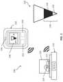
- FIG. 1is an illustrative schematic of one variation of a system for quantifying fluid from a patient.
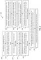
- FIG. 2is an illustrative flowchart schematic of one variation of a method for quantifying fluid from a patient.
- FIG. 3is an exemplary user interface prompting a user input of a quantity of one or more items, such as for weighing.
- FIG. 4is an exemplary user interface displaying an estimated quantity of a volume of fluid contained in a weighed item.
- FIG. 5Ais an exemplary user interface displaying a graphical representation of a container and display elements for user inputs.
- FIG. 6is an exemplary user interface displaying estimated quantities of a fluid.
- FIG. 7is another exemplary variation of a user interface displaying a graphical representation of a container and display elements for user inputs.
- FIG. 8is an illustrative schematic of another variation of a system for quantifying fluid from a patient.
- FIG. 9is another exemplary variation of a user interface displaying estimated quantities of a fluid.
- extracorporeal fluidse.g., blood
- surgical textiles or other absorbent itemssuch as surgical sponges (e.g., laparotomy sponges), surgical dressings, surgical gauze, surgical towels, absorbent pads or drapes (e.g., chux pads), vaginal packs and/or other textiles or absorbent items, etc.
- Textiles or absorbent itemsmay be placed in a bag (e.g., sponge count bag) for tracking purposes, hygienic purposes, etc.
- extracorporeal fluids that are lost by the patientmay be collected in a container, such as a canister.
- a drape with at least one pocketmay be placed under the patient for collecting blood, amniotic fluid, urine, etc.
- the quantity of fluid collected in at least some types of items, such as surgical textiles and/or canisters,may be estimated based on a measured weight (mass) of the item when containing fluid.
- multiple batches of such itemsmay be weighed (e.g., as each batch of one or more textiles become saturated) and aggregated into a running total or overall estimate of the quantity of fluid lost by the patient during the procedure.
- Such estimatesmay, in some variations, be combined with estimates of fluid collected in batches and/or cumulatively over time in some other types of items, such as canisters or blood collection drapes. For example, total volume of fluid and/or total rate of fluid loss may be estimated at any particular point during the procedure and/or post-procedure.
- the methods and systems described hereinmay be used in a variety of settings, including in a hospital or clinic setting (e.g., operating or clinic setting), a military setting (e.g., battlefield) or other suitable medical treatment settings.
- This informationcan be used to improve medical treatment of patients, as well as reduce costs to medical institutions and patients.
- medical practitionerse.g., nurses, surgeons
- who receive this information during and/or after a medical proceduremay be able to make more appropriate decisions for treatment of the patient (such as determining whether to provide a blood transfusion to the patient and how much blood is necessary) based on more accurate information on patient status.
- practitionersmay be able to, for example, avoid providing unnecessary blood transfusions (which deplete inventory and increase risk of complications due to transfusions), while also avoiding delayed blood transfusions (which risk patient health).
- a computer-implemented method 200 for quantifying fluids from a patientmay include estimating the first volume of a first fluid based on a difference between a wet weight and a dry weight of the item 230 .
- the methodmay further include receiving a wet weight of at least one item 210 containing a first volume of a first fluid, receiving a quantity of at least one item type 220 , receiving a dry weight of the item type 222 , such that the received wet weight and dry weight may be used to estimate the first volume of the first fluid.
- the method 200may further include displaying a graphical representation of a container 240 containing a volume of a second fluid and a second volume of the first fluid, receiving a first user input 250 indicating the volume of the second fluid in the container, receiving a second user input 252 indicating a total fluid volume in the container, and estimating the second volume of the first fluid 260 based on at least one of the first and second user inputs. Furthermore, the method 200 may include estimating a quantity of the first fluid 280 based at least in part on the estimated first and second volumes of the first fluid, and may include displaying the estimated quantity of the first fluid 290 . Estimates of fluid collected via other sources (estimates generated by weighing and/or received through user input) may additionally or alternatively be used to quantify fluid from the patient, as further described herein.
- the methodmay be used to quantify blood from a patient undergoing a labor and delivery procedure.
- Some of the blood lost by the patientmay be collected with one or more types of surgical textiles or other substrates, and the quantity of blood in these substrates may be estimated based on weight of the substrates.
- some of the blood lost by the patientmay be collected and estimated with a blood collection drape (e.g., collecting from underneath the patient) and/or a canister (e.g., collected via suction wand, etc.).
- Estimates of blood from the weighed items and the other fluid-collecting itemsmay be displayed separately (e.g., according to each fluid-collecting item) and/or may be summed and displayed as an aggregate estimate of blood lost by the patient.
- the methodmay include receiving a wet weight of at least one item 210 and receiving a dry weight of the item (as further described below).
- the wet weight of the itemmay be measured with a scale 110 and communicated to a processor such as a processor 154 in a mobile device 150 .
- the scalemay first be tared or zeroed in order to set a baseline reference value relative to which the wet weight is determined.
- the scalemay be tared while no fluid-collecting items are placed on the scale, such that the baseline reference value may incorporate substantially no pre-existing weight placed on the scale or may incorporate a substantially empty receptacle (e.g., bucket 120 ) configured to receive fluid-collecting items.
- each batch or multiple batches of fluid-collecting itemsmay be separately weighed and analyzed, then removed prior to the placement of a subsequent batch on the scale.
- the scalemay be tared while one or more previously-analyzed batches of items are placed on the scale, such that only the weight of an additional, subsequent batch of items is used and analyzed for fluid content in the additional batch.
- the methodmay include automatically taring the scale to prepare the weighing and analysis of the next batch of fluid-collecting items.
- taringmay automatically occur after a predetermined length of time (e.g., ten seconds, thirty seconds, one minute, five minutes, etc.) without any significant change in detected weight on the scale.
- taringmay automatically occur whenever a negative weight is measured.
- taringmay automatically occur if a negative weight is measured for a period of time equal to or exceeding a predetermined amount of time, such as at least about 0.5 seconds, at least about 1 second, at least about 2 seconds, at least about 5 seconds, etc.
- a predetermined amount of timesuch as at least about 0.5 seconds, at least about 1 second, at least about 2 seconds, at least about 5 seconds, etc.
- the usermay place a batch including at least one item 112 containing fluid 114 (e.g., a surgical textile at least partially saturated with blood) in a receptacle 120 on a scale 110 (or alternatively, directly on the scale 110 ), which measures the wet weight of the item 112 .
- the scale 110may communicate the measured wet weight (wirelessly and/or via a wired connection) to a processor that receives the wet weight value for analysis as described elsewhere herein.
- the methodmay include identifying a weighing event 208 and receiving the wet weight of the item 210 in response to detecting the weighing event.
- a weighing eventmay correspond to placement of at least one fluid-collecting item on the scale.
- a weighing eventmay be determined based on detection of an increase in measured weight beyond a predetermined weight threshold.
- the predetermined weight thresholdmay be a value ranging between about 5 grams and about 15 grams, between about 7 grams and about 13 grams, or about 10 grams, etc.).
- the predetermined weight thresholdmay differ depending on the type of item being measured (e.g., which may be known based on user input, as described below, and entered by the user before the user places the batch of items for weighing on the scale).
- the predetermined weight thresholdmay be similar in value to a dry weight of a user-selected item type, or expected combined dry weight of multiple items of one or more user-selected item types.
- the predetermined weight thresholdmay be determined based at least in part on one or more previously measured dry weights of one or more items, where the measured dry weights are stored in memory as further described below.
- the predetermined weight thresholdmay be based at least in part on the lowest of one or more previously measured and stored dry weights of an item.
- the predetermined weight thresholdmay be equal to the lowest of all previously measured and stored dry weights (e.g., of all weighed items, of all weighed items of the same type as the item currently being measured, etc.) or a fraction of such lowest dry item weight (e.g., the lowest previously measured and recorded dry weight multiplied by a scaling factor such as a factor between about 0.1 and about 0.9, between about 0.25 and about 0.75, about 0.5, etc.). In some of these variations, all or only a subset of previously measured and stored dry item weights may be used when determining the lowest stored dry item weight.
- a weighing eventmay be determined based on detection of a stable measurement reading (e.g., substantially no fluctuation in measured weight, such as fluctuation only within a predetermined measurement range) for a predetermined threshold of time.
- the predetermined measurement rangemay be about ⁇ 0.1 grams, ⁇ 0.07 grams, ⁇ 0.05 grams, ⁇ 0.02 grams, ⁇ 0.01 grams, etc.
- the predetermined threshold of timemay range between about 1 second and 5 seconds, between about 2 second and 4 seconds, etc.
- Combinations of various suitable conditionse.g., detecting a threshold weight value for at least a predetermined period of time may be used to identify a weighing event.
- a weighing eventmay be determined based on user input, such as a user interacting with a user interface (e.g., buttons on the scale, user-manipulable or touch-sensitive icons on a displayed user interface, voice commands, etc.) to confirm the existence of a weighing event.
- a user interfacee.g., buttons on the scale, user-manipulable or touch-sensitive icons on a displayed user interface, voice commands, etc.
- the methodmay include applying a machine learning algorithm to distinguish between fluctuations in weight measurements due to weighing events (e.g., due to placement of at least one fluid-collecting item on the scale) and those due to non-weighing events, such as fluctuations caused by repositioning of a receptacle on the scale, fluctuations caused by shaking of a surface on which the scale rests, and/or fluctuations caused by vibrations when personnel walks near the scale or when machinery or equipment is operated near the scale, etc.
- weighing eventse.g., due to placement of at least one fluid-collecting item on the scale
- non-weighing eventssuch as fluctuations caused by repositioning of a receptacle on the scale, fluctuations caused by shaking of a surface on which the scale rests, and/or fluctuations caused by vibrations when personnel walks near the scale or when machinery or equipment is operated near the scale, etc.
- This intervalmay be shortened or lengthened based on one or more various factors or conditions, such as an identification of another weighing event as described above (e.g., the difference in stabilized weight crossing a particular threshold to recognize a weighing event).
- the determined frequency of signal variationmay then be applied to a learned model (e.g., classification model) to recognize whether the frequency is best matched to one or more particular pre-learned non-weighing events such as a person walking by, other objects being placed on the same surface as the scale (but not in the scale), nearby machinery, etc.
- a learned modele.g., classification model
- low-pass/high pass filtering techniquesmay be used to smooth out the scale signal over a given period of time to aid in signal processing.
- the methodincludes receiving a wet weight of the item 210 , which may be in response to detecting the weighing event.
- the scalemay be pinged or prompted to communicate a weight measurement.
- the scalemay substantially, continuously, or periodically communicate its weight measurement (e.g., every 0.05 seconds, 0.1 seconds, 0.5 seconds or every 1 second) and only a weight measurement following detection of the weighing event may be taken as a wet weight of the item and used for subsequent analysis as described elsewhere herein.
- the wet weight of the itemmay be based on a single measurement value, or may be based on multiple measurement values (e.g., average) taken around the time of the weighing event.
- the methodmay include storing the wet weight value in memory or another suitable data storage device.
- the methodmay further include receiving a quantity of at least one item type 220 in the batch of weighed fluid-collecting items.
- the received quantity of item typesmay be used at least in part to determine total dry weight of all items being weighed in a particular batch.
- FIG. 2shows that receiving a quantity of at least one item type 220 may be performed following receiving a wet weight of the at least one item 210 , it should be understood that in other variations, receiving a quantity of at least one item type 220 may be performed prior to or substantially concurrently with receiving a wet weight of the least one item 210 .
- a user interfacemay prompt a user to enter quantity of each item or other substrate being weighed.
- Various item typesmay be displayed for selection by the user, such as surgical sponges (e.g., laparotomy sponges), surgical dressings, surgical gauze, surgical towels, absorbent pads or drapes (e.g., chux pads), vaginal packs and/or other textiles or absorbent items, etc.
- surgical spongese.g., laparotomy sponges
- surgical dressingse.g., surgical gauze, surgical towels, absorbent pads or drapes (e.g., chux pads), vaginal packs and/or other textiles or absorbent items, etc.
- absorbent pads or drapese.g., chux pads
- vaginal packse.g., vaginal packs and/or other textiles or absorbent items, etc.
- one or more types of containers holding any such textiles or other absorbent itemse.g., sponge count bag
- the itemsmay be biological (e
- a dry weight of the item typemay be estimated as the average dry weight for a single sample of the item type, based on the total weight and number of dry samples.
- the user interfacemay, for example, prompt the user to weigh at least a minimum number of dry samples of a new item type, prompt the user to input the quantity of dry samples of the new item type, measure the total weight of the dry samples, determine average dry weight of a single dry sample, prompt the user to input a custom item type name for the new item type, and store in memory (or any suitable data storage device) the average dry weight of a single dry sample as associated with the custom item type.
- the custom item typemay be displayed for selection by the user during a medical procedure.
- the item typesmay be presented in an array in combination with item counters.
- the item typesmay be identified, for example, with word labels and/or representative images or graphics (e.g., image of a type of unsaturated surgical textile may represent surgical textiles of that type).
- the item countersmay be incremented and/or decremented by the user (e.g., by touching designated icons for incrementing and/or decrementing) to indicate the quantity of each type of item being weighed.
- the item typesmay be listed in drop-down menus or presented for selection in any suitable manner.
- the user interfacemay display a text box prompting the user to manually type in a quantity of item type, display a user-rotatable numbered dial, or facilitate user input of quantity of one or more item types in any suitable manner.
- the usermay confirm the selection (e.g., by selecting a displayed “analyze” icon as shown in FIG. 3 , or any suitable icon).
- the methodmay, in some variations, include storing and displaying a subset of selectable item types under certain circumstances.
- the methodmay include storing selected types of items as “favorites” or “preferred item types”, etc., such as selected types for particular kinds of medical procedures (e.g., chux for labor and delivery), selected brands for particular medical institutions (e.g., where a hospital may stock only a particular brand of an item type), etc.
- the methodmay include automatically suggesting items types during the procedure, such as based on item types previously selected during the same procedure or in previous procedures of the same procedure type. Storing and displaying a subset of selectable item types under certain circumstances may, for example, reduce time needed for the user to input quantities of one or more item types.
- the methodmay include using computer vision techniques to estimate or predict the quantity and/or item types being weighed.
- the methodmay include generating or receiving an optical image of the items on the scale (e.g., on a bucket or tray on the scale), and a classification algorithm, etc. may be applied to the image data for determining a number and/or type of items on the scale.
- the methodmay include using an infrared (IR) depth-sending camera to perform a 3D scan of the items on the scale and determine contours of the items present on the scale.
- the methodmay include applying a classification algorithm to classify the batch of items on the scale as including a particular number of items and/or item type.
- the classification algorithmmay, for example, base its classification on contour features of piles or collections of textiles known to include zero, one, two, etc. items of particular item types.
- the predicted quantity and/or item types being weighedmay, in some variations, be confirmed by a user before being used in subsequent analysis.
- the methodmay include receiving a dry weight of the at least one item type 222 .
- the dry weight for a single instance of each item typemay, for example, be stored in memory or other suitable data storage device, as part of a look-up table, hash function, etc. such that the dry weight may be retrieved or received upon knowing the type or types of items that have been weighed.
- a total dry weight of multiple itemsmay be determined by, for example, multiplying the dry weight of a single instance of an item type by the quantity of the item type to achieve a subtotal dry weight of each item type, then summing the subtotal dry weights of multiple item types to determine the total dry weight.
- the methodmay include weighing one or more items before being used to collect fluid. For example, weighing the one or more items may directly generate dry weight values of the items (e.g., instead of receiving the values from a stored memory, or to confirm the values received from a stored memory). Each individual item may be separately weighed, or groups of items of the same type may be weighed (whereupon collective weight may be divided by the number of items to estimate the dry weight of a single instance of that item type).
- a complementary “after” count of dry or unused itemsmay be performed in a manner similar to that described for performing a “before” count, by receiving an item type, measuring total weight of dry or unused items that that are left over after the procedure, and dividing the total weight by a known dry weight of the item type.
- a discrepancy between the “before” count compared to the sum of the “after” count of used items and the complementary “after” count of unused itemsmay prompt medical staff to perform risk mitigation (e.g., searching for apparently missing items, recounting, performing an X-ray scan of the patient, etc.).
- the methodmay include alerting the user to review, recount, or otherwise reassess the number of textiles in the pack.
- volume ⁇ ⁇ of ⁇ ⁇ fluid ⁇ ⁇ in ⁇ ⁇ itemwet ⁇ ⁇ weight ⁇ ⁇ of ⁇ ⁇ item - dry ⁇ ⁇ weight ⁇ ⁇ of ⁇ ⁇ item density ⁇ ⁇ of ⁇ ⁇ fluid ( 1 )
- the methodmay be used to estimate a volume of blood, which has a density of about 1.06 g/ml. Accordingly, to estimate a volume of blood in a weighed item, the method may include dividing the difference between the item's wet weight and dry weight (expressed in grams) by 1.06 g/ml, resulting in an estimate of the volume of blood expressed in milliliters.
- the difference between the wet weight and the dry weight in gramsmay, in some applications, provide a sufficient estimate of volume of fluid in the weight items and the step of dividing the difference in wet and dry weights by the density of fluid may be omitted.
- the step of dividing by the fluid densitymay be omitted if only a rough estimate of blood is required (since blood has a density generally approximating 1.0 g/ml), or a volume of urine or other fluid with a density of about 1.0 g/ml is being estimated in the weighed items.
- the methodmay include displaying the estimated volume of fluid to the user.
- the methodmay include displaying on a user interface a quantified blood loss (QBL) that has collected in a weighed batch of items containing blood.
- QBLquantified blood loss
- the methodmay further include displaying the quantity and item types of items that contain the volume of blood, which may, for example, help confirm to the user the correct number and item type were used to determine the dry weight value used in the analysis.
- the quantity and item typemay, for example, be displayed as words as shown in FIG. 4 , but additionally or alternatively may be displayed with representative graphics or images.
- the methodmay additionally or alternatively include weighing and analyzing items (e.g., textiles, etc.) that collect fluids by soaking or absorbing fluids
- the methodmay additionally or alternatively include weighing or analyzing items (e.g., V-shaped blood collection drape, canister) that collect fluids in a receptacle.
- weighing or analyzing itemse.g., V-shaped blood collection drape, canister
- a volume of fluid collected in a containermay be estimated in a process similar to that described above.
- a containermay be associated with a first weight (e.g., an empty weight, dry weight, or other baseline reference weight such as weight of the container including an amount of fluid previously estimated), then weighed to determine a second weight (e.g., a wet weight or other measurement weight to be compared to the baseline reference weight).
- the difference between the first and second weightsmay be divided by the density of the fluid to estimate a volume of fluid in the container (or additional volume of fluid since the previous estimation). This estimate of fluid in the container may, for example, be used to confirm or corroborate other estimations of volume (e.g., those based on reference markers on the container).
- some containers or other receptaclesmay be used to collect fluids lost by a patient.
- a V-shaped blood collection drapee.g., for labor and delivery applications of the method
- a canistermay collect fluids without soaking or absorbing the fluid.
- estimates of the fluid in the containersmay be combined with estimates of the fluid in the weighed items (e.g., estimated as described above) to generate an aggregate total of volume of the fluid lost by the patient.
- a fluid of intereste.g., blood
- a container 130may include total fluid volume 136 that includes a volume of a second fluid 132 and a volume of a fluid of interest 134 .
- the containermay include markings (e.g., graduated or calibrated markings) to facilitate estimation of fluid volumes.
- the methodmay include displaying a graphical representation of a container 240 containing a volume of a second fluid and a second volume of a first fluid of interest.
- the graphical representation of the containermay include one or more display elements for receiving one or more user inputs indicating a volume of one or more of the fluids contained in the container.
- the user inputmay involve manipulating the display elements relative to displayed fluid measurement markings corresponding to actual fluid measurement markings on the actual container, so as to mirror the relative positions of actual fluid levels and fluid measurement markings on the actual container.
- the first display element 520may be configured to indicate a volume of amniotic fluid (which may be estimated directly from markings on the container)
- the second display element 530may be configured to indicate a total volume of fluid including the amniotic fluid and blood (which may also be estimated directly from markings on the container).
- the third display element 540may be configured to indicate a known volume of another fluid such as a saline or antiseptic wash (e.g., that has been poured over the patient's birthing area to disinfect the area). Accordingly, in some variations, before manipulating the display elements, a user may not be required to mentally calculate the volume of blood based on a difference between relevant markings on the container, thereby reducing inaccuracies in blood loss estimation.
- a saline or antiseptic washe.g., that has been poured over the patient's birthing area to disinfect the area.
- the first and/or second display elements 520 and 530may include sliding markers that are user-manipulable and movable up and down along the depth of the graphical representation of the container.
- the user inputmay be received by a user touch on the display in which the user touches and drags the sliding markers to desired positions, taps the sliding markers (or increment counter elements, or selected portions of the graphical representation of the container, etc.) to selectively increment or decrement them to desired positions, etc.
- the methodmay include adjusting the estimated fluid volumes based on the detected positions of the first and second display elements 520 and 530 (e.g., adjusting an estimated volume of the second fluid based on the detected position of the first display element 520 , adjusting an estimated total fluid volume based on the detected position of the second display element 530 , etc.).
- the numerical volume valuesmay be displayed adjacent the sliding markers.
- first and/or second display elements 520 and 530may additionally or alternatively have any other suitable form for receiving user input indicating fluid volumes.
- a display elementmay include a text box 730 into which the user may type or otherwise enter a fluid volume value.
- a custom on-screen touch gesturemay be suitable user input for initiating locking and/or unlocking of the display element.
- a display element that is lockedmay be indicated with a displayed “lock” icon 542 , or any suitable indication (e.g., the word “lock”, a change in color of the display element, a change in text font in the display element, etc.).
- the display element 730may include a sliding marker that directly corresponds to a fluid level in the container itself that may be estimated using fluid measurement markings on the container.
- the display elementmay include a textbox into which a user may enter a fluid volume value.
- one or more fluid volumes in the containermay be estimated based on an optical image of the container.
- one or more optical imagesmay be taken with a camera or other suitable optical sensor whose field of view includes the container containing a volume of a fluid of interest.
- the methodmay include capturing a static, single-frame image including at least a portion of the fluid container, and/or a multi-frame video feed including multiple static images of the fluid container.
- static imagesmay be taken periodically (e.g., every ten seconds, every thirty seconds, etc.) or upon a user-input or other trigger event.
- the imagecan be a color image, a black and white image, a grayscale image, an infrared image, a field of view of an optical sensor, a fingerprint of a field of view of an optical sensor, a point cloud, or any other suitable type of image.
- One or more machine vision techniquesmay be used to estimate fluid volumes in the container based on an optical image of the container. For example, generally, estimating fluid volume in the container may include using machine vision techniques in identifying a container-depicting region of an image of the container, correlating a portion of the container-depicting region with an estimated fluid level within the container, and estimating a volume of fluid within the container based on the estimated fluid level.
- Suitable vision techniquesinclude segmentation techniques (e.g., edge detection, background subtraction, grab-cut algorithms, etc.), gauging, clustering, pattern recognition, template matching, feature extraction, descriptor extraction (e.g., extraction of texton maps, color histograms, HOG, SIFT, MSER (maximally stable external regions for removing blob-features from the selected area), etc.), feature dimensionality reduction (e.g. PCA, K-Means linear discriminant analysis, etc.), feature selection, thresholding, positioning, color analysis, parametric regression, non-parametric regression, unsupervised or semi-supervised parametric or non-parametric regression, and/or any other type of machine learning or machine vision to estimate a physical dimension of the container and/or its fluid contents.
- segmentation techniquese.g., edge detection, background subtraction, grab-cut algorithms, etc.
- gaugingclustering
- pattern recognitione.g., template matching
- feature extractione.g., descriptor extraction (e.g., extraction
- estimating fluid volume in the containermay include implementing suitable machine vision techniques to identify fluid level markings printed (and/or embossed, adhered, etc.) on the fluid container and identifying the fluid boundaries (e.g., boundary between layers of different fluids, surface of total fluid volume, etc.) within the canister.
- Fluid boundariesmay be identified, for example, based on transitions in color of pixels along the height (e.g., vertical line of pixels) along the container-depicting image region. For example, upper and lower boundaries of blood in a container may be identified based on abrupt shifts between red and non-red colors along a vertical line of pixels in the container-depicting image region.
- the methodmay further estimate the volume of one or more fluids within the canister based on one or more identified fluid boundaries relative to one or more identified fluid level markings on the container.
- Another variation of estimating fluid volume in the container using imagesincludes using machine vision techniques to identify a float or other reference point corresponding to a fluid level in the container.
- the identified position of the floatcan be used, such as by comparing the float to optical fiducials, to estimate fluid volume.
- fluid volume in the containermay be automatically estimated based on one or more fluid level sensors.
- fluid levelmay be detected with one or more point-level sensors that indicate whether the fluid volume in the container is above or below a particular threshold sensing point.
- fluid levelmay be detected with one or more continuous-level sensors that measure the fluid level in the container and indicates a numerical value corresponding to fluid volume.
- Any suitable fluid level sensormay be used to detect fluid volume in the container and generate a fluid level signal that indicates the detected fluid volume.
- capacitance level sensors, conductive level sensors, ultrasonic level sensor, and/or optical sensors, etc.may be appropriate as point-level and/or continuous-level sensors.
- another estimated volume of fluid from a weighed container or container whose contents were entered by a user or detected automatically through machine vision techniques and/or sensorsmay be displayed as a third subtotal 914 .
- More or fewer subtotal volumes of fluidmay be displayed, which may depend, for example, on the number of individual categories of fluid-collecting items that are considered.
- different kinds of subtotalsmay be displayed (e.g., estimates of fluid collected by different kinds of weighed items).
- the methodmay include displaying the generated present fluid loss trend and/or predicted future fluid loss trend.
- the methodmay include comparing the estimated quantity of the first fluid (or other fluid of interest) with a predetermined threshold for lost fluid, and triggering an alarm if the volume of first fluid meets the predetermined threshold.
- the alarmmay be visual (e.g., displayed on a display) or audible over speakers, etc.
- the estimated amount of loss of the first fluidmay be compared to a threshold. For example, in a labor and delivery application of the method, if an estimated total amount of blood lost by the patient (and which is collected by items subsequently weighed and/or in other items) exceeds about 500 milliliters, then an alarm may be triggered to warn medical personnel of the possibility of patient hemorrhage that may endanger the health and life of the patient.
- the estimated volumetric rate of loss of the first fluidmay be compared to a threshold.
- the generated present fluid loss trend and/or predicted future fluid loss trendmay be compared to one or more representative reference data sets.
- a representative reference data setmay be identified from a stored database of data based on, for example, one or more health condition or disease factors, practice-based factors, patient type (e.g., sex, age, weight, ethnicity, etc.), and/or other suitable patient- or procedure-related factors.
- a set of reference fluid loss valuesmay include an average, median, mode, or a range of historical fluid loss values from a plurality of prior procedures, each of which may or may not be the same type as the current procedure.
- the methodmay include triggering an alarm indicating predicted danger to the patient (e.g., predicted excessive future blood loss).
- predicted dangere.g., predicted excessive future blood loss.
- U.S. patent application Ser. No. 15/154,921entitled “Method for projecting blood loss of a patient during a surgery,” which is hereby incorporated in its entirety by this reference.
- a system 100 for quantifying fluid from a patientmay include a scale 110 configured to measure a wet weight of at least one item containing a first volume of a first fluid, a display 158 configured to display a graphical representation of a container containing a least a second volume of the first fluid, and a processor 154 coupled to the scale 110 and the display 158 .
- the graphical representationmay represent a container 130 including a total fluid volume 136 that includes a volume of a second fluid 132 and a volume of the first fluid 134 .
- the scale 110may be an electronic or digital scale having suitable range and resolution for measuring medical or surgical items containing fluid.
- the scalemay be configured to measure loads up to a maximum load ranging between about 100 grams and 10,000 grams, between about 250 grams and 750 grams, at least about 500 grams, etc.
- the scalemay further be configured to measure loads with a precision of at least 0.01 grams, at least 0.1 grams, at least 1 gram, etc.
- the scale 110may be wireless (e.g., communicating with Bluetooth or other suitable communication protocol) and configured to communicate with the processor 154 remotely. Additionally or alternatively, the scale 110 may be configured to communicate with the processor 154 via a wired connection. Wireless or wired communication status may be indicated to the user.
- confirmation of functional communication between the scale 110 and the processor 154may be displayed (e.g., on a display as shown in FIG. 3 ), and/or audibly indicated to the user.
- any interruption in communication between the scale 110 and the processor 154may be detected and indicated to the user on a display or audibly, etc. as an alert or warning.
- the processor 154may be configured to estimate the first volume of the first fluid in the weighed item based on a difference between a dry weight of the item and the wet weight of the item, estimate the second volume of the first fluid in the container based at least in part on the first and second user inputs, and estimate a total quantity of the first fluid based at least in part on the estimated first and second volumes of the first fluid.
- the processor 154may additionally or alternatively be configured to perform some or all aspects of any of the variations of the methods described herein.
- the one or more processors 154may be integrated into a handheld or mobile device 150 .
- the one or more processors 154may be incorporated into a computing device or system, such as a cloud-based computer system, a mainframe computer system, a grid-computer system, or other suitable computer system.
- the display 158may be configured to display one or more of the estimated volumes of the first fluid and/or estimated total quantity (or predicted or future trends of loss, etc.) of the first fluid.
- the display 158may be configured to display or otherwise communicate to a user (e.g., doctor, nurse) other information, including but not limited to patient information, images of containers or substrates, etc.
- the display 158may include a screen on a handheld or mobile device, a computer monitor, a television screen, a projector screen, or other suitable display.
- the display 158may be configured to display a user interface that enables the user to input information (e.g., estimated volumes of fluid in one or more containers), select display options (e.g., font, color, language, etc.) and/or content (e.g., patient information, fluid-related information, alerts, etc.).
- the displaymay be user-interactive and include a resistive or capacitive touch screen that is responsive to skin, a stylet, or other user contact.
- the display 158may be user-interactive via a cursor controlled by a mouse, keyboard, or other suitable input device.
- a system 800 for quantifying fluid from a patientmay be substantially similar to the system 100 shown in FIG. 1 , except that the system may additionally or alternatively include a camera 156 or other optical sensor or optical imaging device configured to generate an image of a container 810 including a third volume of the fluid of interest.
- the camera 156may include at least one optical image sensor (e.g., CCD, CMOS, etc.) that captures a color optical digital image with red, green, and blue (RGB) color components for the pixels, and/or other suitable optical components.
Landscapes
- Health & Medical Sciences (AREA)
- Life Sciences & Earth Sciences (AREA)
- Engineering & Computer Science (AREA)
- Physics & Mathematics (AREA)
- Surgery (AREA)
- Public Health (AREA)
- General Health & Medical Sciences (AREA)
- Veterinary Medicine (AREA)
- Animal Behavior & Ethology (AREA)
- Biomedical Technology (AREA)
- Heart & Thoracic Surgery (AREA)
- Pathology (AREA)
- Medical Informatics (AREA)
- Molecular Biology (AREA)
- Biophysics (AREA)
- Nuclear Medicine, Radiotherapy & Molecular Imaging (AREA)
- Theoretical Computer Science (AREA)
- Mathematical Physics (AREA)
- Physiology (AREA)
- Hematology (AREA)
- Cardiology (AREA)
- Human Computer Interaction (AREA)
- Radiology & Medical Imaging (AREA)
- Artificial Intelligence (AREA)
- Signal Processing (AREA)
- Psychiatry (AREA)
- General Physics & Mathematics (AREA)
- Epidemiology (AREA)
- Vascular Medicine (AREA)
- Computer Vision & Pattern Recognition (AREA)
- External Artificial Organs (AREA)
Abstract
Description
Claims (33)
Priority Applications (3)
| Application Number | Priority Date | Filing Date | Title |
|---|---|---|---|
| US15/869,429US11229368B2 (en) | 2017-01-13 | 2018-01-12 | Fluid loss estimation based on weight of medical items |
| US17/560,728US12268477B2 (en) | 2017-01-13 | 2021-12-23 | Fluid loss estimation of medical items |
| US19/027,138US20250160656A1 (en) | 2017-01-13 | 2025-01-17 | Fluid loss estimation based on weight of medical items |
Applications Claiming Priority (2)
| Application Number | Priority Date | Filing Date | Title |
|---|---|---|---|
| US201762446333P | 2017-01-13 | 2017-01-13 | |
| US15/869,429US11229368B2 (en) | 2017-01-13 | 2018-01-12 | Fluid loss estimation based on weight of medical items |
Related Child Applications (1)
| Application Number | Title | Priority Date | Filing Date |
|---|---|---|---|
| US17/560,728ContinuationUS12268477B2 (en) | 2017-01-13 | 2021-12-23 | Fluid loss estimation of medical items |
Publications (2)
| Publication Number | Publication Date |
|---|---|
| US20180199827A1 US20180199827A1 (en) | 2018-07-19 |
| US11229368B2true US11229368B2 (en) | 2022-01-25 |
Family
ID=62838383
Family Applications (3)
| Application Number | Title | Priority Date | Filing Date |
|---|---|---|---|
| US15/869,429Active2040-11-28US11229368B2 (en) | 2017-01-13 | 2018-01-12 | Fluid loss estimation based on weight of medical items |
| US17/560,728Active2038-07-19US12268477B2 (en) | 2017-01-13 | 2021-12-23 | Fluid loss estimation of medical items |
| US19/027,138PendingUS20250160656A1 (en) | 2017-01-13 | 2025-01-17 | Fluid loss estimation based on weight of medical items |
Family Applications After (2)
| Application Number | Title | Priority Date | Filing Date |
|---|---|---|---|
| US17/560,728Active2038-07-19US12268477B2 (en) | 2017-01-13 | 2021-12-23 | Fluid loss estimation of medical items |
| US19/027,138PendingUS20250160656A1 (en) | 2017-01-13 | 2025-01-17 | Fluid loss estimation based on weight of medical items |
Country Status (1)
| Country | Link |
|---|---|
| US (3) | US11229368B2 (en) |
Cited By (1)
| Publication number | Priority date | Publication date | Assignee | Title |
|---|---|---|---|---|
| US20220110532A1 (en)* | 2017-01-13 | 2022-04-14 | Gauss Surgical Inc. | Fluid Loss Estimation of Medical Items |
Families Citing this family (14)
| Publication number | Priority date | Publication date | Assignee | Title |
|---|---|---|---|---|
| US10426356B2 (en) | 2011-07-09 | 2019-10-01 | Gauss Surgical, Inc. | Method for estimating a quantity of a blood component in a fluid receiver and corresponding error |
| US9652655B2 (en) | 2011-07-09 | 2017-05-16 | Gauss Surgical, Inc. | System and method for estimating extracorporeal blood volume in a physical sample |
| CN109738621B (en) | 2012-05-14 | 2021-05-11 | 高斯外科公司 | System and method for estimating amount of blood component in liquid tank |
| IN2014DN10121A (en) | 2012-05-14 | 2015-08-21 | Gauss Surgical | |
| US10424060B2 (en) | 2012-07-09 | 2019-09-24 | Gauss Surgical, Inc. | Method for estimating blood component quantities in surgical textiles |
| US10789710B2 (en) | 2015-05-15 | 2020-09-29 | Gauss Surgical, Inc. | Methods and systems for characterizing fluids from a patient |
| US11504037B2 (en) | 2015-05-15 | 2022-11-22 | Gauss Surgical, Inc. | Systems and methods for assessing fluids from a patient |
| EP3393539B1 (en) | 2015-12-23 | 2023-12-27 | Gauss Surgical, Inc. | System and method for estimating an amount of a blood component in a volume of fluid |
| CN119366875A (en) | 2018-09-27 | 2025-01-28 | 史赛克公司 | System and method for pipeline fluid characterization |
| CA3144778A1 (en) | 2019-07-03 | 2021-01-07 | Gauss Surgical, Inc. | Providing guidance during a medical procedure |
| CN112304413A (en)* | 2020-09-28 | 2021-02-02 | 梅特勒-托利多(常州)精密仪器有限公司 | Method and device for detecting state of weighing sensor |
| AU2023236146A1 (en)* | 2022-03-18 | 2024-09-26 | Stryker Corporation | Systems and methods for quantifying blood loss with a surgical sponge management system |
| CN114933107B (en)* | 2022-05-25 | 2023-02-28 | 江苏木盟智能科技有限公司 | Intelligent medical waste recycling method and system, medical waste recycling cabinet and robot |
| WO2024049399A1 (en)* | 2022-08-28 | 2024-03-07 | Surgibox Inc. | Systems, devices, and methods for measuring the quantity of lost blood during surgery |
Citations (193)
| Publication number | Priority date | Publication date | Assignee | Title |
|---|---|---|---|---|
| US2707955A (en) | 1953-07-17 | 1955-05-10 | Frederic W Borden | Blood-loss meter |
| US3182252A (en) | 1960-12-16 | 1965-05-04 | Berg Janwillem Van Den | Blood loss meter |
| US3199507A (en) | 1963-04-24 | 1965-08-10 | Michael L Kamm | Blood loss measure |
| US3367431A (en) | 1965-12-16 | 1968-02-06 | Dorothy H. Prindle Baker | Surgical sponge collector with means for counting and weighing sponges |
| US3646938A (en) | 1970-07-27 | 1972-03-07 | John N Haswell | Postpartum blood loss receptacle |
| US3832135A (en) | 1972-04-05 | 1974-08-27 | Becton Dickinson Co | Automatic clinical analyzer |
| US3864571A (en) | 1971-02-10 | 1975-02-04 | Wheeler International Inc | Method and Apparatus for Automatically, Identifying and Counting Various Cells in Body Fluids |
| US3869005A (en)* | 1974-04-17 | 1975-03-04 | Reliance Electric Co | Value computing scale |
| US3948390A (en) | 1974-10-18 | 1976-04-06 | Ferreri John G | Laparotomy sponge package and counter |
| US4105019A (en) | 1976-02-11 | 1978-08-08 | Haswell John N | Method for collecting postpartum fluid loss |
| US4190153A (en) | 1978-10-23 | 1980-02-26 | California Medical Developments, Inc. | Sponge disposal tray |
| US4244369A (en) | 1979-02-26 | 1981-01-13 | The Kendall Company | Surgical sponge with visually detectable element |
| US4295537A (en) | 1980-03-21 | 1981-10-20 | The Kendall Company | Sponge measuring device |
| US4313292A (en) | 1979-03-05 | 1982-02-02 | Mcwilliams Rose M | Method and apparatus for enumerative display and disposal of surgical sponges |
| US4402373A (en) | 1982-01-26 | 1983-09-06 | Comeau Perry J | Blood loss analyzer |
| US4422548A (en)* | 1982-01-18 | 1983-12-27 | Ritmed Limited | Surgical sponge counter and blood loss determination system |
| US4429789A (en) | 1982-11-22 | 1984-02-07 | Meridian Industries, Inc. | Surgical sponge counter |
| JPS59161801U (en) | 1983-04-14 | 1984-10-30 | 株式会社 大協器械製作所 | Bleeding measurement device |
| US4512428A (en)* | 1983-05-05 | 1985-04-23 | K-Tron International, Inc. | Weighing apparatus and method |
| US4562842A (en) | 1984-02-21 | 1986-01-07 | Diane E. Morfeld | Blood-loss measurement apparatus |
| US4583546A (en) | 1983-11-18 | 1986-04-22 | Garde Patria P | Blood loss monitor |
| JPS61176357A (en) | 1985-01-29 | 1986-08-08 | シレイ・インコーポレーテツド | Vein return vessel used in incision of heart |
| US4681571A (en) | 1981-04-23 | 1987-07-21 | C. R. Bard, Inc. | Suction canister with disposable liner and check valve |
| JPS62144652U (en) | 1986-03-06 | 1987-09-11 | ||
| US4773423A (en) | 1986-04-18 | 1988-09-27 | Northstar Research Institute, Ltd. | Blood loss measurement |
| US4784267A (en) | 1987-07-20 | 1988-11-15 | Gessler Annette L | Surgical sponge counter and disposal container |
| US4832198A (en) | 1987-06-15 | 1989-05-23 | Raza Alikhan | Container for packaging and counting surgical sponges |
| US4917694A (en) | 1982-05-19 | 1990-04-17 | The Kendall Company | Surgical sponge |
| US4922922A (en) | 1988-04-12 | 1990-05-08 | Pollock Richard A | Fluid monitoring apparatus |
| US5009275A (en) | 1988-11-24 | 1991-04-23 | James Sheehan | Medical swab weighing apparatus |
| US5029584A (en) | 1989-09-21 | 1991-07-09 | Cornelius Smith | Method and apparatus for measuring patient blood loss |
| US5031642A (en) | 1989-04-06 | 1991-07-16 | Nosek Bettie L | Integrator - collector for surgical/medical procedures |
| US5048683A (en) | 1990-05-10 | 1991-09-17 | The Kendall Company | Sponge counter bag |
| US5119814A (en) | 1990-07-25 | 1992-06-09 | Minnich Thomas E | Method and apparatus for monitoring blood loss via retinal venous oxygen saturation |
| US5132087A (en) | 1989-10-16 | 1992-07-21 | Kristen L Manion | Apparatus for measuring blood constituent counts |
| WO1992017787A1 (en) | 1991-04-08 | 1992-10-15 | University Of South Alabama | Method and apparatus for monitoring blood loss |
| US5190059A (en) | 1989-11-16 | 1993-03-02 | Fabian Carl E | Surgical implement detector utilizing a powered marker |
| US5227765A (en)* | 1991-04-12 | 1993-07-13 | Senko Medical Instrument Mfg. Co. Ltd. | Counting and measuring apparatus for medical cloths |
| US5231032A (en) | 1991-04-08 | 1993-07-27 | University Of South Alabama | Method of monitoring blood loss |
| US5285682A (en) | 1991-04-09 | 1994-02-15 | Micklish Clara J | Sponge absorbed fluid measuring unit |
| US5348533A (en) | 1992-08-27 | 1994-09-20 | Haemoentics Corporation | Pheresis apparatus |
| US5369713A (en) | 1992-07-09 | 1994-11-29 | Schwartz; Nira | Inspection method using area of interest (AOI) analysis |
| US5443082A (en) | 1990-03-30 | 1995-08-22 | British Technology Group Ltd. | Controlling articles of equipment during surgery |
| JPH07308312A (en) | 1993-09-03 | 1995-11-28 | Ken Ishihara | Apparatus for noninvasive hemanalysis and method therefor |
| US5492537A (en) | 1994-05-03 | 1996-02-20 | Aquintel, Inc. | Surgical fluid monitor |
| US5522805A (en) | 1994-05-03 | 1996-06-04 | Aquintel, Inc. | Surgical fluid monitor |
| WO1996039927A1 (en) | 1995-06-07 | 1996-12-19 | Blackbox, Inc. | Method for noninvasive intermittent and/or continuous hemoglobin, arterial oxygen content, and hematocrit determination |
| US5629498A (en) | 1995-01-18 | 1997-05-13 | Richard A. Pollock | Intraoperative tracking devices and processes |
| US5633166A (en) | 1995-01-19 | 1997-05-27 | Mds Health Group Limited | Method of analyzing medical specimens with improved length of analytical run determination |
| US5650596A (en) | 1994-08-05 | 1997-07-22 | Surgical Resources, L.L.C. | Automatic surgical sponge counter and blood loss determination system |
| US5709670A (en) | 1994-05-03 | 1998-01-20 | Aquintel, Inc. | Surgical fluid and tissue loss monitor |
| US5807358A (en) | 1995-03-13 | 1998-09-15 | Atrium Medical Corporation | Dry suction regulator blood collection device |
| US5851835A (en) | 1995-12-18 | 1998-12-22 | Center For Laboratory Technology, Inc. | Multiparameter hematology apparatus and method |
| JPH1137845A (en) | 1997-07-22 | 1999-02-12 | Matsushita Electric Ind Co Ltd | Serum volume measurement device |
| US5923001A (en) | 1994-08-05 | 1999-07-13 | Surgical Resources, L.L.C. | Automatic surgical sponge counter and blood loss determination system |
| US5931824A (en) | 1996-09-04 | 1999-08-03 | Stewart; William W. | Identification and accountability system for surgical sponges |
| US5984893A (en) | 1997-03-27 | 1999-11-16 | Ward; Roger T. | Blood infusion control system |
| US5996889A (en) | 1996-04-15 | 1999-12-07 | Aesculap Ag & Co. Kg | Process and device for the monitoring and control of the flow of material in a hospital |
| US6006119A (en) | 1998-02-04 | 1999-12-21 | Polestar Technologies, Inc. | Non-invasive optical measurement of blood hematocrit |
| US6061583A (en) | 1995-12-27 | 2000-05-09 | Sysmex Corporation And Ken Ishihara | Noninvasive blood analyzer |
| JP2000227390A (en) | 1999-02-05 | 2000-08-15 | New Cosmos Electric Corp | Nozzle of gas sampling and supply tool for inspection |
| US6359683B1 (en) | 2000-04-27 | 2002-03-19 | Becton, Dickinson And Company | Method for determining the volume of particles suspended in liquids |
| JP2002331031A (en) | 2001-05-09 | 2002-11-19 | Univ Nihon | Appropriate blood transfusion support device, method and program |
| US6510330B1 (en) | 1998-11-30 | 2003-01-21 | Gambro Ab | Method and device for measuring blood parameters |
| JP2003075436A (en) | 2001-09-04 | 2003-03-12 | Daiken Iki Kk | Bleeding volume measurement device |
| US20030069509A1 (en) | 2001-10-10 | 2003-04-10 | David Matzinger | Devices for physiological fluid sampling and methods of using the same |
| US20030095197A1 (en) | 2001-09-20 | 2003-05-22 | Eastman Kodak Company | System and method for deciding when to correct image-specific defects based on camera, scene, display and demographic data |
| US20030121704A1 (en)* | 1995-06-07 | 2003-07-03 | Breed David S. | Arrangement for obtaining information about an occupying item of a seat |
| US20030130596A1 (en) | 2000-01-25 | 2003-07-10 | Von Der Goltz Volker Freiherr | Device and method for detecting the coagulation functions of global, especially primary hemostasis |
| US6640130B1 (en) | 1999-07-02 | 2003-10-28 | Hypermed, Inc. | Integrated imaging apparatus |
| US6641039B2 (en) | 2002-03-21 | 2003-11-04 | Alcon, Inc. | Surgical procedure identification system |
| US20040031626A1 (en) | 1994-08-05 | 2004-02-19 | Morris Sharon L. | Automatic surgical sponge counter and blood loss determination system |
| US6699231B1 (en) | 1997-12-31 | 2004-03-02 | Heartport, Inc. | Methods and apparatus for perfusion of isolated tissue structure |
| US6728561B2 (en) | 2001-08-14 | 2004-04-27 | University Of Alabama In Huntsville | Multispectral image processing method, apparatus and computer program product for determining the blood oxygen saturation in a vessel |
| US6730054B2 (en) | 1999-10-16 | 2004-05-04 | Baxter International Inc. | Blood collection systems and methods that derive estimated effects upon the donor's blood volume and hematocrit |
| US20040129678A1 (en) | 2002-09-07 | 2004-07-08 | Timothy Crowley | Integrated apparatus and methods for treating liquids |
| US6763148B1 (en) | 2000-11-13 | 2004-07-13 | Visual Key, Inc. | Image recognition methods |
| US6777623B2 (en) | 2002-04-17 | 2004-08-17 | M. Daniel Ballard | System and method of tracking surgical sponges |
| US6781067B2 (en) | 2002-03-25 | 2004-08-24 | Sunbeam Products, Inc. | Floor tile scale with wall display |
| JP2005052288A (en) | 2003-08-01 | 2005-03-03 | Sanyo Electric Industries Co Ltd | Gauze counter for operation |
| US20050051466A1 (en) | 2003-07-02 | 2005-03-10 | Carter Lee F. | Monitoring and control system for blood processing |
| US20050163354A1 (en) | 2002-01-19 | 2005-07-28 | Michael Ziegler | Method and device for the analysis of body fluids |
| US20050265996A1 (en) | 2004-04-30 | 2005-12-01 | Biopheresis Technologies, Inc. | Method and system to remove soluble TNFR1, TNFR2, and IL2 in patients |
| US20060058593A1 (en) | 2004-09-02 | 2006-03-16 | Drinan Darrel D | Monitoring platform for detection of hypovolemia, hemorrhage and blood loss |
| WO2006053208A1 (en) | 2004-11-10 | 2006-05-18 | Becton, Dickinson And Company | System and method for determining sample volume in a container |
| US20060178578A1 (en) | 2005-02-10 | 2006-08-10 | Dennis Tribble | Vision system to calculate a fluid volume in a container |
| US7112273B2 (en) | 2002-09-27 | 2006-09-26 | Nxstage Medical, Inc. | Volumetric fluid balance control for extracorporeal blood treatment |
| US20060224086A1 (en) | 2005-03-13 | 2006-10-05 | Harty Robert D | Device for collecting and measuring fluid loss |
| JP2006280445A (en) | 2005-03-31 | 2006-10-19 | Toshiba Corp | Indwelling prevention system and gauze |
| US7147626B2 (en) | 2004-09-23 | 2006-12-12 | Celgene Corporation | Cord blood and placenta collection kit |
| US7158030B2 (en) | 2001-09-19 | 2007-01-02 | Avante International Technology | Medical assistance and tracking system and method employing smart tags |
| US20070004959A1 (en) | 2003-05-11 | 2007-01-04 | Michel Carrier | Blood pump with frusto-conical bearing structure |
| US20070024946A1 (en) | 2004-12-28 | 2007-02-01 | Panasyuk Svetlana V | Hyperspectral/multispectral imaging in determination, assessment and monitoring of systemic physiology and shock |
| US7180014B2 (en) | 2003-03-20 | 2007-02-20 | Boris Farber | Method and equipment for automated tracking and identification of nonuniform items |
| JP2007101482A (en) | 2005-10-07 | 2007-04-19 | Matsushita Electric Ind Co Ltd | Measuring chip and analysis method thereof |
| US20070108129A1 (en) | 2005-11-11 | 2007-05-17 | Nikkiso Co., Ltd. | Hemodialysis apparatus and method for hemodialysis |
| US7255003B2 (en) | 2005-09-12 | 2007-08-14 | Schneiter Calvin R | Device for measuring and displaying the amount of beer in a keg |
| US7274947B2 (en) | 2002-11-26 | 2007-09-25 | Interdigital Technology Corporation | Bias error compensated initial transmission power control for data services |
| US20070243137A1 (en) | 2006-04-18 | 2007-10-18 | Nanoprobes, Inc. | Cell and sub-cell methods for imaging and therapy |
| US7297834B1 (en) | 2004-01-26 | 2007-11-20 | Michael Evan Shapiro | Surgical sponge identification system and method |
| US7299981B2 (en) | 2001-05-21 | 2007-11-27 | Scott Laboratories, Inc. | Smart supplies, components and capital equipment |
| US20070287182A1 (en) | 2006-08-08 | 2007-12-13 | Morris David A N | Method and apparatus for analyzing solutions |
| US20080030303A1 (en) | 2006-08-04 | 2008-02-07 | Myles Kobren | Surgical pad accounting system and method |
| US20080029416A1 (en) | 2006-08-04 | 2008-02-07 | Denise Paxton | Surgical sponge counting device and method |
| US20080045845A1 (en) | 2006-06-21 | 2008-02-21 | Iprm Intellectual Property Rights Management | Apparatus and computer program for determining a patient's volemic status represented by cardiopulmonary blood volume |
| US7364545B2 (en) | 2005-04-20 | 2008-04-29 | Klein Jeffrey A | Method of measuring bleeding volume |
| US7384399B2 (en) | 2004-02-11 | 2008-06-10 | Jamshid Ghajar | Cognition and motor timing diagnosis and training system and method |
| WO2008094703A2 (en) | 2007-01-31 | 2008-08-07 | Allegiance Corporation | Liquid collection system and related methods |
| US20080194906A1 (en) | 2004-10-27 | 2008-08-14 | General Electric Company | Measurement and treatment system and method |
| US7430478B2 (en) | 2000-03-01 | 2008-09-30 | Caridian Bct, Inc. | Blood processing information system with blood loss equivalency tracking |
| US7430047B2 (en) | 2004-03-09 | 2008-09-30 | Gerald Walter Budd | Small container fluid dynamics to produce optimized inspection conditions |
| US7469727B2 (en) | 2002-08-21 | 2008-12-30 | Biodrain Medical, Inc. | Method and apparatus for disposing of liquid surgical waste for protection of healthcare workers |
| US20090080757A1 (en) | 2007-09-24 | 2009-03-26 | Baxter International Inc. | Detecting access disconnect by pattern recognition |
| US7557710B2 (en) | 2006-03-17 | 2009-07-07 | Med Wave, Llc | System for tracking surgical items in an operating room environment |
| CN101505813A (en) | 2006-08-23 | 2009-08-12 | B布劳恩阿维图姆公司 | Medical device for extracorporeal blood treatment |
| WO2009117652A1 (en) | 2008-03-21 | 2009-09-24 | Abbott Point Of Care, Inc. | Method and apparatus for determining the hematocrit of a blood sample utilizing the intrinsic pigmentation of hemoglobin contained within the red blood cells |
| US20090257632A1 (en) | 2008-04-09 | 2009-10-15 | Abbott Point Of Care, Inc. | Method for measuring the area of a sample disposed within an analysis chamber |
| US20090310123A1 (en) | 2008-06-16 | 2009-12-17 | Robert John Thomson | Blood concentrate detector |
| US20090317002A1 (en) | 2008-06-23 | 2009-12-24 | John Richard Dein | Intra-operative system for identifying and tracking surgical sharp objects, instruments, and sponges |
| US7641612B1 (en) | 2006-01-17 | 2010-01-05 | Mccall Kenneth Shawn | Blood loss detection for hemodialysis system |
| US20100007727A1 (en) | 2003-04-10 | 2010-01-14 | Torre-Bueno Jose De La | Automated measurement of concentration and/or amount in a biological sample |
| US20100027868A1 (en) | 2008-08-01 | 2010-02-04 | Sysmex Corporation | Blood sample analyzing apparatus, blood sample coagulation determining apparatus, blood sample coagulation determining method and computer program product |
| USD611731S1 (en) | 2007-03-12 | 2010-03-16 | Ian Levine | Sponge counter unit |
| US20100066996A1 (en) | 2008-08-01 | 2010-03-18 | Sysmex Corporation | Sample analyzer |
| US7703674B2 (en) | 2005-10-11 | 2010-04-27 | Patient Safety Technologies, Inc. | Systems and methods for counting surgical implements |
| US7711403B2 (en) | 2001-04-05 | 2010-05-04 | Rhode Island Hospital | Non-invasive determination of blood components |
| US20100150759A1 (en) | 2005-12-01 | 2010-06-17 | Mazur Daniel E | Pulsatile rotary ventricular pump |
| US7749217B2 (en) | 2002-05-06 | 2010-07-06 | Covidien Ag | Method and system for optically detecting blood and controlling a generator during electrosurgery |
| US7819818B2 (en) | 2004-02-11 | 2010-10-26 | Jamshid Ghajar | Cognition and motor timing diagnosis using smooth eye pursuit analysis |
| US20100280117A1 (en) | 2009-04-30 | 2010-11-04 | Xanodyne Pharmaceuticals, Inc. | Menorrhagia Instrument and Method for the Treatment of Menstrual Bleeding Disorders |
| US7872201B1 (en) | 2007-07-31 | 2011-01-18 | Edlund Company, Llc | System and method for programming a weighing scale using a key signal to enter a programming mode |
| WO2011019576A1 (en) | 2009-08-13 | 2011-02-17 | Siemens Healthcare Diagnostics Inc. | Methods and apparatus for ascertaining interferents and physical dimensions in liquid samples and containers to be analyzed by a clinical analyzer |
| JP2011036371A (en) | 2009-08-10 | 2011-02-24 | Tohoku Otas Kk | Medical image recording apparatus |
| US20110066182A1 (en) | 2009-04-07 | 2011-03-17 | Biomedica Management Corp | Tissue Sealant for Use in Non Compressible Hemorrhage |
| US7909806B2 (en) | 2004-09-23 | 2011-03-22 | Anthrogenesis Corporation | Cord blood and placenta collection kit |
| US20110118647A1 (en) | 2008-07-09 | 2011-05-19 | Gambro Lundia Ab | extracorporeal blood treatment apparatus |
| US7966269B2 (en) | 2005-10-20 | 2011-06-21 | Bauer James D | Intelligent human-machine interface |
| US20110192745A1 (en) | 2010-02-05 | 2011-08-11 | Kyungyoon Min | Medical containers for use in blood collection and processing and medical systems, methods and apparatus for use in blood collection and processing |
| US20110196321A1 (en) | 2009-06-10 | 2011-08-11 | Tyco Healthcare Group Lp | Fluid Collection Canister Including Canister Top with Filter Membrane and Negative Pressure Wound Therapy Systems Including Same |
| US20110200239A1 (en) | 2010-02-17 | 2011-08-18 | Levine Joshua D | Method and apparatus for remotely performing hematologic analysis utilizing a transmitted image of a centrifuged analysis tube |
| US8025173B2 (en) | 2006-09-07 | 2011-09-27 | Allegiance Corporation | Collapsible canister liner for medical fluid collection |
| US20110275957A1 (en) | 2010-05-06 | 2011-11-10 | Sachin Bhandari | Inertial Sensor Based Surgical Navigation System for Knee Replacement Surgery |
| WO2011145351A1 (en) | 2010-05-20 | 2011-11-24 | パナソニック株式会社 | Drug solution determination device and drug solution determination method |
| US20110305376A1 (en) | 2010-06-14 | 2011-12-15 | Siemens Medical Solutions Usa, Inc. | Automatic Patient and Device Recognition and Association System |
| JP2011252804A (en) | 2010-06-02 | 2011-12-15 | Hitachi High-Technologies Corp | Analysis method and analyzer of biological sample |
| US20110316973A1 (en) | 2009-03-10 | 2011-12-29 | Miller J Scott | Extended dynamic range and extended dimensionality image signal conversion and/or delivery via legacy video interfaces |
| US20120000297A1 (en) | 2009-03-19 | 2012-01-05 | Nobuya Hashizume | Liquid collecting system and a method therefor |
| US20120064132A1 (en) | 2009-05-20 | 2012-03-15 | Keio University | Antibacterial medical equipment and method for producing the same |
| US20120065482A1 (en) | 2005-04-08 | 2012-03-15 | Mark Ries Robinson | Determination of blood pump system performance and sample dilution using a property of fluid being transported |
| US20120106811A1 (en) | 2010-11-03 | 2012-05-03 | Teco Diagnostics | All-In-One Specimen Cup With Optically Readable Results |
| US8181860B2 (en) | 2006-09-13 | 2012-05-22 | Clearcount Medical Solutions, Inc. | Apparatus and methods for monitoring objects in a surgical field |
| US8241238B2 (en) | 2006-05-22 | 2012-08-14 | Hamamatsu Photonics K.K. | Cell selection apparatus |
| US20120210778A1 (en) | 2010-11-19 | 2012-08-23 | Becton, Dickinson And Company | Specimen Container Label for Automated Clinical Laboratory Processing Systems |
| US20120257188A1 (en) | 2011-04-08 | 2012-10-11 | William Marsh Rice University | Paper based spectrophotometric detection of blood hemoglobin concentration |
| US20120262704A1 (en) | 2011-04-15 | 2012-10-18 | Constitution Medical, Inc. | Measuring Volume and Constituents of Cells |
| US20120309636A1 (en) | 2011-01-21 | 2012-12-06 | Ian Gibbons | Systems and methods for sample use maximization |
| US20120327365A1 (en) | 2010-03-12 | 2012-12-27 | Canon Kabushiki Kaisha | Ophthalmologic apparatus and control method for the same |
| US20130011042A1 (en)* | 2011-07-09 | 2013-01-10 | Siddarth Satish | System and method for estimating extracorporeal blood volume in a physical sample |
| WO2013009709A2 (en) | 2011-07-09 | 2013-01-17 | Gauss Surgical | Systems and methods for estimating extracorporeal blood volume and for counting surgical samples |
| US20130034908A1 (en) | 2011-08-04 | 2013-02-07 | University Of Notre Dame Du Lac | Analytical devices for detection of low-quality pharmaceuticals |
| US8374397B2 (en) | 2005-10-11 | 2013-02-12 | Primesense Ltd | Depth-varying light fields for three dimensional sensing |
| US8398546B2 (en) | 2000-06-16 | 2013-03-19 | Bodymedia, Inc. | System for monitoring and managing body weight and other physiological conditions including iterative and personalized planning, intervention and reporting capability |
| US20130088354A1 (en) | 2011-10-07 | 2013-04-11 | Jackson Thomas | System, Method and Device for Tracking Surgical Sponges |
| US20130094996A1 (en) | 2011-10-13 | 2013-04-18 | Heartware Bvba | Cardiopulmonary bypass or cpb monitoring tool |
| US8472693B2 (en) | 2010-03-18 | 2013-06-25 | Abbott Point Of Care, Inc. | Method for determining at least one hemoglobin related parameter of a whole blood sample |
| US20130170729A1 (en) | 2008-03-21 | 2013-07-04 | Abbott Point Of Care, Inc. | Method and apparatus for identifying reticulocytes within a blood sample |
| CA2870635A1 (en) | 2012-04-17 | 2013-10-24 | Joel R. L. Ehrenkranz | Device for performing a diagnostic test and methods for use thereof |
| US20130308852A1 (en) | 2012-03-15 | 2013-11-21 | Qualcomm Incorporated | System and method for robust estimation of color dependent measurements |
| WO2013172874A1 (en) | 2012-05-14 | 2013-11-21 | Gauss Surgical | System and method for estimating a quantity of a blood component in a fluid canister |
| WO2013173356A1 (en) | 2012-05-14 | 2013-11-21 | Gauss Surgical | System and methods for managing blood loss of a patient |
| US8626268B2 (en) | 2001-03-14 | 2014-01-07 | Given Imaging Ltd. | Method and system for detecting colorimetric abnormalities in vivo |
| US8639226B2 (en) | 2009-04-21 | 2014-01-28 | Withings | Weighing device and method |
| US20140079297A1 (en) | 2012-09-17 | 2014-03-20 | Saied Tadayon | Application of Z-Webs and Z-factors to Analytics, Search Engine, Learning, Recognition, Natural Language, and Other Utilities |
| US8693753B2 (en) | 2011-09-30 | 2014-04-08 | Fujifilm Corporation | Medical image processing device, method and program |
| US8704178B1 (en) | 2013-03-15 | 2014-04-22 | Surgitrac Corporation | Container for surgical object and fluid monitoring system |
| US8768014B2 (en) | 2009-01-14 | 2014-07-01 | Indiana University Research And Technology Corp. | System and method for identifying a person with reference to a sclera image |
| US20140207091A1 (en) | 2009-01-09 | 2014-07-24 | Smith & Nephew, Inc. | Canister for receiving wound exudate in a negative pressure therapy system |
| US8823776B2 (en) | 2010-05-20 | 2014-09-02 | Cisco Technology, Inc. | Implementing selective image enhancement |
| US20140294237A1 (en) | 2010-03-01 | 2014-10-02 | Primesense Ltd. | Combined color image and depth processing |
| US9047663B2 (en) | 2011-07-09 | 2015-06-02 | Gauss Surgical | Method for triggering blood salvage |
| WO2015160997A1 (en) | 2014-04-15 | 2015-10-22 | Gauss Surgical, Inc. | Method for estimating a quantity of a blood component in a fluid canister |
| US20150310634A1 (en) | 2014-02-13 | 2015-10-29 | James Bennett Babcock | Cloud-based system for water analysis |
| US20160069743A1 (en) | 2014-06-18 | 2016-03-10 | Innopix, Inc. | Spectral imaging system for remote and noninvasive detection of target substances using spectral filter arrays and image capture arrays |
| US20160123998A1 (en) | 2013-05-10 | 2016-05-05 | University Of Utah Research Foundation | Devices, Systems, and Methods for Measuring Blood Loss |
| US9347817B2 (en) | 2013-03-15 | 2016-05-24 | Surgitrac Corporation | Surgical object and comprehensive fluid monitoring system having capability of mobile monitoring and having highly sensitive and reliable detection of objects being placed in a container |
| US20160331248A1 (en) | 2015-05-15 | 2016-11-17 | Gauss Surgical, Inc. | Method for projecting blood loss of a patient during a surgery |
| US20160331282A1 (en) | 2015-05-15 | 2016-11-17 | Gauss Surgical, Inc. | Systems and methods for assessing fluids from a patient |
| US20170011276A1 (en) | 2015-07-08 | 2017-01-12 | Intelleflex Corporation | Photo analytics calibration |
| US20170023446A1 (en) | 2015-02-20 | 2017-01-26 | Neoteryx, Llc. | Method and apparatus for acquiring blood for testing |
| US20170186160A1 (en) | 2012-07-09 | 2017-06-29 | Gauss Surgical, Inc. | Method for estimating blood component quantities in surgical textiles |
| US20170185739A1 (en)* | 2015-12-28 | 2017-06-29 | Cerner Innovation, Inc. | Methods and system for hemorrhage-specific determinations |
| US9804768B1 (en)* | 2014-06-25 | 2017-10-31 | ABCDisability, Inc. | Method and system for generating an examination report |
| US20180199827A1 (en) | 2017-01-13 | 2018-07-19 | Gauss Surgical, Inc. | Fluid loss estimation based on weight of medical items |
Family Cites Families (5)
| Publication number | Priority date | Publication date | Assignee | Title |
|---|---|---|---|---|
| US5333498A (en)* | 1992-06-19 | 1994-08-02 | W. L. Walker Co., Inc. | Apparatus and method for measuring physical characteristics of a liquid |
| EP1992263B2 (en)* | 2007-05-16 | 2016-10-12 | Nestec S.A. | Beverage production module and method for operating a beverage production module |
| WO2009052532A1 (en)* | 2007-10-20 | 2009-04-23 | Sowb Yasser A | Systems and methods for patient cardiovascular and respiratory management |
| US9685049B2 (en)* | 2014-12-30 | 2017-06-20 | Hand Held Products, Inc. | Method and system for improving barcode scanner performance |
| ES2819748T3 (en)* | 2015-04-30 | 2021-04-19 | Evoca Spa | Automatic beverage dispensing machine with improved man-machine interface |
- 2018
- 2018-01-12USUS15/869,429patent/US11229368B2/enactiveActive
- 2021
- 2021-12-23USUS17/560,728patent/US12268477B2/enactiveActive
- 2025
- 2025-01-17USUS19/027,138patent/US20250160656A1/enactivePending
Patent Citations (238)
| Publication number | Priority date | Publication date | Assignee | Title |
|---|---|---|---|---|
| US2707955A (en) | 1953-07-17 | 1955-05-10 | Frederic W Borden | Blood-loss meter |
| US3182252A (en) | 1960-12-16 | 1965-05-04 | Berg Janwillem Van Den | Blood loss meter |
| US3199507A (en) | 1963-04-24 | 1965-08-10 | Michael L Kamm | Blood loss measure |
| US3367431A (en) | 1965-12-16 | 1968-02-06 | Dorothy H. Prindle Baker | Surgical sponge collector with means for counting and weighing sponges |
| US3646938A (en) | 1970-07-27 | 1972-03-07 | John N Haswell | Postpartum blood loss receptacle |
| US3864571A (en) | 1971-02-10 | 1975-02-04 | Wheeler International Inc | Method and Apparatus for Automatically, Identifying and Counting Various Cells in Body Fluids |
| US3832135A (en) | 1972-04-05 | 1974-08-27 | Becton Dickinson Co | Automatic clinical analyzer |
| US3869005A (en)* | 1974-04-17 | 1975-03-04 | Reliance Electric Co | Value computing scale |
| US3948390A (en) | 1974-10-18 | 1976-04-06 | Ferreri John G | Laparotomy sponge package and counter |
| US4105019A (en) | 1976-02-11 | 1978-08-08 | Haswell John N | Method for collecting postpartum fluid loss |
| US4149537A (en) | 1976-02-11 | 1979-04-17 | Haswell John N | Postpartum fluid loss receptacle |
| US4190153A (en) | 1978-10-23 | 1980-02-26 | California Medical Developments, Inc. | Sponge disposal tray |
| US4244369A (en) | 1979-02-26 | 1981-01-13 | The Kendall Company | Surgical sponge with visually detectable element |
| US4313292A (en) | 1979-03-05 | 1982-02-02 | Mcwilliams Rose M | Method and apparatus for enumerative display and disposal of surgical sponges |
| US4295537A (en) | 1980-03-21 | 1981-10-20 | The Kendall Company | Sponge measuring device |
| US4681571A (en) | 1981-04-23 | 1987-07-21 | C. R. Bard, Inc. | Suction canister with disposable liner and check valve |
| US4422548A (en)* | 1982-01-18 | 1983-12-27 | Ritmed Limited | Surgical sponge counter and blood loss determination system |
| US4402373A (en) | 1982-01-26 | 1983-09-06 | Comeau Perry J | Blood loss analyzer |
| US4917694A (en) | 1982-05-19 | 1990-04-17 | The Kendall Company | Surgical sponge |
| US4429789A (en) | 1982-11-22 | 1984-02-07 | Meridian Industries, Inc. | Surgical sponge counter |
| JPS59161801U (en) | 1983-04-14 | 1984-10-30 | 株式会社 大協器械製作所 | Bleeding measurement device |
| US4512428A (en)* | 1983-05-05 | 1985-04-23 | K-Tron International, Inc. | Weighing apparatus and method |
| US4583546A (en) | 1983-11-18 | 1986-04-22 | Garde Patria P | Blood loss monitor |
| US4562842A (en) | 1984-02-21 | 1986-01-07 | Diane E. Morfeld | Blood-loss measurement apparatus |
| JPS61176357A (en) | 1985-01-29 | 1986-08-08 | シレイ・インコーポレーテツド | Vein return vessel used in incision of heart |
| US4642089A (en) | 1985-01-29 | 1987-02-10 | Shiley, Inc. | Unitary venous return reservoir with cardiotomy filter |
| JPS62144652U (en) | 1986-03-06 | 1987-09-11 | ||
| US4773423A (en) | 1986-04-18 | 1988-09-27 | Northstar Research Institute, Ltd. | Blood loss measurement |
| US4832198A (en) | 1987-06-15 | 1989-05-23 | Raza Alikhan | Container for packaging and counting surgical sponges |
| US4784267A (en) | 1987-07-20 | 1988-11-15 | Gessler Annette L | Surgical sponge counter and disposal container |
| US4922922A (en) | 1988-04-12 | 1990-05-08 | Pollock Richard A | Fluid monitoring apparatus |
| US5009275A (en) | 1988-11-24 | 1991-04-23 | James Sheehan | Medical swab weighing apparatus |
| US5031642A (en) | 1989-04-06 | 1991-07-16 | Nosek Bettie L | Integrator - collector for surgical/medical procedures |
| US5029584A (en) | 1989-09-21 | 1991-07-09 | Cornelius Smith | Method and apparatus for measuring patient blood loss |
| US5132087A (en) | 1989-10-16 | 1992-07-21 | Kristen L Manion | Apparatus for measuring blood constituent counts |
| US5190059A (en) | 1989-11-16 | 1993-03-02 | Fabian Carl E | Surgical implement detector utilizing a powered marker |
| US5443082A (en) | 1990-03-30 | 1995-08-22 | British Technology Group Ltd. | Controlling articles of equipment during surgery |
| US5048683A (en) | 1990-05-10 | 1991-09-17 | The Kendall Company | Sponge counter bag |
| US5119814A (en) | 1990-07-25 | 1992-06-09 | Minnich Thomas E | Method and apparatus for monitoring blood loss via retinal venous oxygen saturation |
| JPH06510210A (en) | 1991-04-08 | 1994-11-17 | ユニヴァーシティ オヴ サウスアラバマ | Blood loss monitoring method and device |
| US5231032A (en) | 1991-04-08 | 1993-07-27 | University Of South Alabama | Method of monitoring blood loss |
| US5236664A (en) | 1991-04-08 | 1993-08-17 | University Of South Alabama | Apparatus for monitoring blood loss |
| WO1992017787A1 (en) | 1991-04-08 | 1992-10-15 | University Of South Alabama | Method and apparatus for monitoring blood loss |
| US5285682A (en) | 1991-04-09 | 1994-02-15 | Micklish Clara J | Sponge absorbed fluid measuring unit |
| US5227765A (en)* | 1991-04-12 | 1993-07-13 | Senko Medical Instrument Mfg. Co. Ltd. | Counting and measuring apparatus for medical cloths |
| US5369713A (en) | 1992-07-09 | 1994-11-29 | Schwartz; Nira | Inspection method using area of interest (AOI) analysis |
| US5348533A (en) | 1992-08-27 | 1994-09-20 | Haemoentics Corporation | Pheresis apparatus |
| JPH07308312A (en) | 1993-09-03 | 1995-11-28 | Ken Ishihara | Apparatus for noninvasive hemanalysis and method therefor |
| US5492537A (en) | 1994-05-03 | 1996-02-20 | Aquintel, Inc. | Surgical fluid monitor |
| US5522805A (en) | 1994-05-03 | 1996-06-04 | Aquintel, Inc. | Surgical fluid monitor |
| US5709670A (en) | 1994-05-03 | 1998-01-20 | Aquintel, Inc. | Surgical fluid and tissue loss monitor |
| US5956130A (en) | 1994-05-03 | 1999-09-21 | Aquintel Corporation | Method of surgical fluid analysis |
| US5944668A (en) | 1994-05-03 | 1999-08-31 | Aquintel Corporation | Surgical fluid and tissue loss monitor |
| US5522805B1 (en) | 1994-05-03 | 1998-06-16 | Aquintel Inc | Surgical fluid monitor |
| US5923001A (en) | 1994-08-05 | 1999-07-13 | Surgical Resources, L.L.C. | Automatic surgical sponge counter and blood loss determination system |
| US6998541B2 (en) | 1994-08-05 | 2006-02-14 | Clearcount Medical Solutions, Inc. | Automatic surgical sponge counter and blood loss determination system |
| US20040031626A1 (en) | 1994-08-05 | 2004-02-19 | Morris Sharon L. | Automatic surgical sponge counter and blood loss determination system |
| US8105296B2 (en) | 1994-08-05 | 2012-01-31 | Clearcount Medical Solutions, Inc. | Automatic surgical sponge counter and blood loss determination system |
| US8279068B2 (en) | 1994-08-05 | 2012-10-02 | Clearcount Medical Solutions, Inc. | Automatic surgical sponge counter and blood loss determination system |
| US8576076B2 (en) | 1994-08-05 | 2013-11-05 | Clearcount Medical Solutions, Inc. | Automatic surgical sponge counter and blood loss determination system |
| US5650596A (en) | 1994-08-05 | 1997-07-22 | Surgical Resources, L.L.C. | Automatic surgical sponge counter and blood loss determination system |
| US5629498A (en) | 1995-01-18 | 1997-05-13 | Richard A. Pollock | Intraoperative tracking devices and processes |
| US5633166A (en) | 1995-01-19 | 1997-05-27 | Mds Health Group Limited | Method of analyzing medical specimens with improved length of analytical run determination |
| US5807358A (en) | 1995-03-13 | 1998-09-15 | Atrium Medical Corporation | Dry suction regulator blood collection device |
| US20030121704A1 (en)* | 1995-06-07 | 2003-07-03 | Breed David S. | Arrangement for obtaining information about an occupying item of a seat |
| WO1996039927A1 (en) | 1995-06-07 | 1996-12-19 | Blackbox, Inc. | Method for noninvasive intermittent and/or continuous hemoglobin, arterial oxygen content, and hematocrit determination |
| US5851835A (en) | 1995-12-18 | 1998-12-22 | Center For Laboratory Technology, Inc. | Multiparameter hematology apparatus and method |
| JP3701031B2 (en) | 1995-12-27 | 2005-09-28 | シスメックス株式会社 | Noninvasive blood test equipment |
| US6061583A (en) | 1995-12-27 | 2000-05-09 | Sysmex Corporation And Ken Ishihara | Noninvasive blood analyzer |
| US5996889A (en) | 1996-04-15 | 1999-12-07 | Aesculap Ag & Co. Kg | Process and device for the monitoring and control of the flow of material in a hospital |
| US5931824A (en) | 1996-09-04 | 1999-08-03 | Stewart; William W. | Identification and accountability system for surgical sponges |
| US5984893A (en) | 1997-03-27 | 1999-11-16 | Ward; Roger T. | Blood infusion control system |
| JPH1137845A (en) | 1997-07-22 | 1999-02-12 | Matsushita Electric Ind Co Ltd | Serum volume measurement device |
| US6699231B1 (en) | 1997-12-31 | 2004-03-02 | Heartport, Inc. | Methods and apparatus for perfusion of isolated tissue structure |
| US6006119A (en) | 1998-02-04 | 1999-12-21 | Polestar Technologies, Inc. | Non-invasive optical measurement of blood hematocrit |
| US6510330B1 (en) | 1998-11-30 | 2003-01-21 | Gambro Ab | Method and device for measuring blood parameters |
| JP2000227390A (en) | 1999-02-05 | 2000-08-15 | New Cosmos Electric Corp | Nozzle of gas sampling and supply tool for inspection |
| US6640130B1 (en) | 1999-07-02 | 2003-10-28 | Hypermed, Inc. | Integrated imaging apparatus |
| US6730054B2 (en) | 1999-10-16 | 2004-05-04 | Baxter International Inc. | Blood collection systems and methods that derive estimated effects upon the donor's blood volume and hematocrit |
| US20030130596A1 (en) | 2000-01-25 | 2003-07-10 | Von Der Goltz Volker Freiherr | Device and method for detecting the coagulation functions of global, especially primary hemostasis |
| US7430478B2 (en) | 2000-03-01 | 2008-09-30 | Caridian Bct, Inc. | Blood processing information system with blood loss equivalency tracking |
| US6359683B1 (en) | 2000-04-27 | 2002-03-19 | Becton, Dickinson And Company | Method for determining the volume of particles suspended in liquids |
| US8398546B2 (en) | 2000-06-16 | 2013-03-19 | Bodymedia, Inc. | System for monitoring and managing body weight and other physiological conditions including iterative and personalized planning, intervention and reporting capability |
| US20140330094A1 (en) | 2000-06-16 | 2014-11-06 | Bodymedia, Inc. | System for monitoring and presenting health, wellness and fitness utilizng multiple user trend data having user selectable parameters |
| US6763148B1 (en) | 2000-11-13 | 2004-07-13 | Visual Key, Inc. | Image recognition methods |
| US8626268B2 (en) | 2001-03-14 | 2014-01-07 | Given Imaging Ltd. | Method and system for detecting colorimetric abnormalities in vivo |
| US7711403B2 (en) | 2001-04-05 | 2010-05-04 | Rhode Island Hospital | Non-invasive determination of blood components |
| JP2002331031A (en) | 2001-05-09 | 2002-11-19 | Univ Nihon | Appropriate blood transfusion support device, method and program |
| US7299981B2 (en) | 2001-05-21 | 2007-11-27 | Scott Laboratories, Inc. | Smart supplies, components and capital equipment |
| US6728561B2 (en) | 2001-08-14 | 2004-04-27 | University Of Alabama In Huntsville | Multispectral image processing method, apparatus and computer program product for determining the blood oxygen saturation in a vessel |
| JP2003075436A (en) | 2001-09-04 | 2003-03-12 | Daiken Iki Kk | Bleeding volume measurement device |
| US7158030B2 (en) | 2001-09-19 | 2007-01-02 | Avante International Technology | Medical assistance and tracking system and method employing smart tags |
| US20030095197A1 (en) | 2001-09-20 | 2003-05-22 | Eastman Kodak Company | System and method for deciding when to correct image-specific defects based on camera, scene, display and demographic data |
| US20030069509A1 (en) | 2001-10-10 | 2003-04-10 | David Matzinger | Devices for physiological fluid sampling and methods of using the same |
| US20050163354A1 (en) | 2002-01-19 | 2005-07-28 | Michael Ziegler | Method and device for the analysis of body fluids |
| US6641039B2 (en) | 2002-03-21 | 2003-11-04 | Alcon, Inc. | Surgical procedure identification system |
| US6781067B2 (en) | 2002-03-25 | 2004-08-24 | Sunbeam Products, Inc. | Floor tile scale with wall display |
| US7001366B2 (en) | 2002-04-17 | 2006-02-21 | Ballard M Daniel | Radiologically trackable surgical sponges |
| US6777623B2 (en) | 2002-04-17 | 2004-08-17 | M. Daniel Ballard | System and method of tracking surgical sponges |
| US7749217B2 (en) | 2002-05-06 | 2010-07-06 | Covidien Ag | Method and system for optically detecting blood and controlling a generator during electrosurgery |
| US7469727B2 (en) | 2002-08-21 | 2008-12-30 | Biodrain Medical, Inc. | Method and apparatus for disposing of liquid surgical waste for protection of healthcare workers |
| US20090076470A1 (en) | 2002-08-21 | 2009-03-19 | Biodrain Medical, Inc. | Method and apparatus for disposing of liquid surgical waste for protection of healthcare workers |
| US20040129678A1 (en) | 2002-09-07 | 2004-07-08 | Timothy Crowley | Integrated apparatus and methods for treating liquids |
| US7112273B2 (en) | 2002-09-27 | 2006-09-26 | Nxstage Medical, Inc. | Volumetric fluid balance control for extracorporeal blood treatment |
| US7274947B2 (en) | 2002-11-26 | 2007-09-25 | Interdigital Technology Corporation | Bias error compensated initial transmission power control for data services |
| US7180014B2 (en) | 2003-03-20 | 2007-02-20 | Boris Farber | Method and equipment for automated tracking and identification of nonuniform items |
| US20100007727A1 (en) | 2003-04-10 | 2010-01-14 | Torre-Bueno Jose De La | Automated measurement of concentration and/or amount in a biological sample |
| US20070004959A1 (en) | 2003-05-11 | 2007-01-04 | Michel Carrier | Blood pump with frusto-conical bearing structure |
| US20100025336A1 (en) | 2003-07-02 | 2010-02-04 | Caridianbct, Inc. | Predictive Method for Controlling a Blood Processing System |
| US20050051466A1 (en) | 2003-07-02 | 2005-03-10 | Carter Lee F. | Monitoring and control system for blood processing |
| JP2005052288A (en) | 2003-08-01 | 2005-03-03 | Sanyo Electric Industries Co Ltd | Gauze counter for operation |
| US7297834B1 (en) | 2004-01-26 | 2007-11-20 | Michael Evan Shapiro | Surgical sponge identification system and method |
| US7708700B2 (en) | 2004-02-11 | 2010-05-04 | Jamshid Ghajar | Training system and method for improving cognition and motor timing |
| US7819818B2 (en) | 2004-02-11 | 2010-10-26 | Jamshid Ghajar | Cognition and motor timing diagnosis using smooth eye pursuit analysis |
| US7384399B2 (en) | 2004-02-11 | 2008-06-10 | Jamshid Ghajar | Cognition and motor timing diagnosis and training system and method |
| US7430047B2 (en) | 2004-03-09 | 2008-09-30 | Gerald Walter Budd | Small container fluid dynamics to produce optimized inspection conditions |
| US20050265996A1 (en) | 2004-04-30 | 2005-12-01 | Biopheresis Technologies, Inc. | Method and system to remove soluble TNFR1, TNFR2, and IL2 in patients |
| US20060058593A1 (en) | 2004-09-02 | 2006-03-16 | Drinan Darrel D | Monitoring platform for detection of hypovolemia, hemorrhage and blood loss |
| US7909806B2 (en) | 2004-09-23 | 2011-03-22 | Anthrogenesis Corporation | Cord blood and placenta collection kit |
| US7147626B2 (en) | 2004-09-23 | 2006-12-12 | Celgene Corporation | Cord blood and placenta collection kit |
| US20080194906A1 (en) | 2004-10-27 | 2008-08-14 | General Electric Company | Measurement and treatment system and method |
| JP2008519604A (en) | 2004-11-10 | 2008-06-12 | ベクトン・ディキンソン・アンド・カンパニー | System and method for determining the amount of sample in a container |
| WO2006053208A1 (en) | 2004-11-10 | 2006-05-18 | Becton, Dickinson And Company | System and method for determining sample volume in a container |
| US20100003714A1 (en) | 2004-11-10 | 2010-01-07 | Becton, Dickinson And Company | System and Method for Determining Fill Volume in a Container |
| US20070024946A1 (en) | 2004-12-28 | 2007-02-01 | Panasyuk Svetlana V | Hyperspectral/multispectral imaging in determination, assessment and monitoring of systemic physiology and shock |
| US7499581B2 (en) | 2005-02-10 | 2009-03-03 | Forhealth Technologies, Inc. | Vision system to calculate a fluid volume in a container |
| US20060178578A1 (en) | 2005-02-10 | 2006-08-10 | Dennis Tribble | Vision system to calculate a fluid volume in a container |
| US20060224086A1 (en) | 2005-03-13 | 2006-10-05 | Harty Robert D | Device for collecting and measuring fluid loss |
| JP2006280445A (en) | 2005-03-31 | 2006-10-19 | Toshiba Corp | Indwelling prevention system and gauze |
| US20120065482A1 (en) | 2005-04-08 | 2012-03-15 | Mark Ries Robinson | Determination of blood pump system performance and sample dilution using a property of fluid being transported |
| US7364545B2 (en) | 2005-04-20 | 2008-04-29 | Klein Jeffrey A | Method of measuring bleeding volume |
| US7255003B2 (en) | 2005-09-12 | 2007-08-14 | Schneiter Calvin R | Device for measuring and displaying the amount of beer in a keg |
| JP2007101482A (en) | 2005-10-07 | 2007-04-19 | Matsushita Electric Ind Co Ltd | Measuring chip and analysis method thereof |
| US8374397B2 (en) | 2005-10-11 | 2013-02-12 | Primesense Ltd | Depth-varying light fields for three dimensional sensing |
| US7703674B2 (en) | 2005-10-11 | 2010-04-27 | Patient Safety Technologies, Inc. | Systems and methods for counting surgical implements |
| US7795491B2 (en) | 2005-10-11 | 2010-09-14 | Patient Safety Technologies, Inc. | Systems and methods for counting surgical implements |
| US7966269B2 (en) | 2005-10-20 | 2011-06-21 | Bauer James D | Intelligent human-machine interface |
| US20070108129A1 (en) | 2005-11-11 | 2007-05-17 | Nikkiso Co., Ltd. | Hemodialysis apparatus and method for hemodialysis |
| US20100150759A1 (en) | 2005-12-01 | 2010-06-17 | Mazur Daniel E | Pulsatile rotary ventricular pump |
| US7670289B1 (en) | 2006-01-17 | 2010-03-02 | Mccall Kenneth Shawn | Dressing and integrated system for detection of fluid loss |
| US7641612B1 (en) | 2006-01-17 | 2010-01-05 | Mccall Kenneth Shawn | Blood loss detection for hemodialysis system |
| US7557710B2 (en) | 2006-03-17 | 2009-07-07 | Med Wave, Llc | System for tracking surgical items in an operating room environment |
| US20070243137A1 (en) | 2006-04-18 | 2007-10-18 | Nanoprobes, Inc. | Cell and sub-cell methods for imaging and therapy |
| US8241238B2 (en) | 2006-05-22 | 2012-08-14 | Hamamatsu Photonics K.K. | Cell selection apparatus |
| JP2008055142A (en) | 2006-06-21 | 2008-03-13 | Iprm Intellectual Property Rights Management Ag | Apparatus and computer program for measuring a patient's volomic status indicated by cardiopulmonary blood volume |
| US20080045845A1 (en) | 2006-06-21 | 2008-02-21 | Iprm Intellectual Property Rights Management | Apparatus and computer program for determining a patient's volemic status represented by cardiopulmonary blood volume |
| US20080029416A1 (en) | 2006-08-04 | 2008-02-07 | Denise Paxton | Surgical sponge counting device and method |
| US20080030303A1 (en) | 2006-08-04 | 2008-02-07 | Myles Kobren | Surgical pad accounting system and method |
| US20070287182A1 (en) | 2006-08-08 | 2007-12-13 | Morris David A N | Method and apparatus for analyzing solutions |
| US20100087770A1 (en) | 2006-08-23 | 2010-04-08 | B. Braun Avitum Ag | Medical apparatus for extracorporeal blood treatment |
| CN101505813A (en) | 2006-08-23 | 2009-08-12 | B布劳恩阿维图姆公司 | Medical device for extracorporeal blood treatment |
| US8025173B2 (en) | 2006-09-07 | 2011-09-27 | Allegiance Corporation | Collapsible canister liner for medical fluid collection |
| US8985446B2 (en) | 2006-09-13 | 2015-03-24 | Stryker Combo L.L.C. | Apparatus and methods for monitoring objects in a surgical field |
| US8479989B2 (en) | 2006-09-13 | 2013-07-09 | Clearcount Medical Solutions, Inc. | Apparatus and methods for monitoring objects in a surgical field |
| US8181860B2 (en) | 2006-09-13 | 2012-05-22 | Clearcount Medical Solutions, Inc. | Apparatus and methods for monitoring objects in a surgical field |
| WO2008094703A3 (en) | 2007-01-31 | 2009-08-06 | Allegiance Corp | Liquid collection system and related methods |
| JP2010516429A (en) | 2007-01-31 | 2010-05-20 | アリージャンス コーポレーション | Liquid collection system and related methods |
| WO2008094703A2 (en) | 2007-01-31 | 2008-08-07 | Allegiance Corporation | Liquid collection system and related methods |
| USD611731S1 (en) | 2007-03-12 | 2010-03-16 | Ian Levine | Sponge counter unit |
| US7872201B1 (en) | 2007-07-31 | 2011-01-18 | Edlund Company, Llc | System and method for programming a weighing scale using a key signal to enter a programming mode |
| US7995816B2 (en) | 2007-09-24 | 2011-08-09 | Baxter International Inc. | Detecting access disconnect by pattern recognition |
| US20090080757A1 (en) | 2007-09-24 | 2009-03-26 | Baxter International Inc. | Detecting access disconnect by pattern recognition |
| WO2009117652A1 (en) | 2008-03-21 | 2009-09-24 | Abbott Point Of Care, Inc. | Method and apparatus for determining the hematocrit of a blood sample utilizing the intrinsic pigmentation of hemoglobin contained within the red blood cells |
| JP2011515681A (en) | 2008-03-21 | 2011-05-19 | アボット ポイント オブ ケア インコーポレイテッド | Method and apparatus for measuring the hematocrit of a blood sample using the inherent dye of hemoglobin contained in erythrocytes |
| US20130170729A1 (en) | 2008-03-21 | 2013-07-04 | Abbott Point Of Care, Inc. | Method and apparatus for identifying reticulocytes within a blood sample |
| US20090257632A1 (en) | 2008-04-09 | 2009-10-15 | Abbott Point Of Care, Inc. | Method for measuring the area of a sample disposed within an analysis chamber |
| US20090310123A1 (en) | 2008-06-16 | 2009-12-17 | Robert John Thomson | Blood concentrate detector |
| US20090317002A1 (en) | 2008-06-23 | 2009-12-24 | John Richard Dein | Intra-operative system for identifying and tracking surgical sharp objects, instruments, and sponges |
| US20110118647A1 (en) | 2008-07-09 | 2011-05-19 | Gambro Lundia Ab | extracorporeal blood treatment apparatus |
| US8194235B2 (en) | 2008-08-01 | 2012-06-05 | Sysmex Corporation | Sample analyzer |
| US20100027868A1 (en) | 2008-08-01 | 2010-02-04 | Sysmex Corporation | Blood sample analyzing apparatus, blood sample coagulation determining apparatus, blood sample coagulation determining method and computer program product |
| US20100066996A1 (en) | 2008-08-01 | 2010-03-18 | Sysmex Corporation | Sample analyzer |
| US20140207091A1 (en) | 2009-01-09 | 2014-07-24 | Smith & Nephew, Inc. | Canister for receiving wound exudate in a negative pressure therapy system |
| US8768014B2 (en) | 2009-01-14 | 2014-07-01 | Indiana University Research And Technology Corp. | System and method for identifying a person with reference to a sclera image |
| US20110316973A1 (en) | 2009-03-10 | 2011-12-29 | Miller J Scott | Extended dynamic range and extended dimensionality image signal conversion and/or delivery via legacy video interfaces |
| US20120000297A1 (en) | 2009-03-19 | 2012-01-05 | Nobuya Hashizume | Liquid collecting system and a method therefor |
| US20110066182A1 (en) | 2009-04-07 | 2011-03-17 | Biomedica Management Corp | Tissue Sealant for Use in Non Compressible Hemorrhage |
| US8639226B2 (en) | 2009-04-21 | 2014-01-28 | Withings | Weighing device and method |
| US20100280117A1 (en) | 2009-04-30 | 2010-11-04 | Xanodyne Pharmaceuticals, Inc. | Menorrhagia Instrument and Method for the Treatment of Menstrual Bleeding Disorders |
| US20120064132A1 (en) | 2009-05-20 | 2012-03-15 | Keio University | Antibacterial medical equipment and method for producing the same |
| US20110196321A1 (en) | 2009-06-10 | 2011-08-11 | Tyco Healthcare Group Lp | Fluid Collection Canister Including Canister Top with Filter Membrane and Negative Pressure Wound Therapy Systems Including Same |
| JP2011036371A (en) | 2009-08-10 | 2011-02-24 | Tohoku Otas Kk | Medical image recording apparatus |
| WO2011019576A1 (en) | 2009-08-13 | 2011-02-17 | Siemens Healthcare Diagnostics Inc. | Methods and apparatus for ascertaining interferents and physical dimensions in liquid samples and containers to be analyzed by a clinical analyzer |
| US20110192745A1 (en) | 2010-02-05 | 2011-08-11 | Kyungyoon Min | Medical containers for use in blood collection and processing and medical systems, methods and apparatus for use in blood collection and processing |
| US20110200239A1 (en) | 2010-02-17 | 2011-08-18 | Levine Joshua D | Method and apparatus for remotely performing hematologic analysis utilizing a transmitted image of a centrifuged analysis tube |
| US20140294237A1 (en) | 2010-03-01 | 2014-10-02 | Primesense Ltd. | Combined color image and depth processing |
| US20120327365A1 (en) | 2010-03-12 | 2012-12-27 | Canon Kabushiki Kaisha | Ophthalmologic apparatus and control method for the same |
| US8472693B2 (en) | 2010-03-18 | 2013-06-25 | Abbott Point Of Care, Inc. | Method for determining at least one hemoglobin related parameter of a whole blood sample |
| US20110275957A1 (en) | 2010-05-06 | 2011-11-10 | Sachin Bhandari | Inertial Sensor Based Surgical Navigation System for Knee Replacement Surgery |
| US8823776B2 (en) | 2010-05-20 | 2014-09-02 | Cisco Technology, Inc. | Implementing selective image enhancement |
| US20120127290A1 (en) | 2010-05-20 | 2012-05-24 | Tsuyoshi Tojo | Liquid medicine identification apparatus and liquid medicine identification method |
| WO2011145351A1 (en) | 2010-05-20 | 2011-11-24 | パナソニック株式会社 | Drug solution determination device and drug solution determination method |
| JP2011252804A (en) | 2010-06-02 | 2011-12-15 | Hitachi High-Technologies Corp | Analysis method and analyzer of biological sample |
| US20110305376A1 (en) | 2010-06-14 | 2011-12-15 | Siemens Medical Solutions Usa, Inc. | Automatic Patient and Device Recognition and Association System |
| US20120106811A1 (en) | 2010-11-03 | 2012-05-03 | Teco Diagnostics | All-In-One Specimen Cup With Optically Readable Results |
| US20120210778A1 (en) | 2010-11-19 | 2012-08-23 | Becton, Dickinson And Company | Specimen Container Label for Automated Clinical Laboratory Processing Systems |
| US20120309636A1 (en) | 2011-01-21 | 2012-12-06 | Ian Gibbons | Systems and methods for sample use maximization |
| US20120257188A1 (en) | 2011-04-08 | 2012-10-11 | William Marsh Rice University | Paper based spectrophotometric detection of blood hemoglobin concentration |
| US20120262704A1 (en) | 2011-04-15 | 2012-10-18 | Constitution Medical, Inc. | Measuring Volume and Constituents of Cells |
| WO2013009709A3 (en) | 2011-07-09 | 2014-05-08 | Gauss Surgical | Systems and methods for estimating extracorporeal blood volume and for counting surgical samples |
| US8792693B2 (en) | 2011-07-09 | 2014-07-29 | Gauss Surgical | System and method for estimating extracorporeal blood volume in a physical sample |
| US20170351894A1 (en) | 2011-07-09 | 2017-12-07 | Gauss Surgical, Inc. | System and method for estimating extracorporeal blood volume in a physical sample |
| US8897523B2 (en) | 2011-07-09 | 2014-11-25 | Gauss Surgical | System and method for counting surgical samples |
| US9652655B2 (en) | 2011-07-09 | 2017-05-16 | Gauss Surgical, Inc. | System and method for estimating extracorporeal blood volume in a physical sample |
| US9047663B2 (en) | 2011-07-09 | 2015-06-02 | Gauss Surgical | Method for triggering blood salvage |
| WO2013009709A2 (en) | 2011-07-09 | 2013-01-17 | Gauss Surgical | Systems and methods for estimating extracorporeal blood volume and for counting surgical samples |
| US20130011042A1 (en)* | 2011-07-09 | 2013-01-10 | Siddarth Satish | System and method for estimating extracorporeal blood volume in a physical sample |
| US20130034908A1 (en) | 2011-08-04 | 2013-02-07 | University Of Notre Dame Du Lac | Analytical devices for detection of low-quality pharmaceuticals |
| US8693753B2 (en) | 2011-09-30 | 2014-04-08 | Fujifilm Corporation | Medical image processing device, method and program |
| US20130088354A1 (en) | 2011-10-07 | 2013-04-11 | Jackson Thomas | System, Method and Device for Tracking Surgical Sponges |
| US20130094996A1 (en) | 2011-10-13 | 2013-04-18 | Heartware Bvba | Cardiopulmonary bypass or cpb monitoring tool |
| US20130308852A1 (en) | 2012-03-15 | 2013-11-21 | Qualcomm Incorporated | System and method for robust estimation of color dependent measurements |
| CA2870635A1 (en) | 2012-04-17 | 2013-10-24 | Joel R. L. Ehrenkranz | Device for performing a diagnostic test and methods for use thereof |
| US9595104B2 (en) | 2012-05-14 | 2017-03-14 | Gauss Surgical, Inc. | System and method for estimating a quantity of a blood component in a fluid canister |
| US8983167B2 (en) | 2012-05-14 | 2015-03-17 | Gauss Surgical | System and method for estimating a quantity of a blood component in a fluid canister |
| US10282839B2 (en) | 2012-05-14 | 2019-05-07 | Gauss Surgical, Inc. | System and method for estimating a quantity of a blood component in a fluid canister |
| US20190008427A1 (en) | 2012-05-14 | 2019-01-10 | Gauss Surgical, Inc. | System and methods for managing blood loss of a patient |
| US9171368B2 (en) | 2012-05-14 | 2015-10-27 | Gauss Surgical | System and method for estimating a quantity of a blood component in a fluid canister |
| US9936906B2 (en) | 2012-05-14 | 2018-04-10 | Gauss Surgical, Inc. | System and methods for managing blood loss of a patient |
| WO2013172874A1 (en) | 2012-05-14 | 2013-11-21 | Gauss Surgical | System and method for estimating a quantity of a blood component in a fluid canister |
| US20170352152A1 (en) | 2012-05-14 | 2017-12-07 | Gauss Surgical, Inc. | System and method for estimating a quantity of a blood component in a fluid canister |
| WO2013173356A1 (en) | 2012-05-14 | 2013-11-21 | Gauss Surgical | System and methods for managing blood loss of a patient |
| US20170186160A1 (en) | 2012-07-09 | 2017-06-29 | Gauss Surgical, Inc. | Method for estimating blood component quantities in surgical textiles |
| US20140079297A1 (en) | 2012-09-17 | 2014-03-20 | Saied Tadayon | Application of Z-Webs and Z-factors to Analytics, Search Engine, Learning, Recognition, Natural Language, and Other Utilities |
| US8704178B1 (en) | 2013-03-15 | 2014-04-22 | Surgitrac Corporation | Container for surgical object and fluid monitoring system |
| US9347817B2 (en) | 2013-03-15 | 2016-05-24 | Surgitrac Corporation | Surgical object and comprehensive fluid monitoring system having capability of mobile monitoring and having highly sensitive and reliable detection of objects being placed in a container |
| US20160123998A1 (en) | 2013-05-10 | 2016-05-05 | University Of Utah Research Foundation | Devices, Systems, and Methods for Measuring Blood Loss |
| US20150310634A1 (en) | 2014-02-13 | 2015-10-29 | James Bennett Babcock | Cloud-based system for water analysis |
| US9824441B2 (en) | 2014-04-15 | 2017-11-21 | Gauss Surgical, Inc. | Method for estimating a quantity of a blood component in a fluid canister |
| WO2015160997A1 (en) | 2014-04-15 | 2015-10-22 | Gauss Surgical, Inc. | Method for estimating a quantity of a blood component in a fluid canister |
| US20160069743A1 (en) | 2014-06-18 | 2016-03-10 | Innopix, Inc. | Spectral imaging system for remote and noninvasive detection of target substances using spectral filter arrays and image capture arrays |
| US9804768B1 (en)* | 2014-06-25 | 2017-10-31 | ABCDisability, Inc. | Method and system for generating an examination report |
| US20170023446A1 (en) | 2015-02-20 | 2017-01-26 | Neoteryx, Llc. | Method and apparatus for acquiring blood for testing |
| US20160331248A1 (en) | 2015-05-15 | 2016-11-17 | Gauss Surgical, Inc. | Method for projecting blood loss of a patient during a surgery |
| US20160331282A1 (en) | 2015-05-15 | 2016-11-17 | Gauss Surgical, Inc. | Systems and methods for assessing fluids from a patient |
| US20170011276A1 (en) | 2015-07-08 | 2017-01-12 | Intelleflex Corporation | Photo analytics calibration |
| US20170185739A1 (en)* | 2015-12-28 | 2017-06-29 | Cerner Innovation, Inc. | Methods and system for hemorrhage-specific determinations |
| US20180199827A1 (en) | 2017-01-13 | 2018-07-19 | Gauss Surgical, Inc. | Fluid loss estimation based on weight of medical items |
Non-Patent Citations (66)
| Title |
|---|
| "Quantification of Blood Loss (QBL) Calculator." 2015 https://www.perinatotogy.com/calculators/Blood%20%20Loss%20Calculator.htm (Year: 2015).* |
| ACOG (2012). "Optimizing protocols in obstetrics," Series 2, 25 total pages. |
| Adkins, A.R. et al. (2014). "Accuracy of blood loss estimations among anesthesia providers," AANA Journal 82(4):300-306. |
| Aklilu, A. Gauss Surgical Measures Blood Loss with a Smartphone. Jun. 14, 2012. <http://www.health2con.com/news/2012/06/14/gauss-surgical-measures-blood-loss-with-a-smartphone/>, 6 pages. |
| Al-Kadri, H.M. et al. (2014). "Effect of education and clinical assessment on the accuracy of post partum blood loss estimation," BMC Preg. Childbirth 14:110, 7 total pages. |
| AWHONN Practice Brief (2014). "Quantification of blood loss: AWHONN practice brief No. 1,"AWHONN p. 1-3. |
| Bellad, et al. (2009). "Standardized Visual Estimation of Blood Loss during Vaginal Delivery with Its Correlation Hematocrit Changes—A Descriptive Study." South Asian Federation of Obstetrics and Gynecology 1:29-34. |
| Bose, P. et al. (2006). "Improving the accuracy of estimated blood loss at obstetric haemorrhage using clinical reconstructions," BJOG 113(8):919-924. |
| Corrected Notice of Allowability dated Sep. 15, 2017, for U.S. Appl. No. 14/687,842, filed Apr. 15, 2015, 2 pages. |
| Eipe, N. et al. (2006). "Perioperative blood loss assessment—How accurate?" Indian J. Anaesth. 50(1):35-38. |
| Extended European Search Report dated Apr. 1, 2015, for EP Application No. 12 810 640.8, filed on Jul. 9, 2012, 8 pages. |
| Extended European Search Report dated Jul. 12, 2019, for EP Application No. 19 156 549.8, filed on Jul. 9, 2012, 8 pages. |
| Extended European Search Report dated Jul. 26, 2017, for EP Application No. 15 780 653.0, filed on Apr. 15, 2015, 12 pages. |
| Extended European Search Report dated Jul. 9, 2019, for EP Application No. 16 880 150.4, filed on Dec. 23, 2016, 9 pages. |
| Extended European Search Report dated Nov. 17, 2015, for EP Application No. 13 790 449.6, filed on Jan. 10, 2013, 8 pages. |
| Extended European Search Report dated Nov. 23, 2015, for EP Application No. 13 790 688.9, filed on May 14, 2013, 9 pages. |
| Extended European Search Report dated Nov. 4, 2016, for EP Application No. 16 183 350.4, filed on Jul. 9, 2012, 9 pages. |
| Final Office Action dated Aug. 26, 2016, for U.S. Appl. No. 13/894,054, filed May 14, 2013, 7 pages. |
| Final Office Action dated Feb. 12, 2016, for U.S. Appl. No. 13/544,664, filed Jul. 9, 2012, 9 pages. |
| Final Office Action dated Jul. 26, 2016, for U.S. Appl. No. 14/876,628, filed Oct. 6, 2015, 5 pages. |
| Habak, P.J. et al. (2016). "A comparison of visual estimate versus calculated estimate of blood loss at vaginal delivery," British J. Med. Medical Res. 11 (4):1-7. |
| Holmes, A.A. et al. (2014). "Clinical evaluation of a novel system for monitoring surgical hemoglobin loss," Anesth. Analg. 119(3):588-594. |
| International Search Report dated Jul. 24, 2015, for PCT Application No. PCT/US2015/026036, filed on Apr. 15, 2015, 2 pages. |
| International Search Report dated Mar. 26, 2013, for PCT Application No. PCT/US2013/021075, filed on Jan. 10, 2013, 2 pages. |
| International Search Report dated Mar. 30, 2017, for PCT Application No. PCT/US2016/068540, filed on Dec. 23, 2016, 3 pages. |
| International Search Report dated Sep. 17, 2012, for PCT Application No. PCT/US2012/045969, filed on Jul. 9, 2012, 2 pages. |
| International Search Report dated Sep. 24, 2013, for PCT Application No. PCT/US2013/040976, filed on May 14, 2013, 2 pages. |
| Jones, R. (2015). "Quantitative measurement of blood loss during delivery," AWHONN p. S41. |
| Kamiyoshihara, M. et al. (2008). "The Utility of an Autologous Blood Salvage System in Emergency Thoracotomy for a Hemothorax After Chest Trauma," Gen. Thorac. Cardiovasc. Surg. 56:222. |
| Lyndon, A. et al. (2010). "Blood loss: Clinical techniques for ongoing quantitative measurement," CMQCC Obstetric Hemorrhage Toolkit, pp. 1-7. |
| Lyndon, A. et al. (2015). "Cumulative quantitative assessment of blood loss," CMQCC Obstetric Hemorrhage Toolkit Version 2.0, pp. 80-85. |
| Manikandan, D. et al. (2015). "Measurement of blood loss during adenotonsillectomy in children and factors affecting it," Case Reports in Clinical Medicine 4:151-156. |
| Merck for Mother's Program (2012). Blood loss measurement: Technology opportunity assessment, 9 total pages. |
| Non-Final Office Action dated Apr. 11, 2018, for U.S. Appl. No. 15/416,986, filed Jan. 26, 2017, 6 pages. |
| Non-Final Office Action dated Apr. 20, 2017, for U.S. Appl. No. 13/894,054, filed May 14, 2013, 7 pages. |
| Non-Final Office Action dated Aug. 13, 2015, for U.S. Appl. No. 13/544,664, filed Jul. 9, 2012, 8 pages. |
| Non-Final Office Action dated Aug. 2, 2016, for U.S. Appl. No. 13/544,664, filed Jul. 9, 2012, 6 pages. |
| Non-Final Office Action dated Dec. 15, 2015, for U.S. Appl. No. 14/876,628, filed Oct. 6, 2015, 8 pages. |
| Non-Final Office Action dated Feb. 21, 2019, for U.S. Appl. No. 15/594,017, filed May 12, 2017, 23 pages. |
| Non-Final Office Action dated Mar. 20, 2015, for U.S. Appl. No. 14/613,807, filed Feb. 4, 2015, 8 pages. |
| Non-Final Office Action dated Mar. 24, 2017, for U.S. Appl. No. 14/687,842, filed Apr. 15, 2015, 27 pages. |
| Non-Final Office Action dated Mar. 30, 2016, for U.S. Appl. No. 13/894,054, filed May 14, 2013, 9 pages. |
| Non-Final Office Action dated May 9, 2014, for U.S. Appl. No. 13/544,679, filed Jul. 9, 2012, 7 pages.—(1.31). |
| Non-Final Office Action dated Oct. 19, 2018, for U.S. Appl. No. 15/390,017, filed Dec. 23, 2016, 12 pages. |
| Non-Final Office Action dated Sep. 5, 2014, for U.S. Appl. No. 13/738,919, filed Jan. 10, 2013, 8 pages. |
| Notice of Allowance dated Aug. 3, 2017, for U.S. Appl. No. 14/687,842, filed Apr. 15, 2015, 9 pages. |
| Notice of Allowance dated Feb. 15, 2017, for U.S. Appl. No. 13/544,664, filed Jul. 9, 2012, 10 pages. |
| Notice of Allowance dated Jan. 24, 2019, for U.S. Appl. No. 15/416,986, filed Jan. 26, 2017, 9 pages. |
| Notice of Allowance dated Jun. 25, 2015, for U.S. Appl. No. 14/613,807, filed Feb. 4, 2015, 10 pages. |
| Notice of Allowance dated May 12, 2014, for U.S. Appl. No. 13/544,646, filed Jul. 9, 2012, 10 pages. |
| Notice of Allowance dated May 3, 2019, for U.S. Appl. No. 15/390,017, filed Dec. 23, 2016, 11 pages. |
| Notice of Allowance dated Nov. 10, 2014, for U.S. Appl. No. 13/738,919, filed Jan. 10, 2013, 10 pages. |
| Notice of Allowance dated Nov. 20, 2017, for U.S. Appl. No. 13/894,054, filed May 14, 2013, 8 pages. |
| Notice of Allowance dated Oct. 26, 2016, for U.S. Appl. No. 14/876,628, filed Oct. 6, 2015, 11 pages. |
| Notice of Allowance dated Sep. 3, 2014, for U.S. Appl. No. 13/544,679, filed Jul. 9, 2012, 8 pages. |
| Pogorelc, D. iPads in the OR: New Mobile Platform to Monitor Blood Loss During Surgery. MedCityNews, Jun. 6, 2012. http://medcitynews.com/2012/06/ipads-in-the-or-new-mobile-platform-to-monitor-blood-loss-during-surgery, 4 pages. |
| Roston, A.B. et al. (2012). "Chapter 9: Blood loss: Accuracy of visual estimation," in A comprehensive textbook of postpartum hemorrhage: An essential clinical reference for effective management, 2nd edition, Sapiens publishing, pp. 71-72. |
| Sant, et al. "Exsanguinated Blood Volume Estimation Using Fractal Analysis of Digital Images." Journal of Forensic Sciences 57.3 (2012): 610-17. |
| Schorn, M.N. (2010). "Measurement of blood loss: Review of the literature," J. Midwifery and Women's Health 55(1):20-27. |
| Sukprasert, M. et al. (2006). "Increase accuracy of visual estimation of blood loss from education programme," J. Med. Assoc. Thai 89(suppl. 4):S54-S59. |
| U.S. Appl. No. 15/943,561, filed Apr. 2, 2018, by Satish et al. |
| Written Opinion of the International Searching Authority dated Jul. 24, 2015, for PCT Application No. PCT/US2015/026036, filed on Apr. 15, 2015, 6 pages. |
| Written Opinion of the International Searching Authority dated Mar. 26, 2013, for PCT Application No. PCT/US2013/021075, filed on Jan. 10, 2013, 6 pages. |
| Written Opinion of the International Searching Authority dated Mar. 30, 2017, for PCT Application No. PCT/US2016/068540, filed on Dec. 23, 2016, 8 pages. |
| Written Opinion of the International Searching Authority dated Sep. 17, 2012, for PCT Application No. PCT/US2012/045969, filed on Jul. 9, 2012, 4 pages. |
| Written Opinion of the International Searching Authority dated Sep. 24, 2013, for PCT Application No. PCT/US2013/040976, filed on May 14, 2013, 4 pages. |
Cited By (2)
| Publication number | Priority date | Publication date | Assignee | Title |
|---|---|---|---|---|
| US20220110532A1 (en)* | 2017-01-13 | 2022-04-14 | Gauss Surgical Inc. | Fluid Loss Estimation of Medical Items |
| US12268477B2 (en)* | 2017-01-13 | 2025-04-08 | Stryker Corporation | Fluid loss estimation of medical items |
Also Published As
| Publication number | Publication date |
|---|---|
| US20180199827A1 (en) | 2018-07-19 |
| US12268477B2 (en) | 2025-04-08 |
| US20220110532A1 (en) | 2022-04-14 |
| US20250160656A1 (en) | 2025-05-22 |
Similar Documents
| Publication | Publication Date | Title |
|---|---|---|
| US12268477B2 (en) | Fluid loss estimation of medical items | |
| JP7621279B2 (en) | Systems and methods for tracking surgical instruments - Patents.com | |
| US12322275B2 (en) | Method for estimating a quantity of a blood component in a fluid receiver and corresponding error | |
| JP7550178B2 (en) | Providing guidance during medical procedures | |
| RU2650586C2 (en) | Health monitoring system for calculating a total risk score | |
| US9870625B2 (en) | Method for estimating a quantity of a blood component in a fluid receiver and corresponding error | |
| JP2017106924A (en) | System and method for measuring the amount of blood components in a fluid canister | |
| US20210065905A1 (en) | Medical examination assistance apparatus | |
| US10258252B1 (en) | Wound measurement and tracking system and method | |
| CN109589137B (en) | Fetal movement identification method, fetal movement identification device, terminal and computer-readable storage medium | |
| US20160220127A1 (en) | Wellness or illness assessment system, method, and computer program product | |
| CN108968926A (en) | Distinguish method, acquisition device and the system of pulse condition | |
| JP7217007B2 (en) | Urination information analysis device, urination information analysis system and program | |
| US20160354013A1 (en) | Method and apparatus for herpes zoster diagnosis and recording medium using the method | |
| EP3547321A1 (en) | Method and system for displaying clinical information on a screen of a wearable device | |
| TW201432262A (en) | The method of urine color identification |
Legal Events
| Date | Code | Title | Description |
|---|---|---|---|
| FEPP | Fee payment procedure | Free format text:ENTITY STATUS SET TO UNDISCOUNTED (ORIGINAL EVENT CODE: BIG.); ENTITY STATUS OF PATENT OWNER: SMALL ENTITY | |
| FEPP | Fee payment procedure | Free format text:ENTITY STATUS SET TO SMALL (ORIGINAL EVENT CODE: SMAL); ENTITY STATUS OF PATENT OWNER: SMALL ENTITY | |
| STPP | Information on status: patent application and granting procedure in general | Free format text:DOCKETED NEW CASE - READY FOR EXAMINATION | |
| AS | Assignment | Owner name:GAUSS SURGICAL, INC., CALIFORNIA Free format text:ASSIGNMENT OF ASSIGNORS INTEREST;ASSIGNORS:SATISH, SIDDARTH;THAYN, JULIAN;REEL/FRAME:051099/0754 Effective date:20191115 | |
| STPP | Information on status: patent application and granting procedure in general | Free format text:NON FINAL ACTION MAILED | |
| STPP | Information on status: patent application and granting procedure in general | Free format text:RESPONSE TO NON-FINAL OFFICE ACTION ENTERED AND FORWARDED TO EXAMINER | |
| STPP | Information on status: patent application and granting procedure in general | Free format text:NOTICE OF ALLOWANCE MAILED -- APPLICATION RECEIVED IN OFFICE OF PUBLICATIONS | |
| STPP | Information on status: patent application and granting procedure in general | Free format text:PUBLICATIONS -- ISSUE FEE PAYMENT VERIFIED | |
| STCF | Information on status: patent grant | Free format text:PATENTED CASE | |
| AS | Assignment | Owner name:STRYKER CORPORATION, MICHIGAN Free format text:ASSIGNMENT OF ASSIGNORS INTEREST;ASSIGNOR:GAUSS SURGICAL, INC.;REEL/FRAME:068412/0122 Effective date:20240826 | |
| AS | Assignment | Owner name:STRYKER CORPORATION, MICHIGAN Free format text:CHANGE OF ADDRESS;ASSIGNOR:STRYKER CORPORATION;REEL/FRAME:069737/0184 Effective date:20241217 | |
| FEPP | Fee payment procedure | Free format text:ENTITY STATUS SET TO UNDISCOUNTED (ORIGINAL EVENT CODE: BIG.); ENTITY STATUS OF PATENT OWNER: LARGE ENTITY | |
| MAFP | Maintenance fee payment | Free format text:PAYMENT OF MAINTENANCE FEE, 4TH YEAR, LARGE ENTITY (ORIGINAL EVENT CODE: M1551); ENTITY STATUS OF PATENT OWNER: LARGE ENTITY Year of fee payment:4 |