US11213664B2 - Frictional trans-epithelial tissue disruption collection apparatus and method of inducing an immune response - Google Patents
Frictional trans-epithelial tissue disruption collection apparatus and method of inducing an immune responseDownload PDFInfo
- Publication number
- US11213664B2 US11213664B2US16/371,027US201916371027AUS11213664B2US 11213664 B2US11213664 B2US 11213664B2US 201916371027 AUS201916371027 AUS 201916371027AUS 11213664 B2US11213664 B2US 11213664B2
- Authority
- US
- United States
- Prior art keywords
- curved
- loops
- fenestrated
- fabric
- short
- Prior art date
- Legal status (The legal status is an assumption and is not a legal conclusion. Google has not performed a legal analysis and makes no representation as to the accuracy of the status listed.)
- Active, expires
Links
Images


Classifications
- A—HUMAN NECESSITIES
- A61—MEDICAL OR VETERINARY SCIENCE; HYGIENE
- A61M—DEVICES FOR INTRODUCING MEDIA INTO, OR ONTO, THE BODY; DEVICES FOR TRANSDUCING BODY MEDIA OR FOR TAKING MEDIA FROM THE BODY; DEVICES FOR PRODUCING OR ENDING SLEEP OR STUPOR
- A61M37/00—Other apparatus for introducing media into the body; Percutany, i.e. introducing medicines into the body by diffusion through the skin
- A61M37/0015—Other apparatus for introducing media into the body; Percutany, i.e. introducing medicines into the body by diffusion through the skin by using microneedles
- A—HUMAN NECESSITIES
- A61—MEDICAL OR VETERINARY SCIENCE; HYGIENE
- A61B—DIAGNOSIS; SURGERY; IDENTIFICATION
- A61B10/00—Instruments for taking body samples for diagnostic purposes; Other methods or instruments for diagnosis, e.g. for vaccination diagnosis, sex determination or ovulation-period determination; Throat striking implements
- A61B10/02—Instruments for taking cell samples or for biopsy
- A61B10/0291—Instruments for taking cell samples or for biopsy for uterus
- A—HUMAN NECESSITIES
- A61—MEDICAL OR VETERINARY SCIENCE; HYGIENE
- A61B—DIAGNOSIS; SURGERY; IDENTIFICATION
- A61B10/00—Instruments for taking body samples for diagnostic purposes; Other methods or instruments for diagnosis, e.g. for vaccination diagnosis, sex determination or ovulation-period determination; Throat striking implements
- A61B10/02—Instruments for taking cell samples or for biopsy
- A61B10/04—Endoscopic instruments, e.g. catheter-type instruments
- A—HUMAN NECESSITIES
- A61—MEDICAL OR VETERINARY SCIENCE; HYGIENE
- A61B—DIAGNOSIS; SURGERY; IDENTIFICATION
- A61B10/00—Instruments for taking body samples for diagnostic purposes; Other methods or instruments for diagnosis, e.g. for vaccination diagnosis, sex determination or ovulation-period determination; Throat striking implements
- A61B10/02—Instruments for taking cell samples or for biopsy
- A61B2010/0216—Sampling brushes
- A—HUMAN NECESSITIES
- A61—MEDICAL OR VETERINARY SCIENCE; HYGIENE
- A61B—DIAGNOSIS; SURGERY; IDENTIFICATION
- A61B17/00—Surgical instruments, devices or methods
- A61B17/32—Surgical cutting instruments
- A61B2017/320004—Surgical cutting instruments abrasive
- A—HUMAN NECESSITIES
- A61—MEDICAL OR VETERINARY SCIENCE; HYGIENE
- A61B—DIAGNOSIS; SURGERY; IDENTIFICATION
- A61B17/00—Surgical instruments, devices or methods
- A61B17/32—Surgical cutting instruments
- A61B2017/320004—Surgical cutting instruments abrasive
- A61B2017/320012—Brushes
- A—HUMAN NECESSITIES
- A61—MEDICAL OR VETERINARY SCIENCE; HYGIENE
- A61M—DEVICES FOR INTRODUCING MEDIA INTO, OR ONTO, THE BODY; DEVICES FOR TRANSDUCING BODY MEDIA OR FOR TAKING MEDIA FROM THE BODY; DEVICES FOR PRODUCING OR ENDING SLEEP OR STUPOR
- A61M37/00—Other apparatus for introducing media into the body; Percutany, i.e. introducing medicines into the body by diffusion through the skin
- A61M37/0015—Other apparatus for introducing media into the body; Percutany, i.e. introducing medicines into the body by diffusion through the skin by using microneedles
- A61M2037/0023—Drug applicators using microneedles
- A—HUMAN NECESSITIES
- A61—MEDICAL OR VETERINARY SCIENCE; HYGIENE
- A61M—DEVICES FOR INTRODUCING MEDIA INTO, OR ONTO, THE BODY; DEVICES FOR TRANSDUCING BODY MEDIA OR FOR TAKING MEDIA FROM THE BODY; DEVICES FOR PRODUCING OR ENDING SLEEP OR STUPOR
- A61M37/00—Other apparatus for introducing media into the body; Percutany, i.e. introducing medicines into the body by diffusion through the skin
- A61M37/0015—Other apparatus for introducing media into the body; Percutany, i.e. introducing medicines into the body by diffusion through the skin by using microneedles
- A61M2037/0046—Solid microneedles
- A—HUMAN NECESSITIES
- A61—MEDICAL OR VETERINARY SCIENCE; HYGIENE
- A61M—DEVICES FOR INTRODUCING MEDIA INTO, OR ONTO, THE BODY; DEVICES FOR TRANSDUCING BODY MEDIA OR FOR TAKING MEDIA FROM THE BODY; DEVICES FOR PRODUCING OR ENDING SLEEP OR STUPOR
- A61M37/00—Other apparatus for introducing media into the body; Percutany, i.e. introducing medicines into the body by diffusion through the skin
- A61M37/0015—Other apparatus for introducing media into the body; Percutany, i.e. introducing medicines into the body by diffusion through the skin by using microneedles
- A61M2037/0061—Methods for using microneedles
- A—HUMAN NECESSITIES
- A61—MEDICAL OR VETERINARY SCIENCE; HYGIENE
- A61M—DEVICES FOR INTRODUCING MEDIA INTO, OR ONTO, THE BODY; DEVICES FOR TRANSDUCING BODY MEDIA OR FOR TAKING MEDIA FROM THE BODY; DEVICES FOR PRODUCING OR ENDING SLEEP OR STUPOR
- A61M2202/00—Special media to be introduced, removed or treated
- A61M2202/20—Pathogenic agents
- A61M2202/206—Viruses
- A—HUMAN NECESSITIES
- A61—MEDICAL OR VETERINARY SCIENCE; HYGIENE
- A61M—DEVICES FOR INTRODUCING MEDIA INTO, OR ONTO, THE BODY; DEVICES FOR TRANSDUCING BODY MEDIA OR FOR TAKING MEDIA FROM THE BODY; DEVICES FOR PRODUCING OR ENDING SLEEP OR STUPOR
- A61M2202/00—Special media to be introduced, removed or treated
- A61M2202/30—Vaccines
Definitions
- the inventionrelates to epithelial tissue sampling and collection devices for performing biopsies from lesions and anatomical landmarks at risk of neoplastic transformation, including but not limited to the squamo-columnar junction of the female cervix, methods of inducing an immune response against a pathogen and methods of trans-epithelial drug delivery.
- Previous devicesinclude brushes with rigid bristles that puncture and shear epithelial surfaces (U.S. Pat. Nos. 5,535,756; 6,258,044; 6,376,905; 6,494,845 and 6,132,421), single metal or plastic curettes that extend in a parallel direction to the applicator handle and are much larger than the innovation (U.S. Pat. Nos. 4,641,662 and 6,730,085), scalpels or similar bladed sharp cutting tools (U.S. Pat. Nos.
- HPVHuman papillomaviruses
- a first aspectrelates to a fabric for functionally abrading epithelial surfaces including a backing material and a plurality of fenestrated loops attached to the backing material, the loops having sufficient flexibility and rigidity to frictionally abrade the epithelial surfaces, wherein the loops are about 3 mm to about 25 mm in length, wherein the loops have a short hook end, and wherein the distance from the top of the loop to the bottom of the hook is less than 50% of the length of the loop.
- a second aspectrelates to an apparatus for obtaining a histological sample including a handle, a platform at a distal end of the handle, and a fabric for functionally abrading epithelial surfaces including a backing material and a plurality of fenestrated loops attached to the backing material ( 26 ).
- a third aspectrelates to a method of inducing an immune response against a pathogen that normally evades the immune system including disrupting epithelial cells containing the pathogen with a frictional trans-epithelial tissue disruption apparatus, and thereby introducing the pathogen, DNA fragments, proteins or antigenic material into the bloodstream of a patient to elicit an immune response.
- a fourth aspectrelates to a method of trans-epithelial drug delivery including disrupting tissue with a trans-epithelial tissue disruption apparatus and applying a drug to intra-epithelial and sub-epithelial spaces created by the disrupting tissue.
- FIG. 1Apparatus for frictional trans-epithelial tissue disruption of an epithelial flat surface
- FIG. 2(A)Apparatus for frictional trans-epithelial tissue disruption of an epithelial-lined canal surface with tapered cone tip, Side view
- FIG. 2(B)apparatus for frictional trans-epithelial tissue disruption of an epithelial-lined canal surface with tapered cone tip, oblique view
- FIG. 2(C)apparatus for frictional trans-epithelial tissue disruption of an epithelial-lined canal surface with tapered cone tip, top view;
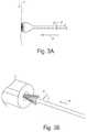
- FIG. 3(A)Schematic diagram showing a method of frictional trans-epithelial tissue disruption of a flat epithelial surface
- FIG. 3(B)schematic diagram showing a method of frictional trans-epithelial tissue disruption of an epithelial surface of a canal or body cavity
- FIG. 4Frictional trans-epithelial tissue disruptor with a motorized or vibratory handle used to spin or agitate the fenestrated loops;
- FIG. 5Schematic diagram of an apparatus with a detachable platform that anchors fiber loops at a distal end of the handle;
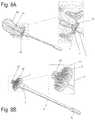
- FIG. 6(A)Schematic diagram of frictional trans-epithelial tissue disruption showing representation of tissue with a squamous epithelial lined surface
- FIG. 6(B)Schematic diagram of frictional trans-epithelial tissue disruption with application of the frictional biopsy device to the body surface
- FIG. 6(C)Schematic diagram of frictional trans-epithelial tissue disruption where simultaneous pressure, agitational, and rotational force splays and separates the hooks/loops. Frictional abrasive forces create heat which buckles the epithelial surface
- FIG. 6(D)Schematic diagram of frictional trans-epithelial tissue disruption showing representation of tissue with a squamous epithelial lined surface
- FIG. 6(B)Schematic diagram of frictional trans-epithelial tissue disruption with application of the frictional biopsy device to the body surface
- FIG. 6(C)Schematic diagram of frictional trans-epithelial tissue disruption where simultaneous pressure, agitational, and rotational force splay
- FIG. 7(A)Side view of a focal biopsy apparatus, depicted at the outer lip of the cervix (exocervix);
- FIG. 7(B)Schematic diagram of apparatus for focal biopsies with an enlarged view of the platform and loops;
- FIG. 8(A)Side view of apparatus for simultaneous biopsy of epithelial surfaces and canal-like surfaces. Longer central core fibers to insinuate into a canal and a perimeter of approximately 3 mm fibers contact an outer epithelial surface; FIG. 8(B) . Schematic diagram of apparatus for simultaneous biopsy of epithelial surfaces and canal-like surfaces with enlarged view of platform and loops.
- fenestrated looprefers to a hooked, “candy-cane” shape formed by severing a loop, wherein a short, hooked end is less than about 50% of the length of the loop.
- a fenestrated loopis formed by severing a loop once, leaving a short arm adjacent to the fenestrated loop.
- a lesional biopsy siteis no larger than about 10 mm in diameter (i.e., focal biopsy).
- the lesionsare accessible to an examiner during routine examination.
- the surfaceis accessible following entry into a body cavity through a natural orifice or surgical channel via a trochar and inspection using an endoscope with a biopsy port. The device head remains on the lesion or area of intended biopsy/therapy due to the rigid nature of the applicator. Referring to FIG.
- a focal biopsy apparatusis configured with loops that are about 3 mm to about 25 mm in length, preferably about 3 mm in length, wherein the loops have a short hook end, wherein the distance from the top of the loop to the bottom of the hook is less than 50% of the length of the loop.
- the intentis to biopsy and screen large geographic areas of tissue at risk for disease (e.g., neoplastic transformation such as, but not limited to, the squamo-columnar junction of the female cervix in the presence or absence of visualized lesions).
- the deviceprovides samples of clumps or clusters of excavated epithelial tissue fragments for analysis, in contrast to other methods disclosed in prior art that provide surface and exfoliated cells by sweeping the cells from such target tissue sites, commonly with blunt spatula or soft bristle brush devices.
- the intentis to remove tissue based evidence with frictional biopsy of the larger area, which may range from 10-40 millimeters in diameter.
- the devicecontains a central core of longer fenestrated loops (e.g., about 4-7 mm long), surrounded by a wider rim of shorter fenestrated loops (e.g., about 3 mm in length).
- the longer loopsare geometrically suited to insinuate within a central canal structure, such as the endocervical canal of the cervix.
- a central canal structuresuch as the endocervical canal of the cervix.
- the frictional tissue sampling and collection biopsy devices disclosed hereinutilize a fabric that includes minute plastic (e.g., nylon) fiber loops that are fenestrated at a minimal distance from the apex of the loop.
- minute plastice.g., nylon
- the loopsflex but do not fracture under minimal to moderate force or separate under pressure.
- the semi-rigid loopsmay be pressed in a rotational manner (e.g., in sweeping or circular motion) away from or toward the operator, perpendicular, or at an angle into epithelial tissue surfaces.
- the semi-rigid loopsremain flexible enough to cause separation of the fenestrated ends, creating frictional forces sufficient to cause local heating and buckling of the epithelial surface away from the underlying stroma.
- the loopsare fenestrated such that with applied pressure they are flexible enough to open and provide access to a “collection well” for histological fragments.
- the tips of the fiber hooksare oriented away from the tissue. On pressing and rotation across the tissue surface, the fibers scrape, buckle and shear the epithelium from the underlying stroma.
- the fragmentsare excoriated from the tissue surface through the concomitant application of frictional forces applied to the tissue surfaces by the fenestrated loops.
- the frictional forcesovercome the adhesive and binding forces of the tissue below to release fragments of various shapes and size, all eligible for collection in a histology lab, and subsequent processing and analysis.
- the semi-rigid loops(e.g., made of nylon) hold the tissue fragments after excoriation because the loops are elastic enough to sufficiently re-close and capture the remove tissue. In addition, the spaces between the fibers also retain excoriated tissue. The frictional forces exceed the binding forces afforded by adhesion molecules which anchor epithelia to the basement membrane, as well as disrupting Van der Waals forces.
- the methodis frictional abrasion, excavation via rotation and other directional motion, and tissue collection within inter-loop channels.
- the fabriccan be cut into uniform shapes such as a circular disc or straight edge shape(s) and with uniform height, allowing the device to provide 360 degree coverage of tissue surfaces over suspected lesions, without a gap within the circumference of the device.
- Thisis in distinction to bristle brushes which are spiral or bent in shape, which present surface gaps that do not allow uniforms contact with the target tissue, and gaps that cause migration of the device from the lesion site toward the direction of rotation when such devices are pressed onto lesions and rotated or moved for tissue harvesting.
- the fabric, fibers, and/or device headare removed and placed in a vial of liquid fixative for later laboratory work.
- a laboratorymay remove the tissue from the device and process it for analysis. Therefore, one may intentionally design the device in an embodiment in which the user could easily decouple the device head from the device shaft.
- some embodimentsmay have the shaft inserted into the head via a clip or screw thread mechanism, a key-in-lock design with a pressure release button, or a luer-lock type of attachment.
- tissue from the fiber assemblySome methods for removal of tissue from the fiber assembly include rinsing under pressure, immersion and agitation manually or mechanically, or by sonication.
- the fiberscan be sheared from the fabric on telfa or other filter paper, and the fibers plucked off the paper leaving the entire biopsy specimen.
- tissuemay deposited via rotation or agitation in a vial of liquid fixative, rinsed off the device under pressurized spraying, or removed from the nylon fibers by cutting away the nylon fibers from the fabric (e.g., onto filter paper), thus leaving the tissue on the paper, which can be immersed in fixative.
- the fabric fibersare manufactured in a similar manner to Velcro® or other hook and pile type fastener, where strands are longer than conventional hook and pile, about 3 mm in length, fenestrated closer to the apex of the loop instead of close to the base of one arm of the loop, and thus appear V-wishbone shaped. They have a short hook end with the curvature starting at 2 mm from the base.
- loop strandsare longer, they flex and bend to a greater angle and twist with greater elasticity when rotated or agitated when compared with standard Velcro® Because the fenestration is closer to the base in standard Velcro®, the loops fenestrations are unlikely to separate, leaving the curved smooth surface of the loop in contact with the tissue, not providing sufficient frictional forces during rotation to shear and separate the epithelium form the underlying basement membrane and stroma.
- Preferred embodimentsutilize minute plastic fenestrated loops that are pressed perpendicular or at an angle into epithelial tissue surfaces which, upon rotational or agitational pressure forces, cause tissue epithelial fragments to be frictionally separated from the underlying tissue basement membrane and stroma.
- the channels between the fenestrated loopsentrap and collect the tissue fragments.
- the processis similar to curettage with a blunt curved tool, which also scrapes, shears and strips epithelium from the underlying stroma of target tissues.
- the processis in contrast to sharp curettage where the purposefully sharp edge of the curette first incises, pierces, then shaves and scoops epithelium and underlying stroma from the tissue surface.
- the process described hereinis less perceptible to patients than conventional biopsies and causes a smaller amount of blood loss and trauma.
- the present inventionrelates to a frictional trans-epithelial tissue apparatus.
- the apparatuscomprises 3 mm or smaller loops adherent to and projecting perpendicular from a platform, with a density of 5-50 loops per square inch, evenly spaced or arranged in rows.
- the loopsmay be intact or fenestrated at the center or at their lateral aspect to allow for added flexibility and constructed from plastic, metal, or another stiff material.
- the rounded end of the loopis opposite the platform.
- the space between loopsmay serve to capture and harbor the sampled tissue.
- a flat, flexible platformwhich anchors the loops may be of any size, but is most practically approximately 5-10 mm in diameter and circular in shape.
- the shapemay be another geometrical design if it affords an advantage in covering the target tissue area for sampling.
- the platformmay be hinged in such a way that it can be folded or compressed, inserted through a small endoscopic channel, and then reinstated to its original state as a platform with a sampling surface. It may be comprised of plastic, cloth, or another composite material.
- the loopsare threaded through and project away from the platform towards the tissue surface.
- Some embodimentsmay comprise a hub fiber or “pin” that penetrates and anchors the center of the disc on the target biopsy area, serving as a central post to rotate the disc around for stability.
- the optimal shapeis circular, the diameter could range from about 10-50 mm, and the loops project at varied distances from the platform to towards the tissue surface.
- the central 5 mm diameter discprojects longer (5-25 mm) fenestrated loop fibers, and is surrounded circumferentially by the aforementioned approximately 3-23 mm long loop fibers.
- the longer fibersinsinuate inside canal structures, (e.g., the endocervical canal) simultaneously with contact of the shorter fibers with an outer endothelial surface (e.g., the exocervical surface).
- the endocervical and exocervical tissuescan be simultaneously frictionally sheared and collected. Histological screening may be necessary to correctly reflect the presence or absence of epithelial pathology, because adhesion molecules may prevent representative exfoliation from diseased tissue in some cases, leaving cytological screening methods lacking in accuracy.
- a frictional trans-epithelial biopsy sampleis taken from a lesion or an anatomical region that is predisposed to disease.
- Some embodimentscomprise a plastic, metal, or mixed composition cylinder or curved convex head, which provides a flat surface for the platform to be attached to. It is equal or greater in diameter to the platform.
- the cylinderis 5-10 mm in length while the flat or convex base is less than about 3 mm thick.
- Some embodimentscomprise a rod or cylindrical shaped applicator probe comprised of any suitable material (e.g., wood, plastic, paper or metal), which has the base, platform and loop unit at its most distal end, wherein the applicator probe is approximately 2-5 mm in diameter and 15-30 cm in length. It is constructed larger or smaller depending on the access to the tissue surface.
- the shaft of the rod or cylindrical shaped applicator probemay be rigid or semi-rigid so as to not bow or arc when pressure is transmitted from the handle to the device head.
- a handle into which the applicator probe can be transfixedis optionally mechanical, providing motorized rotational, drill-like movement or agitating vibration.
- the device handlewill be composed of stiff material, preferably plastic similar to Lucite, clear or opaque in coloration, rigid nylon plastic, or alternatively could be wood or metal.
- the device headcan take may shapes, cylindrical or tapered in design, but the distal most platform face is circular, square, or polygonal, and may be composed of plastic, (e.g., nylon). The diameter may range from 5-50 mm.
- the fabricis welded to the nylon platform ultrasonically, or may alternatively be attached via adhesive, or via a rim or collar (e.g., which snaps on to the platform into a recess in the head of the device).
- the operatorexamines tissue surfaces and chooses an area to be sampled based on the presence of a suspicious lesion. In other embodiments, the operator chooses an anatomical landmark known to be “at risk” for neoplastic or disease transformation for the purposes of sampling the entire chosen surface.
- the handle or applicator probeis grasped at its proximal end or handle.
- the distal portion or head of the devicethat contains the base, platform and loops that project perpendicular from the base towards the tissue surface with the more rounded ends that are pressed against the tissue surface.
- the examinerWith moderate pressure, the examiner simultaneously presses and rotates the device against the tissue several times in a clockwise or counterclockwise direction, opening or separating the fenestrated loops, thus performing frictional disruption of the tissue surface.
- a sweeping motionmay be used. If a motorized handle is used, it can be activated to assist in the rotation or vibration of the device.
- the harvested tissueis collected from the tissue surface, and some tissue already trapped in the loops themselves is inspected and can be teased from the loops, or the loops transected from the fabric and separated, and the remaining tissue placed in a fixative solution.
- fabric with fenestrated loops ( 1 )is connected to platform ( 2 ), which is in communication with head ( 3 ), located at a distal end of handle ( 5 ), optionally including an elongated rod ( 4 ).
- moderate force ( 8 )is applied against a tissue surface ( 7 ).
- the device headis rotated ( 9 ) on the surface to frictionally separate or agitate the surface epithelium.
- the device headis rinsed or placed with tissue in the loops into fixative for subsequent pathological analysis.
- FIG. 2An apparatus with a conical platform is depicted in FIG. 2 .
- fabric with fenestrated loops ( 1 )is connected to conical platform ( 6 ).
- conical platform6
- FIG. 3Ban apparatus with a conical platform may be inserted into a canal or cavity.
- the device headis rotated ( 9 ) while maintaining pressure force in direction ( 8 ).
- the device head with tissue in the loopsis rinsed or placed into pathological fixative.
- FIG. 4An apparatus with a motor configured to rotate the platform is depicted in FIG. 4 .
- Fabric with fenestrated loops ( 1 )is attached to platform ( 2 ) on head ( 3 ) at the distal end of an elongated rod ( 4 ), which is attached to a motorized handle ( 5 ).
- the headis detachable from the elongated rod/handle.
- a detachable head configurationallows the distal portion with head ( 3 ), platform ( 2 ), together with attached fabric containing loops, to be detached and placed into a preservative medium for later tissue removal and pathological processing.
- Some embodimentsmay have the shaft inserted into the head via a clip or screw thread mechanism, or a luer-lock type of attachment ( 23 ). Tissue fragments that remain attached to the detachable head are in addition to any free tissue obtained and collected from the tissue surface or the device as a result of the frictional tissue sampling.
- epithelial tissue samplesare obtained by frictional trans-epithelial tissue disruption.
- a representation of tissue with a squamous epithelial lined surfaceis depicted in panel (A).
- the squamous epithelial multilayer ( 11 )is shown with superficial flat and basal cuboidal epithelium.
- Basement membrane ( 12 )separates the squamous epithelial multilayer from the subcutaneous tissue stroma ( 13 ) and the underlying sub-stromal tissue ( 14 ).
- FIG. 6Bdepicts application of the frictional biopsy device to the tissue surface.
- the device head ( 3 )is applied ( 24 ) to a chosen area where curved portions of the fenestrated loops ( 1 ) press against the epithelial surface.
- a representation of two abutting hooksis shown, creating a collection channel.
- a shorter arm ( 15 ), adjacent to the fenestrated loop ( 1 ),may remain following severing of an initial continuous loop to create the fenestrated loop.
- simultaneous pressure, agitational, and rotational force ( 16 )splays and separates the hooks/loops. Frictional abrasive forces create heat which buckles the epithelial surface. Referring to FIG.
- FIG. 7Africtional trans-epithelial tissue disruption with a focal biopsy apparatus is shown at the outer lip of the exocervix ( 17 ), alternatively known as the “transformation zone” of the cervix ( 18 ).
- fenestrated loops ( 1 )approximately 3 mm in length are used to disrupt and collect tissue fragments.
- FIG. 7Bdepicts an enlarged focal biopsy apparatus, with an enlarged view of fenestrated loops ( 1 ) attached to platform ( 2 ).
- simultaneous trans-epithelial biopsy of epithelial surfaces and canal-like surfacesin particular, biopsy of the endocervical canal ( 20 ) and the exocervical area around the endocervical canal (i.e., the transformation zone) is shown ( 19 ).
- a central core of elongated loops of about 5-25 mm in length ( 21 )are surrounded by a wider rim of shorter fenestrated loops of about 3-23 mm in length ( 22 ).
- the frictional tissue sampling and collection devicecan be used on any body surface, both external to the body, body cavities, or on internal organs.
- the device headmay be deflated, folded or collapsed to pass through a small aperture or port, and re-opened or expanded to fully expose the fabric to the biopsy surface.
- This devicecan be used on humans or any other living organism with an epithelial surface. Any tissue surface may be sampled. The ease of use in each case will be related to the strength of the individual tissue adhesion and binding forces in specific locations.
- the loopsthemselves can harvest the tissue and also serve as tissue collection reservoirs for later storage once placed in a fixative medium.
- the platform with the loopsmay be detached from any applicator for later examination and processing (i.e., decoupled from the instrument used to press against tissue surfaces to obtain the tissue sample).
- the loopsare directly attached to the probe itself which are gradually tapered at the end to facilitate insertion into the canal.
- the loopsproject perpendicularly from the probe surface at its distal end, and the unit, once placed into the canal that is lined on its surface with epithelium, contacts such epithelium snugly.
- the loopscan be mounted on the platform or project from the rim surface of the platform, perpendicular or at an angle to the platform along the margin of the platform, or attached to other delivery applicators, including the examiner's gloved finger, or other surgical instruments.
- the platformcan be any shape or size which can fit on a tissue surface.
- the base assemblycan be any shape or size, and may be permanently rigid or collapsible.
- the loopscan be attached directly to the applicator probe, which can be inserted into the canal shaped body cavity.
- the probe with the loops projecting from the surface and contacting the epitheliumis rotated causing the frictional disruption sampling from the tissue surface.
- the shape of the probecan be constructed in any shape that allows a snug fit into the canal.
- the loopsmay be arranged in rows or equally spaced, allowing for maximal contact and tissue collection.
- Some embodiments of the inventioncomprise a motorized mechanical assistance via a mechanical handle into which the most proximal end of the applicator probe is inserted.
- Such mechanical assistancemay enhance the rotational or vibratory force that the device transmits to the tissue after contact is established. This can increase the frictional forces and the speed of the tissue disruption/sampling and shorten the procedure time.
- the frictional sampling loops of the inventionare collectively referred to as fenestrated loop fibers.
- the fibersare made using the hooked side of a modified Velcro® or other hook and pile type fastener, where the strands are about 3 mm in length and are V-wishbone shaped. They have a short hook end ( 25 ) with the curvature starting at 2 mm from the base.
- the loopsmay be 2.5-25 mm in length, 3-5 mm in length, 3-10 mm in length, 3-15 mm in length, 3-20 mm in length or 3-25 mm in length.
- the loops of the present inventionare longer than those of standard Velcro®, they are made of a similar nylon material compared with standard Velcro®, are more flexible when rubbed on a tissue surface due to their length, and they have shorter loops that hook nearer to the end of the strands.
- the distance from the top of the loop to the bottom of the hookis preferably less than 50% of the length of the loop, more preferably less than 40%, still more preferably less than 30%, and even more preferably less than 20% the length of the loop. This distance is also preferably at least 1% the length of the loop, more preferably at least 5% the length of the loop, and still more preferably at least 10% the length of the loop.
- the inventionincludes hooks in all of the ranges between any of the preferred minimum distances and any of the preferred maximum distances.
- the bottoms of the hooksare preferably arranged so that they are all approximately the same distance from the loop, although this is not strictly necessary. Because the hooks are cut at a relatively distal location, the ends of the hooks are more accessible to the tissue surface allowing for uniform transmission of frictional forces to the tissue surface. As a result, the action of the fibers more effectively buckle and shear the tissue, while the loops sweep over and capture the tissue.
- the loop fibersare arranged so as to efficiently capture tissue.
- the fibersare arranged in an orderly orientation.
- the fiberscan be arranged in rows between which the tissue can be captured.
- the hookscan be arranged to be oriented at approximately the same angle and direction in each of the fibers.
- the fiberscan be organized such they all have a consistent direction and angle of orientation.
- the spacing between each of the fiberscan be made to be the same or different.
- the devicecan be oriented so that the fibers are perpendicular to tissue, and then pressure is applied. As a result, the epithelial surface is frictionally sheared.
- the fibersare preferably mounted on a flat or curved platform, optimally 4-10 mm in diameter so as optimize this process.
- alternatively shaped platformscan also be used in certain embodiments. Because the fibers can be mounted directly on the platform, which may be flat or slightly curved, the orientation remains evenly spaced and the spaces inside the fenestrated loops and between them remain evenly distributed to facilitate tissue capture.
- the platformmay in the form of a thumbtack, wherein it is attached to the handle.
- the platform and handlemay take on a variety of forms. It is envisioned that the handle and the platform may be molded as one piece, and the fibers (e.g., modified Velcro®) may be attached with adhesive or via ultrasonic or thermal welding of the fabric to the platform.
- the trans-epithelial, frictional tissue sampling and collection devices described hereinare utilized to agitate and disrupt epithelial cells containing a pathogen, or cellular proteins altered by a pathogen, to induce an immune response against the pathogen.
- the methodmay also be termed therapeutic frictional abrasion-excoriation. This method is advantageous when a pathogen is normally able to evade an immune response. For example, some viruses remain in surface epithelial layers where they are sequestered from the immune system. Other viruses may be integrated into cellular DNA, thereby evading immune detection.
- the methods of inducing an immune response against a pathogen that normally evades the immune systemcomprise the steps of a) disrupting epithelial cells containing the pathogen, virally altered DNA, or cellular oncoproteins with a micro-curettage device described herein, and b) introducing the pathogen into the bloodstream of a patient to elicit an immune response.
- the trans-epithelial, frictional tissue sampling and collection devices described hereinare utilized to disrupt epithelial cells to induce an immune response against human papillomaviruses (HPVs).
- HPVsare persistent viruses that can remain in their hosts for long periods of time before causing any ill effects.
- the hostreacts to viral pathogens by generating both humoral and cell-mediated responses.
- Humoral responsesare typically antibody-mediated and involve the secretion of antibodies such as immunoglobulin A (IgA) and immunoglobulin G (IgG) by B lymphocytes.
- Immunoeffector cellssuch as dendritic cells (DCs), natural killer (NK) cells, macrophages and T lymphocytes which secrete a number of cytokines including interferons (INF) and tumor necrosis factor (TNF), and up-regulate the expression of Fas ligand (FasL) and TNF-related apoptosis inducing ligand (TRAIL) on their cell surface.
- DCsdendritic cells
- NKnatural killer cells
- TNFtumor necrosis factor
- Fas ligandFas ligand
- TRAILTNF-related apoptosis inducing ligand
- the immune responseis frequently weak or undetectable, and accompanied by little or no inflammation. Even when an immune response is elicited, it may not be able to clear the virus.
- Disruption of the epithelial surface by frictional tissue disruptioninduces repair and inflammation and serves to auto-inoculate the patient.
- exposure of the epithelial surface to frictional tissue disruptionuniquely induced by the apparatus and methods disclosed herein through local heating from friction forces exerted, may enhance the induction of repair, inflammation and an immune response following patient autoinoculation.
- Agitation or scrubbing of a lesionserves to introduce viral particles into the bloodstream of a patient where they can trigger a humoral or antibody related immune response.
- the methodcan fracture cells releasing antigens locally within the tissue stroma inducing a cell mediated response associated with the release of cytokines and attraction of helper and killer T cells to the sampled tissue area.
- the method of the present inventionauto-inoculates a patient with viral particles of the specific viral serotype(s) that the patient is infected with.
- current vaccine strategiesare effective on a subset of HPV strains.
- GARDASIL®by Merck & Co., Inc. is indicated to help prevent cervical cancer, precancerous and low-grade cervical lesions, vulvar and vaginal pre-cancers and genital warts caused by human papillomavirus (HPV) types 6, 11, 16 and 18 and CervarixTM by GlaxoSmithKline is an HPV 16/18 cervical cancer candidate vaccine.
- HPVhuman papillomavirus
- the vaccineis commonly injected in a limb, not the target organ at risk, the cervix, and has been only documented to elicit a humoral antibody immune reaction.
- an adjuvant drug or an immune modulating agentis used in combination with the autoinoculation method, thus augmenting an immune response.
- ImiquimodAldara® topical cream, manufactured and marketed by Graceway Pharmaceutical Company
- sBCCsuperficial basal cell carcinoma
- An immune responsemay be enhanced by using such immune modulating agents in combination with autoinoculation by the methods described herein.
- the adjuvant drugcan be applied to the fenestrated loop fibers directly akin to toothpaste on a toothbrush, or a channel within the applicator can be used to transmit the drug from the top of the handle by means of a squeeze bulb or syringe, through a small lumen in the center of the fabric disc, concomitant with the tissue disruption, delivering drug into the fracture crevices created during the frictional buckling and shearing process created by the device.
- Some embodimentscomprise a method of drug delivery to a pathological lesion or areas of tissue that concomitantly disrupts tissue planes, creating crevices or pathways for drugs to enter via intra-epithelial and sub-epithelial spaces. This is in contrast to topical therapies, which are slowly absorbed into and through the epithelia. Intra-lesional application is more focused and requires less drug, presenting less risk of side effects.
- Any type of druge.g., ablative, antibiotic, antiseptic, immune modulating, etc. may be used.
- drugis delivered via an applicator comprising a fabric with fenestrated loops as described herein.
- Drugis applied in a manner akin to applying toothpaste to a toothbrush, or drug may injected onto the platform or the apparatus via a channel leading through a hollow applicator handle.
- the drug application apparatusmay optionally have an element through which the drug is delivered (e.g., a syringe with a locking mechanism). Drug is applied to a “wound” created by frictionally agitating the tissue.
- the fenestrated loopsmay be impregnated with a drug during manufacture, wherein the drug leeches out into the disrupted tissue when the fiber contacts and macerates/disrupts the tissue.
Landscapes
- Health & Medical Sciences (AREA)
- Life Sciences & Earth Sciences (AREA)
- Surgery (AREA)
- Engineering & Computer Science (AREA)
- General Health & Medical Sciences (AREA)
- Public Health (AREA)
- Veterinary Medicine (AREA)
- Biomedical Technology (AREA)
- Heart & Thoracic Surgery (AREA)
- Medical Informatics (AREA)
- Animal Behavior & Ethology (AREA)
- Molecular Biology (AREA)
- Pathology (AREA)
- Gynecology & Obstetrics (AREA)
- Reproductive Health (AREA)
- Nuclear Medicine, Radiotherapy & Molecular Imaging (AREA)
- Radiology & Medical Imaging (AREA)
- Surgical Instruments (AREA)
- Dermatology (AREA)
- Anesthesiology (AREA)
- Hematology (AREA)
- Apparatus Associated With Microorganisms And Enzymes (AREA)
- Prostheses (AREA)
- Sampling And Sample Adjustment (AREA)
Abstract
Description
Claims (14)
Priority Applications (2)
| Application Number | Priority Date | Filing Date | Title |
|---|---|---|---|
| US16/371,027US11213664B2 (en) | 2007-07-17 | 2019-03-31 | Frictional trans-epithelial tissue disruption collection apparatus and method of inducing an immune response |
| US18/210,552US20230338721A1 (en) | 2007-07-17 | 2023-06-15 | Frictional trans-epithelial tissue disruption and collection apparatus and method of inducing or augmenting an immune response |
Applications Claiming Priority (7)
| Application Number | Priority Date | Filing Date | Title |
|---|---|---|---|
| US95028007P | 2007-07-17 | 2007-07-17 | |
| PCT/US2008/070341WO2009012392A1 (en) | 2007-07-17 | 2008-07-17 | Frictional trans-epithelial tissue disruption and collection apparatus and method of inducing and/or augmenting an immune response |
| US14/154,447US20140128773A1 (en) | 2007-07-17 | 2014-01-14 | Frictional trans-epithelial tissue disruption and collection apparatus and method of inducing and/or augmenting an immune response |
| US14/602,002US9393394B2 (en) | 2007-07-17 | 2015-01-21 | Frictional trans-epithelial tissue disruption and collection apparatus and method of inducing or augmenting an immune response |
| US15/208,603US9687642B2 (en) | 2007-07-17 | 2016-07-12 | Frictional trans-epithelial tissue disruption and collection apparatus and method of inducing or augmenting an immune response |
| US15/603,374US10258780B2 (en) | 2007-07-17 | 2017-05-23 | Frictional trans-epithelial tissue disruption collection apparatus and method of inducing an immune response |
| US16/371,027US11213664B2 (en) | 2007-07-17 | 2019-03-31 | Frictional trans-epithelial tissue disruption collection apparatus and method of inducing an immune response |
Related Parent Applications (1)
| Application Number | Title | Priority Date | Filing Date |
|---|---|---|---|
| US15/603,374DivisionUS10258780B2 (en) | 2007-07-17 | 2017-05-23 | Frictional trans-epithelial tissue disruption collection apparatus and method of inducing an immune response |
Related Child Applications (1)
| Application Number | Title | Priority Date | Filing Date |
|---|---|---|---|
| US18/210,552ContinuationUS20230338721A1 (en) | 2007-07-17 | 2023-06-15 | Frictional trans-epithelial tissue disruption and collection apparatus and method of inducing or augmenting an immune response |
Publications (2)
| Publication Number | Publication Date |
|---|---|
| US20190224463A1 US20190224463A1 (en) | 2019-07-25 |
| US11213664B2true US11213664B2 (en) | 2022-01-04 |
Family
ID=40260076
Family Applications (8)
| Application Number | Title | Priority Date | Filing Date |
|---|---|---|---|
| US12/669,638Active2031-01-18US8652067B2 (en) | 2007-07-17 | 2008-07-17 | Frictional trans-epithelial tissue disruption and collection apparatus and method of inducing and/or augmenting an immune response |
| US14/154,447AbandonedUS20140128773A1 (en) | 2007-07-17 | 2014-01-14 | Frictional trans-epithelial tissue disruption and collection apparatus and method of inducing and/or augmenting an immune response |
| US14/602,002Active2028-08-09US9393394B2 (en) | 2007-07-17 | 2015-01-21 | Frictional trans-epithelial tissue disruption and collection apparatus and method of inducing or augmenting an immune response |
| US15/208,603ActiveUS9687642B2 (en) | 2007-07-17 | 2016-07-12 | Frictional trans-epithelial tissue disruption and collection apparatus and method of inducing or augmenting an immune response |
| US15/603,374ActiveUS10258780B2 (en) | 2007-07-17 | 2017-05-23 | Frictional trans-epithelial tissue disruption collection apparatus and method of inducing an immune response |
| US16/371,027Active2028-08-14US11213664B2 (en) | 2007-07-17 | 2019-03-31 | Frictional trans-epithelial tissue disruption collection apparatus and method of inducing an immune response |
| US17/553,514Active2030-05-27US12349876B2 (en) | 2007-07-17 | 2021-12-16 | Frictional trans-epithelial tissue disruption collection apparatus and method of inducing an immune response |
| US18/210,552PendingUS20230338721A1 (en) | 2007-07-17 | 2023-06-15 | Frictional trans-epithelial tissue disruption and collection apparatus and method of inducing or augmenting an immune response |
Family Applications Before (5)
| Application Number | Title | Priority Date | Filing Date |
|---|---|---|---|
| US12/669,638Active2031-01-18US8652067B2 (en) | 2007-07-17 | 2008-07-17 | Frictional trans-epithelial tissue disruption and collection apparatus and method of inducing and/or augmenting an immune response |
| US14/154,447AbandonedUS20140128773A1 (en) | 2007-07-17 | 2014-01-14 | Frictional trans-epithelial tissue disruption and collection apparatus and method of inducing and/or augmenting an immune response |
| US14/602,002Active2028-08-09US9393394B2 (en) | 2007-07-17 | 2015-01-21 | Frictional trans-epithelial tissue disruption and collection apparatus and method of inducing or augmenting an immune response |
| US15/208,603ActiveUS9687642B2 (en) | 2007-07-17 | 2016-07-12 | Frictional trans-epithelial tissue disruption and collection apparatus and method of inducing or augmenting an immune response |
| US15/603,374ActiveUS10258780B2 (en) | 2007-07-17 | 2017-05-23 | Frictional trans-epithelial tissue disruption collection apparatus and method of inducing an immune response |
Family Applications After (2)
| Application Number | Title | Priority Date | Filing Date |
|---|---|---|---|
| US17/553,514Active2030-05-27US12349876B2 (en) | 2007-07-17 | 2021-12-16 | Frictional trans-epithelial tissue disruption collection apparatus and method of inducing an immune response |
| US18/210,552PendingUS20230338721A1 (en) | 2007-07-17 | 2023-06-15 | Frictional trans-epithelial tissue disruption and collection apparatus and method of inducing or augmenting an immune response |
Country Status (7)
| Country | Link |
|---|---|
| US (8) | US8652067B2 (en) |
| EP (1) | EP2166965B1 (en) |
| KR (1) | KR20100105829A (en) |
| AU (1) | AU2008275983B2 (en) |
| CA (1) | CA2693897C (en) |
| ES (1) | ES2633650T3 (en) |
| WO (1) | WO2009012392A1 (en) |
Families Citing this family (27)
| Publication number | Priority date | Publication date | Assignee | Title |
|---|---|---|---|---|
| WO2009012392A1 (en) | 2007-07-17 | 2009-01-22 | Neal Marc Lonky | Frictional trans-epithelial tissue disruption and collection apparatus and method of inducing and/or augmenting an immune response |
| US8795197B2 (en) | 2007-07-17 | 2014-08-05 | Histologics, LLC | Frictional trans-epithelial tissue disruption collection apparatus and method of inducing an immune response |
| US8267951B2 (en)* | 2008-06-12 | 2012-09-18 | Ncontact Surgical, Inc. | Dissecting cannula and methods of use thereof |
| US8348856B1 (en) | 2008-12-16 | 2013-01-08 | Zanetta Malanowska-Stega | Simultaneous multiple method out-patient uterus biopsy device and method |
| US8993347B2 (en)* | 2009-12-17 | 2015-03-31 | Cornell University | Methods for detecting antibodies in mucosal samples and device for sampling mucosal material |
| US9044213B1 (en) | 2010-03-26 | 2015-06-02 | Histologics, LLC | Frictional tissue sampling and collection method and device |
| EP2667763A4 (en) | 2011-01-25 | 2014-04-09 | Nvision Medical Corp | SYSTEMS AND METHODS FOR PRESERVING NARROW BODILY LIGHT |
| US10201332B1 (en) | 2012-12-03 | 2019-02-12 | Healoe Llc | Device and method of orienting a biopsy device on epithelial tissue |
| CA2899881A1 (en) | 2013-02-01 | 2014-08-07 | Nvision Medical Corporation | Methods and devices for fallopian tube diagnostics |
| US10639016B2 (en) | 2013-02-01 | 2020-05-05 | Boston Scientific Scimed, Inc. | Methods and devices for Fallopian tube diagnostics |
| US11179143B2 (en) | 2013-02-01 | 2021-11-23 | Boston Scientific Scimed, Inc. | Systems, methods, and devices for fallopian tube diagnostics |
| US11291434B2 (en) | 2013-02-01 | 2022-04-05 | Nvision Medical Corporation | Systems, methods, and devices for fallopian tube diagnostics |
| US20140273066A1 (en)* | 2013-03-15 | 2014-09-18 | Northshore University Healthsystem | Automated cell collection and smearing |
| WO2016044508A1 (en)* | 2014-09-17 | 2016-03-24 | Hologic, Inc. | Separable specimen collection device |
| PL3075862T3 (en)* | 2015-03-30 | 2018-03-30 | Entvantage Diagnostics, Inc. | Devices and assays for diagnosis of sinusitis |
| US11650213B2 (en) | 2015-03-30 | 2023-05-16 | Entvantage Diagnostics, Inc. | Devices and assays for diagnosis of viral and bacterial infections |
| US10921320B2 (en) | 2015-03-30 | 2021-02-16 | Entvantage Diagnostics, Inc. | Devices and methods for diagnosis of sinusitis |
| KR20170007181A (en) | 2015-07-10 | 2017-01-18 | 3스캔 인크. | Spatial multiplexing of histological stains |
| US11013466B2 (en) | 2016-01-28 | 2021-05-25 | Healoe, Llc | Device and method to control and manipulate a catheter |
| MX2019006793A (en) | 2016-12-09 | 2019-11-18 | Zanetta Malanowska Stega | Brush biopsy device, kit and method. |
| KR20180081210A (en) | 2017-01-06 | 2018-07-16 | 이은혜 | Instrument for gathering a cell |
| KR101851755B1 (en) | 2017-01-06 | 2018-06-07 | 이은혜 | Instrument for gathering a cell |
| US11672515B2 (en) | 2017-10-27 | 2023-06-13 | Boston Scientifie Scimed, Inc. | Cell collection and preparation devices and methods |
| CN109528243B (en)* | 2018-11-20 | 2021-08-03 | 广州金域医学检验中心有限公司 | Anal canal cell sampling device, introduction dilator and anal canal cell sampling method |
| CN110346550A (en)* | 2019-08-09 | 2019-10-18 | 台州市中心医院(台州学院附属医院) | Skin Disease Identification and Detection System |
| USD1010121S1 (en)* | 2019-12-27 | 2024-01-02 | Susumu Namihira | Device for prevention or treatment of cervical cancer |
| CN115308046B (en)* | 2022-10-10 | 2022-12-30 | 南通优尼科高分子材料科技有限公司 | Surface whitening color measurement detection device and method for plastic product stress detection |
Citations (190)
| Publication number | Priority date | Publication date | Assignee | Title |
|---|---|---|---|---|
| US1795500A (en) | 1929-04-15 | 1931-03-10 | Mae F Omundson | Finger comb |
| US2675572A (en) | 1950-08-22 | 1954-04-20 | Frank K Nomiya | Annular brush |
| US2701559A (en) | 1951-08-02 | 1955-02-08 | William A Cooper | Apparatus for exfoliating and collecting diagnostic material from inner walls of hollow viscera |
| US2717437A (en) | 1951-10-22 | 1955-09-13 | Velcro Sa Soulie | Velvet type fabric and method of producing same |
| US2811969A (en) | 1955-10-11 | 1957-11-05 | William M Shubert | Obstetrical instrument |
| US2839049A (en) | 1954-03-25 | 1958-06-17 | Kenneth S Maclean | Abrasive cytologic brush |
| US2847005A (en) | 1956-12-14 | 1958-08-12 | John A Bourne | Surgical dressing for forming a finger cot |
| US2955591A (en) | 1954-05-20 | 1960-10-11 | Kenneth S Maclean | Abrasive cytologic instruments |
| US3018498A (en) | 1960-06-28 | 1962-01-30 | Bernard H Wasserman | Finger brush |
| US3263681A (en) | 1963-03-25 | 1966-08-02 | Mitchell J Nechtow | Traction finger cot |
| US3511242A (en) | 1967-12-29 | 1970-05-12 | Frank A Agnone | Surgical finger cot |
| US3554185A (en) | 1968-02-29 | 1971-01-12 | Gerald C Kohl | Cervical biopsy-sampling instrument |
| US3559226A (en) | 1968-05-31 | 1971-02-02 | Univ Alabama Medical Center Fo | Tooth brush for interproximal areas |
| US3628522A (en) | 1970-09-24 | 1971-12-21 | Mikio Kato | Surgical instrument drill for biopsy |
| US3774590A (en) | 1970-04-01 | 1973-11-27 | B Mcdonald | Uterine specimen collecting method |
| US3777743A (en) | 1972-09-29 | 1973-12-11 | Kendall & Co | Endometrial sampler |
| USRE27915E (en) | 1972-11-29 | 1974-02-05 | Cervical biopsy-sampling instrument | |
| US3796211A (en) | 1972-08-07 | 1974-03-12 | Medics Res & Dev Inc | Biopsy sampling method and device for the female genital tract |
| US3877464A (en) | 1972-06-07 | 1975-04-15 | Andrew R Vermes | Intra-uterine biopsy apparatus |
| US3945372A (en) | 1973-06-01 | 1976-03-23 | Milan Albert R | Medical tissue-obtaining system |
| US3995629A (en) | 1975-06-23 | 1976-12-07 | The Kendall Company | Anesthesia device |
| US4016865A (en) | 1975-09-17 | 1977-04-12 | Fredricks Richard N | Cervical-vaginal spatula |
| US4061146A (en) | 1976-04-15 | 1977-12-06 | The United States Of America As Represented By The Administrator Of The National Aeronautics And Space Administration | Tissue macerating instrument |
| US4127113A (en) | 1977-12-15 | 1978-11-28 | Pap Smear Center, Inc. | Multiple smear brush |
| US4168698A (en) | 1977-06-16 | 1979-09-25 | Professional Staff Association Of The Los Angeles County Harbor General Hospital | Endocervical strip biopsy instrument |
| US4227537A (en) | 1978-03-08 | 1980-10-14 | Tucson Medical Instruments, Inc. | Endometrial brush with slidable protective sleeve |
| US4245653A (en) | 1979-01-02 | 1981-01-20 | Kenneth Weaver | Method and apparatus for obtaining specimens of endometrial tissue |
| US4384587A (en) | 1980-08-18 | 1983-05-24 | Milex Products, Inc. | Spatula for collecting cervical cancer cells |
| US4396022A (en) | 1980-07-22 | 1983-08-02 | Marx Alvin J | Endometrial tissue sampling apparatus |
| US4430076A (en) | 1982-02-04 | 1984-02-07 | Harris James H | Combined uterine injector and manipulative device |
| US4465072A (en) | 1983-02-22 | 1984-08-14 | Taheri Syde A | Needle catheter |
| US4467816A (en) | 1978-03-23 | 1984-08-28 | Battelle-Institut E.V. | Device for collecting cell material |
| CH653880A5 (en) | 1983-05-25 | 1986-01-31 | Marco Cesare Foppiano | Instrument for taking samples of mucus from the female genital organs |
| US4620548A (en) | 1980-04-21 | 1986-11-04 | Accupap, Inc. | Pap smear T-zone sampler |
| US4641662A (en) | 1984-09-28 | 1987-02-10 | Jaicks John R | Endocervical curette system |
| USD289926S (en) | 1985-01-23 | 1987-05-19 | Trylon Associates, Ltd. | Light source attachment for endoscopes |
| US4700713A (en) | 1985-12-31 | 1987-10-20 | Futura Nova B.V. | Device for obtaining a smear sample from a body cavity |
| US4754764A (en) | 1987-03-20 | 1988-07-05 | Medical Dynamics, Inc. | Cervical cytology device |
| US4757826A (en) | 1985-05-01 | 1988-07-19 | Gazi Abdulhay | Endocervical biopsy instrument |
| US4759376A (en) | 1984-05-29 | 1988-07-26 | Nils Stormby | Endocervical sampling brush and smear method |
| US4762133A (en) | 1987-03-20 | 1988-08-09 | Medical Dynamics, Inc. | Cervical cytology device |
| US4763669A (en) | 1986-01-09 | 1988-08-16 | Jaeger John C | Surgical instrument with adjustable angle of operation |
| US4777947A (en) | 1986-09-23 | 1988-10-18 | Roland J. Zwick, Inc. | Endocervical curette |
| US4781202A (en) | 1987-08-31 | 1988-11-01 | Janese Woodrow W | Biopsy cannula |
| US4872243A (en) | 1984-04-16 | 1989-10-10 | Velcro Industries B.V. | Multi-hook fastener member |
| US4873992A (en) | 1987-03-20 | 1989-10-17 | Medical Dynamics, Inc. | Cervical cytology device |
| US4892831A (en) | 1988-12-19 | 1990-01-09 | Evergreen Industries, Inc. | Inoculating device |
| US4932857A (en) | 1987-06-05 | 1990-06-12 | Takeda Chemical Industries, Ltd. | Compression molding apparatus |
| US4946389A (en) | 1988-05-31 | 1990-08-07 | Johnson & Johnson Consumer Products, Inc. | Applicator and tips for stain removal |
| US4951684A (en) | 1987-05-15 | 1990-08-28 | Syntex (U.S.A.) Inc. | Device for collecting biological material |
| US4961430A (en) | 1988-07-28 | 1990-10-09 | John Sheahon | Cervical biopsy instrument |
| US4965725A (en) | 1988-04-08 | 1990-10-23 | Nueromedical Systems, Inc. | Neural network based automated cytological specimen classification system and method |
| AT392411B (en) | 1988-07-20 | 1991-03-25 | Maria Dr Hengstberger | Device for taking up and giving up cell material, in particular for a gynaecological cancer smear |
| US5022408A (en) | 1990-04-23 | 1991-06-11 | Mohajer Reza S | Combination exo/endocervical sampler |
| US5067195A (en) | 1989-01-25 | 1991-11-26 | Sussman Harold I | Device for cleaning dental implant posts |
| US5069224A (en) | 1989-02-24 | 1991-12-03 | Zinnanti Jr Anthony | Endometrial aspirator |
| US5092345A (en) | 1988-01-11 | 1992-03-03 | Anne Company Limited | Uterine cell sampler |
| US5133361A (en) | 1990-09-21 | 1992-07-28 | Lanita Cox | Biopsy brush |
| US5154694A (en) | 1989-06-06 | 1992-10-13 | Kelman Charles D | Tissue scraper device for medical use |
| US5184626A (en) | 1991-05-21 | 1993-02-09 | Hicken William J | Brush overlay Pap smear |
| US5191899A (en) | 1991-12-12 | 1993-03-09 | Baal Associates, Inc. | Pap smear collection device with bristles oriented in a plane |
| US5195964A (en) | 1991-12-05 | 1993-03-23 | Research And Education Institute, Inc. | Transcervical catheterization cannula |
| US5197949A (en) | 1991-01-22 | 1993-03-30 | Kraivit Angsupanich | Suction irrigation device with a scraper |
| US5250061A (en) | 1988-09-08 | 1993-10-05 | Michelson Gary K | Ring currette |
| US5253652A (en) | 1993-01-25 | 1993-10-19 | Fast James I | Cytologic sampling device for collecting cervical and vaginal cell specimens |
| US5257182A (en) | 1991-01-29 | 1993-10-26 | Neuromedical Systems, Inc. | Morphological classification system and method |
| US5259391A (en) | 1991-06-03 | 1993-11-09 | Altshuler John H | Method and device for cell sampling |
| US5315740A (en) | 1992-08-20 | 1994-05-31 | Velcro Industries, B.V. | Hook for hook and loop fasteners |
| US5329938A (en) | 1983-02-17 | 1994-07-19 | The Trylon Corporation | Method for endoscopic examination of body cavity using chemilumine-scent light source |
| US5370128A (en) | 1993-12-02 | 1994-12-06 | Wainwright; Sharon R. | Pap brush and pap unit container systems |
| US5421346A (en) | 1993-11-23 | 1995-06-06 | Baylor College Of Medicine | Recovery of human uterine cells and secretions |
| US5445164A (en) | 1993-05-11 | 1995-08-29 | Gynetech, Inc. | Cervical tissue sampling device |
| US5456265A (en) | 1993-09-28 | 1995-10-10 | Yim; Duck S. | Endocervical brush assembly and method for obtaining tissue samples |
| US5462063A (en) | 1993-02-25 | 1995-10-31 | Futura Nova B.V. | Cell collecting device |
| US5464409A (en) | 1993-12-09 | 1995-11-07 | Mohajer; Reza S. | Uterine manipulator and protector |
| US5470308A (en) | 1992-08-12 | 1995-11-28 | Vidamed, Inc. | Medical probe with biopsy stylet |
| US5476104A (en) | 1994-08-01 | 1995-12-19 | Sheahon; John A. | Cervical and endometrial biopsy instrument |
| US5535756A (en) | 1994-01-06 | 1996-07-16 | Parasher; Vinod K. | Catheter with simultaneous brush cytology and scrape biopsy capability |
| US5544650A (en) | 1988-04-08 | 1996-08-13 | Neuromedical Systems, Inc. | Automated specimen classification system and method |
| US5549563A (en) | 1994-10-11 | 1996-08-27 | Kronner; Richard F. | Reinforcing insert for uterine manipulator |
| US5623941A (en) | 1988-05-10 | 1997-04-29 | Nils Stormby | Cervical sampling velour brush |
| US5643307A (en) | 1994-12-13 | 1997-07-01 | Symbiosis Corporation | Colposcopic biopsy punch with removable multiple sample basket |
| US5649943A (en) | 1994-06-15 | 1997-07-22 | Amoils; Percy | Ophthalmic treatment apparatus and its use |
| US5713369A (en) | 1995-09-13 | 1998-02-03 | Vance Products Inc. | Uterine endometrial tissue sample brush |
| US5722423A (en) | 1994-12-30 | 1998-03-03 | Annex Medical, Inc. | Tissue removing device |
| US5761760A (en) | 1994-12-24 | 1998-06-09 | Estee Lauder Inc. | Mascara brush |
| US5785785A (en) | 1994-10-24 | 1998-07-28 | Drip Tape Manufacturers & Engineers, Inc. | Method of making constant flow irrigation tape |
| US5792160A (en) | 1997-01-21 | 1998-08-11 | Weiss; Richard A. | Epithelial remover tool |
| US5800362A (en) | 1995-09-14 | 1998-09-01 | Kobren; Myles S. | Cervical biopsy device |
| US5807282A (en) | 1995-12-28 | 1998-09-15 | Mayo Foundation For Medical Education And Research | Endometrial tissue curette and method |
| US5857982A (en) | 1995-09-08 | 1999-01-12 | United States Surgical Corporation | Apparatus and method for removing tissue |
| US5865765A (en) | 1995-10-16 | 1999-02-02 | Mohajer; Reza S. | Dilator/sampler for sampling materials and fluid from a body cavity |
| US5868509A (en) | 1997-02-16 | 1999-02-09 | Crutcher; William C. | Holder for a writing instrument |
| US5868668A (en) | 1998-07-15 | 1999-02-09 | Weiss; Sol | Surgical instrument |
| US5899850A (en) | 1997-04-03 | 1999-05-04 | Asahi Kogaku Kogyo Kabushiki Kaisha | Treatment accessories for an endoscope |
| US5913857A (en) | 1996-08-29 | 1999-06-22 | Ethicon End0-Surgery, Inc. | Methods and devices for collection of soft tissue |
| US5916228A (en) | 1997-09-29 | 1999-06-29 | Ripich; Robert J. | Tongue scraper |
| US5937870A (en) | 1996-09-26 | 1999-08-17 | L'oreal | Brush for applying a cosmetic product and make-up device comprising it |
| US5951550A (en) | 1998-03-11 | 1999-09-14 | Utah Medical Products, Inc. | Endocervical conization electrode apparatus |
| US6053877A (en) | 1994-02-08 | 2000-04-25 | Boston Scientific Corporation | Movable sample tube multiple biopsy sampling device |
| US6110130A (en) | 1997-04-21 | 2000-08-29 | Virtual Technologies, Inc. | Exoskeleton device for directly measuring fingertip position and inferring finger joint angle |
| US6132421A (en) | 1998-05-28 | 2000-10-17 | Visx, Incorporated | Integrated epithelial removal tool |
| US6193674B1 (en) | 1998-12-02 | 2001-02-27 | Mdz Beheer B.V. | Brush suitable for taking a smear |
| US6258044B1 (en) | 1998-07-23 | 2001-07-10 | Oralscan/Trylon Joint Venture | Apparatus and method for obtaining transepithelial specimen of a body surface using a non-lacerating technique |
| US20010022063A1 (en) | 1999-04-15 | 2001-09-20 | Wayne Korteweg | Method for producing surgical sponge device and product therof |
| US6297044B1 (en) | 1999-02-23 | 2001-10-02 | Oralscan Laboratories, Inc. | Minimally invasive apparatus for testing lesions of the oral cavity and similar epithelium |
| US6336905B1 (en) | 1998-10-30 | 2002-01-08 | Rana A. Colaianni | Endocervical sampling device |
| US6346086B1 (en) | 1998-04-23 | 2002-02-12 | Cook Urological Inc. | Endocervical and exocervical cell collection device |
| US6376905B2 (en) | 2000-01-28 | 2002-04-23 | Hitachi, Ltd. | Semiconductor package |
| US6379315B1 (en) | 1998-04-01 | 2002-04-30 | Medscand Medical Ab | Spatula for taking of sampling |
| US6387058B1 (en) | 2000-10-13 | 2002-05-14 | Wallach Surgical Devices, Inc. | Self-sampling brush and method for use |
| US6394966B1 (en) | 2000-09-08 | 2002-05-28 | Diagnostic Cytology Laboratories, Inc. | Apparatus and method for removing cells from an endocervical brush in a liquid-based pap test system |
| US20020068881A1 (en) | 2000-12-04 | 2002-06-06 | Kobren Myles S. | Cervical cytology instrument |
| US6408492B1 (en) | 1988-03-21 | 2002-06-25 | Ronald V. Sparks | Holder for slender elongated articles such as fishing rod sections |
| US20020165467A1 (en) | 2001-05-04 | 2002-11-07 | Mark Rutenberg | Retractable brush for use with endoscope for brush biopsy |
| US20030055373A1 (en) | 2001-08-03 | 2003-03-20 | Stemsource Llc | Devices and methods for extraction of bone marrow |
| US20030109804A1 (en) | 2001-12-11 | 2003-06-12 | Auerbach Robert D. | Cell collection apparatus |
| US6595947B1 (en) | 2000-05-22 | 2003-07-22 | Becton, Dickinson And Company | Topical delivery of vaccines |
| US20040029658A1 (en) | 2000-09-06 | 2004-02-12 | Howe Alice H. | Tennis racquet equipped with a tennis ball retriever |
| US6730085B2 (en) | 1999-02-24 | 2004-05-04 | Samuel George | Surgical biopsy instrument |
| US20040116827A1 (en) | 2001-11-16 | 2004-06-17 | Tiberio Osvaldo Antonio | [Cervical Cancer Screening Methods and Apparatus] |
| US20040120989A1 (en) | 1998-12-29 | 2004-06-24 | Remedy Marketing, Inc. | Article for debridement and detoxification of wounds |
| US20040138642A1 (en) | 2003-01-10 | 2004-07-15 | Fischer Dan E. | Fiber-coated dental infusor systems and methods of use |
| US6790654B2 (en) | 2001-09-07 | 2004-09-14 | The Automation Partnership (Cambridge) Ltd. | Cell culture harvesting |
| US20040181185A1 (en) | 2003-03-14 | 2004-09-16 | Yi-Chang Lee | Device for removing diseased surface tissues |
| US20040220478A1 (en) | 2003-02-26 | 2004-11-04 | Wallace Jeffrey M. | Method and devices for imaging and biopsy |
| US20040236247A1 (en) | 2003-05-24 | 2004-11-25 | Syed Rizvi | Endocervical Curette |
| US20040260201A1 (en) | 2003-06-23 | 2004-12-23 | Mueller Richard L. | Cytology brush with releasable end portion |
| US20040260199A1 (en) | 2003-06-19 | 2004-12-23 | Wilson-Cook Medical, Inc. | Cytology collection device |
| US20040267191A1 (en) | 2003-03-27 | 2004-12-30 | Cierra, Inc. | Methods and apparatus for treatment of patent foramen ovale |
| US6860738B2 (en) | 1999-11-26 | 2005-03-01 | Marc William Bachmann | Hygiene instrument for cleaning and polishing the surface of the teeth and the composite materials of dental fillings |
| US20050059905A1 (en) | 2003-09-11 | 2005-03-17 | Robert Boock | Tissue extraction and maceration device |
| US20050074269A1 (en) | 2003-10-02 | 2005-04-07 | Philippe Asselin | Pen holder |
| US20050085845A1 (en) | 2003-10-16 | 2005-04-21 | Minvasys, Sa | Catheter system for stenting bifurcated vessels |
| WO2005084555A1 (en) | 2004-03-03 | 2005-09-15 | Georg-August-Universität Göttingen | Device for removing and smearing cells |
| US20050215920A1 (en) | 2004-03-29 | 2005-09-29 | Isa Nessim N | Endocervical curettings receiver |
| US20050251093A1 (en) | 2004-05-06 | 2005-11-10 | Hassan Abou-Kansoul | Hand mounted surgical aspiration device |
| US20050261603A1 (en) | 2004-05-20 | 2005-11-24 | Wittenberg Gregory P | Transparent biopsy punch |
| US7004913B1 (en) | 2001-05-04 | 2006-02-28 | Cdx Laboratories, Inc. | Retractable brush for use with endoscope for brush biopsy |
| US20060052805A1 (en) | 2004-09-07 | 2006-03-09 | Cwik James L | Tongue scraper and brush |
| US20060122641A1 (en) | 2004-11-15 | 2006-06-08 | Walter Eberle | Device for cutting out tissue specimens |
| US20060200043A1 (en) | 2005-03-01 | 2006-09-07 | Jannetty Joseph D | Pap smear collection device with ejection sleeve |
| US7137956B2 (en) | 1999-09-28 | 2006-11-21 | Boston Scientific Scimed, Inc. | Endoscopic submucosal core biopsy device |
| US7157233B2 (en) | 2004-03-24 | 2007-01-02 | Tripath Imaging, Inc. | Methods and compositions for the detection of cervical disease |
| US7156814B1 (en) | 1996-05-14 | 2007-01-02 | Biopath Automation, L.L.C. | Apparatus and method for harvesting and handling tissue samples for biopsy analysis |
| US20070060839A1 (en)* | 2005-08-31 | 2007-03-15 | Kevin Richardson | Cytology device and related methods of use |
| US20070093727A1 (en) | 2005-10-25 | 2007-04-26 | Femspec Llc | Cervical tissue biopsy system and methods of use |
| US20070100335A1 (en) | 2005-10-28 | 2007-05-03 | Apple Medical Corporation | Instrument for electrosurgical excision procedure for the uterine cervix |
| US20070107155A1 (en) | 2001-06-25 | 2007-05-17 | Kacher Mark L | Disposable cleaning sheets comprising a plurality of protrusions for removing debris from surfaces |
| US20070118947A1 (en) | 2004-09-01 | 2007-05-31 | Lorenzo Philip C | Ventilated and swing away finger cot for handling paper documents |
| US20070135731A1 (en) | 2005-12-09 | 2007-06-14 | Ward Richard D | Dermal incisor |
| US20070161042A1 (en) | 2006-01-11 | 2007-07-12 | Fortebio, Inc. | Methods for characterizing molecular interactions |
| WO2007101994A1 (en) | 2006-03-06 | 2007-09-13 | Milton Keynes General Nhs Trust | Intrauterine manipulator |
| US20070270715A1 (en) | 2006-05-19 | 2007-11-22 | Ng Raymond C | Rotary device to gather mucous for testing |
| US20070282222A1 (en)* | 2006-06-01 | 2007-12-06 | Daniel Larkin | Sexually transmitted infection sampling device |
| US20070282223A1 (en) | 2006-06-01 | 2007-12-06 | Daniel Larkin | Method and apparatus for simultaneously collecting exocervical and endocervical samples |
| US20080188769A1 (en) | 2007-02-07 | 2008-08-07 | Li-Cheng Lu | Foldable Brush Self-sampling Device |
| US7413551B2 (en) | 2005-09-27 | 2008-08-19 | David Decker | Combination self adjusting endocervical / exocervical sampling device and cell transport / preservation system |
| US20080216763A1 (en) | 2007-03-09 | 2008-09-11 | Ebert Michael A | Scratching Device |
| US20080262384A1 (en) | 2004-11-05 | 2008-10-23 | Southwest Research Institute | Method and Devices for Screening Cervical Cancer |
| WO2009012392A1 (en) | 2007-07-17 | 2009-01-22 | Neal Marc Lonky | Frictional trans-epithelial tissue disruption and collection apparatus and method of inducing and/or augmenting an immune response |
| US20090024155A1 (en) | 2004-02-25 | 2009-01-22 | Kathy Lee-Sepsick | Methods and Devices for Conduit Occlusion |
| US20090043224A1 (en) | 2005-05-31 | 2009-02-12 | Aprovix Ab | Sampling System |
| US20090112239A1 (en) | 2007-10-31 | 2009-04-30 | Specialized Vascular Technologies, Inc. | Sticky dilatation balloon and methods of using |
| US20090149860A1 (en) | 2005-07-07 | 2009-06-11 | Scribner Robert M | Device for delivery of bone void filling materials |
| US20090275859A1 (en) | 2007-08-29 | 2009-11-05 | Quaternion Investments Llc | Specimen Collecting |
| USD605407S1 (en) | 2009-06-29 | 2009-12-08 | Eugene Carl Wagner | Three sided finger cot for oral hygiene |
| US20090326414A1 (en) | 2006-07-26 | 2009-12-31 | Novacyt | Cytological sampling brush |
| US20100011483A1 (en) | 2006-02-09 | 2010-01-21 | Patrick Pinkart | Gripping system, apparatus, and methods |
| US20100249649A1 (en) | 2006-06-01 | 2010-09-30 | Daniel Larkin | Method and apparatus for simultaneously collecting exocervical and endocervical samples |
| US7836539B2 (en) | 2002-08-09 | 2010-11-23 | Colgate-Palmolive Company | Oral care implement |
| US7871574B2 (en) | 2004-11-30 | 2011-01-18 | Maclip | Flask for preparing a cytological suspension |
| US20110152881A1 (en) | 2009-08-26 | 2011-06-23 | Craig Conner | Control portion of and device for remotely controlling an articulating surgical instrument |
| US20110172557A1 (en) | 2007-07-17 | 2011-07-14 | Histologics Llc | Frictional trans-epithelial tissue disruption collection apparatus and method of inducing an immune response |
| US20110268610A1 (en) | 2005-06-13 | 2011-11-03 | Fortebio, Inc. | Tip Tray Assembly For Optical Sensors |
| US8152739B1 (en) | 2007-09-19 | 2012-04-10 | Christine A. McCully | Adjustable dual-brush cervical cytology collection device |
| WO2012125757A2 (en) | 2011-03-14 | 2012-09-20 | Shared Medical Resources, Llc | Apparatus and method for obtaining transepithelial specimen |
| US8348856B1 (en) | 2008-12-16 | 2013-01-08 | Zanetta Malanowska-Stega | Simultaneous multiple method out-patient uterus biopsy device and method |
| US20130066233A1 (en) | 2011-09-09 | 2013-03-14 | Gyneconcepts, Inc. | Cervical Cell Tissue Self-Sampling Device |
| US20130267870A1 (en) | 2012-04-06 | 2013-10-10 | Histologics Llc | Cell and tissue collection method and device |
| US8617183B2 (en) | 2010-02-08 | 2013-12-31 | Coloplast A/S | Digital suture fixation system |
| US20140358158A1 (en) | 2012-01-30 | 2014-12-04 | Jon Einarsson | Functional uterine manipulator |
| US9028484B2 (en) | 2010-11-16 | 2015-05-12 | Covidien Lp | Fingertip electrosurgical instruments for use in hand-assisted surgery and systems including same |
| US9044213B1 (en) | 2010-03-26 | 2015-06-02 | Histologics, LLC | Frictional tissue sampling and collection method and device |
| WO2015134568A1 (en) | 2014-03-06 | 2015-09-11 | Barbara Schulz | Improved balloon catheter with peelable removable sheath for use in inducing labor |
| US20160100862A1 (en) | 2014-10-09 | 2016-04-14 | Coopersurgical, Inc. | Uterine Manipulators and Related Components and Methods |
| US9421346B2 (en) | 2009-04-16 | 2016-08-23 | Covidien Lp | IUPC introducer |
| US20170112477A1 (en) | 2015-10-21 | 2017-04-27 | Boston Scientific Scimed, Inc. | Tissue sample device and methods |
| US20180296800A1 (en) | 2015-10-15 | 2018-10-18 | Canon U.S.A., Inc. | Steerable medical instrument |
| US10201332B1 (en) | 2012-12-03 | 2019-02-12 | Healoe Llc | Device and method of orienting a biopsy device on epithelial tissue |
Family Cites Families (12)
| Publication number | Priority date | Publication date | Assignee | Title |
|---|---|---|---|---|
| US3774743A (en) | 1972-07-12 | 1973-11-27 | Umc Ind | Changeable verification check controlled vending |
| US5121752A (en) | 1985-01-28 | 1992-06-16 | Canna Cheral J | Apparatus and method for self-obtaining PAP smears |
| US5785784A (en)* | 1994-01-13 | 1998-07-28 | Minnesota Mining And Manufacturing Company | Abrasive articles method of making same and abrading apparatus |
| US5643232A (en)* | 1996-01-19 | 1997-07-01 | James P Villotti Jr | PAP smear glove |
| US5882329A (en) | 1997-02-12 | 1999-03-16 | Prolifix Medical, Inc. | Apparatus and method for removing stenotic material from stents |
| AU1626701A (en) | 1999-11-23 | 2001-06-04 | Michael Owen Richards | Pap smear apparatus and method |
| US6302853B1 (en) | 2000-02-24 | 2001-10-16 | R & G Medical And Development Corp. | Method and apparatus for sampling cervical tissue |
| US20020193729A1 (en)* | 2001-04-20 | 2002-12-19 | Cormier Michel J.N. | Microprojection array immunization patch and method |
| WO2007127392A2 (en)* | 2006-04-27 | 2007-11-08 | Pronovost Allan D | Devices and methods for collecting oral samples of enriched serous fluid |
| US9788820B2 (en) | 2011-12-15 | 2017-10-17 | Femasys Inc | Methods and devices for cervical cell and tissue sampling |
| MX369689B (en) | 2012-07-16 | 2019-11-19 | Smith & Nephew Inc | Negative pressure wound closure device. |
| US11013466B2 (en) | 2016-01-28 | 2021-05-25 | Healoe, Llc | Device and method to control and manipulate a catheter |
- 2008
- 2008-07-17WOPCT/US2008/070341patent/WO2009012392A1/enactiveApplication Filing
- 2008-07-17CACA2693897Apatent/CA2693897C/enactiveActive
- 2008-07-17ESES08796246.0Tpatent/ES2633650T3/enactiveActive
- 2008-07-17EPEP08796246.0Apatent/EP2166965B1/enactiveActive
- 2008-07-17USUS12/669,638patent/US8652067B2/enactiveActive
- 2008-07-17KRKR1020107003523Apatent/KR20100105829A/ennot_activeWithdrawn
- 2008-07-17AUAU2008275983Apatent/AU2008275983B2/enactiveActive
- 2014
- 2014-01-14USUS14/154,447patent/US20140128773A1/ennot_activeAbandoned
- 2015
- 2015-01-21USUS14/602,002patent/US9393394B2/enactiveActive
- 2016
- 2016-07-12USUS15/208,603patent/US9687642B2/enactiveActive
- 2017
- 2017-05-23USUS15/603,374patent/US10258780B2/enactiveActive
- 2019
- 2019-03-31USUS16/371,027patent/US11213664B2/enactiveActive
- 2021
- 2021-12-16USUS17/553,514patent/US12349876B2/enactiveActive
- 2023
- 2023-06-15USUS18/210,552patent/US20230338721A1/enactivePending
Patent Citations (217)
| Publication number | Priority date | Publication date | Assignee | Title |
|---|---|---|---|---|
| US1795500A (en) | 1929-04-15 | 1931-03-10 | Mae F Omundson | Finger comb |
| US2675572A (en) | 1950-08-22 | 1954-04-20 | Frank K Nomiya | Annular brush |
| US2701559A (en) | 1951-08-02 | 1955-02-08 | William A Cooper | Apparatus for exfoliating and collecting diagnostic material from inner walls of hollow viscera |
| US2717437A (en) | 1951-10-22 | 1955-09-13 | Velcro Sa Soulie | Velvet type fabric and method of producing same |
| US2839049A (en) | 1954-03-25 | 1958-06-17 | Kenneth S Maclean | Abrasive cytologic brush |
| US2955591A (en) | 1954-05-20 | 1960-10-11 | Kenneth S Maclean | Abrasive cytologic instruments |
| US2811969A (en) | 1955-10-11 | 1957-11-05 | William M Shubert | Obstetrical instrument |
| US2847005A (en) | 1956-12-14 | 1958-08-12 | John A Bourne | Surgical dressing for forming a finger cot |
| US3018498A (en) | 1960-06-28 | 1962-01-30 | Bernard H Wasserman | Finger brush |
| US3263681A (en) | 1963-03-25 | 1966-08-02 | Mitchell J Nechtow | Traction finger cot |
| US3511242A (en) | 1967-12-29 | 1970-05-12 | Frank A Agnone | Surgical finger cot |
| US3554185A (en) | 1968-02-29 | 1971-01-12 | Gerald C Kohl | Cervical biopsy-sampling instrument |
| US3559226A (en) | 1968-05-31 | 1971-02-02 | Univ Alabama Medical Center Fo | Tooth brush for interproximal areas |
| US3774590A (en) | 1970-04-01 | 1973-11-27 | B Mcdonald | Uterine specimen collecting method |
| US3628522A (en) | 1970-09-24 | 1971-12-21 | Mikio Kato | Surgical instrument drill for biopsy |
| US3877464A (en) | 1972-06-07 | 1975-04-15 | Andrew R Vermes | Intra-uterine biopsy apparatus |
| US3796211A (en) | 1972-08-07 | 1974-03-12 | Medics Res & Dev Inc | Biopsy sampling method and device for the female genital tract |
| US3777743A (en) | 1972-09-29 | 1973-12-11 | Kendall & Co | Endometrial sampler |
| USRE27915E (en) | 1972-11-29 | 1974-02-05 | Cervical biopsy-sampling instrument | |
| US3945372A (en) | 1973-06-01 | 1976-03-23 | Milan Albert R | Medical tissue-obtaining system |
| US3995629A (en) | 1975-06-23 | 1976-12-07 | The Kendall Company | Anesthesia device |
| US4016865A (en) | 1975-09-17 | 1977-04-12 | Fredricks Richard N | Cervical-vaginal spatula |
| US4061146A (en) | 1976-04-15 | 1977-12-06 | The United States Of America As Represented By The Administrator Of The National Aeronautics And Space Administration | Tissue macerating instrument |
| US4168698A (en) | 1977-06-16 | 1979-09-25 | Professional Staff Association Of The Los Angeles County Harbor General Hospital | Endocervical strip biopsy instrument |
| US4127113A (en) | 1977-12-15 | 1978-11-28 | Pap Smear Center, Inc. | Multiple smear brush |
| US4227537A (en) | 1978-03-08 | 1980-10-14 | Tucson Medical Instruments, Inc. | Endometrial brush with slidable protective sleeve |
| US4467816A (en) | 1978-03-23 | 1984-08-28 | Battelle-Institut E.V. | Device for collecting cell material |
| US4245653A (en) | 1979-01-02 | 1981-01-20 | Kenneth Weaver | Method and apparatus for obtaining specimens of endometrial tissue |
| US4620548A (en) | 1980-04-21 | 1986-11-04 | Accupap, Inc. | Pap smear T-zone sampler |
| US4396022A (en) | 1980-07-22 | 1983-08-02 | Marx Alvin J | Endometrial tissue sampling apparatus |
| US4384587A (en) | 1980-08-18 | 1983-05-24 | Milex Products, Inc. | Spatula for collecting cervical cancer cells |
| US4430076A (en) | 1982-02-04 | 1984-02-07 | Harris James H | Combined uterine injector and manipulative device |
| US5329938A (en) | 1983-02-17 | 1994-07-19 | The Trylon Corporation | Method for endoscopic examination of body cavity using chemilumine-scent light source |
| US4465072A (en) | 1983-02-22 | 1984-08-14 | Taheri Syde A | Needle catheter |
| CH653880A5 (en) | 1983-05-25 | 1986-01-31 | Marco Cesare Foppiano | Instrument for taking samples of mucus from the female genital organs |
| US4872243A (en) | 1984-04-16 | 1989-10-10 | Velcro Industries B.V. | Multi-hook fastener member |
| US4759376A (en) | 1984-05-29 | 1988-07-26 | Nils Stormby | Endocervical sampling brush and smear method |
| US4641662A (en) | 1984-09-28 | 1987-02-10 | Jaicks John R | Endocervical curette system |
| USD289926S (en) | 1985-01-23 | 1987-05-19 | Trylon Associates, Ltd. | Light source attachment for endoscopes |
| US4757826A (en) | 1985-05-01 | 1988-07-19 | Gazi Abdulhay | Endocervical biopsy instrument |
| US4700713A (en) | 1985-12-31 | 1987-10-20 | Futura Nova B.V. | Device for obtaining a smear sample from a body cavity |
| US4763669A (en) | 1986-01-09 | 1988-08-16 | Jaeger John C | Surgical instrument with adjustable angle of operation |
| US4777947A (en) | 1986-09-23 | 1988-10-18 | Roland J. Zwick, Inc. | Endocervical curette |
| US4762133A (en) | 1987-03-20 | 1988-08-09 | Medical Dynamics, Inc. | Cervical cytology device |
| US4873992A (en) | 1987-03-20 | 1989-10-17 | Medical Dynamics, Inc. | Cervical cytology device |
| US4754764A (en) | 1987-03-20 | 1988-07-05 | Medical Dynamics, Inc. | Cervical cytology device |
| US4951684A (en) | 1987-05-15 | 1990-08-28 | Syntex (U.S.A.) Inc. | Device for collecting biological material |
| US4932857A (en) | 1987-06-05 | 1990-06-12 | Takeda Chemical Industries, Ltd. | Compression molding apparatus |
| US4781202A (en) | 1987-08-31 | 1988-11-01 | Janese Woodrow W | Biopsy cannula |
| US5092345A (en) | 1988-01-11 | 1992-03-03 | Anne Company Limited | Uterine cell sampler |
| US6408492B1 (en) | 1988-03-21 | 2002-06-25 | Ronald V. Sparks | Holder for slender elongated articles such as fishing rod sections |
| US4965725A (en) | 1988-04-08 | 1990-10-23 | Nueromedical Systems, Inc. | Neural network based automated cytological specimen classification system and method |
| US5287272B1 (en) | 1988-04-08 | 1996-08-27 | Neuromedical Systems Inc | Automated cytological specimen classification system and method |
| US5544650A (en) | 1988-04-08 | 1996-08-13 | Neuromedical Systems, Inc. | Automated specimen classification system and method |
| US4965725B1 (en) | 1988-04-08 | 1996-05-07 | Neuromedical Systems Inc | Neural network based automated cytological specimen classification system and method |
| US5287272A (en) | 1988-04-08 | 1994-02-15 | Neuromedical Systems, Inc. | Automated cytological specimen classification system and method |
| US5623941A (en) | 1988-05-10 | 1997-04-29 | Nils Stormby | Cervical sampling velour brush |
| US4946389A (en) | 1988-05-31 | 1990-08-07 | Johnson & Johnson Consumer Products, Inc. | Applicator and tips for stain removal |
| AT392411B (en) | 1988-07-20 | 1991-03-25 | Maria Dr Hengstberger | Device for taking up and giving up cell material, in particular for a gynaecological cancer smear |
| US4961430A (en) | 1988-07-28 | 1990-10-09 | John Sheahon | Cervical biopsy instrument |
| US5250061A (en) | 1988-09-08 | 1993-10-05 | Michelson Gary K | Ring currette |
| US4892831A (en) | 1988-12-19 | 1990-01-09 | Evergreen Industries, Inc. | Inoculating device |
| US5067195A (en) | 1989-01-25 | 1991-11-26 | Sussman Harold I | Device for cleaning dental implant posts |
| US5069224A (en) | 1989-02-24 | 1991-12-03 | Zinnanti Jr Anthony | Endometrial aspirator |
| US5154694A (en) | 1989-06-06 | 1992-10-13 | Kelman Charles D | Tissue scraper device for medical use |
| US5022408A (en) | 1990-04-23 | 1991-06-11 | Mohajer Reza S | Combination exo/endocervical sampler |
| US5133361A (en) | 1990-09-21 | 1992-07-28 | Lanita Cox | Biopsy brush |
| US5197949A (en) | 1991-01-22 | 1993-03-30 | Kraivit Angsupanich | Suction irrigation device with a scraper |
| US5257182A (en) | 1991-01-29 | 1993-10-26 | Neuromedical Systems, Inc. | Morphological classification system and method |
| US5257182B1 (en) | 1991-01-29 | 1996-05-07 | Neuromedical Systems Inc | Morphological classification system and method |
| US5184626A (en) | 1991-05-21 | 1993-02-09 | Hicken William J | Brush overlay Pap smear |
| US5259391A (en) | 1991-06-03 | 1993-11-09 | Altshuler John H | Method and device for cell sampling |
| US5195964A (en) | 1991-12-05 | 1993-03-23 | Research And Education Institute, Inc. | Transcervical catheterization cannula |
| US5191899A (en) | 1991-12-12 | 1993-03-09 | Baal Associates, Inc. | Pap smear collection device with bristles oriented in a plane |
| US5470308A (en) | 1992-08-12 | 1995-11-28 | Vidamed, Inc. | Medical probe with biopsy stylet |
| US5315740A (en) | 1992-08-20 | 1994-05-31 | Velcro Industries, B.V. | Hook for hook and loop fasteners |
| US5253652A (en) | 1993-01-25 | 1993-10-19 | Fast James I | Cytologic sampling device for collecting cervical and vaginal cell specimens |
| US5462063A (en) | 1993-02-25 | 1995-10-31 | Futura Nova B.V. | Cell collecting device |
| US5445164A (en) | 1993-05-11 | 1995-08-29 | Gynetech, Inc. | Cervical tissue sampling device |
| US5456265A (en) | 1993-09-28 | 1995-10-10 | Yim; Duck S. | Endocervical brush assembly and method for obtaining tissue samples |
| US5421346A (en) | 1993-11-23 | 1995-06-06 | Baylor College Of Medicine | Recovery of human uterine cells and secretions |
| US5370128A (en) | 1993-12-02 | 1994-12-06 | Wainwright; Sharon R. | Pap brush and pap unit container systems |
| US5464409A (en) | 1993-12-09 | 1995-11-07 | Mohajer; Reza S. | Uterine manipulator and protector |
| US5535756A (en) | 1994-01-06 | 1996-07-16 | Parasher; Vinod K. | Catheter with simultaneous brush cytology and scrape biopsy capability |
| US5738109A (en) | 1994-01-06 | 1998-04-14 | Parasher; Vinod K. | Catheter with simutaneous brush cytology and scrape biopsy capability |
| US6053877A (en) | 1994-02-08 | 2000-04-25 | Boston Scientific Corporation | Movable sample tube multiple biopsy sampling device |
| US5649943A (en) | 1994-06-15 | 1997-07-22 | Amoils; Percy | Ophthalmic treatment apparatus and its use |
| US5476104A (en) | 1994-08-01 | 1995-12-19 | Sheahon; John A. | Cervical and endometrial biopsy instrument |
| US5549563A (en) | 1994-10-11 | 1996-08-27 | Kronner; Richard F. | Reinforcing insert for uterine manipulator |
| US5785785A (en) | 1994-10-24 | 1998-07-28 | Drip Tape Manufacturers & Engineers, Inc. | Method of making constant flow irrigation tape |
| US5643307A (en) | 1994-12-13 | 1997-07-01 | Symbiosis Corporation | Colposcopic biopsy punch with removable multiple sample basket |
| US5761760A (en) | 1994-12-24 | 1998-06-09 | Estee Lauder Inc. | Mascara brush |
| US5722423A (en) | 1994-12-30 | 1998-03-03 | Annex Medical, Inc. | Tissue removing device |
| US5857982A (en) | 1995-09-08 | 1999-01-12 | United States Surgical Corporation | Apparatus and method for removing tissue |
| US5713369A (en) | 1995-09-13 | 1998-02-03 | Vance Products Inc. | Uterine endometrial tissue sample brush |
| US5800362A (en) | 1995-09-14 | 1998-09-01 | Kobren; Myles S. | Cervical biopsy device |
| US5865765A (en) | 1995-10-16 | 1999-02-02 | Mohajer; Reza S. | Dilator/sampler for sampling materials and fluid from a body cavity |
| US5807282A (en) | 1995-12-28 | 1998-09-15 | Mayo Foundation For Medical Education And Research | Endometrial tissue curette and method |
| US7156814B1 (en) | 1996-05-14 | 2007-01-02 | Biopath Automation, L.L.C. | Apparatus and method for harvesting and handling tissue samples for biopsy analysis |
| US5913857A (en) | 1996-08-29 | 1999-06-22 | Ethicon End0-Surgery, Inc. | Methods and devices for collection of soft tissue |
| US5937870A (en) | 1996-09-26 | 1999-08-17 | L'oreal | Brush for applying a cosmetic product and make-up device comprising it |
| US5792160A (en) | 1997-01-21 | 1998-08-11 | Weiss; Richard A. | Epithelial remover tool |
| US5868509A (en) | 1997-02-16 | 1999-02-09 | Crutcher; William C. | Holder for a writing instrument |
| US5899850A (en) | 1997-04-03 | 1999-05-04 | Asahi Kogaku Kogyo Kabushiki Kaisha | Treatment accessories for an endoscope |
| US6110130A (en) | 1997-04-21 | 2000-08-29 | Virtual Technologies, Inc. | Exoskeleton device for directly measuring fingertip position and inferring finger joint angle |
| US5916228A (en) | 1997-09-29 | 1999-06-29 | Ripich; Robert J. | Tongue scraper |
| US5951550A (en) | 1998-03-11 | 1999-09-14 | Utah Medical Products, Inc. | Endocervical conization electrode apparatus |
| US6379315B1 (en) | 1998-04-01 | 2002-04-30 | Medscand Medical Ab | Spatula for taking of sampling |
| US6346086B1 (en) | 1998-04-23 | 2002-02-12 | Cook Urological Inc. | Endocervical and exocervical cell collection device |
| US6132421A (en) | 1998-05-28 | 2000-10-17 | Visx, Incorporated | Integrated epithelial removal tool |
| US5868668A (en) | 1998-07-15 | 1999-02-09 | Weiss; Sol | Surgical instrument |
| US6258044B1 (en) | 1998-07-23 | 2001-07-10 | Oralscan/Trylon Joint Venture | Apparatus and method for obtaining transepithelial specimen of a body surface using a non-lacerating technique |
| US6336905B1 (en) | 1998-10-30 | 2002-01-08 | Rana A. Colaianni | Endocervical sampling device |
| US6193674B1 (en) | 1998-12-02 | 2001-02-27 | Mdz Beheer B.V. | Brush suitable for taking a smear |
| US20040120989A1 (en) | 1998-12-29 | 2004-06-24 | Remedy Marketing, Inc. | Article for debridement and detoxification of wounds |
| US6297044B1 (en) | 1999-02-23 | 2001-10-02 | Oralscan Laboratories, Inc. | Minimally invasive apparatus for testing lesions of the oral cavity and similar epithelium |
| US6730085B2 (en) | 1999-02-24 | 2004-05-04 | Samuel George | Surgical biopsy instrument |
| US20010022063A1 (en) | 1999-04-15 | 2001-09-20 | Wayne Korteweg | Method for producing surgical sponge device and product therof |
| US7137956B2 (en) | 1999-09-28 | 2006-11-21 | Boston Scientific Scimed, Inc. | Endoscopic submucosal core biopsy device |
| US6860738B2 (en) | 1999-11-26 | 2005-03-01 | Marc William Bachmann | Hygiene instrument for cleaning and polishing the surface of the teeth and the composite materials of dental fillings |
| US6376905B2 (en) | 2000-01-28 | 2002-04-23 | Hitachi, Ltd. | Semiconductor package |
| US6595947B1 (en) | 2000-05-22 | 2003-07-22 | Becton, Dickinson And Company | Topical delivery of vaccines |
| US20040029658A1 (en) | 2000-09-06 | 2004-02-12 | Howe Alice H. | Tennis racquet equipped with a tennis ball retriever |
| US6394966B1 (en) | 2000-09-08 | 2002-05-28 | Diagnostic Cytology Laboratories, Inc. | Apparatus and method for removing cells from an endocervical brush in a liquid-based pap test system |
| US6387058B1 (en) | 2000-10-13 | 2002-05-14 | Wallach Surgical Devices, Inc. | Self-sampling brush and method for use |
| US20040181170A1 (en) | 2000-10-13 | 2004-09-16 | Wallach Ronald M. | Self-sampling brush and method for use |
| US6740049B2 (en) | 2000-10-13 | 2004-05-25 | Ronald M. Wallach | Self-sampling brush and method for use |
| US20020068881A1 (en) | 2000-12-04 | 2002-06-06 | Kobren Myles S. | Cervical cytology instrument |
| US6676609B1 (en) | 2001-05-04 | 2004-01-13 | Cdx Laboratories, Inc. | Retractable brush for use with endoscope for brush biopsy |
| US20020165467A1 (en) | 2001-05-04 | 2002-11-07 | Mark Rutenberg | Retractable brush for use with endoscope for brush biopsy |
| US7004913B1 (en) | 2001-05-04 | 2006-02-28 | Cdx Laboratories, Inc. | Retractable brush for use with endoscope for brush biopsy |
| US6494845B2 (en) | 2001-05-04 | 2002-12-17 | Oralscan Laboratories, Inc. | Retractable brush for use with endoscope for brush biopsy |
| US20070107155A1 (en) | 2001-06-25 | 2007-05-17 | Kacher Mark L | Disposable cleaning sheets comprising a plurality of protrusions for removing debris from surfaces |
| US20030055373A1 (en) | 2001-08-03 | 2003-03-20 | Stemsource Llc | Devices and methods for extraction of bone marrow |
| US6790654B2 (en) | 2001-09-07 | 2004-09-14 | The Automation Partnership (Cambridge) Ltd. | Cell culture harvesting |
| US20040116827A1 (en) | 2001-11-16 | 2004-06-17 | Tiberio Osvaldo Antonio | [Cervical Cancer Screening Methods and Apparatus] |
| US20030109804A1 (en) | 2001-12-11 | 2003-06-12 | Auerbach Robert D. | Cell collection apparatus |
| US7836539B2 (en) | 2002-08-09 | 2010-11-23 | Colgate-Palmolive Company | Oral care implement |
| US20040138642A1 (en) | 2003-01-10 | 2004-07-15 | Fischer Dan E. | Fiber-coated dental infusor systems and methods of use |
| US20040220478A1 (en) | 2003-02-26 | 2004-11-04 | Wallace Jeffrey M. | Method and devices for imaging and biopsy |
| US20040181185A1 (en) | 2003-03-14 | 2004-09-16 | Yi-Chang Lee | Device for removing diseased surface tissues |
| US20040267191A1 (en) | 2003-03-27 | 2004-12-30 | Cierra, Inc. | Methods and apparatus for treatment of patent foramen ovale |
| US20040236247A1 (en) | 2003-05-24 | 2004-11-25 | Syed Rizvi | Endocervical Curette |
| US20040260199A1 (en) | 2003-06-19 | 2004-12-23 | Wilson-Cook Medical, Inc. | Cytology collection device |
| US20040260201A1 (en) | 2003-06-23 | 2004-12-23 | Mueller Richard L. | Cytology brush with releasable end portion |
| US20050059905A1 (en) | 2003-09-11 | 2005-03-17 | Robert Boock | Tissue extraction and maceration device |
| US20050074269A1 (en) | 2003-10-02 | 2005-04-07 | Philippe Asselin | Pen holder |
| US20050085845A1 (en) | 2003-10-16 | 2005-04-21 | Minvasys, Sa | Catheter system for stenting bifurcated vessels |
| US20090024155A1 (en) | 2004-02-25 | 2009-01-22 | Kathy Lee-Sepsick | Methods and Devices for Conduit Occlusion |
| WO2005084555A1 (en) | 2004-03-03 | 2005-09-15 | Georg-August-Universität Göttingen | Device for removing and smearing cells |
| US20090012424A1 (en) | 2004-03-03 | 2009-01-08 | Georg-August-Universitaet Goettingen | Device for removing and smearing cells |
| US7157233B2 (en) | 2004-03-24 | 2007-01-02 | Tripath Imaging, Inc. | Methods and compositions for the detection of cervical disease |
| US20050215920A1 (en) | 2004-03-29 | 2005-09-29 | Isa Nessim N | Endocervical curettings receiver |
| US20050251093A1 (en) | 2004-05-06 | 2005-11-10 | Hassan Abou-Kansoul | Hand mounted surgical aspiration device |
| US20050261603A1 (en) | 2004-05-20 | 2005-11-24 | Wittenberg Gregory P | Transparent biopsy punch |
| US20070118947A1 (en) | 2004-09-01 | 2007-05-31 | Lorenzo Philip C | Ventilated and swing away finger cot for handling paper documents |
| US20060052805A1 (en) | 2004-09-07 | 2006-03-09 | Cwik James L | Tongue scraper and brush |
| US20080262384A1 (en) | 2004-11-05 | 2008-10-23 | Southwest Research Institute | Method and Devices for Screening Cervical Cancer |
| US20060122641A1 (en) | 2004-11-15 | 2006-06-08 | Walter Eberle | Device for cutting out tissue specimens |
| US7871574B2 (en) | 2004-11-30 | 2011-01-18 | Maclip | Flask for preparing a cytological suspension |
| US20060200043A1 (en) | 2005-03-01 | 2006-09-07 | Jannetty Joseph D | Pap smear collection device with ejection sleeve |
| US20090043224A1 (en) | 2005-05-31 | 2009-02-12 | Aprovix Ab | Sampling System |
| US20110268610A1 (en) | 2005-06-13 | 2011-11-03 | Fortebio, Inc. | Tip Tray Assembly For Optical Sensors |
| US20090149860A1 (en) | 2005-07-07 | 2009-06-11 | Scribner Robert M | Device for delivery of bone void filling materials |
| US20070060839A1 (en)* | 2005-08-31 | 2007-03-15 | Kevin Richardson | Cytology device and related methods of use |
| US7413551B2 (en) | 2005-09-27 | 2008-08-19 | David Decker | Combination self adjusting endocervical / exocervical sampling device and cell transport / preservation system |
| US20070093727A1 (en) | 2005-10-25 | 2007-04-26 | Femspec Llc | Cervical tissue biopsy system and methods of use |
| US20070100335A1 (en) | 2005-10-28 | 2007-05-03 | Apple Medical Corporation | Instrument for electrosurgical excision procedure for the uterine cervix |
| US20070135731A1 (en) | 2005-12-09 | 2007-06-14 | Ward Richard D | Dermal incisor |
| US20070161042A1 (en) | 2006-01-11 | 2007-07-12 | Fortebio, Inc. | Methods for characterizing molecular interactions |
| US20100011483A1 (en) | 2006-02-09 | 2010-01-21 | Patrick Pinkart | Gripping system, apparatus, and methods |
| WO2007101994A1 (en) | 2006-03-06 | 2007-09-13 | Milton Keynes General Nhs Trust | Intrauterine manipulator |
| US20070270715A1 (en) | 2006-05-19 | 2007-11-22 | Ng Raymond C | Rotary device to gather mucous for testing |
| US7517323B2 (en) | 2006-05-19 | 2009-04-14 | Ng Raymond C | Rotary device to gather mucous for testing |
| US20070282223A1 (en) | 2006-06-01 | 2007-12-06 | Daniel Larkin | Method and apparatus for simultaneously collecting exocervical and endocervical samples |
| US20100249649A1 (en) | 2006-06-01 | 2010-09-30 | Daniel Larkin | Method and apparatus for simultaneously collecting exocervical and endocervical samples |
| US8439847B2 (en) | 2006-06-01 | 2013-05-14 | Daniel Larkin | Method and apparatus for simultaneously collecting exocervical and endocervical samples |
| US20070282222A1 (en)* | 2006-06-01 | 2007-12-06 | Daniel Larkin | Sexually transmitted infection sampling device |
| US7749173B2 (en) | 2006-06-01 | 2010-07-06 | Daniel Larkin | Apparatus for simultaneously collecting exocervical and endocervical samples |
| US20090326414A1 (en) | 2006-07-26 | 2009-12-31 | Novacyt | Cytological sampling brush |
| US20080188769A1 (en) | 2007-02-07 | 2008-08-07 | Li-Cheng Lu | Foldable Brush Self-sampling Device |
| US20080216763A1 (en) | 2007-03-09 | 2008-09-11 | Ebert Michael A | Scratching Device |
| US8795197B2 (en) | 2007-07-17 | 2014-08-05 | Histologics, LLC | Frictional trans-epithelial tissue disruption collection apparatus and method of inducing an immune response |
| US20100210968A1 (en) | 2007-07-17 | 2010-08-19 | Histologics Llc | Frictional trans-epithelial tissue disruption and collection apparatus and method of inducing and/or augmenting an immune response |
| EP2166965A1 (en) | 2007-07-17 | 2010-03-31 | Lonky, Neal M. | Frictional trans-epithelial tissue disruption and collection apparatus and method of inducing and/or augmenting an immune response |
| WO2009012392A1 (en) | 2007-07-17 | 2009-01-22 | Neal Marc Lonky | Frictional trans-epithelial tissue disruption and collection apparatus and method of inducing and/or augmenting an immune response |
| US9282951B2 (en) | 2007-07-17 | 2016-03-15 | Histologics, LLC | Frictional trans-epithelial tissue disruption collection apparatus and method of inducing an immune response |
| US20110172557A1 (en) | 2007-07-17 | 2011-07-14 | Histologics Llc | Frictional trans-epithelial tissue disruption collection apparatus and method of inducing an immune response |
| US20170021151A1 (en) | 2007-07-17 | 2017-01-26 | Histologics, LLC | Frictional trans-epithelial tissue disruption and collection apparatus and method of inducing or augmenting an immune response |
| US8652067B2 (en) | 2007-07-17 | 2014-02-18 | Histologics, LLC | Frictional trans-epithelial tissue disruption and collection apparatus and method of inducing and/or augmenting an immune response |
| US9687642B2 (en) | 2007-07-17 | 2017-06-27 | Histologics, LLC | Frictional trans-epithelial tissue disruption and collection apparatus and method of inducing or augmenting an immune response |
| US9393394B2 (en) | 2007-07-17 | 2016-07-19 | Histologics, LLC | Frictional trans-epithelial tissue disruption and collection apparatus and method of inducing or augmenting an immune response |
| US10258780B2 (en) | 2007-07-17 | 2019-04-16 | Histologics, LLC | Frictional trans-epithelial tissue disruption collection apparatus and method of inducing an immune response |
| US9895140B1 (en) | 2007-07-17 | 2018-02-20 | Histologics, LLC | Frictional trans-epithelial tissue disruption collection apparatus and method of inducing an immune response |
| US20140128773A1 (en) | 2007-07-17 | 2014-05-08 | Histologics, LLC | Frictional trans-epithelial tissue disruption and collection apparatus and method of inducing and/or augmenting an immune response |
| US20090275859A1 (en) | 2007-08-29 | 2009-11-05 | Quaternion Investments Llc | Specimen Collecting |
| US8152739B1 (en) | 2007-09-19 | 2012-04-10 | Christine A. McCully | Adjustable dual-brush cervical cytology collection device |
| US20090112239A1 (en) | 2007-10-31 | 2009-04-30 | Specialized Vascular Technologies, Inc. | Sticky dilatation balloon and methods of using |
| US8517956B1 (en) | 2008-12-16 | 2013-08-27 | Zanetta Malanowska-Stega | Simultaneous multiple method out-patient uterus biopsy device and method |
| US8348856B1 (en) | 2008-12-16 | 2013-01-08 | Zanetta Malanowska-Stega | Simultaneous multiple method out-patient uterus biopsy device and method |
| US9421346B2 (en) | 2009-04-16 | 2016-08-23 | Covidien Lp | IUPC introducer |
| USD605407S1 (en) | 2009-06-29 | 2009-12-08 | Eugene Carl Wagner | Three sided finger cot for oral hygiene |
| US20110152881A1 (en) | 2009-08-26 | 2011-06-23 | Craig Conner | Control portion of and device for remotely controlling an articulating surgical instrument |
| US8617183B2 (en) | 2010-02-08 | 2013-12-31 | Coloplast A/S | Digital suture fixation system |
| US9044213B1 (en) | 2010-03-26 | 2015-06-02 | Histologics, LLC | Frictional tissue sampling and collection method and device |
| US9028484B2 (en) | 2010-11-16 | 2015-05-12 | Covidien Lp | Fingertip electrosurgical instruments for use in hand-assisted surgery and systems including same |
| WO2012125757A2 (en) | 2011-03-14 | 2012-09-20 | Shared Medical Resources, Llc | Apparatus and method for obtaining transepithelial specimen |
| US9282950B2 (en) | 2011-09-09 | 2016-03-15 | Gyneconcepts, Inc. | Cervical cell tissue self-sampling device |
| US20130066233A1 (en) | 2011-09-09 | 2013-03-14 | Gyneconcepts, Inc. | Cervical Cell Tissue Self-Sampling Device |
| US20140358158A1 (en) | 2012-01-30 | 2014-12-04 | Jon Einarsson | Functional uterine manipulator |
| US20180035983A1 (en) | 2012-04-06 | 2018-02-08 | Histologics Llc | Cell and tissue collection method and device |
| US20130267870A1 (en) | 2012-04-06 | 2013-10-10 | Histologics Llc | Cell and tissue collection method and device |
| US10201332B1 (en) | 2012-12-03 | 2019-02-12 | Healoe Llc | Device and method of orienting a biopsy device on epithelial tissue |
| WO2015134568A1 (en) | 2014-03-06 | 2015-09-11 | Barbara Schulz | Improved balloon catheter with peelable removable sheath for use in inducing labor |
| US20160100862A1 (en) | 2014-10-09 | 2016-04-14 | Coopersurgical, Inc. | Uterine Manipulators and Related Components and Methods |
| US20180296800A1 (en) | 2015-10-15 | 2018-10-18 | Canon U.S.A., Inc. | Steerable medical instrument |
| US20170112477A1 (en) | 2015-10-21 | 2017-04-27 | Boston Scientific Scimed, Inc. | Tissue sample device and methods |
Non-Patent Citations (47)
| Title |
|---|
| Article 94(3) European Communication, Application No. 08796246, PCT/US2008/070341, dated Jul. 4, 2016, 4 pages. |
| Australian Patent Exam Report 20130806, dated Aug. 6, 2013, 5 pages. |
| Blute, Renal brush biopsy: Survey of indications, techniques and results, J Urol., Aug. 1981, vol. 126(2), pp. 146-149. |
| Boon et al., "Confocal Sectioning of Thick, Otherwise Undiagnosable Cell Groupings in Cervical Smears" Acta Cytol., vol. 37, pp. 40-48 (1991). |
| Boon et al., "Exploiting the "Toothpick Effect" of the Cytobrush by Plastic Embedding of Cervical Samples" Acta Cytol., vol. 35, pp. 57-63 (1991). |
| Boon, "A model for quantitative follow-up studies of cervical lesions," Diagn Cytopathol., 1992, vol. 8(1), pp. 8-17. |
| Butler, B., "Kuper brush in the diagnosis of endometrial lesions," The Lancet, Dec. 1971, vol. 298(7739), pp. 1390-1392. |
| Communication under Article 94 of U.S. Appl. No. 08/796,246, PCT/US2008/070341, dated Jun. 8, 2015, 5 pages. |
| DeGirolami, "Histo-brush technic for endometerial tissue study," Obstet Gynecol., Dec. 1961, vol. 28(6), pp. 861-866. |
| Dowlatshahi et al., "Evaluation of brush cytology as an independent technique for detection of esophageal carcinoma" J Thoracic and Cardiovascular Surgery, vol. 89, No. 6, pp. 848-851, Jun. 1985. |
| Extended ESR of Application No. 08796246, PCT/US2008/070341, May 11, 2012, 6 pages. |
| Extended ESR of Application No. 17744713.3, PCT/US2017/014190, Aug. 19, 2019, 7 pages. |
| Fennessy, "Transbronchial biopsy of peripheral lung lesions," Radiology, May 1967, vol. 88(5), pp. 878-882. |
| Firestone, "Needle lung biopsy, bronchial brushing and mediastinoscopy in Management of Chest Diseases," Calif Med., Sep. 1973, vol. 119(3), pp. 1-5. |
| Gahres et al., "Histo-brush technic for Endometrial Tissue Study", Obstet Gynecol vol. 28, pp. 861-866 (1966)—Front Page Only. |
| Goldstein, "Esophageal biopsy utilizing a flexible brush," Gastrointest Endosc., Aug. 1968, vol. 15(1), pp. 53-55. |
| Granqvist, "Colonoscopic biopsies and cytological exam in chronic ulcerative colitis," J Gastroenterology, Apr. 1980, vol. 15(3), pp. 283-288. |
| Hardwick, "Brush biopsy in the diagnosia of neoplasia in Bartlett's esophagus," Disease Esophagus, Oct. 1997, vol. 10(4), pp. 233-237. |
| Iaccarino, "Percutaneous intralesional brushing of cystic lesions of bone: a technical improvement of diagnostic cytology," Skelatal Radiol, 1990, vol. 19(3), pp. 187-190. |
| International Preliminary Report on Patentability of PCT/US17/14190, published as WO2017/132051, dated Jul. 31, 2018, 7 pages. |
| International Search Report of PCT/US17/14190, published as WO2017/132051, dated Apr. 10, 2017, 13 pages. |
| ISR of Application No. 08796246, PCT/US2008/70341 published as WO2009012392 dated Oct. 22, 2008, 6 pages. |
| Johnsson, "Cytological brush techniques in malignant disease of the endometrium," Acta Obstet Gynecol Scand, Jan. 1968, vol. 47, issue 1, pp. 38-51. |
| Johnsson, "Cytological diagnosis of endometrial disorders with a brush technique," Acta Obstet Gynecol Scand., 1971, vol. 50(2), pp. 141-148. |
| Kovnat, "Bronchial brushing through the flexible fiberoptic bronchoscope in the diagnosis of peripheral pulmonary lesions," Chest, Feb. 1975, vol. 67(2), pp. 179-184. |
| Liu, "Transcervical chorionic villus biopsy with a brush," Prenat Diagn., Sep.-Oct. 1985, vol. 5(5), pp. 349-355. |
| Maksem, "Endometrial brush cytology of advanced postmenopausal endometrium . . ." Diagn Cytopathol., Nov. 1998, vol. 19(5), pp. 338-343. |
| Matsuda, "Bronchial brushing and bronchial biopsy: comparison of diagnostic accuracy and cell typing reliability in lung cancer," Thorax, Jun. 1986, vol. 41(6), pp. 475-479. |
| Meulman, "Predictions of various grades of cervical neoplasia on plastic-embedded cytobrush samples," Anal Quant Cytol Histol., Feb. 1992, vol. 14(1), pp. 60-72. |
| Mills, "Transcatheter brush biopsy of intravenous tumor thrombi," Radiology, Jun. 1978, vol. 127(3), pp. 667-670. |
| Morteza, "Brush and forceps biopsy of biliary ducts via percutaneous transhepatic catheterization," Radiology, Jun. 1980, vol. 135, pp. 777-778. |
| Moskowitz, "To brush or not to brush is there really a question?," Chest, Jun. 1971, vol. 59(6), pp. 648-650. |
| Mullins, "A new technique for transbronchial biopsy in infants and small children," Pediatr Pulmonol, Oct. 1995, vol. 20(4), pp. 253-257. |
| Parasher V.K. and Huibregtse, K., Endoscopic retrograde wire-guided cytology of malignant biliary strictures using a novel scraping brush, Gastrointestinal Endoscopy, (1998) 48 (3) 288-290. |
| Payne, "Diagnostic accuracy of cytology and biopsy in primary bronchial carcinoma," Thorax, Jun. 1979, vol. 43(3), pp. 294-299. |
| Pipkorn, "A brush to harvest cells from the nasal mucosa for microscopic and biochemical analysis," J Immunol Methods, Aug. 9, 1988, vol. 112(1), pp. 37-42. |
| Portner, "New devices for biliary drainage and biopsy," Radiology, Jun. 1982, vol. 138, pp. 1191-1195. |
| Raney, "Detection of carcinoma of upper urinary tract with steerable brush biopsy," Urology, Jul. 1979, vol. 14(1), pp. 77-78. |
| Ravinsky, "Cytologic features of primary adenoid cystic carcinoma of the uterine cervix. A case report," Acta Cytol., Nov.-Dec. 1996, vol. 40(6), pp. 1304-1308. |
| Riise et al., "Bronchial brush biopsies for studies of epithelial inflammation in stable asthma and nonobstructive chronic bronchitis" Eur Respir J vol. 9, pp. 1665-1671 (1996). |
| Riise, "A bronchoscopic brush biopsy study of large airway mucosal pathology in smokers . . ." Eur Respir J., Apr. 1992, vol. 5(4), pp. 382-386. |
| Roth et al., "Cytologic Detection of Esophageal Squamous Cell . . ." Cancer, vol. 80, No. 11, Dec. 1, 1997. |
| Sanderson, "Use of a new controllable-tip brush with the flexible fiber bronchoscope," Chest, Jun. 1974, vol. 65(6), pp. 620-621. |
| Sheline, "Fluoroscopically guided retrograde brush biopsy in the diagnosis of transitional cell carcinoma of the upper urinary tract . . ." Am J Roentgenology, Sep. 1989, vol. 153(2), pp. 313-316. |
| Willson, "Bronchial brush biopsy with a controllable brush," Am J Roentgenology, Jul. 1970, vol. 109(3), pp. 471-477. |
| Zavala, "Use of Bronchofiberscope for bronchial brush biopsy: diagnostic results and comparison with other brushing techniques," Chest, Jun. 1973, vol. 63(6), pp. 889-892. |
| Zeppa, "A model for quantitative follow-up studies of cervical lesions," Diagn Cytopathol, 1992, vol. 8(1), pp. 8-17. |
Also Published As
| Publication number | Publication date |
|---|---|
| EP2166965B1 (en) | 2017-05-17 |
| US20150157841A1 (en) | 2015-06-11 |
| CA2693897C (en) | 2017-08-15 |
| US10258780B2 (en) | 2019-04-16 |
| CA2693897A1 (en) | 2009-01-22 |
| AU2008275983A1 (en) | 2009-01-22 |
| EP2166965A1 (en) | 2010-03-31 |
| US20170021151A1 (en) | 2017-01-26 |
| US20220105329A1 (en) | 2022-04-07 |
| US20100210968A1 (en) | 2010-08-19 |
| US9393394B2 (en) | 2016-07-19 |
| EP2166965A4 (en) | 2012-12-05 |
| US20230338721A1 (en) | 2023-10-26 |
| WO2009012392A1 (en) | 2009-01-22 |
| AU2008275983B2 (en) | 2014-07-31 |
| US20170296800A1 (en) | 2017-10-19 |
| US9687642B2 (en) | 2017-06-27 |
| US20190224463A1 (en) | 2019-07-25 |
| KR20100105829A (en) | 2010-09-30 |
| US20140128773A1 (en) | 2014-05-08 |
| ES2633650T3 (en) | 2017-09-22 |
| US12349876B2 (en) | 2025-07-08 |
| US8652067B2 (en) | 2014-02-18 |
Similar Documents
| Publication | Publication Date | Title |
|---|---|---|
| US12349876B2 (en) | Frictional trans-epithelial tissue disruption collection apparatus and method of inducing an immune response | |
| US9895140B1 (en) | Frictional trans-epithelial tissue disruption collection apparatus and method of inducing an immune response | |
| US20220175352A1 (en) | Frictional tissue sampling and collection method and device | |
| US20180035983A1 (en) | Cell and tissue collection method and device | |
| EP1098595B1 (en) | Apparatus for obtaining transepithelial specimen of a body surface using a non-lacerating technique | |
| US20200093467A1 (en) | Scrape and sweep frictional tissue sampling and collection method and device | |
| US20200229802A1 (en) | Scrape and sweep frictional tissue sampling and collection method and device | |
| JP4474411B2 (en) | Peeling device | |
| JP4740510B2 (en) | Bone marrow biopsy needle | |
| US20130338533A1 (en) | Apparatus and method for obtaining transepithelial specimen | |
| US20230210505A1 (en) | Scrape and sweep frictional tissue sampling and collection method and device |
Legal Events
| Date | Code | Title | Description |
|---|---|---|---|
| FEPP | Fee payment procedure | Free format text:ENTITY STATUS SET TO UNDISCOUNTED (ORIGINAL EVENT CODE: BIG.); ENTITY STATUS OF PATENT OWNER: SMALL ENTITY | |
| FEPP | Fee payment procedure | Free format text:ENTITY STATUS SET TO SMALL (ORIGINAL EVENT CODE: SMAL); ENTITY STATUS OF PATENT OWNER: SMALL ENTITY | |
| STPP | Information on status: patent application and granting procedure in general | Free format text:RESPONSE TO NON-FINAL OFFICE ACTION ENTERED AND FORWARDED TO EXAMINER | |
| STPP | Information on status: patent application and granting procedure in general | Free format text:FINAL REJECTION MAILED | |
| STPP | Information on status: patent application and granting procedure in general | Free format text:RESPONSE AFTER FINAL ACTION FORWARDED TO EXAMINER | |
| STPP | Information on status: patent application and granting procedure in general | Free format text:ADVISORY ACTION MAILED | |
| STPP | Information on status: patent application and granting procedure in general | Free format text:DOCKETED NEW CASE - READY FOR EXAMINATION | |
| STPP | Information on status: patent application and granting procedure in general | Free format text:NOTICE OF ALLOWANCE MAILED -- APPLICATION RECEIVED IN OFFICE OF PUBLICATIONS | |
| STPP | Information on status: patent application and granting procedure in general | Free format text:PUBLICATIONS -- ISSUE FEE PAYMENT VERIFIED | |
| STCF | Information on status: patent grant | Free format text:PATENTED CASE | |
| AS | Assignment | Owner name:HISTOLOGICS, LLC, CALIFORNIA Free format text:ASSIGNMENT OF ASSIGNORS INTEREST;ASSIGNOR:LONKY, NEAL MARC;REEL/FRAME:069191/0603 Effective date:20110412 | |
| AS | Assignment | Owner name:HISTOLOGICS, LLC, CALIFORNIA Free format text:ASSIGNMENT OF ASSIGNORS INTEREST;ASSIGNOR:LONKY, MARTIN L;REEL/FRAME:070328/0317 Effective date:20110412 | |
| MAFP | Maintenance fee payment | Free format text:PAYMENT OF MAINTENANCE FEE, 4TH YR, SMALL ENTITY (ORIGINAL EVENT CODE: M2551); ENTITY STATUS OF PATENT OWNER: SMALL ENTITY Year of fee payment:4 |