US11197731B2 - Auxiliary image display and manipulation on a computer display in a medical robotic system - Google Patents
Auxiliary image display and manipulation on a computer display in a medical robotic systemDownload PDFInfo
- Publication number
- US11197731B2 US11197731B2US16/564,734US201916564734AUS11197731B2US 11197731 B2US11197731 B2US 11197731B2US 201916564734 AUS201916564734 AUS 201916564734AUS 11197731 B2US11197731 B2US 11197731B2
- Authority
- US
- United States
- Prior art keywords
- dimensional
- displayed
- stereo display
- input device
- processor
- Prior art date
- Legal status (The legal status is an assumption and is not a legal conclusion. Google has not performed a legal analysis and makes no representation as to the accuracy of the status listed.)
- Active, expires
Links
Images


Classifications
- A—HUMAN NECESSITIES
- A61—MEDICAL OR VETERINARY SCIENCE; HYGIENE
- A61B—DIAGNOSIS; SURGERY; IDENTIFICATION
- A61B18/00—Surgical instruments, devices or methods for transferring non-mechanical forms of energy to or from the body
- A61B18/04—Surgical instruments, devices or methods for transferring non-mechanical forms of energy to or from the body by heating
- A61B18/12—Surgical instruments, devices or methods for transferring non-mechanical forms of energy to or from the body by heating by passing a current through the tissue to be heated, e.g. high-frequency current
- A—HUMAN NECESSITIES
- A61—MEDICAL OR VETERINARY SCIENCE; HYGIENE
- A61B—DIAGNOSIS; SURGERY; IDENTIFICATION
- A61B34/00—Computer-aided surgery; Manipulators or robots specially adapted for use in surgery
- A61B34/30—Surgical robots
- A61B34/37—Leader-follower robots
- A—HUMAN NECESSITIES
- A61—MEDICAL OR VETERINARY SCIENCE; HYGIENE
- A61B—DIAGNOSIS; SURGERY; IDENTIFICATION
- A61B1/00—Instruments for performing medical examinations of the interior of cavities or tubes of the body by visual or photographical inspection, e.g. endoscopes; Illuminating arrangements therefor
- A61B1/00163—Optical arrangements
- A61B1/00193—Optical arrangements adapted for stereoscopic vision
- A—HUMAN NECESSITIES
- A61—MEDICAL OR VETERINARY SCIENCE; HYGIENE
- A61B—DIAGNOSIS; SURGERY; IDENTIFICATION
- A61B1/00—Instruments for performing medical examinations of the interior of cavities or tubes of the body by visual or photographical inspection, e.g. endoscopes; Illuminating arrangements therefor
- A61B1/04—Instruments for performing medical examinations of the interior of cavities or tubes of the body by visual or photographical inspection, e.g. endoscopes; Illuminating arrangements therefor combined with photographic or television appliances
- A61B1/044—Instruments for performing medical examinations of the interior of cavities or tubes of the body by visual or photographical inspection, e.g. endoscopes; Illuminating arrangements therefor combined with photographic or television appliances for absorption imaging
- A—HUMAN NECESSITIES
- A61—MEDICAL OR VETERINARY SCIENCE; HYGIENE
- A61B—DIAGNOSIS; SURGERY; IDENTIFICATION
- A61B1/00—Instruments for performing medical examinations of the interior of cavities or tubes of the body by visual or photographical inspection, e.g. endoscopes; Illuminating arrangements therefor
- A61B1/313—Instruments for performing medical examinations of the interior of cavities or tubes of the body by visual or photographical inspection, e.g. endoscopes; Illuminating arrangements therefor for introducing through surgical openings, e.g. laparoscopes
- A—HUMAN NECESSITIES
- A61—MEDICAL OR VETERINARY SCIENCE; HYGIENE
- A61B—DIAGNOSIS; SURGERY; IDENTIFICATION
- A61B18/00—Surgical instruments, devices or methods for transferring non-mechanical forms of energy to or from the body
- A61B18/04—Surgical instruments, devices or methods for transferring non-mechanical forms of energy to or from the body by heating
- A61B18/12—Surgical instruments, devices or methods for transferring non-mechanical forms of energy to or from the body by heating by passing a current through the tissue to be heated, e.g. high-frequency current
- A61B18/14—Probes or electrodes therefor
- A—HUMAN NECESSITIES
- A61—MEDICAL OR VETERINARY SCIENCE; HYGIENE
- A61B—DIAGNOSIS; SURGERY; IDENTIFICATION
- A61B34/00—Computer-aided surgery; Manipulators or robots specially adapted for use in surgery
- A61B34/10—Computer-aided planning, simulation or modelling of surgical operations
- A—HUMAN NECESSITIES
- A61—MEDICAL OR VETERINARY SCIENCE; HYGIENE
- A61B—DIAGNOSIS; SURGERY; IDENTIFICATION
- A61B34/00—Computer-aided surgery; Manipulators or robots specially adapted for use in surgery
- A61B34/70—Manipulators specially adapted for use in surgery
- A—HUMAN NECESSITIES
- A61—MEDICAL OR VETERINARY SCIENCE; HYGIENE
- A61B—DIAGNOSIS; SURGERY; IDENTIFICATION
- A61B34/00—Computer-aided surgery; Manipulators or robots specially adapted for use in surgery
- A61B34/70—Manipulators specially adapted for use in surgery
- A61B34/71—Manipulators operated by drive cable mechanisms
- A—HUMAN NECESSITIES
- A61—MEDICAL OR VETERINARY SCIENCE; HYGIENE
- A61B—DIAGNOSIS; SURGERY; IDENTIFICATION
- A61B34/00—Computer-aided surgery; Manipulators or robots specially adapted for use in surgery
- A61B34/70—Manipulators specially adapted for use in surgery
- A61B34/76—Manipulators having means for providing feel, e.g. force or tactile feedback
- A—HUMAN NECESSITIES
- A61—MEDICAL OR VETERINARY SCIENCE; HYGIENE
- A61B—DIAGNOSIS; SURGERY; IDENTIFICATION
- A61B5/00—Measuring for diagnostic purposes; Identification of persons
- A61B5/05—Detecting, measuring or recording for diagnosis by means of electric currents or magnetic fields; Measuring using microwaves or radio waves
- A61B5/055—Detecting, measuring or recording for diagnosis by means of electric currents or magnetic fields; Measuring using microwaves or radio waves involving electronic [EMR] or nuclear [NMR] magnetic resonance, e.g. magnetic resonance imaging
- A—HUMAN NECESSITIES
- A61—MEDICAL OR VETERINARY SCIENCE; HYGIENE
- A61B—DIAGNOSIS; SURGERY; IDENTIFICATION
- A61B5/00—Measuring for diagnostic purposes; Identification of persons
- A61B5/74—Details of notification to user or communication with user or patient; User input means
- A61B5/742—Details of notification to user or communication with user or patient; User input means using visual displays
- A—HUMAN NECESSITIES
- A61—MEDICAL OR VETERINARY SCIENCE; HYGIENE
- A61B—DIAGNOSIS; SURGERY; IDENTIFICATION
- A61B90/00—Instruments, implements or accessories specially adapted for surgery or diagnosis and not covered by any of the groups A61B1/00 - A61B50/00, e.g. for luxation treatment or for protecting wound edges
- A61B90/36—Image-producing devices or illumination devices not otherwise provided for
- G—PHYSICS
- G06—COMPUTING OR CALCULATING; COUNTING
- G06F—ELECTRIC DIGITAL DATA PROCESSING
- G06F3/00—Input arrangements for transferring data to be processed into a form capable of being handled by the computer; Output arrangements for transferring data from processing unit to output unit, e.g. interface arrangements
- G06F3/01—Input arrangements or combined input and output arrangements for interaction between user and computer
- G06F3/011—Arrangements for interaction with the human body, e.g. for user immersion in virtual reality
- G—PHYSICS
- G06—COMPUTING OR CALCULATING; COUNTING
- G06F—ELECTRIC DIGITAL DATA PROCESSING
- G06F3/00—Input arrangements for transferring data to be processed into a form capable of being handled by the computer; Output arrangements for transferring data from processing unit to output unit, e.g. interface arrangements
- G06F3/01—Input arrangements or combined input and output arrangements for interaction between user and computer
- G06F3/016—Input arrangements with force or tactile feedback as computer generated output to the user
- G—PHYSICS
- G06—COMPUTING OR CALCULATING; COUNTING
- G06F—ELECTRIC DIGITAL DATA PROCESSING
- G06F3/00—Input arrangements for transferring data to be processed into a form capable of being handled by the computer; Output arrangements for transferring data from processing unit to output unit, e.g. interface arrangements
- G06F3/01—Input arrangements or combined input and output arrangements for interaction between user and computer
- G06F3/03—Arrangements for converting the position or the displacement of a member into a coded form
- G06F3/033—Pointing devices displaced or positioned by the user, e.g. mice, trackballs, pens or joysticks; Accessories therefor
- G06F3/0346—Pointing devices displaced or positioned by the user, e.g. mice, trackballs, pens or joysticks; Accessories therefor with detection of the device orientation or free movement in a 3D space, e.g. 3D mice, 6-DOF [six degrees of freedom] pointers using gyroscopes, accelerometers or tilt-sensors
- G—PHYSICS
- G06—COMPUTING OR CALCULATING; COUNTING
- G06F—ELECTRIC DIGITAL DATA PROCESSING
- G06F3/00—Input arrangements for transferring data to be processed into a form capable of being handled by the computer; Output arrangements for transferring data from processing unit to output unit, e.g. interface arrangements
- G06F3/01—Input arrangements or combined input and output arrangements for interaction between user and computer
- G06F3/048—Interaction techniques based on graphical user interfaces [GUI]
- G06F3/0481—Interaction techniques based on graphical user interfaces [GUI] based on specific properties of the displayed interaction object or a metaphor-based environment, e.g. interaction with desktop elements like windows or icons, or assisted by a cursor's changing behaviour or appearance
- G06F3/04817—Interaction techniques based on graphical user interfaces [GUI] based on specific properties of the displayed interaction object or a metaphor-based environment, e.g. interaction with desktop elements like windows or icons, or assisted by a cursor's changing behaviour or appearance using icons
- G—PHYSICS
- G06—COMPUTING OR CALCULATING; COUNTING
- G06F—ELECTRIC DIGITAL DATA PROCESSING
- G06F3/00—Input arrangements for transferring data to be processed into a form capable of being handled by the computer; Output arrangements for transferring data from processing unit to output unit, e.g. interface arrangements
- G06F3/01—Input arrangements or combined input and output arrangements for interaction between user and computer
- G06F3/048—Interaction techniques based on graphical user interfaces [GUI]
- G06F3/0484—Interaction techniques based on graphical user interfaces [GUI] for the control of specific functions or operations, e.g. selecting or manipulating an object, an image or a displayed text element, setting a parameter value or selecting a range
- G06F3/04842—Selection of displayed objects or displayed text elements
- G—PHYSICS
- G06—COMPUTING OR CALCULATING; COUNTING
- G06F—ELECTRIC DIGITAL DATA PROCESSING
- G06F3/00—Input arrangements for transferring data to be processed into a form capable of being handled by the computer; Output arrangements for transferring data from processing unit to output unit, e.g. interface arrangements
- G06F3/01—Input arrangements or combined input and output arrangements for interaction between user and computer
- G06F3/048—Interaction techniques based on graphical user interfaces [GUI]
- G06F3/0484—Interaction techniques based on graphical user interfaces [GUI] for the control of specific functions or operations, e.g. selecting or manipulating an object, an image or a displayed text element, setting a parameter value or selecting a range
- G06F3/04845—Interaction techniques based on graphical user interfaces [GUI] for the control of specific functions or operations, e.g. selecting or manipulating an object, an image or a displayed text element, setting a parameter value or selecting a range for image manipulation, e.g. dragging, rotation, expansion or change of colour
- G—PHYSICS
- G06—COMPUTING OR CALCULATING; COUNTING
- G06F—ELECTRIC DIGITAL DATA PROCESSING
- G06F3/00—Input arrangements for transferring data to be processed into a form capable of being handled by the computer; Output arrangements for transferring data from processing unit to output unit, e.g. interface arrangements
- G06F3/01—Input arrangements or combined input and output arrangements for interaction between user and computer
- G06F3/048—Interaction techniques based on graphical user interfaces [GUI]
- G06F3/0484—Interaction techniques based on graphical user interfaces [GUI] for the control of specific functions or operations, e.g. selecting or manipulating an object, an image or a displayed text element, setting a parameter value or selecting a range
- G06F3/04847—Interaction techniques to control parameter settings, e.g. interaction with sliders or dials
- G—PHYSICS
- G06—COMPUTING OR CALCULATING; COUNTING
- G06F—ELECTRIC DIGITAL DATA PROCESSING
- G06F3/00—Input arrangements for transferring data to be processed into a form capable of being handled by the computer; Output arrangements for transferring data from processing unit to output unit, e.g. interface arrangements
- G06F3/01—Input arrangements or combined input and output arrangements for interaction between user and computer
- G06F3/048—Interaction techniques based on graphical user interfaces [GUI]
- G06F3/0484—Interaction techniques based on graphical user interfaces [GUI] for the control of specific functions or operations, e.g. selecting or manipulating an object, an image or a displayed text element, setting a parameter value or selecting a range
- G06F3/0486—Drag-and-drop
- A—HUMAN NECESSITIES
- A61—MEDICAL OR VETERINARY SCIENCE; HYGIENE
- A61B—DIAGNOSIS; SURGERY; IDENTIFICATION
- A61B18/00—Surgical instruments, devices or methods for transferring non-mechanical forms of energy to or from the body
- A61B18/04—Surgical instruments, devices or methods for transferring non-mechanical forms of energy to or from the body by heating
- A61B18/12—Surgical instruments, devices or methods for transferring non-mechanical forms of energy to or from the body by heating by passing a current through the tissue to be heated, e.g. high-frequency current
- A61B18/14—Probes or electrodes therefor
- A61B18/1482—Probes or electrodes therefor having a long rigid shaft for accessing the inner body transcutaneously in minimal invasive surgery, e.g. laparoscopy
- A—HUMAN NECESSITIES
- A61—MEDICAL OR VETERINARY SCIENCE; HYGIENE
- A61B—DIAGNOSIS; SURGERY; IDENTIFICATION
- A61B18/00—Surgical instruments, devices or methods for transferring non-mechanical forms of energy to or from the body
- A61B2018/00571—Surgical instruments, devices or methods for transferring non-mechanical forms of energy to or from the body for achieving a particular surgical effect
- A61B2018/00577—Ablation
- A—HUMAN NECESSITIES
- A61—MEDICAL OR VETERINARY SCIENCE; HYGIENE
- A61B—DIAGNOSIS; SURGERY; IDENTIFICATION
- A61B18/00—Surgical instruments, devices or methods for transferring non-mechanical forms of energy to or from the body
- A61B2018/00571—Surgical instruments, devices or methods for transferring non-mechanical forms of energy to or from the body for achieving a particular surgical effect
- A61B2018/00595—Cauterization
- A—HUMAN NECESSITIES
- A61—MEDICAL OR VETERINARY SCIENCE; HYGIENE
- A61B—DIAGNOSIS; SURGERY; IDENTIFICATION
- A61B18/00—Surgical instruments, devices or methods for transferring non-mechanical forms of energy to or from the body
- A61B2018/00982—Surgical instruments, devices or methods for transferring non-mechanical forms of energy to or from the body combined with or comprising means for visual or photographic inspections inside the body, e.g. endoscopes
- A—HUMAN NECESSITIES
- A61—MEDICAL OR VETERINARY SCIENCE; HYGIENE
- A61B—DIAGNOSIS; SURGERY; IDENTIFICATION
- A61B18/00—Surgical instruments, devices or methods for transferring non-mechanical forms of energy to or from the body
- A61B2018/00994—Surgical instruments, devices or methods for transferring non-mechanical forms of energy to or from the body combining two or more different kinds of non-mechanical energy or combining one or more non-mechanical energies with ultrasound
- A—HUMAN NECESSITIES
- A61—MEDICAL OR VETERINARY SCIENCE; HYGIENE
- A61B—DIAGNOSIS; SURGERY; IDENTIFICATION
- A61B90/00—Instruments, implements or accessories specially adapted for surgery or diagnosis and not covered by any of the groups A61B1/00 - A61B50/00, e.g. for luxation treatment or for protecting wound edges
- A61B90/10—Instruments, implements or accessories specially adapted for surgery or diagnosis and not covered by any of the groups A61B1/00 - A61B50/00, e.g. for luxation treatment or for protecting wound edges for stereotaxic surgery, e.g. frame-based stereotaxis
- A61B2090/101—Instruments, implements or accessories specially adapted for surgery or diagnosis and not covered by any of the groups A61B1/00 - A61B50/00, e.g. for luxation treatment or for protecting wound edges for stereotaxic surgery, e.g. frame-based stereotaxis for stereotaxic radiosurgery
- A—HUMAN NECESSITIES
- A61—MEDICAL OR VETERINARY SCIENCE; HYGIENE
- A61B—DIAGNOSIS; SURGERY; IDENTIFICATION
- A61B90/00—Instruments, implements or accessories specially adapted for surgery or diagnosis and not covered by any of the groups A61B1/00 - A61B50/00, e.g. for luxation treatment or for protecting wound edges
- A61B90/36—Image-producing devices or illumination devices not otherwise provided for
- A61B2090/364—Correlation of different images or relation of image positions in respect to the body
- A—HUMAN NECESSITIES
- A61—MEDICAL OR VETERINARY SCIENCE; HYGIENE
- A61B—DIAGNOSIS; SURGERY; IDENTIFICATION
- A61B90/00—Instruments, implements or accessories specially adapted for surgery or diagnosis and not covered by any of the groups A61B1/00 - A61B50/00, e.g. for luxation treatment or for protecting wound edges
- A61B90/36—Image-producing devices or illumination devices not otherwise provided for
- A61B90/37—Surgical systems with images on a monitor during operation
- A61B2090/374—NMR or MRI
- A—HUMAN NECESSITIES
- A61—MEDICAL OR VETERINARY SCIENCE; HYGIENE
- A61B—DIAGNOSIS; SURGERY; IDENTIFICATION
- A61B90/00—Instruments, implements or accessories specially adapted for surgery or diagnosis and not covered by any of the groups A61B1/00 - A61B50/00, e.g. for luxation treatment or for protecting wound edges
- A61B90/36—Image-producing devices or illumination devices not otherwise provided for
- A61B90/37—Surgical systems with images on a monitor during operation
- A61B2090/378—Surgical systems with images on a monitor during operation using ultrasound
- A61B2090/3782—Surgical systems with images on a monitor during operation using ultrasound transmitter or receiver in catheter or minimal invasive instrument
- A—HUMAN NECESSITIES
- A61—MEDICAL OR VETERINARY SCIENCE; HYGIENE
- A61B—DIAGNOSIS; SURGERY; IDENTIFICATION
- A61B34/00—Computer-aided surgery; Manipulators or robots specially adapted for use in surgery
- A61B34/30—Surgical robots
- A—HUMAN NECESSITIES
- A61—MEDICAL OR VETERINARY SCIENCE; HYGIENE
- A61B—DIAGNOSIS; SURGERY; IDENTIFICATION
- A61B90/00—Instruments, implements or accessories specially adapted for surgery or diagnosis and not covered by any of the groups A61B1/00 - A61B50/00, e.g. for luxation treatment or for protecting wound edges
- A61B90/36—Image-producing devices or illumination devices not otherwise provided for
- A61B90/361—Image-producing devices, e.g. surgical cameras
- A—HUMAN NECESSITIES
- A61—MEDICAL OR VETERINARY SCIENCE; HYGIENE
- A61B—DIAGNOSIS; SURGERY; IDENTIFICATION
- A61B90/00—Instruments, implements or accessories specially adapted for surgery or diagnosis and not covered by any of the groups A61B1/00 - A61B50/00, e.g. for luxation treatment or for protecting wound edges
- A61B90/36—Image-producing devices or illumination devices not otherwise provided for
- A61B90/37—Surgical systems with images on a monitor during operation
- A—HUMAN NECESSITIES
- A61—MEDICAL OR VETERINARY SCIENCE; HYGIENE
- A61N—ELECTROTHERAPY; MAGNETOTHERAPY; RADIATION THERAPY; ULTRASOUND THERAPY
- A61N7/00—Ultrasound therapy
- A61N7/02—Localised ultrasound hyperthermia
- A61N7/022—Localised ultrasound hyperthermia intracavitary
- G—PHYSICS
- G06—COMPUTING OR CALCULATING; COUNTING
- G06F—ELECTRIC DIGITAL DATA PROCESSING
- G06F2203/00—Indexing scheme relating to G06F3/00 - G06F3/048
- G06F2203/01—Indexing scheme relating to G06F3/01
- G06F2203/014—Force feedback applied to GUI
- G—PHYSICS
- G06—COMPUTING OR CALCULATING; COUNTING
- G06F—ELECTRIC DIGITAL DATA PROCESSING
- G06F2203/00—Indexing scheme relating to G06F3/00 - G06F3/048
- G06F2203/048—Indexing scheme relating to G06F3/048
- G06F2203/04804—Transparency, e.g. transparent or translucent windows
- G—PHYSICS
- G06—COMPUTING OR CALCULATING; COUNTING
- G06F—ELECTRIC DIGITAL DATA PROCESSING
- G06F2203/00—Indexing scheme relating to G06F3/00 - G06F3/048
- G06F2203/048—Indexing scheme relating to G06F3/048
- G06F2203/04806—Zoom, i.e. interaction techniques or interactors for controlling the zooming operation
Definitions
- the present inventiongenerally relates to medical robotic systems and in particular, to the displaying and manipulating of auxiliary images on a computer display in a medical robotic system.
- Medical robotic systemssuch as those used in performing minimally invasive surgical procedures offer many benefits over traditional open surgery techniques, including less pain, shorter hospital stays, quicker return to normal activities, minimal scarring, reduced recovery time, and less injury to tissue. Consequently, demand for minimally invasive surgery using medical robotic systems is strong and growing.
- the daVinci® Surgical Systemincludes a surgeon's console, a patient-side cart, a high performance 3-D vision system, and Intuitive Surgical's proprietary EndoWristTM articulating instruments, which are modeled after the human wrist so that when added to the motions of the robotic arm assembly holding the surgical instrument, they allow at least a full six degrees of freedom of motion, which is comparable to the natural motions of open surgery.
- the daVinci® surgeon's consolehas a high-resolution stereoscopic video display with two progressive scan cathode ray tubes (“CRTs”).
- CRTsprogressive scan cathode ray tubes
- the systemoffers higher fidelity than polarization, shutter eyeglass, or other techniques.
- Each eyeviews a separate CRT presenting the left or right eye perspective, through an objective lens and a series of mirrors. The surgeon sits comfortably and looks into this display throughout surgery, making it an ideal place for the surgeon to display and manipulate 3-D intra-operative imagery.
- auxiliary informationmay be provided in various modes such as text information, bar graphs, two-dimensional picture-in-picture images, and two-dimensional or three-dimensional images that are registered and properly overlaid with respect to their primary image counterparts.
- the imagesmay be captured pre-operatively or intra-operatively using techniques such as ultrasonography, magnetic resonance imaging, computed axial tomography, and fluoroscopy to provide internal details of an anatomic structure being treated. This information may then be used to supplement external views of the anatomic structure such as captured by a locally placed camera.
- one object of various aspects of the present inventionis a method for displaying auxiliary information including the effect of a therapeutic procedure as an overlay to or otherwise associated with an image of an anatomic structure being treated at the time by the procedure.
- Another object of various aspects of the present inventionis a method for displaying a user selected portion at a user specified magnification factor of a volume rendering of an auxiliary image of an anatomic structure as a registered overlay to a primary image of the anatomic structure on a computer display screen.
- Another object of various aspects of the present inventionis a medical robotic system having a master input device that may be used to manually register images in a three-dimensional space of a computer display.
- Another object of various aspects of the present inventionis a medical robotic system having a master input device that may be used to define cut-planes of a volume rendering of an anatomic structure in a three-dimensional space of a computer display.
- Another object of various aspects of the present inventionis a medical robotic system having a master input device that may be used to selectively modify portions or details of a volume rendering of an anatomic structure in a three-dimensional space of a computer display.
- Another object of various aspects of the present inventionis a medical robotic system having a master input device that may be used to vary display parameters for a rendering of an anatomic structure being displayed on a computer display screen.
- Still another object of various aspects of the present inventionis a medical robotic system having a master input device that may be switched between an image capturing mode wherein the master input device controls movement of an image capturing device, and an image manipulating mode wherein the master input device controls display and manipulation of images captured by the image capturing device on a computer display screen.
- FIG. 1illustrates a top view of an operating room employing a medical robotic system utilizing aspects of the present invention.
- FIG. 2illustrates a block diagram of a medical robotic system utilizing aspects of the present invention.
- FIG. 3illustrates a laparoscopic ultrasound probe useful for a medical robotic system utilizing aspects of the present invention.
- FIG. 4illustrates a flow diagram of a method for displaying on a computer display screen an effect of a therapeutic procedure being applied by a therapeutic instrument to an anatomic structure, utilizing aspects of the present invention.
- FIG. 5illustrates an external view of an anatomic structure with a therapeutic instrument inserted in the anatomic structure for performing a therapeutic procedure.
- FIG. 6illustrates an internal view of an anatomic structure with a discernable therapeutic effect shown as captured by a therapy sensing device.
- FIG. 7illustrates a computer display screen displaying an effect of a therapeutic procedure registered to an anatomic structure being treated by the procedure, as generated by a method utilizing aspects of the present invention.
- FIG. 8illustrates a flow diagram of a method for displaying a selected portion of an auxiliary image of an anatomic structure in a user movable magnifying glass on a computer display screen, utilizing aspects of the present invention.
- FIG. 9illustrates a flow diagram of a method for displaying a manipulatable window of an internal view of an anatomic structure at a specified magnification factor, utilizing aspects of the present invention.
- FIG. 10illustrates an auxiliary image of an anatomic structure and concentric areas of the auxiliary image representing different magnification factors for display on a computer display screen in a magnifying glass by a method utilizing aspects of the present invention.
- FIG. 11illustrates a computer display screen with a primary image of an anatomic structure and an overlaid portion of an auxiliary image of the anatomic structure viewed in a magnifying glass lens as displayed by a method utilizing aspects of the present invention.
- FIG. 12illustrates a flow diagram of a method performed by a processor in a medical robotic system for manipulating objects displayed on a computer display screen utilizing aspects of the present invention.
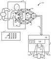
- FIG. 1illustrates, as an example, a top view of an operating room employing a medical robotic system.
- the medical robotic systemin this case is a Minimally Invasive Robotic Surgical (“MIRS”) System 100 including a Console (“C”) utilized by a Surgeon (“S”) while performing a minimally invasive diagnostic or surgical procedure with assistance from one or more Assistants (“A”) on a Patient (“P”) who is reclining on an Operating table (“O”).
- MIRSMinimally Invasive Robotic Surgical
- CConsole
- SS
- Aminimally invasive diagnostic or surgical procedure with assistance from one or more Assistants (“A”) on a Patient (“P”) who is reclining on an Operating table (“O”).
- the Consoleincludes a Master Display 104 (also referred to herein as a “Display Screen” or “computer display screen”) for displaying one or more images of a surgical site within the Patient as well as perhaps other information to the Surgeon. Also included are Master Input Devices 107 , 108 (also referred to herein as “Master Manipulators”), one or more Foot Pedals 105 , 106 , a Microphone 103 for receiving voice commands from the Surgeon, and a Processor 102 .
- the Master Input Devices 107 , 108may include any one or more of a variety of input devices such as joysticks, gloves, trigger-guns, hand-operated controllers, grippers, or the like.
- the Processor 102is preferably a personal computer that may be integrated into the Console or otherwise connected to it in a conventional manner.
- the Surgeonperforms a medical procedure using the MIRS System 100 by manipulating the Master Input Devices 107 , 108 so that the Processor 102 causes their respectively associated Slave Arms 121 , 122 to manipulate their respective removably coupled and held Surgical Instruments 138 , 139 (also referred to herein as “Tools”) accordingly, while the Surgeon views three-dimensional (“3D”) images of the surgical site on the Master Display 104 .
- Toolsthree-dimensional
- the Tools 138 , 139are preferably Intuitive Surgical's proprietary EndoWristTM articulating instruments, which are modeled after the human wrist so that when added to the motions of the robot arm holding the tool, they allow at least a full six degrees of freedom of motion, which is comparable to the natural motions of open surgery. Additional details on such tools may be found in commonly owned U.S. Pat. No. 5,797,900 entitled “Wrist Mechanism for Surgical Instrument for Performing Minimally Invasive Surgery with Enhanced Dexterity and Sensitivity,” which is incorporated herein by this reference.
- a manipulatable end effectorsuch as a clamp, grasper, scissor, stapler, blade, needle, needle holder, or energizable probe.
- the Master Display 104has a high-resolution stereoscopic video display with two progressive scan cathode ray tubes (“CRTs”).
- CRTsprogressive scan cathode ray tubes
- the systemoffers higher fidelity than polarization, shutter eyeglass, or other techniques.
- Each eyeviews a separate CRT presenting the left or right eye perspective, through an objective lens and a series of mirrors. The Surgeon sits comfortably and looks into this display throughout surgery, making it an ideal place for the Surgeon to display and manipulate 3-D intra-operative imagery.
- a Stereoscopic Endoscope 140provides right and left camera views to the Processor 102 so that it may process the information according to programmed instructions and cause it to be displayed on the Master Display 104 .
- a Laparoscopic Ultrasound (“LUS”) Probe 150provides two-dimensional (“2D”) ultrasound image slices of an anatomic structure to the Processor 102 so that the Processor 102 may generate a 3D ultrasound computer model or volume rendering of the anatomic structure.
- Each of the Tools 138 , 139 , as well as the Endoscope 140 and LUS Probe 150is preferably inserted through a cannula or trocar (not shown) or other tool guide into the Patient so as to extend down to the surgical site through a corresponding minimally invasive incision such as Incision 161 .
- Each of the Slave Arms 121 - 124includes a slave manipulator and setup arms.
- the slave manipulatorsare robotically moved using motor controlled joints (also referred to as “active joints”) in order to manipulate and/or move their respectively held Tools.
- the setup armsare manually manipulated by releasing normally braked joints (also referred to as “setup joints”) to horizontally and vertically position the Slave Arms 121 - 124 so that their respective Tools may be inserted into the cannulae.
- the number of surgical tools used at one time and consequently, the number of slave arms being used in the System 100will generally depend on the medical procedure to be performed and the space constraints within the operating room, among other factors. If it is necessary to change one or more of the tools being used during a procedure, the Assistant may remove the tool no longer being used from its slave arm, and replace it with another tool, such as Tool 131 , from a Tray (“T”) in the Operating Room.
- TTray
- the Master Display 104is positioned near the Surgeon's hands so that it will display a projected image that is oriented so that the Surgeon feels that he or she is actually looking directly down onto the surgical site.
- an image of the Tools 138 , 139preferably appear to be located substantially where the Surgeon's hands are located even though the observation points (i.e., that of the Endoscope 140 and LUS Probe 150 ) may not be from the point of view of the image.
- the real-time imageis preferably projected into a perspective image such that the Surgeon can manipulate the end effector of a Tool, 138 or 139 , through its associated Master Input Device, 107 or 108 , as if viewing the workspace in substantially true presence.
- true presenceit is meant that the presentation of an image is a true perspective image simulating the viewpoint of an operator that is physically manipulating the Tools.
- the Processor 102transforms the coordinates of the Tools to a perceived position so that the perspective image is the image that one would see if the Endoscope 140 was looking directly at the Tools from a Surgeon's eye-level during an open cavity procedure.
- the Processor 102performs various functions in the System 100 .
- One important function that it performsis to translate and transfer the mechanical motion of Master Input Devices 107 , 108 to their associated Slave Arms 121 , 122 through control signals over Bus 110 so that the Surgeon can effectively manipulate their respective Tools 138 , 139 .
- Another important functionis to implement the various methods described herein in reference to FIGS. 4-12 .
- Processor 102may be implemented in practice by any combination of hardware, software and firmware. Also, its functions as described herein may be performed by one unit, or divided up among different components, each of which may be implemented in turn by any combination of hardware, software and firmware. When divided up among different components, the components may be centralized in one location or distributed across the System 100 for distributed processing purposes.
- ultrasound images captured by the LUS Probe 150Prior to performing a medical procedure, ultrasound images captured by the LUS Probe 150 , right and left 2D camera images captured by the stereoscopic Endoscope 140 , and end effector positions and orientations as determined using kinematics of the Slave Arms 121 - 124 and their sensed joint positions, are calibrated and registered with each other.
- Slave Arms 123 , 124may manipulate the Endoscope 140 and LUS Probe 150 in similar manners as Slave Arms 121 , 122 manipulate Tools 138 , 139 .
- Master Input Devices 107 , 108 in the System 100in order for the Surgeon to manually control movement of either the Endoscope 140 or LUS Probe 150 , it may be required to temporarily associate one of the Master Input Devices 107 , 108 with the Endoscope 140 or the LUS Probe 150 that the Surgeon desires manual control over, while its previously associated Tool and Slave Manipulator are locked in position.
- auxiliary images of anatomic structuresmay be included in the System 100 , such as those commonly used for capturing ultrasound, magnetic resonance, computed axial tomography, and fluoroscopic images. Each of these sources of imagery may be used pre-operatively, and where appropriate and practical, intra-operatively.
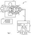
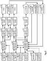
- FIG. 2illustrates, as an example, a block diagram of the System 100 .
- Master Input Device 107controls movement of either a Tool 138 or a stereoscopic Endoscope 140 , depending upon which mode its Control Switch Mechanism 211 is in, and Master Input Device 108 controls movement of either a Tool 139 or a LUS Probe 150 , depending upon which mode its Control Switch Mechanism 231 is in.
- the Control Switch Mechanisms 211 and 231may be placed in either a first or second mode by a Surgeon using voice commands, switches physically placed on or near the Master Input Devices 107 , 108 , Foot Pedals 105 , 106 on the Console, or Surgeon selection of appropriate icons or other graphical user interface selection means displayed on the Master Display 104 or an auxiliary display (not shown).
- Control Switch Mechanism 211When Control Switch Mechanism 211 is placed in the first mode, it causes Master Controller 202 to communicate with Slave Controller 203 so that manipulation of the Master Input 107 by the Surgeon results in corresponding movement of Tool 138 by Slave Arm 121 , while the Endoscope 140 is locked in position. On the other hand, when Control Switch Mechanism 211 is placed in the second mode, it causes Master Controller 202 to communicate with Slave Controller 233 so that manipulation of the Master Input 107 by the Surgeon results in corresponding movement of Endoscope 140 by Slave Arm 123 , while the Tool 138 is locked in position.
- Control Switch Mechanism 231when Control Switch Mechanism 231 is placed in the first mode, it causes Master Controller 108 to communicate with Slave Controller 223 so that manipulation of the Master Input 108 by the Surgeon results in corresponding movement of Tool 139 by Slave Arm 122 .
- the LUS Probe 150is not necessarily locked in position. Its movement may be guided by an Auxiliary Controller 242 according to stored instructions in Memory 240 .
- the Auxiliary Controller 242also provides haptic feedback to the Surgeon through Master Input 108 that reflects readings of a LUS Probe Force Sensor 247 .
- Control Switch Mechanism 231when placed in the second mode, it causes Master Controller 108 to communicate with Slave Controller 243 so that manipulation of the Master Input 108 by the Surgeon results in corresponding movement of LUS Probe 150 by Slave Arm 124 , while the Tool 139 is locked in position.
- a Control Switch MechanismBefore a Control Switch Mechanism effects a switch back to its first or normal mode, its associated Master Input Device is preferably repositioned to where it was before the switch. Alternatively, the Master Input Device may remain in its current position and kinematic relationships between the Master Input Device and its associated Tool Slave Arm readjusted so that upon the Control Switch Mechanism switching back to its first or normal mode, abrupt movement of the Tool does not occur.
- control switchingsee, e.g., commonly owned U.S. Pat. No. 6,659,939 entitled “Cooperative Minimally Invasive Telesurgical System,” which is incorporated herein by this reference.
- a third Control Switch Mechanism 241is provided to allow the user to switch between an image capturing mode and an image manipulating mode while the Control Switch Mechanism 231 is in its second mode (i.e., associating the Master Input Device 108 with the LUS Probe 150 ).
- the LUS Probe 150In its first or normal mode (i.e., image capturing mode), the LUS Probe 150 is normally controlled by the Master Input Device 108 as described above.
- the LUS Probe 150In its second mode (i.e., image manipulating mode), the LUS Probe 150 is not controlled by the Master Input Device 108 , leaving the Master Input Device 108 free to perform other tasks such as the displaying and manipulating of auxiliary images on the Display Screen 104 and in particular, for performing certain user specified functions as described herein.
- the LUS Probe 150may not be controlled by the Master Input Device 108 in this second mode of the Control Switch Mechanism 241 , it may still be automatically rocked or otherwise moved under the control of the Auxiliary Controller 242 according to stored instructions in Memory 240 so that a 3D volume rendering of a proximate anatomic structure may be generated from a series of 2D ultrasound image slices captured by the LUS Probe 150 .
- a 3D volume rendering of a proximate anatomic structuremay be generated from a series of 2D ultrasound image slices captured by the LUS Probe 150 .
- the Auxiliary Controller 242also performs other functions related to the LUS Probe 150 and the Endoscope 140 . It receives output from a LUS Probe Force Sensor 247 , which senses forces being exerted against the LUS Probe 150 , and feeds the force information back to the Master Input Device 108 through the Master Controller 222 so that the Surgeon may feel those forces even if he or she is not directly controlling movement of the LUS Probe 150 at the time. Thus, potential injury to the Patient is minimized since the Surgeon has the capability to immediately stop any movement of the LUS Probe 150 as well as the capability to take over manual control of its movement.
- Another key function of the Auxiliary Control 242is to cause processed information from the Endoscope 140 and the LUS Probe 150 to be displayed on the Master Display 104 according to user selected display options. Examples of such processing include generating a 3D ultrasound image from 2D ultrasound image slices received from the LUS Probe 150 through an Ultrasound Processor 246 , causing either 3D or 2D ultrasound images corresponding to a selected position and orientation to be displayed in a picture-in-picture window of the Master Display 104 , causing either 3D or 2D ultrasound images of an anatomic structure to overlay a camera captured image of the anatomic structure being displayed on the Master Display 104 , and performing the methods described below in reference to FIGS. 4-12 .
- the Master Controllers 202 , 222 , Slave Controllers 203 , 233 , 223 , 243 , and Auxiliary Controller 242are preferably implemented as software modules executed by the Processor 102 , as well as certain mode switching aspects of the Control Switch Mechanisms 211 , 231 , 241 .
- the Ultrasound Processor 246 and Video Processor 236may be software modules or separate boards or cards that are inserted into appropriate slots coupled to or otherwise integrated with the Processor 102 to convert signals received from these image capturing devices into signals suitable for display on the Master Display 104 and/or for additional processing by the Auxiliary Controller 242 before being displayed on the Master Display 104 .
- each Master Input Deviceis being shared by only one pre-assigned Tool Slave Robotic Arm and one pre-assigned Image Capturing Device Robotic Arm
- alternative arrangementsare also feasible and envisioned to be within the full scope of the present invention.
- each of the Master Input Devicesmay be selectively associated with any one of the Tool and Image Capturing Device Robotic Arms is also possible and even preferably for maximum flexibility.
- the Endoscope Robotic Armis shown in this example as being controlled by a single Master Input Device, it may also be controlled using both Master Input Devices to give the sensation of being able to “grab the image” and move it to a different location or view.
- FIG. 3illustrates a side view of one embodiment of the LUS Probe 150 .
- the LUS Probe 150is a dexterous tool with preferably two distal degrees of freedom. Opposing pairs of Drive Rods or Cables (not shown) physically connected to a proximal end of the LUS Sensor 301 and extending through an internal passage of Elongated Shaft 312 mechanically control pitch and yaw movement of the LUS Sensor 301 using conventional push-pull type action.
- the LUS Sensor 301captures 2D ultrasound slices of a proximate anatomic structure, and transmits the information back to the Processor 102 through LUS Cable 304 . Although shown as running outside of the Elongated Shaft 312 , the LUS Cable 304 may also extend within it.
- a Clamshell Sheath 321encloses the Elongate Shaft 312 and LUS Cable 304 to provide a good seal passing through a Cannula 331 (or trocar). Fiducial Marks 302 and 322 are placed on the LUS Sensor 301 and the Sheath 321 for video tracking purposes.
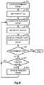
- FIG. 4illustrates, as an example, a flow diagram of a method for displaying the effect of a therapeutic procedure or treatment on the Display Screen 104 .
- a primary image of an anatomic structureis captured by an image capturing device.
- FIG. 5illustrates a primary image which has been captured by the Endoscope 140 and includes an anatomic structure 501 and therapeutic instrument 511 that has been partially inserted into the anatomic structure 501 in order to perform a therapeutic procedure at a therapy site within the anatomic structure 501 .
- the therapeutic instrument 511may only need to touch or come close to the anatomic structure 501 in order to perform a therapeutic procedure.
- the primary imagemay be captured before or during the therapeutic procedure.
- a primary image captured before the procedureis referred to as being a “pre-operative” image, and a primary image captured during the procedure is referred to as being an “intra-operative” image.
- the imageis generally not updated during the procedure, so that the method generally only employs one primary image.
- the primary imageis an intra-operative image, the image is preferably updated periodically during the procedure, so that the method employs multiple primary images in that case.
- Pre-operative imagesare typically captured using techniques such as Ultrasonography, Magnetic Resonance Imaging (MM), or Computed Axial Tomography (CAT).
- Intra-operative imagesmay be captured at the surgical or therapeutic site by image capturing devices such as the stereoscopic Endoscope 140 or LUS Probe 150 , or they may be captured externally by techniques such as those used to capture the pre-operative images.
- the therapeutic instrumentis turned on, or otherwise activated or energized, so as to be capable of applying therapy to the anatomic structure within the patient.
- the instrumentgenerally has a tip for applying the therapeutic energy to abnormal tissue such as diseased or damaged tissue.
- Radio Frequency Ablationmay be used to destroy diseased tissue such as a tumor located in an anatomic structure such as the liver by applying heat to the diseased tissue site using an RFA probe.
- RFARadio Frequency Ablation
- HIFUHigh Intensity Focused Ultrasound
- the therapeutic instrumentmay be one of the Tools 138 , 139 attached to Slave Arms 121 , 122 so that it may be moved to and manipulated at the therapy site through the master/slave control system by the Surgeon.
- an auxiliary imageis generated, wherein the auxiliary image indicates the effect of the therapeutic procedure on the anatomic structure.
- the auxiliary imagemay be an actual image of the anatomic structure that has been provided by or generated from information captured by a sensing device which is capable of sensing the effect of the therapeutic procedure.
- the auxiliary imagemay be a computer model indicating the effect of the therapy, which may be generated using an empirically derived or otherwise conventionally determined formula of such effect.
- the computer modelis generally a volumetric shape determined by such factors as the geometry of the tip of the therapeutic instrument, the heat or energy level being applied to the anatomic structure by the tip of the therapeutic instrument, and the features of the surrounding tissue of a therapy site being subjected to the therapeutic procedure in the anatomic structure.
- FIG. 6illustrates a three-dimensional ultrasound image of an anatomic structure 601 which has been conventionally derived from two-dimensional ultrasound slices captured by the LUS probe 150 .
- an ablation volume 621is shown which represents the effect of a therapeutic procedure in which a tip 613 of an RFA probe 612 is being applied to a tumor site of the anatomic structure 601 .
- the growth of the ablation volume in this caseis viewable due to changes in tissue properties from the heating and necrosis of the surrounding tissue at the tumor site.
- the primary and auxiliary imagesare registered so as to be of the same scale and refer to a same position and orientation in a common reference frame. Registration of this sort is well known.
- U.S. Pat. No. 6,522,906entitled “Devices and Methods for Presenting and Regulating Auxiliary Information on an Image Display of a Telesurgical System to Assist an Operator in Performing a Surgical Procedure,” which is incorporated herein by this reference.
- the primary imageis displayed on the Display Screen 104 while the therapeutic procedure is being performed, with the registered auxiliary image preferably overlaid upon the primary image so that corresponding structures or objects in each of the images appear as the same size and at the same location and orientation on the Display Screen 104 .
- the effect of the therapeutic procedureis shown as an overlay over the anatomic structure that is being subjected to the procedure.
- FIG. 7shows an exemplary Display Screen 104 in which an auxiliary image, distinguished as a dotted line for illustrative purposes, is overlaid over the primary image of FIG. 5 .
- the auxiliary imageis provided by or derives from information captured by a sensing device
- the therapy effect 521 , therapeutic instrument 512 , and instrument tip 513is provided by or derived from the captured information.
- the therapy effect 521is generated as a volumetric shaped computer model using an empirically determined formula
- the therapeutic instrument 512 and instrument tip 513may be determined using conventional tool tracking computations based at least in part upon joint positions of its manipulating slave arm.
- the methodthen checks whether the therapeutic instrument has been turned off. If it has, then this means that the therapeutic procedure is over, and the method ends. On the other hand, if the therapeutic instrument is still on, then the method assumes that the therapeutic procedure is still being performed, and proceeds in 407 to determine whether a new primary image has been captured. If no new primary image has been captured, for example, because the primary image is a pre-operative image, then the method jumps back to 403 to update the auxiliary image and continue to loop through 403 - 407 until the therapeutic procedure is determined to be completed by detecting that the therapeutic instrument has been turned off.
- the methodupdates the primary image in 408 before jumping back to 403 to update the auxiliary image and continue to loop through 403 - 408 until the therapeutic procedure is determined to be completed by detecting that the therapeutic instrument has been turned off.
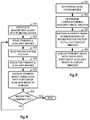
- FIG. 8illustrates, as an example, a flow diagram of a method for displaying an auxiliary image of an anatomic structure as a registered overlay to a primary image of the anatomic structure at a user specified magnification in a window defined as the lens area of a magnifying glass whose position and orientation as displayed on the Display Screen 104 is manipulatable by the user using an associated pointing device.
- the methodstarts out by associating the magnifying glass with the pointing device so that as the pointing device moves, the magnifying glass being displayed on the Display Screen 104 (and in particular, its lens which may be thought of as a window) moves in a corresponding fashion.
- the associationin this case may be performed by “grabbing” the magnifying glass in a conventional manner using the pointing device, or by making the magnifying glass effectively the cursor for the pointing device.
- the Display Screen 104is preferably a three-dimensional display
- the pointing deviceis correspondingly preferably a three-dimensional pointing device with orientation indicating capability.
- the primary image in this exampleis captured by the Endoscope 140 and the auxiliary captured by the LUS Probe 150 .
- other sources for the primary and auxiliary imagesare also usable and contemplated in practicing the invention, including primary and auxiliary images captured from the same source.
- a high resolution cameramay capture images at a resolution greater than that being used to display images on a display screen.
- the high resolution image captured by the cameramay be treated as the auxiliary image, and the downsized image to be displayed on the display screen may be treated as the primary image.
- a user selectable magnification factoris read.
- the magnification factoris user selectable by, for example, a dial or wheel type control on the pointing device. Alternatively, it may be user selectable by user selection of item in a menu displayed on the Display Screen 104 , or any other conventional user selectable parameter value scheme or mechanism. If the user fails to make a selection, then a default value is used, such as a magnification factor of 1.0.
- the primary and auxiliary imagesare registered so as to be of the same scale and refer to a same position and orientation in a common reference frame so that corresponding structures and objects in the two images have the same coordinates.
- the primary imageis displayed on the Display Screen 104 such as a three-dimensional view of the anatomic structure, in which case, a portion of a two-dimensional slice of the auxiliary image of the anatomic structure may be displayed as an overlay in the lens of the magnifying glass.
- the portion of the two-dimensional slice in this caseis defined by a window area having a central point that has the same position and orientation of as the central point of the lens of the magnifying glass, and an area determined by the magnification factor so that the portion of the two-dimensional slice may be enlarged or reduced so as to fit in the lens of the magnifying glass.
- the two-dimensional slicecan correspond to any user selected depth within the anatomic structure.
- its viewis not limited to inspecting only the exterior of the anatomic structure.
- the methoddetermines whether the magnifying glass command has been turned off by, for example, the user releasing a “grabbed” image of the magnifying glass, or otherwise switching off the association between the magnifying glass and the pointing device by the use of a conventional switch mechanism of some sort. If it has, then the method ends. On the other hand, if it has not, then the method jumps back to 802 and continues to loop through 802 - 806 until the magnifying glass command is detected to have been turned off. Note that each time the method loops through 802 - 806 , updated versions, if any, of the primary and auxiliary images are processed along with updated values, if any, for the user selectable magnification factor. Thus, if the method proceeds through the looping in a sufficiently fast manner, the user will not notice any significant delay if the user is turning a dial or knob to adjust the magnification factor while viewing the anatomic structure at a selected position and orientation of the magnifying glass.
- FIG. 9illustrates, as an example, a flow diagram of a method for displaying an auxiliary image view of an anatomic structure at a specified magnification factor as an overlay to a primary image view of the anatomic structure in the lens of a user movable magnifying glass. As previously explained, this method may be used to perform 805 of FIG. 8 .
- the current position and orientation of a central point of the lens of the magnifying glassare determined in the three-dimensional space of the Display Screen 104 .
- a two-dimensional slice of the registered volumetric model of the auxiliary imageis taken from the perspective of that position and orientation, and a portion of the two-dimensional slice is taken as defined in an auxiliary view window having a central point preferably at that same position and orientation.
- the area of the auxiliary view window in this caseis inversely proportional to that of the lens according to the current magnification factor for the magnifying glass.
- the portion of the two-dimensional slice defined by the auxiliary view windowis then enlarged by the magnification factor so that it fits in the lens area of the magnifying glass, and in 904 , the primary image of the anatomic structure is displayed on the Display Screen 104 with the enlarged portion of the two-dimensional slice of the auxiliary image overlaid in the lens area of the magnifying glass being displayed on the Display Screen 104 .
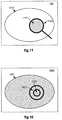
- FIGS. 10-11a two-dimensional slice 1001 of an auxiliary image of an anatomic structure is shown along with two circular windows 1021 , 1022 on the two-dimensional slice as illustrated in FIG. 10 .
- Each of the windows 1021 , 1022in this case corresponds in shape to and having a central point equal to that of a lens 1121 of a magnifying glass 1120 which is being displayed along with a primary image of an external view 1101 of the anatomic structure on the Display Screen 104 as illustrated in FIG. 11 .
- the area of the window 1021is equal to the area of the lens 1121 , so that if the magnification factor was 1.0, then window 1021 would be selected for use in 902 .
- the area of the window 1022is less than the area of the lens 1121 , so that if the magnification factor is greater than 1.0, then the window 1022 may be selected for use in 902 .
- the lens 1121 of the magnifying glass 1120is depicted as being circularly shaped, it may also have other common shapes for a magnifying glass, such as a rectangular shape.
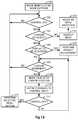
- FIG. 12illustrates, as an example, a flow diagram of a method performed by a processor in a medical robotic system for manipulating image objects displayed on a computer display screen of the medical robotic system in response to corresponding manipulation of an associated master input device when the master input device is in an image manipulating mode.
- the medical robotic systemincludes an image capturing device to capture images (such as either the Endoscope 140 or the LUS Probe 150 ); a robotic arm holding the image capturing device (such as the Slave Arm 123 or the Slave Arm 124 respectively holding the Endoscope 140 and the LUS Probe 150 ); a computer display screen (such as the Display Screen 104 ); a master input device adapted to be manipulatable by a user in multiple degrees-of-freedom movement (such as the Master Input Device 107 or the Master Input Device 108 ); and a processor (such as the Auxiliary Controller 242 ) that is configured to control movement of the image capturing device according to user manipulation of the master input device when the master input device is in an image capturing mode, and control the displaying of images derived from the captured images on the computer display screen according to user manipulation of the master input device when the master input device is in the image manipulating mode.
- an image capturing device to capture imagessuch as either the Endoscope 140 or the LUS Probe 150
- the processordetects that the user has placed the master input device into its image manipulating mode.
- a master clutch mechanismprovided in the medical robotic system, which supports disengaging the master input device from its associated robotic arm so that the master input device may be repositioned.
- this modeis activated by some mechanism such as the user depressing a button on the master input device, pressing down on a foot pedal, or using voice activation, the associated robotic arm is locked in position, and a cursor (nominally an iconic representation of a hand, e.g. ) is presented to the user on the computer display screen.
- a cursornominally an iconic representation of a hand, e.g.
- the processordetermines whether a control input such as that generated by depressing a button on a conventional mouse has been activated by the user.
- the control inputin this case may be activated by depressing a button provided on the master input device, or it may be activated by some other fashion such as squeezing a gripper or pincher formation provided on the master input device.
- a control inputsuch as that generated by depressing a button on a conventional mouse has been activated by the user.
- the control inputin this case may be activated by depressing a button provided on the master input device, or it may be activated by some other fashion such as squeezing a gripper or pincher formation provided on the master input device.
- For additional details on clutching, and gripper or pincher formations on a master input devicesee, e.g., commonly owned U.S. Pat. No. 6,659,939 entitled “Cooperative Minimally Invasive Telesurgical System,” which has been previously incorporated herein by reference. If the control
- the processorAfter receiving an indication that the control input is “on”, the processor checks to see if the cursor is positioned on (or within a predefined distance to) an object being displayed on the computer display screen. If it is not, then in 1204 , the processor causes a menu of user selectable items or actions to be displayed on the computer display screen, and in 1205 , the processor receives and reacts to a menu selection made by the user.
- Examples of user selectable menu itemsinclude: magnifying glass, cut-plane, eraser, and image registration. If the user selects the magnifying glass item, then an image of a magnifying glass is displayed on the computer display screen and the method described in reference to FIG. 8 may be performed by the processor. When the user is finished with the magnifying glass function, then the user may indicate exiting of the function in any conventional manner and the processor returns to 1202 .
- a plane(or rectangular window of fixed or user adjustable size) is displayed on the computer display screen.
- the master input devicemay then be associated with the plane so that the user may position and orientate the plane in the three-dimensional space of the computer display screen by manipulating the master input device in the manner of a pointing device. If the plane is maneuvered so as to intersect a volume rendering of an anatomic structure, then it functions as a cut-plane defining a two-dimensional slice of the volume rendering at the intersection. Alternatively, the master input device may be associated with the volume rendering of the anatomic structure, which may then be maneuvered so as to intersect the displayed plane to define the cut-plane. Association of the plane or volume rendering with the pointing device may be performed in substantially the same manner as described in reference to the magnifying glass with respect to 801 of FIG. 8 .
- the two-dimensional slicemay then be viewed either in the plane itself, or in a separate window on the computer display screen such as in a picture-in-picture.
- the usermay further select the cut-plane item additional times to define additional two-dimensional slices of the volume rendering for concurrent viewing in respective planes or picture-in-picture windows on the computer display screen.
- a conventional delete functionis provided so that the user may selectively delete any cut-planes and their corresponding slices.
- the usermay indicate exiting of the function in any conventional manner and the processor returns to 1202 .
- an eraseris displayed on the computer display screen.
- the master input deviceis then associated with the eraser so that the user may position and orientate the eraser in the three-dimensional space of the computer display screen by manipulating the master input device in the manner of a pointing device. Association of the eraser with the pointing device in this case may be performed in substantially the same manner as described in reference to the magnifying glass with respect to 801 of FIG. 8 . If the eraser is maneuvered so as to intersect a volume rendering of an anatomic structure, then it functions to either completely or partially erase such rendering wherever it traverses the volume rendering.
- partial erasingis selected by the user (or otherwise pre-programmed into the processor), then each time the eraser traverses the volume rendering, less detail of the anatomic structure may be shown. Less detail in this case may refer to the coarseness/fineness of the rendering, or it may refer to the stripping away of layers in the three-dimensional volume rendering. All such characteristics or options of the erasing may be user selected using conventional means. If the user inadvertently erases a portion of the volume rendering, a conventional undo feature is provided to allow the user to undo the erasure. When the user is finished with the erasing function, then the user may indicate exiting of the function in any conventional manner and the processor returns to 1202 .
- spatially localized modifying functionsare also contemplated and considered to be within the full scope of the present invention, including selectively sharpening, brightening, or coloring portions of a displayed image to enhance its visibility in, or otherwise highlight, a selected area.
- Each such spatially localized modifying functionmay be performed using substantially the same method described above in reference to the eraser function.
- Image registrationin this case typically involves manually registering an auxiliary image of an object such as an anatomic structure with a corresponding primary image of the object.
- icons respectively indicating each of the selectable items as described abovemay be displayed on the computer display screen upon entering image manipulating mode and selected by the user clicking on them, after which, the processor proceeds to perform as described above in reference to selection of their corresponding menu items.
- the processorafter receiving an indication that the control input is on in 1201 and determining that the cursor is positioned on or near an object (not an icon) being displayed on the computer display screen in 1202 , the processor preferably changes the cursor from an iconic representation of a hand, for example, to that of a grasping hand to indicate that the object has been “grabbed” and is ready to be moved or “dragged” to another position and/or orientation in the three-dimensional space of the computer display screen through user manipulation of the master input device.
- the processordetermines whether the user has indicated that a display parameter of the selected object is to be adjusted, and if the user has so indicated, in 1207 , the processor performs the display adjustment.
- a dial on the master input devicemay be turned by the user to indicate both that a display adjustment for a display parameter associated with dial is to be adjusted according to the amount of rotation of the dial on the selected object.
- the grippermay be rotated so as to function as a dial. Examples of display parameters that may be adjusted in this manner include: brightness, contrast, color, and level of detail (e.g., mesh coarseness/fineness, or voxel size and/or opaqueness) of the selected object being displayed on the computer display screen.
- the processorthen proceeds to 1208 to determine whether the cursor has moved since “grabbing” the selected object after an affirmative determination in 1203 . If it has not moved, then the processor jumps back to 1202 since the user may only have wanted to adjust a display parameter of a selected object at this time. On the other hand, if the cursor has moved since “grabbing” the selected object, then in 1209 , the processor moves the selected object to the new cursor position. Since the cursor operates in the three-dimensional space of the computer display screen, when it moves “into” the display screen, it may indicate such movement by, for example, getting progressively smaller in size.
- haptic feedbackmay be provided back to the master input device so that the user may sense reflected forces while the “grabbed” object is being moved in 1209 .
- user interactions with the objectmay be reflected haptically back to the user by associating a virtual mass and inertial properties with the object so that the user feels a reflected force when coming into contact with the object or when translating or rotating the object as it is accelerated/decelerated.
- the haptic feedback performed in this 1210may only be performed for some types of objects and not for others, or it may take effect only in certain circumstances. Use of such haptic feedback may also be applied to the movement of the magnifying glass and/or the plane used for defining cut-planes as described above. In such cases, however, the haptic feedback may be restricted to only occurring after the magnifying glass or the plane enters into an anatomic structure of interest.
- the processordetermines whether the control input is still in an “on” state. If the control is still “on”, then the processor jumps back to 1208 to track and respond to cursor movement. On the other hand, if the control has been turned off by, for example, the user releasing a button that was initially depressed to indicate that control was turned “on”, then in 1212 , the processor performs a selected menu action.
- the object that has been movedis registered with another image of the object that is now aligned with and is being displayed on the computer display screen at the time so that they have the same coordinate and orientation values in a common reference frame such as that of the computer display screen.
- This featurefacilitates, for example, manual registration of an auxiliary image of an anatomic structure (such as obtained using the LUS Probe 150 ) with a primary image of the anatomic structure (such as obtained using the Endoscope 140 ).
- changes to the position and/or orientation of the corresponding object in the primary imagemay be mirrored so as to cause corresponding changes to the selected object in the auxiliary image so as to maintain its relative position/orientation with respect to the primary image.
- the processorreturns to 1202 .
Landscapes
- Engineering & Computer Science (AREA)
- Health & Medical Sciences (AREA)
- Life Sciences & Earth Sciences (AREA)
- Surgery (AREA)
- General Engineering & Computer Science (AREA)
- Theoretical Computer Science (AREA)
- Physics & Mathematics (AREA)
- Animal Behavior & Ethology (AREA)
- Biomedical Technology (AREA)
- Molecular Biology (AREA)
- Heart & Thoracic Surgery (AREA)
- General Health & Medical Sciences (AREA)
- Public Health (AREA)
- Veterinary Medicine (AREA)
- Medical Informatics (AREA)
- Nuclear Medicine, Radiotherapy & Molecular Imaging (AREA)
- Human Computer Interaction (AREA)
- General Physics & Mathematics (AREA)
- Robotics (AREA)
- Pathology (AREA)
- Biophysics (AREA)
- Radiology & Medical Imaging (AREA)
- Optics & Photonics (AREA)
- Plasma & Fusion (AREA)
- Otolaryngology (AREA)
- Oral & Maxillofacial Surgery (AREA)
- Endoscopes (AREA)
- Ultra Sonic Daignosis Equipment (AREA)
- Magnetic Resonance Imaging Apparatus (AREA)
- High Energy & Nuclear Physics (AREA)
- Manipulator (AREA)
- Surgical Instruments (AREA)
- Apparatus For Radiation Diagnosis (AREA)
- Controls And Circuits For Display Device (AREA)
Abstract
Description

Claims (20)
Priority Applications (2)
| Application Number | Priority Date | Filing Date | Title |
|---|---|---|---|
| US16/564,734US11197731B2 (en) | 2005-10-20 | 2019-09-09 | Auxiliary image display and manipulation on a computer display in a medical robotic system |
| US17/530,166US20220071721A1 (en) | 2005-10-20 | 2021-11-18 | Auxiliary image display and manipulation on a computer display in a medical robotic system |
Applications Claiming Priority (4)
| Application Number | Priority Date | Filing Date | Title |
|---|---|---|---|
| US72845005P | 2005-10-20 | 2005-10-20 | |
| US11/583,963US20080033240A1 (en) | 2005-10-20 | 2006-10-19 | Auxiliary image display and manipulation on a computer display in a medical robotic system |
| US15/139,682US20160235496A1 (en) | 2005-10-20 | 2016-04-27 | Auxiliary image display and manipulation on a computer display in a medical robotic system |
| US16/564,734US11197731B2 (en) | 2005-10-20 | 2019-09-09 | Auxiliary image display and manipulation on a computer display in a medical robotic system |
Related Parent Applications (1)
| Application Number | Title | Priority Date | Filing Date |
|---|---|---|---|
| US15/139,682ContinuationUS20160235496A1 (en) | 2005-10-20 | 2016-04-27 | Auxiliary image display and manipulation on a computer display in a medical robotic system |
Related Child Applications (1)
| Application Number | Title | Priority Date | Filing Date |
|---|---|---|---|
| US17/530,166ContinuationUS20220071721A1 (en) | 2005-10-20 | 2021-11-18 | Auxiliary image display and manipulation on a computer display in a medical robotic system |
Publications (2)
| Publication Number | Publication Date |
|---|---|
| US20190388169A1 US20190388169A1 (en) | 2019-12-26 |
| US11197731B2true US11197731B2 (en) | 2021-12-14 |
Family
ID=37744551
Family Applications (4)
| Application Number | Title | Priority Date | Filing Date |
|---|---|---|---|
| US11/583,963AbandonedUS20080033240A1 (en) | 2005-10-20 | 2006-10-19 | Auxiliary image display and manipulation on a computer display in a medical robotic system |
| US15/139,682AbandonedUS20160235496A1 (en) | 2005-10-20 | 2016-04-27 | Auxiliary image display and manipulation on a computer display in a medical robotic system |
| US16/564,734Active2026-11-20US11197731B2 (en) | 2005-10-20 | 2019-09-09 | Auxiliary image display and manipulation on a computer display in a medical robotic system |
| US17/530,166AbandonedUS20220071721A1 (en) | 2005-10-20 | 2021-11-18 | Auxiliary image display and manipulation on a computer display in a medical robotic system |
Family Applications Before (2)
| Application Number | Title | Priority Date | Filing Date |
|---|---|---|---|
| US11/583,963AbandonedUS20080033240A1 (en) | 2005-10-20 | 2006-10-19 | Auxiliary image display and manipulation on a computer display in a medical robotic system |
| US15/139,682AbandonedUS20160235496A1 (en) | 2005-10-20 | 2016-04-27 | Auxiliary image display and manipulation on a computer display in a medical robotic system |
Family Applications After (1)
| Application Number | Title | Priority Date | Filing Date |
|---|---|---|---|
| US17/530,166AbandonedUS20220071721A1 (en) | 2005-10-20 | 2021-11-18 | Auxiliary image display and manipulation on a computer display in a medical robotic system |
Country Status (6)
| Country | Link |
|---|---|
| US (4) | US20080033240A1 (en) |
| EP (4) | EP1937176B1 (en) |
| JP (4) | JP5322648B2 (en) |
| KR (1) | KR101320379B1 (en) |
| CN (3) | CN103251455B (en) |
| WO (1) | WO2007047782A2 (en) |
Cited By (50)
| Publication number | Priority date | Publication date | Assignee | Title |
|---|---|---|---|---|
| US20220087763A1 (en)* | 2020-09-23 | 2022-03-24 | Verb Surgical Inc. | Deep disengagement detection during telesurgery |
| US11485012B2 (en)* | 2019-12-12 | 2022-11-01 | Seiko Epson Corporation | Control method and robot system |
| US11504197B1 (en) | 2021-03-31 | 2022-11-22 | Moon Surgical Sas | Co-manipulation surgical system having multiple operational modes for use with surgical instruments for performing laparoscopic surgery |
| US11662270B2 (en)* | 2017-08-22 | 2023-05-30 | Intuitive Surgical Operations, Inc. | User-installable part installation detection techniques |
| US11812938B2 (en) | 2021-03-31 | 2023-11-14 | Moon Surgical Sas | Co-manipulation surgical system having a coupling mechanism removeably attachable to surgical instruments |
| US11819302B2 (en) | 2021-03-31 | 2023-11-21 | Moon Surgical Sas | Co-manipulation surgical system having user guided stage control |
| US11832910B1 (en) | 2023-01-09 | 2023-12-05 | Moon Surgical Sas | Co-manipulation surgical system having adaptive gravity compensation |
| US11832909B2 (en) | 2021-03-31 | 2023-12-05 | Moon Surgical Sas | Co-manipulation surgical system having actuatable setup joints |
| US11844583B2 (en) | 2021-03-31 | 2023-12-19 | Moon Surgical Sas | Co-manipulation surgical system having an instrument centering mode for automatic scope movements |
| US11844545B2 (en) | 2018-03-08 | 2023-12-19 | Cilag Gmbh International | Calcified vessel identification |
| US11844579B2 (en) | 2017-12-28 | 2023-12-19 | Cilag Gmbh International | Adjustments based on airborne particle properties |
| US11857152B2 (en) | 2017-12-28 | 2024-01-02 | Cilag Gmbh International | Surgical hub spatial awareness to determine devices in operating theater |
| US11864728B2 (en) | 2017-12-28 | 2024-01-09 | Cilag Gmbh International | Characterization of tissue irregularities through the use of mono-chromatic light refractivity |
| US11871901B2 (en) | 2012-05-20 | 2024-01-16 | Cilag Gmbh International | Method for situational awareness for surgical network or surgical network connected device capable of adjusting function based on a sensed situation or usage |
| US11890065B2 (en) | 2017-12-28 | 2024-02-06 | Cilag Gmbh International | Surgical system to limit displacement |
| US11896322B2 (en) | 2017-12-28 | 2024-02-13 | Cilag Gmbh International | Sensing the patient position and contact utilizing the mono-polar return pad electrode to provide situational awareness to the hub |
| US11896443B2 (en) | 2017-12-28 | 2024-02-13 | Cilag Gmbh International | Control of a surgical system through a surgical barrier |
| US11918302B2 (en) | 2017-12-28 | 2024-03-05 | Cilag Gmbh International | Sterile field interactive control displays |
| US11925350B2 (en) | 2019-02-19 | 2024-03-12 | Cilag Gmbh International | Method for providing an authentication lockout in a surgical stapler with a replaceable cartridge |
| US11925373B2 (en) | 2017-10-30 | 2024-03-12 | Cilag Gmbh International | Surgical suturing instrument comprising a non-circular needle |
| US11931027B2 (en) | 2018-03-28 | 2024-03-19 | Cilag Gmbh Interntional | Surgical instrument comprising an adaptive control system |
| US11969216B2 (en) | 2017-12-28 | 2024-04-30 | Cilag Gmbh International | Surgical network recommendations from real time analysis of procedure variables against a baseline highlighting differences from the optimal solution |
| US11969142B2 (en) | 2017-12-28 | 2024-04-30 | Cilag Gmbh International | Method of compressing tissue within a stapling device and simultaneously displaying the location of the tissue within the jaws |
| US11986165B1 (en) | 2023-01-09 | 2024-05-21 | Moon Surgical Sas | Co-manipulation surgical system for use with surgical instruments for performing laparoscopic surgery while estimating hold force |
| US11986233B2 (en) | 2018-03-08 | 2024-05-21 | Cilag Gmbh International | Adjustment of complex impedance to compensate for lost power in an articulating ultrasonic device |
| US11998193B2 (en) | 2017-12-28 | 2024-06-04 | Cilag Gmbh International | Method for usage of the shroud as an aspect of sensing or controlling a powered surgical device, and a control algorithm to adjust its default operation |
| US12009095B2 (en) | 2017-12-28 | 2024-06-11 | Cilag Gmbh International | Real-time analysis of comprehensive cost of all instrumentation used in surgery utilizing data fluidity to track instruments through stocking and in-house processes |
| US12029506B2 (en) | 2017-12-28 | 2024-07-09 | Cilag Gmbh International | Method of cloud based data analytics for use with the hub |
| US12035983B2 (en) | 2017-10-30 | 2024-07-16 | Cilag Gmbh International | Method for producing a surgical instrument comprising a smart electrical system |
| US12035890B2 (en) | 2017-12-28 | 2024-07-16 | Cilag Gmbh International | Method of sensing particulate from smoke evacuated from a patient, adjusting the pump speed based on the sensed information, and communicating the functional parameters of the system to the hub |
| US12042207B2 (en) | 2017-12-28 | 2024-07-23 | Cilag Gmbh International | Estimating state of ultrasonic end effector and control system therefor |
| US12042241B2 (en) | 2021-03-31 | 2024-07-23 | Moon Surgical Sas | Co-manipulation surgical system having automated preset robot arm configurations |
| US12048496B2 (en) | 2017-12-28 | 2024-07-30 | Cilag Gmbh International | Adaptive control program updates for surgical hubs |
| US12062442B2 (en) | 2017-12-28 | 2024-08-13 | Cilag Gmbh International | Method for operating surgical instrument systems |
| US12059169B2 (en) | 2017-12-28 | 2024-08-13 | Cilag Gmbh International | Controlling an ultrasonic surgical instrument according to tissue location |
| US12121256B2 (en) | 2018-03-08 | 2024-10-22 | Cilag Gmbh International | Methods for controlling temperature in ultrasonic device |
| US12127729B2 (en) | 2017-12-28 | 2024-10-29 | Cilag Gmbh International | Method for smoke evacuation for surgical hub |
| US12133773B2 (en) | 2017-12-28 | 2024-11-05 | Cilag Gmbh International | Surgical hub and modular device response adjustment based on situational awareness |
| US12133709B2 (en) | 2017-12-28 | 2024-11-05 | Cilag Gmbh International | Communication hub and storage device for storing parameters and status of a surgical device to be shared with cloud based analytics systems |
| US12144518B2 (en) | 2017-12-28 | 2024-11-19 | Cilag Gmbh International | Surgical systems for detecting end effector tissue distribution irregularities |
| US12167900B2 (en) | 2021-03-31 | 2024-12-17 | Moon Surgical Sas | Co-manipulation surgical system having automated preset robot arm configurations |
| US12178418B2 (en) | 2021-03-31 | 2024-12-31 | Moon Surgical Sas | Co-manipulation surgical system having a coupling mechanism removeably attachable to surgical instruments |
| US12226166B2 (en) | 2017-12-28 | 2025-02-18 | Cilag Gmbh International | Surgical instrument with a sensing array |
| US12226151B2 (en) | 2017-12-28 | 2025-02-18 | Cilag Gmbh International | Capacitive coupled return path pad with separable array elements |
| US12295674B2 (en) | 2017-12-28 | 2025-05-13 | Cilag Gmbh International | Usage and technique analysis of surgeon / staff performance against a baseline to optimize device utilization and performance for both current and future procedures |
| US12310586B2 (en) | 2017-12-28 | 2025-05-27 | Cilag Gmbh International | Method for adaptive control schemes for surgical network control and interaction |
| US12318152B2 (en) | 2017-12-28 | 2025-06-03 | Cilag Gmbh International | Computer implemented interactive surgical systems |
| US12329467B2 (en) | 2017-10-30 | 2025-06-17 | Cilag Gmbh International | Method of hub communication with surgical instrument systems |
| US12370001B2 (en) | 2023-01-09 | 2025-07-29 | Moon Surgical Sas | Co-manipulation surgical system having automated user override detection |
| US12396806B2 (en) | 2017-12-28 | 2025-08-26 | Cilag Gmbh International | Adjustment of a surgical device function based on situational awareness |
Families Citing this family (172)
| Publication number | Priority date | Publication date | Assignee | Title |
|---|---|---|---|---|
| US8944070B2 (en)* | 1999-04-07 | 2015-02-03 | Intuitive Surgical Operations, Inc. | Non-force reflecting method for providing tool force information to a user of a telesurgical system |
| US9517106B2 (en) | 1999-09-17 | 2016-12-13 | Intuitive Surgical Operations, Inc. | Systems and methods for commanded reconfiguration of a surgical manipulator using the null-space |
| US9155544B2 (en)* | 2002-03-20 | 2015-10-13 | P Tech, Llc | Robotic systems and methods |
| US9789608B2 (en) | 2006-06-29 | 2017-10-17 | Intuitive Surgical Operations, Inc. | Synthetic representation of a surgical robot |
| US9492240B2 (en) | 2009-06-16 | 2016-11-15 | Intuitive Surgical Operations, Inc. | Virtual measurement tool for minimally invasive surgery |
| US8971597B2 (en)* | 2005-05-16 | 2015-03-03 | Intuitive Surgical Operations, Inc. | Efficient vision and kinematic data fusion for robotic surgical instruments and other applications |
| US8073528B2 (en) | 2007-09-30 | 2011-12-06 | Intuitive Surgical Operations, Inc. | Tool tracking systems, methods and computer products for image guided surgery |
| US10555775B2 (en) | 2005-05-16 | 2020-02-11 | Intuitive Surgical Operations, Inc. | Methods and system for performing 3-D tool tracking by fusion of sensor and/or camera derived data during minimally invasive robotic surgery |
| WO2007047782A2 (en) | 2005-10-20 | 2007-04-26 | Intuitive Surgical, Inc | Auxiliary image display and manipulation on a computer display in a medical robotic system |
| US7907166B2 (en)* | 2005-12-30 | 2011-03-15 | Intuitive Surgical Operations, Inc. | Stereo telestration for robotic surgery |
| KR101477133B1 (en) | 2006-06-13 | 2014-12-29 | 인튜어티브 서지컬 인코포레이티드 | Minimally invasive surgical system |
| US10258425B2 (en) | 2008-06-27 | 2019-04-16 | Intuitive Surgical Operations, Inc. | Medical robotic system providing an auxiliary view of articulatable instruments extending out of a distal end of an entry guide |
| US20090192523A1 (en) | 2006-06-29 | 2009-07-30 | Intuitive Surgical, Inc. | Synthetic representation of a surgical instrument |
| US9718190B2 (en) | 2006-06-29 | 2017-08-01 | Intuitive Surgical Operations, Inc. | Tool position and identification indicator displayed in a boundary area of a computer display screen |
| US12357400B2 (en) | 2006-06-29 | 2025-07-15 | Intuitive Surgical Operations, Inc. | Synthetic representation of a surgical robot |
| US10008017B2 (en) | 2006-06-29 | 2018-06-26 | Intuitive Surgical Operations, Inc. | Rendering tool information as graphic overlays on displayed images of tools |
| WO2008076910A1 (en)* | 2006-12-15 | 2008-06-26 | The Board Of Trustees Of The Leland Stanford Junior University | Image mosaicing systems and methods |
| US11315307B1 (en) | 2006-12-28 | 2022-04-26 | Tipping Point Medical Images, Llc | Method and apparatus for performing rotating viewpoints using a head display unit |
| US10795457B2 (en) | 2006-12-28 | 2020-10-06 | D3D Technologies, Inc. | Interactive 3D cursor |
| US11228753B1 (en) | 2006-12-28 | 2022-01-18 | Robert Edwin Douglas | Method and apparatus for performing stereoscopic zooming on a head display unit |
| US11275242B1 (en) | 2006-12-28 | 2022-03-15 | Tipping Point Medical Images, Llc | Method and apparatus for performing stereoscopic rotation of a volume on a head display unit |
| US9980691B2 (en)* | 2006-12-28 | 2018-05-29 | David Byron Douglas | Method and apparatus for three dimensional viewing of images |
| JP4916011B2 (en)* | 2007-03-20 | 2012-04-11 | 株式会社日立製作所 | Master / slave manipulator system |
| EP2148629B1 (en)* | 2007-04-16 | 2012-06-06 | NeuroArm Surgical, Ltd. | Frame mapping and force feedback methods, devices and systems |
| US9089256B2 (en)* | 2008-06-27 | 2015-07-28 | Intuitive Surgical Operations, Inc. | Medical robotic system providing an auxiliary view including range of motion limitations for articulatable instruments extending out of a distal end of an entry guide |
| US9138129B2 (en) | 2007-06-13 | 2015-09-22 | Intuitive Surgical Operations, Inc. | Method and system for moving a plurality of articulated instruments in tandem back towards an entry guide |
| US9469034B2 (en) | 2007-06-13 | 2016-10-18 | Intuitive Surgical Operations, Inc. | Method and system for switching modes of a robotic system |
| US8903546B2 (en) | 2009-08-15 | 2014-12-02 | Intuitive Surgical Operations, Inc. | Smooth control of an articulated instrument across areas with different work space conditions |
| US9084623B2 (en)* | 2009-08-15 | 2015-07-21 | Intuitive Surgical Operations, Inc. | Controller assisted reconfiguration of an articulated instrument during movement into and out of an entry guide |
| US8620473B2 (en)* | 2007-06-13 | 2013-12-31 | Intuitive Surgical Operations, Inc. | Medical robotic system with coupled control modes |
| US20090069804A1 (en)* | 2007-09-12 | 2009-03-12 | Jensen Jeffrey L | Apparatus for efficient power delivery |
| US9050120B2 (en)* | 2007-09-30 | 2015-06-09 | Intuitive Surgical Operations, Inc. | Apparatus and method of user interface with alternate tool mode for robotic surgical tools |
| US8042435B2 (en)* | 2007-11-26 | 2011-10-25 | Thompson Ray P | Special articulating tool holder |
| US9123159B2 (en)* | 2007-11-30 | 2015-09-01 | Microsoft Technology Licensing, Llc | Interactive geo-positioning of imagery |
| US8808164B2 (en) | 2008-03-28 | 2014-08-19 | Intuitive Surgical Operations, Inc. | Controlling a robotic surgical tool with a display monitor |
| US8155479B2 (en) | 2008-03-28 | 2012-04-10 | Intuitive Surgical Operations Inc. | Automated panning and digital zooming for robotic surgical systems |
| US8864652B2 (en)* | 2008-06-27 | 2014-10-21 | Intuitive Surgical Operations, Inc. | Medical robotic system providing computer generated auxiliary views of a camera instrument for controlling the positioning and orienting of its tip |
| US12239396B2 (en) | 2008-06-27 | 2025-03-04 | Intuitive Surgical Operations, Inc. | Medical robotic system providing an auxiliary view including range of motion limitations for articulatable instruments extending out of a distal end of an entry guide |
| DE102008041867B4 (en)* | 2008-09-08 | 2015-09-10 | Deutsches Zentrum für Luft- und Raumfahrt e.V. | Medical workstation and operating device for manually moving a robot arm |
| US8315720B2 (en) | 2008-09-26 | 2012-11-20 | Intuitive Surgical Operations, Inc. | Method for graphically providing continuous change of state directions to a user of a medical robotic system |
| WO2010058368A1 (en)* | 2008-11-24 | 2010-05-27 | Koninklijke Philips Electronics N.V. | Combining 3d video and auxiliary data |
| US8830224B2 (en) | 2008-12-31 | 2014-09-09 | Intuitive Surgical Operations, Inc. | Efficient 3-D telestration for local robotic proctoring |
| US8184880B2 (en) | 2008-12-31 | 2012-05-22 | Intuitive Surgical Operations, Inc. | Robust sparse image matching for robotic surgery |
| WO2010105237A2 (en)* | 2009-03-12 | 2010-09-16 | Health Research Inc. | Method and system for minimally-invasive surgery training |
| US12266040B2 (en) | 2009-03-31 | 2025-04-01 | Intuitive Surgical Operations, Inc. | Rendering tool information as graphic overlays on displayed images of tools |
| US9155592B2 (en)* | 2009-06-16 | 2015-10-13 | Intuitive Surgical Operations, Inc. | Virtual measurement tool for minimally invasive surgery |
| US8918211B2 (en) | 2010-02-12 | 2014-12-23 | Intuitive Surgical Operations, Inc. | Medical robotic system providing sensory feedback indicating a difference between a commanded state and a preferred pose of an articulated instrument |
| US9492927B2 (en) | 2009-08-15 | 2016-11-15 | Intuitive Surgical Operations, Inc. | Application of force feedback on an input device to urge its operator to command an articulated instrument to a preferred pose |
| KR101039108B1 (en)* | 2009-09-01 | 2011-06-07 | 한양대학교 산학협력단 | Medical robot system and its control method |
| KR101683057B1 (en)* | 2009-10-30 | 2016-12-07 | (주)미래컴퍼니 | Surgical robot system and motion restriction control method thereof |
| KR101598774B1 (en)* | 2009-10-01 | 2016-03-02 | (주)미래컴퍼니 | Apparatus and Method for processing surgical image |
| WO2011040769A2 (en)* | 2009-10-01 | 2011-04-07 | 주식회사 이턴 | Surgical image processing device, image-processing method, laparoscopic manipulation method, surgical robot system and an operation-limiting method therefor |
| US8706184B2 (en)* | 2009-10-07 | 2014-04-22 | Intuitive Surgical Operations, Inc. | Methods and apparatus for displaying enhanced imaging data on a clinical image |
| WO2011053921A2 (en) | 2009-10-30 | 2011-05-05 | The Johns Hopkins University | Visual tracking and annotation of clinically important anatomical landmarks for surgical interventions |
| DE102010009295B4 (en)* | 2010-02-25 | 2019-02-21 | Siemens Healthcare Gmbh | Method for displaying a region to be examined and / or treated |
| US9298260B2 (en)* | 2010-03-12 | 2016-03-29 | Broadcom Corporation | Tactile communication system with communications based on capabilities of a remote system |
| US8675939B2 (en) | 2010-07-13 | 2014-03-18 | Stryker Leibinger Gmbh & Co. Kg | Registration of anatomical data sets |
| US20140287393A1 (en)* | 2010-11-04 | 2014-09-25 | The Johns Hopkins University | System and method for the evaluation of or improvement of minimally invasive surgery skills |
| US9486189B2 (en) | 2010-12-02 | 2016-11-08 | Hitachi Aloka Medical, Ltd. | Assembly for use with surgery system |
| JP5717543B2 (en) | 2011-05-30 | 2015-05-13 | ジーイー・メディカル・システムズ・グローバル・テクノロジー・カンパニー・エルエルシー | Ultrasonic diagnostic apparatus and control program therefor |
| CN103607971B (en) | 2011-07-07 | 2016-08-31 | 奥林巴斯株式会社 | Medical master-slave operator |
| JP5892361B2 (en)* | 2011-08-02 | 2016-03-23 | ソニー株式会社 | Control device, control method, program, and robot control system |
| JP5936914B2 (en) | 2011-08-04 | 2016-06-22 | オリンパス株式会社 | Operation input device and manipulator system including the same |
| US9161772B2 (en) | 2011-08-04 | 2015-10-20 | Olympus Corporation | Surgical instrument and medical manipulator |
| JP6021353B2 (en) | 2011-08-04 | 2016-11-09 | オリンパス株式会社 | Surgery support device |
| JP6021484B2 (en) | 2011-08-04 | 2016-11-09 | オリンパス株式会社 | Medical manipulator |
| JP5931497B2 (en) | 2011-08-04 | 2016-06-08 | オリンパス株式会社 | Surgery support apparatus and assembly method thereof |
| JP6081061B2 (en) | 2011-08-04 | 2017-02-15 | オリンパス株式会社 | Surgery support device |
| JP5953058B2 (en) | 2011-08-04 | 2016-07-13 | オリンパス株式会社 | Surgery support device and method for attaching and detaching the same |
| JP5841451B2 (en) | 2011-08-04 | 2016-01-13 | オリンパス株式会社 | Surgical instrument and control method thereof |
| JP6009840B2 (en) | 2011-08-04 | 2016-10-19 | オリンパス株式会社 | Medical equipment |
| JP6000641B2 (en) | 2011-08-04 | 2016-10-05 | オリンパス株式会社 | Manipulator system |
| JP6005950B2 (en) | 2011-08-04 | 2016-10-12 | オリンパス株式会社 | Surgery support apparatus and control method thereof |
| WO2013018908A1 (en) | 2011-08-04 | 2013-02-07 | オリンパス株式会社 | Manipulator for medical use and surgery support device |
| WO2013018861A1 (en) | 2011-08-04 | 2013-02-07 | オリンパス株式会社 | Medical manipulator and method for controlling same |
| KR101828453B1 (en) | 2011-12-09 | 2018-02-13 | 삼성전자주식회사 | Medical robotic system and control method for thereof |
| US9956042B2 (en) | 2012-01-13 | 2018-05-01 | Vanderbilt University | Systems and methods for robot-assisted transurethral exploration and intervention |
| WO2013123310A1 (en) | 2012-02-15 | 2013-08-22 | Intuitive Surgical Operations, Inc. | User selection of robotic system operating modes using mode distinguishing operator actions |
| EP2650691A1 (en) | 2012-04-12 | 2013-10-16 | Koninklijke Philips N.V. | Coordinate transformation of graphical objects registered to a magnetic resonance image |
| WO2013158983A1 (en) | 2012-04-20 | 2013-10-24 | Vanderbilt University | Robotic device for establishing access channel |
| US9539726B2 (en)* | 2012-04-20 | 2017-01-10 | Vanderbilt University | Systems and methods for safe compliant insertion and hybrid force/motion telemanipulation of continuum robots |
| US9687303B2 (en) | 2012-04-20 | 2017-06-27 | Vanderbilt University | Dexterous wrists for surgical intervention |
| US20130314418A1 (en)* | 2012-05-24 | 2013-11-28 | Siemens Medical Solutions Usa, Inc. | System for Erasing Medical Image Features |
| CN104334109B (en) | 2012-06-01 | 2017-06-23 | 直观外科手术操作公司 | Systems and methods for reconfiguration of commands of a surgical manipulator using null space |
| US9615728B2 (en) | 2012-06-27 | 2017-04-11 | Camplex, Inc. | Surgical visualization system with camera tracking |
| US9642606B2 (en) | 2012-06-27 | 2017-05-09 | Camplex, Inc. | Surgical visualization system |
| JP6382802B2 (en)* | 2012-06-28 | 2018-08-29 | コーニンクレッカ フィリップス エヌ ヴェKoninklijke Philips N.V. | Improving blood vessel visualization using a robot-operated endoscope |
| CN104411248B (en)* | 2012-06-28 | 2017-09-26 | 皇家飞利浦有限公司 | It is used for the C-arm trajectory planning that optimized image is gathered in endo-surgical |
| US9076227B2 (en)* | 2012-10-01 | 2015-07-07 | Mitsubishi Electric Research Laboratories, Inc. | 3D object tracking in multiple 2D sequences |
| CN103054612B (en)* | 2012-12-10 | 2015-06-10 | 苏州佳世达电通有限公司 | Ultrasonic probe mouse and ultrasonoscope |
| US9386908B2 (en)* | 2013-01-29 | 2016-07-12 | Gyrus Acmi, Inc. (D.B.A. Olympus Surgical Technologies America) | Navigation using a pre-acquired image |
| US10507066B2 (en) | 2013-02-15 | 2019-12-17 | Intuitive Surgical Operations, Inc. | Providing information of tools by filtering image areas adjacent to or on displayed images of the tools |
| US9782159B2 (en) | 2013-03-13 | 2017-10-10 | Camplex, Inc. | Surgical visualization systems |
| JP6396987B2 (en) | 2013-03-15 | 2018-09-26 | エスアールアイ インターナショナルSRI International | Super elaborate surgical system |
| CN105377128B (en)* | 2013-03-15 | 2019-02-22 | 9234438加拿大股份有限公司 | Electrosurgical mapping tools and methods |
| KR101563498B1 (en) | 2013-05-02 | 2015-10-27 | 삼성메디슨 주식회사 | Ultrasound system and method for providing change information of target object |
| EP3046458B1 (en) | 2013-09-20 | 2020-10-21 | Camplex, Inc. | Surgical visualization systems |
| JP5781135B2 (en)* | 2013-09-27 | 2015-09-16 | エフ・エーシステムエンジニアリング株式会社 | 3D navigation video generation device |
| WO2015064470A1 (en)* | 2013-10-30 | 2015-05-07 | オリンパスメディカルシステムズ株式会社 | Light source unit and endoscope device |
| JP6358463B2 (en) | 2013-11-13 | 2018-07-18 | パナソニックIpマネジメント株式会社 | Master device for master-slave device, control method therefor, and master-slave device |
| US10057590B2 (en)* | 2014-01-13 | 2018-08-21 | Mediatek Inc. | Method and apparatus using software engine and hardware engine collaborated with each other to achieve hybrid video encoding |
| WO2015110928A1 (en)* | 2014-01-24 | 2015-07-30 | Koninklijke Philips N.V. | Virtual image with optical shape sensing device perspective |
| US10083278B2 (en)* | 2014-02-12 | 2018-09-25 | Edda Technology, Inc. | Method and system for displaying a timing signal for surgical instrument insertion in surgical procedures |
| KR102363661B1 (en) | 2014-03-17 | 2022-02-17 | 인튜어티브 서지컬 오퍼레이션즈 인코포레이티드 | Systems and methods for offscreen indication of instruments in a teleoperational medical system |
| WO2015142955A1 (en)* | 2014-03-17 | 2015-09-24 | Intuitive Surgical Operations, Inc. | Automated structure with pre-established arm positions in a teleoperated medical system |
| JP6725424B2 (en) | 2014-03-17 | 2020-07-15 | インテュイティブ サージカル オペレーションズ, インコーポレイテッド | Guidance setup for teleoperated medical system |
| KR102373714B1 (en) | 2014-03-28 | 2022-03-15 | 인튜어티브 서지컬 오퍼레이션즈 인코포레이티드 | Quantitative three-dimensional imaging of surgical scenes from multiport perspectives |
| WO2015149043A1 (en) | 2014-03-28 | 2015-10-01 | Dorin Panescu | Quantitative three-dimensional imaging and printing of surgical implants |
| WO2015149044A1 (en) | 2014-03-28 | 2015-10-01 | Dorin Panescu | Surgical system with haptic feedback based upon quantitative three-dimensional imaging |
| CN106456267B (en) | 2014-03-28 | 2020-04-03 | 直观外科手术操作公司 | Quantitative 3D visualization of instruments in the field of view |
| WO2015149040A1 (en) | 2014-03-28 | 2015-10-01 | Dorin Panescu | Quantitative three-dimensional imaging of surgical scenes |
| JP2015192697A (en)* | 2014-03-31 | 2015-11-05 | ソニー株式会社 | Control device, control method, and imaging control system |
| JP6305810B2 (en)* | 2014-03-31 | 2018-04-04 | キヤノンメディカルシステムズ株式会社 | Medical diagnostic imaging equipment |
| US20150313445A1 (en)* | 2014-05-01 | 2015-11-05 | Endochoice, Inc. | System and Method of Scanning a Body Cavity Using a Multiple Viewing Elements Endoscope |
| CN105321202A (en)* | 2014-07-16 | 2016-02-10 | 南京普爱射线影像设备有限公司 | Medical two-dimensional image and 3D image display software system |
| JP6246093B2 (en) | 2014-07-25 | 2017-12-13 | オリンパス株式会社 | Treatment instrument and treatment instrument system |
| US9815206B2 (en) | 2014-09-25 | 2017-11-14 | The Johns Hopkins University | Surgical system user interface using cooperatively-controlled robot |
| US10123846B2 (en) | 2014-11-13 | 2018-11-13 | Intuitive Surgical Operations, Inc. | User-interface control using master controller |
| CN118370610A (en)* | 2014-11-13 | 2024-07-23 | 直观外科手术操作公司 | Interaction between user interface and master controller |
| EP3226799A4 (en) | 2014-12-05 | 2018-07-25 | Camplex, Inc. | Surgical visualization systems and displays |
| WO2016154589A1 (en) | 2015-03-25 | 2016-09-29 | Camplex, Inc. | Surgical visualization systems and displays |
| CN104887175A (en)* | 2015-06-03 | 2015-09-09 | 皖南医学院 | Virtual gastroscopy and diagnosis system |
| WO2017033365A1 (en)* | 2015-08-25 | 2017-03-02 | 川崎重工業株式会社 | Remote control robot system |
| JP6871929B2 (en)* | 2015-09-29 | 2021-05-19 | コーニンクレッカ フィリップス エヌ ヴェKoninklijke Philips N.V. | Instrument controller for robot-assisted minimally invasive surgery |
| EP3342365B1 (en)* | 2015-10-02 | 2023-10-11 | Sony Group Corporation | Medical control device, control method, program and medical control system |
| WO2017091704A1 (en) | 2015-11-25 | 2017-06-01 | Camplex, Inc. | Surgical visualization systems and displays |
| CN105376503B (en)* | 2015-12-14 | 2018-07-20 | 北京医千创科技有限公司 | A kind of operative image processing unit and method |
| EP3413830B1 (en) | 2016-02-12 | 2022-06-15 | Intuitive Surgical Operations, Inc. | Computer program for using registered fluoroscopic images in image-guided surgery |
| KR102735980B1 (en)* | 2016-06-09 | 2024-12-02 | 인튜어티브 서지컬 오퍼레이션즈 인코포레이티드 | Computer-assist remote control surgical system and method |
| KR102764213B1 (en) | 2016-06-30 | 2025-02-07 | 인튜어티브 서지컬 오퍼레이션즈 인코포레이티드 | Graphical user interface for displaying guidance information in a plurality of modes during an image-guided procedure |
| EP4620418A2 (en) | 2016-06-30 | 2025-09-24 | Intuitive Surgical Operations, Inc. | Graphical user interface for displaying guidance information during an image-guided procedure |
| JP6918844B2 (en)* | 2016-07-14 | 2021-08-11 | インテュイティブ サージカル オペレーションズ, インコーポレイテッド | Systems and methods for on-screen menus in remote-controlled medical systems |
| EP3500174A4 (en)* | 2016-08-18 | 2020-01-22 | Stryker European Holdings I, LLC | METHOD FOR VISUALIZING AN OS |
| KR101715026B1 (en)* | 2016-08-26 | 2017-03-13 | (주)미래컴퍼니 | Surgical robot system and motion restriction control method thereof |
| WO2018081931A1 (en)* | 2016-11-01 | 2018-05-11 | Bio-Medical Engineering (HK) Limited | Surgical robotic devices and systems for use in performing minimally invasive and natural orifice transluminal endoscopic surgical actions |
| EP3576596A4 (en) | 2016-12-02 | 2021-01-06 | Vanderbilt University | STEERABLE ENDOSCOPE WITH CONTINUOUS MANIPULATOR |
| US11918306B2 (en) | 2017-02-14 | 2024-03-05 | Intuitive Surgical Operations, Inc. | Multi-dimensional visualization in computer-assisted tele-operated surgery |
| US10839956B2 (en)* | 2017-03-03 | 2020-11-17 | University of Maryland Medical Center | Universal device and method to integrate diagnostic testing into treatment in real-time |
| WO2018195216A1 (en)* | 2017-04-18 | 2018-10-25 | Intuitive Surgical Operations, Inc. | Graphical user interface for monitoring an image-guided procedure |
| WO2018208691A1 (en) | 2017-05-08 | 2018-11-15 | Camplex, Inc. | Variable light source |
| WO2019008942A1 (en)* | 2017-07-03 | 2019-01-10 | 富士フイルム株式会社 | Medical image processing device, endoscope device, diagnostic support device, medical service support device and report generation support device |
| CN111565657B (en) | 2017-08-21 | 2023-11-07 | 锐凌公司 | Arthroscopic Devices and Methods |
| US20200261180A1 (en)* | 2017-09-06 | 2020-08-20 | Covidien Lp | 27-3systems, methods, and computer-readable media for providing stereoscopic visual perception notifications and/or recommendations during a robotic surgical procedure |
| US10967504B2 (en) | 2017-09-13 | 2021-04-06 | Vanderbilt University | Continuum robots with multi-scale motion through equilibrium modulation |
| WO2019079126A1 (en)* | 2017-10-17 | 2019-04-25 | Verily Life Sciences Llc | Display of preoperative and intraoperative images |
| CN111356407A (en)* | 2017-10-20 | 2020-06-30 | 昆山华大智造云影医疗科技有限公司 | Ultrasonic detection device, ultrasonic control device, ultrasonic system and ultrasonic imaging method |
| CN108836392B (en)* | 2018-03-30 | 2021-06-22 | 中国科学院深圳先进技术研究院 | Ultrasonic imaging method, device, equipment and storage medium based on ultrasonic RF signal |
| US11179213B2 (en) | 2018-05-18 | 2021-11-23 | Auris Health, Inc. | Controllers for robotically-enabled teleoperated systems |
| CN109330689B (en)* | 2018-07-31 | 2024-01-09 | 深圳市精锋医疗科技股份有限公司 | Slave operation equipment assembly capable of adjusting operation arm and operation robot |
| US20200073526A1 (en)* | 2018-08-28 | 2020-03-05 | Johnson Controls Technology Company | Energy management system with draggable and non-draggable building component user interface elements |
| EP3851024B1 (en) | 2018-09-11 | 2024-10-30 | Sony Group Corporation | Medical observation system, medical observation device and medical observation method |
| JP7427654B2 (en) | 2018-09-17 | 2024-02-05 | オーリス ヘルス インコーポレイテッド | Systems and methods for performing associated medical procedures |
| US11529038B2 (en) | 2018-10-02 | 2022-12-20 | Elements Endoscopy, Inc. | Endoscope with inertial measurement units and / or haptic input controls |
| CN109498162B (en)* | 2018-12-20 | 2023-11-03 | 深圳市精锋医疗科技股份有限公司 | Main operation table for improving immersion sense and surgical robot |
| CN113366414B (en)* | 2019-02-12 | 2025-05-06 | 直观外科手术操作公司 | System and method for facilitating optimization of imaging device viewpoint during an operation session of a computer-assisted operating system |
| CN119791847A (en) | 2019-05-31 | 2025-04-11 | 直观外科手术操作公司 | Composite medical imaging system and method |
| US12161512B2 (en) | 2019-05-31 | 2024-12-10 | Intuitive Surgical Operations, Inc. | Systems and methods for integrating imagery captured by different imaging modalities into composite imagery of a surgical space |
| KR102247545B1 (en)* | 2019-07-24 | 2021-05-03 | 경북대학교 산학협력단 | Surgical Location Information Providing Method and Device Thereof |
| US11903650B2 (en) | 2019-09-11 | 2024-02-20 | Ardeshir Rastinehad | Method for providing clinical support for surgical guidance during robotic surgery |
| CN114727860A (en)* | 2019-10-07 | 2022-07-08 | 直观外科手术操作公司 | Physical Medical Component Placement System |
| US11918307B1 (en)* | 2019-11-15 | 2024-03-05 | Verily Life Sciences Llc | Integrating applications in a surgeon console user interface of a robotic surgical system |
| US11931119B1 (en) | 2019-11-15 | 2024-03-19 | Verily Life Sciences Llc | Integrating applications in a surgeon console user interface of a robotic surgical system |
| USD981425S1 (en)* | 2020-09-30 | 2023-03-21 | Karl Storz Se & Co. Kg | Display screen with graphical user interface |
| CN111991085B (en)* | 2020-10-08 | 2022-03-04 | 深圳市精锋医疗科技股份有限公司 | Surgical robot and its graphic control device and graphic display method |
| USD1022197S1 (en) | 2020-11-19 | 2024-04-09 | Auris Health, Inc. | Endoscope |
| CN112957107B (en)* | 2021-02-19 | 2021-11-30 | 南昌华安众辉健康科技有限公司 | Pleuroperitoneal cavity surgical instrument with laparoscope |
| CN113925615A (en)* | 2021-10-26 | 2022-01-14 | 北京歌锐科技有限公司 | Minimally invasive surgery equipment and control method thereof |
| US11717149B1 (en) | 2022-04-27 | 2023-08-08 | Maciej J. Kieturakis | Methods and systems for robotic single-port laparoscopic access |
| CN115153599A (en)* | 2022-08-05 | 2022-10-11 | 北京唯迈医疗设备有限公司 | A wristband DSA control device |
| CN115363751B (en)* | 2022-08-12 | 2023-05-16 | 华平祥晟(上海)医疗科技有限公司 | Intraoperative anatomical structure indication method |
| CN118078434A (en)* | 2022-11-25 | 2024-05-28 | 深圳市精锋医疗科技股份有限公司 | Operation console, navigation console, operating system and storage medium |
| CN118986527B (en)* | 2024-10-22 | 2024-12-31 | 深圳市精锋医疗科技股份有限公司 | Remote operation robot and corresponding stereoscopic vision effect generation method and medium |
Citations (45)
| Publication number | Priority date | Publication date | Assignee | Title |
|---|---|---|---|---|
| EP0514584A2 (en) | 1991-05-21 | 1992-11-25 | Hewlett-Packard Company | Transducer positioning system |
| US5182728A (en) | 1991-06-28 | 1993-01-26 | Acoustic Imaging Technologies Corporation | Ultrasound imaging system and method |
| US5279309A (en) | 1991-06-13 | 1994-01-18 | International Business Machines Corporation | Signaling device and method for monitoring positions in a surgical operation |
| WO1994024631A1 (en) | 1993-04-20 | 1994-10-27 | General Electric Company | Computer graphic and live video system for enhancing visualisation of body structures during surgery |
| US5397323A (en) | 1992-10-30 | 1995-03-14 | International Business Machines Corporation | Remote center-of-motion robot for surgery |
| US5417210A (en)* | 1992-05-27 | 1995-05-23 | International Business Machines Corporation | System and method for augmentation of endoscopic surgery |
| US5493595A (en) | 1982-02-24 | 1996-02-20 | Schoolman Scientific Corp. | Stereoscopically displayed three dimensional medical imaging |
| JPH08111816A (en) | 1994-10-11 | 1996-04-30 | Toshiba Corp | Medical image display device |
| US5551432A (en) | 1995-06-19 | 1996-09-03 | New York Eye & Ear Infirmary | Scanning control system for ultrasound biomicroscopy |
| JPH09173352A (en) | 1995-12-25 | 1997-07-08 | Toshiba Medical Eng Co Ltd | Medical navigation system |
| US5759153A (en) | 1992-06-30 | 1998-06-02 | Cardiovascular Imaging Systems, Inc. | Automated longitudinal position translator for ultrasonic imaging probes, and methods of using same |
| US5765561A (en) | 1994-10-07 | 1998-06-16 | Medical Media Systems | Video-based surgical targeting system |
| US5788688A (en) | 1992-11-05 | 1998-08-04 | Bauer Laboratories, Inc. | Surgeon's command and control |
| US5797849A (en) | 1995-03-28 | 1998-08-25 | Sonometrics Corporation | Method for carrying out a medical procedure using a three-dimensional tracking and imaging system |
| US5810008A (en) | 1996-12-03 | 1998-09-22 | Isg Technologies Inc. | Apparatus and method for visualizing ultrasonic images |
| US5817022A (en) | 1995-03-28 | 1998-10-06 | Sonometrics Corporation | System for displaying a 2-D ultrasound image within a 3-D viewing environment |
| US5836880A (en) | 1995-02-27 | 1998-11-17 | Micro Chemical, Inc. | Automated system for measuring internal tissue characteristics in feed animals |
| US5842473A (en) | 1993-11-29 | 1998-12-01 | Life Imaging Systems | Three-dimensional imaging system |
| US5842993A (en) | 1997-12-10 | 1998-12-01 | The Whitaker Corporation | Navigable ultrasonic imaging probe assembly |
| JPH10322629A (en) | 1997-05-16 | 1998-12-04 | Canon Inc | Imaging device, imaging system, and storage medium |
| US5853367A (en) | 1997-03-17 | 1998-12-29 | General Electric Company | Task-interface and communications system and method for ultrasound imager control |
| US5887121A (en) | 1995-04-21 | 1999-03-23 | International Business Machines Corporation | Method of constrained Cartesian control of robotic mechanisms with active and passive joints |
| US6129670A (en) | 1997-11-24 | 2000-10-10 | Burdette Medical Systems | Real time brachytherapy spatial registration and visualization system |
| US6241725B1 (en) | 1993-12-15 | 2001-06-05 | Sherwood Services Ag | High frequency thermal ablation of cancerous tumors and functional targets with image data assistance |
| US6256529B1 (en) | 1995-07-26 | 2001-07-03 | Burdette Medical Systems, Inc. | Virtual reality 3D visualization for surgical procedures |
| US6312391B1 (en) | 2000-02-16 | 2001-11-06 | Urologix, Inc. | Thermodynamic modeling of tissue treatment procedure |
| US6402737B1 (en) | 1998-03-19 | 2002-06-11 | Hitachi, Ltd. | Surgical apparatus |
| US20020193800A1 (en) | 2001-06-11 | 2002-12-19 | Kienzle Thomas C. | Surgical drill for use with a computer assisted surgery system |
| US6522906B1 (en)* | 1998-12-08 | 2003-02-18 | Intuitive Surgical, Inc. | Devices and methods for presenting and regulating auxiliary information on an image display of a telesurgical system to assist an operator in performing a surgical procedure |
| US20030055410A1 (en) | 1998-11-20 | 2003-03-20 | Intuitive Surgical, Inc. | Performing cardiac surgery without cardioplegia |
| US6599247B1 (en) | 2000-07-07 | 2003-07-29 | University Of Pittsburgh | System and method for location-merging of real-time tomographic slice images with human vision |
| US6602185B1 (en) | 1999-02-18 | 2003-08-05 | Olympus Optical Co., Ltd. | Remote surgery support system |
| US6642836B1 (en) | 1996-08-06 | 2003-11-04 | Computer Motion, Inc. | General purpose distributed operating room control system |
| US20040106916A1 (en) | 2002-03-06 | 2004-06-03 | Z-Kat, Inc. | Guidance system and method for surgical procedures with improved feedback |
| US20040111183A1 (en) | 2002-08-13 | 2004-06-10 | Sutherland Garnette Roy | Microsurgical robot system |
| US6799065B1 (en) | 1998-12-08 | 2004-09-28 | Intuitive Surgical, Inc. | Image shifting apparatus and method for a telerobotic system |
| US6837883B2 (en) | 1998-11-20 | 2005-01-04 | Intuitive Surgical, Inc. | Arm cart for telerobotic surgical system |
| US20050065658A1 (en) | 1992-01-21 | 2005-03-24 | Sri International | Flexible robotic surgery system and method |
| US20050203375A1 (en) | 1998-08-03 | 2005-09-15 | Scimed Life Systems, Inc. | System and method for passively reconstructing anatomical structure |
| US20060058988A1 (en) | 2001-05-29 | 2006-03-16 | Defranoux Nadine A | Method and apparatus for computer modeling a joint |
| US20060112334A1 (en) | 2004-11-22 | 2006-05-25 | Serguei Endrikhovski | Diagnostic system having gaze tracking |
| WO2007030173A1 (en) | 2005-06-06 | 2007-03-15 | Intuitive Surgical, Inc. | Laparoscopic ultrasound robotic surgical system |
| WO2007047782A2 (en) | 2005-10-20 | 2007-04-26 | Intuitive Surgical, Inc | Auxiliary image display and manipulation on a computer display in a medical robotic system |
| US20080020362A1 (en) | 2004-08-10 | 2008-01-24 | Cotin Stephane M | Methods and Apparatus for Simulaton of Endovascular and Endoluminal Procedures |
| US7413565B2 (en) | 2002-01-16 | 2008-08-19 | Intuitive Surgical, Inc. | Minimally invasive surgical training using robotics and telecollaboration |
Family Cites Families (9)
| Publication number | Priority date | Publication date | Assignee | Title |
|---|---|---|---|---|
| JPH08275958A (en)* | 1995-04-07 | 1996-10-22 | Olympus Optical Co Ltd | Manipulator device for operation |
| US5797900A (en) | 1996-05-20 | 1998-08-25 | Intuitive Surgical, Inc. | Wrist mechanism for surgical instrument for performing minimally invasive surgery with enhanced dexterity and sensitivity |
| JP2000300579A (en)* | 1999-04-26 | 2000-10-31 | Olympus Optical Co Ltd | Multifunctional manipulator |
| US20060161218A1 (en)* | 2003-11-26 | 2006-07-20 | Wicab, Inc. | Systems and methods for treating traumatic brain injury |
| JP4377827B2 (en)* | 2004-03-30 | 2009-12-02 | 株式会社東芝 | Manipulator device |
| JP2006055273A (en)* | 2004-08-18 | 2006-03-02 | Olympus Corp | Surgery support system |
| JP2006320427A (en)* | 2005-05-17 | 2006-11-30 | Hitachi Medical Corp | Endoscopic operation support system |
| JP2006321027A (en)* | 2005-05-20 | 2006-11-30 | Hitachi Ltd | Master / slave manipulator system and its operation input device |
| JP4398405B2 (en)* | 2005-05-30 | 2010-01-13 | アロカ株式会社 | Medical system |
- 2006
- 2006-10-19WOPCT/US2006/040754patent/WO2007047782A2/enactiveApplication Filing
- 2006-10-19CNCN201310052673.7Apatent/CN103251455B/enactiveActive
- 2006-10-19EPEP06817132.1Apatent/EP1937176B1/enactiveActive
- 2006-10-19EPEP16195634.7Apatent/EP3162318B1/enactiveActive
- 2006-10-19JPJP2008536776Apatent/JP5322648B2/enactiveActive
- 2006-10-19EPEP19164568.8Apatent/EP3524202A1/enactivePending
- 2006-10-19KRKR1020087006736Apatent/KR101320379B1/enactiveActive
- 2006-10-19USUS11/583,963patent/US20080033240A1/ennot_activeAbandoned
- 2006-10-19CNCN2006800389441Apatent/CN101291635B/enactiveActive
- 2006-10-19EPEP16195633.9Apatent/EP3155998B1/enactiveActive
- 2006-10-19CNCN201310052693.4Apatent/CN103142309B/enactiveActive
- 2011
- 2011-12-20JPJP2011278962Apatent/JP5467615B2/enactiveActive
- 2011-12-20JPJP2011278963Apatent/JP5276706B2/enactiveActive
- 2013
- 2013-05-01JPJP2013096250Apatent/JP5639223B2/enactiveActive
- 2016
- 2016-04-27USUS15/139,682patent/US20160235496A1/ennot_activeAbandoned
- 2019
- 2019-09-09USUS16/564,734patent/US11197731B2/enactiveActive
- 2021
- 2021-11-18USUS17/530,166patent/US20220071721A1/ennot_activeAbandoned
Patent Citations (50)
| Publication number | Priority date | Publication date | Assignee | Title |
|---|---|---|---|---|
| US5493595A (en) | 1982-02-24 | 1996-02-20 | Schoolman Scientific Corp. | Stereoscopically displayed three dimensional medical imaging |
| EP0514584A2 (en) | 1991-05-21 | 1992-11-25 | Hewlett-Packard Company | Transducer positioning system |
| US5279309A (en) | 1991-06-13 | 1994-01-18 | International Business Machines Corporation | Signaling device and method for monitoring positions in a surgical operation |
| US6201984B1 (en) | 1991-06-13 | 2001-03-13 | International Business Machines Corporation | System and method for augmentation of endoscopic surgery |
| US5182728A (en) | 1991-06-28 | 1993-01-26 | Acoustic Imaging Technologies Corporation | Ultrasound imaging system and method |
| US7107124B2 (en) | 1992-01-21 | 2006-09-12 | Sri International | Roll-pitch-roll wrist methods for minimally invasive robotic surgery |
| US20050065658A1 (en) | 1992-01-21 | 2005-03-24 | Sri International | Flexible robotic surgery system and method |
| US5417210A (en)* | 1992-05-27 | 1995-05-23 | International Business Machines Corporation | System and method for augmentation of endoscopic surgery |
| US5572999A (en) | 1992-05-27 | 1996-11-12 | International Business Machines Corporation | Robotic system for positioning a surgical instrument relative to a patient's body |
| US5749362A (en) | 1992-05-27 | 1998-05-12 | International Business Machines Corporation | Method of creating an image of an anatomical feature where the feature is within a patient's body |
| US5759153A (en) | 1992-06-30 | 1998-06-02 | Cardiovascular Imaging Systems, Inc. | Automated longitudinal position translator for ultrasonic imaging probes, and methods of using same |
| US5397323A (en) | 1992-10-30 | 1995-03-14 | International Business Machines Corporation | Remote center-of-motion robot for surgery |
| US5788688A (en) | 1992-11-05 | 1998-08-04 | Bauer Laboratories, Inc. | Surgeon's command and control |
| WO1994024631A1 (en) | 1993-04-20 | 1994-10-27 | General Electric Company | Computer graphic and live video system for enhancing visualisation of body structures during surgery |
| US5842473A (en) | 1993-11-29 | 1998-12-01 | Life Imaging Systems | Three-dimensional imaging system |
| US6241725B1 (en) | 1993-12-15 | 2001-06-05 | Sherwood Services Ag | High frequency thermal ablation of cancerous tumors and functional targets with image data assistance |
| US5765561A (en) | 1994-10-07 | 1998-06-16 | Medical Media Systems | Video-based surgical targeting system |
| JPH08111816A (en) | 1994-10-11 | 1996-04-30 | Toshiba Corp | Medical image display device |
| US5836880A (en) | 1995-02-27 | 1998-11-17 | Micro Chemical, Inc. | Automated system for measuring internal tissue characteristics in feed animals |
| US5797849A (en) | 1995-03-28 | 1998-08-25 | Sonometrics Corporation | Method for carrying out a medical procedure using a three-dimensional tracking and imaging system |
| US5817022A (en) | 1995-03-28 | 1998-10-06 | Sonometrics Corporation | System for displaying a 2-D ultrasound image within a 3-D viewing environment |
| US5887121A (en) | 1995-04-21 | 1999-03-23 | International Business Machines Corporation | Method of constrained Cartesian control of robotic mechanisms with active and passive joints |
| US5551432A (en) | 1995-06-19 | 1996-09-03 | New York Eye & Ear Infirmary | Scanning control system for ultrasound biomicroscopy |
| US6256529B1 (en) | 1995-07-26 | 2001-07-03 | Burdette Medical Systems, Inc. | Virtual reality 3D visualization for surgical procedures |
| JPH09173352A (en) | 1995-12-25 | 1997-07-08 | Toshiba Medical Eng Co Ltd | Medical navigation system |
| US6642836B1 (en) | 1996-08-06 | 2003-11-04 | Computer Motion, Inc. | General purpose distributed operating room control system |
| US5810008A (en) | 1996-12-03 | 1998-09-22 | Isg Technologies Inc. | Apparatus and method for visualizing ultrasonic images |
| US5853367A (en) | 1997-03-17 | 1998-12-29 | General Electric Company | Task-interface and communications system and method for ultrasound imager control |
| JPH10322629A (en) | 1997-05-16 | 1998-12-04 | Canon Inc | Imaging device, imaging system, and storage medium |
| US6129670A (en) | 1997-11-24 | 2000-10-10 | Burdette Medical Systems | Real time brachytherapy spatial registration and visualization system |
| US5842993A (en) | 1997-12-10 | 1998-12-01 | The Whitaker Corporation | Navigable ultrasonic imaging probe assembly |
| US6402737B1 (en) | 1998-03-19 | 2002-06-11 | Hitachi, Ltd. | Surgical apparatus |
| US20050203375A1 (en) | 1998-08-03 | 2005-09-15 | Scimed Life Systems, Inc. | System and method for passively reconstructing anatomical structure |
| US20030055410A1 (en) | 1998-11-20 | 2003-03-20 | Intuitive Surgical, Inc. | Performing cardiac surgery without cardioplegia |
| US6837883B2 (en) | 1998-11-20 | 2005-01-04 | Intuitive Surgical, Inc. | Arm cart for telerobotic surgical system |
| US6522906B1 (en)* | 1998-12-08 | 2003-02-18 | Intuitive Surgical, Inc. | Devices and methods for presenting and regulating auxiliary information on an image display of a telesurgical system to assist an operator in performing a surgical procedure |
| US6799065B1 (en) | 1998-12-08 | 2004-09-28 | Intuitive Surgical, Inc. | Image shifting apparatus and method for a telerobotic system |
| US6602185B1 (en) | 1999-02-18 | 2003-08-05 | Olympus Optical Co., Ltd. | Remote surgery support system |
| US6312391B1 (en) | 2000-02-16 | 2001-11-06 | Urologix, Inc. | Thermodynamic modeling of tissue treatment procedure |
| US6599247B1 (en) | 2000-07-07 | 2003-07-29 | University Of Pittsburgh | System and method for location-merging of real-time tomographic slice images with human vision |
| US20060058988A1 (en) | 2001-05-29 | 2006-03-16 | Defranoux Nadine A | Method and apparatus for computer modeling a joint |
| US20020193800A1 (en) | 2001-06-11 | 2002-12-19 | Kienzle Thomas C. | Surgical drill for use with a computer assisted surgery system |
| US7413565B2 (en) | 2002-01-16 | 2008-08-19 | Intuitive Surgical, Inc. | Minimally invasive surgical training using robotics and telecollaboration |
| US20040106916A1 (en) | 2002-03-06 | 2004-06-03 | Z-Kat, Inc. | Guidance system and method for surgical procedures with improved feedback |
| US20040111183A1 (en) | 2002-08-13 | 2004-06-10 | Sutherland Garnette Roy | Microsurgical robot system |
| US20080020362A1 (en) | 2004-08-10 | 2008-01-24 | Cotin Stephane M | Methods and Apparatus for Simulaton of Endovascular and Endoluminal Procedures |
| US20060112334A1 (en) | 2004-11-22 | 2006-05-25 | Serguei Endrikhovski | Diagnostic system having gaze tracking |
| WO2007030173A1 (en) | 2005-06-06 | 2007-03-15 | Intuitive Surgical, Inc. | Laparoscopic ultrasound robotic surgical system |
| WO2007047782A2 (en) | 2005-10-20 | 2007-04-26 | Intuitive Surgical, Inc | Auxiliary image display and manipulation on a computer display in a medical robotic system |
| US20080033240A1 (en) | 2005-10-20 | 2008-02-07 | Intuitive Surgical Inc. | Auxiliary image display and manipulation on a computer display in a medical robotic system |
Non-Patent Citations (257)
| Title |
|---|
| 3D Slicer, http://slicer.org/welcome.html, downloaded Oct. 25, 2006, p. 1; and Introduction, http:/slicer.org/intro/index.html, downloaded Oct. 25, 2006, pp. 1-4. |
| Abolmaesumi, Purang et al., "A User Interface for Robot-Assisted Diagnostic Ultrasound," IEEE Robotics and Automation Conference, 2001, pp. 1549-1554, vol. 2, IEEE. |
| Abolmaesumi, Purang et al., "Image Guided Control of a Robot for Medical Ultrasound," IEEE Transactions on Robotics and Automation, 2002, pp. 11-23, vol. 18—Issue 1, IEEE. |
| Ahlering, Thomas. E. et al., "Robotic radical prostatectomy: a technique to reduce pT2 positive margins," Urology, 2004, pp. 1224-1228, vol. 64 Issue 6, Elsevier Inc. |
| Azuma, Ronald T., "A Survey of Augmented Reality," Teleoperators and Virtual Environments, 1997, pp. 355-385, vol. 6—No. 4. |
| Bajura M., et al., "Merging Virtual Objects with the Real World: Seeing Ultrasound Imagery within the Patient," in: Computer-Integrated Surgery, R.H. Taylor, et al., Eds., Cambridge, Mass.: MIT Press, 1996, pp. 245-254. |
| Banovac, Filip et al., "Liver Tumor Biopsy in a Respiring Phantom with the Assistance of a Novel Electromagnetic Navigation Device," 2002, pp. 200-207, Springer-Verlag. |
| Bartels, Richard H. et al., "An Introduction to Splines for use in Computer Graphics and Geometric Modeling," 1987, 6 Pages total , Morgan kaufmann publishers, INC. |
| Bartels, Richard H. et al., "Solution of the Matrix Equation AX+XB=C," Communications of the ACM, 1972, pp. 820-826, vol. 15—Issue 9, ACM Press. |
| Berkelman, Peter J. et al., "A Compact Compliant Laparoscopic Endoscope Manipulator," IEEE International Conference on Robotics and Automation, 2002, pp. 1870-1875, vol. 2, IEEE. |
| Berkelman, Peter J. et al., "A miniature Instrument Tip Force Sensor for Robot/Human Cooperative Micro surgical Manipulation with Enhanced Force Feedback," Proceedings of the Third International Conference on Medical Image Computing and Computer-Assisted Intervention, Springer-Verlag, 2000, pp. 897-906, vol. 1935. |
| Berkelman, Peter J. et al., "A miniature microsurgical instrument tip force sensor for enhanced force feedback during robot-assisted manipulation," IEEE Transactions on Robotics and Automation, 2000, pp. 917-922, vol. 19—Issue 5, IEEE. |
| Berkelman, Peter J. et al., "Performance Evaluation of a Cooperative Manipulation Microsurgical Assistant Robot Applied to Stapedotomy," Medical Image Computing and Computer-Assisted Interventions, Lecture Notes in Computer Science, 2001, pp. 1426-1429, vol. 2208. |
| Bettini, A. et al., "Vision Assisted Control for Manipulation Using Virtual Fixtures," IEEE/RSJ International Conference on Intelligent Robots and Systems (IROS), Oct. 29-Nov. 3, 2001, pp. 1171-1176, vol. 2. |
| Bettini, A. et al., "Vision Assisted Control for Manipulation Using Virtual Fixtures: Experiments at Macro and Micro Scales," IEEE Conference on Robots and Automation (ICRA '02), May 11-15, 2002, pp. 3354-3361, vol. 4, IEEE. |
| Bettini, Alessandro et al., "Vision Assisted Control for Manipulation Using Virtual Fixtures," IEEE Transactions on Robotics, 2004, pp. 953-966, vol. 20—Issue 6, IEEE. |
| Boctor, Emad et al., "A Novel Closed Form Solution for Ultrasound Calibration," IEEE International Symposium on Biomedical Imaging (ISBI), Arlington, VA, vol. 1, pp. 527-530, Apr. 15-18, 2004. |
| Boctor, Emad, M. et al., "A dual-armed robotic system for intraoperative ultrasound guided hepatic ablative therapy: a prospective study," Proc of IEEE 2004 International Conference on Robotics & Automation, 2004, pp. 2517-2522, vol. 3, IEEE. |
| Boctor, Emad, M. et al., "A Rapid calibration method for registration and 3D tracking of ultrasound images using spatial localizer," Ultrasonic Imaging and Signal Processing, 2003, pp. 521-532, vol. 5035, SPIE. |
| Boctor, Emad, M. et al., "CISUS: An integrated 3D ultrasound system for IGT using a modular tracking API," Proceedings of the SPIE, 2004, pp. 247-256, vol. 5367, SPIE. |
| Boctor, Emad, M. et al., "Development of a Robotically-Assisted 3-D Ultrasound System for Radiofrequency Ablation of Liver Tumors," 6th World Congress of the Hepato-Pancreato-Biliary Association, Abstract No. 167, 2004, p. 46, vol. 6—Supplement 1, Taylor & Francis Health Science. |
| Boctor, Emad, M. et al., "PC Based system for calibration, Reconstruction Processing and Visualization of 3D Ultrasound Data Based on a Magnetic-Field Position and Orientation Sensing System," Proceedings of the International Conference on Computational Science—Part II, Lecture Notes in Computer Science , 2001, pp. 13-22, vol. 2074, Springer. |
| Boctor, Emad, M. et al., "Robot-assisted 3D strain imaging for monitoring thermal ablation of liver," Annual congress of the Society of American Gastrointestinal Endoscopic Surgeons (SAGES),Emerging Technology Lunch Poster TP004, 2005, pp. 240-241. |
| Boctor, Emad, M. et al., "Robotic Strain Imaging for Monitoring Thermal Ablation of Liver," Medical Image Computing and Computer-Assisted Intervention MICCAI, 2004, pp. 81-88, vol. 2, Springer-Verlag. |
| Boctor, Emad, M. et al., "Robotically assisted intraoperative ultrasound with application to ablative therapy of liver cancer," Medical Imaging:Visualization, Image Guided Procedures, and Display, 2003, pp. 281-291, vol. 5029, SPIE. |
| Boctor, Emad, M. et al., "Tracked 3D ultrasound in radio-frequency liver ablation," in Medical Imaging 2003:Ultrasonic Imaging and Signal Processing, 2003, pp. 174-182, vol. 5035, SPIE. |
| Boudet,Sylvie et al., "An Integrated Robotics and Medical Control Device to Quantify Atheromatous Plaques: Experiments on the Arteries of a Patient," Proc of IEE/RSH International Conference on Intelligent Robots and Systems, 1997, pp. 1533-1538, vol. 3. |
| Brown, Myron M. et al., "Advances in Computational Stereo," IEEE Transactions on Pattern Analysis and Machine Intelligence (PAMI), 2003, pp. 993-1008, vol. 25 Issue, IEEE. |
| Burschka, Darius et al., "Navigating Inner Space: 3-D Assistance for Minimally Invasive Surgery," Robotics and Autonomous Systems, 2005, pp. 5-26, vol. 52—Issue 1, Elsevier. |
| Burschka, Darius et al., "Principle and Practice of Real-Time Visual Tracking for Navigation and Mapping," IEEE Workshop on Robotic Sensing: Robotics in the Automotive Industry, 2004, pp. 1-8, IEEE. |
| Burschka, Darius et al., "Scale-Invariant Registration of Monocular Endoscopic Images to CT-Scans for Sinus Surgery," Med Image Anal, 2004, pp. 413-421, vol. 2, Springer-Verlag. |
| Burschka, Darius et al., "Scale-Invariant Registration of Monocular Stereo Images to 3D Surface Models," IEEE Int. Conf. on Robots and Systems, 2004, pp. 2581-2586, vol. 3, IEEE. |
| Bzostek, Andrew et al., "A Testbed System for Robotically Assisted Percutaneous Pattern Therapy," Medical Image Computing and Computer-Assisted Surgery, Lecture Notes In Computer Science, 1999, pp. 1098-1107, vol. 1679, Springer. |
| Bzostek, Andrew et al., "An automated system for precise percutaneous access of the renal collecting system," Proceedings of the First Joint Conference on Computer Vision, Virtual Reality and Robotics in Medicine and Medial Robotics and Computer-Assisted Surgery, Lecture Notes In Computer Science, 1997, pp. 299-308, vol. 1205, Springer-Verlag. |
| Bzostek, Andrew, "Image Guided Percutaneous Pattern Placement in Soft Tissue," The Johns Hopkins University Dept. of Computer Science: Baltimore, 1997, pp. 2007-01-22. |
| Cadeddu, Jeffrey A. et al., "A Robotic System for Percutaneous Renal Access," The Journal of Urology, 1997, pp. 1589-1593, vol. 158—Issue 4. |
| Cadeddu, Jeffrey et al., "A robotic system for percutaneous renal access incorporating a remote center of motion design," Journal of Endourolog, 1998, S237, vol. 12. |
| Cannon, Jeremy W. et al., "Real-time three-dimensional ultrasound for guiding surgical tasks," Computer Aided Surgery, 2003, pp. 82-90, vol. 8—No. 2, John Wiley & Sons. |
| Carr, J., "Surface reconstruction in 3D medical imaging," PhD Thesis, Part 1, University of Canterbury, Christchurch, New Zealand, 1996, 112 Pages. |
| Carr, J., "Surface reconstruction in 3D medical imaging," PhD Thesis, Part 2, University of Canterbury, Christchurch, New Zealand, 1996, 112 Pages. |
| Cash, David M. et al., "Incorporation of a laser range scanner into an image-guided surgical system," The International Society for Optical Engineering (SPIE), Medical Imaging 2003: Visualization, Image-Guided Procedures, and Display; San Diego, CA, Ed. Robert L. Galloway, 2003, pp. 269-280, vol. 5029. |
| Chang, Jun Keun et al., "Intravascular micro active catheter for minimal invasive surgery," 1st Annual International Conference on Microtechnologies in Medicine and Biology, 2000, pp. 243-246. |
| Chen, Homer H. "A Screw Motion Approach to Uniqueness Analysis of Head-Eye Geometry," Computer Vision and Pattern Recognition, 1991, pp. 145-151, IEEE. |
| Chinzei, Kiyoyuki et al., "MR Compatible Surgical Assist Robot: System Integration and Preliminary Feasibility Study," in Proceedings of Third International Conference On Medical Imaging and Computer Assisted Surgery (MICCAI), 2000, pp. 921-930, vol. 1935, Springer-Verlag. |
| Choti, Michael A. et al., "Trends in Long Term Survival Following Liver Resection for Hepatic Colorectal Metastases," Ana Surg, 2002, pp. 759-766, vol. 235—No. 6, Lippincott Williams & Wilkins. |
| Choti, Michael A., "Hepatic Radiofrequency Ablation," Cancer Journal, 2000, pp. S291-S292, vol. 6—issue 4, Jones and Bartlett. |
| Choti, Michael A., "Surgical Management of Hepatocellular Carcinoma: Resection and Ablation," Journal of Vascular and Interventional Radiology, 2002, pp. S197-S203, vol. 13—No. 9. |
| Chung, Mathew et al., "Laparascopic Radiofrequency Ablation of Unresectable Hepatic Malignancies," Surg Endosc, 2001, pp. 1020-1026, vol. 15—No. 9, Springer-Verlag. |
| Cleary, Kevin et al., "State of the art surgical robotics clinical applications and technology challenges," Computer Aided Surgery, 2001, pp. 312-328, vol. 6; Part 6, John Wiley & Sons. |
| Cleary,K. et al., "Robotically-assisted spine nerve blocks," Radiology, 2001, 1 page, vol. 221—No. 618. |
| D'Angelica M., "Staging Laparoscopy for Potentially Respectable Noncolorectal," Ann Surg Oncol, 2002, pp. 204-209, vol. 9—No. 2, Lippincott Williams & Wilkins. |
| Daniilidis, Konstantinos, Hand-Eye Calibration Using Dual Quaternions, Int. J. of Robotics Research, 1999, pp. 286-298, vol. 18 (3), Sage Publications, Inc. |
| Davies, Brain L. et al., "A Robotic system for tkr surgery," Proceedings of 3rd Annual North American Program on Computer Assisted Orthopaedic Surgery (CAOS USA), University of Pittsburgh Medical Center, Pittsburgh, Pennsylvania,published in Computer Aided Surgery, Jun. 17-19, 1999, p. 339, vol. 4—Iss. 6. |
| Davies, S.C., et al., "Ultrasound Quantitaion of Respiratory Organ Motion in the Upper Abdomen," British Journal of Radiology, Nov. 1994, vol. 67 (803), pp. 1096-1102. |
| De Cunha, D. et al., The MIDSTEP System for Ultrasound guided Remote Telesurgery, Proceedings of the 20th Annual International Conference of the IEEE Engineering in Medicine and Biology Society, 1998, pp. 1266-1269, vol. 3—No. 29, IEEE. |
| Degoulange, E. et al., "HIPPOCRATE: an intrinsically safe robot for medical applications," IEEE/RSH International Conference on Intelligent Biomedicine, 1998, pp. 959-964, vol. 2, IEEE. |
| Delgorge, Cecile et al., "A Tele-Operated Mobile Ultrasound Scanner Using a Light-Weight Robo," IEEE Transactions on Information Technology in Biomedicine, 2005, pp. 50-58, vol. 9 No. 1, IEEE. |
| Dewan, Maneesh et al., "Vision-Based Assistance for Ophthalmic Micro-Surgery," Proceedings of Seventh International Conference on Medical Image Computing and Computer-Assisted Intervention (MICCAI), 2004, pp. 49-57, vol. 3217, Springer-Verlag. |
| Dodds, Zachary et al., "A hierarchical architecture for vision-based robotic manipulation tasks," in Proceedings of the International Conference on Vision Systems, 1999, pp. 312-330, vol. 542, Springer-Verlag. |
| Doggett, Stephen W., "Image Registered Real Time Intra-Operative Treatment Planning: Permanent Seed Brachytherapy," 2000, p. 4. |
| Eldridge, B. et al., "A Remote Center of Motion Robotic Arm for Computer Assisted Surgery," Robotica, 1996, pp. 103-109, vol. 14 Issue 1. |
| Ellsmere, James et al., "A navigation system for augmenting laparoscopic ultrasound," Medical Image Computing and Computer-Assisted Intervention, Lecture Notes in Computer Science, 2003, pp. 184-191, Springer. |
| Extended European Search Report for Application No. 16195634.7, dated Jul. 11, 2017, 10 pages. |
| Extended European Search Report for Application No. EP16195633.9, dated Mar. 17, 2017, 7 pages. |
| Extended European Search Report for Application No. EP19164568.8, dated Jun. 28, 2019, 6 pages. |
| Fattal, Lischinsk, "Variational Classification for Visualization of 3D Ultrasound Data," Proceedings of the conference on Visualization, 2001, pp. 403-410, IEEE Computer Society. |
| Fenster, Aaron, et al., "3-D Ultrasound Imaging:A Review," IEEE Engineering and Medicine and Biology Magazine, Nov.-Dec. 1996, pp. 41-51, vol. 15—Issue 6, IEEE. |
| Fenster, Aaron, et al., "Three-dimensional ultrasound imaging of the prostate," SPIE International Symposium on Medical Imaging,San Diego, California,Published in SPIE: Medical Physics, Feb. 20-26, 1999, pp. 2-11, vol. 3859, SPIE. |
| Fichtinger, Gabor et al., "Robotically Assisted Percutaneous Local Therapy and Biopsy," 10th International Conference of Advance Robotics, 2001, pp. 133-151, IEEE. |
| Fichtinger, Gabor et al., "Surgical CAD/CAM and its application for robotically assisted percutaneous procedures," 30th Applied Imagery Pattern Recognition Workshop (AIPR), 2001, pp. 3-8, IEEE. |
| Fichtinger, Gabor et al., "System For Robotically Assisted Prostate Biopsy and Therapy With intraOperative CT Guidance," Journal of Academic Radiology, 2002, pp. 60-74, vol. 9 No. 1, Elsevier. |
| Fichtinger, Gabor et al., "Transrectal prostate biopsy inside closed MRI scanner with remote actuation under real-time image guidance," Medical Image Computing and Computer-Assisted Intervention, Lecture Notes in Computer Science, 2002, pp. 91-98, vol. 2488, Springer Verlag. |
| Font, Iciar, et al. "Haptic feedback designs in teleoperation systems for minimal invasive surgery." 2004 IEEE International Conference on Systems, Man and Cybernetics (IEEE Cat. No. 04CH37583). vol. 3. IEEE, 2004. (Year: 2004).* |
| Frantz D.D et al., "Accuracy assessment protocols for electromagnetic tracking systems," Physics in Medicine and Biology, 2003, pp. 2241-2251, Issue 48. |
| Fuchs, Henry et al., "Augmented Reality Visualization for Laparoscopic Surgery," Medical Image Computing and Computer-Assisted Intervention, 1998, pp. 934-943, vol. 1496, Springer-Verlag. |
| Funda J., et al., "An experimental user interface for an interactive surgical robot," In 1st International Symposium on Medical Robotics and Computer Assisted Surgery (MRCAS 94), 1994, pp. 196-203. |
| Funda J., et al., "Constrained Cartesian Motion Control for Teleoperated Surgical Robots," IEEE Transactions on Robotics and Automation, IEEE, Jun. 1996, vol. 12 (3), pp. 453-465. |
| Funda, Janez et al., "Comparison of two manipulator designs for laparoscopic surgery," SPIE International Symposium on Optical Tools for Manufacturing and Advanced Automation, 1994, pp. 172-183, vol. 2351, Telemanipulator and Telepresence Technologies. |
| Funda, Janez et al., "Control and evaluation of a 7-axis surgical robot for laparoscopy," IEEE Int. Conf. on Robotics and Automation, 1995, pp. 1477-1484, vol. 2, IEEE. |
| Funda, Janez et al., "Image-Guided Command and Control of a Surgical Robot," Proc. Medicine Meets Virtual Reality II, 1994, pp. 52-57. |
| Funda, Janez et al., "Optimal Motion Control for Teleoperated Surgical Robots," Intl. Symp. on Optical Tools for Manuf. & Adv Autom,Telemanipulator Technology and Space Telerobotics, 1993, pp. 211-222, vol. 2057, SPIE. |
| Garrett, William F. et al., "Real-Time Incremental Visualization of Dynamic Ultrasound Volumes Using Parallel BSP Trees," IEEE Proceedings Visualization, 1996, pp. 235-240, 490, IEEE. |
| Gee, Andrew et al., "Processing and visualizing three-dimensional ultrasound data," Journal of Radiology, 2004, pp. 186-193, vol. 77. |
| Gelb, A., et al., Table of Contents for "Applied Optimal Estimation," The Analytic Science Corporation, MIT Press, Cambridge, Massachusetts,1974, 4 pages. |
| Gennari, G. et al., "Probabilistic data association methods in visual tracking of groups," IEEE Conference on Computer Vision and Pattern Recognition, 2004, pp. I-790-1-797, vol. 1—issue. 27, IEEE. |
| Gigot, Jean-Francois et al., "Laparoscopic Liver Resection for Malignant Liver Tumors Prclimary Results of a Multicenter European Study." Ann Surg, 2002, pp. 90-97, vol. 236—issue 1. |
| Gonzales, Adriana Vilchis et al., "A System for Robotic Tele-echography," Medical Image Computing and Computer-Assisted Intervention, 2001, pp. 326-334, vol. 2208, Springer. |
| Hager G., et al., "The X Vision System: A Portable Substrate for Real Time Vision Applications," Computer Vision and Image Understanding, 1998, vol. 69 (1),pp. 23-37. |
| Hager Gregory D. et al., "Multiple Kernel Tracking with SSD," Proceedings of the IEEE Conference on Computer Vision and Pattern Recognition (CVPR 2004), 2004, pp. I-790-I-797, vol. 1—issue 27, IEEE. |
| Hager, Gregory D. et al., "Efficient Region Tracking With Parametric Models of Geometry and Illumination," IEEE Transactions on Pattern Analysis and Machine Intelligence, 1998, pp. 1025-1039, vol. 20—issue. 10, IEEE. |
| Hager, Gregory D., "A Modular System for Robust Positioning Using Feedback from Stereo Vision," IEEE Transactions on Robotics and Automation, Aug. 1997, vol. 13 (4), pp. 582-595. |
| Hannaford, Blake et al., "Performance Evaluation of a Six-Axis Generalized Force-Reflecting Teleoperator," IEEE Transactions on Systems, Man, and Cybernetics, 1991, pp. 620-633, vol. 21—No. 3, IEEE. |
| Harris, S.J. et al., "Experiences with Robotic Systems for Knee Surgery," First Joint Conference of CVRMed and MRCAS. Mar. 19-22, 1997, Grenoble, France; Springer, 1997, pp. 757-766. |
| Herline A.J., et al., "Image-Guided Surgery: Preliminary Feasibility Studies of Frameless Stereotactic Liver Surgery," Archives of Surgery, 1999, vol. 134 (6), pp. 644-650. |
| Herline, Alan J. et al., "Surface Registration for Use in Interactive," Image-Guided Liver Surgery, Computer Aided Surgery, 2000, pp. 11-17, vol. 5—No. 2. |
| Hespanha J.P., et al., "What Tasks Can Be Performed with an Uncalibrated Stereo Vision System," International Journal of Computer Vision, Nov. 1999, vol. 35 (1), 33 pages. |
| Ho. S. C.et al., "Robot Assisted Knee Surgery," IEEE Engineering in Medicine and Biology Magazine, 1995, pp. 292-300, vol. 14—Iss. 3, IEEE. |
| Hong, Jae-Sung et al., "A Motion Adaptable Needle Placement Instrument Based on Tumor Specific Ultrasonic Image Segmentation," Fifth International Conference on Medical Image Computing and Computer Assisted Intervention, MICCAI '02, Tokyo, Japan, Jul. 2002, pp. 122-129. |
| Hutchinson, Seth et al., "A Tutorial Visual Servo Control," IEEE Transactions on Robotics and Automation, 1996, pp. 651-670, vol. 12 issue.5, IEEE. |
| IDL ("Working with Images in IDL", 2001) (Year: 2001).* |
| Jain, Ameet Kumar et al., "Understanding Bone Responses in B-mode Ultrasound Images and Automatic Bone Surface Extraction using a BayesianProbabilistic Framework," SPIE Medical Imaging, 2004, pp. 131-142, vol. 5373. |
| Joskowicz L., et al., "Computers in Imaging and Guided Surgery," Computing in Science and Engineering, 2001, vol. 3 (5), pp. 65-72. |
| Jurie, Frederic et al., "Hyperplane Approximation for Template Matching," IEEE Transactions on Pattern Analysis and Machine Intelligence(PAMI), 2002, pp. 996-1000, vol. 24—Issue 7, IEEE. |
| Kane, Robert A., "Intraoperative Ultrasonography, History, Current State of the Art, and Future Directions," J Ultrasound Med, 2004, pp. 1407-1420, vol. 23. |
| Kaplan, Irving, "Minimizing Rectal and Urinary Complications in Prostate Brachytherapy," Journal of Endourology, 2000, pp. 381-383. |
| Kapoor A., et al., "Simple Biomanipulation Tasks with "Steady Hand" Cooperative Manipulator," Lecture Notes in Computer Science, 2003, vol. 2878, pp. 141-148. |
| Kapoor, Ankur et al., "Suturing in Confined Spaces: Constrained Motion Control of a Hybrid 8-DOF Robot," Proceedings, 12th International Conference on Advanced Robotics, 2005, pp. 452-459. |
| Kavoussi L.R., "Laparoscopic Donor Neptarectomy," Kidney International, 2000, vol. 57, pp. 2175-2186. |
| Kazanzides, Peter et al., "Force Sensing and Control for a Surgical Robot," Int. Conference on Robotics and Automation, May 1992, Nice, France; pp. 612-617, vol. 1, IEEE. |
| Kazerooni, H. , "Human Extenders," ASME J. Dynamic Systems, Measurements and Control, 1993, pp. 281-290, vol. 115 No. 2(B). |
| Koizumi, Naoshi et al., "Development of Three-Dimensional Endoscopic Ultrasound System with Optical Tracking," Medical Image Computing and Computer-Assisted Intervention—MICCAI '02, Tokyo, 2002, pp. 60-65, vol. 2488, Springer-Verlag. |
| Koizumi, Norihiro et al., "Continuous Path Controller of Slave Manipulator in Remote Ultrasound Diagnostic System," Int. Conference on Robotics and Automation (ICRA 2002), 2002, pp. 3368-3373, vol. 4, IEEE. |
| Kon, Ryan et al., "An open-source ultrasound calibration toolkit," Medical Imaging Ultrasonic Imaging and Signal Processing, 2005, pp. 516-523, vol. 5750, SPIE. |
| Korein James U. et al., "A Configurable System for Automation Programming and Control," IEEE Conf. on Robotics and Automation. San Francisco, 1986, pp. 1871-1877, vol. 3, IEEE. |
| Kragic D. et al., "Human-Machine Collaborative Systems for Microsurgical Applications," International Symposium on Robotics Research, 2005, pp. 731-741, vol. 24—Issue 9, Sage Publications. |
| Krupa, A. et al., "Automatic 3-D Positioning of Surgical Instruments during Laparoscopic Surgery Using Automatic Visual Feedback," Proceedings of the 5th International Conference on Medical Image Computing and Computer-Assisted Intervention-Part, Lecture Notes In Computer Science, 2002, pp. 9-16, vol. 2488, Springer Verlag. |
| Kumar R., "An Augmented Steady Hand System for Precise Micromanipulation," PhD thesis in Computer Science, The Johns Hopkins University, Baltimore, Apr. 2001, 118 pages. |
| Kumar, R., et al., "An Augmentation System for Fine Manipulation," Proceedings of the Third International Conference on Medical Image Computing and Computer-Assisted Intervention, Lecture Notes In Computer Science, 2000, vol. 1935, pp. 957-965. |
| Kumar, Rajesh et al., "Application of Task-Level Augmentation for Cooperative Fine Manipulation Tasks in Surgery," Proceedings of the 4th International Conference on Medical Image Computing and Computer-Assisted Intervention, Lecture Notes In Computer Science, 2001, pp. 1417-1418, vol. 2208, Springer Verlang. |
| Kumar, Rajesh et al., "Experiments with a Steady Hand Robot in Constrained Compliant Motion and Path Following", 1999, pp. 92-97, IEEE. |
| Kumar, Rajesh et al., "Preliminary Experiments in Cooperative Human/Robot Force Control for Robot Assisted Microsurgical Manipulation," Conference on Robotics and Automation, 2000, pp. 610-617, vol. 1, IEEE. |
| Kumar, Rajesh et al., "Preliminary experiments in robot/human microinjection," IEEE/RSJ International Conference on Intelligent Robots and Systems, 2003, pp. 3186-3191, vol. 3, IEEE. |
| Lacroute, P., "The VolPack Volume Rendering Library," 1995, information downloaded from https://graphics.stanford.edu/software/volpack/, 4 pages. |
| Lacroute, Philippe G., "Fast Volume Rendering Using a Shear-Warp Factorization of the Viewing Transformation PhD Thesis," Computer Science, Stanford, California, 1995, 236 Pages. |
| Lang, Samuel J., Xvision 2—A Framework for Dynamic Vision. Masters Thesis, Computer Science, Johns Hopkins University, Baltimore, 2001, pp. 1-49. |
| Lange, Thomas et al., Augmenting Intraoperative 3D Ultrasound with Preoperative Models for Navigation in Liver Surgery, Medical Image Computing and Computer-Assisted Interventions, 2004, pp. 534-541, vol. 3217, Springer Verlag. |
| Lau, William W. et al., "Stereo-Based Endoscopic Tracking of Cardiac Surface Deformation," Proceedings of Seventh International Conference on Medical Image Computing and Computer-Assisted Intervention (MICCAI), Lecture Notes in Computer Science, 2004, pp. 494-501, vol. 2, Springer Verlag. |
| Lavonius, Maija I. et al., "Staging of Gastric Cancer: A Study with Spiral Computed Tomography,Ultrasonography, Laparoscopy, and Laparoscopic Ultrasonography," Surgical Laparoscopy, Endoscopy & Percutaneous Techniques, 2002, pp. 77-81, vol. 12—No. 2, Lippincott Williams & Wilkins, Inc. |
| Lawson, Charles L. et al., "Linear least squares with linear inequality constraints Solving Least Squares Problems," 1974, pp. 158-173, Prentice Hall Inc. |
| Lee Jr, F.T., et al., "CT-monitored Percutaneous Cryoablation in a Pig Liver Model: Pilot Study," Radiology, 1999, vol. 211 (3), pp. 687-692. |
| Levoy, Marc, "Display of Surfaces from Volume Data," IEEE Computer Graphics and Applications, 1988, pp. 29-37, vol. 8—Iss. 3, IEEE. |
| Li, Ming and Russell H. Taylor, "Performance of surgical robots with automatically generated spatial virtual fixtures," IEEE International Conference on Robotics and Automation, Barcelona, Spain, Apr. 2005, pp. 217-222. |
| Li, Ming and Russell H. Taylor, "Spatial Motion Constraints in Medical Robots Using Virtual Fixtures Generated by Anatomy," IEEE International Conference on Robotics and Automation, New Orleans, Apr. 2004, pp. 1270-1275. |
| Li, Ming et al., "Optimal Robot Control for 3D Virtual Fixture inConstrained ENT Surgery," Proceedings of the Sixth International Conference on Medical Image Computing and Computer Assisted Intervention—MICCAI, Lecture Notes in Computer Science, 2003, pp. 165-172, vol. I, Springer Verlag. |
| Li, Ming et al., "Recognition of Operator Motions for Real-Time Assistance using Virtual Fixtures," IEEE, Haptics 2003, 11th Symposium on Haptic Interfaces for Virtual Environment and Teleoperator Systems, Mar. 22-23, 2003, pp. 125-131, IEEE. |
| Loser, Michael H. et al., "A New Robotic System for Visually Controlled Percutaneous Interventions under CT Fluoroscopy," Medical Image Computing and Computer-Assisted Interventions,Lecture Notes in Computer Science, 2000, pp. 887-896, vol. 1935, Springer Verlag. |
| Loser, Michael H. et al., "Visual servoing for automatic and uncalibrated percutaneous procedures," SPIE Medical Imaging, 2000, pp. 270-281, vol. 3976, SPIE. |
| Maehara, S. et al., "Laparoscopy-Assisted Hepatectomy Using the Endoclose," Surgical Endoscopy, 2002, vol. 16 (9), pp. 1363-1364. |
| Maier, Georg, E. et al., "A Dynamically Configurable General Purpose Automation Controller," Proceedings of IFAC/IFIP Symp. on Software for Computer Control, 1986, pp. 47-52, Pergamon Press. |
| Mala, T. et al., "A Comparative Study of the Short-Term Outcome Following Open and Laparoscopic Liver Resection of Colorectal Metastases," Surg Endosc, 2002, pp. 1059-1063, vol. 16(7), Springer Verlag. |
| Marayong, Panadda et al., "Spatial Motion Constraints: Theory and Demonstrations for Robot Guidance Using Virtual Fixtures," IEEE International Conference on Robotics and Automation Robotics and Automation, 2003, pp. 1954-1959, vol. 2, No. 14-19, IEEE. |
| Masamune K., et al., "Development of a MRI Compatible Needle Insertion Manipulator for Stereotactic Neurosurgery," Journal of Image Guided Surgery, 1995, vol. 1, pp. 242-248. |
| Masamune K., et al., "System for Robotically Assisted Percutaneous Procedures With Computed Tomography Guidance," Journal of Computer-Assisted Surgery, 2001, vol. 6 (6), pp. 370-383. |
| Masamune Ken et al., "Development of CT-PAKY frame system—CT image guided needle puncturing manipulator and a single slice registration for urological surgery," Proc. 8th annual meeting of Japanese Society for Computer Aided Surgery (JSCAS), 1999, pp. 89-90. |
| Masamune, Ken et al., "Development of a MRI Compatible Needle Insertion Manipulator for Stereotactic Neurosurgery," Image Guid Surg, 1995, pp. 165-172. |
| Masamune, Ken H. et al., "A Newly Developed Stereotactic Robot with Detachable Drive for Neurosurgery," 1st International Conference on Medical Image Computing and Computer-Assisted Intervention—MICCAI,Cambridge, Massachusetts; Springer, Oct. 11-13, 1998, pp. 215-222, vol. 1496. |
| Mayer, Hermann et al., "Skill Transfer and Learning by Demonstration in a Realistic Scenario of Laparoscopic Surgery," International Conference on Humanoids, 2003, 17 pages, IEEE. |
| Mayer, Hermann et al., "The Endo [PA]R System for Minimally Invasive Robotic Surgery," IEEE/RSJ International Conference on Intelligent Robots and Systems (IROS), 2004, pp. 3637-3642, vol. 4, IEEE. |
| Megali, Giusepp et al., "A Computer-Assisted Robotic Ultrasound-Guided Biopsy System for Video-Assisted Surgery," Proceedings of the 4th International Conference on Medical Image Computing and Computer-Assisted Intervention, Lecture Notes In Computer Science, 2001, pp. 343-350, vol. 2208, Springer-Verlag. |
| Menack, M. et al., "Staging of pancreatic and ampullary cancers for resectability using laparoscopy with laparoscopic ultrasound," Surg Endosc, 2001, pp. 1129-1134, vol. 15—No. 10, Springer-Verlag. |
| Menon, Mani, "Vattikuti Institute prostatectomy, a technique of robotic radical prostatectomy for management of localized carcinoma of the prostate: experience of over 1100 cases," Urol Clin N Am, 2004, pp. 701-717, vol. 31. |
| Merola, Stephen et al., "Comparison of Laparoscopic Colectomy With and Without the Aid of a Robotic Camera Holder," Surg Laparosc Endosc Percutan Tech, 2002, pp. 45-61, vol. 12—No. 1, Lippincott Williams & Wilkins, Inc. |
| Migga, Michael I. et al., "intraoperative Registration of the Liver for Image-Guided Surgery System," The International Society for Optical Engineering (SPIE), Medical Imaging 2003: Visualization, Image-Guided Procedures, and Display; San Diego, CA, Ed. Robert L. Galloway, 2003, pp. 350-359, vol. 5029. |
| Mitsuishi M., et al., "A tele-micro-surgery system with co-located view and operation points and a rotational-force-feedback-free master manipulator," 2nd Annual Intl. Symposium on Medical robotics and Computer Assisted Surgery Baltimore Maryland, Nov. 4-7, 1995, pp. 111-118. |
| Mitsuishi, Mamoru et al., "Remote Ultrasound Diagnostic System," Conf. on Robotics and Automation, 2001, pp. 1567-1574, vol. 2, IEEE. |
| Mourgues, Fabienet al., "Flexible Calibrations of Actuated Stereoscopic Endoscope for Overlay in Robot Assisted Surgery," Proceedings of the 5th International Conference on Medical Image Computing and Computer-Assisted Intervention—Part I, Lecture Notes In Computer Science, 2002, pp. 25-34, vol. 2488, Springer-Verlag. |
| Muratore, Diane M. et al., "Beam Calibration Without a Phantom for Creating a 3D Freehand Ultrasound System," Ultrasound in Medicine and Biology, 2001, pp. 1557-1566, vol. 27—No. 11, Elsevier. |
| Nakakura, Eric K et al., "Hepatocellular Carcinoma: Current Management Recommendations," Advances on Oncology, 2000, pp. 12-18, vol. 16—No. 2. |
| Nelson, Thomas R. et al., "Interactive Acquisition, Analysis, and Visualization of Sonographic Volume Data," International Journal of Imaging Systems and Technology, 1997, pp. 26-37, vol. 8, John Wiley & Sons, Inc. |
| Nelson, Thomas, R. et al., "Three-dimensional ultrasound imaging," Ultrasound in Medicine & Biology, 1998, pp. 1243-1270, vol. 24—No. 9, Elsevier. |
| Novotny Paul M. et al., "Tool Localization in 3D Ultrasound Images," Medical Image Computing and Computer-Assisted Intervention, 2003, pp. 969-970, vol. 2879, Springer. |
| Office Action dated Jun. 9, 2016 for European Application No. 06817132.1 filed Oct. 19, 2006, 3 pages. |
| Office Action dated Sep. 28, 2014 for Chinese Application 201310052673.7 filed Oct. 19, 2006, 9 pages. |
| Ohbuchi R., et al., "Incremental Volume Reconstruction and Rendering for 3D Ultrasound Imaging," The International Society of Optical Engineering, 1992, vol. 1808, pp. 312-323. |
| Park, Shinsuk et al., "Virtual Fixtures for Robotic Cardiac Surgery," Proceedings of the 4th International Conference on Medical Image Computing and Computer-Assisted Intervention, 2001, pp. 1419-1420, vol. 2208, Springer-Verlag. |
| Partial European Search Report for Application No. EP16195634.7, dated Mar. 15, 2017, 7 pages. |
| Patriciu A., et al., "Motion-based Robotic Instrument Targeting under C-Arm Fluoroscopy," Medical Image Computing and Computer-Assisted Interventions, 2000, vol. 1935, pp. 988-998. |
| Payandeh S., et al., "On Application of Virtual Fixtures as an Aid for Telemanipulation and Training," Proceedings 10th Symposium on Haptic Interfaces for Virtual Environment and Teleoperator Systems (HAPTICS),Mar. 2002, pp. 18-23. |
| PCT/US06/21852 International Search Report dated Nov. 2, 2006, 3 pages. |
| PCT/US06/21852 Written Opinion of the International Search Authority dated Nov. 2, 2006, 6 pages. |
| PCT/US06/40754 International Search Report dated Jul. 11, 2007, 4 pages. |
| PCT/US06/40754 Written Opinion of the International Search Authority dated Apr. 23, 2008, 8 pages. |
| Podnos Y.D., et al., "Laparoscopic Ultrasound with Radiofrequency Ablation in Cirrhotic Patients with Hepatocellular Carcinoma: Technique and Technical Considerations," American Surgeon, Dec. 2001, vol. 67 (12), pp. 1181-1184. |
| Poulose B.K., et al., "Human vs Robotic Organ Retraction During Laparoscopic Nissen Fundoplication," Surgical Endoscopy, 1999, vol. 13, pp. 461-465. |
| Prager Richard et al., "Practical segmentation of 3D ultrasound," In Proceedings of Medical Image Understanding and Analysis, 1999, pp. 161-164. |
| Prager Richard et al., "Rapid Calibration for 3D Freehand Ultrasound," Ultrasound in Medicine and Biology, 1998, pp. 855-869, vol. 24—No. 6, Elsevier. |
| Prasad Srinivas K. et al., "A Modular 2-DOF Force-Sensing Instrument for Laparoscopic Surgery," Proceedings of the Sixth International Conference on Medical Image Computing and Computer Assisted Intervention—MICCAI,Lecture Notes in Computer Science, 2003, pp. 279-286, vol. I, Springer. |
| Prasad, Srinivas K. et al., "A minimally invasive approach to pelvic osteolysis," 2002, in Proc. Computer-Assisted Orthopaedic Surgery (CAOS), pp. 349-350. |
| Ramey, N. A., "Stereo-Based Direct Surface Tracking with Deformable Parametric Models," Thesis submitted to The Johns Hopkins University, Maryland, Apr. 2003, 104 pages. |
| Ramey, Nicholas A. et al., "Evaluation of Registration Techniques in a robotic approach to pelvic osteolysis," International Proceedings of Computer Assisted Orthopaedic Surgery (CAOS), 2004, pp. 26-27. |
| Rasmussen, Christopher et al., "Probabilistic data association methods for tracking complex visual objects," IEEE Transactions on Pattern Analysis and Machine Intelligence, 2001, pp. 560-576, vol. 23, Issue 6, IEEE. |
| Ratner, Lloyd E. et al, "Laparoscopic live donor nephrectomy removes disincentives to live donation," Transplantation, 1997, pp. 3402-3403, vol. 29—Issue 8, Elsevier. |
| Ratner, Lloyd E. et al., "Laparoscopic live donor nephrectomy," Transplantation, 1995, pp. 1047-1049. |
| Rau, Beate, M. eta al., "Is There Additional Information From Laparoscopic Ultrasound in Tumor Staging", Digestive Surgery, 2002, pp. 479-483, vol. 19—No. 6. |
| Rohling, Robert et al., "Three-dimensional spatial compounding of ultrasound images," Medical Image Analysis, 1996, pp. 177-193, vol. 1—No. 3, Oxford University Press. |
| Rohling, Robert N. et al., "Radial basis function interpolation for 3-d ultrasound," CUED/F-INFENG/TR 327, Cambridge University, Jul. 1998, 28 Pages. |
| Rosen J., et al., "The BlueDRAGON—A System for Measuring the Kinematics and the Dynamics of Minimally Invasive Surgical Tools In-Vivo," Proceedings of the 2002 IEEE International Conference on Robotics & Automation, 2002, pp. 1876-1881. |
| Rosenberg, Louis B., "Virtual Fixtures: Perceptual Tools for Telerobotic Manipulation," IEEE Virtual Reality International Symposium, 1993, pp. 76-82, IEEE. |
| Rothbaum Daniel L. et al., "Robot-assisted stapedotomy: micropick fenestration of the stapes footplate," Otolaryngology—Head and NeckSurgery, 2002, pp. 417-426, vol. 127. |
| Rothbaum Daniel L. et al., "Task Performance in stapedotomy: Comparison between surgeons of different experience levels," Otolaryngology—Head and Neck Surgery, 2003, pp. 71-77, vol. 128—No. 1. |
| Roy, Jaydeep, "Advances in the design, analysis and control of force controlled robots," Master's Thesis, Mechanical Engineering, Johns Hopkins University, Baltimore, 2001, 210 Pages. |
| Sakas, Georgios et al., "Extracting surfaces from fuzzy 3D-Ultrasound data," Proceedings of the 22nd annual conference on Computer graphics and interactive techniques, 1995, pp. 465-474. |
| Salcudean, Septimiu E. et al., "A Robot System for Medical Ultrasound," 9th International Symposium of Robotics Research (ISRR'99), 1999, pp. 195-202. |
| Santambrogio, R. et al., "Ultrasound-Guided Interventional Procedures of the Liver During Laparoscopy: Technical Considerations," Surg Endosc, 2002, pp. 349-354, Springer-Verlag. |
| Schorr, O., et al., "Distributed Modular Computer-Integrated Surgical Robotic Systems: Architecture for Intelligent Object Distribution," Proceedings of the Third International Conference on Medical Image Computing and Computer-Assisted Intervention, Lecture Notes In Computer Science, 2000, vol. 1935, pp. 979-987. |
| Schreiner, Steve et al., "A system for percutaneous delivery of treatment with a fluoroscopically-guided robot," Proceedings of the First Joint Conference on Computer Vision, Virtual Reality and Robotics in Medicine and Medial Robotics and Computer-Assisted Surgery,Lecture Notes in Computer Science, 1997, pp. 747-756, Springer-Verlag. |
| Schweikard, Achim et al., "Motion Planning in Stereotaxic Radiosurgery," IEEE Transactions on Robotics and Automation, 1993, pp. 909-916, vol. 1, IEEE. |
| Scott D.J., et al., "Accuracy and Effectiveness of Laparoscopic vs Open Hepatic Radiofrequency Ablation," Surgical Endoscopy, Feb. 2001, vol. 15 (2),pp. 135-140. |
| Simaan, Nabil et al., "A Dexterous System for Laryngeal Surgery: Multi-Backbone Bending Snake-like Slaves for Teleoperated Dextrous Surgical Tool Manipulation," IEEE International Conference on Robotics and Automation, 2004, pp. 351-357, IEEE. |
| Simaan, Nabil et al., "High Dexterity Snake-Like Robotic Slaves for Minimally Invasive Telesurgery of the Upper Airway," MICCAI 2004—the 7th International Conference on Medical Image Computing and Computer-Assisted Intervention, 2004, pp. 17-24. |
| Solomon S.B., et al., "Robotically Driven Interventions: A Method of Using CT Fluoroscopy without Radiation Exposure to the Physician," Radiology, 2002, vol. 225, pp. 277-282. |
| Solus-3D Ultrasound Project in Obstetrics and Gynaecology, University of Cambridge, http://mi.eng.cam.ac.uk/research/projects/Solus/, downloaded Jul. 5, 2007, 4 pages. |
| Sommer, Graham et al., "Liver tumors: utility of characterization at dual frequency US," Radiology, 1999, pp. 629-636, vol. 211—No. 3. |
| Steele, Micah R. et al., "Shared control between human and machine: using a haptic steering wheel to aid in land vehicle guidance," Human Factors and Ergonomics Society 45th Annual Meeting , Minneapolis, Minnesota, 2001, pp. 1671-1675. |
| Steen, Erik et al., "Volume Rendering of 3D Medical Ultrasound Data Using Direct Feature Mapping," IEEE Transactions on Medical Imaging, 1994, pp. 517-525, vol. 13—Iss. 3, IEEE. |
| Stefansic, James D. et al., "Registration of Physical Space to Laparoscopic Image Space for Use in Minimally Invasive Hepatic Surgery," IEEE Transactions on Medical Imaging, 2000, pp. 1012-1023, vol. 19—No. 10, IEEE. |
| Stetten, George D et al., "Overlaying Ultrasound Images on Direct Vision," Journal of Ultrasound in Medicine, 2001, pp. 235-240, vol. 20—No. 3. |
| Stewart, Charles V. et al., "The Dual-Bootstrap Iterative Closest Point Algorithm With Application to Retinal Image Registration," IEEE Transactions on Medical Imaging, Nov. 2003, pp. 1379-1394, vol. 22—No. 11, IEEE. |
| Stoainovici D., et al., "Robotic Telemanipulation for Percutaneous Renal Access," in 16th World Congress On Endourology, New York City, Sep. 3-6, 1998, Poster Session 17-5, p. S201. |
| Stoianovici, Dan et al., "Robotic For Precise Percutaneous Needle Insertion," In Thirteenth Annual Meeting of the Society for Urology and Engineering. San Diego, May 1998, pp. 4. |
| Stoianovici, Dan, "A Modular Surgical Robotic System for Image Guided Percutaneous Procedures," Proceedings of the First International Conference on Medical Image Computing and Computer-Assisted Intervention, pp. 404-410, vol. 1496, Springer-Verlag, 1998. |
| Stoll, Jeff, "Ultrasound-based servoing of manipulators for telesurgery," Telemanipulator and Telepresence Technologies VIII Conference, 2001, pp. 78-85, SPIE. |
| Sublett, John W. et al. "Design and implementation of a digital teleultrasound system for real-time remote diagnosis," 8th IEEE Symposium on Computer-Based Medical Systems, IEEE Computer Society Press, Jun. 9-10, 1995, pp. 292-298. |
| Suramo, I. et al., "Cranio-caudal movements of the liver, pancreas and kidneys in respiration," Acta Radiologica: Diagnosis, 1984, pp. 129-131, vol. 25, Radiological Societies. |
| Susil, Robert, C. et al., "A Single Image Registration Method for CT Guided Interventions," 2nd International Symposium on Medical Image Computing and Computer-Assisted Interventions (MICCAI' 99),Lecture Notes in Computer Science, 1999, pp. 798-808, vol. 1679, Springer-Verlag. |
| Szeliski, Richard, "Motion Estimation with Quadtree Splines," IEEE 5th International Conference on Computer Vision, 1995, pp. 757-763, vol. 18—Issue. 12, IEEE Computer Society Washington, DC, USA. |
| Taylor R., et al., "A Telerobotic System for Augmentation of Endoscopic Surgery," in IEEE Conference on Engineering in Medicine and Biology, 1992, vol. 14, pp. 1054-1056. |
| Taylor R.H., et al., "A Computational Architecture for Programmable Automation Research," Intelligent Robots and Computer Vision, 1986, vol. 726, pp. 438-440. |
| Taylor R.H., et al., Table of Contents, "Computer-Integrated Surgery," Technology and Clinical Applications, The MIT Press, Cambridge, MA, 1996, 8 pages. |
| Taylor, R.H., "Medical Robotics and Computer-Integrated Surgery," Handbook of Industrial Robotics, Second Edition, 1999, pp. 1213-1227, Chapter 65, John Wiley & Sons. |
| Taylor, R.H., et al., "A General Purpose Control Architecture for Programmable Automation Research," Proceedings of the Third International Symposium on Robotics, 1986, pp. 165-173, MIT Press. |
| Taylor, Russell H. "An Image-directed Robotic System for Precise Orthopaedic Surgery," IEEE Transactions on Robotics mid Automation, 1994, pp. 261-275, vol. 10—No. 3, IEEE. |
| Taylor, Russell H. "Medical Robots," in Computer and Robotic Assisted Knee and Hip Surgery, 2004, pp. 54-59, Oxford Press. |
| Taylor, Russell H. "The Planning and Execution of Straight Line Manipulator Trajectories," IBM Journal of Research and Development, 1979, pp. 424-436, vol. 23—Issue 4. |
| Taylor, Russell H. et al., "A Telerobotic Assistant for Laparoscopic Surgery," IEEE Engineering in Medicine and Biology, May/Jun. 1995, pp. 279-288, vol. 14, Issue 3, IEEE. |
| Taylor, Russell H. et al., "An Image-directed Robotic System for Hip Replacement Surgery," J. Robotics Society of Japan, 1990, pp. 615-620, vol. 8—issue 5. |
| Taylor, Russell H. et al., "Computer-Integrated Revision Total Hip Replacement Surgery: Concept and Preliminary Results," 1999, Medical image analysis, pp. 301-319, vol. 3—Issue 3, Oxford University Press. |
| Taylor, Russell H. et al., "Medical Robotics in Computer-Integrated Surgery," IEEE Transactions on Robotics md Automation, 2003, pp. 765-781, vol. 19—No. 5, IEEE. |
| Taylor, Russell H., "Robotics in Orthopedic Surgery," In Computer Assisted Orthopaedic Surgery (CAOS), L.P. Nolte and R. Ganz, Editors. 1999, Hogrefe and Huber, 1999, pp. 35-41. |
| Taylor, Russell H., et al., "Chapter 46: A Telerobotic Assistant for Laparoscopic Surgery," in Computer-Integrated Surgery, R. H. Taylor, et al., Editors, 1996, MIT Press, pp. 581-592. |
| Taylor, Russell, H et al., "A Steady-Hand Robotic System for Microsurgical Augmentation," International Journal of Robotics Research, 1999, pp. 1201-1210, vol. 18—No. 12, Springer-Verlag. |
| Taylor, Russell, H et al., "AML A Manufacturing Language," The International Journal of Robotics Research, 1982, pp. 19-41, vol. 1—No. 3, SAGE Publications. |
| Taylor, Russell, H et al., "The Architecture of an Integrated Robot System," First Int. Conf. on Advanced Robotics (ICAR)., 1983, pp. 389-398. |
| Taylor, Russell, H. et al., "An Integrated Robot Systems Architecture," Proceedings of the IEEE, 1983, pp. 842-856, vol. 71—Issue 7, IEEE. |
| Taylor, Russell, H. et al., "Redundant Consistency Checking in a Precise Surgical Robot," in 12'th Annual Conference on Engineering in Medicine and Biology, 1990, pp. 1933-1935, vol. 12—No. 5, IEEE. |
| Teistler, Michael et al., "Virtual Tomography: A New Approach to Efficient Human-Computer Interaction for Medical Imaging," Proc. of SPIE,, The International Society for Optical Engineering (SPIE), Medical Imaging 2003: Visualization, Image-Guided Procedures, and Display; San Diego, CA, Ed. Robert L. Galloway, 2003, pp. 512-519, vol. 5029. |
| Tewari, Ashutosh et al., "Technique of da Vinci Robot-Assisted Anatomic Radical Prostatectomy," Urology, 2002, pp. 569-572,vol. 60—No. 4, Elsevier. |
| Toyama, Kentaro et al., "Incremental Focus of Attention for Robust Vision-based Tracking," International Journal of Computer Vision, 1999, pp. 45-63, vol. 35—No. 1, Kluwer Academic Publishers. |
| Troccaz, Jocelyne et al., "The use of localizers, robots, and synergistic devices in CAS," Proceedings of the First Joint Conference on Computer Vision, Virtual Reality and Robotics in Medicine and Medial Robotics and Computer-Assisted Surgery,Lecture Notes in Computer Science, 1997, pp. 727-736, vol. 1205, Springer-Verlag. |
| Uecker, Darrin R. et al., "A Speech-Directed Multi-Modal Man-Machine Interface for Robotically Enhanced Surgery," 1994, pp. 176-183. |
| Vertut, Jean and Phillipe Coiffet, Robot Technology: Teleoperation and Robotics Evolution and Development, English translation, Prentice-Hall, Inc., Inglewood Cliffs, NJ, USA 1986, vol. 3A, 332 pages. |
| Vilchis, Adriana et al., "A New Robot Architecture for Tele-Echography," IEEE Trans. Robotics & Automation, pp. 922-926, 2003, vol. 19—No. 5, IEEE. |
| Viswanathan, Anand et al., "Immediate Ultrasound Calibration with Three Poses and Minimal Image Processing," MICCAI, 2004, pp. 446-454, vol. 2, Springer-Verlag. |
| Webster R.J. et al., "Nonholonomic Modeling of Needle Steering," The International Journal of Robotics Research, 2006, vol. 25 (5-6), pp. 509-525. |
| Webster Robert J. et al., "Design Considerations for Robotic Needle Steering," International Conference on Robotics and Automation, 2005, pp. 3588-3594, IEEE. |
| Wei, Zhouping et al. "Robot-assisted 3D-TRUS guided prostate brachytherapy: system integration and validation," Medical Physics, 2004, pp. 539-548, vol. 31—No. 3. |
| Wengert, C., "Camera Calibration Toolbox for Matlab," http://www.vision.caltech.edu/bouguetj/calib_doc/, downloaded Oct. 24, 2006, 9 pages. |
| Wilhelm, Dirk et al., "Electromagnetically Navigated Laparoscopic Ultrasound," Surg. Technol. Int, 2003, pp. 50-54, vol. 11. |
| Wood Thomas F. et al., "Radiofrequency ablation of 231 Unresectable hepatic tumors:indications, limitations, and complications," Ann. Surg. Oncol, 2000, pp. 593-600, vol. 7, Lippincott Williams & Wilkins. |
| Wu, Xiaohui et al., "A Framework for Calibration of Electromagnetic Surgical Navigation Systems," IEEE RSJ International Conference on Intelligent Robot Systems (IROS), 2003, pp. 547-552, vol. 1, IEEE. |
| Xu, Sheng et al., "3D Motion Tracking of Pulmonary Lesions Using CT Fluoroscopy Images for Robotically Assisted Lung Biopsy," Proc. SPIE. 5367, Medical Imaging 2004: Visualization, Image-Guided Procedures, and Display, 394. (May 5, 2004), pp. 394-402. |
| Yamagata H., et al., "Development of a New Display Method for Compound 3D Ultrasound Images: Fusion 3D Images From B-mode and 3D Doppler Images," 1999, vol. 70, pp. 43-46. |
| Yao, Jianhua et al., "A C-arm fluoroscopy-guided progressive cut refinement strategy using a surgical robot," Computer Aided Surgery, 2000, pp. 373-390, vol. 5—No. 6, Wiley-Liss, Inc. |
| Yao, Jianhua et al., "Deformable registration between a statistical born density atlas and X-ray images," Second International Conference on Computer Assisted Orthopaedic Surgery, 2002, pp. 168-169. |
| Yao, Jianhua, et al., "A Progressive Cut Refinement Scheme for Revision Total Hip Replacement Surgery Using C-arm Fluoroscopy," Proceedings of the 2nd International Conference on Medical Image and Computer-Assisted Intervention (MICCAI'99), Springer-Verlag, 1999, pp. 1010-1019, vol. 1679. |
| Zacherl, Johannes et al., "Current value of intraoperative sonography during surgery for hepatic neoplasms," World J Surg, 2002, pp. 550-554, vol. 26—No. 5. |
| Zhang, Z., "A Flexible New Technique for Camera Calibration," Technical report MSR-TR-98-71, Microsoft Research, Microsoft Corporation, Redmond, WA, Dec. 1998, pp. 1-21. |
Cited By (70)
| Publication number | Priority date | Publication date | Assignee | Title |
|---|---|---|---|---|
| US11871901B2 (en) | 2012-05-20 | 2024-01-16 | Cilag Gmbh International | Method for situational awareness for surgical network or surgical network connected device capable of adjusting function based on a sensed situation or usage |
| US12385807B2 (en) | 2017-08-22 | 2025-08-12 | Intuitive Surgical Operations, Inc. | User-installable part installation detection techniques |
| US11662270B2 (en)* | 2017-08-22 | 2023-05-30 | Intuitive Surgical Operations, Inc. | User-installable part installation detection techniques |
| US12000753B2 (en) | 2017-08-22 | 2024-06-04 | Intuitive Surgical Operations, Inc. | User-installable part installation detection techniques |
| US12329467B2 (en) | 2017-10-30 | 2025-06-17 | Cilag Gmbh International | Method of hub communication with surgical instrument systems |
| US12121255B2 (en) | 2017-10-30 | 2024-10-22 | Cilag Gmbh International | Electrical power output control based on mechanical forces |
| US12035983B2 (en) | 2017-10-30 | 2024-07-16 | Cilag Gmbh International | Method for producing a surgical instrument comprising a smart electrical system |
| US11925373B2 (en) | 2017-10-30 | 2024-03-12 | Cilag Gmbh International | Surgical suturing instrument comprising a non-circular needle |
| US12133709B2 (en) | 2017-12-28 | 2024-11-05 | Cilag Gmbh International | Communication hub and storage device for storing parameters and status of a surgical device to be shared with cloud based analytics systems |
| US11969216B2 (en) | 2017-12-28 | 2024-04-30 | Cilag Gmbh International | Surgical network recommendations from real time analysis of procedure variables against a baseline highlighting differences from the optimal solution |
| US12396806B2 (en) | 2017-12-28 | 2025-08-26 | Cilag Gmbh International | Adjustment of a surgical device function based on situational awareness |
| US12318152B2 (en) | 2017-12-28 | 2025-06-03 | Cilag Gmbh International | Computer implemented interactive surgical systems |
| US12310586B2 (en) | 2017-12-28 | 2025-05-27 | Cilag Gmbh International | Method for adaptive control schemes for surgical network control and interaction |
| US12059124B2 (en) | 2017-12-28 | 2024-08-13 | Cilag Gmbh International | Surgical hub spatial awareness to determine devices in operating theater |
| US11844579B2 (en) | 2017-12-28 | 2023-12-19 | Cilag Gmbh International | Adjustments based on airborne particle properties |
| US11857152B2 (en) | 2017-12-28 | 2024-01-02 | Cilag Gmbh International | Surgical hub spatial awareness to determine devices in operating theater |
| US11864728B2 (en) | 2017-12-28 | 2024-01-09 | Cilag Gmbh International | Characterization of tissue irregularities through the use of mono-chromatic light refractivity |
| US12295674B2 (en) | 2017-12-28 | 2025-05-13 | Cilag Gmbh International | Usage and technique analysis of surgeon / staff performance against a baseline to optimize device utilization and performance for both current and future procedures |
| US11890065B2 (en) | 2017-12-28 | 2024-02-06 | Cilag Gmbh International | Surgical system to limit displacement |
| US11896322B2 (en) | 2017-12-28 | 2024-02-13 | Cilag Gmbh International | Sensing the patient position and contact utilizing the mono-polar return pad electrode to provide situational awareness to the hub |
| US11896443B2 (en) | 2017-12-28 | 2024-02-13 | Cilag Gmbh International | Control of a surgical system through a surgical barrier |
| US11918302B2 (en) | 2017-12-28 | 2024-03-05 | Cilag Gmbh International | Sterile field interactive control displays |
| US12256995B2 (en) | 2017-12-28 | 2025-03-25 | Cilag Gmbh International | Surgical network recommendations from real time analysis of procedure variables against a baseline highlighting differences from the optimal solution |
| US12226151B2 (en) | 2017-12-28 | 2025-02-18 | Cilag Gmbh International | Capacitive coupled return path pad with separable array elements |
| US12226166B2 (en) | 2017-12-28 | 2025-02-18 | Cilag Gmbh International | Surgical instrument with a sensing array |
| US12059169B2 (en) | 2017-12-28 | 2024-08-13 | Cilag Gmbh International | Controlling an ultrasonic surgical instrument according to tissue location |
| US11969142B2 (en) | 2017-12-28 | 2024-04-30 | Cilag Gmbh International | Method of compressing tissue within a stapling device and simultaneously displaying the location of the tissue within the jaws |
| US12193636B2 (en) | 2017-12-28 | 2025-01-14 | Cilag Gmbh International | Characterization of tissue irregularities through the use of mono-chromatic light refractivity |
| US12144518B2 (en) | 2017-12-28 | 2024-11-19 | Cilag Gmbh International | Surgical systems for detecting end effector tissue distribution irregularities |
| US12062442B2 (en) | 2017-12-28 | 2024-08-13 | Cilag Gmbh International | Method for operating surgical instrument systems |
| US12133773B2 (en) | 2017-12-28 | 2024-11-05 | Cilag Gmbh International | Surgical hub and modular device response adjustment based on situational awareness |
| US11998193B2 (en) | 2017-12-28 | 2024-06-04 | Cilag Gmbh International | Method for usage of the shroud as an aspect of sensing or controlling a powered surgical device, and a control algorithm to adjust its default operation |
| US12009095B2 (en) | 2017-12-28 | 2024-06-11 | Cilag Gmbh International | Real-time analysis of comprehensive cost of all instrumentation used in surgery utilizing data fluidity to track instruments through stocking and in-house processes |
| US12127729B2 (en) | 2017-12-28 | 2024-10-29 | Cilag Gmbh International | Method for smoke evacuation for surgical hub |
| US12053159B2 (en) | 2017-12-28 | 2024-08-06 | Cilag Gmbh International | Method of sensing particulate from smoke evacuated from a patient, adjusting the pump speed based on the sensed information, and communicating the functional parameters of the system to the hub |
| US12029506B2 (en) | 2017-12-28 | 2024-07-09 | Cilag Gmbh International | Method of cloud based data analytics for use with the hub |
| US12096916B2 (en) | 2017-12-28 | 2024-09-24 | Cilag Gmbh International | Method of sensing particulate from smoke evacuated from a patient, adjusting the pump speed based on the sensed information, and communicating the functional parameters of the system to the hub |
| US12035890B2 (en) | 2017-12-28 | 2024-07-16 | Cilag Gmbh International | Method of sensing particulate from smoke evacuated from a patient, adjusting the pump speed based on the sensed information, and communicating the functional parameters of the system to the hub |
| US12042207B2 (en) | 2017-12-28 | 2024-07-23 | Cilag Gmbh International | Estimating state of ultrasonic end effector and control system therefor |
| US12096985B2 (en) | 2017-12-28 | 2024-09-24 | Cilag Gmbh International | Surgical network recommendations from real time analysis of procedure variables against a baseline highlighting differences from the optimal solution |
| US12048496B2 (en) | 2017-12-28 | 2024-07-30 | Cilag Gmbh International | Adaptive control program updates for surgical hubs |
| US12121256B2 (en) | 2018-03-08 | 2024-10-22 | Cilag Gmbh International | Methods for controlling temperature in ultrasonic device |
| US11986233B2 (en) | 2018-03-08 | 2024-05-21 | Cilag Gmbh International | Adjustment of complex impedance to compensate for lost power in an articulating ultrasonic device |
| US11844545B2 (en) | 2018-03-08 | 2023-12-19 | Cilag Gmbh International | Calcified vessel identification |
| US11931027B2 (en) | 2018-03-28 | 2024-03-19 | Cilag Gmbh Interntional | Surgical instrument comprising an adaptive control system |
| US11925350B2 (en) | 2019-02-19 | 2024-03-12 | Cilag Gmbh International | Method for providing an authentication lockout in a surgical stapler with a replaceable cartridge |
| US11485012B2 (en)* | 2019-12-12 | 2022-11-01 | Seiko Epson Corporation | Control method and robot system |
| US20220087763A1 (en)* | 2020-09-23 | 2022-03-24 | Verb Surgical Inc. | Deep disengagement detection during telesurgery |
| US12029522B2 (en)* | 2020-09-23 | 2024-07-09 | Verb Surgical Inc. | Deep disengagement detection during telesurgery |
| US12364563B2 (en) | 2020-09-23 | 2025-07-22 | Verb Surgical Inc. | Deep disengagement detection during telesurgery |
| US11504197B1 (en) | 2021-03-31 | 2022-11-22 | Moon Surgical Sas | Co-manipulation surgical system having multiple operational modes for use with surgical instruments for performing laparoscopic surgery |
| US12011149B2 (en) | 2021-03-31 | 2024-06-18 | Moon Surgical Sas | Co-manipulation surgical system for bedside robotic laparoscopic surgery using surgical instruments |
| US12396711B2 (en) | 2021-03-31 | 2025-08-26 | Moon Surgical Sas | Co-manipulation surgical system having multiple operational modes for use with surgical instruments for performing surgery |
| US12161432B2 (en) | 2021-03-31 | 2024-12-10 | Moon Surgical Sas | Co-manipulation surgical system having a robot arm removeably attachable to surgical instruments for performing laparoscopic surgery |
| US12167900B2 (en) | 2021-03-31 | 2024-12-17 | Moon Surgical Sas | Co-manipulation surgical system having automated preset robot arm configurations |
| US12178418B2 (en) | 2021-03-31 | 2024-12-31 | Moon Surgical Sas | Co-manipulation surgical system having a coupling mechanism removeably attachable to surgical instruments |
| US11980431B2 (en) | 2021-03-31 | 2024-05-14 | Moon Surgical Sas | Co-manipulation surgical system for use with surgical instruments having a virtual map display to facilitate setup |
| US11832909B2 (en) | 2021-03-31 | 2023-12-05 | Moon Surgical Sas | Co-manipulation surgical system having actuatable setup joints |
| US11812938B2 (en) | 2021-03-31 | 2023-11-14 | Moon Surgical Sas | Co-manipulation surgical system having a coupling mechanism removeably attachable to surgical instruments |
| US11786323B2 (en) | 2021-03-31 | 2023-10-17 | Moon Surgical Sas | Self-calibrating co-manipulation surgical system for use with surgical instrument for performing laparoscopic surgery |
| US11819302B2 (en) | 2021-03-31 | 2023-11-21 | Moon Surgical Sas | Co-manipulation surgical system having user guided stage control |
| US11844583B2 (en) | 2021-03-31 | 2023-12-19 | Moon Surgical Sas | Co-manipulation surgical system having an instrument centering mode for automatic scope movements |
| US11737840B2 (en) | 2021-03-31 | 2023-08-29 | Moon Surgical Sas | Co-manipulation surgical system having a robot arm removeably attachable to surgical instruments for performing laparoscopic surgery |
| US12042241B2 (en) | 2021-03-31 | 2024-07-23 | Moon Surgical Sas | Co-manipulation surgical system having automated preset robot arm configurations |
| US12349995B2 (en) | 2021-03-31 | 2025-07-08 | Moon Surgical Sas | Co-manipulation surgical systems having optical sensors for generating graphical displays |
| US11622826B2 (en) | 2021-03-31 | 2023-04-11 | Moon Surgical Sas | Co-manipulation surgical system for use with surgical instruments for performing laparoscopic surgery while compensating for external forces |
| US12370001B2 (en) | 2023-01-09 | 2025-07-29 | Moon Surgical Sas | Co-manipulation surgical system having automated user override detection |
| US11839442B1 (en) | 2023-01-09 | 2023-12-12 | Moon Surgical Sas | Co-manipulation surgical system for use with surgical instruments for performing laparoscopic surgery while estimating hold force |
| US11832910B1 (en) | 2023-01-09 | 2023-12-05 | Moon Surgical Sas | Co-manipulation surgical system having adaptive gravity compensation |
| US11986165B1 (en) | 2023-01-09 | 2024-05-21 | Moon Surgical Sas | Co-manipulation surgical system for use with surgical instruments for performing laparoscopic surgery while estimating hold force |
Also Published As
| Publication number | Publication date |
|---|---|
| CN103142309B (en) | 2015-06-17 |
| EP3162318A2 (en) | 2017-05-03 |
| US20220071721A1 (en) | 2022-03-10 |
| JP5276706B2 (en) | 2013-08-28 |
| CN103142309A (en) | 2013-06-12 |
| CN101291635A (en) | 2008-10-22 |
| US20160235496A1 (en) | 2016-08-18 |
| JP5322648B2 (en) | 2013-10-23 |
| CN101291635B (en) | 2013-03-27 |
| JP5639223B2 (en) | 2014-12-10 |
| EP3524202A1 (en) | 2019-08-14 |
| EP1937176B1 (en) | 2019-04-17 |
| EP3155998A1 (en) | 2017-04-19 |
| EP3155998B1 (en) | 2021-03-31 |
| WO2007047782A2 (en) | 2007-04-26 |
| CN103251455A (en) | 2013-08-21 |
| JP2009512514A (en) | 2009-03-26 |
| JP2012061336A (en) | 2012-03-29 |
| US20080033240A1 (en) | 2008-02-07 |
| EP3162318B1 (en) | 2019-10-16 |
| CN103251455B (en) | 2016-04-27 |
| EP1937176A2 (en) | 2008-07-02 |
| JP2013150873A (en) | 2013-08-08 |
| US20190388169A1 (en) | 2019-12-26 |
| JP2012055752A (en) | 2012-03-22 |
| WO2007047782A3 (en) | 2007-09-13 |
| KR20080068640A (en) | 2008-07-23 |
| EP3162318A3 (en) | 2017-08-09 |
| KR101320379B1 (en) | 2013-10-22 |
| JP5467615B2 (en) | 2014-04-09 |
Similar Documents
| Publication | Publication Date | Title |
|---|---|---|
| US11197731B2 (en) | Auxiliary image display and manipulation on a computer display in a medical robotic system | |
| US20230338101A1 (en) | Laparoscopic ultrasound robotic surgical system | |
| JP2009512514A5 (en) | ||
| US20210212773A1 (en) | System and method for hybrid control using eye tracking |
Legal Events
| Date | Code | Title | Description |
|---|---|---|---|
| FEPP | Fee payment procedure | Free format text:ENTITY STATUS SET TO UNDISCOUNTED (ORIGINAL EVENT CODE: BIG.); ENTITY STATUS OF PATENT OWNER: LARGE ENTITY | |
| STPP | Information on status: patent application and granting procedure in general | Free format text:DOCKETED NEW CASE - READY FOR EXAMINATION | |
| AS | Assignment | Owner name:INTUITIVE SURGICAL, INC., CALIFORNIA Free format text:ASSIGNMENT OF ASSIGNORS INTEREST;ASSIGNORS:HOFFMAN, BRIAN DAVID;KUMAR, RAJESH;LARKIN, DAVID Q.;AND OTHERS;SIGNING DATES FROM 20061103 TO 20061207;REEL/FRAME:054144/0970 Owner name:INTUITIVE SURGICAL OPERATIONS, INC., CALIFORNIA Free format text:ASSIGNMENT OF ASSIGNORS INTEREST;ASSIGNOR:INTUITIVE SURGICAL, INC.;REEL/FRAME:054145/0001 Effective date:20100219 | |
| STPP | Information on status: patent application and granting procedure in general | Free format text:NON FINAL ACTION MAILED | |
| STPP | Information on status: patent application and granting procedure in general | Free format text:RESPONSE TO NON-FINAL OFFICE ACTION ENTERED AND FORWARDED TO EXAMINER | |
| STPP | Information on status: patent application and granting procedure in general | Free format text:FINAL REJECTION MAILED | |
| STPP | Information on status: patent application and granting procedure in general | Free format text:RESPONSE AFTER FINAL ACTION FORWARDED TO EXAMINER | |
| STPP | Information on status: patent application and granting procedure in general | Free format text:ADVISORY ACTION MAILED | |
| STPP | Information on status: patent application and granting procedure in general | Free format text:NOTICE OF ALLOWANCE MAILED -- APPLICATION RECEIVED IN OFFICE OF PUBLICATIONS | |
| STPP | Information on status: patent application and granting procedure in general | Free format text:PUBLICATIONS -- ISSUE FEE PAYMENT RECEIVED | |
| STPP | Information on status: patent application and granting procedure in general | Free format text:PUBLICATIONS -- ISSUE FEE PAYMENT VERIFIED | |
| STCF | Information on status: patent grant | Free format text:PATENTED CASE | |
| FEPP | Fee payment procedure | Free format text:MAINTENANCE FEE REMINDER MAILED (ORIGINAL EVENT CODE: REM.); ENTITY STATUS OF PATENT OWNER: LARGE ENTITY |