US11191955B2 - Devices and systems for accessing cardiac tissue - Google Patents
Devices and systems for accessing cardiac tissueDownload PDFInfo
- Publication number
- US11191955B2 US11191955B2US15/913,446US201815913446AUS11191955B2US 11191955 B2US11191955 B2US 11191955B2US 201815913446 AUS201815913446 AUS 201815913446AUS 11191955 B2US11191955 B2US 11191955B2
- Authority
- US
- United States
- Prior art keywords
- targeted tissue
- catheter
- engagement catheter
- internal lumen
- vacuum
- Prior art date
- Legal status (The legal status is an assumption and is not a legal conclusion. Google has not performed a legal analysis and makes no representation as to the accuracy of the status listed.)
- Active, expires
Links
Images


Classifications
- A—HUMAN NECESSITIES
- A61—MEDICAL OR VETERINARY SCIENCE; HYGIENE
- A61N—ELECTROTHERAPY; MAGNETOTHERAPY; RADIATION THERAPY; ULTRASOUND THERAPY
- A61N1/00—Electrotherapy; Circuits therefor
- A61N1/02—Details
- A61N1/04—Electrodes
- A61N1/05—Electrodes for implantation or insertion into the body, e.g. heart electrode
- A61N1/0587—Epicardial electrode systems; Endocardial electrodes piercing the pericardium
- A61N1/0592—Introducing the lead through the pericardium with a needle
- A—HUMAN NECESSITIES
- A61—MEDICAL OR VETERINARY SCIENCE; HYGIENE
- A61B—DIAGNOSIS; SURGERY; IDENTIFICATION
- A61B17/00—Surgical instruments, devices or methods
- A61B17/00491—Surgical glue applicators
- A—HUMAN NECESSITIES
- A61—MEDICAL OR VETERINARY SCIENCE; HYGIENE
- A61B—DIAGNOSIS; SURGERY; IDENTIFICATION
- A61B17/00—Surgical instruments, devices or methods
- A61B17/00234—Surgical instruments, devices or methods for minimally invasive surgery
- A—HUMAN NECESSITIES
- A61—MEDICAL OR VETERINARY SCIENCE; HYGIENE
- A61B—DIAGNOSIS; SURGERY; IDENTIFICATION
- A61B17/00—Surgical instruments, devices or methods
- A61B17/0057—Implements for plugging an opening in the wall of a hollow or tubular organ, e.g. for sealing a vessel puncture or closing a cardiac septal defect
- A—HUMAN NECESSITIES
- A61—MEDICAL OR VETERINARY SCIENCE; HYGIENE
- A61B—DIAGNOSIS; SURGERY; IDENTIFICATION
- A61B17/00—Surgical instruments, devices or methods
- A61B17/30—Surgical pincettes, i.e. surgical tweezers without pivotal connections
- A—HUMAN NECESSITIES
- A61—MEDICAL OR VETERINARY SCIENCE; HYGIENE
- A61M—DEVICES FOR INTRODUCING MEDIA INTO, OR ONTO, THE BODY; DEVICES FOR TRANSDUCING BODY MEDIA OR FOR TAKING MEDIA FROM THE BODY; DEVICES FOR PRODUCING OR ENDING SLEEP OR STUPOR
- A61M25/00—Catheters; Hollow probes
- A61M25/0067—Catheters; Hollow probes characterised by the distal end, e.g. tips
- A61M25/0082—Catheter tip comprising a tool
- A61M25/0084—Catheter tip comprising a tool being one or more injection needles
- A—HUMAN NECESSITIES
- A61—MEDICAL OR VETERINARY SCIENCE; HYGIENE
- A61M—DEVICES FOR INTRODUCING MEDIA INTO, OR ONTO, THE BODY; DEVICES FOR TRANSDUCING BODY MEDIA OR FOR TAKING MEDIA FROM THE BODY; DEVICES FOR PRODUCING OR ENDING SLEEP OR STUPOR
- A61M25/00—Catheters; Hollow probes
- A61M25/01—Introducing, guiding, advancing, emplacing or holding catheters
- A61M25/02—Holding devices, e.g. on the body
- A61M25/04—Holding devices, e.g. on the body in the body, e.g. expansible
- A—HUMAN NECESSITIES
- A61—MEDICAL OR VETERINARY SCIENCE; HYGIENE
- A61M—DEVICES FOR INTRODUCING MEDIA INTO, OR ONTO, THE BODY; DEVICES FOR TRANSDUCING BODY MEDIA OR FOR TAKING MEDIA FROM THE BODY; DEVICES FOR PRODUCING OR ENDING SLEEP OR STUPOR
- A61M25/00—Catheters; Hollow probes
- A61M25/01—Introducing, guiding, advancing, emplacing or holding catheters
- A61M25/06—Body-piercing guide needles or the like
- A61M25/0662—Guide tubes
- A—HUMAN NECESSITIES
- A61—MEDICAL OR VETERINARY SCIENCE; HYGIENE
- A61M—DEVICES FOR INTRODUCING MEDIA INTO, OR ONTO, THE BODY; DEVICES FOR TRANSDUCING BODY MEDIA OR FOR TAKING MEDIA FROM THE BODY; DEVICES FOR PRODUCING OR ENDING SLEEP OR STUPOR
- A61M5/00—Devices for bringing media into the body in a subcutaneous, intra-vascular or intramuscular way; Accessories therefor, e.g. filling or cleaning devices, arm-rests
- A61M5/14—Infusion devices, e.g. infusing by gravity; Blood infusion; Accessories therefor
- A—HUMAN NECESSITIES
- A61—MEDICAL OR VETERINARY SCIENCE; HYGIENE
- A61B—DIAGNOSIS; SURGERY; IDENTIFICATION
- A61B17/00—Surgical instruments, devices or methods
- A61B17/00234—Surgical instruments, devices or methods for minimally invasive surgery
- A61B2017/00238—Type of minimally invasive operation
- A61B2017/00243—Type of minimally invasive operation cardiac
- A—HUMAN NECESSITIES
- A61—MEDICAL OR VETERINARY SCIENCE; HYGIENE
- A61B—DIAGNOSIS; SURGERY; IDENTIFICATION
- A61B17/00—Surgical instruments, devices or methods
- A61B17/00234—Surgical instruments, devices or methods for minimally invasive surgery
- A61B2017/00292—Surgical instruments, devices or methods for minimally invasive surgery mounted on or guided by flexible, e.g. catheter-like, means
- A—HUMAN NECESSITIES
- A61—MEDICAL OR VETERINARY SCIENCE; HYGIENE
- A61B—DIAGNOSIS; SURGERY; IDENTIFICATION
- A61B17/00—Surgical instruments, devices or methods
- A61B17/0057—Implements for plugging an opening in the wall of a hollow or tubular organ, e.g. for sealing a vessel puncture or closing a cardiac septal defect
- A61B2017/00575—Implements for plugging an opening in the wall of a hollow or tubular organ, e.g. for sealing a vessel puncture or closing a cardiac septal defect for closure at remote site, e.g. closing atrial septum defects
- A61B2017/00606—Implements H-shaped in cross-section, i.e. with occluders on both sides of the opening
- A—HUMAN NECESSITIES
- A61—MEDICAL OR VETERINARY SCIENCE; HYGIENE
- A61B—DIAGNOSIS; SURGERY; IDENTIFICATION
- A61B17/00—Surgical instruments, devices or methods
- A61B17/0057—Implements for plugging an opening in the wall of a hollow or tubular organ, e.g. for sealing a vessel puncture or closing a cardiac septal defect
- A61B2017/00646—Type of implements
- A61B2017/0065—Type of implements the implement being an adhesive
- A—HUMAN NECESSITIES
- A61—MEDICAL OR VETERINARY SCIENCE; HYGIENE
- A61B—DIAGNOSIS; SURGERY; IDENTIFICATION
- A61B17/00—Surgical instruments, devices or methods
- A61B17/30—Surgical pincettes, i.e. surgical tweezers without pivotal connections
- A61B2017/306—Surgical pincettes, i.e. surgical tweezers without pivotal connections holding by means of suction
- A—HUMAN NECESSITIES
- A61—MEDICAL OR VETERINARY SCIENCE; HYGIENE
- A61M—DEVICES FOR INTRODUCING MEDIA INTO, OR ONTO, THE BODY; DEVICES FOR TRANSDUCING BODY MEDIA OR FOR TAKING MEDIA FROM THE BODY; DEVICES FOR PRODUCING OR ENDING SLEEP OR STUPOR
- A61M25/00—Catheters; Hollow probes
- A61M2025/0004—Catheters; Hollow probes having two or more concentrically arranged tubes for forming a concentric catheter system
- A—HUMAN NECESSITIES
- A61—MEDICAL OR VETERINARY SCIENCE; HYGIENE
- A61M—DEVICES FOR INTRODUCING MEDIA INTO, OR ONTO, THE BODY; DEVICES FOR TRANSDUCING BODY MEDIA OR FOR TAKING MEDIA FROM THE BODY; DEVICES FOR PRODUCING OR ENDING SLEEP OR STUPOR
- A61M25/00—Catheters; Hollow probes
- A61M25/0021—Catheters; Hollow probes characterised by the form of the tubing
- A61M25/0023—Catheters; Hollow probes characterised by the form of the tubing by the form of the lumen, e.g. cross-section, variable diameter
- A61M25/0026—Multi-lumen catheters with stationary elements
- A61M2025/0039—Multi-lumen catheters with stationary elements characterized by lumina being arranged coaxially
- A—HUMAN NECESSITIES
- A61—MEDICAL OR VETERINARY SCIENCE; HYGIENE
- A61M—DEVICES FOR INTRODUCING MEDIA INTO, OR ONTO, THE BODY; DEVICES FOR TRANSDUCING BODY MEDIA OR FOR TAKING MEDIA FROM THE BODY; DEVICES FOR PRODUCING OR ENDING SLEEP OR STUPOR
- A61M2210/00—Anatomical parts of the body
- A61M2210/12—Blood circulatory system
- A61M2210/125—Heart
Definitions
- Ischemic heart diseaseor coronary heart disease, kills more Americans per year than any other single cause. In 2004, one in every five deaths in the United States resulted from ischemic heart disease.
- Ischemic heart diseaseis generally characterized by a diminished flow of blood to the myocardium and is also often treated using drug therapy.
- many of the available drugsmay be administered systemically, local drug delivery (“LDD”) directly to the heart can result in higher local drug concentrations with fewer systemic side effects, thereby leading to improved therapeutic outcomes.
- LDDlocal drug delivery
- Cardiac drugsmay be delivered locally via catheter passing through the blood vessels to the inside of the heart.
- endoluminal drug deliveryhas several shortcomings, such as: (1) inconsistent delivery, (2) low efficiency of localization, and (3) relatively rapid washout into the circulation.
- drugsmay be delivered directly into the pericardial space, which surrounds the external surface of the heart.
- the pericardial spaceis a cavity formed between the heart and the relatively stiff pericardial sac that encases the heart.
- a cathetermay be used to inject a drug into the pericardial space for local administration to the myocardial and coronary tissues.
- Drug delivery methodsthat supply the agent to the heart via the pericardial space offer several advantages over endoluminal delivery, including: (1) enhanced consistency and (2) prolonged exposure of the drug to the cardiac tissue.
- drugsare delivered into the pericardial space either by the percutaneous transventricular method or by the transthoracic approach.
- the percutaneous transventricular methodinvolves the controlled penetration of a catheter through the ventricular myocardium to the pericardial space.
- the transthoracic approachinvolves accessing the pericardial space from outside the heart using a sheathed needle with a suction tip to grasp the pericardium, pulling it away from the myocardium to enlarge the pericardial space, and injecting the drug into the space with the needle.
- the only approved non-surgical means for accessing the pericardial spaceinclude the subxiphoid and the ultrasound-guided apical and parasternal needle catheter techniques, and each methods involves a transthoracic approach.
- a sheathed needle with a suction tipis advanced from a subxiphoid position into the mediastinum under fluoroscopic guidance.
- the catheteris positioned onto the anterior outer surface of the pericardial sac, and the suction tip is used to grasp the pericardium and pull it away from the heart tissue, thereby creating additional clearance between the pericardial sac and the heart.
- the additional clearancetends to decrease the likelihood that the myocardium will be inadvertently punctured when the pericardial sac is pierced.
- a substancemay be delivered to a specifically targeted area of the interior of a wall of the heart (i.e., “targeted tissue”).
- targeted tissuea specifically targeted area of the interior of a wall of the heart
- Certain other embodimentsprovide for access to the tissue on the external surface of the heart by delivering a substance to the pericardial space using a non-surgical, percutaneous route that is both rapid and safe. Indeed, many of the disclosed embodiments avoid percutaneous subxiphoid puncture and hence the associated increased risk of right ventricular lesions, as well as the anterior thoracotomy for pericardial window procedure.
- At least some of the embodiments disclosed hereininclude a system for accessing the tissue of a heart comprising an engagement catheter having a proximal end, a distal end, and first and second lumens extending between the proximal end and the distal end.
- a vacuum portis located at the proximal end of the engagement catheter and is operatively connected to the first lumen of the engagement catheter and capable of operative connection to a vacuum source.
- the first lumen of the engagement catheterincludes a suction port located at or near the catheter's distal end, and the suction port is configured to removably attach to a targeted tissue on the interior of a wall of the heart.
- the wallmay be an atrial wall or a wall of the atrial appendage.
- the suction portis capable of forming a reversible seal with the targeted tissue when the vacuum source is operatively attached to the vacuum port, and the system is capable of enlarging a pericardial space between the targeted tissue and a pericardial sac that surrounds the heart by retracting the targeted tissue away from the pericardial sac.
- the systemalso includes a delivery catheter comprising a proximal end, a distal end, and a hollow tube extending between the proximal end and the distal end, and the delivery catheter may be configured to be inserted into the second lumen of the engagement catheter.
- a needlemay be located at the distal end of the delivery catheter, and the needle may include a pressure tip or a needle wire.
- the delivery cathetermay include a first lumen for delivering a fluid to the pericardial space.
- the delivery cathetermay be configured to fit within the second lumen of the engagement catheter such that the needle is positioned to be capable of piercing the targeted tissue when the suction port is attached to the targeted tissue, and such that, when the tissue is pierced, access to the pericardial space is achieved.
- the engagement catheteralso has, in fluid communication with its second lumen, an injection channel that is configured to administer a fluid to the targeted tissue.
- the systemmay include a fluid, such as an adhesive, for administration to the targeted tissue through the injection channel.
- the injection channelmay be formed along the length of the engagement catheter, may have at its distal end at least one opening for administering a fluid to the heart tissue, and may be capable of operable attachment to an external fluid source at the proximal end of the injection channel such that fluid from the external fluid source can flow through the injection channel to the targeted tissue when the external fluid source is operatively attached to the injection channel.
- the injection channelis ring-shaped.
- an engagement catheter to be used with a vacuum source in accessing heart tissueSuch embodiments include an elongated tube comprising a proximal end, a distal end, an outer wall positioned circumferentially along the length of the tube, and an inner wall positioned circumferentially along the length of the tube, wherein the outer wall and the inner wall form at least one suction channel along the length of the tube between the outer wall and the inner wall; a vacuum port in communication with the proximal end of the tube, the vacuum port being operatively connected to the at least one suction channel and capable of operative connection to the vacuum source; and a suction port in communication with the at least one suction channel at the distal end of the tube.
- the suction portis configured to removably attach to targeted tissue on the interior of a wall of the heart and is capable of forming a reversible seal with the heart wall when the vacuum source is operatively attached to the vacuum port.
- the engagement catheteris capable of enlarging the pericardial space between the heart and the pericardial sac.
- Certain embodimentsinclude at least one internal lumen support positioned within the suction channel and attached to the outer wall and the inner wall.
- Each internal lumen supportmay extend from the distal end of the tube along at least a substantial portion of the length of the tube.
- the lumen supportsform two suction channels.
- an engagement catheter disclosed hereinhave an injection channel formed along the length of the tube, the injection channel having at its distal end at least one opening for administering a fluid to the targeted tissue.
- the injection channelis capable of operable attachment to an external fluid source at the proximal end of the injection channel such that fluid from the external fluid source can flow through the injection channel to the heart tissue when the external fluid source is operatively attached to the injection channel.
- a delivery catheterfor use in accessing heart tissue.
- Some delivery catheter embodimentsinclude an elongated hollow tube comprising a proximal end, a distal end, a lumen, a needle extending from the distal end of the tube, and a security notch formed circumferentially around the needle.
- the security notchis configured to prevent over-perforation of the needle when piercing a wall of the heart into the pericardial space.
- the tube of some embodiments of delivery catheterfurther includes one or more openings for administering a fluid to an external surface of the heart located in the pericardial space, such that the at least one opening is in fluid communication with the lumen of the tube.
- an elongated guide wiremay be placed inside the lumen of the tube and inserted into the pericardial space.
- Certain embodimentsinclude the steps of providing a system as disclosed herein; inserting an engagement catheter into the body such that the distal end of the engagement catheter is positioned inside the heart and the suction port is in contact with the interior of a wall of the heart; operatively connecting a vacuum source to the vacuum port such that the suction port is reversibly attached to a targeted tissue on the interior of a wall of the heart; inserting the delivery catheter into the second lumen of the engagement catheter; piercing the targeted tissue with the needle; and administering a substance into the pericardial space.
- the methodalso includes the step of administering a substance to the targeted tissue after withdrawal of the needle, and the substance may include an adhesive for sealing a puncture wound in the targeted tissue.
- Certain other embodimentsinclude the steps of extending into a blood vessel an elongated hollow tube having a proximal end, a distal end, and at least one lumen, such that the distal end of the tube is in contact with the interior of a wall of the heart; aspirating a targeted tissue on the interior of a wall of the heart such that the wall of the heart is retracted away from a pericardial sac surrounding the heart to enlarge a pericardial space between the pericardial sac and the wall of the heart; delivering a fluid onto the targeted tissue; and removing the elongated tube from the body.
- Such embodimentsmay further include the steps of inserting through a lumen of the elongated tube a delivery catheter having a proximal end, a distal end, and a needle located at the distal end, such that the needle is located within the heart; inserting the needle into the targeted tissue on the interior of the wall of the heart; and injecting a fluid into the pericardial space such that the fluid contacts the exterior of the heart within the pericardial space.
- the needleis withdrawn after puncture, and the distal end of a guide wire is inserted through the lumen of the delivery catheter and into the pericardial space. The delivery catheter may then be inserted into the pericardial space.
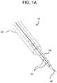
- FIG. 1Ashows an embodiment of an engagement catheter and an embodiment of a delivery catheter as disclosed herein;
- FIG. 1Bshows a percutaneous intravascular pericardial delivery using another embodiment of an engagement catheter and another embodiment of a delivery catheter as disclosed herein;
- FIG. 2Ashows a percutaneous intravascular technique for accessing the pericardial space through a right atrial wall or atrial appendage using the engagement and delivery catheters shown in FIG. 1A ;
- FIG. 2Bshows the embodiment of an engagement catheter shown in FIG. 2A ;
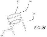
- FIG. 2Cshows another view of the distal end of the engagement catheter embodiment shown in FIGS. 2A and 2B ;
- FIG. 3Ashows removal of an embodiment of a catheter as disclosed herein
- FIG. 3Bshows the resealing of a puncture according to an embodiment as disclosed herein;
- FIG. 4A to 4Cshow a closure of a hole in the atrial wall using an embodiment as disclosed herein;
- FIG. 5Ashows an embodiment of an engagement catheter as disclosed herein
- FIG. 5Bshows a cross-sectional view of the proximal end of the engagement catheter shown in FIG. 5A ;
- FIG. 5Cshows a cross-sectional view of the distal end of the engagement catheter shown in FIG. 5A ;
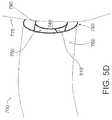
- FIG. 5Dshows the engagement catheter shown in FIG. 5A approaching a heart wall from inside of the heart
- FIG. 6Ashows an embodiment of a delivery catheter as disclosed herein
- FIG. 6Bshows a close-up view of the needle shown in FIG. 6A ;
- FIG. 6Cshows a cross-sectional view of the needle shown in FIGS. 6A and 6B .
- the disclosed embodimentsinclude devices, systems, and methods useful for accessing various tissues of the heart from inside the heart.
- various embodimentsprovide for percutaneous, intravascular access into the pericardial space through an atrial wall or the wall of an atrial appendage.
- the heart wallis aspirated and retracted from the pericardial sac to increase the pericardial space between the heart and the sac and thereby facilitate access into the space.
- the atrial wall and atrial appendageare rather soft and deformable.
- suction of the atrial wall or atrial appendagecan provide significantly more clearance of the cardiac structure from the pericardium as compared to suction of the pericardium.
- navigation from the intravascular region (inside of the heart)provides more certainty of position of vital cardiac structures than does intrathoracic access (outside of the heart).
- Access to the pericardial spacemay be used for identification of diagnostic markers in the pericardial fluid; for pericardiocentesis; and for administration of therapeutic factors with angiogenic, myogenic, and antiarrhythmic potential.
- epicardial pacing leadsmay be delivered via the pericardial space, and an ablation catheter may be used on the epicardial tissue from the pericardial space.
- catheter system 10includes an engagement catheter 20 , a delivery catheter 30 , and a needle 40 .
- engagement catheter 20has a lumen through which delivery catheter 30 has been inserted
- delivery catheter 30has a lumen through which needle 40 has been inserted.
- Delivery catheter 30also has a number of openings 50 that can be used to transmit fluid from the lumen of the catheter to the heart tissue in close proximity to the distal end of the catheter.
- engagement catheter 20includes a vacuum channel 60 used for suction of a targeted tissue 65 in the heart and an injection channel 70 used for infusion of substances to targeted tissue 65 , including, for example, a biological or non-biological degradable adhesive.
- injection channel 70is ring-shaped, which tends to provide relatively even dispersal of the infused substance over the targeted tissue, but other shapes of injection channels may be suitable.
- a syringe 80is attached to injection channel 70 for delivery of the appropriate substances to injection channel 70
- a syringe 90is attached to vacuum channel 60 through a vacuum port (not shown) at the proximal end of engagement catheter 20 to provide appropriate suction through vacuum channel 60 .
- a suction port 95is attached to vacuum channel 60 for contacting targeted tissue 65 , such that suction port 95 surrounds targeted tissue 65 , which is thereby encompassed within the circumference of suction port 95 .
- syringe 90is shown in FIG. 2B as the vacuum source providing suction for engagement catheter 20 , other types of vacuum sources may be used, such as a controlled vacuum system providing specific suction pressures.
- syringe 80serves as the external fluid source in the embodiment shown in FIG. 2B , but other external fluid sources may be used.
- a route of entry for use of various embodiments disclosed hereinis through the jugular or femoral vein to the superior or inferior vena cavae, respectively, to the right atrial wall or atrial appendage (percutaneously) to the pericardial sac (through puncture).
- an engagement catheter 100is placed via standard approach into the jugular or femoral vein.
- the catheterwhich may be 4 or 5 Fr., is positioned under fluoroscopic or echocardiographic guidance into the right atrial appendage 110 .
- Suctionis initiated to aspirate a portion of atrial appendage 110 away from the pericardial sac 120 that surrounds the heart.
- aspiration of the heart tissueis evidenced when no blood can be pulled back through engagement catheter 100 and, if suction pressure is being measured, when the suction pressure gradually increases.
- a delivery catheter 130is then inserted through a lumen of engagement catheter 100 .
- a small perforationcan be made in the aspirated atrial appendage 110 with a needle such as needle 40 , as shown in FIGS. 1A and 2A .
- a guide wire(not shown) can then be advanced through delivery catheter 130 into the pericardial space to secure the point of entry 125 through the atrial appendage and guide further insertion of delivery catheter 130 or another catheter.
- Flouroscopy or echocardiogramcan be used to confirm the position of the catheter in the pericardial space.
- a pressure tip needlecan sense the pressure and measure the pressure change from the atrium (about 10 mmHg) to the pericardial space (about 2 mmHg). This is particularly helpful for transseptal access where puncture of arterial structures (e.g., the aorta) can be diagnosed and sealed with an adhesive, as described in more detail below.
- CO2 gascan be delivered through a catheter, such as delivery catheter 130 , into the pericardial space to create additional space between the pericardial sac and the heart surface.
- the catheter system shown in FIG. 1Bis retrieved by pull back through the route of entry.
- the puncture of the targeted tissue in the hearte.g., the right atrial appendage as shown in FIG. 3A
- the retrieval of the cathetermay be combined with a sealing of the tissue in one of several ways: (1) release of a tissue adhesive or polymer 75 via injection channel 70 to seal off the puncture hole, as shown in FIG.
- closuremay be accomplished by using, for example, a biodegradable adhesive material (e.g., fibrin glue or cyanomethacrylate), a magnetic system, or an umbrella-shaped nitinol stent.
- a biodegradable adhesive materiale.g., fibrin glue or cyanomethacrylate
- a magnetic systeme.g., a magnetospine
- an umbrella-shaped nitinol stente.g., a magnetic system
- FIG. 3BAn example of the closure of a hole in the atrium is shown in FIG. 3B .
- Engagement catheter 20is attached to targeted tissue 95 using suction through suction port 60 .
- Tissue adhesive 75is injected through injection channel 70 to coat and seal the puncture wound in targeted tissue 95 .
- Engagement catheter 20is then withdrawn, leaving a plug of tissue adhesive 75 attached to the atrial wall or atrial appendage.
- FIGS. 4A, 4B, and 4COther examples for sealing the puncture wound in the atrial wall or appendage are shown in FIGS. 4A, 4B, and 4C .
- a sandwich-type closure memberhaving an external cover 610 and an internal cover 620 , is inserted through the lumen of engagement catheter 600 , which is attached to the targeted tissue of an atrial wall 630 .
- Each of external and internal covers 610 and 620is similar to an umbrella in that it can be inserted through a catheter in its folded configuration and expanded once it is outside of the catheter.
- external cover 610is deployed (in its expanded configuration) on the outside of the atrial wall to seal a puncture wound in the targeted tissue.
- Internal cover 620is delivered through engagement catheter 600 (in its folded configuration), as shown in FIGS.
- FIGS. 5A, 5B, 5C, and 5Dshow another embodiment of an engagement catheter as disclosed herein.
- Engagement catheter 700is an elongated tube having a proximal end 710 and a distal end 720 , as well as two lumens 730 , 740 extending between proximal end 710 and distal end 720 .
- Lumens 730 , 740are formed by concentric inner wall 750 and outer wall 760 , as particularly shown in FIGS. 5B and 5C .
- engagement catheter 700includes a vacuum port 770 , which is attached to lumen 730 so that a vacuum source can be attached to vacuum port 770 to create suction in lumen 730 , thereby forming a suction channel.
- a suction port 780is attached to lumen 730 so that suction port 780 can be placed in contact with heart tissue 775 (see FIG. 5D ) for aspirating the tissue, thereby forming a vacuum seal between suction port 780 and tissue 775 when the vacuum source is attached and engaged.
- the vacuum sealenables suction port 780 to grip, stabilize, and retract tissue 775 .
- attaching a suction port to an interior atrial wall using a vacuum sourceenables the suction port to retract the atrial wall from the pericardial sac surrounding the heart, which enlarges the pericardial space between the atrial wall and the pericardial sac.
- two internal lumen supports 810 , 820are located within lumen 730 and are attached to inner wall 750 and outer wall 760 to provide support to the walls. These lumen supports divide lumen 730 into two suction channels. Although internal lumen supports 810 , 820 extend from distal end 720 of catheter 700 along a substantial portion of the length of catheter 700 , internal lumen supports 810 , 820 may or may not span the entire length of catheter 700 . Indeed, as shown in FIGS. 5A, 5B, and 5C , internal lumen supports 810 , 820 do not extend to proximal end 710 to ensure that the suction from the external vacuum source is distributed relatively evenly around the circumference of catheter 700 . Although the embodiment shown in FIG. 5C includes two internal lumen supports, other embodiments may have just one internal support or even three or more such supports.
- FIG. 5Dshows engagement catheter 700 approaching heart tissue 775 for attachment thereto. It is important for the clinician performing the procedure to know when the suction port has engaged the tissue of the atrial wall or the atrial appendage. For example, in reference to FIG. 5D , it is clear that suction port 780 has not fully engaged tissue 775 such that a seal is formed. However, because suction port 780 is not usually seen during the procedure, the clinician may determine when the proper vacuum seal between the atrial tissue and the suction port has been made by monitoring the amount of blood that is aspirated, by monitoring the suction pressure with a pressure sensor/regulator, or both.
- the suctioncan be activated through lumen 730 .
- a certain level of suctione.g., 10 mmHg
- a pressure sensor/regulatorAs long as catheter 700 does not engage the wall, some blood will be aspirated into the catheter and the suction pressure will remain the same. However, when catheter 700 engages or attaches to the wall of the heart (depicted as tissue 775 in FIG. 5D ), minimal blood is aspirated and the suction pressure will start to gradually increase. Each of these signs can alert the clinician (through alarm or other means) as an indication of engagement.
- the pressure regulatoris then able to maintain the suction pressure at a preset value to prevent over-suction of the tissue.
- An engagement cathetersuch as engagement catheter 700
- lumen 740 shown in FIGS. 5A and 5Cincludes an injection channel 790 at distal end 720 .
- Injection channel 790dispenses to the targeted tissue a substance flowing through lumen 740 .
- injection channel 790is the distal end of lumen 740 .
- the injection channelmay be ring-shaped (see FIG. 2C ) or have some other suitable configuration.
- Substances that can be locally administered with an engagement catheterinclude preparations for gene or cell therapy, drugs, and adhesives that are safe for use in the heart.
- the proximal end of lumen 740has a fluid port 800 , which is capable of attachment to an external fluid source for supply of the fluid to be delivered to the targeted tissue.
- an adhesivemay be administered to the targeted tissue by the engagement catheter for sealing the puncture wound left by the needle withdrawn from the targeted tissue.
- a delivery catheter 850comprising an elongated hollow tube 880 having a proximal end 860 , a distal end 870 , and a lumen 885 along the length of the catheter. Extending from distal end 870 is a hollow needle 890 in communication with lumen 885 . Needle 890 is attached to distal end 870 in the embodiment of FIGS. 6A, 6B, and 6C , but, in other embodiments, the needle may be removably attached to, or otherwise located at, the distal end of the catheter (see FIG. 1A ). In the embodiment shown in FIGS.
- the junctioni.e., site of attachment
- the clinicianwhen a clinician inserts needle 890 through an atrial wall to gain access to the pericardial space, the clinician will not, under normal conditions, unintentionally perforate the pericardial sac with needle 890 because the larger diameter of hollow tube 880 (as compared to that of needle 890 ) at security notch 910 hinders further needle insertion.
- security notch 910is formed by the junction of hollow tube 880 and needle 890 in the embodiment shown in FIGS.
- a security notchmay include a band, ring, or similar device that is attached to the needle a suitable distance from the tip of the needle.
- a security notch 910other security notch embodiments hinder insertion of the needle past the notch itself by presenting a larger profile than the profile of the needle such that the notch does not easily enter the hole in the tissue caused by entry of the needle.
- the delivery cathetercan be connected to a pressure transducer to measure pressure at the tip of the needle. Because the pressure is lower and much less pulsatile in the pericardial space than in the atrium, the clinician can recognize immediately when the needle passes through the atrial tissue into the pericardial space.
- needle 890may be connected to a strain gauge 915 as part of the catheter assembly.
- tissuenot shown
- needle 890will be deformed.
- the deformationwill be transmitted to strain gauge 915 and an electrical signal will reflect the deformation (through a classical wheatstone bridge), thereby alerting the clinician.
- Such confirmation of the puncture of the wallcan prevent over-puncture and can provide additional control of the procedure.
- a delivery cathetersuch as catheter 850 shown in FIGS. 6A, 6B , and 6 C
- an engagement cathetersuch as catheter 700 shown in FIGS. 5A, 5B, 5C , and 5 D
- engagement catheter 700may be inserted into the vascular system and advanced such that the distal end of the engagement catheter is within the atrium.
- the engagement cathetermay be attached to the targeted tissue on the interior of a wall of the atrium using a suction port as disclosed herein.
- a standard guide wiremay be inserted through the lumen of the delivery catheter as the delivery catheter is inserted through the inner lumen of the engagement catheter, such as lumen 740 shown in FIGS.
- Use of the guide wireenables more effective navigation of the delivery catheter 850 and prevents the needle 890 from damaging the inner wall 750 of the engagement catheter 700 .
- the tip of the delivery catheter with the protruding guide wirereaches the atrium, the wire is pulled back, and the needle is pushed forward to perforate the targeted tissue.
- the guide wireis then advanced through the perforation into the pericardial space, providing access to the pericardial space through the atrial wall.
- lumen 885 of delivery catheter 850may be used for delivering fluid into the pericardial space after needle 890 is inserted through the atrial wall or the atrial appendage.
- a guide wire(not shown) may be inserted through needle lumen 900 into the pericardial space to maintain access through the atrial wall or appendage. Fluid may then be introduced to the pericardial space in a number of ways. For example, after the needle punctures the atrial wall or appendage, the needle is generally withdrawn. If the needle is permanently attached to the delivery catheter, as in the embodiment shown in FIGS. 6A and 6B , then delivery catheter 850 would be withdrawn and another delivery catheter (without an attached needle) would be introduced over the guide wire into the pericardial space. Fluid may then be introduced into the pericardial space through the lumen of the second delivery catheter.
- the needleis not attached to the delivery catheter, but instead may be a needle wire (see FIG. 1A ).
- the needleis withdrawn through the lumen of the delivery catheter, and the delivery catheter may be inserted over the guide wire into the pericardial space. Fluid is then introduced into the pericardial space through the lumen of the delivery catheter.
- the various embodiments disclosed hereinmay be used by clinicians, for example: (1) to deliver genes, cells, drugs, etc.; (2) to provide catheter access for epicardial stimulation; (3) to evacuate fluids acutely (e.g., in cases of pericardial tamponade) or chronically (e.g., to alleviate effusion caused by chronic renal disease, cancer, etc.); (4) to perform transeptal puncture and delivery of a catheter through the left atrial appendage for electrophysiological therapy, biopsy, etc.; (5) to deliver a magnetic glue or ring through the right atrial appendage to the aortic root to hold a percutaneous aortic valve in place; (6) to deliver a catheter for tissue ablation, e.g., to the pulmonary veins, or right atrial and epicardial surface of the heart for atrial and ventricular arrhythmias; (7) to deliver and place epicardial, right atrial, and right and left ventricle pacing leads; (8) to occlude the left atrial appendage
- the disclosuremay have presented a method and/or process as a particular sequence of steps.
- the method or processshould not be limited to the particular sequence of steps described.
- Other sequences of stepsmay be possible. Therefore, the particular order of the steps disclosed herein should not be construed as limitations of the present disclosure.
- disclosure directed to a method and/or processshould not be limited to the performance of their steps in the order written. Such sequences may be varied and still remain within the scope of the present disclosure.
Landscapes
- Health & Medical Sciences (AREA)
- Life Sciences & Earth Sciences (AREA)
- Heart & Thoracic Surgery (AREA)
- General Health & Medical Sciences (AREA)
- Public Health (AREA)
- Biomedical Technology (AREA)
- Engineering & Computer Science (AREA)
- Veterinary Medicine (AREA)
- Animal Behavior & Ethology (AREA)
- Surgery (AREA)
- Nuclear Medicine, Radiotherapy & Molecular Imaging (AREA)
- Hematology (AREA)
- Anesthesiology (AREA)
- Molecular Biology (AREA)
- Biophysics (AREA)
- Pulmonology (AREA)
- Medical Informatics (AREA)
- Cardiology (AREA)
- Radiology & Medical Imaging (AREA)
- Vascular Medicine (AREA)
- Surgical Instruments (AREA)
- External Artificial Organs (AREA)
- Endoscopes (AREA)
Abstract
Description
Claims (19)
Priority Applications (2)
| Application Number | Priority Date | Filing Date | Title |
|---|---|---|---|
| US15/913,446US11191955B2 (en) | 2006-06-30 | 2018-03-06 | Devices and systems for accessing cardiac tissue |
| US17/544,731US20220088377A1 (en) | 2006-06-30 | 2021-12-07 | Devices and systems for accessing cardiac tissue |
Applications Claiming Priority (7)
| Application Number | Priority Date | Filing Date | Title |
|---|---|---|---|
| US81742106P | 2006-06-30 | 2006-06-30 | |
| US91445207P | 2007-04-27 | 2007-04-27 | |
| PCT/US2007/015207WO2008010905A2 (en) | 2006-06-30 | 2007-06-29 | Percutaneous intravascular access to cardiac tissue |
| US30586408A | 2008-12-19 | 2008-12-19 | |
| US12/816,655US8894606B2 (en) | 2006-06-30 | 2010-06-16 | Devices, systems, and methods for accessing cardiac tissue |
| US14/552,708US9907954B2 (en) | 2006-06-30 | 2014-11-25 | Devices and systems for accessing cardiac tissue |
| US15/913,446US11191955B2 (en) | 2006-06-30 | 2018-03-06 | Devices and systems for accessing cardiac tissue |
Related Parent Applications (1)
| Application Number | Title | Priority Date | Filing Date |
|---|---|---|---|
| US14/552,708ContinuationUS9907954B2 (en) | 2006-06-30 | 2014-11-25 | Devices and systems for accessing cardiac tissue |
Related Child Applications (1)
| Application Number | Title | Priority Date | Filing Date |
|---|---|---|---|
| US17/544,731ContinuationUS20220088377A1 (en) | 2006-06-30 | 2021-12-07 | Devices and systems for accessing cardiac tissue |
Publications (2)
| Publication Number | Publication Date |
|---|---|
| US20180193635A1 US20180193635A1 (en) | 2018-07-12 |
| US11191955B2true US11191955B2 (en) | 2021-12-07 |
Family
ID=38957265
Family Applications (11)
| Application Number | Title | Priority Date | Filing Date |
|---|---|---|---|
| US12/305,864AbandonedUS20100069849A1 (en) | 2006-06-30 | 2007-06-29 | Percutaneous intravascular access to cardiac tissue |
| US12/722,287AbandonedUS20100168791A1 (en) | 2006-06-30 | 2010-03-11 | Systems and methods for closing a hole in cardiac tissue |
| US12/722,913Expired - Fee RelatedUS8128593B2 (en) | 2006-06-30 | 2010-03-12 | Removing fluid from a bodily tissue via a catheter with circumferential concave grooves |
| US12/723,278Expired - Fee RelatedUS8303481B2 (en) | 2006-06-30 | 2010-03-12 | Devices and methods for assisting heart function |
| US12/816,655Active2027-10-20US8894606B2 (en) | 2006-06-30 | 2010-06-16 | Devices, systems, and methods for accessing cardiac tissue |
| US13/323,174Active2027-08-29US8876776B2 (en) | 2006-06-30 | 2011-12-12 | Engagement catheter systems and devices and methods of using the same |
| US13/539,574ActiveUS8777904B2 (en) | 2006-06-30 | 2012-07-02 | Systems and methods for engaging heart tissue |
| US14/332,064AbandonedUS20170080218A9 (en) | 2006-06-30 | 2014-07-15 | Engagement and delivery catheter systems |
| US14/552,708Active2028-06-03US9907954B2 (en) | 2006-06-30 | 2014-11-25 | Devices and systems for accessing cardiac tissue |
| US15/913,446Active2029-03-05US11191955B2 (en) | 2006-06-30 | 2018-03-06 | Devices and systems for accessing cardiac tissue |
| US17/544,731AbandonedUS20220088377A1 (en) | 2006-06-30 | 2021-12-07 | Devices and systems for accessing cardiac tissue |
Family Applications Before (9)
| Application Number | Title | Priority Date | Filing Date |
|---|---|---|---|
| US12/305,864AbandonedUS20100069849A1 (en) | 2006-06-30 | 2007-06-29 | Percutaneous intravascular access to cardiac tissue |
| US12/722,287AbandonedUS20100168791A1 (en) | 2006-06-30 | 2010-03-11 | Systems and methods for closing a hole in cardiac tissue |
| US12/722,913Expired - Fee RelatedUS8128593B2 (en) | 2006-06-30 | 2010-03-12 | Removing fluid from a bodily tissue via a catheter with circumferential concave grooves |
| US12/723,278Expired - Fee RelatedUS8303481B2 (en) | 2006-06-30 | 2010-03-12 | Devices and methods for assisting heart function |
| US12/816,655Active2027-10-20US8894606B2 (en) | 2006-06-30 | 2010-06-16 | Devices, systems, and methods for accessing cardiac tissue |
| US13/323,174Active2027-08-29US8876776B2 (en) | 2006-06-30 | 2011-12-12 | Engagement catheter systems and devices and methods of using the same |
| US13/539,574ActiveUS8777904B2 (en) | 2006-06-30 | 2012-07-02 | Systems and methods for engaging heart tissue |
| US14/332,064AbandonedUS20170080218A9 (en) | 2006-06-30 | 2014-07-15 | Engagement and delivery catheter systems |
| US14/552,708Active2028-06-03US9907954B2 (en) | 2006-06-30 | 2014-11-25 | Devices and systems for accessing cardiac tissue |
Family Applications After (1)
| Application Number | Title | Priority Date | Filing Date |
|---|---|---|---|
| US17/544,731AbandonedUS20220088377A1 (en) | 2006-06-30 | 2021-12-07 | Devices and systems for accessing cardiac tissue |
Country Status (7)
| Country | Link |
|---|---|
| US (11) | US20100069849A1 (en) |
| EP (1) | EP2035074A4 (en) |
| JP (1) | JP2009542338A (en) |
| AU (1) | AU2007275844B2 (en) |
| CA (1) | CA2656341C (en) |
| NZ (2) | NZ599528A (en) |
| WO (1) | WO2008010905A2 (en) |
Families Citing this family (110)
| Publication number | Priority date | Publication date | Assignee | Title |
|---|---|---|---|---|
| EP2015681B1 (en) | 2006-05-03 | 2018-03-28 | Datascope Corp. | Tissue closure device |
| US10238311B2 (en) | 2007-01-23 | 2019-03-26 | Cvdevices, Llc | Devices, systems, and methods to evaluate cardiovascular function |
| WO2008091610A2 (en) | 2007-01-23 | 2008-07-31 | Cvdevices, Llc | Systems and methods for epicardial navigation |
| WO2008118737A1 (en) | 2007-03-22 | 2008-10-02 | University Of Virginia Patent Foundation | Electrode catheter for ablation purposes and related method thereof |
| US10166066B2 (en)* | 2007-03-13 | 2019-01-01 | University Of Virginia Patent Foundation | Epicardial ablation catheter and method of use |
| SI2142107T1 (en) | 2007-03-30 | 2013-05-31 | Sentreheart, Inc. | Devices and systems for closing the left atrial appendage |
| US9050064B2 (en)* | 2007-04-27 | 2015-06-09 | Cvdevices, Llc | Systems for engaging a bodily tissue and methods of using the same |
| US20100241185A1 (en)* | 2007-11-09 | 2010-09-23 | University Of Virginia Patent Foundation | Steerable epicardial pacing catheter system placed via the subxiphoid process |
| AU2010232589B2 (en) | 2009-04-01 | 2014-11-27 | Atricure, Inc. | Tissue ligation devices and controls therefor |
| US9642534B2 (en) | 2009-09-11 | 2017-05-09 | University Of Virginia Patent Foundation | Systems and methods for determining location of an access needle in a subject |
| US9307980B2 (en) | 2010-01-22 | 2016-04-12 | 4Tech Inc. | Tricuspid valve repair using tension |
| US8475525B2 (en) | 2010-01-22 | 2013-07-02 | 4Tech Inc. | Tricuspid valve repair using tension |
| US10058323B2 (en) | 2010-01-22 | 2018-08-28 | 4 Tech Inc. | Tricuspid valve repair using tension |
| US8961596B2 (en) | 2010-01-22 | 2015-02-24 | 4Tech Inc. | Method and apparatus for tricuspid valve repair using tension |
| EP4183441A1 (en) | 2010-02-26 | 2023-05-24 | The Board of Trustees of the Leland Stanford Junior University | Systems for endoluminal valve creation |
| CA2796347A1 (en)* | 2010-04-13 | 2011-10-20 | Sentreheart, Inc. | Methods and devices for pericardial access |
| US11419632B2 (en) | 2010-04-23 | 2022-08-23 | Mark D. Wieczorek, P.C. | Transseptal access device and method of use |
| WO2011133977A2 (en) | 2010-04-23 | 2011-10-27 | Assist Medical Llc | Transseptal access device and method of use |
| US10220134B2 (en) | 2010-04-23 | 2019-03-05 | Mark D. Wieczorek | Transseptal access device and method of use |
| WO2012033847A1 (en) | 2010-09-07 | 2012-03-15 | Spence Paul A | Cannula systems and methods |
| EP2624776B1 (en) | 2010-10-05 | 2021-04-28 | Emory University | Devices and systems for improving access to cardiac and vascular chambers |
| EP4335393A3 (en) | 2011-04-20 | 2024-06-05 | The Board of Trustees of the Leland Stanford Junior University | Systems for endoluminal valve creation |
| EP2734157B1 (en) | 2011-07-21 | 2018-09-05 | 4Tech Inc. | Apparatus for tricuspid valve repair using tension |
| EP4101399B1 (en) | 2011-08-05 | 2025-04-09 | Route 92 Medical, Inc. | System for treatment of acute ischemic stroke |
| US10779855B2 (en) | 2011-08-05 | 2020-09-22 | Route 92 Medical, Inc. | Methods and systems for treatment of acute ischemic stroke |
| US10307167B2 (en) | 2012-12-14 | 2019-06-04 | Corquest Medical, Inc. | Assembly and method for left atrial appendage occlusion |
| US10314594B2 (en) | 2012-12-14 | 2019-06-11 | Corquest Medical, Inc. | Assembly and method for left atrial appendage occlusion |
| US10813630B2 (en) | 2011-08-09 | 2020-10-27 | Corquest Medical, Inc. | Closure system for atrial wall |
| EP2765907B1 (en) | 2011-10-14 | 2016-05-18 | Acist Medical Systems, Inc. | Device for measuring an anatomical structure |
| JP5984372B2 (en)* | 2011-12-09 | 2016-09-06 | オリンパス株式会社 | Pericardial fluid volume control system |
| EP3628247B1 (en) | 2012-02-07 | 2022-08-10 | Intervene, Inc. | System for endoluminal valve creation |
| US9549679B2 (en) | 2012-05-14 | 2017-01-24 | Acist Medical Systems, Inc. | Multiple transducer delivery device and method |
| US9244196B2 (en) | 2012-05-25 | 2016-01-26 | Johnson & Johnson Vision Care, Inc. | Polymers and nanogel materials and methods for making and using the same |
| US8961594B2 (en) | 2012-05-31 | 2015-02-24 | 4Tech Inc. | Heart valve repair system |
| EP2908880B1 (en) | 2012-10-16 | 2018-12-05 | Paul A. Spence | Devices for facilitating flow from the heart to a blood pump |
| US20140142689A1 (en) | 2012-11-21 | 2014-05-22 | Didier De Canniere | Device and method of treating heart valve malfunction |
| CN105007832B (en) | 2013-01-09 | 2018-01-23 | 4科技有限公司 | Organize ancora equipment |
| WO2014110460A1 (en) | 2013-01-10 | 2014-07-17 | Intervene, Inc. | Systems and methods for endoluminal valve creation |
| US9131932B2 (en)* | 2013-02-01 | 2015-09-15 | St. Jude Medical Puerto Rico Llc | Dual lumen carrier tube with retractable sleeve and methods |
| US9907681B2 (en) | 2013-03-14 | 2018-03-06 | 4Tech Inc. | Stent with tether interface |
| US9320841B2 (en)* | 2013-06-21 | 2016-04-26 | Corvivo, Inc. | Ventricular assist device |
| US9814816B2 (en) | 2013-06-21 | 2017-11-14 | Corvivo, Inc. | Artificial ventricles |
| WO2015048565A2 (en) | 2013-09-27 | 2015-04-02 | Intervene, Inc. | Visualization devices, systems, and methods for informing intravascular procedures on blood vessel valves |
| US10022114B2 (en) | 2013-10-30 | 2018-07-17 | 4Tech Inc. | Percutaneous tether locking |
| WO2015063580A2 (en) | 2013-10-30 | 2015-05-07 | 4Tech Inc. | Multiple anchoring-point tension system |
| US10052095B2 (en) | 2013-10-30 | 2018-08-21 | 4Tech Inc. | Multiple anchoring-point tension system |
| WO2015077356A1 (en) | 2013-11-19 | 2015-05-28 | Wheeler William K | Fastener applicator with interlock |
| US9566443B2 (en) | 2013-11-26 | 2017-02-14 | Corquest Medical, Inc. | System for treating heart valve malfunction including mitral regurgitation |
| US9265512B2 (en) | 2013-12-23 | 2016-02-23 | Silk Road Medical, Inc. | Transcarotid neurovascular catheter |
| HRP20241397T1 (en) | 2014-01-10 | 2024-12-20 | Bayer Healthcare Llc | CONNECTOR FOR DISPOSABLE KIT |
| US9962519B2 (en)* | 2014-01-14 | 2018-05-08 | The Charles Stark Draper Laboratory, Inc. | Seeping flow anti-clotting blood catheter |
| CA2939929C (en) | 2014-02-28 | 2023-04-11 | Sentreheart, Inc. | Pericardial access devices and methods |
| US9511219B1 (en)* | 2014-03-24 | 2016-12-06 | Subhajit Datta | Dual vacuum device for medical fixture placement including for thoracoscopic left ventricular lead placement |
| WO2015148581A1 (en) | 2014-03-24 | 2015-10-01 | Intervene, Inc. | Devices, systems, and methods for controlled hydrodissection of vessel walls |
| WO2015193728A2 (en) | 2014-06-19 | 2015-12-23 | 4Tech Inc. | Cardiac tissue cinching |
| EP3200846B1 (en) | 2014-10-01 | 2020-01-15 | Heartware, Inc. | Backup controller system with updating |
| US9907547B2 (en) | 2014-12-02 | 2018-03-06 | 4Tech Inc. | Off-center tissue anchors |
| JP5945586B2 (en)* | 2014-12-08 | 2016-07-05 | オリンパス株式会社 | Medical device guide system |
| US10842626B2 (en) | 2014-12-09 | 2020-11-24 | Didier De Canniere | Intracardiac device to correct mitral regurgitation |
| WO2016100574A2 (en) | 2014-12-16 | 2016-06-23 | Intervene, Inc. | Intravascular devices, systems, and methods for the controlled dissection of body lumens |
| US10507319B2 (en) | 2015-01-09 | 2019-12-17 | Bayer Healthcare Llc | Multiple fluid delivery system with multi-use disposable set and features thereof |
| WO2016118616A1 (en) | 2015-01-20 | 2016-07-28 | Talon Medical, LLC | Tissue engagement devices, systems, and methods |
| EP4241820B1 (en)* | 2015-01-23 | 2025-07-09 | Terumo Kabushiki Kaisha | Guide wire |
| US11065019B1 (en) | 2015-02-04 | 2021-07-20 | Route 92 Medical, Inc. | Aspiration catheter systems and methods of use |
| US10426497B2 (en) | 2015-07-24 | 2019-10-01 | Route 92 Medical, Inc. | Anchoring delivery system and methods |
| CN119949953A (en) | 2015-02-04 | 2025-05-09 | 92号医疗公司 | Intravascular access system, dilator and system including dilator |
| US11173278B2 (en) | 2015-05-15 | 2021-11-16 | The Usa, As Represented By The Secretary, Dept. Of Health And Human Services | Three-dimensional right atrial appendage curve catheter |
| US10646247B2 (en) | 2016-04-01 | 2020-05-12 | Intervene, Inc. | Intraluminal tissue modifying systems and associated devices and methods |
| TWI651107B (en) | 2016-06-15 | 2019-02-21 | 拜耳保健公司 | Multi-use disposable system and syringe therefor |
| CN106215261B (en)* | 2016-08-29 | 2018-10-02 | 安徽通灵仿生科技有限公司 | A kind of ventricle subsidiary conduit pump |
| JP2019531787A (en) | 2016-08-30 | 2019-11-07 | ザ リージェンツ オブ ザ ユニバーシティ オブ カリフォルニア | Biomedical targeting and delivery method and apparatus and system for performing the same |
| PL3515327T3 (en) | 2016-09-23 | 2024-06-10 | Atricure, Inc. | DEVICES FOR CLOSING THE LEFT ATRIAL APPEARANCE |
| US11266810B2 (en) | 2016-10-10 | 2022-03-08 | Clph, Llc | Isolation and attachment catheters and methods for using them |
| CN110799236A (en)* | 2016-10-25 | 2020-02-14 | 玛芬股份有限公司 | Vascular anchoring introducer sheath |
| CN110392591B (en) | 2017-01-10 | 2022-06-03 | 92号医疗公司 | Aspiration catheter system and method of use |
| US10864350B2 (en) | 2017-01-20 | 2020-12-15 | Route 92 Medical, Inc. | Single operator intracranial medical device delivery systems and methods of use |
| CN111132626B (en) | 2017-07-17 | 2024-01-30 | 沃雅戈治疗公司 | track array guidance system |
| US12402885B2 (en) | 2017-09-23 | 2025-09-02 | Universität Zürich | Medical occlusion device |
| EP3459469A1 (en) | 2017-09-23 | 2019-03-27 | Universität Zürich | Medical occluder device |
| US10806579B2 (en) | 2017-10-20 | 2020-10-20 | Boston Scientific Scimed, Inc. | Heart valve repair implant for treating tricuspid regurgitation |
| US11191547B2 (en) | 2018-01-26 | 2021-12-07 | Syntheon 2.0, LLC | Left atrial appendage clipping device and methods for clipping the LAA |
| WO2019154847A1 (en)* | 2018-02-06 | 2019-08-15 | Septulus Ab | Negative pressure-based gripping system and method |
| US20210030950A1 (en)* | 2018-02-19 | 2021-02-04 | Bayer Healthcare Llc | Syringe rolling apparatus and method |
| EP3768160B1 (en) | 2018-03-23 | 2023-06-07 | Medtronic, Inc. | Vfa cardiac therapy for tachycardia |
| JP2021518192A (en) | 2018-03-23 | 2021-08-02 | メドトロニック,インコーポレイテッド | VfA cardiac resynchronization therapy |
| CN111886046B (en) | 2018-03-23 | 2025-05-27 | 美敦力公司 | AV-synchronized VFA cardiac therapy |
| JP2021519143A (en) | 2018-03-27 | 2021-08-10 | センターハート・インコーポレイテッドSentreHEART, Inc. | Devices and methods for left atrial appendage closure |
| JP7348199B2 (en) | 2018-03-28 | 2023-09-20 | データスコープ コーポレイション | Device for atrial appendage exclusion |
| JP7616642B2 (en) | 2018-05-17 | 2025-01-17 | ルート92メディカル・インコーポレイテッド | Suction catheter system and method of use |
| US11235161B2 (en) | 2018-09-26 | 2022-02-01 | Medtronic, Inc. | Capture in ventricle-from-atrium cardiac therapy |
| US11951313B2 (en) | 2018-11-17 | 2024-04-09 | Medtronic, Inc. | VFA delivery systems and methods |
| US12296177B2 (en) | 2018-12-21 | 2025-05-13 | Medtronic, Inc. | Delivery systems and methods for left ventricular pacing |
| US11679265B2 (en) | 2019-02-14 | 2023-06-20 | Medtronic, Inc. | Lead-in-lead systems and methods for cardiac therapy |
| US11697025B2 (en) | 2019-03-29 | 2023-07-11 | Medtronic, Inc. | Cardiac conduction system capture |
| US11213676B2 (en)* | 2019-04-01 | 2022-01-04 | Medtronic, Inc. | Delivery systems for VfA cardiac therapy |
| US10925615B2 (en) | 2019-05-03 | 2021-02-23 | Syntheon 2.0, LLC | Recapturable left atrial appendage clipping device and methods for recapturing a left atrial appendage clip |
| US11712188B2 (en) | 2019-05-07 | 2023-08-01 | Medtronic, Inc. | Posterior left bundle branch engagement |
| CN112120741A (en)* | 2019-06-25 | 2020-12-25 | 杭州德柯医疗科技有限公司 | Adsorption head and adsorption device |
| US11305127B2 (en) | 2019-08-26 | 2022-04-19 | Medtronic Inc. | VfA delivery and implant region detection |
| US20210138239A1 (en) | 2019-09-25 | 2021-05-13 | Swift Sync, Llc | Transvenous Intracardiac Pacing Catheter |
| JP7531236B2 (en) | 2019-09-26 | 2024-08-09 | ウニベルシタット チューリッヒ | Left atrial appendage closure device |
| US11497431B2 (en) | 2019-10-09 | 2022-11-15 | Medtronic, Inc. | Systems and methods for configuring cardiac therapy |
| US11642533B2 (en) | 2019-11-04 | 2023-05-09 | Medtronic, Inc. | Systems and methods for evaluating cardiac therapy |
| US11813466B2 (en) | 2020-01-27 | 2023-11-14 | Medtronic, Inc. | Atrioventricular nodal stimulation |
| JP7634552B2 (en)* | 2020-03-20 | 2025-02-21 | バード・ペリフェラル・バスキュラー・インコーポレーテッド | SEALANT INJECTION NEEDLE ASSEMBLY AND SEALANT DELIVERY DEVICE FOR USE IN PULMONARY ACCESS PROCEDURES - Patent application |
| US11813464B2 (en) | 2020-07-31 | 2023-11-14 | Medtronic, Inc. | Cardiac conduction system evaluation |
| US11857417B2 (en) | 2020-08-16 | 2024-01-02 | Trilio Medical Ltd. | Leaflet support |
| WO2022076893A1 (en) | 2020-10-09 | 2022-04-14 | Route 92 Medical, Inc. | Aspiration catheter systems and methods of use |
| WO2022140784A1 (en)* | 2020-12-22 | 2022-06-30 | Nxt Biomedical, Llc | Surgical drain methods and articles |
| US12151099B2 (en) | 2021-01-20 | 2024-11-26 | Swift Sync, Inc. | Transvenous intracardiac pacing catheter having improved leads |
Citations (41)
| Publication number | Priority date | Publication date | Assignee | Title |
|---|---|---|---|---|
| US3583404A (en) | 1969-06-23 | 1971-06-08 | Kendall & Co | Nonblocking catheter |
| US3630207A (en) | 1969-08-08 | 1971-12-28 | Cutter Lab | Pericardial catheter |
| US4946457A (en) | 1987-12-03 | 1990-08-07 | Dimed, Incorporated | Defibrillator system with cardiac leads and method for transvenous implantation |
| US4991578A (en)* | 1989-04-04 | 1991-02-12 | Siemens-Pacesetter, Inc. | Method and system for implanting self-anchoring epicardial defibrillation electrodes |
| US5195968A (en) | 1990-02-02 | 1993-03-23 | Ingemar Lundquist | Catheter steering mechanism |
| US5292332A (en) | 1992-07-27 | 1994-03-08 | Lee Benjamin I | Methods and device for percutanceous sealing of arterial puncture sites |
| US5715817A (en) | 1993-06-29 | 1998-02-10 | C.R. Bard, Inc. | Bidirectional steering catheter |
| US5968010A (en)* | 1997-04-30 | 1999-10-19 | Beth Israel Deaconess Medical Center, Inc. | Method for transvenously accessing the pericardial space via the right atrium |
| US5972013A (en)* | 1997-09-19 | 1999-10-26 | Comedicus Incorporated | Direct pericardial access device with deflecting mechanism and method |
| US6113611A (en) | 1998-05-28 | 2000-09-05 | Advanced Vascular Technologies, Llc | Surgical fastener and delivery system |
| US6200303B1 (en) | 1997-04-30 | 2001-03-13 | Beth Israel Deaconess Medical Center, Inc. | Method and kit for transvenously accessing the pericardial space via the right atrium |
| US6338345B1 (en) | 1999-04-07 | 2002-01-15 | Endonetics, Inc. | Submucosal prosthesis delivery device |
| US20020072768A1 (en) | 2000-12-07 | 2002-06-13 | Ginn Richard S. | Apparatus and methods for providing tactile feedback while delivering a closure device |
| US20020091354A1 (en) | 1997-09-23 | 2002-07-11 | Navia Jose Antonio | Intraluminal catheter with expandable tubular open-walled element |
| US20020165561A1 (en) | 2001-05-01 | 2002-11-07 | Stephen Ainsworth | Self-closing surgical clip for tissue |
| US20020168317A1 (en) | 2000-03-03 | 2002-11-14 | Intramedical Imaging, Llc | Methods and devices to expand applications of intraoperative radiation probes |
| US6500167B1 (en) | 1997-09-05 | 2002-12-31 | Biosense Webster, Inc. | Omni-directional steerable catheter |
| US20030109852A1 (en) | 2001-12-11 | 2003-06-12 | Cardiac Pacemakers, Inc. | Deflectable telescoping guide catheter |
| US6595982B2 (en) | 1996-06-03 | 2003-07-22 | Terumo Kabushiki Kaisha | Tubular medical device |
| US6613062B1 (en) | 1999-10-29 | 2003-09-02 | Medtronic, Inc. | Method and apparatus for providing intra-pericardial access |
| US6626930B1 (en) | 1999-10-21 | 2003-09-30 | Edwards Lifesciences Corporation | Minimally invasive mitral valve repair method and apparatus |
| US20030225420A1 (en) | 2002-03-11 | 2003-12-04 | Wardle John L. | Surgical coils and methods of deploying |
| US6663633B1 (en) | 2000-10-25 | 2003-12-16 | Pierson, Iii Raymond H. | Helical orthopedic fixation and reduction device, insertion system, and associated methods |
| US20040010216A1 (en) | 2000-02-24 | 2004-01-15 | Zhu Yong Hua | Device for closing tissue openings |
| US20040018228A1 (en) | 2000-11-06 | 2004-01-29 | Afmedica, Inc. | Compositions and methods for reducing scar tissue formation |
| US6692458B2 (en) | 2000-12-19 | 2004-02-17 | Edwards Lifesciences Corporation | Intra-pericardial drug delivery device with multiple balloons and method for angiogenesis |
| US20040086479A1 (en) | 2001-02-26 | 2004-05-06 | Duke University | Novel dendritic polymers, crosslinked gels, and their biomedical uses |
| US6776784B2 (en) | 2001-09-06 | 2004-08-17 | Core Medical, Inc. | Clip apparatus for closing septal defects and methods of use |
| US20040230131A1 (en) | 2003-02-21 | 2004-11-18 | Kassab Ghassan S. | System and method for measuring cross-sectional areas and pressure gradients in luminal organs |
| US6837893B2 (en) | 2000-09-01 | 2005-01-04 | Onux Medical, Inc. | Multi-fastener surgical apparatus and method |
| US6890295B2 (en)* | 2002-10-31 | 2005-05-10 | Medtronic, Inc. | Anatomical space access tools and methods |
| US20050113760A1 (en) | 2003-11-24 | 2005-05-26 | Chachques Juan C. | Diagnostic and injection catheter, in particular for an application in cardiology |
| US6918890B2 (en) | 1997-09-19 | 2005-07-19 | Cecil C. Schmidt | Direct pericardial access device and method |
| US20050256450A1 (en) | 1997-11-04 | 2005-11-17 | Boston Scientific Scimed, Inc. | Catheter for the delivery of therapeutic agents to tissues |
| US6991616B2 (en) | 1998-10-02 | 2006-01-31 | Boston Scientific Scimed, Inc. | Steerable device for introducing diagnostic and therapeutic apparatus into the body |
| US7029468B2 (en) | 2002-06-25 | 2006-04-18 | Enpath Medical, Inc. | Catheter assembly with side wall exit lumen and method therefor |
| US20060106442A1 (en) | 2004-05-19 | 2006-05-18 | The Board Of Trustees Of The Leland Stanford Junior University | Devices and methods for treating cardiac pathologies |
| US7081125B2 (en) | 1997-03-12 | 2006-07-25 | Neomend, Inc. | Universal introducer |
| US20060217764A1 (en) | 2001-09-06 | 2006-09-28 | Ryan Abbott | Systems and Methods for Treating Septal Defects |
| US7326231B2 (en) | 2000-02-09 | 2008-02-05 | Anson Medical Limited | Device for the repair of arteries |
| US7942897B2 (en) | 2003-07-10 | 2011-05-17 | Boston Scientific Scimed, Inc. | System for closing an opening in a body cavity |
Family Cites Families (45)
| Publication number | Priority date | Publication date | Assignee | Title |
|---|---|---|---|---|
| US2922420A (en)* | 1957-11-29 | 1960-01-26 | Sierra Eng Co | Epidural needle |
| US4556059A (en)* | 1982-09-03 | 1985-12-03 | Adamson Jr Howard | Spring operated tracheotome |
| CA1221596A (en)* | 1984-03-09 | 1987-05-12 | David Evans | Surgical needle |
| US4552554A (en)* | 1984-06-25 | 1985-11-12 | Medi-Tech Incorporated | Introducing catheter |
| US5041109A (en)* | 1986-10-27 | 1991-08-20 | University Of Florida | Laser apparatus for the recanalization of vessels and the treatment of other cardiac conditions |
| US5114401A (en)* | 1990-02-23 | 1992-05-19 | New England Deaconess Hospital Corporation | Method for central venous catheterization |
| US5558644A (en)* | 1991-07-16 | 1996-09-24 | Heartport, Inc. | Retrograde delivery catheter and method for inducing cardioplegic arrest |
| US6572529B2 (en)* | 1993-06-17 | 2003-06-03 | Wilk Patent Development Corporation | Intrapericardial assist method |
| ATE420628T1 (en)* | 1993-07-19 | 2009-01-15 | Angiotech Pharm Inc | ANTI-ANGIogenic AGENTS AND METHODS OF USE THEREOF |
| US5360416A (en)* | 1993-09-30 | 1994-11-01 | Sherwood Medical Company | Thin-walled anesthesia needles |
| US5827216A (en)* | 1995-06-07 | 1998-10-27 | Cormedics Corp. | Method and apparatus for accessing the pericardial space |
| US6447539B1 (en)* | 1996-09-16 | 2002-09-10 | Transvascular, Inc. | Method and apparatus for treating ischemic heart disease by providing transvenous myocardial perfusion |
| US6042581A (en)* | 1996-11-08 | 2000-03-28 | Thomas J. Fogarty | Transvascular TMR device and method |
| US6511412B1 (en) | 1998-09-30 | 2003-01-28 | L. Vad Technology, Inc. | Cardivascular support control system |
| US6432039B1 (en) | 1998-12-21 | 2002-08-13 | Corset, Inc. | Methods and apparatus for reinforcement of the heart ventricles |
| US6333345B1 (en)* | 1999-05-14 | 2001-12-25 | Sepracor, Inc. | Methods of using and compositions comprising N-desmethylzolpidem |
| ATE324922T1 (en)* | 1999-06-08 | 2006-06-15 | Altea Therapeutics Corp | DEVICE FOR MICROPORATION OF A BIOLOGICAL TISSUE USING A FILM TISSUE INTERFACE DEVICE AND METHOD |
| US6241706B1 (en) | 1999-07-16 | 2001-06-05 | Datascope Investment Corporation | Fast response intra-aortic balloon pump |
| US7526342B2 (en)* | 1999-08-10 | 2009-04-28 | Maquet Cardiovascular Llc | Apparatus for endoscopic cardiac mapping and lead placement |
| US20030187461A1 (en)* | 1999-08-10 | 2003-10-02 | Chin Albert K. | Releasable guide and method for endoscopic cardiac lead placement |
| US7060200B1 (en) | 1999-09-03 | 2006-06-13 | Merck Patent Gmbh | Multireactive polymerizable mesogenic compounds |
| JP4674975B2 (en)* | 2000-05-26 | 2011-04-20 | オリンパス株式会社 | Endoscope hood |
| US6837848B2 (en)* | 2003-01-15 | 2005-01-04 | Medtronic, Inc. | Methods and apparatus for accessing and stabilizing an area of the heart |
| US7740623B2 (en)* | 2001-01-13 | 2010-06-22 | Medtronic, Inc. | Devices and methods for interstitial injection of biologic agents into tissue |
| WO2002076344A1 (en)* | 2001-03-23 | 2002-10-03 | Durect Corporation | Delivery of drugs from sustained release devices implanted in myocardial tissue or in the pericardial space |
| US6626821B1 (en)* | 2001-05-22 | 2003-09-30 | Abiomed, Inc. | Flow-balanced cardiac wrap |
| US6907298B2 (en) | 2002-01-09 | 2005-06-14 | Medtronic, Inc. | Method and apparatus for imparting curves in implantable elongated medical instruments |
| AU2003225913A1 (en) | 2002-03-18 | 2003-10-08 | Roy David Kornbluh | Electroactive polymer devices for moving fluid |
| US7903742B2 (en)* | 2002-07-15 | 2011-03-08 | Thomson Licensing | Adaptive weighting of reference pictures in video decoding |
| US7681572B2 (en) | 2002-08-20 | 2010-03-23 | Aga Ab | Method and devices for administration of therapeutic gases |
| US20050054994A1 (en) | 2002-09-25 | 2005-03-10 | Iulian Cioanta | Catheters with suction capability and related methods and systems for obtaining biosamples in vivo |
| US20050261673A1 (en)* | 2003-01-15 | 2005-11-24 | Medtronic, Inc. | Methods and apparatus for accessing and stabilizing an area of the heart |
| US7165552B2 (en)* | 2003-03-27 | 2007-01-23 | Cierra, Inc. | Methods and apparatus for treatment of patent foramen ovale |
| US7273446B2 (en)* | 2003-10-31 | 2007-09-25 | Spence Paul A | Methods, devices and systems for counterpulsation of blood flow to and from the circulatory system |
| US7186214B2 (en)* | 2004-02-12 | 2007-03-06 | Medtronic, Inc. | Instruments and methods for accessing an anatomic space |
| US7517321B2 (en)* | 2005-01-31 | 2009-04-14 | C. R. Bard, Inc. | Quick cycle biopsy system |
| JP4981690B2 (en)* | 2005-02-08 | 2012-07-25 | コーニンクレッカ フィリップス エレクトロニクス エヌ ヴィ | System and method for percutaneous lingual plastic surgery |
| US20070010793A1 (en) | 2005-06-23 | 2007-01-11 | Cardiac Pacemakers, Inc. | Method and system for accessing a pericardial space |
| WO2008134104A2 (en)* | 2007-04-27 | 2008-11-06 | Cvdevices, Llc | Devices, systems, and methods for accessing the epicardial surface of the heart |
| US8019404B2 (en) | 2006-10-06 | 2011-09-13 | The Cleveland Clinic Foundation | Apparatus and method for targeting a body tissue |
| US20080269876A1 (en)* | 2007-04-24 | 2008-10-30 | Medtronic Vascular, Inc. | Repair of Incompetent Heart Valves by Papillary Muscle Bulking |
| JP5174891B2 (en)* | 2007-04-27 | 2013-04-03 | シーヴィ デヴァイシズ,エルエルシー | Devices, systems, and methods for accessing the epicardial surface of the heart |
| US7951111B2 (en) | 2008-10-10 | 2011-05-31 | Intervalve, Inc. | Valvuloplasty catheter and methods |
| US8628552B2 (en)* | 2011-03-16 | 2014-01-14 | Pacesetter, Inc. | Apparatus and method for accessing an intrapericardial space |
| EP2908745A4 (en)* | 2012-10-22 | 2016-06-15 | Sentreheart Inc | Pericardial access devices and methods |
- 2007
- 2007-06-29NZNZ599528Apatent/NZ599528A/ennot_activeIP Right Cessation
- 2007-06-29EPEP07835937Apatent/EP2035074A4/ennot_activeWithdrawn
- 2007-06-29WOPCT/US2007/015207patent/WO2008010905A2/enactiveApplication Filing
- 2007-06-29USUS12/305,864patent/US20100069849A1/ennot_activeAbandoned
- 2007-06-29AUAU2007275844Apatent/AU2007275844B2/ennot_activeCeased
- 2007-06-29NZNZ573919Apatent/NZ573919A/ennot_activeIP Right Cessation
- 2007-06-29JPJP2009518309Apatent/JP2009542338A/enactivePending
- 2007-06-29CACA2656341Apatent/CA2656341C/enactiveActive
- 2010
- 2010-03-11USUS12/722,287patent/US20100168791A1/ennot_activeAbandoned
- 2010-03-12USUS12/722,913patent/US8128593B2/ennot_activeExpired - Fee Related
- 2010-03-12USUS12/723,278patent/US8303481B2/ennot_activeExpired - Fee Related
- 2010-06-16USUS12/816,655patent/US8894606B2/enactiveActive
- 2011
- 2011-12-12USUS13/323,174patent/US8876776B2/enactiveActive
- 2012
- 2012-07-02USUS13/539,574patent/US8777904B2/enactiveActive
- 2014
- 2014-07-15USUS14/332,064patent/US20170080218A9/ennot_activeAbandoned
- 2014-11-25USUS14/552,708patent/US9907954B2/enactiveActive
- 2018
- 2018-03-06USUS15/913,446patent/US11191955B2/enactiveActive
- 2021
- 2021-12-07USUS17/544,731patent/US20220088377A1/ennot_activeAbandoned
Patent Citations (43)
| Publication number | Priority date | Publication date | Assignee | Title |
|---|---|---|---|---|
| US3583404A (en) | 1969-06-23 | 1971-06-08 | Kendall & Co | Nonblocking catheter |
| US3630207A (en) | 1969-08-08 | 1971-12-28 | Cutter Lab | Pericardial catheter |
| US4946457A (en) | 1987-12-03 | 1990-08-07 | Dimed, Incorporated | Defibrillator system with cardiac leads and method for transvenous implantation |
| US4991578A (en)* | 1989-04-04 | 1991-02-12 | Siemens-Pacesetter, Inc. | Method and system for implanting self-anchoring epicardial defibrillation electrodes |
| US5195968A (en) | 1990-02-02 | 1993-03-23 | Ingemar Lundquist | Catheter steering mechanism |
| US5292332A (en) | 1992-07-27 | 1994-03-08 | Lee Benjamin I | Methods and device for percutanceous sealing of arterial puncture sites |
| US5715817A (en) | 1993-06-29 | 1998-02-10 | C.R. Bard, Inc. | Bidirectional steering catheter |
| US6595982B2 (en) | 1996-06-03 | 2003-07-22 | Terumo Kabushiki Kaisha | Tubular medical device |
| US7081125B2 (en) | 1997-03-12 | 2006-07-25 | Neomend, Inc. | Universal introducer |
| US6200303B1 (en) | 1997-04-30 | 2001-03-13 | Beth Israel Deaconess Medical Center, Inc. | Method and kit for transvenously accessing the pericardial space via the right atrium |
| US5968010A (en)* | 1997-04-30 | 1999-10-19 | Beth Israel Deaconess Medical Center, Inc. | Method for transvenously accessing the pericardial space via the right atrium |
| US6500167B1 (en) | 1997-09-05 | 2002-12-31 | Biosense Webster, Inc. | Omni-directional steerable catheter |
| US5972013A (en)* | 1997-09-19 | 1999-10-26 | Comedicus Incorporated | Direct pericardial access device with deflecting mechanism and method |
| US6918890B2 (en) | 1997-09-19 | 2005-07-19 | Cecil C. Schmidt | Direct pericardial access device and method |
| US20020091354A1 (en) | 1997-09-23 | 2002-07-11 | Navia Jose Antonio | Intraluminal catheter with expandable tubular open-walled element |
| US20050256450A1 (en) | 1997-11-04 | 2005-11-17 | Boston Scientific Scimed, Inc. | Catheter for the delivery of therapeutic agents to tissues |
| US6113611A (en) | 1998-05-28 | 2000-09-05 | Advanced Vascular Technologies, Llc | Surgical fastener and delivery system |
| US6991616B2 (en) | 1998-10-02 | 2006-01-31 | Boston Scientific Scimed, Inc. | Steerable device for introducing diagnostic and therapeutic apparatus into the body |
| US6338345B1 (en) | 1999-04-07 | 2002-01-15 | Endonetics, Inc. | Submucosal prosthesis delivery device |
| US6626930B1 (en) | 1999-10-21 | 2003-09-30 | Edwards Lifesciences Corporation | Minimally invasive mitral valve repair method and apparatus |
| US6613062B1 (en) | 1999-10-29 | 2003-09-02 | Medtronic, Inc. | Method and apparatus for providing intra-pericardial access |
| US7326231B2 (en) | 2000-02-09 | 2008-02-05 | Anson Medical Limited | Device for the repair of arteries |
| US7931628B2 (en) | 2000-02-24 | 2011-04-26 | Loma Linda University Medical Center | Device for closing tissue openings |
| US20040010216A1 (en) | 2000-02-24 | 2004-01-15 | Zhu Yong Hua | Device for closing tissue openings |
| US20020168317A1 (en) | 2000-03-03 | 2002-11-14 | Intramedical Imaging, Llc | Methods and devices to expand applications of intraoperative radiation probes |
| US6837893B2 (en) | 2000-09-01 | 2005-01-04 | Onux Medical, Inc. | Multi-fastener surgical apparatus and method |
| US6663633B1 (en) | 2000-10-25 | 2003-12-16 | Pierson, Iii Raymond H. | Helical orthopedic fixation and reduction device, insertion system, and associated methods |
| US20040018228A1 (en) | 2000-11-06 | 2004-01-29 | Afmedica, Inc. | Compositions and methods for reducing scar tissue formation |
| US7842068B2 (en) | 2000-12-07 | 2010-11-30 | Integrated Vascular Systems, Inc. | Apparatus and methods for providing tactile feedback while delivering a closure device |
| US20020072768A1 (en) | 2000-12-07 | 2002-06-13 | Ginn Richard S. | Apparatus and methods for providing tactile feedback while delivering a closure device |
| US6692458B2 (en) | 2000-12-19 | 2004-02-17 | Edwards Lifesciences Corporation | Intra-pericardial drug delivery device with multiple balloons and method for angiogenesis |
| US20040086479A1 (en) | 2001-02-26 | 2004-05-06 | Duke University | Novel dendritic polymers, crosslinked gels, and their biomedical uses |
| US20020165561A1 (en) | 2001-05-01 | 2002-11-07 | Stephen Ainsworth | Self-closing surgical clip for tissue |
| US6776784B2 (en) | 2001-09-06 | 2004-08-17 | Core Medical, Inc. | Clip apparatus for closing septal defects and methods of use |
| US20060217764A1 (en) | 2001-09-06 | 2006-09-28 | Ryan Abbott | Systems and Methods for Treating Septal Defects |
| US20030109852A1 (en) | 2001-12-11 | 2003-06-12 | Cardiac Pacemakers, Inc. | Deflectable telescoping guide catheter |
| US20030225420A1 (en) | 2002-03-11 | 2003-12-04 | Wardle John L. | Surgical coils and methods of deploying |
| US7029468B2 (en) | 2002-06-25 | 2006-04-18 | Enpath Medical, Inc. | Catheter assembly with side wall exit lumen and method therefor |
| US6890295B2 (en)* | 2002-10-31 | 2005-05-10 | Medtronic, Inc. | Anatomical space access tools and methods |
| US20040230131A1 (en) | 2003-02-21 | 2004-11-18 | Kassab Ghassan S. | System and method for measuring cross-sectional areas and pressure gradients in luminal organs |
| US7942897B2 (en) | 2003-07-10 | 2011-05-17 | Boston Scientific Scimed, Inc. | System for closing an opening in a body cavity |
| US20050113760A1 (en) | 2003-11-24 | 2005-05-26 | Chachques Juan C. | Diagnostic and injection catheter, in particular for an application in cardiology |
| US20060106442A1 (en) | 2004-05-19 | 2006-05-18 | The Board Of Trustees Of The Leland Stanford Junior University | Devices and methods for treating cardiac pathologies |
Non-Patent Citations (5)
| Title |
|---|
| Hertzberg, et al, "Sonographic assessment of . . .", American Journal of Roentgenology, May 1997, vol. 168, No. 5, pp. 1253-1257. Viewable at www.ajronline.org/content/168/5/1253. |
| International Searching Authority, International Search Report, PCT/US07/15207, dated Sep. 11, 2008. |
| International Searching Authority, Written Opinion of the International Searching Authority, PCT/US07/15207, dated Sep. 11, 2008. |
| Prince, et al., "The diameter of the inferior vena cava . . .", Radiology, Dec. 1983; 149(3) 687-9. Viewable at www.ncbi.nlm.nih.gov/pubmed/6647844. |
| Uchida, et al.; "Angiogenic therapy of acute myocardial infarction by intrapericardial injection of . . .", American Heart Journal, vol. 130, No. 6, pp. 1182-1188 (Dec. 1995). |
Also Published As
| Publication number | Publication date |
|---|---|
| US20160339210A9 (en) | 2016-11-24 |
| NZ599528A (en) | 2013-11-29 |
| US8303481B2 (en) | 2012-11-06 |
| WO2008010905A2 (en) | 2008-01-24 |
| NZ573919A (en) | 2012-08-31 |
| AU2007275844A1 (en) | 2008-01-24 |
| US20150080805A1 (en) | 2015-03-19 |
| US20100256599A1 (en) | 2010-10-07 |
| WO2008010905A3 (en) | 2008-11-13 |
| US20100069849A1 (en) | 2010-03-18 |
| US20100168655A1 (en) | 2010-07-01 |
| EP2035074A2 (en) | 2009-03-18 |
| JP2009542338A (en) | 2009-12-03 |
| EP2035074A4 (en) | 2012-11-07 |
| US20140330211A1 (en) | 2014-11-06 |
| AU2007275844B2 (en) | 2013-05-23 |
| US20180193635A1 (en) | 2018-07-12 |
| US20120277725A1 (en) | 2012-11-01 |
| US20100168791A1 (en) | 2010-07-01 |
| US8128593B2 (en) | 2012-03-06 |
| CA2656341C (en) | 2015-12-29 |
| US8876776B2 (en) | 2014-11-04 |
| US8894606B2 (en) | 2014-11-25 |
| CA2656341A1 (en) | 2008-01-24 |
| US20220088377A1 (en) | 2022-03-24 |
| US20100185044A1 (en) | 2010-07-22 |
| US8777904B2 (en) | 2014-07-15 |
| US9907954B2 (en) | 2018-03-06 |
| US20120116317A1 (en) | 2012-05-10 |
| US20170080218A9 (en) | 2017-03-23 |
Similar Documents
| Publication | Publication Date | Title |
|---|---|---|
| US20220088377A1 (en) | Devices and systems for accessing cardiac tissue | |
| US8211084B2 (en) | Devices, systems, and methods for accessing the epicardial surface of the heart | |
| US9393383B2 (en) | Intravascular catheters, systems, and methods | |
| US9955999B2 (en) | Systems, devices, and methods for transeptal atrial puncture using an engagement catheter platform | |
| US9050064B2 (en) | Systems for engaging a bodily tissue and methods of using the same | |
| US20180193600A1 (en) | Steering engagement catheter devices, systems, and methods | |
| US9901710B2 (en) | Steering engagement catheter devices, systems, and methods | |
| US20110224720A1 (en) | Devices, systems, and methods for closing a hole in cardiac tissue |
Legal Events
| Date | Code | Title | Description |
|---|---|---|---|
| AS | Assignment | Owner name:CVDEVICES, LLC, CALIFORNIA Free format text:ASSIGNMENT OF ASSIGNORS INTEREST;ASSIGNORS:KASSAB, GHASSAN S.;NAVIA, JOSE A, SR;REEL/FRAME:045123/0121 Effective date:20070322 | |
| FEPP | Fee payment procedure | Free format text:ENTITY STATUS SET TO UNDISCOUNTED (ORIGINAL EVENT CODE: BIG.); ENTITY STATUS OF PATENT OWNER: SMALL ENTITY | |
| FEPP | Fee payment procedure | Free format text:ENTITY STATUS SET TO SMALL (ORIGINAL EVENT CODE: SMAL); ENTITY STATUS OF PATENT OWNER: SMALL ENTITY | |
| STPP | Information on status: patent application and granting procedure in general | Free format text:DOCKETED NEW CASE - READY FOR EXAMINATION | |
| STPP | Information on status: patent application and granting procedure in general | Free format text:NON FINAL ACTION MAILED | |
| STPP | Information on status: patent application and granting procedure in general | Free format text:RESPONSE TO NON-FINAL OFFICE ACTION ENTERED AND FORWARDED TO EXAMINER | |
| STPP | Information on status: patent application and granting procedure in general | Free format text:FINAL REJECTION MAILED | |
| STPP | Information on status: patent application and granting procedure in general | Free format text:RESPONSE AFTER FINAL ACTION FORWARDED TO EXAMINER | |
| STPP | Information on status: patent application and granting procedure in general | Free format text:NOTICE OF ALLOWANCE MAILED -- APPLICATION RECEIVED IN OFFICE OF PUBLICATIONS | |
| STPP | Information on status: patent application and granting procedure in general | Free format text:PUBLICATIONS -- ISSUE FEE PAYMENT VERIFIED | |
| STCF | Information on status: patent grant | Free format text:PATENTED CASE | |
| MAFP | Maintenance fee payment | Free format text:PAYMENT OF MAINTENANCE FEE, 4TH YR, SMALL ENTITY (ORIGINAL EVENT CODE: M2551); ENTITY STATUS OF PATENT OWNER: SMALL ENTITY Year of fee payment:4 |