US11172895B2 - Visualization, navigation, and planning with electromagnetic navigation bronchoscopy and cone beam computed tomography integrated - Google Patents
Visualization, navigation, and planning with electromagnetic navigation bronchoscopy and cone beam computed tomography integratedDownload PDFInfo
- Publication number
- US11172895B2 US11172895B2US15/370,906US201615370906AUS11172895B2US 11172895 B2US11172895 B2US 11172895B2US 201615370906 AUS201615370906 AUS 201615370906AUS 11172895 B2US11172895 B2US 11172895B2
- Authority
- US
- United States
- Prior art keywords
- patient
- image data
- cbct
- chest
- imaging device
- Prior art date
- Legal status (The legal status is an assumption and is not a legal conclusion. Google has not performed a legal analysis and makes no representation as to the accuracy of the status listed.)
- Active, expires
Links
- 238000007408cone-beam computed tomographyMethods0.000titleclaimsabstractdescription158
- 238000012800visualizationMethods0.000titledescription4
- 238000013276bronchoscopyMethods0.000titledescription3
- 238000000034methodMethods0.000claimsabstractdescription144
- 230000000007visual effectEffects0.000claimsabstractdescription11
- 238000003384imaging methodMethods0.000claimsdescription100
- 238000002679ablationMethods0.000claimsdescription84
- 230000000241respiratory effectEffects0.000claimsdescription42
- 230000037361pathwayEffects0.000claimsdescription34
- 238000012544monitoring processMethods0.000claimsdescription10
- 238000011282treatmentMethods0.000description66
- 210000004072lungAnatomy0.000description57
- 230000008569processEffects0.000description36
- 238000012545processingMethods0.000description30
- 230000029058respiratory gaseous exchangeEffects0.000description11
- 238000001574biopsyMethods0.000description10
- 230000033001locomotionEffects0.000description10
- 238000002591computed tomographyMethods0.000description7
- 210000001519tissueAnatomy0.000description6
- 230000001594aberrant effectEffects0.000description4
- 238000010586diagramMethods0.000description4
- 210000003437tracheaAnatomy0.000description4
- 206010028980NeoplasmDiseases0.000description3
- 210000004204blood vesselAnatomy0.000description3
- 238000002600positron emission tomographyMethods0.000description3
- 238000005516engineering processMethods0.000description2
- 238000003780insertionMethods0.000description2
- 230000037431insertionEffects0.000description2
- 230000003902lesionEffects0.000description2
- 210000001365lymphatic vesselAnatomy0.000description2
- 238000002595magnetic resonance imagingMethods0.000description2
- 210000000056organAnatomy0.000description2
- 230000002792vascularEffects0.000description2
- 208000019693Lung diseaseDiseases0.000description1
- 230000004913activationEffects0.000description1
- 210000003484anatomyAnatomy0.000description1
- 230000008901benefitEffects0.000description1
- 238000002725brachytherapyMethods0.000description1
- 238000004891communicationMethods0.000description1
- 230000006835compressionEffects0.000description1
- 238000007906compressionMethods0.000description1
- 230000000994depressogenic effectEffects0.000description1
- 230000002708enhancing effectEffects0.000description1
- 238000013213extrapolationMethods0.000description1
- 230000006870functionEffects0.000description1
- 238000005286illuminationMethods0.000description1
- 238000013152interventional procedureMethods0.000description1
- 230000004807localizationEffects0.000description1
- 230000001926lymphatic effectEffects0.000description1
- 230000007246mechanismEffects0.000description1
- 239000002184metalSubstances0.000description1
- 238000012986modificationMethods0.000description1
- 230000004048modificationEffects0.000description1
- 230000003287optical effectEffects0.000description1
- 210000004224pleuraAnatomy0.000description1
- 230000005855radiationEffects0.000description1
- 238000007674radiofrequency ablationMethods0.000description1
- 238000009877renderingMethods0.000description1
- 239000007787solidSubstances0.000description1
- 230000006641stabilisationEffects0.000description1
- 238000011105stabilizationMethods0.000description1
- 238000001356surgical procedureMethods0.000description1
- 230000007704transitionEffects0.000description1
- 238000002604ultrasonographyMethods0.000description1
Images


Classifications
- A—HUMAN NECESSITIES
- A61—MEDICAL OR VETERINARY SCIENCE; HYGIENE
- A61B—DIAGNOSIS; SURGERY; IDENTIFICATION
- A61B6/00—Apparatus or devices for radiation diagnosis; Apparatus or devices for radiation diagnosis combined with radiation therapy equipment
- A61B6/12—Arrangements for detecting or locating foreign bodies
- A—HUMAN NECESSITIES
- A61—MEDICAL OR VETERINARY SCIENCE; HYGIENE
- A61B—DIAGNOSIS; SURGERY; IDENTIFICATION
- A61B10/00—Instruments for taking body samples for diagnostic purposes; Other methods or instruments for diagnosis, e.g. for vaccination diagnosis, sex determination or ovulation-period determination; Throat striking implements
- A61B10/02—Instruments for taking cell samples or for biopsy
- A—HUMAN NECESSITIES
- A61—MEDICAL OR VETERINARY SCIENCE; HYGIENE
- A61B—DIAGNOSIS; SURGERY; IDENTIFICATION
- A61B18/00—Surgical instruments, devices or methods for transferring non-mechanical forms of energy to or from the body
- A61B18/04—Surgical instruments, devices or methods for transferring non-mechanical forms of energy to or from the body by heating
- A61B18/12—Surgical instruments, devices or methods for transferring non-mechanical forms of energy to or from the body by heating by passing a current through the tissue to be heated, e.g. high-frequency current
- A61B18/1206—Generators therefor
- A—HUMAN NECESSITIES
- A61—MEDICAL OR VETERINARY SCIENCE; HYGIENE
- A61B—DIAGNOSIS; SURGERY; IDENTIFICATION
- A61B18/00—Surgical instruments, devices or methods for transferring non-mechanical forms of energy to or from the body
- A61B18/04—Surgical instruments, devices or methods for transferring non-mechanical forms of energy to or from the body by heating
- A61B18/12—Surgical instruments, devices or methods for transferring non-mechanical forms of energy to or from the body by heating by passing a current through the tissue to be heated, e.g. high-frequency current
- A61B18/14—Probes or electrodes therefor
- A61B18/1492—Probes or electrodes therefor having a flexible, catheter-like structure, e.g. for heart ablation
- A—HUMAN NECESSITIES
- A61—MEDICAL OR VETERINARY SCIENCE; HYGIENE
- A61B—DIAGNOSIS; SURGERY; IDENTIFICATION
- A61B18/00—Surgical instruments, devices or methods for transferring non-mechanical forms of energy to or from the body
- A61B18/18—Surgical instruments, devices or methods for transferring non-mechanical forms of energy to or from the body by applying electromagnetic radiation, e.g. microwaves
- A61B18/1815—Surgical instruments, devices or methods for transferring non-mechanical forms of energy to or from the body by applying electromagnetic radiation, e.g. microwaves using microwaves
- A—HUMAN NECESSITIES
- A61—MEDICAL OR VETERINARY SCIENCE; HYGIENE
- A61B—DIAGNOSIS; SURGERY; IDENTIFICATION
- A61B34/00—Computer-aided surgery; Manipulators or robots specially adapted for use in surgery
- A61B34/20—Surgical navigation systems; Devices for tracking or guiding surgical instruments, e.g. for frameless stereotaxis
- A—HUMAN NECESSITIES
- A61—MEDICAL OR VETERINARY SCIENCE; HYGIENE
- A61B—DIAGNOSIS; SURGERY; IDENTIFICATION
- A61B5/00—Measuring for diagnostic purposes; Identification of persons
- A61B5/06—Devices, other than using radiation, for detecting or locating foreign bodies ; Determining position of diagnostic devices within or on the body of the patient
- A61B5/061—Determining position of a probe within the body employing means separate from the probe, e.g. sensing internal probe position employing impedance electrodes on the surface of the body
- A61B5/062—Determining position of a probe within the body employing means separate from the probe, e.g. sensing internal probe position employing impedance electrodes on the surface of the body using magnetic field
- A—HUMAN NECESSITIES
- A61—MEDICAL OR VETERINARY SCIENCE; HYGIENE
- A61B—DIAGNOSIS; SURGERY; IDENTIFICATION
- A61B6/00—Apparatus or devices for radiation diagnosis; Apparatus or devices for radiation diagnosis combined with radiation therapy equipment
- A61B6/02—Arrangements for diagnosis sequentially in different planes; Stereoscopic radiation diagnosis
- A61B6/03—Computed tomography [CT]
- A61B6/032—Transmission computed tomography [CT]
- A—HUMAN NECESSITIES
- A61—MEDICAL OR VETERINARY SCIENCE; HYGIENE
- A61B—DIAGNOSIS; SURGERY; IDENTIFICATION
- A61B6/00—Apparatus or devices for radiation diagnosis; Apparatus or devices for radiation diagnosis combined with radiation therapy equipment
- A61B6/40—Arrangements for generating radiation specially adapted for radiation diagnosis
- A61B6/4064—Arrangements for generating radiation specially adapted for radiation diagnosis specially adapted for producing a particular type of beam
- A61B6/4085—Cone-beams
- A—HUMAN NECESSITIES
- A61—MEDICAL OR VETERINARY SCIENCE; HYGIENE
- A61B—DIAGNOSIS; SURGERY; IDENTIFICATION
- A61B6/00—Apparatus or devices for radiation diagnosis; Apparatus or devices for radiation diagnosis combined with radiation therapy equipment
- A61B6/46—Arrangements for interfacing with the operator or the patient
- A61B6/461—Displaying means of special interest
- A61B6/463—Displaying means of special interest characterised by displaying multiple images or images and diagnostic data on one display
- A—HUMAN NECESSITIES
- A61—MEDICAL OR VETERINARY SCIENCE; HYGIENE
- A61B—DIAGNOSIS; SURGERY; IDENTIFICATION
- A61B6/00—Apparatus or devices for radiation diagnosis; Apparatus or devices for radiation diagnosis combined with radiation therapy equipment
- A61B6/48—Diagnostic techniques
- A61B6/486—Diagnostic techniques involving generating temporal series of image data
- A61B6/487—Diagnostic techniques involving generating temporal series of image data involving fluoroscopy
- A—HUMAN NECESSITIES
- A61—MEDICAL OR VETERINARY SCIENCE; HYGIENE
- A61B—DIAGNOSIS; SURGERY; IDENTIFICATION
- A61B90/00—Instruments, implements or accessories specially adapted for surgery or diagnosis and not covered by any of the groups A61B1/00 - A61B50/00, e.g. for luxation treatment or for protecting wound edges
- A61B90/36—Image-producing devices or illumination devices not otherwise provided for
- A61B90/37—Surgical systems with images on a monitor during operation
- G—PHYSICS
- G16—INFORMATION AND COMMUNICATION TECHNOLOGY [ICT] SPECIALLY ADAPTED FOR SPECIFIC APPLICATION FIELDS
- G16H—HEALTHCARE INFORMATICS, i.e. INFORMATION AND COMMUNICATION TECHNOLOGY [ICT] SPECIALLY ADAPTED FOR THE HANDLING OR PROCESSING OF MEDICAL OR HEALTHCARE DATA
- G16H50/00—ICT specially adapted for medical diagnosis, medical simulation or medical data mining; ICT specially adapted for detecting, monitoring or modelling epidemics or pandemics
- G16H50/50—ICT specially adapted for medical diagnosis, medical simulation or medical data mining; ICT specially adapted for detecting, monitoring or modelling epidemics or pandemics for simulation or modelling of medical disorders
- A—HUMAN NECESSITIES
- A61—MEDICAL OR VETERINARY SCIENCE; HYGIENE
- A61B—DIAGNOSIS; SURGERY; IDENTIFICATION
- A61B1/00—Instruments for performing medical examinations of the interior of cavities or tubes of the body by visual or photographical inspection, e.g. endoscopes; Illuminating arrangements therefor
- A61B1/267—Instruments for performing medical examinations of the interior of cavities or tubes of the body by visual or photographical inspection, e.g. endoscopes; Illuminating arrangements therefor for the respiratory tract, e.g. laryngoscopes, bronchoscopes
- A—HUMAN NECESSITIES
- A61—MEDICAL OR VETERINARY SCIENCE; HYGIENE
- A61B—DIAGNOSIS; SURGERY; IDENTIFICATION
- A61B17/00—Surgical instruments, devices or methods
- A61B2017/00681—Aspects not otherwise provided for
- A61B2017/00694—Aspects not otherwise provided for with means correcting for movement of or for synchronisation with the body
- A61B2017/00699—Aspects not otherwise provided for with means correcting for movement of or for synchronisation with the body correcting for movement caused by respiration, e.g. by triggering
- A—HUMAN NECESSITIES
- A61—MEDICAL OR VETERINARY SCIENCE; HYGIENE
- A61B—DIAGNOSIS; SURGERY; IDENTIFICATION
- A61B18/00—Surgical instruments, devices or methods for transferring non-mechanical forms of energy to or from the body
- A61B2018/00315—Surgical instruments, devices or methods for transferring non-mechanical forms of energy to or from the body for treatment of particular body parts
- A61B2018/00541—Lung or bronchi
- A—HUMAN NECESSITIES
- A61—MEDICAL OR VETERINARY SCIENCE; HYGIENE
- A61B—DIAGNOSIS; SURGERY; IDENTIFICATION
- A61B18/00—Surgical instruments, devices or methods for transferring non-mechanical forms of energy to or from the body
- A61B2018/00571—Surgical instruments, devices or methods for transferring non-mechanical forms of energy to or from the body for achieving a particular surgical effect
- A61B2018/00577—Ablation
- A—HUMAN NECESSITIES
- A61—MEDICAL OR VETERINARY SCIENCE; HYGIENE
- A61B—DIAGNOSIS; SURGERY; IDENTIFICATION
- A61B18/00—Surgical instruments, devices or methods for transferring non-mechanical forms of energy to or from the body
- A61B2018/00982—Surgical instruments, devices or methods for transferring non-mechanical forms of energy to or from the body combined with or comprising means for visual or photographic inspections inside the body, e.g. endoscopes
- A—HUMAN NECESSITIES
- A61—MEDICAL OR VETERINARY SCIENCE; HYGIENE
- A61B—DIAGNOSIS; SURGERY; IDENTIFICATION
- A61B18/00—Surgical instruments, devices or methods for transferring non-mechanical forms of energy to or from the body
- A61B18/18—Surgical instruments, devices or methods for transferring non-mechanical forms of energy to or from the body by applying electromagnetic radiation, e.g. microwaves
- A61B18/1815—Surgical instruments, devices or methods for transferring non-mechanical forms of energy to or from the body by applying electromagnetic radiation, e.g. microwaves using microwaves
- A61B2018/1823—Generators therefor
- A—HUMAN NECESSITIES
- A61—MEDICAL OR VETERINARY SCIENCE; HYGIENE
- A61B—DIAGNOSIS; SURGERY; IDENTIFICATION
- A61B34/00—Computer-aided surgery; Manipulators or robots specially adapted for use in surgery
- A61B34/20—Surgical navigation systems; Devices for tracking or guiding surgical instruments, e.g. for frameless stereotaxis
- A61B2034/2046—Tracking techniques
- A61B2034/2051—Electromagnetic tracking systems
- A—HUMAN NECESSITIES
- A61—MEDICAL OR VETERINARY SCIENCE; HYGIENE
- A61B—DIAGNOSIS; SURGERY; IDENTIFICATION
- A61B90/00—Instruments, implements or accessories specially adapted for surgery or diagnosis and not covered by any of the groups A61B1/00 - A61B50/00, e.g. for luxation treatment or for protecting wound edges
- A61B90/36—Image-producing devices or illumination devices not otherwise provided for
- A61B90/37—Surgical systems with images on a monitor during operation
- A61B2090/376—Surgical systems with images on a monitor during operation using X-rays, e.g. fluoroscopy
- A61B2090/3762—Surgical systems with images on a monitor during operation using X-rays, e.g. fluoroscopy using computed tomography systems [CT]
- A61B2090/3764—Surgical systems with images on a monitor during operation using X-rays, e.g. fluoroscopy using computed tomography systems [CT] with a rotating C-arm having a cone beam emitting source
- A—HUMAN NECESSITIES
- A61—MEDICAL OR VETERINARY SCIENCE; HYGIENE
- A61B—DIAGNOSIS; SURGERY; IDENTIFICATION
- A61B6/00—Apparatus or devices for radiation diagnosis; Apparatus or devices for radiation diagnosis combined with radiation therapy equipment
- A61B6/44—Constructional features of apparatus for radiation diagnosis
- A61B6/4429—Constructional features of apparatus for radiation diagnosis related to the mounting of source units and detector units
- A61B6/4435—Constructional features of apparatus for radiation diagnosis related to the mounting of source units and detector units the source unit and the detector unit being coupled by a rigid structure
- A61B6/4441—Constructional features of apparatus for radiation diagnosis related to the mounting of source units and detector units the source unit and the detector unit being coupled by a rigid structure the rigid structure being a C-arm or U-arm
- A—HUMAN NECESSITIES
- A61—MEDICAL OR VETERINARY SCIENCE; HYGIENE
- A61B—DIAGNOSIS; SURGERY; IDENTIFICATION
- A61B6/00—Apparatus or devices for radiation diagnosis; Apparatus or devices for radiation diagnosis combined with radiation therapy equipment
- A61B6/46—Arrangements for interfacing with the operator or the patient
- A61B6/461—Displaying means of special interest
- A61B6/466—Displaying means of special interest adapted to display 3D data
Definitions
- the present disclosurerelates to tools for assisting surgeons during performance of medical procedures, and more particularly, to systems, devices, and methods for providing image-based guidance during medical procedures.
- Pulmonary diseasemay cause one or more portions of a patient's lungs may lose its ability to function normally and thus may need to be treated. Lung treatment procedures may be very complex and would be greatly aided if the surgeon performing the procedure can visualize the way airways and other structures in the patient's lungs are shaped and where tools are located. Traditional pre-operative images are helpful, to an extent, with the former, but provide no guidance with regard to the latter.
- Systems for displaying images and tracking tools in the patient's lungsgenerally rely on pre-operative data, such as from computed tomography (CT) scans performed before, sometimes days or weeks in advance, the treatment procedure begins.
- CTcomputed tomography
- systemsdo not account for changes that may have occurred after the CT scan was performed, or for movement occurring during the treatment procedure.
- Systems, devices, and methods for improving on the process of identifying and visualizing a patient's lungs, as well as structures and tools located therein,are described below.
- the methodincludes presenting a three-dimensional (3D) model of a luminal network, generating, by an electromagnetic (EM) field generator, an EM field about the patient's chest, detecting a location of an EM sensor within the EM field, determining a position of a tool within the patient's chest based on the detected location of the EM sensor, displaying an indication of the position of the tool on the 3D model, receiving cone beam computed tomography (CBCT) image data of the patient's chest, detecting the location of the tool within the patient's chest based on the CBCT image data, and updating the indication of the position of the tool on the 3D model based on the detected location.
- EMelectromagnetic
- CBCTcone beam computed tomography
- the methodfurther includes providing guidance for positioning a CBCT imaging device based on the detected location of the EM sensor.
- the methodfurther includes imaging a portion of the patient's chest about the detected location of the EM sensor to generate the CBCT image data.
- the presented 3D modelincludes an indication of a target location and a pathway for navigating the tool to the target location.
- a first portion of the pathway to the target locationis located inside of the luminal network and a second portion of the pathway to the target location is located outside of the luminal network.
- the CBCT datais received while the tool is navigated along the second portion of the pathway to the target location.
- the second portion of the pathway to the target locationis updated based on the CBCT data.
- the methodfurther includes providing a notification that the EM field may be distorted based on the position of the CBCT imaging device.
- the EM field generatorcompensates for distortion of the EM field based on the position of the CBCT imaging device.
- the methodfurther includes providing a notification when one or more EM sensors on the patient's chest indicate that the patient has moved during the imaging.
- the methodfurther includes disabling the EM field generator prior to imaging the portion of the patient's chest, and enabling the EM field generator after imaging the portion of the patient's chest.
- the CBCT image datais generated during a first phase of the patient's respiratory cycle
- the methodfurther includes imaging the portion of the patient's chest about the detected location of the EM sensor to generate additional CBCT image data during a second phase of the patient's respiratory cycle.
- the methodfurther includes monitoring the patient's respiratory cycle to determine a current phase of the patient's respiratory cycle, and displaying one of the CBCT image data or the additional CBCT image data corresponding to the current phase of the patient's respiratory cycle.
- the monitoring of the patient's respiratory cycleis based on sensors located on the patient's chest.
- the monitoring of the patient's respiratory cycleis based on a ventilator connected to the patient.
- the methodfurther includes monitoring the patient's respiratory cycle to determine a current phase of the patient's respiratory cycle, and displaying a notification when the current phase of the patient's respiratory cycle does not correspond to the CBCT image data or the additional CBCT image data.
- the methodfurther includes identifying the tool in the CBCT image data, orienting the CBCT image data based on a position and orientation of the tool in the CBCT image data, and displaying the CBCT image data based on the orienting.
- the toolis an ablation device
- the methodfurther includes identify a radiating portion of the ablation device, determining a projected ablation zone based on the position and orientation of the ablation device and ablation settings, and displaying an indication of the projected ablation zone on the CBCT image data.
- the methodfurther includes determining an estimated ablation zone based on the position and orientation of the ablation device, the ablation settings, and an elapsed time, and displaying an indication of the estimated ablation zone on the CBCT image data.
- the methodfurther includes receiving fluoroscopic image data of the patient's chest, identifying the tool within the patient's chest in the fluoroscopic image data, and overlaying the CBCT image data onto the fluoroscopic image data based on the location of the tool.
- a non-transitory computer-readable storage mediumstoring instructions which, when executed by a processor, cause a computing device to receive first image data of a patient's chest, identify a luminal network in the patient's chest based on the first image data, generate a three-dimensional (3D) model of the luminal network based on the first image data, detect a location of an EM sensor within an electromagnetic (EM) field generated about the patient's chest by an EM field generator, determine a position of a tool within the patient's chest based on the detected location of the EM sensor, cause a display device to display an indication of the position of the tool on the 3D model, receive second image data of the patient's chest, detect a location of the tool within the patient's chest based on the second image data, and update the indication of the position of the tool on the 3D model based on the detected location.
- EMelectromagnetic
- FIG. 1is a schematic diagram of an endobronchial system for planning and performing treatment of an area of a patient's lungs, according to an embodiment of the present disclosure
- FIG. 2is a schematic diagram of an imaging device usable with the system of FIG. 1 shown in relation to the patient;
- FIGS. 3A-3Eshow a flowchart of an example method for providing visual guidance for navigating inside a patient's chest, according to an embodiment of the present disclosure
- FIG. 4shows an exemplary user interface which may be presented as part of the method of FIGS. 3A-3E ;
- FIG. 5is a block diagram of an example computing device forming part of the systems of FIG. 1 , according to an embodiment of the present disclosure.
- the present disclosureis directed to devices, systems, and methods for using cone beam computed tomography (CBCT) images while navigating tools in a patient's lungs and performing treatment in the patient's lungs. More particularly, the disclosure relates to integrating CBCT image data with a lung navigation system to update and/or improve localization and visualization of treatment targets and tools within the patient's lungs, and to update image-based guidance for a treatment procedure.
- CBCTcone beam computed tomography
- the CBCT image datamay be displayed in conjunction with or alongside a digital reconstruction, such as a three-dimensional (3D) model or map, of the patient's lungs, as well as image data from other imaging modalities including computed tomography (CT) images, magnetic resonance (MR) images, positron emission tomography (PET) images, fluoroscopic and other X-ray type images, and/or ultrasound images.
- a digital reconstructionsuch as a three-dimensional (3D) model or map
- CTcomputed tomography
- MRmagnetic resonance
- PETpositron emission tomography
- fluoroscopic and other X-ray type imagesfluoroscopic and other X-ray type images
- ultrasound imagessuch as a three-dimensional (3D) model or map
- CTcomputed tomography
- MRmagnetic resonance
- PETpositron emission tomography
- fluoroscopic and other X-ray type imagesfluoroscopic and other X-ray type images
- ultrasound imagessuch as a three-dimensional (3D) model or map
- CTcompute
- a preoperative segmental and subsegmental delineation and extrapolationmay be performed based on image data of the patient's lungs to create a visual representation of the patient's lungs, including lumens, pleural surfaces and fissures of the patient's lungs, and/or tumors or other aberrant structures that may be present in the patient's lungs.
- the delineationmay be performed using one or more software applications executing on a computer.
- the applicationmay generate the 3D model of the patient's lungs based on radiographically obtained image data, noted above, to use for the visual representation of the patient's lungs.
- the 3D modelmay show, among other things, the lumens, pleural surfaces and fissures, and other structures of the patient's lungs.
- the image datamay further be processed to identify one or more treatment targets, such as tumors or other aberrant structures, in the patient's lungs.
- the applicationmay identify the locations of lumens, such as airways, blood vessels, and/or lymphatic structures from the image data, and, based on the locations of the lumens, determine where lung fissures are located and a degree of completeness of the fissures, as well as determine the locations of the pleural surfaces and/or treatment targets.
- the 3D model and image datamay then be viewed by a clinician and/or surgeon to plan a medical treatment procedure, such as a surgical or interventional procedure.
- the 3D model and/or treatment planmay further be stored for later viewing during the treatment procedure in an operating room or the like.
- the treatment planmay include identified locations for one or more treatment targets, such as tumors, lesions, or other aberrant structures identified in the image data, and a pathway between the patient's trachea and each of the treatment targets.
- the pathwaymay include a portion located inside lumens, such as airways, of the patient's lungs, and a portion located outside of the airways of the patient's lungs.
- An “exit point”may mark the transition point between the portion of the pathway located inside the patient's airways and the portion of the pathway located outside of the patient's airways.
- the 3D modelmay be displayed, as further described below, to assist the clinician in navigating one or more tools to the treatment target.
- the 3D modelmay include an indicator of a tracked position of the tool inside the patient's lungs.
- additional image datasuch as the CBCT data described above, may be collected to show a real-time location of the tool and/or the treatment target in the patient's lungs. For example, after the tool passes the “exit point” and is located outside of the patient's airways, or at any other time of the clinician's choosing, a CBCT scan may be performed and the collected data processed to identify the tool and/or the treatment target.
- the indicator on 3D model of the tracked position of the toolmay then be updated based on the image data collected from the CBCT scan, thereby showing a confirmed location of the tool and/or the treatment target.
- the image data collected from the CBCT scanmay further show, and thus provide a software application the ability to track, the location of the tool during various phases of the patient's respiration cycle. While the 3D model may be generated based on image data acquired while the patient was in a particular phase of the respiration cycle, e.g. full breath hold, the patient will not be maintaining that phase of the respiration cycle for the entire duration of the treatment procedure.
- acquiring image data during the treatment proceduremay provide a clearer and more accurate visualization of the location of the tool and the treatment target inside the patient's lungs, as well as the position of the treatment tool relative to the treatment target.
- the intra-procedural CBCT scanmay be used to confirm placement of the tool at the treatment target.
- the methods and systems described hereinare useful in various planning and/or navigation contexts for treatment procedures performed on the patient's lungs and surrounding tissue.
- the methods and systemsmay provide the clinician with various views of the patient's lungs and the location of the tool and treatment target therein.
- the methods and systemsmay provide the clinician with the ability to update the indicated location of the tool and/or treatment target on the 3D model at a time of the clinician's choosing, by performing one or more intra-operative CBCT scans to collect image data about the location of the tool and/or treatment target in the patient's lungs.
- An electromagnetic navigation (EMN) systemsuch as the ELECTROMAGNETIC NAVIGATION BRONCHOSCOPY® system currently sold by Medtronic PLC under the brand name SUPERDIMENSION®, may be used for planning and performing treatment of an area of a patient's lungs.
- the EMN systemmay be used in planning treatment of an area of the patient's lungs by identifying the locations of one or more treatment targets in the patient's lungs, selecting one or more of the treatment targets as a target location, determining a pathway to the target location, navigating a positioning assembly to the target location, and navigating a variety of tools to the target location via the positioning assembly.
- the EMN systemmay be configured to display various views of the patient's lungs, including the aforementioned image data and 3D model.
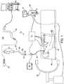
- an EMN system 100suitable for implementing methods for providing visual guidance for navigating inside a patient's chest is provided in accordance with the present disclosure.
- EMN system 100is used to perform one or more procedures on a patient supported on an operating table 40 .
- EMN system 100generally includes a bronchoscope 50 , monitoring equipment 30 , an electromagnetic (EM) tracking system 70 , and a computing device 80 .
- EMelectromagnetic
- Bronchoscope 50is configured for insertion through the patient's mouth and/or nose into the patient's airways.
- Bronchoscope 50includes a source of illumination and a video imaging system (not explicitly shown) and is coupled to monitoring equipment 30 , for example, a video display, for displaying the video images received from the video imaging system of bronchoscope 50 .
- bronchoscope 50may operate in conjunction with a catheter guide assembly 90 .
- Catheter guide assembly 90includes a locatable guide (LG) 92 and an extended working channel (EWC) 96 configured for insertion through a working channel of bronchoscope 50 into the patient's airways (although the catheter guide assembly 90 may alternatively be used without bronchoscope 50 ).
- LGlocatable guide
- EWCextended working channel
- Catheter guide assembly 90includes a handle 91 connected to EWC 96 , and which can be manipulated by rotation and compression to steer LG 92 and EWC 96 .
- EWC 96is sized for placement into the working channel of bronchoscope 50 .
- LG 92including an EM sensor 94
- LG 92is inserted into EWC 96 and locked into position such that EM sensor 94 extends a desired distance beyond a distal tip 93 of EWC 96 .
- the location of EM sensor 94and thus distal tip 93 of EWC 96 , within an EM field generated by EM field generator 76 , can be derived by tracking module 72 and computing device 80 .
- catheter guide assembly 90For a more detailed description of catheter guide assembly 90 , reference is made to commonly-owned U.S. Pat. No. 9,247,992, entitled “MICROWAVE ABLATION CATHETER AND METHOD OF UTILIZING THE SAME”, filed on Mar. 15, 2013, by Ladtkow et al., the entire contents of which are hereby incorporated by reference.
- LG 92 and EWC 96are selectively lockable relative to one another via a locking mechanism 99 .
- a six degrees-of-freedom EM tracking system 70e.g., similar to those disclosed in U.S. Pat. No. 6,188,355 and published PCT Application Nos. WO 00/10456 and WO 01/67035, entitled “WIRELESS SIX-DEGREE-OF-FREEDOM LOCATOR”, filed on Dec. 14, 1998 by Gilboa, the entire contents of each of which is incorporated herein by reference, or any other suitable positioning measuring system, is utilized for performing navigation, although other configurations are also contemplated.
- EM tracking system 70may be configured for use with catheter guide assembly 90 to track a position of EM sensor 94 as it moves in conjunction with EWC 96 through the airways of the patient, as detailed below.
- EM tracking system 70includes a tracking module 72 , a plurality of reference sensors 74 , and an EM field generator 76 .
- EM field generator 76is positioned beneath the patient.
- EM field generator 76 and the plurality of reference sensors 74are interconnected with tracking module 72 , which derives the location of each reference sensor 74 in the six degrees of freedom.
- One or more of reference sensors 74are attached to the chest of the patient.
- the six degrees of freedom coordinates of reference sensors 74are sent as data to computing device 80 , which includes an application 81 , where the data from reference sensors 74 are used to calculate a patient coordinate frame of reference.
- EM sensor 94is described above as being included in LG 92 , it is also envisioned that EM sensor 94 may be embedded or incorporated within a treatment tool, such as a biopsy tool 62 and/or an ablation tool 64 , where the treatment tool may alternatively be utilized for navigation without need of LG 92 or the necessary tool exchanges that use of LG 92 requires. EM sensor 94 may also be embedded or incorporated within EWC 96 , such as at a distal portion of EWC 96 , thereby enabling tracking of the distal portion of EWC 96 without the need for a separate LG 92 .
- treatment tools 62 , 64are configured to be insertable into catheter guide assembly 90 following navigation to a target location and removal of LG 92 .
- Biopsy tool 62may be used to collect one or more tissue samples from the target location, and in an embodiment, is further configured for use in conjunction with tracking system 70 to facilitate navigation of biopsy tool 62 to the target location, and tracking of a location of biopsy tool 62 as it is manipulated relative to the target location to obtain the tissue sample.
- Ablation tool 64is configured to be operated with a generator 66 , such as a radio frequency generator or a microwave generator, and may include any of a variety of ablation tools and/or catheters, examples of which are more fully described in U.S. Pat. Nos.
- a piercing tool and/or puncture toolmay be used and/or incorporated within LG 92 to create an exit point where LG 92 , and thereby EWC 96 , is navigated outside of the patient's airways and toward the target location, as further described below.
- a radiographic imaging device 20such as a C-arm imaging device capable of performing a CBCT scan of at least a portion of the patient's lungs, may be used in conjunction with EMN system 100 . Imaging device 20 may further be capable of performing fluoroscopic scans of the patient's lungs. As shown in FIG. 1 , imaging device 20 is connected to computing device 80 such that application 81 may receive and process image data obtained by imaging device 20 . However, imaging device 20 may also have a separate computing device located within the treatment room or in a separate control room to first receive the image data obtained by imaging device 20 and relay such image data to computing device 80 . For example, to avoid exposing the clinician to unnecessary radiation from repeated radiographic scans, the clinician may exit the treatment room and wait in an adjacent room, such as the control room, while imaging device 20 performs the CBCT and/or fluoroscopic scans.
- Computing device 80includes software and/or hardware, such as application 81 , used to facilitate the various phases of an EMN procedure, including generating the 3D model, identifying a target location, planning a pathway to the target location, registering the 3D model with the patient's actual airways, navigating to the target location, and performing treatment at the target location.
- application 81used to facilitate the various phases of an EMN procedure, including generating the 3D model, identifying a target location, planning a pathway to the target location, registering the 3D model with the patient's actual airways, navigating to the target location, and performing treatment at the target location.
- computing device 80utilizes data acquired from a CT scan, CBCT scan, magnetic resonance imaging (MRI) scan, positron emission tomography (PET) scan, and/or any other suitable imaging modality to generate and display the 3D model of the patient's airways, to enable identification of a target location on the 3D model (automatically, semi-automatically or manually) by analyzing the image data and/or 3D model, and allow for the determination and selection of a pathway through the patient's airways to the target location.
- the image datamay have gaps, omissions, and/or other imperfections included in the image data
- the 3D modelis a smooth representation of the patient's airways, with any such gaps, omissions, and/or imperfections in the image data filled in or corrected.
- the 3D modelmay be presented on a display monitor associated with computing device 80 , or in any other suitable fashion.
- An example of the planning software described hereincan be found in U.S. Patent Publication Nos. 2014/0281961, 2014/0270441, and 2014/0282216, filed by Baker et al. on Mar. 15, 2013, and entitled “PATHWAY PLANNING SYSTEM AND METHOD”, the contents of all of which are incorporated herein by reference. Further examples of the planning software can be found in commonly assigned U.S. Patent Publication No. 2016/0000302, entitled “SYSTEM AND METHOD FOR NAVIGATING WITHIN THE LUNG”, filed on Jun. 29, 2015, by Brown et al., the contents of which are incorporated herein by reference.
- various views of the image data and/or 3D modelmay be displayed to and manipulated by a clinician to facilitate identification of the target location.
- the target locationmay be a site within the patient's lungs where treatment is to be performed.
- the treatment targetmay be located in lung tissue adjacent to an airway.
- the 3D modelmay include, among other things, a model airway tree corresponding to the actual airways of the patient's lungs, and show the various passages, branches, and bifurcations of the patient's actual airway tree.
- the 3D modelmay include lesions, markers, blood vessels and vascular structures, lymphatic vessels and structures, organs, other physiological structures, and/or a 3D rendering of the pleural surfaces and fissures of the patient's lungs. Some or all of the aforementioned elements may be selectively displayed, such that the clinician may choose which elements should be displayed when viewing the 3D model.
- application 81may determine a pathway between the patient's trachea and the target location via the patient's airways.
- the target locationis located in lung tissue that is not directly adjacent an airway
- at least a portion of the pathwaywill be located outside of the patient's airways to connect an exit point on an airway wall to the target location.
- LG 94 and EWC 96will first be navigated along a first portion of the pathway through the patient's airways to the exit point on the airway wall.
- LG 94may then be removed from EWC 96 and an access tool, such as a piercing or puncture tool, inserted into EWC 96 to create an opening in the airway wall at the exit point.
- EWC 96may then be advanced through the airway wall into the parenchyma surrounding the airways.
- the access toolmay then be removed from EWC 96 and LG 94 and/or tools 62 , 64 reinserted into EWC 96 to navigate EWC 96 along a second portion of the pathway outside of the airways to the target location.
- EM sensor 94in conjunction with tracking system 70 , enables tracking of EM sensor 94 (and thus distal tip 93 of EWC 96 or tools 62 , 64 ) as EM sensor 94 is advanced through the patient's airways following the pathway planned during the planning phase.
- the 3D modelis registered with the patient's actual airways to enable application 81 to display an indication of the location of EM sensor 94 on the 3D model corresponding to the location of EM sensor 94 within the patient's airways.
- One potential method of registrationinvolves performing a survey of the patient's lungs by navigating LG 92 into each lobe of the patient's lungs to at least the second bifurcation of the airways of that lobe.
- the position of LG 92is tracked during this registration phase, and the 3D model is iteratively updated based on the tracked position of the locatable guide within the actual airways of the patient's lungs.
- This registration processis described in commonly-owned U.S. Patent Application Publication No. 2011/0085720, entitled “AUTOMATIC REGISTRATION TECHNIQUE,” filed on May 14, 2010, by Barak et al., and U.S. Patent Publication No.
- Another potential method of registrationuses image data from a CBCT scan performed at the start of the treatment procedure to generate the 3D model with the patient remaining on table 40 while the clinician performs the above-described planning phase. Because the scan is taken with reference sensors 74 placed on the patient, the anatomy of the patient relative to reference sensors 74 is known. By performing the scan with reference sensors 74 placed on the patient, performing registration by using the lung survey technique, described above, becomes unnecessary. Additionally, features and sensors in EM field generator 76 under the patient may also be used as another means to help ensure the target location is placed within the ENB field.
- the clinicianmay then start the navigation phase of the procedure without performing the above-described survey of the patient's lungs because the patient will still be in substantially the same position as the patient was when the image data on which the 3D model is based were obtained.
- application 81may extrapolate sufficient data points from the position of EM sensor 94 within the EM field while LG 92 is navigated along the planned pathway to register the 3D model to the patient's actual airways while the navigation phase is in process.
- the clinicianmay request that additional CBCT scans be performed on the patient.
- the additional CBCT scansmay be directed at a particular location in the patient's body, such as an area of the patient's lungs about the position of LG 92 , for which the clinician wants additional image data.
- the additional image datamay be used to confirm the position of EM sensor 94 (representing the location of LG 92 and/or tool 62 , 64 ) and/or the target location within the patient's lungs.
- Application 81may receive the image data acquired by the additional CBCT scan and process the additional image data to identify the position of EM sensor 94 and/or the target location within the patient's lungs.
- Application 81may then update the indicator of the position of EM sensor 94 on the 3D model based on the additional CBCT image data if the additional image data indicates that the position displayed based on the original image data is incorrect.
- the additional CBCT scansmay be performed based on the patient's breathing or respiratory cycle, such as to acquire image data during different phases of the patient's respiratory cycle, as further described below.
- the clinicianmay also request that fluoroscopic scans be performed at various times during the procedure. Image data acquired from the fluoroscopic scans may further be used to assist the clinician in navigating and positioning LG 92 about the target location.
- imaging device 20may be moved out of position during the procedure to allow the clinician better access to the patient, and may be moved back into position for scanning with guidance provided by application 81 .
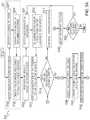
- FIGS. 3A-3Ethere is shown a flowchart of an exemplary method 300 for providing visual guidance for navigating inside a patient's chest, according to embodiments of the present disclosure.
- various subsections of method 300may be directed at different embodiments.
- steps of method 300may be omitted or performed in a different order than described below.
- an initial portion 301 of method 300includes the steps of the above-described planning phase of the treatment procedure.
- method 300may start at step S 302 where application 81 receives image data of the patient's chest.
- the image datamay be acquired during a pre-procedure scan of the patient's body, or a relevant portion thereof, such as a CT scan, MRI scan, PET scan, CBCT scan, etc.
- the image datamay be acquired during a scan, such as a CBCT scan, performed at the start of the treatment procedure.
- application 81After receiving the image data, application 81 processes the image data, at step S 304 , to identify a luminal network in the image data.
- the luminal networkmay be the patient's airways, blood vessels, and/or lymphatic vessels in the patient's lungs.
- Application 81may further process the image data to identify other structures, such as the pleura and fissures of the patient's lungs, other organs and critical structures, and/or aberrant structures in and/or around the patient's lungs.
- Application 81then, at step S 306 , generates a 3D model based on the processed image data.
- At least one treatment targetis identified, either automatically by application 81 , semi-automatically with input from the clinician, or manually by the clinician. After identifying the treatment target, a target location representative of the identified location of the treatment target is marked on the 3D model, and application 81 determines a pathway between the patient's trachea and the target location via the patient's airways.
- the pathwaymay include various portions, including at least one portion located inside the patient's airways running between the trachea and an exit point in an airway wall proximate the target location, and at least one portion located outside the patient's airways running from the exit point to the target location.
- the pathwayrepresents a recommended route along which LG 92 or other tool including sensor 94 should be navigated through the patient's airways and, as described further below, after reaching the exit point, through the tissue and space surrounding the patient's airways.
- Application 81displays the pathway and the target location on the 3D model at step S 308 .
- the navigation phase of the treatment procedurecommences.
- the 3D modelmust be registered to the actual airways of the patient's lungs.
- there are various methods of registration that may be used for this purposeincluding the lung survey method described above.
- a lung surveymay not be necessary, because application 81 may collect sufficient data points regarding the position of LG 92 in the patient's airways during the initial portion of the navigation phase to register the 3D model to the patient's airways while navigation is in progress.
- EM field generator 76generates an EM field about the patient's chest.
- An EM sensor 94is then inserted into the patient's airways and a location of EM sensor 94 in the EM field is detected by tracking system 70 at step S 311 .
- the detected location of EM sensor 94is relayed to application 81 to determine, at step S 312 , a position of LG 92 , tools 62 , 64 , and/or EWC 96 based on the detected location of EM sensor 94 .
- the detected position of EM sensor 94may be reflective of whichever tool EM sensor 94 is included in, such as LG 92 , tools 62 , 64 , and/or EWC 96 , but for purpose of brevity and to ease this description, an example wherein LG 92 is used for navigation will be described hereinafter. However, those skilled in the art will realize that any of the other aforementioned tools and devices including an EM sensor 94 could be substituted for LG 92 .
- Step S 313Application 81 displays an indication of the determined position of LG 92 on the 3D model.
- a view 410 of the 3D modelincludes at least an airway tree 412 , an indicator 414 of the determined position of LG 92 , the target location 416 , and the pathway 418 to the target location.
- Steps S 311 to S 313may be iteratively repeated while navigation is in progress to continually detect the location of EM sensor 94 in the EM field, and determine and display a corresponding location of LG 92 on the 3D model.
- the clinicianmay, at various times during the procedure, request that an intra-procedural CBCT scan be performed to verify the determined position of LG 92 .
- the clinicianmay request that a CBCT scan be performed when LG 92 reaches the exit point where the pathway moves from within the patient's airways to outside of the patient's airways.
- the clinicianmay also request that a CBCT scan be performed after LG 92 has been navigated through the airway wall and towards the target location outside of the patient's airways.
- the clinicianmay request that a CBCT scan be performed to confirm the location of LG 92 when LG 92 is navigated proximate the target location, such as to confirm placement of LG 92 at the treatment target.
- application 81determines, at step S 314 , whether a request, such as a button press or other user input, for a CBCT scan has been received. If a request for a CBCT scan has not been received, application 81 continues tracking the location of EM sensor 94 at step S 315 , whereafter processing returns to step S 311 . However, if application 81 determines that a request for a CBCT scan has been received, processing proceeds to step S 320 .
- step S 320provides guidance for positioning imaging device 20 .
- the guidancemay instruct the clinician how to position imaging device 20 to perform a CBCT scan of at least a portion of the patient's chest based on the determined location of LG 92 .
- a focused CBCT scanmay be performed of only the area surrounding the determined location of LG 92 , thereby limiting the amount of radiation exposure to the patient.
- application 81may provide visual guidance, such as graphical illustrations, and/or audible instructions regarding positioning of imaging device 20 .
- step S 321application 81 based at least in part on data received from tracking system 70 , determines whether imaging device 20 is within the EM field generated by EM field generator 76 .
- application 81may provide a notification, at step S 322 , informing the clinician that the EM field may be distorted, such as by metal and/or other EM components included in imaging device 20 being present in the EM field.
- the notificationmay be a visual and/or audio notification, and may alert the clinician that the displayed location of LG 92 may be incorrect due to the distortion of the EM field.
- Application 81 and/or tracking system 70may then, at step S 323 , compensate for the distortion of the EM field, such as by adjusting the EM field and/or the displayed position of LG 92 on the 3D model based on the distortion of the EM field.
- step S 324application 81 determines whether the imaging device is ready for imaging.
- application 81may be able to actively track the location of imaging device 20 , such as by sensors included in imaging device 20 .
- application 81may determine whether imaging device 20 is ready for imaging based on input from the clinician. If application 81 determines that imaging device 20 is not yet ready for imaging, processing returns to step S 320 . Alternatively, if application 81 determines that imaging device 20 is ready for imaging, processing proceeds to step S 325 , where application 81 determines a current phase of the patient's respiratory cycle.
- the current phase of the patient's respiratory cyclemay be determined based on data received from sensors, such as reference sensors 74 located on the patient's chest. Further information on determination of a patient's respiratory cycle and compensation for movement occurring during the patient's respiratory cycle may be found in commonly-owned co-pending U.S. patent application Ser. No. 15/254,141, entitled RESPIRATION MOTION STABILIZATION FOR LUNG MAGNETIC NAVIGATION SYSTEM, filed on Sep. 1, 2016, by Koyrakh et al., the entire contents of which are incorporated herein by reference. Alternatively, or in addition, the current phase of the patient's respiratory cycle may be determined based on data received from a ventilator coupled to the patient.
- step S 326application 81 determines whether the current phase of the patient's respiratory cycle corresponds to the desired phase of the patient's respiratory cycle requested by the clinician. If application 81 determines that the current phase of the patient's respiratory cycle does not correspond to the desired phase, the method proceeds to step S 327 , where application 81 waits for the patient's respiratory cycle to enter the desired phase. Thereafter, processing returns to step S 325 to again determine the current phase of the patient's respiratory cycle. If application 81 determines that the current phase of the patient's respiratory cycle corresponds to the desired phase, processing proceeds to step S 330 .
- step S 330application 81 causes tracking system 70 to disable EM field generator 76 to avoid interference with imaging device 20 during the imaging process.
- step S 331the CBCT imaging is performed.
- the CBCT imagingmay be performed manually by the clinician interacting with imaging device 20 to perform the CBCT imaging. Alternatively, the CBCT imaging may be performed automatically or semi-automatically via application 81 directly or indirectly controlling imaging device 20 .
- step S 332application 81 causes tracking system 70 to re-enable EM field generator 76 .
- application 81determines, at step S 333 , whether the patient moved during the CBCT imaging process. The determination may be based on data received from sensors, such as reference sensors 74 , indicating the patient's current position relative to the patient's position prior to the CBCT imaging, and/or indicating movement of the patient during the CBCT imaging process. If application 81 determines that the patient moved during the CBCT imaging process, application 81 determines, at step S 334 , whether an amount of the movement is within a predetermined threshold. For example, mere minor movement may be insufficient to affect the CBCT image data collected during the CBCT imaging process, while more significant movement may cause the CBCT image data to be unusable.
- a predetermined thresholdFor example, mere minor movement may be insufficient to affect the CBCT image data collected during the CBCT imaging process, while more significant movement may cause the CBCT image data to be unusable.
- step S 335application 81 may mark the CBCT image data received from imaging device 20 after the CBCT imaging as unusable, and reinitiate the CBCT imaging process at step S 335 , whereafter processing returns to step S 324 .
- application 81may provide a notification at step S 336 , indicating that the patient moved but that the movement was within the predetermined threshold. Thereafter, or if application determined at step S 333 that the patient did not move during the CBCT imaging process, processing proceeds to step S 340 .
- step S 340application 81 verifies the location of LG 92 based on the CBCT image data.
- application 81may process the CBCT image data received after the CBCT imaging process to identify the patient's airways and the location of LG 92 within the patient's chest. If application 81 determines that the location of LG 92 identified in the CBCT image data does not correspond with the location indicated on the 3D model, application 81 may, at step S 341 , provide a notification that the indicated location of LG 92 on the 3D model may be incorrect, and update the indicated location of LG 92 on the 3D model.
- application 81may request approval from the clinician prior to updating the indicated location of LG 92 on the 3D model. If application 81 updates the indicated location of LG 92 on the 3D model, application 81 may further update the portion of the pathway between the updated location of LG 92 and the target location. In some embodiments, application 81 may request approval from the clinician prior to updating the pathway. Thereafter, processing proceeds to step S 350 .
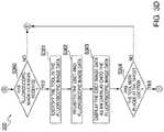
- step S 350application 81 again determines a current phase of the patient's respiratory cycle, similar to the determination performed at step S 325 . Thereafter, at step S 351 , application 81 determines whether CBCT image data corresponding to the current phase of the patient's respiratory cycle is available. If application 81 determines that CBCT data corresponding to the current phase of the patient's respiratory cycle is not available, application 81 provides a notification at step S 353 that CBCT image data corresponding to the current phase of the patient's respiratory cycle is not available.
- step S 354application 81 selects predetermined CBCT image data.
- application 81may select CBCT image data corresponding to the phase towards which the patient's respiratory cycle is moving. For example, if the patient's respiratory cycle is currently past full inhale and partially into an exhaling phase, application 81 may select CBCT image data corresponding to a portion of the exhaling phase towards which the patient's respiratory cycle is moving. In another embodiment, application 81 may select the most recently obtained CBCT image data. Thereafter, processing proceeds to step S 355 .
- step S 351If application 81 determined at step S 351 that CBCT image data corresponding to the current phase of the patient's respiratory cycle is available, application 81 selects such CBCT image data at step S 352 .
- step S 355application 81 identifies LG 92 in the CBCT image data, similar to the identification of LG 92 at step S 341 .
- Application 81further determines a position and orientation of LG 92 based on the CBCT image data, and orients the CBCT image data based on the determined position and orientation of LG 92 at step S 356 .
- Application 81displays the CBCT image data at step S 357 , as shown in view 430 of FIG. 4 , described below.
- step S 358application 81 determines whether LG 92 has been placed at the target location. The determination may be based on processing of the CBCT image data, tracking data received from tracking system 70 , and/or input provided by the clinician. If application 81 determines that LG 92 has not yet been placed at the target location, or if application 81 cannot determine that LG 92 has been placed at the target location, or if application 81 determines, such as based on input provided by the clinician, that an alternative view is necessary to determine if LG 92 has been placed at the target location, processing proceeds to step S 360 .
- step S 360application 81 determines whether fluoroscopic image data has been received. If fluoroscopic image data has not been received, processing returns to step S 314 , whereafter the CBCT imaging process may be repeated or navigation may continue. However, if fluoroscopic image data has been received, processing proceeds to step S 361 , where application 81 identifies LG 92 in the fluoroscopic image data. Thereafter, at step S 362 , application 81 registers the CBCT image data and the fluoroscopic image data.
- Application 81displays the CBCT image data in conjunction with the fluoroscopic image data.
- application 81may display the CBCT image data as an overlay onto the fluoroscopic image data, as shown in view 420 of FIG. 4 , described below.
- the overlay of the CBCT data onto the fluoroscopic image datagives the clinician greater insight into the actual location of LG 92 within the patient's chest.
- the CBCT image datamay also be displayed according to the patient's breathing cycle.
- the CBCT image datamay be displayed as an overlay onto the fluoroscopic image data, and faded in and out as the patient breathes, thus becoming clearer when the patient's breathing cycle coincides with the phase of the breathing cycle during which the CBCT image data was taken, and less clear when the patient's breathing cycle moves to the next phase.
- step S 364application 81 again determines whether LG 92 has been placed at the target location. As with step S 358 described above, the determination may be based on processing of the CBCT image data, tracking data received from tracking system 70 , and/or input provided by the clinician. If application 81 determines that LG 92 has not been placed at the target location, or if application 81 cannot determine that LG 92 has been placed at the target location, processing returns to step S 314 for further imaging and/or navigation.
- step S 358determines at either step S 358 or step S 364 that LG 92 has been placed at the target location
- processingproceeds to step S 359 ( FIG. 3C ).
- the clinicianmay uncouple LG 92 and EWC 96 , and remove LG 92 from the patient's airways while leaving EWC 96 in place.
- Biopsy tool 62 and/or ablation tool 64may then be inserted into the patient's airways and advanced through EWC 96 to the treatment target at the target location.
- EM sensor 94may be included in biopsy tool 62 and/or ablation tool 64 , thus making such a tool exchange unnecessary.
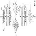
- application 81determines whether the inserted tool is a biopsy tool 62 or an ablation tool 64 . If application 81 determines that the inserted tool is ablation tool 64 , processing proceeds to step S 370 . Alternatively, if application 81 determines that the inserted tool is biopsy tool 62 , processing skips directly to step S 380 ( FIG. 3A , described below).
- step S 370application 81 identifies a radiating portion of ablation tool 64 in one or more of the CBCT image data, the fluoroscopic image data, and/or the 3D model.
- ablation tool 64may include one or more radiopaque elements about the radiating portion of ablation tool 64 that may be detectable by application 81 while processing CBCT and/or fluoroscopic image data.
- application 81may determine that the radiating portion is a predetermined distance from a distal end of ablation tool 64 .
- application 81determines a projected ablation zone based on the location of ablation tool 64 , and thus the radiating portion of ablation tool 64 , inside the patient's chest, and configuration settings for the ablation procedure.
- the clinicianmay enter configuration settings, such as time, temperature, wattage, etc., for the ablation procedure into computing device 80 .
- Application 81may then, at step S 371 , use such configuration settings to determine a projected ablation zone representing a maximum area around the radiating portion of ablation tool 64 that will be ablated according to the configuration settings.
- Application 81may then, at step S 372 , display an indicator of the projected ablation zone on the CBCT image data, the fluoroscopic image data, and/or the 3D model.
- application 81determines whether the clinician has approved the projected ablation zone.
- the projected ablation zonemay be represented by a sphere surrounding ablation tool 64 , with an anchor point of the sphere being the radiating portion of ablation tool 64 . Based on the displayed projected ablation zone, the clinician may decide to adjust the configuration settings for the ablation procedure and/or the location of ablation tool 64 .
- the clinicianmay determine that the projected ablation zone does not sufficiently cover the treatment target, and thus choose to adjust the configuration settings or the location of ablation tool 64 .
- the clinicianmay input a decision to proceed with the ablation procedure or return to navigation into computing device 80 . If application 81 determines that the clinician does not approve of the displayed projected ablation zone, processing returns to step S 314 for further navigation and/or imaging. Alternatively, if application 81 determines that the clinician approves of the displayed projected ablation zone, processing proceeds to step S 374 , where application 81 determines whether the ablation process has started. For example, application 81 may detect that an activation button on ablation tool 64 has been depressed.
- ablation tool 64may be controlled by application 81 based on input received from the clinician. Thus, if application 81 determines that the ablation process has not started, processing returns to step S 372 . Alternatively, if application 81 determines that the ablation process has started, processing proceeds to step S 375 .
- application 81determines an estimated ablation zone.
- the determination of the estimated ablation zonemay be based on the configuration settings for the ablation procedure, the location of the radiating portion of ablation tool 64 , an elapsed time since the ablation process was started, and/or additional image data received during the ablation process. For example, application 81 may determine that, based on the configuration settings, after a particular amount of time has elapsed since the ablation process was started, a particular area around the radiating portion of ablation tool 64 would be expected to have been ablated.
- additional CBCT and/or fluoroscopic scansmay be performed during the ablation process to provide image data showing the progress of the ablation process.
- Application 81may then, at step S 376 , display an indicator of such area, such as by a sphere or other shape around ablation tool 64 in the CBCT image data, the fluoroscopic image data, and/or the 3D model.
- step S 377application 81 determines if the ablation process has been completed. For example, application 81 may determine based on the elapsed time since the ablation process was started reaching the time included in the configuration settings that the ablation process is complete. Application 81 may also receive input from the clinician that the ablation process is complete. If application 81 determines that the ablation process has not been completed, processing returns to step S 375 . Alternatively, if application 81 determines that the ablation process has been completed, processing proceeds to step S 380 .
- application 81marks the location where treatment, whether biopsy or ablation, was performed.
- application 81may store the position information received from tracking system 70 regarding the location of tool 62 , 64 in the patient's chest while treatment was performed.
- application 81may also store the last determined estimated ablation zone of the ablation procedure. Such stored treatment locations and estimated ablation zones may further be displayed on the 3D model.
- step S 381application 81 determines whether additional treatments are required. For example, application 81 may determine based on the above-described treatment plan that additional treatments are required. Application 81 may also receive input from the clinician that additional treatments are required. Thus, if application 81 determines that additional treatments are required, processing returns to step S 311 . If navigation to a different target location is required, and tool 62 , 64 does not include an EM sensor 94 , tool 62 , 64 may be removed from EWC 96 and replaced by LG 92 . Alternatively, if application 81 determines that additional treatments are not required, processing ends.
- FIG. 4depicts a graphical user interface (GUI) 400 including at least three image views: a 3D model view 410 , a fluoroscopic image view 420 , and a CBCT image view 430 .
- 3D model view 410includes at least airway tree 412 , tool indicator 414 representing the location of EM sensor 94 in the patients airways, target location 416 , and pathway 418 .
- Each of views 410 , 420 , and 430may be selectively enabled and configured to show image data from the various sources described above, according to the clinician's preference.
- Computing device 80may include a memory 502 , a processor 504 , a display 506 , a network interface 508 , an input device 510 , and/or an output module 512 .
- Memory 502may store application 81 and/or image data 514 .
- Application 81may, when executed by processor 504 , cause display 506 to present user interface 516 .
- Application 81may also provide the interface between the sensed position of EM sensor 94 and the image and planning data developed in the pathway planning phase, described above.
- Memory 502may include any non-transitory computer-readable storage media for storing data and/or software that is executable by processor 504 and which controls the operation of computing device 80 .
- memory 502may include one or more solid-state storage devices such as flash memory chips.
- memory 502may include one or more mass storage devices connected to the processor 504 through a mass storage controller (not shown) and a communications bus (not shown).
- mass storage controllernot shown
- communications busnot shown
- computer readable storage mediaincludes non-transitory, volatile and non-volatile, removable and non-removable media implemented in any method or technology for storage of information such as computer-readable instructions, data structures, program modules or other data.
- computer-readable storage mediaincludes RAM, ROM, EPROM, EEPROM, flash memory or other solid state memory technology, CD-ROM, DVD, Blu-Ray or other optical storage, magnetic cassettes, magnetic tape, magnetic disk storage or other magnetic storage devices, or any other medium which can be used to store the desired information and which can be accessed by computing device 80 .
- Network interface 508may be configured to connect to a network such as a local area network (LAN) consisting of a wired network and/or a wireless network, a wide area network (WAN), a wireless mobile network, a Bluetooth network, and/or the internet.
- LANlocal area network
- WANwide area network
- wireless mobile networka Bluetooth network
- Input device 510may be any device by means of which a user may interact with computing device 80 , such as, for example, a mouse, keyboard, foot pedal, touch screen, and/or voice interface.
- Output module 512may include any connectivity port or bus, such as, for example, parallel ports, serial ports, universal serial busses (USB), or any other similar connectivity port known to those skilled in the art.
- USBuniversal serial busses
Landscapes
- Health & Medical Sciences (AREA)
- Life Sciences & Earth Sciences (AREA)
- Engineering & Computer Science (AREA)
- Medical Informatics (AREA)
- Surgery (AREA)
- Public Health (AREA)
- General Health & Medical Sciences (AREA)
- Biomedical Technology (AREA)
- Veterinary Medicine (AREA)
- Animal Behavior & Ethology (AREA)
- Heart & Thoracic Surgery (AREA)
- Molecular Biology (AREA)
- Nuclear Medicine, Radiotherapy & Molecular Imaging (AREA)
- Pathology (AREA)
- Physics & Mathematics (AREA)
- Radiology & Medical Imaging (AREA)
- Biophysics (AREA)
- Optics & Photonics (AREA)
- High Energy & Nuclear Physics (AREA)
- Otolaryngology (AREA)
- Human Computer Interaction (AREA)
- Plasma & Fusion (AREA)
- Theoretical Computer Science (AREA)
- Pulmonology (AREA)
- Data Mining & Analysis (AREA)
- Oral & Maxillofacial Surgery (AREA)
- Databases & Information Systems (AREA)
- Epidemiology (AREA)
- Primary Health Care (AREA)
- Robotics (AREA)
- Gynecology & Obstetrics (AREA)
- Cardiology (AREA)
- Electromagnetism (AREA)
- Apparatus For Radiation Diagnosis (AREA)
Abstract
Description
Claims (19)
Priority Applications (5)
| Application Number | Priority Date | Filing Date | Title |
|---|---|---|---|
| US15/370,906US11172895B2 (en) | 2015-12-07 | 2016-12-06 | Visualization, navigation, and planning with electromagnetic navigation bronchoscopy and cone beam computed tomography integrated |
| CN201611117979.6ACN106821498B (en) | 2015-12-07 | 2016-12-07 | Non-transitory computer-readable storage medium |
| EP16202781.7AEP3178435B1 (en) | 2015-12-07 | 2016-12-07 | Visualization, navigation, and planning with electromagnetic navigation bronchoscopy and cone beam computed tomography integrated |
| US17/516,810US11925493B2 (en) | 2015-12-07 | 2021-11-02 | Visualization, navigation, and planning with electromagnetic navigation bronchoscopy and cone beam computed tomography integrated |
| US18/427,567US20240206829A1 (en) | 2015-12-07 | 2024-01-30 | Visualization, navigation, and planning with electromagnetic navigation bronchoscopy and cone beam computed tomography integrated |
Applications Claiming Priority (2)
| Application Number | Priority Date | Filing Date | Title |
|---|---|---|---|
| US201562264145P | 2015-12-07 | 2015-12-07 | |
| US15/370,906US11172895B2 (en) | 2015-12-07 | 2016-12-06 | Visualization, navigation, and planning with electromagnetic navigation bronchoscopy and cone beam computed tomography integrated |
Related Child Applications (1)
| Application Number | Title | Priority Date | Filing Date |
|---|---|---|---|
| US17/516,810ContinuationUS11925493B2 (en) | 2015-12-07 | 2021-11-02 | Visualization, navigation, and planning with electromagnetic navigation bronchoscopy and cone beam computed tomography integrated |
Publications (2)
| Publication Number | Publication Date |
|---|---|
| US20170156685A1 US20170156685A1 (en) | 2017-06-08 |
| US11172895B2true US11172895B2 (en) | 2021-11-16 |
Family
ID=57749647
Family Applications (3)
| Application Number | Title | Priority Date | Filing Date |
|---|---|---|---|
| US15/370,906Active2038-06-14US11172895B2 (en) | 2015-12-07 | 2016-12-06 | Visualization, navigation, and planning with electromagnetic navigation bronchoscopy and cone beam computed tomography integrated |
| US17/516,810Active2037-06-13US11925493B2 (en) | 2015-12-07 | 2021-11-02 | Visualization, navigation, and planning with electromagnetic navigation bronchoscopy and cone beam computed tomography integrated |
| US18/427,567PendingUS20240206829A1 (en) | 2015-12-07 | 2024-01-30 | Visualization, navigation, and planning with electromagnetic navigation bronchoscopy and cone beam computed tomography integrated |
Family Applications After (2)
| Application Number | Title | Priority Date | Filing Date |
|---|---|---|---|
| US17/516,810Active2037-06-13US11925493B2 (en) | 2015-12-07 | 2021-11-02 | Visualization, navigation, and planning with electromagnetic navigation bronchoscopy and cone beam computed tomography integrated |
| US18/427,567PendingUS20240206829A1 (en) | 2015-12-07 | 2024-01-30 | Visualization, navigation, and planning with electromagnetic navigation bronchoscopy and cone beam computed tomography integrated |
Country Status (3)
| Country | Link |
|---|---|
| US (3) | US11172895B2 (en) |
| EP (1) | EP3178435B1 (en) |
| CN (1) | CN106821498B (en) |
Cited By (11)
| Publication number | Priority date | Publication date | Assignee | Title |
|---|---|---|---|---|
| US11576730B2 (en) | 2018-03-28 | 2023-02-14 | Auris Health, Inc. | Systems and methods for registration of location sensors |
| US11660147B2 (en) | 2019-12-31 | 2023-05-30 | Auris Health, Inc. | Alignment techniques for percutaneous access |
| US11712173B2 (en) | 2018-03-28 | 2023-08-01 | Auris Health, Inc. | Systems and methods for displaying estimated location of instrument |
| US11759266B2 (en) | 2017-06-23 | 2023-09-19 | Auris Health, Inc. | Robotic systems for determining a roll of a medical device in luminal networks |
| US11759090B2 (en) | 2018-05-31 | 2023-09-19 | Auris Health, Inc. | Image-based airway analysis and mapping |
| US11850008B2 (en) | 2017-10-13 | 2023-12-26 | Auris Health, Inc. | Image-based branch detection and mapping for navigation |
| US11925493B2 (en) | 2015-12-07 | 2024-03-12 | Covidien Lp | Visualization, navigation, and planning with electromagnetic navigation bronchoscopy and cone beam computed tomography integrated |
| US12076100B2 (en) | 2018-09-28 | 2024-09-03 | Auris Health, Inc. | Robotic systems and methods for concomitant endoscopic and percutaneous medical procedures |
| US12220150B2 (en) | 2019-12-31 | 2025-02-11 | Auris Health, Inc. | Aligning medical instruments to access anatomy |
| US12414686B2 (en) | 2020-03-30 | 2025-09-16 | Auris Health, Inc. | Endoscopic anatomical feature tracking |
| US12414823B2 (en) | 2019-12-31 | 2025-09-16 | Auris Health, Inc. | Anatomical feature tracking |
Families Citing this family (38)
| Publication number | Priority date | Publication date | Assignee | Title |
|---|---|---|---|---|
| US8672837B2 (en) | 2010-06-24 | 2014-03-18 | Hansen Medical, Inc. | Methods and devices for controlling a shapeable medical device |
| US9057600B2 (en) | 2013-03-13 | 2015-06-16 | Hansen Medical, Inc. | Reducing incremental measurement sensor error |
| US9629595B2 (en) | 2013-03-15 | 2017-04-25 | Hansen Medical, Inc. | Systems and methods for localizing, tracking and/or controlling medical instruments |
| US9271663B2 (en) | 2013-03-15 | 2016-03-01 | Hansen Medical, Inc. | Flexible instrument localization from both remote and elongation sensors |
| US9014851B2 (en) | 2013-03-15 | 2015-04-21 | Hansen Medical, Inc. | Systems and methods for tracking robotically controlled medical instruments |
| US11020016B2 (en) | 2013-05-30 | 2021-06-01 | Auris Health, Inc. | System and method for displaying anatomy and devices on a movable display |
| JP6824967B2 (en) | 2015-09-18 | 2021-02-03 | オーリス ヘルス インコーポレイテッド | Tubular net navigation |
| US10143526B2 (en) | 2015-11-30 | 2018-12-04 | Auris Health, Inc. | Robot-assisted driving systems and methods |
| US10244926B2 (en) | 2016-12-28 | 2019-04-02 | Auris Health, Inc. | Detecting endolumenal buckling of flexible instruments |
| US11793579B2 (en) | 2017-02-22 | 2023-10-24 | Covidien Lp | Integration of multiple data sources for localization and navigation |
| WO2018183727A1 (en) | 2017-03-31 | 2018-10-04 | Auris Health, Inc. | Robotic systems for navigation of luminal networks that compensate for physiological noise |
| WO2019005696A1 (en) | 2017-06-28 | 2019-01-03 | Auris Health, Inc. | Electromagnetic distortion detection |
| CN116725667A (en)* | 2017-06-28 | 2023-09-12 | 奥瑞斯健康公司 | System for providing positioning information and method for positioning an instrument within an anatomical structure |
| US11058493B2 (en) | 2017-10-13 | 2021-07-13 | Auris Health, Inc. | Robotic system configured for navigation path tracing |
| CN108175502B (en)* | 2017-11-29 | 2021-08-17 | 苏州朗开医疗技术有限公司 | Bronchoscope electromagnetic navigation system |
| EP3720354B1 (en)* | 2017-12-04 | 2025-01-29 | Covidien LP | Automatic ablation antenna segmentation from ct image |
| US11006852B2 (en)* | 2017-12-11 | 2021-05-18 | Covidien Lp | Systems, methods, and computer-readable media of estimating thoracic cavity movement during respiration |
| US11471217B2 (en) | 2017-12-11 | 2022-10-18 | Covidien Lp | Systems, methods, and computer-readable media for improved predictive modeling and navigation |
| US11510736B2 (en) | 2017-12-14 | 2022-11-29 | Auris Health, Inc. | System and method for estimating instrument location |
| WO2019125964A1 (en) | 2017-12-18 | 2019-06-27 | Auris Health, Inc. | Methods and systems for instrument tracking and navigation within luminal networks |
| US11896312B2 (en) | 2018-02-05 | 2024-02-13 | Broncus Medical Inc. | Image-guided lung tumor planning and ablation system |
| CN108186116A (en)* | 2018-02-12 | 2018-06-22 | 中南大学湘雅二医院 | A kind of operation point of a knife heat source imaging system and its method |
| US10905499B2 (en) | 2018-05-30 | 2021-02-02 | Auris Health, Inc. | Systems and methods for location sensor-based branch prediction |
| KR102567087B1 (en) | 2018-05-31 | 2023-08-17 | 아우리스 헬스, 인코포레이티드 | Robotic systems and methods for navigation of luminal networks detecting physiological noise |
| JP7371026B2 (en) | 2018-05-31 | 2023-10-30 | オーリス ヘルス インコーポレイテッド | Path-based navigation of tubular networks |
| WO2020117726A1 (en)* | 2018-12-05 | 2020-06-11 | Covidien Lp | Electromagnetic navigation assembly and computed tomography scanner patient table, surgery system including the same, and method using the same |
| US11564751B2 (en) | 2019-02-01 | 2023-01-31 | Covidien Lp | Systems and methods for visualizing navigation of medical devices relative to targets |
| US11925333B2 (en)* | 2019-02-01 | 2024-03-12 | Covidien Lp | System for fluoroscopic tracking of a catheter to update the relative position of a target and the catheter in a 3D model of a luminal network |
| CN109793489A (en)* | 2019-03-26 | 2019-05-24 | 上海优益基医用材料有限公司 | visual positioning catheter |
| CN114127781B (en)* | 2019-06-11 | 2025-08-29 | 直观外科手术操作公司 | Detecting and representing anatomical features of anatomical structures |
| US12089902B2 (en)* | 2019-07-30 | 2024-09-17 | Coviden Lp | Cone beam and 3D fluoroscope lung navigation |
| US11147633B2 (en) | 2019-08-30 | 2021-10-19 | Auris Health, Inc. | Instrument image reliability systems and methods |
| US11207141B2 (en) | 2019-08-30 | 2021-12-28 | Auris Health, Inc. | Systems and methods for weight-based registration of location sensors |
| EP4025921A4 (en) | 2019-09-03 | 2023-09-06 | Auris Health, Inc. | DETECTION AND COMPENSATION OF ELECTROMAGNETIC DISTORTION |
| EP4031046A1 (en)* | 2019-11-05 | 2022-07-27 | Boston Scientific Scimed, Inc. | Medical systems for ablating tissue |
| US11701492B2 (en) | 2020-06-04 | 2023-07-18 | Covidien Lp | Active distal tip drive |
| EP4346606A1 (en)* | 2021-05-28 | 2024-04-10 | Covidien LP | Systems and methods of assessing breath hold during intraprocedural imaging |
| WO2025163487A1 (en)* | 2024-02-02 | 2025-08-07 | Covidien Lp | System and method for endoluminal peripheral lung nodule access |
Citations (231)
| Publication number | Priority date | Publication date | Assignee | Title |
|---|---|---|---|---|
| US4587975A (en) | 1984-07-02 | 1986-05-13 | Cardiac Pacemakers, Inc. | Dimension sensitive angioplasty catheter |
| US4593687A (en) | 1983-10-31 | 1986-06-10 | Gray Leo C | Endotracheal catheter |
| US5419324A (en) | 1993-11-24 | 1995-05-30 | Endovascular Technologies, Inc. | Radiological marker board with movable indicators |
| US5797849A (en) | 1995-03-28 | 1998-08-25 | Sonometrics Corporation | Method for carrying out a medical procedure using a three-dimensional tracking and imaging system |
| US5852646A (en) | 1996-05-21 | 1998-12-22 | U.S. Philips Corporation | X-ray imaging method |
| US5930329A (en) | 1997-09-22 | 1999-07-27 | Siemens Corporate Research, Inc. | Apparatus and method for detection and localization of a biopsy needle or similar surgical tool in a radiographic image |
| US5951475A (en) | 1997-09-25 | 1999-09-14 | International Business Machines Corporation | Methods and apparatus for registering CT-scan data to multiple fluoroscopic images |
| US5963613A (en) | 1997-12-31 | 1999-10-05 | Siemens Corporate Research, Inc. | C-arm calibration method for 3D reconstruction in an imaging system |
| US6028912A (en) | 1997-09-30 | 2000-02-22 | Siemens Corporate Research, Inc. | Apparatus and method for point reconstruction and metric measurement on radiographic images |
| US6038282A (en) | 1997-04-30 | 2000-03-14 | Siemens Aktiengesellschaft | X-ray imaging system |
| US6050724A (en) | 1997-01-31 | 2000-04-18 | U. S. Philips Corporation | Method of and device for position detection in X-ray imaging |
| US6055449A (en) | 1997-09-22 | 2000-04-25 | Siemens Corporate Research, Inc. | Method for localization of a biopsy needle or similar surgical tool in a radiographic image |
| US6081577A (en) | 1998-07-24 | 2000-06-27 | Wake Forest University | Method and system for creating task-dependent three-dimensional images |
| US6120180A (en) | 1997-10-17 | 2000-09-19 | Siemens Aktiengesellschaft | X-ray exposure system for 3D imaging |
| US6167296A (en) | 1996-06-28 | 2000-12-26 | The Board Of Trustees Of The Leland Stanford Junior University | Method for volumetric image navigation |
| US6188355B1 (en) | 1997-12-12 | 2001-02-13 | Super Dimension Ltd. | Wireless six-degree-of-freedom locator |
| US6236704B1 (en) | 1999-06-30 | 2001-05-22 | Siemens Corporate Research, Inc. | Method and apparatus using a virtual detector for three-dimensional reconstruction from x-ray images |
| US6314310B1 (en) | 1997-02-14 | 2001-11-06 | Biosense, Inc. | X-ray guided surgical location system with extended mapping volume |
| US6317621B1 (en) | 1999-04-30 | 2001-11-13 | Siemens Aktiengesellschaft | Method and device for catheter navigation in three-dimensional vascular tree exposures |
| US6351573B1 (en) | 1994-01-28 | 2002-02-26 | Schneider Medical Technologies, Inc. | Imaging device and method |
| US6351513B1 (en) | 2000-06-30 | 2002-02-26 | Siemens Corporate Research, Inc. | Fluoroscopy based 3-D neural navigation based on co-registration of other modalities with 3-D angiography reconstruction data |
| US6381483B1 (en) | 1998-03-25 | 2002-04-30 | Olympus Optical Co., Ltd. | Therapeutic system |
| US6389104B1 (en) | 2000-06-30 | 2002-05-14 | Siemens Corporate Research, Inc. | Fluoroscopy based 3-D neural navigation based on 3-D angiography reconstruction data |
| US6404843B1 (en) | 1999-09-27 | 2002-06-11 | Ge Medical Systems, S.A. | Method of reconstruction of a three-dimensional image of elements of sharp contrast |
| US6424731B1 (en) | 1998-07-17 | 2002-07-23 | Ge Medical Systems S.A. | Method of positioning a radiographic device |
| US20020143324A1 (en) | 1998-02-19 | 2002-10-03 | Curon Medical, Inc. | Apparatus to detect and treat aberrant myoelectric activity |
| US6470207B1 (en) | 1999-03-23 | 2002-10-22 | Surgical Navigation Technologies, Inc. | Navigational guidance via computer-assisted fluoroscopic imaging |
| US6484049B1 (en) | 2000-04-28 | 2002-11-19 | Ge Medical Systems Global Technology Company, Llc | Fluoroscopic tracking and visualization system |
| US6485422B1 (en) | 1999-05-26 | 2002-11-26 | Endocare, Inc. | Computer guided cryosurgery |
| US6491430B1 (en) | 1999-08-03 | 2002-12-10 | Siemens Aktiengesellschaft | Mobile X-ray apparatus and method for determining projection geometries therein |
| US6520934B1 (en) | 1999-12-29 | 2003-02-18 | Advanced Cardiovascular Systems, Inc. | Catheter assemblies with flexible radiopaque marker |
| US6535756B1 (en) | 2000-04-07 | 2003-03-18 | Surgical Navigation Technologies, Inc. | Trajectory storage apparatus and method for surgical navigation system |
| US6539127B1 (en) | 1998-07-28 | 2003-03-25 | Inria Institut National De Recherche | Electronic device for automatic registration of images |
| US6546068B1 (en) | 1999-01-19 | 2003-04-08 | Fuji Photo Film Co., Ltd. | Image data acquisition method and image data acquisition device |
| US6546279B1 (en) | 2001-10-12 | 2003-04-08 | University Of Florida | Computer controlled guidance of a biopsy needle |
| US6628977B2 (en) | 1999-12-28 | 2003-09-30 | Siemens Aktiengesellschaft | Method and system for visualizing an object |
| US20030220555A1 (en) | 2002-03-11 | 2003-11-27 | Benno Heigl | Method and apparatus for image presentation of a medical instrument introduced into an examination region of a patent |
| US6697664B2 (en) | 1999-02-10 | 2004-02-24 | Ge Medical Systems Global Technology Company, Llc | Computer assisted targeting device for use in orthopaedic surgery |
| US6707878B2 (en) | 2002-04-15 | 2004-03-16 | General Electric Company | Generalized filtered back-projection reconstruction in digital tomosynthesis |
| US6714810B2 (en) | 2000-09-07 | 2004-03-30 | Cbyon, Inc. | Fluoroscopic registration system and method |
| US6725080B2 (en) | 2000-03-01 | 2004-04-20 | Surgical Navigation Technologies, Inc. | Multiple cannula image guided tool for image guided procedures |
| US6731970B2 (en) | 2000-07-07 | 2004-05-04 | Brainlab Ag | Method for breath compensation in radiation therapy |
| US20040143317A1 (en) | 2003-01-17 | 2004-07-22 | Stinson Jonathan S. | Medical devices |
| US6768784B1 (en) | 2001-11-07 | 2004-07-27 | Koninklijke Philips Electronics N.V. | X-ray image enhancement |
| US6782287B2 (en) | 2000-06-27 | 2004-08-24 | The Board Of Trustees Of The Leland Stanford Junior University | Method and apparatus for tracking a medical instrument based on image registration |
| US6785356B2 (en) | 2001-11-13 | 2004-08-31 | Koninklijke Philips Electronics N.V. | Fluoroscopic computed tomography method |
| US6785571B2 (en) | 2001-03-30 | 2004-08-31 | Neil David Glossop | Device and method for registering a position sensor in an anatomical body |
| US6823207B1 (en) | 2000-08-26 | 2004-11-23 | Ge Medical Systems Global Technology Company, Llc | Integrated fluoroscopic surgical navigation and imaging workstation with command protocol |
| DE10323008A1 (en) | 2003-05-21 | 2004-12-23 | Siemens Ag | Automatic fusion of 2D fluoroscopic C-frame X-ray images with preoperative 3D images using navigation markers, by use of a projection matrix based on a 2D fluoroscopy image and a defined reference navigation system |
| US20050008210A1 (en) | 2000-05-09 | 2005-01-13 | Paieon, Inc. | System and method for three-dimensional reconstruction of an artery |
| US6856827B2 (en) | 2000-04-28 | 2005-02-15 | Ge Medical Systems Global Technology Company, Llc | Fluoroscopic tracking and visualization system |
| US6856826B2 (en) | 2000-04-28 | 2005-02-15 | Ge Medical Systems Global Technology Company, Llc | Fluoroscopic tracking and visualization system |
| US6865253B2 (en) | 2000-10-17 | 2005-03-08 | Brainlab Ag | Method and device for accurately positioning a patient in radiotherapy and/or radiosurgery |
| US6898263B2 (en) | 2002-11-27 | 2005-05-24 | Ge Medical Systems Global Technology Company, Llc | Method and apparatus for soft-tissue volume visualization |
| US6944260B2 (en) | 2003-11-11 | 2005-09-13 | Ge Medical Systems Global Technology Company, Llc | Methods and apparatus for artifact reduction in computed tomography imaging systems |
| US20050215874A1 (en) | 2004-03-12 | 2005-09-29 | Lixiao Wang | MRI and X-ray visualization |
| US6956927B2 (en) | 2003-05-16 | 2005-10-18 | Shimadzu Corporation | Radiation therapy planning device |
| US20060002630A1 (en) | 2004-06-30 | 2006-01-05 | Accuray, Inc. | Fiducial-less tracking with non-rigid image registration |
| US20060023840A1 (en) | 2004-07-23 | 2006-02-02 | Jan Boese | Method for imaging in a medical interventional procedure by image subtraction |
| US7010152B2 (en) | 2002-01-22 | 2006-03-07 | Canon Kabushiki Kaisha | Radiographic image composition and use |
| US7010080B2 (en) | 2003-05-20 | 2006-03-07 | Siemens Aktiengesellschaft | Method for marker-free automatic fusion of 2-D fluoroscopic C-arm images with preoperative 3D images using an intraoperatively obtained 3D data record |
| US7033325B1 (en) | 1989-12-19 | 2006-04-25 | Scimed Life Systems, Inc. | Guidewire with multiple radiopaque marker sections |
| US7035371B2 (en) | 2004-03-22 | 2006-04-25 | Siemens Aktiengesellschaft | Method and device for medical imaging |
| US7106825B2 (en) | 2002-08-21 | 2006-09-12 | Breakaway Imaging, Llc | Apparatus and method for reconstruction of volumetric images in a divergent scanning computed tomography system |
| US7117027B2 (en) | 2001-02-07 | 2006-10-03 | Synthes (Usa) | Method for establishing a three-dimensional representation of a bone from image data |
| US7130676B2 (en) | 1998-08-20 | 2006-10-31 | Sofamor Danek Holdings, Inc. | Fluoroscopic image guided orthopaedic surgery system with intraoperative registration |
| US7129946B2 (en) | 2002-09-17 | 2006-10-31 | Siemens Aktiengesellschaft | Computer-aided presentation method for a 3D subject |
| US7165362B2 (en) | 2002-07-15 | 2007-01-23 | Apple Computer, Inc. | Glass support member |
| US20070049861A1 (en)* | 2005-08-05 | 2007-03-01 | Lutz Gundel | Device and method for automated planning of an access path for a percutaneous, minimally invasive intervention |
| US7251522B2 (en) | 2002-09-12 | 2007-07-31 | Brainlab Ag | X-ray image-assisted navigation using original, two-dimensional x-ray images |
| US20070232898A1 (en) | 2006-03-31 | 2007-10-04 | Medtronic Vascular, Inc. | Telescoping Catheter With Electromagnetic Coils for Imaging and Navigation During Cardiac Procedures |
| WO2007113703A2 (en) | 2006-03-30 | 2007-10-11 | Koninklijke Philips Electronics N. V. | Targeting method, targeting device, computer readable medium and program element |
| US7327872B2 (en) | 2004-10-13 | 2008-02-05 | General Electric Company | Method and system for registering 3D models of anatomical regions with projection images of the same |
| US20080033420A1 (en)* | 2006-08-04 | 2008-02-07 | Nields Morgan W | Methods for planning and performing thermal ablation |
| US7343195B2 (en) | 1999-05-18 | 2008-03-11 | Mediguide Ltd. | Method and apparatus for real time quantitative three-dimensional image reconstruction of a moving organ and intra-body navigation |
| US7356367B2 (en) | 2000-06-06 | 2008-04-08 | The Research Foundation Of State University Of New York | Computer aided treatment planning and visualization with image registration and fusion |
| US7369641B2 (en) | 2005-02-01 | 2008-05-06 | Canon Kabushiki Kaisha | Photographing apparatus and three-dimensional image generating apparatus |
| US20080119725A1 (en) | 2006-11-20 | 2008-05-22 | General Electric Company | Systems and Methods for Visual Verification of CT Registration and Feedback |
| US20080178880A1 (en)* | 2007-01-26 | 2008-07-31 | Cs Medical, Inc. | System for providing flow-targeted ventilation synchronized to a patient's breathing cycle |
| US20080243142A1 (en) | 2007-02-20 | 2008-10-02 | Gildenberg Philip L | Videotactic and audiotactic assisted surgical methods and procedures |
| US7440538B2 (en) | 2003-06-09 | 2008-10-21 | Canon Kabushiki Kaisha | Radiation imaging apparatus |
| US20080262346A1 (en) | 2004-12-08 | 2008-10-23 | Tsuriel Assis | Method And Apparatus For Blood Vessel Parameter Determinations |
| US7467007B2 (en) | 2006-05-16 | 2008-12-16 | Siemens Medical Solutions Usa, Inc. | Respiratory gated image fusion of computed tomography 3D images and live fluoroscopy images |
| US7474913B2 (en) | 2004-06-25 | 2009-01-06 | Siemens Aktiengesellschaft | Method for medical imaging |
| US7499743B2 (en) | 2002-03-15 | 2009-03-03 | General Electric Company | Method and system for registration of 3D images within an interventional system |
| US7505809B2 (en) | 2003-01-13 | 2009-03-17 | Mediguide Ltd. | Method and system for registering a first image with a second image relative to the body of a patient |
| US7505549B2 (en) | 1999-01-21 | 2009-03-17 | Kabushiki Kaisha Toshiba | X-ray diagnosis apparatus |
| US7508388B2 (en) | 2005-05-19 | 2009-03-24 | Siemens Aktiengesellschaft | Method for extending the display of a 2D image of an object region |
| US20090080737A1 (en) | 2007-09-25 | 2009-03-26 | General Electric Company | System and Method for Use of Fluoroscope and Computed Tomography Registration for Sinuplasty Navigation |
| US20090216112A1 (en) | 2005-05-03 | 2009-08-27 | Tsuriel Assis | Method and apparatus for positioning a biventrivular pacemaker lead and electrode |
| US7587074B2 (en) | 2003-07-21 | 2009-09-08 | Paieon Inc. | Method and system for identifying optimal image within a series of images that depict a moving organ |
| US7603155B2 (en) | 2005-05-24 | 2009-10-13 | General Electric Company | Method and system of acquiring images with a medical imaging device |
| US20090281417A1 (en)* | 2008-05-06 | 2009-11-12 | Medtronic Navigation, Inc. | Method and apparatus for tracking a patient |
| US7620223B2 (en) | 2004-04-22 | 2009-11-17 | Siemens Medical Solutions Usa, Inc. | Method and system for registering pre-procedural images with intra-procedural images using a pre-computed knowledge base |
| US20090287443A1 (en)* | 2001-06-04 | 2009-11-19 | Surgical Navigation Technologies, Inc. | Method for Calibrating a Navigation System |
| US7639866B2 (en) | 2003-06-10 | 2009-12-29 | Biospace Med | Method of radiographic imaging for three-dimensional reconstruction, and a computer program and apparatus for implementing the method |
| US7664542B2 (en) | 2005-07-29 | 2010-02-16 | Siemens Aktiengesellschaft | Registering intra-operative image data sets with pre-operative 3D image data sets on the basis of optical surface extraction |
| US7689042B2 (en) | 2005-06-30 | 2010-03-30 | Siemens Aktiengesellschaft | Method for contour visualization of regions of interest in 2D fluoroscopy images |
| US7689019B2 (en) | 2005-05-19 | 2010-03-30 | Siemens Aktiengesellschaft | Method and device for registering 2D projection images relative to a 3D image data record |
| US7693263B2 (en) | 2006-08-03 | 2010-04-06 | General Electric Company | Method for the reconstruction of a body map |
| US7697972B2 (en) | 2002-11-19 | 2010-04-13 | Medtronic Navigation, Inc. | Navigation system for cardiac therapies |
| US7711082B2 (en) | 2007-11-30 | 2010-05-04 | Ge Medical Systems Global Technology Company, Llc | X-ray CT apparatus and X-ray CT imaging method |
| US7711083B2 (en) | 2006-09-01 | 2010-05-04 | Siemens Aktiengesellschaft | Method for reconstructing a three-dimensional image volume and x-ray devices |
| US7711409B2 (en) | 2006-10-04 | 2010-05-04 | Hampton University | Opposed view and dual head detector apparatus for diagnosis and biopsy with image processing methods |
| US7720520B2 (en) | 2004-12-01 | 2010-05-18 | Boston Scientific Scimed, Inc. | Method and system for registering an image with a navigation reference catheter |
| US7725165B2 (en) | 2004-12-07 | 2010-05-25 | M2S, Inc. | Method and apparatus for visualizing anatomical structures |
| US7734329B2 (en) | 2005-07-12 | 2010-06-08 | Siemens Aktiengesellschaft | Method for pre-interventional planning of a 2D fluoroscopy projection |
| US7742629B2 (en) | 2003-09-25 | 2010-06-22 | Paieon Inc. | System and method for three-dimensional reconstruction of a tubular organ |
| US7742557B2 (en) | 2007-06-05 | 2010-06-22 | Siemens Aktiengesellschaft | Method for generating a 3D reconstruction of a body |
| US7761135B2 (en) | 2005-12-14 | 2010-07-20 | Siemens Aktiengesellschaft | Method and device for correction motion in imaging during a medical intervention |
| US7778685B2 (en) | 2000-10-18 | 2010-08-17 | Paieon Inc. | Method and system for positioning a device in a tubular organ |
| CA2655001A1 (en) | 2009-02-20 | 2010-08-20 | Queen's University At Kingston | Marker localization using intensity-based registration of imaging modalities |
| US7787932B2 (en) | 2002-04-26 | 2010-08-31 | Brainlab Ag | Planning and navigation assistance using two-dimensionally adapted generic and detected patient data |
| US7804991B2 (en) | 2000-01-18 | 2010-09-28 | Mako Surgical Corp. | Apparatus and method for measuring anatomical objects using coordinated fluoroscopy |
| US7831096B2 (en) | 2006-11-17 | 2010-11-09 | General Electric Company | Medical navigation system with tool and/or implant integration into fluoroscopic image projections and method of use |
| US7835779B2 (en) | 2002-03-27 | 2010-11-16 | Ge Medical Systems Global Technology Company Llc | Magnetic tracking system |
| US7853061B2 (en) | 2007-04-26 | 2010-12-14 | General Electric Company | System and method to improve visibility of an object in an imaged subject |
| US7877132B2 (en) | 2005-04-26 | 2011-01-25 | Koninklijke Philips Electronics N.V. | Medical viewing system and method for detecting and enhancing static structures in noisy images using motion of the image acquisition means |
| US7899226B2 (en) | 2007-04-03 | 2011-03-01 | General Electric Company | System and method of navigating an object in an imaged subject |
| US7907989B2 (en) | 2003-06-16 | 2011-03-15 | Koninklijke Philips Electronics N.V. | Imaging system for interventional radiology |
| US7912262B2 (en) | 2005-03-10 | 2011-03-22 | Koninklijke Philips Electronics N.V. | Image processing system and method for registration of two-dimensional with three-dimensional volume data during interventional procedures |
| US7912180B2 (en) | 2009-02-19 | 2011-03-22 | Kabushiki Kaisha Toshiba | Scattered radiation correction method and scattered radiation correction apparatus |
| US20110085720A1 (en) | 2009-05-14 | 2011-04-14 | Superdimension, Ltd. | Automatic Registration Technique |
| US7941000B2 (en) | 2007-05-11 | 2011-05-10 | Koninklijke Philips Electronics N.V. | Method for producing an image and system for producing an image |
| US20110112398A1 (en) | 2009-11-08 | 2011-05-12 | Paieon Inc. | Apparatus and method for locating a device tip within a volume |
| US7949088B2 (en) | 2007-10-15 | 2011-05-24 | Ge Medical Systems Global Technology Company, Llc | X-ray CT apparatus and method for processing X-ray projection data |
| US7991450B2 (en) | 2007-07-02 | 2011-08-02 | General Electric Company | Methods and systems for volume fusion in diagnostic imaging |
| US7995819B2 (en) | 2006-10-30 | 2011-08-09 | General Electric Company | Methods for displaying a location of a point of interest on a 3-D model of an anatomical region |
| US8000436B2 (en) | 2002-07-24 | 2011-08-16 | Varian Medical Systems, Inc. | Radiation scanning units including a movable platform |
| US8045780B2 (en) | 2006-09-29 | 2011-10-25 | Siemens Aktiengesellschaft | Device for merging a 2D radioscopy image with an image from a 3D image data record |
| US8043003B2 (en) | 2007-09-06 | 2011-10-25 | Siemens Aktiengesellschaft | Method for determining an imaging rule and method for generating a 3D reconstruction |
| US8050739B2 (en) | 2005-12-15 | 2011-11-01 | Koninklijke Philips Electronics N.V. | System and method for visualizing heart morphology during electrophysiology mapping and treatment |
| US8090168B2 (en) | 2007-10-15 | 2012-01-03 | General Electric Company | Method and system for visualizing registered images |
| US8098914B2 (en) | 2007-03-05 | 2012-01-17 | Siemens Aktiengesellschaft | Registration of CT volumes with fluoroscopic images |
| US8111894B2 (en) | 2006-11-16 | 2012-02-07 | Koninklijke Philips Electronics N.V. | Computer Tomography (CT) C-arm system and method for examination of an object |
| US8111895B2 (en) | 2006-12-06 | 2012-02-07 | Siemens Medical Solutions Usa, Inc. | Locally adaptive image enhancement for digital subtraction X-ray imaging |
| US8126241B2 (en) | 2001-10-15 | 2012-02-28 | Michael Zarkh | Method and apparatus for positioning a device in a tubular organ |
| US8126111B2 (en) | 2008-01-22 | 2012-02-28 | Brainlab Ag | Displaying recordings in a superimposed or oriented way |
| US8150131B2 (en) | 2008-01-04 | 2012-04-03 | Siemens Aktiengesellschaft | Computer tomography method for determining an object region with the aid of projection images |
| US20120109260A1 (en)* | 2010-10-29 | 2012-05-03 | Medtronic, Inc. | Implantable medical device telemetry in disruptive energy field |
| US8180132B2 (en) | 2007-07-19 | 2012-05-15 | General Electric Company | Method to correct the registration of radiography images |
| US8195271B2 (en) | 2007-11-06 | 2012-06-05 | Siemens Aktiengesellschaft | Method and system for performing ablation to treat ventricular tachycardia |
| US8218843B2 (en) | 2007-10-01 | 2012-07-10 | Brainlab Ag | Method for registering two-dimensional image data, computer program product, navigation method for navigating a treatment apparatus in the medical field, and computational device for registering two-dimensional image data |
| US8229061B2 (en) | 2008-04-25 | 2012-07-24 | Fraunhofer-Gesellschaft zur Förderung der angewandten Forschung e.V. | X-ray computer tomograph and method for investigating a component by means of X-ray computer tomography |
| US8238625B2 (en) | 2007-01-17 | 2012-08-07 | MediGuide, Ltd. | Method and a system for registering a 3D pre acquired image coordinates system with a medical positioning system coordinate system and with a 2D image coordinate system |
| US8248413B2 (en) | 2006-09-18 | 2012-08-21 | Stryker Corporation | Visual navigation system for endoscopic surgery |
| US8270691B2 (en) | 2007-10-09 | 2012-09-18 | Siemens Aktiengesellschaft | Method for fusing images acquired from a plurality of different image acquiring modalities |
| US8271068B2 (en) | 2007-10-02 | 2012-09-18 | Siemens Aktiengesellschaft | Method for dynamic road mapping |
| US8275448B2 (en) | 2006-03-10 | 2012-09-25 | Siemens Aktiengesellschaft | Method for reconstructing a 3D presentation |
| US8295577B2 (en) | 2005-03-31 | 2012-10-23 | Michael Zarkh | Method and apparatus for guiding a device in a totally occluded or partly occluded tubular organ |
| US8306303B2 (en) | 2007-12-21 | 2012-11-06 | Siemens Aktiengesellschaft | Method for improving the quality of computed tomography image series by image processing and CT system comprising a computational unit |
| US20120281903A1 (en) | 2008-05-15 | 2012-11-08 | Superdimension, Ltd. | Automatic pathway and waypoint generation and navigation method |
| US8320992B2 (en) | 2006-10-05 | 2012-11-27 | Visionsense Ltd. | Method and system for superimposing three dimensional medical information on a three dimensional image |
| US8340379B2 (en) | 2008-03-07 | 2012-12-25 | Inneroptic Technology, Inc. | Systems and methods for displaying guidance data based on updated deformable imaging data |
| WO2012177470A1 (en) | 2011-06-22 | 2012-12-27 | Synthes Usa, Llc | Ultrasound ct registration for positioning |
| US8345817B2 (en) | 2008-04-10 | 2013-01-01 | Fraunhofer-Gesellschaft Zur Foerderung Der Angewandten Forschung E.V. | Apparatus and method for non-rotational computer tomography |
| US8346344B2 (en) | 2007-09-11 | 2013-01-01 | Siemens Aktiengesellschaft | Device localization and guidance |
| US8358874B2 (en) | 2006-12-13 | 2013-01-22 | Siemens Aktiengesellschaft | Method for displaying computed-tomography scans, and a computed-tomography system or computed-tomography system assembly for carrying out this method |
| US8374416B2 (en) | 2008-10-08 | 2013-02-12 | Siemens Aktiengesellschaft | Device and method for localization of a tissue variation for a biopsy |
| US8374678B2 (en) | 2006-02-21 | 2013-02-12 | Siemens Aktiengesellschaft | Medical apparatus with image acquisition device and position determination device combined in the medical apparatus |
| US8423117B2 (en) | 2009-06-22 | 2013-04-16 | General Electric Company | System and method to process an acquired image of a subject anatomy to differentiate a portion of subject anatomy to protect relative to a portion to receive treatment |
| US20130116739A1 (en) | 2011-11-07 | 2013-05-09 | Paieon Inc. | Method and system for detecting and analyzing heart mecahnics |
| US8442618B2 (en) | 1999-05-18 | 2013-05-14 | Mediguide Ltd. | Method and system for delivering a medical device to a selected position within a lumen |
| US8504588B2 (en) | 2010-02-22 | 2013-08-06 | Siemens Aktiengesellschaft | Automatic study linking for hybrid imaging |
| US8515527B2 (en) | 2004-10-13 | 2013-08-20 | General Electric Company | Method and apparatus for registering 3D models of anatomical regions of a heart and a tracking system with projection images of an interventional fluoroscopic system |
| US8526688B2 (en) | 2006-03-09 | 2013-09-03 | General Electric Company | Methods and systems for registration of surgical navigation data and image data |
| US8526700B2 (en) | 2010-10-06 | 2013-09-03 | Robert E. Isaacs | Imaging system and method for surgical and interventional medical procedures |
| US8532258B2 (en) | 2009-10-19 | 2013-09-10 | Siemens Aktiengesellschaft | Method for determining the projection geometry of an x-ray apparatus |
| US8532259B2 (en) | 2008-04-17 | 2013-09-10 | University Of Florida Research Foundation, Inc. | Method and apparatus for computed imaging backscatter radiography |
| US8548567B2 (en) | 2005-07-13 | 2013-10-01 | Siemens Aktiengesellschaft | System for performing and monitoring minimally invasive interventions |
| US20130259338A1 (en)* | 2012-03-31 | 2013-10-03 | Varian Medical Systems, Inc. | 4d cone beam ct using deformable registration |
| US20130259341A1 (en) | 2012-02-23 | 2013-10-03 | Siemens Aktiengesellschaft | Image fusion for interventional guidance |
| US20130279780A1 (en) | 2012-01-24 | 2013-10-24 | Siemens Aktiengesellschaft | Method and System for Model Based Fusion on Pre-Operative Computed Tomography and Intra-Operative Fluoroscopy Using Transesophageal Echocardiography |
| CN103402453A (en) | 2011-03-03 | 2013-11-20 | 皇家飞利浦有限公司 | System and method for automated initialization and registration of navigation system |
| US8625869B2 (en) | 2010-05-21 | 2014-01-07 | Siemens Medical Solutions Usa, Inc. | Visualization of medical image data with localized enhancement |
| US8625865B2 (en) | 2008-04-17 | 2014-01-07 | Paieon Inc. | Method and apparatus for navigating a therapeutic device to a location |
| US20140046211A1 (en) | 2012-08-07 | 2014-02-13 | Covidien Lp | Microwave ablation catheter and method of utilizing the same |
| US8666137B2 (en) | 2009-09-07 | 2014-03-04 | Koninklijke Philips N.V. | Apparatus and method for processing projection data |
| US8670603B2 (en) | 2007-03-08 | 2014-03-11 | Sync-Rx, Ltd. | Apparatus and methods for masking a portion of a moving image stream |
| US8675996B2 (en) | 2009-07-29 | 2014-03-18 | Siemens Aktiengesellschaft | Catheter RF ablation using segmentation-based 2D-3D registration |
| US8693622B2 (en) | 2011-04-07 | 2014-04-08 | Siemens Aktiengesellschaft | X-ray method and X-ray system for merging X-ray images and determining three-dimensional volume data |
| US8694075B2 (en) | 2009-12-21 | 2014-04-08 | General Electric Company | Intra-operative registration for navigated surgical procedures |
| US8706186B2 (en) | 2002-04-22 | 2014-04-22 | The Johns Hopkins University | Apparatus for insertion of a medical device during a medical imaging process |
| US8718346B2 (en) | 2011-10-05 | 2014-05-06 | Saferay Spine Llc | Imaging system and method for use in surgical and interventional medical procedures |
| US20140148808A1 (en)* | 2011-02-18 | 2014-05-29 | DePuy Synthes Products, LLC | Tool with integrated navigation and guidance system and related apparatus and methods |
| US8750582B2 (en) | 2010-05-06 | 2014-06-10 | Siemens Aktiengesellschaft | Method for recording and reconstructing a three-dimensional image dataset and X-ray apparatus |
| US8755587B2 (en) | 2011-03-04 | 2014-06-17 | Siemens Aktiengesellschaft | Method for providing a 3D image data record of a physiological object with a metal object therein |
| US8798339B2 (en) | 2007-05-10 | 2014-08-05 | Koninklijke Philips N.V. | Targeting method, targeting device, computer readable medium and program element |
| US20140282216A1 (en) | 2013-03-15 | 2014-09-18 | Covidien Lp | Pathway planning system and method |
| US20140281961A1 (en) | 2013-03-15 | 2014-09-18 | Covidien Lp | Pathway planning system and method |
| US20140270441A1 (en) | 2013-03-15 | 2014-09-18 | Covidien Lp | Pathway planning system and method |
| US20140343416A1 (en) | 2013-05-16 | 2014-11-20 | Intuitive Surgical Operations, Inc. | Systems and methods for robotic medical system integration with external imaging |
| US20150042643A1 (en) | 2012-03-29 | 2015-02-12 | Shimadzu Corporation | Medical x-ray apparatus |
| CA2923457A1 (en) | 2013-09-06 | 2015-03-12 | Covidien Lp | System and method for lung visualization using ultrasound |
| US20150088120A1 (en)* | 2008-04-29 | 2015-03-26 | Virginia Tech Intellectual Properties Inc. | System and method for estimating tissue heating of a target ablation zone for electrical-energy based therapies |
| US9001962B2 (en) | 2012-12-20 | 2015-04-07 | Triple Ring Technologies, Inc. | Method and apparatus for multiple X-ray imaging applications |
| US9001121B2 (en) | 2007-11-15 | 2015-04-07 | The Boeing Company | Method and apparatus for generating data for three-dimensional models from x-rays |
| CN104582622A (en) | 2012-04-16 | 2015-04-29 | 儿童国家医疗中心 | Dual Mode Stereoscopic Imaging System for Tracking and Control in Surgical and Interventional Procedures |
| US9031188B2 (en) | 2012-02-08 | 2015-05-12 | Georgetown Rail Equipment Company | Internal imaging system |
| US9036777B2 (en) | 2011-06-06 | 2015-05-19 | Kabushiki Kaisha Toshiba | Medical image processing apparatus |
| US9042624B2 (en) | 2012-03-14 | 2015-05-26 | Siemens Aktiengesellschaft | Method for reducing motion artifacts |
| US9044190B2 (en) | 2006-09-25 | 2015-06-02 | Mazor Robotics Ltd. | C-arm computerized tomography system |
| WO2015087206A1 (en) | 2013-12-12 | 2015-06-18 | Koninklijke Philips N.V. | Method and system for electromagnetic tracking with magnetic trackers for respiratory monitoring |
| WO2015089013A1 (en) | 2013-12-09 | 2015-06-18 | Intuitive Surgical Operations, Inc. | Systems and methods for device-aware flexible tool registration |
| US9087404B2 (en) | 2010-05-27 | 2015-07-21 | Koninklijke Philips N.V. | Reconstruction for cone-beam computed tomography imaging with off-center flat panel detector |
| US9095252B2 (en) | 2010-01-13 | 2015-08-04 | Koninklijke Philips N.V. | Image integration based registration and navigation for endoscopic surgery |
| US9104902B2 (en) | 2010-04-15 | 2015-08-11 | Koninklijke Philips N.V. | Instrument-based image registration for fusing images with tubular structures |
| US20150227679A1 (en) | 2012-07-12 | 2015-08-13 | Ao Technology Ag | Method for generating a graphical 3d computer model of at least one anatomical structure in a selectable pre-, intra-, or postoperative status |
| US9135706B2 (en) | 2007-12-18 | 2015-09-15 | Koninklijke Philips N.V. | Features-based 2D-3D image registration |
| US9171365B2 (en) | 2013-11-29 | 2015-10-27 | Kabushiki Kaisha Toshiba | Distance driven computation balancing |
| US9179878B2 (en) | 2011-12-09 | 2015-11-10 | Industry Foundation Of Chonnam National University | Non-rotational computerized tomography system |
| US20160000302A1 (en) | 2014-07-02 | 2016-01-07 | Covidien Lp | System and method for navigating within the lung |
| US20160005168A1 (en) | 2014-07-02 | 2016-01-07 | Covidien Lp | Fluoroscopic pose estimation |
| US20160000356A1 (en) | 2014-07-02 | 2016-01-07 | Covidien Lp | Real-time automatic registration feedback |
| US20160005194A1 (en) | 2009-01-21 | 2016-01-07 | Koninklijke Philips N.V. | Method and apparatus for large field of view imaging and detection and compensation of motion artifacts |
| US9232924B2 (en) | 2011-04-01 | 2016-01-12 | Koninklijke Philips N.V. | X-ray pose recovery |
| US9262830B2 (en) | 2011-03-04 | 2016-02-16 | Koninklijke Philips N.V. | 2D/3D image registration |
| US9265468B2 (en) | 2011-05-11 | 2016-02-23 | Broncus Medical, Inc. | Fluoroscopy-based surgical device tracking method |
| US9280837B2 (en) | 2008-10-10 | 2016-03-08 | Koninklijke Philips N.V. | Angiographic image acquisition system and method with automatic shutter adaptation for yielding a reduced field of view covering a segmented target structure or lesion for decreasing X-radiation dose in minimally invasive X-ray-guided interventions |
| US9277893B2 (en) | 2009-12-24 | 2016-03-08 | Kabushiki Kaisha Toshiba | X-ray computed tomography apparatus |
| US9282944B2 (en) | 2010-06-22 | 2016-03-15 | Queen's University At Kingston | C-arm pose estimation using intensity-based registration of imaging modalities |
| US20160120521A1 (en) | 2014-10-31 | 2016-05-05 | Covidien Lp | Computed tomography enhanced fluoroscopic system, device, and method of utilizing the same |
| US20160166329A1 (en)* | 2014-12-15 | 2016-06-16 | General Electric Company | Tomographic imaging for interventional tool guidance |
| US20160206380A1 (en) | 2008-05-15 | 2016-07-21 | Intelect Medical, Inc. | Clinician programmer system and method for generating interface models and displays of volumes of activation |
| US9401047B2 (en) | 2010-04-15 | 2016-07-26 | Siemens Medical Solutions, Usa, Inc. | Enhanced visualization of medical image data |
| US9406134B2 (en) | 2007-03-22 | 2016-08-02 | Siemens Healthcare Gmbh | Image system for supporting the navigation of interventional tools |
| US9445772B2 (en) | 2007-12-31 | 2016-09-20 | St. Jude Medical, Atrial Fibrillatin Division, Inc. | Reduced radiation fluoroscopic system |
| US9445776B2 (en) | 2012-12-27 | 2016-09-20 | Samsung Electronics Co., Ltd. | X-ray imaging apparatus and method of controlling the same |
| US20160287343A1 (en) | 2013-03-15 | 2016-10-06 | Mediguide Ltd. | Medical device navigation system |
| US9466135B2 (en) | 2009-08-20 | 2016-10-11 | Koninklijke Philips N.V. | Reconstruction of a region-of-interest image |
| US20170340240A1 (en)* | 2013-03-15 | 2017-11-30 | Medtronic Navigation, Inc. | Navigation Field Distortion Detection |
Family Cites Families (9)
| Publication number | Priority date | Publication date | Assignee | Title |
|---|---|---|---|---|
| WO2000010456A1 (en) | 1998-08-02 | 2000-03-02 | Super Dimension Ltd. | Intrabody navigation system for medical applications |
| US6793652B1 (en)* | 1999-06-02 | 2004-09-21 | Power Medical Interventions, Inc. | Electro-mechanical surgical device |
| EP1269111A4 (en) | 2000-03-09 | 2016-08-10 | Covidien Lp | Object tracking using a single sensor or a pair of sensors |
| US8214015B2 (en)* | 2001-02-06 | 2012-07-03 | Medtronic Vascular, Inc. | In vivo localization and tracking of tissue penetrating catheters using magnetic resonance imaging |
| US8831735B2 (en)* | 2005-08-31 | 2014-09-09 | Michael Sasha John | Methods and systems for semi-automatic adjustment of medical monitoring and treatment |
| CA2856519C (en)* | 2011-11-22 | 2020-11-03 | Ascension Technology Corporation | Tracking a guidewire |
| US9629595B2 (en)* | 2013-03-15 | 2017-04-25 | Hansen Medical, Inc. | Systems and methods for localizing, tracking and/or controlling medical instruments |
| US20160242838A1 (en)* | 2013-10-22 | 2016-08-25 | Koninklijke Philips N.V. | Temperature monitoring apparatus and method for monitoring a temperature within a tissue |
| US11172895B2 (en) | 2015-12-07 | 2021-11-16 | Covidien Lp | Visualization, navigation, and planning with electromagnetic navigation bronchoscopy and cone beam computed tomography integrated |
- 2016
- 2016-12-06USUS15/370,906patent/US11172895B2/enactiveActive
- 2016-12-07CNCN201611117979.6Apatent/CN106821498B/enactiveActive
- 2016-12-07EPEP16202781.7Apatent/EP3178435B1/enactiveActive
- 2021
- 2021-11-02USUS17/516,810patent/US11925493B2/enactiveActive
- 2024
- 2024-01-30USUS18/427,567patent/US20240206829A1/enactivePending
Patent Citations (257)
| Publication number | Priority date | Publication date | Assignee | Title |
|---|---|---|---|---|
| US4593687A (en) | 1983-10-31 | 1986-06-10 | Gray Leo C | Endotracheal catheter |
| US4587975A (en) | 1984-07-02 | 1986-05-13 | Cardiac Pacemakers, Inc. | Dimension sensitive angioplasty catheter |
| US7033325B1 (en) | 1989-12-19 | 2006-04-25 | Scimed Life Systems, Inc. | Guidewire with multiple radiopaque marker sections |
| US5419324A (en) | 1993-11-24 | 1995-05-30 | Endovascular Technologies, Inc. | Radiological marker board with movable indicators |
| US6351573B1 (en) | 1994-01-28 | 2002-02-26 | Schneider Medical Technologies, Inc. | Imaging device and method |
| US5797849A (en) | 1995-03-28 | 1998-08-25 | Sonometrics Corporation | Method for carrying out a medical procedure using a three-dimensional tracking and imaging system |
| US5852646A (en) | 1996-05-21 | 1998-12-22 | U.S. Philips Corporation | X-ray imaging method |
| US6167296A (en) | 1996-06-28 | 2000-12-26 | The Board Of Trustees Of The Leland Stanford Junior University | Method for volumetric image navigation |
| US6050724A (en) | 1997-01-31 | 2000-04-18 | U. S. Philips Corporation | Method of and device for position detection in X-ray imaging |
| US6314310B1 (en) | 1997-02-14 | 2001-11-06 | Biosense, Inc. | X-ray guided surgical location system with extended mapping volume |
| US6038282A (en) | 1997-04-30 | 2000-03-14 | Siemens Aktiengesellschaft | X-ray imaging system |
| US5930329A (en) | 1997-09-22 | 1999-07-27 | Siemens Corporate Research, Inc. | Apparatus and method for detection and localization of a biopsy needle or similar surgical tool in a radiographic image |
| US6055449A (en) | 1997-09-22 | 2000-04-25 | Siemens Corporate Research, Inc. | Method for localization of a biopsy needle or similar surgical tool in a radiographic image |
| US5951475A (en) | 1997-09-25 | 1999-09-14 | International Business Machines Corporation | Methods and apparatus for registering CT-scan data to multiple fluoroscopic images |
| US6028912A (en) | 1997-09-30 | 2000-02-22 | Siemens Corporate Research, Inc. | Apparatus and method for point reconstruction and metric measurement on radiographic images |
| US6120180A (en) | 1997-10-17 | 2000-09-19 | Siemens Aktiengesellschaft | X-ray exposure system for 3D imaging |
| US6188355B1 (en) | 1997-12-12 | 2001-02-13 | Super Dimension Ltd. | Wireless six-degree-of-freedom locator |
| US5963613A (en) | 1997-12-31 | 1999-10-05 | Siemens Corporate Research, Inc. | C-arm calibration method for 3D reconstruction in an imaging system |
| US6049582A (en) | 1997-12-31 | 2000-04-11 | Siemens Corporate Research, Inc. | C-arm calibration method for 3D reconstruction |
| US6731283B1 (en) | 1997-12-31 | 2004-05-04 | Siemens Corporate Research, Inc. | C-arm calibration method utilizing aplanar transformation for 3D reconstruction in an imaging system |
| US5963612A (en) | 1997-12-31 | 1999-10-05 | Siemens Corporation Research, Inc. | Apparatus for C-arm calibration for 3D reconstruction in an imaging system utilizing planar transformation |
| US20020143324A1 (en) | 1998-02-19 | 2002-10-03 | Curon Medical, Inc. | Apparatus to detect and treat aberrant myoelectric activity |
| US6381483B1 (en) | 1998-03-25 | 2002-04-30 | Olympus Optical Co., Ltd. | Therapeutic system |
| US6424731B1 (en) | 1998-07-17 | 2002-07-23 | Ge Medical Systems S.A. | Method of positioning a radiographic device |
| US6081577A (en) | 1998-07-24 | 2000-06-27 | Wake Forest University | Method and system for creating task-dependent three-dimensional images |
| US6549607B1 (en) | 1998-07-24 | 2003-04-15 | Wake Forest University | Method and system for creating task-dependent three-dimensional images |
| US6801597B2 (en) | 1998-07-24 | 2004-10-05 | Wake Forest University Health Sciences | Method and system for creating task-dependent three-dimensional images |
| US6539127B1 (en) | 1998-07-28 | 2003-03-25 | Inria Institut National De Recherche | Electronic device for automatic registration of images |
| US7130676B2 (en) | 1998-08-20 | 2006-10-31 | Sofamor Danek Holdings, Inc. | Fluoroscopic image guided orthopaedic surgery system with intraoperative registration |
| US6546068B1 (en) | 1999-01-19 | 2003-04-08 | Fuji Photo Film Co., Ltd. | Image data acquisition method and image data acquisition device |
| US7505549B2 (en) | 1999-01-21 | 2009-03-17 | Kabushiki Kaisha Toshiba | X-ray diagnosis apparatus |
| US6697664B2 (en) | 1999-02-10 | 2004-02-24 | Ge Medical Systems Global Technology Company, Llc | Computer assisted targeting device for use in orthopaedic surgery |
| US6470207B1 (en) | 1999-03-23 | 2002-10-22 | Surgical Navigation Technologies, Inc. | Navigational guidance via computer-assisted fluoroscopic imaging |
| US6317621B1 (en) | 1999-04-30 | 2001-11-13 | Siemens Aktiengesellschaft | Method and device for catheter navigation in three-dimensional vascular tree exposures |
| US8442618B2 (en) | 1999-05-18 | 2013-05-14 | Mediguide Ltd. | Method and system for delivering a medical device to a selected position within a lumen |
| US7343195B2 (en) | 1999-05-18 | 2008-03-11 | Mediguide Ltd. | Method and apparatus for real time quantitative three-dimensional image reconstruction of a moving organ and intra-body navigation |
| US6485422B1 (en) | 1999-05-26 | 2002-11-26 | Endocare, Inc. | Computer guided cryosurgery |
| US6236704B1 (en) | 1999-06-30 | 2001-05-22 | Siemens Corporate Research, Inc. | Method and apparatus using a virtual detector for three-dimensional reconstruction from x-ray images |
| US6491430B1 (en) | 1999-08-03 | 2002-12-10 | Siemens Aktiengesellschaft | Mobile X-ray apparatus and method for determining projection geometries therein |
| US6404843B1 (en) | 1999-09-27 | 2002-06-11 | Ge Medical Systems, S.A. | Method of reconstruction of a three-dimensional image of elements of sharp contrast |
| US6628977B2 (en) | 1999-12-28 | 2003-09-30 | Siemens Aktiengesellschaft | Method and system for visualizing an object |
| US6520934B1 (en) | 1999-12-29 | 2003-02-18 | Advanced Cardiovascular Systems, Inc. | Catheter assemblies with flexible radiopaque marker |
| US7804991B2 (en) | 2000-01-18 | 2010-09-28 | Mako Surgical Corp. | Apparatus and method for measuring anatomical objects using coordinated fluoroscopy |
| US6725080B2 (en) | 2000-03-01 | 2004-04-20 | Surgical Navigation Technologies, Inc. | Multiple cannula image guided tool for image guided procedures |
| US6535756B1 (en) | 2000-04-07 | 2003-03-18 | Surgical Navigation Technologies, Inc. | Trajectory storage apparatus and method for surgical navigation system |
| US6484049B1 (en) | 2000-04-28 | 2002-11-19 | Ge Medical Systems Global Technology Company, Llc | Fluoroscopic tracking and visualization system |
| US6490475B1 (en) | 2000-04-28 | 2002-12-03 | Ge Medical Systems Global Technology Company, Llc | Fluoroscopic tracking and visualization system |
| US6856826B2 (en) | 2000-04-28 | 2005-02-15 | Ge Medical Systems Global Technology Company, Llc | Fluoroscopic tracking and visualization system |
| US6856827B2 (en) | 2000-04-28 | 2005-02-15 | Ge Medical Systems Global Technology Company, Llc | Fluoroscopic tracking and visualization system |
| US7321677B2 (en) | 2000-05-09 | 2008-01-22 | Paieon Inc. | System and method for three-dimensional reconstruction of an artery |
| US20050008210A1 (en) | 2000-05-09 | 2005-01-13 | Paieon, Inc. | System and method for three-dimensional reconstruction of an artery |
| US7356367B2 (en) | 2000-06-06 | 2008-04-08 | The Research Foundation Of State University Of New York | Computer aided treatment planning and visualization with image registration and fusion |
| US6782287B2 (en) | 2000-06-27 | 2004-08-24 | The Board Of Trustees Of The Leland Stanford Junior University | Method and apparatus for tracking a medical instrument based on image registration |
| US6389104B1 (en) | 2000-06-30 | 2002-05-14 | Siemens Corporate Research, Inc. | Fluoroscopy based 3-D neural navigation based on 3-D angiography reconstruction data |
| US6351513B1 (en) | 2000-06-30 | 2002-02-26 | Siemens Corporate Research, Inc. | Fluoroscopy based 3-D neural navigation based on co-registration of other modalities with 3-D angiography reconstruction data |
| US6731970B2 (en) | 2000-07-07 | 2004-05-04 | Brainlab Ag | Method for breath compensation in radiation therapy |
| US6823207B1 (en) | 2000-08-26 | 2004-11-23 | Ge Medical Systems Global Technology Company, Llc | Integrated fluoroscopic surgical navigation and imaging workstation with command protocol |
| US6714810B2 (en) | 2000-09-07 | 2004-03-30 | Cbyon, Inc. | Fluoroscopic registration system and method |
| US6865253B2 (en) | 2000-10-17 | 2005-03-08 | Brainlab Ag | Method and device for accurately positioning a patient in radiotherapy and/or radiosurgery |
| US7778685B2 (en) | 2000-10-18 | 2010-08-17 | Paieon Inc. | Method and system for positioning a device in a tubular organ |
| US7117027B2 (en) | 2001-02-07 | 2006-10-03 | Synthes (Usa) | Method for establishing a three-dimensional representation of a bone from image data |
| US6785571B2 (en) | 2001-03-30 | 2004-08-31 | Neil David Glossop | Device and method for registering a position sensor in an anatomical body |
| US20090287443A1 (en)* | 2001-06-04 | 2009-11-19 | Surgical Navigation Technologies, Inc. | Method for Calibrating a Navigation System |
| US6546279B1 (en) | 2001-10-12 | 2003-04-08 | University Of Florida | Computer controlled guidance of a biopsy needle |
| US8126241B2 (en) | 2001-10-15 | 2012-02-28 | Michael Zarkh | Method and apparatus for positioning a device in a tubular organ |
| US6768784B1 (en) | 2001-11-07 | 2004-07-27 | Koninklijke Philips Electronics N.V. | X-ray image enhancement |
| US6785356B2 (en) | 2001-11-13 | 2004-08-31 | Koninklijke Philips Electronics N.V. | Fluoroscopic computed tomography method |
| US7010152B2 (en) | 2002-01-22 | 2006-03-07 | Canon Kabushiki Kaisha | Radiographic image composition and use |
| US7502503B2 (en) | 2002-01-22 | 2009-03-10 | Canon Kabushiki Kaisha | Radiographic image composition and use |
| US20030220555A1 (en) | 2002-03-11 | 2003-11-27 | Benno Heigl | Method and apparatus for image presentation of a medical instrument introduced into an examination region of a patent |
| US7499743B2 (en) | 2002-03-15 | 2009-03-03 | General Electric Company | Method and system for registration of 3D images within an interventional system |
| US7835779B2 (en) | 2002-03-27 | 2010-11-16 | Ge Medical Systems Global Technology Company Llc | Magnetic tracking system |
| US6707878B2 (en) | 2002-04-15 | 2004-03-16 | General Electric Company | Generalized filtered back-projection reconstruction in digital tomosynthesis |
| US8706186B2 (en) | 2002-04-22 | 2014-04-22 | The Johns Hopkins University | Apparatus for insertion of a medical device during a medical imaging process |
| US7787932B2 (en) | 2002-04-26 | 2010-08-31 | Brainlab Ag | Planning and navigation assistance using two-dimensionally adapted generic and detected patient data |
| US7165362B2 (en) | 2002-07-15 | 2007-01-23 | Apple Computer, Inc. | Glass support member |
| US8000436B2 (en) | 2002-07-24 | 2011-08-16 | Varian Medical Systems, Inc. | Radiation scanning units including a movable platform |
| US7106825B2 (en) | 2002-08-21 | 2006-09-12 | Breakaway Imaging, Llc | Apparatus and method for reconstruction of volumetric images in a divergent scanning computed tomography system |
| US7251522B2 (en) | 2002-09-12 | 2007-07-31 | Brainlab Ag | X-ray image-assisted navigation using original, two-dimensional x-ray images |
| US7129946B2 (en) | 2002-09-17 | 2006-10-31 | Siemens Aktiengesellschaft | Computer-aided presentation method for a 3D subject |
| US7697972B2 (en) | 2002-11-19 | 2010-04-13 | Medtronic Navigation, Inc. | Navigation system for cardiac therapies |
| US6898263B2 (en) | 2002-11-27 | 2005-05-24 | Ge Medical Systems Global Technology Company, Llc | Method and apparatus for soft-tissue volume visualization |
| US7505809B2 (en) | 2003-01-13 | 2009-03-17 | Mediguide Ltd. | Method and system for registering a first image with a second image relative to the body of a patient |
| US20040143317A1 (en) | 2003-01-17 | 2004-07-22 | Stinson Jonathan S. | Medical devices |
| US6956927B2 (en) | 2003-05-16 | 2005-10-18 | Shimadzu Corporation | Radiation therapy planning device |
| US7010080B2 (en) | 2003-05-20 | 2006-03-07 | Siemens Aktiengesellschaft | Method for marker-free automatic fusion of 2-D fluoroscopic C-arm images with preoperative 3D images using an intraoperatively obtained 3D data record |
| DE10323008A1 (en) | 2003-05-21 | 2004-12-23 | Siemens Ag | Automatic fusion of 2D fluoroscopic C-frame X-ray images with preoperative 3D images using navigation markers, by use of a projection matrix based on a 2D fluoroscopy image and a defined reference navigation system |
| US20050027193A1 (en) | 2003-05-21 | 2005-02-03 | Matthias Mitschke | Method for automatically merging a 2D fluoroscopic C-arm image with a preoperative 3D image with one-time use of navigation markers |
| US7440538B2 (en) | 2003-06-09 | 2008-10-21 | Canon Kabushiki Kaisha | Radiation imaging apparatus |
| US7639866B2 (en) | 2003-06-10 | 2009-12-29 | Biospace Med | Method of radiographic imaging for three-dimensional reconstruction, and a computer program and apparatus for implementing the method |
| US7907989B2 (en) | 2003-06-16 | 2011-03-15 | Koninklijke Philips Electronics N.V. | Imaging system for interventional radiology |
| US7587074B2 (en) | 2003-07-21 | 2009-09-08 | Paieon Inc. | Method and system for identifying optimal image within a series of images that depict a moving organ |
| US7742629B2 (en) | 2003-09-25 | 2010-06-22 | Paieon Inc. | System and method for three-dimensional reconstruction of a tubular organ |
| US6944260B2 (en) | 2003-11-11 | 2005-09-13 | Ge Medical Systems Global Technology Company, Llc | Methods and apparatus for artifact reduction in computed tomography imaging systems |
| US20050215874A1 (en) | 2004-03-12 | 2005-09-29 | Lixiao Wang | MRI and X-ray visualization |
| US7035371B2 (en) | 2004-03-22 | 2006-04-25 | Siemens Aktiengesellschaft | Method and device for medical imaging |
| US7620223B2 (en) | 2004-04-22 | 2009-11-17 | Siemens Medical Solutions Usa, Inc. | Method and system for registering pre-procedural images with intra-procedural images using a pre-computed knowledge base |
| US7474913B2 (en) | 2004-06-25 | 2009-01-06 | Siemens Aktiengesellschaft | Method for medical imaging |
| US20060002630A1 (en) | 2004-06-30 | 2006-01-05 | Accuray, Inc. | Fiducial-less tracking with non-rigid image registration |
| US20060023840A1 (en) | 2004-07-23 | 2006-02-02 | Jan Boese | Method for imaging in a medical interventional procedure by image subtraction |
| US8515527B2 (en) | 2004-10-13 | 2013-08-20 | General Electric Company | Method and apparatus for registering 3D models of anatomical regions of a heart and a tracking system with projection images of an interventional fluoroscopic system |
| US7327872B2 (en) | 2004-10-13 | 2008-02-05 | General Electric Company | Method and system for registering 3D models of anatomical regions with projection images of the same |
| US7720520B2 (en) | 2004-12-01 | 2010-05-18 | Boston Scientific Scimed, Inc. | Method and system for registering an image with a navigation reference catheter |
| US7725165B2 (en) | 2004-12-07 | 2010-05-25 | M2S, Inc. | Method and apparatus for visualizing anatomical structures |
| US20080262346A1 (en) | 2004-12-08 | 2008-10-23 | Tsuriel Assis | Method And Apparatus For Blood Vessel Parameter Determinations |
| US7369641B2 (en) | 2005-02-01 | 2008-05-06 | Canon Kabushiki Kaisha | Photographing apparatus and three-dimensional image generating apparatus |
| US7912262B2 (en) | 2005-03-10 | 2011-03-22 | Koninklijke Philips Electronics N.V. | Image processing system and method for registration of two-dimensional with three-dimensional volume data during interventional procedures |
| US8295577B2 (en) | 2005-03-31 | 2012-10-23 | Michael Zarkh | Method and apparatus for guiding a device in a totally occluded or partly occluded tubular organ |
| US7877132B2 (en) | 2005-04-26 | 2011-01-25 | Koninklijke Philips Electronics N.V. | Medical viewing system and method for detecting and enhancing static structures in noisy images using motion of the image acquisition means |
| US20090216112A1 (en) | 2005-05-03 | 2009-08-27 | Tsuriel Assis | Method and apparatus for positioning a biventrivular pacemaker lead and electrode |
| US7689019B2 (en) | 2005-05-19 | 2010-03-30 | Siemens Aktiengesellschaft | Method and device for registering 2D projection images relative to a 3D image data record |
| US7508388B2 (en) | 2005-05-19 | 2009-03-24 | Siemens Aktiengesellschaft | Method for extending the display of a 2D image of an object region |
| US7603155B2 (en) | 2005-05-24 | 2009-10-13 | General Electric Company | Method and system of acquiring images with a medical imaging device |
| US7689042B2 (en) | 2005-06-30 | 2010-03-30 | Siemens Aktiengesellschaft | Method for contour visualization of regions of interest in 2D fluoroscopy images |
| US7734329B2 (en) | 2005-07-12 | 2010-06-08 | Siemens Aktiengesellschaft | Method for pre-interventional planning of a 2D fluoroscopy projection |
| US8548567B2 (en) | 2005-07-13 | 2013-10-01 | Siemens Aktiengesellschaft | System for performing and monitoring minimally invasive interventions |
| US7664542B2 (en) | 2005-07-29 | 2010-02-16 | Siemens Aktiengesellschaft | Registering intra-operative image data sets with pre-operative 3D image data sets on the basis of optical surface extraction |
| US20070049861A1 (en)* | 2005-08-05 | 2007-03-01 | Lutz Gundel | Device and method for automated planning of an access path for a percutaneous, minimally invasive intervention |
| US7761135B2 (en) | 2005-12-14 | 2010-07-20 | Siemens Aktiengesellschaft | Method and device for correction motion in imaging during a medical intervention |
| US8050739B2 (en) | 2005-12-15 | 2011-11-01 | Koninklijke Philips Electronics N.V. | System and method for visualizing heart morphology during electrophysiology mapping and treatment |
| US8374678B2 (en) | 2006-02-21 | 2013-02-12 | Siemens Aktiengesellschaft | Medical apparatus with image acquisition device and position determination device combined in the medical apparatus |
| US8526688B2 (en) | 2006-03-09 | 2013-09-03 | General Electric Company | Methods and systems for registration of surgical navigation data and image data |
| US8275448B2 (en) | 2006-03-10 | 2012-09-25 | Siemens Aktiengesellschaft | Method for reconstructing a 3D presentation |
| WO2007113703A2 (en) | 2006-03-30 | 2007-10-11 | Koninklijke Philips Electronics N. V. | Targeting method, targeting device, computer readable medium and program element |
| US8208708B2 (en) | 2006-03-30 | 2012-06-26 | Koninklijke Philips Electronics N.V. | Targeting method, targeting device, computer readable medium and program element |
| US20070232898A1 (en) | 2006-03-31 | 2007-10-04 | Medtronic Vascular, Inc. | Telescoping Catheter With Electromagnetic Coils for Imaging and Navigation During Cardiac Procedures |
| US7467007B2 (en) | 2006-05-16 | 2008-12-16 | Siemens Medical Solutions Usa, Inc. | Respiratory gated image fusion of computed tomography 3D images and live fluoroscopy images |
| US7693263B2 (en) | 2006-08-03 | 2010-04-06 | General Electric Company | Method for the reconstruction of a body map |
| US20080033420A1 (en)* | 2006-08-04 | 2008-02-07 | Nields Morgan W | Methods for planning and performing thermal ablation |
| US7711083B2 (en) | 2006-09-01 | 2010-05-04 | Siemens Aktiengesellschaft | Method for reconstructing a three-dimensional image volume and x-ray devices |
| US8248413B2 (en) | 2006-09-18 | 2012-08-21 | Stryker Corporation | Visual navigation system for endoscopic surgery |
| US9044190B2 (en) | 2006-09-25 | 2015-06-02 | Mazor Robotics Ltd. | C-arm computerized tomography system |
| US8045780B2 (en) | 2006-09-29 | 2011-10-25 | Siemens Aktiengesellschaft | Device for merging a 2D radioscopy image with an image from a 3D image data record |
| US8311617B2 (en) | 2006-10-04 | 2012-11-13 | Hampton University | Opposed view and dual head detector apparatus for diagnosis and biopsy with image processing method |
| US8200316B2 (en) | 2006-10-04 | 2012-06-12 | Hampton University | Opposed view and dual head detector apparatus for diagnosis and biopsy with image processing methods |
| US8855748B2 (en) | 2006-10-04 | 2014-10-07 | Hampton University | Opposed view and dual head detector apparatus for diagnosis and biopsy with image processing methods |
| US7711409B2 (en) | 2006-10-04 | 2010-05-04 | Hampton University | Opposed view and dual head detector apparatus for diagnosis and biopsy with image processing methods |
| US8320992B2 (en) | 2006-10-05 | 2012-11-27 | Visionsense Ltd. | Method and system for superimposing three dimensional medical information on a three dimensional image |
| US7995819B2 (en) | 2006-10-30 | 2011-08-09 | General Electric Company | Methods for displaying a location of a point of interest on a 3-D model of an anatomical region |
| US8111894B2 (en) | 2006-11-16 | 2012-02-07 | Koninklijke Philips Electronics N.V. | Computer Tomography (CT) C-arm system and method for examination of an object |
| US7831096B2 (en) | 2006-11-17 | 2010-11-09 | General Electric Company | Medical navigation system with tool and/or implant integration into fluoroscopic image projections and method of use |
| US20080119725A1 (en) | 2006-11-20 | 2008-05-22 | General Electric Company | Systems and Methods for Visual Verification of CT Registration and Feedback |
| US8111895B2 (en) | 2006-12-06 | 2012-02-07 | Siemens Medical Solutions Usa, Inc. | Locally adaptive image enhancement for digital subtraction X-ray imaging |
| US8358874B2 (en) | 2006-12-13 | 2013-01-22 | Siemens Aktiengesellschaft | Method for displaying computed-tomography scans, and a computed-tomography system or computed-tomography system assembly for carrying out this method |
| US8238625B2 (en) | 2007-01-17 | 2012-08-07 | MediGuide, Ltd. | Method and a system for registering a 3D pre acquired image coordinates system with a medical positioning system coordinate system and with a 2D image coordinate system |
| US8712129B2 (en) | 2007-01-17 | 2014-04-29 | Mediguide Ltd. | Method and a system for registering a 3D pre-acquired image coordinates system with a medical positioning system coordinate system and with a 2D image coordinate system |
| US9111175B2 (en) | 2007-01-17 | 2015-08-18 | Mediguide Ltd. | Method and a system for registering a 3D pre-acquired image coordinate system with a medical positioning system coordinate system and with a 2D image coordinate system |
| US20080178880A1 (en)* | 2007-01-26 | 2008-07-31 | Cs Medical, Inc. | System for providing flow-targeted ventilation synchronized to a patient's breathing cycle |
| US20080243142A1 (en) | 2007-02-20 | 2008-10-02 | Gildenberg Philip L | Videotactic and audiotactic assisted surgical methods and procedures |
| US8098914B2 (en) | 2007-03-05 | 2012-01-17 | Siemens Aktiengesellschaft | Registration of CT volumes with fluoroscopic images |
| US8670603B2 (en) | 2007-03-08 | 2014-03-11 | Sync-Rx, Ltd. | Apparatus and methods for masking a portion of a moving image stream |
| US9216065B2 (en) | 2007-03-08 | 2015-12-22 | Sync-Rx, Ltd. | Forming and displaying a composite image |
| US9008367B2 (en) | 2007-03-08 | 2015-04-14 | Sync-Rx, Ltd. | Apparatus and methods for reducing visibility of a periphery of an image stream |
| US8693756B2 (en) | 2007-03-08 | 2014-04-08 | Sync-Rx, Ltd. | Automatic reduction of interfering elements from an image stream of a moving organ |
| US9406134B2 (en) | 2007-03-22 | 2016-08-02 | Siemens Healthcare Gmbh | Image system for supporting the navigation of interventional tools |
| US7899226B2 (en) | 2007-04-03 | 2011-03-01 | General Electric Company | System and method of navigating an object in an imaged subject |
| US7853061B2 (en) | 2007-04-26 | 2010-12-14 | General Electric Company | System and method to improve visibility of an object in an imaged subject |
| US8798339B2 (en) | 2007-05-10 | 2014-08-05 | Koninklijke Philips N.V. | Targeting method, targeting device, computer readable medium and program element |
| US7941000B2 (en) | 2007-05-11 | 2011-05-10 | Koninklijke Philips Electronics N.V. | Method for producing an image and system for producing an image |
| US7742557B2 (en) | 2007-06-05 | 2010-06-22 | Siemens Aktiengesellschaft | Method for generating a 3D reconstruction of a body |
| US7991450B2 (en) | 2007-07-02 | 2011-08-02 | General Electric Company | Methods and systems for volume fusion in diagnostic imaging |
| US8180132B2 (en) | 2007-07-19 | 2012-05-15 | General Electric Company | Method to correct the registration of radiography images |
| US8043003B2 (en) | 2007-09-06 | 2011-10-25 | Siemens Aktiengesellschaft | Method for determining an imaging rule and method for generating a 3D reconstruction |
| US8346344B2 (en) | 2007-09-11 | 2013-01-01 | Siemens Aktiengesellschaft | Device localization and guidance |
| US20090080737A1 (en) | 2007-09-25 | 2009-03-26 | General Electric Company | System and Method for Use of Fluoroscope and Computed Tomography Registration for Sinuplasty Navigation |
| US8218843B2 (en) | 2007-10-01 | 2012-07-10 | Brainlab Ag | Method for registering two-dimensional image data, computer program product, navigation method for navigating a treatment apparatus in the medical field, and computational device for registering two-dimensional image data |
| US8271068B2 (en) | 2007-10-02 | 2012-09-18 | Siemens Aktiengesellschaft | Method for dynamic road mapping |
| US8270691B2 (en) | 2007-10-09 | 2012-09-18 | Siemens Aktiengesellschaft | Method for fusing images acquired from a plurality of different image acquiring modalities |
| US7949088B2 (en) | 2007-10-15 | 2011-05-24 | Ge Medical Systems Global Technology Company, Llc | X-ray CT apparatus and method for processing X-ray projection data |
| US8090168B2 (en) | 2007-10-15 | 2012-01-03 | General Electric Company | Method and system for visualizing registered images |
| US8195271B2 (en) | 2007-11-06 | 2012-06-05 | Siemens Aktiengesellschaft | Method and system for performing ablation to treat ventricular tachycardia |
| US9001121B2 (en) | 2007-11-15 | 2015-04-07 | The Boeing Company | Method and apparatus for generating data for three-dimensional models from x-rays |
| US7711082B2 (en) | 2007-11-30 | 2010-05-04 | Ge Medical Systems Global Technology Company, Llc | X-ray CT apparatus and X-ray CT imaging method |
| US9135706B2 (en) | 2007-12-18 | 2015-09-15 | Koninklijke Philips N.V. | Features-based 2D-3D image registration |
| US8306303B2 (en) | 2007-12-21 | 2012-11-06 | Siemens Aktiengesellschaft | Method for improving the quality of computed tomography image series by image processing and CT system comprising a computational unit |
| US9445772B2 (en) | 2007-12-31 | 2016-09-20 | St. Jude Medical, Atrial Fibrillatin Division, Inc. | Reduced radiation fluoroscopic system |
| US8150131B2 (en) | 2008-01-04 | 2012-04-03 | Siemens Aktiengesellschaft | Computer tomography method for determining an object region with the aid of projection images |
| US8126111B2 (en) | 2008-01-22 | 2012-02-28 | Brainlab Ag | Displaying recordings in a superimposed or oriented way |
| US8831310B2 (en) | 2008-03-07 | 2014-09-09 | Inneroptic Technology, Inc. | Systems and methods for displaying guidance data based on updated deformable imaging data |
| US8340379B2 (en) | 2008-03-07 | 2012-12-25 | Inneroptic Technology, Inc. | Systems and methods for displaying guidance data based on updated deformable imaging data |
| US8781064B2 (en) | 2008-04-10 | 2014-07-15 | Fraunhofer-Gesellschaft Zur Foerderung Der Angewandten Forschung E.V. | Apparatus and method for non-rotational computer tomography |
| US8345817B2 (en) | 2008-04-10 | 2013-01-01 | Fraunhofer-Gesellschaft Zur Foerderung Der Angewandten Forschung E.V. | Apparatus and method for non-rotational computer tomography |
| US8532259B2 (en) | 2008-04-17 | 2013-09-10 | University Of Florida Research Foundation, Inc. | Method and apparatus for computed imaging backscatter radiography |
| US8625865B2 (en) | 2008-04-17 | 2014-01-07 | Paieon Inc. | Method and apparatus for navigating a therapeutic device to a location |
| US8229061B2 (en) | 2008-04-25 | 2012-07-24 | Fraunhofer-Gesellschaft zur Förderung der angewandten Forschung e.V. | X-ray computer tomograph and method for investigating a component by means of X-ray computer tomography |
| US20150088120A1 (en)* | 2008-04-29 | 2015-03-26 | Virginia Tech Intellectual Properties Inc. | System and method for estimating tissue heating of a target ablation zone for electrical-energy based therapies |
| US20090281417A1 (en)* | 2008-05-06 | 2009-11-12 | Medtronic Navigation, Inc. | Method and apparatus for tracking a patient |
| US20120281903A1 (en) | 2008-05-15 | 2012-11-08 | Superdimension, Ltd. | Automatic pathway and waypoint generation and navigation method |
| US20160206380A1 (en) | 2008-05-15 | 2016-07-21 | Intelect Medical, Inc. | Clinician programmer system and method for generating interface models and displays of volumes of activation |
| US8374416B2 (en) | 2008-10-08 | 2013-02-12 | Siemens Aktiengesellschaft | Device and method for localization of a tissue variation for a biopsy |
| US9280837B2 (en) | 2008-10-10 | 2016-03-08 | Koninklijke Philips N.V. | Angiographic image acquisition system and method with automatic shutter adaptation for yielding a reduced field of view covering a segmented target structure or lesion for decreasing X-radiation dose in minimally invasive X-ray-guided interventions |
| US20160005194A1 (en) | 2009-01-21 | 2016-01-07 | Koninklijke Philips N.V. | Method and apparatus for large field of view imaging and detection and compensation of motion artifacts |
| US7912180B2 (en) | 2009-02-19 | 2011-03-22 | Kabushiki Kaisha Toshiba | Scattered radiation correction method and scattered radiation correction apparatus |
| CA2655001A1 (en) | 2009-02-20 | 2010-08-20 | Queen's University At Kingston | Marker localization using intensity-based registration of imaging modalities |
| US20110085720A1 (en) | 2009-05-14 | 2011-04-14 | Superdimension, Ltd. | Automatic Registration Technique |
| US8423117B2 (en) | 2009-06-22 | 2013-04-16 | General Electric Company | System and method to process an acquired image of a subject anatomy to differentiate a portion of subject anatomy to protect relative to a portion to receive treatment |
| US8675996B2 (en) | 2009-07-29 | 2014-03-18 | Siemens Aktiengesellschaft | Catheter RF ablation using segmentation-based 2D-3D registration |
| US9466135B2 (en) | 2009-08-20 | 2016-10-11 | Koninklijke Philips N.V. | Reconstruction of a region-of-interest image |
| US8666137B2 (en) | 2009-09-07 | 2014-03-04 | Koninklijke Philips N.V. | Apparatus and method for processing projection data |
| US8532258B2 (en) | 2009-10-19 | 2013-09-10 | Siemens Aktiengesellschaft | Method for determining the projection geometry of an x-ray apparatus |
| US20110112398A1 (en) | 2009-11-08 | 2011-05-12 | Paieon Inc. | Apparatus and method for locating a device tip within a volume |
| US8694075B2 (en) | 2009-12-21 | 2014-04-08 | General Electric Company | Intra-operative registration for navigated surgical procedures |
| US9277893B2 (en) | 2009-12-24 | 2016-03-08 | Kabushiki Kaisha Toshiba | X-ray computed tomography apparatus |
| US9095252B2 (en) | 2010-01-13 | 2015-08-04 | Koninklijke Philips N.V. | Image integration based registration and navigation for endoscopic surgery |
| US8504588B2 (en) | 2010-02-22 | 2013-08-06 | Siemens Aktiengesellschaft | Automatic study linking for hybrid imaging |
| US9401047B2 (en) | 2010-04-15 | 2016-07-26 | Siemens Medical Solutions, Usa, Inc. | Enhanced visualization of medical image data |
| US9104902B2 (en) | 2010-04-15 | 2015-08-11 | Koninklijke Philips N.V. | Instrument-based image registration for fusing images with tubular structures |
| US8750582B2 (en) | 2010-05-06 | 2014-06-10 | Siemens Aktiengesellschaft | Method for recording and reconstructing a three-dimensional image dataset and X-ray apparatus |
| US8625869B2 (en) | 2010-05-21 | 2014-01-07 | Siemens Medical Solutions Usa, Inc. | Visualization of medical image data with localized enhancement |
| US9087404B2 (en) | 2010-05-27 | 2015-07-21 | Koninklijke Philips N.V. | Reconstruction for cone-beam computed tomography imaging with off-center flat panel detector |
| US9282944B2 (en) | 2010-06-22 | 2016-03-15 | Queen's University At Kingston | C-arm pose estimation using intensity-based registration of imaging modalities |
| US8792704B2 (en) | 2010-10-06 | 2014-07-29 | Saferay Spine Llc | Imaging system and method for use in surgical and interventional medical procedures |
| US8526700B2 (en) | 2010-10-06 | 2013-09-03 | Robert E. Isaacs | Imaging system and method for surgical and interventional medical procedures |
| US20120109260A1 (en)* | 2010-10-29 | 2012-05-03 | Medtronic, Inc. | Implantable medical device telemetry in disruptive energy field |
| US20140148808A1 (en)* | 2011-02-18 | 2014-05-29 | DePuy Synthes Products, LLC | Tool with integrated navigation and guidance system and related apparatus and methods |
| CN103402453A (en) | 2011-03-03 | 2013-11-20 | 皇家飞利浦有限公司 | System and method for automated initialization and registration of navigation system |
| US9262830B2 (en) | 2011-03-04 | 2016-02-16 | Koninklijke Philips N.V. | 2D/3D image registration |
| US8755587B2 (en) | 2011-03-04 | 2014-06-17 | Siemens Aktiengesellschaft | Method for providing a 3D image data record of a physiological object with a metal object therein |
| US9232924B2 (en) | 2011-04-01 | 2016-01-12 | Koninklijke Philips N.V. | X-ray pose recovery |
| US8693622B2 (en) | 2011-04-07 | 2014-04-08 | Siemens Aktiengesellschaft | X-ray method and X-ray system for merging X-ray images and determining three-dimensional volume data |
| US9265468B2 (en) | 2011-05-11 | 2016-02-23 | Broncus Medical, Inc. | Fluoroscopy-based surgical device tracking method |
| US9036777B2 (en) | 2011-06-06 | 2015-05-19 | Kabushiki Kaisha Toshiba | Medical image processing apparatus |
| WO2012177470A1 (en) | 2011-06-22 | 2012-12-27 | Synthes Usa, Llc | Ultrasound ct registration for positioning |
| US8718346B2 (en) | 2011-10-05 | 2014-05-06 | Saferay Spine Llc | Imaging system and method for use in surgical and interventional medical procedures |
| US20130116739A1 (en) | 2011-11-07 | 2013-05-09 | Paieon Inc. | Method and system for detecting and analyzing heart mecahnics |
| US9179878B2 (en) | 2011-12-09 | 2015-11-10 | Industry Foundation Of Chonnam National University | Non-rotational computerized tomography system |
| US20130279780A1 (en) | 2012-01-24 | 2013-10-24 | Siemens Aktiengesellschaft | Method and System for Model Based Fusion on Pre-Operative Computed Tomography and Intra-Operative Fluoroscopy Using Transesophageal Echocardiography |
| US9031188B2 (en) | 2012-02-08 | 2015-05-12 | Georgetown Rail Equipment Company | Internal imaging system |
| US20130259341A1 (en) | 2012-02-23 | 2013-10-03 | Siemens Aktiengesellschaft | Image fusion for interventional guidance |
| US9042624B2 (en) | 2012-03-14 | 2015-05-26 | Siemens Aktiengesellschaft | Method for reducing motion artifacts |
| US20150042643A1 (en) | 2012-03-29 | 2015-02-12 | Shimadzu Corporation | Medical x-ray apparatus |
| US20130259338A1 (en)* | 2012-03-31 | 2013-10-03 | Varian Medical Systems, Inc. | 4d cone beam ct using deformable registration |
| CN104582622A (en) | 2012-04-16 | 2015-04-29 | 儿童国家医疗中心 | Dual Mode Stereoscopic Imaging System for Tracking and Control in Surgical and Interventional Procedures |
| US20150227679A1 (en) | 2012-07-12 | 2015-08-13 | Ao Technology Ag | Method for generating a graphical 3d computer model of at least one anatomical structure in a selectable pre-, intra-, or postoperative status |
| US9259269B2 (en) | 2012-08-07 | 2016-02-16 | Covidien Lp | Microwave ablation catheter and method of utilizing the same |
| US9370398B2 (en) | 2012-08-07 | 2016-06-21 | Covidien Lp | Microwave ablation catheter and method of utilizing the same |
| US20140046211A1 (en) | 2012-08-07 | 2014-02-13 | Covidien Lp | Microwave ablation catheter and method of utilizing the same |
| US9247992B2 (en) | 2012-08-07 | 2016-02-02 | Covidien, LP | Microwave ablation catheter and method of utilizing the same |
| US9247993B2 (en) | 2012-08-07 | 2016-02-02 | Covidien, LP | Microwave ablation catheter and method of utilizing the same |
| US9044254B2 (en) | 2012-08-07 | 2015-06-02 | Covidien Lp | Microwave ablation catheter and method of utilizing the same |
| US9001962B2 (en) | 2012-12-20 | 2015-04-07 | Triple Ring Technologies, Inc. | Method and apparatus for multiple X-ray imaging applications |
| US9445776B2 (en) | 2012-12-27 | 2016-09-20 | Samsung Electronics Co., Ltd. | X-ray imaging apparatus and method of controlling the same |
| US20140270441A1 (en) | 2013-03-15 | 2014-09-18 | Covidien Lp | Pathway planning system and method |
| US20140281961A1 (en) | 2013-03-15 | 2014-09-18 | Covidien Lp | Pathway planning system and method |
| US20140282216A1 (en) | 2013-03-15 | 2014-09-18 | Covidien Lp | Pathway planning system and method |
| US20160287343A1 (en) | 2013-03-15 | 2016-10-06 | Mediguide Ltd. | Medical device navigation system |
| US20170340240A1 (en)* | 2013-03-15 | 2017-11-30 | Medtronic Navigation, Inc. | Navigation Field Distortion Detection |
| US20140343416A1 (en) | 2013-05-16 | 2014-11-20 | Intuitive Surgical Operations, Inc. | Systems and methods for robotic medical system integration with external imaging |
| CA2923457A1 (en) | 2013-09-06 | 2015-03-12 | Covidien Lp | System and method for lung visualization using ultrasound |
| US9171365B2 (en) | 2013-11-29 | 2015-10-27 | Kabushiki Kaisha Toshiba | Distance driven computation balancing |
| WO2015089013A1 (en) | 2013-12-09 | 2015-06-18 | Intuitive Surgical Operations, Inc. | Systems and methods for device-aware flexible tool registration |
| WO2015087206A1 (en) | 2013-12-12 | 2015-06-18 | Koninklijke Philips N.V. | Method and system for electromagnetic tracking with magnetic trackers for respiratory monitoring |
| US20160000356A1 (en) | 2014-07-02 | 2016-01-07 | Covidien Lp | Real-time automatic registration feedback |
| US20160005168A1 (en) | 2014-07-02 | 2016-01-07 | Covidien Lp | Fluoroscopic pose estimation |
| US20160000302A1 (en) | 2014-07-02 | 2016-01-07 | Covidien Lp | System and method for navigating within the lung |
| US20160120521A1 (en) | 2014-10-31 | 2016-05-05 | Covidien Lp | Computed tomography enhanced fluoroscopic system, device, and method of utilizing the same |
| US20160166329A1 (en)* | 2014-12-15 | 2016-06-16 | General Electric Company | Tomographic imaging for interventional tool guidance |
Non-Patent Citations (7)
| Title |
|---|
| Chinese Office Action issued in Appl. No. CN 201611117979.6 dated Oct. 9, 2018, together with English language translation (15 pages). |
| Chinese Office Action issued in corresponding Appl. No. CN 201611117979.6 dated May 21, 2019, together with English language translation (7 pages). |
| European Examination Report for application No. 16 202 781.7 dated Mar. 9, 2018, 3 pages. |
| Extended European Search Report issued by the European Patent Office, corresponding to European Patent Application No. 16202781.7; dated Apr. 24, 2017 (8 pages). |
| Leira et al., A novel research platform for electromagnetic navigated bronchoscopy using cone beam CT imaging and an animal model. Minim Invasive Ther Allied Technol. Jan. 2011;20(1):30-41. doi: 10.3109/13645706.2010.518747. Epub Sep. 27, 2010. (Year: 2010).* |
| Lugez et al, Electromagnetic tracking in surgical and interventional environments: usability study, International Journal of Computer Assisted Radiology and Surgery, Mar. 2015, vol. 10, Issue 3, pp. 253-262. Epub Sep. 6, 2014. (Year: 2014).* |
| Yaniv Ziv et al., "Electromagnetic tracking in the clinical environment", Medical Physics, vol. 36, No. 3, Mar. 2009, pp. 876-892. |
Cited By (14)
| Publication number | Priority date | Publication date | Assignee | Title |
|---|---|---|---|---|
| US11925493B2 (en) | 2015-12-07 | 2024-03-12 | Covidien Lp | Visualization, navigation, and planning with electromagnetic navigation bronchoscopy and cone beam computed tomography integrated |
| US11759266B2 (en) | 2017-06-23 | 2023-09-19 | Auris Health, Inc. | Robotic systems for determining a roll of a medical device in luminal networks |
| US12295672B2 (en) | 2017-06-23 | 2025-05-13 | Auris Health, Inc. | Robotic systems for determining a roll of a medical device in luminal networks |
| US11850008B2 (en) | 2017-10-13 | 2023-12-26 | Auris Health, Inc. | Image-based branch detection and mapping for navigation |
| US11712173B2 (en) | 2018-03-28 | 2023-08-01 | Auris Health, Inc. | Systems and methods for displaying estimated location of instrument |
| US11576730B2 (en) | 2018-03-28 | 2023-02-14 | Auris Health, Inc. | Systems and methods for registration of location sensors |
| US11950898B2 (en) | 2018-03-28 | 2024-04-09 | Auris Health, Inc. | Systems and methods for displaying estimated location of instrument |
| US12226168B2 (en) | 2018-03-28 | 2025-02-18 | Auris Health, Inc. | Systems and methods for registration of location sensors |
| US11759090B2 (en) | 2018-05-31 | 2023-09-19 | Auris Health, Inc. | Image-based airway analysis and mapping |
| US12076100B2 (en) | 2018-09-28 | 2024-09-03 | Auris Health, Inc. | Robotic systems and methods for concomitant endoscopic and percutaneous medical procedures |
| US11660147B2 (en) | 2019-12-31 | 2023-05-30 | Auris Health, Inc. | Alignment techniques for percutaneous access |
| US12220150B2 (en) | 2019-12-31 | 2025-02-11 | Auris Health, Inc. | Aligning medical instruments to access anatomy |
| US12414823B2 (en) | 2019-12-31 | 2025-09-16 | Auris Health, Inc. | Anatomical feature tracking |
| US12414686B2 (en) | 2020-03-30 | 2025-09-16 | Auris Health, Inc. | Endoscopic anatomical feature tracking |
Also Published As
| Publication number | Publication date |
|---|---|
| US20170156685A1 (en) | 2017-06-08 |
| EP3178435A1 (en) | 2017-06-14 |
| US20220054097A1 (en) | 2022-02-24 |
| EP3178435B1 (en) | 2019-09-25 |
| US11925493B2 (en) | 2024-03-12 |
| CN106821498A (en) | 2017-06-13 |
| US20240206829A1 (en) | 2024-06-27 |
| CN106821498B (en) | 2020-02-07 |
Similar Documents
| Publication | Publication Date | Title |
|---|---|---|
| US11925493B2 (en) | Visualization, navigation, and planning with electromagnetic navigation bronchoscopy and cone beam computed tomography integrated | |
| US12318151B2 (en) | Integration of multiple data sources for localization and navigation | |
| US11341692B2 (en) | System and method for identifying, marking and navigating to a target using real time two dimensional fluoroscopic data | |
| US11622815B2 (en) | Systems and methods for providing proximity awareness to pleural boundaries, vascular structures, and other critical intra-thoracic structures during electromagnetic navigation bronchoscopy | |
| US20230172670A1 (en) | Systems and methods for visualizing navigation of medical devices relative to targets | |
| US20190247127A1 (en) | 3d reconstruction and guidance based on combined endobronchial ultrasound and magnetic tracking | |
| US20170086665A1 (en) | Marker placement | |
| US20200046433A1 (en) | Identification and notification of tool displacement during medical procedure | |
| US20190246946A1 (en) | 3d reconstruction and guidance based on combined endobronchial ultrasound and magnetic tracking | |
| US20240307126A1 (en) | Systems and methods for active tracking of electromagnetic navigation bronchoscopy tools with single guide sheaths | |
| CN120018825A (en) | Systems and methods for determining the position or orientation of a medical device relative to a target |
Legal Events
| Date | Code | Title | Description |
|---|---|---|---|
| AS | Assignment | Owner name:COVIDIEN LP, MASSACHUSETTS Free format text:ASSIGNMENT OF ASSIGNORS INTEREST;ASSIGNOR:DICKHANS, WILLIAM J.;REEL/FRAME:040586/0546 Effective date:20161116 | |
| AS | Assignment | Owner name:COVIDIEN LP, MASSACHUSETTS Free format text:ASSIGNMENT OF ASSIGNORS INTEREST;ASSIGNOR:HALEY, KAYLEN J.;REEL/FRAME:040895/0863 Effective date:20161208 | |
| STPP | Information on status: patent application and granting procedure in general | Free format text:DOCKETED NEW CASE - READY FOR EXAMINATION | |
| STPP | Information on status: patent application and granting procedure in general | Free format text:NON FINAL ACTION MAILED | |
| STPP | Information on status: patent application and granting procedure in general | Free format text:FINAL REJECTION MAILED | |
| STPP | Information on status: patent application and granting procedure in general | Free format text:NON FINAL ACTION MAILED | |
| STPP | Information on status: patent application and granting procedure in general | Free format text:RESPONSE TO NON-FINAL OFFICE ACTION ENTERED AND FORWARDED TO EXAMINER | |
| STPP | Information on status: patent application and granting procedure in general | Free format text:FINAL REJECTION MAILED | |
| STPP | Information on status: patent application and granting procedure in general | Free format text:RESPONSE AFTER FINAL ACTION FORWARDED TO EXAMINER | |
| STPP | Information on status: patent application and granting procedure in general | Free format text:NOTICE OF ALLOWANCE MAILED -- APPLICATION RECEIVED IN OFFICE OF PUBLICATIONS | |
| STPP | Information on status: patent application and granting procedure in general | Free format text:PUBLICATIONS -- ISSUE FEE PAYMENT VERIFIED | |
| STCF | Information on status: patent grant | Free format text:PATENTED CASE | |
| MAFP | Maintenance fee payment | Free format text:PAYMENT OF MAINTENANCE FEE, 4TH YEAR, LARGE ENTITY (ORIGINAL EVENT CODE: M1551); ENTITY STATUS OF PATENT OWNER: LARGE ENTITY Year of fee payment:4 |