US11154696B2 - Surgical device with integrated visualization and cauterization - Google Patents
Surgical device with integrated visualization and cauterizationDownload PDFInfo
- Publication number
- US11154696B2 US11154696B2US16/222,847US201816222847AUS11154696B2US 11154696 B2US11154696 B2US 11154696B2US 201816222847 AUS201816222847 AUS 201816222847AUS 11154696 B2US11154696 B2US 11154696B2
- Authority
- US
- United States
- Prior art keywords
- leaflet
- space
- creating
- expandable
- surgical device
- Prior art date
- Legal status (The legal status is an assumption and is not a legal conclusion. Google has not performed a legal analysis and makes no representation as to the accuracy of the status listed.)
- Active, expires
Links
Images


Classifications
- A—HUMAN NECESSITIES
- A61—MEDICAL OR VETERINARY SCIENCE; HYGIENE
- A61M—DEVICES FOR INTRODUCING MEDIA INTO, OR ONTO, THE BODY; DEVICES FOR TRANSDUCING BODY MEDIA OR FOR TAKING MEDIA FROM THE BODY; DEVICES FOR PRODUCING OR ENDING SLEEP OR STUPOR
- A61M29/00—Dilators with or without means for introducing media, e.g. remedies
- A—HUMAN NECESSITIES
- A61—MEDICAL OR VETERINARY SCIENCE; HYGIENE
- A61B—DIAGNOSIS; SURGERY; IDENTIFICATION
- A61B1/00—Instruments for performing medical examinations of the interior of cavities or tubes of the body by visual or photographical inspection, e.g. endoscopes; Illuminating arrangements therefor
- A61B1/00064—Constructional details of the endoscope body
- A61B1/00071—Insertion part of the endoscope body
- A61B1/0008—Insertion part of the endoscope body characterised by distal tip features
- A61B1/00087—Tools
- A—HUMAN NECESSITIES
- A61—MEDICAL OR VETERINARY SCIENCE; HYGIENE
- A61B—DIAGNOSIS; SURGERY; IDENTIFICATION
- A61B1/00—Instruments for performing medical examinations of the interior of cavities or tubes of the body by visual or photographical inspection, e.g. endoscopes; Illuminating arrangements therefor
- A61B1/00064—Constructional details of the endoscope body
- A61B1/00071—Insertion part of the endoscope body
- A61B1/0008—Insertion part of the endoscope body characterised by distal tip features
- A61B1/00089—Hoods
- A—HUMAN NECESSITIES
- A61—MEDICAL OR VETERINARY SCIENCE; HYGIENE
- A61B—DIAGNOSIS; SURGERY; IDENTIFICATION
- A61B1/00—Instruments for performing medical examinations of the interior of cavities or tubes of the body by visual or photographical inspection, e.g. endoscopes; Illuminating arrangements therefor
- A61B1/00131—Accessories for endoscopes
- A61B1/00135—Oversleeves mounted on the endoscope prior to insertion
- A—HUMAN NECESSITIES
- A61—MEDICAL OR VETERINARY SCIENCE; HYGIENE
- A61B—DIAGNOSIS; SURGERY; IDENTIFICATION
- A61B1/00—Instruments for performing medical examinations of the interior of cavities or tubes of the body by visual or photographical inspection, e.g. endoscopes; Illuminating arrangements therefor
- A61B1/00163—Optical arrangements
- A61B1/00174—Optical arrangements characterised by the viewing angles
- A61B1/00183—Optical arrangements characterised by the viewing angles for variable viewing angles
- A—HUMAN NECESSITIES
- A61—MEDICAL OR VETERINARY SCIENCE; HYGIENE
- A61B—DIAGNOSIS; SURGERY; IDENTIFICATION
- A61B1/00—Instruments for performing medical examinations of the interior of cavities or tubes of the body by visual or photographical inspection, e.g. endoscopes; Illuminating arrangements therefor
- A61B1/04—Instruments for performing medical examinations of the interior of cavities or tubes of the body by visual or photographical inspection, e.g. endoscopes; Illuminating arrangements therefor combined with photographic or television appliances
- A61B1/05—Instruments for performing medical examinations of the interior of cavities or tubes of the body by visual or photographical inspection, e.g. endoscopes; Illuminating arrangements therefor combined with photographic or television appliances characterised by the image sensor, e.g. camera, being in the distal end portion
- A—HUMAN NECESSITIES
- A61—MEDICAL OR VETERINARY SCIENCE; HYGIENE
- A61B—DIAGNOSIS; SURGERY; IDENTIFICATION
- A61B17/00—Surgical instruments, devices or methods
- A61B17/02—Surgical instruments, devices or methods for holding wounds open, e.g. retractors; Tractors
- A61B17/0218—Surgical instruments, devices or methods for holding wounds open, e.g. retractors; Tractors for minimally invasive surgery
- A—HUMAN NECESSITIES
- A61—MEDICAL OR VETERINARY SCIENCE; HYGIENE
- A61B—DIAGNOSIS; SURGERY; IDENTIFICATION
- A61B17/00—Surgical instruments, devices or methods
- A61B17/32—Surgical cutting instruments
- A61B17/320016—Endoscopic cutting instruments, e.g. arthroscopes, resectoscopes
- A61B17/32002—Endoscopic cutting instruments, e.g. arthroscopes, resectoscopes with continuously rotating, oscillating or reciprocating cutting instruments
- A—HUMAN NECESSITIES
- A61—MEDICAL OR VETERINARY SCIENCE; HYGIENE
- A61B—DIAGNOSIS; SURGERY; IDENTIFICATION
- A61B17/00—Surgical instruments, devices or methods
- A61B17/32—Surgical cutting instruments
- A61B17/3203—Fluid jet cutting instruments
- A—HUMAN NECESSITIES
- A61—MEDICAL OR VETERINARY SCIENCE; HYGIENE
- A61B—DIAGNOSIS; SURGERY; IDENTIFICATION
- A61B17/00—Surgical instruments, devices or methods
- A61B17/32—Surgical cutting instruments
- A61B17/320016—Endoscopic cutting instruments, e.g. arthroscopes, resectoscopes
- A61B17/32002—Endoscopic cutting instruments, e.g. arthroscopes, resectoscopes with continuously rotating, oscillating or reciprocating cutting instruments
- A61B2017/320024—Morcellators, e.g. having a hollow cutting tube with an annular cutter for morcellating and removing tissue
Definitions
- It is an object of this invention to provide a surgical devicecomprising a means for accessing a therapeutic target within a mammalian body, a means for creating space proximate to the therapeutic target, a means for visualizing the therapeutic target and surroundings, and a means for providing therapy.
- a surgical devicecomprising a means for accessing a therapeutic target within a mammalian body, a means for creating space proximate to the therapeutic target, a means for visualizing the therapeutic target and surroundings, and a means for providing therapy, configured to access the therapeutic target through a natural bodily orifice.
- a surgical devicecomprising a means for accessing a therapeutic target within a mammalian body, a means for creating space proximate to the therapeutic target, a means for visualizing the therapeutic target and surroundings, and a means for providing therapy, configured to access the therapeutic target through a surgically created orifice.
- a surgical devicecomprising an elongated flexible structure comprising a distal end, a proximal end, and a central lumen; an expandable space-creating structure mounted in the vicinity of the distal end configured for placement within a mammalian body proximate to a therapeutic target, and a proximal terminal comprising an actuator mounted in the vicinity of the proximal end configured to remain outside of said body and provide the user with a means for actuating the space-creating structure, and at least one fluid connector, whereby said expandable structure comprises at least one expandable leaflet, and said central lumen is in fluidic communication between the interior of the expandable space-creating structure and the at least one fluid connector.
- a surgical devicecomprising an elongated flexible structure comprising a distal end, a proximal end, and a central lumen; an expandable space creating structure mounted in the vicinity of the distal end configured for placement within a mammalian body proximate to a therapeutic target, and a proximal terminal comprising an actuator mounted in the vicinity of the proximal end configured to remain outside of said body and provide the user with a means for actuating the space-creating structure, and at least one fluid connector, whereby said expandable structure comprises at least one optically transparent expandable leaflet, and said central lumen is in fluidic communication between the interior of the expandable space-creating structure, and the at least one fluid connector.
- a surgical devicecomprising an elongated flexible structure comprising a distal end, a proximal end, and a central lumen; an expandable space-creating structure mounted in the vicinity of the distal end configured for placement within a mammalian body proximate to a therapeutic target, and a proximal terminal comprising an actuator mounted in the vicinity of the proximal end configured to remain outside of said body and provide the user with a means for actuating the space-creating structure, and at least one fluid connector, whereby said expandable space-creating structure comprises at least one optically transparent expandable leaflet, and at least one internally mounted imaging device, and said central lumen is in fluidic communication between the interior of the expandable space creating structure and the at least one fluid connector.
- a surgical devicecomprising an elongated flexible structure comprising a distal end, a proximal end, and a central lumen; an expandable space-creating structure mounted in the vicinity of the distal end configured for placement within a mammalian body proximate to a therapeutic target comprising at least one optically transparent expandable leaflet, at least one internally mounted imaging device, and at least one internally mounted light emitting device, and a proximal terminal comprising an actuator mounted in the vicinity of the proximal end configured to remain outside of said body and provide the user with a means for actuating the expandable space-creating structure, and at least one fluid connector, whereby, said imaging device is a camera, and said light emitting device is an array of light emitting diodes, and the central lumen is in fluidic communication between the interior of the expandable space-creating structure and the at least one fluid connector.
- a surgical devicecomprising an elongated flexible structure comprising a distal end, a proximal end, and a central lumen; an expandable space-creating structure mounted in the vicinity of the distal end configured for placement within a mammalian body proximate to a therapeutic target comprising at least one optically transparent expandable leaflet, at least one internally mounted imaging device, and at least one internally mounted light emitting device, and a proximal terminal comprising an actuator mounted in the vicinity of the proximal end configured to remain outside of said body and provide the user with a means for actuating the expandable space-creating structure, and at least one fluid connector, whereby, said imaging device comprises a coherent optical bundle, and the central lumen is in fluidic communication between the interior of the expandable space-creating structure and the at least one fluid connector.
- a surgical devicecomprising an elongated flexible structure comprising a distal end, a proximal end, and a central lumen; an expandable space creating structure mounted in the vicinity of the distal end configured for placement within a mammalian body proximate to a therapeutic target comprising at least one optically transparent expandable leaflet, at least one internally mounted imaging device, and at least one internally mounted light emitting device, and a proximal terminal comprising an actuator mounted in the vicinity of the proximal end configured to remain outside of said body and provide the user with a means for actuating the expandable space-creating structure, and at least one fluid connector, whereby, said the aim of said imaging device is associated with the expansion of said leaflet, and the central lumen is in fluidic communication between the interior of the expandable space-creating structure and the at least one fluid connector.
- a surgical devicecomprising an elongated flexible structure comprising a distal end, a proximal end, and a central lumen; an expandable space-creating structure mounted in the vicinity of the distal end configured for placement within a mammalian body proximate to a therapeutic target comprising at least one optically transparent expandable leaflet, two internally mounted imaging devices, and at least one internally mounted light emitting device, and a proximal terminal comprising an actuator mounted in the vicinity of the proximal end configured to remain outside of said body and provide the user with a means for actuating the expandable space-creating structure, and at least one fluid connector, whereby, said imaging devices are configured for three dimensional imaging, and the central lumen is in fluidic communication between the interior of the expandable space-creating structure and the at least one fluid connector.
- a surgical devicecomprising an elongated flexible structure comprising a distal end, a proximal end, and a central lumen; an expandable space-creating structure mounted in the vicinity of the distal end configured for placement within a mammalian body proximate to a therapeutic target comprising at least one optically transparent expandable leaflet, at least one internally mounted imaging device, and a proximal terminal comprising an actuator mounted in the vicinity of the proximal end configured to remain outside of said body and provide the user with a means for actuating the expandable space-creating structure, and at least one fluid connector, whereby, said imaging device comprises and ultrasonic imaging transducer, and the central lumen is in fluidic communication between the interior of the expandable space-creating structure and the at least one fluid connector.
- a surgical devicecomprising an elongated flexible structure comprising a distal end, a proximal end, and a central lumen; an expandable space-creating structure mounted in the vicinity of the distal end configured for placement within a mammalian body proximate to a therapeutic target, and a proximal terminal comprising an actuator mounted in the vicinity of the proximal end configured to remain outside of said body and provide the user with a means for actuating the space-creating structure, and comprises at least one fluid connector, and at least one electrical connector, whereby said expandable structure comprises at least one expandable leaflet comprising a radiofrequency electrode surface disposed in the vicinity of its edge, and in electrical communication with said at least one electrical connector, and said central lumen is in fluidic communication between the interior of the expandable space creating structure and the at least one fluid connector.
- a surgical devicecomprising an elongated flexible structure comprising a distal end, a proximal end, and a central lumen; an expandable space-creating structure mounted in the vicinity of the distal end configured for placement within a mammalian body proximate to a therapeutic target, and a proximal terminal comprising an actuator mounted in the vicinity of the proximal end configured to remain outside of said body and provide the user with a means for actuating the space-creating structure, and comprises at least one fluid connector, and at least two electrical connectors, whereby said expandable structure comprises at least one expandable leaflet comprising a radiofrequency electrode surface disposed in the vicinity of its edge, a second electrode surface disposed in opposition to the first electrode surface with the first electrode surface in electrical communication with one electrical connector, and the second electrode surface in electrical communication with the second electrical connector, and said central lumen is in fluidic communication between the interior of the expandable space-creating structure and the at least one fluid connector.
- a surgical devicecomprising an elongated flexible structure comprising a distal end, a proximal end, a central lumen, and at least one additional lumen; an expandable space-creating structure mounted in the vicinity of the distal end configured for placement within a mammalian body proximate to a therapeutic target, and a proximal terminal comprising an actuator mounted in the vicinity of the proximal end configured to remain outside of said body and provide the user with a means for actuating the space-creating structure, and comprises at least two fluid connectors, whereby said central lumen is in fluidic communication between the interior of the expandable space-creating structure and one fluid connector, and said at least one additional lumen is in fluidic communication with the second fluid connector, wherein the central lumen is configured for aspiration, and the second lumen is configured for irrigation.
- a surgical devicecomprising an elongated flexible structure comprising a distal end, a proximal end, a central lumen, and at least one additional lumen; an expandable space-creating structure mounted in the vicinity of the distal end configured for placement within a mammalian body proximate to a therapeutic target comprising at least one expandable leaflet, and a proximal terminal comprising an actuator mounted in the vicinity of the proximal end configured to remain outside of said body and provide the user with a means for actuating the space-creating structure, and comprises at least two fluid connectors, whereby said central lumen is in fluidic communication between the interior of the expandable space-creating structure and one fluid connector, and said at least one additional lumen is in fluidic communication between the distal end of said expandable leaflet, and the second fluid connector, wherein the central lumen is configured for aspiration, and the second lumen is configured for irrigation.
- a surgical devicecomprising an elongated flexible structure comprising a distal end, a proximal end, a central lumen, and at least one additional lumen; an expandable space-creating structure mounted in the vicinity of the distal end configured for placement within a mammalian body proximate to a therapeutic target comprising at least one expandable leaflet, and a proximal terminal comprising an actuator mounted in the vicinity of the proximal end configured to remain outside of said body and provide the user with a means for actuating the space-creating structure, and comprises at least two fluid connectors, whereby said central lumen is in fluidic communication between the interior of the expandable space-creating structure and one fluid connector, and said at least one additional lumen is in fluidic communication between the distal end of said expandable leaflet, and the second fluid connector, wherein the central lumen is configured for aspiration, and the second lumen is configured for hydro-dissection.
- a surgical devicecomprising an elongated flexible structure comprising a distal end, a proximal end, a central lumen, and at least one additional lumen; an expandable space-creating structure mounted in the vicinity of the distal end configured for placement within a mammalian body proximate to a therapeutic target comprising at least two opposing expandable leaflets, and a proximal terminal comprising an actuator mounted in the vicinity of the proximal end configured to remain outside of said body and provide the user with a means for actuating the space-creating structure, and comprises at least two fluid connectors, whereby said central lumen is in fluidic communication between the interior of the expandable space-creating structure and one fluid connector, and said at least one additional lumen is in fluidic communication between the distal end of each said expandable leaflet, and the second fluid connector, wherein the central lumen is configured for aspiration, and the second lumen is configured for hydro-dissection, wherein the hydro-dissection
- a surgical devicecomprising an elongated flexible structure comprising a distal end, a proximal end, a central lumen, and at least one additional lumen; an expandable space-creating structure mounted in the vicinity of the distal end configured for placement within a mammalian body proximate to a therapeutic target comprising at least one expandable leaflet, and a proximal terminal comprising an actuator mounted in the vicinity of the proximal end configured to remain outside of said body and provide the user with a means for actuating the space-creating structure, and comprises at least two fluid connectors, and a mechanical macerator disposed within the central lumen, whereby said central lumen is in fluidic communication between the interior of the expandable space-creating structure and one fluid connector, and said at least one additional lumen is in fluidic communication between interior of said expandable space-creating structure, and the second fluid connector, wherein said mechanical macerator is configured for maceration of tissue occupying the internal space of said
- a surgical devicecomprising an elongated flexible structure comprising a distal end, a proximal end, a central lumen, and at least one additional lumen; an expandable space-creating structure mounted in the vicinity of the distal end configured for placement within a mammalian body proximate to a therapeutic target comprising at least one expandable leaflet, and a proximal terminal comprising an actuator mounted in the vicinity of the proximal end configured to remain outside of said body and provide the user with a means for actuating the space-creating structure, and comprises at least two fluid connectors, and a cryo-ablation probe disposed within the central lumen, whereby said central lumen is in fluidic communication between the interior of the expandable space-creating structure and one fluid connector, and said at least one additional lumen is in fluidic communication between interior of said expandable space-creating structure, and the second fluid connector, wherein said cryo-ablation probe is configured for cryo-ablation of tissue occupy
- a surgical devicecomprising an elongated flexible structure comprising a distal end, a proximal end, a central lumen, and at least one additional lumen; an expandable space-creating structure mounted in the vicinity of the distal end configured for placement within a mammalian body proximate to a therapeutic target comprising at least one expandable leaflet, and a proximal terminal comprising an actuator mounted in the vicinity of the proximal end configured to remain outside of said body and provide the user with a means for actuating the space-creating structure, and comprises at least two fluid connectors, and a laser ablation disposed within the central lumen, whereby said central lumen is in fluidic communication between the interior of the expandable space-creating structure and one fluid connector, and said at least one additional lumen is in fluidic communication between interior of said expandable space-creating structure, and the second fluid connector, wherein said laser ablation probe is configured for laser ablation of tissue occupying the internal space of said expand
- a surgical devicecomprising an elongated flexible structure comprising a distal end, a proximal end, a central lumen, and at least one additional lumen; an expandable space-creating structure mounted in the vicinity of the distal end configured for placement within a mammalian body proximate to a therapeutic target comprising at least one expandable leaflet, and a proximal terminal comprising an actuator mounted in the vicinity of the proximal end configured to remain outside of said body and provide the user with a means for actuating the space-creating structure, and comprises at least two fluid connectors, and a microwave ablation probe disposed within the central lumen, whereby said central lumen is in fluidic communication between the interior of the expandable space-creating structure and one fluid connector, and said at least one additional lumen is in fluidic communication between interior of said expandable space-creating structure, and the second fluid connector, wherein microwave ablation probe is configured for microwave ablation of tissue occupying the internal space of said expand
- a surgical devicecomprising an elongated flexible structure comprising a distal end, a proximal end, a central lumen, and at least one additional lumen; an expandable space-creating structure mounted in the vicinity of the distal end configured for placement within a mammalian body proximate to a therapeutic target comprising at least one expandable leaflet, and a proximal terminal comprising an actuator mounted in the vicinity of the proximal end configured to remain outside of said body and provide the user with a means for actuating the space-creating structure, and comprises at least two fluid connectors, and a ultrasonic energy ablation probe disposed within the central lumen, whereby said central lumen is in fluidic communication between the interior of the expandable space-creating structure and one fluid connector, and said at least one additional lumen is in fluidic communication between interior of said expandable space-creating structure, and the second fluid connector, wherein said ultrasonic energy ablation probe is configured for ultrasonic energy ablation of
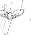
- FIGS. 1A-1Cshow perspective views of a bi-leaflet embodiment of the present invention.
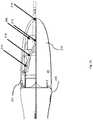
- FIGS. 2A-2Dshow side and end views of another embodiment where a camera is mounted within the transparent or translucent space of a leaflet.
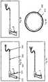
- FIGS. 3A-3Cperspective views of a lower leaflet that is capable of performing ablation and cauterization of the tissue.
- FIGS. 4A and 4Bshow perspective views of another embodiment incorporating fluid irrigation.
- FIGS. 5A-5Cshow another embodiment with a plurality of leaflets.
- FIGS. 6A-6Dshow a device for actuation of the subject invention.
- FIGS. 7A-7Dshow another embodiment where the device is constructed of a metallic or hard polymer to allow it to puncture the skin of an individual.
- FIGS. 8A-8Bshow another embodiment where the removal of the resected tissue segment is assisted by the use of high pressure fluid carried to a region near the distal end of the device.
- FIG. 9shows another embodiment where a morcellator assists in the removal of the tissue by breaking it up into small pieces.
- FIG. 10shows another embodiment whereby two jets of high pressure fluid exit a region near the tip of the device through ports.
- FIGS. 1A-1Cshow a bi-leaflet embodiment of the present invention.
- the two leafletsare identified as elements 110 and 111 .
- Either one or both of these leafletscan be made transparent or translucent for a visualizing element, which might be inserted in the outer sheath 120 of the device to see through these leaflets and provide guidance for the operator while travers in the tissue to get to a target site.
- This visualizing elementcan be a rigid or flexible endoscope, a digital micro camera like the ones that Medigus or Awaiba markets, it could also be a fiber optic image bundle or any other image-carrying device.
- Transparent or translucent leafletsmake visualization thru closed leaflets possible.
- FIG. 1Bshows another view of the present invention where the inner tube 112 is visible through the transparent leaflets. As this inner tube, 112 moves forward, it acts upon the leaflets 110 and 111 pushing them outward as explained below in FIG. 1C .
- Element 112is a tube tightly fitting within the outer sheath 120 and is in turn connected to the arm 114 through hinge 113 .
- the arm 114is in turn connected to the leaflet 111 through hinge 115 .
- Forward motion of the tube 112 in cooperation with elements 113 , 114 , and 115places an outward force on leaflet s 110 and 111 leading to their movement away from the central axis of the sheath 120 . This force will be modulated by the mechanical advantage within the handle of the device to maintain a constant dilation force by the leaflet.
- FIG. 2Ashows another embodiment of the current invention where a camera 280 is mounted within the transparent or translucent space of leaflet 211 and looks forward through the leaflet 211 long the optical axis 216 , in a somewhat paraxial arrangement.
- Camera 280is connected to the top leaflet, 211 through arm 214 and arm 214 in turn is connected to the leaflet and camera through hinges 213 and 215 .
- FIG. 2Bshows the current embodiment with the leaflets slightly opened. Camera 280 is now looking slightly downward towards the central axis of the sheath 220 .
- FIG. 2Cillustrates further opening of the leaflets and now camera 28 is looking even further inward toward the optical axis and thereby provides a more relevant image to the operator who is interested in the tissue that is closer to the opening of the device.
- FIG. 2Dis a front view of the subject invention illustrating the full lumen access upon opening of the leaflet as was mentioned before.
- FIG. 3Ashows the lower leaflet of the subject invention, 300 that is capable of performing ablation and cauterization of the tissue.
- This embodiment of the subject inventionis intended to take a partial bite of the tissue, which makes it easier to remove the dissected tissue that would be contained within the space between the two leaflets when the leaflets are closed and the tissue that's contained between the leaflets is separated from the surrounding area and the leaflets are touching alongside their length.
- cauterizing of the remaining surface of the dissected tissueprevents bleeding and creating turbidity in the irrigating fluid and thereby clouding the view.
- the leafletcan be made out of many different materials as described before.
- Element 310represents the lower leaflet that could be metallic or transparent or translucent as described before.
- Element 340represents the portion of the lower leaflet which is conducive to electrical energy and in cooperation with the upper leaflet is utilized to resect the tissue by radio frequency.
- Element 330is a high temperature non-conductive material with an expansion coefficient that is properly chosen to minimize the relative motion between elements 310 , 330 , 340 . Examples of such materials include ceramics, polyimide, polysulfone, silicone, and other materials that have the qualities that were described.
- Element 340is a conductor chosen from the metallic group of materials or conductive polymer materials. This element will be electrically connected to a source of energy that would be used to cut and cauterize tissue. There is an identical element to 340 , identified as 341 , which is located on the upper leaflet. Elements 340 and 341 could also be made out of a fiber optic that can be connected to a source of high power laser so the tissue is optically cut and cauterized as known in the art.
- a radiofrequency generatormight be utilized to supply the required energy to this element which would in turn conduct this energy through the tissue and by the virtue of ohmic resistance of the tissue heat the tissue up to very high temperature quickly and thereby vaporize the intervening tissue between elements 340 on the lower leaflet and element 341 , its mating part on the upper leaflet, 341 .
- RF energy sourcethat is bipolar as known in the art, conduction of energy occurs between these two elements, the upper and lower leaflets ( 340 , 341 ).
- Element 350identifies an insulator which could be made out of hard foam, silicone, cork, acrylic, 335 polycarbonate and similar materials.
- Element 340is a conductive metallic structure that has a sharp tip in the direction of the opposite leaflet.
- Element 330is the insulator as described previously.
- FIG. 3Bshows the current invention with both leaflets 310 and 311 in the open position.
- the sheath 360is connected to these leaflets through the hinges and arms as previously described.
- FIG. 3Cone can see both leaflets in a closed position.
- the conductive elements 340 and 341are contiguous or adjacent with very close proximity.
- Elements 350 and 351show the insulating material as described the benefit of this insulating material is evident in this figure in as much as they prevent premature contact of the two blades when the energy source is of a Bipolar nature and also they limit the size of the tissue that is resected with each closure of the leaflets.
- FIG. 4Ashows the device 400 of the current invention incorporating irrigation and aspiration.
- Lumen 420carries the irrigating fluid to the source outside the device at an appropriate pressure to the opening 430 where the irrigating fluid exits and through the inner tube 412 is aspirated.
- the arrow 440shows the direction of the flow of the irrigating fluid.
- FIG. 4Bshows another embodiment 400 ′ of the current invention incorporating fluid irrigation where the path of the fluid is between openings 430 and 431 carried by lumens 420 and 421 .
- Arrow 440shows the direction of the flow of the fluid between openings 430 and 431 .
- This embodiment of the current inventionwill provide the benefit of clearing of the operating area from blood and debris with a fluid flow path that extends beyond the distal end of the device.
- FIG. 5Ashows yet another embodiment of the subject invention with a plurality of leaflets which is comprised of 6 leaflets in this embodiment.
- This embodimentalso incorporate an opening 590 which when the leaflets are in the closed position to provide a path to follow a guide wire that may have been inserted through the path of the interest to make it easier for this embodiment of the device to navigate through the tissue to the target.
- FIG. 5Bshows the device of the FIG. 5A in an open position.
- the mechanism of opening of each of these leafletscan be inferred from FIG. 1C .
- FIG. 5Cshows the device in a completely open position accentuating the fact that the main lumen of the device 580 is unobstructed and fully available for passing of the tools of interest.
- FIG. 6A through C show a device for actuation of the subject invention which includes a handle 610 , a trigger 620 , a shaft 630 a flexible section 640 , and a distal end effector 650 .
- Figures A through Cshow the sequential movement of the trigger 620 and the opening of the end effector 650 further and further with the gradually increasing pressure applied to the trigger 620 . As this increasing pressure is being applied, where the trigger moves between positions 1 , 2 , and 3 the distal multi leaflet structure opens wider and wider.
- Outer tube 660contains an inner flexible tube 612 , which is shown in FIG. 6D in the cross sectional, view. Tube 612 is flexible enough to follow the flexible section, 640 of this device.
- FIG. 6Dshows the relationship of the outer tube 660 and the inner tube 612 .
- the tube 612 and 660are chosen such that the inner diameter of the tube 660 closely matches the outer diameter of the flexible tube 612 in order to prevent undesirable movement from tube 612 while maintaining a small gap to allow relative longitudinal motion between these tubes.
- FIG. 7Ashows another embodiment of the subject invention where the device is constructed of a metallic or hard polymer to allow it to puncture the skin of an individual.
- the lower leaflet of the device 700which is identified 710 , is made rigid to have high column strength in order to make it easier to penetrate the skin like a common hypodermic needle.
- the upper leaflet 711is hinged to the body of the device.
- FIG. 7Bshows the position of the camera within the upper leaflet 711 of the device.
- FIG. 7Cshows the device 700 with a leaflet separated and camera 712 can be seen in a position where the field of view is looking forward at the field of interest.
- FIG. 7Dshows the device 700 from a frontal point of view and as with the previous described devices the lumen 790 is unobstructed and fully available for the insertion of tools, fluids or performing aspiration.
- FIG. 8Ashows yet another embodiment of the present invention 800 where the removal of the resected tissue segment 820 is assisted by the use of high pressure fluid in two streams of 840 and 841 that are carried to a region near the distal end of the device and once the leaflet 810 and 811 come together and form a seal as in FIG. 8B they induce pressure where they push the resected tissue segment 820 back into the tube 860 and pushes it all the way out of this tube into the proximal section of the tube outside the body of the patient.
- FIG. 9shows another embodiment of this invention where morcellator 970 assists in the removal of the tissue by breaking it up into small pieces and thereby allowing it to be removed by vacuum applied at the proximal end of the tube 960 and prevents the clogging of the tube by a large piece of tissue.
- FIG. 10shows another embodiment of the present invention whereby two jets of high pressure fluid 1040 and 1041 exit a region near the tip of the device through ports 1030 and 1031 which are unitary with leaflets 1010 and 1011 .
- Fluid jet 1040 and 1041exit ports 1030 and 1031 at high velocities enabling the device to perform hydro dissection as known in the art.
- These jets of fluidcan be applied in either open or closed positions of the leaflets.
Landscapes
- Health & Medical Sciences (AREA)
- Life Sciences & Earth Sciences (AREA)
- Surgery (AREA)
- General Health & Medical Sciences (AREA)
- Biomedical Technology (AREA)
- Veterinary Medicine (AREA)
- Public Health (AREA)
- Animal Behavior & Ethology (AREA)
- Heart & Thoracic Surgery (AREA)
- Engineering & Computer Science (AREA)
- Medical Informatics (AREA)
- Nuclear Medicine, Radiotherapy & Molecular Imaging (AREA)
- Molecular Biology (AREA)
- Biophysics (AREA)
- Radiology & Medical Imaging (AREA)
- Physics & Mathematics (AREA)
- Pathology (AREA)
- Optics & Photonics (AREA)
- Anesthesiology (AREA)
- Hematology (AREA)
- Orthopedic Medicine & Surgery (AREA)
- Surgical Instruments (AREA)
Abstract
Description
Claims (20)
Priority Applications (1)
| Application Number | Priority Date | Filing Date | Title |
|---|---|---|---|
| US16/222,847US11154696B2 (en) | 2013-02-08 | 2018-12-17 | Surgical device with integrated visualization and cauterization |
Applications Claiming Priority (4)
| Application Number | Priority Date | Filing Date | Title |
|---|---|---|---|
| US201361762660P | 2013-02-08 | 2013-02-08 | |
| US14/168,717US20140228875A1 (en) | 2013-02-08 | 2014-01-30 | Surgical device with integrated visualization and cauterization |
| US14/957,475US10201687B2 (en) | 2013-02-08 | 2015-12-02 | Surgical device with integrated visualization and cauterization |
| US16/222,847US11154696B2 (en) | 2013-02-08 | 2018-12-17 | Surgical device with integrated visualization and cauterization |
Related Parent Applications (1)
| Application Number | Title | Priority Date | Filing Date |
|---|---|---|---|
| US14/957,475ContinuationUS10201687B2 (en) | 2013-02-08 | 2015-12-02 | Surgical device with integrated visualization and cauterization |
Publications (2)
| Publication Number | Publication Date |
|---|---|
| US20190381290A1 US20190381290A1 (en) | 2019-12-19 |
| US11154696B2true US11154696B2 (en) | 2021-10-26 |
Family
ID=51297972
Family Applications (3)
| Application Number | Title | Priority Date | Filing Date |
|---|---|---|---|
| US14/168,717AbandonedUS20140228875A1 (en) | 2013-02-08 | 2014-01-30 | Surgical device with integrated visualization and cauterization |
| US14/957,475Active2034-06-03US10201687B2 (en) | 2013-02-08 | 2015-12-02 | Surgical device with integrated visualization and cauterization |
| US16/222,847Active2034-06-16US11154696B2 (en) | 2013-02-08 | 2018-12-17 | Surgical device with integrated visualization and cauterization |
Family Applications Before (2)
| Application Number | Title | Priority Date | Filing Date |
|---|---|---|---|
| US14/168,717AbandonedUS20140228875A1 (en) | 2013-02-08 | 2014-01-30 | Surgical device with integrated visualization and cauterization |
| US14/957,475Active2034-06-03US10201687B2 (en) | 2013-02-08 | 2015-12-02 | Surgical device with integrated visualization and cauterization |
Country Status (1)
| Country | Link |
|---|---|
| US (3) | US20140228875A1 (en) |
Families Citing this family (39)
| Publication number | Priority date | Publication date | Assignee | Title |
|---|---|---|---|---|
| US9913577B2 (en) | 2010-09-28 | 2018-03-13 | Obp Medical Corporation | Speculum |
| US20140228875A1 (en)* | 2013-02-08 | 2014-08-14 | Nidus Medical, Llc | Surgical device with integrated visualization and cauterization |
| US20150031946A1 (en) | 2013-07-24 | 2015-01-29 | Nidus Medical, Llc | Direct vision cryosurgical probe and methods of use |
| WO2015048806A2 (en) | 2013-09-30 | 2015-04-02 | Nidus Medical, Llc | Apparatus and methods for treating rhinitis |
| WO2015077584A2 (en) | 2013-11-22 | 2015-05-28 | Massachusetts Institute Of Technology | Steering techniques for surgical instruments |
| US9763743B2 (en) | 2014-07-25 | 2017-09-19 | Arrinex, Inc. | Apparatus and method for treating rhinitis |
| WO2016076964A2 (en)* | 2014-09-30 | 2016-05-19 | Massachusetts Institute Of Technology | Instruments for minimally invasive surgical procedures |
| US10420538B2 (en) | 2015-02-05 | 2019-09-24 | Obp Medical Corporation | Illuminated surgical retractor |
| US9867602B2 (en) | 2015-02-05 | 2018-01-16 | Obp Medical Corporation | Illuminated surgical retractor |
| CA2975885A1 (en) | 2015-02-27 | 2016-09-01 | Covidien Lp | Expanding endoscope and method |
| US12268433B2 (en) | 2015-05-12 | 2025-04-08 | National University Of Ireland, Galway | Devices for therapeutic nasal neuromodulation and associated methods and systems |
| WO2016183337A2 (en) | 2015-05-12 | 2016-11-17 | National University Of Ireland Galway | Devices for therapeutic nasal neuromodulation and associated methods and systems |
| US10939899B2 (en) | 2015-06-03 | 2021-03-09 | Obp Medical Corporation | End cap assembly for retractor and other medical devices |
| US10881387B2 (en) | 2015-06-03 | 2021-01-05 | Obp Medical Corporation | Retractor |
| ES2968069T3 (en) | 2015-06-03 | 2024-05-07 | Obp Surgical Corp | Retractor |
| US20170119234A1 (en)* | 2015-10-14 | 2017-05-04 | Cook Medical Technologies Llc | Endoscope cap with separable arms |
| EP3413822B1 (en) | 2016-02-11 | 2023-08-30 | Arrinex, Inc. | Device for image guided post-nasal nerve ablation |
| CN109600988B (en) | 2016-06-15 | 2021-08-31 | 阿里内克斯股份有限公司 | Devices and methods for treating the lateral surface of the nasal cavity |
| US10722621B2 (en) | 2016-07-11 | 2020-07-28 | Obp Medical Corporation | Illuminated suction device |
| DE102016114881A1 (en)* | 2016-08-11 | 2018-02-15 | Digital Endoscopy Gmbh | Endoscope head, endoscope and albarrane lever holding element |
| US11096560B2 (en) | 2016-09-23 | 2021-08-24 | Meditrina, Inc. | Endoscope with multiple image sensors |
| JP6669625B2 (en)* | 2016-10-11 | 2020-03-18 | 京セラ株式会社 | Trocar device with camera |
| US11253312B2 (en) | 2016-10-17 | 2022-02-22 | Arrinex, Inc. | Integrated nasal nerve detector ablation-apparatus, nasal nerve locator, and methods of use |
| EP3614940B1 (en) | 2017-04-28 | 2024-11-20 | Arrinex, Inc. | Systems for locating blood vessels in the treatment of rhinitis |
| US10716594B2 (en) | 2017-06-12 | 2020-07-21 | Covidien Lp | Devices, systems, and methods for tissue specimen removal |
| US10687793B2 (en) | 2017-07-18 | 2020-06-23 | Obp Medical Corporation | Minimally invasive no touch (MINT) procedure for harvesting the great saphenous vein (GSV) and venous hydrodissector and retractor for use during the MINT procedure |
| US10278572B1 (en) | 2017-10-19 | 2019-05-07 | Obp Medical Corporation | Speculum |
| US10799229B2 (en) | 2018-02-20 | 2020-10-13 | Obp Medical Corporation | Illuminated medical devices |
| US11547473B2 (en) | 2018-12-11 | 2023-01-10 | Neurent Medical Limited | Systems and methods for therapeutic nasal neuromodulation |
| DE102019118043A1 (en)* | 2019-07-04 | 2021-01-07 | Ovesco Endoscopy Ag | Treatment instrument for endoscope |
| US12121281B2 (en) | 2019-08-07 | 2024-10-22 | Christopher M. Shaari | Systems and methods for cryogenic treatment of headache |
| US20210204907A1 (en) | 2020-01-07 | 2021-07-08 | Covidien Lp | Devices, systems, and methods for trans-vaginal, ultrasound-guided hysteroscopic surgical procedures |
| US10959609B1 (en) | 2020-01-31 | 2021-03-30 | Obp Medical Corporation | Illuminated suction device |
| US10966702B1 (en) | 2020-02-25 | 2021-04-06 | Obp Medical Corporation | Illuminated dual-blade retractor |
| WO2021205229A1 (en) | 2020-04-09 | 2021-10-14 | Neurent Medical Limited | Systems and methods for improving sleep with therapeutic nasal treatment |
| US11896818B2 (en) | 2020-04-09 | 2024-02-13 | Neurent Medical Limited | Systems and methods for therapeutic nasal treatment |
| DE102020132773B3 (en)* | 2020-12-09 | 2021-11-11 | Karl Storz Se & Co. Kg | Endoscope with cleanable rotating drum and cleaning procedure |
| WO2024054671A2 (en)* | 2022-09-09 | 2024-03-14 | The University Of North Carolina At Charlotte | End effector assemblies for surgical instruments |
| US12318080B2 (en) | 2023-07-21 | 2025-06-03 | Coopersurgical, Inc. | Illuminated surgical retractor capable of hand-held operation and of being mounted to a fixed frame |
Citations (83)
| Publication number | Priority date | Publication date | Assignee | Title |
|---|---|---|---|---|
| US5353784A (en) | 1993-04-02 | 1994-10-11 | The Research Foundation Of Suny | Endoscopic device and method of use |
| US5527351A (en) | 1994-09-21 | 1996-06-18 | Friedman; Mark H. | Treatment of vascular and tension headache atypical facial pain allergic rhinitis and cervical muscle hyperactivity |
| US5611796A (en) | 1994-08-04 | 1997-03-18 | Kamami; Yves-Victor | Handpiece for a device for performing laser surgery of the nose |
| US5658307A (en)* | 1990-11-07 | 1997-08-19 | Exconde; Primo D. | Method of using a surgical dissector instrument |
| US5733280A (en) | 1995-11-15 | 1998-03-31 | Avitall; Boaz | Cryogenic epicardial mapping and ablation |
| US5827305A (en)* | 1996-01-24 | 1998-10-27 | Gordon; Mark G. | Tissue sampling device |
| US5899899A (en) | 1997-02-27 | 1999-05-04 | Cryocath Technologies Inc. | Cryosurgical linear ablation structure |
| US6106518A (en) | 1998-04-09 | 2000-08-22 | Cryocath Technologies, Inc. | Variable geometry tip for a cryosurgical ablation device |
| US6139508A (en)* | 1998-08-04 | 2000-10-31 | Endonetics, Inc. | Articulated medical device |
| US6210035B1 (en) | 1997-11-28 | 2001-04-03 | Seiko Instruments Inc. | High-speed thermal analyzer |
| US6270476B1 (en) | 1999-04-23 | 2001-08-07 | Cryocath Technologies, Inc. | Catheter |
| US20020029006A1 (en)* | 1996-11-25 | 2002-03-07 | Scimed Life Systems, Inc. | Biopsy instrument having irrigation and aspiration capabilities |
| US20030114659A1 (en) | 1988-11-11 | 2003-06-19 | Medical Research Council | Single domain ligands, receptors comprising said ligands, methods for their production, and use of said ligands and receptors |
| US6595988B2 (en) | 2000-06-23 | 2003-07-22 | Cryocath Technologies Inc. | Cryotreatment device and method |
| US20040224412A1 (en) | 2001-05-23 | 2004-11-11 | Abdelali Hannoufa | Repressor-mediated regulation system for control of gene expression in plants |
| US20050096502A1 (en) | 2003-10-29 | 2005-05-05 | Khalili Theodore M. | Robotic surgical device |
| US7104984B2 (en) | 2003-08-22 | 2006-09-12 | Cryocor, Inc. | Reshapeable tip for a cryoprobe |
| US20060235474A1 (en) | 2002-04-08 | 2006-10-19 | Ardian, Inc. | Methods and apparatus for multi-vessel renal neuromodulation |
| US20060276852A1 (en) | 2002-04-08 | 2006-12-07 | Ardian, Inc. | Methods and apparatus for treating hypertension |
| US20070173899A1 (en) | 2002-04-08 | 2007-07-26 | Ardian, Inc. | Renal nerve stimulation method for treatment of patients |
| US20070265687A1 (en) | 2002-04-08 | 2007-11-15 | Ardian, Inc. | Apparatuses for renal neuromodulation |
| US7300433B2 (en) | 1999-08-23 | 2007-11-27 | Cryocath Technologies Inc. | Endovascular cryotreatment catheter |
| US20070299433A1 (en) | 2006-06-27 | 2007-12-27 | C2 Therapeutics | Barrett's Esophagus Cryogenic Ablation System |
| US20080027423A1 (en) | 2006-07-25 | 2008-01-31 | Zoom Therapeutics, Inc. | Systems for treatment of nasal tissue |
| WO2008051918A2 (en) | 2006-10-23 | 2008-05-02 | Allux Medical, Inc. | Methods, devices and kits for phototherapy and photodynamic therapy treatment of body cavities |
| US20080119693A1 (en) | 2004-04-21 | 2008-05-22 | Acclarent, Inc. | Methods and Apparatus for Treating Disorders of the Ear, Nose and Throat |
| US7418292B2 (en) | 2003-10-01 | 2008-08-26 | Medtronic, Inc. | Device and method for attenuating an immune response |
| US20080208230A1 (en)* | 2007-02-22 | 2008-08-28 | Singfatt Chin | Expandable rotating device and method for tissue aspiration |
| US20090030276A1 (en) | 2007-07-27 | 2009-01-29 | Voyage Medical, Inc. | Tissue visualization catheter with imaging systems integration |
| US20090062873A1 (en) | 2006-06-28 | 2009-03-05 | Ardian, Inc. | Methods and systems for thermally-induced renal neuromodulation |
| US7527601B2 (en) | 2005-12-29 | 2009-05-05 | Intrapartum Ventures, Llc | Cervimeter |
| US20090187074A1 (en) | 2008-01-17 | 2009-07-23 | Nidus Medical, Llc | Epicardial access and treatment systems |
| US20090234345A1 (en) | 2008-03-13 | 2009-09-17 | Raphael Hon | Cryo-ablation refrigerant distribution catheter |
| US20090326572A1 (en) | 2008-06-27 | 2009-12-31 | Ruey-Feng Peh | Apparatus and methods for rapid tissue crossing |
| US20100057150A1 (en) | 2002-04-08 | 2010-03-04 | Ardian, Inc. | Methods and apparatus for pulsed electric field neuromodulation via an intra-to-extravascular approach |
| US20100137952A1 (en) | 2002-04-08 | 2010-06-03 | Ardian, Inc. | Apparatuses for thermally-induced renal neuromodulation |
| US20100137860A1 (en) | 2002-04-08 | 2010-06-03 | Ardian, Inc. | Apparatus for performing a non-continuous circumferential treatment of a body lumen |
| US20100168739A1 (en) | 2008-12-31 | 2010-07-01 | Ardian, Inc. | Apparatus, systems, and methods for achieving intravascular, thermally-induced renal neuromodulation |
| US20100168731A1 (en) | 2008-12-31 | 2010-07-01 | Ardian, Inc. | Apparatus, systems, and methods for achieving intravascular, thermally-induced renal neuromodulation |
| US20100174282A1 (en) | 2002-04-08 | 2010-07-08 | Ardian, Inc. | Apparatus for thermal modulation of nerves contributing to renal function |
| US7769442B2 (en) | 2003-10-01 | 2010-08-03 | Medtronic, Inc. | Device and method for inhibiting release of pro-inflammatory mediator |
| US20100256629A1 (en) | 2009-04-06 | 2010-10-07 | Voyage Medical, Inc. | Methods and devices for treatment of the ostium |
| US20110152855A1 (en) | 2009-10-27 | 2011-06-23 | Mayse Martin L | Delivery devices with coolable energy emitting assemblies |
| US20110184402A1 (en) | 2009-11-02 | 2011-07-28 | Cpsi Biotech | Flexible Cryogenic Probe Tip |
| US8088127B2 (en) | 2008-05-09 | 2012-01-03 | Innovative Pulmonary Solutions, Inc. | Systems, assemblies, and methods for treating a bronchial tree |
| US20120016260A1 (en)* | 2008-10-20 | 2012-01-19 | To John T | Retractor cannula system for accessing and visualizing spine and related methods |
| WO2012027641A2 (en) | 2010-08-26 | 2012-03-01 | Cryomedix, Llc | Cryoablation balloon catheter and related method |
| US8142424B2 (en) | 2004-05-10 | 2012-03-27 | Boston Scientific Scimed, Inc. | Probe based low temperature lesion formation apparatus, systems and methods |
| US8231613B2 (en) | 1999-07-14 | 2012-07-31 | Cardiofocus, Inc. | Deflectable sheath catheters |
| EP2532300A2 (en) | 2005-01-18 | 2012-12-12 | Acclarent, Inc. | Paranasal insertion device |
| US20130006326A1 (en) | 2010-11-16 | 2013-01-03 | Douglas Michael Ackermann | Stimulation devices and methods |
| US20130018366A1 (en) | 2011-07-11 | 2013-01-17 | C2 Therapeutics | Focal Ablation Assembly |
| US8382746B2 (en) | 2008-11-21 | 2013-02-26 | C2 Therapeutics, Inc. | Cryogenic ablation system and method |
| US8388600B1 (en) | 2008-09-04 | 2013-03-05 | Dolor Technologies | Apparatus, system, and method for treating atypical headaches |
| US8394075B2 (en) | 2000-06-16 | 2013-03-12 | Mehdi M. Ansarinia | Stimulation method for the sphenopalatine ganglia, sphenopalatine nerve, or vidian nerve for treatment of medical conditions |
| WO2013035192A1 (en) | 2011-09-09 | 2013-03-14 | 医療法人社団アドベント | Method and device for treating symptom of rhinitis surgically |
| US8425457B2 (en) | 2004-04-21 | 2013-04-23 | Acclarent, Inc. | Devices, systems and methods for diagnosing and treating sinusitus and other disorder of the ears, nose and/or throat |
| US8512324B2 (en) | 2005-04-29 | 2013-08-20 | Medtronic Cryocath Lp | Wide area ablation of myocardial tissue |
| US20130253387A1 (en) | 2012-03-08 | 2013-09-26 | Sonitec, LLC | Vibratory energy systems and methods for occluded body cavities |
| WO2013173481A2 (en) | 2012-05-18 | 2013-11-21 | Holaira, Inc. | Compact delivery pulmonary treatment systems and methods for improving pulmonary function |
| US20130345700A1 (en) | 2008-02-15 | 2013-12-26 | Holaira, Inc. | System and method for bronchial dilation |
| US8676324B2 (en) | 2005-11-10 | 2014-03-18 | ElectroCore, LLC | Electrical and magnetic stimulators used to treat migraine/sinus headache, rhinitis, sinusitis, rhinosinusitis, and comorbid disorders |
| US8715275B2 (en) | 2007-11-14 | 2014-05-06 | Myoscience, Inc. | Pain management using cryogenic remodeling |
| US20140186341A1 (en) | 2012-12-27 | 2014-07-03 | Holaira, Inc. | Methods for improving drug efficacy |
| US20140257271A1 (en) | 2011-10-05 | 2014-09-11 | Holaira, Inc. | Apparatus for injuring nerve tissue |
| US20140276792A1 (en) | 2013-03-13 | 2014-09-18 | Holaira, Inc. | Fluid delivery system and method for treatment |
| US20140277429A1 (en) | 2013-03-12 | 2014-09-18 | Oculeve, Inc. | Implant delivery devices, systems, and methods |
| US20140316310A1 (en) | 2013-04-19 | 2014-10-23 | Oculeve, Inc. | Nasal stimulation devices and methods |
| US20150031946A1 (en) | 2013-07-24 | 2015-01-29 | Nidus Medical, Llc | Direct vision cryosurgical probe and methods of use |
| US20150080870A1 (en) | 2010-10-22 | 2015-03-19 | Medtronic Cryocath Lp | Balloon catheter with deformable fluid delivery conduit |
| US20150126986A1 (en) | 2011-04-25 | 2015-05-07 | Medtronic Ardian Luxembourg S.A.R.L. | Apparatus and Methods Related to Constrained Deployment of Cryogenic Balloons for Limited Cryogenic Abiation of Vessel Walls |
| US9050073B2 (en) | 2013-11-01 | 2015-06-09 | C2 Therapeutics, Inc. | Cryogenic balloon ablation system |
| US20150164571A1 (en) | 2013-09-30 | 2015-06-18 | Nidus Medical, Llc | Apparatus and methods for treating rhinitis |
| US9084592B2 (en) | 2011-07-11 | 2015-07-21 | C2 Therapeutics, Inc. | Focal ablation assembly |
| US20150238754A1 (en) | 2014-02-25 | 2015-08-27 | Oculeve, Inc. | Polymer formulations for nasolacrimal stimulation |
| US20160022992A1 (en) | 2014-07-25 | 2016-01-28 | Oculeve, Inc. | Stimulation patterns for treating dry eye |
| US20160045277A1 (en) | 2014-07-25 | 2016-02-18 | Nidus Medical, Llc | Apparatus and method for treating rhinitis |
| US9265956B2 (en) | 2013-03-08 | 2016-02-23 | Oculeve, Inc. | Devices and methods for treating dry eye in animals |
| US20160101286A1 (en) | 2012-06-15 | 2016-04-14 | Case Western Reserve University | Methods of treatment of a neurological disorder using electrical nerve conduction block |
| US20160114172A1 (en) | 2014-10-22 | 2016-04-28 | Oculeve, Inc. | Contact lens for increasing tear production |
| US20160114163A1 (en) | 2014-10-22 | 2016-04-28 | Oculeve, Inc. | Implantable nasal stimulator systems and methods |
| US10201687B2 (en)* | 2013-02-08 | 2019-02-12 | Arrinex, Inc. | Surgical device with integrated visualization and cauterization |
| EP2862046B1 (en) | 2012-06-13 | 2019-05-01 | Google LLC | Interfacing with windows having hidden borders |
Family Cites Families (3)
| Publication number | Priority date | Publication date | Assignee | Title |
|---|---|---|---|---|
| US5843021A (en) | 1994-05-09 | 1998-12-01 | Somnus Medical Technologies, Inc. | Cell necrosis apparatus |
| US5746224A (en) | 1994-06-24 | 1998-05-05 | Somnus Medical Technologies, Inc. | Method for ablating turbinates |
| US6921385B2 (en) | 2002-08-05 | 2005-07-26 | Alcon, Inc. | Apparatus for delivery of fluid to opthalmic surgical handpiece |
- 2014
- 2014-01-30USUS14/168,717patent/US20140228875A1/ennot_activeAbandoned
- 2015
- 2015-12-02USUS14/957,475patent/US10201687B2/enactiveActive
- 2018
- 2018-12-17USUS16/222,847patent/US11154696B2/enactiveActive
Patent Citations (99)
| Publication number | Priority date | Publication date | Assignee | Title |
|---|---|---|---|---|
| US20030114659A1 (en) | 1988-11-11 | 2003-06-19 | Medical Research Council | Single domain ligands, receptors comprising said ligands, methods for their production, and use of said ligands and receptors |
| US5658307A (en)* | 1990-11-07 | 1997-08-19 | Exconde; Primo D. | Method of using a surgical dissector instrument |
| US5353784A (en) | 1993-04-02 | 1994-10-11 | The Research Foundation Of Suny | Endoscopic device and method of use |
| US5611796A (en) | 1994-08-04 | 1997-03-18 | Kamami; Yves-Victor | Handpiece for a device for performing laser surgery of the nose |
| US5527351A (en) | 1994-09-21 | 1996-06-18 | Friedman; Mark H. | Treatment of vascular and tension headache atypical facial pain allergic rhinitis and cervical muscle hyperactivity |
| US5733280A (en) | 1995-11-15 | 1998-03-31 | Avitall; Boaz | Cryogenic epicardial mapping and ablation |
| US5827305A (en)* | 1996-01-24 | 1998-10-27 | Gordon; Mark G. | Tissue sampling device |
| US20020029006A1 (en)* | 1996-11-25 | 2002-03-07 | Scimed Life Systems, Inc. | Biopsy instrument having irrigation and aspiration capabilities |
| US5899898A (en) | 1997-02-27 | 1999-05-04 | Cryocath Technologies Inc. | Cryosurgical linear ablation |
| US5899899A (en) | 1997-02-27 | 1999-05-04 | Cryocath Technologies Inc. | Cryosurgical linear ablation structure |
| US6210035B1 (en) | 1997-11-28 | 2001-04-03 | Seiko Instruments Inc. | High-speed thermal analyzer |
| US6106518A (en) | 1998-04-09 | 2000-08-22 | Cryocath Technologies, Inc. | Variable geometry tip for a cryosurgical ablation device |
| US6139508A (en)* | 1998-08-04 | 2000-10-31 | Endonetics, Inc. | Articulated medical device |
| US6270476B1 (en) | 1999-04-23 | 2001-08-07 | Cryocath Technologies, Inc. | Catheter |
| US8231613B2 (en) | 1999-07-14 | 2012-07-31 | Cardiofocus, Inc. | Deflectable sheath catheters |
| US7300433B2 (en) | 1999-08-23 | 2007-11-27 | Cryocath Technologies Inc. | Endovascular cryotreatment catheter |
| US8394075B2 (en) | 2000-06-16 | 2013-03-12 | Mehdi M. Ansarinia | Stimulation method for the sphenopalatine ganglia, sphenopalatine nerve, or vidian nerve for treatment of medical conditions |
| US6595988B2 (en) | 2000-06-23 | 2003-07-22 | Cryocath Technologies Inc. | Cryotreatment device and method |
| US20040224412A1 (en) | 2001-05-23 | 2004-11-11 | Abdelali Hannoufa | Repressor-mediated regulation system for control of gene expression in plants |
| US20100057150A1 (en) | 2002-04-08 | 2010-03-04 | Ardian, Inc. | Methods and apparatus for pulsed electric field neuromodulation via an intra-to-extravascular approach |
| US20070173899A1 (en) | 2002-04-08 | 2007-07-26 | Ardian, Inc. | Renal nerve stimulation method for treatment of patients |
| US20070265687A1 (en) | 2002-04-08 | 2007-11-15 | Ardian, Inc. | Apparatuses for renal neuromodulation |
| US20060276852A1 (en) | 2002-04-08 | 2006-12-07 | Ardian, Inc. | Methods and apparatus for treating hypertension |
| US20100191112A1 (en) | 2002-04-08 | 2010-07-29 | Ardian, Inc. | Ultrasound apparatuses for thermally-induced renal neuromodulation |
| US20100137952A1 (en) | 2002-04-08 | 2010-06-03 | Ardian, Inc. | Apparatuses for thermally-induced renal neuromodulation |
| US20090036948A1 (en) | 2002-04-08 | 2009-02-05 | Ardian, Inc. | Renal nerve stimulation methods for treatment of patients |
| US20060235474A1 (en) | 2002-04-08 | 2006-10-19 | Ardian, Inc. | Methods and apparatus for multi-vessel renal neuromodulation |
| US20100174282A1 (en) | 2002-04-08 | 2010-07-08 | Ardian, Inc. | Apparatus for thermal modulation of nerves contributing to renal function |
| US20100137860A1 (en) | 2002-04-08 | 2010-06-03 | Ardian, Inc. | Apparatus for performing a non-continuous circumferential treatment of a body lumen |
| US7104984B2 (en) | 2003-08-22 | 2006-09-12 | Cryocor, Inc. | Reshapeable tip for a cryoprobe |
| US7418292B2 (en) | 2003-10-01 | 2008-08-26 | Medtronic, Inc. | Device and method for attenuating an immune response |
| US7769442B2 (en) | 2003-10-01 | 2010-08-03 | Medtronic, Inc. | Device and method for inhibiting release of pro-inflammatory mediator |
| US20050096502A1 (en) | 2003-10-29 | 2005-05-05 | Khalili Theodore M. | Robotic surgical device |
| US8425457B2 (en) | 2004-04-21 | 2013-04-23 | Acclarent, Inc. | Devices, systems and methods for diagnosing and treating sinusitus and other disorder of the ears, nose and/or throat |
| US20080119693A1 (en) | 2004-04-21 | 2008-05-22 | Acclarent, Inc. | Methods and Apparatus for Treating Disorders of the Ear, Nose and Throat |
| US8142424B2 (en) | 2004-05-10 | 2012-03-27 | Boston Scientific Scimed, Inc. | Probe based low temperature lesion formation apparatus, systems and methods |
| EP2532300A2 (en) | 2005-01-18 | 2012-12-12 | Acclarent, Inc. | Paranasal insertion device |
| US8512324B2 (en) | 2005-04-29 | 2013-08-20 | Medtronic Cryocath Lp | Wide area ablation of myocardial tissue |
| US8679104B2 (en) | 2005-04-29 | 2014-03-25 | Medtronic Cryocath Lp | Wide area ablation of myocardial tissue |
| US8676324B2 (en) | 2005-11-10 | 2014-03-18 | ElectroCore, LLC | Electrical and magnetic stimulators used to treat migraine/sinus headache, rhinitis, sinusitis, rhinosinusitis, and comorbid disorders |
| US7527601B2 (en) | 2005-12-29 | 2009-05-05 | Intrapartum Ventures, Llc | Cervimeter |
| US20070299433A1 (en) | 2006-06-27 | 2007-12-27 | C2 Therapeutics | Barrett's Esophagus Cryogenic Ablation System |
| US20090076409A1 (en) | 2006-06-28 | 2009-03-19 | Ardian, Inc. | Methods and systems for thermally-induced renal neuromodulation |
| US20090062873A1 (en) | 2006-06-28 | 2009-03-05 | Ardian, Inc. | Methods and systems for thermally-induced renal neuromodulation |
| US20080027423A1 (en) | 2006-07-25 | 2008-01-31 | Zoom Therapeutics, Inc. | Systems for treatment of nasal tissue |
| WO2008051918A2 (en) | 2006-10-23 | 2008-05-02 | Allux Medical, Inc. | Methods, devices and kits for phototherapy and photodynamic therapy treatment of body cavities |
| US20080208230A1 (en)* | 2007-02-22 | 2008-08-28 | Singfatt Chin | Expandable rotating device and method for tissue aspiration |
| US20090030276A1 (en) | 2007-07-27 | 2009-01-29 | Voyage Medical, Inc. | Tissue visualization catheter with imaging systems integration |
| US8715275B2 (en) | 2007-11-14 | 2014-05-06 | Myoscience, Inc. | Pain management using cryogenic remodeling |
| US20090187074A1 (en) | 2008-01-17 | 2009-07-23 | Nidus Medical, Llc | Epicardial access and treatment systems |
| US20140236148A1 (en) | 2008-02-15 | 2014-08-21 | Holaira, Inc. | System and method for bronchial dilation |
| US20130345700A1 (en) | 2008-02-15 | 2013-12-26 | Holaira, Inc. | System and method for bronchial dilation |
| US20090234345A1 (en) | 2008-03-13 | 2009-09-17 | Raphael Hon | Cryo-ablation refrigerant distribution catheter |
| EP2662116A1 (en) | 2008-05-09 | 2013-11-13 | Holaira, Inc. | Systems, assemblies, and methods for treating a bronchial tree |
| EP2662027A1 (en) | 2008-05-09 | 2013-11-13 | Holaira, Inc. | Systems, assemblies, and methods for treating a bronchial tree |
| US8088127B2 (en) | 2008-05-09 | 2012-01-03 | Innovative Pulmonary Solutions, Inc. | Systems, assemblies, and methods for treating a bronchial tree |
| US20090326572A1 (en) | 2008-06-27 | 2009-12-31 | Ruey-Feng Peh | Apparatus and methods for rapid tissue crossing |
| US8388600B1 (en) | 2008-09-04 | 2013-03-05 | Dolor Technologies | Apparatus, system, and method for treating atypical headaches |
| US20120016260A1 (en)* | 2008-10-20 | 2012-01-19 | To John T | Retractor cannula system for accessing and visualizing spine and related methods |
| US8382746B2 (en) | 2008-11-21 | 2013-02-26 | C2 Therapeutics, Inc. | Cryogenic ablation system and method |
| US9168081B2 (en) | 2008-11-21 | 2015-10-27 | C2 Therapeutics, Inc. | Cryogenic ablation system and method |
| US20100168739A1 (en) | 2008-12-31 | 2010-07-01 | Ardian, Inc. | Apparatus, systems, and methods for achieving intravascular, thermally-induced renal neuromodulation |
| US20100168731A1 (en) | 2008-12-31 | 2010-07-01 | Ardian, Inc. | Apparatus, systems, and methods for achieving intravascular, thermally-induced renal neuromodulation |
| US20100256629A1 (en) | 2009-04-06 | 2010-10-07 | Voyage Medical, Inc. | Methods and devices for treatment of the ostium |
| US20110152855A1 (en) | 2009-10-27 | 2011-06-23 | Mayse Martin L | Delivery devices with coolable energy emitting assemblies |
| US20110184402A1 (en) | 2009-11-02 | 2011-07-28 | Cpsi Biotech | Flexible Cryogenic Probe Tip |
| WO2012027641A2 (en) | 2010-08-26 | 2012-03-01 | Cryomedix, Llc | Cryoablation balloon catheter and related method |
| WO2012027641A3 (en) | 2010-08-26 | 2012-05-18 | Cryomedix, Llc | Cryoablation balloon catheter and related method |
| US20150080870A1 (en) | 2010-10-22 | 2015-03-19 | Medtronic Cryocath Lp | Balloon catheter with deformable fluid delivery conduit |
| US20130006326A1 (en) | 2010-11-16 | 2013-01-03 | Douglas Michael Ackermann | Stimulation devices and methods |
| US20150126986A1 (en) | 2011-04-25 | 2015-05-07 | Medtronic Ardian Luxembourg S.A.R.L. | Apparatus and Methods Related to Constrained Deployment of Cryogenic Balloons for Limited Cryogenic Abiation of Vessel Walls |
| US9084592B2 (en) | 2011-07-11 | 2015-07-21 | C2 Therapeutics, Inc. | Focal ablation assembly |
| US20130018366A1 (en) | 2011-07-11 | 2013-01-17 | C2 Therapeutics | Focal Ablation Assembly |
| US20150313661A1 (en) | 2011-07-11 | 2015-11-05 | C2 Therapeutics, Inc. | Focal ablation assembly |
| WO2013035192A1 (en) | 2011-09-09 | 2013-03-14 | 医療法人社団アドベント | Method and device for treating symptom of rhinitis surgically |
| US20140257271A1 (en) | 2011-10-05 | 2014-09-11 | Holaira, Inc. | Apparatus for injuring nerve tissue |
| US20130253387A1 (en) | 2012-03-08 | 2013-09-26 | Sonitec, LLC | Vibratory energy systems and methods for occluded body cavities |
| US20130310822A1 (en) | 2012-05-18 | 2013-11-21 | Holaira, Inc. | Compact delivery pulmonary treatment systems and methods for improving pulmonary function |
| WO2013173481A2 (en) | 2012-05-18 | 2013-11-21 | Holaira, Inc. | Compact delivery pulmonary treatment systems and methods for improving pulmonary function |
| EP2862046B1 (en) | 2012-06-13 | 2019-05-01 | Google LLC | Interfacing with windows having hidden borders |
| US20160101286A1 (en) | 2012-06-15 | 2016-04-14 | Case Western Reserve University | Methods of treatment of a neurological disorder using electrical nerve conduction block |
| US20140186341A1 (en) | 2012-12-27 | 2014-07-03 | Holaira, Inc. | Methods for improving drug efficacy |
| US10201687B2 (en)* | 2013-02-08 | 2019-02-12 | Arrinex, Inc. | Surgical device with integrated visualization and cauterization |
| US9265956B2 (en) | 2013-03-08 | 2016-02-23 | Oculeve, Inc. | Devices and methods for treating dry eye in animals |
| US20140277429A1 (en) | 2013-03-12 | 2014-09-18 | Oculeve, Inc. | Implant delivery devices, systems, and methods |
| US20140276792A1 (en) | 2013-03-13 | 2014-09-18 | Holaira, Inc. | Fluid delivery system and method for treatment |
| US8996137B2 (en) | 2013-04-19 | 2015-03-31 | Oculeve, Inc. | Nasal stimulation devices and methods |
| US20140316310A1 (en) | 2013-04-19 | 2014-10-23 | Oculeve, Inc. | Nasal stimulation devices and methods |
| US20140371812A1 (en) | 2013-04-19 | 2014-12-18 | Oculeve, Inc. | Nasal stimulation devices and methods |
| US20160158548A1 (en) | 2013-04-19 | 2016-06-09 | Oculeve, Inc. | Nasal stimulation devices and methods |
| US20150031946A1 (en) | 2013-07-24 | 2015-01-29 | Nidus Medical, Llc | Direct vision cryosurgical probe and methods of use |
| US20150164571A1 (en) | 2013-09-30 | 2015-06-18 | Nidus Medical, Llc | Apparatus and methods for treating rhinitis |
| US9050073B2 (en) | 2013-11-01 | 2015-06-09 | C2 Therapeutics, Inc. | Cryogenic balloon ablation system |
| US20150196345A1 (en) | 2013-11-01 | 2015-07-16 | C2 Therapeutics, Inc. | Cryogenic balloon ablation system |
| US20150238754A1 (en) | 2014-02-25 | 2015-08-27 | Oculeve, Inc. | Polymer formulations for nasolacrimal stimulation |
| US20160045277A1 (en) | 2014-07-25 | 2016-02-18 | Nidus Medical, Llc | Apparatus and method for treating rhinitis |
| US20160022992A1 (en) | 2014-07-25 | 2016-01-28 | Oculeve, Inc. | Stimulation patterns for treating dry eye |
| US20160114172A1 (en) | 2014-10-22 | 2016-04-28 | Oculeve, Inc. | Contact lens for increasing tear production |
| US20160114163A1 (en) | 2014-10-22 | 2016-04-28 | Oculeve, Inc. | Implantable nasal stimulator systems and methods |
Non-Patent Citations (12)
| Title |
|---|
| Arora, et al., "Cryodestruction of Vidian Nerve Branches", Indian Journal of Otolaryngology, vol. 31, No. 3, Sep. 1980, pp. 80-82. |
| Bumsted, "Cryotherapy for Chronic Vasomotor Rhinitis: Technique and Patient Selection or Improved Results", Laryngoscope, vol. 94, Apr. 1984, pp. 539-544. |
| Girdar-Gopal, An Assessment of Postganglionic Cryoneurolysis for Managing Vasomotor Rhinitis:, American Journal of Rhinology, vol. 8, No. 4, Jul.-Aug. 1994, pp. 157-164. |
| Golhar, et al., "The effect of Cyrodestruction of Vidian Nasal Branches on Nasal Mucus Flow in Vasomotor Rhinitis", Indian Journal of otolaryngology, vol. 33, No. 1, Mar. 1981, pp. 12-14. |
| Goode, "A Liquid Nitrogen Turbinate Probe for Hypertrophic Rhinitis", Arch Otolaryngol., vol. 103, 1977, p. 431. |
| Mehra, et al., "Cryosurgery in Vasomotor Rhinitis—An Analysis of 156 Patients", Indian Journal of Otolaryngology, vol. 83, No. 4, 1973, pp. 508-516. |
| Ozenberger, "Cryosurgery for the Treatment of Chronic Rhinitis", Laryngoscope, vol. 83, No. 4, 1973, pp. 508-516. |
| Ozenberger, "Cryosurgery in Chronic Rhinitis", The Laryngoscope, vol. 80, No. 5, May 1970, pp. 723-734. |
| Principato, "Chronic Vasomotor Rhinitis: Cryogenic and Other Surgical Modes of Treatment", The Laryngoscope, vol. 89, 1979, pp. 619-638. |
| Rao, "Cryosurgery on Inferior turbinate hypertrophy under topical anaesthesia—is it boon in electricity deprived places", National Journal of otorhinolaryngology and Head & Neck Surgery, vol. 1 {10}, No. 1, Apr. 2013, pp. 7-9. |
| Strome, "A long-term assessment of cryotherapy for treating vasomotor instability", vol. 69, No. 12, http://apps.webofknowledge.com.laneproxy.standford.edu/OutboundService . . . marked_list_candidates=1&excludeEventConfig=ExcludelfFromFuiiRecPage, Dec. 1990, pp. 839-842. |
| Terao, et al., "Cryosurgery on Postganglionic Fibers {Poserior Nasal Branches) of the Pterygopalatine Ganglion for Vasomotor Rhinitis", Acta otolaryngol., vol. 96, 1983, pp. 139-148. |
Also Published As
| Publication number | Publication date |
|---|---|
| US10201687B2 (en) | 2019-02-12 |
| US20190381290A1 (en) | 2019-12-19 |
| US20140228875A1 (en) | 2014-08-14 |
| US20160317794A1 (en) | 2016-11-03 |
Similar Documents
| Publication | Publication Date | Title |
|---|---|---|
| US11154696B2 (en) | Surgical device with integrated visualization and cauterization | |
| US11134835B2 (en) | Endoscopic vessel harvesting system components | |
| US11071579B2 (en) | Bipolar cutting and hemostasis blade system | |
| JP2017500958A (en) | Electrosurgical instrument for supplying RF and / or microwave energy into living tissue | |
| JP7195623B2 (en) | Electrosurgical device for delivering RF and/or microwave energy into living tissue | |
| EP3998034B1 (en) | An interface joint for interconnecting an electrosurgical generator and an electrosurgical instrument | |
| US20210219835A1 (en) | Endoscopic vessel harvesting system components | |
| CN113784673A (en) | Integrated endoscopic vessel harvesting device with visual cues to identify orientation of cutting element | |
| CN113317865A (en) | Expanding device for endoscope | |
| JP7514557B2 (en) | Interface joint for electrosurgical device | |
| US9795440B2 (en) | Single lumen cautery forceps | |
| US20230096889A1 (en) | Electrosurgical resector tool |
Legal Events
| Date | Code | Title | Description |
|---|---|---|---|
| FEPP | Fee payment procedure | Free format text:ENTITY STATUS SET TO UNDISCOUNTED (ORIGINAL EVENT CODE: BIG.); ENTITY STATUS OF PATENT OWNER: LARGE ENTITY | |
| AS | Assignment | Owner name:ARRINEX, INC., CALIFORNIA Free format text:ASSIGNMENT OF ASSIGNORS INTEREST;ASSIGNOR:NIDUS MEDICAL, LLC;REEL/FRAME:048042/0070 Effective date:20151202 Owner name:NIDUS MEDICAL, LLC, CALIFORNIA Free format text:ASSIGNMENT OF ASSIGNORS INTEREST;ASSIGNOR:SAADAT, VAHID;REEL/FRAME:048041/0896 Effective date:20130913 | |
| FEPP | Fee payment procedure | Free format text:ENTITY STATUS SET TO SMALL (ORIGINAL EVENT CODE: SMAL); ENTITY STATUS OF PATENT OWNER: LARGE ENTITY | |
| STPP | Information on status: patent application and granting procedure in general | Free format text:DOCKETED NEW CASE - READY FOR EXAMINATION | |
| FEPP | Fee payment procedure | Free format text:ENTITY STATUS SET TO UNDISCOUNTED (ORIGINAL EVENT CODE: BIG.); ENTITY STATUS OF PATENT OWNER: LARGE ENTITY | |
| STPP | Information on status: patent application and granting procedure in general | Free format text:RESPONSE TO NON-FINAL OFFICE ACTION ENTERED AND FORWARDED TO EXAMINER | |
| STPP | Information on status: patent application and granting procedure in general | Free format text:FINAL REJECTION MAILED | |
| STPP | Information on status: patent application and granting procedure in general | Free format text:RESPONSE AFTER FINAL ACTION FORWARDED TO EXAMINER | |
| STPP | Information on status: patent application and granting procedure in general | Free format text:NOTICE OF ALLOWANCE MAILED -- APPLICATION RECEIVED IN OFFICE OF PUBLICATIONS | |
| STPP | Information on status: patent application and granting procedure in general | Free format text:PUBLICATIONS -- ISSUE FEE PAYMENT VERIFIED | |
| STCF | Information on status: patent grant | Free format text:PATENTED CASE | |
| FEPP | Fee payment procedure | Free format text:MAINTENANCE FEE REMINDER MAILED (ORIGINAL EVENT CODE: REM.); ENTITY STATUS OF PATENT OWNER: LARGE ENTITY |