US11116438B2 - Device and method for the geometric determination of electrical dipole densities on the cardiac wall - Google Patents
Device and method for the geometric determination of electrical dipole densities on the cardiac wallDownload PDFInfo
- Publication number
- US11116438B2 US11116438B2US16/568,768US201916568768AUS11116438B2US 11116438 B2US11116438 B2US 11116438B2US 201916568768 AUS201916568768 AUS 201916568768AUS 11116438 B2US11116438 B2US 11116438B2
- Authority
- US
- United States
- Prior art keywords
- dipole
- cardiac
- densities
- database
- electrode
- Prior art date
- Legal status (The legal status is an assumption and is not a legal conclusion. Google has not performed a legal analysis and makes no representation as to the accuracy of the status listed.)
- Active, expires
Links
Images
Classifications
- A—HUMAN NECESSITIES
- A61—MEDICAL OR VETERINARY SCIENCE; HYGIENE
- A61B—DIAGNOSIS; SURGERY; IDENTIFICATION
- A61B5/00—Measuring for diagnostic purposes; Identification of persons
- A61B5/24—Detecting, measuring or recording bioelectric or biomagnetic signals of the body or parts thereof
- A61B5/316—Modalities, i.e. specific diagnostic methods
- A61B5/318—Heart-related electrical modalities, e.g. electrocardiography [ECG]
- A61B5/346—Analysis of electrocardiograms
- A—HUMAN NECESSITIES
- A61—MEDICAL OR VETERINARY SCIENCE; HYGIENE
- A61B—DIAGNOSIS; SURGERY; IDENTIFICATION
- A61B18/00—Surgical instruments, devices or methods for transferring non-mechanical forms of energy to or from the body
- A61B18/02—Surgical instruments, devices or methods for transferring non-mechanical forms of energy to or from the body by cooling, e.g. cryogenic techniques
- A61B18/0206—Surgical instruments, devices or methods for transferring non-mechanical forms of energy to or from the body by cooling, e.g. cryogenic techniques ultrasonic, e.g. for destroying tissue or enhancing freezing
- A—HUMAN NECESSITIES
- A61—MEDICAL OR VETERINARY SCIENCE; HYGIENE
- A61B—DIAGNOSIS; SURGERY; IDENTIFICATION
- A61B18/00—Surgical instruments, devices or methods for transferring non-mechanical forms of energy to or from the body
- A61B18/04—Surgical instruments, devices or methods for transferring non-mechanical forms of energy to or from the body by heating
- A61B18/12—Surgical instruments, devices or methods for transferring non-mechanical forms of energy to or from the body by heating by passing a current through the tissue to be heated, e.g. high-frequency current
- A—HUMAN NECESSITIES
- A61—MEDICAL OR VETERINARY SCIENCE; HYGIENE
- A61B—DIAGNOSIS; SURGERY; IDENTIFICATION
- A61B18/00—Surgical instruments, devices or methods for transferring non-mechanical forms of energy to or from the body
- A61B18/18—Surgical instruments, devices or methods for transferring non-mechanical forms of energy to or from the body by applying electromagnetic radiation, e.g. microwaves
- A—HUMAN NECESSITIES
- A61—MEDICAL OR VETERINARY SCIENCE; HYGIENE
- A61B—DIAGNOSIS; SURGERY; IDENTIFICATION
- A61B5/00—Measuring for diagnostic purposes; Identification of persons
- A61B5/0033—Features or image-related aspects of imaging apparatus, e.g. for MRI, optical tomography or impedance tomography apparatus; Arrangements of imaging apparatus in a room
- A61B5/0036—Features or image-related aspects of imaging apparatus, e.g. for MRI, optical tomography or impedance tomography apparatus; Arrangements of imaging apparatus in a room including treatment, e.g., using an implantable medical device, ablating, ventilating
- A—HUMAN NECESSITIES
- A61—MEDICAL OR VETERINARY SCIENCE; HYGIENE
- A61B—DIAGNOSIS; SURGERY; IDENTIFICATION
- A61B5/00—Measuring for diagnostic purposes; Identification of persons
- A61B5/02—Detecting, measuring or recording for evaluating the cardiovascular system, e.g. pulse, heart rate, blood pressure or blood flow
- A61B5/02028—Determining haemodynamic parameters not otherwise provided for, e.g. cardiac contractility or left ventricular ejection fraction
- A—HUMAN NECESSITIES
- A61—MEDICAL OR VETERINARY SCIENCE; HYGIENE
- A61B—DIAGNOSIS; SURGERY; IDENTIFICATION
- A61B5/00—Measuring for diagnostic purposes; Identification of persons
- A61B5/24—Detecting, measuring or recording bioelectric or biomagnetic signals of the body or parts thereof
- A61B5/242—Detecting biomagnetic fields, e.g. magnetic fields produced by bioelectric currents
- A—HUMAN NECESSITIES
- A61—MEDICAL OR VETERINARY SCIENCE; HYGIENE
- A61B—DIAGNOSIS; SURGERY; IDENTIFICATION
- A61B5/00—Measuring for diagnostic purposes; Identification of persons
- A61B5/24—Detecting, measuring or recording bioelectric or biomagnetic signals of the body or parts thereof
- A61B5/25—Bioelectric electrodes therefor
- A61B5/279—Bioelectric electrodes therefor specially adapted for particular uses
- A61B5/28—Bioelectric electrodes therefor specially adapted for particular uses for electrocardiography [ECG]
- A61B5/283—Invasive
- A61B5/287—Holders for multiple electrodes, e.g. electrode catheters for electrophysiological study [EPS]
- A—HUMAN NECESSITIES
- A61—MEDICAL OR VETERINARY SCIENCE; HYGIENE
- A61B—DIAGNOSIS; SURGERY; IDENTIFICATION
- A61B5/00—Measuring for diagnostic purposes; Identification of persons
- A61B5/24—Detecting, measuring or recording bioelectric or biomagnetic signals of the body or parts thereof
- A61B5/316—Modalities, i.e. specific diagnostic methods
- A61B5/318—Heart-related electrical modalities, e.g. electrocardiography [ECG]
- A61B5/333—Recording apparatus specially adapted therefor
- A—HUMAN NECESSITIES
- A61—MEDICAL OR VETERINARY SCIENCE; HYGIENE
- A61B—DIAGNOSIS; SURGERY; IDENTIFICATION
- A61B5/00—Measuring for diagnostic purposes; Identification of persons
- A61B5/24—Detecting, measuring or recording bioelectric or biomagnetic signals of the body or parts thereof
- A61B5/316—Modalities, i.e. specific diagnostic methods
- A61B5/318—Heart-related electrical modalities, e.g. electrocardiography [ECG]
- A61B5/339—Displays specially adapted therefor
- A—HUMAN NECESSITIES
- A61—MEDICAL OR VETERINARY SCIENCE; HYGIENE
- A61B—DIAGNOSIS; SURGERY; IDENTIFICATION
- A61B5/00—Measuring for diagnostic purposes; Identification of persons
- A61B5/24—Detecting, measuring or recording bioelectric or biomagnetic signals of the body or parts thereof
- A61B5/316—Modalities, i.e. specific diagnostic methods
- A61B5/318—Heart-related electrical modalities, e.g. electrocardiography [ECG]
- A61B5/346—Analysis of electrocardiograms
- A61B5/349—Detecting specific parameters of the electrocardiograph cycle
- A61B5/361—Detecting fibrillation
- A—HUMAN NECESSITIES
- A61—MEDICAL OR VETERINARY SCIENCE; HYGIENE
- A61B—DIAGNOSIS; SURGERY; IDENTIFICATION
- A61B5/00—Measuring for diagnostic purposes; Identification of persons
- A61B5/68—Arrangements of detecting, measuring or recording means, e.g. sensors, in relation to patient
- A61B5/6846—Arrangements of detecting, measuring or recording means, e.g. sensors, in relation to patient specially adapted to be brought in contact with an internal body part, i.e. invasive
- A61B5/6847—Arrangements of detecting, measuring or recording means, e.g. sensors, in relation to patient specially adapted to be brought in contact with an internal body part, i.e. invasive mounted on an invasive device
- A61B5/6852—Catheters
- A—HUMAN NECESSITIES
- A61—MEDICAL OR VETERINARY SCIENCE; HYGIENE
- A61B—DIAGNOSIS; SURGERY; IDENTIFICATION
- A61B6/00—Apparatus or devices for radiation diagnosis; Apparatus or devices for radiation diagnosis combined with radiation therapy equipment
- A61B6/02—Arrangements for diagnosis sequentially in different planes; Stereoscopic radiation diagnosis
- A61B6/03—Computed tomography [CT]
- A61B6/032—Transmission computed tomography [CT]
- A—HUMAN NECESSITIES
- A61—MEDICAL OR VETERINARY SCIENCE; HYGIENE
- A61B—DIAGNOSIS; SURGERY; IDENTIFICATION
- A61B8/00—Diagnosis using ultrasonic, sonic or infrasonic waves
- A61B8/13—Tomography
- A—HUMAN NECESSITIES
- A61—MEDICAL OR VETERINARY SCIENCE; HYGIENE
- A61N—ELECTROTHERAPY; MAGNETOTHERAPY; RADIATION THERAPY; ULTRASOUND THERAPY
- A61N7/00—Ultrasound therapy
- A—HUMAN NECESSITIES
- A61—MEDICAL OR VETERINARY SCIENCE; HYGIENE
- A61B—DIAGNOSIS; SURGERY; IDENTIFICATION
- A61B18/00—Surgical instruments, devices or methods for transferring non-mechanical forms of energy to or from the body
- A61B2018/00315—Surgical instruments, devices or methods for transferring non-mechanical forms of energy to or from the body for treatment of particular body parts
- A61B2018/00345—Vascular system
- A61B2018/00351—Heart
- A—HUMAN NECESSITIES
- A61—MEDICAL OR VETERINARY SCIENCE; HYGIENE
- A61B—DIAGNOSIS; SURGERY; IDENTIFICATION
- A61B18/00—Surgical instruments, devices or methods for transferring non-mechanical forms of energy to or from the body
- A61B2018/00571—Surgical instruments, devices or methods for transferring non-mechanical forms of energy to or from the body for achieving a particular surgical effect
- A61B2018/00577—Ablation
- A—HUMAN NECESSITIES
- A61—MEDICAL OR VETERINARY SCIENCE; HYGIENE
- A61B—DIAGNOSIS; SURGERY; IDENTIFICATION
- A61B5/00—Measuring for diagnostic purposes; Identification of persons
- A61B5/24—Detecting, measuring or recording bioelectric or biomagnetic signals of the body or parts thereof
- A61B5/316—Modalities, i.e. specific diagnostic methods
- A61B5/318—Heart-related electrical modalities, e.g. electrocardiography [ECG]
- A61B5/339—Displays specially adapted therefor
- A61B5/343—Potential distribution indication
Definitions
- the present inventionrelates generally to the localization and treatment of cardiac arrhythmias, and more particularly to devices and methods for the geometric determination of electrical dipole densities on the cardiac wall.
- mappingSystems used to localize the origin of cardiac arrhythmias measure potentials (e.g. in millivolts) in the cardiac chambers and localize them on a three dimensional representation of the cardiac chamber wall.
- the measurement of the electrical activity present on the cardiac wallsis called mapping.
- a multiple electrode mapping cathetermay be positioned within the heart such that multiple potentials can be simultaneously measured at different locations on the wall of the cardiac chamber without having direct wall contact (non-contact mapping).
- the cardiac chamberis visualized as a three dimensional structure, either directly by moving one or more mapping electrodes within the corresponding heart chamber or by importing an anatomical geometry of the cardiac chamber from an imaging device (e.g. Computed Tomography, MRI, or ultrasound).
- an imaging devicee.g. Computed Tomography, MRI, or ultrasound.
- the electrical activity within the heartcan be measured with the multi-electrode mapping catheter, which may be able to simultaneously measure potentials at different points in three dimensional space.
- the measured potentials from the non-contact multi-electrode mapping catheterdo not directly correspond to the electrical activity on the cardiac wall as measured with an electrode with direct wall contact (contact mapping).
- the measured potentials of the non-contact mapping systemhave to be converted with computer programs and extrapolated into virtual electrograms projected on the heart chamber of the mapping system.
- the current conversion methodsare inaccurate, and further processing, termed regularization methods, have to be used. These regularization methods decrease spatial resolution.
- Another limitation of the current methodsis that the provided potentials represent only the mean electrical activity that emanates from different cells, consisting of membranes separating electrical dipoles.
- Dipole density informationcan be used by a clinician to diagnose and treat heart diseases such as arrhythmias.
- the dipole density informationis based on anatomical models of the patient's heart and mapping information recorded by multiple electrodes, such as electrodes included on the distal end of a three dimensional mapping catheter.
- a device for creating a database of dipole densities at the surface of one or more cardiac chambers of a patientincludes a first receiver that receives mapping information from multiple electrodes included in one or more mapping catheters. The electrodes are placed in a cardiac chamber of the patient's heart.
- the devicefurther includes a second receiver that receives anatomical information.
- the anatomical informationmay be a generic heart model, or more preferably tissue contour and other anatomical information recorded from the patient's own heart.
- a dipole density moduledetermines the database of dipole densities, in the table form d(y), where y represents the location on the heart tissue including that particular dipole density.
- the potential at various locations x, within a cardiac chamber and termed V(x),are recorded by the multiple electrodes.
- Solid angle ⁇ acute over ( ⁇ ) ⁇ (x,y)represents the solid angle for a triangle projection between location x (electrode location in chamber) and y (triangle location on chamber wall).
- the dipole density moduledetermines the dipole density for individual triangle shaped projections onto the cardiac chamber wall based on the following: each triangle projection at location y contributes ⁇ acute over ( ⁇ ) ⁇ (x,y) times the dipole density d(y) to the potential V(x) at the point x.
- the devicecomprises a software program, e.g., such as a software program loaded onto a personal computer; an ECG system; a cardiac tissue ablation system and/or an imaging system.
- the number of triangles determined by the dipole density moduleis sufficiently large (triangle area small enough) such that the dipole density for each triangle projection is relatively constant.
- 1000 or more trianglesare used in the calculations, such as a calculation based on a standard sized Left Atrium. Larger numbers of triangles are used for larger sized chambers.
- the patientis being diagnosed and/or treated for a heart condition, such as an arrhythmia.
- the electrodesare included at the distal end of one or more mapping catheters and are placed into a chamber of the patient's heart to record potentials.
- An imaging instrumentsuch as an instrument that provides a generic model of a heart, or an instrument which provides an anatomical model of the patient's heart, delivers the anatomical information to the second receiver.
- the imaging instrumentis one or more of: Computed Tomography; MRI; ultrasound; and an ECG system with mapping catheter.
- the dipole density moduleimplements an algorithm configured to assist in the creation of the database of dipole densities.
- the algorithmmay be a progressive algorithm configured to be modified or refined to improve spatial and/or time resolution of the database.
- the dipole density modulemay determine a map of dipole densities at corresponding time intervals. A synthesis of maps represents a cascade of activation sequences of each corresponding heart beat.
- the deviceincludes a third receiver.
- the third receiverreceives mapping information from one or more skin electrodes.
- the dipole density moduleuses the skin electrode signals to calculate or recalculate the database of dipole densities, using equations listed herebelow.
- a system for creating a database of dipole densities at the surface of one or more cardiac chambers of a patient's heartincludes one or more of a multiple electrode catheter; an imaging instrument; an ablation device; and at least one surface or skin electrode.
- the mapping catheteris also used for ablating tissue identified by the database of dipole densities.
- the systemincludes a monitor to display the dipole density information, such as information displayed in relative geometry to the chamber of the patient's heart.
- a method of creating a database of dipole densities at the surface of one or more cardiac chambers of a patient's heartis provided.
- the methodcan be used to diagnose and/or treat cardiac disease.
- the methodis used to diagnose and treat Atrial Fibrillation (AF).
- AFAtrial Fibrillation
- the methodis used to detect ventricular ischemia and/or quantify myocardial function.
- the methodincludes placing an array of multiple electrodes within a chamber of the patient's heart to measure potentials. The array of multiple electrodes may or may not be repositioned to determine dipole densities.
- the methodfurther includes placing one or more skin electrodes.
- the information recorded by the skin electrodesis used to determine the database of dipole densities.
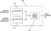
- FIG. 1illustrates a schematic view of an embodiment of a device for determining a database table of dipole densities d(y) of at least one heart chamber, consistent with aspects of the present invention.
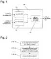
- FIG. 2illustrates a flow chart of an embodiment of a preferred method for determining a database table of dipole densities of at least one heart chamber, consistent with aspects of the present invention.
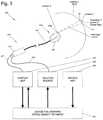
- FIG. 3illustrates a schematic view of an embodiment of a system for determining a database table of dipole densities of at least one heart chamber with help of the solid angle ⁇ acute over ( ⁇ ) ⁇ (x,y), consistent with aspects of the present invention.
- a device for calculating surface charge densitieshas been described in detail in PCT International Application Number PCT/CH2007/000380 (hereinafter the '380 Patent Application) naming Scharf as investor, filed Aug. 3, 2007, and entitled METHOD AND DEVICE FOR DETERMINING AND PRESENTING SURFACE CHARGE AND DIPOLE DENSITIES ON CARDIAC WALLS, and is incorporated by reference herein in its entirety.
- the present inventionprovides an improved device, system and method for calculating and visualizing the distribution and activity of dipole charge densities on a cardiac wall.
- the dipole densitiesare directly determined geometrically, avoiding the errors encountered using previous extrapolation algorithms.
- a devicethat measures and calculates a database of dipole densities d(y) on the cardiac wall.
- the actual measured potentials in the heartresult from electrical activity of cells, which can be regarded as dipoles.
- the dipolesconsist of ion charges on both sides of biological membranes.
- the use of dipole densitiesoffers a precise representation of the electrical activity.
- Systems and methods in accordance with the present inventionefficiently and effectively calculate the dipole densities utilizing one or more mathematical theorems. This calculation is significantly more precise than calculations of virtual potentials produced by current systems, which lose spatial precision because of the required numerical methods and the use of potentials instead of dipole densities.
- Systems and methods in accordance with the present inventionare efficient in calculating dipole densities geometrically, such as through the use of computer systems, or similar microcontroller and/or mathematical processing equipment.
- the terms “subject” and “patient”refer to any animal, such as a mammal like livestock, pets, and preferably a human. Specific examples of “subjects” and “patients” include, but are not limited, to individuals requiring medical assistance, and in particular, patients with an arrhythmia such as atrial fibrillation (AF).
- AFatrial fibrillation
- solid angleis the angle subtended by a triangle on the heart wall at the position x of observation.
- the straight linesthen define the spherical triangle on the surface of the sphere.
- the solid angleis proportional to the surface area of the projection of that object onto a sphere centered at the point x.
- FIGS. 1-3illustrate various preferred embodiments of devices, systems and methods in accordance with aspects of the present invention. However, the present invention is not limited to these particular configurations.
- Device 100includes a first receiver 110 configured to receive electrical potentials from a separate device, such as a device including a multi-electrode mapping catheter placed in the circulating blood within a chamber of the patient's heart.
- Device 100further includes a second receiver 120 configured to receive cardiac geometry information (e.g. the geometric contour of the cardiac chamber wall), such as from an instrument including, but not limited to: Computed Tomography; MRI; Ultrasound; a multi-electrode mapping catheter; and combinations of these.
- cardiac geometry informatione.g. the geometric contour of the cardiac chamber wall
- a standard geometrycan be loaded representing a model of the cardiac chamber.
- Device 100further includes a dipole density module 130 which comprises mathematical processing element, such as a computer or other electronic module including software and/or hardware for performing mathematical or other calculations.
- Dipole density module 130receives mapping information from first receiver 110 and cardiac geometry information from second receiver 120 .
- Dipole density module 130preferably uses one or more algorithms to process the received mapping and geometry information to produce a database table of dipole densities.
- the geometrical model of the cardiac chamberis processed by dipole density module 130 into multiple small triangles (triangularization).
- the dipole density at each trianglecan be regarded as constant.
- a standard cardiac chamber of 4-6 cm diameteris divided up into over 1000 triangles.
- the number of triangles determined by dipole density module 130is based on the size of the heart chamber.
- the dipole density module 130computes the solid angle ⁇ acute over ( ⁇ ) ⁇ (x,y) subtended by each triangle at position y on each electrode at position x on the multi-electrode catheter. If the dipole density at the triangle is d(y), the triangle contributes ⁇ acute over ( ⁇ ) ⁇ (x,y) times d(y) to the potential V(x) at the position x on the multi-electrode catheter. The total measured potential V(x) is the sum resulting from all the triangles. A detailed description is provided in reference to FIG. 3 herebelow.
- dipole density module 130implements a progressive algorithm that can be modified and/or refined in order to improve spatial and/or time resolution of the database of dipole densities that are produced.
- the dipole densities d(y)are obtained by solving a linear system of equations. This calculation requires some care to avoid numerical instabilities. Thereby a map of dipole densities can be created at each corresponding time interval. The synthesis of the maps generates a cascade of the activation sequence of each corresponding heart beat that can be used to define the origin of the electrical activity, arrhythmias or diagnose cardiac disease.
- the measuring electrodes used in the present inventionare placed in the blood flow in a heart chamber, a relatively homogeneous condition, such that the mathematical analysis of the present invention is well applicable.
- skin electrodesare also implemented such that dipole density module 130 can use the information received from the skin electrodes to calculate and/or recalculate the dipole densities for the cardiac wall.
- the spatial resolution which can be obtained by invasive (i.e., placed in the heart chamber) multi-electrode potential measurementsis limited by the number of electrodes that can be placed in any cardiac chamber, such as the Left Atrium (LA).
- LALeft Atrium
- Skin placed electrodes, such as electrodes placed on the thoraxare not as space limited.
- the badly defined boundary value problemcan be avoided by an additional measurement (in addition to the skin electrode measurements) of the multi-electrode array of the present invention.
- This methodyields a higher spatial resolution than the L array electrodes alone.
- regularization techniquesmust be used (e.g. Tikhonov regularization and its modifications) in order to avoid numerical instabilities.
- Step 10a multi-electrode array is placed within the corresponding heart chamber.
- Step 20the geometry of the corresponding heart chamber is obtained in relation to the multi-electrode array position, such as by moving around a second mapping electrode or by importing a geometry model from an imaging study (e.g. using computed tomography, MRI or ultrasound before or after the multi-electrode array of electrodes has been placed in the heart chamber).
- the surface of the geometry of the corresponding heart chamberis divided into small triangles, typically at least 1000 small triangles.
- the dipole density d(y)can be calculated from the measured potential values and the calculated solid angles.
- the measurementscan be repeated successively during the cardiac cycle giving a high timely resolution during each millisecond.
- the information of the timely dependent dipole densitiescan be depicted as an activation map of the corresponding heart chamber for the given heart beat.
- the informationcan be used to diagnose and/or treat a patient with a cardiac arrhythmia, such as an atrial fibrillation patient.
- the informationis used to determine cardiac wall treatment locations for lesion creation, such as a lesion created in the Left or Right atrium, by an RF, ultrasound or cryogenic ablation catheter.
- the multiple electrode mapping arrayis placed in a ventricle and the dipole densities are determined for the ventricular wall, such as to detect ischemia or quantify myocardial function.
- System 500includes device 100 , which is configured to create a database table of dipole densities d(y) based on voltage potential measurements within the heart chamber and image information relating to the heart chamber, as has been described hereabove.
- System 500further includes imaging unit 220 , which is configured to provide a two or three-dimensional image of the heart chamber to device 100 .
- Imaging unit 220may perform at least one of Computed Tomography, MRI and/or ultrasound imaging. Imaging unit 220 may produce any form of real or virtual models of the cardiac chambers, such that a triangularization analysis is possible.
- System 500further includes mapping catheter 310 , which includes shaft 311 , shown inserted into a chamber of a patient's heart, such as the Left Atrium (LA). At the distal end of shaft 311 is an electrode array 315 including multiple electrodes 316 . Electrode array 315 is shown in a basket construction, but numerous other constructions can be used including multiple independent arms, spiral arrays, electrode covered balloons, and other constructions configured to place multiple electrodes into a three-dimensional space. In a preferred embodiment, any catheter with a three-dimensional array of electrodes can be used to supply the mapping information to device 100 .
- electrodes 316are connected to wires, not shown, but traveling proximally to cable 317 , which is electrically connected to a mapping unit 210 , such as an electrocardiogram (ECG) unit.
- ECG unit 210includes a monitor for displaying information, such as the potentials recorded by electrodes 316 , as well as the dipole density information produced by device 100 .
- device 100further includes a monitor, not shown, but configured to display one or more of: dipole density information; potentials recorded by electrodes 316 ; and cardiac chamber contours and other geometry information.
- dipole density and or recorded potentials informationis shown in reference to a three-dimensional representation of the heart chamber into which catheter 310 is inserted.
- imaging unit 220may include a device configured to create an image of the cardiac chamber from signals recorded from an electrode catheter, such as catheter 310 .
- System 500may include a device for treating a cardiac arrhythmia, such as ablation source 230 , which is electrically attached to electrodes 316 via cable 318 .
- ablation source 230can be attached to a different ablation catheter, such as a single or multiple ablation element catheter configured to deliver ablation energy such as RF energy, cryogenic energy, or other tissue disrupting energy.
- triangle T 1defined by device 100
- Electrode 316 a of catheter 310is at location X.
- the geometric relationship between triangle T 1 and Location Xis defined by the solid angle, angle ⁇ acute over ( ⁇ ) ⁇ (X,Y).
- Device 100includes dipole density module 130 such that each triangle at location y contributes ⁇ acute over ( ⁇ ) ⁇ (x,y) times the dipole density d(y) to the potential V(x) at the position x on a multi-electrode.
- Solid angle ⁇ acute over ( ⁇ ) ⁇ (x,y)corresponds to the triangle at a location y and the electrode at positions x on the multi-electrode array.
- the dipole density module 130 of device 100determines from the total measured potential V(x), which is the sum resulting from all the triangles defined by device 100 , the desired dipole density d(y).
- the dipole density d(y) at many equally distributed regions y on the cardiac wallis calculated by solving a linear equation system.
- the measured potentialse.g. with help of splines
- the solid angle ⁇ acute over ( ⁇ ) ⁇ (x,y) of a regionis the sum of the solid angles of the individual triangles in the region on the cardiac wall.
- the resultsare presented in a visual, anatomical format, such as depicting the dipole densities on a geometric image of the cardiac wall in relation to time (t).
- This formatallows a clinician, such as an electrophysiologist, to determine the activation sequence on the cardiac wall, such as to determine treatment locations for a cardiac arrhythmia.
- the resultsmay be shown on a display of mapping unit 210 , or on a separate unit such as a display included with device 100 , display not shown but preferably a color monitor.
- the device of the present inventionis implemented as, or includes, a software program that is executable by at least one processor.
- the software programcan be integrated into one or more of: an ECG system; a cardiac tissue ablation system; an imaging system; a computer; and combinations of these.
- the multi-electrode catheterincludes at least 10 electrodes, configured to represent a three dimensional body with known geometry.
- the electrodesare preferably positioned in a spherical geometry, such as a spherical geometry created in a basket catheter.
- Elliptical electrode array geometriesmay be used, such as those provided in the Ensite Array Catheter, manufactured by St. Jude Medical of St. Paul Minn.
- multiple cathetersare inserted into the heart chamber to provide the multiple electrodes.
- the electrodes of the multi-electrode mapping arrayare repositioned during the method of determining dipole densities. Repositioning of electrodes can be beneficial to increase the number of measured potential values, if electrode positions are known. Therefore, repositioning is in concordance with adjustment of the geometry map in relation to the multi-electrode mapping catheter.
Landscapes
- Health & Medical Sciences (AREA)
- Life Sciences & Earth Sciences (AREA)
- Surgery (AREA)
- Engineering & Computer Science (AREA)
- Animal Behavior & Ethology (AREA)
- Veterinary Medicine (AREA)
- Public Health (AREA)
- Biomedical Technology (AREA)
- General Health & Medical Sciences (AREA)
- Medical Informatics (AREA)
- Heart & Thoracic Surgery (AREA)
- Molecular Biology (AREA)
- Nuclear Medicine, Radiotherapy & Molecular Imaging (AREA)
- Physics & Mathematics (AREA)
- Pathology (AREA)
- Biophysics (AREA)
- Cardiology (AREA)
- Radiology & Medical Imaging (AREA)
- Otolaryngology (AREA)
- Physiology (AREA)
- Electromagnetism (AREA)
- Pulmonology (AREA)
- Theoretical Computer Science (AREA)
- High Energy & Nuclear Physics (AREA)
- Optics & Photonics (AREA)
- Plasma & Fusion (AREA)
- Measurement And Recording Of Electrical Phenomena And Electrical Characteristics Of The Living Body (AREA)
Abstract
Description
N is chosen to be N=K+L where K is the number of surface electrodes and L is the number of internally placed array electrodes.
Claims (22)
Priority Applications (1)
| Application Number | Priority Date | Filing Date | Title |
|---|---|---|---|
| US16/568,768US11116438B2 (en) | 2008-01-17 | 2019-09-12 | Device and method for the geometric determination of electrical dipole densities on the cardiac wall |
Applications Claiming Priority (9)
| Application Number | Priority Date | Filing Date | Title |
|---|---|---|---|
| CH00068/08 | 2008-01-17 | ||
| CH682008 | 2008-01-17 | ||
| PCT/IB2009/000071WO2009090547A2 (en) | 2008-01-17 | 2009-01-16 | A device and method for the geometric determination of electrical dipole densities on the cardiac wall |
| US86341110A | 2010-07-16 | 2010-07-16 | |
| US13/946,712US9192318B2 (en) | 2008-01-17 | 2013-07-19 | Device and method for the geometric determination of electrical dipole densities on the cardiac wall |
| US14/886,449US9504395B2 (en) | 2008-01-17 | 2015-10-19 | Device and method for the geometric determination of electrical dipole densities on the cardiac wall |
| US15/333,378US9913589B2 (en) | 2008-01-17 | 2016-10-25 | Device and method for the geometric determination of electrical dipole densities on the cardiac wall |
| US15/882,097US10463267B2 (en) | 2008-01-17 | 2018-01-29 | Device and method for the geometric determination of electrical dipole densities on the cardiac wall |
| US16/568,768US11116438B2 (en) | 2008-01-17 | 2019-09-12 | Device and method for the geometric determination of electrical dipole densities on the cardiac wall |
Related Parent Applications (1)
| Application Number | Title | Priority Date | Filing Date |
|---|---|---|---|
| US15/882,097ContinuationUS10463267B2 (en) | 2008-01-17 | 2018-01-29 | Device and method for the geometric determination of electrical dipole densities on the cardiac wall |
Publications (2)
| Publication Number | Publication Date |
|---|---|
| US20200138317A1 US20200138317A1 (en) | 2020-05-07 |
| US11116438B2true US11116438B2 (en) | 2021-09-14 |
Family
ID=40467019
Family Applications (6)
| Application Number | Title | Priority Date | Filing Date |
|---|---|---|---|
| US12/863,411Active2030-01-31US8512255B2 (en) | 2008-01-17 | 2009-01-16 | Device and method for the geometric determination of electrical dipole densities on the cardiac wall |
| US13/946,712Active2029-04-15US9192318B2 (en) | 2008-01-17 | 2013-07-19 | Device and method for the geometric determination of electrical dipole densities on the cardiac wall |
| US14/886,449ActiveUS9504395B2 (en) | 2008-01-17 | 2015-10-19 | Device and method for the geometric determination of electrical dipole densities on the cardiac wall |
| US15/333,378ActiveUS9913589B2 (en) | 2008-01-17 | 2016-10-25 | Device and method for the geometric determination of electrical dipole densities on the cardiac wall |
| US15/882,097ActiveUS10463267B2 (en) | 2008-01-17 | 2018-01-29 | Device and method for the geometric determination of electrical dipole densities on the cardiac wall |
| US16/568,768Active2029-04-21US11116438B2 (en) | 2008-01-17 | 2019-09-12 | Device and method for the geometric determination of electrical dipole densities on the cardiac wall |
Family Applications Before (5)
| Application Number | Title | Priority Date | Filing Date |
|---|---|---|---|
| US12/863,411Active2030-01-31US8512255B2 (en) | 2008-01-17 | 2009-01-16 | Device and method for the geometric determination of electrical dipole densities on the cardiac wall |
| US13/946,712Active2029-04-15US9192318B2 (en) | 2008-01-17 | 2013-07-19 | Device and method for the geometric determination of electrical dipole densities on the cardiac wall |
| US14/886,449ActiveUS9504395B2 (en) | 2008-01-17 | 2015-10-19 | Device and method for the geometric determination of electrical dipole densities on the cardiac wall |
| US15/333,378ActiveUS9913589B2 (en) | 2008-01-17 | 2016-10-25 | Device and method for the geometric determination of electrical dipole densities on the cardiac wall |
| US15/882,097ActiveUS10463267B2 (en) | 2008-01-17 | 2018-01-29 | Device and method for the geometric determination of electrical dipole densities on the cardiac wall |
Country Status (4)
| Country | Link |
|---|---|
| US (6) | US8512255B2 (en) |
| EP (2) | EP2737849A3 (en) |
| CA (1) | CA2747859C (en) |
| WO (1) | WO2009090547A2 (en) |
Families Citing this family (29)
| Publication number | Priority date | Publication date | Assignee | Title |
|---|---|---|---|---|
| US11389232B2 (en) | 2006-06-28 | 2022-07-19 | Kardium Inc. | Apparatus and method for intra-cardiac mapping and ablation |
| US9119633B2 (en) | 2006-06-28 | 2015-09-01 | Kardium Inc. | Apparatus and method for intra-cardiac mapping and ablation |
| WO2008014629A2 (en)* | 2006-08-03 | 2008-02-07 | Christoph Scharf | Method and device for determining and presenting surface charge and dipole densities on cardiac walls |
| US8906011B2 (en) | 2007-11-16 | 2014-12-09 | Kardium Inc. | Medical device for use in bodily lumens, for example an atrium |
| EP2737849A3 (en) | 2008-01-17 | 2014-10-29 | Christoph Scharf | A device and method for the geometric determination of electrical dipole densities on the cardiac wall |
| US9757044B2 (en)* | 2011-03-10 | 2017-09-12 | Acutus Medical, Inc. | Device and method for the geometric determination of electrical dipole densities on the cardiac wall |
| US10827977B2 (en) | 2012-05-21 | 2020-11-10 | Kardium Inc. | Systems and methods for activating transducers |
| US9017321B2 (en) | 2012-05-21 | 2015-04-28 | Kardium, Inc. | Systems and methods for activating transducers |
| US9198592B2 (en) | 2012-05-21 | 2015-12-01 | Kardium Inc. | Systems and methods for activating transducers |
| US10588543B2 (en) | 2012-05-23 | 2020-03-17 | Biosense Webster (Israel), Ltd. | Position sensing using electric dipole fields |
| EP2890292B1 (en) | 2012-08-31 | 2021-01-13 | Acutus Medical, Inc. | Catheter system for the heart |
| CN105358070B (en) | 2013-02-08 | 2018-03-23 | 阿库图森医疗有限公司 | Expandable catheter assembly with flexible printed circuit board |
| CA2922941C (en) | 2013-09-13 | 2021-11-16 | Acutus Medical, Inc. | Devices and methods for determination of electrical dipole densities on a cardiac surface |
| US9289145B2 (en) | 2013-12-05 | 2016-03-22 | Medtronic, Inc. | Identification of abnormal cardiac substrate during left-ventricular pacing |
| JP6739346B2 (en) | 2014-03-25 | 2020-08-12 | アクタス メディカル インクAcutus Medical,Inc. | Method of operating system of cardiac analysis user interface |
| US10722184B2 (en) | 2014-11-17 | 2020-07-28 | Kardium Inc. | Systems and methods for selecting, activating, or selecting and activating transducers |
| US10368936B2 (en) | 2014-11-17 | 2019-08-06 | Kardium Inc. | Systems and methods for selecting, activating, or selecting and activating transducers |
| CN115299988A (en) | 2015-05-12 | 2022-11-08 | 阿库图森医疗有限公司 | Ultrasonic sequencing systems and methods |
| US10593234B2 (en) | 2015-05-12 | 2020-03-17 | Acutus Medical, Inc. | Cardiac virtualization test tank and testing system and method |
| US10653318B2 (en) | 2015-05-13 | 2020-05-19 | Acutus Medical, Inc. | Localization system and method useful in the acquisition and analysis of cardiac information |
| CA3022806A1 (en)* | 2016-05-03 | 2017-11-09 | Acutus Medical, Inc. | Cardiac mapping system with efficiency algorithm |
| EP3451916A4 (en) | 2016-05-03 | 2019-10-16 | Acutus Medical Inc. | SYSTEM AND METHOD FOR THE DYNAMIC DISPLAY OF HEART DATA |
| US10842399B2 (en)* | 2017-08-17 | 2020-11-24 | Biosense Webster (Israel) Ltd. | System and method of managing ECG data for user defined map |
| EP3446628B1 (en) | 2017-08-24 | 2023-09-27 | Karlsruher Institut für Technologie | Method and system for determining ventricular far field contribution in atrial electrograms |
| EP3790471A4 (en)* | 2018-05-08 | 2022-01-12 | Acutus Medical, Inc. | Cardiac information processing system |
| US12178582B2 (en) | 2018-11-09 | 2024-12-31 | Acutus Medical, Inc. | Systems and methods for calculating patient information |
| JP2022529908A (en) | 2019-04-18 | 2022-06-27 | アクタス メディカル インク | System for generating composite maps |
| CA3135773A1 (en) | 2019-06-04 | 2020-12-10 | Acutus Medical, Inc. | Systems and methods for performing localization within a body |
| WO2021102230A1 (en) | 2019-11-22 | 2021-05-27 | Acutus Medical, Inc. | Tissue treatment systems, devices, and methods |
Citations (186)
| Publication number | Priority date | Publication date | Assignee | Title |
|---|---|---|---|---|
| US4173228A (en) | 1977-05-16 | 1979-11-06 | Applied Medical Devices | Catheter locating device |
| US5041973A (en) | 1988-10-25 | 1991-08-20 | London Health Association | Cardiac mapping system simulator |
| US5156151A (en) | 1991-02-15 | 1992-10-20 | Cardiac Pathways Corporation | Endocardial mapping and ablation system and catheter probe |
| US5293868A (en) | 1992-06-30 | 1994-03-15 | American Cardiac Ablation Co., Inc. | Cardiac ablation catheter having resistive mapping electrodes |
| WO1994006349A1 (en) | 1992-09-23 | 1994-03-31 | Endocardial Therapeutics, Inc. | Endocardial mapping system |
| US5482472A (en) | 1993-11-17 | 1996-01-09 | Board Of Regents, The University Of Texas System | Electrical signal generator interface with three-dimensional electrical pathway and transparent heart and method of visually simulating cardiac waveforms in three dimensions |
| US5499981A (en) | 1993-03-16 | 1996-03-19 | Ep Technologies, Inc. | Flexible interlaced multiple electrode assemblies |
| JPH08504333A (en) | 1992-09-25 | 1996-05-14 | イーピー・テクノロジーズ・インコーポレーテッド | Electrode-supported splines for the cardiac system |
| US5555883A (en) | 1992-02-24 | 1996-09-17 | Avitall; Boaz | Loop electrode array mapping and ablation catheter for cardiac chambers |
| US5595183A (en) | 1995-02-17 | 1997-01-21 | Ep Technologies, Inc. | Systems and methods for examining heart tissue employing multiple electrode structures and roving electrodes |
| US5601084A (en) | 1993-06-23 | 1997-02-11 | University Of Washington | Determining cardiac wall thickness and motion by imaging and three-dimensional modeling |
| US5647367A (en) | 1996-05-31 | 1997-07-15 | Hewlett-Packard Company | Scanning ultrasonic probe with locally-driven sweeping ultrasonic source |
| US5662108A (en) | 1992-09-23 | 1997-09-02 | Endocardial Solutions, Inc. | Electrophysiology mapping system |
| US5722416A (en) | 1995-02-17 | 1998-03-03 | Ep Technologies, Inc. | Systems and methods for analyzing biopotential morphologies in heart tissue to locate potential ablation sites |
| US5722402A (en) | 1994-10-11 | 1998-03-03 | Ep Technologies, Inc. | Systems and methods for guiding movable electrode elements within multiple-electrode structures |
| US5740808A (en) | 1996-10-28 | 1998-04-21 | Ep Technologies, Inc | Systems and methods for guilding diagnostic or therapeutic devices in interior tissue regions |
| US5749833A (en) | 1995-08-15 | 1998-05-12 | Hakki; A-Hamid | Combined echo-electrocardiographic probe |
| US5759158A (en) | 1995-07-28 | 1998-06-02 | E.P. Technologies, Inc. | Systems and methods for conducting electrophysiological testing using high-voltage energy pulses to stun heart tissue |
| US5782239A (en) | 1992-06-30 | 1998-07-21 | Cordis Webster, Inc. | Unique electrode configurations for cardiovascular electrode catheter with built-in deflection method and central puller wire |
| US5795298A (en) | 1995-03-28 | 1998-08-18 | Sonometrics Corporation | System for sharing electrocardiogram electrodes and transducers |
| US5795299A (en) | 1997-01-31 | 1998-08-18 | Acuson Corporation | Ultrasonic transducer assembly with extended flexible circuits |
| US5820568A (en) | 1996-10-15 | 1998-10-13 | Cardiac Pathways Corporation | Apparatus and method for aiding in the positioning of a catheter |
| US5830144A (en) | 1995-03-28 | 1998-11-03 | Vesely; Ivan | Tracking data sheath |
| US5846198A (en) | 1996-05-31 | 1998-12-08 | Siemens Aktiengesellschaft | Apparatus for localizing action currents in the heart |
| WO1999005971A1 (en) | 1997-08-01 | 1999-02-11 | Cardiac Pathways Corporation | System for electrode localization using ultrasound |
| US5876336A (en) | 1994-10-11 | 1999-03-02 | Ep Technologies, Inc. | Systems and methods for guiding movable electrode elements within multiple-electrode structure |
| US5928228A (en) | 1993-03-16 | 1999-07-27 | Ep Technologies, Inc. | Flexible high density multiple electrode circuit assemblies employing ribbon cable |
| US5944022A (en) | 1997-04-28 | 1999-08-31 | American Cardiac Ablation Co. Inc. | Catheter positioning system |
| US5968040A (en) | 1994-03-04 | 1999-10-19 | Ep Technologies, Inc. | Systems and methods using asymmetric multiple electrode arrays |
| US6014590A (en) | 1974-03-04 | 2000-01-11 | Ep Technologies, Inc. | Systems and methods employing structures having asymmetric mechanical properties to support diagnostic or therapeutic elements in contact with tissue in interior body regions |
| US6024703A (en) | 1997-05-07 | 2000-02-15 | Eclipse Surgical Technologies, Inc. | Ultrasound device for axial ranging |
| WO2000007501A1 (en) | 1998-08-03 | 2000-02-17 | Cardiac Pathways Corporation | A dynamically alterable three-dimensional graphical model of a body region |
| US6066096A (en) | 1998-05-08 | 2000-05-23 | Duke University | Imaging probes and catheters for volumetric intraluminal ultrasound imaging and related systems |
| US6086532A (en) | 1997-09-26 | 2000-07-11 | Ep Technologies, Inc. | Systems for recording use of structures deployed in association with heart tissue |
| US6107699A (en) | 1998-05-22 | 2000-08-22 | Scimed Life Systems, Inc. | Power supply for use in electrophysiological apparatus employing high-voltage pulses to render tissue temporarily unresponsive |
| US6115626A (en) | 1998-03-26 | 2000-09-05 | Scimed Life Systems, Inc. | Systems and methods using annotated images for controlling the use of diagnostic or therapeutic instruments in instruments in interior body regions |
| JP2000358299A (en) | 1999-06-16 | 2000-12-26 | Ngk Spark Plug Co Ltd | Wave transmitting and receiving element for ultrasonic probe, its production method and ultrasonic probe using the same element |
| US6188928B1 (en) | 1996-11-18 | 2001-02-13 | Pacesetter Ab | Apparatus for tissue stimulation |
| US6187032B1 (en)* | 1997-10-30 | 2001-02-13 | Kabushiki Kaisha Toshiba | Measurement of intracardiac electrophysiological phenomena |
| JP2001070269A (en) | 1999-09-06 | 2001-03-21 | Toshiba Corp | Electrophysiology mapping device |
| US6216043B1 (en) | 1994-03-04 | 2001-04-10 | Ep Technologies, Inc. | Asymmetric multiple electrode support structures |
| US6240307B1 (en) | 1993-09-23 | 2001-05-29 | Endocardial Solutions, Inc. | Endocardial mapping system |
| US20010007070A1 (en) | 1999-04-05 | 2001-07-05 | Medtronic, Inc. | Ablation catheter assembly and method for isolating a pulmonary vein |
| US6301496B1 (en) | 1998-07-24 | 2001-10-09 | Biosense, Inc. | Vector mapping of three-dimensionally reconstructed intrabody organs and method of display |
| EP1166714A1 (en) | 2000-06-21 | 2002-01-02 | Biosense, Inc. | Rapid mapping of electrical activity in the heart |
| US20020026118A1 (en) | 2000-08-18 | 2002-02-28 | Assaf Govari | Three-dimensional reconstruction using ultrasound |
| US20020045810A1 (en) | 1993-07-20 | 2002-04-18 | Shlomo Ben-Haim | Method for mapping a heart using catheters having ultrasonic position sensors |
| US20020128565A1 (en) | 1997-07-31 | 2002-09-12 | Case Western Reserve University | System and method for non-invasive electrocardiographic imaging |
| US20020165441A1 (en) | 2000-01-27 | 2002-11-07 | Coleman James H. | Bidirectional catheter having mapping assembly |
| US6514249B1 (en) | 1997-07-08 | 2003-02-04 | Atrionix, Inc. | Positioning system and method for orienting an ablation element within a pulmonary vein ostium |
| JP2003511098A (en) | 1998-04-14 | 2003-03-25 | ジーエムピー・ドラツグ・デリバリー・インコーポレーテツド | Iontophoresis, electroporation and combination catheters for local drug delivery to arteries and other body tissues |
| WO2003026722A2 (en) | 2001-09-27 | 2003-04-03 | Baylor College Of Medicine | Cardiac catheter imaging system |
| US20030078494A1 (en) | 2001-10-24 | 2003-04-24 | Scimed Life Systems, Inc. | Systems and methods for guiding and locating functional elements on medical devices positioned in a body |
| US6574492B1 (en) | 1996-01-08 | 2003-06-03 | Biosense, Inc. | Catheter having multiple arms with electrode and position sensor |
| US20030120318A1 (en) | 1998-06-30 | 2003-06-26 | Hauck John A. | Congestive heart failure pacing optimization method and device |
| US20030153907A1 (en) | 1999-01-06 | 2003-08-14 | Scimed Life Systems, Inc. | Ultrasound-guided ablation catheter and methods of use |
| US20030158477A1 (en) | 2001-11-09 | 2003-08-21 | Dorin Panescu | Systems and methods for guiding catheters using registered images |
| US20030231789A1 (en) | 2002-06-18 | 2003-12-18 | Scimed Life Systems, Inc. | Computer generated representation of the imaging pattern of an imaging device |
| US20030236466A1 (en) | 2002-06-21 | 2003-12-25 | Tarjan Peter P. | Single or multi-mode cardiac activity data collection, processing and display obtained in a non-invasive manner |
| US20040039312A1 (en) | 2002-02-20 | 2004-02-26 | Liposonix, Inc. | Ultrasonic treatment and imaging of adipose tissue |
| WO2004026134A1 (en) | 2002-08-24 | 2004-04-01 | Krishnan Subramaniam C | Method and apparatus for locating the fossa ovalis and performing transseptal puncture |
| US20040082870A1 (en) | 1997-07-31 | 2004-04-29 | Yoram Rudy | Systems and methods for determining a surface geometry |
| US6773402B2 (en) | 2001-07-10 | 2004-08-10 | Biosense, Inc. | Location sensing with real-time ultrasound imaging |
| US20040254437A1 (en) | 1998-06-30 | 2004-12-16 | Hauck John A. | Method and apparatus for catheter navigation and location and mapping in the heart |
| JP2004350702A (en) | 2003-05-26 | 2004-12-16 | Olympus Corp | Ultrasonic diagnosis probe apparatus |
| US6839588B1 (en) | 1997-07-31 | 2005-01-04 | Case Western Reserve University | Electrophysiological cardiac mapping system based on a non-contact non-expandable miniature multi-electrode catheter and method therefor |
| US20050059880A1 (en) | 2003-09-11 | 2005-03-17 | Mathias Sanjay George | ECG driven image reconstruction for cardiac imaging |
| US20050113665A1 (en) | 2003-11-26 | 2005-05-26 | Mohr Kelly A. | Cardiac display methods and apparatus |
| US20050148836A1 (en) | 2003-11-26 | 2005-07-07 | Martin Kleen | Catheter device |
| US6939309B1 (en) | 1993-09-23 | 2005-09-06 | Endocardial Solutions, Inc. | Electrophysiology mapping system |
| US20060058676A1 (en) | 2002-04-17 | 2006-03-16 | Tomoyuki Yagi | Ultrasonic probe in body cavity |
| US20060116576A1 (en) | 2004-12-01 | 2006-06-01 | Scimed Life Systems, Inc. | System and use thereof to provide indication of proximity between catheter and location of interest in 3-D space |
| CN1856123A (en) | 2005-04-29 | 2006-11-01 | 华为技术有限公司 | System and method for exchanging modules in switch |
| US20060244177A1 (en) | 2005-04-19 | 2006-11-02 | Masayuki Kaneto | Flexible printed circuit board for catheter, catheter using same, and production method of catheter |
| US20070016007A1 (en) | 2005-07-15 | 2007-01-18 | Assaf Govari | Hybrid magnetic-based and impedance-based position sensing |
| EP1760661A2 (en) | 2005-08-30 | 2007-03-07 | Biosense Webster, Inc. | Segmentation and registration of multimodal images using physiological data |
| US20070055150A1 (en) | 2005-08-16 | 2007-03-08 | General Electric Company | Method and system for mapping physiology information onto ultrasound-based anatomic structure |
| US20070060832A1 (en) | 2005-08-26 | 2007-03-15 | Michael Levin | Detection of skin impedance |
| US20070083194A1 (en) | 2005-06-20 | 2007-04-12 | Kunis Christopher G | Ablation catheter |
| EP1779787A2 (en) | 2005-10-28 | 2007-05-02 | Biosense Webster, Inc. | Synchronization of ultrasound imaging data with electrical mapping |
| US20070167722A1 (en) | 1992-08-14 | 2007-07-19 | British Telecommunications Public Limited Company | Surgical navigation |
| CN101048100A (en) | 2004-09-08 | 2007-10-03 | 奥林巴斯株式会社 | capsule medical device |
| US20070232949A1 (en) | 2006-03-31 | 2007-10-04 | Ep Medsystems, Inc. | Method For Simultaneous Bi-Atrial Mapping Of Atrial Fibrillation |
| US7291146B2 (en) | 2003-09-12 | 2007-11-06 | Minnow Medical, Inc. | Selectable eccentric remodeling and/or ablation of atherosclerotic material |
| US20080009758A1 (en) | 2006-05-17 | 2008-01-10 | Voth Eric J | System and method for mapping electrophysiology information onto complex geometry |
| WO2008014629A2 (en) | 2006-08-03 | 2008-02-07 | Christoph Scharf | Method and device for determining and presenting surface charge and dipole densities on cardiac walls |
| US20080146937A1 (en) | 2006-12-14 | 2008-06-19 | General Electric Company | Mechanically expanding transducer assembly |
| US20080287777A1 (en) | 2007-05-16 | 2008-11-20 | General Electric Company | System and method to register a tracking system with an intracardiac echocardiography (ice) imaging system |
| US20080319297A1 (en) | 2007-06-20 | 2008-12-25 | Kenneth Danehorn | Electrode catheter positioning system |
| US20090024086A1 (en) | 2007-07-20 | 2009-01-22 | Qiming Zhang | Micro-steerable catheter |
| US7505810B2 (en) | 2006-06-13 | 2009-03-17 | Rhythmia Medical, Inc. | Non-contact cardiac mapping, including preprocessing |
| US20090076483A1 (en) | 2007-09-14 | 2009-03-19 | Kenneth Danehorn | Catheter localization system |
| US20090082691A1 (en) | 2007-09-26 | 2009-03-26 | Medtronic, Inc. | Frequency selective monitoring of physiological signals |
| CN201223445Y (en) | 2008-06-23 | 2009-04-22 | 北京有色金属研究总院 | Radio frequency ablation catheter |
| US20090131930A1 (en) | 2007-11-16 | 2009-05-21 | Daniel Gelbart | Medical device for use in bodily lumens, for example an atrium |
| US20090143651A1 (en) | 2006-06-01 | 2009-06-04 | Bengt Kallback | Device for Invasive Use |
| US20090148012A1 (en) | 2007-12-05 | 2009-06-11 | Andres Claudio Altmann | Anatomical modeling from a 3-d image and a surface mapping |
| JP2009135109A (en) | 2009-03-09 | 2009-06-18 | Advanced Systems Japan Inc | Sheet-like connecting terminal and method of connecting fpc substrate using this |
| US20090171274A1 (en) | 2007-12-28 | 2009-07-02 | Doron Harlev | Non contact mapping catheter |
| CN201275144Y (en) | 2007-08-10 | 2009-07-22 | 北京美中双和医疗器械有限公司 | Electrophysiologic ablation device |
| WO2009090547A2 (en) | 2008-01-17 | 2009-07-23 | Christoph Scharf | A device and method for the geometric determination of electrical dipole densities on the cardiac wall |
| US7573182B2 (en) | 2005-06-01 | 2009-08-11 | Prorhythm, Inc. | Ultrasonic transducer |
| US20100023004A1 (en) | 2008-07-28 | 2010-01-28 | David Francischelli | Systems and methods for cardiac tissue electroporation ablation |
| US20100076426A1 (en) | 2007-05-09 | 2010-03-25 | De La Rama Alan | Basket catheter having multiple electrodes |
| US20100094279A1 (en) | 2006-10-10 | 2010-04-15 | Kauphusman James V | Circuit for a catheter or sheath and method of forming same |
| US20100168578A1 (en) | 2007-06-12 | 2010-07-01 | University Of Virginia Patent Foundation | System and Method for Combined ECG-Echo for Cardiac Diagnosis |
| US20100279263A1 (en) | 2009-04-29 | 2010-11-04 | Scott Duryea | Polysomnography Training Apparatus |
| US20100286551A1 (en) | 2009-05-08 | 2010-11-11 | Rhythmia Medical, Inc. | Impedance Based Anatomy Generation |
| US7841986B2 (en) | 2006-05-10 | 2010-11-30 | Regents Of The University Of Minnesota | Methods and apparatus of three dimensional cardiac electrophysiological imaging |
| JP2011504363A (en) | 2007-11-23 | 2011-02-10 | ディーエスエム アイピー アセッツ ビー.ブイ. | Production of improved bioactive peptides |
| US20110077526A1 (en) | 2008-05-27 | 2011-03-31 | Gil Zwirn | Ultrasound garment |
| US20110092809A1 (en) | 2009-04-07 | 2011-04-21 | Pacesetter, Inc. | Cardiac coordinate system for motion analysis |
| US20110118726A1 (en) | 2009-11-13 | 2011-05-19 | St. Jude Medical, Inc. | Assembly of staggered ablation elements |
| US20110125172A1 (en) | 2006-05-19 | 2011-05-26 | Kardium Inc. | Automatic atherectomy system |
| US20110144510A1 (en) | 2009-12-16 | 2011-06-16 | Pacesetter, Inc. | Methods to identify damaged or scarred tissue based on position information and physiological information |
| US20110201951A1 (en) | 2010-02-12 | 2011-08-18 | Siemens Medical Solutions Usa, Inc. | System for cardiac arrhythmia detection and characterization |
| US20110213231A1 (en) | 2007-05-09 | 2011-09-01 | Hall Sacha C | Bendable catheter arms having varied flexibility |
| WO2011136867A1 (en) | 2010-04-28 | 2011-11-03 | Medtronic Ablation Frontiers Llc | Systems and methods of performing medical procedures |
| US8147486B2 (en) | 2003-09-22 | 2012-04-03 | St. Jude Medical, Atrial Fibrillation Division, Inc. | Medical device with flexible printed circuit |
| US20120082969A1 (en) | 2010-10-05 | 2012-04-05 | Yitzhack Schwartz | Simulation of an invasive procedure |
| US20120123296A1 (en) | 2009-08-03 | 2012-05-17 | Dune Medical Devices Ltd. | Surgical tool |
| WO2012068471A1 (en) | 2010-11-19 | 2012-05-24 | Boston Scientific Scimed, Inc. | Renal nerve detection and ablation apparatus and method |
| US20120136231A1 (en) | 2006-07-25 | 2012-05-31 | Gal Markel | Wearable items providing physiological, environmental and situational parameter monitoring |
| US20120143298A1 (en) | 2010-12-02 | 2012-06-07 | Just Dale E | Catheter electrode assemblies and methods of construction therefor |
| US20120165667A1 (en) | 2010-12-22 | 2012-06-28 | Andres Claudio Altmann | Lasso catheter with ultrasound transducer |
| WO2012092016A1 (en) | 2010-12-30 | 2012-07-05 | St. Jude Medical, Atrial Fibrillation Division, Inc. | System and method for diagnosing arrhythmias and directing catheter therapies |
| US20120172859A1 (en) | 2011-01-05 | 2012-07-05 | Medtronic Ablation Frontiers Llc | Multipolarity epicardial radiofrequency ablation |
| US20120184863A1 (en) | 2011-01-13 | 2012-07-19 | Rhythmia Medical, Inc. | Electroanatomical mapping |
| WO2012100185A2 (en) | 2011-01-21 | 2012-07-26 | Kardium Inc. | Enhanced medical device for use in bodily cavities, for example an atrium |
| WO2012110942A1 (en) | 2011-02-17 | 2012-08-23 | Koninklijke Philips Electronics N.V. | System for providing an electrical activity map using optical shape sensing |
| WO2012122517A2 (en) | 2011-03-10 | 2012-09-13 | Acutus Medical, Inc. | Device and method for the geometric determination of electrical dipole densities on the cardiac wall |
| US20120265054A1 (en) | 2011-04-14 | 2012-10-18 | Olson Eric S | System and Method for Registration of Multiple Navigation Systems to a Common Coordinate Frame |
| US20120271138A1 (en) | 2011-04-22 | 2012-10-25 | Topera, Inc. | Basket style cardiac mapping catheter having a flexible electrode assembly for detection of cardiac rhythm disorders |
| US20120302912A1 (en) | 2011-05-27 | 2012-11-29 | Boston Scientific Neuromodulation Corporation | Collection of clinical data for graphical representation and analysis |
| US20120310064A1 (en) | 2011-06-01 | 2012-12-06 | Mcgee David L | Ablation probe with ultrasonic imaging capabilities |
| US20130006238A1 (en) | 2011-06-30 | 2013-01-03 | Tom Allen Ditter | Catheter with variable arcuate distal section |
| US20130085361A1 (en) | 2010-04-01 | 2013-04-04 | Ecole Polytechnique Federale De Lausanne | Device for interacting with neurological tissue and methods of making and using the same |
| US20130096432A1 (en) | 1998-06-30 | 2013-04-18 | John A. Hauck | System and method for navigating an ultrasound catheter to image a beating heart |
| US8428690B2 (en) | 2007-05-16 | 2013-04-23 | General Electric Company | Intracardiac echocardiography image reconstruction in combination with position tracking system |
| US20130158537A1 (en) | 2010-06-30 | 2013-06-20 | Koninklijke Philips Electronics N.V. | Energy application apparatus for applying energy to an object |
| US20130165916A1 (en) | 2011-12-23 | 2013-06-27 | Vessix Vascular, Inc. | Methods and apparatuses for remodeling tissue of or adjacent to a body passage |
| WO2013101257A1 (en) | 2011-12-29 | 2013-07-04 | St. Jude Medical, Atrial Fibrillation Division, Inc. | Method and system for constructing an electrophysiology map |
| US20130172715A1 (en) | 2011-12-30 | 2013-07-04 | Dale E. Just | Electrode support structure assemblies |
| US20130190587A1 (en) | 2011-01-21 | 2013-07-25 | Kardium Inc. | High-density electrode-based medical device system |
| US20130197614A1 (en) | 2005-05-03 | 2013-08-01 | Vessix Vascular, Inc. | Selective accumulation of energy with or without knowledge of tissue topography |
| US20130245433A1 (en) | 2010-11-18 | 2013-09-19 | Koninklijke Philips Electronics N.V. | Location determination apparatus |
| US20130245621A1 (en) | 2012-03-16 | 2013-09-19 | St. Jude Medical Ab | Ablation stent and method of using an ablation stent |
| US20130241929A1 (en) | 2012-03-13 | 2013-09-19 | Fady Massarwa | Selectably transparent electrophysiology map |
| US20130267853A1 (en) | 2010-12-03 | 2013-10-10 | Research Triangle Institute | Ultrasound device, and associated cable assembly |
| US20130282084A1 (en) | 2004-09-10 | 2013-10-24 | Vessix Vascular, Inc. | Apparatus and Method for Treatment of In-Stent Restenosis |
| US20130304062A1 (en) | 2012-05-14 | 2013-11-14 | Biosense Webster (Irael), Ltd. | Catheter with helical end section for vessel ablation |
| US20130310827A1 (en) | 2012-05-21 | 2013-11-21 | Kardium Inc. | Systems and methods for selecting, activating, or selecting and activating transducers |
| US20130330701A1 (en) | 2012-06-12 | 2013-12-12 | Vladimir Rubinstein | Physical heart simulator |
| WO2014036439A2 (en) | 2012-08-31 | 2014-03-06 | Acutus Medical, Inc. | Catheter system and methods of medical uses of same, including diagnostic and treatment uses for the heart |
| US20140095105A1 (en) | 2012-10-03 | 2014-04-03 | Lev Abramovich Koyrakh | Scaling of electrical impedance-based navigation space using inter-electrode spacing |
| US20140221803A1 (en) | 2008-09-30 | 2014-08-07 | Biosense Webster (Israel), Ltd. | Current localization tracker |
| WO2014124231A1 (en) | 2013-02-08 | 2014-08-14 | Acutus Medical, Inc. | Expandable catheter assembly with flexible printed circuit board |
| US20140235988A1 (en) | 2013-02-21 | 2014-08-21 | Medtronic, Inc. | Methods for simultaneous cardiac substrate mapping using spatial correlation maps between neighboring unipolar electrograms |
| US20140249505A1 (en) | 2013-03-02 | 2014-09-04 | Vladislav Bukhman | Method and System of Utilizing ECG Signal for Central Venous Catheter Tip Positioning |
| US20140257071A1 (en) | 2013-03-05 | 2014-09-11 | Timothy G. Curran | System and Method for Detecting Sheathing and Unsheathing of Localization Elements |
| US20140257069A1 (en) | 2013-03-08 | 2014-09-11 | St. Jude Medical, Atrial Fibrillation Division, Inc. | Basket for a multi-electrode array catheter |
| US20140276746A1 (en) | 2013-03-15 | 2014-09-18 | St. Jude Medical, Cardiology Division, Inc. | Feedback systems and methods utilizing two or more sites along denervation catheter |
| US20140276789A1 (en) | 2013-03-15 | 2014-09-18 | Boston Scientific Scimed, Inc. | Methods and apparatuses for remodeling tissue of or adjacent to a body passage |
| US20140276733A1 (en) | 2013-03-14 | 2014-09-18 | St. Jude Medical, Cardiology Division, Inc. | Mediguide-enabled renal denervation system for ensuring wall contact and mapping lesion locations |
| US20140358143A1 (en) | 2013-05-31 | 2014-12-04 | Medtronic Ablation Frontiers Llc | Adjustable catheter for ostial, septal, and roof ablation in atrial fibrillation patients |
| WO2015038607A2 (en) | 2013-09-13 | 2015-03-19 | Acutus Medical, Inc. | Devices and methods for determination of electrical dipole densities on a cardiac surface |
| CN104462650A (en) | 2014-11-10 | 2015-03-25 | 张建卿 | Materialized heart 3D model manufacturing method capable of achieving internal and external structures |
| US20150196217A1 (en) | 2006-06-13 | 2015-07-16 | Rhythmia Medical, Inc. | Cardiac mapping |
| US20150208938A1 (en) | 2014-01-29 | 2015-07-30 | Biosense Webster (Israel) Ltd. | Hybrid bipolar/unipolar detection of activation wavefront |
| US20150257825A1 (en) | 2014-03-13 | 2015-09-17 | Medtronic Ardian Luxembourg S.A.R.L. | Low Profile Catheter Assemblies and Associated Systems and Methods |
| US20150257732A1 (en) | 2013-03-15 | 2015-09-17 | Stephen Eric Ryan | Distance, diameter and area determining device |
| WO2015148470A1 (en) | 2014-03-25 | 2015-10-01 | Acutus Medical, Inc. | Cardiac analysis user interface system and method |
| US20150342491A1 (en) | 2014-06-03 | 2015-12-03 | Boston Scientific Scimed, Inc. | Electrode assembly having an atraumatic distal tip |
| US20150374252A1 (en) | 2007-05-23 | 2015-12-31 | St. Jude Medical, Cardiology Division, Inc. | Flexible high-density mapping catheter tips and flexible ablation catheter tips with onboard high-density mapping electrodes |
| US20160051321A1 (en) | 2013-04-08 | 2016-02-25 | Amr Salahieh | Tissue ablation and monitoring |
| USD758596S1 (en) | 2015-04-17 | 2016-06-07 | Micron Devices Llc | Flexible circuit for an implantable neural stimulator |
| US20160256112A1 (en) | 2009-11-03 | 2016-09-08 | Vivaquant Llc | System for processing physiological data |
| WO2016183468A1 (en) | 2015-05-13 | 2016-11-17 | Acutus Medical, Inc. | Localization system and method useful in the acquisition and analysis of cardiac information |
| WO2016183179A1 (en) | 2015-05-12 | 2016-11-17 | Acutus Medical, Inc. | Cardiac virtualization test tank and testing system and method |
| WO2016183285A1 (en) | 2015-05-12 | 2016-11-17 | Acutus Medical, Inc. | Ultrasound sequencing system and method |
| US20170065204A1 (en) | 2015-09-04 | 2017-03-09 | Biosense Webster (Israel) Ltd. | Field-based location coordinate correction |
| US20170319180A1 (en) | 2014-12-11 | 2017-11-09 | Koninklijke Philips N.V. | Catheter transducer with staggered columns of micromachined ultrasonic transducers |
| WO2017192769A1 (en) | 2016-05-03 | 2017-11-09 | Acutus Medical, Inc. | Cardiac information dynamic display system and method |
| WO2017192775A1 (en) | 2016-05-03 | 2017-11-09 | Acutus Medical, Inc. | Cardiac mapping system with efficiency algorithm |
| WO2019144103A1 (en) | 2018-01-21 | 2019-07-25 | Acutus Medical, Inc. | System for identifying cardiac conduction patterns |
| WO2019217430A1 (en) | 2018-05-08 | 2019-11-14 | Acutus Medical, Inc. | Cardiac information processing system |
- 2009
- 2009-01-16EPEP13176658.6Apatent/EP2737849A3/ennot_activeCeased
- 2009-01-16USUS12/863,411patent/US8512255B2/enactiveActive
- 2009-01-16CACA2747859Apatent/CA2747859C/enactiveActive
- 2009-01-16WOPCT/IB2009/000071patent/WO2009090547A2/enactiveApplication Filing
- 2009-01-16EPEP09702094Apatent/EP2252203A2/ennot_activeCeased
- 2013
- 2013-07-19USUS13/946,712patent/US9192318B2/enactiveActive
- 2015
- 2015-10-19USUS14/886,449patent/US9504395B2/enactiveActive
- 2016
- 2016-10-25USUS15/333,378patent/US9913589B2/enactiveActive
- 2018
- 2018-01-29USUS15/882,097patent/US10463267B2/enactiveActive
- 2019
- 2019-09-12USUS16/568,768patent/US11116438B2/enactiveActive
Patent Citations (376)
| Publication number | Priority date | Publication date | Assignee | Title |
|---|---|---|---|---|
| US6014590A (en) | 1974-03-04 | 2000-01-11 | Ep Technologies, Inc. | Systems and methods employing structures having asymmetric mechanical properties to support diagnostic or therapeutic elements in contact with tissue in interior body regions |
| US4173228A (en) | 1977-05-16 | 1979-11-06 | Applied Medical Devices | Catheter locating device |
| US5041973A (en) | 1988-10-25 | 1991-08-20 | London Health Association | Cardiac mapping system simulator |
| US5156151A (en) | 1991-02-15 | 1992-10-20 | Cardiac Pathways Corporation | Endocardial mapping and ablation system and catheter probe |
| US5555883A (en) | 1992-02-24 | 1996-09-17 | Avitall; Boaz | Loop electrode array mapping and ablation catheter for cardiac chambers |
| US5293868A (en) | 1992-06-30 | 1994-03-15 | American Cardiac Ablation Co., Inc. | Cardiac ablation catheter having resistive mapping electrodes |
| US5782239A (en) | 1992-06-30 | 1998-07-21 | Cordis Webster, Inc. | Unique electrode configurations for cardiovascular electrode catheter with built-in deflection method and central puller wire |
| US8200314B2 (en) | 1992-08-14 | 2012-06-12 | British Telecommunications Public Limited Company | Surgical navigation |
| US20070167722A1 (en) | 1992-08-14 | 2007-07-19 | British Telecommunications Public Limited Company | Surgical navigation |
| US20060084970A1 (en) | 1992-09-23 | 2006-04-20 | Endocardial Solutions, Inc. | Mapping physiological data in a heart chamber |
| US20060052716A1 (en) | 1992-09-23 | 2006-03-09 | Endocardial Solutions, Inc. | Delivering ablation therapy in a heart chamber |
| US20060058692A1 (en) | 1992-09-23 | 2006-03-16 | Endocardial Solutions, Inc. | Mapping physiological data in a heart chamber |
| US8208998B2 (en) | 1992-09-23 | 2012-06-26 | St. Jude Medical, Atrial Fibrillation Division, Inc. | Representing the geometry of a heart chamber |
| US5662108A (en) | 1992-09-23 | 1997-09-02 | Endocardial Solutions, Inc. | Electrophysiology mapping system |
| WO1994006349A1 (en) | 1992-09-23 | 1994-03-31 | Endocardial Therapeutics, Inc. | Endocardial mapping system |
| US7289843B2 (en) | 1992-09-23 | 2007-10-30 | St. Jude Medical, Atrial Fibrillation Division, Inc. | Software for mapping potential distribution of a heart chamber |
| US6978168B2 (en) | 1992-09-23 | 2005-12-20 | Endocardial Solutions, Inc. | Software for mapping potential distribution of a heart chamber |
| US20060084972A1 (en) | 1992-09-23 | 2006-04-20 | Endocardial Solutions, Inc. | Delivering ablation therapy in a heart chamber |
| US6990370B1 (en) | 1992-09-23 | 2006-01-24 | Endocardial Solutions, Inc. | Method for mapping heart electrophysiology |
| US20030176799A1 (en) | 1992-09-23 | 2003-09-18 | Beatty Graydon Ernest | Software for mapping potential distribution of a heart chamber |
| JPH08501477A (en) | 1992-09-23 | 1996-02-20 | エンドカーディアル・セラピューティクス・インコーポレーテッド | Endocardial mapping system |
| US6640119B1 (en) | 1992-09-23 | 2003-10-28 | Endocardial Solutions, Inc. | Method for orienting an electrode array |
| US6826420B1 (en) | 1992-09-23 | 2004-11-30 | Endocardial Solutions, Inc. | Method of mapping a plug in a mapping catheter |
| US20050101874A1 (en) | 1992-09-23 | 2005-05-12 | Beatty Graydon E. | Software for mapping potential distribution of a heart chamber |
| US6728562B1 (en) | 1992-09-23 | 2004-04-27 | Endocardial Solutions, Inc. | Method for creating a virtual electrogram |
| US20060058693A1 (en) | 1992-09-23 | 2006-03-16 | Endocardial Solutions, Inc. | Mapping electrophysiological data in a heart chamber |
| US6826421B1 (en) | 1992-09-23 | 2004-11-30 | Graydon Ernest Beatty | Endocardial mapping catheter |
| US20060084884A1 (en) | 1992-09-23 | 2006-04-20 | Endocardial Solutions, Inc. | Mapping electrophysiological data in a heart chamber |
| US20060084971A1 (en) | 1992-09-23 | 2006-04-20 | Endocardial Solutions, Inc. | Mapping physiological data in a heart chamber |
| JPH08504333A (en) | 1992-09-25 | 1996-05-14 | イーピー・テクノロジーズ・インコーポレーテッド | Electrode-supported splines for the cardiac system |
| US5928228A (en) | 1993-03-16 | 1999-07-27 | Ep Technologies, Inc. | Flexible high density multiple electrode circuit assemblies employing ribbon cable |
| US5499981A (en) | 1993-03-16 | 1996-03-19 | Ep Technologies, Inc. | Flexible interlaced multiple electrode assemblies |
| US5601084A (en) | 1993-06-23 | 1997-02-11 | University Of Washington | Determining cardiac wall thickness and motion by imaging and three-dimensional modeling |
| US20020045810A1 (en) | 1993-07-20 | 2002-04-18 | Shlomo Ben-Haim | Method for mapping a heart using catheters having ultrasonic position sensors |
| US6939309B1 (en) | 1993-09-23 | 2005-09-06 | Endocardial Solutions, Inc. | Electrophysiology mapping system |
| US6240307B1 (en) | 1993-09-23 | 2001-05-29 | Endocardial Solutions, Inc. | Endocardial mapping system |
| US5482472A (en) | 1993-11-17 | 1996-01-09 | Board Of Regents, The University Of Texas System | Electrical signal generator interface with three-dimensional electrical pathway and transparent heart and method of visually simulating cardiac waveforms in three dimensions |
| US5968040A (en) | 1994-03-04 | 1999-10-19 | Ep Technologies, Inc. | Systems and methods using asymmetric multiple electrode arrays |
| US6216043B1 (en) | 1994-03-04 | 2001-04-10 | Ep Technologies, Inc. | Asymmetric multiple electrode support structures |
| US5722402A (en) | 1994-10-11 | 1998-03-03 | Ep Technologies, Inc. | Systems and methods for guiding movable electrode elements within multiple-electrode structures |
| US5876336A (en) | 1994-10-11 | 1999-03-02 | Ep Technologies, Inc. | Systems and methods for guiding movable electrode elements within multiple-electrode structure |
| US5595183A (en) | 1995-02-17 | 1997-01-21 | Ep Technologies, Inc. | Systems and methods for examining heart tissue employing multiple electrode structures and roving electrodes |
| US5722416A (en) | 1995-02-17 | 1998-03-03 | Ep Technologies, Inc. | Systems and methods for analyzing biopotential morphologies in heart tissue to locate potential ablation sites |
| US5830144A (en) | 1995-03-28 | 1998-11-03 | Vesely; Ivan | Tracking data sheath |
| US5795298A (en) | 1995-03-28 | 1998-08-18 | Sonometrics Corporation | System for sharing electrocardiogram electrodes and transducers |
| JPH11504541A (en) | 1995-05-01 | 1999-04-27 | コーディス ウェブスター,インコーポレイティド | Intrinsic electrode configuration for cardiovascular electrode catheters with integrated flexure and central puller wire |
| US5759158A (en) | 1995-07-28 | 1998-06-02 | E.P. Technologies, Inc. | Systems and methods for conducting electrophysiological testing using high-voltage energy pulses to stun heart tissue |
| US5749833A (en) | 1995-08-15 | 1998-05-12 | Hakki; A-Hamid | Combined echo-electrocardiographic probe |
| US6574492B1 (en) | 1996-01-08 | 2003-06-03 | Biosense, Inc. | Catheter having multiple arms with electrode and position sensor |
| US5846198A (en) | 1996-05-31 | 1998-12-08 | Siemens Aktiengesellschaft | Apparatus for localizing action currents in the heart |
| US5647367A (en) | 1996-05-31 | 1997-07-15 | Hewlett-Packard Company | Scanning ultrasonic probe with locally-driven sweeping ultrasonic source |
| JPH10137207A (en) | 1996-07-12 | 1998-05-26 | Ep Technol Inc | System and method for guiding movable electrode element within multiple-electrode structure |
| US5820568A (en) | 1996-10-15 | 1998-10-13 | Cardiac Pathways Corporation | Apparatus and method for aiding in the positioning of a catheter |
| US5740808A (en) | 1996-10-28 | 1998-04-21 | Ep Technologies, Inc | Systems and methods for guilding diagnostic or therapeutic devices in interior tissue regions |
| US6188928B1 (en) | 1996-11-18 | 2001-02-13 | Pacesetter Ab | Apparatus for tissue stimulation |
| US5795299A (en) | 1997-01-31 | 1998-08-18 | Acuson Corporation | Ultrasonic transducer assembly with extended flexible circuits |
| JP2000510250A (en) | 1997-03-06 | 2000-08-08 | ソノメトリクス コーポレイション | Tracking data generation sheath |
| JP2000510030A (en) | 1997-03-07 | 2000-08-08 | ソノメトリクス コーポレイション | Electrocardiogram electrode and transducer sharing system |
| JP2001522288A (en) | 1997-04-28 | 2001-11-13 | アメリカン カーディアック アブレイション カンパニー インコーポレイテッド | Catheter positioning system |
| US5944022A (en) | 1997-04-28 | 1999-08-31 | American Cardiac Ablation Co. Inc. | Catheter positioning system |
| US6024703A (en) | 1997-05-07 | 2000-02-15 | Eclipse Surgical Technologies, Inc. | Ultrasound device for axial ranging |
| US6514249B1 (en) | 1997-07-08 | 2003-02-04 | Atrionix, Inc. | Positioning system and method for orienting an ablation element within a pulmonary vein ostium |
| US20040082870A1 (en) | 1997-07-31 | 2004-04-29 | Yoram Rudy | Systems and methods for determining a surface geometry |
| US20020128565A1 (en) | 1997-07-31 | 2002-09-12 | Case Western Reserve University | System and method for non-invasive electrocardiographic imaging |
| US6839588B1 (en) | 1997-07-31 | 2005-01-04 | Case Western Reserve University | Electrophysiological cardiac mapping system based on a non-contact non-expandable miniature multi-electrode catheter and method therefor |
| US6772004B2 (en) | 1997-07-31 | 2004-08-03 | Case Western Reserve University | System and method for non-invasive electrocardiographic imaging |
| US20130225983A1 (en) | 1997-08-01 | 2013-08-29 | Boston Scientific Scimed, Inc. | System and method for marking an anatomical structure in three-dimensional coordinate system |
| US20060058663A1 (en) | 1997-08-01 | 2006-03-16 | Scimed Life Systems, Inc. | System and method for marking an anatomical structure in three-dimensional coordinate system |
| WO1999005971A1 (en) | 1997-08-01 | 1999-02-11 | Cardiac Pathways Corporation | System for electrode localization using ultrasound |
| US20030036696A1 (en) | 1997-08-01 | 2003-02-20 | Cardiac Pathways Corporation | System for electrode localization using ultrasound |
| US6490474B1 (en) | 1997-08-01 | 2002-12-03 | Cardiac Pathways Corporation | System and method for electrode localization using ultrasound |
| US6970733B2 (en) | 1997-08-01 | 2005-11-29 | Scimed Life Systems, Inc. | System and method for electrode localization using ultrasound |
| US6216027B1 (en) | 1997-08-01 | 2001-04-10 | Cardiac Pathways Corporation | System for electrode localization using ultrasound |
| US8412307B2 (en) | 1997-08-01 | 2013-04-02 | Boston Scientific Scimed, Inc. | System and method for marking an anatomical structure in three-dimensional coordinate system |
| US6086532A (en) | 1997-09-26 | 2000-07-11 | Ep Technologies, Inc. | Systems for recording use of structures deployed in association with heart tissue |
| US6187032B1 (en)* | 1997-10-30 | 2001-02-13 | Kabushiki Kaisha Toshiba | Measurement of intracardiac electrophysiological phenomena |
| US6115626A (en) | 1998-03-26 | 2000-09-05 | Scimed Life Systems, Inc. | Systems and methods using annotated images for controlling the use of diagnostic or therapeutic instruments in instruments in interior body regions |
| JP2003511098A (en) | 1998-04-14 | 2003-03-25 | ジーエムピー・ドラツグ・デリバリー・インコーポレーテツド | Iontophoresis, electroporation and combination catheters for local drug delivery to arteries and other body tissues |
| US6066096A (en) | 1998-05-08 | 2000-05-23 | Duke University | Imaging probes and catheters for volumetric intraluminal ultrasound imaging and related systems |
| US6107699A (en) | 1998-05-22 | 2000-08-22 | Scimed Life Systems, Inc. | Power supply for use in electrophysiological apparatus employing high-voltage pulses to render tissue temporarily unresponsive |
| US7187973B2 (en) | 1998-06-30 | 2007-03-06 | Endocardial Solutions, Inc. | Congestive heart failure pacing optimization method and device |
| US20030120318A1 (en) | 1998-06-30 | 2003-06-26 | Hauck John A. | Congestive heart failure pacing optimization method and device |
| US20130096432A1 (en) | 1998-06-30 | 2013-04-18 | John A. Hauck | System and method for navigating an ultrasound catheter to image a beating heart |
| US7263397B2 (en) | 1998-06-30 | 2007-08-28 | St. Jude Medical, Atrial Fibrillation Division, Inc. | Method and apparatus for catheter navigation and location and mapping in the heart |
| US20040254437A1 (en) | 1998-06-30 | 2004-12-16 | Hauck John A. | Method and apparatus for catheter navigation and location and mapping in the heart |
| US6301496B1 (en) | 1998-07-24 | 2001-10-09 | Biosense, Inc. | Vector mapping of three-dimensionally reconstructed intrabody organs and method of display |
| JP2002522106A (en) | 1998-08-03 | 2002-07-23 | カーディアック・パスウェイズ・コーポレーション | Dynamically changeable 3D graphic model of the human body |
| US6950689B1 (en) | 1998-08-03 | 2005-09-27 | Boston Scientific Scimed, Inc. | Dynamically alterable three-dimensional graphical model of a body region |
| US20050203375A1 (en) | 1998-08-03 | 2005-09-15 | Scimed Life Systems, Inc. | System and method for passively reconstructing anatomical structure |
| WO2000007501A1 (en) | 1998-08-03 | 2000-02-17 | Cardiac Pathways Corporation | A dynamically alterable three-dimensional graphical model of a body region |
| US20030153907A1 (en) | 1999-01-06 | 2003-08-14 | Scimed Life Systems, Inc. | Ultrasound-guided ablation catheter and methods of use |
| US6824515B2 (en) | 1999-01-06 | 2004-11-30 | Scimed Life Systems, Inc. | Ultrasound-guided ablation catheter and methods of use |
| US20010007070A1 (en) | 1999-04-05 | 2001-07-05 | Medtronic, Inc. | Ablation catheter assembly and method for isolating a pulmonary vein |
| US7285119B2 (en) | 1999-04-05 | 2007-10-23 | Medtronic, Inc. | Ablation catheter assembly and method for isolating a pulmonary vein |
| US20040082948A1 (en) | 1999-04-05 | 2004-04-29 | Medtronic, Inc. | Ablation catheter assembly and method for isolating a pulmonary vein |
| US6396198B1 (en) | 1999-06-16 | 2002-05-28 | Ngk Spark Plug Co. Ltd. | Wave transmission-reception element for use in ultrasound probe, method for manufacturing the wave transmission-reception element and ultrasound probe incorporating the transmission-reception element |
| JP2000358299A (en) | 1999-06-16 | 2000-12-26 | Ngk Spark Plug Co Ltd | Wave transmitting and receiving element for ultrasonic probe, its production method and ultrasonic probe using the same element |
| JP2001070269A (en) | 1999-09-06 | 2001-03-21 | Toshiba Corp | Electrophysiology mapping device |
| US20020165441A1 (en) | 2000-01-27 | 2002-11-07 | Coleman James H. | Bidirectional catheter having mapping assembly |
| EP1166714A1 (en) | 2000-06-21 | 2002-01-02 | Biosense, Inc. | Rapid mapping of electrical activity in the heart |
| JP2002051998A (en) | 2000-06-21 | 2002-02-19 | Biosense Inc | High-speed mapping of cardiac electrical activity |
| US6400981B1 (en) | 2000-06-21 | 2002-06-04 | Biosense, Inc. | Rapid mapping of electrical activity in the heart |
| JP2002113004A (en) | 2000-08-18 | 2002-04-16 | Biosense Inc | Three-dimensional reconstruction using ultrasonic wave |
| US6716166B2 (en) | 2000-08-18 | 2004-04-06 | Biosense, Inc. | Three-dimensional reconstruction using ultrasound |
| US20020026118A1 (en) | 2000-08-18 | 2002-02-28 | Assaf Govari | Three-dimensional reconstruction using ultrasound |
| WO2002045608A2 (en) | 2000-12-08 | 2002-06-13 | Medtronic, Inc. | Ablation catheter assembly and method for isolating a pulmonary vein |
| US6773402B2 (en) | 2001-07-10 | 2004-08-10 | Biosense, Inc. | Location sensing with real-time ultrasound imaging |
| US7187964B2 (en) | 2001-09-27 | 2007-03-06 | Dirar S. Khoury | Cardiac catheter imaging system |
| US20030065271A1 (en) | 2001-09-27 | 2003-04-03 | Baylor College Of Medicine | Cardiac catheter imaging system |
| WO2003026722A2 (en) | 2001-09-27 | 2003-04-03 | Baylor College Of Medicine | Cardiac catheter imaging system |
| US20030078494A1 (en) | 2001-10-24 | 2003-04-24 | Scimed Life Systems, Inc. | Systems and methods for guiding and locating functional elements on medical devices positioned in a body |
| US6895267B2 (en) | 2001-10-24 | 2005-05-17 | Scimed Life Systems, Inc. | Systems and methods for guiding and locating functional elements on medical devices positioned in a body |
| US20030158477A1 (en) | 2001-11-09 | 2003-08-21 | Dorin Panescu | Systems and methods for guiding catheters using registered images |
| US20120277574A1 (en) | 2001-11-09 | 2012-11-01 | Boston Scientific Scimed, Inc. | Systems and methods for guiding catheters using registered images |
| US8175680B2 (en) | 2001-11-09 | 2012-05-08 | Boston Scientific Scimed, Inc. | Systems and methods for guiding catheters using registered images |
| US20040039312A1 (en) | 2002-02-20 | 2004-02-26 | Liposonix, Inc. | Ultrasonic treatment and imaging of adipose tissue |
| US7258674B2 (en) | 2002-02-20 | 2007-08-21 | Liposonix, Inc. | Ultrasonic treatment and imaging of adipose tissue |
| US7766838B2 (en) | 2002-04-17 | 2010-08-03 | Hitachi Medical Corporation | Ultrasonic probe in body cavity |
| US20060058676A1 (en) | 2002-04-17 | 2006-03-16 | Tomoyuki Yagi | Ultrasonic probe in body cavity |
| US20030231789A1 (en) | 2002-06-18 | 2003-12-18 | Scimed Life Systems, Inc. | Computer generated representation of the imaging pattern of an imaging device |
| US20030236466A1 (en) | 2002-06-21 | 2003-12-25 | Tarjan Peter P. | Single or multi-mode cardiac activity data collection, processing and display obtained in a non-invasive manner |
| JP2005536313A (en) | 2002-08-24 | 2005-12-02 | サブラマニアム シー. クリシュナン | Method and apparatus for identifying the position of the foveal fossa |
| WO2004026134A1 (en) | 2002-08-24 | 2004-04-01 | Krishnan Subramaniam C | Method and apparatus for locating the fossa ovalis and performing transseptal puncture |
| JP2006511296A (en) | 2002-12-18 | 2006-04-06 | ボストン サイエンティフィック リミテッド | Catheter guidance system using registered images |
| JP2004350702A (en) | 2003-05-26 | 2004-12-16 | Olympus Corp | Ultrasonic diagnosis probe apparatus |
| US20050059880A1 (en) | 2003-09-11 | 2005-03-17 | Mathias Sanjay George | ECG driven image reconstruction for cardiac imaging |
| US7291146B2 (en) | 2003-09-12 | 2007-11-06 | Minnow Medical, Inc. | Selectable eccentric remodeling and/or ablation of atherosclerotic material |
| US8147486B2 (en) | 2003-09-22 | 2012-04-03 | St. Jude Medical, Atrial Fibrillation Division, Inc. | Medical device with flexible printed circuit |
| US7479141B2 (en) | 2003-11-26 | 2009-01-20 | Siemens Aktiengesellschaft | Ablation tip catheter device with integrated imaging, ECG and positioning devices |
| US7689261B2 (en) | 2003-11-26 | 2010-03-30 | General Electric Company | Cardiac display methods and apparatus |
| US20050148836A1 (en) | 2003-11-26 | 2005-07-07 | Martin Kleen | Catheter device |
| US20050113665A1 (en) | 2003-11-26 | 2005-05-26 | Mohr Kelly A. | Cardiac display methods and apparatus |
| CN101048100A (en) | 2004-09-08 | 2007-10-03 | 奥林巴斯株式会社 | capsule medical device |
| US20130282084A1 (en) | 2004-09-10 | 2013-10-24 | Vessix Vascular, Inc. | Apparatus and Method for Treatment of In-Stent Restenosis |
| US9713730B2 (en) | 2004-09-10 | 2017-07-25 | Boston Scientific Scimed, Inc. | Apparatus and method for treatment of in-stent restenosis |
| US20060116576A1 (en) | 2004-12-01 | 2006-06-01 | Scimed Life Systems, Inc. | System and use thereof to provide indication of proximity between catheter and location of interest in 3-D space |
| WO2006060613A1 (en) | 2004-12-01 | 2006-06-08 | Boston Scientific Scimed, Inc. | System and use thereof to provide indication of proximity between catheter and location of interest |
| US20060244177A1 (en) | 2005-04-19 | 2006-11-02 | Masayuki Kaneto | Flexible printed circuit board for catheter, catheter using same, and production method of catheter |
| US7351914B2 (en) | 2005-04-19 | 2008-04-01 | Nitto Denko Corporation | Flexible printed circuit board for catheter, catheter using same, and production method of catheter |
| CN1856123A (en) | 2005-04-29 | 2006-11-01 | 华为技术有限公司 | System and method for exchanging modules in switch |
| US20130197614A1 (en) | 2005-05-03 | 2013-08-01 | Vessix Vascular, Inc. | Selective accumulation of energy with or without knowledge of tissue topography |
| US9486355B2 (en) | 2005-05-03 | 2016-11-08 | Vessix Vascular, Inc. | Selective accumulation of energy with or without knowledge of tissue topography |
| US7573182B2 (en) | 2005-06-01 | 2009-08-11 | Prorhythm, Inc. | Ultrasonic transducer |
| US20070083194A1 (en) | 2005-06-20 | 2007-04-12 | Kunis Christopher G | Ablation catheter |
| US20070016007A1 (en) | 2005-07-15 | 2007-01-18 | Assaf Govari | Hybrid magnetic-based and impedance-based position sensing |
| US7536218B2 (en) | 2005-07-15 | 2009-05-19 | Biosense Webster, Inc. | Hybrid magnetic-based and impedance-based position sensing |
| US20070055150A1 (en) | 2005-08-16 | 2007-03-08 | General Electric Company | Method and system for mapping physiology information onto ultrasound-based anatomic structure |
| US20070060832A1 (en) | 2005-08-26 | 2007-03-15 | Michael Levin | Detection of skin impedance |
| EP1760661A2 (en) | 2005-08-30 | 2007-03-07 | Biosense Webster, Inc. | Segmentation and registration of multimodal images using physiological data |
| US7918793B2 (en) | 2005-10-28 | 2011-04-05 | Biosense Webster, Inc. | Synchronization of ultrasound imaging data with electrical mapping |
| EP1779787A2 (en) | 2005-10-28 | 2007-05-02 | Biosense Webster, Inc. | Synchronization of ultrasound imaging data with electrical mapping |
| US20070106146A1 (en) | 2005-10-28 | 2007-05-10 | Altmann Andres C | Synchronization of ultrasound imaging data with electrical mapping |
| US20100256627A1 (en) | 2006-02-10 | 2010-10-07 | Beijing Amsino Medical Co., Ltd. | Electrophysiology ablation device |
| US20070232949A1 (en) | 2006-03-31 | 2007-10-04 | Ep Medsystems, Inc. | Method For Simultaneous Bi-Atrial Mapping Of Atrial Fibrillation |
| US7841986B2 (en) | 2006-05-10 | 2010-11-30 | Regents Of The University Of Minnesota | Methods and apparatus of three dimensional cardiac electrophysiological imaging |
| US20080009758A1 (en) | 2006-05-17 | 2008-01-10 | Voth Eric J | System and method for mapping electrophysiology information onto complex geometry |
| US20110125172A1 (en) | 2006-05-19 | 2011-05-26 | Kardium Inc. | Automatic atherectomy system |
| US8150499B2 (en) | 2006-05-19 | 2012-04-03 | Kardium Inc. | Automatic atherectomy system |
| US20090143651A1 (en) | 2006-06-01 | 2009-06-04 | Bengt Kallback | Device for Invasive Use |
| US9730602B2 (en) | 2006-06-13 | 2017-08-15 | Boston Scientific Scimed Inc. | Cardiac mapping |
| US20150196217A1 (en) | 2006-06-13 | 2015-07-16 | Rhythmia Medical, Inc. | Cardiac mapping |
| US7505810B2 (en) | 2006-06-13 | 2009-03-17 | Rhythmia Medical, Inc. | Non-contact cardiac mapping, including preprocessing |
| US7953475B2 (en) | 2006-06-13 | 2011-05-31 | Rhythmia Medical, Inc. | Preprocessing for cardiac mapping |
| US20120136231A1 (en) | 2006-07-25 | 2012-05-31 | Gal Markel | Wearable items providing physiological, environmental and situational parameter monitoring |
| US8918158B2 (en) | 2006-08-03 | 2014-12-23 | Christoph Scharf | Method and device for determining and presenting surface charge and dipole densities on cardiac walls |
| EP2051625A2 (en) | 2006-08-03 | 2009-04-29 | Christoph Scharf | Method and device for determining and presenting surface charge and dipole densities on cardiac walls |
| US20130226017A1 (en) | 2006-08-03 | 2013-08-29 | Christoph Scharf | Method and device for determining and presenting surface charge and dipole densities on cardiac walls |
| US20160007869A1 (en) | 2006-08-03 | 2016-01-14 | Christoph Scharf | Method and device for determining and presenting surface charge and dipole densities on cardiac walls |
| US9610024B2 (en) | 2006-08-03 | 2017-04-04 | Christoph Scharf | Method and device for determining and presenting surface charge and dipole densities on cardiac walls |
| US9167982B2 (en) | 2006-08-03 | 2015-10-27 | Christoph Scharf | Method and device for determining and presenting surface charge and dipole densities on cardiac walls |
| US20170258347A1 (en) | 2006-08-03 | 2017-09-14 | Christoph Scharf | Method and device for determining and presenting surface charge and dipole densities on cardiac walls |
| WO2008014629A2 (en) | 2006-08-03 | 2008-02-07 | Christoph Scharf | Method and device for determining and presenting surface charge and dipole densities on cardiac walls |
| US8417313B2 (en) | 2006-08-03 | 2013-04-09 | Christoph Scharf | Method and device for determining and presenting surface charge and dipole densities on cardiac walls |
| US8700119B2 (en) | 2006-08-03 | 2014-04-15 | Christoph Scharf | Method and device for determining and presenting surface charge and dipole densities on cardiac walls |
| US20150196219A1 (en) | 2006-08-03 | 2015-07-16 | Christoph Scharf | Method and device for determining and presenting surface charge and dipole densities on cardiac walls |
| US20140180150A1 (en) | 2006-08-03 | 2014-06-26 | Christoph Scharf | Method and device for determining and presenting surface charge and dipole densities on cardiac walls |
| US20090264781A1 (en)* | 2006-08-03 | 2009-10-22 | Christoph Scharf | Method and device for determining and presenting surface charge and dipole densities on cardiac walls |
| US20100094279A1 (en) | 2006-10-10 | 2010-04-15 | Kauphusman James V | Circuit for a catheter or sheath and method of forming same |
| US8968299B2 (en) | 2006-10-10 | 2015-03-03 | St. Jude Medical, Atrial Fibrillation Division, Inc. | Circuit for a catheter or sheath and method of forming same |
| US20080146937A1 (en) | 2006-12-14 | 2008-06-19 | General Electric Company | Mechanically expanding transducer assembly |
| JP2008149132A (en) | 2006-12-14 | 2008-07-03 | General Electric Co <Ge> | Mechanically expanding transducer assembly |
| US20110213231A1 (en) | 2007-05-09 | 2011-09-01 | Hall Sacha C | Bendable catheter arms having varied flexibility |
| US20100076426A1 (en) | 2007-05-09 | 2010-03-25 | De La Rama Alan | Basket catheter having multiple electrodes |
| US8428690B2 (en) | 2007-05-16 | 2013-04-23 | General Electric Company | Intracardiac echocardiography image reconstruction in combination with position tracking system |
| US8989842B2 (en) | 2007-05-16 | 2015-03-24 | General Electric Company | System and method to register a tracking system with intracardiac echocardiography (ICE) imaging system |
| US20080287777A1 (en) | 2007-05-16 | 2008-11-20 | General Electric Company | System and method to register a tracking system with an intracardiac echocardiography (ice) imaging system |
| US20150374252A1 (en) | 2007-05-23 | 2015-12-31 | St. Jude Medical, Cardiology Division, Inc. | Flexible high-density mapping catheter tips and flexible ablation catheter tips with onboard high-density mapping electrodes |
| US20100168578A1 (en) | 2007-06-12 | 2010-07-01 | University Of Virginia Patent Foundation | System and Method for Combined ECG-Echo for Cardiac Diagnosis |
| US8311613B2 (en) | 2007-06-20 | 2012-11-13 | Siemens Aktiengesellschaft | Electrode catheter positioning system |
| US20080319297A1 (en) | 2007-06-20 | 2008-12-25 | Kenneth Danehorn | Electrode catheter positioning system |
| US20090024086A1 (en) | 2007-07-20 | 2009-01-22 | Qiming Zhang | Micro-steerable catheter |
| CN201275144Y (en) | 2007-08-10 | 2009-07-22 | 北京美中双和医疗器械有限公司 | Electrophysiologic ablation device |
| US8454596B2 (en) | 2007-08-10 | 2013-06-04 | Beijing Amsino Medical Co., Ltd. | Electrophysiology ablation device |
| US8825134B2 (en) | 2007-09-14 | 2014-09-02 | Siemens Aktiengesellschaft | Catheter localization system |
| US20090076483A1 (en) | 2007-09-14 | 2009-03-19 | Kenneth Danehorn | Catheter localization system |
| US20090082691A1 (en) | 2007-09-26 | 2009-03-26 | Medtronic, Inc. | Frequency selective monitoring of physiological signals |
| US20110172658A1 (en) | 2007-11-16 | 2011-07-14 | Kardium Inc. | Medical device for use in bodily lumens, for example an atrium |
| US20090131930A1 (en) | 2007-11-16 | 2009-05-21 | Daniel Gelbart | Medical device for use in bodily lumens, for example an atrium |
| WO2009065042A2 (en) | 2007-11-16 | 2009-05-22 | Kardium Inc. | Medical device for use in bodily lumens, for example an atrium background |
| US20110045130A1 (en) | 2007-11-23 | 2011-02-24 | Luppo Edens | Bioactive peptide production |
| JP2011504363A (en) | 2007-11-23 | 2011-02-10 | ディーエスエム アイピー アセッツ ビー.ブイ. | Production of improved bioactive peptides |
| US20090148012A1 (en) | 2007-12-05 | 2009-06-11 | Andres Claudio Altmann | Anatomical modeling from a 3-d image and a surface mapping |
| US8320711B2 (en) | 2007-12-05 | 2012-11-27 | Biosense Webster, Inc. | Anatomical modeling from a 3-D image and a surface mapping |
| JP2009136679A (en) | 2007-12-05 | 2009-06-25 | Biosense Webster Inc | Anatomical modeling with 3-D images and surface mapping |
| US8103327B2 (en) | 2007-12-28 | 2012-01-24 | Rhythmia Medical, Inc. | Cardiac mapping catheter |
| US20090171274A1 (en) | 2007-12-28 | 2009-07-02 | Doron Harlev | Non contact mapping catheter |
| US20140275921A1 (en) | 2007-12-28 | 2014-09-18 | Rhythmia Medical, Inc. | Cardiac mapping catheter |
| US8447377B2 (en) | 2007-12-28 | 2013-05-21 | Rhythmia Medical, Inc. | Cardiac mapping catheter |
| US20120078077A1 (en) | 2007-12-28 | 2012-03-29 | Rhythmia Medical, Inc. | Cardiac mapping catheter |
| US20130253298A1 (en) | 2007-12-28 | 2013-09-26 | Rhythmia, Medical, Inc. | Cardiac mapping catheter |
| JP2011507656A (en) | 2007-12-28 | 2011-03-10 | リズミア メディカル インコーポレイテッド | Non-contact mapping catheter |
| US8755861B2 (en) | 2007-12-28 | 2014-06-17 | Rhythmia Medical, Inc. | Cardiac mapping catheter |
| US20160038051A1 (en) | 2008-01-17 | 2016-02-11 | Christoph Scharf | Device and method for the geometric determination of electrical dipole densities on the cardiac wall |
| EP2252203A2 (en) | 2008-01-17 | 2010-11-24 | Christoph Scharf | A device and method for the geometric determination of electrical dipole densities on the cardiac wall |
| US9504395B2 (en)* | 2008-01-17 | 2016-11-29 | Christoph Scharf | Device and method for the geometric determination of electrical dipole densities on the cardiac wall |
| US8512255B2 (en)* | 2008-01-17 | 2013-08-20 | Christoph Scharf | Device and method for the geometric determination of electrical dipole densities on the cardiac wall |
| US10463267B2 (en)* | 2008-01-17 | 2019-11-05 | Christoph Scharf | Device and method for the geometric determination of electrical dipole densities on the cardiac wall |
| US9192318B2 (en)* | 2008-01-17 | 2015-11-24 | Christoph Scharf | Device and method for the geometric determination of electrical dipole densities on the cardiac wall |
| US9913589B2 (en) | 2008-01-17 | 2018-03-13 | Christoph Scharf | Device and method for the geometric determination of electrical dipole densities on the cardiac wall |
| US20170100049A1 (en) | 2008-01-17 | 2017-04-13 | Christoph Scharf | Device and method for the geometric determination of electrical dipole densities on the cardiac wall |
| WO2009090547A2 (en) | 2008-01-17 | 2009-07-23 | Christoph Scharf | A device and method for the geometric determination of electrical dipole densities on the cardiac wall |
| US20140024910A1 (en) | 2008-01-17 | 2014-01-23 | Christoph Scharf | Device and method for the geometric determination of electrical dipole densities on the cardiac wall |
| US20100298690A1 (en) | 2008-01-17 | 2010-11-25 | Christoph Scharf | Device and method for the geometric determination of electrical dipole densities on the cardiac wall |
| US8465433B2 (en) | 2008-05-27 | 2013-06-18 | Volusonics Medical Imaging Ltd. | Ultrasound garment |
| US20110077526A1 (en) | 2008-05-27 | 2011-03-31 | Gil Zwirn | Ultrasound garment |
| CN201223445Y (en) | 2008-06-23 | 2009-04-22 | 北京有色金属研究总院 | Radio frequency ablation catheter |
| US8221411B2 (en) | 2008-07-28 | 2012-07-17 | Medtronic, Inc. | Systems and methods for cardiac tissue electroporation ablation |
| US20100023004A1 (en) | 2008-07-28 | 2010-01-28 | David Francischelli | Systems and methods for cardiac tissue electroporation ablation |
| US20140221803A1 (en) | 2008-09-30 | 2014-08-07 | Biosense Webster (Israel), Ltd. | Current localization tracker |
| US9023027B2 (en) | 2008-09-30 | 2015-05-05 | Biosense Webster (Israel), Ltd. | Current localization tracker |
| JP2009135109A (en) | 2009-03-09 | 2009-06-18 | Advanced Systems Japan Inc | Sheet-like connecting terminal and method of connecting fpc substrate using this |
| US20110092809A1 (en) | 2009-04-07 | 2011-04-21 | Pacesetter, Inc. | Cardiac coordinate system for motion analysis |
| US8478388B2 (en) | 2009-04-07 | 2013-07-02 | Pacesetter, Inc. | Cardiac coordinate system for motion analysis |
| US8360786B2 (en) | 2009-04-29 | 2013-01-29 | Scott Duryea | Polysomnography training apparatus |
| US20100279263A1 (en) | 2009-04-29 | 2010-11-04 | Scott Duryea | Polysomnography Training Apparatus |
| US20100286551A1 (en) | 2009-05-08 | 2010-11-11 | Rhythmia Medical, Inc. | Impedance Based Anatomy Generation |
| US8571647B2 (en) | 2009-05-08 | 2013-10-29 | Rhythmia Medical, Inc. | Impedance based anatomy generation |
| CN102770085A (en) | 2009-08-03 | 2012-11-07 | 沙丘医疗设备有限公司 | Surgical tool |
| US20120123296A1 (en) | 2009-08-03 | 2012-05-17 | Dune Medical Devices Ltd. | Surgical tool |
| US9498192B2 (en) | 2009-08-03 | 2016-11-22 | Dune Medical Devices Ltd. | Surgical tool |
| US20160256112A1 (en) | 2009-11-03 | 2016-09-08 | Vivaquant Llc | System for processing physiological data |
| US10028706B2 (en) | 2009-11-03 | 2018-07-24 | Vivaquant Llc | System for processing physiological data |
| US20110118726A1 (en) | 2009-11-13 | 2011-05-19 | St. Jude Medical, Inc. | Assembly of staggered ablation elements |
| US8979839B2 (en) | 2009-11-13 | 2015-03-17 | St. Jude Medical, Inc. | Assembly of staggered ablation elements |
| US20110144510A1 (en) | 2009-12-16 | 2011-06-16 | Pacesetter, Inc. | Methods to identify damaged or scarred tissue based on position information and physiological information |
| US20110201951A1 (en) | 2010-02-12 | 2011-08-18 | Siemens Medical Solutions Usa, Inc. | System for cardiac arrhythmia detection and characterization |
| US8233972B2 (en) | 2010-02-12 | 2012-07-31 | Siemens Medical Solutions Usa, Inc. | System for cardiac arrhythmia detection and characterization |
| US9549708B2 (en) | 2010-04-01 | 2017-01-24 | Ecole Polytechnique Federale De Lausanne | Device for interacting with neurological tissue and methods of making and using the same |
| US20130085361A1 (en) | 2010-04-01 | 2013-04-04 | Ecole Polytechnique Federale De Lausanne | Device for interacting with neurological tissue and methods of making and using the same |
| WO2011136867A1 (en) | 2010-04-28 | 2011-11-03 | Medtronic Ablation Frontiers Llc | Systems and methods of performing medical procedures |
| US20110270237A1 (en) | 2010-04-28 | 2011-11-03 | Medtronic Ablation Frontiers Llc | Systems and methods of performing medical procedures |
| US20130158537A1 (en) | 2010-06-30 | 2013-06-20 | Koninklijke Philips Electronics N.V. | Energy application apparatus for applying energy to an object |
| US20120082969A1 (en) | 2010-10-05 | 2012-04-05 | Yitzhack Schwartz | Simulation of an invasive procedure |
| US10405828B2 (en) | 2010-11-18 | 2019-09-10 | Koninklijke Philips N.V. | Location determination apparatus |
| US20130245433A1 (en) | 2010-11-18 | 2013-09-19 | Koninklijke Philips Electronics N.V. | Location determination apparatus |
| WO2012068471A1 (en) | 2010-11-19 | 2012-05-24 | Boston Scientific Scimed, Inc. | Renal nerve detection and ablation apparatus and method |
| US20120143298A1 (en) | 2010-12-02 | 2012-06-07 | Just Dale E | Catheter electrode assemblies and methods of construction therefor |
| US20130267853A1 (en) | 2010-12-03 | 2013-10-10 | Research Triangle Institute | Ultrasound device, and associated cable assembly |
| US20120165667A1 (en) | 2010-12-22 | 2012-06-28 | Andres Claudio Altmann | Lasso catheter with ultrasound transducer |
| JP2014506171A (en) | 2010-12-30 | 2014-03-13 | セント・ジュード・メディカル・エイトリアル・フィブリレーション・ディヴィジョン・インコーポレーテッド | System and method for arrhythmia diagnosis and catheter therapy |
| US20170311833A1 (en) | 2010-12-30 | 2017-11-02 | St. Jude Medical, Atrial Fibrillation Division, Inc. | System and method for diagnosing arrhythmias and directing catheter therapies |
| US9675266B2 (en) | 2010-12-30 | 2017-06-13 | St. Jude Medical, Atrial Fibrillation Division, Inc. | System and method for diagnosing arrhythmias and directing catheter therapies |
| US9186081B2 (en) | 2010-12-30 | 2015-11-17 | St. Jude Medical, Atrial Fibrillation Division, Inc. | System and method for diagnosing arrhythmias and directing catheter therapies |
| US20130274582A1 (en) | 2010-12-30 | 2013-10-17 | Valtino X. Afonso | System and Method for Diagnosing Arrhythmias and Directing Catheter Therapies |
| WO2012092016A1 (en) | 2010-12-30 | 2012-07-05 | St. Jude Medical, Atrial Fibrillation Division, Inc. | System and method for diagnosing arrhythmias and directing catheter therapies |
| US20160100770A1 (en) | 2010-12-30 | 2016-04-14 | St. Jude Medical, Atrial Fibrillation Division, Inc. | System and method for diagnosing arrhythmias and directing catheter therapies |
| US9044245B2 (en) | 2011-01-05 | 2015-06-02 | Medtronic Ablation Frontiers Llc | Multipolarity epicardial radiofrequency ablation |
| US20120172859A1 (en) | 2011-01-05 | 2012-07-05 | Medtronic Ablation Frontiers Llc | Multipolarity epicardial radiofrequency ablation |
| US20120184863A1 (en) | 2011-01-13 | 2012-07-19 | Rhythmia Medical, Inc. | Electroanatomical mapping |
| US8948837B2 (en) | 2011-01-13 | 2015-02-03 | Rhythmia Medical, Inc. | Electroanatomical mapping |
| US9492228B2 (en) | 2011-01-21 | 2016-11-15 | Kardium Inc. | Enhanced medical device for use in bodily cavities, for example an atrium |
| US9526573B2 (en) | 2011-01-21 | 2016-12-27 | Kardium Inc. | Enhanced medical device for use in bodily cavities, for example an atrium |
| WO2012100184A2 (en) | 2011-01-21 | 2012-07-26 | Kardium Inc. | Enhanced medical device for use in bodily cavities, for example an atrium |
| US9492227B2 (en) | 2011-01-21 | 2016-11-15 | Kardium Inc. | Enhanced medical device for use in bodily cavities, for example an atrium |
| US9480525B2 (en) | 2011-01-21 | 2016-11-01 | Kardium, Inc. | High-density electrode-based medical device system |
| US20130304065A1 (en) | 2011-01-21 | 2013-11-14 | Kardium Inc. | Enhanced medical device for use in bodily cavities, for example an atrium |
| US20170035486A1 (en) | 2011-01-21 | 2017-02-09 | Kardium Inc. | Enhanced medical device for use in bodily cavities, for example an atrium |
| WO2012100185A2 (en) | 2011-01-21 | 2012-07-26 | Kardium Inc. | Enhanced medical device for use in bodily cavities, for example an atrium |
| US20130190587A1 (en) | 2011-01-21 | 2013-07-25 | Kardium Inc. | High-density electrode-based medical device system |
| US20150038862A1 (en) | 2011-02-17 | 2015-02-05 | Koninklijke Philips N.V. | System for providing an electrical activity map |
| WO2012110942A1 (en) | 2011-02-17 | 2012-08-23 | Koninklijke Philips Electronics N.V. | System for providing an electrical activity map using optical shape sensing |
| US20140148677A1 (en) | 2011-02-17 | 2014-05-29 | Koninklijke Philips N.V. | System for providing an electrical activity map using optical shape sensing |
| US20180055374A1 (en) | 2011-03-10 | 2018-03-01 | Acutus Medical, Inc. | Device and method for the geometric determination of electrical dipole densities on the cardiac wall |
| WO2012122517A2 (en) | 2011-03-10 | 2012-09-13 | Acutus Medical, Inc. | Device and method for the geometric determination of electrical dipole densities on the cardiac wall |
| EP2683293A2 (en) | 2011-03-10 | 2014-01-15 | Acutus Medical, Inc. | Device and method for the geometric determination of electrical dipole densities on the cardiac wall |
| US9757044B2 (en)* | 2011-03-10 | 2017-09-12 | Acutus Medical, Inc. | Device and method for the geometric determination of electrical dipole densities on the cardiac wall |
| JP2014514031A (en) | 2011-03-10 | 2014-06-19 | アクタス メディカル インク | Apparatus and method for geometric measurement of heart wall electric dipole density |
| US10314497B2 (en)* | 2011-03-10 | 2019-06-11 | Acutus Medical Inc. | Device and method for the geometric determination of electrical dipole densities on the cardiac wall |
| US20140121470A1 (en) | 2011-03-10 | 2014-05-01 | Acutus Medical, Inc. | Device and method for the geometric determination of electrical dipole densities on the cardiac wall |
| CA2829626A1 (en) | 2011-03-10 | 2012-09-13 | Acutus Medical, Inc. | Device and method for the geometric determination of electrical dipole densities on the cardiac wall |
| US9968268B2 (en)* | 2011-03-10 | 2018-05-15 | Acutus Medical, Inc. | Device and method for the geometric determination of electrical dipole densities on the cardiac wall |
| US20120265054A1 (en) | 2011-04-14 | 2012-10-18 | Olson Eric S | System and Method for Registration of Multiple Navigation Systems to a Common Coordinate Frame |
| US9901303B2 (en) | 2011-04-14 | 2018-02-27 | St. Jude Medical, Atrial Fibrillation Division, Inc. | System and method for registration of multiple navigation systems to a common coordinate frame |
| US20120271138A1 (en) | 2011-04-22 | 2012-10-25 | Topera, Inc. | Basket style cardiac mapping catheter having a flexible electrode assembly for detection of cardiac rhythm disorders |
| US20120271139A1 (en) | 2011-04-22 | 2012-10-25 | Topera, Inc. | Basket style cardiac mapping catheter having spline bends for detection of cardiac rhythm disorders |
| US8364234B2 (en) | 2011-04-22 | 2013-01-29 | Topera, Inc. | Basket style cardiac mapping catheter having spline bends for detection of cardiac rhythm disorders |
| US8346339B2 (en) | 2011-04-22 | 2013-01-01 | Topera, Inc. | Basket style cardiac mapping catheter having a flexible electrode assembly for detection of cardiac rhythm disorders |
| US20120302912A1 (en) | 2011-05-27 | 2012-11-29 | Boston Scientific Neuromodulation Corporation | Collection of clinical data for graphical representation and analysis |
| US9358398B2 (en) | 2011-05-27 | 2016-06-07 | Boston Scientific Neuromodulation Corporation | Collection of clinical data for graphical representation and analysis |
| US20120310064A1 (en) | 2011-06-01 | 2012-12-06 | Mcgee David L | Ablation probe with ultrasonic imaging capabilities |
| JP2014516723A (en) | 2011-06-01 | 2014-07-17 | ボストン サイエンティフィック サイムド,インコーポレイテッド | Ablation probe with ultrasound imaging capability |
| US9241687B2 (en) | 2011-06-01 | 2016-01-26 | Boston Scientific Scimed Inc. | Ablation probe with ultrasonic imaging capabilities |
| US20160128771A1 (en) | 2011-06-30 | 2016-05-12 | Biosense Webster (Israel), Ltd. | Catheter with adjustable arcuate distal section |
| US20130006238A1 (en) | 2011-06-30 | 2013-01-03 | Tom Allen Ditter | Catheter with variable arcuate distal section |
| US9717559B2 (en) | 2011-06-30 | 2017-08-01 | Biosense Webster (Israel) Ltd. | Catheter with adjustable arcuate distal section |
| US20130165916A1 (en) | 2011-12-23 | 2013-06-27 | Vessix Vascular, Inc. | Methods and apparatuses for remodeling tissue of or adjacent to a body passage |
| US9037259B2 (en) | 2011-12-23 | 2015-05-19 | Vessix Vascular, Inc. | Methods and apparatuses for remodeling tissue of or adjacent to a body passage |
| WO2013101257A1 (en) | 2011-12-29 | 2013-07-04 | St. Jude Medical, Atrial Fibrillation Division, Inc. | Method and system for constructing an electrophysiology map |
| US20130172715A1 (en) | 2011-12-30 | 2013-07-04 | Dale E. Just | Electrode support structure assemblies |
| US8825130B2 (en) | 2011-12-30 | 2014-09-02 | St. Jude Medical, Atrial Fibrillation Division, Inc. | Electrode support structure assemblies |
| US20130241929A1 (en) | 2012-03-13 | 2013-09-19 | Fady Massarwa | Selectably transparent electrophysiology map |
| JP2013188476A (en) | 2012-03-13 | 2013-09-26 | Biosense Webster (Israel) Ltd | Selectively transparent electrophysiology map |
| US8934988B2 (en) | 2012-03-16 | 2015-01-13 | St. Jude Medical Ab | Ablation stent with meander structure |
| US20130245621A1 (en) | 2012-03-16 | 2013-09-19 | St. Jude Medical Ab | Ablation stent and method of using an ablation stent |
| US9717555B2 (en) | 2012-05-14 | 2017-08-01 | Biosense Webster (Israel), Ltd. | Catheter with helical end section for vessel ablation |
| US20130304062A1 (en) | 2012-05-14 | 2013-11-14 | Biosense Webster (Irael), Ltd. | Catheter with helical end section for vessel ablation |
| US9011423B2 (en) | 2012-05-21 | 2015-04-21 | Kardium, Inc. | Systems and methods for selecting, activating, or selecting and activating transducers |
| US20130310827A1 (en) | 2012-05-21 | 2013-11-21 | Kardium Inc. | Systems and methods for selecting, activating, or selecting and activating transducers |
| US20160128772A1 (en) | 2012-05-21 | 2016-05-12 | Kardium Inc. | Systems and methods for activating transducers |
| US20130330701A1 (en) | 2012-06-12 | 2013-12-12 | Vladimir Rubinstein | Physical heart simulator |
| WO2014036439A2 (en) | 2012-08-31 | 2014-03-06 | Acutus Medical, Inc. | Catheter system and methods of medical uses of same, including diagnostic and treatment uses for the heart |
| US20150223757A1 (en) | 2012-08-31 | 2015-08-13 | Acutus Medical, Inc. | Catheter system and methods of medical uses of same, including diagnostic and treatment uses for the heart |
| USD782686S1 (en) | 2012-08-31 | 2017-03-28 | Acutus Medical, Inc. | Transducer-electrode pair for a catheter |
| US10004459B2 (en) | 2012-08-31 | 2018-06-26 | Acutus Medical, Inc. | Catheter system and methods of medical uses of same, including diagnostic and treatment uses for the heart |
| US10082395B2 (en) | 2012-10-03 | 2018-09-25 | St. Jude Medical, Atrial Fibrillation Division, Inc. | Scaling of electrical impedance-based navigation space using inter-electrode spacing |
| US20140095105A1 (en) | 2012-10-03 | 2014-04-03 | Lev Abramovich Koyrakh | Scaling of electrical impedance-based navigation space using inter-electrode spacing |
| US20150366508A1 (en) | 2013-02-08 | 2015-12-24 | Acutus Medical., Inc. | Expandable catheter assembly with flexible printed circuit board (pcb) electrical pathways |
| EP2953550A1 (en) | 2013-02-08 | 2015-12-16 | Acutus Medical Inc. | Expandable catheter assembly with flexible printed circuit board |
| US20190159729A1 (en) | 2013-02-08 | 2019-05-30 | Acutus Medical, Inc. | Expandable catheter assembly with flexible printed circuit board (pcb) electrical pathways |
| US10201311B2 (en) | 2013-02-08 | 2019-02-12 | Acutus Medical, Inc. | Expandable catheter assembly with flexible printed circuit board (PCB) electrical pathways |
| WO2014124231A1 (en) | 2013-02-08 | 2014-08-14 | Acutus Medical, Inc. | Expandable catheter assembly with flexible printed circuit board |
| JP2016511026A (en) | 2013-02-08 | 2016-04-14 | アクタス メディカル インク | Expandable catheter assembly with flexible printed circuit board |
| US20150223863A1 (en) | 2013-02-21 | 2015-08-13 | Medtronic, Inc. | Methods for simultaneous cardiac substrate mapping using spatial correlation maps between neighboring unipolar electrograms |
| US20140235988A1 (en) | 2013-02-21 | 2014-08-21 | Medtronic, Inc. | Methods for simultaneous cardiac substrate mapping using spatial correlation maps between neighboring unipolar electrograms |
| US9603651B2 (en) | 2013-02-21 | 2017-03-28 | Medtronic, Inc. | Methods for simultaneous cardiac substrate mapping using spatial correlation maps between neighboring unipolar electrograms |
| WO2014130169A1 (en) | 2013-02-21 | 2014-08-28 | Medtronic, Inc. | Methods for simultaneous cardiac substrate mapping using spatial correlation maps between neighboring unipolar electrograms |
| US9031642B2 (en) | 2013-02-21 | 2015-05-12 | Medtronic, Inc. | Methods for simultaneous cardiac substrate mapping using spatial correlation maps between neighboring unipolar electrograms |
| US9220432B2 (en) | 2013-03-02 | 2015-12-29 | C. R. Bard, Inc. | Method and system of utilizing ECG signal for central venous catheter tip positioning |
| US20140249505A1 (en) | 2013-03-02 | 2014-09-04 | Vladislav Bukhman | Method and System of Utilizing ECG Signal for Central Venous Catheter Tip Positioning |
| US9026196B2 (en) | 2013-03-05 | 2015-05-05 | St. Jude Medical, Atrial Fibrillation Division, Inc. | System and method for detecting sheathing and unsheathing of localization elements |
| WO2014137897A1 (en) | 2013-03-05 | 2014-09-12 | St. Jude Medical, Atrial Fibrillation Division, Inc. | System and method for detecting sheathing and unsheathing of localization elements |
| US20140257071A1 (en) | 2013-03-05 | 2014-09-11 | Timothy G. Curran | System and Method for Detecting Sheathing and Unsheathing of Localization Elements |
| US9474486B2 (en) | 2013-03-08 | 2016-10-25 | St. Jude Medical, Atrial Fibrillation Division, Inc. | Basket for a multi-electrode array catheter |
| US20140257069A1 (en) | 2013-03-08 | 2014-09-11 | St. Jude Medical, Atrial Fibrillation Division, Inc. | Basket for a multi-electrode array catheter |
| US20140276733A1 (en) | 2013-03-14 | 2014-09-18 | St. Jude Medical, Cardiology Division, Inc. | Mediguide-enabled renal denervation system for ensuring wall contact and mapping lesion locations |
| US20140276789A1 (en) | 2013-03-15 | 2014-09-18 | Boston Scientific Scimed, Inc. | Methods and apparatuses for remodeling tissue of or adjacent to a body passage |
| US20140276746A1 (en) | 2013-03-15 | 2014-09-18 | St. Jude Medical, Cardiology Division, Inc. | Feedback systems and methods utilizing two or more sites along denervation catheter |
| US9827039B2 (en) | 2013-03-15 | 2017-11-28 | Boston Scientific Scimed, Inc. | Methods and apparatuses for remodeling tissue of or adjacent to a body passage |
| US20150257732A1 (en) | 2013-03-15 | 2015-09-17 | Stephen Eric Ryan | Distance, diameter and area determining device |
| US9186212B2 (en) | 2013-03-15 | 2015-11-17 | St. Jude Medical, Cardiology Division, Inc. | Feedback systems and methods utilizing two or more sites along denervation catheter |
| US20160051321A1 (en) | 2013-04-08 | 2016-02-25 | Amr Salahieh | Tissue ablation and monitoring |
| US9351789B2 (en) | 2013-05-31 | 2016-05-31 | Medtronic Ablation Frontiers Llc | Adjustable catheter for ostial, septal, and roof ablation in atrial fibrillation patients |
| US20140358143A1 (en) | 2013-05-31 | 2014-12-04 | Medtronic Ablation Frontiers Llc | Adjustable catheter for ostial, septal, and roof ablation in atrial fibrillation patients |
| US20160192902A1 (en) | 2013-09-13 | 2016-07-07 | Acutus Medical, Inc. | Devices and methods for determination of electrical dipole densities on a cardiac surface |
| WO2015038607A2 (en) | 2013-09-13 | 2015-03-19 | Acutus Medical, Inc. | Devices and methods for determination of electrical dipole densities on a cardiac surface |
| US20150208938A1 (en) | 2014-01-29 | 2015-07-30 | Biosense Webster (Israel) Ltd. | Hybrid bipolar/unipolar detection of activation wavefront |
| US9380953B2 (en) | 2014-01-29 | 2016-07-05 | Biosense Webster (Israel) Ltd. | Hybrid bipolar/unipolar detection of activation wavefront |
| US9579149B2 (en) | 2014-03-13 | 2017-02-28 | Medtronic Ardian Luxembourg S.A.R.L. | Low profile catheter assemblies and associated systems and methods |
| US20150257825A1 (en) | 2014-03-13 | 2015-09-17 | Medtronic Ardian Luxembourg S.A.R.L. | Low Profile Catheter Assemblies and Associated Systems and Methods |
| JP2017514553A (en) | 2014-03-25 | 2017-06-08 | アクタス メディカル インク | Cardiac analysis user interface system and method |
| US20170202469A1 (en) | 2014-03-25 | 2017-07-20 | Acutus Medical ,Inc. | Cardiac analysis user interface system and method |
| WO2015148470A1 (en) | 2014-03-25 | 2015-10-01 | Acutus Medical, Inc. | Cardiac analysis user interface system and method |
| US9585588B2 (en) | 2014-06-03 | 2017-03-07 | Boston Scientific Scimed, Inc. | Electrode assembly having an atraumatic distal tip |
| US20150342491A1 (en) | 2014-06-03 | 2015-12-03 | Boston Scientific Scimed, Inc. | Electrode assembly having an atraumatic distal tip |
| CN104462650A (en) | 2014-11-10 | 2015-03-25 | 张建卿 | Materialized heart 3D model manufacturing method capable of achieving internal and external structures |
| US20170319180A1 (en) | 2014-12-11 | 2017-11-09 | Koninklijke Philips N.V. | Catheter transducer with staggered columns of micromachined ultrasonic transducers |
| USD758596S1 (en) | 2015-04-17 | 2016-06-07 | Micron Devices Llc | Flexible circuit for an implantable neural stimulator |
| US20180146948A1 (en) | 2015-05-12 | 2018-05-31 | Acutus Medical, Inc. | Ultrasound sequencing system and method |
| WO2016183285A1 (en) | 2015-05-12 | 2016-11-17 | Acutus Medical, Inc. | Ultrasound sequencing system and method |
| WO2016183179A1 (en) | 2015-05-12 | 2016-11-17 | Acutus Medical, Inc. | Cardiac virtualization test tank and testing system and method |
| WO2016183468A1 (en) | 2015-05-13 | 2016-11-17 | Acutus Medical, Inc. | Localization system and method useful in the acquisition and analysis of cardiac information |
| US20170065204A1 (en) | 2015-09-04 | 2017-03-09 | Biosense Webster (Israel) Ltd. | Field-based location coordinate correction |
| WO2017192775A1 (en) | 2016-05-03 | 2017-11-09 | Acutus Medical, Inc. | Cardiac mapping system with efficiency algorithm |
| WO2017192769A1 (en) | 2016-05-03 | 2017-11-09 | Acutus Medical, Inc. | Cardiac information dynamic display system and method |
| WO2019144103A1 (en) | 2018-01-21 | 2019-07-25 | Acutus Medical, Inc. | System for identifying cardiac conduction patterns |
| WO2019217430A1 (en) | 2018-05-08 | 2019-11-14 | Acutus Medical, Inc. | Cardiac information processing system |
Non-Patent Citations (99)
| Title |
|---|
| Anatomy Warehouse, "Axis Heart Model", 2014, pp. 1-3, at http://www.anatomywarehouse.com/axis-scientific-2-part-deluxe-life-size-human-heart-a-104269. (Year: 2014). |
| Australian Examination Report dated Feb. 8, 2019 issued in corresponding Australian Application No. 2018250516. |
| Australian Examination Report dated Jun. 28, 2018 issued in corresponding Australian Patent Application No. 2014318872. |
| Australian Office Action dated Dec. 22, 2019 issued in corresponding Australian Application No. 2018278959. |
| Australian Office Action dated Feb. 26, 2018 issued in Australian Application No. 2017201560. |
| Australian Office Action dated Jan. 15, 2020 issued in corresponding Australian Application No. 2016262547. |
| Australian Office Action dated Jan. 26, 2019 issued in corresponding Australian Application No. 2018211348. |
| Australian Office Action dated Jul. 6, 2017 issued in corresponding Australian Application No. 2014214756. |
| Australian Office Action dated Jun. 14, 2018 issued in Australian Application No. 2014214756. |
| Australian Office Action dated Jun. 27, 2017 issued in corresponding Australian Application No. 2013308531. |
| Australian Office Action dated Mar. 16, 2020 issued in corresponding Australian Application No. 2016260522. |
| Australian Office Action dated Mar. 17, 2018 issued in corresponding Australian Application No. 2013308531. |
| Australian Office Action dated May 30, 2016 issued in related Australian Application No. 2012225250. |
| Australian Office Action dated Sep. 21, 2016 issued in corresponding Australian Application No. 2012225250. |
| Canadian Office Action dated Apr. 26, 2017 issued in corresponding Canadian Application No. 2932956. |
| Canadian Office Action dated Apr. 27, 2016 issued in corresponding Canadian Application No. 2747859. |
| Canadian Office Action dated Dec. 22, 2015 issued in corresponding Canadian Application No. 2656898. |
| Canadian Office Action dated Jan. 22, 2018 issued in corresponding Canadian Application No. 2932956. |
| Canadian Office Action dated Mar. 30, 2017 issued in corresponding Canadian Application No. 2747859. |
| Canadian Office Action dated May 20, 2020 issued in corresponding Canadian Application No. 2881457. |
| Canadian Office Action dated Nov. 27, 2017 issued in corresponding Canadian Application No. 2829626. |
| Canadian Office Action dated Nov. 7, 2018 issued in corresponding Canadian Application No. 2932956. |
| Canadian Office Action dated Oct. 29, 2018 issued in corresponding Canadian Application No. 2829626. |
| Canadian Office Action dated Oct. 4, 2013 issued in corresponding Canadian Application No. 2659898. |
| Chinese Office Action dated Apr. 17, 2017 issued in corresponding Chinese Application No. 201480018328.4. |
| Chinese Office Action dated Apr. 8, 2020 issued in corresponding Chinese Application No. 201810153436.2. |
| Christoph Scharf et al. Declaration under 37 C.F.R. 1.132, Nov. 15, 2012. |
| Decision dated Jan. 16, 2018 issued for European Patent Application No. 09702094.5. |
| Decision dated Jan. 18, 2018 issued for European Patent Application No. 13176658.6. |
| Della Bella et al. "Non-contact mapping to guide catheter ablation of untolerated ventrical tachycardia" European Heart Journal, May 2002, 23(9)742-752. |
| European Office Action dated Apr. 23, 2018 issued in corresponding European Application No. 07785075.8. |
| European Office Action dated Apr. 28, 2014 issued in corresponding European Application No. 09702094.5. |
| European Office Action dated Feb. 29, 2016 issued in corresponding European Application No. 07785075.8. |
| European Office Action dated Feb. 6, 2019 issued in corresponding European Application No. 14843283.4. |
| European Office Action dated Jan. 28, 2019 issued in corresponding European Application No. 14748567.6. |
| European Office Action dated Jan. 31, 2018 issued in corresponding European Application No. 13763151.1. |
| European Office Action dated Jun. 15, 2020 issued in corresponding European Application No. 15768711.2. |
| European Office Action dated Mar. 21, 2017 issued in corresponding European Application No. 07785075.8. |
| European Office Action dated Mar. 9, 2016 issued in corresponding European Application No. 09702094.5. |
| European Office Action dated Mar. 9, 2016 issued in corresponding European Application No. 13176658.6. |
| European Office Action dated Nov. 7, 2017 issued in corresponding European Application No. 15768711. |
| Extended European Search Report dated Dec. 5, 2018 issued in corresponding European Application No. 16793622.8. |
| Extended European Search Report dated Jul. 8, 2016 issued in corresponding European Application No. 14748567.6. |
| Extended European Search Report dated Mar. 14, 2017 issued in corresponding European Application No. 14843283.4. |
| Extended European Search Report dated Nov. 26, 2019 issued in corresponding European Application No. 19184148.5. |
| Extended European Search Report dated Oct. 18, 2017 issued in European Application No. 15768711. |
| Extended European Search Report dated Oct. 4, 2018 issued in corresponding European Application No. 16793503.0. |
| Extended European Search Report dated Sep. 29, 2014 issued in corresponding European Application No. 13176658. |
| Gupta et al. "Point of View Cardiac Mapping: Utility or Futility?", Indian Pacing and Electrophysiology Journal, vol. 2, No. 1, 2002, pp. 20-32. |
| He et al. "An equivalent body surface charge model representing three-dimensional bioelectrical activity" IEEE Transactions on Biomedical Engineering, 421 (Jul. 7, 1995) pp. 637-646. |
| International Search Report and Written Opinion dated Apr. 8, 2019, issued in corresponding International Application No. PCT/US19/14498. |
| International Search Report and Written Opinion dated Aug. 11, 2016 issued in corresponding International Application No. PCT/US2016/032017. |
| International Search Report and Written Opinion dated Aug. 18, 2016 issued in corresponding International Application No. PCT/US16/32420. |
| International Search Report and Written Opinion dated Aug. 4, 2017 issued in corresponding International Application No. PCT/US17/30915. |
| International Search Report and Written Opinion dated Aug. 8, 2016 issued in corresponding European Application No. PCT/US2016/031823. |
| International Search Report and Written Opinion dated Dec. 12, 2017 issued in corresponding International Application No. PCT/US2017/056064. |
| International Search Report and Written Opinion dated Jan. 14, 2020 issued in International Application No. PCT/US2019/060433. |
| International Search Report and Written Opinion dated Jul. 21, 2020 issued in corresponding International Application No. PCT/US2020/028779. |
| International Search Report and Written Opinion dated Jul. 23, 2019 issued in corresponding International Application No. PCT/US2019/031131. |
| International Search Report and Written Opinion dated Jun. 26, 2015 issued in International Application No. PCT/US2015/022187. |
| International Search Report and Written Opinion dated Jun. 5, 2014 issued in corresponding International Application No. PCT/US2013/057579. |
| International Search Report and Written Opinion dated Mar. 10, 2015 issued in corresponding International Application No. PCT/US14/54942. |
| International Search Report and Written Opinion dated Mar. 5, 2013 issued in corresponding International Application No. PCT/US2012/028593. |
| International Search Report and Written Opinion dated May 20, 2014 issued in corresponding International Application No. PCT/US14/15261. |
| International Search Report and Written Opinion dated Sep. 14, 2020 issued in corresponding International Application No. PCT/US2020/036110. |
| International Search Report and Written Opinion dated Sep. 25, 2017, issued in corresponding Application No. PCT/US17/30922. |
| International Search Report dated Apr. 21, 2008 in related International Application No. PCT/CH2007/000380. |
| International Search Report dated Oct. 7, 2009 issued in corresponding International Application No. PCT/IB2009/000071. |
| Invitation to Pay Additional Fees issued on Jan. 8, 2014 in corresponding International Application No. PCT/US2013/057579. |
| Jackson, JD, "Surface Distributions of Charges and Dipoles and Discontinuities in the Electric Field and Potential", Classical Electrodynamics, 3rd edition, Dec. 1998, pp. 31-34. |
| Japanese Notice of Allowance dated Feb. 27, 2018 issued in corresponding Japanese Application No. 2015-530101, with English language translation. |
| Japanese Notice of Allowance dated Jul. 11, 2017 issued in corresponding Japanese Application No. 2013-557-926, with English language summary. |
| Japanese Notice of Allowance dated Jul. 7, 2020 issued in corresponding Japanese Application No. 2016558799, with English translation of allowed claims. |
| Japanese Notice of Allowance dated Jun. 11, 2019 issued in corresponding Japanese Application No. 2018-024907, with English translation. |
| Japanese Notice of Allowance dated Mar. 5, 2019 issued in corresponding Japanese Application No. 2018061040, with English translation. |
| Japanese Notice of Allowance dated Sep. 18, 2018 issued in corresponding Japanese Application No. 2015-557091, with English language translation. |
| Japanese Office Action dated Aug. 28, 2018 issued in corresponding Japanese Application No. 2016-542062, with machine translation to English. |
| Japanese Office Action dated Dec. 11, 2018 issued in corresponding Japanese Application No. 2018-024907, with machine translation to English. |
| Japanese Office Action dated Feb. 16, 2016 issued in corresponding Japanese Application No. 2013-557-926, with English language summary. |
| Japanese Office Action dated Feb. 19, 2019 issued in corresponding Japanese Application No. 2016-558799, with machine translation to English. |
| Japanese Office Action dated Jan. 31, 2017 issued in corresponding Japanese Application No. 2013-557-926, with English language summary. |
| Japanese Office Action dated Jan. 7, 2020 issued in corresponding Japanese Application No. 2016-558799, with machine translation to English. |
| Japanese Office Action dated Jul. 23, 2019 issued in corresponding Japanese Application No. 2016-542062, with machine translation to English. |
| Japanese Office Action dated Jul. 28, 2020 issued in corresponding Japanese Application No. 2018-195960, with machine translation to English. |
| Japanese Office Action dated Jun. 27, 2017 issued in corresponding Japanese Application No. 2015-530101, with English language translation. |
| Japanese Office Action dated Jun. 30, 2020 issued in corresponding Japanese Application No. 2017559317, with machine translation to English. |
| Japanese Office Action dated Mar. 10, 2020 issued in corresponding Japanese Application No. 2017-559320, with machine translation to English. |
| Japanese Office Action dated Mar. 17, 2020 issued in corresponding Japanese Application No. 2019-071004, with machine translation to English. |
| Japanese Office Action dated Oct. 10, 2017 issued in corresponding Japanese Application No. 2015-557091, with machine translation to English. |
| Japanese Office Action dated Oct. 15, 2019 issued in corresponding Japanese Application No. 2018-195960, with machine translation to English. |
| Japanese Office Action dated Sep. 26, 2017 issued in corresponding Japanese Application No. 2017-155346, with English translation. |
| Japanese Office Notice of Allowance dated Sep. 1, 2020 issued in corresponding Japanese Application No. 2017-559320, with English summary. |
| Leif et al., "Geometric modeling based on polygonal meshes". Eurographics 2000 Tutorial, Aug. 21, 2000. |
| Partial European Search Report dated Apr. 29, 2014 issued in corresponding European Application No. 13176658. |
| Pullan et al. "The inverse problem of electrocardiology" Northeastern University Electrical and Computer Engineering, Feb. 23, 2007. |
| Stevenson et al. "Recording Techniques for Clinical Electrophysiology", Journal of Cardiovascular Electrophysiology, vol. 16, No. 9, Sep. 2005, pp. 1017-1022. |
| Summons to Attend Oral Proceedings dated Dec. 20, 2019 issued in corresponding European Application No. 13763151.1. |
| Van Oosterom A: "Solidifying the solid angle." 2002 Journal of Electrocardiology 2002 vol. 35 Suppl pp. 181-192 ISSN: 0022-0736. |
| Wolfgang Nolting: Elektrodynamik—Grundkurs Theoretische Physik 3, Springer Spectrum, p. 89-91. |
Also Published As
| Publication number | Publication date |
|---|---|
| EP2737849A2 (en) | 2014-06-04 |
| CA2747859C (en) | 2018-09-04 |
| US20140024910A1 (en) | 2014-01-23 |
| US9504395B2 (en) | 2016-11-29 |
| US20170100049A1 (en) | 2017-04-13 |
| US9192318B2 (en) | 2015-11-24 |
| US20160038051A1 (en) | 2016-02-11 |
| WO2009090547A3 (en) | 2009-09-11 |
| US9913589B2 (en) | 2018-03-13 |
| US8512255B2 (en) | 2013-08-20 |
| WO2009090547A2 (en) | 2009-07-23 |
| CA2747859A1 (en) | 2009-07-23 |
| US20100298690A1 (en) | 2010-11-25 |
| EP2737849A3 (en) | 2014-10-29 |
| US10463267B2 (en) | 2019-11-05 |
| US20200138317A1 (en) | 2020-05-07 |
| EP2252203A2 (en) | 2010-11-24 |
| US20180214044A1 (en) | 2018-08-02 |
Similar Documents
| Publication | Publication Date | Title |
|---|---|---|
| US11116438B2 (en) | Device and method for the geometric determination of electrical dipole densities on the cardiac wall | |
| US20250017508A1 (en) | Cardiac analysis user interface system and method | |
| US11013444B2 (en) | Method and device for determining and presenting surface charge and dipole densities on cardiac walls | |
| US7729752B2 (en) | Non-contact cardiac mapping, including resolution map | |
| US7957791B2 (en) | Multi-beat integration for cardiac mapping | |
| US7505810B2 (en) | Non-contact cardiac mapping, including preprocessing | |
| CN110381813B (en) | System and method for distinguishing adipose tissue and scar tissue during electrophysiology mapping | |
| JP2024540614A (en) | Algorithm for optimal beat selection | |
| US11553867B2 (en) | Systems and methods for displaying EP maps using confidence metrics | |
| Mattison et al. | Cardiac Mapping Technologies | |
| Scinicariello et al. | Cardiac Mapping Technologies |
Legal Events
| Date | Code | Title | Description |
|---|---|---|---|
| FEPP | Fee payment procedure | Free format text:ENTITY STATUS SET TO UNDISCOUNTED (ORIGINAL EVENT CODE: BIG.); ENTITY STATUS OF PATENT OWNER: SMALL ENTITY | |
| AS | Assignment | Owner name:SCHARF, CHRISTOPH, SWITZERLAND Free format text:ASSIGNMENT OF ASSIGNORS INTEREST;ASSIGNOR:SCHARF, GUNTER;REEL/FRAME:050441/0600 Effective date:20130315 | |
| FEPP | Fee payment procedure | Free format text:ENTITY STATUS SET TO SMALL (ORIGINAL EVENT CODE: SMAL); ENTITY STATUS OF PATENT OWNER: SMALL ENTITY | |
| STPP | Information on status: patent application and granting procedure in general | Free format text:DOCKETED NEW CASE - READY FOR EXAMINATION | |
| STPP | Information on status: patent application and granting procedure in general | Free format text:NON FINAL ACTION MAILED | |
| STPP | Information on status: patent application and granting procedure in general | Free format text:RESPONSE TO NON-FINAL OFFICE ACTION ENTERED AND FORWARDED TO EXAMINER | |
| STPP | Information on status: patent application and granting procedure in general | Free format text:NOTICE OF ALLOWANCE MAILED -- APPLICATION RECEIVED IN OFFICE OF PUBLICATIONS | |
| STPP | Information on status: patent application and granting procedure in general | Free format text:PUBLICATIONS -- ISSUE FEE PAYMENT VERIFIED | |
| STCF | Information on status: patent grant | Free format text:PATENTED CASE | |
| CC | Certificate of correction | ||
| MAFP | Maintenance fee payment | Free format text:PAYMENT OF MAINTENANCE FEE, 4TH YR, SMALL ENTITY (ORIGINAL EVENT CODE: M2551); ENTITY STATUS OF PATENT OWNER: SMALL ENTITY Year of fee payment:4 |