US11109740B2 - Apparatus and method for four dimensional soft tissue navigation in endoscopic applications - Google Patents
Apparatus and method for four dimensional soft tissue navigation in endoscopic applicationsDownload PDFInfo
- Publication number
- US11109740B2 US11109740B2US16/358,882US201916358882AUS11109740B2US 11109740 B2US11109740 B2US 11109740B2US 201916358882 AUS201916358882 AUS 201916358882AUS 11109740 B2US11109740 B2US 11109740B2
- Authority
- US
- United States
- Prior art keywords
- patient
- image
- during
- markers
- data
- Prior art date
- Legal status (The legal status is an assumption and is not a legal conclusion. Google has not performed a legal analysis and makes no representation as to the accuracy of the status listed.)
- Active, expires
Links
Images


Classifications
- A—HUMAN NECESSITIES
- A61—MEDICAL OR VETERINARY SCIENCE; HYGIENE
- A61B—DIAGNOSIS; SURGERY; IDENTIFICATION
- A61B5/00—Measuring for diagnostic purposes; Identification of persons
- A61B5/06—Devices, other than using radiation, for detecting or locating foreign bodies ; Determining position of diagnostic devices within or on the body of the patient
- A61B5/061—Determining position of a probe within the body employing means separate from the probe, e.g. sensing internal probe position employing impedance electrodes on the surface of the body
- A61B5/062—Determining position of a probe within the body employing means separate from the probe, e.g. sensing internal probe position employing impedance electrodes on the surface of the body using magnetic field
- A—HUMAN NECESSITIES
- A61—MEDICAL OR VETERINARY SCIENCE; HYGIENE
- A61B—DIAGNOSIS; SURGERY; IDENTIFICATION
- A61B1/00—Instruments for performing medical examinations of the interior of cavities or tubes of the body by visual or photographical inspection, e.g. endoscopes; Illuminating arrangements therefor
- A61B1/00002—Operational features of endoscopes
- A61B1/00004—Operational features of endoscopes characterised by electronic signal processing
- A61B1/00009—Operational features of endoscopes characterised by electronic signal processing of image signals during a use of endoscope
- A—HUMAN NECESSITIES
- A61—MEDICAL OR VETERINARY SCIENCE; HYGIENE
- A61B—DIAGNOSIS; SURGERY; IDENTIFICATION
- A61B1/00—Instruments for performing medical examinations of the interior of cavities or tubes of the body by visual or photographical inspection, e.g. endoscopes; Illuminating arrangements therefor
- A61B1/00002—Operational features of endoscopes
- A61B1/00043—Operational features of endoscopes provided with output arrangements
- A61B1/00045—Display arrangement
- A61B1/0005—Display arrangement combining images e.g. side-by-side, superimposed or tiled
- A—HUMAN NECESSITIES
- A61—MEDICAL OR VETERINARY SCIENCE; HYGIENE
- A61B—DIAGNOSIS; SURGERY; IDENTIFICATION
- A61B1/00—Instruments for performing medical examinations of the interior of cavities or tubes of the body by visual or photographical inspection, e.g. endoscopes; Illuminating arrangements therefor
- A61B1/00064—Constructional details of the endoscope body
- A61B1/00071—Insertion part of the endoscope body
- A61B1/0008—Insertion part of the endoscope body characterised by distal tip features
- A61B1/00094—Suction openings
- A—HUMAN NECESSITIES
- A61—MEDICAL OR VETERINARY SCIENCE; HYGIENE
- A61B—DIAGNOSIS; SURGERY; IDENTIFICATION
- A61B1/00—Instruments for performing medical examinations of the interior of cavities or tubes of the body by visual or photographical inspection, e.g. endoscopes; Illuminating arrangements therefor
- A61B1/04—Instruments for performing medical examinations of the interior of cavities or tubes of the body by visual or photographical inspection, e.g. endoscopes; Illuminating arrangements therefor combined with photographic or television appliances
- A—HUMAN NECESSITIES
- A61—MEDICAL OR VETERINARY SCIENCE; HYGIENE
- A61B—DIAGNOSIS; SURGERY; IDENTIFICATION
- A61B1/00—Instruments for performing medical examinations of the interior of cavities or tubes of the body by visual or photographical inspection, e.g. endoscopes; Illuminating arrangements therefor
- A61B1/267—Instruments for performing medical examinations of the interior of cavities or tubes of the body by visual or photographical inspection, e.g. endoscopes; Illuminating arrangements therefor for the respiratory tract, e.g. laryngoscopes, bronchoscopes
- A61B1/2676—Bronchoscopes
- A—HUMAN NECESSITIES
- A61—MEDICAL OR VETERINARY SCIENCE; HYGIENE
- A61B—DIAGNOSIS; SURGERY; IDENTIFICATION
- A61B34/00—Computer-aided surgery; Manipulators or robots specially adapted for use in surgery
- A61B34/20—Surgical navigation systems; Devices for tracking or guiding surgical instruments, e.g. for frameless stereotaxis
- A—HUMAN NECESSITIES
- A61—MEDICAL OR VETERINARY SCIENCE; HYGIENE
- A61B—DIAGNOSIS; SURGERY; IDENTIFICATION
- A61B5/00—Measuring for diagnostic purposes; Identification of persons
- A61B5/0059—Measuring for diagnostic purposes; Identification of persons using light, e.g. diagnosis by transillumination, diascopy, fluorescence
- A61B5/0062—Arrangements for scanning
- A61B5/0066—Optical coherence imaging
- A—HUMAN NECESSITIES
- A61—MEDICAL OR VETERINARY SCIENCE; HYGIENE
- A61B—DIAGNOSIS; SURGERY; IDENTIFICATION
- A61B5/00—Measuring for diagnostic purposes; Identification of persons
- A61B5/06—Devices, other than using radiation, for detecting or locating foreign bodies ; Determining position of diagnostic devices within or on the body of the patient
- A61B5/061—Determining position of a probe within the body employing means separate from the probe, e.g. sensing internal probe position employing impedance electrodes on the surface of the body
- A61B5/064—Determining position of a probe within the body employing means separate from the probe, e.g. sensing internal probe position employing impedance electrodes on the surface of the body using markers
- A—HUMAN NECESSITIES
- A61—MEDICAL OR VETERINARY SCIENCE; HYGIENE
- A61B—DIAGNOSIS; SURGERY; IDENTIFICATION
- A61B5/00—Measuring for diagnostic purposes; Identification of persons
- A61B5/06—Devices, other than using radiation, for detecting or locating foreign bodies ; Determining position of diagnostic devices within or on the body of the patient
- A61B5/065—Determining position of the probe employing exclusively positioning means located on or in the probe, e.g. using position sensors arranged on the probe
- A61B5/066—Superposing sensor position on an image of the patient, e.g. obtained by ultrasound or x-ray imaging
- A—HUMAN NECESSITIES
- A61—MEDICAL OR VETERINARY SCIENCE; HYGIENE
- A61B—DIAGNOSIS; SURGERY; IDENTIFICATION
- A61B5/00—Measuring for diagnostic purposes; Identification of persons
- A61B5/103—Measuring devices for testing the shape, pattern, colour, size or movement of the body or parts thereof, for diagnostic purposes
- A61B5/11—Measuring movement of the entire body or parts thereof, e.g. head or hand tremor or mobility of a limb
- A61B5/113—Measuring movement of the entire body or parts thereof, e.g. head or hand tremor or mobility of a limb occurring during breathing
- A—HUMAN NECESSITIES
- A61—MEDICAL OR VETERINARY SCIENCE; HYGIENE
- A61B—DIAGNOSIS; SURGERY; IDENTIFICATION
- A61B5/00—Measuring for diagnostic purposes; Identification of persons
- A61B5/24—Detecting, measuring or recording bioelectric or biomagnetic signals of the body or parts thereof
- A61B5/316—Modalities, i.e. specific diagnostic methods
- A61B5/318—Heart-related electrical modalities, e.g. electrocardiography [ECG]
- A61B5/346—Analysis of electrocardiograms
- A61B5/349—Detecting specific parameters of the electrocardiograph cycle
- A61B5/352—Detecting R peaks, e.g. for synchronising diagnostic apparatus; Estimating R-R interval
- A—HUMAN NECESSITIES
- A61—MEDICAL OR VETERINARY SCIENCE; HYGIENE
- A61B—DIAGNOSIS; SURGERY; IDENTIFICATION
- A61B5/00—Measuring for diagnostic purposes; Identification of persons
- A61B5/41—Detecting, measuring or recording for evaluating the immune or lymphatic systems
- A61B5/414—Evaluating particular organs or parts of the immune or lymphatic systems
- A61B5/415—Evaluating particular organs or parts of the immune or lymphatic systems the glands, e.g. tonsils, adenoids or thymus
- A—HUMAN NECESSITIES
- A61—MEDICAL OR VETERINARY SCIENCE; HYGIENE
- A61B—DIAGNOSIS; SURGERY; IDENTIFICATION
- A61B5/00—Measuring for diagnostic purposes; Identification of persons
- A61B5/41—Detecting, measuring or recording for evaluating the immune or lymphatic systems
- A61B5/414—Evaluating particular organs or parts of the immune or lymphatic systems
- A61B5/418—Evaluating particular organs or parts of the immune or lymphatic systems lymph vessels, ducts or nodes
- A—HUMAN NECESSITIES
- A61—MEDICAL OR VETERINARY SCIENCE; HYGIENE
- A61B—DIAGNOSIS; SURGERY; IDENTIFICATION
- A61B8/00—Diagnosis using ultrasonic, sonic or infrasonic waves
- A61B8/08—Clinical applications
- A61B8/0833—Clinical applications involving detecting or locating foreign bodies or organic structures
- A61B8/0841—Clinical applications involving detecting or locating foreign bodies or organic structures for locating instruments
- A—HUMAN NECESSITIES
- A61—MEDICAL OR VETERINARY SCIENCE; HYGIENE
- A61B—DIAGNOSIS; SURGERY; IDENTIFICATION
- A61B8/00—Diagnosis using ultrasonic, sonic or infrasonic waves
- A61B8/12—Diagnosis using ultrasonic, sonic or infrasonic waves in body cavities or body tracts, e.g. by using catheters
- A—HUMAN NECESSITIES
- A61—MEDICAL OR VETERINARY SCIENCE; HYGIENE
- A61B—DIAGNOSIS; SURGERY; IDENTIFICATION
- A61B90/00—Instruments, implements or accessories specially adapted for surgery or diagnosis and not covered by any of the groups A61B1/00 - A61B50/00, e.g. for luxation treatment or for protecting wound edges
- A61B90/39—Markers, e.g. radio-opaque or breast lesions markers
- A—HUMAN NECESSITIES
- A61—MEDICAL OR VETERINARY SCIENCE; HYGIENE
- A61M—DEVICES FOR INTRODUCING MEDIA INTO, OR ONTO, THE BODY; DEVICES FOR TRANSDUCING BODY MEDIA OR FOR TAKING MEDIA FROM THE BODY; DEVICES FOR PRODUCING OR ENDING SLEEP OR STUPOR
- A61M25/00—Catheters; Hollow probes
- A61M25/01—Introducing, guiding, advancing, emplacing or holding catheters
- A61M25/09—Guide wires
- A—HUMAN NECESSITIES
- A61—MEDICAL OR VETERINARY SCIENCE; HYGIENE
- A61B—DIAGNOSIS; SURGERY; IDENTIFICATION
- A61B34/00—Computer-aided surgery; Manipulators or robots specially adapted for use in surgery
- A61B34/20—Surgical navigation systems; Devices for tracking or guiding surgical instruments, e.g. for frameless stereotaxis
- A61B2034/2046—Tracking techniques
- A61B2034/2051—Electromagnetic tracking systems
- A—HUMAN NECESSITIES
- A61—MEDICAL OR VETERINARY SCIENCE; HYGIENE
- A61B—DIAGNOSIS; SURGERY; IDENTIFICATION
- A61B34/00—Computer-aided surgery; Manipulators or robots specially adapted for use in surgery
- A61B34/20—Surgical navigation systems; Devices for tracking or guiding surgical instruments, e.g. for frameless stereotaxis
- A61B2034/2046—Tracking techniques
- A61B2034/2061—Tracking techniques using shape-sensors, e.g. fiber shape sensors with Bragg gratings
- A—HUMAN NECESSITIES
- A61—MEDICAL OR VETERINARY SCIENCE; HYGIENE
- A61B—DIAGNOSIS; SURGERY; IDENTIFICATION
- A61B34/00—Computer-aided surgery; Manipulators or robots specially adapted for use in surgery
- A61B34/20—Surgical navigation systems; Devices for tracking or guiding surgical instruments, e.g. for frameless stereotaxis
- A61B2034/2046—Tracking techniques
- A61B2034/2063—Acoustic tracking systems, e.g. using ultrasound
- A—HUMAN NECESSITIES
- A61—MEDICAL OR VETERINARY SCIENCE; HYGIENE
- A61B—DIAGNOSIS; SURGERY; IDENTIFICATION
- A61B34/00—Computer-aided surgery; Manipulators or robots specially adapted for use in surgery
- A61B34/20—Surgical navigation systems; Devices for tracking or guiding surgical instruments, e.g. for frameless stereotaxis
- A61B2034/2046—Tracking techniques
- A61B2034/2065—Tracking using image or pattern recognition
- A—HUMAN NECESSITIES
- A61—MEDICAL OR VETERINARY SCIENCE; HYGIENE
- A61B—DIAGNOSIS; SURGERY; IDENTIFICATION
- A61B34/00—Computer-aided surgery; Manipulators or robots specially adapted for use in surgery
- A61B34/20—Surgical navigation systems; Devices for tracking or guiding surgical instruments, e.g. for frameless stereotaxis
- A61B2034/2068—Surgical navigation systems; Devices for tracking or guiding surgical instruments, e.g. for frameless stereotaxis using pointers, e.g. pointers having reference marks for determining coordinates of body points
- A—HUMAN NECESSITIES
- A61—MEDICAL OR VETERINARY SCIENCE; HYGIENE
- A61M—DEVICES FOR INTRODUCING MEDIA INTO, OR ONTO, THE BODY; DEVICES FOR TRANSDUCING BODY MEDIA OR FOR TAKING MEDIA FROM THE BODY; DEVICES FOR PRODUCING OR ENDING SLEEP OR STUPOR
- A61M25/00—Catheters; Hollow probes
- A61M25/01—Introducing, guiding, advancing, emplacing or holding catheters
- A61M25/09—Guide wires
- A61M2025/09175—Guide wires having specific characteristics at the distal tip
- A61M2025/09183—Guide wires having specific characteristics at the distal tip having tools at the distal tip
- G—PHYSICS
- G06—COMPUTING OR CALCULATING; COUNTING
- G06T—IMAGE DATA PROCESSING OR GENERATION, IN GENERAL
- G06T2207/00—Indexing scheme for image analysis or image enhancement
- G06T2207/30—Subject of image; Context of image processing
- G06T2207/30004—Biomedical image processing
- G06T2207/30061—Lung
Definitions
- the inventionrelates generally to a medical device and particularly to an apparatus and methods associated with a range of image guided medical procedures.
- IGSimage guided surgery
- IGIimage guided intervention
- IGScan include 2-dimensional (2-D), 3-dimensional (3-D), and 4-dimensional (4-D) applications.
- the fourth dimension of IGScan include multiple parameters either individually or together such as time, motion, electrical signals, pressure, airflow, blood flow, respiration, heartbeat, and other patient measured parameters.
- Imaging modalitiescan capture the movement of dynamic anatomy.
- Such modalitiesinclude electrocardiogram (ECG)-gated or respiratory-gated magnetic resonance imaging (MRI) devices, ECG-gated or respiratory-gated computer tomography (CT) devices, standard computed tomography (CT), 3D Fluoroscopic images (Angio-suites), and cinematography (CINE) fluoroscopy and ultrasound.
- ECGelectrocardiogram
- CTcomputer tomography
- CINEcinematography fluoroscopy and ultrasound.
- Multiple image datasetscan be acquired at different times, cycles of patient signals, or physical states of the patient.
- the dynamic imaging modalitiescan capture the movement of anatomy over a periodic cycle of that movement by sampling the anatomy at several instants during its characteristic movement and then creating a set of image frames or volumes.
- a methodincludes receiving during a first time interval image data associated with an image of a dynamic body.
- the image dataincludes an indication of a position of a first marker on a patient tracking device (PTD) coupled to the dynamic body and a position of a second marker on the PTD.
- PTDpatient tracking device
- Some registration methodssuch as 2D to 3D registration techniques allow for the image data containing the target or patient anatomy of interest to not contain the PTD.
- a registration stepis performed to calculate the transformation from image space to patient space using an additional dataset to register (i.e., a 2D fluoroscopic set of images is used to register a 3D fluoroscopic dataset).
- This techniqueis not limited to fluoroscopic procedures as it can implemented in any procedure acquiring 2D images such as ultrasound, OCT (optical coherence tomography), EBUS (endobronchial ultrasound), or IVUS (intravascular ultrasound).
- This techniqueuses the markers that are within multiple 2D images to register the 3D volume that is reconstructed from these 2D images.
- the reconstructed 3D volumeis smaller than the field of view of the 2D images, so this technique allows for the PTD markers to be visible in a subset of the 2D images, but not within the 3D volume.
- the first markeris coupled to the PTD at a first location and the second marker is coupled to the PTD at a second location. A distance between the position of the first marker and the position of the second marker is determined.
- a second time interval after the first time intervaldata associated with a position of a first localization element coupled to the PTD at the first location and data associated with a position of a second localization element coupled to the PTD at the second location are received.
- a distance between the first localization element and the second localization element based on the data associated with the position of the first localization element and the position of the second localization elementis determined.
- a differenceis calculated between the distance between the first marker and the second marker during the first time interval and the distance between the first localization element and the second localization element during the second time interval.
- the PTD devicecan be tracked continuously during the procedure and a sequence of motion of the PTD device that represents the patient motion of an organ or the patient's respiratory cycle can be collected. The sequence of motion can then be analyzed to find unique similar points within the dataset and grouped.
- FIG. 1is a schematic illustration of various devices used with a method according to an embodiment of the invention.
- FIG. 2is a schematic illustration of various devices used with a method according to an embodiment of the invention.
- FIG. 3is a schematic illustrating vector distances on an apparatus according to an embodiment of the invention.
- FIG. 4Ais a schematic illustrating vector distances from a localization device according to an embodiment of the invention.
- FIG. 4Bis a schematic illustrating vector distances from image data according to an embodiment of the invention.
- FIG. 5is a front perspective view of an apparatus according to an embodiment of the invention.
- FIG. 6is a graphical representation illustrating the function of an apparatus according to an embodiment of the invention.
- FIG. 7is a flowchart illustrating a method according to an embodiment of the invention.
- FIG. 8shows the layout of a system that may be used to carry out image guided interventions using certain of the present methods that involve gated datasets.
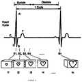
- FIG. 9illustrates one example of samples of a periodic human characteristic signal (specifically, an ECG waveform) associated, or gated, with images of dynamic anatomy.
- a periodic human characteristic signalspecifically, an ECG waveform
- FIG. 10is a diagram of an exemplary surgical instrument navigation system in accordance with present invention.
- FIG. 11is a flowchart that depicts a technique for simulating a virtual volumetric scene of a body cavity from a point of view of a surgical instrument positioned within the patient in accordance with the present invention
- FIG. 12is an exemplary display from the surgical instrument navigation system of the present invention.
- FIG. 12Ais an illustration of the position of the surgical instrument within the trachea as depicted in FIG. 12 .
- FIGS. 12B and 12Ceach illustrate the video image provided by the surgical instrument when undergoing the “wiggle maneuver” along the plane shown in FIG. 12A at the respective positions indicated.
- FIG. 12Dis an illustration of the video image provided by the surgical instrument and corresponding to the 3D navigation model image of FIG. 12 .
- FIG. 13is a flowchart that depicts a technique for synchronizing the display of an indicia or graphical representation of the surgical instrument with cardiac or respiratory cycle of the patient in accordance with the present invention.
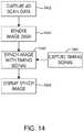
- FIG. 14is a flowchart that depicts a technique for generating four-dimensional image data that is synchronized with the patient in accordance with the present invention.
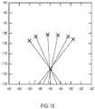
- FIG. 15is a graph depicting an axis or point that the instrument (e.g., a bronchoscope) deflects in a single planar direction.
- the graphshows the instrument (e.g., a bronchoscope) being maneuvered in six different orientations in a 3D localizer volume, with all orientations converging about a common axis or point of deflection.
- FIG. 16is a graph depicting the eigenvalues (e 0 ,e 1 ,e 2 ) for a moving 3.0 sec PCA (principal component analysis) window over a data file including 1800 samples.
- the square waverepresents an on/off “wiggle detector” state based on the algorithm described herein, and this square wave demonstrates that the algorithm exhibits no false negatives for the validation test data and that the seven exemplary “wiggle” periods are clearly matched to the “on” state of the wiggle detector.
- the implementation of the algorithmuses low pass filtering and an appropriate comparator function to eliminate any false positive traces or spots (“blips”) indicated in FIG. 16 .
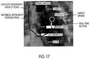
- FIG. 17is an image of an exemplary synthetic radiograph in accordance with the present invention, depicting the historical instrument position trace.
- FIG. 18depicts an exemplary curvature warning system in accordance with the invention described herein.
- FIG. 19depicts an exemplary real-time respiration compensation algorithm.
- a methodthat comprises receiving a position of an instrument reference marker coupled to an instrument; transforming the position into image space using a position of a non-tissue internal reference marker implanted in a patient; and superimposing a representation of the instrument on an image in which the non-tissue internal reference marker appears possesses at least the receiving, transforming, and superimposing steps, but is not limited to possessing only those steps.
- the methodalso covers instances where the transforming includes transforming the position into image space using a transformation that is based, in part, on the position of the non-tissue internal reference marker implanted in the patient, and calculating the transformation using image space coordinates of the internal reference marker in the image.
- the term “use”should be interpreted the same way. Thus, a calculation that uses certain items uses at least those items, but also covers the use of additional items.
- a step that calls for creating a dataset that includes imagesone of the images (a) depicting a non-tissue internal reference marker, (b) being linked to non-tissue internal reference marker positional information, and (c) being at least 2-dimensional covers the creation of at least such a dataset, but also covers the creation of a dataset that includes images, where each image (a) depicts the non-tissue internal reference marker, and (b) is linked to non-tissue internal reference marker positional information.
- a and “an”are defined as one or more than one.
- the term “another”is defined as at least a second or more.
- the term “coupled”encompasses both direct and indirect connections, and is not limited to mechanical connections.
- An apparatusincludes a PTD and two or more markers coupled to the PTD.
- the apparatuscan also include two or more localization elements coupled to the PTD proximate the markers.
- the apparatusis configured to be coupled to a dynamic body, such as selected dynamic anatomy of a patient.
- Dynamic anatomycan be, for example, any anatomy that moves during its normal function (e.g., the heart, lungs, kidneys, liver and blood vessels).
- a processorsuch as a computer, is configured to receive image data associated with the dynamic body taken during a pre-surgical or pre-procedural first time interval.
- the image datacan include an indication of a position of each of the markers for multiple instants in time during the first time interval.
- the processorcan also receive position data associated with the localization elements during a second time interval in which a surgical procedure or other medical procedure is being performed.
- the processorcan use the position data received from the localization elements to determine a distance between the elements for a given instant in time during the second time interval.
- the processorcan also use the image data to determine the distance between the markers for a given instant in time during the first time interval.
- the processorcan then find a match between an image where the distance between the markers at a given instant in time during the first time interval is the same as the distance between the elements associated with those markers at a given instant in time during the medical procedure, or second time interval.
- the processorcan determine a sequence of motion of the markers and match this sequence of motion to the recorded motion of the markers over the complete procedure or significant period of time. Distance alone between the markers may not be sufficient to match the patient space to image space in many instances, it is important for the system to know the direction the markers are moving and the range and speed of this motion to find the appropriate sequence of motion for a complex signal or sequence
- a physician or other healthcare professionalcan use the images selected by the processor during a medical procedure performed during the second time interval.
- a medical procedureis performed on a targeted anatomy of a patient, such as a heart or lung
- the physicianmay not be able to utilize an imaging device during the medical procedure to guide him to the targeted area within the patient.
- a PTDaccording to an embodiment of the invention can be positioned or coupled to the patient proximate the targeted anatomy prior to the medical procedure, and pre-procedural images can be taken of the targeted area during a first time interval.
- Markers or fiducials coupled to the PTDcan be viewed with the image data, which can include an indication of the position of the markers during a given path of motion of the targeted anatomy (e.g., the heart) during the first time interval. Such motion can be due, for example, to inspiration (i.e., inhaling) and expiration (i.e., exhaling) of the patient, or due to the heart beating.
- the processorreceives data from the localization elements associated with a position of the elements at a given instant in time during the medical procedure (or second time interval).
- the distance between selected pairs of markerscan be determined from the image data and the distance, range, acceleration, and speed between corresponding selected pairs of localization elements can be determined based on the element data for given instants in time. From multiple image datasets the range and speed of the markers motion can be calculated.
- the distance between a selected pair of elementscan be used to determine an intra-procedural distance between the pair of corresponding markers to which the localization elements are coupled.
- An image from the pre-procedural image data taken during the first time intervalcan then be selected where the distance between the pair of selected markers in that image corresponds with or closely approximates the same distance determined using the localization elements at a given instant in time during the second time interval.
- This processcan be done continuously during the medical procedure, producing simulated real-time, intra-procedural images illustrating the orientation and shape of the targeted anatomy as a catheter, sheath, needle, forceps, guidewire, fiducial delivery devices, therapy device (ablation modeling, drug diffusion modeling, etc.), or similar structure(s) is/are navigated to the targeted anatomy.
- the physiciancan view selected image(s) of the targeted anatomy that correspond to and simulate real-time movement of the anatomy.
- the location(s) of a sensore.g., an electromagnetic coil sensor
- the location(s) of a sensorcan be superimposed on an image of a catheter.
- the superimposed image(s) of the cathetercan then be superimposed on the selected image(s) from the first time interval, providing simulated real-time images of the catheter location relative to the targeted anatomy.
- a real-time pathway registrationis applied to a pre-acquired dataset that does not contain the PTD.
- the pre-acquired datasetcan be at only one cycle of a patient's respiratory, heartbeat, or other path of motion.
- a PTDcan be subsequently applied to the patient, and the PTD signal can be used to collect registration information throughout full range or path of motion but only that information that is captured at a similar PTD orientation, shape, or point along the PTD cycle of motion is used.
- This methodenhances the registration accuracy by ensuring that the registration points being used to register are at the same point during the initial dataset acquisition.
- the methoduses multiple subsets of the acquired registration data that are collected based on the PTD signal. These multiple subsets are then applied against the pre-acquired dataset to find the optimal registration fit.
- the devicecan be integrated with one or more fiber optic localization (FDL) devices and/or techniques.
- the sensorsuch as an EM sensor
- the FDLprovides shape sensing of the airway, vessel, pathway, organ, environment and surroundings.
- Conventional FDL techniquescan be employed.
- the FDL devicecan be used to create localization information for the complete pathway or to refine the localization accuracy in a particular segment of the pathway.
- the systemcan use a weighted algorithm between multiple localization devices to determine the location and orientation of the instrument in the patient.
- the FDL devicecan also be used as or in conjunction with the PTD to track the patient's motion such as respiration or heartbeat.
- video input of the bronchoscopeto adjust the virtual “fly-through” view to be consistent with the user's normal perspective.
- conventional video processing and matching techniquescan be used to align the real-time video and the virtual image.
- Angular informationcan be derived from the location of patient anatomy in the image and the relative size of each within the image.
- the systemcan determine which the direction of the display. This can be done using, for example, translation, rotation, or a combination of both. Comparing the real-time image captured to the virtual image constructed from the 3D dataset (i.e., CT) the system can use this information to align the virtual image and/or enhance the system accuracy.
- a high-speed three-dimensional imaging devicesuch as an optical coherence tomography (OCT) device
- OCToptical coherence tomography
- a devicecan only view 1-2 mm below the surface.
- OCToptical coherence tomography
- multiple 3D volumes of datacan be collected and a larger 3D volume of collected data can be constructed. Knowing the 3D location and orientation of the multiple 3D volumes will allow the user to view a more robust image of, for example, pre-cancerous changes in the esophagus or colon.
- This datacan also be correlated to pre-acquired or intra-procedurally acquired CT, fluoroscopic, ultrasound, or 3D fluoroscopic images to provide additional information.
- an endolumenal systemas described herein is that a user could overlay the planned pathway information on to the actual/real-time video image of the scope or imaging device (such as ultrasound based device). Additionally, the system and apparatus could provide a visual cue on the real-time video image showing the correct direction or pathway to take.
- a 5DOF sensor 1024 and a limited or known range of motion of a localization device 134could be used to determine its orientation in the field. This is particularly relevant, for example, in determining which way is up or down or the overall rotation of an image in 3D space. In bronchoscopy, for instance, this can be used to orient the bronchoscopic view to the user's normal expected visual orientation. Because a bronchoscope 1012 is typically only able to move in one plane up and down, the system can use the 3D location of a tip sensor 1024 moving up and down to determine the sixth degree of freedom. In this implementation, such as is shown in FIG.
- a usercould steer the bronchoscope 1012 to a bifurcation or close to a bifurcation such as at location 29 shown in FIG. 12 and then perform a motion with the scope (e.g., up and down) using the thumb control to wiggle or flutter the tip 1015 and sensor 1024 .
- the systemcan determine the orientation and display the correct image such as depicted in bronchoscope video image of FIG. 12D corresponding to the 3D navigation model image provided by the imaging device shown at view 38 of FIG. 12 .
- 5DOF sensing of instrument tip POSEdetermines 5 of the 6 POSE parameters (x, y, z, pitch, and yaw).
- Such sensingmay be unable in certain applications, however, to determine the instantaneous roll of the device. This roll determination can be critical in matching the video coming from the device to the images.
- the techniques described hereinadvantageously allow for users to relate orientation of a navigation model or virtual endoscopic “fly-thru” display 38 to actual video orientation presented by the endoscopic instrument as illustrated in FIG. 12D .
- the methods described hereinprovides for the user to select a location, most typically at a branching point in the bronchial tree, such as is shown in FIG. 12A where the position of the catheter tip 1015 is shown in a position corresponding to location 29 depicted in FIG. 12 , and perform a “wiggle” maneuver with the tip of the device.
- the wiggle maneuvergenerally consists of three steps:
- the physical or translational location of deviceis substantially secured or held in place.
- the usershould ensure that the scope is held securely so it cannot translate relative to the airway.
- a manifestation of an algorithm to detect this operationconsists of recognizing a unique signature of motion over time.
- One such techniquefor example, consists of two parts:
- orientation of the tip to the eigenvector EUwhich represents the historical vector of motion for the instrument tip, is compared.
- the orientation of the tipshould show a rhythmic oscillation of about 90° (approximate range could be for instance +/ ⁇ 45°).
- This comparison of tip orientation to E0provides the basis for a determination of the wiggle plane normal using, for example, a cross-product or gram-schmidt orthogonalization.
- the physical wiggle plane normalis in a constant relationship to the coordinate space of the video signal, and thus can be related (calibrated) thereto.
- the wiggle techniques described herein for determining direction and orientationmay be used in a range of different endoscopic applications.
- the above examples and embodimentsgenerally refer to the use of the wiggle maneuver in connection with bronchoscopic applications, it will be understood that the techniques described herein may also be used in other applications including, but not limited to, enteroscopy, colonoscopy, sigmoidoscopy, rhinoscopy, proctoscopy, otoscopy, cystoscopy, gynoscopy, colposcopy, hysteroscopy, falloposcopy, laparoscopy, arthroscopy, thoracoscopy, amnioscopy, fetoscopy, panendoscopy, epiduroscopy, and the like.
- the wiggle techniques described hereinmay also be applicable in non-medical endoscopic uses, such as the internal inspection of complex technical systems, surveillance, and the like.
- serial orientation or positioning of multiple sensorsallows the determination of one or more parameters such as shape, position, orientation, and mechanical status of a complete or partial section of guidewire or other device or instrument.
- the placement of multiple sensorscan assist in visualizing the shape of the device and any bends in the path by providing a number of data points on the path (e.g., 8 sensors, spaced 1 mm apart) to create a 3D shape model of the device.
- Various parameterscan be used to track past or present movement and changes in device shape including, for example, elasticity, bend radius, limiting, and durometer rating of the device material.
- These parameters and accompanying datacan provide visual cues to the user during the procedure, for example, when the device has a certain bend or curvature (based on path or surroundings), e.g., to provide a notice or warning that the device is on the correct or incorrect path, or to provide notice regarding, or track, a particular parameter(s) that the user is interested in.
- a sensor pathwayis generally depicted in FIG. 18 , which shows exemplary curvature warning scenarios in the differently marked sections or segments.
- a breathing patientfor example, or a patient with a moving vessel related to heartbeat
- the patient's tracked or physiological signalis used to determine 4D patient motion cycle, and with the instrument's traveled path, one can determine the optical location relative to a segmented airway or vessel and use this information to provide the optimal virtual display.
- FIGS. 1 and 2are schematic illustrations of devices that can be used in conjunction with, or to perform, various procedures described herein.
- an apparatus 110includes a PTD 120 .
- the PTD 120can be coupled to a dynamic body B.
- the dynamic body Bcan be, for example, a selected dynamic portion of the anatomy of a patient.
- the PTD 120can be a variety of different shapes and sizes.
- the PTD 120is substantially planar, such as in the form of a patch that can be disposed at a variety of locations on a patient's body.
- Such a PTD 120can be coupled to the dynamic body with adhesive, straps, hook and pile, snaps, or any other suitable coupling method.
- the PTDcan be a catheter type device with a pigtail or anchoring mechanism that allows it to be attached to an internal organ or along a vessel.
- Two or more markers or fiducials 122are coupled to the PTD 120 at selected locations as shown in FIG. 1 .
- the markers 122are constructed of a material that can be viewed on an image, such as an X-ray or CT.
- the markers 122can be, for example, radiopaque, and can be coupled to the PTD 120 using any known methods of coupling such devices.
- FIGS. 1 and 2illustrate the apparatus 110 having four markers 122 , but any number of two or more markers can be used.
- the marker or fiducials and the localization elementcan be the same device.
- An imaging device 140can be used to take images of the dynamic body B while the PTD 120 is coupled to the dynamic body B, pre-procedurally during a first time interval.
- the markers 122are visible on the images and can provide an indication of a position of each of the markers 122 during the first time interval. The position of the markers 122 at given instants in time through a path of motion of the dynamic body B can be illustrated with the images.
- the imaging device 140can be, for example, a computed tomography (CT) device (e.g., respiratory-gated CT device, ECG-gated CT device), a magnetic resonance imaging (MRI) device (e.g., respiratory-gated MRI device, ECG-gated MRI device), an X-ray device, or any other suitable medical imaging device.
- CTcomputed tomography
- MRImagnetic resonance imaging
- the imaging device 140is a computed tomography positron emission tomography device that produces a fused computed tomography positron emission tomography image dataset.
- the imaging device 140can be in communication with a processor 130 and send, transfer, copy and/or provide image data taken during the first time interval associated with the dynamic body B to the processor 130 .
- the processor 130includes a processor-readable medium storing code representing instructions to cause the processor 130 to perform a process.
- the processor 130can be, for example, a commercially available personal computer, or a less complex computing or processing device that is dedicated to performing one or more specific tasks.
- the processor 130can be a terminal dedicated to providing an interactive graphical user interface (GUI).
- GUIgraphical user interface
- the processor 130can be a commercially available microprocessor.
- the processor 130can be an application-specific integrated circuit (ASIC) or a combination of ASICs, which are designed to achieve one or more specific functions, or enable one or more specific devices or applications.
- the processor 130can be an analog or digital circuit, or a combination of multiple circuits.
- the processor 130can include a memory component 132 .
- the memory component 132can include one or more types of memory.
- the memory component 132can include a read only memory (ROM) component and a random access memory (RAM) component.
- the memory componentcan also include other types of memory that are suitable for storing data in a form retrievable by the processor 130 .
- EPROMelectronically programmable read only memory
- EEPROMerasable electronically programmable read only memory
- flash memoryas well as other suitable forms of memory can be included within the memory component.
- the processor 130can also include a variety of other components, such as for example, coprocessors, graphic processors, etc., depending upon the desired functionality of the code.
- the processor 130can store data in the memory component 132 or retrieve data previously stored in the memory component 132 .
- the components of the processor 130can communicate with devices external to the processor 130 by way of an input/output (I/O) component (not shown).
- I/O componentcan include a variety of suitable communication interfaces.
- the I/O componentcan include, for example, wired connections, such as standard serial ports, parallel ports, universal serial bus (USB) ports, S-video ports, local area network (LAN) ports, small computer system interface (SCCI) ports, and so forth.
- the I/O componentcan include, for example, wireless connections, such as infrared ports, optical ports, Bluetooth® wireless ports, wireless LAN ports, or the like.
- the processor 130can be connected to a network, which may be any form of interconnecting network including an intranet, such as a local or wide area network, or an extranet, such as the World Wide Web or the Internet.
- the networkcan be physically implemented on a wireless or wired network, on leased or dedicated lines, including a virtual private network (VPN).
- VPNvirtual private network
- the processor 130can receive image data from the imaging device 140 .
- the processor 130can identify the position of selected markers 122 within the image data or voxel space using various segmentation techniques, such as Hounsfield unit thresholding, convolution, connected component, or other combinatory image processing and segmentation techniques.
- the processor 130can determine a distance and direction between the position of any two markers 122 during multiple instants in time during the first time interval, and store the image data, as well as the position and distance data, within the memory component 132 . Multiple images can be produced providing a visual image at multiple instants in time through the path of motion of the dynamic body.
- the processor 130can also include a receiving device or localization device 134 , which is described in more detail below.
- a deformation fieldmay also be included in the analysis in various embodiments described herein.
- the deformation fieldcan be applied to fuse 3D fluoroscopic images to CT images in order to compensate for different patient orientations, patient position, respiration, deformation induced by the catheter or other instrument, and/or other changes or perturbations that occur due to therapy delivery or resection or ablation of tissue.
- real-time respiration compensationcan be determined by applying an inspiration-to-expiration deformation vector field.
- the instrument locationcan be calculated using the deformation vector field.
- a real-time instrument tip correction vectorcan be applied to a 3D localized instrument tip.
- the real-time correction vectoris computed by scaling an inspiration-to-expiration deformation vector (found from the inspiration-to-expiration deformation vector field) based on the PTD respiratory signal. This correction vector can then be applied to the 3D localized instrument tip. This can further optimize accuracy during navigation.
- FIG. 19An example of an algorithm for real-time respiration compensation can be found in FIG. 19 .
- this algorithmfor each :
- FIG. 19 and the above discussiongenerally relate to real-time respiration motion, it will be understood that these calculations and determinations may also be applied to real-time heartbeat and/or vessel motion compensation, or any other motion of a dynamic body as described herein.
- the deformation matrixis calculated based upon inspiration and expiration.
- the deformation matrixis calculated based upon heartbeat.
- the deformation matrixis based upon vessel motion.
- Deformation on 2D imagescan also be calculated based upon therapeutic change of tissue, changes in Houndsfield units for images, patient motion compensation during the imaging sequence, therapy monitoring, and temperature monitoring with fluoroscopic imaging, among other things.
- One potential issue with conventional therapy delivery, for instance,is monitoring the therapy for temperature or tissue changes.
- this monitoringcan be carried out using intermittent fluoroscopic imaging, where the images are compensated between acquisition times to show very small changes in image density, which can represent temperature changes or tissue changes as a result of the therapy and/or navigation.
- One method of reducing radiation during the acquisition of a 3D fluoroscopic datasetis to use a deformation field between acquired 2D images to reduce the actual number of 2D images that need to be acquired to create the 3D dataset.
- the deformation fieldis used to calculate the deformation between images in the acquisition sequence to produce 2D images between the acquired slices, and these new slices can be used to calculate the 3D fluoroscopic dataset.
- 90 2D imagescan be acquired over a 360 degree acquisition sequence and the data from the images that would have ordinarily been acquired between each slice can be calculated and imported into the 3D reconstruction algorithm.
- the radiationis effectively reduced by 50%.
- two or more localization elements 124are coupled to the PTD 120 proximate the locations of the markers 122 for use during a medical procedure to be performed during a second time interval.
- the localization elements 124can be, for example, electromagnetic coils, infrared light emitting diodes, and/or optical passive reflective markers.
- the localization elements 124can also be, or be integrated with, one or more fiber optic localization (FDL) devices.
- the markers 122can include plastic or non-ferrous fixtures or dovetails or other suitable connectors used to couple the localization elements 124 to the markers 122 .
- a medical procedurecan then be performed with the PTD 120 coupled to the dynamic body B at the same location as during the first time interval when the pre-procedural images were taken.
- the localization elements 124are in communication or coupled to the localization device 134 included within processor 130 .
- the localization device 134can be, for example, an analog to digital converter that measures voltages induced onto localization coils in the field; creates a digital voltage reading; and maps that voltage reading to a metric positional measurement based on a characterized volume of voltages to millimeters from a fixed field emitter.
- Position data associated with the elements 124can be transmitted or sent to the localization device 134 continuously during the medical procedure during the second time interval.
- the position of the localization elements 124can be captured at given instants in time during the second time interval. Because the localization elements 124 are coupled to the PTD 120 proximate the markers 122 , the localization device 134 can use the position data of the elements 124 to deduce coordinates or positions associated with the markers 122 intra-procedurally during the second time interval. The distance, range, acceleration, and speed between one or more selected pairs of localization elements 124 (and corresponding markers 122 ) can then be determined and various algorithms can be used to analyze and compare the distance between selected elements 124 at given instants in time, to the distances between and orientation among corresponding markers 122 observed in the pre-operative images.
- An imagecan then be selected from the pre-operative images taken during the first time interval that indicates a distance or is grouped in a similar sequence of motion between corresponding markers 122 at a given instant in time, that most closely approximates or matches the distance or similar sequence of motion between the selected elements 124 .
- the process of comparing the distancesis described in more detail below.
- the apparatus 110 and processor 130can be used to provide images corresponding to the actual movement of the targeted anatomy during the medical procedure being performed during the second time interval.
- the imagesillustrate the orientation and shape of the targeted anatomy during a path of motion of the anatomy, for example, during inhaling and exhaling.
- FIG. 3illustrates an example set of distances or vectors d 1 through d 6 between a set of markers 122 , labeled m 1 through m 9 that are disposed at spaced locations on a PTD 120 .
- pre-procedure imagescan be taken of a dynamic body for which the PTD 120 is to be coupled during a first time interval.
- the distances between the markerscan be determined for multiple instants in time through the path of motion of the dynamic body.
- localization elements(not shown in FIG. 3 ) coupled proximate to the location of markers 122 can provide position data for the elements to a localization device (not shown in FIG. 3 ).
- the localization devicecan use the position data to determine distances or vectors between the elements for multiple instants in time during the medical procedure or second time interval.
- FIG. 4Ashows an example of distance or vector data from the localization device.
- Vectors a 1 through a 6represent distance data for one instant in time and vectors n 1 through n 6 for another instant in time, during a time interval from a to n.
- the vector datacan be used to select an image from the pre-procedural images that includes distances between the markers m 1 through m 9 that correspond to or closely approximate the distances a 1 through a 6 for time a, for example, between the localization elements.
- the same processcan be performed for the vectors n 1 through n 6 captured during time n.
- One method of selecting the appropriate image from the pre-procedural imagesis to execute an algorithm that can sum all of the distances a 1 through a 6 and then search for and match this sum to an image containing a sum of all of the distances d 1 through d 6 obtained pre-procedurally from the image data that is equal to the sum of the distances a 1 through a 6 .
- the difference between these sumsis equal to zero, the relative position and orientation of the anatomy or dynamic body D during the medical procedure will substantially match the position and orientation of the anatomy in the particular image.
- the image associated with distances d 1 through d 6 that match or closely approximate the distances a 1 through a 6can then be selected and displayed. For example, FIG.
- FIG. 4Billustrates examples of pre-procedural images, Image a and Image n, of a dynamic body D that correspond to the distances a 1 through a 6 and n 1 through n 6 , respectively.
- FIG. 5illustrates an apparatus 210 according to an embodiment of the invention.
- the apparatus 210includes a tubular shaped PTD 220 that can be constructed with a rigid material or, alternatively, a flexible and/or stretchable material.
- the PTD 220is substantially rigid in structure.
- the PTD 220has a flexible or stretchable structure.
- the PTD 220can be positioned over a portion of a patient's body, such as around the upper or lower torso of the patient.
- the stretchability of the PTD 220allows the PTD 220 to at least partially constrict some of the movement of the portion of the body for which it is coupled.
- the apparatus 210further includes multiple markers or fiducials 222 coupled to the PTD 220 at spaced locations.
- a plurality of localization elements 224are removably coupled proximate to the locations of markers 222 , such that during a first time interval as described above, images can be taken without the elements 224 being coupled to the PTD 220 .
- the localization elementsneed not be removably coupled.
- the elementscan be fixedly coupled to the PTD.
- the elementscan be coupled to the PTD during the pre-procedure imaging.
- FIG. 6is a graphical illustration indicating how the apparatus 210 (shown without localization elements 224 ) can move and change orientation and shape during movement of a dynamic body, such as a mammalian body M.
- the graphis one example of how the lung volume can change during inhalation (inspiration) and exhalation (expiration) of the mammalian body M.
- the corresponding changes in shape and orientation of the apparatus 210 during inhalation and exhalationare also illustrated.
- the six markers 222 shown in FIG. 5are labeled a, b, c, d, e, and f. As described above, images of the apparatus 210 can be taken during a first time interval.
- the imagescan include an indication of relative position of each of the markers 222 , that is the markers 222 are visible in the images, and the position of each marker 222 can then be observed over a period of time.
- a distance between any two markers 222can then be determined for any given instant of time during the first time interval. For example, a distance X between markers a and b is illustrated, and a distance Y between markers b and f is illustrated. These distances can be determined for any given instant in time during the first time interval from an associated image that illustrates the position and orientation of the markers 222 . As illustrated, during expiration of the mammalian body M at times indicated as A and C, the distance X is smaller than during inspiration of the mammalian body M, at the time indicated as B.
- the distance Yis greater during inspiration than during expiration.
- the distance between any pair of markers 222can be determined and used in the processes described herein.
- the above embodimentsare merely examples of possible pair selections. For example, a distance between a position of marker e and a position of marker b may be determined.
- multiple pairs or only one pairmay be selected for a given procedure.
- FIG. 7is a flowchart illustrating a method according to an embodiment of the invention.
- a method 51includes at step 52 receiving image data during a pre-procedural or first time interval.
- imagesare taken of a dynamic body using an appropriate imaging modality (e.g., CT Scan, MRI, etc.).
- the image datais associated with one or more images taken of a PTD (as described herein) coupled to a dynamic body, where the PTD includes two or more markers coupled thereto.
- the image data of the dynamic bodyis correlated with image data related to the PTD.
- the one or more imagescan be taken using a variety of different imaging modalities as described previously.
- the image datacan include an indication of a position of a first marker and an indication of a position of a second marker, as illustrated at step 54 .
- the image datacan include position data for multiple positions of the markers during a range or path of motion of the dynamic body over a selected time interval.
- the image datacan include position data associated with multiple markers, however, only two are described here for simplicity.
- a distance between the position of the first marker and the position of the second markercan be determined for multiple instants in time during the first time interval, at step 56 .
- the determinationcan include determining the distance based on the observable distance between the markers on a given image.
- the image data, including all of the images received during the first time interval, the position, and the distance datacan be stored in a memory and/or recorded at step 58 .
- position datacan be received for a first localization element and a second localization element.
- the localization elementscan be coupled to the PTD proximate the locations of the markers, such that the position data associated with the elements can be used to determine the relative position of the markers in real-time during the medical procedure.
- the position data of the elementscan be stored and/or recorded at step 62 .
- a distance between the first and second localization elementscan be determined at step 64 . Although only two localization elements are described, as with the markers, position data associated with more than two localization elements can be received and the distances between the additional elements can be determined.
- the next stepis to determine which image from the one or more images taken during the first time interval represents the relative position and/or orientation of the dynamic body at a given instant in time during the second time interval or during the medical procedure. To determine this, at step 66 , the distance between the positions of the first and second localization elements at a given instant in time during the second time interval are compared to the distance(s) determined in step 56 between the positions of the first and second markers obtained with the image data during the first time interval.
- An imagecan be selected from the first time interval that best represents the same position and orientation of the dynamic body at a given instant in time during the medical procedure.
- the difference between the distance between a given pair of localization elements during the second time intervalis used to select the image that contains the same distance between the same given pair of markers from the image data received during the first time interval.
- Thiscan be accomplished, for example, by executing an algorithm to perform the calculations.
- the algorithmcan sum the distances between all of the selected pairs of elements for a given instant in time during the second time interval and sum the distances between all of the associated selected pairs of markers for each instant in time during the first time interval when the pre-procedural image data was received.
- FIG. 8shows one embodiment of a system (system 100 ) that includes components that can be used to perform image guided interventions using a gated imaging modality, such as ECG-gated MRI, or ECG-gated CT.
- a gated imaging modalitysuch as ECG-gated MRI, or ECG-gated CT.
- the figuredepicts a patient 10 positioned on an operating table 12 with a physician 14 performing a medical procedure on him.
- FIG. 8depicts physician 14 steering a medical instrument 16 through the patient's internal anatomy in order to deliver therapy.
- instrument 16is depicted as a catheter entering the right atrium by way of the inferior vena cava preceded by a femoral artery access point; however, the present systems are not limited to catheter use indications.
- the position of virtually any instrumentmay be tracked as discussed below and a representation of it superimposed on the proper image, consistent with the present methods, apparatuses, and systems.
- An “instrument”is any device controlled by physician 14 for the purpose of delivering therapy, and includes needles, guidewires, stents, filters, occluders, retrieval devices, imaging devices (such as OCT, EBUS, IVUS, and the like), and leads.
- Instrument 16is fitted with one or more instrument reference markers 18 .
- a tracker 20(which is sometimes referred to in the art as a “tracking system”) is configured to track the type of reference marker or markers coupled to instrument 16 .
- Tracker 20can be any type of tracking system, including but not limited to an electromagnetic tracking system.
- An example of a suitable electromagnetic tracking systemis the AURORA electromagnetic tracking system, commercially available from Northern Digital Inc. in Waterloo, Ontario Canada. If tracker 20 is an electromagnetic tracking system, element 20 would represent an electromagnetic field generator that emits a series of electromagnetic fields designed to engulf patient 10 , and reference marker or markers 18 coupled to medical instrument 16 could be coils that would receive an induced voltage that could be monitored and translated into a coordinate position of the marker(s).
- an angled coil sensoris employed during the targeted navigation.
- the coilis wrapped at an acute angle (i.e., the angle is less than about 90°) relative to the axial length of the sensor.
- the coilis positioned (e.g., wrapped) at an angle of from about 30° to about 60° relative to the axial length. In one preferred embodiment, the coil is positioned at about a 45° angle relative to the axial length.
- the positioning of the coil in accordance with the exemplary embodiments described hereinadvantageously provides a directional vector that is not parallel with the sensor core.
- the physical axisis different and, as the sensor moves, this additional directional vector can be quantified and used to detect up and down (and other directional) movement.
- This motioncan be captured over time as described herein to determine orientation and prepare and display the more accurate images.
- An external reference marker 22can be placed in a location close to the region of the patient where the procedure is to be performed, yet in a stable location that will not move (or that will move a negligible amount) with the patient's heart beat and respiration. If patient 10 is securely fixed to table 12 for the procedure, external reference marker 22 (which may be described as “static”) can be affixed to table 12 . If patient 10 is not completely secured to table 12 , external reference marker 22 can be placed on region of the back of patient 10 exhibiting the least amount of movement. Tracker 20 can be configured to track external reference marker 22 .
- Non-tissue internal reference markers 24can be placed in the gross region where the image guided navigation will be carried out.
- Non-tissue internal reference marker(s) 24should be placed in an anatomic location that exhibits movement that is correlated with the movement of the anatomy intended for image guided navigation. This location will be internal to the patient, in the gross location of the anatomy of interest.
- Converter 26can be configured to convert analog measurements received from the reference markers and tracker 20 into digital data understandable by image guidance computing platform 30 , and relay that data to image guidance computing platform 30 to which converter 26 can be coupled.
- Image guidance computing platform 30can take the form of a computer, and may include a monitor on which a representation of one or more instruments used during the IGI can be displayed over an image of the anatomy of interest.
- System 100also includes a periodic human characteristic signal monitor, such as ECG monitor 32 , which can be configured to receive a periodic human characteristic signal.
- ECG monitor 32can be configured to receive an ECG signal in the form of the ECG data transmitted to it by ECG leads 34 coupled to patient 10 .
- the periodic human characteristic signal monitor(e.g., ECG monitor 32 ) can also be configured to relay a periodic human characteristic signal (e.g., ECG data) to image guidance computing platform 30 , to which it can be coupled.
- non-tissue internal reference marker(s) 24Prior to the start of the image guided intervention, non-tissue internal reference marker(s) 24 —but not necessarily static external reference marker 22 —should be placed in the gross region of interest for the procedure.
- patient 10is to be scanned with an imaging device, such as gated scanner 40 , and the resulting gated image dataset transferred to image guidance computing platform 30 , to which the imaging device is coupled and which can reside in the operating or procedure theatre.
- imaging devicesand more specifically suitable gated scanners, include ECG-gated MRI scanners and ECG-gated CT scanners.
- a hospital network 50may be used to couple gated scanner 40 to image guidance computing platform 30 .
- the imaging devicee.g., gated scanner 40
- the imaging devicecan be configured to create a gated dataset that includes pre-operative images, one or more of which (up to all) are taken using the imaging device and are linked to a sample of a periodic human characteristic signal (e.g., a sample, or a phase, of an ECG signal).
- a periodic human characteristic signale.g., a sample, or a phase, of an ECG signal.
- FIG. 9highlights the relationship between the samples (S 1 . . . Sn) and the images (I 1 . . . In) that were captured by gated scanner 40 .
- Designations P, Q, R, S, and Tare designations well known in the art; they designate depolarizations and re-polarizations of the heart.
- Gated scanner 40essentially creates an image of the anatomy of interest at a particular instant in time during the anatomy's periodic movement. Image I 1 corresponds to the image that was captured at the S 1 moment of patient 10 's ECG cycle. Similarly, I 2 is correlated with S 2 , and In with Sn.
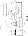
- FIG. 10is a diagram of another exemplary surgical instrument navigation system 1010 .
- the surgical instrument navigation system 1010is operable to visually simulate a virtual volumetric scene within the body of a patient, such as an internal body cavity, from a point of view of a surgical instrument 1012 residing in the cavity of a patient 10 .
- the surgical instrument navigation system 1010is primarily comprised of a surgical instrument 1012 , a data processor 1016 having a display 1018 , and a tracking subsystem 1020 .
- the surgical instrument navigation system 1010may further include (or accompanied by) an imaging device 1014 that is operable to provide image data to the system.
- the surgical instrument 1012is preferably a relatively inexpensive, flexible and/or steerable catheter that may be of a disposable type.
- the surgical instrument 1012is modified to include one or more tracking sensors 1024 that are detectable by the tracking subsystem 1020 .
- tracking sensors 1024that are detectable by the tracking subsystem 1020 .
- other types of surgical instrumentse.g., a guide wire, a needle, a forcep, a pointer probe, a stent, a seed, an implant, an endoscope, an energy delivery device, a therapy delivery device, etc.
- at least some of these surgical instrumentsmay be wireless or have wireless communications links.
- the surgical instrumentsmay encompass medical devices which are used for exploratory purposes, testing purposes or other types of medical procedures.
- the volumetric scan datais then registered as shown at 34 .
- Registration of the dynamic reference frame 19generally relates information in the volumetric scan data to the region of interest associated with the patient. This process is referred to as registering image space to patient space. Often, the image space must also be registered to another image space. Registration is accomplished through knowledge of the coordinate vectors of at least three non-collinear points in the image space and the patient space.
- the imaging device 1014is used to capture volumetric scan data 1132 representative of an internal region of interest within the patient 10 .
- the three-dimensional scan datais preferably obtained prior to surgery on the patient 10 .
- the captured volumetric scan datamay be stored in a data store associated with the data processor 1016 for subsequent processing.
- the principles of the present inventionmay also extend to scan data acquired during surgery.
- volumetric scan datamay be acquired using various known medical imaging devices 1014 , including but not limited to a magnetic resonance imaging (MRI) device, a computed tomography (CT) imaging device, a positron emission tomography (PET) imaging device, a 2D or 3D fluoroscopic imaging device, and 2D, 3D or 4D ultrasound imaging devices.
- MRImagnetic resonance imaging
- CTcomputed tomography
- PETpositron emission tomography
- 2D or 3D fluoroscopic imaging devicea 2D or 3D fluoroscopic imaging device
- 2D, 3D or 4D ultrasound imaging devicesa series of two-dimensional data sets may be acquired and then assembled into volumetric data as is well known in the art using a two-dimensional to three-dimensional conversion.
- the multi-dimensional imaging modalities described hereinmay also be coupled with digitally reconstructed radiography (DRR) techniques.
- DRRdigitally reconstructed radiography
- a fluoroscopic image acquisitionfor example, radiation passes through a physical media to create a projection image on a radiation-sensitive film or an electronic image intensifier.
- a simulated imagecan be generated in conjunction with DRR methodologies.
- DRRis generally known in the art, and is described, for example, by Lemieux et al. (Med. Phys. 21(11), November 1994, pp. 1749-60).
- a fluoroscopic imageis formed by computationally projecting volume elements, or voxels, of the 3D or 4D dataset onto one or more selected image planes.
- volume elementsor voxels
- a fluoroscopic imageis formed by computationally projecting volume elements, or voxels, of the 3D or 4D dataset onto one or more selected image planes.
- thisprovides another method to see the up-and-down (and other directional) movement of the instrument.
- This arrangementfurther provides the ability to see how the device moves in an image(s), which translates to improved movement of the device in a patient.
- An exemplary pathway in accordance with the disclosure hereincan be seen in FIG. 17 .
- a dynamic reference frame 19is attached to the patient proximate to the region of interest within the patient 10 .
- the region of interestis a vessel or a cavity within the patient, it is readily understood that the dynamic reference frame 19 may be placed within the patient 10 .
- the dynamic reference frame 19is also modified to include tracking sensors detectable by the tracking subsystem 1020 .
- the tracking subsystem 1020is operable to determine position data for the dynamic reference frame 19 as further described below.
- the volumetric scan datais then registered as shown at 1134 .
- Registration of the dynamic reference frame 19generally relates information in the volumetric scan data to the region of interest associated with the patient. This process is referred to as registering image space to patient space. Often, the image space must also be registered to another image space. Registration is accomplished through knowledge of the coordinate vectors of at least three non-collinear points in the image space and the patient space.
- Registration for image guided surgerycan be completed by different known techniques.
- point-to-point registrationis accomplished by identifying points in an image space and then touching the same points in patient space. These points are generally anatomical landmarks that are easily identifiable on the patient.
- surface registrationinvolves the user's generation of a surface in patient space by either selecting multiple points or scanning, and then accepting the best fit to that surface in image space by iteratively calculating with the data processor until a surface match is identified.
- repeat fixation devicesentail the user repeatedly removing and replacing a device (i.e., dynamic reference frame, etc.) in known relation to the patient or image fiducials of the patient.
- automatic registrationby first attaching the dynamic reference frame to the patient prior to acquiring image data.
- FIG. 12illustrates another type of secondary image 28 which may be displayed in conjunction with the primary perspective image 38 .
- the primary perspective imageis an interior view of an air passage within the patient 10 .
- the secondary image 28is an exterior view of the air passage which includes an indicia or graphical representation 29 that corresponds to the location of the surgical instrument 1012 within the air passage.
- the indicia 29is shown as a crosshairs. It is envisioned that other indicia may be used to signify the location of the surgical instrument in the secondary image.
- the secondary image 28is constructed by superimposing the indicia 29 of the surgical instrument 1012 onto the manipulated image data 1138 (see FIG. 11 ).
- the display of an indicia of the surgical instrument 1012 on the secondary imagemay be synchronized with an anatomical function, such as the cardiac or respiratory cycle, of the patient.
- an anatomical functionsuch as the cardiac or respiratory cycle
- the cardiac or respiratory cycle of the patientmay cause the surgical instrument 1012 to flutter or jitter within the patient.
- a surgical instrument 1012 positioned in or near a chamber of the heartwill move in relation to the patient's heart beat.
- the indicia of the surgical instrument 1012will likewise flutter or jitter on the displayed image 1140 .
- other anatomical functionswhich may affect the position of the surgical instrument 1012 within the patient are also within the scope of the present invention.
- position data for the surgical instrument 1012is acquired at a repetitive point within each cycle of either the cardiac cycle or the respiratory cycle of the patient.
- the imaging device 1014is used to capture volumetric scan data 1342 representative of an internal region of interest within a given patient.
- a secondary imagemay then be rendered 1344 from the volumetric scan data by the data processor 1016 .
- the surgical instrument navigation system 1010may further include a timing signal generator 1026 .
- the timing signal generator 1026is operable to generate and transmit a timing signal 1346 that correlates to at least one of (or both) the cardiac cycle or the respiratory cycle of the patient 10 .
- the timing signalmight be in the form of a periodic clock signal.
- the timing signalmay be derived from an electrocardiogram signal from the patient 10 .
- One skilled in the artwill readily recognize other techniques for deriving a timing signal that correlate to at least one of the cardiac or respiratory cycle or other anatomical cycle of the patient.
- the indicia of the surgical instrument 1012tracks the movement of the surgical instrument 1012 as it is moved by the surgeon within the patient 10 .
- the display of the indicia of the surgical instrument 1012is periodically updated 1348 based on the timing signal from the timing signal generator 1026 .
- the timing generator 1026is electrically connected to the tracking subsystem 1020 .
- the tracking subsystem 1020is in turn operable to report position data for the surgical instrument 1012 in response to a timing signal received from the timing signal generator 1026 .
- the position of the indicia of the surgical instrument 1012is then updated 1350 on the display of the image data.
- the surgical instrument navigation system 1010may be further adapted to display four-dimensional image data for a region of interest as shown in FIG. 14 .
- the imaging device 1014is operable to capture volumetric scan data 1462 for an internal region of interest over a period of time, such that the region of interest includes motion that is caused by either the cardiac cycle or the respiratory cycle of the patient 10 .
- a volumetric perspective view of the regionmay be rendered 1464 from the volumetric scan data 1462 by the data processor 1016 as described above.
- the four-dimensional image datamay be further supplemented with other patient data, such as temperature or blood pressure, using coloring coding techniques.
- the data processor 1016is adapted to receive a timing signal from the timing signal generator 1026 .
- the timing signal generator 1026is operable to generate and transmit a timing signal that correlates to either the cardiac cycle or the respiratory cycle of the patient 10 .
- the volumetric perspective imagemay be synchronized 1466 with the cardiac or respiratory cycle of the patient 10 .
- the synchronized image 1466is then displayed 1468 on the display 1018 of the system.
- the four-dimensional synchronized imagemay be either (or both of) the primary image rendered from the point of view of the surgical instrument or the secondary image depicting the indicia of the position of the surgical instrument 1012 within the patient 10 . It is readily understood that the synchronization process is also applicable to two-dimensional image data acquire over time.
- the surgical navigation systemcan use prior knowledge such as the segmented vessel or airway structure to compensate for error in the tracking subsystem or for inaccuracies caused by an anatomical shift occurring since acquisition of scan data. For instance, it is known that the surgical instrument 1012 being localized is located within a given vessel or airway and, therefore should be displayed within the vessel or airway. Statistical methods can be used to determine the most likely location; within the vessel or airway with respect to the reported location and then compensate so the display accurately represents the instrument 1012 within the center of the vessel or airway.
- the center of the vessel or airwaycan be found by segmenting the vessels or airways from the three-dimensional datasets and using commonly known imaging techniques to define the centerline of the vessel or airway tree.
- Statistical methodsmay also be used to determine if the surgical instrument 1012 has potentially punctured the vessel or airway. This can be done by determining the reported location is too far from the centerline or the trajectory of the path traveled is greater than a certain angle (worse case 90 degrees) with respect to the vessel or airway. Reporting this type of trajectory (error) is very important to the clinicians.
- the tracking along the center of the vesselmay also be further refined by correcting for motion of the respiratory or cardiac cycle, as described above. While navigating along the vessel or airway tree prior knowledge about the last known location can be used to aid in determining the new location.
- the instrument or navigated devicemust follow a pre-defined vessel or airway tree and therefore cannot jump from one branch to the other without traveling along a path that would be allowed.
- the orientation of the instrument or navigated devicecan also be used to select the most likely pathway that is being traversed. The orientation information can be used to increase the probability or weight for selected location or to exclude potential pathways and therefore enhance system accuracy.
- the surgical instrument navigation system of the present inventionmay also incorporate atlas maps. It is envisioned that three-dimensional or four-dimensional atlas maps may be registered with patient specific scan data or generic anatomical models. Atlas maps may contain kinematic information (e.g., heart and lung models) that can be synchronized with four-dimensional image data, thereby supplementing the real-time information. In addition, the kinematic information may be combined with localization information from several instruments to provide a complete four-dimensional model of organ motion. The atlas maps may also be used to localize bones or soft tissue which can assist in determining placement and location of implants.
- Atlas mapsmay contain kinematic information (e.g., heart and lung models) that can be synchronized with four-dimensional image data, thereby supplementing the real-time information.
- the kinematic informationmay be combined with localization information from several instruments to provide a complete four-dimensional model of organ motion.
- the atlas mapsmay also be used to localize bones or soft tissue which can assist in determining placement and location
- the previous description of the embodimentsis provided to enable any person skilled in the art to make or use the invention. While the invention has been particularly shown and described with reference to embodiments thereof, it will be understood by those skilled in art that various changes in form and details may be made therein without departing from the spirit and scope of the invention.
- the PTD, markers and localization elementscan be constructed from any suitable material, and can be a variety of different shapes and sizes, not necessarily specifically illustrated, while still remaining within the scope of the invention.
Landscapes
- Health & Medical Sciences (AREA)
- Life Sciences & Earth Sciences (AREA)
- Engineering & Computer Science (AREA)
- Surgery (AREA)
- Veterinary Medicine (AREA)
- Public Health (AREA)
- General Health & Medical Sciences (AREA)
- Biomedical Technology (AREA)
- Heart & Thoracic Surgery (AREA)
- Animal Behavior & Ethology (AREA)
- Molecular Biology (AREA)
- Medical Informatics (AREA)
- Pathology (AREA)
- Biophysics (AREA)
- Physics & Mathematics (AREA)
- Nuclear Medicine, Radiotherapy & Molecular Imaging (AREA)
- Radiology & Medical Imaging (AREA)
- Human Computer Interaction (AREA)
- Optics & Photonics (AREA)
- Pulmonology (AREA)
- Cardiology (AREA)
- Physiology (AREA)
- Oral & Maxillofacial Surgery (AREA)
- Immunology (AREA)
- Vascular Medicine (AREA)
- Otolaryngology (AREA)
- Dentistry (AREA)
- Gynecology & Obstetrics (AREA)
- Robotics (AREA)
- Endocrinology (AREA)
- Anesthesiology (AREA)
- Hematology (AREA)
- Signal Processing (AREA)
- Apparatus For Radiation Diagnosis (AREA)
- Endoscopes (AREA)
- Magnetic Resonance Imaging Apparatus (AREA)
Abstract
Description
r=V·E0
- stationary scope (wherein the e0, e1, and e2 values will be very small and roughly equal, representing the small magnitude, unbiased Gaussian noise of the direct localization measurement);
- scope in motion along a straight line in space, such as when traversing individual segments (wherein the e0 will be very large, with very small e1 and e2 values, representing a large co-linear translation in space). In addition, the absolute value of r will be close to 1, indicating that the orientation of the tip is nearly collinear with the translation of the tip over time.

- (a) find visuch that scalar d is minimized;
- (b) compute c, wherein:
c=−vit - and (c) compute′, wherein:
 ′++c
′++c
Thus,′ is a respiration compensated version of.
Does Σai=Σdi(i=1 to 6 in this example) OR
Does Σ(ai−di)=0 (i=1 to 6 in this example).
If yes to either of these, then the image is a match to the vector or distance data obtained during the medical procedure.
Claims (5)
Priority Applications (2)
| Application Number | Priority Date | Filing Date | Title |
|---|---|---|---|
| US16/358,882US11109740B2 (en) | 2010-08-20 | 2019-03-20 | Apparatus and method for four dimensional soft tissue navigation in endoscopic applications |
| US17/466,555US11690527B2 (en) | 2010-08-20 | 2021-09-03 | Apparatus and method for four dimensional soft tissue navigation in endoscopic applications |
Applications Claiming Priority (7)
| Application Number | Priority Date | Filing Date | Title |
|---|---|---|---|
| US37552310P | 2010-08-20 | 2010-08-20 | |
| US37553310P | 2010-08-20 | 2010-08-20 | |
| US37543910P | 2010-08-20 | 2010-08-20 | |
| US37548410P | 2010-08-20 | 2010-08-20 | |
| US13/215,017US8696549B2 (en) | 2010-08-20 | 2011-08-22 | Apparatus and method for four dimensional soft tissue navigation in endoscopic applications |
| US14/182,896US20140232840A1 (en) | 2010-08-20 | 2014-02-18 | Apparatus and method for four dimensional soft tissue navigation in endoscopic applications |
| US16/358,882US11109740B2 (en) | 2010-08-20 | 2019-03-20 | Apparatus and method for four dimensional soft tissue navigation in endoscopic applications |
Related Parent Applications (1)
| Application Number | Title | Priority Date | Filing Date |
|---|---|---|---|
| US14/182,896ContinuationUS20140232840A1 (en) | 2010-08-20 | 2014-02-18 | Apparatus and method for four dimensional soft tissue navigation in endoscopic applications |
Related Child Applications (1)
| Application Number | Title | Priority Date | Filing Date |
|---|---|---|---|
| US17/466,555ContinuationUS11690527B2 (en) | 2010-08-20 | 2021-09-03 | Apparatus and method for four dimensional soft tissue navigation in endoscopic applications |
Publications (2)
| Publication Number | Publication Date |
|---|---|
| US20190320876A1 US20190320876A1 (en) | 2019-10-24 |
| US11109740B2true US11109740B2 (en) | 2021-09-07 |
Family
ID=45594602
Family Applications (15)
| Application Number | Title | Priority Date | Filing Date |
|---|---|---|---|
| US13/215,041ActiveUS10165928B2 (en) | 2010-08-20 | 2011-08-22 | Systems, instruments, and methods for four dimensional soft tissue navigation |
| US13/215,017Active2031-08-26US8696549B2 (en) | 2010-08-20 | 2011-08-22 | Apparatus and method for four dimensional soft tissue navigation in endoscopic applications |
| US13/817,730AbandonedUS20130303887A1 (en) | 2010-08-20 | 2011-08-22 | Apparatus and method for four dimensional soft tissue navigation |
| US13/215,036ActiveUS10264947B2 (en) | 2010-08-20 | 2011-08-22 | Apparatus and method for airway registration and navigation |
| US13/215,050AbandonedUS20120071753A1 (en) | 2010-08-20 | 2011-08-22 | Apparatus and method for four dimensional soft tissue navigation including endoscopic mapping |
| US14/182,896AbandonedUS20140232840A1 (en) | 2010-08-20 | 2014-02-18 | Apparatus and method for four dimensional soft tissue navigation in endoscopic applications |
| US14/989,692AbandonedUS20160354159A1 (en) | 2010-08-20 | 2016-01-06 | Apparatus and method for four dimensional soft tissue navigation including endoscopic mapping |
| US16/221,886AbandonedUS20190231168A1 (en) | 2010-08-20 | 2018-12-17 | Systems, Instruments, and Methods for Four Dimensional Soft Tissue Navigation |
| US16/357,442Active2031-12-29US10898057B2 (en) | 2010-08-20 | 2019-03-19 | Apparatus and method for airway registration and navigation |
| US16/358,882Active2032-02-27US11109740B2 (en) | 2010-08-20 | 2019-03-20 | Apparatus and method for four dimensional soft tissue navigation in endoscopic applications |
| US16/371,576AbandonedUS20190223689A1 (en) | 2010-08-20 | 2019-04-01 | Apparatus and Method for Four Dimensional Soft Tissue Navigation Including Endoscopic Mapping |
| US16/665,135AbandonedUS20200060512A1 (en) | 2010-08-20 | 2019-10-28 | Apparatus and method for four dimensional soft tissue navigation |
| US17/156,051AbandonedUS20210137351A1 (en) | 2010-08-20 | 2021-01-22 | Apparatus and Method for Airway Registration and Navigation |
| US17/466,555Active2031-09-04US11690527B2 (en) | 2010-08-20 | 2021-09-03 | Apparatus and method for four dimensional soft tissue navigation in endoscopic applications |
| US17/698,112AbandonedUS20220361729A1 (en) | 2010-08-20 | 2022-03-18 | Apparatus and method for four dimensional soft tissue navigation |
Family Applications Before (9)
| Application Number | Title | Priority Date | Filing Date |
|---|---|---|---|
| US13/215,041ActiveUS10165928B2 (en) | 2010-08-20 | 2011-08-22 | Systems, instruments, and methods for four dimensional soft tissue navigation |
| US13/215,017Active2031-08-26US8696549B2 (en) | 2010-08-20 | 2011-08-22 | Apparatus and method for four dimensional soft tissue navigation in endoscopic applications |
| US13/817,730AbandonedUS20130303887A1 (en) | 2010-08-20 | 2011-08-22 | Apparatus and method for four dimensional soft tissue navigation |
| US13/215,036ActiveUS10264947B2 (en) | 2010-08-20 | 2011-08-22 | Apparatus and method for airway registration and navigation |
| US13/215,050AbandonedUS20120071753A1 (en) | 2010-08-20 | 2011-08-22 | Apparatus and method for four dimensional soft tissue navigation including endoscopic mapping |
| US14/182,896AbandonedUS20140232840A1 (en) | 2010-08-20 | 2014-02-18 | Apparatus and method for four dimensional soft tissue navigation in endoscopic applications |
| US14/989,692AbandonedUS20160354159A1 (en) | 2010-08-20 | 2016-01-06 | Apparatus and method for four dimensional soft tissue navigation including endoscopic mapping |
| US16/221,886AbandonedUS20190231168A1 (en) | 2010-08-20 | 2018-12-17 | Systems, Instruments, and Methods for Four Dimensional Soft Tissue Navigation |
| US16/357,442Active2031-12-29US10898057B2 (en) | 2010-08-20 | 2019-03-19 | Apparatus and method for airway registration and navigation |
Family Applications After (5)
| Application Number | Title | Priority Date | Filing Date |
|---|---|---|---|
| US16/371,576AbandonedUS20190223689A1 (en) | 2010-08-20 | 2019-04-01 | Apparatus and Method for Four Dimensional Soft Tissue Navigation Including Endoscopic Mapping |
| US16/665,135AbandonedUS20200060512A1 (en) | 2010-08-20 | 2019-10-28 | Apparatus and method for four dimensional soft tissue navigation |
| US17/156,051AbandonedUS20210137351A1 (en) | 2010-08-20 | 2021-01-22 | Apparatus and Method for Airway Registration and Navigation |
| US17/466,555Active2031-09-04US11690527B2 (en) | 2010-08-20 | 2021-09-03 | Apparatus and method for four dimensional soft tissue navigation in endoscopic applications |
| US17/698,112AbandonedUS20220361729A1 (en) | 2010-08-20 | 2022-03-18 | Apparatus and method for four dimensional soft tissue navigation |
Country Status (3)
| Country | Link |
|---|---|
| US (15) | US10165928B2 (en) |
| EP (2) | EP3659490B1 (en) |
| WO (1) | WO2012024686A2 (en) |
Families Citing this family (315)
| Publication number | Priority date | Publication date | Assignee | Title |
|---|---|---|---|---|
| EP1924198B1 (en) | 2005-09-13 | 2019-04-03 | Veran Medical Technologies, Inc. | Apparatus for image guided accuracy verification |
| US20070066881A1 (en) | 2005-09-13 | 2007-03-22 | Edwards Jerome R | Apparatus and method for image guided accuracy verification |
| US8219178B2 (en) | 2007-02-16 | 2012-07-10 | Catholic Healthcare West | Method and system for performing invasive medical procedures using a surgical robot |
| US10653497B2 (en) | 2006-02-16 | 2020-05-19 | Globus Medical, Inc. | Surgical tool systems and methods |
| US10357184B2 (en) | 2012-06-21 | 2019-07-23 | Globus Medical, Inc. | Surgical tool systems and method |
| US10893912B2 (en) | 2006-02-16 | 2021-01-19 | Globus Medical Inc. | Surgical tool systems and methods |
| US8090166B2 (en) | 2006-09-21 | 2012-01-03 | Surgix Ltd. | Medical image analysis |
| US9629571B2 (en) | 2007-03-08 | 2017-04-25 | Sync-Rx, Ltd. | Co-use of endoluminal data and extraluminal imaging |
| WO2008107905A2 (en)* | 2007-03-08 | 2008-09-12 | Sync-Rx, Ltd. | Imaging and tools for use with moving organs |
| US9968256B2 (en) | 2007-03-08 | 2018-05-15 | Sync-Rx Ltd. | Automatic identification of a tool |
| US10716528B2 (en) | 2007-03-08 | 2020-07-21 | Sync-Rx, Ltd. | Automatic display of previously-acquired endoluminal images |
| US9305334B2 (en) | 2007-03-08 | 2016-04-05 | Sync-Rx, Ltd. | Luminal background cleaning |
| US11064964B2 (en) | 2007-03-08 | 2021-07-20 | Sync-Rx, Ltd | Determining a characteristic of a lumen by measuring velocity of a contrast agent |
| US9375164B2 (en) | 2007-03-08 | 2016-06-28 | Sync-Rx, Ltd. | Co-use of endoluminal data and extraluminal imaging |
| US11197651B2 (en) | 2007-03-08 | 2021-12-14 | Sync-Rx, Ltd. | Identification and presentation of device-to-vessel relative motion |
| US8781193B2 (en) | 2007-03-08 | 2014-07-15 | Sync-Rx, Ltd. | Automatic quantitative vessel analysis |
| IL184151A0 (en) | 2007-06-21 | 2007-10-31 | Diagnostica Imaging Software Ltd | X-ray measurement method |
| EP2310839A4 (en) | 2008-06-18 | 2011-08-03 | Surgix Ltd | A method and system for stitching multiple images into a panoramic image |
| EP2303385B1 (en)* | 2008-06-19 | 2013-12-11 | Sync-RX, Ltd. | Stepwise advancement of a medical tool |
| US10362962B2 (en) | 2008-11-18 | 2019-07-30 | Synx-Rx, Ltd. | Accounting for skipped imaging locations during movement of an endoluminal imaging probe |
| US9974509B2 (en) | 2008-11-18 | 2018-05-22 | Sync-Rx Ltd. | Image super enhancement |
| US9095313B2 (en) | 2008-11-18 | 2015-08-04 | Sync-Rx, Ltd. | Accounting for non-uniform longitudinal motion during movement of an endoluminal imaging probe |
| US9144394B2 (en) | 2008-11-18 | 2015-09-29 | Sync-Rx, Ltd. | Apparatus and methods for determining a plurality of local calibration factors for an image |
| US8855744B2 (en) | 2008-11-18 | 2014-10-07 | Sync-Rx, Ltd. | Displaying a device within an endoluminal image stack |
| US11064903B2 (en) | 2008-11-18 | 2021-07-20 | Sync-Rx, Ltd | Apparatus and methods for mapping a sequence of images to a roadmap image |
| US9101286B2 (en) | 2008-11-18 | 2015-08-11 | Sync-Rx, Ltd. | Apparatus and methods for determining a dimension of a portion of a stack of endoluminal data points |
| US10004387B2 (en)* | 2009-03-26 | 2018-06-26 | Intuitive Surgical Operations, Inc. | Method and system for assisting an operator in endoscopic navigation |
| US8337397B2 (en)* | 2009-03-26 | 2012-12-25 | Intuitive Surgical Operations, Inc. | Method and system for providing visual guidance to an operator for steering a tip of an endoscopic device toward one or more landmarks in a patient |
| US8348831B2 (en)* | 2009-12-15 | 2013-01-08 | Zhejiang University | Device and method for computer simulated marking targeting biopsy |
| US20110184238A1 (en)* | 2010-01-28 | 2011-07-28 | The Penn State Research Foundation | Image-based global registration system and method applicable to bronchoscopy guidance |
| US8672837B2 (en) | 2010-06-24 | 2014-03-18 | Hansen Medical, Inc. | Methods and devices for controlling a shapeable medical device |
| EP3659490B1 (en)* | 2010-08-20 | 2025-10-01 | Veran Medical Technologies, Inc. | Apparatus and method for four dimensional soft tissue navigation |
| BR112013005879A2 (en)* | 2010-09-15 | 2016-05-10 | Konink Philps Electronics Nv | '' Robot guidance system, control units for an endoscope and robot guidance method '' |
| US8568326B2 (en)* | 2010-10-13 | 2013-10-29 | Volcano Corporation | Intravascular ultrasound pigtail catheter |
| JP5715372B2 (en)* | 2010-10-15 | 2015-05-07 | オリンパス株式会社 | Image processing apparatus, method of operating image processing apparatus, and endoscope apparatus |
| US20120190970A1 (en) | 2010-11-10 | 2012-07-26 | Gnanasekar Velusamy | Apparatus and method for stabilizing a needle |
| JP5944917B2 (en)* | 2010-11-24 | 2016-07-05 | ボストン サイエンティフィック サイムド,インコーポレイテッドBoston Scientific Scimed,Inc. | Computer readable medium for detecting and displaying body lumen bifurcation and system including the same |
| US8768019B2 (en)* | 2011-02-03 | 2014-07-01 | Medtronic, Inc. | Display of an acquired cine loop for procedure navigation |
| US9308050B2 (en) | 2011-04-01 | 2016-04-12 | Ecole Polytechnique Federale De Lausanne (Epfl) | Robotic system and method for spinal and other surgeries |
| US8900131B2 (en)* | 2011-05-13 | 2014-12-02 | Intuitive Surgical Operations, Inc. | Medical system providing dynamic registration of a model of an anatomical structure for image-guided surgery |
| CN103764061B (en) | 2011-06-27 | 2017-03-08 | 内布拉斯加大学评议会 | On Tool Tracking System and Computer Assisted Surgery Method |
| US11911117B2 (en) | 2011-06-27 | 2024-02-27 | Board Of Regents Of The University Of Nebraska | On-board tool tracking system and methods of computer assisted surgery |
| US9498231B2 (en) | 2011-06-27 | 2016-11-22 | Board Of Regents Of The University Of Nebraska | On-board tool tracking system and methods of computer assisted surgery |
| US9295449B2 (en)* | 2012-01-23 | 2016-03-29 | Ultrasonix Medical Corporation | Landmarks for ultrasound imaging |
| JP5918548B2 (en)* | 2012-01-24 | 2016-05-18 | 富士フイルム株式会社 | Endoscopic image diagnosis support apparatus, operation method thereof, and endoscopic image diagnosis support program |
| US20130211244A1 (en)* | 2012-01-25 | 2013-08-15 | Surgix Ltd. | Methods, Devices, Systems, Circuits and Associated Computer Executable Code for Detecting and Predicting the Position, Orientation and Trajectory of Surgical Tools |
| EP2816966B1 (en) | 2012-02-22 | 2023-10-25 | Veran Medical Technologies, Inc. | Steerable surgical catheter comprising a biopsy device at the distal end portion thereof |
| JP2015514494A (en)* | 2012-04-17 | 2015-05-21 | カレッジ メディカル イメージング リミテッド | Organ mapping system using optical coherence tomography probe |
| WO2013168117A2 (en)* | 2012-05-09 | 2013-11-14 | Koninklijke Philips N.V. | Interventional information brokering medical tracking interface |
| JP6334517B2 (en)* | 2012-05-14 | 2018-05-30 | インテュイティブ サージカル オペレーションズ, インコーポレイテッド | System and method for deformation compensation using shape sensing |
| US11793570B2 (en) | 2012-06-21 | 2023-10-24 | Globus Medical Inc. | Surgical robotic automation with tracking markers |
| US11399900B2 (en) | 2012-06-21 | 2022-08-02 | Globus Medical, Inc. | Robotic systems providing co-registration using natural fiducials and related methods |
| US11298196B2 (en) | 2012-06-21 | 2022-04-12 | Globus Medical Inc. | Surgical robotic automation with tracking markers and controlled tool advancement |
| US10624710B2 (en) | 2012-06-21 | 2020-04-21 | Globus Medical, Inc. | System and method for measuring depth of instrumentation |
| US20150032164A1 (en) | 2012-06-21 | 2015-01-29 | Globus Medical, Inc. | Methods for Performing Invasive Medical Procedures Using a Surgical Robot |
| US10758315B2 (en) | 2012-06-21 | 2020-09-01 | Globus Medical Inc. | Method and system for improving 2D-3D registration convergence |
| US11864839B2 (en) | 2012-06-21 | 2024-01-09 | Globus Medical Inc. | Methods of adjusting a virtual implant and related surgical navigation systems |
| US12329593B2 (en) | 2012-06-21 | 2025-06-17 | Globus Medical, Inc. | Surgical robotic automation with tracking markers |
| US10136954B2 (en) | 2012-06-21 | 2018-11-27 | Globus Medical, Inc. | Surgical tool systems and method |
| US12004905B2 (en) | 2012-06-21 | 2024-06-11 | Globus Medical, Inc. | Medical imaging systems using robotic actuators and related methods |
| US11395706B2 (en) | 2012-06-21 | 2022-07-26 | Globus Medical Inc. | Surgical robot platform |
| US12220120B2 (en) | 2012-06-21 | 2025-02-11 | Globus Medical, Inc. | Surgical robotic system with retractor |
| US12262954B2 (en) | 2012-06-21 | 2025-04-01 | Globus Medical, Inc. | Surgical robotic automation with tracking markers |
| US11317971B2 (en) | 2012-06-21 | 2022-05-03 | Globus Medical, Inc. | Systems and methods related to robotic guidance in surgery |
| US11857266B2 (en) | 2012-06-21 | 2024-01-02 | Globus Medical, Inc. | System for a surveillance marker in robotic-assisted surgery |
| EP2863827B1 (en) | 2012-06-21 | 2022-11-16 | Globus Medical, Inc. | Surgical robot platform |
| US11116576B2 (en) | 2012-06-21 | 2021-09-14 | Globus Medical Inc. | Dynamic reference arrays and methods of use |
| US11857149B2 (en) | 2012-06-21 | 2024-01-02 | Globus Medical, Inc. | Surgical robotic systems with target trajectory deviation monitoring and related methods |
| US11045267B2 (en) | 2012-06-21 | 2021-06-29 | Globus Medical, Inc. | Surgical robotic automation with tracking markers |
| US10350013B2 (en) | 2012-06-21 | 2019-07-16 | Globus Medical, Inc. | Surgical tool systems and methods |
| US11974822B2 (en) | 2012-06-21 | 2024-05-07 | Globus Medical Inc. | Method for a surveillance marker in robotic-assisted surgery |
| US11253327B2 (en) | 2012-06-21 | 2022-02-22 | Globus Medical, Inc. | Systems and methods for automatically changing an end-effector on a surgical robot |
| US12310683B2 (en) | 2012-06-21 | 2025-05-27 | Globus Medical, Inc. | Surgical tool systems and method |
| US10231791B2 (en) | 2012-06-21 | 2019-03-19 | Globus Medical, Inc. | Infrared signal based position recognition system for use with a robot-assisted surgery |
| US11607149B2 (en) | 2012-06-21 | 2023-03-21 | Globus Medical Inc. | Surgical tool systems and method |
| US11864745B2 (en) | 2012-06-21 | 2024-01-09 | Globus Medical, Inc. | Surgical robotic system with retractor |
| EP2863802B1 (en) | 2012-06-26 | 2020-11-04 | Sync-RX, Ltd. | Flow-related image processing in luminal organs |
| US20140005527A1 (en)* | 2012-06-29 | 2014-01-02 | General Electric Company | Method and system for dynamic referencing and registration used with surgical and interventional procedures |
| US9183354B2 (en) | 2012-08-15 | 2015-11-10 | Musc Foundation For Research Development | Systems and methods for image guided surgery |
| CN104736085B (en) | 2012-10-12 | 2018-01-30 | 直观外科手术操作公司 | Determining the location of medical devices in branching anatomy |
| BR112015013808A2 (en)* | 2012-12-17 | 2017-07-11 | Koninklijke Philips Nv | point-of-view tracking and imaging device, and computer-readable media for point-of-view tracking and imaging |
| WO2014106137A1 (en)* | 2012-12-28 | 2014-07-03 | The General Hospital Corporation | Optical probe apparatus, systems, methods for guiding tissue asessment |
| CA2842426C (en) | 2013-02-11 | 2021-01-12 | Covidien Lp | Cytology sampling system and method of utilizing the same |
| WO2014136576A1 (en)* | 2013-03-06 | 2014-09-12 | オリンパスメディカルシステムズ株式会社 | Endoscope system |
| US9057600B2 (en) | 2013-03-13 | 2015-06-16 | Hansen Medical, Inc. | Reducing incremental measurement sensor error |
| US20140267330A1 (en)* | 2013-03-14 | 2014-09-18 | Volcano Corporation | Systems and methods for managing medical image data for multiple users |
| US9271663B2 (en) | 2013-03-15 | 2016-03-01 | Hansen Medical, Inc. | Flexible instrument localization from both remote and elongation sensors |
| US10105149B2 (en) | 2013-03-15 | 2018-10-23 | Board Of Regents Of The University Of Nebraska | On-board tool tracking system and methods of computer assisted surgery |
| WO2014139019A1 (en) | 2013-03-15 | 2014-09-18 | Synaptive Medical (Barbados) Inc. | System and method for dynamic validation, correction of registration for surgical navigation |
| US9014851B2 (en) | 2013-03-15 | 2015-04-21 | Hansen Medical, Inc. | Systems and methods for tracking robotically controlled medical instruments |
| US9629595B2 (en) | 2013-03-15 | 2017-04-25 | Hansen Medical, Inc. | Systems and methods for localizing, tracking and/or controlling medical instruments |
| AU2014231354B2 (en)* | 2013-03-15 | 2019-08-29 | Conavi Medical Inc. | Data display and processing algorithms for 3D imaging systems |
| WO2015135055A1 (en)* | 2014-03-14 | 2015-09-17 | Synaptive Medical (Barbados) Inc. | System and method for projected tool trajectories for surgical navigation systems |
| EP2979610B1 (en)* | 2013-03-28 | 2019-06-19 | Olympus Corporation | Endoscope system |
| EP2981205A4 (en) | 2013-04-04 | 2017-02-15 | Children's National Medical Center | Device and method for generating composite images for endoscopic surgery of moving and deformable anatomy |
| US11020016B2 (en) | 2013-05-30 | 2021-06-01 | Auris Health, Inc. | System and method for displaying anatomy and devices on a movable display |
| EP3041427B1 (en)* | 2013-09-06 | 2024-11-06 | Brigham and Women's Hospital, Inc. | System for a tissue resection margin measurement device |
| US10201265B2 (en) | 2013-09-06 | 2019-02-12 | Covidien Lp | Microwave ablation catheter, handle, and system |
| AU2014317930B2 (en) | 2013-09-06 | 2018-11-08 | Covidien Lp | Microwave ablation catheter, handle, and system |
| US10098566B2 (en) | 2013-09-06 | 2018-10-16 | Covidien Lp | System and method for lung visualization using ultrasound |
| US10448861B2 (en)* | 2013-09-06 | 2019-10-22 | Covidien Lp | System and method for light based lung visualization |
| US9283048B2 (en) | 2013-10-04 | 2016-03-15 | KB Medical SA | Apparatus and systems for precise guidance of surgical tools |
| US10028649B2 (en)* | 2013-12-02 | 2018-07-24 | Welch Allyn, Inc. | Digital colposcope system |
| US10390796B2 (en)* | 2013-12-04 | 2019-08-27 | Siemens Medical Solutions Usa, Inc. | Motion correction in three-dimensional elasticity ultrasound imaging |
| WO2015087206A1 (en)* | 2013-12-12 | 2015-06-18 | Koninklijke Philips N.V. | Method and system for electromagnetic tracking with magnetic trackers for respiratory monitoring |
| DE102014200326A1 (en)* | 2014-01-10 | 2015-07-16 | Siemens Aktiengesellschaft | A method of supporting navigation of a medical instrument |
| US9241771B2 (en) | 2014-01-15 | 2016-01-26 | KB Medical SA | Notched apparatus for guidance of an insertable instrument along an axis during spinal surgery |
| WO2015121311A1 (en) | 2014-02-11 | 2015-08-20 | KB Medical SA | Sterile handle for controlling a robotic surgical system from a sterile field |
| DE102014205313B4 (en)* | 2014-03-21 | 2015-10-15 | Siemens Aktiengesellschaft | Method for registering a near-infrared spectroscopy map and an anatomy image data set and x-ray device |
| US20150305650A1 (en) | 2014-04-23 | 2015-10-29 | Mark Hunter | Apparatuses and methods for endobronchial navigation to and confirmation of the location of a target tissue and percutaneous interception of the target tissue |
| US20150305612A1 (en) | 2014-04-23 | 2015-10-29 | Mark Hunter | Apparatuses and methods for registering a real-time image feed from an imaging device to a steerable catheter |
| EP3134022B1 (en) | 2014-04-24 | 2018-01-10 | KB Medical SA | Surgical instrument holder for use with a robotic surgical system |
| WO2015165736A1 (en)* | 2014-04-29 | 2015-11-05 | Koninklijke Philips N.V. | Device for determining a specific position of a catheter |
| EP3811891B8 (en) | 2014-05-14 | 2025-01-08 | Stryker European Operations Holdings LLC | Navigation system and processor arrangement for tracking the position of a work target |
| AU2015283946B2 (en) | 2014-07-02 | 2019-09-12 | Covidien Lp | Real-time automatic registration feedback |
| US9603668B2 (en) | 2014-07-02 | 2017-03-28 | Covidien Lp | Dynamic 3D lung map view for tool navigation inside the lung |
| US9754367B2 (en) | 2014-07-02 | 2017-09-05 | Covidien Lp | Trachea marking |
| US9770216B2 (en) | 2014-07-02 | 2017-09-26 | Covidien Lp | System and method for navigating within the lung |
| US20160000414A1 (en) | 2014-07-02 | 2016-01-07 | Covidien Lp | Methods for marking biopsy location |
| US11188285B2 (en)* | 2014-07-02 | 2021-11-30 | Covidien Lp | Intelligent display |
| CN106232010B (en) | 2014-07-02 | 2020-03-31 | 柯惠有限合伙公司 | System and method for detecting trachea |
| CN106659453B (en) | 2014-07-02 | 2020-05-26 | 柯惠有限合伙公司 | System and method for segmenting lungs |
| WO2016004302A1 (en) | 2014-07-02 | 2016-01-07 | Covidien Lp | Alignment ct |
| US10357257B2 (en) | 2014-07-14 | 2019-07-23 | KB Medical SA | Anti-skid surgical instrument for use in preparing holes in bone tissue |
| EP4328856B1 (en) | 2014-07-28 | 2025-07-02 | Intuitive Surgical Operations, Inc. | Systems and methods for intraoperative segmentation |
| US10966688B2 (en)* | 2014-08-26 | 2021-04-06 | Rational Surgical Solutions, Llc | Image registration for CT or MR imagery and ultrasound imagery using mobile device |
| EP3226781B1 (en) | 2014-12-02 | 2018-08-01 | KB Medical SA | Robot assisted volume removal during surgery |
| WO2016091766A1 (en)* | 2014-12-10 | 2016-06-16 | Koninklijke Philips N.V. | Supporting a user in performing an embolization procedure |
| US12144642B2 (en) | 2015-02-03 | 2024-11-19 | Arizona Board Of Regents On Behalf Of The University Of Arizona | Cell-collecting falloposcope and method for ovarian cancer detection |
| US10939864B2 (en) | 2015-02-03 | 2021-03-09 | Arizona Board Of Regents On Behalf Of The University Of Arizona | Falloposcope and method for ovarian cancer detection |
| US10013808B2 (en) | 2015-02-03 | 2018-07-03 | Globus Medical, Inc. | Surgeon head-mounted display apparatuses |
| CN104720752B (en)* | 2015-02-13 | 2017-09-15 | 亚太仿生学有限公司 | Detector and system device for internal thermal imaging of cavity structure |
| US10163204B2 (en)* | 2015-02-13 | 2018-12-25 | St. Jude Medical International Holding S.À R.L. | Tracking-based 3D model enhancement |
| WO2016131903A1 (en) | 2015-02-18 | 2016-08-25 | KB Medical SA | Systems and methods for performing minimally invasive spinal surgery with a robotic surgical system using a percutaneous technique |
| US20160331343A1 (en)* | 2015-05-11 | 2016-11-17 | Veran Medical Technologies, Inc. | Medical apparatus with translatable imaging device for real-time confirmation of interception of target tissue |
| CN104939908B (en)* | 2015-06-08 | 2017-11-10 | 温州医科大学附属第一医院 | A kind of guidance set of Ureteroscopy |
| DE102015213935B4 (en)* | 2015-07-23 | 2019-02-14 | Siemens Healthcare Gmbh | A medical imaging device having a positioning unit and a method for determining a position on a positioning surface |
| US10646298B2 (en) | 2015-07-31 | 2020-05-12 | Globus Medical, Inc. | Robot arm and methods of use |
| US10058394B2 (en) | 2015-07-31 | 2018-08-28 | Globus Medical, Inc. | Robot arm and methods of use |
| US10080615B2 (en) | 2015-08-12 | 2018-09-25 | Globus Medical, Inc. | Devices and methods for temporary mounting of parts to bone |
| JP6894431B2 (en) | 2015-08-31 | 2021-06-30 | ケービー メディカル エスアー | Robotic surgical system and method |
| US10034716B2 (en) | 2015-09-14 | 2018-07-31 | Globus Medical, Inc. | Surgical robotic systems and methods thereof |
| JP6824967B2 (en) | 2015-09-18 | 2021-02-03 | オーリス ヘルス インコーポレイテッド | Tubular net navigation |
| US10986990B2 (en) | 2015-09-24 | 2021-04-27 | Covidien Lp | Marker placement |
| US9771092B2 (en) | 2015-10-13 | 2017-09-26 | Globus Medical, Inc. | Stabilizer wheel assembly and methods of use |
| US11253322B2 (en) | 2015-10-14 | 2022-02-22 | Ecential Robotics | Fluoro-navigation system for navigating a tool relative to a medical image |
| US10709352B2 (en) | 2015-10-27 | 2020-07-14 | Covidien Lp | Method of using lung airway carina locations to improve ENB registration |
| US10143526B2 (en) | 2015-11-30 | 2018-12-04 | Auris Health, Inc. | Robot-assisted driving systems and methods |
| EP3397184A1 (en)* | 2015-12-29 | 2018-11-07 | Koninklijke Philips N.V. | System, control unit and method for control of a surgical robot |
| US10842453B2 (en) | 2016-02-03 | 2020-11-24 | Globus Medical, Inc. | Portable medical imaging system |
| US11883217B2 (en) | 2016-02-03 | 2024-01-30 | Globus Medical, Inc. | Portable medical imaging system and method |
| US10117632B2 (en) | 2016-02-03 | 2018-11-06 | Globus Medical, Inc. | Portable medical imaging system with beam scanning collimator |
| US10448910B2 (en) | 2016-02-03 | 2019-10-22 | Globus Medical, Inc. | Portable medical imaging system |
| US11058378B2 (en) | 2016-02-03 | 2021-07-13 | Globus Medical, Inc. | Portable medical imaging system |
| JP6976266B2 (en)* | 2016-03-10 | 2021-12-08 | ボディ・ビジョン・メディカル・リミテッドBody Vision Medical Ltd. | Methods and systems for using multi-view pose estimation |
| US10866119B2 (en) | 2016-03-14 | 2020-12-15 | Globus Medical, Inc. | Metal detector for detecting insertion of a surgical device into a hollow tube |
| EP3241518B1 (en) | 2016-04-11 | 2024-10-23 | Globus Medical, Inc | Surgical tool systems |
| CN107456278B (en)* | 2016-06-06 | 2021-03-05 | 北京理工大学 | Endoscopic surgery navigation method and system |
| US10342633B2 (en)* | 2016-06-20 | 2019-07-09 | Toshiba Medical Systems Corporation | Medical image data processing system and method |
| US10478143B2 (en) | 2016-08-02 | 2019-11-19 | Covidien Lp | System and method of generating and updatng a three dimensional model of a luminal network |
| CN106236264B (en)* | 2016-08-24 | 2020-05-08 | 李国新 | Gastrointestinal surgery navigation method and system based on optical tracking and image matching |
| CN106236263A (en)* | 2016-08-24 | 2016-12-21 | 李国新 | The gastrointestinal procedures air navigation aid decomposed based on scene and system |
| US10238455B2 (en) | 2016-08-31 | 2019-03-26 | Covidien Lp | Pathway planning for use with a navigation planning and procedure system |
| US10631933B2 (en) | 2016-08-31 | 2020-04-28 | Covidien Lp | Pathway planning for use with a navigation planning and procedure system |
| US10939963B2 (en) | 2016-09-01 | 2021-03-09 | Covidien Lp | Systems and methods for providing proximity awareness to pleural boundaries, vascular structures, and other critical intra-thoracic structures during electromagnetic navigation bronchoscopy |
| CN106419815B (en)* | 2016-09-19 | 2018-09-18 | 成都测迪森生物科技有限公司 | A kind of mobile laryngoscope with wearing function |
| US10799092B2 (en)* | 2016-09-19 | 2020-10-13 | Covidien Lp | System and method for cleansing segments of a luminal network |
| CN106419814B (en)* | 2016-09-19 | 2018-11-20 | 成都测迪森生物科技有限公司 | A kind of movable high-efficiency laryngoscope |
| KR101931747B1 (en)* | 2016-10-28 | 2019-03-13 | 삼성메디슨 주식회사 | Biopsy apparatus and method for operating the same |
| KR20190067917A (en) | 2016-11-02 | 2019-06-17 | 인튜어티브 서지컬 오퍼레이션즈 인코포레이티드 | Continuous political system and method for visual guided surgery |
| US10244926B2 (en) | 2016-12-28 | 2019-04-02 | Auris Health, Inc. | Detecting endolumenal buckling of flexible instruments |
| JP7233841B2 (en) | 2017-01-18 | 2023-03-07 | ケービー メディカル エスアー | Robotic Navigation for Robotic Surgical Systems |
| US11439742B2 (en)* | 2017-02-08 | 2022-09-13 | Veran Medical Technologies, Inc. | Localization needle |
| US11443441B2 (en)* | 2017-02-24 | 2022-09-13 | Brainlab Ag | Deep inspiration breath-hold setup using x-ray imaging |
| US11071594B2 (en) | 2017-03-16 | 2021-07-27 | KB Medical SA | Robotic navigation of robotic surgical systems |
| WO2018183727A1 (en) | 2017-03-31 | 2018-10-04 | Auris Health, Inc. | Robotic systems for navigation of luminal networks that compensate for physiological noise |
| US20180289432A1 (en) | 2017-04-05 | 2018-10-11 | Kb Medical, Sa | Robotic surgical systems for preparing holes in bone tissue and methods of their use |
| EP3409193B1 (en)* | 2017-06-02 | 2019-12-25 | Siemens Healthcare GmbH | Positioning of an mr body coil |
| US10575907B2 (en) | 2017-06-21 | 2020-03-03 | Biosense Webster (Israel) Ltd. | Registration with trajectory information with shape sensing |
| US10022192B1 (en) | 2017-06-23 | 2018-07-17 | Auris Health, Inc. | Automatically-initialized robotic systems for navigation of luminal networks |
| WO2019005696A1 (en) | 2017-06-28 | 2019-01-03 | Auris Health, Inc. | Electromagnetic distortion detection |
| CN116725667A (en) | 2017-06-28 | 2023-09-12 | 奥瑞斯健康公司 | System for providing positioning information and method for positioning an instrument within an anatomical structure |
| US11135015B2 (en) | 2017-07-21 | 2021-10-05 | Globus Medical, Inc. | Robot surgical platform |
| US10359464B2 (en) | 2017-09-18 | 2019-07-23 | Biosense Webster (Israel) Ltd. | Cable and associated continuity monitoring system and method |
| EP3694412A4 (en)* | 2017-10-10 | 2021-08-18 | Covidien LP | SYSTEM AND METHOD FOR IDENTIFYING AND MARKING A TARGET IN A FLUOROSCOPIC THREE-DIMENSIONAL RECONSTRUCTION |
| US11058493B2 (en) | 2017-10-13 | 2021-07-13 | Auris Health, Inc. | Robotic system configured for navigation path tracing |
| US10555778B2 (en) | 2017-10-13 | 2020-02-11 | Auris Health, Inc. | Image-based branch detection and mapping for navigation |
| EP3492032B1 (en) | 2017-11-09 | 2023-01-04 | Globus Medical, Inc. | Surgical robotic systems for bending surgical rods |
| US11794338B2 (en) | 2017-11-09 | 2023-10-24 | Globus Medical Inc. | Robotic rod benders and related mechanical and motor housings |
| US11357548B2 (en) | 2017-11-09 | 2022-06-14 | Globus Medical, Inc. | Robotic rod benders and related mechanical and motor housings |
| US11134862B2 (en) | 2017-11-10 | 2021-10-05 | Globus Medical, Inc. | Methods of selecting surgical implants and related devices |
| US11510736B2 (en) | 2017-12-14 | 2022-11-29 | Auris Health, Inc. | System and method for estimating instrument location |
| WO2019125964A1 (en) | 2017-12-18 | 2019-06-27 | Auris Health, Inc. | Methods and systems for instrument tracking and navigation within luminal networks |
| US11224392B2 (en) | 2018-02-01 | 2022-01-18 | Covidien Lp | Mapping disease spread |
| US10893842B2 (en) | 2018-02-08 | 2021-01-19 | Covidien Lp | System and method for pose estimation of an imaging device and for determining the location of a medical device with respect to a target |
| US10489911B2 (en)* | 2018-02-08 | 2019-11-26 | Apn Health, Llc | Determining respiratory phase from fluoroscopic images |
| US11464576B2 (en) | 2018-02-09 | 2022-10-11 | Covidien Lp | System and method for displaying an alignment CT |
| US20190254753A1 (en) | 2018-02-19 | 2019-08-22 | Globus Medical, Inc. | Augmented reality navigation systems for use with robotic surgical systems and methods of their use |
| US11373330B2 (en)* | 2018-03-27 | 2022-06-28 | Siemens Healthcare Gmbh | Image-based guidance for device path planning based on penalty function values and distances between ROI centerline and backprojected instrument centerline |
| JP7225259B2 (en) | 2018-03-28 | 2023-02-20 | オーリス ヘルス インコーポレイテッド | Systems and methods for indicating probable location of instruments |
| WO2019191144A1 (en) | 2018-03-28 | 2019-10-03 | Auris Health, Inc. | Systems and methods for registration of location sensors |
| US10573023B2 (en) | 2018-04-09 | 2020-02-25 | Globus Medical, Inc. | Predictive visualization of medical imaging scanner component movement |
| US10643333B2 (en)* | 2018-04-12 | 2020-05-05 | Veran Medical Technologies | Apparatuses and methods for navigation in and Local segmentation extension of anatomical treelike structures |
| US12380572B2 (en) | 2018-04-12 | 2025-08-05 | Veran Medical Technologies, Inc. | Methods for the segmentation of lungs, lung vasculature and lung lobes from CT data and clinical applications |
| US10869727B2 (en)* | 2018-05-07 | 2020-12-22 | The Cleveland Clinic Foundation | Live 3D holographic guidance and navigation for performing interventional procedures |
| US10905499B2 (en) | 2018-05-30 | 2021-02-02 | Auris Health, Inc. | Systems and methods for location sensor-based branch prediction |
| MX2020012904A (en) | 2018-05-31 | 2021-02-26 | Auris Health Inc | Image-based airway analysis and mapping. |
| JP7371026B2 (en) | 2018-05-31 | 2023-10-30 | オーリス ヘルス インコーポレイテッド | Path-based navigation of tubular networks |
| KR102567087B1 (en) | 2018-05-31 | 2023-08-17 | 아우리스 헬스, 인코포레이티드 | Robotic systems and methods for navigation of luminal networks detecting physiological noise |
| US11033721B2 (en)* | 2018-06-22 | 2021-06-15 | Acclarent, Inc. | Guidewire for dilating eustachian tube via middle ear |
| US11571205B2 (en)* | 2018-07-16 | 2023-02-07 | Cilag Gmbh International | Surgical visualization feedback system |
| US11944388B2 (en) | 2018-09-28 | 2024-04-02 | Covidien Lp | Systems and methods for magnetic interference correction |
| CN112804959B (en) | 2018-09-28 | 2025-01-28 | 奥瑞斯健康公司 | Robotic systems and methods for accompanying endoscopic and percutaneous medical procedures |
| US11204677B2 (en)* | 2018-10-22 | 2021-12-21 | Acclarent, Inc. | Method for real time update of fly-through camera placement |
| US11337742B2 (en) | 2018-11-05 | 2022-05-24 | Globus Medical Inc | Compliant orthopedic driver |
| US11278360B2 (en) | 2018-11-16 | 2022-03-22 | Globus Medical, Inc. | End-effectors for surgical robotic systems having sealed optical components |
| US11145111B2 (en) | 2018-12-04 | 2021-10-12 | Intuitive Research And Technology Corporation | Volumetric slicer |
| US11602402B2 (en) | 2018-12-04 | 2023-03-14 | Globus Medical, Inc. | Drill guide fixtures, cranial insertion fixtures, and related methods and robotic systems |
| US11744655B2 (en) | 2018-12-04 | 2023-09-05 | Globus Medical, Inc. | Drill guide fixtures, cranial insertion fixtures, and related methods and robotic systems |
| US11113868B2 (en) | 2018-12-04 | 2021-09-07 | Intuitive Research And Technology Corporation | Rastered volume renderer and manipulator |
| US11877806B2 (en) | 2018-12-06 | 2024-01-23 | Covidien Lp | Deformable registration of computer-generated airway models to airway trees |
| EP3890615B1 (en) | 2018-12-07 | 2025-02-05 | Veran Medical Technologies, Inc. | Endobronchial catheter system for rapid diagnosis of lung disease |
| CN109745120B (en)* | 2018-12-24 | 2020-07-31 | 罗雄彪 | Image registration conversion parameter optimization method and system |
| US20200237459A1 (en)* | 2019-01-25 | 2020-07-30 | Biosense Webster (Israel) Ltd. | Flexible multi-coil tracking sensor |
| CN113614844B (en) | 2019-03-14 | 2025-09-23 | 皇家飞利浦有限公司 | Dynamic intervention 3D model deformation |
| US11806084B2 (en) | 2019-03-22 | 2023-11-07 | Globus Medical, Inc. | System for neuronavigation registration and robotic trajectory guidance, and related methods and devices |
| US11317978B2 (en) | 2019-03-22 | 2022-05-03 | Globus Medical, Inc. | System for neuronavigation registration and robotic trajectory guidance, robotic surgery, and related methods and devices |
| US11571265B2 (en) | 2019-03-22 | 2023-02-07 | Globus Medical Inc. | System for neuronavigation registration and robotic trajectory guidance, robotic surgery, and related methods and devices |
| US11382549B2 (en) | 2019-03-22 | 2022-07-12 | Globus Medical, Inc. | System for neuronavigation registration and robotic trajectory guidance, and related methods and devices |
| US11419616B2 (en) | 2019-03-22 | 2022-08-23 | Globus Medical, Inc. | System for neuronavigation registration and robotic trajectory guidance, robotic surgery, and related methods and devices |
| US20200297357A1 (en) | 2019-03-22 | 2020-09-24 | Globus Medical, Inc. | System for neuronavigation registration and robotic trajectory guidance, robotic surgery, and related methods and devices |
| US11571268B1 (en) | 2019-04-02 | 2023-02-07 | Canon U.S.A., Inc. | Medical continuum robot extraction and methods thereof |
| US11045179B2 (en) | 2019-05-20 | 2021-06-29 | Global Medical Inc | Robot-mounted retractor system |
| US20200375665A1 (en)* | 2019-05-31 | 2020-12-03 | Canon U.S.A., Inc. | Medical continuum robot and methods thereof |
| US11628023B2 (en) | 2019-07-10 | 2023-04-18 | Globus Medical, Inc. | Robotic navigational system for interbody implants |
| US12089902B2 (en) | 2019-07-30 | 2024-09-17 | Coviden Lp | Cone beam and 3D fluoroscope lung navigation |
| US12059281B2 (en)* | 2019-08-19 | 2024-08-13 | Covidien Lp | Systems and methods of fluoro-CT imaging for initial registration |
| CN211131187U (en)* | 2019-08-29 | 2020-07-31 | 蓝明 | Device for interventional operation of heart and blood vessel through radial artery or ulnar artery |
| US11207141B2 (en) | 2019-08-30 | 2021-12-28 | Auris Health, Inc. | Systems and methods for weight-based registration of location sensors |
| US11147633B2 (en) | 2019-08-30 | 2021-10-19 | Auris Health, Inc. | Instrument image reliability systems and methods |
| EP4025921A4 (en) | 2019-09-03 | 2023-09-06 | Auris Health, Inc. | DETECTION AND COMPENSATION OF ELECTROMAGNETIC DISTORTION |
| US12239287B2 (en) | 2019-09-09 | 2025-03-04 | Arizona Board Of Regents On Behalf Of The University Of Arizona | Cell-collecting falloposcope and method for ovarian cancer detection |
| US12396692B2 (en) | 2019-09-24 | 2025-08-26 | Globus Medical, Inc. | Compound curve cable chain |
| US11571171B2 (en) | 2019-09-24 | 2023-02-07 | Globus Medical, Inc. | Compound curve cable chain |
| US12408929B2 (en) | 2019-09-27 | 2025-09-09 | Globus Medical, Inc. | Systems and methods for navigating a pin guide driver |
| US11426178B2 (en) | 2019-09-27 | 2022-08-30 | Globus Medical Inc. | Systems and methods for navigating a pin guide driver |
| US12329391B2 (en) | 2019-09-27 | 2025-06-17 | Globus Medical, Inc. | Systems and methods for robot-assisted knee arthroplasty surgery |
| US11864857B2 (en) | 2019-09-27 | 2024-01-09 | Globus Medical, Inc. | Surgical robot with passive end effector |
| US11890066B2 (en) | 2019-09-30 | 2024-02-06 | Globus Medical, Inc | Surgical robot with passive end effector |
| US11510684B2 (en) | 2019-10-14 | 2022-11-29 | Globus Medical, Inc. | Rotary motion passive end effector for surgical robots in orthopedic surgeries |
| US12133772B2 (en) | 2019-12-10 | 2024-11-05 | Globus Medical, Inc. | Augmented reality headset for navigated robotic surgery |
| US12220176B2 (en) | 2019-12-10 | 2025-02-11 | Globus Medical, Inc. | Extended reality instrument interaction zone for navigated robotic |
| US11992373B2 (en) | 2019-12-10 | 2024-05-28 | Globus Medical, Inc | Augmented reality headset with varied opacity for navigated robotic surgery |
| US12064189B2 (en) | 2019-12-13 | 2024-08-20 | Globus Medical, Inc. | Navigated instrument for use in robotic guided surgery |
| US11446095B2 (en)* | 2019-12-24 | 2022-09-20 | Biosense Webster (Israel) Ltd. | 2D pathfinder visualization |
| WO2021137109A1 (en) | 2019-12-31 | 2021-07-08 | Auris Health, Inc. | Alignment techniques for percutaneous access |
| WO2021137108A1 (en) | 2019-12-31 | 2021-07-08 | Auris Health, Inc. | Alignment interfaces for percutaneous access |
| EP4084721B1 (en) | 2019-12-31 | 2025-10-01 | Auris Health, Inc. | Anatomical feature identification and targeting |
| US11382699B2 (en) | 2020-02-10 | 2022-07-12 | Globus Medical Inc. | Extended reality visualization of optical tool tracking volume for computer assisted navigation in surgery |
| US12414752B2 (en) | 2020-02-17 | 2025-09-16 | Globus Medical, Inc. | System and method of determining optimal 3-dimensional position and orientation of imaging device for imaging patient bones |
| US11207150B2 (en) | 2020-02-19 | 2021-12-28 | Globus Medical, Inc. | Displaying a virtual model of a planned instrument attachment to ensure correct selection of physical instrument attachment |
| US11737663B2 (en) | 2020-03-30 | 2023-08-29 | Auris Health, Inc. | Target anatomical feature localization |
| US11253216B2 (en) | 2020-04-28 | 2022-02-22 | Globus Medical Inc. | Fixtures for fluoroscopic imaging systems and related navigation systems and methods |
| US11153555B1 (en) | 2020-05-08 | 2021-10-19 | Globus Medical Inc. | Extended reality headset camera system for computer assisted navigation in surgery |
| US11510750B2 (en) | 2020-05-08 | 2022-11-29 | Globus Medical, Inc. | Leveraging two-dimensional digital imaging and communication in medicine imagery in three-dimensional extended reality applications |
| US11382700B2 (en) | 2020-05-08 | 2022-07-12 | Globus Medical Inc. | Extended reality headset tool tracking and control |
| US11701492B2 (en) | 2020-06-04 | 2023-07-18 | Covidien Lp | Active distal tip drive |
| US11317973B2 (en) | 2020-06-09 | 2022-05-03 | Globus Medical, Inc. | Camera tracking bar for computer assisted navigation during surgery |
| US12070276B2 (en) | 2020-06-09 | 2024-08-27 | Globus Medical Inc. | Surgical object tracking in visible light via fiducial seeding and synthetic image registration |
| US11382713B2 (en) | 2020-06-16 | 2022-07-12 | Globus Medical, Inc. | Navigated surgical system with eye to XR headset display calibration |
| US11877807B2 (en) | 2020-07-10 | 2024-01-23 | Globus Medical, Inc | Instruments for navigated orthopedic surgeries |
| US11793588B2 (en) | 2020-07-23 | 2023-10-24 | Globus Medical, Inc. | Sterile draping of robotic arms |
| US11737831B2 (en) | 2020-09-02 | 2023-08-29 | Globus Medical Inc. | Surgical object tracking template generation for computer assisted navigation during surgical procedure |
| EP3967232A1 (en)* | 2020-09-10 | 2022-03-16 | Koninklijke Philips N.V. | Method for providing a source of secondary medical imaging |
| US11523785B2 (en) | 2020-09-24 | 2022-12-13 | Globus Medical, Inc. | Increased cone beam computed tomography volume length without requiring stitching or longitudinal C-arm movement |
| US11911112B2 (en) | 2020-10-27 | 2024-02-27 | Globus Medical, Inc. | Robotic navigational system |
| US12076091B2 (en) | 2020-10-27 | 2024-09-03 | Globus Medical, Inc. | Robotic navigational system |
| US11941814B2 (en) | 2020-11-04 | 2024-03-26 | Globus Medical Inc. | Auto segmentation using 2-D images taken during 3-D imaging spin |
| US11717350B2 (en) | 2020-11-24 | 2023-08-08 | Globus Medical Inc. | Methods for robotic assistance and navigation in spinal surgery and related systems |
| US12161433B2 (en) | 2021-01-08 | 2024-12-10 | Globus Medical, Inc. | System and method for ligament balancing with robotic assistance |
| CN112891685B (en)* | 2021-01-14 | 2022-07-01 | 四川大学华西医院 | Method and system for intelligently detecting position of blood vessel |
| CN116916848A (en)* | 2021-02-03 | 2023-10-20 | 加利福尼亚大学董事会 | A surgical perception framework for robotic tissue manipulation |
| US12433683B2 (en) | 2021-04-08 | 2025-10-07 | Mazor Robotics Ltd. | Tracking soft tissue changes intraoperatively |
| US12150728B2 (en) | 2021-04-14 | 2024-11-26 | Globus Medical, Inc. | End effector for a surgical robot |
| US12178523B2 (en) | 2021-04-19 | 2024-12-31 | Globus Medical, Inc. | Computer assisted surgical navigation system for spine procedures |
| US11857273B2 (en) | 2021-07-06 | 2024-01-02 | Globus Medical, Inc. | Ultrasonic robotic surgical navigation |
| CN113413213B (en)* | 2021-07-14 | 2023-03-14 | 广州医科大学附属第一医院(广州呼吸中心) | CT result processing method, navigation processing method, device and detection system |
| US11439444B1 (en) | 2021-07-22 | 2022-09-13 | Globus Medical, Inc. | Screw tower and rod reduction tool |
| US12213745B2 (en) | 2021-09-16 | 2025-02-04 | Globus Medical, Inc. | Extended reality systems for visualizing and controlling operating room equipment |
| US12238087B2 (en) | 2021-10-04 | 2025-02-25 | Globus Medical, Inc. | Validating credential keys based on combinations of credential value strings and input order strings |
| US12184636B2 (en) | 2021-10-04 | 2024-12-31 | Globus Medical, Inc. | Validating credential keys based on combinations of credential value strings and input order strings |
| US20230124915A1 (en)* | 2021-10-20 | 2023-04-20 | Covidien Lp | Video laryngoscope image file management systems and methods |
| US20230368330A1 (en) | 2021-10-20 | 2023-11-16 | Globus Medical, Inc. | Interpolation of medical images |
| DE112022004581T5 (en) | 2021-10-22 | 2024-09-19 | Veran Medical Technologies, Inc. | LOW PROFILE ULTRASOUND CATHETER AND SYSTEM |
| WO2023081342A1 (en)* | 2021-11-05 | 2023-05-11 | Board Of Regents, The University Of Texas System | Four-dimensional tactile sensing system, device, and method |
| US20230165639A1 (en) | 2021-12-01 | 2023-06-01 | Globus Medical, Inc. | Extended reality systems with three-dimensional visualizations of medical image scan slices |
| US11911115B2 (en) | 2021-12-20 | 2024-02-27 | Globus Medical Inc. | Flat panel registration fixture and method of using same |
| CN116416412A (en)* | 2021-12-31 | 2023-07-11 | 杭州堃博生物科技有限公司 | Auxiliary navigation method, device and equipment for bronchoscope |
| US12094128B2 (en) | 2022-02-03 | 2024-09-17 | Mazor Robotics Ltd. | Robot integrated segmental tracking |
| US12103480B2 (en) | 2022-03-18 | 2024-10-01 | Globus Medical Inc. | Omni-wheel cable pusher |
| CN114387320B (en)* | 2022-03-25 | 2022-07-19 | 武汉楚精灵医疗科技有限公司 | Medical image registration method, device, terminal and computer-readable storage medium |
| US12048493B2 (en) | 2022-03-31 | 2024-07-30 | Globus Medical, Inc. | Camera tracking system identifying phantom markers during computer assisted surgery navigation |
| US12394086B2 (en) | 2022-05-10 | 2025-08-19 | Globus Medical, Inc. | Accuracy check and automatic calibration of tracked instruments |
| US12161427B2 (en) | 2022-06-08 | 2024-12-10 | Globus Medical, Inc. | Surgical navigation system with flat panel registration fixture |
| US20240020840A1 (en) | 2022-07-15 | 2024-01-18 | Globus Medical, Inc. | REGISTRATION OF 3D and 2D IMAGES FOR SURGICAL NAVIGATION AND ROBOTIC GUIDANCE WITHOUT USING RADIOPAQUE FIDUCIALS IN THE IMAGES |
| US12226169B2 (en) | 2022-07-15 | 2025-02-18 | Globus Medical, Inc. | Registration of 3D and 2D images for surgical navigation and robotic guidance without using radiopaque fiducials in the images |
| US12318150B2 (en) | 2022-10-11 | 2025-06-03 | Globus Medical Inc. | Camera tracking system for computer assisted surgery navigation |
| US20240293071A1 (en)* | 2023-03-03 | 2024-09-05 | Garth Constantine | System, method and apparatus for real-time 3d cardiac mapping with multi-catheter support and cardiac-wall analytics |
| WO2024236414A1 (en)* | 2023-05-15 | 2024-11-21 | Medtronic Navigation, Inc. | Systems and methods for correlating one or more motions of an anatomical element |
| US12329568B2 (en) | 2023-05-22 | 2025-06-17 | Veran Medical Technologies, Inc. | Systems and methods for monitoring ablation progress using linear EBUS data |
| CN117598782B (en)* | 2023-09-28 | 2024-06-04 | 苏州盛星医疗器械有限公司 | Surgical navigation method, device, equipment and medium for percutaneous puncture surgery |
| US20250295394A1 (en)* | 2024-03-25 | 2025-09-25 | GE Precision Healthcare LLC | System and method for synchronizing data acqusition of an ultrasound system and a tracking system |
| EP4625434A1 (en)* | 2024-03-28 | 2025-10-01 | Koninklijke Philips N.V. | Supporting an interventional procedure |
| CN118303990B (en)* | 2024-04-11 | 2025-05-09 | 江苏一影医疗设备有限公司 | Imaging navigation method and system |
| CN120168109B (en)* | 2025-03-14 | 2025-09-26 | 贵黔国际医院管理有限公司 | Surgical robot navigation system based on neuroendoscope |
Citations (361)
| Publication number | Priority date | Publication date | Assignee | Title |
|---|---|---|---|---|
| US3788324A (en) | 1972-05-05 | 1974-01-29 | P Lim | External catheter device |
| US4421106A (en) | 1976-03-19 | 1983-12-20 | Takami Uehara | Fiber scope for biopsy operable by a single operator |
| US4583538A (en) | 1984-05-04 | 1986-04-22 | Onik Gary M | Method and apparatus for stereotaxic placement of probes in the body utilizing CT scanner localization |
| US5053042A (en) | 1990-01-16 | 1991-10-01 | Bidwell Clifford D | Biopsy needle guide for use with CT scanner |
| US5158088A (en) | 1990-11-14 | 1992-10-27 | Advanced Technology Laboratories, Inc. | Ultrasonic diagnostic systems for imaging medical instruments within the body |
| US5186174A (en) | 1987-05-21 | 1993-02-16 | G. M. Piaff | Process and device for the reproducible optical representation of a surgical operation |
| US5251165A (en) | 1992-04-09 | 1993-10-05 | James Iii J Colin | Two phase random number generator |
| US5251635A (en) | 1991-09-03 | 1993-10-12 | General Electric Company | Stereoscopic X-ray fluoroscopy system using radiofrequency fields |
| US5253770A (en) | 1991-12-26 | 1993-10-19 | Rosenthal Import Ltd. | Merchandising unit |
| US5265610A (en) | 1991-09-03 | 1993-11-30 | General Electric Company | Multi-planar X-ray fluoroscopy system using radiofrequency fields |
| EP0591993A2 (en) | 1992-10-09 | 1994-04-13 | United States Surgical Corporation | Apparatus for straightening coiled wire |
| US5348011A (en) | 1991-11-14 | 1994-09-20 | Picker International, Inc. | Shared excitation phase encode grouping for improved throughput cardiac gated MRI cine imaging |
| WO1994024933A1 (en) | 1993-04-26 | 1994-11-10 | St. Louis University | Indicating the position of a surgical probe |
| US5377678A (en) | 1991-09-03 | 1995-01-03 | General Electric Company | Tracking system to follow the position and orientation of a device with radiofrequency fields |
| WO1995001757A1 (en) | 1993-07-07 | 1995-01-19 | Cornelius Borst | Robotic system for close inspection and remote treatment of moving parts |
| US5391199A (en) | 1993-07-20 | 1995-02-21 | Biosense, Inc. | Apparatus and method for treating cardiac arrhythmias |
| US5417210A (en) | 1992-05-27 | 1995-05-23 | International Business Machines Corporation | System and method for augmentation of endoscopic surgery |
| US5437292A (en) | 1993-11-19 | 1995-08-01 | Bioseal, Llc | Method for sealing blood vessel puncture sites |
| US5483691A (en) | 1992-06-08 | 1996-01-09 | Motorola, Inc. | Zero intermediate frequency receiver having an automatic gain control circuit |
| US5483961A (en) | 1993-03-19 | 1996-01-16 | Kelly; Patrick J. | Magnetic field digitizer for stereotactic surgery |
| WO1996008209A2 (en) | 1994-09-15 | 1996-03-21 | Visualization Technology, Inc. | Position tracking and imaging system for use in medical applications using a reference unit secured to a patient's head |
| WO1996010949A1 (en) | 1994-10-07 | 1996-04-18 | Medical Media Systems | Video-based surgical targeting system |
| WO1996026672A1 (en) | 1995-02-28 | 1996-09-06 | Boston Scientific Corporation | Deflectable biopsy catheter |
| US5577502A (en) | 1995-04-03 | 1996-11-26 | General Electric Company | Imaging of interventional devices during medical procedures |
| US5581183A (en) | 1994-09-30 | 1996-12-03 | Siemens Aktiengesellschaft | Method for the operation of a magnetic resonance apparatus |
| US5644612A (en) | 1993-01-25 | 1997-07-01 | Cardiac Mariners, Inc. | Image reconstruction methods |
| WO1997029699A1 (en) | 1996-02-15 | 1997-08-21 | Biosense Inc. | Intrabody energy focusing |
| WO1997029709A1 (en) | 1996-02-15 | 1997-08-21 | Biosense, Inc. | Medical procedures and apparatus using intrabody probes |
| US5674498A (en) | 1994-06-23 | 1997-10-07 | Lotte Co., Ltd. | Blood-lipid depressant and victuals containing the same |
| US5704897A (en) | 1992-07-31 | 1998-01-06 | Truppe; Michael J. | Apparatus and method for registration of points of a data field with respective points of an optical image |
| US5718241A (en) | 1995-06-07 | 1998-02-17 | Biosense, Inc. | Apparatus and method for treating cardiac arrhythmias with no discrete target |
| US5730129A (en) | 1995-04-03 | 1998-03-24 | General Electric Company | Imaging of interventional devices in a non-stationary subject |
| US5740808A (en) | 1996-10-28 | 1998-04-21 | Ep Technologies, Inc | Systems and methods for guilding diagnostic or therapeutic devices in interior tissue regions |
| US5769861A (en) | 1995-09-28 | 1998-06-23 | Brainlab Med. Computersysteme Gmbh | Method and devices for localizing an instrument |
| US5771306A (en) | 1992-05-26 | 1998-06-23 | Ricoh Corporation | Method and apparatus for extracting speech related facial features for use in speech recognition systems |
| US5769789A (en) | 1993-02-12 | 1998-06-23 | George S. Allen | Automatic technique for localizing externally attached fiducial markers in volume images of the head |
| US5787866A (en) | 1996-04-12 | 1998-08-04 | Denso Corporation | Air-fuel ratio sensor |
| US5787886A (en) | 1993-03-19 | 1998-08-04 | Compass International Incorporated | Magnetic field digitizer for stereotatic surgery |
| WO1998036684A1 (en) | 1997-02-25 | 1998-08-27 | Biosense, Inc. | Image-guided thoracic therapy and apparatus therefor |
| US5803089A (en) | 1994-09-15 | 1998-09-08 | Visualization Technology, Inc. | Position tracking and imaging system for use in medical applications |
| US5814066A (en) | 1994-12-23 | 1998-09-29 | The University Of Virginia Patent Foundation | Reduction of femoral arterial bleeding post catheterization using percutaneous application of fibrin sealant |
| US5814022A (en) | 1996-02-06 | 1998-09-29 | Plasmaseal Llc | Method and apparatus for applying tissue sealant |
| EP0869745A2 (en) | 1994-10-07 | 1998-10-14 | St. Louis University | Surgical navigation systems including reference and localization frames |
| DE19751761A1 (en) | 1997-04-11 | 1998-10-15 | Brainlab Med Computersyst Gmbh | System for continuous display of target location in medical treatments |
| US5833608A (en) | 1993-10-06 | 1998-11-10 | Biosense, Inc. | Magnetic determination of position and orientation |
| DE19725137A1 (en) | 1997-06-13 | 1999-01-07 | Siemens Ag | Medical examination device with mechanism for detecting patient or device movement |
| US5868673A (en) | 1995-03-28 | 1999-02-09 | Sonometrics Corporation | System for carrying out surgery, biopsy and ablation of a tumor or other physical anomaly |
| WO1999016352A1 (en) | 1997-09-29 | 1999-04-08 | Medsim Advanced Radiology Medical Simulation Ltd. | Interventional radiology guidance system |
| US5899672A (en) | 1996-01-19 | 1999-05-04 | Salamey; Laurence R. | Electromagnetic pump with magnetically separated cylinders |
| WO1999027839A2 (en) | 1997-12-01 | 1999-06-10 | Cosman Eric R | Surgical positioning system |
| EP0928600A2 (en) | 1997-12-01 | 1999-07-14 | Cordis Webster, Inc. | Steerable catheter with electromagnetic sensor |
| US5928248A (en) | 1997-02-14 | 1999-07-27 | Biosense, Inc. | Guided deployment of stents |
| WO1999043253A1 (en) | 1998-02-25 | 1999-09-02 | Biosense, Inc. | Image-guided thoracic therapy and apparatus therefor |
| US5951461A (en) | 1996-12-20 | 1999-09-14 | Nyo; Tin | Image-guided laryngoscope for tracheal intubation |
| US5978696A (en) | 1997-10-06 | 1999-11-02 | General Electric Company | Real-time image-guided placement of anchor devices |
| DE19829224A1 (en) | 1998-06-30 | 2000-01-05 | Brainlab Med Computersyst Gmbh | Accurate location of treatment targets in region of soft body parts during treatment which involves positional changes |
| US6016439A (en) | 1996-10-15 | 2000-01-18 | Biosense, Inc. | Method and apparatus for synthetic viewpoint imaging |
| JP2000023941A (en) | 1998-05-26 | 2000-01-25 | General Electric Co <Ge> | System for localization of instrument in selected tracking capacity |
| US6019724A (en) | 1995-02-22 | 2000-02-01 | Gronningsaeter; Aage | Method for ultrasound guidance during clinical procedures |
| US6026173A (en) | 1997-07-05 | 2000-02-15 | Svenson; Robert H. | Electromagnetic imaging and therapeutic (EMIT) systems |
| WO2000016684A1 (en) | 1998-09-24 | 2000-03-30 | Super Dimension Ltd. | System and method for determining the location of a catheter during an intra-body medical procedure |
| DE19909816A1 (en) | 1998-11-17 | 2000-05-25 | Nicolet Biomedical Inc | Navigation system for performing and supporting surgical interventions, marking device or fiducial and pointer for a tracking device of a navigation system |
| WO2000028911A1 (en) | 1998-11-17 | 2000-05-25 | Nicolet Biomedical Inc. | Surgical navigation system, marking device and pointer for a tracking device |
| US6078175A (en) | 1998-10-26 | 2000-06-20 | General Electric Company | Acquistion of segmented cardiac gated MRI perfusion images |
| WO2000047103A2 (en) | 1999-02-10 | 2000-08-17 | Surgical Insights, Inc. | Computer assisted targeting device for use in orthopaedic surgery |
| WO2000049958A1 (en) | 1999-02-22 | 2000-08-31 | Vtarget Ltd. | A method and system for guiding a diagnostic or therapeutic instrument towards a target region inside the patient's body |
| US6122538A (en) | 1997-01-16 | 2000-09-19 | Acuson Corporation | Motion--Monitoring method and system for medical devices |
| US6122541A (en) | 1995-05-04 | 2000-09-19 | Radionics, Inc. | Head band for frameless stereotactic registration |
| WO2000057767A2 (en) | 1999-03-31 | 2000-10-05 | Ultraguide Ltd. | Apparatus and methods for medical diagnostic and for medical guided interventions and therapy |
| US6144875A (en) | 1999-03-16 | 2000-11-07 | Accuray Incorporated | Apparatus and method for compensating for respiratory and patient motion during treatment |
| WO2000069335A1 (en) | 1999-05-18 | 2000-11-23 | Mediguide Ltd. | Medical positioning system |
| US6167296A (en) | 1996-06-28 | 2000-12-26 | The Board Of Trustees Of The Leland Stanford Junior University | Method for volumetric image navigation |
| WO2001001845A2 (en) | 1999-07-02 | 2001-01-11 | Ultraguide Ltd. | Apparatus and methods for medical interventions |
| US6188355B1 (en) | 1997-12-12 | 2001-02-13 | Super Dimension Ltd. | Wireless six-degree-of-freedom locator |
| EP1079240A2 (en) | 1999-08-23 | 2001-02-28 | GE Vingmed Ultrasound AS | Method and apparatus for providing real-time calculation and display of tissue deformation in ultrasound imaging |
| US6198959B1 (en) | 1998-03-27 | 2001-03-06 | Cornell Research Foundation Inc. | Coronary magnetic resonance angiography using motion matched acquisition |
| US6226543B1 (en) | 1998-09-24 | 2001-05-01 | Super Dimension Ltd. | System and method of recording and displaying in context of an image a location of at least one point-of-interest in a body during an intra-body medical procedure |
| US6226548B1 (en) | 1997-09-24 | 2001-05-01 | Surgical Navigation Technologies, Inc. | Percutaneous registration apparatus and method for use in computer-assisted surgical navigation |
| US6235038B1 (en) | 1999-10-28 | 2001-05-22 | Medtronic Surgical Navigation Technologies | System for translation of electromagnetic and optical localization systems |
| WO2001037748A2 (en) | 1999-11-29 | 2001-05-31 | Cbyon, Inc. | Method and apparatus for transforming view orientations in image-guided surgery |
| US6246896B1 (en) | 1998-11-24 | 2001-06-12 | General Electric Company | MRI guided ablation system |
| US6246898B1 (en) | 1995-03-28 | 2001-06-12 | Sonometrics Corporation | Method for carrying out a medical procedure using a three-dimensional tracking and imaging system |
| US6253770B1 (en) | 1996-02-15 | 2001-07-03 | Biosense, Inc. | Catheter with lumen |
| US20010007918A1 (en) | 2000-01-12 | 2001-07-12 | Brainlab Ag | Intraoperative navigation updating |
| US6267769B1 (en) | 1997-05-15 | 2001-07-31 | Regents Of The Universitiy Of Minnesota | Trajectory guide method and apparatus for use in magnetic resonance and computerized tomographic scanners |
| US6275560B1 (en) | 1998-12-22 | 2001-08-14 | General Electric Company | Cardiac gated computed tomography system |
| US6282442B1 (en) | 1998-09-11 | 2001-08-28 | Surgical Laser Technologies, Inc. | Multi-fit suction irrigation hand piece |
| WO2001062134A2 (en) | 2000-02-24 | 2001-08-30 | V-Target Ltd. | A method and system for guiding a diagnostic or therapeutic instrument towards a target region inside the patient's body |
| WO2001064124A1 (en) | 2000-03-01 | 2001-09-07 | Surgical Navigation Technologies, Inc. | Multiple cannula image guided tool for image guided procedures |
| US6298259B1 (en) | 1998-10-16 | 2001-10-02 | Univ Minnesota | Combined magnetic resonance imaging and magnetic stereotaxis surgical apparatus and processes |
| WO2001076496A1 (en) | 2000-04-10 | 2001-10-18 | Karl Storz Gmbh & Co. Kg | Medical device for positioning data on intraoperative images |
| US20010031919A1 (en) | 1999-05-18 | 2001-10-18 | Mediguide Ltd | Medical imaging and navigation system |
| WO2001076497A1 (en) | 2000-04-07 | 2001-10-18 | Surgical Navigation Technologies, Inc. | Trajectory storage apparatus and method for surgical navigation systems |
| US6314312B1 (en) | 1999-03-30 | 2001-11-06 | Siemens Aktiengesellschaft | Method and system for determining movement of an organ or therapy region of a patient |
| US6314310B1 (en) | 1997-02-14 | 2001-11-06 | Biosense, Inc. | X-ray guided surgical location system with extended mapping volume |
| US6314311B1 (en) | 1999-07-28 | 2001-11-06 | Picker International, Inc. | Movable mirror laser registration system |
| US6317616B1 (en) | 1999-09-15 | 2001-11-13 | Neil David Glossop | Method and system to facilitate image guided surgery |
| US6317619B1 (en) | 1999-07-29 | 2001-11-13 | U.S. Philips Corporation | Apparatus, methods, and devices for magnetic resonance imaging controlled by the position of a moveable RF coil |
| WO2001087136A2 (en) | 2000-04-28 | 2001-11-22 | Visualization Technology | Fluoroscopic tracking and visualization system |
| US6330356B1 (en) | 1999-09-29 | 2001-12-11 | Rockwell Science Center Llc | Dynamic visual registration of a 3-D object with a graphical model |
| WO2001093745A2 (en) | 2000-06-06 | 2001-12-13 | The Research Foundation Of State University Of New York | Computer aided visualization, fusion and treatment planning |
| US6332891B1 (en) | 1999-02-16 | 2001-12-25 | Stryker Corporation | System and method for performing image guided surgery |
| US6335617B1 (en) | 1996-05-06 | 2002-01-01 | Biosense, Inc. | Method and apparatus for calibrating a magnetic field generator |
| US6335623B1 (en) | 1992-12-18 | 2002-01-01 | Fonar Corporation | MRI apparatus |
| WO2002000103A2 (en) | 2000-06-27 | 2002-01-03 | Cbyon, Inc. | Method and apparatus for tracking a medical instrument based on image registration |
| WO2002000093A2 (en) | 2000-06-27 | 2002-01-03 | Insightec-Image Guided Treatment Ltd. | Registration of target object images to stored image data |
| US6340363B1 (en) | 1998-10-09 | 2002-01-22 | Surgical Navigation Technologies, Inc. | Image guided vertebral distractor and method for tracking the position of vertebrae |
| US6347240B1 (en) | 1990-10-19 | 2002-02-12 | St. Louis University | System and method for use in displaying images of a body part |
| US6348058B1 (en) | 1997-12-12 | 2002-02-19 | Surgical Navigation Technologies, Inc. | Image guided spinal surgery guide, system, and method for use thereof |
| US6351573B1 (en) | 1994-01-28 | 2002-02-26 | Schneider Medical Technologies, Inc. | Imaging device and method |
| US6351659B1 (en) | 1995-09-28 | 2002-02-26 | Brainlab Med. Computersysteme Gmbh | Neuro-navigation system |
| EP1181897A2 (en) | 2000-08-26 | 2002-02-27 | GE Medical Systems Global Technology Company LLC | Integrated fluoroscopic surgical navigation and imaging workstation with command protocol |
| WO2002019936A2 (en) | 2000-09-07 | 2002-03-14 | Cbyon, Inc. | Virtual fluoroscopic system and method |
| WO2002022015A2 (en) | 2000-09-18 | 2002-03-21 | John Hopkins University School Of Medicine | Methods and systems for image-guided surgical interventions |
| US6361759B1 (en) | 1998-05-26 | 2002-03-26 | Wisconsin Alumni Research Foundation | MR signal-emitting coatings |
| US6362821B1 (en) | 1999-01-06 | 2002-03-26 | Mitsubishi Electric Research Laboratories, Inc. | Surface model generation for visualizing three-dimensional objects using multiple elastic surface nets |
| WO2002024051A2 (en) | 2000-09-23 | 2002-03-28 | The Board Of Trustees Of The Leland Stanford Junior University | Endoscopic targeting method and system |
| US6369574B1 (en) | 1997-09-23 | 2002-04-09 | Boliden Mineral Ab | Method of probing drill holes electromagnetically, and a transmitter arrangement and a receiver arrangement for carrying out the method |
| US6373998B2 (en) | 1997-05-21 | 2002-04-16 | Inria Institut National Dn Recherche En Informatique Et En Automatique | Image processing electronic device for detecting dimensional variations |
| US20020044631A1 (en) | 2000-08-31 | 2002-04-18 | Rainer Graumann | Method for determining a coordinate transformation for use in navigating an object |
| US20020049375A1 (en) | 1999-05-18 | 2002-04-25 | Mediguide Ltd. | Method and apparatus for real time quantitative three-dimensional image reconstruction of a moving organ and intra-body navigation |
| US20020049378A1 (en) | 2000-09-07 | 2002-04-25 | Robert Grzeszczuk | Fluoroscopic registration system and method |
| US6380732B1 (en) | 1997-02-13 | 2002-04-30 | Super Dimension Ltd. | Six-degree of freedom tracking system having a passive transponder on the object being tracked |
| US6379302B1 (en) | 1999-10-28 | 2002-04-30 | Surgical Navigation Technologies Inc. | Navigation information overlay onto ultrasound imagery |
| US6381485B1 (en) | 1999-10-28 | 2002-04-30 | Surgical Navigation Technologies, Inc. | Registration of human anatomy integrated for electromagnetic localization |
| US20020070970A1 (en) | 2000-11-22 | 2002-06-13 | Wood Susan A. | Graphical user interface for display of anatomical information |
| US20020075994A1 (en) | 2000-09-07 | 2002-06-20 | Ramin Shahidi | Virtual fluoroscopic system and method |
| US20020082492A1 (en) | 2000-09-07 | 2002-06-27 | Robert Grzeszczuk | Fast mapping of volumetric density data onto a two-dimensional screen |
| US20020085681A1 (en) | 2000-12-28 | 2002-07-04 | Jensen Vernon Thomas | Method and apparatus for obtaining and displaying computed tomography images using a fluoroscopy imaging system |
| US6418238B1 (en) | 1997-09-22 | 2002-07-09 | Olympus Optical Co., Ltd. | Image detection apparatus and image detection method capable of detecting roundish shape |
| US6421551B1 (en) | 1998-08-25 | 2002-07-16 | Siemens Aktiengesellschaft | Method for registering images of a subject with a magnetic resonance system and magnetic resonance system for the implementation of the method |
| US6425865B1 (en) | 1998-06-12 | 2002-07-30 | The University Of British Columbia | Robotically assisted medical ultrasound |
| US6428328B2 (en) | 1998-01-09 | 2002-08-06 | Tessera, Inc. | Method of making a connection to a microelectronic element |
| US6430430B1 (en) | 1999-04-29 | 2002-08-06 | University Of South Florida | Method and system for knowledge guided hyperintensity detection and volumetric measurement |
| US6434507B1 (en) | 1997-09-05 | 2002-08-13 | Surgical Navigation Technologies, Inc. | Medical instrument and method for use with computer-assisted image guided surgery |
| US6437571B1 (en) | 1997-11-21 | 2002-08-20 | Fonar Corporation | MRI apparatus |
| US6445943B1 (en) | 1994-09-15 | 2002-09-03 | Visualization Technology, Inc. | Position tracking and imaging system for use in medical applications |
| US6455182B1 (en) | 2001-05-09 | 2002-09-24 | Utc Fuel Cells, Llc | Shift converter having an improved catalyst composition, and method for its use |
| US20020143317A1 (en) | 2001-03-30 | 2002-10-03 | Glossop Neil David | Device and method for registering a position sensor in an anatomical body |
| US20020140708A1 (en) | 2001-03-27 | 2002-10-03 | Frank Sauer | Augmented reality guided instrument positioning with depth determining graphics |
| US6461372B1 (en) | 1995-06-07 | 2002-10-08 | Sri International | System and method for releasably holding a surgical instrument |
| WO2002082375A2 (en) | 2001-04-06 | 2002-10-17 | Stephen Solomon | Cardiological mapping and navigation system |
| US6470066B2 (en) | 1995-09-11 | 2002-10-22 | Hitachi Medical Corporation | X-ray computerized tomography apparatus, control method therefor and image generating method using the apparatus |
| US6468265B1 (en) | 1998-11-20 | 2002-10-22 | Intuitive Surgical, Inc. | Performing cardiac surgery without cardioplegia |
| US6470207B1 (en) | 1999-03-23 | 2002-10-22 | Surgical Navigation Technologies, Inc. | Navigational guidance via computer-assisted fluoroscopic imaging |
| US6473032B1 (en) | 2001-03-18 | 2002-10-29 | Trimble Navigation, Ltd | Network of non-cooperative integrated pseudolite/satellite base station transmitters |
| US6473635B1 (en) | 1999-09-30 | 2002-10-29 | Koninkiljke Phillip Electronics N.V. | Method of and device for determining the position of a medical instrument |
| US20020161295A1 (en) | 2001-04-03 | 2002-10-31 | Medtronic, Inc. | System and method for detecting dislodgement of an implantable medical device |
| US6477400B1 (en) | 1998-08-20 | 2002-11-05 | Sofamor Danek Holdings, Inc. | Fluoroscopic image guided orthopaedic surgery system with intraoperative registration |
| US6478802B2 (en) | 2000-06-09 | 2002-11-12 | Ge Medical Systems Global Technology Company, Llc | Method and apparatus for display of an image guided drill bit |
| US6478793B1 (en) | 1999-06-11 | 2002-11-12 | Sherwood Services Ag | Ablation treatment of bone metastases |
| US6483948B1 (en) | 1994-12-23 | 2002-11-19 | Leica Ag | Microscope, in particular a stereomicroscope, and a method of superimposing two images |
| US6485413B1 (en) | 1991-04-29 | 2002-11-26 | The General Hospital Corporation | Methods and apparatus for forward-directed optical scanning instruments |
| US6490477B1 (en) | 1998-05-05 | 2002-12-03 | Koninklijke Philips Electronics N.V. | Imaging modality for image guided surgery |
| USD466609S1 (en) | 2001-03-30 | 2002-12-03 | Neil David Glossop | Tracking device |
| USD466610S1 (en) | 2002-01-10 | 2002-12-03 | Julia L. Ashton | Eye patch for treatment of amblyopia |
| US6493574B1 (en) | 2000-09-28 | 2002-12-10 | Koninklijke Philips Electronics, N.V. | Calibration phantom and recognition algorithm for automatic coordinate transformation in diagnostic imaging |
| US6491702B2 (en) | 1992-04-21 | 2002-12-10 | Sofamor Danek Holdings, Inc. | Apparatus and method for photogrammetric surgical localization |
| US6491699B1 (en) | 1999-04-20 | 2002-12-10 | Surgical Navigation Technologies, Inc. | Instrument guidance method and system for image guided surgery |
| WO2002098273A2 (en) | 2001-06-04 | 2002-12-12 | Surgical Navigation Technologies, Inc. | Method and apparatus for electromagnetic navigation of a surgical probe near a metal object |
| US6501981B1 (en) | 1999-03-16 | 2002-12-31 | Accuray, Inc. | Apparatus and method for compensating for respiratory and patient motions during treatment |
| US20030004411A1 (en) | 1999-03-11 | 2003-01-02 | Assaf Govari | Invasive medical device with position sensing and display |
| US20030000535A1 (en) | 2001-06-27 | 2003-01-02 | Vanderbilt University | Method and apparatus for collecting and processing physical space data for use while performing image-guided surgery |
| US6504894B2 (en) | 2000-09-29 | 2003-01-07 | Ge Medical Systems Global Technology Company Llc | Phase-driven multisector reconstruction for multislice helical CT imaging |
| US6504893B1 (en) | 1999-11-28 | 2003-01-07 | Siemens Aktiengesellschaft | Method for examining a body region executing a periodic motion |
| US20030016852A1 (en) | 2001-07-17 | 2003-01-23 | Acculmage Diagnostics Corp. | Methods and software for retrospectively gating a set of images |
| US20030023161A1 (en) | 1999-03-11 | 2003-01-30 | Assaf Govari | Position sensing system with integral location pad and position display |
| US6516213B1 (en) | 1999-09-03 | 2003-02-04 | Robin Medical, Inc. | Method and apparatus to estimate location and orientation of objects during magnetic resonance imaging |
| US20030029464A1 (en) | 2000-12-21 | 2003-02-13 | Chen David T. | Video-based surgical targeting system |
| DE10136709A1 (en) | 2001-07-27 | 2003-02-20 | Siemens Ag | Operation system for carrying out minimally invasive surgical interventions on patients whereby a surgical instrument is combined with a position sensor so that the position of the instrument can be related to image data |
| US6527443B1 (en) | 1999-04-20 | 2003-03-04 | Brainlab Ag | Process and apparatus for image guided treatment with an integration of X-ray detection and navigation system |
| US6539127B1 (en) | 1998-07-28 | 2003-03-25 | Inria Institut National De Recherche | Electronic device for automatic registration of images |
| US6538634B1 (en) | 1998-12-18 | 2003-03-25 | Kent Ridge Digital Labs | Apparatus for the simulation of image-guided surgery |
| US6541947B1 (en) | 1998-09-10 | 2003-04-01 | Robert Bosch Gmbh | Step-down constant-current transformer |
| US6544041B1 (en) | 1999-10-06 | 2003-04-08 | Fonar Corporation | Simulator for surgical procedures |
| US6547782B1 (en) | 1991-06-13 | 2003-04-15 | International Business Machines, Corp. | System and method for augmentation of surgery |
| US20030074011A1 (en) | 1998-09-24 | 2003-04-17 | Super Dimension Ltd. | System and method of recording and displaying in context of an image a location of at least one point-of-interest in a body during an intra-body medical procedure |
| US20030088179A1 (en) | 2000-04-28 | 2003-05-08 | Teresa Seeley | Fluoroscopic tracking and visualization system |
| US6562059B2 (en) | 1997-03-12 | 2003-05-13 | Neomend, Inc. | Vascular sealing device with microwave antenna |
| EP1319368A2 (en) | 2001-12-12 | 2003-06-18 | TecMedic GmbH | Method for determining the orientation and relative position of a medical instrument |
| US6584174B2 (en) | 2001-05-22 | 2003-06-24 | Brainlab Ag | Registering image information |
| US20030130576A1 (en) | 2000-04-28 | 2003-07-10 | Teresa Seeley | Fluoroscopic tracking and visualization system |
| US6593884B1 (en) | 1998-08-02 | 2003-07-15 | Super Dimension Ltd. | Intrabody navigation system for medical applications |
| US20030139663A1 (en) | 2002-01-17 | 2003-07-24 | Siemens Aktiengesellschaft | Registration procedure in projective intra-operative 3D imaging |
| US6606513B2 (en) | 2000-02-01 | 2003-08-12 | Surgi-Vision, Inc. | Magnetic resonance imaging transseptal needle antenna |
| US6615155B2 (en) | 2000-03-09 | 2003-09-02 | Super Dimension Ltd. | Object tracking using a single sensor or a pair of sensors |
| US20030199785A1 (en) | 2002-04-23 | 2003-10-23 | Ethicon Endo-Surgery | Localization mechanism for an MRI compatible biopsy device |
| US6650924B2 (en) | 2000-11-09 | 2003-11-18 | Siemens Aktiengesellschaft | Method and apparatus for the automatic recognition of pneumothorax |
| US20030216631A1 (en) | 2002-04-03 | 2003-11-20 | Isabelle Bloch | Registration of thoracic and abdominal imaging modalities |
| US20030220557A1 (en) | 2002-03-01 | 2003-11-27 | Kevin Cleary | Image guided liver interventions based on magnetic tracking of internal organ motion |
| EP1374792A1 (en) | 2002-06-17 | 2004-01-02 | Biosense, Inc. | Guidance of invasive medical procedures using implantable tags |
| US6675033B1 (en) | 1999-04-15 | 2004-01-06 | Johns Hopkins University School Of Medicine | Magnetic resonance imaging guidewire probe |
| US20040006268A1 (en) | 1998-09-24 | 2004-01-08 | Super Dimension Ltd Was Filed In Parent Case | System and method of recording and displaying in context of an image a location of at least one point-of-interest in a body during an intra-body medical procedure |
| US20040013548A1 (en) | 2002-06-04 | 2004-01-22 | Seiko Epson Corporation | Pump |
| US6690386B2 (en) | 2001-05-31 | 2004-02-10 | Dynapel Systems, Inc. | Medical image display system |
| US20040034300A1 (en) | 2002-08-19 | 2004-02-19 | Laurent Verard | Method and apparatus for virtual endoscopy |
| US6702780B1 (en) | 1999-09-08 | 2004-03-09 | Super Dimension Ltd. | Steering configuration for catheter with rigid distal device |
| US20040049121A1 (en) | 2002-09-06 | 2004-03-11 | Uri Yaron | Positioning system for neurological procedures in the brain |
| US6714629B2 (en) | 2000-05-09 | 2004-03-30 | Brainlab Ag | Method for registering a patient data set obtained by an imaging process in navigation-supported surgical operations by means of an x-ray image assignment |
| US20040092815A1 (en) | 2002-11-12 | 2004-05-13 | Achim Schweikard | Method and apparatus for tracking an internal target region without an implanted fiducial |
| US20040097805A1 (en) | 2002-11-19 | 2004-05-20 | Laurent Verard | Navigation system for cardiac therapies |
| US20040097806A1 (en) | 2002-11-19 | 2004-05-20 | Mark Hunter | Navigation system for cardiac therapies |
| WO2004046754A2 (en) | 2002-11-14 | 2004-06-03 | General Electric Medical Systems Global Technology Company, Llc | Interchangeable localizing devices for use with tracking systems |
| US20040138548A1 (en) | 2003-01-13 | 2004-07-15 | Mediguide Ltd. | Method and system for registering a medical situation associated with a first coordinate system, in second coordinate system using an MPS system |
| WO2004060157A1 (en) | 2003-01-07 | 2004-07-22 | Philips Intellectual Property & Standards Gmbh | Method and arrangement for tracking a medical instrument |
| US20040152970A1 (en) | 2003-01-30 | 2004-08-05 | Mark Hunter | Six degree of freedom alignment display for medical procedures |
| US20040152974A1 (en) | 2001-04-06 | 2004-08-05 | Stephen Solomon | Cardiology mapping and navigation system |
| US6774624B2 (en) | 2002-03-27 | 2004-08-10 | Ge Medical Systems Global Technology Company, Llc | Magnetic tracking system |
| US20040167393A1 (en) | 2003-02-25 | 2004-08-26 | Solar Matthew S. | Fiducial marker devices, tools, and methods |
| US20040193042A1 (en) | 2003-03-27 | 2004-09-30 | Steven Scampini | Guidance of invasive medical devices by high resolution three dimensional ultrasonic imaging |
| US6799569B2 (en) | 1999-12-20 | 2004-10-05 | Barbro Danielsson | Device for compression of the neck spine for medical imaging purposes |
| EP1464285A1 (en) | 2003-04-04 | 2004-10-06 | BrainLAB AG | Perspective registration and visualisation of internal body regions |
| US6826423B1 (en) | 1999-01-04 | 2004-11-30 | Midco-Medical Instrumentation And Diagnostics Corporation | Whole body stereotactic localization and immobilization system |
| US20040249267A1 (en) | 2002-04-17 | 2004-12-09 | Pinhas Gilboa | Endoscope structures and techniques for navigating to a target in branched structure |
| US20050010099A1 (en) | 2003-04-04 | 2005-01-13 | Andreas Raabe | Perspective registration and visualization of internal areas of the body |
| US20050020900A1 (en) | 2003-04-01 | 2005-01-27 | Sectra Imtec Ab | Method and system for measuring in a dynamic sequence of medical images |
| EP1504726A1 (en) | 1999-04-22 | 2005-02-09 | Medtronic Surgical Navigation Technologies | Apparatus for image guided surgery |
| US20050038337A1 (en) | 2003-08-11 | 2005-02-17 | Edwards Jerome R. | Methods, apparatuses, and systems useful in conducting image guided interventions |
| US20050065433A1 (en) | 2003-09-24 | 2005-03-24 | Anderson Peter Traneus | System and method for software configurable electromagnetic tracking |
| EP1523951A2 (en) | 2003-10-17 | 2005-04-20 | Surgical Navigation Technologies, Inc. | Method and apparatus for surgical navigation |
| US20050085718A1 (en) | 2003-10-21 | 2005-04-21 | Ramin Shahidi | Systems and methods for intraoperative targetting |
| US20050107679A1 (en) | 2003-07-11 | 2005-05-19 | Bernhard Geiger | System and method for endoscopic path planning |
| US20050107688A1 (en) | 1999-05-18 | 2005-05-19 | Mediguide Ltd. | System and method for delivering a stent to a selected position within a lumen |
| US6898303B2 (en) | 2000-01-18 | 2005-05-24 | Arch Development Corporation | Method, system and computer readable medium for the two-dimensional and three-dimensional detection of lesions in computed tomography scans |
| WO2005070318A1 (en) | 2004-01-20 | 2005-08-04 | Philips Intellectual Property & Standards Gmbh | Device and method for navigating a catheter |
| US20050169510A1 (en) | 2004-02-03 | 2005-08-04 | Zuhars Joel F. | Method and apparatus for instrument tracking on a scrolling series of 2D fluoroscopic images |
| US20050182295A1 (en) | 2003-12-12 | 2005-08-18 | University Of Washington | Catheterscope 3D guidance and interface system |
| US20050182319A1 (en) | 2004-02-17 | 2005-08-18 | Glossop Neil D. | Method and apparatus for registration, verification, and referencing of internal organs |
| WO2005077293A2 (en) | 2004-02-10 | 2005-08-25 | Koninklijke Philips Electronics N.V. | A method, a system for generating a spatial roadmap for an interventional device and a quality control system for guarding the spatial accuracy thereof |
| US20050187482A1 (en) | 2003-09-16 | 2005-08-25 | O'brien David | Implantable wireless sensor |
| US20050197568A1 (en) | 2002-03-15 | 2005-09-08 | General Electric Company | Method and system for registration of 3d images within an interventional system |
| US20050203383A1 (en) | 2004-03-11 | 2005-09-15 | Moctezuma De La Barrera Jose L. | System, device, and method for determining a position of an object |
| WO2005101277A2 (en) | 2004-04-16 | 2005-10-27 | Philips Intellectual Property & Standards Gmbh | Data set visualization |
| WO2005111942A1 (en) | 2004-05-17 | 2005-11-24 | Koninklijke Philips Electronics N.V. | A medical imaging system for mapping a structure in a patient's body |
| US6978166B2 (en) | 1994-10-07 | 2005-12-20 | Saint Louis University | System for use in displaying images of a body part |
| US20050288574A1 (en) | 2004-06-23 | 2005-12-29 | Thornton Thomas M | Wireless (disposable) fiducial based registration and EM distoration based surface registration |
| US20050288578A1 (en) | 2004-06-25 | 2005-12-29 | Siemens Aktiengesellschaft | Method for medical imaging |
| US20060004281A1 (en) | 2004-06-30 | 2006-01-05 | Michael Saracen | Vest-based respiration monitoring system |
| WO2006002396A2 (en) | 2004-06-24 | 2006-01-05 | Calypso Medical Technologies, Inc. | Radiation therapy of the lungs using leadless markers |
| WO2006010141A2 (en) | 2004-07-09 | 2006-01-26 | Advanced Plastics Technologies Luxembourg S.A. | Coating process and apparatus for forming coated articles |
| US6992477B2 (en) | 2001-06-15 | 2006-01-31 | Biosense, Inc. | Medical device with position sensor having core with high permeability material for determining location coordinates of a portion of the medical device |
| US20060025677A1 (en) | 2003-10-17 | 2006-02-02 | Verard Laurent G | Method and apparatus for surgical navigation |
| US20060025679A1 (en) | 2004-06-04 | 2006-02-02 | Viswanathan Raju R | User interface for remote control of medical devices |
| US6996430B1 (en) | 1999-08-16 | 2006-02-07 | Super Dimension Ltd | Method and system for displaying cross-sectional images of a body |
| EP1629774A1 (en) | 2004-08-25 | 2006-03-01 | BrainLAB AG | Registration of intraoperative scans |
| EP1629789A1 (en) | 2004-08-31 | 2006-03-01 | BrainLAB AG | Fluoroscopic image verification |
| DE102005038394A1 (en) | 2004-08-27 | 2006-03-02 | General Electric Co. | Method and systems for motion correction in a volumetric ultrasound data set |
| US20060058647A1 (en) | 1999-05-18 | 2006-03-16 | Mediguide Ltd. | Method and system for delivering a medical device to a selected position within a lumen |
| US7015907B2 (en) | 2002-04-18 | 2006-03-21 | Siemens Corporate Research, Inc. | Segmentation of 3D medical structures using robust ray propagation |
| US7015859B2 (en) | 2003-11-14 | 2006-03-21 | General Electric Company | Electromagnetic tracking system and method using a three-coil wireless transmitter |
| US20060064006A1 (en) | 1999-05-18 | 2006-03-23 | Mediguide Ltd. | Method and system for determining a three dimensional representation of a tubular organ |
| US20060059842A1 (en) | 2004-09-14 | 2006-03-23 | Mccafferty Babcock Jack | Clip connector and method |
| US20060063998A1 (en) | 2004-09-21 | 2006-03-23 | Von Jako Ron | Navigation and visualization of an access needle system |
| US20060074299A1 (en) | 2004-10-02 | 2006-04-06 | Sohail Sayeh | Non-linear correlation models for internal target movement |
| US20060074292A1 (en) | 2004-09-30 | 2006-04-06 | Accuray, Inc. | Dynamic tracking of moving targets |
| US20060079759A1 (en) | 2004-10-13 | 2006-04-13 | Regis Vaillant | Method and apparatus for registering 3D models of anatomical regions of a heart and a tracking system with projection images of an interventional fluoroscopic system |
| US20060084867A1 (en) | 2003-10-17 | 2006-04-20 | Tremblay Brian M | Method and apparatus for surgical navigation |
| US7035683B2 (en) | 2001-07-31 | 2006-04-25 | Siemens Aktiengesellschaft | Method for respiration-dependent triggering of a medical image exposure |
| US20060094958A1 (en) | 2004-10-28 | 2006-05-04 | Marquart Joel G | Method and apparatus for calibrating non-linear instruments |
| WO2006051523A2 (en) | 2004-11-10 | 2006-05-18 | Mediguide Ltd. | Method and apparatus for invasive device tracking using organ timing signal generated from mps sensors |
| US7050845B2 (en) | 2001-12-18 | 2006-05-23 | Brainlab Ag | Projecting patient image data from radioscopic imaging methods and/or tomographic imaging methods onto video images |
| US20060116634A1 (en) | 2002-07-16 | 2006-06-01 | Yehoshua Shachar | System and method for controlling movement of a surgical tool |
| US20060122497A1 (en) | 2004-11-12 | 2006-06-08 | Glossop Neil D | Device and method for ensuring the accuracy of a tracking device in a volume |
| US7063660B2 (en) | 1995-07-24 | 2006-06-20 | Medical Media Systems | Anatomical visualization system |
| US20060142798A1 (en) | 2004-12-27 | 2006-06-29 | Holman Thomas J | Device and method for closing an opening in a body cavity or lumen |
| DE102004058122A1 (en) | 2004-12-02 | 2006-07-13 | Siemens Ag | Medical image registration aid for landmarks by computerized and photon emission tomographies, comprises permeable radioactive substance is filled with the emission tomography as radiation permeable containers, a belt and patient body bowl |
| US20060174269A1 (en) | 2004-11-23 | 2006-08-03 | Stmicroelectronics, Inc. | Method and system for providing an electronic program guide |
| US20060173291A1 (en) | 2005-01-18 | 2006-08-03 | Glossop Neil D | Electromagnetically tracked K-wire device |
| US20060173356A1 (en) | 2004-11-15 | 2006-08-03 | Thomas Feilkas | Method and device for calibrating a medical instrument |
| US20060189867A1 (en) | 2005-02-22 | 2006-08-24 | Ian Revie | Probe |
| US20060247511A1 (en) | 2003-07-01 | 2006-11-02 | General Electric Company | Ultra-low frequency electromagnetic tracking system |
| US20060253032A1 (en) | 2005-04-26 | 2006-11-09 | Altmann Andres C | Display of catheter tip with beam direction for ultrasound system |
| US20060258933A1 (en) | 2005-05-10 | 2006-11-16 | Advanced Clinical Solutions, Inc. | Method of defining a biological target for treatment |
| US7153297B2 (en) | 2003-11-03 | 2006-12-26 | Ge Medical Systems Global Technolgoy Company, Llc | Universal attachment mechanism for attaching a tracking device to an instrument |
| WO2007002079A2 (en) | 2005-06-21 | 2007-01-04 | Traxtal Inc. | System, method and apparatus for navigated therapy and diagnosis |
| US7171257B2 (en) | 2003-06-11 | 2007-01-30 | Accuray Incorporated | Apparatus and method for radiosurgery |
| US20070038058A1 (en) | 2005-08-11 | 2007-02-15 | West Jay B | Patient tracking using a virtual image |
| US20070055128A1 (en) | 2005-08-24 | 2007-03-08 | Glossop Neil D | System, method and devices for navigated flexible endoscopy |
| US20070060799A1 (en) | 2005-09-13 | 2007-03-15 | Lyon Torsten M | Apparatus and method for automatic image guided accuracy verification |
| US20070066887A1 (en) | 2005-09-21 | 2007-03-22 | Mire David A | Method and apparatus for positioning a reference frame |
| WO2007033206A2 (en) | 2005-09-13 | 2007-03-22 | Veran Medical Technologies, Inc. | Apparatus and method for image guided accuracy verification |
| WO2007031314A2 (en) | 2005-09-14 | 2007-03-22 | Cas Innovations Ag | Positioning system for percutaneous interventions |
| US20070110289A1 (en) | 2005-11-16 | 2007-05-17 | Dongshan Fu | Rigid body tracking for radiosurgery |
| WO2007062051A2 (en) | 2005-11-23 | 2007-05-31 | General Electric Company | System for surgical navigation |
| US20070160312A1 (en) | 2004-02-13 | 2007-07-12 | Koninklijke Philips Electronics N.V. | Apparatus and method for registering images of a structured object |
| US20070159337A1 (en) | 2006-01-12 | 2007-07-12 | Sdgi Holdings, Inc. | Modular RFID tag |
| US20070167714A1 (en) | 2005-12-07 | 2007-07-19 | Siemens Corporate Research, Inc. | System and Method For Bronchoscopic Navigational Assistance |
| US20070167744A1 (en) | 2005-11-23 | 2007-07-19 | General Electric Company | System and method for surgical navigation cross-reference to related applications |
| US20070167784A1 (en) | 2005-12-13 | 2007-07-19 | Raj Shekhar | Real-time Elastic Registration to Determine Temporal Evolution of Internal Tissues for Image-Guided Interventions |
| WO2007084893A2 (en) | 2006-01-17 | 2007-07-26 | Stryker Corporation | Hybrid navigation system for tracking the position of body tissue |
| US20070225559A1 (en) | 2006-03-21 | 2007-09-27 | Boston Scientific Scimed, Inc. | Vision catheter having electromechanical navigation |
| US20070225553A1 (en) | 2003-10-21 | 2007-09-27 | The Board Of Trustees Of The Leland Stanford Junio | Systems and Methods for Intraoperative Targeting |
| US20070244355A1 (en) | 2006-04-17 | 2007-10-18 | Boston Scientific Scimed, Inc. | Catheter for use with an endoscope |
| US20070249896A1 (en) | 2004-04-21 | 2007-10-25 | Eric Goldfarb | Endoscopic methods and devices for transnasal procedures |
| US7302295B2 (en) | 2003-09-18 | 2007-11-27 | Cardiac Pacemakers, Inc. | Method and apparatus for mimicking respiratory sinus arrhythmia with cardiac pacing controlled via external respiration therapy device |
| US20070276180A1 (en) | 2003-09-15 | 2007-11-29 | Superdimension, Ltd. | System of Accessories for Use With Bronchoscopes |
| US7339587B2 (en) | 2004-05-10 | 2008-03-04 | Siemens Aktiengesellschaft | Method for medical imaging and image processing, computed tomography machine, workstation and computer program product |
| US20080071143A1 (en) | 2006-09-18 | 2008-03-20 | Abhishek Gattani | Multi-dimensional navigation of endoscopic video |
| US20080071142A1 (en) | 2006-09-18 | 2008-03-20 | Abhishek Gattani | Visual navigation system for endoscopic surgery |
| US7371067B2 (en) | 2001-03-06 | 2008-05-13 | The Johns Hopkins University School Of Medicine | Simulation method for designing customized medical devices |
| US20080118135A1 (en) | 2006-11-10 | 2008-05-22 | Superdimension, Ltd. | Adaptive Navigation Technique For Navigating A Catheter Through A Body Channel Or Cavity |
| US20080125760A1 (en) | 2004-02-09 | 2008-05-29 | Super Dimension Ltd. | Directional Anchoring Mechanism, Method and Applications Thereof |
| US20080132757A1 (en) | 2006-12-01 | 2008-06-05 | General Electric Company | System and Method for Performing Minimally Invasive Surgery Using a Multi-Channel Catheter |
| US20080140114A1 (en) | 1997-03-12 | 2008-06-12 | Neomend, Inc. | Systems and methods for sealing a vascular puncture |
| US20080161640A1 (en) | 2007-01-03 | 2008-07-03 | Boston Scientific Scimed, Inc. | Method and Apparatus for Biliary Access and Stone Retrieval |
| US20080167639A1 (en) | 2007-01-08 | 2008-07-10 | Superdimension Ltd. | Methods for localized intra-body treatment of tissue |
| US20080207997A1 (en) | 2007-01-31 | 2008-08-28 | The Penn State Research Foundation | Method and apparatus for continuous guidance of endoscopy |
| US20080228086A1 (en) | 2006-02-15 | 2008-09-18 | Olusegun Johnson Ilegbusi | Systems and methods for evaluating vessels |
| US20080247622A1 (en) | 2004-09-24 | 2008-10-09 | Stephen Aylward | Methods, Systems, and Computer Program Products For Hierarchical Registration Between a Blood Vessel and Tissue Surface Model For a Subject and a Blood Vessel and Tissue Surface Image For the Subject |
| US20080255416A1 (en) | 2005-01-27 | 2008-10-16 | Super Dimension, Ltd. | Endoscope with Miniature Imaging Arrangement |
| US20080262430A1 (en) | 2007-04-18 | 2008-10-23 | Access Scientific, Inc. | Access device |
| US20080262342A1 (en) | 2007-03-26 | 2008-10-23 | Superdimension, Ltd. | CT-Enhanced Fluoroscopy |
| US20080262297A1 (en) | 2004-04-26 | 2008-10-23 | Super Dimension Ltd. | System and Method for Image-Based Alignment of an Endoscope |
| US20080269561A1 (en) | 2003-04-01 | 2008-10-30 | Scimed Life Systems, Inc. | Endoscopic imaging system |
| US20080287803A1 (en) | 2007-05-16 | 2008-11-20 | General Electric Company | Intracardiac echocardiography image reconstruction in combination with position tracking system |
| US7505806B2 (en) | 2002-08-29 | 2009-03-17 | Ge Medical Systems Global Technology Company, Llc | Fiber rendering apparatus |
| US20090088600A1 (en) | 2007-09-27 | 2009-04-02 | Superdimension, Ltd. | Bronchoscope Adapter and Method |
| US20090092300A1 (en) | 2007-10-03 | 2009-04-09 | Siemens Medical Solutions Usa, Inc. | System and Method for Lesion Detection Using Locally Adjustable Priors |
| US20090124883A1 (en) | 2006-06-30 | 2009-05-14 | Broncus Technologies, Inc. | Airway bypass site selection and treatment planning |
| US20090156895A1 (en) | 2007-01-31 | 2009-06-18 | The Penn State Research Foundation | Precise endoscopic planning and visualization |
| US20090156951A1 (en) | 2007-07-09 | 2009-06-18 | Superdimension, Ltd. | Patient breathing modeling |
| US20090209817A1 (en) | 2008-02-12 | 2009-08-20 | Superdimension, Ltd. | Controlled Perspective Guidance Method |
| US20090227861A1 (en) | 2008-03-06 | 2009-09-10 | Vida Diagnostics, Inc. | Systems and methods for navigation within a branched structure of a body |
| US20090240140A1 (en) | 2008-01-29 | 2009-09-24 | Superdimension, Ltd. | Target Identification Tool For Intra-Body Localization |
| US20090240198A1 (en) | 2004-02-09 | 2009-09-24 | Superdimension, Ltd. | Directional Anchoring Mechanism, Method And Applications Thereof |
| US7594925B2 (en) | 2005-04-21 | 2009-09-29 | Asthmatx, Inc. | Control systems for delivering energy |
| US20090284255A1 (en) | 2008-04-03 | 2009-11-19 | Superdimension, Ltd | Magnetic Interference Detection System And Method |
| WO2009158578A1 (en) | 2008-06-27 | 2009-12-30 | Boston Scientific Scimed, Inc. | Steerable medical device |
| US7641609B2 (en) | 2002-07-31 | 2010-01-05 | Olympus Corporation | Endoscope device and navigation method for endoscope device |
| US20100030063A1 (en) | 2008-07-31 | 2010-02-04 | Medtronic, Inc. | System and method for tracking an instrument |
| US7659912B2 (en) | 2003-10-29 | 2010-02-09 | Olympus Corporation | Insertion support system for producing imaginary endoscopic image and supporting insertion of bronchoscope |
| US20100041949A1 (en) | 2007-03-12 | 2010-02-18 | David Tolkowsky | Devices and methods for performing medical procedures in tree-like luminal structures |
| US20100087705A1 (en) | 2008-05-19 | 2010-04-08 | Boston Scientific Scimed, Inc. | Integrated Locking Device With Passive Sealing |
| US20100160733A1 (en) | 2002-04-17 | 2010-06-24 | Pinhas Gilboa | Endoscope Structures And Techniques For Navigating To A Target In Branched Structure |
| US7756563B2 (en) | 2005-05-23 | 2010-07-13 | The Penn State Research Foundation | Guidance method based on 3D-2D pose estimation and 3D-CT registration with application to live bronchoscopy |
| US20100183206A1 (en) | 2007-06-21 | 2010-07-22 | Koninklijke Philips Electronics N.V. | Adjusting acquisition protocols for dynamic medical imaging using dynamic models |
| US20100310146A1 (en) | 2008-02-14 | 2010-12-09 | The Penn State Research Foundation | Medical image reporting system and method |
| US7889905B2 (en) | 2005-05-23 | 2011-02-15 | The Penn State Research Foundation | Fast 3D-2D image registration method with application to continuously guided endoscopy |
| US20110058721A1 (en) | 2008-04-21 | 2011-03-10 | University Of South Florida | Method and apparatus for pulmonary ventilation imaging using local volume changes |
| US20110093243A1 (en) | 2007-11-14 | 2011-04-21 | Tawhai Merryn H | Method for multi-scale meshing of branching biological structures |
| US20110166418A1 (en) | 2010-01-07 | 2011-07-07 | Kabushiki Kaisha Toshiba | Medical image processing system and a method for processing a medical image |
| US20110184238A1 (en) | 2010-01-28 | 2011-07-28 | The Penn State Research Foundation | Image-based global registration system and method applicable to bronchoscopy guidance |
| EP2380550A2 (en) | 2000-08-23 | 2011-10-26 | Micronix Pty Ltd | Catheter locator apparatus and method of use |
| US8049777B2 (en) | 2003-10-31 | 2011-11-01 | Olympus Corporation | Insertion support system for specifying a location of interest as an arbitrary region and also appropriately setting a navigation leading to the specified region |
| US8048777B2 (en) | 2006-09-29 | 2011-11-01 | Semiconductor Energy Laboratory Co., Ltd. | Method for manufacturing semiconductor device |
| US20110282151A1 (en) | 2008-10-20 | 2011-11-17 | Koninklijke Philips Electronics N.V. | Image-based localization method and system |
| US20110311115A1 (en) | 2010-06-18 | 2011-12-22 | Siemens Corporation | Method and system for validating image registration |
| US8102416B2 (en) | 2010-02-22 | 2012-01-24 | Olympus Medical Systems Corp. | Medical apparatus |
| US20120059248A1 (en) | 2010-08-20 | 2012-03-08 | Troy Holsing | Apparatus and method for airway registration and navigation |
| US8150138B2 (en) | 2010-06-02 | 2012-04-03 | Olympus Medical Systems Corp. | Medical instrument and medical instrument control method |
| US20120123296A1 (en) | 2009-08-03 | 2012-05-17 | Dune Medical Devices Ltd. | Surgical tool |
| US8202213B2 (en) | 2008-05-23 | 2012-06-19 | Olympus Medical Systems Corp. | Medical device |
| US8218847B2 (en) | 2008-06-06 | 2012-07-10 | Superdimension, Ltd. | Hybrid registration method |
| US8218846B2 (en) | 2008-05-15 | 2012-07-10 | Superdimension, Ltd. | Automatic pathway and waypoint generation and navigation method |
| US20120288173A1 (en) | 2011-05-13 | 2012-11-15 | Broncus Technologies, Inc. | Surgical assistance planning method using lung motion analysis |
| US8433159B1 (en) | 2007-05-16 | 2013-04-30 | Varian Medical Systems International Ag | Compressed target movement model using interpolation |
| US8468003B2 (en) | 2010-08-23 | 2013-06-18 | Broncus Medical, Inc. | Automated fiducial marker planning system and related methods |
| US8494612B2 (en) | 2004-03-03 | 2013-07-23 | Deutsches Krebsforschungszentrum | Incremental real-time recording of tracked instruments in tubular organ structures inside the human body |
| US8611984B2 (en) | 2009-04-08 | 2013-12-17 | Covidien Lp | Locatable catheter |
| US8611983B2 (en) | 2005-01-18 | 2013-12-17 | Philips Electronics Ltd | Method and apparatus for guiding an instrument to a target in the lung |
| US9218179B2 (en) | 2011-07-15 | 2015-12-22 | Integware, Inc. | Software automated data and data model upgrade system |
Family Cites Families (22)
| Publication number | Priority date | Publication date | Assignee | Title |
|---|---|---|---|---|
| CA2003497C (en) | 1989-11-21 | 1999-04-06 | Michael M. Greenberg | Probe-correlated viewing of anatomical image data |
| US6285898B1 (en)* | 1993-07-20 | 2001-09-04 | Biosense, Inc. | Cardiac electromechanics |
| US5638819A (en) | 1995-08-29 | 1997-06-17 | Manwaring; Kim H. | Method and apparatus for guiding an instrument to a target |
| DE19802145C1 (en) | 1998-01-22 | 1999-09-30 | Storz Karl Gmbh & Co | Medical sliding shaft instrument |
| US8644907B2 (en) | 1999-10-28 | 2014-02-04 | Medtronic Navigaton, Inc. | Method and apparatus for surgical navigation |
| US6519575B1 (en)* | 2000-04-24 | 2003-02-11 | General Electric Company | System and method for classifying unknown data patterns in multi-variate feature space |
| US6717092B2 (en) | 2000-08-11 | 2004-04-06 | Pentax Corporation | Method of manufacturing treatment instrument of endoscope |
| US7117026B2 (en)* | 2002-06-12 | 2006-10-03 | Koninklijke Philips Electronics N.V. | Physiological model based non-rigid image registration |
| US6899672B2 (en) | 2002-11-08 | 2005-05-31 | Scimed Life Systems, Inc. | Endoscopic imaging system including removable deflection device |
| WO2005087128A1 (en) | 2004-03-05 | 2005-09-22 | Hansen Medical, Inc. | Robotic catheter system |
| US20060063973A1 (en)* | 2004-04-21 | 2006-03-23 | Acclarent, Inc. | Methods and apparatus for treating disorders of the ear, nose and throat |
| US7634122B2 (en) | 2004-08-25 | 2009-12-15 | Brainlab Ag | Registering intraoperative scans |
| US7751868B2 (en) | 2004-11-12 | 2010-07-06 | Philips Electronics Ltd | Integrated skin-mounted multifunction device for use in image-guided surgery |
| WO2007100846A2 (en)* | 2006-02-28 | 2007-09-07 | Emphasys Medical, Inc. | Endoscopic tool |
| US8543188B2 (en)* | 2006-10-17 | 2013-09-24 | General Electric Company | Method and apparatus for calibrating medical devices |
| US8374673B2 (en)* | 2007-01-25 | 2013-02-12 | Warsaw Orthopedic, Inc. | Integrated surgical navigational and neuromonitoring system having automated surgical assistance and control |
| US7933380B2 (en)* | 2007-09-28 | 2011-04-26 | Varian Medical Systems International Ag | Radiation systems and methods using deformable image registration |
| US8197464B2 (en) | 2007-10-19 | 2012-06-12 | Cordis Corporation | Deflecting guide catheter for use in a minimally invasive medical procedure for the treatment of mitral valve regurgitation |
| JP5372407B2 (en)* | 2008-05-23 | 2013-12-18 | オリンパスメディカルシステムズ株式会社 | Medical equipment |
| EP2297673B1 (en) | 2008-06-03 | 2020-04-22 | Covidien LP | Feature-based registration method |
| US10004387B2 (en)* | 2009-03-26 | 2018-06-26 | Intuitive Surgical Operations, Inc. | Method and system for assisting an operator in endoscopic navigation |
| JP5650248B2 (en) | 2010-02-01 | 2015-01-07 | コビディエン エルピー | Region expansion algorithm |
- 2011
- 2011-08-22EPEP19205463.3Apatent/EP3659490B1/enactiveActive
- 2011-08-22USUS13/215,041patent/US10165928B2/enactiveActive
- 2011-08-22USUS13/215,017patent/US8696549B2/enactiveActive
- 2011-08-22WOPCT/US2011/048669patent/WO2012024686A2/ennot_activeCeased
- 2011-08-22USUS13/817,730patent/US20130303887A1/ennot_activeAbandoned
- 2011-08-22USUS13/215,036patent/US10264947B2/enactiveActive
- 2011-08-22USUS13/215,050patent/US20120071753A1/ennot_activeAbandoned
- 2011-08-22EPEP11818898.6Apatent/EP2605693B1/enactiveActive
- 2014
- 2014-02-18USUS14/182,896patent/US20140232840A1/ennot_activeAbandoned
- 2016
- 2016-01-06USUS14/989,692patent/US20160354159A1/ennot_activeAbandoned
- 2018
- 2018-12-17USUS16/221,886patent/US20190231168A1/ennot_activeAbandoned
- 2019
- 2019-03-19USUS16/357,442patent/US10898057B2/enactiveActive
- 2019-03-20USUS16/358,882patent/US11109740B2/enactiveActive
- 2019-04-01USUS16/371,576patent/US20190223689A1/ennot_activeAbandoned
- 2019-10-28USUS16/665,135patent/US20200060512A1/ennot_activeAbandoned
- 2021
- 2021-01-22USUS17/156,051patent/US20210137351A1/ennot_activeAbandoned
- 2021-09-03USUS17/466,555patent/US11690527B2/enactiveActive
- 2022
- 2022-03-18USUS17/698,112patent/US20220361729A1/ennot_activeAbandoned
Patent Citations (505)
| Publication number | Priority date | Publication date | Assignee | Title |
|---|---|---|---|---|
| US3788324A (en) | 1972-05-05 | 1974-01-29 | P Lim | External catheter device |
| US4421106A (en) | 1976-03-19 | 1983-12-20 | Takami Uehara | Fiber scope for biopsy operable by a single operator |
| US4583538A (en) | 1984-05-04 | 1986-04-22 | Onik Gary M | Method and apparatus for stereotaxic placement of probes in the body utilizing CT scanner localization |
| US5186174A (en) | 1987-05-21 | 1993-02-16 | G. M. Piaff | Process and device for the reproducible optical representation of a surgical operation |
| US5053042A (en) | 1990-01-16 | 1991-10-01 | Bidwell Clifford D | Biopsy needle guide for use with CT scanner |
| US6347240B1 (en) | 1990-10-19 | 2002-02-12 | St. Louis University | System and method for use in displaying images of a body part |
| US6434415B1 (en) | 1990-10-19 | 2002-08-13 | St. Louis University | System for use in displaying images of a body part |
| US6490467B1 (en) | 1990-10-19 | 2002-12-03 | Surgical Navigation Technologies, Inc. | Surgical navigation systems including reference and localization frames |
| US5158088A (en) | 1990-11-14 | 1992-10-27 | Advanced Technology Laboratories, Inc. | Ultrasonic diagnostic systems for imaging medical instruments within the body |
| US20040122311A1 (en) | 1991-01-28 | 2004-06-24 | Cosman Eric R. | Surgical positioning system |
| US6485413B1 (en) | 1991-04-29 | 2002-11-26 | The General Hospital Corporation | Methods and apparatus for forward-directed optical scanning instruments |
| US6547782B1 (en) | 1991-06-13 | 2003-04-15 | International Business Machines, Corp. | System and method for augmentation of surgery |
| US5265610A (en) | 1991-09-03 | 1993-11-30 | General Electric Company | Multi-planar X-ray fluoroscopy system using radiofrequency fields |
| US5377678A (en) | 1991-09-03 | 1995-01-03 | General Electric Company | Tracking system to follow the position and orientation of a device with radiofrequency fields |
| US5251635A (en) | 1991-09-03 | 1993-10-12 | General Electric Company | Stereoscopic X-ray fluoroscopy system using radiofrequency fields |
| US5348011A (en) | 1991-11-14 | 1994-09-20 | Picker International, Inc. | Shared excitation phase encode grouping for improved throughput cardiac gated MRI cine imaging |
| US5253770A (en) | 1991-12-26 | 1993-10-19 | Rosenthal Import Ltd. | Merchandising unit |
| US5251165A (en) | 1992-04-09 | 1993-10-05 | James Iii J Colin | Two phase random number generator |
| US6491702B2 (en) | 1992-04-21 | 2002-12-10 | Sofamor Danek Holdings, Inc. | Apparatus and method for photogrammetric surgical localization |
| US5771306A (en) | 1992-05-26 | 1998-06-23 | Ricoh Corporation | Method and apparatus for extracting speech related facial features for use in speech recognition systems |
| US5417210A (en) | 1992-05-27 | 1995-05-23 | International Business Machines Corporation | System and method for augmentation of endoscopic surgery |
| US5483691A (en) | 1992-06-08 | 1996-01-09 | Motorola, Inc. | Zero intermediate frequency receiver having an automatic gain control circuit |
| US5704897A (en) | 1992-07-31 | 1998-01-06 | Truppe; Michael J. | Apparatus and method for registration of points of a data field with respective points of an optical image |
| EP0591993A2 (en) | 1992-10-09 | 1994-04-13 | United States Surgical Corporation | Apparatus for straightening coiled wire |
| US6335623B1 (en) | 1992-12-18 | 2002-01-01 | Fonar Corporation | MRI apparatus |
| US6469508B1 (en) | 1992-12-18 | 2002-10-22 | Fonar Corporation | MRI apparatus |
| US6369571B1 (en) | 1992-12-18 | 2002-04-09 | Fonar Corporation | MRI apparatus |
| US6496007B1 (en) | 1992-12-18 | 2002-12-17 | Fonar Corporation | MRI apparatus |
| US6445186B1 (en) | 1992-12-18 | 2002-09-03 | Fonar Corporation | MRI apparatus |
| US5644612A (en) | 1993-01-25 | 1997-07-01 | Cardiac Mariners, Inc. | Image reconstruction methods |
| US5769789A (en) | 1993-02-12 | 1998-06-23 | George S. Allen | Automatic technique for localizing externally attached fiducial markers in volume images of the head |
| US5483961A (en) | 1993-03-19 | 1996-01-16 | Kelly; Patrick J. | Magnetic field digitizer for stereotactic surgery |
| US5787886A (en) | 1993-03-19 | 1998-08-04 | Compass International Incorporated | Magnetic field digitizer for stereotatic surgery |
| US7139601B2 (en) | 1993-04-26 | 2006-11-21 | Surgical Navigation Technologies, Inc. | Surgical navigation systems including reference and localization frames |
| WO1994024933A1 (en) | 1993-04-26 | 1994-11-10 | St. Louis University | Indicating the position of a surgical probe |
| WO1995001757A1 (en) | 1993-07-07 | 1995-01-19 | Cornelius Borst | Robotic system for close inspection and remote treatment of moving parts |
| US5391199A (en) | 1993-07-20 | 1995-02-21 | Biosense, Inc. | Apparatus and method for treating cardiac arrhythmias |
| US5840025A (en) | 1993-07-20 | 1998-11-24 | Biosense, Inc. | Apparatus and method for treating cardiac arrhythmias |
| US5833608A (en) | 1993-10-06 | 1998-11-10 | Biosense, Inc. | Magnetic determination of position and orientation |
| US5437292A (en) | 1993-11-19 | 1995-08-01 | Bioseal, Llc | Method for sealing blood vessel puncture sites |
| US6351573B1 (en) | 1994-01-28 | 2002-02-26 | Schneider Medical Technologies, Inc. | Imaging device and method |
| US5674498A (en) | 1994-06-23 | 1997-10-07 | Lotte Co., Ltd. | Blood-lipid depressant and victuals containing the same |
| US5803089A (en) | 1994-09-15 | 1998-09-08 | Visualization Technology, Inc. | Position tracking and imaging system for use in medical applications |
| US6738656B1 (en) | 1994-09-15 | 2004-05-18 | Ge Medical Systems Global Technology Company, Llc | Automatic registration system for use with position tracking an imaging system for use in medical applications |
| US6445943B1 (en) | 1994-09-15 | 2002-09-03 | Visualization Technology, Inc. | Position tracking and imaging system for use in medical applications |
| US6341231B1 (en) | 1994-09-15 | 2002-01-22 | Visualization Technology, Inc. | Position tracking and imaging system for use in medical applications |
| US6694167B1 (en) | 1994-09-15 | 2004-02-17 | Ge Medical Systems Global Technology Company, Llc | System for monitoring a position of a medical instrument with respect to a patient's head |
| US6687531B1 (en) | 1994-09-15 | 2004-02-03 | Ge Medical Systems Global Technology Company, Llc | Position tracking and imaging system for use in medical applications |
| WO1996008209A2 (en) | 1994-09-15 | 1996-03-21 | Visualization Technology, Inc. | Position tracking and imaging system for use in medical applications using a reference unit secured to a patient's head |
| US6934575B2 (en) | 1994-09-15 | 2005-08-23 | Ge Medical Systems Global Technology Company, Llc | Position tracking and imaging system for use in medical applications |
| US5581183A (en) | 1994-09-30 | 1996-12-03 | Siemens Aktiengesellschaft | Method for the operation of a magnetic resonance apparatus |
| US6675032B2 (en) | 1994-10-07 | 2004-01-06 | Medical Media Systems | Video-based surgical targeting system |
| US6236875B1 (en) | 1994-10-07 | 2001-05-22 | Surgical Navigation Technologies | Surgical navigation systems including reference and localization frames |
| EP0869745A2 (en) | 1994-10-07 | 1998-10-14 | St. Louis University | Surgical navigation systems including reference and localization frames |
| WO1996010949A1 (en) | 1994-10-07 | 1996-04-18 | Medical Media Systems | Video-based surgical targeting system |
| US6978166B2 (en) | 1994-10-07 | 2005-12-20 | Saint Louis University | System for use in displaying images of a body part |
| US5765561A (en) | 1994-10-07 | 1998-06-16 | Medical Media Systems | Video-based surgical targeting system |
| US20040210125A1 (en) | 1994-10-07 | 2004-10-21 | Chen David T. | Video-based surgical targeting system |
| US5814066A (en) | 1994-12-23 | 1998-09-29 | The University Of Virginia Patent Foundation | Reduction of femoral arterial bleeding post catheterization using percutaneous application of fibrin sealant |
| US6483948B1 (en) | 1994-12-23 | 2002-11-19 | Leica Ag | Microscope, in particular a stereomicroscope, and a method of superimposing two images |
| US6019724A (en) | 1995-02-22 | 2000-02-01 | Gronningsaeter; Aage | Method for ultrasound guidance during clinical procedures |
| WO1996026672A1 (en) | 1995-02-28 | 1996-09-06 | Boston Scientific Corporation | Deflectable biopsy catheter |
| US5868673A (en) | 1995-03-28 | 1999-02-09 | Sonometrics Corporation | System for carrying out surgery, biopsy and ablation of a tumor or other physical anomaly |
| US6246898B1 (en) | 1995-03-28 | 2001-06-12 | Sonometrics Corporation | Method for carrying out a medical procedure using a three-dimensional tracking and imaging system |
| US5730129A (en) | 1995-04-03 | 1998-03-24 | General Electric Company | Imaging of interventional devices in a non-stationary subject |
| US5577502A (en) | 1995-04-03 | 1996-11-26 | General Electric Company | Imaging of interventional devices during medical procedures |
| US5671739A (en) | 1995-04-03 | 1997-09-30 | General Electric Company | Imaging of interventional devices during medical procedures |
| US6122541A (en) | 1995-05-04 | 2000-09-19 | Radionics, Inc. | Head band for frameless stereotactic registration |
| US5718241A (en) | 1995-06-07 | 1998-02-17 | Biosense, Inc. | Apparatus and method for treating cardiac arrhythmias with no discrete target |
| US6461372B1 (en) | 1995-06-07 | 2002-10-08 | Sri International | System and method for releasably holding a surgical instrument |
| US7063660B2 (en) | 1995-07-24 | 2006-06-20 | Medical Media Systems | Anatomical visualization system |
| US6470066B2 (en) | 1995-09-11 | 2002-10-22 | Hitachi Medical Corporation | X-ray computerized tomography apparatus, control method therefor and image generating method using the apparatus |
| US5769861A (en) | 1995-09-28 | 1998-06-23 | Brainlab Med. Computersysteme Gmbh | Method and devices for localizing an instrument |
| US6351659B1 (en) | 1995-09-28 | 2002-02-26 | Brainlab Med. Computersysteme Gmbh | Neuro-navigation system |
| US5899672A (en) | 1996-01-19 | 1999-05-04 | Salamey; Laurence R. | Electromagnetic pump with magnetically separated cylinders |
| US6132396A (en) | 1996-02-06 | 2000-10-17 | Plasmaseal Llc | Apparatus for applying tissue sealant |
| US5814022A (en) | 1996-02-06 | 1998-09-29 | Plasmaseal Llc | Method and apparatus for applying tissue sealant |
| US6332089B1 (en) | 1996-02-15 | 2001-12-18 | Biosense, Inc. | Medical procedures and apparatus using intrabody probes |
| WO1997029699A1 (en) | 1996-02-15 | 1997-08-21 | Biosense Inc. | Intrabody energy focusing |
| US6253770B1 (en) | 1996-02-15 | 2001-07-03 | Biosense, Inc. | Catheter with lumen |
| WO1997029709A1 (en) | 1996-02-15 | 1997-08-21 | Biosense, Inc. | Medical procedures and apparatus using intrabody probes |
| US5787866A (en) | 1996-04-12 | 1998-08-04 | Denso Corporation | Air-fuel ratio sensor |
| US6335617B1 (en) | 1996-05-06 | 2002-01-01 | Biosense, Inc. | Method and apparatus for calibrating a magnetic field generator |
| US6591130B2 (en) | 1996-06-28 | 2003-07-08 | The Board Of Trustees Of The Leland Stanford Junior University | Method of image-enhanced endoscopy at a patient site |
| US20010007919A1 (en) | 1996-06-28 | 2001-07-12 | Ramin Shahidi | Method and apparatus for volumetric image navigation |
| US20010029333A1 (en) | 1996-06-28 | 2001-10-11 | The Board Of Trustees Of The Leland Stanford Junior University | Method and apparatus for volumetric image navigation |
| US20030032878A1 (en) | 1996-06-28 | 2003-02-13 | The Board Of Trustees Of The Leland Stanford Junior University | Method and apparatus for volumetric image navigation |
| US6167296A (en) | 1996-06-28 | 2000-12-26 | The Board Of Trustees Of The Leland Stanford Junior University | Method for volumetric image navigation |
| US6016439A (en) | 1996-10-15 | 2000-01-18 | Biosense, Inc. | Method and apparatus for synthetic viewpoint imaging |
| US5740808A (en) | 1996-10-28 | 1998-04-21 | Ep Technologies, Inc | Systems and methods for guilding diagnostic or therapeutic devices in interior tissue regions |
| US5951461A (en) | 1996-12-20 | 1999-09-14 | Nyo; Tin | Image-guided laryngoscope for tracheal intubation |
| US6122538A (en) | 1997-01-16 | 2000-09-19 | Acuson Corporation | Motion--Monitoring method and system for medical devices |
| US7969143B2 (en) | 1997-02-13 | 2011-06-28 | Superdimension, Ltd. | Method of tracking an object having a passive transponder attached thereto |
| US6380732B1 (en) | 1997-02-13 | 2002-04-30 | Super Dimension Ltd. | Six-degree of freedom tracking system having a passive transponder on the object being tracked |
| US6314310B1 (en) | 1997-02-14 | 2001-11-06 | Biosense, Inc. | X-ray guided surgical location system with extended mapping volume |
| US5928248A (en) | 1997-02-14 | 1999-07-27 | Biosense, Inc. | Guided deployment of stents |
| WO1998036684A1 (en) | 1997-02-25 | 1998-08-27 | Biosense, Inc. | Image-guided thoracic therapy and apparatus therefor |
| US6580938B1 (en) | 1997-02-25 | 2003-06-17 | Biosense, Inc. | Image-guided thoracic therapy and apparatus therefor |
| EP0900048A1 (en) | 1997-02-25 | 1999-03-10 | Biosense, Inc. | Image-guided thoracic therapy and apparatus therefor |
| US6562059B2 (en) | 1997-03-12 | 2003-05-13 | Neomend, Inc. | Vascular sealing device with microwave antenna |
| US20080140114A1 (en) | 1997-03-12 | 2008-06-12 | Neomend, Inc. | Systems and methods for sealing a vascular puncture |
| DE19751761A1 (en) | 1997-04-11 | 1998-10-15 | Brainlab Med Computersyst Gmbh | System for continuous display of target location in medical treatments |
| US6267769B1 (en) | 1997-05-15 | 2001-07-31 | Regents Of The Universitiy Of Minnesota | Trajectory guide method and apparatus for use in magnetic resonance and computerized tomographic scanners |
| US6373998B2 (en) | 1997-05-21 | 2002-04-16 | Inria Institut National Dn Recherche En Informatique Et En Automatique | Image processing electronic device for detecting dimensional variations |
| DE19725137A1 (en) | 1997-06-13 | 1999-01-07 | Siemens Ag | Medical examination device with mechanism for detecting patient or device movement |
| US20010025142A1 (en) | 1997-06-13 | 2001-09-27 | Gerd Wessels | Medical examination apparatus with means for acquiring patient and/or apparatus movements |
| US6026173A (en) | 1997-07-05 | 2000-02-15 | Svenson; Robert H. | Electromagnetic imaging and therapeutic (EMIT) systems |
| US6434507B1 (en) | 1997-09-05 | 2002-08-13 | Surgical Navigation Technologies, Inc. | Medical instrument and method for use with computer-assisted image guided surgery |
| US6418238B1 (en) | 1997-09-22 | 2002-07-09 | Olympus Optical Co., Ltd. | Image detection apparatus and image detection method capable of detecting roundish shape |
| US6369574B1 (en) | 1997-09-23 | 2002-04-09 | Boliden Mineral Ab | Method of probing drill holes electromagnetically, and a transmitter arrangement and a receiver arrangement for carrying out the method |
| US6226548B1 (en) | 1997-09-24 | 2001-05-01 | Surgical Navigation Technologies, Inc. | Percutaneous registration apparatus and method for use in computer-assisted surgical navigation |
| WO1999016352A1 (en) | 1997-09-29 | 1999-04-08 | Medsim Advanced Radiology Medical Simulation Ltd. | Interventional radiology guidance system |
| US5978696A (en) | 1997-10-06 | 1999-11-02 | General Electric Company | Real-time image-guided placement of anchor devices |
| US6541973B1 (en) | 1997-11-21 | 2003-04-01 | Fonar Corporation | MRI apparatus |
| US6437571B1 (en) | 1997-11-21 | 2002-08-20 | Fonar Corporation | MRI apparatus |
| EP0928600A2 (en) | 1997-12-01 | 1999-07-14 | Cordis Webster, Inc. | Steerable catheter with electromagnetic sensor |
| WO1999027839A2 (en) | 1997-12-01 | 1999-06-10 | Cosman Eric R | Surgical positioning system |
| US6188355B1 (en) | 1997-12-12 | 2001-02-13 | Super Dimension Ltd. | Wireless six-degree-of-freedom locator |
| US6796988B2 (en) | 1997-12-12 | 2004-09-28 | Surgical Navigation Technologies, Inc. | Image guided spinal surgery guide, system, and method for use thereof |
| US6348058B1 (en) | 1997-12-12 | 2002-02-19 | Surgical Navigation Technologies, Inc. | Image guided spinal surgery guide, system, and method for use thereof |
| US6428328B2 (en) | 1998-01-09 | 2002-08-06 | Tessera, Inc. | Method of making a connection to a microelectronic element |
| EP0977510A1 (en) | 1998-02-25 | 2000-02-09 | Biosense, Inc. | Image-guided thoracic therapy and apparatus therefor |
| WO1999043253A1 (en) | 1998-02-25 | 1999-09-02 | Biosense, Inc. | Image-guided thoracic therapy and apparatus therefor |
| US6198959B1 (en) | 1998-03-27 | 2001-03-06 | Cornell Research Foundation Inc. | Coronary magnetic resonance angiography using motion matched acquisition |
| US6490477B1 (en) | 1998-05-05 | 2002-12-03 | Koninklijke Philips Electronics N.V. | Imaging modality for image guided surgery |
| JP2000023941A (en) | 1998-05-26 | 2000-01-25 | General Electric Co <Ge> | System for localization of instrument in selected tracking capacity |
| US6201987B1 (en) | 1998-05-26 | 2001-03-13 | General Electric Company | Error compensation for device tracking systems employing electromagnetic fields |
| US6361759B1 (en) | 1998-05-26 | 2002-03-26 | Wisconsin Alumni Research Foundation | MR signal-emitting coatings |
| US6425865B1 (en) | 1998-06-12 | 2002-07-30 | The University Of British Columbia | Robotically assisted medical ultrasound |
| DE19829224A1 (en) | 1998-06-30 | 2000-01-05 | Brainlab Med Computersyst Gmbh | Accurate location of treatment targets in region of soft body parts during treatment which involves positional changes |
| US6424856B1 (en) | 1998-06-30 | 2002-07-23 | Brainlab Ag | Method for the localization of targeted treatment areas in soft body parts |
| US6539127B1 (en) | 1998-07-28 | 2003-03-25 | Inria Institut National De Recherche | Electronic device for automatic registration of images |
| US6947788B2 (en) | 1998-08-02 | 2005-09-20 | Super Dimension Ltd. | Navigable catheter |
| US6833814B2 (en) | 1998-08-02 | 2004-12-21 | Super Dimension Ltd. | Intrabody navigation system for medical applications |
| US7555330B2 (en) | 1998-08-02 | 2009-06-30 | Superdimension, Ltd. | Intrabody navigation system for medical applications |
| US6593884B1 (en) | 1998-08-02 | 2003-07-15 | Super Dimension Ltd. | Intrabody navigation system for medical applications |
| US6477400B1 (en) | 1998-08-20 | 2002-11-05 | Sofamor Danek Holdings, Inc. | Fluoroscopic image guided orthopaedic surgery system with intraoperative registration |
| US6421551B1 (en) | 1998-08-25 | 2002-07-16 | Siemens Aktiengesellschaft | Method for registering images of a subject with a magnetic resonance system and magnetic resonance system for the implementation of the method |
| US6541947B1 (en) | 1998-09-10 | 2003-04-01 | Robert Bosch Gmbh | Step-down constant-current transformer |
| US6282442B1 (en) | 1998-09-11 | 2001-08-28 | Surgical Laser Technologies, Inc. | Multi-fit suction irrigation hand piece |
| US20010031985A1 (en) | 1998-09-24 | 2001-10-18 | Was Filed In Parent Case | System and method of recording and displaying in context of an image a location of at least one point-of -interest in a body during an intra-body medical procedure |
| US6226543B1 (en) | 1998-09-24 | 2001-05-01 | Super Dimension Ltd. | System and method of recording and displaying in context of an image a location of at least one point-of-interest in a body during an intra-body medical procedure |
| WO2000016684A1 (en) | 1998-09-24 | 2000-03-30 | Super Dimension Ltd. | System and method for determining the location of a catheter during an intra-body medical procedure |
| US6711429B1 (en) | 1998-09-24 | 2004-03-23 | Super Dimension Ltd. | System and method for determining the location of a catheter during an intra-body medical procedure |
| US20040006268A1 (en) | 1998-09-24 | 2004-01-08 | Super Dimension Ltd Was Filed In Parent Case | System and method of recording and displaying in context of an image a location of at least one point-of-interest in a body during an intra-body medical procedure |
| US20070232896A1 (en) | 1998-09-24 | 2007-10-04 | Super Dimension Ltd. | System and method of recording and displaying in context of an image a location of at least one point-of-interest in a body during an intra-body medical procedure |
| US6558333B2 (en) | 1998-09-24 | 2003-05-06 | Super Dimension Ltd | System and method of recording and displaying in context of an image a location of at least one point-of-interest in a body during an intra-body medical procedure |
| US20030074011A1 (en) | 1998-09-24 | 2003-04-17 | Super Dimension Ltd. | System and method of recording and displaying in context of an image a location of at least one point-of-interest in a body during an intra-body medical procedure |
| US6340363B1 (en) | 1998-10-09 | 2002-01-22 | Surgical Navigation Technologies, Inc. | Image guided vertebral distractor and method for tracking the position of vertebrae |
| US6298259B1 (en) | 1998-10-16 | 2001-10-02 | Univ Minnesota | Combined magnetic resonance imaging and magnetic stereotaxis surgical apparatus and processes |
| US6078175A (en) | 1998-10-26 | 2000-06-20 | General Electric Company | Acquistion of segmented cardiac gated MRI perfusion images |
| DE19909816A1 (en) | 1998-11-17 | 2000-05-25 | Nicolet Biomedical Inc | Navigation system for performing and supporting surgical interventions, marking device or fiducial and pointer for a tracking device of a navigation system |
| WO2000028911A1 (en) | 1998-11-17 | 2000-05-25 | Nicolet Biomedical Inc. | Surgical navigation system, marking device and pointer for a tracking device |
| EP1152706A1 (en) | 1998-11-17 | 2001-11-14 | Schaerer Mayfield USA, Inc. | Surgical navigation system having a marking device and a pointer for a tracking device |
| US6468265B1 (en) | 1998-11-20 | 2002-10-22 | Intuitive Surgical, Inc. | Performing cardiac surgery without cardioplegia |
| US6246896B1 (en) | 1998-11-24 | 2001-06-12 | General Electric Company | MRI guided ablation system |
| US6538634B1 (en) | 1998-12-18 | 2003-03-25 | Kent Ridge Digital Labs | Apparatus for the simulation of image-guided surgery |
| US6275560B1 (en) | 1998-12-22 | 2001-08-14 | General Electric Company | Cardiac gated computed tomography system |
| US6826423B1 (en) | 1999-01-04 | 2004-11-30 | Midco-Medical Instrumentation And Diagnostics Corporation | Whole body stereotactic localization and immobilization system |
| US6362821B1 (en) | 1999-01-06 | 2002-03-26 | Mitsubishi Electric Research Laboratories, Inc. | Surface model generation for visualizing three-dimensional objects using multiple elastic surface nets |
| US6697664B2 (en) | 1999-02-10 | 2004-02-24 | Ge Medical Systems Global Technology Company, Llc | Computer assisted targeting device for use in orthopaedic surgery |
| WO2000047103A2 (en) | 1999-02-10 | 2000-08-17 | Surgical Insights, Inc. | Computer assisted targeting device for use in orthopaedic surgery |
| US6285902B1 (en) | 1999-02-10 | 2001-09-04 | Surgical Insights, Inc. | Computer assisted targeting device for use in orthopaedic surgery |
| US20010036245A1 (en) | 1999-02-10 | 2001-11-01 | Kienzle Thomas C. | Computer assisted targeting device for use in orthopaedic surgery |
| US6332891B1 (en) | 1999-02-16 | 2001-12-25 | Stryker Corporation | System and method for performing image guided surgery |
| US6567687B2 (en) | 1999-02-22 | 2003-05-20 | Yaron Front | Method and system for guiding a diagnostic or therapeutic instrument towards a target region inside the patient's body |
| US6368331B1 (en) | 1999-02-22 | 2002-04-09 | Vtarget Ltd. | Method and system for guiding a diagnostic or therapeutic instrument towards a target region inside the patient's body |
| WO2000049958A1 (en) | 1999-02-22 | 2000-08-31 | Vtarget Ltd. | A method and system for guiding a diagnostic or therapeutic instrument towards a target region inside the patient's body |
| US20010041835A1 (en) | 1999-02-22 | 2001-11-15 | Yaron Front | Method and system for guiding a diagnostic or therapeutic instrument towards a target region inside the patient's body |
| US6173201B1 (en) | 1999-02-22 | 2001-01-09 | V-Target Ltd. | Stereotactic diagnosis and treatment with reference to a combined image |
| US20030004411A1 (en) | 1999-03-11 | 2003-01-02 | Assaf Govari | Invasive medical device with position sensing and display |
| US7174201B2 (en) | 1999-03-11 | 2007-02-06 | Biosense, Inc. | Position sensing system with integral location pad and position display |
| US20030023161A1 (en) | 1999-03-11 | 2003-01-30 | Assaf Govari | Position sensing system with integral location pad and position display |
| US20030125622A1 (en) | 1999-03-16 | 2003-07-03 | Achim Schweikard | Apparatus and method for compensating for respiratory and patient motion during treatment |
| US6144875A (en) | 1999-03-16 | 2000-11-07 | Accuray Incorporated | Apparatus and method for compensating for respiratory and patient motion during treatment |
| US6501981B1 (en) | 1999-03-16 | 2002-12-31 | Accuray, Inc. | Apparatus and method for compensating for respiratory and patient motions during treatment |
| US6470207B1 (en) | 1999-03-23 | 2002-10-22 | Surgical Navigation Technologies, Inc. | Navigational guidance via computer-assisted fluoroscopic imaging |
| US6314312B1 (en) | 1999-03-30 | 2001-11-06 | Siemens Aktiengesellschaft | Method and system for determining movement of an organ or therapy region of a patient |
| WO2000057767A2 (en) | 1999-03-31 | 2000-10-05 | Ultraguide Ltd. | Apparatus and methods for medical diagnostic and for medical guided interventions and therapy |
| US6675033B1 (en) | 1999-04-15 | 2004-01-06 | Johns Hopkins University School Of Medicine | Magnetic resonance imaging guidewire probe |
| US6527443B1 (en) | 1999-04-20 | 2003-03-04 | Brainlab Ag | Process and apparatus for image guided treatment with an integration of X-ray detection and navigation system |
| US6491699B1 (en) | 1999-04-20 | 2002-12-10 | Surgical Navigation Technologies, Inc. | Instrument guidance method and system for image guided surgery |
| EP1504726A1 (en) | 1999-04-22 | 2005-02-09 | Medtronic Surgical Navigation Technologies | Apparatus for image guided surgery |
| US6430430B1 (en) | 1999-04-29 | 2002-08-06 | University Of South Florida | Method and system for knowledge guided hyperintensity detection and volumetric measurement |
| WO2000069335A1 (en) | 1999-05-18 | 2000-11-23 | Mediguide Ltd. | Medical positioning system |
| US20060058647A1 (en) | 1999-05-18 | 2006-03-16 | Mediguide Ltd. | Method and system for delivering a medical device to a selected position within a lumen |
| US6233476B1 (en) | 1999-05-18 | 2001-05-15 | Mediguide Ltd. | Medical positioning system |
| US20050107688A1 (en) | 1999-05-18 | 2005-05-19 | Mediguide Ltd. | System and method for delivering a stent to a selected position within a lumen |
| US20060064006A1 (en) | 1999-05-18 | 2006-03-23 | Mediguide Ltd. | Method and system for determining a three dimensional representation of a tubular organ |
| US20010031919A1 (en) | 1999-05-18 | 2001-10-18 | Mediguide Ltd | Medical imaging and navigation system |
| US20020049375A1 (en) | 1999-05-18 | 2002-04-25 | Mediguide Ltd. | Method and apparatus for real time quantitative three-dimensional image reconstruction of a moving organ and intra-body navigation |
| US6478793B1 (en) | 1999-06-11 | 2002-11-12 | Sherwood Services Ag | Ablation treatment of bone metastases |
| WO2001001845A2 (en) | 1999-07-02 | 2001-01-11 | Ultraguide Ltd. | Apparatus and methods for medical interventions |
| US6314311B1 (en) | 1999-07-28 | 2001-11-06 | Picker International, Inc. | Movable mirror laser registration system |
| US6317619B1 (en) | 1999-07-29 | 2001-11-13 | U.S. Philips Corporation | Apparatus, methods, and devices for magnetic resonance imaging controlled by the position of a moveable RF coil |
| US6996430B1 (en) | 1999-08-16 | 2006-02-07 | Super Dimension Ltd | Method and system for displaying cross-sectional images of a body |
| EP1079240A2 (en) | 1999-08-23 | 2001-02-28 | GE Vingmed Ultrasound AS | Method and apparatus for providing real-time calculation and display of tissue deformation in ultrasound imaging |
| US6517485B2 (en) | 1999-08-23 | 2003-02-11 | G.E. Vingmed Ultrasound As | Method and apparatus for providing real-time calculation and display of tissue deformation in ultrasound imaging |
| US6516213B1 (en) | 1999-09-03 | 2003-02-04 | Robin Medical, Inc. | Method and apparatus to estimate location and orientation of objects during magnetic resonance imaging |
| US6702780B1 (en) | 1999-09-08 | 2004-03-09 | Super Dimension Ltd. | Steering configuration for catheter with rigid distal device |
| US6317616B1 (en) | 1999-09-15 | 2001-11-13 | Neil David Glossop | Method and system to facilitate image guided surgery |
| US6330356B1 (en) | 1999-09-29 | 2001-12-11 | Rockwell Science Center Llc | Dynamic visual registration of a 3-D object with a graphical model |
| US6473635B1 (en) | 1999-09-30 | 2002-10-29 | Koninkiljke Phillip Electronics N.V. | Method of and device for determining the position of a medical instrument |
| US6544041B1 (en) | 1999-10-06 | 2003-04-08 | Fonar Corporation | Simulator for surgical procedures |
| US6235038B1 (en) | 1999-10-28 | 2001-05-22 | Medtronic Surgical Navigation Technologies | System for translation of electromagnetic and optical localization systems |
| US6379302B1 (en) | 1999-10-28 | 2002-04-30 | Surgical Navigation Technologies Inc. | Navigation information overlay onto ultrasound imagery |
| US6381485B1 (en) | 1999-10-28 | 2002-04-30 | Surgical Navigation Technologies, Inc. | Registration of human anatomy integrated for electromagnetic localization |
| US6402762B2 (en) | 1999-10-28 | 2002-06-11 | Surgical Navigation Technologies, Inc. | System for translation of electromagnetic and optical localization systems |
| US6968224B2 (en) | 1999-10-28 | 2005-11-22 | Surgical Navigation Technologies, Inc. | Method of detecting organ matter shift in a patient |
| US6504893B1 (en) | 1999-11-28 | 2003-01-07 | Siemens Aktiengesellschaft | Method for examining a body region executing a periodic motion |
| WO2001037748A2 (en) | 1999-11-29 | 2001-05-31 | Cbyon, Inc. | Method and apparatus for transforming view orientations in image-guided surgery |
| US6442417B1 (en) | 1999-11-29 | 2002-08-27 | The Board Of Trustees Of The Leland Stanford Junior University | Method and apparatus for transforming view orientations in image-guided surgery |
| US6799569B2 (en) | 1999-12-20 | 2004-10-05 | Barbro Danielsson | Device for compression of the neck spine for medical imaging purposes |
| US20010007918A1 (en) | 2000-01-12 | 2001-07-12 | Brainlab Ag | Intraoperative navigation updating |
| DE10000937A1 (en) | 2000-01-12 | 2001-08-09 | Brainlab Ag | Intraoperative navigation update |
| US6609022B2 (en) | 2000-01-12 | 2003-08-19 | Brainlab Ag | Intraoperative navigation updating |
| US6898303B2 (en) | 2000-01-18 | 2005-05-24 | Arch Development Corporation | Method, system and computer readable medium for the two-dimensional and three-dimensional detection of lesions in computed tomography scans |
| US6606513B2 (en) | 2000-02-01 | 2003-08-12 | Surgi-Vision, Inc. | Magnetic resonance imaging transseptal needle antenna |
| WO2001062134A2 (en) | 2000-02-24 | 2001-08-30 | V-Target Ltd. | A method and system for guiding a diagnostic or therapeutic instrument towards a target region inside the patient's body |
| US20030208122A1 (en) | 2000-03-01 | 2003-11-06 | Melkent Anthony J. | Multiple cannula image guided tool for image guided procedures |
| US20050113809A1 (en) | 2000-03-01 | 2005-05-26 | Melkent Anthony J. | Multiple cannula image guided tool for image guided procedures |
| WO2001064124A1 (en) | 2000-03-01 | 2001-09-07 | Surgical Navigation Technologies, Inc. | Multiple cannula image guided tool for image guided procedures |
| US6725080B2 (en) | 2000-03-01 | 2004-04-20 | Surgical Navigation Technologies, Inc. | Multiple cannula image guided tool for image guided procedures |
| US6615155B2 (en) | 2000-03-09 | 2003-09-02 | Super Dimension Ltd. | Object tracking using a single sensor or a pair of sensors |
| US6920347B2 (en) | 2000-04-07 | 2005-07-19 | Surgical Navigation Technologies, Inc. | Trajectory storage apparatus and method for surgical navigation systems |
| US20050234335A1 (en) | 2000-04-07 | 2005-10-20 | Simon David A | Trajectory storage apparatus and method for surgical navigation systems |
| US20030028091A1 (en) | 2000-04-07 | 2003-02-06 | Simon David Anthony | Trajectory storage apparatus and method for surgical navigation systems |
| US6535756B1 (en) | 2000-04-07 | 2003-03-18 | Surgical Navigation Technologies, Inc. | Trajectory storage apparatus and method for surgical navigation system |
| WO2001076497A1 (en) | 2000-04-07 | 2001-10-18 | Surgical Navigation Technologies, Inc. | Trajectory storage apparatus and method for surgical navigation systems |
| WO2001076496A1 (en) | 2000-04-10 | 2001-10-18 | Karl Storz Gmbh & Co. Kg | Medical device for positioning data on intraoperative images |
| US6856827B2 (en) | 2000-04-28 | 2005-02-15 | Ge Medical Systems Global Technology Company, Llc | Fluoroscopic tracking and visualization system |
| US6490475B1 (en) | 2000-04-28 | 2002-12-03 | Ge Medical Systems Global Technology Company, Llc | Fluoroscopic tracking and visualization system |
| US20030088179A1 (en) | 2000-04-28 | 2003-05-08 | Teresa Seeley | Fluoroscopic tracking and visualization system |
| US20030130576A1 (en) | 2000-04-28 | 2003-07-10 | Teresa Seeley | Fluoroscopic tracking and visualization system |
| WO2001087136A2 (en) | 2000-04-28 | 2001-11-22 | Visualization Technology | Fluoroscopic tracking and visualization system |
| US6856826B2 (en) | 2000-04-28 | 2005-02-15 | Ge Medical Systems Global Technology Company, Llc | Fluoroscopic tracking and visualization system |
| US6484049B1 (en) | 2000-04-28 | 2002-11-19 | Ge Medical Systems Global Technology Company, Llc | Fluoroscopic tracking and visualization system |
| US6714629B2 (en) | 2000-05-09 | 2004-03-30 | Brainlab Ag | Method for registering a patient data set obtained by an imaging process in navigation-supported surgical operations by means of an x-ray image assignment |
| US20030208116A1 (en) | 2000-06-06 | 2003-11-06 | Zhengrong Liang | Computer aided treatment planning and visualization with image registration and fusion |
| WO2001093745A2 (en) | 2000-06-06 | 2001-12-13 | The Research Foundation Of State University Of New York | Computer aided visualization, fusion and treatment planning |
| US7356367B2 (en) | 2000-06-06 | 2008-04-08 | The Research Foundation Of State University Of New York | Computer aided treatment planning and visualization with image registration and fusion |
| US6478802B2 (en) | 2000-06-09 | 2002-11-12 | Ge Medical Systems Global Technology Company, Llc | Method and apparatus for display of an image guided drill bit |
| WO2002000093A2 (en) | 2000-06-27 | 2002-01-03 | Insightec-Image Guided Treatment Ltd. | Registration of target object images to stored image data |
| WO2002000103A2 (en) | 2000-06-27 | 2002-01-03 | Cbyon, Inc. | Method and apparatus for tracking a medical instrument based on image registration |
| US20020077543A1 (en) | 2000-06-27 | 2002-06-20 | Robert Grzeszczuk | Method and apparatus for tracking a medical instrument based on image registration |
| US6782287B2 (en) | 2000-06-27 | 2004-08-24 | The Board Of Trustees Of The Leland Stanford Junior University | Method and apparatus for tracking a medical instrument based on image registration |
| EP2380550A2 (en) | 2000-08-23 | 2011-10-26 | Micronix Pty Ltd | Catheter locator apparatus and method of use |
| US20040076259A1 (en) | 2000-08-26 | 2004-04-22 | Jensen Vernon Thomas | Integrated fluoroscopic surgical navigation and workstation with command protocol |
| EP1181897A2 (en) | 2000-08-26 | 2002-02-27 | GE Medical Systems Global Technology Company LLC | Integrated fluoroscopic surgical navigation and imaging workstation with command protocol |
| US6823207B1 (en) | 2000-08-26 | 2004-11-23 | Ge Medical Systems Global Technology Company, Llc | Integrated fluoroscopic surgical navigation and imaging workstation with command protocol |
| US20020044631A1 (en) | 2000-08-31 | 2002-04-18 | Rainer Graumann | Method for determining a coordinate transformation for use in navigating an object |
| US6714810B2 (en) | 2000-09-07 | 2004-03-30 | Cbyon, Inc. | Fluoroscopic registration system and method |
| WO2002019936A2 (en) | 2000-09-07 | 2002-03-14 | Cbyon, Inc. | Virtual fluoroscopic system and method |
| US6674833B2 (en) | 2000-09-07 | 2004-01-06 | Cbyon, Inc. | Virtual fluoroscopic system and method |
| US20020049378A1 (en) | 2000-09-07 | 2002-04-25 | Robert Grzeszczuk | Fluoroscopic registration system and method |
| US20020075994A1 (en) | 2000-09-07 | 2002-06-20 | Ramin Shahidi | Virtual fluoroscopic system and method |
| US20020082492A1 (en) | 2000-09-07 | 2002-06-27 | Robert Grzeszczuk | Fast mapping of volumetric density data onto a two-dimensional screen |
| US6907281B2 (en) | 2000-09-07 | 2005-06-14 | Ge Medical Systems | Fast mapping of volumetric density data onto a two-dimensional screen |
| WO2002022015A2 (en) | 2000-09-18 | 2002-03-21 | John Hopkins University School Of Medicine | Methods and systems for image-guided surgical interventions |
| US6850794B2 (en) | 2000-09-23 | 2005-02-01 | The Trustees Of The Leland Stanford Junior University | Endoscopic targeting method and system |
| WO2002024051A2 (en) | 2000-09-23 | 2002-03-28 | The Board Of Trustees Of The Leland Stanford Junior University | Endoscopic targeting method and system |
| US20020077544A1 (en) | 2000-09-23 | 2002-06-20 | Ramin Shahidi | Endoscopic targeting method and system |
| US6493574B1 (en) | 2000-09-28 | 2002-12-10 | Koninklijke Philips Electronics, N.V. | Calibration phantom and recognition algorithm for automatic coordinate transformation in diagnostic imaging |
| US6504894B2 (en) | 2000-09-29 | 2003-01-07 | Ge Medical Systems Global Technology Company Llc | Phase-driven multisector reconstruction for multislice helical CT imaging |
| US6650924B2 (en) | 2000-11-09 | 2003-11-18 | Siemens Aktiengesellschaft | Method and apparatus for the automatic recognition of pneumothorax |
| US20020070970A1 (en) | 2000-11-22 | 2002-06-13 | Wood Susan A. | Graphical user interface for display of anatomical information |
| US6925200B2 (en) | 2000-11-22 | 2005-08-02 | R2 Technology, Inc. | Graphical user interface for display of anatomical information |
| US20030029464A1 (en) | 2000-12-21 | 2003-02-13 | Chen David T. | Video-based surgical targeting system |
| US20050027186A1 (en) | 2000-12-21 | 2005-02-03 | Chen David T. | Video-based surgical targeting system |
| US6690960B2 (en) | 2000-12-21 | 2004-02-10 | David T. Chen | Video-based surgical targeting system |
| US20020085681A1 (en) | 2000-12-28 | 2002-07-04 | Jensen Vernon Thomas | Method and apparatus for obtaining and displaying computed tomography images using a fluoroscopy imaging system |
| US6666579B2 (en) | 2000-12-28 | 2003-12-23 | Ge Medical Systems Global Technology Company, Llc | Method and apparatus for obtaining and displaying computed tomography images using a fluoroscopy imaging system |
| WO2002056770A1 (en) | 2000-12-28 | 2002-07-25 | Ge Medical Systems Global Technology Company, Llc | Fluoroscopic x-ray tomography imaging |
| WO2002064011A2 (en) | 2001-02-13 | 2002-08-22 | Mediguide Ltd. | Medical imaging and navigation system |
| US7371067B2 (en) | 2001-03-06 | 2008-05-13 | The Johns Hopkins University School Of Medicine | Simulation method for designing customized medical devices |
| US6473032B1 (en) | 2001-03-18 | 2002-10-29 | Trimble Navigation, Ltd | Network of non-cooperative integrated pseudolite/satellite base station transmitters |
| US20020140708A1 (en) | 2001-03-27 | 2002-10-03 | Frank Sauer | Augmented reality guided instrument positioning with depth determining graphics |
| US20050085793A1 (en) | 2001-03-30 | 2005-04-21 | Glossop Neil D. | Device and method for registering a position sensor in an anatomical body |
| USD466609S1 (en) | 2001-03-30 | 2002-12-03 | Neil David Glossop | Tracking device |
| US20020143317A1 (en) | 2001-03-30 | 2002-10-03 | Glossop Neil David | Device and method for registering a position sensor in an anatomical body |
| US20020161295A1 (en) | 2001-04-03 | 2002-10-31 | Medtronic, Inc. | System and method for detecting dislodgement of an implantable medical device |
| US20030018251A1 (en) | 2001-04-06 | 2003-01-23 | Stephen Solomon | Cardiological mapping and navigation system |
| WO2002082375A2 (en) | 2001-04-06 | 2002-10-17 | Stephen Solomon | Cardiological mapping and navigation system |
| US20040152974A1 (en) | 2001-04-06 | 2004-08-05 | Stephen Solomon | Cardiology mapping and navigation system |
| US6455182B1 (en) | 2001-05-09 | 2002-09-24 | Utc Fuel Cells, Llc | Shift converter having an improved catalyst composition, and method for its use |
| US6584174B2 (en) | 2001-05-22 | 2003-06-24 | Brainlab Ag | Registering image information |
| US6690386B2 (en) | 2001-05-31 | 2004-02-10 | Dynapel Systems, Inc. | Medical image display system |
| WO2002098273A2 (en) | 2001-06-04 | 2002-12-12 | Surgical Navigation Technologies, Inc. | Method and apparatus for electromagnetic navigation of a surgical probe near a metal object |
| US6636757B1 (en) | 2001-06-04 | 2003-10-21 | Surgical Navigation Technologies, Inc. | Method and apparatus for electromagnetic navigation of a surgical probe near a metal object |
| US20040116803A1 (en) | 2001-06-04 | 2004-06-17 | Bradley Jascob | Method and apparatus for electromagnetic navigation of a surgical probe near a metal object |
| US6992477B2 (en) | 2001-06-15 | 2006-01-31 | Biosense, Inc. | Medical device with position sensor having core with high permeability material for determining location coordinates of a portion of the medical device |
| US20030000535A1 (en) | 2001-06-27 | 2003-01-02 | Vanderbilt University | Method and apparatus for collecting and processing physical space data for use while performing image-guided surgery |
| US6584339B2 (en) | 2001-06-27 | 2003-06-24 | Vanderbilt University | Method and apparatus for collecting and processing physical space data for use while performing image-guided surgery |
| US20030016852A1 (en) | 2001-07-17 | 2003-01-23 | Acculmage Diagnostics Corp. | Methods and software for retrospectively gating a set of images |
| US20030040667A1 (en) | 2001-07-27 | 2003-02-27 | Hubertus Feussner | Device and method for carrying out surgical interventions on a patient |
| DE10136709A1 (en) | 2001-07-27 | 2003-02-20 | Siemens Ag | Operation system for carrying out minimally invasive surgical interventions on patients whereby a surgical instrument is combined with a position sensor so that the position of the instrument can be related to image data |
| US7035683B2 (en) | 2001-07-31 | 2006-04-25 | Siemens Aktiengesellschaft | Method for respiration-dependent triggering of a medical image exposure |
| EP1319368A2 (en) | 2001-12-12 | 2003-06-18 | TecMedic GmbH | Method for determining the orientation and relative position of a medical instrument |
| DE10161160A1 (en) | 2001-12-13 | 2003-06-18 | Tecmedic Gmbh | Method for determining the orientation and relative position of a medical instrument in relation to a structure in the body of a breathing person or animal |
| US7050845B2 (en) | 2001-12-18 | 2006-05-23 | Brainlab Ag | Projecting patient image data from radioscopic imaging methods and/or tomographic imaging methods onto video images |
| USD466610S1 (en) | 2002-01-10 | 2002-12-03 | Julia L. Ashton | Eye patch for treatment of amblyopia |
| US20030139663A1 (en) | 2002-01-17 | 2003-07-24 | Siemens Aktiengesellschaft | Registration procedure in projective intra-operative 3D imaging |
| US20030220557A1 (en) | 2002-03-01 | 2003-11-27 | Kevin Cleary | Image guided liver interventions based on magnetic tracking of internal organ motion |
| US20050197568A1 (en) | 2002-03-15 | 2005-09-08 | General Electric Company | Method and system for registration of 3d images within an interventional system |
| US6774624B2 (en) | 2002-03-27 | 2004-08-10 | Ge Medical Systems Global Technology Company, Llc | Magnetic tracking system |
| US20030216631A1 (en) | 2002-04-03 | 2003-11-20 | Isabelle Bloch | Registration of thoracic and abdominal imaging modalities |
| US20100160733A1 (en) | 2002-04-17 | 2010-06-24 | Pinhas Gilboa | Endoscope Structures And Techniques For Navigating To A Target In Branched Structure |
| US7233820B2 (en) | 2002-04-17 | 2007-06-19 | Superdimension Ltd. | Endoscope structures and techniques for navigating to a target in branched structure |
| US20040249267A1 (en) | 2002-04-17 | 2004-12-09 | Pinhas Gilboa | Endoscope structures and techniques for navigating to a target in branched structure |
| US8696548B2 (en) | 2002-04-17 | 2014-04-15 | Covidien Lp | Endoscope structures and techniques for navigating to a target in branched structure |
| US8696685B2 (en) | 2002-04-17 | 2014-04-15 | Covidien Lp | Endoscope structures and techniques for navigating to a target in branched structure |
| US7015907B2 (en) | 2002-04-18 | 2006-03-21 | Siemens Corporate Research, Inc. | Segmentation of 3D medical structures using robust ray propagation |
| US20030199785A1 (en) | 2002-04-23 | 2003-10-23 | Ethicon Endo-Surgery | Localization mechanism for an MRI compatible biopsy device |
| US20040013548A1 (en) | 2002-06-04 | 2004-01-22 | Seiko Epson Corporation | Pump |
| EP1374792A1 (en) | 2002-06-17 | 2004-01-02 | Biosense, Inc. | Guidance of invasive medical procedures using implantable tags |
| EP1374793A1 (en) | 2002-06-17 | 2004-01-02 | Biosense, Inc. | Invasive medical device with position sensing and display |
| US20060116634A1 (en) | 2002-07-16 | 2006-06-01 | Yehoshua Shachar | System and method for controlling movement of a surgical tool |
| US7641609B2 (en) | 2002-07-31 | 2010-01-05 | Olympus Corporation | Endoscope device and navigation method for endoscope device |
| US20050143651A1 (en) | 2002-08-19 | 2005-06-30 | Laurent Verard | Method and apparatus for virtual endoscopy |
| US20040034300A1 (en) | 2002-08-19 | 2004-02-19 | Laurent Verard | Method and apparatus for virtual endoscopy |
| EP1391181A1 (en) | 2002-08-19 | 2004-02-25 | Surgical Navigation Technologies, Inc. | Apparatus for virtual endoscopy |
| US6892090B2 (en) | 2002-08-19 | 2005-05-10 | Surgical Navigation Technologies, Inc. | Method and apparatus for virtual endoscopy |
| US7505806B2 (en) | 2002-08-29 | 2009-03-17 | Ge Medical Systems Global Technology Company, Llc | Fiber rendering apparatus |
| US20040049121A1 (en) | 2002-09-06 | 2004-03-11 | Uri Yaron | Positioning system for neurological procedures in the brain |
| US7260426B2 (en) | 2002-11-12 | 2007-08-21 | Accuray Incorporated | Method and apparatus for tracking an internal target region without an implanted fiducial |
| US20040092815A1 (en) | 2002-11-12 | 2004-05-13 | Achim Schweikard | Method and apparatus for tracking an internal target region without an implanted fiducial |
| WO2004046754A2 (en) | 2002-11-14 | 2004-06-03 | General Electric Medical Systems Global Technology Company, Llc | Interchangeable localizing devices for use with tracking systems |
| EP1421913A1 (en) | 2002-11-19 | 2004-05-26 | Surgical Navigation Technologies, Inc. | Image guided catheter navigation system for cardiac surgery |
| US20040097805A1 (en) | 2002-11-19 | 2004-05-20 | Laurent Verard | Navigation system for cardiac therapies |
| US8046052B2 (en) | 2002-11-19 | 2011-10-25 | Medtronic Navigation, Inc. | Navigation system for cardiac therapies |
| US20040097806A1 (en) | 2002-11-19 | 2004-05-20 | Mark Hunter | Navigation system for cardiac therapies |
| US7697972B2 (en) | 2002-11-19 | 2010-04-13 | Medtronic Navigation, Inc. | Navigation system for cardiac therapies |
| US7599730B2 (en) | 2002-11-19 | 2009-10-06 | Medtronic Navigation, Inc. | Navigation system for cardiac therapies |
| WO2004060157A1 (en) | 2003-01-07 | 2004-07-22 | Philips Intellectual Property & Standards Gmbh | Method and arrangement for tracking a medical instrument |
| WO2004062497A1 (en) | 2003-01-13 | 2004-07-29 | Mediguide Ltd. | Method and system for registering a medical situation associated with a first coordinate system, in a second coordinate system using an mps system |
| US20040138548A1 (en) | 2003-01-13 | 2004-07-15 | Mediguide Ltd. | Method and system for registering a medical situation associated with a first coordinate system, in second coordinate system using an MPS system |
| US20050033149A1 (en) | 2003-01-13 | 2005-02-10 | Mediguide Ltd. | Method and system for registering a medical situation associated with a first coordinate system, in a second coordinate system using an MPS system |
| US20040152970A1 (en) | 2003-01-30 | 2004-08-05 | Mark Hunter | Six degree of freedom alignment display for medical procedures |
| US20040167393A1 (en) | 2003-02-25 | 2004-08-26 | Solar Matthew S. | Fiducial marker devices, tools, and methods |
| US20040193042A1 (en) | 2003-03-27 | 2004-09-30 | Steven Scampini | Guidance of invasive medical devices by high resolution three dimensional ultrasonic imaging |
| US20080269561A1 (en) | 2003-04-01 | 2008-10-30 | Scimed Life Systems, Inc. | Endoscopic imaging system |
| US20050020900A1 (en) | 2003-04-01 | 2005-01-27 | Sectra Imtec Ab | Method and system for measuring in a dynamic sequence of medical images |
| US20050010099A1 (en) | 2003-04-04 | 2005-01-13 | Andreas Raabe | Perspective registration and visualization of internal areas of the body |
| EP1464285A1 (en) | 2003-04-04 | 2004-10-06 | BrainLAB AG | Perspective registration and visualisation of internal body regions |
| US7171257B2 (en) | 2003-06-11 | 2007-01-30 | Accuray Incorporated | Apparatus and method for radiosurgery |
| US20060247511A1 (en) | 2003-07-01 | 2006-11-02 | General Electric Company | Ultra-low frequency electromagnetic tracking system |
| US20050107679A1 (en) | 2003-07-11 | 2005-05-19 | Bernhard Geiger | System and method for endoscopic path planning |
| EP1504713A1 (en) | 2003-07-14 | 2005-02-09 | Surgical Navigation Technologies, Inc. | Navigation system for cardiac therapies |
| US20050038337A1 (en) | 2003-08-11 | 2005-02-17 | Edwards Jerome R. | Methods, apparatuses, and systems useful in conducting image guided interventions |
| WO2005016166A2 (en) | 2003-08-11 | 2005-02-24 | Veran Medical Technologies, Inc. | Methods, apparatuses, and systems useful in conducting image guided medical interventions |
| US7398116B2 (en) | 2003-08-11 | 2008-07-08 | Veran Medical Technologies, Inc. | Methods, apparatuses, and systems useful in conducting image guided interventions |
| US8483801B2 (en) | 2003-08-11 | 2013-07-09 | Veran Medical Technologies, Inc. | Methods, apparatuses, and systems useful in conducting image guided interventions |
| US20070276180A1 (en) | 2003-09-15 | 2007-11-29 | Superdimension, Ltd. | System of Accessories for Use With Bronchoscopes |
| US8317149B2 (en) | 2003-09-15 | 2012-11-27 | Superdimension Ltd | System of accessories for use with bronchoscopes |
| US20050187482A1 (en) | 2003-09-16 | 2005-08-25 | O'brien David | Implantable wireless sensor |
| US7302295B2 (en) | 2003-09-18 | 2007-11-27 | Cardiac Pacemakers, Inc. | Method and apparatus for mimicking respiratory sinus arrhythmia with cardiac pacing controlled via external respiration therapy device |
| EP1519140A1 (en) | 2003-09-24 | 2005-03-30 | GE Medical Systems Global Technology Company LLC | System and method for software configurable electromagnetic tracking |
| US20050065433A1 (en) | 2003-09-24 | 2005-03-24 | Anderson Peter Traneus | System and method for software configurable electromagnetic tracking |
| US20060106292A1 (en) | 2003-09-24 | 2006-05-18 | General Electric Company | System and method for employing multiple coil architectures simultaneously in one electromagnetic tracking system |
| US7366562B2 (en) | 2003-10-17 | 2008-04-29 | Medtronic Navigation, Inc. | Method and apparatus for surgical navigation |
| US20060084867A1 (en) | 2003-10-17 | 2006-04-20 | Tremblay Brian M | Method and apparatus for surgical navigation |
| US20060025677A1 (en) | 2003-10-17 | 2006-02-02 | Verard Laurent G | Method and apparatus for surgical navigation |
| EP1523951A2 (en) | 2003-10-17 | 2005-04-20 | Surgical Navigation Technologies, Inc. | Method and apparatus for surgical navigation |
| US20070225553A1 (en) | 2003-10-21 | 2007-09-27 | The Board Of Trustees Of The Leland Stanford Junio | Systems and Methods for Intraoperative Targeting |
| US20050085718A1 (en) | 2003-10-21 | 2005-04-21 | Ramin Shahidi | Systems and methods for intraoperative targetting |
| US7659912B2 (en) | 2003-10-29 | 2010-02-09 | Olympus Corporation | Insertion support system for producing imaginary endoscopic image and supporting insertion of bronchoscope |
| US8049777B2 (en) | 2003-10-31 | 2011-11-01 | Olympus Corporation | Insertion support system for specifying a location of interest as an arbitrary region and also appropriately setting a navigation leading to the specified region |
| US7153297B2 (en) | 2003-11-03 | 2006-12-26 | Ge Medical Systems Global Technolgoy Company, Llc | Universal attachment mechanism for attaching a tracking device to an instrument |
| US7015859B2 (en) | 2003-11-14 | 2006-03-21 | General Electric Company | Electromagnetic tracking system and method using a three-coil wireless transmitter |
| US8382662B2 (en) | 2003-12-12 | 2013-02-26 | University Of Washington | Catheterscope 3D guidance and interface system |
| US7901348B2 (en) | 2003-12-12 | 2011-03-08 | University Of Washington | Catheterscope 3D guidance and interface system |
| US20050182295A1 (en) | 2003-12-12 | 2005-08-18 | University Of Washington | Catheterscope 3D guidance and interface system |
| WO2005070318A1 (en) | 2004-01-20 | 2005-08-04 | Philips Intellectual Property & Standards Gmbh | Device and method for navigating a catheter |
| US20070167738A1 (en) | 2004-01-20 | 2007-07-19 | Koninklijke Philips Electronics N.V. | Device and method for navigating a catheter |
| US20050169510A1 (en) | 2004-02-03 | 2005-08-04 | Zuhars Joel F. | Method and apparatus for instrument tracking on a scrolling series of 2D fluoroscopic images |
| EP1561423A1 (en) | 2004-02-03 | 2005-08-10 | GE Medical Systems Global Technology Company LLC | Method and apparatus for instrument tracking on a scrolling series of 2D fluoroscopic images |
| US20080125760A1 (en) | 2004-02-09 | 2008-05-29 | Super Dimension Ltd. | Directional Anchoring Mechanism, Method and Applications Thereof |
| US20090240198A1 (en) | 2004-02-09 | 2009-09-24 | Superdimension, Ltd. | Directional Anchoring Mechanism, Method And Applications Thereof |
| WO2005077293A2 (en) | 2004-02-10 | 2005-08-25 | Koninklijke Philips Electronics N.V. | A method, a system for generating a spatial roadmap for an interventional device and a quality control system for guarding the spatial accuracy thereof |
| US20070160312A1 (en) | 2004-02-13 | 2007-07-12 | Koninklijke Philips Electronics N.V. | Apparatus and method for registering images of a structured object |
| US20050182319A1 (en) | 2004-02-17 | 2005-08-18 | Glossop Neil D. | Method and apparatus for registration, verification, and referencing of internal organs |
| US8494612B2 (en) | 2004-03-03 | 2013-07-23 | Deutsches Krebsforschungszentrum | Incremental real-time recording of tracked instruments in tubular organ structures inside the human body |
| US20050203383A1 (en) | 2004-03-11 | 2005-09-15 | Moctezuma De La Barrera Jose L. | System, device, and method for determining a position of an object |
| DE102005010010A1 (en) | 2004-03-11 | 2005-09-29 | Stryker Leibinger Gmbh & Co. Kg | System, apparatus and method for determining a position of an object |
| US7998062B2 (en) | 2004-03-29 | 2011-08-16 | Superdimension, Ltd. | Endoscope structures and techniques for navigating to a target in branched structure |
| WO2005101277A2 (en) | 2004-04-16 | 2005-10-27 | Philips Intellectual Property & Standards Gmbh | Data set visualization |
| US20070249896A1 (en) | 2004-04-21 | 2007-10-25 | Eric Goldfarb | Endoscopic methods and devices for transnasal procedures |
| US20080262297A1 (en) | 2004-04-26 | 2008-10-23 | Super Dimension Ltd. | System and Method for Image-Based Alignment of an Endoscope |
| US7339587B2 (en) | 2004-05-10 | 2008-03-04 | Siemens Aktiengesellschaft | Method for medical imaging and image processing, computed tomography machine, workstation and computer program product |
| WO2005111942A1 (en) | 2004-05-17 | 2005-11-24 | Koninklijke Philips Electronics N.V. | A medical imaging system for mapping a structure in a patient's body |
| US20060025679A1 (en) | 2004-06-04 | 2006-02-02 | Viswanathan Raju R | User interface for remote control of medical devices |
| US20050288574A1 (en) | 2004-06-23 | 2005-12-29 | Thornton Thomas M | Wireless (disposable) fiducial based registration and EM distoration based surface registration |
| DE102005026251A1 (en) | 2004-06-23 | 2006-01-12 | General Electric Co. | Wireless (freely available) fiducial registration and EM-distorted surface registration |
| WO2006002396A2 (en) | 2004-06-24 | 2006-01-05 | Calypso Medical Technologies, Inc. | Radiation therapy of the lungs using leadless markers |
| US20060093089A1 (en) | 2004-06-24 | 2006-05-04 | Vertatschitsch Edward J | Systems and methods for treating a lung of a patient using guided radiation therapy or surgery |
| DE102004030836A1 (en) | 2004-06-25 | 2006-01-26 | Siemens Ag | Process for the image representation of a medical instrument, in particular a catheter, introduced into a region of examination of a patient that moves rhythmically or arrhythmically |
| US20050288578A1 (en) | 2004-06-25 | 2005-12-29 | Siemens Aktiengesellschaft | Method for medical imaging |
| WO2006005021A2 (en) | 2004-06-30 | 2006-01-12 | Accuray Incorporated | Vest-based respiration monitoring system |
| US20060004281A1 (en) | 2004-06-30 | 2006-01-05 | Michael Saracen | Vest-based respiration monitoring system |
| WO2006010141A2 (en) | 2004-07-09 | 2006-01-26 | Advanced Plastics Technologies Luxembourg S.A. | Coating process and apparatus for forming coated articles |
| EP1629774A1 (en) | 2004-08-25 | 2006-03-01 | BrainLAB AG | Registration of intraoperative scans |
| US20060045318A1 (en) | 2004-08-27 | 2006-03-02 | Armin Schoisswohl | Methods and systems for motion correction in an ultrasound volumetric data set |
| DE102005038394A1 (en) | 2004-08-27 | 2006-03-02 | General Electric Co. | Method and systems for motion correction in a volumetric ultrasound data set |
| EP1629789A1 (en) | 2004-08-31 | 2006-03-01 | BrainLAB AG | Fluoroscopic image verification |
| US20060050988A1 (en) | 2004-08-31 | 2006-03-09 | Florian Kraus | Fluoroscopy image verification |
| WO2006027781A2 (en) | 2004-09-09 | 2006-03-16 | Mediguide Ltd. | Method and system for delivering a medical device to a selected position within a lumen |
| US20060059842A1 (en) | 2004-09-14 | 2006-03-23 | Mccafferty Babcock Jack | Clip connector and method |
| US20060063998A1 (en) | 2004-09-21 | 2006-03-23 | Von Jako Ron | Navigation and visualization of an access needle system |
| US20080247622A1 (en) | 2004-09-24 | 2008-10-09 | Stephen Aylward | Methods, Systems, and Computer Program Products For Hierarchical Registration Between a Blood Vessel and Tissue Surface Model For a Subject and a Blood Vessel and Tissue Surface Image For the Subject |
| US20060074292A1 (en) | 2004-09-30 | 2006-04-06 | Accuray, Inc. | Dynamic tracking of moving targets |
| WO2006039009A2 (en) | 2004-09-30 | 2006-04-13 | Accuray Inc. | Dynamic tracking of moving targets |
| US20060074299A1 (en) | 2004-10-02 | 2006-04-06 | Sohail Sayeh | Non-linear correlation models for internal target movement |
| US20060074304A1 (en) | 2004-10-02 | 2006-04-06 | Sohail Sayeh | Correlation model selection for internal target movement |
| FR2876273A1 (en) | 2004-10-13 | 2006-04-14 | Gen Electric | METHOD AND APPARATUS FOR CONCORDING 3 D MODELS OF ANATOMIC REGIONS OF A HEART AND A MONITORING SYSTEM BY PROJECTION IMAGES OF AN INTERVENTIONAL FLUOROSCOPIC SYSTEM |
| DE102005050286A1 (en) | 2004-10-13 | 2006-04-20 | General Electric Company | A method and apparatus for matching three-dimensional models of anatomical regions of a heart and a position tracking system with projection images of an interventional fluoroscopy system |
| US20060079759A1 (en) | 2004-10-13 | 2006-04-13 | Regis Vaillant | Method and apparatus for registering 3D models of anatomical regions of a heart and a tracking system with projection images of an interventional fluoroscopic system |
| US20060094958A1 (en) | 2004-10-28 | 2006-05-04 | Marquart Joel G | Method and apparatus for calibrating non-linear instruments |
| WO2006051523A2 (en) | 2004-11-10 | 2006-05-18 | Mediguide Ltd. | Method and apparatus for invasive device tracking using organ timing signal generated from mps sensors |
| US20060122497A1 (en) | 2004-11-12 | 2006-06-08 | Glossop Neil D | Device and method for ensuring the accuracy of a tracking device in a volume |
| US20060173356A1 (en) | 2004-11-15 | 2006-08-03 | Thomas Feilkas | Method and device for calibrating a medical instrument |
| US20060174269A1 (en) | 2004-11-23 | 2006-08-03 | Stmicroelectronics, Inc. | Method and system for providing an electronic program guide |
| DE102004058122A1 (en) | 2004-12-02 | 2006-07-13 | Siemens Ag | Medical image registration aid for landmarks by computerized and photon emission tomographies, comprises permeable radioactive substance is filled with the emission tomography as radiation permeable containers, a belt and patient body bowl |
| US20060142798A1 (en) | 2004-12-27 | 2006-06-29 | Holman Thomas J | Device and method for closing an opening in a body cavity or lumen |
| US20060173291A1 (en) | 2005-01-18 | 2006-08-03 | Glossop Neil D | Electromagnetically tracked K-wire device |
| US8611983B2 (en) | 2005-01-18 | 2013-12-17 | Philips Electronics Ltd | Method and apparatus for guiding an instrument to a target in the lung |
| US20080255416A1 (en) | 2005-01-27 | 2008-10-16 | Super Dimension, Ltd. | Endoscope with Miniature Imaging Arrangement |
| US20060189867A1 (en) | 2005-02-22 | 2006-08-24 | Ian Revie | Probe |
| US7594925B2 (en) | 2005-04-21 | 2009-09-29 | Asthmatx, Inc. | Control systems for delivering energy |
| US20060253032A1 (en) | 2005-04-26 | 2006-11-09 | Altmann Andres C | Display of catheter tip with beam direction for ultrasound system |
| US20060258933A1 (en) | 2005-05-10 | 2006-11-16 | Advanced Clinical Solutions, Inc. | Method of defining a biological target for treatment |
| US8064669B2 (en) | 2005-05-23 | 2011-11-22 | The Penn State Research Foundation | Fast 3D-2D image registration system with application to continuously guided endoscopy |
| US8675935B2 (en) | 2005-05-23 | 2014-03-18 | The Penn State Research Foundation | Fast 3D-2D image registration method with application to continuously guided endoscopy |
| US7889905B2 (en) | 2005-05-23 | 2011-02-15 | The Penn State Research Foundation | Fast 3D-2D image registration method with application to continuously guided endoscopy |
| US7756563B2 (en) | 2005-05-23 | 2010-07-13 | The Penn State Research Foundation | Guidance method based on 3D-2D pose estimation and 3D-CT registration with application to live bronchoscopy |
| US8632461B2 (en) | 2005-06-21 | 2014-01-21 | Koninklijke Philips N.V. | System, method and apparatus for navigated therapy and diagnosis |
| WO2007002079A2 (en) | 2005-06-21 | 2007-01-04 | Traxtal Inc. | System, method and apparatus for navigated therapy and diagnosis |
| US20070032723A1 (en) | 2005-06-21 | 2007-02-08 | Glossop Neil D | System, method and apparatus for navigated therapy and diagnosis |
| US20070038058A1 (en) | 2005-08-11 | 2007-02-15 | West Jay B | Patient tracking using a virtual image |
| US20070055128A1 (en) | 2005-08-24 | 2007-03-08 | Glossop Neil D | System, method and devices for navigated flexible endoscopy |
| WO2007033206A2 (en) | 2005-09-13 | 2007-03-22 | Veran Medical Technologies, Inc. | Apparatus and method for image guided accuracy verification |
| US20070066881A1 (en) | 2005-09-13 | 2007-03-22 | Edwards Jerome R | Apparatus and method for image guided accuracy verification |
| US20070060799A1 (en) | 2005-09-13 | 2007-03-15 | Lyon Torsten M | Apparatus and method for automatic image guided accuracy verification |
| WO2007031314A2 (en) | 2005-09-14 | 2007-03-22 | Cas Innovations Ag | Positioning system for percutaneous interventions |
| US20070066887A1 (en) | 2005-09-21 | 2007-03-22 | Mire David A | Method and apparatus for positioning a reference frame |
| US20070110289A1 (en) | 2005-11-16 | 2007-05-17 | Dongshan Fu | Rigid body tracking for radiosurgery |
| WO2007062051A2 (en) | 2005-11-23 | 2007-05-31 | General Electric Company | System for surgical navigation |
| US20070167744A1 (en) | 2005-11-23 | 2007-07-19 | General Electric Company | System and method for surgical navigation cross-reference to related applications |
| US20070129629A1 (en) | 2005-11-23 | 2007-06-07 | Beauregard Gerald L | System and method for surgical navigation |
| US20070167714A1 (en) | 2005-12-07 | 2007-07-19 | Siemens Corporate Research, Inc. | System and Method For Bronchoscopic Navigational Assistance |
| US20070167784A1 (en) | 2005-12-13 | 2007-07-19 | Raj Shekhar | Real-time Elastic Registration to Determine Temporal Evolution of Internal Tissues for Image-Guided Interventions |
| US20070159337A1 (en) | 2006-01-12 | 2007-07-12 | Sdgi Holdings, Inc. | Modular RFID tag |
| WO2007084893A2 (en) | 2006-01-17 | 2007-07-26 | Stryker Corporation | Hybrid navigation system for tracking the position of body tissue |
| US20080228086A1 (en) | 2006-02-15 | 2008-09-18 | Olusegun Johnson Ilegbusi | Systems and methods for evaluating vessels |
| US8016749B2 (en) | 2006-03-21 | 2011-09-13 | Boston Scientific Scimed, Inc. | Vision catheter having electromechanical navigation |
| US20070225559A1 (en) | 2006-03-21 | 2007-09-27 | Boston Scientific Scimed, Inc. | Vision catheter having electromechanical navigation |
| US20070244355A1 (en) | 2006-04-17 | 2007-10-18 | Boston Scientific Scimed, Inc. | Catheter for use with an endoscope |
| US7985187B2 (en) | 2006-06-30 | 2011-07-26 | Broncus Technologies, Inc. | Airway bypass site selection and treatment planning |
| US20090124883A1 (en) | 2006-06-30 | 2009-05-14 | Broncus Technologies, Inc. | Airway bypass site selection and treatment planning |
| US20080071143A1 (en) | 2006-09-18 | 2008-03-20 | Abhishek Gattani | Multi-dimensional navigation of endoscopic video |
| US20080071142A1 (en) | 2006-09-18 | 2008-03-20 | Abhishek Gattani | Visual navigation system for endoscopic surgery |
| US8048777B2 (en) | 2006-09-29 | 2011-11-01 | Semiconductor Energy Laboratory Co., Ltd. | Method for manufacturing semiconductor device |
| US20080118135A1 (en) | 2006-11-10 | 2008-05-22 | Superdimension, Ltd. | Adaptive Navigation Technique For Navigating A Catheter Through A Body Channel Or Cavity |
| US20080132757A1 (en) | 2006-12-01 | 2008-06-05 | General Electric Company | System and Method for Performing Minimally Invasive Surgery Using a Multi-Channel Catheter |
| US20080161640A1 (en) | 2007-01-03 | 2008-07-03 | Boston Scientific Scimed, Inc. | Method and Apparatus for Biliary Access and Stone Retrieval |
| US20080167639A1 (en) | 2007-01-08 | 2008-07-10 | Superdimension Ltd. | Methods for localized intra-body treatment of tissue |
| US8672836B2 (en) | 2007-01-31 | 2014-03-18 | The Penn State Research Foundation | Method and apparatus for continuous guidance of endoscopy |
| US20080207997A1 (en) | 2007-01-31 | 2008-08-28 | The Penn State Research Foundation | Method and apparatus for continuous guidance of endoscopy |
| US20090156895A1 (en) | 2007-01-31 | 2009-06-18 | The Penn State Research Foundation | Precise endoscopic planning and visualization |
| US20100041949A1 (en) | 2007-03-12 | 2010-02-18 | David Tolkowsky | Devices and methods for performing medical procedures in tree-like luminal structures |
| US20080262342A1 (en) | 2007-03-26 | 2008-10-23 | Superdimension, Ltd. | CT-Enhanced Fluoroscopy |
| US20080262430A1 (en) | 2007-04-18 | 2008-10-23 | Access Scientific, Inc. | Access device |
| US8433159B1 (en) | 2007-05-16 | 2013-04-30 | Varian Medical Systems International Ag | Compressed target movement model using interpolation |
| US20080287803A1 (en) | 2007-05-16 | 2008-11-20 | General Electric Company | Intracardiac echocardiography image reconstruction in combination with position tracking system |
| US20100183206A1 (en) | 2007-06-21 | 2010-07-22 | Koninklijke Philips Electronics N.V. | Adjusting acquisition protocols for dynamic medical imaging using dynamic models |
| US20090156951A1 (en) | 2007-07-09 | 2009-06-18 | Superdimension, Ltd. | Patient breathing modeling |
| US20090088600A1 (en) | 2007-09-27 | 2009-04-02 | Superdimension, Ltd. | Bronchoscope Adapter and Method |
| US20090092300A1 (en) | 2007-10-03 | 2009-04-09 | Siemens Medical Solutions Usa, Inc. | System and Method for Lesion Detection Using Locally Adjustable Priors |
| US20110093243A1 (en) | 2007-11-14 | 2011-04-21 | Tawhai Merryn H | Method for multi-scale meshing of branching biological structures |
| US20090240140A1 (en) | 2008-01-29 | 2009-09-24 | Superdimension, Ltd. | Target Identification Tool For Intra-Body Localization |
| US20090209817A1 (en) | 2008-02-12 | 2009-08-20 | Superdimension, Ltd. | Controlled Perspective Guidance Method |
| US20100310146A1 (en) | 2008-02-14 | 2010-12-09 | The Penn State Research Foundation | Medical image reporting system and method |
| US8219179B2 (en) | 2008-03-06 | 2012-07-10 | Vida Diagnostics, Inc. | Systems and methods for navigation within a branched structure of a body |
| US8700132B2 (en) | 2008-03-06 | 2014-04-15 | Vida Diagnostics, Inc. | Systems and methods for navigation within a branched structure of a body |
| US20090227861A1 (en) | 2008-03-06 | 2009-09-10 | Vida Diagnostics, Inc. | Systems and methods for navigation within a branched structure of a body |
| US20090284255A1 (en) | 2008-04-03 | 2009-11-19 | Superdimension, Ltd | Magnetic Interference Detection System And Method |
| US20110058721A1 (en) | 2008-04-21 | 2011-03-10 | University Of South Florida | Method and apparatus for pulmonary ventilation imaging using local volume changes |
| US8218846B2 (en) | 2008-05-15 | 2012-07-10 | Superdimension, Ltd. | Automatic pathway and waypoint generation and navigation method |
| US8494246B2 (en) | 2008-05-15 | 2013-07-23 | Covidien Lp | Automatic pathway and waypoint generation and navigation method |
| US20100087705A1 (en) | 2008-05-19 | 2010-04-08 | Boston Scientific Scimed, Inc. | Integrated Locking Device With Passive Sealing |
| US8202213B2 (en) | 2008-05-23 | 2012-06-19 | Olympus Medical Systems Corp. | Medical device |
| US8218847B2 (en) | 2008-06-06 | 2012-07-10 | Superdimension, Ltd. | Hybrid registration method |
| WO2009158578A1 (en) | 2008-06-27 | 2009-12-30 | Boston Scientific Scimed, Inc. | Steerable medical device |
| US20100030063A1 (en) | 2008-07-31 | 2010-02-04 | Medtronic, Inc. | System and method for tracking an instrument |
| US20110282151A1 (en) | 2008-10-20 | 2011-11-17 | Koninklijke Philips Electronics N.V. | Image-based localization method and system |
| US8611984B2 (en) | 2009-04-08 | 2013-12-17 | Covidien Lp | Locatable catheter |
| US20120123296A1 (en) | 2009-08-03 | 2012-05-17 | Dune Medical Devices Ltd. | Surgical tool |
| US20110166418A1 (en) | 2010-01-07 | 2011-07-07 | Kabushiki Kaisha Toshiba | Medical image processing system and a method for processing a medical image |
| US20110184238A1 (en) | 2010-01-28 | 2011-07-28 | The Penn State Research Foundation | Image-based global registration system and method applicable to bronchoscopy guidance |
| US8102416B2 (en) | 2010-02-22 | 2012-01-24 | Olympus Medical Systems Corp. | Medical apparatus |
| US8150138B2 (en) | 2010-06-02 | 2012-04-03 | Olympus Medical Systems Corp. | Medical instrument and medical instrument control method |
| US20110311115A1 (en) | 2010-06-18 | 2011-12-22 | Siemens Corporation | Method and system for validating image registration |
| US20120059248A1 (en) | 2010-08-20 | 2012-03-08 | Troy Holsing | Apparatus and method for airway registration and navigation |
| US8468003B2 (en) | 2010-08-23 | 2013-06-18 | Broncus Medical, Inc. | Automated fiducial marker planning system and related methods |
| US20120288173A1 (en) | 2011-05-13 | 2012-11-15 | Broncus Technologies, Inc. | Surgical assistance planning method using lung motion analysis |
| US9218179B2 (en) | 2011-07-15 | 2015-12-22 | Integware, Inc. | Software automated data and data model upgrade system |
Non-Patent Citations (38)
| Title |
|---|
| Apr. 11, 2018 USPTO Office Action (U.S. Appl. No. 13/817,730). |
| Apr. 12, 2013 USPTO Office Action (U.S. Appl. No. 13/215,036). |
| Apr. 6, 2015 USPTO Office Action (U.S. Appl. No. 13/215,036). |
| Aug. 27, 2015 USPTO Office Action (U.S. Appl. No. 13/215,041). |
| Balamugesh et al. "Endobronchial ultrasound: A new innovation in bronchoscopy." Lung India, Jan.-Mar. 2009; 26(1); 17-21. |
| Dec. 1, 2016 USPTO Office Action (U.S. Appl. No. 13/817,730). |
| Dec. 16, 2016 USPTO Office Action (U.S. Appl. No. 13/215,041). |
| Dec. 7, 2015 USPTO Office Action (U.S. Appl. No. 13/215,036). |
| European Patent Office, Extended Search Report issued for EP 11818898.6, 6 pages dated Dec. 20, 2013. |
| FDA Approves Lung Sealant, May 31, 2000 [online], [retried Oct. 17, 2008 from Internet]; http://www.meds.com/archive/mol-cancer/2000/05/msg01329.html Aug. 31, 2000. |
| Feb. 15, 2019 USPTO Office Action (U.S. Appl. No. 13/817,730). |
| Feb. 24, 2014 USPTO Office Action (U.S. Appl. No. 13/215,041). |
| Garrity, James M., "Development of a Dynamic Model for the Lung Lobes and Airway Tree in the NCAT Phantom," IEEE Transactions on Nuclear Science, vol. 50, No. 3, pp. 378-383. |
| Highlights from Presentation of 5th Joint Meeting of European Assn. for Cardio-Thoracic Surgery and European Society of Thoracic Surgeons "Evidence for Fleece-Bound Sealants in Cardiothoracic Surgery" Sep. 9-13, 2006 Sep. 9, 2006. |
| Jan. 24, 2014 USPTO Office Action (U.S. Appl. No. 13/215,036). |
| Jul. 12, 2017 USPTO Office Action (U.S. Appl. No. 13/817,730). |
| Jul. 26, 2018 USPTO Office Action (U.S. Appl. No. 13/215,036). |
| Jul. 28, 2017 USPTO Office Action (U.S. Appl. No. 14/989,692). |
| Jul. 8, 2017 USPTO Office Action (U.S. Appl. No. 13/215,041). |
| Jun. 15, 2017 USPTO Office Action (U.S. Appl. No. 14/182,896). |
| Jun. 3, 2014 USPTO Office Action (U.S. Appl. No. 13/215,036). |
| Mar. 29, 2018 USPTO Office Action (U.S. Appl. No. 14/182,896). |
| Mar. 9, 2017 USPTO Office Action (U.S. Appl. No. 13/215,036). |
| May 15, 2018 USPTO Office Action (U.S. Appl. No. 14/989,692). |
| May 21, 2013 USPTO Office Action (U.S. Appl. No. 13/215,041). |
| May 3, 2016 USPTO Office Action (U.S. Appl. No. 13/817,730). |
| May 3, 2016 USPTO Office Action (U.S. Appl. No. 14/182,896). |
| Medical Industry Today, "New Navigational Aid Could Improve Hip Replacement Outcomes," Jul. 22, 1997. |
| Moore, E. et al., Needle Aspiration Lung Biopsy: Re-evaluation of the blood patch technique in an equine model, Radiology, 196(1) Jul. 1, 1995. |
| Nov. 1, 2018 USPTO Office Action (U.S. Appl. No. 14/182,896). |
| Nov. 12, 2014 USPTO Office Action (U.S. Appl. No. 13/215,036). |
| Nov. 22, 2016 USPTO Office Action (U.S. Appl. No. 14/182,896). |
| Oct. 16, 2018 USPTO Office Action (U.S. Appl. No. 14/989,692). |
| Oct. 2, 2014 USPTO Office Action (U.S. Appl. No. 13/215,041). |
| Patent Cooperation Treaty, International Search Report and Written Opinion from PCT/US06/35548, dated Aug. 20, 2007, 7 pages. |
| Patent Cooperation Treaty, International Search Report from PCT/US11/48669, dated Apr. 9, 2012, 7 pages. |
| Sep. 26, 2017 USPTO Office Action (U.S. Appl. No. 13/215,036). |
| Sep. 26, 2018 USPTO Office Action (U.S. Appl. No. 13/817,730). |
Also Published As
| Publication number | Publication date |
|---|---|
| US11690527B2 (en) | 2023-07-04 |
| US20120046521A1 (en) | 2012-02-23 |
| US20200060512A1 (en) | 2020-02-27 |
| US20190320876A1 (en) | 2019-10-24 |
| US20190231168A1 (en) | 2019-08-01 |
| US8696549B2 (en) | 2014-04-15 |
| US20210137351A1 (en) | 2021-05-13 |
| US20160354159A1 (en) | 2016-12-08 |
| US20220071474A1 (en) | 2022-03-10 |
| EP3659490A1 (en) | 2020-06-03 |
| US20190208987A1 (en) | 2019-07-11 |
| US20120059220A1 (en) | 2012-03-08 |
| US20140232840A1 (en) | 2014-08-21 |
| US20190223689A1 (en) | 2019-07-25 |
| US10898057B2 (en) | 2021-01-26 |
| US20130303887A1 (en) | 2013-11-14 |
| WO2012024686A3 (en) | 2012-07-19 |
| EP2605693A4 (en) | 2014-01-22 |
| US20220361729A1 (en) | 2022-11-17 |
| EP2605693B1 (en) | 2019-11-06 |
| US20120059248A1 (en) | 2012-03-08 |
| US10264947B2 (en) | 2019-04-23 |
| EP3659490B1 (en) | 2025-10-01 |
| US10165928B2 (en) | 2019-01-01 |
| US20120071753A1 (en) | 2012-03-22 |
| EP2605693A2 (en) | 2013-06-26 |
| WO2012024686A2 (en) | 2012-02-23 |
Similar Documents
| Publication | Publication Date | Title |
|---|---|---|
| US11690527B2 (en) | Apparatus and method for four dimensional soft tissue navigation in endoscopic applications | |
| US11553968B2 (en) | Apparatuses and methods for registering a real-time image feed from an imaging device to a steerable catheter | |
| US11830198B2 (en) | Systems, methods and devices for forming respiratory-gated point cloud for four dimensional soft tissue navigation | |
| US20200146588A1 (en) | Apparatuses and methods for endobronchial navigation to and confirmation of the location of a target tissue and percutaneous interception of the target tissue | |
| US12208232B2 (en) | Localization needle |
Legal Events
| Date | Code | Title | Description |
|---|---|---|---|
| FEPP | Fee payment procedure | Free format text:ENTITY STATUS SET TO UNDISCOUNTED (ORIGINAL EVENT CODE: BIG.); ENTITY STATUS OF PATENT OWNER: LARGE ENTITY | |
| STPP | Information on status: patent application and granting procedure in general | Free format text:DOCKETED NEW CASE - READY FOR EXAMINATION | |
| STPP | Information on status: patent application and granting procedure in general | Free format text:NON FINAL ACTION MAILED | |
| STPP | Information on status: patent application and granting procedure in general | Free format text:RESPONSE TO NON-FINAL OFFICE ACTION ENTERED AND FORWARDED TO EXAMINER | |
| STPP | Information on status: patent application and granting procedure in general | Free format text:FINAL REJECTION MAILED | |
| STPP | Information on status: patent application and granting procedure in general | Free format text:RESPONSE AFTER FINAL ACTION FORWARDED TO EXAMINER | |
| STPP | Information on status: patent application and granting procedure in general | Free format text:NOTICE OF ALLOWANCE MAILED -- APPLICATION RECEIVED IN OFFICE OF PUBLICATIONS | |
| STPP | Information on status: patent application and granting procedure in general | Free format text:PUBLICATIONS -- ISSUE FEE PAYMENT VERIFIED | |
| STCF | Information on status: patent grant | Free format text:PATENTED CASE | |
| MAFP | Maintenance fee payment | Free format text:PAYMENT OF MAINTENANCE FEE, 4TH YEAR, LARGE ENTITY (ORIGINAL EVENT CODE: M1551); ENTITY STATUS OF PATENT OWNER: LARGE ENTITY Year of fee payment:4 |