US11096717B2 - Tissue collection device for catheter - Google Patents
Tissue collection device for catheterDownload PDFInfo
- Publication number
- US11096717B2 US11096717B2US14/776,749US201314776749AUS11096717B2US 11096717 B2US11096717 B2US 11096717B2US 201314776749 AUS201314776749 AUS 201314776749AUS 11096717 B2US11096717 B2US 11096717B2
- Authority
- US
- United States
- Prior art keywords
- housing
- proximal
- distal
- tissue
- storage reservoir
- Prior art date
- Legal status (The legal status is an assumption and is not a legal conclusion. Google has not performed a legal analysis and makes no representation as to the accuracy of the status listed.)
- Active, expires
Links
Images


Classifications
- A—HUMAN NECESSITIES
- A61—MEDICAL OR VETERINARY SCIENCE; HYGIENE
- A61B—DIAGNOSIS; SURGERY; IDENTIFICATION
- A61B17/00—Surgical instruments, devices or methods
- A61B17/32—Surgical cutting instruments
- A61B17/3205—Excision instruments
- A61B17/3207—Atherectomy devices working by cutting or abrading; Similar devices specially adapted for non-vascular obstructions
- A61B17/320783—Atherectomy devices working by cutting or abrading; Similar devices specially adapted for non-vascular obstructions through side-hole, e.g. sliding or rotating cutter inside catheter
- A—HUMAN NECESSITIES
- A61—MEDICAL OR VETERINARY SCIENCE; HYGIENE
- A61B—DIAGNOSIS; SURGERY; IDENTIFICATION
- A61B10/00—Instruments for taking body samples for diagnostic purposes; Other methods or instruments for diagnosis, e.g. for vaccination diagnosis, sex determination or ovulation-period determination; Throat striking implements
- A61B10/02—Instruments for taking cell samples or for biopsy
- A61B10/0233—Pointed or sharp biopsy instruments
- A61B10/0266—Pointed or sharp biopsy instruments means for severing sample
- A61B10/0275—Pointed or sharp biopsy instruments means for severing sample with sample notch, e.g. on the side of inner stylet
- A—HUMAN NECESSITIES
- A61—MEDICAL OR VETERINARY SCIENCE; HYGIENE
- A61B—DIAGNOSIS; SURGERY; IDENTIFICATION
- A61B17/00—Surgical instruments, devices or methods
- A61B17/32—Surgical cutting instruments
- A61B2017/320064—Surgical cutting instruments with tissue or sample retaining means
- A—HUMAN NECESSITIES
- A61—MEDICAL OR VETERINARY SCIENCE; HYGIENE
- A61B—DIAGNOSIS; SURGERY; IDENTIFICATION
- A61B17/00—Surgical instruments, devices or methods
- A61B17/32—Surgical cutting instruments
- A61B17/3205—Excision instruments
- A61B17/3207—Atherectomy devices working by cutting or abrading; Similar devices specially adapted for non-vascular obstructions
- A61B17/320783—Atherectomy devices working by cutting or abrading; Similar devices specially adapted for non-vascular obstructions through side-hole, e.g. sliding or rotating cutter inside catheter
- A61B2017/320791—Atherectomy devices working by cutting or abrading; Similar devices specially adapted for non-vascular obstructions through side-hole, e.g. sliding or rotating cutter inside catheter with cutter extending outside the cutting window
Definitions
- tissue collection devicesthat can be used with occlusion-crossing devices or systems such as atherectomy catheters.
- the tissue collection devices describedmay be configured with one or more of the following features: adjustable crossing profile; easily cleanable or replaceable components; and/or venting elements. Methods of using the tissue collection devices described herein are also provided.
- PADPeripheral artery disease
- Peripheral artery diseaseis a progressive narrowing of the blood vessels most often caused by atherosclerosis, the collection of plaque or a fatty substance along the inner lining of the artery wall. Over time, this substance hardens and thickens, which may interfere with blood circulation to the arms, legs, stomach and kidneys. This narrowing forms an occlusion, completely or partially restricting flow through the artery. Blood circulation to the brain and heart may be reduced, increasing the risk for stroke and heart disease.
- Interventional treatments for PADmay include endarterectomy and/or atherectomy.
- Endarterectomyis surgical removal of plaque from the blocked artery to restore or improve blood flow.
- Endovascular therapiessuch as atherectomy are typically minimally invasive techniques that open or widen arteries that have become narrowed or blocked.
- Other treatmentsmay include angioplasty to open the artery.
- a balloon angioplastytypically involves insertion of a catheter into a leg or arm artery and positioning the catheter such that the balloon resides within the blockage. The balloon, connected to the catheter, is expanded to open the artery. Surgeons may then place a wire mesh tube, called a stent, at the area of blockage to keep the artery open.
- Such minimally invasive techniquestypically involve the placement of a guidewire through the occlusion.
- one or more interventional devicesmay be positioned to remove or displace the occlusion.
- placement of the guidewirewhile critical for effective treatment, may be difficult.
- Atherectomyoffers a simple mechanical advantage over alternative therapies. Removing the majority of plaque mass (e.g., debulking) may create a larger initial lumen and dramatically increases the compliance of the arterial wall. As a result, stent deployment is greatly enhanced.
- Atherectomyis not commonly performed.
- Traditional atherectomy deviceshave been plagued by a number of problems, which have severely limited market adoption.
- available atherectomy devicesoften provide insufficient tissue collection and removal options during procedures.
- the storage capacity of the tissue collection compartment of an atherectomy deviceis not large enough to accommodate the amount of excised tissue.
- the device operatormust remove the device in order to clean the filled compartment before finishing the procedure.
- the operatormay continue the procedure without collecting the resulting debris, which then leaves the debris in the patient's system to possibly redeposit onto the vessel walls.
- tissue storage areaoften forms the largest crossing profile for the atherectomy device, which results in a larger crossing profile with increased storage capacity.
- Larger crossing profilesmake it difficult for the atherectomy or other occlusion-crossing devices to cross tight vessel regions without damaging or injuring the surrounding vessel tissue.
- tissue collection devicethat can satisfy the competing interests of a small crossing profile and a large storage capacity.
- Atherectomy devicescommonly utilize tissue packing devices that compress the stored tissue into the collection chamber to compact as much tissue into the chamber as possible. However, fluid trapped in the collection chamber generates back pressure against the packing device, which prevents optimal use of the storage space. To avoid these concerns, there is a need for a tissue collection device that vents or releases fluids from the storage chamber.
- tissue removed during atherectomy procedurescan still exceed storage capacity regardless of how much storage is provided.
- the physicianmay remove an atherectomy device several times to clean the filled tissue collection chamber. This is often suboptimal as available devices do not have easily detachable collection chambers for quick cleaning. Accordingly, there is a need for a tissue collection device that is configured to be easily removed, replaced, and/or cleaned during a procedure.
- tissue collection devices, chambers, or reservoirs and methods for using theseare described herein to address at least some of the challenges illustrated above.
- the present inventionrelates to intravascular tissue collection devices.
- Some embodiments described hereinrelate to a tissue collection device having a proximal end and a distal end defining a length of the device; a hollow shaft located along at least a portion of said length, the shaft defining a lumen; a tendon member residing in the shaft lumen; and a tissue storage reservoir having an adjustable cross-section.
- the tissue storage reservoir(which may be defined by a tip portion) is movable between a first configuration and a second configuration, the second configuration having a smaller crossing profile and a reduced cross-section relative to the first configuration.
- the tip portion or tissue storage reservoiris configured to compress when moved to the second configuration and expand when moved to the first configuration.
- the tip portion or tissue storage reservoirmay include a mesh that defines the storage reservoir. The mesh may be configured to collapse when the tip portion or tissue storage reservoir is moved from the first configuration to the second configuration.
- the tip portion or tissue storage reservoirincludes a wire frame that is configured to collapse when the tip portion or tissue storage reservoir moves from the first to the second configuration.
- the tip portion or tissue storage reservoirmay include an elastic material, a resilient material, or a shape-memory material.
- the tissue storage reservoirmay be formed from a braided nitinol mesh that is configured to collapse and expand between the first and second configurations.
- the tissue storage reservoiris configured to move from the first configuration to the second configuration by applying a distally directed force along a longitudinal axis of the device. In any of the preceding embodiments, the distally directed force is applied to the distal end of the device.
- distally moving the tendon member against a distal end of the deviceapplies a distally directed force to thereby move the tissue storage reservoir from a first configuration to a second configuration.
- the tissue storage reservoirhas a distal end and a proximal end, the distal end of the reservoir fixed to the tendon member and the proximal end of the reservoir fixed to the hollow shaft.
- the reservoiris adapted to transition to the second configuration by distally moving the tendon member relative to the shaft.
- proximally moving the tendon membermoves the tissue storage reservoir from a second configuration to a first configuration.
- the reservoiris adapted to transition to the second configuration by rotating the reservoir about the shaft to form a coiled collapsed configuration.
- the tissue storage reservoirincludes a resilient frame configured to naturally return to the first configuration.
- the tissue storage reservoiris configured to move from the first configuration to the second configuration by extending the length of the device.
- the devicemay include a guidewire lumen and a guidewire residing in a guidewire lumen, wherein distally moving the guidewire against the distal end of the device applies a distally directed force to thereby move the tissue storage reservoir from a first configuration to a second configuration.
- the tissue storage reservoiris configured to return to the first configuration from the second configuration after the distal force is released.
- the tissue storage reservoiris configured to return to the first configuration from the second configuration by applying a proximally directed force to the distal end.
- the first configurationmay have a crossing profile of about 0.080 inches.
- the second configurationmay have a crossing profile of about 0.020 inches.
- the crossing profile of the tissue storage reservoiris between about 0.020 inches to about 0.080 inches.
- the proximal end of the devicemay be adapted to couple to a catheter.
- the device lengthis between about 10 mm to about 100 mm.
- the tissue collection devicemay include a tissue storage reservoir having a plurality of gaps having a width between about 50 ⁇ m to about 200 ⁇ m.
- the tissue storage reservoirmay include a porous member configured allow fluid movement out of the storage reservoir.
- the tissue storage reservoirincludes a plurality of gaps of between about 0.01 to about 0.5 mm.
- the devicesinclude a third configuration, the third configuration having a greater crossing profile relative to the first configuration.
- the tissue storage reservoiris adapted to be biased towards the first configuration.
- an atherectomy catheter devicehaving an elongate body; a central lumen extending within the elongate body from a proximal end of the elongate body to a distal end of the elongate body; a rotatable cutter at the distal end of the elongate body and configured to rotate relative to the elongate body; and a tissue collection device positioned at the distal end of the elongate body, distal of the rotatable cutter, the tissue collection device including a tissue storage reservoir, wherein the tissue collection device has an adjustable crossing profile.
- the tissue collection deviceincludes a plurality of configurations, a first configuration having a smaller crossing profile and a reduced storage capacity relative to a second configuration.
- the atherectomy devicehas a crossing profile that is adjustable by varying the length of the tissue collection device or the tissue storage reservoir.
- the atherectomy deviceis configured for shortening the length of the tissue collection device to increase the crossing profile.
- the tip portioncan include a braided mesh defining the storage reservoir.
- tissue collection deviceincluding a proximal end and a distal end defining a length of the device; and a tissue storage reservoir.
- the tissue storage reservoirincludes a venting element configured to release fluid pressure in the storage reservoir.
- the venting elementincludes a plurality of apertures on the tip portion.
- the venting elementincludes a mesh material.
- the devicee.g. tip portion
- the devicemay include a plurality of venting sections configured to allow fluid movement out of the storage reservoir.
- the venting elementis positioned adjacent the distal end of the device.
- an atherectomy deviceincluding an elongate body; a central lumen extending within the elongate body from a proximal end of the elongate body to a distal end of the elongate body; a rotatable cutter at the distal end of the elongate body and configured to rotate relative to the elongate body; and a tissue collection device positioned at the distal end of the elongate body, distal of the rotatable cutter.
- the tissue collection deviceincludes a tip portion defining a tissue storage reservoir.
- the tip portionmay include a venting element configured to release fluid pressure in the storage reservoir.
- Some embodimentsprovide a detachable tissue collection device having a tip portion having a first housing and a second housing, the first housing adapted to mechanically couple to the second housing to form the tip portion; and a tissue storage reservoir defined by the tip portion, wherein at least a section of the storage reservoir is configured to be detachable by uncoupling the first housing from the second housing.
- the first housingmay be configured to couple to the second housing through a mated fit. In any of the preceding embodiments, substantially the entire tissue storage reservoir is detachable by uncoupling the first and second housings.
- the first housingincludes a plurality of angled tabs and the second housing includes a plurality of slots for receiving the plurality of angled tabs to retain the first housing when the angled tabs are engaged with the slots.
- the angled tabsare formed from a wall of the first housing. In any of the preceding embodiments, the first housing and the second housing are laterally locked when mechanically coupled.
- any of the preceding embodimentsmay include a guidewire lumen defined by a first channel on the first housing and a second channel on the second housing.
- the first housing and second housingare rotationally locked when a guidewire is placed through the first and second channels.
- Other embodimentsprovide for methods of performing an atherectomy. These methods include collapsing a distal tip region of an atherectomy catheter to a collapsed configuration by applying a distally directed force to the distal tip region; expanding the distal tip region to an expanded configuration by releasing the distally directed force, wherein the expanded configuration has a larger crossing profile and cross-section relative to the collapsed configuration; and storing excised tissue in a tissue storage reservoir of the expanded distal tip region.
- the storing stepincludes deflecting the excised tissue into the tissue storage reservoir.
- the methodsmay include packing the tissue into the storage reservoir.
- methods of performing an atherectomyinclude the steps of detaching a portion of a distal tip region of an atherectomy catheter; and replacing the portion of the distal tip region.
- a detachable tissue collection deviceincluding a proximal end adapted to releasably couple to a distal end of an atherectomy catheter; a replaceable distal tissue storage reservoir configured to be removable from the atherectomy catheter by uncoupling the proximal end from the distal end of the catheter.
- the tissue collection devicemay include a first mating structure at the proximal end configured to couple to a second mating structure on the distal end of the catheter.
- the first mating structuremay include a plurality of projecting tabs and the second mating structure may include a plurality of slots.
- FIG. 1is a side view of an atherectomy catheter having a distal tissue collection chamber.
- FIG. 2is a side view of a collapsible tissue collection device attached to a catheter.
- FIG. 3is a side view of the tissue collection device of FIG. 2 in a collapsed configuration.
- FIG. 4is a side view of the tissue collection device with varying lengths in different configurations.
- FIG. 5is a side view of another embodiment of a collapsible tissue collection device including a two-part guidewire housing.
- FIG. 6is a side view of a collapsible tissue collection device with a collapsible frame supporting the tissue storage reservoir.
- FIG. 7is a side view of an extended configuration for a collapsible tissue collection device.
- FIG. 8is a side view of a vented tissue collection device with venting elements to release fluid pressure in the storage reservoir.
- FIG. 9is a side view of an alternative vented tissue collection device.
- FIG. 10is a side view of a detachable tissue collection device.
- FIG. 11shows the device of FIG. 10 with a first housing detached from a second housing of the tip portion.
- FIG. 12is a side view of a tissue collection device with a proximal attachment section.
- FIG. 13is a perspective view of a second housing for a detachable tissue collection device.
- FIG. 14is a perspective view of a first housing for a detachable tissue collection device.
- FIG. 15shows a bottom view of the first housing shown in FIG. 14 .
- FIG. 16shows the first and second housings of FIGS. 13-14 coupled.
- FIG. 17shows the cross-section of the coupled housings in FIG. 16 .
- FIG. 18is a perspective view of a pair of coupled housings with a guidewire lock.
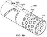
- FIG. 19is a perspective view of an alternative embodiment of the first housing.
- FIG. 20is a perspective view of an alternative embodiment of the second housing.
- FIG. 21is a side view of a detachable vented tissue collection device.
- FIG. 22is a side view of a collapsible and detachable tissue collection device.
- FIGS. 23 a - cshows an alternative collapsible tissue collection device.
- FIG. 24shows a coiled collapsed configuration for a tissue collection device.
- FIG. 25shows an uncoiled configuration for the device of FIG. 24 .
- the tissue collection devices described hereintypically include a tissue chamber, reservoir, and/or tissue storage area adapted for receiving and retaining excised tissue or solid biological material.
- tissue collection devicescan be used during minimally invasive procedures where tissue or other material is cut and removed from the patient's body.
- the tissue collection devicesmay be used with any suitable catheters including, at least, those described in U.S. Patent Application No. 61/646,843, titled “ATHERECTOMY CATHETERS ERS WITH IMAGING,” filed on May 14, 2012, U.S. patent application Ser. No. 13/433,049, titled “OCCLUSION-CROSSING DEVICES, IMAGING, AND ATHERECTOMY DEVICES,” filed Mar. 28, 2012, U.S. patent application Ser. No.
- tissue collection devicesare not meant to limit the tissue collection device to any particular location or position. Rather, the described embodiments illustrate examples of how the contemplated tissue collection devices can be used with other devices or systems. Likewise, in some embodiments, the tissue collection devices may be described as having one or more specific features such as detachability or size adjustment. However, it is to be appreciated that the contemplated embodiments may include features in different combinations or variations than the examples provided. For example, some devices may be detachable and size adjustable or only detachable.
- tissue storage or collection deviceswith adjustable dimensions.
- the devicesare designed to change one or more of cross-sectional size, length, inner diameter, outer diameter, etc. to allow the tissue storage device to move between collapsed and expanded configurations.
- the tissue collection devicemay employ a collapsed or compressed cross-section during insertion into the patient or navigation through narrow vessel sections. Once desired positioning is achieved, the tissue collection device may be expanded to increase the cross-section and crossing profile of the device.
- the tissue collection devicemay include a plurality of configurations ranging from a fully collapsed to a fully expanded configuration. For example, at the fully collapsed configuration, the device may have a minimum inner diameter and outer diameter. Likewise, at the fully expanded configuration, the device may have maximum inner and outer diameters. Additionally, beyond the fully collapsed or expanded positions, the tissue device may have configurations with dimensions between maximum and minimum dimensions.
- thisprovides the physician with a plurality of configurations within a preset range.
- the tissue collection devicemay adjust cross-sectional size and/or the crossing profile by changing the length of the device or a portion of the device.
- the devicehas a length adjustable portion (e.g., an adjustable tip portion) that contains the storage reservoir.
- the crossing profile around the storage reservoiris reduced by lengthening it (e.g., by lengthening the tip portion).
- applying a distally directed force at the distal end of the storage reservoir and/or tip portionpushes the distal end to extend or elongate the storage reservoir and/or tip portion. Elongating the tip portion, consequently, also compresses the cross-section of the tissue storage reservoir contained within the tip portion to reduce the crossing profile of the device.
- Lengthening the tip portion/tissue reservoiris a means for transitioning the tissue collection device from a first expanded state to a second collapsed state.
- the tip portion/storage reservoirmay return to an expanded position.
- the deviceis biased toward the expanded state whereby releasing the lengthening force allows the tissue storage reservoir to return unassisted to its expanded position.
- the storage reservoirmay be made from a resilient or elastic material or frame with a natural elasticity that springs, recoils, or recovers to the expanded shape once the elongating force is removed or released.
- the tip portionmay refer to and/or include the storage reservoir.
- the tip portion (and/or storage reservoir)is not limited to the distal tip region of the devices described herein; additional structures may be located at the distal (or in some orientations, proximal) tip regions. Further, the storage reservoir and/or tip portion may be located proximally of the distal tip of the device(s) described herein.
- the devicemay require an assisting force to transition from the collapsed to expanded state.
- a forcemay be applied to transition the collapsed device (e.g., tip portion and/or tissue reservoir) from the elongated configuration to the original expanded configuration.
- Thisalso increases the collapsed crossing profile to the expanded crossing profile.
- thismay be achieved by applying a proximal directed force that shortens the elongated tissue storage reservoir. The proximally directed force pulls the distal end of the elongated tip portion back towards the expanded configuration. This causes the outer diameter and crossing profile of the tissue storage reservoir to increase.
- a force applying elementmay be employed to impart force to the device.
- a tether, tendon member, guidewire, tensioning element, or any other suitable mechanismcan be used for this purpose.
- some embodimentsinclude a hollow shaft or lumen through which an elongate tendon member (e.g., wire) extends. A portion of the tendon member is attached to the tissue device such that moving the tendon member through the lumen imparts a configuration changing force to the device.
- a separate tendon and tendon lumenare not necessary where a guidewire and corresponding guidewire lumen can serve the same function.
- a guidewiremay be received and retained in a guidewire lumen of the tissue collection device such that the guidewire can maneuver the device into various configurations.
- a general non-collapsible atherectomy catheter device 200having a cutter 202 and distal tip region 201 with a tissue storage reservoir.
- the tissue storage reservoiris in the main body 204 of the catheter.
- a distal tip storage reservoiris described.
- the distal tip regionmay be hollow or otherwise configured to hold material cut by the atherectomy device.
- the distal tip regionis clear or at least partially transparent, allowing one to see if material has been collected or remains in the tip region.
- the distal tip regionmay include a flush port or may otherwise be adapted to allow removal of cut material stored therein.
- the distal endmay be tapered but may be open.
- the distal tip regionmay be removable and/or replaceable.
- a reusable locking mechanismsuch as threads, or the like, may be used to secure a distal tip region on the catheter.
- the distal tip region 201is advanced into the patient's vasculature and maneuvered to a target treatment location. During advancement, the distal tip region must cross through vessel lesions or narrow/tortuous pathways to position the cutter 202 at a target site for tissue excision. To do so, the crossing profile of the distal tip region 201 must be sized to allow bypass through tight vessel cross-sections.
- the distal tip region 201also serves as the tissue collection chamber for storing tissue removed by the cutter 202 .
- the cutter 202may excise the tissue and direct the tissue into a hollow reservoir inside the distal tip region 201 . Any number of methods for doing so have been described in the applications aforementioned and incorporated by reference.
- the cutter 202may have a scoop shape to cut and deflect tissue into a receiving collection chamber in the distal tip region 201 .
- FIG. 1shows the distal tip region 201 having a closed nosecone construction with an opening at a proximal end for receiving cut tissue.
- the structureis relatively inelastic and does not easily compress or change shape. As such, the crossing profile is preset and is not easily changed without permanently deforming and, possibly, damaging the nosecone.
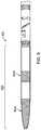
- FIG. 2shows a collapsible tissue collection device 100 attached in the distal region 101 of the catheter 200 .
- the tissue collection device 100includes a proximal housing 104 that attaches the device 100 to the catheter body.
- the proximal housing 104releasably couples the tissue collection device 100 to the main catheter body through any suitable mechanical attachment means such as friction fit, mated fit, threads, etc.
- the proximal housing 104permanently secures the tissue collection device 100 to the catheter 200 .
- the tissue collection device 100has a size adjustable tip portion or tip 102 .
- the tip portion 102may also be attached to the proximal housing 104 at a proximal end 106 of the tip portion 102 .
- the tip portion 102includes a distal end 108 and a length of the device 100 between the two ends 106 , 108 .
- a storage reservoir 110is contained within the tip portion 102 .
- the tissue reservoir 110may extend along a part of or all of the tip portion. Where the storage reservoir 110 extends to the distal end 108 of the tip portion 102 , the distal end may be sealed to prevent the release of tissue from the reservoir.
- the proximal end 106does not need to be sealed and can include an opening in communication with the tissue storage reservoir. This allows excised tissue to enter the reservoir through the proximal end 106 .
- the tissue storage reservoiris attached to the proximal end 106 and the distal end 108 of the tip portion by way of an adhesive or biocompatible polymer such as PEBAX®, Tecothane®, or polyimide.
- the structure of the reservoirmay be fused to a polymer-based housing at the ends 106 , 108 .
- FIGS. 2-3show the collapsible tissue collection device 100 in expanded and collapsed states respectively.
- the tissue collection device 100In the expanded state, the tissue collection device 100 has a larger crossing profile 103 a relative to a collapsed configuration (collapsed crossing profile 103 b shown in FIG. 3 ).
- the device 100can assume a reduced profile to navigate through narrow vessel structures.
- the tip portion 102 and/or the storage reservoir 110may be made from an elastic, deformable, stretchable, or resilient structure or material.
- Suitable materialsinclude biocompatible shape memory materials, alloys, metals, composites, polymers, etc. These include, but are not limited to, nitinol, PEBAX®, polyimide, PEEK, polyester, polypropylene, Tecothane®, stainless steel, elgiloy, cobalt-chromium alloys, carbon fiber, nylon, titanium and its alloys, or Kevlar.
- the material(s) forming the tissue portion/reservoirhas a natural elasticity or resilience that biases the material to a relaxed shape. When deformed, the material exhibits a tendency to recover the relaxed shape. Additionally, any biocompatible material may be used that retains collected solids while allowing fluid movement out of the reservoir.
- the tissue reservoiris defined by a collapsible or foldable structure.
- Thisincludes a compressible frame that allows the storage reservoir to reduce cross-sectional size.
- the storage reservoir or tip portionmay be constructed from a collapsible frame that supports an unstructured elastic or deformable material.
- the framemay provide an outer structure or skeleton upon which a deformable material (e.g. flexible mesh) is draped and secured.
- FIG. 6shows device 300 with a frame 314 supporting an elastic material 312 .
- the framedefines the outer boundaries of the tissue storage reservoir 310 while the elastic material 312 forms a sheath over the frame 314 .
- the frame 314is collapsible or foldable while providing support to the elastic material 312 .
- the framemay also include additional support members such as struts, ribs, posts, joints, etc. to facilitate the configuration changes of the tissue collection device.
- the collapsible frameis a network forming a mesh or netted structure.
- a mesh framemay be braided or woven to increase strength and to better hold stored contents in the collection chamber.
- a wire mesh or netted framemay surround and define the storage reservoir inside the frame.
- FIG. 2shows an example of a mesh structure forming the tissue reservoir on the tip portion 102 .
- the meshsurrounds and defines the storage volume in the storage reservoir 110 .
- the meshis attached to the distal and proximal ends of the tip portion. Any attachments means may be used, including fusing the mesh to the ends using a melted polymer such as PEBAX®. In some cases, the mesh is also attached to the housing 104 for additional stability.
- the meshis a braided wire that includes gaps and openings.
- the braidis structured such that gaps are sufficiently small to prevent the release of collected tissue from the storage reservoir.
- the gapsare about 0.25 sq cm.
- the gapsare between about 50 ⁇ m to about 200 ⁇ m in width.
- the gapsare about 0.01 to about 0.5 mm in width.
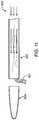
- FIG. 3shows the tissue collection device 100 of FIG. 2 in a reduced crossing profile configuration 120 .
- the tip portion 102is collapsed to reduce the cross-section of the storage reservoir.
- the phantom linesindicate the expanded configuration 118 relative to the shown collapsed configuration 120 .
- the crossing profile 103 a of the expanded configuration 118is greater than the crossing profile 103 b of the collapsed configuration.
- FIG. 3shows that the collapsed configuration 120 also exhibits a greater tip portion/storage reservoir length L 2 relative to the length L 1 of the expanded configuration 118 .
- extending or elongating the tip portion 102 and tissue storage reservoir 110reduces the crossing profile.
- One method of extending the length of the tip portion or storage reservoir 110is to apply a distally directed force to the tip portion. This force can be applied along the length of the device or at the distal end 108 .
- applying a distally directed force (F 1 ) along the longitudinal axis of the tip portion 102forces the tip portion to lengthen.
- F 1distally directed force
- the cross-section of the tip portioncompresses to accommodate the tip portion elongation. This reduces the crossing profile of the tissue storage reservoir 110 .
- the distally directed forcemay transition the tissue collection device from an unfolded to a folded configuration.
- the foldable outer frame 314may include joints and ribs that pivot or move to reduce crossing profile when a distally directed force is applied.
- the braided meshmay accommodate stretching in one or more directions. As shown in FIGS. 2-3 , the mesh stretches in the longitudinal direction under the distally directed force. This allows the tissue reservoir to elongate while reducing the mesh's cross-section.
- the tissue collection device 100can return to its expanded state without applying any assisting force to transition the device.
- Thiscan be accomplished by using a resilient or elastic material for the tissue storage reservoir.
- the mesh material in FIG. 2may be biased toward the larger crossing profile configuration such that once the elongating force (F 1 ) is removed, the mesh will recover its natural shape.
- any resilient materialcan be used whereby the material has a natural elasticity or tendency to return to an expanded state once the compressing force is no longer applied.
- the tissue collection deviceis not biased toward any particular configuration (e.g. relaxed expanded state). Instead, the material and/structure forming the tissue storage reservoir remains in the reduced profile configuration even after the elongating force (F 1 ) is no longer applied to the device. In such cases, a proximally directed force (F 2 ) may be required to pull the tip portion into a non-collapsed configuration. In other words, an assisting force is needed to transition the compressed tissue reservoir to an expanded state with a larger crossing profile.
- the device 2400includes a storage reservoir 2410 at a tip portion of the device 2400 .
- the expanded state 2418 for device 2400is indicated by the dotted lines.
- the tissue storage reservoir 2140is wrapped around a shaft 2420 on the device 2400 .
- the storage reservoir 2410is a loose braided mesh that can coil around the shaft 2420 .
- the structure 2420is a catheter, guidewire lumen, tendon member lumen, or other housing encircled by the storage reservoir.
- the storage reservoir 2410is rotated or turned about the shaft 2420 .
- the distal end 2408 of the storage reservoiris fixed to the shaft 2420 and the proximal end 2406 of the reservoir moves about the shaft 2420 .
- Rotating the proximal end 2406 about the shaft 2420coils the reservoir.
- the storage reservoiris first elongated and then coiled. Elongation can slightly compress the cross-section of reservoir to facilitate the coiling.
- FIG. 4shows the distally or proximally directed force applied by the tendon wire 109 .
- the tendon wire 109resides in a lumen that is defined, in part or whole, by a tendon housing.
- FIG. 4shows the housing in two parts. The first part 111 a is at the proximal end and the second part 111 b is at the distal end.
- the tendon member 109is fed through an opening at each of the two-part housing components 111 a - b .
- the tendon member 109is exposed along a length of the tip portion 102 between the two housing components 111 a - b.
- the housing componentsmay be constructed to retain the tendon member 109 during configuration changes.
- the distal housing component 111 bmay include adhesive, stays, stops, or a tight fit such that the housing component resists distal movement of the tendon member 109 .
- the tendon memberis fixed or fused to the distal end of the tip portion or the tissue storage reservoir. In such cases, distally pushing the tendon member 109 against the distal housing component 111 b imparts a distally directed force F 1 to the distal end of the tip portion 102 . This, in turn, pushes the tip portion distally to lengthen the device 100 and reduce the crossing profile.
- the tendon housing 111may include adhesive, stays, stops, or fitting dimensions to retain the tendon member while a proximally directed force F 2 is applied to pull the distal end of the tip portion proximally. This is applicable, for example, where the tissue storage reservoir does not naturally recover to the expanded state when the collapsing force (F 1 ) is removed. The proximal force (F 2 ) transitions the collapsed configuration back to the expanded state 118 .
- FIGS. 2, 5, and 23 a - cshow various tendon member and housing variations.
- FIG. 2shows a lumen 112 along a length or a longitudinal axis of the tissue collection device 100 .
- the lumen 112is defined by a housing 111 that is also positioned along a length of the device 100 .
- the housing 111is fixed to the proximal end 106 , distal end 108 , and along a length of the tip portion 102 .
- a tendon member 109resides in the lumen 112 .
- FIG. 5shows a similar arrangement with the housing having two components at the distal and proximal ends. Housing components 111 a - b define a portion of the lumen where a tendon member is exposed in the area between the housing components.
- the housing 111 in FIG. 2may be made from an elastic material that can also elongate.
- the housing 111may also be made from a mesh or wire net that can compress and elongate during configuration changes of the storage reservoir.
- FIGS. 23 a - cillustrates another alternative housing and tendon member arrangement.
- Tissue collection device 2300has a tissue storage reservoir 2310 having a distal end 2303 and a proximal end 2305 .
- the reservoir 2310is fixed to the tendon member 2308 by an adhesive material 2314 (e.g. a melted and fused polymer).
- the proximal end 2305is fixed to the tendon lumen housing 2306 by an adhesive material 2318 (e.g. a melted and fused polymer).
- the proximal end 2305includes an opening 2316 for the excised tissue to be advanced into the reservoir 2310 .
- the housing 2306forms a sleeve with an inner lumen 2307 and the tendon member 2308 resides in the lumen 2307 .
- the tendon member 2308slides or moves longitudinally within lumen 2307 . (See FIG. 23 b showing a cross-section of the housing and tendon member.)
- the device 2400can be collapsed in a couple of ways.
- the tendon member 2308is moved distally through the housing 2306 or lumen 2307 . This distal movement applies a distal force Fd against the distal tip 2303 of the storage reservoir 2310 . Because the proximal end 2305 of the reservoir is fixed to the housing 2306 , the distal force Fd pushes against the distal tip 2303 to lengthen the reservoir 2310 . This compresses the cross-section and results in the reduced crossing profile 2313 b .
- the reservoirmay be partially elongated before rotating the proximal end 2305 of the reservoir about the housing 2306 . This twists and coils the reservoir 2310 about the housing 2306 to form a collapsed coiled state.
- the tissue collection devicescan return to an expanded state by removing the collapsing force (i.e. removing a distal or coiling force) and allowing the device to recover a natural relaxed state.
- another forcesuch as a proximal force is applied to move the device back to an expanded state.
- the reservoirmay need to be rotated in a counter direction to unwind the coil. For example, referring to FIG. 23 c , if a clockwise direction winds the reservoir, then the opposing counter-clockwise direction uncoils the device.
- a separate tendon member and housingare not necessary as a guidewire lumen and guidewire can also transition the tissue collection devices between expanded and collapsed configurations.
- the tendon membermay be configured to function as a guidewire and the tendon housing functions as a guidewire lumen.
- the tissue collection devicemay have two separate structures for the guidewire and tendon member.
- the polymercan be melted or softened to adhere the housing to the structure of the storage reservoir or tip portion.
- Suitable materialsinclude polymers, such as polyimide tubing, that can be softened or melted to adhere to the collapsible frame.
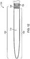
- the tissue collection device 400has an additional extended configuration beyond the collapsed or expanded configurations.
- the extended configurationhas a greater crossing profile 403 c relative to the collapsed and expanded configurations ( 403 a - b ).
- the extended configurationmay allow the device to adjust cross-sectional dimensions to efficiently pack tissue in the storage space.
- the tissue collection devicemay have the following dimensions described.
- the minimum outer diameteris about 0.020 inches.
- the maximum outer diameteris about 0.080 inches.
- the devicehas an outer diameter between about 0.014 inches and about 0.10 inches.
- the length of the tissue collection device (when deployed)could range from about 10 mm to about 100 mm.
- the outer diameter/crossing profile (when deployed)could range from about 0.02 inches to about 0.15 inches.
- the range of the inner diametermay follow the range of the outer diameter, differing by virtue of the wall thickness of the device.
- the embodiments describedprovide for tissue collection devices that release trapped fluids and relieve fluid pressure in the storage reservoir of the devices.
- These devicesmay include venting members or venting elements through which fluid can escape and flow out of the storage reservoir.
- a tip portion of the devicemay include any suitable wall features such as holes, gaps, apertures, nets, mesh, slits, slots, etc. that accommodate the migration of fluids out of the storage reservoir.
- such embodimentsprevent the buildup of fluids in the storage reservoir, which can prevent efficient use of the available storage space.
- any of the fluid releasing members or features describedcan be used with any of the other features described.
- fluid releasing memberscan be used with a collapsible tissue collection device.
- a detachable devicecan include venting elements.
- FIG. 8shows a tissue collection device 500 with a tip portion 502 .
- the tip portion 502has a distal end 508 and a proximal end 506 .
- the device 500includes a proximal housing 504 attached to the tip portion 502 .
- the proximal housing 504is adapted to attach or couple the device 500 to a catheter.
- the tip portion 502includes a plurality of venting elements 505 a at a distal end of the tip portion 502 .
- Another set of venting elements 505 bare located at another section of the tip portion 502 .
- the venting elementsmay be holes or apertures allowing fluid to escape from the tissue storage reservoir 510 . This is particularly useful when tissue is packed into the storage reservoir during an atherectomy procedure where a packing mechanism such as a plunger pushes tissue distally into the storage reservoir. A pocket of fluid in the distal area of the storage reservoir can create back pressure against the plunger. As such, the trapped fluid fills valuable storage space while also impeding the storage of additional tissue.
- FIG. 9shows a tissue collection device 600 with a tip portion 602 having venting elements 604 a - b .
- the venting elementsare formed from a mesh net that contains solid materials within the device 600 while allowing movement of fluids out of the storage reservoir.
- the meshmay be made out of any suitable material including an elastic or resilient material such as shape-memory alloys, biocompatible polymers, etc.
- the meshis made from braided nitinol wires.
- suitable materials that can be used for the deviceinclude biocompatible alloys, metals, composites, polymers, etc. These include, but are not limited to, nitinol, PEBAX®, polyimide, PEEK, polyester, polypropylene, Tecothane®, stainless steel, elgiloy, cobalt-chromium alloys, carbon fiber, nylon, titanium and its alloys, or Kevlar. Additionally, any biocompatible material may be used to form the elastic or stretchable structure for the reservoir that can retain solids such as excised tissue while allow fluid movement out of the reservoir.
- the venting elementsare limited to sections of the device. Venting elements 604 a - b are separated by non-venting sections of the tip portion. In such variations, the venting sections may be fused to the material of the non-venting sections.
- the tip portionmay be made from a thermoplastic polymer that can be melted and fused to the mesh to create venting elements and sections on the tip portion.
- embodiments describedprovide for a tissue collection device that can be easily detached, replaced, and/or cleaned.
- the storage reservoirs of collection devicesare often filled before a procedure is completed. Operators must then remove the treatment devices and clean the tissue collection device.
- Embodiments describedprovide for a tissue collection device having a storage reservoir that can be detached for efficient cleaning or replacement.
- the entire storage reservoircan be detached and replaced with a clean reservoir.
- the entire tip portionmay be removed and replaced with a clean tip portion.
- a portion of the storage reservoiris removed to provide a distal opening through which stored material can be flushed out with cleaning solution (e.g. saline, water, etc.) before re-attaching the removed section.
- cleaning solutione.g. saline, water, etc.
- FIG. 10shows an embodiment of the detachable tissue collection device 800 having a tip portion 802 defined by a first housing 824 and a second housing 825 .
- the first housing 824 and the second housing 825are detachably coupled at an attachment section 826 .
- the device 800includes a proximal housing 804 for coupling the tissue collection device 800 to a catheter (e.g. atherectomy catheter).
- a cathetere.g. atherectomy catheter
- the tip portion 802defines a tissue storage reservoir 810 within the first and second housings 824 , 825 .
- the storage reservoir 810is shown filled with stored excised material 801 .
- the first housing 824is detached from the second housing 825 . Once detached, shown in FIG. 11 , the two housings can be separately cleaned to remove stored material. The housings may be flushed to clear and remove debris.
- the storage reservoirmay be limited to the volume defined between the junction 826 and the proximal housing 804 .
- the storage reservoircan be flushed out by cleaning the second housing 825 with cleaning fluid (e.g. saline) without flushing the first housing 824 .
- FIG. 12shows a tissue collection device 700 with a tip portion 702 .
- the tip portion 702defines a storage reservoir 710 .
- the tip portionmay be formed from a first housing 724 , which also surrounds the storage reservoir 710 .
- the first housing 724may be coupled to a second housing or proximal housing 725 .
- the proximal housing 725may be connected or coupled to a catheter.
- the second housingis a part of the main body of the catheter and is adapted to attach the tissue collection device to the catheter.
- the tissue collection devicemay include an attachment element for coupling the device to the catheter via an attachment section 726 .
- any of the removable componentscan be disposable such that these can be easily replaced to avoid cleaning.
- Any suitable mechanism or meanse.g. friction fit, mated fit, threaded fit, hooks, securing members, etc. may be used to detach a portion or the entirety of a tissue collection device to another device.
- FIGS. 13-20illustrate examples of attachment mechanisms that can be used for this purpose.
- FIG. 13shows a second housing 925 with a proximal end 930 and a distal end 928 . Additionally, the second housing is shown as having a generally cylindrical main body with a lumen between the proximal and distal ends. The slots or cutouts 932 a - b are formed through the wall of the main body. Although shown with two slots having a generally rectangular shape, the second housing can have any number of slots with any shape. In this embodiment, the shape of the main body is designed to be inserted into a first housing shown in FIGS. 14-15 .
- the second housingmay be a part of the tissue collection device, such as shown in FIG. 10 .
- the second housingmay be a part of a catheter, such as at a distal end of the catheter where the catheter attaches to a tissue collection device (see FIG. 12 ).
- FIGS. 14-15illustrate a first housing 1024 with a corresponding structure for releasably coupling to the second housing 925 .
- the first housingmay be part of the tissue collection device such as a section of the tip portion that can be removed from the rest of the tip portion.
- the first housingmay define a portion of a nosecone on a catheter such that removing the first housing exposes the remaining section of the nosecone.
- the first housingmay define the entire tip portion and storage reservoir such that the entire tip portion can be removed and replaced with a clean empty storage reservoir.
- the first housinghas a main body with a distal end 1034 and a proximal end 1032 .
- the first housing 1024is shape set to the second housing such that the second housing 925 can be inserted into the first housing 1024 to form a snug fit.
- the first housing 1024has an inner wall 1038 that contacts the outer wall 934 of the second housing 925 when fitted.
- the first housing 1024includes protrusions shown as tabs 1036 a - b that project from its main body towards the center. In some embodiments, the tabs 1036 a - b protrude at an angle towards the center of the main body.
- FIG. 14shows the tabs with a proximal end 1033 a - b that is fixed to the main body and a free end 1035 a - b that extends toward the interior of the first housing 1024 .
- the angled projection of the tabscreates recesses 1031 a - b between the tabs and the inner wall 1038 .
- the tabsare shown as formed from the main body of the first housing, the tabs can also be made of an separate structure or component that sits on the inner wall 1038 of the first housing 1024 .
- the first housing 1024has two tabs 1036 a - b to interface and lock with the receiving slots 932 a - b of the second housing 925 .
- the first housing 1024is placed over the outer wall 924 of the second housing 925 to form a snug fit.
- the proximal end 1032 of the first housing 1024is advanced over the distal end 928 of the second housing 925 .
- FIG. 16shows the first housing 1024 surrounding the second housing 925 with tabs 1036 a - b engaged with slots 932 a - b .
- FIG. 17shows a cross-sectional view with the first and second housing rotated to align the tabs and slots.
- the tabs 1036 a - bare received through the slots 932 a - b into an interior of the second housing 925 .
- the edge of the slots 932 a - bare slid into the recesses 1031 a - b to hold and lock the lateral orientation of the second housing 925 within the first housing 1024 . As shown, rotating the first housing counter-clockwise disengages the coupling structures and releases the housings from one another.
- FIG. 18shows the first and second housings having a guidewire channel or lumen 1023 , 923 through which a guidewire 909 can reside once the first and second housings are coupled.
- the guidewireprevents the housings from substantially rotating relative to one another to decouple the housings while the guidewire is in the channels.
- FIGS. 19-20show alternative embodiments of the first and second housings.
- FIG. 19shows a first housing 1124 with tabs 1136 a - b and a plurality of apertures 1105 . In some embodiments, the apertures provide for fluid pressure release.
- FIG. 20shows an alternative second housing 1125 having slots 1132 a - c . Although the housings are shown with two or three tabs/slots, it is to be understood that any number of mating structures can be used to form the detachable tissue collection devices.
- the first housingis formed by shape setting the housing to the second housing and baking the first housing in an oven at about 504 degrees Fahrenheit for 20 minutes.
- second housinghas an inner diameter of about 0.065 inches and an outer diameter of about 0.072 inches.
- the second housingmay have a length of about 0.220 inches.
- the cutoutsmay have a width of about 0.050 inches and a length of about 0.070 inches. Where multiple cutouts are employed, the cutouts may be separated by a distance of about 0.025 inches.
- the first housingmay have an inner diameter of about 0.072 inches and an outer diameter of about 0.078 inches.
- the first housingmay have a length of about 0.230 inches.
- FIG. 21shows a tissue collection device having a detachable distal tip 1224 .
- the detachable tip 1224is attached to a tip section 1225 at an attachment point 1226 .
- the detachable distal tip 1224also includes venting elements 1244 for releasing fluid pressure buildup on the storage reservoir.
- FIG. 22shows a tissue collection device 1300 with a collapsible tip portion 1302 .
- the tip portion 1302is also releasably coupled to an atherectomy catheter at attachment section 1326 .
- any of the described tissue collection devicescan be used with atherectomy or other occlusion crossing devices.
- the atherectomy devicestypically include an elongate body and a rotatable tip (with a cutter) at the first distal end of the elongate body and configured to rotate relative to the elongate body.
- Such devicesare described in U.S. Patent Application No. 61/646,843, titled “ATHERECTOMY CATHETERS WITH IMAGING,” filed on May 14, 2012, U.S. patent application Ser. No. 13/433,049, titled “OCCLUSION-CROSSING DEVICES, IMAGING, AND ATHERECTOMY DEVICES,” filed Mar. 28, 2012, U.S. patent application Ser. No.
- references to a structure or feature that is disposed “adjacent” another featuremay have portions that overlap or underlie the adjacent feature.
- spatially relative termssuch as “under”, “below”, “lower”, “over”, “upper” and the like, may be used herein for ease of description to describe one element or feature's relationship to another element(s) or feature(s) as illustrated in the figures. It will be understood that the spatially relative terms are intended to encompass different orientations of the device in use or operation in addition to the orientation depicted in the figures. For example, if a device in the figures is inverted, elements described as “under” or “beneath” other elements or features would then be oriented “over” the other elements or features. Thus, the exemplary term “under” can encompass both an orientation of over and under.
- the devicemay be otherwise oriented (rotated 90 degrees or at other orientations) and the spatially relative descriptors used herein interpreted accordingly.
- the terms “upwardly”, “downwardly”, “vertical”, “horizontal” and the likeare used herein for the purpose of explanation only unless specifically indicated otherwise.
- first and secondmay be used herein to describe various features/elements, these features/elements should not be limited by these terms, unless the context indicates otherwise. These terms may be used to distinguish one feature/element from another feature/element. Thus, a first feature/element discussed below could be termed a second feature/element, and similarly, a second feature/element discussed below could be termed a first feature/element without departing from the teachings of the present invention.
- a numeric valuemay have a value that is +/ ⁇ 0.1% of the stated value (or range of values), +/ ⁇ 1% of the stated value (or range of values), +/ ⁇ 2% of the stated value (or range of values), +/ ⁇ 5% of the stated value (or range of values), +/ ⁇ 10% of the stated value (or range of values), etc. Any numerical range recited herein is intended to include all sub-ranges subsumed therein.
Landscapes
- Health & Medical Sciences (AREA)
- Life Sciences & Earth Sciences (AREA)
- Surgery (AREA)
- Medical Informatics (AREA)
- Engineering & Computer Science (AREA)
- Biomedical Technology (AREA)
- Heart & Thoracic Surgery (AREA)
- Molecular Biology (AREA)
- Animal Behavior & Ethology (AREA)
- General Health & Medical Sciences (AREA)
- Public Health (AREA)
- Veterinary Medicine (AREA)
- Nuclear Medicine, Radiotherapy & Molecular Imaging (AREA)
- Vascular Medicine (AREA)
- Pathology (AREA)
- Surgical Instruments (AREA)
Abstract
Description
Claims (21)
Applications Claiming Priority (1)
| Application Number | Priority Date | Filing Date | Title |
|---|---|---|---|
| PCT/US2013/031978WO2014142954A1 (en) | 2013-03-15 | 2013-03-15 | Tissue collection device for catheter |
Related Parent Applications (1)
| Application Number | Title | Priority Date | Filing Date |
|---|---|---|---|
| PCT/US2013/031978A-371-Of-InternationalWO2014142954A1 (en) | 2013-03-15 | 2013-03-15 | Tissue collection device for catheter |
Related Child Applications (1)
| Application Number | Title | Priority Date | Filing Date |
|---|---|---|---|
| US17/445,648ContinuationUS11980386B2 (en) | 2013-03-15 | 2021-08-23 | Tissue collection device for catheter |
Publications (2)
| Publication Number | Publication Date |
|---|---|
| US20160008025A1 US20160008025A1 (en) | 2016-01-14 |
| US11096717B2true US11096717B2 (en) | 2021-08-24 |
Family
ID=51537323
Family Applications (2)
| Application Number | Title | Priority Date | Filing Date |
|---|---|---|---|
| US14/776,749Active2033-11-15US11096717B2 (en) | 2013-03-15 | 2013-03-15 | Tissue collection device for catheter |
| US17/445,648Active2033-04-18US11980386B2 (en) | 2013-03-15 | 2021-08-23 | Tissue collection device for catheter |
Family Applications After (1)
| Application Number | Title | Priority Date | Filing Date |
|---|---|---|---|
| US17/445,648Active2033-04-18US11980386B2 (en) | 2013-03-15 | 2021-08-23 | Tissue collection device for catheter |
Country Status (3)
| Country | Link |
|---|---|
| US (2) | US11096717B2 (en) |
| EP (1) | EP2967507B1 (en) |
| WO (1) | WO2014142954A1 (en) |
Cited By (13)
| Publication number | Priority date | Publication date | Assignee | Title |
|---|---|---|---|---|
| US11627881B2 (en) | 2015-07-13 | 2023-04-18 | Avinger, Inc. | Micro-molded anamorphic reflector lens for image guided therapeutic/diagnostic catheters |
| US11793400B2 (en) | 2019-10-18 | 2023-10-24 | Avinger, Inc. | Occlusion-crossing devices |
| US11839493B2 (en) | 2009-05-28 | 2023-12-12 | Avinger, Inc. | Optical coherence tomography for biological imaging |
| US11903677B2 (en) | 2011-03-28 | 2024-02-20 | Avinger, Inc. | Occlusion-crossing devices, imaging, and atherectomy devices |
| US11931061B2 (en) | 2014-07-08 | 2024-03-19 | Avinger, Inc. | High speed chronic total occlusion crossing devices |
| US11957376B2 (en) | 2016-04-01 | 2024-04-16 | Avinger, Inc. | Atherectomy catheter with serrated cutter |
| US11980386B2 (en) | 2013-03-15 | 2024-05-14 | Avinger, Inc. | Tissue collection device for catheter |
| US11998311B2 (en) | 2009-04-28 | 2024-06-04 | Avinger, Inc. | Guidewire positioning catheter |
| US12089868B2 (en) | 2009-07-01 | 2024-09-17 | Avinger, Inc. | Methods of using atherectomy catheter with deflectable distal tip |
| US12161360B2 (en) | 2016-06-30 | 2024-12-10 | Avinger, Inc. | Atherectomy catheter with shaped distal tip |
| US12171407B2 (en) | 2012-05-14 | 2024-12-24 | Avinger, Inc. | Atherectomy catheter drive assemblies |
| US12257003B2 (en) | 2011-11-11 | 2025-03-25 | Avinger, Inc. | Occlusion-crossing devices, atherectomy devices, and imaging |
| US12279789B2 (en) | 2016-06-03 | 2025-04-22 | Avinger, Inc. | Catheter device with detachable distal end |
Families Citing this family (29)
| Publication number | Priority date | Publication date | Assignee | Title |
|---|---|---|---|---|
| US8062316B2 (en) | 2008-04-23 | 2011-11-22 | Avinger, Inc. | Catheter system and method for boring through blocked vascular passages |
| US9125562B2 (en) | 2009-07-01 | 2015-09-08 | Avinger, Inc. | Catheter-based off-axis optical coherence tomography imaging system |
| US9345510B2 (en) | 2010-07-01 | 2016-05-24 | Avinger, Inc. | Atherectomy catheters with longitudinally displaceable drive shafts |
| US11382653B2 (en) | 2010-07-01 | 2022-07-12 | Avinger, Inc. | Atherectomy catheter |
| US9949754B2 (en) | 2011-03-28 | 2018-04-24 | Avinger, Inc. | Occlusion-crossing devices |
| US10779855B2 (en) | 2011-08-05 | 2020-09-22 | Route 92 Medical, Inc. | Methods and systems for treatment of acute ischemic stroke |
| EP4101399B1 (en) | 2011-08-05 | 2025-04-09 | Route 92 Medical, Inc. | System for treatment of acute ischemic stroke |
| EP3653151A1 (en) | 2011-10-17 | 2020-05-20 | Avinger, Inc. | Atherectomy catheters and non-contact actuation mechanism for catheters |
| US9557156B2 (en) | 2012-05-14 | 2017-01-31 | Avinger, Inc. | Optical coherence tomography with graded index fiber for biological imaging |
| US9498247B2 (en) | 2014-02-06 | 2016-11-22 | Avinger, Inc. | Atherectomy catheters and occlusion crossing devices |
| US11284916B2 (en) | 2012-09-06 | 2022-03-29 | Avinger, Inc. | Atherectomy catheters and occlusion crossing devices |
| WO2014143064A1 (en) | 2013-03-15 | 2014-09-18 | Avinger, Inc. | Chronic total occlusion crossing devices with imaging |
| CN105228514B (en) | 2013-03-15 | 2019-01-22 | 阿维格公司 | Optical Pressure Sensor Assembly |
| EP3019096B1 (en) | 2013-07-08 | 2023-07-05 | Avinger, Inc. | System for identification of elastic lamina to guide interventional therapy |
| US9265512B2 (en) | 2013-12-23 | 2016-02-23 | Silk Road Medical, Inc. | Transcarotid neurovascular catheter |
| MX2016010141A (en) | 2014-02-06 | 2017-04-06 | Avinger Inc | Atherectomy catheters and occlusion crossing devices. |
| US9241699B1 (en) | 2014-09-04 | 2016-01-26 | Silk Road Medical, Inc. | Methods and devices for transcarotid access |
| US11027104B2 (en) | 2014-09-04 | 2021-06-08 | Silk Road Medical, Inc. | Methods and devices for transcarotid access |
| CN119949953A (en) | 2015-02-04 | 2025-05-09 | 92号医疗公司 | Intravascular access system, dilator and system including dilator |
| US11065019B1 (en) | 2015-02-04 | 2021-07-20 | Route 92 Medical, Inc. | Aspiration catheter systems and methods of use |
| US10426497B2 (en) | 2015-07-24 | 2019-10-01 | Route 92 Medical, Inc. | Anchoring delivery system and methods |
| JP6927986B2 (en) | 2016-01-25 | 2021-09-01 | アビンガー・インコーポレイテッドAvinger, Inc. | OCT imaging catheter with delay compensation |
| WO2017161166A1 (en)* | 2016-03-16 | 2017-09-21 | Avinger, Inc. | Atherectomy catheters and occlusion crossing devices |
| WO2018055431A1 (en)* | 2016-09-20 | 2018-03-29 | Gavanescu Cosmin Adrian | Surgery device |
| CN110392591B (en) | 2017-01-10 | 2022-06-03 | 92号医疗公司 | Aspiration catheter system and method of use |
| US10864350B2 (en) | 2017-01-20 | 2020-12-15 | Route 92 Medical, Inc. | Single operator intracranial medical device delivery systems and methods of use |
| US12167867B2 (en) | 2018-04-19 | 2024-12-17 | Avinger, Inc. | Occlusion-crossing devices |
| JP7616642B2 (en) | 2018-05-17 | 2025-01-17 | ルート92メディカル・インコーポレイテッド | Suction catheter system and method of use |
| WO2022076893A1 (en) | 2020-10-09 | 2022-04-14 | Route 92 Medical, Inc. | Aspiration catheter systems and methods of use |
Citations (526)
| Publication number | Priority date | Publication date | Assignee | Title |
|---|---|---|---|---|
| US3367727A (en) | 1965-10-22 | 1968-02-06 | Abraham W. Ward | Oral surgery tool with interchangeable blades |
| US3908637A (en) | 1974-04-22 | 1975-09-30 | Louis W Doroshow | Rigid urethral instrument |
| US4178935A (en) | 1977-07-21 | 1979-12-18 | Ediny Jury G | Method and apparatus for disintegration of urinary concretions |
| US4487206A (en) | 1982-10-13 | 1984-12-11 | Honeywell Inc. | Fiber optic pressure sensor with temperature compensation and reference |
| US4527553A (en) | 1980-04-28 | 1985-07-09 | Upsher Michael S | Laryngoscope with improved light source |
| US4552554A (en) | 1984-06-25 | 1985-11-12 | Medi-Tech Incorporated | Introducing catheter |
| US4578061A (en) | 1980-10-28 | 1986-03-25 | Lemelson Jerome H | Injection catheter and method |
| US4611600A (en) | 1983-11-21 | 1986-09-16 | Cordis Corporation | Optical fiber pressure transducer |
| US4621353A (en) | 1982-09-09 | 1986-11-04 | Burroughs Corporation | Optical memory system providing improved focusing control and improved beam combining and separating apparatus |
| US4639091A (en) | 1983-02-25 | 1987-01-27 | Thomson-Csf | Static deflector device for an infrared beam |
| US4651753A (en)* | 1984-10-12 | 1987-03-24 | Jayco Pharmaceuticals | Endoscopic multiple biopsy instrument |
| US4654024A (en) | 1985-09-04 | 1987-03-31 | C.R. Bard, Inc. | Thermorecanalization catheter and method for use |
| US4681106A (en) | 1985-08-12 | 1987-07-21 | Intravascular Surgical Instruments, Inc. | Catheter based surgical methods and apparatus therefor |
| US4686982A (en) | 1985-06-19 | 1987-08-18 | John Nash | Spiral wire bearing for rotating wire drive catheter |
| US4691708A (en) | 1986-03-10 | 1987-09-08 | Cordis Corporation | Optical pressure sensor for measuring blood pressure |
| JPS62275425A (en) | 1986-05-21 | 1987-11-30 | オリンパス光学工業株式会社 | Endoscope |
| US4729763A (en) | 1986-06-06 | 1988-03-08 | Henrie Rodney A | Catheter for removing occlusive material |
| US4771774A (en) | 1986-02-28 | 1988-09-20 | Devices For Vascular Intervention, Inc. | Motor drive unit |
| US4841977A (en) | 1987-05-26 | 1989-06-27 | Inter Therapy, Inc. | Ultra-thin acoustic transducer and balloon catheter using same in imaging array subassembly |
| US4857046A (en) | 1987-10-21 | 1989-08-15 | Cordis Corporation | Drive catheter having helical pump drive shaft |
| EP0347098A2 (en) | 1988-06-13 | 1989-12-20 | Samuel Shiber | Atherectomy system with a guide-wire |
| US4920961A (en)* | 1988-06-02 | 1990-05-01 | Circon Corporation | System for disconnetably mounting an endoscope sheath with an endoscope tool |
| US4926858A (en) | 1984-05-30 | 1990-05-22 | Devices For Vascular Intervention, Inc. | Atherectomy device for severe occlusions |
| US5000185A (en) | 1986-02-28 | 1991-03-19 | Cardiovascular Imaging Systems, Inc. | Method for intravascular two-dimensional ultrasonography and recanalization |
| JPH03502060A (en) | 1988-07-29 | 1991-05-16 | ラディ・メディカル・システムズ・アクチェボラーグ | miniature pressure sensor |
| US5018529A (en) | 1986-06-25 | 1991-05-28 | Radisensor Ab | Miniaturized sensor for physiological pressure measurements |
| US5041082A (en) | 1986-06-16 | 1991-08-20 | Samuel Shiber | Mechanical atherectomy system and method |
| US5047040A (en) | 1987-11-05 | 1991-09-10 | Devices For Vascular Intervention, Inc. | Atherectomy device and method |
| WO1991017698A1 (en) | 1990-05-16 | 1991-11-28 | Brigham And Women's Hospital | Steerable guide wire for tubular cannulation |
| US5085662A (en) | 1989-11-13 | 1992-02-04 | Scimed Life Systems, Inc. | Atherectomy catheter and related components |
| US5099850A (en) | 1989-01-17 | 1992-03-31 | Olympus Optical Co., Ltd. | Ultrasonic diagnostic apparatus |
| US5178153A (en) | 1984-03-08 | 1993-01-12 | Einzig Robert E | Fluid flow sensing apparatus for in vivo and industrial applications employing novel differential optical fiber pressure sensors |
| US5182291A (en) | 1986-02-14 | 1993-01-26 | Sanofi | Pyrozala-pyridyl aminoabkoxyphenol compounds |
| US5190050A (en) | 1991-11-08 | 1993-03-02 | Electro-Catheter Corporation | Tip deflectable steerable catheter |
| US5192291A (en) | 1992-01-13 | 1993-03-09 | Interventional Technologies, Inc. | Rotationally expandable atherectomy cutter assembly |
| JPH05103763A (en) | 1991-03-11 | 1993-04-27 | Hewlett Packard Co <Hp> | Pressure sensor |
| JPH0627343A (en) | 1992-07-06 | 1994-02-04 | Nippon Telegr & Teleph Corp <Ntt> | Optical fiber juncture for optical fiber amplifier |
| US5312415A (en) | 1992-09-22 | 1994-05-17 | Target Therapeutics, Inc. | Assembly for placement of embolic coils using frictional placement |
| US5312425A (en) | 1989-09-12 | 1994-05-17 | Devices For Vascular Intervention, Inc. | Atherectomy device having helical blade and blade guide |
| US5321501A (en) | 1991-04-29 | 1994-06-14 | Massachusetts Institute Of Technology | Method and apparatus for optical imaging with means for controlling the longitudinal range of the sample |
| US5333142A (en) | 1992-10-26 | 1994-07-26 | The United States Of America As Represented By The Secretary Of The Navy | Technique for intracavity sum frequency generation |
| US5358472A (en) | 1992-01-13 | 1994-10-25 | Schneider (Usa) Inc. | Guidewire atherectomy catheter and method of using the same |
| US5366464A (en) | 1993-07-22 | 1994-11-22 | Belknap John C | Atherectomy catheter device |
| US5372601A (en) | 1993-03-30 | 1994-12-13 | Lary; Banning G. | Longitudinal reciprocating incisor |
| US5383467A (en) | 1992-11-18 | 1995-01-24 | Spectrascience, Inc. | Guidewire catheter and apparatus for diagnostic imaging |
| US5383460A (en) | 1992-10-05 | 1995-01-24 | Cardiovascular Imaging Systems, Inc. | Method and apparatus for ultrasound imaging and atherectomy |
| US5425273A (en) | 1990-07-27 | 1995-06-20 | Cosurvey Optics | Fiber optic pressure sensor with inclusions in a compressible transparent material |
| US5429136A (en) | 1993-04-21 | 1995-07-04 | Devices For Vascular Intervention, Inc. | Imaging atherectomy apparatus |
| US5431673A (en) | 1989-02-17 | 1995-07-11 | American Biomed, Inc. | Distal atherectomy catheter |
| JPH07184888A (en) | 1993-12-27 | 1995-07-25 | Toshiba Corp | Ultrasonic diagnostic equipment |
| US5437284A (en) | 1993-12-30 | 1995-08-01 | Camino Laboratories, Inc. | System and method for in vivo calibration of a sensor |
| US5460168A (en) | 1992-12-25 | 1995-10-24 | Olympus Optical Co., Ltd. | Endoscope cover assembly and cover-system endoscope |
| US5465147A (en) | 1991-04-29 | 1995-11-07 | Massachusetts Institute Of Technology | Method and apparatus for acquiring images using a ccd detector array and no transverse scanner |
| JPH07308393A (en) | 1994-03-23 | 1995-11-28 | Yasuo Hashimoto | Cancer treating apparatus |
| US5507795A (en)* | 1994-04-29 | 1996-04-16 | Devices For Vascular Intervention, Inc. | Catheter with perfusion system |
| US5507760A (en) | 1993-11-09 | 1996-04-16 | Devices For Vascular Intervention, Inc. | Cutter device |
| US5517998A (en) | 1994-01-24 | 1996-05-21 | Medamicus, Inc. | Closed loop pressure determination system and method for fiber optic pressure transducer system |
| US5556405A (en) | 1995-10-13 | 1996-09-17 | Interventional Technologies Inc. | Universal dilator with reciprocal incisor |
| US5607394A (en) | 1993-10-07 | 1997-03-04 | Boston Scientific Corp. | Dilatation catheter having a field stylet |
| US5620426A (en) | 1992-04-07 | 1997-04-15 | Innovata Biomed Limited | Connecting device |
| US5632755A (en) | 1992-11-09 | 1997-05-27 | Endo Vascular Intruments, Inc. | Intra-artery obstruction clearing apparatus and methods |
| US5632754A (en) | 1994-12-23 | 1997-05-27 | Devices For Vascular Intervention | Universal catheter with interchangeable work element |
| US5674232A (en) | 1990-06-05 | 1997-10-07 | Halliburton; Alexander George | Catheter and method of use thereof |
| US5681336A (en) | 1995-09-07 | 1997-10-28 | Boston Scientific Corporation | Therapeutic device for treating vien graft lesions |
| US5690634A (en) | 1993-09-15 | 1997-11-25 | Synthes (U.S.A.) | Medullary drill head |
| EP0808638A1 (en) | 1996-05-20 | 1997-11-26 | Cordis Europa N.V. | Catheter-introduction-sheath with occlusion balloon |
| US5722403A (en) | 1996-10-28 | 1998-03-03 | Ep Technologies, Inc. | Systems and methods using a porous electrode for ablating and visualizing interior tissue regions |
| US5749846A (en) | 1992-08-12 | 1998-05-12 | Vidamed, Inc. | Medical probe device with optical viewing capability |
| US5795295A (en) | 1996-06-25 | 1998-08-18 | Carl Zeiss, Inc. | OCT-assisted surgical microscope with multi-coordinate manipulator |
| US5807339A (en) | 1995-12-04 | 1998-09-15 | Pacesetter Ab | Stylet unit for stiffening a hollow, flexible, elongated component |
| US5830145A (en) | 1996-09-20 | 1998-11-03 | Cardiovascular Imaging Systems, Inc. | Enhanced accuracy of three-dimensional intraluminal ultrasound (ILUS) image reconstruction |
| US5836957A (en) | 1994-12-22 | 1998-11-17 | Devices For Vascular Intervention, Inc. | Large volume atherectomy device |
| US5843103A (en) | 1997-03-06 | 1998-12-01 | Scimed Life Systems, Inc. | Shaped wire rotational atherectomy device |
| US5843050A (en) | 1995-11-13 | 1998-12-01 | Micro Therapeutics, Inc. | Microcatheter |
| US5851212A (en) | 1997-06-11 | 1998-12-22 | Endius Incorporated | Surgical instrument |
| US5868778A (en) | 1995-10-27 | 1999-02-09 | Vascular Solutions, Inc. | Vascular sealing apparatus and method |
| US5872879A (en) | 1996-11-25 | 1999-02-16 | Boston Scientific Corporation | Rotatable connecting optical fibers |
| US5904651A (en) | 1996-10-28 | 1999-05-18 | Ep Technologies, Inc. | Systems and methods for visualizing tissue during diagnostic or therapeutic procedures |
| WO1999023958A1 (en) | 1997-11-07 | 1999-05-20 | Prolifix Medical, Inc. | Methods and systems for treating obstructions in a body lumen |
| US5907425A (en) | 1995-12-19 | 1999-05-25 | The Board Of Trustees Of The Leland Stanford Junior University | Miniature scanning confocal microscope |
| US5935075A (en) | 1995-09-20 | 1999-08-10 | Texas Heart Institute | Detecting thermal discrepancies in vessel walls |
| US5938602A (en) | 1996-06-11 | 1999-08-17 | Roke Manor Research Limited | Catheter tracking system and method |
| US5938671A (en) | 1997-11-14 | 1999-08-17 | Reflow, Inc. | Recanalization apparatus and devices for use therein and method |
| US5951583A (en) | 1993-05-25 | 1999-09-14 | Vascular Solutions, Inc. | Thrombin and collagen procoagulant and process for making the same |
| US5951581A (en) | 1996-12-02 | 1999-09-14 | Angiotrax, Inc. | Cutting apparatus having disposable handpiece |
| US5951482A (en) | 1997-10-03 | 1999-09-14 | Intraluminal Therapeutics, Inc. | Assemblies and methods for advancing a guide wire through body tissue |
| US5956355A (en) | 1991-04-29 | 1999-09-21 | Massachusetts Institute Of Technology | Method and apparatus for performing optical measurements using a rapidly frequency-tuned laser |
| US5957952A (en) | 1993-05-25 | 1999-09-28 | Vascular Solutions, Inc. | Vascular sealing device |
| US5987995A (en) | 1997-07-17 | 1999-11-23 | Sentec Corporation | Fiber optic pressure catheter |
| US5997558A (en) | 1996-07-26 | 1999-12-07 | Kensey Nash Corporation | System and method or use for revascularizing stenotic bypass grafts and other blood vessels |
| US6001112A (en) | 1998-04-10 | 1999-12-14 | Endicor Medical, Inc. | Rotational atherectomy device |
| US6007530A (en) | 1995-02-09 | 1999-12-28 | C.R. Bard, Inc. | Angioplasty catheter for expanding and/or opening up blood vessels |
| US6010449A (en) | 1997-02-28 | 2000-01-04 | Lumend, Inc. | Intravascular catheter system for treating a vascular occlusion |
| US6013072A (en) | 1997-07-09 | 2000-01-11 | Intraluminal Therapeutics, Inc. | Systems and methods for steering a catheter through body tissue |
| US6017359A (en) | 1993-05-25 | 2000-01-25 | Vascular Solutions, Inc. | Vascular sealing apparatus |
| US6027514A (en) | 1997-12-17 | 2000-02-22 | Fox Hollow Technologies, Inc. | Apparatus and method for removing occluding material from body lumens |
| US6032673A (en) | 1994-10-13 | 2000-03-07 | Femrx, Inc. | Methods and devices for tissue removal |
| US6048349A (en) | 1997-07-09 | 2000-04-11 | Intraluminal Therapeutics, Inc. | Systems and methods for guiding a medical instrument through a body |
| US6080170A (en) | 1996-07-26 | 2000-06-27 | Kensey Nash Corporation | System and method of use for revascularizing stenotic bypass grafts and other occluded blood vessels |
| US6106515A (en) | 1998-08-13 | 2000-08-22 | Intraluminal Therapeutics, Inc. | Expandable laser catheter |
| US6110164A (en) | 1997-12-05 | 2000-08-29 | Intratherapeutics, Inc. | Guideless catheter segment |
| US6120516A (en) | 1997-02-28 | 2000-09-19 | Lumend, Inc. | Method for treating vascular occlusion |
| US6120515A (en) | 1996-02-06 | 2000-09-19 | Devices For Vascular Intervention, Inc. | Composite atherectomy cutter |
| WO2000054659A1 (en) | 1999-03-15 | 2000-09-21 | Prolifix Medical, Inc. | Shielded atherectomy device |
| US6134002A (en) | 1999-01-14 | 2000-10-17 | Duke University | Apparatus and method for the rapid spectral resolution of confocal images |
| US6134003A (en) | 1991-04-29 | 2000-10-17 | Massachusetts Institute Of Technology | Method and apparatus for performing optical measurements using a fiber optic imaging guidewire, catheter or endoscope |
| US6152938A (en) | 1997-08-19 | 2000-11-28 | Curry; Paul | Device for opening blocked tubes |
| US6152951A (en) | 1994-03-23 | 2000-11-28 | Hamamatsu Photonics K.K. | Method of treating cancer |
| US6175669B1 (en) | 1998-03-30 | 2001-01-16 | The Regents Of The Universtiy Of California | Optical coherence domain reflectometry guidewire |
| US6176871B1 (en) | 1993-04-28 | 2001-01-23 | Focal, Inc. | Apparatus and methods for intraluminal photothermoforming |
| US6183432B1 (en) | 1997-11-13 | 2001-02-06 | Lumend, Inc. | Guidewire and catheter with rotating and reciprocating symmetrical or asymmetrical distal tip |
| US6193676B1 (en) | 1997-10-03 | 2001-02-27 | Intraluminal Therapeutics, Inc. | Guide wire assembly |
| WO2001015609A1 (en) | 1999-08-31 | 2001-03-08 | Fox Hollow Technologies | Atherectomy catheter with a rotating and telescoping cutter |
| US6228076B1 (en) | 1999-01-09 | 2001-05-08 | Intraluminal Therapeutics, Inc. | System and method for controlling tissue ablation |
| US6241744B1 (en) | 1998-08-14 | 2001-06-05 | Fox Hollow Technologies, Inc. | Apparatus for deploying a guidewire across a complex lesion |
| US20010005788A1 (en) | 1997-07-24 | 2001-06-28 | Mcguckin James F. | Bladder dialysis urinary catheter |
| US6285903B1 (en) | 1998-06-30 | 2001-09-04 | Eclipse Surgical Technologies, Inc. | Intracorporeal device with radiopaque marker |
| US6290668B1 (en) | 1998-04-30 | 2001-09-18 | Kenton W. Gregory | Light delivery catheter and methods for the use thereof |
| US6294775B1 (en) | 1999-06-08 | 2001-09-25 | University Of Washington | Miniature image acquistion system using a scanning resonant waveguide |
| US6299622B1 (en) | 1999-08-19 | 2001-10-09 | Fox Hollow Technologies, Inc. | Atherectomy catheter with aligned imager |
| WO2001076680A1 (en) | 2000-04-05 | 2001-10-18 | Stx Medical, Inc. | Intralumenal material removal systems and methods |
| US6307985B1 (en) | 1998-07-10 | 2001-10-23 | Micro Therapeutics, Inc. | Optical transmission system |
| US20020019644A1 (en) | 1999-07-12 | 2002-02-14 | Hastings Roger N. | Magnetically guided atherectomy |
| US6375615B1 (en) | 1995-10-13 | 2002-04-23 | Transvascular, Inc. | Tissue penetrating catheters having integral imaging transducers and their methods of use |
| US6402719B1 (en) | 1997-09-05 | 2002-06-11 | Cordis Webster, Inc. | Steerable DMR catheter with infusion tube |
| US20020072706A1 (en) | 2000-12-11 | 2002-06-13 | Thomas Hiblar | Transluminal drug delivery catheter |
| US20020077642A1 (en)* | 2000-12-20 | 2002-06-20 | Fox Hollow Technologies, Inc. | Debulking catheter |
| US20020082626A1 (en) | 1999-11-15 | 2002-06-27 | Donohoe Brendan M. | Integrated anastomosis tool with graft vessel attachment device and cutting device |
| US20020082585A1 (en) | 1999-06-15 | 2002-06-27 | Sean Carroll | Defined deflection structure |
| US6416527B1 (en) | 1998-01-28 | 2002-07-09 | St. Jude Medical Cardiovascular Group, Inc. | Vessel cutting device |
| RU2185859C2 (en) | 1995-10-20 | 2002-07-27 | Надим М. Закка | Device for removing stenoses and supporting vascular walls |
| US20020115931A1 (en) | 2001-02-21 | 2002-08-22 | Strauss H. William | Localizing intravascular lesions on anatomic images |
| US6445944B1 (en) | 1999-02-01 | 2002-09-03 | Scimed Life Systems | Medical scanning system and related method of scanning |
| US6445939B1 (en) | 1999-08-09 | 2002-09-03 | Lightlab Imaging, Llc | Ultra-small optical probes, imaging optics, and methods for using same |
| US6447525B2 (en) | 1999-08-19 | 2002-09-10 | Fox Hollow Technologies, Inc. | Apparatus and methods for removing material from a body lumen |
| US6451036B1 (en) | 1998-04-10 | 2002-09-17 | Endicor Medical, Inc. | Rotational atherectomy system with stationary cutting elements |
| US6454717B1 (en) | 2000-04-13 | 2002-09-24 | Scimed Life Systems, Inc. | Concentric catheter drive shaft clutch |
| US20020147459A1 (en) | 2001-04-10 | 2002-10-10 | Mehran Bashiri | Devices and methods for removing occlusions in vessels |
| US20020158547A1 (en) | 2001-04-26 | 2002-10-31 | Wood Robert L. | Latchable microelectromechanical structures using non-newtonian fluids, and methods of operating same |
| US6482216B1 (en) | 1997-03-06 | 2002-11-19 | Scimed Life Systems, Inc. | Ablation device including guidewire with abrasive tip |
| US6482217B1 (en) | 1998-04-10 | 2002-11-19 | Endicor Medical, Inc. | Neuro thrombectomy catheter |
| US6485413B1 (en) | 1991-04-29 | 2002-11-26 | The General Hospital Corporation | Methods and apparatus for forward-directed optical scanning instruments |
| US6497649B2 (en) | 2001-01-21 | 2002-12-24 | University Of Washington | Alleviating motion, simulator, and virtual environmental sickness by presenting visual scene components matched to inner ear vestibular sensations |
| US6501551B1 (en) | 1991-04-29 | 2002-12-31 | Massachusetts Institute Of Technology | Fiber optic imaging endoscope interferometer with at least one faraday rotator |
| US20030002038A1 (en) | 2000-01-28 | 2003-01-02 | Kazuma Mawatari | Photothermic transducing spectroscopic analyzer |
| US6503261B1 (en) | 2001-01-17 | 2003-01-07 | Scimed Life Systems, Inc. | Bi-directional atherectomy burr |
| US6511458B2 (en) | 1998-01-13 | 2003-01-28 | Lumend, Inc. | Vascular re-entry catheter |
| US20030028100A1 (en) | 2001-05-01 | 2003-02-06 | Tearney Guillermo J. | Method and apparatus for determination of atherosclerotic plaque type by measurement of tissue optical properties |
| US6517528B1 (en) | 2000-04-13 | 2003-02-11 | Scimed Life Systems, Inc. | Magnetic catheter drive shaft clutch |
| US20030032880A1 (en) | 2001-06-13 | 2003-02-13 | Pauliina Moore | Apparatus and method for ultrasonically identifying vulnerable plaque |
| US20030045835A1 (en) | 2001-08-30 | 2003-03-06 | Vascular Solutions, Inc. | Method and apparatus for coagulation and closure of pseudoaneurysms |
| US6542665B2 (en) | 2001-02-17 | 2003-04-01 | Lucent Technologies Inc. | GRIN fiber lenses |
| US6544230B1 (en) | 1998-03-31 | 2003-04-08 | Transvascular, Inc. | Catheters, systems and methods for percutaneous in situ arterio-venous bypass |
| US6546272B1 (en) | 1999-06-24 | 2003-04-08 | Mackinnon Nicholas B. | Apparatus for in vivo imaging of the respiratory tract and other internal organs |
| US6551302B1 (en) | 1997-09-24 | 2003-04-22 | Michael J. Rosinko | Steerable catheter with tip alignment and surface contact detector |
| US6563105B2 (en) | 1999-06-08 | 2003-05-13 | University Of Washington | Image acquisition with depth enhancement |
| US6564087B1 (en) | 1991-04-29 | 2003-05-13 | Massachusetts Institute Of Technology | Fiber optic needle probes for optical coherence tomography imaging |
| US6565588B1 (en) | 2000-04-05 | 2003-05-20 | Pathway Medical Technologies, Inc. | Intralumenal material removal using an expandable cutting device |
| US20030095248A1 (en) | 2001-11-16 | 2003-05-22 | Didier Frot | Optical-fiber refractometer |
| US20030097044A1 (en) | 2001-11-19 | 2003-05-22 | Tokendo (S.A.R.L.) | Deviated distal viewing endoscope |
| US6572563B2 (en) | 1999-12-22 | 2003-06-03 | Pentax Corporation | Endoscopic tissue collecting instrument |
| US6572643B1 (en) | 2000-07-19 | 2003-06-03 | Vascular Architects, Inc. | Endoprosthesis delivery catheter assembly and method |
| US6575995B1 (en) | 2000-07-14 | 2003-06-10 | Advanced Cardiovascular Systems, Inc. | Expandable cage embolic material filter system and method |
| US6579298B1 (en) | 2000-02-29 | 2003-06-17 | Scimed Life Systems, Inc. | Method and apparatus for treating vein graft lesions |
| US20030120295A1 (en) | 2000-12-20 | 2003-06-26 | Fox Hollow Technologies, Inc. | Debulking catheters and methods |
| US20030120150A1 (en) | 2001-12-21 | 2003-06-26 | Assaf Govari | Wireless position sensor |
| US20030125757A1 (en)* | 2000-12-20 | 2003-07-03 | Fox Hollow Technologies, Inc. | Debulking catheters and methods |
| US20030125758A1 (en) | 2000-12-20 | 2003-07-03 | Fox Hollow Technologies, Inc. | Debulking catheters and methods |
| US20030125756A1 (en) | 2001-10-19 | 2003-07-03 | Leonid Shturman | Rotational angioplasty device with abrasive crown |
| US20030139751A1 (en) | 2000-01-25 | 2003-07-24 | Bacchus Vascular Inc. | Apparatus and methods for clot dissolution |
| US6599296B1 (en) | 2001-07-27 | 2003-07-29 | Advanced Cardiovascular Systems, Inc. | Ratcheting handle for intraluminal catheter systems |
| US6615071B1 (en) | 1995-09-20 | 2003-09-02 | Board Of Regents, The University Of Texas System | Method and apparatus for detecting vulnerable atherosclerotic plaque |
| US20030181855A1 (en) | 2002-03-22 | 2003-09-25 | Simpson John A. | Pre-shaped catheter with proximal articulation and pre-formed distal end |
| US6638233B2 (en) | 1999-08-19 | 2003-10-28 | Fox Hollow Technologies, Inc. | Apparatus and methods for material capture and removal |
| US6645217B1 (en) | 1999-05-15 | 2003-11-11 | Advanced Cardiovascular Systems, Inc. | Over-the-wire atherectomy catheter |
| US6657727B1 (en) | 1998-09-11 | 2003-12-02 | Joseph A. Izatt | Interferometers for optical coherence domain reflectometry and optical coherence tomography using nonreciprocal optical elements |
| RU2218191C2 (en) | 2002-04-11 | 2003-12-10 | Научно-исследовательский институт радиоэлектроники и лазерной техники Московского государственного технического университета им. Н.Э.Баумана | Endovasal mini robot |
| US20040002650A1 (en) | 2001-06-20 | 2004-01-01 | Evgenia Mandrusov | Agents that stimulate therapeutic angiogenesis and techniques and devices that enable their delivery |
| US6687010B1 (en) | 1999-09-09 | 2004-02-03 | Olympus Corporation | Rapid depth scanning optical imaging device |
| US20040039371A1 (en) | 2002-08-23 | 2004-02-26 | Bruce Tockman | Coronary vein navigator |
| US20040057667A1 (en) | 2002-09-20 | 2004-03-25 | Koji Yamada | Optical module and manufacturing method therefor |
| US20040059257A1 (en) | 2001-01-08 | 2004-03-25 | Benny Gaber | Deflectable guiding apparatus |
| JP2004509695A (en) | 2000-09-29 | 2004-04-02 | エンドバシックス,インコーポレイテッド | Flexible flow device and method of breaking obstruction |
| US6728571B1 (en) | 2001-07-16 | 2004-04-27 | Scimed Life Systems, Inc. | Electronically scanned optical coherence tomography with frequency modulated signals |
| US20040082850A1 (en) | 2002-10-23 | 2004-04-29 | Medtonic, Inc. | Methods and apparatus for locating body vessels and occlusions in body vessels |
| US6730063B2 (en) | 1999-10-22 | 2004-05-04 | Corazon Technologies, Inc. | Catheter devices and methods for their use in the treatment of calcified vascular occlusions |
| US20040092915A1 (en) | 1990-08-06 | 2004-05-13 | Levatter Jeffrey I. | Fiber optic laser catheter and method of using it |
| US20040093001A1 (en) | 2002-10-25 | 2004-05-13 | Hamada James S. | Minimal access lumbar diskectomy instrumentation and method |
| USD489973S1 (en) | 2003-06-02 | 2004-05-18 | Vascular Solutions, Inc. | Medical device package |
| US6758854B1 (en) | 1997-05-09 | 2004-07-06 | St. Jude Medical | Splittable occlusion balloon sheath and process of use |
| US6760112B2 (en) | 2001-02-17 | 2004-07-06 | Lucent Technologies Inc. | Grin-fiber lens based optical endoscopes |
| US20040147934A1 (en) | 2002-10-18 | 2004-07-29 | Kiester P. Douglas | Oscillating, steerable, surgical burring tool and method of using the same |
| US20040167553A1 (en) | 2000-12-20 | 2004-08-26 | Fox Hollow Technologies, Inc. | Methods and devices for cutting tissue |
| US20040167554A1 (en) | 2000-12-20 | 2004-08-26 | Fox Hollow Technologies, Inc. | Methods and devices for reentering a true lumen from a subintimal space |
| US20040181249A1 (en) | 2003-03-10 | 2004-09-16 | Pathway Medical Technologies, Inc. | Bearing system to support a rotatable operating head in an intracorporeal device |
| US20040186368A1 (en) | 2003-03-21 | 2004-09-23 | Scimed Life Systems, Inc. | Systems and methods for internal tissue penetration |
| US20040193140A1 (en) | 2003-03-27 | 2004-09-30 | Scimed Life Systems,Inc. | Medical device |
| US6800085B2 (en) | 1997-02-28 | 2004-10-05 | Lumend, Inc. | Methods and apparatus for treating vascular occlusions |
| US20040202418A1 (en) | 2003-04-10 | 2004-10-14 | Margaret Ghiron | Beam shaping and practical methods of reducing loss associated with mating external sources and optics to thin silicon waveguides |
| US20040230213A1 (en) | 2000-04-05 | 2004-11-18 | Pathway Medical Technologies, Inc. | Liquid seal assembly for a rotating torque tube |
| US20040230212A1 (en) | 2000-04-05 | 2004-11-18 | Pathway Medical Technologies, Inc. | Medical sealed tubular structures |
| US6824550B1 (en) | 2000-04-06 | 2004-11-30 | Norbon Medical, Inc. | Guidewire for crossing occlusions or stenosis |
| US20040243162A1 (en) | 2000-04-05 | 2004-12-02 | Pathway Medical Technologies, Inc. | Interventional catheter assemblies and control systems |
| US6830577B2 (en) | 1996-07-26 | 2004-12-14 | Kensey Nash Corporation | System and method of use for treating occluded vessels and diseased tissue |
| US20040254599A1 (en) | 2003-03-25 | 2004-12-16 | Lipoma Michael V. | Method and apparatus for pre-lancing stimulation of puncture site |
| US20040260236A1 (en) | 2002-01-07 | 2004-12-23 | Cardiac Pacemakers, Inc. | Guide catheter steering using pre-shaped rotatable shaft |
| US6845190B1 (en) | 2000-11-27 | 2005-01-18 | University Of Washington | Control of an optical fiber scanner |
| US20050020925A1 (en) | 2003-05-22 | 2005-01-27 | Martin Kleen | Optical coherence tomography system for the examination of human or animal tissue or of organs |
| US20050027199A1 (en) | 2001-04-11 | 2005-02-03 | Clarke Dana S. | Tissue structure identification in advance of instrument |
| US6852109B2 (en) | 2002-06-11 | 2005-02-08 | Intraluminal Therapeutics, Inc. | Radio frequency guide wire assembly with optical coherence reflectometry guidance |
| US6853457B2 (en) | 2000-09-04 | 2005-02-08 | Forskningscenter Riso | Optical amplification in coherence reflectometry |
| US6856712B2 (en) | 2000-11-27 | 2005-02-15 | University Of Washington | Micro-fabricated optical waveguide for use in scanning fiber displays and scanned fiber image acquisition |
| US20050043614A1 (en) | 2003-08-21 | 2005-02-24 | Huizenga Joel T. | Automated methods and systems for vascular plaque detection and analysis |
| US20050054947A1 (en) | 2003-08-28 | 2005-03-10 | Goldenberg Alec S. | Rotating soft tissue biopsy needle |
| US6867753B2 (en) | 2002-10-28 | 2005-03-15 | University Of Washington | Virtual image registration in augmented display field |
| US20050075660A1 (en) | 2003-10-03 | 2005-04-07 | Chu Michael S. H. | Systems and methods for delivering a medical implant to an anatomical location in a patient |
| US6879851B2 (en) | 2001-06-07 | 2005-04-12 | Lightlab Imaging, Llc | Fiber optic endoscopic gastrointestinal probe |
| US20050085708A1 (en) | 2002-04-19 | 2005-04-21 | University Of Washington | System and method for preparation of cells for 3D image acquisition |
| US20050085721A1 (en) | 2002-04-19 | 2005-04-21 | University Of Washington | System and method for processing specimens and images for optical tomography |
| JP2005114473A (en) | 2003-10-06 | 2005-04-28 | Hitachi Medical Corp | Light detection method and biological light-measuring instrument |
| US20050105097A1 (en) | 2001-12-18 | 2005-05-19 | Massachusetts Institute Of Technology | Systems and methods for phase measurements |
| US20050141843A1 (en) | 2003-12-31 | 2005-06-30 | Invitrogen Corporation | Waveguide comprising scattered light detectable particles |
| US20050154407A1 (en) | 2000-12-20 | 2005-07-14 | Fox Hollow Technologies, Inc. | Method of evaluating drug efficacy for treating atherosclerosis |
| US20050159712A1 (en) | 2000-07-12 | 2005-07-21 | Erik Andersen | Catheter having a tip with an elongated collar |
| US20050159731A1 (en) | 2004-01-16 | 2005-07-21 | Lee Don W. | Intravascular catheter |
| US20050171478A1 (en) | 1998-01-13 | 2005-08-04 | Selmon Matthew R. | Catheter system for crossing total occlusions in vasculature |
| US20050177068A1 (en) | 2000-12-20 | 2005-08-11 | Fox Hollow Technologies, Inc. | Debulking catheters and methods |
| US20050182295A1 (en) | 2003-12-12 | 2005-08-18 | University Of Washington | Catheterscope 3D guidance and interface system |
| US20050187571A1 (en) | 2004-02-20 | 2005-08-25 | Siemens Aktiengesellschaft | Device for applying and monitoring medical atherectomy |
| US20050192496A1 (en) | 2004-01-09 | 2005-09-01 | Michael Maschke | Catheter for inserting into a vessel |
| JP2005230550A (en) | 2004-02-20 | 2005-09-02 | Siemens Ag | Device for performing and monitoring rotabration |
| US20050197623A1 (en) | 2004-02-17 | 2005-09-08 | Leeflang Stephen A. | Variable steerable catheters and methods for using them |
| US20050201662A1 (en) | 2002-07-25 | 2005-09-15 | Petersen Christopher L. | Scanning miniature optical probes with optical distortion correction and rotational control |
| JP2005249704A (en) | 2004-03-08 | 2005-09-15 | Fujinon Corp | Tomographic apparatus |
| US6947787B2 (en) | 2001-12-21 | 2005-09-20 | Advanced Cardiovascular Systems, Inc. | System and methods for imaging within a body lumen |
| US20050222519A1 (en) | 2000-12-20 | 2005-10-06 | Fox Hollow Technologies, Inc. | Debulking catheters and methods |
| US20050222663A1 (en)* | 2000-12-20 | 2005-10-06 | Fox Hollow Technologies, Inc. | Debulking catheters and methods |
| US6961123B1 (en) | 2001-09-28 | 2005-11-01 | The Texas A&M University System | Method and apparatus for obtaining information from polarization-sensitive optical coherence tomography |
| JP2005533533A (en) | 2001-10-24 | 2005-11-10 | シメッド ライフ システムズ インコーポレイテッド | Optical catheter connector |
| US20050251116A1 (en) | 2004-05-05 | 2005-11-10 | Minnow Medical, Llc | Imaging and eccentric atherosclerotic material laser remodeling and/or ablation catheter |
| US6975898B2 (en) | 2000-06-19 | 2005-12-13 | University Of Washington | Medical imaging, diagnosis, and therapy using a scanning single optical fiber system |
| US20060011820A1 (en) | 2004-07-16 | 2006-01-19 | Kin-Man Yip And Chow-Shing Shin | Fiber-optic sensing system |
| US20060032508A1 (en) | 2000-12-20 | 2006-02-16 | Fox Hollow Technologies, Inc. | Method of evaluating a treatment for vascular disease |
| US20060046235A1 (en) | 1996-09-04 | 2006-03-02 | David Alexander | Interface device and method for interfacing instruments to medical procedure simulation systems |
| US20060049587A1 (en) | 2004-09-09 | 2006-03-09 | Cornwell Webster R | Tool connector |
| US20060064009A1 (en) | 2004-09-21 | 2006-03-23 | Webler William E | Vessel imaging devices and methods |
| US20060084911A1 (en) | 1998-10-06 | 2006-04-20 | Boston Scientific Scimed, Inc. | Driveable catheter systems and methods |
| US20060109478A1 (en) | 2004-11-24 | 2006-05-25 | The General Hospital Corporation | Devices and arrangements for performing coherence range imaging using a common path interferometer |
| US20060135870A1 (en) | 2004-12-20 | 2006-06-22 | Webler William E | Methods and apparatuses for positioning within an internal channel |
| US7068878B2 (en) | 2003-01-24 | 2006-06-27 | University Of Washington | Optical beam scanning system for compact image display or image acquisition |
| US7074231B2 (en) | 1991-06-13 | 2006-07-11 | Advanced Cardiovascular Systems, Inc. | Convertible mode vascular catheter system |
| US20060173475A1 (en) | 2005-02-01 | 2006-08-03 | Boston Scientific Scimed, Inc. | Vascular catheter, system, and method |
| US20060229659A1 (en) | 2004-12-09 | 2006-10-12 | The Foundry, Inc. | Aortic valve repair |
| US20060229646A1 (en) | 2005-04-12 | 2006-10-12 | Sparks Kurt D | Forward-directed atherectomy catheter |
| US20060235366A1 (en) | 2000-12-20 | 2006-10-19 | Fox Hollow Technologies, Inc. | Method of evaluating a treatment for vascular disease |
| US20060236019A1 (en) | 2005-04-19 | 2006-10-19 | Fox Hollow Technologies, Inc. | Libraries and data structures of materials removed by debulking catheters |
| US20060235262A1 (en) | 2005-02-04 | 2006-10-19 | Arnal Kevin R | Needle design for male transobturator sling |
| US7126693B2 (en) | 2004-03-29 | 2006-10-24 | Carl Zeiss Meditec, Inc. | Simple high efficiency optical coherence domain reflectometer design |
| US20060241503A1 (en) | 2005-02-10 | 2006-10-26 | Lightlab Imaging, Inc. | Optical coherence tomography apparatus and methods |
| US20060239982A1 (en) | 2000-12-20 | 2006-10-26 | Fox Hollow Technologies, Inc. | Debulking catheters and methods |
| JP2006288775A (en) | 2005-04-12 | 2006-10-26 | Hitachi Medical Corp | System for supporting endoscopic surgery |
| US20060244973A1 (en) | 2003-10-27 | 2006-11-02 | Seok-Hyun Yun | Method and apparatus for performing optical imaging using frequency-domain interferometry |
| US20060252993A1 (en) | 2005-03-23 | 2006-11-09 | Freed David I | Medical devices and systems |
| JP2006313158A (en) | 2005-05-06 | 2006-11-16 | Siemens Ag | Luminal tomographic image display method and optical coherence tomography apparatus by optical coherence tomography |
| US20060264907A1 (en) | 2005-05-02 | 2006-11-23 | Pulsar Vascular, Inc. | Catheters having stiffening mechanisms |
| US20060264741A1 (en) | 1993-06-07 | 2006-11-23 | Prince Martin R | Method and apparatus for imaging abdominal aorta and aortic aneurysms |
| JP2006526790A (en) | 2003-06-04 | 2006-11-24 | トモフェイズ コーポレーション | Measurement of optical heterogeneity and other properties in materials using light propagation modes |
| JP2006326157A (en) | 2005-05-30 | 2006-12-07 | Olympus Corp | Treatment instrument for endoscope and treatment instrument system for endoscope |
| WO2006133030A2 (en) | 2005-06-06 | 2006-12-14 | Board Of Regents | Oct using spectrally resolved bandwidth |
| US20070010840A1 (en) | 2003-04-22 | 2007-01-11 | Fox Hollow Technologies, Inc. | Methods and devices for cutting tissue at a vascular location |
| US20070015979A1 (en) | 2005-07-14 | 2007-01-18 | Siemens Aktiengesellschaft | Method and device for generating an image using optical coherence tomography |
| US20070038125A1 (en) | 2005-06-27 | 2007-02-15 | Siemens Aktiengesellschaft | Oct-based imaging method |
| US20070038173A1 (en) | 2005-07-27 | 2007-02-15 | Fox Hollow Technologies, Inc. | Methods affecting markers in patients having vascular disease |
| US20070038061A1 (en) | 2005-06-24 | 2007-02-15 | Volcano Corporation | Three dimensional co-registration for intravascular diagnosis and therapy |
| US20070035855A1 (en) | 2003-03-03 | 2007-02-15 | Dickensheets David L | Miniature confocal optical device, system, and method |
| DE202006018883U1 (en) | 2006-12-14 | 2007-02-15 | Aesculap Ag & Co. Kg | Surgical obturator, comprises control element securing the cutting device in an active position |
| US20070078500A1 (en) | 2005-09-30 | 2007-04-05 | Cornova, Inc. | Systems and methods for analysis and treatment of a body lumen |
| JP2007083057A (en) | 2005-09-22 | 2007-04-05 | Siemens Ag | CATHETER DEVICE, TREATMENT DEVICE, AND METHOD FOR CREATION OF EXAMINATION IMAGE FOR IMPLEMENTATION |
| US20070078469A1 (en) | 2000-12-20 | 2007-04-05 | Fox Hollow Technologies, Inc | Testing a patient population having a cardiovascular condition for drug efficacy |
| JP2007083053A (en) | 2005-09-21 | 2007-04-05 | Siemens Ag | CATHETER DEVICE AND IMAGE MONITORING METHOD DURING TREATMENT OF VESSEL |
| US20070081166A1 (en) | 2005-09-29 | 2007-04-12 | Bioptigen, Inc. | Portable Optical Coherence Tomography (OCT) Devices and Related Systems |
| CN1947652A (en) | 2005-10-12 | 2007-04-18 | 株式会社拓普康 | Optical image measuring device, fundus observation device, storage media for optical image measuring program and and fundus observation program |
| US20070088230A1 (en) | 2005-09-06 | 2007-04-19 | Fmd Co., Ltd | Medical instrument and medical equipment for treatment, and rotational handle device |
| KR20070047221A (en) | 2005-11-01 | 2007-05-04 | 바이오센스 웹스터 인코포레이티드 | Directional Control of Ultrasound Imaging Catheter |
| US20070106155A1 (en) | 2005-10-31 | 2007-05-10 | Novelis, Inc. | System and method for reducing angular geometric distortion in an imaging device |
| US20070135712A1 (en) | 2005-12-12 | 2007-06-14 | Siemens Aktiengesellschaft | Catheter device |
| US7242480B2 (en) | 2004-05-14 | 2007-07-10 | Medeikon Corporation | Low coherence interferometry for detecting and characterizing plaques |
| US20070167710A1 (en) | 2005-11-29 | 2007-07-19 | Siemens Corporate Research, Inc. | Method and Apparatus for Inner Wall Extraction and Stent Strut Detection Using Intravascular Optical Coherence Tomography Imaging |
| US20070196926A1 (en) | 2006-02-17 | 2007-08-23 | Fox Hollow Technologies, Inc. | Testing lumenectomy samples for Markers of non-vascular diseases |
| US7261687B2 (en) | 2004-03-23 | 2007-08-28 | California Institute Of Technology | Forward scanning imaging optical fiber probe |
| JP2007225349A (en) | 2006-02-21 | 2007-09-06 | Univ Of Tsukuba | Image processing method for three-dimensional optical tomographic image |
| US20070213618A1 (en) | 2006-01-17 | 2007-09-13 | University Of Washington | Scanning fiber-optic nonlinear optical imaging and spectroscopy endoscope |
| US20070219484A1 (en) | 2004-03-04 | 2007-09-20 | Straub Medical Ag | Catheter For Aspirating, Fragmenting And Removing Material |
| US20070250080A1 (en) | 2006-04-20 | 2007-10-25 | Integrated Vascular Systems, Inc. | Resettable clip applier and reset tools |
| US20070255252A1 (en) | 2003-10-07 | 2007-11-01 | Mehta Bharat A | Embolectomy Catheter |
| US7291146B2 (en) | 2003-09-12 | 2007-11-06 | Minnow Medical, Inc. | Selectable eccentric remodeling and/or ablation of atherosclerotic material |
| US7297131B2 (en) | 2002-08-02 | 2007-11-20 | Flowcardia Inc. | Therapeutic ultrasound system |
| US20070270647A1 (en) | 2006-05-19 | 2007-11-22 | Ams Research Corporation | Handle for Multifunction Endoscope |
| JP2007533361A (en) | 2003-10-03 | 2007-11-22 | アキュメン・メディカル・インコーポレイテッド | Inflatable sheath and device and method of manufacturing the same |
| US20070276419A1 (en) | 2006-05-26 | 2007-11-29 | Fox Hollow Technologies, Inc. | Methods and devices for rotating an active element and an energy emitter on a catheter |
| US20070288036A1 (en) | 2006-06-09 | 2007-12-13 | Niranjan Seshadri | Assembly for crossing a chronic total occlusion and method therefor |
| US7311723B2 (en) | 2003-07-11 | 2007-12-25 | University Of Washington | Scanning laser device and methods of use |
| US20070299309A1 (en) | 2005-02-28 | 2007-12-27 | University Of Washington | Monitoring disposition of tethered capsule endoscope in esophagus |
| US20080004644A1 (en) | 2006-06-30 | 2008-01-03 | Atheromed, Inc. | Atherectomy devices and methods |
| US20080004646A1 (en) | 2006-06-30 | 2008-01-03 | Atheromed, Inc. | Atherectomy devices and methods |
| WO2008005888A2 (en) | 2006-06-30 | 2008-01-10 | Atheromed, Inc. | Atherectomy devices and methods |
| US20080015491A1 (en) | 2005-02-24 | 2008-01-17 | Abbott Vascular Solutions, Inc. | Recovery catheter apparatus and method |
| US20080027334A1 (en) | 2004-03-10 | 2008-01-31 | Vascular Solutions, Inc. | Coaxial dual lumen pigtail catheter |
| JP2008023627A (en) | 2006-07-19 | 2008-02-07 | Denso Corp | Optical gas concentration detector and manufacturing method of microstructure used therefor |
| US20080033396A1 (en) | 2006-08-01 | 2008-02-07 | Percutaneous Systems, Inc. | Vascular sheaths and methods for their deployment |
| US20080045986A1 (en) | 2006-06-30 | 2008-02-21 | Atheromed, Inc. | Atherectomy devices and methods |
| US20080049234A1 (en) | 2006-08-23 | 2008-02-28 | Peter Seitz | Colorimetric three-dimensional microscopy |
| US20080058629A1 (en) | 2006-08-21 | 2008-03-06 | University Of Washington | Optical fiber scope with both non-resonant illumination and resonant collection/imaging for multiple modes of operation |
| US20080065205A1 (en) | 2006-09-11 | 2008-03-13 | Duy Nguyen | Retrievable implant and method for treatment of mitral regurgitation |
| WO2008029506A1 (en) | 2006-09-04 | 2008-03-13 | School Juridical Person Kitasato Gakuen | Optical coherence tomography apparatus |
| US20080065124A1 (en) | 1999-08-19 | 2008-03-13 | Foxhollow Technologies, Inc. | High capacity debulking catheter with razor edge cutting window |
| WO2008042987A2 (en) | 2006-10-04 | 2008-04-10 | Pathway Medical Technologies, Inc. | Interventional catheters |
| US20080095421A1 (en) | 2006-10-20 | 2008-04-24 | Siemens Corporation Research, Inc. | Registering 2d and 3d data using 3d ultrasound data |
| US7366376B2 (en) | 2004-09-29 | 2008-04-29 | The General Hospital Corporation | System and method for optical coherence imaging |
| WO2008051951A1 (en) | 2006-10-26 | 2008-05-02 | Wilson-Cook Medical Inc. | Biopsy collection device |
| US7382949B2 (en) | 2004-11-02 | 2008-06-03 | The General Hospital Corporation | Fiber-optic rotational device, optical system and method for imaging a sample |
| JP2008128708A (en) | 2006-11-17 | 2008-06-05 | Fujifilm Corp | Optical tomographic imaging system |
| WO2008065600A2 (en) | 2006-11-27 | 2008-06-05 | Koninklijke Philips Electronics, N.V. | System and method for fusing real-time ultrasound images with pre-acquired medical images |
| US20080132929A1 (en) | 2005-07-19 | 2008-06-05 | O'sullivan Denis F | Surgical bur with anti-chatter flute geometry |
| US20080139897A1 (en) | 2001-05-31 | 2008-06-12 | Ainsworth Robert D | Catheter with optical fiber sensor |
| US20080146942A1 (en) | 2006-12-13 | 2008-06-19 | Ep Medsystems, Inc. | Catheter Position Tracking Methods Using Fluoroscopy and Rotational Sensors |
| US20080147000A1 (en) | 2006-12-13 | 2008-06-19 | University Of Washington | Catheter tip displacement mechanism |
| US20080154293A1 (en) | 2006-12-22 | 2008-06-26 | The Spectranetics Corporation | Retractable Separating Systems and Methods |
| JP2008145376A (en) | 2006-12-13 | 2008-06-26 | Fujifilm Corp | Optical tomographic imaging system |
| US20080154296A1 (en) | 2006-12-22 | 2008-06-26 | The Spectranetics Corporation | Tissue Separating Systems and Methods |
| WO2008087613A2 (en) | 2007-01-20 | 2008-07-24 | Ecole Polytechnique Federale De Lausanne (Epfl) | Dual beam heterodyne fourier domain optical coherence tomography |
| WO2008086613A1 (en) | 2007-01-19 | 2008-07-24 | Sunnybrook Health Sciences Centre | Imaging probe with combined ultrasound and optical means of imaging |
| JP2008175698A (en) | 2007-01-18 | 2008-07-31 | Univ Of Tsukuba | Optical coherence tomography image processing method and image processing apparatus |
| US20080186501A1 (en) | 2005-01-12 | 2008-08-07 | University Of Florid Research Foundation, Inc. | Full Circumferential Scanning Oct Intravascular Imaging Probe Based On Scanning Mems Mirror |
| JP2008183208A (en) | 2007-01-30 | 2008-08-14 | Hoya Corp | OCT probe and OCT system. |
| US20080207996A1 (en) | 2005-06-06 | 2008-08-28 | Solar International Products, Inc | Portable Imaging Apparatus |
| US20080221388A1 (en) | 2007-03-09 | 2008-09-11 | University Of Washington | Side viewing optical fiber endoscope |
| US7426036B2 (en) | 2005-07-08 | 2008-09-16 | Imalux Corporation | Common path frequency domain optical coherence reflectometer and common path frequency domain optical coherence tomography device |
| US20080228033A1 (en) | 2005-07-04 | 2008-09-18 | Medizinische Universität Wien | Optical Coherence Tomography Probe Device |
| US7428001B2 (en) | 2002-03-15 | 2008-09-23 | University Of Washington | Materials and methods for simulating focal shifts in viewers using large depth of focus displays |
| US7428053B2 (en) | 2005-07-08 | 2008-09-23 | Imalux Corporation | Common path frequency domain optical coherence reflectometry/tomography device |
| US20080243031A1 (en) | 2007-04-02 | 2008-10-02 | University Of Washington | Catheter with imaging capability acts as guidewire for cannula tools |
| US20080243030A1 (en) | 2007-04-02 | 2008-10-02 | University Of Washington | Multifunction cannula tools |
| US20080262312A1 (en) | 2007-04-17 | 2008-10-23 | University Of Washington | Shadowing pipe mosaicing algorithms with application to esophageal endoscopy |
| JP2008253492A (en) | 2007-04-04 | 2008-10-23 | Fujifilm Corp | Tomographic image processing method, apparatus and program |
| US20080275485A1 (en) | 2006-04-03 | 2008-11-06 | Possis Medical, Inc. | Guidewire with collapsible filter system and method of use |
| US7455649B2 (en) | 2005-01-05 | 2008-11-25 | Vascular Solutions, Inc. | Abdominal tissue support for femoral puncture procedures |
| US7474407B2 (en) | 2003-02-20 | 2009-01-06 | Applied Science Innovations | Optical coherence tomography with 3d coherence scanning |
| WO2009005779A1 (en) | 2007-06-29 | 2009-01-08 | Atheromed, Inc. | Atherectomy devices, systems, and methods |
| WO2009006335A1 (en) | 2007-06-28 | 2009-01-08 | Gore Enterprise Holdings, Inc. | Improved catheter |
| US20090018566A1 (en) | 2006-06-30 | 2009-01-15 | Artheromed, Inc. | Atherectomy devices, systems, and methods |
| US20090018565A1 (en) | 2006-06-30 | 2009-01-15 | Artheromed, Inc. | Atherectomy devices, systems, and methods |
| US20090018567A1 (en) | 2006-06-30 | 2009-01-15 | Artheromed, Inc. | Atherectomy devices, systems, and methods |
| WO2009009802A1 (en) | 2007-07-12 | 2009-01-15 | Volcano Corporation | Oct-ivus catheter for concurrent luminal imaging |
| WO2009009799A1 (en) | 2007-07-12 | 2009-01-15 | Volcano Corporation | Catheter for in vivo imaging |
| US20090024191A1 (en) | 2006-03-03 | 2009-01-22 | University Of Washington | Multi-cladding optical fiber scanner |
| JP2009014751A (en) | 2007-06-29 | 2009-01-22 | Terumo Corp | Optical cable and optical interference diagnostic imaging apparatus using the same |
| US20090024085A1 (en) | 2006-06-30 | 2009-01-22 | Artheromed, Inc | Atherectomy devices, systems, and methods |
| US20090024084A1 (en) | 2007-07-16 | 2009-01-22 | Peritec Biosciences Ltd. | Multi-lumen catheter assembly and method of providing relative motion thereto |
| US20090028507A1 (en) | 2007-07-27 | 2009-01-29 | Ashley Wesley Jones | Fiber optic adapter with integrated shutter |
| US20090028407A1 (en) | 2005-11-23 | 2009-01-29 | University Of Washington | Scanning beam with variable sequential framing using interrupted scanning resonance |
| US7488340B2 (en) | 2003-06-02 | 2009-02-10 | Vascular Solutions, Inc. | Vascular access closure system |
| WO2009023635A1 (en) | 2007-08-10 | 2009-02-19 | Board Of Regents, The University Of Texas System | Forward-imaging optical coherence tomography (oct) systems and probe |
| WO2009024344A1 (en) | 2007-08-22 | 2009-02-26 | Carl Mahr Holding Gmbh | Optical microprobe |
| US20090076447A1 (en) | 2007-09-19 | 2009-03-19 | Jack Casas | Method and apparatus for conducting peripheral vascular disease procedures using a novel anchor balloon catheter |
| JP2009066252A (en) | 2007-09-14 | 2009-04-02 | Hoya Corp | Endoscopic high-frequency treatment instrument |
| US20090093764A1 (en) | 2007-10-08 | 2009-04-09 | Ais Gmbh Aachen Innovative Solutions | Catheter device |
| JP2009078150A (en) | 1998-08-05 | 2009-04-16 | Boston Scientific Ltd | Automatic / manual longitudinal position transducer for catheter and rotary drive system |
| US20090099641A1 (en) | 2003-09-12 | 2009-04-16 | Abbott Vascular Solutions Inc. | Delivery system for medical devices |
| US7530976B2 (en) | 2001-07-17 | 2009-05-12 | Fox Hollow Technologies, Inc. | Fluid exchange system for controlled and localized irrigation and aspiration |
| US20090125019A1 (en) | 2007-11-08 | 2009-05-14 | Douglass Valerie L | Articulatable Device for Delivering Therapeutic Energy to Tissue |
| US7538886B2 (en) | 2006-08-22 | 2009-05-26 | Imalux Corporation | Common path time domain optical coherence reflectometry/tomography device |
| US7538859B2 (en) | 2006-02-01 | 2009-05-26 | The General Hospital Corporation | Methods and systems for monitoring and obtaining information of at least one portion of a sample using conformal laser therapy procedures, and providing electromagnetic radiation thereto |
| US7539362B2 (en) | 2005-11-21 | 2009-05-26 | Fujifilm Corporation | Optical probe and optical tomography system |
| US20090137893A1 (en) | 2007-11-27 | 2009-05-28 | University Of Washington | Adding imaging capability to distal tips of medical tools, catheters, and conduits |
| US20090135280A1 (en) | 2007-11-27 | 2009-05-28 | University Of Washington | Eliminating illumination crosstalk while imaging using multiple imaging devices |
| US7542145B2 (en) | 2005-09-30 | 2009-06-02 | Fujifilm Corporation | Optical tomography method and optical tomography system |
| US7545504B2 (en) | 2005-10-07 | 2009-06-09 | Biotigen, Inc. | Imaging systems using unpolarized light and related methods and controllers |
| US7544162B2 (en) | 2005-09-30 | 2009-06-09 | Fujifilm Corporation | Optical probe |
| US20090152664A1 (en) | 2007-04-18 | 2009-06-18 | Ethan Jacob Dukenfield Klem | Materials, Systems and Methods for Optoelectronic Devices |
| US7555333B2 (en) | 2000-06-19 | 2009-06-30 | University Of Washington | Integrated optical scanning image acquisition and display |
| US20090185135A1 (en) | 2008-01-22 | 2009-07-23 | Volk Donald A | Real image forming eye examination lens utilizing two reflecting surfaces providing upright image |
| WO2009094341A2 (en) | 2008-01-21 | 2009-07-30 | The Regents Of The University Of California | Endovascular optical coherence tomography device |
| US20090196554A1 (en) | 2006-08-24 | 2009-08-06 | Terumo Kabushiki Kaisha | Coupling device, auxilliary device and adaptor fixing member |
| US20090198125A1 (en) | 2008-02-01 | 2009-08-06 | Fujifilm Corporation | Oct optical probe and optical tomography imaging apparatus |
| US7577471B2 (en) | 2005-09-21 | 2009-08-18 | Siemens Aktiengesellschaft | Optical coherence tomography system |
| EP2090245A1 (en) | 2008-02-15 | 2009-08-19 | Ethicon Endo-Surgery, Inc. | End effector coupling arrangements for a surgical cutting and stapling instrument |
| US20090208143A1 (en) | 2008-02-19 | 2009-08-20 | University Of Washington | Efficient automated urothelial imaging using an endoscope with tip bending |
| US20090216180A1 (en) | 2008-02-25 | 2009-08-27 | Fox Hollow Technologies, Inc. | Methods and devices for cutting tissue |
| US7583872B2 (en) | 2007-04-05 | 2009-09-01 | University Of Washington | Compact scanning fiber device |
| US20090221904A1 (en) | 2006-05-04 | 2009-09-03 | Shealy David J | Inflammatory condition progression, diagnosis and treatment monitoring methods, systems, apparatus, and uses |
| US20090221920A1 (en) | 2008-01-18 | 2009-09-03 | Boppart Stephen A | Low-coherence interferometry and optical coherence tomography for image-guided surgical treatment of solid tumors |
| US20090244547A1 (en) | 2008-03-31 | 2009-10-01 | Fujifilm Corporation | Optical tomographic imaging system, tomographic image acquiring method, and optical tomographic image forming method |
| US20090244485A1 (en) | 2008-03-27 | 2009-10-01 | Walsh Alexander C | Optical coherence tomography device, method, and system |
| US20090264826A1 (en) | 2008-04-21 | 2009-10-22 | Medtronic Vascular, Inc. | Needleless Catheters and Methods for True Lumen Re-Entry in Treatment of Chronic Total Occlusions and Other Disorders |
| US7616986B2 (en) | 2001-05-07 | 2009-11-10 | University Of Washington | Optical fiber scanner for performing multimodal optical imaging |
| WO2009140617A2 (en) | 2008-05-15 | 2009-11-19 | Axsun Technologies, Inc. | Oct combining probes and integrated systems |
| US20090292199A1 (en) | 2007-06-28 | 2009-11-26 | W.L. Gore & Associates, Inc. | Catheter |
| WO2009148317A1 (en) | 2008-06-05 | 2009-12-10 | Technische Universiteit Delft | Automatic catheter positioning system |
| US20090306520A1 (en) | 2008-06-02 | 2009-12-10 | Lightlab Imaging, Inc. | Quantitative methods for obtaining tissue characteristics from optical coherence tomography images |
| CN101601581A (en) | 2008-06-12 | 2009-12-16 | 奥林巴斯医疗株式会社 | Biological observation apparatus and method |
| US20090316116A1 (en) | 2008-05-19 | 2009-12-24 | University Of Washington Uw Techtransfer - Invention Licensing | Scanning laser projection display for small handheld devices |
| US20090318862A1 (en) | 2004-04-07 | 2009-12-24 | Boston Scientific Scimed, Inc. | Balloon catheters and methods for manufacturing balloons for balloon catheters |
| US7637885B2 (en) | 2004-03-31 | 2009-12-29 | Siemens Aktiengesellschaft | Catheter device for applying a medical cutting balloon intervention |
| US20100004544A1 (en) | 2008-07-02 | 2010-01-07 | Masahiro Toida | Optical tomographic imaging probe, and optical tomographic imaging apparatus using the same |
| US20100021926A1 (en) | 2006-04-17 | 2010-01-28 | Universiti Sains Malaysia | Method for rapid detection of lymphatic filariasis |
| JP2010042182A (en) | 2008-08-18 | 2010-02-25 | Fujifilm Corp | Laser treatment device |
| US20100049225A1 (en) | 2007-10-22 | 2010-02-25 | Atheromed, Inc. | Atherectomy devices and methods |
| US7674253B2 (en) | 2006-08-18 | 2010-03-09 | Kensey Nash Corporation | Catheter for conducting a procedure within a lumen, duct or organ of a living being |
| US7682319B2 (en) | 1999-04-09 | 2010-03-23 | Evalve, Inc. | Steerable access sheath and methods of use |
| US20100082000A1 (en) | 2008-09-30 | 2010-04-01 | Medtronic, Inc. | Catheter system with reinforced member |
| US20100080016A1 (en) | 2008-09-29 | 2010-04-01 | Olympus Corporation | Light source apparatus |
| WO2010039464A1 (en) | 2008-09-30 | 2010-04-08 | AiHeart Medical Technologies, Inc. | Systems and methods for optical viewing and therapeutic intervention in blood vessels |
| US7706863B2 (en) | 2004-01-21 | 2010-04-27 | University Of Washington | Methods for assessing a physiological state of a mammalian retina |
| US20100125253A1 (en) | 2008-11-17 | 2010-05-20 | Avinger | Dual-tip Catheter System for Boring through Blocked Vascular Passages |
| WO2010056771A1 (en) | 2008-11-11 | 2010-05-20 | Shifamed Llc | Low profile electrode assembly |
| US20100130996A1 (en) | 2008-10-13 | 2010-05-27 | Fox Hollow Technologies, Inc. | Devices and methods for manipulating a catheter shaft |
| US7728985B2 (en) | 2005-11-14 | 2010-06-01 | Imalux Corporation | Polarization-sensitive common path optical coherence reflectometry/tomography device |
| JP2010518900A (en) | 2007-02-16 | 2010-06-03 | ラディ・メディカル・システムズ・アクチェボラーグ | Device for measuring the physiological state of the body |
| US7734332B2 (en) | 2002-10-18 | 2010-06-08 | Ariomedica Ltd. | Atherectomy system with imaging guidewire |
| US7738945B2 (en) | 2002-04-19 | 2010-06-15 | University Of Washington | Method and apparatus for pseudo-projection formation for optical tomography |
| US7771425B2 (en) | 2003-06-13 | 2010-08-10 | Covidien Ag | Vessel sealer and divider having a variable jaw clamping mechanism |
| US20100217245A1 (en) | 2009-02-26 | 2010-08-26 | Prescott Anthony D | Surgical Instrument Having a Magnetically Driven Detachable Tool Assembly |
| US7785286B2 (en) | 2006-03-30 | 2010-08-31 | Volcano Corporation | Method and system for imaging, diagnosing, and/or treating an area of interest in a patient's body |
| US20100241147A1 (en) | 2009-03-23 | 2010-09-23 | Michael Maschke | Catheter and medical apparatus as well as method for assisting an intervention to remove plaque |
| US20100253949A1 (en) | 2007-11-12 | 2010-10-07 | Lightlab Imaging, Inc. | Miniature Optical Elements for Fiber-Optic Beam Shaping |
| US7813609B2 (en) | 2007-11-12 | 2010-10-12 | Lightlab Imaging, Inc. | Imaging catheter with integrated reference reflector |
| US7821643B2 (en) | 2006-09-06 | 2010-10-26 | Imalux Corporation | Common path systems and methods for frequency domain and time domain optical coherence tomography using non-specular reference reflection and a delivering device for optical radiation with a partially optically transparent non-specular reference reflector |
| US7824089B2 (en) | 2006-10-03 | 2010-11-02 | Alcon, Inc. | Gradient index surgical illuminator |
| US20100292721A1 (en)* | 2009-05-14 | 2010-11-18 | Fox Hollow Technologies, Inc. | Easily cleaned atherectomy catheters and methods of use |
| US20100292539A1 (en) | 2009-05-15 | 2010-11-18 | Medizinisches Laserzentrum Luebeck Gmbh | Forward Scanning OCT Endoscope |
| US7840283B1 (en) | 2006-09-21 | 2010-11-23 | Pacesetter, Inc. | Bipolar screw-in lead |
| US20100305452A1 (en) | 2009-05-28 | 2010-12-02 | Black John F | Optical coherence tomography for biological imaging |
| US20100312263A1 (en) | 2009-04-29 | 2010-12-09 | Fox Hollow Technologies, Inc. | Methods and devices for cutting and abrading tissue |
| US20100317973A1 (en) | 2009-06-12 | 2010-12-16 | Henry Nita | Device and method for vascular re-entry |
| US20100324472A1 (en) | 2007-11-14 | 2010-12-23 | Pathway Medical Technologies, Inc. | Delivery and administration of compositions using interventional catheters |
| US20110004107A1 (en) | 2009-07-01 | 2011-01-06 | Rosenthal Michael H | Atherectomy catheter with laterally-displaceable tip |
| US20110028977A1 (en) | 2009-07-31 | 2011-02-03 | Zimmer, Gmbh | Orthopaedic reamer |
| US20110023617A1 (en) | 2009-08-03 | 2011-02-03 | University Of Maryland | Ultra-miniature fiber-optic pressure sensor system and method of fabrication |
| US20110040238A1 (en) | 2009-08-12 | 2011-02-17 | Pathway Medical Technologies, Inc. | Interventional catheter assemblies incorporating guide wire brake and management systems |
| US20110058250A1 (en) | 2008-08-21 | 2011-03-10 | Nlight Photonics Corporation | High power fiber amplifier with stable output |
| US20110060186A1 (en) | 2008-04-27 | 2011-03-10 | Alexander Quillin Tilson | Biological navigation device |
| US20110071401A1 (en) | 2009-09-24 | 2011-03-24 | Boston Scientific Scimed, Inc. | Systems and methods for making and using a stepper motor for an intravascular ultrasound imaging system |
| WO2011044387A2 (en) | 2009-10-07 | 2011-04-14 | The Board Of Regents Of The University Of Texas System | Pressure-sensing medical devices, systems and methods, and methods of forming medical devices |
| US20110106004A1 (en) | 2009-08-12 | 2011-05-05 | Pathway Medical Technologies, Inc. | Systems and methods for operating interventional catheters using a common operating console and adaptive interface components |
| US7944568B2 (en) | 2007-12-27 | 2011-05-17 | Fujifilm Corporation | Method and system for producing tomographic image by optical tomography with processing of interference light signals |
| US20110118660A1 (en) | 2006-10-04 | 2011-05-19 | Pathway Medical Technologies, Inc. | Interventional catheters incorporating an active aspiration system |
| WO2011062087A1 (en) | 2009-11-17 | 2011-05-26 | コニカミノルタオプト株式会社 | Probe for optical tomographic image measurement device and method for adjusting probe |
| US7952718B2 (en) | 2007-05-03 | 2011-05-31 | University Of Washington | High resolution optical coherence tomography based imaging for intraluminal and interstitial use implemented with a reduced form factor |
| US20110130777A1 (en) | 2009-12-02 | 2011-06-02 | Fox Hollow Technologies, Inc. | Methods and devices for cutting tissue |
| US20110144673A1 (en)* | 2009-12-11 | 2011-06-16 | Fox Hollow Technologies, Inc. | Material removal device having improved material capture efficiency and methods of use |
| US7972299B2 (en) | 2007-07-09 | 2011-07-05 | Cook Medical Technologies Llc | Balloon catheter with deflation mechanism |
| US20110201924A1 (en) | 2002-04-30 | 2011-08-18 | The General Hospital Corporation | Method and Apparatus for Improving Image Clarity and Sensitivity in Optical Tomography Using Dynamic Feedback to Control Focal Properties and Coherence Gating |
| US20110208222A1 (en) | 2010-02-25 | 2011-08-25 | Boris Ljahnicky | System and Method for the Treatment of Occluded Vessels |
| US20110257478A1 (en) | 2010-04-20 | 2011-10-20 | Spinewindow Llc | Method and apparatus for performing retro peritoneal dissection |
| US20110264125A1 (en) | 2010-02-26 | 2011-10-27 | Wilson Fletcher T | Systems and methods for endoluminal valve creation |
| US20110270187A1 (en) | 2010-04-28 | 2011-11-03 | Nelson Brian D | Body portal anchors and systems |
| US8059274B2 (en) | 2007-12-07 | 2011-11-15 | The Spectranetics Corporation | Low-loss polarized light diversion |
| US8062316B2 (en) | 2008-04-23 | 2011-11-22 | Avinger, Inc. | Catheter system and method for boring through blocked vascular passages |
| US8068921B2 (en) | 2006-09-29 | 2011-11-29 | Vivant Medical, Inc. | Microwave antenna assembly and method of using the same |
| US20110295148A1 (en) | 2008-12-10 | 2011-12-01 | Jean-Jacques Destoumieux | Device for collecting a tissue sample from an animal |
| US20110301625A1 (en) | 2008-07-25 | 2011-12-08 | Medtronic Vascular, Inc. | Hydrodynamic Thrombectomy Catheter |
| US20110319905A1 (en) | 2010-06-23 | 2011-12-29 | Palme Robert A | Multiple function vascular device |
| US20120002928A1 (en) | 2008-06-20 | 2012-01-05 | Terumo Kabushiki Kaisha | Coupling device and optical imaging device |
| US20120004506A1 (en) | 2010-06-03 | 2012-01-05 | The General Hospital Corporation | Apparatus and method for devices for imaging structures in or at one or more luminal organs |
| US20120046679A1 (en)* | 2010-07-01 | 2012-02-23 | Patel Himanshu N | Atherectomy catheters with longitudinally displaceable drive shafts |
| WO2012057940A1 (en) | 2010-10-28 | 2012-05-03 | Boston Scientific Scimed, Inc. | Systems and methods for reducing non-uniform rotation distortion in ultrasound images |
| US20120123352A1 (en) | 2010-11-11 | 2012-05-17 | Tyco Healthcare Group Lp | Flexible debulking catheters with imaging and methods of use and manufacture |
| WO2012061935A1 (en) | 2010-11-09 | 2012-05-18 | Opsens Inc. | Guidewire with internal pressure sensor |
| JP2012143558A (en) | 2011-01-10 | 2012-08-02 | Rex Medical Lp | Rotational thrombectomy wire |
| WO2012123737A1 (en) | 2011-03-15 | 2012-09-20 | Barts And The London Nhs Trust | Steerable element for use in surgery |
| US20120238869A1 (en) | 2010-01-19 | 2012-09-20 | Lightlab Imaging, Inc. | Intravascular Optical Coherence Tomography System with Pressure Monitoring Interface and Accessories |
| US20120259337A1 (en) | 2011-04-07 | 2012-10-11 | Synthes Usa, Llc | Surgical drill instrument with motor and locking mechanism to receive an attachment and a cutting burr |
| US20120277730A1 (en) | 2009-06-24 | 2012-11-01 | Amr Salahieh | Steerable Delivery Sheaths |
| US20120289971A1 (en) | 2011-05-11 | 2012-11-15 | Merit Medical Systems, Inc. | Multiple lumen retrieval device and method of using |
| US8313493B2 (en) | 2008-07-10 | 2012-11-20 | Cook Medical Technologies Llc | Hydraulic guidewire advancement system |
| JP2012229976A (en) | 2011-04-26 | 2012-11-22 | Hoya Corp | Optical scanning probe |
| WO2012166332A1 (en) | 2011-05-27 | 2012-12-06 | Lightlab Imaging, Inc. | Optical coherence tomography and pressure based systems and methods |
| JP2012533353A (en) | 2009-07-14 | 2012-12-27 | ザ ジェネラル ホスピタル コーポレイション | Apparatus, system and method for measuring flow and pressure inside a blood vessel |
| US20130035692A1 (en) | 2010-10-06 | 2013-02-07 | Peter Klindt Sorensen | System for use in tissue repair |
| WO2013033490A1 (en) | 2011-08-31 | 2013-03-07 | Volcano Corporation | Rotational imaging systems with stabilizers |
| US20130072787A1 (en) | 2011-09-16 | 2013-03-21 | Translucent Medical, Inc. | System and method for virtually tracking a surgical tool on a movable display |
| CN103027727A (en) | 2011-09-30 | 2013-04-10 | 泰科保健集团有限合伙公司 | Rotating occlusion treatment system |
| US20130096589A1 (en) | 2011-10-17 | 2013-04-18 | Maegan K. Spencer | Atherectomy catheters and non-contact actuation mechanism for catheters |
| WO2013056262A1 (en) | 2011-10-13 | 2013-04-18 | Atheromed, Inc. | Atherectomy apparatus, systems and methods |
| US20130123615A1 (en) | 2011-11-11 | 2013-05-16 | Maegan K. Spencer | Occlusion-crossing devices, atherectomy devices, and imaging |
| US20130211221A1 (en) | 2012-02-08 | 2013-08-15 | Imricor Medical Systems, Inc. | System and method for identifying and communicating with an interventional medical device |
| US20130223801A1 (en) | 2012-02-23 | 2013-08-29 | Venkata Adiseshaiah Bhagavatula | Stub lens assemblies for use in optical coherence tomography systems |
| US20130223798A1 (en) | 2011-08-31 | 2013-08-29 | Robert K. Jenner | Optical-electrical rotary joint and methods of use |
| US8548571B2 (en) | 2009-12-08 | 2013-10-01 | Avinger, Inc. | Devices and methods for predicting and preventing restenosis |
| US8548603B2 (en) | 2005-08-09 | 2013-10-01 | Greatbatch Ltd. | Fiber optic assisted medical lead |
| US20130255069A1 (en) | 2012-03-28 | 2013-10-03 | Yoshihiro Higashi | Method for manufacturing pressure sensing device |
| US20130266259A1 (en) | 2012-03-28 | 2013-10-10 | Corning Incorporated | Monolithic beam-shaping optical systems and methods for an oct probe |
| US20130289392A1 (en) | 2009-04-28 | 2013-10-31 | Himanshu N. Patel | Guidewire positioning catheter |
| US20130287282A1 (en) | 2011-08-18 | 2013-10-31 | Toshiba Medical Systems Corporation | Image processing display device and an image processing display program |
| US20130296695A1 (en) | 2009-07-01 | 2013-11-07 | Maegan K. Spencer | Catheter-based off-axis optical coherence tomography imaging system |
| US20130317519A1 (en) | 2012-05-25 | 2013-11-28 | Hansen Medical, Inc. | Low friction instrument driver interface for robotic systems |
| US20130325003A1 (en) | 2012-05-31 | 2013-12-05 | Tyco Healthcare Group Lp | Electro-mechanical intravascular device |
| US8632557B2 (en) | 2009-05-12 | 2014-01-21 | Cardiovascular Systems, Inc. | Rotational atherectomy device and method to improve abrading efficiency |
| US8644913B2 (en) | 2011-03-28 | 2014-02-04 | Avinger, Inc. | Occlusion-crossing devices, imaging, and atherectomy devices |
| US8647335B2 (en) | 2006-04-11 | 2014-02-11 | Vimecon Gmbh | Laser applicator |
| US20140128893A1 (en) | 2012-11-08 | 2014-05-08 | Covidien Lp | Tissue-removing catheter with rotatable cutter |
| WO2014077870A1 (en) | 2012-11-19 | 2014-05-22 | Lightlab Imaging, Inc. | Multimodel imaging systems, probes and methods |
| WO2014093148A2 (en) | 2012-12-12 | 2014-06-19 | Covidien Lp | Tissue-removing catheter for body lumen |
| US20140187949A1 (en) | 2012-12-31 | 2014-07-03 | Intuitive Surgical Operations, Inc. | Systems and Methods For Interventional Procedure Planning |
| US20140222047A1 (en) | 2012-12-12 | 2014-08-07 | Covidien Lp | Tissue-Removing Catheter Including Urging Mechanism |
| US20140222042A1 (en) | 2012-11-08 | 2014-08-07 | Covidien Lp | Tissue-Removing Catheter Including Operational Control Mechanism |
| US8829953B1 (en) | 2014-01-09 | 2014-09-09 | Freescale Semiconductor, Inc. | Programmable clock divider |
| US20140275996A1 (en) | 2013-03-12 | 2014-09-18 | Volcano Corporation | Systems and methods for constructing an image of a body structure |
| US20140371718A1 (en) | 2011-06-30 | 2014-12-18 | The Spectranetics Corporation | Reentry cathether and method thereof |
| US20150025310A1 (en) | 2012-03-07 | 2015-01-22 | Oi Medical Limited | Medical applicator |
| US20150099984A1 (en) | 2012-05-14 | 2015-04-09 | Avinger, Inc. | Optical coherence tomography with graded index fiber for biological imaging |
| US20150126856A1 (en) | 2012-05-14 | 2015-05-07 | Avinger, Inc. | Atherectomy catheter drive assemblies |
| US20150141816A1 (en) | 2012-05-14 | 2015-05-21 | Avinger, Inc. | Atherectomy catheters with imaging |
| US20150164530A1 (en) | 2013-03-15 | 2015-06-18 | The Spectranetics Corporation | Multiple configuration surgical cutting device |
| US20150208922A1 (en) | 2010-07-01 | 2015-07-30 | Avinger,Inc | Balloon atherectomy catheters with imaging |
| US9119662B2 (en) | 2010-06-14 | 2015-09-01 | Covidien Lp | Material removal device and method of use |
| US20150272615A1 (en) | 2011-03-28 | 2015-10-01 | Avinger, Inc. | Occlusion-crossing devices |
| CN104968285A (en) | 2013-01-07 | 2015-10-07 | 塔尔雅格医疗有限公司 | Expandable atherectomy device |
| US20150320975A1 (en) | 2012-09-06 | 2015-11-12 | Avinger, Inc. | Re-entry stylet for catheter |
| JP2016508758A (en) | 2012-12-21 | 2016-03-24 | ヴォルカノ コーポレイションVolcano Corporation | Display control for multi-sensor medical devices |
| US9333007B2 (en) | 2007-10-22 | 2016-05-10 | Atheromed, Inc. | Atherectomy devices and methods |
| US9351757B2 (en) | 2012-01-17 | 2016-05-31 | Covidien Lp | Material removal device and method of use |
| US20160192962A1 (en) | 2014-02-06 | 2016-07-07 | John B. Simpson | Atherectomy catheters devices having multi-channel bushings |
| US20160199092A1 (en) | 2014-02-06 | 2016-07-14 | Himanshu N. Patel | Atherectomy catheters and occlusion crossing devices |
| US20190313941A1 (en) | 2016-11-16 | 2019-10-17 | Avinger, Inc. | Methods, systems and apparatuses for displaying real-time catheter position |
Family Cites Families (63)
| Publication number | Priority date | Publication date | Assignee | Title |
|---|---|---|---|---|
| US4598710A (en) | 1984-01-20 | 1986-07-08 | Urban Engineering Company, Inc. | Surgical instrument and method of making same |
| US4781186A (en) | 1984-05-30 | 1988-11-01 | Devices For Vascular Intervention, Inc. | Atherectomy device having a flexible housing |
| US4842578A (en) | 1986-03-12 | 1989-06-27 | Dyonics, Inc. | Surgical abrading instrument |
| US4808163A (en)* | 1987-07-29 | 1989-02-28 | Laub Glenn W | Percutaneous venous cannula for cardiopulmonary bypass |
| US4850354A (en) | 1987-08-13 | 1989-07-25 | Baxter Travenol Laboratories, Inc. | Surgical cutting instrument |
| US5529580A (en) | 1987-10-30 | 1996-06-25 | Olympus Optical Co., Ltd. | Surgical resecting tool |
| US4911148A (en) | 1989-03-14 | 1990-03-27 | Intramed Laboratories, Inc. | Deflectable-end endoscope with detachable flexible shaft assembly |
| US5002560A (en)* | 1989-09-08 | 1991-03-26 | Advanced Cardiovascular Systems, Inc. | Expandable cage catheter with a rotatable guide |
| US5449372A (en)* | 1990-10-09 | 1995-09-12 | Scimed Lifesystems, Inc. | Temporary stent and methods for use and manufacture |
| US5217479A (en) | 1991-02-14 | 1993-06-08 | Linvatec Corporation | Surgical cutting instrument |
| US5425371A (en) | 1992-10-05 | 1995-06-20 | Metatech Corporation | Fiberoptic pressure transducer |
| CA2107741C (en) | 1992-10-07 | 2000-06-27 | Peter T. Keith | Ablation devices and methods of use |
| US5368564A (en) | 1992-12-23 | 1994-11-29 | Angeion Corporation | Steerable catheter |
| US5613981A (en) | 1995-04-21 | 1997-03-25 | Medtronic, Inc. | Bidirectional dual sinusoidal helix stent |
| SE9601541D0 (en) | 1995-11-08 | 1996-04-23 | Pacesetter Ab | Guidewire assembly |
| US5676012A (en) | 1995-12-05 | 1997-10-14 | Spectrum Manufacturing, Inc. | Process for forming endoscopic shaver blade from elongate tube |
| US6373573B1 (en) | 2000-03-13 | 2002-04-16 | Lj Laboratories L.L.C. | Apparatus for measuring optical characteristics of a substrate and pigments applied thereto |
| US5935139A (en)* | 1996-05-03 | 1999-08-10 | Boston Scientific Corporation | System for immobilizing or manipulating an object in a tract |
| JPH10161018A (en) | 1996-11-27 | 1998-06-19 | Olympus Optical Co Ltd | Optical system |
| US6673042B1 (en)* | 1999-11-22 | 2004-01-06 | Wilfred J. Samson | Expandable venous cannula and method of use |
| US6629953B1 (en) | 2000-02-18 | 2003-10-07 | Fox Hollow Technologies, Inc. | Methods and devices for removing material from a vascular site |
| US6451009B1 (en) | 2000-09-12 | 2002-09-17 | The Regents Of The University Of California | OCDR guided laser ablation device |
| US20020138091A1 (en) | 2001-03-23 | 2002-09-26 | Devonrex, Inc. | Micro-invasive nucleotomy device and method |
| US20050021075A1 (en)* | 2002-12-30 | 2005-01-27 | Bonnette Michael J. | Guidewire having deployable sheathless protective filter |
| AU2003205148A1 (en) | 2002-01-16 | 2003-09-02 | Eva Corporation | Catheter hand-piece apparatus and method of using the same |
| US7359554B2 (en) | 2002-08-26 | 2008-04-15 | Cleveland Clinic Foundation | System and method for identifying a vascular border |
| US7087064B1 (en) | 2002-10-15 | 2006-08-08 | Advanced Cardiovascular Systems, Inc. | Apparatuses and methods for heart valve repair |
| WO2005105191A2 (en)* | 2003-10-27 | 2005-11-10 | Besselink, Petrus, A. | Self-activating endoluminal device |
| US20050149096A1 (en) | 2003-12-23 | 2005-07-07 | Hilal Said S. | Catheter with conduit traversing tip |
| KR101239250B1 (en) | 2004-05-29 | 2013-03-05 | 더 제너럴 하스피탈 코포레이션 | Process, system and software arrangement for a chromatic dispersion compensation using reflective layers in optical coherence tomography (oct) imaging |
| US8409191B2 (en) | 2004-11-04 | 2013-04-02 | Boston Scientific Scimed, Inc. | Preshaped ablation catheter for ablating pulmonary vein ostia within the heart |
| US7402151B2 (en) | 2004-12-17 | 2008-07-22 | Biocardia, Inc. | Steerable guide catheters and methods for their use |
| IL176889A0 (en) | 2006-07-16 | 2006-10-31 | Medigus Ltd | Devices and methods for treating morbid obesity |
| WO2008151155A2 (en) | 2007-05-31 | 2008-12-11 | Board Of Regents, The University Of Texas System | Polarization-sensitive spectral interferometry |
| JP5154868B2 (en) | 2007-09-10 | 2013-02-27 | テルモ株式会社 | Diagnostic imaging apparatus and operating method thereof |
| DE102008013854A1 (en) | 2008-03-12 | 2009-09-24 | Siemens Aktiengesellschaft | Catheter and associated medical examination and treatment facility |
| WO2009131655A2 (en) | 2008-04-23 | 2009-10-29 | University Of Pittsburgh - Of The Commonwealth System Of Higher Education | Automated assessment of optic nerve head with spectral domain optical coherence tomograph |
| US20090270838A1 (en) | 2008-04-24 | 2009-10-29 | Medtronic Vascular, Inc. | Catheter Flushing Mandrel |
| US20090275966A1 (en) | 2008-05-05 | 2009-11-05 | Miroslav Mitusina | Flexible inner members having flexible regions comprising a plurality of intertwined helical cuts |
| US20100198081A1 (en) | 2009-02-02 | 2010-08-05 | John Harold Hanlin | Scanning light imager |
| US11382653B2 (en) | 2010-07-01 | 2022-07-12 | Avinger, Inc. | Atherectomy catheter |
| US9084610B2 (en) | 2010-10-21 | 2015-07-21 | Medtronic Ardian Luxembourg S.A.R.L. | Catheter apparatuses, systems, and methods for renal neuromodulation |
| EP2484297B1 (en) | 2011-02-04 | 2017-04-26 | Arthrex, Inc. | Curved bur |
| US9888994B2 (en)* | 2012-05-15 | 2018-02-13 | Transverse Medical, Inc. | Catheter-based apparatuses and methods |
| US10537308B2 (en)* | 2012-07-06 | 2020-01-21 | Michael Zhadkevich | Catheter for prevention of stroke and method of use |
| US11284916B2 (en) | 2012-09-06 | 2022-03-29 | Avinger, Inc. | Atherectomy catheters and occlusion crossing devices |
| US9532844B2 (en) | 2012-09-13 | 2017-01-03 | Covidien Lp | Cleaning device for medical instrument and method of use |
| WO2014087245A2 (en)* | 2012-12-05 | 2014-06-12 | Rapid Medical Ltd. | Devices and methods for assisting medical treatments |
| US11096717B2 (en) | 2013-03-15 | 2021-08-24 | Avinger, Inc. | Tissue collection device for catheter |
| CN105228514B (en) | 2013-03-15 | 2019-01-22 | 阿维格公司 | Optical Pressure Sensor Assembly |
| WO2014143064A1 (en) | 2013-03-15 | 2014-09-18 | Avinger, Inc. | Chronic total occlusion crossing devices with imaging |
| WO2015002997A2 (en) | 2013-07-01 | 2015-01-08 | Avinger, Inc. | Occlusion sheath for imaging catheter |
| EP3019096B1 (en) | 2013-07-08 | 2023-07-05 | Avinger, Inc. | System for identification of elastic lamina to guide interventional therapy |
| US10357277B2 (en) | 2014-07-08 | 2019-07-23 | Avinger, Inc. | High speed chronic total occlusion crossing devices |
| CN104494261B (en) | 2014-12-11 | 2016-08-17 | 昆山市张浦彩印厂 | Degradable EVOH high-barrier composite film |
| US20180207417A1 (en) | 2015-07-06 | 2018-07-26 | Avinger, Inc. | Self-alignment mechanism for imaging catheter and drive assembly |
| US10568520B2 (en) | 2015-07-13 | 2020-02-25 | Avinger, Inc. | Micro-molded anamorphic reflector lens for image guided therapeutic/diagnostic catheters |
| JP6927986B2 (en) | 2016-01-25 | 2021-09-01 | アビンガー・インコーポレイテッドAvinger, Inc. | OCT imaging catheter with delay compensation |
| EP3435892B1 (en) | 2016-04-01 | 2024-04-03 | Avinger, Inc. | Atherectomy catheter with serrated cutter |
| EP3249541B1 (en) | 2016-05-27 | 2020-07-08 | NXP USA, Inc. | A data processor |
| US11344327B2 (en) | 2016-06-03 | 2022-05-31 | Avinger, Inc. | Catheter device with detachable distal end |
| WO2018006041A1 (en) | 2016-06-30 | 2018-01-04 | Avinger, Inc. | Atherectomy catheter with shapeable distal tip |
| US12167867B2 (en) | 2018-04-19 | 2024-12-17 | Avinger, Inc. | Occlusion-crossing devices |
- 2013
- 2013-03-15USUS14/776,749patent/US11096717B2/enactiveActive
- 2013-03-15WOPCT/US2013/031978patent/WO2014142954A1/enactiveApplication Filing
- 2013-03-15EPEP13878091.1Apatent/EP2967507B1/ennot_activeNot-in-force
- 2021
- 2021-08-23USUS17/445,648patent/US11980386B2/enactiveActive
Patent Citations (583)
| Publication number | Priority date | Publication date | Assignee | Title |
|---|---|---|---|---|
| US3367727A (en) | 1965-10-22 | 1968-02-06 | Abraham W. Ward | Oral surgery tool with interchangeable blades |
| US3908637A (en) | 1974-04-22 | 1975-09-30 | Louis W Doroshow | Rigid urethral instrument |
| US4178935A (en) | 1977-07-21 | 1979-12-18 | Ediny Jury G | Method and apparatus for disintegration of urinary concretions |
| US4527553A (en) | 1980-04-28 | 1985-07-09 | Upsher Michael S | Laryngoscope with improved light source |
| US4578061A (en) | 1980-10-28 | 1986-03-25 | Lemelson Jerome H | Injection catheter and method |
| US4621353A (en) | 1982-09-09 | 1986-11-04 | Burroughs Corporation | Optical memory system providing improved focusing control and improved beam combining and separating apparatus |
| US4487206A (en) | 1982-10-13 | 1984-12-11 | Honeywell Inc. | Fiber optic pressure sensor with temperature compensation and reference |
| US4639091A (en) | 1983-02-25 | 1987-01-27 | Thomson-Csf | Static deflector device for an infrared beam |
| US4611600A (en) | 1983-11-21 | 1986-09-16 | Cordis Corporation | Optical fiber pressure transducer |
| US5178153A (en) | 1984-03-08 | 1993-01-12 | Einzig Robert E | Fluid flow sensing apparatus for in vivo and industrial applications employing novel differential optical fiber pressure sensors |
| US4926858A (en) | 1984-05-30 | 1990-05-22 | Devices For Vascular Intervention, Inc. | Atherectomy device for severe occlusions |
| US4552554A (en) | 1984-06-25 | 1985-11-12 | Medi-Tech Incorporated | Introducing catheter |
| US4651753A (en)* | 1984-10-12 | 1987-03-24 | Jayco Pharmaceuticals | Endoscopic multiple biopsy instrument |
| US4686982A (en) | 1985-06-19 | 1987-08-18 | John Nash | Spiral wire bearing for rotating wire drive catheter |
| US4681106A (en) | 1985-08-12 | 1987-07-21 | Intravascular Surgical Instruments, Inc. | Catheter based surgical methods and apparatus therefor |
| US4654024A (en) | 1985-09-04 | 1987-03-31 | C.R. Bard, Inc. | Thermorecanalization catheter and method for use |
| US5182291A (en) | 1986-02-14 | 1993-01-26 | Sanofi | Pyrozala-pyridyl aminoabkoxyphenol compounds |
| US4771774A (en) | 1986-02-28 | 1988-09-20 | Devices For Vascular Intervention, Inc. | Motor drive unit |
| US5000185A (en) | 1986-02-28 | 1991-03-19 | Cardiovascular Imaging Systems, Inc. | Method for intravascular two-dimensional ultrasonography and recanalization |
| US4691708A (en) | 1986-03-10 | 1987-09-08 | Cordis Corporation | Optical pressure sensor for measuring blood pressure |
| JPS62275425A (en) | 1986-05-21 | 1987-11-30 | オリンパス光学工業株式会社 | Endoscope |
| US4729763A (en) | 1986-06-06 | 1988-03-08 | Henrie Rodney A | Catheter for removing occlusive material |
| US5041082A (en) | 1986-06-16 | 1991-08-20 | Samuel Shiber | Mechanical atherectomy system and method |
| US5018529A (en) | 1986-06-25 | 1991-05-28 | Radisensor Ab | Miniaturized sensor for physiological pressure measurements |
| US4841977A (en) | 1987-05-26 | 1989-06-27 | Inter Therapy, Inc. | Ultra-thin acoustic transducer and balloon catheter using same in imaging array subassembly |
| US4857046A (en) | 1987-10-21 | 1989-08-15 | Cordis Corporation | Drive catheter having helical pump drive shaft |
| US5047040A (en) | 1987-11-05 | 1991-09-10 | Devices For Vascular Intervention, Inc. | Atherectomy device and method |
| US4920961A (en)* | 1988-06-02 | 1990-05-01 | Circon Corporation | System for disconnetably mounting an endoscope sheath with an endoscope tool |
| EP0347098A2 (en) | 1988-06-13 | 1989-12-20 | Samuel Shiber | Atherectomy system with a guide-wire |
| JPH03502060A (en) | 1988-07-29 | 1991-05-16 | ラディ・メディカル・システムズ・アクチェボラーグ | miniature pressure sensor |
| US5099850A (en) | 1989-01-17 | 1992-03-31 | Olympus Optical Co., Ltd. | Ultrasonic diagnostic apparatus |
| US5431673A (en) | 1989-02-17 | 1995-07-11 | American Biomed, Inc. | Distal atherectomy catheter |
| US5312425A (en) | 1989-09-12 | 1994-05-17 | Devices For Vascular Intervention, Inc. | Atherectomy device having helical blade and blade guide |
| US5085662A (en) | 1989-11-13 | 1992-02-04 | Scimed Life Systems, Inc. | Atherectomy catheter and related components |
| WO1991017698A1 (en) | 1990-05-16 | 1991-11-28 | Brigham And Women's Hospital | Steerable guide wire for tubular cannulation |
| US5674232A (en) | 1990-06-05 | 1997-10-07 | Halliburton; Alexander George | Catheter and method of use thereof |
| US5425273A (en) | 1990-07-27 | 1995-06-20 | Cosurvey Optics | Fiber optic pressure sensor with inclusions in a compressible transparent material |
| US20040092915A1 (en) | 1990-08-06 | 2004-05-13 | Levatter Jeffrey I. | Fiber optic laser catheter and method of using it |
| JPH05103763A (en) | 1991-03-11 | 1993-04-27 | Hewlett Packard Co <Hp> | Pressure sensor |
| US5321501A (en) | 1991-04-29 | 1994-06-14 | Massachusetts Institute Of Technology | Method and apparatus for optical imaging with means for controlling the longitudinal range of the sample |
| US6501551B1 (en) | 1991-04-29 | 2002-12-31 | Massachusetts Institute Of Technology | Fiber optic imaging endoscope interferometer with at least one faraday rotator |
| US5459570A (en) | 1991-04-29 | 1995-10-17 | Massachusetts Institute Of Technology | Method and apparatus for performing optical measurements |
| US5956355A (en) | 1991-04-29 | 1999-09-21 | Massachusetts Institute Of Technology | Method and apparatus for performing optical measurements using a rapidly frequency-tuned laser |
| US6134003A (en) | 1991-04-29 | 2000-10-17 | Massachusetts Institute Of Technology | Method and apparatus for performing optical measurements using a fiber optic imaging guidewire, catheter or endoscope |
| US6160826A (en) | 1991-04-29 | 2000-12-12 | Massachusetts Institute Of Technology | Method and apparatus for performing optical frequency domain reflectometry |
| US6485413B1 (en) | 1991-04-29 | 2002-11-26 | The General Hospital Corporation | Methods and apparatus for forward-directed optical scanning instruments |
| US5465147A (en) | 1991-04-29 | 1995-11-07 | Massachusetts Institute Of Technology | Method and apparatus for acquiring images using a ccd detector array and no transverse scanner |
| US6564087B1 (en) | 1991-04-29 | 2003-05-13 | Massachusetts Institute Of Technology | Fiber optic needle probes for optical coherence tomography imaging |
| US7074231B2 (en) | 1991-06-13 | 2006-07-11 | Advanced Cardiovascular Systems, Inc. | Convertible mode vascular catheter system |
| US5190050A (en) | 1991-11-08 | 1993-03-02 | Electro-Catheter Corporation | Tip deflectable steerable catheter |
| US5192291A (en) | 1992-01-13 | 1993-03-09 | Interventional Technologies, Inc. | Rotationally expandable atherectomy cutter assembly |
| US5358472A (en) | 1992-01-13 | 1994-10-25 | Schneider (Usa) Inc. | Guidewire atherectomy catheter and method of using the same |
| US5620426A (en) | 1992-04-07 | 1997-04-15 | Innovata Biomed Limited | Connecting device |
| JPH0627343A (en) | 1992-07-06 | 1994-02-04 | Nippon Telegr & Teleph Corp <Ntt> | Optical fiber juncture for optical fiber amplifier |
| US5749846A (en) | 1992-08-12 | 1998-05-12 | Vidamed, Inc. | Medical probe device with optical viewing capability |
| US5312415A (en) | 1992-09-22 | 1994-05-17 | Target Therapeutics, Inc. | Assembly for placement of embolic coils using frictional placement |
| US5383460A (en) | 1992-10-05 | 1995-01-24 | Cardiovascular Imaging Systems, Inc. | Method and apparatus for ultrasound imaging and atherectomy |
| US5333142A (en) | 1992-10-26 | 1994-07-26 | The United States Of America As Represented By The Secretary Of The Navy | Technique for intracavity sum frequency generation |
| US5632755A (en) | 1992-11-09 | 1997-05-27 | Endo Vascular Intruments, Inc. | Intra-artery obstruction clearing apparatus and methods |
| US5383467A (en) | 1992-11-18 | 1995-01-24 | Spectrascience, Inc. | Guidewire catheter and apparatus for diagnostic imaging |
| US5460168A (en) | 1992-12-25 | 1995-10-24 | Olympus Optical Co., Ltd. | Endoscope cover assembly and cover-system endoscope |
| US5372601A (en) | 1993-03-30 | 1994-12-13 | Lary; Banning G. | Longitudinal reciprocating incisor |
| US5429136A (en) | 1993-04-21 | 1995-07-04 | Devices For Vascular Intervention, Inc. | Imaging atherectomy apparatus |
| US6176871B1 (en) | 1993-04-28 | 2001-01-23 | Focal, Inc. | Apparatus and methods for intraluminal photothermoforming |
| US5951583A (en) | 1993-05-25 | 1999-09-14 | Vascular Solutions, Inc. | Thrombin and collagen procoagulant and process for making the same |
| US5957952A (en) | 1993-05-25 | 1999-09-28 | Vascular Solutions, Inc. | Vascular sealing device |
| US6017359A (en) | 1993-05-25 | 2000-01-25 | Vascular Solutions, Inc. | Vascular sealing apparatus |
| US20060264741A1 (en) | 1993-06-07 | 2006-11-23 | Prince Martin R | Method and apparatus for imaging abdominal aorta and aortic aneurysms |
| US5366464A (en) | 1993-07-22 | 1994-11-22 | Belknap John C | Atherectomy catheter device |
| US5690634A (en) | 1993-09-15 | 1997-11-25 | Synthes (U.S.A.) | Medullary drill head |
| US5607394A (en) | 1993-10-07 | 1997-03-04 | Boston Scientific Corp. | Dilatation catheter having a field stylet |
| US5507760A (en) | 1993-11-09 | 1996-04-16 | Devices For Vascular Intervention, Inc. | Cutter device |
| JPH07184888A (en) | 1993-12-27 | 1995-07-25 | Toshiba Corp | Ultrasonic diagnostic equipment |
| US5437284A (en) | 1993-12-30 | 1995-08-01 | Camino Laboratories, Inc. | System and method for in vivo calibration of a sensor |
| US5517998A (en) | 1994-01-24 | 1996-05-21 | Medamicus, Inc. | Closed loop pressure determination system and method for fiber optic pressure transducer system |
| US6283957B1 (en) | 1994-03-23 | 2001-09-04 | Hamamatsu Photonics K.K. | Cancer therapeutic instrument |
| US6152951A (en) | 1994-03-23 | 2000-11-28 | Hamamatsu Photonics K.K. | Method of treating cancer |
| JPH07308393A (en) | 1994-03-23 | 1995-11-28 | Yasuo Hashimoto | Cancer treating apparatus |
| US5507795A (en)* | 1994-04-29 | 1996-04-16 | Devices For Vascular Intervention, Inc. | Catheter with perfusion system |
| US6032673A (en) | 1994-10-13 | 2000-03-07 | Femrx, Inc. | Methods and devices for tissue removal |
| US5836957A (en) | 1994-12-22 | 1998-11-17 | Devices For Vascular Intervention, Inc. | Large volume atherectomy device |
| US5632754A (en) | 1994-12-23 | 1997-05-27 | Devices For Vascular Intervention | Universal catheter with interchangeable work element |
| US6007530A (en) | 1995-02-09 | 1999-12-28 | C.R. Bard, Inc. | Angioplasty catheter for expanding and/or opening up blood vessels |
| US5681336A (en) | 1995-09-07 | 1997-10-28 | Boston Scientific Corporation | Therapeutic device for treating vien graft lesions |
| US5935075A (en) | 1995-09-20 | 1999-08-10 | Texas Heart Institute | Detecting thermal discrepancies in vessel walls |
| US6615071B1 (en) | 1995-09-20 | 2003-09-02 | Board Of Regents, The University Of Texas System | Method and apparatus for detecting vulnerable atherosclerotic plaque |
| US5556405A (en) | 1995-10-13 | 1996-09-17 | Interventional Technologies Inc. | Universal dilator with reciprocal incisor |
| US6375615B1 (en) | 1995-10-13 | 2002-04-23 | Transvascular, Inc. | Tissue penetrating catheters having integral imaging transducers and their methods of use |
| RU2185859C2 (en) | 1995-10-20 | 2002-07-27 | Надим М. Закка | Device for removing stenoses and supporting vascular walls |
| US5868778A (en) | 1995-10-27 | 1999-02-09 | Vascular Solutions, Inc. | Vascular sealing apparatus and method |
| US5843050A (en) | 1995-11-13 | 1998-12-01 | Micro Therapeutics, Inc. | Microcatheter |
| US5807339A (en) | 1995-12-04 | 1998-09-15 | Pacesetter Ab | Stylet unit for stiffening a hollow, flexible, elongated component |
| US5907425A (en) | 1995-12-19 | 1999-05-25 | The Board Of Trustees Of The Leland Stanford Junior University | Miniature scanning confocal microscope |
| US6120515A (en) | 1996-02-06 | 2000-09-19 | Devices For Vascular Intervention, Inc. | Composite atherectomy cutter |
| JP2002214127A (en) | 1996-02-27 | 2002-07-31 | Massachusetts Inst Of Technol <Mit> | Method and apparatus for making optical measurements using fiber optic imaging guidewires, catheters or endoscopes |
| EP0808638A1 (en) | 1996-05-20 | 1997-11-26 | Cordis Europa N.V. | Catheter-introduction-sheath with occlusion balloon |
| US5938602A (en) | 1996-06-11 | 1999-08-17 | Roke Manor Research Limited | Catheter tracking system and method |
| US5795295A (en) | 1996-06-25 | 1998-08-18 | Carl Zeiss, Inc. | OCT-assisted surgical microscope with multi-coordinate manipulator |
| US5997558A (en) | 1996-07-26 | 1999-12-07 | Kensey Nash Corporation | System and method or use for revascularizing stenotic bypass grafts and other blood vessels |
| US6830577B2 (en) | 1996-07-26 | 2004-12-14 | Kensey Nash Corporation | System and method of use for treating occluded vessels and diseased tissue |
| US6080170A (en) | 1996-07-26 | 2000-06-27 | Kensey Nash Corporation | System and method of use for revascularizing stenotic bypass grafts and other occluded blood vessels |
| US20060046235A1 (en) | 1996-09-04 | 2006-03-02 | David Alexander | Interface device and method for interfacing instruments to medical procedure simulation systems |
| US5830145A (en) | 1996-09-20 | 1998-11-03 | Cardiovascular Imaging Systems, Inc. | Enhanced accuracy of three-dimensional intraluminal ultrasound (ILUS) image reconstruction |
| US20020111548A1 (en) | 1996-10-28 | 2002-08-15 | Ep Technologies, Inc. | Ablation and imaging catheter |
| US5722403A (en) | 1996-10-28 | 1998-03-03 | Ep Technologies, Inc. | Systems and methods using a porous electrode for ablating and visualizing interior tissue regions |
| US5904651A (en) | 1996-10-28 | 1999-05-18 | Ep Technologies, Inc. | Systems and methods for visualizing tissue during diagnostic or therapeutic procedures |
| US20010020126A1 (en) | 1996-10-28 | 2001-09-06 | David K. Swanson | Systems and methods for visualizing tissue during diagnostic or therapeutic procedures |
| US5872879A (en) | 1996-11-25 | 1999-02-16 | Boston Scientific Corporation | Rotatable connecting optical fibers |
| US5951581A (en) | 1996-12-02 | 1999-09-14 | Angiotrax, Inc. | Cutting apparatus having disposable handpiece |
| US6120516A (en) | 1997-02-28 | 2000-09-19 | Lumend, Inc. | Method for treating vascular occlusion |
| US6010449A (en) | 1997-02-28 | 2000-01-04 | Lumend, Inc. | Intravascular catheter system for treating a vascular occlusion |
| US6800085B2 (en) | 1997-02-28 | 2004-10-05 | Lumend, Inc. | Methods and apparatus for treating vascular occlusions |
| US5843103A (en) | 1997-03-06 | 1998-12-01 | Scimed Life Systems, Inc. | Shaped wire rotational atherectomy device |
| US6482216B1 (en) | 1997-03-06 | 2002-11-19 | Scimed Life Systems, Inc. | Ablation device including guidewire with abrasive tip |
| US6758854B1 (en) | 1997-05-09 | 2004-07-06 | St. Jude Medical | Splittable occlusion balloon sheath and process of use |
| US5851212A (en) | 1997-06-11 | 1998-12-22 | Endius Incorporated | Surgical instrument |
| US6970732B2 (en) | 1997-07-09 | 2005-11-29 | Intraluminal Therapeutics, Inc. | Method for guiding a medical instrument through a body |
| US6013072A (en) | 1997-07-09 | 2000-01-11 | Intraluminal Therapeutics, Inc. | Systems and methods for steering a catheter through body tissue |
| US6048349A (en) | 1997-07-09 | 2000-04-11 | Intraluminal Therapeutics, Inc. | Systems and methods for guiding a medical instrument through a body |
| US5987995A (en) | 1997-07-17 | 1999-11-23 | Sentec Corporation | Fiber optic pressure catheter |
| US20010005788A1 (en) | 1997-07-24 | 2001-06-28 | Mcguckin James F. | Bladder dialysis urinary catheter |
| US6152938A (en) | 1997-08-19 | 2000-11-28 | Curry; Paul | Device for opening blocked tubes |
| US6402719B1 (en) | 1997-09-05 | 2002-06-11 | Cordis Webster, Inc. | Steerable DMR catheter with infusion tube |
| US6551302B1 (en) | 1997-09-24 | 2003-04-22 | Michael J. Rosinko | Steerable catheter with tip alignment and surface contact detector |
| US6193676B1 (en) | 1997-10-03 | 2001-02-27 | Intraluminal Therapeutics, Inc. | Guide wire assembly |
| US5951482A (en) | 1997-10-03 | 1999-09-14 | Intraluminal Therapeutics, Inc. | Assemblies and methods for advancing a guide wire through body tissue |
| WO1999023958A1 (en) | 1997-11-07 | 1999-05-20 | Prolifix Medical, Inc. | Methods and systems for treating obstructions in a body lumen |
| US6183432B1 (en) | 1997-11-13 | 2001-02-06 | Lumend, Inc. | Guidewire and catheter with rotating and reciprocating symmetrical or asymmetrical distal tip |
| US5938671A (en) | 1997-11-14 | 1999-08-17 | Reflow, Inc. | Recanalization apparatus and devices for use therein and method |
| US6110164A (en) | 1997-12-05 | 2000-08-29 | Intratherapeutics, Inc. | Guideless catheter segment |
| US6027514A (en) | 1997-12-17 | 2000-02-22 | Fox Hollow Technologies, Inc. | Apparatus and method for removing occluding material from body lumens |
| US20050171478A1 (en) | 1998-01-13 | 2005-08-04 | Selmon Matthew R. | Catheter system for crossing total occlusions in vasculature |
| US6511458B2 (en) | 1998-01-13 | 2003-01-28 | Lumend, Inc. | Vascular re-entry catheter |
| US6416527B1 (en) | 1998-01-28 | 2002-07-09 | St. Jude Medical Cardiovascular Group, Inc. | Vessel cutting device |
| US6175669B1 (en) | 1998-03-30 | 2001-01-16 | The Regents Of The Universtiy Of California | Optical coherence domain reflectometry guidewire |
| US6544230B1 (en) | 1998-03-31 | 2003-04-08 | Transvascular, Inc. | Catheters, systems and methods for percutaneous in situ arterio-venous bypass |
| US6454779B1 (en) | 1998-04-10 | 2002-09-24 | Endicor Medical, Inc. | Rotational atherectomy device |
| US6482217B1 (en) | 1998-04-10 | 2002-11-19 | Endicor Medical, Inc. | Neuro thrombectomy catheter |
| US7172610B2 (en) | 1998-04-10 | 2007-02-06 | Ev3 Endovascular, Inc. | Rotational atherectomy system with stationary cutting elements |
| US6001112A (en) | 1998-04-10 | 1999-12-14 | Endicor Medical, Inc. | Rotational atherectomy device |
| US6451036B1 (en) | 1998-04-10 | 2002-09-17 | Endicor Medical, Inc. | Rotational atherectomy system with stationary cutting elements |
| US6666874B2 (en) | 1998-04-10 | 2003-12-23 | Endicor Medical, Inc. | Rotational atherectomy system with serrated cutting tip |
| US6206898B1 (en) | 1998-04-10 | 2001-03-27 | Endicor Medical, Inc. | Rotational atherectomy device |
| US6290668B1 (en) | 1998-04-30 | 2001-09-18 | Kenton W. Gregory | Light delivery catheter and methods for the use thereof |
| US6285903B1 (en) | 1998-06-30 | 2001-09-04 | Eclipse Surgical Technologies, Inc. | Intracorporeal device with radiopaque marker |
| US6307985B1 (en) | 1998-07-10 | 2001-10-23 | Micro Therapeutics, Inc. | Optical transmission system |
| JP2009078150A (en) | 1998-08-05 | 2009-04-16 | Boston Scientific Ltd | Automatic / manual longitudinal position transducer for catheter and rotary drive system |
| US7288087B2 (en) | 1998-08-13 | 2007-10-30 | Intraluminal Therapeutics, Inc. | Expandable laser catheter |
| US6106515A (en) | 1998-08-13 | 2000-08-22 | Intraluminal Therapeutics, Inc. | Expandable laser catheter |
| US6241744B1 (en) | 1998-08-14 | 2001-06-05 | Fox Hollow Technologies, Inc. | Apparatus for deploying a guidewire across a complex lesion |
| US6657727B1 (en) | 1998-09-11 | 2003-12-02 | Joseph A. Izatt | Interferometers for optical coherence domain reflectometry and optical coherence tomography using nonreciprocal optical elements |
| US20060084911A1 (en) | 1998-10-06 | 2006-04-20 | Boston Scientific Scimed, Inc. | Driveable catheter systems and methods |
| US6228076B1 (en) | 1999-01-09 | 2001-05-08 | Intraluminal Therapeutics, Inc. | System and method for controlling tissue ablation |
| US6134002A (en) | 1999-01-14 | 2000-10-17 | Duke University | Apparatus and method for the rapid spectral resolution of confocal images |
| US6445944B1 (en) | 1999-02-01 | 2002-09-03 | Scimed Life Systems | Medical scanning system and related method of scanning |
| WO2000054659A1 (en) | 1999-03-15 | 2000-09-21 | Prolifix Medical, Inc. | Shielded atherectomy device |
| US7682319B2 (en) | 1999-04-09 | 2010-03-23 | Evalve, Inc. | Steerable access sheath and methods of use |
| US6645217B1 (en) | 1999-05-15 | 2003-11-11 | Advanced Cardiovascular Systems, Inc. | Over-the-wire atherectomy catheter |
| US6294775B1 (en) | 1999-06-08 | 2001-09-25 | University Of Washington | Miniature image acquistion system using a scanning resonant waveguide |
| US6563105B2 (en) | 1999-06-08 | 2003-05-13 | University Of Washington | Image acquisition with depth enhancement |
| US20020082585A1 (en) | 1999-06-15 | 2002-06-27 | Sean Carroll | Defined deflection structure |
| US6546272B1 (en) | 1999-06-24 | 2003-04-08 | Mackinnon Nicholas B. | Apparatus for in vivo imaging of the respiratory tract and other internal organs |
| US20020019644A1 (en) | 1999-07-12 | 2002-02-14 | Hastings Roger N. | Magnetically guided atherectomy |
| US6445939B1 (en) | 1999-08-09 | 2002-09-03 | Lightlab Imaging, Llc | Ultra-small optical probes, imaging optics, and methods for using same |
| US6299622B1 (en) | 1999-08-19 | 2001-10-09 | Fox Hollow Technologies, Inc. | Atherectomy catheter with aligned imager |
| US6447525B2 (en) | 1999-08-19 | 2002-09-10 | Fox Hollow Technologies, Inc. | Apparatus and methods for removing material from a body lumen |
| US6638233B2 (en) | 1999-08-19 | 2003-10-28 | Fox Hollow Technologies, Inc. | Apparatus and methods for material capture and removal |
| US9788854B2 (en)* | 1999-08-19 | 2017-10-17 | Covidien Lp | Debulking catheters and methods |
| US20080065124A1 (en) | 1999-08-19 | 2008-03-13 | Foxhollow Technologies, Inc. | High capacity debulking catheter with razor edge cutting window |
| US8911459B2 (en) | 1999-08-19 | 2014-12-16 | Covidien Lp | Debulking catheters and methods |
| WO2001015609A1 (en) | 1999-08-31 | 2001-03-08 | Fox Hollow Technologies | Atherectomy catheter with a rotating and telescoping cutter |
| US6687010B1 (en) | 1999-09-09 | 2004-02-03 | Olympus Corporation | Rapid depth scanning optical imaging device |
| US6730063B2 (en) | 1999-10-22 | 2004-05-04 | Corazon Technologies, Inc. | Catheter devices and methods for their use in the treatment of calcified vascular occlusions |
| US20020082626A1 (en) | 1999-11-15 | 2002-06-27 | Donohoe Brendan M. | Integrated anastomosis tool with graft vessel attachment device and cutting device |
| US6572563B2 (en) | 1999-12-22 | 2003-06-03 | Pentax Corporation | Endoscopic tissue collecting instrument |
| US20030139751A1 (en) | 2000-01-25 | 2003-07-24 | Bacchus Vascular Inc. | Apparatus and methods for clot dissolution |
| US20030002038A1 (en) | 2000-01-28 | 2003-01-02 | Kazuma Mawatari | Photothermic transducing spectroscopic analyzer |
| US6579298B1 (en) | 2000-02-29 | 2003-06-17 | Scimed Life Systems, Inc. | Method and apparatus for treating vein graft lesions |
| US7344546B2 (en) | 2000-04-05 | 2008-03-18 | Pathway Medical Technologies | Intralumenal material removal using a cutting device for differential cutting |
| US20040230213A1 (en) | 2000-04-05 | 2004-11-18 | Pathway Medical Technologies, Inc. | Liquid seal assembly for a rotating torque tube |
| US20040243162A1 (en) | 2000-04-05 | 2004-12-02 | Pathway Medical Technologies, Inc. | Interventional catheter assemblies and control systems |
| WO2001076680A1 (en) | 2000-04-05 | 2001-10-18 | Stx Medical, Inc. | Intralumenal material removal systems and methods |
| US6818001B2 (en) | 2000-04-05 | 2004-11-16 | Pathway Medical Technologies, Inc. | Intralumenal material removal systems and methods |
| US6565588B1 (en) | 2000-04-05 | 2003-05-20 | Pathway Medical Technologies, Inc. | Intralumenal material removal using an expandable cutting device |
| US20040230212A1 (en) | 2000-04-05 | 2004-11-18 | Pathway Medical Technologies, Inc. | Medical sealed tubular structures |
| US6824550B1 (en) | 2000-04-06 | 2004-11-30 | Norbon Medical, Inc. | Guidewire for crossing occlusions or stenosis |
| US6517528B1 (en) | 2000-04-13 | 2003-02-11 | Scimed Life Systems, Inc. | Magnetic catheter drive shaft clutch |
| US6454717B1 (en) | 2000-04-13 | 2002-09-24 | Scimed Life Systems, Inc. | Concentric catheter drive shaft clutch |
| US6975898B2 (en) | 2000-06-19 | 2005-12-13 | University Of Washington | Medical imaging, diagnosis, and therapy using a scanning single optical fiber system |
| US7555333B2 (en) | 2000-06-19 | 2009-06-30 | University Of Washington | Integrated optical scanning image acquisition and display |
| US20090235396A1 (en) | 2000-06-19 | 2009-09-17 | University Of Washington | Integrated optical scanning image acquisition and display |
| US20050159712A1 (en) | 2000-07-12 | 2005-07-21 | Erik Andersen | Catheter having a tip with an elongated collar |
| US6575995B1 (en) | 2000-07-14 | 2003-06-10 | Advanced Cardiovascular Systems, Inc. | Expandable cage embolic material filter system and method |
| US6572643B1 (en) | 2000-07-19 | 2003-06-03 | Vascular Architects, Inc. | Endoprosthesis delivery catheter assembly and method |
| US6853457B2 (en) | 2000-09-04 | 2005-02-08 | Forskningscenter Riso | Optical amplification in coherence reflectometry |
| JP2004509695A (en) | 2000-09-29 | 2004-04-02 | エンドバシックス,インコーポレイテッド | Flexible flow device and method of breaking obstruction |
| US6856712B2 (en) | 2000-11-27 | 2005-02-15 | University Of Washington | Micro-fabricated optical waveguide for use in scanning fiber displays and scanned fiber image acquisition |
| US6845190B1 (en) | 2000-11-27 | 2005-01-18 | University Of Washington | Control of an optical fiber scanner |
| US20020072706A1 (en) | 2000-12-11 | 2002-06-13 | Thomas Hiblar | Transluminal drug delivery catheter |
| US20050177068A1 (en) | 2000-12-20 | 2005-08-11 | Fox Hollow Technologies, Inc. | Debulking catheters and methods |
| US20080065125A1 (en) | 2000-12-20 | 2008-03-13 | Foxhollow Technologies, Inc. | High capacity debulking catheter with distal driven cutting wheel |
| US20030125757A1 (en)* | 2000-12-20 | 2003-07-03 | Fox Hollow Technologies, Inc. | Debulking catheters and methods |
| US20040167553A1 (en) | 2000-12-20 | 2004-08-26 | Fox Hollow Technologies, Inc. | Methods and devices for cutting tissue |
| US20040167554A1 (en) | 2000-12-20 | 2004-08-26 | Fox Hollow Technologies, Inc. | Methods and devices for reentering a true lumen from a subintimal space |
| US20030120295A1 (en) | 2000-12-20 | 2003-06-26 | Fox Hollow Technologies, Inc. | Debulking catheters and methods |
| US20050222663A1 (en)* | 2000-12-20 | 2005-10-06 | Fox Hollow Technologies, Inc. | Debulking catheters and methods |
| US20050222519A1 (en) | 2000-12-20 | 2005-10-06 | Fox Hollow Technologies, Inc. | Debulking catheters and methods |
| US20020077642A1 (en)* | 2000-12-20 | 2002-06-20 | Fox Hollow Technologies, Inc. | Debulking catheter |
| JP2004516073A (en) | 2000-12-20 | 2004-06-03 | フォックス ハロウ テクノロジーズ,インコーポレイティド | Reduced volume catheter |
| US20060235366A1 (en) | 2000-12-20 | 2006-10-19 | Fox Hollow Technologies, Inc. | Method of evaluating a treatment for vascular disease |
| US20060239982A1 (en) | 2000-12-20 | 2006-10-26 | Fox Hollow Technologies, Inc. | Debulking catheters and methods |
| US20060032508A1 (en) | 2000-12-20 | 2006-02-16 | Fox Hollow Technologies, Inc. | Method of evaluating a treatment for vascular disease |
| US20030125758A1 (en) | 2000-12-20 | 2003-07-03 | Fox Hollow Technologies, Inc. | Debulking catheters and methods |
| US20050154407A1 (en) | 2000-12-20 | 2005-07-14 | Fox Hollow Technologies, Inc. | Method of evaluating drug efficacy for treating atherosclerosis |
| EP2353526B1 (en) | 2000-12-20 | 2013-09-04 | Covidien LP | Catheter for removing atheromatous or thrombotic occlusive material |
| US20070078469A1 (en) | 2000-12-20 | 2007-04-05 | Fox Hollow Technologies, Inc | Testing a patient population having a cardiovascular condition for drug efficacy |
| US20040059257A1 (en) | 2001-01-08 | 2004-03-25 | Benny Gaber | Deflectable guiding apparatus |
| US6503261B1 (en) | 2001-01-17 | 2003-01-07 | Scimed Life Systems, Inc. | Bi-directional atherectomy burr |
| US6497649B2 (en) | 2001-01-21 | 2002-12-24 | University Of Washington | Alleviating motion, simulator, and virtual environmental sickness by presenting visual scene components matched to inner ear vestibular sensations |
| US6542665B2 (en) | 2001-02-17 | 2003-04-01 | Lucent Technologies Inc. | GRIN fiber lenses |
| US6760112B2 (en) | 2001-02-17 | 2004-07-06 | Lucent Technologies Inc. | Grin-fiber lens based optical endoscopes |
| US20020115931A1 (en) | 2001-02-21 | 2002-08-22 | Strauss H. William | Localizing intravascular lesions on anatomic images |
| US20020147459A1 (en) | 2001-04-10 | 2002-10-10 | Mehran Bashiri | Devices and methods for removing occlusions in vessels |
| US20050027199A1 (en) | 2001-04-11 | 2005-02-03 | Clarke Dana S. | Tissue structure identification in advance of instrument |
| US20020158547A1 (en) | 2001-04-26 | 2002-10-31 | Wood Robert L. | Latchable microelectromechanical structures using non-newtonian fluids, and methods of operating same |
| US20030028100A1 (en) | 2001-05-01 | 2003-02-06 | Tearney Guillermo J. | Method and apparatus for determination of atherosclerotic plaque type by measurement of tissue optical properties |
| US7616986B2 (en) | 2001-05-07 | 2009-11-10 | University Of Washington | Optical fiber scanner for performing multimodal optical imaging |
| US20080139897A1 (en) | 2001-05-31 | 2008-06-12 | Ainsworth Robert D | Catheter with optical fiber sensor |
| US6879851B2 (en) | 2001-06-07 | 2005-04-12 | Lightlab Imaging, Llc | Fiber optic endoscopic gastrointestinal probe |
| US20030032880A1 (en) | 2001-06-13 | 2003-02-13 | Pauliina Moore | Apparatus and method for ultrasonically identifying vulnerable plaque |
| US20040002650A1 (en) | 2001-06-20 | 2004-01-01 | Evgenia Mandrusov | Agents that stimulate therapeutic angiogenesis and techniques and devices that enable their delivery |
| US6728571B1 (en) | 2001-07-16 | 2004-04-27 | Scimed Life Systems, Inc. | Electronically scanned optical coherence tomography with frequency modulated signals |
| US7530976B2 (en) | 2001-07-17 | 2009-05-12 | Fox Hollow Technologies, Inc. | Fluid exchange system for controlled and localized irrigation and aspiration |
| US6599296B1 (en) | 2001-07-27 | 2003-07-29 | Advanced Cardiovascular Systems, Inc. | Ratcheting handle for intraluminal catheter systems |
| US20030045835A1 (en) | 2001-08-30 | 2003-03-06 | Vascular Solutions, Inc. | Method and apparatus for coagulation and closure of pseudoaneurysms |
| US6961123B1 (en) | 2001-09-28 | 2005-11-01 | The Texas A&M University System | Method and apparatus for obtaining information from polarization-sensitive optical coherence tomography |
| US20030125756A1 (en) | 2001-10-19 | 2003-07-03 | Leonid Shturman | Rotational angioplasty device with abrasive crown |
| JP2005533533A (en) | 2001-10-24 | 2005-11-10 | シメッド ライフ システムズ インコーポレイテッド | Optical catheter connector |
| US20030095248A1 (en) | 2001-11-16 | 2003-05-22 | Didier Frot | Optical-fiber refractometer |
| US20030097044A1 (en) | 2001-11-19 | 2003-05-22 | Tokendo (S.A.R.L.) | Deviated distal viewing endoscope |
| US20050105097A1 (en) | 2001-12-18 | 2005-05-19 | Massachusetts Institute Of Technology | Systems and methods for phase measurements |
| US20030120150A1 (en) | 2001-12-21 | 2003-06-26 | Assaf Govari | Wireless position sensor |
| US6947787B2 (en) | 2001-12-21 | 2005-09-20 | Advanced Cardiovascular Systems, Inc. | System and methods for imaging within a body lumen |
| US20040260236A1 (en) | 2002-01-07 | 2004-12-23 | Cardiac Pacemakers, Inc. | Guide catheter steering using pre-shaped rotatable shaft |
| US7428001B2 (en) | 2002-03-15 | 2008-09-23 | University Of Washington | Materials and methods for simulating focal shifts in viewers using large depth of focus displays |
| US20030181855A1 (en) | 2002-03-22 | 2003-09-25 | Simpson John A. | Pre-shaped catheter with proximal articulation and pre-formed distal end |
| RU2218191C2 (en) | 2002-04-11 | 2003-12-10 | Научно-исследовательский институт радиоэлектроники и лазерной техники Московского государственного технического университета им. Н.Э.Баумана | Endovasal mini robot |
| US7738945B2 (en) | 2002-04-19 | 2010-06-15 | University Of Washington | Method and apparatus for pseudo-projection formation for optical tomography |
| US20050085708A1 (en) | 2002-04-19 | 2005-04-21 | University Of Washington | System and method for preparation of cells for 3D image acquisition |
| US20050085721A1 (en) | 2002-04-19 | 2005-04-21 | University Of Washington | System and method for processing specimens and images for optical tomography |
| US20110201924A1 (en) | 2002-04-30 | 2011-08-18 | The General Hospital Corporation | Method and Apparatus for Improving Image Clarity and Sensitivity in Optical Tomography Using Dynamic Feedback to Control Focal Properties and Coherence Gating |
| US6852109B2 (en) | 2002-06-11 | 2005-02-08 | Intraluminal Therapeutics, Inc. | Radio frequency guide wire assembly with optical coherence reflectometry guidance |
| US20050201662A1 (en) | 2002-07-25 | 2005-09-15 | Petersen Christopher L. | Scanning miniature optical probes with optical distortion correction and rotational control |
| US7297131B2 (en) | 2002-08-02 | 2007-11-20 | Flowcardia Inc. | Therapeutic ultrasound system |
| US20040039371A1 (en) | 2002-08-23 | 2004-02-26 | Bruce Tockman | Coronary vein navigator |
| US20040057667A1 (en) | 2002-09-20 | 2004-03-25 | Koji Yamada | Optical module and manufacturing method therefor |
| US7734332B2 (en) | 2002-10-18 | 2010-06-08 | Ariomedica Ltd. | Atherectomy system with imaging guidewire |
| US20040147934A1 (en) | 2002-10-18 | 2004-07-29 | Kiester P. Douglas | Oscillating, steerable, surgical burring tool and method of using the same |
| US20040082850A1 (en) | 2002-10-23 | 2004-04-29 | Medtonic, Inc. | Methods and apparatus for locating body vessels and occlusions in body vessels |
| US20040093001A1 (en) | 2002-10-25 | 2004-05-13 | Hamada James S. | Minimal access lumbar diskectomy instrumentation and method |
| US6867753B2 (en) | 2002-10-28 | 2005-03-15 | University Of Washington | Virtual image registration in augmented display field |
| US7068878B2 (en) | 2003-01-24 | 2006-06-27 | University Of Washington | Optical beam scanning system for compact image display or image acquisition |
| US7474407B2 (en) | 2003-02-20 | 2009-01-06 | Applied Science Innovations | Optical coherence tomography with 3d coherence scanning |
| US20070035855A1 (en) | 2003-03-03 | 2007-02-15 | Dickensheets David L | Miniature confocal optical device, system, and method |
| US20040220519A1 (en) | 2003-03-10 | 2004-11-04 | Pathway Medical Technologies, Inc. | Interventional catheter assemblies and control systems |
| US20040181249A1 (en) | 2003-03-10 | 2004-09-16 | Pathway Medical Technologies, Inc. | Bearing system to support a rotatable operating head in an intracorporeal device |
| US7485127B2 (en) | 2003-03-10 | 2009-02-03 | Pathway Medical Technologies, Inc. | Tubular torque transmitting system for medical device |
| US20040236312A1 (en) | 2003-03-10 | 2004-11-25 | Pathway Medical Technologies, Inc. | Seal for a connector of a movable catheter system |
| US20040186368A1 (en) | 2003-03-21 | 2004-09-23 | Scimed Life Systems, Inc. | Systems and methods for internal tissue penetration |
| US20040254599A1 (en) | 2003-03-25 | 2004-12-16 | Lipoma Michael V. | Method and apparatus for pre-lancing stimulation of puncture site |
| US20040193140A1 (en) | 2003-03-27 | 2004-09-30 | Scimed Life Systems,Inc. | Medical device |
| US20040202418A1 (en) | 2003-04-10 | 2004-10-14 | Margaret Ghiron | Beam shaping and practical methods of reducing loss associated with mating external sources and optics to thin silicon waveguides |
| US20070010840A1 (en) | 2003-04-22 | 2007-01-11 | Fox Hollow Technologies, Inc. | Methods and devices for cutting tissue at a vascular location |
| US20050020925A1 (en) | 2003-05-22 | 2005-01-27 | Martin Kleen | Optical coherence tomography system for the examination of human or animal tissue or of organs |
| US7488340B2 (en) | 2003-06-02 | 2009-02-10 | Vascular Solutions, Inc. | Vascular access closure system |
| USD489973S1 (en) | 2003-06-02 | 2004-05-18 | Vascular Solutions, Inc. | Medical device package |
| JP2006526790A (en) | 2003-06-04 | 2006-11-24 | トモフェイズ コーポレーション | Measurement of optical heterogeneity and other properties in materials using light propagation modes |
| US20090073444A1 (en) | 2003-06-04 | 2009-03-19 | Tomophase Corporation | Optical measurements of properties in substances using propagation modes of light |
| US7771425B2 (en) | 2003-06-13 | 2010-08-10 | Covidien Ag | Vessel sealer and divider having a variable jaw clamping mechanism |
| US7311723B2 (en) | 2003-07-11 | 2007-12-25 | University Of Washington | Scanning laser device and methods of use |
| US20050043614A1 (en) | 2003-08-21 | 2005-02-24 | Huizenga Joel T. | Automated methods and systems for vascular plaque detection and analysis |
| US20050054947A1 (en) | 2003-08-28 | 2005-03-10 | Goldenberg Alec S. | Rotating soft tissue biopsy needle |
| US7291146B2 (en) | 2003-09-12 | 2007-11-06 | Minnow Medical, Inc. | Selectable eccentric remodeling and/or ablation of atherosclerotic material |
| US20090099641A1 (en) | 2003-09-12 | 2009-04-16 | Abbott Vascular Solutions Inc. | Delivery system for medical devices |
| US20050075660A1 (en) | 2003-10-03 | 2005-04-07 | Chu Michael S. H. | Systems and methods for delivering a medical implant to an anatomical location in a patient |
| JP2007533361A (en) | 2003-10-03 | 2007-11-22 | アキュメン・メディカル・インコーポレイテッド | Inflatable sheath and device and method of manufacturing the same |
| JP2005114473A (en) | 2003-10-06 | 2005-04-28 | Hitachi Medical Corp | Light detection method and biological light-measuring instrument |
| US20070255252A1 (en) | 2003-10-07 | 2007-11-01 | Mehta Bharat A | Embolectomy Catheter |
| CN1875242A (en) | 2003-10-27 | 2006-12-06 | 通用医疗公司 | Method and apparatus for optical imaging using frequency domain interferometry |
| US20060244973A1 (en) | 2003-10-27 | 2006-11-02 | Seok-Hyun Yun | Method and apparatus for performing optical imaging using frequency-domain interferometry |
| US20050182295A1 (en) | 2003-12-12 | 2005-08-18 | University Of Washington | Catheterscope 3D guidance and interface system |
| US20050141843A1 (en) | 2003-12-31 | 2005-06-30 | Invitrogen Corporation | Waveguide comprising scattered light detectable particles |
| US20050192496A1 (en) | 2004-01-09 | 2005-09-01 | Michael Maschke | Catheter for inserting into a vessel |
| US20050159731A1 (en) | 2004-01-16 | 2005-07-21 | Lee Don W. | Intravascular catheter |
| US7706863B2 (en) | 2004-01-21 | 2010-04-27 | University Of Washington | Methods for assessing a physiological state of a mammalian retina |
| US20050197623A1 (en) | 2004-02-17 | 2005-09-08 | Leeflang Stephen A. | Variable steerable catheters and methods for using them |
| US20050203553A1 (en) | 2004-02-20 | 2005-09-15 | Siemens Aktiengesellschaft | Device for the performance and monitoring of rotablation |
| JP2005230550A (en) | 2004-02-20 | 2005-09-02 | Siemens Ag | Device for performing and monitoring rotabration |
| US20050187571A1 (en) | 2004-02-20 | 2005-08-25 | Siemens Aktiengesellschaft | Device for applying and monitoring medical atherectomy |
| US20070219484A1 (en) | 2004-03-04 | 2007-09-20 | Straub Medical Ag | Catheter For Aspirating, Fragmenting And Removing Material |
| JP2005249704A (en) | 2004-03-08 | 2005-09-15 | Fujinon Corp | Tomographic apparatus |
| US20080027334A1 (en) | 2004-03-10 | 2008-01-31 | Vascular Solutions, Inc. | Coaxial dual lumen pigtail catheter |
| US7261687B2 (en) | 2004-03-23 | 2007-08-28 | California Institute Of Technology | Forward scanning imaging optical fiber probe |
| US7126693B2 (en) | 2004-03-29 | 2006-10-24 | Carl Zeiss Meditec, Inc. | Simple high efficiency optical coherence domain reflectometer design |
| US7637885B2 (en) | 2004-03-31 | 2009-12-29 | Siemens Aktiengesellschaft | Catheter device for applying a medical cutting balloon intervention |
| US20090318862A1 (en) | 2004-04-07 | 2009-12-24 | Boston Scientific Scimed, Inc. | Balloon catheters and methods for manufacturing balloons for balloon catheters |
| US20050251116A1 (en) | 2004-05-05 | 2005-11-10 | Minnow Medical, Llc | Imaging and eccentric atherosclerotic material laser remodeling and/or ablation catheter |
| US7242480B2 (en) | 2004-05-14 | 2007-07-10 | Medeikon Corporation | Low coherence interferometry for detecting and characterizing plaques |
| US20060011820A1 (en) | 2004-07-16 | 2006-01-19 | Kin-Man Yip And Chow-Shing Shin | Fiber-optic sensing system |
| US20060049587A1 (en) | 2004-09-09 | 2006-03-09 | Cornwell Webster R | Tool connector |
| US20060064009A1 (en) | 2004-09-21 | 2006-03-23 | Webler William E | Vessel imaging devices and methods |
| US7366376B2 (en) | 2004-09-29 | 2008-04-29 | The General Hospital Corporation | System and method for optical coherence imaging |
| US7382949B2 (en) | 2004-11-02 | 2008-06-03 | The General Hospital Corporation | Fiber-optic rotational device, optical system and method for imaging a sample |
| US20060109478A1 (en) | 2004-11-24 | 2006-05-25 | The General Hospital Corporation | Devices and arrangements for performing coherence range imaging using a common path interferometer |
| US20060229659A1 (en) | 2004-12-09 | 2006-10-12 | The Foundry, Inc. | Aortic valve repair |
| US20060135870A1 (en) | 2004-12-20 | 2006-06-22 | Webler William E | Methods and apparatuses for positioning within an internal channel |
| EP1859732A1 (en) | 2004-12-20 | 2007-11-28 | Advanced Cardiovascular Systems, Inc. | Methods and apparatuses for positioning within an internal channel |
| US7455649B2 (en) | 2005-01-05 | 2008-11-25 | Vascular Solutions, Inc. | Abdominal tissue support for femoral puncture procedures |
| US20080186501A1 (en) | 2005-01-12 | 2008-08-07 | University Of Florid Research Foundation, Inc. | Full Circumferential Scanning Oct Intravascular Imaging Probe Based On Scanning Mems Mirror |
| US20060173475A1 (en) | 2005-02-01 | 2006-08-03 | Boston Scientific Scimed, Inc. | Vascular catheter, system, and method |
| US20060235262A1 (en) | 2005-02-04 | 2006-10-19 | Arnal Kevin R | Needle design for male transobturator sling |
| US20060241503A1 (en) | 2005-02-10 | 2006-10-26 | Lightlab Imaging, Inc. | Optical coherence tomography apparatus and methods |
| US20080015491A1 (en) | 2005-02-24 | 2008-01-17 | Abbott Vascular Solutions, Inc. | Recovery catheter apparatus and method |
| US20070299309A1 (en) | 2005-02-28 | 2007-12-27 | University Of Washington | Monitoring disposition of tethered capsule endoscope in esophagus |
| US7530948B2 (en) | 2005-02-28 | 2009-05-12 | University Of Washington | Tethered capsule endoscope for Barrett's Esophagus screening |
| US20060252993A1 (en) | 2005-03-23 | 2006-11-09 | Freed David I | Medical devices and systems |
| JP2006288775A (en) | 2005-04-12 | 2006-10-26 | Hitachi Medical Corp | System for supporting endoscopic surgery |
| US20060229646A1 (en) | 2005-04-12 | 2006-10-12 | Sparks Kurt D | Forward-directed atherectomy catheter |
| US20060236019A1 (en) | 2005-04-19 | 2006-10-19 | Fox Hollow Technologies, Inc. | Libraries and data structures of materials removed by debulking catheters |
| US20060264907A1 (en) | 2005-05-02 | 2006-11-23 | Pulsar Vascular, Inc. | Catheters having stiffening mechanisms |
| US20060264743A1 (en) | 2005-05-06 | 2006-11-23 | Siemens Aktiengesellschaft | Method for tomographically displaying a cavity by optical coherence tomography (OCT) and an OCT device for carrying out the method |
| JP2006313158A (en) | 2005-05-06 | 2006-11-16 | Siemens Ag | Luminal tomographic image display method and optical coherence tomography apparatus by optical coherence tomography |
| JP2006326157A (en) | 2005-05-30 | 2006-12-07 | Olympus Corp | Treatment instrument for endoscope and treatment instrument system for endoscope |
| WO2006133030A2 (en) | 2005-06-06 | 2006-12-14 | Board Of Regents | Oct using spectrally resolved bandwidth |
| US20070015969A1 (en) | 2005-06-06 | 2007-01-18 | Board Of Regents, The University Of Texas System | OCT using spectrally resolved bandwidth |
| US20080207996A1 (en) | 2005-06-06 | 2008-08-28 | Solar International Products, Inc | Portable Imaging Apparatus |
| US20070038061A1 (en) | 2005-06-24 | 2007-02-15 | Volcano Corporation | Three dimensional co-registration for intravascular diagnosis and therapy |
| US20070038125A1 (en) | 2005-06-27 | 2007-02-15 | Siemens Aktiengesellschaft | Oct-based imaging method |
| US20080228033A1 (en) | 2005-07-04 | 2008-09-18 | Medizinische Universität Wien | Optical Coherence Tomography Probe Device |
| US7426036B2 (en) | 2005-07-08 | 2008-09-16 | Imalux Corporation | Common path frequency domain optical coherence reflectometer and common path frequency domain optical coherence tomography device |
| US7428053B2 (en) | 2005-07-08 | 2008-09-23 | Imalux Corporation | Common path frequency domain optical coherence reflectometry/tomography device |
| US20070015979A1 (en) | 2005-07-14 | 2007-01-18 | Siemens Aktiengesellschaft | Method and device for generating an image using optical coherence tomography |
| US20080132929A1 (en) | 2005-07-19 | 2008-06-05 | O'sullivan Denis F | Surgical bur with anti-chatter flute geometry |
| US20070038173A1 (en) | 2005-07-27 | 2007-02-15 | Fox Hollow Technologies, Inc. | Methods affecting markers in patients having vascular disease |
| US8548603B2 (en) | 2005-08-09 | 2013-10-01 | Greatbatch Ltd. | Fiber optic assisted medical lead |
| US20070088230A1 (en) | 2005-09-06 | 2007-04-19 | Fmd Co., Ltd | Medical instrument and medical equipment for treatment, and rotational handle device |
| JP2007083053A (en) | 2005-09-21 | 2007-04-05 | Siemens Ag | CATHETER DEVICE AND IMAGE MONITORING METHOD DURING TREATMENT OF VESSEL |
| US7577471B2 (en) | 2005-09-21 | 2009-08-18 | Siemens Aktiengesellschaft | Optical coherence tomography system |
| US7729745B2 (en) | 2005-09-22 | 2010-06-01 | Siemens Aktiengesellschaft | Device for carrying out rotablation |
| JP2007083057A (en) | 2005-09-22 | 2007-04-05 | Siemens Ag | CATHETER DEVICE, TREATMENT DEVICE, AND METHOD FOR CREATION OF EXAMINATION IMAGE FOR IMPLEMENTATION |
| US7753852B2 (en) | 2005-09-22 | 2010-07-13 | Siemens Aktiengesellschaft | Atherectomy catheter with combined OCT/IVUS imaging |
| US20070081166A1 (en) | 2005-09-29 | 2007-04-12 | Bioptigen, Inc. | Portable Optical Coherence Tomography (OCT) Devices and Related Systems |
| US7542145B2 (en) | 2005-09-30 | 2009-06-02 | Fujifilm Corporation | Optical tomography method and optical tomography system |
| JP2009509690A (en) | 2005-09-30 | 2009-03-12 | コルノヴァ インク | System and method for body cavity analysis and treatment |
| US7544162B2 (en) | 2005-09-30 | 2009-06-09 | Fujifilm Corporation | Optical probe |
| US20070078500A1 (en) | 2005-09-30 | 2007-04-05 | Cornova, Inc. | Systems and methods for analysis and treatment of a body lumen |
| US7545504B2 (en) | 2005-10-07 | 2009-06-09 | Biotigen, Inc. | Imaging systems using unpolarized light and related methods and controllers |
| CN1947652A (en) | 2005-10-12 | 2007-04-18 | 株式会社拓普康 | Optical image measuring device, fundus observation device, storage media for optical image measuring program and and fundus observation program |
| US20070106155A1 (en) | 2005-10-31 | 2007-05-10 | Novelis, Inc. | System and method for reducing angular geometric distortion in an imaging device |
| KR20070047221A (en) | 2005-11-01 | 2007-05-04 | 바이오센스 웹스터 인코포레이티드 | Directional Control of Ultrasound Imaging Catheter |
| US7728985B2 (en) | 2005-11-14 | 2010-06-01 | Imalux Corporation | Polarization-sensitive common path optical coherence reflectometry/tomography device |
| US7539362B2 (en) | 2005-11-21 | 2009-05-26 | Fujifilm Corporation | Optical probe and optical tomography system |
| US20090028407A1 (en) | 2005-11-23 | 2009-01-29 | University Of Washington | Scanning beam with variable sequential framing using interrupted scanning resonance |
| US20070167710A1 (en) | 2005-11-29 | 2007-07-19 | Siemens Corporate Research, Inc. | Method and Apparatus for Inner Wall Extraction and Stent Strut Detection Using Intravascular Optical Coherence Tomography Imaging |
| US20070135712A1 (en) | 2005-12-12 | 2007-06-14 | Siemens Aktiengesellschaft | Catheter device |
| US20070213618A1 (en) | 2006-01-17 | 2007-09-13 | University Of Washington | Scanning fiber-optic nonlinear optical imaging and spectroscopy endoscope |
| US7538859B2 (en) | 2006-02-01 | 2009-05-26 | The General Hospital Corporation | Methods and systems for monitoring and obtaining information of at least one portion of a sample using conformal laser therapy procedures, and providing electromagnetic radiation thereto |
| US20070196926A1 (en) | 2006-02-17 | 2007-08-23 | Fox Hollow Technologies, Inc. | Testing lumenectomy samples for Markers of non-vascular diseases |
| JP2007225349A (en) | 2006-02-21 | 2007-09-06 | Univ Of Tsukuba | Image processing method for three-dimensional optical tomographic image |
| US20090024191A1 (en) | 2006-03-03 | 2009-01-22 | University Of Washington | Multi-cladding optical fiber scanner |
| US7785286B2 (en) | 2006-03-30 | 2010-08-31 | Volcano Corporation | Method and system for imaging, diagnosing, and/or treating an area of interest in a patient's body |
| US20080275485A1 (en) | 2006-04-03 | 2008-11-06 | Possis Medical, Inc. | Guidewire with collapsible filter system and method of use |
| US8647335B2 (en) | 2006-04-11 | 2014-02-11 | Vimecon Gmbh | Laser applicator |
| US20100021926A1 (en) | 2006-04-17 | 2010-01-28 | Universiti Sains Malaysia | Method for rapid detection of lymphatic filariasis |
| US20070250080A1 (en) | 2006-04-20 | 2007-10-25 | Integrated Vascular Systems, Inc. | Resettable clip applier and reset tools |
| US20090221904A1 (en) | 2006-05-04 | 2009-09-03 | Shealy David J | Inflammatory condition progression, diagnosis and treatment monitoring methods, systems, apparatus, and uses |
| US20070270647A1 (en) | 2006-05-19 | 2007-11-22 | Ams Research Corporation | Handle for Multifunction Endoscope |
| US20070276419A1 (en) | 2006-05-26 | 2007-11-29 | Fox Hollow Technologies, Inc. | Methods and devices for rotating an active element and an energy emitter on a catheter |
| US20070288036A1 (en) | 2006-06-09 | 2007-12-13 | Niranjan Seshadri | Assembly for crossing a chronic total occlusion and method therefor |
| US20090024085A1 (en) | 2006-06-30 | 2009-01-22 | Artheromed, Inc | Atherectomy devices, systems, and methods |
| WO2008005888A2 (en) | 2006-06-30 | 2008-01-10 | Atheromed, Inc. | Atherectomy devices and methods |
| US20090018567A1 (en) | 2006-06-30 | 2009-01-15 | Artheromed, Inc. | Atherectomy devices, systems, and methods |
| US20090018566A1 (en) | 2006-06-30 | 2009-01-15 | Artheromed, Inc. | Atherectomy devices, systems, and methods |
| US20090018565A1 (en) | 2006-06-30 | 2009-01-15 | Artheromed, Inc. | Atherectomy devices, systems, and methods |
| US20080004643A1 (en) | 2006-06-30 | 2008-01-03 | Atheromed, Inc. | Atherectomy devices and methods |
| US20080004646A1 (en) | 2006-06-30 | 2008-01-03 | Atheromed, Inc. | Atherectomy devices and methods |
| US20080004645A1 (en) | 2006-06-30 | 2008-01-03 | Atheromed, Inc. | Atherectomy devices and methods |
| US20080045986A1 (en) | 2006-06-30 | 2008-02-21 | Atheromed, Inc. | Atherectomy devices and methods |
| US20080004644A1 (en) | 2006-06-30 | 2008-01-03 | Atheromed, Inc. | Atherectomy devices and methods |
| JP2008023627A (en) | 2006-07-19 | 2008-02-07 | Denso Corp | Optical gas concentration detector and manufacturing method of microstructure used therefor |
| US20080033396A1 (en) | 2006-08-01 | 2008-02-07 | Percutaneous Systems, Inc. | Vascular sheaths and methods for their deployment |
| US7674253B2 (en) | 2006-08-18 | 2010-03-09 | Kensey Nash Corporation | Catheter for conducting a procedure within a lumen, duct or organ of a living being |
| US20080058629A1 (en) | 2006-08-21 | 2008-03-06 | University Of Washington | Optical fiber scope with both non-resonant illumination and resonant collection/imaging for multiple modes of operation |
| US7538886B2 (en) | 2006-08-22 | 2009-05-26 | Imalux Corporation | Common path time domain optical coherence reflectometry/tomography device |
| US20080049234A1 (en) | 2006-08-23 | 2008-02-28 | Peter Seitz | Colorimetric three-dimensional microscopy |
| US20090196554A1 (en) | 2006-08-24 | 2009-08-06 | Terumo Kabushiki Kaisha | Coupling device, auxilliary device and adaptor fixing member |
| WO2008029506A1 (en) | 2006-09-04 | 2008-03-13 | School Juridical Person Kitasato Gakuen | Optical coherence tomography apparatus |
| US7821643B2 (en) | 2006-09-06 | 2010-10-26 | Imalux Corporation | Common path systems and methods for frequency domain and time domain optical coherence tomography using non-specular reference reflection and a delivering device for optical radiation with a partially optically transparent non-specular reference reflector |
| US20080065205A1 (en) | 2006-09-11 | 2008-03-13 | Duy Nguyen | Retrievable implant and method for treatment of mitral regurgitation |
| US7840283B1 (en) | 2006-09-21 | 2010-11-23 | Pacesetter, Inc. | Bipolar screw-in lead |
| US8068921B2 (en) | 2006-09-29 | 2011-11-29 | Vivant Medical, Inc. | Microwave antenna assembly and method of using the same |
| US7824089B2 (en) | 2006-10-03 | 2010-11-02 | Alcon, Inc. | Gradient index surgical illuminator |
| US20080103516A1 (en) | 2006-10-04 | 2008-05-01 | Pathway Medical Technologies, Inc. | Interventional catheters having cutter assemblies and differential cutting surfaces for use in such assemblies |
| US20110118660A1 (en) | 2006-10-04 | 2011-05-19 | Pathway Medical Technologies, Inc. | Interventional catheters incorporating an active aspiration system |
| US20080103439A1 (en) | 2006-10-04 | 2008-05-01 | Pathway Medical Technologies, Inc. | Interventional catheters incorporating an active aspiration system |
| US20080103446A1 (en) | 2006-10-04 | 2008-05-01 | Pathway Medical Technologies, Inc. | Interventional catheters incorporating aspiration and/or infusion systems |
| WO2008042987A2 (en) | 2006-10-04 | 2008-04-10 | Pathway Medical Technologies, Inc. | Interventional catheters |
| US20080095421A1 (en) | 2006-10-20 | 2008-04-24 | Siemens Corporation Research, Inc. | Registering 2d and 3d data using 3d ultrasound data |
| WO2008051951A1 (en) | 2006-10-26 | 2008-05-02 | Wilson-Cook Medical Inc. | Biopsy collection device |
| JP2008128708A (en) | 2006-11-17 | 2008-06-05 | Fujifilm Corp | Optical tomographic imaging system |
| WO2008065600A2 (en) | 2006-11-27 | 2008-06-05 | Koninklijke Philips Electronics, N.V. | System and method for fusing real-time ultrasound images with pre-acquired medical images |
| US20080147000A1 (en) | 2006-12-13 | 2008-06-19 | University Of Washington | Catheter tip displacement mechanism |
| JP2008145376A (en) | 2006-12-13 | 2008-06-26 | Fujifilm Corp | Optical tomographic imaging system |
| US20080146942A1 (en) | 2006-12-13 | 2008-06-19 | Ep Medsystems, Inc. | Catheter Position Tracking Methods Using Fluoroscopy and Rotational Sensors |
| DE202006018883U1 (en) | 2006-12-14 | 2007-02-15 | Aesculap Ag & Co. Kg | Surgical obturator, comprises control element securing the cutting device in an active position |
| US20080154296A1 (en) | 2006-12-22 | 2008-06-26 | The Spectranetics Corporation | Tissue Separating Systems and Methods |
| US20080154293A1 (en) | 2006-12-22 | 2008-06-26 | The Spectranetics Corporation | Retractable Separating Systems and Methods |
| JP2008175698A (en) | 2007-01-18 | 2008-07-31 | Univ Of Tsukuba | Optical coherence tomography image processing method and image processing apparatus |
| US20080177138A1 (en) | 2007-01-19 | 2008-07-24 | Brian Courtney | Scanning mechanisms for imaging probe |
| WO2008086613A1 (en) | 2007-01-19 | 2008-07-24 | Sunnybrook Health Sciences Centre | Imaging probe with combined ultrasound and optical means of imaging |
| WO2008087613A2 (en) | 2007-01-20 | 2008-07-24 | Ecole Polytechnique Federale De Lausanne (Epfl) | Dual beam heterodyne fourier domain optical coherence tomography |
| JP2008183208A (en) | 2007-01-30 | 2008-08-14 | Hoya Corp | OCT probe and OCT system. |
| JP2010518900A (en) | 2007-02-16 | 2010-06-03 | ラディ・メディカル・システムズ・アクチェボラーグ | Device for measuring the physiological state of the body |
| US20080221388A1 (en) | 2007-03-09 | 2008-09-11 | University Of Washington | Side viewing optical fiber endoscope |
| US20080243031A1 (en) | 2007-04-02 | 2008-10-02 | University Of Washington | Catheter with imaging capability acts as guidewire for cannula tools |
| US20080243030A1 (en) | 2007-04-02 | 2008-10-02 | University Of Washington | Multifunction cannula tools |
| JP2008253492A (en) | 2007-04-04 | 2008-10-23 | Fujifilm Corp | Tomographic image processing method, apparatus and program |
| US7583872B2 (en) | 2007-04-05 | 2009-09-01 | University Of Washington | Compact scanning fiber device |
| US20080262312A1 (en) | 2007-04-17 | 2008-10-23 | University Of Washington | Shadowing pipe mosaicing algorithms with application to esophageal endoscopy |
| US20090152664A1 (en) | 2007-04-18 | 2009-06-18 | Ethan Jacob Dukenfield Klem | Materials, Systems and Methods for Optoelectronic Devices |
| US7952718B2 (en) | 2007-05-03 | 2011-05-31 | University Of Washington | High resolution optical coherence tomography based imaging for intraluminal and interstitial use implemented with a reduced form factor |
| WO2009006335A1 (en) | 2007-06-28 | 2009-01-08 | Gore Enterprise Holdings, Inc. | Improved catheter |
| US20090292199A1 (en) | 2007-06-28 | 2009-11-26 | W.L. Gore & Associates, Inc. | Catheter |
| WO2009005779A1 (en) | 2007-06-29 | 2009-01-08 | Atheromed, Inc. | Atherectomy devices, systems, and methods |
| JP2009014751A (en) | 2007-06-29 | 2009-01-22 | Terumo Corp | Optical cable and optical interference diagnostic imaging apparatus using the same |
| US7972299B2 (en) | 2007-07-09 | 2011-07-05 | Cook Medical Technologies Llc | Balloon catheter with deflation mechanism |
| WO2009009802A1 (en) | 2007-07-12 | 2009-01-15 | Volcano Corporation | Oct-ivus catheter for concurrent luminal imaging |
| WO2009009799A1 (en) | 2007-07-12 | 2009-01-15 | Volcano Corporation | Catheter for in vivo imaging |
| US20090043191A1 (en) | 2007-07-12 | 2009-02-12 | Volcano Corporation | Oct-ivus catheter for concurrent luminal imaging |
| US20090024084A1 (en) | 2007-07-16 | 2009-01-22 | Peritec Biosciences Ltd. | Multi-lumen catheter assembly and method of providing relative motion thereto |
| US20090028507A1 (en) | 2007-07-27 | 2009-01-29 | Ashley Wesley Jones | Fiber optic adapter with integrated shutter |
| WO2009023635A1 (en) | 2007-08-10 | 2009-02-19 | Board Of Regents, The University Of Texas System | Forward-imaging optical coherence tomography (oct) systems and probe |
| WO2009024344A1 (en) | 2007-08-22 | 2009-02-26 | Carl Mahr Holding Gmbh | Optical microprobe |
| JP2009066252A (en) | 2007-09-14 | 2009-04-02 | Hoya Corp | Endoscopic high-frequency treatment instrument |
| US20090076447A1 (en) | 2007-09-19 | 2009-03-19 | Jack Casas | Method and apparatus for conducting peripheral vascular disease procedures using a novel anchor balloon catheter |
| US20090093764A1 (en) | 2007-10-08 | 2009-04-09 | Ais Gmbh Aachen Innovative Solutions | Catheter device |
| US20100049225A1 (en) | 2007-10-22 | 2010-02-25 | Atheromed, Inc. | Atherectomy devices and methods |
| US9333007B2 (en) | 2007-10-22 | 2016-05-10 | Atheromed, Inc. | Atherectomy devices and methods |
| US20090125019A1 (en) | 2007-11-08 | 2009-05-14 | Douglass Valerie L | Articulatable Device for Delivering Therapeutic Energy to Tissue |
| US7813609B2 (en) | 2007-11-12 | 2010-10-12 | Lightlab Imaging, Inc. | Imaging catheter with integrated reference reflector |
| US20100253949A1 (en) | 2007-11-12 | 2010-10-07 | Lightlab Imaging, Inc. | Miniature Optical Elements for Fiber-Optic Beam Shaping |
| US20100324472A1 (en) | 2007-11-14 | 2010-12-23 | Pathway Medical Technologies, Inc. | Delivery and administration of compositions using interventional catheters |
| US20090135280A1 (en) | 2007-11-27 | 2009-05-28 | University Of Washington | Eliminating illumination crosstalk while imaging using multiple imaging devices |
| US20090137893A1 (en) | 2007-11-27 | 2009-05-28 | University Of Washington | Adding imaging capability to distal tips of medical tools, catheters, and conduits |
| US8059274B2 (en) | 2007-12-07 | 2011-11-15 | The Spectranetics Corporation | Low-loss polarized light diversion |
| US7944568B2 (en) | 2007-12-27 | 2011-05-17 | Fujifilm Corporation | Method and system for producing tomographic image by optical tomography with processing of interference light signals |
| US20090221920A1 (en) | 2008-01-18 | 2009-09-03 | Boppart Stephen A | Low-coherence interferometry and optical coherence tomography for image-guided surgical treatment of solid tumors |
| WO2009094341A2 (en) | 2008-01-21 | 2009-07-30 | The Regents Of The University Of California | Endovascular optical coherence tomography device |
| US20090185135A1 (en) | 2008-01-22 | 2009-07-23 | Volk Donald A | Real image forming eye examination lens utilizing two reflecting surfaces providing upright image |
| JP2009201969A (en) | 2008-02-01 | 2009-09-10 | Fujifilm Corp | Oct optical probe and optical tomography imaging apparatus |
| US20090198125A1 (en) | 2008-02-01 | 2009-08-06 | Fujifilm Corporation | Oct optical probe and optical tomography imaging apparatus |
| EP2090245A1 (en) | 2008-02-15 | 2009-08-19 | Ethicon Endo-Surgery, Inc. | End effector coupling arrangements for a surgical cutting and stapling instrument |
| US20090208143A1 (en) | 2008-02-19 | 2009-08-20 | University Of Washington | Efficient automated urothelial imaging using an endoscope with tip bending |
| US20090216180A1 (en) | 2008-02-25 | 2009-08-27 | Fox Hollow Technologies, Inc. | Methods and devices for cutting tissue |
| US20090244485A1 (en) | 2008-03-27 | 2009-10-01 | Walsh Alexander C | Optical coherence tomography device, method, and system |
| US20090244547A1 (en) | 2008-03-31 | 2009-10-01 | Fujifilm Corporation | Optical tomographic imaging system, tomographic image acquiring method, and optical tomographic image forming method |
| US20090264826A1 (en) | 2008-04-21 | 2009-10-22 | Medtronic Vascular, Inc. | Needleless Catheters and Methods for True Lumen Re-Entry in Treatment of Chronic Total Occlusions and Other Disorders |
| US8062316B2 (en) | 2008-04-23 | 2011-11-22 | Avinger, Inc. | Catheter system and method for boring through blocked vascular passages |
| US20140213893A1 (en) | 2008-04-23 | 2014-07-31 | John B. Simpson | Occlusion-crossing devices, imaging, and atherectomy devices |
| US8361097B2 (en) | 2008-04-23 | 2013-01-29 | Avinger, Inc. | Catheter system and method for boring through blocked vascular passages |
| US20130138128A1 (en) | 2008-04-23 | 2013-05-30 | Himanshu N. Patel | Catheter system and method for boring through blocked vascular passages |
| US20180256187A1 (en) | 2008-04-23 | 2018-09-13 | Himanshu N. Patel | Catheter system and method for boring through blocked vascular passages |
| US20110060186A1 (en) | 2008-04-27 | 2011-03-10 | Alexander Quillin Tilson | Biological navigation device |
| US20090284749A1 (en) | 2008-05-15 | 2009-11-19 | Axsun Technologies, Inc. | OCT Combining Probes and Integrated Systems |
| WO2009140617A2 (en) | 2008-05-15 | 2009-11-19 | Axsun Technologies, Inc. | Oct combining probes and integrated systems |
| US20090316116A1 (en) | 2008-05-19 | 2009-12-24 | University Of Washington Uw Techtransfer - Invention Licensing | Scanning laser projection display for small handheld devices |
| US20090306520A1 (en) | 2008-06-02 | 2009-12-10 | Lightlab Imaging, Inc. | Quantitative methods for obtaining tissue characteristics from optical coherence tomography images |
| JP2011521747A (en) | 2008-06-02 | 2011-07-28 | ライトラブ イメージング, インコーポレイテッド | Quantitative method for obtaining tissue features from optical coherence tomography images |
| WO2009148317A1 (en) | 2008-06-05 | 2009-12-10 | Technische Universiteit Delft | Automatic catheter positioning system |
| CN101601581A (en) | 2008-06-12 | 2009-12-16 | 奥林巴斯医疗株式会社 | Biological observation apparatus and method |
| US20120002928A1 (en) | 2008-06-20 | 2012-01-05 | Terumo Kabushiki Kaisha | Coupling device and optical imaging device |
| US20100004544A1 (en) | 2008-07-02 | 2010-01-07 | Masahiro Toida | Optical tomographic imaging probe, and optical tomographic imaging apparatus using the same |
| US8313493B2 (en) | 2008-07-10 | 2012-11-20 | Cook Medical Technologies Llc | Hydraulic guidewire advancement system |
| US20110301625A1 (en) | 2008-07-25 | 2011-12-08 | Medtronic Vascular, Inc. | Hydrodynamic Thrombectomy Catheter |
| JP2010042182A (en) | 2008-08-18 | 2010-02-25 | Fujifilm Corp | Laser treatment device |
| US20110058250A1 (en) | 2008-08-21 | 2011-03-10 | Nlight Photonics Corporation | High power fiber amplifier with stable output |
| US20100080016A1 (en) | 2008-09-29 | 2010-04-01 | Olympus Corporation | Light source apparatus |
| WO2010039464A1 (en) | 2008-09-30 | 2010-04-08 | AiHeart Medical Technologies, Inc. | Systems and methods for optical viewing and therapeutic intervention in blood vessels |
| US20100082000A1 (en) | 2008-09-30 | 2010-04-01 | Medtronic, Inc. | Catheter system with reinforced member |
| US20100130996A1 (en) | 2008-10-13 | 2010-05-27 | Fox Hollow Technologies, Inc. | Devices and methods for manipulating a catheter shaft |
| WO2010056771A1 (en) | 2008-11-11 | 2010-05-20 | Shifamed Llc | Low profile electrode assembly |
| US20100125253A1 (en) | 2008-11-17 | 2010-05-20 | Avinger | Dual-tip Catheter System for Boring through Blocked Vascular Passages |
| US20110295148A1 (en) | 2008-12-10 | 2011-12-01 | Jean-Jacques Destoumieux | Device for collecting a tissue sample from an animal |
| US20100217245A1 (en) | 2009-02-26 | 2010-08-26 | Prescott Anthony D | Surgical Instrument Having a Magnetically Driven Detachable Tool Assembly |
| US20100241147A1 (en) | 2009-03-23 | 2010-09-23 | Michael Maschke | Catheter and medical apparatus as well as method for assisting an intervention to remove plaque |
| US20130289392A1 (en) | 2009-04-28 | 2013-10-31 | Himanshu N. Patel | Guidewire positioning catheter |
| US8696695B2 (en) | 2009-04-28 | 2014-04-15 | Avinger, Inc. | Guidewire positioning catheter |
| US20100312263A1 (en) | 2009-04-29 | 2010-12-09 | Fox Hollow Technologies, Inc. | Methods and devices for cutting and abrading tissue |
| US8632557B2 (en) | 2009-05-12 | 2014-01-21 | Cardiovascular Systems, Inc. | Rotational atherectomy device and method to improve abrading efficiency |
| US20100292721A1 (en)* | 2009-05-14 | 2010-11-18 | Fox Hollow Technologies, Inc. | Easily cleaned atherectomy catheters and methods of use |
| US20100292539A1 (en) | 2009-05-15 | 2010-11-18 | Medizinisches Laserzentrum Luebeck Gmbh | Forward Scanning OCT Endoscope |
| US20100305452A1 (en) | 2009-05-28 | 2010-12-02 | Black John F | Optical coherence tomography for biological imaging |
| US20180049700A1 (en) | 2009-05-28 | 2018-02-22 | John F. Black | Optical coherence tomography for biological imaging |
| US20100317973A1 (en) | 2009-06-12 | 2010-12-16 | Henry Nita | Device and method for vascular re-entry |
| US20120277730A1 (en) | 2009-06-24 | 2012-11-01 | Amr Salahieh | Steerable Delivery Sheaths |
| US20110004107A1 (en) | 2009-07-01 | 2011-01-06 | Rosenthal Michael H | Atherectomy catheter with laterally-displaceable tip |
| US20130296695A1 (en) | 2009-07-01 | 2013-11-07 | Maegan K. Spencer | Catheter-based off-axis optical coherence tomography imaging system |
| US9125562B2 (en) | 2009-07-01 | 2015-09-08 | Avinger, Inc. | Catheter-based off-axis optical coherence tomography imaging system |
| JP2012533353A (en) | 2009-07-14 | 2012-12-27 | ザ ジェネラル ホスピタル コーポレイション | Apparatus, system and method for measuring flow and pressure inside a blood vessel |
| US20110028977A1 (en) | 2009-07-31 | 2011-02-03 | Zimmer, Gmbh | Orthopaedic reamer |
| US20110023617A1 (en) | 2009-08-03 | 2011-02-03 | University Of Maryland | Ultra-miniature fiber-optic pressure sensor system and method of fabrication |
| US20110106004A1 (en) | 2009-08-12 | 2011-05-05 | Pathway Medical Technologies, Inc. | Systems and methods for operating interventional catheters using a common operating console and adaptive interface components |
| US20110040238A1 (en) | 2009-08-12 | 2011-02-17 | Pathway Medical Technologies, Inc. | Interventional catheter assemblies incorporating guide wire brake and management systems |
| US20110071401A1 (en) | 2009-09-24 | 2011-03-24 | Boston Scientific Scimed, Inc. | Systems and methods for making and using a stepper motor for an intravascular ultrasound imaging system |
| WO2011044387A2 (en) | 2009-10-07 | 2011-04-14 | The Board Of Regents Of The University Of Texas System | Pressure-sensing medical devices, systems and methods, and methods of forming medical devices |
| US20110092955A1 (en) | 2009-10-07 | 2011-04-21 | Purdy Phillip D | Pressure-Sensing Medical Devices, Systems and Methods, and Methods of Forming Medical Devices |
| WO2011062087A1 (en) | 2009-11-17 | 2011-05-26 | コニカミノルタオプト株式会社 | Probe for optical tomographic image measurement device and method for adjusting probe |
| JP2013512736A (en) | 2009-12-02 | 2013-04-18 | タイコ ヘルスケア グループ リミテッド パートナーシップ | Method and apparatus for cutting tissue |
| US20110130777A1 (en) | 2009-12-02 | 2011-06-02 | Fox Hollow Technologies, Inc. | Methods and devices for cutting tissue |
| US8548571B2 (en) | 2009-12-08 | 2013-10-01 | Avinger, Inc. | Devices and methods for predicting and preventing restenosis |
| US20140005534A1 (en) | 2009-12-08 | 2014-01-02 | Xuanmin He | Devices and methods for predicting and preventing restenosis |
| US20110144673A1 (en)* | 2009-12-11 | 2011-06-16 | Fox Hollow Technologies, Inc. | Material removal device having improved material capture efficiency and methods of use |
| US20120238869A1 (en) | 2010-01-19 | 2012-09-20 | Lightlab Imaging, Inc. | Intravascular Optical Coherence Tomography System with Pressure Monitoring Interface and Accessories |
| US20110208222A1 (en) | 2010-02-25 | 2011-08-25 | Boris Ljahnicky | System and Method for the Treatment of Occluded Vessels |
| US20110264125A1 (en) | 2010-02-26 | 2011-10-27 | Wilson Fletcher T | Systems and methods for endoluminal valve creation |
| US20110257478A1 (en) | 2010-04-20 | 2011-10-20 | Spinewindow Llc | Method and apparatus for performing retro peritoneal dissection |
| JP2013524930A (en) | 2010-04-22 | 2013-06-20 | ライトラブ イメージング, インコーポレイテッド | Small optical elements for optical fiber beam shaping |
| US20110270187A1 (en) | 2010-04-28 | 2011-11-03 | Nelson Brian D | Body portal anchors and systems |
| US20120004506A1 (en) | 2010-06-03 | 2012-01-05 | The General Hospital Corporation | Apparatus and method for devices for imaging structures in or at one or more luminal organs |
| US9119662B2 (en) | 2010-06-14 | 2015-09-01 | Covidien Lp | Material removal device and method of use |
| US20110319905A1 (en) | 2010-06-23 | 2011-12-29 | Palme Robert A | Multiple function vascular device |
| US20120046679A1 (en)* | 2010-07-01 | 2012-02-23 | Patel Himanshu N | Atherectomy catheters with longitudinally displaceable drive shafts |
| US20150208922A1 (en) | 2010-07-01 | 2015-07-30 | Avinger,Inc | Balloon atherectomy catheters with imaging |
| US20130035692A1 (en) | 2010-10-06 | 2013-02-07 | Peter Klindt Sorensen | System for use in tissue repair |
| WO2012057940A1 (en) | 2010-10-28 | 2012-05-03 | Boston Scientific Scimed, Inc. | Systems and methods for reducing non-uniform rotation distortion in ultrasound images |
| WO2012061935A1 (en) | 2010-11-09 | 2012-05-18 | Opsens Inc. | Guidewire with internal pressure sensor |
| US20120123352A1 (en) | 2010-11-11 | 2012-05-17 | Tyco Healthcare Group Lp | Flexible debulking catheters with imaging and methods of use and manufacture |
| JP2012143558A (en) | 2011-01-10 | 2012-08-02 | Rex Medical Lp | Rotational thrombectomy wire |
| US20140046250A1 (en) | 2011-03-15 | 2014-02-13 | Barts And The London Nhs Trust | Steerable element for use in surgery |
| WO2012123737A1 (en) | 2011-03-15 | 2012-09-20 | Barts And The London Nhs Trust | Steerable element for use in surgery |
| US8644913B2 (en) | 2011-03-28 | 2014-02-04 | Avinger, Inc. | Occlusion-crossing devices, imaging, and atherectomy devices |
| US20150272615A1 (en) | 2011-03-28 | 2015-10-01 | Avinger, Inc. | Occlusion-crossing devices |
| US20120259337A1 (en) | 2011-04-07 | 2012-10-11 | Synthes Usa, Llc | Surgical drill instrument with motor and locking mechanism to receive an attachment and a cutting burr |
| JP2012229976A (en) | 2011-04-26 | 2012-11-22 | Hoya Corp | Optical scanning probe |
| US20120289971A1 (en) | 2011-05-11 | 2012-11-15 | Merit Medical Systems, Inc. | Multiple lumen retrieval device and method of using |
| WO2012166332A1 (en) | 2011-05-27 | 2012-12-06 | Lightlab Imaging, Inc. | Optical coherence tomography and pressure based systems and methods |
| US20140371718A1 (en) | 2011-06-30 | 2014-12-18 | The Spectranetics Corporation | Reentry cathether and method thereof |
| US20130287282A1 (en) | 2011-08-18 | 2013-10-31 | Toshiba Medical Systems Corporation | Image processing display device and an image processing display program |
| WO2013033490A1 (en) | 2011-08-31 | 2013-03-07 | Volcano Corporation | Rotational imaging systems with stabilizers |
| US20130223798A1 (en) | 2011-08-31 | 2013-08-29 | Robert K. Jenner | Optical-electrical rotary joint and methods of use |
| US20130072787A1 (en) | 2011-09-16 | 2013-03-21 | Translucent Medical, Inc. | System and method for virtually tracking a surgical tool on a movable display |
| CN103027727A (en) | 2011-09-30 | 2013-04-10 | 泰科保健集团有限合伙公司 | Rotating occlusion treatment system |
| WO2013056262A1 (en) | 2011-10-13 | 2013-04-18 | Atheromed, Inc. | Atherectomy apparatus, systems and methods |
| US9345511B2 (en) | 2011-10-13 | 2016-05-24 | Atheromed, Inc. | Atherectomy apparatus, systems and methods |
| US20130096589A1 (en) | 2011-10-17 | 2013-04-18 | Maegan K. Spencer | Atherectomy catheters and non-contact actuation mechanism for catheters |
| US20130123615A1 (en) | 2011-11-11 | 2013-05-16 | Maegan K. Spencer | Occlusion-crossing devices, atherectomy devices, and imaging |
| US9351757B2 (en) | 2012-01-17 | 2016-05-31 | Covidien Lp | Material removal device and method of use |
| US20130211221A1 (en) | 2012-02-08 | 2013-08-15 | Imricor Medical Systems, Inc. | System and method for identifying and communicating with an interventional medical device |
| US20130223801A1 (en) | 2012-02-23 | 2013-08-29 | Venkata Adiseshaiah Bhagavatula | Stub lens assemblies for use in optical coherence tomography systems |
| US20150025310A1 (en) | 2012-03-07 | 2015-01-22 | Oi Medical Limited | Medical applicator |
| US20130266259A1 (en) | 2012-03-28 | 2013-10-10 | Corning Incorporated | Monolithic beam-shaping optical systems and methods for an oct probe |
| US20130255069A1 (en) | 2012-03-28 | 2013-10-03 | Yoshihiro Higashi | Method for manufacturing pressure sensing device |
| US20150099984A1 (en) | 2012-05-14 | 2015-04-09 | Avinger, Inc. | Optical coherence tomography with graded index fiber for biological imaging |
| US20150126856A1 (en) | 2012-05-14 | 2015-05-07 | Avinger, Inc. | Atherectomy catheter drive assemblies |
| US20150141816A1 (en) | 2012-05-14 | 2015-05-21 | Avinger, Inc. | Atherectomy catheters with imaging |
| US20130317519A1 (en) | 2012-05-25 | 2013-11-28 | Hansen Medical, Inc. | Low friction instrument driver interface for robotic systems |
| US20130325003A1 (en) | 2012-05-31 | 2013-12-05 | Tyco Healthcare Group Lp | Electro-mechanical intravascular device |
| US20150320975A1 (en) | 2012-09-06 | 2015-11-12 | Avinger, Inc. | Re-entry stylet for catheter |
| US20140128893A1 (en) | 2012-11-08 | 2014-05-08 | Covidien Lp | Tissue-removing catheter with rotatable cutter |
| US20140222042A1 (en) | 2012-11-08 | 2014-08-07 | Covidien Lp | Tissue-Removing Catheter Including Operational Control Mechanism |
| JP2015533584A (en) | 2012-11-08 | 2015-11-26 | コヴィディエン リミテッド パートナーシップ | Tissue removal catheter including motion control mechanism |
| WO2014077870A1 (en) | 2012-11-19 | 2014-05-22 | Lightlab Imaging, Inc. | Multimodel imaging systems, probes and methods |
| US20140222047A1 (en) | 2012-12-12 | 2014-08-07 | Covidien Lp | Tissue-Removing Catheter Including Urging Mechanism |
| WO2014093148A2 (en) | 2012-12-12 | 2014-06-19 | Covidien Lp | Tissue-removing catheter for body lumen |
| JP2016508758A (en) | 2012-12-21 | 2016-03-24 | ヴォルカノ コーポレイションVolcano Corporation | Display control for multi-sensor medical devices |
| US20140187949A1 (en) | 2012-12-31 | 2014-07-03 | Intuitive Surgical Operations, Inc. | Systems and Methods For Interventional Procedure Planning |
| CN104968285A (en) | 2013-01-07 | 2015-10-07 | 塔尔雅格医疗有限公司 | Expandable atherectomy device |
| US20140275996A1 (en) | 2013-03-12 | 2014-09-18 | Volcano Corporation | Systems and methods for constructing an image of a body structure |
| US20150164530A1 (en) | 2013-03-15 | 2015-06-18 | The Spectranetics Corporation | Multiple configuration surgical cutting device |
| US8829953B1 (en) | 2014-01-09 | 2014-09-09 | Freescale Semiconductor, Inc. | Programmable clock divider |
| US20160192962A1 (en) | 2014-02-06 | 2016-07-07 | John B. Simpson | Atherectomy catheters devices having multi-channel bushings |
| US20160199092A1 (en) | 2014-02-06 | 2016-07-14 | Himanshu N. Patel | Atherectomy catheters and occlusion crossing devices |
| US20190313941A1 (en) | 2016-11-16 | 2019-10-17 | Avinger, Inc. | Methods, systems and apparatuses for displaying real-time catheter position |
Non-Patent Citations (55)
| Title |
|---|
| Aziz et al.; Chronic total occlusions—a stiff challege requiring a major breakthrough: is there light at the end of the tunnel?; Heart; vol. 91; suppl. III; pp. 42-48; Jun. 2005. |
| Bayer Material Science: ; Snap-Fit joints for Plastics; 26 pages; retrieved from the Internet: ( https://web.archive.org/web/20121119232733if_/http://fab.cba.mit.edu:80/classes/S62.12/people/vernelle.noel/Plastic_Snap_fit_design.pdf) on Sep. 26, 2018. |
| Black et al; U.S. Appl. No. 16/506,851 entitled "Optical coherence tomography for biological imaging," filed Jul. 9, 2019. |
| Choma et al.; Sensitivity advantage of swept source and fourier domain optical coherence tomography; Optics Express; 11(18); pp. 2183-2189; Sep. 8, 2003. |
| Christensen; U.S. Appl. No. 16/069,545 entitled "OCT imaging catheter with lag correction," filed Jul. 12, 2018**. |
| De Boer et al.; Improved signal-to-noise ratio in spectral-domain compared with time-domain optical coherence tomography; Optics Letters; 28(21); pp. 2067-2069; Nov. 2003. |
| Emkey et al.; Analysis and evaluation of graded-index fiber-lenses; Journal of Lightwave Technology; vol. LT-5; No. 9; pp. 1156-1164; Sep. 1987. |
| Fernandez et al., U.S. Appl. No. 16/305,136 entitled "Catheter device with detachable distal end," filed Nov. 28, 2018. |
| Gonzalo et al.; Optical coherence tomography patterns of stent restenosis; Am. Heart J.; 158(2); pp. 284-293; Aug. 2009. |
| Han et al.; In situ Frog Retina Imaging Using Common-Path OCT with a Gold-Coated Bare Fiber Probe; CFM6; San Jose, California; CLEO, May 4, 2008; 2 pages. |
| Kankaria; U.S. Appl. No. 15/419,815 entitled "Optical coherence tomography with graded index fiber for biological imaging," filed Jan. 30, 2017. |
| Leitgeb et al.; Performance of fourier domain vs time domain optical coherence tomography; Optics Express; 11(8); pp. 889-894; Apr. 21, 2003. |
| Linares et al.; Arbitrary single-mode coupling by tapered and nontapered grin fiber lenses; Applied Optics; vol. 29; No. 28; pp. 4003-4007; Oct. 1, 1990. |
| Merriam Webster; Proximal (Definition); 10 pages; retrieved from the internet (https://www.merriam-webster.com/dictionary/proximal) on Jun. 9, 2021. |
| Muller et al.; Time-gated infrared fourier-domain optical coherence tomography; CFM5; San Jose, California; CLEO May 4, 2008; 2 pages. |
| Newhauser et al.; U.S. Appl. No. 15/954,407 entitled "Occlusion-crossing devices," filed Apr. 16, 2018**. |
| Patel et al., U.S. Appl. No. 16/310,470 entitled "Atherectomy catheter with shapeable distal tip," filed Dec. 17, 2019. |
| Patel et al.; U.S. Appl. No. 15/162,330 entitled "Atherectomy catheters with longitudinally displaceable drive shafts," filed May 23, 2016. |
| Patel et al.; U.S. Appl. No. 15/324,325 entitled "High speed chronic total occulusion crossing devices," filed Jan. 6, 2017. |
| Patel et al.; U.S. Appl. No. 15/354,842 entitled "Atherectomy catheters and occlusion crossing devices," filed Nov. 17, 2016. |
| Patel et al.; U.S. Appl. No. 15/480,238 entitled "Guidewire positioning catheter," filed Apr. 5, 2017. |
| Patel et al.; U.S. Appl. No. 15/741,928 entitled "Micro-molded anamorpjic reflector lens for image guided therapeutic/diagnostic catheters," filed Jan. 4, 2018. |
| Patel et al.; U.S. Appl. No. 16/148,246 entitled "Atherectomy catheter with serrated cutter," filed Oct. 1, 2018**. |
| Patel et al.; U.S. Appl. No. 16/490,903 entitled "Atherctomy catheter," filed Jul. 2, 2019. |
| Patel et al.; U.S. Appl. No. 16/516,093 entitled "High speed chronic total occlusion crossing devices," filed Jul. 18, 2019. |
| Patel et al.; U.S. Appl. No. 16/681,807 entitled "Atherectomy catheters and occlusion crossing devices," filed Nov. 12, 2019**. |
| Patel et al.; U.S. Appl. No. 16/801,047 entitled "Micro-molded anamorphic reflector lens for image guided therapeutic/diagnostic catheters," filed Feb. 25, 2020. |
| Patel et al.; U.S. Appl. No. 17/046,066 entitled "Occlusion-crossing devices," filed Oct. 8, 2020. |
| Rollins et al.; Optimal interferometer designs for optical coherence tomography; Optics Letters; 24(21); pp. 1484-1486; Nov. 1999. |
| Rosenthal et al.; U.S. Appl. No. 15/354,898 entitled "Atherectomy catheter with laterally-displaceable tip," filed Nov. 17, 2017. |
| Rosenthal et al.; U.S. Appl. No. 16/105,743 entitled "Atherectomy catheter with laterally-displaceable tip," filed Aug. 20, 2018**. |
| Schmitt et al.; A new rotational thrombectomy catheter: System design and first clinical esperiences; Cardiovascular and Interventional Radiology; Sprinver-Verlag; 22(6); pp. 504-509; Nov. 1, 1999. |
| Sharma et al.; Common-path optical coherence tomography with side-viewing bare fiber probe for endoscopic optical coherence tomography; vol. 78; 113102; 5 pages; Nov. 6, 2007. |
| Sharma et al.; Optical coherence tomography based on an all-fiber autocorrelator using probe-end reflection as reference; CWJ13; San Francisco, California; CLEO May 16, 2004; 4 pages. |
| Shinkle et al.; Evaluation of stent placement and outcomes with optical coherence tomography; Interv. Cardiol.; 2(4); pp. 535-543; (manuscript version, 12 pages); Aug. 2010. |
| Simpson et al.; U.S. Appl. No. 14/899,877 entitled "Occusion sheath for imaging catheter," filed Dec. 18, 2015. |
| Simpson et al.; U.S. Appl. No. 14/899,893 entitled "Identification of elastic lamina to guide interventional therapy," filed Dec. 18, 2015. |
| Simpson et al.; U.S. Appl. No. 15/434,758 entitled "Occlusion-crossing devices, imaging, and atherectomy devices," filed Feb. 16, 2017. |
| Simpson et al.; U.S. Appl. No. 15/457,960 entitled "Atherectomy catheters devices having multi-channel bushings," filed Mar. 13, 2017. |
| Simpson et al.; U.S. Appl. No. 16/194,183 entitled "Identification of elastic lamina to guide interventional therapy," filed Nov. 16, 2018. |
| Simpson et al.; U.S. Appl. No. 17/075,548 entitled "Identification of elastic lamina to guide interventional therapy," filed Oct. 20, 2020. |
| Smith et al.; U.S. Appl. No. 14/776,748 entitled "Optical pressure sensor assembly," filed Sep. 15, 2015. |
| Smith et al.; U.S. Appl. No. 14/776,750 entitled "Chronic total occlusion crossing devices with imaging," filed Sep. 15, 2015. |
| Smith et al.; U.S. Appl. No. 16/941,310 entitled "Chronic total occlusion crossing devices with imaging," filed Jul. 28, 2020. |
| Smith et al.; US. Appl. No. 15/854,579 entitled "Chronic total occusion crossing devices with imaging," filed Dec. 26, 2017. |
| Spencer et al.; U.S. Appl. No. 15/162,353 entitled "Occlusion-crossing devices, atherectomy devices, and imaging," filed May 23, 2016. |
| Spencer et al.; U.S. Appl. No. 16/943,446 entitled "Catheter-based off-axis optical coherence tomography imaging system," filed Jul. 30, 2020. |
| Stamper et al., Plaque characterization with optical coherence tomography, Journal of the American College of Cardiology. 47(8); pp. 69-79; Apr. 18, 2006. |
| Suparno et al.; Light scattering with single-mode fiber collimators; Applied Optics; vol. 33; No. 30; pp. 7200-7205; Oct. 20, 1994. |
| Tachibana et al.; U.S. Appl. No. 15/162,391 entitled "Atherectomy catheter drive assemblies," filed May 23, 2016. |
| Tachibana et al.; U.S. Appl. No. 16/372,112 entitled "Atherectomy catheter drive assemblies," filed Apr. 1, 2019. |
| Tanaka et al.; Challenges on the frontier of intracoronary imaging: atherosclerotic plaque macrophage measurement by optical coherence tomography; Journal of Biomedical Optics; 15(1); pp. (011104-1)-(011104-8); Jan.-Feb. 2010. |
| Wang et al.; Common-path endoscopic Fourier domain OCT with a reference Michelson interferometer; Proceedings of the SPIE; vol. 7566; pp. 75660L-75660L-7; Jan. 2010. |
| Wikipedia; Hinge; 4 pages; retrieved from the internet (https://en.wikipedia.org/w/index.php?title=Hinge&oldid=479569345) on Jun. 9, 2021. |
| Zung et al.; U.S. Appl. No. 15/741,773 entitled "Self-alignment mechanism for imaging cather and drive assembly," filed Jan. 4, 2018. |
Cited By (16)
| Publication number | Priority date | Publication date | Assignee | Title |
|---|---|---|---|---|
| US11998311B2 (en) | 2009-04-28 | 2024-06-04 | Avinger, Inc. | Guidewire positioning catheter |
| US12178613B2 (en) | 2009-05-28 | 2024-12-31 | Avinger, Inc. | Optical coherence tomography for biological imaging |
| US11839493B2 (en) | 2009-05-28 | 2023-12-12 | Avinger, Inc. | Optical coherence tomography for biological imaging |
| US12089868B2 (en) | 2009-07-01 | 2024-09-17 | Avinger, Inc. | Methods of using atherectomy catheter with deflectable distal tip |
| US11903677B2 (en) | 2011-03-28 | 2024-02-20 | Avinger, Inc. | Occlusion-crossing devices, imaging, and atherectomy devices |
| US12257029B2 (en) | 2011-03-28 | 2025-03-25 | Avinger, Inc. | Occlusion-crossing devices, imaging, and atherectomy devices |
| US12257003B2 (en) | 2011-11-11 | 2025-03-25 | Avinger, Inc. | Occlusion-crossing devices, atherectomy devices, and imaging |
| US12171407B2 (en) | 2012-05-14 | 2024-12-24 | Avinger, Inc. | Atherectomy catheter drive assemblies |
| US11980386B2 (en) | 2013-03-15 | 2024-05-14 | Avinger, Inc. | Tissue collection device for catheter |
| US11931061B2 (en) | 2014-07-08 | 2024-03-19 | Avinger, Inc. | High speed chronic total occlusion crossing devices |
| US11627881B2 (en) | 2015-07-13 | 2023-04-18 | Avinger, Inc. | Micro-molded anamorphic reflector lens for image guided therapeutic/diagnostic catheters |
| US11974830B2 (en) | 2015-07-13 | 2024-05-07 | Avinger, Inc. | Micro-molded anamorphic reflector lens for image guided therapeutic/diagnostic catheters |
| US11957376B2 (en) | 2016-04-01 | 2024-04-16 | Avinger, Inc. | Atherectomy catheter with serrated cutter |
| US12279789B2 (en) | 2016-06-03 | 2025-04-22 | Avinger, Inc. | Catheter device with detachable distal end |
| US12161360B2 (en) | 2016-06-30 | 2024-12-10 | Avinger, Inc. | Atherectomy catheter with shaped distal tip |
| US11793400B2 (en) | 2019-10-18 | 2023-10-24 | Avinger, Inc. | Occlusion-crossing devices |
Also Published As
| Publication number | Publication date |
|---|---|
| EP2967507A1 (en) | 2016-01-20 |
| EP2967507B1 (en) | 2018-09-05 |
| EP2967507A4 (en) | 2016-11-23 |
| US11980386B2 (en) | 2024-05-14 |
| US20160008025A1 (en) | 2016-01-14 |
| US20220079617A1 (en) | 2022-03-17 |
| WO2014142954A1 (en) | 2014-09-18 |
Similar Documents
| Publication | Publication Date | Title |
|---|---|---|
| US11980386B2 (en) | Tissue collection device for catheter | |
| JP6957441B2 (en) | Embolic protection device and usage | |
| CN108697498B (en) | Plug protective basket device | |
| US7766934B2 (en) | Embolic protection device with an integral basket and bag | |
| EP1316292B1 (en) | Apparatus for temporary intraluminal protection | |
| US7771452B2 (en) | Embolic protection device with a filter bag that disengages from a basket | |
| CN107072770B (en) | Improved anti-embolism protection device and method | |
| JP6272781B2 (en) | Integrated embolic protection device | |
| EP3107497B1 (en) | Delivery device for controlled deployment of a replacement valve | |
| CN105120798B (en) | Percutaneous transluminal angioplasty device with integral embolic filter | |
| US9254213B2 (en) | Stent delivery device | |
| EP1489994B1 (en) | Guide wire apparatus for prevention of distal atheroembolization | |
| US20060184194A1 (en) | Embolic protection device | |
| US20140088634A1 (en) | Vascular filters and methods for using them | |
| US20100191273A1 (en) | Embolic protection device with no delivery catheter or retrieval catheter and methods of using the same | |
| JP7506070B2 (en) | Methods, systems and devices for embolic protection | |
| JP2022524985A (en) | Systems and methods to protect the cerebrovascular system | |
| CN117503420A (en) | thrombus filtering device | |
| CN120548151A (en) | Embolic protection system | |
| HK1220351B (en) | Embolic protection devices |
Legal Events
| Date | Code | Title | Description |
|---|---|---|---|
| AS | Assignment | Owner name:PDL BIOPHARMA, INC., NEVADA Free format text:SECURITY AGREEMENT;ASSIGNOR:AVINGER, INC.;REEL/FRAME:030248/0481 Effective date:20130418 | |
| AS | Assignment | Owner name:CRG PARTNERS III - PARALLEL FUND "B" (CAYMAN) L.P., TEXAS Free format text:SECURITY INTEREST;ASSIGNOR:AVINGER, INC.;REEL/FRAME:036627/0335 Effective date:20150922 Owner name:CRG PARTNERS III (CAYMAN) L.P., TEXAS Free format text:SECURITY INTEREST;ASSIGNOR:AVINGER, INC.;REEL/FRAME:036627/0335 Effective date:20150922 Owner name:CRG PARTNERS III - PARALLEL FUND "A" L.P., TEXAS Free format text:SECURITY INTEREST;ASSIGNOR:AVINGER, INC.;REEL/FRAME:036627/0335 Effective date:20150922 Owner name:CRG PARTNERS III L.P., TEXAS Free format text:SECURITY INTEREST;ASSIGNOR:AVINGER, INC.;REEL/FRAME:036627/0335 Effective date:20150922 Owner name:CRG PARTNERS III - PARALLEL FUND "B" (CAYMAN) L.P. Free format text:SECURITY INTEREST;ASSIGNOR:AVINGER, INC.;REEL/FRAME:036627/0335 Effective date:20150922 Owner name:AVINGER, INC., CALIFORNIA Free format text:RELEASE AND REASSIGNMENT OF PATENTS RECORDED AT REEL 30248/FRAME 481;ASSIGNOR:PDL BIOPHARMA, INC.;REEL/FRAME:036658/0847 Effective date:20150922 | |
| STPP | Information on status: patent application and granting procedure in general | Free format text:FINAL REJECTION MAILED | |
| AS | Assignment | Owner name:AVINGER, INC., CALIFORNIA Free format text:ASSIGNMENT OF ASSIGNORS INTEREST;ASSIGNORS:GUPTA, PRIYANSHU;VAN, CHRISTINA;SPINELLI, NICHOLAS J.;AND OTHERS;SIGNING DATES FROM 20141024 TO 20150827;REEL/FRAME:049051/0166 | |
| STPP | Information on status: patent application and granting procedure in general | Free format text:DOCKETED NEW CASE - READY FOR EXAMINATION | |
| STPP | Information on status: patent application and granting procedure in general | Free format text:NON FINAL ACTION MAILED | |
| STPP | Information on status: patent application and granting procedure in general | Free format text:RESPONSE TO NON-FINAL OFFICE ACTION ENTERED AND FORWARDED TO EXAMINER | |
| STPP | Information on status: patent application and granting procedure in general | Free format text:FINAL REJECTION MAILED | |
| STPP | Information on status: patent application and granting procedure in general | Free format text:RESPONSE TO NON-FINAL OFFICE ACTION ENTERED AND FORWARDED TO EXAMINER | |
| STPP | Information on status: patent application and granting procedure in general | Free format text:NOTICE OF ALLOWANCE MAILED -- APPLICATION RECEIVED IN OFFICE OF PUBLICATIONS | |
| STPP | Information on status: patent application and granting procedure in general | Free format text:AWAITING TC RESP., ISSUE FEE NOT PAID Free format text:NOTICE OF ALLOWANCE MAILED -- APPLICATION RECEIVED IN OFFICE OF PUBLICATIONS | |
| STPP | Information on status: patent application and granting procedure in general | Free format text:AWAITING TC RESP., ISSUE FEE NOT PAID | |
| STPP | Information on status: patent application and granting procedure in general | Free format text:PUBLICATIONS -- ISSUE FEE PAYMENT RECEIVED | |
| STPP | Information on status: patent application and granting procedure in general | Free format text:AWAITING TC RESP, ISSUE FEE PAYMENT VERIFIED Free format text:PUBLICATIONS -- ISSUE FEE PAYMENT VERIFIED | |
| STPP | Information on status: patent application and granting procedure in general | Free format text:PUBLICATIONS -- ISSUE FEE PAYMENT VERIFIED | |
| STPP | Information on status: patent application and granting procedure in general | Free format text:AWAITING TC RESP, ISSUE FEE PAYMENT VERIFIED | |
| STCF | Information on status: patent grant | Free format text:PATENTED CASE | |
| MAFP | Maintenance fee payment | Free format text:PAYMENT OF MAINTENANCE FEE, 4TH YR, SMALL ENTITY (ORIGINAL EVENT CODE: M2551); ENTITY STATUS OF PATENT OWNER: SMALL ENTITY Year of fee payment:4 | |
| AS | Assignment | Owner name:ZYLOX TONBRIDGE MEDICAL LIMITED, CHINA Free format text:SECURITY INTEREST;ASSIGNOR:LUMIVASCULAR, INC.;REEL/FRAME:071135/0568 Effective date:20250408 |