US11071493B2 - Multicomponent brain-based electromagnetic biosignal detection system - Google Patents
Multicomponent brain-based electromagnetic biosignal detection systemDownload PDFInfo
- Publication number
- US11071493B2 US11071493B2US16/152,778US201816152778AUS11071493B2US 11071493 B2US11071493 B2US 11071493B2US 201816152778 AUS201816152778 AUS 201816152778AUS 11071493 B2US11071493 B2US 11071493B2
- Authority
- US
- United States
- Prior art keywords
- bio
- signal
- brain
- oral
- multicomponent
- Prior art date
- Legal status (The legal status is an assumption and is not a legal conclusion. Google has not performed a legal analysis and makes no representation as to the accuracy of the status listed.)
- Active, expires
Links
Images


Classifications
- A—HUMAN NECESSITIES
- A61—MEDICAL OR VETERINARY SCIENCE; HYGIENE
- A61B—DIAGNOSIS; SURGERY; IDENTIFICATION
- A61B5/00—Measuring for diagnostic purposes; Identification of persons
- A61B5/40—Detecting, measuring or recording for evaluating the nervous system
- A61B5/4058—Detecting, measuring or recording for evaluating the nervous system for evaluating the central nervous system
- A61B5/4064—Evaluating the brain
- A—HUMAN NECESSITIES
- A61—MEDICAL OR VETERINARY SCIENCE; HYGIENE
- A61B—DIAGNOSIS; SURGERY; IDENTIFICATION
- A61B3/00—Apparatus for testing the eyes; Instruments for examining the eyes
- A61B3/10—Objective types, i.e. instruments for examining the eyes independent of the patients' perceptions or reactions
- A61B3/113—Objective types, i.e. instruments for examining the eyes independent of the patients' perceptions or reactions for determining or recording eye movement
- A—HUMAN NECESSITIES
- A61—MEDICAL OR VETERINARY SCIENCE; HYGIENE
- A61B—DIAGNOSIS; SURGERY; IDENTIFICATION
- A61B5/00—Measuring for diagnostic purposes; Identification of persons
- A61B5/0002—Remote monitoring of patients using telemetry, e.g. transmission of vital signals via a communication network
- A61B5/0015—Remote monitoring of patients using telemetry, e.g. transmission of vital signals via a communication network characterised by features of the telemetry system
- A61B5/0022—Monitoring a patient using a global network, e.g. telephone networks, internet
- A—HUMAN NECESSITIES
- A61—MEDICAL OR VETERINARY SCIENCE; HYGIENE
- A61B—DIAGNOSIS; SURGERY; IDENTIFICATION
- A61B5/00—Measuring for diagnostic purposes; Identification of persons
- A61B5/02—Detecting, measuring or recording for evaluating the cardiovascular system, e.g. pulse, heart rate, blood pressure or blood flow
- A61B5/0205—Simultaneously evaluating both cardiovascular conditions and different types of body conditions, e.g. heart and respiratory condition
- A—HUMAN NECESSITIES
- A61—MEDICAL OR VETERINARY SCIENCE; HYGIENE
- A61B—DIAGNOSIS; SURGERY; IDENTIFICATION
- A61B5/00—Measuring for diagnostic purposes; Identification of persons
- A61B5/08—Measuring devices for evaluating the respiratory organs
- A—HUMAN NECESSITIES
- A61—MEDICAL OR VETERINARY SCIENCE; HYGIENE
- A61B—DIAGNOSIS; SURGERY; IDENTIFICATION
- A61B5/00—Measuring for diagnostic purposes; Identification of persons
- A61B5/24—Detecting, measuring or recording bioelectric or biomagnetic signals of the body or parts thereof
- A61B5/242—Detecting biomagnetic fields, e.g. magnetic fields produced by bioelectric currents
- A—HUMAN NECESSITIES
- A61—MEDICAL OR VETERINARY SCIENCE; HYGIENE
- A61B—DIAGNOSIS; SURGERY; IDENTIFICATION
- A61B5/00—Measuring for diagnostic purposes; Identification of persons
- A61B5/24—Detecting, measuring or recording bioelectric or biomagnetic signals of the body or parts thereof
- A61B5/316—Modalities, i.e. specific diagnostic methods
- A—HUMAN NECESSITIES
- A61—MEDICAL OR VETERINARY SCIENCE; HYGIENE
- A61B—DIAGNOSIS; SURGERY; IDENTIFICATION
- A61B5/00—Measuring for diagnostic purposes; Identification of persons
- A61B5/48—Other medical applications
- A61B5/4806—Sleep evaluation
- A61B5/4818—Sleep apnoea
- A—HUMAN NECESSITIES
- A61—MEDICAL OR VETERINARY SCIENCE; HYGIENE
- A61B—DIAGNOSIS; SURGERY; IDENTIFICATION
- A61B5/00—Measuring for diagnostic purposes; Identification of persons
- A61B5/68—Arrangements of detecting, measuring or recording means, e.g. sensors, in relation to patient
- A61B5/6801—Arrangements of detecting, measuring or recording means, e.g. sensors, in relation to patient specially adapted to be attached to or worn on the body surface
- A61B5/6813—Specially adapted to be attached to a specific body part
- A61B5/6814—Head
- A61B5/682—Mouth, e.g., oral cavity; tongue; Lips; Teeth
- A—HUMAN NECESSITIES
- A61—MEDICAL OR VETERINARY SCIENCE; HYGIENE
- A61B—DIAGNOSIS; SURGERY; IDENTIFICATION
- A61B5/00—Measuring for diagnostic purposes; Identification of persons
- A61B5/68—Arrangements of detecting, measuring or recording means, e.g. sensors, in relation to patient
- A61B5/6801—Arrangements of detecting, measuring or recording means, e.g. sensors, in relation to patient specially adapted to be attached to or worn on the body surface
- A61B5/683—Means for maintaining contact with the body
- A61B5/6832—Means for maintaining contact with the body using adhesives
Definitions
- the present inventionrelates to methods of reading brain-based bio-signals.
- the present inventionprovides a method for detecting, processing and extracting a variety of biosignals from a brain-based multi-component signal detected in the oral cavity.
- EEGelectroencephalogram
- invasive proceduresincluding needle electrodes (sharp wires placed between the scalp and the skull); cortical electrodes, subdural electrodes and depth electrodes.
- the characteristics of brain electrical activity monitored with invasive electrodesare related to surface electrodes like EEG electrodes on the scalp or skin, but are different since attenuation and spreading of the signal by the scalp and skin is bypassed.
- Particular brain-based electromagnetic bio-signalscan include multiple component signals forming a multicomponent brain-based signal, that may include signals that are generated from other parts of the body including the central nervous system, heart electrical activity, lung activity (respiration), local artery movement, eye dipole electrical activity (and other dipoles), muscle electrical activity, and local tissue electrical activity such as generated by the peripheral nervous system, as well as brain-based electromagnetic signals.
- the multicomponent brain-based signalmay be detected by sensors positioned in the oral cavity.
- the multicomponent brain-based signalmay then be digitized, amplified and filtered. After filtering desired sub-component bio signals may be isolated from the multicomponent brain-based signal for further analysis.
- multicomponent brain-based signalis used to describe this collection of sub-component bio-signals, as the primary component bio-signals of interest emanates from the brain.
- the multicomponent brain-based signalcan include bio-electromagnetic signals, cardiac bio-electromagnetic bio-signals, local tissue bio-electromagnetic signals; eye dipole bioelectric bio-signals; muscle bio-electromagnetic bio-signals; tongue bio-electromagnetic bio-signals; cardiac related pulsatile bio-signals; respiration related pulsatile bio-signals; movement related bio-signals; biomechanical bio-signals; bio-acoustic bio-signals.
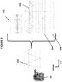
- FIG. 1is a comparative schematic view of hard palate multicomponent brain-based bio-signal detection versus scalp EEG brain wave detection.
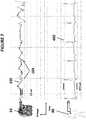
- FIG. 2is a schematic view of the hard palate multicomponent brain-based bio-signal and the resulting subcomponent waves after extraction.
- FIG. 3is a comparative schematic view of an extracted hard palate alpha wave subcomponent signal versus scalp EEG alpha waves.
- FIG. 4is an overlay schematic view of an extracted hard palate brain-based bio-signal subcomponent signal versus scalp EEG brain waves during mental counting activity.
- FIG. 5is a comparative schematic view of an extracted hard palate brain-based bio-signal subcomponent signal versus raw EOG brain waves during up-down eye movement.
- FIG. 6is a comparative schematic view of an extracted hard palate brain-based bio-signal subcomponent signal versus raw EOG brain waves during left-right eye movement.
- FIG. 7is a comparative schematic view of an extracted hard palate brain-based bio-signal subcomponent signal versus cardiac ECG waves.
- FIG. 8is a comparative schematic view of an extracted hard palate brain-based bio-signal subcomponent signal versus EEG, EOG and Respiration waves.
- FIG. 10is a schematic view a system to detect sleep disorders including internal and external units.
- FIG. 12is an isometric view of an embodiment of an oral attachment device.
- FIG. 13is an isometric view of an embodiment of an oral attachment device.
- a system for detecting multi-component brain-based electromagnetic bio-signalsincludes a sensor in the oral cavity may be coupled to one or more electronic processors capable of electronically digitizing, amplifying, attenuating, filtering and normalizing the multi-component brain-based bio-signals as needed.
- the computer processormay also be capable of extracting, isolating or otherwise dividing sub-component signals from the multi-component brain-based bio-signals, and optionally classifying and analyzing the sub-component signals.
- the sensormay be an electrical or magnetic sensor capable of detecting multi-component brain-based electromagnetic bio-signals.
- the sensorincludes electrodes touching the hard palate, where one electrode acts as a reference for comparison with one or more other electrodes.
- the electrodesmay be resistive mode electrodes, capacitive mode electrodes, current mode electrodes, or inductive mode electrodes.
- the electrodesmay be passive electrodes which simply receive a signal, or may be active electrodes which are able to digitize or otherwise process the received signal with an internal electronic processor.
- the senor in the oral cavitymay be included in an oral device configured to couple to the dentition or other oral tissue.
- the position of the sensor or electrodes of the sensormay be adjustable in relation to the oral device.
- the senormay be communicatively coupled to a processor in the oral device. In other embodiments the sensor may be communicatively coupled to one or more external processing device.
- the sensor and/or processor(s)may be communicatively coupled via wires and/or wirelessly, such as Bluetooth or other wireless technology.
- the multicomponent brain-based bio-signal 100is a relatively unremarkable pattern of the raw hard palate bio-potential signal in comparison to the raw scalp EEG signal 200 .
- the multicomponent brain-based bio-signal 100has a significantly larger voltage range when compared to raw scalp EEG signal 200 , 100 ⁇ V versus 10 ⁇ V respectively. This demonstrates that special analysis of the hard palate 10 multicomponent brain-based bio-signal 100 is necessary to determine subcomponents of the hard palate 10 multicomponent brain-based bio-signal 100 especially the brain-based subcomponent signals.
- FIG. 3shows the strong correlation between the 8-14 Hz brain wave subcomponent signal 102 detected on the hard palate and the 8-14 Hz brain wave scalp EEG signal 201 .
- FIG. 4shows the strong correlation between the 3.5-30 Hz brain wave subcomponent signal 102 detected on the hard palate and the 3.5-30 Hz brain wave scalp EEG signal 202 of a subject when subject was performing the mental activity of counting backwards from 100 by 7's (i.e. 100, 93, 86, 79, etc.). The subject was seated in a well-lit, environmentally controlled room.
- FIG. 5shows the strong correlation between an 80 ⁇ V range multicomponent brain-based bio-signal 100 detected on the hard palate 10 and the 53 ⁇ V brain wave EOG signal 300 detected with EOG electrodes 30 on the right-side human scalp while the subject was quickly looking up and down. No filtering or isolation of subcomponents of the multicomponent brain-based bio-signal 100 was needed for this embodiment.
- FIG. 6shows the correlation between an 50 ⁇ V range multicomponent brain-based bio-signal 100 detected on the hard palate 10 and the 558 ⁇ V brain wave EOG signal 300 detected with EOG electrodes 30 on the right-side human scalp while the subject was quickly looking left and right. No filtering or isolation of subcomponents of the multicomponent brain-based bio-signal 100 was needed for this embodiment.
- FIG. 8shows the correlation between respiration sub component signals 106 at 1.5 Hz-249 Hz extracted from the brain-based multicomponent bio-signals detected on the hard palate 10 and scalp EEG signals 203 , right eye EOG signals 301 both of which were filtered at 1.5 Hz-249 Hz, and a nasal cannula respiration signal 500 while the subject takes a fast deep breath 501 and holds the breath 502 for 20 seconds.
- the graphshows that the hard palate bio-potential changes at the same time the scalp EEG and EOG changes showing the strong temporal relation between the hard palate multicomponent bio-signals and the scalp related signals.
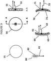
- FIG. 9shows various electrode embodiments for use in hard palate multicomponent bio-signal detection to accommodate the shape of oral tissue and provide for comfort and biocompatibility.
- Soft materialsgauge, or foam
- Other materialscan be used for electrodes as desired.
- the electrode assembly 50includes a metal electrode 51 and temperature sensor 52 .
- the combination of electrode 51 with temperature sensing 52 for the oral cavity, or other body locations,is shown.
- the electrodesdetect current flow from tissue and the temperature sensor allows determination of oral temperature, motion artifact (since temperature is not a bio-potential measurement), and oral airflow.
- Average oral temperaturecan be estimated by a thermistor, semiconductor IC, thermocouple, or other appropriate sensor.
- One embodiment of the electrodemay be a convex electrode assembly 60 which may include a metal electrode with a lead wire 61 and temperature sensor 62 and a soft, absorbent cover-surface 63 .
- Another embodiment of the electrodemay be a concave electrode assembly 70 which may include a metal electrode with a lead wire 71 and temperature sensor 72 and a soft, absorbent cover-surface 73 .
- a third embodiment of the electrodemay be a flat electrode assembly 80 which may include a metal electrode with a lead wire 81 and a soft, absorbent cover 82 .
- the reference electrode 11may be a circular metal electrode.
- FIG. 10is a schematic of a system to detect sleep disorders 600 including an internal oral unit 601 and an external unit 650 .
- the internal oral unit 601may include a convex electrode 60 positioned near the middle of the hard palate and one or two concave electrodes 70 positioned to the left and/or right near the gums.
- the internal oral unit 601may include a sensor unit 602 , a power source 603 , a power manager 604 , a microcontroller 605 , and a transmission unit 606 .
- the internal oral unit 601may amplify, filter and/or digitize multicomponent bio-signals using dedicated circuitry as shown or as part of the data-management microcontroller ( ⁇ CU). Digital signals may be passed to a radio-frequency (RF) module for transmission to a remote receiver, e.g., a Smartphone or computer, or cloud etc.
- RFradio-frequency
- Detecting multicomponent brain-based signalsmay be accomplished by placing the internal unit 601 inside the appropriate body cavity where brain-based multicomponent bio-signal detection can automatically (or manually) be initiated. Signal detection usually begins immediately, however a temperature sensing component can be added to monitor environmental temperatures to ensure proper operating conditions and or monitor temperature during data collection. The temperature sensor can also be used to monitor changes in airflow via the mouth. Additional sensors can also be added to monitor a variety of additional physiological variables including oxygen saturation via optical PPG sensor/monitor, accelerometer, gyroscope, GPS, pressure, camera, biological or chemical monitors etc. Brain-based Detectors monitor multiparameter physiological signals including brain waves.
- the detectori.e. sensor
- the detectorcan be based on any of the following sensors: resistance mode electrodes, capacitive mode electrodes, current mode electrodes, passive electrodes, active electrodes, magnetic mode detectors, inductive mode detectors, acoustic mode detectors, optical or electro-optic mode detectors, chemical or biochemical mode detectors, biological mode detectors and brain-based detector arrays (brain-based detector can also comprise multiple sensors oriented in different geometric planes).
- the sensorsmay be of various shapes and include various metals, metal salts, or metal alloys, semiconductors, polymers, carbon compounds, conductive fabrics, composites, graphenes, non-metals; sensor comprises rigid, semi-rigid, and other flexible materials.
- the sensorsmay utilize microelectronics technology.
- the sensorsmay be disposable and/or reusable.
- the sensorsmay include remote sensors. Sensors may be adjustable in position and/or performance to optimize brain-based multicomponent bio-signal detection.
- Sensor unit 602may detect electrical signals from the hard palate picked up by electrodes and may amplify and filter the electrical signals to remove motion and other artifacts and conveyed to the microcontroller ( ⁇ CU) 605 via the SPI bus for further processing, storage and transmission.
- ⁇ CUmicrocontroller
- Signal and power management schedulingare performed by the ⁇ CU 605 .
- Energy consumed from a disposable or rechargeable power supply 603can be minimized by the ⁇ CU 605 by controlling the duration and duty cycle of data-collection devices, the transmission module 606 , and the ⁇ CU 605 itself.
- Intelligent power managementcan reduce the size and complexity of the power source 603 and eliminate the need for a power line-operated system.
- Data transmission by the transmission unit 606may be via well-known standard communications protocols, such as Bluetooth (BT) and Bluetooth LE (Low Energy) (BLE), or a proprietary protocols or frequencies. Use of standard protocols may ensure easier post-transmission processing.
- the transmission unit 606may support both BT and BLE, which can be accessed by Smartphones and other devices.
- An antenna of the transmission unit 606may be built into the side and/or front walls of an oral appliance attachment device as shown in FIGS. 12-15 .
- the external unit 650includes a receiver unit 651 , a preprocessing unit 652 , an Independent Component Analysis (“ICA”) processor 700 , a raw brain-based multicomponent bio-signal component analyzer 750 ,
- ICAIndependent Component Analysis
- the receiver unit 651may be configured to receive signals from the transmission unit 606 .
- the preprocessing unit 652removes as much signal noise as possible
- the ICA processor 700may use standard ICA algorithms to extract and isolate individual subcomponent signals. To ensure a good estimate of the components of the brain-based raw hard palate signal, brain wave filters 1-N 751 , 752 and 753 , eye movement signal processor 710 , cardiac signal processor 720 and respiration signal processor 730 .
- the preprocessing unit 652may eliminate non-physiological noise via filtering and sensors (thermistors) built into electrodes or a sensor platform. Electrodes may be shielded on their rear surface by the oral attachment device to prevent disturbance by the tongue and or internal facial muscles. Thermistors also provide a means to detect movement of the device relative to tissue as well as provide means to correct for large temperature changes due to breathing. Additional processing includes data filtering such as low pass filtering. Additional preprocessing may include centering and whitening.
- Centeringremoves the mean from each component by subtracting the mean of the data from the actual data. Whitening the data is done to make the raw data uncorrelated to ensure that each subcomponent is as independent as possible. Preprocessing can also identify eye movements due to the unique arrangement of the electrodes (left and right) that produce significant differences in the raw signal detected by each electrode. Root mean square values can be determined and threshold detectors may be incorporated.
- Digitization by the pre-processing unit 652may be electronically performed to enable efficient digital processing as well as signal amplification and or attenuation of the bio-signals if necessary.
- Pre-processingalso seeks to remove unwanted noise by filtering, shielding, blocking, or algorithmically removing or eliminating undesirable physiological and or non-physiological signals such as electrical noise, acoustical noise, mechanical noise, other artifact, or galvanic currents from dissimilar metals, or tongue artifact etc. Undesirable artifact contained in biosignals can hamper recordings. Signal normalization can also occur at this stage.
- the ICA processor 700determines the individual subcomponent signals of the raw hard palate multicomponent bio-signal without previously knowing each component. To effectively determine each subcomponent the number of detectors (sensors) must be equal to or greater to the number of individual signal components. Embodiments may utilize three electrodes to detect bio-potentials each with a built-in thermistor which provides six (6) detectors overall. This embodiment may detect 4 subcomponent bio-signals. To separate the components the JADE algorithm (Joint Approximate Diagonalization Eignen Matrices), which tends to perform best for small datasets) can be incorporated used by the ICA processor 700 of a computer or Field Programmable Gate Array (FPGA).
- JADE algorithmJoint Approximate Diagonalization Eignen Matrices
- Extracting, isolating, or dividing the detected multicomponent brain-based signal into individual parasubcomponent signalsmay involve appropriate means to extract, isolate, and/or divide the brain-based multicomponent signal into constituent physiological signals and/or other signals as desired.
- Primary subcomponent signalsmay include brain-based bio-electromagnetic signals, cardiac bio-electromagnetic bio-signals, ECG, local tissue bio-electromagnetic signals; eye dipole bioelectric bio-signals, muscle bio-electromagnetic bio-signals, tongue bio-electromagnetic bio-signals, cardiovascular related pulsatile bio-signals (e.g. Blood Volume Pulse); respiration related pulsatile bio-signals, movement related bio-signals, biomechanical bio-signals and/or bio-acoustic bio-signals.
- Each subcomponent signaltypically includes multiple frequencies, and may have different dynamic ranges that may overlap.
- additional physiological parameterscan be derived, including heart rate, respiration rate, heart rate variability, pulse transit time, arterial blood pressure.
- Subcomponent bio-signal extractionmay include use of pattern recognition, Independent Component Analysis, Principle Component analysis, Linear analysis, Frequency domain analysis, time—frequency and non-linear techniques such as correlation dimension (CD), phase space plots, different entropies, wavelet based, Hilbert-Huang Transforms (HHT), and similar means as desired.
- pattern recognitionIndependent Component Analysis
- Principle Component analysisLinear analysis
- Frequency domain analysistime—frequency and non-linear techniques such as correlation dimension (CD), phase space plots, different entropies, wavelet based, Hilbert-Huang Transforms (HHT), and similar means as desired.
- CDcorrelation dimension
- HHTHilbert-Huang Transforms
- key subcomponent signal featuressuch as data points, thresholds and/or data slope be extracted or isolated from the signal of interest. This may involve identification of brain-based signal patterns and translation into commands to extract said feature and or issue commands to perform a task.
- a desired algorithmmay be used to automatically estimate/calculate a value to represent the signals by a few relevant key values.
- algorithmsThere are a large variety of algorithms that may be implemented from the simplistic methods such as adding, subtracting, multiplying, dividing, etc., to other complex techniques involving time-based approaches or frequency based approaches, Principle component analysis, Support vector machine, Genetic algorithm, Distinctive sensitive learning vector quantization etc.
- key features of a subcomponent bio-signalmay be classified or translated to a command.
- the classification stepassigns a class to a set of features extracted from the signals.
- the classcan correspond to the type of mental states identified. This step can also be denoted as “feature translation”.
- Key feature informationmay be provided or displayed to a user/operator and/or used to perform tasks, such as comparing an extracted subcomponent bio-signals to a database of baseline signals to control a device, assist in a diagnosis of a disease, disorder, or condition, and or report the status of the device function.
- subcomponent bio-signalscan be displayed or utilized for other purposes such as calculating vital signs, part of a command to control another device(s), or to perform additional processing such as extract particular features.
- the raw brain-signalcan be further analyzed or separated into various frequency bands using band-pass filters 750 and then displayed or used to issue a command.
- Thismay include brain wave filters 1-N 751 .
- the filtersmay be programmed or maintained in hardware for bands of interest 752 , 753 .
- Eye movement sub component signals 710can be displayed and observed for Rapid Eye Movement (REM) to determine sleep stage.
- REMRapid Eye Movement
- Cardiac signals 720can show basic heart rate and can be used to determine R wave peaks as well as heart rate variability.
- Respiration signals 730can be displayed to determine breathing rate.
- Various datamay be stored on a data storage device incorporated into the internal oral unit and/or external unit.
- the systemmay also include a stimulate tissue device.
- brain-based biosignal mapscan be developed to allow for topographical mapping of electrical activity for internal body locations.
- FIG. 11An alternative embodiment that incorporates multiple sensors such as oxygen saturation, head position via accelerometers, temperature, and brain-based signals is shown in FIG. 11 .
- FIG. 12shows an embodiment of the internal oral unit 800 including convex electrodes 60 configured to contact the center of the hard palate, and concave electrodes 70 configured to contact the gums.
- the internal oral unit 800may be a mouth-guard platform which may incorporate a biocompatible adhesive to maintain contact with the dentition and/or oral tissue similar to a denture adhesive.
- the internal oral unit 800electronic circuits 601 that perform some or all of the functions described above.
- the electrodesmay be positioned in the structure which provides a slight spring force against the gums and hard palate to ensure electrode contact with oral tissue.
- FIG. 13shows Oral attachment device 900 , which incorporates a flexible transverse support band 901 to maintain contact with the hard palate and electrodes 11 , 12 , 13 and temperature sensor 52 .
- Oral attachment device 900may include electronics 601 and transmission unit 606 .
- FIG. 14shows an oral attachment device 1000 which includes a thin flexible platform that incorporates a biocompatible adhesive to maintain contact with the mandible and flexible electronic circuits.
- the electrodes 11 , 12 and 13are positioned in the structure which provides a slight spring force against the gums and hard palate to ensure electrode contact with oral tissue.
- Some embodimentsmay include temperature 52 , electronics 601 and transmission unit 606 .
- FIG. 15shows a sagittal view of a human head with an example of an oral attachment mouthguard 800 with electrodes contacting the left and right side of the hard palate and one electrode contacting the hard palate and transmission system, and an exemplary embodiment of the external unit 650 .
- External unitcan be a smartphone, computer or other computing device.
- An embodiment of the present inventionmay utilize subcomponent signals of the brain-based multicomponent bio-signals for screening, diagnosing and monitoring obstructive sleep apnea (“OSA”).
- OSAis a breathing disorder caused by movement and upper airway blockage by the tongue and narrowing of the upper airway by soft tissues within the nose, mouth and throat that occurs during sleep. This phenomenon causes snoring and recurrent interruption of breathing due to periodic obstruction of airflow in the upper airway during inhalation.
- EEGelectrocardiograph
- EMGelectromyograph
- a pulse oximeterattached various parts of a patient.
- Devices intended for home usemay measure fewer parameters are available, but still require multiple connections.
- Embodiments of the inventionenable detection of multicomponent brain-based bio-signals ( FIG. 2 ) from which subcomponent bio-signals can be extracted including brain-based electrical activity including alpha or other waves ( FIG. 3 ), eye movement ( FIG. 5 ), respiration ( FIG. 8 ) and ECG ( FIG. 7 ).
- Brain electrical activity subcomponent signalscan enable determination of sleep state/stage and overall sleep time.
- Respiration subcomponent bio-signalsmay enable determination of apnea events.
- Eye movement subcomponent bio-signalscan enable determination of rapid eye movement (REM) sleep
- ECG subcomponent bio-signalscan enable determination of heart rate during sleep.
- signal processingincluding filtering, amplification, digitizing, storage etc.
- recording of some or all of the sub-component signalsmay occur in computer chip(s) embedded in an oral device including the sensor(s) can be accomplished.
- Resulting datacan either be transmitted as it becomes available via wired or wireless technology (such as Bluetooth) to a receiving device (such as a smartphone, a computer, or dedicated device) and/or uploaded to a receiving device at a later time.
- the multicomponent brain-based bio-signalis transmitted to an external receiving device (such as a smartphone, a computer, or dedicated device) for signal processing.
- the multicomponent brain-based bio-signalmay be transmitted as it is being detected by the sensor or it may be recorded on a storage device in an oral device for retrieval at a later time.
- the sensor detecting the multicomponent brain-based bio-signalmay be supplemented with additional secondary sensors (i.e. accelerometers, thermocouples, O 2 saturation sensors, CO 2 sensors, air flow meters, etc.) may be used in combination with the multicomponent brain-based bio-signal to determine head position and oxygen desaturation and other events during sleep.
- additional secondary sensorsi.e. accelerometers, thermocouples, O 2 saturation sensors, CO 2 sensors, air flow meters, etc.
- the oral devicemay automatically turn off when it is removed from the patient's mouth. In other embodiments the oral device may be turned off manually.
- the signals stored on the devicemay then be uploaded to a computer system including a software program for interpretation of the signal data, and be available for a diagnosis to be made by a physician or other medical personnel.
- the electrical brain activity subcomponent signals extracted from the detected brain-based multi-component signalmay be used along with signals from accelerometers to detect traumatic brain injury in military personnel, sports participants, or other people in at-risk professions or activities, such as concussions, strokes and seizures. Detection of traumatic brain injury may be facilitated by comparing current subcomponent signals to pre-existing baseline signals.
- the pre-existing baseline signalsmay be recorded from the specific patient being tested or a generic baseline derived from consolidation of multiple previously recorded signals from the patient or a segment of the population. In other embodiments these signals may be used to monitor performance.
- subcomponent signals extracted from the detected brain-based multi-component signalmay be used to optimize training and provide feedback on performance of athletes and soldiers in order to enhance their capabilities during competition or in the field.
- the subcomponent signals extracted from the detected brain-based multi-component signalmay also be used in biofeedback applications.
- brain waves and muscle activity subcomponent signals extracted from the detected brain-based multi-component signalmay be used to determine the level of consciousness of a patient under general anesthesia.
- subcomponent signals extracted from the detected brain-based multi-component signalmay be used to detect abnormal brain wave patterns indicative of hypoglycemia in persons with diabetes.
- brain-based bio-signals, eye movement, head position and breathing signals and other subcomponent signals extracted from the detected brain-based multi-component signalmay be used to assist individuals who are physically impaired but mentally capable to operate a wide variety of equipment and tools using a brain-computer interface which interprets the subcomponent signals to operate a variety of equipment's actions. For example moving a motorized wheel chair or operating an artificial limb.
- brain waves and eye movement subcomponent signals extracted from the detected brain-based multi-component signalcan be monitored for advertising or media programming evaluation.
- a usercan be trained to alter his brain waves in order to send a subcomponent signal extracted from the detected brain-based multi-component signal to a central computer in order to automatically control his mobile telephone, video game console, television set, music system or DVD player; change the temperature settings in the room; control an alarm system; control kitchen appliances; or control an automobile's computer system.
- subcomponent signal extracted from the detected brain-based multi-component signalmay be used to detect drowsiness or sedatives or drug related impairment in the operator of a motor vehicle by monitoring sub-component signals related to respiration, eye movement, and other useful parameters.
- the device for this applicationmay be in the form of a nose clip, a mouthpiece, or combinations thereof that collects and processes brain-based multi-component signal via an onboard computer that can subsequently trigger alarm systems and provide notification, or alarm when a driver becomes a drowsy or falls asleep at the wheel.
- a devicemay utilize subcomponent signal extracted from the detected brain-based multi-component signal, such as eye movement and other bio-signals to control machines such as automobiles or airplanes using thought control especially for complex, rapid or emergency maneuvers.
- subcomponent signal extracted from the detected brain-based multi-component signalsuch as eye movement and other bio-signals to control machines such as automobiles or airplanes using thought control especially for complex, rapid or emergency maneuvers.
- one applicationmay be enhancing combat or drone pilots reaction times and assist in the control of aircraft during high-performance or wartime situations.
- the senor and/or other elements of the systemmay be implanted in soft tissue, such as the soft palate or gums; or alternatively inside teeth or tooth implants; or in a third alternative, in parts of the body other than the oral cavity.
- the sensor and/or other elements of the systemcan be implanted in the soft palate and self-powered via piezoelectric material within the device.
- the sensor and/or other elements of the systemmay be implanted beneath the skin and periodically charged inductively, capacitively, optically or other charging methods.
- the senor and/or other elements of the systemmay be located in a swimmer's or underwater diver's mouthpiece.
- the senor and/or other elements of the systemmay be mounted on a nose clip designed for comfortable placement within the nostrils of an individual.
Landscapes
- Health & Medical Sciences (AREA)
- Life Sciences & Earth Sciences (AREA)
- Engineering & Computer Science (AREA)
- General Health & Medical Sciences (AREA)
- Veterinary Medicine (AREA)
- Biophysics (AREA)
- Biomedical Technology (AREA)
- Heart & Thoracic Surgery (AREA)
- Medical Informatics (AREA)
- Molecular Biology (AREA)
- Surgery (AREA)
- Animal Behavior & Ethology (AREA)
- Physics & Mathematics (AREA)
- Public Health (AREA)
- Pathology (AREA)
- Neurology (AREA)
- Physiology (AREA)
- Cardiology (AREA)
- Pulmonology (AREA)
- Neurosurgery (AREA)
- Dentistry (AREA)
- Oral & Maxillofacial Surgery (AREA)
- Psychology (AREA)
- Human Computer Interaction (AREA)
- Ophthalmology & Optometry (AREA)
- Computer Networks & Wireless Communication (AREA)
- Measurement And Recording Of Electrical Phenomena And Electrical Characteristics Of The Living Body (AREA)
Abstract
Description
Claims (20)
Priority Applications (1)
| Application Number | Priority Date | Filing Date | Title |
|---|---|---|---|
| US16/152,778US11071493B2 (en) | 2012-10-24 | 2018-10-05 | Multicomponent brain-based electromagnetic biosignal detection system |
Applications Claiming Priority (5)
| Application Number | Priority Date | Filing Date | Title |
|---|---|---|---|
| US201261717997P | 2012-10-24 | 2012-10-24 | |
| US201361790007P | 2013-03-15 | 2013-03-15 | |
| US14/062,573US20140114165A1 (en) | 2012-10-24 | 2013-10-24 | Systems and methods for detecting brain-based bio-signals |
| US15/165,309US20170020434A1 (en) | 2012-10-24 | 2016-05-26 | Systems and methods for detecting brain-based bio-signals |
| US16/152,778US11071493B2 (en) | 2012-10-24 | 2018-10-05 | Multicomponent brain-based electromagnetic biosignal detection system |
Related Parent Applications (1)
| Application Number | Title | Priority Date | Filing Date |
|---|---|---|---|
| US15/165,309ContinuationUS20170020434A1 (en) | 2012-10-24 | 2016-05-26 | Systems and methods for detecting brain-based bio-signals |
Publications (2)
| Publication Number | Publication Date |
|---|---|
| US20190029587A1 US20190029587A1 (en) | 2019-01-31 |
| US11071493B2true US11071493B2 (en) | 2021-07-27 |
Family
ID=50485948
Family Applications (3)
| Application Number | Title | Priority Date | Filing Date |
|---|---|---|---|
| US14/062,573AbandonedUS20140114165A1 (en) | 2012-10-24 | 2013-10-24 | Systems and methods for detecting brain-based bio-signals |
| US15/165,309AbandonedUS20170020434A1 (en) | 2012-10-24 | 2016-05-26 | Systems and methods for detecting brain-based bio-signals |
| US16/152,778Active2034-05-05US11071493B2 (en) | 2012-10-24 | 2018-10-05 | Multicomponent brain-based electromagnetic biosignal detection system |
Family Applications Before (2)
| Application Number | Title | Priority Date | Filing Date |
|---|---|---|---|
| US14/062,573AbandonedUS20140114165A1 (en) | 2012-10-24 | 2013-10-24 | Systems and methods for detecting brain-based bio-signals |
| US15/165,309AbandonedUS20170020434A1 (en) | 2012-10-24 | 2016-05-26 | Systems and methods for detecting brain-based bio-signals |
Country Status (5)
| Country | Link |
|---|---|
| US (3) | US20140114165A1 (en) |
| EP (1) | EP2911578B1 (en) |
| JP (1) | JP6387352B2 (en) |
| ES (1) | ES2776178T3 (en) |
| WO (1) | WO2014066666A2 (en) |
Families Citing this family (43)
| Publication number | Priority date | Publication date | Assignee | Title |
|---|---|---|---|---|
| WO2014143896A2 (en)* | 2013-03-15 | 2014-09-18 | Simon Adam J | System and signatures for the multi-modal physiological stimulation and assessment of brain health |
| US11064913B2 (en) | 2013-10-25 | 2021-07-20 | Force Impact Technologies, Inc. | Impact sensing wearable device and method |
| AU2013404204B2 (en)* | 2013-10-30 | 2017-07-06 | Fujitsu Limited | Biological sensing system, biological sensing method, and biological sensing program |
| US20150286779A1 (en)* | 2014-04-04 | 2015-10-08 | Xerox Corporation | System and method for embedding a physiological signal into a video |
| US10743806B2 (en) | 2014-06-11 | 2020-08-18 | Dignity Health | Systems and methods for non-intrusive deception detection |
| AU2015305371B2 (en) | 2014-08-21 | 2019-11-21 | Dignity Health | Systems and methods for using eye movements to determine traumatic brain injury |
| US10542961B2 (en) | 2015-06-15 | 2020-01-28 | The Research Foundation For The State University Of New York | System and method for infrasonic cardiac monitoring |
| JP6407824B2 (en) | 2015-09-01 | 2018-10-17 | 株式会社東芝 | Glasses-type wearable terminal and method using the terminal |
| US11207022B2 (en) | 2015-12-22 | 2021-12-28 | Koninklijke Philips N.V. | System and method for determining sleep stages based on cardiac activity information and brain activity information in EEG signals |
| US10470921B2 (en) | 2016-04-07 | 2019-11-12 | Achaemenid, Llc | Removable mandibular myo-stimulator |
| US11375951B2 (en)* | 2016-04-07 | 2022-07-05 | Achaemenid, Llc | Intra-oral electroencephalography device and method |
| US11234638B2 (en) | 2016-04-07 | 2022-02-01 | Achaemenid, Llc | Intra-oral electroencephalography device and method |
| US11000405B2 (en) | 2016-04-07 | 2021-05-11 | Achaemenid, Llc | Removable mandibular pharmaceutical delivery device |
| CN105997088A (en)* | 2016-06-19 | 2016-10-12 | 河北工业大学 | Sleep breath detection device based on flexible force sensor |
| WO2018044959A1 (en)* | 2016-08-29 | 2018-03-08 | Smrt Ip, Llc | Sensor for continuous measurement of hydration and fatigue |
| KR101863056B1 (en)* | 2016-09-19 | 2018-05-31 | 연암공과대학교산학협력단 | Self-charging imitation nipple with thermometer |
| CN110325110B (en)* | 2016-11-10 | 2022-08-09 | 纽约州立大学研究基金会 | Systems, methods, and biomarkers for airway obstruction |
| WO2018115082A1 (en) | 2016-12-20 | 2018-06-28 | Koninklijke Philips N.V. | Patient monitoring |
| US11963839B2 (en)* | 2017-01-10 | 2024-04-23 | A. T. Still University | Dental monitoring system |
| US11723579B2 (en) | 2017-09-19 | 2023-08-15 | Neuroenhancement Lab, LLC | Method and apparatus for neuroenhancement |
| CA3078801A1 (en)* | 2017-10-13 | 2019-04-18 | BioAnalytics Holdings Pty Ltd | Improvements relating to sleep monitoring |
| US20190117124A1 (en)* | 2017-10-19 | 2019-04-25 | MedicusTek, Inc. | Sensor pad for monitoring user posture |
| WO2019077625A1 (en) | 2017-10-20 | 2019-04-25 | Indian Institute Of Technology, Guwahati | A point-of-care system for detection of the physical stress at different parts of body |
| US11717686B2 (en) | 2017-12-04 | 2023-08-08 | Neuroenhancement Lab, LLC | Method and apparatus for neuroenhancement to facilitate learning and performance |
| US11273283B2 (en) | 2017-12-31 | 2022-03-15 | Neuroenhancement Lab, LLC | Method and apparatus for neuroenhancement to enhance emotional response |
| US12280219B2 (en) | 2017-12-31 | 2025-04-22 | NeuroLight, Inc. | Method and apparatus for neuroenhancement to enhance emotional response |
| US11364361B2 (en) | 2018-04-20 | 2022-06-21 | Neuroenhancement Lab, LLC | System and method for inducing sleep by transplanting mental states |
| EP3849410A4 (en) | 2018-09-14 | 2022-11-02 | Neuroenhancement Lab, LLC | SLEEP ENHANCEMENT SYSTEM AND METHOD |
| CN109259731A (en)* | 2018-10-09 | 2019-01-25 | 广东数相智能科技有限公司 | A kind of apoplexy omen method for early warning, electronic equipment and storage medium based on lingual diagnosis |
| US11389113B2 (en) | 2018-12-20 | 2022-07-19 | Force Impact Technologies, Inc. | Mouth guard having user-notification feature of impact force |
| EP3920790B1 (en)* | 2019-02-05 | 2024-06-19 | Inspire Medical Systems, Inc. | Implant-access incision and sensing for sleep disordered breathing (sdb) care |
| US11786694B2 (en) | 2019-05-24 | 2023-10-17 | NeuroLight, Inc. | Device, method, and app for facilitating sleep |
| US11553871B2 (en)* | 2019-06-04 | 2023-01-17 | Lab NINE, Inc. | System and apparatus for non-invasive measurement of transcranial electrical signals, and method of calibrating and/or using same for various applications |
| FR3102054A1 (en) | 2019-10-18 | 2021-04-23 | Devinnova | Helmet to improve the balance of the sympathovagal balance of an individual |
| EP4054420A4 (en)* | 2019-11-04 | 2024-03-06 | Achaemenid, LLC | DEVICE AND METHOD FOR INTRAORAL ELECTROENCEPHALOGRAPHY |
| WO2021092556A1 (en)* | 2019-11-08 | 2021-05-14 | The Johns Hopkins University | Oral measurement devices and methods |
| US11033750B1 (en)* | 2020-02-17 | 2021-06-15 | Achaemenid, Llc | Intra-oral appliance with thermoelectric power source |
| WO2022039613A1 (en)* | 2020-08-19 | 2022-02-24 | Петр Валентинович ИВАНОВ | Mouth guard-electrode for performing procedures in the oral cavity |
| CN113876311B (en)* | 2021-09-02 | 2023-09-15 | 天津大学 | An adaptive selection non-contact multi-player heart rate efficient extraction device |
| CN115348339B (en)* | 2022-08-12 | 2023-11-21 | 北京威努特技术有限公司 | Industrial control abnormity detection method based on correlation of function code and service data |
| US12403033B2 (en) | 2023-04-21 | 2025-09-02 | Achaemenid, Llc | Oral appliance for the treatment of sleep apnea |
| WO2025174909A1 (en)* | 2024-02-12 | 2025-08-21 | Brainscope Company, Inc. | Concussion subtype identification systems and devices |
| CN119257561A (en)* | 2024-11-11 | 2025-01-07 | 广东工业大学 | A method and system for denoising EEG signals by combining SSA and JADE |
Citations (45)
| Publication number | Priority date | Publication date | Assignee | Title |
|---|---|---|---|---|
| US5513649A (en) | 1994-03-22 | 1996-05-07 | Sam Technology, Inc. | Adaptive interference canceler for EEG movement and eye artifacts |
| US5792067A (en)* | 1995-11-21 | 1998-08-11 | Karell; Manuel L. | Apparatus and method for mitigating sleep and other disorders through electromuscular stimulation |
| US6263225B1 (en) | 1994-02-09 | 2001-07-17 | University Of Iowa Research Foundation | Stereotactic electrode assembly |
| US20050239493A1 (en) | 2002-05-07 | 2005-10-27 | Izmail Batkin | Remote monitoring of cardiac electrical activity using a cell phone device |
| US7173437B2 (en) | 2004-06-10 | 2007-02-06 | Quantum Applied Science And Research, Inc. | Garment incorporating embedded physiological sensors |
| US20070032737A1 (en) | 2005-08-02 | 2007-02-08 | Elvir Causevic | Method for assessing brain function and portable automatic brain function assessment apparatus |
| US7209775B2 (en) | 2003-05-09 | 2007-04-24 | Samsung Electronics Co., Ltd. | Ear type apparatus for measuring a bio signal and measuring method therefor |
| US20070106172A1 (en) | 2005-10-24 | 2007-05-10 | Abreu Marcio M | Apparatus and method for measuring biologic parameters |
| US7328062B2 (en) | 2000-07-12 | 2008-02-05 | Ge Healthcare Finland Oy | Monitoring of patient's electrical characteristics using a single piece of equipment |
| US20080058620A1 (en) | 2006-09-05 | 2008-03-06 | Samsung Electronics Co., Ltd | Pressure-providing instrument and biosignal-measuring device including a pressure-providing instrument |
| US20080300469A1 (en)* | 2007-05-31 | 2008-12-04 | National Yang-Ming University | Miniature, wireless apparatus for processing physiological signals and use thereof |
| US20090082829A1 (en) | 2007-09-26 | 2009-03-26 | Medtronic, Inc. | Patient directed therapy control |
| US7693566B2 (en) | 2004-10-18 | 2010-04-06 | Compumedics Limited | Method and apparatus for buffering electrophysiological signals during an MRI procedure |
| US20100274152A1 (en) | 2007-11-06 | 2010-10-28 | Hydrodot, Inc. | Device and method for performing electroencephalography |
| US8019402B1 (en) | 2004-11-12 | 2011-09-13 | Orbital Research Inc | Electrode harness and method of taking biopotential measurements |
| US20110245633A1 (en) | 2010-03-04 | 2011-10-06 | Neumitra LLC | Devices and methods for treating psychological disorders |
| US20120029399A1 (en) | 2009-04-09 | 2012-02-02 | Yoshiyuki Sankai | Wearable type movement assisting apparatus |
| US20120041330A1 (en) | 2010-08-16 | 2012-02-16 | Prichep Leslie S | Field deployable concussion assessment device |
| US20120065478A1 (en) | 2010-09-10 | 2012-03-15 | National Chiao Tung University | Interactive biosensing device and method thereof |
| US8150523B2 (en) | 1999-06-11 | 2012-04-03 | Cornell Research Foundation, Inc. | Feedback method for deep brain stimulation with detection of generalized efference copy signals |
| US8161971B2 (en) | 2006-08-04 | 2012-04-24 | Ric Investments, Llc | Nasal and oral patient interface |
| US8190248B2 (en) | 2003-10-16 | 2012-05-29 | Louisiana Tech University Foundation, Inc. | Medical devices for the detection, prevention and/or treatment of neurological disorders, and methods related thereto |
| US20120133363A1 (en) | 2009-08-07 | 2012-05-31 | University Of Florida Research Foundation, Inc. | Magnetic resonance compatible and susceptibility-matched apparatus and method for mr imaging & spectroscopy |
| US8209004B2 (en) | 2008-06-23 | 2012-06-26 | Freer Logic, Llc | Body-based monitoring of brain electrical activity |
| US20120172682A1 (en) | 2005-12-21 | 2012-07-05 | Norconnect Inc. | Method and apparatus for biometric analysis using eeg and emg signals |
| US8224434B2 (en) | 2006-03-08 | 2012-07-17 | Neuropace, Inc. | Implantable seizure monitor |
| US8267862B2 (en) | 2008-12-18 | 2012-09-18 | Electronics And Telecommunications Research Institute | Apparatus and method for monitoring health index using electroconductive fiber |
| US20120253166A1 (en) | 2011-04-01 | 2012-10-04 | Electronics And Telecommunications Research Institute | Canal type mini-apparatuses insertable in ears for diagnosing and curing diseases |
| US20120294451A1 (en) | 2010-06-11 | 2012-11-22 | Panasonic Corporation | Hearing determination system, and method and program for the same |
| US20120302867A1 (en) | 2011-05-24 | 2012-11-29 | Canon Kabushiki Kaisha | Brain function measuring apparatus and brain function measuring method |
| US20120330178A1 (en) | 2011-06-24 | 2012-12-27 | U.S. Government As Represented By The Secretary Of The Army | Method and apparatus for multimodal mobile screening to quantitatively detect brain function impairment |
| US20130018251A1 (en) | 2011-07-14 | 2013-01-17 | Verathon Inc. | Sensor device with flexible joints |
| US20130035578A1 (en) | 2011-08-01 | 2013-02-07 | Gordon Chiu | Portable Brain Activity Monitor and Method |
| WO2013026481A1 (en) | 2011-08-24 | 2013-02-28 | Widex A/S | Eeg monitor with capacitive electrodes and method of monitoring brain waves |
| US8391948B2 (en) | 2005-09-23 | 2013-03-05 | Brainscope Company, Inc. | Electrode array |
| US20130079634A1 (en) | 2007-02-12 | 2013-03-28 | Med-El Elektromedizinische Geraete Gmbh | Implantable Microphone Noise Suppression |
| US8437843B1 (en) | 2006-06-16 | 2013-05-07 | Cleveland Medical Devices Inc. | EEG data acquisition system with novel features |
| US20130116520A1 (en) | 2011-09-01 | 2013-05-09 | Masoud Roham | Single and multi node, semi-disposable wearable medical electronic patches for bio-signal monitoring and robust feature extraction |
| US8447406B2 (en) | 2010-06-29 | 2013-05-21 | Medtronic, Inc. | Medical method and device for monitoring a neural brain network |
| US8447392B2 (en) | 2002-08-21 | 2013-05-21 | New York University | Brain-machine interface systems and methods |
| US8454505B2 (en) | 2009-09-14 | 2013-06-04 | Imec | Method and electronic medical device for simultaneously measuring an impedance and a biopotential signal |
| US8466875B2 (en) | 2008-01-25 | 2013-06-18 | Panasonic Corporation | Electroencephalogram interface system, electroencephalogram interface apparatus, method, and computer program |
| US20130184316A1 (en) | 2010-07-15 | 2013-07-18 | Andrew Hornstein | Methods for diagnosing and treating concussive disorders |
| US20130211270A1 (en) | 2009-07-20 | 2013-08-15 | Bryan St. Laurent | Mouth Guard for Monitoring Body Dynamics and Methods Therefor |
| US20130253286A1 (en) | 2010-08-27 | 2013-09-26 | The Johns Hopkins University | Device and System for Sensing Medically Relevant Information from the Mouth |
Family Cites Families (19)
| Publication number | Priority date | Publication date | Assignee | Title |
|---|---|---|---|---|
| JPS5342490A (en)* | 1976-09-29 | 1978-04-17 | Rion Co | Artificial cover unit and method of producing same |
| US5190048A (en)* | 1991-09-17 | 1993-03-02 | Healthdyne, Inc. | Thermistor airflow sensor assembly |
| JPH07265272A (en)* | 1994-04-01 | 1995-10-17 | Itec Kk | Nursing/monitoring system |
| US6280394B1 (en)* | 1998-03-18 | 2001-08-28 | Sean R. Maloney | Apparatus and methods for detecting and processing EMG signals |
| JP4633373B2 (en)* | 2004-03-10 | 2011-02-16 | 公立大学法人会津大学 | Biological information processing system |
| US20060058700A1 (en)* | 2004-08-26 | 2006-03-16 | Marro Dominic P | Patient sedation monitor |
| WO2006072150A1 (en)* | 2005-01-07 | 2006-07-13 | K.U. Leuven Research And Development | Muscle artifact removal from encephalograms |
| US7720530B2 (en)* | 2005-08-02 | 2010-05-18 | Brainscope Company, Inc. | Field-deployable concussion detector |
| JP2009530064A (en)* | 2006-03-22 | 2009-08-27 | エモーティブ システムズ ピーティーワイ リミテッド | Electrode and electrode headset |
| GB0611872D0 (en)* | 2006-06-15 | 2006-07-26 | Hypo Safe As | Analysis of EEG signals to detect hypoglycaemia |
| IL178393A0 (en)* | 2006-09-28 | 2007-02-11 | Boris Kayzerman | Improved x-ray apparatus |
| WO2009042721A1 (en)* | 2007-09-25 | 2009-04-02 | Neosync, Inc. | Systems and methods for neuro-eeg synchronization therapy |
| WO2009064473A1 (en)* | 2007-11-13 | 2009-05-22 | Wavesynch Technologies, Inc. | A method for monitoring attentiveness and productivity in a subject |
| US20090281433A1 (en)* | 2008-05-07 | 2009-11-12 | Sonitus Medical, Inc. | Systems and methods for pulmonary monitoring and treatment |
| JP4651720B2 (en)* | 2009-03-12 | 2011-03-16 | 尚治 北島 | Nystagmus recording apparatus and nystagmus inspection system using the same |
| US8355769B2 (en)* | 2009-03-17 | 2013-01-15 | Advanced Brain Monitoring, Inc. | System for the assessment of sleep quality in adults and children |
| CA2758118C (en)* | 2009-04-07 | 2019-05-21 | Catholic Healthcare West | Uterine electrical stimulation system and method |
| RU2556976C2 (en)* | 2009-12-23 | 2015-07-20 | Конинклейке Филипс Электроникс Н.В. | Toothbrush with automatic activation |
| JP2012110536A (en)* | 2010-11-25 | 2012-06-14 | Sony Corp | Wake-up assisting apparatus and wake-up assisting method |
- 2013
- 2013-10-24EPEP13849498.4Apatent/EP2911578B1/enactiveActive
- 2013-10-24WOPCT/US2013/066661patent/WO2014066666A2/enactiveApplication Filing
- 2013-10-24USUS14/062,573patent/US20140114165A1/ennot_activeAbandoned
- 2013-10-24ESES13849498Tpatent/ES2776178T3/enactiveActive
- 2013-10-24JPJP2015539805Apatent/JP6387352B2/enactiveActive
- 2016
- 2016-05-26USUS15/165,309patent/US20170020434A1/ennot_activeAbandoned
- 2018
- 2018-10-05USUS16/152,778patent/US11071493B2/enactiveActive
Patent Citations (46)
| Publication number | Priority date | Publication date | Assignee | Title |
|---|---|---|---|---|
| US6263225B1 (en) | 1994-02-09 | 2001-07-17 | University Of Iowa Research Foundation | Stereotactic electrode assembly |
| US5513649A (en) | 1994-03-22 | 1996-05-07 | Sam Technology, Inc. | Adaptive interference canceler for EEG movement and eye artifacts |
| US5792067A (en)* | 1995-11-21 | 1998-08-11 | Karell; Manuel L. | Apparatus and method for mitigating sleep and other disorders through electromuscular stimulation |
| US8150523B2 (en) | 1999-06-11 | 2012-04-03 | Cornell Research Foundation, Inc. | Feedback method for deep brain stimulation with detection of generalized efference copy signals |
| US7328062B2 (en) | 2000-07-12 | 2008-02-05 | Ge Healthcare Finland Oy | Monitoring of patient's electrical characteristics using a single piece of equipment |
| US20050239493A1 (en) | 2002-05-07 | 2005-10-27 | Izmail Batkin | Remote monitoring of cardiac electrical activity using a cell phone device |
| US8447392B2 (en) | 2002-08-21 | 2013-05-21 | New York University | Brain-machine interface systems and methods |
| US7209775B2 (en) | 2003-05-09 | 2007-04-24 | Samsung Electronics Co., Ltd. | Ear type apparatus for measuring a bio signal and measuring method therefor |
| US8190248B2 (en) | 2003-10-16 | 2012-05-29 | Louisiana Tech University Foundation, Inc. | Medical devices for the detection, prevention and/or treatment of neurological disorders, and methods related thereto |
| US7173437B2 (en) | 2004-06-10 | 2007-02-06 | Quantum Applied Science And Research, Inc. | Garment incorporating embedded physiological sensors |
| US7693566B2 (en) | 2004-10-18 | 2010-04-06 | Compumedics Limited | Method and apparatus for buffering electrophysiological signals during an MRI procedure |
| US8019402B1 (en) | 2004-11-12 | 2011-09-13 | Orbital Research Inc | Electrode harness and method of taking biopotential measurements |
| US20070032737A1 (en) | 2005-08-02 | 2007-02-08 | Elvir Causevic | Method for assessing brain function and portable automatic brain function assessment apparatus |
| US8391948B2 (en) | 2005-09-23 | 2013-03-05 | Brainscope Company, Inc. | Electrode array |
| US20070106172A1 (en) | 2005-10-24 | 2007-05-10 | Abreu Marcio M | Apparatus and method for measuring biologic parameters |
| US20120172682A1 (en) | 2005-12-21 | 2012-07-05 | Norconnect Inc. | Method and apparatus for biometric analysis using eeg and emg signals |
| US8224434B2 (en) | 2006-03-08 | 2012-07-17 | Neuropace, Inc. | Implantable seizure monitor |
| US8437843B1 (en) | 2006-06-16 | 2013-05-07 | Cleveland Medical Devices Inc. | EEG data acquisition system with novel features |
| US8161971B2 (en) | 2006-08-04 | 2012-04-24 | Ric Investments, Llc | Nasal and oral patient interface |
| US20080058620A1 (en) | 2006-09-05 | 2008-03-06 | Samsung Electronics Co., Ltd | Pressure-providing instrument and biosignal-measuring device including a pressure-providing instrument |
| US20130079634A1 (en) | 2007-02-12 | 2013-03-28 | Med-El Elektromedizinische Geraete Gmbh | Implantable Microphone Noise Suppression |
| US20080300469A1 (en)* | 2007-05-31 | 2008-12-04 | National Yang-Ming University | Miniature, wireless apparatus for processing physiological signals and use thereof |
| US20090082829A1 (en) | 2007-09-26 | 2009-03-26 | Medtronic, Inc. | Patient directed therapy control |
| US20100274152A1 (en) | 2007-11-06 | 2010-10-28 | Hydrodot, Inc. | Device and method for performing electroencephalography |
| US8466875B2 (en) | 2008-01-25 | 2013-06-18 | Panasonic Corporation | Electroencephalogram interface system, electroencephalogram interface apparatus, method, and computer program |
| US8209004B2 (en) | 2008-06-23 | 2012-06-26 | Freer Logic, Llc | Body-based monitoring of brain electrical activity |
| US8391967B2 (en) | 2008-06-23 | 2013-03-05 | Freer Logic, Llc | Body-based monitoring of brain electrical activity |
| US8267862B2 (en) | 2008-12-18 | 2012-09-18 | Electronics And Telecommunications Research Institute | Apparatus and method for monitoring health index using electroconductive fiber |
| US20120029399A1 (en) | 2009-04-09 | 2012-02-02 | Yoshiyuki Sankai | Wearable type movement assisting apparatus |
| US20130211270A1 (en) | 2009-07-20 | 2013-08-15 | Bryan St. Laurent | Mouth Guard for Monitoring Body Dynamics and Methods Therefor |
| US20120133363A1 (en) | 2009-08-07 | 2012-05-31 | University Of Florida Research Foundation, Inc. | Magnetic resonance compatible and susceptibility-matched apparatus and method for mr imaging & spectroscopy |
| US8454505B2 (en) | 2009-09-14 | 2013-06-04 | Imec | Method and electronic medical device for simultaneously measuring an impedance and a biopotential signal |
| US20110245633A1 (en) | 2010-03-04 | 2011-10-06 | Neumitra LLC | Devices and methods for treating psychological disorders |
| US20120294451A1 (en) | 2010-06-11 | 2012-11-22 | Panasonic Corporation | Hearing determination system, and method and program for the same |
| US8447406B2 (en) | 2010-06-29 | 2013-05-21 | Medtronic, Inc. | Medical method and device for monitoring a neural brain network |
| US20130184316A1 (en) | 2010-07-15 | 2013-07-18 | Andrew Hornstein | Methods for diagnosing and treating concussive disorders |
| US20120041330A1 (en) | 2010-08-16 | 2012-02-16 | Prichep Leslie S | Field deployable concussion assessment device |
| US20130253286A1 (en) | 2010-08-27 | 2013-09-26 | The Johns Hopkins University | Device and System for Sensing Medically Relevant Information from the Mouth |
| US20120065478A1 (en) | 2010-09-10 | 2012-03-15 | National Chiao Tung University | Interactive biosensing device and method thereof |
| US20120253166A1 (en) | 2011-04-01 | 2012-10-04 | Electronics And Telecommunications Research Institute | Canal type mini-apparatuses insertable in ears for diagnosing and curing diseases |
| US20120302867A1 (en) | 2011-05-24 | 2012-11-29 | Canon Kabushiki Kaisha | Brain function measuring apparatus and brain function measuring method |
| US20120330178A1 (en) | 2011-06-24 | 2012-12-27 | U.S. Government As Represented By The Secretary Of The Army | Method and apparatus for multimodal mobile screening to quantitatively detect brain function impairment |
| US20130018251A1 (en) | 2011-07-14 | 2013-01-17 | Verathon Inc. | Sensor device with flexible joints |
| US20130035578A1 (en) | 2011-08-01 | 2013-02-07 | Gordon Chiu | Portable Brain Activity Monitor and Method |
| WO2013026481A1 (en) | 2011-08-24 | 2013-02-28 | Widex A/S | Eeg monitor with capacitive electrodes and method of monitoring brain waves |
| US20130116520A1 (en) | 2011-09-01 | 2013-05-09 | Masoud Roham | Single and multi node, semi-disposable wearable medical electronic patches for bio-signal monitoring and robust feature extraction |
Also Published As
| Publication number | Publication date |
|---|---|
| EP2911578A4 (en) | 2016-08-10 |
| US20190029587A1 (en) | 2019-01-31 |
| US20140114165A1 (en) | 2014-04-24 |
| EP2911578B1 (en) | 2019-12-25 |
| EP2911578A2 (en) | 2015-09-02 |
| WO2014066666A2 (en) | 2014-05-01 |
| US20170020434A1 (en) | 2017-01-26 |
| WO2014066666A3 (en) | 2015-07-16 |
| ES2776178T3 (en) | 2020-07-29 |
| JP6387352B2 (en) | 2018-09-05 |
| JP2015533580A (en) | 2015-11-26 |
Similar Documents
| Publication | Publication Date | Title |
|---|---|---|
| US11071493B2 (en) | Multicomponent brain-based electromagnetic biosignal detection system | |
| CN108697390B (en) | Sleep state measuring device, phase coherence calculating device, and pressure state measuring device | |
| EP2575608B1 (en) | Detector for identifying physiological artifacts from physiological signals and method | |
| EP3927234B1 (en) | A sleep monitoring system and method | |
| US9833184B2 (en) | Identification of emotional states using physiological responses | |
| EP1989998A2 (en) | Methods and apparatus for monitoring consciousness | |
| US20070208269A1 (en) | Mask assembly, system and method for determining the occurrence of respiratory events using frontal electrode array | |
| EP3267880B1 (en) | Portable electronic device to process a signal acquired from a living body and method thereof | |
| CN111938574A (en) | sleep physiological system | |
| CA2887393A1 (en) | Determining physiological state(s) of an organism based on data sensed with sensors in motion | |
| JP2018524080A (en) | Apparatus and method for monitoring the physiological state of a subject | |
| KR20170083483A (en) | Realtime monitoring apparatus for sleep disorders | |
| US20220022809A1 (en) | Systems and methods to detect and treat obstructive sleep apnea and upper airway obstruction | |
| WO2014152565A1 (en) | Wavelet analysis in neuro diagnostics | |
| CN112739259A (en) | Detection of subject transitions between awake, sleepy, and sleep stages based on photoplethysmography | |
| US12207933B1 (en) | Detector for identifying physiological artifacts from physiological signals and method | |
| Nabavi et al. | Flexible hybrid intraoral sleep monitoring system | |
| WO2024042530A1 (en) | Method and system for electrophysiological determination of a behavioral activity | |
| Gorman | A novel non-EEG wearable device for the detection of epileptic seizures | |
| Nguyen-Binh et al. | A Combination Solution for Sleep Apnea and Heart Rate Detection Based on Accelerometer Tracking | |
| CN117598685A (en) | Respiration detection device, method and system acting on nerve regulation and control | |
| Vidyarani et al. | Driver drowsiness detection using LabVIEW |
Legal Events
| Date | Code | Title | Description |
|---|---|---|---|
| AS | Assignment | Owner name:DREAMSCAPE MEDICAL LLC, GEORGIA Free format text:ASSIGNMENT OF ASSIGNORS INTEREST;ASSIGNORS:KIMANI MWANGI, ANTHONY P.;WALKER, ELIJAH CHARLES;REEL/FRAME:047082/0456 Effective date:20170623 | |
| FEPP | Fee payment procedure | Free format text:ENTITY STATUS SET TO UNDISCOUNTED (ORIGINAL EVENT CODE: BIG.); ENTITY STATUS OF PATENT OWNER: SMALL ENTITY | |
| FEPP | Fee payment procedure | Free format text:ENTITY STATUS SET TO SMALL (ORIGINAL EVENT CODE: SMAL); ENTITY STATUS OF PATENT OWNER: SMALL ENTITY | |
| STPP | Information on status: patent application and granting procedure in general | Free format text:DOCKETED NEW CASE - READY FOR EXAMINATION | |
| STPP | Information on status: patent application and granting procedure in general | Free format text:NON FINAL ACTION MAILED | |
| STPP | Information on status: patent application and granting procedure in general | Free format text:RESPONSE TO NON-FINAL OFFICE ACTION ENTERED AND FORWARDED TO EXAMINER | |
| STPP | Information on status: patent application and granting procedure in general | Free format text:NOTICE OF ALLOWANCE MAILED -- APPLICATION RECEIVED IN OFFICE OF PUBLICATIONS | |
| STPP | Information on status: patent application and granting procedure in general | Free format text:PUBLICATIONS -- ISSUE FEE PAYMENT RECEIVED | |
| STPP | Information on status: patent application and granting procedure in general | Free format text:PUBLICATIONS -- ISSUE FEE PAYMENT VERIFIED | |
| STCF | Information on status: patent grant | Free format text:PATENTED CASE | |
| RF | Reissue application filed | Effective date:20230725 | |
| MAFP | Maintenance fee payment | Free format text:PAYMENT OF MAINTENANCE FEE, 4TH YR, SMALL ENTITY (ORIGINAL EVENT CODE: M2551); ENTITY STATUS OF PATENT OWNER: SMALL ENTITY Year of fee payment:4 |