US11058417B2 - Device, system, and method to secure an article to a tissue - Google Patents
Device, system, and method to secure an article to a tissueDownload PDFInfo
- Publication number
- US11058417B2 US11058417B2US14/897,921US201414897921AUS11058417B2US 11058417 B2US11058417 B2US 11058417B2US 201414897921 AUS201414897921 AUS 201414897921AUS 11058417 B2US11058417 B2US 11058417B2
- Authority
- US
- United States
- Prior art keywords
- securing
- tissue
- article
- securing device
- state
- Prior art date
- Legal status (The legal status is an assumption and is not a legal conclusion. Google has not performed a legal analysis and makes no representation as to the accuracy of the status listed.)
- Active
Links
Images


Classifications
- A—HUMAN NECESSITIES
- A61—MEDICAL OR VETERINARY SCIENCE; HYGIENE
- A61B—DIAGNOSIS; SURGERY; IDENTIFICATION
- A61B17/00—Surgical instruments, devices or methods
- A61B17/064—Surgical staples, i.e. penetrating the tissue
- A61B17/0644—Surgical staples, i.e. penetrating the tissue penetrating the tissue, deformable to closed position
- A—HUMAN NECESSITIES
- A61—MEDICAL OR VETERINARY SCIENCE; HYGIENE
- A61B—DIAGNOSIS; SURGERY; IDENTIFICATION
- A61B17/00—Surgical instruments, devices or methods
- A61B17/068—Surgical staplers, e.g. containing multiple staples or clamps
- A—HUMAN NECESSITIES
- A61—MEDICAL OR VETERINARY SCIENCE; HYGIENE
- A61B—DIAGNOSIS; SURGERY; IDENTIFICATION
- A61B17/00—Surgical instruments, devices or methods
- A61B17/068—Surgical staplers, e.g. containing multiple staples or clamps
- A61B17/0682—Surgical staplers, e.g. containing multiple staples or clamps for applying U-shaped staples or clamps, e.g. without a forming anvil
- A61B17/0684—Surgical staplers, e.g. containing multiple staples or clamps for applying U-shaped staples or clamps, e.g. without a forming anvil having a forming anvil staying above the tissue during stapling
- A—HUMAN NECESSITIES
- A61—MEDICAL OR VETERINARY SCIENCE; HYGIENE
- A61B—DIAGNOSIS; SURGERY; IDENTIFICATION
- A61B17/00—Surgical instruments, devices or methods
- A61B2017/00831—Material properties
- A61B2017/00867—Material properties shape memory effect
- A—HUMAN NECESSITIES
- A61—MEDICAL OR VETERINARY SCIENCE; HYGIENE
- A61B—DIAGNOSIS; SURGERY; IDENTIFICATION
- A61B17/00—Surgical instruments, devices or methods
- A61B17/04—Surgical instruments, devices or methods for suturing wounds; Holders or packages for needles or suture materials
- A61B17/0401—Suture anchors, buttons or pledgets, i.e. means for attaching sutures to bone, cartilage or soft tissue; Instruments for applying or removing suture anchors
- A61B2017/0412—Suture anchors, buttons or pledgets, i.e. means for attaching sutures to bone, cartilage or soft tissue; Instruments for applying or removing suture anchors having anchoring barbs or pins extending outwardly from suture anchor body
- A—HUMAN NECESSITIES
- A61—MEDICAL OR VETERINARY SCIENCE; HYGIENE
- A61B—DIAGNOSIS; SURGERY; IDENTIFICATION
- A61B17/00—Surgical instruments, devices or methods
- A61B17/064—Surgical staples, i.e. penetrating the tissue
- A61B2017/0645—Surgical staples, i.e. penetrating the tissue being elastically deformed for insertion
Definitions
- Staplesexist for a variety of applications in the medical field.
- staplesmay fix soft tissues, boney tissues, and soft tissues to boney tissues.
- Some staplesmay bring two or more tissues together while the staples are applied.
- staplesmay function to fix together some tissues that are brought together by other means, such as by mechanical or chemical means (shape memory)
- staplesmay be designed to bring tissues together so the tissues may heal.
- one or more staplesmay be used to hold or otherwise affix an instrument or other structure to one or more tissues. In some instances, however, an instrument or structure may not be adapted to work with a staple due to size or geometry.
- Such medical staplesmay find use in a number of surgical or non-surgical procedures including, without limitation, an open surgical procedure, an arthroscopic procedure, or in less invasive procedures. Additionally, staples may include additional features suited to specialty applications including imaging applications, such as radiographic procedures.
- a device for securing an article to a tissuemay be composed of a body, transitionable from a first deployment state to a second securing state, having a first portion, a second portion, and a medial portion between the first portion and the second portion, in which the first portion has a first terminal end configured to pierce the tissue, and in which the second securing state comprises at least a section of the first portion of the body assuming a curved geometry embedded in the tissue and at least a section of the medial portion of the body contacting a surface of the article.
- a device for securing an article to a tissuemay be composed of a body, transitionable from a first deployment state to a second securing state, having a first portion, a second portion, and a medial portion between the first portion and the second portion, in which the first portion has a first terminal end configured to pierce the tissue, and a tab standoff in mechanical communication with the medial portion of the body at a first end and a tab at a second end, in which the tab is displaced by a slot from the body.
- the first deployment state of the devicemay be a linearly extended state.
- the deviceIn the second securing state, the device has at least a section of the first portion of the body assuming a curved geometry embedded in the tissue and at least a section of the second portion contacting a surface of the article.
- a device for securing an article to a tissuemay be compose of a body, transitionable from a first deployment state to a second securing state, having a first portion, a second portion, and a medial portion between the first portion and the second portion, in which the first portion has a first terminal end configured to pierce the tissue and the second portion has a second terminal end configured to pierce the tissue.
- the first deployment statemay be a U-shaped state.
- the deviceIn the second securing state, the device has at least a section of the first terminal end assuming a curved geometry embedded in the tissue, at least a section of the second terminal end assuming a curved geometry embedded in the tissue, and at least a section of the medial portion contacting a surface of the article.
- system for securing an article to a tissue by releasibly engaging at least one securing devicemay be composed of an exterior tube having a distal end, in which at least a portion of the distal end of the exterior tube is configured to form a stable mechanical contact between the article and the tissue, and a push rod at least partially disposed within the interior tube.
- the systemmay be configured to releasibly engage at least one securing device having a body transitionable from a first deployment state to a second securing state, and the push rod may be configured to effect a release of the at least one securing device, thereby allowing the securing device body to transition from the first deployment state to the second securing state.
- a method of securing an article to a tissuemay be composed of providing a system for securing the article to the tissue, the system composed of a housing having a distal end, in which at least a portion of the distal end of the housing is configured to engage the article with a surface of the tissue, providing at least one securing device composed of a body, transitionable from a first deployment state to a second securing state, having a first portion, a second portion, and a medial portion between the first portion and the second portion, in which the first portion has a first terminal end configured to pierce the tissue, and wherein the second securing state comprises at least a section of the first portion of the body assuming a curved geometry embedded in the tissue and at least a section of the medial portion of the body assuming a curved geometry around a surface of the article, releasibly engaging the securing device in the first deployment state within the system for securing the article, engaging, by least a portion of the distal
- FIG. 1Adepicts a side view of a securing device having a linearly extended deployment state according to an embodiment.
- FIG. 1Bdepicts a side view of a securing state of a securing device having a linearly extended deployment state according to an embodiment.
- FIG. 1Cdepicts a side view of an article secured to a tissue surface by a securing device having a linearly extended deployment state according to an embodiment.
- FIG. 2Adepicts a side view of a securing device having a U-shaped deployment state according to an embodiment.
- FIG. 2Bdepicts a 3-D view of an article secured by a securing device having a U-shaped deployment state according to an embodiment.
- FIGS. 2C-2Fdepict 3-D views of exemplary securing states attainable by a securing device having a U-shaped deployment state according to an embodiment.
- FIGS. 3A-3Cdepict use of a system to secure an article to a surface of a tissue using a securing device having a linearly extended deployment state according to an embodiment.
- FIGS. 4A-4Ddepict use of a system to secure an article to a surface of a tissue using a securing device having a U-shaped deployment state according to an embodiment.
- FIGS. 5A-5Ddepict a manner in which an article may be secured to a surface of a tissue by the use of a securing device having a U-shaped deployment state according to an embodiment.
- FIG. 6is a flow chart for a method of securing an article to a surface of a tissue using a securing device having a deployment state and a securing state according to an embodiment.
- an articlemay be secured through the use of one or more sutures, staples, and clamps.
- Suturesmay be used in a wide variety of procedures because sutures, comprising flexible material, may take on the shape of the surface of the article being attached.
- suturingis a time-consuming process, and there may be variability in the degree of stabilization of the article to the tissue surface due to the innate variability of suture application.
- the flexible suturing materialmay have a limited tensile strength that may not be strong enough for some applications.
- Staples and clipsmay require less time to apply.
- Staplersused to apply the staples, may provide more consistent force for securing the article to the tissue. As a result, there is less variability in force applied to the article, especially if a number of staples are used.
- Such staples and clipsare typically made of metal, which has a greater tensile strength than sutures.
- staples and clipsbecause they are more rigid than sutures, may not readily conform to the surface of the article being secured. As a result, the article secured by the staples or clips may shift because the clip/article interface is not exact.
- the securing device disclosed hereinmay adjust to the surface geometry of the article being secured while being easily applied from a stapler-type system.
- the articlemay include one or more cardiovascular implant articles.
- the tissuemay comprise a soft tissue such as one or more of a muscle tissue, a tendon tissue, a ligament tissue, and a cardiovascular tissue.
- the devicemay place the article in close proximity to the tissue.
- the devicemay be used to cause the article to physically contact the tissue.
- the securing devicemay be used to secure two portions of tissue together with or without securing a non-tissue article thereto.
- the systemmay be an arthroscopic delivery device or may be part of an arthroscopic device.
- Such arthroscopic devicesmay be used, for example, during open surgical procedures or in minimally invasive surgical procedures.
- itmay be necessary to visualize the attachment of the device to the tissue and article. Examples of visualization methods may include radiographic methods and ultrasound methods.
- the device disclosed hereingenerally has a two dimensional design and function, thereby accommodating such visualization techniques.
- the disclosed devicecan be used in a variety of procedures in which one or more devices and/or one or more tissues may be involved.
- an articlemay be partially pre-attached to a tissue by an alternative means, such as by gluing, and a more robust attachment is required.
- the articlemay be in physical proximity to the tissue but may lack any secure attachment.
- the articlemay be completely attached to the tissue and additional locking between the article and the tissue may be required.
- the articlemay include one or more sutures or polymers and the tissue may include one of the papillary muscles.
- Such a proceduremay find use in repairing or replacing one or more of the chordae tendineae by providing an anchor for the suture or polymer in the muscle to which the replacement may be attached.
- the disclosed devicecan also be used as a means to attach two articles together.
- the first articlemay be a previously implanted article already fixed at a target site and the second article may be a new structure to be fixed to the first article and/or the surrounding tissue.
- the first articlemay be a D-shaped or round annuloplasty ring that is implanted in a mitral valve annulus, and the second article may include a prosthetic valve designed to work in concert with the annuloplasty ring.
- the securing devicemay take on a variety of embodiments as depicted in FIGS. 1A-1C and 2A-2F . It may be understood that such figures and embodiments are only examples and are not limiting. Common to all the possible embodiments of the securing device is the ability of the device to transition between a deployment state and a securing state.
- the deployment statemay comprise a geometry consistent with a device adapted to deliver the securing device, and may include, without limitation, a linearly extended geometry or a U-shaped geometry.
- the securing statemay comprise a geometry capable of contacting the article with a tissue or other article and securing the article thereto.
- the securing statemay include contacting a portion of the securing device with at least a portion of a surface of the article to be secured, and embedding at least one terminal or end portion of the securing device in the tissue.
- the at least one terminal or end portion of the securing device, in the securing statemay include a curved or angled geometry within the tissue, thereby providing an anchor for the device. It may be understood that the device, in transitioning from the deployment state to the securing state may assume any appropriate intermediate geometry consistent with the initial (deployment) and final (securing) states.
- Such securing devicesmay derive their property of transitioning from a first geometric state to a second geometric state from the material or materials from which they are made.
- the securing devices disclosed hereinmay comprise one or more materials having a high yield stress and high level of recoverable plastic strain. As a result of these properties, the device may be deformed from its relaxed shape (comprising the securing state) into a second shape (the deployment state) under force, but will return to the original relaxed shape once the force is removed.
- the body of the securing devicemay include one or more of a metal, a polymer, and a plastic.
- metalsmay include 316 stainless steel, a cobalt chromium alloy, and combinations and alloys thereof.
- the body of the securing devicemay include one or more of a biocompatible material and a bio-absorbable material.
- the biocompatible materialmay include polyether ether ketone, a polyetherimide, or similar material.
- the body of the securing devicemay include one or more of a superelastic material and a shape memory alloy. A non-limiting example of such a shape memory alloy may include a nickel titanium alloy.
- the securing devicesmay also include materials specific for the securing device function or use.
- the body of the securing devicemay include a material that is visualizable by a radiological technique, an MRI technique, and an ultrasound technique.
- the body of the securing devicemay have a length of about 10 mm to about 30 mm.
- the body length of the securing devicemay include a length of about 10 mm, about 12 mm, about 14 mm, about 16 mm, about 18 mm, about 20 mm, about 22 mm, about 24 mm, about 26 mm, about 28 mm, about 30 mm, and any value or range between any two of these values including endpoints.
- the body of the securing devicemay have a length of about 20 mm. In some securing devices, the body may have a cross-sectional diameter of about 0.2 mm to about 1 mm. Examples of the body cross-sectional diameter of the securing device may include a diameter of about 0.2 mm, about 0.3 mm, about 0.4 mm, about 0.5 mm, about 0.6 mm, about 0.7 mm, about 0.8 mm, about 0.9 mm, about 1 mm, and any value or range between any two of these values including endpoints. In one non-limiting example, the body of the securing device may have a cross-sectional diameter of about 0.5 mm.
- the body of the securing devicemay have a circular cross-section.
- the bodymay have a circular cross-sectional diameter of about 0.2 mm to about 1.5 mm.
- the circular cross-sectional diameter of the securing devicemay include a diameter of about 0.2 mm, about 0.3 mm, about 0.4 mm, about 0.5 mm, about 0.6 mm, about 0.7 mm, about 0.8 mm, about 0.9 mm, about 1 mm, about 1.3 mm, about 1.5 mm, and any value or range between any two of these values including endpoints.
- the body of the securing devicemay have a circular cross-sectional diameter of about of about 0.8 mm.
- the body of the securing devicemay have a square or rectangular cross section.
- the bodymay have a rectangular cross-section having a first cross-sectional diameter of about 0.2 mm to about 1 mm and a second cross-sectional diameter of about 0.2 mm to about 1 mm.
- the first cross-sectional diameter of the securing devicemay include a diameter of about 0.2 mm, about 0.3 mm, about 0.4 mm, about 0.5 mm, about 0.6 mm, about 0.7 mm, about 0.8 mm, about 0.9 mm, about 1 mm, and any value or range between any two of these values including endpoints.
- Examples of the second cross-sectional diameter of the securing devicemay include a diameter of about 0.2 mm, about 0.3 mm, about 0.4 mm, about 0.5 mm, about 0.6 mm, about 0.7 mm, about 0.8 mm, about 0.9 mm, about 1 mm, and any value or range between any two of these values including endpoints.
- a securing device having a rectangular cross-sectionmay have a first cross-sectional diameter of about 0.5 mm and a second cross-sectional diameter of about 0.8 mm.
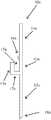
- FIG. 1Adepicts a side view of one embodiment of a securing device having a linearly extended deployment state.
- FIG. 1Bdepicts a side view of the same securing device as FIG. 1A in the securing state.
- the securing device in the deployment state 10 amay include a first portion 12 a , a second portion 14 a , and a medial portion 13 a between the first portion and the second portion.
- the first portion 12 a , the second portion 14 a , and the medial portion 13 a togethermay all form a linearly extended structure.
- the first portion 12 amay have a first terminal end 18 a having a sharpened point for penetrating the tissue.
- the first terminal end 18 amay include one or more barbs, one or more serrations, one or more hooks, or any combination thereof.
- the second portion 14 amay have a second terminal end (not shown) which, in some embodiments, may be a blunt end.
- the securing device in the deployment state 10 amay also include a tab standoff 17 a that is connected to a tab 11 a on one end and to the medial portion 13 a of the securing device on the other end.
- the tab 11 amay be displaced from the medial portion 13 a by a slot 15 a.
- the tab 11 amay have a length of about 2 mm to about 7 mm.
- Examples of the length of the tab 11 amay include a length of about 2 mm, about 3 mm, about 4 mm, about 5 mm, about 6 mm, about 7 mm, and any value or range between any two of these values including endpoints.
- the tab 11 amay have a length of about 5 mm.
- the tab 11 amay have a width of about 0.2 mm to about 1 mm.
- Examples of the width of the tab 11 amay include a width of about 0.2 mm, about 0.3 mm, about 0.4 mm, about 0.5 mm, about 0.6 mm, about 0.7 mm, about 0.8 mm, about 0.9 mm, about 1 mm, and any value or range between any two of these values including endpoints.
- the tab 11 amay have a width of about 0.5 mm.
- the tab 11 amay be displaced from the medial portion 13 a of the body of the securing device by a slot 15 a having by a distance of about 0.5 mm to about 3 mm from the medial portion.
- Examples of the distance of the tab 11 a from the medial portion 13 a of the bodymay include a distance of about 0.5 mm, about 1 mm, about 1.5 mm, about 2 mm, about 2.5 mm, about 3 mm, and any value or range between any two of these values including endpoints.
- the tab 11 amay have distance from the medial portion 13 a of the body of about 1.5 mm.
- the securing device in the deployment state 10 amay also include a friction protrusion 16 a in mechanical communication with the medial portion of the body 13 a and proximate to the tab 11 a .
- the tab 11 amay have a first end in mechanical communication with the tab standoff 17 a and have a second free end approximately opposite the friction protrusion 16 a without making physical contact with it.
- the second free end of the tab 11 amay be in mechanical contact with the friction protrusion 16 a .
- the friction protrusion 16 a and the tab 11 amay help secure the securing device in the extended state 10 a to a system configured to deploy it, as disclosed below.
- the friction protrusion 16 amay have a height of about 0.2 mm to about 1 mm.
- the height of the friction protrusion 16 amay include a height of about 0.2 mm, about 0.3 mm, about 0.4 mm, about 0.5 mm, about 0.6 mm, about 0.7 mm, about 0.8 mm, about 0.9 mm, about 1 mm, and ranges between any two of these values including endpoints.
- the friction protrusion 16 amay have a height of about 0.5 mm.
- the securing device in FIG. 1Amay assume a securing state 10 b including a first portion 12 b , a second portion 14 b , and a medial portion 13 b between the first portion and the second portion.
- the first portion 12 b in FIG. 1Bmay assume a curved geometry.
- the first terminal end 18 b of the first portion 12 bmay include a sharpened point that may help anchor the securing device in the securing state 10 b to the tissue.
- the first terminal end 18 bmay include one or more barbs, one or more serrations, one or more hooks, or any combination thereof. Also in contrast to FIG.
- the second portion 14 b in FIG. 1Bmay assume a curved geometry.
- the securing device in the securing state 10 bmay also include a tab standoff 17 b that is connected to a tab 11 b on one end and to the medial portion 13 b of the securing device on the other end.
- the tab 11 bmay be displaced from the medial portion 13 b by a slot 15 b.
- FIG. 1Cdepicts a manner in which an article may be secured to the surface of a tissue by the securing device depicted in FIGS. 1A and 1B .
- the article 2may be held against the surface of a tissue 3 by means of the securing device in the securing state 10 b .
- the first portion 12 b of the devicemay assume a curved geometry within the tissue 3 , thereby forming a tissue anchor for the device.
- the second portion 14 b of the devicemay contact at least a portion of a surface of the article 2 , thereby securing the article against the surface of the tissue 3 .
- the medial portion of the devicemay also be in contact with at least a portion of the surface of the article 2 .
- the second portion 14 bmay form a curved geometry around the surface of the article 2 .
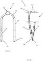
- FIG. 2Adepicts a side view of another embodiment of a securing device having a U-shaped deployment state.
- FIG. 2Bdepicts a side view of the same securing device as FIG. 2A in the securing state.
- the securing device in the deployment state 20 amay include a first portion 22 a , a second portion 24 a , and a medial portion 23 a between the first portion and the second portion.
- the first portion 22 a and the second portion 24 a in the deployment state 20 ado not form a linearly extended structure, but may be effectively parallel to each other.
- the medial portion 23 ais curved, the deployment state 20 a may form a U-shaped structure.
- the first portion 22 amay have a first terminal end 28 a having a sharpened point for penetrating the tissue.
- the first terminal end 28 amay include one or more barbs, one or more serrations, one or more hooks, or any combination thereof.
- the second portion 24 amay have a second terminal end 29 a which may have a sharpened point for penetrating the tissue.
- the second terminal end 29 amay include one or more barbs, one or more serrations, one or more hooks, or any combination thereof.
- the securing device in the deployment state 20 amay also include additional features 21 a connected to the medial portion 23 a of the securing device.
- the additional features 21 amay be used to secure the device in the deployment state 20 a within a system to secure an article to a surface of a tissue through the use of one or more securing devices.
- the additional features 21 amay include a thickened section of the medial portion 23 a , a broadened section of the medial portion, a flanged section of the medial portion, a perforated section of the medial portion, a hooked section of the medial portion (as depicted, for example, in FIG. 2E ), or any combination thereof.
- the securing device in FIG. 2Amay assume a securing state 20 b including a first portion 22 b , a second portion 24 b , and a medial portion 23 b between the first portion and the second portion.
- the first portion 22 b in FIG. 2Bmay assume a curved geometry within the tissue.
- the first terminal end 28 b of the first portion 22 bmay include a sharpened point that may help anchor the securing device in the securing state 20 b to the tissue.
- the first terminal end 28 bmay include one or more barbs, one or more serrations, one or more hooks, or any combination thereof. Also in contrast to FIG.
- the second portion 24 b in FIG. 2Bmay assume a curved geometry while embedded in the tissue.
- the second terminal end 29 b of the second portion 24 bmay include a sharpened point that may help anchor the securing device in the securing state 20 b to the tissue.
- the second terminal end 29 bmay include one or more barbs, one or more serrations, one or more hooks, or any combination thereof.
- the article 2 to be secured to the tissuemay be contacted by a section of the first portion 22 b , a section of the medial portion 23 b , and a section of the second portion 24 b .
- the additional features 21 bmay also be configured to assist with stabilizing the article 2 within the securing device in the securing state 20 b.
- the securing statemay include the first and second portions or the first and second terminal ends forming a scissor-like geometry within the tissue, thereby holding the article between them.
- the first and second portions or the first and second terminal endsmay form a pair of opposing fish-hooks in the tissue and below the article.
- the first and second portions or the first and second terminal endsmay form a pair of opposing fish-hooks in the tissue that point away from the article.
- the first and second portions or the first and second terminal endsmay form a pair of opposing barbs in the tissue that point away from the article.
- the first and second portions or the first and second terminal endsmay form a clothes-pin-type arrangement within the tissue pointing towards the article. It may be understood that the specific geometric arrangements of the first portion, first terminal end, second portion, and second terminal end in the securing state as depicted in FIGS. 2B-2F are merely representative examples, and do not limit the possible configurations achievable by the securing device.
- FIGS. 3A-3Cdepict one type of system configured to deploy a securing device and secure an article therewith to a surface of a tissue.
- the systemmay be used with a securing device having a linearly extended deployment state.
- the systemmay include an exterior tube 31 that may house the remaining components.
- the exterior tube 31may include, at a distal end, a portion configured to form a mechanically stable contact between the article 2 and the surface of the tissue 3 .
- the portion of the exterior tube 31may comprise a pawl 32 .
- the pawl 32may have a surface geometry complementary to the geometry of at least a portion of an external surface of the article 2 .
- the exterior tube 31may be composed of one or more of a metal and a plastic.
- the exterior tube 31may be composed of one or more of a stainless steel, a nickel titanium alloy, and a cobalt chrome alloy.
- the exterior tube 31may be composed of one or more of a polyether ether ketone, a polyether block amide, a polytetrafluoroethylene, a fluorinated ethylene propylene, and an extended polytetrafluoroethylene.
- the exterior tube 31may have a diameter of about 3 mm to about 8 mm.
- the diameter of the exterior tube 31may include a diameter of about 3 mm, about 4 mm, about 5 mm, about 6 mm, about 7 mm, about 8 mm, and any value or range between any two of these values including endpoints.
- the exterior tube 31may have a diameter of about 5 mm.
- the systemmay also include a medial tube 34 disposed at least partially within the exterior tube 31 , an interior tube 36 at least partially disposed within the medial tube, and a push rod 38 at least partially disposed within the interior tube.
- the medial tube 34may be composed of one or more of a metal and a plastic.
- the medial tube 34may be composed of one or more of a stainless steel, a nickel titanium alloy, and a cobalt chrome alloy.
- the medial tube 34may be composed of or more of a polyether ether ketone, a polyether block amide, a polytetrafluoroethylene, a fluorinated ethylene propylene, and an extended polytetrafluoroethylene.
- the interior tube 36may be composed of one or more of a metal and a plastic.
- the medial tube 36may be composed of one or more of a stainless steel, a nickel titanium alloy, and a cobalt chrome alloy.
- the medial tube 36may be composed of or more of a polyether ether ketone, a polyether block amide, a polytetrafluoroethylene, a fluorinated ethylene propylene, and an extended polytetrafluoroethylene.
- the push rod 38may be composed of a metal.
- the push rod 38may be composed of one or more of a stainless steel, a nickel titanium alloy, and a cobalt chrome alloy.
- the interior tube 36may have an exterior surface
- the medial tube 34may have an interior surface
- the interior tube and the medial tubemay be configured to releasibly engage the at least one securing device 10 in the first deployment state therebetween.
- the medial tube 34may have a distal end configured to engage a slot between a tab and the body of the securing device 10 .
- the push rod 38may have a distal end configured to engage an end of the tab of the securing device 10 .
- the interior tube 36may further have at least one detent configured to form a mechanical contact with at least one friction protrusion of the at least one securing device 10 .
- the securing device and system depicted in FIGS. 3A-3Cmay operate in the following manner.
- a securing device 10 having a linearly extended deployment statemay be engaged by the system in a space between the medial tube 34 and the interior tube 36 .
- a distal end of the interior tube 36may engage a slot of the securing device 10 and the exterior end of the push rod 38 may engage a tip end of the tab of the securing device.
- the article 2may be stabilized against the surface of the tissue 3 by the pawl 32 .
- the push rod 38 and interior tube 36may be moved in a distal direction.
- the securing device 10may be displaced in the distal direction, thereby forcing the first portion of the securing device to penetrate the tissue 3 and releasing the securing device 10 from the system.
- the first portion of the securing device 10may continue penetrating the tissue 3 until the tab standoff 37 contacts the surface of the tissue.
- the first portion of the securing device 10free of the system, may then assume a curved geometry within the tissue 3 , constituting a portion of the securing state of the device. As illustrated in FIG.
- the medial tube 34 and the interior tube 36may be moved in the proximal direction.
- the second portion of the securing device 10may be freed from the system and allowed to assume the curved geometry of the securing state.
- the securing statemay include the second portion of the securing device 10 forming a curved contact with at least a portion of the surface of the article 2 .
- FIGS. 4A-4DAn alternative embodiment of a system for securing an article to a tissue is depicted in FIGS. 4A-4D .
- the securing devicemay be one having a U-shaped deployment state.
- the one or more securing devices 10may initially be engaged within the delivery system 44 .
- the delivery system 44may include a distal end having a surface geometry complementary to the geometry of at least a portion of an external surface of the article to be secured.
- the delivery system 44may be brought in proximal contact to the article 2 to be secured.
- FIG. 4Dillustrates that a securing device 10 may be released from the system 44 to contact the article 2 .
- the securing device 10may be released from the system 44 according to any of a number of means including the use of a push rod (not shown).
- FIGS. 5A-5Ddepict the interaction of a securing device 10 with an article 2 and a tissue 3 to which the article may be secured.
- the securing device 10may be one having a U-shaped deployment state.
- FIG. 5Aillustrates the article 2 proximate to the surface of the tissue 3 .
- the securing device 10may be in a deployment state, as indicated by the U-shape of the securing device. In the deployment state, such a securing device 10 may have a first portion 22 a , and a second portion 24 a in a roughly parallel orientation.
- a medial portion 23 ais disposed between the first portion 22 a and the second portion 24 a .
- first portion 22 a , the second portion 24 a , and the medial portion 23 amay surround the article 2 .
- first terminal end from the first portion 22 a , and the second terminal end of the second portion 24 amay contact the surface of the tissue 3 .
- the securing device 10may still be in the deployment state, as indicated by the index letter ‘a’ associated with each of the figure component labels. The securing device 10 , as illustrated in FIG.
- the medial portion 23 cmay also transition between a deployment state to a securing state.
- the securing device 10has transitioned to the securing state, as indicated by the index letter ‘b’ associated with each of the figure component labels.
- the first portion 22 b and second portion 24 bmay assume their respective curved geometries within the tissue 3 .
- the article 2may thus be anchored to the surface of the tissue 3 by means of the first portion 22 b , second portion 24 b , and medial portion 23 b of the securing device 10 .
- FIG. 6is a flow chart of a method for securing an article to a tissue.
- the methodmay include providing 610 a system for securing the article to the tissue.
- the systemmay include a housing having a distal end, in which at least a portion of the distal end of the housing may be configured to engage the article with a surface of the tissue.
- the system for securing the article to the tissuemay further include a medial tube at least partially disposed within the system housing, an interior tube at least partially disposed within the medial tube, and a push rod at least partially disposed within the interior tube.
- the methodmay also include providing 620 at least one securing device.
- the at least one securing devicemay include a body, transitionable from a first deployment state to a second securing state, having a first portion, a second portion, and a medial portion between the first portion and the second portion.
- the first portion of the securing devicemay further have a first terminal end configured to pierce the tissue.
- the second securing state of the securing devicemay include at least a section of the first portion of the body assuming a curved geometry embedded in the tissue and at least a section of the medial portion of the body contacting a surface of the article.
- the first deployment state of the securing devicemay take the form of a U-shaped state
- the second securing statemay include at least a section of a first terminal end of the first portion assuming a curved geometry embedded in the tissue, at least a section of a second terminal end of the second portion assuming a curved geometry embedded in the tissue, and at least a section of the medial portion contacting a surface of the article
- the first deployment state of the securing devicemay take the form of a linearly extended state
- the second securing statemay further include at least a section of the first portion of the body assuming a curved geometry embedded in the tissue and at least a section of the second portion contacting a surface of the article.
- the securing devicemay further include a tab standoff in mechanical communication with the medial portion of the body at a first standoff end and a tab at a second standoff end, in which the tab may be displaced by a slot from the body.
- the securing devicewhile in a deployment state, may be releasibly engaged 630 with the deployment system.
- the at least one securing devicemay include a tab standoff in mechanical communication with the medial portion of the body of the securing device at a first standoff end and a tab at a second standoff end, thereby displacing the tab from the body by a slot.
- releasibly engaging 630 the device within the system for securing the articlemay further include releasibly engaging the slot of the device with a portion of the deployment system.
- such a deployment systemmay include an interior tube having a distal end that may engage 630 the slot of the securing device.
- the at least one securing devicemay also include a friction protrusion in mechanical communication with the body and proximate to a tab.
- releasibly engaging the device within the system for securing the articlemay further include releasibly engaging the friction protrusion with a detent on an exterior surface of an interior tube of the deployment system.
- the deployment systemmay be used to engage 640 the article with the surface of the tissue.
- the articlemay be engaged 640 with the surface of the tissue using at least a portion of a distal end of the housing.
- the securing devicemay be released 650 from the deployment system, thereby allowing the body of the securing device to assume the second securing state and securing the article against the surface of the tissue.
- releasing 650 the securing device from the distal end of the housingmay include contacting the tab of the securing device with a distal end of a deployment system push rod, extending the push rod in a distal direction, and extending a medial tube of the deployment system in a proximal direction.
- compositions, methods, and devicesare described in terms of “comprising” various components or steps (interpreted as meaning “including, but not limited to”), the compositions, methods, and devices can also “consist essentially of” or “consist of” the various components and steps, and such terminology should be interpreted as defining essentially closed-member groups. It will be further understood by those within the art that if a specific number of an introduced claim recitation is intended, such an intent will be explicitly recited in the claim, and in the absence of such recitation no such intent is present. For example, as an aid to understanding, the following appended claims may contain usage of the introductory phrases “at least one” and “one or more” to introduce claim recitations.
- a rangeincludes each individual member.
- a group having 1-3 cellsrefers to groups having 1, 2, or 3 cells.
- a group having 1-5 cellsrefers to groups having 1, 2, 3, 4, or 5 cells, and so forth.
Landscapes
- Health & Medical Sciences (AREA)
- Life Sciences & Earth Sciences (AREA)
- Surgery (AREA)
- Heart & Thoracic Surgery (AREA)
- Engineering & Computer Science (AREA)
- Biomedical Technology (AREA)
- Nuclear Medicine, Radiotherapy & Molecular Imaging (AREA)
- Medical Informatics (AREA)
- Molecular Biology (AREA)
- Animal Behavior & Ethology (AREA)
- General Health & Medical Sciences (AREA)
- Public Health (AREA)
- Veterinary Medicine (AREA)
- Surgical Instruments (AREA)
- Prostheses (AREA)
Abstract
Description
Claims (7)
Priority Applications (1)
| Application Number | Priority Date | Filing Date | Title |
|---|---|---|---|
| US14/897,921US11058417B2 (en) | 2013-06-28 | 2014-06-30 | Device, system, and method to secure an article to a tissue |
Applications Claiming Priority (3)
| Application Number | Priority Date | Filing Date | Title |
|---|---|---|---|
| US201361840588P | 2013-06-28 | 2013-06-28 | |
| PCT/US2014/044920WO2014210600A2 (en) | 2013-06-28 | 2014-06-30 | Device, system, and method to secure an article to a tissue |
| US14/897,921US11058417B2 (en) | 2013-06-28 | 2014-06-30 | Device, system, and method to secure an article to a tissue |
Related Parent Applications (1)
| Application Number | Title | Priority Date | Filing Date |
|---|---|---|---|
| PCT/US2014/044920A-371-Of-InternationalWO2014210600A2 (en) | 2013-06-28 | 2014-06-30 | Device, system, and method to secure an article to a tissue |
Related Child Applications (2)
| Application Number | Title | Priority Date | Filing Date |
|---|---|---|---|
| US16/196,223DivisionUS11224422B2 (en) | 2013-06-28 | 2018-11-20 | Device, system, and method to secure an article to a tissue |
| US16/196,444DivisionUS11191536B2 (en) | 2013-06-28 | 2018-11-20 | Device, system, and method to secure an article to a tissue |
Publications (2)
| Publication Number | Publication Date |
|---|---|
| US20160106420A1 US20160106420A1 (en) | 2016-04-21 |
| US11058417B2true US11058417B2 (en) | 2021-07-13 |
Family
ID=52142857
Family Applications (4)
| Application Number | Title | Priority Date | Filing Date |
|---|---|---|---|
| US14/897,921ActiveUS11058417B2 (en) | 2013-06-28 | 2014-06-30 | Device, system, and method to secure an article to a tissue |
| US16/196,444Active2035-01-15US11191536B2 (en) | 2013-06-28 | 2018-11-20 | Device, system, and method to secure an article to a tissue |
| US16/196,223Active2035-04-14US11224422B2 (en) | 2013-06-28 | 2018-11-20 | Device, system, and method to secure an article to a tissue |
| US17/648,153Active2034-09-10US11806009B2 (en) | 2013-06-28 | 2022-01-17 | Device, system, and method to secure an article to a tissue |
Family Applications After (3)
| Application Number | Title | Priority Date | Filing Date |
|---|---|---|---|
| US16/196,444Active2035-01-15US11191536B2 (en) | 2013-06-28 | 2018-11-20 | Device, system, and method to secure an article to a tissue |
| US16/196,223Active2035-04-14US11224422B2 (en) | 2013-06-28 | 2018-11-20 | Device, system, and method to secure an article to a tissue |
| US17/648,153Active2034-09-10US11806009B2 (en) | 2013-06-28 | 2022-01-17 | Device, system, and method to secure an article to a tissue |
Country Status (4)
| Country | Link |
|---|---|
| US (4) | US11058417B2 (en) |
| EP (2) | EP3797707A1 (en) |
| IL (1) | IL243130B (en) |
| WO (1) | WO2014210600A2 (en) |
Cited By (5)
| Publication number | Priority date | Publication date | Assignee | Title |
|---|---|---|---|---|
| US11806009B2 (en) | 2013-06-28 | 2023-11-07 | Valcare, Inc. | Device, system, and method to secure an article to a tissue |
| US12115069B2 (en) | 2012-02-29 | 2024-10-15 | Valcare Medical, Inc. | Percutaneous annuloplasty system with anterior-posterior adjustment |
| US12279955B2 (en) | 2017-03-17 | 2025-04-22 | Valcare Medical, Inc. | Mitral or tricuspid repair systems with multi-directional anchors |
| US12396853B2 (en) | 2019-06-11 | 2025-08-26 | Valcare Medical, Inc. | Systems and methods for delivery of chordae replacement system |
| US12409034B2 (en) | 2019-06-11 | 2025-09-09 | Valcare Medical, Inc. | Annuloplasty ring with posterior leaflet for minimally invasive treatment |
Families Citing this family (21)
| Publication number | Priority date | Publication date | Assignee | Title |
|---|---|---|---|---|
| WO2012019052A2 (en) | 2010-08-04 | 2012-02-09 | Micardia Corporation | Percutaneous transcatheter repair of heart valves |
| US9402721B2 (en) | 2011-06-01 | 2016-08-02 | Valcare, Inc. | Percutaneous transcatheter repair of heart valves via trans-apical access |
| US9180008B2 (en) | 2012-02-29 | 2015-11-10 | Valcare, Inc. | Methods, devices, and systems for percutaneously anchoring annuloplasty rings |
| US10166100B2 (en) | 2013-03-15 | 2019-01-01 | Valcare, Inc. | Systems and methods for delivery of annuloplasty rings |
| US10813751B2 (en) | 2013-05-22 | 2020-10-27 | Valcare, Inc. | Transcatheter prosthetic valve for mitral or tricuspid valve replacement |
| US20160120642A1 (en) | 2013-05-24 | 2016-05-05 | Valcare, Inc. | Heart and peripheral vascular valve replacement in conjunction with a support ring |
| FR3032608B1 (en)* | 2015-02-13 | 2020-10-23 | Institut Hospitalo Univ De Chirurgie Mini Invasive Guidee Par Limage | ENDOSCOPIC SURGERY SYSTEM FORMED ON THE ONE HAND BY A PLURALITY OF STAPLES AND ON THE OTHER HAND BY AN ENDOSCOPIC APPLICATOR. |
| CN107753153B (en) | 2016-08-15 | 2022-05-31 | 沃卡尔有限公司 | Device and method for treating heart valve insufficiency |
| US11285003B2 (en) | 2018-03-20 | 2022-03-29 | Medtronic Vascular, Inc. | Prolapse prevention device and methods of use thereof |
| US11026791B2 (en) | 2018-03-20 | 2021-06-08 | Medtronic Vascular, Inc. | Flexible canopy valve repair systems and methods of use |
| CA3094990C (en) | 2018-03-23 | 2023-01-03 | Neochord, Inc. | Device for suture attachment for minimally invasive heart valve repair |
| US11253360B2 (en) | 2018-05-09 | 2022-02-22 | Neochord, Inc. | Low profile tissue anchor for minimally invasive heart valve repair |
| EP3790509A4 (en)* | 2018-05-09 | 2022-03-09 | NeoChord, Inc. | SYSTEMS AND METHODS FOR TRANSCATHETER VALVE REPAIR |
| US11173030B2 (en) | 2018-05-09 | 2021-11-16 | Neochord, Inc. | Suture length adjustment for minimally invasive heart valve repair |
| AU2019336254B2 (en) | 2018-09-07 | 2021-12-09 | Neochord, Inc. | Device for suture attachment for minimally invasive heart valve repair |
| EP3626181A1 (en)* | 2018-09-23 | 2020-03-25 | Medtentia International Ltd Oy | A clip for fixation of a medical implant to tissue |
| US11534300B2 (en) | 2018-12-03 | 2022-12-27 | Valcare, Inc. | Stabilizing and adjusting tool for controlling a minimally invasive mitral / tricuspid valve repair system |
| WO2020214818A1 (en) | 2019-04-16 | 2020-10-22 | Neochord, Inc. | Transverse helical cardiac anchor for minimally invasive heart valve repair |
| US11793628B2 (en) | 2019-07-15 | 2023-10-24 | Valcare, Inc. | Transcatheter bio-prosthesis member and support structure |
| WO2021146757A2 (en) | 2020-01-16 | 2021-07-22 | Neochord, Inc. | Helical cardiac anchors for minimally invasive heart valve repair |
| US20230045532A1 (en)* | 2021-08-09 | 2023-02-09 | Valcare, Inc. | Systems and methods for target/article bridging engagement and anchoring |
Citations (184)
| Publication number | Priority date | Publication date | Assignee | Title |
|---|---|---|---|---|
| US4602911A (en) | 1982-08-19 | 1986-07-29 | General Resorts S.A. | Adjustable ringprosthesis |
| WO1990009153A1 (en) | 1989-02-13 | 1990-08-23 | Baxter International Inc. | Selectively flexible annuloplasty ring |
| US5236440A (en) | 1992-04-14 | 1993-08-17 | American Cyanamid Company | Surgical fastener |
| US5306296A (en) | 1992-08-21 | 1994-04-26 | Medtronic, Inc. | Annuloplasty and suture rings |
| US5695518A (en) | 1990-12-28 | 1997-12-09 | Laerum; Frode | Filtering device for preventing embolism and/or distension of blood vessel walls |
| US5716370A (en) | 1996-02-23 | 1998-02-10 | Williamson, Iv; Warren | Means for replacing a heart valve in a minimally invasive manner |
| US5855614A (en) | 1993-02-22 | 1999-01-05 | Heartport, Inc. | Method and apparatus for thoracoscopic intracardiac procedures |
| US6113611A (en) | 1998-05-28 | 2000-09-05 | Advanced Vascular Technologies, Llc | Surgical fastener and delivery system |
| US6231602B1 (en) | 1999-04-16 | 2001-05-15 | Edwards Lifesciences Corporation | Aortic annuloplasty ring |
| US6447524B1 (en) | 2000-10-19 | 2002-09-10 | Ethicon Endo-Surgery, Inc. | Fastener for hernia mesh fixation |
| US20020151961A1 (en) | 2000-01-31 | 2002-10-17 | Lashinski Randall T. | Medical system and method for remodeling an extravascular tissue structure |
| US20020151970A1 (en) | 1999-02-10 | 2002-10-17 | Garrison Michi E. | Methods and devices for implanting cardiac valves |
| US20020188170A1 (en) | 2001-04-27 | 2002-12-12 | Santamore William P. | Prevention of myocardial infarction induced ventricular expansion and remodeling |
| US20020198526A1 (en) | 2000-06-23 | 2002-12-26 | Shaolian Samuel M. | Formed in place fixation system with thermal acceleration |
| WO2003017874A1 (en) | 2001-08-24 | 2003-03-06 | Edwards Lifesciences Corporation | Self-molding annuloplasty ring |
| US20030050693A1 (en) | 2001-09-10 | 2003-03-13 | Quijano Rodolfo C. | Minimally invasive delivery system for annuloplasty rings |
| US20030078671A1 (en) | 2001-04-27 | 2003-04-24 | Lesniak Jeanne M. | Prevention of myocardial infarction induced ventricular expansion and remodeling |
| US20030078465A1 (en) | 2001-10-16 | 2003-04-24 | Suresh Pai | Systems for heart treatment |
| WO2003047467A1 (en) | 2001-12-04 | 2003-06-12 | Edwards Lifesciences Corporation | Minimally-invasive annuloplasty repair segment delivery template system |
| US6619291B2 (en) | 2001-04-24 | 2003-09-16 | Edwin J. Hlavka | Method and apparatus for catheter-based annuloplasty |
| US6629534B1 (en) | 1999-04-09 | 2003-10-07 | Evalve, Inc. | Methods and apparatus for cardiac valve repair |
| US20030191528A1 (en) | 2000-06-02 | 2003-10-09 | Quijano Rodolfo C. | Expandable medical implant and percutaneous delivery |
| US20030198605A1 (en) | 1998-02-13 | 2003-10-23 | Montgomery R. Eric | Light-activated tooth whitening composition and method of using same |
| US20030199974A1 (en) | 2002-04-18 | 2003-10-23 | Coalescent Surgical, Inc. | Annuloplasty apparatus and methods |
| US6669687B1 (en) | 1999-06-25 | 2003-12-30 | Vahid Saadat | Apparatus and methods for treating tissue |
| US6689048B2 (en) | 2000-01-14 | 2004-02-10 | Acorn Cardiovascular, Inc. | Delivery of cardiac constraint jacket |
| US20040044364A1 (en)* | 2002-08-29 | 2004-03-04 | Devries Robert | Tissue fasteners and related deployment systems and methods |
| US20040068276A1 (en)* | 2002-10-04 | 2004-04-08 | Steve Golden | Anastomosis apparatus and methods |
| US6726704B1 (en)* | 1998-05-29 | 2004-04-27 | By-Pass, Inc. | Advanced closure device |
| US20040122514A1 (en) | 2002-12-20 | 2004-06-24 | Fogarty Thomas J. | Biologically implantable prosthesis and methods of using the same |
| US20040138744A1 (en) | 2000-01-31 | 2004-07-15 | Randall Lashinski | Transluminal mitral annuloplasty with active anchoring |
| US20040148021A1 (en) | 2002-08-29 | 2004-07-29 | Cartledge Richard G. | Implantable devices for controlling the internal circumference of an anatomic orifice or lumen |
| US6776784B2 (en) | 2001-09-06 | 2004-08-17 | Core Medical, Inc. | Clip apparatus for closing septal defects and methods of use |
| US6790229B1 (en) | 1999-05-25 | 2004-09-14 | Eric Berreklouw | Fixing device, in particular for fixing to vascular wall tissue |
| US6797002B2 (en) | 2000-02-02 | 2004-09-28 | Paul A. Spence | Heart valve repair apparatus and methods |
| US20040193191A1 (en) | 2003-02-06 | 2004-09-30 | Guided Delivery Systems, Inc. | Devices and methods for heart valve repair |
| KR20040095482A (en) | 2003-05-09 | 2004-11-15 | 안성순 | annuloplasty ring |
| US20040243230A1 (en) | 2003-05-20 | 2004-12-02 | The Cleveland Clinic Foundation | Apparatus and methods for repair of a cardiac valve |
| US20040249391A1 (en) | 2001-08-09 | 2004-12-09 | Christy Cummins | Surgical stapling device and method |
| US20040260393A1 (en) | 2000-09-20 | 2004-12-23 | Ample Medical, Inc. | Devices, systems, and methods for reshaping a heart valve annulus |
| US20040260394A1 (en) | 2003-06-20 | 2004-12-23 | Medtronic Vascular, Inc. | Cardiac valve annulus compressor system |
| US20050020696A1 (en) | 2000-04-21 | 2005-01-27 | Montgomery Robert Eric | Low peak exotherm curable compositions |
| US20050033325A1 (en) | 2001-02-16 | 2005-02-10 | Ethicon, Inc. | Surgical knot pusher and method of use |
| US20050065550A1 (en) | 2003-02-06 | 2005-03-24 | Guided Delivery Systems, Inc. | Delivery devices and methods for heart valve repair |
| US20050090846A1 (en) | 2003-07-18 | 2005-04-28 | Wesley Pedersen | Valvuloplasty devices and methods |
| US20050096740A1 (en) | 2001-01-30 | 2005-05-05 | Edwards Lifesciences Ag | Transluminal mitral annuloplasty |
| US6893459B1 (en) | 2000-09-20 | 2005-05-17 | Ample Medical, Inc. | Heart valve annulus device and method of using same |
| WO2005046488A2 (en) | 2003-11-12 | 2005-05-26 | Medtronic Vascular, Inc. | Cardiac valve annulus reduction system |
| US20050113910A1 (en) | 2002-01-04 | 2005-05-26 | David Paniagua | Percutaneously implantable replacement heart valve device and method of making same |
| US20050137692A1 (en) | 2003-12-23 | 2005-06-23 | Haug Ulrich R. | Methods and apparatus for endovascularly replacing a patient's heart valve |
| US20050137695A1 (en) | 2003-12-23 | 2005-06-23 | Sadra Medical | Replacement valve and anchor |
| US20050203549A1 (en) | 2004-03-09 | 2005-09-15 | Fidel Realyvasquez | Methods and apparatus for off pump aortic valve replacement with a valve prosthesis |
| US20050222678A1 (en) | 2004-04-05 | 2005-10-06 | Lashinski Randall T | Remotely adjustable coronary sinus implant |
| US20050240200A1 (en) | 2004-04-23 | 2005-10-27 | Bjarne Bergheim | Method and system for cardiac valve delivery |
| US20050250161A1 (en) | 1999-06-15 | 2005-11-10 | The Trustees Of Columbia University | Generation of antigen specific T suppressor cells for treatment of rejection |
| US20050267572A1 (en) | 2004-05-14 | 2005-12-01 | St. Jude Medical, Inc. | Systems and methods for holding annuloplasty rings |
| US20050283190A1 (en) | 2004-06-16 | 2005-12-22 | Huitema Thomas W | Surgical fastener |
| US20050288778A1 (en) | 2004-06-29 | 2005-12-29 | Emanuel Shaoulian | Selectively adjustable cardiac valve implants |
| US20060009737A1 (en) | 2004-07-12 | 2006-01-12 | Whiting James S | Methods and devices for transseptal access |
| US20060020327A1 (en) | 2004-05-05 | 2006-01-26 | Lashinski Randall T | Nonstented heart valves with formed in situ support |
| US20060122633A1 (en) | 2002-06-13 | 2006-06-08 | John To | Methods and devices for termination |
| US20060129025A1 (en) | 2002-06-27 | 2006-06-15 | Levine Robert A | Systems for and methods of atrioventricular valve regurgitation and reversing ventricular remodeling |
| US20060161169A1 (en) | 2003-05-02 | 2006-07-20 | Cardiac Dimensions, Inc., A Delaware Corporation | Device and method for modifying the shape of a body organ |
| US20060184242A1 (en) | 2003-10-20 | 2006-08-17 | Samuel Lichtenstein | Method and apparatus for percutaneous reduction of anterior-posterior diameter of mitral valve |
| US20060184240A1 (en) | 2003-06-25 | 2006-08-17 | Georgia Tech Research Corporation | Annuloplasty chain |
| US20060195183A1 (en) | 2005-02-18 | 2006-08-31 | The Cleveland Clinic Foundation | Apparatus and methods for replacing a cardiac valve |
| US20060195134A1 (en) | 2005-02-28 | 2006-08-31 | Medtronic Vascular, Inc. | Device, system, and method for aiding valve annuloplasty |
| US7101395B2 (en) | 2002-06-12 | 2006-09-05 | Mitral Interventions, Inc. | Method and apparatus for tissue connection |
| US7114953B1 (en) | 2003-04-25 | 2006-10-03 | Wagner Eugene C | Tooth whitening appliance having membrane covered applicator |
| US20060241748A1 (en) | 2005-03-25 | 2006-10-26 | Lee Leonard Y | Methods and apparatus for controlling the internal circumference of an anatomic orifice or lumen |
| US20060282161A1 (en) | 2003-06-20 | 2006-12-14 | Medtronic Vascular, Inc. | Valve annulus reduction system |
| US20070016287A1 (en) | 2005-03-25 | 2007-01-18 | Cartledge Richard G | Methods and apparatus for controlling the internal circumference of an anatomic orifice or lumen |
| US20070027533A1 (en) | 2005-07-28 | 2007-02-01 | Medtronic Vascular, Inc. | Cardiac valve annulus restraining device |
| US20070038296A1 (en) | 2005-07-15 | 2007-02-15 | Cleveland Clinic | Apparatus and method for remodeling a cardiac valve annulus |
| US20070067027A1 (en) | 2005-09-14 | 2007-03-22 | Micardia Corporation | Left atrial balloon catheter |
| US20070073098A1 (en) | 2005-09-23 | 2007-03-29 | Ellipse Technologies, Inc. | Method and apparatus for adjusting body lumens |
| US20070080188A1 (en) | 2003-12-23 | 2007-04-12 | Mitralign, Inc. | Tissue fastening systems and methods |
| US20070093854A1 (en) | 2002-04-17 | 2007-04-26 | Tyco Healthcare Group Lp | Tacking tool and tack |
| US20070118215A1 (en) | 2005-11-16 | 2007-05-24 | Micardia Corporation | Magnetic engagement of catheter to implantable device |
| US20070128132A1 (en) | 2005-11-09 | 2007-06-07 | Remigio Piergallini | Teeth whitening composition and methods |
| US20070135913A1 (en) | 2004-06-29 | 2007-06-14 | Micardia Corporation | Adjustable annuloplasty ring activation system |
| US20070142907A1 (en) | 2005-12-16 | 2007-06-21 | Micardia Corporation | Adjustable prosthetic valve implant |
| US7238191B2 (en) | 2002-09-04 | 2007-07-03 | Endoart S.A. | Surgical ring featuring a reversible diameter remote control system |
| US20070213812A1 (en) | 2002-11-15 | 2007-09-13 | Webler William E | Apparatuses and methods for delivering/deploying a medical device in a vessel |
| US20070233239A1 (en) | 2005-07-15 | 2007-10-04 | The Cleveland Clinic Foundation | Apparatus and method for reducing cardiac valve regurgitation |
| US20070244554A1 (en) | 2006-04-12 | 2007-10-18 | Medtronic Vascular, Inc. | Annuloplasty Device Having a Helical Anchor and Methods for its Use |
| US20070244555A1 (en) | 2006-04-12 | 2007-10-18 | Medtronic Vascular, Inc. | Annuloplasty Device Having a Helical Anchor and Methods for its Use |
| US7285087B2 (en) | 2004-07-15 | 2007-10-23 | Micardia Corporation | Shape memory devices and methods for reshaping heart anatomy |
| US20070293942A1 (en) | 2006-06-16 | 2007-12-20 | Daryush Mirzaee | Prosthetic valve and deployment method |
| US20080177381A1 (en) | 2007-01-19 | 2008-07-24 | The Cleveland Clinic Foundation | Method for implanting a cardiovascular valve |
| US20080177380A1 (en) | 2007-01-19 | 2008-07-24 | Starksen Niel F | Methods and devices for heart tissue repair |
| US20080200980A1 (en) | 2006-10-19 | 2008-08-21 | Kevin Robin | Profile reduction of valve implant |
| US20080243220A1 (en) | 2007-03-28 | 2008-10-02 | Advanced Bionics Corporation | Lead anchor for implantable stimulation devices |
| US20080262513A1 (en) | 2007-02-15 | 2008-10-23 | Hansen Medical, Inc. | Instrument driver having independently rotatable carriages |
| US20080262609A1 (en) | 2006-12-05 | 2008-10-23 | Valtech Cardio, Ltd. | Segmented ring placement |
| US20080306586A1 (en) | 2007-02-05 | 2008-12-11 | Cartledge Richard G | Minimally Invasive System for Delivering and Securing an Annular Implant |
| US20090088838A1 (en) | 2006-05-12 | 2009-04-02 | Shaolian Samuel M | Adjustable annuloplasty ring and activation system |
| WO2009052427A1 (en) | 2007-10-19 | 2009-04-23 | Guided Delivery Systems Inc. | Systems and methods for cardiac remodeling |
| US20090118747A1 (en) | 2007-11-05 | 2009-05-07 | Tyco Healthcare Group Lp | Novel surgical fastener |
| US20090125098A1 (en) | 2007-11-09 | 2009-05-14 | Cook Incorporated | Aortic valve stent graft |
| US20090149872A1 (en) | 2005-03-17 | 2009-06-11 | Amir Gross | Mitral valve treatment techniques |
| US7569072B2 (en) | 1999-04-23 | 2009-08-04 | St. Jude Medical Atg, Inc. | Artificial heart valve attachment apparatus and methods |
| US20090216322A1 (en) | 2007-08-10 | 2009-08-27 | Le Le | Adjustable annuloplasty ring and activation system |
| US20090222083A1 (en) | 2008-02-06 | 2009-09-03 | Guided Delivery Systems Inc. | Multi-window guide tunnel |
| US20090238778A1 (en) | 2008-03-19 | 2009-09-24 | Mordas Carolyn J | Tooth whitening compositions, delivery systems and methods |
| WO2009120764A2 (en) | 2008-03-25 | 2009-10-01 | Ellipse Technologies, Inc. | Systems and methods for adjusting an annuloplasty ring with an integrated magnetic drive |
| US20090299470A1 (en) | 2008-05-09 | 2009-12-03 | Edwards Lifesciences Corporation | Quick-Release Annuloplasty Ring Holder |
| US7635329B2 (en) | 2004-09-27 | 2009-12-22 | Evalve, Inc. | Methods and devices for tissue grasping and assessment |
| WO2010004546A1 (en) | 2008-06-16 | 2010-01-14 | Valtech Cardio, Ltd. | Annuloplasty devices and methods of delivery therefor |
| US20100010616A1 (en) | 2003-10-08 | 2010-01-14 | Arbor Surgical Technologies, Inc. | Attachment device and methods of using the same |
| US20100030014A1 (en) | 2008-07-30 | 2010-02-04 | Cube S.R.L. | Intracardiac device for restoring the functional elasticity of the cardiac structures, holding tool for the intracardiac device, and method for implantation of the intracardiac device in the heart |
| US20100063586A1 (en) | 2006-05-15 | 2010-03-11 | John Michael Hasenkam | System and a method for altering the geometry of the heart |
| US20100121433A1 (en) | 2007-01-08 | 2010-05-13 | Millipede Llc, A Corporation Of Michigan | Reconfiguring heart features |
| US7717954B2 (en) | 1999-06-29 | 2010-05-18 | Edwards Lifesciences Ag | Device and method for treatment of mitral insufficiency |
| US20100161047A1 (en) | 2008-12-22 | 2010-06-24 | Valtech Cardio, Ltd. | Adjustable partial annuloplasty ring and mechanism therefor |
| WO2010085659A1 (en) | 2009-01-22 | 2010-07-29 | St. Jude Medical | Magnetic docking system and method for the long term adjustment of an implantable device |
| US20100211166A1 (en) | 2009-02-17 | 2010-08-19 | Eran Miller | Actively-engageable movement-restriction mechanism for use with an annuloplasty structure |
| US20100249920A1 (en) | 2007-01-08 | 2010-09-30 | Millipede Llc | Reconfiguring heart features |
| US20100266989A1 (en) | 2006-11-09 | 2010-10-21 | Klox Technologies Inc. | Teeth whitening compositions and methods |
| US20100280605A1 (en) | 2009-05-04 | 2010-11-04 | Valtech Cardio, Ltd. | Deployment techniques for annuloplasty ring |
| US20100286767A1 (en) | 2009-05-07 | 2010-11-11 | Valtech Cardio, Ltd. | Annuloplasty ring with intra-ring anchoring |
| US7837729B2 (en) | 2002-12-05 | 2010-11-23 | Cardiac Dimensions, Inc. | Percutaneous mitral valve annuloplasty delivery system |
| US20110022168A1 (en) | 2009-01-22 | 2011-01-27 | Cartledge Richard G | Post-operative adjustment tool, minimally invasive attachment apparatus, and adjustable tricuspid ring |
| WO2011011443A2 (en) | 2009-07-20 | 2011-01-27 | Micardia Corporation | Adjustable annuloplasty ring with subcutaneous activation port |
| US20110027753A1 (en) | 2009-07-30 | 2011-02-03 | Maurat Vincent | Application tip |
| US20110034953A1 (en) | 2008-04-21 | 2011-02-10 | Simcha Milo | Surgical stapling systems |
| US20110066231A1 (en) | 2007-01-03 | 2011-03-17 | Cartledge Richard G | Implantable devices for controlling the size and shape of an anatomical structure or lumen |
| US20110093062A1 (en) | 2009-02-09 | 2011-04-21 | St. Jude Medical | Inflatable minimally invasive system for delivering and securing an annular implant |
| US20110106245A1 (en) | 2009-10-29 | 2011-05-05 | Valtech Cardio, Ltd. | Apparatus for guide-wire based advancement of a rotation assembly |
| US20110106247A1 (en) | 2009-10-29 | 2011-05-05 | Valtech Cardio, Ltd. | Tissue anchor for annuloplasty device |
| US20110137397A1 (en) | 2009-12-04 | 2011-06-09 | Edwards Lifesciences Corporation | Prosthetic valve for replacing mitral valve |
| US20110190879A1 (en) | 2010-02-03 | 2011-08-04 | Edwards Lifesciences Corporation | Devices and Methods for Treating a Heart |
| US20110208298A1 (en) | 2010-02-24 | 2011-08-25 | Medtronic Ventor Technologies Ltd | Mitral Prosthesis and Methods for Implantation |
| US20110224785A1 (en) | 2010-03-10 | 2011-09-15 | Hacohen Gil | Prosthetic mitral valve with tissue anchors |
| US20110257728A1 (en) | 2010-04-15 | 2011-10-20 | Medtronic, Inc. | Catheter-Based Annuloplasty System and Method |
| US20110282361A1 (en) | 2009-12-02 | 2011-11-17 | Eran Miller | Delivery tool for implantation of spool assembly coupled to a helical anchor |
| US20110301699A1 (en) | 1999-06-25 | 2011-12-08 | Hansen Medical, Inc. | Apparatus and methods for treating tissue |
| WO2012004679A2 (en) | 2010-07-09 | 2012-01-12 | Highlife Sas | Transcatheter atrio-ventricular valve prosthesis |
| US20120022644A1 (en) | 2008-12-22 | 2012-01-26 | Valtech Cardio, Ltd. | Partially-adjustable annuloplasty structure |
| US20120022557A1 (en) | 2010-07-26 | 2012-01-26 | Valtech Cardio, Ltd. | Multiple anchor delivery tool |
| WO2012019052A2 (en) | 2010-08-04 | 2012-02-09 | Micardia Corporation | Percutaneous transcatheter repair of heart valves |
| US20120059458A1 (en) | 2010-09-01 | 2012-03-08 | Maurice Buchbinder | Cardiac Valve Support Structure |
| US20120095455A1 (en) | 2000-02-11 | 2012-04-19 | The General Hospital Corporation | Photochemical tissue bonding |
| US8163014B2 (en) | 2005-02-28 | 2012-04-24 | Medtronic, Inc. | Conformable prostheses for implanting two-piece heart valves and methods for using them |
| WO2012063228A1 (en) | 2010-11-12 | 2012-05-18 | Ht Consultant Di Giovanni Righini | Prosthesis for cardiovascular valve |
| US8182529B2 (en) | 2002-12-05 | 2012-05-22 | Cardiac Dimensions, Inc. | Percutaneous mitral valve annuloplasty device delivery method |
| US20120136436A1 (en) | 2008-12-22 | 2012-05-31 | Valtech Cardio, Ltd. | Adjustable annuloplasty devices and adjustment mechanisms therefor |
| US20120136463A1 (en) | 2009-07-29 | 2012-05-31 | Sotero Muniz | Method for Quantifying the Productivity of Basketball Players, Teams and Coaches |
| US20120165930A1 (en) | 2010-12-23 | 2012-06-28 | The Foundy, Llc | System for mitral valve repair and replacement |
| WO2012095159A2 (en) | 2011-01-11 | 2012-07-19 | Hans Reiner Figulla | Prosthetic valve for replacing an atrioventricular heart valve |
| US8236049B2 (en) | 2008-06-20 | 2012-08-07 | Edwards Lifesciences Corporation | Multipiece prosthetic mitral valve and method |
| WO2012106354A1 (en) | 2011-01-31 | 2012-08-09 | St. Jude Medical, Inc. | Adjustable prosthetic anatomical device holder and handle for the implantation of an annuloplasty ring |
| US20120245604A1 (en) | 2011-03-25 | 2012-09-27 | Kardium Inc. | Medical kit for constricting tissue or a bodily orifice, for example, a mitral valve |
| US8287591B2 (en) | 2008-09-19 | 2012-10-16 | Edwards Lifesciences Corporation | Transformable annuloplasty ring configured to receive a percutaneous prosthetic heart valve implantation |
| WO2012167095A2 (en) | 2011-06-01 | 2012-12-06 | Micardia Corporation | Percutaneous transcatheter repair of heart valves via trans-apical access |
| RU125062U1 (en) | 2011-08-30 | 2013-02-27 | Закрытое Акционерное Общество Научно-Производственное Предприятие "Мединж" | FLEXIBLE PROTESIS OF THE VALVE OF THE HEART |
| US20130087598A1 (en) | 2005-06-10 | 2013-04-11 | Cook Medical Technologies Llc | Medical Stapler |
| US20130116780A1 (en) | 2011-11-04 | 2013-05-09 | Valtech Cardio, Ltd. | Implant having multiple rotational assemblies |
| US20130166022A1 (en) | 2011-12-21 | 2013-06-27 | Edwards Lifesciences Corporation | Anchoring device and method for replacing or repairing a heart valve |
| US20130204361A1 (en) | 2010-02-03 | 2013-08-08 | Medtronic ATS Medical, Inc. | Semi-flexible annuloplasty ring |
| US20130226290A1 (en) | 2012-02-29 | 2013-08-29 | ValCare,Inc. | Methods, devices, and systems for percutaneously anchoring annuloplasty rings |
| US20130226289A1 (en) | 2012-02-29 | 2013-08-29 | Valcare, Inc. | Percutaneous annuloplasty system with anterior-posterior adjustment |
| WO2013128436A1 (en) | 2012-02-28 | 2013-09-06 | Mvalve Technologies Ltd. | Single-ring cardiac valve support |
| US20130282114A1 (en) | 2012-04-19 | 2013-10-24 | Caisson Interventional, LLC | Heart valve assembly systems and methods |
| US20130289720A1 (en) | 2012-04-27 | 2013-10-31 | Nikola Dobrilovic | Prosthetic device for heart valve reinforcement and remodeling procedures |
| US8579968B1 (en) | 2010-05-19 | 2013-11-12 | Micardia Corporation | Adjustable tricuspid ring |
| US20130304197A1 (en) | 2012-02-28 | 2013-11-14 | Mvalve Technologies Ltd. | Cardiac valve modification device |
| WO2013175468A2 (en) | 2012-05-20 | 2013-11-28 | Tel Hashomer Medical Research Infrastructure And Services Ltd. | Prosthetic mitral valve |
| US20140005778A1 (en) | 2010-09-01 | 2014-01-02 | Mvalve Technologies Ltd. | Cardiac valve support structure |
| US20140046433A1 (en) | 2012-08-13 | 2014-02-13 | Medtronic, Inc. | Heart Valve Prosthesis |
| US20140058505A1 (en) | 2011-01-31 | 2014-02-27 | St. Jude Medical, Inc | Adjustable annuloplasty ring sizing indicator |
| US20140114407A1 (en) | 2012-10-22 | 2014-04-24 | ConcieValve LLC | Methods for inhibiting stenosis, obstruction, or calcification of a stented heart valve |
| WO2014145399A1 (en) | 2013-03-15 | 2014-09-18 | Valcare, Inc. | Systems and methods for delivery of annuloplasty rings |
| WO2014178869A1 (en) | 2013-05-02 | 2014-11-06 | Empire Technology Development Llc | Diverticulum treating device |
| WO2014189509A1 (en) | 2013-05-22 | 2014-11-27 | Nadav Yellin | Transcatheter prosthetic valve for mitral or tricuspid valve replacement |
| WO2014190329A1 (en) | 2013-05-24 | 2014-11-27 | Valcare, Inc. | Heart and peripheral vascular valve replacement in conjunction with a support ring |
| WO2014210600A2 (en) | 2013-06-28 | 2014-12-31 | Valcare, Inc. | Device, system, and method to secure an article to a tissue |
| US20150173987A1 (en) | 2013-12-19 | 2015-06-25 | King Fahd University Of Petroleum And Minerals | Wheelchair suspension system comprising of an encased set of springs with a damper, and method for enhancing stability |
| DE102014102653A1 (en) | 2014-02-28 | 2015-09-03 | Highlife Sas | Transcatheter valve prosthesis |
| WO2015132668A1 (en) | 2014-02-28 | 2015-09-11 | Highlife Sas | Transcatheter valve prosthesis |
| US20150351903A1 (en) | 2011-10-19 | 2015-12-10 | Twelve, Inc. | Devices, systems and methods for heart valve replacement |
| US20160120645A1 (en) | 2013-06-06 | 2016-05-05 | David Alon | Heart Valve Repair and Replacement |
| US20170231763A1 (en) | 2013-05-22 | 2017-08-17 | Valcare, Inc. | Transcatheter prosthetic valve for mitral or tricuspid valve replacement |
| US20180042723A1 (en) | 2016-08-15 | 2018-02-15 | Valcare, Inc. | Devices and methods for the treatment of heart valve insufficiencies |
Family Cites Families (5)
| Publication number | Priority date | Publication date | Assignee | Title |
|---|---|---|---|---|
| SU125062A1 (en) | 1959-04-24 | 1959-11-30 | К.Д. Кояли | Folding spherical screen |
| US5976159A (en)* | 1995-02-24 | 1999-11-02 | Heartport, Inc. | Surgical clips and methods for tissue approximation |
| US7231689B2 (en) | 2002-06-05 | 2007-06-19 | Sunrise Medical Hhg Inc. | Adjustable wheel assembly |
| US20040073237A1 (en)* | 2002-10-08 | 2004-04-15 | Leinsing Karl R. | Surgical fastener and delivery system |
| US8551161B2 (en) | 2006-04-25 | 2013-10-08 | Medtronic Vascular, Inc. | Cardiac valve annulus restraining device |
- 2014
- 2014-06-30USUS14/897,921patent/US11058417B2/enactiveActive
- 2014-06-30EPEP20206790.6Apatent/EP3797707A1/enactivePending
- 2014-06-30EPEP14817662.1Apatent/EP3013253B1/enactiveActive
- 2014-06-30WOPCT/US2014/044920patent/WO2014210600A2/enactiveApplication Filing
- 2015
- 2015-12-15ILIL243130Apatent/IL243130B/enactiveIP Right Grant
- 2018
- 2018-11-20USUS16/196,444patent/US11191536B2/enactiveActive
- 2018-11-20USUS16/196,223patent/US11224422B2/enactiveActive
- 2022
- 2022-01-17USUS17/648,153patent/US11806009B2/enactiveActive
Patent Citations (219)
| Publication number | Priority date | Publication date | Assignee | Title |
|---|---|---|---|---|
| US4602911A (en) | 1982-08-19 | 1986-07-29 | General Resorts S.A. | Adjustable ringprosthesis |
| WO1990009153A1 (en) | 1989-02-13 | 1990-08-23 | Baxter International Inc. | Selectively flexible annuloplasty ring |
| US5695518A (en) | 1990-12-28 | 1997-12-09 | Laerum; Frode | Filtering device for preventing embolism and/or distension of blood vessel walls |
| US5236440A (en) | 1992-04-14 | 1993-08-17 | American Cyanamid Company | Surgical fastener |
| US5306296A (en) | 1992-08-21 | 1994-04-26 | Medtronic, Inc. | Annuloplasty and suture rings |
| US5855614A (en) | 1993-02-22 | 1999-01-05 | Heartport, Inc. | Method and apparatus for thoracoscopic intracardiac procedures |
| US5716370A (en) | 1996-02-23 | 1998-02-10 | Williamson, Iv; Warren | Means for replacing a heart valve in a minimally invasive manner |
| US20030198605A1 (en) | 1998-02-13 | 2003-10-23 | Montgomery R. Eric | Light-activated tooth whitening composition and method of using same |
| US6113611A (en) | 1998-05-28 | 2000-09-05 | Advanced Vascular Technologies, Llc | Surgical fastener and delivery system |
| US6726704B1 (en)* | 1998-05-29 | 2004-04-27 | By-Pass, Inc. | Advanced closure device |
| US20020151970A1 (en) | 1999-02-10 | 2002-10-17 | Garrison Michi E. | Methods and devices for implanting cardiac valves |
| US6629534B1 (en) | 1999-04-09 | 2003-10-07 | Evalve, Inc. | Methods and apparatus for cardiac valve repair |
| US6231602B1 (en) | 1999-04-16 | 2001-05-15 | Edwards Lifesciences Corporation | Aortic annuloplasty ring |
| US7569072B2 (en) | 1999-04-23 | 2009-08-04 | St. Jude Medical Atg, Inc. | Artificial heart valve attachment apparatus and methods |
| US6790229B1 (en) | 1999-05-25 | 2004-09-14 | Eric Berreklouw | Fixing device, in particular for fixing to vascular wall tissue |
| US20050250161A1 (en) | 1999-06-15 | 2005-11-10 | The Trustees Of Columbia University | Generation of antigen specific T suppressor cells for treatment of rejection |
| US20110301699A1 (en) | 1999-06-25 | 2011-12-08 | Hansen Medical, Inc. | Apparatus and methods for treating tissue |
| US6669687B1 (en) | 1999-06-25 | 2003-12-30 | Vahid Saadat | Apparatus and methods for treating tissue |
| US7717954B2 (en) | 1999-06-29 | 2010-05-18 | Edwards Lifesciences Ag | Device and method for treatment of mitral insufficiency |
| US20060155165A1 (en) | 2000-01-14 | 2006-07-13 | Acorn Cardiovascular, Inc. | Delivery of cardiac constraint jacket |
| US6689048B2 (en) | 2000-01-14 | 2004-02-10 | Acorn Cardiovascular, Inc. | Delivery of cardiac constraint jacket |
| US20040138744A1 (en) | 2000-01-31 | 2004-07-15 | Randall Lashinski | Transluminal mitral annuloplasty with active anchoring |
| US20020151961A1 (en) | 2000-01-31 | 2002-10-17 | Lashinski Randall T. | Medical system and method for remodeling an extravascular tissue structure |
| US6797002B2 (en) | 2000-02-02 | 2004-09-28 | Paul A. Spence | Heart valve repair apparatus and methods |
| US20120095455A1 (en) | 2000-02-11 | 2012-04-19 | The General Hospital Corporation | Photochemical tissue bonding |
| US20050020696A1 (en) | 2000-04-21 | 2005-01-27 | Montgomery Robert Eric | Low peak exotherm curable compositions |
| US6805711B2 (en) | 2000-06-02 | 2004-10-19 | 3F Therapeutics, Inc. | Expandable medical implant and percutaneous delivery |
| US20030191528A1 (en) | 2000-06-02 | 2003-10-09 | Quijano Rodolfo C. | Expandable medical implant and percutaneous delivery |
| US20020198526A1 (en) | 2000-06-23 | 2002-12-26 | Shaolian Samuel M. | Formed in place fixation system with thermal acceleration |
| US6893459B1 (en) | 2000-09-20 | 2005-05-17 | Ample Medical, Inc. | Heart valve annulus device and method of using same |
| US20040260393A1 (en) | 2000-09-20 | 2004-12-23 | Ample Medical, Inc. | Devices, systems, and methods for reshaping a heart valve annulus |
| US6447524B1 (en) | 2000-10-19 | 2002-09-10 | Ethicon Endo-Surgery, Inc. | Fastener for hernia mesh fixation |
| US20050096740A1 (en) | 2001-01-30 | 2005-05-05 | Edwards Lifesciences Ag | Transluminal mitral annuloplasty |
| US20050033325A1 (en) | 2001-02-16 | 2005-02-10 | Ethicon, Inc. | Surgical knot pusher and method of use |
| US6619291B2 (en) | 2001-04-24 | 2003-09-16 | Edwin J. Hlavka | Method and apparatus for catheter-based annuloplasty |
| US20030078671A1 (en) | 2001-04-27 | 2003-04-24 | Lesniak Jeanne M. | Prevention of myocardial infarction induced ventricular expansion and remodeling |
| US20020188170A1 (en) | 2001-04-27 | 2002-12-12 | Santamore William P. | Prevention of myocardial infarction induced ventricular expansion and remodeling |
| US20040249391A1 (en) | 2001-08-09 | 2004-12-09 | Christy Cummins | Surgical stapling device and method |
| WO2003017874A1 (en) | 2001-08-24 | 2003-03-06 | Edwards Lifesciences Corporation | Self-molding annuloplasty ring |
| US6776784B2 (en) | 2001-09-06 | 2004-08-17 | Core Medical, Inc. | Clip apparatus for closing septal defects and methods of use |
| US20030050693A1 (en) | 2001-09-10 | 2003-03-13 | Quijano Rodolfo C. | Minimally invasive delivery system for annuloplasty rings |
| US20030078465A1 (en) | 2001-10-16 | 2003-04-24 | Suresh Pai | Systems for heart treatment |
| WO2003047467A1 (en) | 2001-12-04 | 2003-06-12 | Edwards Lifesciences Corporation | Minimally-invasive annuloplasty repair segment delivery template system |
| US20050113910A1 (en) | 2002-01-04 | 2005-05-26 | David Paniagua | Percutaneously implantable replacement heart valve device and method of making same |
| US20070093854A1 (en) | 2002-04-17 | 2007-04-26 | Tyco Healthcare Group Lp | Tacking tool and tack |
| US20030199974A1 (en) | 2002-04-18 | 2003-10-23 | Coalescent Surgical, Inc. | Annuloplasty apparatus and methods |
| US7101395B2 (en) | 2002-06-12 | 2006-09-05 | Mitral Interventions, Inc. | Method and apparatus for tissue connection |
| US20060122633A1 (en) | 2002-06-13 | 2006-06-08 | John To | Methods and devices for termination |
| US20060129025A1 (en) | 2002-06-27 | 2006-06-15 | Levine Robert A | Systems for and methods of atrioventricular valve regurgitation and reversing ventricular remodeling |
| US7175660B2 (en) | 2002-08-29 | 2007-02-13 | Mitralsolutions, Inc. | Apparatus for implanting surgical devices for controlling the internal circumference of an anatomic orifice or lumen |
| US7297150B2 (en) | 2002-08-29 | 2007-11-20 | Mitralsolutions, Inc. | Implantable devices for controlling the internal circumference of an anatomic orifice or lumen |
| US20040044364A1 (en)* | 2002-08-29 | 2004-03-04 | Devries Robert | Tissue fasteners and related deployment systems and methods |
| US20040148021A1 (en) | 2002-08-29 | 2004-07-29 | Cartledge Richard G. | Implantable devices for controlling the internal circumference of an anatomic orifice or lumen |
| US7238191B2 (en) | 2002-09-04 | 2007-07-03 | Endoart S.A. | Surgical ring featuring a reversible diameter remote control system |
| US20040068276A1 (en)* | 2002-10-04 | 2004-04-08 | Steve Golden | Anastomosis apparatus and methods |
| US20070213812A1 (en) | 2002-11-15 | 2007-09-13 | Webler William E | Apparatuses and methods for delivering/deploying a medical device in a vessel |
| US8182529B2 (en) | 2002-12-05 | 2012-05-22 | Cardiac Dimensions, Inc. | Percutaneous mitral valve annuloplasty device delivery method |
| US7837729B2 (en) | 2002-12-05 | 2010-11-23 | Cardiac Dimensions, Inc. | Percutaneous mitral valve annuloplasty delivery system |
| US20040122514A1 (en) | 2002-12-20 | 2004-06-24 | Fogarty Thomas J. | Biologically implantable prosthesis and methods of using the same |
| US20040193191A1 (en) | 2003-02-06 | 2004-09-30 | Guided Delivery Systems, Inc. | Devices and methods for heart valve repair |
| US20050065550A1 (en) | 2003-02-06 | 2005-03-24 | Guided Delivery Systems, Inc. | Delivery devices and methods for heart valve repair |
| US7758637B2 (en) | 2003-02-06 | 2010-07-20 | Guided Delivery Systems, Inc. | Delivery devices and methods for heart valve repair |
| US7114953B1 (en) | 2003-04-25 | 2006-10-03 | Wagner Eugene C | Tooth whitening appliance having membrane covered applicator |
| US20060161169A1 (en) | 2003-05-02 | 2006-07-20 | Cardiac Dimensions, Inc., A Delaware Corporation | Device and method for modifying the shape of a body organ |
| KR20040095482A (en) | 2003-05-09 | 2004-11-15 | 안성순 | annuloplasty ring |
| US20070239272A1 (en) | 2003-05-20 | 2007-10-11 | Navia Jose L | Apparatus and methods for repair of a cardiac valve |
| US20040243230A1 (en) | 2003-05-20 | 2004-12-02 | The Cleveland Clinic Foundation | Apparatus and methods for repair of a cardiac valve |
| US20060282161A1 (en) | 2003-06-20 | 2006-12-14 | Medtronic Vascular, Inc. | Valve annulus reduction system |
| US20040260394A1 (en) | 2003-06-20 | 2004-12-23 | Medtronic Vascular, Inc. | Cardiac valve annulus compressor system |
| US20060184240A1 (en) | 2003-06-25 | 2006-08-17 | Georgia Tech Research Corporation | Annuloplasty chain |
| US20050090846A1 (en) | 2003-07-18 | 2005-04-28 | Wesley Pedersen | Valvuloplasty devices and methods |
| US20100010616A1 (en) | 2003-10-08 | 2010-01-14 | Arbor Surgical Technologies, Inc. | Attachment device and methods of using the same |
| US20060184242A1 (en) | 2003-10-20 | 2006-08-17 | Samuel Lichtenstein | Method and apparatus for percutaneous reduction of anterior-posterior diameter of mitral valve |
| US7655040B2 (en) | 2003-11-12 | 2010-02-02 | Medtronic Vascular, Inc. | Cardiac valve annulus reduction system |
| WO2005046488A2 (en) | 2003-11-12 | 2005-05-26 | Medtronic Vascular, Inc. | Cardiac valve annulus reduction system |
| US20070051377A1 (en) | 2003-11-12 | 2007-03-08 | Medtronic Vascular, Inc. | Cardiac valve annulus reduction system |
| US20050137695A1 (en) | 2003-12-23 | 2005-06-23 | Sadra Medical | Replacement valve and anchor |
| US20050137692A1 (en) | 2003-12-23 | 2005-06-23 | Haug Ulrich R. | Methods and apparatus for endovascularly replacing a patient's heart valve |
| US20070080188A1 (en) | 2003-12-23 | 2007-04-12 | Mitralign, Inc. | Tissue fastening systems and methods |
| US20050203549A1 (en) | 2004-03-09 | 2005-09-15 | Fidel Realyvasquez | Methods and apparatus for off pump aortic valve replacement with a valve prosthesis |
| US20050222678A1 (en) | 2004-04-05 | 2005-10-06 | Lashinski Randall T | Remotely adjustable coronary sinus implant |
| US20050240200A1 (en) | 2004-04-23 | 2005-10-27 | Bjarne Bergheim | Method and system for cardiac valve delivery |
| US20060020327A1 (en) | 2004-05-05 | 2006-01-26 | Lashinski Randall T | Nonstented heart valves with formed in situ support |
| US20050267572A1 (en) | 2004-05-14 | 2005-12-01 | St. Jude Medical, Inc. | Systems and methods for holding annuloplasty rings |
| US20050283190A1 (en) | 2004-06-16 | 2005-12-22 | Huitema Thomas W | Surgical fastener |
| US7722668B2 (en) | 2004-06-29 | 2010-05-25 | Micardia Corporation | Cardiac valve implant with energy absorbing material |
| US20070135913A1 (en) | 2004-06-29 | 2007-06-14 | Micardia Corporation | Adjustable annuloplasty ring activation system |
| US20050288778A1 (en) | 2004-06-29 | 2005-12-29 | Emanuel Shaoulian | Selectively adjustable cardiac valve implants |
| US20050288781A1 (en) | 2004-06-29 | 2005-12-29 | Shahram Moaddeb | Adjustable cardiac valve implant with ferromagnetic material |
| US20060009737A1 (en) | 2004-07-12 | 2006-01-12 | Whiting James S | Methods and devices for transseptal access |
| US7594887B2 (en) | 2004-07-15 | 2009-09-29 | Micardia Corporation | Shape memory devices and methods for reshaping heart anatomy |
| US7285087B2 (en) | 2004-07-15 | 2007-10-23 | Micardia Corporation | Shape memory devices and methods for reshaping heart anatomy |
| US7635329B2 (en) | 2004-09-27 | 2009-12-22 | Evalve, Inc. | Methods and devices for tissue grasping and assessment |
| US20060195183A1 (en) | 2005-02-18 | 2006-08-31 | The Cleveland Clinic Foundation | Apparatus and methods for replacing a cardiac valve |
| US20060195134A1 (en) | 2005-02-28 | 2006-08-31 | Medtronic Vascular, Inc. | Device, system, and method for aiding valve annuloplasty |
| US8163014B2 (en) | 2005-02-28 | 2012-04-24 | Medtronic, Inc. | Conformable prostheses for implanting two-piece heart valves and methods for using them |
| US20090149872A1 (en) | 2005-03-17 | 2009-06-11 | Amir Gross | Mitral valve treatment techniques |
| US20070016287A1 (en) | 2005-03-25 | 2007-01-18 | Cartledge Richard G | Methods and apparatus for controlling the internal circumference of an anatomic orifice or lumen |
| US20060241748A1 (en) | 2005-03-25 | 2006-10-26 | Lee Leonard Y | Methods and apparatus for controlling the internal circumference of an anatomic orifice or lumen |
| US20130087598A1 (en) | 2005-06-10 | 2013-04-11 | Cook Medical Technologies Llc | Medical Stapler |
| US20070038296A1 (en) | 2005-07-15 | 2007-02-15 | Cleveland Clinic | Apparatus and method for remodeling a cardiac valve annulus |
| US20070233239A1 (en) | 2005-07-15 | 2007-10-04 | The Cleveland Clinic Foundation | Apparatus and method for reducing cardiac valve regurgitation |
| US20070027533A1 (en) | 2005-07-28 | 2007-02-01 | Medtronic Vascular, Inc. | Cardiac valve annulus restraining device |
| US20070067027A1 (en) | 2005-09-14 | 2007-03-22 | Micardia Corporation | Left atrial balloon catheter |
| US20070073098A1 (en) | 2005-09-23 | 2007-03-29 | Ellipse Technologies, Inc. | Method and apparatus for adjusting body lumens |
| US20070128132A1 (en) | 2005-11-09 | 2007-06-07 | Remigio Piergallini | Teeth whitening composition and methods |
| US20070118215A1 (en) | 2005-11-16 | 2007-05-24 | Micardia Corporation | Magnetic engagement of catheter to implantable device |
| US20100185274A1 (en) | 2005-12-16 | 2010-07-22 | Micardia Corporation | Adjustable prosthetic valve implant |
| US20070142907A1 (en) | 2005-12-16 | 2007-06-21 | Micardia Corporation | Adjustable prosthetic valve implant |
| US20070244554A1 (en) | 2006-04-12 | 2007-10-18 | Medtronic Vascular, Inc. | Annuloplasty Device Having a Helical Anchor and Methods for its Use |
| US20070244555A1 (en) | 2006-04-12 | 2007-10-18 | Medtronic Vascular, Inc. | Annuloplasty Device Having a Helical Anchor and Methods for its Use |
| US20070244553A1 (en) | 2006-04-12 | 2007-10-18 | Medtronic Vascular, Inc. | Annuloplasty Device Having a Helical Anchor and Methods for its Use |
| US20070244556A1 (en) | 2006-04-12 | 2007-10-18 | Medtronic Vascular, Inc. | Annuloplasty Device Having a Helical Anchor and Methods for its Use |
| US20090088838A1 (en) | 2006-05-12 | 2009-04-02 | Shaolian Samuel M | Adjustable annuloplasty ring and activation system |
| US20100063586A1 (en) | 2006-05-15 | 2010-03-11 | John Michael Hasenkam | System and a method for altering the geometry of the heart |
| US20070293942A1 (en) | 2006-06-16 | 2007-12-20 | Daryush Mirzaee | Prosthetic valve and deployment method |
| US20080200980A1 (en) | 2006-10-19 | 2008-08-21 | Kevin Robin | Profile reduction of valve implant |
| US20100266989A1 (en) | 2006-11-09 | 2010-10-21 | Klox Technologies Inc. | Teeth whitening compositions and methods |
| US20080262609A1 (en) | 2006-12-05 | 2008-10-23 | Valtech Cardio, Ltd. | Segmented ring placement |
| US7988725B2 (en) | 2006-12-05 | 2011-08-02 | Valtech Cardio, Ltd. | Segmented ring placement |
| US20110066231A1 (en) | 2007-01-03 | 2011-03-17 | Cartledge Richard G | Implantable devices for controlling the size and shape of an anatomical structure or lumen |
| US20100121433A1 (en) | 2007-01-08 | 2010-05-13 | Millipede Llc, A Corporation Of Michigan | Reconfiguring heart features |
| US20100249920A1 (en) | 2007-01-08 | 2010-09-30 | Millipede Llc | Reconfiguring heart features |
| US20080177380A1 (en) | 2007-01-19 | 2008-07-24 | Starksen Niel F | Methods and devices for heart tissue repair |
| US20080177381A1 (en) | 2007-01-19 | 2008-07-24 | The Cleveland Clinic Foundation | Method for implanting a cardiovascular valve |
| US20080306586A1 (en) | 2007-02-05 | 2008-12-11 | Cartledge Richard G | Minimally Invasive System for Delivering and Securing an Annular Implant |
| US20080262513A1 (en) | 2007-02-15 | 2008-10-23 | Hansen Medical, Inc. | Instrument driver having independently rotatable carriages |
| US20080243220A1 (en) | 2007-03-28 | 2008-10-02 | Advanced Bionics Corporation | Lead anchor for implantable stimulation devices |
| US20090216322A1 (en) | 2007-08-10 | 2009-08-27 | Le Le | Adjustable annuloplasty ring and activation system |
| WO2009052427A1 (en) | 2007-10-19 | 2009-04-23 | Guided Delivery Systems Inc. | Systems and methods for cardiac remodeling |
| US20090118747A1 (en) | 2007-11-05 | 2009-05-07 | Tyco Healthcare Group Lp | Novel surgical fastener |
| US20090125098A1 (en) | 2007-11-09 | 2009-05-14 | Cook Incorporated | Aortic valve stent graft |
| US20090222083A1 (en) | 2008-02-06 | 2009-09-03 | Guided Delivery Systems Inc. | Multi-window guide tunnel |
| US20090238778A1 (en) | 2008-03-19 | 2009-09-24 | Mordas Carolyn J | Tooth whitening compositions, delivery systems and methods |
| WO2009120764A2 (en) | 2008-03-25 | 2009-10-01 | Ellipse Technologies, Inc. | Systems and methods for adjusting an annuloplasty ring with an integrated magnetic drive |
| US20110034953A1 (en) | 2008-04-21 | 2011-02-10 | Simcha Milo | Surgical stapling systems |
| US20090299470A1 (en) | 2008-05-09 | 2009-12-03 | Edwards Lifesciences Corporation | Quick-Release Annuloplasty Ring Holder |
| US20110166649A1 (en) | 2008-06-16 | 2011-07-07 | Valtech Cardio Ltd. | Annuloplasty devices and methods of deliver therefor |
| WO2010004546A1 (en) | 2008-06-16 | 2010-01-14 | Valtech Cardio, Ltd. | Annuloplasty devices and methods of delivery therefor |
| US8236049B2 (en) | 2008-06-20 | 2012-08-07 | Edwards Lifesciences Corporation | Multipiece prosthetic mitral valve and method |
| US20100030014A1 (en) | 2008-07-30 | 2010-02-04 | Cube S.R.L. | Intracardiac device for restoring the functional elasticity of the cardiac structures, holding tool for the intracardiac device, and method for implantation of the intracardiac device in the heart |
| US8287591B2 (en) | 2008-09-19 | 2012-10-16 | Edwards Lifesciences Corporation | Transformable annuloplasty ring configured to receive a percutaneous prosthetic heart valve implantation |
| US20100161047A1 (en) | 2008-12-22 | 2010-06-24 | Valtech Cardio, Ltd. | Adjustable partial annuloplasty ring and mechanism therefor |
| US20120136436A1 (en) | 2008-12-22 | 2012-05-31 | Valtech Cardio, Ltd. | Adjustable annuloplasty devices and adjustment mechanisms therefor |
| US20120022644A1 (en) | 2008-12-22 | 2012-01-26 | Valtech Cardio, Ltd. | Partially-adjustable annuloplasty structure |
| US20110022168A1 (en) | 2009-01-22 | 2011-01-27 | Cartledge Richard G | Post-operative adjustment tool, minimally invasive attachment apparatus, and adjustable tricuspid ring |
| WO2010085659A1 (en) | 2009-01-22 | 2010-07-29 | St. Jude Medical | Magnetic docking system and method for the long term adjustment of an implantable device |
| US20110093062A1 (en) | 2009-02-09 | 2011-04-21 | St. Jude Medical | Inflatable minimally invasive system for delivering and securing an annular implant |
| US20100211166A1 (en) | 2009-02-17 | 2010-08-19 | Eran Miller | Actively-engageable movement-restriction mechanism for use with an annuloplasty structure |
| US20100280605A1 (en) | 2009-05-04 | 2010-11-04 | Valtech Cardio, Ltd. | Deployment techniques for annuloplasty ring |
| US20100286767A1 (en) | 2009-05-07 | 2010-11-11 | Valtech Cardio, Ltd. | Annuloplasty ring with intra-ring anchoring |
| WO2011011443A2 (en) | 2009-07-20 | 2011-01-27 | Micardia Corporation | Adjustable annuloplasty ring with subcutaneous activation port |
| US20120136463A1 (en) | 2009-07-29 | 2012-05-31 | Sotero Muniz | Method for Quantifying the Productivity of Basketball Players, Teams and Coaches |
| US20110027753A1 (en) | 2009-07-30 | 2011-02-03 | Maurat Vincent | Application tip |
| US20110106247A1 (en) | 2009-10-29 | 2011-05-05 | Valtech Cardio, Ltd. | Tissue anchor for annuloplasty device |
| US20110106245A1 (en) | 2009-10-29 | 2011-05-05 | Valtech Cardio, Ltd. | Apparatus for guide-wire based advancement of a rotation assembly |
| US20110301698A1 (en) | 2009-10-29 | 2011-12-08 | Valtech Cardio, Ltd. | Method for guide-wire based advancement of a rotation assembly |
| US20110282361A1 (en) | 2009-12-02 | 2011-11-17 | Eran Miller | Delivery tool for implantation of spool assembly coupled to a helical anchor |
| US20110137397A1 (en) | 2009-12-04 | 2011-06-09 | Edwards Lifesciences Corporation | Prosthetic valve for replacing mitral valve |
| US20130204361A1 (en) | 2010-02-03 | 2013-08-08 | Medtronic ATS Medical, Inc. | Semi-flexible annuloplasty ring |
| US20110190879A1 (en) | 2010-02-03 | 2011-08-04 | Edwards Lifesciences Corporation | Devices and Methods for Treating a Heart |
| WO2011097355A2 (en) | 2010-02-03 | 2011-08-11 | Edwards Lifesciences Corporation | Devices and methods for treating a heart |
| US20110208298A1 (en) | 2010-02-24 | 2011-08-25 | Medtronic Ventor Technologies Ltd | Mitral Prosthesis and Methods for Implantation |
| US20110224785A1 (en) | 2010-03-10 | 2011-09-15 | Hacohen Gil | Prosthetic mitral valve with tissue anchors |
| US20110257728A1 (en) | 2010-04-15 | 2011-10-20 | Medtronic, Inc. | Catheter-Based Annuloplasty System and Method |
| US8579968B1 (en) | 2010-05-19 | 2013-11-12 | Micardia Corporation | Adjustable tricuspid ring |
| WO2012004679A2 (en) | 2010-07-09 | 2012-01-12 | Highlife Sas | Transcatheter atrio-ventricular valve prosthesis |
| US20120022557A1 (en) | 2010-07-26 | 2012-01-26 | Valtech Cardio, Ltd. | Multiple anchor delivery tool |
| WO2012019052A2 (en) | 2010-08-04 | 2012-02-09 | Micardia Corporation | Percutaneous transcatheter repair of heart valves |
| US20130289718A1 (en) | 2010-08-04 | 2013-10-31 | Valcare, Inc. | Percutaneous transcatheter repair of heart valves |
| EP2600799A2 (en) | 2010-08-04 | 2013-06-12 | ValCare, Inc. | Percutaneous transcatheter repair of heart valves |
| US8518107B2 (en) | 2010-08-04 | 2013-08-27 | Valcare, Inc. | Percutaneous transcatheter repair of heart valves |
| US20120123531A1 (en) | 2010-08-04 | 2012-05-17 | Micardia Corporation | Percutaneous transcatheter repair of heart valves |
| US20120059458A1 (en) | 2010-09-01 | 2012-03-08 | Maurice Buchbinder | Cardiac Valve Support Structure |
| US20140005778A1 (en) | 2010-09-01 | 2014-01-02 | Mvalve Technologies Ltd. | Cardiac valve support structure |
| WO2012063228A1 (en) | 2010-11-12 | 2012-05-18 | Ht Consultant Di Giovanni Righini | Prosthesis for cardiovascular valve |
| US20120165930A1 (en) | 2010-12-23 | 2012-06-28 | The Foundy, Llc | System for mitral valve repair and replacement |
| WO2012095159A2 (en) | 2011-01-11 | 2012-07-19 | Hans Reiner Figulla | Prosthetic valve for replacing an atrioventricular heart valve |
| US20140058505A1 (en) | 2011-01-31 | 2014-02-27 | St. Jude Medical, Inc | Adjustable annuloplasty ring sizing indicator |
| WO2012106354A1 (en) | 2011-01-31 | 2012-08-09 | St. Jude Medical, Inc. | Adjustable prosthetic anatomical device holder and handle for the implantation of an annuloplasty ring |
| US20120245604A1 (en) | 2011-03-25 | 2012-09-27 | Kardium Inc. | Medical kit for constricting tissue or a bodily orifice, for example, a mitral valve |
| US20120310330A1 (en) | 2011-06-01 | 2012-12-06 | Micardia Corporation | Percutaneous transcatheter repair of heart valves via trans-apical access |
| WO2012167095A2 (en) | 2011-06-01 | 2012-12-06 | Micardia Corporation | Percutaneous transcatheter repair of heart valves via trans-apical access |
| RU125062U1 (en) | 2011-08-30 | 2013-02-27 | Закрытое Акционерное Общество Научно-Производственное Предприятие "Мединж" | FLEXIBLE PROTESIS OF THE VALVE OF THE HEART |
| US20150351903A1 (en) | 2011-10-19 | 2015-12-10 | Twelve, Inc. | Devices, systems and methods for heart valve replacement |
| US20130116780A1 (en) | 2011-11-04 | 2013-05-09 | Valtech Cardio, Ltd. | Implant having multiple rotational assemblies |
| WO2013095816A1 (en) | 2011-12-21 | 2013-06-27 | Edwards Lifesciences Corporation | Anchoring device and method for replacing or repairing a heart valve |
| US20130166022A1 (en) | 2011-12-21 | 2013-06-27 | Edwards Lifesciences Corporation | Anchoring device and method for replacing or repairing a heart valve |
| WO2013128436A1 (en) | 2012-02-28 | 2013-09-06 | Mvalve Technologies Ltd. | Single-ring cardiac valve support |
| US20130304197A1 (en) | 2012-02-28 | 2013-11-14 | Mvalve Technologies Ltd. | Cardiac valve modification device |
| US20160038286A1 (en) | 2012-02-29 | 2016-02-11 | Valcare, Inc. | Methods, devices, and systems for percutaneously anchoring annuloplasty rings |
| US20130226289A1 (en) | 2012-02-29 | 2013-08-29 | Valcare, Inc. | Percutaneous annuloplasty system with anterior-posterior adjustment |
| US20130226290A1 (en) | 2012-02-29 | 2013-08-29 | ValCare,Inc. | Methods, devices, and systems for percutaneously anchoring annuloplasty rings |
| WO2013130641A1 (en) | 2012-02-29 | 2013-09-06 | Valcare, Inc. | Percutaneous annuloplasty system with anterior-posterior adjustment |
| US20130282114A1 (en) | 2012-04-19 | 2013-10-24 | Caisson Interventional, LLC | Heart valve assembly systems and methods |
| US20130289720A1 (en) | 2012-04-27 | 2013-10-31 | Nikola Dobrilovic | Prosthetic device for heart valve reinforcement and remodeling procedures |
| WO2013175468A2 (en) | 2012-05-20 | 2013-11-28 | Tel Hashomer Medical Research Infrastructure And Services Ltd. | Prosthetic mitral valve |
| US20150173897A1 (en) | 2012-05-20 | 2015-06-25 | Tel Hashomer Medical Research Infrastructure And Services Ltd. | Prosthetic mitral valve |
| US20140046433A1 (en) | 2012-08-13 | 2014-02-13 | Medtronic, Inc. | Heart Valve Prosthesis |
| US20140114407A1 (en) | 2012-10-22 | 2014-04-24 | ConcieValve LLC | Methods for inhibiting stenosis, obstruction, or calcification of a stented heart valve |
| EP2967700A1 (en) | 2013-03-15 | 2016-01-20 | Valcare, Inc. | Systems and methods for delivery of annuloplasty rings |
| WO2014145399A1 (en) | 2013-03-15 | 2014-09-18 | Valcare, Inc. | Systems and methods for delivery of annuloplasty rings |
| US20160022419A1 (en) | 2013-03-15 | 2016-01-28 | Valcare, Inc. | Systems and methods for delivery of annuloplasty rings |
| WO2014178869A1 (en) | 2013-05-02 | 2014-11-06 | Empire Technology Development Llc | Diverticulum treating device |
| WO2014189509A1 (en) | 2013-05-22 | 2014-11-27 | Nadav Yellin | Transcatheter prosthetic valve for mitral or tricuspid valve replacement |
| US20160089235A1 (en) | 2013-05-22 | 2016-03-31 | Valcare, Inc. | Transcatheter prosthetic valve for mitral or tricuspid valve replacement |
| US20170231763A1 (en) | 2013-05-22 | 2017-08-17 | Valcare, Inc. | Transcatheter prosthetic valve for mitral or tricuspid valve replacement |
| US20160120642A1 (en) | 2013-05-24 | 2016-05-05 | Valcare, Inc. | Heart and peripheral vascular valve replacement in conjunction with a support ring |
| US20170042670A1 (en) | 2013-05-24 | 2017-02-16 | Valcare, Inc. | Heart and peripheral vascular valve replacement in conjunction with a support ring |
| WO2014190329A1 (en) | 2013-05-24 | 2014-11-27 | Valcare, Inc. | Heart and peripheral vascular valve replacement in conjunction with a support ring |
| US20160120645A1 (en) | 2013-06-06 | 2016-05-05 | David Alon | Heart Valve Repair and Replacement |
| WO2014210600A2 (en) | 2013-06-28 | 2014-12-31 | Valcare, Inc. | Device, system, and method to secure an article to a tissue |
| US20190083092A1 (en) | 2013-06-28 | 2019-03-21 | Valcare, Inc. | Device, system, and method to secure an article to a tissue |
| US20190083091A1 (en) | 2013-06-28 | 2019-03-21 | Valcare, Inc. | Device, system, and method to secure an article to a tissue |
| US20150173987A1 (en) | 2013-12-19 | 2015-06-25 | King Fahd University Of Petroleum And Minerals | Wheelchair suspension system comprising of an encased set of springs with a damper, and method for enhancing stability |
| WO2015132668A1 (en) | 2014-02-28 | 2015-09-11 | Highlife Sas | Transcatheter valve prosthesis |
| DE102014102653A1 (en) | 2014-02-28 | 2015-09-03 | Highlife Sas | Transcatheter valve prosthesis |
| US20180042723A1 (en) | 2016-08-15 | 2018-02-15 | Valcare, Inc. | Devices and methods for the treatment of heart valve insufficiencies |
| WO2018035118A1 (en) | 2016-08-15 | 2018-02-22 | Valcare, Inc. | Devices and methods for the treatment of heart valve insufficiencies |
Non-Patent Citations (22)
| Title |
|---|
| 14817662.1, Extended European Search Report, dated Jan. 23, 2017 ,7 pages. |
| Communication pursuant to Article 94(3) EPC for EP 14801009.3 dated Sep. 27, 2018. |
| European Search Report in EP 17155803.4 dated Aug. 9, 2017. |
| International Search Report and Written Opinion for PCT/US2011/046659 dated Jun. 4, 2012. |
| International Search Report and Written Opinion for PCT/US2012/040481 dated Dec. 6, 2012. |
| International Search Report and Written Opinion for PCT/US2013/042275 dated Feb. 20, 2014. |
| International Search Report and Written Opinion for PCT/US2013/073552 dated Mar. 6, 2014. |
| International Search Report and Written Opinion for PCT/US2014/030163 dated Aug. 27, 2014. |
| International Search Report and Written Opinion for PCT/US2014/039545 dated Oct. 22, 2014. |
| International Search Report and Written Opinion for PCT/US2017/046933 dated Dec. 21, 2017. |
| International Search Report and Written Opinion for PCT/US2018/022910 dated May 23, 2018. |
| International Search Report and Written Opinion for PCT2019/064289 dated Feb. 5, 2020. |
| International Search Report for PCT/US2013/028065 dated Jun. 27, 2013. |
| International Search Report for PCT/US2013/058102 dated Apr. 21, 2014. |
| International Search Report for PCT/US2014/044920 dated Dec. 24, 2014. |
| Lendlein et al. "Biodegradable, Elastic Shape-Memory Polymers for Potential Biomedical Applications" May 31, 2002, Science 296:1673-1676. |
| PCT/US2013/042275, International Search Report and Written Opinion, dated Nov. 24, 2015 ,11 pages. |
| PCT/US2014/039454, International Search Report and Written Opinion, dated Oct. 22, 2014 ,10 pages. |
| Supplemental European Search Report and Written Opinion for EP 12793292.9 dated Dec. 1, 2014. |
| Supplemental European Search Report and Written Opinion for EP 14762806.9 dated Jul. 29, 2016. |
| Supplementary Partial European Search Report for EP 13 75 5441 dated Nov. 3, 2015. |
| U.S. Appl. No. 16/196,444, Non-Final Office Action, dated Jan. 19, 2021, 10 pages. |
Cited By (5)
| Publication number | Priority date | Publication date | Assignee | Title |
|---|---|---|---|---|
| US12115069B2 (en) | 2012-02-29 | 2024-10-15 | Valcare Medical, Inc. | Percutaneous annuloplasty system with anterior-posterior adjustment |
| US11806009B2 (en) | 2013-06-28 | 2023-11-07 | Valcare, Inc. | Device, system, and method to secure an article to a tissue |
| US12279955B2 (en) | 2017-03-17 | 2025-04-22 | Valcare Medical, Inc. | Mitral or tricuspid repair systems with multi-directional anchors |
| US12396853B2 (en) | 2019-06-11 | 2025-08-26 | Valcare Medical, Inc. | Systems and methods for delivery of chordae replacement system |
| US12409034B2 (en) | 2019-06-11 | 2025-09-09 | Valcare Medical, Inc. | Annuloplasty ring with posterior leaflet for minimally invasive treatment |
Also Published As
| Publication number | Publication date |
|---|---|
| US20160106420A1 (en) | 2016-04-21 |
| WO2014210600A2 (en) | 2014-12-31 |
| US11191536B2 (en) | 2021-12-07 |
| EP3013253B1 (en) | 2021-01-06 |
| US20220280153A1 (en) | 2022-09-08 |
| EP3797707A1 (en) | 2021-03-31 |
| EP3013253A4 (en) | 2017-02-22 |
| US20190083092A1 (en) | 2019-03-21 |
| US11224422B2 (en) | 2022-01-18 |
| EP3013253A2 (en) | 2016-05-04 |
| US20190083091A1 (en) | 2019-03-21 |
| US11806009B2 (en) | 2023-11-07 |
| IL243130B (en) | 2020-08-31 |
| WO2014210600A3 (en) | 2015-02-26 |
Similar Documents
| Publication | Publication Date | Title |
|---|---|---|
| US11806009B2 (en) | Device, system, and method to secure an article to a tissue | |
| US8535339B2 (en) | Apparatus and method for suturing | |
| EP1030606B1 (en) | Gastrointestinal compression clips | |
| US11890002B2 (en) | Knotless anchor insertion | |
| US20160184072A1 (en) | Curved surgical fasteners for securing prosthetic devices to tissue | |
| CN108472133A (en) | Medical implant and method for heart valve repair | |
| US20150073478A1 (en) | Soft tissue suture anchor | |
| AU2018309721A1 (en) | Systems and methods for reducing the risk of proximal junctional kyphosis using a flexible extension | |
| JP2025501266A (en) | Removal of the Knotless Anchor Inserter Tool | |
| CA2874068C (en) | Variable depth surgical fixation | |
| CN112004482B (en) | Tissue repair device and method | |
| KR20170093915A (en) | Two-part anchor with anchor inserter | |
| US20140222028A1 (en) | Surgical fastener having a snap lock and devices deploying it | |
| US20150305737A1 (en) | Flexible suture anchor threader and suture anchor kit | |
| JP2022524340A (en) | Suture tensioning and fixing devices, systems, and methods | |
| US9486201B2 (en) | Directionally specific bone anchors and method |
Legal Events
| Date | Code | Title | Description |
|---|---|---|---|
| STPP | Information on status: patent application and granting procedure in general | Free format text:NON FINAL ACTION MAILED | |
| STPP | Information on status: patent application and granting procedure in general | Free format text:RESPONSE TO NON-FINAL OFFICE ACTION ENTERED AND FORWARDED TO EXAMINER | |
| STPP | Information on status: patent application and granting procedure in general | Free format text:FINAL REJECTION MAILED | |
| STPP | Information on status: patent application and granting procedure in general | Free format text:DOCKETED NEW CASE - READY FOR EXAMINATION | |
| STPP | Information on status: patent application and granting procedure in general | Free format text:RESPONSE TO NON-FINAL OFFICE ACTION ENTERED AND FORWARDED TO EXAMINER | |
| STPP | Information on status: patent application and granting procedure in general | Free format text:NOTICE OF ALLOWANCE MAILED -- APPLICATION RECEIVED IN OFFICE OF PUBLICATIONS | |
| STPP | Information on status: patent application and granting procedure in general | Free format text:PUBLICATIONS -- ISSUE FEE PAYMENT RECEIVED | |
| STPP | Information on status: patent application and granting procedure in general | Free format text:PUBLICATIONS -- ISSUE FEE PAYMENT VERIFIED | |
| STCF | Information on status: patent grant | Free format text:PATENTED CASE | |
| AS | Assignment | Owner name:VALCARE, INC., CALIFORNIA Free format text:ASSIGNMENT OF ASSIGNORS INTEREST;ASSIGNORS:FOERSTER, SETH;YELLIN, NADAV;SHAOLIAN, SAMUEL M.;AND OTHERS;SIGNING DATES FROM 20130827 TO 20130911;REEL/FRAME:065760/0470 Owner name:VALCARE, INC., CALIFORNIA Free format text:ASSIGNMENT OF ASSIGNORS INTEREST;ASSIGNORS:FOERSTER, SETH;YELLIN, NADAV;SHAOLIAN, SAMUEL M.;AND OTHERS;SIGNING DATES FROM 20130827 TO 20130911;REEL/FRAME:065760/0450 | |
| AS | Assignment | Owner name:VALCARE MEDICAL, INC., DELAWARE Free format text:ASSIGNMENT OF ASSIGNORS INTEREST;ASSIGNOR:VALCARE, INC.;REEL/FRAME:065835/0401 Effective date:20231031 Owner name:VALCARE MEDICAL, INC., DELAWARE Free format text:ASSIGNMENT OF ASSIGNORS INTEREST;ASSIGNOR:VALCARE, INC.;REEL/FRAME:065835/0090 Effective date:20231031 | |
| MAFP | Maintenance fee payment | Free format text:PAYMENT OF MAINTENANCE FEE, 4TH YR, SMALL ENTITY (ORIGINAL EVENT CODE: M2551); ENTITY STATUS OF PATENT OWNER: SMALL ENTITY Year of fee payment:4 |