US11058399B2 - Bubble-induced color doppler feedback during histotripsy - Google Patents
Bubble-induced color doppler feedback during histotripsyDownload PDFInfo
- Publication number
- US11058399B2 US11058399B2US15/713,441US201715713441AUS11058399B2US 11058399 B2US11058399 B2US 11058399B2US 201715713441 AUS201715713441 AUS 201715713441AUS 11058399 B2US11058399 B2US 11058399B2
- Authority
- US
- United States
- Prior art keywords
- histotripsy
- doppler
- pulses
- tissue
- velocity
- Prior art date
- Legal status (The legal status is an assumption and is not a legal conclusion. Google has not performed a legal analysis and makes no representation as to the accuracy of the status listed.)
- Active
Links
Images


Classifications
- A—HUMAN NECESSITIES
- A61—MEDICAL OR VETERINARY SCIENCE; HYGIENE
- A61B—DIAGNOSIS; SURGERY; IDENTIFICATION
- A61B8/00—Diagnosis using ultrasonic, sonic or infrasonic waves
- A61B8/48—Diagnostic techniques
- A61B8/488—Diagnostic techniques involving Doppler signals
- A—HUMAN NECESSITIES
- A61—MEDICAL OR VETERINARY SCIENCE; HYGIENE
- A61B—DIAGNOSIS; SURGERY; IDENTIFICATION
- A61B17/00—Surgical instruments, devices or methods
- A61B17/22—Implements for squeezing-off ulcers or the like on inner organs of the body; Implements for scraping-out cavities of body organs, e.g. bones; for invasive removal or destruction of calculus using mechanical vibrations; for removing obstructions in blood vessels, not otherwise provided for
- A61B17/22004—Implements for squeezing-off ulcers or the like on inner organs of the body; Implements for scraping-out cavities of body organs, e.g. bones; for invasive removal or destruction of calculus using mechanical vibrations; for removing obstructions in blood vessels, not otherwise provided for using mechanical vibrations, e.g. ultrasonic shock waves
- A61B17/22012—Implements for squeezing-off ulcers or the like on inner organs of the body; Implements for scraping-out cavities of body organs, e.g. bones; for invasive removal or destruction of calculus using mechanical vibrations; for removing obstructions in blood vessels, not otherwise provided for using mechanical vibrations, e.g. ultrasonic shock waves in direct contact with, or very close to, the obstruction or concrement
- A—HUMAN NECESSITIES
- A61—MEDICAL OR VETERINARY SCIENCE; HYGIENE
- A61B—DIAGNOSIS; SURGERY; IDENTIFICATION
- A61B17/00—Surgical instruments, devices or methods
- A61B17/22—Implements for squeezing-off ulcers or the like on inner organs of the body; Implements for scraping-out cavities of body organs, e.g. bones; for invasive removal or destruction of calculus using mechanical vibrations; for removing obstructions in blood vessels, not otherwise provided for
- A61B17/225—Implements for squeezing-off ulcers or the like on inner organs of the body; Implements for scraping-out cavities of body organs, e.g. bones; for invasive removal or destruction of calculus using mechanical vibrations; for removing obstructions in blood vessels, not otherwise provided for for extracorporeal shock wave lithotripsy [ESWL], e.g. by using ultrasonic waves
- A61B17/2256—Implements for squeezing-off ulcers or the like on inner organs of the body; Implements for scraping-out cavities of body organs, e.g. bones; for invasive removal or destruction of calculus using mechanical vibrations; for removing obstructions in blood vessels, not otherwise provided for for extracorporeal shock wave lithotripsy [ESWL], e.g. by using ultrasonic waves with means for locating or checking the concrement, e.g. X-ray apparatus, imaging means
- A61B17/2258—Implements for squeezing-off ulcers or the like on inner organs of the body; Implements for scraping-out cavities of body organs, e.g. bones; for invasive removal or destruction of calculus using mechanical vibrations; for removing obstructions in blood vessels, not otherwise provided for for extracorporeal shock wave lithotripsy [ESWL], e.g. by using ultrasonic waves with means for locating or checking the concrement, e.g. X-ray apparatus, imaging means integrated in a central portion of the shock wave apparatus
- A—HUMAN NECESSITIES
- A61—MEDICAL OR VETERINARY SCIENCE; HYGIENE
- A61B—DIAGNOSIS; SURGERY; IDENTIFICATION
- A61B5/00—Measuring for diagnostic purposes; Identification of persons
- A61B5/48—Other medical applications
- A61B5/4848—Monitoring or testing the effects of treatment, e.g. of medication
- A—HUMAN NECESSITIES
- A61—MEDICAL OR VETERINARY SCIENCE; HYGIENE
- A61B—DIAGNOSIS; SURGERY; IDENTIFICATION
- A61B8/00—Diagnosis using ultrasonic, sonic or infrasonic waves
- A61B8/54—Control of the diagnostic device
- G—PHYSICS
- G01—MEASURING; TESTING
- G01S—RADIO DIRECTION-FINDING; RADIO NAVIGATION; DETERMINING DISTANCE OR VELOCITY BY USE OF RADIO WAVES; LOCATING OR PRESENCE-DETECTING BY USE OF THE REFLECTION OR RERADIATION OF RADIO WAVES; ANALOGOUS ARRANGEMENTS USING OTHER WAVES
- G01S15/00—Systems using the reflection or reradiation of acoustic waves, e.g. sonar systems
- G01S15/88—Sonar systems specially adapted for specific applications
- G01S15/89—Sonar systems specially adapted for specific applications for mapping or imaging
- G01S15/8906—Short-range imaging systems; Acoustic microscope systems using pulse-echo techniques
- G01S15/8979—Combined Doppler and pulse-echo imaging systems
- G—PHYSICS
- G01—MEASURING; TESTING
- G01S—RADIO DIRECTION-FINDING; RADIO NAVIGATION; DETERMINING DISTANCE OR VELOCITY BY USE OF RADIO WAVES; LOCATING OR PRESENCE-DETECTING BY USE OF THE REFLECTION OR RERADIATION OF RADIO WAVES; ANALOGOUS ARRANGEMENTS USING OTHER WAVES
- G01S15/00—Systems using the reflection or reradiation of acoustic waves, e.g. sonar systems
- G01S15/88—Sonar systems specially adapted for specific applications
- G01S15/89—Sonar systems specially adapted for specific applications for mapping or imaging
- G01S15/8906—Short-range imaging systems; Acoustic microscope systems using pulse-echo techniques
- G01S15/899—Combination of imaging systems with ancillary equipment
- A—HUMAN NECESSITIES
- A61—MEDICAL OR VETERINARY SCIENCE; HYGIENE
- A61B—DIAGNOSIS; SURGERY; IDENTIFICATION
- A61B5/00—Measuring for diagnostic purposes; Identification of persons
- A61B5/0033—Features or image-related aspects of imaging apparatus, e.g. for MRI, optical tomography or impedance tomography apparatus; Arrangements of imaging apparatus in a room
- A61B5/0035—Features or image-related aspects of imaging apparatus, e.g. for MRI, optical tomography or impedance tomography apparatus; Arrangements of imaging apparatus in a room adapted for acquisition of images from more than one imaging mode, e.g. combining MRI and optical tomography
- A—HUMAN NECESSITIES
- A61—MEDICAL OR VETERINARY SCIENCE; HYGIENE
- A61B—DIAGNOSIS; SURGERY; IDENTIFICATION
- A61B5/00—Measuring for diagnostic purposes; Identification of persons
- A61B5/0059—Measuring for diagnostic purposes; Identification of persons using light, e.g. diagnosis by transillumination, diascopy, fluorescence
- A61B5/0075—Measuring for diagnostic purposes; Identification of persons using light, e.g. diagnosis by transillumination, diascopy, fluorescence by spectroscopy, i.e. measuring spectra, e.g. Raman spectroscopy, infrared absorption spectroscopy
- A—HUMAN NECESSITIES
- A61—MEDICAL OR VETERINARY SCIENCE; HYGIENE
- A61B—DIAGNOSIS; SURGERY; IDENTIFICATION
- A61B8/00—Diagnosis using ultrasonic, sonic or infrasonic waves
- A61B8/54—Control of the diagnostic device
- A61B8/543—Control of the diagnostic device involving acquisition triggered by a physiological signal
- G—PHYSICS
- G01—MEASURING; TESTING
- G01S—RADIO DIRECTION-FINDING; RADIO NAVIGATION; DETERMINING DISTANCE OR VELOCITY BY USE OF RADIO WAVES; LOCATING OR PRESENCE-DETECTING BY USE OF THE REFLECTION OR RERADIATION OF RADIO WAVES; ANALOGOUS ARRANGEMENTS USING OTHER WAVES
- G01S15/00—Systems using the reflection or reradiation of acoustic waves, e.g. sonar systems
- G01S15/88—Sonar systems specially adapted for specific applications
- G01S15/89—Sonar systems specially adapted for specific applications for mapping or imaging
- G01S15/8906—Short-range imaging systems; Acoustic microscope systems using pulse-echo techniques
- G01S15/8979—Combined Doppler and pulse-echo imaging systems
- G01S15/8981—Discriminating between fixed and moving objects or between objects moving at different speeds, e.g. wall clutter filter
Definitions
- This disclosuregenerally relates to applying therapeutic ultrasound to tissue. More specifically, this disclosure relates to real-time Doppler-based feedback during Histotripsy therapy to tissue.
- Radiofrequency ablationis currently the standard local ablation therapy.
- No imaging feedbackis typically used to monitor RFA treatment.
- the treatment completionis usually determined by calculation of the delivered thermal dose necessary to destroy all cells within a treated volume.
- accurate dose calculationis nearly impossible to achieve.
- MRI-based thermometryis being investigated for RFA monitoring, but this technique requires an open magnet MRI system, which is not clinically available.
- High intensity focused ultrasound (HIFU) thermal therapyis a relatively new and promising non-invasive ablation technology.
- HIFU systemsmostly use MRI thermometry to monitor the thermal dose during treatment, but the use of MRI for such long procedures is expensive.
- MRI thermometrymeasures the temperature change in the tissue to derive the treatment tissue effect, but not the direct change in the tissue.
- ultrasound and MRI elastography and other ultrasound-based feedbackhave also been investigated to monitor the tissue elasticity increase produced by the HIFU treatment.
- Histotripsyis a new non-invasive and non-thermal ultrasound ablation technology. It uses high intensity, microsecond-long ultrasound pulses to control cavitating bubble clouds for tissue fractionation.
- generating Histotripsy pulsescomprises generating short ( ⁇ 20 ⁇ sec), high pressure (peak negative pressure >10 MPa) shockwave ultrasound pulses at a duty cycle ⁇ 5%.
- the Histotripsy-induced cavitation cloudcan be monitored through ultrasound imaging and provides an inherent feedback for targeting.
- the tissue fractionation induced by Histotripsyappears as a dark zone on B-mode ultrasound images due to speckle amplitude reduction, although significant speckle reduction is only observed when substantial tissue fractionation is generated. It is also difficult to identify a level of backscatter amplitude reduction corresponding to complete tissue fractionation or a specific fractionation level corresponding to complete tissue death, due to the variation in speckle amplitude across different tissue samples.
- Ultrasound elastographycan detect the elasticity decrease in the fractionated tissue and shows a higher sensitivity to monitor the early stage tissue fractionation compared to speckle amplitude reduction.
- ultrasound elastographymeasures the difference in tissue stiffness.
- the tissue stiffnesscan be described by an elastic modulus, which can be measured by the tissue's resistance to deformation, in compression/tension (Young's modulus) or in shear (shear modulus). Tissue deformation occurs in response to a stress being applied to the tissue.
- the stresscan be applied by a manual push from the clinician's finger or imaging probe. It can also be applied by acoustic radiation force from an ultrasound pulse.
- the dynamic displacement response of the soft tissueis typically monitored using cross-correlation between adjacent ultrasound image frames of the displayed tissue. The amplitude and temporal characteristics of the displacement, including peak displacement, time to peak displacement, and tissue velocity, can then be extracted and used to calculate the elastic modulus of the tissue.
- generating Histotripsy pulsescomprises generating short ( ⁇ 20 ⁇ sec), high pressure (peak negative pressure >10 MPa) shockwave ultrasound pulses at a duty cycle ⁇ 5%.
- control systemis configured to set specific Doppler parameters to follow the tissue displacement using color Doppler, such as a time delay between a Doppler pulse packet and the Histotripsy pulses, a pulse repetition frequency of the Doppler pulse packet, and a number of frames in the Doppler pulse packet.
- the ultrasound therapy transducerincludes a central hole configured to house an ultrasound imaging transducer of the ultrasound Doppler imaging system so as to align the ultrasound imaging transducer along a propagation path of the Histotripsy pulses.
- control systemis configured to synchronize transmission of the ultrasound imaging pulses with transmission of the Histotripsy pulses by sending a trigger signal from the control system to the ultrasound Doppler imaging system during the transmission of each Histotrispy pulse plus a predetermined time delay.
- a pulse repetition frequency (PRF) and a number of frames of Doppler imagingare set by the ultrasound Doppler imaging system so color Doppler flow velocity increases as a degree of tissue fractionation generated by the Histotripsy pulses increases.
- PRFpulse repetition frequency
- an expansion of a temporal profile of a color Doppler velocityincreases as a degree of tissue fractionation generated by the Histotripsy pulses increases.
- a rapid expansion of a temporal profile of a color Doppler velocitycorresponds to microscopic cellular damage, while a slow expansion of the temporal profile of the color Doppler velocity corresponds to macroscopic tissue structural damage generated by the Histotripsy pulses.
- a saturation or decrease of expansion of a temporal profile of a color Doppler velocityindicates complete homogenization and liquefaction of the tissue.
- a PRF and number of frames of color Doppler imagingis controlled by the ultrasound Doppler imaging system such that a direction of a color Doppler flow changes from towards an imaging transducer to away from the imaging transducer when the tissue is sufficiently fractionated by the Histotripsy pulses.
- a wall filter valuecan be set by the ultrasound Doppler imaging system such that a color Doppler flow map matches the tissue when it has been fractionated by the Histotripsy pulses.
- 2D or 3D images of the tissuecan be reconstructed by scanning a focus of the ultrasound therapy transducer and collecting a color Doppler map at a position of the focus.
- the Doppler imagingcan be configured to monitor vessel function and cardiac function during the transmission of Histotripsy pulses.
- the ultrasound Doppler imaging systemcan display different colors to distinguish tissue motion from blood flow.
- a method of monitoring Doppler-based feedback during Histotrispy treatmentcomprising the steps of transmitting Histotripsy pulses into tissue having a pulse length less than 20 ⁇ sec, a peak negative pressure greater than 10 MPa, and a duty cycle less than 5% with an ultrasound therapy transducer, obtaining color Doppler acquisition of the tissue during transmission of the Histotripsy pulses with an ultrasound imaging system, and synchronizing the color Doppler acquisition with the transmission of Histotripsy pulses with a control system.
- the methodcomprises setting specific Doppler parameters to follow tissue displacement using color Doppler acquisition.
- the methodcomprises obtaining color Doppler acquisition along a propagation line of the Histotripsy pulses to measure tissue displacement of the tissue.
- the synchronizing stepcomprises sending a trigger signal to the ultrasound imaging system from the control system during the transmission of each Histotrispy pulse plus a predetermined time delay.
- the methodcomprises setting a PRF and number of frames for color Doppler acquisition such that a color Doppler flow velocity increases with an increasing degree of tissue fractionation generated by the Histotripsy pulses.
- the methodcomprises setting a PRF and number of frames for color Doppler acquisition such that a direction of a color Doppler flow changes from towards the ultrasound imaging system to away from the ultrasound imaging system when the tissue is sufficiently fractionated by the Histotripsy pulses.
- the methodcomprises setting a wall filter value such that a color Doppler flow map matches a fractionated tissue region generated by the Histotripsy pulses.
- the methodcomprises reconstructing 2D or 3D Doppler imaging of a fractionated tissue by scanning a focus of the ultrasound therapy system and collecting a color Doppler map at a position of the focus.
- the methodcomprises monitoring vessel function and cardiac function during transmission of the Histotripsy pulses.
- the methodcomprises distinguishing tissue displacement from blood flow with the color Doppler acquisition.
- the color Doppler acquisitioncan be used to monitor and indicate microscopic cellular damage versus macroscopic tissue structure homogenization.
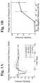
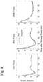
- FIGS. 1A-1BVelocity observed at the therapy focus following Histotripsy pulses of various focal pressures (left), along with a plot of the average peak velocity observed for each of the tested pressures (right) within the focal region in the agarose phantom using particle image velocimetry (PIV). Without cavitation generated by the Histotripsy pulse, no appreciable motion was detected. When cavitation occurred, the peak motion detected by PIV increased with increasing Histotripsy pulse pressure.
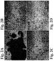
- FIGS. 2A-2DHigh speed images of the focal region 50 pulses into treatment (therapy applied from the right) with PIV velocity map overlays showing the Histotripsy bubble cloud (top left), chaotic motion immediately after the collapse of the bubble cloud (top right), and finally coherent motion, including a push away from the transducer (bottom left) and subsequent rebound (bottom right).
- FIG. 3describes the setup of the Histotripsy system and ultrasound imaging system to perform the bubble-induced color Doppler feedback for Histotripsy treatment.
- FIG. 4demonstrates one synchronization scheme to trigger the Doppler pulse transmission and acquisition using a signal sent out from the Histotripsy system at an appropriate delay time (negative or positive) after the transmission of the Histotripsy pulse.
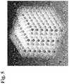
- FIG. 5Steered focal locations for the 219 foci with alternating 1 mm spaced grids of 7 ⁇ 7 foci and 6 ⁇ 6 foci.
- the axial layersare separated by 1 mm, but with the 6 ⁇ 6 grids offset laterally from the 7 ⁇ 7 layers by 0.5 mm
- FIG. 6Experimental setup with 500 kHz transducer mounted to the side of a water tank with 5 MHz imaging probe mounted opposite the therapy and aligned along the therapy axis.
- the Phantom high speed camerawas positioned perpendicular to the therapy axis.
- FIGS. 7A-7BPlots showing the velocity estimates from PIV (top) and Doppler (bottom) after every 10 therapy pulses.
- FIG. 8Individual velocity plots for the 19 ms after the therapy pulse after 10 therapy pulses (left), 30 therapy pulses (center), and 290 therapy pulses (right) showing good agreement between PIV and Doppler in measured velocity after the initial chaotic motion.
- FIG. 9Doppler velocity progression at a 6 ms delay from therapy pulse without averaging (left) and with a 10 pulse running average (right).
- FIG. 11Alternative progression metric, time to peak velocity, shows less variation and captures the same rapid change up to 100 pulses shown in the mean lesion intensity, with a slower continued progression up to 200 pulses.
- FIG. 12Plot showing the velocity estimates from Doppler after every therapy pulse in ex vivo porcine liver.
- FIG. 13Doppler velocity progression in ex vivo liver without averaging (left) and with a 10 point running average (right).
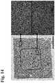
- FIG. 14Histological images of the lesion after 50 therapy pulses. Macroscopic image (left) shows little large-scale homogenization, however widespread mechanical fractionation is visible microscopically (bottom right) compared to control (top right).
- FIG. 15Histological images of the lesion after 200 therapy pulses. Macroscopic image (left) shows clear large-scale homogenization, with increased mechanical fractionation visible microscopically (bottom right) compared to control (top right).
- FIG. 16Histological images of the lesion after 500 therapy pulses. Macroscopic image (left) shows complete large-scale homogenization, with near complete homogenization visible microscopically as well (bottom right) compared to control (top right).
- This disclosureintroduces new imaging feedback systems and methods using bubble-induced color Doppler to monitor the Histotrispy-induced tissue fractionation in real-time.
- This novel approachcan monitor the level of tissue fractionation generated by Histotripsy with improved sensitivity compared to backscatter speckle amplitude reduction and can be implemented in real-time during Histotripsy treatment. Further areas of applicability will become apparent from the description provided herein. The description and specific examples in this summary are intended for purposes of illustration only and are not intended to limit the scope of the present disclosure.
- first, second, third, etc.may be used herein to describe various elements, components, regions, layers and/or sections, these elements, components, regions, layers and/or sections should not be limited by these terms. These terms may be only used to distinguish one element, component, region, layer or section from another region, layer or section. Terms such as “first,” “second,” and other numerical terms when used herein do not imply a sequence or order unless clearly indicated by the context. Thus, a first element, component, region, layer or section discussed below could be termed a second element, component, region, layer or section without departing from the teachings of the example embodiments.
- Spatially relative termssuch as “inner,” “outer,” “beneath,” “below,” “lower,” “above,” “upper,” and the like, may be used herein for ease of description to describe one element or feature's relationship to another element(s) or feature(s) as illustrated in the figures. Spatially relative terms may be intended to encompass different orientations of the device in use or operation in addition to the orientation depicted in the figures. For example, if the device in the figures is turned over, elements described as “below” or “beneath” other elements or features would then be oriented “above” the other elements or features. Thus, the example term “below” can encompass both an orientation of above and below. The device may be otherwise oriented (rotated 90 degrees or at other orientations) and the spatially relative descriptors used herein interpreted accordingly.
- FIGS. 1A-1Bshow a plot of the average velocity profile after each Histotripsy pulse along with the average peak velocity observed for each of the tested focal pressures.
- FIG. 1Aillustrates the velocity observed at the therapy focus following Histotripsy pulses of various focal pressures
- FIG. 1Bshows a plot of the average peak velocity observed for each of the tested pressures within the focal region in the agarose phantom using particle image velocimetry (PIV).
- PIVparticle image velocimetry
- a cavitation bubble cloudwas generated immediately and collapsed within 300 ⁇ S. Residual bubble, nuclei persist for over 100 ms after the cavitation collapse and were clearly visible in high-speed optical images of the focal region after a Histotripsy therapy pulse.
- PIV velocity mapsshowed 2 phases of motion during the 19 ms after a Histotripsy therapy pulse.
- chaotic motionwas present, where the motion was pointed in all directions in a random manner through this period.
- This chaotic motion phaselikely resulted from the violent collapse of the Histotripsy bubble cloud.
- a coherent motion along the direction of the therapy ultrasound beamwas visible. The coherent motion was first moving away from the therapy transducer for up to 6 ms, and then rebounding back towards the therapy transducer through the remaining 19 ms.
- FIGS. 2A-2Dshow images of an example progression of the focal region PIV velocity map after the tissue had been treated with 50 Histotripsy pulses. The therapy pulse was propagated from right to left.
- FIGS. 2A-2Dhigh speed images of the focal region are shown approximately 50 pulses into treatment (therapy applied from the right on the page) with PIV velocity map overlays showing the Histotripsy bubble cloud ( FIG. 2A ), chaotic motion immediately after the collapse of the bubble cloud ( FIG. 2B ), and finally coherent motion, including a push away from the transducer ( FIG. 2C ) and subsequent rebound ( FIG. 2D ).
- the time profile of the resulting velocity of the coherent motionexpands as the tissue is fractionated and saturate when the tissue is completely liquefied.
- the averaged velocity within a specific time window of the coherent motionincreases with increasing degree of tissue fractionation, and saturates when the clot is completely liquefied.
- the velocity resulting from the coherent motioncan be detected by ultrasound color Doppler that uses the cross-correlation time/phase lag of adjacent frames to detect the target motion.
- color Dopplercan be used to monitor the coherent motion in the Histotripsy treatment region.
- color Dopplercan be used to monitor the coherent motion phase without the interference from the chaotic motion.
- the Doppler velocitycan be then analyzed to quantitatively predict the level tissue fractionation during the treatment in real-time.
- the Doppler velocity mapcan also be displayed it as a colored region overlaid on the gray-scale image, providing real-time imaging feedback to monitor Histotripsy tissue fractionation.
- B-Flow and M-mode approachesare possible alternatives to color Doppler.
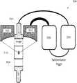
- FIG. 3illustrates a Doppler monitoring Histotripsy system 300 including an ultrasound therapy transducer 302 , a Doppler imaging transducer or transducers 304 (shown as 304 a and 304 b ), Histotripsy therapy driving hardware 306 (which can include, for example, a pulse generator, amplifiers, matching networks, and an electronic controller configured to generate Histotripsy pulses in the ultrasound therapy transducer), and imaging hardware 308 which can control Doppler imaging with the Doppler imaging transducer(s) 304 .
- the Doppler imaging transducerscan be disposed within a cut-out or hole within the ultrasound therapy transducer, for example, so as to facilitate imaging of a focus 310 (and thus the bubble cloud) of the therapy transducer.
- the tissue velocity along the axial direction or the propagation direction of the ultrasound pulseis monitored using color Doppler.
- Thiscan be achieved by placing the ultrasound imaging transducer in-line with the therapy transducer, for example, the Histotripsy therapy transducer can have a central hole to house the ultrasound imaging transducer to ensure the imaging transducer is monitoring the axial displacement of the tissue along the propagation direction of the Histotripsy pulse.
- the synchronization of the Histotripsy system and Doppler acquisition of the ultrasound imaging systemis essential and can be achieved by triggering Doppler pulse transmission from the Doppler imaging transducer(s) using a signal sent out from the Histotripsy therapy driving hardware 306 at an appropriate delay time (negative or positive) after the transmission of the Histotripsy pulse. It is also possible to trigger the Histotripsy therapy driving hardware with a signal from the imaging hardware 308 .
- FIG. 4demonstrates a synchronization scheme according to one embodiment.
- An appropriate delayneeds to be set between the Histotripsy pulse and the Doppler pulse transmission, such that the Doppler velocity measures the coherent motion phase, not the chaotic motion immediately following the Histotrispy pulse.
- the chaotic motion phaseranges from 300 us to 2 ms, depending on the tissue type and the level of tissue fractionation.
- the time profile of the Doppler velocityexpanded with a very steep slope for the first 50 pulses. After that, the expansion of the temporal profile of the Doppler was much more gradual, until at 900 pulses, the expansion saturates.
- microscopic cellular damagei.e., cell death
- macroscopic tissue structure homogenizationi.e., tissue liquefaction
- Current B-mode ultrasound imagingis not sensitive enough to monitor the microscopic cellular damage alone.
- the bubble-induced color Dopplerhas improved sensitivity to detect the microscopic cellular death as well as damage to the macroscopic tissue structure. This improved sensitivity can dramatically increase the treatment efficiency.
- treatment completioncan be determined in real-time for different clinical applications. For example, macroscopic tissue liquefaction is needed for clot removal, while cell death may be sufficient for tumor treatment and benign lesions. This feature is innovative and of clinical importance, and is not available for any current feedback techniques.
- the amplitude of bubble-induced color Doppler changes over the Histotripsy treatmentmay vary across different organs and patients.
- Our datasuggest that the slope or the rate of Doppler velocity change, either the temporal profile of the velocity within sub-time window of the coherent motion, can be used to monitor the treatment, to detect microscopic cellular damage as well as macroscopic tissue structure homogenization. Therefore, the detection does not depend on the absolute value of the Doppler velocity, but the relative change, and therefore is expected to be consistent and reliable across different organs and patients.
- the Doppler parameterssuch as the pulse repetition frequency (PRF) and number of frames for each Doppler acquisition, can be selected appropriately to achieve the desired correlation between the Doppler velocity increase with the increasing degree of tissue fractionation (i.e., Histotripsy treatment progress) in different tissue types.
- PRFpulse repetition frequency
- the color Doppler velocity map regioncan precisely match the fractionation region.
- Doppler parameterse.g., time window of the Doppler acquisition
- the average Doppler velocityis towards the transducer prior to treatment completion shown as one color (e.g., blue)
- the Doppler flowis away from the transducer at the treatment completion viewed as a different color (e.g., red).
- a definitive indication for treatment completionis apparent to even inexperienced users. This can be achieved because the temporal profiles of the coherent motion away from the transducer and back towards the transducer expand with the degree of tissue fractionation.
- these residual nucleiprovide bright speckle to track the bubble-induced motion in the tissue during Histotripsy treatment. They provide strong speckles for displacement the motion tracking, even with poor imaging quality.
- Doppleris an essential tool for monitoring cardiovascular function
- the capability of color Doppler during Histotripsy treatmentallows us to monitor the vessel and cardiac function during the treatment, which could have significant clinical implications.
- Histotripsycan be used to remove blood clots in the vessel and color Doppler can evaluate whether the blood flow is restored or improved during the Histotripsy treatment in a previously completely or partially occluded vessel.
- Histotripsyhas also been studied to create a flow channel through the atrial septum between the two atria in the heart for patients with congenital heart disease.
- color Dopplercan indicate the generation of the flow channel, i.e., treatment completion.
- color Dopplercan be used to ensure no penetration is generated to the vessel during the Histotripsy treatment.
- Different colorscan be used for bubble-induced color Doppler feedback during Histotripsy (e.g., green and yellow) to distinguish from blue and red commonly used in color Doppler for blood flow.
- the bubble-induced color Dopplercannot be used directly to form an image of a large volume, as the Histotripsy pulse is used to treat one focal volume at a time. It is possible to steer the therapy transducer focus (electronically or mechanically) over the large ablated volume and collect the data to reconstruct the 2D/3D image of the ablated volume.
- the ablated tissuecoagulates quickly after treatment, which may change the elasticity of the treated volume after treatment. If bubble-induced color Doppler will be used for post-treatment lesion evaluation when the tissue is coagulated, we can develop a quick ablation scan sequence to re-fractionate the coagulative tissue prior to the elastography measurement.
- This phantomwas treated with 2-cycle pulses at >50 MPa over a 6 mm cube using the same 500 kHz phased array transducer.
- This high pressureguaranteed the generation of a cavitation cloud, and the residual bubble nuclei left after its collapse for optical and acoustic contrast at the focal location.
- 219 focal points at 1 mm separations( FIG. 5 ) were treated sequentially at 150 Hz with a single pulse applied at each location. This process was repeated every 1.5 seconds until all focal locations had been treated with 960 pulses each.
- This pulsing strategyguarantees uniform therapy dose over the treatment volume at all times during treatment.
- FIG. 5219 focal points at 1 mm separations
- steered focal locations for the 219 fociare shown with alternating 1 mm spaced grids of 7 ⁇ 7 foci and 6 ⁇ 6 foci.
- the axial layersare separated by 1 mm, but with the 6 ⁇ 6 grids offset laterally from the 7 ⁇ 7 layers by 0.5 mm.
- the internal memory of the high-speed cameramay not be able to accommodate acquisitions after every pulse, so to facilitate continuous treatment without interruptions for data transfer; images can be captured periodically (e.g., after every 10 th pulse delivered to the center focal location).
- Ultrasound Doppler acquisitionscan be performed after every therapy pulse.
- the imaging transducercan be positioned opposite the therapy transducer, as described above, aligned along the therapy axis, i.e., the ultrasound imaging beam can be rigidly aligned with the therapy beam to avoid the effect of angle variation on Doppler.
- FIG. 6An experimental setup is illustrated in FIG. 6 , which shows a therapy transducer 602 , a Doppler imaging transducer 604 , ultrasound control system 606 , and imaging control system 608 .
- the experimental setupalso shows the tissue phantom disposed in a water tank and a high speed camera and light source for additional imaging capabilities.
- the experimental setupincluded a 500 kHz transducer mounted to the side of a water tank with 5 MHz imaging probe mounted opposite the therapy and aligned along the therapy axis.
- the high speed camerawas positioned perpendicular to the therapy axis.
- a tissue-mimicking agarose gel phantom with an embedded red blood cell (RBC) layerwas used to visualize and quantify the development of the lesion.
- RBCred blood cell
- These phantomshave been shown to produce reliable estimates of the cavitation-induced damage zone resulting from Histotripsy therapy.
- the RBC area lysed by Histotripsychanged from opaque red to translucent pink, allowing direct visualization and quantification of the lesion development.
- the lesionswere photographed during treatment after each application of the 219 focal patterns. Simultaneous ultrasound Doppler acquisitions were also performed for direct comparison. The average pixel intensity within the lesion was then computed over the course of the treatment as a direct quantification of the fractionation progression in the tissue phantom and compared to the measured Doppler velocity progression.
- the liverwas treated with 2000 pulses at each of the 219 focal locations, with ultrasound Doppler acquisitions performed after every pulse delivered to the center focal location. High-speed optical imaging for PIV analysis was not possible in the tissue.
- the high-speed optical images of the focal regionwere processed to estimate the motion resulting from the Histotripsy therapy pulses.
- the PIV analysiswas performed in a ⁇ 1.7 by 0.85 mm field of view at a resolution of 151 pixels per mm (total 256 ⁇ 128 pixels at 50 kHz frame rate) for the glass bead layer experiments and ⁇ 6.6 by 3.3 mm field of view at a resolution of 116 pixels per mm (total 768 ⁇ 384 pixels at 10 kHz frame rate) for the large lesion treatments.
- the imageswere processed in pairs at two subsequent time points using a FFT window deformation algorithm with 3 pass velocity estimation with image block sizes and step sizes of 24/12 pixels for pass 1, 16/8 pixels for pass 2, and 8/4 pixels for pass 3 in the glass bead layer experiment and 64/32 pixels for pass 1, 32/16 pixels for pass 2, and 16/8 pixels for pass 3 in the larger lesion treatments. Both resulted in velocity field maps of the field of view over the 19 ms after a Histotripsy therapy pulse. The axial components of these PIV velocity maps were then averaged over the bubble cloud area to produce the final average velocity estimate over time.
- the ultrasound Doppler acquisitionswere also processed. To calculate the velocity over the 19 ms after the therapy pulse, the 200 acquisitions were processed in rolling 10 acquisition segments (equivalent to using 10 frames at 10 kHz PRF, with different delay times after the histotrispy pulse). These Doppler velocity maps were then averaged over the 2 ⁇ 4 mm bubble cloud area to produce the final average velocity estimate over time.
- FIGS. 7A-7BThe full velocity profiles over the 960 pulse treatments are shown in FIGS. 7A-7B for both PIV ( FIG. 7A ) and Doppler ( FIG. 7B ) estimation methods.
- the estimated velocityis shown versus the delay from the therapy pulse (y axis) and the therapy dose (x axis).
- FIG. 8shows 3 individual velocity traces after 10, 30, and 290 therapy pulses.
- PIV and Doppler estimatesagree with each other well over the course of treatment. These plots show a time expansion of the velocity profile with increased therapy dose, which is likely due to the elasticity decrease as the phantom when it was gradually fractionated by Histotripsy pulses. The increase in the duration of the coherent push and rebound motion reached a peak after 400 pulses, likely because the phantom was completely liquefied.
- FIG. 8shows good agreement between PIV and Doppler in measured velocity after the initial chaotic motion.
- the velocity progression at any single delay between the Histotrispy pulse and the Doppler pulse packetcan be extracted from this dataset, producing the average velocity within a 1 ms window over the course of therapy. These velocity progressions are readily attainable in real-time from color Doppler during Histotripsy therapy, with an average processing frame rate around 30 Hz.
- the Doppler velocity progression at 6 ms delay from the Histotripsy pulseis shown in FIG. 9 . In this case, the Doppler measurement estimates the average velocity during the time window from 6-7 ms after the therapy pulse.
- FIG. 9shows Doppler velocity progression at a 6 ms delay from therapy pulse without averaging (left) and with a 10 pulse running average (right).
- RBCred blood cell
- This Doppler velocity metricis obtainable in real-time at high frame rates (up to 200 Hz) during Histotripsy therapy, however if high frame rates are not required, alternative metrics are also possible.
- the time to peak velocity shown in FIG. 11also captures the same rapid change up to 100 pulses observed in the lesion intensity, and also the continued slow increase up to 200 pulses.
- an alternative progression metric, time to peak velocityshows less variation and captures the same rapid change up to 100 pulses shown in the mean lesion intensity, with a slower continued progression up to 200 pulses.
- FIG. 12shows the full velocity profile over the entire treatment. The estimated velocity is shown versus the delay from the therapy pulse (y axis) and therapy dose (x axis).
- the Doppler velocity profiles in the ex vivo porcine liverwere similar to the agarose phantom, with a brief period of chaotic motion followed by coherent motion. These coherent motions also expanded in time with increased therapy dose very rapidly up to 50 pulses. After 50 pulses, the temporal profile of the Doppler velocity continued to expand at a slower rate until 900 pulses. After that point, the temporal profile of the Doppler velocity decreased slowly with increasing number of therapy pulses.
- the velocity progression at a single delay of 8 mswas extracted from this dataset, producing the average velocity during the 8-9 ms window over the course of therapy. This is shown in FIG. 13 .
- the velocityincreased quickly for the first 50 pulses, and then steadily at a slower rate up to 900 pulses as the tissue was fractionated. After 900 pulses, the velocity decreased steadily with increased variation from pulse to pulse.
- Doppler velocity progression in ex vivo liveris shown without averaging (left) and with a 10 point running average (right).
- FIG. 14shows the lesion resulting from a 50 pulse treatment, with widespread mechanical disruption of the cellular structures visible microscopically in the entire treatment region. This widespread microscopic cellular damage is sufficient to cause tissue death. No macroscopic homogenization of the tissue structure was visible after 50 pulses.
- Histological images of the lesion after 50 therapy pulsesare shown. The macroscopic image (left) shows little large-scale homogenization, however widespread mechanical fractionation is visible microscopically (bottom right) compared to control (top right).
- FIG. 15histological images of the lesion are shown after 200 therapy pulses. Macroscopic image (left) shows clear large-scale homogenization, with increased mechanical fractionation visible microscopically (bottom right) compared to control (top right).
- the lesionappears homogeneous and completely fractionated, with very few remaining cell nuclei in the homogenous fractionated tissue product.
- histological images of the lesionare shown after 500 therapy pulses.
- the macroscopic imageshows complete large-scale homogenization, with near complete homogenization visible microscopically as well (bottom right) compared to control (top right).
- the initial rapid expansion in the temporal profile of the Doppler velocitymatch well with the microscopic cellular damage to the treated tissue, both were observed at 50 pulses. After that, the temporal profile of the Doppler velocity continues to expand, but the rate is more gradual. Correspondingly, macroscopic damage to the tissue structure is observed. When the tissue is completely liquefied with no tissue or cellular structures remaining, the temporal profile of the Doppler velocity flattens and begins to shrink at a very slow rate.
- the bubble-induced color Dopplerprovides a real-time, high sensitivity feedback to monitor Histotripsy tissue fractionation during treatment.
- the bubble-induced color Doppler feedbackcan predict microscopic cellular damage, especially at an earlier treatment stage, which cannot be achieved with reduced echogenicity.
- bubble-induced color Dopplerhas the potential to predict microscopic cellular damage versus the macroscopic damage to the tissue structure. This level of sensitivity is very important for clinical application to predict the end point for treatment for different clinical applications.
- the bubble-induced color Dopplercan provide consistent and reliable feedback across different tissues and patients.
- Our datasuggest that the slope or the rate of Doppler velocity change, either the temporal profile of the velocity within sub-time window of the coherent motion, can be used to monitor the treatment, to detect microscopic cellular damage as well as macroscopic tissue structure homogenization.
- the detectiondoes not depend on the absolute value of the Doppler velocity that may vary across patients, but the relative change, and therefore is expected to be consistent and reliable across different organs and patients.
- the bubble-induced color Dopplercan be displayed as color overlaid on gray-scale ultrasound images, providing a high contrast feedback to monitor the degree of tissue fractionation (i.e., treatment progress and completion). Such feedback is unambiguous and easy to use even for inexperienced users.
- an ultrasound imaging transducercan be placed in-line (or co-axially) with the Histotripsy therapy transducer.
- such configurationcan be achieved by having a small center hole in the therapy transducer to house the imaging probe.
- the Doppler acquisition on the ultrasound imaging systemneeds to be synchronized by the Histotripsy therapy pulse such that the first Doppler pulse arrives at the focus at a predefined delay time after the arrival of the Histotripsy pulse.
- the real-time bubble-induced color Dopplershould also allow evaluation of the vessel or the heart close to the treatment target during the Histotripsy treatment.
- Different colorscan be used for tissue motion (e.g., green and yellow) to distinguish from the red and blue commonly used in color Doppler for blood flow.
- the ultrasound gray-scale imaging quality of deep tissueis often degraded significantly due to the attenuation and aberration from the overlying tissue, resulting in coarse tissue speckle and making the accurate tissue motion tracking difficult.
- the residual nuclei from bubble cloud generated by Histotripsylast over 100 milliseconds after each Histotripsy pulse and moves with the target tissue, providing strong ultrasound speckles for motion tracking during bubble-induced color Doppler.
Landscapes
- Health & Medical Sciences (AREA)
- Life Sciences & Earth Sciences (AREA)
- Engineering & Computer Science (AREA)
- Physics & Mathematics (AREA)
- Surgery (AREA)
- Radar, Positioning & Navigation (AREA)
- Remote Sensing (AREA)
- Biomedical Technology (AREA)
- Medical Informatics (AREA)
- Nuclear Medicine, Radiotherapy & Molecular Imaging (AREA)
- Veterinary Medicine (AREA)
- Public Health (AREA)
- General Health & Medical Sciences (AREA)
- Animal Behavior & Ethology (AREA)
- Molecular Biology (AREA)
- Heart & Thoracic Surgery (AREA)
- Pathology (AREA)
- Biophysics (AREA)
- Acoustics & Sound (AREA)
- Radiology & Medical Imaging (AREA)
- Computer Networks & Wireless Communication (AREA)
- General Physics & Mathematics (AREA)
- Vascular Medicine (AREA)
- Orthopedic Medicine & Surgery (AREA)
- Mechanical Engineering (AREA)
- Spectroscopy & Molecular Physics (AREA)
- Ultra Sonic Daignosis Equipment (AREA)
- Surgical Instruments (AREA)
Abstract
Description
Claims (6)
Priority Applications (1)
| Application Number | Priority Date | Filing Date | Title |
|---|---|---|---|
| US15/713,441US11058399B2 (en) | 2012-10-05 | 2017-09-22 | Bubble-induced color doppler feedback during histotripsy |
Applications Claiming Priority (3)
| Application Number | Priority Date | Filing Date | Title |
|---|---|---|---|
| US201261710172P | 2012-10-05 | 2012-10-05 | |
| US14/046,024US20140100459A1 (en) | 2012-10-05 | 2013-10-04 | Bubble-induced color doppler feedback during histotripsy |
| US15/713,441US11058399B2 (en) | 2012-10-05 | 2017-09-22 | Bubble-induced color doppler feedback during histotripsy |
Related Parent Applications (1)
| Application Number | Title | Priority Date | Filing Date |
|---|---|---|---|
| US14/046,024ContinuationUS20140100459A1 (en) | 2012-10-05 | 2013-10-04 | Bubble-induced color doppler feedback during histotripsy |
Publications (2)
| Publication Number | Publication Date |
|---|---|
| US20180049719A1 US20180049719A1 (en) | 2018-02-22 |
| US11058399B2true US11058399B2 (en) | 2021-07-13 |
Family
ID=50433233
Family Applications (2)
| Application Number | Title | Priority Date | Filing Date |
|---|---|---|---|
| US14/046,024AbandonedUS20140100459A1 (en) | 2012-10-05 | 2013-10-04 | Bubble-induced color doppler feedback during histotripsy |
| US15/713,441ActiveUS11058399B2 (en) | 2012-10-05 | 2017-09-22 | Bubble-induced color doppler feedback during histotripsy |
Family Applications Before (1)
| Application Number | Title | Priority Date | Filing Date |
|---|---|---|---|
| US14/046,024AbandonedUS20140100459A1 (en) | 2012-10-05 | 2013-10-04 | Bubble-induced color doppler feedback during histotripsy |
Country Status (3)
| Country | Link |
|---|---|
| US (2) | US20140100459A1 (en) |
| EP (1) | EP2903688A4 (en) |
| WO (1) | WO2014055906A1 (en) |
Cited By (7)
| Publication number | Priority date | Publication date | Assignee | Title |
|---|---|---|---|---|
| US11701134B2 (en) | 2005-09-22 | 2023-07-18 | The Regents Of The University Of Michigan | Histotripsy for thrombolysis |
| US11980778B2 (en) | 2018-11-28 | 2024-05-14 | Histosonics, Inc. | Histotripsy systems and methods |
| WO2024163876A1 (en)* | 2023-02-03 | 2024-08-08 | Sciton, Inc. | Methods and systems for histotripsy |
| US12220602B2 (en) | 2015-06-24 | 2025-02-11 | The Regents Of The University Of Michigan | Histotripsy therapy systems and methods for the treatment of brain tissue |
| US12318636B2 (en) | 2022-10-28 | 2025-06-03 | Histosonics, Inc. | Histotripsy systems and methods |
| US12343568B2 (en) | 2020-08-27 | 2025-07-01 | The Regents Of The University Of Michigan | Ultrasound transducer with transmit-receive capability for histotripsy |
| US12350525B2 (en) | 2013-08-22 | 2025-07-08 | The Regents Of The University Of Michigan | Histotripsy using very short ultrasound pulses |
Families Citing this family (17)
| Publication number | Priority date | Publication date | Assignee | Title |
|---|---|---|---|---|
| WO2011022411A2 (en)* | 2009-08-17 | 2011-02-24 | Histosonics, Inc. | Disposable acoustic coupling medium container |
| JP5863654B2 (en) | 2009-08-26 | 2016-02-16 | リージェンツ オブ ザ ユニバーシティー オブ ミシガン | Micromanipulator control arm for therapeutic and image processing ultrasonic transducers |
| US9144694B2 (en) | 2011-08-10 | 2015-09-29 | The Regents Of The University Of Michigan | Lesion generation through bone using histotripsy therapy without aberration correction |
| US20140100459A1 (en) | 2012-10-05 | 2014-04-10 | The Regents Of The University Of Michigan | Bubble-induced color doppler feedback during histotripsy |
| EP3016594B1 (en) | 2013-07-03 | 2023-01-25 | Histosonics, Inc. | Histotripsy excitation sequences optimized for bubble cloud formation using shock scattering |
| WO2015003154A1 (en) | 2013-07-03 | 2015-01-08 | Histosonics, Inc. | Articulating arm limiter for cavitational ultrasound therapy system |
| JP6288996B2 (en)* | 2013-09-11 | 2018-03-07 | キヤノンメディカルシステムズ株式会社 | Ultrasonic diagnostic apparatus and ultrasonic imaging program |
| US10888307B2 (en)* | 2014-02-26 | 2021-01-12 | Koninklijke Philips N.V. | System for performing intraluminal coronary and method of operation thereof |
| US10470742B2 (en) | 2014-04-28 | 2019-11-12 | Covidien Lp | Systems and methods for speckle reduction |
| US10004610B2 (en)* | 2015-02-04 | 2018-06-26 | DePuy Synthes Products, Inc. | Non-invasive methods for modifying tissue to facilitate treatment |
| EP3675959B1 (en) | 2017-09-01 | 2024-11-27 | Dalhousie University | Transducer assembly for generating focused ultrasound |
| EP3549529A1 (en)* | 2018-04-05 | 2019-10-09 | Koninklijke Philips N.V. | Ultrasound imaging system and method |
| KR102124422B1 (en) | 2018-06-05 | 2020-06-18 | 한국과학기술연구원 | High-low intensity focused ultrasound treatment apparatus |
| RU2692220C1 (en)* | 2018-12-13 | 2019-06-21 | Федеральное государственное бюджетное образовательное учреждение высшего образования "Тамбовский государственный технический университет" (ФГБОУ ВО "ТГТУ") | Method of colour doppler mapping in endoscopic optical coherence tomography |
| US11813485B2 (en) | 2020-01-28 | 2023-11-14 | The Regents Of The University Of Michigan | Systems and methods for histotripsy immunosensitization |
| CN116419707A (en)* | 2020-09-11 | 2023-07-11 | 密歇根大学董事会 | Transcranial MR guided histotripsy system and method |
| DE102022202553A1 (en)* | 2022-03-15 | 2023-09-21 | Siemens Healthcare Gmbh | Determining movement of an object and therapy device |
Citations (379)
| Publication number | Priority date | Publication date | Assignee | Title |
|---|---|---|---|---|
| US3243497A (en) | 1964-12-11 | 1966-03-29 | Dynapower Systems Corp Of Cali | Universal support for electrotherapeutic treatment head |
| US3679021A (en) | 1970-03-25 | 1972-07-25 | Eg & G Inc | Acoustic pulse generating system |
| US4016749A (en) | 1973-07-05 | 1977-04-12 | Wachter William J | Method and apparatus for inspection of nuclear fuel rods |
| US4024501A (en) | 1975-09-03 | 1977-05-17 | Standard Oil Company | Line driver system |
| US4051394A (en) | 1976-03-15 | 1977-09-27 | The Boeing Company | Zero crossing ac relay control circuit |
| US4117446A (en) | 1974-11-28 | 1978-09-26 | Agence Nationale De Valorisation De La Recherche (A N V A R) | Devices for probing by ultrasonic radiation |
| EP0017382A1 (en) | 1979-03-20 | 1980-10-15 | THE GENERAL ELECTRIC COMPANY, p.l.c. | Ultrasonic imaging system |
| US4269174A (en) | 1979-08-06 | 1981-05-26 | Medical Dynamics, Inc. | Transcutaneous vasectomy apparatus and method |
| US4277367A (en) | 1978-10-23 | 1981-07-07 | Wisconsin Alumni Research Foundation | Phantom material and method |
| US4351038A (en) | 1979-12-31 | 1982-09-21 | Agence Nationale De Valorisation De La Recherche (Anvar) | Ultrasonic examination and imaging |
| GB2099582A (en) | 1980-02-08 | 1982-12-08 | Stanford Res Inst Int | Ultrasonic image methods and apparatus |
| US4406153A (en) | 1979-05-04 | 1983-09-27 | Acoustic Standards Corporation | Ultrasonic beam characterization device |
| DE3220751A1 (en) | 1982-06-02 | 1983-12-08 | Jörg Dr. 8022 Grünwald Schüller | Device for crushing concrements, especially renal calculi, in living human or animal bodies |
| US4440025A (en) | 1980-06-27 | 1984-04-03 | Matsushita Electric Industrial Company, Limited | Arc scan transducer array having a diverging lens |
| US4453408A (en) | 1981-03-09 | 1984-06-12 | William Clayman | Device for testing ultrasonic beam profiles |
| US4483345A (en) | 1981-08-08 | 1984-11-20 | Fujitsu Limited | Pressure measuring system with ultrasonic wave |
| JPS6080779A (en) | 1983-10-07 | 1985-05-08 | Matsushita Electric Ind Co Ltd | Magnetic field sensor |
| US4549533A (en) | 1984-01-30 | 1985-10-29 | University Of Illinois | Apparatus and method for generating and directing ultrasound |
| US4551794A (en) | 1983-09-21 | 1985-11-05 | Sven Sandell | Imitation candle with magnetic pendulum |
| US4550606A (en) | 1982-09-28 | 1985-11-05 | Cornell Research Foundation, Inc. | Ultrasonic transducer array with controlled excitation pattern |
| US4575330A (en) | 1984-08-08 | 1986-03-11 | Uvp, Inc. | Apparatus for production of three-dimensional objects by stereolithography |
| JPS61196718A (en) | 1985-02-22 | 1986-08-30 | 株式会社日立製作所 | Earth fault protection device |
| US4622972A (en) | 1981-10-05 | 1986-11-18 | Varian Associates, Inc. | Ultrasound hyperthermia applicator with variable coherence by multi-spiral focusing |
| US4625731A (en) | 1984-10-10 | 1986-12-02 | Picker International, Inc. | Ultrasonic image display mounting |
| US4641378A (en) | 1984-06-06 | 1987-02-03 | Raycom Systems, Inc. | Fiber optic communication module |
| US4669483A (en) | 1984-07-21 | 1987-06-02 | Dornier System Gmbh | Lithotripsy system having locating and orienting apparatus |
| DE3544628A1 (en) | 1985-12-17 | 1987-06-19 | Eisenmenger Wolfgang | DEVICE FOR MECHANICALLY ACOUSTIC CONNECTION OF PRESSURE SHAFTS, ESPECIALLY OF FOCUSED SHOCK WAVES TO THE BODY OF LIVING BEINGS |
| US4689986A (en) | 1985-03-13 | 1987-09-01 | The University Of Michigan | Variable frequency gas-bubble-manipulating apparatus and method |
| US4757820A (en) | 1985-03-15 | 1988-07-19 | Kabushiki Kaisha Toshiba | Ultrasound therapy system |
| US4791915A (en) | 1986-09-29 | 1988-12-20 | Dynawave Corporation | Ultrasound therapy device |
| US4819621A (en) | 1986-03-11 | 1989-04-11 | Richard Wolf Gmbh | Method for detection of cavitations during medical application of high sonic energy |
| US4829491A (en) | 1984-07-12 | 1989-05-09 | Siemens Aktiengesellschaft | Phased-array equipment |
| EP0320303A2 (en) | 1987-12-11 | 1989-06-14 | General Electric Company | Coherent beam formation |
| US4856107A (en) | 1987-04-28 | 1989-08-08 | Edap International | Acoustic filter for suppressing or attenuating the negative half-waves of an elastic wave and an elastic wave generator comprising such a filter |
| US4865042A (en) | 1985-08-16 | 1989-09-12 | Hitachi, Ltd. | Ultrasonic irradiation system |
| EP0332871A2 (en) | 1988-03-16 | 1989-09-20 | Dornier Medizintechnik Gmbh | Destruction of concretions by combined treatment |
| DE3817094A1 (en) | 1988-04-18 | 1989-11-30 | Schubert Werner | Coupling and adhesive device for shock wave treatment units |
| US4888746A (en) | 1987-09-24 | 1989-12-19 | Richard Wolf Gmbh | Focussing ultrasound transducer |
| US4890267A (en) | 1985-09-24 | 1989-12-26 | Hewlett-Packard Company | Switch matrix |
| US4922917A (en) | 1987-08-14 | 1990-05-08 | Edap International | Ultrasonic tissue characterization |
| US4938217A (en) | 1988-06-21 | 1990-07-03 | Massachusetts Institute Of Technology | Electronically-controlled variable focus ultrasound hyperthermia system |
| JPH02215451A (en) | 1989-02-17 | 1990-08-28 | Toshiba Corp | Calculus crushing device |
| EP0384831A2 (en) | 1989-02-21 | 1990-08-29 | Technomed International | Apparatus for selective destruction of cells including soft tissues and bones inside a living being by implosing of gas bubbles |
| US4957099A (en) | 1988-02-10 | 1990-09-18 | Siemens Aktiengesellschaft | Shock wave source for extracorporeal lithotripsy |
| US4973980A (en) | 1987-09-11 | 1990-11-27 | Dataproducts Corporation | Acoustic microstreaming in an ink jet apparatus |
| US4984575A (en) | 1987-04-16 | 1991-01-15 | Olympus Optical Co., Ltd. | Therapeutical apparatus of extracorporeal type |
| US4991151A (en) | 1987-04-28 | 1991-02-05 | Edap International | Elastic pulse generator having a desired predetermined wave form |
| US5014686A (en) | 1989-08-31 | 1991-05-14 | International Sonic Technologies | Phantom kidney stone system |
| USRE33590E (en) | 1983-12-14 | 1991-05-21 | Edap International, S.A. | Method for examining, localizing and treating with ultrasound |
| US5065751A (en) | 1990-01-03 | 1991-11-19 | Wolf Gerald L | Method and apparatus for reversibly occluding a biological tube |
| US5080101A (en) | 1983-12-14 | 1992-01-14 | Edap International, S.A. | Method for examining and aiming treatment with untrasound |
| US5091893A (en) | 1990-04-05 | 1992-02-25 | General Electric Company | Ultrasonic array with a high density of electrical connections |
| US5092336A (en) | 1989-02-08 | 1992-03-03 | Universite Paris Vii-Bureau De La Valorisation Et De Relations Industrielle | Method and device for localization and focusing of acoustic waves in tissues |
| US5097709A (en) | 1989-02-16 | 1992-03-24 | Hitachi, Ltd. | Ultrasonic imaging system |
| DE4012760A1 (en) | 1990-04-21 | 1992-05-07 | G M T I Ges Fuer Medizintechni | Ultrasonic Doppler method for gallstone lithography - uses analysis of Doppler frequency shift to detect velocity and calculating size of tracked particles |
| US5143074A (en) | 1983-12-14 | 1992-09-01 | Edap International | Ultrasonic treatment device using a focussing and oscillating piezoelectric element |
| US5150711A (en) | 1983-12-14 | 1992-09-29 | Edap International, S.A. | Ultra-high-speed extracorporeal ultrasound hyperthermia treatment device |
| US5158071A (en) | 1988-07-01 | 1992-10-27 | Hitachi, Ltd. | Ultrasonic apparatus for therapeutical use |
| US5158070A (en) | 1983-12-14 | 1992-10-27 | Edap International, S.A. | Method for the localized destruction of soft structures using negative pressure elastic waves |
| US5163421A (en) | 1988-01-22 | 1992-11-17 | Angiosonics, Inc. | In vivo ultrasonic system with angioplasty and ultrasonic contrast imaging |
| US5165412A (en) | 1990-03-05 | 1992-11-24 | Kabushiki Kaisha Toshiba | Shock wave medical treatment apparatus with exchangeable imaging ultrasonic wave probe |
| US5174294A (en) | 1988-10-26 | 1992-12-29 | Kabushiki Kaisha Toshiba | Shockwave treatment apparatus |
| US5209221A (en) | 1988-03-01 | 1993-05-11 | Richard Wolf Gmbh | Ultrasonic treatment of pathological tissue |
| US5215680A (en) | 1990-07-10 | 1993-06-01 | Cavitation-Control Technology, Inc. | Method for the production of medical-grade lipid-coated microbubbles, paramagnetic labeling of such microbubbles and therapeutic uses of microbubbles |
| US5222806A (en) | 1992-06-04 | 1993-06-29 | C. N. Burman Co. | Lamp |
| US5230340A (en) | 1992-04-13 | 1993-07-27 | General Electric Company | Ultrasound imaging system with improved dynamic focusing |
| US5295484A (en) | 1992-05-19 | 1994-03-22 | Arizona Board Of Regents For And On Behalf Of The University Of Arizona | Apparatus and method for intra-cardiac ablation of arrhythmias |
| WO1994006355A1 (en) | 1992-09-14 | 1994-03-31 | Coraje, Inc. | Apparatus and method for enhanced intravascular phonophoresis including dissolution of intravascular blockage and concomitant inhibition of restenosis |
| US5316000A (en) | 1991-03-05 | 1994-05-31 | Technomed International (Societe Anonyme) | Use of at least one composite piezoelectric transducer in the manufacture of an ultrasonic therapy apparatus for applying therapy, in a body zone, in particular to concretions, to tissue, or to bones, of a living being and method of ultrasonic therapy |
| JPH06197907A (en) | 1992-11-16 | 1994-07-19 | Siemens Ag | Therapeutic ultrasonic applicator |
| US5354258A (en) | 1992-01-07 | 1994-10-11 | Edap International | Ultra-high-speed extracorporeal ultrasound hyperthermia treatment method |
| JPH06304178A (en) | 1993-04-02 | 1994-11-01 | Siemens Ag | A therapeutic device for the treatment of pathological tissue by focused ultrasound |
| US5380411A (en) | 1987-12-02 | 1995-01-10 | Schering Aktiengesellschaft | Ultrasound or shock wave work process and preparation for carrying out same |
| US5409002A (en) | 1989-07-12 | 1995-04-25 | Focus Surgery Incorporated | Treatment system with localization |
| JPH07504339A (en) | 1992-03-10 | 1995-05-18 | シーメンス アクチエンゲゼルシヤフト | Tissue treatment method and treatment device using ultrasound |
| US5431621A (en) | 1984-11-26 | 1995-07-11 | Edap International | Process and device of an anatomic anomaly by means of elastic waves, with tracking of the target and automatic triggering of the shootings |
| US5435311A (en) | 1989-06-27 | 1995-07-25 | Hitachi, Ltd. | Ultrasound therapeutic system |
| US5469852A (en) | 1993-03-12 | 1995-11-28 | Kabushiki Kaisha Toshiba | Ultrasound diagnosis apparatus and probe therefor |
| US5490051A (en) | 1993-02-19 | 1996-02-06 | Messana; Joseph | Self-positioning lamp fixture with integrally formed unitary support structure |
| US5501655A (en) | 1992-03-31 | 1996-03-26 | Massachusetts Institute Of Technology | Apparatus and method for acoustic heat generation and hyperthermia |
| JPH0884740A (en) | 1994-09-16 | 1996-04-02 | Toshiba Corp | Treatment equipment |
| US5520188A (en) | 1994-11-02 | 1996-05-28 | Focus Surgery Inc. | Annular array transducer |
| JPH08131454A (en) | 1994-09-17 | 1996-05-28 | Toshiba Corp | Ultrasonic treatment device and ultrasonic irradiation device |
| US5523058A (en) | 1992-09-16 | 1996-06-04 | Hitachi, Ltd. | Ultrasonic irradiation apparatus and processing apparatus based thereon |
| US5524620A (en) | 1991-11-12 | 1996-06-11 | November Technologies Ltd. | Ablation of blood thrombi by means of acoustic energy |
| US5540909A (en) | 1994-09-28 | 1996-07-30 | Alliance Pharmaceutical Corp. | Harmonic ultrasound imaging with microbubbles |
| US5542935A (en) | 1989-12-22 | 1996-08-06 | Imarx Pharmaceutical Corp. | Therapeutic delivery systems related applications |
| US5558092A (en) | 1995-06-06 | 1996-09-24 | Imarx Pharmaceutical Corp. | Methods and apparatus for performing diagnostic and therapeutic ultrasound simultaneously |
| US5563346A (en) | 1994-02-21 | 1996-10-08 | Siemens Aktiengesellschaft | Method and device for imaging an object using a two-dimensional ultrasonic array |
| US5566675A (en) | 1995-06-30 | 1996-10-22 | Siemens Medical Systems, Inc. | Beamformer for phase aberration correction |
| US5573497A (en) | 1994-11-30 | 1996-11-12 | Technomed Medical Systems And Institut National | High-intensity ultrasound therapy method and apparatus with controlled cavitation effect and reduced side lobes |
| US5580575A (en) | 1989-12-22 | 1996-12-03 | Imarx Pharmaceutical Corp. | Therapeutic drug delivery systems |
| US5582578A (en) | 1995-08-01 | 1996-12-10 | Duke University | Method for the comminution of concretions |
| US5590657A (en) | 1995-11-06 | 1997-01-07 | The Regents Of The University Of Michigan | Phased array ultrasound system and method for cardiac ablation |
| EP0755653A1 (en) | 1995-07-27 | 1997-01-29 | Hewlett-Packard GmbH | Patient monitoring module |
| US5601526A (en) | 1991-12-20 | 1997-02-11 | Technomed Medical Systems | Ultrasound therapy apparatus delivering ultrasound waves having thermal and cavitation effects |
| JPH0955571A (en) | 1995-08-11 | 1997-02-25 | Hewlett Packard Japan Ltd | Electronic circuit board with high insulation section and its production |
| US5617862A (en) | 1995-05-02 | 1997-04-08 | Acuson Corporation | Method and apparatus for beamformer system with variable aperture |
| US5648098A (en) | 1995-10-17 | 1997-07-15 | The Board Of Regents Of The University Of Nebraska | Thrombolytic agents and methods of treatment for thrombosis |
| US5676692A (en) | 1996-03-28 | 1997-10-14 | Indianapolis Center For Advanced Research, Inc. | Focussed ultrasound tissue treatment method |
| US5676452A (en) | 1995-03-02 | 1997-10-14 | Gebr. Berchtold Gmbh & Co. | Operating lamp with main bulb and replacement bulb |
| US5678554A (en) | 1996-07-02 | 1997-10-21 | Acuson Corporation | Ultrasound transducer for multiple focusing and method for manufacture thereof |
| US5694936A (en) | 1994-09-17 | 1997-12-09 | Kabushiki Kaisha Toshiba | Ultrasonic apparatus for thermotherapy with variable frequency for suppressing cavitation |
| US5695460A (en) | 1994-09-09 | 1997-12-09 | Coraje, Inc. | Enhancement of ultrasound thrombolysis |
| US5717657A (en) | 1996-06-24 | 1998-02-10 | The United States Of America As Represented By The Secretary Of The Navy | Acoustical cavitation suppressor for flow fields |
| US5724972A (en) | 1996-05-02 | 1998-03-10 | Acuson Corporation | Method and apparatus for distributed focus control with slope tracking |
| US5753929A (en) | 1996-08-28 | 1998-05-19 | Motorola, Inc. | Multi-directional optocoupler and method of manufacture |
| US5766138A (en) | 1996-04-18 | 1998-06-16 | Siemens Aktiengesellschaft | Therapy apparatus with simple setting of a desired distance from a reference point |
| US5769790A (en) | 1996-10-25 | 1998-06-23 | General Electric Company | Focused ultrasound surgery system guided by ultrasound imaging |
| US5797848A (en) | 1997-01-31 | 1998-08-25 | Acuson Corporation | Ultrasonic transducer assembly with improved electrical interface |
| US5823962A (en) | 1996-09-02 | 1998-10-20 | Siemens Aktiengesellschaft | Ultrasound transducer for diagnostic and therapeutic use |
| US5827204A (en) | 1996-11-26 | 1998-10-27 | Grandia; Willem | Medical noninvasive operations using focused modulated high power ultrasound |
| US5836896A (en) | 1996-08-19 | 1998-11-17 | Angiosonics | Method of inhibiting restenosis by applying ultrasonic energy |
| JPH10512477A (en) | 1995-01-20 | 1998-12-02 | メデラ インコーポレイテッド | Apparatus and method for supporting a breast shield and associated pumping equipment |
| US5849727A (en) | 1996-06-28 | 1998-12-15 | Board Of Regents Of The University Of Nebraska | Compositions and methods for altering the biodistribution of biological agents |
| US5873902A (en) | 1995-03-31 | 1999-02-23 | Focus Surgery, Inc. | Ultrasound intensity determining method and apparatus |
| US5879314A (en) | 1997-06-30 | 1999-03-09 | Cybersonics, Inc. | Transducer assembly and method for coupling ultrasonic energy to a body for thrombolysis of vascular thrombi |
| US5932807A (en) | 1994-10-25 | 1999-08-03 | U.S. Philips Corporation | Device for the non-destructive testing of hollow tubular objects by means of ultrasound |
| US5947904A (en) | 1997-08-21 | 1999-09-07 | Acuson Corporation | Ultrasonic method and system for imaging blood flow including disruption or activation of a contrast agent |
| US6001069A (en) | 1997-05-01 | 1999-12-14 | Ekos Corporation | Ultrasound catheter for providing a therapeutic effect to a vessel of a body |
| US6022309A (en) | 1996-04-24 | 2000-02-08 | The Regents Of The University Of California | Opto-acoustic thrombolysis |
| US6036667A (en) | 1996-10-04 | 2000-03-14 | United States Surgical Corporation | Ultrasonic dissection and coagulation system |
| US6088613A (en) | 1989-12-22 | 2000-07-11 | Imarx Pharmaceutical Corp. | Method of magnetic resonance focused surgical and therapeutic ultrasound |
| US6093883A (en) | 1997-07-15 | 2000-07-25 | Focus Surgery, Inc. | Ultrasound intensity determining method and apparatus |
| US6113558A (en) | 1997-09-29 | 2000-09-05 | Angiosonics Inc. | Pulsed mode lysis method |
| US6126607A (en) | 1997-11-03 | 2000-10-03 | Barzell-Whitmore Maroon Bells, Inc. | Ultrasound interface control system |
| US6128958A (en) | 1997-09-11 | 2000-10-10 | The Regents Of The University Of Michigan | Phased array system architecture |
| JP2000300559A (en) | 1999-04-26 | 2000-10-31 | Olympus Optical Co Ltd | Ultrasonic probe and its manufacture |
| US6143018A (en) | 1993-05-14 | 2000-11-07 | Ceramoptec Gmbh | Method and device for thermally obliterating biological tissue |
| US6165144A (en) | 1998-03-17 | 2000-12-26 | Exogen, Inc. | Apparatus and method for mounting an ultrasound transducer |
| US6176842B1 (en) | 1995-03-08 | 2001-01-23 | Ekos Corporation | Ultrasound assembly for use with light activated drugs |
| US6308585B1 (en) | 2000-02-10 | 2001-10-30 | Ultra Sonus Ab | Method and a device for attaching ultrasonic transducers |
| US6308710B1 (en) | 1999-04-12 | 2001-10-30 | David Silva | Scrotal drape and support |
| US6309355B1 (en) | 1998-12-22 | 2001-10-30 | The Regents Of The University Of Michigan | Method and assembly for performing ultrasound surgery using cavitation |
| US20010039420A1 (en) | 1998-04-08 | 2001-11-08 | Senorx, Inc. | Tissue specimen isolating and damaging device and method |
| US20010041163A1 (en) | 2000-03-09 | 2001-11-15 | Nami Sugita | Sensitizer for tumor treatment |
| US6318146B1 (en) | 1999-07-14 | 2001-11-20 | Wisconsin Alumni Research Foundation | Multi-imaging modality tissue mimicking materials for imaging phantoms |
| US6321109B2 (en) | 1996-02-15 | 2001-11-20 | Biosense, Inc. | Catheter based surgery |
| US6338566B1 (en) | 1999-04-28 | 2002-01-15 | Alm | Flexible stop piece for limiting angular travel, articulated system comprising such a stop piece, and medical equipment comprising such an articulated system |
| US6344489B1 (en) | 1991-02-14 | 2002-02-05 | Wayne State University | Stabilized gas-enriched and gas-supersaturated liquids |
| US20020045890A1 (en) | 1996-04-24 | 2002-04-18 | The Regents Of The University O F California | Opto-acoustic thrombolysis |
| WO2002032506A1 (en) | 2000-10-20 | 2002-04-25 | Sunnybrook And Women"S College Health Sciences Centre, | Technique and apparatus for ultrasound therapy |
| US6391020B1 (en) | 1999-10-06 | 2002-05-21 | The Regents Of The Univerity Of Michigan | Photodisruptive laser nucleation and ultrasonically-driven cavitation of tissues and materials |
| US20020078964A1 (en) | 2000-10-09 | 2002-06-27 | American Medical Systems, Inc. | Pelvic surgery drape |
| US6419648B1 (en) | 2000-04-21 | 2002-07-16 | Insightec-Txsonics Ltd. | Systems and methods for reducing secondary hot spots in a phased array focused ultrasound system |
| US20020099356A1 (en) | 2001-01-19 | 2002-07-25 | Unger Evan C. | Transmembrane transport apparatus and method |
| US6470204B1 (en) | 1999-08-25 | 2002-10-22 | Egidijus Edward Uzgiris | Intracavity probe for MR image guided biopsy and delivery of therapy |
| US6490469B2 (en) | 2000-03-15 | 2002-12-03 | The Regents Of The University Of California | Method and apparatus for dynamic focusing of ultrasound energy |
| US6488639B1 (en) | 1998-05-13 | 2002-12-03 | Technomed Medical Systems, S.A | Frequency adjustment in high intensity focused ultrasound treatment apparatus |
| US6500141B1 (en) | 1998-01-08 | 2002-12-31 | Karl Storz Gmbh & Co. Kg | Apparatus and method for treating body tissue, in particular soft surface tissue with ultrasound |
| US6506171B1 (en) | 2000-07-27 | 2003-01-14 | Insightec-Txsonics, Ltd | System and methods for controlling distribution of acoustic energy around a focal point using a focused ultrasound system |
| US6506154B1 (en) | 2000-11-28 | 2003-01-14 | Insightec-Txsonics, Ltd. | Systems and methods for controlling a phased array focused ultrasound system |
| US6508774B1 (en) | 1999-03-09 | 2003-01-21 | Transurgical, Inc. | Hifu applications with feedback control |
| US6511428B1 (en) | 1998-10-26 | 2003-01-28 | Hitachi, Ltd. | Ultrasonic medical treating device |
| US6511444B2 (en) | 1998-02-17 | 2003-01-28 | Brigham And Women's Hospital | Transmyocardial revascularization using ultrasound |
| US6522142B1 (en) | 2001-12-14 | 2003-02-18 | Insightec-Txsonics Ltd. | MRI-guided temperature mapping of tissue undergoing thermal treatment |
| US6524251B2 (en) | 1999-10-05 | 2003-02-25 | Omnisonics Medical Technologies, Inc. | Ultrasonic device for tissue ablation and sheath for use therewith |
| JP2003510159A (en) | 1999-10-05 | 2003-03-18 | オムニソニクス メディカル テクノロジーズ インコーポレイテッド | Ultrasound therapy method and ultrasound therapy device for reducing prostate in particular |
| US6536553B1 (en) | 2000-04-25 | 2003-03-25 | The United States Of America As Represented By The Secretary Of The Army | Method and apparatus using acoustic sensor for sub-surface object detection and visualization |
| US6543272B1 (en) | 2000-04-21 | 2003-04-08 | Insightec-Txsonics Ltd. | Systems and methods for testing and calibrating a focused ultrasound transducer array |
| US6556750B2 (en) | 2000-05-26 | 2003-04-29 | Fairchild Semiconductor Corporation | Bi-directional optical coupler |
| US6559644B2 (en) | 2001-05-30 | 2003-05-06 | Insightec - Txsonics Ltd. | MRI-based temperature mapping with error compensation |
| US20030092982A1 (en) | 1999-08-12 | 2003-05-15 | Eppstein Jonathan A. | Microporation of tissue for delivery of bioactive agents |
| US20030112922A1 (en) | 2001-11-05 | 2003-06-19 | Computerized Medical Systems, Inc. | Apparatus and method for registration, guidance and targeting of external beam radiation therapy |
| US6599288B2 (en) | 2000-05-16 | 2003-07-29 | Atrionix, Inc. | Apparatus and method incorporating an ultrasound transducer onto a delivery member |
| US20030149352A1 (en) | 2002-02-04 | 2003-08-07 | Shen-Min Liang | Automatic stone-tracking system |
| US6607498B2 (en) | 2001-01-03 | 2003-08-19 | Uitra Shape, Inc. | Method and apparatus for non-invasive body contouring by lysing adipose tissue |
| US20030157025A1 (en) | 1995-06-07 | 2003-08-21 | Unger Evan C. | Novel methods of imaging and treatment with targeted compositions |
| US6613004B1 (en) | 2000-04-21 | 2003-09-02 | Insightec-Txsonics, Ltd. | Systems and methods for creating longer necrosed volumes using a phased array focused ultrasound system |
| US6613005B1 (en) | 2000-11-28 | 2003-09-02 | Insightec-Txsonics, Ltd. | Systems and methods for steering a focused ultrasound array |
| US6612988B2 (en) | 2000-08-29 | 2003-09-02 | Brigham And Women's Hospital, Inc. | Ultrasound therapy |
| US20030169591A1 (en) | 2002-03-11 | 2003-09-11 | Dennis Cochran | Underwater probe and illumination device |
| US20030181833A1 (en) | 2002-03-22 | 2003-09-25 | Fmd, Llc | Apparatus for extracorporeal shock wave lithotripter using at least two shock wave pulses |
| US6626854B2 (en) | 2000-12-27 | 2003-09-30 | Insightec - Txsonics Ltd. | Systems and methods for ultrasound assisted lipolysis |
| US6626855B1 (en) | 1999-11-26 | 2003-09-30 | Therus Corpoation | Controlled high efficiency lesion formation using high intensity ultrasound |
| US20030199857A1 (en) | 2002-04-17 | 2003-10-23 | Dornier Medtech Systems Gmbh | Apparatus and method for manipulating acoustic pulses |
| US6645162B2 (en) | 2000-12-27 | 2003-11-11 | Insightec - Txsonics Ltd. | Systems and methods for ultrasound assisted lipolysis |
| US6648839B2 (en) | 2002-02-28 | 2003-11-18 | Misonix, Incorporated | Ultrasonic medical treatment device for RF cauterization and related method |
| US20030221561A1 (en) | 1999-12-06 | 2003-12-04 | Simcha Milo | Ultrasonic medical device |
| US6666833B1 (en) | 2000-11-28 | 2003-12-23 | Insightec-Txsonics Ltd | Systems and methods for focussing an acoustic energy beam transmitted through non-uniform tissue medium |
| US20030236539A1 (en) | 1999-10-05 | 2003-12-25 | Omnisonics Medical Technologies, Inc. | Apparatus and method for using an ultrasonic probe to clear a vascular access device |
| EP1374785A1 (en) | 2002-06-26 | 2004-01-02 | Dornier MedTech Systems GmbH | Lithotripter with a doppler ultrasound unit for hit/miss monitoring |
| US6685657B2 (en) | 1998-11-20 | 2004-02-03 | Joie P. Jones | Methods for selectively dissolving and removing materials using ultra-high frequency ultrasound |
| US6685640B1 (en) | 1998-03-30 | 2004-02-03 | Focus Surgery, Inc. | Ablation system |
| JP2004505660A (en) | 2000-08-03 | 2004-02-26 | エル.アール. アールアンドディー リミテッド | System for enhanced chemical debridement |
| US6705994B2 (en) | 2002-07-08 | 2004-03-16 | Insightec - Image Guided Treatment Ltd | Tissue inhomogeneity correction in ultrasound imaging |
| US6719694B2 (en) | 1999-12-23 | 2004-04-13 | Therus Corporation | Ultrasound transducers for imaging and therapy |
| US6719449B1 (en) | 1998-10-28 | 2004-04-13 | Covaris, Inc. | Apparatus and method for controlling sonic treatment |
| JP2004512502A (en) | 2000-08-21 | 2004-04-22 | ヴイ−ターゲット テクノロジーズ リミテッド | Radiation radiation detector with position tracking system and its use in medical systems and procedures |
| US6735461B2 (en) | 2001-06-19 | 2004-05-11 | Insightec-Txsonics Ltd | Focused ultrasound system with MRI synchronization |
| US6736814B2 (en) | 2002-02-28 | 2004-05-18 | Misonix, Incorporated | Ultrasonic medical treatment device for bipolar RF cauterization and related method |
| US6750463B1 (en) | 2000-02-29 | 2004-06-15 | Hill-Rom Services, Inc. | Optical isolation apparatus and method |
| US20040127815A1 (en) | 1993-09-24 | 2004-07-01 | Transmedica International, Inc. | Removable tip for laser device |
| US20040138563A1 (en) | 2000-02-09 | 2004-07-15 | Moehring Mark A | Method and apparatus combining diagnostic ultrasound with therapeutic ultrasound to enhance thrombolysis |
| US6770031B2 (en) | 2000-12-15 | 2004-08-03 | Brigham And Women's Hospital, Inc. | Ultrasound therapy |
| US6775438B1 (en) | 1999-07-19 | 2004-08-10 | Thomson Licensing S.A. | Electrical insulation device with optocoupler for bidirectional connecting lines |
| US6788977B2 (en) | 2000-06-20 | 2004-09-07 | Celsion Corporation | System and method for heating the prostate gland to treat and prevent the growth and spread of prostate tumor |
| US6790180B2 (en) | 2001-12-03 | 2004-09-14 | Insightec-Txsonics Ltd. | Apparatus, systems, and methods for measuring power output of an ultrasound transducer |
| US6820160B1 (en) | 2001-08-21 | 2004-11-16 | Cypress Semiconductor Corporation | Apparatus for optically isolating a USB peripheral from a USB host |
| US20040236248A1 (en) | 1992-01-07 | 2004-11-25 | Pat Svedman | Transdermal perfusion of fluids |
| US20040243021A1 (en) | 2001-11-06 | 2004-12-02 | Murphy John C. | Device for thermal stimulation of small neural fibers |
| US20050020945A1 (en) | 2002-07-02 | 2005-01-27 | Tosaya Carol A. | Acoustically-aided cerebrospinal-fluid manipulation for neurodegenerative disease therapy |
| US6852082B2 (en) | 2002-07-17 | 2005-02-08 | Adam Strickberger | Apparatus and methods for performing non-invasive vasectomies |
| EP1504713A1 (en) | 2003-07-14 | 2005-02-09 | Surgical Navigation Technologies, Inc. | Navigation system for cardiac therapies |
| US20050038339A1 (en) | 2002-01-21 | 2005-02-17 | Sunita Chauhan | Ultrasonic treatment of breast cancer |
| US20050038361A1 (en) | 2003-08-14 | 2005-02-17 | Duke University | Apparatus for improved shock-wave lithotripsy (SWL) using a piezoelectric annular array (PEAA) shock-wave generator in combination with a primary shock wave source |
| WO2005018469A1 (en) | 2003-08-14 | 2005-03-03 | Duke University | Apparatus for improved shock-wave lithotripsy (swl) using a piezoelectric annular array (peaa) shock-wave generator in combination with a primary shock wave |
| US6869439B2 (en) | 1996-09-19 | 2005-03-22 | United States Surgical Corporation | Ultrasonic dissector |
| US6890332B2 (en) | 1999-05-24 | 2005-05-10 | Csaba Truckai | Electrical discharge devices and techniques for medical procedures |
| JP2005167058A (en) | 2003-12-04 | 2005-06-23 | Oval Corp | Explosion-proof insulated separation circuit |
| US20050154314A1 (en) | 2003-12-30 | 2005-07-14 | Liposonix, Inc. | Component ultrasound transducer |
| US20050152561A1 (en) | 2002-01-18 | 2005-07-14 | Spencer Michael E. | Modulator - amplifier |
| US6929609B2 (en) | 2001-01-18 | 2005-08-16 | Hitachi Medical Corporation | Ultrasonic diagnosing/treating device and method therefor |
| US20050234438A1 (en) | 2004-04-15 | 2005-10-20 | Mast T D | Ultrasound medical treatment system and method |
| US20050283098A1 (en) | 1998-02-06 | 2005-12-22 | Conston Stanley R | Method for ultrasound triggered drug delivery using hollow microbubbles with controlled fragility |
| US7004282B2 (en) | 2002-10-28 | 2006-02-28 | Misonix, Incorporated | Ultrasonic horn |
| US20060060991A1 (en) | 2004-09-21 | 2006-03-23 | Interuniversitair Microelektronica Centrum (Imec) | Method and apparatus for controlled transient cavitation |
| US20060074303A1 (en) | 2004-09-28 | 2006-04-06 | Minnesota Medical Physics Llc | Apparatus and method for conformal radiation brachytherapy for prostate gland and other tumors |
| US7059168B2 (en) | 2002-10-01 | 2006-06-13 | Olympus Corporation | Ultrasound phantom |
| US20060173387A1 (en) | 2004-12-10 | 2006-08-03 | Douglas Hansmann | Externally enhanced ultrasonic therapy |
| US20060206028A1 (en) | 2005-03-11 | 2006-09-14 | Qi Yu | Apparatus and method for ablating deposits from blood vessel |
| US20060241533A1 (en) | 2003-04-22 | 2006-10-26 | Benjamin Geller | Apparatus and method for treatment of damaged tissue |
| US20060241466A1 (en) | 1999-08-13 | 2006-10-26 | Point Biomedical Corporation | Hollow microspheres with controlled fragility for medical use |
| US20060241523A1 (en) | 2005-04-12 | 2006-10-26 | Prorhythm, Inc. | Ultrasound generating method, apparatus and probe |
| US7128711B2 (en) | 2002-03-25 | 2006-10-31 | Insightec, Ltd. | Positioning systems and methods for guided ultrasound therapy systems |
| US20060264760A1 (en) | 2005-02-10 | 2006-11-23 | Board Of Regents, The University Of Texas System | Near infrared transrectal probes for prostate cancer detection and prognosis |
| US20060293630A1 (en) | 2005-06-22 | 2006-12-28 | Misonix Incorporated | Fluid containment apparatus for surgery and method of use |
| US20070010805A1 (en) | 2005-07-08 | 2007-01-11 | Fedewa Russell J | Method and apparatus for the treatment of tissue |
| US20070016039A1 (en) | 2005-06-21 | 2007-01-18 | Insightec-Image Guided Treatment Ltd. | Controlled, non-linear focused ultrasound treatment |
| US7175596B2 (en) | 2001-10-29 | 2007-02-13 | Insightec-Txsonics Ltd | System and method for sensing and locating disturbances in an energy path of a focused ultrasound system |
| US20070044562A1 (en) | 2005-08-26 | 2007-03-01 | The Boeing Company | Rapid prototype integrated matrix ultrasonic transducer array inspection apparatus, systems, and methods |
| US20070065420A1 (en) | 2005-08-23 | 2007-03-22 | Johnson Lanny L | Ultrasound Therapy Resulting in Bone Marrow Rejuvenation |
| US7196313B2 (en) | 2004-04-02 | 2007-03-27 | Fairchild Semiconductor Corporation | Surface mount multi-channel optocoupler |
| US20070083120A1 (en) | 2005-09-22 | 2007-04-12 | Cain Charles A | Pulsed cavitational ultrasound therapy |
| US7223239B2 (en) | 2002-03-22 | 2007-05-29 | Ethicon Endo-Surgery, Inc. | Medical device that removably attaches to a bodily organ |
| JP2007144225A (en) | 1997-03-03 | 2007-06-14 | Toshiba Corp | Ultrasonic therapy device |
| US20070161902A1 (en) | 2004-02-06 | 2007-07-12 | Adam Dan | Localized production of microbubbles and control of cavitational and heating effects by use of enhanced ultrasound |
| US20070167764A1 (en) | 2005-11-15 | 2007-07-19 | Kullervo Hynynen | Impedance matching for ultrasound phased array elements |
| US7258674B2 (en) | 2002-02-20 | 2007-08-21 | Liposonix, Inc. | Ultrasonic treatment and imaging of adipose tissue |
| US20070205785A1 (en) | 2004-10-18 | 2007-09-06 | Mobile Robotics Sweden Ab | Robot for ultrasonic examination |
| US20070219448A1 (en) | 2004-05-06 | 2007-09-20 | Focus Surgery, Inc. | Method and Apparatus for Selective Treatment of Tissue |
| US7273459B2 (en) | 2003-03-31 | 2007-09-25 | Liposonix, Inc. | Vortex transducer |
| US7273458B2 (en) | 1998-01-12 | 2007-09-25 | Georgia Tech Research Corporation | Method of applying acoustic energy effective to alter transport or cell viability |
| US7300414B1 (en) | 1999-11-01 | 2007-11-27 | University Of Cincinnati | Transcranial ultrasound thrombolysis system and method of treating a stroke |
| US7311679B2 (en) | 2003-12-30 | 2007-12-25 | Liposonix, Inc. | Disposable transducer seal |
| US20080013593A1 (en) | 2006-06-21 | 2008-01-17 | Ken-Ichi Kawabata | Phantom |
| US20080033417A1 (en) | 2006-08-04 | 2008-02-07 | Nields Morgan W | Apparatus for planning and performing thermal ablation |
| US7331951B2 (en) | 2002-06-25 | 2008-02-19 | Ultrashape Inc. | Devices and methodologies useful in body aesthetics |
| US20080055003A1 (en) | 2006-09-06 | 2008-03-06 | Texas Instruments Incorporated | Reduction of voltage spikes in switching half-bridge stages |
| US7341569B2 (en) | 2004-01-30 | 2008-03-11 | Ekos Corporation | Treatment of vascular occlusions using ultrasonic energy and microbubbles |
| US7347855B2 (en) | 2001-10-29 | 2008-03-25 | Ultrashape Ltd. | Non-invasive ultrasonic body contouring |
| US20080082026A1 (en) | 2006-04-26 | 2008-04-03 | Rita Schmidt | Focused ultrasound system with far field tail suppression |
| US7358226B2 (en) | 2003-08-27 | 2008-04-15 | The Regents Of The University Of California | Ultrasonic concentration of drug delivery capsules |
| US7359640B2 (en) | 2003-09-30 | 2008-04-15 | Stmicroelectronics Sa | Optical coupling device and method for bidirectional data communication over a common signal line |
| US20080091125A1 (en) | 2006-10-13 | 2008-04-17 | University Of Washington | Method and apparatus to detect the fragmentation of kidney stones by measuring acoustic scatter |
| WO2008051484A2 (en) | 2006-10-19 | 2008-05-02 | Medela Holding Ag | System and device for supporting a breast shield |
| US7367948B2 (en) | 2002-08-29 | 2008-05-06 | The Regents Of The University Of Michigan | Acoustic monitoring method and system in laser-induced optical breakdown (LIOB) |
| US7374551B2 (en) | 2003-02-19 | 2008-05-20 | Pittsburgh Plastic Surgery Research Associates | Minimally invasive fat cavitation method |
| US7377900B2 (en) | 2003-06-02 | 2008-05-27 | Insightec - Image Guided Treatment Ltd. | Endo-cavity focused ultrasound transducer |
| US20080126665A1 (en) | 2006-09-19 | 2008-05-29 | Kent Allan Burr | Apparatus and methods to communicatively couple field devices to controllers in a process control system |
| US20080177180A1 (en) | 2004-08-17 | 2008-07-24 | Technion Research & Development | Ultrasonic Image-Guided Tissue-Damaging Procedure |
| US20080194965A1 (en) | 2007-02-08 | 2008-08-14 | Sliwa John W | Device and method for high intensity focused ultrasound ablation with acoustic lens |
| US20080214964A1 (en) | 2005-03-15 | 2008-09-04 | Edap S.A. | Therapeutic Endocavity Probe Comprising an Image Transducer Integrated Within the Therapy Ultrasonic Transducer |
| US7429249B1 (en) | 1999-06-14 | 2008-09-30 | Exogen, Inc. | Method for cavitation-induced tissue healing with low intensity ultrasound |
| US7431704B2 (en) | 2006-06-07 | 2008-10-07 | Bacoustics, Llc | Apparatus and method for the treatment of tissue with ultrasound energy by direct contact |
| US20080262345A1 (en) | 2003-07-21 | 2008-10-23 | The John Hopkins University | Image registration of multiple medical imaging modalities using a multiple degree-of-freedom-encoded fiducial device |
| US20080262486A1 (en) | 2000-07-31 | 2008-10-23 | Galil Medical Ltd. | Planning and facilitation systems and methods for cryosurgery |
| US7442168B2 (en) | 2002-04-05 | 2008-10-28 | Misonix, Incorporated | High efficiency medical transducer with ergonomic shape and method of manufacture |
| US7462488B2 (en) | 2002-10-04 | 2008-12-09 | Wisconsin Alumni Research Foundation | Tissue mimicking elastography phantoms |
| US20080312561A1 (en) | 2004-05-06 | 2008-12-18 | Nanyang Technological University | Mechanical Manipulator for Hifu Transducers |
| US20080319356A1 (en)* | 2005-09-22 | 2008-12-25 | Cain Charles A | Pulsed cavitational ultrasound therapy |
| US20080319376A1 (en) | 2007-06-22 | 2008-12-25 | Ekos Corporation | Method and apparatus for treatment of intracranial hemorrhages |
| US20090030339A1 (en) | 2006-01-26 | 2009-01-29 | Cheng Wai Sam C | Apparatus and method for motorised placement of needle |
| US20090112098A1 (en) | 2005-09-16 | 2009-04-30 | Shahram Vaezy | Thin-profile therapeutic ultrasound applicators |
| US20090177085A1 (en)* | 2005-09-22 | 2009-07-09 | Adam Maxwell | Histotripsy for thrombolysis |
| US7559905B2 (en) | 2006-09-21 | 2009-07-14 | Focus Surgery, Inc. | HIFU probe for treating tissue with in-line degassing of fluid |
| US20090198094A1 (en) | 2004-03-09 | 2009-08-06 | Robarts Research Institute | Apparatus and computing device for performing brachytherapy and methods of imaging using the same |
| US20090211587A1 (en) | 2005-11-30 | 2009-08-27 | Urotech Pty Ltd. | Urology Drape |
| US20090227874A1 (en) | 2007-11-09 | 2009-09-10 | Eigen, Inc. | Holder assembly for a medical imaging instrument |
| US20090230822A1 (en) | 2008-03-13 | 2009-09-17 | Leonid Kushculey | Patterned ultrasonic transducers |
| US20090287083A1 (en) | 2008-05-14 | 2009-11-19 | Leonid Kushculey | Cavitation detector |
| US20100011845A1 (en) | 1998-10-28 | 2010-01-21 | Covaris, Inc. | Methods and systems for modulating acoustic energy delivery |
| JP2010019554A (en) | 2008-07-08 | 2010-01-28 | Hioki Ee Corp | Circuit board and measuring device |
| US7656638B2 (en) | 2004-05-18 | 2010-02-02 | Abb Oy | Earthing and overvoltage protection arrangement |
| JP2010029650A (en) | 2008-07-01 | 2010-02-12 | Yoshihiro Kagamiyama | Medical ultrasonic phantom |
| US20100056924A1 (en) | 2006-11-20 | 2010-03-04 | Koninklijke Philips Electronics N.V. | Control and display of ultrasonic microbubble cavitation |
| US20100059264A1 (en) | 2008-09-10 | 2010-03-11 | Kabushiki Kaisha Toshiba | Electronic apparatus and printed wiring board |
| US7695437B2 (en) | 2003-12-30 | 2010-04-13 | Medicis Technologies Corporation | Ultrasound therapy head with movement control |
| US7714481B2 (en) | 2006-11-30 | 2010-05-11 | Olympus Medical Systems Corp. | Ultrasonic treatment apparatus |
| US20100125225A1 (en) | 2008-11-19 | 2010-05-20 | Daniel Gelbart | System for selective ultrasonic ablation |
| US20100152624A1 (en) | 2005-11-07 | 2010-06-17 | Smith & Nephew, Inc. | Apparatus and method for mounting a therapeutic device |
| US20100163694A1 (en) | 2008-11-27 | 2010-07-01 | Franz Fadler | Imaging system stand |
| US7771359B2 (en) | 2004-05-10 | 2010-08-10 | Venousonics Ltd. | Enhancement of ultrasonic cavitation |
| JP2010204068A (en) | 2009-03-06 | 2010-09-16 | Hioki Ee Corp | Insulating input type measuring instrument |
| US20100261994A1 (en) | 2009-04-09 | 2010-10-14 | Rafael Davalos | Integration of very short electric pulses for minimally to noninvasive electroporation |
| US20100274136A1 (en) | 2009-04-23 | 2010-10-28 | Marino Cerofolini | Array of electroacoustic transducers and electronic probe for three-dimensional imaging |
| US20100286519A1 (en) | 2009-05-11 | 2010-11-11 | General Electric Company | Ultrasound system and method to automatically identify and treat adipose tissue |
| US20100298744A1 (en) | 2009-04-30 | 2010-11-25 | Palomar Medical Technologies, Inc. | System and method of treating tissue with ultrasound energy |
| US20100305432A1 (en) | 2009-05-28 | 2010-12-02 | Edwards Lifesciences Corporation | System and Method for Locating Medical Devices in Vivo Using Ultrasound Doppler Mode |
| US20100317971A1 (en) | 2009-05-04 | 2010-12-16 | Siemens Medical Solutions Usa, Inc. | Feedback in medical ultrasound imaging for high intensity focused ultrasound |
| US20100318002A1 (en) | 2009-06-10 | 2010-12-16 | Oleg Prus | Acoustic-Feedback Power Control During Focused Ultrasound Delivery |
| US20110054363A1 (en) | 2009-08-26 | 2011-03-03 | Cain Charles A | Devices and methods for using controlled bubble cloud cavitation in fractionating urinary stones |
| US20110054315A1 (en) | 2009-08-26 | 2011-03-03 | Roberts William W | Micromanipulator control arm for therapeutic and imaging ultrasound transducers |
| WO2011040054A1 (en) | 2009-09-30 | 2011-04-07 | 株式会社日立製作所 | Insulation circuit board, and power semiconductor device or inverter module using the same |
| US20110112400A1 (en) | 2009-11-06 | 2011-05-12 | Ardian, Inc. | High intensity focused ultrasound catheter apparatuses, systems, and methods for renal neuromodulation |
| US20110118602A1 (en) | 1999-10-25 | 2011-05-19 | Kona Medical, Inc. | Methods and apparatus for focused ultrasound application |
| US20110144545A1 (en) | 2009-12-15 | 2011-06-16 | General Electric Company | Methods And System For Delivering Treatment To A Region Of Interest Using Ultrasound |
| US20110144490A1 (en) | 2009-12-10 | 2011-06-16 | General Electric Company | Devices and methods for adipose tissue reduction and skin contour irregularity smoothing |
| US7967763B2 (en) | 2005-09-07 | 2011-06-28 | Cabochon Aesthetics, Inc. | Method for treating subcutaneous tissues |
| US20110172529A1 (en) | 2009-10-12 | 2011-07-14 | Michael Gertner | Flow directed heating of nervous structures |
| US20110178444A1 (en) | 2009-11-24 | 2011-07-21 | Slayton Michael H | Methods and systems for generating thermal bubbles for improved ultrasound imaging and therapy |
| WO2011092683A1 (en) | 2010-02-01 | 2011-08-04 | Livesonics Ltd. | Non-invasive ultrasound treatment of subcostal lesions |
| US20110251528A1 (en) | 2010-04-12 | 2011-10-13 | University Of Washington | Methods and systems for non-invasive treatment of tissue using high intensity focused ultrasound therapy |
| US20110257524A1 (en) | 2009-10-12 | 2011-10-20 | Michael Gertner | External autonomic modulation |
| US20110263967A1 (en) | 2010-04-22 | 2011-10-27 | of higher education having a principal place of bussiness | Ultrasound based method and apparatus for stone detection and to facilitate clearance thereof |
| WO2011154654A2 (en) | 2010-06-07 | 2011-12-15 | Image Guided Therapy | Ultrasound transducer for medical use |
| EP2397188A1 (en) | 2010-06-15 | 2011-12-21 | Theraclion SAS | Ultrasound probe head comprising an imaging transducer with a shielding element |
| US20120029353A1 (en) | 2010-08-02 | 2012-02-02 | Guided Therapy Systems, Llc | Systems and methods for ultrasound treatment |
| US20120059264A1 (en)* | 2009-05-13 | 2012-03-08 | Koninklijke Philips Electronics N.V. | Ultrasonic blood flow doppler audio with pitch shifting |
| US20120059285A1 (en) | 2010-08-27 | 2012-03-08 | Ekos Corporation | Method and apparatus for treatment of intracranial hemorrhages |
| US20120092724A1 (en) | 2010-08-18 | 2012-04-19 | Pettis Nathaniel B | Networked three-dimensional printing |
| US20120130288A1 (en) | 2009-03-20 | 2012-05-24 | University Of Cincinnati | Ultrasound-mediated inducement, detection, and enhancement of stable cavitation |
| CN102481164A (en) | 2009-12-04 | 2012-05-30 | 株式会社日立制作所 | Ultrasonic treatment device |
| US20120136279A1 (en) | 2009-07-06 | 2012-05-31 | Olympus Medical Systems Corp. | Ultrasound surgical apparatus |
| US20120189998A1 (en) | 2009-06-29 | 2012-07-26 | Koninklijke Philips Electronics N.V. | Tumor ablation training system |
| US20120232388A1 (en) | 2011-03-10 | 2012-09-13 | University Of Washington Through Its Center For Commercialization | Ultrasound systems and methods for real-time noninvasive spatial temperature estimation |
| US20120259250A1 (en) | 2011-04-11 | 2012-10-11 | University Of Washington | Methods of soft tissue emulsification using a mechanism of ultrasonic atomization inside gas or vapor cavities and associated systems and devices |
| US8295912B2 (en) | 2009-10-12 | 2012-10-23 | Kona Medical, Inc. | Method and system to inhibit a function of a nerve traveling with an artery |
| US20120271167A1 (en) | 2011-03-01 | 2012-10-25 | University Of Cincinnati | Methods of Enhancing Delivery of Drugs Using Ultrasonic Waves and Systems for Performing The Same |
| US20120271223A1 (en) | 2006-05-05 | 2012-10-25 | Neurovention, LLC | Central Nervous System Drain |
| US8333115B1 (en) | 2010-08-26 | 2012-12-18 | The Boeing Company | Inspection apparatus and method for irregular shaped, closed cavity structures |
| US8337407B2 (en) | 2003-12-30 | 2012-12-25 | Liposonix, Inc. | Articulating arm for medical procedures |
| US8376970B2 (en) | 2009-10-29 | 2013-02-19 | Eilaz Babaev | Ultrasound apparatus and methods for mitigation of neurological damage |
| US20130053691A1 (en) | 2010-04-09 | 2013-02-28 | Kenichi Kawabata | Ultrasound diagnostic and treatment device |
| US20130090579A1 (en) | 2011-10-10 | 2013-04-11 | Charles A. Cain | Pulsed Cavitational Therapeutic Ultrasound With Dithering |
| US20130102932A1 (en) | 2011-10-10 | 2013-04-25 | Charles A. Cain | Imaging Feedback of Histotripsy Treatments with Ultrasound Transient Elastography |
| US20130190623A1 (en) | 2012-01-06 | 2013-07-25 | James A. Bertolina | Histotripsy Therapy Transducer |
| US8539813B2 (en) | 2009-09-22 | 2013-09-24 | The Regents Of The University Of Michigan | Gel phantoms for testing cavitational ultrasound (histotripsy) transducers |
| US8568339B2 (en) | 2007-08-16 | 2013-10-29 | Ultrashape Ltd. | Single element ultrasound transducer with multiple driving circuits |
| US8636664B2 (en) | 2011-02-17 | 2014-01-28 | Covidien Lp | Energy-delivery device including ultrasound transducer array and phased antenna array, and methods of adjusting an ablation field radiating into tissue using same |
| US20140058293A1 (en) | 2012-05-23 | 2014-02-27 | Sunnybrook Research Institute | Multi-Frequency Ultrasound Device and Method of Operation |
| US20140074076A1 (en) | 2009-10-12 | 2014-03-13 | Kona Medical, Inc. | Non-invasive autonomic nervous system modulation |
| US20140073995A1 (en) | 2012-09-11 | 2014-03-13 | Dejan Teofilovic | Histotripsy therapy system |
| US20140100459A1 (en) | 2012-10-05 | 2014-04-10 | The Regents Of The University Of Michigan | Bubble-induced color doppler feedback during histotripsy |
| US8715187B2 (en) | 2010-12-17 | 2014-05-06 | General Electric Company | Systems and methods for automatically identifying and segmenting different tissue types in ultrasound images |
| US20140128734A1 (en) | 2012-11-05 | 2014-05-08 | Ekos Corporation | Catheter systems and methods |
| WO2014071386A1 (en) | 2012-11-05 | 2014-05-08 | Regents Of The University Of Minnesota | Non-invasive lung pacing |
| US20140200489A1 (en) | 2011-09-01 | 2014-07-17 | Perseus-Biomed Inc | Method and system for tissue modulation |
| US8845537B2 (en) | 2009-06-03 | 2014-09-30 | Olympus Medical Systems Corp. | Ultrasound operation apparatus, ultrasound operation system, and cavitation utilization method |
| US20140330124A1 (en) | 2013-05-03 | 2014-11-06 | SonaCare Medical, LLC | Flexible endoscopic probe system and method of using same |
| US20150011916A1 (en) | 2013-07-03 | 2015-01-08 | Jonathan M. Cannata | Histotripsy excitation sequences optimized for bubble cloud formation using shock scattering |
| WO2015000953A1 (en) | 2013-07-03 | 2015-01-08 | Bracco Suisse S.A. | Devices and methods for the ultrasound treatment of ischemic stroke |
| US8932239B2 (en) | 2005-06-07 | 2015-01-13 | Koninklijke Philips N.V. | Method and apparatus for ultrasound drug delivery and thermal therapy with phase-convertible fluids |
| US9028434B2 (en) | 2009-05-18 | 2015-05-12 | Olympus Medical Systems Corp. | Ultrasound operation apparatus, cavitation control method, and ultrasound transducer control method |
| US9049783B2 (en) | 2012-04-13 | 2015-06-02 | Histosonics, Inc. | Systems and methods for obtaining large creepage isolation on printed circuit boards |
| US20150151141A1 (en) | 2012-06-06 | 2015-06-04 | Centre National De La Recherche Scientifique - Cnrs | Device and Method for Focusing Pulses |
| US9061131B2 (en) | 2009-08-17 | 2015-06-23 | Histosonics, Inc. | Disposable acoustic coupling medium container |
| US20150258352A1 (en) | 2014-03-12 | 2015-09-17 | Kuang-Wei Lin | Frequency compounding ultrasound pulses for imaging and therapy |
| US9144694B2 (en) | 2011-08-10 | 2015-09-29 | The Regents Of The University Of Michigan | Lesion generation through bone using histotripsy therapy without aberration correction |
| US9220476B2 (en) | 2009-09-22 | 2015-12-29 | Isis Innovation Limited | Ultrasound systems |
| US9228730B1 (en) | 2013-03-12 | 2016-01-05 | The United States Of America As Represented By The Secretary Of The Air Force | Variable radius multi-lamp illumination system |
| US9302124B2 (en) | 2008-09-10 | 2016-04-05 | The Trustees Of Columbia University In The City Of New York | Systems and methods for opening a tissue |
| US20160135916A1 (en) | 2013-07-03 | 2016-05-19 | Histosonics, Inc. | Articulating arm limiter for cavitational ultrasound therapy system |
| US20160151618A1 (en) | 2013-06-28 | 2016-06-02 | Koninklijke Philips N.V. | Transducer placement and registration for image-guided sonothrombolysis |
| US20160184616A1 (en) | 2013-08-22 | 2016-06-30 | The Regents Of The University Of Michigan | Histotripsy using very short ultrasound pulses |
| US20160184614A1 (en) | 2013-08-27 | 2016-06-30 | University Of Washington Through Its Center For Commercialization | Systems and methods for treating abscesses and infected fluid collections |
| US20160206867A1 (en) | 2009-07-21 | 2016-07-21 | University Of Virginia Patent Foundation | Systems and Methods for Ultrasound Imaging and Insonation of Microbubbles |
| US9457201B2 (en) | 2012-05-11 | 2016-10-04 | The Regents Of The University Of California | Portable device to initiate and monitor treatment of stroke victims in the field |
| US20160287909A1 (en) | 2013-03-28 | 2016-10-06 | University Of Washington Through Its Center For Commercialization | Focused Ultrasound Apparatus and Methods of Use |
| US9636133B2 (en) | 2012-04-30 | 2017-05-02 | The Regents Of The University Of Michigan | Method of manufacturing an ultrasound system |
| US20180154186A1 (en) | 2015-06-24 | 2018-06-07 | The Regents Of The University Of Michigan | Histotripsy therapy systems and methods for the treatment of brain tissue |
| US10046181B2 (en) | 2004-10-06 | 2018-08-14 | Guided Therapy Systems, Llc | Energy based hyperhidrosis treatment |
| US10058352B2 (en) | 2012-11-08 | 2018-08-28 | Smith & Nephew, Inc. | Methods and devices suitable for improved reattachment of detached cartilage to subchondral bone |
| US10912463B2 (en) | 2011-08-20 | 2021-02-09 | Philips Image Guided Therapy Corporation | Devices, systems, and methods for assessing a vessel |
| EP2887989B1 (en) | 2012-08-23 | 2021-02-24 | Philips Image Guided Therapy Corporation | Device for anatomical lesion length estimation |
| EP2802276B1 (en) | 2012-01-13 | 2021-04-07 | Philips Image Guided Therapy Corporation | Retrieval snare device |
| US10973419B2 (en) | 2007-09-28 | 2021-04-13 | Philips Image Guided Therapy Corporation | Intravascular pressure devices incorporating sensors manufactured using deep reactive ion etching |
| EP2809221B1 (en) | 2012-02-02 | 2021-04-21 | Philips Image Guided Therapy Corporation | Wireless pressure wire system with integrated power |
| US10993618B2 (en) | 2012-10-31 | 2021-05-04 | Philips Image Guided Therapy Corporation | Dependency-based startup in a multi-modality medical system |
Family Cites Families (1)
| Publication number | Priority date | Publication date | Assignee | Title |
|---|---|---|---|---|
| WO2009094554A2 (en)* | 2008-01-25 | 2009-07-30 | The Regents Of The University Of Michigan | Histotripsy for thrombolysis |
- 2013
- 2013-10-04USUS14/046,024patent/US20140100459A1/ennot_activeAbandoned
- 2013-10-04EPEP13844243.9Apatent/EP2903688A4/ennot_activeWithdrawn
- 2013-10-04WOPCT/US2013/063520patent/WO2014055906A1/enactiveApplication Filing
- 2017
- 2017-09-22USUS15/713,441patent/US11058399B2/enactiveActive
Patent Citations (403)
| Publication number | Priority date | Publication date | Assignee | Title |
|---|---|---|---|---|
| US3243497A (en) | 1964-12-11 | 1966-03-29 | Dynapower Systems Corp Of Cali | Universal support for electrotherapeutic treatment head |
| US3679021A (en) | 1970-03-25 | 1972-07-25 | Eg & G Inc | Acoustic pulse generating system |
| US4016749A (en) | 1973-07-05 | 1977-04-12 | Wachter William J | Method and apparatus for inspection of nuclear fuel rods |
| US4117446A (en) | 1974-11-28 | 1978-09-26 | Agence Nationale De Valorisation De La Recherche (A N V A R) | Devices for probing by ultrasonic radiation |
| US4024501A (en) | 1975-09-03 | 1977-05-17 | Standard Oil Company | Line driver system |
| US4051394A (en) | 1976-03-15 | 1977-09-27 | The Boeing Company | Zero crossing ac relay control circuit |
| US4277367A (en) | 1978-10-23 | 1981-07-07 | Wisconsin Alumni Research Foundation | Phantom material and method |
| EP0017382A1 (en) | 1979-03-20 | 1980-10-15 | THE GENERAL ELECTRIC COMPANY, p.l.c. | Ultrasonic imaging system |
| US4406153A (en) | 1979-05-04 | 1983-09-27 | Acoustic Standards Corporation | Ultrasonic beam characterization device |
| US4269174A (en) | 1979-08-06 | 1981-05-26 | Medical Dynamics, Inc. | Transcutaneous vasectomy apparatus and method |
| US4351038A (en) | 1979-12-31 | 1982-09-21 | Agence Nationale De Valorisation De La Recherche (Anvar) | Ultrasonic examination and imaging |
| GB2099582A (en) | 1980-02-08 | 1982-12-08 | Stanford Res Inst Int | Ultrasonic image methods and apparatus |
| US4440025A (en) | 1980-06-27 | 1984-04-03 | Matsushita Electric Industrial Company, Limited | Arc scan transducer array having a diverging lens |
| US4453408A (en) | 1981-03-09 | 1984-06-12 | William Clayman | Device for testing ultrasonic beam profiles |
| US4483345A (en) | 1981-08-08 | 1984-11-20 | Fujitsu Limited | Pressure measuring system with ultrasonic wave |
| US4622972A (en) | 1981-10-05 | 1986-11-18 | Varian Associates, Inc. | Ultrasound hyperthermia applicator with variable coherence by multi-spiral focusing |
| DE3220751A1 (en) | 1982-06-02 | 1983-12-08 | Jörg Dr. 8022 Grünwald Schüller | Device for crushing concrements, especially renal calculi, in living human or animal bodies |
| US4550606A (en) | 1982-09-28 | 1985-11-05 | Cornell Research Foundation, Inc. | Ultrasonic transducer array with controlled excitation pattern |
| US4551794A (en) | 1983-09-21 | 1985-11-05 | Sven Sandell | Imitation candle with magnetic pendulum |
| JPS6080779A (en) | 1983-10-07 | 1985-05-08 | Matsushita Electric Ind Co Ltd | Magnetic field sensor |
| US5158070A (en) | 1983-12-14 | 1992-10-27 | Edap International, S.A. | Method for the localized destruction of soft structures using negative pressure elastic waves |
| US5111822A (en) | 1983-12-14 | 1992-05-12 | Edap International, S.A. | Piezoelectric article |
| USRE33590E (en) | 1983-12-14 | 1991-05-21 | Edap International, S.A. | Method for examining, localizing and treating with ultrasound |
| US5080102A (en) | 1983-12-14 | 1992-01-14 | Edap International, S.A. | Examining, localizing and treatment with ultrasound |
| US5143073A (en) | 1983-12-14 | 1992-09-01 | Edap International, S.A. | Wave apparatus system |
| US5143074A (en) | 1983-12-14 | 1992-09-01 | Edap International | Ultrasonic treatment device using a focussing and oscillating piezoelectric element |
| US5150711A (en) | 1983-12-14 | 1992-09-29 | Edap International, S.A. | Ultra-high-speed extracorporeal ultrasound hyperthermia treatment device |
| US5080101A (en) | 1983-12-14 | 1992-01-14 | Edap International, S.A. | Method for examining and aiming treatment with untrasound |
| US4549533A (en) | 1984-01-30 | 1985-10-29 | University Of Illinois | Apparatus and method for generating and directing ultrasound |
| US4641378A (en) | 1984-06-06 | 1987-02-03 | Raycom Systems, Inc. | Fiber optic communication module |
| US4829491A (en) | 1984-07-12 | 1989-05-09 | Siemens Aktiengesellschaft | Phased-array equipment |
| US4669483A (en) | 1984-07-21 | 1987-06-02 | Dornier System Gmbh | Lithotripsy system having locating and orienting apparatus |
| US4575330B1 (en) | 1984-08-08 | 1989-12-19 | ||
| US4575330A (en) | 1984-08-08 | 1986-03-11 | Uvp, Inc. | Apparatus for production of three-dimensional objects by stereolithography |
| US4625731A (en) | 1984-10-10 | 1986-12-02 | Picker International, Inc. | Ultrasonic image display mounting |
| US5431621A (en) | 1984-11-26 | 1995-07-11 | Edap International | Process and device of an anatomic anomaly by means of elastic waves, with tracking of the target and automatic triggering of the shootings |
| JPS61196718A (en) | 1985-02-22 | 1986-08-30 | 株式会社日立製作所 | Earth fault protection device |
| US4689986A (en) | 1985-03-13 | 1987-09-01 | The University Of Michigan | Variable frequency gas-bubble-manipulating apparatus and method |
| US4757820A (en) | 1985-03-15 | 1988-07-19 | Kabushiki Kaisha Toshiba | Ultrasound therapy system |
| US4865042A (en) | 1985-08-16 | 1989-09-12 | Hitachi, Ltd. | Ultrasonic irradiation system |
| US4890267A (en) | 1985-09-24 | 1989-12-26 | Hewlett-Packard Company | Switch matrix |
| DE3544628A1 (en) | 1985-12-17 | 1987-06-19 | Eisenmenger Wolfgang | DEVICE FOR MECHANICALLY ACOUSTIC CONNECTION OF PRESSURE SHAFTS, ESPECIALLY OF FOCUSED SHOCK WAVES TO THE BODY OF LIVING BEINGS |
| US4819621A (en) | 1986-03-11 | 1989-04-11 | Richard Wolf Gmbh | Method for detection of cavitations during medical application of high sonic energy |
| US4791915A (en) | 1986-09-29 | 1988-12-20 | Dynawave Corporation | Ultrasound therapy device |
| US4984575A (en) | 1987-04-16 | 1991-01-15 | Olympus Optical Co., Ltd. | Therapeutical apparatus of extracorporeal type |
| US4995012A (en) | 1987-04-28 | 1991-02-19 | Edap International | Method of generating an elastic wave |
| US4991151A (en) | 1987-04-28 | 1991-02-05 | Edap International | Elastic pulse generator having a desired predetermined wave form |
| US4856107A (en) | 1987-04-28 | 1989-08-08 | Edap International | Acoustic filter for suppressing or attenuating the negative half-waves of an elastic wave and an elastic wave generator comprising such a filter |
| US4922917A (en) | 1987-08-14 | 1990-05-08 | Edap International | Ultrasonic tissue characterization |
| US4973980A (en) | 1987-09-11 | 1990-11-27 | Dataproducts Corporation | Acoustic microstreaming in an ink jet apparatus |
| US4888746A (en) | 1987-09-24 | 1989-12-19 | Richard Wolf Gmbh | Focussing ultrasound transducer |
| US5380411A (en) | 1987-12-02 | 1995-01-10 | Schering Aktiengesellschaft | Ultrasound or shock wave work process and preparation for carrying out same |
| EP0320303A2 (en) | 1987-12-11 | 1989-06-14 | General Electric Company | Coherent beam formation |
| US5163421A (en) | 1988-01-22 | 1992-11-17 | Angiosonics, Inc. | In vivo ultrasonic system with angioplasty and ultrasonic contrast imaging |
| US4957099A (en) | 1988-02-10 | 1990-09-18 | Siemens Aktiengesellschaft | Shock wave source for extracorporeal lithotripsy |
| US5209221A (en) | 1988-03-01 | 1993-05-11 | Richard Wolf Gmbh | Ultrasonic treatment of pathological tissue |
| EP0332871A2 (en) | 1988-03-16 | 1989-09-20 | Dornier Medizintechnik Gmbh | Destruction of concretions by combined treatment |
| DE3817094A1 (en) | 1988-04-18 | 1989-11-30 | Schubert Werner | Coupling and adhesive device for shock wave treatment units |
| US4938217A (en) | 1988-06-21 | 1990-07-03 | Massachusetts Institute Of Technology | Electronically-controlled variable focus ultrasound hyperthermia system |
| US5158071A (en) | 1988-07-01 | 1992-10-27 | Hitachi, Ltd. | Ultrasonic apparatus for therapeutical use |
| US5174294A (en) | 1988-10-26 | 1992-12-29 | Kabushiki Kaisha Toshiba | Shockwave treatment apparatus |
| US5092336A (en) | 1989-02-08 | 1992-03-03 | Universite Paris Vii-Bureau De La Valorisation Et De Relations Industrielle | Method and device for localization and focusing of acoustic waves in tissues |
| US5097709A (en) | 1989-02-16 | 1992-03-24 | Hitachi, Ltd. | Ultrasonic imaging system |
| JPH02215451A (en) | 1989-02-17 | 1990-08-28 | Toshiba Corp | Calculus crushing device |
| EP0384831A2 (en) | 1989-02-21 | 1990-08-29 | Technomed International | Apparatus for selective destruction of cells including soft tissues and bones inside a living being by implosing of gas bubbles |
| US5219401A (en) | 1989-02-21 | 1993-06-15 | Technomed Int'l | Apparatus for selective destruction of cells by implosion of gas bubbles |
| US5435311A (en) | 1989-06-27 | 1995-07-25 | Hitachi, Ltd. | Ultrasound therapeutic system |
| US5409002A (en) | 1989-07-12 | 1995-04-25 | Focus Surgery Incorporated | Treatment system with localization |
| US5014686A (en) | 1989-08-31 | 1991-05-14 | International Sonic Technologies | Phantom kidney stone system |
| US5580575A (en) | 1989-12-22 | 1996-12-03 | Imarx Pharmaceutical Corp. | Therapeutic drug delivery systems |
| US6088613A (en) | 1989-12-22 | 2000-07-11 | Imarx Pharmaceutical Corp. | Method of magnetic resonance focused surgical and therapeutic ultrasound |
| US5542935A (en) | 1989-12-22 | 1996-08-06 | Imarx Pharmaceutical Corp. | Therapeutic delivery systems related applications |
| US5065751A (en) | 1990-01-03 | 1991-11-19 | Wolf Gerald L | Method and apparatus for reversibly occluding a biological tube |
| US5165412A (en) | 1990-03-05 | 1992-11-24 | Kabushiki Kaisha Toshiba | Shock wave medical treatment apparatus with exchangeable imaging ultrasonic wave probe |
| US5091893A (en) | 1990-04-05 | 1992-02-25 | General Electric Company | Ultrasonic array with a high density of electrical connections |
| DE4012760A1 (en) | 1990-04-21 | 1992-05-07 | G M T I Ges Fuer Medizintechni | Ultrasonic Doppler method for gallstone lithography - uses analysis of Doppler frequency shift to detect velocity and calculating size of tracked particles |
| US5215680A (en) | 1990-07-10 | 1993-06-01 | Cavitation-Control Technology, Inc. | Method for the production of medical-grade lipid-coated microbubbles, paramagnetic labeling of such microbubbles and therapeutic uses of microbubbles |
| US6344489B1 (en) | 1991-02-14 | 2002-02-05 | Wayne State University | Stabilized gas-enriched and gas-supersaturated liquids |
| US5474071A (en) | 1991-03-05 | 1995-12-12 | Technomed Medical Systems | Therapeutic endo-rectal probe and apparatus constituting an application thereof for destroying cancer tissue, in particular of the prostate, and preferably in combination with an imaging endo-cavitary-probe |
| US5316000A (en) | 1991-03-05 | 1994-05-31 | Technomed International (Societe Anonyme) | Use of at least one composite piezoelectric transducer in the manufacture of an ultrasonic therapy apparatus for applying therapy, in a body zone, in particular to concretions, to tissue, or to bones, of a living being and method of ultrasonic therapy |
| US5666954A (en) | 1991-03-05 | 1997-09-16 | Technomed Medical Systems Inserm-Institut National De La Sante Et De La Recherche Medicale | Therapeutic endo-rectal probe, and apparatus constituting an application thereof for destroying cancer tissue, in particular of the prostate, and preferably in combination with an imaging endo-cavitary-probe |
| US5524620A (en) | 1991-11-12 | 1996-06-11 | November Technologies Ltd. | Ablation of blood thrombi by means of acoustic energy |
| US5601526A (en) | 1991-12-20 | 1997-02-11 | Technomed Medical Systems | Ultrasound therapy apparatus delivering ultrasound waves having thermal and cavitation effects |
| US20040236248A1 (en) | 1992-01-07 | 2004-11-25 | Pat Svedman | Transdermal perfusion of fluids |
| US5354258A (en) | 1992-01-07 | 1994-10-11 | Edap International | Ultra-high-speed extracorporeal ultrasound hyperthermia treatment method |
| JPH07504339A (en) | 1992-03-10 | 1995-05-18 | シーメンス アクチエンゲゼルシヤフト | Tissue treatment method and treatment device using ultrasound |
| US5759162A (en) | 1992-03-10 | 1998-06-02 | Siemens Aktiengesellschaft | Method and apparatus for ultrasound tissue therapy |
| US5501655A (en) | 1992-03-31 | 1996-03-26 | Massachusetts Institute Of Technology | Apparatus and method for acoustic heat generation and hyperthermia |
| US5230340A (en) | 1992-04-13 | 1993-07-27 | General Electric Company | Ultrasound imaging system with improved dynamic focusing |
| US5295484A (en) | 1992-05-19 | 1994-03-22 | Arizona Board Of Regents For And On Behalf Of The University Of Arizona | Apparatus and method for intra-cardiac ablation of arrhythmias |
| US5222806A (en) | 1992-06-04 | 1993-06-29 | C. N. Burman Co. | Lamp |
| WO1994006355A1 (en) | 1992-09-14 | 1994-03-31 | Coraje, Inc. | Apparatus and method for enhanced intravascular phonophoresis including dissolution of intravascular blockage and concomitant inhibition of restenosis |
| US5474531A (en) | 1992-09-14 | 1995-12-12 | Coraje, Inc. | Apparatus and method for enhanced intravascular phonophoresis including dissolution of intravascular blockage and concomitant inhibition of restenosis |
| US5523058A (en) | 1992-09-16 | 1996-06-04 | Hitachi, Ltd. | Ultrasonic irradiation apparatus and processing apparatus based thereon |
| US5443069A (en) | 1992-11-16 | 1995-08-22 | Siemens Aktiengesellschaft | Therapeutic ultrasound applicator for the urogenital region |
| JPH06197907A (en) | 1992-11-16 | 1994-07-19 | Siemens Ag | Therapeutic ultrasonic applicator |
| US5743863A (en) | 1993-01-22 | 1998-04-28 | Technomed Medical Systems And Institut National | High-intensity ultrasound therapy method and apparatus with controlled cavitation effect and reduced side lobes |
| US5490051A (en) | 1993-02-19 | 1996-02-06 | Messana; Joseph | Self-positioning lamp fixture with integrally formed unitary support structure |
| US5469852A (en) | 1993-03-12 | 1995-11-28 | Kabushiki Kaisha Toshiba | Ultrasound diagnosis apparatus and probe therefor |
| JPH06304178A (en) | 1993-04-02 | 1994-11-01 | Siemens Ag | A therapeutic device for the treatment of pathological tissue by focused ultrasound |
| US6143018A (en) | 1993-05-14 | 2000-11-07 | Ceramoptec Gmbh | Method and device for thermally obliterating biological tissue |
| US20040127815A1 (en) | 1993-09-24 | 2004-07-01 | Transmedica International, Inc. | Removable tip for laser device |
| US5563346A (en) | 1994-02-21 | 1996-10-08 | Siemens Aktiengesellschaft | Method and device for imaging an object using a two-dimensional ultrasonic array |
| US6576220B2 (en) | 1994-03-11 | 2003-06-10 | Imarx Therapeutics, Inc. | Non-invasive methods for surgery in the vasculature |
| US5695460A (en) | 1994-09-09 | 1997-12-09 | Coraje, Inc. | Enhancement of ultrasound thrombolysis |
| JPH0884740A (en) | 1994-09-16 | 1996-04-02 | Toshiba Corp | Treatment equipment |
| JPH08131454A (en) | 1994-09-17 | 1996-05-28 | Toshiba Corp | Ultrasonic treatment device and ultrasonic irradiation device |
| US5694936A (en) | 1994-09-17 | 1997-12-09 | Kabushiki Kaisha Toshiba | Ultrasonic apparatus for thermotherapy with variable frequency for suppressing cavitation |
| US5540909A (en) | 1994-09-28 | 1996-07-30 | Alliance Pharmaceutical Corp. | Harmonic ultrasound imaging with microbubbles |
| US5932807A (en) | 1994-10-25 | 1999-08-03 | U.S. Philips Corporation | Device for the non-destructive testing of hollow tubular objects by means of ultrasound |
| US5520188A (en) | 1994-11-02 | 1996-05-28 | Focus Surgery Inc. | Annular array transducer |
| US5573497A (en) | 1994-11-30 | 1996-11-12 | Technomed Medical Systems And Institut National | High-intensity ultrasound therapy method and apparatus with controlled cavitation effect and reduced side lobes |
| JPH10512477A (en) | 1995-01-20 | 1998-12-02 | メデラ インコーポレイテッド | Apparatus and method for supporting a breast shield and associated pumping equipment |
| US5676452A (en) | 1995-03-02 | 1997-10-14 | Gebr. Berchtold Gmbh & Co. | Operating lamp with main bulb and replacement bulb |
| US6176842B1 (en) | 1995-03-08 | 2001-01-23 | Ekos Corporation | Ultrasound assembly for use with light activated drugs |
| US5873902A (en) | 1995-03-31 | 1999-02-23 | Focus Surgery, Inc. | Ultrasound intensity determining method and apparatus |
| US5617862A (en) | 1995-05-02 | 1997-04-08 | Acuson Corporation | Method and apparatus for beamformer system with variable aperture |
| US5558092A (en) | 1995-06-06 | 1996-09-24 | Imarx Pharmaceutical Corp. | Methods and apparatus for performing diagnostic and therapeutic ultrasound simultaneously |
| US20030157025A1 (en) | 1995-06-07 | 2003-08-21 | Unger Evan C. | Novel methods of imaging and treatment with targeted compositions |
| US5566675A (en) | 1995-06-30 | 1996-10-22 | Siemens Medical Systems, Inc. | Beamformer for phase aberration correction |
| EP0755653A1 (en) | 1995-07-27 | 1997-01-29 | Hewlett-Packard GmbH | Patient monitoring module |
| US5582578A (en) | 1995-08-01 | 1996-12-10 | Duke University | Method for the comminution of concretions |
| JPH0955571A (en) | 1995-08-11 | 1997-02-25 | Hewlett Packard Japan Ltd | Electronic circuit board with high insulation section and its production |
| US5648098A (en) | 1995-10-17 | 1997-07-15 | The Board Of Regents Of The University Of Nebraska | Thrombolytic agents and methods of treatment for thrombosis |
| US5590657A (en) | 1995-11-06 | 1997-01-07 | The Regents Of The University Of Michigan | Phased array ultrasound system and method for cardiac ablation |
| US6321109B2 (en) | 1996-02-15 | 2001-11-20 | Biosense, Inc. | Catheter based surgery |
| US5676692A (en) | 1996-03-28 | 1997-10-14 | Indianapolis Center For Advanced Research, Inc. | Focussed ultrasound tissue treatment method |
| US5766138A (en) | 1996-04-18 | 1998-06-16 | Siemens Aktiengesellschaft | Therapy apparatus with simple setting of a desired distance from a reference point |
| US20020045890A1 (en) | 1996-04-24 | 2002-04-18 | The Regents Of The University O F California | Opto-acoustic thrombolysis |
| US6022309A (en) | 1996-04-24 | 2000-02-08 | The Regents Of The University Of California | Opto-acoustic thrombolysis |
| US5724972A (en) | 1996-05-02 | 1998-03-10 | Acuson Corporation | Method and apparatus for distributed focus control with slope tracking |
| US5717657A (en) | 1996-06-24 | 1998-02-10 | The United States Of America As Represented By The Secretary Of The Navy | Acoustical cavitation suppressor for flow fields |
| US5849727A (en) | 1996-06-28 | 1998-12-15 | Board Of Regents Of The University Of Nebraska | Compositions and methods for altering the biodistribution of biological agents |
| US5678554A (en) | 1996-07-02 | 1997-10-21 | Acuson Corporation | Ultrasound transducer for multiple focusing and method for manufacture thereof |
| US5836896A (en) | 1996-08-19 | 1998-11-17 | Angiosonics | Method of inhibiting restenosis by applying ultrasonic energy |
| US5753929A (en) | 1996-08-28 | 1998-05-19 | Motorola, Inc. | Multi-directional optocoupler and method of manufacture |
| US5823962A (en) | 1996-09-02 | 1998-10-20 | Siemens Aktiengesellschaft | Ultrasound transducer for diagnostic and therapeutic use |
| US6869439B2 (en) | 1996-09-19 | 2005-03-22 | United States Surgical Corporation | Ultrasonic dissector |
| US6036667A (en) | 1996-10-04 | 2000-03-14 | United States Surgical Corporation | Ultrasonic dissection and coagulation system |
| US5769790A (en) | 1996-10-25 | 1998-06-23 | General Electric Company | Focused ultrasound surgery system guided by ultrasound imaging |
| US5827204A (en) | 1996-11-26 | 1998-10-27 | Grandia; Willem | Medical noninvasive operations using focused modulated high power ultrasound |
| US5797848A (en) | 1997-01-31 | 1998-08-25 | Acuson Corporation | Ultrasonic transducer assembly with improved electrical interface |
| JP2007144225A (en) | 1997-03-03 | 2007-06-14 | Toshiba Corp | Ultrasonic therapy device |
| US6001069A (en) | 1997-05-01 | 1999-12-14 | Ekos Corporation | Ultrasound catheter for providing a therapeutic effect to a vessel of a body |
| US5879314A (en) | 1997-06-30 | 1999-03-09 | Cybersonics, Inc. | Transducer assembly and method for coupling ultrasonic energy to a body for thrombolysis of vascular thrombi |
| US6093883A (en) | 1997-07-15 | 2000-07-25 | Focus Surgery, Inc. | Ultrasound intensity determining method and apparatus |
| US5947904A (en) | 1997-08-21 | 1999-09-07 | Acuson Corporation | Ultrasonic method and system for imaging blood flow including disruption or activation of a contrast agent |
| US6128958A (en) | 1997-09-11 | 2000-10-10 | The Regents Of The University Of Michigan | Phased array system architecture |
| US6113558A (en) | 1997-09-29 | 2000-09-05 | Angiosonics Inc. | Pulsed mode lysis method |
| US6126607A (en) | 1997-11-03 | 2000-10-03 | Barzell-Whitmore Maroon Bells, Inc. | Ultrasound interface control system |
| US6500141B1 (en) | 1998-01-08 | 2002-12-31 | Karl Storz Gmbh & Co. Kg | Apparatus and method for treating body tissue, in particular soft surface tissue with ultrasound |
| US7273458B2 (en) | 1998-01-12 | 2007-09-25 | Georgia Tech Research Corporation | Method of applying acoustic energy effective to alter transport or cell viability |
| US20050283098A1 (en) | 1998-02-06 | 2005-12-22 | Conston Stanley R | Method for ultrasound triggered drug delivery using hollow microbubbles with controlled fragility |
| US6511444B2 (en) | 1998-02-17 | 2003-01-28 | Brigham And Women's Hospital | Transmyocardial revascularization using ultrasound |
| US6165144A (en) | 1998-03-17 | 2000-12-26 | Exogen, Inc. | Apparatus and method for mounting an ultrasound transducer |
| US6685640B1 (en) | 1998-03-30 | 2004-02-03 | Focus Surgery, Inc. | Ablation system |
| US20010039420A1 (en) | 1998-04-08 | 2001-11-08 | Senorx, Inc. | Tissue specimen isolating and damaging device and method |
| US6488639B1 (en) | 1998-05-13 | 2002-12-03 | Technomed Medical Systems, S.A | Frequency adjustment in high intensity focused ultrasound treatment apparatus |
| US6511428B1 (en) | 1998-10-26 | 2003-01-28 | Hitachi, Ltd. | Ultrasonic medical treating device |
| US6719449B1 (en) | 1998-10-28 | 2004-04-13 | Covaris, Inc. | Apparatus and method for controlling sonic treatment |
| US20100011845A1 (en) | 1998-10-28 | 2010-01-21 | Covaris, Inc. | Methods and systems for modulating acoustic energy delivery |
| US6685657B2 (en) | 1998-11-20 | 2004-02-03 | Joie P. Jones | Methods for selectively dissolving and removing materials using ultra-high frequency ultrasound |
| US6309355B1 (en) | 1998-12-22 | 2001-10-30 | The Regents Of The University Of Michigan | Method and assembly for performing ultrasound surgery using cavitation |
| US6413216B1 (en) | 1998-12-22 | 2002-07-02 | The Regents Of The University Of Michigan | Method and assembly for performing ultrasound surgery using cavitation |
| US6508774B1 (en) | 1999-03-09 | 2003-01-21 | Transurgical, Inc. | Hifu applications with feedback control |
| US6308710B1 (en) | 1999-04-12 | 2001-10-30 | David Silva | Scrotal drape and support |
| JP2000300559A (en) | 1999-04-26 | 2000-10-31 | Olympus Optical Co Ltd | Ultrasonic probe and its manufacture |
| US6338566B1 (en) | 1999-04-28 | 2002-01-15 | Alm | Flexible stop piece for limiting angular travel, articulated system comprising such a stop piece, and medical equipment comprising such an articulated system |
| US6890332B2 (en) | 1999-05-24 | 2005-05-10 | Csaba Truckai | Electrical discharge devices and techniques for medical procedures |
| US7429249B1 (en) | 1999-06-14 | 2008-09-30 | Exogen, Inc. | Method for cavitation-induced tissue healing with low intensity ultrasound |
| US6318146B1 (en) | 1999-07-14 | 2001-11-20 | Wisconsin Alumni Research Foundation | Multi-imaging modality tissue mimicking materials for imaging phantoms |
| US6775438B1 (en) | 1999-07-19 | 2004-08-10 | Thomson Licensing S.A. | Electrical insulation device with optocoupler for bidirectional connecting lines |
| US20030092982A1 (en) | 1999-08-12 | 2003-05-15 | Eppstein Jonathan A. | Microporation of tissue for delivery of bioactive agents |
| US20060241466A1 (en) | 1999-08-13 | 2006-10-26 | Point Biomedical Corporation | Hollow microspheres with controlled fragility for medical use |
| US6470204B1 (en) | 1999-08-25 | 2002-10-22 | Egidijus Edward Uzgiris | Intracavity probe for MR image guided biopsy and delivery of therapy |
| JP2003510159A (en) | 1999-10-05 | 2003-03-18 | オムニソニクス メディカル テクノロジーズ インコーポレイテッド | Ultrasound therapy method and ultrasound therapy device for reducing prostate in particular |
| US6524251B2 (en) | 1999-10-05 | 2003-02-25 | Omnisonics Medical Technologies, Inc. | Ultrasonic device for tissue ablation and sheath for use therewith |
| US20030236539A1 (en) | 1999-10-05 | 2003-12-25 | Omnisonics Medical Technologies, Inc. | Apparatus and method for using an ultrasonic probe to clear a vascular access device |
| US6391020B1 (en) | 1999-10-06 | 2002-05-21 | The Regents Of The Univerity Of Michigan | Photodisruptive laser nucleation and ultrasonically-driven cavitation of tissues and materials |
| US20110118602A1 (en) | 1999-10-25 | 2011-05-19 | Kona Medical, Inc. | Methods and apparatus for focused ultrasound application |
| US7300414B1 (en) | 1999-11-01 | 2007-11-27 | University Of Cincinnati | Transcranial ultrasound thrombolysis system and method of treating a stroke |
| US6626855B1 (en) | 1999-11-26 | 2003-09-30 | Therus Corpoation | Controlled high efficiency lesion formation using high intensity ultrasound |
| US20030221561A1 (en) | 1999-12-06 | 2003-12-04 | Simcha Milo | Ultrasonic medical device |
| US6719694B2 (en) | 1999-12-23 | 2004-04-13 | Therus Corporation | Ultrasound transducers for imaging and therapy |
| US20040138563A1 (en) | 2000-02-09 | 2004-07-15 | Moehring Mark A | Method and apparatus combining diagnostic ultrasound with therapeutic ultrasound to enhance thrombolysis |
| US6308585B1 (en) | 2000-02-10 | 2001-10-30 | Ultra Sonus Ab | Method and a device for attaching ultrasonic transducers |
| US6750463B1 (en) | 2000-02-29 | 2004-06-15 | Hill-Rom Services, Inc. | Optical isolation apparatus and method |
| US20010041163A1 (en) | 2000-03-09 | 2001-11-15 | Nami Sugita | Sensitizer for tumor treatment |
| US6490469B2 (en) | 2000-03-15 | 2002-12-03 | The Regents Of The University Of California | Method and apparatus for dynamic focusing of ultrasound energy |
| US6419648B1 (en) | 2000-04-21 | 2002-07-16 | Insightec-Txsonics Ltd. | Systems and methods for reducing secondary hot spots in a phased array focused ultrasound system |
| US6543272B1 (en) | 2000-04-21 | 2003-04-08 | Insightec-Txsonics Ltd. | Systems and methods for testing and calibrating a focused ultrasound transducer array |
| US6613004B1 (en) | 2000-04-21 | 2003-09-02 | Insightec-Txsonics, Ltd. | Systems and methods for creating longer necrosed volumes using a phased array focused ultrasound system |
| US6536553B1 (en) | 2000-04-25 | 2003-03-25 | The United States Of America As Represented By The Secretary Of The Army | Method and apparatus using acoustic sensor for sub-surface object detection and visualization |
| US6599288B2 (en) | 2000-05-16 | 2003-07-29 | Atrionix, Inc. | Apparatus and method incorporating an ultrasound transducer onto a delivery member |
| US6556750B2 (en) | 2000-05-26 | 2003-04-29 | Fairchild Semiconductor Corporation | Bi-directional optical coupler |
| US6788977B2 (en) | 2000-06-20 | 2004-09-07 | Celsion Corporation | System and method for heating the prostate gland to treat and prevent the growth and spread of prostate tumor |
| US6506171B1 (en) | 2000-07-27 | 2003-01-14 | Insightec-Txsonics, Ltd | System and methods for controlling distribution of acoustic energy around a focal point using a focused ultrasound system |
| US20080262486A1 (en) | 2000-07-31 | 2008-10-23 | Galil Medical Ltd. | Planning and facilitation systems and methods for cryosurgery |
| US7128719B2 (en) | 2000-08-03 | 2006-10-31 | Mediwound Ltd. | System for enhanced chemical debridement |
| JP2004505660A (en) | 2000-08-03 | 2004-02-26 | エル.アール. アールアンドディー リミテッド | System for enhanced chemical debridement |
| JP2004512502A (en) | 2000-08-21 | 2004-04-22 | ヴイ−ターゲット テクノロジーズ リミテッド | Radiation radiation detector with position tracking system and its use in medical systems and procedures |
| US6612988B2 (en) | 2000-08-29 | 2003-09-02 | Brigham And Women's Hospital, Inc. | Ultrasound therapy |
| US20020078964A1 (en) | 2000-10-09 | 2002-06-27 | American Medical Systems, Inc. | Pelvic surgery drape |
| WO2002032506A1 (en) | 2000-10-20 | 2002-04-25 | Sunnybrook And Women"S College Health Sciences Centre, | Technique and apparatus for ultrasound therapy |
| US6506154B1 (en) | 2000-11-28 | 2003-01-14 | Insightec-Txsonics, Ltd. | Systems and methods for controlling a phased array focused ultrasound system |
| US6613005B1 (en) | 2000-11-28 | 2003-09-02 | Insightec-Txsonics, Ltd. | Systems and methods for steering a focused ultrasound array |
| US6666833B1 (en) | 2000-11-28 | 2003-12-23 | Insightec-Txsonics Ltd | Systems and methods for focussing an acoustic energy beam transmitted through non-uniform tissue medium |
| US6770031B2 (en) | 2000-12-15 | 2004-08-03 | Brigham And Women's Hospital, Inc. | Ultrasound therapy |
| US6645162B2 (en) | 2000-12-27 | 2003-11-11 | Insightec - Txsonics Ltd. | Systems and methods for ultrasound assisted lipolysis |
| US6626854B2 (en) | 2000-12-27 | 2003-09-30 | Insightec - Txsonics Ltd. | Systems and methods for ultrasound assisted lipolysis |
| US6607498B2 (en) | 2001-01-03 | 2003-08-19 | Uitra Shape, Inc. | Method and apparatus for non-invasive body contouring by lysing adipose tissue |
| US6929609B2 (en) | 2001-01-18 | 2005-08-16 | Hitachi Medical Corporation | Ultrasonic diagnosing/treating device and method therefor |
| US20020099356A1 (en) | 2001-01-19 | 2002-07-25 | Unger Evan C. | Transmembrane transport apparatus and method |
| US6559644B2 (en) | 2001-05-30 | 2003-05-06 | Insightec - Txsonics Ltd. | MRI-based temperature mapping with error compensation |
| US6735461B2 (en) | 2001-06-19 | 2004-05-11 | Insightec-Txsonics Ltd | Focused ultrasound system with MRI synchronization |
| US6820160B1 (en) | 2001-08-21 | 2004-11-16 | Cypress Semiconductor Corporation | Apparatus for optically isolating a USB peripheral from a USB host |
| US7347855B2 (en) | 2001-10-29 | 2008-03-25 | Ultrashape Ltd. | Non-invasive ultrasonic body contouring |
| US7175596B2 (en) | 2001-10-29 | 2007-02-13 | Insightec-Txsonics Ltd | System and method for sensing and locating disturbances in an energy path of a focused ultrasound system |
| US20030112922A1 (en) | 2001-11-05 | 2003-06-19 | Computerized Medical Systems, Inc. | Apparatus and method for registration, guidance and targeting of external beam radiation therapy |
| US20040243021A1 (en) | 2001-11-06 | 2004-12-02 | Murphy John C. | Device for thermal stimulation of small neural fibers |
| US6790180B2 (en) | 2001-12-03 | 2004-09-14 | Insightec-Txsonics Ltd. | Apparatus, systems, and methods for measuring power output of an ultrasound transducer |
| US6522142B1 (en) | 2001-12-14 | 2003-02-18 | Insightec-Txsonics Ltd. | MRI-guided temperature mapping of tissue undergoing thermal treatment |
| US20050152561A1 (en) | 2002-01-18 | 2005-07-14 | Spencer Michael E. | Modulator - amplifier |
| US20050038339A1 (en) | 2002-01-21 | 2005-02-17 | Sunita Chauhan | Ultrasonic treatment of breast cancer |
| US20030149352A1 (en) | 2002-02-04 | 2003-08-07 | Shen-Min Liang | Automatic stone-tracking system |
| US7258674B2 (en) | 2002-02-20 | 2007-08-21 | Liposonix, Inc. | Ultrasonic treatment and imaging of adipose tissue |
| US6648839B2 (en) | 2002-02-28 | 2003-11-18 | Misonix, Incorporated | Ultrasonic medical treatment device for RF cauterization and related method |
| US6736814B2 (en) | 2002-02-28 | 2004-05-18 | Misonix, Incorporated | Ultrasonic medical treatment device for bipolar RF cauterization and related method |
| US20030169591A1 (en) | 2002-03-11 | 2003-09-11 | Dennis Cochran | Underwater probe and illumination device |
| US7223239B2 (en) | 2002-03-22 | 2007-05-29 | Ethicon Endo-Surgery, Inc. | Medical device that removably attaches to a bodily organ |
| US20030181833A1 (en) | 2002-03-22 | 2003-09-25 | Fmd, Llc | Apparatus for extracorporeal shock wave lithotripter using at least two shock wave pulses |
| US7128711B2 (en) | 2002-03-25 | 2006-10-31 | Insightec, Ltd. | Positioning systems and methods for guided ultrasound therapy systems |
| US7442168B2 (en) | 2002-04-05 | 2008-10-28 | Misonix, Incorporated | High efficiency medical transducer with ergonomic shape and method of manufacture |
| US20030199857A1 (en) | 2002-04-17 | 2003-10-23 | Dornier Medtech Systems Gmbh | Apparatus and method for manipulating acoustic pulses |
| US7331951B2 (en) | 2002-06-25 | 2008-02-19 | Ultrashape Inc. | Devices and methodologies useful in body aesthetics |
| EP1374785A1 (en) | 2002-06-26 | 2004-01-02 | Dornier MedTech Systems GmbH | Lithotripter with a doppler ultrasound unit for hit/miss monitoring |
| US20050020945A1 (en) | 2002-07-02 | 2005-01-27 | Tosaya Carol A. | Acoustically-aided cerebrospinal-fluid manipulation for neurodegenerative disease therapy |
| US6705994B2 (en) | 2002-07-08 | 2004-03-16 | Insightec - Image Guided Treatment Ltd | Tissue inhomogeneity correction in ultrasound imaging |
| US6852082B2 (en) | 2002-07-17 | 2005-02-08 | Adam Strickberger | Apparatus and methods for performing non-invasive vasectomies |
| US7367948B2 (en) | 2002-08-29 | 2008-05-06 | The Regents Of The University Of Michigan | Acoustic monitoring method and system in laser-induced optical breakdown (LIOB) |
| US7059168B2 (en) | 2002-10-01 | 2006-06-13 | Olympus Corporation | Ultrasound phantom |
| US7462488B2 (en) | 2002-10-04 | 2008-12-09 | Wisconsin Alumni Research Foundation | Tissue mimicking elastography phantoms |
| US7004282B2 (en) | 2002-10-28 | 2006-02-28 | Misonix, Incorporated | Ultrasonic horn |
| US7374551B2 (en) | 2003-02-19 | 2008-05-20 | Pittsburgh Plastic Surgery Research Associates | Minimally invasive fat cavitation method |
| US7273459B2 (en) | 2003-03-31 | 2007-09-25 | Liposonix, Inc. | Vortex transducer |
| US20060241533A1 (en) | 2003-04-22 | 2006-10-26 | Benjamin Geller | Apparatus and method for treatment of damaged tissue |
| US7377900B2 (en) | 2003-06-02 | 2008-05-27 | Insightec - Image Guided Treatment Ltd. | Endo-cavity focused ultrasound transducer |
| EP1504713A1 (en) | 2003-07-14 | 2005-02-09 | Surgical Navigation Technologies, Inc. | Navigation system for cardiac therapies |
| US20080262345A1 (en) | 2003-07-21 | 2008-10-23 | The John Hopkins University | Image registration of multiple medical imaging modalities using a multiple degree-of-freedom-encoded fiducial device |
| WO2005018469A1 (en) | 2003-08-14 | 2005-03-03 | Duke University | Apparatus for improved shock-wave lithotripsy (swl) using a piezoelectric annular array (peaa) shock-wave generator in combination with a primary shock wave |
| US20050038361A1 (en) | 2003-08-14 | 2005-02-17 | Duke University | Apparatus for improved shock-wave lithotripsy (SWL) using a piezoelectric annular array (PEAA) shock-wave generator in combination with a primary shock wave source |
| US7358226B2 (en) | 2003-08-27 | 2008-04-15 | The Regents Of The University Of California | Ultrasonic concentration of drug delivery capsules |
| US7359640B2 (en) | 2003-09-30 | 2008-04-15 | Stmicroelectronics Sa | Optical coupling device and method for bidirectional data communication over a common signal line |
| JP2005167058A (en) | 2003-12-04 | 2005-06-23 | Oval Corp | Explosion-proof insulated separation circuit |
| US20050154314A1 (en) | 2003-12-30 | 2005-07-14 | Liposonix, Inc. | Component ultrasound transducer |
| US7311679B2 (en) | 2003-12-30 | 2007-12-25 | Liposonix, Inc. | Disposable transducer seal |
| US8337407B2 (en) | 2003-12-30 | 2012-12-25 | Liposonix, Inc. | Articulating arm for medical procedures |
| US7695437B2 (en) | 2003-12-30 | 2010-04-13 | Medicis Technologies Corporation | Ultrasound therapy head with movement control |
| US7341569B2 (en) | 2004-01-30 | 2008-03-11 | Ekos Corporation | Treatment of vascular occlusions using ultrasonic energy and microbubbles |
| US20070161902A1 (en) | 2004-02-06 | 2007-07-12 | Adam Dan | Localized production of microbubbles and control of cavitational and heating effects by use of enhanced ultrasound |
| JP2007520307A (en) | 2004-02-06 | 2007-07-26 | テクニオン リサーチ アンド ディベロップメント ファウンデーション リミティド | Microbubble local formation method, cavitation effect control and heating effect control by using enhanced ultrasound |
| US20090198094A1 (en) | 2004-03-09 | 2009-08-06 | Robarts Research Institute | Apparatus and computing device for performing brachytherapy and methods of imaging using the same |
| US7196313B2 (en) | 2004-04-02 | 2007-03-27 | Fairchild Semiconductor Corporation | Surface mount multi-channel optocoupler |
| US20050234438A1 (en) | 2004-04-15 | 2005-10-20 | Mast T D | Ultrasound medical treatment system and method |
| US20070219448A1 (en) | 2004-05-06 | 2007-09-20 | Focus Surgery, Inc. | Method and Apparatus for Selective Treatment of Tissue |
| US20080312561A1 (en) | 2004-05-06 | 2008-12-18 | Nanyang Technological University | Mechanical Manipulator for Hifu Transducers |
| US7771359B2 (en) | 2004-05-10 | 2010-08-10 | Venousonics Ltd. | Enhancement of ultrasonic cavitation |
| US7656638B2 (en) | 2004-05-18 | 2010-02-02 | Abb Oy | Earthing and overvoltage protection arrangement |
| US20080177180A1 (en) | 2004-08-17 | 2008-07-24 | Technion Research & Development | Ultrasonic Image-Guided Tissue-Damaging Procedure |
| US20060060991A1 (en) | 2004-09-21 | 2006-03-23 | Interuniversitair Microelektronica Centrum (Imec) | Method and apparatus for controlled transient cavitation |
| US20060074303A1 (en) | 2004-09-28 | 2006-04-06 | Minnesota Medical Physics Llc | Apparatus and method for conformal radiation brachytherapy for prostate gland and other tumors |
| US10046181B2 (en) | 2004-10-06 | 2018-08-14 | Guided Therapy Systems, Llc | Energy based hyperhidrosis treatment |
| US20070205785A1 (en) | 2004-10-18 | 2007-09-06 | Mobile Robotics Sweden Ab | Robot for ultrasonic examination |
| US20060173387A1 (en) | 2004-12-10 | 2006-08-03 | Douglas Hansmann | Externally enhanced ultrasonic therapy |
| US20060264760A1 (en) | 2005-02-10 | 2006-11-23 | Board Of Regents, The University Of Texas System | Near infrared transrectal probes for prostate cancer detection and prognosis |
| US20060206028A1 (en) | 2005-03-11 | 2006-09-14 | Qi Yu | Apparatus and method for ablating deposits from blood vessel |
| US20080214964A1 (en) | 2005-03-15 | 2008-09-04 | Edap S.A. | Therapeutic Endocavity Probe Comprising an Image Transducer Integrated Within the Therapy Ultrasonic Transducer |
| US20060241523A1 (en) | 2005-04-12 | 2006-10-26 | Prorhythm, Inc. | Ultrasound generating method, apparatus and probe |
| US8932239B2 (en) | 2005-06-07 | 2015-01-13 | Koninklijke Philips N.V. | Method and apparatus for ultrasound drug delivery and thermal therapy with phase-convertible fluids |
| US10130828B2 (en) | 2005-06-21 | 2018-11-20 | Insightec Ltd. | Controlled, non-linear focused ultrasound treatment |
| US20070016039A1 (en) | 2005-06-21 | 2007-01-18 | Insightec-Image Guided Treatment Ltd. | Controlled, non-linear focused ultrasound treatment |
| US20060293630A1 (en) | 2005-06-22 | 2006-12-28 | Misonix Incorporated | Fluid containment apparatus for surgery and method of use |
| US20070010805A1 (en) | 2005-07-08 | 2007-01-11 | Fedewa Russell J | Method and apparatus for the treatment of tissue |
| US20070065420A1 (en) | 2005-08-23 | 2007-03-22 | Johnson Lanny L | Ultrasound Therapy Resulting in Bone Marrow Rejuvenation |
| US20070044562A1 (en) | 2005-08-26 | 2007-03-01 | The Boeing Company | Rapid prototype integrated matrix ultrasonic transducer array inspection apparatus, systems, and methods |
| US7967763B2 (en) | 2005-09-07 | 2011-06-28 | Cabochon Aesthetics, Inc. | Method for treating subcutaneous tissues |
| US20090112098A1 (en) | 2005-09-16 | 2009-04-30 | Shahram Vaezy | Thin-profile therapeutic ultrasound applicators |
| US8057408B2 (en) | 2005-09-22 | 2011-11-15 | The Regents Of The University Of Michigan | Pulsed cavitational ultrasound therapy |
| US20080319356A1 (en)* | 2005-09-22 | 2008-12-25 | Cain Charles A | Pulsed cavitational ultrasound therapy |
| US20070083120A1 (en) | 2005-09-22 | 2007-04-12 | Cain Charles A | Pulsed cavitational ultrasound therapy |
| US20090177085A1 (en)* | 2005-09-22 | 2009-07-09 | Adam Maxwell | Histotripsy for thrombolysis |
| US20100069797A1 (en) | 2005-09-22 | 2010-03-18 | Cain Charles A | Pulsed cavitational ultrasound therapy |
| US9642634B2 (en) | 2005-09-22 | 2017-05-09 | The Regents Of The University Of Michigan | Pulsed cavitational ultrasound therapy |
| US20100152624A1 (en) | 2005-11-07 | 2010-06-17 | Smith & Nephew, Inc. | Apparatus and method for mounting a therapeutic device |
| US20070167764A1 (en) | 2005-11-15 | 2007-07-19 | Kullervo Hynynen | Impedance matching for ultrasound phased array elements |
| US20090211587A1 (en) | 2005-11-30 | 2009-08-27 | Urotech Pty Ltd. | Urology Drape |
| US20090030339A1 (en) | 2006-01-26 | 2009-01-29 | Cheng Wai Sam C | Apparatus and method for motorised placement of needle |
| US20080082026A1 (en) | 2006-04-26 | 2008-04-03 | Rita Schmidt | Focused ultrasound system with far field tail suppression |
| US20120271223A1 (en) | 2006-05-05 | 2012-10-25 | Neurovention, LLC | Central Nervous System Drain |
| US7431704B2 (en) | 2006-06-07 | 2008-10-07 | Bacoustics, Llc | Apparatus and method for the treatment of tissue with ultrasound energy by direct contact |
| US20080013593A1 (en) | 2006-06-21 | 2008-01-17 | Ken-Ichi Kawabata | Phantom |
| US20080033417A1 (en) | 2006-08-04 | 2008-02-07 | Nields Morgan W | Apparatus for planning and performing thermal ablation |
| US20080055003A1 (en) | 2006-09-06 | 2008-03-06 | Texas Instruments Incorporated | Reduction of voltage spikes in switching half-bridge stages |
| US20080126665A1 (en) | 2006-09-19 | 2008-05-29 | Kent Allan Burr | Apparatus and methods to communicatively couple field devices to controllers in a process control system |
| US7559905B2 (en) | 2006-09-21 | 2009-07-14 | Focus Surgery, Inc. | HIFU probe for treating tissue with in-line degassing of fluid |
| US20080091125A1 (en) | 2006-10-13 | 2008-04-17 | University Of Washington | Method and apparatus to detect the fragmentation of kidney stones by measuring acoustic scatter |
| WO2008051484A2 (en) | 2006-10-19 | 2008-05-02 | Medela Holding Ag | System and device for supporting a breast shield |
| US20100056924A1 (en) | 2006-11-20 | 2010-03-04 | Koninklijke Philips Electronics N.V. | Control and display of ultrasonic microbubble cavitation |
| US7714481B2 (en) | 2006-11-30 | 2010-05-11 | Olympus Medical Systems Corp. | Ultrasonic treatment apparatus |
| US20080194965A1 (en) | 2007-02-08 | 2008-08-14 | Sliwa John W | Device and method for high intensity focused ultrasound ablation with acoustic lens |
| US20080319376A1 (en) | 2007-06-22 | 2008-12-25 | Ekos Corporation | Method and apparatus for treatment of intracranial hemorrhages |
| US8568339B2 (en) | 2007-08-16 | 2013-10-29 | Ultrashape Ltd. | Single element ultrasound transducer with multiple driving circuits |
| US10973419B2 (en) | 2007-09-28 | 2021-04-13 | Philips Image Guided Therapy Corporation | Intravascular pressure devices incorporating sensors manufactured using deep reactive ion etching |
| US20090227874A1 (en) | 2007-11-09 | 2009-09-10 | Eigen, Inc. | Holder assembly for a medical imaging instrument |
| US20090230822A1 (en) | 2008-03-13 | 2009-09-17 | Leonid Kushculey | Patterned ultrasonic transducers |
| US20090287083A1 (en) | 2008-05-14 | 2009-11-19 | Leonid Kushculey | Cavitation detector |
| JP2010029650A (en) | 2008-07-01 | 2010-02-12 | Yoshihiro Kagamiyama | Medical ultrasonic phantom |
| JP2010019554A (en) | 2008-07-08 | 2010-01-28 | Hioki Ee Corp | Circuit board and measuring device |
| US9302124B2 (en) | 2008-09-10 | 2016-04-05 | The Trustees Of Columbia University In The City Of New York | Systems and methods for opening a tissue |
| US20100059264A1 (en) | 2008-09-10 | 2010-03-11 | Kabushiki Kaisha Toshiba | Electronic apparatus and printed wiring board |
| US20100125225A1 (en) | 2008-11-19 | 2010-05-20 | Daniel Gelbart | System for selective ultrasonic ablation |
| US20100163694A1 (en) | 2008-11-27 | 2010-07-01 | Franz Fadler | Imaging system stand |
| JP2010204068A (en) | 2009-03-06 | 2010-09-16 | Hioki Ee Corp | Insulating input type measuring instrument |
| US20120130288A1 (en) | 2009-03-20 | 2012-05-24 | University Of Cincinnati | Ultrasound-mediated inducement, detection, and enhancement of stable cavitation |
| US20100261994A1 (en) | 2009-04-09 | 2010-10-14 | Rafael Davalos | Integration of very short electric pulses for minimally to noninvasive electroporation |
| US20100274136A1 (en) | 2009-04-23 | 2010-10-28 | Marino Cerofolini | Array of electroacoustic transducers and electronic probe for three-dimensional imaging |
| US20100298744A1 (en) | 2009-04-30 | 2010-11-25 | Palomar Medical Technologies, Inc. | System and method of treating tissue with ultrasound energy |
| US20100317971A1 (en) | 2009-05-04 | 2010-12-16 | Siemens Medical Solutions Usa, Inc. | Feedback in medical ultrasound imaging for high intensity focused ultrasound |
| US20100286519A1 (en) | 2009-05-11 | 2010-11-11 | General Electric Company | Ultrasound system and method to automatically identify and treat adipose tissue |
| US20120059264A1 (en)* | 2009-05-13 | 2012-03-08 | Koninklijke Philips Electronics N.V. | Ultrasonic blood flow doppler audio with pitch shifting |
| US9028434B2 (en) | 2009-05-18 | 2015-05-12 | Olympus Medical Systems Corp. | Ultrasound operation apparatus, cavitation control method, and ultrasound transducer control method |
| US20100305432A1 (en) | 2009-05-28 | 2010-12-02 | Edwards Lifesciences Corporation | System and Method for Locating Medical Devices in Vivo Using Ultrasound Doppler Mode |
| US8845537B2 (en) | 2009-06-03 | 2014-09-30 | Olympus Medical Systems Corp. | Ultrasound operation apparatus, ultrasound operation system, and cavitation utilization method |
| US20100318002A1 (en) | 2009-06-10 | 2010-12-16 | Oleg Prus | Acoustic-Feedback Power Control During Focused Ultrasound Delivery |
| US20120189998A1 (en) | 2009-06-29 | 2012-07-26 | Koninklijke Philips Electronics N.V. | Tumor ablation training system |
| US20120136279A1 (en) | 2009-07-06 | 2012-05-31 | Olympus Medical Systems Corp. | Ultrasound surgical apparatus |
| US20160206867A1 (en) | 2009-07-21 | 2016-07-21 | University Of Virginia Patent Foundation | Systems and Methods for Ultrasound Imaging and Insonation of Microbubbles |
| US9061131B2 (en) | 2009-08-17 | 2015-06-23 | Histosonics, Inc. | Disposable acoustic coupling medium container |
| US9526923B2 (en) | 2009-08-17 | 2016-12-27 | Histosonics, Inc. | Disposable acoustic coupling medium container |
| US20130303906A1 (en) | 2009-08-26 | 2013-11-14 | Charles A. Cain | Devices and Methods for Using Controlled Bubble Cloud Cavitation in Fractionating Urinary Stones |
| US20110054363A1 (en) | 2009-08-26 | 2011-03-03 | Cain Charles A | Devices and methods for using controlled bubble cloud cavitation in fractionating urinary stones |
| US20110054315A1 (en) | 2009-08-26 | 2011-03-03 | Roberts William W | Micromanipulator control arm for therapeutic and imaging ultrasound transducers |
| US9220476B2 (en) | 2009-09-22 | 2015-12-29 | Isis Innovation Limited | Ultrasound systems |
| US8539813B2 (en) | 2009-09-22 | 2013-09-24 | The Regents Of The University Of Michigan | Gel phantoms for testing cavitational ultrasound (histotripsy) transducers |
| WO2011040054A1 (en) | 2009-09-30 | 2011-04-07 | 株式会社日立製作所 | Insulation circuit board, and power semiconductor device or inverter module using the same |
| US20140074076A1 (en) | 2009-10-12 | 2014-03-13 | Kona Medical, Inc. | Non-invasive autonomic nervous system modulation |
| US8295912B2 (en) | 2009-10-12 | 2012-10-23 | Kona Medical, Inc. | Method and system to inhibit a function of a nerve traveling with an artery |
| US20110257524A1 (en) | 2009-10-12 | 2011-10-20 | Michael Gertner | External autonomic modulation |
| US20110172529A1 (en) | 2009-10-12 | 2011-07-14 | Michael Gertner | Flow directed heating of nervous structures |
| US8376970B2 (en) | 2009-10-29 | 2013-02-19 | Eilaz Babaev | Ultrasound apparatus and methods for mitigation of neurological damage |
| US20110112400A1 (en) | 2009-11-06 | 2011-05-12 | Ardian, Inc. | High intensity focused ultrasound catheter apparatuses, systems, and methods for renal neuromodulation |
| US20110178444A1 (en) | 2009-11-24 | 2011-07-21 | Slayton Michael H | Methods and systems for generating thermal bubbles for improved ultrasound imaging and therapy |
| US20120172720A1 (en) | 2009-12-04 | 2012-07-05 | Hitachi ,Ltd. | Ultrasonic treatment device |
| CN102481164A (en) | 2009-12-04 | 2012-05-30 | 株式会社日立制作所 | Ultrasonic treatment device |
| US20110144490A1 (en) | 2009-12-10 | 2011-06-16 | General Electric Company | Devices and methods for adipose tissue reduction and skin contour irregularity smoothing |
| US20110144545A1 (en) | 2009-12-15 | 2011-06-16 | General Electric Company | Methods And System For Delivering Treatment To A Region Of Interest Using Ultrasound |
| WO2011092683A1 (en) | 2010-02-01 | 2011-08-04 | Livesonics Ltd. | Non-invasive ultrasound treatment of subcostal lesions |
| US20130053691A1 (en) | 2010-04-09 | 2013-02-28 | Kenichi Kawabata | Ultrasound diagnostic and treatment device |
| US20110251528A1 (en) | 2010-04-12 | 2011-10-13 | University Of Washington | Methods and systems for non-invasive treatment of tissue using high intensity focused ultrasound therapy |
| US20110263967A1 (en) | 2010-04-22 | 2011-10-27 | of higher education having a principal place of bussiness | Ultrasound based method and apparatus for stone detection and to facilitate clearance thereof |
| WO2011154654A2 (en) | 2010-06-07 | 2011-12-15 | Image Guided Therapy | Ultrasound transducer for medical use |
| EP2397188A1 (en) | 2010-06-15 | 2011-12-21 | Theraclion SAS | Ultrasound probe head comprising an imaging transducer with a shielding element |
| US20120029353A1 (en) | 2010-08-02 | 2012-02-02 | Guided Therapy Systems, Llc | Systems and methods for ultrasound treatment |
| US20120092724A1 (en) | 2010-08-18 | 2012-04-19 | Pettis Nathaniel B | Networked three-dimensional printing |
| US8333115B1 (en) | 2010-08-26 | 2012-12-18 | The Boeing Company | Inspection apparatus and method for irregular shaped, closed cavity structures |
| US20120059285A1 (en) | 2010-08-27 | 2012-03-08 | Ekos Corporation | Method and apparatus for treatment of intracranial hemorrhages |
| US8715187B2 (en) | 2010-12-17 | 2014-05-06 | General Electric Company | Systems and methods for automatically identifying and segmenting different tissue types in ultrasound images |
| US8636664B2 (en) | 2011-02-17 | 2014-01-28 | Covidien Lp | Energy-delivery device including ultrasound transducer array and phased antenna array, and methods of adjusting an ablation field radiating into tissue using same |
| US20120271167A1 (en) | 2011-03-01 | 2012-10-25 | University Of Cincinnati | Methods of Enhancing Delivery of Drugs Using Ultrasonic Waves and Systems for Performing The Same |
| US20120232388A1 (en) | 2011-03-10 | 2012-09-13 | University Of Washington Through Its Center For Commercialization | Ultrasound systems and methods for real-time noninvasive spatial temperature estimation |
| US20120259250A1 (en) | 2011-04-11 | 2012-10-11 | University Of Washington | Methods of soft tissue emulsification using a mechanism of ultrasonic atomization inside gas or vapor cavities and associated systems and devices |
| US20150375015A1 (en) | 2011-08-10 | 2015-12-31 | Charles A. Cain | Lesion generation through bone using histotripsy therapy without aberration correction |
| US9144694B2 (en) | 2011-08-10 | 2015-09-29 | The Regents Of The University Of Michigan | Lesion generation through bone using histotripsy therapy without aberration correction |
| US10912463B2 (en) | 2011-08-20 | 2021-02-09 | Philips Image Guided Therapy Corporation | Devices, systems, and methods for assessing a vessel |
| US20140200489A1 (en) | 2011-09-01 | 2014-07-17 | Perseus-Biomed Inc | Method and system for tissue modulation |
| US20130090579A1 (en) | 2011-10-10 | 2013-04-11 | Charles A. Cain | Pulsed Cavitational Therapeutic Ultrasound With Dithering |
| US20130102932A1 (en) | 2011-10-10 | 2013-04-25 | Charles A. Cain | Imaging Feedback of Histotripsy Treatments with Ultrasound Transient Elastography |
| US20130190623A1 (en) | 2012-01-06 | 2013-07-25 | James A. Bertolina | Histotripsy Therapy Transducer |
| EP2802276B1 (en) | 2012-01-13 | 2021-04-07 | Philips Image Guided Therapy Corporation | Retrieval snare device |
| EP2809221B1 (en) | 2012-02-02 | 2021-04-21 | Philips Image Guided Therapy Corporation | Wireless pressure wire system with integrated power |
| US9049783B2 (en) | 2012-04-13 | 2015-06-02 | Histosonics, Inc. | Systems and methods for obtaining large creepage isolation on printed circuit boards |
| US9636133B2 (en) | 2012-04-30 | 2017-05-02 | The Regents Of The University Of Michigan | Method of manufacturing an ultrasound system |
| US9457201B2 (en) | 2012-05-11 | 2016-10-04 | The Regents Of The University Of California | Portable device to initiate and monitor treatment of stroke victims in the field |
| US20140058293A1 (en) | 2012-05-23 | 2014-02-27 | Sunnybrook Research Institute | Multi-Frequency Ultrasound Device and Method of Operation |
| US20150151141A1 (en) | 2012-06-06 | 2015-06-04 | Centre National De La Recherche Scientifique - Cnrs | Device and Method for Focusing Pulses |
| EP2887989B1 (en) | 2012-08-23 | 2021-02-24 | Philips Image Guided Therapy Corporation | Device for anatomical lesion length estimation |
| US20140073995A1 (en) | 2012-09-11 | 2014-03-13 | Dejan Teofilovic | Histotripsy therapy system |
| US20140100459A1 (en) | 2012-10-05 | 2014-04-10 | The Regents Of The University Of Michigan | Bubble-induced color doppler feedback during histotripsy |
| US10993618B2 (en) | 2012-10-31 | 2021-05-04 | Philips Image Guided Therapy Corporation | Dependency-based startup in a multi-modality medical system |
| US20140128734A1 (en) | 2012-11-05 | 2014-05-08 | Ekos Corporation | Catheter systems and methods |
| WO2014071386A1 (en) | 2012-11-05 | 2014-05-08 | Regents Of The University Of Minnesota | Non-invasive lung pacing |
| US10058352B2 (en) | 2012-11-08 | 2018-08-28 | Smith & Nephew, Inc. | Methods and devices suitable for improved reattachment of detached cartilage to subchondral bone |
| US9228730B1 (en) | 2013-03-12 | 2016-01-05 | The United States Of America As Represented By The Secretary Of The Air Force | Variable radius multi-lamp illumination system |
| US20160287909A1 (en) | 2013-03-28 | 2016-10-06 | University Of Washington Through Its Center For Commercialization | Focused Ultrasound Apparatus and Methods of Use |
| US20140330124A1 (en) | 2013-05-03 | 2014-11-06 | SonaCare Medical, LLC | Flexible endoscopic probe system and method of using same |
| US20160151618A1 (en) | 2013-06-28 | 2016-06-02 | Koninklijke Philips N.V. | Transducer placement and registration for image-guided sonothrombolysis |
| US20160135916A1 (en) | 2013-07-03 | 2016-05-19 | Histosonics, Inc. | Articulating arm limiter for cavitational ultrasound therapy system |
| WO2015000953A1 (en) | 2013-07-03 | 2015-01-08 | Bracco Suisse S.A. | Devices and methods for the ultrasound treatment of ischemic stroke |
| US20150011916A1 (en) | 2013-07-03 | 2015-01-08 | Jonathan M. Cannata | Histotripsy excitation sequences optimized for bubble cloud formation using shock scattering |
| US20160184616A1 (en) | 2013-08-22 | 2016-06-30 | The Regents Of The University Of Michigan | Histotripsy using very short ultrasound pulses |
| US20160184614A1 (en) | 2013-08-27 | 2016-06-30 | University Of Washington Through Its Center For Commercialization | Systems and methods for treating abscesses and infected fluid collections |
| US20150258352A1 (en) | 2014-03-12 | 2015-09-17 | Kuang-Wei Lin | Frequency compounding ultrasound pulses for imaging and therapy |
| US20180154186A1 (en) | 2015-06-24 | 2018-06-07 | The Regents Of The University Of Michigan | Histotripsy therapy systems and methods for the treatment of brain tissue |
Non-Patent Citations (94)
| Title |
|---|
| Akiyama et al.; Elliptically curved acoustic lens for emitting strongly focused finite-amplitude beams: Application of the spheroidal beam equation model to the theoretical prediction; Acoustical Science and Technology, vol. 26, pp. 279-284, May 2005. |
| Appel et al.; Stereoscopic highspeed recording of bubble filaments; Ultrasonics Sonochemistry; vol. 11(1); pp. 39-42; Jan. 2004. |
| Arani et al.; Transurethral prostate magnetic resonance elestography; prospective imaging requirements; Magn. Reson. Med.; 65(2); pp. 340-349; Feb. 2011. |
| Aschoff et al.; How does alteration of hepatic blood flow affect liver perfusion and radiofrequency-induced thermal lesion size in rabbit liver?; J Magn Reson Imaging; 13(1); pp. 57-63; Jan. 2001. |
| Atchley et al.; Thresholds for cavitation produced in water by pulsed ultrasound; Ultrasonics.; vol. 26(5); pp. 280-285; Sep. 1988. |
| Avago Technologies; ACNV2601 High Insulation Voltage 10 MBd Digital Opotcoupler. Avago Technologies Data Sheet; pp. 1-11; Jul. 29, 2010. |
| Avago Technologies; Avago's ACNV2601 optocoupler is an optically coupled logic gate; Data Sheet; 2 pages; Jul. 29, 2010. |
| Avtech; AVR-8 Data sheet; May 23, 2004; 3 pages; retrieved from the internet (http//www.avtechpulse.com). |
| Billson et al.; Rapid prototyping technologies for ultrasonic beam focussing in NDE; IEEE International Ultrasonic Symposium Proceedings; pp. 2472-2474; Oct. 2011. |
| Bjoerk et al.; Cool/MOS CP—How to make most beneficial use of the generation of super junction technology devices. Infineon Technologies AG. [retrieved Feb. 4, 2014] from the internet (http://www.infineon.com/dgdl/Infineon+-+Application+Note+-+PowerMOSFETs+-+600V+CoolMOS%E284%A2+-+CP+Most+beneficial+use+of+superjunction+technologie+devices.pdf?folderId=db3a304412b407950112b408e8c90004&fileId=db3a304412b407950112b40ac9a40688>pp. 1, 4, 14: Feb. 2007. |
| Bland et al.; Surgical Oncology; McGraw Hill; Chap. 5 (Cavitron Ultrasonic Aspirator); pp. 461-462; Jan. 29, 2001. |
| Burdin et al.; Implementation of the laser diffraction technique for cavitation bubble investigations; Particle & Particle Systems Characterization; vol. 19; pp. 73-83; May 2002. |
| Cain, Charles A.; Histotripsy: controlled mechanical sub-division of soft tissues by high intensity pulsed ultrasound (conference presentation); American Institute of Physics (AIP) Therapeutic Ultrasound: 5th International Symposium on Therapeutic Ultrasound; 44 pgs.; Oct. 27-29, 2005. |
| Canney et al.; Shock-Induced Heating and Millisecond Boiling in Gels and Tissue Due to High Intensity Focused Ultrasound; Ultrasound in Medicine & Biology, vol. 36, pp. 250-267; Feb. 2010 (author manuscript). |
| Chan et al.; An image-guided high intensity focused ultrasound device for uterine fibroids treatment; Medical Physics, vol. 29, pp. 2611-2620, Nov. 2002. |
| Clasen et al.; MR-guided radiofrequency ablation of hepatocellular carcinoma: Long-term effectiveness; J Vase Intery Radiol; 22(6); pp. 762-770; Jun. 2011. |
| Clement et al.; A hemisphere array for non-invasive ultrasound brain therapy and surgery; Physics in Medicine and Biology, vol. 45, p. 3707-3719, Dec. 2000. |
| Cline et al.; Magnetic resonance-guided thermal surgery; Magnetic Resonance in Medicine; 30(1); pp. 98-106; Jul. 1993. |
| Curiel et al.; Elastography for the follow-up of high-intensity focused ultrasound prostate cancer treatment: Initial comparison with MRI; Ultrasound Med. Biol; 31(11); pp. 1461-1468; Nov. 2005. |
| Desilets et al.; The Design of Efficient Broad-Band Piezoelectric Transducers; Sonics and Ultrasonics, IEEE Transactions on, vol. 25, pp. 115-125, May 1978. |
| Emelianov et al.; Triplex ultrasound: Elasticity imaging to age deep venous thrombosis; Ultrasound Med Biol; 28(6); pp. 757-767; Jun. 2002. |
| Giannatsis et al.; Additive fabrication technologies applied to medicine and health care: a review; The International Journal of Advanced Manufacturing Technology; 40(1-2); pp. 116-127; Jan. 2009. |
| Gudra et al.; Influence of acoustic impedance of multilayer acoustic systems on the transfer function of ultrasonic airborne transducers; Ultrasonics, vol. 40, pp. 457-463, May 2002. |
| Hall et al.; A Low Cost Compact 512 Channel Therapeutic Ultrasound System for Transcutaneous Ultrasound Surgery; AIP Conference Proceedings, Boston, MA; vol. 829, pp. 445-449, Oct. 27-29, 2005. |
| Hall et al.; Acoustic Access to the Prostate for Extracorporeal Ultrasound Ablation; Journal of Endourology, vol. 24, pp. 1875-1881, Nov. 2010. |
| Hall et al.; Histotripsy of the prostate: dose effects in a chronic canine model; Urology; 74(4); pp. 932-937; Oct. 2009 (author manuscript). |
| Hall et al.; Imaging feedback of tissue liquefaction (histotripsy) in ultrasound surgery; IEEE Ultrasonic Symposium, Sep. 18-21, 2005, pp. 1732-1734. |
| Hall et al.; U.S. Appl. No. 15/583,852 entitled "Method of manufacturing an ultrasound system," filed May 1, 2017. |
| Hartmann; Ultrasonic properties of poly(4-methyl pentene-1), Journal of Applied Physics, vol. 51, pp. 310-314, Jan. 1980. |
| Hobarth et al.; Color flow doppler sonography for extracorporal shock wave lithotripsy; Journal of Urology; 150(6); pp. 1768-1770; Dec. 1, 1993. |
| Holland et al.; Thresholds for transient cavitation produced by pulsed ultrasound in a controlled nuclei environment; J. Acoust. Soc. Am.; vol. 88(5); pp. 2059-2069; Nov. 1990. |
| Huber et al.; Influence of shock wave pressure amplitude and pulse repetition frequency on the lifespan, size and number of transient cavities in the field of an electromagnetic lithotripter; Physics in Medicine and Biology; vol. 43(10); pp. 3113-3128; Oct. 1998. |
| Hynynen et al.; Tissue thermometry during ultrasound exposure; European Urology; 23(Suppl 1); pp. 12-16; (year of pub. sufficiently earlier than effective US filing date and any foreign priority date)1993. |
| Investigation of the Mechanism of ARFI-based Color Doppler Feedback of Histotripsy Tissue Fractionation by Miller et al. pub. Ultrasonics Symposium (IUS), 2013 IEEE International, Issue Date: Jul. 21-25, 2013.* |
| Kallel et al.; The feasibility of elastographic visualization of HIFU-induced thermal lesions in soft tissues: Image-guided high-intensity focused ultrasound; Ultrasound Med. Biol; 25(4); pp. 641-647; May 1999. |
| Khokhlova et al.; Controlled tissue emulsification produced by high intensity focused ultrasound shock waves and millisecond boiling; J. Acoust. Soc. Am.; 130(5), pt. 2; pp. 3498-3510; Nov. 2011. |
| Kim et al.; Dependence of particle volume fraction on sound velocity and attenuation of EPDM composites; Ultrasonics, vol. 46, pp. 177-183, Feb. 2007. |
| Konofagou; Quo vadis elasticity imaging?; Ultrasonics; 42(1-9); pp. 331-336; Apr. 2004. |
| Krimholtz et al.; New equivalent circuits for elementary piezoelectric transducers; Electronics Letters, vol. 6, pp. 398-399, Jun. 1970. |
| Kruse et al.; Tissue characterization using magnetic resonance elastography: Preliminary results; Phys. Med. Biol; 45(6); pp. 1579-1590; Jun. 2000. |
| Lake et al.; Histotripsy: minimally invasive technology for prostatic tissue ablation in an in vivo canine model; Urology; 72(3); pp. 682-686; Sep. 2008. |
| Lauterborn et al.; Cavitation bubble dynamics studied by high speed photography and holography: part one; Ultrasonics; vol. 23; pp. 260-268; Nov. 1985. |
| Lensing et al.; Deep-vein thrombosis; The Lancet, vol. 353, pp. 479-485, Feb. 6, 1999. |
| Liu et al.; Real-time 2-D temperature imaging using ultrasound; IEEE Trans Biomed Eng; 57(1); pp. 12-16; Jan. 2010 (author manuscript, 16 pgs.). |
| Liu et al.; Viscoelastic property measurement in thin tissue constructs using ultrasound; IEEE Trans Ultrason Ferroelectr Freq Control; 55(2); pp. 368-383; Feb. 2008 (author manuscript, 37 pgs.). |
| Manes et al.; Design of a Simplified Delay System for Ultrasound Phased Array Imaging; Sonics and Ultrasonics, IEEE Transactions on, vol. 30, pp. 350-354, Nov. 1983. |
| Maréchal et al; Effect of Radial Displacement of Lens on Response of Focused Ultrasonic Transducer; Japanese Journal of Applied Physics, vol. 46, p. 3077-3085; May 15, 2007. |
| Maréchal et al; Lens-focused transducer modeling using an extended KLM model; Ultrasonics, vol. 46, pp. 155-167, May 2007. |
| Martin et al.; Water-cooled, high-intensity ultrasound surgical applicators with frequency tracking; Ultrasonics, Ferroelectrics and Frequency Control, IEEE Transactions on, vol. 50, pp. 1305-1317, Oct. 2003. |
| Maxwell et al.; Cavitation clouds created by shock scattering from bubbles during histotripsy; J. Acoust. Soc. Am.; 130(4); pp. 1888-1898; Oct. 2011. |
| Maxwell et al.; In-vivo study of non-invasive thrombolysis by histotripsy in a porcine model; IEEE international Ultrasonics Symposium; IEEE; p. 220-223; Sep. 20, 2009. |
| Maxwell et al.; Noninvasive Thrombolysis Using Pulsed Ultrasound Cavitation Therapy—Histotripsy; Ultrasound in Medicine & Biology, vol. 35, pp. 1982-1994, Dec. 2009 (author manuscript). |
| Maxwell; Noninvasive thrombolysis using histotripsy pulsed ultrasound cavitation therapy; PhD Dissertation. University of Michigan, Ann Arbor, Michigan. Jun. 2012. |
| Medical ultrasound by Wikipedia pub. online on Sep. 30, 2012 at https://en.wikipedia.org/w/index.php?title=Medical_ultrasound&oldid=515340960.* |
| Miller et al.; A review of in vitro bioeffects of inertial ultrasonic cavitation from a mechanistic perspective; Ultrasound in Medicine and Biology; vol. 22; pp. 1131-1154; (year of publication is sufficiently earlier than the effective U.S. filing date and any foreign priority date) 1996. |
| Miller et al.; Investigation of the mechanism of ARFI-based color doppler feedback of histotripsy tissue fractionation; Ultrasonic Symposium (IUS); 2013 IEEE International; 4 pages; Jul. 21-25, 2013. |
| Miller et al.; Real-time elastography-based monitoring of histotripsy tissue fractionation using color doppler; Ultrasonics Symposium (IUS); 2012 IEEE International; 8 pages; Oct. 7-10, 2012. |
| Nightingale et al.; Analysis of contrast in images generated with transient acoustic radiation force; Ultrasound Med Biol; 32(1); pp. 61-72; Jan. 2006. |
| Non-invasive Creation of an Atrial Septal Defect by Histotripsy in a Canine Model by Xu et al. pub. Published online Feb. 1, 2010. doi: 10.1161/CIRCULATIONAHA.109.889071.* |
| Ohl et al.; Bubble dynamics, shock waves and sonoluminescence; Phil. Trans. R. Soc. Lond. A; vol. 357; pp. 269-294; (year of publication is sufficiently earlier than the effective U.S. filing date and any foreign priority date) 1999. |
| Okada et al.; A case of hepatocellular carcinoma treated by MR-guided focused ultrasound ablation with respiratory gating; Magn Reson Med Sci; 5(3); pp. 167-171; Oct. 2006. |
| Palmeri et at.; Acoustic radiation force-based elasticity imaging methods; Interface Focus; 1; pp. 553-564; Aug. 2011. |
| Parsons et al.; Cost-effective assembly of a basic fiber-optic hydrophone for measurement of high-amplitude therapeutic ultrasound fields; The Journal of the Acoustical Society of America, vol. 119, pp. 1432-1440, Mar. 2006. |
| Parsons et al.; Pulsed cavitational ultrasound therapy for controlled tissue homogenization; Ultrasound in Med. & Biol.; vol. 32(1); pp. 115-129; Jan. 2006. |
| Pishchalnikov et al.; Cavitation Bubble Cluster Activity in the Breakage of Kidney Stones by Lithotripter Shock Waves; J Endourol.; 17(7): 435-446; Sep. 2003. |
| Porter et al.; Reduction in left ventricular cavitary attenuation and improvement in posterior myocardial contrast . . . ; J Am Soc Echocardiography; pp. 437-441; Jul.-Aug. 1996. |
| Roberts et al.; Pulsed cavitational ultrasound: a noninvasive technology for controlled tissue ablation (histotripsy) in the rabbit kidney; Journal of Urology; vol. 175(2); pp. 734-738; Feb. 2006. |
| Rosenschein et al.; Ultrasound Imaging-Guided Noninvasive Ultrasound Thrombolysis: Preclinical Results; Circulation; vol. 102; pp. 238-245, Jul. 11, 2000. |
| Rowland et al.; MRI study of hepatic tumours following high intensity focused ultrasound surgery; British Journal of Radiology; 70; pp. 144-153; Feb. 1997. |
| Roy et al.; A precise technique for the measurement of acoustic cavitation thresholds and some preliminary results; Journal of the Acoustical Society of America; vol. 78(5); pp. 1799-1805; Nov. 1985. |
| Sapareto et al.; Thermal dose determination in cancer therapy; Int J Radiat Oncol Biol Phys; 10(6); pp. 787-800; Apr. 1984. |
| Sapozhnikov et al.; Ultrasound-Guided Localized Detection of Cavitation During Lithotripsy in Pig Kidney in Vivo; IEEE Ultrasonics Symposium, vol. 2; pp. 1347-1350; Oct. 7-10, 2001. |
| Sato et al.; Experimental Investigation of Phased Array Using Tapered Matching Layers. 2002 IEEE Ultrasound Symposium. vol. 2; pp. 1235-1238, Oct. 2002. |
| Simonin et al.; Characterization of heterogeneous structure in a polymer object manufactured by stereolithography with low-frequency microechography; Journal of Materials Chemistry; vol. 6, pp. 1595-1599, Sep. 1996. |
| Sokolov et al.; Use of a dual-pulse lithotripter to generate a localized and intensified cavitation field; Journal of the Acoustical Society of America; vol. 110(3); pp. 1685-1695; Sep. 2001. |
| Song et al; Feasibility of Using Lateral Mode Coupling Method for a Large Scale Ultrasound Phased Array for Noninvasive Transcranial Therapy; Biomedical Engineering; IEEE Transactions on, vol. 57, pp. 124-133; Jan. 2010 (author manuscript). |
| Souchon et al.; Visualisation of HIFU lesions using elastography of the human prostate in vivo: Preliminary results; Ultrasound Med. Biol; 29(7); pp. 1007-1015; Jul. 2003. |
| Souquet et al.; Design of Low-Loss Wide-Band Ultrasonic Transducers for Noninvasive Medical Application; Sonics and Ultrasonics, IEEE Transactions on, vol. 26, pp. 75-80, Mar. 1979. |
| Therapeutic Ultrasound Group. Non-invasive Ultrasonic Tissue Fraction for Treatment of Benign Disease and Cancer—"Histotripsy". University research [online]. Biomedical Engineering Department, University of Michigan. Jul. 2011[retrieved on Jan. 28, 2014] from: (http://web.archive.org/web/20110720091822/http://www.histotripsy.umich.edu/index.html>.entiredocument) Jul. 2011. |
| TODA; Narrowband impedance matching layer for high efficiency thickness mode ultrasonic transducers; Ultrasonics, Ferroelectrics and Frequency Control, IEEE Transactions on, vol. 49, pp. 299-306, Mar. 2002. |
| Urban et al.; Measurement of prostate viscoelasticity using shearwave dispersion ultrasound vibrometry (SDUV): an in vitro study; IEEE International Ultrasonics Symposium Proceedings (IUS); pp. 1141-1144; Oct. 11, 2010. |
| Van Kervel et al.⋅, A calculation scheme for the optimum design of ultrasonic transducers; Ultrasonics, vol. 21, pp. 134-140, May 1983. |
| Wang et al.; Quantitative ultrasound backscatter for pulsed cavitational ultrasound therapy-histotripsy; Ultrasonics, Ferroelectrics and Frequency Control, IEEE Transactions on, vol. 56, pp. 995-1005, May 2009. |
| Xie et al.; Correspondence of ultrasound elasticity imaging to direct mechanical measurement in aging DVT in rats; Ultrasound Med Biol; 31(10); pp. 1351-1359; Oct. 2005 (author manuscript, 20 pgs.). |
| Xu et al.; A new strategy to enhance cavitational tissue erosion by using a high intensity initiating sequence; IEEE Trans Ultrasonics Ferroelectrics and Freq Control; vol. 53(8); pp. 1412-1424; Aug. 2006. |
| Xu et al.; Controlled ultrasound tissue erosion: the role of dynamic interaction between insonation and microbubble activity; Journal of the Acoustical Society of America; vol. 117(1); pp. 424-435; Jan. 2005. |
| Xu et al.; Controlled ultrasound tissue erosion; IEEE Transaction on Ultrasonics, Ferroelectrics, and Frequency Control; vol. 51 (6); pp. 726-736; Jun. 2004. |
| Xu et al.; Effects of acoustic parameters on bubble cloud dynamics in ultrasound tissue erosion (histotripsy); Journal of the Acoustical Society of America; vol. 122(1); pp. 229-236; Jul. 2007. |
| Xu et al.; High Speed Imaging of Bubble Clouds Generated in Pulsed Ultrasound Cavitational Therapy'Histotripsy; IEEE Trans Ultrason Ferroelectr Freq Control; ; vol. 54; No. 10; pp. 2091R2101; Oct. 2007. |
| Xu et al.; Investigation of intensity threshold for ultrasound tissue erosion; Ultrasound in Med. & Biol.; vol. 31(12); pp. 1673-1682; Dec. 2005. |
| Xu et al.; Optical and acoustic monitoring of bubble cloud dynamics at a tissue-fluid interface in ultrasound tissue erosion; Journal of the Acoustical Society of America; vol. 121(4); pp. 2421-2430; Apr. 2007. |
| Yan et al.; A review of rapid prototyping technologies and systems; Computer-Aided Design, vol. 28, pp. 307-318, Apr. 1996. |
| Zhang et al.; A fast tissue stiffness-dependent elastography for HIFU-induced lesions inspection; Ultrasonics; 51(8); pp. 857-869; Dec. 2011. |
| Zheng et al.; An acoustic backscatter-based method for localization of lesions induced by high-intensity focused ultrasound; Ultrasound Med Biol; 36(4); pp. 610-622; Apr. 2010. |
Cited By (11)
| Publication number | Priority date | Publication date | Assignee | Title |
|---|---|---|---|---|
| US11701134B2 (en) | 2005-09-22 | 2023-07-18 | The Regents Of The University Of Michigan | Histotripsy for thrombolysis |
| US12150661B2 (en) | 2005-09-22 | 2024-11-26 | The Regents Of The University Of Michigan | Histotripsy for thrombolysis |
| US12303152B2 (en) | 2005-09-22 | 2025-05-20 | The Regents Of The University Of Michigan | Histotripsy for thrombolysis |
| US12350525B2 (en) | 2013-08-22 | 2025-07-08 | The Regents Of The University Of Michigan | Histotripsy using very short ultrasound pulses |
| US12220602B2 (en) | 2015-06-24 | 2025-02-11 | The Regents Of The University Of Michigan | Histotripsy therapy systems and methods for the treatment of brain tissue |
| US11980778B2 (en) | 2018-11-28 | 2024-05-14 | Histosonics, Inc. | Histotripsy systems and methods |
| US12420118B2 (en) | 2018-11-28 | 2025-09-23 | Histosonics, Inc. | Histotripsy systems and methods |
| US12343568B2 (en) | 2020-08-27 | 2025-07-01 | The Regents Of The University Of Michigan | Ultrasound transducer with transmit-receive capability for histotripsy |
| US12318636B2 (en) | 2022-10-28 | 2025-06-03 | Histosonics, Inc. | Histotripsy systems and methods |
| US12390665B1 (en) | 2022-10-28 | 2025-08-19 | Histosonics, Inc. | Histotripsy systems and methods |
| WO2024163876A1 (en)* | 2023-02-03 | 2024-08-08 | Sciton, Inc. | Methods and systems for histotripsy |
Also Published As
| Publication number | Publication date |
|---|---|
| US20140100459A1 (en) | 2014-04-10 |
| WO2014055906A1 (en) | 2014-04-10 |
| EP2903688A1 (en) | 2015-08-12 |
| US20180049719A1 (en) | 2018-02-22 |
| EP2903688A4 (en) | 2016-06-15 |
Similar Documents
| Publication | Publication Date | Title |
|---|---|---|
| US11058399B2 (en) | Bubble-induced color doppler feedback during histotripsy | |
| KR102854907B1 (en) | An ultrasonic processing device comprising means for imaging cavitation bubbles | |
| US12179041B2 (en) | Apparatus, system, and method for mechanical ablation with therapeutic ultrasound | |
| US20130102932A1 (en) | Imaging Feedback of Histotripsy Treatments with Ultrasound Transient Elastography | |
| JPH09103434A (en) | Ultrasound therapy equipment | |
| Gudur et al. | High-frequency rapid B-mode ultrasound imaging for real-time monitoring of lesion formation and gas body activity during high-intensity focused ultrasound ablation | |
| JP4928458B2 (en) | Ultrasonic diagnostic equipment | |
| Miller et al. | Bubble-induced color Doppler feedback for histotripsy tissue fractionation | |
| Özsoy et al. | Real-time assessment of high-intensity focused ultrasound heating and cavitation with hybrid optoacoustic ultrasound imaging | |
| Macoskey et al. | Bubble-induced color Doppler feedback correlates with histotripsy-induced destruction of structural components in liver tissue | |
| Xia et al. | Real-time monitoring of high-intensity focused ultrasound treatment using axial strain and axial-shear strain elastograms | |
| Li et al. | Harmonic motion imaging-guided focused ultrasound ablation: Comparison of three focused ultrasound interference filtering methods | |
| Lu et al. | Delay multiply and sum beamforming method applied to enhance linear‐array passive acoustic mapping of ultrasound cavitation | |
| Lindsey et al. | 3-D intravascular characterization of blood flow velocity fields with a forward-viewing 2-D array | |
| WO2015193648A1 (en) | Ablation treatment device sensor | |
| KR101625646B1 (en) | Real-time HIFU treatment monitoring method and ultrasound medical device thereof | |
| Song et al. | Dynamic mode decomposition for transient cavitation bubbles imaging in pulsed high-intensity focused ultrasound therapy | |
| Niu et al. | A 2 D non‐invasive ultrasonic method for simultaneous measurement of arterial strain and flow pattern | |
| Rangraz et al. | Real-time monitoring of high-intensity focused ultrasound thermal therapy using the manifold learning method | |
| Boulos et al. | Passive cavitation imaging using different advanced beamforming methods | |
| Bharat et al. | Contrast-transfer improvement for electrode displacement elastography | |
| Miller et al. | Investigation of the mechanism of ARFI-based Color Doppler feedback of histotripsy tissue fractionation | |
| Zhang et al. | In Situ Measurement of Acoustic Attenuation for Focused Ultrasound Ablation Surgery Using a Boiling Bubble at the Focus | |
| WO2007119609A1 (en) | Embolus observing method and device, and ultrasonograph using them | |
| Landry et al. | Ultrasound imaging guided precision histotripsy: Effects of pulse settings on ablation properties in rat brain |
Legal Events
| Date | Code | Title | Description |
|---|---|---|---|
| FEPP | Fee payment procedure | Free format text:ENTITY STATUS SET TO UNDISCOUNTED (ORIGINAL EVENT CODE: BIG.); ENTITY STATUS OF PATENT OWNER: SMALL ENTITY | |
| FEPP | Fee payment procedure | Free format text:ENTITY STATUS SET TO SMALL (ORIGINAL EVENT CODE: SMAL); ENTITY STATUS OF PATENT OWNER: SMALL ENTITY | |
| AS | Assignment | Owner name:THE REGENTS OF THE UNIVERSITY OF MICHIGAN, MICHIGAN Free format text:ASSIGNMENT OF ASSIGNORS INTEREST;ASSIGNORS:XU, ZHEN;MILLER, RYAN M.;MAXWELL, ADAM;AND OTHERS;SIGNING DATES FROM 20131025 TO 20131115;REEL/FRAME:045136/0204 Owner name:THE REGENTS OF THE UNIVERSITY OF MICHIGAN, MICHIGA Free format text:ASSIGNMENT OF ASSIGNORS INTEREST;ASSIGNORS:XU, ZHEN;MILLER, RYAN M.;MAXWELL, ADAM;AND OTHERS;SIGNING DATES FROM 20131025 TO 20131115;REEL/FRAME:045136/0204 | |
| STCV | Information on status: appeal procedure | Free format text:NOTICE OF APPEAL FILED | |
| STCV | Information on status: appeal procedure | Free format text:APPEAL BRIEF (OR SUPPLEMENTAL BRIEF) ENTERED AND FORWARDED TO EXAMINER | |
| STCV | Information on status: appeal procedure | Free format text:EXAMINER'S ANSWER TO APPEAL BRIEF MAILED | |
| STCV | Information on status: appeal procedure | Free format text:ON APPEAL -- AWAITING DECISION BY THE BOARD OF APPEALS | |
| STCV | Information on status: appeal procedure | Free format text:BOARD OF APPEALS DECISION RENDERED | |
| STPP | Information on status: patent application and granting procedure in general | Free format text:NOTICE OF ALLOWANCE MAILED -- APPLICATION RECEIVED IN OFFICE OF PUBLICATIONS | |
| STPP | Information on status: patent application and granting procedure in general | Free format text:AWAITING TC RESP., ISSUE FEE NOT PAID | |
| STPP | Information on status: patent application and granting procedure in general | Free format text:NOTICE OF ALLOWANCE MAILED -- APPLICATION RECEIVED IN OFFICE OF PUBLICATIONS | |
| STPP | Information on status: patent application and granting procedure in general | Free format text:PUBLICATIONS -- ISSUE FEE PAYMENT RECEIVED | |
| STPP | Information on status: patent application and granting procedure in general | Free format text:PUBLICATIONS -- ISSUE FEE PAYMENT VERIFIED | |
| STCF | Information on status: patent grant | Free format text:PATENTED CASE | |
| MAFP | Maintenance fee payment | Free format text:PAYMENT OF MAINTENANCE FEE, 4TH YR, SMALL ENTITY (ORIGINAL EVENT CODE: M2551); ENTITY STATUS OF PATENT OWNER: SMALL ENTITY Year of fee payment:4 |