US11052175B2 - Cartilage-derived implants and methods of making and using same - Google Patents
Cartilage-derived implants and methods of making and using sameDownload PDFInfo
- Publication number
- US11052175B2 US11052175B2US15/236,975US201615236975AUS11052175B2US 11052175 B2US11052175 B2US 11052175B2US 201615236975 AUS201615236975 AUS 201615236975AUS 11052175 B2US11052175 B2US 11052175B2
- Authority
- US
- United States
- Prior art keywords
- cartilage
- fibers
- cartilage fibers
- range
- implant
- Prior art date
- Legal status (The legal status is an assumption and is not a legal conclusion. Google has not performed a legal analysis and makes no representation as to the accuracy of the status listed.)
- Active
Links
- 210000000845cartilageAnatomy0.000titleclaimsabstractdescription1068
- 239000007943implantSubstances0.000titleclaimsabstractdescription146
- 238000000034methodMethods0.000titleabstractdescription73
- 239000000835fiberSubstances0.000claimsabstractdescription811
- 239000002245particleSubstances0.000claimsabstractdescription78
- 230000007547defectEffects0.000claimsabstractdescription39
- 239000000463materialSubstances0.000claimsdescription29
- XLYOFNOQVPJJNP-UHFFFAOYSA-NwaterSubstancesOXLYOFNOQVPJJNP-UHFFFAOYSA-N0.000claimsdescription7
- 230000036571hydrationEffects0.000claimsdescription3
- 238000006703hydration reactionMethods0.000claimsdescription3
- 230000002829reductive effectEffects0.000claimsdescription3
- 238000002513implantationMethods0.000claimsdescription2
- 238000007493shaping processMethods0.000claims1
- 230000008961swellingEffects0.000abstractdescription3
- 230000014759maintenance of locationEffects0.000abstract1
- 210000004027cellAnatomy0.000description181
- 238000005520cutting processMethods0.000description99
- 239000001963growth mediumSubstances0.000description62
- 210000001519tissueAnatomy0.000description59
- 239000000203mixtureSubstances0.000description51
- 230000035899viabilityEffects0.000description37
- 238000005138cryopreservationMethods0.000description36
- IAZDPXIOMUYVGZ-UHFFFAOYSA-Ndimethyl sulfoxideNatural productsCS(C)=OIAZDPXIOMUYVGZ-UHFFFAOYSA-N0.000description33
- 239000000243solutionSubstances0.000description33
- 239000000725suspensionSubstances0.000description27
- 210000001612chondrocyteAnatomy0.000description26
- 230000029087digestionEffects0.000description25
- 239000000126substanceSubstances0.000description24
- 238000012360testing methodMethods0.000description23
- 239000007788liquidSubstances0.000description20
- 230000008439repair processEffects0.000description19
- 230000002338cryopreservative effectEffects0.000description16
- 208000037265diseases, disorders, signs and symptomsDiseases0.000description16
- 210000004623platelet-rich plasmaAnatomy0.000description16
- FAPWRFPIFSIZLT-UHFFFAOYSA-MSodium chlorideChemical compound[Na+].[Cl-]FAPWRFPIFSIZLT-UHFFFAOYSA-M0.000description15
- 230000000735allogeneic effectEffects0.000description15
- 238000009472formulationMethods0.000description15
- 210000001185bone marrowAnatomy0.000description14
- 230000008569processEffects0.000description14
- 210000001188articular cartilageAnatomy0.000description13
- 239000011780sodium chlorideSubstances0.000description13
- 239000000499gelSubstances0.000description12
- 238000010586diagramMethods0.000description11
- 241000894007speciesSpecies0.000description11
- LOKCTEFSRHRXRJ-UHFFFAOYSA-Idipotassium trisodium dihydrogen phosphate hydrogen phosphate dichlorideChemical compoundP(=O)(O)(O)[O-].[K+].P(=O)(O)([O-])[O-].[Na+].[Na+].[Cl-].[K+].[Cl-].[Na+]LOKCTEFSRHRXRJ-UHFFFAOYSA-I0.000description10
- 208000035475disorderDiseases0.000description10
- 239000011159matrix materialSubstances0.000description10
- 239000002953phosphate buffered salineSubstances0.000description10
- 210000000130stem cellAnatomy0.000description10
- KIUKXJAPPMFGSW-DNGZLQJQSA-N(2S,3S,4S,5R,6R)-6-[(2S,3R,4R,5S,6R)-3-Acetamido-2-[(2S,3S,4R,5R,6R)-6-[(2R,3R,4R,5S,6R)-3-acetamido-2,5-dihydroxy-6-(hydroxymethyl)oxan-4-yl]oxy-2-carboxy-4,5-dihydroxyoxan-3-yl]oxy-5-hydroxy-6-(hydroxymethyl)oxan-4-yl]oxy-3,4,5-trihydroxyoxane-2-carboxylic acidChemical compoundCC(=O)N[C@H]1[C@H](O)O[C@H](CO)[C@@H](O)[C@@H]1O[C@H]1[C@H](O)[C@@H](O)[C@H](O[C@H]2[C@@H]([C@@H](O[C@H]3[C@@H]([C@@H](O)[C@H](O)[C@H](O3)C(O)=O)O)[C@H](O)[C@@H](CO)O2)NC(C)=O)[C@@H](C(O)=O)O1KIUKXJAPPMFGSW-DNGZLQJQSA-N0.000description9
- 210000004369bloodAnatomy0.000description9
- 239000008280bloodSubstances0.000description9
- 210000000988bone and boneAnatomy0.000description9
- 238000004519manufacturing processMethods0.000description9
- 102000029816CollagenaseHuman genes0.000description8
- 108060005980CollagenaseProteins0.000description8
- 108010059712PronaseProteins0.000description8
- GLNADSQYFUSGOU-GPTZEZBUSA-JTrypan blueChemical compound[Na+].[Na+].[Na+].[Na+].C1=C(S([O-])(=O)=O)C=C2C=C(S([O-])(=O)=O)C(/N=N/C3=CC=C(C=C3C)C=3C=C(C(=CC=3)\N=N\C=3C(=CC4=CC(=CC(N)=C4C=3O)S([O-])(=O)=O)S([O-])(=O)=O)C)=C(O)C2=C1NGLNADSQYFUSGOU-GPTZEZBUSA-J0.000description8
- 239000006285cell suspensionSubstances0.000description8
- 229960002424collagenaseDrugs0.000description8
- 210000002966serumAnatomy0.000description8
- 239000004055small Interfering RNASubstances0.000description8
- 102000010834Extracellular Matrix ProteinsHuman genes0.000description7
- 108010037362Extracellular Matrix ProteinsProteins0.000description7
- PEDCQBHIVMGVHV-UHFFFAOYSA-NGlycerineChemical compoundOCC(O)COPEDCQBHIVMGVHV-UHFFFAOYSA-N0.000description7
- 230000006378damageEffects0.000description7
- 210000002744extracellular matrixAnatomy0.000description7
- 239000012530fluidSubstances0.000description7
- 239000011888foilSubstances0.000description7
- 238000004108freeze dryingMethods0.000description7
- 230000012010growthEffects0.000description7
- 229920002674hyaluronanPolymers0.000description7
- 229960003160hyaluronic acidDrugs0.000description7
- 210000005065subchondral bone plateAnatomy0.000description7
- 229920002683GlycosaminoglycanPolymers0.000description6
- 230000015572biosynthetic processEffects0.000description6
- 201000010099diseaseDiseases0.000description6
- 230000006870functionEffects0.000description6
- 239000003102growth factorSubstances0.000description6
- 239000000644isotonic solutionSubstances0.000description6
- 238000003860storageMethods0.000description6
- 208000024891symptomDiseases0.000description6
- 102000008186CollagenHuman genes0.000description5
- 108010035532CollagenProteins0.000description5
- 108020004414DNAProteins0.000description5
- 239000002253acidSubstances0.000description5
- 230000000975bioactive effectEffects0.000description5
- 239000000872bufferSubstances0.000description5
- 239000000969carrierSubstances0.000description5
- 229920001436collagenPolymers0.000description5
- QTCANKDTWWSCMR-UHFFFAOYSA-Ncostic aldehydeNatural productsC1CCC(=C)C2CC(C(=C)C=O)CCC21CQTCANKDTWWSCMR-UHFFFAOYSA-N0.000description5
- 238000007710freezingMethods0.000description5
- 230000008014freezingEffects0.000description5
- ISTFUJWTQAMRGA-UHFFFAOYSA-Niso-beta-costalNatural productsC1C(C(=C)C=O)CCC2(C)CCCC(C)=C21ISTFUJWTQAMRGA-UHFFFAOYSA-N0.000description5
- 230000000670limiting effectEffects0.000description5
- 108091070501miRNAProteins0.000description5
- 239000002679microRNASubstances0.000description5
- 230000004048modificationEffects0.000description5
- 238000012986modificationMethods0.000description5
- -1or to the puttySubstances0.000description5
- 239000002002slurrySubstances0.000description5
- 238000001356surgical procedureMethods0.000description5
- CIWBSHSKHKDKBQ-JLAZNSOCSA-NAscorbic acidChemical compoundOC[C@H](O)[C@H]1OC(=O)C(O)=C1OCIWBSHSKHKDKBQ-JLAZNSOCSA-N0.000description4
- IJGRMHOSHXDMSA-UHFFFAOYSA-NAtomic nitrogenChemical compoundN#NIJGRMHOSHXDMSA-UHFFFAOYSA-N0.000description4
- 108091027967Small hairpin RNAProteins0.000description4
- 108020004459Small interfering RNAProteins0.000description4
- 239000013543active substanceSubstances0.000description4
- 210000000577adipose tissueAnatomy0.000description4
- 210000002798bone marrow cellAnatomy0.000description4
- 238000005530etchingMethods0.000description4
- 239000012634fragmentSubstances0.000description4
- 208000014674injuryDiseases0.000description4
- 108090000765processed proteins & peptidesProteins0.000description4
- 238000007790scrapingMethods0.000description4
- 210000004872soft tissueAnatomy0.000description4
- 230000001225therapeutic effectEffects0.000description4
- 230000002792vascularEffects0.000description4
- 108010001857Cell Surface ReceptorsProteins0.000description3
- LYCAIKOWRPUZTN-UHFFFAOYSA-NEthylene glycolChemical compoundOCCOLYCAIKOWRPUZTN-UHFFFAOYSA-N0.000description3
- 108010080379Fibrin Tissue AdhesiveProteins0.000description3
- WQZGKKKJIJFFOK-GASJEMHNSA-NGlucoseNatural productsOC[C@H]1OC(O)[C@H](O)[C@@H](O)[C@@H]1OWQZGKKKJIJFFOK-GASJEMHNSA-N0.000description3
- DNIAPMSPPWPWGF-UHFFFAOYSA-NPropylene glycolChemical compoundCC(O)CODNIAPMSPPWPWGF-UHFFFAOYSA-N0.000description3
- 102000016611ProteoglycansHuman genes0.000description3
- 108010067787ProteoglycansProteins0.000description3
- 208000013201Stress fractureDiseases0.000description3
- 208000027418Wounds and injuryDiseases0.000description3
- 230000009471actionEffects0.000description3
- 210000003484anatomyAnatomy0.000description3
- 239000003855balanced salt solutionSubstances0.000description3
- 230000009286beneficial effectEffects0.000description3
- 239000007853buffer solutionSubstances0.000description3
- 230000001413cellular effectEffects0.000description3
- 230000002950deficientEffects0.000description3
- 230000018044dehydrationEffects0.000description3
- 238000006297dehydration reactionMethods0.000description3
- 238000000151depositionMethods0.000description3
- 230000004069differentiationEffects0.000description3
- 238000002224dissectionMethods0.000description3
- 150000004676glycansChemical class0.000description3
- 235000011187glycerolNutrition0.000description3
- 210000001503jointAnatomy0.000description3
- 102000006240membrane receptorsHuman genes0.000description3
- 210000002901mesenchymal stem cellAnatomy0.000description3
- 229920001282polysaccharidePolymers0.000description3
- 239000005017polysaccharideSubstances0.000description3
- 239000002243precursorSubstances0.000description3
- 102000004196processed proteins & peptidesHuman genes0.000description3
- 102000004169proteins and genesHuman genes0.000description3
- 108090000623proteins and genesProteins0.000description3
- 238000007634remodelingMethods0.000description3
- 239000002904solventSubstances0.000description3
- 210000001179synovial fluidAnatomy0.000description3
- 230000009772tissue formationEffects0.000description3
- 230000017423tissue regenerationEffects0.000description3
- 108091032973(ribonucleotides)n+mProteins0.000description2
- IIZPXYDJLKNOIY-JXPKJXOSSA-N1-palmitoyl-2-arachidonoyl-sn-glycero-3-phosphocholineChemical compoundCCCCCCCCCCCCCCCC(=O)OC[C@H](COP([O-])(=O)OCC[N+](C)(C)C)OC(=O)CCC\C=C/C\C=C/C\C=C/C\C=C/CCCCCIIZPXYDJLKNOIY-JXPKJXOSSA-N0.000description2
- FHVDTGUDJYJELY-UHFFFAOYSA-N6-{[2-carboxy-4,5-dihydroxy-6-(phosphanyloxy)oxan-3-yl]oxy}-4,5-dihydroxy-3-phosphanyloxane-2-carboxylic acidChemical compoundO1C(C(O)=O)C(P)C(O)C(O)C1OC1C(C(O)=O)OC(OP)C(O)C1OFHVDTGUDJYJELY-UHFFFAOYSA-N0.000description2
- ZKHQWZAMYRWXGA-KQYNXXCUSA-JATP(4-)Chemical compoundC1=NC=2C(N)=NC=NC=2N1[C@@H]1O[C@H](COP([O-])(=O)OP([O-])(=O)OP([O-])([O-])=O)[C@@H](O)[C@H]1OZKHQWZAMYRWXGA-KQYNXXCUSA-J0.000description2
- ZKHQWZAMYRWXGA-UHFFFAOYSA-NAdenosine triphosphateNatural productsC1=NC=2C(N)=NC=NC=2N1C1OC(COP(O)(=O)OP(O)(=O)OP(O)(O)=O)C(O)C1OZKHQWZAMYRWXGA-UHFFFAOYSA-N0.000description2
- 108091003079Bovine Serum AlbuminProteins0.000description2
- 102000004127CytokinesHuman genes0.000description2
- 108090000695CytokinesProteins0.000description2
- 108060003393GranulinProteins0.000description2
- 241001494479PecoraSpecies0.000description2
- AUNGANRZJHBGPY-SCRDCRAPSA-NRiboflavinChemical compoundOC[C@@H](O)[C@@H](O)[C@@H](O)CN1C=2C=C(C)C(C)=CC=2N=C2C1=NC(=O)NC2=OAUNGANRZJHBGPY-SCRDCRAPSA-N0.000description2
- 229920002385Sodium hyaluronatePolymers0.000description2
- 230000001464adherent effectEffects0.000description2
- 229940072056alginateDrugs0.000description2
- 235000010443alginic acidNutrition0.000description2
- 229920000615alginic acidPolymers0.000description2
- 239000012062aqueous bufferSubstances0.000description2
- 239000011668ascorbic acidSubstances0.000description2
- 229960005070ascorbic acidDrugs0.000description2
- 235000010323ascorbic acidNutrition0.000description2
- 230000036760body temperatureEffects0.000description2
- 210000004271bone marrow stromal cellAnatomy0.000description2
- 239000007975buffered salineSubstances0.000description2
- 230000003139buffering effectEffects0.000description2
- 239000004067bulking agentSubstances0.000description2
- BPKIGYQJPYCAOW-FFJTTWKXSA-Icalcium;potassium;disodium;(2s)-2-hydroxypropanoate;dichloride;dihydroxide;hydrateChemical compoundO.[OH-].[OH-].[Na+].[Na+].[Cl-].[Cl-].[K+].[Ca+2].C[C@H](O)C([O-])=OBPKIGYQJPYCAOW-FFJTTWKXSA-I0.000description2
- 230000015556catabolic processEffects0.000description2
- 230000024245cell differentiationEffects0.000description2
- 238000006243chemical reactionMethods0.000description2
- 239000003795chemical substances by applicationSubstances0.000description2
- 239000004020conductorSubstances0.000description2
- 239000000470constituentSubstances0.000description2
- 238000006731degradation reactionMethods0.000description2
- 230000008021depositionEffects0.000description2
- 239000008121dextroseSubstances0.000description2
- 238000009792diffusion processMethods0.000description2
- 239000003814drugSubstances0.000description2
- 238000001035dryingMethods0.000description2
- 230000000694effectsEffects0.000description2
- 230000008030eliminationEffects0.000description2
- 238000003379elimination reactionMethods0.000description2
- 238000002474experimental methodMethods0.000description2
- 210000003195fasciaAnatomy0.000description2
- 239000012091fetal bovine serumSubstances0.000description2
- 102000034240fibrous proteinsHuman genes0.000description2
- 108091005899fibrous proteinsProteins0.000description2
- 238000001914filtrationMethods0.000description2
- 230000009969flowable effectEffects0.000description2
- 238000009645freezer millingMethods0.000description2
- 230000005283ground stateEffects0.000description2
- 230000035876healingEffects0.000description2
- 210000003035hyaline cartilageAnatomy0.000description2
- 238000000338in vitroMethods0.000description2
- 239000007924injectionSubstances0.000description2
- 238000002347injectionMethods0.000description2
- 230000000366juvenile effectEffects0.000description2
- 235000010445lecithinNutrition0.000description2
- 239000000787lecithinSubstances0.000description2
- 229940067606lecithinDrugs0.000description2
- 239000002502liposomeSubstances0.000description2
- 238000007443liposuctionMethods0.000description2
- 239000002609mediumSubstances0.000description2
- 244000005700microbiomeSpecies0.000description2
- 230000005012migrationEffects0.000description2
- 238000013508migrationMethods0.000description2
- 238000003801millingMethods0.000description2
- 238000000465mouldingMethods0.000description2
- 229910052757nitrogenInorganic materials0.000description2
- 235000015097nutrientsNutrition0.000description2
- 210000000056organAnatomy0.000description2
- 230000000278osteoconductive effectEffects0.000description2
- 239000006072pasteSubstances0.000description2
- 239000002504physiological saline solutionSubstances0.000description2
- 210000005059placental tissueAnatomy0.000description2
- 229920000642polymerPolymers0.000description2
- 239000011148porous materialSubstances0.000description2
- 230000009467reductionEffects0.000description2
- 239000000310rehydration solutionSubstances0.000description2
- 150000003839saltsChemical class0.000description2
- 229940010747sodium hyaluronateDrugs0.000description2
- YWIVKILSMZOHHF-QJZPQSOGSA-Nsodium;(2s,3s,4s,5r,6r)-6-[(2s,3r,4r,5s,6r)-3-acetamido-2-[(2s,3s,4r,5r,6r)-6-[(2r,3r,4r,5s,6r)-3-acetamido-2,5-dihydroxy-6-(hydroxymethyl)oxan-4-yl]oxy-2-carboxy-4,5-dihydroxyoxan-3-yl]oxy-5-hydroxy-6-(hydroxymethyl)oxan-4-yl]oxy-3,4,5-trihydroxyoxane-2-Chemical compound[Na+].CC(=O)N[C@H]1[C@H](O)O[C@H](CO)[C@@H](O)[C@@H]1O[C@H]1[C@H](O)[C@@H](O)[C@H](O[C@H]2[C@@H]([C@@H](O[C@H]3[C@@H]([C@@H](O)[C@H](O)[C@H](O3)C(O)=O)O)[C@H](O)[C@@H](CO)O2)NC(C)=O)[C@@H](C(O)=O)O1YWIVKILSMZOHHF-QJZPQSOGSA-N0.000description2
- 239000007787solidSubstances0.000description2
- 229920001059synthetic polymerPolymers0.000description2
- 210000003437tracheaAnatomy0.000description2
- 238000012546transferMethods0.000description2
- 239000013603viral vectorSubstances0.000description2
- 2380000101463D printingMethods0.000description1
- SQDAZGGFXASXDW-UHFFFAOYSA-N5-bromo-2-(trifluoromethoxy)pyridineChemical compoundFC(F)(F)OC1=CC=C(Br)C=N1SQDAZGGFXASXDW-UHFFFAOYSA-N0.000description1
- 108010049931Bone Morphogenetic Protein 2Proteins0.000description1
- 102100024506Bone morphogenetic protein 2Human genes0.000description1
- 101710132601Capsid proteinProteins0.000description1
- 229920001287Chondroitin sulfatePolymers0.000description1
- 206010061762ChondropathyDiseases0.000description1
- 206010053567CoagulopathiesDiseases0.000description1
- AUNGANRZJHBGPY-UHFFFAOYSA-ND-LyxoflavinNatural productsOCC(O)C(O)C(O)CN1C=2C=C(C)C(C)=CC=2N=C2C1=NC(=O)NC2=OAUNGANRZJHBGPY-UHFFFAOYSA-N0.000description1
- FBPFZTCFMRRESA-KVTDHHQDSA-ND-MannitolChemical compoundOC[C@@H](O)[C@@H](O)[C@H](O)[C@H](O)COFBPFZTCFMRRESA-KVTDHHQDSA-N0.000description1
- 239000006144Dulbecco’s modified Eagle's mediumSubstances0.000description1
- 102000016942ElastinHuman genes0.000description1
- 108010014258ElastinProteins0.000description1
- 108010049003FibrinogenProteins0.000description1
- 102000008946FibrinogenHuman genes0.000description1
- 102100024785Fibroblast growth factor 2Human genes0.000description1
- 108090000379Fibroblast growth factor 2Proteins0.000description1
- 102000016359FibronectinsHuman genes0.000description1
- 108010067306FibronectinsProteins0.000description1
- 229940123457Free radical scavengerDrugs0.000description1
- 102000003886GlycoproteinsHuman genes0.000description1
- 108090000288GlycoproteinsProteins0.000description1
- 239000012981Hank's balanced salt solutionSubstances0.000description1
- 229920002971Heparan sulfatePolymers0.000description1
- HTTJABKRGRZYRN-UHFFFAOYSA-NHeparinChemical compoundOC1C(NC(=O)C)C(O)OC(COS(O)(=O)=O)C1OC1C(OS(O)(=O)=O)C(O)C(OC2C(C(OS(O)(=O)=O)C(OC3C(C(O)C(O)C(O3)C(O)=O)OS(O)(=O)=O)C(CO)O2)NS(O)(=O)=O)C(C(O)=O)O1HTTJABKRGRZYRN-UHFFFAOYSA-N0.000description1
- 102000011782KeratinsHuman genes0.000description1
- 108010076876KeratinsProteins0.000description1
- 102000007547LamininHuman genes0.000description1
- 108010085895LamininProteins0.000description1
- 229930195725MannitolNatural products0.000description1
- 241001465754MetazoaSpecies0.000description1
- 206010028980NeoplasmDiseases0.000description1
- 108091028043Nucleic acid sequenceProteins0.000description1
- 239000012891Ringer solutionSubstances0.000description1
- QAOWNCQODCNURD-UHFFFAOYSA-LSulfateChemical compound[O-]S([O-])(=O)=OQAOWNCQODCNURD-UHFFFAOYSA-L0.000description1
- 208000002847Surgical WoundDiseases0.000description1
- 108091023040Transcription factorProteins0.000description1
- 102000040945Transcription factorHuman genes0.000description1
- 102000004887Transforming Growth Factor betaHuman genes0.000description1
- 108090001012Transforming Growth Factor betaProteins0.000description1
- 108010019530Vascular Endothelial Growth FactorsProteins0.000description1
- 102000005789Vascular Endothelial Growth FactorsHuman genes0.000description1
- 206010000269abscessDiseases0.000description1
- 239000006096absorbing agentSubstances0.000description1
- 239000000654additiveSubstances0.000description1
- 230000002293adipogenic effectEffects0.000description1
- 239000002870angiogenesis inducing agentSubstances0.000description1
- 210000003423ankleAnatomy0.000description1
- 239000003242anti bacterial agentSubstances0.000description1
- 229940088710antibiotic agentDrugs0.000description1
- 230000000890antigenic effectEffects0.000description1
- 239000012736aqueous mediumSubstances0.000description1
- 238000003556assayMethods0.000description1
- 238000009412basement excavationMethods0.000description1
- 230000008901benefitEffects0.000description1
- WQZGKKKJIJFFOK-VFUOTHLCSA-Nbeta-D-glucoseChemical compoundOC[C@H]1O[C@@H](O)[C@H](O)[C@@H](O)[C@@H]1OWQZGKKKJIJFFOK-VFUOTHLCSA-N0.000description1
- 230000004071biological effectEffects0.000description1
- 239000013060biological fluidSubstances0.000description1
- 230000017531blood circulationEffects0.000description1
- 239000004068calcium phosphate ceramicSubstances0.000description1
- 230000022159cartilage developmentEffects0.000description1
- 238000005266castingMethods0.000description1
- 230000011712cell developmentEffects0.000description1
- 230000003833cell viabilityEffects0.000description1
- 239000012094cell viability reagentSubstances0.000description1
- 230000036755cellular responseEffects0.000description1
- 210000003850cellular structureAnatomy0.000description1
- 238000005119centrifugationMethods0.000description1
- 230000008859changeEffects0.000description1
- 230000002925chemical effectEffects0.000description1
- 230000002648chondrogenic effectEffects0.000description1
- 229940059329chondroitin sulfateDrugs0.000description1
- 230000035602clottingEffects0.000description1
- 150000001875compoundsChemical class0.000description1
- 238000012937correctionMethods0.000description1
- 238000010908decantationMethods0.000description1
- 238000011161developmentMethods0.000description1
- 230000018109developmental processEffects0.000description1
- 230000003467diminishing effectEffects0.000description1
- 238000005553drillingMethods0.000description1
- 239000000975dyeSubstances0.000description1
- 210000000613ear canalAnatomy0.000description1
- 210000004728ear cartilageAnatomy0.000description1
- 210000001162elastic cartilageAnatomy0.000description1
- 229920002549elastinPolymers0.000description1
- 238000005323electroformingMethods0.000description1
- 210000002409epiglottisAnatomy0.000description1
- 230000005284excitationEffects0.000description1
- 239000000284extractSubstances0.000description1
- 238000007380fibre productionMethods0.000description1
- 229940012952fibrinogenDrugs0.000description1
- 210000000968fibrocartilageAnatomy0.000description1
- 239000002657fibrous materialSubstances0.000description1
- 239000007850fluorescent dyeSubstances0.000description1
- 230000002068genetic effectEffects0.000description1
- 239000008103glucoseSubstances0.000description1
- 235000001727glucoseNutrition0.000description1
- 239000008187granular materialSubstances0.000description1
- 230000005484gravityEffects0.000description1
- 238000000227grindingMethods0.000description1
- 230000036541healthEffects0.000description1
- 239000008240homogeneous mixtureSubstances0.000description1
- 229940088597hormoneDrugs0.000description1
- 239000005556hormoneSubstances0.000description1
- 239000001257hydrogenSubstances0.000description1
- 229910052739hydrogenInorganic materials0.000description1
- GPRLSGONYQIRFK-UHFFFAOYSA-NhydronChemical compound[H+]GPRLSGONYQIRFK-UHFFFAOYSA-N0.000description1
- 238000003384imaging methodMethods0.000description1
- 230000008105immune reactionEffects0.000description1
- 238000001727in vivoMethods0.000description1
- 230000006698inductionEffects0.000description1
- 239000004615ingredientSubstances0.000description1
- 230000002401inhibitory effectEffects0.000description1
- 230000003993interactionEffects0.000description1
- 230000010262intracellular communicationEffects0.000description1
- 230000031146intracellular signal transductionEffects0.000description1
- 150000002500ionsChemical class0.000description1
- 230000001788irregularEffects0.000description1
- 230000007794irritationEffects0.000description1
- 210000000629knee jointAnatomy0.000description1
- 210000000867larynxAnatomy0.000description1
- 238000010329laser etchingMethods0.000description1
- 230000031700light absorptionEffects0.000description1
- 238000011068loading methodMethods0.000description1
- 231100000053low toxicityToxicity0.000description1
- 230000001050lubricating effectEffects0.000description1
- 239000000594mannitolSubstances0.000description1
- 235000010355mannitolNutrition0.000description1
- 230000013011matingEffects0.000description1
- 230000035800maturationEffects0.000description1
- 230000007246mechanismEffects0.000description1
- 239000012528membraneSubstances0.000description1
- 239000011859microparticleSubstances0.000description1
- 238000002156mixingMethods0.000description1
- 239000002086nanomaterialSubstances0.000description1
- 239000002105nanoparticleSubstances0.000description1
- 210000002184nasal cartilageAnatomy0.000description1
- 210000000492nasalseptumAnatomy0.000description1
- 210000001331noseAnatomy0.000description1
- 238000010899nucleationMethods0.000description1
- 108020004707nucleic acidsProteins0.000description1
- 102000039446nucleic acidsHuman genes0.000description1
- 150000007523nucleic acidsChemical class0.000description1
- 230000000399orthopedic effectEffects0.000description1
- 201000008482osteoarthritisDiseases0.000description1
- 230000002188osteogenic effectEffects0.000description1
- 238000004806packaging method and processMethods0.000description1
- 230000003076paracrineEffects0.000description1
- 230000036961partial effectEffects0.000description1
- 230000007170pathologyEffects0.000description1
- 230000037361pathwayEffects0.000description1
- 230000000737periodic effectEffects0.000description1
- 229940021222peritoneal dialysis isotonic solutionDrugs0.000description1
- 239000000546pharmaceutical excipientSubstances0.000description1
- 230000000704physical effectEffects0.000description1
- 239000000049pigmentSubstances0.000description1
- 210000002381plasmaAnatomy0.000description1
- 239000004033plasticSubstances0.000description1
- 229920001184polypeptidePolymers0.000description1
- 238000002360preparation methodMethods0.000description1
- 239000003755preservative agentSubstances0.000description1
- 230000002335preservative effectEffects0.000description1
- 230000035755proliferationEffects0.000description1
- 239000002516radical scavengerSubstances0.000description1
- 238000011084recoveryMethods0.000description1
- 238000005057refrigerationMethods0.000description1
- 230000001172regenerating effectEffects0.000description1
- 230000008929regenerationEffects0.000description1
- 238000011069regeneration methodMethods0.000description1
- 230000001105regulatory effectEffects0.000description1
- 230000002787reinforcementEffects0.000description1
- 238000011160researchMethods0.000description1
- 238000002271resectionMethods0.000description1
- 230000004044responseEffects0.000description1
- 239000002151riboflavinSubstances0.000description1
- 229960002477riboflavinDrugs0.000description1
- 235000019192riboflavinNutrition0.000description1
- 239000012266salt solutionSubstances0.000description1
- 238000004062sedimentationMethods0.000description1
- 230000035939shockEffects0.000description1
- 238000007873sievingMethods0.000description1
- 230000011664signalingEffects0.000description1
- 238000005549size reductionMethods0.000description1
- 230000002269spontaneous effectEffects0.000description1
- 238000010561standard procedureMethods0.000description1
- 210000001562sternumAnatomy0.000description1
- 230000004936stimulating effectEffects0.000description1
- 210000002536stromal cellAnatomy0.000description1
- 235000000346sugarNutrition0.000description1
- 150000008163sugarsChemical class0.000description1
- 239000004094surface-active agentSubstances0.000description1
- 238000011477surgical interventionMethods0.000description1
- ZRKFYGHZFMAOKI-QMGMOQQFSA-NtgfbetaChemical compoundC([C@H](NC(=O)[C@H](C(C)C)NC(=O)CNC(=O)[C@H](CCC(O)=O)NC(=O)[C@H](CCCNC(N)=N)NC(=O)[C@H](CC(N)=O)NC(=O)[C@H](CC(C)C)NC(=O)[C@H]([C@@H](C)O)NC(=O)[C@H](CCC(O)=O)NC(=O)[C@H]([C@@H](C)O)NC(=O)[C@H](CC(C)C)NC(=O)CNC(=O)[C@H](C)NC(=O)[C@H](CO)NC(=O)[C@H](CCC(N)=O)NC(=O)[C@@H](NC(=O)[C@H](C)NC(=O)[C@H](C)NC(=O)[C@@H](NC(=O)[C@H](CC(C)C)NC(=O)[C@@H](N)CCSC)C(C)C)[C@@H](C)CC)C(=O)N[C@@H]([C@@H](C)O)C(=O)N[C@@H](C(C)C)C(=O)N[C@@H](CC=1C=CC=CC=1)C(=O)N[C@@H](C)C(=O)N1[C@@H](CCC1)C(=O)N[C@@H]([C@@H](C)O)C(=O)N[C@@H](CC(N)=O)C(=O)N[C@@H](CCC(O)=O)C(=O)N[C@@H](C)C(=O)N[C@@H](CC=1C=CC=CC=1)C(=O)N[C@@H](CCCNC(N)=N)C(=O)N[C@@H](C)C(=O)N[C@@H](CC(C)C)C(=O)N1[C@@H](CCC1)C(=O)N1[C@@H](CCC1)C(=O)N[C@@H](CCCNC(N)=N)C(=O)N[C@@H](CCC(O)=O)C(=O)N[C@@H](CCCNC(N)=N)C(=O)N[C@@H](CO)C(=O)N[C@@H](CCCNC(N)=N)C(=O)N[C@@H](CC(C)C)C(=O)N[C@@H](CC(C)C)C(O)=O)C1=CC=C(O)C=C1ZRKFYGHZFMAOKI-QMGMOQQFSA-N0.000description1
- 238000002560therapeutic procedureMethods0.000description1
- 230000025366tissue developmentEffects0.000description1
- 230000008467tissue growthEffects0.000description1
- 208000037816tissue injuryDiseases0.000description1
- 231100000331toxicToxicity0.000description1
- 230000002588toxic effectEffects0.000description1
- 230000001052transient effectEffects0.000description1
- 230000008733traumaEffects0.000description1
- 239000003656tris buffered salineSubstances0.000description1
- 238000003828vacuum filtrationMethods0.000description1
- 239000003981vehicleSubstances0.000description1
- 239000011782vitaminSubstances0.000description1
- 235000013343vitaminNutrition0.000description1
- 229940088594vitaminDrugs0.000description1
- 229930003231vitaminNatural products0.000description1
- 239000011800void materialSubstances0.000description1
Images


Classifications
- A—HUMAN NECESSITIES
- A61—MEDICAL OR VETERINARY SCIENCE; HYGIENE
- A61L—METHODS OR APPARATUS FOR STERILISING MATERIALS OR OBJECTS IN GENERAL; DISINFECTION, STERILISATION OR DEODORISATION OF AIR; CHEMICAL ASPECTS OF BANDAGES, DRESSINGS, ABSORBENT PADS OR SURGICAL ARTICLES; MATERIALS FOR BANDAGES, DRESSINGS, ABSORBENT PADS OR SURGICAL ARTICLES
- A61L27/00—Materials for grafts or prostheses or for coating grafts or prostheses
- A61L27/36—Materials for grafts or prostheses or for coating grafts or prostheses containing ingredients of undetermined constitution or reaction products thereof, e.g. transplant tissue, natural bone, extracellular matrix
- A61L27/3604—Materials for grafts or prostheses or for coating grafts or prostheses containing ingredients of undetermined constitution or reaction products thereof, e.g. transplant tissue, natural bone, extracellular matrix characterised by the human or animal origin of the biological material, e.g. hair, fascia, fish scales, silk, shellac, pericardium, pleura, renal tissue, amniotic membrane, parenchymal tissue, fetal tissue, muscle tissue, fat tissue, enamel
- A61L27/3612—Cartilage, synovial fluid
- A—HUMAN NECESSITIES
- A61—MEDICAL OR VETERINARY SCIENCE; HYGIENE
- A61L—METHODS OR APPARATUS FOR STERILISING MATERIALS OR OBJECTS IN GENERAL; DISINFECTION, STERILISATION OR DEODORISATION OF AIR; CHEMICAL ASPECTS OF BANDAGES, DRESSINGS, ABSORBENT PADS OR SURGICAL ARTICLES; MATERIALS FOR BANDAGES, DRESSINGS, ABSORBENT PADS OR SURGICAL ARTICLES
- A61L27/00—Materials for grafts or prostheses or for coating grafts or prostheses
- A61L27/36—Materials for grafts or prostheses or for coating grafts or prostheses containing ingredients of undetermined constitution or reaction products thereof, e.g. transplant tissue, natural bone, extracellular matrix
- A61L27/3641—Materials for grafts or prostheses or for coating grafts or prostheses containing ingredients of undetermined constitution or reaction products thereof, e.g. transplant tissue, natural bone, extracellular matrix characterised by the site of application in the body
- A61L27/3645—Connective tissue
- A61L27/3654—Cartilage, e.g. meniscus
- B—PERFORMING OPERATIONS; TRANSPORTING
- B29—WORKING OF PLASTICS; WORKING OF SUBSTANCES IN A PLASTIC STATE IN GENERAL
- B29B—PREPARATION OR PRETREATMENT OF THE MATERIAL TO BE SHAPED; MAKING GRANULES OR PREFORMS; RECOVERY OF PLASTICS OR OTHER CONSTITUENTS OF WASTE MATERIAL CONTAINING PLASTICS
- B29B13/00—Conditioning or physical treatment of the material to be shaped
- B29B13/04—Conditioning or physical treatment of the material to be shaped by cooling
- B—PERFORMING OPERATIONS; TRANSPORTING
- B29—WORKING OF PLASTICS; WORKING OF SUBSTANCES IN A PLASTIC STATE IN GENERAL
- B29C—SHAPING OR JOINING OF PLASTICS; SHAPING OF MATERIAL IN A PLASTIC STATE, NOT OTHERWISE PROVIDED FOR; AFTER-TREATMENT OF THE SHAPED PRODUCTS, e.g. REPAIRING
- B29C65/00—Joining or sealing of preformed parts, e.g. welding of plastics materials; Apparatus therefor
- B29C65/56—Joining or sealing of preformed parts, e.g. welding of plastics materials; Apparatus therefor using mechanical means or mechanical connections, e.g. form-fits
- B29C65/565—Joining or sealing of preformed parts, e.g. welding of plastics materials; Apparatus therefor using mechanical means or mechanical connections, e.g. form-fits involving interference fits, e.g. force-fits or press-fits
- A—HUMAN NECESSITIES
- A61—MEDICAL OR VETERINARY SCIENCE; HYGIENE
- A61L—METHODS OR APPARATUS FOR STERILISING MATERIALS OR OBJECTS IN GENERAL; DISINFECTION, STERILISATION OR DEODORISATION OF AIR; CHEMICAL ASPECTS OF BANDAGES, DRESSINGS, ABSORBENT PADS OR SURGICAL ARTICLES; MATERIALS FOR BANDAGES, DRESSINGS, ABSORBENT PADS OR SURGICAL ARTICLES
- A61L2430/00—Materials or treatment for tissue regeneration
- A61L2430/06—Materials or treatment for tissue regeneration for cartilage reconstruction, e.g. meniscus
- B—PERFORMING OPERATIONS; TRANSPORTING
- B29—WORKING OF PLASTICS; WORKING OF SUBSTANCES IN A PLASTIC STATE IN GENERAL
- B29K—INDEXING SCHEME ASSOCIATED WITH SUBCLASSES B29B, B29C OR B29D, RELATING TO MOULDING MATERIALS OR TO MATERIALS FOR MOULDS, REINFORCEMENTS, FILLERS OR PREFORMED PARTS, e.g. INSERTS
- B29K2995/00—Properties of moulding materials, reinforcements, fillers, preformed parts or moulds
- B29K2995/0037—Other properties
- B29K2995/0056—Biocompatible, e.g. biopolymers or bioelastomers
- B—PERFORMING OPERATIONS; TRANSPORTING
- B29—WORKING OF PLASTICS; WORKING OF SUBSTANCES IN A PLASTIC STATE IN GENERAL
- B29L—INDEXING SCHEME ASSOCIATED WITH SUBCLASS B29C, RELATING TO PARTICULAR ARTICLES
- B29L2031/00—Other particular articles
- B29L2031/753—Medical equipment; Accessories therefor
- B29L2031/7532—Artificial members, protheses
Definitions
- the present inventionrelates to compositions for repairing damaged or defective tissues, to methods for making such compositions, and to surgical methods for making such repairs.
- Articular cartilage in a jointacts as a lubricating smooth surface and shock absorber between joint members.
- Articular cartilageranges from 1-4 mm in thickness, and is located on the mating surfaces of joints.
- Articular cartilageis an anatomical tissue that wears out with age, or tears due to excess loading such as in sport or other trauma related activities.
- Oxid or worn articular cartilagedoes not heal without surgical intervention. It can be repaired in several ways using both autograft and allograft techniques. These repairs fall into two categories: Osteochondral (OC) repair that addresses both the damaged cartilage and damaged subchondral bone and chondral repair that address only damaged cartilage.
- OCOsteochondral
- OATSosteoarticular transfer system
- ACIautologous chondrocyte implantation
- OC allograft surgical repair techniquesinclude OC transplant utilizing donor cartilage and subchondral bone, and, cartilage-only grafts made from minced or milled cartilage.
- OC allograft tissuesare provided as a whole or portion of the OC region of a long bone from which the surgeon selects and prepares an appropriately sized transplant specimen. The tissue is selected from available donated tissue to match the patients defect.
- Minced or milled donor cartilageis provided ready to use.
- the damaged cartilage areais prepared to receive the transplant and the minced or milled cartilage is placed in the prepared area either by itself or mixed with/covered by saline or biologic liquid (blood, platelet rich plasma (PRP), fibrin glue, etc.).
- saline or biologic liquidblood, platelet rich plasma (PRP), fibrin glue, etc.
- a cartilage-derived implantincludes cartilage fibers.
- the cartilage fibersinclude viable native chondrocytes.
- the cartilage fibersinclude non-viable native chondrocytes.
- the cartilage fibersinclude viable non-native chondrocytes.
- the cartilage fibersinclude non-native non-viable chondrocytes.
- the cartilage fibersare substantially free of chondrocytes and their components.
- the cartilage fibersare freeze-dried.
- the cartilage-derived implantconsists of cartilage fibers.
- the cartilage-derived implantconsists of cartilage fibers and cartilage particles.
- the cartilage particlesare freeze-dried. In an embodiment, the cartilage particles are from about 0.1 millimeter (mm) to about 1 mm.
- the cartilage-derived implantincludes a biologically-compatible material other than cartilage. In an embodiment, the cartilage-derived implant is a putty-like material. In an embodiment, the cartilage-derived implant is a pre-shaped form. In an embodiment, the cartilage-derived implant is a pre-shaped form that may be reshaped by a surgeon to fit an articular cartilage defect.
- the cartilage fibersare prepared by a method including the step of separating the cartilage fibers, by mechanical techniques such as, without limitation, grating, scraping, slicing, cutting, or combinations thereof, from recovered cartilage, then recovering cartilage fibers having desired dimensions, and storing the recovered fibers in a growth media.
- the selected cartilage fibersare lyophilized instead of being stored in a growth media.
- unwanted materialsuch as, without limitation, bone fragments and tissue fragments, are separated from the cartilage fibers after they are separated from recovered cartilage.
- measured portions of the cartilage fibersare eventually placed in containers for storage and shipping.
- cartilage particlesare prepared by a method including the steps of freezer-milling strips of cartilage, sieving the cartilage particles to separate cartilage particles of a desired size range, then lyophilizing the separated cartilage particles.
- a method for preparing the cartilage particlesare described for example, without limitation, in U.S. Patent Application Publication No. US2006/0210643, published Sep. 21, 2006, which is incorporated herein by reference in its entirety.
- cartilage fibers and cartilage particlesare packaged separately from each other and provided to a surgeon as part of a kit.
- the surgeonmixes the cartilage fibers and cartilage particles to form the cartilage-derived implant.
- the surgeonmay further mix the cartilage fibers and cartilage particles with a biologically-compatible carrier.
- the cartilage-derived implantis provided to the surgeon in a pre-mixed form, as a putty-like material, as a pre-shaped form, or as a pre-shaped form that may be reshaped by the surgeon.
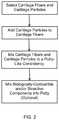
- FIG. 1is a block diagram of a process for preparing cartilage fibers having viable native chondrocytes, according to an embodiment of the present invention
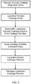
- FIG. 2is a block diagram of a process for preparing a cartilage-derived implant having viable native chondrocytes
- FIG. 3is a block diagram of a process for preparing cartilage particles, according to an embodiment of the present invention.
- FIG. 4is a top view of a planar grater suitable for use in an embodiment of the present invention.
- FIG. 5is a perspective view of a rotary grater suitable for use in an embodiment of the present invention.
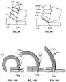
- FIG. 6Ais a perspective view of an exemplary embodiment of a cutting feature of a grater suitable for use in an embodiment of the present invention
- FIG. 6Bis a cross-sectional view of the cutting feature of FIG. 6A ;
- FIG. 6Cis a perspective view of a cutting feature similar to that of FIG. 6A but which has been modified;
- 6 Dis a cross-sectional view of the modified cutting feature of FIG. 6C ;
- FIG. 7is a series of cross-sectional views of a cutting feature similar to that of FIG. 6A , but having differently sized cutting angles;
- FIG. 8Ais a top view of another exemplary embodiment of a cutting feature of a grater suitable for use in an embodiment of the present invention
- FIG. 8Bis a cross-sectional view of the cutting feature of FIG. 8A ;
- FIG. 9Ais a schematic diagram showing the formation of a cartilage fiber having striations by a cutting edge
- FIG. 9Bis a schematic diagram showing the formation of discontinuous pieces of cartilage by a cutting edge
- FIGS. 10A, 10B and 10Care a series of diagrams showing how cutting edges having smaller cutting angles will produce less curved cartilage fibers
- FIGS. 11A-11Bare schematic diagrams showing an exemplary embodiment of a cartilage-derived implant in accordance with the present invention, with and without protruding cartilage fibers, respectively;
- FIGS. 12A-12Bare schematic diagrams showing the use of the cartilage-derived implant of FIG. 11A , where FIG. 12B shows swelling of the cartilage-derived implant upon rehydration.
- An embodiment of the present inventioncomprises a cartilage-derived implant including cartilage fibers.
- the cartilage fibersinclude viable native chondrocytes.
- the cartilage fibersare freeze-dried, and do not contain measurable amounts of viable chondrocytes.
- the cartilage-derived implantincludes cartilage fibers and cartilage particles.
- Embodiments of the present inventioninclude methods for producing cartilage fibers from recovered cartilage, including without limitation one or more condyles.
- Embodiments of the present inventionalso include methods of making the aforementioned cartilage-derived implants.
- Embodiments of the present inventionalso include methods of using the aforesaid cartilage-derived implants for the repair of cartilage defects. Non-limiting exemplary embodiments of the cartilage-derived implants of the present invention, and methods of making and using same, are discussed herein.
- the cartilage-derived implants of the present inventionhave uses in the repair of defects in articular cartilage, costal cartilage, or other types of cartilage in a patient.
- the cartilage-derived implants of the present inventionhave uses in meniscal repair, sternotomy repair, the repair of subchondral bone, the repair of articulating joints, and the repair or replacement of cartilaginous anatomical features, including, but not limited to, nasal and auricular cartilage.
- the cartilage-derived implantis a scaffold for the delivery of growth-inductive factors. In an embodiment, the cartilage-derived implant is growth-inductive. In an embodiment, the cartilage-derived implant is a scaffold for the delivery of cells. In an embodiment, the cartilage-derived implant is a scaffold for the migration of cells. In an embodiment, the cartilage-derived implant is a growth-conductive medium for the in-growth of tissue. In an embodiment, the cartilage-derived implant is a tissuegenic matrix.
- Cartilage fibers according to some embodiments of the present inventionare derived from cartilage which is allogeneic to the patient.
- cartilage fibersare derived from cartilage which is autologous to the patient.
- cartilage fibersare derived from cartilage which is xenogeneic to the patient.
- the cartilageincludes articular cartilage.
- the cartilageincludes costal cartilage.
- cartilageis recovered from deceased human donors, and the tissue is treated to reduce bioburden according to methods known in the art.
- the donoris a mature adult human donor. In an embodiment, the donor is a mature adult human donor having an age in the range of 20 to 55 years.
- the donoris a human donor having an age of less than 20 years. In an embodiment, the donor is a juvenile human donor.
- Methods of selecting, recovering, and treating cartilageare known in the art, and exemplary methods are disclosed in co-owned U.S. Pat. Nos. RE 42,208, RE 43,258, 8,292,968, 8,834,928, and 8,883,210, the disclosures of which are incorporated by reference herein.
- cartilage fibersare obtained by a dissection of the recovered cartilage so as to obtain fibers therefrom (e.g., by grating, scraping, slicing, or cutting) and collecting the fibers, as described in further detail hereinafter.
- the cartilage fibersare then stored in media.
- the cartilage fibersare cleaned by means of buffered saline rinses (e.g., phosphate buffered saline (PBS) solution) or other rinses that would remove or reduce the potential for antigenic reactions in a patient receiving the cartilage fibers.
- the cartilage fibersare cleaned by means that conserve the viability of chondrocytes within the cartilage fibers.
- the cartilage fibersmay then be sieved or filtered to collect fibers having desirable dimensions for use in making the cartilage-derived implant.
- the cartilage fibershave lengths in the range of about 0.2 mm to about 50 mm. In an embodiment of the present invention, at least one of the cartilage fibers has a length in the range of about 0.2 mm to about 0.5 mm. In an embodiment of the present invention, at least one of the cartilage fibers has a length in the range of about 0.5 mm to about 1.0 mm. In an embodiment of the present invention, at least one of the cartilage fibers has a length in the range of about 1.0 mm to about 1.5 mm. In an embodiment of the present invention, at least one of the cartilage fibers has a length in the range of about 1.5 mm to about 2.0 mm.
- At least one of the cartilage fibershas a length in the range of about 2.0 mm to about 2.5 mm. In an embodiment of the present invention, at least one of the cartilage fibers has a length in the range of about 2.5 mm to about 3.0 mm. In an embodiment of the present invention, at least one of the cartilage fibers has a length in the range of about 3.0 mm to about 3.5 mm. In an embodiment of the present invention, at least one of the cartilage fibers has a length in the range of about 3.5 mm to about 4.0 mm. In an embodiment of the present invention, at least one of the cartilage fibers has a length in the range of about 4.0 mm to about 4.5 mm.
- At least one of the cartilage fibershas a length in the range of about 4.5 mm to about 5.0 mm. In an embodiment of the present invention, at least one of the cartilage fibers has a length in the range of about 5.0 mm to about 5.5 mm. In an embodiment of the present invention, at least one of the cartilage fibers has a length in the range of about 5.5 mm to about 6.0 mm. In an embodiment of the present invention, at least one of the cartilage fibers has a length in the range of about 6.0 mm to about 6.5 mm. In an embodiment of the present invention, at least one of the cartilage fibers has a length in the range of about 6.5 mm to about 7.0 mm.
- At least one of the cartilage fibershas a length in the range of about 7.0 mm to about 7.5 mm. In an embodiment of the present invention, at least one of the cartilage fibers has a length in the range of about 7.5 mm to about 8.0 mm. In an embodiment of the present invention, at least one of the cartilage fibers has a length in the range of about 8.0 mm to about 8.5 mm. In an embodiment of the present invention, at least one of the cartilage fibers has a length in the range of about 8.5 mm to about 9.0 mm. In an embodiment of the present invention, at least one of the cartilage fibers has a length in the range of about 9.0 mm to about 9.5 mm.
- At least one of the cartilage fibershas a length in the range of about 9.5 mm to about 10.0 mm. In an embodiment of the present invention, at least one of the cartilage fibers has a length in the range of about 10 mm to about 11 mm. In an embodiment of the present invention, at least one of the cartilage fibers has a length in the range of about 11 mm to about 12 mm. In an embodiment of the present invention, at least one of the cartilage fibers has a length in the range of about 12 mm to about 13 mm. In an embodiment of the present invention, at least one of the cartilage fibers has a length in the range of about 13 mm to about 14 mm.
- At least one of the cartilage fibershas a length in the range of about 14 mm to about 15 mm. In an embodiment of the present invention, at least one of the cartilage fibers has a length in the range of about 15 mm to about 16 mm. In an embodiment of the present invention, at least one of the cartilage fibers has a length in the range of about 16 mm to about 17 mm. In an embodiment of the present invention, at least one of the cartilage fibers has a length in the range of about 17 mm to about 18 mm. In an embodiment of the present invention, at least one of the cartilage fibers has a length in the range of about 18 mm to about 19 mm.
- At least one of the cartilage fibershas a length in the range of about 19 mm to about 20 mm. In an embodiment of the present invention, at least one of the cartilage fibers has a length in the range of about 20 mm to about 25 mm. In an embodiment of the present invention, at least one of the cartilage fibers has a length in the range of about 25 mm to about 30 mm. In an embodiment of the present invention, at least one of the cartilage fibers has a length in the range of about 30 mm to about 35 mm. In an embodiment of the present invention, at least one of the cartilage fibers has a length in the range of about 35 mm to about 40 mm.
- At least one of the cartilage fibershas a length in the range of about 40 mm to about 45 mm. In an embodiment of the present invention, at least one of the cartilage fibers has a length in the range of about 45 mm to about 50 mm.
- the cartilage fibershave an average length in the range of about 1 mm to about 20 mm. In an embodiment, the cartilage fibers have an average length of about 1 mm. In an embodiment, the cartilage fibers have an average length of about 2 mm. In an embodiment, the cartilage fibers have an average length of about 3 mm. In an embodiment, the cartilage fibers have an average length of about 4 mm. In an embodiment, the cartilage fibers have an average length of about 5 mm. In an embodiment, the cartilage fibers have an average length of about 6 mm. In an average length of about 7 mm. In an embodiment, the cartilage fibers have an average length of about 8 mm.
- the cartilage fibershave an average length of about 9 mm. In an embodiment, the cartilage fibers have an average length of about 10 mm. In an embodiment, the cartilage fibers have an average length of about 12 mm. In an embodiment, the cartilage fibers have an average length of about 14 mm. In an embodiment, the cartilage fibers have an average length of about 16 mm. In an embodiment, the cartilage fibers have an average length of about 18 mm. In an embodiment, the cartilage fibers have an average length of about 20 mm.
- the cartilage fibershave an average length of at least 1 mm. In an embodiment, the cartilage fibers have an average length of at least 2 mm. In an embodiment, the cartilage fibers have an average length of at least 3 mm. In an embodiment, the cartilage fibers have an average length of at least 4 mm. In an embodiment, the cartilage fibers have an average length of at least 5 mm. In an embodiment, the cartilage fibers have an average length of at least 6 mm. In an average length of at least 7 mm. In an embodiment, the cartilage fibers have an average length of at least 8 mm. In an embodiment, the cartilage fibers have an average length of about at least 9 mm.
- the cartilage fibershave an average length of at least 10 mm. In an embodiment, the cartilage fibers have an average length of at least 12 mm. In an embodiment, the cartilage fibers have an average length of at least 14 mm. In an embodiment, the cartilage fibers have an average length of at least 16 mm. In an embodiment, the cartilage fibers have an average length of at least 18 mm. In an embodiment, the cartilage fibers have an average length of at least 20 mm.
- the cartilage fibershave an average length in the range of about 1 mm to about 3 mm. In an embodiment, the cartilage fibers have an average length in the range of about 2 mm to about 4 mm. In an embodiment, the cartilage fibers have an average length in the range of about 3 mm to about 5 mm. In an embodiment, the cartilage fibers have an average length in the range of about 4 mm to about 6 mm. In an embodiment, the cartilage fibers have an average length in the range of about 5 mm to about 7 mm. In an embodiment, the cartilage fibers have an average length in the range of about 6 mm to about 8 mm. In an embodiment, the cartilage fibers have an average length in the range of about 7 mm to about 9 mm.
- the cartilage fibershave an average length in the range of about 8 mm to about 10 mm. In an embodiment, the cartilage fibers have an average length in the range of about 9 mm to about 11 mm. In an embodiment, the cartilage fibers have an average length in the range of about 10 mm to about 12 mm. In an embodiment, the cartilage fibers have an average length in the range of about 12 mm to about 14 mm. In an embodiment, the cartilage fibers have an average length in the range of about 14 mm to about 16 mm. In an embodiment, the cartilage fibers have an average length in the range of about 16 mm to about 18 mm. In an embodiment, the cartilage fibers have an average length in the range of about 18 mm to about 20 mm.
- the cartilage fibershave widths or thicknesses in the range of about 0.01 mm to about 5 mm. In an embodiment of the present invention, at least one of the cartilage fibers has a width in the range of about 0.01 mm to about 0.05 mm. In an embodiment of the present invention, at least one of the cartilage fibers has a width in the range of about 0.05 mm to about 0.10 mm. In an embodiment of the present invention, at least one of the cartilage fibers has a width in the range of about 0.10 mm to about 0.15 mm. In an embodiment of the present invention, at least one of the cartilage fibers has a width in the range of about 0.15 mm to about 0.20 mm.
- At least one of the cartilage fibershas a width in the range of about 0.20 mm to about 0.25 mm. In an embodiment of the present invention, at least one of the cartilage fibers has a width in the range of about 0.25 mm to about 0.30 mm. In an embodiment of the present invention, at least one of the cartilage fibers has a width in the range of about 0.30 mm to about 0.35 mm. In an embodiment of the present invention, at least one of the cartilage fibers has a width in the range of about 0.35 mm to about 0.40 mm. In an embodiment of the present invention, at least one of the cartilage fibers has a width in the range of about 0.40 mm to about 0.45 mm.
- At least one of the cartilage fibershas a width in the range of about 0.45 mm to about 0.50 mm. In an embodiment of the present invention, at least one of the cartilage fibers has a width in the range of about 0.50 mm to about 0.55 mm. In an embodiment of the present invention, at least one of the cartilage fibers has a width in the range of about 0.55 mm to about 0.60 mm. In an embodiment of the present invention, at least one of the cartilage fibers has a width in the range of about 0.60 mm to about 0.65 mm. In an embodiment of the present invention, at least one of the cartilage fibers has a width in the range of about 0.65 mm to about 0.70 mm.
- At least one of the cartilage fibershas a width in the range of about 0.70 mm to about 0.75 mm. In an embodiment of the present invention, at least one of the cartilage fibers has a width in the range of about 0.75 mm to about 0.80 mm. In an embodiment of the present invention, at least one of the cartilage fibers has a width in the range of about 0.80 mm to about 0.85 mm. In an embodiment of the present invention, at least one of the cartilage fibers has a width in the range of about 0.85 mm to about 0.90 mm. In an embodiment of the present invention, at least one of the cartilage fibers has a width in the range of about 0.90 mm to about 0.95 mm.
- At least one of the cartilage fibershas a width in the range of about 0.95 mm to about 1.0 mm. In an embodiment of the present invention, at least one of the cartilage fibers has a width in the range of about 1.0 mm to about 1.1 mm. In an embodiment of the present invention, at least one of the cartilage fibers has a width in the range of about 1.1 mm to about 1.2 mm. In an embodiment of the present invention, at least one of the cartilage fibers has a width in the range of about 1.2 mm to about 1.3 mm. In an embodiment of the present invention, at least one of the cartilage fibers has a width in the range of about 1.3 mm to about 1.4 mm.
- At least one of the cartilage fibershas a width in the range of about 1.4 mm to about 1.5 mm. In an embodiment of the present invention, at least one of the cartilage fibers has a width in the range of about 1.5 mm to about 1.6 mm. In an embodiment of the present invention, at least one of the cartilage fibers has a width in the range of about 1.6 mm to about 1.7 mm. In an embodiment of the present invention, at least one of the cartilage fibers has a width in the range of about 1.7 mm to about 1.8 mm. In an embodiment of the present invention, at least one of the cartilage fibers has a width in the range of about 1.8 mm to about 1.9 mm.
- At least one of the cartilage fibershas a width in the range of about 1.9 mm to about 2.0 mm. In an embodiment of the present invention, at least one of the cartilage fibers has a width in the range of about 2.0 mm to about 2.5 mm. In an embodiment of the present invention, at least one of the cartilage fibers has a width in the range of about 2.5 mm to about 3.0 mm. In an embodiment of the present invention, at least one of the cartilage fibers has a width in the range of about 3.0 mm to about 3.5 mm. In an embodiment of the present invention, at least one of the cartilage fibers has a width in the range of about 3.5 mm to about 4.0 mm.
- At least one of the cartilage fibershas a width in the range of about 4.0 mm to about 4.5 mm. In an embodiment of the present invention, at least one of the cartilage fibers has a width in the range of about 4.5 mm to about 5.0 mm.
- the cartilage fibershave an average width in the range of about 0.1 mm to about 2 mm. In an embodiment, the cartilage fibers have an average width of about 0.1 mm. In an embodiment, the cartilage fibers have an average width of about 0.2 mm. In an embodiment, the cartilage fibers have an average width of about 0.3 mm. In an embodiment, the cartilage fibers have an average width of about 0.4 mm. In an embodiment, the cartilage fibers have an average width of about 0.5 mm. In an embodiment, the cartilage fibers have an average width of about 0.6 mm. In an embodiment, the cartilage fibers have an average width of about 0.7 mm. In an embodiment, the cartilage fibers have an average width of about 0.8 mm.
- the cartilage fibershave an average width of about 0.9 mm. In an embodiment, the cartilage fibers have an average width of about 1.0 mm. In an embodiment, the cartilage fibers have an average width of about 1.2 mm. In an embodiment, the cartilage fibers have an average width of about 1.4 mm. In an embodiment, the cartilage fibers have an average width of about 1.6 mm. In an embodiment, the cartilage fibers have an average width of about 1.8 mm. In an embodiment, the cartilage fibers have an average width of about 2 mm.
- the cartilage fibershave an average width of at least 0.1 mm. In an embodiment, the cartilage fibers have an average width of at least 0.2 mm. In an embodiment, the cartilage fibers have an average width of at least 0.3 mm. In an embodiment, the cartilage fibers have an average width of at least 0.4 mm. In an embodiment, the cartilage fibers have an average width of at least 0.5 mm. In an embodiment, the cartilage fibers have an average width of at least 0.6 mm. In an embodiment, the cartilage fibers have an average width of at least 0.7 mm. In an embodiment, the cartilage fibers have an average width of at least 0.8 mm. In an embodiment, the cartilage fibers have an average width of at least 0.9 mm.
- the cartilage fibershave an average width of at least 1.0 mm. In an embodiment, the cartilage fibers have an average width of at least 1.2 mm. In an embodiment, the cartilage fibers have an average width of at least 1.4 mm. In an embodiment, the cartilage fibers have an average width of at least 1.6 mm. In an embodiment, the cartilage fibers have an average width of at least 1.8 mm. In an embodiment, the cartilage fibers have an average width of at least 2.0 mm.
- the cartilage fibershave an average width in the range of about 0.1 mm to about 0.3 mm. In an embodiment, the cartilage fibers have an average width in the range of about 0.2 mm to about 0.4 mm. In an embodiment, the cartilage fibers have an average width in the range of about 0.3 mm to about 0.5 mm. In an embodiment, the cartilage fibers have an average width in the range of about 0.4 mm to about 0.6 mm. In an embodiment, the cartilage fibers have an average width in the range of about 0.5 mm to about 0.7 mm. In an embodiment, the cartilage fibers have an average width in the range of about 0.6 mm to about 0.8 mm.
- the cartilage fibershave an average width in the range of about 0.7 mm to about 0.9 mm. In an embodiment, the cartilage fibers have an average width in the range of about 0.8 mm to about 1.0 mm. In an embodiment, the cartilage fibers have an average width in the range of about 0.9 mm to about 1.1 mm. In an embodiment, the cartilage fibers have an average width in the range of about 1.0 mm to about 1.2 mm. In an embodiment, the cartilage fibers have an average width in the range of about 1.2 mm to about 1.4 mm. In an embodiment, the cartilage fibers have an average width in the range of about 1.4 mm to about 1.6 mm. In an embodiment, the cartilage fibers have an average width in the range of about 1.6 mm to about 1.8 mm. In an embodiment, the cartilage fibers have an average width in the range of about 1.8 mm to about 2.0 mm.
- the cartilage fibershave individual volumes in the range of about 0.1 mm 3 to about 200 mm 3 . In an embodiment, at least one of the cartilage fibers has a volume in the range of about 0.1 mm 3 to about 0.5 mm 3 . In an embodiment, at least one of the cartilage fibers has a volume in the range of about 0.5 mm 3 to about 1.0 mm 3 . In an embodiment, at least one of the cartilage fibers has a volume in the range of about 1.0 mm 3 to about 1.2 mm 3 . In an embodiment, at least one of the cartilage fibers has a volume in the range of about 1.2 mm 3 to about 1.4 mm 3 .
- At least one of the cartilage fibershas a volume in the range of about 1.4 mm 3 to about 1.6 mm 3 . In an embodiment, at least one of the cartilage fibers has a volume in the range of about 1.6 mm 3 to about 1.8 mm 3 . In an embodiment, at least one of the cartilage fibers has a volume in the range of about 1.8 mm 3 to about 2.0 mm 3 . In an embodiment, at least one of the cartilage fibers has a volume in the range of about 2.0 mm 3 to about 2.2 mm 3 . In an embodiment, at least one of the cartilage fibers has a volume in the range of about 2.2 mm 3 to about 2.4 mm 3 .
- At least one of the cartilage fibershas a volume in the range of about 2.4 mm 3 to about 2.6 mm 3 . In an embodiment, at least one of the cartilage fibers has a volume in the range of about 2.6 mm 3 to about 2.8 mm 3 . In an embodiment, at least one of the cartilage fibers has a volume in the range of about 2.8 mm 3 to about 3.0 mm 3 . In an embodiment, at least one of the cartilage fibers has a volume in the range of about 3.0 mm 3 to about 3.2 mm 3 . In an embodiment, at least one of the cartilage fibers has a volume in the range of about 3.2 mm 3 to about 3.4 mm 3 .
- At least one of the cartilage fibershas a volume in the range of about 3.4 mm 3 to about 3.6 mm 3 . In an embodiment, at least one of the cartilage fibers has a volume in the range of about 3.6 mm 3 to about 3.8 mm 3 . In an embodiment, at least one of the cartilage fibers has a volume in the range of about 3.8 mm 3 to about 4.0 mm 3 . In an embodiment, at least one of the cartilage fibers has a volume in the range of about 4.0 mm 3 to about 4.2 mm 3 . In an embodiment, at least one of the cartilage fibers has a volume in the range of about 4.2 mm 3 to about 4.4 mm 3 .
- At least one of the cartilage fibershas a volume in the range of about 4.4 mm 3 to about 4.6 mm 3 . In an embodiment, at least one of the cartilage fibers has a volume in the range of about 4.6 mm 3 to about 4.8 mm 3 . In an embodiment, at least one of the cartilage fibers has a volume in the range of about 4.8 mm 3 to about 5.0 mm 3 . In an embodiment, at least one of the cartilage fibers has a volume in the range of about 5.0 mm 3 to about 5.5 mm 3 . In an embodiment, at least one of the cartilage fibers has a volume in the range of about 5.5 mm 3 to about 6.0 mm 3 .
- At least one of the cartilage fibershas a volume in the range of about 6.0 mm 3 to about 6.5 mm 3 . In an embodiment, at least one of the cartilage fibers has a volume in the range of about 6.5 mm 3 to about 7.0 mm 3 . In an embodiment, at least one of the cartilage fibers has a volume in the range of about 7.0 mm 3 to about 7.5 mm 3 . In an embodiment, at least one of the cartilage fibers has a volume in the range of about 7.5 mm 3 to about 8.0 mm 3 . In an embodiment, at least one of the cartilage fibers has a volume in the range of about 8.0 mm 3 to about 8.5 mm 3 .
- At least one of the cartilage fibershas a volume in the range of about 8.5 mm 3 to about 9.0 mm 3 . In an embodiment, at least one of the cartilage fibers has a volume in the range of about 9.5 mm 3 to about 10 mm 3 . In an embodiment, at least one of the cartilage fibers has a volume in the range of about 10 mm 3 to about 12 mm 3 . In an embodiment, at least one of the cartilage fibers has a volume in the range of about 12 mm 3 to about 14 mm 3 . In an embodiment, at least one of the cartilage fibers has a volume in the range of about 14 mm 3 to about 16 mm 3 .
- At least one of the cartilage fibershas a volume in the range of about 16 mm 3 to about 18 mm 3 . In an embodiment, at least one of the cartilage fibers has a volume in the range of about 18 mm 3 to about 20 mm 3 . In an embodiment, at least one of the cartilage fibers has a volume in the range of about 20 mm 3 to about 25 mm 3 . In an embodiment, at least one of the cartilage fibers has a volume in the range of about 25 mm 3 to about 30 mm 3 . In an embodiment, at least one of the cartilage fibers has a volume in the range of about 30 mm 3 to about 35 mm 3 .
- At least one of the cartilage fibershas a volume in the range of about 35 mm 3 to about 40 mm 3 . In an embodiment, at least one of the cartilage fibers has a volume in the range of about 40 mm 3 to about 45 mm 3 . In an embodiment, at least one of the cartilage fibers has a volume in the range of about 45 mm 3 to about 50 mm 3 . In an embodiment, at least one of the cartilage fibers has a volume in the range of about 50 mm 3 to about 60 mm 3 . In an embodiment, at least one of the cartilage fibers has a volume in the range of about 60 mm 3 to about 70 mm 3 .
- At least one of the cartilage fibershas a volume in the range of about 70 mm 3 to about 80 mm 3 . In an embodiment, at least one of the cartilage fibers has a volume in the range of about 80 mm 3 to about 90 mm 3 . In an embodiment, at least one of the cartilage fibers has a volume in the range of about 90 mm 3 to about 100 mm 3 . In an embodiment, at least one of the cartilage fibers has a volume in the range of about 100 mm 3 to about 120 mm 3 . In an embodiment, at least one of the cartilage fibers has a volume in the range of about 120 mm 3 to about 140 mm 3 .
- At least one of the cartilage fibershas a volume in the range of about 140 mm 3 to about 160 mm 3 . In an embodiment, at least one of the cartilage fibers has a volume in the range of about 160 mm 3 to about 180 mm 3 . In an embodiment, at least one of the cartilage fibers has a volume in the range of about 180 mm 3 to about 200 mm 3 .
- the cartilage fibershave an average volume in the range of about 1 mm 3 to about 20 mm 3 . In an embodiment, the cartilage fibers have an average volume of about 1 mm 3 . In an embodiment, the cartilage fibers have an average volume of about 1.2 mm 3 . In an embodiment, the cartilage fibers have an average volume of about 1.4 mm 3 . In an embodiment, the cartilage fibers have an average volume of about 1.6 mm 3 . In an embodiment, the cartilage fibers have an average volume of about 1.8 mm 3 . In an embodiment, the cartilage fibers have an average volume of about 2.0 mm 3 . In an embodiment, the cartilage fibers have an average volume of about 2.2 mm 3 .
- the cartilage fibershave an average volume of about 2.4 mm 3 . In an embodiment, the cartilage fibers have an average volume of about 2.6 mm 3 . In an embodiment, the cartilage fibers have an average volume of about 2.8 mm 3 . In an embodiment, the cartilage fibers have an average volume of about 3.0 mm 3 . In an embodiment, the cartilage fibers have an average volume of about 3.2 mm 3 . In an embodiment, the cartilage fibers have an average volume of about 3.4 mm 3 . In an embodiment, the cartilage fibers have an average volume of about 3.6 mm 3 . In an embodiment, the cartilage fibers have an average volume of about 3.8 mm 3 .
- the cartilage fibershave an average volume of about 4.0 mm 3 . In an embodiment, the cartilage fibers have an average volume of about 4.5 mm 3 . In an embodiment, the cartilage fibers have an average volume of about 5.0 mm 3 . In an embodiment, the cartilage fibers have an average volume of about 5.5 mm 3 . In an embodiment, the cartilage fibers have an average volume of about 6.0 mm 3 . In an embodiment, the cartilage fibers have an average volume of about 6.5 mm 3 . In an embodiment, the cartilage fibers have an average volume of about 7.0 mm 3 . In an embodiment, the cartilage fibers have an average volume of about 7.5 mm 3 .
- the cartilage fibershave an average volume of about 8.0 mm 3 . In an embodiment, the cartilage fibers have an average volume of about 8.5 mm 3 . In an embodiment, the cartilage fibers have an average volume of about 9.0 mm 3 . In an embodiment, the cartilage fibers have an average volume of about 9.5 mm 3 . In an embodiment, the cartilage fibers have an average volume of about 10 mm 3 . In an embodiment, the cartilage fibers have an average volume of about 12 mm 3 . In an embodiment, the cartilage fibers have an average volume of about 14 mm 3 . In an embodiment, the cartilage fibers have an average volume of about 16 mm 3 . In an embodiment, the cartilage fibers have an average volume of about 18 mm 3 . In an embodiment, the cartilage fibers have an average volume of about 20 mm 3 .
- the cartilage fibershave an average volume of at least 1 mm 3 . In an embodiment, the cartilage fibers have an average volume of at least 1.2 mm 3 . In an embodiment, the cartilage fibers have an average volume of at least 1.4 mm 3 . In an embodiment, the cartilage fibers have an average volume of at least 1.6 mm 3 . In an embodiment, the cartilage fibers have an average volume of at least 1.8 mm 3 . In an embodiment, the cartilage fibers have an average volume of at least 2.0 mm 3 . In an embodiment, the cartilage fibers have an average volume of at least 2.2 mm 3 . In an embodiment, the cartilage fibers have an average volume of at least 2.4 mm 3 .
- the cartilage fibershave an average volume of at least 2.6 mm 3 . In an embodiment, the cartilage fibers have an average volume of at least 2.8 mm 3 . In an embodiment, the cartilage fibers have an average volume of at least 3.0 mm 3 . In an embodiment, the cartilage fibers have an average volume of at least 3.2 mm 3 . In an embodiment, the cartilage fibers have an average volume of at least 3.4 mm 3 . In an embodiment, the cartilage fibers have an average volume of at least 3.6 mm 3 . In an embodiment, the cartilage fibers have an average volume of at least 3.8 mm 3 . In an embodiment, the cartilage fibers have an average volume of at least 4.0 mm 3 .
- the cartilage fibershave an average volume of at least 4.5 mm 3 . In an embodiment, the cartilage fibers have an average volume of at least 5.0 mm 3 . In an embodiment, the cartilage fibers have an average volume of at least 5.5 mm 3 . In an embodiment, the cartilage fibers have an average volume of at least 6.0 mm 3 . In an embodiment, the cartilage fibers have an average volume of at least 6.5 mm 3 . In an embodiment, the cartilage fibers have an average volume of at least 7.0 mm 3 . In an embodiment, the cartilage fibers have an average volume of at least 7.5 mm 3 . In an embodiment, the cartilage fibers have an average volume of at least 8.0 mm 3 .
- the cartilage fibershave an average volume of at least 8.5 mm 3 . In an embodiment, the cartilage fibers have an average volume of at least 9.0 mm 3 . In an embodiment, the cartilage fibers have an average volume of at least 9.5 mm 3 . In an embodiment, the cartilage fibers have an average volume of at least 10 mm 3 . In an embodiment, the cartilage fibers have an average volume of at least 12 mm 3 . In an embodiment, the cartilage fibers have an average volume of at least 14 mm 3 . In an embodiment, the cartilage fibers have an average volume of at least 16 mm 3 . In an embodiment, the cartilage fibers have an average volume of at least 18 mm 3 . In an embodiment, the cartilage fibers have an average volume of at least 20 mm 3 .
- the cartilage fibershave an average volume in the range of about 1 mm 3 to about 1.4 mm 3 . In an embodiment, the cartilage fibers have an average volume in the range of about 1.2 mm 3 to about 1.6 mm 3 . In an embodiment, the cartilage fibers have an average volume in the range of about 1.4 mm 3 to about 1.8 mm 3 . In an embodiment, the cartilage fibers have an average volume in the range of about 1.6 mm 3 to about 2.0 mm 3 . In an embodiment, the cartilage fibers have an average volume in the range of about 1.8 mm 3 to about 2.2 mm 3 . In an embodiment, the cartilage fibers have an average volume in the range of about 2.0 mm 3 to about 2.4 mm 3 .
- the cartilage fibershave an average volume in the range of about 2.2 mm 3 to about 2.6 mm 3 . In an embodiment, the cartilage fibers have an average volume in the range of about 2.4 mm 3 to about 2.8 mm 3 . In an embodiment, the cartilage fibers have an average volume in the range of about 2.6 mm 3 to about 3.0 mm 3 . In an embodiment, the cartilage fibers have an average volume in the range of about 2.8 mm 3 to about 3.2 mm 3 . In an embodiment, the cartilage fibers have an average volume in the range of about 3.0 mm 3 to about 3.4 mm 3 . In an embodiment, the cartilage fibers have an average volume in the range of about 3.2 mm 3 to about 3.6 mm 3 .
- the cartilage fibershave an average volume in the range of about 3.4 mm 3 to about 3.8 mm 3 . In an embodiment, the cartilage fibers have an average volume in the range of about 3.6 mm 3 to about 4.0 mm 3 . In an embodiment, the cartilage fibers have an average volume in the range of about 3.8 mm 3 to about 4.2 mm 3 . In an embodiment, the cartilage fibers have an average volume in the range of about 4.0 mm 3 to about 4.4 mm 3 . In an embodiment, the cartilage fibers have an average volume in the range of about 4.2 mm 3 to about 5.0 mm 3 . In an embodiment, the cartilage fibers have an average volume in the range of about 4.5 mm 3 to about 5.5 mm 3 .
- the cartilage fibershave an average volume in the range of about 5.0 mm 3 to about 6.0 mm 3 . In an embodiment, the cartilage fibers have an average volume in the range of about 5.5 mm 3 to about 6.5 mm 3 . In an embodiment, the cartilage fibers have an average volume in the range of about 6.0 mm 3 to about 7.0 mm 3 . In an embodiment, the cartilage fibers have an average volume in the range of about 6.5 mm 3 to about 7.5 mm 3 . In an embodiment, the cartilage fibers have an average volume in the range of about 7.0 mm 3 to about 8.0 mm 3 . In an embodiment, the cartilage fibers have an average volume in the range of about 7.5 mm 3 to about 9.5 mm 3 .
- the cartilage fibershave an average volume in the range of about 8.0 mm 3 to about 10.0 mm 3 . In an embodiment, the cartilage fibers have an average volume in the range of about 8.5 mm 3 to about 10.5 mm 3 . In an embodiment, the cartilage fibers have an average volume in the range of about 9.0 mm 3 to about 11.0 mm 3 . In an embodiment, the cartilage fibers have an average volume in the range of about 9.5 mm 3 to about 11.5 mm 3 . In an embodiment, the cartilage fibers have an average volume in the range of about 10 mm 3 to about 14 mm 3 . In an embodiment, the cartilage fibers have an average volume in the range of about 12 mm 3 to about 16 mm 3 . In an embodiment, the cartilage fibers have an average volume in the range of about 14 mm 3 to about 18 mm 3 . In an embodiment, the cartilage fibers have an average volume in the range of about 16 mm 3 to about 20 mm 3 .
- At least some of the cartilage fibershave an aspect ratio (length/width) in the range of about 1:1 to about 50:1. In an embodiment, at least some of the cartilage fibers have an aspect ratio in the range of about 1:1 to about 2:1. In an embodiment, at least some of the cartilage fibers have an aspect ratio in the range of about 2:1 to about 3:1. In an embodiment, at least some of the cartilage fibers have an aspect ratio in the range of about 3:1 to about 4:1. In an embodiment, at least some of the cartilage fibers have an aspect ratio in the range of about 4:1 to about 5:1. In an embodiment, at least some of the cartilage fibers have an aspect ratio in the range of about 5:1 to about 6:1.
- At least some of the cartilage fibershave an aspect ratio in the range of about 6:1 to about 7:1. In an embodiment, at least some of the cartilage fibers have an aspect ratio in the range of about 7:1 to about 8:1. In an embodiment, at least some of the cartilage fibers have an aspect ratio in the range of about 8:1 to about 9:1. In an embodiment, at least some of the cartilage fibers have an aspect ratio in the range of about 9:1 to about 10:1. In an embodiment, at least some of the cartilage fibers have an aspect ratio in the range of about 10:1 to about 12:1. In an embodiment, at least some of the cartilage fibers have an aspect ratio in the range of about 12:1 to about 14:1.
- At least some of the cartilage fibershave an aspect ratio in the range of about 14:1 to about 16:1. In an embodiment, at least some of the cartilage fibers have an aspect ratio in the range of about 16:1 to about 18:1. In an embodiment, at least some of the cartilage fibers have an aspect ratio in the range of about 18:1 to about 20:1. In an embodiment, at least some of the cartilage fibers have an aspect ratio in the range of about 20:1 to about 25:1. In an embodiment, at least some of the cartilage fibers have an aspect ratio in the range of about 25:1 to about 30:1. In an embodiment, at least some of the cartilage fibers have an aspect ratio in the range of about 30:1 to about 35:1.
- At least some of the cartilage fibershave an aspect ratio in the range of about 35:1 to about 40:1. In an embodiment, at least some of the cartilage fibers have an aspect ratio in the range of about 40:1 to about 45:1. In an embodiment, at least some of the cartilage fibers have an aspect ratio in the range of about 45:1 to about 50:1.
- At least some of the cartilage fibershave an aspect ratio of at least 1:1. In an embodiment, at least some of the cartilage fibers have an aspect ratio of at least 2:1. In an embodiment, at least some of the cartilage fibers have an aspect ratio of at least 3:1. In an embodiment, at least some of the cartilage fibers have an aspect ratio of at least 4:1. In an embodiment, at least some of the cartilage fibers have an aspect ratio of at least 5:1. In an embodiment, at least some of the cartilage fibers have an aspect ratio of at least 6:1. In an embodiment, at least some of the cartilage fibers have an aspect ratio of at least 7:1. In an embodiment, at least some of the cartilage fibers have an aspect ratio of at least 8:1.
- At least some of the cartilage fibershave an aspect ratio of at least 9:1. In an embodiment, at least some of the cartilage fibers have an aspect ratio of at least 10:1. In an embodiment, at least some of the cartilage fibers have an aspect ratio of at least 12:1. In an embodiment, at least some of the cartilage fibers have an aspect ratio of at least 14:1. In an embodiment, at least some of the cartilage fibers have an aspect ratio of at least 16:1. In an embodiment, at least some of the cartilage fibers have an aspect ratio of at least 18:1. In an embodiment, at least some of the cartilage fibers have an aspect ratio of at least 20:1. In an embodiment, at least some of the cartilage fibers have an aspect ratio of at least 30:1.
- At least some of the cartilage fibershave an aspect ratio of at least 35:1. In an embodiment, at least some of the cartilage fibers have an aspect ratio of at least 40:1. In an embodiment, at least some of the cartilage fibers have an aspect ratio of at least 45:1.
- At least some of the cartilage fibersare ribbon-like, having, for example, a width/thickness ratio in the range of about 10:1 to about 500:1. In an embodiment, at least some of the ribbon-like cartilage fibers have a width/thickness ratio in the range of about 10:1 to about 20:1. In an embodiment, at least some of the ribbon-like cartilage fibers have a width/thickness ratio in the range of about 20:1 to about 40:1. In an embodiment, at least some of the ribbon-like cartilage fibers have a width/thickness ratio in the range of about 40:1 to about 60:1.
- At least some of the ribbon-like cartilage fibershave a width/thickness ratio in the range of about 60:1 to about 80:1. In an embodiment, at least some of the ribbon-like cartilage fibers have a width/thickness ratio in the range of about 80:1 to about 100:1. In an embodiment, at least some of the ribbon-like cartilage fibers have a width/thickness ratio in the range of about 100:1 to about 150:1. In an embodiment, at least some of the ribbon-like cartilage fibers have a width/thickness ratio in the range of about 150:1 to about 200:1. In an embodiment, at least some of the ribbon-like cartilage fibers have a width/thickness ratio in the range of about 200:1 to about 250:1.
- At least some of the ribbon-like cartilage fibershave a width/thickness ratio in the range of about 250:1 to about 300:1. In an embodiment, at least some of the ribbon-like cartilage fibers have a width/thickness ratio in the range of about 300:1 to about 350:1. In an embodiment, at least some of the ribbon-like cartilage fibers have a width/thickness ratio in the range of about 350:1 to about 400:1. In an embodiment, at least some of the ribbon-like cartilage fibers have a width/thickness ratio in the range of about 450:1 to about 500:1.
- At least some of the cartilage fibersare ribbon-like, having, for example, a width/thickness ratio in the range of about 10:1 to about 500:1. In an embodiment, at least some of the ribbon-like cartilage fibers have a width/thickness ratio of at least 10:1. In an embodiment, at least some of the ribbon-like cartilage fibers have a width/thickness ratio of at least 20:1. In an embodiment, at least some of the ribbon-like cartilage fibers have a width/thickness ratio of at least 40:1. In an embodiment, at least some of the ribbon-like cartilage fibers have a width/thickness ratio of at least 60:1.
- At least some of the ribbon-like cartilage fibershave a width/thickness ratio of at least 80:1. In an embodiment, at least some of the ribbon-like cartilage fibers have a width/thickness ratio of at least 100:1. In an embodiment, at least some of the ribbon-like cartilage fibers have a width/thickness ratio of at least 150:1. In an embodiment, at least some of the ribbon-like cartilage fibers have a width/thickness ratio of at least 200:1. In an embodiment, at least some of the ribbon-like cartilage fibers have a width/thickness ratio of at least 250:1. In an embodiment, at least some of the ribbon-like cartilage fibers have a width/thickness ratio of at least 300:1. In an embodiment, at least some of the ribbon-like cartilage fibers have a width/thickness ratio of at least 350:1. In an embodiment, at least some of the ribbon-like cartilage fibers have a width/thickness ratio of at least 450:1.
- At least some of the cartilage fibersare thread-like, having, for example, a width/thickness ratio in the range of about 1:1 to about 10:1. In an embodiment, at least some of the thread-like cartilage fibers have a width/thickness ratio in the range of about 1:1 to about 2:1. In an embodiment, at least some of the thread-like cartilage fibers have a width/thickness ratio in the range of about 2:1 to about 3:1. In an embodiment, at least some of the thread-like cartilage fibers have a width/thickness ratio in the range of about 3:1 to about 4:1. In an embodiment, at least some of the thread-like cartilage fibers have a width/thickness ratio in the range of about 4:1 to about 5:1.
- At least some of the thread-like cartilage fibershave a width/thickness ratio in the range of about 5:1 to about 6:1. In an embodiment, at least some of the thread-like cartilage fibers have a width/thickness ratio in the range of about 6:1 to about 7:1. In an embodiment, at least some of the thread-like cartilage fibers have a width/thickness ratio in the range of about 7:1 to about 8:1. In an embodiment, at least some of the thread-like cartilage fibers have a width/thickness ratio in the range of about 8:1 to about 9:1. In an embodiment, at least some of the thread-like cartilage fibers have a width/thickness ratio in the range of about 9:1 to about 10:1.
- At least some of the thread-like cartilage fibershave a width/thickness ratio of at least 1:1. In an embodiment, at least some of the thread-like cartilage fibers have a width/thickness ratio of at least 2:1. In an embodiment, at least some of the thread-like cartilage fibers have a width/thickness ratio of at least 3:1. In an embodiment, at least some of the thread-like cartilage fibers have a width/thickness ratio of at least 4:1. In an embodiment, at least some of the thread-like cartilage fibers have a width/thickness ratio of at least 5:1. In an embodiment, at least some of the thread-like cartilage fibers have a width/thickness ratio of at least 6:1.
- At least some of the thread-like cartilage fibershave a width/thickness ratio of at least 7:1. In an embodiment, at least some of the thread-like cartilage fibers have a width/thickness ratio of at least 8:1. In an embodiment, at least some of the thread-like cartilage fibers have a width/thickness ratio of at least 9:1. In an embodiment, at least some of the thread-like cartilage fibers have a width/thickness ratio of at least 10:1.
- the cartilage fibersmay have cross-sections of virtually any shape.
- at least some of the cartilage fibershave cross-sectional shapes that are similar to each other.
- at least some of the cartilage fibershave a cross-sectional shape having at least one straight edge.
- at least some of the cartilage fibershave a cross-sectional shape having at least two straight edges.
- at least some of the cartilage fibershave a cross-sectional shape having at least two straight edges that intersect each other at an angle.
- at least some of the cartilage fibershave a polygonal cross-sectional shape.
- At least some of the cartilage fibershave a cross-sectional shape with at least one curved edge. In an embodiment, at least some of the cartilage fibers have a semi-circular cross-sectional shape. In an embodiment, at least some of the cartilage fibers have a semi-elliptical cross-sectional shape. In an embodiment, at least some of the cartilage fibers have a circular cross-sectional shape. In an embodiment, at least some of the cartilage fibers have a crescent cross-sectional shape. In an embodiment, at least some of the cartilage fibers have an irregular cross-sectional shape.
- the cartilage fibersare similar to each other in their respective dimensions. In an embodiment, at least some of the cartilage fibers are a mixture of ribbon-like and thread-like cartilage fibers. In an embodiment, at least some of the cartilage fibers have selected dimensions and/or cross-sectional shapes. In an embodiment, the cartilage fibers have various lengths. In an embodiment, the cartilage fibers have various widths and thicknesses. In an embodiment, some of the cartilage fibers have long lengths and some of the cartilage fibers have short lengths. In an embodiment, at least some of the cartilage fibers have one cross-sectional shape and at least some of the cartilage fibers have a different cross-sectional shape. In an embodiment, the dimensions and/or cross-sectional shape of a cartilage fiber are generated by the tool selected to perform the blunt dissection of the recovered cartilage.
- the cartilage fibersare substantially free of cells and their components.
- the cartilage fibersinclude viable native cells.
- the cartilage fibersinclude non-viable native cells.
- Non-viable native cellsmay, for example without limitation, be present in the cartilage fibers when the cartilage fibers are produced from recovered cartilage that has been frozen.
- the cartilage fibersinclude viable non-native cells.
- the cartilage fibersinclude non-native non-viable cells.
- Cartilage fibersmay, for example without limitation, include non-native cells (viable or non-viable) when they have been added or cultured on the cartilage fibers after production of the cartilage fibers.
- Native and non-native cellsinclude, without limitation, chondrocytes and other types of cells.
- the cartilage fibersare in the form of a mass of cartilage fibers, which are then used to prepare implants that may be used to fill a cartilage defect in a patient. Such implants may be used as a standalone treatment device or be applied in combination with one or more of a variety of bioactive materials or cells that facilitate the reconstruction and healing of cartilage.
- the cartilage fibersserve as a matrix or scaffold for the remodeling of chondral tissue, resulting in the growth of new cartilage or of collagen fibrous material that fills spaces in the defect. Remodeling occurs along the edges of the fibers, which provide a greater number of remodeling sites relative to the number provided by other types of cartilage pieces.
- the cartilage fibersare manipulated to form a putty without adding a carrier or other biologically-compatible material.
- the cartilage fibersare combined with a carrier or other biologically-compatible material to form a paste, putty or gel.
- the cartilage fibersare combined with a carrier or other biologically-compatible material such that the resulting mixture may be injected into a cartilage defect site in a patient.
- the paste, putty, gel, or injectable mixtureis used to fill a defect site, or are otherwise applied to a defect site or to a site of a diseased or defective cartilage, or to other tissues in a patient in need of such a paste, putty, gel, or injectable mixture.
- suitable carriers for forming a putty or an injectable mixtureinclude, but are not necessarily limited to, the following: an aqueous buffer, an isotonic solution; a sodium chloride solution at a concentration of about 0.1% to about 1%, more particularly, about 0.9%; a lactated Ringer's solution, with or without D5LR (dextrose); phosphate buffered saline (“PBS”); platelet rich plasma (PRP); glycerin; lecithin; alginate; hyaluronic acid (HA); a derivative of HA; or sodium hyaluronate; or other suitable carriers known in the art.
- the carriermay also comprise “biological components” added to the carrier, such as, but not limited to, DNA, RNA, short hairpin RNA (shRNA), small interfering RNA (siRNA), micro RNA (mRNA), polysaccharides, peptides, matrix proteins, glycosaminoglycans (e.g, hyaluronic acid), viral vectors, and liposomes.
- biological componentssuch as, but not limited to, DNA, RNA, short hairpin RNA (shRNA), small interfering RNA (siRNA), micro RNA (mRNA), polysaccharides, peptides, matrix proteins, glycosaminoglycans (e.g, hyaluronic acid), viral vectors, and liposomes.
- the carrierfurther should maintain the stability and bioavailability of an active agent, if such an active agent is added to the carrier.
- Biologically-compatible materials that are suitable for combination with the cartilage fibersinclude, but are not necessarily limited to, bone marrow aspirate (BMA), platelet rich plasma (PRP), growth factors, blood, synovial fluid, fibrin glue, and acellular bulking agents derived from soft tissues such as fascia, adipose tissue, placental tissues, or other soft tissues.
- BMAbone marrow aspirate
- PRPplatelet rich plasma
- growth factorsgrowth factors
- bloodsynovial fluid
- fibrin glueacellular bulking agents derived from soft tissues such as fascia, adipose tissue, placental tissues, or other soft tissues.
- exogenous viable cellsare added to the cartilage fibers, or to the putty, paste, gel, or injectable mixture.
- exogenous viable cellsmay include, but are not necessarily limited to, autologous or allogeneic chondrocytes, autologous cells such as those obtained from the patient's bone marrow or stromal vascular fraction (SVF), allogeneic cells such as those obtained from a cell bank (e.g., stem cells, progenitor cells or other cell types available from cell banks), or bone marrow and bone marrow components including bone marrow cells (both autologous and allogeneic).
- autologous or allogeneic chondrocytessuch as those obtained from the patient's bone marrow or stromal vascular fraction (SVF)
- allogeneic cellssuch as those obtained from a cell bank (e.g., stem cells, progenitor cells or other cell types available from cell banks)
- bone marrow and bone marrow componentsincluding bone marrow cells (both autologous and allogeneic).
- methods for preparing cartilage fibers suitable for use in producing cartilage-derived implants as described and contemplated hereininclude separating cartilage fibers by mechanical techniques selected from grating, scraping, slicing, cutting, or combinations thereof, from recovered cartilage, such as without limitation one or more condyles.
- the grating, scraping, slicing or cutting of the recovered cartilagemay be performed using any suitable tools or devices, known now or in the future, that are capable of separating cartilage fibers from the recovered cartilage that have the desired dimensions and shapes.
- a grateris used to prepare the cartilage fibers from recovered cartilage held in a vice.
- the position of the recovered cartilage in the vicemay be changed so as to allow the grater better access to the cartilage without bumping against the vice.
- gratersare commercially available from various sources for performing the desired grating operation, including: Kitchen Aid of Benton Harbor, Mich., U.S.A.; Oxo located Chambersburg, Pa., U.S.A.; Microplane of McAllen, Tex., U.S.A.; Chen, a division of Taylor Precision Products located in Las Cruces, N. Mex., U.S.A.; and Cuisinart of East Windsor, N.J., U.S.A.
- commercially available gratersare modified to perform the desired grating operation.
- the gratersare custom designed and fabricated to perform the desired grating operation.
- the characteristicse.g., size, shape, degree of pliability or stiffness, degree of curling, quantity of viable chondrocytes, etc.
- the characteristics of cartilage fibersappear to be related to the type of grater used, as well as the characteristics of the cutting features of the grater, as will be discussed in further detail hereinafter.
- graterssuch as, without limitation, die stamping, acid etching, microplaning, laser etching, and electroforming, regardless of the type of grater.
- differences in cutting feature forming techniquesaffect certain characteristics of the resulting cartilage fibers. For example, Applicants have surprisingly discovered that cartilage fibers prepared using a grater having cutting features formed by die stamping techniques are rougher and have more striations, are more curled, more pliable, and swell less upon hydration.
- cartilage fibers prepared using a grater having cutting features formed by acid etching techniquesare smoother (fewer or no striations), less curled, more stiff (less pliable), and swell more upon hydration. Additionally, Applicants have surprisingly found that cartilage fibers prepared using a grater having cutting features formed by acid etching techniques have a greater quantity of viable chondrocytes, while cartilage fibers prepared using a grater having cutting features formed by die stamping techniques have a fewer viable chondrocytes. While cartilage fibers produced with acid etched cutting features are easier to cut and contain more viable chondrocytes, they also swell more upon rehydration.
- the implantremains substantially flush with the native surface of the cartilage having the defect. “Substantially flush” as used herein means that the implant swells less than 2 millimeters beyond or above the native surface of the cartilage having the defect after the implant is implanted therein.
- Cartilage fibers produced with die stamping techniqueshave better handling characteristics, but fewer viable chondrocytes.
- FIGS. 4 and 5show two exemplary types of graters suitable for performing the methods for preparing cartilage fibers described and contemplated herein.
- FIG. 4shows the top view of a planar grater 10 having a handle 12 , a convex planar body 14 , and a plurality of cutting features 16 on the planar body 14 .
- Another type of grateri.e., a rotary grater 110 , is shown in FIG. 5 having a handle 112 adapted for connection with a drill device (not shown) and a cylindrical body 114 with a plurality of cutting features 116 on the cylindrical body 114 .
- gratersare also suitable for preparing cartilage fibers from recovered cartilage according to the methods described and contemplated herein.
- the cutting features 16 , 116 shown on the graters 10 , 110 of FIGS. 4 and 5were formed by a die stamping technique, which is common and well-known in the industry of grater manufacture. However, the cutting features could have been made by any technique known now or in the future, such as those mentioned above. Applicants have surprisingly found that using a rotary type of grater produces cartilage fibers having a more consistent fiber length compared to cartilage fibers produced using a planar grater.
- FIG. 6Ashows a perspective view of an individual cutting feature 16
- FIG. 6Bshows a front cross-sectional view of that cutting feature 16 taken along line I-I and looking in the direction of the arrows.
- Each individual cutting feature 16has a configuration which includes an opening 20 and a raised cutting surface 22 with a cutting edge 24 .
- the cutting surface 22 and cutting edge 24 of this cutting feature 16have a curved or arced shape, which produces cartilage fibers having a commensurately sized fiber thickness.
- FIGS. 6C and 6Dshow modifications to the cutting features 216 to create modified cutting surfaces 222 and cutting edges 224 , as shown for example in FIGS. 6C and 6D , in which the cutting surface 222 has a flattened portion 226 .
- FIG. 6Dshows a front cross-sectional view of the cutting feature 216 of FIG. 6C taken along line III-III and looking in the direction of the arrows.
- the cutting edge 224 of such modified cutting features 216also has a flattened portion 228 which produces cartilage fibers having a commensurately smaller sized fiber thickness than those produced with the unmodified cutting features 16 .
- the cutting features 16may be made such as making the cutting surface 22 smaller, which in turn makes the opening 20 larger and the cutting edge 24 shorter and less curved, which produced cartilage fibers having a commensurately smaller sized fiber thickness than those produced with the unmodified cutting features 16 .
- FIG. 7shows a series of lateral cross-sectional views (#1, #2, #3, #4, #5, #6) of a cutting feature 316 , of the type shown in FIG. 6A , taken along line II-II and looking in the direction of the arrows.
- the cutting edges 324 of various cutting features 316were modified to have cutting angles ( ⁇ 1, ⁇ 2, ⁇ 3, ⁇ 4, ⁇ 5, ⁇ 6) of various sizes.
- the cutting edge 24has a cutting angle ⁇ 1 of ⁇ 20°.
- the cutting edge 24has a cutting angle ⁇ 2 of ⁇ 5°.
- the cutting edge 24has a cutting angle ⁇ 3 of ⁇ 35°.
- the cutting edge 24has a cutting angle ⁇ 4 of 0°.
- the cutting edge 24has a cutting angle ⁇ 5 of 10°.
- the cutting edge 24has a cutting angle ⁇ 6 of 20°.
- the exemplary embodiments shown in FIG. 7are not the only possible configurations, and cutting angles ( ⁇ 1, ⁇ 2, ⁇ 3, ⁇ 4, ⁇ 5, ⁇ 6) of other sizes are possible and suitable for use in the methods described and contemplated herein.
- FIG. 8Ashows a top plan view of a cutting feature 416 having an alternative configuration
- FIG. 8Bshows a front cross-sectional view of the cutting feature 216 of FIG. 6C taken along line IV-IV and looking in the direction of the arrows.
- individual cutting features 416each comprise an opening 420 with a planar cutting surface, such as a blade 422 , and a linear cutting edge 424 on the blade 422 .
- the size of the blade angle ⁇may be varied, which will commensurately change the size of the cartilage fibers produced.
- a cutting feature 416 having a smaller blade angle ⁇is expected to produce cartilage fibers having a smaller fiber thickness than a cutting feature 416 having a larger blade angle ⁇ .
- the cutting angle ⁇ 7 of these cutting features 416may be modified to be any one of various sizes, in the manner of the modifications to the cutting angle ( ⁇ 1, ⁇ 2, ⁇ 3, ⁇ 4, ⁇ 5, ⁇ 6) of the cutting features 316 described above in connection with FIG. 7 .
- a high shear strain zone HSis created in the fiber 532 first, followed by a low shear strain zone LS.
- FIG. 9Bif the recovered cartilage 630 is too dry, discontinuous cartilage pieces 638 a , 638 b , 638 c , 638 d will likely be created by the action of the cutting edge 634 . Also, as shown in FIGS.
- the cartilage fibersare generally in the form of a mass of cartilage fibers which is then manipulated, with or without carrier or other biologically-compatible material, into a paste, putty, or gel that may then be used to fill a cartilage defect in a patient.
- a mass of cartilage fibersmay be manipulated into a more cohesive cartilage-derived implant 840 , such as shown in FIG.
- FIG. 11Ashows a cartilage-derived implant 940 having cartilage fibers 932 undesirably protruding from its surface.
- a smooth and substantially homogenous surfaceis most beneficial for the cartilage-derived implants (as shown in FIG. 11A ). It has been surprisingly found by Applicants that cartilage fibers that have striations and are curled perform better in this respect to form a cartilage-derived implant without fibers protruding from its surface.
- FIG. 12Aprovides a schematic side cross-sectional view of cartilage 1050 having a defect 1052 which has been filled with a cartilage-derived implant 1040 according to the methods described and contemplated herein.
- the implant 1040is pushed into the defect until its outward facing surface 1042 is flush or level with the native surface 1054 of the healthy or undamaged portion cartilage 1050 .
- the implant 1040may swell, as shown in FIG. 12B , and the outward facing surface 1042 of the implant 1040 may undesirably protrude from the native surface 1054 of the cartilage 1050 .
- the swelling of the implant 1040 after rehydrationis generally not preferred.
- Applicantshave determined that cartilage fibers produced using a grater having cutting features formed by die stamping techniques have better handling characteristics, including more curling and having striations, which features are believed to minimize the protrusion of individual cartilage fibers from the surface of a cartilage-derived implant comprising such fibers.
- the aforementioned dissection of the recovered cartilage to produce cartilage fibersis performed so as to preserve viable cells within the cartilage fibers (hereinafter referred to as “fresh cartilage fibers”).
- a cutting tool having cutting features formed by acid etching techniquesis used to produce cartilage fibers having viable cells.
- about 90-100% of the cells in the fresh cartilage fibersare viable.
- Fresh cartilage fibersare those which include viable cells and have not been subjected to lyophilizing or freezing. The fresh cartilage fibers may then be used in the operating room to prepare a cartilage-derived implant, or packaged in such a way that the viability of the cells in the cartilage fibers is maintained.
- the packaged fibersmay then be recovered for use in a cartilage-derived implant, as described elsewhere herein.
- more than 50% of the cells in the recovered fresh cartilage fibersare viable.
- about 50% to about 70% of the cells in the recovered fresh cartilage fibersare viable.
- more than 85% of the cells in the recovered fresh cartilage fibersare viable.
- about 85% to about 95% of the cells in the recovered fresh cartilage fibersare viable.
- FIG. 1is a block diagram of steps in a process for preparing fresh cartilage fibers, which may be understood with reference to Sections I and II of the present disclosure.
- fresh cartilage fibersmay be stored in a growth media or other media that is suitable for maintaining the viability of the cells within the fresh cartilage fibers.
- the fresh cartilage fibersare stored with a serum.
- the fresh cartilage fibersare stored without a serum.
- the fresh cartilage fibersare stored at room temperature, such as from about 15° C. to about 30° C.
- the fresh cartilage fibersare stored at a refrigeration temperature, such as from about 2° C. to less than 15° C., such as, without limitation, from about 4° C. to about 10° C.
- the fresh cartilage fibersare stored for a period of time from about 1 hour to less than about 7 days. In an embodiment, the fresh cartilage fibers are stored for a period of time from about 7 days to about 3 months. In an embodiment, the fresh cartilage fibers are stored for a period of time from greater than about 3 months to about 6 months. In an embodiment, the fresh cartilage fibers are stored for a period of time from greater than about 6 months to about 9 months. In an embodiment, the fresh cartilage fibers are stored for a period of time from greater than about 9 months to about 12 months. In an embodiment, the fresh cartilage fibers are stored for a period of time greater than about 12 months.
- the fresh cartilage fibersare stored under cryogenic conditions.
- cryogenic conditionsare achieved using controlled-rate freezing.
- cryogenic conditionsare achieved using freezing at an un-controlled rate.
- the fresh cartilage fibersare stored under cryogenic conditions in at least one cryopreservative.
- the at least one cryopreservativeis a solution.
- the cryopreservativeis dimethylsulfoxide (DMSO).
- the cryopreservativeis basal media solution comprising about 5% DMSO.
- the cryopreservativeis basal media solution comprising about 10% DMSO.
- the cryopreservativeis basal media solution comprising about 15% DMSO.
- the cryopreservativeis fetal bovine serum comprising about 5% DMSO. According to an embodiment, the cryopreservative is fetal bovine serum comprising about 10% DMSO. According to an embodiment, the cryopreservative is a human serum comprising about 15% DMSO. According to an embodiment, the cryopreservative is human serum comprising about 5% DMSO. According to an embodiment, the cryopreservative is human serum comprising about 10% DMSO. According to an embodiment, the cryopreservative includes ethylene glycol. According to an embodiment, the cryopreservative includes propylene glycol. According to an embodiment, the cryopreservative includes glycerol.
- the cryopreservativeis another cryopreservative known in the art for maintaining the viability of cells at cryogenic or other subfreezing temperatures (i.e., a temperature below 0° C. or below).
- the fresh cartilage fibersare cooled to a subfreezing temperature.
- the fresh cartilage fibersare stored cryogenically at the temperature of liquid nitrogen (e.g., at a temperature of about ⁇ 196 C).
- the fresh cartilage fibersare stored cryogenically at a temperature in the range of about ⁇ 20° C. to about ⁇ 90° C.
- the packaged fresh cartilage fibersmay be recovered according to methods that preserve the viability of the cells in the fresh cartilage fibers and are appropriate to the media in which they are stored.
- the packaged fresh cartilage fibersare brought to room temperature in the media.
- the frozen or cryogenically-preserved fresh cartilage fibers and the mediaare thawed.
- the fresh cartilage fibersare separated from the media by sedimentation.
- the fresh cartilage fibersare separated from the media by centrifugation.
- the fresh cartilage fibersare separated from the media by decantation.
- the fresh cartilage fibersare separated from the media by filtration.
- the fresh cartilage fibersare separated from the media by gravity filtration.
- the fresh cartilage fibersare separated from the media by vacuum filtration.
- the fresh cartilage fibersare separated from the media by capturing the fresh cartilage fibers in a sieve.
- the separated fresh cartilage fibersare rinsed to remove media from the fibers.
- the separated fresh cartilage fibersare rinsed with a liquid.
- the liquidcomprises a physiological saline solution.
- the liquidcomprises a buffered isotonic solution.
- the physiological saline solutionis a phosphate-buffered saline (PBS) solution.
- PBSphosphate-buffered saline
- the fresh cartilage fibersare rinsed at a temperature in the range from about 4° C. to about 10° C.
- the fresh cartilage fibersare rinsed at a temperature in the range from about 10° C. to about 20° C.
- the fresh cartilage fibersare rinsed at a temperature in the range from about 20° C. to about 30° C. In an embodiment, the fresh cartilage fibers are rinsed at a temperature in the range from about 30° C. to about 45° C. In an embodiment, the fresh cartilage fibers are rinsed at a temperature of about 25° C. In an embodiment, the fresh cartilage fibers are rinsed at an ambient temperature. In an embodiment, the fresh cartilage fibers are rinsed at body temperature (i.e., from about 35° C. to about 45° C.).
- the fresh cartilage fibersare dehydrated or lyophilized before being packaged or used.
- dehydration or lyophilizationdevitalizes the cartilage fibers (i.e., viable cells are removed or killed).
- the dehydrated or lyophilized cartilage fibershave an amount of residual moisture that maintains the integrity of the tissue, but does not permit biological activity.
- the fresh cartilage fibersare dehydrated without being frozen. In an embodiment, the fresh cartilage fibers are air-dried. In an embodiment, the fresh cartilage fibers are dehydrated under vacuum. In an embodiment, the fresh cartilage fibers are dehydrated at a temperature in the range from about 4° C. to about 10° C. In an embodiment, the fresh cartilage fibers are dehydrated at a temperature in the range from about 10° C. to about 20° C. In an embodiment, the fresh cartilage fibers are dehydrated at a temperature in the range from about of about 20° C. to about 30° C. In an embodiment, the fresh cartilage fibers are dehydrated at a temperature of about 25° C.
- the fresh cartilage fibersare dehydrated at a temperature that is no greater than 35° C. In an embodiment, the fresh cartilage fibers are dehydrated at a temperature that is no greater than 40° C. In an embodiment, the fresh cartilage fibers are dehydrated at a temperature that is no greater than 50° C. In an embodiment, the fresh cartilage fibers are dehydrated at an ambient temperature. In an embodiment, the fresh cartilage fibers are dehydrated at body temperature (i.e., from about 35° C. to about 45° C.). In an embodiment, the dehydrated cartilage fibers have residual moisture in the range of about 0% to about 20%. In an embodiment, the dehydrated cartilage fibers have residual moisture in the range of about 6% to about 15%.
- the fresh cartilage fibersare dried in a frozen state (i.e., “freeze-dried” or “lyophilized”). Lyophilization methods suitable for use in the present invention are those known now or in the future to persons of ordinary skill in the relevant art.
- the lyophilized cartilage fibershave residual moisture of about 6% or less, such as about 5% or less, or about 4% or less, or about 3% or less, by weight based on the total weight of the lyophilized cartilage fibers.
- the dehydrated or lyophilized cartilage fibersare packaged in a container without the addition of other materials.
- the dehydrated or lyophilized cartilage fibersare stored at a temperature in the range from about 2° C. to about 15° C., such as, without limitation, from about 4° C. to about 10° C.
- the dehydrated or lyophilized cartilage fibersare stored at an ambient temperature.
- the dehydrated or lyophilized cartilage fibersare rehydrated, then manipulated to form a putty without adding a carrier or other biologically-compatible material.
- the dehydrated or lyophilized cartilage fibersare rehydrated using a rehydration solution known in the art.
- the dehydrated or lyophilized cartilage fibersare rehydrated with a biologically compatible buffer solution.
- the dehydrated or lyophilized cartilage fibersare rehydrated with PBS.
- the dehydrated or lyophilized cartilage fibersare rehydrated with Ringer solution.
- the rehydration solutionincludes a preservative, which may include, but is not necessarily limited to, mannitol, ascorbic acid, and glucose.
- a cartilage-derived implantincludes both cartilage fibers and cartilage particles.
- the cartilage fibersare fresh cartilage fibers, as described in Section III above.
- the cartilage fibersare dehydrated, lyophilized, or rehydrated cartilage fibers, as described in Section IV above.
- FIG. 2is a block diagram of steps in a method of preparing a cartilage-derived implant from fresh cartilage fibers and lyophilized cartilage particles, which may be understood in conjunction with FIGS. 1 and 3 and Sections I, II, III and IV of the present disclosure.
- the cartilage fibers and the cartilage particlesare from the same donor.
- the cartilage fibers and the cartilage particlesare from different donors.
- the cartilage fibers and the cartilage particlesare from the same species. In an embodiment, the cartilage fibers and the cartilage particles are from different species. In an embodiment, the cartilage fibers and the cartilage particles are from the same species but the recipient is a different species.
- the cartilage particlesare derived from articular cartilage recovered from a human donor.
- the cartilage particlesare derived from meniscal cartilage recovered from a human donor.
- the cartilage particlesare derived from costal cartilage recovered from a human donor.
- the cartilage particlesare derived from hyaline cartilage recovered from a human donor.
- the cartilage particlesare derived from other cartilage recovered from a human donor.
- the donoris a mature adult human donor having an age in the range of 20 to 55 years.
- the donoris a human donor having an age of less than 20 years.
- the donoris a juvenile human donor.
- FIG. 3is a block diagram of steps in a method of preparing cartilage particles useful in the present invention. Additional methods of selecting, recovering, and treating cartilage are known in the art, and exemplary methods are disclosed in co-owned U.S. Pat. Nos. RE 42,208, RE 43,258, 8,292,968, 8,834,928, and 8,883,210, the disclosures of which are incorporated by reference herein. Methods of reducing cartilage to particulate form are also known in the art, and exemplary methods are disclosed in the aforesaid patents. Additionally, methods for making and using cartilage particles are also disclosed in U.S. Patent Application Publication Nos. 2006/0210643, 2008/0133008, 2011/0070271 and 2011/0196508, each of which is hereby incorporated herein.
- the cartilage particlesinclude viable cells (hereinafter, “fresh cartilage particles”).
- the cartilage particlesare dehydrated cartilage particles having residual moisture in the range of about 6% to about 15%.
- the cartilage particlesare lyophilized cartilage particles having residual moisture of about 6% or less.
- the cartilage particleshave sizes in the range of 212 ⁇ m and less. In an embodiment, the cartilage particles have sizes in the range of 500 ⁇ m and less. In an embodiment, the cartilage particles have sizes in the range of about 50 ⁇ m to about 212 ⁇ m.
- the cartilage-derived implantcomprises only cartilage fibers and no cartilage particles (i.e., a fiber:particle ratio of about 100/0 (w/w)).
- the cartilage-derived implantcomprises cartilage fibers and cartilage particles in a ratio of about 95/5 (w/w), a ratio of about 90/10 (w/w), a ratio of about 85/15 (w/w), a ratio of about 80/20 (w/w), a ratio of about 75/25 (w/w), a ratio of about 70/30 (w/w), a ratio of about 65/35 (w/w), a ratio of about 60/40 (w/w), a ratio of about 55/45 (w/w), a ratio of about 50/50 (w/w), a ratio of about 45/55 (w/w), a ratio of about 40/60 (w/w), a ratio of about 35/65 (w/w), a ratio of about 30/70 (w/w), a ratio of about 25/75
- the cartilage-derived implantconsists of cartilage fibers and cartilage particles.
- the cartilage-derived implantcomprises cartilage fibers, cartilage particles, and at least one additional component.
- the at least one additional componentis a carrier or another biologically-compatible material.
- suitable carriers and biologically-compatible materialsinclude, but are not necessarily limited to, the following: an aqueous buffer, an isotonic solution; a sodium chloride solution at a concentration of about 0.1% to about 1%, more particularly, about 0.9%; a lactated Ringer's solution, with or without D5LR (dextrose); phosphate buffered saline (“PBS”); platelet rich plasma (PRP); glycerin; lecithin; alginate; hyaluronic acid (HA); a derivative of HA; or sodium hyaluronate; or other suitable carriers known in the art.
- the carriermay also comprise “biological components” added to the carrier, such as, but not limited to, DNA, RNA, short hairpin RNA (shRNA), small interfering RNA (siRNA), micro RNA (mRNA), polysaccharides, peptides, matrix proteins, glycosaminoglycans (e.g, hyaluronic acid), viral vectors, and liposomes.
- biological componentssuch as, but not limited to, DNA, RNA, short hairpin RNA (shRNA), small interfering RNA (siRNA), micro RNA (mRNA), polysaccharides, peptides, matrix proteins, glycosaminoglycans (e.g, hyaluronic acid), viral vectors, and liposomes.
- Biologically-compatible materialsthat are not necessarily inert, but are also suitable for combination with the cartilage fibers and cartilage particles include, but are not necessarily limited to, bone marrow aspirate (BMA), platelet rich plasma (PRP), growth factors, blood, synovial fluid, fibrin glue, and acellular bulking agents derived from soft tissues such as fascia, adipose tissue, placental tissues, or other soft tissues.
- BMAbone marrow aspirate
- PRPplatelet rich plasma
- growth factorsgrowth factors
- bloodsynovial fluid
- fibrin glueacellular bulking agents derived from soft tissues such as fascia, adipose tissue, placental tissues, or other soft tissues.
- exogenous viable cellsare added to the cartilage fibers and cartilage particles.
- exogenous viable cellsmay include, but are not necessarily limited to, autologous or allogeneic chondrocytes, autologous cells such as those obtained from the patient's bone marrow or stromal vascular fraction (SVF) allogeneic cells such as those obtained from a cell bank (e.g., stem cells, progenitor cells or other cell types available from cell banks), or bone marrow and bone marrow components including bone marrow cells (both autologous and allogeneic).
- a cell banke.g., stem cells, progenitor cells or other cell types available from cell banks
- bone marrow and bone marrow componentsincluding bone marrow cells (both autologous and allogeneic).
- the cartilage fibersare provided separately from the cartilage particles (e.g., in a kit). In an embodiment, the cartilage fibers and cartilage particles are provided separately from each other, and are combined to make the cartilage-derived implant in the operating room prior to being administered to a patient in need thereof. In an embodiment, the cartilage fibers and cartilage particles are combined with a carrier or other biologically-compatible material to make a paste. In an embodiment, the cartilage fibers and cartilage particles are combined with a carrier or other biologically-compatible material to make a putty. In an embodiment, the cartilage fibers are combined with a carrier or other biologically-compatible material to make a paste.
- the cartilage fibersare combined with a carrier or other biologically-compatible material to make a strip. In an embodiment, the cartilage fibers are combined with a carrier or other biologically-compatible material to make a gel. In an embodiment, the cartilage fibers and cartilage particles are combined with a carrier or other biologically-compatible material to make a mixture that is injectable into a cartilage defect.
- the cartilage-derived implantis prepared, and then stored under conditions that preserve the viability of native or added cells.
- the cartilage-derived implantis then thawed and warmed to ambient or physiological temperatures before being administered to a patient in need thereof.
- Suitable storage conditionsinclude those discussed in Section III of the present disclosure with respect to fresh cartilage fibers.
- shaped cartilage-derived implantsinclude three-dimensional shaped structures formed by a process in which articular cartilage is broken into fibers, then reformed into a three-dimensional structure.
- Suitable cartilage fibersmay be prepared using methods disclosed in Sections I and II of the present application or by other methods that may be known in the art.
- Suitable cartilage fibersmay be fresh cartilage fibers, dehydrated cartilage fibers, frozen cartilage fibers, cartilage fibers including non-viable cells, lyophilized cartilage fibers, or rehydrated cartilage fibers.
- cartilage fibersare suspended in a liquid (e.g., water or a buffer solution) to form a flowable mass, such as a slurry.
- a liquide.g., water or a buffer solution
- the flowable massmay be poured into a mold of a desired shape, in which it may form a porous or sponge-like shaped acellular tissue upon dehydration or lyophilization.
- a mass of cartilage fibersis manipulated to form a putty, which can then be molded into a desired shape.
- the cartilage fibersare manipulated with a liquid to form a paste.
- the cartilage fibersare mixed with a polymer to form a paste or a putty.
- a shaped cartilage-derived implantis formed by dehydrating the slurry, paste or gel. In an embodiment, a shaped cartilage-derived implant is formed by lyophilizing the slurry, paste or gel. In an embodiment, the density or porosity of a shaped cartilage-derived implant is controlled by selecting the amount of carrier or other liquid relative to the amount of cartilage fiber. In an embodiment, the shaped cartilage-derived implant is a solid piece that conforms to the shape of a mold after being dehydrated or lyophilized. In an embodiment, a shaped cartilage-derived implant may be provided in a lyophilized, dehydrated, cryopreserved, or frozen form.
- the liquids added to tissue before and/or during the molding processcould optionally contain therapeutic factors, cytokines, growth factors, pharmaceuticals, antibiotics, free-radical scavengers, sugars, vitamins including, but not limited to, riboflavin and ascorbic acid, surfactants, DMEM medium, human or animal serum, or other additives.
- therapeutic factorsincluding, but not limited to, riboflavin and ascorbic acid, surfactants, DMEM medium, human or animal serum, or other additives.
- the shaped cartilage-derived implantincludes cartilage fibers that have been aligned to form a sheet. In an embodiment, the shaped cartilage-derived implant includes cartilage fibers that have been interwoven to form a sheet. In an embodiment, the shaped cartilage-derived implant includes a mass of cartilage fibers that have been pressed to form a sheet. In an embodiment, the shaped cartilage-derived implant includes cartilage fibers that have been placed into a mold to form a pre-shaped implant. In an embodiment, the pre-shaped implant is a sheet. In an embodiment, the pre-shaped implant is a block. In an embodiment, the pre-shaped implant is a disk. In an embodiment, the pre-shaped implant is a dome. In an embodiment, the pre-shaped implant is sphere. In an embodiment, the pre-shaped implant is portion of a sphere.
- shaped cartilage-derived implantsmay be used to surgically repair cartilage defects in a patient or worn or damaged cartilage in a patient.
- shaped cartilage-derived implantsare used alone or after being seeded or cultured with appropriate exogenous cells, such as the exogenous cells identified with respect to the cartilage-derived implants of Sections I, II and V of the present disclosure.
- the shaped cartilage-derived implantsare provided with bioactive components such as growth factors, proteins, angiogenic factors, and other bioactive components such as those discussed with respect to the cartilage-derived fiber implants of Sections I, II and V of the present disclosure.
- one or more biologically-compatible substancesare added to the shaped cartilage-derived implant, such as carriers or other biologically-compatible substances that are discussed with respect to the cartilage-derived implants of Sections I, II and V of the present disclosure.
- the exogenous cells, bioactive components, or biologically-compatible substancesare added to the slurry, putty, paste, gel, or cartilage fiber mass or sheet before the shaped cartilage-derived implant is formed.
- the exogenous cells, bioactive components, or biologically-compatible substancesare added to the shaped cartilage-derived implant after it is shaped, but before it is lyophilized, dehydrated, cryopreserved, or frozen.
- the degradation profile of the shaped cartilage-derived implant and a substance added theretocause the substance to be released at an appropriate time for growth or healing of tissues to occur.
- the shaped cartilage-derived implanthas a simple shape. In an embodiment, the shaped cartilage-derived implant has a complex shape. In an embodiment, the shaped cartilage-derived implant has a symmetrical shape. In an embodiment, the shaped cartilage-derived implant has an asymmetrical shape.
- the shaped cartilage-derived implanthas a porosity customized for its intended use. In an embodiment, the shaped cartilage-derived implant has a pH customized for its intended use. In an embodiment, the shaped cartilage-derived implant includes cross-linked collagen. In an embodiment, the shaped cartilage-derived implant includes cross-linked non-collagen components. In an embodiment, the shaped cartilage-derived implant has biological polymers that are cross-linked with non-biological (i.e., synthetic) polymers.
- a method of forming a shaped cartilage-derived implantincludes a step of scanning or imaging a cartilaginous portion of a patient's body (e.g., a nose or an ear), then making shaped cartilage-derived implants to replace those anatomical structures.
- a shaped cartilage-derived implantis made to restore the shape of a cartilaginous anatomical structure.
- a shaped cartilage-derived implantis made to provide a substitute for a cartilaginous anatomical structure.
- Methods of making such a shaped cartilage-derived implantinclude any method of forming a putty, a paste, a gel, a slurry, or a mass of cartilage fibers into a three-dimensional object, and include, but are not necessarily limited to, casting, molding, and three-dimensional printing.
- the shaped cartilage-derived implantis formed, then cultured in vitro with exogenous cells. When the cells reach a sufficient number, the shaped cartilage-derived implant is implanted for orthopedic, plastic, reconstructive or regenerative surgery or used in cartilage repair procedures.
- the shaped cartilage-derived implantis formed, then cultured in vitro with cells. When the cells reach a sufficient number, the shaped cartilage-derived implant is cryopreserved, and then reconstituted when needed for use.
- mesenchymal stem cellsare harvested from a patient in need of a nasal graft, cultured onto a shaped cartilage-derived implant resembling the patient's own nasal structure.
- mesenchymal stem cellsare harvested from a patient in need of an ear graft, cultured onto a shaped cartilage-derived implant resembling the patient's own ear.
- the shaped cartilage-derived implantcould optionally resemble other structures natural to patient's own body, for example but not limited to nasal septum, sternum, trachea, bronchea, articular cartilage, osteroarticular cartilage, costal cartilage. After the cells have differentiated into a sufficient number of chondrocytes, the shaped cartilage-derived implant can be provided to the patient as a viable graft.
- a shaped cartilage-derived implantin a lyophilized form.
- the lyophilized shaped cartilage-derived implantis rehydrated in the operating room, where it may be combined with such substances as the patient's platelet-rich plasma (PRP), autologous cells such as those obtained from the patient's bone marrow or stromal vascular fraction (SVF) (e.g., SVF from adipose tissue obtained by liposuction), allogeneic cells such as those obtained from a cell bank (e.g., stem cells, progenitor cells or other cell types available from cell banks), or bone marrow and bone marrow components including bone marrow cells (both autologous and allogeneic).
- PRPplatelet-rich plasma
- autologous cellssuch as those obtained from the patient's bone marrow or stromal vascular fraction (SVF) (e.g., SVF from adipose tissue obtained by liposuction)
- allogeneic cellssuch as those obtained
- a shaped cartilage-derived implantis provided in a dehydrated form.
- the dehydrated shaped cartilage-derived implantis rehydrated in the operating room, where it may be combined with such substances as the patient's platelet-rich plasma (PRP), autologous cells such as those obtained from the patient's bone marrow or stromal vascular fraction (SVF) (e.g., SVF from adipose tissue obtained by liposuction), allogeneic cells such as those obtained from a cell bank (e.g., stem cells, progenitor cells or other cell types available from cell banks), or bone marrow and bone marrow components including bone marrow cells (both autologous and allogeneic).
- PRPplatelet-rich plasma
- autologous cellssuch as those obtained from the patient's bone marrow or stromal vascular fraction (SVF) (e.g., SVF from adipose tissue obtained by liposuction)
- allogeneic cellssuch as those obtained from
- the cartilage-derived implantmay include cartilage tissue forms other than cartilage fibers and cartilage particles.
- cartilage tissue formsother than cartilage fibers and cartilage particles.
- minced cartilage or sheets of cartilage cut from recovered intact body partsmay be used in place of, or in combination with, cartilage fibers and cartilage particles in the implants discussed in the present disclosure.
- Such minced cartilage or cartilage sheetsmay be in fresh, dehydrated, or lyophilized form, and may be processed or used in similar fashion to the cartilage fibers and cartilage particles.
- Fresh cartilage fiberswere assayed for viable cells. Viability was determined by total cell counts and live/dead cell counts. Alternative means can be used to assess cell viability of intact fibers, such as standard methods utilizing the adenosine triphosphate (ATP) assays or routine cell viability reagents (e.g., PrestoBlue®; ThermoFisher Scientific, Waltham, Mass.) as described in the literature, although such methods were not used in the present example.
- ATPadenosine triphosphate
- Femoral condyleswere recovered from an adult human donor, cut into hemi-condyles, and stored in growth media. The recovered hemi-condyles were submerged in growth media, and separated into two groups: Group A and Group B. Fresh cartilage fibers from Group A were obtained by grating the cartilage of the hemi-condyles while the hemi-condyles were totally submerged in growth media. Fresh cartilage fibers from Group B were obtained by grating the cartilage of the hemi-condyles outside of the growth media, and depositing the fibers into the growth media. Fresh cartilage fibers were then separated from the growth media, placed into separate vials, and covered with growth media, after which the vials were sealed and stored in sealed Kapak® pouches until the fibers were tested for viability.
- Fresh cartilage fibers from Group A and Group Bwere tested separately for viability. Prior to testing, the fresh cartilage fibers were separated from the growth media by settling. Cells were recovered from the fresh cartilage fibers by digestion with Pronase® and collagenase II, following protocols known in the art. Cell counts were performed on the resulting cell suspensions using Trypan Blue, following protocols known in the art. Fluorescent live/dead counts were performed on cells plated from the suspensions, following protocols known in the art. Cartilage digestion and cell recovery for Group A and Group B were completed on the same day that the condyles were recovered from the human donor. All viability tests were performed promptly thereafter.
- the total number of cells counted for fresh cartilage fibers from Group A and Group Bwere 2.44 ⁇ 10 6 /mL suspension and 3.09 ⁇ 10 6 /mL suspension, respectively.
- Fresh and cryopreserved cartilage fiberswere assayed for viable cells. Cryopreserved fibers were assayed at 0 hours and 48 hours after cryopreservation. Viability was determined by total cell counts and live/dead cell counts.
- Femoral condyleswere recovered from an adult human donor, cut into hemi-condyles, and stored in growth media. Each recovered hemi-condyle was removed from the growth media, and securely mounted onto a vise. Fresh cartilage fibers were harvested from the hemi-condyle using a grater, and periodically transferred from the grater to a container of growth media.
- the cartilage fiberswere separated from the growth media, and divided among vials with growth media and jars with a cryopreservation solution (100 mL DMSO/1 L HycloneTM media). The vials and jars were immediately sealed in foil pouches, and transferred to a laboratory for viability testing.
- a cryopreservation solution100 mL DMSO/1 L HycloneTM media
- the fresh cartilage fiberswere separated from the growth media and cryopreserved cartilage fibers were separated from both growth media and cryopreservation solution.
- Cellswere recovered from the cartilage fibers by digestion with Pronase® and collagenase II, following protocols known in the art. Cell counts were performed on the resulting cell suspensions using Trypan Blue, following protocols known in the art. Fluorescent live/dead counts were performed on cells plated from the suspensions, following protocols known in the art.
- the total number of cells counted for cartilage fibers from Groups A, B, and Cwere 3.85 ⁇ 10 6 /mL suspension, 0.8 ⁇ 10 6 /mL suspension, and 0.91 ⁇ 10 6 /mL suspension, respectively.
- Fresh and cryopreserved cartilage fiberswere assayed for viable cells. Cryopreserved fibers were assayed at 0 hours and 48 hours after cryopreservation. Viability was determined by total cell counts, and live/dead cell counts.
- Femoral condyleswere recovered from an adult human donor, cut into hemi-condyles, and stored in growth media. Each recovered hemi-condyle was removed from the growth media, and securely mounted onto a vise. Fresh cartilage fibers were harvested from the hemi-condyle using a grater, and periodically transferred from the grater to a container of growth media.
- the cartilage fiberswere separated from the growth media, and divided among vials with growth media and jars with a cryopreservation solution (100 mL DMSO/1 L HycloneTM media). The vials and jars were immediately sealed in foil pouches, and transferred to a laboratory for viability testing.
- a cryopreservation solution100 mL DMSO/1 L HycloneTM media
- the fresh cartilage fiberswere separated from the growth media and cryopreserved cartilage fibers were separated from both growth media and cryopreservation solution.
- Cellswere recovered from the cartilage fibers by digestion with Pronase® and collagenase II, following protocols known in the art. Cell counts were performed on the resulting cell suspensions using Trypan Blue, following protocols known in the art. Fluorescent live/dead counts were performed on cells plated from the suspensions, following protocols known in the art.
- the total number of cells counted for cartilage fibers from Groups A, B, and Cwere 3.38 ⁇ 10 6 /mL suspension, 1.52 ⁇ 10 6 /mL suspension, and 1.05 ⁇ 10 6 /mL suspension, respectively.
- Femoral condyleswere recovered from an adult human donor, cut into hemi-condyles, and stored in growth media. Each recovered hemi-condyle was removed from the growth media, and securely mounted onto a vise. Fresh cartilage fibers were harvested from the hemi-condyle using a grater, and periodically transferred from the grater to a container of growth media.
- the cartilage fiberswere separated from the growth media, and divided among vials with growth media and jars with a cryopreservation solution (100 mL DMSO/1 L HycloneTM media). The vials and jars were immediately sealed in foil pouches, and transferred to a laboratory for viability testing.
- a cryopreservation solution100 mL DMSO/1 L HycloneTM media
- the fresh cartilage fiberswere separated from the growth media and cryopreserved cartilage fibers were separated from both growth media and cryopreservation solution.
- Cellswere recovered from the cartilage fibers by digestion with Pronase® and collagenase II, following protocols known in the art. Cell counts were performed on the resulting cell suspensions using Trypan Blue, following protocols known in the art. Fluorescent live/dead counts were performed on cells plated from the suspensions, following protocols known in the art.
- the total number of cells counted for cartilage fibers from Group A and Group Bwere 2.86 ⁇ 10 6 /mL suspension and 0.71 ⁇ 10 6 /mL suspension, respectively.
- Femoral condyleswere recovered from an adult human donor, cut into hemi-condyles, and stored in growth media. Each recovered hemi-condyle was removed from the growth media, and securely mounted onto a vise. Fresh cartilage fibers were harvested from the hemi-condyle using a grater, and periodically transferred from the grater to a container of growth media.
- the cartilage fiberswere separated from the growth media, and divided among vials with growth media and jars with a cryopreservation solution (100 mL DMSO/1 L HycloneTM media). The vials and jars were immediately sealed in foil pouches, and transferred to a laboratory for viability testing.
- a cryopreservation solution100 mL DMSO/1 L HycloneTM media
- the fresh cartilage fiberswere separated from the growth media and cryopreserved cartilage fibers were separated from both growth media and cryopreservation solution.
- Cellswere recovered from the cartilage fibers by digestion with Pronase® and collagenase II, following protocols known in the art. Cell counts were performed on the resulting cell suspensions using Trypan Blue, following protocols known in the art. Fluorescent live/dead counts were performed on cells plated from the suspensions, following protocols known in the art.
- the total number of cells counted for cartilage fibers from Groups A, B, and Cwere 1.61 ⁇ 10 6 /mL suspension, 0.625 ⁇ 10 6 /mL suspension, and 0.29 ⁇ 10 6 /mL suspension, respectively.
- Fresh and cryopreserved cartilage fiberswere assayed for viable cells. Cryopreserved fibers were assayed at 0 hours and 35 days after cryopreservation. Viability was determined by total cell counts and live/dead cell counts.
- Femoral condyleswere recovered from an adult human donor, cut into hemi-condyles, and stored in growth media. Each recovered hemi-condyle was removed from the growth media, and securely mounted onto a vise. Fresh cartilage fibers were harvested from the hemi-condyle using a grater, and periodically transferred from the grater to a container of growth media.
- the cartilage fiberswere separated from the growth media, and divided among vials with growth media and jars with a cryopreservation solution (100 mL DMSO/1 L HycloneTM media). The vials and jars were immediately sealed in foil pouches, and transferred to a laboratory for viability testing.
- a cryopreservation solution100 mL DMSO/1 L HycloneTM media
- the fresh cartilage fiberswere separated from the growth media and cryopreserved cartilage fibers were separated from both growth media and cryopreservation solution.
- Cellswere recovered from the cartilage fibers by digestion with Pronase® and collagenase II, following protocols known in the art. Cell counts were performed on the resulting cell suspensions using Trypan Blue, following protocols known in the art. Fluorescent live/dead counts were performed on cells plated from the suspensions, following protocols known in the art.
- the total number of cells counted for cartilage fibers from Groups A, B, and Cwere 3.42 ⁇ 10 6 /mL suspension, 0.735 ⁇ 10 6 /mL suspension, and 0.615 ⁇ 10 6 /mL suspension, respectively.
- Fresh articular cartilage fiberswere mixed at various ratios with lyophilized cartilage particles having sizes of less than 212 microns to assess the handling characteristics of the mixture. It was found that the fresh fibers provided sufficient moisture to bind with the lyophilized particles and form a cohesive putty that was easily handled. Mixtures that included more than about 35% to about 45% particles by weight were not as cohesive as mixtures having particle contents of less than about 35% by weight.
- Fresh cartilage fiberswere collected by grating articular cartilage from intact hemi-condyles. The following fluids were added to samples of the fresh cartilage fibers: synovial fluid, blood, platelet rich plasma (PRP), growth media, Sorenson's buffer, and standard saline. One sample of fresh cartilage fibers received no fluids. Each sample of the fresh cartilage fibers was then lyophilized. All of the lyophilized samples were found to be stiff when handled.
- Lyophilized cartilage fiberswhich had received no fluids were soaked overnight in growth media. The rehydrated fibers looked and felt like fresh cartilage fibers, and had the same handling properties as fresh cartilage fibers.
- Fresh cartilage fiberswere collected by grating articular cartilage from thawed hemi-condyles. The fresh cartilage fibers were soaked for up to 4 hours in growth media, then drained in a 212 ⁇ m sieve. Lyophilized cartilage fibers, prepared without adding a fluid, and lyophilized cartilage particles were previously prepared according to methods discussed in the present disclosure.
- lyophilized cartilage fibers(0.12 g) were mixed with lyophilized cartilage particles (0.2 g) and defibrinated sheep blood (0.7 mL).
- the formulationmixed readily into a cohesive mass. A portion of the mixture was placed in a cartilage defect in a knee-en-bloc, where it stuck well. The mixture remained firm in the defect after the knee-en-bloc had been articulated 20 times with a saline drip
- lyophilized cartilage fibers(0.3 g) were mixed with defibrinated sheep blood (0.7 mL).
- the formulationwas difficult to mix, but gradually improved.
- a portion of the mixturewas placed in a cartilage defect in a knee-en-bloc, where it stuck well.
- the mixtureremained in the defect after the knee-en-bloc had been articulated 20 times without a saline drip.
- the mixturebegan to swell after the knee-en-bloc was articulated 20 times with a saline drip.
- a fourth formulationfresh cartilage fibers (0.6 g) were mixed with lyophilized cartilage fibers (0.4 g). The formulation was dry and did not mix well, but formed a cohesive mass upon further manipulation. A portion of the mixture was placed in a cartilage defect in a knee-en-bloc, where it stuck well. The mixture remained in the defect after the knee-en-bloc had been articulated 20 times with a saline drip.
- a seventh formulationfresh cartilage fibers (0.75 g) were mixed with lyophilized cartilage particles (0.25 g). The formulation mixed easily into a cohesive putty-like mass. A portion of the mixture was placed in a cartilage defect in a knee-en-bloc, where it stuck well. The mixture remained firm in the defect after approximately 20 articulations with a saline drip.
- Cryopreserved cartilage fiberswere assayed for viable cells. Cryopreserved fibers were assayed approximately 6 months after cryopreservation. Viability was determined by total cell counts and live/dead cell counts.
- Femoral condyleswere recovered from an adult human donor, cut into hemi-condyles, and stored in growth media. Each recovered hemi-condyle was removed from the growth media, and securely mounted onto a vise. Fresh cartilage fibers were harvested from the hemi-condyle using a grater, and periodically transferred from the grater to a container of growth media.
- the cartilage fiberswere separated from the growth media, and divided among vials with a cryopreservation solution (100 mL DMSO/1 L HycloneTM media) to form Group A.
- the vialswere immediately sealed in foil pouches and cryopreserved using controlled rate freezing. Following cryopreservation, samples were stored in a Liquid Nitrogen (LN) tank until time of testing.
- LNLiquid Nitrogen
- cryopreserved cartilage fibersPrior to testing, the cryopreserved cartilage fibers were separated from the cryopreservation solution. Cells were recovered from the cryopreserved cartilage fibers by digestion with Pronase® and collagenase II, following protocols known in the art. Cell counts were performed on the resulting cell suspensions using Trypan Blue, following protocols known in the art. Fluorescent live/dead counts were performed on cells plated from the suspensions, following protocols known in the art.
- the total number of cells counted for cryopreserved cartilage fibers from Group Awas 0.220 ⁇ 10 6 /mL suspension.
- Cryopreserved cartilage fiberswere assayed for viable cells. Cryopreserved fibers were assayed approximately 6 months after cryopreservation. Viability was determined by total cell counts and live/dead cell counts.
- Femoral condyleswere recovered from an adult human donor, cut into hemi-condyles, and stored in growth media. Each recovered hemi-condyle was removed from the growth media, and securely mounted onto a vise. Fresh cartilage fibers were harvested from the semi-condyle using a grater, and periodically transferred from the grater to a container of growth media.
- the cartilage fiberswere separated from the growth media, and divided among vials with a cryopreservation solution (100 mL DMSO/1 L HycloneTM media).
- the vialswere immediately sealed in foil pouches and cryopreserved using controlled rate freezing. Following cryopreservation, samples were split into two groups. Group A samples were stored in the LN tank and Group B samples were stored in a ⁇ 70 C freezer until time of testing.
- cryopreserved cartilage fibersPrior to testing, the cryopreserved cartilage fibers were separated from the cryopreservation solution. Cells were recovered from the cryopreserved cartilage fibers by digestion with Pronase® and collagenase II, following protocols known in the art. Cell counts were performed on the resulting cell suspensions using Trypan Blue, following protocols known in the art. Fluorescent live/dead counts were performed on cells plated from the suspensions, following protocols known in the art.
- the total number of cells counted for cryopreserved cartilage fibers from Group A and Group Bwere 0.205 ⁇ 10 6 /mL suspension and 0.145 ⁇ 10 6 /mL suspension, respectively.
- acellularas used herein means substantially free of cells and their components (including DNA).
- ambient temperaturerefers to the temperature of the immediate, unaltered surroundings. Ambient temperature is between about 15° C. and about 30° C. According to some embodiments, ambient temperature is room temperature.
- adheredin all of its grammatical forms, as used herein refers to the act of sticking to, clinging, or staying attached.
- administermeans to give or to apply.
- allogeneicrefers to being genetically different although belonging to or obtained from the same species.
- allogeneicmay be used interchangeably herein with the term “allogenic”.
- allogenic graftrefers to a tissue that is recovered from one individual and implanted into different individual of the same species.
- attachmentrefers to being fastened, fixed, joined, connected, bound, adhered to or assembled with.
- autologousas used herein means derived from the same organism.
- autologous graftor “autograft” as used herein refers to a tissue that is grafted into a new position in or on the body of the same individual.
- biocompatiblerefers to causing no clinically relevant tissue irritation, injury, toxic reaction, or immunological reaction to living tissue.
- bufferor “buffer solution” as used herein refers to a compound, usually a salt, which, when dissolved in an aqueous medium, serves to maintain the free hydrogen ion concentration of the solution within a certain pH range when hydrogen ions are added or removed from the solution.
- a salt or solutionis said to have a “buffering capacity” or to buffer the solution over such a range, when it provides this function.
- a bufferwill have adequate buffering capacity over a range that is within ⁇ 0.1 pH unit of its pK.
- buffered isotonic solutionrefers to any buffer that is commonly used in biological research or the commercial biotechnology field.
- exemplary buffered isotonic solutionsinclude but are not limited to balanced salt solution (BSS), Hank's Balanced Salt Solution, Grey's Balanced Salt Solution, Hank's Buffered Salt Solution, Phosphate Buffered Saline, Tris-Buffered Saline, etc.
- isotonic solutionrefers to a solution whose osmolarity and ion concentrations closely match those within normal cells of the body and the blood.
- carrierrefers to a pharmaceutically acceptable inert agent or vehicle for delivering one or more biological components or active agents to a subject, and often is referred to as an “excipient.”
- the carriermust be of sufficiently high purity and of sufficiently low toxicity to render it suitable for administration to the subject being treated.
- the carrierfurther should maintain the stability and bioavailability of the biological component or active agent
- cellis used herein to refer to the structural and functional unit of living organisms and is the smallest unit of an organism classified as living.
- chondrocytesrefers to cells found in cartilage that produce and maintain the cartilaginous matrix for, for example, joints, ear canals, trachea, epiglottis, larynx, the discs between vertebrae and the ends of ribs. From least to terminally differentiated, the chondrocytic lineage is (i) Colony-forming unit-fibroblast (CFU-F); (ii) mesenchymal stem cell/marrow stromal cell (MSC); (iii) chondrocyte.
- CFU-FColony-forming unit-fibroblast
- MSCmesenchymal stem cell/marrow stromal cell
- chondrocytechondrocyte.
- chondrogenesisrefers to the formation of new cartilage from cartilage forming or chondrocompetent cells.
- chondrogenicrefers to a potential of precursor cells to differentiate into cartilage forming or chondrocompetent cells.
- compatiblemeans that the components of a composition are capable of being combined with each other in a manner such that there is no interaction that would substantially reduce the efficacy of the composition under ordinary use conditions.
- componentrefers to a constituent part, element or ingredient.
- conditionrefers to a variety of health states and is meant to include disorders or diseases caused by any underlying mechanism or disorder, injury, and the promotion of healthy tissues and organs.
- contactand its various grammatical forms as used herein refers to a state or condition of touching or of immediate or local proximity. Contacting a composition to a target destination may occur by any means of administration known to the skilled artisan.
- Decellularizationis any process by which at least some of the endogenous cells and cellular components (including DNA) are removed from a tissue, thereby leaving the extracellular matrix (ECM) which may be essentially free of such cells and cellular components.
- ECMextracellular matrix
- dehydrateas used herein in all of its grammatical forms, means removal of at least a portion of water that is initially present.
- a devitalized tissueis a tissue in which less than 5%, by weight, of its native cells remain metabolically viable, compared to the total weight of the metabolically viable cells originally present in the tissue.
- endogenousrefers to that which is naturally occurring, incorporated within, housed within, adherent to, attached to or resident in.
- extracellular matrixrefers to a scaffold in a cell's external environment with which the cell interacts via specific cell surface receptors.
- the extracellular matrixserves many functions, including, but not limited to, providing support and anchorage for cells, segregating one tissue from another tissue, and regulating intracellular communication.
- the extracellular matrixis composed of an interlocking mesh of fibrous proteins and glycosaminoglycans (GAGs). Examples of fibrous proteins found in the extracellular matrix include collagen, elastin, fibronectin, and laminin.
- GAGs found in the extracellular matrixinclude proteoglycans (e.g., heparin sulfate), chondroitin sulfate, keratin sulfate, and non-proteoglycan polysaccharide (e.g., hyaluronic acid).
- proteoglycanrefers to a group of glycoproteins that contain a core protein to which is attached one or more glycosaminoglycans.
- factorrefers to nonliving components that have a chemical or physical effect.
- a “paracrine factor”is a diffusible signaling molecule that is secreted from one cell type that acts on another cell type in a tissue.
- a “transcription factor”is a protein that binds to specific DNA sequences and thereby controls the transfer of genetic information from DNA to mRNA.
- fiberrefers to an elongated piece of tissue having a thread-like or ribbon-like shape.
- fluorescencerefers to the result of a three-state process that occurs in certain molecules, generally referred to as “fluorophores” or “fluorescent dyes,” when a molecule or nanostructure relaxes to its ground state after being electrically excited.
- Stage 1involves the excitation of a fluorophore through the absorption of light energy;
- Stage 2involves a transient excited lifetime with some loss of energy;
- Stage 3involves the return of the fluorophore to its ground state accompanied by the emission of light.
- graftrefers to a tissue or organ transplanted from a donor to a recipient. It includes, but is not limited to, a tissue transferred from one body site to another in the same individual (“autologous graft”), a tissue transferred between genetically identical individuals or sufficiently immunologically compatible to allow tissue transplant (“syngeneic graft”), a tissue transferred between genetically different members of the same species (“allogeneic graft” or “allograft”), and a tissue transferred between different species (“xenograft”).
- growthrefers to a process of becoming larger, longer or more numerous, or an increase in size, number, or volume.
- growth conductionrefers to a process by which a tissue is directed to regenerate or grow so as to conform to a material's surface.
- a growth-conductive surfaceis one that permits tissue growth on its surface or down into pores, channels or pipes.
- Growth-conductive materialfacilitates the spontaneous formation of a tissue by furnishing a microenvironment that supports deposition or adhesion of tissuegenic cells and optionally, vascularization.
- growth-conductive materialsinclude, but are not limited to, processed human bone (e.g., allograft bone, which may be an osteoconductive material), purified collagen, calcium phosphate ceramics, synthetic polymers, tissue-derived matrices, BMP-2 and 4, VEGF, bFGF, TGF- ⁇ , and PDGF.
- growth-conductive matrixrefers to a matrix that may be inert in and of itself but which supports three-dimensional tissue formation.
- allograft bone tissuemay be an osteoconductive matrix.
- growth factorrefers to extracellular polypeptide molecules that bind to a cell-surface receptor triggering an intracellular signaling pathway, leading to proliferation, differentiation, or other cellular response. Growth factors include, but are not limited to, cytokines and hormones.
- growth inductionrefers to a process by which primitive, undifferentiated and tissuegenic cells are stimulated to develop into an ensemble of cells, not necessarily identical, that together carry out a specific function. This ensemble of cells is termed a tissue.
- growth-inductive matrixrefers to a matrix containing a substance or substances capable of recruiting or stimulating local tissuegenic cells so that the cells are induced (meaning to cause, bring about, bring about, or trigger) to differentiate and/or produce a tissue.
- growth-inductive componentsor “growth-inductive factors” or “tissuegenic factors” are used interchangeably to refer to the plethora of mediators associated with tissue development and repair.
- growth mediumrefers to liquids or gels designed to support the growth of microorganisms or cells. There are different types of media for growing different types of cells, and the selection of appropriate media for certain cell types will be understood by those having ordinary skill in the art.
- implantrefers to any device or material inserted or placed, permanently or temporarily, into or onto a subject as well as those used for the administration or delivery of a therapeutic agent(s) or substance.
- improverefers to bring into a more desirable or excellent condition.
- tissue in the bodywithout limit, and may refer to spaces formed therein from injections, surgical incisions, tumor or tissue removal, tissue injuries, abscess formation, or any other similar cavity, space, or pocket formed thus by action of clinical assessment, treatment or physiologic response to disease or pathology as non-limiting examples thereof.
- injuryrefers to damage or harm to a structure or function of the body caused by an outside agent or force, which may be physical or chemical.
- the terms “length”, “width”, and “thickness”refer to dimensions of an object measured along three axes which respectively extend in different, non-parallel directions (e.g., in the x-y-z system of three orthogonal axes).
- the thickness of an objecte.g., a fiber or a particle
- the width of the objectis equal to or less than the width of the object, which is, in turn, equal to or less than the length of the object.
- Lyophilizeas used herein in all its grammatical forms, means tissue dehydrated for storage by conversion of the water content of frozen tissue to a gaseous state under vacuum that extracts moisture. Lyophilization is also known as freeze-drying and these terms are used interchangeable herein.
- matrixrefers to a surrounding substance within which something is contained or embedded.
- millrefers to operations performed to grind, to cut, to shred, to chip, or to pulverize a substance, or equipment for performing such operations on a substance.
- freezer-millrefers to milling a substance in a frozen state, or equipment for performing such operations.
- particlerefers to a chip, fragment, slice, fiber or other small constituent of a larger body (e.g., picoparticles, nanoparticles, microparticles, milliparticle, centiparticle, deciparticle; fractions thereof, or, in some instances, a larger segment or piece).
- piecerefers to a particle, section, strip, chip, fragment, slice, fiber or other part, derived from, cut off, or broken from a larger unit.
- recovered cartilagemeans cartilage which is derived from a donor, whether the donor is the same as the patient, another human, a cadaver human, or another species (alive or not) and recovered cartilage may, for example, be any one or more of articular, costal, hyaline, elastic and fibrocartilage types of cartilage.
- reducedor “to reduce”, as used herein in all of its grammatical forms, refers to a diminishing, a decrease in, an attenuation or abatement of the degree, intensity, extent, size, amount, density or number of.
- regenerationor “regenerate” as used herein refers to a process of recreation, reconstitution, renewal, revival, restoration, differentiation and growth to form a tissue with characteristics that conform with a natural counterpart of the tissue.
- rehydrationrefers to the restoration of water or fluid content to a body, tissue, or substance that has had water or fluid removed via drying processes, including but not limited to dehydration, lyophilization, freeze-drying or any other drying process.
- repairrefers to any correction, reinforcement, reconditioning, remedy, making up for, making sound, renewal, mending, patching, or the like that restores function.
- itmeans to correct, to reinforce, to recondition, to remedy, to make up for, to make sound, to renew, to mend, to patch or to otherwise restore function.
- “repair”includes full repair and partial repair.
- rinseand its various grammatical forms, as used herein refers to wash, to douse with a liquid or liquids or to flow a liquid or liquids over the material being rinsed.
- scaffoldrefers to a structure capable of supporting a three-dimensional tissue formation.
- a three-dimensional scaffoldis believed to be critical to replicate the in vivo milieu and to allow the cells to influence their own microenvironment. Scaffolds may serve to promote cell attachment and migration, to deliver and retain cells and biochemical factors, to enable diffusion of vital cell nutrients and expressed products, and to exert certain mechanical and biological influences to modify the behavior of the cell phase.
- a scaffold utilized for tissue reconstructionhas several requisites. Such a scaffold should have a high porosity and an adequate pore size to facilitate cell seeding and diffusion of both cells and nutrients throughout the whole structure. Biodegradability of the scaffold is also an essential requisite.
- the scaffoldshould be absorbed by the surrounding tissues without the necessity of a surgical removal, such that the rate at which degradation occurs coincides as closely as possible with the rate of tissue formation.
- the scaffoldprovides structural integrity within the body and eventually degrades leaving the neotissue (newly formed tissue) to assume the mechanical load.
- serumrefers to the clear portion of any physiological liquid separated from its more solid elements. With regard to sera derived from blood, “serum” refers to the clear liquid that separates from blood when it is allowed to clot completely (e.g., blood plasma from which fibrinogen is removed during clotting.
- size reductionrefers to a process by which an object, such as a tissue, is divided or reduced in size. Such processes include, without limitation, cutting, slicing, chopping, grinding, milling, freezer-milling, blending, homogenizing, tearing, shredding, fracturing, breaking, crushing, and morselizing.
- a “solution”generally is considered as a homogeneous mixture of two or more substances. It is frequently, though not necessarily, a liquid. In a solution, the molecules of the solute (or dissolved substance) are uniformly distributed among those of the solvent.
- solventrefers to a substance capable of dissolving another substance (termed a “solute”) to form a uniformly dispersed mixture (solution).
- stainrefers to a composition of a dye(s) or pigment(s) used to make a structure, a material, a cell, a cell component, a membrane, a granule, a nucleus, a cell surface receptor, a peptide, a microorganism, a nucleic acid, a protein or a tissue differentiable.
- subject in need thereofrefers to a patient that (i) will be administered at least one graft, (ii) is receiving at least one graft; or (iii) has received at least one graft, unless the context and usage of the phrase indicates otherwise.
- substantially freemeans a subject material or composition includes less than 5% of the component or substance of which it is being said to be substantially free, based on the total original native component or substance that was present.
- cartilage fibers that are substantially free of chondrocytes or their componentsare cartilage fibers which include less than 5%, by weight, of chondrocytes and their components, based on the original total weight of the native chondrocytes and their components originally present in the cartilage fibers.
- substantially similarmeans that a first value, aspect, trait, feature, number, or amount is of at least 70%, at least 75%, at least 80%, at least 85%, at least 90%, or at least 95% of a second value, aspect, trait, feature, number, or amount.
- therapeutic effectrefers to a consequence of treatment, the results of which are judged to be desirable and beneficial.
- a therapeutic effectmay include, directly or indirectly, the arrest, reduction, or elimination of a disease manifestation.
- a therapeutic effectalso may include, directly or indirectly, the arrest reduction or elimination of the progression of a disease manifestation.
- tissuegenicrefers to a potential of a precursor cell to differentiate into a mature cell type and to regenerate a tissue.
- tissuegenic cellsinclude but are not limited to a stem cell, a progenitor cell, a precursor cell, a non-terminally differentiated cell, an immature cell anywhere along its maturation pathway up to the point of terminal differentiation, any cell type with differentiation potential or any combination thereof.
- osteoogenicrefers more specifically to cell differentiation and tissue regeneration with regard to bone.
- adipogenicrefers more specifically to cell differentiation and tissue regeneration with regard to the adipose compartment.
- treator “treating” includes abrogating, substantially inhibiting, slowing or reversing the progression of a disease, condition or disorder, substantially ameliorating clinical or esthetical symptoms of a condition, substantially preventing the appearance of clinical or esthetical symptoms of a disease, condition, or disorder, and protecting from harmful or annoying symptoms.
- Treatingfurther refers to accomplishing one or more of the following: (a) reducing the severity of the disorder; (b) limiting development of symptoms characteristic of the disorder(s) being treated; (c) limiting worsening of symptoms characteristic of the disorder(s) being treated; (d) limiting recurrence of the disorder(s) in patients that have previously had the disorder(s); and (e) limiting recurrence of symptoms in patients that were previously asymptomatic for the disorder(s).
- viablerefers to having the ability to grow, expand, or develop; capable of living.
- xenogenic graftrefers to a tissue that is grafted into an individual of one species from an individual of a different species.
Landscapes
- Health & Medical Sciences (AREA)
- Engineering & Computer Science (AREA)
- Life Sciences & Earth Sciences (AREA)
- Chemical & Material Sciences (AREA)
- Biomedical Technology (AREA)
- Medicinal Chemistry (AREA)
- Animal Behavior & Ethology (AREA)
- Veterinary Medicine (AREA)
- Dermatology (AREA)
- Oral & Maxillofacial Surgery (AREA)
- Transplantation (AREA)
- Epidemiology (AREA)
- Public Health (AREA)
- General Health & Medical Sciences (AREA)
- Botany (AREA)
- Chemical Kinetics & Catalysis (AREA)
- Mechanical Engineering (AREA)
- Molecular Biology (AREA)
- Urology & Nephrology (AREA)
- Zoology (AREA)
- Vascular Medicine (AREA)
- Materials For Medical Uses (AREA)
- Prostheses (AREA)
- Medicines Containing Material From Animals Or Micro-Organisms (AREA)
Abstract
Description
Claims (12)
Priority Applications (4)
| Application Number | Priority Date | Filing Date | Title |
|---|---|---|---|
| US15/236,975US11052175B2 (en) | 2015-08-19 | 2016-08-15 | Cartilage-derived implants and methods of making and using same |
| US17/218,410US11938245B2 (en) | 2015-08-19 | 2021-03-31 | Cartilage-derived implants and methods of making and using same |
| US17/327,107US11806443B2 (en) | 2015-08-19 | 2021-05-21 | Cartilage-derived implants and methods of making and using same |
| US18/591,564US20240245829A1 (en) | 2015-08-19 | 2024-02-29 | Cartilage-derived implants and methods of making and using same |
Applications Claiming Priority (2)
| Application Number | Priority Date | Filing Date | Title |
|---|---|---|---|
| US201562207146P | 2015-08-19 | 2015-08-19 | |
| US15/236,975US11052175B2 (en) | 2015-08-19 | 2016-08-15 | Cartilage-derived implants and methods of making and using same |
Related Child Applications (2)
| Application Number | Title | Priority Date | Filing Date |
|---|---|---|---|
| US17/218,410DivisionUS11938245B2 (en) | 2015-08-19 | 2021-03-31 | Cartilage-derived implants and methods of making and using same |
| US17/327,107ContinuationUS11806443B2 (en) | 2015-08-19 | 2021-05-21 | Cartilage-derived implants and methods of making and using same |
Publications (2)
| Publication Number | Publication Date |
|---|---|
| US20170049930A1 US20170049930A1 (en) | 2017-02-23 |
| US11052175B2true US11052175B2 (en) | 2021-07-06 |
Family
ID=58095077
Family Applications (4)
| Application Number | Title | Priority Date | Filing Date |
|---|---|---|---|
| US15/236,975ActiveUS11052175B2 (en) | 2015-08-19 | 2016-08-15 | Cartilage-derived implants and methods of making and using same |
| US17/218,410Active2037-08-08US11938245B2 (en) | 2015-08-19 | 2021-03-31 | Cartilage-derived implants and methods of making and using same |
| US17/327,107Active2037-07-10US11806443B2 (en) | 2015-08-19 | 2021-05-21 | Cartilage-derived implants and methods of making and using same |
| US18/591,564PendingUS20240245829A1 (en) | 2015-08-19 | 2024-02-29 | Cartilage-derived implants and methods of making and using same |
Family Applications After (3)
| Application Number | Title | Priority Date | Filing Date |
|---|---|---|---|
| US17/218,410Active2037-08-08US11938245B2 (en) | 2015-08-19 | 2021-03-31 | Cartilage-derived implants and methods of making and using same |
| US17/327,107Active2037-07-10US11806443B2 (en) | 2015-08-19 | 2021-05-21 | Cartilage-derived implants and methods of making and using same |
| US18/591,564PendingUS20240245829A1 (en) | 2015-08-19 | 2024-02-29 | Cartilage-derived implants and methods of making and using same |
Country Status (2)
| Country | Link |
|---|---|
| US (4) | US11052175B2 (en) |
| CA (1) | CA2939280C (en) |
Cited By (1)
| Publication number | Priority date | Publication date | Assignee | Title |
|---|---|---|---|---|
| US11806443B2 (en) | 2015-08-19 | 2023-11-07 | Musculoskeletal Transplant Foundation | Cartilage-derived implants and methods of making and using same |
Families Citing this family (4)
| Publication number | Priority date | Publication date | Assignee | Title |
|---|---|---|---|---|
| CN108348266A (en) | 2015-11-25 | 2018-07-31 | 史密夫和内修有限公司 | Tissue capture device for cartilage autograft transfer system |
| EP4090391A1 (en) | 2020-01-14 | 2022-11-23 | Musculoskeletal Transplant Foundation | Multiple component grafts for treating tissue defects and methods for making and using same |
| CN111973807B (en)* | 2020-08-31 | 2021-08-31 | 四川大学 | A bionic artificial temporomandibular joint disc and preparation method thereof |
| DE102023134446A1 (en)* | 2023-12-08 | 2025-06-12 | Brückner Maschinenbau GmbH | System and method for recycling ultra-high molecular weight polymer film sections |
Citations (556)
| Publication number | Priority date | Publication date | Assignee | Title |
|---|---|---|---|---|
| US831812A (en) | 1906-03-01 | 1906-09-25 | Henry E Weber | Pasteurizing process. |
| US4707369A (en) | 1984-02-23 | 1987-11-17 | Faraday Laboratories, Inc. | Process for treating wet fat biological tissue using a water miscible solvent |
| US4767746A (en) | 1985-12-04 | 1988-08-30 | Trustees Of Boston University | Method for enhancement of healing of epithelial wounds in mammals |
| US4776853A (en) | 1986-07-28 | 1988-10-11 | Hsc Research Development Corporation | Process for preparing biological mammalian implants |
| US4801299A (en) | 1983-06-10 | 1989-01-31 | University Patents, Inc. | Body implants of extracellular matrix and means and methods of making and using such implants |
| US4820626A (en) | 1985-06-06 | 1989-04-11 | Thomas Jefferson University | Method of treating a synthetic or naturally occuring surface with microvascular endothelial cells, and the treated surface itself |
| US5035708A (en) | 1985-06-06 | 1991-07-30 | Thomas Jefferson University | Endothelial cell procurement and deposition kit |
| US5079160A (en) | 1987-06-08 | 1992-01-07 | Lacy Paul E | Method to isolate clusters of cell subtypes from organs |
| US5131907A (en) | 1986-04-04 | 1992-07-21 | Thomas Jefferson University | Method of treating a synthetic naturally occurring surface with a collagen laminate to support microvascular endothelial cell growth, and the surface itself |
| EP0518389A2 (en) | 1985-06-06 | 1992-12-16 | Thomas Jefferson University | Coating for prosthetic devices |
| US5196185A (en) | 1989-09-11 | 1993-03-23 | Micro-Collagen Pharmaceutics, Ltd. | Collagen-based wound dressing and method for applying same |
| US5230693A (en) | 1985-06-06 | 1993-07-27 | Thomas Jefferson University | Implantable prosthetic device for implantation into a human patient having a surface treated with microvascular endothelial cells |
| US5284655A (en) | 1989-09-21 | 1994-02-08 | Osteotech, Inc. | Swollen demineralized bone particles, flowable osteogenic composition containing same and use of the composition in the repair of osseous defects |
| US5326357A (en) | 1992-03-18 | 1994-07-05 | Mount Sinai Hospital Corporation | Reconstituted cartridge tissue |
| US5336616A (en) | 1990-09-12 | 1994-08-09 | Lifecell Corporation | Method for processing and preserving collagen-based tissues for transplantation |
| US5436135A (en) | 1985-09-02 | 1995-07-25 | Pasteur Merieux Serums Et Vaccins | New preparation of placenta collagen, their extraction method and their applications |
| US5516532A (en) | 1994-08-05 | 1996-05-14 | Children's Medical Center Corporation | Injectable non-immunogenic cartilage and bone preparation |
| WO1996039159A1 (en) | 1995-06-06 | 1996-12-12 | Regen Biologics, Inc. | Collagen-based delivery matrix |
| US5628781A (en) | 1985-06-06 | 1997-05-13 | Thomas Jefferson University | Implant materials, methods of treating the surface of implants with microvascular endothelial cells, and the treated implants themselves |
| US5632778A (en) | 1994-03-14 | 1997-05-27 | Cryolife, Inc. | Treated tissue for implantation and methods of preparation |
| US5674292A (en) | 1995-06-07 | 1997-10-07 | Stryker Corporation | Terminally sterilized osteogenic devices and preparation thereof |
| US5744360A (en) | 1994-07-01 | 1998-04-28 | Baxter International Inc. | Methods for harvesting adipose tissue containing autologous microvascular endothelial cells |
| US5800537A (en) | 1992-08-07 | 1998-09-01 | Tissue Engineering, Inc. | Method and construct for producing graft tissue from an extracellular matrix |
| US5804366A (en) | 1995-02-09 | 1998-09-08 | Baxter International Inc. | Method and apparatus for sodding microvessel cells onto a synthetic vascular graft |
| US5837235A (en) | 1994-07-08 | 1998-11-17 | Sulzer Medizinaltechnik Ag | Process for regenerating bone and cartilage |
| US5855619A (en) | 1994-06-06 | 1999-01-05 | Case Western Reserve University | Biomatrix for soft tissue regeneration |
| US5869037A (en) | 1996-06-26 | 1999-02-09 | Cornell Research Foundation, Inc. | Adenoviral-mediated gene transfer to adipocytes |
| US5888220A (en) | 1994-05-06 | 1999-03-30 | Advanced Bio Surfaces, Inc. | Articulating joint repair |
| US5893888A (en) | 1992-08-07 | 1999-04-13 | Tissue Engineering, Inc. | Method and construct for producing graft tissue from extracellular matrix |
| US5899939A (en) | 1998-01-21 | 1999-05-04 | Osteotech, Inc. | Bone-derived implant for load-supporting applications |
| US5964805A (en) | 1997-02-12 | 1999-10-12 | Stone; Kevin R. | Method and paste for articular cartilage transplantation |
| US5993844A (en) | 1997-05-08 | 1999-11-30 | Organogenesis, Inc. | Chemical treatment, without detergents or enzymes, of tissue to form an acellular, collagenous matrix |
| US6020196A (en) | 1996-05-09 | 2000-02-01 | Baxter International Inc. | Devices for harvesting and homogenizing adipose tissue containing autologous endothelial cells |
| US6099567A (en) | 1996-12-10 | 2000-08-08 | Purdue Research Foundation | Stomach submucosa derived tissue graft |
| US6123731A (en) | 1998-02-06 | 2000-09-26 | Osteotech, Inc. | Osteoimplant and method for its manufacture |
| US6127143A (en) | 1997-01-13 | 2000-10-03 | Gunasekaran; Subramanian | Preparation of purified and biocompatible collagen using two proteolytic enzyme treatments and a reducing agent |
| US6149906A (en) | 1997-09-20 | 2000-11-21 | Osiris Therapeutics, Inc. | Antigen presenting cells of the adipocyte lineage |
| US20010012965A1 (en) | 1999-03-01 | 2001-08-09 | Rush-Presbyterian-St. Luke's Medical Center | In vitro production of transplantable cartilage tissue |
| US6277555B1 (en) | 1998-06-24 | 2001-08-21 | The International Heart Institute Of Montana Foundation | Compliant dehydrated tissue for implantation and process of making the same |
| US6284284B1 (en) | 1995-06-06 | 2001-09-04 | Advanced Tissue Sciences, Inc. | Compositions and methods for production and use of an injectable naturally secreted extracellular matrix |
| US20010038848A1 (en) | 2000-02-18 | 2001-11-08 | Donda Russell S. | Implantable tissues infused with growth factors and other additives |
| US6316247B1 (en) | 1999-06-15 | 2001-11-13 | University Of Pittsburgh | System and method for refining liposuctioned adipose tissue |
| US20010043940A1 (en) | 1999-02-23 | 2001-11-22 | Boyce Todd M. | Load-bearing osteoimplant, method for its manufacture and method of repairing bone using same |
| US6326029B1 (en) | 1995-02-22 | 2001-12-04 | Ed Geistlich Soehne Ag Fuer Chemische Industrie | Resorbable extracellular matrix for reconstruction of cartilage tissue |
| US20010051834A1 (en) | 1999-03-24 | 2001-12-13 | Chondros, Inc. | Method for composite cell-based implants |
| US6348069B1 (en) | 1995-05-19 | 2002-02-19 | Children's Medical Center Corporation | Engineering of strong, pliable tissues |
| US6355239B1 (en) | 1998-03-13 | 2002-03-12 | Osiris Therapeutics, Inc. | Uses for non-autologous mesenchymal stem cells |
| US6371992B1 (en) | 1997-12-19 | 2002-04-16 | The Regents Of The University Of California | Acellular matrix grafts: preparation and use |
| WO2002036049A2 (en) | 2000-11-03 | 2002-05-10 | Osteotech, Inc. | Spinal intervertebral implant and method of making |
| US6387693B2 (en) | 1996-06-04 | 2002-05-14 | Sulzer Orthopedics Ltd. | Method for producing cartilage tissue and implants for repairing enchondral and osteochondral defects as well as arrangement for carrying out the method |
| US6391297B1 (en) | 1997-12-02 | 2002-05-21 | Artecel Sciences, Inc. | Differentiation of adipose stromal cells into osteoblasts and uses thereof |
| US20020076400A1 (en) | 1999-03-10 | 2002-06-20 | University Of Pittsburgh Of The Commonwealth System Of Higher Education | Adipose-derived stem cells and lattices |
| US6429013B1 (en) | 1999-08-19 | 2002-08-06 | Artecel Science, Inc. | Use of adipose tissue-derived stromal cells for chondrocyte differentiation and cartilage repair |
| US6436138B1 (en) | 1996-01-17 | 2002-08-20 | Osteotech, Inc. | Process for producing flexible sheets from demineralized, elongate, bone particles |
| US6468314B2 (en) | 1999-06-04 | 2002-10-22 | Depuy Orthopaedics, Inc. | Cartilage repair unit |
| US20030009237A1 (en) | 1990-06-28 | 2003-01-09 | Bonutti Peter M. | Method for forming and implanting a grafting material containing tissue |
| US6511958B1 (en) | 1997-08-14 | 2003-01-28 | Sulzer Biologics, Inc. | Compositions for regeneration and repair of cartilage lesions |
| US20030039676A1 (en) | 1999-02-23 | 2003-02-27 | Boyce Todd M. | Shaped load-bearing osteoimplant and methods of making same |
| US20030054331A1 (en) | 2001-09-14 | 2003-03-20 | Stemsource, Inc. | Preservation of non embryonic cells from non hematopoietic tissues |
| WO2003024463A1 (en) | 2001-09-15 | 2003-03-27 | Rush-Presbyterian-St. Luke's Medical Center | Stratified cartilage tissue and methods to engineer same |
| US20030082152A1 (en) | 1999-03-10 | 2003-05-01 | Hedrick Marc H. | Adipose-derived stem cells and lattices |
| US20030099620A1 (en) | 1997-10-30 | 2003-05-29 | The General Hospital Corporation | Bonding of cartilaginous matrices using isolated chondrocytes |
| US20030104026A1 (en) | 2001-08-27 | 2003-06-05 | Wironen John F. | Processed soft tissue for topical or internal application |
| US6576015B2 (en) | 2000-07-19 | 2003-06-10 | Ed. Geistlich Soehne Ag Fuer Chemische Industrie | Bone material and collagen combination for repair of injured joints |
| US6596274B1 (en) | 1995-11-20 | 2003-07-22 | Fidia Advanced Biopolymers S.R.L. | Biological material containing bone marrow stem cells partially or completely differentiated into connective tissue cells and a hyaluronic acid ester matrix |
| US20030152558A1 (en) | 2001-11-09 | 2003-08-14 | Christopher Luft | Methods and compositions for the use of stromal cells to support embryonic and adult stem cells |
| US20030161816A1 (en) | 2001-12-07 | 2003-08-28 | Fraser John K. | Systems and methods for treating patients with processed lipoaspirate cells |
| US20030162707A1 (en) | 2001-12-20 | 2003-08-28 | Fraser John K. | Systems and methods for treating patients with collagen-rich material extracted from adipose tissue |
| US20030170214A1 (en) | 2000-05-29 | 2003-09-11 | Augustinus Bader | Method for producing a bio-artificial transplant |
| US20030181978A1 (en) | 2002-03-25 | 2003-09-25 | Brown Kelly R. | Channeled biomedical foams and method for producing same |
| US20030211130A1 (en) | 2002-02-22 | 2003-11-13 | Sanders Joan E. | Bioengineered tissue substitutes |
| US6652872B2 (en) | 1999-07-06 | 2003-11-25 | Ramat At Tel Aviv University Ltd. | Scaffold formed of tissue treated to eliminate cellular and cytosolic elements |
| US20040030406A1 (en) | 2000-12-06 | 2004-02-12 | Mitsuo Ochi | Tissue equivalent for transplantation and process for producing the same |
| US6696074B2 (en) | 2000-12-04 | 2004-02-24 | Tei Biosciences, Inc. | Processing fetal or neo-natal tissue to produce a scaffold for tissue engineering |
| US20040048375A1 (en) | 2001-09-30 | 2004-03-11 | Scicotec Gmbh | Application of pluripotential stem cells for body organ tissue repair and venous capillary expansion |
| US20040052830A1 (en) | 2000-12-20 | 2004-03-18 | Wolfgang Konertz | Method for decellularising foreign material for the production of bioprostheses |
| US20040052768A1 (en) | 2000-08-21 | 2004-03-18 | Morrison Wayne A. | Vascularised tissue graft |
| US20040059364A1 (en) | 2002-06-26 | 2004-03-25 | Lifenet | Device and process for producing fiber products and fiber products produced thereby |
| US20040067218A1 (en) | 2001-01-10 | 2004-04-08 | Louis Casteilla | Extramedullary adipose tissue cells and use thereof for regenerating hematopoietic and muscular tissue |
| US20040078090A1 (en) | 2002-10-18 | 2004-04-22 | Francois Binette | Biocompatible scaffolds with tissue fragments |
| US20040082063A1 (en) | 2002-10-18 | 2004-04-29 | Reliance Life Sciences Pvt. Ltd. | Tissue-like organization of cells and macroscopic tissue-like constructs, generated by macromass culture of cells, and the method of macromass culture |
| US6734018B2 (en) | 1999-06-07 | 2004-05-11 | Lifenet | Process for decellularizing soft-tissue engineered medical implants, and decellularized soft-tissue medical implants produced |
| US20040092011A1 (en) | 2002-04-03 | 2004-05-13 | Wilkison William O. | Adipocytic differentiated adipose derived adult stem cells and uses thereof |
| US6764517B2 (en) | 1995-10-16 | 2004-07-20 | Depuy Acromed, Inc. | Tissue repair matrix |
| US20040151705A1 (en) | 2002-03-22 | 2004-08-05 | Shuichi Mizuno | Neo-cartilage constructs and a method for preparation thereof |
| US6777231B1 (en) | 1999-03-10 | 2004-08-17 | The Regents Of The University Of California | Adipose-derived stem cells and lattices |
| US20040170663A1 (en) | 2002-10-09 | 2004-09-02 | Jennifer H. Elisseeff | Method and material for enhanced tissue-biomaterial integration |
| US20040191226A1 (en) | 2002-12-04 | 2004-09-30 | Badylak Stephen F. | Method for repair of body wall |
| US20040197367A1 (en) | 2003-04-02 | 2004-10-07 | Alireza Rezania | Composite scaffolds seeded with mammalian cells |
| WO2004096983A2 (en) | 2003-04-29 | 2004-11-11 | Musculoskeletal Transplant Foundation | Novel glue for cartilage repair |
| US20040228901A1 (en) | 2002-09-18 | 2004-11-18 | Trieu Hai H. | Collagen-based materials and methods for treating synovial joints |
| US20040230303A1 (en) | 2003-05-16 | 2004-11-18 | Gomes Katherine A. | Cartilage allograft plug |
| US20040241146A1 (en) | 2001-08-27 | 2004-12-02 | Biscup Robert S. | Compositions, methods and apparatus for surgical procedures |
| US20040265971A1 (en) | 2001-11-19 | 2004-12-30 | Hidetaka Sato | Drug mobilizing pluripotent stem cells from tissue into peripheral blood |
| US20050008626A1 (en) | 2001-12-07 | 2005-01-13 | Fraser John K. | Methods of using adipose tissue-derived cells in the treatment of cardiovascular conditions |
| US20050009000A1 (en) | 2001-08-06 | 2005-01-13 | Wilhelm Peter Lamm | Method for the devitalisation of natural organs and/or for the preparation of extracellular matrices for tissue engineering |
| US20050013870A1 (en) | 2003-07-17 | 2005-01-20 | Toby Freyman | Decellularized extracellular matrix of conditioned body tissues and uses thereof |
| US6849594B1 (en) | 1999-06-30 | 2005-02-01 | John Lawler | Purification and use of human recombinant cartilage oligomeric matrix protein |
| US20050033449A1 (en) | 1999-11-24 | 2005-02-10 | A Enterprises, Inc. | Soft tissue substitute and method of soft tissue reformation |
| US20050048035A1 (en) | 2001-12-07 | 2005-03-03 | Fraser John K. | Methods of using regenerative cells in the treatment of stroke and related diseases and disorders |
| US20050048036A1 (en) | 2001-12-07 | 2005-03-03 | Hedrick Marc H. | Methods of using regenerative cells in the treatment of inherited and acquired disorders of the bone, bone marrow, liver, and other tissues |
| US6866686B2 (en) | 2000-01-28 | 2005-03-15 | Cryolife, Inc. | Tissue graft |
| US20050064042A1 (en) | 2003-04-29 | 2005-03-24 | Musculoskeletal Transplant Foundation | Cartilage implant plug with fibrin glue and method for implantation |
| US20050076396A1 (en) | 1999-03-10 | 2005-04-07 | Katz Adam J. | Adipose-derived stem cells and lattices |
| US6886568B2 (en) | 1998-04-08 | 2005-05-03 | The Johns Hopkins University | Method for fabricating cell-containing implants |
| US20050095228A1 (en) | 2001-12-07 | 2005-05-05 | Fraser John K. | Methods of using regenerative cells in the treatment of peripheral vascular disease and related disorders |
| US20050113937A1 (en) | 2003-11-26 | 2005-05-26 | Francois Binette | Conformable tissue repair implant capable of injection delivery |
| US20050118228A1 (en) | 2003-03-28 | 2005-06-02 | Trieu Hai H. | Compositions and methods for augmentation or repair of intervertebral discs |
| US20050125077A1 (en) | 2003-12-05 | 2005-06-09 | Harmon Alexander M. | Viable tissue repair implants and methods of use |
| US20050136042A1 (en) | 2003-08-12 | 2005-06-23 | Betz Oliver B. | Methods and compositions for tissue repair |
| US20050152948A1 (en) | 2003-11-20 | 2005-07-14 | Angiotech International Ag | Soft tissue implants and anti-scarring agents |
| US20050152882A1 (en) | 2003-12-11 | 2005-07-14 | Isto Technologies | Particulate cartilage system |
| US20050153442A1 (en) | 1999-03-10 | 2005-07-14 | Adam Katz | Adipose-derived stem cells and lattices |
| US20050171616A1 (en) | 2002-02-04 | 2005-08-04 | Hsing-Wen Sung | Peritoneal regeneration with acellular pericardial patch |
| US20050182463A1 (en) | 2003-11-20 | 2005-08-18 | Angiotech International Ag | Polymer compositions and methods for their use |
| US20050186286A1 (en) | 2004-02-25 | 2005-08-25 | Yoshihiro Takami | Skin decellularization method, acellular dermal matrix and production method therefore employing said decellularization method, and composite cultured skin employing said matrix |
| US6942961B1 (en) | 1999-08-26 | 2005-09-13 | Tutogen Medical Gmbh | Method for dehydrating biological tissue for producing preserved transplants |
| US20050209705A1 (en) | 2004-03-09 | 2005-09-22 | Niederauer Gabriele G | Implant scaffold combined with autologous or allogenic tissue |
| US20050222687A1 (en) | 2004-04-02 | 2005-10-06 | Gordana Vunjak-Novakovic | Cartilage implant assembly and method for implantation |
| US20050250202A1 (en) | 2002-03-19 | 2005-11-10 | March Keith L | Adipose stromal stem cells for tissue and vascular modification |
| US20050256588A1 (en) | 2002-06-28 | 2005-11-17 | Cardio, Inc. | Decellularized tissue |
| US20050260176A1 (en) | 2002-08-21 | 2005-11-24 | Revivicor, Inc. | Tissue products derived from animals lacking any expression of functional alpha 1,3 galactosyltransferase |
| US20050260748A1 (en) | 2004-02-27 | 2005-11-24 | Michigan State University | Adult stem cells and uses thereof |
| US20050266390A1 (en) | 2004-06-01 | 2005-12-01 | Yuichiro Ueda | Processes for removing cells and cell debris from tissue and tissue constructs used in transplantation and tissue reconstruction |
| US20050288796A1 (en) | 2004-06-23 | 2005-12-29 | Hani Awad | Native soft tissue matrix for therapeutic applications |
| US6991652B2 (en) | 2000-06-13 | 2006-01-31 | Burg Karen J L | Tissue engineering composite |
| US7001430B2 (en) | 2004-01-09 | 2006-02-21 | Regeneration Technologies, Inc. | Matrix composition for human grafts/implants |
| US20060045872A1 (en) | 2004-08-25 | 2006-03-02 | Universidad Autonoma De Madrid Ciudad Universitaria de Cantoblanco | Use of adipose tissue-derived stromal stem cells in treating fistula |
| US7008591B2 (en) | 2001-10-17 | 2006-03-07 | Edwards Lifesciences Corporation | Supercritical fluid extraction process for tissue preparation |
| US20060051865A1 (en) | 2004-08-31 | 2006-03-09 | Higgins Joel C | Systems and methods for isolating stromal cells from adipose tissue and uses thereof |
| US7019192B2 (en) | 1998-02-27 | 2006-03-28 | Musculoskeletal Transplant Foundation | Composition for filling bone defects |
| US20060073124A1 (en) | 2004-10-04 | 2006-04-06 | Cellerix, S.L. Universidad Autonoma De Madrid | Identification and isolation of multipotent cells from non-osteochondral mesenchymal tissue |
| US20060078993A1 (en) | 2004-08-16 | 2006-04-13 | Cellresearch Corporation Pte Ltd | Isolation, cultivation and uses of stem/progenitor cells |
| US7029689B2 (en) | 2001-05-10 | 2006-04-18 | Georgia Tech Research Corporation | Tubular construct for implantation |
| US7030157B2 (en) | 2001-07-31 | 2006-04-18 | Pfizer Inc. | Pharmaceutical compositions, kits and methods comprising combinations of estrogen agonists/antagonists, estrogens and progestins |
| US7029838B2 (en) | 2001-03-30 | 2006-04-18 | Arizona Board Of Regents On Behalf Of The University Of Arizona | Prevascularized contructs for implantation to provide blood perfusion |
| US20060083720A1 (en) | 2001-12-07 | 2006-04-20 | Fraser John K | Methods of using adipose tissue-derived cells in the treatment of cardiovascular conditions |
| US7052829B2 (en) | 2001-03-30 | 2006-05-30 | The Arizona Board Of Regents On Behalf Of The University Of Arizona | Prevascularized constructs for implantation to provide blood perfusion |
| US7060022B2 (en) | 2000-04-28 | 2006-06-13 | Baylor College Of Medicine | Decellularized vascular prostheses resistant to thrombus occlusion and immunological rejection |
| US20060153797A1 (en) | 2004-06-11 | 2006-07-13 | Bortolotto Susan K | Tissue material and matrix |
| US7078230B2 (en) | 2000-02-26 | 2006-07-18 | Artecel, Inc. | Adipose tissue-derived stromal cell that expresses characteristics of a neuronal cell |
| US7078232B2 (en) | 1999-08-19 | 2006-07-18 | Artecel, Inc. | Adipose tissue-derived adult stem or stromal cells for the repair of articular cartilage fractures and uses thereof |
| US20060171932A1 (en) | 2005-01-31 | 2006-08-03 | Cognate Therapeutics, Inc. | Adipose derived stromal cells exhibiting characteristics of endothelial cells |
| US20060193885A1 (en) | 2004-12-24 | 2006-08-31 | Celxcel Pty Ltd | Implantable biomaterial and a method of producing same |
| US20060204556A1 (en) | 2001-12-07 | 2006-09-14 | Cytori Therapeutics, Inc. | Cell-loaded prostheses for regenerative intraluminal applications |
| US20060203636A1 (en) | 2002-10-10 | 2006-09-14 | Samsung Electronics Co., Ltd. | Method of and apparatus for managing disc defects in disc, and disc on which defects are managed |
| WO2006101885A2 (en) | 2005-03-16 | 2006-09-28 | Musculoskeletal Transplant Foundation | Soft tissue processing |
| US7131994B2 (en) | 2004-01-09 | 2006-11-07 | Regeneration Technologies, Inc. | Muscle-based grafts/implants |
| US20060282173A1 (en) | 2004-03-09 | 2006-12-14 | Mcfetridge Peter S | Substantially decellularized grafts from umbilical cord vessels and process for preparing and using same |
| US20070010897A1 (en) | 2005-01-06 | 2007-01-11 | Stone Kevin R | Immunochemically modified and sterilized xenografts and allografts |
| US20070027543A1 (en) | 2005-08-01 | 2007-02-01 | Gimble Jeffrey M | Use of adipose tissue-derived stromal cells in spinal fusion |
| US20070026518A1 (en) | 2005-03-29 | 2007-02-01 | The Regents Of The University Of California | Controlling stem cell destiny with tunable matrices |
| WO2007025290A2 (en) | 2005-08-26 | 2007-03-01 | Isto Technologies, Inc. | Implants and methods for repair, replacement and treatment of joint disease |
| US7186557B2 (en) | 2003-06-13 | 2007-03-06 | Isolagen Technologies, Inc. | Methods of producing neurons |
| WO2007037572A1 (en) | 2005-09-28 | 2007-04-05 | Sewon Cellontech Co., Ltd. | Semi-solid gel composition for adipose tissue regeneration |
| US20070077649A1 (en) | 2005-09-06 | 2007-04-05 | Sammak Paul J | Transplantable cell growth niche and related compositions and methods |
| US7201917B2 (en) | 2001-07-16 | 2007-04-10 | Depuy Products, Inc. | Porous delivery scaffold and method |
| US20070083270A1 (en) | 2005-10-12 | 2007-04-12 | Leila Masinaei | Orthopedic prosthetic devices containing compositions for repair of defects in osseous tissues |
| WO2007049125A1 (en) | 2005-10-24 | 2007-05-03 | Ed. Geistlich Soehne Ag Fuer Chemische Industrie | Method and device for synovial cell-charged collagen membrane or gel |
| US20070104692A1 (en) | 2005-11-07 | 2007-05-10 | Quijano Rodolfo C | Breast tissue regeneration |
| US20070104693A1 (en) | 2005-11-07 | 2007-05-10 | Quijano Rodolfo C | Breast augmentation system |
| US20070134343A1 (en) | 2002-11-15 | 2007-06-14 | Trieu Hai H | Collagen-based materials and methods for treating synovial joints |
| US20070148766A1 (en) | 2003-11-04 | 2007-06-28 | Biomaster, Inc. | Method and system for preparing stem cells from fat tissue |
| US7252685B2 (en) | 2003-06-05 | 2007-08-07 | Sdgi Holdings, Inc. | Fusion implant and method of making same |
| US20070185585A1 (en) | 2004-03-09 | 2007-08-09 | Brat Bracy | Implant Scaffold Combined With Autologous Tissue, Allogenic Tissue, Cultured Tissue, or combinations Thereof |
| US20070196421A1 (en) | 2005-10-03 | 2007-08-23 | Hunter William L | Soft tissue implants and drug combination compositions, and use thereof |
| US20070202592A1 (en) | 2004-07-08 | 2007-08-30 | Yasuo Kitagawa | Pluripotent Cells Distributed Ubiquitously In Animal Tissue, Which Proliferate Selectively In Lower-Serum Culture |
| US20070207125A1 (en) | 2002-04-12 | 2007-09-06 | Bothwell Alfred L | Vascularized human skin equivalent |
| US20070213822A1 (en) | 2006-02-14 | 2007-09-13 | Sdgi Holdings, Inc. | Treatment of the vertebral column |
| US20070212396A1 (en) | 2002-09-27 | 2007-09-13 | Verigen Ag | Autologous cells on a support matrix for tissue repair |
| US20070212336A1 (en) | 2005-10-31 | 2007-09-13 | Fulkerson Loren D | Methods for harvesting and storing autologous stem cells including blood derived hematopoietic stem cells and adipose derived mesenchymal stem cells |
| US20070244568A1 (en) | 2003-12-26 | 2007-10-18 | Cardio Incorporated | Decellularized Tissue and Method of Preparing the Same |
| US20070249044A1 (en) | 2006-01-30 | 2007-10-25 | University Of Illinois At Chicago | Microstructures in three dimensional gel suspensions for growth of cells |
| US20070258956A1 (en) | 2006-05-02 | 2007-11-08 | Biomet Manufacturing Corp. | Methods and apparatus for promoting hair growth using adipose cell based therapies |
| US7294509B2 (en) | 2000-07-18 | 2007-11-13 | Nestec S.A. | Pre-adipose cell lines |
| US20070264239A1 (en) | 2006-05-10 | 2007-11-15 | University Of Pittsburgh - Of The Commonwealth System Of Higher Education | Isolation of pericytes |
| US7297540B2 (en) | 2002-01-15 | 2007-11-20 | Yissum Research Development Company Of The Hebrew University Of Jerusalem | Methods of generating tissue using devitalized, acellular scaffold matrices derived from micro-organs |
| US20070269791A1 (en) | 2003-12-25 | 2007-11-22 | Yoshisiro Takami | Method of Preparing Isolated Cell-Free Skin, Cell-Free Dermal Matrix, Method of Producing the Same and Composite Cultured Skin with The Use of the Cell-Free Dermal Matrix |
| US20070282456A1 (en) | 2004-07-13 | 2007-12-06 | Board Of Regents Of The University Of Texas System | Compositions and Methods for Myogenesis of Fat-Derived Stem Cells Expressing Telomerase and Myocardin |
| US20070292401A1 (en) | 2006-06-20 | 2007-12-20 | Harmon Alexander M | Soft tissue repair and regeneration using stem cell products |
| US20070299508A1 (en) | 2000-08-21 | 2007-12-27 | Victorian Tissue Engineering Centre Pty Ltd | Vascularized tissue graft |
| US20080014179A1 (en) | 2006-07-11 | 2008-01-17 | Ferree Bret A | Tissue transplantation compositions and methods |
| US7323190B2 (en) | 2001-09-14 | 2008-01-29 | The Research Foundation At State University Of New York | Cell delivery system comprising a fibrous matrix and cells |
| US7335381B2 (en) | 2003-10-02 | 2008-02-26 | Losec, Inc | Transplantable particulate bone composition having high osteoinductive capacity and methods for making and using same |
| US20080077251A1 (en) | 1999-06-07 | 2008-03-27 | Chen Silvia S | Cleaning and devitalization of cartilage |
| US7354749B2 (en) | 2001-05-24 | 2008-04-08 | The University Leeds | Decellularisation of matrices |
| US20080097601A1 (en) | 2006-07-31 | 2008-04-24 | Jeanne Codori-Hurff | Mastopexy and Breast Reconstruction Prostheses and Method |
| US20080138414A1 (en) | 2006-12-08 | 2008-06-12 | Smith & Nephew, Inc. | Methods of Regenerating Cartilage |
| US20080154386A1 (en) | 2000-12-14 | 2008-06-26 | Morris John W | Method of making demineralized bone particles |
| US20080160085A1 (en) | 2006-09-21 | 2008-07-03 | Boland Eugene D | Cell delivery matrices |
| US20080160496A1 (en) | 2005-02-22 | 2008-07-03 | Victor Rzepakovsky | Preserved Viable Cartilage, Method for Its Preservation, and System and Devices Used Therefor |
| US7402319B2 (en) | 2002-09-27 | 2008-07-22 | Board Of Regents, The University Of Texas System | Cell-free tissue replacement for tissue engineering |
| US20080187518A1 (en) | 2005-05-25 | 2008-08-07 | University Of Virginia Patent Foundation | Production of Osteoclasts from Adipose Tissues |
| US20080195229A1 (en) | 2007-02-09 | 2008-08-14 | Quijano Rodolfo C | Decellularized pericardial tissue |
| WO2008106254A2 (en) | 2007-01-24 | 2008-09-04 | Musculoskeletal Transplant Foundation | Two piece cancellous construct for cartilage repair |
| US20080213235A1 (en) | 2005-07-29 | 2008-09-04 | University Of Virginia Patent Foundation | Adipose Tissue Stem Cells, Perivascular Cells and Pericytes |
| US20080220044A1 (en) | 2007-03-06 | 2008-09-11 | Semler Eric J | Cancellous construct with support ring for repair of osteochondral defects |
| US20080260794A1 (en) | 2007-02-12 | 2008-10-23 | Lauritzen Nels J | Collagen products and methods for producing collagen products |
| US20080269762A1 (en) | 2007-04-25 | 2008-10-30 | Biomet Manufacturing Corp. | Method and device for repair of cartilage defects |
| US7445793B2 (en) | 2002-09-09 | 2008-11-04 | Kaneka Corporation | Support for tissue regeneration and process for producing the same |
| US20080274185A1 (en) | 2004-06-30 | 2008-11-06 | Jeremy Mao | Shape and Dimension Maintenance of Soft Tissue Grafts by Stem Cells |
| US20080274184A1 (en) | 2004-03-31 | 2008-11-06 | Hunt James B | Ecm-Based Graft Material |
| US20080279939A1 (en) | 2007-05-10 | 2008-11-13 | Firestone Leigh H | Extracellular matrix compositions for tissue regeneration |
| US20080279825A1 (en)* | 2007-05-10 | 2008-11-13 | Malinin Theodore I | Cartilage material |
| US20080286241A1 (en) | 2005-04-21 | 2008-11-20 | Anterogen Co., Ltd. | Transplantation of Differentiated Immature Adipocytes and Biodegradable Scaffold for Tissue Augmentation |
| US20080306610A1 (en) | 2007-06-07 | 2008-12-11 | Zimmer Orthobiologics, Inc. | Tissue processing for nonimmunogenic implants |
| WO2008154623A2 (en) | 2007-06-12 | 2008-12-18 | Musculoskeletal Transplant Foundation | Process for sterilizing acellular soft tissue with irradiation |
| US20080318317A1 (en) | 2005-09-20 | 2008-12-25 | Franck Festy | Kit for Preparing a Composition Comprising Fat Cells |
| US20090012629A1 (en) | 2007-04-12 | 2009-01-08 | Isto Technologies, Inc. | Compositions and methods for tissue repair |
| US20090024229A1 (en) | 2007-07-16 | 2009-01-22 | Chen Silvia S | Devitalization and recellularization of cartilage |
| US20090024224A1 (en) | 2007-07-16 | 2009-01-22 | Chen Silvia S | Implantation of cartilage |
| US20090022773A1 (en) | 2005-12-01 | 2009-01-22 | Ossacur Ag, A Corporation Of Germany A Corporation Of Germany | Porous and biocompatible carrier material for treating bone and/or cartilage defects |
| US7488348B2 (en) | 2003-05-16 | 2009-02-10 | Musculoskeletal Transplant Foundation | Cartilage allograft plug |
| US20090041825A1 (en) | 2006-02-10 | 2009-02-12 | Kotov Nicholas A | Cell culture well-plates having inverted colloidal crystal scaffolds |
| US20090054983A1 (en) | 2004-07-23 | 2009-02-26 | Technologiestichting Stw | Bioresorbable bone implant |
| US20090053277A1 (en) | 2005-01-27 | 2009-02-26 | Japan Health Sciences Foundation | Cell Sheet Containing Mesenchymal Stem Cells |
| US7498041B2 (en) | 2005-10-12 | 2009-03-03 | Lifenet Health | Composition for repair of defects in osseous tissues |
| US7498040B2 (en) | 2005-10-12 | 2009-03-03 | Lifenet Health | Compositions for repair of defects in osseous tissues, and methods of making the same |
| US20090068154A1 (en) | 2006-01-04 | 2009-03-12 | The University Of Tokyo | Cell Composition for Transplant |
| US7514075B2 (en) | 2001-12-07 | 2009-04-07 | Cytori Therapeutics, Inc. | Systems and methods for separating and concentrating adipose derived stem cells from tissue |
| US7521234B2 (en) | 2001-03-16 | 2009-04-21 | Naoya Kobayashi | Mammalian immortalized liver cell |
| US7531355B2 (en) | 2005-07-29 | 2009-05-12 | The Regents Of The University Of California | Methods and compositions for smooth muscle reconstruction |
| US20090123509A1 (en) | 2007-11-08 | 2009-05-14 | Cory Berkland | Biodegradable Colloidal Gels as Moldable Tissue Engineering Scaffolds |
| US20090130756A1 (en) | 2007-11-20 | 2009-05-21 | Pioneer Surgical Orthobiologics,Inc. | Cryopreservation of cells using cross-linked bioactive hydrogel matrix particles |
| US20090130067A1 (en) | 2005-09-23 | 2009-05-21 | Dirk Buscher | Cell Population Having Immunoregulatory Activity, Method for Isolation and Uses |
| US20090138095A1 (en) | 2007-11-28 | 2009-05-28 | Aesculap Ag | Method for treating a cartilage defect, surgical instrumentation and surgical navigation system |
| US20090148487A1 (en) | 2005-12-14 | 2009-06-11 | Scil Technology Gmbh | Moldable biomaterial for bone regeneration |
| US20090149893A1 (en) | 2007-12-05 | 2009-06-11 | Semler Eric J | Cancellous Bone Implant for Cartilage Repair |
| US20090157087A1 (en) | 2007-07-10 | 2009-06-18 | Guobao Wei | Delivery system attachment |
| US7550152B2 (en) | 2005-01-19 | 2009-06-23 | National University Of Ireland, Galway | Tissue graft scaffold made from cholecyst-derived extracellular matrix |
| US20090163990A1 (en) | 2007-12-19 | 2009-06-25 | Chunlin Yang | Decellularized omentum matrix and uses thereof |
| US20090169642A1 (en) | 2005-10-14 | 2009-07-02 | Julie Fradette | Reconstructed living adipose tissue |
| US20090181104A1 (en) | 2007-12-14 | 2009-07-16 | Gino Rigotti | Breast reconstruction or augmentation using computer-modeled deposition of processed adipose tissue |
| US20090187245A1 (en) | 2006-12-22 | 2009-07-23 | Musculoskeletal Transplant Foundation | Interbody fusion hybrid graft |
| US20090192602A1 (en) | 2008-01-25 | 2009-07-30 | Medtronic, Inc. | Deformable Sizer and Holder Devices for Minimally Invasive Cardiac Surgery |
| WO2009102452A2 (en) | 2008-02-11 | 2009-08-20 | The Johns Hopkins University | Compositions and methods for implantation of adipose tissue and adipose tissue products |
| US20090209020A1 (en) | 2005-11-07 | 2009-08-20 | Kyoung-Chan Park | collagenous matrix with improved porosity and tensile strength and preparation method therefore by using mechanical stimulation system |
| US7582292B2 (en) | 2000-02-26 | 2009-09-01 | Artecel, Inc. | Adipose tissue derived stromal cells for the treatment of neurological disorders |
| US7585670B2 (en) | 2001-12-07 | 2009-09-08 | Cytori Therapeutics, Inc. | Automated methods for isolating and using clinically safe adipose derived regenerative cells |
| US7585323B2 (en) | 2000-05-05 | 2009-09-08 | Medlden, Llc | In vitro mechanical loading of musculoskeletal tissues |
| CA2717725A1 (en) | 2008-03-05 | 2009-09-11 | Musculoskeletal Transplant Foundation | Cancellous constructs, cartilage particles and combinations of cancellous constructs and cartilage particles |
| US7592174B2 (en) | 2002-05-31 | 2009-09-22 | The Board Of Trustees Of The Leland Stanford Junior University | Isolation of mesenchymal stem cells |
| US7595043B2 (en) | 2001-12-07 | 2009-09-29 | Cytori Therapeutics, Inc. | Method for processing and using adipose-derived stem cells |
| US7595062B2 (en) | 2005-07-28 | 2009-09-29 | Depuy Products, Inc. | Joint resurfacing orthopaedic implant and associated method |
| US20090252711A1 (en) | 2006-05-11 | 2009-10-08 | Andrew Craig Boquest | Stem Cells And Methods Of Making And Using Stem Cells |
| US7621963B2 (en) | 2005-04-13 | 2009-11-24 | Ebi, Llc | Composite bone graft material |
| US20090292311A1 (en) | 2004-08-25 | 2009-11-26 | Damian Garcia Olmo | Biomaterial for Suturing |
| US20090291112A1 (en) | 2003-05-16 | 2009-11-26 | Truncale Katherine G | Allograft osteochondral plug combined with cartilage particle mixture |
| US7625581B2 (en) | 2003-12-19 | 2009-12-01 | Ethicon, Inc. | Tissue scaffolds for use in muscoloskeletal repairs |
| US20090304646A1 (en) | 2007-06-15 | 2009-12-10 | Kazuhiro Sakurada | Multipotent/Pluripotent Cells and Methods |
| US20090304654A1 (en) | 2008-04-30 | 2009-12-10 | Regents Of The University Of California | Methods for isolating adipose-derived stem cells and therapeutic use thereof |
| US20090304644A1 (en) | 2006-05-30 | 2009-12-10 | Cytori Therapeutics, Inc. | Systems and methods for manipulation of regenerative cells separated and concentrated from adipose tissue |
| US20090312842A1 (en) | 2008-06-16 | 2009-12-17 | Predrag Bursac | Assembled Cartilage Repair Graft |
| US7648676B2 (en) | 2004-04-20 | 2010-01-19 | Rti Biologics, Inc. | Process and apparatus for treating implants comprising soft tissue |
| US20100015104A1 (en) | 2006-07-26 | 2010-01-21 | Cytori Therapeutics, Inc | Generation of adipose tissue and adipocytes |
| US7651684B2 (en) | 2001-12-07 | 2010-01-26 | Cytori Therapeutics, Inc. | Methods of using adipose tissue-derived cells in augmenting autologous fat transfer |
| US20100040687A1 (en) | 2008-08-14 | 2010-02-18 | Kci Licensing, Inc. | Tissue Scaffolds |
| US20100047213A1 (en) | 2008-08-20 | 2010-02-25 | Andy Zeitlin | Cell composition and methods of making the same |
| US7682822B2 (en) | 2006-03-31 | 2010-03-23 | Aastrom Biosciences, Inc. | Ex vivo generated tissue system |
| US20100082113A1 (en) | 2008-04-29 | 2010-04-01 | Peter Gingras | Tissue repair implant |
| US20100098739A1 (en) | 2008-10-20 | 2010-04-22 | University Of Virginia Patent Foundation | Compositions and methods for modular soft tissue repair |
| USRE41286E1 (en) | 1997-08-14 | 2010-04-27 | Zimmer Orthobiologics, Inc. | Compositions for regeneration and repair of cartilage lesions |
| US7709442B2 (en) | 2003-09-19 | 2010-05-04 | The Trustees Of Columbia University In The City Of New York | In vivo synthesis of connective tissues |
| US20100112696A1 (en) | 2008-11-03 | 2010-05-06 | Baxter International Inc. | Apparatus And Methods For Processing Tissue To Release Cells |
| US20100111850A1 (en) | 2008-10-31 | 2010-05-06 | Searete Llc, A Limited Liability Corporation Of The State Delaware | Compositions and methods for administering compartmentalized frozen particles |
| US20100112031A1 (en) | 2008-10-17 | 2010-05-06 | University Of Virginia Patent Foundation | Compositions And Methods For Regulating Extracellular Matrix Production In Adipose Derived Cells |
| US20100112543A1 (en) | 2005-03-16 | 2010-05-06 | Manh-Dan Ngo | Processing soft tissue, methods and compositions related thereto |
| US20100112695A1 (en) | 2008-11-03 | 2010-05-06 | Kyungyoon Min | Apparatus And Methods For Processing Tissue to Release Cells |
| US20100119492A1 (en) | 2008-10-31 | 2010-05-13 | Synthes Usa, Llc | Method and device for activating stem cells |
| US20100136114A1 (en) | 2006-07-10 | 2010-06-03 | The Trustees Of Columbia University In The City Of New York | De novo formation and regeneration of vascularized tissue from tissue progenitor cells and vascular progentitor cells |
| US7732126B2 (en) | 2004-09-03 | 2010-06-08 | University Of Maryland, Baltimore | Integrin CD18 is a novel stromal stem cell marker and functions to promote osteogenesis |
| US20100145473A1 (en) | 2005-11-07 | 2010-06-10 | Yannas Ioannis V | Gradient Template for Angiogensis During Large Organ Regeneration |
| US20100151574A1 (en) | 2007-06-14 | 2010-06-17 | Foundation For Biomedical Research And Innovation | Multipotent progenitor cell derived from adipose tissue |
| US20100151435A1 (en) | 2007-02-17 | 2010-06-17 | President And Fellows Of Harvard College | Compositions and methods for tissue preservation |
| US20100158876A1 (en) | 2002-04-10 | 2010-06-24 | Giulio Alessandri | Process for the preparation of stem cells from human muscle tissue and adipose tissue, and stem cells obtainable by this process |
| US20100166824A1 (en) | 2008-01-30 | 2010-07-01 | Histogen, Inc. | Extracellular matrix compositions |
| US20100178681A1 (en) | 2007-05-18 | 2010-07-15 | Lee Heeyoung | Lipid-free scaffolds for human volume replacement or cell culture and use thereof |
| US20100179639A1 (en) | 2007-04-16 | 2010-07-15 | Stephen Bloor | Vascular implant |
| US7767452B2 (en) | 1997-02-20 | 2010-08-03 | Kleinsek Don A | Tissue treatments with adipocyte cells |
| US20100196478A1 (en) | 1998-09-25 | 2010-08-05 | Gel-Del Technologies, Inc. | Protein matrix materials, devices and methods of making and using thereof |
| US20100196439A1 (en) | 2006-12-22 | 2010-08-05 | Medtronic, Inc. | Angiogenesis Mechanism and Method, and Implantable Device |
| US7771716B2 (en) | 2001-12-07 | 2010-08-10 | Cytori Therapeutics, Inc. | Methods of using regenerative cells in the treatment of musculoskeletal disorders |
| US7775965B2 (en) | 2004-03-09 | 2010-08-17 | The Board Of Regents Of The University Of Oklahoma | Decellularized grafts from umbilical cord vessels and process for preparing and using same |
| US7776596B2 (en) | 2002-05-02 | 2010-08-17 | Purdue Research Foundation | Vascularization enhanced graft constructs |
| US7785582B2 (en) | 2004-09-07 | 2010-08-31 | Johnson Lanny L | Use of synovium and omentum for tissue engineering |
| US20100227399A1 (en) | 2007-06-29 | 2010-09-09 | Makoto Funaki | Soft gel systems in modulating stem cell development |
| US7795022B2 (en) | 2002-05-02 | 2010-09-14 | Purdue Research Foundation | Vascularization enhanced graft constructs |
| US20100233131A1 (en) | 2008-06-26 | 2010-09-16 | Pusan National University Industry-University Coop | Selenium dedifferentiated cell, preparation method and usage thereof |
| US20100239542A1 (en) | 1999-09-24 | 2010-09-23 | Young Henry E | Pluripotent embryonic-like stem cells, compositions, methods and uses thereof |
| US20100239540A1 (en) | 2007-06-13 | 2010-09-23 | Fmc Corporation | Peptide Linked Cell Matrix Materials for Stem Cells and Methods of Using the Same |
| US20100239543A1 (en) | 1999-09-24 | 2010-09-23 | Young Henry E | Pluripotent embryonic-like stem cells, compositions, methods and uses thereof |
| US7807461B2 (en) | 2005-11-16 | 2010-10-05 | Rnl Bio Co., Ltd | Multipotent stem cells derived from human adipose tissue and cellular therapeutic agents comprising the same |
| US7815686B2 (en) | 2002-05-02 | 2010-10-19 | Purdue Research Foundation | Vascularization enhanced graft constructs |
| US7815926B2 (en) | 2005-07-11 | 2010-10-19 | Musculoskeletal Transplant Foundation | Implant for articular cartilage repair |
| US20100267107A1 (en) | 2007-10-30 | 2010-10-21 | University Of Louisville Research Foundation, Inc. | Methods for isolating very small embryonic-like (vsel) stem cells |
| US20100274362A1 (en) | 2009-01-15 | 2010-10-28 | Avner Yayon | Cartilage particle tissue mixtures optionally combined with a cancellous construct |
| US7824701B2 (en) | 2002-10-18 | 2010-11-02 | Ethicon, Inc. | Biocompatible scaffold for ligament or tendon repair |
| US20100285580A1 (en) | 2007-11-15 | 2010-11-11 | Sylvia Evans | Cardiac Conduction System (CCS) Progenitor Cells and Uses Thereof |
| US20100285521A1 (en) | 2004-12-23 | 2010-11-11 | Erik Vossman | Adipose tissue collection and pre-processing devices for use in liposuction procedure |
| US20100286795A1 (en) | 2007-02-26 | 2010-11-11 | Biomet Sports Medicine, Llc | Stable Cartilage Defect Repair Plug |
| US20100291219A1 (en) | 2007-06-21 | 2010-11-18 | Massachusetts Institute Of Technology | Methods and compositions relating to progenitor cells |
| US7838040B2 (en) | 2004-03-08 | 2010-11-23 | Theodore Malinin | Method for regenerating cartilage |
| US20100304477A1 (en) | 2007-08-03 | 2010-12-02 | Genetrix, S.L. | Population of adult stem cells derived from cardiac adipose tissue and use thereof in cardiac regeneration |
| US20100303766A1 (en) | 2007-08-08 | 2010-12-02 | Kyowa Hakko Kirin Co., Ltd. | Cell population with enhanced transplantation activity |
| US7846728B2 (en) | 2006-10-13 | 2010-12-07 | BioStruxs, LLC | Tissue engineering in vivo with vascularized scaffolds |
| US20100310527A1 (en) | 2009-01-09 | 2010-12-09 | Board Of Regents Of The University Of Texas System | Liposome mediated delivery of lineage determining factors |
| US20100322908A1 (en) | 2008-02-29 | 2010-12-23 | Hanne Everland | Compositions and methods for augmentation and regeneration of living tissue in a subject |
| US20100330047A1 (en) | 2009-06-26 | 2010-12-30 | Life & Light Ltd. | Mesenchymal Stem Cells Grown Under Hypoxic Conditions: Compositions, Methods and Uses Therefor |
| US20110002972A1 (en) | 2007-09-14 | 2011-01-06 | Scil Technology Gmbh | Neuroendocrine factors for treatment of degenerative diseases |
| US20110002996A1 (en) | 2003-07-21 | 2011-01-06 | Lifecell Corporation | Acellular tissue matrices made from alpha-1,3-galactose-deficient tissue |
| US20110010023A1 (en) | 2005-12-03 | 2011-01-13 | Kunzig Robert S | Method and apparatus for managing and controlling manned and automated utility vehicles |
| US7871605B2 (en) | 2006-11-03 | 2011-01-18 | Aastrom Bioscience, Inc. | Mixed cell populations for tissue repair and separation technique for cell processing |
| US20110014701A1 (en) | 2007-12-04 | 2011-01-20 | Proteobioactives Pty Ltd. | Protection of Progenitor Cells and Regulation of Their Differentiation |
| US7883541B2 (en) | 2004-01-09 | 2011-02-08 | Rti Biologics, Inc. | Muscle-based grafts/implants |
| US20110035004A1 (en) | 2007-11-14 | 2011-02-10 | Maxwell G | Interfaced medical implant |
| WO2011019822A2 (en) | 2009-08-11 | 2011-02-17 | The Johns Hopkins University | Compositions and methods for implantation of processed adipose tissue and processed adipose tissue products |
| US7892724B2 (en) | 2007-07-12 | 2011-02-22 | Warsaw Orthopedic, Inc | Method for enhancing the viability of mammalian cells, tissues and organs using a solution comprising two low molecular weight PEGs |
| US20110045044A1 (en) | 2005-10-12 | 2011-02-24 | Lifenet Health | Compositions for repair of defects in tissues, and methods of making the same |
| US7906110B2 (en) | 2001-02-23 | 2011-03-15 | University of Pittsburgh—of the Commonwealth System of Higher Education | Rapid preparation of stem cell matrices for use in tissue and organ treatment and repair |
| US20110064701A1 (en) | 1999-09-24 | 2011-03-17 | Abt Holding Company | Pluripotent embryonic-like stem cells, compositions, methods and uses thereof |
| US20110064782A1 (en) | 2007-04-16 | 2011-03-17 | Stephen Bloor | Methods and compositions for tissue regeneration |
| US20110070647A1 (en) | 2009-07-15 | 2011-03-24 | Mari Dezawa | Pluripotent stem cell that can be isolated from body tissue |
| US7915039B2 (en) | 2004-04-09 | 2011-03-29 | Teplyashin Alexander S | Method for obtaining mesenchymal stem cells |
| US7914808B2 (en) | 2001-07-16 | 2011-03-29 | Depuy Products, Inc. | Hybrid biologic/synthetic porous extracellular matrix scaffolds |
| US20110087338A1 (en) | 2008-04-04 | 2011-04-14 | The Cleveland Clinic Foundation | Use of Epineural Sheath Grafts for Neural Regeneration and Protection |
| US20110086426A1 (en) | 2009-10-13 | 2011-04-14 | Lipostem Corp. | Methods and apparatus for collecting and separating regenerative cells from adipose tissue |
| US7931687B2 (en) | 2002-05-13 | 2011-04-26 | Articular Engineering, Llc | Tissue engineered osteochondral implant |
| US7932084B2 (en) | 2005-09-08 | 2011-04-26 | University Of Virginia Patent Foundation | Methods and compositions for growing adipose stem cells |
| US20110098826A1 (en) | 2009-10-28 | 2011-04-28 | The Trustees Of The University Of Pennsylvania | Disc-Like Angle-Ply Structures for Intervertebral Disc Tissue Engineering and Replacement |
| US20110104133A1 (en) | 2008-05-06 | 2011-05-05 | Joslin Diabetes Center, Inc. | Methods and compositions for inducing brown adipogenesis |
| US20110104735A1 (en) | 2009-11-01 | 2011-05-05 | Zenbio, Inc. | Human omental mesothelial cells, methods of isolation and uses thereof |
| US20110110898A1 (en) | 1999-11-05 | 2011-05-12 | Don Kleinsek | Bioactive removal of hypertrophic scars |
| US20110112655A1 (en) | 1994-05-13 | 2011-05-12 | Brekke John H | Device for regeneration of articular cartilage and other tissue |
| US20110111497A1 (en) | 2009-11-06 | 2011-05-12 | Olympus Corporation | Cell separation apparatus, method for activating fat-derived cells, graft material producing process, and graft material |
| US20110117650A1 (en) | 2009-11-10 | 2011-05-19 | Riordan Neil H | Vessel and method for isolating cells from tissue portions |
| US20110117171A1 (en) | 2009-11-18 | 2011-05-19 | Affinergy, Inc. | Implantable bone graft materials |
| US7947266B2 (en) | 2003-03-28 | 2011-05-24 | Angioblast Systems Inc. | Perivascular mesenchymal precursor cells |
| US20110129447A1 (en) | 2006-03-23 | 2011-06-02 | Pluristem Ltd. | Methods for Cell Expansion and Uses of Cells and Conditioned Media Produced Thereby for Therapy |
| US7960098B2 (en) | 2007-07-12 | 2011-06-14 | Warsaw Orthoperic, Inc. | Methods and compositions for the preservation of cells and tissues |
| US20110143429A1 (en) | 2008-04-30 | 2011-06-16 | Iksoo Chun | Tissue engineered blood vessels |
| US20110150845A1 (en) | 2008-05-02 | 2011-06-23 | Massachusetts Institute Of Technology | Methods and compositions for modulating immunological tolerance |
| US20110150846A1 (en) | 2008-07-02 | 2011-06-23 | Allergan, Inc. | Compositions and methods for tissue filling and regeneration |
| US7968329B2 (en) | 2000-10-06 | 2011-06-28 | Michael Dancu | Method of conditioning a hybrid synthetic tubular structure to yield a functional human hybrid coronary bypass graft |
| US20110161011A1 (en) | 2008-03-05 | 2011-06-30 | New York University | Computer-accessible medium, system and method for assessing effect of a stimulus using intersubject correlation |
| US20110158959A1 (en) | 2005-07-15 | 2011-06-30 | Artecel, Inc. | Immunophenotype and immunogenicity of human adipose derived cells |
| WO2011087743A2 (en) | 2009-12-22 | 2011-07-21 | University Of Pittsburgh-Of The Commonwealth System Of Higher Education | Decellularized adipose cell growth scaffold |
| US20110177593A1 (en) | 2007-06-29 | 2011-07-21 | Makoto Funaki | Low rigidity gels for msc growth modulation |
| US20110182962A1 (en) | 2010-01-26 | 2011-07-28 | Warsaw Orthopedic, Inc. | resorbable matrix having elongated particles |
| US20110189140A1 (en) | 2008-09-30 | 2011-08-04 | The Regents Of The University Of California | Compositions and Methods for Tissue Repair with Extracellular Matrices |
| US7993679B2 (en) | 2007-09-25 | 2011-08-09 | Integra Lifesciences Corporation | Flowable wound matrix and its preparation and use |
| US20110195107A1 (en) | 2008-10-13 | 2011-08-11 | Ajou Universiyt Industry-Academic Cooperation Foundation | Method for manufacturing a porous three-dimensional scaffold using powder from animal tissue, and porous three-dimensional scaffold manufactured by same |
| US7998472B2 (en) | 2007-05-24 | 2011-08-16 | Apceth Gmbh & Co. Kg | CD34 stem cell-related methods and compositions |
| US20110202142A1 (en) | 2007-07-02 | 2011-08-18 | The Trustees Of Columbia University In The City Of New York | Biologically derived composite tissue engineering |
| US8017389B2 (en) | 2006-11-07 | 2011-09-13 | Keck Graduate Institute | Enriched stem cell and progenitor cell populations, and methods of producing and using such populations |
| US8017390B2 (en) | 2007-07-20 | 2011-09-13 | Dongguk University Industry-Academic Cooperation Foundation | Method for the preparation of dermal papilla tissue employing mesenchymal stem cells |
| US8025896B2 (en) | 2001-07-16 | 2011-09-27 | Depuy Products, Inc. | Porous extracellular matrix scaffold and method |
| US20110257763A1 (en) | 2006-11-29 | 2011-10-20 | National University Corporation Hokkaido University | Method for Bone-filling Cartilage Tissue for Inducing Regeneration of the Cartilage |
| US20110262515A1 (en) | 2010-04-23 | 2011-10-27 | Osteotech, Inc. | Fenestrated wound repair scaffold |
| US20110274668A1 (en) | 1999-10-15 | 2011-11-10 | Warsaw Orthopedic, Inc. | Volume maintaining osteoinductive/osteoconductive compositions |
| US20110274742A1 (en) | 2010-03-12 | 2011-11-10 | New Jersey Institute Of Technology | Cartilage Repair Systems and Applications Utilizing A Glycosaminoglycan Mimic |
| US20110282239A1 (en) | 2010-05-11 | 2011-11-17 | Conlon Sean P | Tissue Harvesting Device with Manual Dicing Mechanism |
| US8067149B2 (en) | 1990-09-12 | 2011-11-29 | Lifecell Corporation | Acellular dermal matrix and method of use thereof for grafting |
| US20110293667A1 (en) | 2010-01-14 | 2011-12-01 | Dolores Baksh | Bioengineered Tissue Constructs and Methods for Producing and Using Thereof |
| US8070827B2 (en) | 2007-07-03 | 2011-12-06 | Histogenics Corporation | Method for use of a double-structured tissue implant for treatment of tissue defects |
| US8071083B2 (en) | 2004-12-13 | 2011-12-06 | Progentix Orthobiology B.V. | Tissue regeneration |
| US20110301525A1 (en) | 2008-11-12 | 2011-12-08 | Nicoll Steven B | Biomaterials for tissue replacement |
| US20110313538A1 (en) | 2007-08-09 | 2011-12-22 | The Board Of Regents Of The University Of Texas System | Bi-layered bone-like scaffolds |
| US20110313541A1 (en) | 2006-11-29 | 2011-12-22 | Dennis James E | Methods for generating cartilage tissue |
| WO2012002986A2 (en) | 2009-12-21 | 2012-01-05 | The Regents Of The University Of California | Decellularized and delipidized extracellular matrix and methods of use |
| US8093047B2 (en) | 2003-12-25 | 2012-01-10 | Japan Science And Technology Agency | Induction of myocardial cell from mammalian bone marrow cell or cord blood-derived cell and fat tissue |
| US20120010728A1 (en) | 2010-07-08 | 2012-01-12 | Lifecell Corporation | Method for shaping tissue matrices |
| US20120015003A1 (en) | 2009-01-23 | 2012-01-19 | Royal College Of Surgeons In Ireland | Layered Scaffold Suitable for Osteochondral Repair |
| US20120029653A1 (en) | 2002-06-13 | 2012-02-02 | Evans Douglas G | Devices and methods for treating defects in the tissue of a living being |
| US8114668B2 (en) | 2007-05-14 | 2012-02-14 | Cardiac Pacemakers, Inc. | Composition for cold storage of stem cells |
| US20120039961A1 (en) | 2009-04-17 | 2012-02-16 | Tetec Tissue Engineering Technologies Ag | Implant and therapeutic composition for treating damage and/or diseases relating to the human animal musculoskeletal system |
| US8119398B2 (en) | 2004-12-30 | 2012-02-21 | Primegen Biotech Llc | Adipose-derived stem cells for tissue regeneration and wound healing |
| US20120053692A1 (en) | 2010-08-26 | 2012-03-01 | University Of Louisville Research Foundation, Inc. | Compositions and methods for treating bone defects |
| US20120063997A1 (en) | 2009-03-05 | 2012-03-15 | Regentec Limited | Delivery system with scaffolds |
| US8137703B2 (en) | 2004-03-05 | 2012-03-20 | Reo Laboratory Co., Ltd. | Ozone water and production method therefor |
| US20120087958A1 (en) | 2009-06-04 | 2012-04-12 | Cliniques Universitaires Saint-Luc | Multi-Dimensional Biomaterial and Method for Producing the Same |
| US20120089238A1 (en) | 2010-10-06 | 2012-04-12 | Hyun-Wook Kang | Integrated organ and tissue printing methods, system and apparatus |
| US8163495B2 (en) | 2006-09-12 | 2012-04-24 | Eberhard-Karls-Universität Tuebingen Universitaetsklinikum | Method for isolating and/or identifying mesenchymal stem cells (MSC) |
| US8163549B2 (en) | 2006-12-20 | 2012-04-24 | Zimmer Orthobiologics, Inc. | Method of obtaining viable small tissue particles and use for tissue repair |
| US20120109335A1 (en) | 2010-09-17 | 2012-05-03 | May Thomas C | Method and apparatus for restoring articular cartilage |
| US8187619B2 (en) | 2006-10-23 | 2012-05-29 | Cook Biotech Incorporated | Enhanced compositions containing cells and extracellular matrix materials |
| US8192348B2 (en) | 2003-07-01 | 2012-06-05 | Regents Of The University Of Minnesota | Engineered blood vessels |
| US20120143334A1 (en) | 1999-02-23 | 2012-06-07 | Warsaw Orthopedic, Inc. | Shaped load-bearing osteoimplant and methods of making the same |
| US8202493B2 (en) | 2004-03-30 | 2012-06-19 | Genesis Biosystems, Inc. | Autologous tissue harvesting and irrigation device |
| WO2012080706A2 (en) | 2010-12-16 | 2012-06-21 | Imperial Innovations Limited | Layered fibrous construct |
| US8226715B2 (en) | 2003-06-30 | 2012-07-24 | Depuy Mitek, Inc. | Scaffold for connective tissue repair |
| US8241902B2 (en) | 2006-01-11 | 2012-08-14 | Technion Research & Development Foundation Ltd. | Preparation of adult stem cell-derived connective tissue progenitors |
| US20120221118A1 (en) | 2011-02-25 | 2012-08-30 | Obi Biologics, Inc. | Materials for soft and hard tissue repair |
| US8263359B2 (en) | 2006-05-04 | 2012-09-11 | Alma Mater Studiorum—Universita di Bologna | Method and device to fractionate stem cells |
| US8273253B2 (en) | 2007-03-02 | 2012-09-25 | Smith & Nephew Plc | Apparatus and method for filter cleaning by ultrasound, backwashing and filter movement during the filtration of biological samples |
| US20120263763A1 (en) | 2011-04-14 | 2012-10-18 | Lifecell Corporation | Regenerative materials |
| US8292799B2 (en) | 2005-07-29 | 2012-10-23 | Grandhope Biotech Co., Ltd. | Biological artificial blood vessel and method of making |
| US20120277152A1 (en) | 2003-12-04 | 2012-11-01 | Ringeisen Timothy A | Compressed high density fibrous polymers suitable for implant |
| US8309342B2 (en) | 2007-12-04 | 2012-11-13 | Ingeneron, Inc. | Apparatus and methods for cell isolation |
| US8308814B2 (en) | 2009-03-27 | 2012-11-13 | Depuy Mitek, Inc. | Methods and devices for preparing and implanting tissue scaffolds |
| US20120310367A1 (en) | 2011-05-31 | 2012-12-06 | Lifecell Corporation | Adipose tissue matrices |
| US8334135B2 (en) | 2002-07-31 | 2012-12-18 | Yves Saint Laurent Parfums | Stem cells from adipose tissue, and differentiated cells from said cells |
| US8337711B2 (en) | 2008-02-29 | 2012-12-25 | Biomet Biologics, Llc | System and process for separating a material |
| US20120329034A1 (en) | 2010-02-26 | 2012-12-27 | Wook Chun | Method For Producing An Acellular Dermal Matrix, And Acellular Dermal Matrix Produced By Same |
| US20130013068A1 (en) | 2009-08-11 | 2013-01-10 | James Forsell | Acellular Dermal Allografts And Method Of Preparation |
| US20130011442A1 (en) | 2009-12-11 | 2013-01-10 | Taipei Medical University | Method for repair of articular cartilage defect and a device used therein |
| US20130011446A1 (en) | 2007-03-15 | 2013-01-10 | Musculoskeletal Transplant Foundation | Ceramic composition for filling bone defects |
| US8361503B2 (en) | 2007-03-02 | 2013-01-29 | University of Pittsburgh—of the Commonwealth System of Higher Education | Extracellular matrix-derived gels and related methods |
| US20130028981A1 (en) | 2010-02-26 | 2013-01-31 | Decell Technologies Inc. | Methods for tissue decellularization |
| US8367405B2 (en) | 2005-04-12 | 2013-02-05 | Mesoblast, Inc. | Isolation of adult multipotential cells by tissue non-specific alkaline phosphatase |
| US8377143B2 (en) | 2010-07-06 | 2013-02-19 | Cryolife, Inc. | Tissue implants for implantation and methods for preparing the same |
| US8404229B2 (en) | 2001-12-07 | 2013-03-26 | Cytori Therapeutics, Inc. | Methods of using adipose derived stem cells to treat acute tubular necrosis |
| US8425600B2 (en) | 2007-11-14 | 2013-04-23 | G. Patrick Maxwell | Interfaced medical implant assembly |
| US8435943B2 (en) | 2006-12-14 | 2013-05-07 | Advanced Technogies And Regenerative Medicine, Llc | Protein stabilization formulations |
| US8446586B2 (en) | 2008-10-15 | 2013-05-21 | Allan Yang Wu | Method and apparatus for increasing adipose vascular fraction |
| US8460860B2 (en) | 2006-05-08 | 2013-06-11 | Nuvasive, Inc. | Cancellous bone treated with collagenase and essentially free of blood cells |
| US20130158658A1 (en) | 2011-12-20 | 2013-06-20 | Lifecell Corporation | Sheet tissue products |
| US8470595B2 (en) | 2008-04-18 | 2013-06-25 | National University Corporation Nagoya University | Mesenchymal stem cell and method for production thereof |
| US8470520B2 (en) | 2005-08-26 | 2013-06-25 | Regents Of The University Of Minnesota | Decellularization and recellularization of organs and tissues |
| US8475827B2 (en) | 2010-07-06 | 2013-07-02 | Cryolife, Inc. | Tissue implants for implantation and methods for preparing the same |
| US8481253B2 (en) | 2008-03-19 | 2013-07-09 | Cryo-Save Ag | Cryopreservation of adipose tissue for the isolation of mesenchymal stem cells |
| US8491885B2 (en) | 2007-07-16 | 2013-07-23 | Catholic University Industry Academic Cooperation Foundation | Method for promoting the self-renewal of adult stem cells using mesenchymal stromal cells |
| US20130189339A1 (en) | 2006-01-31 | 2013-07-25 | David J. Vachon | Medical Devices, Wound Dressings, and Methods for Dressing Wounds |
| US20130190893A1 (en) | 2012-01-24 | 2013-07-25 | Lifecell Corporation | Elongated tissue matrices |
| US8497236B2 (en) | 1998-02-13 | 2013-07-30 | Zimmer Orthobiologics, Inc. | Implantable putty material |
| US20130195805A1 (en) | 2012-01-30 | 2013-08-01 | Warsaw Orthopedic, Inc. | Modification of reactivity of bone constructs |
| US8512695B2 (en) | 2008-10-21 | 2013-08-20 | The General Hospital Corporation | Method of preventing fat graft resorption by administering fat-derived cells and poloxamer P 188 |
| US8524253B2 (en) | 2008-11-25 | 2013-09-03 | Warsaw Orthopedic, Inc. | Bone regeneration device |
| US20130231288A1 (en) | 2006-10-06 | 2013-09-05 | Anthrogenesis Corporation | Human placental collagen compositions, and methods of making and using the same |
| US8530415B2 (en) | 2008-01-30 | 2013-09-10 | Histogen, Inc. | Repair and/or regeneration of cells with a composition produced by culturing fibroblast cells under hypoxic conditions |
| US8541372B2 (en) | 2008-07-01 | 2013-09-24 | Cook Biotech Incorporated | Isolated extracellular matrix material including subserous fascia |
| US8545856B2 (en) | 2008-10-31 | 2013-10-01 | The Invention Science Fund I, Llc | Compositions and methods for delivery of frozen particle adhesives |
| US8545855B2 (en) | 2008-10-31 | 2013-10-01 | The Invention Science Fund I, Llc | Compositions and methods for surface abrasion with frozen particles |
| US8545857B2 (en) | 2008-10-31 | 2013-10-01 | The Invention Science Fund I, Llc | Compositions and methods for administering compartmentalized frozen particles |
| US20130259838A1 (en) | 2012-03-30 | 2013-10-03 | Gc Corporation | Cultured cartilage tissue material |
| US20130274890A1 (en) | 2012-04-17 | 2013-10-17 | Warsaw Orthopedic, Inc. | Interbody bone implant device |
| US8568363B2 (en) | 2008-10-31 | 2013-10-29 | The Invention Science Fund I, Llc | Frozen compositions and methods for piercing a substrate |
| US8574614B2 (en) | 2010-02-04 | 2013-11-05 | Hwa-Chang Liu | Surgical grafts for repairing chondral defects |
| US8592209B2 (en) | 2006-01-30 | 2013-11-26 | University Of Virginia Patent Foundation | Methods of preparing and characterizing mesenchymal stem cell aggregates and uses thereof |
| US8597352B2 (en) | 2004-03-03 | 2013-12-03 | Schwartz Biomedical, Llc | Articular cartilage fixation device and method |
| US8603496B2 (en) | 2008-10-31 | 2013-12-10 | The Invention Science Fund I, Llc | Compositions and methods for biological remodeling with frozen particle compositions |
| US8603495B2 (en) | 2008-10-31 | 2013-12-10 | The Invention Science Fund I, Llc | Compositions and methods for biological remodeling with frozen particle compositions |
| US8603819B2 (en) | 2008-05-23 | 2013-12-10 | The Regents Of The University Of California | Compositions and methods for generating musculoskeletal tissue |
| US20130338792A1 (en) | 2012-06-15 | 2013-12-19 | Arthrex, Inc. | Implantation of micronized allograft tissue over a microfractured defect |
| US20130345826A1 (en) | 2012-06-22 | 2013-12-26 | Collagen Matrix, Inc. | Implants for Bone and Cartilage Repair |
| US8631397B2 (en) | 2008-03-31 | 2014-01-14 | Microsoft Corporation | Virtualized application image patching |
| US20140017206A1 (en) | 2012-07-13 | 2014-01-16 | Lifecell Corporation | Methods for Improved Treatment of Adipose Tissue |
| US20140017292A1 (en) | 2012-07-11 | 2014-01-16 | Osiris Therapeutics, Inc. | Methods of manufacturing cartilage products |
| US20140025166A1 (en) | 2004-10-26 | 2014-01-23 | P Tech, Llc | Devices and methods for stabilizing tissue and implants |
| US20140056865A1 (en) | 2012-08-17 | 2014-02-27 | Allosource | Adipose composition systems and methods |
| WO2014039429A1 (en) | 2012-09-04 | 2014-03-13 | Anthrogenesis Corporation | Methods of tissue generation |
| US8679809B2 (en) | 2006-05-19 | 2014-03-25 | The University Of Hong Kong | Cell-matrix microspheres, methods for preparation and applications |
| US8685407B2 (en) | 2002-11-19 | 2014-04-01 | The Trustees Of The University Of Pennsylvania | Genetic constructs and compositions comprising RRE and CTE and uses thereof |
| WO2014052376A1 (en) | 2012-09-26 | 2014-04-03 | Lifecell Corporation | Processed adipose tissue |
| US8693653B1 (en) | 2007-10-09 | 2014-04-08 | Jeffrey David Mullen | Advanced dating websites |
| US8691542B2 (en) | 2005-09-09 | 2014-04-08 | Cytex Therapeutics, Inc. | Tissue engineering methods and compositions |
| US8715733B2 (en) | 2004-07-23 | 2014-05-06 | DePuy Synthes Products, LLC | Enhanced adipose tissue |
| US20140134212A1 (en) | 2012-11-15 | 2014-05-15 | Allosource | Minced cartilage systems and methods |
| US8735054B1 (en) | 2008-01-04 | 2014-05-27 | Lifecell Corporation | Acellular tissue matrix preservation solution |
| US8734525B2 (en) | 2003-12-31 | 2014-05-27 | Warsaw Orthopedic, Inc. | Osteoinductive demineralized cancellous bone |
| US8758781B2 (en) | 2008-07-30 | 2014-06-24 | Mesynthes Ltd. | Tissue scaffolds derived from forestomach extracellular matrix |
| US20140178343A1 (en) | 2012-12-21 | 2014-06-26 | Jian Q. Yao | Supports and methods for promoting integration of cartilage tissue explants |
| US20140178450A1 (en) | 2011-09-07 | 2014-06-26 | The Regents Of The University Of California | Compositions and Methods for Tissue Repair with Extracellular Matrices |
| US8784890B2 (en) | 2009-12-22 | 2014-07-22 | Cook Medical Technologies Llc | Methods for producing ECM-based biomaterials |
| US8784499B2 (en) | 2010-03-25 | 2014-07-22 | Lifecell Corporation | Preparation of regenerative tissue scaffolds |
| US8802081B2 (en) | 2010-07-02 | 2014-08-12 | The University Of North Carolina At Chapel Hill | Biomatrix scaffolds |
| US20140243993A1 (en) | 2013-02-22 | 2014-08-28 | Allosource | Cartilage mosaic compositions and methods |
| US8828724B2 (en) | 2007-08-17 | 2014-09-09 | Institut National De La Sante Et De La Recherche Medicale (Inserm) | Method for preparing cells for engraftment |
| US8834928B1 (en) | 2011-05-16 | 2014-09-16 | Musculoskeletal Transplant Foundation | Tissue-derived tissugenic implants, and methods of fabricating and using same |
| US8835170B2 (en) | 2006-10-06 | 2014-09-16 | University Of Virginia Patent Foundation | Methods and compositions useful for diabetic wound healing |
| US20140302104A1 (en) | 2010-06-07 | 2014-10-09 | The Curators Of The University Of Missouri | Conjugated polymeric material and uses thereof |
| US8858981B2 (en) | 1997-10-10 | 2014-10-14 | Ed. Geistlich Soehne Fuer Chemistrie Industrie | Bone healing material comprising matrix carrying bone-forming cells |
| US8865199B2 (en) | 2008-11-17 | 2014-10-21 | Ingeneron, Inc. | Biomatrix composition and methods of biomatrix seeding |
| US20140314822A1 (en) | 2013-04-19 | 2014-10-23 | Theracell, Inc. | Demineralized bone fibers having controlld geometry and shapes and methods thereof |
| US8883210B1 (en) | 2010-05-14 | 2014-11-11 | Musculoskeletal Transplant Foundation | Tissue-derived tissuegenic implants, and methods of fabricating and using same |
| US20140341871A1 (en) | 2011-12-30 | 2014-11-20 | Paul R. Morris | Regenerative tissue matrix |
| US20140377833A1 (en) | 2011-04-28 | 2014-12-25 | Lifecell Corporation | Method for enzymatic treatment of tissue products |
| US20150017140A1 (en) | 2011-12-23 | 2015-01-15 | Anthrogenesis Corporation | Organoids comprising decellularized and repopulated placental vascular scaffold |
| US8936651B2 (en) | 2013-03-14 | 2015-01-20 | Ethicon, Inc. | Decellularized omentum matrix and uses thereof |
| US20150021058A1 (en) | 2012-02-14 | 2015-01-22 | Mark Devloo | Wheel or Disc Scraper and Agricultural Implement Featuring Same |
| US8945920B2 (en) | 2006-01-26 | 2015-02-03 | Centre National De La Recherche Scientifique | Method for culturing cells derived from the adipose tissue and uses thereof |
| US20150037386A1 (en) | 2008-10-24 | 2015-02-05 | Warsaw Orthopedic, Inc. | Compositions and methods for promoting bone formation |
| US20150037436A1 (en) | 2013-07-30 | 2015-02-05 | Musculoskeletal Transplant Foundation | Acellular soft tissue-derived matrices and methods for preparing same |
| US20150037432A1 (en) | 2013-07-31 | 2015-02-05 | Vivex Biomedical Inc. | Self-assembly of collagen fibers from dermis, fascia and tendon for tissue augmentation and coverage of wounds and burns |
| US8962044B2 (en) | 2013-07-03 | 2015-02-24 | Vivex Biomedical, Inc. | Radiopaque bone repair mixture and method of use |
| US8962324B2 (en) | 2006-01-18 | 2015-02-24 | Cormatrix Cardiovascular, Inc | Method and system for treatment of biological tissue |
| WO2015035115A1 (en) | 2013-09-05 | 2015-03-12 | Lifecell Corporation | Method for enzymatic treatment of tissue products |
| US20150079145A1 (en) | 2006-12-14 | 2015-03-19 | DePuy Synthes Products, LLC | Protein stabilization formulations |
| US8986377B2 (en) | 2009-07-21 | 2015-03-24 | Lifecell Corporation | Graft materials for surgical breast procedures |
| US8986378B2 (en) | 2011-11-02 | 2015-03-24 | Mimedx Group, Inc. | Implantable collagen devices and related methods and systems of making same |
| US8992628B2 (en) | 2012-01-20 | 2015-03-31 | Warsaw Orthopedic, Inc. | Bone delivery system |
| WO2015048317A1 (en) | 2013-09-25 | 2015-04-02 | The Children's Mercy Hospital | Decellularized hyaline cartilage powder for tissue scaffolds |
| US9011537B2 (en) | 2009-02-12 | 2015-04-21 | Warsaw Orthopedic, Inc. | Delivery system cartridge |
| US9011985B2 (en) | 2009-10-30 | 2015-04-21 | Sumitomo Chemical Company, Limited | Method of manufacture of multilayer film |
| US20150112089A1 (en) | 2013-10-22 | 2015-04-23 | Metselex, Inc. | Deuterated bile acids |
| US9023416B2 (en) | 2012-01-27 | 2015-05-05 | Cartilast II LLC | Preservation of the biological activity of undenatured type II collagen |
| US9034386B2 (en) | 2009-12-17 | 2015-05-19 | Queen's University At Kingston | Decellularized adipose tissue |
| US9057052B2 (en) | 2005-10-21 | 2015-06-16 | Kaneka Corporation | Stem cell separating material and method of separation |
| US9056084B2 (en) | 2008-09-08 | 2015-06-16 | Timo Ylikomi | Methods and means for soft tissue engineering |
| US9074190B2 (en) | 2003-10-07 | 2015-07-07 | Biomaster, Inc. | Cell differentiation of adipose-derived precursor cells |
| US20150202348A1 (en) | 2013-06-24 | 2015-07-23 | Ramot At Tel-Aviv University Ltd. | Omentum based scaffold and delivery system |
| US9089117B2 (en) | 2006-08-01 | 2015-07-28 | The Feinstein Institute For Medical Research | Bioactive scaffolds |
| US9089523B2 (en) | 2011-07-28 | 2015-07-28 | Lifecell Corporation | Natural tissue scaffolds as tissue fillers |
| US9090678B2 (en) | 2008-08-18 | 2015-07-28 | Mesoblast, Inc. | Monoclonal antibody STRO-4 |
| US9115176B2 (en) | 2007-06-29 | 2015-08-25 | Lucas Meyer Cosmetics Canada Inc. | Compounds, use thereof in cosmetic and cosmeceutic applications, and compositions comprising same |
| US9125971B2 (en) | 1998-06-30 | 2015-09-08 | Lifenet Health | Plasticized bone and soft tissue grafts and methods of making and using same |
| US20150256309A1 (en) | 2014-03-06 | 2015-09-10 | Samsung Electronics Co., Ltd. | Methods and apparatuses for wirelessly communicating in a multiple user-multiple-in-multiple-out network |
| US9133431B2 (en) | 2009-05-01 | 2015-09-15 | Bimini Technologies Llc | Systems, methods and compositions for optimizing tissue and cell enriched grafts |
| US9132208B2 (en) | 2008-08-07 | 2015-09-15 | Lifenet Health | Composition for a tissue repair implant and methods of making the same |
| US9149562B2 (en) | 2007-07-03 | 2015-10-06 | Histogenics Corporation | Method for use of a double-structured tissue implant for treatment of tissue defects |
| US20150290248A1 (en) | 2014-04-10 | 2015-10-15 | Nanofiber Health, Inc. | Fibrous component for health, performance, and aesthetic treatment |
| US9162011B2 (en) | 2011-12-19 | 2015-10-20 | Allosource | Flowable matrix compositions and methods |
| US9173903B2 (en) | 2009-08-17 | 2015-11-03 | Ashok K. Singh | Fluid associated with adult stem cells for medical, cosmetic, and veterinary use |
| US9192695B2 (en) | 2008-11-20 | 2015-11-24 | Allosource | Allografts combined with tissue derived stem cells for bone healing |
| US9199002B2 (en) | 2007-05-24 | 2015-12-01 | The Trustees Of Columbia University In The City Of New York | Hybrid soft tissue implants from progenitor cells and biomaterials |
| US9206442B2 (en) | 2011-04-28 | 2015-12-08 | Lifecell Corporation | Method for enzymatic treatment of tissue products |
| WO2015188020A1 (en) | 2014-06-06 | 2015-12-10 | Vivoscript, Inc. | Methods for repairing cartilage damage |
| US9211307B2 (en) | 2005-11-08 | 2015-12-15 | Georgia Tech Research Corporation | Acellularized biomaterial from embryonic stem cells |
| US9216236B2 (en) | 2005-03-07 | 2015-12-22 | Technion Research & Development Foundation Limited | Natural tissue-derived decellularized matrix and methods of generating and using same |
| US9216194B2 (en) | 2006-12-12 | 2015-12-22 | Synthasome, Inc. | Composite material for tissue repair |
| US9220803B2 (en) | 2006-10-02 | 2015-12-29 | Lars Lidgren | Method of producing native components, such as growth factors or extracellular matrix proteins, through cell culturing of tissue samples for tissue repair |
| US9238793B2 (en) | 2011-04-28 | 2016-01-19 | Lifecell Corporation | Method for enzymatic treatment of tissue products |
| US9238090B1 (en) | 2014-12-24 | 2016-01-19 | Fettech, Llc | Tissue-based compositions |
| US9249393B2 (en) | 2006-04-25 | 2016-02-02 | Board Of Supervisors Of Louisiana State University And Agricultural And Mechanical College | Adipose derived adult stem cells in hepatic regeneration |
| WO2016019170A1 (en) | 2014-08-01 | 2016-02-04 | Isto Technologies, Inc. | Cartilage compositions and methods for modifying proteoglycan content |
| US20160030635A1 (en) | 2013-03-15 | 2016-02-04 | Anthrogenesis Corporation | Improved method of making extracellular matrix compositions |
| WO2016018710A1 (en) | 2014-07-30 | 2016-02-04 | Zimmer Orthobiologics, Inc. | Storage media for tissue allografts |
| US20160030636A1 (en) | 2013-03-15 | 2016-02-04 | University Of Florida Research Foundation, Inc. | Method for Decellularization of Tissue Grafts |
| US20160030487A1 (en) | 2014-07-30 | 2016-02-04 | Lifecell Corporation | Methods for selection of age-appropriate tissues |
| WO2016024025A1 (en) | 2014-08-15 | 2016-02-18 | The Provost, Fellows, Fdn Scholars, & The Other Members Of Board, Of The College Of The Holy & Undiv | A method for making a porous scaffold suitable for use in repair of osseous, chondral, or osteochondral defects in a mammal |
| US9278165B2 (en) | 2012-05-30 | 2016-03-08 | Lifecell Corporation | Device for harvesting, processing, and transferring adipose tissue |
| US20160067377A1 (en) | 2009-12-10 | 2016-03-10 | Allosource | Stem Cell Seeded Natural Substrates and Methods Relating Thereto |
| US20160066563A1 (en) | 2012-06-07 | 2016-03-10 | David Moscatello | Compositions and methods for collecting, washing, cryopreserving, recovering and return of lipoaspirates to physician for autologous adipose transfer procedures |
| US9296781B2 (en) | 2011-06-28 | 2016-03-29 | Veris Medical, Inc. | System and method for collagen isolation |
| US9301975B2 (en) | 2009-05-01 | 2016-04-05 | Biocardia, Inc. | Method of preparing autologous cells and method of use for therapy |
| US20160113863A1 (en) | 2013-06-26 | 2016-04-28 | Stemform Aps | Lipofilling with ex-vivo expanded adipose tissue-derived stem cells for cosmetic breast filling or for facial filling and/or rejuvenation |
| US9352003B1 (en) | 2010-05-14 | 2016-05-31 | Musculoskeletal Transplant Foundation | Tissue-derived tissuegenic implants, and methods of fabricating and using same |
| US9358327B1 (en) | 2015-05-28 | 2016-06-07 | Mark Louis Venturi | Adipose tissue separation device and methods |
| US20160160172A1 (en) | 2010-07-09 | 2016-06-09 | The Gid Group, Inc. | Tissue processing apparatus and method for processing adipose tissue |
| WO2016092106A1 (en) | 2014-12-11 | 2016-06-16 | ETH Zürich | Graft scaffold for cartilage repair and process for making same |
| US9370606B2 (en) | 2003-08-01 | 2016-06-21 | Two Cells, Co., Ltd. | Scaffold-free self-organized 3D synthetic tissue |
| US20160186125A1 (en) | 2013-08-02 | 2016-06-30 | Biomed Device S..R.L. | System and method to divide liposuction fat into aliquots |
| US9382514B2 (en) | 2008-06-20 | 2016-07-05 | Escape Therapeutics, Inc. | Compositions comprising mesenchymal stem cell-derived fibroblasts |
| US9382422B2 (en) | 2007-07-10 | 2016-07-05 | Lifecell Corporation | Acellular tissue matrix compositions for tissue repair |
| US9381273B2 (en) | 2008-01-31 | 2016-07-05 | Yissum Research Development Company Of The Hebrew University Of Jerusalem | Scaffolds with oxygen carriers, and their use in tissue regeneration |
| US20160192640A1 (en) | 2013-08-02 | 2016-07-07 | Biomed Device S.R.L. | System and method to divide liposuction fat into aliquots to be use and cryopreserved |
| US20160193382A1 (en) | 2013-09-03 | 2016-07-07 | Technion Research & Development Foundation Limited | A flap for de-novo tissue regeneration |
| US20160193386A1 (en) | 2015-01-05 | 2016-07-07 | Histogenics Corporation | Neocartilage constructs using universal cells |
| US9421304B2 (en) | 2007-07-03 | 2016-08-23 | Histogenics Corporation | Method for improvement of differentiation of mesenchymal stem cells using a double-structured tissue implant |
| US9446077B2 (en) | 2013-03-13 | 2016-09-20 | Allosource | Fascia fibrous compositions and methods for their use and manufacture |
| US20160326493A1 (en) | 2015-05-07 | 2016-11-10 | Vivex Biomedical, Inc. | Multilineage stem cells derived from the peripheral blood and uses thereof |
| US20170049930A1 (en) | 2015-08-19 | 2017-02-23 | Musculoskeletal Transplant Foundation | Cartilage-derived implants and methods of making and using same |
| US20170065742A1 (en) | 2015-09-03 | 2017-03-09 | The University Of Kansas | Methacrylated devitalized cartilage and devitalized cartilage particles |
| US20170128633A1 (en) | 2015-11-10 | 2017-05-11 | Theodore Malinin | Bioactive Implants and Methods of Making and Using |
| US9655951B2 (en) | 2013-01-23 | 2017-05-23 | Cartilast II LLC | Preservation of the biological activity of undenatured type II collagen |
| US9855393B2 (en) | 2013-08-07 | 2018-01-02 | Arthrex, Inc. | Delivery needle with elevator/paddle for micronized allograft tissue |
| US10130736B1 (en) | 2010-05-14 | 2018-11-20 | Musculoskeletal Transplant Foundation | Tissue-derived tissuegenic implants, and methods of fabricating and using same |
Family Cites Families (13)
| Publication number | Priority date | Publication date | Assignee | Title |
|---|---|---|---|---|
| US1785244A (en)* | 1929-08-20 | 1930-12-16 | Thomas W Bigoney | Grater |
| DE3042073C2 (en) | 1980-11-07 | 1982-08-26 | Aisa Automation Industrielle S.A., Vouvry | tube |
| US20010031254A1 (en) | 1998-11-13 | 2001-10-18 | Bianchi John R. | Assembled implant |
| US7494811B2 (en) | 2003-05-01 | 2009-02-24 | Lifenet Health | In vitro growth of tissues suitable to the formation of bone and bone forming tissue formed thereby |
| US20080164537A1 (en) | 2007-01-04 | 2008-07-10 | Jun Cai | Integrated complementary low voltage rf-ldmos |
| US20080266461A1 (en) | 2007-04-27 | 2008-10-30 | Himax Technologies Limited | Video processing circuit with multiple-interface |
| US8685107B2 (en) | 2007-07-03 | 2014-04-01 | Histogenics Corporation | Double-structured tissue implant and a method for preparation and use thereof |
| US9183903B2 (en) | 2010-10-20 | 2015-11-10 | Taiwan Semiconductor Manufacturing Company, Ltd. | Reading memory data |
| KR20210146436A (en) | 2011-02-14 | 2021-12-03 | 미메딕스 그룹 인크. | Micronized placental tissue compositions and methods for making and using the same |
| AR087715A1 (en) | 2011-09-16 | 2014-04-09 | Lilly Co Eli | ANTI PCSK9 ANTIBODIES AND USES OF THE SAME |
| US9231206B2 (en) | 2013-09-13 | 2016-01-05 | Micron Technology, Inc. | Methods of forming a ferroelectric memory cell |
| FR3020265B1 (en) | 2014-04-24 | 2019-09-06 | Cormove | DEVICE FOR PLACING A SEAL AROUND AN IMPLANT IN A BLOOD CIRCULATION PASSAGE, AND TREATMENT NECESSARY THEREFOR |
| US20160095307A1 (en) | 2014-10-07 | 2016-04-07 | NuTech Medical, Inc. | Method and composition for hypothermic storage of placental tissue |
- 2016
- 2016-08-15USUS15/236,975patent/US11052175B2/enactiveActive
- 2016-08-18CACA2939280Apatent/CA2939280C/enactiveActive
- 2021
- 2021-03-31USUS17/218,410patent/US11938245B2/enactiveActive
- 2021-05-21USUS17/327,107patent/US11806443B2/enactiveActive
- 2024
- 2024-02-29USUS18/591,564patent/US20240245829A1/enactivePending
Patent Citations (794)
| Publication number | Priority date | Publication date | Assignee | Title |
|---|---|---|---|---|
| US831812A (en) | 1906-03-01 | 1906-09-25 | Henry E Weber | Pasteurizing process. |
| US4801299A (en) | 1983-06-10 | 1989-01-31 | University Patents, Inc. | Body implants of extracellular matrix and means and methods of making and using such implants |
| US4707369A (en) | 1984-02-23 | 1987-11-17 | Faraday Laboratories, Inc. | Process for treating wet fat biological tissue using a water miscible solvent |
| EP0518389A2 (en) | 1985-06-06 | 1992-12-16 | Thomas Jefferson University | Coating for prosthetic devices |
| US4820626A (en) | 1985-06-06 | 1989-04-11 | Thomas Jefferson University | Method of treating a synthetic or naturally occuring surface with microvascular endothelial cells, and the treated surface itself |
| US5035708A (en) | 1985-06-06 | 1991-07-30 | Thomas Jefferson University | Endothelial cell procurement and deposition kit |
| US5628781A (en) | 1985-06-06 | 1997-05-13 | Thomas Jefferson University | Implant materials, methods of treating the surface of implants with microvascular endothelial cells, and the treated implants themselves |
| US5230693A (en) | 1985-06-06 | 1993-07-27 | Thomas Jefferson University | Implantable prosthetic device for implantation into a human patient having a surface treated with microvascular endothelial cells |
| US5436135A (en) | 1985-09-02 | 1995-07-25 | Pasteur Merieux Serums Et Vaccins | New preparation of placenta collagen, their extraction method and their applications |
| US4767746B1 (en) | 1985-12-04 | 1990-05-08 | Angio Medical Corp | |
| US4767746A (en) | 1985-12-04 | 1988-08-30 | Trustees Of Boston University | Method for enhancement of healing of epithelial wounds in mammals |
| US5131907A (en) | 1986-04-04 | 1992-07-21 | Thomas Jefferson University | Method of treating a synthetic naturally occurring surface with a collagen laminate to support microvascular endothelial cell growth, and the surface itself |
| US4776853A (en) | 1986-07-28 | 1988-10-11 | Hsc Research Development Corporation | Process for preparing biological mammalian implants |
| US5079160A (en) | 1987-06-08 | 1992-01-07 | Lacy Paul E | Method to isolate clusters of cell subtypes from organs |
| US5196185A (en) | 1989-09-11 | 1993-03-23 | Micro-Collagen Pharmaceutics, Ltd. | Collagen-based wound dressing and method for applying same |
| US5290558A (en) | 1989-09-21 | 1994-03-01 | Osteotech, Inc. | Flowable demineralized bone powder composition and its use in bone repair |
| US5298254A (en) | 1989-09-21 | 1994-03-29 | Osteotech, Inc. | Shaped, swollen demineralized bone and its use in bone repair |
| US5284655A (en) | 1989-09-21 | 1994-02-08 | Osteotech, Inc. | Swollen demineralized bone particles, flowable osteogenic composition containing same and use of the composition in the repair of osseous defects |
| US20030009237A1 (en) | 1990-06-28 | 2003-01-09 | Bonutti Peter M. | Method for forming and implanting a grafting material containing tissue |
| US8067149B2 (en) | 1990-09-12 | 2011-11-29 | Lifecell Corporation | Acellular dermal matrix and method of use thereof for grafting |
| US5336616A (en) | 1990-09-12 | 1994-08-09 | Lifecell Corporation | Method for processing and preserving collagen-based tissues for transplantation |
| US5326357A (en) | 1992-03-18 | 1994-07-05 | Mount Sinai Hospital Corporation | Reconstituted cartridge tissue |
| US5800537A (en) | 1992-08-07 | 1998-09-01 | Tissue Engineering, Inc. | Method and construct for producing graft tissue from an extracellular matrix |
| US6398819B1 (en) | 1992-08-07 | 2002-06-04 | Tei Biosciences, Inc. | Method and construct for producing graft tissue from an extracellular matrix |
| US5893888A (en) | 1992-08-07 | 1999-04-13 | Tissue Engineering, Inc. | Method and construct for producing graft tissue from extracellular matrix |
| US5632778A (en) | 1994-03-14 | 1997-05-27 | Cryolife, Inc. | Treated tissue for implantation and methods of preparation |
| US5899936A (en) | 1994-03-14 | 1999-05-04 | Cryolife, Inc. | Treated tissue for implantation and methods of preparation |
| US5843182A (en) | 1994-03-14 | 1998-12-01 | Cryolife, Inc. | Treated tissue for implantation and methods of preparation |
| US5888220A (en) | 1994-05-06 | 1999-03-30 | Advanced Bio Surfaces, Inc. | Articulating joint repair |
| US20110112655A1 (en) | 1994-05-13 | 2011-05-12 | Brekke John H | Device for regeneration of articular cartilage and other tissue |
| US5855619A (en) | 1994-06-06 | 1999-01-05 | Case Western Reserve University | Biomatrix for soft tissue regeneration |
| US5744360A (en) | 1994-07-01 | 1998-04-28 | Baxter International Inc. | Methods for harvesting adipose tissue containing autologous microvascular endothelial cells |
| US5837235A (en) | 1994-07-08 | 1998-11-17 | Sulzer Medizinaltechnik Ag | Process for regenerating bone and cartilage |
| US5516532A (en) | 1994-08-05 | 1996-05-14 | Children's Medical Center Corporation | Injectable non-immunogenic cartilage and bone preparation |
| US5968556A (en) | 1994-08-05 | 1999-10-19 | Children's Medical Center Corp. | Injectable non-immunogenic cartilage and bone preparation |
| US5804366A (en) | 1995-02-09 | 1998-09-08 | Baxter International Inc. | Method and apparatus for sodding microvessel cells onto a synthetic vascular graft |
| US6326029B1 (en) | 1995-02-22 | 2001-12-04 | Ed Geistlich Soehne Ag Fuer Chemische Industrie | Resorbable extracellular matrix for reconstruction of cartilage tissue |
| US6676969B2 (en) | 1995-02-22 | 2004-01-13 | Ed. Geistlich Soehne Ag Fuer Chemische Industrie | Resorbable extracellular matrix for reconstruction of cartilage tissue |
| US7208177B2 (en) | 1995-02-22 | 2007-04-24 | Ed. Geistlich Soehne Ag Fuer Chemische Industrie | Resorbable extracellular matrix for reconstruction of cartilage |
| US6348069B1 (en) | 1995-05-19 | 2002-02-19 | Children's Medical Center Corporation | Engineering of strong, pliable tissues |
| WO1996039159A1 (en) | 1995-06-06 | 1996-12-12 | Regen Biologics, Inc. | Collagen-based delivery matrix |
| US6284284B1 (en) | 1995-06-06 | 2001-09-04 | Advanced Tissue Sciences, Inc. | Compositions and methods for production and use of an injectable naturally secreted extracellular matrix |
| US6504079B2 (en) | 1995-06-07 | 2003-01-07 | Stryker Corporation | Terminally sterilized osteogenic devices and preparation thereof |
| US6461630B1 (en) | 1995-06-07 | 2002-10-08 | Stryker Corporation | Terminally sterilized osteogenic devices and preparation thereof |
| US5674292A (en) | 1995-06-07 | 1997-10-07 | Stryker Corporation | Terminally sterilized osteogenic devices and preparation thereof |
| US20020106394A1 (en) | 1995-06-07 | 2002-08-08 | Tucker Marjorie M. | Terminally sterilized osteogenic devices and preparation thereof |
| US6764517B2 (en) | 1995-10-16 | 2004-07-20 | Depuy Acromed, Inc. | Tissue repair matrix |
| US6596274B1 (en) | 1995-11-20 | 2003-07-22 | Fidia Advanced Biopolymers S.R.L. | Biological material containing bone marrow stem cells partially or completely differentiated into connective tissue cells and a hyaluronic acid ester matrix |
| US6436138B1 (en) | 1996-01-17 | 2002-08-20 | Osteotech, Inc. | Process for producing flexible sheets from demineralized, elongate, bone particles |
| US6020196A (en) | 1996-05-09 | 2000-02-01 | Baxter International Inc. | Devices for harvesting and homogenizing adipose tissue containing autologous endothelial cells |
| US6387693B2 (en) | 1996-06-04 | 2002-05-14 | Sulzer Orthopedics Ltd. | Method for producing cartilage tissue and implants for repairing enchondral and osteochondral defects as well as arrangement for carrying out the method |
| US5869037A (en) | 1996-06-26 | 1999-02-09 | Cornell Research Foundation, Inc. | Adenoviral-mediated gene transfer to adipocytes |
| US6099567A (en) | 1996-12-10 | 2000-08-08 | Purdue Research Foundation | Stomach submucosa derived tissue graft |
| US6127143A (en) | 1997-01-13 | 2000-10-03 | Gunasekaran; Subramanian | Preparation of purified and biocompatible collagen using two proteolytic enzyme treatments and a reducing agent |
| US5964805A (en) | 1997-02-12 | 1999-10-12 | Stone; Kevin R. | Method and paste for articular cartilage transplantation |
| US7767452B2 (en) | 1997-02-20 | 2010-08-03 | Kleinsek Don A | Tissue treatments with adipocyte cells |
| US6893653B2 (en) | 1997-05-08 | 2005-05-17 | Organogenesis Inc. | Chemical cleaning of biological material |
| US5993844A (en) | 1997-05-08 | 1999-11-30 | Organogenesis, Inc. | Chemical treatment, without detergents or enzymes, of tissue to form an acellular, collagenous matrix |
| US6511958B1 (en) | 1997-08-14 | 2003-01-28 | Sulzer Biologics, Inc. | Compositions for regeneration and repair of cartilage lesions |
| USRE41286E1 (en) | 1997-08-14 | 2010-04-27 | Zimmer Orthobiologics, Inc. | Compositions for regeneration and repair of cartilage lesions |
| US6149906A (en) | 1997-09-20 | 2000-11-21 | Osiris Therapeutics, Inc. | Antigen presenting cells of the adipocyte lineage |
| US8858981B2 (en) | 1997-10-10 | 2014-10-14 | Ed. Geistlich Soehne Fuer Chemistrie Industrie | Bone healing material comprising matrix carrying bone-forming cells |
| US20030099620A1 (en) | 1997-10-30 | 2003-05-29 | The General Hospital Corporation | Bonding of cartilaginous matrices using isolated chondrocytes |
| US6391297B1 (en) | 1997-12-02 | 2002-05-21 | Artecel Sciences, Inc. | Differentiation of adipose stromal cells into osteoblasts and uses thereof |
| US9200255B2 (en) | 1997-12-02 | 2015-12-01 | Artecel Sciences Inc. | Differentiation of adipose stromal cells into osteoblasts and treating bones |
| US6371992B1 (en) | 1997-12-19 | 2002-04-16 | The Regents Of The University Of California | Acellular matrix grafts: preparation and use |
| US5899939A (en) | 1998-01-21 | 1999-05-04 | Osteotech, Inc. | Bone-derived implant for load-supporting applications |
| US6123731A (en) | 1998-02-06 | 2000-09-26 | Osteotech, Inc. | Osteoimplant and method for its manufacture |
| US8497236B2 (en) | 1998-02-13 | 2013-07-30 | Zimmer Orthobiologics, Inc. | Implantable putty material |
| US7019192B2 (en) | 1998-02-27 | 2006-03-28 | Musculoskeletal Transplant Foundation | Composition for filling bone defects |
| US6355239B1 (en) | 1998-03-13 | 2002-03-12 | Osiris Therapeutics, Inc. | Uses for non-autologous mesenchymal stem cells |
| US7029666B2 (en) | 1998-03-13 | 2006-04-18 | Osiris Therapeutics, Inc. | Uses for non-autologous mesenchymal stem cells |
| US6886568B2 (en) | 1998-04-08 | 2005-05-03 | The Johns Hopkins University | Method for fabricating cell-containing implants |
| US6277555B1 (en) | 1998-06-24 | 2001-08-21 | The International Heart Institute Of Montana Foundation | Compliant dehydrated tissue for implantation and process of making the same |
| US9125971B2 (en) | 1998-06-30 | 2015-09-08 | Lifenet Health | Plasticized bone and soft tissue grafts and methods of making and using same |
| US20100196478A1 (en) | 1998-09-25 | 2010-08-05 | Gel-Del Technologies, Inc. | Protein matrix materials, devices and methods of making and using thereof |
| US6696073B2 (en) | 1999-02-23 | 2004-02-24 | Osteotech, Inc. | Shaped load-bearing osteoimplant and methods of making same |
| US20030039676A1 (en) | 1999-02-23 | 2003-02-27 | Boyce Todd M. | Shaped load-bearing osteoimplant and methods of making same |
| US20010043940A1 (en) | 1999-02-23 | 2001-11-22 | Boyce Todd M. | Load-bearing osteoimplant, method for its manufacture and method of repairing bone using same |
| US20120143334A1 (en) | 1999-02-23 | 2012-06-07 | Warsaw Orthopedic, Inc. | Shaped load-bearing osteoimplant and methods of making the same |
| US6440444B2 (en) | 1999-02-23 | 2002-08-27 | Osteotech, Inc. | Load bearing osteoimplant and method of repairing bone using the same |
| US20010012965A1 (en) | 1999-03-01 | 2001-08-09 | Rush-Presbyterian-St. Luke's Medical Center | In vitro production of transplantable cartilage tissue |
| US6451060B2 (en) | 1999-03-01 | 2002-09-17 | Rush-Presbyterian-St. Luke's Medical Center | Cartilage matrix and in vitro production of transplantable cartilage tissue |
| US20050153442A1 (en) | 1999-03-10 | 2005-07-14 | Adam Katz | Adipose-derived stem cells and lattices |
| US20100173411A1 (en) | 1999-03-10 | 2010-07-08 | Katz Adam J | Adipose-derived stem cells and lattices |
| US20090181456A1 (en) | 1999-03-10 | 2009-07-16 | Hedrick Marc H | Adipose-derived stem cells and lattices |
| US20050282275A1 (en) | 1999-03-10 | 2005-12-22 | University Of Pittsburgh Of The Commonwealth System Of Higher Education | Adipose-derived stem cells and lattices |
| US20050076396A1 (en) | 1999-03-10 | 2005-04-07 | Katz Adam J. | Adipose-derived stem cells and lattices |
| US20030082152A1 (en) | 1999-03-10 | 2003-05-01 | Hedrick Marc H. | Adipose-derived stem cells and lattices |
| US6777231B1 (en) | 1999-03-10 | 2004-08-17 | The Regents Of The University Of California | Adipose-derived stem cells and lattices |
| US20040171146A1 (en) | 1999-03-10 | 2004-09-02 | University Of Pittsburgh Of The Commonwealth System Of Higher Education | Adipose-derived stem cells and lattices |
| US20110027879A1 (en) | 1999-03-10 | 2011-02-03 | Katz Adam J | Adipose-derived stem cells and lattices |
| US7470537B2 (en) | 1999-03-10 | 2008-12-30 | Univ California | Adipose-derived stem cells and lattices |
| US20020076400A1 (en) | 1999-03-10 | 2002-06-20 | University Of Pittsburgh Of The Commonwealth System Of Higher Education | Adipose-derived stem cells and lattices |
| US6662805B2 (en) | 1999-03-24 | 2003-12-16 | The Johns Hopkins University | Method for composite cell-based implants |
| US20040117033A1 (en) | 1999-03-24 | 2004-06-17 | Frondoza Carmelita G. | Method for composite cell-based implants |
| US20010051834A1 (en) | 1999-03-24 | 2001-12-13 | Chondros, Inc. | Method for composite cell-based implants |
| US6468314B2 (en) | 1999-06-04 | 2002-10-22 | Depuy Orthopaedics, Inc. | Cartilage repair unit |
| US8574826B2 (en) | 1999-06-07 | 2013-11-05 | Lifenet Health | Process for decellularizing soft-tissue engineered medical implants, and decellularized soft-tissue medical implants produced |
| US7338757B2 (en) | 1999-06-07 | 2008-03-04 | Lifenet Health | Process for decellularizing soft-tissue engineered medical implants, and decellularized soft-tissue medical implants produced |
| US20080077251A1 (en) | 1999-06-07 | 2008-03-27 | Chen Silvia S | Cleaning and devitalization of cartilage |
| US20090041729A1 (en) | 1999-06-07 | 2009-02-12 | Lifenet Health | Process for Decellularizing Soft-Tissue Engineered Medical Implants, and Decellularized Soft-Tissue Medical Implants Produced |
| US6734018B2 (en) | 1999-06-07 | 2004-05-11 | Lifenet | Process for decellularizing soft-tissue engineered medical implants, and decellularized soft-tissue medical implants produced |
| US6316247B1 (en) | 1999-06-15 | 2001-11-13 | University Of Pittsburgh | System and method for refining liposuctioned adipose tissue |
| US6849594B1 (en) | 1999-06-30 | 2005-02-01 | John Lawler | Purification and use of human recombinant cartilage oligomeric matrix protein |
| US6652872B2 (en) | 1999-07-06 | 2003-11-25 | Ramat At Tel Aviv University Ltd. | Scaffold formed of tissue treated to eliminate cellular and cytosolic elements |
| US6429013B1 (en) | 1999-08-19 | 2002-08-06 | Artecel Science, Inc. | Use of adipose tissue-derived stromal cells for chondrocyte differentiation and cartilage repair |
| US7078232B2 (en) | 1999-08-19 | 2006-07-18 | Artecel, Inc. | Adipose tissue-derived adult stem or stromal cells for the repair of articular cartilage fractures and uses thereof |
| US6841150B2 (en) | 1999-08-19 | 2005-01-11 | Artecal, Sciences, Inc. | Use of adipose tissue-derived stromal cells for chondrocyte differentiation and cartilage repair |
| US7033587B2 (en) | 1999-08-19 | 2006-04-25 | Artecel Sciences, Inc. | Use of adipose tissue-derived stromal cells for chondrocyte differentiation and cartilage repair |
| US20060228796A1 (en) | 1999-08-19 | 2006-10-12 | Artecel, Inc. | Adipose tissue-derived adult stem cells for the repair of articular cartilage fractures and uses thereof |
| US6942961B1 (en) | 1999-08-26 | 2005-09-13 | Tutogen Medical Gmbh | Method for dehydrating biological tissue for producing preserved transplants |
| US20110064701A1 (en) | 1999-09-24 | 2011-03-17 | Abt Holding Company | Pluripotent embryonic-like stem cells, compositions, methods and uses thereof |
| US20100239543A1 (en) | 1999-09-24 | 2010-09-23 | Young Henry E | Pluripotent embryonic-like stem cells, compositions, methods and uses thereof |
| US20100239542A1 (en) | 1999-09-24 | 2010-09-23 | Young Henry E | Pluripotent embryonic-like stem cells, compositions, methods and uses thereof |
| US20110274668A1 (en) | 1999-10-15 | 2011-11-10 | Warsaw Orthopedic, Inc. | Volume maintaining osteoinductive/osteoconductive compositions |
| US20110110898A1 (en) | 1999-11-05 | 2011-05-12 | Don Kleinsek | Bioactive removal of hypertrophic scars |
| US20050033449A1 (en) | 1999-11-24 | 2005-02-10 | A Enterprises, Inc. | Soft tissue substitute and method of soft tissue reformation |
| US7004977B2 (en) | 1999-11-24 | 2006-02-28 | A Enterprises, Inc. | Soft tissue substitute and method of soft tissue reformation |
| US6866686B2 (en) | 2000-01-28 | 2005-03-15 | Cryolife, Inc. | Tissue graft |
| US7763081B2 (en) | 2000-01-28 | 2010-07-27 | Cryolife, Inc. | Tissue graft |
| US20010038848A1 (en) | 2000-02-18 | 2001-11-08 | Donda Russell S. | Implantable tissues infused with growth factors and other additives |
| US7078230B2 (en) | 2000-02-26 | 2006-07-18 | Artecel, Inc. | Adipose tissue-derived stromal cell that expresses characteristics of a neuronal cell |
| US20100119496A1 (en) | 2000-02-26 | 2010-05-13 | Artecel, Inc. | Adipose tissue derived stromal cells for the treatment of neurological disorders |
| US7582292B2 (en) | 2000-02-26 | 2009-09-01 | Artecel, Inc. | Adipose tissue derived stromal cells for the treatment of neurological disorders |
| US7060022B2 (en) | 2000-04-28 | 2006-06-13 | Baylor College Of Medicine | Decellularized vascular prostheses resistant to thrombus occlusion and immunological rejection |
| US7585323B2 (en) | 2000-05-05 | 2009-09-08 | Medlden, Llc | In vitro mechanical loading of musculoskeletal tissues |
| US20030170214A1 (en) | 2000-05-29 | 2003-09-11 | Augustinus Bader | Method for producing a bio-artificial transplant |
| US6991652B2 (en) | 2000-06-13 | 2006-01-31 | Burg Karen J L | Tissue engineering composite |
| US7294509B2 (en) | 2000-07-18 | 2007-11-13 | Nestec S.A. | Pre-adipose cell lines |
| US6576015B2 (en) | 2000-07-19 | 2003-06-10 | Ed. Geistlich Soehne Ag Fuer Chemische Industrie | Bone material and collagen combination for repair of injured joints |
| US7998735B2 (en) | 2000-08-21 | 2011-08-16 | Victorian Tissue Engineering Centre Pty. Ltd. | Vascularized tissue graft |
| US20070299508A1 (en) | 2000-08-21 | 2007-12-27 | Victorian Tissue Engineering Centre Pty Ltd | Vascularized tissue graft |
| US20040052768A1 (en) | 2000-08-21 | 2004-03-18 | Morrison Wayne A. | Vascularised tissue graft |
| US7968329B2 (en) | 2000-10-06 | 2011-06-28 | Michael Dancu | Method of conditioning a hybrid synthetic tubular structure to yield a functional human hybrid coronary bypass graft |
| WO2002036049A2 (en) | 2000-11-03 | 2002-05-10 | Osteotech, Inc. | Spinal intervertebral implant and method of making |
| US7354702B2 (en) | 2000-12-04 | 2008-04-08 | Tei Biosciences, Inc. | Processing tissue to produce a biopolymer scaffold for tissue engineering |
| US6696074B2 (en) | 2000-12-04 | 2004-02-24 | Tei Biosciences, Inc. | Processing fetal or neo-natal tissue to produce a scaffold for tissue engineering |
| US20070172812A1 (en) | 2000-12-06 | 2007-07-26 | Mitsuo Ochi | Tissue equivalent for transplantation and method for producing same |
| US20040030406A1 (en) | 2000-12-06 | 2004-02-12 | Mitsuo Ochi | Tissue equivalent for transplantation and process for producing the same |
| US7939108B2 (en) | 2000-12-14 | 2011-05-10 | Osteotech, Inc. | Method of making demineralized bone particles |
| US8529962B2 (en) | 2000-12-14 | 2013-09-10 | Warsaw Orthopedic, Inc. | Method of making demineralized bone particles |
| US20080154386A1 (en) | 2000-12-14 | 2008-06-26 | Morris John W | Method of making demineralized bone particles |
| US7824609B2 (en) | 2000-12-20 | 2010-11-02 | Auto Tissue Gmbh | Method for decellularizing foreign material to produce bioprostheses |
| US20040052830A1 (en) | 2000-12-20 | 2004-03-18 | Wolfgang Konertz | Method for decellularising foreign material for the production of bioprostheses |
| US20040067218A1 (en) | 2001-01-10 | 2004-04-08 | Louis Casteilla | Extramedullary adipose tissue cells and use thereof for regenerating hematopoietic and muscular tissue |
| US20080206208A1 (en) | 2001-01-10 | 2008-08-28 | Centre National De La Recherche Scientifique | Extramedullary adipose tissue cells and use thereof for regenerating hematopoietic and muscular tissues |
| US7906110B2 (en) | 2001-02-23 | 2011-03-15 | University of Pittsburgh—of the Commonwealth System of Higher Education | Rapid preparation of stem cell matrices for use in tissue and organ treatment and repair |
| US7521234B2 (en) | 2001-03-16 | 2009-04-21 | Naoya Kobayashi | Mammalian immortalized liver cell |
| US20090220569A1 (en) | 2001-03-30 | 2009-09-03 | The Arizona Board Of Regents On Behalf Of The University Of Arizona | Prevascularized Constructs for Implantation to Provide Blood Perfusion |
| US7052829B2 (en) | 2001-03-30 | 2006-05-30 | The Arizona Board Of Regents On Behalf Of The University Of Arizona | Prevascularized constructs for implantation to provide blood perfusion |
| US7029838B2 (en) | 2001-03-30 | 2006-04-18 | Arizona Board Of Regents On Behalf Of The University Of Arizona | Prevascularized contructs for implantation to provide blood perfusion |
| US7029689B2 (en) | 2001-05-10 | 2006-04-18 | Georgia Tech Research Corporation | Tubular construct for implantation |
| US7354749B2 (en) | 2001-05-24 | 2008-04-08 | The University Leeds | Decellularisation of matrices |
| US8025896B2 (en) | 2001-07-16 | 2011-09-27 | Depuy Products, Inc. | Porous extracellular matrix scaffold and method |
| US7201917B2 (en) | 2001-07-16 | 2007-04-10 | Depuy Products, Inc. | Porous delivery scaffold and method |
| US7914808B2 (en) | 2001-07-16 | 2011-03-29 | Depuy Products, Inc. | Hybrid biologic/synthetic porous extracellular matrix scaffolds |
| US7030157B2 (en) | 2001-07-31 | 2006-04-18 | Pfizer Inc. | Pharmaceutical compositions, kits and methods comprising combinations of estrogen agonists/antagonists, estrogens and progestins |
| US20050009000A1 (en) | 2001-08-06 | 2005-01-13 | Wilhelm Peter Lamm | Method for the devitalisation of natural organs and/or for the preparation of extracellular matrices for tissue engineering |
| US20030104026A1 (en) | 2001-08-27 | 2003-06-05 | Wironen John F. | Processed soft tissue for topical or internal application |
| US20040241146A1 (en) | 2001-08-27 | 2004-12-02 | Biscup Robert S. | Compositions, methods and apparatus for surgical procedures |
| US7153518B2 (en) | 2001-08-27 | 2006-12-26 | Regeneration Technologies, Inc. | Processed soft tissue for topical or internal application |
| US20030054331A1 (en) | 2001-09-14 | 2003-03-20 | Stemsource, Inc. | Preservation of non embryonic cells from non hematopoietic tissues |
| US8021869B2 (en) | 2001-09-14 | 2011-09-20 | The Research Foundation Of State University Of New York | Method of cell storage in a delivery system comprising a fibrous matrix |
| US7323190B2 (en) | 2001-09-14 | 2008-01-29 | The Research Foundation At State University Of New York | Cell delivery system comprising a fibrous matrix and cells |
| WO2003024463A1 (en) | 2001-09-15 | 2003-03-27 | Rush-Presbyterian-St. Luke's Medical Center | Stratified cartilage tissue and methods to engineer same |
| US7476257B2 (en) | 2001-09-15 | 2009-01-13 | Rush University Medical Center | Methods to engineer stratified cartilage tissue |
| US20040048375A1 (en) | 2001-09-30 | 2004-03-11 | Scicotec Gmbh | Application of pluripotential stem cells for body organ tissue repair and venous capillary expansion |
| US7008591B2 (en) | 2001-10-17 | 2006-03-07 | Edwards Lifesciences Corporation | Supercritical fluid extraction process for tissue preparation |
| US20030152558A1 (en) | 2001-11-09 | 2003-08-14 | Christopher Luft | Methods and compositions for the use of stromal cells to support embryonic and adult stem cells |
| US20040265971A1 (en) | 2001-11-19 | 2004-12-30 | Hidetaka Sato | Drug mobilizing pluripotent stem cells from tissue into peripheral blood |
| US8337834B2 (en) | 2001-12-07 | 2012-12-25 | Cytori Therapeutics, Inc. | Methods of making enhanced, autologous fat grafts |
| US20050048036A1 (en) | 2001-12-07 | 2005-03-03 | Hedrick Marc H. | Methods of using regenerative cells in the treatment of inherited and acquired disorders of the bone, bone marrow, liver, and other tissues |
| US7501115B2 (en) | 2001-12-07 | 2009-03-10 | Cytori Therapeutics, Inc. | Systems and methods for treating patients with processed lipoaspirate cells |
| US7514075B2 (en) | 2001-12-07 | 2009-04-07 | Cytori Therapeutics, Inc. | Systems and methods for separating and concentrating adipose derived stem cells from tissue |
| US7651684B2 (en) | 2001-12-07 | 2010-01-26 | Cytori Therapeutics, Inc. | Methods of using adipose tissue-derived cells in augmenting autologous fat transfer |
| US7901672B2 (en) | 2001-12-07 | 2011-03-08 | Cytori Therapeutics, Inc. | Methods of making enhanced, autologous fat grafts |
| US20030161816A1 (en) | 2001-12-07 | 2003-08-28 | Fraser John K. | Systems and methods for treating patients with processed lipoaspirate cells |
| US20060204556A1 (en) | 2001-12-07 | 2006-09-14 | Cytori Therapeutics, Inc. | Cell-loaded prostheses for regenerative intraluminal applications |
| US8163276B2 (en) | 2001-12-07 | 2012-04-24 | Cytori Therapeutics, Inc. | Systems and methods for isolating and using clinically safe adipose derived regenerative cells |
| US7771716B2 (en) | 2001-12-07 | 2010-08-10 | Cytori Therapeutics, Inc. | Methods of using regenerative cells in the treatment of musculoskeletal disorders |
| US20050008626A1 (en) | 2001-12-07 | 2005-01-13 | Fraser John K. | Methods of using adipose tissue-derived cells in the treatment of cardiovascular conditions |
| US20050095228A1 (en) | 2001-12-07 | 2005-05-05 | Fraser John K. | Methods of using regenerative cells in the treatment of peripheral vascular disease and related disorders |
| US7585670B2 (en) | 2001-12-07 | 2009-09-08 | Cytori Therapeutics, Inc. | Automated methods for isolating and using clinically safe adipose derived regenerative cells |
| US8246947B2 (en) | 2001-12-07 | 2012-08-21 | Cytori Therapeutics, Inc. | Methods of using adipose tissue-derived cells in augmenting autologous fat transfer |
| US20060083720A1 (en) | 2001-12-07 | 2006-04-20 | Fraser John K | Methods of using adipose tissue-derived cells in the treatment of cardiovascular conditions |
| US20100015204A1 (en) | 2001-12-07 | 2010-01-21 | Hedrick Marc H | Cell carrier and cell carrier containment devices containing regenerative cells |
| US7887795B2 (en) | 2001-12-07 | 2011-02-15 | Cytori Therapeutics, Inc. | Methods of making enhanced, autologous fat grafts |
| US8119121B2 (en) | 2001-12-07 | 2012-02-21 | Cytori Therapeutics, Inc. | Autologous adipose tissue implant with concentrated stem cells |
| US7687059B2 (en) | 2001-12-07 | 2010-03-30 | Cytori Therapeutics, Inc. | Systems and methods for treating patients with processed lipoaspirate cells |
| US20070036768A1 (en) | 2001-12-07 | 2007-02-15 | Fraser John K | Systems and methods for treating patients with processed lipoaspirate cells |
| US7595043B2 (en) | 2001-12-07 | 2009-09-29 | Cytori Therapeutics, Inc. | Method for processing and using adipose-derived stem cells |
| US20090269315A1 (en) | 2001-12-07 | 2009-10-29 | Fraser John K | Methods of using adipose tissue-derived cells in the treatment of cardiovascular conditions |
| US7390484B2 (en) | 2001-12-07 | 2008-06-24 | Cytori Therapeutics, Inc. | Self-contained adipose derived stem cell processing unit |
| US7429488B2 (en) | 2001-12-07 | 2008-09-30 | Cytori Therapeutics, Inc. | Method for processing lipoaspirate cells |
| US20100303774A1 (en) | 2001-12-07 | 2010-12-02 | Cytori Therapeutics, Inc. | Methods of using regenerative cells in the treatment of musculoskeletal disorders |
| US20090297488A1 (en) | 2001-12-07 | 2009-12-03 | John K Fraser | Methods of using regenerative cells in the treatment of peripheral vascular disease and related disorders |
| US20050048035A1 (en) | 2001-12-07 | 2005-03-03 | Fraser John K. | Methods of using regenerative cells in the treatment of stroke and related diseases and disorders |
| US8105580B2 (en) | 2001-12-07 | 2012-01-31 | Cytori Therapeutics, Inc. | Methods of using adipose derived stem cells to promote wound healing |
| US7473420B2 (en) | 2001-12-07 | 2009-01-06 | Cytori Therapeutics, Inc. | Systems and methods for treating patients with processed lipoaspirate cells |
| US8404229B2 (en) | 2001-12-07 | 2013-03-26 | Cytori Therapeutics, Inc. | Methods of using adipose derived stem cells to treat acute tubular necrosis |
| US8753689B2 (en) | 2001-12-14 | 2014-06-17 | Warsaw Orthopedic, Inc. | Method of making demineralized bone particles |
| US20030162707A1 (en) | 2001-12-20 | 2003-08-28 | Fraser John K. | Systems and methods for treating patients with collagen-rich material extracted from adipose tissue |
| US7297540B2 (en) | 2002-01-15 | 2007-11-20 | Yissum Research Development Company Of The Hebrew University Of Jerusalem | Methods of generating tissue using devitalized, acellular scaffold matrices derived from micro-organs |
| US20050171616A1 (en) | 2002-02-04 | 2005-08-04 | Hsing-Wen Sung | Peritoneal regeneration with acellular pericardial patch |
| US20030211130A1 (en) | 2002-02-22 | 2003-11-13 | Sanders Joan E. | Bioengineered tissue substitutes |
| US8067234B2 (en) | 2002-03-19 | 2011-11-29 | Tissue Genesis, Inc. | Adipose stromal stem cells for tissue and vascular modification |
| US20050250202A1 (en) | 2002-03-19 | 2005-11-10 | March Keith L | Adipose stromal stem cells for tissue and vascular modification |
| US20040151705A1 (en) | 2002-03-22 | 2004-08-05 | Shuichi Mizuno | Neo-cartilage constructs and a method for preparation thereof |
| US9393195B2 (en) | 2002-03-22 | 2016-07-19 | Histogenics Corporation | Systems for cartilage repair |
| US20160367727A1 (en) | 2002-03-22 | 2016-12-22 | Histogenics Corporation | Systems for cartilage repair |
| US20130273121A1 (en) | 2002-03-22 | 2013-10-17 | Histogenics Corporation | Systems for cartilage repair |
| US20030181978A1 (en) | 2002-03-25 | 2003-09-25 | Brown Kelly R. | Channeled biomedical foams and method for producing same |
| US20040092011A1 (en) | 2002-04-03 | 2004-05-13 | Wilkison William O. | Adipocytic differentiated adipose derived adult stem cells and uses thereof |
| US20100129330A1 (en) | 2002-04-03 | 2010-05-27 | Wilkison William O | Adipocytic differentiated adipose derived adult stem cells and uses thereof |
| US20100158876A1 (en) | 2002-04-10 | 2010-06-24 | Giulio Alessandri | Process for the preparation of stem cells from human muscle tissue and adipose tissue, and stem cells obtainable by this process |
| US20070207125A1 (en) | 2002-04-12 | 2007-09-06 | Bothwell Alfred L | Vascularized human skin equivalent |
| US7795022B2 (en) | 2002-05-02 | 2010-09-14 | Purdue Research Foundation | Vascularization enhanced graft constructs |
| US7815686B2 (en) | 2002-05-02 | 2010-10-19 | Purdue Research Foundation | Vascularization enhanced graft constructs |
| US7776596B2 (en) | 2002-05-02 | 2010-08-17 | Purdue Research Foundation | Vascularization enhanced graft constructs |
| US7931687B2 (en) | 2002-05-13 | 2011-04-26 | Articular Engineering, Llc | Tissue engineered osteochondral implant |
| US7592174B2 (en) | 2002-05-31 | 2009-09-22 | The Board Of Trustees Of The Leland Stanford Junior University | Isolation of mesenchymal stem cells |
| US20120029653A1 (en) | 2002-06-13 | 2012-02-02 | Evans Douglas G | Devices and methods for treating defects in the tissue of a living being |
| US20040059364A1 (en) | 2002-06-26 | 2004-03-25 | Lifenet | Device and process for producing fiber products and fiber products produced thereby |
| US9034644B2 (en) | 2002-06-26 | 2015-05-19 | Lifenet Health | Compositions for repair of defects in osseous tissues, and methods of making the same |
| US9962467B2 (en) | 2002-06-26 | 2018-05-08 | Lifenet Health | Compositions for repair of defects in osseous tissues, and methods of making the same |
| US20150217023A1 (en) | 2002-06-26 | 2015-08-06 | Lifenet Health | Compositions for Repair of Defects in Osseous Tissues, and Methods of Making the Same |
| US20100196333A1 (en) | 2002-06-26 | 2010-08-05 | Lifenet Health | Device and Process for Producing Fiber Products and Fiber Products Produced Thereby |
| US20180177916A1 (en) | 2002-06-26 | 2018-06-28 | Lifenet Health | Compositions for repair of defects in osseous tissues, and methods of making the same |
| US20050256588A1 (en) | 2002-06-28 | 2005-11-17 | Cardio, Inc. | Decellularized tissue |
| US8334135B2 (en) | 2002-07-31 | 2012-12-18 | Yves Saint Laurent Parfums | Stem cells from adipose tissue, and differentiated cells from said cells |
| US20120282226A1 (en) | 2002-08-21 | 2012-11-08 | David Ayares | Tissue Products Derived From Animals Lacking Any Expression of Functional Alpha 1, 3 Galactosyltransferase |
| US20050260176A1 (en) | 2002-08-21 | 2005-11-24 | Revivicor, Inc. | Tissue products derived from animals lacking any expression of functional alpha 1,3 galactosyltransferase |
| US8106251B2 (en) | 2002-08-21 | 2012-01-31 | Revivicor, Inc. | Tissue products derived from porcine animals lacking any expression of functional alpha 1,3 galactosyltransferase |
| US7445793B2 (en) | 2002-09-09 | 2008-11-04 | Kaneka Corporation | Support for tissue regeneration and process for producing the same |
| US20040228901A1 (en) | 2002-09-18 | 2004-11-18 | Trieu Hai H. | Collagen-based materials and methods for treating synovial joints |
| US20070212396A1 (en) | 2002-09-27 | 2007-09-13 | Verigen Ag | Autologous cells on a support matrix for tissue repair |
| US7402319B2 (en) | 2002-09-27 | 2008-07-22 | Board Of Regents, The University Of Texas System | Cell-free tissue replacement for tissue engineering |
| US20040170663A1 (en) | 2002-10-09 | 2004-09-02 | Jennifer H. Elisseeff | Method and material for enhanced tissue-biomaterial integration |
| US20060203636A1 (en) | 2002-10-10 | 2006-09-14 | Samsung Electronics Co., Ltd. | Method of and apparatus for managing disc defects in disc, and disc on which defects are managed |
| US8637066B2 (en) | 2002-10-18 | 2014-01-28 | Depuy Mitek, Llc | Biocompatible scaffold for ligament or tendon repair |
| US20170049931A1 (en) | 2002-10-18 | 2017-02-23 | Depuy Mitek, Llc | Biocompatible Scaffolds with Tissue Fragments |
| US7824701B2 (en) | 2002-10-18 | 2010-11-02 | Ethicon, Inc. | Biocompatible scaffold for ligament or tendon repair |
| US20040078090A1 (en) | 2002-10-18 | 2004-04-22 | Francois Binette | Biocompatible scaffolds with tissue fragments |
| US20080026461A1 (en) | 2002-10-18 | 2008-01-31 | Reliance Life Sciences Pvt. Ltd. | Tissue-like organization of cells and macroscopic tissue-like constructs, generated by macromass culture of cells and the method of macromass culture |
| US9511171B2 (en) | 2002-10-18 | 2016-12-06 | Depuy Mitek, Llc | Biocompatible scaffolds with tissue fragments |
| US20110091517A1 (en) | 2002-10-18 | 2011-04-21 | Depuy Mitek, Inc. | Biocompatible scaffolds with tissue fragments |
| US20040082063A1 (en) | 2002-10-18 | 2004-04-29 | Reliance Life Sciences Pvt. Ltd. | Tissue-like organization of cells and macroscopic tissue-like constructs, generated by macromass culture of cells, and the method of macromass culture |
| US20110009963A1 (en) | 2002-10-18 | 2011-01-13 | Depuy Mitek, Inc. | Biocompatible scaffold for ligament or tendon repair |
| US20070134343A1 (en) | 2002-11-15 | 2007-06-14 | Trieu Hai H | Collagen-based materials and methods for treating synovial joints |
| US8685407B2 (en) | 2002-11-19 | 2014-04-01 | The Trustees Of The University Of Pennsylvania | Genetic constructs and compositions comprising RRE and CTE and uses thereof |
| US20040191226A1 (en) | 2002-12-04 | 2004-09-30 | Badylak Stephen F. | Method for repair of body wall |
| US7947266B2 (en) | 2003-03-28 | 2011-05-24 | Angioblast Systems Inc. | Perivascular mesenchymal precursor cells |
| US20050118228A1 (en) | 2003-03-28 | 2005-06-02 | Trieu Hai H. | Compositions and methods for augmentation or repair of intervertebral discs |
| US20040197367A1 (en) | 2003-04-02 | 2004-10-07 | Alireza Rezania | Composite scaffolds seeded with mammalian cells |
| WO2005110278A2 (en) | 2003-04-29 | 2005-11-24 | Musculoskeletal Transplant Foundation | Cartilage repair mixture containing allograft chondrocytes |
| US20080133008A1 (en) | 2003-04-29 | 2008-06-05 | Muscuoskeletal Transplant Foundation | Cartilage repair mixture containing allograft chondrocytes |
| US7067123B2 (en) | 2003-04-29 | 2006-06-27 | Musculoskeletal Transplant Foundation | Glue for cartilage repair |
| CA2563082A1 (en) | 2003-04-29 | 2005-11-24 | Musculoskeletal Transplant Foundation | Cartilage repair mixture containing allograft chondrocytes |
| US20080274157A1 (en) | 2003-04-29 | 2008-11-06 | Gordana Vunjak-Novakovic | Cartilage implant plug with fibrin glue and method for implantation |
| US20050064042A1 (en) | 2003-04-29 | 2005-03-24 | Musculoskeletal Transplant Foundation | Cartilage implant plug with fibrin glue and method for implantation |
| US20110196508A1 (en) | 2003-04-29 | 2011-08-11 | Katherine Gomes Truncale | Cartilage repair mixture containing allograft chondrocytes |
| US20060210643A1 (en) | 2003-04-29 | 2006-09-21 | Truncale Katherine A G | Cartilage repair mixture containing allograft chondrocytes |
| WO2004096983A2 (en) | 2003-04-29 | 2004-11-11 | Musculoskeletal Transplant Foundation | Novel glue for cartilage repair |
| USRE43258E1 (en) | 2003-04-29 | 2012-03-20 | Musculoskeletal Transplant Foundation | Glue for cartilage repair |
| USRE42208E1 (en) | 2003-04-29 | 2011-03-08 | Musculoskeletal Transplant Foundation | Glue for cartilage repair |
| EP1740121A2 (en) | 2003-04-29 | 2007-01-10 | Musculoskeletal Transplant Foundation | Cartilage repair mixture containing allograft chondrocytes |
| US20090069901A1 (en) | 2003-05-16 | 2009-03-12 | Katherine Gomes Truncale | Cartilage allograft plug |
| US8221500B2 (en) | 2003-05-16 | 2012-07-17 | Musculoskeletal Transplant Foundation | Cartilage allograft plug |
| US20090291112A1 (en) | 2003-05-16 | 2009-11-26 | Truncale Katherine G | Allograft osteochondral plug combined with cartilage particle mixture |
| US7488348B2 (en) | 2003-05-16 | 2009-02-10 | Musculoskeletal Transplant Foundation | Cartilage allograft plug |
| US7901457B2 (en) | 2003-05-16 | 2011-03-08 | Musculoskeletal Transplant Foundation | Cartilage allograft plug |
| US20040230303A1 (en) | 2003-05-16 | 2004-11-18 | Gomes Katherine A. | Cartilage allograft plug |
| US7252685B2 (en) | 2003-06-05 | 2007-08-07 | Sdgi Holdings, Inc. | Fusion implant and method of making same |
| US20070276489A1 (en) | 2003-06-05 | 2007-11-29 | Bindseil James J | Fusion implant and method of making same |
| US7186557B2 (en) | 2003-06-13 | 2007-03-06 | Isolagen Technologies, Inc. | Methods of producing neurons |
| US8226715B2 (en) | 2003-06-30 | 2012-07-24 | Depuy Mitek, Inc. | Scaffold for connective tissue repair |
| US8192348B2 (en) | 2003-07-01 | 2012-06-05 | Regents Of The University Of Minnesota | Engineered blood vessels |
| US20050013870A1 (en) | 2003-07-17 | 2005-01-20 | Toby Freyman | Decellularized extracellular matrix of conditioned body tissues and uses thereof |
| US20110002996A1 (en) | 2003-07-21 | 2011-01-06 | Lifecell Corporation | Acellular tissue matrices made from alpha-1,3-galactose-deficient tissue |
| US9370606B2 (en) | 2003-08-01 | 2016-06-21 | Two Cells, Co., Ltd. | Scaffold-free self-organized 3D synthetic tissue |
| US20050136042A1 (en) | 2003-08-12 | 2005-06-23 | Betz Oliver B. | Methods and compositions for tissue repair |
| US7709442B2 (en) | 2003-09-19 | 2010-05-04 | The Trustees Of Columbia University In The City Of New York | In vivo synthesis of connective tissues |
| US7335381B2 (en) | 2003-10-02 | 2008-02-26 | Losec, Inc | Transplantable particulate bone composition having high osteoinductive capacity and methods for making and using same |
| US9074190B2 (en) | 2003-10-07 | 2015-07-07 | Biomaster, Inc. | Cell differentiation of adipose-derived precursor cells |
| US20070148766A1 (en) | 2003-11-04 | 2007-06-28 | Biomaster, Inc. | Method and system for preparing stem cells from fat tissue |
| US20050182463A1 (en) | 2003-11-20 | 2005-08-18 | Angiotech International Ag | Polymer compositions and methods for their use |
| US20050182496A1 (en) | 2003-11-20 | 2005-08-18 | Angiotech International Ag | Soft tissue implants and anti-scarring agents |
| US20050152941A1 (en) | 2003-11-20 | 2005-07-14 | Angiotech International Ag | Soft tissue implants and anti-scarring agents |
| US20050152945A1 (en) | 2003-11-20 | 2005-07-14 | Angiotech International Ag | Soft tissue implants and anti-scarring agents |
| US20050152944A1 (en) | 2003-11-20 | 2005-07-14 | Angiotech International Ag | Soft tissue implants and anti-scarring agents |
| US20050203635A1 (en) | 2003-11-20 | 2005-09-15 | Angiotech International Ag | Soft tissue implants and anti-scarring agents |
| US20050187639A1 (en) | 2003-11-20 | 2005-08-25 | Angiotech International Ag | Soft tissue implants and anti-scarring agents |
| US20050152947A1 (en) | 2003-11-20 | 2005-07-14 | Angiotech International Ag | Soft tissue implants and anti-scarring agents |
| US20050152948A1 (en) | 2003-11-20 | 2005-07-14 | Angiotech International Ag | Soft tissue implants and anti-scarring agents |
| US20080071385A1 (en) | 2003-11-26 | 2008-03-20 | Depuy Mitek, Inc. | Conformable tissue repair implant capable of injection delivery |
| US7316822B2 (en) | 2003-11-26 | 2008-01-08 | Ethicon, Inc. | Conformable tissue repair implant capable of injection delivery |
| US8496970B2 (en) | 2003-11-26 | 2013-07-30 | Depuy Mitek, Llc | Conformable tissue repair implant capable of injection delivery |
| US20050113937A1 (en) | 2003-11-26 | 2005-05-26 | Francois Binette | Conformable tissue repair implant capable of injection delivery |
| US8137702B2 (en) | 2003-11-26 | 2012-03-20 | Depuy Mitek, Inc. | Conformable tissue repair implant capable of injection delivery |
| US20110097381A1 (en) | 2003-11-26 | 2011-04-28 | Depuy Mitek, Inc. | Conformable tissue repair implant capable of injection delivery |
| US7875296B2 (en) | 2003-11-26 | 2011-01-25 | Depuy Mitek, Inc. | Conformable tissue repair implant capable of injection delivery |
| US20120156265A1 (en) | 2003-11-26 | 2012-06-21 | Depuy Mitek, Inc. | Conformable tissue repair implant capable of injection delivery |
| US20120277152A1 (en) | 2003-12-04 | 2012-11-01 | Ringeisen Timothy A | Compressed high density fibrous polymers suitable for implant |
| US8641775B2 (en) | 2003-12-05 | 2014-02-04 | Depuy Mitek, Llc | Viable tissue repair implants and methods of use |
| US20110177134A1 (en) | 2003-12-05 | 2011-07-21 | Depuy Mitek, Inc. | Viable Tissue Repair Implants and Methods of Use |
| US20050125077A1 (en) | 2003-12-05 | 2005-06-09 | Harmon Alexander M. | Viable tissue repair implants and methods of use |
| US7901461B2 (en) | 2003-12-05 | 2011-03-08 | Ethicon, Inc. | Viable tissue repair implants and methods of use |
| US20100322994A1 (en) | 2003-12-11 | 2010-12-23 | Isto Technologies, Inc. | Particulate cartilage system |
| US7824711B2 (en) | 2003-12-11 | 2010-11-02 | Isto Technologies, Inc. | Particulate cartilage system |
| US20120087948A1 (en) | 2003-12-11 | 2012-04-12 | Isto Technologies, Inc. | Particulate cartilage system |
| US20120009224A1 (en) | 2003-12-11 | 2012-01-12 | Isto Technologies, Inc. | Particulate cartilage treatment composition |
| US20120237558A1 (en) | 2003-12-11 | 2012-09-20 | Zimmer, Inc. | Method of treating an osteochondral defect |
| US20050152882A1 (en) | 2003-12-11 | 2005-07-14 | Isto Technologies | Particulate cartilage system |
| US7625581B2 (en) | 2003-12-19 | 2009-12-01 | Ethicon, Inc. | Tissue scaffolds for use in muscoloskeletal repairs |
| US8093047B2 (en) | 2003-12-25 | 2012-01-10 | Japan Science And Technology Agency | Induction of myocardial cell from mammalian bone marrow cell or cord blood-derived cell and fat tissue |
| US20070269791A1 (en) | 2003-12-25 | 2007-11-22 | Yoshisiro Takami | Method of Preparing Isolated Cell-Free Skin, Cell-Free Dermal Matrix, Method of Producing the Same and Composite Cultured Skin with The Use of the Cell-Free Dermal Matrix |
| US20070244568A1 (en) | 2003-12-26 | 2007-10-18 | Cardio Incorporated | Decellularized Tissue and Method of Preparing the Same |
| US8734525B2 (en) | 2003-12-31 | 2014-05-27 | Warsaw Orthopedic, Inc. | Osteoinductive demineralized cancellous bone |
| US7131994B2 (en) | 2004-01-09 | 2006-11-07 | Regeneration Technologies, Inc. | Muscle-based grafts/implants |
| US7883541B2 (en) | 2004-01-09 | 2011-02-08 | Rti Biologics, Inc. | Muscle-based grafts/implants |
| US8747467B2 (en) | 2004-01-09 | 2014-06-10 | Rti Surgical, Inc. | Muscle-based grafts/implants |
| US8394141B2 (en) | 2004-01-09 | 2013-03-12 | Rti Biologics, Inc. | Connective-tissue-based or dermal-tissue-based grafts/implants |
| US7001430B2 (en) | 2004-01-09 | 2006-02-21 | Regeneration Technologies, Inc. | Matrix composition for human grafts/implants |
| US20050186286A1 (en) | 2004-02-25 | 2005-08-25 | Yoshihiro Takami | Skin decellularization method, acellular dermal matrix and production method therefore employing said decellularization method, and composite cultured skin employing said matrix |
| US20050260748A1 (en) | 2004-02-27 | 2005-11-24 | Michigan State University | Adult stem cells and uses thereof |
| US8597352B2 (en) | 2004-03-03 | 2013-12-03 | Schwartz Biomedical, Llc | Articular cartilage fixation device and method |
| US8137703B2 (en) | 2004-03-05 | 2012-03-20 | Reo Laboratory Co., Ltd. | Ozone water and production method therefor |
| US20110052705A1 (en) | 2004-03-08 | 2011-03-03 | Malinin Theodore I | Cartilage composition and process for producing the cartilage composition |
| US7838040B2 (en) | 2004-03-08 | 2010-11-23 | Theodore Malinin | Method for regenerating cartilage |
| US20060282173A1 (en) | 2004-03-09 | 2006-12-14 | Mcfetridge Peter S | Substantially decellularized grafts from umbilical cord vessels and process for preparing and using same |
| US20050209705A1 (en) | 2004-03-09 | 2005-09-22 | Niederauer Gabriele G | Implant scaffold combined with autologous or allogenic tissue |
| US20070185585A1 (en) | 2004-03-09 | 2007-08-09 | Brat Bracy | Implant Scaffold Combined With Autologous Tissue, Allogenic Tissue, Cultured Tissue, or combinations Thereof |
| US7775965B2 (en) | 2004-03-09 | 2010-08-17 | The Board Of Regents Of The University Of Oklahoma | Decellularized grafts from umbilical cord vessels and process for preparing and using same |
| US8202493B2 (en) | 2004-03-30 | 2012-06-19 | Genesis Biosystems, Inc. | Autologous tissue harvesting and irrigation device |
| US20080274184A1 (en) | 2004-03-31 | 2008-11-06 | Hunt James B | Ecm-Based Graft Material |
| WO2007024238A1 (en) | 2004-04-02 | 2007-03-01 | Musculoskeletal Transplant Foundation | Cartilage implant assembly and method for implantation |
| US20050222687A1 (en) | 2004-04-02 | 2005-10-06 | Gordana Vunjak-Novakovic | Cartilage implant assembly and method for implantation |
| US7915039B2 (en) | 2004-04-09 | 2011-03-29 | Teplyashin Alexander S | Method for obtaining mesenchymal stem cells |
| US7648676B2 (en) | 2004-04-20 | 2010-01-19 | Rti Biologics, Inc. | Process and apparatus for treating implants comprising soft tissue |
| US20050266390A1 (en) | 2004-06-01 | 2005-12-01 | Yuichiro Ueda | Processes for removing cells and cell debris from tissue and tissue constructs used in transplantation and tissue reconstruction |
| US20060153797A1 (en) | 2004-06-11 | 2006-07-13 | Bortolotto Susan K | Tissue material and matrix |
| US20170165400A1 (en) | 2004-06-23 | 2017-06-15 | Hani Awad | Native soft tissue matrix for therapeutic applications |
| US20050288796A1 (en) | 2004-06-23 | 2005-12-29 | Hani Awad | Native soft tissue matrix for therapeutic applications |
| US20080274185A1 (en) | 2004-06-30 | 2008-11-06 | Jeremy Mao | Shape and Dimension Maintenance of Soft Tissue Grafts by Stem Cells |
| US20070202592A1 (en) | 2004-07-08 | 2007-08-30 | Yasuo Kitagawa | Pluripotent Cells Distributed Ubiquitously In Animal Tissue, Which Proliferate Selectively In Lower-Serum Culture |
| US20070282456A1 (en) | 2004-07-13 | 2007-12-06 | Board Of Regents Of The University Of Texas System | Compositions and Methods for Myogenesis of Fat-Derived Stem Cells Expressing Telomerase and Myocardin |
| US8715733B2 (en) | 2004-07-23 | 2014-05-06 | DePuy Synthes Products, LLC | Enhanced adipose tissue |
| US20090054983A1 (en) | 2004-07-23 | 2009-02-26 | Technologiestichting Stw | Bioresorbable bone implant |
| US20060078993A1 (en) | 2004-08-16 | 2006-04-13 | Cellresearch Corporation Pte Ltd | Isolation, cultivation and uses of stem/progenitor cells |
| US20060045872A1 (en) | 2004-08-25 | 2006-03-02 | Universidad Autonoma De Madrid Ciudad Universitaria de Cantoblanco | Use of adipose tissue-derived stromal stem cells in treating fistula |
| US20090292311A1 (en) | 2004-08-25 | 2009-11-26 | Damian Garcia Olmo | Biomaterial for Suturing |
| US20060051865A1 (en) | 2004-08-31 | 2006-03-09 | Higgins Joel C | Systems and methods for isolating stromal cells from adipose tissue and uses thereof |
| US7732126B2 (en) | 2004-09-03 | 2010-06-08 | University Of Maryland, Baltimore | Integrin CD18 is a novel stromal stem cell marker and functions to promote osteogenesis |
| US7785582B2 (en) | 2004-09-07 | 2010-08-31 | Johnson Lanny L | Use of synovium and omentum for tissue engineering |
| US20070248580A1 (en) | 2004-10-04 | 2007-10-25 | Garcia Castro Rosa A | Identification and Isolation of Multipotent Cells From Non-Osteochondral Mesenchymal Tissue |
| US20060073124A1 (en) | 2004-10-04 | 2006-04-06 | Cellerix, S.L. Universidad Autonoma De Madrid | Identification and isolation of multipotent cells from non-osteochondral mesenchymal tissue |
| US20110166669A1 (en) | 2004-10-12 | 2011-07-07 | Truncale Katherine G | Cancellous constructs, cartilage particles and combinations of cancellous constructs and cartilage particles |
| US20090319045A1 (en) | 2004-10-12 | 2009-12-24 | Truncale Katherine G | Cancellous constructs, cartilage particles and combinations of cancellous constructs and cartilage particles |
| WO2006042311A2 (en) | 2004-10-12 | 2006-04-20 | Musculoskeletal Transplant Foundation | Cartilage implant plug with fibrin glue and method for implantation |
| US20110070271A1 (en) | 2004-10-12 | 2011-03-24 | Truncale Katherine G | Cancellous constructs, cartilage particles and combinations of cancellous constructs and cartilage particles |
| US8292968B2 (en) | 2004-10-12 | 2012-10-23 | Musculoskeletal Transplant Foundation | Cancellous constructs, cartilage particles and combinations of cancellous constructs and cartilage particles |
| US20140025166A1 (en) | 2004-10-26 | 2014-01-23 | P Tech, Llc | Devices and methods for stabilizing tissue and implants |
| US8071083B2 (en) | 2004-12-13 | 2011-12-06 | Progentix Orthobiology B.V. | Tissue regeneration |
| US20100285521A1 (en) | 2004-12-23 | 2010-11-11 | Erik Vossman | Adipose tissue collection and pre-processing devices for use in liposuction procedure |
| US20060193885A1 (en) | 2004-12-24 | 2006-08-31 | Celxcel Pty Ltd | Implantable biomaterial and a method of producing same |
| US9205172B2 (en) | 2004-12-24 | 2015-12-08 | Admedus Regen Pty Ltd | Implantable biomaterial and a method of producing same |
| US8119398B2 (en) | 2004-12-30 | 2012-02-21 | Primegen Biotech Llc | Adipose-derived stem cells for tissue regeneration and wound healing |
| US20070010897A1 (en) | 2005-01-06 | 2007-01-11 | Stone Kevin R | Immunochemically modified and sterilized xenografts and allografts |
| US7550152B2 (en) | 2005-01-19 | 2009-06-23 | National University Of Ireland, Galway | Tissue graft scaffold made from cholecyst-derived extracellular matrix |
| US20090053277A1 (en) | 2005-01-27 | 2009-02-26 | Japan Health Sciences Foundation | Cell Sheet Containing Mesenchymal Stem Cells |
| US20060171932A1 (en) | 2005-01-31 | 2006-08-03 | Cognate Therapeutics, Inc. | Adipose derived stromal cells exhibiting characteristics of endothelial cells |
| US20080160496A1 (en) | 2005-02-22 | 2008-07-03 | Victor Rzepakovsky | Preserved Viable Cartilage, Method for Its Preservation, and System and Devices Used Therefor |
| US9216236B2 (en) | 2005-03-07 | 2015-12-22 | Technion Research & Development Foundation Limited | Natural tissue-derived decellularized matrix and methods of generating and using same |
| US8557581B2 (en) | 2005-03-16 | 2013-10-15 | Musculoskeletal Transplant Foundation | Soft tissue processing |
| WO2006101885A2 (en) | 2005-03-16 | 2006-09-28 | Musculoskeletal Transplant Foundation | Soft tissue processing |
| US20100112543A1 (en) | 2005-03-16 | 2010-05-06 | Manh-Dan Ngo | Processing soft tissue, methods and compositions related thereto |
| US20070026518A1 (en) | 2005-03-29 | 2007-02-01 | The Regents Of The University Of California | Controlling stem cell destiny with tunable matrices |
| US8367405B2 (en) | 2005-04-12 | 2013-02-05 | Mesoblast, Inc. | Isolation of adult multipotential cells by tissue non-specific alkaline phosphatase |
| US7621963B2 (en) | 2005-04-13 | 2009-11-24 | Ebi, Llc | Composite bone graft material |
| US20080286241A1 (en) | 2005-04-21 | 2008-11-20 | Anterogen Co., Ltd. | Transplantation of Differentiated Immature Adipocytes and Biodegradable Scaffold for Tissue Augmentation |
| US20080187518A1 (en) | 2005-05-25 | 2008-08-07 | University Of Virginia Patent Foundation | Production of Osteoclasts from Adipose Tissues |
| US8999709B2 (en) | 2005-06-24 | 2015-04-07 | Tigenix, S.A.U. | Use of adipose tissue-derived stromal stem cells in treating fistula |
| US7815926B2 (en) | 2005-07-11 | 2010-10-19 | Musculoskeletal Transplant Foundation | Implant for articular cartilage repair |
| US20110158959A1 (en) | 2005-07-15 | 2011-06-30 | Artecel, Inc. | Immunophenotype and immunogenicity of human adipose derived cells |
| US7595062B2 (en) | 2005-07-28 | 2009-09-29 | Depuy Products, Inc. | Joint resurfacing orthopaedic implant and associated method |
| US20080213235A1 (en) | 2005-07-29 | 2008-09-04 | University Of Virginia Patent Foundation | Adipose Tissue Stem Cells, Perivascular Cells and Pericytes |
| US8292799B2 (en) | 2005-07-29 | 2012-10-23 | Grandhope Biotech Co., Ltd. | Biological artificial blood vessel and method of making |
| US7531355B2 (en) | 2005-07-29 | 2009-05-12 | The Regents Of The University Of California | Methods and compositions for smooth muscle reconstruction |
| US20070027543A1 (en) | 2005-08-01 | 2007-02-01 | Gimble Jeffrey M | Use of adipose tissue-derived stromal cells in spinal fusion |
| US8470520B2 (en) | 2005-08-26 | 2013-06-25 | Regents Of The University Of Minnesota | Decellularization and recellularization of organs and tissues |
| WO2007025290A2 (en) | 2005-08-26 | 2007-03-01 | Isto Technologies, Inc. | Implants and methods for repair, replacement and treatment of joint disease |
| US20070077649A1 (en) | 2005-09-06 | 2007-04-05 | Sammak Paul J | Transplantable cell growth niche and related compositions and methods |
| US7932084B2 (en) | 2005-09-08 | 2011-04-26 | University Of Virginia Patent Foundation | Methods and compositions for growing adipose stem cells |
| US20140227336A1 (en) | 2005-09-09 | 2014-08-14 | Cytex Therapeutics, Inc. | Tissue Engineering Methods and Compositions |
| US8691542B2 (en) | 2005-09-09 | 2014-04-08 | Cytex Therapeutics, Inc. | Tissue engineering methods and compositions |
| US20080318317A1 (en) | 2005-09-20 | 2008-12-25 | Franck Festy | Kit for Preparing a Composition Comprising Fat Cells |
| US20090130067A1 (en) | 2005-09-23 | 2009-05-21 | Dirk Buscher | Cell Population Having Immunoregulatory Activity, Method for Isolation and Uses |
| WO2007037572A1 (en) | 2005-09-28 | 2007-04-05 | Sewon Cellontech Co., Ltd. | Semi-solid gel composition for adipose tissue regeneration |
| US20070196421A1 (en) | 2005-10-03 | 2007-08-23 | Hunter William L | Soft tissue implants and drug combination compositions, and use thereof |
| US20150182559A1 (en) | 2005-10-12 | 2015-07-02 | Lifenet Health | Compositions for Repair of Defects in Tissues, and Methods of Making the Same |
| US9408875B2 (en) | 2005-10-12 | 2016-08-09 | Lifenet Health | Compositions for repair of defects in tissues, and methods of making the same |
| US20070083270A1 (en) | 2005-10-12 | 2007-04-12 | Leila Masinaei | Orthopedic prosthetic devices containing compositions for repair of defects in osseous tissues |
| US7977094B2 (en) | 2005-10-12 | 2011-07-12 | Lifenet Health | Compositions for repair of defects in osseous tissues, and methods of making the same |
| US9005646B2 (en) | 2005-10-12 | 2015-04-14 | Lifenet Health | Compositions for repair of defects in tissues, and methods of making the same |
| US20110045044A1 (en) | 2005-10-12 | 2011-02-24 | Lifenet Health | Compositions for repair of defects in tissues, and methods of making the same |
| US7498040B2 (en) | 2005-10-12 | 2009-03-03 | Lifenet Health | Compositions for repair of defects in osseous tissues, and methods of making the same |
| US8309106B2 (en) | 2005-10-12 | 2012-11-13 | Lifenet Health | Composition for repair of defects in osseous tissues |
| US7498041B2 (en) | 2005-10-12 | 2009-03-03 | Lifenet Health | Composition for repair of defects in osseous tissues |
| US20090169642A1 (en) | 2005-10-14 | 2009-07-02 | Julie Fradette | Reconstructed living adipose tissue |
| US9057052B2 (en) | 2005-10-21 | 2015-06-16 | Kaneka Corporation | Stem cell separating material and method of separation |
| WO2007049125A1 (en) | 2005-10-24 | 2007-05-03 | Ed. Geistlich Soehne Ag Fuer Chemische Industrie | Method and device for synovial cell-charged collagen membrane or gel |
| US20070212336A1 (en) | 2005-10-31 | 2007-09-13 | Fulkerson Loren D | Methods for harvesting and storing autologous stem cells including blood derived hematopoietic stem cells and adipose derived mesenchymal stem cells |
| US20070104692A1 (en) | 2005-11-07 | 2007-05-10 | Quijano Rodolfo C | Breast tissue regeneration |
| US20100145473A1 (en) | 2005-11-07 | 2010-06-10 | Yannas Ioannis V | Gradient Template for Angiogensis During Large Organ Regeneration |
| US20070104693A1 (en) | 2005-11-07 | 2007-05-10 | Quijano Rodolfo C | Breast augmentation system |
| US20090209020A1 (en) | 2005-11-07 | 2009-08-20 | Kyoung-Chan Park | collagenous matrix with improved porosity and tensile strength and preparation method therefore by using mechanical stimulation system |
| US9211307B2 (en) | 2005-11-08 | 2015-12-15 | Georgia Tech Research Corporation | Acellularized biomaterial from embryonic stem cells |
| US7807461B2 (en) | 2005-11-16 | 2010-10-05 | Rnl Bio Co., Ltd | Multipotent stem cells derived from human adipose tissue and cellular therapeutic agents comprising the same |
| US20110171726A1 (en) | 2005-11-16 | 2011-07-14 | Rnl Bio Co. Ltd. | Multipotent stem cells derived from human adipose tissue and cellular therapeutic agents comprising the same |
| US20090022773A1 (en) | 2005-12-01 | 2009-01-22 | Ossacur Ag, A Corporation Of Germany A Corporation Of Germany | Porous and biocompatible carrier material for treating bone and/or cartilage defects |
| US20110010023A1 (en) | 2005-12-03 | 2011-01-13 | Kunzig Robert S | Method and apparatus for managing and controlling manned and automated utility vehicles |
| US20090148487A1 (en) | 2005-12-14 | 2009-06-11 | Scil Technology Gmbh | Moldable biomaterial for bone regeneration |
| US20090068154A1 (en) | 2006-01-04 | 2009-03-12 | The University Of Tokyo | Cell Composition for Transplant |
| US8241902B2 (en) | 2006-01-11 | 2012-08-14 | Technion Research & Development Foundation Ltd. | Preparation of adult stem cell-derived connective tissue progenitors |
| US8962324B2 (en) | 2006-01-18 | 2015-02-24 | Cormatrix Cardiovascular, Inc | Method and system for treatment of biological tissue |
| US8945920B2 (en) | 2006-01-26 | 2015-02-03 | Centre National De La Recherche Scientifique | Method for culturing cells derived from the adipose tissue and uses thereof |
| US20070249044A1 (en) | 2006-01-30 | 2007-10-25 | University Of Illinois At Chicago | Microstructures in three dimensional gel suspensions for growth of cells |
| US8592209B2 (en) | 2006-01-30 | 2013-11-26 | University Of Virginia Patent Foundation | Methods of preparing and characterizing mesenchymal stem cell aggregates and uses thereof |
| US20130189339A1 (en) | 2006-01-31 | 2013-07-25 | David J. Vachon | Medical Devices, Wound Dressings, and Methods for Dressing Wounds |
| US20090041825A1 (en) | 2006-02-10 | 2009-02-12 | Kotov Nicholas A | Cell culture well-plates having inverted colloidal crystal scaffolds |
| US20070213822A1 (en) | 2006-02-14 | 2007-09-13 | Sdgi Holdings, Inc. | Treatment of the vertebral column |
| US7520888B2 (en) | 2006-02-14 | 2009-04-21 | Warsaw Orthopedic, Inc. | Treatment of the vertebral column |
| US8163018B2 (en) | 2006-02-14 | 2012-04-24 | Warsaw Orthopedic, Inc. | Treatment of the vertebral column |
| US20110129447A1 (en) | 2006-03-23 | 2011-06-02 | Pluristem Ltd. | Methods for Cell Expansion and Uses of Cells and Conditioned Media Produced Thereby for Therapy |
| US7682822B2 (en) | 2006-03-31 | 2010-03-23 | Aastrom Biosciences, Inc. | Ex vivo generated tissue system |
| US9249393B2 (en) | 2006-04-25 | 2016-02-02 | Board Of Supervisors Of Louisiana State University And Agricultural And Mechanical College | Adipose derived adult stem cells in hepatic regeneration |
| US20070258956A1 (en) | 2006-05-02 | 2007-11-08 | Biomet Manufacturing Corp. | Methods and apparatus for promoting hair growth using adipose cell based therapies |
| US8263359B2 (en) | 2006-05-04 | 2012-09-11 | Alma Mater Studiorum—Universita di Bologna | Method and device to fractionate stem cells |
| US8460860B2 (en) | 2006-05-08 | 2013-06-11 | Nuvasive, Inc. | Cancellous bone treated with collagenase and essentially free of blood cells |
| US20070264239A1 (en) | 2006-05-10 | 2007-11-15 | University Of Pittsburgh - Of The Commonwealth System Of Higher Education | Isolation of pericytes |
| US20090252711A1 (en) | 2006-05-11 | 2009-10-08 | Andrew Craig Boquest | Stem Cells And Methods Of Making And Using Stem Cells |
| US8679809B2 (en) | 2006-05-19 | 2014-03-25 | The University Of Hong Kong | Cell-matrix microspheres, methods for preparation and applications |
| US20090304644A1 (en) | 2006-05-30 | 2009-12-10 | Cytori Therapeutics, Inc. | Systems and methods for manipulation of regenerative cells separated and concentrated from adipose tissue |
| US20070292401A1 (en) | 2006-06-20 | 2007-12-20 | Harmon Alexander M | Soft tissue repair and regeneration using stem cell products |
| US20100136114A1 (en) | 2006-07-10 | 2010-06-03 | The Trustees Of Columbia University In The City Of New York | De novo formation and regeneration of vascularized tissue from tissue progenitor cells and vascular progentitor cells |
| US20080014179A1 (en) | 2006-07-11 | 2008-01-17 | Ferree Bret A | Tissue transplantation compositions and methods |
| US20100015104A1 (en) | 2006-07-26 | 2010-01-21 | Cytori Therapeutics, Inc | Generation of adipose tissue and adipocytes |
| US20080097601A1 (en) | 2006-07-31 | 2008-04-24 | Jeanne Codori-Hurff | Mastopexy and Breast Reconstruction Prostheses and Method |
| US9089117B2 (en) | 2006-08-01 | 2015-07-28 | The Feinstein Institute For Medical Research | Bioactive scaffolds |
| US8163495B2 (en) | 2006-09-12 | 2012-04-24 | Eberhard-Karls-Universität Tuebingen Universitaetsklinikum | Method for isolating and/or identifying mesenchymal stem cells (MSC) |
| US20080160085A1 (en) | 2006-09-21 | 2008-07-03 | Boland Eugene D | Cell delivery matrices |
| US9220803B2 (en) | 2006-10-02 | 2015-12-29 | Lars Lidgren | Method of producing native components, such as growth factors or extracellular matrix proteins, through cell culturing of tissue samples for tissue repair |
| US9889233B2 (en) | 2006-10-02 | 2018-02-13 | Orthocell Limited | Method of producing native components, such as growth factors or extracellular matrix proteins, through cell culturing of tissue samples for tissue repair |
| US8835170B2 (en) | 2006-10-06 | 2014-09-16 | University Of Virginia Patent Foundation | Methods and compositions useful for diabetic wound healing |
| US20130231288A1 (en) | 2006-10-06 | 2013-09-05 | Anthrogenesis Corporation | Human placental collagen compositions, and methods of making and using the same |
| US7846728B2 (en) | 2006-10-13 | 2010-12-07 | BioStruxs, LLC | Tissue engineering in vivo with vascularized scaffolds |
| US8192763B2 (en) | 2006-10-23 | 2012-06-05 | Cook Biotech Incorporated | Processed ECM materials with enhanced component profiles |
| US8455008B2 (en) | 2006-10-23 | 2013-06-04 | Cook Biotech Incorporated | Processed ECM materials with enhanced component profiles |
| US8187619B2 (en) | 2006-10-23 | 2012-05-29 | Cook Biotech Incorporated | Enhanced compositions containing cells and extracellular matrix materials |
| US7871605B2 (en) | 2006-11-03 | 2011-01-18 | Aastrom Bioscience, Inc. | Mixed cell populations for tissue repair and separation technique for cell processing |
| US8394631B2 (en) | 2006-11-03 | 2013-03-12 | Aastrom Biosciences, Inc. | Mixed cell populations for tissue repair and separation technique for cell processing |
| US8017389B2 (en) | 2006-11-07 | 2011-09-13 | Keck Graduate Institute | Enriched stem cell and progenitor cell populations, and methods of producing and using such populations |
| US20110257763A1 (en) | 2006-11-29 | 2011-10-20 | National University Corporation Hokkaido University | Method for Bone-filling Cartilage Tissue for Inducing Regeneration of the Cartilage |
| US20110313541A1 (en) | 2006-11-29 | 2011-12-22 | Dennis James E | Methods for generating cartilage tissue |
| US20080138414A1 (en) | 2006-12-08 | 2008-06-12 | Smith & Nephew, Inc. | Methods of Regenerating Cartilage |
| US9216194B2 (en) | 2006-12-12 | 2015-12-22 | Synthasome, Inc. | Composite material for tissue repair |
| US20160175479A1 (en) | 2006-12-12 | 2016-06-23 | Synthasome, Inc. | Composite material for tissue repair |
| US8435943B2 (en) | 2006-12-14 | 2013-05-07 | Advanced Technogies And Regenerative Medicine, Llc | Protein stabilization formulations |
| US20150079145A1 (en) | 2006-12-14 | 2015-03-19 | DePuy Synthes Products, LLC | Protein stabilization formulations |
| US8497121B2 (en) | 2006-12-20 | 2013-07-30 | Zimmer Orthobiologics, Inc. | Method of obtaining viable small tissue particles and use for tissue repair |
| US20120183586A1 (en) | 2006-12-20 | 2012-07-19 | Zimmer Orthobiologics, Inc. | Method of obtaining viable small tissue particles and use for tissue repair |
| US20130330415A1 (en) | 2006-12-20 | 2013-12-12 | Zimmer Orthobiologics, Inc. | Method of obtaining viable small tissue particles and use for tissue repair |
| US8163549B2 (en) | 2006-12-20 | 2012-04-24 | Zimmer Orthobiologics, Inc. | Method of obtaining viable small tissue particles and use for tissue repair |
| US20100196439A1 (en) | 2006-12-22 | 2010-08-05 | Medtronic, Inc. | Angiogenesis Mechanism and Method, and Implantable Device |
| US20090187245A1 (en) | 2006-12-22 | 2009-07-23 | Musculoskeletal Transplant Foundation | Interbody fusion hybrid graft |
| US7837740B2 (en) | 2007-01-24 | 2010-11-23 | Musculoskeletal Transplant Foundation | Two piece cancellous construct for cartilage repair |
| US8906110B2 (en) | 2007-01-24 | 2014-12-09 | Musculoskeletal Transplant Foundation | Two piece cancellous construct for cartilage repair |
| WO2008106254A2 (en) | 2007-01-24 | 2008-09-04 | Musculoskeletal Transplant Foundation | Two piece cancellous construct for cartilage repair |
| US20080195229A1 (en) | 2007-02-09 | 2008-08-14 | Quijano Rodolfo C | Decellularized pericardial tissue |
| US20080260794A1 (en) | 2007-02-12 | 2008-10-23 | Lauritzen Nels J | Collagen products and methods for producing collagen products |
| US20100151435A1 (en) | 2007-02-17 | 2010-06-17 | President And Fellows Of Harvard College | Compositions and methods for tissue preservation |
| US20100286795A1 (en) | 2007-02-26 | 2010-11-11 | Biomet Sports Medicine, Llc | Stable Cartilage Defect Repair Plug |
| US8273253B2 (en) | 2007-03-02 | 2012-09-25 | Smith & Nephew Plc | Apparatus and method for filter cleaning by ultrasound, backwashing and filter movement during the filtration of biological samples |
| US8361503B2 (en) | 2007-03-02 | 2013-01-29 | University of Pittsburgh—of the Commonwealth System of Higher Education | Extracellular matrix-derived gels and related methods |
| US20080220044A1 (en) | 2007-03-06 | 2008-09-11 | Semler Eric J | Cancellous construct with support ring for repair of osteochondral defects |
| US20130011446A1 (en) | 2007-03-15 | 2013-01-10 | Musculoskeletal Transplant Foundation | Ceramic composition for filling bone defects |
| US20090012629A1 (en) | 2007-04-12 | 2009-01-08 | Isto Technologies, Inc. | Compositions and methods for tissue repair |
| US20110064782A1 (en) | 2007-04-16 | 2011-03-17 | Stephen Bloor | Methods and compositions for tissue regeneration |
| US20100179639A1 (en) | 2007-04-16 | 2010-07-15 | Stephen Bloor | Vascular implant |
| US20080269762A1 (en) | 2007-04-25 | 2008-10-30 | Biomet Manufacturing Corp. | Method and device for repair of cartilage defects |
| US20080279825A1 (en)* | 2007-05-10 | 2008-11-13 | Malinin Theodore I | Cartilage material |
| US20140294911A1 (en) | 2007-05-10 | 2014-10-02 | Vivex Biomedical Inc. | Cartilage material |
| US20090142409A1 (en) | 2007-05-10 | 2009-06-04 | Firestone Leigh H | Extracellular Matrix Compositions for Tissue Regeneration |
| US20130330391A1 (en) | 2007-05-10 | 2013-12-12 | Vivex Biomedical Inc. | Cartilage material |
| US9433702B2 (en) | 2007-05-10 | 2016-09-06 | Vivex Biomedical, Inc. | Cartilage material |
| US20080279939A1 (en) | 2007-05-10 | 2008-11-13 | Firestone Leigh H | Extracellular matrix compositions for tissue regeneration |
| US20120034191A1 (en) | 2007-05-10 | 2012-02-09 | Matheny Robert G | Extracellular matrix compositions for tissue regeneration |
| US8940692B2 (en) | 2007-05-10 | 2015-01-27 | Vivex Biomedical, Inc. | Cartilage material |
| US8791071B1 (en) | 2007-05-10 | 2014-07-29 | Vivex Biomedical, Inc. | Cartilage material |
| US8940698B2 (en) | 2007-05-10 | 2015-01-27 | Vivex Biomedical, Inc. | Cartilage material |
| US20140220096A1 (en) | 2007-05-10 | 2014-08-07 | Vivex Biomedical Inc. | Cartilage material |
| US20110104242A1 (en) | 2007-05-10 | 2011-05-05 | Malinin Theodore I | Cartilage material |
| US8114668B2 (en) | 2007-05-14 | 2012-02-14 | Cardiac Pacemakers, Inc. | Composition for cold storage of stem cells |
| US20100178681A1 (en) | 2007-05-18 | 2010-07-15 | Lee Heeyoung | Lipid-free scaffolds for human volume replacement or cell culture and use thereof |
| US9199002B2 (en) | 2007-05-24 | 2015-12-01 | The Trustees Of Columbia University In The City Of New York | Hybrid soft tissue implants from progenitor cells and biomaterials |
| US7998472B2 (en) | 2007-05-24 | 2011-08-16 | Apceth Gmbh & Co. Kg | CD34 stem cell-related methods and compositions |
| US20080306610A1 (en) | 2007-06-07 | 2008-12-11 | Zimmer Orthobiologics, Inc. | Tissue processing for nonimmunogenic implants |
| WO2008154623A2 (en) | 2007-06-12 | 2008-12-18 | Musculoskeletal Transplant Foundation | Process for sterilizing acellular soft tissue with irradiation |
| US20100239540A1 (en) | 2007-06-13 | 2010-09-23 | Fmc Corporation | Peptide Linked Cell Matrix Materials for Stem Cells and Methods of Using the Same |
| US20100183568A1 (en) | 2007-06-14 | 2010-07-22 | Foundation For Biomedical Research And Innovation | Method for production of hepatic-lobule-like cell cluster from adipose-tissue-derived cell |
| US20100151574A1 (en) | 2007-06-14 | 2010-06-17 | Foundation For Biomedical Research And Innovation | Multipotent progenitor cell derived from adipose tissue |
| US20100105100A1 (en) | 2007-06-15 | 2010-04-29 | Kazuhiro Sakurada | Multipotent/pluripotent cells and methods |
| US20100120069A1 (en) | 2007-06-15 | 2010-05-13 | Kazuhiro Sakurada | Multipotent/pluripotent cells and methods |
| US20090304646A1 (en) | 2007-06-15 | 2009-12-10 | Kazuhiro Sakurada | Multipotent/Pluripotent Cells and Methods |
| US20110039332A1 (en) | 2007-06-15 | 2011-02-17 | Kazuhiro Sakurada | Human pluripotent stem cells induced from undifferentiated stem cells derived from a human postnatal tissue |
| US20100291219A1 (en) | 2007-06-21 | 2010-11-18 | Massachusetts Institute Of Technology | Methods and compositions relating to progenitor cells |
| US20100227399A1 (en) | 2007-06-29 | 2010-09-09 | Makoto Funaki | Soft gel systems in modulating stem cell development |
| US20110177593A1 (en) | 2007-06-29 | 2011-07-21 | Makoto Funaki | Low rigidity gels for msc growth modulation |
| US9115176B2 (en) | 2007-06-29 | 2015-08-25 | Lucas Meyer Cosmetics Canada Inc. | Compounds, use thereof in cosmetic and cosmeceutic applications, and compositions comprising same |
| US20110202142A1 (en) | 2007-07-02 | 2011-08-18 | The Trustees Of Columbia University In The City Of New York | Biologically derived composite tissue engineering |
| US9421304B2 (en) | 2007-07-03 | 2016-08-23 | Histogenics Corporation | Method for improvement of differentiation of mesenchymal stem cells using a double-structured tissue implant |
| US9993326B2 (en) | 2007-07-03 | 2018-06-12 | Histogenics Corporation | Method for use of a double-structured tissue implant for treatment of tissue defects |
| US20160331507A1 (en) | 2007-07-03 | 2016-11-17 | Histogenics Corporation | Method for improvement of differentiation of mesenchymal stem cells using a double-structured tissue implant |
| US9149562B2 (en) | 2007-07-03 | 2015-10-06 | Histogenics Corporation | Method for use of a double-structured tissue implant for treatment of tissue defects |
| US8070827B2 (en) | 2007-07-03 | 2011-12-06 | Histogenics Corporation | Method for use of a double-structured tissue implant for treatment of tissue defects |
| US20090157087A1 (en) | 2007-07-10 | 2009-06-18 | Guobao Wei | Delivery system attachment |
| US9382422B2 (en) | 2007-07-10 | 2016-07-05 | Lifecell Corporation | Acellular tissue matrix compositions for tissue repair |
| US7892724B2 (en) | 2007-07-12 | 2011-02-22 | Warsaw Orthopedic, Inc | Method for enhancing the viability of mammalian cells, tissues and organs using a solution comprising two low molecular weight PEGs |
| US8927202B2 (en) | 2007-07-12 | 2015-01-06 | Warsaw Orthopedic, Inc. | Compositions for enhancing the viability of animal cells, tissues, and organ explants comprising two low molecular weight PEGs |
| US7960098B2 (en) | 2007-07-12 | 2011-06-14 | Warsaw Orthoperic, Inc. | Methods and compositions for the preservation of cells and tissues |
| US8354221B2 (en) | 2007-07-12 | 2013-01-15 | Warsaw Orthopedic, Inc. | Methods and compositions for the preservation of cells and tissues |
| US20090024229A1 (en) | 2007-07-16 | 2009-01-22 | Chen Silvia S | Devitalization and recellularization of cartilage |
| US9744043B2 (en) | 2007-07-16 | 2017-08-29 | Lifenet Health | Crafting of cartilage |
| US20180104062A1 (en) | 2007-07-16 | 2018-04-19 | Lifenet Health | Crafting of cartilage |
| US9125743B2 (en) | 2007-07-16 | 2015-09-08 | Lifenet Health | Devitalization and recellularization of cartilage |
| US20090024224A1 (en) | 2007-07-16 | 2009-01-22 | Chen Silvia S | Implantation of cartilage |
| US20130030528A1 (en) | 2007-07-16 | 2013-01-31 | Lifenet Health | Implantation of Cartilage |
| WO2009011849A2 (en) | 2007-07-16 | 2009-01-22 | Lifenet Health | Cartilage grafts |
| US8491885B2 (en) | 2007-07-16 | 2013-07-23 | Catholic University Industry Academic Cooperation Foundation | Method for promoting the self-renewal of adult stem cells using mesenchymal stromal cells |
| US8017390B2 (en) | 2007-07-20 | 2011-09-13 | Dongguk University Industry-Academic Cooperation Foundation | Method for the preparation of dermal papilla tissue employing mesenchymal stem cells |
| US20100304477A1 (en) | 2007-08-03 | 2010-12-02 | Genetrix, S.L. | Population of adult stem cells derived from cardiac adipose tissue and use thereof in cardiac regeneration |
| US20100303766A1 (en) | 2007-08-08 | 2010-12-02 | Kyowa Hakko Kirin Co., Ltd. | Cell population with enhanced transplantation activity |
| US20110313538A1 (en) | 2007-08-09 | 2011-12-22 | The Board Of Regents Of The University Of Texas System | Bi-layered bone-like scaffolds |
| US8828724B2 (en) | 2007-08-17 | 2014-09-09 | Institut National De La Sante Et De La Recherche Medicale (Inserm) | Method for preparing cells for engraftment |
| US20110002972A1 (en) | 2007-09-14 | 2011-01-06 | Scil Technology Gmbh | Neuroendocrine factors for treatment of degenerative diseases |
| US7993679B2 (en) | 2007-09-25 | 2011-08-09 | Integra Lifesciences Corporation | Flowable wound matrix and its preparation and use |
| US8693653B1 (en) | 2007-10-09 | 2014-04-08 | Jeffrey David Mullen | Advanced dating websites |
| US20100267107A1 (en) | 2007-10-30 | 2010-10-21 | University Of Louisville Research Foundation, Inc. | Methods for isolating very small embryonic-like (vsel) stem cells |
| US20090123509A1 (en) | 2007-11-08 | 2009-05-14 | Cory Berkland | Biodegradable Colloidal Gels as Moldable Tissue Engineering Scaffolds |
| US20110035004A1 (en) | 2007-11-14 | 2011-02-10 | Maxwell G | Interfaced medical implant |
| US8425600B2 (en) | 2007-11-14 | 2013-04-23 | G. Patrick Maxwell | Interfaced medical implant assembly |
| US20100285580A1 (en) | 2007-11-15 | 2010-11-11 | Sylvia Evans | Cardiac Conduction System (CCS) Progenitor Cells and Uses Thereof |
| US20090130756A1 (en) | 2007-11-20 | 2009-05-21 | Pioneer Surgical Orthobiologics,Inc. | Cryopreservation of cells using cross-linked bioactive hydrogel matrix particles |
| US20090138095A1 (en) | 2007-11-28 | 2009-05-28 | Aesculap Ag | Method for treating a cartilage defect, surgical instrumentation and surgical navigation system |
| US20110014701A1 (en) | 2007-12-04 | 2011-01-20 | Proteobioactives Pty Ltd. | Protection of Progenitor Cells and Regulation of Their Differentiation |
| US8309342B2 (en) | 2007-12-04 | 2012-11-13 | Ingeneron, Inc. | Apparatus and methods for cell isolation |
| US20090149893A1 (en) | 2007-12-05 | 2009-06-11 | Semler Eric J | Cancellous Bone Implant for Cartilage Repair |
| US20090181104A1 (en) | 2007-12-14 | 2009-07-16 | Gino Rigotti | Breast reconstruction or augmentation using computer-modeled deposition of processed adipose tissue |
| US20090163990A1 (en) | 2007-12-19 | 2009-06-25 | Chunlin Yang | Decellularized omentum matrix and uses thereof |
| US8735054B1 (en) | 2008-01-04 | 2014-05-27 | Lifecell Corporation | Acellular tissue matrix preservation solution |
| US20090192602A1 (en) | 2008-01-25 | 2009-07-30 | Medtronic, Inc. | Deformable Sizer and Holder Devices for Minimally Invasive Cardiac Surgery |
| US20100166824A1 (en) | 2008-01-30 | 2010-07-01 | Histogen, Inc. | Extracellular matrix compositions |
| US8530415B2 (en) | 2008-01-30 | 2013-09-10 | Histogen, Inc. | Repair and/or regeneration of cells with a composition produced by culturing fibroblast cells under hypoxic conditions |
| US9381273B2 (en) | 2008-01-31 | 2016-07-05 | Yissum Research Development Company Of The Hebrew University Of Jerusalem | Scaffolds with oxygen carriers, and their use in tissue regeneration |
| WO2009102452A2 (en) | 2008-02-11 | 2009-08-20 | The Johns Hopkins University | Compositions and methods for implantation of adipose tissue and adipose tissue products |
| US9441200B2 (en) | 2008-02-11 | 2016-09-13 | The Johns Hopkins University | Compositions and methods for implantation of adipose tissue and adipose tissue products |
| US8337711B2 (en) | 2008-02-29 | 2012-12-25 | Biomet Biologics, Llc | System and process for separating a material |
| US20130251758A1 (en) | 2008-02-29 | 2013-09-26 | Coloplast | Compositions and methods for augmentation and regeneration of living tissue in a subject |
| US20100322908A1 (en) | 2008-02-29 | 2010-12-23 | Hanne Everland | Compositions and methods for augmentation and regeneration of living tissue in a subject |
| EP2265220A1 (en) | 2008-03-05 | 2010-12-29 | Musculoskeletal Transplant Foundation | Cancellous constructs, cartilage particles and combinations of cancellous constructs and cartilage particles |
| US20110161011A1 (en) | 2008-03-05 | 2011-06-30 | New York University | Computer-accessible medium, system and method for assessing effect of a stimulus using intersubject correlation |
| CA2717725A1 (en) | 2008-03-05 | 2009-09-11 | Musculoskeletal Transplant Foundation | Cancellous constructs, cartilage particles and combinations of cancellous constructs and cartilage particles |
| US8481253B2 (en) | 2008-03-19 | 2013-07-09 | Cryo-Save Ag | Cryopreservation of adipose tissue for the isolation of mesenchymal stem cells |
| US8631397B2 (en) | 2008-03-31 | 2014-01-14 | Microsoft Corporation | Virtualized application image patching |
| US20110087338A1 (en) | 2008-04-04 | 2011-04-14 | The Cleveland Clinic Foundation | Use of Epineural Sheath Grafts for Neural Regeneration and Protection |
| US8470595B2 (en) | 2008-04-18 | 2013-06-25 | National University Corporation Nagoya University | Mesenchymal stem cell and method for production thereof |
| US20100082113A1 (en) | 2008-04-29 | 2010-04-01 | Peter Gingras | Tissue repair implant |
| US20090304654A1 (en) | 2008-04-30 | 2009-12-10 | Regents Of The University Of California | Methods for isolating adipose-derived stem cells and therapeutic use thereof |
| US20110143429A1 (en) | 2008-04-30 | 2011-06-16 | Iksoo Chun | Tissue engineered blood vessels |
| US20110150845A1 (en) | 2008-05-02 | 2011-06-23 | Massachusetts Institute Of Technology | Methods and compositions for modulating immunological tolerance |
| US20110104133A1 (en) | 2008-05-06 | 2011-05-05 | Joslin Diabetes Center, Inc. | Methods and compositions for inducing brown adipogenesis |
| US8603819B2 (en) | 2008-05-23 | 2013-12-10 | The Regents Of The University Of California | Compositions and methods for generating musculoskeletal tissue |
| US20090312842A1 (en) | 2008-06-16 | 2009-12-17 | Predrag Bursac | Assembled Cartilage Repair Graft |
| US9382514B2 (en) | 2008-06-20 | 2016-07-05 | Escape Therapeutics, Inc. | Compositions comprising mesenchymal stem cell-derived fibroblasts |
| US20100233131A1 (en) | 2008-06-26 | 2010-09-16 | Pusan National University Industry-University Coop | Selenium dedifferentiated cell, preparation method and usage thereof |
| US8541372B2 (en) | 2008-07-01 | 2013-09-24 | Cook Biotech Incorporated | Isolated extracellular matrix material including subserous fascia |
| US9044455B2 (en) | 2008-07-01 | 2015-06-02 | Cook Biotech Incorporated | Isolated extracellular matrix material including subserous fascia |
| US20120164116A1 (en) | 2008-07-02 | 2012-06-28 | Allergan, Inc. | Compositions and methods for tissue filling and regeneration |
| US20110150846A1 (en) | 2008-07-02 | 2011-06-23 | Allergan, Inc. | Compositions and methods for tissue filling and regeneration |
| US8758781B2 (en) | 2008-07-30 | 2014-06-24 | Mesynthes Ltd. | Tissue scaffolds derived from forestomach extracellular matrix |
| US9132208B2 (en) | 2008-08-07 | 2015-09-15 | Lifenet Health | Composition for a tissue repair implant and methods of making the same |
| US20100040687A1 (en) | 2008-08-14 | 2010-02-18 | Kci Licensing, Inc. | Tissue Scaffolds |
| US9090678B2 (en) | 2008-08-18 | 2015-07-28 | Mesoblast, Inc. | Monoclonal antibody STRO-4 |
| US20100047213A1 (en) | 2008-08-20 | 2010-02-25 | Andy Zeitlin | Cell composition and methods of making the same |
| US9056084B2 (en) | 2008-09-08 | 2015-06-16 | Timo Ylikomi | Methods and means for soft tissue engineering |
| US20110189140A1 (en) | 2008-09-30 | 2011-08-04 | The Regents Of The University Of California | Compositions and Methods for Tissue Repair with Extracellular Matrices |
| US20130251687A1 (en) | 2008-09-30 | 2013-09-26 | The Regents Of The University Of California | Compositions and Methods for Tissue Repair with Extracellular Matrices |
| US20110195107A1 (en) | 2008-10-13 | 2011-08-11 | Ajou Universiyt Industry-Academic Cooperation Foundation | Method for manufacturing a porous three-dimensional scaffold using powder from animal tissue, and porous three-dimensional scaffold manufactured by same |
| US8446586B2 (en) | 2008-10-15 | 2013-05-21 | Allan Yang Wu | Method and apparatus for increasing adipose vascular fraction |
| US20100112031A1 (en) | 2008-10-17 | 2010-05-06 | University Of Virginia Patent Foundation | Compositions And Methods For Regulating Extracellular Matrix Production In Adipose Derived Cells |
| US20100098739A1 (en) | 2008-10-20 | 2010-04-22 | University Of Virginia Patent Foundation | Compositions and methods for modular soft tissue repair |
| US8512695B2 (en) | 2008-10-21 | 2013-08-20 | The General Hospital Corporation | Method of preventing fat graft resorption by administering fat-derived cells and poloxamer P 188 |
| US20150037386A1 (en) | 2008-10-24 | 2015-02-05 | Warsaw Orthopedic, Inc. | Compositions and methods for promoting bone formation |
| US8545856B2 (en) | 2008-10-31 | 2013-10-01 | The Invention Science Fund I, Llc | Compositions and methods for delivery of frozen particle adhesives |
| US20100119492A1 (en) | 2008-10-31 | 2010-05-13 | Synthes Usa, Llc | Method and device for activating stem cells |
| US8603496B2 (en) | 2008-10-31 | 2013-12-10 | The Invention Science Fund I, Llc | Compositions and methods for biological remodeling with frozen particle compositions |
| US20100111850A1 (en) | 2008-10-31 | 2010-05-06 | Searete Llc, A Limited Liability Corporation Of The State Delaware | Compositions and methods for administering compartmentalized frozen particles |
| US8551506B2 (en) | 2008-10-31 | 2013-10-08 | The Invention Science Fund I, Llc | Compositions and methods for administering compartmentalized frozen particles |
| US8603494B2 (en) | 2008-10-31 | 2013-12-10 | The Invention Science Fund I, Llc | Compositions and methods for administering compartmentalized frozen particles |
| US8545857B2 (en) | 2008-10-31 | 2013-10-01 | The Invention Science Fund I, Llc | Compositions and methods for administering compartmentalized frozen particles |
| US8603495B2 (en) | 2008-10-31 | 2013-12-10 | The Invention Science Fund I, Llc | Compositions and methods for biological remodeling with frozen particle compositions |
| US8568363B2 (en) | 2008-10-31 | 2013-10-29 | The Invention Science Fund I, Llc | Frozen compositions and methods for piercing a substrate |
| US8563012B2 (en) | 2008-10-31 | 2013-10-22 | The Invention Science Fund I, Llc | Compositions and methods for administering compartmentalized frozen particles |
| US8545855B2 (en) | 2008-10-31 | 2013-10-01 | The Invention Science Fund I, Llc | Compositions and methods for surface abrasion with frozen particles |
| US20100112695A1 (en) | 2008-11-03 | 2010-05-06 | Kyungyoon Min | Apparatus And Methods For Processing Tissue to Release Cells |
| US20100112696A1 (en) | 2008-11-03 | 2010-05-06 | Baxter International Inc. | Apparatus And Methods For Processing Tissue To Release Cells |
| US20110301525A1 (en) | 2008-11-12 | 2011-12-08 | Nicoll Steven B | Biomaterials for tissue replacement |
| US8865199B2 (en) | 2008-11-17 | 2014-10-21 | Ingeneron, Inc. | Biomatrix composition and methods of biomatrix seeding |
| US9192695B2 (en) | 2008-11-20 | 2015-11-24 | Allosource | Allografts combined with tissue derived stem cells for bone healing |
| US8524253B2 (en) | 2008-11-25 | 2013-09-03 | Warsaw Orthopedic, Inc. | Bone regeneration device |
| US20100310527A1 (en) | 2009-01-09 | 2010-12-09 | Board Of Regents Of The University Of Texas System | Liposome mediated delivery of lineage determining factors |
| US20100274362A1 (en) | 2009-01-15 | 2010-10-28 | Avner Yayon | Cartilage particle tissue mixtures optionally combined with a cancellous construct |
| US20120015003A1 (en) | 2009-01-23 | 2012-01-19 | Royal College Of Surgeons In Ireland | Layered Scaffold Suitable for Osteochondral Repair |
| US9011537B2 (en) | 2009-02-12 | 2015-04-21 | Warsaw Orthopedic, Inc. | Delivery system cartridge |
| US20120063997A1 (en) | 2009-03-05 | 2012-03-15 | Regentec Limited | Delivery system with scaffolds |
| US8308814B2 (en) | 2009-03-27 | 2012-11-13 | Depuy Mitek, Inc. | Methods and devices for preparing and implanting tissue scaffolds |
| US20120039961A1 (en) | 2009-04-17 | 2012-02-16 | Tetec Tissue Engineering Technologies Ag | Implant and therapeutic composition for treating damage and/or diseases relating to the human animal musculoskeletal system |
| US9301975B2 (en) | 2009-05-01 | 2016-04-05 | Biocardia, Inc. | Method of preparing autologous cells and method of use for therapy |
| US9133431B2 (en) | 2009-05-01 | 2015-09-15 | Bimini Technologies Llc | Systems, methods and compositions for optimizing tissue and cell enriched grafts |
| US20120087958A1 (en) | 2009-06-04 | 2012-04-12 | Cliniques Universitaires Saint-Luc | Multi-Dimensional Biomaterial and Method for Producing the Same |
| US20100330047A1 (en) | 2009-06-26 | 2010-12-30 | Life & Light Ltd. | Mesenchymal Stem Cells Grown Under Hypoxic Conditions: Compositions, Methods and Uses Therefor |
| US20110070647A1 (en) | 2009-07-15 | 2011-03-24 | Mari Dezawa | Pluripotent stem cell that can be isolated from body tissue |
| US8986377B2 (en) | 2009-07-21 | 2015-03-24 | Lifecell Corporation | Graft materials for surgical breast procedures |
| WO2011019822A2 (en) | 2009-08-11 | 2011-02-17 | The Johns Hopkins University | Compositions and methods for implantation of processed adipose tissue and processed adipose tissue products |
| US20120189588A1 (en) | 2009-08-11 | 2012-07-26 | The Johns Hopkins University | Compositions and Methods for Implantation of Processed Adipose Tissue and Processed Adipose Tissue Products |
| US20130013068A1 (en) | 2009-08-11 | 2013-01-10 | James Forsell | Acellular Dermal Allografts And Method Of Preparation |
| US9173903B2 (en) | 2009-08-17 | 2015-11-03 | Ashok K. Singh | Fluid associated with adult stem cells for medical, cosmetic, and veterinary use |
| US20110086426A1 (en) | 2009-10-13 | 2011-04-14 | Lipostem Corp. | Methods and apparatus for collecting and separating regenerative cells from adipose tissue |
| US20110098826A1 (en) | 2009-10-28 | 2011-04-28 | The Trustees Of The University Of Pennsylvania | Disc-Like Angle-Ply Structures for Intervertebral Disc Tissue Engineering and Replacement |
| US9011985B2 (en) | 2009-10-30 | 2015-04-21 | Sumitomo Chemical Company, Limited | Method of manufacture of multilayer film |
| US20110104735A1 (en) | 2009-11-01 | 2011-05-05 | Zenbio, Inc. | Human omental mesothelial cells, methods of isolation and uses thereof |
| US20110111497A1 (en) | 2009-11-06 | 2011-05-12 | Olympus Corporation | Cell separation apparatus, method for activating fat-derived cells, graft material producing process, and graft material |
| US20110117650A1 (en) | 2009-11-10 | 2011-05-19 | Riordan Neil H | Vessel and method for isolating cells from tissue portions |
| US8691946B2 (en) | 2009-11-18 | 2014-04-08 | Affinergy, Llc | Methods and compositions for soft tissue repair |
| US8779089B2 (en) | 2009-11-18 | 2014-07-15 | Affinergy, Llc | Methods and compositions for soft tissue repair |
| US20110117171A1 (en) | 2009-11-18 | 2011-05-19 | Affinergy, Inc. | Implantable bone graft materials |
| US20160067377A1 (en) | 2009-12-10 | 2016-03-10 | Allosource | Stem Cell Seeded Natural Substrates and Methods Relating Thereto |
| US20130011442A1 (en) | 2009-12-11 | 2013-01-10 | Taipei Medical University | Method for repair of articular cartilage defect and a device used therein |
| US9034386B2 (en) | 2009-12-17 | 2015-05-19 | Queen's University At Kingston | Decellularized adipose tissue |
| US20160051728A1 (en) | 2009-12-17 | 2016-02-25 | Queen's University At Kingston | Decellularized Adipose Tissue |
| WO2012002986A2 (en) | 2009-12-21 | 2012-01-05 | The Regents Of The University Of California | Decellularized and delipidized extracellular matrix and methods of use |
| US20120264190A1 (en) | 2009-12-21 | 2012-10-18 | The Regents Of The University Of California | Decellularized and Delipidized Extracellular Matrix and Methods of Use |
| US20130202563A1 (en) | 2009-12-22 | 2013-08-08 | University Of Pittsburgh Of The Commonwealth System Of Higher Education | Decellularized adipose cell growth scaffold |
| US8784890B2 (en) | 2009-12-22 | 2014-07-22 | Cook Medical Technologies Llc | Methods for producing ECM-based biomaterials |
| WO2011087743A2 (en) | 2009-12-22 | 2011-07-21 | University Of Pittsburgh-Of The Commonwealth System Of Higher Education | Decellularized adipose cell growth scaffold |
| US20110293667A1 (en) | 2010-01-14 | 2011-12-01 | Dolores Baksh | Bioengineered Tissue Constructs and Methods for Producing and Using Thereof |
| US20110182962A1 (en) | 2010-01-26 | 2011-07-28 | Warsaw Orthopedic, Inc. | resorbable matrix having elongated particles |
| US8475824B2 (en) | 2010-01-26 | 2013-07-02 | Warsaw Orthopedic, Inc. | Resorbable matrix having elongated particles |
| US8574614B2 (en) | 2010-02-04 | 2013-11-05 | Hwa-Chang Liu | Surgical grafts for repairing chondral defects |
| US20120329034A1 (en) | 2010-02-26 | 2012-12-27 | Wook Chun | Method For Producing An Acellular Dermal Matrix, And Acellular Dermal Matrix Produced By Same |
| US20130028981A1 (en) | 2010-02-26 | 2013-01-31 | Decell Technologies Inc. | Methods for tissue decellularization |
| US20110274742A1 (en) | 2010-03-12 | 2011-11-10 | New Jersey Institute Of Technology | Cartilage Repair Systems and Applications Utilizing A Glycosaminoglycan Mimic |
| US8784499B2 (en) | 2010-03-25 | 2014-07-22 | Lifecell Corporation | Preparation of regenerative tissue scaffolds |
| US8460691B2 (en) | 2010-04-23 | 2013-06-11 | Warsaw Orthopedic, Inc. | Fenestrated wound repair scaffold |
| US20110262515A1 (en) | 2010-04-23 | 2011-10-27 | Osteotech, Inc. | Fenestrated wound repair scaffold |
| US20110282239A1 (en) | 2010-05-11 | 2011-11-17 | Conlon Sean P | Tissue Harvesting Device with Manual Dicing Mechanism |
| US20190134265A1 (en) | 2010-05-14 | 2019-05-09 | Musculoskeletal Transplant Foundation | Tissue-derived tissuegenic implants, and methods of fabricating and using same |
| US9352003B1 (en) | 2010-05-14 | 2016-05-31 | Musculoskeletal Transplant Foundation | Tissue-derived tissuegenic implants, and methods of fabricating and using same |
| US10130736B1 (en) | 2010-05-14 | 2018-11-20 | Musculoskeletal Transplant Foundation | Tissue-derived tissuegenic implants, and methods of fabricating and using same |
| US8883210B1 (en) | 2010-05-14 | 2014-11-11 | Musculoskeletal Transplant Foundation | Tissue-derived tissuegenic implants, and methods of fabricating and using same |
| US20140302104A1 (en) | 2010-06-07 | 2014-10-09 | The Curators Of The University Of Missouri | Conjugated polymeric material and uses thereof |
| US20160024463A1 (en) | 2010-07-02 | 2016-01-28 | Phoenixsongs Biologicals | Biomatrix scaffolds for industrial scale dispersal |
| US20150110753A1 (en) | 2010-07-02 | 2015-04-23 | The University Of North Carolina At Chapel Hill | Biomatrix scaffolds |
| US8802081B2 (en) | 2010-07-02 | 2014-08-12 | The University Of North Carolina At Chapel Hill | Biomatrix scaffolds |
| US9102913B2 (en) | 2010-07-02 | 2015-08-11 | The University Of North Carolina At Chapel Hill | Biomatrix scaffolds for industrial scale dispersal |
| US8377143B2 (en) | 2010-07-06 | 2013-02-19 | Cryolife, Inc. | Tissue implants for implantation and methods for preparing the same |
| US8883408B2 (en) | 2010-07-06 | 2014-11-11 | Cryolife, Inc. | Tissue implants for implantation and methods of preparing the same |
| US8475827B2 (en) | 2010-07-06 | 2013-07-02 | Cryolife, Inc. | Tissue implants for implantation and methods for preparing the same |
| US20120010728A1 (en) | 2010-07-08 | 2012-01-12 | Lifecell Corporation | Method for shaping tissue matrices |
| US20160160172A1 (en) | 2010-07-09 | 2016-06-09 | The Gid Group, Inc. | Tissue processing apparatus and method for processing adipose tissue |
| US20120053692A1 (en) | 2010-08-26 | 2012-03-01 | University Of Louisville Research Foundation, Inc. | Compositions and methods for treating bone defects |
| US20120109335A1 (en) | 2010-09-17 | 2012-05-03 | May Thomas C | Method and apparatus for restoring articular cartilage |
| US20120089238A1 (en) | 2010-10-06 | 2012-04-12 | Hyun-Wook Kang | Integrated organ and tissue printing methods, system and apparatus |
| US20150119994A1 (en) | 2010-10-06 | 2015-04-30 | Wake Forest University Health Sciences | Integrated organ and tissue printing methods, system and apparatus |
| WO2012080706A2 (en) | 2010-12-16 | 2012-06-21 | Imperial Innovations Limited | Layered fibrous construct |
| US9393097B2 (en) | 2010-12-16 | 2016-07-19 | Seth Dylan McCullen | Layered fibrous construct |
| US20120221118A1 (en) | 2011-02-25 | 2012-08-30 | Obi Biologics, Inc. | Materials for soft and hard tissue repair |
| US9375513B2 (en) | 2011-04-14 | 2016-06-28 | Lifecell Corporation | Regenerative materials |
| US20160271295A1 (en) | 2011-04-14 | 2016-09-22 | Lifecell Corporation | Regenerative materials |
| US20120263763A1 (en) | 2011-04-14 | 2012-10-18 | Lifecell Corporation | Regenerative materials |
| US20140377833A1 (en) | 2011-04-28 | 2014-12-25 | Lifecell Corporation | Method for enzymatic treatment of tissue products |
| US20160045639A1 (en) | 2011-04-28 | 2016-02-18 | Lifecell Corporation | Method for Enzymatic Treatment of Tissue Products |
| US20160090572A1 (en) | 2011-04-28 | 2016-03-31 | Lifecell Corporation | Method for Enzymatic Treatment of Tissue Products |
| US9238793B2 (en) | 2011-04-28 | 2016-01-19 | Lifecell Corporation | Method for enzymatic treatment of tissue products |
| US9206442B2 (en) | 2011-04-28 | 2015-12-08 | Lifecell Corporation | Method for enzymatic treatment of tissue products |
| US8834928B1 (en) | 2011-05-16 | 2014-09-16 | Musculoskeletal Transplant Foundation | Tissue-derived tissugenic implants, and methods of fabricating and using same |
| WO2012166784A1 (en) | 2011-05-31 | 2012-12-06 | Lifecell Corporation | Adipose tissue matrices |
| US20120310367A1 (en) | 2011-05-31 | 2012-12-06 | Lifecell Corporation | Adipose tissue matrices |
| US9296781B2 (en) | 2011-06-28 | 2016-03-29 | Veris Medical, Inc. | System and method for collagen isolation |
| US20160159884A1 (en) | 2011-06-28 | 2016-06-09 | Veris Medical, Inc. | System and Method for Collagen Isolation |
| US9504770B2 (en) | 2011-07-28 | 2016-11-29 | Lifecell Corporation | Natural tissue scaffolds as tissue fillers |
| US9089523B2 (en) | 2011-07-28 | 2015-07-28 | Lifecell Corporation | Natural tissue scaffolds as tissue fillers |
| US20140178450A1 (en) | 2011-09-07 | 2014-06-26 | The Regents Of The University Of California | Compositions and Methods for Tissue Repair with Extracellular Matrices |
| US8986378B2 (en) | 2011-11-02 | 2015-03-24 | Mimedx Group, Inc. | Implantable collagen devices and related methods and systems of making same |
| US20160008512A1 (en) | 2011-12-19 | 2016-01-14 | Allosource | Flowable Matrix Compositions and Methods |
| US9162011B2 (en) | 2011-12-19 | 2015-10-20 | Allosource | Flowable matrix compositions and methods |
| US20160008515A1 (en) | 2011-12-19 | 2016-01-14 | Allosource | Flowable Matrix Compositions and Methods |
| US20160008511A1 (en) | 2011-12-19 | 2016-01-14 | Allosource | Flowable Matrix Compositions and Methods |
| US20130158658A1 (en) | 2011-12-20 | 2013-06-20 | Lifecell Corporation | Sheet tissue products |
| US20150017140A1 (en) | 2011-12-23 | 2015-01-15 | Anthrogenesis Corporation | Organoids comprising decellularized and repopulated placental vascular scaffold |
| US20140341871A1 (en) | 2011-12-30 | 2014-11-20 | Paul R. Morris | Regenerative tissue matrix |
| US8992628B2 (en) | 2012-01-20 | 2015-03-31 | Warsaw Orthopedic, Inc. | Bone delivery system |
| US9271821B2 (en) | 2012-01-24 | 2016-03-01 | Lifecell Corporation | Elongated tissue matrices |
| US20160135940A1 (en) | 2012-01-24 | 2016-05-19 | Lifecell Corporation | Elongated Tissue Matrices |
| US20130190893A1 (en) | 2012-01-24 | 2013-07-25 | Lifecell Corporation | Elongated tissue matrices |
| US9023416B2 (en) | 2012-01-27 | 2015-05-05 | Cartilast II LLC | Preservation of the biological activity of undenatured type II collagen |
| US20130195805A1 (en) | 2012-01-30 | 2013-08-01 | Warsaw Orthopedic, Inc. | Modification of reactivity of bone constructs |
| US20150021058A1 (en) | 2012-02-14 | 2015-01-22 | Mark Devloo | Wheel or Disc Scraper and Agricultural Implement Featuring Same |
| US20130259838A1 (en) | 2012-03-30 | 2013-10-03 | Gc Corporation | Cultured cartilage tissue material |
| US20130274890A1 (en) | 2012-04-17 | 2013-10-17 | Warsaw Orthopedic, Inc. | Interbody bone implant device |
| US20160144086A1 (en) | 2012-05-30 | 2016-05-26 | Lifecell Corporation | Device for Harvesting, Processing and Transferring Adipose Tissue |
| US9278165B2 (en) | 2012-05-30 | 2016-03-08 | Lifecell Corporation | Device for harvesting, processing, and transferring adipose tissue |
| US20160066563A1 (en) | 2012-06-07 | 2016-03-10 | David Moscatello | Compositions and methods for collecting, washing, cryopreserving, recovering and return of lipoaspirates to physician for autologous adipose transfer procedures |
| US20130338792A1 (en) | 2012-06-15 | 2013-12-19 | Arthrex, Inc. | Implantation of micronized allograft tissue over a microfractured defect |
| US20160354207A1 (en) | 2012-06-22 | 2016-12-08 | Collagen Matrix, Inc. | Implants for bone and cartilage repair |
| US20130345826A1 (en) | 2012-06-22 | 2013-12-26 | Collagen Matrix, Inc. | Implants for Bone and Cartilage Repair |
| US20140017283A1 (en) | 2012-07-11 | 2014-01-16 | Osiris Therapeutics, Inc. | Porated cartilage products |
| US20150004211A1 (en) | 2012-07-11 | 2015-01-01 | Osiris Therapeutics, Inc. | Methods of Manufacturing Cartilage Products |
| US20140017292A1 (en) | 2012-07-11 | 2014-01-16 | Osiris Therapeutics, Inc. | Methods of manufacturing cartilage products |
| US20140030309A1 (en) | 2012-07-11 | 2014-01-30 | Osiris Therapeutics, Inc. | Disrupted cartilage products |
| US20150140057A1 (en) | 2012-07-11 | 2015-05-21 | Osiris Therapeutics, Inc. | Porated cartilage products |
| US20150017222A1 (en) | 2012-07-11 | 2015-01-15 | Osiris Therapeutics, Inc. | Disrupted Cartilage Products |
| US20140017206A1 (en) | 2012-07-13 | 2014-01-16 | Lifecell Corporation | Methods for Improved Treatment of Adipose Tissue |
| US20140056865A1 (en) | 2012-08-17 | 2014-02-27 | Allosource | Adipose composition systems and methods |
| WO2014039429A1 (en) | 2012-09-04 | 2014-03-13 | Anthrogenesis Corporation | Methods of tissue generation |
| US20150246072A1 (en) | 2012-09-04 | 2015-09-03 | Anthrogenesis Corporation | Methods of tissue generation |
| WO2014052376A1 (en) | 2012-09-26 | 2014-04-03 | Lifecell Corporation | Processed adipose tissue |
| US20160256606A1 (en) | 2012-09-26 | 2016-09-08 | Lifecell Corporation | Processed Adipose Tissue |
| US9370536B2 (en) | 2012-09-26 | 2016-06-21 | Lifecell Corporation | Processed adipose tissue |
| US20160022740A1 (en) | 2012-11-15 | 2016-01-28 | Allosource | Minced Cartilage Systems and Methods |
| US20140134212A1 (en) | 2012-11-15 | 2014-05-15 | Allosource | Minced cartilage systems and methods |
| US9186380B2 (en) | 2012-11-15 | 2015-11-17 | Allosource | Minced cartilage systems and methods |
| EP2919794A1 (en) | 2012-11-15 | 2015-09-23 | AlloSource | Minced cartilage systems and methods |
| US20140178343A1 (en) | 2012-12-21 | 2014-06-26 | Jian Q. Yao | Supports and methods for promoting integration of cartilage tissue explants |
| US9655951B2 (en) | 2013-01-23 | 2017-05-23 | Cartilast II LLC | Preservation of the biological activity of undenatured type II collagen |
| US20140243993A1 (en) | 2013-02-22 | 2014-08-28 | Allosource | Cartilage mosaic compositions and methods |
| WO2014130883A2 (en) | 2013-02-22 | 2014-08-28 | Allosource | Cartilage mosaic compositions and methods |
| US20180078375A1 (en) | 2013-02-22 | 2018-03-22 | Allosource | Cartilage mosaic compositions and methods |
| US9186253B2 (en) | 2013-02-22 | 2015-11-17 | Allosource | Cartilage mosaic compositions and methods |
| US9700415B2 (en) | 2013-02-22 | 2017-07-11 | Allosource | Cartilage mosaic compositions and methods |
| US9446077B2 (en) | 2013-03-13 | 2016-09-20 | Allosource | Fascia fibrous compositions and methods for their use and manufacture |
| US8936651B2 (en) | 2013-03-14 | 2015-01-20 | Ethicon, Inc. | Decellularized omentum matrix and uses thereof |
| US20160030636A1 (en) | 2013-03-15 | 2016-02-04 | University Of Florida Research Foundation, Inc. | Method for Decellularization of Tissue Grafts |
| US20160030635A1 (en) | 2013-03-15 | 2016-02-04 | Anthrogenesis Corporation | Improved method of making extracellular matrix compositions |
| US20150093429A1 (en) | 2013-04-19 | 2015-04-02 | Theracell, Inc. | Demineralized bone fibers having controlled geometry and shapes and methods thereof |
| US20140314822A1 (en) | 2013-04-19 | 2014-10-23 | Theracell, Inc. | Demineralized bone fibers having controlld geometry and shapes and methods thereof |
| US20150202348A1 (en) | 2013-06-24 | 2015-07-23 | Ramot At Tel-Aviv University Ltd. | Omentum based scaffold and delivery system |
| US20160113863A1 (en) | 2013-06-26 | 2016-04-28 | Stemform Aps | Lipofilling with ex-vivo expanded adipose tissue-derived stem cells for cosmetic breast filling or for facial filling and/or rejuvenation |
| US8962044B2 (en) | 2013-07-03 | 2015-02-24 | Vivex Biomedical, Inc. | Radiopaque bone repair mixture and method of use |
| EP3027235A1 (en) | 2013-07-30 | 2016-06-08 | Musculoskeletal Transplant Foundation | Acellular soft tissue-derived matrices and methods for preparing same |
| US20150037436A1 (en) | 2013-07-30 | 2015-02-05 | Musculoskeletal Transplant Foundation | Acellular soft tissue-derived matrices and methods for preparing same |
| US10092600B2 (en) | 2013-07-30 | 2018-10-09 | Musculoskeletal Transplant Foundation | Method of preparing an adipose tissue derived matrix |
| US20190008903A1 (en) | 2013-07-30 | 2019-01-10 | Musculoskeletal Transplant Foundation | Acellular soft tissue-derived matrices and methods for preparing same |
| WO2015017500A1 (en) | 2013-07-30 | 2015-02-05 | Musculoskeletal Transplant Foundation | Acellular soft tissue-derived matrices and methods for preparing same |
| US20150037432A1 (en) | 2013-07-31 | 2015-02-05 | Vivex Biomedical Inc. | Self-assembly of collagen fibers from dermis, fascia and tendon for tissue augmentation and coverage of wounds and burns |
| US20160186125A1 (en) | 2013-08-02 | 2016-06-30 | Biomed Device S..R.L. | System and method to divide liposuction fat into aliquots |
| US20160192640A1 (en) | 2013-08-02 | 2016-07-07 | Biomed Device S.R.L. | System and method to divide liposuction fat into aliquots to be use and cryopreserved |
| US9855393B2 (en) | 2013-08-07 | 2018-01-02 | Arthrex, Inc. | Delivery needle with elevator/paddle for micronized allograft tissue |
| US20160193382A1 (en) | 2013-09-03 | 2016-07-07 | Technion Research & Development Foundation Limited | A flap for de-novo tissue regeneration |
| WO2015035115A1 (en) | 2013-09-05 | 2015-03-12 | Lifecell Corporation | Method for enzymatic treatment of tissue products |
| US20160235892A1 (en) | 2013-09-25 | 2016-08-18 | The Children's Mercy Hospital | Decellularized hyaline cartilage powder for tissue scaffolds |
| WO2015048317A1 (en) | 2013-09-25 | 2015-04-02 | The Children's Mercy Hospital | Decellularized hyaline cartilage powder for tissue scaffolds |
| US20150112089A1 (en) | 2013-10-22 | 2015-04-23 | Metselex, Inc. | Deuterated bile acids |
| US20150256309A1 (en) | 2014-03-06 | 2015-09-10 | Samsung Electronics Co., Ltd. | Methods and apparatuses for wirelessly communicating in a multiple user-multiple-in-multiple-out network |
| US20150290248A1 (en) | 2014-04-10 | 2015-10-15 | Nanofiber Health, Inc. | Fibrous component for health, performance, and aesthetic treatment |
| WO2015188020A1 (en) | 2014-06-06 | 2015-12-10 | Vivoscript, Inc. | Methods for repairing cartilage damage |
| US20170197011A1 (en) | 2014-06-06 | 2017-07-13 | Vivoscript, Inc. | Methods for repairing cartilage damage |
| US20160030487A1 (en) | 2014-07-30 | 2016-02-04 | Lifecell Corporation | Methods for selection of age-appropriate tissues |
| WO2016018710A1 (en) | 2014-07-30 | 2016-02-04 | Zimmer Orthobiologics, Inc. | Storage media for tissue allografts |
| WO2016019170A1 (en) | 2014-08-01 | 2016-02-04 | Isto Technologies, Inc. | Cartilage compositions and methods for modifying proteoglycan content |
| US20170360989A1 (en) | 2014-08-01 | 2017-12-21 | Isto Technologies, Inc. | Cartilage compositions and methods for modifying proteoglycan content |
| US20170232144A1 (en) | 2014-08-15 | 2017-08-17 | The Provost, Fellows, Foundation Scholars, & The Other Members If Board, Of The College Of The Holy | A method for making a porous scaffold suitable for use in repair of osseous, chondral, or osteochondral defects in a mammal |
| WO2016024025A1 (en) | 2014-08-15 | 2016-02-18 | The Provost, Fellows, Fdn Scholars, & The Other Members Of Board, Of The College Of The Holy & Undiv | A method for making a porous scaffold suitable for use in repair of osseous, chondral, or osteochondral defects in a mammal |
| US20170348458A1 (en) | 2014-12-11 | 2017-12-07 | Eth Zurich | Graft scaffold for cartilage repair and process for making same |
| WO2016092106A1 (en) | 2014-12-11 | 2016-06-16 | ETH Zürich | Graft scaffold for cartilage repair and process for making same |
| US9238090B1 (en) | 2014-12-24 | 2016-01-19 | Fettech, Llc | Tissue-based compositions |
| US20160184479A1 (en) | 2014-12-24 | 2016-06-30 | Fettech, Llc | Tissue-based compositions and methods of use thereof |
| US20160193386A1 (en) | 2015-01-05 | 2016-07-07 | Histogenics Corporation | Neocartilage constructs using universal cells |
| US20160326493A1 (en) | 2015-05-07 | 2016-11-10 | Vivex Biomedical, Inc. | Multilineage stem cells derived from the peripheral blood and uses thereof |
| US9358327B1 (en) | 2015-05-28 | 2016-06-07 | Mark Louis Venturi | Adipose tissue separation device and methods |
| US20170049930A1 (en) | 2015-08-19 | 2017-02-23 | Musculoskeletal Transplant Foundation | Cartilage-derived implants and methods of making and using same |
| US20170065742A1 (en) | 2015-09-03 | 2017-03-09 | The University Of Kansas | Methacrylated devitalized cartilage and devitalized cartilage particles |
| US20170128633A1 (en) | 2015-11-10 | 2017-05-11 | Theodore Malinin | Bioactive Implants and Methods of Making and Using |
Non-Patent Citations (67)
| Title |
|---|
| Australian Patent Examination Report No. 1 regarding Australian Patent Application No. 2014296259, dated Jun. 2, 2016. |
| Beaudoin et al., Journal of Otolaryngology—Head and Neck Surgery 42: 7 (2013).* |
| Choi, J. et al., Human extracellular matrix (ECM) powders for injectable cell delivery and adipose tissue engineering, J Control Release, 139, 1, (2009), p. 2-7. |
| Clarke, KM, et al., Intestine submucosa and Polypropylene Mesh for Abdominal Wall Repair in Dogs, Journal of Surgical Research, vol. 60, Iss. 1, pp. 107-114, (Jan. 1996). |
| Coleman III, W. et al., Autologous Collagen? Lipocytic Dermal Augmentation A Histopathologic Study, J. Dermatol. Surg. Oneal., vol. 19, pp. 1032-1040, (1993). |
| Erdag, et al., Fibroblasts Improve Performance of Cul Tu Red Composite Skin Substitutes on Athymic Mice, Burns, 30 (2004) pp. 322-328. |
| Examination Report for AU2014296259, dated Jun. 2, 2016. |
| Examination Report No. 2, for AU2014296259, dated Jan. 9, 2017. |
| Final Office Action for U.S. Appl. No. 13/690,542, dated Jul. 2, 2015. |
| Final Office Action for U.S. Appl. No. 14/933,176, dated Mar. 27, 2018. |
| Final Office Action for U.S. Appl. No. 14/933,176, dated Mar. 3, 2017. |
| Final Office Action for U.S. Appl. No. 15/217,409, dated Feb. 22, 2019. |
| Grauss, R.W. et al., Decellularization of Rat Aortic Valve Allografts Reduces Leaflet Destruction and Extracellular Matrix Remodeling, Journal Thoracic and Cardiovascular Surgery, 126, 2003-2010, (2003). |
| Hara, A. et al., Lipid extraction of tissues with a low-toxicity solvent, Analytical Biochemistry, 90, 1, (1978), p. 420-426. |
| Hubbell, J., Materials as morphogenic guides in tissue engineering, Current Opinion in Biotechnology, 14, pp. 551-558,(2003). |
| International Search Report and Written Opinion for International (PCT) Application No. PCT/US2014/025619, dated Jun. 30, 2014. |
| International Search Report and Written Opinion of the International Searching Authority in regard to International Application No. PCT/US2014/048797, dated Oct. 9, 2014. |
| International Search Report for PCT/US2014/048797—WO2015/017500, dated Feb. 5, 2015. |
| IPRP for PCT/US2014/048797—WO2015/017500, dated Feb. 2, 2016. |
| Kropp, BP., et al., Experimental assessment of small intestinal submucosa as a bladder wall substitute, Urology, vol. 46, Iss. 3, pp. 396-400, (Sep. 1995). |
| Kropp, BP., et al., Regenerative urinary bladder augmentation using small intestinal submucosa: urodynamic and histopathologic assessment in long-term canine bladder augmentations, J. Ural., vol. 155, Iss. 6, pp. 2098-2104 (Jun. 1996). |
| Mulliken (1981, Ann. Surg. 194: 366-372. |
| Non-Final Office Action dated Sep. 22, 2016 by the USPTO regarding U.S. Appl. No. 14/446,629. |
| Non-Final Office Action for U.S. Appl. No. 13/108,856, dated Feb. 27, 2014. |
| Non-Final Office Action for U.S. Appl. No. 13/948,798, dated Nov. 27, 2013. |
| Non-Final Office Action for U.S. Appl. No. 14/378,523, dated Sep. 30, 2016. |
| Non-Final Office Action for U.S. Appl. No. 14/446,629, dated Sep. 22, 2016. |
| Non-Final Office Action for U.S. Appl. No. 14/933,176, dated Jul. 12, 2016. |
| Non-Final Office Action for U.S. Appl. No. 15/159,174, dated Jul. 9, 2018. |
| Non-Final Office Action for U.S. Appl. No. 15/388,428, dated Sep. 15, 2017. |
| Office Action for Columbian Patent Application No. 16-049.384, dated Apr. 19, 2016. |
| Office Action for EP 3027235, dated Mar. 6, 2018. |
| Office Action for EP 3027235, dated May 23, 2017. |
| Office Action for EP 3027235, dated Nov. 30, 2018. |
| Oh, 2002, Cryobiology, 44: 279-287. |
| Oliver, et al., "Reconstruction of Full-Thickness Loss Skin Wounds Using Skin Collagen Allografts", British Journal of Plastic Surgery, 32 (1979), pp. 87-90. |
| Poon C. et al. Preparation of an Adipogenic Hydrogel from Subcutaneous Adipose Tissue. Acta Biomaterialia 9(3) 5609-5620, Mar. 2013. (Year: 2013). |
| Prevel, CD., et al., Small intestinal submucosa: utilization for repair of rodent abdominal wall defects, Ann. Plast. Surg., vol. 35, Iss. 4, pp. 374-380, (Oct. 1995). |
| Restriction Requirement for U.S. Appl. No. 13/108,856, dated Aug. 9, 2012. |
| Restriction Requirement for U.S. Appl. No. 13/690,542, dated Jul. 3, 2014. |
| Restriction Requirement for U.S. Appl. No. 15/388,428, dated Jul. 18, 2017. |
| Restriction Requirement issued for U.S. Appl. No. 14/378,523 dated Mar. 31, 2016. |
| Restriction Requirement issued for U.S. Appl. No. 16/121,969, dated Oct. 8, 2019. |
| Schmidt, C. et al., Acellular vascular tissues: natural biomaterials for tissue repair and tissue engineering, Biomaterials, 21, (2000), p. 2215-2231. |
| Sekiya, S. et al., Bioengineered cardiac cell sheet grafts have intrinsic angiogenic potential, Biochemical and Biophysical Research Communications, 341, pp. 573-582, (2006). |
| Sheldon et al., J. Cell Biol. 4(4): 401-406 (1958).* |
| Takasaki, S. et al., Human type VI collagen: purification from human subcutaneous fat tissue and an immunohistochemical study of morphea and systemic sclerosis, J Dermatol, 22, 7, (Jul. 1995), p. 480-485. |
| Translation of Office Action for Columbian Patent Application No. 16-049.384, dated Apr. 19, 2016. |
| U.S. Appl. No. 14/537,253, filed Nov. 10, 2014. |
| U.S. Appl. No. 15/159,174, filed May 19, 2016. |
| U.S. Appl. No. 15/217,409 entitled "Acellular Soft Tissue-Derived Matrices and Methods for Preparing Same", filed Jul. 22, 2016. |
| U.S. Appl. No. 16/229,650, filed Dec. 21, 2018. |
| U.S. Appl. No. 16/428,505, filed May 31, 2019. |
| U.S. Appl. No. 61/345,057, filed May 14, 2010. |
| U.S. Appl. No. 61/863,346, filed Aug. 7, 2013. |
| U.S. Appl. No. 62/008,513, filed Jun. 6, 2014. |
| U.S. Appl. No. 62/032,255, filed Aug. 1, 2014. |
| U.S. Appl. No. 62/071,946, filed Nov. 7, 2014. |
| U.S. Appl. No. 62/214,109, filed Sep. 3, 2015. |
| U.S. Appl. No. 62/845,015, filed May 8, 2019. |
| Ueda, Y. et al., Antigen clearing from porcine heart valves with preservation of structural integrity, The International Journal of Artificial Organs, vol. 29, No. 8, pp. 781-789, (2006). |
| Van der Donk, 2003, Clinical Orthopaedics and Related Research 408: 302-310. |
| Wang L., et al., "Combining decellularized human adipose tissue extracellular matrix and adipose-derived stem cells for adipose tissue engineering." Acta Biometer., 9/11, pp. 8921-8931, Nov. 1, 2013. |
| Wang, L. et al., Combining decellularized human adipose tissue extracellular matrix and adipose-derived stem cells for adipose tissue engineering, Acta Biomater, 9, 11, (2013), p. 8921-8931. |
| Wellisz, T. et al., Ostene, a new water-soluble bone hemostasis agent, J. Craniofac. Surg., vol. 17, Iss. 3, pp. 420-425, (May 2006). |
| Wilshaw, S. et al., Production of an Acellular Amniotic Membrane Matrix for Use in Tissue Engineering, Tissue Engineering, vol. 12, No. 8, pp. 2117-2129, (2006). |
| Written Opinion for WO2015017500—PCT/US2014/048797, dated Feb. 5, 2015. |
Cited By (2)
| Publication number | Priority date | Publication date | Assignee | Title |
|---|---|---|---|---|
| US11806443B2 (en) | 2015-08-19 | 2023-11-07 | Musculoskeletal Transplant Foundation | Cartilage-derived implants and methods of making and using same |
| US11938245B2 (en) | 2015-08-19 | 2024-03-26 | Musculoskeletal Transplant Foundation | Cartilage-derived implants and methods of making and using same |
Also Published As
| Publication number | Publication date |
|---|---|
| US11938245B2 (en) | 2024-03-26 |
| US20210213166A1 (en) | 2021-07-15 |
| US20240245829A1 (en) | 2024-07-25 |
| CA2939280A1 (en) | 2017-02-19 |
| CA2939280C (en) | 2022-09-20 |
| US11806443B2 (en) | 2023-11-07 |
| US20210275722A1 (en) | 2021-09-09 |
| US20170049930A1 (en) | 2017-02-23 |
Similar Documents
| Publication | Publication Date | Title |
|---|---|---|
| US11806443B2 (en) | Cartilage-derived implants and methods of making and using same | |
| US12338457B2 (en) | Biologically functional soft tissue scaffolds and implants | |
| CN102238970B (en) | Method for manufacturing a porous three-dimensional support using powder from animal tissue, and porous three-dimensional support manufactured by same | |
| Beck et al. | Approaching the compressive modulus of articular cartilage with a decellularized cartilage-based hydrogel | |
| Xia et al. | Decellularized cartilage as a prospective scaffold for cartilage repair | |
| JP5695131B2 (en) | Plasma protein matrix and method for producing the same | |
| US9421304B2 (en) | Method for improvement of differentiation of mesenchymal stem cells using a double-structured tissue implant | |
| ES2813619T3 (en) | Cartilage tissue repair | |
| JP2015523153A (en) | Perforated cartilage products | |
| JP2003503159A (en) | Scaffold matrix and tissue maintenance system | |
| Şeker et al. | Macroporous elastic cryogels based on platelet lysate and oxidized dextran as tissue engineering scaffold: In vitro and in vivo evaluations | |
| EP2780048B1 (en) | A dextran-based tissuelette containing platelet-rich plasma lysate for cartilage repair | |
| FR2883187A1 (en) | BONE REPAIR MATERIAL USING A CHONDROCYTE HAVING THE POTENTIAL FOR HYPERTROPHY AND A BONE SUBSTITUTE | |
| CN104640577A (en) | Hydrophilic phosphate group containing dehydrated partially purified bone replacement material | |
| Roseti et al. | Host environment: Scaffolds and signaling (tissue engineering) articular cartilage regeneration: Cells, scaffolds, and growth factors | |
| WO2012112723A2 (en) | Thin shell graft for cartilage resurfacing | |
| ES2574655B1 (en) | Elaboration of artificial tissues comprising magnetic particles |
Legal Events
| Date | Code | Title | Description |
|---|---|---|---|
| AS | Assignment | Owner name:MUSCULOSKELETAL TRANSPLANT FOUNDATION, NEW JERSEY Free format text:ASSIGNMENT OF ASSIGNORS INTEREST;ASSIGNORS:NASERT, MICHAEL A.;STOFFEL, FLORENCE;WILLIAMS, PAUL R.;AND OTHERS;SIGNING DATES FROM 20161010 TO 20161019;REEL/FRAME:040958/0316 | |
| STPP | Information on status: patent application and granting procedure in general | Free format text:RESPONSE TO NON-FINAL OFFICE ACTION ENTERED AND FORWARDED TO EXAMINER | |
| STPP | Information on status: patent application and granting procedure in general | Free format text:FINAL REJECTION MAILED | |
| STPP | Information on status: patent application and granting procedure in general | Free format text:DOCKETED NEW CASE - READY FOR EXAMINATION | |
| STPP | Information on status: patent application and granting procedure in general | Free format text:NON FINAL ACTION MAILED | |
| STPP | Information on status: patent application and granting procedure in general | Free format text:RESPONSE TO NON-FINAL OFFICE ACTION ENTERED AND FORWARDED TO EXAMINER | |
| STPP | Information on status: patent application and granting procedure in general | Free format text:FINAL REJECTION MAILED | |
| AS | Assignment | Owner name:M&T BANK, NEW YORK Free format text:SECURITY INTEREST;ASSIGNOR:MUSCULOSKELETAL TRANSPLANT FOUNDATION, INC.;REEL/FRAME:054842/0458 Effective date:20201201 | |
| STPP | Information on status: patent application and granting procedure in general | Free format text:DOCKETED NEW CASE - READY FOR EXAMINATION | |
| STPP | Information on status: patent application and granting procedure in general | Free format text:NOTICE OF ALLOWANCE MAILED -- APPLICATION RECEIVED IN OFFICE OF PUBLICATIONS | |
| STPP | Information on status: patent application and granting procedure in general | Free format text:PUBLICATIONS -- ISSUE FEE PAYMENT RECEIVED | |
| STPP | Information on status: patent application and granting procedure in general | Free format text:PUBLICATIONS -- ISSUE FEE PAYMENT VERIFIED | |
| STCF | Information on status: patent grant | Free format text:PATENTED CASE | |
| AS | Assignment | Owner name:M&T BANK, NEW YORK Free format text:SECURITY INTEREST;ASSIGNOR:MUSCULOSKELETAL TRANSPLANT FOUNDATION, INC.;REEL/FRAME:064401/0369 Effective date:20230630 | |
| MAFP | Maintenance fee payment | Free format text:PAYMENT OF MAINTENANCE FEE, 4TH YEAR, LARGE ENTITY (ORIGINAL EVENT CODE: M1551); ENTITY STATUS OF PATENT OWNER: LARGE ENTITY Year of fee payment:4 |