US11031136B2 - Assistance device and method for an interventional hemodynamic measurement - Google Patents
Assistance device and method for an interventional hemodynamic measurementDownload PDFInfo
- Publication number
- US11031136B2 US11031136B2US15/745,162US201615745162AUS11031136B2US 11031136 B2US11031136 B2US 11031136B2US 201615745162 AUS201615745162 AUS 201615745162AUS 11031136 B2US11031136 B2US 11031136B2
- Authority
- US
- United States
- Prior art keywords
- vessel
- positions
- hemodynamic
- tree
- branch
- Prior art date
- Legal status (The legal status is an assumption and is not a legal conclusion. Google has not performed a legal analysis and makes no representation as to the accuracy of the status listed.)
- Active, expires
Links
Images


Classifications
- G—PHYSICS
- G16—INFORMATION AND COMMUNICATION TECHNOLOGY [ICT] SPECIALLY ADAPTED FOR SPECIFIC APPLICATION FIELDS
- G16H—HEALTHCARE INFORMATICS, i.e. INFORMATION AND COMMUNICATION TECHNOLOGY [ICT] SPECIALLY ADAPTED FOR THE HANDLING OR PROCESSING OF MEDICAL OR HEALTHCARE DATA
- G16H50/00—ICT specially adapted for medical diagnosis, medical simulation or medical data mining; ICT specially adapted for detecting, monitoring or modelling epidemics or pandemics
- G16H50/50—ICT specially adapted for medical diagnosis, medical simulation or medical data mining; ICT specially adapted for detecting, monitoring or modelling epidemics or pandemics for simulation or modelling of medical disorders
- G—PHYSICS
- G16—INFORMATION AND COMMUNICATION TECHNOLOGY [ICT] SPECIALLY ADAPTED FOR SPECIFIC APPLICATION FIELDS
- G16H—HEALTHCARE INFORMATICS, i.e. INFORMATION AND COMMUNICATION TECHNOLOGY [ICT] SPECIALLY ADAPTED FOR THE HANDLING OR PROCESSING OF MEDICAL OR HEALTHCARE DATA
- G16H10/00—ICT specially adapted for the handling or processing of patient-related medical or healthcare data
- G16H10/60—ICT specially adapted for the handling or processing of patient-related medical or healthcare data for patient-specific data, e.g. for electronic patient records
- G—PHYSICS
- G06—COMPUTING OR CALCULATING; COUNTING
- G06F—ELECTRIC DIGITAL DATA PROCESSING
- G06F2111/00—Details relating to CAD techniques
- G06F2111/10—Numerical modelling
Definitions
- the inventionrelates to an assistance device for assisting a practitioner in an interventional hemodynamic measurement (in particular a fractional flow reserve measurement) on a subject, and a corresponding software product.
- Invasive catheter-based pressure measurementsare recently seeing increasing attention for functional stenosis assessment (e.g. in coronary arteries). Such measurements can be combined with a 3D vessel model and computational fluid dynamics calculations to deliver additional parameters like flow and myocardial resistance for a per branch analysis.
- an assistance devicefor assisting a practitioner in an interventional hemodynamic measurement on a subject, comprising a model acquiring unit arranged to acquire a vessel model of vessel geometry of the subject, a position determination unit for determining a set of positions for hemodynamic measurements based on the vessel model complying with a predetermined metric, and an output unit for outputting the determined set of positions to the practitioner.
- an assistance systemfor assisting a practitioner in an interventional hemodynamic measurement on a subject, comprising the assistance device according to claim 1 and at least one of a data storage device storing image data of vessel geometry of the subject and a display device for displaying the determined set of positions to the practitioner.
- an assistance methodfor assisting a practitioner in an interventional hemodynamic measurement on a subject, comprising a model acquiring step of acquiring a vessel model of vessel geometry of the subject, a position determination step of determining a set of positions for hemodynamic measurements based on the vessel model complying with a predetermined metric, and an output step of outputting the determined set of positions to the practitioner.
- the inventionprovides a technique related in particular to fractional flow reserve (FFR) measurement.
- FFR pressure measurementsare combined with an, for example, angiography-based assessment of the coronary vessel geometry.
- An advanced computational fluid dynamics modelmay be employed to add flow and myocardial resistance data based on the interventional pressure values and on a vascular model generated prior to the intervention. In case that these data are available prior to the intervention, the location of most optimal positions for pressure measurements can be pre-calculated and by overlay of the vessel tree, for example, on the X-ray projection, advice can be given for the interventional cardiologist during the intervention.
- the vessel treecan be segmented prior to the intervention and it can be overlaid on the projection during the intervention (CT overlay functionality).
- CT overlay functionalityWith the segmented vessel tree available during the intervention, advice can be given to the interventional cardiologist, at which positions and in which branches pressure measurements should be performed to achieve a most stable and complete functional characterization of the coronary artery tree. This may include measurement in different branches, proximal and distal to a stenosis or to branching vessels.
- the position determination unitis arranged to provide a plurality of simulations using a lumped parameter model for computational fluid dynamics and the metric includes a stability of a solution including a set of positions.
- the lumped parameter modelallows for a convenient approach on modifying the conditions of the simulations.
- the stability of a solutionis tested, for example, by varying pressure values at different positions in the vessel tree and testing the stability of the solution by analyzing the overall variation of the solution.
- Model boundary conditionsmay be taken into consideration, as well as an accuracy of segmentation provided in obtaining the vessel model.
- Other parameters which may be usedinclude the segmentation length resulting in different vessel radii at the outlets or the number of branches included.
- the position determination unitis arranged to obtain the stability of a solution by providing simulated pressure variations and/or flow variations at a plurality of positions in the vessel model.
- the position determination unitis arranged to take into account information on a position and/or degree of a stenosis in the vessel of the subject.
- the predetermined metricincludes a number of positions included in the set of positions.
- the number of measurements needed for a complete characterization of the vessel treeimpacts on the duration of the procedure and it is thus beneficial to reduce the number of measurement points by avoiding redundancies and the like.
- additional measurement pointsmay be beneficial, as such information may be used to obtain flow information with a higher accuracy than at other positions.
- the additional measurement pointmay allow for an improved information gathering on global vessel data.
- the position determination unitis further arranged to determine temporal information for at least one of the determined positions, the temporal information indicating a measurement time in relation to a predetermined reference.
- the predetermined referenceis a cardiac phase of the subject.
- temporal advicemay also be given, e.g. by analyzing the projection sequence with respect to the cardiac phase (e.g. by correlation of the 3D model to the 2D projection or via the ECG).
- the model acquiring unitis arranged to receive three-dimensional image data and/or a plurality of two-dimensional image data of the vessel geometry of the subject and to generate the vessel model based on the image data.
- the model acquiring unitmay also be provided with such model from the outside, e.g. from a database including previously obtained information on the subject/patient.
- the model acquiring unitis arranged to receive a pre-interventional data set of the vessel geometry of the subject and to segment the data set for generating the vessel model.
- a possible source of the data setmay be computer tomography, which is an imaging approach which is widely spread and often employed, in particular in preparation for invasive FFR measurements.
- Other data sourcemay include intravascular ultrasound (IVUS), optical coherence tomography (OCT) and magnetic resonance imaging (MRI).
- IVUSintravascular ultrasound
- OCToptical coherence tomography
- MRImagnetic resonance imaging
- the data setmay also be obtained by combinations of such methods.
- the vessel modelis one of a lumen and centerline model, a tetrahedral model representing the coronary lumen volume as tetrahedrons and a voxelized model. From these the lumen and centerline model or the tetrahedral model are preferred for convenience in the simulation procedures.
- full 3D modelsa combination of lumped model for a selected subset of the vessel sections (e.g. healthy vessel section) with a full 3D model for the stenosed sections, and/or a 1D wave propagation model (e.g. spectral elements) of the healthy part and specific stenosis model for the stenosis model.
- the output unitis arranged to register the vessel model with one or more invasive angiograms and to cause a display of the determined set of positions in an overlay onto a projection during intervention.
- An advantageous approach for displaying the determined positional (and perhaps additional temporal) information for optimization of the measurementincludes the overlay of the information, so the practitioner may observe and use the information during the procedure in a convenient way.
- a computer programfor assisting a practitioner in an interventional hemodynamic measurement on a subject, the software product comprising program code means for causing an assistance device according to the present invention to carry out the steps of the method according to the present invention when the software product is run on the assistance device.
- the assistance device of claim 1the assistance system of claim 12 , the assistance method of claim 13 , and the computer program of claim 14 have similar and/or identical preferred embodiments, in particular, as defined in the dependent claims.
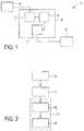
- FIG. 1shows an assistance system including an assistance device in accordance with an embodiment of the invention
- FIG. 2shows a flow diagram illustrating an assistance method in accordance with another embodiment of the invention.
- FIG. 3shows an exemplary X-ray angiogram with a vessel tree overlay from CT and marked measurement positions for pressure measurement in accordance with the invention.
- FIG. 1shows an assistance system 1 including an assistance device 2 in accordance with an embodiment of the invention.
- the assistance system 1includes the assistance device 2 , a data storage device 3 and a display device 4 .
- the data storage device 3stores image data of vessel geometry of the subject, in particular pre-interventional coronary angiography data obtained using computer tomography.
- the image datais provided to the assistance device 2 , which, in turn, outputs information to be displayed to the practitioner carrying out the interventional fractional flow reserve measurement to the display device 4 .
- the assistance device 2includes a model acquiring unit 5 , a position determination unit 6 and output unit 7 .
- the image data provided by the data storage device 3is received by the model acquiring unit 5 , which generates a vessel model based on the image data, the vessel model reflecting the vessel geometry of the subject.
- the vessel modelis a lumen and centerline model.
- This vessel modelis provided to the position determination unit 6 , which determines a set of positions for fractional flow reserve measurements based thereon.
- the position determination unit 6provides a plurality of simulations using a lumped parameter model for computational fluid dynamics.
- the stability of a solutionis tested by varying pressure values at different positions in the model vessel tree, such that the stability of the solution can be tested by analyzing the overall variation of the solution.
- the position determination unit 6takes into account information on a position and a degree of a stenosis in the vessel of the subject.
- the position determination unit 6arrives at a set of positions at which pressure measurements should be performed most optimally to deliver a stable and complete functional tree characterization of the vessel (tree) of the subject. This information is forwarded to the output unit 7 .
- the output unit 7registers the vessel model (vessel tree) to one or more invasive angiograms obtained in the context of the intervention.
- the vessel treeis overlaid onto a projection during the intervention, such that the measurement positions are displayed to the practitioner by means of the display device 4 .
- FIG. 2shows a flow diagram illustrating an assistance method in accordance with another embodiment of the invention.
- a vessel model of vessel geometry of the subjectis acquired.
- This vessel modelis used in a subsequent position determination step 11 , such that a set of positions for fractional flow reserve measurements to be carried out by the practitioner are determined based on the vessel model complying with a predetermined metric.
- the determined set of positionsis outputted to the practitioner.
- an overlay and display step 14the vessel tree is overlaid onto a projection shown to the practitioner during the intervention, such that the determined measurement positions (determined in the position determination step 11 ) are displayed to the practitioner.
- FIG. 3shows an exemplary X-ray angiogram with a vessel tree overlay from CT and marked measurement positions for pressure measurement in accordance with the invention.
- the display of the X-ray angiogram 20is supplemented by a display of a vessel tree including the heart 21 and blood vessels 22 of the subject, wherein this information is based on the vessel model acquired.
- measurement positions 23are indicated, such that the practitioner may carry out the interventional fractional flow reserve measurement accordingly.
- the ideal measurement pointsare supposed to provide the simulation with optimal input information to provide results with high consistency.
- pressure valuesare measured after multiple bifurcations one does not need to rely on heuristic assumptions of a scaling law to determine the relative flow distribution.
- the measurement position in the branchesshould not be arbitrary since e.g. further branching or a local narrowing may influence the result.
- a unfavorable measurement positionmay lead to false results, since other effects (e.g. branching or a general narrowing in distal vessels) may influence the measurement.
- An optimal measurement positionwould avoid other fluid dynamical effects to influence the targeted measurement.
- a pre-interventional CT (MR, 3DCA) data setis segmented, delivering a lumen and centerline model.
- the positionsare calculated, at which pressure measurements should be performed most optimally to deliver a stable and complete functional vessel tree characterization. This is achieved by a number of test simulations using a lumped parameter model for CFD calculation together with model boundary conditions.
- the stability of the solutionis tested by varying pressure values at different positions in the vessel tree and testing the stability of the solution by analyzing the overall variation of the solution.
- the segmentation accuracymay be a boundary condition for this analysis.
- the number of branches, the position of the branching points and the position of stenosiswill also determine the measurement positions. Further, at the beginning of the actual intervention the segmented vessel tree is registered to one or more invasive angiograms and subsequently, the vessel tree is overlaid onto the projection during the intervention and the pre-calculated measurement positions are displayed. As a result, a complete functional characterization of the vessel tree including pressure, flow and resistance data are achieved.
- temporal advicemay also be given, e.g. by analyzing the projection sequence with respect to the cardiac phase (e.g. by correlation of the 3D model to the 2D projection or via the ECG).
- Such variationsinclude, for example, giving advice about measurement time and/or position in any other application (besides fractional flow reserve) where hemodynamic parameters (e.g. pressure or flow) are interventionally measured, and where a vessel model can be constructed to predict these optimal measurements through fluid dynamic simulations.
- hemodynamic parameterse.g. pressure or flow
- a single processor, device or other unitmay fulfill the functions of several items recited in the claims.
- the mere fact that certain measures are recited in mutually different dependent claimsdoes not indicate that a combination of these measures cannot be used to advantage.
- Operations like acquiring, determining, outputting, providing, obtaining, calculating, simulating, receiving, and registeringcan be implemented as program code means of a computer program and/or as dedicated hardware.
- a computer programmay be stored and/or distributed on a suitable medium, such as an optical storage medium or a solid-state medium, supplied together with or as part of other hardware, but may also be distributed in other forms, such as via the Internet or other wired or wireless telecommunication systems.
- a suitable mediumsuch as an optical storage medium or a solid-state medium, supplied together with or as part of other hardware, but may also be distributed in other forms, such as via the Internet or other wired or wireless telecommunication systems.
Landscapes
- Health & Medical Sciences (AREA)
- Engineering & Computer Science (AREA)
- Medical Informatics (AREA)
- Public Health (AREA)
- Epidemiology (AREA)
- General Health & Medical Sciences (AREA)
- Primary Health Care (AREA)
- Data Mining & Analysis (AREA)
- Biomedical Technology (AREA)
- Databases & Information Systems (AREA)
- Pathology (AREA)
- Apparatus For Radiation Diagnosis (AREA)
- Measuring Pulse, Heart Rate, Blood Pressure Or Blood Flow (AREA)
- Measuring And Recording Apparatus For Diagnosis (AREA)
- Endoscopes (AREA)
- Ultra Sonic Daignosis Equipment (AREA)
- Image Analysis (AREA)
- Magnetic Resonance Imaging Apparatus (AREA)
Abstract
Description
Claims (14)
Applications Claiming Priority (4)
| Application Number | Priority Date | Filing Date | Title |
|---|---|---|---|
| EP15179819 | 2015-08-05 | ||
| EP15179819.6 | 2015-08-05 | ||
| EP15179819 | 2015-08-05 | ||
| PCT/EP2016/067725WO2017021201A1 (en) | 2015-08-05 | 2016-07-26 | Assistance device and method for an interventional hemodynamic measurement |
Publications (2)
| Publication Number | Publication Date |
|---|---|
| US20180211729A1 US20180211729A1 (en) | 2018-07-26 |
| US11031136B2true US11031136B2 (en) | 2021-06-08 |
Family
ID=53938093
Family Applications (1)
| Application Number | Title | Priority Date | Filing Date |
|---|---|---|---|
| US15/745,162Active2037-07-04US11031136B2 (en) | 2015-08-05 | 2016-07-26 | Assistance device and method for an interventional hemodynamic measurement |
Country Status (5)
| Country | Link |
|---|---|
| US (1) | US11031136B2 (en) |
| EP (1) | EP3332339B1 (en) |
| JP (1) | JP6738406B2 (en) |
| CN (1) | CN107851463B (en) |
| WO (1) | WO2017021201A1 (en) |
Cited By (10)
| Publication number | Priority date | Publication date | Assignee | Title |
|---|---|---|---|---|
| US11145224B2 (en)* | 2019-10-15 | 2021-10-12 | Ai Medic Inc. | Blood flow simulation method and apparatus for subject-specific blood vessel |
| US11541174B2 (en)* | 2019-07-19 | 2023-01-03 | Nexus Medical, Llc | Clinical assessment of an intravenous catheter site |
| US11666236B2 (en) | 2016-05-16 | 2023-06-06 | Cathworks Ltd. | System for vascular assessment |
| US11779233B2 (en) | 2019-03-19 | 2023-10-10 | Suzhou Rainmed Medical Technology Co., Ltd. | Method for calculating fractional flow reserve based on pressure sensor and angiographic image |
| US11937963B2 (en) | 2016-05-16 | 2024-03-26 | Cathworks Ltd. | Vascular selection from images |
| US12039685B2 (en) | 2019-09-23 | 2024-07-16 | Cathworks Ltd. | Methods, apparatus, and system for synchronization between a three-dimensional vascular model and an imaging device |
| US12079994B2 (en) | 2019-04-01 | 2024-09-03 | Cathworks Ltd. | Methods and apparatus for angiographic image selection |
| US12315076B1 (en) | 2021-09-22 | 2025-05-27 | Cathworks Ltd. | Four-dimensional motion analysis of a patient's coronary arteries and myocardial wall |
| US12354755B2 (en) | 2012-10-24 | 2025-07-08 | Cathworks Ltd | Creating a vascular tree model |
| US12387325B2 (en) | 2022-02-10 | 2025-08-12 | Cath Works Ltd. | System and method for machine-learning based sensor analysis and vascular tree segmentation |
Families Citing this family (7)
| Publication number | Priority date | Publication date | Assignee | Title |
|---|---|---|---|---|
| US11523744B2 (en) | 2017-03-31 | 2022-12-13 | Koninklijke Philips N.V. | Interaction monitoring of non-invasive imaging based FFR |
| EP3384850A1 (en)* | 2017-04-05 | 2018-10-10 | Koninklijke Philips N.V. | Method and apparatus for physiological functional parameter determination |
| EP3488774A1 (en) | 2017-11-23 | 2019-05-29 | Koninklijke Philips N.V. | Measurement guidance for coronary flow estimation from bernoulli´s principle |
| CN110226923B (en)* | 2018-03-05 | 2021-12-14 | 苏州润迈德医疗科技有限公司 | Method for measuring fractional flow reserve without vasodilator |
| US12048575B2 (en) | 2020-03-10 | 2024-07-30 | GE Precision Healthcare LLC | Systems and methods for registration of angiographic projections with computed tomographic data |
| EP4394694A1 (en)* | 2022-12-30 | 2024-07-03 | Siemens Healthineers AG | Determination of hemodynamic indices |
| CN118490168B (en)* | 2024-05-10 | 2025-03-21 | 中国人民解放军总医院第二医学中心 | A method and device for predicting blood flow reserve fraction based on coronary magnetic resonance imaging |
Citations (28)
| Publication number | Priority date | Publication date | Assignee | Title |
|---|---|---|---|---|
| WO2000072037A1 (en) | 1999-05-21 | 2000-11-30 | Nycomed Imaging As | Method of magnetic resonance imaging |
| WO2004025572A1 (en) | 2002-09-16 | 2004-03-25 | Tayside Flow Technologies Limited | A method of analysing fluid flow in a conduit |
| WO2006061815A1 (en) | 2004-12-08 | 2006-06-15 | Paieon Inc. | Method and apparatus for blood vessel parameter determinations |
| WO2006061814A1 (en) | 2004-12-08 | 2006-06-15 | Paieon Inc. | Method and apparatus for finding the tubular organs blood velocity and flow and related parameters |
| DE102008014792B3 (en) | 2008-03-18 | 2009-06-18 | Siemens Aktiengesellschaft | Method for simulation of flow of blood in vessel section, involves obtaining picture recording of vessel area containing vessel sections, where picture recording is obtained with assigned implant |
| US7574026B2 (en) | 2003-02-12 | 2009-08-11 | Koninklijke Philips Electronics N.V. | Method for the 3d modeling of a tubular structure |
| WO2010022762A2 (en) | 2008-08-25 | 2010-03-04 | ETH Zürich | Method, system and device for enhancing flow field data |
| US20100125197A1 (en) | 2008-11-18 | 2010-05-20 | Fishel Robert S | Method and apparatus for addressing vascular stenotic lesions |
| US20100130878A1 (en) | 2008-11-24 | 2010-05-27 | General Electric Company | Systems, apparatus and processes for automated blood flow assessment of vasculature |
| US20100241404A1 (en) | 2009-03-17 | 2010-09-23 | Taylor Charles A | Patient-specific hemodynamics of the cardio vascular system |
| US20110071404A1 (en)* | 2009-09-23 | 2011-03-24 | Lightlab Imaging, Inc. | Lumen Morphology and Vascular Resistance Measurements Data Collection Systems, Apparatus and Methods |
| US20110211742A1 (en) | 2008-09-30 | 2011-09-01 | Koninklijke Philips Electronics N.V. | Perfusion imaging |
| US20110307231A1 (en) | 2010-06-09 | 2011-12-15 | Jens Kirchner | Method and arrangement for creating an individualized, computer-aided model of a system, and a corresponding computer program and a corresponding machine-readable storage medium |
| US20120022843A1 (en) | 2010-07-21 | 2012-01-26 | Razvan Ioan Ionasec | Method and System for Comprehensive Patient-Specific Modeling of the Heart |
| US20120041739A1 (en) | 2010-08-12 | 2012-02-16 | Heartflow, Inc. | Method and System for Patient-Specific Modeling of Blood Flow |
| US20120041325A1 (en) | 2007-03-14 | 2012-02-16 | Ramesh Wariar | Method and apparatus for management of heart failure hospitalization |
| US20120072190A1 (en) | 2010-09-16 | 2012-03-22 | Siemens Corporation | Method and System for Non-Invasive Assessment of Coronary Artery Disease |
| US8157742B2 (en) | 2010-08-12 | 2012-04-17 | Heartflow, Inc. | Method and system for patient-specific modeling of blood flow |
| US20120121151A1 (en) | 2010-11-12 | 2012-05-17 | Siemens Aktiengesellschaft | Device And Computed Tomography Scanner For Determining And Visualizing The Perfusion Of The Myocardial Muscle |
| US8200466B2 (en) | 2008-07-21 | 2012-06-12 | The Board Of Trustees Of The Leland Stanford Junior University | Method for tuning patient-specific cardiovascular simulations |
| US20120243761A1 (en) | 2011-03-21 | 2012-09-27 | Senzig Robert F | System and method for estimating vascular flow using ct imaging |
| US20120296199A1 (en) | 2011-03-21 | 2012-11-22 | New York University | Apparatus and Method of Non-Contrast Magnetic Resonance Angiography of Abdominal and Pelvic Arteries |
| WO2013071219A1 (en) | 2011-11-10 | 2013-05-16 | Siemens Corporation | Method and system for multi-scale anatomical and functional modeling of coronary circulation |
| US20140114618A1 (en) | 2012-10-19 | 2014-04-24 | Timothy A. Fonte | Systems and methods for numerically evaluating vasculature |
| WO2014111930A1 (en) | 2013-01-15 | 2014-07-24 | Cathworks Ltd. | Creating a vascular tree model |
| WO2014127320A1 (en) | 2013-02-15 | 2014-08-21 | The Johns Hopkins University | Computational flow dynamics based method for estimating thromboembolic risk in patients with myocardial infarction |
| US20150112191A1 (en) | 2013-10-22 | 2015-04-23 | Koninklijke Philips Electronics N.V. | Fractional flow reserve (ffr) index with adaptive boundary condition parameters |
| WO2015082576A1 (en) | 2013-12-04 | 2015-06-11 | Koninklijke Philips N.V. | Local ffr estimation and visualisation for improved functional stenosis analysis |
Family Cites Families (4)
| Publication number | Priority date | Publication date | Assignee | Title |
|---|---|---|---|---|
| US10373700B2 (en)* | 2012-03-13 | 2019-08-06 | Siemens Healthcare Gmbh | Non-invasive functional assessment of coronary artery stenosis including simulation of hyperemia by changing resting microvascular resistance |
| WO2014027692A1 (en)* | 2012-08-16 | 2014-02-20 | 株式会社東芝 | Image-processing device, diagnostic medical image apparatus and blood pressure monitor |
| US9424395B2 (en)* | 2013-03-04 | 2016-08-23 | Heartflow, Inc. | Method and system for sensitivity analysis in modeling blood flow characteristics |
| WO2015024934A1 (en)* | 2013-08-21 | 2015-02-26 | Koninklijke Philips N.V. | Segmentation apparatus for interactively segmenting blood vessels in angiographic image data |
- 2016
- 2016-07-26CNCN201680045787.0Apatent/CN107851463B/enactiveActive
- 2016-07-26JPJP2018505001Apatent/JP6738406B2/enactiveActive
- 2016-07-26WOPCT/EP2016/067725patent/WO2017021201A1/ennot_activeCeased
- 2016-07-26EPEP16744717.6Apatent/EP3332339B1/enactiveActive
- 2016-07-26USUS15/745,162patent/US11031136B2/enactiveActive
Patent Citations (38)
| Publication number | Priority date | Publication date | Assignee | Title |
|---|---|---|---|---|
| WO2000072037A1 (en) | 1999-05-21 | 2000-11-30 | Nycomed Imaging As | Method of magnetic resonance imaging |
| WO2004025572A1 (en) | 2002-09-16 | 2004-03-25 | Tayside Flow Technologies Limited | A method of analysing fluid flow in a conduit |
| US7574026B2 (en) | 2003-02-12 | 2009-08-11 | Koninklijke Philips Electronics N.V. | Method for the 3d modeling of a tubular structure |
| WO2006061815A1 (en) | 2004-12-08 | 2006-06-15 | Paieon Inc. | Method and apparatus for blood vessel parameter determinations |
| WO2006061814A1 (en) | 2004-12-08 | 2006-06-15 | Paieon Inc. | Method and apparatus for finding the tubular organs blood velocity and flow and related parameters |
| US20120041325A1 (en) | 2007-03-14 | 2012-02-16 | Ramesh Wariar | Method and apparatus for management of heart failure hospitalization |
| DE102008014792B3 (en) | 2008-03-18 | 2009-06-18 | Siemens Aktiengesellschaft | Method for simulation of flow of blood in vessel section, involves obtaining picture recording of vessel area containing vessel sections, where picture recording is obtained with assigned implant |
| US8200466B2 (en) | 2008-07-21 | 2012-06-12 | The Board Of Trustees Of The Leland Stanford Junior University | Method for tuning patient-specific cardiovascular simulations |
| WO2010022762A2 (en) | 2008-08-25 | 2010-03-04 | ETH Zürich | Method, system and device for enhancing flow field data |
| US20110211742A1 (en) | 2008-09-30 | 2011-09-01 | Koninklijke Philips Electronics N.V. | Perfusion imaging |
| US20100125197A1 (en) | 2008-11-18 | 2010-05-20 | Fishel Robert S | Method and apparatus for addressing vascular stenotic lesions |
| US20100130878A1 (en) | 2008-11-24 | 2010-05-27 | General Electric Company | Systems, apparatus and processes for automated blood flow assessment of vasculature |
| US20100241404A1 (en) | 2009-03-17 | 2010-09-23 | Taylor Charles A | Patient-specific hemodynamics of the cardio vascular system |
| US20110071404A1 (en)* | 2009-09-23 | 2011-03-24 | Lightlab Imaging, Inc. | Lumen Morphology and Vascular Resistance Measurements Data Collection Systems, Apparatus and Methods |
| US20110307231A1 (en) | 2010-06-09 | 2011-12-15 | Jens Kirchner | Method and arrangement for creating an individualized, computer-aided model of a system, and a corresponding computer program and a corresponding machine-readable storage medium |
| US20120022843A1 (en) | 2010-07-21 | 2012-01-26 | Razvan Ioan Ionasec | Method and System for Comprehensive Patient-Specific Modeling of the Heart |
| US20120041318A1 (en)* | 2010-08-12 | 2012-02-16 | Heartflow, Inc. | Method and system for patient-specific modeling of blood flow |
| US8249815B2 (en) | 2010-08-12 | 2012-08-21 | Heartflow, Inc. | Method and system for patient-specific modeling of blood flow |
| US20120041322A1 (en) | 2010-08-12 | 2012-02-16 | Heartflow, Inc. | Method and system for patient-specific modeling of blood flow |
| US20120041319A1 (en) | 2010-08-12 | 2012-02-16 | Heartflow, Inc. | Method and system for patient-specific modeling of blood flow |
| US20120041320A1 (en) | 2010-08-12 | 2012-02-16 | Heartflow, Inc. | Method and system for patient-specific modeling of blood flow |
| US20120041321A1 (en) | 2010-08-12 | 2012-02-16 | Heartflow, Inc. | Method and system for patient-specific modeling of blood flow |
| US20120041324A1 (en) | 2010-08-12 | 2012-02-16 | Heartflow, Inc. | Method and system for patient-specific modeling of blood flow |
| US20120053919A1 (en) | 2010-08-12 | 2012-03-01 | Heartflow, Inc. | Method and system for patient-specific modeling of blood flow |
| US20120059246A1 (en) | 2010-08-12 | 2012-03-08 | Heartflow, Inc. | Method and system for patient-specific modeling of blood flow |
| US8157742B2 (en) | 2010-08-12 | 2012-04-17 | Heartflow, Inc. | Method and system for patient-specific modeling of blood flow |
| US20120041323A1 (en) | 2010-08-12 | 2012-02-16 | Heartflow, Inc. | Method and system for patient-specific modeling of blood flow |
| US20120041739A1 (en) | 2010-08-12 | 2012-02-16 | Heartflow, Inc. | Method and System for Patient-Specific Modeling of Blood Flow |
| US20120072190A1 (en) | 2010-09-16 | 2012-03-22 | Siemens Corporation | Method and System for Non-Invasive Assessment of Coronary Artery Disease |
| US20120121151A1 (en) | 2010-11-12 | 2012-05-17 | Siemens Aktiengesellschaft | Device And Computed Tomography Scanner For Determining And Visualizing The Perfusion Of The Myocardial Muscle |
| US20120243761A1 (en) | 2011-03-21 | 2012-09-27 | Senzig Robert F | System and method for estimating vascular flow using ct imaging |
| US20120296199A1 (en) | 2011-03-21 | 2012-11-22 | New York University | Apparatus and Method of Non-Contrast Magnetic Resonance Angiography of Abdominal and Pelvic Arteries |
| WO2013071219A1 (en) | 2011-11-10 | 2013-05-16 | Siemens Corporation | Method and system for multi-scale anatomical and functional modeling of coronary circulation |
| US20140114618A1 (en) | 2012-10-19 | 2014-04-24 | Timothy A. Fonte | Systems and methods for numerically evaluating vasculature |
| WO2014111930A1 (en) | 2013-01-15 | 2014-07-24 | Cathworks Ltd. | Creating a vascular tree model |
| WO2014127320A1 (en) | 2013-02-15 | 2014-08-21 | The Johns Hopkins University | Computational flow dynamics based method for estimating thromboembolic risk in patients with myocardial infarction |
| US20150112191A1 (en) | 2013-10-22 | 2015-04-23 | Koninklijke Philips Electronics N.V. | Fractional flow reserve (ffr) index with adaptive boundary condition parameters |
| WO2015082576A1 (en) | 2013-12-04 | 2015-06-11 | Koninklijke Philips N.V. | Local ffr estimation and visualisation for improved functional stenosis analysis |
Non-Patent Citations (3)
| Title |
|---|
| Cheng_2008 (A computational fluid dynamic study of stent graft remodeling after endovascular repair of thoratic aortic dissections, 2008, The Society of Vascular Surgery). (Year: 2008).* |
| Liu_2004 (Catheter-Based Intraluminal Sonography, 2004 American Institute of Ultrasound in Medicine). (Year: 2004).* |
| Unit_Set_2015 (Set (Mathematics) Wikipedia Archived dated Aug. 2, 2015 downloaded from https://en.wikipedia.org/w/index.php?title=Set_(mathematics)&oldid=674281577 ). (Year: 2015).* |
Cited By (13)
| Publication number | Priority date | Publication date | Assignee | Title |
|---|---|---|---|---|
| US12354755B2 (en) | 2012-10-24 | 2025-07-08 | Cathworks Ltd | Creating a vascular tree model |
| US12138027B2 (en) | 2016-05-16 | 2024-11-12 | Cath Works Ltd. | System for vascular assessment |
| US12408885B2 (en) | 2016-05-16 | 2025-09-09 | Cathworks Ltd. | Vascular selection from images |
| US11666236B2 (en) | 2016-05-16 | 2023-06-06 | Cathworks Ltd. | System for vascular assessment |
| US11937963B2 (en) | 2016-05-16 | 2024-03-26 | Cathworks Ltd. | Vascular selection from images |
| US11779233B2 (en) | 2019-03-19 | 2023-10-10 | Suzhou Rainmed Medical Technology Co., Ltd. | Method for calculating fractional flow reserve based on pressure sensor and angiographic image |
| US12079994B2 (en) | 2019-04-01 | 2024-09-03 | Cathworks Ltd. | Methods and apparatus for angiographic image selection |
| US11541174B2 (en)* | 2019-07-19 | 2023-01-03 | Nexus Medical, Llc | Clinical assessment of an intravenous catheter site |
| US12039685B2 (en) | 2019-09-23 | 2024-07-16 | Cathworks Ltd. | Methods, apparatus, and system for synchronization between a three-dimensional vascular model and an imaging device |
| US11145224B2 (en)* | 2019-10-15 | 2021-10-12 | Ai Medic Inc. | Blood flow simulation method and apparatus for subject-specific blood vessel |
| US12315076B1 (en) | 2021-09-22 | 2025-05-27 | Cathworks Ltd. | Four-dimensional motion analysis of a patient's coronary arteries and myocardial wall |
| US12387325B2 (en) | 2022-02-10 | 2025-08-12 | Cath Works Ltd. | System and method for machine-learning based sensor analysis and vascular tree segmentation |
| US12423813B2 (en) | 2022-02-10 | 2025-09-23 | Cathworks Ltd. | System and method for machine-learning based sensor analysis and vascular tree segmentation |
Also Published As
| Publication number | Publication date |
|---|---|
| JP6738406B2 (en) | 2020-08-12 |
| US20180211729A1 (en) | 2018-07-26 |
| EP3332339B1 (en) | 2022-06-22 |
| WO2017021201A1 (en) | 2017-02-09 |
| CN107851463B (en) | 2022-11-22 |
| JP2018533383A (en) | 2018-11-15 |
| CN107851463A (en) | 2018-03-27 |
| EP3332339A1 (en) | 2018-06-13 |
Similar Documents
| Publication | Publication Date | Title |
|---|---|---|
| US11031136B2 (en) | Assistance device and method for an interventional hemodynamic measurement | |
| RU2695262C2 (en) | Stenosis assessment | |
| US10052032B2 (en) | Stenosis therapy planning | |
| US9891044B2 (en) | Method and device for determining deviation in pressure in a blood vessel | |
| JP6661613B2 (en) | System and method for automatically determining myocardial bridge and effect on a patient | |
| US12115014B2 (en) | Most relevant x-ray image selection for hemodynamic simulation | |
| US12380562B2 (en) | Vessel registration using functional information | |
| EP3125764A1 (en) | Processing apparatus and method for processing cardiac data of a living being | |
| EP3456243A1 (en) | Improved vessel geometry and additional boundary conditions for hemodynamic ffr/ifr simulations from intravascular imaging | |
| US20250127484A1 (en) | Aortic abdominal aneurysm biomarkers | |
| EP3382583A1 (en) | Hemodynamic simulation of movement inducted vascular deformations | |
| JP7443197B2 (en) | Medical image processing device, system and method |
Legal Events
| Date | Code | Title | Description |
|---|---|---|---|
| AS | Assignment | Owner name:KONINKLIJKE PHILIPS N.V., NETHERLANDS Free format text:ASSIGNMENT OF ASSIGNORS INTEREST;ASSIGNORS:GRASS, MICHAEL;HAASE, CHRISTIAN;RONGEN, PETER MARIA JOHANNES;AND OTHERS;SIGNING DATES FROM 20160812 TO 20180111;REEL/FRAME:044625/0705 | |
| FEPP | Fee payment procedure | Free format text:ENTITY STATUS SET TO UNDISCOUNTED (ORIGINAL EVENT CODE: BIG.); ENTITY STATUS OF PATENT OWNER: LARGE ENTITY | |
| STPP | Information on status: patent application and granting procedure in general | Free format text:DOCKETED NEW CASE - READY FOR EXAMINATION | |
| STPP | Information on status: patent application and granting procedure in general | Free format text:FINAL REJECTION MAILED | |
| STPP | Information on status: patent application and granting procedure in general | Free format text:ADVISORY ACTION MAILED | |
| STPP | Information on status: patent application and granting procedure in general | Free format text:DOCKETED NEW CASE - READY FOR EXAMINATION | |
| STPP | Information on status: patent application and granting procedure in general | Free format text:NON FINAL ACTION MAILED | |
| STPP | Information on status: patent application and granting procedure in general | Free format text:NOTICE OF ALLOWANCE MAILED -- APPLICATION RECEIVED IN OFFICE OF PUBLICATIONS | |
| STPP | Information on status: patent application and granting procedure in general | Free format text:PUBLICATIONS -- ISSUE FEE PAYMENT RECEIVED | |
| STPP | Information on status: patent application and granting procedure in general | Free format text:PUBLICATIONS -- ISSUE FEE PAYMENT VERIFIED | |
| STCF | Information on status: patent grant | Free format text:PATENTED CASE | |
| AS | Assignment | Owner name:JPMORGAN CHASE BANK, N.A., AS ADMINISTRATIVE AGENT, TEXAS Free format text:SECURITY INTEREST;ASSIGNORS:ESTIS COMPRESSION, LLC;FLOWCO PRODUCTIONS LLC;PATRIOT ARTIFICIAL LIFT, LLC;AND OTHERS;REEL/FRAME:068762/0857 Effective date:20240820 | |
| MAFP | Maintenance fee payment | Free format text:PAYMENT OF MAINTENANCE FEE, 4TH YEAR, LARGE ENTITY (ORIGINAL EVENT CODE: M1551); ENTITY STATUS OF PATENT OWNER: LARGE ENTITY Year of fee payment:4 |