US11026618B2 - Intracardiac EGM signals for beat matching and acceptance - Google Patents
Intracardiac EGM signals for beat matching and acceptanceDownload PDFInfo
- Publication number
- US11026618B2 US11026618B2US16/384,077US201916384077AUS11026618B2US 11026618 B2US11026618 B2US 11026618B2US 201916384077 AUS201916384077 AUS 201916384077AUS 11026618 B2US11026618 B2US 11026618B2
- Authority
- US
- United States
- Prior art keywords
- signals
- preprocessed
- intracardiac
- beat
- catheters
- Prior art date
- Legal status (The legal status is an assumption and is not a legal conclusion. Google has not performed a legal analysis and makes no representation as to the accuracy of the status listed.)
- Active, expires
Links
- 230000000694effectsEffects0.000claimsabstractdescription14
- 238000000034methodMethods0.000claimsdescription164
- 230000000747cardiac effectEffects0.000claimsdescription153
- 238000013507mappingMethods0.000claimsdescription104
- 239000011159matrix materialSubstances0.000claimsdescription15
- 238000007781pre-processingMethods0.000claimsdescription13
- 230000000877morphologic effectEffects0.000claimsdescription7
- 230000001131transforming effectEffects0.000claimsdescription3
- 238000012935AveragingMethods0.000claims2
- 230000008569processEffects0.000description127
- 238000012545processingMethods0.000description49
- 238000001514detection methodMethods0.000description42
- 230000000875corresponding effectEffects0.000description28
- 230000033764rhythmic processEffects0.000description22
- 238000010586diagramMethods0.000description14
- 210000005242cardiac chamberAnatomy0.000description12
- 230000006870functionEffects0.000description11
- 230000009466transformationEffects0.000description9
- 238000005259measurementMethods0.000description8
- 230000004913activationEffects0.000description7
- 238000001994activationMethods0.000description7
- 230000002596correlated effectEffects0.000description5
- 210000001174endocardiumAnatomy0.000description5
- 238000012986modificationMethods0.000description4
- 230000004048modificationEffects0.000description4
- 230000009467reductionEffects0.000description4
- 210000003484anatomyAnatomy0.000description3
- 238000002679ablationMethods0.000description2
- 230000004931aggregating effectEffects0.000description2
- 230000015572biosynthetic processEffects0.000description2
- 230000003750conditioning effectEffects0.000description2
- 238000010276constructionMethods0.000description2
- 238000002595magnetic resonance imagingMethods0.000description2
- 230000004044responseEffects0.000description2
- 238000005070samplingMethods0.000description2
- 230000001360synchronised effectEffects0.000description2
- 238000002604ultrasonographyMethods0.000description2
- 240000007817Olea europaeaSpecies0.000description1
- 206010042600Supraventricular arrhythmiasDiseases0.000description1
- 206010047281Ventricular arrhythmiaDiseases0.000description1
- 230000009471actionEffects0.000description1
- 238000007792additionMethods0.000description1
- 238000002583angiographyMethods0.000description1
- 206010003119arrhythmiaDiseases0.000description1
- 230000006793arrhythmiaEffects0.000description1
- 230000001746atrial effectEffects0.000description1
- 230000036772blood pressureEffects0.000description1
- 238000013153catheter ablationMethods0.000description1
- 238000006243chemical reactionMethods0.000description1
- 210000003748coronary sinusAnatomy0.000description1
- 230000008878couplingEffects0.000description1
- 238000010168coupling processMethods0.000description1
- 238000005859coupling reactionMethods0.000description1
- 230000005284excitationEffects0.000description1
- 238000001914filtrationMethods0.000description1
- 238000002594fluoroscopyMethods0.000description1
- 210000002837heart atriumAnatomy0.000description1
- 238000003780insertionMethods0.000description1
- 230000037431insertionEffects0.000description1
- 238000004519manufacturing processMethods0.000description1
- 230000007246mechanismEffects0.000description1
- 238000002324minimally invasive surgeryMethods0.000description1
- HLXZNVUGXRDIFK-UHFFFAOYSA-Nnickel titaniumChemical compound[Ti].[Ti].[Ti].[Ti].[Ti].[Ti].[Ti].[Ti].[Ti].[Ti].[Ti].[Ni].[Ni].[Ni].[Ni].[Ni].[Ni].[Ni].[Ni].[Ni].[Ni].[Ni].[Ni].[Ni].[Ni]HLXZNVUGXRDIFK-UHFFFAOYSA-N0.000description1
- 229910001000nickel titaniumInorganic materials0.000description1
- 230000003287optical effectEffects0.000description1
- 230000002093peripheral effectEffects0.000description1
- 238000012805post-processingMethods0.000description1
- 230000029058respiratory gaseous exchangeEffects0.000description1
- 230000035945sensitivityEffects0.000description1
- 239000012781shape memory materialSubstances0.000description1
- 230000003595spectral effectEffects0.000description1
- 230000001225therapeutic effectEffects0.000description1
- 230000002861ventricularEffects0.000description1
Images


Classifications
- A—HUMAN NECESSITIES
- A61—MEDICAL OR VETERINARY SCIENCE; HYGIENE
- A61B—DIAGNOSIS; SURGERY; IDENTIFICATION
- A61B5/00—Measuring for diagnostic purposes; Identification of persons
- A61B5/74—Details of notification to user or communication with user or patient; User input means
- A61B5/7475—User input or interface means, e.g. keyboard, pointing device, joystick
- A—HUMAN NECESSITIES
- A61—MEDICAL OR VETERINARY SCIENCE; HYGIENE
- A61B—DIAGNOSIS; SURGERY; IDENTIFICATION
- A61B5/00—Measuring for diagnostic purposes; Identification of persons
- A61B5/24—Detecting, measuring or recording bioelectric or biomagnetic signals of the body or parts thereof
- A61B5/316—Modalities, i.e. specific diagnostic methods
- A61B5/318—Heart-related electrical modalities, e.g. electrocardiography [ECG]
- A61B5/346—Analysis of electrocardiograms
- A61B5/349—Detecting specific parameters of the electrocardiograph cycle
- A61B5/35—Detecting specific parameters of the electrocardiograph cycle by template matching
- A—HUMAN NECESSITIES
- A61—MEDICAL OR VETERINARY SCIENCE; HYGIENE
- A61B—DIAGNOSIS; SURGERY; IDENTIFICATION
- A61B5/00—Measuring for diagnostic purposes; Identification of persons
- A61B5/02—Detecting, measuring or recording for evaluating the cardiovascular system, e.g. pulse, heart rate, blood pressure or blood flow
- A61B5/024—Measuring pulse rate or heart rate
- A61B5/0245—Measuring pulse rate or heart rate by using sensing means generating electric signals, i.e. ECG signals
- A—HUMAN NECESSITIES
- A61—MEDICAL OR VETERINARY SCIENCE; HYGIENE
- A61B—DIAGNOSIS; SURGERY; IDENTIFICATION
- A61B5/00—Measuring for diagnostic purposes; Identification of persons
- A61B5/24—Detecting, measuring or recording bioelectric or biomagnetic signals of the body or parts thereof
- A—HUMAN NECESSITIES
- A61—MEDICAL OR VETERINARY SCIENCE; HYGIENE
- A61B—DIAGNOSIS; SURGERY; IDENTIFICATION
- A61B5/00—Measuring for diagnostic purposes; Identification of persons
- A61B5/24—Detecting, measuring or recording bioelectric or biomagnetic signals of the body or parts thereof
- A61B5/25—Bioelectric electrodes therefor
- A61B5/279—Bioelectric electrodes therefor specially adapted for particular uses
- A61B5/28—Bioelectric electrodes therefor specially adapted for particular uses for electrocardiography [ECG]
- A61B5/283—Invasive
- A61B5/287—Holders for multiple electrodes, e.g. electrode catheters for electrophysiological study [EPS]
- A—HUMAN NECESSITIES
- A61—MEDICAL OR VETERINARY SCIENCE; HYGIENE
- A61B—DIAGNOSIS; SURGERY; IDENTIFICATION
- A61B5/00—Measuring for diagnostic purposes; Identification of persons
- A61B5/24—Detecting, measuring or recording bioelectric or biomagnetic signals of the body or parts thereof
- A61B5/316—Modalities, i.e. specific diagnostic methods
- A61B5/318—Heart-related electrical modalities, e.g. electrocardiography [ECG]
- A61B5/333—Recording apparatus specially adapted therefor
- A—HUMAN NECESSITIES
- A61—MEDICAL OR VETERINARY SCIENCE; HYGIENE
- A61B—DIAGNOSIS; SURGERY; IDENTIFICATION
- A61B5/00—Measuring for diagnostic purposes; Identification of persons
- A61B5/24—Detecting, measuring or recording bioelectric or biomagnetic signals of the body or parts thereof
- A61B5/316—Modalities, i.e. specific diagnostic methods
- A61B5/318—Heart-related electrical modalities, e.g. electrocardiography [ECG]
- A61B5/339—Displays specially adapted therefor
- A—HUMAN NECESSITIES
- A61—MEDICAL OR VETERINARY SCIENCE; HYGIENE
- A61B—DIAGNOSIS; SURGERY; IDENTIFICATION
- A61B5/00—Measuring for diagnostic purposes; Identification of persons
- A61B5/72—Signal processing specially adapted for physiological signals or for diagnostic purposes
- A61B5/7203—Signal processing specially adapted for physiological signals or for diagnostic purposes for noise prevention, reduction or removal
Definitions
- This disclosurerelates to systems and methods for providing information about a patient's heart and, in particular, to systems and methods for electro-anatomically mapping the patient's heart.
- cardiac mappingTo perform the cardiac mapping, a catheter with one or more electrodes can be inserted into the patient's heart.
- Cardiac mapping techniquesinclude contact mapping, near contact mapping, and non-contact mapping.
- contact mappingone or more catheters are advanced into the heart and physiological signals resulting from the electrical activity of the heart are acquired with one or more electrodes located at the catheter distal tip after determining that the tip is in stable and steady contact with the endocardial surface of a heart chamber.
- the location and electrical activitycan be measured on a point-by-point basis at, for example, about 50 to 200 points on the internal surface of the heart to construct an electro-anatomical depiction of the heart.
- a movable catheter having multiple spatially distributed electrodesis placed in a heart chamber of interest and moved to one or more locations within the chamber of interest, where the electrodes are on or near, such as within millimeters of, the endocardial surface of the heart chamber. Measurements are taken automatically at each of the locations of the catheter, without determining whether the electrodes are in contact with the surface of the heart. These measurements are analyzed to detect the endocardial surface of the heart chamber in the vicinity of the catheter.
- the location of the cathetere.g., a location provided by a tracking system, and the measurements from the electrodes are used to reconstruct the chamber anatomy, where, for example, 20,000 measurements may be made to construct an electro-anatomical depiction of the heart.
- a partial or complete representation of the chamber anatomycan be constructed.
- non-contact mappinga multiple electrode catheter is placed in the heart chamber of interest and the catheter is deployed to assume a three dimensional shape.
- the systemuses the signals detected by the non-contact electrodes and information on chamber anatomy and relative electrode location, the system calculates and provides physiological information regarding the endocardial surface of the heart chamber.
- the generated mapmay then serve as the basis for deciding on a therapeutic course of action, such as tissue ablation, to alter the propagation of the heart's electrical activity and to restore normal heart rhythm.
- the mapping processdetermines whether a cardiac beat matches a target beat morphology. If the cardiac beat matches the target beat morphology, the signals obtained in conjunction with the cardiac beat can be mapped into a map dataset for the target beat morphology. If the cardiac beat does not match the target beat morphology, the signals obtained in conjunction with the cardiac beat may be discarded. Often, electrocardiogram (ECG) signals are used to detect the similarity between the cardiac beat and the target beat morphology.
- ECGelectrocardiogram
- Example 1is a system for providing information about a patient's heart.
- the systemincludes one or more catheters that receive intracardiac signals from electrical activity of the heart over one or more heart beat cycles.
- the systembeing characterized by an electronic processor coupled to the one or more catheters to: receive the intracardiac signals from the one or more catheters; preprocess the intracardiac signals to provide preprocessed signals, wherein each of the intracardiac signals is preprocessed to provide a corresponding preprocessed signal; and compare the preprocessed signals to a set of signals to determine a degree of similarity between each of the preprocessed signals and the set of signals.
- Example 2is the system of Example 1, wherein to preprocess the intracardiac signals the electronic processor is to: apply a transform function to the intracardiac signals to provide transformed signals, wherein each of the intracardiac signals is transformed to a corresponding transformed signal; and apply a morphological close operation to each of the transformed signals to reduce noise in the preprocessed signals.
- Example 3is the system of Example 2, wherein the transform function is a Hilbert transform and the close operation is a non-boolean close operation.
- Example 4is the system of any of Examples 1-3, wherein the electronic processor is to: detect a beat of the heart; define a window for samples of the intracardiac signals based on the beat; and provide a matrix of the samples of the intracardiac signals in the window.
- Example 5is the system of any of Examples 1-4, wherein the one or more catheters include one or more reference catheters situated at stable positions in the patient's body and the set of signals includes a template of signals determined from the intracardiac signals received by the one or more reference catheters.
- Example 6is the system of any of Examples 1-5, wherein to compare the preprocessed signals the electronic processor is to: receive individual threshold values for each of the intracardiac signals; set a master threshold value; provide dynamic threshold values for each of the intracardiac signals based on the individual threshold values and the master threshold value; compare the preprocessed signals to the set of signals to obtain comparison values for the intracardiac signals; and compare the comparison values to the dynamic threshold values to determine whether a beat of the intracardiac signals is to be accepted into an existing cardiac map.
- Example 7is the system of any of Examples 1-6, wherein to compare the preprocessed signals the electronic processor is to: receive individual threshold values for each of the intracardiac signals; compare the preprocessed signals to the set of signals to obtain comparison values for the intracardiac signals; and compare the comparison values to the individual threshold values to determine whether a beat of the intracardiac signals is to be accepted.
- Example 8is the system of any of Examples 1-7, wherein to compare the preprocessed signals the electronic processor is to: receive individual threshold values for each of the intracardiac signals; and compare each of the individual threshold values to a fixed value and if all of the individual threshold values are less than the fixed value, accept one or more beats of the intracardiac signals.
- Example 9is the system of any of Examples 1-8, wherein the one or more catheters include one or more mapping catheters positioned in the patient's heart and the set of signals includes a set of signals from the one or more mapping catheters and a previously detected beat of the heart.
- Example 10is the system of any of Examples 1-9, wherein to compare the preprocessed signals the electronic processor is to: compare the preprocessed signals to the set of signals from the one or more mapping catheters and a previously detected beat of the heart to obtain previous beat comparison values; and compare each of the previous beat comparison values to a corresponding threshold value to determine whether a beat of the intracardiac signals is to be accepted.
- Example 11is the system of any of Examples 1-10, wherein the electronic processor is to determine whether to accept the intracardiac signals into a cardiac map based on the degree of similarity, and the electronic processor is to: classify the intracardiac signals into a current existing cardiac map; classify the intracardiac signals into another existing cardiac map; or make a new cardiac map based on the intracardiac signals.
- Example 12is a method for mapping a patient's heart.
- the methodincludes receiving intracardiac signals from electrical activity of the heart over one or more heart beat cycles at one or more catheters.
- the methodbeing characterized by the steps of: receiving the intracardiac signals at an electronic processor that is coupled to the one or more catheters; preprocessing the intracardiac signals, by the electronic processor, to provide preprocessed signals, wherein each of the intracardiac signals is preprocessed to provide a corresponding preprocessed signal; and comparing the preprocessed signals, by the electronic processor, to a set of signals to determine a degree of similarity between each of the preprocessed signals and the set of signals.
- Example 13is the method of Example 12, wherein preprocessing the intracardiac signals includes: transforming each of the intracardiac signals via a transform function; and applying a morphological close operation to reduce noise in the preprocessed signals.
- Example 14is the method of any of Examples 12 and 13, wherein the one or more catheters include one or more reference catheters situated at stable positions in the patient's body and the set of signals includes a template of signals determined from the intracardiac signals received by the one or more reference catheters.
- Example 15is the method of any of Examples 12-14, wherein the one or more catheters include one or more mapping catheters positioned in the patient's heart and the set of signals includes a set of signals from the one or more mapping catheters and a previously detected beat of the heart.
- Example 16is a system for providing information about a patient's heart.
- the systemincludes one or more catheters that receive intracardiac signals from electrical activity of the heart over one or more heart beat cycles and an electronic processor coupled to the one or more catheters.
- the electronic processorto: receive the intracardiac signals from the one or more catheters; preprocess the intracardiac signals to provide preprocessed signals, wherein each of the intracardiac signals is preprocessed to provide a corresponding preprocessed signal; compare the preprocessed signals to a set of signals to determine a degree of similarity between each of the preprocessed signals and the set of signals; and determine whether to accept one or more beats of the intracardiac signals into an existing cardiac map based on the degree of similarity.
- Example 17is the system of Example 16, wherein to preprocess the intracardiac signals the electronic processor is to: apply a transform function to the intracardiac signals to provide transformed signals, wherein each of the intracardiac signals is transformed to a corresponding transformed signal; and apply a morphological close operation to each of the transformed signals to reduce noise in the preprocessed signals.
- Example 18is the system of Example 17, wherein the transform function is a Hilbert transform and the close operation is a non-boolean close operation.
- Example 19is the system of Example 16, wherein the electronic processor is to: detect a beat of the heart; define a window for samples of the intracardiac signals based on the beat; and provide a matrix of the samples of the intracardiac signals in the window.
- Example 20is the system of Example 16, wherein the one or more catheters include one or more reference catheters situated at stable positions in the patient's body and the set of signals includes a template of signals determined from the intracardiac signals received by the one or more reference catheters.
- Example 21is the system of Example 16, wherein the one or more catheters include one or more mapping catheters positioned in the patient's heart and the set of signals includes a set of signals from the one or more mapping catheters and a previously detected beat of the heart.
- Example 22is the system of Example 16, wherein to compare the preprocessed signals the electronic processor is to: receive individual threshold values for each of the intracardiac signals; set a master threshold value; provide dynamic threshold values for each of the intracardiac signals based on the individual threshold values and the master threshold value; compare the preprocessed signals to the set of signals to obtain comparison values for the intracardiac signals; and compare the comparison values to the dynamic threshold values to determine whether a beat of the intracardiac signals is to be accepted into an existing cardiac map.
- Example 23is the system of Example 16, wherein to compare the preprocessed signals the electronic processor is to: receive individual threshold values for each of the intracardiac signals; compare the preprocessed signals to the set of signals to obtain comparison values for the intracardiac signals; and compare the comparison values to the individual threshold values to determine whether a beat of the intracardiac signals is to be accepted into an existing cardiac map.
- Example 24is the system of Example 16, wherein to compare the preprocessed signals the electronic processor is to: receive individual threshold values for each of the intracardiac signals; and compare each of the individual threshold values to a fixed value and if all of the individual threshold values are less than the fixed value, accept one or more beats of the intracardiac signals into an existing cardiac map.
- Example 25is the system of Example 16, wherein to compare the preprocessed signals the electronic processor is to: compare the preprocessed signals to a set of signals from one or more mapping catheters and a previously detected beat of the heart to obtain previous beat comparison values; and compare each of the previous beat comparison values to a corresponding threshold value to determine whether a beat of the intracardiac signals is to be accepted.
- Example 26is the system of Example 16, wherein the electronic processor is to determine whether to: classify the intracardiac signals into the current cardiac map; classify the intracardiac signals into another existing cardiac map; or make a new cardiac map based on the intracardiac signals.
- Example 27is a system for providing information about a patient's heart.
- the systemincludes one or more catheters that receive intracardiac signals from electrical activity of the heart over one or more heart beat cycles and an electronic processor coupled to the one or more catheters.
- the electronic processorto receive the intracardiac signals from the one or more catheters and preprocess the intracardiac signals to provide preprocessed signals, wherein the electronic processor is to apply a transform function to the intracardiac signals to provide transformed signals, wherein each of the intracardiac signals is transformed to a corresponding transformed signal and apply a close operation to each of the transformed signals to reduce noise in the preprocessed signals.
- the electronic processoris to correlate the preprocessed signals to a set of signals to determine a degree of correlation between each of the preprocessed signals and the set of signals and determine whether to accept the intracardiac signals into an existing cardiac map based on the degree of correlation.
- Example 28is the system of Example 27, wherein the one or more catheters include one or more reference catheters situated at stable positions in the patient's body and the set of signals includes a template of signals determined from the intracardiac signals received by the one or more reference catheters.
- Example 29is the system of Example 27, wherein the one or more catheters include one or more mapping catheters positioned in the patient's heart and the set of signals includes a set of signals from the one or more mapping catheters and a previously detected beat of the heart.
- Example 30is a method for mapping a patient's heart. The method including: receiving intracardiac signals from electrical activity of the heart over one or more heart beat cycles at one or more catheters; receiving the intracardiac signals at an electronic processor that is coupled to the one or more catheters; preprocessing the intracardiac signals, by the electronic processor, to provide preprocessed signals, wherein each of the intracardiac signals is preprocessed to provide a corresponding preprocessed signal; comparing the preprocessed signals, by the electronic processor, to a set of signals to determine a degree of similarity between each of the preprocessed signals and the set of signals; and determining whether to accept the intracardiac signals into an existing cardiac map based on the degree of similarity.
- Example 31is the method of Example 30, wherein preprocessing the intracardiac signals includes transforming each of the intracardiac signals via a transform function and applying a close operation to reduce noise in the preprocessed signals.
- Example 32is the method of Example 30, wherein comparing the preprocessed signals comprises: receiving individual threshold values for each of the intracardiac signals; setting a master threshold value; providing dynamic threshold values for each of the intracardiac signals based on the individual threshold values and the master threshold value; comparing the preprocessed signals to the set of signals to obtain comparison values for the intracardiac signals; and comparing the comparison values to the dynamic threshold values to determine whether a beat of the intracardiac signals is to be accepted into an existing cardiac map.
- Example 33is the method of Example 30, wherein comparing the preprocessed signals comprises: receiving individual threshold values for each of the intracardiac signals; comparing the preprocessed signals to the set of signals to obtain comparison values; and comparing the comparison values to the individual threshold values to determine whether a beat of the intracardiac signals is to be accepted into an existing cardiac map.
- Example 34is the method of Example 30, wherein comparing the preprocessed signals comprises: receiving individual threshold values for each of the intracardiac signals; and comparing each of the individual threshold values to a fixed value and if all of the individual threshold values are less than the fixed value, accept one or more beats of the intracardiac signals into an existing cardiac map.
- Example 35is the method of Example 30, wherein comparing the preprocessed signals comprises: comparing the preprocessed signals to a set of signals from one or more mapping catheters and a previously detected beat of the heart to obtain previous beat comparison values; and comparing each of the previous beat comparison values to a corresponding threshold value to determine whether a beat of the intracardiac signals is to be accepted.
- FIG. 1is a diagram illustrating an electro-anatomical mapping system for mapping cardiac rhythms of a patient, according to embodiments of the disclosure.
- FIG. 2is a flowchart diagram illustrating an electro-anatomical mapping process, according to embodiments of the disclosure.
- FIG. 3is a flowchart diagram illustrating a data stream and a beat detection and processing process, according to embodiments of the disclosure.
- FIG. 4is a flowchart diagram illustrating formation of a template of preprocessed intracardiac EGM signals that can be used in a comparison process, according to embodiments of the disclosure.
- FIG. 5is a flowchart diagram illustrating a comparison process, according to embodiments of the disclosure.
- FIG. 6is a flowchart diagram illustrating another comparison process, according to embodiments of the disclosure.
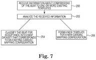
- FIG. 7is a flowchart diagram illustrating a classification process, according to embodiments of the disclosure.
- the present disclosuredescribes systems and methods that use intracardiac electrogram (EGM) signals for detecting and processing cardiac beats.
- the detected and processed beatscan be compared to target beat morphologies, accepted into map datasets, and/or classified into different cardiac beat morphologies.
- This beat detection and processingincludes beat detection, beat comparison, and beat classification that uses metrics derived from comparing the incoming intracardiac EGM signals to another set of intracardiac EGM signals.
- the other set of intracardiac EGM signalsincludes a template of intracardiac EGM signals.
- the other set of intracardiac EGM signalsincludes a set of intracardiac EGM signals obtained from a previous cardiac beat, such as the most recent previous cardiac beat.
- Embodiments of the systems and methods described hereinmay add signals to selected cardiac beat morphologies, check for consistencies between neighboring cardiac beats, and/or automatically classify the cardiac beats into different cardiac beat morphologies.
- Embodiments of systems and methods described hereinmay be used for both ventricular and atrial cardiac mapping.
- FIG. 1is a diagram illustrating an electro-anatomical mapping system 20 for mapping cardiac rhythms of a patient 22 using intracardiac EGM signals for beat detection and processing, according to embodiments described in the disclosure.
- the system 20can be operated by a user 24 , such as a physician and/or a technician.
- the system 20includes one or more catheters 26 , each having one or more electrodes situated at or toward the distal end of the catheter 26 .
- the one or more catheters 26can be situated in or near the heart of the patient 22 .
- the system 20obtains intracardiac EGM signals from the one or more electrodes on the catheters 26 .
- the one or more catheters 26include one or more reference catheters, where each of the reference catheters includes one or more electrodes and is secured in place in a stable position in or near the heart.
- the one or more catheters 26include up to five reference catheters, each having one or more electrodes and being secured in place in a stable position in or near the heart.
- the one or more reference cathetersinclude at least one coronary sinus catheter.
- the one or more catheters 26include one or more mapping catheters, where each of the mapping catheters includes one or more electrodes and can be moved from one location to another in the heart. In embodiments that include the one or more mapping catheters, at least one of the mapping catheters can be displaced to multiple locations within the heart during the signal acquisition stage of a mapping procedure, where the acquisition of signals at multiple catheter locations in the heart chamber enables the one or more mapping catheters to effectively act as a “mega-catheter” whose effective number of electrodes and electrode span is proportional to the product of the number of locations in which signal acquisition is performed and the number of electrodes on the one or more mapping catheters. In some embodiments, the one or more mapping catheters are configured for contact mapping.
- the one or more mapping cathetersare configured for near-contact mapping. In some embodiments, the one or more mapping catheters are configured for non-contact mapping. In some embodiments, the electrodes are mounted on the one or more mapping catheters following a three dimensional olive shape, where the electrodes are mounted on a device capable of deploying the electrodes into the desired shape while inside the heart and capable of retracting the electrodes when the catheter is removed from the heart. In embodiments, to allow deployment into the three dimensional shape, the electrodes may be mounted on a balloon or shape memory material, such as Nitinol.
- the one or more mapping cathetersare moved to more than three locations, such as more than 5, 10, or even 50 locations within the heart chamber. Further, the spatial range over which the one or more mapping catheters are moved may be larger than one third (1 ⁇ 3) of the diameter of the heart cavity, such as larger than 35%, 40%, 50% or even 60% of the diameter of the heart cavity.
- the one or more electrodes on the one or more catheters 26receive intracardiac EGM signals resulting from electrical activity in the heart cavity.
- the intracardiac EGM signalsare used in the beat detection and processing process and can provide, to the user 24 , physiological data pertaining to the heart's electrical activity.
- the physiological informationis computed based on signals measured over several heart beats, either at a single catheter location within the heart chamber or over several locations.
- the measurementscan be synchronized with one another so that the measurements are performed, and/or analyzed, with respect to approximately the same phase of the heart cycle.
- the signal measurements over multiple beatscan be synchronized based on features detected from physiological data, such as, for example, a surface electrocardiogram (ECG) or the intracardiac EGM signals.
- ECGsurface electrocardiogram
- the system 20includes a processing unit 28 , which may be, or include, a processor that executes code stored in internal memory 30 and/or in a storage device 32 to perform operations pertaining to embodiments of a mapping procedure.
- the internal memory 30 and/or the storage device 32also, or alternatively, may store data acquired by the one or more electrodes of the one or more catheters 26 .
- the processing unit 28is an electronic processor, which may be, at least in part, a software processor.
- the processing unit 28is communicatively coupled to the one or more catheters 26 and receives the intracardiac EGM signals from the one or more electrodes on the one or more catheters 26 .
- the processing unit 28executes code from memory, such as the internal memory 30 and/or the storage device 32 , to process the intracardiac EGM signals for beat detection and processing, including beat detection, beat comparison, and beat classification for the cardiac rhythm.
- the processing unit 28executes code to preprocess the intracardiac EGM signals and provide preprocessed signals, where each of the intracardiac EGM signals is preprocessed to provide a corresponding preprocessed signal. Also, the processing unit 28 executes code to compare the preprocessed signals to another set of preprocessed intracardiac EGM signals and determine a degree of similarity between each of the preprocessed signals and the other set of preprocessed intracardiac EGM signals. This degree of comparison can be used as a metric for determining how closely the signals compare to or match an existing cardiac map configuration, for acceptance of the detected beat into a map dataset, and for classification of the detected beat into a cardiac rhythm morphology.
- the processing unit 28executes code to correlate the preprocessed signals to the other set of preprocessed intracardiac EGM signals and determines a degree of correlation between each of the preprocessed signals and the other set of preprocessed intracardiac EGM signals, where this degree of correlation can be used as a metric for determining how closely the signals compare to or match an existing cardiac map configuration, for acceptance of the detected beat into a map dataset, and for classification of the detected beat into a cardiac rhythm morphology.
- the one or more catheters 26include one or more reference catheters secured in place at stable locations in the patient's body, where the intracardiac EGM signals are obtained from the one or more reference catheters and the other set of preprocessed intracardiac EGM signals is a template of preprocessed intracardiac EGM signals that were determined from intracardiac EGM signals previously received by the one or more reference catheters.
- the one or more catheters 26include one or more mapping catheters positioned in the patient's heart, where the intracardiac EGM signals are obtained from the one or more mapping catheters and the other set of signals is a set of intracardiac EGM signals obtained from the one or more mapping catheters from a previously detected beat of the heart, such as the most recent previously detected beat of the heart.
- the processing unit 28executes code to determine whether to accept the intracardiac EGM signals into a cardiac map based on the degree of similarity, which in some embodiments is a degree of correlation. In embodiments, the processing unit 28 executes code to classify the intracardiac EGM signals for inclusion in a current existing cardiac map, another existing cardiac map, or a new cardiac map.
- the processing unit 28executes code from memory to process the intracardiac EGM signals with beat detection, beat comparison, and beat classification criteria for existing cardiac mapping configurations that may include existing cardiac mapping configurations that correspond to the current existing cardiac map and other existing cardiac maps. This provides information about how well the signals match the cardiac rhythm of the existing cardiac mapping configurations.
- the processing unit 28executes a reconstruction procedure to determine the physiological information at the endocardium surface.
- the processing unit 28may compute, prior to the insertion of the catheter 26 into the heart chamber and/or before signal acquisition by the catheter's electrodes has commenced, transformation functions that can be used, during a mapping procedure, to facilitate the reconstruction process.
- the mapping proceduremay be performed expeditiously by computing those transformation components that were not computed ahead of the signal acquisition stage, and combining those components with the appropriate pre-processed transformation components to obtain the overall transformation function(s).
- the overall transformation functionmay be applied to the acquired raw data to perform an inverse reconstruction operation.
- the processing unit 28may also perform a catheter registration procedure.
- the location of the one or more catheters 26 in the heart chambermay be determined using a conventional sensing and tracking system (not shown) that provides the three dimensional spatial coordinates of the one or more catheters 26 and/or its multiple electrodes with respect to the catheter's coordinate system as established by the sensing and tracking system.
- a conventional sensing and tracking systemnot shown
- the processing unit 28 or another processing module of the system 20may be configured to determine a coordinate system transformation function that transforms the three dimensional spatial coordinates of the catheter's locations into coordinates expressed in terms of the endocardium surface's coordinate system, or vice-versa.
- the processing unit 28performs post-processing operations on the reconstructed physiological information to extract and display useful features of the information to the operator of the system 20 and/or other persons, such as a physician.
- the intracardiac EGM signals acquired by the one or more electrodes of the one or more catheters 26may be passed to the processing unit 28 via a signal conditioning module 34 that receives the signals from the one or more catheters 26 and performs signal enhancement operations on the signals before they are forwarded to the processing unit 28 .
- Signal conditioning hardwaremay be used to amplify, filter, and continuously sample intracardiac potential measured by one or more electrodes.
- the intracardiac signalshave maximum amplitudes of 60 mV and mean amplitudes of a few millivolts.
- the signalsare bandpass filtered in a frequency range, such as 0.5-500 Hz, and sampled with analog to digital converters, such as converters with 15-bit resolution at 1 kHz.
- the signalsmay be filtered to remove one or more frequencies corresponding to the equipment.
- Other types of signal processing operationsmay be implemented, such as, for example, spectral equalization, automatic gain control, and/or the like.
- the resultant processed signalsare forwarded by the module 34 to the processing unit 28 for further processing.
- the system 20includes a user interface 36 and, optionally, peripheral devices, such as a printer 38 , which are communicatively coupled to the processing unit 28 .
- the user interface 36includes one or more display devices 40 and input devices, such as a mouse 42 and a keyboard 44 .
- the user interface 36may receive signals from the processing unit 28 and display information about which of the existing cardiac mapping configurations more closely matches or compares to the cardiac beat, including information about whether the beat more closely matches the cardiac rhythm that corresponds to the current map or the cardiac rhythm that corresponds to a different one of the existing cardiac maps. In some embodiments, with the user interface 32 displaying this information, the user 24 can quickly and easily determine whether to add the cardiac beat into the current existing cardiac map or another one of the existing cardiac maps.
- the user interface 36displays this information while the system 20 is adding signals into the current map.
- the user interface 36includes a graphical user interface that includes a touch screen, which can be used for switching from adding beats to the current map to adding beats to another one of the existing maps or a new cardiac map.
- FIG. 2is a flowchart diagram illustrating an electro-anatomical mapping process, according to embodiments of the disclosure.
- the electro-anatomical mapping process of FIG. 2may be performed, at least in part, by the electro-anatomical mapping system 20 of FIG. 1 .
- the processing unit 28executes computer code stored in the internal memory 30 and/or storage device 32 to facilitate the electro-anatomical mapping process of FIG. 2 .
- a data stream 100 containing multiple signalsis input into the system (e.g., the mapping system 20 depicted in FIG. 1 ).
- the data stream 100provides a collection of physiological and non-physiological signals and information that serve as inputs to the mapping process.
- the data stream 100includes signals, such as unipolar or bipolar intracardiac EGM signals, received from one or more electrodes on the one or more catheters 26 .
- the data stream 100can include signals and/or information, such as ECG signals, electrode and/or catheter location information originating from a variety of methodologies including magnetic, impedance, ultrasound, fluoroscopy, and real time magnetic resonance imaging (MRI) methodologies, tissue proximity information, catheter force or contact information such as from force spring sensing, piezo-electric sensing, and optical sensing, catheter tip and/or tissue temperature, acoustic information, catheter electrical coupling information, respiration phase, blood pressure, and/or other physiological information.
- the data stream 100can contain information such as catheter shape and electrode properties. The signals and information can be collected directly by the mapping system and/or obtained from another system using an analog or digital interface.
- a beat detection and processing process 102receives the data stream 100 and processes the data to compare the data to existing cardiac mapping configurations for different beat morphologies, to accept the data into an existing map dataset, and/or to classify the data into one or more cardiac beat morphologies.
- the beat detection and processing process 102can provide information for determining whether the signals in the data stream 100 should be added to the current map, added to another existing cardiac map, or added to a new cardiac map.
- the beat detection and processing process 102includes a beat detection process 104 , a beat comparison process 106 , and a beat classification process 108 .
- the beat detection and processing process 102processes the data with beat detection and beat acceptance criteria to provide information about how well the signals compare to existing cardiac mapping configurations.
- the beat detection and processing process 102can process the data with beat detection and beat acceptance criteria for the current map of the existing maps into which data are currently being added, and with beat detection and beat acceptance criteria for one or more other existing maps.
- the beat detection and processing process 102processes the data with beat detection and beat acceptance criteria for all existing cardiac mapping configurations.
- the beat detection and processing process 102processes the data with beat detection and beat acceptance criteria for a subset of all existing cardiac mapping configurations.
- the beat detection and processing process 102continuously (or continually) processes the data or signals with beat detection and beat acceptance criteria, such as by sequentially comparing different criteria to the incoming data.
- the beat detection and processing process 102displays the degree of similarity or matching between the incoming data and the existing cardiac mapping configurations via a user interface, such as the user interface 36 .
- the beat detection and processing process 102may display the degree of similarity for determining whether the cardiac rhythm of an existing cardiac mapping configuration more closely matches the incoming data. With this information displayed on the user interface, the user may quickly and easily determine whether the incoming data should be added to the current map, or added to another existing map or added to a new cardiac map.
- the beat detection and processing process 102displays at least some of the above information on the user interface while adding the incoming data to the current map.
- the beat detection process 104includes triggering from one or more signals in the data stream 100 via a trigger event, defining a window about the trigger event, and providing a matrix of signal names and signal samples from the window.
- One or more of the signals in the data stream 100can be used as a reference signal for triggering relative to the cardiac rhythm of the incoming data.
- the beat detection process 104detects a trigger event around which a window of data is sampled from the data stream 100 . These samples, from the window of data, are put into the matrix of signal names and signal samples and provided in a beat dataset for the trigger event.
- the trigger eventis detected from one or more intracardiac EGM signals designated as a reference signal.
- waveform attributessuch as minimum/maximum, absolute maximum, maximum/minimum slope, and/or first deviation from baseline are used to detect a trigger event.
- a single signal sourcecan be selected for triggering, such as a single intracardiac EGM signal from one electrode on the one or more catheters 26 .
- multiple intracardiac EGM signalscan be used to determine a trigger event, which can be advantageous as more stable than triggering schemes based on a single signal.
- an electro-anatomical mapsuch as an activation map
- a stable referencesuch as constructing an anatomical shell
- one or more of the signals in the incoming data stream 100can be used as a reference for triggering and alignment of the data stream 100 relative to a biological cycle other than the cardiac rhythm and/or to a system clock.
- the beat detection process 104provides a beat dataset that includes the matrix of signal names and signal samples from the window of data around the trigger event for the beat dataset.
- Each of the signal names in the matrixis referred to as a signal channel, i.e., a channel.
- the beat datasetsare received by the beat comparison process 106 that determines metrics for each of the beat datasets. These metrics can be used to: compare a beat dataset to an existing cardiac mapping configuration; make a decision about whether to accept a beat dataset into a map dataset 110 for the current existing cardiac map, another existing cardiac map, or a new cardiac map; and/or classify the beat dataset into a certain cardiac rhythm morphology.
- the beat comparison process 106includes a preprocessing process and a comparison process, which in some embodiments is a correlation process.
- the preprocessing processthe intracardiac EGM signals are converted or transformed into preprocessed signals, where each of the intracardiac EGM signals is preprocessed to provide a corresponding preprocessed signal.
- the comparison processreceives the preprocessed signals and compares the preprocessed signals to another set of preprocessed intracardiac EGM signals to determine the degree of similarity between each of the preprocessed signals and the other set of preprocessed intracardiac EGM signals. Comparison values determined in the comparison process can be used as beat metrics.
- the comparison processincludes a correlation process that receives the preprocessed signals and correlates the preprocessed signals to another set of preprocessed intracardiac EGM signals to determine the degree of correlation between each of the preprocessed signals and the other set of preprocessed intracardiac EGM signals, where the correlation values determined in the correlation process can be used as beat metrics.
- the comparison processincludes a different process, other than correlation, that is used to compare the preprocessed signals to another set of preprocessed intracardiac EGM signals to determine the degree of similarity between each of the preprocessed signals and the other set of preprocessed intracardiac EGM signals.
- the one or more catheters 26can include one or more reference catheters secured in place at stable locations in the patient's body, where the intracardiac EGM signals are obtained from the one or more reference catheters and the other set of preprocessed intracardiac EGM signals is a template of preprocessed intracardiac EGM signals that were previously received via the one or more reference catheters and preprocessed to form the template.
- the one or more catheters 26include one or more mapping catheters positioned in the patient's heart, where the intracardiac EGM signals are obtained from the one or more mapping catheters and the other set of signals is a set of intracardiac EGM signals obtained from the one or more mapping catheters from a previously detected beat of the heart.
- a number of beat metricsare computed for each of the beat datasets. These beat metrics can be computed using information from a single signal spanning one or more beats, over multiple signals within the same beat, and/or from multiple signals spanning multiple beats. The beat metrics provide multiple types of information on the quality of a beat dataset and the likelihood that the beat data in the beat dataset is acceptable for inclusion in a map dataset 110 .
- the classification process 108aggregates the metrics and determines which of the existing cardiac mapping configurations more closely compares to or matches the beat dataset and whether the beat dataset can be added to one of the existing map datasets 110 .
- the classification process 108can classify the beat datasets for acceptance into map datasets 110 for the current existing cardiac map, other existing cardiac maps, or a new cardiac map based on the degree of similarity.
- the classification process 108can indicate the degree of similarity between the incoming data stream 100 and the existing cardiac mapping configurations, including the existing cardiac mapping configurations that correspond to the current map and at least one other existing cardiac map.
- a usere.g., user 24
- the systeme.g., system 20
- the beat detection and processing process 102indicates the degree of similarity between the incoming data and at least two of the existing cardiac mapping configurations, including the existing cardiac mapping configuration that corresponds to the current map, by displaying the percentage of beat datasets that are accepted and can be added into each of the different map datasets 110 over a predetermined period of time.
- the electro-anatomical mapping process of FIG. 2continues with a surface map generation process 120 that is employed to generate surface map data from the map datasets 110 and surface geometry data 118 .
- the surface geometry data 118may be generated concurrently, or at least during the same data acquisition process, using identical or different triggering and beat acceptance metrics employing a surface geometry construction process 112 .
- the surface geometry construction process 112may construct surface geometry using data such as electrode locations and catheter shape contained in the data stream 100 .
- previously collected surface geometry 116may be used as an input to the surface map data.
- Such surface geometry 116may be collected in the same procedure using a different map dataset or using a different modality such as CT, MRI, ultrasound, and/or rotational angiography, and/or registered to the catheter locating system.
- a systemsuch as system 20 may select the source of the surface geometry data at 114 and provide surface geometry data 118 to the surface map generation process 120 .
- the surface map generation process 120generates surface map data 122 that can provide information on cardiac electrical excitation, cardiac motion, tissue proximity information, tissue impedance information, force information, and/or any other collected and/or derived information.
- the surface map data 122may be further processed to annotate desired features from the underlying data, a process defined herein as surface map annotation 124 . Desired annotations may include instantaneous potential, activation time, voltage amplitude, dominant frequency and/or other properties of the signal.
- the annotationsmay be displayed superimposed on chamber geometry. If the number of annotations is lower than the number of elements that make up the display of surface geometry, surface map interpolation 126 may be employed. Displayed maps 128 may be computed and displayed separately, combined, and/or overlaid on top of each other.
- FIG. 3is a flowchart diagram illustrating the data stream 100 and the beat detection and processing process 102 , according to embodiments of the disclosure.
- the beat detection and processing process 102includes the beat detection process 104 , the beat comparison process 106 , and the beat classification process 108 , as shown in FIG. 2 and described in the description of FIG. 2 .
- the beat detection and processing process 102processes the data stream 100 to provide information about how closely the signals in the data stream 100 compare to existing cardiac mapping configurations.
- the beat detection and processing process 102processes the data with existing cardiac mapping configurations, including beat detection and beat acceptance criteria, for the current map and one or more other existing maps for determining whether the signals in the data stream 100 should be added to the current map, added to another existing cardiac map, or added to a new cardiac map.
- the beat detection process 104receives the data stream 100 and provides triggering 110 of the data stream 100 via a trigger event, defining a window 112 about the trigger event, and providing a matrix of signal names and signal samples 114 from the window, where each of the signal names in the matrix is referred to as a signal channel, i.e., a channel.
- the beat detection process 104identifies a trigger event, such as an activation of a cardiac rhythm in the heart, in the data stream 100 .
- the trigger eventindicates when a beat occurs and is detected in the data stream 100 using one or more of the signals or channels in the data stream 100 as a reference signal or channel.
- the trigger eventis detected from one or more intracardiac EGM signals or channels designated as a reference signal or channel.
- waveform attributessuch as minimum/maximum, absolute maximum, maximum/minimum slope, and/or first deviation from baseline are used to detect a trigger event.
- a single signal sourcecan be selected for triggering, such as a single intracardiac EGM signal or channel from one electrode on the one or more catheters 26 .
- multiple intracardiac EGM signals or channelscan be used to determine a trigger event.
- triggering 110identifies activations in an atrium of the heart. In some embodiments, triggering 110 identifies activations in a ventricle of the heart. In some embodiments, schemes, such as beat blanking, are used to enhance identification of a trigger event and a beat.
- the beat detection process 104defines a window in time around or about the trigger event.
- the windowidentifies the part of the beat that may be useful in determining cardiac rhythm morphology and other characteristics about the beat.
- the window sizecan be 100-300 milliseconds long.
- the beat detection system 104samples at least some of the intracardiac EGM signals in the data stream 100 during the window to obtain a number of samples for each of the signals.
- the names of the signals sampled (the channels) and the samplesare put into a matrix of signal names and signal samples and provided in a beat dataset for the trigger event.
- the signalscan be sampled at a predetermined sampling rate. In some embodiments, the sampling rate is 1 KHz. In some embodiments, the number of samples is from 100-300 samples
- the beat detection process 104provides a beat dataset that includes the matrix of signal names and signal samples from the window of data around the trigger event.
- the beat datasetsare received by the beat comparison process 106 that determines beat metrics for each of the beat datasets. These beat metrics can be used to: compare a beat dataset to an existing cardiac mapping configuration; make a decision about whether to accept a beat dataset into a map dataset 110 for the current existing cardiac map, another existing cardiac map, or a new cardiac map; and/or classify the beat dataset into a certain cardiac rhythm morphology.
- the beat comparison process 106includes preprocessing 116 of the matrix of signal names and signal samples into preprocessed signals and comparison 118 of the preprocessed signals to other preprocessed signals.
- the preprocessing 116includes transformation 120 of the signals in the matrix into transformed signals and a close operation 122 on the transformed signals.
- the comparison 118includes correlation of the preprocessed signals to other preprocessed signals.
- the intracardiac EGM signals in the matrixare converted or transformed into transformed signals.
- Each of the intracardiac EGM signalsis converted or transformed into a corresponding transformed signal.
- the transformation 120includes applying a Hilbert transform on each of the intracardiac EGM signals to provide a corresponding transformed signal.
- the magnitude of the signalwhich is a local estimate of power, is computed, where each of the transformed signals is substantially an envelope of the power in the signal.
- another type of transform or conversioncan be used to show signal content and amplitude.
- a noise reduction operationis performed on each of the transformed signals to provide preprocessed signals.
- the noise reduction operationreduces noise, fills in gaps, and/or removes artifacts in the transformed signals to provide the preprocessed signals.
- the close operationincludes a morphological close operation.
- the close operationincludes shape preserving morphological noise reduction to reduce sensitivity to subtle signal variations.
- the close operationincludes shape preserving morphological noise reduction using a plus or minus 5 millisecond greyscale.
- the close operation 122is similar to an image processing operation for removing noise, filling in gaps, and/or removing artifacts.
- the close operation 122is a non-boolean close operation.
- the close operation 122includes bandpass filtering of the signals for frequency selectivity.
- the output of the close operation 122is a preprocessed signal, also referred to as an activation waveform, which is provided for metric computational stages and/or template production.
- the comparison process 118receives the preprocessed signals from the close operation 122 and compares the preprocessed signals to another set of preprocessed intracardiac EGM signals to determine a degree of similarity between each of the preprocessed signals and the other set of preprocessed intracardiac EGM signals.
- the comparison values determined in the comparison process 118are used as beat metrics that indicate how closely the preprocessed signals, and the beat dataset from which they came, compare to or match the beat morphology or cardiac rhythm shape represented by the other set of preprocessed intracardiac EGM signals.
- the comparison process 118compares the preprocessed signals to multiple other sets of preprocessed intracardiac EGM signals to determine a degree of similarity between the preprocessed signals and each of the other sets of preprocessed intracardiac EGM signals and their cardiac rhythm shapes.
- the comparison process 118includes a correlation process that receives the preprocessed signals from the close operation 122 and correlates the preprocessed signals to another set of preprocessed intracardiac EGM signals to determine a degree of correlation between each of the preprocessed signals and the other set of preprocessed intracardiac EGM signals.
- the correlation values determined in the correlation processare used as beat metrics that indicate how closely the preprocessed signals, and the beat dataset from which they came, compare to or match the beat morphology or cardiac rhythm shape represented by the other set of preprocessed intracardiac EGM signals.
- the intracardiac EGM signalsare obtained from one or more reference catheters and the other set of preprocessed intracardiac EGM signals is a template of preprocessed intracardiac EGM signals.
- the templaterepresents one beat morphology or cardiac rhythm shape and the intracardiac EGM signals used to form the template are received via the one or more reference catheters and preprocessed to form the template.
- the comparison process 118compares the preprocessed signals to one or more templates to determine the degree of similarity between the preprocessed signals and each of the one or more templates and their cardiac rhythm shapes.
- the intracardiac EGM signalsare obtained from one or more mapping catheters and the other set of signals is a set of intracardiac EGM signals obtained from the one or more mapping catheters from a previously detected beat of the heart, such as the most recent previously detected beat of the heart.
- the classification process 108aggregates the comparison value metrics and determines which of the existing cardiac mapping configurations more closely compares to the beat dataset and whether the beat dataset can be added to one of the existing map datasets 110 .
- the classification process 108can classify the beat datasets for acceptance into map datasets 110 for the current existing cardiac map, other existing cardiac maps, or a new cardiac map based on the degree of similarity.
- the comparison valuesare determined for multiple templates and the classification process 108 classifies the beat dataset for adding to map datasets 110 for the current existing cardiac map, other existing cardiac maps, and/or a new cardiac map based on the comparison values.
- FIG. 4is a flowchart diagram illustrating the formation of a template of preprocessed intracardiac EGM signals, which can be used in the comparison process 118 , according to embodiments of the disclosure.
- the preprocessed intracardiac EGM signals to be compared to the template in the comparison process 118are obtained via one or more reference catheters situated in stable positions in or near the heart.
- the data stream 150includes intracardiac EGM signals obtained from the same one or more reference catheters.
- 10 seconds of datais obtained from the data stream 150 .
- This 10 seconds of datacontains a number of beats of the heart, such as 10 to 15 beats.
- a systemsuch as system 20 detects and preprocesses the beats in the 10 seconds of data.
- each of the intracardiac EGM signals of interest in each of the beatsis preprocessed to provide a corresponding preprocessed signal.
- the systemuses the beat detection process 104 and the preprocessing 116 from the beat comparison process 106 to detect beats and preprocess the signals at 154 .
- a usersuch as a physician or a technician, selects one of the beats in the 10 seconds of data as a favorite beat, where the favorite beat often represents a cardiac rhythm morphology that the user wants to map.
- the systemcompares the preprocessed signals of the favorite beat to the preprocessed signals of the other beats.
- each of the preprocessed intracardiac EGM signals from the favorite beatis compared to each of the corresponding preprocessed intracardiac EGM signals from the other preprocessed beats.
- a beatis considered similar to the favorite beat if the signals in the beat compare favorably to the signals in the favorite beat.
- the corresponding signals in the similar beatsare averaged together to form averaged signals in the template.
- the preprocessed intracardiac EGM signals from the selected beatare correlated to the preprocessed intracardiac EGM signals from the other preprocessed beats.
- a beatis considered similar to the favorite beat if all reference signals or channels in the beats have a minimum correlation value of greater than or equal to 0.8.
- the beats that are deemed to be similar beatsare averaged to form a template of preprocessed signals.
- each of the preprocessed intracardiac EGM signalsis averaged with the corresponding preprocessed intracardiac EGM signals of other similar beats to obtain average signals for each of the preprocessed intracardiac signals in the template.
- each beat that was used to form the templatehas its signals compared to the averaged signals in the template to determine individual threshold values for each of the signal channels. These individual thresholds are confidence levels that indicate the amount of information in and the confidence a user can have in a particular channel.
- each of the preprocessed intracardiac EGM signals of a beat used to form the templateis correlated to each of the corresponding averaged preprocessed intracardiac EGM signals in the template to obtain the individual thresholds.
- FIG. 5is a flowchart diagram illustrating a comparison process 118 , according to embodiments of the disclosure.
- the comparison process 118includes a correlation process and acceptance/rejection criteria for correlating preprocessed intracardiac EGM signals of a beat to the averaged preprocessed intracardiac EGM signals of a template and determining whether to accept or reject the beat as matching the template.
- the intracardiac EGM signals of the beatwere obtained via one or more reference catheters and the intracardiac EGM signals used to form the template were also obtained via the one or more reference catheters.
- the templateis formed using the process of FIG. 4 . As described above, each of the channels or signal names in the template has an individual threshold value.
- the comparison process 118receives the preprocessed intracardiac EGM signals (activation waveforms) of a beat from the close operation 122 of preprocessing 116 and the comparison process 188 receives the individual thresholds for each of the channels.
- the comparison process 118compares each of the individual thresholds to a fixed value and, at 174 , if all of the individual threshold values are less than the fixed value, the beat is accepted as matching the template. If one or more of the individual threshold values is greater than the fixed value, processing can continue at 176 .
- the fixed valueis zero and if the individual thresholds are all less than or equal to zero the beats are accepted as matching the template.
- the comparison process 118correlates the preprocessed intracardiac EGM signals of the beat to the averaged preprocessed intracardiac EGM signals of the template.
- Each of the preprocessed intracardiac EGM signals of a beatis correlated to the averaged preprocessed intracardiac EGM signals in the template to provide a correlation value for that signal of the beat.
- other comparisons and/or correlationscan be used to determine the correlation value of a signal to the template.
- the comparison process 118compares each of the correlation values of the beat to each corresponding individual threshold value for the channel and, at 180 , if all of the correlation values obtained at 176 are greater than or equal to the corresponding individual threshold values, the beat is accepted as matching the template, otherwise processing can continue at 182 .
- the comparison process 118sets a master threshold value.
- the master threshold valueis seen by the user 24 and can be set by the user 24 or the system 20 .
- the master threshold valuemodulates each of the individual thresholds of the channels to provide a dynamic threshold value for each of the channels.
- the comparison process 118calculates a dynamic threshold value for each of the channels based on the individual threshold value for the channel and the master threshold value.
- the comparison process 118compares each of the correlation values of the beat to each corresponding dynamic threshold value for the channel and, at 188 , if all of the correlation values obtained at 176 are greater than or equal to the corresponding dynamic threshold values, the beat is accepted as matching the template. If the beat is not accepted, via any of the above criteria, at 174 , 180 , and 188 , the beat is rejected. This follows the principal that one bad or wrong signal makes the entire beat unacceptable and eliminates beats if any of the signals in the beat do not provide a correlation value that is greater than or equal to the individual threshold value or the dynamic threshold value for that channel.
- the dynamic threshold valuescan be calculated differently.
- each of the individual threshold valuescan be multiplied by the master threshold value to obtain a dynamic threshold value. For example, if the master threshold level is set to 0.6, then each of the individual threshold values is multiplied by 0.6 to provide the dynamic threshold level for that particular signal name/channel.
- FIG. 6is a flowchart diagram illustrating the comparison process 118 correlating preprocessed intracardiac EGM signals of a beat to a set of preprocessed intracardiac EGM signals of a previous beat, according to embodiments of the disclosure.
- the intracardiac EGM signals of the beatwere obtained via one or more mapping catheters and the intracardiac EGM signals of the previous beat were also obtained via the one or more mapping catheters.
- the comparison process 118receives the preprocessed intracardiac EGM signals of a beat from the close operation 122 of preprocessing 116 . These preprocessed intracardiac EGM signals are correlated to the preprocessed intracardiac EGM signals of the most recent previous beat to provide beat metrics for the beat. Based on these beat metrics, the beat will be either accepted or rejected as matching the most recent previous beat.
- the comparison process 118correlates the preprocessed intracardiac EGM signals of the beat to the preprocessed intracardiac EGM signals of the most recent previous beat.
- Each of the preprocessed intracardiac EGM signals of the beatis correlated to the set of preprocessed intracardiac EGM signals of the previous beat to obtain correlation values.
- other comparisons and/or correlationscan be used to determine the comparison value of a signal to the previous beat.
- the comparison process 118compares the correlation values obtained at 192 to a corresponding threshold value.
- the threshold valueis set by the user to be between zero and one.
- the beatis rejected. This follows the principal that one bad or wrong signal makes the entire beat unacceptable and eliminates beats from mapping if any of the signals in the beat do not correlate to the previous beat to provide a correlation value that is greater than or equal to the corresponding threshold value.
- FIG. 7is a flowchart diagram illustrating the classification process 108 , according to embodiments of the disclosure.
- the classification process 108aggregates the metrics and determines which of the existing cardiac mapping configurations more closely compares to or matches the beat dataset and whether the beat dataset can be added to one of the existing map datasets 110 .
- the classification process 108can classify the beat datasets for acceptance into map datasets 110 for the current existing cardiac map, other existing cardiac maps, or a new cardiac map based on the degree of similarity, which in some embodiments is the degree of correlation.
- the classification process 108can indicate the degree of similarity between the incoming data stream 100 and the existing cardiac mapping configurations, including the existing cardiac mapping configurations that correspond to the current map and at least one other existing cardiac map.
- the comparison process 118compares the intracardiac EGM signals of a beat to the averaged intracardiac EGM signals of a template and provides comparison values, such as correlation values, that are used as beat metrics for the beat.
- the templatemay correspond to an existing cardiac map and the map dataset 110 for that existing cardiac map.
- the comparison process 118compares the intracardiac EGM signals of a beat to multiple existing templates and provides multiple sets of comparison values, one set of comparison values for each comparison to a different existing template.
- the multiple existing templatescorrespond to the current existing cardiac map and one or more other existing cardiac maps.
- the classification process 108receives information about the comparisons of the beat to one or more existing templates.
- the classification process 108receives the comparison values, such as correlation values, from the comparison process 118 , for the comparisons of the beat to the different templates.
- the classification process 108receives acceptance/rejection determinations, from the comparison process 118 , for the comparisons of the beat to the different templates.
- the classification process 108analyzes the received information about the comparisons of the beat to one or more existing templates. In some embodiments, the classification process 108 analyzes the correlation values against the individual and dynamic threshold values for the comparisons to classify the beat into one of the existing cardiac maps or to build a new cardiac map. In some embodiments, the classification process 108 analyzes the acceptance/rejection determinations from the comparison process 118 to classify the beat into one of the existing cardiac maps or to build a new cardiac map.
- the classification process 108classifies the beat for acceptance into a map dataset 110 that corresponds to the template.
- the classification process 108forms a new template for a new cardiac mapping configuration and classifies the beat for acceptance into a new map dataset 110 that corresponds to the new template.
- a usere.g., user 24
- the systeme.g., system 20
- the classification process 108indicates the degree of similarity between the incoming data and at least two of the existing cardiac maps, including the current map, by displaying the percentage of beat datasets that can be added into each of the different map datasets 110 over a predetermined period of time.
Landscapes
- Health & Medical Sciences (AREA)
- Life Sciences & Earth Sciences (AREA)
- Engineering & Computer Science (AREA)
- Surgery (AREA)
- Animal Behavior & Ethology (AREA)
- Veterinary Medicine (AREA)
- Public Health (AREA)
- Physics & Mathematics (AREA)
- General Health & Medical Sciences (AREA)
- Biophysics (AREA)
- Pathology (AREA)
- Biomedical Technology (AREA)
- Heart & Thoracic Surgery (AREA)
- Medical Informatics (AREA)
- Molecular Biology (AREA)
- Cardiology (AREA)
- Signal Processing (AREA)
- Physiology (AREA)
- Computer Vision & Pattern Recognition (AREA)
- Artificial Intelligence (AREA)
- Psychiatry (AREA)
- Measurement And Recording Of Electrical Phenomena And Electrical Characteristics Of The Living Body (AREA)
Abstract
Description
θo,j=min[
where θo,jis the individual threshold for channel j, Vi,jis the waveform or signal from channel j of similar beat i,
θd={hacek over (θ)}α+(−1)(1−α)
where, {hacek over (θ)} is calculated as shown in Equation III,
{hacek over (θ)}=θ0−(1−t)
and α is calculated as shown in Equation IV,
α={hacek over (θ)}g
where θ0is the individual threshold, t is the master threshold value, and g is a shape parameter with a value, in some embodiments, selected to be 0.1.
Claims (15)
Priority Applications (1)
| Application Number | Priority Date | Filing Date | Title |
|---|---|---|---|
| US16/384,077US11026618B2 (en) | 2015-09-26 | 2019-04-15 | Intracardiac EGM signals for beat matching and acceptance |
Applications Claiming Priority (3)
| Application Number | Priority Date | Filing Date | Title |
|---|---|---|---|
| US201562233341P | 2015-09-26 | 2015-09-26 | |
| US15/275,438US10271758B2 (en) | 2015-09-26 | 2016-09-25 | Intracardiac EGM signals for beat matching and acceptance |
| US16/384,077US11026618B2 (en) | 2015-09-26 | 2019-04-15 | Intracardiac EGM signals for beat matching and acceptance |
Related Parent Applications (1)
| Application Number | Title | Priority Date | Filing Date |
|---|---|---|---|
| US15/275,438ContinuationUS10271758B2 (en) | 2015-09-26 | 2016-09-25 | Intracardiac EGM signals for beat matching and acceptance |
Publications (2)
| Publication Number | Publication Date |
|---|---|
| US20190254554A1 US20190254554A1 (en) | 2019-08-22 |
| US11026618B2true US11026618B2 (en) | 2021-06-08 |
Family
ID=57137261
Family Applications (2)
| Application Number | Title | Priority Date | Filing Date |
|---|---|---|---|
| US15/275,438ActiveUS10271758B2 (en) | 2015-09-26 | 2016-09-25 | Intracardiac EGM signals for beat matching and acceptance |
| US16/384,077Active2037-01-08US11026618B2 (en) | 2015-09-26 | 2019-04-15 | Intracardiac EGM signals for beat matching and acceptance |
Family Applications Before (1)
| Application Number | Title | Priority Date | Filing Date |
|---|---|---|---|
| US15/275,438ActiveUS10271758B2 (en) | 2015-09-26 | 2016-09-25 | Intracardiac EGM signals for beat matching and acceptance |
Country Status (4)
| Country | Link |
|---|---|
| US (2) | US10271758B2 (en) |
| EP (1) | EP3352662B1 (en) |
| CN (1) | CN108024747B (en) |
| WO (1) | WO2017053921A1 (en) |
Families Citing this family (6)
| Publication number | Priority date | Publication date | Assignee | Title |
|---|---|---|---|---|
| JP6739346B2 (en) | 2014-03-25 | 2020-08-12 | アクタス メディカル インクAcutus Medical,Inc. | Method of operating system of cardiac analysis user interface |
| WO2017053921A1 (en) | 2015-09-26 | 2017-03-30 | Boston Scientific Scimed Inc. | Intracardiac egm signals for beat matching and acceptance |
| JP7093776B2 (en) | 2017-01-13 | 2022-06-30 | セント・ジュード・メディカル,カーディオロジー・ディヴィジョン,インコーポレイテッド | Systems and Methods for Generating Premature Ventricular Contraction Electrophysiological Maps |
| JP2022529908A (en)* | 2019-04-18 | 2022-06-27 | アクタス メディカル インク | System for generating composite maps |
| US20210169359A1 (en)* | 2019-12-06 | 2021-06-10 | Biosense Webster (Israel) Ltd. | Intra-cardiac pattern matching |
| US11497427B2 (en)* | 2020-03-12 | 2022-11-15 | Biosense Webster (Israel) Ltd. | Adjusting annotation points in real time |
Citations (155)
| Publication number | Priority date | Publication date | Assignee | Title |
|---|---|---|---|---|
| US1674518A (en) | 1927-06-06 | 1928-06-19 | John W Parsons | Fruit gatherer |
| US2939309A (en) | 1956-03-29 | 1960-06-07 | Sanitary Plastics Inc | Method, apparatus and product for manufacturing separable stairs |
| US4649924A (en) | 1984-08-14 | 1987-03-17 | Consiglio Nazionale Delle Ricerche | Method for the detection of intracardiac electrical potential fields |
| US4674518A (en) | 1985-09-06 | 1987-06-23 | Cardiac Pacemakers, Inc. | Method and apparatus for measuring ventricular volume |
| US4840182A (en) | 1988-04-04 | 1989-06-20 | Rhode Island Hospital | Conductance catheter |
| US4920490A (en) | 1988-01-28 | 1990-04-24 | Rensselaer Polytechnic Institute | Process and apparatus for distinguishing conductivities by electric current computed tomography |
| US4964410A (en) | 1988-11-15 | 1990-10-23 | Coherent Systems, Inc. | Time period and heart rate measurement system |
| US5156151A (en) | 1991-02-15 | 1992-10-20 | Cardiac Pathways Corporation | Endocardial mapping and ablation system and catheter probe |
| US5284142A (en) | 1991-12-16 | 1994-02-08 | Rensselaer Polytechnic Institute | Three-dimensional impedance imaging processes |
| US5297549A (en) | 1992-09-23 | 1994-03-29 | Endocardial Therapeutics, Inc. | Endocardial mapping system |
| US5300068A (en) | 1992-04-21 | 1994-04-05 | St. Jude Medical, Inc. | Electrosurgical apparatus |
| US5341807A (en) | 1992-06-30 | 1994-08-30 | American Cardiac Ablation Co., Inc. | Ablation catheter positioning system |
| US5381333A (en) | 1991-07-23 | 1995-01-10 | Rensselaer Polytechnic Institute | Current patterns for electrical impedance tomography |
| US5469858A (en) | 1994-03-15 | 1995-11-28 | Hewlett-Packard Corporation | ECG P-QRS-T onset/offset annotation method and apparatus |
| US5480422A (en) | 1993-07-20 | 1996-01-02 | Biosense, Inc. | Apparatus for treating cardiac arrhythmias |
| US5500011A (en) | 1986-11-14 | 1996-03-19 | Desai; Jawahar M. | Catheter for mapping and ablation and method therefor |
| US5553611A (en) | 1994-01-06 | 1996-09-10 | Endocardial Solutions, Inc. | Endocardial measurement method |
| US5577502A (en) | 1995-04-03 | 1996-11-26 | General Electric Company | Imaging of interventional devices during medical procedures |
| US5588429A (en) | 1991-07-09 | 1996-12-31 | Rensselaer Polytechnic Institute | Process for producing optimal current patterns for electrical impedance tomography |
| US5634469A (en) | 1995-03-29 | 1997-06-03 | Siemens Aktiengesellschaft | Method for localizing a site of origin of an electrical heart activity |
| JPH09508293A (en) | 1994-01-26 | 1997-08-26 | ケンブリッジ・ハート・インコーポレイテッド | Measurement and evaluation of electrical stability of the heart |
| US5662108A (en) | 1992-09-23 | 1997-09-02 | Endocardial Solutions, Inc. | Electrophysiology mapping system |
| US5687737A (en) | 1992-10-09 | 1997-11-18 | Washington University | Computerized three-dimensional cardiac mapping with interactive visual displays |
| US5697377A (en) | 1995-11-22 | 1997-12-16 | Medtronic, Inc. | Catheter mapping system and method |
| US5704365A (en) | 1994-11-14 | 1998-01-06 | Cambridge Heart, Inc. | Using related signals to reduce ECG noise |
| US5711305A (en)* | 1995-02-17 | 1998-01-27 | Ep Technologies, Inc. | Systems and methods for acquiring endocardially or epicardially paced electrocardiograms |
| US5722402A (en) | 1994-10-11 | 1998-03-03 | Ep Technologies, Inc. | Systems and methods for guiding movable electrode elements within multiple-electrode structures |
| US5724985A (en) | 1995-08-02 | 1998-03-10 | Pacesetter, Inc. | User interface for an implantable medical device using an integrated digitizer display screen |
| JPH10234693A (en) | 1997-02-26 | 1998-09-08 | Isuzu Motors Ltd | Brain induced potential measuring method |
| US5810740A (en) | 1995-11-13 | 1998-09-22 | Heart Rhythm Technologies, Inc. | System and method for analyzing electrogram waveforms |
| US5840031A (en) | 1993-07-01 | 1998-11-24 | Boston Scientific Corporation | Catheters for imaging, sensing electrical potentials and ablating tissue |
| US5846198A (en) | 1996-05-31 | 1998-12-08 | Siemens Aktiengesellschaft | Apparatus for localizing action currents in the heart |
| US5848972A (en) | 1995-09-15 | 1998-12-15 | Children's Medical Center Corporation | Method for endocardial activation mapping using a multi-electrode catheter |
| US5921982A (en) | 1993-07-30 | 1999-07-13 | Lesh; Michael D. | Systems and methods for ablating body tissue |
| US5954665A (en) | 1995-06-07 | 1999-09-21 | Biosense, Inc. | Cardiac ablation catheter using correlation measure |
| US5971933A (en) | 1996-09-17 | 1999-10-26 | Cleveland Clinic Foundation | Method and apparatus to correct for electric field non-uniformity in conductance catheter volumetry |
| US6014581A (en) | 1998-03-26 | 2000-01-11 | Ep Technologies, Inc. | Interface for performing a diagnostic or therapeutic procedure on heart tissue with an electrode structure |
| US6050267A (en) | 1997-04-28 | 2000-04-18 | American Cardiac Ablation Co. Inc. | Catheter positioning system |
| US6095150A (en) | 1994-10-11 | 2000-08-01 | Ep Technologies, Inc. | Systems and methods for locating and guiding operative elements within interior body regions |
| US6167296A (en) | 1996-06-28 | 2000-12-26 | The Board Of Trustees Of The Leland Stanford Junior University | Method for volumetric image navigation |
| JP2001070269A (en) | 1999-09-06 | 2001-03-21 | Toshiba Corp | Electrophysiology mapping device |
| US6226542B1 (en) | 1998-07-24 | 2001-05-01 | Biosense, Inc. | Three-dimensional reconstruction of intrabody organs |
| US6236883B1 (en) | 1999-02-03 | 2001-05-22 | The Trustees Of Columbia University In The City Of New York | Methods and systems for localizing reentrant circuits from electrogram features |
| US6236886B1 (en) | 1996-12-11 | 2001-05-22 | Technology Commercialization International | Method for producing a tomographic image of the body and electric impedance tomograph |
| US6240307B1 (en) | 1993-09-23 | 2001-05-29 | Endocardial Solutions, Inc. | Endocardial mapping system |
| US6246898B1 (en) | 1995-03-28 | 2001-06-12 | Sonometrics Corporation | Method for carrying out a medical procedure using a three-dimensional tracking and imaging system |
| US6254536B1 (en) | 1995-08-02 | 2001-07-03 | Ibva Technologies, Inc. | Method and apparatus for measuring and analyzing physiological signals for active or passive control of physical and virtual spaces and the contents therein |
| US6278894B1 (en) | 1999-06-21 | 2001-08-21 | Cardiac Pacemakers, Inc. | Multi-site impedance sensor using coronary sinus/vein electrodes |
| US6298257B1 (en) | 1999-09-22 | 2001-10-02 | Sterotaxis, Inc. | Cardiac methods and system |
| US6308093B1 (en) | 1999-10-07 | 2001-10-23 | Massachusetts Institute Of Technology | Method and apparatus for guiding ablative therapy of abnormal biological electrical excitation |
| US6314310B1 (en) | 1997-02-14 | 2001-11-06 | Biosense, Inc. | X-ray guided surgical location system with extended mapping volume |
| US6317619B1 (en) | 1999-07-29 | 2001-11-13 | U.S. Philips Corporation | Apparatus, methods, and devices for magnetic resonance imaging controlled by the position of a moveable RF coil |
| US6318375B1 (en) | 1999-01-28 | 2001-11-20 | Ministero Dell' Universita' E Della Ricera Scientifica E Tecnologica | Device for locating endocardial electrodes |
| US6360123B1 (en) | 1999-08-24 | 2002-03-19 | Impulse Dynamics N.V. | Apparatus and method for determining a mechanical property of an organ or body cavity by impedance determination |
| US6368285B1 (en) | 1999-09-21 | 2002-04-09 | Biosense, Inc. | Method and apparatus for mapping a chamber of a heart |
| US20020055674A1 (en) | 1996-01-08 | 2002-05-09 | Shlomo Ben-Haim | Mapping catheter |
| US20020065455A1 (en) | 1995-01-24 | 2002-05-30 | Shlomo Ben-Haim | Medical diagnosis, treatment and imaging systems |
| US6400981B1 (en) | 2000-06-21 | 2002-06-04 | Biosense, Inc. | Rapid mapping of electrical activity in the heart |
| US20020091330A1 (en) | 2000-11-10 | 2002-07-11 | C.R. Bard, Inc. | Methods for processing electrocardiac signals having superimposed complexes |
| US20020091333A1 (en) | 1999-02-12 | 2002-07-11 | Cardiac Pacemakers, Inc. | System and method for arrhythmia discrimination |
| US20020115941A1 (en) | 1993-04-15 | 2002-08-22 | Scimed Life Systems, Inc. | Systems and methods using annotated images for controlling the use of diagnostic or therapeutic instruments in interior body regions |
| US20020151807A1 (en) | 2000-07-07 | 2002-10-17 | Alexander Goldin | Bipolar mapping of intracardiac potentials using recessed electrodes |
| US20020161295A1 (en) | 2001-04-03 | 2002-10-31 | Medtronic, Inc. | System and method for detecting dislodgement of an implantable medical device |
| US20020177421A1 (en) | 2001-04-25 | 2002-11-28 | Khurram Muhammad | Subsampling communication receiver architecture with gain control and RSSI generation |
| US20030013958A1 (en) | 2001-07-10 | 2003-01-16 | Assaf Govari | Location sensing with real-time ultrasound imaging |
| US20030018251A1 (en) | 2001-04-06 | 2003-01-23 | Stephen Solomon | Cardiological mapping and navigation system |
| US20030023130A1 (en) | 2001-07-30 | 2003-01-30 | Ciaccio Edward J. | System and method for determining reentrant ventricular tachycardia isthmus and shape for catheter ablation |
| US20030065271A1 (en) | 2001-09-27 | 2003-04-03 | Baylor College Of Medicine | Cardiac catheter imaging system |
| US6547082B1 (en) | 1999-07-16 | 2003-04-15 | Richco Italia S.R.L. | Support device for boards, in particular for printed circuit boards |
| US20030072881A1 (en) | 2001-06-11 | 2003-04-17 | General Electric Company | Apparatus and method for large area chemical vapor deposition using multiple expanding thermal plasma generators |
| US20030078509A1 (en) | 2001-10-24 | 2003-04-24 | Scimed Life Systems, Inc. | Systems and processes for refining a registered map of a body cavity |
| US20030076277A1 (en) | 2001-10-19 | 2003-04-24 | Minolta Co., Ltd. | Image displaying device |
| US6556695B1 (en) | 1999-02-05 | 2003-04-29 | Mayo Foundation For Medical Education And Research | Method for producing high resolution real-time images, of structure and function during medical procedures |
| US6574498B1 (en) | 1999-09-16 | 2003-06-03 | Super Dimension Ltd. | Linking of an intra-body tracking system to external reference coordinates |
| US20030120163A1 (en) | 1997-07-31 | 2003-06-26 | Yoram Rudy | System and methods for noninvasive electrocardiographic imaging (ECGI) using generalized minimum residual (GMRes) |
| US6593884B1 (en) | 1998-08-02 | 2003-07-15 | Super Dimension Ltd. | Intrabody navigation system for medical applications |
| US6599241B1 (en) | 1999-05-11 | 2003-07-29 | Remote Diagnostic Technologies Limited | Diagnostic system and apparatus |
| US6603996B1 (en) | 2000-06-07 | 2003-08-05 | Graydon Ernest Beatty | Software for mapping potential distribution of a heart chamber |
| US20030176799A1 (en) | 1992-09-23 | 2003-09-18 | Beatty Graydon Ernest | Software for mapping potential distribution of a heart chamber |
| US6631290B1 (en) | 2000-10-25 | 2003-10-07 | Medtronic, Inc. | Multilayer ceramic electrodes for sensing cardiac depolarization signals |
| US6650927B1 (en) | 2000-08-18 | 2003-11-18 | Biosense, Inc. | Rendering of diagnostic imaging data on a three-dimensional map |
| US20030216630A1 (en) | 2002-01-25 | 2003-11-20 | Inotech Medical Systems, Inc. | Conductivity reconstruction based on inverse finite element measurements in a tissue monitoring system |
| US20030236466A1 (en) | 2002-06-21 | 2003-12-25 | Tarjan Peter P. | Single or multi-mode cardiac activity data collection, processing and display obtained in a non-invasive manner |
| US20040034300A1 (en) | 2002-08-19 | 2004-02-19 | Laurent Verard | Method and apparatus for virtual endoscopy |
| US20040039293A1 (en) | 2002-08-21 | 2004-02-26 | Joshua Porath | Transient event mapping in the heart |
| US6701176B1 (en) | 1998-11-04 | 2004-03-02 | Johns Hopkins University School Of Medicine | Magnetic-resonance-guided imaging, electrophysiology, and ablation |
| US20040077942A1 (en) | 2002-03-11 | 2004-04-22 | Hall Andrew F. | 3D imaging for catheter interventions by use of positioning system |
| US20040097806A1 (en) | 2002-11-19 | 2004-05-20 | Mark Hunter | Navigation system for cardiac therapies |
| US20040127805A1 (en) | 2000-11-10 | 2004-07-01 | Macadam David P. | Methods for processing electrocardiac signals having superimposed complexes |
| US20040243015A1 (en) | 2001-10-03 | 2004-12-02 | Smith Mark John | Apparatus for monitoring fetal heart-beat |
| US20040254437A1 (en) | 1998-06-30 | 2004-12-16 | Hauck John A. | Method and apparatus for catheter navigation and location and mapping in the heart |
| US6839588B1 (en) | 1997-07-31 | 2005-01-04 | Case Western Reserve University | Electrophysiological cardiac mapping system based on a non-contact non-expandable miniature multi-electrode catheter and method therefor |
| US20050038337A1 (en) | 2003-08-11 | 2005-02-17 | Edwards Jerome R. | Methods, apparatuses, and systems useful in conducting image guided interventions |
| US20050054918A1 (en) | 2003-09-04 | 2005-03-10 | Sra Jasbir S. | Method and system for treatment of atrial fibrillation and other cardiac arrhythmias |
| US6892091B1 (en) | 2000-02-18 | 2005-05-10 | Biosense, Inc. | Catheter, method and apparatus for generating an electrical map of a chamber of the heart |
| US20050107834A1 (en) | 2003-11-13 | 2005-05-19 | Freeman Gary A. | Multi-path transthoracic defibrillation and cardioversion |
| US20050154282A1 (en) | 2003-12-31 | 2005-07-14 | Wenguang Li | System and method for registering an image with a representation of a probe |
| US6939309B1 (en) | 1993-09-23 | 2005-09-06 | Endocardial Solutions, Inc. | Electrophysiology mapping system |
| WO2005115232A1 (en) | 2004-05-17 | 2005-12-08 | C.R. Bard, Inc. | High density atrial fibrillation cycle length (afcl) detection and mapping system |
| US20060085049A1 (en) | 2004-10-20 | 2006-04-20 | Nervonix, Inc. | Active electrode, bio-impedance based, tissue discrimination system and methods of use |
| US20060116575A1 (en) | 2004-12-01 | 2006-06-01 | Scimed Life Systems, Inc. | Method and system for registering an image with a navigation reference catheter |
| US20060122526A1 (en) | 2004-12-02 | 2006-06-08 | Omer Berenfeld | Method and algorithm for spatially identifying sources of cardiac fibrillation |
| US20060173251A1 (en) | 2005-01-07 | 2006-08-03 | Assaf Govari | Current-based position sensing |
| US20060178587A1 (en) | 2005-02-10 | 2006-08-10 | Khoury Dirar S | Conductance-imaging catheter and determination of cavitary volume |
| US20060200049A1 (en) | 2005-03-04 | 2006-09-07 | Giovanni Leo | Medical apparatus system having optical fiber load sensing capability |
| US20060235476A1 (en) | 2005-04-18 | 2006-10-19 | Gunderson Bruce D | Method and apparatus for identifying oversensing using far-field intracardiac electrograms and marker channels |
| US20060241401A1 (en) | 2005-01-07 | 2006-10-26 | Assaf Govari | Reference catheter for impedance calibration |
| US20070016007A1 (en) | 2005-07-15 | 2007-01-18 | Assaf Govari | Hybrid magnetic-based and impedance-based position sensing |
| US20070038078A1 (en) | 2005-07-08 | 2007-02-15 | Daniel Osadchy | Relative impedance measurement |
| US20070049821A1 (en) | 2005-08-26 | 2007-03-01 | Boston Scientific Scimed, Inc. | System and method of graphically generating anatomical structures using ultrasound echo information |
| US20070073179A1 (en) | 2005-09-15 | 2007-03-29 | St. Jude Medical, Atrial Fibrillation Division, Inc. | System and Method for Three Dimensional Mapping of Electrophysiology Information |
| US20070083193A1 (en) | 2005-08-22 | 2007-04-12 | Werneth Randell L | User interface for tissue ablation system |
| US20070100332A1 (en) | 2005-10-27 | 2007-05-03 | St. Jude Medical, Atrial Fibrillation Division, Inc. | Systems and methods for electrode contact assessment |
| US20070197929A1 (en) | 2006-01-12 | 2007-08-23 | Joshua Porath | Mapping of complex fractionated atrial electrogram |
| US20070208260A1 (en) | 2005-09-15 | 2007-09-06 | Afonso Valtino X | System and method for mapping complex fractionated electrogram information |
| US20070265539A1 (en) | 2006-05-15 | 2007-11-15 | Hastings David F | Monitoring system for sleep disordered breathing |
| US20070270703A1 (en) | 2006-05-10 | 2007-11-22 | Regents Of The University Of Minnesota | Methods and apparatus of three dimensional cardiac electrophysiological imaging |
| US20070287902A1 (en) | 2003-09-01 | 2007-12-13 | Kristine Fuimaono | Method and Device for Visually Assisting an Electrophysiological Use of a Catheter in the Heart |
| US20070299353A1 (en) | 2006-06-13 | 2007-12-27 | Doron Harlev | Non-contact cardiac mapping, including preprocessing |
| US20070299352A1 (en) | 2006-06-13 | 2007-12-27 | Doron Harlev | Non-contact cardiac mapping, including moving catheter and multi-beat integration |
| US20070299351A1 (en)* | 2006-06-13 | 2007-12-27 | Doron Harlev | Non-contact cardiac mapping, including resolution map |
| CN101156774A (en) | 2006-01-12 | 2008-04-09 | 韦伯斯特生物官能公司 | Mapping of complex fractionated atrial electrogram |
| US20080137927A1 (en) | 2006-12-08 | 2008-06-12 | Andres Claudio Altmann | Coloring electroanatomical maps to indicate ultrasound data acquisition |
| US20080190438A1 (en) | 2007-02-08 | 2008-08-14 | Doron Harlev | Impedance registration and catheter tracking |
| US20080221566A1 (en) | 2005-11-29 | 2008-09-11 | Krishnan Subramaniam C | Method and apparatus for detecting and achieving closure of patent foramen ovale |
| US20080234588A1 (en) | 2003-09-09 | 2008-09-25 | Feldman Marc D | Method and Apparatus for Determining Cardiac Performance in a Patient with a Conductance Catheter |
| US20080281319A1 (en) | 2005-12-06 | 2008-11-13 | Saurav Paul | Assessment of Electrode Coupling For Tissue Ablation |
| US20090050948A1 (en) | 2007-08-24 | 2009-02-26 | Ishikawa Mizue | Spin mos field effect transistor and tunneling magnetoresistive effect element using stack having heusler alloy |
| US20090069704A1 (en) | 2007-05-08 | 2009-03-12 | C.R. Bard, Inc. | Rapid 3d mapping using multielectrode position data |
| US20090069642A1 (en) | 2007-09-11 | 2009-03-12 | Aid Networks, Llc | Wearable Wireless Electronic Patient Data Communications and Physiological Monitoring Device |
| US20090093806A1 (en) | 2007-10-08 | 2009-04-09 | Assaf Govari | Catheter with pressure sensing |
| US20090099468A1 (en) | 2004-12-21 | 2009-04-16 | Aravinda Thiagalingam | Automated Processing of Electrophysiological Data |
| US20090171274A1 (en) | 2007-12-28 | 2009-07-02 | Doron Harlev | Non contact mapping catheter |
| US20090253976A1 (en) | 2008-04-02 | 2009-10-08 | Rhythmia Medical, Inc. | Intracardiac Tracking System |
| US20090264767A1 (en) | 2008-04-21 | 2009-10-22 | General Electric Company | Method and apparatus for ultrasonic imaging using transducer arrays |
| JP2009539566A (en) | 2006-06-13 | 2009-11-19 | リズミア メディカル インコーポレイテッド | Non-contact cardiac mapping including catheter movement and multi-beat integration |
| US20100049069A1 (en) | 2006-12-01 | 2010-02-25 | Oxford Biosignals Limited | Biomedical signal morphology analysis method |
| WO2010020958A1 (en) | 2008-08-22 | 2010-02-25 | Koninklijke Philips Electronics N.V. | Sensing apparatus for sensing an object |
| US7706867B1 (en) | 2005-08-04 | 2010-04-27 | Pacesetter, Inc. | Methods and systems to correlate arrhythmic and ischemic events |
| US20100106154A1 (en) | 2008-10-27 | 2010-04-29 | Rhythmia Medical, Inc. | Tracking System Using Field Mapping |
| US20100168620A1 (en) | 2008-12-31 | 2010-07-01 | Klimovitch Gleb V | System and method for measuring force and torque applied to a catheter electrode tip |
| US20100274150A1 (en) | 2009-04-23 | 2010-10-28 | Rhythmia Medical, Inc. | Multi-Electrode Mapping System |
| US20100286550A1 (en) | 2009-05-08 | 2010-11-11 | Rhythmia Medical, Inc. | Impedance Based Anatomy Generation |
| US20100286551A1 (en) | 2009-05-08 | 2010-11-11 | Rhythmia Medical, Inc. | Impedance Based Anatomy Generation |
| US20100311482A1 (en) | 2009-05-30 | 2010-12-09 | Lange Daniel H | Electro-Biometric Methods and Apparatus |
| US7856260B1 (en) | 2006-09-06 | 2010-12-21 | Pacesetter, Inc. | Implantable cardiac patch for measuring physiologic information |
| US20120184865A1 (en) | 2011-01-13 | 2012-07-19 | Rhythmia Medical, Inc. | Electroanatomical mapping |
| WO2012097067A1 (en) | 2011-01-13 | 2012-07-19 | Rhythmia Medical, Inc. | Beat alignment and selection for cardiac mapping |
| US20120184684A1 (en) | 2009-10-09 | 2012-07-19 | Basf Se | Process for Remoisturizing Surface-Postcrosslinked Water-Absorbing Polymer Particles |
| CN103718191A (en) | 2011-05-02 | 2014-04-09 | 加利福尼亚大学董事会 | System and method for targeting heart rhythm disorders using shaped ablation |
| US20140200430A1 (en)* | 2013-01-16 | 2014-07-17 | University Of Vermont | Catheter systems and related methods for mapping, minimizing, and treating cardiac fibrillation |
| WO2014186684A1 (en) | 2013-05-16 | 2014-11-20 | Boston Scientific Scimed Inc. | Enhanced activation onset time optimization by similarity based pattern matching |
| US20150196216A1 (en) | 2014-01-13 | 2015-07-16 | Boston Scientific Scimed, Inc. | Medical devices for mapping cardiac tissue |
| US20150216438A1 (en) | 2014-02-04 | 2015-08-06 | Cardioinsight Technologies, Inc. | Integrated analysis of electrophysiological data |
| US20170086701A1 (en) | 2015-09-26 | 2017-03-30 | Boston Scientific Scimed Inc. | Intracardiac egm signals for beat matching and acceptance |
Family Cites Families (2)
| Publication number | Priority date | Publication date | Assignee | Title |
|---|---|---|---|---|
| JP5370302B2 (en)* | 2010-07-26 | 2013-12-18 | マックス株式会社 | Fluid supply control device and gas fuel supply control device in gas combustion type nailer |
| US8984480B2 (en)* | 2012-07-10 | 2015-03-17 | International Business Machines Corporation | Automating and/or recommending data sharing coordination among applications in mobile devices |
- 2016
- 2016-09-25WOPCT/US2016/053627patent/WO2017053921A1/ennot_activeCeased
- 2016-09-25EPEP16781919.2Apatent/EP3352662B1/enactiveActive
- 2016-09-25CNCN201680054612.6Apatent/CN108024747B/enactiveActive
- 2016-09-25USUS15/275,438patent/US10271758B2/enactiveActive
- 2019
- 2019-04-15USUS16/384,077patent/US11026618B2/enactiveActive
Patent Citations (220)
| Publication number | Priority date | Publication date | Assignee | Title |
|---|---|---|---|---|
| US1674518A (en) | 1927-06-06 | 1928-06-19 | John W Parsons | Fruit gatherer |
| US2939309A (en) | 1956-03-29 | 1960-06-07 | Sanitary Plastics Inc | Method, apparatus and product for manufacturing separable stairs |
| US4649924A (en) | 1984-08-14 | 1987-03-17 | Consiglio Nazionale Delle Ricerche | Method for the detection of intracardiac electrical potential fields |
| US4674518A (en) | 1985-09-06 | 1987-06-23 | Cardiac Pacemakers, Inc. | Method and apparatus for measuring ventricular volume |
| US5500011A (en) | 1986-11-14 | 1996-03-19 | Desai; Jawahar M. | Catheter for mapping and ablation and method therefor |
| US4920490A (en) | 1988-01-28 | 1990-04-24 | Rensselaer Polytechnic Institute | Process and apparatus for distinguishing conductivities by electric current computed tomography |
| US4840182A (en) | 1988-04-04 | 1989-06-20 | Rhode Island Hospital | Conductance catheter |
| US4964410A (en) | 1988-11-15 | 1990-10-23 | Coherent Systems, Inc. | Time period and heart rate measurement system |
| US5156151A (en) | 1991-02-15 | 1992-10-20 | Cardiac Pathways Corporation | Endocardial mapping and ablation system and catheter probe |
| US5588429A (en) | 1991-07-09 | 1996-12-31 | Rensselaer Polytechnic Institute | Process for producing optimal current patterns for electrical impedance tomography |
| US5381333A (en) | 1991-07-23 | 1995-01-10 | Rensselaer Polytechnic Institute | Current patterns for electrical impedance tomography |
| US5284142A (en) | 1991-12-16 | 1994-02-08 | Rensselaer Polytechnic Institute | Three-dimensional impedance imaging processes |
| US5300068A (en) | 1992-04-21 | 1994-04-05 | St. Jude Medical, Inc. | Electrosurgical apparatus |
| US5341807A (en) | 1992-06-30 | 1994-08-30 | American Cardiac Ablation Co., Inc. | Ablation catheter positioning system |
| US6728562B1 (en) | 1992-09-23 | 2004-04-27 | Endocardial Solutions, Inc. | Method for creating a virtual electrogram |
| US20030176799A1 (en) | 1992-09-23 | 2003-09-18 | Beatty Graydon Ernest | Software for mapping potential distribution of a heart chamber |
| US6990370B1 (en) | 1992-09-23 | 2006-01-24 | Endocardial Solutions, Inc. | Method for mapping heart electrophysiology |
| US6978168B2 (en) | 1992-09-23 | 2005-12-20 | Endocardial Solutions, Inc. | Software for mapping potential distribution of a heart chamber |
| US6640119B1 (en) | 1992-09-23 | 2003-10-28 | Endocardial Solutions, Inc. | Method for orienting an electrode array |
| US5662108A (en) | 1992-09-23 | 1997-09-02 | Endocardial Solutions, Inc. | Electrophysiology mapping system |
| US5297549A (en) | 1992-09-23 | 1994-03-29 | Endocardial Therapeutics, Inc. | Endocardial mapping system |
| US5687737A (en) | 1992-10-09 | 1997-11-18 | Washington University | Computerized three-dimensional cardiac mapping with interactive visual displays |
| US20020115941A1 (en) | 1993-04-15 | 2002-08-22 | Scimed Life Systems, Inc. | Systems and methods using annotated images for controlling the use of diagnostic or therapeutic instruments in interior body regions |
| US5840031A (en) | 1993-07-01 | 1998-11-24 | Boston Scientific Corporation | Catheters for imaging, sensing electrical potentials and ablating tissue |
| US5568809A (en) | 1993-07-20 | 1996-10-29 | Biosense, Inc. | Apparatus and method for intrabody mapping |
| US5480422A (en) | 1993-07-20 | 1996-01-02 | Biosense, Inc. | Apparatus for treating cardiac arrhythmias |
| US5840025A (en) | 1993-07-20 | 1998-11-24 | Biosense, Inc. | Apparatus and method for treating cardiac arrhythmias |
| US5921982A (en) | 1993-07-30 | 1999-07-13 | Lesh; Michael D. | Systems and methods for ablating body tissue |
| US6939309B1 (en) | 1993-09-23 | 2005-09-06 | Endocardial Solutions, Inc. | Electrophysiology mapping system |
| US6240307B1 (en) | 1993-09-23 | 2001-05-29 | Endocardial Solutions, Inc. | Endocardial mapping system |
| US5553611A (en) | 1994-01-06 | 1996-09-10 | Endocardial Solutions, Inc. | Endocardial measurement method |
| EP0746229B1 (en) | 1994-01-26 | 2003-09-10 | Cambridge Heart, Inc. | Apparatus for measuring and assessing cardiac electrical stability |
| JPH09508293A (en) | 1994-01-26 | 1997-08-26 | ケンブリッジ・ハート・インコーポレイテッド | Measurement and evaluation of electrical stability of the heart |
| US5469858A (en) | 1994-03-15 | 1995-11-28 | Hewlett-Packard Corporation | ECG P-QRS-T onset/offset annotation method and apparatus |
| US5722402A (en) | 1994-10-11 | 1998-03-03 | Ep Technologies, Inc. | Systems and methods for guiding movable electrode elements within multiple-electrode structures |
| US6095150A (en) | 1994-10-11 | 2000-08-01 | Ep Technologies, Inc. | Systems and methods for locating and guiding operative elements within interior body regions |
| US6516807B1 (en) | 1994-10-11 | 2003-02-11 | Ep Technologies, Inc. | System and methods for locating and guiding operative elements within interior body regions |
| US5704365A (en) | 1994-11-14 | 1998-01-06 | Cambridge Heart, Inc. | Using related signals to reduce ECG noise |
| US6690963B2 (en) | 1995-01-24 | 2004-02-10 | Biosense, Inc. | System for determining the location and orientation of an invasive medical instrument |
| US20020065455A1 (en) | 1995-01-24 | 2002-05-30 | Shlomo Ben-Haim | Medical diagnosis, treatment and imaging systems |
| US5711305A (en)* | 1995-02-17 | 1998-01-27 | Ep Technologies, Inc. | Systems and methods for acquiring endocardially or epicardially paced electrocardiograms |
| US6246898B1 (en) | 1995-03-28 | 2001-06-12 | Sonometrics Corporation | Method for carrying out a medical procedure using a three-dimensional tracking and imaging system |
| US5634469A (en) | 1995-03-29 | 1997-06-03 | Siemens Aktiengesellschaft | Method for localizing a site of origin of an electrical heart activity |
| US5577502A (en) | 1995-04-03 | 1996-11-26 | General Electric Company | Imaging of interventional devices during medical procedures |
| US5954665A (en) | 1995-06-07 | 1999-09-21 | Biosense, Inc. | Cardiac ablation catheter using correlation measure |
| US6254536B1 (en) | 1995-08-02 | 2001-07-03 | Ibva Technologies, Inc. | Method and apparatus for measuring and analyzing physiological signals for active or passive control of physical and virtual spaces and the contents therein |
| US5724985A (en) | 1995-08-02 | 1998-03-10 | Pacesetter, Inc. | User interface for an implantable medical device using an integrated digitizer display screen |
| US5848972A (en) | 1995-09-15 | 1998-12-15 | Children's Medical Center Corporation | Method for endocardial activation mapping using a multi-electrode catheter |
| US5810740A (en) | 1995-11-13 | 1998-09-22 | Heart Rhythm Technologies, Inc. | System and method for analyzing electrogram waveforms |
| US5983126A (en) | 1995-11-22 | 1999-11-09 | Medtronic, Inc. | Catheter location system and method |
| US5697377A (en) | 1995-11-22 | 1997-12-16 | Medtronic, Inc. | Catheter mapping system and method |
| US6574492B1 (en) | 1996-01-08 | 2003-06-03 | Biosense, Inc. | Catheter having multiple arms with electrode and position sensor |
| US20020055674A1 (en) | 1996-01-08 | 2002-05-09 | Shlomo Ben-Haim | Mapping catheter |
| US6600948B2 (en) | 1996-01-08 | 2003-07-29 | Biosense, Inc. | Method for velocity component vector mapping |
| US5846198A (en) | 1996-05-31 | 1998-12-08 | Siemens Aktiengesellschaft | Apparatus for localizing action currents in the heart |
| US6167296A (en) | 1996-06-28 | 2000-12-26 | The Board Of Trustees Of The Leland Stanford Junior University | Method for volumetric image navigation |
| US5971933A (en) | 1996-09-17 | 1999-10-26 | Cleveland Clinic Foundation | Method and apparatus to correct for electric field non-uniformity in conductance catheter volumetry |
| US6236886B1 (en) | 1996-12-11 | 2001-05-22 | Technology Commercialization International | Method for producing a tomographic image of the body and electric impedance tomograph |
| US6314310B1 (en) | 1997-02-14 | 2001-11-06 | Biosense, Inc. | X-ray guided surgical location system with extended mapping volume |
| US20010044578A1 (en) | 1997-02-14 | 2001-11-22 | Shlomo Ben-Haim | X-ray guided surgical location system with extended mapping volume |
| JPH10234693A (en) | 1997-02-26 | 1998-09-08 | Isuzu Motors Ltd | Brain induced potential measuring method |
| US6050267A (en) | 1997-04-28 | 2000-04-18 | American Cardiac Ablation Co. Inc. | Catheter positioning system |
| US20030120163A1 (en) | 1997-07-31 | 2003-06-26 | Yoram Rudy | System and methods for noninvasive electrocardiographic imaging (ECGI) using generalized minimum residual (GMRes) |
| US7016719B2 (en) | 1997-07-31 | 2006-03-21 | Case Western Reserve University | System and methods for noninvasive electrocardiographic imaging (ECGI) using generalized minimum residual (GMRes) |
| US6839588B1 (en) | 1997-07-31 | 2005-01-04 | Case Western Reserve University | Electrophysiological cardiac mapping system based on a non-contact non-expandable miniature multi-electrode catheter and method therefor |
| US6014581A (en) | 1998-03-26 | 2000-01-11 | Ep Technologies, Inc. | Interface for performing a diagnostic or therapeutic procedure on heart tissue with an electrode structure |
| US20040254437A1 (en) | 1998-06-30 | 2004-12-16 | Hauck John A. | Method and apparatus for catheter navigation and location and mapping in the heart |
| US7263397B2 (en) | 1998-06-30 | 2007-08-28 | St. Jude Medical, Atrial Fibrillation Division, Inc. | Method and apparatus for catheter navigation and location and mapping in the heart |
| US6226542B1 (en) | 1998-07-24 | 2001-05-01 | Biosense, Inc. | Three-dimensional reconstruction of intrabody organs |
| US6593884B1 (en) | 1998-08-02 | 2003-07-15 | Super Dimension Ltd. | Intrabody navigation system for medical applications |
| US6701176B1 (en) | 1998-11-04 | 2004-03-02 | Johns Hopkins University School Of Medicine | Magnetic-resonance-guided imaging, electrophysiology, and ablation |
| US6318375B1 (en) | 1999-01-28 | 2001-11-20 | Ministero Dell' Universita' E Della Ricera Scientifica E Tecnologica | Device for locating endocardial electrodes |
| US6236883B1 (en) | 1999-02-03 | 2001-05-22 | The Trustees Of Columbia University In The City Of New York | Methods and systems for localizing reentrant circuits from electrogram features |
| US6556695B1 (en) | 1999-02-05 | 2003-04-29 | Mayo Foundation For Medical Education And Research | Method for producing high resolution real-time images, of structure and function during medical procedures |
| US20020091333A1 (en) | 1999-02-12 | 2002-07-11 | Cardiac Pacemakers, Inc. | System and method for arrhythmia discrimination |
| US6599241B1 (en) | 1999-05-11 | 2003-07-29 | Remote Diagnostic Technologies Limited | Diagnostic system and apparatus |
| US6278894B1 (en) | 1999-06-21 | 2001-08-21 | Cardiac Pacemakers, Inc. | Multi-site impedance sensor using coronary sinus/vein electrodes |
| US6547082B1 (en) | 1999-07-16 | 2003-04-15 | Richco Italia S.R.L. | Support device for boards, in particular for printed circuit boards |
| US6317619B1 (en) | 1999-07-29 | 2001-11-13 | U.S. Philips Corporation | Apparatus, methods, and devices for magnetic resonance imaging controlled by the position of a moveable RF coil |
| US6360123B1 (en) | 1999-08-24 | 2002-03-19 | Impulse Dynamics N.V. | Apparatus and method for determining a mechanical property of an organ or body cavity by impedance determination |
| JP2001070269A (en) | 1999-09-06 | 2001-03-21 | Toshiba Corp | Electrophysiology mapping device |
| US6574498B1 (en) | 1999-09-16 | 2003-06-03 | Super Dimension Ltd. | Linking of an intra-body tracking system to external reference coordinates |
| US6368285B1 (en) | 1999-09-21 | 2002-04-09 | Biosense, Inc. | Method and apparatus for mapping a chamber of a heart |
| US6298257B1 (en) | 1999-09-22 | 2001-10-02 | Sterotaxis, Inc. | Cardiac methods and system |
| US6308093B1 (en) | 1999-10-07 | 2001-10-23 | Massachusetts Institute Of Technology | Method and apparatus for guiding ablative therapy of abnormal biological electrical excitation |
| US6892091B1 (en) | 2000-02-18 | 2005-05-10 | Biosense, Inc. | Catheter, method and apparatus for generating an electrical map of a chamber of the heart |
| US6603996B1 (en) | 2000-06-07 | 2003-08-05 | Graydon Ernest Beatty | Software for mapping potential distribution of a heart chamber |
| US6400981B1 (en) | 2000-06-21 | 2002-06-04 | Biosense, Inc. | Rapid mapping of electrical activity in the heart |
| US20020151807A1 (en) | 2000-07-07 | 2002-10-17 | Alexander Goldin | Bipolar mapping of intracardiac potentials using recessed electrodes |
| US6650927B1 (en) | 2000-08-18 | 2003-11-18 | Biosense, Inc. | Rendering of diagnostic imaging data on a three-dimensional map |
| US6631290B1 (en) | 2000-10-25 | 2003-10-07 | Medtronic, Inc. | Multilayer ceramic electrodes for sensing cardiac depolarization signals |
| JP2005506097A (en) | 2000-11-10 | 2005-03-03 | シー・アール・バード・インコーポレーテッド | System for processing an electrocardiogram signal having a superimposed composite |
| US20020091330A1 (en) | 2000-11-10 | 2002-07-11 | C.R. Bard, Inc. | Methods for processing electrocardiac signals having superimposed complexes |
| US20040127805A1 (en) | 2000-11-10 | 2004-07-01 | Macadam David P. | Methods for processing electrocardiac signals having superimposed complexes |
| US20020161295A1 (en) | 2001-04-03 | 2002-10-31 | Medtronic, Inc. | System and method for detecting dislodgement of an implantable medical device |
| US6807439B2 (en) | 2001-04-03 | 2004-10-19 | Medtronic, Inc. | System and method for detecting dislodgement of an implantable medical device |
| US20030018251A1 (en) | 2001-04-06 | 2003-01-23 | Stephen Solomon | Cardiological mapping and navigation system |
| US20020177421A1 (en) | 2001-04-25 | 2002-11-28 | Khurram Muhammad | Subsampling communication receiver architecture with gain control and RSSI generation |
| US6872428B2 (en) | 2001-06-11 | 2005-03-29 | General Electric Company | Apparatus and method for large area chemical vapor deposition using multiple expanding thermal plasma generators |
| US20030072881A1 (en) | 2001-06-11 | 2003-04-17 | General Electric Company | Apparatus and method for large area chemical vapor deposition using multiple expanding thermal plasma generators |
| US20030013958A1 (en) | 2001-07-10 | 2003-01-16 | Assaf Govari | Location sensing with real-time ultrasound imaging |
| US6773402B2 (en) | 2001-07-10 | 2004-08-10 | Biosense, Inc. | Location sensing with real-time ultrasound imaging |
| US6847839B2 (en) | 2001-07-30 | 2005-01-25 | The Trustees Of Columbia University In The City Of New York | System and method for determining reentrant ventricular tachycardia isthmus location and shape for catheter ablation |
| US20030023130A1 (en) | 2001-07-30 | 2003-01-30 | Ciaccio Edward J. | System and method for determining reentrant ventricular tachycardia isthmus and shape for catheter ablation |
| US20030065271A1 (en) | 2001-09-27 | 2003-04-03 | Baylor College Of Medicine | Cardiac catheter imaging system |
| US20040243015A1 (en) | 2001-10-03 | 2004-12-02 | Smith Mark John | Apparatus for monitoring fetal heart-beat |
| US20030076277A1 (en) | 2001-10-19 | 2003-04-24 | Minolta Co., Ltd. | Image displaying device |
| US20030078509A1 (en) | 2001-10-24 | 2003-04-24 | Scimed Life Systems, Inc. | Systems and processes for refining a registered map of a body cavity |
| US20030216630A1 (en) | 2002-01-25 | 2003-11-20 | Inotech Medical Systems, Inc. | Conductivity reconstruction based on inverse finite element measurements in a tissue monitoring system |
| US20040077942A1 (en) | 2002-03-11 | 2004-04-22 | Hall Andrew F. | 3D imaging for catheter interventions by use of positioning system |
| US20030236466A1 (en) | 2002-06-21 | 2003-12-25 | Tarjan Peter P. | Single or multi-mode cardiac activity data collection, processing and display obtained in a non-invasive manner |
| US7043292B2 (en) | 2002-06-21 | 2006-05-09 | Tarjan Peter P | Single or multi-mode cardiac activity data collection, processing and display obtained in a non-invasive manner |
| US6892090B2 (en) | 2002-08-19 | 2005-05-10 | Surgical Navigation Technologies, Inc. | Method and apparatus for virtual endoscopy |
| US20040034300A1 (en) | 2002-08-19 | 2004-02-19 | Laurent Verard | Method and apparatus for virtual endoscopy |
| US20040039293A1 (en) | 2002-08-21 | 2004-02-26 | Joshua Porath | Transient event mapping in the heart |
| US6957101B2 (en) | 2002-08-21 | 2005-10-18 | Joshua Porath | Transient event mapping in the heart |
| US20040097806A1 (en) | 2002-11-19 | 2004-05-20 | Mark Hunter | Navigation system for cardiac therapies |
| US7599730B2 (en) | 2002-11-19 | 2009-10-06 | Medtronic Navigation, Inc. | Navigation system for cardiac therapies |
| US20050038337A1 (en) | 2003-08-11 | 2005-02-17 | Edwards Jerome R. | Methods, apparatuses, and systems useful in conducting image guided interventions |
| JP2005131367A (en) | 2003-08-20 | 2005-05-26 | Biosense Webster Inc | Transient event mapping in heart |
| US20070287902A1 (en) | 2003-09-01 | 2007-12-13 | Kristine Fuimaono | Method and Device for Visually Assisting an Electrophysiological Use of a Catheter in the Heart |
| US20050054918A1 (en) | 2003-09-04 | 2005-03-10 | Sra Jasbir S. | Method and system for treatment of atrial fibrillation and other cardiac arrhythmias |
| US20080234588A1 (en) | 2003-09-09 | 2008-09-25 | Feldman Marc D | Method and Apparatus for Determining Cardiac Performance in a Patient with a Conductance Catheter |
| US20050107834A1 (en) | 2003-11-13 | 2005-05-19 | Freeman Gary A. | Multi-path transthoracic defibrillation and cardioversion |
| US20050154282A1 (en) | 2003-12-31 | 2005-07-14 | Wenguang Li | System and method for registering an image with a representation of a probe |
| WO2005115232A1 (en) | 2004-05-17 | 2005-12-08 | C.R. Bard, Inc. | High density atrial fibrillation cycle length (afcl) detection and mapping system |
| JP2007537823A (en) | 2004-05-17 | 2007-12-27 | シー・アール・バード・インコーポレーテッド | High density atrial fibrillation cycle length (AFCL) detection and mapping system |
| US20050288599A1 (en) | 2004-05-17 | 2005-12-29 | C.R. Bard, Inc. | High density atrial fibrillation cycle length (AFCL) detection and mapping system |
| US20060085049A1 (en) | 2004-10-20 | 2006-04-20 | Nervonix, Inc. | Active electrode, bio-impedance based, tissue discrimination system and methods of use |
| US20060116575A1 (en) | 2004-12-01 | 2006-06-01 | Scimed Life Systems, Inc. | Method and system for registering an image with a navigation reference catheter |
| US20060122526A1 (en) | 2004-12-02 | 2006-06-08 | Omer Berenfeld | Method and algorithm for spatially identifying sources of cardiac fibrillation |
| US20090099468A1 (en) | 2004-12-21 | 2009-04-16 | Aravinda Thiagalingam | Automated Processing of Electrophysiological Data |
| US20060173251A1 (en) | 2005-01-07 | 2006-08-03 | Assaf Govari | Current-based position sensing |
| US20060241401A1 (en) | 2005-01-07 | 2006-10-26 | Assaf Govari | Reference catheter for impedance calibration |
| US20060178587A1 (en) | 2005-02-10 | 2006-08-10 | Khoury Dirar S | Conductance-imaging catheter and determination of cavitary volume |
| US20060200049A1 (en) | 2005-03-04 | 2006-09-07 | Giovanni Leo | Medical apparatus system having optical fiber load sensing capability |
| US8182433B2 (en) | 2005-03-04 | 2012-05-22 | Endosense Sa | Medical apparatus system having optical fiber load sensing capability |
| JP2008536633A (en) | 2005-04-18 | 2008-09-11 | メドトロニック・インコーポレーテッド | Method and apparatus for identifying oversensing using a far-field intracardiac electrogram and a marker channel |
| US20060235476A1 (en) | 2005-04-18 | 2006-10-19 | Gunderson Bruce D | Method and apparatus for identifying oversensing using far-field intracardiac electrograms and marker channels |
| WO2006113698A1 (en) | 2005-04-18 | 2006-10-26 | Medtronic, Inc. | Method and apparatus for identifying oversensing using far-field intracardiac electrograms and marker channels |
| US20070038078A1 (en) | 2005-07-08 | 2007-02-15 | Daniel Osadchy | Relative impedance measurement |
| US20070016007A1 (en) | 2005-07-15 | 2007-01-18 | Assaf Govari | Hybrid magnetic-based and impedance-based position sensing |
| US7706867B1 (en) | 2005-08-04 | 2010-04-27 | Pacesetter, Inc. | Methods and systems to correlate arrhythmic and ischemic events |
| US20070083193A1 (en) | 2005-08-22 | 2007-04-12 | Werneth Randell L | User interface for tissue ablation system |
| US20070049821A1 (en) | 2005-08-26 | 2007-03-01 | Boston Scientific Scimed, Inc. | System and method of graphically generating anatomical structures using ultrasound echo information |
| US20070073179A1 (en) | 2005-09-15 | 2007-03-29 | St. Jude Medical, Atrial Fibrillation Division, Inc. | System and Method for Three Dimensional Mapping of Electrophysiology Information |
| US20070208260A1 (en) | 2005-09-15 | 2007-09-06 | Afonso Valtino X | System and method for mapping complex fractionated electrogram information |
| US20070100332A1 (en) | 2005-10-27 | 2007-05-03 | St. Jude Medical, Atrial Fibrillation Division, Inc. | Systems and methods for electrode contact assessment |
| US8021361B2 (en) | 2005-10-27 | 2011-09-20 | St. Jude Medical, Atrial Fibrillation Division, Inc. | Systems and methods for electrode contact assessment |
| US20080221566A1 (en) | 2005-11-29 | 2008-09-11 | Krishnan Subramaniam C | Method and apparatus for detecting and achieving closure of patent foramen ovale |
| US8267926B2 (en) | 2005-12-06 | 2012-09-18 | St. Jude Medical, Atrial Fibrillation Division, Inc. | Assessment of electrode coupling for tissue ablation |
| US20080281319A1 (en) | 2005-12-06 | 2008-11-13 | Saurav Paul | Assessment of Electrode Coupling For Tissue Ablation |
| US20070197929A1 (en) | 2006-01-12 | 2007-08-23 | Joshua Porath | Mapping of complex fractionated atrial electrogram |
| CN101156774A (en) | 2006-01-12 | 2008-04-09 | 韦伯斯特生物官能公司 | Mapping of complex fractionated atrial electrogram |
| US20070270703A1 (en) | 2006-05-10 | 2007-11-22 | Regents Of The University Of Minnesota | Methods and apparatus of three dimensional cardiac electrophysiological imaging |
| US20070265539A1 (en) | 2006-05-15 | 2007-11-15 | Hastings David F | Monitoring system for sleep disordered breathing |
| WO2007137077A2 (en) | 2006-05-17 | 2007-11-29 | St. Jude Medical, Atrial Fibrillation Division, Inc. | System and method for mapping complex fractionated electrogram information |
| JP2009537252A (en) | 2006-05-17 | 2009-10-29 | セント・ジュード・メディカル・エイトリアル・フィブリレーション・ディヴィジョン・インコーポレーテッド | System and method for mapping complex fragmented electrical activity diagram information |
| US7515954B2 (en) | 2006-06-13 | 2009-04-07 | Rhythmia Medical, Inc. | Non-contact cardiac mapping, including moving catheter and multi-beat integration |
| US20090177072A1 (en) | 2006-06-13 | 2009-07-09 | Rhythmia Medical, Inc. | Non-Contact Cardiac Mapping, Including Moving Catheter and Multi-Beat Integration |
| US7505810B2 (en) | 2006-06-13 | 2009-03-17 | Rhythmia Medical, Inc. | Non-contact cardiac mapping, including preprocessing |
| JP2009539566A (en) | 2006-06-13 | 2009-11-19 | リズミア メディカル インコーポレイテッド | Non-contact cardiac mapping including catheter movement and multi-beat integration |
| EP2712546A1 (en) | 2006-06-13 | 2014-04-02 | Rhythmia Medical, Inc. | Non-contact cardiac mapping, including moving catheter and multi-beat integration |
| US20080249424A1 (en) | 2006-06-13 | 2008-10-09 | Rhythmia Medical, Inc. A Delaware Corporation | Non-Contact Cardiac Mapping, Including Moving Catheter and Multi-Beat Integration |
| US20070299351A1 (en)* | 2006-06-13 | 2007-12-27 | Doron Harlev | Non-contact cardiac mapping, including resolution map |
| US20070299353A1 (en) | 2006-06-13 | 2007-12-27 | Doron Harlev | Non-contact cardiac mapping, including preprocessing |
| US20070299352A1 (en) | 2006-06-13 | 2007-12-27 | Doron Harlev | Non-contact cardiac mapping, including moving catheter and multi-beat integration |
| US20090281439A1 (en) | 2006-06-13 | 2009-11-12 | Rhythmia Medical, Inc. | Non-contact cardiac mapping, including resolution map |
| US7729752B2 (en) | 2006-06-13 | 2010-06-01 | Rhythmia Medical, Inc. | Non-contact cardiac mapping, including resolution map |
| US7856260B1 (en) | 2006-09-06 | 2010-12-21 | Pacesetter, Inc. | Implantable cardiac patch for measuring physiologic information |
| US20100049069A1 (en) | 2006-12-01 | 2010-02-25 | Oxford Biosignals Limited | Biomedical signal morphology analysis method |
| US20080137927A1 (en) | 2006-12-08 | 2008-06-12 | Andres Claudio Altmann | Coloring electroanatomical maps to indicate ultrasound data acquisition |
| US20080190438A1 (en) | 2007-02-08 | 2008-08-14 | Doron Harlev | Impedance registration and catheter tracking |
| US20090069704A1 (en) | 2007-05-08 | 2009-03-12 | C.R. Bard, Inc. | Rapid 3d mapping using multielectrode position data |
| US20090050948A1 (en) | 2007-08-24 | 2009-02-26 | Ishikawa Mizue | Spin mos field effect transistor and tunneling magnetoresistive effect element using stack having heusler alloy |
| US7709867B2 (en) | 2007-08-24 | 2010-05-04 | Kabushiki Kaisha Toshiba | Spin MOS field effect transistor and tunneling magnetoresistive effect element using stack having Heusler alloy |
| US20090069642A1 (en) | 2007-09-11 | 2009-03-12 | Aid Networks, Llc | Wearable Wireless Electronic Patient Data Communications and Physiological Monitoring Device |
| US20090093806A1 (en) | 2007-10-08 | 2009-04-09 | Assaf Govari | Catheter with pressure sensing |
| US8357152B2 (en) | 2007-10-08 | 2013-01-22 | Biosense Webster (Israel), Ltd. | Catheter with pressure sensing |
| US8103327B2 (en) | 2007-12-28 | 2012-01-24 | Rhythmia Medical, Inc. | Cardiac mapping catheter |
| US20090171274A1 (en) | 2007-12-28 | 2009-07-02 | Doron Harlev | Non contact mapping catheter |
| US20090253976A1 (en) | 2008-04-02 | 2009-10-08 | Rhythmia Medical, Inc. | Intracardiac Tracking System |
| US20090264767A1 (en) | 2008-04-21 | 2009-10-22 | General Electric Company | Method and apparatus for ultrasonic imaging using transducer arrays |
| WO2010020958A1 (en) | 2008-08-22 | 2010-02-25 | Koninklijke Philips Electronics N.V. | Sensing apparatus for sensing an object |
| US20100106154A1 (en) | 2008-10-27 | 2010-04-29 | Rhythmia Medical, Inc. | Tracking System Using Field Mapping |
| US8137343B2 (en) | 2008-10-27 | 2012-03-20 | Rhythmia Medical, Inc. | Tracking system using field mapping |
| US20100168620A1 (en) | 2008-12-31 | 2010-07-01 | Klimovitch Gleb V | System and method for measuring force and torque applied to a catheter electrode tip |
| US20100274150A1 (en) | 2009-04-23 | 2010-10-28 | Rhythmia Medical, Inc. | Multi-Electrode Mapping System |
| US8103338B2 (en) | 2009-05-08 | 2012-01-24 | Rhythmia Medical, Inc. | Impedance based anatomy generation |
| US20100286550A1 (en) | 2009-05-08 | 2010-11-11 | Rhythmia Medical, Inc. | Impedance Based Anatomy Generation |
| US20100286551A1 (en) | 2009-05-08 | 2010-11-11 | Rhythmia Medical, Inc. | Impedance Based Anatomy Generation |
| US20100311482A1 (en) | 2009-05-30 | 2010-12-09 | Lange Daniel H | Electro-Biometric Methods and Apparatus |
| US20120184684A1 (en) | 2009-10-09 | 2012-07-19 | Basf Se | Process for Remoisturizing Surface-Postcrosslinked Water-Absorbing Polymer Particles |
| EP2663227A1 (en) | 2011-01-13 | 2013-11-20 | Rhythmia Medical, Inc. | Beat alignment and selection for cardiac mapping |
| US9002442B2 (en) | 2011-01-13 | 2015-04-07 | Rhythmia Medical, Inc. | Beat alignment and selection for cardiac mapping |
| US20120184863A1 (en) | 2011-01-13 | 2012-07-19 | Rhythmia Medical, Inc. | Electroanatomical mapping |
| US20120184864A1 (en) | 2011-01-13 | 2012-07-19 | Rhythmia Medical, Inc. | Electroanatomical mapping |
| US8428700B2 (en) | 2011-01-13 | 2013-04-23 | Rhythmia Medical, Inc. | Electroanatomical mapping |
| US20130109945A1 (en) | 2011-01-13 | 2013-05-02 | Rhythmia Medical, Inc. | Electroanatomical mapping |
| CN103327887A (en) | 2011-01-13 | 2013-09-25 | 里斯米亚医疗公司 | Electroanatomical mapping |
| WO2012097067A1 (en) | 2011-01-13 | 2012-07-19 | Rhythmia Medical, Inc. | Beat alignment and selection for cardiac mapping |
| US20120184865A1 (en) | 2011-01-13 | 2012-07-19 | Rhythmia Medical, Inc. | Electroanatomical mapping |
| US9888862B2 (en) | 2011-01-13 | 2018-02-13 | Boston Scientific Scimed, Inc. | Electroanatomical mapping |
| US20170042436A1 (en) | 2011-01-13 | 2017-02-16 | Rhythmia Medical, Inc. | Electroanatomical mapping |
| US9498146B2 (en) | 2011-01-13 | 2016-11-22 | Rhythmia Medical, Inc. | Electroanatomical mapping |
| US8948837B2 (en) | 2011-01-13 | 2015-02-03 | Rhythmia Medical, Inc. | Electroanatomical mapping |
| US20120184858A1 (en)* | 2011-01-13 | 2012-07-19 | Rhythmia Medical, Inc. | Beat alignment and selection for cardiac mapping |
| US20150112178A1 (en) | 2011-01-13 | 2015-04-23 | Rhythmia Medical, Inc. | Electroanatomical mapping |
| US9289148B2 (en) | 2011-01-13 | 2016-03-22 | Rhythmia Medical, Inc. | Electroanatomical mapping |
| US9277872B2 (en) | 2011-01-13 | 2016-03-08 | Rhythmia Medical, Inc. | Electroanatomical mapping |
| US9107599B2 (en) | 2011-01-13 | 2015-08-18 | Rhythmia Medical, Inc. | Electroanatomical mapping |
| US20150282729A1 (en) | 2011-01-13 | 2015-10-08 | Rhythmia Medical, Inc. | Beat alignment and selection for cardiac mapping |
| US20160051160A1 (en) | 2011-01-13 | 2016-02-25 | Rhythmia Medical, Inc. | Electroanatomical mapping |
| CN103718191A (en) | 2011-05-02 | 2014-04-09 | 加利福尼亚大学董事会 | System and method for targeting heart rhythm disorders using shaped ablation |
| US20140200430A1 (en)* | 2013-01-16 | 2014-07-17 | University Of Vermont | Catheter systems and related methods for mapping, minimizing, and treating cardiac fibrillation |
| WO2014186684A1 (en) | 2013-05-16 | 2014-11-20 | Boston Scientific Scimed Inc. | Enhanced activation onset time optimization by similarity based pattern matching |
| US20150196216A1 (en) | 2014-01-13 | 2015-07-16 | Boston Scientific Scimed, Inc. | Medical devices for mapping cardiac tissue |
| US20150216438A1 (en) | 2014-02-04 | 2015-08-06 | Cardioinsight Technologies, Inc. | Integrated analysis of electrophysiological data |
| US20170086701A1 (en) | 2015-09-26 | 2017-03-30 | Boston Scientific Scimed Inc. | Intracardiac egm signals for beat matching and acceptance |
| US10271758B2 (en)* | 2015-09-26 | 2019-04-30 | Boston Scientific Scimed, Inc. | Intracardiac EGM signals for beat matching and acceptance |
Non-Patent Citations (78)
| Title |
|---|
| Adams et al., "Seeded Region Growing", IEEE Transactions on Pattern Analysis and Machine Intelligence, 16(6):641-647, 1994. |
| Arthur, "Clinical Use of Intracardiac Impedance: Current Applications and Future Perspectives", PACE, vol. 24:500-506, Apr. 2001. |
| Authorized officer Blaine R. Copenheaver, International Search Report/Written Opinion in PCT/US2012/020935 dated May 8, 2012, 30 pages. |
| Authorized officer Carl H. Layno, International Search Report and the Written Opinion in PCT/US07/70854 dated Sep. 12, 2008, 15 pages. |
| Authorized officer, Blaine R. Copenheaver, International Search Report and the Written Opinion in PCT/US2009/061277 dated Apr. 8, 2010, 13 pages. |
| Baan, Jan et al., "Continuous Measurement of Left Ventricular Volume in Animals and Humans by Conductance Catheter", Circulation, 07(5):812-823, 1984. |
| Badics, "Real-Time Reconstruction of Endocardial Potential Maps in Non-Contact Cardiace Mapping", International Journal for computation and Mathematics in Electrical Engineering (COMPEL), vol. 28, No. 4, 2009. |
| Ben-Haim et al., "Nonfluoroscopic, in Vivo Navigation and Mapping Technology", Nature Medicine, 2(12):1393-1395, 1996. |
| Besl et al., "A Method for Registration of 3-D Shapes", IEEE Transaction on Pattern Analysis and Machine Intelligence, 14(2):239-256, 1992. |
| Blomstrom-Lundqvist et al., "ACC/AHA/ESC Guidelines for the Management of Patients with Supraventricular Arrhythmias-Executive Summary", Journal of the American College of Cardiology, 42(8):1493-1531, 2003. |
| Breithardt et al., "AHA Medical/Scientific Statement—Special Report: Standards for Analysis of Ventricular Late Potentials Using High-Resolution or Signal/Averaged Electrocardiography", Circulation, 83(4):1481-1488, 1991. |
| Brooks et al., "Electrical Imaging of the Heart", IEEE Signal Processing Magazine, pp. 24-42, 1997. |
| Caspi et al., "Stem Cell Research: Regenerating the Heart Using Human Embryonic Stem Cells—from Cell to Bedside", IMAJ 8:208-214, 2006. |
| Cheney, Margaret, et al. "Electrical Impedance Tomography." Society for Industrial and Applied Mathematics, Review, 41(1):85-101, Mar. 1999. |
| De Groot et al., "Three-Dimensional Catheter Positioning During Radiofrequency Ablation in Patients: First Application of a Real-Time Position Management System", Journal of Cardiovascular Electrophysiology, 11:1183-1192, 2000. |
| Donahue et al., "Focal Modification of Electrical Conduction in the Heart by Viral Gene Transfer", Nature Medicine, 6(12):1395-1398, 2000. |
| Dong et al., "Integrated Electroanatomic Mapping With Three-Dimensional Computed Tomographic Images for Real-Time Guided Ablations", Circulation 113:186-194, 2006. |
| Durrer et al., "Total Excitation of the Isolated Human Heart", Circulation, XL1:899-912, 1970. |
| Ector, Joris et al., "Cardiac Three-Dimensional Magnetic Resonance Imaging and Fluoroscopy Merging", Circulation, 112:3769-3776, 2005. |
| Extended International Search Report issued in European application 12733844.0, dated Jun. 26, 2017, 9 pages. |
| Extended International Search Report issued in European Application 12734224.4, dated Jun. 27, 2017, 10 pages. |
| Friedman, "Catheter Cryoablation of Cardiac Arrhythmias", Current Opinion in Cardiology, 20:48-54, 2005. |
| Friedman, "Novel Mapping Techniques for Cardiac Electrophysiology", Heart, 87:575-582, 2002. |
| Geddes, L.A. et al., "Criteria for the Selection of Materials for implanted Electrodes", Annals of Biomedical Engineering, vol. 31, pp. 879-890 (2003). |
| Gepstein et al., "A Novel Method for Nonfluoroscopic Catheter-Based Electroanatomical Mapping of the Heart", Circulation 95:1611-1622, 1997. |
| Haug. E J. et al.: Design Sensitivity Analysis of Structural Systems, Mathematics in Science and Engineering, vol. 177 (1986). |
| Huang, Yi-Chih et al., "Development of a Third Generation Intraventricular Impedance Imaging (EI) System Evaluation of Hardware Design", Engineering in Medicine and Biology Society, Proceedings of the 19th Annual International Conference of the IEEE/EMBS, 6:2442-2444 Oct. 30-Nov. 2, 1997. |
| International Search Report and Written Opinion issued in PCT/US2008/052385, dated Aug. 8, 2008, 6 pages. |
| International Search Report and Written Opinion issued in PCT/US2009/036099, dated Apr. 28, 2009, 21 pages. |
| International Search Report and Written Opinion issued in PCT/US2009/061277, dated Apr. 8, 2010, 13 pages. |
| International Search Report and Written Opinion issued in PCT/US2016/053627, dated Feb. 14, 2017, 17 pages. |
| Jain et al., "Cell Therapy Attenuates Deleterious Ventricular Remodeling and Improves Cardiac Performance after Myocardial Infarction", Circulation, 103:1920-1927, 2001. |
| Jalife, "Rotors and Spiral Waves in Atrial Fibrillation", Journal of Cardiovascular Electrophysiology, 14:776-780, 2003. |
| Jane et al., "Alignment Methods for Averaging of High-Resolution Cardiac Signals: A Comparative Study of Performance", IEEE Transactions on Biomedical Engineering, 38(6):571-579, 1991. |
| Jia et al., "Electrophysiologic Endocardial Mapping from a Noncontact Nonexpandable Catheter: A Validation Study of a Geometry-Based Concept". Journal of Cardiovascular Electrophysiology, 11:1238-1251, 2000. |
| Ju et al., "Mean value coordinates for closed triangular meshes," ACM Trans. Graph. 24(3):561-566 (2005). |
| Kikuchi et al., "Targeted Modification of Atrial Electrophysiology by Homogeneous Transmural Artial Gene Transfer", Circulation, 111:264-270, 2005. |
| Kistler et al., "Validation of Three-Dimensional Cardiac Image Integration: Use of Integrated CT Image into Electroanatomic Mapping System to Performa Catheter Ablation of Atrial Fibrillation", Journal of Cardiovascular Electrophysiology, 17:341-348, 2006. |
| Kriebel et al., "Noncontact Mapping and Radiofrequency Catheter Ablation of Fast and Hemodynamically Unstable Ventricular Tachycardia After Surgical Repair of Tetralogy of Fallot", Journal of the American College of Cardiology, Elsevier, New York, NY, vol. 50, No. 22, Nov. 13, 2007, pp. 2162-2168. |
| Kuklik et al., "The reconstruction, from a set of points, and analysis of the interior surface of the heart chamber", Physiol. Meas. 25:617-627, 2004. |
| Kun, Stevan et al., "Conductance Volumetric Model of an Eccentrically Positioned Catheter within a Three-Compartment Ellipsoidal Ventricle", U, IEEE Transactions on Jun. 1993, 40(6); 589-592. |
| L. Piegl, W. Tiller. The NURBS Book, 2nd Edition, Springer (1997). |
| Laciar et al., "Improved Alignment Method for Noisy High-Resolution ECG and Holter Records Using Multiscale Cross-Correlation", IEEE Transactions on Biomedical Engineering, 50(3):344-353, 2003. |
| Liu et al., "Endocardial Potential Mapping from a Noncontact Nonexpandable Catheter: A Feasibility Study", Annals of Biomedical Engineering, 26:994-1009, 1998. |
| Lorensen et al., "Marching Cubes: A High Resolution 3D Surface Construction Algorithm", Computer Graphics 21(4):163-169, Jul. 1987. |
| Makela et al., "A Review of Cardiac Image Registration Methods", IEEE Transaction on Medical Imaging, 21(9):1011-1021, 2002. |
| Malladi, R.et al., "A Geometric Approach to Segmentation and Analysis of 3D Medical Images", Mathematical Methods in Biomedical Image Analysis, Proceedings of the Workshop on, Jun. 21-22, 1996, pp. 244-252. |
| Mangan, Alan et al., "Partitioning 3D Surface Meshes Using Watershed Segmentation", IEEE Transactions on Visualization and Computer Graphics, 5(4):308-321, 1999. |
| Martin K. Stiles et al., "The Effect of Electrogram Duration on Quantification of Complex Fractionated Atrial Electrograms and Dominant Frequency", Journal of Cardiovascular Electrophysiology, vol. 19, No. 3, Mar. 1, 2008, pp. 252-258. |
| McLeish et al., "A Study of the Motion and Deformation of the Heart Due to Respiration," IEEE Transactions on Medical Imaging, 21(9):1142-1150 (2002). |
| Meininger et al., "Initial Experience with a Novel Focused Ultrasound Ablation System for Ring Ablation Outside the Pulmonary Vein", Journal of Interventional Cardiac Electrophysiology, 8:141-148, 2003. |
| Merrill, Daniel R. et al., "Electrical stimulation of excitable tissue: design of efficacious and safe protocols", Journal of Neuroscience Methods, 141:171-198, 2005. |
| Miller, "Editor's Forum—Application of Registration for Ablation: A Marriage of Technologies", Journal of Interventional Cardiac Electrophysiology, 11:87-89, 2004. |
| Nademanee et al., "A New Approach for Catheter Ablation of Atrial Fibrillation: Mapping of the Electrophysiologic Substrate", Journal of the American College of Cardiology, 43(11):2044-2053, 2004. |
| Noseworthy et al., "The Impact of Respiration on Left Atrial and Pulmonary Venous Anatomy: Implications for Image-Guided Intervention", Heart Rhythm, 2:1173-1178, 2005. |
| Pappone et al., "Robotic Magnetic Navigation for Atrial Fibrillation Ablation", Journal of the American College of Cardiology, 47(7): 1390-1400, 2006. |
| Paragios, "A Level Set Approach for Shape-Driven Segmentation and Tracking of the Left Ventricle", IEEE Transactions on Medical imaging, 22(6):773-776, 2003. |
| Persson et al., "A Simple Mesh Generator in MATLAB", SIAM Review, 46(2):329-345, 2004. |
| Persson, Per-Olof. "Mesh Generation for Implicit Geometries." Massachusetts Institute of Technology, Thesis, Feb. 2005, 126 pages. |
| Pham, Dzung, et al., "Current Methods in Medical Image Segmentation", Annu. Rev. Biomed. Eng., 02:315-337, 2000. |
| Rao et al., "Novel Noncontact Catheter System for Endocardial Electrical and Anatomical Imaging", Annals of Biomedical Engineering, 32(4):573-584, 2004. |
| Reddy et al., "Integration of Cardiac Meagnetic Resonance Imaging with Three-Dimentional Electroanatomic Mapping to Guide Left Ventricular Catheter Manipulation—Feasibility is a Porcine Modelof Healed Myocardial Infarction", Journal of the American College of Cardiology, 44(11):2202-2213, 2004. |
| Reddy et al., "Use of a Diode Laser Balloon Ablation Catheter to Generate Circumferential Pulmonary Venous Lesions in an Open-Thoracotomy Caprine Model", PACE, 27:52-57, 2004. |
| Sanders et al., "Spectral Analysis Identifies Sites of High-Frequency Activity Maintaining Atrial Fibrillation in Humans", Circulation, 112:789-797, 2005. |
| Sethian, "Level Set Methods and Fast Marching Methods: Evolving Interfaces in Computational Geometry, Fluid Mechanics, Computer Vision, and Materials Science", Department of Mathematics—University of California, Berkeley, Cambridge University Press, 1999. |
| Smits et al., "Catheter-Based Intramyocarial Injection of Autologous Skeletal Myoblasts as a Primary Treatment of Ischemic Heart Failure", Journal of the American College of Cardiology, 42(12):2063-2069, 2003. |
| Solomon et al., "Real-Time Cardiac Catheter Navigation on Three-Dimensional CT Images", Journal of Interventional Cardiac Electrophysiology, 8:27-36, 2003. |
| Sra et al., "Registration of Three-Dimensional Left Atrial Computed Tomographic Images With Projection Images Obtained Using Fluoroscopy", Circulation, 112:3763-3768, 2005. |
| Sra, Jasbir et al, "Registration of 3D Computed Tomographic Images With Interventional Systems: Implications for Catheter Ablation of Atrial Fibrillation", J Interv Card Electrophysiol, 16:141-148, 2006. |
| Stevenson, "Radiofrequency Catheter Ablation of Ventricular Tachycardia After Myocardial Infarction", Circulation, 98:308-314, 1998. |
| Taccardi et al., "A New Intracavitary Probe for Detecting the Site of the Origin of Ectopic Ventricular Beats During One Cardiac Cycle", Circulation, 75(1):272-281, 1987. |
| Thal et al., "Novel Applications in Catheter Ablation", Journal of Interventional Cardiac Electrophysiology, 13:17-21, 2005. |
| Thiagalingam et al., "Noncontact Mapping of the Left Ventricle: Insights from Validation With Transmural Contact Mapping", PACE, 27:570-578, 2004. |
| U.S. Appl. Harlev et al., filed Jan. 13, 2011., U.S. Appl. No. 61/432,404, U.S. Appl. No. 61/432,404. |
| Voth, "The Inverse Problem of Electrocardiography: Industrial Solutions and Simulations", BEM and NFSI Conference Proceedings, Minneapolis, MN, May 12-15, 2005, pp. 191-194. |
| Wazni et al., "Atrial Arrhythmias After Surgical Maze", Journal of the American College of Cardiology, Elsevier, New York, NY, vol. 48, No. 7, Oct. 3, 2006, pp. 1405-1409. |
| Wittkampf et al., "LocaLisa: New Technique for Real-Time 3-Dimensional Localization of Regular Intracardiac Electrodes", Circulation, 99:1312-1317, 1999. |
| Yezzi, Anthony et al., "A Geometric Snake Model for Segmentation", IEEE Transactions on Medical Imaging, 16(2) Apr. 1997. |
Also Published As
| Publication number | Publication date |
|---|---|
| WO2017053921A1 (en) | 2017-03-30 |
| EP3352662A1 (en) | 2018-08-01 |
| EP3352662B1 (en) | 2019-08-14 |
| US20190254554A1 (en) | 2019-08-22 |
| US20170086701A1 (en) | 2017-03-30 |
| CN108024747A (en) | 2018-05-11 |
| CN108024747B (en) | 2020-12-04 |
| US10271758B2 (en) | 2019-04-30 |
Similar Documents
| Publication | Publication Date | Title |
|---|---|---|
| US11026618B2 (en) | Intracardiac EGM signals for beat matching and acceptance | |
| US11357438B2 (en) | Annotation histogram | |
| US11272887B2 (en) | Electroanatomical mapping tools facilitated by activation waveforms | |
| US11389099B2 (en) | Annotation waveform | |
| JP6072005B2 (en) | Signal averaging | |
| EP2663228B1 (en) | Electroanatomical mapping | |
| JP7297451B2 (en) | Apparatus and method for heartbeat classification based on time series and morphology of intracardiac and surface electrocardiogram (ECG) signals | |
| JP2005506097A (en) | System for processing an electrocardiogram signal having a superimposed composite | |
| US8684942B2 (en) | System for cardiac impairment detection based on signal regularity | |
| CN113080997B (en) | Processing ectopic beats in electroanatomical mapping of the heart | |
| US10918301B2 (en) | Multiple rhythm template monitoring | |
| US11596341B2 (en) | Systems and methods for identifying split activation histograms in cardiac mapping procedures | |
| US20220095987A1 (en) | Electrophysiology system and method for assessing micro-reentry sites | |
| JP2025118507A (en) | ECG activation pattern clustering template analysis |
Legal Events
| Date | Code | Title | Description |
|---|---|---|---|
| FEPP | Fee payment procedure | Free format text:ENTITY STATUS SET TO UNDISCOUNTED (ORIGINAL EVENT CODE: BIG.); ENTITY STATUS OF PATENT OWNER: LARGE ENTITY | |
| STPP | Information on status: patent application and granting procedure in general | Free format text:APPLICATION DISPATCHED FROM PREEXAM, NOT YET DOCKETED | |
| STPP | Information on status: patent application and granting procedure in general | Free format text:DOCKETED NEW CASE - READY FOR EXAMINATION | |
| STPP | Information on status: patent application and granting procedure in general | Free format text:RESPONSE TO NON-FINAL OFFICE ACTION ENTERED AND FORWARDED TO EXAMINER | |
| AS | Assignment | Owner name:BOSTON SCIENTIFIC SCIMED, INC., MINNESOTA Free format text:ASSIGNMENT OF ASSIGNORS INTEREST;ASSIGNOR:STEWART, BRIAN;REEL/FRAME:054184/0843 Effective date:20161024 | |
| AS | Assignment | Owner name:BOSTON SCIENTIFIC SCIMED, INC., MINNESOTA Free format text:ASSIGNMENT OF ASSIGNORS INTEREST;ASSIGNORS:HARLEV, DORON;BENNETT, NATHAN H.;STEWART, BRIAN;SIGNING DATES FROM 20151001 TO 20160926;REEL/FRAME:054214/0929 | |
| STPP | Information on status: patent application and granting procedure in general | Free format text:NOTICE OF ALLOWANCE MAILED -- APPLICATION RECEIVED IN OFFICE OF PUBLICATIONS | |
| STPP | Information on status: patent application and granting procedure in general | Free format text:PUBLICATIONS -- ISSUE FEE PAYMENT RECEIVED | |
| STPP | Information on status: patent application and granting procedure in general | Free format text:PUBLICATIONS -- ISSUE FEE PAYMENT VERIFIED | |
| STCF | Information on status: patent grant | Free format text:PATENTED CASE | |
| MAFP | Maintenance fee payment | Free format text:PAYMENT OF MAINTENANCE FEE, 4TH YEAR, LARGE ENTITY (ORIGINAL EVENT CODE: M1551); ENTITY STATUS OF PATENT OWNER: LARGE ENTITY Year of fee payment:4 |