US11013444B2 - Method and device for determining and presenting surface charge and dipole densities on cardiac walls - Google Patents
Method and device for determining and presenting surface charge and dipole densities on cardiac wallsDownload PDFInfo
- Publication number
- US11013444B2 US11013444B2US16/533,028US201916533028AUS11013444B2US 11013444 B2US11013444 B2US 11013444B2US 201916533028 AUS201916533028 AUS 201916533028AUS 11013444 B2US11013444 B2US 11013444B2
- Authority
- US
- United States
- Prior art keywords
- electrodes
- surface charge
- densities
- processor
- heart
- Prior art date
- Legal status (The legal status is an assumption and is not a legal conclusion. Google has not performed a legal analysis and makes no representation as to the accuracy of the status listed.)
- Active
Links
Images


Classifications
- A—HUMAN NECESSITIES
- A61—MEDICAL OR VETERINARY SCIENCE; HYGIENE
- A61B—DIAGNOSIS; SURGERY; IDENTIFICATION
- A61B5/00—Measuring for diagnostic purposes; Identification of persons
- A61B5/02—Detecting, measuring or recording for evaluating the cardiovascular system, e.g. pulse, heart rate, blood pressure or blood flow
- A61B5/0205—Simultaneously evaluating both cardiovascular conditions and different types of body conditions, e.g. heart and respiratory condition
- A—HUMAN NECESSITIES
- A61—MEDICAL OR VETERINARY SCIENCE; HYGIENE
- A61B—DIAGNOSIS; SURGERY; IDENTIFICATION
- A61B5/00—Measuring for diagnostic purposes; Identification of persons
- A61B5/103—Measuring devices for testing the shape, pattern, colour, size or movement of the body or parts thereof, for diagnostic purposes
- A61B5/107—Measuring physical dimensions, e.g. size of the entire body or parts thereof
- A61B5/1075—Measuring physical dimensions, e.g. size of the entire body or parts thereof for measuring dimensions by non-invasive methods, e.g. for determining thickness of tissue layer
- A—HUMAN NECESSITIES
- A61—MEDICAL OR VETERINARY SCIENCE; HYGIENE
- A61B—DIAGNOSIS; SURGERY; IDENTIFICATION
- A61B5/00—Measuring for diagnostic purposes; Identification of persons
- A61B5/103—Measuring devices for testing the shape, pattern, colour, size or movement of the body or parts thereof, for diagnostic purposes
- A61B5/107—Measuring physical dimensions, e.g. size of the entire body or parts thereof
- A61B5/1076—Measuring physical dimensions, e.g. size of the entire body or parts thereof for measuring dimensions inside body cavities, e.g. using catheters
- A—HUMAN NECESSITIES
- A61—MEDICAL OR VETERINARY SCIENCE; HYGIENE
- A61B—DIAGNOSIS; SURGERY; IDENTIFICATION
- A61B5/00—Measuring for diagnostic purposes; Identification of persons
- A61B5/24—Detecting, measuring or recording bioelectric or biomagnetic signals of the body or parts thereof
- A61B5/25—Bioelectric electrodes therefor
- A61B5/279—Bioelectric electrodes therefor specially adapted for particular uses
- A61B5/28—Bioelectric electrodes therefor specially adapted for particular uses for electrocardiography [ECG]
- A61B5/282—Holders for multiple electrodes
- A—HUMAN NECESSITIES
- A61—MEDICAL OR VETERINARY SCIENCE; HYGIENE
- A61B—DIAGNOSIS; SURGERY; IDENTIFICATION
- A61B5/00—Measuring for diagnostic purposes; Identification of persons
- A61B5/24—Detecting, measuring or recording bioelectric or biomagnetic signals of the body or parts thereof
- A61B5/25—Bioelectric electrodes therefor
- A61B5/279—Bioelectric electrodes therefor specially adapted for particular uses
- A61B5/28—Bioelectric electrodes therefor specially adapted for particular uses for electrocardiography [ECG]
- A61B5/283—Invasive
- A61B5/287—Holders for multiple electrodes, e.g. electrode catheters for electrophysiological study [EPS]
- A—HUMAN NECESSITIES
- A61—MEDICAL OR VETERINARY SCIENCE; HYGIENE
- A61B—DIAGNOSIS; SURGERY; IDENTIFICATION
- A61B5/00—Measuring for diagnostic purposes; Identification of persons
- A61B5/24—Detecting, measuring or recording bioelectric or biomagnetic signals of the body or parts thereof
- A61B5/316—Modalities, i.e. specific diagnostic methods
- A61B5/318—Heart-related electrical modalities, e.g. electrocardiography [ECG]
- A61B5/33—Heart-related electrical modalities, e.g. electrocardiography [ECG] specially adapted for cooperation with other devices
- A—HUMAN NECESSITIES
- A61—MEDICAL OR VETERINARY SCIENCE; HYGIENE
- A61B—DIAGNOSIS; SURGERY; IDENTIFICATION
- A61B5/00—Measuring for diagnostic purposes; Identification of persons
- A61B5/24—Detecting, measuring or recording bioelectric or biomagnetic signals of the body or parts thereof
- A61B5/316—Modalities, i.e. specific diagnostic methods
- A61B5/318—Heart-related electrical modalities, e.g. electrocardiography [ECG]
- A61B5/333—Recording apparatus specially adapted therefor
- A—HUMAN NECESSITIES
- A61—MEDICAL OR VETERINARY SCIENCE; HYGIENE
- A61B—DIAGNOSIS; SURGERY; IDENTIFICATION
- A61B5/00—Measuring for diagnostic purposes; Identification of persons
- A61B5/24—Detecting, measuring or recording bioelectric or biomagnetic signals of the body or parts thereof
- A61B5/316—Modalities, i.e. specific diagnostic methods
- A61B5/318—Heart-related electrical modalities, e.g. electrocardiography [ECG]
- A61B5/339—Displays specially adapted therefor
- A—HUMAN NECESSITIES
- A61—MEDICAL OR VETERINARY SCIENCE; HYGIENE
- A61B—DIAGNOSIS; SURGERY; IDENTIFICATION
- A61B5/00—Measuring for diagnostic purposes; Identification of persons
- A61B5/24—Detecting, measuring or recording bioelectric or biomagnetic signals of the body or parts thereof
- A61B5/316—Modalities, i.e. specific diagnostic methods
- A61B5/318—Heart-related electrical modalities, e.g. electrocardiography [ECG]
- A61B5/346—Analysis of electrocardiograms
- A61B5/349—Detecting specific parameters of the electrocardiograph cycle
- A61B5/363—Detecting tachycardia or bradycardia
- G06F19/00—
- G—PHYSICS
- G16—INFORMATION AND COMMUNICATION TECHNOLOGY [ICT] SPECIALLY ADAPTED FOR SPECIFIC APPLICATION FIELDS
- G16H—HEALTHCARE INFORMATICS, i.e. INFORMATION AND COMMUNICATION TECHNOLOGY [ICT] SPECIALLY ADAPTED FOR THE HANDLING OR PROCESSING OF MEDICAL OR HEALTHCARE DATA
- G16H20/00—ICT specially adapted for therapies or health-improving plans, e.g. for handling prescriptions, for steering therapy or for monitoring patient compliance
- G16H20/10—ICT specially adapted for therapies or health-improving plans, e.g. for handling prescriptions, for steering therapy or for monitoring patient compliance relating to drugs or medications, e.g. for ensuring correct administration to patients
- G—PHYSICS
- G16—INFORMATION AND COMMUNICATION TECHNOLOGY [ICT] SPECIALLY ADAPTED FOR SPECIFIC APPLICATION FIELDS
- G16H—HEALTHCARE INFORMATICS, i.e. INFORMATION AND COMMUNICATION TECHNOLOGY [ICT] SPECIALLY ADAPTED FOR THE HANDLING OR PROCESSING OF MEDICAL OR HEALTHCARE DATA
- G16H20/00—ICT specially adapted for therapies or health-improving plans, e.g. for handling prescriptions, for steering therapy or for monitoring patient compliance
- G16H20/40—ICT specially adapted for therapies or health-improving plans, e.g. for handling prescriptions, for steering therapy or for monitoring patient compliance relating to mechanical, radiation or invasive therapies, e.g. surgery, laser therapy, dialysis or acupuncture
- G—PHYSICS
- G16—INFORMATION AND COMMUNICATION TECHNOLOGY [ICT] SPECIALLY ADAPTED FOR SPECIFIC APPLICATION FIELDS
- G16H—HEALTHCARE INFORMATICS, i.e. INFORMATION AND COMMUNICATION TECHNOLOGY [ICT] SPECIALLY ADAPTED FOR THE HANDLING OR PROCESSING OF MEDICAL OR HEALTHCARE DATA
- G16H20/00—ICT specially adapted for therapies or health-improving plans, e.g. for handling prescriptions, for steering therapy or for monitoring patient compliance
- G16H20/60—ICT specially adapted for therapies or health-improving plans, e.g. for handling prescriptions, for steering therapy or for monitoring patient compliance relating to nutrition control, e.g. diets
- G—PHYSICS
- G16—INFORMATION AND COMMUNICATION TECHNOLOGY [ICT] SPECIALLY ADAPTED FOR SPECIFIC APPLICATION FIELDS
- G16H—HEALTHCARE INFORMATICS, i.e. INFORMATION AND COMMUNICATION TECHNOLOGY [ICT] SPECIALLY ADAPTED FOR THE HANDLING OR PROCESSING OF MEDICAL OR HEALTHCARE DATA
- G16H50/00—ICT specially adapted for medical diagnosis, medical simulation or medical data mining; ICT specially adapted for detecting, monitoring or modelling epidemics or pandemics
- G16H50/50—ICT specially adapted for medical diagnosis, medical simulation or medical data mining; ICT specially adapted for detecting, monitoring or modelling epidemics or pandemics for simulation or modelling of medical disorders
- G—PHYSICS
- G16—INFORMATION AND COMMUNICATION TECHNOLOGY [ICT] SPECIALLY ADAPTED FOR SPECIFIC APPLICATION FIELDS
- G16Z—INFORMATION AND COMMUNICATION TECHNOLOGY [ICT] SPECIALLY ADAPTED FOR SPECIFIC APPLICATION FIELDS, NOT OTHERWISE PROVIDED FOR
- G16Z99/00—Subject matter not provided for in other main groups of this subclass
- A—HUMAN NECESSITIES
- A61—MEDICAL OR VETERINARY SCIENCE; HYGIENE
- A61B—DIAGNOSIS; SURGERY; IDENTIFICATION
- A61B90/00—Instruments, implements or accessories specially adapted for surgery or diagnosis and not covered by any of the groups A61B1/00 - A61B50/00, e.g. for luxation treatment or for protecting wound edges
- A61B90/36—Image-producing devices or illumination devices not otherwise provided for
- A61B90/37—Surgical systems with images on a monitor during operation
- A61B2090/374—NMR or MRI
- A—HUMAN NECESSITIES
- A61—MEDICAL OR VETERINARY SCIENCE; HYGIENE
- A61B—DIAGNOSIS; SURGERY; IDENTIFICATION
- A61B90/00—Instruments, implements or accessories specially adapted for surgery or diagnosis and not covered by any of the groups A61B1/00 - A61B50/00, e.g. for luxation treatment or for protecting wound edges
- A61B90/36—Image-producing devices or illumination devices not otherwise provided for
- A61B90/37—Surgical systems with images on a monitor during operation
- A61B2090/376—Surgical systems with images on a monitor during operation using X-rays, e.g. fluoroscopy
- A61B2090/3762—Surgical systems with images on a monitor during operation using X-rays, e.g. fluoroscopy using computed tomography systems [CT]
- A—HUMAN NECESSITIES
- A61—MEDICAL OR VETERINARY SCIENCE; HYGIENE
- A61B—DIAGNOSIS; SURGERY; IDENTIFICATION
- A61B2562/00—Details of sensors; Constructional details of sensor housings or probes; Accessories for sensors
- A61B2562/02—Details of sensors specially adapted for in-vivo measurements
- A61B2562/0209—Special features of electrodes classified in A61B5/24, A61B5/25, A61B5/283, A61B5/291, A61B5/296, A61B5/053
- A61B2562/0214—Capacitive electrodes
Definitions
- the inventionrelates to a method, a system, a computer program and a device for determining the surface charge and/or dipole densities on heart walls in order to locate the origin(s) of cardiac arrhythmias.
- Electrodes catheterscan be inserted into the heart and moved around while recording cardiac potentials during normal heart rhythm or cardiac arrhythmia. If the arrhythmia has a regular activation sequence, the timing of the electric activation measured in voltages at the site of the electrode can be integrated when moving the electrode around during the arrhythmia, to create a three dimensional map of the electric activation. By doing this, information on the localization of the source of arrhythmia(s) and mechanisms, i.e., reentry circuits, can be diagnosed to initiate or guide treatment (radiofrequency ablation).
- This mapping procedureis often aided by computer systems generating three dimensional maps of catheter positions by localizing the catheter with the help of magnetic fields (the so called Carto System) or transthoracic impedances (by Localisa and NavX). Because all the points of such maps are obtained by electrode positions in contact with the cardiac surface, this mapping system is called contact mapping. It has the inherent limitation that cardiac activation can only be assessed simultaneously at the points in contact with the myocardium. Hence, an instant map of the entire cardiac activation is impossible because the entire heart chamber cannot be contacted without compromising blood circulation. An instant mapping of the simultaneous electric activation of the heart chamber, however, might be of advantage in unstable arrhythmias of short duration, rendering the mapping procedures (moving the electrode around during the arrhythmia) too long.
- an instant map of cardiac electric activationmight be of advantage during irregular arrhythmias or arrhythmias with non-constant activation sequences that render integration of activation times from contact mapping impossible.
- instant maps of cardiac activationare probably also faster and easier obtained, than a contact map generated by time consuming catheters movements to different areas of the heart in all sorts of cardiac arrhythmias.
- non-contact mappingallows for mapping cardiac activation of a heart chamber simultaneously without contact to the cardiac wall.
- a multi electrode array mounted on an inflatable ballooncan be inserted into the heart.
- the geometry of the heart chamberis obtained either (i) by reconstruction of a contact map, which is obtained from integration of movements with an electrode catheter within the heart chamber, or (ii) by importing imaging data from computed tomography or MRI (magnetic resonance imaging).
- the information of a simultaneous recording of cardiac farfield potentials (unipoles) by the multi electrode arraycan be extrapolated to the desired cardiac map using advanced mathematical methods.
- This non-contact mappinghas the advantage that it provides the entire electric activation measured by farfield unipolar potentials either in sinus rhythm or during arrhythmia without the need for moving an electrode catheter around the cardiac chamber. This allows for a beat to beat analysis of cardiac activation and, therefore, unstable, irregular or multifocal arrhythmias can be tracked and treated.
- the disadvantage of non-contact mappingis that it relies on farfield potentials, which do not allow for the same precision in localization as contact mapping (i.e. measuring local electrograms (potentials) of cardiac activation by touching the endocardium at the site of interest with a mapping electrode).
- non-contact mappingis more prone to artifact generation and interference from potentials generated by cardiac re-polarization and adjacent heart chambers (atria/ventricles).
- non-contact mappingi.e. the instant cardiac activation maps
- disadvantagesi.e. the decreased spatial resolution due to recording of far field signals, filtering of artifacts, etc.
- body surface mappingAnother method for the non-invasive localization of cardiac arrhythmias is body surface mapping.
- multiple electrodesare attached to the entire surface of the thorax and the information of the cardiac electrograms (surface ECG) is measured in voltages integrated to maps of cardiac activation.
- Complex mathematical methodsare required in order to determine the electric activation in a heart model, for instance, one obtained from CT or MRI imaging giving information on cardiac size and orientation within the thoracic cavity.
- mapping methodsi.e. contact and non-contact types
- the disadvantage of both mapping methodsis the representation of the electric activity of the heart by means of potentials, that are the result of a summation of electric activities of many cardiac cells.
- the integration of all these local electric ion charges generated by the cardiac cellsprovides for the potentials that are measured by current mapping systems.
- the present inventionrelates to a method for determining a database table of surface charge densities ( ⁇ ) of at least one given heart chamber, the surface charge density information comprising a table (data values) ⁇ (P′, t), wherein:
- the present inventionrelates to a method for determining a database table of dipole densities ⁇ (P′,t) of at least one given heart chamber, the dipole density information comprising a table (data values) ⁇ (P′, t), wherein:
- the electric potential(s) V ecan be determined by contact mapping. Equally preferred the electric potential(s) V e can be determined by non-contact mapping.
- the above mentioned algorithm method for transforming said V e into surface charge density (p) or dipole density ( ⁇ ) in step b) aboveemploys the boundary element method (BEM).
- the geometry of the probe electrodecan be ellipsoidal or spherical.
- the measured potential(s) V ecan be transformed into surface charge densities ⁇ using the following equation:
- V e ⁇ ( P )- 1 4 ⁇ ⁇ ⁇ ⁇ S e ⁇ ⁇ ⁇ ( P ′ ) ⁇ P ′ - P ⁇ ⁇ d ⁇ ⁇ ⁇ ⁇ ( P ′ ) ( 4 ) wherein:
- Sesurface of endocardium
- the measured potential(s) V ecan be transformed into dipole densities ⁇ using the following equation:
- V e ⁇ ( P )1 4 ⁇ ⁇ ⁇ ⁇ S e ⁇ ⁇ ⁇ ( P ′ ) ⁇ ⁇ ⁇ n P ′ ⁇ 1 ⁇ P - P ′ ⁇ ⁇ d ⁇ ⁇ ⁇ ⁇ ( P ′ ) ( 5 ) wherein:
- Sesurface of endocardium
- a system for determining a table of surface charge densities ⁇ (P′, t) of a given heart chambercomprising:
- the measuring and recording unitcomprises electrodes configured to measure an electric potential V e when brought into contact with at least one part of the heart chamber.
- the measuring and recording unitcomprises electrodes configured to measure an electric potential V e when not in contact with at least one part of the heart chamber.
- the systemcan also comprise an imaging unit that represents the surface charge densities ⁇ (P′, t) as a 2-dimensional image or time-dependent sequence of images.
- the systemcan comprise an imaging unit that represents the surface charge densities ⁇ (P′, t) as a 3-dimensional image or time-dependent sequence of images.
- a systemthat generates a table of dipole densities ⁇ (P′, t) of a given heart chamber, comprising:
- the measuring and recording unitcan comprise electrodes configured to measure an electric potential V e when brought into contact with at least one part of the heart chamber.
- the measuring and recording unitcan comprise electrodes configured to measure an electric potential V e when not in contact with at least one part of the heart chamber.
- the systemcan further comprise an imaging unit that represents the dipole densities ⁇ (P′, t) as a 2-dimensional image or time-dependent sequence of images.
- the systemcan further comprise an imaging unit that represents the dipole densities ⁇ (P′, t) as a 3-dimensional image or time-dependent sequence of images.
- the systemcan be configured to implement the above cited methods of the invention.
- the present inventionis directed to a computer program comprising instructions for implementing a method of the present invention.
- the computer program of the inventioncan comprise instructions implementing a system of the invention.
- the computer program of the present inventioncan comprise a computer readable program code executable by a processor, where the method can include starting program after booting a computer and/or a system in accordance with the invention.
- a further aspect of the inventionrelates to a device for implementing a method according to the invention, comprising at least one an electrode for measuring the electrode potential V e using the method of contact mapping and/or using the method of non-contact mapping, at least one processing unit for generating and transforming V e into said surface charge density ⁇ (P′, t) and/or dipole density ⁇ (P′, t) for presenting on a display.
- FIG. 1is an exemplary embodiment of a mapping system, according to aspect of the present invention.
- FIG. 2is an exemplary embodiment of a computer architecture forming part of the mapping system of FIG. 1 ;
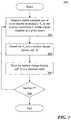
- FIG. 3is an example embodiment of a method of determining and storing surface charge densities, in accordance with aspects of the present invention.
- FIG. 4is an example embodiment of a method of determining and storing dipole densities, in accordance with aspects of the present invention.
- the surface charge densitymeans surface charge (Coulombs) per unit area (cm 2 ).
- a dipole as suchis a neutral element, wherein a part comprises a positive charge and the other part comprises the same but negative charge.
- a dipolemight represent the electric nature of cellular membranes better, because in biological environment ion charges are not macroscopically separated.
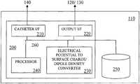
- FIG. 1shows an example embodiment of a mapping system 100 that can be used to map a heart 12 of a human 10 .
- Mapping system 100can include a computer 110 having known types of input devices and output devices, such as a display 120 and printer 130 , and a probe system 140 .
- a probe electrode 142will be used, which is connected to the computer 110 via a cable and forms part of probe system 140 .
- the probe electrode 142may be a multi-electrode array with elliptic or spherical shape.
- the spherical shapehas certain advantages for the subsequent data analysis. But also other types or even several independent electrodes could be used to measure V e .
- V(x,y,z)is measured at point x,y,z on the surface S P .
- Vthe potential and x,y,z denote the three dimensional coordinates.
- the solutionis an integral that allows for calculating the potential V(x′y′z′) at any point x′y′z′ in the whole volume of the heart chamber that is filled with blood.
- BEMboundary element method
- the boundary element methodis a numerical computational method for solving linear integral equations (i.e. in surface integral form).
- the methodis applied in many areas of engineering and science including fluid mechanics, acoustics, electromagnetics, and fracture mechanics.
- Boundary element formulationstypically give rise to fully populated matrices after discretisation. This means, that the storage requirements and computational time will tend to grow according to the square of the problem size.
- finite element matricesare typically banded (elements are only locally connected) and the storage requirements for the system matrices typically grow quite linearly with the problem size.
- ⁇ S e (P)is the delta-distribution concentrated on the surface of the heart chamber S e and ⁇ is the dipole density.
- V e ⁇ ( P )- 1 4 ⁇ ⁇ ⁇ ⁇ S e ⁇ ⁇ ⁇ ( P ′ ) ⁇ P ′ - P ⁇ ⁇ d ⁇ ⁇ ⁇ ⁇ ( P ′ ) ( 4 )
- V e ⁇ ( P )1 4 ⁇ ⁇ ⁇ ⁇ S e ⁇ ⁇ ⁇ ( P ′ ) ⁇ ⁇ ⁇ n P ′ ⁇ 1 ⁇ P - P ′ ⁇ ⁇ d ⁇ ⁇ ⁇ ⁇ ( P ′ ) ( 5 ) (For a review see Jackson J D. Classical Electrodynamics, 2 nd edition, Wiley, New York 1975.)
- the boundary element methodagain provides a code for transforming the potential V e in formulas 4 and 5 into the desired surface charge densities and dipole densities, which can be recorded in the database.
- the electric potential(s) V eis (are) determined by contact mapping. In this case the steps for calculating the electric potential V e are not necessary, because the direct contact of the electrode to the wall of the heart chamber already provides the electric potential V e .
- the probe electrodecomprises a shape that allows for calculating precisely the electric potential V e and, thus, simplifies the calculations for transforming V e into the desired charge or dipole densities.
- This preferred geometry of the electrodeis essentially ellipsoidal or spherical.
- the method, system, and devices of the present inventiondo not require any particular new electrodes for implementing the best mode for practicing the present invention.
- the inventionprovides a new and advantageous processing of the available data that will allow for an increase in precision, accuracy and spatial resolution of cardiac activation mapping when compared to prior art systems based on electric surface potentials in the heart only.
- the present inventionwill allow for providing superior diagnostic means for diagnosing cardiac arrhythmias and electric status of heart cells including metabolic and functional information.
- FIG. 2provides an example embodiment of a computer architecture 200 that can form part of mapping system 100 .
- Architecture 200includes standard interface modules 210 for probe system 140 (and electrode 142 ) and standard interface modules 220 for interfacing with output devices 120 , 130 .
- the computerincludes at least one processor 240 and at least one computer memory 250 .
- the foregoingare generally known, however the present invention further includes an electrical potential to surface charge density and/or dipole density converter module 230 .
- Module 230includes instructions necessary for carrying out the methods described herein, when executed by processor 240 , wherein the results of such processing are stored in memory 250 —as would be understood by one skilled in the art having the benefit of this disclosure.
- FIG. 3 and FIG. 4summarize methods for determining and storing surface charge densities and dipole densities in accordance with aspects of the present invention, respectively, which have been described in detail above.
- mapping system 100is used to measure and/or calculate one or more electric potential(s) V e into one or more position(s) P within a heart chamber at a given time t.
- V eis transformed into a surface charge density ⁇ (P′,t).
- the surface charge density ⁇ (P′,t)is stored in a database table. The method is repeated if there is another P, in step 308 .
- mapping system 100is used to measure and/or calculate one or more electric potential(s) V e in one or more position(s) P within a heart chamber at a given time t.
- V eis transformed into said dipole density ⁇ (P′,t) by using an algorithm suitable for transforming an electric potential into surface charge density.
- the dipole density ⁇ (P′,t)is stored in a database table. The method is repeated if there is another P, in step 408 .
Landscapes
- Health & Medical Sciences (AREA)
- Life Sciences & Earth Sciences (AREA)
- Engineering & Computer Science (AREA)
- Medical Informatics (AREA)
- Public Health (AREA)
- General Health & Medical Sciences (AREA)
- Cardiology (AREA)
- Surgery (AREA)
- Pathology (AREA)
- Biomedical Technology (AREA)
- Biophysics (AREA)
- Physics & Mathematics (AREA)
- Heart & Thoracic Surgery (AREA)
- Molecular Biology (AREA)
- Veterinary Medicine (AREA)
- Animal Behavior & Ethology (AREA)
- Primary Health Care (AREA)
- Epidemiology (AREA)
- Physiology (AREA)
- Dentistry (AREA)
- Oral & Maxillofacial Surgery (AREA)
- Nuclear Medicine, Radiotherapy & Molecular Imaging (AREA)
- Urology & Nephrology (AREA)
- Pulmonology (AREA)
- Data Mining & Analysis (AREA)
- Databases & Information Systems (AREA)
- Chemical & Material Sciences (AREA)
- Bioinformatics & Cheminformatics (AREA)
- Medicinal Chemistry (AREA)
- Nutrition Science (AREA)
- Measurement And Recording Of Electrical Phenomena And Electrical Characteristics Of The Living Body (AREA)
- Electrotherapy Devices (AREA)
Abstract
Description
- i) the position P′=(x′,y′,z′) of a point at the wall of the heart is defined in x, y, z-coordinates,
- ii) t is the time of measurement for said surface charge density, and
- iii) ρ is the surface charge density at said time t and said position P′ derived from a measured electric potential from a given heart chamber,
- comprising the following steps:
- a) measuring and/or calculating one or more electric potential(s) Vein one or more position(s) P at a given time t, and
- b) transforming Veinto said charge density ρ(P′,t) by using an algorithm suitable for transforming an electric potential into surface charge density.
- i) the position P′=(x′,y′,z′) of a point at the wall of the heart is defined in x, y, z-coordinates,
- ii) t is the time of measurement for said dipole density, and
- iii) ν is the dipole density at said time t and said position P′ derived from a measured electric potential Vefrom a given heart chamber,
- comprising the following steps:
- a) measuring and/or calculating one or more electric potential(s) Vein one or more positions P at a given time t, and
- b) transforming Veinto said dipole density ν(P′,t) by using an algorithm suitable for transforming an electric potential into surface charge density.
wherein:
wherein:
- a) one unit for measuring and recording at least one electric potential Veat a given position P,
- b) one a/d-converter for converting the measured electric potentials into digital data,
- c) a processor that transforms the digital voltage data into digital surface charge density data, and
- d) a memory that stores the at least one electric potential Veand the transformed digital surface charge density data.
- a) a measuring and recording unit that measures and records data used to determine at least one electric potential Veat a given position P,
- b) an a/d-converter that converts the at least one electric potentials Veinto digital voltage data,
- c) a processor that transforms the digital voltage data into dipole charge density data, and
- d) a memory that stores the at least one electric potential Veand the transformed dipole charge density data.
needs to be solved, wherein V is the potential and x,y,z denote the three dimensional coordinates. The boundary conditions for this equation are V(x,y,z)=Vρ(x,y,z) on SP, wherein VPis the potential on surface of the probe.
wherein ρ(P) is the surface charge density in position P=x,y,z, δS
(For a review see Jackson J D. Classical Electrodynamics, 2ndedition, Wiley, New York 1975.)
- a) one unit for measuring and recording electric potentials V at a given position P(x,y,z) on the surface of a given heart chamber (Contact mapping) or a probe electrode positioned within the heart, but without direct wall contact (noncontact mapping)
- b) one a/d-converter for converting the measured electric potentials into digital data,
- c) one memory to save the measured and/or transformed data, and
- d) one processor unit for transforming the digital data into digital surface charge density or dipole density data.
Claims (20)
Priority Applications (1)
| Application Number | Priority Date | Filing Date | Title |
|---|---|---|---|
| US16/533,028US11013444B2 (en) | 2006-08-03 | 2019-08-06 | Method and device for determining and presenting surface charge and dipole densities on cardiac walls |
Applications Claiming Priority (11)
| Application Number | Priority Date | Filing Date | Title |
|---|---|---|---|
| CH1251/06 | 2006-08-03 | ||
| CH12512006 | 2006-08-03 | ||
| PCT/CH2007/000380WO2008014629A2 (en) | 2006-08-03 | 2007-08-03 | Method and device for determining and presenting surface charge and dipole densities on cardiac walls |
| US37627009A | 2009-02-03 | 2009-02-03 | |
| US13/858,715US8700119B2 (en) | 2006-08-03 | 2013-04-08 | Method and device for determining and presenting surface charge and dipole densities on cardiac walls |
| US14/189,643US8918158B2 (en) | 2006-08-03 | 2014-02-25 | Method and device for determining and presenting surface charge and dipole densities on cardiac walls |
| US14/547,258US9167982B2 (en) | 2006-08-03 | 2014-11-19 | Method and device for determining and presenting surface charge and dipole densities on cardiac walls |
| US14/865,435US9610024B2 (en) | 2006-08-03 | 2015-09-25 | Method and device for determining and presenting surface charge and dipole densities on cardiac walls |
| US15/435,763US10376171B2 (en) | 2006-08-03 | 2017-02-17 | Method and device for determining and presenting surface charge and dipole densities on cardiac walls |
| US16/014,370US10413206B2 (en) | 2006-08-03 | 2018-06-21 | Method and device for determining and presenting surface charge and dipole densities on cardiac walls |
| US16/533,028US11013444B2 (en) | 2006-08-03 | 2019-08-06 | Method and device for determining and presenting surface charge and dipole densities on cardiac walls |
Related Parent Applications (1)
| Application Number | Title | Priority Date | Filing Date |
|---|---|---|---|
| US16/014,370ContinuationUS10413206B2 (en) | 2006-08-03 | 2018-06-21 | Method and device for determining and presenting surface charge and dipole densities on cardiac walls |
Publications (2)
| Publication Number | Publication Date |
|---|---|
| US20200187801A1 US20200187801A1 (en) | 2020-06-18 |
| US11013444B2true US11013444B2 (en) | 2021-05-25 |
Family
ID=38997497
Family Applications (8)
| Application Number | Title | Priority Date | Filing Date |
|---|---|---|---|
| US12/376,270Active2028-10-31US8417313B2 (en) | 2006-08-03 | 2007-08-03 | Method and device for determining and presenting surface charge and dipole densities on cardiac walls |
| US13/858,715ActiveUS8700119B2 (en) | 2006-08-03 | 2013-04-08 | Method and device for determining and presenting surface charge and dipole densities on cardiac walls |
| US14/189,643ActiveUS8918158B2 (en) | 2006-08-03 | 2014-02-25 | Method and device for determining and presenting surface charge and dipole densities on cardiac walls |
| US14/547,258ActiveUS9167982B2 (en) | 2006-08-03 | 2014-11-19 | Method and device for determining and presenting surface charge and dipole densities on cardiac walls |
| US14/865,435ActiveUS9610024B2 (en) | 2006-08-03 | 2015-09-25 | Method and device for determining and presenting surface charge and dipole densities on cardiac walls |
| US15/435,763ActiveUS10376171B2 (en) | 2006-08-03 | 2017-02-17 | Method and device for determining and presenting surface charge and dipole densities on cardiac walls |
| US16/014,370ActiveUS10413206B2 (en) | 2006-08-03 | 2018-06-21 | Method and device for determining and presenting surface charge and dipole densities on cardiac walls |
| US16/533,028ActiveUS11013444B2 (en) | 2006-08-03 | 2019-08-06 | Method and device for determining and presenting surface charge and dipole densities on cardiac walls |
Family Applications Before (7)
| Application Number | Title | Priority Date | Filing Date |
|---|---|---|---|
| US12/376,270Active2028-10-31US8417313B2 (en) | 2006-08-03 | 2007-08-03 | Method and device for determining and presenting surface charge and dipole densities on cardiac walls |
| US13/858,715ActiveUS8700119B2 (en) | 2006-08-03 | 2013-04-08 | Method and device for determining and presenting surface charge and dipole densities on cardiac walls |
| US14/189,643ActiveUS8918158B2 (en) | 2006-08-03 | 2014-02-25 | Method and device for determining and presenting surface charge and dipole densities on cardiac walls |
| US14/547,258ActiveUS9167982B2 (en) | 2006-08-03 | 2014-11-19 | Method and device for determining and presenting surface charge and dipole densities on cardiac walls |
| US14/865,435ActiveUS9610024B2 (en) | 2006-08-03 | 2015-09-25 | Method and device for determining and presenting surface charge and dipole densities on cardiac walls |
| US15/435,763ActiveUS10376171B2 (en) | 2006-08-03 | 2017-02-17 | Method and device for determining and presenting surface charge and dipole densities on cardiac walls |
| US16/014,370ActiveUS10413206B2 (en) | 2006-08-03 | 2018-06-21 | Method and device for determining and presenting surface charge and dipole densities on cardiac walls |
Country Status (6)
| Country | Link |
|---|---|
| US (8) | US8417313B2 (en) |
| EP (2) | EP3603500B1 (en) |
| AU (1) | AU2007281009B2 (en) |
| CA (2) | CA2932956C (en) |
| ES (1) | ES2870924T3 (en) |
| WO (1) | WO2008014629A2 (en) |
Cited By (1)
| Publication number | Priority date | Publication date | Assignee | Title |
|---|---|---|---|---|
| US12402825B2 (en) | 2014-03-25 | 2025-09-02 | Enchannel Medical, Ltd. | Cardiac analysis user interface system and method |
Families Citing this family (29)
| Publication number | Priority date | Publication date | Assignee | Title |
|---|---|---|---|---|
| US5849881A (en) | 1989-05-05 | 1998-12-15 | Baylor College Medicine | Production of recombinant lactoferrin and lactoferrin polypeptides using cDNA sequences in various organisms |
| WO2008014629A2 (en) | 2006-08-03 | 2008-02-07 | Christoph Scharf | Method and device for determining and presenting surface charge and dipole densities on cardiac walls |
| EP2737849A3 (en) | 2008-01-17 | 2014-10-29 | Christoph Scharf | A device and method for the geometric determination of electrical dipole densities on the cardiac wall |
| US20110213260A1 (en)* | 2010-02-26 | 2011-09-01 | Pacesetter, Inc. | Crt lead placement based on optimal branch selection and optimal site selection |
| US9757044B2 (en)* | 2011-03-10 | 2017-09-12 | Acutus Medical, Inc. | Device and method for the geometric determination of electrical dipole densities on the cardiac wall |
| US8897516B2 (en) | 2011-03-16 | 2014-11-25 | Biosense Webster (Israel) Ltd. | Two-dimensional cardiac mapping |
| US9017321B2 (en) | 2012-05-21 | 2015-04-28 | Kardium, Inc. | Systems and methods for activating transducers |
| US10827977B2 (en) | 2012-05-21 | 2020-11-10 | Kardium Inc. | Systems and methods for activating transducers |
| US9198592B2 (en) | 2012-05-21 | 2015-12-01 | Kardium Inc. | Systems and methods for activating transducers |
| US10588543B2 (en) | 2012-05-23 | 2020-03-17 | Biosense Webster (Israel), Ltd. | Position sensing using electric dipole fields |
| EP2890292B1 (en) | 2012-08-31 | 2021-01-13 | Acutus Medical, Inc. | Catheter system for the heart |
| CN105358070B (en) | 2013-02-08 | 2018-03-23 | 阿库图森医疗有限公司 | Expandable catheter assembly with flexible printed circuit board |
| CA2922941C (en) | 2013-09-13 | 2021-11-16 | Acutus Medical, Inc. | Devices and methods for determination of electrical dipole densities on a cardiac surface |
| EP3110324A4 (en)* | 2014-02-28 | 2017-10-18 | Powell Mansfield, Inc. | Systems, methods and devices for sensing emg activity |
| ES2572142B1 (en)* | 2014-10-30 | 2017-06-21 | Fundación Para La Investigación Biomédica Del Hospital Gregorio Marañón | CARDIAC ARRITMIAS LOCATION DEVICE |
| US10368936B2 (en) | 2014-11-17 | 2019-08-06 | Kardium Inc. | Systems and methods for selecting, activating, or selecting and activating transducers |
| US10722184B2 (en) | 2014-11-17 | 2020-07-28 | Kardium Inc. | Systems and methods for selecting, activating, or selecting and activating transducers |
| WO2016154280A1 (en) | 2015-03-23 | 2016-09-29 | The Methodist Hospital | Methods and devices for sample characterization |
| CN115299988A (en) | 2015-05-12 | 2022-11-08 | 阿库图森医疗有限公司 | Ultrasonic sequencing systems and methods |
| US10593234B2 (en) | 2015-05-12 | 2020-03-17 | Acutus Medical, Inc. | Cardiac virtualization test tank and testing system and method |
| US10653318B2 (en) | 2015-05-13 | 2020-05-19 | Acutus Medical, Inc. | Localization system and method useful in the acquisition and analysis of cardiac information |
| EP3451916A4 (en) | 2016-05-03 | 2019-10-16 | Acutus Medical Inc. | SYSTEM AND METHOD FOR THE DYNAMIC DISPLAY OF HEART DATA |
| CA3022806A1 (en) | 2016-05-03 | 2017-11-09 | Acutus Medical, Inc. | Cardiac mapping system with efficiency algorithm |
| EP3790471A4 (en) | 2018-05-08 | 2022-01-12 | Acutus Medical, Inc. | Cardiac information processing system |
| US12178582B2 (en) | 2018-11-09 | 2024-12-31 | Acutus Medical, Inc. | Systems and methods for calculating patient information |
| JP2022529908A (en) | 2019-04-18 | 2022-06-27 | アクタス メディカル インク | System for generating composite maps |
| CA3135773A1 (en) | 2019-06-04 | 2020-12-10 | Acutus Medical, Inc. | Systems and methods for performing localization within a body |
| WO2021102230A1 (en) | 2019-11-22 | 2021-05-27 | Acutus Medical, Inc. | Tissue treatment systems, devices, and methods |
| CN118892331A (en)* | 2023-04-26 | 2024-11-05 | 深圳心寰科技有限公司 | Three-dimensional mapping method, system, device, equipment and medium based on boundary element |
Citations (186)
| Publication number | Priority date | Publication date | Assignee | Title |
|---|---|---|---|---|
| US4173228A (en) | 1977-05-16 | 1979-11-06 | Applied Medical Devices | Catheter locating device |
| US5041973A (en) | 1988-10-25 | 1991-08-20 | London Health Association | Cardiac mapping system simulator |
| US5156151A (en) | 1991-02-15 | 1992-10-20 | Cardiac Pathways Corporation | Endocardial mapping and ablation system and catheter probe |
| US5293868A (en) | 1992-06-30 | 1994-03-15 | American Cardiac Ablation Co., Inc. | Cardiac ablation catheter having resistive mapping electrodes |
| WO1994006349A1 (en) | 1992-09-23 | 1994-03-31 | Endocardial Therapeutics, Inc. | Endocardial mapping system |
| US5482472A (en) | 1993-11-17 | 1996-01-09 | Board Of Regents, The University Of Texas System | Electrical signal generator interface with three-dimensional electrical pathway and transparent heart and method of visually simulating cardiac waveforms in three dimensions |
| US5499981A (en) | 1993-03-16 | 1996-03-19 | Ep Technologies, Inc. | Flexible interlaced multiple electrode assemblies |
| JPH08504333A (en) | 1992-09-25 | 1996-05-14 | イーピー・テクノロジーズ・インコーポレーテッド | Electrode-supported splines for the cardiac system |
| US5555883A (en) | 1992-02-24 | 1996-09-17 | Avitall; Boaz | Loop electrode array mapping and ablation catheter for cardiac chambers |
| US5595183A (en) | 1995-02-17 | 1997-01-21 | Ep Technologies, Inc. | Systems and methods for examining heart tissue employing multiple electrode structures and roving electrodes |
| US5601084A (en) | 1993-06-23 | 1997-02-11 | University Of Washington | Determining cardiac wall thickness and motion by imaging and three-dimensional modeling |
| US5647367A (en) | 1996-05-31 | 1997-07-15 | Hewlett-Packard Company | Scanning ultrasonic probe with locally-driven sweeping ultrasonic source |
| US5662108A (en) | 1992-09-23 | 1997-09-02 | Endocardial Solutions, Inc. | Electrophysiology mapping system |
| US5722402A (en) | 1994-10-11 | 1998-03-03 | Ep Technologies, Inc. | Systems and methods for guiding movable electrode elements within multiple-electrode structures |
| US5722416A (en) | 1995-02-17 | 1998-03-03 | Ep Technologies, Inc. | Systems and methods for analyzing biopotential morphologies in heart tissue to locate potential ablation sites |
| US5740808A (en) | 1996-10-28 | 1998-04-21 | Ep Technologies, Inc | Systems and methods for guilding diagnostic or therapeutic devices in interior tissue regions |
| US5749833A (en) | 1995-08-15 | 1998-05-12 | Hakki; A-Hamid | Combined echo-electrocardiographic probe |
| US5759158A (en) | 1995-07-28 | 1998-06-02 | E.P. Technologies, Inc. | Systems and methods for conducting electrophysiological testing using high-voltage energy pulses to stun heart tissue |
| US5782239A (en) | 1992-06-30 | 1998-07-21 | Cordis Webster, Inc. | Unique electrode configurations for cardiovascular electrode catheter with built-in deflection method and central puller wire |
| US5795299A (en) | 1997-01-31 | 1998-08-18 | Acuson Corporation | Ultrasonic transducer assembly with extended flexible circuits |
| US5795298A (en) | 1995-03-28 | 1998-08-18 | Sonometrics Corporation | System for sharing electrocardiogram electrodes and transducers |
| US5820568A (en) | 1996-10-15 | 1998-10-13 | Cardiac Pathways Corporation | Apparatus and method for aiding in the positioning of a catheter |
| US5830144A (en) | 1995-03-28 | 1998-11-03 | Vesely; Ivan | Tracking data sheath |
| US5846198A (en) | 1996-05-31 | 1998-12-08 | Siemens Aktiengesellschaft | Apparatus for localizing action currents in the heart |
| WO1999005971A1 (en) | 1997-08-01 | 1999-02-11 | Cardiac Pathways Corporation | System for electrode localization using ultrasound |
| US5876336A (en) | 1994-10-11 | 1999-03-02 | Ep Technologies, Inc. | Systems and methods for guiding movable electrode elements within multiple-electrode structure |
| US5928228A (en) | 1993-03-16 | 1999-07-27 | Ep Technologies, Inc. | Flexible high density multiple electrode circuit assemblies employing ribbon cable |
| US5944022A (en) | 1997-04-28 | 1999-08-31 | American Cardiac Ablation Co. Inc. | Catheter positioning system |
| US5968040A (en) | 1994-03-04 | 1999-10-19 | Ep Technologies, Inc. | Systems and methods using asymmetric multiple electrode arrays |
| US6014590A (en) | 1974-03-04 | 2000-01-11 | Ep Technologies, Inc. | Systems and methods employing structures having asymmetric mechanical properties to support diagnostic or therapeutic elements in contact with tissue in interior body regions |
| US6024703A (en) | 1997-05-07 | 2000-02-15 | Eclipse Surgical Technologies, Inc. | Ultrasound device for axial ranging |
| WO2000007501A1 (en) | 1998-08-03 | 2000-02-17 | Cardiac Pathways Corporation | A dynamically alterable three-dimensional graphical model of a body region |
| US6066096A (en) | 1998-05-08 | 2000-05-23 | Duke University | Imaging probes and catheters for volumetric intraluminal ultrasound imaging and related systems |
| US6086532A (en) | 1997-09-26 | 2000-07-11 | Ep Technologies, Inc. | Systems for recording use of structures deployed in association with heart tissue |
| US6107699A (en) | 1998-05-22 | 2000-08-22 | Scimed Life Systems, Inc. | Power supply for use in electrophysiological apparatus employing high-voltage pulses to render tissue temporarily unresponsive |
| US6115626A (en) | 1998-03-26 | 2000-09-05 | Scimed Life Systems, Inc. | Systems and methods using annotated images for controlling the use of diagnostic or therapeutic instruments in instruments in interior body regions |
| JP2000358299A (en) | 1999-06-16 | 2000-12-26 | Ngk Spark Plug Co Ltd | Wave transmitting and receiving element for ultrasonic probe, its production method and ultrasonic probe using the same element |
| US6188928B1 (en) | 1996-11-18 | 2001-02-13 | Pacesetter Ab | Apparatus for tissue stimulation |
| US6187032B1 (en) | 1997-10-30 | 2001-02-13 | Kabushiki Kaisha Toshiba | Measurement of intracardiac electrophysiological phenomena |
| JP2001070269A (en) | 1999-09-06 | 2001-03-21 | Toshiba Corp | Electrophysiology mapping device |
| US6216043B1 (en) | 1994-03-04 | 2001-04-10 | Ep Technologies, Inc. | Asymmetric multiple electrode support structures |
| US6240307B1 (en) | 1993-09-23 | 2001-05-29 | Endocardial Solutions, Inc. | Endocardial mapping system |
| US20010007070A1 (en) | 1999-04-05 | 2001-07-05 | Medtronic, Inc. | Ablation catheter assembly and method for isolating a pulmonary vein |
| US6301496B1 (en) | 1998-07-24 | 2001-10-09 | Biosense, Inc. | Vector mapping of three-dimensionally reconstructed intrabody organs and method of display |
| EP1166714A1 (en) | 2000-06-21 | 2002-01-02 | Biosense, Inc. | Rapid mapping of electrical activity in the heart |
| US20020026118A1 (en) | 2000-08-18 | 2002-02-28 | Assaf Govari | Three-dimensional reconstruction using ultrasound |
| US20020045810A1 (en) | 1993-07-20 | 2002-04-18 | Shlomo Ben-Haim | Method for mapping a heart using catheters having ultrasonic position sensors |
| US20020128565A1 (en) | 1997-07-31 | 2002-09-12 | Case Western Reserve University | System and method for non-invasive electrocardiographic imaging |
| US20020165441A1 (en) | 2000-01-27 | 2002-11-07 | Coleman James H. | Bidirectional catheter having mapping assembly |
| US6514249B1 (en) | 1997-07-08 | 2003-02-04 | Atrionix, Inc. | Positioning system and method for orienting an ablation element within a pulmonary vein ostium |
| JP2003511098A (en) | 1998-04-14 | 2003-03-25 | ジーエムピー・ドラツグ・デリバリー・インコーポレーテツド | Iontophoresis, electroporation and combination catheters for local drug delivery to arteries and other body tissues |
| WO2003026722A2 (en) | 2001-09-27 | 2003-04-03 | Baylor College Of Medicine | Cardiac catheter imaging system |
| US20030078494A1 (en) | 2001-10-24 | 2003-04-24 | Scimed Life Systems, Inc. | Systems and methods for guiding and locating functional elements on medical devices positioned in a body |
| US6574492B1 (en) | 1996-01-08 | 2003-06-03 | Biosense, Inc. | Catheter having multiple arms with electrode and position sensor |
| US20030120318A1 (en) | 1998-06-30 | 2003-06-26 | Hauck John A. | Congestive heart failure pacing optimization method and device |
| US20030153907A1 (en) | 1999-01-06 | 2003-08-14 | Scimed Life Systems, Inc. | Ultrasound-guided ablation catheter and methods of use |
| US20030158477A1 (en) | 2001-11-09 | 2003-08-21 | Dorin Panescu | Systems and methods for guiding catheters using registered images |
| US20030231789A1 (en) | 2002-06-18 | 2003-12-18 | Scimed Life Systems, Inc. | Computer generated representation of the imaging pattern of an imaging device |
| US20030236466A1 (en) | 2002-06-21 | 2003-12-25 | Tarjan Peter P. | Single or multi-mode cardiac activity data collection, processing and display obtained in a non-invasive manner |
| US20040039312A1 (en) | 2002-02-20 | 2004-02-26 | Liposonix, Inc. | Ultrasonic treatment and imaging of adipose tissue |
| WO2004026134A1 (en) | 2002-08-24 | 2004-04-01 | Krishnan Subramaniam C | Method and apparatus for locating the fossa ovalis and performing transseptal puncture |
| US20040082870A1 (en) | 1997-07-31 | 2004-04-29 | Yoram Rudy | Systems and methods for determining a surface geometry |
| US6773402B2 (en) | 2001-07-10 | 2004-08-10 | Biosense, Inc. | Location sensing with real-time ultrasound imaging |
| US20040254437A1 (en) | 1998-06-30 | 2004-12-16 | Hauck John A. | Method and apparatus for catheter navigation and location and mapping in the heart |
| JP2004350702A (en) | 2003-05-26 | 2004-12-16 | Olympus Corp | Ultrasonic diagnosis probe apparatus |
| US6839588B1 (en) | 1997-07-31 | 2005-01-04 | Case Western Reserve University | Electrophysiological cardiac mapping system based on a non-contact non-expandable miniature multi-electrode catheter and method therefor |
| US20050059880A1 (en) | 2003-09-11 | 2005-03-17 | Mathias Sanjay George | ECG driven image reconstruction for cardiac imaging |
| US20050113665A1 (en) | 2003-11-26 | 2005-05-26 | Mohr Kelly A. | Cardiac display methods and apparatus |
| US20050148836A1 (en) | 2003-11-26 | 2005-07-07 | Martin Kleen | Catheter device |
| US6939309B1 (en) | 1993-09-23 | 2005-09-06 | Endocardial Solutions, Inc. | Electrophysiology mapping system |
| US20060058676A1 (en) | 2002-04-17 | 2006-03-16 | Tomoyuki Yagi | Ultrasonic probe in body cavity |
| US20060116576A1 (en) | 2004-12-01 | 2006-06-01 | Scimed Life Systems, Inc. | System and use thereof to provide indication of proximity between catheter and location of interest in 3-D space |
| CN1856123A (en) | 2005-04-29 | 2006-11-01 | 华为技术有限公司 | System and method for exchanging modules in switch |
| US20060244177A1 (en) | 2005-04-19 | 2006-11-02 | Masayuki Kaneto | Flexible printed circuit board for catheter, catheter using same, and production method of catheter |
| US20070016007A1 (en) | 2005-07-15 | 2007-01-18 | Assaf Govari | Hybrid magnetic-based and impedance-based position sensing |
| EP1760661A2 (en) | 2005-08-30 | 2007-03-07 | Biosense Webster, Inc. | Segmentation and registration of multimodal images using physiological data |
| US20070055150A1 (en) | 2005-08-16 | 2007-03-08 | General Electric Company | Method and system for mapping physiology information onto ultrasound-based anatomic structure |
| US20070060832A1 (en) | 2005-08-26 | 2007-03-15 | Michael Levin | Detection of skin impedance |
| US20070083194A1 (en) | 2005-06-20 | 2007-04-12 | Kunis Christopher G | Ablation catheter |
| EP1779787A2 (en) | 2005-10-28 | 2007-05-02 | Biosense Webster, Inc. | Synchronization of ultrasound imaging data with electrical mapping |
| US20070167722A1 (en) | 1992-08-14 | 2007-07-19 | British Telecommunications Public Limited Company | Surgical navigation |
| US20070219551A1 (en) | 2003-09-22 | 2007-09-20 | Honour Kirk S | Medical device with flexible printed circuit |
| CN101048100A (en) | 2004-09-08 | 2007-10-03 | 奥林巴斯株式会社 | capsule medical device |
| US20070232949A1 (en) | 2006-03-31 | 2007-10-04 | Ep Medsystems, Inc. | Method For Simultaneous Bi-Atrial Mapping Of Atrial Fibrillation |
| US7291146B2 (en) | 2003-09-12 | 2007-11-06 | Minnow Medical, Inc. | Selectable eccentric remodeling and/or ablation of atherosclerotic material |
| US20080009758A1 (en) | 2006-05-17 | 2008-01-10 | Voth Eric J | System and method for mapping electrophysiology information onto complex geometry |
| WO2008014629A2 (en) | 2006-08-03 | 2008-02-07 | Christoph Scharf | Method and device for determining and presenting surface charge and dipole densities on cardiac walls |
| US20080146937A1 (en) | 2006-12-14 | 2008-06-19 | General Electric Company | Mechanically expanding transducer assembly |
| US20080287777A1 (en) | 2007-05-16 | 2008-11-20 | General Electric Company | System and method to register a tracking system with an intracardiac echocardiography (ice) imaging system |
| US20080319297A1 (en) | 2007-06-20 | 2008-12-25 | Kenneth Danehorn | Electrode catheter positioning system |
| US20090024086A1 (en) | 2007-07-20 | 2009-01-22 | Qiming Zhang | Micro-steerable catheter |
| US7505810B2 (en) | 2006-06-13 | 2009-03-17 | Rhythmia Medical, Inc. | Non-contact cardiac mapping, including preprocessing |
| US20090076483A1 (en) | 2007-09-14 | 2009-03-19 | Kenneth Danehorn | Catheter localization system |
| US20090082691A1 (en) | 2007-09-26 | 2009-03-26 | Medtronic, Inc. | Frequency selective monitoring of physiological signals |
| CN201223445Y (en) | 2008-06-23 | 2009-04-22 | 北京有色金属研究总院 | Radio frequency ablation catheter |
| US20090131930A1 (en) | 2007-11-16 | 2009-05-21 | Daniel Gelbart | Medical device for use in bodily lumens, for example an atrium |
| US20090143651A1 (en) | 2006-06-01 | 2009-06-04 | Bengt Kallback | Device for Invasive Use |
| US20090148012A1 (en) | 2007-12-05 | 2009-06-11 | Andres Claudio Altmann | Anatomical modeling from a 3-d image and a surface mapping |
| JP2009135109A (en) | 2009-03-09 | 2009-06-18 | Advanced Systems Japan Inc | Sheet-like connecting terminal and method of connecting fpc substrate using this |
| US20090171274A1 (en) | 2007-12-28 | 2009-07-02 | Doron Harlev | Non contact mapping catheter |
| CN201275144Y (en) | 2007-08-10 | 2009-07-22 | 北京美中双和医疗器械有限公司 | Electrophysiologic ablation device |
| WO2009090547A2 (en) | 2008-01-17 | 2009-07-23 | Christoph Scharf | A device and method for the geometric determination of electrical dipole densities on the cardiac wall |
| US7573182B2 (en) | 2005-06-01 | 2009-08-11 | Prorhythm, Inc. | Ultrasonic transducer |
| US20100023004A1 (en) | 2008-07-28 | 2010-01-28 | David Francischelli | Systems and methods for cardiac tissue electroporation ablation |
| US20100076426A1 (en) | 2007-05-09 | 2010-03-25 | De La Rama Alan | Basket catheter having multiple electrodes |
| US20100094279A1 (en) | 2006-10-10 | 2010-04-15 | Kauphusman James V | Circuit for a catheter or sheath and method of forming same |
| US20100168578A1 (en) | 2007-06-12 | 2010-07-01 | University Of Virginia Patent Foundation | System and Method for Combined ECG-Echo for Cardiac Diagnosis |
| US20100279263A1 (en) | 2009-04-29 | 2010-11-04 | Scott Duryea | Polysomnography Training Apparatus |
| US20100286551A1 (en) | 2009-05-08 | 2010-11-11 | Rhythmia Medical, Inc. | Impedance Based Anatomy Generation |
| US7841986B2 (en) | 2006-05-10 | 2010-11-30 | Regents Of The University Of Minnesota | Methods and apparatus of three dimensional cardiac electrophysiological imaging |
| JP2011504363A (en) | 2007-11-23 | 2011-02-10 | ディーエスエム アイピー アセッツ ビー.ブイ. | Production of improved bioactive peptides |
| US20110077526A1 (en) | 2008-05-27 | 2011-03-31 | Gil Zwirn | Ultrasound garment |
| US20110092809A1 (en) | 2009-04-07 | 2011-04-21 | Pacesetter, Inc. | Cardiac coordinate system for motion analysis |
| US20110118726A1 (en) | 2009-11-13 | 2011-05-19 | St. Jude Medical, Inc. | Assembly of staggered ablation elements |
| US20110125172A1 (en) | 2006-05-19 | 2011-05-26 | Kardium Inc. | Automatic atherectomy system |
| US20110144510A1 (en) | 2009-12-16 | 2011-06-16 | Pacesetter, Inc. | Methods to identify damaged or scarred tissue based on position information and physiological information |
| US20110201951A1 (en) | 2010-02-12 | 2011-08-18 | Siemens Medical Solutions Usa, Inc. | System for cardiac arrhythmia detection and characterization |
| US20110213231A1 (en) | 2007-05-09 | 2011-09-01 | Hall Sacha C | Bendable catheter arms having varied flexibility |
| US20110270237A1 (en) | 2010-04-28 | 2011-11-03 | Medtronic Ablation Frontiers Llc | Systems and methods of performing medical procedures |
| US20120082969A1 (en) | 2010-10-05 | 2012-04-05 | Yitzhack Schwartz | Simulation of an invasive procedure |
| US20120123296A1 (en) | 2009-08-03 | 2012-05-17 | Dune Medical Devices Ltd. | Surgical tool |
| WO2012068471A1 (en) | 2010-11-19 | 2012-05-24 | Boston Scientific Scimed, Inc. | Renal nerve detection and ablation apparatus and method |
| US20120136231A1 (en) | 2006-07-25 | 2012-05-31 | Gal Markel | Wearable items providing physiological, environmental and situational parameter monitoring |
| US20120143298A1 (en) | 2010-12-02 | 2012-06-07 | Just Dale E | Catheter electrode assemblies and methods of construction therefor |
| US20120165667A1 (en) | 2010-12-22 | 2012-06-28 | Andres Claudio Altmann | Lasso catheter with ultrasound transducer |
| US20120172859A1 (en) | 2011-01-05 | 2012-07-05 | Medtronic Ablation Frontiers Llc | Multipolarity epicardial radiofrequency ablation |
| WO2012092016A1 (en) | 2010-12-30 | 2012-07-05 | St. Jude Medical, Atrial Fibrillation Division, Inc. | System and method for diagnosing arrhythmias and directing catheter therapies |
| US20120184863A1 (en) | 2011-01-13 | 2012-07-19 | Rhythmia Medical, Inc. | Electroanatomical mapping |
| WO2012100184A2 (en) | 2011-01-21 | 2012-07-26 | Kardium Inc. | Enhanced medical device for use in bodily cavities, for example an atrium |
| WO2012110942A1 (en) | 2011-02-17 | 2012-08-23 | Koninklijke Philips Electronics N.V. | System for providing an electrical activity map using optical shape sensing |
| CA2829626A1 (en) | 2011-03-10 | 2012-09-13 | Acutus Medical, Inc. | Device and method for the geometric determination of electrical dipole densities on the cardiac wall |
| US20120265054A1 (en) | 2011-04-14 | 2012-10-18 | Olson Eric S | System and Method for Registration of Multiple Navigation Systems to a Common Coordinate Frame |
| US20120271138A1 (en) | 2011-04-22 | 2012-10-25 | Topera, Inc. | Basket style cardiac mapping catheter having a flexible electrode assembly for detection of cardiac rhythm disorders |
| US20120302912A1 (en) | 2011-05-27 | 2012-11-29 | Boston Scientific Neuromodulation Corporation | Collection of clinical data for graphical representation and analysis |
| US20120310064A1 (en) | 2011-06-01 | 2012-12-06 | Mcgee David L | Ablation probe with ultrasonic imaging capabilities |
| US20130006238A1 (en) | 2011-06-30 | 2013-01-03 | Tom Allen Ditter | Catheter with variable arcuate distal section |
| US20130085361A1 (en) | 2010-04-01 | 2013-04-04 | Ecole Polytechnique Federale De Lausanne | Device for interacting with neurological tissue and methods of making and using the same |
| US20130096432A1 (en) | 1998-06-30 | 2013-04-18 | John A. Hauck | System and method for navigating an ultrasound catheter to image a beating heart |
| US8428690B2 (en) | 2007-05-16 | 2013-04-23 | General Electric Company | Intracardiac echocardiography image reconstruction in combination with position tracking system |
| US20130158537A1 (en) | 2010-06-30 | 2013-06-20 | Koninklijke Philips Electronics N.V. | Energy application apparatus for applying energy to an object |
| US20130165916A1 (en) | 2011-12-23 | 2013-06-27 | Vessix Vascular, Inc. | Methods and apparatuses for remodeling tissue of or adjacent to a body passage |
| US20130172715A1 (en) | 2011-12-30 | 2013-07-04 | Dale E. Just | Electrode support structure assemblies |
| WO2013101257A1 (en) | 2011-12-29 | 2013-07-04 | St. Jude Medical, Atrial Fibrillation Division, Inc. | Method and system for constructing an electrophysiology map |
| US20130190587A1 (en) | 2011-01-21 | 2013-07-25 | Kardium Inc. | High-density electrode-based medical device system |
| US20130197614A1 (en) | 2005-05-03 | 2013-08-01 | Vessix Vascular, Inc. | Selective accumulation of energy with or without knowledge of tissue topography |
| US20130245433A1 (en) | 2010-11-18 | 2013-09-19 | Koninklijke Philips Electronics N.V. | Location determination apparatus |
| US20130245621A1 (en) | 2012-03-16 | 2013-09-19 | St. Jude Medical Ab | Ablation stent and method of using an ablation stent |
| US20130241929A1 (en) | 2012-03-13 | 2013-09-19 | Fady Massarwa | Selectably transparent electrophysiology map |
| US20130267853A1 (en) | 2010-12-03 | 2013-10-10 | Research Triangle Institute | Ultrasound device, and associated cable assembly |
| US20130282084A1 (en) | 2004-09-10 | 2013-10-24 | Vessix Vascular, Inc. | Apparatus and Method for Treatment of In-Stent Restenosis |
| US20130304062A1 (en) | 2012-05-14 | 2013-11-14 | Biosense Webster (Irael), Ltd. | Catheter with helical end section for vessel ablation |
| US20130310827A1 (en) | 2012-05-21 | 2013-11-21 | Kardium Inc. | Systems and methods for selecting, activating, or selecting and activating transducers |
| US20130330701A1 (en) | 2012-06-12 | 2013-12-12 | Vladimir Rubinstein | Physical heart simulator |
| WO2014036439A2 (en) | 2012-08-31 | 2014-03-06 | Acutus Medical, Inc. | Catheter system and methods of medical uses of same, including diagnostic and treatment uses for the heart |
| US20140095105A1 (en) | 2012-10-03 | 2014-04-03 | Lev Abramovich Koyrakh | Scaling of electrical impedance-based navigation space using inter-electrode spacing |
| US20140221803A1 (en) | 2008-09-30 | 2014-08-07 | Biosense Webster (Israel), Ltd. | Current localization tracker |
| WO2014124231A1 (en) | 2013-02-08 | 2014-08-14 | Acutus Medical, Inc. | Expandable catheter assembly with flexible printed circuit board |
| US20140235988A1 (en) | 2013-02-21 | 2014-08-21 | Medtronic, Inc. | Methods for simultaneous cardiac substrate mapping using spatial correlation maps between neighboring unipolar electrograms |
| US20140249505A1 (en) | 2013-03-02 | 2014-09-04 | Vladislav Bukhman | Method and System of Utilizing ECG Signal for Central Venous Catheter Tip Positioning |
| US20140257071A1 (en) | 2013-03-05 | 2014-09-11 | Timothy G. Curran | System and Method for Detecting Sheathing and Unsheathing of Localization Elements |
| US20140257069A1 (en) | 2013-03-08 | 2014-09-11 | St. Jude Medical, Atrial Fibrillation Division, Inc. | Basket for a multi-electrode array catheter |
| US20140276789A1 (en) | 2013-03-15 | 2014-09-18 | Boston Scientific Scimed, Inc. | Methods and apparatuses for remodeling tissue of or adjacent to a body passage |
| US20140276746A1 (en) | 2013-03-15 | 2014-09-18 | St. Jude Medical, Cardiology Division, Inc. | Feedback systems and methods utilizing two or more sites along denervation catheter |
| US20140276733A1 (en) | 2013-03-14 | 2014-09-18 | St. Jude Medical, Cardiology Division, Inc. | Mediguide-enabled renal denervation system for ensuring wall contact and mapping lesion locations |
| US20140358143A1 (en) | 2013-05-31 | 2014-12-04 | Medtronic Ablation Frontiers Llc | Adjustable catheter for ostial, septal, and roof ablation in atrial fibrillation patients |
| WO2015038607A2 (en) | 2013-09-13 | 2015-03-19 | Acutus Medical, Inc. | Devices and methods for determination of electrical dipole densities on a cardiac surface |
| CN104462650A (en) | 2014-11-10 | 2015-03-25 | 张建卿 | Materialized heart 3D model manufacturing method capable of achieving internal and external structures |
| US20150196217A1 (en) | 2006-06-13 | 2015-07-16 | Rhythmia Medical, Inc. | Cardiac mapping |
| US20150208938A1 (en) | 2014-01-29 | 2015-07-30 | Biosense Webster (Israel) Ltd. | Hybrid bipolar/unipolar detection of activation wavefront |
| US20150257825A1 (en) | 2014-03-13 | 2015-09-17 | Medtronic Ardian Luxembourg S.A.R.L. | Low Profile Catheter Assemblies and Associated Systems and Methods |
| US20150257732A1 (en) | 2013-03-15 | 2015-09-17 | Stephen Eric Ryan | Distance, diameter and area determining device |
| WO2015148470A1 (en) | 2014-03-25 | 2015-10-01 | Acutus Medical, Inc. | Cardiac analysis user interface system and method |
| US20150342491A1 (en) | 2014-06-03 | 2015-12-03 | Boston Scientific Scimed, Inc. | Electrode assembly having an atraumatic distal tip |
| US20150374252A1 (en) | 2007-05-23 | 2015-12-31 | St. Jude Medical, Cardiology Division, Inc. | Flexible high-density mapping catheter tips and flexible ablation catheter tips with onboard high-density mapping electrodes |
| US20160051321A1 (en) | 2013-04-08 | 2016-02-25 | Amr Salahieh | Tissue ablation and monitoring |
| USD758596S1 (en) | 2015-04-17 | 2016-06-07 | Micron Devices Llc | Flexible circuit for an implantable neural stimulator |
| US20160256112A1 (en) | 2009-11-03 | 2016-09-08 | Vivaquant Llc | System for processing physiological data |
| WO2016183179A1 (en) | 2015-05-12 | 2016-11-17 | Acutus Medical, Inc. | Cardiac virtualization test tank and testing system and method |
| WO2016183285A1 (en) | 2015-05-12 | 2016-11-17 | Acutus Medical, Inc. | Ultrasound sequencing system and method |
| WO2016183468A1 (en) | 2015-05-13 | 2016-11-17 | Acutus Medical, Inc. | Localization system and method useful in the acquisition and analysis of cardiac information |
| US20170065204A1 (en) | 2015-09-04 | 2017-03-09 | Biosense Webster (Israel) Ltd. | Field-based location coordinate correction |
| WO2017192775A1 (en) | 2016-05-03 | 2017-11-09 | Acutus Medical, Inc. | Cardiac mapping system with efficiency algorithm |
| US20170319180A1 (en) | 2014-12-11 | 2017-11-09 | Koninklijke Philips N.V. | Catheter transducer with staggered columns of micromachined ultrasonic transducers |
| WO2017192769A1 (en) | 2016-05-03 | 2017-11-09 | Acutus Medical, Inc. | Cardiac information dynamic display system and method |
| WO2019144103A1 (en) | 2018-01-21 | 2019-07-25 | Acutus Medical, Inc. | System for identifying cardiac conduction patterns |
| WO2019217430A1 (en) | 2018-05-08 | 2019-11-14 | Acutus Medical, Inc. | Cardiac information processing system |
- 2007
- 2007-08-03WOPCT/CH2007/000380patent/WO2008014629A2/enactiveApplication Filing
- 2007-08-03USUS12/376,270patent/US8417313B2/enactiveActive
- 2007-08-03ESES19184148Tpatent/ES2870924T3/enactiveActive
- 2007-08-03EPEP19184148.5Apatent/EP3603500B1/enactiveActive
- 2007-08-03CACA2932956Apatent/CA2932956C/enactiveActive
- 2007-08-03AUAU2007281009Apatent/AU2007281009B2/enactiveActive
- 2007-08-03CACA2659898Apatent/CA2659898C/enactiveActive
- 2007-08-03EPEP07785075.8Apatent/EP2051625B1/enactiveActive
- 2013
- 2013-04-08USUS13/858,715patent/US8700119B2/enactiveActive
- 2014
- 2014-02-25USUS14/189,643patent/US8918158B2/enactiveActive
- 2014-11-19USUS14/547,258patent/US9167982B2/enactiveActive
- 2015
- 2015-09-25USUS14/865,435patent/US9610024B2/enactiveActive
- 2017
- 2017-02-17USUS15/435,763patent/US10376171B2/enactiveActive
- 2018
- 2018-06-21USUS16/014,370patent/US10413206B2/enactiveActive
- 2019
- 2019-08-06USUS16/533,028patent/US11013444B2/enactiveActive
Patent Citations (375)
| Publication number | Priority date | Publication date | Assignee | Title |
|---|---|---|---|---|
| US6014590A (en) | 1974-03-04 | 2000-01-11 | Ep Technologies, Inc. | Systems and methods employing structures having asymmetric mechanical properties to support diagnostic or therapeutic elements in contact with tissue in interior body regions |
| US4173228A (en) | 1977-05-16 | 1979-11-06 | Applied Medical Devices | Catheter locating device |
| US5041973A (en) | 1988-10-25 | 1991-08-20 | London Health Association | Cardiac mapping system simulator |
| US5156151A (en) | 1991-02-15 | 1992-10-20 | Cardiac Pathways Corporation | Endocardial mapping and ablation system and catheter probe |
| US5555883A (en) | 1992-02-24 | 1996-09-17 | Avitall; Boaz | Loop electrode array mapping and ablation catheter for cardiac chambers |
| US5293868A (en) | 1992-06-30 | 1994-03-15 | American Cardiac Ablation Co., Inc. | Cardiac ablation catheter having resistive mapping electrodes |
| US5782239A (en) | 1992-06-30 | 1998-07-21 | Cordis Webster, Inc. | Unique electrode configurations for cardiovascular electrode catheter with built-in deflection method and central puller wire |
| US8200314B2 (en) | 1992-08-14 | 2012-06-12 | British Telecommunications Public Limited Company | Surgical navigation |
| US20070167722A1 (en) | 1992-08-14 | 2007-07-19 | British Telecommunications Public Limited Company | Surgical navigation |
| US20060084884A1 (en) | 1992-09-23 | 2006-04-20 | Endocardial Solutions, Inc. | Mapping electrophysiological data in a heart chamber |
| US20050101874A1 (en) | 1992-09-23 | 2005-05-12 | Beatty Graydon E. | Software for mapping potential distribution of a heart chamber |
| US6728562B1 (en) | 1992-09-23 | 2004-04-27 | Endocardial Solutions, Inc. | Method for creating a virtual electrogram |
| US20030176799A1 (en) | 1992-09-23 | 2003-09-18 | Beatty Graydon Ernest | Software for mapping potential distribution of a heart chamber |
| US5662108A (en) | 1992-09-23 | 1997-09-02 | Endocardial Solutions, Inc. | Electrophysiology mapping system |
| US20060058692A1 (en) | 1992-09-23 | 2006-03-16 | Endocardial Solutions, Inc. | Mapping physiological data in a heart chamber |
| US20060084972A1 (en) | 1992-09-23 | 2006-04-20 | Endocardial Solutions, Inc. | Delivering ablation therapy in a heart chamber |
| US6640119B1 (en) | 1992-09-23 | 2003-10-28 | Endocardial Solutions, Inc. | Method for orienting an electrode array |
| US20060052716A1 (en) | 1992-09-23 | 2006-03-09 | Endocardial Solutions, Inc. | Delivering ablation therapy in a heart chamber |
| US20060058693A1 (en) | 1992-09-23 | 2006-03-16 | Endocardial Solutions, Inc. | Mapping electrophysiological data in a heart chamber |
| US6978168B2 (en) | 1992-09-23 | 2005-12-20 | Endocardial Solutions, Inc. | Software for mapping potential distribution of a heart chamber |
| US8208998B2 (en) | 1992-09-23 | 2012-06-26 | St. Jude Medical, Atrial Fibrillation Division, Inc. | Representing the geometry of a heart chamber |
| US20060084971A1 (en) | 1992-09-23 | 2006-04-20 | Endocardial Solutions, Inc. | Mapping physiological data in a heart chamber |
| US20060084970A1 (en) | 1992-09-23 | 2006-04-20 | Endocardial Solutions, Inc. | Mapping physiological data in a heart chamber |
| US6826421B1 (en) | 1992-09-23 | 2004-11-30 | Graydon Ernest Beatty | Endocardial mapping catheter |
| JPH08501477A (en) | 1992-09-23 | 1996-02-20 | エンドカーディアル・セラピューティクス・インコーポレーテッド | Endocardial mapping system |
| US6826420B1 (en) | 1992-09-23 | 2004-11-30 | Endocardial Solutions, Inc. | Method of mapping a plug in a mapping catheter |
| WO1994006349A1 (en) | 1992-09-23 | 1994-03-31 | Endocardial Therapeutics, Inc. | Endocardial mapping system |
| US7289843B2 (en) | 1992-09-23 | 2007-10-30 | St. Jude Medical, Atrial Fibrillation Division, Inc. | Software for mapping potential distribution of a heart chamber |
| US6990370B1 (en) | 1992-09-23 | 2006-01-24 | Endocardial Solutions, Inc. | Method for mapping heart electrophysiology |
| JPH08504333A (en) | 1992-09-25 | 1996-05-14 | イーピー・テクノロジーズ・インコーポレーテッド | Electrode-supported splines for the cardiac system |
| US5928228A (en) | 1993-03-16 | 1999-07-27 | Ep Technologies, Inc. | Flexible high density multiple electrode circuit assemblies employing ribbon cable |
| US5499981A (en) | 1993-03-16 | 1996-03-19 | Ep Technologies, Inc. | Flexible interlaced multiple electrode assemblies |
| US5601084A (en) | 1993-06-23 | 1997-02-11 | University Of Washington | Determining cardiac wall thickness and motion by imaging and three-dimensional modeling |
| US20020045810A1 (en) | 1993-07-20 | 2002-04-18 | Shlomo Ben-Haim | Method for mapping a heart using catheters having ultrasonic position sensors |
| US6939309B1 (en) | 1993-09-23 | 2005-09-06 | Endocardial Solutions, Inc. | Electrophysiology mapping system |
| US6240307B1 (en) | 1993-09-23 | 2001-05-29 | Endocardial Solutions, Inc. | Endocardial mapping system |
| US5482472A (en) | 1993-11-17 | 1996-01-09 | Board Of Regents, The University Of Texas System | Electrical signal generator interface with three-dimensional electrical pathway and transparent heart and method of visually simulating cardiac waveforms in three dimensions |
| US5968040A (en) | 1994-03-04 | 1999-10-19 | Ep Technologies, Inc. | Systems and methods using asymmetric multiple electrode arrays |
| US6216043B1 (en) | 1994-03-04 | 2001-04-10 | Ep Technologies, Inc. | Asymmetric multiple electrode support structures |
| US5876336A (en) | 1994-10-11 | 1999-03-02 | Ep Technologies, Inc. | Systems and methods for guiding movable electrode elements within multiple-electrode structure |
| US5722402A (en) | 1994-10-11 | 1998-03-03 | Ep Technologies, Inc. | Systems and methods for guiding movable electrode elements within multiple-electrode structures |
| US5595183A (en) | 1995-02-17 | 1997-01-21 | Ep Technologies, Inc. | Systems and methods for examining heart tissue employing multiple electrode structures and roving electrodes |
| US5722416A (en) | 1995-02-17 | 1998-03-03 | Ep Technologies, Inc. | Systems and methods for analyzing biopotential morphologies in heart tissue to locate potential ablation sites |
| US5830144A (en) | 1995-03-28 | 1998-11-03 | Vesely; Ivan | Tracking data sheath |
| US5795298A (en) | 1995-03-28 | 1998-08-18 | Sonometrics Corporation | System for sharing electrocardiogram electrodes and transducers |
| JPH11504541A (en) | 1995-05-01 | 1999-04-27 | コーディス ウェブスター,インコーポレイティド | Intrinsic electrode configuration for cardiovascular electrode catheters with integrated flexure and central puller wire |
| US5759158A (en) | 1995-07-28 | 1998-06-02 | E.P. Technologies, Inc. | Systems and methods for conducting electrophysiological testing using high-voltage energy pulses to stun heart tissue |
| US5749833A (en) | 1995-08-15 | 1998-05-12 | Hakki; A-Hamid | Combined echo-electrocardiographic probe |
| US6574492B1 (en) | 1996-01-08 | 2003-06-03 | Biosense, Inc. | Catheter having multiple arms with electrode and position sensor |
| US5846198A (en) | 1996-05-31 | 1998-12-08 | Siemens Aktiengesellschaft | Apparatus for localizing action currents in the heart |
| US5647367A (en) | 1996-05-31 | 1997-07-15 | Hewlett-Packard Company | Scanning ultrasonic probe with locally-driven sweeping ultrasonic source |
| JPH10137207A (en) | 1996-07-12 | 1998-05-26 | Ep Technol Inc | System and method for guiding movable electrode element within multiple-electrode structure |
| US5820568A (en) | 1996-10-15 | 1998-10-13 | Cardiac Pathways Corporation | Apparatus and method for aiding in the positioning of a catheter |
| US5740808A (en) | 1996-10-28 | 1998-04-21 | Ep Technologies, Inc | Systems and methods for guilding diagnostic or therapeutic devices in interior tissue regions |
| US6188928B1 (en) | 1996-11-18 | 2001-02-13 | Pacesetter Ab | Apparatus for tissue stimulation |
| US5795299A (en) | 1997-01-31 | 1998-08-18 | Acuson Corporation | Ultrasonic transducer assembly with extended flexible circuits |
| JP2000510250A (en) | 1997-03-06 | 2000-08-08 | ソノメトリクス コーポレイション | Tracking data generation sheath |
| JP2000510030A (en) | 1997-03-07 | 2000-08-08 | ソノメトリクス コーポレイション | Electrocardiogram electrode and transducer sharing system |
| JP2001522288A (en) | 1997-04-28 | 2001-11-13 | アメリカン カーディアック アブレイション カンパニー インコーポレイテッド | Catheter positioning system |
| US5944022A (en) | 1997-04-28 | 1999-08-31 | American Cardiac Ablation Co. Inc. | Catheter positioning system |
| US6024703A (en) | 1997-05-07 | 2000-02-15 | Eclipse Surgical Technologies, Inc. | Ultrasound device for axial ranging |
| US6514249B1 (en) | 1997-07-08 | 2003-02-04 | Atrionix, Inc. | Positioning system and method for orienting an ablation element within a pulmonary vein ostium |
| US6839588B1 (en) | 1997-07-31 | 2005-01-04 | Case Western Reserve University | Electrophysiological cardiac mapping system based on a non-contact non-expandable miniature multi-electrode catheter and method therefor |
| US20020128565A1 (en) | 1997-07-31 | 2002-09-12 | Case Western Reserve University | System and method for non-invasive electrocardiographic imaging |
| US20040082870A1 (en) | 1997-07-31 | 2004-04-29 | Yoram Rudy | Systems and methods for determining a surface geometry |
| US6772004B2 (en) | 1997-07-31 | 2004-08-03 | Case Western Reserve University | System and method for non-invasive electrocardiographic imaging |
| US6970733B2 (en) | 1997-08-01 | 2005-11-29 | Scimed Life Systems, Inc. | System and method for electrode localization using ultrasound |
| US20030036696A1 (en) | 1997-08-01 | 2003-02-20 | Cardiac Pathways Corporation | System for electrode localization using ultrasound |
| WO1999005971A1 (en) | 1997-08-01 | 1999-02-11 | Cardiac Pathways Corporation | System for electrode localization using ultrasound |
| US6216027B1 (en) | 1997-08-01 | 2001-04-10 | Cardiac Pathways Corporation | System for electrode localization using ultrasound |
| US20060058663A1 (en) | 1997-08-01 | 2006-03-16 | Scimed Life Systems, Inc. | System and method for marking an anatomical structure in three-dimensional coordinate system |
| US6490474B1 (en) | 1997-08-01 | 2002-12-03 | Cardiac Pathways Corporation | System and method for electrode localization using ultrasound |
| US8412307B2 (en) | 1997-08-01 | 2013-04-02 | Boston Scientific Scimed, Inc. | System and method for marking an anatomical structure in three-dimensional coordinate system |
| US20130225983A1 (en) | 1997-08-01 | 2013-08-29 | Boston Scientific Scimed, Inc. | System and method for marking an anatomical structure in three-dimensional coordinate system |
| US6086532A (en) | 1997-09-26 | 2000-07-11 | Ep Technologies, Inc. | Systems for recording use of structures deployed in association with heart tissue |
| US6187032B1 (en) | 1997-10-30 | 2001-02-13 | Kabushiki Kaisha Toshiba | Measurement of intracardiac electrophysiological phenomena |
| US6115626A (en) | 1998-03-26 | 2000-09-05 | Scimed Life Systems, Inc. | Systems and methods using annotated images for controlling the use of diagnostic or therapeutic instruments in instruments in interior body regions |
| JP2003511098A (en) | 1998-04-14 | 2003-03-25 | ジーエムピー・ドラツグ・デリバリー・インコーポレーテツド | Iontophoresis, electroporation and combination catheters for local drug delivery to arteries and other body tissues |
| US6066096A (en) | 1998-05-08 | 2000-05-23 | Duke University | Imaging probes and catheters for volumetric intraluminal ultrasound imaging and related systems |
| US6107699A (en) | 1998-05-22 | 2000-08-22 | Scimed Life Systems, Inc. | Power supply for use in electrophysiological apparatus employing high-voltage pulses to render tissue temporarily unresponsive |
| US7263397B2 (en) | 1998-06-30 | 2007-08-28 | St. Jude Medical, Atrial Fibrillation Division, Inc. | Method and apparatus for catheter navigation and location and mapping in the heart |
| US7187973B2 (en) | 1998-06-30 | 2007-03-06 | Endocardial Solutions, Inc. | Congestive heart failure pacing optimization method and device |
| US20130096432A1 (en) | 1998-06-30 | 2013-04-18 | John A. Hauck | System and method for navigating an ultrasound catheter to image a beating heart |
| US20030120318A1 (en) | 1998-06-30 | 2003-06-26 | Hauck John A. | Congestive heart failure pacing optimization method and device |
| US20040254437A1 (en) | 1998-06-30 | 2004-12-16 | Hauck John A. | Method and apparatus for catheter navigation and location and mapping in the heart |
| US6301496B1 (en) | 1998-07-24 | 2001-10-09 | Biosense, Inc. | Vector mapping of three-dimensionally reconstructed intrabody organs and method of display |
| JP2002522106A (en) | 1998-08-03 | 2002-07-23 | カーディアック・パスウェイズ・コーポレーション | Dynamically changeable 3D graphic model of the human body |
| US20050203375A1 (en) | 1998-08-03 | 2005-09-15 | Scimed Life Systems, Inc. | System and method for passively reconstructing anatomical structure |
| WO2000007501A1 (en) | 1998-08-03 | 2000-02-17 | Cardiac Pathways Corporation | A dynamically alterable three-dimensional graphical model of a body region |
| US6950689B1 (en) | 1998-08-03 | 2005-09-27 | Boston Scientific Scimed, Inc. | Dynamically alterable three-dimensional graphical model of a body region |
| US6824515B2 (en) | 1999-01-06 | 2004-11-30 | Scimed Life Systems, Inc. | Ultrasound-guided ablation catheter and methods of use |
| US20030153907A1 (en) | 1999-01-06 | 2003-08-14 | Scimed Life Systems, Inc. | Ultrasound-guided ablation catheter and methods of use |
| US20040082948A1 (en) | 1999-04-05 | 2004-04-29 | Medtronic, Inc. | Ablation catheter assembly and method for isolating a pulmonary vein |
| US7285119B2 (en) | 1999-04-05 | 2007-10-23 | Medtronic, Inc. | Ablation catheter assembly and method for isolating a pulmonary vein |
| US20010007070A1 (en) | 1999-04-05 | 2001-07-05 | Medtronic, Inc. | Ablation catheter assembly and method for isolating a pulmonary vein |
| JP2000358299A (en) | 1999-06-16 | 2000-12-26 | Ngk Spark Plug Co Ltd | Wave transmitting and receiving element for ultrasonic probe, its production method and ultrasonic probe using the same element |
| US6396198B1 (en) | 1999-06-16 | 2002-05-28 | Ngk Spark Plug Co. Ltd. | Wave transmission-reception element for use in ultrasound probe, method for manufacturing the wave transmission-reception element and ultrasound probe incorporating the transmission-reception element |
| JP2001070269A (en) | 1999-09-06 | 2001-03-21 | Toshiba Corp | Electrophysiology mapping device |
| US20020165441A1 (en) | 2000-01-27 | 2002-11-07 | Coleman James H. | Bidirectional catheter having mapping assembly |
| JP2002051998A (en) | 2000-06-21 | 2002-02-19 | Biosense Inc | High-speed mapping of cardiac electrical activity |
| EP1166714A1 (en) | 2000-06-21 | 2002-01-02 | Biosense, Inc. | Rapid mapping of electrical activity in the heart |
| US6400981B1 (en) | 2000-06-21 | 2002-06-04 | Biosense, Inc. | Rapid mapping of electrical activity in the heart |
| US6716166B2 (en) | 2000-08-18 | 2004-04-06 | Biosense, Inc. | Three-dimensional reconstruction using ultrasound |
| JP2002113004A (en) | 2000-08-18 | 2002-04-16 | Biosense Inc | Three-dimensional reconstruction using ultrasonic wave |
| US20020026118A1 (en) | 2000-08-18 | 2002-02-28 | Assaf Govari | Three-dimensional reconstruction using ultrasound |
| WO2002045608A2 (en) | 2000-12-08 | 2002-06-13 | Medtronic, Inc. | Ablation catheter assembly and method for isolating a pulmonary vein |
| US6773402B2 (en) | 2001-07-10 | 2004-08-10 | Biosense, Inc. | Location sensing with real-time ultrasound imaging |
| US7187964B2 (en) | 2001-09-27 | 2007-03-06 | Dirar S. Khoury | Cardiac catheter imaging system |
| US20030065271A1 (en) | 2001-09-27 | 2003-04-03 | Baylor College Of Medicine | Cardiac catheter imaging system |
| WO2003026722A2 (en) | 2001-09-27 | 2003-04-03 | Baylor College Of Medicine | Cardiac catheter imaging system |
| US6895267B2 (en) | 2001-10-24 | 2005-05-17 | Scimed Life Systems, Inc. | Systems and methods for guiding and locating functional elements on medical devices positioned in a body |
| US20030078494A1 (en) | 2001-10-24 | 2003-04-24 | Scimed Life Systems, Inc. | Systems and methods for guiding and locating functional elements on medical devices positioned in a body |
| US20030158477A1 (en) | 2001-11-09 | 2003-08-21 | Dorin Panescu | Systems and methods for guiding catheters using registered images |
| US8175680B2 (en) | 2001-11-09 | 2012-05-08 | Boston Scientific Scimed, Inc. | Systems and methods for guiding catheters using registered images |
| US20120277574A1 (en) | 2001-11-09 | 2012-11-01 | Boston Scientific Scimed, Inc. | Systems and methods for guiding catheters using registered images |
| US20040039312A1 (en) | 2002-02-20 | 2004-02-26 | Liposonix, Inc. | Ultrasonic treatment and imaging of adipose tissue |
| US7258674B2 (en) | 2002-02-20 | 2007-08-21 | Liposonix, Inc. | Ultrasonic treatment and imaging of adipose tissue |
| US20060058676A1 (en) | 2002-04-17 | 2006-03-16 | Tomoyuki Yagi | Ultrasonic probe in body cavity |
| US7766838B2 (en) | 2002-04-17 | 2010-08-03 | Hitachi Medical Corporation | Ultrasonic probe in body cavity |
| US20030231789A1 (en) | 2002-06-18 | 2003-12-18 | Scimed Life Systems, Inc. | Computer generated representation of the imaging pattern of an imaging device |
| US20030236466A1 (en) | 2002-06-21 | 2003-12-25 | Tarjan Peter P. | Single or multi-mode cardiac activity data collection, processing and display obtained in a non-invasive manner |
| WO2004026134A1 (en) | 2002-08-24 | 2004-04-01 | Krishnan Subramaniam C | Method and apparatus for locating the fossa ovalis and performing transseptal puncture |
| JP2005536313A (en) | 2002-08-24 | 2005-12-02 | サブラマニアム シー. クリシュナン | Method and apparatus for identifying the position of the foveal fossa |
| JP2006511296A (en) | 2002-12-18 | 2006-04-06 | ボストン サイエンティフィック リミテッド | Catheter guidance system using registered images |
| JP2004350702A (en) | 2003-05-26 | 2004-12-16 | Olympus Corp | Ultrasonic diagnosis probe apparatus |
| US20050059880A1 (en) | 2003-09-11 | 2005-03-17 | Mathias Sanjay George | ECG driven image reconstruction for cardiac imaging |
| US7291146B2 (en) | 2003-09-12 | 2007-11-06 | Minnow Medical, Inc. | Selectable eccentric remodeling and/or ablation of atherosclerotic material |
| US8147486B2 (en) | 2003-09-22 | 2012-04-03 | St. Jude Medical, Atrial Fibrillation Division, Inc. | Medical device with flexible printed circuit |
| US20070219551A1 (en) | 2003-09-22 | 2007-09-20 | Honour Kirk S | Medical device with flexible printed circuit |
| US20050113665A1 (en) | 2003-11-26 | 2005-05-26 | Mohr Kelly A. | Cardiac display methods and apparatus |
| US7689261B2 (en) | 2003-11-26 | 2010-03-30 | General Electric Company | Cardiac display methods and apparatus |
| US7479141B2 (en) | 2003-11-26 | 2009-01-20 | Siemens Aktiengesellschaft | Ablation tip catheter device with integrated imaging, ECG and positioning devices |
| US20050148836A1 (en) | 2003-11-26 | 2005-07-07 | Martin Kleen | Catheter device |
| CN101048100A (en) | 2004-09-08 | 2007-10-03 | 奥林巴斯株式会社 | capsule medical device |
| US20130282084A1 (en) | 2004-09-10 | 2013-10-24 | Vessix Vascular, Inc. | Apparatus and Method for Treatment of In-Stent Restenosis |
| US9713730B2 (en) | 2004-09-10 | 2017-07-25 | Boston Scientific Scimed, Inc. | Apparatus and method for treatment of in-stent restenosis |
| WO2006060613A1 (en) | 2004-12-01 | 2006-06-08 | Boston Scientific Scimed, Inc. | System and use thereof to provide indication of proximity between catheter and location of interest |
| US20060116576A1 (en) | 2004-12-01 | 2006-06-01 | Scimed Life Systems, Inc. | System and use thereof to provide indication of proximity between catheter and location of interest in 3-D space |
| US20060244177A1 (en) | 2005-04-19 | 2006-11-02 | Masayuki Kaneto | Flexible printed circuit board for catheter, catheter using same, and production method of catheter |
| US7351914B2 (en) | 2005-04-19 | 2008-04-01 | Nitto Denko Corporation | Flexible printed circuit board for catheter, catheter using same, and production method of catheter |
| CN1856123A (en) | 2005-04-29 | 2006-11-01 | 华为技术有限公司 | System and method for exchanging modules in switch |
| US9486355B2 (en) | 2005-05-03 | 2016-11-08 | Vessix Vascular, Inc. | Selective accumulation of energy with or without knowledge of tissue topography |
| US20130197614A1 (en) | 2005-05-03 | 2013-08-01 | Vessix Vascular, Inc. | Selective accumulation of energy with or without knowledge of tissue topography |
| US7573182B2 (en) | 2005-06-01 | 2009-08-11 | Prorhythm, Inc. | Ultrasonic transducer |
| US20070083194A1 (en) | 2005-06-20 | 2007-04-12 | Kunis Christopher G | Ablation catheter |
| US20070016007A1 (en) | 2005-07-15 | 2007-01-18 | Assaf Govari | Hybrid magnetic-based and impedance-based position sensing |
| US7536218B2 (en) | 2005-07-15 | 2009-05-19 | Biosense Webster, Inc. | Hybrid magnetic-based and impedance-based position sensing |
| US20070055150A1 (en) | 2005-08-16 | 2007-03-08 | General Electric Company | Method and system for mapping physiology information onto ultrasound-based anatomic structure |
| US20070060832A1 (en) | 2005-08-26 | 2007-03-15 | Michael Levin | Detection of skin impedance |
| EP1760661A2 (en) | 2005-08-30 | 2007-03-07 | Biosense Webster, Inc. | Segmentation and registration of multimodal images using physiological data |
| EP1779787A2 (en) | 2005-10-28 | 2007-05-02 | Biosense Webster, Inc. | Synchronization of ultrasound imaging data with electrical mapping |
| US20070106146A1 (en) | 2005-10-28 | 2007-05-10 | Altmann Andres C | Synchronization of ultrasound imaging data with electrical mapping |
| US7918793B2 (en) | 2005-10-28 | 2011-04-05 | Biosense Webster, Inc. | Synchronization of ultrasound imaging data with electrical mapping |
| US20100256627A1 (en) | 2006-02-10 | 2010-10-07 | Beijing Amsino Medical Co., Ltd. | Electrophysiology ablation device |
| US20070232949A1 (en) | 2006-03-31 | 2007-10-04 | Ep Medsystems, Inc. | Method For Simultaneous Bi-Atrial Mapping Of Atrial Fibrillation |
| US7841986B2 (en) | 2006-05-10 | 2010-11-30 | Regents Of The University Of Minnesota | Methods and apparatus of three dimensional cardiac electrophysiological imaging |
| US20080009758A1 (en) | 2006-05-17 | 2008-01-10 | Voth Eric J | System and method for mapping electrophysiology information onto complex geometry |
| US20110125172A1 (en) | 2006-05-19 | 2011-05-26 | Kardium Inc. | Automatic atherectomy system |
| US8150499B2 (en) | 2006-05-19 | 2012-04-03 | Kardium Inc. | Automatic atherectomy system |
| US20090143651A1 (en) | 2006-06-01 | 2009-06-04 | Bengt Kallback | Device for Invasive Use |
| US9730602B2 (en) | 2006-06-13 | 2017-08-15 | Boston Scientific Scimed Inc. | Cardiac mapping |
| US7953475B2 (en) | 2006-06-13 | 2011-05-31 | Rhythmia Medical, Inc. | Preprocessing for cardiac mapping |
| US7505810B2 (en) | 2006-06-13 | 2009-03-17 | Rhythmia Medical, Inc. | Non-contact cardiac mapping, including preprocessing |
| US20150196217A1 (en) | 2006-06-13 | 2015-07-16 | Rhythmia Medical, Inc. | Cardiac mapping |
| US20120136231A1 (en) | 2006-07-25 | 2012-05-31 | Gal Markel | Wearable items providing physiological, environmental and situational parameter monitoring |
| WO2008014629A2 (en) | 2006-08-03 | 2008-02-07 | Christoph Scharf | Method and device for determining and presenting surface charge and dipole densities on cardiac walls |
| US9167982B2 (en) | 2006-08-03 | 2015-10-27 | Christoph Scharf | Method and device for determining and presenting surface charge and dipole densities on cardiac walls |
| US8918158B2 (en) | 2006-08-03 | 2014-12-23 | Christoph Scharf | Method and device for determining and presenting surface charge and dipole densities on cardiac walls |
| US8417313B2 (en) | 2006-08-03 | 2013-04-09 | Christoph Scharf | Method and device for determining and presenting surface charge and dipole densities on cardiac walls |
| EP2051625A2 (en) | 2006-08-03 | 2009-04-29 | Christoph Scharf | Method and device for determining and presenting surface charge and dipole densities on cardiac walls |
| US8700119B2 (en) | 2006-08-03 | 2014-04-15 | Christoph Scharf | Method and device for determining and presenting surface charge and dipole densities on cardiac walls |
| US20170258347A1 (en) | 2006-08-03 | 2017-09-14 | Christoph Scharf | Method and device for determining and presenting surface charge and dipole densities on cardiac walls |
| US20140180150A1 (en) | 2006-08-03 | 2014-06-26 | Christoph Scharf | Method and device for determining and presenting surface charge and dipole densities on cardiac walls |
| US20150196219A1 (en) | 2006-08-03 | 2015-07-16 | Christoph Scharf | Method and device for determining and presenting surface charge and dipole densities on cardiac walls |
| US9610024B2 (en) | 2006-08-03 | 2017-04-04 | Christoph Scharf | Method and device for determining and presenting surface charge and dipole densities on cardiac walls |
| US20160007869A1 (en) | 2006-08-03 | 2016-01-14 | Christoph Scharf | Method and device for determining and presenting surface charge and dipole densities on cardiac walls |
| US20130226017A1 (en) | 2006-08-03 | 2013-08-29 | Christoph Scharf | Method and device for determining and presenting surface charge and dipole densities on cardiac walls |
| US20090264781A1 (en) | 2006-08-03 | 2009-10-22 | Christoph Scharf | Method and device for determining and presenting surface charge and dipole densities on cardiac walls |
| US8968299B2 (en) | 2006-10-10 | 2015-03-03 | St. Jude Medical, Atrial Fibrillation Division, Inc. | Circuit for a catheter or sheath and method of forming same |
| US20100094279A1 (en) | 2006-10-10 | 2010-04-15 | Kauphusman James V | Circuit for a catheter or sheath and method of forming same |
| US20080146937A1 (en) | 2006-12-14 | 2008-06-19 | General Electric Company | Mechanically expanding transducer assembly |
| JP2008149132A (en) | 2006-12-14 | 2008-07-03 | General Electric Co <Ge> | Mechanically expanding transducer assembly |
| US20100076426A1 (en) | 2007-05-09 | 2010-03-25 | De La Rama Alan | Basket catheter having multiple electrodes |
| US20110213231A1 (en) | 2007-05-09 | 2011-09-01 | Hall Sacha C | Bendable catheter arms having varied flexibility |
| US20080287777A1 (en) | 2007-05-16 | 2008-11-20 | General Electric Company | System and method to register a tracking system with an intracardiac echocardiography (ice) imaging system |
| US8428690B2 (en) | 2007-05-16 | 2013-04-23 | General Electric Company | Intracardiac echocardiography image reconstruction in combination with position tracking system |
| US8989842B2 (en) | 2007-05-16 | 2015-03-24 | General Electric Company | System and method to register a tracking system with intracardiac echocardiography (ICE) imaging system |
| US20150374252A1 (en) | 2007-05-23 | 2015-12-31 | St. Jude Medical, Cardiology Division, Inc. | Flexible high-density mapping catheter tips and flexible ablation catheter tips with onboard high-density mapping electrodes |
| US20100168578A1 (en) | 2007-06-12 | 2010-07-01 | University Of Virginia Patent Foundation | System and Method for Combined ECG-Echo for Cardiac Diagnosis |
| US20080319297A1 (en) | 2007-06-20 | 2008-12-25 | Kenneth Danehorn | Electrode catheter positioning system |
| US8311613B2 (en) | 2007-06-20 | 2012-11-13 | Siemens Aktiengesellschaft | Electrode catheter positioning system |
| US20090024086A1 (en) | 2007-07-20 | 2009-01-22 | Qiming Zhang | Micro-steerable catheter |
| US8454596B2 (en) | 2007-08-10 | 2013-06-04 | Beijing Amsino Medical Co., Ltd. | Electrophysiology ablation device |
| CN201275144Y (en) | 2007-08-10 | 2009-07-22 | 北京美中双和医疗器械有限公司 | Electrophysiologic ablation device |
| US8825134B2 (en) | 2007-09-14 | 2014-09-02 | Siemens Aktiengesellschaft | Catheter localization system |
| US20090076483A1 (en) | 2007-09-14 | 2009-03-19 | Kenneth Danehorn | Catheter localization system |
| US20090082691A1 (en) | 2007-09-26 | 2009-03-26 | Medtronic, Inc. | Frequency selective monitoring of physiological signals |
| WO2009065042A2 (en) | 2007-11-16 | 2009-05-22 | Kardium Inc. | Medical device for use in bodily lumens, for example an atrium background |
| US20110172658A1 (en) | 2007-11-16 | 2011-07-14 | Kardium Inc. | Medical device for use in bodily lumens, for example an atrium |
| US20090131930A1 (en) | 2007-11-16 | 2009-05-21 | Daniel Gelbart | Medical device for use in bodily lumens, for example an atrium |
| US20110045130A1 (en) | 2007-11-23 | 2011-02-24 | Luppo Edens | Bioactive peptide production |
| JP2011504363A (en) | 2007-11-23 | 2011-02-10 | ディーエスエム アイピー アセッツ ビー.ブイ. | Production of improved bioactive peptides |
| US20090148012A1 (en) | 2007-12-05 | 2009-06-11 | Andres Claudio Altmann | Anatomical modeling from a 3-d image and a surface mapping |
| US8320711B2 (en) | 2007-12-05 | 2012-11-27 | Biosense Webster, Inc. | Anatomical modeling from a 3-D image and a surface mapping |
| JP2009136679A (en) | 2007-12-05 | 2009-06-25 | Biosense Webster Inc | Anatomical modeling with 3-D images and surface mapping |
| JP2011507656A (en) | 2007-12-28 | 2011-03-10 | リズミア メディカル インコーポレイテッド | Non-contact mapping catheter |
| US20130253298A1 (en) | 2007-12-28 | 2013-09-26 | Rhythmia, Medical, Inc. | Cardiac mapping catheter |
| US8447377B2 (en) | 2007-12-28 | 2013-05-21 | Rhythmia Medical, Inc. | Cardiac mapping catheter |
| US20090171274A1 (en) | 2007-12-28 | 2009-07-02 | Doron Harlev | Non contact mapping catheter |
| US20140275921A1 (en) | 2007-12-28 | 2014-09-18 | Rhythmia Medical, Inc. | Cardiac mapping catheter |
| US20120078077A1 (en) | 2007-12-28 | 2012-03-29 | Rhythmia Medical, Inc. | Cardiac mapping catheter |
| US8103327B2 (en) | 2007-12-28 | 2012-01-24 | Rhythmia Medical, Inc. | Cardiac mapping catheter |
| US8755861B2 (en) | 2007-12-28 | 2014-06-17 | Rhythmia Medical, Inc. | Cardiac mapping catheter |
| US20160038051A1 (en) | 2008-01-17 | 2016-02-11 | Christoph Scharf | Device and method for the geometric determination of electrical dipole densities on the cardiac wall |
| EP2252203A2 (en) | 2008-01-17 | 2010-11-24 | Christoph Scharf | A device and method for the geometric determination of electrical dipole densities on the cardiac wall |
| US20170100049A1 (en) | 2008-01-17 | 2017-04-13 | Christoph Scharf | Device and method for the geometric determination of electrical dipole densities on the cardiac wall |
| US9192318B2 (en) | 2008-01-17 | 2015-11-24 | Christoph Scharf | Device and method for the geometric determination of electrical dipole densities on the cardiac wall |
| US20140024910A1 (en) | 2008-01-17 | 2014-01-23 | Christoph Scharf | Device and method for the geometric determination of electrical dipole densities on the cardiac wall |
| WO2009090547A2 (en) | 2008-01-17 | 2009-07-23 | Christoph Scharf | A device and method for the geometric determination of electrical dipole densities on the cardiac wall |
| US9913589B2 (en) | 2008-01-17 | 2018-03-13 | Christoph Scharf | Device and method for the geometric determination of electrical dipole densities on the cardiac wall |
| US20100298690A1 (en) | 2008-01-17 | 2010-11-25 | Christoph Scharf | Device and method for the geometric determination of electrical dipole densities on the cardiac wall |
| US8512255B2 (en) | 2008-01-17 | 2013-08-20 | Christoph Scharf | Device and method for the geometric determination of electrical dipole densities on the cardiac wall |
| US9504395B2 (en) | 2008-01-17 | 2016-11-29 | Christoph Scharf | Device and method for the geometric determination of electrical dipole densities on the cardiac wall |
| US8465433B2 (en) | 2008-05-27 | 2013-06-18 | Volusonics Medical Imaging Ltd. | Ultrasound garment |
| US20110077526A1 (en) | 2008-05-27 | 2011-03-31 | Gil Zwirn | Ultrasound garment |
| CN201223445Y (en) | 2008-06-23 | 2009-04-22 | 北京有色金属研究总院 | Radio frequency ablation catheter |
| US20100023004A1 (en) | 2008-07-28 | 2010-01-28 | David Francischelli | Systems and methods for cardiac tissue electroporation ablation |
| US8221411B2 (en) | 2008-07-28 | 2012-07-17 | Medtronic, Inc. | Systems and methods for cardiac tissue electroporation ablation |
| US20140221803A1 (en) | 2008-09-30 | 2014-08-07 | Biosense Webster (Israel), Ltd. | Current localization tracker |
| US9023027B2 (en) | 2008-09-30 | 2015-05-05 | Biosense Webster (Israel), Ltd. | Current localization tracker |
| JP2009135109A (en) | 2009-03-09 | 2009-06-18 | Advanced Systems Japan Inc | Sheet-like connecting terminal and method of connecting fpc substrate using this |
| US8478388B2 (en) | 2009-04-07 | 2013-07-02 | Pacesetter, Inc. | Cardiac coordinate system for motion analysis |
| US20110092809A1 (en) | 2009-04-07 | 2011-04-21 | Pacesetter, Inc. | Cardiac coordinate system for motion analysis |
| US8360786B2 (en) | 2009-04-29 | 2013-01-29 | Scott Duryea | Polysomnography training apparatus |
| US20100279263A1 (en) | 2009-04-29 | 2010-11-04 | Scott Duryea | Polysomnography Training Apparatus |
| US8571647B2 (en) | 2009-05-08 | 2013-10-29 | Rhythmia Medical, Inc. | Impedance based anatomy generation |
| US20100286551A1 (en) | 2009-05-08 | 2010-11-11 | Rhythmia Medical, Inc. | Impedance Based Anatomy Generation |
| US9498192B2 (en) | 2009-08-03 | 2016-11-22 | Dune Medical Devices Ltd. | Surgical tool |
| US20120123296A1 (en) | 2009-08-03 | 2012-05-17 | Dune Medical Devices Ltd. | Surgical tool |
| CN102770085A (en) | 2009-08-03 | 2012-11-07 | 沙丘医疗设备有限公司 | Surgical tool |
| US20160256112A1 (en) | 2009-11-03 | 2016-09-08 | Vivaquant Llc | System for processing physiological data |
| US10028706B2 (en) | 2009-11-03 | 2018-07-24 | Vivaquant Llc | System for processing physiological data |
| US20110118726A1 (en) | 2009-11-13 | 2011-05-19 | St. Jude Medical, Inc. | Assembly of staggered ablation elements |
| US8979839B2 (en) | 2009-11-13 | 2015-03-17 | St. Jude Medical, Inc. | Assembly of staggered ablation elements |
| US20110144510A1 (en) | 2009-12-16 | 2011-06-16 | Pacesetter, Inc. | Methods to identify damaged or scarred tissue based on position information and physiological information |
| US8233972B2 (en) | 2010-02-12 | 2012-07-31 | Siemens Medical Solutions Usa, Inc. | System for cardiac arrhythmia detection and characterization |
| US20110201951A1 (en) | 2010-02-12 | 2011-08-18 | Siemens Medical Solutions Usa, Inc. | System for cardiac arrhythmia detection and characterization |
| US9549708B2 (en) | 2010-04-01 | 2017-01-24 | Ecole Polytechnique Federale De Lausanne | Device for interacting with neurological tissue and methods of making and using the same |
| US20130085361A1 (en) | 2010-04-01 | 2013-04-04 | Ecole Polytechnique Federale De Lausanne | Device for interacting with neurological tissue and methods of making and using the same |
| US20110270237A1 (en) | 2010-04-28 | 2011-11-03 | Medtronic Ablation Frontiers Llc | Systems and methods of performing medical procedures |
| WO2011136867A1 (en) | 2010-04-28 | 2011-11-03 | Medtronic Ablation Frontiers Llc | Systems and methods of performing medical procedures |
| US20130158537A1 (en) | 2010-06-30 | 2013-06-20 | Koninklijke Philips Electronics N.V. | Energy application apparatus for applying energy to an object |
| US20120082969A1 (en) | 2010-10-05 | 2012-04-05 | Yitzhack Schwartz | Simulation of an invasive procedure |
| US10405828B2 (en) | 2010-11-18 | 2019-09-10 | Koninklijke Philips N.V. | Location determination apparatus |
| US20130245433A1 (en) | 2010-11-18 | 2013-09-19 | Koninklijke Philips Electronics N.V. | Location determination apparatus |
| WO2012068471A1 (en) | 2010-11-19 | 2012-05-24 | Boston Scientific Scimed, Inc. | Renal nerve detection and ablation apparatus and method |
| US20120143298A1 (en) | 2010-12-02 | 2012-06-07 | Just Dale E | Catheter electrode assemblies and methods of construction therefor |
| US20130267853A1 (en) | 2010-12-03 | 2013-10-10 | Research Triangle Institute | Ultrasound device, and associated cable assembly |
| US20120165667A1 (en) | 2010-12-22 | 2012-06-28 | Andres Claudio Altmann | Lasso catheter with ultrasound transducer |
| JP2014506171A (en) | 2010-12-30 | 2014-03-13 | セント・ジュード・メディカル・エイトリアル・フィブリレーション・ディヴィジョン・インコーポレーテッド | System and method for arrhythmia diagnosis and catheter therapy |
| US20130274582A1 (en) | 2010-12-30 | 2013-10-17 | Valtino X. Afonso | System and Method for Diagnosing Arrhythmias and Directing Catheter Therapies |
| US20160100770A1 (en) | 2010-12-30 | 2016-04-14 | St. Jude Medical, Atrial Fibrillation Division, Inc. | System and method for diagnosing arrhythmias and directing catheter therapies |
| US9186081B2 (en) | 2010-12-30 | 2015-11-17 | St. Jude Medical, Atrial Fibrillation Division, Inc. | System and method for diagnosing arrhythmias and directing catheter therapies |
| US9675266B2 (en) | 2010-12-30 | 2017-06-13 | St. Jude Medical, Atrial Fibrillation Division, Inc. | System and method for diagnosing arrhythmias and directing catheter therapies |
| US20170311833A1 (en) | 2010-12-30 | 2017-11-02 | St. Jude Medical, Atrial Fibrillation Division, Inc. | System and method for diagnosing arrhythmias and directing catheter therapies |
| WO2012092016A1 (en) | 2010-12-30 | 2012-07-05 | St. Jude Medical, Atrial Fibrillation Division, Inc. | System and method for diagnosing arrhythmias and directing catheter therapies |
| US20120172859A1 (en) | 2011-01-05 | 2012-07-05 | Medtronic Ablation Frontiers Llc | Multipolarity epicardial radiofrequency ablation |
| US9044245B2 (en) | 2011-01-05 | 2015-06-02 | Medtronic Ablation Frontiers Llc | Multipolarity epicardial radiofrequency ablation |
| US8948837B2 (en) | 2011-01-13 | 2015-02-03 | Rhythmia Medical, Inc. | Electroanatomical mapping |
| US20120184863A1 (en) | 2011-01-13 | 2012-07-19 | Rhythmia Medical, Inc. | Electroanatomical mapping |
| WO2012100185A2 (en) | 2011-01-21 | 2012-07-26 | Kardium Inc. | Enhanced medical device for use in bodily cavities, for example an atrium |
| US20170035486A1 (en) | 2011-01-21 | 2017-02-09 | Kardium Inc. | Enhanced medical device for use in bodily cavities, for example an atrium |
| US20130190587A1 (en) | 2011-01-21 | 2013-07-25 | Kardium Inc. | High-density electrode-based medical device system |
| US9526573B2 (en) | 2011-01-21 | 2016-12-27 | Kardium Inc. | Enhanced medical device for use in bodily cavities, for example an atrium |
| WO2012100184A2 (en) | 2011-01-21 | 2012-07-26 | Kardium Inc. | Enhanced medical device for use in bodily cavities, for example an atrium |
| US9492228B2 (en) | 2011-01-21 | 2016-11-15 | Kardium Inc. | Enhanced medical device for use in bodily cavities, for example an atrium |
| US9492227B2 (en) | 2011-01-21 | 2016-11-15 | Kardium Inc. | Enhanced medical device for use in bodily cavities, for example an atrium |
| US9480525B2 (en) | 2011-01-21 | 2016-11-01 | Kardium, Inc. | High-density electrode-based medical device system |
| US20130304065A1 (en) | 2011-01-21 | 2013-11-14 | Kardium Inc. | Enhanced medical device for use in bodily cavities, for example an atrium |
| US20150038862A1 (en) | 2011-02-17 | 2015-02-05 | Koninklijke Philips N.V. | System for providing an electrical activity map |
| WO2012110942A1 (en) | 2011-02-17 | 2012-08-23 | Koninklijke Philips Electronics N.V. | System for providing an electrical activity map using optical shape sensing |
| US20140148677A1 (en) | 2011-02-17 | 2014-05-29 | Koninklijke Philips N.V. | System for providing an electrical activity map using optical shape sensing |
| EP2683293A2 (en) | 2011-03-10 | 2014-01-15 | Acutus Medical, Inc. | Device and method for the geometric determination of electrical dipole densities on the cardiac wall |
| JP2014514031A (en) | 2011-03-10 | 2014-06-19 | アクタス メディカル インク | Apparatus and method for geometric measurement of heart wall electric dipole density |
| CA2829626A1 (en) | 2011-03-10 | 2012-09-13 | Acutus Medical, Inc. | Device and method for the geometric determination of electrical dipole densities on the cardiac wall |
| US20140121470A1 (en) | 2011-03-10 | 2014-05-01 | Acutus Medical, Inc. | Device and method for the geometric determination of electrical dipole densities on the cardiac wall |
| US20180055374A1 (en) | 2011-03-10 | 2018-03-01 | Acutus Medical, Inc. | Device and method for the geometric determination of electrical dipole densities on the cardiac wall |
| US9968268B2 (en) | 2011-03-10 | 2018-05-15 | Acutus Medical, Inc. | Device and method for the geometric determination of electrical dipole densities on the cardiac wall |
| WO2012122517A2 (en) | 2011-03-10 | 2012-09-13 | Acutus Medical, Inc. | Device and method for the geometric determination of electrical dipole densities on the cardiac wall |
| US9757044B2 (en) | 2011-03-10 | 2017-09-12 | Acutus Medical, Inc. | Device and method for the geometric determination of electrical dipole densities on the cardiac wall |
| US9901303B2 (en) | 2011-04-14 | 2018-02-27 | St. Jude Medical, Atrial Fibrillation Division, Inc. | System and method for registration of multiple navigation systems to a common coordinate frame |
| US20120265054A1 (en) | 2011-04-14 | 2012-10-18 | Olson Eric S | System and Method for Registration of Multiple Navigation Systems to a Common Coordinate Frame |
| US8364234B2 (en) | 2011-04-22 | 2013-01-29 | Topera, Inc. | Basket style cardiac mapping catheter having spline bends for detection of cardiac rhythm disorders |
| US20120271138A1 (en) | 2011-04-22 | 2012-10-25 | Topera, Inc. | Basket style cardiac mapping catheter having a flexible electrode assembly for detection of cardiac rhythm disorders |
| US20120271139A1 (en) | 2011-04-22 | 2012-10-25 | Topera, Inc. | Basket style cardiac mapping catheter having spline bends for detection of cardiac rhythm disorders |
| US8346339B2 (en) | 2011-04-22 | 2013-01-01 | Topera, Inc. | Basket style cardiac mapping catheter having a flexible electrode assembly for detection of cardiac rhythm disorders |
| US20120302912A1 (en) | 2011-05-27 | 2012-11-29 | Boston Scientific Neuromodulation Corporation | Collection of clinical data for graphical representation and analysis |
| US9358398B2 (en) | 2011-05-27 | 2016-06-07 | Boston Scientific Neuromodulation Corporation | Collection of clinical data for graphical representation and analysis |
| JP2014516723A (en) | 2011-06-01 | 2014-07-17 | ボストン サイエンティフィック サイムド,インコーポレイテッド | Ablation probe with ultrasound imaging capability |
| US9241687B2 (en) | 2011-06-01 | 2016-01-26 | Boston Scientific Scimed Inc. | Ablation probe with ultrasonic imaging capabilities |
| US20120310064A1 (en) | 2011-06-01 | 2012-12-06 | Mcgee David L | Ablation probe with ultrasonic imaging capabilities |
| US20160128771A1 (en) | 2011-06-30 | 2016-05-12 | Biosense Webster (Israel), Ltd. | Catheter with adjustable arcuate distal section |
| US20130006238A1 (en) | 2011-06-30 | 2013-01-03 | Tom Allen Ditter | Catheter with variable arcuate distal section |
| US9717559B2 (en) | 2011-06-30 | 2017-08-01 | Biosense Webster (Israel) Ltd. | Catheter with adjustable arcuate distal section |
| US9037259B2 (en) | 2011-12-23 | 2015-05-19 | Vessix Vascular, Inc. | Methods and apparatuses for remodeling tissue of or adjacent to a body passage |
| US20130165916A1 (en) | 2011-12-23 | 2013-06-27 | Vessix Vascular, Inc. | Methods and apparatuses for remodeling tissue of or adjacent to a body passage |
| WO2013101257A1 (en) | 2011-12-29 | 2013-07-04 | St. Jude Medical, Atrial Fibrillation Division, Inc. | Method and system for constructing an electrophysiology map |
| US8825130B2 (en) | 2011-12-30 | 2014-09-02 | St. Jude Medical, Atrial Fibrillation Division, Inc. | Electrode support structure assemblies |
| US20130172715A1 (en) | 2011-12-30 | 2013-07-04 | Dale E. Just | Electrode support structure assemblies |
| US20130241929A1 (en) | 2012-03-13 | 2013-09-19 | Fady Massarwa | Selectably transparent electrophysiology map |
| JP2013188476A (en) | 2012-03-13 | 2013-09-26 | Biosense Webster (Israel) Ltd | Selectively transparent electrophysiology map |
| US20130245621A1 (en) | 2012-03-16 | 2013-09-19 | St. Jude Medical Ab | Ablation stent and method of using an ablation stent |
| US8934988B2 (en) | 2012-03-16 | 2015-01-13 | St. Jude Medical Ab | Ablation stent with meander structure |
| US20130304062A1 (en) | 2012-05-14 | 2013-11-14 | Biosense Webster (Irael), Ltd. | Catheter with helical end section for vessel ablation |
| US9717555B2 (en) | 2012-05-14 | 2017-08-01 | Biosense Webster (Israel), Ltd. | Catheter with helical end section for vessel ablation |
| US20130310827A1 (en) | 2012-05-21 | 2013-11-21 | Kardium Inc. | Systems and methods for selecting, activating, or selecting and activating transducers |
| US9011423B2 (en) | 2012-05-21 | 2015-04-21 | Kardium, Inc. | Systems and methods for selecting, activating, or selecting and activating transducers |
| US20160128772A1 (en) | 2012-05-21 | 2016-05-12 | Kardium Inc. | Systems and methods for activating transducers |
| US20130330701A1 (en) | 2012-06-12 | 2013-12-12 | Vladimir Rubinstein | Physical heart simulator |
| WO2014036439A2 (en) | 2012-08-31 | 2014-03-06 | Acutus Medical, Inc. | Catheter system and methods of medical uses of same, including diagnostic and treatment uses for the heart |
| US10004459B2 (en) | 2012-08-31 | 2018-06-26 | Acutus Medical, Inc. | Catheter system and methods of medical uses of same, including diagnostic and treatment uses for the heart |
| US20150223757A1 (en) | 2012-08-31 | 2015-08-13 | Acutus Medical, Inc. | Catheter system and methods of medical uses of same, including diagnostic and treatment uses for the heart |
| USD782686S1 (en) | 2012-08-31 | 2017-03-28 | Acutus Medical, Inc. | Transducer-electrode pair for a catheter |
| US10082395B2 (en) | 2012-10-03 | 2018-09-25 | St. Jude Medical, Atrial Fibrillation Division, Inc. | Scaling of electrical impedance-based navigation space using inter-electrode spacing |
| US20140095105A1 (en) | 2012-10-03 | 2014-04-03 | Lev Abramovich Koyrakh | Scaling of electrical impedance-based navigation space using inter-electrode spacing |
| US20150366508A1 (en) | 2013-02-08 | 2015-12-24 | Acutus Medical., Inc. | Expandable catheter assembly with flexible printed circuit board (pcb) electrical pathways |
| WO2014124231A1 (en) | 2013-02-08 | 2014-08-14 | Acutus Medical, Inc. | Expandable catheter assembly with flexible printed circuit board |
| JP2016511026A (en) | 2013-02-08 | 2016-04-14 | アクタス メディカル インク | Expandable catheter assembly with flexible printed circuit board |
| US20190159729A1 (en) | 2013-02-08 | 2019-05-30 | Acutus Medical, Inc. | Expandable catheter assembly with flexible printed circuit board (pcb) electrical pathways |
| US10201311B2 (en) | 2013-02-08 | 2019-02-12 | Acutus Medical, Inc. | Expandable catheter assembly with flexible printed circuit board (PCB) electrical pathways |
| EP2953550A1 (en) | 2013-02-08 | 2015-12-16 | Acutus Medical Inc. | Expandable catheter assembly with flexible printed circuit board |
| WO2014130169A1 (en) | 2013-02-21 | 2014-08-28 | Medtronic, Inc. | Methods for simultaneous cardiac substrate mapping using spatial correlation maps between neighboring unipolar electrograms |
| US9031642B2 (en) | 2013-02-21 | 2015-05-12 | Medtronic, Inc. | Methods for simultaneous cardiac substrate mapping using spatial correlation maps between neighboring unipolar electrograms |
| US20150223863A1 (en) | 2013-02-21 | 2015-08-13 | Medtronic, Inc. | Methods for simultaneous cardiac substrate mapping using spatial correlation maps between neighboring unipolar electrograms |
| US9603651B2 (en) | 2013-02-21 | 2017-03-28 | Medtronic, Inc. | Methods for simultaneous cardiac substrate mapping using spatial correlation maps between neighboring unipolar electrograms |
| US20140235988A1 (en) | 2013-02-21 | 2014-08-21 | Medtronic, Inc. | Methods for simultaneous cardiac substrate mapping using spatial correlation maps between neighboring unipolar electrograms |
| US20140249505A1 (en) | 2013-03-02 | 2014-09-04 | Vladislav Bukhman | Method and System of Utilizing ECG Signal for Central Venous Catheter Tip Positioning |
| US9220432B2 (en) | 2013-03-02 | 2015-12-29 | C. R. Bard, Inc. | Method and system of utilizing ECG signal for central venous catheter tip positioning |
| US20140257071A1 (en) | 2013-03-05 | 2014-09-11 | Timothy G. Curran | System and Method for Detecting Sheathing and Unsheathing of Localization Elements |
| WO2014137897A1 (en) | 2013-03-05 | 2014-09-12 | St. Jude Medical, Atrial Fibrillation Division, Inc. | System and method for detecting sheathing and unsheathing of localization elements |
| US9026196B2 (en) | 2013-03-05 | 2015-05-05 | St. Jude Medical, Atrial Fibrillation Division, Inc. | System and method for detecting sheathing and unsheathing of localization elements |
| US9474486B2 (en) | 2013-03-08 | 2016-10-25 | St. Jude Medical, Atrial Fibrillation Division, Inc. | Basket for a multi-electrode array catheter |
| US20140257069A1 (en) | 2013-03-08 | 2014-09-11 | St. Jude Medical, Atrial Fibrillation Division, Inc. | Basket for a multi-electrode array catheter |
| US20140276733A1 (en) | 2013-03-14 | 2014-09-18 | St. Jude Medical, Cardiology Division, Inc. | Mediguide-enabled renal denervation system for ensuring wall contact and mapping lesion locations |
| US20150257732A1 (en) | 2013-03-15 | 2015-09-17 | Stephen Eric Ryan | Distance, diameter and area determining device |
| US20140276746A1 (en) | 2013-03-15 | 2014-09-18 | St. Jude Medical, Cardiology Division, Inc. | Feedback systems and methods utilizing two or more sites along denervation catheter |
| US9186212B2 (en) | 2013-03-15 | 2015-11-17 | St. Jude Medical, Cardiology Division, Inc. | Feedback systems and methods utilizing two or more sites along denervation catheter |
| US9827039B2 (en) | 2013-03-15 | 2017-11-28 | Boston Scientific Scimed, Inc. | Methods and apparatuses for remodeling tissue of or adjacent to a body passage |
| US20140276789A1 (en) | 2013-03-15 | 2014-09-18 | Boston Scientific Scimed, Inc. | Methods and apparatuses for remodeling tissue of or adjacent to a body passage |
| US20160051321A1 (en) | 2013-04-08 | 2016-02-25 | Amr Salahieh | Tissue ablation and monitoring |
| US9351789B2 (en) | 2013-05-31 | 2016-05-31 | Medtronic Ablation Frontiers Llc | Adjustable catheter for ostial, septal, and roof ablation in atrial fibrillation patients |
| US20140358143A1 (en) | 2013-05-31 | 2014-12-04 | Medtronic Ablation Frontiers Llc | Adjustable catheter for ostial, septal, and roof ablation in atrial fibrillation patients |
| WO2015038607A2 (en) | 2013-09-13 | 2015-03-19 | Acutus Medical, Inc. | Devices and methods for determination of electrical dipole densities on a cardiac surface |
| US20160192902A1 (en) | 2013-09-13 | 2016-07-07 | Acutus Medical, Inc. | Devices and methods for determination of electrical dipole densities on a cardiac surface |
| US9380953B2 (en) | 2014-01-29 | 2016-07-05 | Biosense Webster (Israel) Ltd. | Hybrid bipolar/unipolar detection of activation wavefront |
| US20150208938A1 (en) | 2014-01-29 | 2015-07-30 | Biosense Webster (Israel) Ltd. | Hybrid bipolar/unipolar detection of activation wavefront |
| US9579149B2 (en) | 2014-03-13 | 2017-02-28 | Medtronic Ardian Luxembourg S.A.R.L. | Low profile catheter assemblies and associated systems and methods |
| US20150257825A1 (en) | 2014-03-13 | 2015-09-17 | Medtronic Ardian Luxembourg S.A.R.L. | Low Profile Catheter Assemblies and Associated Systems and Methods |
| US20170202469A1 (en) | 2014-03-25 | 2017-07-20 | Acutus Medical ,Inc. | Cardiac analysis user interface system and method |
| WO2015148470A1 (en) | 2014-03-25 | 2015-10-01 | Acutus Medical, Inc. | Cardiac analysis user interface system and method |
| JP2017514553A (en) | 2014-03-25 | 2017-06-08 | アクタス メディカル インク | Cardiac analysis user interface system and method |
| US20150342491A1 (en) | 2014-06-03 | 2015-12-03 | Boston Scientific Scimed, Inc. | Electrode assembly having an atraumatic distal tip |
| US9585588B2 (en) | 2014-06-03 | 2017-03-07 | Boston Scientific Scimed, Inc. | Electrode assembly having an atraumatic distal tip |
| CN104462650A (en) | 2014-11-10 | 2015-03-25 | 张建卿 | Materialized heart 3D model manufacturing method capable of achieving internal and external structures |
| US20170319180A1 (en) | 2014-12-11 | 2017-11-09 | Koninklijke Philips N.V. | Catheter transducer with staggered columns of micromachined ultrasonic transducers |
| USD758596S1 (en) | 2015-04-17 | 2016-06-07 | Micron Devices Llc | Flexible circuit for an implantable neural stimulator |
| WO2016183285A1 (en) | 2015-05-12 | 2016-11-17 | Acutus Medical, Inc. | Ultrasound sequencing system and method |
| US20180146948A1 (en) | 2015-05-12 | 2018-05-31 | Acutus Medical, Inc. | Ultrasound sequencing system and method |
| WO2016183179A1 (en) | 2015-05-12 | 2016-11-17 | Acutus Medical, Inc. | Cardiac virtualization test tank and testing system and method |
| WO2016183468A1 (en) | 2015-05-13 | 2016-11-17 | Acutus Medical, Inc. | Localization system and method useful in the acquisition and analysis of cardiac information |
| US20170065204A1 (en) | 2015-09-04 | 2017-03-09 | Biosense Webster (Israel) Ltd. | Field-based location coordinate correction |
| WO2017192775A1 (en) | 2016-05-03 | 2017-11-09 | Acutus Medical, Inc. | Cardiac mapping system with efficiency algorithm |
| WO2017192769A1 (en) | 2016-05-03 | 2017-11-09 | Acutus Medical, Inc. | Cardiac information dynamic display system and method |
| WO2019144103A1 (en) | 2018-01-21 | 2019-07-25 | Acutus Medical, Inc. | System for identifying cardiac conduction patterns |
| WO2019217430A1 (en) | 2018-05-08 | 2019-11-14 | Acutus Medical, Inc. | Cardiac information processing system |
Non-Patent Citations (99)
| Title |
|---|
| Anatomy Warehouse, "Axis Heart Model", 2014, pp. 1-3, at http://www.anatomywarehouse.com/axis-scientific-2-part-deluxe-life-size-human-heart-a-104269. (Year: 2014). |
| Australian Examination Report dated Feb. 8, 2019 issued in corresponding Australian Application No. 2018250516. |
| Australian Examination Report dated Jun. 28, 2018 issued in corresponding Australian Patent Application No. 2014318872. |
| Australian Office Action dated Dec. 22, 2019 issued in corresponding Australian Application No. 2018278959. |
| Australian Office Action dated Feb. 26, 2018 issued in Australian Application No. 2017201560. |
| Australian Office Action dated Jan. 15, 2020 issued in corresponding Australian Application No. 2016262547. |
| Australian Office Action dated Jan. 26, 2019 issued in corresponding Australian Application No. 2018211348. |
| Australian Office Action dated Jul. 6, 2017 issued in corresponding Australian Application No. 2014214756. |
| Australian Office Action dated Jun. 14, 2018 issued in Australian Application No. 2014214756. |
| Australian Office Action dated Jun. 27, 2017 issued in corresponding Australian Application No. 2013308531. |
| Australian Office Action dated Mar. 16, 2020 issued in corresponding Australian Application No. 2016260522. |
| Australian Office Action dated Mar. 17, 2018 issued in corresponding Australian Application No. 2013308531. |
| Australian Office Action dated May 30, 2016 issued in related Australian Application No. 2012225250. |
| Australian Office Action dated Sep. 21, 2016 issued in corresponding Australian Application No. 2012225250. |
| Canadian Office Action dated Apr. 26, 2017 issued in corresponding Canadian Application No. 2932956. |
| Canadian Office Action dated Apr. 27, 2016 issued in corresponding Canadian Application No. 2747859. |
| Canadian Office Action dated Dec. 22, 2015 issued in corresponding Canadian Application No. 2656898. |
| Canadian Office Action dated Jan. 22, 2018 issued in corresponding Canadian Application No. 2932956. |
| Canadian Office Action dated Mar. 30, 2017 issued in corresponding Canadian Application No. 2747859. |
| Canadian Office Action dated May 20, 2020 issued in corresponding Canadian Application No. 2881457. |
| Canadian Office Action dated Nov. 27, 2017 issued in corresponding Canadian Application No. 2829626. |
| Canadian Office Action dated Nov. 7, 2018 issued in corresponding Canadian Application No. 2932956. |
| Canadian Office Action dated Oct. 29, 2018 issued in corresponding Canadian Application No. 2829626. |
| Canadian Office Action dated Oct. 4, 2013 issued in corresponding Canadian Application No. 2659898. |
| Chinese Office Action dated Apr. 17, 2017 issued in corresponding Chinese Application No. 201480018328.4. |
| Chinese Office Action dated Apr. 8, 2020 issued in corresponding Chinese Application No. 201810153436.2. |
| Christoph Scharf et al. Declaration under 37 C.F.R. 1.132, Nov. 15, 2012. |
| Decision dated Jan. 16, 2018 issued for European Patent Application No. 09702094.5. |
| Decision dated Jan. 18, 2018 issued for European Patent Application No. 13176658.6. |
| Della Bella et al. "Non-contact mapping to guide catheter ablation of untolerated ventrical tachycardia" European Heart Journal, May 2002, 23(9)742-752. |
| European Office Action dated Apr. 23, 2018 issued in corresponding European Application No. 07785075.8. |
| European Office Action dated Apr. 28, 2014 issued in corresponding European Application No. 09702094.5. |
| European Office Action dated Feb. 29, 2016 issued in corresponding European Application No. 07785075.8. |
| European Office Action dated Feb. 6, 2019 issued in corresponding European Application No. 14843283.4. |
| European Office Action dated Jan. 28, 2019 issued in corresponding European Application No. 14748567.6. |
| European Office Action dated Jan. 31, 2018 issued in corresponding European Application No. 13763151.1. |
| European Office Action dated Jun. 15, 2020 issued in corresponding European Application No. 15768711.2. |
| European Office Action dated Mar. 21, 2017 issued in corresponding European Application No. 07785075.8. |
| European Office Action dated Mar. 9, 2016 issued in corresponding European Application No. 09702094.5. |
| European Office Action dated Mar. 9, 2016 issued in corresponding European Application No. 13176658.6. |
| European Office Action dated Nov. 7, 2017 issued in corresponding European Application No. 15768711. |
| Extended European Search Report dated Dec. 5, 2018 issued in corresponding European Application No. 16793622.8. |
| Extended European Search Report dated Jul. 8, 2016 issued in corresponding European Application No. 14748567.6. |
| Extended European Search Report dated Mar. 14, 2017 issued in corresponding European Application No. 14843283.4. |
| Extended European Search Report dated Nov. 26, 2019 issued in corresponding European Application No. 19184148.5. |
| Extended European Search Report dated Oct. 18, 2017 issued in European Application No. 15768711. |
| Extended European Search Report dated Oct. 4, 2018 issued in corresponding European Application No. 16793503.0. |
| Extended European Search Report dated Sep. 29, 2014 issued in corresponding European Application No. 13176658. |
| Gupta et al. "Point of View Cardiac Mapping: Utility or Futility?", Indian Pacing and Electrophysiology Journal, vol. 2, No. 1, 2002, pp. 20-32. |
| He et al. "An equivalent body surface charge model representing three-dimensional bioelectrical activity" IEEE Transactions on Biomedical Engineering, 42.7 (Jul. 7, 1995) pp. 637-646. |
| International Search Report and Written Opinion dated Apr. 8, 2019, issued in corresponding International Application No. PCT/US19/14498. |
| International Search Report and Written Opinion dated Aug. 11, 2016 issued in corresponding International Application No. PCT/US2016/032017. |
| International Search Report and Written Opinion dated Aug. 18, 2016 issued in corresponding International Application No. PCT/US16/32420. |
| International Search Report and Written Opinion dated Aug. 4, 2017 issued in corresponding International Application No. PCT/US17/30915. |
| International Search Report and Written Opinion dated Aug. 8, 2016 issued in corresponding European Application No. PCT/US2016/031823. |
| International Search Report and Written Opinion dated Dec. 12, 2017 issued in corresponding International Application No. PCT/US2017/056064. |
| International Search Report and Written Opinion dated Jan. 14, 2020 issued in International Application No. PCT/US2019/060433. |
| International Search Report and Written Opinion dated Jul. 21, 2020 issued in corresponding International Application No. PCT/US2020/028779. |
| International Search Report and Written Opinion dated Jul. 23, 2019 issued in corresponding International Application No. PCT/US2019/031131. |
| International Search Report and Written Opinion dated Jun. 26, 2015 issued in International Application No. PCT/US2015/022187. |
| International Search Report and Written Opinion dated Jun. 5, 2014 issued in corresponding International Application No. PCT/US2013/057579. |
| International Search Report and Written Opinion dated Mar. 10, 2015 issued in corresponding International Application No. PCT/US14/54942. |
| International Search Report and Written Opinion dated Mar. 5, 2013 issued in corresponding International Application No. PCT/US2012/028593. |
| International Search Report and Written Opinion dated May 20, 2014 issued in corresponding International Application No. PCT/US14/15261. |
| International Search Report and Written Opinion dated Sep. 14, 2020 issued in corresponding International Application No. PCT/US2020/036110. |
| International Search Report and Written Opinion dated Sep. 25, 2017, issued in corresponding Application No. PCT/US17/30922. |
| International Search Report dated Apr. 14, 2008 in related International Application No. PCT/CH2007/000380. |
| International Search Report dated Oct. 7, 2009 issued in corresponding International Application No. PCT/IB2009/000071. |
| Invitation to Pay Additional Fees issued on Jan. 8, 2014 in corresponding International Application No. PCT/US2013/057579. |
| Jackson, JD, "Surface Distributions of Charges and Dipoles and Discontinuities in the Electric Field and Potential", Classical Electrodynamics, 3rd edition, Dec. 1998, pp. 31-34. |
| Japanese Notice of Allowance dated Feb. 27, 2018 issued in corresponding Japanese Application No. 2015-530101, with English language translation. |
| Japanese Notice of Allowance dated Jul. 11, 2017 issued in corresponding Japanese Application No. 2013-557-926, with English language summary. |
| Japanese Notice of Allowance dated Jul. 7, 2020 issued in corresponding Japanese Application No. 2016558799, with English translation of allowed claims. |
| Japanese Notice of Allowance dated Jun. 11, 2019 issued in corresponding Japanese Application No. 2018-024907, with English translation. |
| Japanese Notice of Allowance dated Mar. 5, 2019 issued in corresponding Japanese Application No. 2018061040, with English translation. |
| Japanese Notice of Allowance dated Sep. 18, 2018 issued in corresponding Japanese Application No. 2015-557091, with English language translation. |
| Japanese Office Action dated Aug. 28, 2018 issued in corresponding Japanese Application No. 2016-542062, with machine translation to English. |
| Japanese Office Action dated Dec. 11, 2018 issued in corresponding Japanese Application No. 2018-024907, with machine translation to English. |
| Japanese Office Action dated Feb. 16, 2016 issued in corresponding Japanese Application No. 2013-557926, with English translation. |
| Japanese Office Action dated Feb. 19, 2019 issued in corresponding Japanese Application No. 2016-558799, with machine translation to English. |
| Japanese Office Action dated Jan. 31, 2017 issued in corresponding Japanese Application No. 2013-557-926, with English language summary. |
| Japanese Office Action dated Jan. 7, 2020 issued in corresponding Japanese Application No. 2016-558799, with machine translation to English. |
| Japanese Office Action dated Jul. 23, 2019 issued in corresponding Japanese Application No. 2016-542062, with machine translation to English. |
| Japanese Office Action dated Jul. 28, 2020 issued in corresponding Japanese Application No. 2018-195960, with machine translation to English. |
| Japanese Office Action dated Jun. 27, 2017 issued in corresponding Japanese Application No. 2015-530101, with English language translation. |
| Japanese Office Action dated Jun. 30, 2020 issued in corresponding Japanese Application No. 2017559317, with machine translation to English. |
| Japanese Office Action dated Mar. 10, 2020 issued in corresponding Japanese Application No. 2017-559320, with machine translation to English. |
| Japanese Office Action dated Mar. 17, 2020 issued in corresponding Japanese Application No. 2019-071004, with machine translation to English. |
| Japanese Office Action dated Oct. 10, 2017 issued in corresponding Japanese Application No. 2015-557091, with machine translation to English. |
| Japanese Office Action dated Oct. 15, 2019 issued in corresponding Japanese Application No. 2018-195960, with machine translation to English. |
| Japanese Office Action dated Sep. 26, 2017 issued in corresponding Japanese Application No. 2017-155346, with English translation. |
| Japanese Office Notice of Allowance dated Sep. 1, 2020 issued in corresponding Japanese Application No. 2017-559320, with English summary. |
| Leif et al., "Geometric modeling based on polygonal meshes". Eurographics 2000 Tutorial, Aug. 21, 2000. |
| Partial European Search Report dated Apr. 29, 2014 issued in corresponding European Application No. 13176658. |
| Pullan et al. "The inverse problem of electrocardiology" Northeastern University Electrical and Computer Engineering, Feb. 23, 2007. |
| Stevenson et al. "Recording Techniques for Clinical Electrophysiology", Journal of Cardiovascular Electrophysiology, vol. 16, No. 9, Sep. 2005, pp. 1017-1022. |
| Summons to Attend Oral Proceedings dated Dec. 20, 2019 issued in corresponding European Application No. 13763151.1. |
| Van Oosterom A: "Solidifying the solid angle." 2002 Journal of Electrocardiology 2002 vol. 35 Suppl pp. 181-192 ISSN: 0022-0736. |
| Wolfgang Nolting: Elektrodynamik-Grundkurs Theoretische Physik 3, Springer Spectrum, p. 89-91. |
Cited By (1)
| Publication number | Priority date | Publication date | Assignee | Title |
|---|---|---|---|---|
| US12402825B2 (en) | 2014-03-25 | 2025-09-02 | Enchannel Medical, Ltd. | Cardiac analysis user interface system and method |
Also Published As
| Publication number | Publication date |
|---|---|
| CA2932956A1 (en) | 2008-02-07 |
| EP3603500B1 (en) | 2021-03-31 |
| US8417313B2 (en) | 2013-04-09 |
| EP2051625A2 (en) | 2009-04-29 |
| AU2007281009A1 (en) | 2008-02-07 |
| US8918158B2 (en) | 2014-12-23 |
| EP2051625B1 (en) | 2019-07-17 |
| US20130226017A1 (en) | 2013-08-29 |
| US20150196219A1 (en) | 2015-07-16 |
| US20170258347A1 (en) | 2017-09-14 |
| US10413206B2 (en) | 2019-09-17 |
| WO2008014629A3 (en) | 2008-06-05 |
| CA2659898C (en) | 2017-08-29 |
| WO2008014629A2 (en) | 2008-02-07 |
| US20160007869A1 (en) | 2016-01-14 |
| US20140180150A1 (en) | 2014-06-26 |
| CA2932956C (en) | 2020-04-14 |
| US9167982B2 (en) | 2015-10-27 |
| EP3603500A1 (en) | 2020-02-05 |
| AU2007281009B2 (en) | 2013-07-11 |
| US8700119B2 (en) | 2014-04-15 |
| ES2870924T3 (en) | 2021-10-28 |
| US9610024B2 (en) | 2017-04-04 |
| CA2659898A1 (en) | 2008-02-07 |
| US20200187801A1 (en) | 2020-06-18 |
| US20180296110A1 (en) | 2018-10-18 |
| US10376171B2 (en) | 2019-08-13 |
| US20090264781A1 (en) | 2009-10-22 |
Similar Documents
| Publication | Publication Date | Title |
|---|---|---|
| US11013444B2 (en) | Method and device for determining and presenting surface charge and dipole densities on cardiac walls | |
| US11116438B2 (en) | Device and method for the geometric determination of electrical dipole densities on the cardiac wall | |
| US7957791B2 (en) | Multi-beat integration for cardiac mapping | |
| US20150150472A1 (en) | Cardiac mapping with catheter shape information | |
| EP2712546A1 (en) | Non-contact cardiac mapping, including moving catheter and multi-beat integration | |
| US11559240B2 (en) | Methods and tools to merge mapping data acquired from multiple catheters |
Legal Events
| Date | Code | Title | Description |
|---|---|---|---|
| FEPP | Fee payment procedure | Free format text:ENTITY STATUS SET TO UNDISCOUNTED (ORIGINAL EVENT CODE: BIG.); ENTITY STATUS OF PATENT OWNER: SMALL ENTITY | |
| AS | Assignment | Owner name:SCHARF, CHRISTOPH, SWITZERLAND Free format text:ASSIGNMENT OF ASSIGNORS INTEREST;ASSIGNOR:SCHARF, GUNTER;REEL/FRAME:050026/0446 Effective date:20140302 | |
| FEPP | Fee payment procedure | Free format text:ENTITY STATUS SET TO SMALL (ORIGINAL EVENT CODE: SMAL); ENTITY STATUS OF PATENT OWNER: SMALL ENTITY | |
| STPP | Information on status: patent application and granting procedure in general | Free format text:DOCKETED NEW CASE - READY FOR EXAMINATION | |
| STPP | Information on status: patent application and granting procedure in general | Free format text:NOTICE OF ALLOWANCE MAILED -- APPLICATION RECEIVED IN OFFICE OF PUBLICATIONS | |
| STPP | Information on status: patent application and granting procedure in general | Free format text:PUBLICATIONS -- ISSUE FEE PAYMENT RECEIVED | |
| STPP | Information on status: patent application and granting procedure in general | Free format text:PUBLICATIONS -- ISSUE FEE PAYMENT VERIFIED | |
| STCF | Information on status: patent grant | Free format text:PATENTED CASE | |
| MAFP | Maintenance fee payment | Free format text:PAYMENT OF MAINTENANCE FEE, 4TH YR, SMALL ENTITY (ORIGINAL EVENT CODE: M2551); ENTITY STATUS OF PATENT OWNER: SMALL ENTITY Year of fee payment:4 |