US11007296B2 - Drug-eluting self-retaining sutures and methods relating thereto - Google Patents
Drug-eluting self-retaining sutures and methods relating theretoDownload PDFInfo
- Publication number
- US11007296B2 US11007296B2US13/883,066US201113883066AUS11007296B2US 11007296 B2US11007296 B2US 11007296B2US 201113883066 AUS201113883066 AUS 201113883066AUS 11007296 B2US11007296 B2US 11007296B2
- Authority
- US
- United States
- Prior art keywords
- suture
- filament
- drug
- retainers
- self
- Prior art date
- Legal status (The legal status is an assumption and is not a legal conclusion. Google has not performed a legal analysis and makes no representation as to the accuracy of the status listed.)
- Active, expires
Links
Images


Classifications
- A—HUMAN NECESSITIES
- A61—MEDICAL OR VETERINARY SCIENCE; HYGIENE
- A61L—METHODS OR APPARATUS FOR STERILISING MATERIALS OR OBJECTS IN GENERAL; DISINFECTION, STERILISATION OR DEODORISATION OF AIR; CHEMICAL ASPECTS OF BANDAGES, DRESSINGS, ABSORBENT PADS OR SURGICAL ARTICLES; MATERIALS FOR BANDAGES, DRESSINGS, ABSORBENT PADS OR SURGICAL ARTICLES
- A61L17/00—Materials for surgical sutures or for ligaturing blood vessels ; Materials for prostheses or catheters
- A61L17/005—Materials for surgical sutures or for ligaturing blood vessels ; Materials for prostheses or catheters containing a biologically active substance, e.g. a medicament or a biocide
- A—HUMAN NECESSITIES
- A61—MEDICAL OR VETERINARY SCIENCE; HYGIENE
- A61B—DIAGNOSIS; SURGERY; IDENTIFICATION
- A61B17/00—Surgical instruments, devices or methods
- A61B17/04—Surgical instruments, devices or methods for suturing wounds; Holders or packages for needles or suture materials
- A61B17/0401—Suture anchors, buttons or pledgets, i.e. means for attaching sutures to bone, cartilage or soft tissue; Instruments for applying or removing suture anchors
- A—HUMAN NECESSITIES
- A61—MEDICAL OR VETERINARY SCIENCE; HYGIENE
- A61B—DIAGNOSIS; SURGERY; IDENTIFICATION
- A61B17/00—Surgical instruments, devices or methods
- A61B17/04—Surgical instruments, devices or methods for suturing wounds; Holders or packages for needles or suture materials
- A61B17/06—Needles ; Sutures; Needle-suture combinations; Holders or packages for needles or suture materials
- A61B17/06166—Sutures
- D—TEXTILES; PAPER
- D02—YARNS; MECHANICAL FINISHING OF YARNS OR ROPES; WARPING OR BEAMING
- D02G—CRIMPING OR CURLING FIBRES, FILAMENTS, THREADS, OR YARNS; YARNS OR THREADS
- D02G3/00—Yarns or threads, e.g. fancy yarns; Processes or apparatus for the production thereof, not otherwise provided for
- D02G3/44—Yarns or threads characterised by the purpose for which they are designed
- D02G3/448—Yarns or threads for use in medical applications
- D—TEXTILES; PAPER
- D02—YARNS; MECHANICAL FINISHING OF YARNS OR ROPES; WARPING OR BEAMING
- D02J—FINISHING OR DRESSING OF FILAMENTS, YARNS, THREADS, CORDS, ROPES OR THE LIKE
- D02J3/00—Modifying the surface
- D02J3/02—Modifying the surface by abrading, scraping, scuffing, cutting, or nicking
- A—HUMAN NECESSITIES
- A61—MEDICAL OR VETERINARY SCIENCE; HYGIENE
- A61B—DIAGNOSIS; SURGERY; IDENTIFICATION
- A61B17/00—Surgical instruments, devices or methods
- A61B17/04—Surgical instruments, devices or methods for suturing wounds; Holders or packages for needles or suture materials
- A—HUMAN NECESSITIES
- A61—MEDICAL OR VETERINARY SCIENCE; HYGIENE
- A61B—DIAGNOSIS; SURGERY; IDENTIFICATION
- A61B17/00—Surgical instruments, devices or methods
- A61B2017/00526—Methods of manufacturing
- A—HUMAN NECESSITIES
- A61—MEDICAL OR VETERINARY SCIENCE; HYGIENE
- A61B—DIAGNOSIS; SURGERY; IDENTIFICATION
- A61B17/00—Surgical instruments, devices or methods
- A61B2017/00831—Material properties
- A61B2017/00893—Material properties pharmaceutically effective
- A—HUMAN NECESSITIES
- A61—MEDICAL OR VETERINARY SCIENCE; HYGIENE
- A61B—DIAGNOSIS; SURGERY; IDENTIFICATION
- A61B17/00—Surgical instruments, devices or methods
- A61B17/04—Surgical instruments, devices or methods for suturing wounds; Holders or packages for needles or suture materials
- A61B17/06—Needles ; Sutures; Needle-suture combinations; Holders or packages for needles or suture materials
- A61B2017/06057—Double-armed sutures, i.e. sutures having a needle attached to each end
- A—HUMAN NECESSITIES
- A61—MEDICAL OR VETERINARY SCIENCE; HYGIENE
- A61B—DIAGNOSIS; SURGERY; IDENTIFICATION
- A61B17/00—Surgical instruments, devices or methods
- A61B17/04—Surgical instruments, devices or methods for suturing wounds; Holders or packages for needles or suture materials
- A61B17/06—Needles ; Sutures; Needle-suture combinations; Holders or packages for needles or suture materials
- A61B17/06066—Needles, e.g. needle tip configurations
- A61B2017/0608—J-shaped
- A—HUMAN NECESSITIES
- A61—MEDICAL OR VETERINARY SCIENCE; HYGIENE
- A61B—DIAGNOSIS; SURGERY; IDENTIFICATION
- A61B17/00—Surgical instruments, devices or methods
- A61B17/04—Surgical instruments, devices or methods for suturing wounds; Holders or packages for needles or suture materials
- A61B17/06—Needles ; Sutures; Needle-suture combinations; Holders or packages for needles or suture materials
- A61B17/06166—Sutures
- A61B2017/06176—Sutures with protrusions, e.g. barbs
- A—HUMAN NECESSITIES
- A61—MEDICAL OR VETERINARY SCIENCE; HYGIENE
- A61L—METHODS OR APPARATUS FOR STERILISING MATERIALS OR OBJECTS IN GENERAL; DISINFECTION, STERILISATION OR DEODORISATION OF AIR; CHEMICAL ASPECTS OF BANDAGES, DRESSINGS, ABSORBENT PADS OR SURGICAL ARTICLES; MATERIALS FOR BANDAGES, DRESSINGS, ABSORBENT PADS OR SURGICAL ARTICLES
- A61L2300/00—Biologically active materials used in bandages, wound dressings, absorbent pads or medical devices
- A61L2300/40—Biologically active materials used in bandages, wound dressings, absorbent pads or medical devices characterised by a specific therapeutic activity or mode of action
- A61L2300/412—Tissue-regenerating or healing or proliferative agents
- A61L2300/414—Growth factors
- A—HUMAN NECESSITIES
- A61—MEDICAL OR VETERINARY SCIENCE; HYGIENE
- A61L—METHODS OR APPARATUS FOR STERILISING MATERIALS OR OBJECTS IN GENERAL; DISINFECTION, STERILISATION OR DEODORISATION OF AIR; CHEMICAL ASPECTS OF BANDAGES, DRESSINGS, ABSORBENT PADS OR SURGICAL ARTICLES; MATERIALS FOR BANDAGES, DRESSINGS, ABSORBENT PADS OR SURGICAL ARTICLES
- A61L2300/00—Biologically active materials used in bandages, wound dressings, absorbent pads or medical devices
- A61L2300/60—Biologically active materials used in bandages, wound dressings, absorbent pads or medical devices characterised by a special physical form
- A61L2300/602—Type of release, e.g. controlled, sustained, slow
- A—HUMAN NECESSITIES
- A61—MEDICAL OR VETERINARY SCIENCE; HYGIENE
- A61L—METHODS OR APPARATUS FOR STERILISING MATERIALS OR OBJECTS IN GENERAL; DISINFECTION, STERILISATION OR DEODORISATION OF AIR; CHEMICAL ASPECTS OF BANDAGES, DRESSINGS, ABSORBENT PADS OR SURGICAL ARTICLES; MATERIALS FOR BANDAGES, DRESSINGS, ABSORBENT PADS OR SURGICAL ARTICLES
- A61L2430/00—Materials or treatment for tissue regeneration
- A61L2430/32—Materials or treatment for tissue regeneration for nerve reconstruction
Definitions
- the present inventionrelates generally to self-retaining systems for surgical procedures, methods of manufacturing self-retaining systems for surgical procedures, and uses thereof.
- Wound closure devicessuch as sutures, staples and tacks have been widely used in superficial and deep surgical procedures in humans and animals for closing wounds, repairing traumatic injuries or defects, joining tissues together (bringing severed tissues into approximation, closing an anatomical space, affixing single or multiple tissue layers together, creating an anastomosis between two hollow/luminal structures, adjoining tissues, attaching or reattaching tissues to their proper anatomical location), attaching foreign elements to tissues (affixing medical implants, devices, prostheses and other functional or supportive devices), and for repositioning tissues to new anatomical locations (repairs, tissue elevations, tissue grafting and related procedures) to name but a few examples.
- Suturesare often used as wound closure devices.
- Suturestypically consist of a filamentous suture thread attached to a needle with a sharp point.
- Suture threadscan be made from a wide variety of materials including bioabsorbable (i.e., that break down completely in the body over time), or non-absorbable (permanent; non-degradable) materials.
- Absorbable sutureshave been found to be particularly useful in situations where suture removal might jeopardize the repair or where the natural healing process renders the support provided by the suture material unnecessary after wound healing has been completed; as in, for example, completing an uncomplicated skin closure.
- Non-degradable (non-absorbable) suturesare used in wounds where healing may be expected to be protracted or where the suture material is needed to provide physical support to the wound for long periods of time; as in, for example, deep tissue repairs, high tension wounds, many orthopedic repairs and some types of surgical anastomosis.
- a wide variety of surgical needlesare available, and the shape and size of the needle body and the configuration of the needle tip is typically selected based upon the needs of the particular application.
- the suture needleis advanced through the desired tissue on one side of the wound and then through the adjacent side of the wound.
- the sutureis then formed into a “loop” which is completed by tying a knot in the suture to hold the wound closed.
- Knot-tyingtakes time and causes a range of complications, including, but not limited to (i) spitting (a condition where the suture, usually a knot) pushes through the skin after a subcutaneous closure), (ii) infection (bacteria are often able to attach and grow in the spaces created by a knot), (iii) bulk/mass (a significant amount of suture material left in a wound is the portion that comprises the knot), (iv) slippage (knots can slip or come untied), and (v) irritation (knots serve as a bulk “foreign body” in a wound).
- Knot-tyingmay lead to ischemia (knots can create tension points that can strangulate tissue and limit blood flow to the region) and increased risk of dehiscence or rupture at the surgical wound. Knot-tying is also labor intensive and can comprise a significant percentage of the time spent closing a surgical wound. Additional operative procedure time is not only bad for the patient (complication rates rise with time spent under anesthesia), but it also adds to the overall cost of the operation (many surgical procedures are estimated to cost between $15 and $30 per minute of operating time).
- Self-retaining suturesdiffer from conventional sutures in that self-retaining sutures possess numerous tissue retainers (such as barbs) which anchor the self-retaining suture into the tissue following deployment and resist movement of the suture in a direction opposite to that in which the retainers face, thereby eliminating the need to tie knots to affix adjacent tissues together (a “knotless” closure).
- Knotless tissue-approximating devices having barbshave been previously described in, for example, U.S. Pat. No. 5,374,268, disclosing armed anchors having barb-like projections, while suture assemblies having barbed lateral members have been described in U.S. Pat. Nos. 5,584,859 and 6,264,675.
- Self-retaining systems for wound closurealso result in better approximation of the wound edges, evenly distribute the tension along the length of the wound (reducing areas of tension that can break or lead to ischemia), decrease the bulk of suture material remaining in the wound (by eliminating knots) and reduce spitting (the extrusion of suture material—typically knots—through the surface of the skin. All of these features are thought to reduce scarring, improve cosmesis, and increase wound strength relative to wound closures using plain sutures or staples.
- self-retaining suturesbecause such sutures avoid knot-tying, allow patients to experience an improved clinical outcome, and also save time and costs associated with extended surgeries and follow-up treatments. It is noted that all patents, patent applications and patent publications identified throughout are incorporated herein by reference in their entirety.
- self-retaining suturesprovide easier handling in anatomically tight or deep places (such as the pelvis, abdomen and thorax) and make it easier to approximate tissues in laparoscopic/endoscopic and minimally invasive procedures; all without having to secure the closure via a knot. Greater accuracy allows self-retaining sutures to be used for more complex closures (such as those with diameter mismatches, larger defects or purse string suturing) than can be accomplished with plain sutures.
- a self-retaining suturemay be unidirectional, having one or more retainers oriented in one direction along the length of the suture thread; or bidirectional, typically having one or more retainers oriented in one direction along a portion of the thread, followed by one or more retainers oriented in another (often opposite) direction over a different portion of the thread (as described with barbed retainers in U.S. Pat. Nos. 5,931,855 and. 6,241,747).
- a common form of bidirectional self-retaining sutureinvolves a needle at one end of a suture thread which has barbs having tips projecting “away” from the needle until the transition point (often the midpoint) of the suture is reached; at the transition point the configuration of barbs reverses itself about 180° (such that the barbs are now facing in the opposite direction) along the remaining length of the suture thread before attaching to a second needle at the opposite end (with the result that the barbs on this portion of the suture also have tips projecting “away” from the nearest needle).
- the tip of the barbis further away from the needle and the portion of suture comprising the barb may be pulled more easily through tissue in the direction of the needle than in the opposite direction.
- the barbs on both “halves” of a typical bidirectional self-retaining suturehave tips that point towards the middle, with a transition segment (lacking barbs) interspersed between them, and with a needle attached to either end.
- the present inventionprovides improved self-retaining sutures which have the ability to anchor into the surrounding tissue, good tissue holding capabilities, and enhanced clinical performance and therapeutic benefits, and methods for making and utilizing such self-retaining sutures.
- self-retaining suturesare configured to effectively distribute or resist tension upon them when deployed in tissue and also to deliver drugs to a tissue in which the self-retaining suture is deployed.
- a drugis impregnated in the body of a self-retaining suture prior to formation of the retainers.
- a drugis isotropically or anisotropically distributed along the length of a self-retaining suture.
- a drugis isotropically or anisotropically distributed over the radius of a self-retaining suture.
- a sutureis provided with one or more tissue retainers which affect the release kinetics of a drug associated with the suture.
- a suturemay include one or more tissue retainers having an uneven or roughened surface which affects the release kinetics of a drug associated with the suture.
- a sutureis provided with one or more tissue retainers which effect the release location of a drug associated with the suture.
- a sutureis provided with one or more tissue retainers which anisotropically effect the release location of a drug associated with the suture.
- a self-retaining suturein another aspect of the invention, includes a section without tissue retainers disposed away from either end of the suture which effects the release of a drug associated with the suture.
- a self-retaining suturein another aspect of the invention, includes an expanded section disposed away from either end of the suture which affects the release of a drug associated with the suture.
- a suture with retainersincludes a Nerve Growth Factor (NGF) as a drug useful in the repair of a nerve.
- NGFNerve Growth Factor
- a method of useincludes a suture that includes a NGF as a drug to repair a nerve.
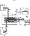
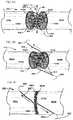
- FIGS. 1A and 1Bare perspective views of a self-retaining suture comprising a drug according to an embodiment of the present invention.
- FIGS. 1C and 1Dare alternative sectional views of the suture filament of FIGS. 1A and 1B illustrating parameters of drug distribution.
- FIGS. 1E and 1Fare enlarged sectional views of the suture thread of the self-retaining suture system of FIGS. 1A and 1B illustrating parameters of the retainers and retainer distribution.
- FIGS. 2A, 2B and 2Care sectional views illustrating steps in the creation of a retainer of a self-retaining suture comprising a drug according to an embodiment of the present invention.
- FIG. 2Dis a sectional view of an alternative step in the creation of a retainer of a self-retaining suture comprising a drug according to an embodiment of the present invention.
- FIG. 3Aillustrates a method and apparatus for co-extruding multiple materials to form a composite filament suitable for creation of a self-retaining suture comprising a drug according to an embodiment of the present invention.
- FIG. 3Billustrates a method and apparatus for extruding a material onto a preformed filament to form a composite filament suitable for creation of a self-retaining suture comprising a drug according to an embodiment of the present invention.
- FIG. 3Cillustrates an alternative method and apparatus for depositing a material onto a preformed filament to form a composite filament suitable for creation of a self-retaining suture comprising a drug according to an embodiment of the present invention.
- FIGS. 3D-3Fillustrate alternative configurations of self-retaining filaments and structures suitable for creation of a self-retaining suture comprising a drug according to embodiments of the present invention.
- FIGS. 4A-4Eillustrate alternative configurations of co-extruded suture stock suitable for creation of a self-retaining suture comprising a drug according to embodiments of the present invention.
- FIGS. 4F-4Iillustrate alternative geometries of co-extruded suture stock suitable for creation of a self-retaining suture comprising a drug according to embodiments of the present invention.
- FIGS. 5A-5Cillustrate particular embodiments of retainers for creation of a self-retaining suture comprising a drug according to embodiments of the present invention.
- FIG. 5Dillustrates a sapphire blade configuration for cutting the retainers of FIGS. 5A-5C according to an embodiment of the present invention.
- FIG. 5Eillustrates an alternative sapphire blade configuration for cutting the retainers of a self-retaining suture according to embodiments of the present invention
- FIG. 5Fillustrates a curved sapphire blade configuration for cutting the retainers of a self-retaining suture according to embodiments of the present invention.
- FIG. 5Gillustrates a circular blade configuration for cutting the retainers of a self-retaining suture according to embodiments of the present invention.
- FIG. 6Aillustrates a single helix distribution of retainers on a drug-eluting self-retaining suture according to an embodiment of the invention.
- FIG. 6Billustrates a double helix distribution of retainers on a drug-eluting self-retaining suture according to an embodiment of the invention.
- FIG. 6Cillustrates a high density quad distribution of retainers on a drug-eluting self-retaining suture according to an embodiment of the invention.
- FIGS. 6D-6Fillustrate alternative retainer shapes for retainers on a drug-eluting self-retaining suture according to embodiments of the invention.
- FIG. 6Gillustrates suture dimensions that can benefit from the embodiments of the invention.
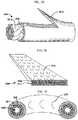
- FIG. 7Aillustrates a sectional view of a nerve for reference.
- FIG. 7Billustrates the release kinetics for NGF from a drug-eluting self-retaining suture according to an embodiment of the present invention.
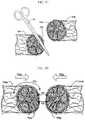
- FIGS. 7C-7Iillustrate a procedure for repairing a nerve utilizing a drug-eluting self-retaining suture according to an embodiment of the present invention.
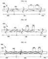
- FIGS. 8A and 8Billustrate techniques utilizing drug-eluting self-retaining sutures to bridge a nerve defect.
- FIGS. 9A, 9B and 9Cillustrate an alternative technique for reattaching a severed nerve utilizing one or more drug-eluting self-retaining sutures.
- FIGS. 10A and 10Bshow PC12 cell culture responses to exposure to no NGF-loaded suture and NGF-loaded suture, respectively.
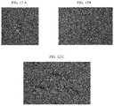
- FIGS. 11A-Dshow PC12 cell culture responses to no NGF-loaded suture and to varying amounts of NGF-loaded suture, respectively, in a green phalloidin stain.
- FIGS. 12A-Cshow PC12 cell culture responses, in phase contrast, to no NGF-loaded suture and to 5 cm and 10 cm lengths of NGF-loaded suture, respectively.
- FIG. 13is a graph illustrating the dose response of PC12 cell cultures to varying amounts of NGF-loaded suture.
- Armed suturerefers to a suture having a suture needle on at least one suture deployment end.
- Bidirectional suturerefers to a self-retaining suture having retainers oriented in one direction at one end and retainers oriented in the other direction at the other end.
- a bidirectional sutureis typically armed with a needle at each end of the suture thread.
- Many bidirectional sutureshave a transition segment located between the two barb orientations.
- Braided suturerefers to a suture comprising a multifilamentary suture thread.
- the filaments in such suture threadsare typically braided, twisted, or woven together.
- “Degradable suture”(also referred to as “biodegradable suture” or “absorbable suture”) refers to a suture which, after introduction into a tissue is broken down and absorbed by the body. Typically, the degradation process is at least partially mediated by, or performed in, a biological system. “Degradation” refers to a chain scission process by which a polymer chain is cleaved into oligomers and monomers. Chain scission may occur through various mechanisms, including, for example, by chemical reaction (e.g., hydrolysis, oxidation/reduction, enzymatic mechanisms or a combination of these) or by a thermal or photolytic process.
- chemical reactione.g., hydrolysis, oxidation/reduction, enzymatic mechanisms or a combination of these
- Degradable suture materialmay include polymers such as polydioxanone (“PDO”), polyglycolic acid, copolymers of glycolide and lactide, copolymers of trimethylene carbonate and glycolide with diethylene glycol (e.g., MAXONTM, Tyco Healthcare Group), terpolymer composed of glycolide, trimethylene carbonate, and dioxanone (e.g., BIOSYNTM [glycolide (60%), trimethylene carbonate (26%), and dioxanone (14%)], Tyco Healthcare Group), copolymers of glycolide, caprolactone, trimethylene carbonate, and lactide (e.g., CAPROSYNTM, Tyco Healthcare Group).
- PDOpolydioxanone
- polyglycolic acidcopolymers of glycolide and lactide
- trimethylene carbonate and glycolide with diethylene glycole.g., MAXONTM, Tyco Healthcare Group
- a dissolvable suturecan also include partially deacetylated polyvinyl alcohol.
- Polymers suitable for use in degradable suturescan be linear polymers, branched polymers or multi-axial polymers. Examples of multi-axial polymers used in sutures are described in U.S. Patent Application Publication Nos. 20020161168, now abandoned, 20040024169, issued as U.S. Pat. No. 7,026,437 on Apr. 11, 2006, and 20040116620, issued as U.S. Pat. No. 7,070,858 on Jul. 4, 2006. Sutures made from degradable suture material lose tensile strength as the material degrades. Degradable sutures can be in either a braided multifilament form or a monofilament form.
- Drugrefers to is a chemical capable of administration to an organism, which modifies or alters the organism's physiology.
- the “drug”is a substance intended for use in the treatment or prevention of disease.
- the term “drug”includes pro-active, activated, metabolized and non-metabolized drugs. Drugs include, for example, synthetic and naturally occurring toxins and bioactive substances as well as recognized pharmaceuticals, such as those listed in the “2010 Physicians' Desk Reference®” (PDR®, 2009) which is incorporated herein by reference.
- the term “drug”is also intended to encompass pharmaceuticals that have the indicated properties but that are discovered or made available after the filing date of this application.
- “Drug-eluting suture”refers to a suture which has a drug associated with it at the time of implantation in tissue of a patient, the suture releasing the drug into the tissue of the patient subsequent to implantation. Association of a drug with a suture can be accomplished in a variety of ways. The drug is impregnated in the suture before, after, or during creation of the filament. The drug alternatively can be coated on the filament before or after retainer formation in a self-retaining suture.
- the drugis incorporated for example, (a) by directly affixing to the suture a formulation (e.g., by either spraying the suture with a polymer/drug film, or by dipping the suture into a polymer/drug solution), (b) by coating or impregnating the suture with a substance such as a hydrogel which will absorb the drug or includes the drug at the time coating, impregnating, or creating the suture (c) by interweaving drug-coated thread (or the polymer itself formed into a thread) into the suture structure in the case of multi-filamentary sutures, (d) constructing the suture itself with the drug in the material of manufacture or being the material of manufacture.
- a formulatione.g., by either spraying the suture with a polymer/drug film, or by dipping the suture into a polymer/drug solution
- a substancesuch as a hydrogel which will absorb the drug or includes the drug at the time coating, impregnating, or creating
- Medical devicerefers to any object placed in the body for the purpose of restoring physiological function, reducing/alleviating symptoms associated with disease, and/or repairing and/or replacing damaged or diseased organs and tissues. While normally composed of biologically compatible synthetic materials (e.g., medical-grade stainless steel, titanium and other metals or polymers such as polyurethane, silicon, PLA, PLGA, PDO, and other materials) that are exogenous, some medical devices and implants include materials derived from animals (e.g., “xenografts” such as whole animal organs; animal tissues such as heart valves; naturally occurring or chemically-modified molecules such as collagen, hyaluronic acid, proteins, carbohydrates and others), human donors (e.g., “allografts” such as whole organs; tissues such as bone grafts, skin grafts and others), or from the patients themselves (e.g., “autografts” such as saphenous vein grafts, skin grafts, tendon/
- autograftssuch
- Medical devicesthat can be used in procedures in conjunction with the present invention include, but are not restricted to, orthopedic implants (artificial joints, ligaments and tendons; screws, plates, and other implantable hardware), dental implants, intravascular implants (arterial and venous vascular bypass grafts, hemodialysis access grafts; both autologous and synthetic), skin grafts (autologous, synthetic), tubes, drains, implantable tissue bulking agents, pumps, shunts, sealants, surgical meshes (e.g., hernia repair meshes, tissue scaffolds), fistula treatments, spinal implants (e.g., artificial intervertebral discs, spinal fusion devices, etc.) and the like.
- orthopedic implantsartificial joints, ligaments and tendons; screws, plates, and other implantable hardware
- dental implantsinclude, but are not restricted to, dental implants, intravascular implants (arterial and venous vascular bypass grafts, hemodialysis access grafts; both autologous and synthetic), skin graft
- “Monofilament suture”refers to a suture comprising a monofilamentary suture thread.
- Needle attachmentrefers to the attachment of a needle to a suture requiring same for deployment into tissue, and can include methods such as crimping, swaging, using adhesives, and so forth.
- the suture threadis attached to the suture needle using methods such as crimping, swaging and adhesives. Attachment of sutures and surgical needles is described in U.S. Pat. Nos. 3,981,307, 5,084,063, 5,102,418, 5,123,911, 5,500,991, 5,722,991, 6,012,216, and 6,163,948, and U.S. Patent Application Publication No. US 20040088003, now abandoned).
- the point of attachment of the suture to the needleis known as the swage.
- Needle diameterrefers to the diameter of a suture deployment needle at the widest point of that needle. While the term “diameter” is often associated with a circular periphery, it is to be understood herein to indicate a cross-sectional dimension associated with a periphery of any shape.
- Non-degradable suturerefers to a suture comprising material that is not degraded by chain scission such as chemical reaction processes (e.g., hydrolysis, oxidation/reduction, enzymatic mechanisms or a combination of these) or by a thermal or photolytic process.
- chain scissionsuch as chemical reaction processes (e.g., hydrolysis, oxidation/reduction, enzymatic mechanisms or a combination of these) or by a thermal or photolytic process.
- Non-degradable suture materialincludes polyamide (also known as nylon, such as nylon 6 and nylon 6,6), polyester (e.g., polyethylene terephthlate), polytetrafluoroethylene (e.g., expanded polytetrafluoroethylene), polyether-ester such as polybutester (block copolymer of butylene terephthalate and polytetra methylene ether glycol), polyurethane, metal alloys, metal (e.g., stainless steel wire), polypropylene, polyethylene, silk, and cotton.
- Sutures made of non-degradable suture materialare suitable for applications in which the suture is meant to remain permanently or is meant to be physically removed from the body.
- Retainer configurationsrefers to configurations of tissue retainers and can include features such as size, shape, flexibility, surface characteristics, and so forth. These are sometimes also referred to as “barb configurations”.
- Self-retaining suturerefers to a suture that comprises features on the suture filament for engaging tissue without the need for a knot or suture anchor.
- Self-retaining systemrefers to a self-retaining suture together with devices for deploying the suture into tissue.
- deployment devicesinclude, without limitation, suture needles and other deployment devices as well as sufficiently rigid and sharp ends on the suture itself to penetrate tissue.
- Suture deployment endrefers to an end of the suture to be deployed into tissue; one or both ends of the suture may be suture deployment ends.
- the suture deployment endmay be attached to a deployment device such as a suture needle, or may be sufficiently sharp and rigid to penetrate tissue on its own.
- Suture diameterrefers to the diameter of the body of the suture. It is to be understood that a variety of suture lengths may be used with the sutures described herein and that while the term “diameter” is often associated with a circular periphery, it is to be understood herein to indicate a cross-sectional dimension associated with a periphery of any shape. Suture sizing is based upon diameter. United States Pharmacopeia (“USP”) designation of suture size runs from 0 to 7 in the larger range and 1-0 to 11-0 in the smaller range; in the smaller range, the higher the value preceding the hyphenated zero, the smaller the suture diameter.
- USPUnited States Pharmacopeia
- the actual diameter of a suturewill depend on the suture material, so that, by way of example, a suture of size 5-0 and made of collagen will have a diameter of 0.15 mm, while sutures having the same USP size designation but made of a synthetic absorbable material or a non-absorbable material will each have a diameter of 0.1 mm.
- the selection of suture size for a particular purposedepends upon factors such as the nature of the tissue to be sutured and the importance of cosmetic concerns; while smaller sutures may be more easily manipulated through tight surgical sites and are associated with less scarring, the tensile strength of a suture manufactured from a given material tends to decrease with decreasing size.
- sutures and methods of manufacturing sutures disclosed hereinare suited to a variety of diameters, including without limitation 7, 6, 5, 4, 3, 2, 1, 0, 1-0, 2-0, 3-0, 4-0, 5-0, 6-0, 7-0, 8-0, 9-0, 10-0 and 11-0.
- “Suture needle”refers to needles used to deploy sutures into tissue, which come in many different shapes, forms and compositions. There are two main types of needles, traumatic needles and atraumatic needles. Traumatic needles have channels or drilled ends (that is, holes or eyes) and are supplied separate from the suture thread and are threaded on site. Atraumatic needles are eyeless and are attached to the suture at the factory by swaging or other methods whereby the suture material is inserted into a channel at the blunt end of the needle which is then deformed to a final shape to hold the suture and needle together. As such, atraumatic needles do not require extra time on site for threading and the suture end at the needle attachment site is generally smaller than the needle body.
- suture needlesIn the traumatic needle, the thread comes out of the needle's hole on both sides and often the suture rips the tissues to a certain extent as it passes through.
- Most modern suturesare swaged atraumatic needles.
- Atraumatic needlesmay be permanently swaged to the suture or may be designed to come off the suture with a sharp straight tug. These “pop-offs” are commonly used for interrupted sutures, where each suture is only passed once and then tied. For barbed sutures that are uninterrupted, atraumatic needles are preferred.
- Suture needlesmay also be classified according to the geometry of the tip or point of the needle.
- needlesmay be (i) “tapered” whereby the needle body is round and tapers smoothly to a point; (ii) “cutting” whereby the needle body is triangular and has a sharpened cutting edge on the inside; (iii) “reverse cutting” whereby the cutting edge is on the outside; (iv) “trocar point” or “taper cut” whereby the needle body is round and tapered, but ends in a small triangular cutting point; (v) “blunt” points for sewing friable tissues; (vi) “side cutting” or “spatula points” whereby the needle is flat on top and bottom with a cutting edge along the front to one side (these are typically used for eye surgery).
- Suture needlesmay also be of several shapes including, (i) straight, (ii) half curved or ski, (iii) 1 ⁇ 4 circle, (iv) 3 ⁇ 8 circle, (v) 1 ⁇ 2 circle, (vi) 5 ⁇ 8 circle, (v) and compound curve.
- Suturing needlesare described, for example, in U.S. Pat. Nos. 6,322,581 and 6,214,030 (Mani, Inc., Japan); and 5,464,422 (W. L.
- the sutures described hereinmay be deployed with a variety of needle types (including without limitation curved, straight, long, short, micro, and so forth), needle cutting surfaces (including without limitation, cutting, tapered, and so forth), and needle attachment techniques (including without limitation, drilled end, crimped, and so forth). Moreover, the sutures described herein may themselves include sufficiently rigid and sharp ends so as to dispense with the requirement for deployment needles altogether.
- Suture threadrefers to the filamentary body component of the suture.
- the suture threadmay be a monofilament, or comprise multiple filaments as in a braided suture.
- the suture threadmay be made of any suitable biocompatible material, and may be further treated with any suitable biocompatible material, whether to enhance the sutures' strength, resilience, longevity, or other qualities, or to equip the sutures to fulfill additional functions besides joining tissues together, repositioning tissues, or attaching foreign elements to tissues.
- tissue elevation procedurerefers to a surgical procedure for repositioning tissue from a lower elevation to a higher elevation (i.e. moving the tissue in a direction opposite to the direction of gravity).
- the retaining ligaments of the facesupport facial soft tissue in the normal anatomic position.
- Face-lift proceduresare designed to lift these sagging tissues, and are one example of a more general class of medical procedure known as a tissue elevation procedure.
- tissue elevation procedurereverses the appearance change that results from effects of aging and gravity over time, and other temporal effects that cause tissue to sag, such as genetic effects. It should be noted that tissue can also be repositioned without elevation; in some procedures tissues are repositioned laterally (away from the midline), medially (towards the midline) or inferiorly (lowered) in order to restore symmetry (i.e. repositioned such that the left and right sides of the body “match”).
- tissue retainerrefers to a physical feature of a suture filament which is adapted to mechanically engage tissue and resist movement of the suture in at least one axial direction.
- tissue retainer or retainerscan include hooks, projections, barbs, darts, extensions, bulges, anchors, protuberances, spurs, bumps, points, cogs, tissue engagers, traction devices, surface roughness, surface irregularities, surface defects, edges, facets and the like.
- tissue retainersare adapted to engage tissue to resist movement of the suture in a direction other than the direction in which the suture is deployed into the tissue by the surgeon, by being oriented to substantially face the deployment direction.
- the retainerslie flat when pulled in the deployment direction and open or “fan out” when pulled in a direction contrary to the deployment direction. As the tissue-penetrating end of each retainer faces away from the deployment direction when moving through tissue during deployment, the tissue retainers should not catch or grab tissue during this phase.
- the tissue retainersare configured to permit motion of the suture in one direction and resist movement of the suture in another direction without fanning out or deploying.
- the tissue retainersare configured or combined with other tissue retainers to resist motion of the suture filament in both directions.
- a suture having such retainersis deployed through a device such as a cannula which prevents contact between the retainers and the tissue until the suture is in the desired location.
- Transition segmentrefers to a retainer-free portion of a bidirectional suture located between a first set of retainers oriented in one direction and a second set of retainers oriented in another direction.
- the transition segmentcan be at about the midpoint of the self-retaining suture, or closer to one end of the self-retaining suture to form an asymmetrical self-retaining suture system.
- wound closurerefers to a surgical procedure for closing of a wound.
- An injuryespecially one in which the skin or another external or internal surface is cut, torn, pierced, or otherwise broken is known as a wound.
- a woundcommonly occurs when the integrity of any tissue is compromised (e.g., skin breaks or burns, muscle tears, or bone fractures).
- a woundmay be caused by an act, such as a puncture, fall, or surgical procedure; by an infectious disease; or by an underlying medical condition.
- Surgical wound closurefacilitates the biological event of healing by joining, or closely approximating, the edges of those wounds where the tissue has been torn, cut, or otherwise separated.
- Surgical wound closuredirectly apposes or approximates the tissue layers, which serves to minimize the volume new tissue formation required to bridge the gap between the two edges of the wound. Closure can serve both functional and aesthetic purposes. These purposes include elimination of dead space by approximating the subcutaneous tissues, minimization of scar formation by careful epidermal alignment, and avoidance of a depressed scar by precise eversion of skin edges.
- compositions, configurations, methods of manufacturing and methods of using self-retaining systems in surgical procedureswhich the ability to anchor into the surrounding tissue to provide superior holding strength and improve clinical performance while providing a drug to this tissue surrounding the suture.
- FIG. 1Aillustrates a bidirectional self-retaining suture system 100 .
- Self-retaining suture system 100comprises needles 110 , 112 attached to self-retaining suture thread 102 .
- Self-retaining suture thread 102includes a plurality of retainers 130 distributed on the surface of a filament 120 .
- In lead-in region 140 of filament 120there are no retainers 130 .
- In region 142 of filament 120there are a plurality of retainers 130 arranged such that the suture can be deployed in the direction of needle 110 but resists movement in the direction of needle 112 .
- transition region 144there are no retainers 130 .
- region 146there are a plurality of retainers 130 arranged such that the suture can be deployed in the direction of needle 112 but resists movement in the direction of needle 110 .
- lead-in region 148 of filament 120there are no retainers 130 .
- a breakis shown in each of regions 140 , 142 , 144 , 146 and 148 to indicate that the length of each region may be varied and selected depending upon the application for which the suture is intended to be used.
- the present inventionincludes self-retaining suture systems of a wide variety of retainer and needle configurations described above.
- the configuration of each of needles 110 and 112can be any of the range of different surgical needles developed for use in different applications. Needles 110 and 112 may have the same configuration or different configurations.
- Filament 120comprises a drug 152 ( FIGS. 1C, 1D ).
- the drug 152is in a coating layer on filament 120 or impregnated in the material 150 of filament 120 .
- Various methodsmay be used to make sutures that carry drugs. For example, such methods include direct extrusion as described in U.S. Pat. No. 6,596,296 (TissueGen, Inc., Dallas, Tex.) to create filaments wherein the drug is uniformly distributed as in FIG. 1C .
- tissueGen, Inc., Dallas, Tex.tissueGen, Inc., Dallas, Tex.
- core/sheathand other multicomponent configurations as illustrated in FIG. 1D may also be extruded as described in U.S. Pat. No. 7,033,603 (TissueGen, Inc., Dallas, Tex.).
- 6,596,296 and 7,033,603are herein incorporated by reference.
- Alternate methodssuch as coating (e.g., spraying or dipping) all or part of the sutures or an “over the wire” extrusion as described in U.S. Pat. No. 6,858,222 (TissueGen, Inc., Dallas, Tex.) may also be used. Additionally, gradients of the drug along the suture are sometimes preferred. These linear anisotropies are described in U.S. Pat. Nos. 6,596,296, 6,858,222, and 7,514,095 (TissueGen, Inc., Dallas, Tex.), the latter of which is also hereby incorporated by reference.
- suturesthemselves can be made at least in part of materials that have pharmaceutical activity in or around the site where the sutures are implanted or inserted.
- only selected portions (such as middle sections or the self-retaining sections) of a self-retaining sutureare coated or otherwise comprise the drug or drugs.
- portions of the suturesare selectively left unassociated with a drug or drugs or are associated with a drug different from another drug associated with a different portion of the self-retaining suture.
- the suture surfaces between retainer and main suture body in which tissue are grippedare selectively associated with one or more drugs that enhance healing and prevent scarring.
- temporally phased release of one or more drugsmay be designed to coincide with known phases of wound healing as a means to reduce scaring and enhance the body's natural wound healing processes. This may be accomplished, for example, by multilayer filaments as described in U.S. Pat. No. 7,033,603 or by using multiple means of incorporating the drug in the base material of the filament, such as simultaneous use of nanoparticles and microspheres within the same filament as described in U.S. Pat. No. 6,858,222.
- the suture surfacemay comprise one or more wells including one or more drugs. In other embodiments, all sections of sutures are coated with the drug(s).
- the methods for applying drugs to suturesinclude, for example: (a) extrusion, (b) by directly affixing to the suture a formulation (e.g., by either spraying the suture with a polymer/drug film, or by dipping the suture into a polymer/drug solution), (c) by coating the suture with a substance such as a hydrogel which will in turn absorb the composition, (d) by interweaving formulation-coated thread (or the polymer itself formed into a thread) into the suture structure in the case of multi-filamentary sutures, (e) constructing the suture itself with a drug-containing composition.
- Drug-eluting self-retaining suture systems 100differ from conventional sutures in that, in addition to providing drugs directly to a tissue of interest, the self-retaining sutures possess numerous tissue retainers 130 (such as barbs) which anchor the self-retaining suture system 100 into the tissue following deployment and resist movement of the suture in a direction opposite to that in which the retainers face, thereby eliminating the need to tie knots to affix adjacent tissues together (a “knotless” closure).
- tissue retainers 130such as barbs
- the drug-eluting self-retaining suture systems for wound closurealso result in better approximation of the wound edges, evenly distribute the tension along the length of the wound (reducing areas of tension that can break or lead to ischemia), decrease the bulk of suture material remaining in the wound (by eliminating knots) and reduce spitting (the extrusion of suture material—typically knots—through the surface of the skin. All of these features are thought to reduce scarring, improve cosmesis, and increase wound strength relative to wound closures using plain sutures or staples.
- self-retaining suturesbecause such sutures avoid knot-tying, allow patients to experience an improved clinical outcome, and also save time and costs associated with extended surgeries and follow-up treatments. Also, by avoiding knot-tying, drug-eluting self retaining sutures avoid local concentrations of drugs in the vicinity of such knots.
- the purpose of the drug-eluting self-retaining suturedetermines the sort of drug that is applied to or incorporated in the suture.
- self-retaining sutures having anti-proliferative drugsmay be used in closing tumor excision sites
- self-retaining sutures containing or coated with nerve growth factor (NGF)may be used in the repair of damaged nerves
- self-retaining sutures with fibrosing drugsmay be used in tissue repositioning procedures and those having anti-scarring drugs may be used for wound closure on the skin.
- Bone growth factorssuch as Bone Morphogenic Proteins (BMP) can also be incorporated within the sutures.
- BMPBone Morphogenic Proteins
- the drugsmay also include a plurality of compositions either together or on different portions of the suture, where the multiple compositions can be selected either for different purposes (such as combinations of growth factors, analgesics, anti-infective and anti-scarring agents) or for the synergistic effects of the combination.
- the drug or drugs incorporated in or coated on a self-retaining suture in embodiments of the present inventioninclude, for example, compositions to promote healing and prevent undesirable effects such as scar formation, infection, pain, and so forth.
- the drugsinclude without limitation growth factors such as nerve growth factor (NGF), bone grown factor (BGF), tissue repair factors, trophic factors to guide tissue repair, inhibition agonists to suppress factors which inhibit tissue repair, mitogenic agents to promote cell division for tissue repair, anti-proliferative agents, anti-angiogenic agents, anti-infective agents, fibrosis-inducing agents, anti-scarring agents, lubricious agents, echogenic agents, anti-inflammatory agents, cell cycle inhibitors, analgesics, and anti-microtubule agents.
- growth factorssuch as nerve growth factor (NGF), bone grown factor (BGF), tissue repair factors, trophic factors to guide tissue repair, inhibition agonists to suppress factors which inhibit tissue repair, mitogenic agents to promote cell division for tissue repair, anti-proliferative agents, anti-angi
- a drugcan be utilized in conjunction with the suture (introduced separately or adhered to the suture or incorporated into a material of the suture) to encourage fibrosis.
- Fibrosis-inducing drugswhich may be used in conjunction with a drug-eluting self-retaining suture according to the present invention are described in U.S. Pat. No. 7,166,570 titled “Medical Implants And Fibrosis-Inducing Agents” to Hunter et al., which is incorporated herein by reference.
- Other drugswhich may be used in drug-eluting self-retaining suture of the present invention include all drugs and agents disclosed in U.S. patent application Ser. No. 12/162,572, published as U.S. Pub. No. 2009/0226500 on Sep. 10, 2009, titled “Sutures And Anti-Scarring Agents” to Avelar et al., which is incorporated herein by reference.
- the drug 152is uniformly distributed in the material 150 of filament 120 .
- drug 152is anisotropically distributed.
- filament 120may have higher concentrations of a drug in an outer layer of material as compared to the inner core of material, or vice versa.
- filament 120may have a higher concentration of a drug at one end as compared to another.
- filament 120may have a higher concentration of a drug in the middle than towards the ends.
- the concentration of the drug 152may change gradually from region to region or there may be changes in concentration from one region to another region.
- Different regions of filament 120 having different drug concentrationsmay correspond to regions of the filament 120 having retainers, no retainers or retainers in one orientation compared to another orientation.
- the filament 120may be provided with visible or otherwise detectable markers which indicate regions have greater or lesser concentrations of the drug 152 in order to identify said regions during manufacturing and/or utilization of the self-retaining suture system 100 .
- a very thin coating including one of more drug(s)can be applied to the suture by any of the aforementioned techniques before the retainers are formed, so that when the retainers engage, the engaging surface is substantially free of the coating. In this way, tissue being sutured contacts a coated surface of the suture as the suture is introduced, but when the retainer engages, a non-coated surface of the retainer contacts the tissue.
- Other embodiments that may provide drug-exposing suture surfacesinclude sutures extruded with the drug(s) uniformly dispersed as in FIG. 1C , sutures extruded as a multi-layer “core/sheath” arrangement as shown in FIG.
- sutures coated after or during formation of retainers on the suture bodyif, for example, a fully-coated rather than selectively-coated suture is desired.
- a sutureis selectively coated either during or after formation of retainers by exposing only selected portions of the suture to the coating.
- the particular purpose to which the suture is to be put or the compositionmay determine whether a fully- or selectively-loaded suture is appropriate, for example coatings such as those comprising fibrosing agents may suitably be applied to all or part of the suture (such as the tissue-engaging surfaces). Coatings such as those comprising such compounds as growth factors may suitably be applied to the entire suture and/or incorporated into the material from which the filament is made prior to formation of the retainers.
- Methods of making drug-loaded filamentsare described in: U.S. Pat. No. 7,514,095 titled “Fabrication Of Drug Loaded Biodegradable Polymer Fibers” and U.S. Pat. No.
- the structure of the suturemay influence the choice and extent of application and/or incorporation of a drug or drugs; for example, sutures having an expanded segment may include a fibrosis-inducing composition in or on the expanded segment to further secure the segment in position in the tissue.
- Sutures used in tissue repairmay include for example a gradient of concentration of the trophic factors such that the suture delivers a gradient of trophic factors to the tissue to guide repair.
- the structure of the suture and retainerscan influence/control the release kinetics of the drug or drugs. The location of the incorporation of coating of the drug will also influence/control the release kinetics of the drug.
- drug dosecan be calculated as a function of dose per unit area (of the portion of the suture being coated), or total drug dose.
- Total drug dose administeredcan be measured and appropriate surface concentrations of active drug can be determined.
- the total drug administeredwill typically be substantially less than the equivalent systemic dose, because, by being associated with the self-retaining suture, the drug will be distributed directly in the vicinity of the target tissue rather than being evenly distributed through the whole body.
- drugsare used at concentrations that range from several times more than, to 50%, 20%, 10%, 5%, or even less than 1% of the concentration typically used for a systemic dose application.
- the drugis released from the composition in effective concentrations in a time period that is measured from the time of infiltration into tissue adjacent to the suture, which ranges from about less than 1 day to about 180 days.
- the release timemay also be from about less than 1 day to about 180 days; from about 7 days to about 14 days; from about 14 days to about 28 days; from about 28 days to about 56 days; from about 56 days to about 90 days; from about 90 days to about 180 days.
- the release kineticsare affected by the surface area of retainers in a particular region and thus should be validated for particular retainer configurations to achieve the desired final kinetics.
- anti-infective agentsare associated with a self-retaining suture, alone or in combination, they may be administered under the following dosing guidelines.
- the total amount (dose) of anti-infective agent in the compositioncan be in the range of about 0.01-1 ⁇ g, or about 1-10 ⁇ g, or about 10-100 mg or about 100 ⁇ g-1 mg or about 1-10 mg, or about 10-100 mg, or about 100 to 250 mg for coating a suture or a portion thereof or for infiltrating a tissue where a suture has been, is being, or is to be, implanted, or about 250-1000 mg for infiltrating a tissue where a suture has been, is being, or is to be, implanted.
- the dose (amount) of anti-infective agent per unit area of suture or tissue surface to which the agent is appliedmay be in the range of about 0.01 ⁇ g/mm 2 to 1 ⁇ g/mm2, or about 1 ⁇ g/mm 2 to 10 ⁇ g/mm 2 , or about 10 ⁇ g/mm 2 to 100 ⁇ g/mm 2 , or about 100 ⁇ g/mm 2 to 250 ⁇ g/mm 2 .
- the above dosing parametersshould be utilized in combination with the release rate of the drug from the filament and retainers such that a minimum concentration of about 10 ⁇ 8 M to 10 ⁇ 7 M, or about 10 ⁇ 7 M to 10 ⁇ 6 M or about 10 ⁇ 6 M to 10 ⁇ 5 M or about 10 ⁇ 5 M to 10 ⁇ 4 M of the agent is maintained in the vicinity of or on the tissue surface to maintain the desired therapeutic effect for the required period of time.
- the required minimum concentrationis dependent on the potency of the agent under consideration and can be determined using standard tests such as the Minimum Inhibitory Concentration (M.I.C.) test.
- FIG. 1Billustrates a magnified view of self-retaining suture 102 in region 142 .
- a plurality of retainers 130is distributed on the surface of filament 120 .
- the affixation of self-retaining sutures after deployment in tissueentails the penetration of retainer ends into the surrounding tissue resulting in tissue being caught between the retainer and the suture body.
- the inner surface of the retainer that is in contact with the tissue that is caught between the retainer 130 and the filament 120herein referred to as the “tissue engagement surface” or “inner retainer surface,” can be adapted to better engage the tissue and also to better distribute drugs in a tissue. As illustrated in FIG.
- each retainer 130has a tip 132 and tissue retainer surface 134 .
- retainer 130lies flat against the body of filament 120 .
- tip 132 or retainer 130engages tissue surrounding filament 120 and causes retainer 130 to fan out from filament 120 and engage the tissue with face 134 thereby preventing movement of the suture in that direction and providing an additional surface from which to provide drugs to the tissue.
- the surface area of the filament 120is also increased by the presence of retainers 130 as compared to portions of filament with no retainers.
- FIG. 1Cshows a cross-sectional view of filament 120 .
- filament 120includes a material 150 and a drug 152 .
- the distribution of drug 152is isotropic or homogenous across the radius of filament 120 .
- FIG. 1Dshows a cross-sectional view of an alternative filament 120 d .
- filament 120 dincludes a material 150 d and a drug 152 d .
- the distribution of drug 152 dis anisotropic across the radius of filament 120 .
- the concentration of drug 152 dis greater in a sheath region 160 of filament 120 d than in the core region 162 .
- a retainer 130may comprise the material of the sheath region 160 and also some portion of the core region 162 or another non-sheath material.
- the materialsare selected such that the properties of the materials in the retainer permit or enhance the function of the retainer such as by facilitating elevation of the retainer 130 .
- the drug 152 dcan be expected to be deployed into the tissue more rapidly than in the embodiment of FIG. 1C .
- the drug 152 dcan be expected to be deployed into the tissue less rapidly.
- the distribution of drug concentrationcan be used to control the release kinetics of the drug or drug(s) and/or sequence the release of drugs from a self-retaining suture.
- different drugsare provided in the core region 162 and sheath region 160 .
- the drug of the sheath region 160will be provided sooner, and or faster than the drug in the core region 162 —allowing for temporal sequencing of the distribution of the drugs to the tissue.
- Filament 120 dis formed by any method known in the art for making a filament having a drug associated with one or more components thereof (for example a core and/or a sheath) (drug-eluting suture).
- a suitable methodis co-extrusion of materials having the required drug concentration as disclosed in U.S. Pat. No. 7,033,603 and will be further described with respect to FIG. 3A .
- Another suitable methodis extrusion of a material over a preformed filament as disclosed in U.S. Pat. No. 6,596,296 (TissueGen, Inc., Dallas, Tex.), incorporated by reference herein, and will be further described with respect to FIG. 3B .
- FIG. 1Eshows a sectional diagram through a retainer 130 . Note that where retainer 130 is cut into filament 120 it leaves a cut-out depression 136 .
- the cut-out depression 136has a cut tip 135 which corresponds with the tip 132 of the retainer 130 .
- the geometry of retainer 130(retainer cut angle, retainer cut depth, retainer cut length, retainer cut distance, etc.) and/or the spatial arrangement of the retainers 130 is varied to enhance engagement of tissue by the retainers.
- Retainer 130is shown elevated above filament 120 in order to show the parameters related to the retainer and elevation of the retainer.
- the parameters shown in FIG. 1Einclude the longitudinal axis of the suture A-A, the suture diameter SD, the retainer length L, the retainer cut depth D, the retainer cut angle ⁇ (theta), the retainer elevation angle ⁇ (epsilon), the retainer cut distance P.
- the cut distance Pis the distance between adjacent retainers measured along the longitudinal axis it can be measured as the distance from one cut-tip 135 to the adjacent cut-tip 139 .
- the retainer cut angle ⁇is the angle between the cut depression 136 and the longitudinal axis A-A surface of filament 120 . Cut angle ⁇ can be measured between the cut depression 136 and the surface of filament 120 which is parallel to longitudinal axis A-A.
- Retainer elevation angle ⁇is the angle between the inner retainer surface 134 and the cut depression surface 136 .
- the spirality angle ⁇is the angle of rotation about the longitudinal axis between adjacent cut tips 135 , 139 . Where adjacent retainers are on opposite sides of filament 120 , as shown in FIG. 1E , the spirality angle ⁇ is 180 degrees.
- FIG. 1Fshows a section of an alternative filament configuration looking along the long axis. As shown in FIG. 1F , the spirality angle ⁇ is 120 degrees.
- FIG. 1Falso shows a straight line illustrating the position of the base 137 of cut depression 136 . For a straight cut such as shown in FIG. 1F , the cut depth D is the maximum distance between base 137 and the surface of filament 120 .
- Self-retaining suture threads described hereinare produced by any suitable method, including without limitation, injection molding, stamping, cutting, laser, extrusion, and so forth.
- cuttingpolymeric thread or filaments are manufactured or purchased for the suture body, and the retainers can be subsequently cut onto the suture body; the retainers are hand-cut, laser-cut, or mechanically machine-cut using blades, cutting wheels, grinding wheels, and so forth.
- a drugis coated on the filament or impregnated in the material of the filament before, during or after the creation of filament.
- the cutting device or the suture threadis moved relative to the other, or both are moved, to control the size, shape and depth of the cut and the resulting retainer.
- FIG. 2Ashows a longitudinal cross-section of filament 120 d .
- filament 120 dcomprises core region 162 having a lower concentration of drug 152 d in material 150 d and a sheath region 160 having a higher concentration of drug 152 d .
- a different drug or drug(s)are provided in the core region 162 as compared to the sheath region 160 .
- the same concentration of a drug or drug(s)are deployed across the entire radius of the filament 120 d.
- a retainer 130is formed on filament 120 d by making a cut 210 into sheath region 160 of filament 120 d .
- Cut 210can be made using any of a wide range of technologies. Such technologies include hand-cutting, laser-cutting, or mechanically machine-cutting using blades, cutting wheels, grinding wheels, and so forth. Note that in this embodiment, the depth of cut has been selected such that cut 210 is entirely within sheath region 160 and does not penetrate into core region 162 . As shown in FIG. 2B , retainer 130 may still lay flat against the surface of filament 120 d after cut 210 has been made in material of sheath region 160 .
- tip 132is preferably elevated above the surface of filament 120 d .
- retainer 130is mechanically bent away from the body of filament 120 d in the direction shown by arrow 220 .
- Tip 132is moved above the surface of filament 120 d and tissue engagement surface 134 is exposed.
- the cut filament surface 234is also exposed.
- the elevation of retainer 130can be achieved by a number of mechanisms. In a simple example, a cutting blade is used to form cut 210 and the cutting blade is then removed from cut 210 in a manner that bends retainer 130 away from the body of filament 120 d .
- the retaineris mechanically elevated by a device other than the blade. Where the drug 152 d is incorporated in filament 120 d prior to forming retainers 130 , care must be taken to ensure that the retainer forming process does not denature the drug 152 d.
- material 150 dis selected such that it is sufficiently plastically deformable that after retainer 130 has been moved away from filament 120 d , retainer 130 d remains in its new deformed shape with the tip 132 of retainer 130 substantially elevated above the surface of filament 120 d and tissue engagement surface 134 exposed.
- Sheath material 150 dis selected such that the mechanical movement of tip 132 of retainer away from filament 120 d is sufficient to plastically deform the region 230 of the material of sheath region 160 at the base of retainer 130 causing it to take on a new permanent shape.
- a the core region 162is made from a different material then the sheath region thus allowing selection of a material 162 is selected to have significantly lower plasticity and significantly higher elasticity and/or tensile strength than the sheath region.
- the filamentis made from a single polymer. In other embodiments the filament is made from a single polymer but the polymer comprises different concentrations of drug in different regions of the filament.
- the surface area of the filament 120 d and retainer 130is increased relative to the filament alone. Essentially, the surface area is increased by the surface area of the tissue engagement surface 134 and cut filament surface 234 . Where a drug 152 d is impregnated in the material 150 d of filament 120 d , the release kinetics of the drug is affected by the surface area. Because the retainers 130 d are formed on the surface of filament 120 d , the retainers 130 d are made, in this embodiment, entirely of sheath region 160 containing a higher concentration of drug 152 d . Furthermore, in specific embodiments of the present invention, retainers 130 d include little or none of core region 162 .
- the size, number and density of retainers 130 among other factorscan affect the release kinetics of the drug in regions of the filament bearing retainers 130 .
- the release kineticsare affected by the surface area of retainers in a particular region and thus should be validated for particular retainer configurations to achieve the desired final kinetics.
- a drugmay homogenously distribute along the length of filament 120 d yet the release kinetics are rendered anisotropic by the distribution, shape or size of retainers 130 .
- the drugis released to a tissue faster than in regions having fewer or no retainers.
- a section of the filament 102 d having no retainersmay release the drug more slowly and over a more extended period of time.
- the drugis distributed heterogeneously in different regions of a filament 102 d in order that the release kinetics are homogeneous across regions having more, less or no retainers.
- filament 120 dis provided with a coating (not shown) which delays or prevents migration of the drug 152 d out of filament 120 d .
- drug 152 dwill migrate out of filament 120 d preferentially or sooner where retainers 130 disrupt the coating and expose the interior of filament 120 d on tissue engagement surface 134 and filament cut surface 234 .
- retainer 130is formed by a process other than cutting into the sheath of the filament.
- retainerscan be formed by melting the material 150 d in region 240 and then drawing material out of filament 120 d with device 244 to form retainer 130 and then cooling the material 150 d .
- the material 150 dis selected such that it may be melted and manipulated without disrupting the tensile strength of the core region 162 .
- a preformed retaineris affixed mechanically, adhesively or by melting to the sheath.
- the sheath materialis in this embodiment selected to enhance the affixation of the retainer to the filament and retention of the retainer by the filament.
- molten materialis formed onto the sheath in the shape of a retainer and the molten material fuses with the sheath material.
- the material in this caseis selected to enhance the adhesion or fusion with the externally applied molten material.
- the molten materialis the same material as the material of the filament 120 d and may include drug 152 d . Where the drug 152 d is incorporated in filament or retainer material 120 d prior to forming retainers 130 , care must be also taken to ensure that the retainer-forming process does not denature the drug 152 d.
- the sutureis a relatively short suture with sharp pointing ends.
- a suturemay function similar to a staple when used in connecting tissues and thus permits a surgeon to rapidly and securely attach the edges of a wound in a body tissue or reconfigure the tissue without the necessity for threading and tying numerous individual stitches or for the use of complicated tools and/or techniques to insert the suture.
- This type of suturesmay thus be referred to as “suture connector.”
- the suture connectoris a bi-directional self-retaining suture.
- the suture connectormay be made by linking two relatively short uni-directional self-retaining sutures together to form a bi-directional self-retaining suture (see, U.S. Pat. No.
- the drug-eluting self-retaining sutureis used to anchor a two or three-dimensional scaffold including comprising a woven, non-woven, or knitted, mesh wherein said fibers comprise any of the compositions described herein above.
- the meshmay include addition filaments which may or may not be drug-eluting.
- the filament materialis selected to have properties useful for retainer formation, elevation and deployment as well as strength and flexibility of the self-retaining suture.
- a different materialis used in the core of the suture as opposed to the sheath.
- the coreis chosen based on its properties of strength and flexibility and the sheath material is selected to have properties useful for retainer formation, elevation and deployment.
- the suture materialsare non-degradable or biodegradable so long as the material is suitable for coating or incorporating a drug and releasing said drug in vivo with suitable release kinetics. Suitable materials include many materials that are currently used for making sutures. The release kinetics are affected by the surface area of retainers in a particular region and thus should be validated for particular retainer configurations to achieve the desired final kinetics.
- Suitable non-degradable suture materialsinclude polyamide (also known as nylon, such as nylon 6 and nylon 6,6), polyester (e.g., polyethylene terephthlate), polytetrafluoroethylenes (e.g., expanded polytetrafluoroethylene), polyether-ester such as polybutester (block copolymer of butylene terephthalate and polytetra methylene ether glycol), 4-hydroxybutyrate, polyhydroxylalkanoate, polyurethane, metals and metal alloys (e.g., stainless steel wire), polypropylene, polyethylene, silk, cotton and/or combinations thereof.
- polyamidealso known as nylon, such as nylon 6 and nylon 6,6
- polyestere.g., polyethylene terephthlate
- polytetrafluoroethylenese.g., expanded polytetrafluoroethylene
- polyether-estersuch as polybutester (block copolymer of butylene terephthalate and polyte
- Suitable biodegradable materials for the filamentinclude single polymer, co-polymer or a blend of polymers of poly(L-lactic acid), poly(p-dioxanone), poly(DL-lactic acid), polycaprolactone, poly(glycolic acid), polyanhydride, polyglycolic acid homopolymer, copolymers of glycolide and ⁇ -caprolactone, copolymers of glycolide and lactide, copolymers of trimethylene carbonate and glycolide with diethylene glycol (e.g., MAXONTM, Tyco Healthcare Group), polyhydroxylalkanoates (such as poly(4-hydroxybutyrate) or poly(4-hydroxybutyrate-co-3-hydroxybutyrate)), terpolymer composed of glycolide, trimethylene carbonate, and dioxanone (e.g., BIOSYNTM [glycolide (60%), trimethylene carbonate (26%), and dioxanone (14%)], Tyco Healthcare Group), copolymers of glyco
- Naturally occurring polymersmay also be used such as reconstituted collagen, fibrin, or natural silks, cellulose, starch, chitin, polypeptides modified polysaccharides, modified proteins and/or combinations of the above with synthetic suture materials listed above.
- Other polymers which may be used in drug-eluting self-retaining sutures of the present inventioninclude all polymers disclosed in U.S. patent application Ser. No. 12/162,572, now abandoned, titled “Sutures And Anti-Scarring Agents” to Avelar et al. which is incorporated herein by reference.
- a drug-eluting suture filamentcan be made in many different ways.
- a monofilament 346is formed by co-extruding two materials.
- satellite extruder 310heats, melts and extrudes a first material 311 along conduit 312 to main extruder 330 .
- Metering pump 313 on conduit 312controls the flow of first material 311 to main extruder 330 .
- a second satellite extruder 315heats, melts and extrudes a second material 316 along conduit 317 to main extruder 330 .
- Metering pump 318 on conduit 317controls the flow of second material 316 to main extruder 330 .
- main extruder 330the two melted materials 311 , 316 flow through two flow paths 336 , 338 through an extrusion die 332 which controls the arrangement of the two materials 311 , 316 when the materials combine in flow channel 339 .
- the two materialsmay contain different concentrations of a drug.
- the two materialsare combined in flow channel 339 as shown and then extruded from die 332 through die exit 334 .
- Die 332 and flow channels 336 , 338 , 339are designed and operated such that the two materials 311 and 316 do not mix in flow channel 339 .
- the fiber 340 which is still melted materialis then solidified by air or liquid cooling in quenching station 350 .
- Quenching station 350optionally includes a quenching bath 352 for liquid cooling.
- the solidified filament 342is then drawn in drawing machine 360 .
- the solidified filamentis drawn at temperatures between 30-80% of melting point (Celsius).
- the sutureis extruded then drawn on several rollers with decreasing temperature. Drawing of the filament reduces the diameter of the filament while at the same time orienting the molecules of the polymers of the filament and enhancing the tensile strength of the filament.
- drawingis conducted in a continuous process by winding the filament around a series of rollers where each roller in the series has a slightly higher roller surface speed. The speed differential of the rollers results in stretching of the filament as the filament passes from roller to roller.
- the filamentmay also be tempered by one or more heating and cooling steps before, during or after the drawing process.
- drawn filament 344is tempered in tempering machine 370 as the filament is passed through heating unit 372 .
- the finished monofilament 346is passed to winder 364 where the monofilament is wound onto drum 366 until required for preparation of self-retaining sutures.
- a drugis incorporated in material 311 and/or 316 before or during the manufacturing of filament 342 , care must be also taken to ensure that the filament manufacturing process does not denature the drug.
- a drugis deposited on or impregnated in the filament during or after extrusion of the filament.
- FIG. 3Billustrates an alternative method of making a filament suitable for use in embodiments of the present invention that include heat-stable drug(s).
- a core filament 380is drawn through an extrusion die 382 .
- Satellite extruder 385heats, melts and extrudes a sheath material 386 via conduit 387 to die 382 .
- Metering pump 388controls the flow of sheath material 386 to flow path 389 of die 382 .
- the rate of supply of sheath material 386 and the rate of movement of core filament 380are controlled such that a sheath material 386 is evenly coated on the core filament 380 in the desired cross-section (as determined by the cross-section of the extrusion nozzle 390 .
- the resulting filamenthas a core and sheath of the same material, however, the sheath material has different physical properties than the core material because the sheath material has not undergone the drawing process.
- a drugis incorporated in material 380 and/or 386 before or during the manufacturing of filament 392 , care must be taken to ensure that the filament manufacturing process does not denature the drug.
- the materialsmay be spun into fibers to be used as monofilament or multifilament sutures.
- the core and sheath constituent materialsare separately melted.
- the constituent materialsare separately fed as polymer melts to a spinneret and are combined in the spinneret just before the spinneret exit orifice.
- the spinning devicemay have one or a plurality of spinnerets.
- the filament produced from a spinneretundergoes subsequent processing such as quenching, drawing and tempering in order to produces a filament suitable for use in embodiments of the present invention.
- Particular apparatus and methods for forming monofilaments suitable for use in the present inventioncan be found in U.S. Pat. No.
- FIG. 3Cillustrates one method by which the coating may be achieved.
- a polymer precursor supply device 300supplies a polymer precursor 316 a along conduit 317 a to coating chamber 301 .
- Metering pump 318 a on conduit 317 acontrols the flow of polymer precursor 31 a to coating chamber 301 .
- a preformed braided core 381is drawn through coating chamber 301 where it is coated polymer precursor 316 a .
- the rate of supply of sheath polymer precursor 316 a and the rate of movement of braided core 381are controlled so that polymer precursor 316 a is evenly coated on the core 381 .
- the braided core 381 coated with polymer precursor 316 apasses from the coating chamber 301 into the polymerization chamber 302 .
- a polymerization agent supply devicesupplies a polymerization agent 316 b along conduit 317 b to polymerization chamber 302 .
- Metering pump 318 b on conduit 317 bcontrols the flow of polymerization agent 316 b to polymerization chamber 302 .
- the preformed braided core 381is drawn through polymerization chamber 301 the coating of polymer precursor 316 a comes into contact with polymerization agent 316 b and is caused to polymerize—beginning with the surface.
- the rate of supply of polymerization agent 316 b and the rate of movement of braided core 381are controlled so that polymer precursor 316 a is evenly polymerized on the core 381 .
- the braided core 381 coated with a polymer sheath 355passes from the polymerization chamber 302 into the polymerization chamber 302 through nozzle 391 which guides the configuration of the polymer sheath.
- the finished suture 356 with braided core 381 and polymer sheath 355may be subjected to suitable post processing and then wound onto a drum until it is ready for forming tissue retainers.
- the polymer sheath 355may be the same polymer as the braided core 381 but have different physical properties because of the differing treatment (i.e. the braided core has different physical properties than the polymer sheath because the core fibers were drawn before braiding and the sheath material was not drawn after polymerization.
- a drugis associated with the polymer sheath 355 during the coating and polymerization step.
- a drugis associated with the braided core 381 before or after manufacture of braided core 381 and before deposition of polymer sheath 355 .
- a drugmay be associated with braided core 381 by dipping the braided core into a solution including the drug and allowing the solution to diffuse into the braided core. Subsequently a polymer sheath 355 is deposited trapping the drug inside the braided core 381 until implantation and release of the drug through the polymer sheath.
- a drugis associated with each of braided core 381 and polymer sheath 355 .
- braided threads and or suturesmay be used as the braided core 381 of a self-retaining braided suture in accordance with embodiments of the present invention.
- core 381may be a conventional braided suture material.
- braided suturessuch as VICRYLTM, SURGICRYLTM BIOVEKTM, VISORBTM, POLYSORBTM, SURGISORBTM, and DEXONTM may be provided with a biodegradable sheath suitable for the formation of retainers and the resulting self-retaining braided suture will be useful in applications where it is desirable to have a self-retaining absorbable suture which retains its tensile strength for a period of three to four weeks and is completely absorbed by hydrolysis in a period of ten weeks.
- These suturesare manufactured using braided polyglycolic acid (PGA) material and may be coated with a polymer sheath into which retainers may be cut. Retainers may be formed in the sheath layer to create self-retaining braided sutures in accordance with embodiments of the present invention.
- PGApolyglycolic acid
- braided suturessuch as ARTHREXTM FIBERWIRETM may be provided with a nonbiodegradable sheath suitable for the formation of retainers and the resulting self-retaining braided suture will be useful in applications where a strong non-absorbable suture is desired.
- FIBERWIRETMincludes a blend of an ultra-high molecular weight polyethylene multi-filament core with a braided polyester jacket and may be coated with extruded polyester into which retainers may be cut.
- Retainersmay be formed in the sheath layer to create self-retaining braided sutures in accordance with embodiments of the present invention.
- Substantial retainersmay be provided suitable for engaging soft tissue to approximate e.g. tissue to bone.
- the retainersare designed to be effective to engage the intended tissues, for example tough tissues, fascia, capsule, meniscus-like structures, glenoid labrium in shoulder, flexor tendons, tendons, rotator cuffs.
- the suturewill tether such tissues to bone or periosteum.
- the braided suturemay include a suture loop of anchoring device at one end to engage a bone tunnel, bone, periosteum or another stable anatomical feature.
- Such braided suturesmay be used in arthroscopic applications where a strong non-absorbable self-retaining braided suture is desired.
- a filament suitable for creating a drug-eluting self-retaining suturecan be created with a wide variety of different arrangements of different materials.
- filamentscan be made using two, three, four or even more different component materials and/or two, three, four or even more different drugs or drug concentrations if necessary or desired for the particular application.
- Different configurations of filamentsare useful in specific embodiments of the present invention and are described below with respect to FIGS. 3D-3F and 4A-4I .
- the arrangement of materials and drugs or drug concentrations in the filamentmay be used to control the release kinetics of the drug or drugs contained within the filament. The release kinetics are affected by the surface area of retainers in a particular region and thus should be validated for particular retainer configurations to achieve the desired final kinetics.
- FIG. 3Dshows a perspective and sectional view of a drug-eluting self-retaining suture 356 d having a braided core 381 d and a polymer sheath 355 d .
- Braided suture 356 dmay be formed by any method known in the art for making a braided suture having a sheath over a braided core each having the properties and drug content required for the function of the material in the suture fiber or thread.
- One suitable methodis extrusion or deposition of material over a preformed braided core as previously described.
- Other methods of forming a sheath on a preformed braidmay also be utilized including, without limitation, dip coating, spray coating, curtain coating and/or chemical deposition (for example, chemical vapor deposition CVD).
- a retainer 357 dis formed on drug-eluting self-retaining suture 356 d by e.g. making a cut 358 d into the sheath 355 d .
- the retainer 357 dcan be made using any of a wide range of technologies as discussed above.
- the depth of cutmay be selected such that cut 358 d is entirely within material of sheath 355 d , as shown in FIG. 3D , and does not penetrate into braided core 381 d .
- the cut depth selectedis a compromise between retainer strength and tensile strength of the suture.
- a cut 358 d too deepmay cause the suture to be reduced in strength and a cut 358 d that is too shallow may cause the retainer not to be strong enough to effectively engage tissue.
- the cut depthwill be selected to be from 10% to 50% of the diameter of the braided suture.
- the cut depthmay be from 20% to 30% of the diameter of the braided suture.
- a primary contribution to the tensile strength of the sutureis the tensile strength of the core 381 d .
- the depth of cutis preferably selected so as not to cut into the core 381 d .
- the thickness of the sheathmay be increased or decreased to enable cuts 358 d having a greater or lesser depth.
- the selection of the depth of the sheathshould be made to allow the resulting retainers to be sufficiently strong so as to effectively engage the tissue.
- a strong core 381 dmay allow sheath 355 d to bear less of the load of the self-retaining suture 356 d , thus allowing use of drug-eluting materials for sheath 355 d which otherwise might not be sufficiently strong for manufacturing a suture.
- the depth of the cut used to form a retainermay be selected so that the depth of cut is approximately equal to the depth of the sheath layer. In such cases, the resulting braided self-retaining suture will depend for its tensile strength entirely upon the tensile strength of the core.
- filamentsmay be formed into shapes other than round surgical suture threads.
- fibersare woven into a two dimensional structure such as a flat ribbon or sheet.
- FIG. 3Eshows a flat ribbon 381 e comprising a plurality of braided fibers.
- Flat ribbon 381 ehas a sheath 355 e deposited over it into which a plurality of retainers 357 e have been formed.
- the drug-eluting self-retaining ribbon 356 emay be particularly useful for supporting tissues and/or closing openings in tissue.
- One or more drugsmay be incorporated into flat ribbon 381 e and/or sheath 355 e and or coated on drug-eluting self-retaining ribbon 356 e .
- the drug-eluting self-retaining ribbon 356 emay then provide those drugs to tissues in which the drug-eluting self-retaining ribbon 356 e is implanted.
- fibersmay also be woven into 3-dimensional structure such as rings or tubes.
- FIG. 3Fshows a circular braided tube 381 f comprising a plurality of fibers braided together. Braided tube 381 f has a sheath 355 f deposited over it into which a plurality of retainers 357 f have been formed.
- the drug-eluting self-retaining tube 357 fmay be particularly useful for supporting tissues and/or surrounding openings in tissue in particular applications.
- Particular 3-dimensional structuresmay be woven or braided to suit a particular application.
- One or more drugsmay be incorporated into braided tube 381 f and/or sheath 355 f and or coated on drug-eluting self-retaining tube 357 f .
- one or more drugsmay be used to fill the lumen 359 f of drug-eluting self-retaining tube 357 f in a liquid, gel, or solid carrier.
- the drug-eluting self-retaining tube 357 fmay then provide drugs from the sheath 355 f , braided tube 381 f or lumen 359 f to tissues in which the drug-eluting self-retaining tube 357 f is implanted.
- simple filaments 410 , 420comprise two materials arranged one material in the core and a second material as a sheath over the core.
- This arrangement of materials in a filamentcan be made by co-extrusion of the two materials.
- the materialsmay be same polymer but have different concentrations of a drug or drugs.
- the two materialsmay be used in different amounts depending on the use to which the filament will be put.
- the core material 412takes up about 25% of the cross-sectional area of filament 410
- the sheath material 414taking up 75% of the cross-sectional area.
- the core material 422 and sheath material 424each take up about 50% of the cross-sectional area.
- the core materialmay comprise from 10% to 90% of the total cross-sectional area of the filament.
- the core materialwill comprise from 25% to 90% of the total cross-sectional area of the filament. More preferably, the core material will comprise more than 50% of the total cross-sectional area of the filament.
- the configuration of the materials in the filamentwill depend upon the characteristics of the materials and the amount of material necessary to fulfill the role of the filament and attain the desired release kinetics for any drug or drugs incorporated in the filament. The release kinetics are affected by the surface area of retainers in a particular region and thus should be validated for particular retainer configurations to achieve the desired final kinetics.
- FIG. 4Cillustrates an alternative filament 430 in which a plurality of “islands” 432 are present in a surrounding “sea” 434 of the second material.
- the plurality of islands 432together comprise a segmented core 433 of filament 430 .
- the “sea” 434 of the second materialcomprises the sheath and also fills the interstices between the segments 432 of the segmented core 433 .
- the materialsmay be same polymer but have different concentrations of a drug or drugs.
- This arrangement of materials in a filament 430can be made by co-extrusion of the two materials.
- the resulting fibermay show a useful combination of the characteristics of the materials.
- Particular configurations of monofilamentscan be found in U.S. Pat. No. 7,070,610 titled “Monofilament Suture And Manufacturing Method Thereof” to Im et al., which is incorporated herein by reference, as well as in the aforementioned U.S. Pat. No. 7,033,
- FIG. 4Dillustrates another alternative filament 440 for use in the present invention.
- the filament of FIG. 4Dis made from three different materials.
- a first materialforms a core 442 of filament 440 .
- a second material 444forms a sheath on the outer surface of filament 442 .
- the third materialis sandwiched between the core 440 and the sheath 444 in intermediate layer 446 .
- the materialsmay be same polymer but have different concentrations of a drug or drugs. This arrangement of materials in a filament can be made by co-extrusion of the three materials.
- the material of intermediate layer 446may be selected, for example, for its mechanical properties as an interface between the core 442 and sheath 444 .
- the material of intermediate layer 446may be selected for favorable interaction with tissues in the retainers as the material of intermediate layer 446 will only be exposed to the tissue where retainers are cut into filament 440 .
- the material of intermediate layer 446may comprise an adhesive component, a therapeutic component or a material that promotes tissue adherence to the retainer or promotes wound healing as described below.
- FIG. 4Eillustrates another alternative embodiment in which the core 462 of filament 460 comprises a plurality of fibers 461 braided together.
- Core 460is surrounded by a sheath 464 .
- the materialsmay be same polymer but have different concentrations of a drug or drugs.
- This filamentmay be prepared by taking a braided thread (such as braided suture) and extruding the sheath onto the braided thread as it is passed through an extrusion die.
- the braided threadmay be dipped in a solution of the drug prior to extrusion of the sheath 464 . During dipping the drug will migrate into the braided thread.
- sheath 464is sufficiently thick that creating retainers in the surface of filament 460 does not cut into core 462 or fibers 461 of core 462 .
- the maximum depth of a straight cut for a retaineris illustrated by dashed line E-E.
- core 462 and the material of its fibers 461may be engineered for high tensile strength and flexibility while sheath 464 is selected based upon it ability to form, elevate and deploy retainers.
- a suitable method for making a filament comprising a braided coreis described in U.S. Pat. No. 6,183,499 titled “Surgical Filament Construction” to Fisher et al. which is incorporated herein by reference.
- FIG. 4Fillustrates an alternative embodiment in which the core and sheaths of filament 450 have different shapes.
- core 452has a circular cross-section while the sheath 454 has a triangular cross-section.
- This arrangementprovides a greater volume of the second material at the apices of the triangle while still allowing the core material to provide a high percentage of the total cross-section of the filament.
- the materialsmay be same polymer but have different concentrations of a drug or drugs.
- the retainersare cut into the apices of the triangular cross section thus making optimal use of the material in the sheath 454 .
- the retainer configurationmay be selected such that retainers with arcuate bases are cut into the apices of the triangle.
- Dashed line F-Fillustrates the cut for an arcuate base of a retainer and illustrates that the cut extends through a greater amount of the sheath 454 than would a straight cut.
- Methods for making self-retaining suture from filaments with triangular or other polyhedral cross-sectionare disclosed in U.S. Pat. No. 5,342,376 titled “Inserting Device For A Barbed Tissue Connector” to Ruff which is incorporated herein by reference.
- the arrangement of materials in a filament shown in FIG. 4Fcan be made by co-extrusion of the two materials.
- the extruder nozzleis selected to have the desired shape.
- the shape of the cross-section of the filamentmatches the shape of the extruder nozzle.
- the filamentmay be formed as in FIG. 4A and then the sheath material 454 may be formed into the triangular shape by post-extrusion manipulations, such as using rollers to pinch the material into shape and then heating to anneal the polymer into the chosen shape prior to creation of the retainers.
- post-extrusion manipulationssuch as using rollers to pinch the material into shape and then heating to anneal the polymer into the chosen shape prior to creation of the retainers.
- FIG. 4Gillustrates a filament 470 having a sheath comprising three segments 474 , 475 , 476 over a circular core 472 .
- the sheathis not continuous but comprised of three elements arrayed around core 472 .
- the retainersare cut into the apices of the sheath segments 474 , 475 , 476 thus making optimal use of the sheath material for making retainers and providing for a large cross-section of core 472 .
- FIG. 4Hillustrates a filament 480 having a square sheath comprising four segments 484 , 485 , 486 , 487 over a square core 482 .
- the retainersare cut into the apices of the sheath segments 484 , 485 , 486 , 487 thus making optimal use of the material in the sheath and providing for a large cross-section of core 482 .
- FIG. 4Iillustrates a filament 490 having a circular cross-section wherein the core 492 has a triangular cross-section.
- the retainersare preferably cut into the thicker portions of the sheath 494 , 495 , 496 .
- the materialsmay be same polymer but have different concentrations of a drug or drugs.
- an embodiment of a self-retaining suture 500can include a retainer 502 with an upper surface 512 extending from a periphery 510 of the filament 518 and a lower surface 514 having at least two facets 516 .
- the retainercan have a roughly pie-slice (i.e., wedge) shape.
- the increased cross-section moment of inertia (also known as the second moment of area) of the retainerimproves strength, and can improve resistance to the tendency of a retainer to fold back on itself and yield to movement of the suture through the tissue, as described above.
- FIG. 5Cshows a self-retaining suture 500 c having an alternative retainer 502 c having two facets 516 c and between them an uncut strand 517 c connecting retainer 502 c to filament 518 c .
- Strand 517 cserves to stabilize the elevation of retainer 502 c thereby enhancing tissue engagement by retainer 502 c in certain applications.
- a V-shaped cutting edge 520includes two blades 522 , 524 arranged in proximity to form a desired cutting angle ⁇ and resembling a V.
- the blades 522 , 524are placed close to each other at a cutting angle ⁇ of 90°.
- the blades 522 , 524can be arranged to form an obtuse or acute angle where desired.
- the V-shaped bladecan have any of its surfaces be convex or concave to allow for the selection of an appropriate final retainer design, either maximizing the moment of inertia of the retainer 502 or the remaining cross sectional area of the filament 518 .
- the cutting edge 520can be temperature controlled to ensure a blade temperature suitable for forming retainer 502 without denaturing any drug coated on or impregnated in the filament 518 .
- the blades 522 , 524 of cutting edge 520are sapphire blades.
- Sapphire bladesare ceramic blades typically having an edge radius one or two magnitudes lower than an edge radius of a steel blade thus allowing the accurate cutting of retainers on filaments of size USP 6-0, 8-0 and smaller.
- sapphire bladesgenerally maintain their mechanical characteristics over the temperature ranges desirable for cutting polymer and co-polymer materials. Maintaining mechanical characteristics (i.e., geometry of a cut produced) can be desired where the retainers are extremely small and therefore sensitive to small changes.
- sapphire bladesare more abrasion resistant than, for example, typical steel blades, providing more repeatable results over long term use.
- blades 522 , 524may be metal, mineral or ceramic blades which are hard coated, mineral coated, ceramic coated and/or carbon coated blades.
- the bladesmay have: carbon coating, diamond coating, diamond-like coating, nano-ceramic coating, ceramic coating, sapphire coating and/or yttriated zirconia coating or a ceramic material having the desired sharpness and durability or other hardened blades or hard coated blades.
- Filament 518can be spooled or otherwise fed or drawn in a direction z after extrusion at a generally constant speed, in a twisting or non-twisting path.
- the speedmay be controlled to control the distribution—lower speed resulting in a higher retainer density and higher speeds resulting in a lower retainer density.
- a cutting edge 520can be arranged in each of four quadrants of a circle. The sapphire blades oscillate in a direction z so that the cutting edge 520 alternately penetrates the suture 520 and pulls away from a cut.
- the cutting edgecan be oscillated by a cam device, for example.
- a cutting edgecan be arranged in each of four quadrants of a circle in each of two directions of protrusion/penetration, resulting in eight cutting edges.
- the cutting edgecan comprise the sapphire blades that oscillate so that the cutting edge alternately penetrates the suture and pulls away from the cut.
- a cutting edgecan be arranged in each of three zones to form retainers extending from three circumferential locations along the suture.
- one or more of the cutting edge and the filamentcan be rotated around the long axis of the filament 120 as the filament is drawn in the z direction so that the retainers are arranged in a helical fashion along the suture.
- the oscillation of the cutting edgemay be increased in frequency or decreased in frequency or stopped altogether in order to increase or decrease the density of retainers or stop retainer formation in a particular self-retaining suture or region of an self-retaining suture.
- Retainer patternscan be formed to suit a surgical or cosmetic procedure or application, and the properties of the material at the location of the procedure or application. As stated above, retainer patterns may also be selected to tailor the surface area of the filament available for elution of a drug in order to achieve the desired release kinetics for the drug in the resulting self-retaining suture or region of a self-retaining suture.
- the cutting edge 520is temperature-controlled to optimize formation and elevation of the retainer 502 while preserving the activity of any drug coated on or impregnated in the filament 518 .
- the sapphire bladesare mounted or otherwise place in conductive communication with a temperature-controlled copper plate 530 .
- the copper plate 530can effectively heat or cool the cutting edge 520 to the desired temperature through conduction.
- the copper plate 530is temperature-controlled utilizing a solid state or liquid heat transport system and a closed-loop temperature controller. The temperature of the cutting edge 520 can thus be tightly controlled to a desired temperature range to provide satisfactory retainer formation while avoiding denaturing a drug coated on or impregnated in the filament 518 .
- the filament 518can also be temperature controlled prior to, during and after formation of the retainer to prevent or reduce any deleterious effects of the retainer formation upon the activity of a drug coated on or incorporated in filament 518 .
- the time of contact between the cutting edge 520 and filament 518can be modulated to reduce the extent to which the cutting blade temperature change the temperature of the filament 518 during cutting.
- the cutting edge 520is in conductive proximity to the filament 518 for from four to five milliseconds during formation of a retainer 502 .
- the effect of the cutting blade temperatureis limited to a small region of the filament 518 immediately adjacent the cutting edge 520 .
- FIG. 5Eshows an alternative cutting mechanism in which the cutting edge 520 e includes two sapphire blades 522 e , 524 e having some finite distance between the cutting surfaces, resulting in a retainer 502 resembling the retainer 502 c of FIG. 5C .
- an alternative temperature controlled copper plate 530 eis shown contacting the surface of the blades 522 e , 524 e rather than contacting the back edge of the blades 522 e , 524 e.
- FIG. 5Fshows an alternative cutting mechanism 550 having a curved sapphire blade 552 .
- Curved sapphire blade 552when cutting filament 518 moves transverse (as shown by arrow 559 ) to the z-axis of filament 518 to cut a retainer (not shown) in filament 518 .
- Filament 518is moved in the direction of the z-axis to cut retainers at different positions along the filament.
- Filament 518may also be rotated around the z axis as shown by arrow 558 in order to cut retainers at multiple angular positions around the z-axis.
- curved sapphire blade 552is mounted in a tube 556 .
- Tube 556mounts to cutting equipment in a manner that allows removal and replacement and angular adjustments of the blade 552 .
- curved sapphire blade 552has a hemispherical edge tip 554 .
- blades of any materialare straight or curved and may have be hemispherical, parabolic, or any other curve suitable for cutting the retainer.
- sapphire blade 552is used to cut retainers on filaments to make self-retaining sutures of USP 6-0, 8-0 and smaller.
- FIG. 5Gshows and alternative cutting mechanism 560 have a circular steel blade 562 .
- Filament 518is moved in the direction of the z-axis to cut retainers at different positions along the filament.
- Filament 518may also be rotated around the z axis as shown by arrow 568 in order to cut retainers at multiple angular positions around the z-axis.
- the angle of circular steel blade 562 and height of blade 562 relative to filament 518are controlled to adjust the shape and size of a retainer formed by the mechanism and to ensure adequate cross-section of filament 518 remains after forming the retainer.
- curved steel blade 562is mounted to a block 566 .
- Block 566mounts to cutting equipment in a manner that allows removal and replacement and angular adjustments of the blade 562 .
- the retainers of self-retaining suturemay also be cut without a blade, for example, in an alternative embodiment of the present invention, a picosecond laser is used to ablate material to form a retainer.
- the lasercan be optically controlled and does not need to physically contact the filament thereby allowing for very accurate shaping and placement of retainers on the filament.
- Other possible retainer creation techniquesinclude EDM, wet etching, dry etching, coining, stamping and the like.
- FIGS. 6A, 6B, and 6Cshow a range of retainer distributions and patterns that can be used in conjunction with a drug-eluting self-retaining suture.
- FIGS. 6D, 6E , and 6 Fshow a range of retainer shapes that can used in conjunction with a drug-eluting self-retaining suture.
- FIG. 6Gshows the range of suture dimensions that can benefit from the embodiments of the invention.
- FIG. 6Ashows a single helix distribution of retainers on a drug-eluting self-retaining suture according to an embodiment of the invention.
- FIG. 6Bshows a double helix distribution of retainers on a drug-eluting self-retaining suture according to an embodiment of the invention.
- FIG. 6Cshow a high density distribution of retainers on a drug-eluting self-retaining suture according to an embodiment of the invention.
- FIG. 6Ashows a single helix distribution of retainers 604 on a drug-eluting self-retaining suture.
- the self-retaining suture 620has a filament 602 which is of USP 6-0, 7-0, 8-0, 9-0 10-0 or below.
- the filamentis 0.25 mm in diameter which is a 4-0 suture.
- the self-retaining suture 600includes a plurality of retainers 604 arranged in a helical pattern around and along the filament 602 .
- the helixhas a pitch of 4.46 mm (or 5.7 twists per inch).
- each retaineris 0.5 mm from tip of depression to base of cut—measured axially—see arrow 616 .
- the self-retaining suturehas a barbed section 612 at least 60 mm in length and a 100 mm unbarbed lead 610 , 614 on either side of the barbed section 612 .
- the barbed section 612may have retainers 604 in one orientation or in different orientations.
- FIG. 6Bshows a double helix distribution of retainers 624 on a drug-eluting self-retaining suture 620 .
- the self-retaining suture 620has a filament 622 which is of USP 6-0, 7-0, 8-0, 9-0 10-0 or below.
- the filamentis 0.25 mm in diameter which is a 4-0 suture.
- the self-retaining suture 620includes a plurality of retainers 624 arranged in a double helical pattern around and along the filament 622 .
- each helixhas a pitch of 6 mm (or 4.2 twists per inch).
- the helixesare shifted axially by 0.49 mm relative to one another.
- Each retaineris 0.31 mm from tip of depression to base of cut—measured axially—see arrow 636 .
- the self-retaining suturehas a barbed section 632 at least 100 mm in length and a 100 mm unbarbed lead 630 , 634 on either side of the barbed section 632 .
- the barbed section 632may have retainers 624 in one orientation or in different orientations.
- the self-retaining suture 640has a filament 642 which is of USP 6-0, 7-0, 8-0, 9-0 10-0 or below. As shown in FIG. 6C , the filament is 0.25 mm in diameter which is a 4-0 suture.
- the self-retaining suture 640includes a plurality of retainers 644 arranged in groups of four retainers each arranged at 90 degrees spacing. Each adjacent set of four retainers is offset to the adjacent sets by 45 degrees. Each retainer is 0.18 mm from tip of depression to base of cut—measured axially—see arrow 656 .
- the self-retaining suturehas a barbed section 652 at least 60 mm in length and a 100 mm unbarbed lead 650 , 654 on either side of the barbed section 652 .
- the barbed section 652may have retainers 644 in one orientation or in different orientations.
- FIGS. 6D, 6E, and 6Fshow a range of retainer shapes that can used in conjunction with a drug-eluting self-retaining suture.
- a USP 4-0 suture filament 660is used for each retainer .
- FIG. 6Dshows a parabolic retainer 664 for use with either the single-helix ( FIG. 6A ) or double helix ( FIG. 6B ) distribution patterns.
- the depth of cut D(measured transversely) is 0.060 mm.
- the length of cut L(measured axially) is 0.250 mm.
- FIG. 6Eshows a parabolic retainer 662 shape for use with the high density ( FIG. 6C ) distribution patterns.
- the depth of cut D (measured transversely)is 0.036 mm.
- FIG. 6Fshows an alternative retainer 668 having a 30 degree entry and then running parallel to the axis of the suture.
- the depth of cut D(measured transversely) is 0.036 mm.
- the length of cut L(measured axially) is 0.234 mm.
- the angle of entry ⁇is 30 degrees from the suture axis.
- the purpose of the suturedetermines the sort of drug that is applied to or incorporated in the suture as well as the diameter of the filament and the shape, size and distribution of the retainers.
- drug-eluting self-retaining suturescontain or are coated with nerve growth factor (NGF) to promote healing of damaged nerves.
- NGFnerve growth factor
- the spectrum of nerve injuriesincludes simple nerve compression lesions, complete transection, and severe laceration.
- An adequate and prompt interventionis crucial to achieve a good clinical outcome.
- the interventionpreferably enhances and/or augments the innate healing process.
- FIG. 7Ais a sectional view of a nerve 700 .
- Nerve 700comprises a plurality of nerve fibers 710 surrounded by connective tissue 720 .
- Nerve fibers 710include axons 712 surrounded by myelinating Schwann cells 714 .
- the endoneurium 722Immediately adjacent the nerve fibers 710 is the first layer of connective tissue—the endoneurium 722 .
- Groups of nerve fibers and endoneuriumare surrounded by a fascia 724 to form a fascicle 702 .
- the fascicles 702are themselves surrounded by connective tissue called the perineurium 726 .
- the perineurium and fasciclesis surrounded by the internal epineurium 727 and external epineurium 728 .
- the external epineuriumis a continuous fascia surrounding the bundle of fascicles and connective tissue.
- the external epineurium 728is surrounded by a layer of connective tissue called the mesoneurium 729 .
- suturescan be applied to the connective tissue in order to reconnect the severed end of the nerves.
- the first stepis preparation of the nerve ends.
- An injury to a peripheral nervetriggers an innate healing process.
- the part of the axon distal to the cut, now cutoff from the axon cell bodyrapidly disintegrates.
- Schwann cells surround the distal axonrapidly lose their myelin and macrophages invade the nerve to absorb the myelin. This is an important step for axon regrowth because myelin is an inhibitor of axon growth.
- the Schwann cells surrounding the distal axonare also stimulated to proliferate and migrate allowing them to extend over a minor nerve defect if a fibrin matrix is present.
- Schwann cells around the proximal axonundergo the same process but only in the region adjacent the injury.
- the injuryis communicated intracellular to the nucleus which initiates gene expression necessary to repair the axon.
- the growing axonresponds to contact and chemical signals from the Schwann cells surrounding the distal axon to guide the axon regrowth to the target muscles or sensory cells.
- a drug-eluting self-retaining sutureis used to supply a drug or drugs (e.g. NGF) to an injured nerve during the critical period immediately post injury.
- a drug or drugse.g. NGF
- the kinetics for releasing NGF from the self-retaining suture into the nerve tissue adjacent the injuryare selected to deliver NGF over the useful period of nerve repair.
- Factors that can be used to control/adjust the release kinetics of NGFinclude: the amount of NGF in the filament; the diameter of the filament; the distribution of the NGF in the filament; the formulation/porosity of the material of the filament; and the shape and distribution of retainers on the filament.
- the release kineticsare affected by the surface area of retainers in a particular region the kinetics are preferably validated for particular retainer configurations to achieve the desired final kinetics.
- first order burst release kineticsare utilized.
- the graph of FIG. 7Billustrates suitable first order burst in-vitro release (IVR) kinetics for releasing NGF from e.g. an 8-0 self-retaining suture suitable for nerve repair. More than 80% of the NGF is supplied in the first 5 days after implantation in vivo. The remaining NGF is released in the next 20-25 days.
- the self-retaining suture with NGFthus is suitable for repairing and supporting a lacerated or ruptured nerve and delivering NGF during the critical early period of nerve healing leading to a superior healing response and better clinical outcomes. While NGF is described a range of other agents that promote nerve healing as described herein may be used instead of or in addition to NGF.
- drugs in addition to NGFare known to stimulate nerve growth/repair.
- the drugsare coated on, impregnated in or otherwise carried by the self-retaining suture in a manner which allows the release of the drug with suitable kinetics into the region of the injury to a nerve.
- Drugs suitable to stimulate nerve growth/repairinclude: neurotrophs, for example, nerve growth factor (NGF), platelet-derived growth factor (PDGF), brain-derived neurotrophic factor (BDNF), neurotrophins-3 (NT-3), neurotrophin-4/5 (NT4/5), neurokines, for example, cilliary neurotrophic factor (CNTF), leukemia inhibitory factor (LIF); and transforming growth factors, for example, transforming growth factors (TGFs)-beta, glial-cell-derived neurotrophic factor (GDNF) growth factor.
- Other drugs for stimulating nerve growthinclude fibroblast growth factor (FGF-2), insulin-like growth factors I and II, and other agents which stimulate growth and repair of peripheral nerves.
- FGF-2fibroblast growth factor
- I and IIinsulin-like growth factors I and II
- drugs which target myelin-associated inhibitors of regenerationcan also be used to promote nerve regeneration by counteracting these inhibitors.
- the agonists of the myelin-associated inhibitorsinclude, for example the antibodies such as the anti-NOGO antibody which promote axonal sprouting and functional recovery.
- other drugscan be used to block the inhibitory effects on the axon growth by disrupting the Rho, Rho-kinase signal pathway in the axon that responds to the myelin-associated inhibitors of regeneration.
- Embodiments of the inventioncan include 8-0 (or smaller) PGA or PDO absorbable suture which can be impregnated during the suture formation process with one or more drugs such as Nerve Growth Factor (NGF).
- NGFNerve Growth Factor
- the resulting suturecan be barbed and formed into a suture.
- the release kinetics of the NGFcan be tailored to promote healing of lacerated and ruptured nerves. This can entail first order burst release kinetics.
- the combination of a 8-0 PGA suture's holding strength to maintain tissue apposition and the NGFcan produce a superior healing response for the nerve resulting in better functionality. While NGF is specifically noted, it is understood that a wide range of neurotrophic factors, growth factors, and neural cytokines can be used.
- Embodiments of the invention conceptcould be extended to the general notion of a drug eluting sutures for active healing of damaged (iatrogenic or non-iatrogenic) tissue with the appropriate drug/
- a double helix ( FIG. 6B ) or quadra helix ( FIG. 6C ) barbing patternmay be used to increase holding strength. It should be noted that increasing the surface area of the self-retaining drug-impregnated suture can also increase the release kinetics; this may be accomplished in several ways, for example by increasing the density of retainers (such as barbs) per unit suture surface area, increasing the size of the retainers, and increasing the tissue engagement surface of the retainers.
- Nerve injuriescomprise simple nerve compression lesions to complete nerve trunk lacerations and ruptures.
- Steps 2 through 4require the ability to the hold the neural tissue in apposition as a function of time.
- Sutureshave demonstrated the ability to bring tissue into apposition under high tension and maintain the tissue in the required position.
- Self-retaining sutures configured for nerve repaircan accomplish this same effect as well as remove suture mass due to a lack of knots. The lack of knots can reduce inflammation in the surrounding tissue.
- the addition of NGF and the use of such a suture as a drug delivery vehicleprovides a controlled mechanism to promote active healing thereby accelerating the neural repair process for a more consistent clinical outcome.
- FIG. 7C-7Fillustrate certain steps in the repair of a severed peripheral nerve 700 utilizing a drug-eluting self-retaining suture.
- the site of the injurymust be exposed in order to visualize the injury to the nerve.
- the zone of damaged/necrotic tissuemust be defined.
- a pair of sharp micro scissors 750 or a surgical blade(not shown) is used to remove any necrotic tissue so that severed ends 740 a , 740 b are viable for attachment. Sharp debridement of the damaged nerve ends provides clean edge to the fascicles.
- FIG. 7Dshows the next step of peripheral nerve repair in which the severed ends 740 a , 740 b of the nerve section 700 a , 700 b are approximated to one another.
- This approximation stepusually requires the mobilization of the nerve sections in order to bridge any small defect (gap) left by the injury or debridement.
- the nerve sections 700 a , 700 bare mobilized by dissecting the nerve away from the underlying tissues in the region adjacent severed ends 740 a , 740 b .
- the severed ends 740 a , 740 bare then approximated as shown by arrows 752 a , 752 b .
- the severed endsare also rotated as shown by arrow 753 in order to align group fascicles 702 and/or longitudinal epineural vessels (not shown). If the nerve ends have retracted they can be approximated by placing a suture through the epineurium into soft tissue adjacent each severed end 740 a , 740 b.
- FIGS. 7E, 7F and 7Gillustrate a suturing technique utilizing four bidirectional drug-eluting self-retaining sutures 760 a , 760 b , 760 c , 760 d .
- FIG. 7Eshows the first suturing step. As shown in FIG.
- each of the four sutureshas a straight needle 762 a , 762 b , 762 c , 762 d at one end attached to a portion 764 a , 764 b , 764 c , 764 d of suture with retainers oriented to allow movement in the direction of needle 762 but not in the other direction.
- Each portion 764 a , 764 b , 764 c , 764 dis joined to a second portion of suture 766 a , 766 b , 766 c , 766 d with retainers oriented to allow movement in the direction of a second straight needle 768 a , 768 b , 768 c , 768 d but not in the other direction.
- First needles 762 a , 762 bare driven longitudinally into the epineurium surrounding severed end 740 a and first needles 762 c , 762 d are driven into the epineural tissue surrounding severed end 740 b .
- the needlesare spaced around the perimeter of the severed ends 740 a , 740 b .
- the first needles 762 a , 762 b , 762 c , 762 dare then used to draw the first portions 764 a , 764 b , 764 c , 764 d of suture into the epineurium until the second portion 766 a , 766 b , 766 c , 766 d of suture engages the epineurium and prevents further advancement of the suture in the direction of the first needles 762 a , 762 b , 762 c , 762 d .
- first needles 762 a , 762 b , 762 c , 762 d and first portions 764 a , 764 b , 764 c , 764 d extending out of the epineurium 727 , 728are cut and removed from the operative field leaving only a longitudinal segment of first portions 764 a , 764 b , 764 c , 764 d embedded in the epineurium 727 , 728 .
- FIGS. 7F and 7Gshow the second suturing step.
- second needles 768 a , 768 bare driven longitudinally into the epineurium surrounding severed end 740 b and second needles 768 c , 768 d are driven into the epineural tissue surrounding severed end 740 a .
- the needlesare spaced around the perimeter of the severed ends 740 a , 740 b and care is taken to align the location of second needles 768 a , 768 b , 768 c , 768 d with the corresponding position of the first needles 762 a , 762 b , 762 c , 762 d ,
- the second needles 768 a , 768 b , 768 c , 768 dare then used to draw the second portions 766 a , 766 b , 766 c , 766 d of suture into the epineurium until the second portions of suture 766 a , 766 b , 766 c , 766 d are not slack, but without tension as shown in FIG.
- second needles 768 a , 768 b , 768 c , 768 dexit the epineurium 727 , 728 of the nerve 700 a , 700 b some distance from the severed ends 740 a , 740 b.
- FIG. 7Hshows the final suturing step.
- second needles 768 a , 768 b , 768 c , 768 dare simultaneously drawn away from the severed ends 740 a , 740 b as shown by arrows 770 .
- the second needlesdraw the second portions 766 a , 766 b , 766 c , 766 d of suture through the epineurium 727 , 728 .
- first portions 764 a , 764 b , 764 c , 764 d of sutureare embedded in the epineurium 727 , 728 on the opposite side of severed ends 740 a , 740 b , pulling through second portions 766 a , 766 b , 766 c , 766 d draws severed ends 740 a , 740 b towards each other (as shown by arrows 772 ) and into aligned contact with each other.
- the segment of the second portions 766 a , 766 b , 766 c , 766 d external to the nerve and the attached second needles 768 a , 768 b , 768 c , 768 dmay then be cut off and removed from the operative field. Leaving the nerve 700 a , 700 b reattached to one another with severed end 740 a , aligned and abutting one another as shown in FIG. 7I . Additional conventional or self-retaining suture sutures can be used to further reinforce the connection between nerves 700 a , 700 b . Alternatively or additionally, the connection between nerve 700 a , 700 b can be augmented with a fibrin-based surgical adhesive.
- a segment 761 a , 761 b , 761 c (not shown), 761 d of each self-retaining suture 760 a , 760 b , 760 c (not shown), 760 dis embedded in the nerves 700 a , 700 b inside the external epineurium (not shown) and spanning the severed ends. 740 a , 740 b .
- the segments 761 a , 761 b , 761 c (not shown), 761 dare also evenly spaced around the nerve.
- the segments of self-retaining suture 761 a , 761 b , 761 c (not shown), 761 ddistribute any tension in nerve 700 a , 700 b across the entire segment 761 a , 761 b , 761 c (not shown), 761 d of self-retaining suture rather than concentrating all the tension at the severed ends 740 a , 740 b .
- the segmentsare ideally positioned within the external epineurium to deliver a drug to enhance regeneration of the nerve 700 a , 700 b .
- the length of the segments 761 a , 761 b , 761 ccan be readily controlled by the surgeon, the amount of drug that will be released from the suture is also well defined for each segment.
- the sutureis provided with visible markers such as a coloring to guide the surgeon as to the correct length of segment to be embedded (and/or the location of the transition from retainers of one direction to retainers of the other direction.
- the surgeonmay also be guided by the length of the first needles 762 a , 762 b , 762 c , 762 d and second needles 768 a , 768 b , 768 c , 768 d which dictates the size of the bite which is taken through the epineurium.
- 2, 4, 6, 8 or more self-retaining suturesare utilized depending upon the nerve to repair.
- FIGS. 8A and 8Bshow techniques utilizing drug-eluting self-retaining sutures to bridge a nerve defect.
- FIG. 8Ait may not be possible to bring the severed ends 740 a , 740 b of nerve 700 A, 700 B into direct contact. The result is a small gap or defect 800 that remains between the severed ends 740 a , 740 b as shown in FIG. 8A .
- the defect 800 in the nerveis bridged by a naturally formed fibrin string between the severed ends 740 a , 740 b . Blood and plasma exuding from the severed ends 740 a , 740 b bridges the defect 800 and subsequently Schwann cells migrate over the defect using the fibrin as a guide.
- Self-retaining sutures 760 a , 760 b , 760 c , 760 d spanning the defect 800 longitudinally, as shown in FIG. 8Acan serve as guide for fibrin coagulation as well as guide subsequent Schwann cell migration. Additional drug-eluting self-retaining sutures could then be deployed across the defect, if desired, to provide more guidance and support for axonal migration.
- the defect 812supported by the drug-eluting self-retaining sutures in a tube 810 .
- the tube 810protects the defect and also serves to concentrate drugs eluting from self-retaining sutures 760 a , 760 b , 760 c , 760 d in the immediate vicinity of the defect 812 .
- the second needles 768 a , 768 b , 768 c , 768 d and second ends 766 a , 766 b , 766 c , 766 d of the self-retaining sutures 760 a , 760 b , 760 c , 760 dare inserted through the tube between the steps shown in FIG. 7E and FIG. 7F . If so inserted when the second ends are drawn through in step 7 H, the severed ends 740 a , 740 b will be drawn into the tube 810 and towards each other.
- the resulting configurationis as shown in FIG. 8B which shows tube 810 in position around self-retaining sutures 760 a , 760 b , 760 c , 760 d and defect 812 in nerve 700 a , 700 b.
- FIGS. 9A, 9B and 9Cshow an alternative technique for reattaching a severed nerve using a technique utilizing one or more drug-eluting self-retaining sutures.
- a drug-eluting self-retaining sutureis used to join the severed ends 940 a , 940 b of a nerve 900 severed into two portions 900 a and 900 b .
- the site of the injurymust be exposed in order to visualize the injury to the nerve.
- the severed ends 940 a and 940 b of the two portions 900 a , 900 b of nerve 900must be debrided of any necrotic tissue so that severed ends 940 a , 940 b are viable for attachment.
- Sharp debridement of the damaged nerve endsprovides clean edge to the fascicles.
- the severed ends 940 a , 940 b of the nerve sections 900 a , 900 bare approximated to one another. This approximation step usually requires the mobilization of the nerve sections in order to bridge any small defect (gap) left by the injury or debridement.
- the nerve sections 900 a , 900 bare mobilized by dissecting the nerve away from the underlying tissues in the region adjacent severed ends 940 a , 940 b .
- the severed ends 940 a , 940 bare then approximated and rotated as previously described in order to align group fascicles 902 and/or longitudinal epineural vessels (not shown).
- FIG. 9Ashows the first suturing step.
- the self-retaining suture 960has a curved needle 962 at one end attached to a first portion 964 of suture with retainers oriented to allow movement in the direction of needle 962 but not in the other direction.
- the first portion 964is joined to a second portion of suture 966 with retainers oriented to allow movement in the direction of a second curved needle 968 but not in the other direction.
- the first needle 962is driven through the epineurium 928 a of severed end 940 a and the second needle 968 is driven through the epineurium 928 b of severed end 940 b . It is best to start on the distal side of the nerve 900 a , 900 b and work towards the proximal side.
- the first and second portions of self-retaining suture 964 and 966are drawn through until the transition point lies approximately equidistant between the two severed ends 940 , 940 b .
- each needle 962 , 968is used to take bites through the epineurium of the opposite severed end alternating as moving away from the initial bite.
- the first needle 962 and second needle 968travel in opposite directions around the severed ends 940 a , 940 b .
- no tensionis applied to the first and second portion 964 , 966 of self-retaining suture.
- the self-retaining suturecan be drawn tight causing the severed ends 940 a and 940 b to be drawn against each other along one half of the defect. Care should be taken to ensure that the suture tightens evenly along the defect to ensure that there are no concentrations of tension or pressure on severed ends 940 a , 940 b.
- the next stepis continuing with alternating bites of first and second portions of suture 964 , 966 towards each other around the proximal edge of severed ends 940 a , 940 b .
- the self-retaining suturecan be drawn tight against causing the severed ends 940 a and 940 b to be drawn against each other along the proximal half of the defect. Care should be taken to ensure that the suture tightens evenly along the defect to ensure that there are no concentrations of tension of pressure on severed ends 940 a , 940 b .
- the bites of self-retaining suture 960are embedded in the nerve 900 through the external epineurium (not shown) and spanning the severed ends 940 a , 940 b .
- the bites of self-retaining sutureare also evenly spaced around the severed ends 940 a , 940 b .
- the self-retaining suture 960can distribute any tension in nerve 900 across the entire defect rather than concentrating tension in one or more places.
- the self-retaining sutureis ideally positioned within the external epineurium to deliver a drug to enhance regeneration of the nerve 900 .
- the sutureis provided with visible markers such as a coloring to guide the surgeon (and/or indicate the location of the transition from retainers of one direction to retainers of the other direction.
- the surgeonmay also be guided by the length and shape of the curved needles 962 , 968 which dictates the size of the bite which is taken through the epineurium 928 a , 928 b .
- two, four, or more self-retaining suturesmay be utilized depending upon the nerve to repair. Where two self-retaining sutures are deployed they should be commenced at opposite sides of the severed ends 940 a , 940 b.
- Bioabsorbable polydioxanone (PDO) loaded with NGF nanoparticleswas manufactured into sutures having diameters of about 0.045 mm (that is, within the USP range for 8-0 gauge sutures), in accordance with methods disclosed in U.S. Pat. No. 6,596,296.
- the sutureshad a targeted NGF drug loading of 10 ng/cm of suture length.
- Additional sutureswere produced with lower targeted NGF concentrations, including 1 ng/cm and 0.1 ng/cm, but these were not used in the tests described below.
- the concentration range of the drug per length of suturemay be about 100 ng DRUG /cm SUTURE to 0.001 ng DRUG /cm SUTURE .
- the concentration range of the drug per length of suturemay be about 15% wt DRUG /wt SUTURE to 0.001 ng DRUG /cm SUTURE .
- different filament materials and different eluting drugswill release the drug at different rates, and the dosing parameters may be utilized in combination with the release rate of the drug from the filament such that a minimum concentration of about 10 ⁇ 13 M to 10 ⁇ 12 M, or about 10 12 M to 10 ⁇ 11 M, or about 10 ⁇ 11 M to 10 ⁇ 10 M, or about 10 ⁇ 10 M to 10 ⁇ 9 M, or about 10 ⁇ 9 M to 10 ⁇ 8 M, or about 10 ⁇ 8 M to 10 ⁇ 7 M of the agent is maintained in the vicinity of or on the tissue surface to maintain the desired therapeutic effect for the required period of time.
- the required minimum concentrationis dependent on the potency of the agent under consideration and the desired therapeutic effect.
- NGF-loaded PDO sutureshaving a diameter of about 0.045 mm (that is, within the USP range for 8-0 sutures, an appropriate gauge for nerve repair) were tested in cell line PC12 to determine whether biologically active NGF elutes from the NGF-loaded PDO suture.
- the PC12 cell lineis derived from a pheochromocytoma of the rat adrenal medulla, and, in the presence of 0.5 ng/ml to 50 ng/ml of NGF with serum-supplemented media, the cell line is known to undergo a dramatic change in phenotype and to emulate the growth of sympathetic neurons by generating long neuritis, making it a very useful test subject for detecting biologically active NGF.
- PC12 neuritesrequire the continued presence of NGF; if the NGF exposure is discontinued, then the PC12 neurites will retract or degenerate, and the PC12 cells will resume normal proliferation within approximately a week. Two sets of cell cultures, one in the presence of NGF-loaded sutures and the other in the absence of NGF-loaded sutures, were grown for two days.
- FIG. 10Ashows non-differentiated PC12 cells with serum-supplemented media without exposure to NGF-loaded sutures.
- the PC12 cellsproliferated without cell differentiation or the generation of long neurite structures.
- FIG. 10Bshows differentiated PC12 cells with serum-supplemented media which were exposed to NGF-loaded sutures.
- the PC12 cellsstopped proliferating and began to differentiate by generating long neurite structures; these structures are clearly visible in FIG. 10B .
- this phenotype differentiationoccurs only when the PC12 cells are exposed to NGF with a drug concentration range from 0.5 ng/ml to 50 ng/ml NGF
- the cellular phenotypic differentiation in FIG. 10Bindicates both (1) the biological activity of the NGF eluted from the drug-loaded sutures, and (2) that the bioactive NGF was within the range of 0.5 ng/ml to 50 ng/ml.
- NGF dose responsewas also tested in the PC12 cell line, by comparing the biological response of PC12 cell cultures to shorter (i.e., 5 cm) lengths of NGF-loaded sutures and to longer (i.e., 10 cm) lengths of NGF-loaded sutures.
- the theoretical load concentration of these sutureswas 500 ng of NGF per milliliter, it should be noted that the actual elution of NGF passed within the range of 0.5 ng/ml to 50 ng/ml, as evidenced by the phenotypic differentiation of the PC12 cells.
- FIGS. 11 and 12show the differentiation of PC12 cell in the presence of the longer (10 cm) and shorter (5 cm) lengths of NGF-loaded sutures.
- FIGS. 11A to 11Dshow the PC12 cell cultures, stained in green phalloidin, that were exposed to no NGF-loaded suture ( FIG. 11A ), that were exposed to 5 cm length of NGF-loaded suture ( FIGS. 11B and 11B ), and that were exposed to 10 cm length of NGF-loaded suture ( FIG. 11D ).
- FIGS. 11Ashows the PC12 cell cultures, stained in green phalloidin, that were exposed to no NGF-loaded suture ( FIG. 11A ), that were exposed to 5 cm length of NGF-loaded suture ( FIGS. 11B and 11B ), and that were exposed to 10 cm length of NGF-loaded suture ( FIG. 11D ).
- FIGS. 11Ashow the PC12 cell cultures, stained in green phalloidin, that were exposed to no NGF-loaded suture
- FIG. 12A to 12Cshow the PC12 cell cultures, in phase contrast, that were exposed to no NGF-loaded suture ( FIG. 12A ), that were exposed to 5 cm length of NGF-loaded suture ( FIG. 12B ), and that were exposed to 10 cm length of NGF-loaded suture ( FIG. 12C ).
- FIG. 13the neuritic processes that were generated in cell cultures exposed to greater total surface area of drug-eluting suture (corresponding to the 10 cm suture lengths) were substantially longer than those exposed to the lesser drug-eluting surface area (corresponding to the 5 cm suture lengths).
- the increase in drug-eluting suture surface areacorrelated to an increase in the drug release kinetics, which in turn correlated to an increased dose response.
- drug eluting self-retaining suturescan be used in a variety of other indications.
- the drug eluting self-retaining suturemay be implanted into many sites in the body including dermal tissues, cardiac tissue, soft tissues, nerves, the eye, arteries, veins, ducts, bile duct, ureter, urethra, trachea, esophagus, intestine, colon, and connective tissue such as tendons, ligaments, muscle and bone.
- the drug eluting self-retaining sutureshave the advantage of providing the body with not only targeted application of the drug but also mechanical support and tissue retention without knots or staples.
- Self-retaining sutures described hereinmay be used in various dental procedures, i.e., oral and maxillofacial surgical procedures and thus may be referred to as “self-retaining dental sutures.”
- the above-mentioned proceduresinclude, but are not limited to, oral surgery (e.g., removal of impacted or broken teeth), surgery to provide bone augmentation, surgery to repair dentofacial deformities, repair following trauma (e.g., facial bone fractures and injuries), surgical treatment of odontogenic and non-odontogenic tumors, reconstructive surgeries, repair of cleft lip or cleft palate, congenital craniofacial deformities, and esthetic facial surgery.
- Self-retaining dental suturesmay be degradable or non-degradable, and may typically range in size from USP 2-0 to USP 6-0.
- Self-retaining sutures described hereinmay also be used in tissue repositioning surgical procedures and thus may be referred to as “self-retaining tissue repositioning sutures”. Such surgical procedures include, without limitation, face lifts, neck lifts, brow lifts, thigh lifts, and breast lifts. Self-retaining sutures used in tissue repositioning procedures may vary depending on the tissue being repositioned; for example, sutures with larger and further spaced-apart retainers may be suitably employed with relatively soft tissues such as fatty tissues.
- Self-retaining sutures described hereinmay also be used in microsurgical procedures that are performed under a surgical microscope (and thus may be referred to as “self-retaining microsutures”). Such surgical procedures include, but are not limited to, reattachment and repair of peripheral nerves, spinal microsurgery, microsurgery of the hand, various plastic microsurgical procedures (e.g., facial reconstruction), microsurgery of the male or female reproductive systems, and various types of reconstructive microsurgery. Microsurgical reconstruction is used for complex reconstructive surgery problems when other options such as primary closure, healing by secondary intention, skin grafting, local flap transfer, and distant flap transfer are not adequate. Self-retaining microsutures have a very small caliber, often as small as USP 9-0 or USP 10-0, and may have an attached needle of corresponding size. The microsutures may be degradable or non-degradable.
- Self-retaining sutures described hereinmay also be used to enhance bone fracture healing.
- the self-retaining suturecan be wrapped around the bone and/or implanted in the bone of other tissues at the site of the fracture; releasing drugs to promote fracture repair e.g. growth hormones.
- the self-retaining suturecan also be used to make a 2D or 3D mesh structure to facilitate placement of the self-retaining suture and/or drug delivery from the self-retaining suture to the target facture tissue.
- Self-retaining sutures as described hereinmay be used in similarly small caliber ranges for ophthalmic surgical procedures and thus may be referred to as “ophthalmic self-retaining sutures”. Such procedures include but are not limited to keratoplasty, cataract, and vitreous retinal microsurgical procedures. Ophthalmic self-retaining sutures may be degradable or non-degradable, and have an attached needle of correspondingly-small caliber.
- Self-retaining suturescan be used in a variety of veterinary applications for a wide number of surgical and traumatic purposes in animal health.
- drug-eluting self-retaining sutures of the present inventionshould preferably be have a stable shelf-life for at least several months and capable of being produced and maintained under sterile conditions.
- the compositions or suturesmay be sterile either by preparing them under aseptic environment and/or they may be terminally sterilized using methods available in the art.
- Many pharmaceuticals/medical devicesare manufactured to be sterile under criteria defined by the U.S. Pharmacopeia (see www.usp.org, Rockville, Md.), or AAMI Standards defined by the Association for the Advancement of Medical Instrumentation, or ISO Standards defined by e.g. The International Organization for Standardization (ISO) or The European Committee for Standardization (CEN).
- Sterilizationmay be accomplished by a number of means, including for example, gas sterilization or ionizing radiation.
- Acceptable gases used for gas sterilizationinclude ethylene oxide.
- Acceptable radiation types used for ionizing radiation methodsinclude gamma, for instance from a cobalt 60 source and electron beam. A typical dose of gamma radiation is 2.5 MRad.
- Sterilizationmay also occur by terminally using gamma radiation or electron beam sterilization methods. A combination of these methods may also be used to prepare the compositions and sutures in the sterile form. The sterilization method and/or doses are selected so that sufficient drug activity remains in the sterilized drug-eluting self-retaining suture to cause the therapeutic effect desired.
- issues to be considered in selecting sterilization methods and/or dosesinclude whether the method/dose may denature the drug(s), induce production of free radicals (which may decrease the stability of the drug(s) over the shelf-life of the suture and/or change the moieties of the drug(s)), or cause loss of potency of the drug (for example, through passive diffusion in the case of methods of sterilization involving aqueous solutions). Cost of the sterilization method/dose may also be a consideration.
- a suturecomprising: a filament; a drug associated with the filament; a plurality of tissue retainers formed in the filament; wherein kinetics of the release of the therapeutic agent is determined by the distribution of the therapeutic agent in at least one of the filament or the retainers.
- said filamentinclude a core and a sheath that covers said core and wherein said drug is provided in at least one of said core and said sheath, where in one embodiment the drug is provided in the core, optionally only in the core, while in another embodiment the drug is provided in the sheath, optionally only in the sheath.
- the drugmay be distributed in said core isotropically, or it may be distributed anisotropically.
- the drugmay be distributed in said sheath isotropicaly, or it may be distributed anisotropically.
- the retainersmay be formed in the sheath, and in this embodiment the drug may be associated with the retainers, optionally to provide anisotropic release of the drug.
- the drugmay be associated with the filament to provide for one of anisotropic release of the drug and isotropic release of the therapeutic agent.
- the drugmay be distributed in said core so as to provide for at least one of isotropically and anisotropically release of the therapeutic agent.
- the drugmay be distributed in said sheath so as to provide for at least one of isotropically and anisotropically release of the drug.
- the filamentincludes one of Nerve Growth Factor (NGF) or Bone Morphogenic Protein (BMP).
- NGFNerve Growth Factor
- BMPBone Morphogenic Protein
- the filamentincludes a plurality of braided suture threads.
- one of the core and the sheathhas a higher concentration of said drug than the other of the core and the sheath.
- said filamentincludes a first end portion, a second end portion and a middle portion, and wherein at least one of said first end portion, said second end portion and said middle portion has a higher concentration of said drug than another of said first end portion, said second end portion, and said middle portion.
- the location of the drugis identified by a detectable marker.
- the release kinetics of the drug in self-retaining suturemay, inter alia, be determined by at least one of the distribution of the retainers, the density of the retainers, the size of the retainers, the surface area of the retainers and the shape of the retainers.
- a suturecomprising: a filament; a drug associated with the filament; a plurality of tissue retainers formed in the filament; wherein kinetics of the release of the drug is determined by the distribution of the drug in the suture; and wherein said filament includes a core and a sheath that covers said core and wherein said drug is provided in at least one of said core and said sheath with a distribution selected from isotropic and anisotropic.
- one of the core and the sheathhas a higher concentration of said drug than the other of the core and the sheath.
- a suturecomprising: a filament; a drug associated with the filament; a plurality of tissue retainers formed in the filament; wherein kinetics of the release of the drug is determined by the distribution of the therapeutic agent in the suture; and wherein said filament includes a core including a plurality of braided threads and a sheath that covers said core and wherein said drug is provided in at least one of said core and said sheath at least one of isotropically and anisotropically.
- one of the core and the sheathhas a higher concentration of said therapeutic agent than the other of the core and the sheath.
- a self-retaining suturecomprising: a filament; a drug associated with the filament; a plurality of tissue retainers cut into the filament; wherein, after implantation in a tissue, the filament and retainers release the drug into the tissue, the kinetics of releasing the drug being modified by the shape and distribution of the retainers as compared to the filament alone.
- a method for nerve repairincluding the steps of: selecting a suture which includes a Nerve Growth Factor (NGF) and with a plurality of distributed retainers so that the nerve can be repaired and tension applied to the nerve during the repair by the suture is spread along the nerve; and applying the suture to the nerve in order to repair the nerve.
- this methodincludes selecting a suture wherein more than eighty percent of the NGF is released to the nerve within the first five days after the suture is applied to the nerve in order to repair the nerve.
- the applying stepinclude applying at least two sutures with bidirectional retainers to the nerve wherein the sutures approximate the nerve along a direction about parallel to the nerve.
- the applying stepincludes applying the suture to repair the nerve using an anastomosis technique.
- the applying stepmay optionally be used on at least one of a lacerated nerve or a ruptured nerve.
- the step of removing necrotic tissue from severed nerveends before the applying step.
- said applying stepmay include using said suture to bridge a nerve defect.
- said applying stepmay optionally include repairing the nerve without a concentration of one of tension or pressure on the nerve.
- a method for nerve repairincluding the steps of: selecting a suture which includes a Nerve Growth Factor (NGF) and with a plurality of distributed retainers so that the nerve can be repaired; and applying the suture to the nerve in order to repair the nerve, wherein the selecting step includes selecting a suture wherein more than eighty percent of the NGF is released to the nerve within the first five days after the suture is applied to the nerve in order to repair the nerve; and wherein said applying step includes repairing the nerve without a concentration of one of tension or pressure on the portion of the nerve that is being repaired.
- this methodprovides that the applying step include applying at least two sutures with bidirectional retainers to the nerve wherein the sutures approximate the nerve along a direction about parallel to the nerve.
- NGFNerve Growth Factor
- the applying stepmay include applying the suture to repair the nerve using an anastomosis technique.
- said applying stepmay optionally be used on at least one of a lacerated nerve or a ruptured nerve.
- the methodmay include the step of removing necrotic tissue from severed nerve ends before the applying step.
- said applying stepincludes using said suture to bridge a nerve defect.
- a method for nerve repairincluding the steps of: selecting a suture which includes a Nerve Growth Factor (NGF) and with a plurality of distributed retainers so that the nerve can be repaired; and applying the suture to the nerve in order to repair the nerve, wherein the selecting step includes selecting a suture suitable for drug burst release kinetics of the NGF; and wherein said applying step includes repairing the nerve without a concentration of one of tension or pressure on the portion of the nerve that is being repaired.
- the applying stepincludes applying at least two sutures with bidirectional retainers to the nerve wherein the sutures approximate the nerve along a direction about parallel to the nerve.
- the applying stepincludes applying the suture to repair the nerve using an anastomosis technique.
- Said applying stepmay optionally be used on at least one of a lacerated nerve or a ruptured nerve.
- the methodmay include the step of removing necrotic tissue from severed nerve ends before the applying step.
- the applying stepmay optionally include using said suture to bridge a nerve defect.
- the selecting stepincludes selecting a suture suitable for drug burst release kinetics of the NGF.
Landscapes
- Health & Medical Sciences (AREA)
- Life Sciences & Earth Sciences (AREA)
- Engineering & Computer Science (AREA)
- Surgery (AREA)
- Animal Behavior & Ethology (AREA)
- Veterinary Medicine (AREA)
- Biomedical Technology (AREA)
- Public Health (AREA)
- General Health & Medical Sciences (AREA)
- Molecular Biology (AREA)
- Materials Engineering (AREA)
- Medical Informatics (AREA)
- Heart & Thoracic Surgery (AREA)
- Nuclear Medicine, Radiotherapy & Molecular Imaging (AREA)
- Chemical & Material Sciences (AREA)
- Vascular Medicine (AREA)
- Epidemiology (AREA)
- Rheumatology (AREA)
- Textile Engineering (AREA)
- Mechanical Engineering (AREA)
- Materials For Medical Uses (AREA)
- Surgical Instruments (AREA)
- Medicinal Preparation (AREA)
Abstract
Description
Claims (6)
Priority Applications (1)
| Application Number | Priority Date | Filing Date | Title |
|---|---|---|---|
| US13/883,066US11007296B2 (en) | 2010-11-03 | 2011-11-03 | Drug-eluting self-retaining sutures and methods relating thereto |
Applications Claiming Priority (3)
| Application Number | Priority Date | Filing Date | Title |
|---|---|---|---|
| US40973110P | 2010-11-03 | 2010-11-03 | |
| PCT/US2011/059238WO2012061658A2 (en) | 2010-11-03 | 2011-11-03 | Drug-eluting self-retaining sutures and methods relating thereto |
| US13/883,066US11007296B2 (en) | 2010-11-03 | 2011-11-03 | Drug-eluting self-retaining sutures and methods relating thereto |
Publications (2)
| Publication Number | Publication Date |
|---|---|
| US20130317545A1 US20130317545A1 (en) | 2013-11-28 |
| US11007296B2true US11007296B2 (en) | 2021-05-18 |
Family
ID=46025120
Family Applications (1)
| Application Number | Title | Priority Date | Filing Date |
|---|---|---|---|
| US13/883,066Active2033-09-03US11007296B2 (en) | 2010-11-03 | 2011-11-03 | Drug-eluting self-retaining sutures and methods relating thereto |
Country Status (13)
| Country | Link |
|---|---|
| US (1) | US11007296B2 (en) |
| EP (1) | EP2635198A4 (en) |
| JP (2) | JP2014504894A (en) |
| KR (2) | KR20130140762A (en) |
| CN (1) | CN103747746B (en) |
| AU (1) | AU2011323299B2 (en) |
| BR (1) | BR112013011090B1 (en) |
| CA (1) | CA2816326C (en) |
| MX (1) | MX2013005072A (en) |
| NZ (2) | NZ706725A (en) |
| RU (1) | RU2608237C2 (en) |
| WO (1) | WO2012061658A2 (en) |
| ZA (1) | ZA201304011B (en) |
Families Citing this family (38)
| Publication number | Priority date | Publication date | Assignee | Title |
|---|---|---|---|---|
| US10548592B2 (en) | 2004-05-14 | 2020-02-04 | Ethicon, Inc. | Suture methods and devices |
| US8915943B2 (en) | 2007-04-13 | 2014-12-23 | Ethicon, Inc. | Self-retaining systems for surgical procedures |
| ES2398779T3 (en) | 2007-09-27 | 2013-03-21 | Ethicon Llc | Self-retaining sutures that include tissue retention elements with enhanced strength |
| US8916077B1 (en) | 2007-12-19 | 2014-12-23 | Ethicon, Inc. | Self-retaining sutures with retainers formed from molten material |
| US8118834B1 (en) | 2007-12-20 | 2012-02-21 | Angiotech Pharmaceuticals, Inc. | Composite self-retaining sutures and method |
| NZ626274A (en) | 2010-05-04 | 2015-03-27 | Ethicon Llc | Laser cutting system and methods for creating self-retaining sutures |
| EP3155978B1 (en) | 2010-06-11 | 2022-04-13 | Cilag GmbH International | Suture delivery tools for endoscopic and robot-assisted surgery |
| US11007296B2 (en) | 2010-11-03 | 2021-05-18 | Ethicon, Inc. | Drug-eluting self-retaining sutures and methods relating thereto |
| JP6013352B2 (en) | 2010-11-09 | 2016-10-25 | エシコン・エルエルシーEthicon LLC | Emergency indwelling suture and package |
| CN103889340B (en) | 2011-03-23 | 2018-09-28 | 伊西康有限责任公司 | Self-retaining variable loop suture |
| US20130172931A1 (en) | 2011-06-06 | 2013-07-04 | Jeffrey M. Gross | Methods and devices for soft palate tissue elevation procedures |
| US8951284B2 (en)* | 2012-12-07 | 2015-02-10 | Thomas Graziano | Anesthetic suture |
| US9066853B2 (en) | 2013-01-15 | 2015-06-30 | Warsaw Orthopedic, Inc. | Clonidine compounds in a biodegradable fiber |
| TW201542253A (en)* | 2014-05-09 | 2015-11-16 | Bio Line Medical Tech Co Ltd | Composite thread and manufacture process thereof |
| KR101641299B1 (en)* | 2014-06-25 | 2016-07-20 | 김동진 | Metal-coated Barbed Suture |
| EP3180040B1 (en)* | 2014-08-15 | 2020-05-13 | Tepha, Inc. | Self-retaining sutures of poly-4-hydroxybutyrate and copolymers thereof |
| US10596330B2 (en)* | 2015-08-26 | 2020-03-24 | Medtronic Xomed, Inc. | Resorbable, drug-eluting submucosal turbinate implant device and method |
| WO2017074905A1 (en)* | 2015-10-27 | 2017-05-04 | Teleflex Medical Incorporated | Low friction flat braid |
| AU2016344019B2 (en) | 2015-10-30 | 2021-07-15 | New York Society For The Relief Of The Ruptured And Crippled, Maintaining The Hospital For Special Surgery | Suture sleeve patch and methods of delivery within an existing arthroscopic workflow |
| KR102082244B1 (en)* | 2015-12-16 | 2020-02-27 | 주식회사 삼양바이오팜 | Barbed suture and method for preparing the same |
| KR101667511B1 (en)* | 2016-02-16 | 2016-10-18 | 윤지혜 | A medical thread and a medical thread kit |
| US11110197B2 (en)* | 2016-02-24 | 2021-09-07 | Georgia Tech Research Corporation | Surgical suture materials with porous sheaths for drug delivery |
| ES3037878T3 (en) | 2016-09-06 | 2025-10-07 | Biocircuit Tech Inc | Devices for repairing damage to a nerve |
| RU181548U1 (en)* | 2016-11-07 | 2018-07-19 | Татьяна Юрьевна Малинина | Absorbable surgical suture material for operations and manipulations on the muscles of the pelvis |
| CN107348979A (en)* | 2017-05-31 | 2017-11-17 | 太仓卡斯特姆新材料有限公司 | A kind of pre-knotted suture |
| EP3638325A1 (en)* | 2017-06-13 | 2020-04-22 | Anatoli D. Dosta | Implant for injured nerve tissue prosthetics, method of surgical treatment for injured nerve tissue and use of porous polytetrafluorethylene |
| CN107661133B (en)* | 2017-11-13 | 2020-09-22 | 丽水市人民医院 | A kind of preparation method of surgical suture with wrapping structure |
| KR102098954B1 (en)* | 2017-12-04 | 2020-04-08 | 주식회사 용창 | Medical Suture with Cog |
| EP3747473B1 (en)* | 2018-01-31 | 2022-09-21 | Titanium Textiles AG | Composite surgical suture material based on polyfilamentous titanium thread and bioresorbable polymers |
| KR102240405B1 (en)* | 2018-07-13 | 2021-04-14 | 차의과학대학교 산학협력단 | Biodegradable stent tube comprising ceramic nano-particle and method thereof |
| CN108853562A (en)* | 2018-09-13 | 2018-11-23 | 王传强 | A kind of absorbable pre-knotted operation suture thread of thoracic surgery sustained antiinfective |
| EP3721813A1 (en)* | 2019-04-12 | 2020-10-14 | B. Braun Surgical S.A. | Wound closure device |
| US12016553B2 (en)* | 2020-06-16 | 2024-06-25 | Ethicon, Inc. | Braided barbed sutures having barbed monofilament inserts with concave core profiles |
| CN112587714A (en)* | 2020-12-18 | 2021-04-02 | 朱炜 | Absorbable wire capable of promoting skin collagen synthesis and using method thereof |
| KR102581890B1 (en)* | 2021-01-29 | 2023-09-22 | 가톨릭관동대학교 산학협력단 | Stitching Fiber with Polynucleotide for Lifting and Its Manufacturing Method |
| KR102716241B1 (en)* | 2022-03-14 | 2024-10-11 | 주식회사 지메디언스 | A drug loaded suture thread |
| WO2025080564A1 (en)* | 2023-10-12 | 2025-04-17 | Trustees Of Tufts College | Soft injectable suture threads for dose-controlled and continuous drug delivery |
| WO2025091101A1 (en) | 2023-11-02 | 2025-05-08 | Joint Stock Company 'altimed' | Suture material and surgical suture kit |
Citations (803)
| Publication number | Priority date | Publication date | Assignee | Title |
|---|---|---|---|---|
| US709392A (en) | 1902-05-06 | 1902-09-16 | Haydn Brown | Suture-clamp. |
| US733723A (en) | 1903-02-16 | 1903-07-14 | Clarence D Lukens | Serrefin. |
| US816026A (en) | 1905-03-09 | 1906-03-27 | Albert J Meier | Surgical clip. |
| US1142510A (en) | 1914-11-04 | 1915-06-08 | Lillian K Engle | Fastening device. |
| GB267007A (en) | 1926-09-04 | 1927-03-10 | Mayr Alfred | Improvements in containers for surgical suture material |
| US1728316A (en) | 1927-07-02 | 1929-09-17 | Kirurgiska Instr Fabriks Aktie | Wound clasp |
| US1886721A (en) | 1928-08-10 | 1932-11-08 | Rubber Products Corp | Road marker |
| US2094578A (en) | 1932-09-13 | 1937-10-05 | Blumenthal Bernhard | Material for surgical ligatures and sutures |
| US2201610A (en) | 1938-05-20 | 1940-05-21 | Jr James C Dawson | Wound clip |
| US2232142A (en) | 1940-09-27 | 1941-02-18 | Schumann Seymour | Wound clip |
| US2254620A (en) | 1939-11-14 | 1941-09-02 | George I Miller | Clip |
| US2347956A (en) | 1943-02-06 | 1944-05-02 | Earl P Lansing | Cable sheath cutting and stripping tool |
| US2355907A (en) | 1943-11-12 | 1944-08-15 | Johnson & Johnson | Method and apparatus for grinding and polishing ligatures |
| US2421193A (en) | 1943-08-02 | 1947-05-27 | Cleveland Clinic Foundation | Surgical dressing |
| US2452734A (en) | 1945-10-26 | 1948-11-02 | John F Costelow | Insulation cutter |
| US2472009A (en) | 1945-08-01 | 1949-05-31 | Cleveland Clinic Foundation | Surgical dressing |
| US2480271A (en) | 1945-02-27 | 1949-08-30 | Sumner Thomas | Soap dispenser with reciprocating cutter blade |
| US2572936A (en) | 1947-02-27 | 1951-10-30 | American Viscose Corp | Process for making crimped artificial filaments |
| US2591063A (en) | 1949-05-14 | 1952-04-01 | Goldberg Harry | Surgical suture |
| US2684070A (en) | 1953-03-23 | 1954-07-20 | Walter L Kelsey | Surgical clip |
| US2736964A (en) | 1954-05-18 | 1956-03-06 | Lieberman Frank | Chiropodist's knife |
| US2779083A (en) | 1955-02-09 | 1957-01-29 | Edward N Eaton | Lip and mouth adjuster |
| US2817339A (en) | 1953-08-10 | 1957-12-24 | Norman M Sullivan | Rigid fascial suture |
| US2830366A (en) | 1955-08-11 | 1958-04-15 | Ernest F Chisena | Cutting implements for electric cables |
| US2866256A (en) | 1956-04-05 | 1958-12-30 | Rohm & Haas | Wool-like artificial fibers |
| US2910067A (en) | 1952-10-13 | 1959-10-27 | Technical Oil Tool Corp | Wound clip and extractor therefor |
| US2928395A (en) | 1957-06-20 | 1960-03-15 | Ethicon Inc | Sutures |
| US2988028A (en) | 1956-08-13 | 1961-06-13 | John H Alcamo | Surgeon's suturing device |
| US3003155A (en) | 1956-07-06 | 1961-10-10 | Felix C Mielzynski | Hair darts for implanting in live or artificial media |
| US3066452A (en) | 1959-04-23 | 1962-12-04 | American Cyanamid Co | Precision grinding of surgical sutures |
| US3066673A (en) | 1959-04-23 | 1962-12-04 | American Cyanamid Co | Surgical sutures |
| US3068870A (en) | 1960-03-18 | 1962-12-18 | Levin Abraham | Wound clip |
| US3068869A (en) | 1959-10-01 | 1962-12-18 | Sheiden Charles Hunter | Tissue suture clamp |
| US3082523A (en) | 1961-09-26 | 1963-03-26 | Imp Eastman Corp | Stripping tool |
| US3123077A (en) | 1964-03-03 | Surgical suture | ||
| US3166072A (en) | 1962-10-22 | 1965-01-19 | Jr John T Sullivan | Barbed clips |
| US3187752A (en) | 1962-04-27 | 1965-06-08 | American Cyanamid Co | Non-absorbable silicone coated sutures and method of making |
| US3206018A (en) | 1963-07-10 | 1965-09-14 | Ethicon Inc | Wire suturing device |
| US3209652A (en) | 1961-03-30 | 1965-10-05 | Burgsmueller Karl | Thread whirling method |
| US3209754A (en) | 1961-08-10 | 1965-10-05 | Ernest C Wood | Surgical clip |
| US3212187A (en) | 1963-03-27 | 1965-10-19 | Bard Parker Company Inc | Suture cutting and removing instrument |
| US3214810A (en) | 1963-05-23 | 1965-11-02 | Robert V Mathison | Fastener devices |
| US3221746A (en) | 1963-01-25 | 1965-12-07 | Noble John William | Surgical connecting device |
| US3234636A (en) | 1962-03-19 | 1966-02-15 | Ernest C Wood | Clip applicator |
| US3273562A (en) | 1960-02-24 | 1966-09-20 | Rene G Le Vaux | Skin and surgical clips |
| US3352191A (en) | 1965-04-23 | 1967-11-14 | Allan H Crawford | Dowel |
| GB1091282A (en) | 1963-07-09 | 1967-11-15 | Nat Res Dev | Sutures |
| US3378010A (en) | 1965-07-28 | 1968-04-16 | Coldling | Surgical clip with means for releasing the clamping pressure |
| US3385299A (en) | 1965-10-23 | 1968-05-28 | New Res And Dev Lab Inc | Wound clip |
| US3494006A (en) | 1968-01-12 | 1970-02-10 | George C Brumlik | Self-gripping fastening device |
| DE1810800A1 (en) | 1968-11-25 | 1970-06-04 | Dr Med Gerhard Metz | Special wires and needles for the tendon sutures |
| US3522637A (en) | 1968-03-06 | 1970-08-04 | George C Brumlik | Self-gripping fastening filament |
| US3525340A (en) | 1967-10-31 | 1970-08-25 | Joseph G Gilbert | Surgical dressing with skin clips thereon |
| US3545608A (en) | 1968-06-28 | 1970-12-08 | Ethicon Inc | Suture package |
| US3557795A (en) | 1968-06-19 | 1971-01-26 | Weck & Co Inc Edward | Suture provided with wound healing coating |
| US3570497A (en) | 1969-01-16 | 1971-03-16 | Gerald M Lemole | Suture apparatus and methods |
| US3586002A (en) | 1968-01-08 | 1971-06-22 | Ernest C Wood | Surgical skin clip |
| US3608539A (en) | 1968-11-06 | 1971-09-28 | Daniel G Miller | Method for the biopsy of subcutaneous masses |
| US3608095A (en) | 1970-03-05 | 1971-09-28 | Federal Tool Eng Co | Method of fixing hair pieces to scalps |
| US3618447A (en) | 1969-09-15 | 1971-11-09 | Phillips Petroleum Co | Deterioration fasteners |
| US3646615A (en) | 1970-01-26 | 1972-03-07 | Richard A Ness | Reinforcing element for muscles |
| US3683926A (en) | 1970-07-09 | 1972-08-15 | Dainippon Pharmaceutical Co | Tube for connecting blood vessels |
| US3716058A (en) | 1970-07-17 | 1973-02-13 | Atlanta Res Inst | Barbed suture |
| US3720055A (en) | 1970-03-20 | 1973-03-13 | Sobico Inc | Synthetic material textile filaments |
| US3748701A (en) | 1970-04-29 | 1973-07-31 | Velcro Sa Soulie | Adhesive element in cloth form |
| US3762418A (en) | 1972-05-17 | 1973-10-02 | W Wasson | Surgical suture |
| US3825010A (en) | 1973-04-23 | 1974-07-23 | Donald B Mc | Surgical apparatus for closing wounds |
| US3833972A (en) | 1969-09-11 | 1974-09-10 | G Brumlik | Self-adhering fastening filament |
| US3845641A (en) | 1968-08-26 | 1974-11-05 | Binder Fa G | Method and apparatus for producing a knitted fabric with projecting barbs |
| US3847156A (en) | 1972-07-17 | 1974-11-12 | Sherwood Medical Ind Inc | Suture |
| US3889322A (en) | 1971-10-22 | 1975-06-17 | Ingrip Fasteners | Multi-element self-gripping device |
| US3918455A (en) | 1974-04-29 | 1975-11-11 | Albany Int Corp | Combined surgical suture and needle |
| US3922455A (en) | 1972-05-23 | 1975-11-25 | Ingrip Fasteners | Linear element with grafted nibs and method therefor |
| US3941164A (en) | 1975-02-13 | 1976-03-02 | Musgrave Daniel D | Process for making barbed material |
| GB1428560A (en) | 1972-07-24 | 1976-03-17 | Ethicon Inc | Producing a surgical suture |
| US3977937A (en) | 1973-10-10 | 1976-08-31 | Candor James T | System for making a non-woven sheet by creating an electrostatic field action |
| US3980177A (en) | 1973-10-26 | 1976-09-14 | Johnson & Johnson | Controlled release suture |
| US3981307A (en) | 1974-07-01 | 1976-09-21 | Ethicon, Inc. | Thermal attachment of surgical sutures to needles |
| US3981051A (en) | 1970-03-16 | 1976-09-21 | Brumlik George C | Bristle-like gripping device |
| US3985138A (en) | 1972-08-25 | 1976-10-12 | Jarvik Robert K | Preformed ligatures for bleeders and methods of applying such ligatures |
| US3990144A (en) | 1975-06-30 | 1976-11-09 | Boris Schwartz | Suture cutter and removal means |
| US4006747A (en) | 1975-04-23 | 1977-02-08 | Ethicon, Inc. | Surgical method |
| US4008303A (en) | 1971-08-30 | 1977-02-15 | American Cyanamid Company | Process for extruding green polyglycolic acid sutures and surgical elements |
| US4024871A (en)* | 1975-07-23 | 1977-05-24 | Ethicon, Inc. | Antimicrobial sutures |
| US4027608A (en) | 1976-02-20 | 1977-06-07 | Raymond Kelder | Suturing device |
| US4043344A (en) | 1976-09-20 | 1977-08-23 | American Cyanamid Company | Non-absorbable surgical sutures coated with polyoxyethylene-polyoxypropylene copolymer lubricant |
| US4052988A (en) | 1976-01-12 | 1977-10-11 | Ethicon, Inc. | Synthetic absorbable surgical devices of poly-dioxanone |
| USD246911S (en) | 1976-02-27 | 1978-01-10 | Bess Jr Kenneth B | Automatic blind suturing machine |
| US4069825A (en) | 1976-01-28 | 1978-01-24 | Taichiro Akiyama | Surgical thread and cutting apparatus for the same |
| US4073298A (en) | 1976-08-03 | 1978-02-14 | New Research & Development Lab., Inc. | Wound clip |
| US4075962A (en) | 1977-03-02 | 1978-02-28 | Mabry Patricia D | Needlework technique using ribbon |
| GB1506362A (en) | 1975-11-17 | 1978-04-05 | Akijama T | Surgical thread |
| GB1508627A (en) | 1975-11-26 | 1978-04-26 | Ethicon Inc | Rapid closure suture |
| US4098210A (en) | 1977-02-18 | 1978-07-04 | Wright V Gene | Canvasworking method and article |
| US4137921A (en) | 1977-06-24 | 1979-02-06 | Ethicon, Inc. | Addition copolymers of lactide and glycolide and method of preparation |
| JPS54116419A (en) | 1978-02-27 | 1979-09-10 | Unitika Ltd | Drawing of synthetic filament yarns of thick denier |
| US4182340A (en) | 1972-05-12 | 1980-01-08 | Spencer Dudley W C | Hoof repair |
| US4186239A (en) | 1976-12-06 | 1980-01-29 | Berkley & Company, Inc. | Monofilament weed cutters |
| US4198734A (en) | 1972-04-04 | 1980-04-22 | Brumlik George C | Self-gripping devices with flexible self-gripping means and method |
| US4204541A (en) | 1977-01-24 | 1980-05-27 | Kapitanov Nikolai N | Surgical instrument for stitching up soft tissues with lengths of spiked suture material |
| US4204542A (en) | 1977-08-03 | 1980-05-27 | Carbomedics, Inc. | Multistrand carbon coated sutures |
| US4259959A (en) | 1978-12-20 | 1981-04-07 | Walker Wesley W | Suturing element |
| US4278374A (en) | 1978-05-16 | 1981-07-14 | Alexandre Wolosianski | Apparatus for screw-threading |
| US4300424A (en) | 1977-03-14 | 1981-11-17 | American Greetings Corporation | Candle manufacturing system including wick cutting means |
| US4311002A (en) | 1977-09-22 | 1982-01-19 | Kabel Metallwerke Ghh | Forming stranded stock |
| US4313448A (en) | 1980-01-28 | 1982-02-02 | Medtronic, Inc. | Myocardial sutureless lead |
| US4316469A (en) | 1976-09-07 | 1982-02-23 | Kapitanov Nikolai N | Surgical apparatus for suturing soft tissues with lengths of suturing material with spicules |
| US4317451A (en) | 1980-02-19 | 1982-03-02 | Ethicon, Inc. | Plastic surgical staple |
| US4372293A (en) | 1980-12-24 | 1983-02-08 | Vijil Rosales Cesar A | Apparatus and method for surgical correction of ptotic breasts |
| US4428376A (en) | 1980-05-02 | 1984-01-31 | Ethicon Inc. | Plastic surgical staple |
| DE3227984A1 (en) | 1982-07-27 | 1984-02-09 | Abdoll-Hossein Dr. med. 4330 Mülheim Towfigh | Device for the production of a tendon butt suture |
| US4430998A (en) | 1982-06-01 | 1984-02-14 | Thoratec Laboratories Corporation | Wound closing device |
| US4434796A (en) | 1981-04-07 | 1984-03-06 | Vsesojuzny Nauchno-Issledovatelsky I Ispytatelny Institut Meditsinskoi Tekhniki | Surgical staple, a method of and forceps for its removal |
| US4449298A (en) | 1981-07-22 | 1984-05-22 | Siemens Aktiengesellschaft | Hand-operated apparatus for peeling off the outer jacket layer of electric lines and cables |
| US4454875A (en) | 1982-04-15 | 1984-06-19 | Techmedica, Inc. | Osteal medical staple |
| US4467805A (en) | 1982-08-25 | 1984-08-28 | Mamoru Fukuda | Skin closure stapling device for surgical procedures |
| EP0121362A1 (en) | 1983-03-14 | 1984-10-10 | Ethicon, Inc. | Split-ring type tissue fastener |
| US4490326A (en) | 1981-07-30 | 1984-12-25 | Ethicon, Inc. | Molding process for polydioxanone polymers |
| US4492075A (en) | 1982-01-26 | 1985-01-08 | Asa S.A. | Method for producing fiber spun yarns and the fiber spun yarns obtained with said method |
| US4493323A (en) | 1982-12-13 | 1985-01-15 | University Of Iowa Research Foundation | Suturing device and method for using same |
| US4505274A (en) | 1980-10-17 | 1985-03-19 | Propper Manufacturing Co., Inc. | Suture clip |
| US4510934A (en) | 1983-05-13 | 1985-04-16 | Batra Subhash K | Suture |
| US4531522A (en) | 1983-06-20 | 1985-07-30 | Ethicon, Inc. | Two-piece tissue fastener with locking top and method for applying same |
| US4532926A (en) | 1983-06-20 | 1985-08-06 | Ethicon, Inc. | Two-piece tissue fastener with ratchet leg staple and sealable latching receiver |
| US4535772A (en) | 1983-03-10 | 1985-08-20 | Kells Medical, Incorporated | Skin closure device |
| US4548202A (en) | 1983-06-20 | 1985-10-22 | Ethicon, Inc. | Mesh tissue fasteners |
| US4553544A (en) | 1982-09-20 | 1985-11-19 | Janome Sewing Machine Co. Ltd. | Suturing instrument for surgical operation |
| WO1986000020A1 (en) | 1984-06-14 | 1986-01-03 | Bioresearch Inc. | Composite surgical sutures |
| US4610251A (en) | 1985-04-19 | 1986-09-09 | Kumar Sarbjeet S | Surgical staple |
| US4610250A (en) | 1985-10-08 | 1986-09-09 | United States Surgical Corporation | Two-part surgical fastener for fascia wound approximation |
| US4635637A (en) | 1984-03-29 | 1987-01-13 | Schreiber Saul N | Surgical suture |
| US4637380A (en) | 1985-06-24 | 1987-01-20 | Orejola Wilmo C | Surgical wound closures |
| WO1987001270A1 (en) | 1985-09-06 | 1987-03-12 | Acufex Microsurgical Inc. | Surgical fastener |
| US4653486A (en) | 1984-04-12 | 1987-03-31 | Coker Tom P | Fastener, particularly suited for orthopedic use |
| US4676245A (en) | 1983-02-09 | 1987-06-30 | Mamoru Fukuda | Interlocking surgical staple assembly |
| US4689882A (en) | 1984-10-22 | 1987-09-01 | United Technologies Automotive, Inc. | Hand tool and method for removing insulation from wire-wound ignition cable |
| US4702250A (en) | 1983-09-02 | 1987-10-27 | Galil Advanced Technologies Ltd. | Surgical implement particularly useful for suturing prosthetic valves |
| US4712553A (en) | 1985-05-30 | 1987-12-15 | Cordis Corporation | Sutures having a porous surface |
| US4719917A (en) | 1987-02-17 | 1988-01-19 | Minnesota Mining And Manufacturing Company | Surgical staple |
| US4741330A (en) | 1983-05-19 | 1988-05-03 | Hayhurst John O | Method and apparatus for anchoring and manipulating cartilage |
| US4750910A (en) | 1986-01-22 | 1988-06-14 | Mitsui Toatsu Chemicals, Incorporated | Indigo blue-colored bioabsorbable surgical fibers and production process thereof |
| US4776337A (en) | 1985-11-07 | 1988-10-11 | Expandable Grafts Partnership | Expandable intraluminal graft, and method and apparatus for implanting an expandable intraluminal graft |
| JPS63288146A (en) | 1987-05-20 | 1988-11-25 | Nippon Medical Supply Corp | Base paper for packing suture |
| WO1988009157A1 (en) | 1987-05-18 | 1988-12-01 | Mitek Surgical Products, Inc. | Suture anchor |
| FR2619129A1 (en) | 1987-08-07 | 1989-02-10 | Mas Richard | Needle full of sewing thread |
| JPH01113091A (en) | 1987-10-27 | 1989-05-01 | Agency Of Ind Science & Technol | Monofilament feeder |
| US4832025A (en) | 1987-07-30 | 1989-05-23 | American Cyanamid Company | Thermoplastic surgical suture with a melt fused length |
| US4841960A (en) | 1987-02-10 | 1989-06-27 | Garner Eric T | Method and apparatus for interosseous bone fixation |
| WO1989005618A1 (en) | 1987-12-15 | 1989-06-29 | Mitek Surgical Products, Inc. | Suture anchor installation tool |
| EP0329787A1 (en) | 1987-08-28 | 1989-08-30 | Tsentralnoe Konstruktorskoe Bjuro Unikalnogo Priborostroenia Akademii Nauk Ssr | Method and device for laser processing of an object |
| US4865026A (en) | 1987-04-23 | 1989-09-12 | Barrett David M | Sealing wound closure device |
| US4873976A (en) | 1984-02-28 | 1989-10-17 | Schreiber Saul N | Surgical fasteners and method |
| US4887601A (en) | 1987-11-06 | 1989-12-19 | Ophthalmic Ventures Limited Partnership | Adjustable surgical staple and method of using the same |
| US4895148A (en) | 1986-05-20 | 1990-01-23 | Concept, Inc. | Method of joining torn parts of bodily tissue in vivo with a biodegradable tack member |
| US4900605A (en) | 1988-09-16 | 1990-02-13 | Harold Thorgersen | Bristle pile textile for garment applications |
| US4905367A (en) | 1988-11-08 | 1990-03-06 | Corvita Corporation | Manufacture of stretchable porous sutures |
| US4930945A (en) | 1988-05-20 | 1990-06-05 | Mitsubishi Metal Corporation | Insert rotary cutter |
| US4932962A (en) | 1989-05-16 | 1990-06-12 | Inbae Yoon | Suture devices particularly useful in endoscopic surgery and methods of suturing |
| US4946468A (en) | 1989-06-06 | 1990-08-07 | Mitek Surgical Products, Inc. | Suture anchor and suture anchor installation tool |
| US4948444A (en) | 1987-09-23 | 1990-08-14 | Societe Lyonnaise Des Eaux | Process for production of a bundle of hollow semi-permeable fibers |
| US4950285A (en) | 1989-11-27 | 1990-08-21 | Wilk Peter J | Suture device |
| US4950258A (en) | 1988-01-28 | 1990-08-21 | Japan Medical Supply Co., Ltd. | Plastic molded articles with shape memory property |
| WO1990009149A1 (en) | 1989-02-08 | 1990-08-23 | Mitek Surgical Products, Inc. | Suture anchor and suture anchor installation tool |
| US4976715A (en) | 1986-05-20 | 1990-12-11 | Concept, Inc. | Repair tack for bodily tissue |
| WO1990014795A2 (en) | 1989-05-16 | 1990-12-13 | Inbae Yoon | Suture devices particularly useful in endoscopic surgery |
| US4979956A (en) | 1987-10-30 | 1990-12-25 | Pfizer Hospital Products Group, Inc. | Device and method for tendon and ligament repair |
| US4981149A (en) | 1989-05-16 | 1991-01-01 | Inbae Yoon | Method for suturing with a bioabsorbable needle |
| US4994073A (en) | 1989-02-22 | 1991-02-19 | United States Surgical Corp. | Skin fastener |
| US4994084A (en) | 1989-06-23 | 1991-02-19 | Brennan H George | Reconstructive surgery method and implant |
| US4997439A (en) | 1989-01-26 | 1991-03-05 | Chen Fusen H | Surgical closure or anastomotic device |
| US5002550A (en) | 1989-06-06 | 1991-03-26 | Mitek Surgical Products, Inc. | Suture anchor installation tool |
| US5002562A (en) | 1988-06-03 | 1991-03-26 | Oberlander Michael A | Surgical clip |
| JPH0380868A (en) | 1989-08-23 | 1991-04-05 | Descente Ltd | drug sustained release suture |
| US5007921A (en) | 1989-10-26 | 1991-04-16 | Brown Alan W | Surgical staple |
| US5007922A (en) | 1989-11-13 | 1991-04-16 | Ethicon, Inc. | Method of making a surgical suture |
| EP0428253A1 (en) | 1989-11-14 | 1991-05-22 | United States Surgical Corporation | Method and apparatus for heat tipping sutures |
| WO1991007916A1 (en) | 1989-11-27 | 1991-06-13 | Wilk Peter J | Suture device |
| US5026390A (en) | 1989-10-26 | 1991-06-25 | Brown Alan W | Surgical staple |
| JPH03165751A (en) | 1989-09-27 | 1991-07-17 | United States Surgical Corp | Device for combining surgical needle and gut and manufacture thereof |
| US5037433A (en) | 1990-05-17 | 1991-08-06 | Wilk Peter J | Endoscopic suturing device and related method and suture |
| US5037422A (en) | 1990-07-02 | 1991-08-06 | Acufex Microsurgical, Inc. | Bone anchor and method of anchoring a suture to a bone |
| US5041129A (en) | 1990-07-02 | 1991-08-20 | Acufex Microsurgical, Inc. | Slotted suture anchor and method of anchoring a suture |
| US5047047A (en) | 1988-10-26 | 1991-09-10 | Inbae Yoon | Wound closing device |
| EP0464480A1 (en) | 1990-07-02 | 1992-01-08 | American Cyanamid Company | Improved slotted suture anchor |
| EP0464479A1 (en) | 1990-07-02 | 1992-01-08 | American Cyanamid Company | Bone anchor |
| US5084063A (en) | 1989-09-27 | 1992-01-28 | United States Surgical Corporation | Surgical needle-suture attachment |
| US5089010A (en) | 1989-09-27 | 1992-02-18 | United States Surgical Corporation | Surgical needle-suture attachment possessing weakened suture segment for controlled suture release |
| JPH0496758A (en) | 1990-08-09 | 1992-03-30 | Gunze Ltd | Manufacture of suture for surgery |
| US5102421A (en) | 1990-06-14 | 1992-04-07 | Wm. E. Anpach, III | Suture anchor and method of forming |
| US5102418A (en) | 1989-09-27 | 1992-04-07 | United States Surgical Corporation | Method for attaching a surgical needle to a suture |
| US5112344A (en) | 1988-10-04 | 1992-05-12 | Petros Peter E | Surgical instrument and method of utilization of such |
| US5123911A (en) | 1989-09-27 | 1992-06-23 | United States Surgical Corporation | Method for attaching a surgical needle to a suture |
| US5123919A (en) | 1991-11-21 | 1992-06-23 | Carbomedics, Inc. | Combined prosthetic aortic heart valve and vascular graft |
| US5127413A (en) | 1990-08-09 | 1992-07-07 | Ebert Edward A | Sinous suture |
| SU1745214A1 (en) | 1990-07-04 | 1992-07-07 | Межотраслевой научно-технический комплекс "Микрохирургия глаза" | Head of an automatic appliance for application of a surgical thread suture |
| US5133738A (en) | 1989-09-27 | 1992-07-28 | United States Surgical Corporation | Combined surgical needle-spiroid braided suture device |
| SU1752358A1 (en) | 1990-06-29 | 1992-08-07 | Крымский Медицинский Институт | Surgical sutural material |
| US5141520A (en) | 1991-10-29 | 1992-08-25 | Marlowe Goble E | Harpoon suture anchor |
| US5147382A (en) | 1978-12-08 | 1992-09-15 | Ethicon, Inc. | Elastomeric surgical sutures comprising segmented copolyether/esters |
| JPH04266749A (en) | 1991-02-21 | 1992-09-22 | Matsutani Seisakusho Co Ltd | Medical suture needle and its manufacturing method |
| US5156615A (en) | 1989-09-27 | 1992-10-20 | United States Surgical Corporation | Surgical needle-suture attachment for controlled suture release |
| EP0513736A1 (en) | 1991-05-13 | 1992-11-19 | United States Surgical Corporation | Device for repairing torn tissue |
| EP0513713A2 (en) | 1991-05-15 | 1992-11-19 | AUGUST JORDAN GMBH & CO. | Light fixture incorporating a linear light source |
| WO1992022336A1 (en) | 1991-06-10 | 1992-12-23 | Guidor Ab | Method of producing a microstructure in a bioresorbable element |
| US5176692A (en) | 1991-12-09 | 1993-01-05 | Wilk Peter J | Method and surgical instrument for repairing hernia |
| US5179964A (en) | 1991-08-30 | 1993-01-19 | Cook Melvin S | Surgical stapling method |
| US5192274A (en) | 1991-05-08 | 1993-03-09 | Bierman Steven F | Anchor pad for catheterization system |
| US5192302A (en) | 1989-12-04 | 1993-03-09 | Kensey Nash Corporation | Plug devices for sealing punctures and methods of use |
| US5197597A (en) | 1990-06-05 | 1993-03-30 | United States Surgical Corporation | Suture retainer |
| US5201326A (en) | 1989-08-30 | 1993-04-13 | Vp-Schickedanz | Tampon for medical or hygienic purposes and method of making the same |
| US5207679A (en) | 1991-09-26 | 1993-05-04 | Mitek Surgical Products, Inc. | Suture anchor and installation tool |
| US5207694A (en) | 1992-06-18 | 1993-05-04 | Surgical Invent Ab | Method for performing a surgical occlusion, and kit and applicator for carrying out the method |
| US5217486A (en) | 1992-02-18 | 1993-06-08 | Mitek Surgical Products, Inc. | Suture anchor and installation tool |
| US5217494A (en) | 1989-01-12 | 1993-06-08 | Coggins Peter R | Tissue supporting prosthesis |
| US5222508A (en) | 1992-10-09 | 1993-06-29 | Osvaldo Contarini | Method for suturing punctures of the human body |
| US5222976A (en) | 1989-05-16 | 1993-06-29 | Inbae Yoon | Suture devices particularly useful in endoscopic surgery |
| US5234006A (en) | 1991-01-18 | 1993-08-10 | Eaton Alexander M | Adjustable sutures and method of using the same |
| US5242457A (en) | 1992-05-08 | 1993-09-07 | Ethicon, Inc. | Surgical instrument and staples for applying purse string sutures |
| US5246441A (en) | 1989-09-08 | 1993-09-21 | Linvatec Corporation | Bioabsorbable tack for joining bodily tissue |
| US5259846A (en) | 1991-01-07 | 1993-11-09 | United States Surgical Corporation | Loop threaded combined surgical needle-suture device |
| US5263973A (en) | 1991-08-30 | 1993-11-23 | Cook Melvin S | Surgical stapling method |
| EP0574707A1 (en) | 1992-06-15 | 1993-12-22 | United States Surgical Corporation | Suture anchoring device and method |
| EP0576337A1 (en) | 1992-06-25 | 1993-12-29 | Societe B.F.L Medical Sa | Device for the repair of a ruptured achilles tendon |
| FR2693108A1 (en) | 1992-06-10 | 1994-01-07 | Hexabio | Resorbable surgical material comprising regenerated cellulose thread - carrying enzymes degrading thread under body conditions |
| US5282832A (en) | 1992-10-09 | 1994-02-01 | United States Surgical Corporation | Suture clip |
| US5292326A (en) | 1990-09-13 | 1994-03-08 | United States Surgical Corporation | Apparatus and method for subcuticular stapling of body tissue |
| US5306288A (en) | 1990-09-05 | 1994-04-26 | United States Surgical Corporation | Combined surgical needle-suture device |
| US5306290A (en) | 1993-02-12 | 1994-04-26 | Mitek Surgical Products, Inc. | Suture button |
| US5312422A (en) | 1992-07-16 | 1994-05-17 | Linvatec Corporation | Endoscopic suturing needle |
| US5320629A (en) | 1991-01-07 | 1994-06-14 | Laparomed Corporation | Device and method for applying suture |
| US5330488A (en) | 1993-03-23 | 1994-07-19 | Goldrath Milton H | Verres needle suturing kit |
| DE4302895A1 (en) | 1993-02-02 | 1994-08-04 | Wiessner Serag Gmbh & Co Kg | Surgical sewing material |
| US5342395A (en) | 1990-07-06 | 1994-08-30 | American Cyanamid Co. | Absorbable surgical repair devices |
| US5342376A (en) | 1993-05-03 | 1994-08-30 | Dermagraphics, Inc. | Inserting device for a barbed tissue connector |
| EP0612504A2 (en) | 1993-02-24 | 1994-08-31 | Ethicon, Inc. | Peelable foil suture package |
| US5350385A (en) | 1993-04-28 | 1994-09-27 | Christy William J | Surgical stab wound closure device and method |
| US5352515A (en) | 1992-03-02 | 1994-10-04 | American Cyanamid Company | Coating for tissue drag reduction |
| US5354298A (en) | 1991-03-22 | 1994-10-11 | United States Surgical Corporation | Suture anchor installation system |
| US5354271A (en) | 1993-08-05 | 1994-10-11 | Voda Jan K | Vascular sheath |
| US5363556A (en) | 1992-03-27 | 1994-11-15 | General Electric Company | Water jet mixing tubes used in water jet cutting devices and method of preparation thereof |
| US5372146A (en) | 1990-11-06 | 1994-12-13 | Branch; Thomas P. | Method and apparatus for re-approximating tissue |
| US5380334A (en) | 1993-02-17 | 1995-01-10 | Smith & Nephew Dyonics, Inc. | Soft tissue anchors and systems for implantation |
| EP0632999A1 (en) | 1993-07-01 | 1995-01-11 | United States Surgical Corporation | Soft tissue repair system and method |
| US5391173A (en) | 1994-02-10 | 1995-02-21 | Wilk; Peter J. | Laparoscopic suturing technique and associated device |
| US5403346A (en) | 1992-12-31 | 1995-04-04 | Loeser; Edward A. | Self-affixing suture assembly |
| US5411523A (en) | 1994-04-11 | 1995-05-02 | Mitek Surgical Products, Inc. | Suture anchor and driver combination |
| US5414988A (en) | 1991-11-04 | 1995-05-16 | Kabelmetal Electro Gmbh | Device for twisting rope-shaped material with changing twist direction |
| US5417691A (en) | 1982-05-20 | 1995-05-23 | Hayhurst; John O. | Apparatus and method for manipulating and anchoring tissue |
| US5425746A (en) | 1990-12-13 | 1995-06-20 | United States Surgical Corporation | Suture-needle combination with cyanoacrylate tipped sutures |
| US5425747A (en) | 1993-10-12 | 1995-06-20 | Brotz; Gregory R. | Suture |
| WO1995016399A1 (en) | 1993-12-14 | 1995-06-22 | Biomet, Inc. | Method and apparatus for securing soft tissues, tendons and ligaments to bone |
| EP0664198A1 (en) | 1993-10-27 | 1995-07-26 | United States Surgical Corporation | Tissue repair device and apparatus and method for fabricating same |
| US5437680A (en) | 1987-05-14 | 1995-08-01 | Yoon; Inbae | Suturing method, apparatus and system for use in endoscopic procedures |
| US5450860A (en) | 1993-08-31 | 1995-09-19 | W. L. Gore & Associates, Inc. | Device for tissue repair and method for employing same |
| US5451461A (en) | 1989-09-01 | 1995-09-19 | Ethicon, Inc. | Thermal treatment of thermoplastic filaments for the preparation of surgical sutures |
| EP0673624A2 (en) | 1994-03-24 | 1995-09-27 | Ethicon, Inc. | Two-piece suture anchor with barbs |
| US5462561A (en) | 1993-08-05 | 1995-10-31 | Voda; Jan K. | Suture device |
| US5464426A (en) | 1993-05-14 | 1995-11-07 | Bonutti; Peter M. | Method of closing discontinuity in tissue |
| US5464427A (en) | 1994-10-04 | 1995-11-07 | Synthes (U.S.A.) | Expanding suture anchor |
| US5464422A (en) | 1993-07-01 | 1995-11-07 | W. L. Gore & Associates, Inc. | Suture needle |
| WO1995029637A1 (en) | 1994-04-28 | 1995-11-09 | Ethicon, Inc. | Umbrella-shaped suture anchor device with actuating ring member |
| US5472452A (en) | 1994-08-30 | 1995-12-05 | Linvatec Corporation | Rectilinear anchor for soft tissue fixation |
| US5478353A (en) | 1987-05-14 | 1995-12-26 | Yoon; Inbae | Suture tie device system and method for suturing anatomical tissue proximate an opening |
| US5480403A (en) | 1991-03-22 | 1996-01-02 | United States Surgical Corporation | Suture anchoring device and method |
| US5480411A (en) | 1992-03-03 | 1996-01-02 | United States Surgical Corporation | Method of suturing using a polyetherimide ester suture |
| US5484451A (en) | 1992-05-08 | 1996-01-16 | Ethicon, Inc. | Endoscopic surgical instrument and staples for applying purse string sutures |
| US5500991A (en) | 1994-01-13 | 1996-03-26 | Ethicon, Inc. | Needle and suture swaging and pull-testing method |
| US5520084A (en) | 1990-08-17 | 1996-05-28 | United States Surgical Corporation | Apparatus and method for producing braided suture products |
| US5522845A (en) | 1994-09-27 | 1996-06-04 | Mitek Surgical Products, Inc. | Bone anchor and bone anchor installation |
| US5531760A (en) | 1995-04-14 | 1996-07-02 | Alwafaie; Mohammed G. | Skin closure clip |
| US5531790A (en) | 1992-05-07 | 1996-07-02 | Mxm | Device for extending living tissue |
| US5536582A (en) | 1993-05-13 | 1996-07-16 | American Cyanamid Company | Aqueous silicone coating compositions, method of coating substrates with the same and coated surgical needles produced thereby |
| US5540718A (en) | 1993-09-20 | 1996-07-30 | Bartlett; Edwin C. | Apparatus and method for anchoring sutures |
| US5540705A (en) | 1995-05-19 | 1996-07-30 | Suturtek, Inc. | Suturing instrument with thread management |
| US5545148A (en) | 1992-10-24 | 1996-08-13 | Wurster; Helmut | Endoscopic sewing instrument |
| US5569272A (en) | 1991-01-31 | 1996-10-29 | Carnegie Mellon University | Tissue-connective devices with micromechanical barbs |
| US5571216A (en) | 1994-01-19 | 1996-11-05 | The General Hospital Corporation | Methods and apparatus for joining collagen-containing materials |
| US5571175A (en) | 1995-06-07 | 1996-11-05 | St. Jude Medical, Inc. | Suture guard for prosthetic heart valve |
| US5571139A (en) | 1995-05-19 | 1996-11-05 | Jenkins, Jr.; Joseph R. | Bidirectional suture anchor |
| US5584859A (en) | 1993-10-12 | 1996-12-17 | Brotz; Gregory R. | Suture assembly |
| CA2309844A1 (en) | 1995-06-07 | 1996-12-19 | Boris Leschinsky | Guide wire with releasable barb anchor |
| WO1997000047A2 (en) | 1995-06-14 | 1997-01-03 | Incont, Inc. | Surgical kit and method for performing laparoscopic urethropexy, and apparatus employed in same |
| US5593424A (en) | 1994-08-10 | 1997-01-14 | Segmed, Inc. | Apparatus and method for reducing and stabilizing the circumference of a vascular structure |
| EP0755656A2 (en) | 1995-07-28 | 1997-01-29 | Ethicon, Inc. | One-way suture retaining device for braided sutures |
| US5601557A (en) | 1982-05-20 | 1997-02-11 | Hayhurst; John O. | Anchoring and manipulating tissue |
| DE19618891C1 (en) | 1996-05-10 | 1997-04-03 | Barbara Bloch | Grass cutting monofilament |
| JPH09103477A (en) | 1995-10-12 | 1997-04-22 | Unitika Ltd | Suture |
| US5626611A (en) | 1994-02-10 | 1997-05-06 | United States Surgical Corporation | Composite bioabsorbable materials and surgical articles made therefrom |
| US5632753A (en) | 1992-12-31 | 1997-05-27 | Loeser; Edward A. | Surgical procedures |
| US5643295A (en) | 1994-12-29 | 1997-07-01 | Yoon; Inbae | Methods and apparatus for suturing tissue |
| US5643288A (en) | 1995-06-14 | 1997-07-01 | Incont, Inc. | Apparatus and method for laparoscopic urethropexy |
| US5643319A (en) | 1991-05-13 | 1997-07-01 | United States Surgical Corporation | Device for applying a meniscal staple |
| US5645568A (en) | 1995-11-20 | 1997-07-08 | Medicinelodge, Inc. | Expandable body suture |
| US5649939A (en) | 1992-12-08 | 1997-07-22 | Reddick; Eddie J. | Laparoscopic suture introducer |
| US5653716A (en) | 1994-12-29 | 1997-08-05 | Acufex Microsurgical, Inc. | Suture manipulating instrument with grasping members |
| US5662714A (en) | 1994-01-21 | 1997-09-02 | M.X.M. | Device for extending living tissues |
| US5662654A (en) | 1995-06-14 | 1997-09-02 | Incont, Inc. | Bone anchor, insertion tool and surgical kit employing same |
| US5676675A (en) | 1992-09-10 | 1997-10-14 | Grice; O. Drew | Laparo-suture needle and method for use thereof |
| US5683417A (en) | 1996-08-14 | 1997-11-04 | Cooper; William I. | Suture and method for endoscopic surgery |
| USD386583S (en) | 1996-01-02 | 1997-11-18 | Acufex Microsurgical, Inc. | Proximal end of a surgical suture slotted knot pusher |
| US5693072A (en) | 1990-11-07 | 1997-12-02 | Mcintosh; Charles L. | Blunt tip surgical needle |
| USD387161S (en) | 1996-01-02 | 1997-12-02 | Acufex Microsurgical, Inc. | Surgical suture knot pusher with hooks |
| US5695879A (en) | 1994-11-10 | 1997-12-09 | B. Braun Surgical Gmbh | Surgical suture material and method of making and using same |
| US5697976A (en) | 1992-06-15 | 1997-12-16 | United States Surgical Corporation | Bioabsorbable implant material |
| US5702397A (en) | 1996-02-20 | 1997-12-30 | Medicinelodge, Inc. | Ligament bone anchor and method for its use |
| US5702462A (en) | 1996-01-24 | 1997-12-30 | Oberlander; Michael | Method of meniscal repair |
| US5709692A (en) | 1994-09-16 | 1998-01-20 | General Surgical Innovations | Surgical instrument for placing suture or fasteners |
| US5716358A (en) | 1994-12-02 | 1998-02-10 | Johnson & Johnson Professional, Inc. | Directional bone fixation device |
| US5716376A (en) | 1996-06-28 | 1998-02-10 | United States Surgical Corporation | Absorbable mixture and coatings for surgical articles fabricated therefrom |
| US5722991A (en) | 1995-06-07 | 1998-03-03 | United States Surgical Corporation | Apparatus and method for attaching surgical needle suture components |
| US5723008A (en) | 1995-07-20 | 1998-03-03 | Gordon; Leonard | Splint for repair of tendons or ligaments and method |
| EP0826337A1 (en) | 1996-02-16 | 1998-03-04 | Nippon Shoji Kaisha Ltd. | A device for making a stitching thread attached with a needle |
| US5728114A (en) | 1994-08-24 | 1998-03-17 | Kensey Nash Corporation | Apparatus and methods of use for preventing blood seepage at a percutaneous puncture site |
| US5731855A (en) | 1995-09-27 | 1998-03-24 | Sharp Kabushiki Kaisha | Liquid crystal display device having a film for protecting interface between interlayer insulating film and underlying layer and manufacturing method thereof |
| JPH1085225A (en) | 1996-09-18 | 1998-04-07 | Yuichi Matsuzawa | Surgical suturing needle with tapered yarn |
| US5741277A (en) | 1992-09-04 | 1998-04-21 | Laurus Medical Corporation | Endoscopic suture system |
| US5744151A (en) | 1995-06-30 | 1998-04-28 | Capelli; Christopher C. | Silver-based pharmaceutical compositions |
| EP0839499A1 (en) | 1996-10-30 | 1998-05-06 | Ethicon, Inc. | Surgical suture having an ultrasonically formed tip, and apparatus and method for making same |
| US5763411A (en) | 1992-10-08 | 1998-06-09 | Bristol-Myers Squibb Company | Nondynamic fibrin monomer on bandages, sutures, prostheses and dressings |
| US5765560A (en) | 1991-12-16 | 1998-06-16 | Adeva Medical, Gesellschaft Fur Entwicklung Und Vertrieb Von Medizinischen, Implantat-Artikeln Mbh | Trachostoma valve and tissue connector and housing for use as a part thereof |
| US5766246A (en) | 1992-05-20 | 1998-06-16 | C. R. Bard, Inc. | Implantable prosthesis and method and apparatus for loading and delivering an implantable prothesis |
| US5779719A (en) | 1992-12-10 | 1998-07-14 | Perclose, Inc. | Device and method for the percutaneous suturing of a vascular puncture site |
| US5782864A (en) | 1997-04-03 | 1998-07-21 | Mitek Surgical Products, Inc. | Knotless suture system and method |
| US5807406A (en) | 1994-10-07 | 1998-09-15 | Baxter International Inc. | Porous microfabricated polymer membrane structures |
| US5807403A (en) | 1992-11-13 | 1998-09-15 | Technion Research And Development Foundation, Ltd. | Medical anchor device with suture thread and method for implantation into bone |
| US5810853A (en) | 1996-01-16 | 1998-09-22 | Yoon; Inbae | Knotting element for use in suturing anatomical tissue and methods therefor |
| US5814051A (en) | 1997-06-06 | 1998-09-29 | Mitex Surgical Products, Inc. | Suture anchor insertion system |
| WO1998052473A1 (en) | 1997-05-21 | 1998-11-26 | Buncke Harry J | Surgical methods using one-way suture |
| US5843087A (en) | 1997-01-30 | 1998-12-01 | Ethicon, Inc. | Suture anchor installation tool |
| US5843178A (en) | 1996-06-20 | 1998-12-01 | St. Jude Medical, Inc. | Suture guard for annuloplasty ring |
| US5855619A (en) | 1994-06-06 | 1999-01-05 | Case Western Reserve University | Biomatrix for soft tissue regeneration |
| JPH1113091A (en) | 1997-06-23 | 1999-01-19 | Hitachi Constr Mach Co Ltd | Hydraulic drive unit for construction machine |
| US5863360A (en) | 1994-03-05 | 1999-01-26 | The University Of Dundee | Surface treatment of shape memory alloys |
| US5887594A (en) | 1997-09-22 | 1999-03-30 | Beth Israel Deaconess Medical Center Inc. | Methods and devices for gastroesophageal reflux reduction |
| US5893856A (en) | 1996-06-12 | 1999-04-13 | Mitek Surgical Products, Inc. | Apparatus and method for binding a first layer of material to a second layer of material |
| US5895413A (en) | 1996-03-01 | 1999-04-20 | Nordstroem; Rolf E. A. | Surgical suture |
| US5895395A (en) | 1997-07-17 | 1999-04-20 | Yeung; Teresa T. | Partial to full thickness suture device & method for endoscopic surgeries |
| US5897572A (en) | 1996-10-11 | 1999-04-27 | Cornell Research Foundation, Inc. | Microsurgical suture needle |
| US5899911A (en) | 1993-08-25 | 1999-05-04 | Inlet Medical, Inc. | Method of using needle-point suture passer to retract and reinforce ligaments |
| EP0913123A1 (en) | 1997-10-29 | 1999-05-06 | Arthrex Inc | Meniscal repair device |
| WO1999021488A1 (en) | 1997-10-29 | 1999-05-06 | Oh Nahm Gun | Suture needle |
| US5916224A (en) | 1997-07-09 | 1999-06-29 | The United States Of America As Represented By The Secretary Of The Army | Tendon repair clip implant |
| US5919234A (en) | 1996-08-19 | 1999-07-06 | Macropore, Inc. | Resorbable, macro-porous, non-collapsing and flexible membrane barrier for skeletal repair and regeneration |
| WO1999033401A1 (en) | 1997-12-23 | 1999-07-08 | Bionx Implants Oy | Bioabsorbable suture anchor |
| US5938668A (en) | 1994-10-07 | 1999-08-17 | United States Surgical | Surgical suturing apparatus |
| US5941899A (en) | 1995-03-03 | 1999-08-24 | United States Surgical Corporation | Channel-bodied surgical needle and method of manufacture |
| US5950633A (en) | 1997-10-02 | 1999-09-14 | Ethicon, Inc. | Microsurgical technique for cosmetic surgery |
| US5954747A (en) | 1997-11-20 | 1999-09-21 | Clark; Ron | Meniscus repair anchor system |
| US5964765A (en) | 1998-04-16 | 1999-10-12 | Axya Medical, Inc. | Soft tissue fixation device |
| US5964783A (en) | 1997-11-07 | 1999-10-12 | Arthrex, Inc. | Suture anchor with insert-molded suture |
| US5968097A (en) | 1996-12-20 | 1999-10-19 | Mxm | Elastic device for extending living tissue and having large capacity for elongation |
| RU2139690C1 (en) | 1996-03-25 | 1999-10-20 | Плетиков Сергей Михайлович | Method of application of tendon suture |
| WO1999052478A1 (en) | 1998-04-16 | 1999-10-21 | Axya Medical, Inc. | Sutureless soft tissue fixation assembly |
| US5972024A (en) | 1996-12-24 | 1999-10-26 | Metacardia, Inc. | Suture-staple apparatus and method |
| US5984933A (en) | 1994-12-29 | 1999-11-16 | Yoon; Inbae | Apparatus for suturing tissue |
| JPH11313826A (en) | 1998-05-07 | 1999-11-16 | Olympus Optical Co Ltd | Suture instrument |
| WO1999059477A1 (en) | 1998-05-21 | 1999-11-25 | Walshe Christopher J | A tissue anchor system |
| US5993459A (en) | 1996-10-04 | 1999-11-30 | Larsen; Scott | Suture anchor installation system with insertion tool |
| EP0960600A1 (en) | 1998-05-29 | 1999-12-01 | Rolf E.A. Nordström | Juncture between a surgical suture made of silicone elastomer and a needle |
| US5997554A (en) | 1995-06-14 | 1999-12-07 | Medworks Corporation | Surgical template and surgical method employing same |
| JPH11332828A (en) | 1998-05-22 | 1999-12-07 | Topcon Corp | Ophthalmic equipment |
| WO1999062431A1 (en) | 1998-06-02 | 1999-12-09 | Cook Incorporated | Multiple-sided intraluminal medical device |
| US6001111A (en) | 1998-01-16 | 1999-12-14 | Cardiothoracic Systems, Inc. | Low profile vessel occluder with and without detachable applicator |
| US6012216A (en) | 1997-04-30 | 2000-01-11 | Ethicon, Inc. | Stand alone swage apparatus |
| DE19833703A1 (en) | 1998-07-27 | 2000-02-03 | Cetex Chemnitzer Textilmaschin | Tension control during precision winding of yarn packages involves periodic variation of winding speed to compensate for yarn displacement due to action of traverse |
| US6024757A (en) | 1998-04-14 | 2000-02-15 | Ethicon, Inc. | Method for cutting a surgical suture tip |
| US6027523A (en) | 1997-10-06 | 2000-02-22 | Arthrex, Inc. | Suture anchor with attached disk |
| US6039741A (en) | 1997-08-15 | 2000-03-21 | Meislin; Robert J. | Method for surgical repair with hook-and-loop fastener |
| US6042583A (en) | 1995-06-14 | 2000-03-28 | Medworks Corporation | Bone anchor-insertion tool and surgical method employing same |
| US6045571A (en) | 1999-04-14 | 2000-04-04 | Ethicon, Inc. | Multifilament surgical cord |
| US6063105A (en) | 1996-06-18 | 2000-05-16 | United States Surgical | Medical devices fabricated from elastomeric alpha-olefins |
| US6071292A (en) | 1997-06-28 | 2000-06-06 | Transvascular, Inc. | Transluminal methods and devices for closing, forming attachments to, and/or forming anastomotic junctions in, luminal anatomical structures |
| US6074419A (en) | 1996-12-31 | 2000-06-13 | St. Jude Medical, Inc. | Indicia for prosthetic device |
| US6076255A (en) | 1996-02-16 | 2000-06-20 | Kabushiki Kaisha Azwell (Azwell Inc.) | Apparatus for manufacturing needle attached sutures |
| US6083244A (en) | 1996-09-13 | 2000-07-04 | Tendon Technology, Ltd. | Apparatus and method for tendon or ligament repair |
| US6102947A (en) | 1995-07-20 | 2000-08-15 | Gordon; Leonard | Splint with flexible body for repair of tendons or ligaments and method |
| US6106544A (en) | 1994-10-06 | 2000-08-22 | Theratechnologies, Inc. | Cutaneous harness for sutureless wound closing |
| US6106545A (en) | 1998-04-16 | 2000-08-22 | Axya Medical, Inc. | Suture tensioning and fixation device |
| US6110484A (en) | 1998-11-24 | 2000-08-29 | Cohesion Technologies, Inc. | Collagen-polymer matrices with differential biodegradability |
| WO2000051658A1 (en) | 1999-03-03 | 2000-09-08 | Marlen Andreevich Sulamanidze | Surgical thread for plastic surgery operations |
| WO2000051685A1 (en) | 1999-03-01 | 2000-09-08 | Ortho-Mcneil Pharmaceutical, Inc. | Composition comprising a tramadol material and a selective cox-2 inhibitor drug |
| US6129741A (en) | 1995-06-10 | 2000-10-10 | Forschungszentrum Karlsruhe Gmbh | Surgical suturing needle |
| USD433753S (en) | 1999-09-24 | 2000-11-14 | Wisebands Ltd. | Suture band tightening device for closing wounds |
| US6146407A (en) | 1998-09-11 | 2000-11-14 | Bio Innovation, Ltd. | Suture anchor installation devices and methods |
| US6146406A (en) | 1998-02-12 | 2000-11-14 | Smith & Nephew, Inc. | Bone anchor |
| US6149660A (en) | 1996-04-22 | 2000-11-21 | Vnus Medical Technologies, Inc. | Method and apparatus for delivery of an appliance in a vessel |
| US6159234A (en) | 1997-08-01 | 2000-12-12 | Peter M. Bonutti | Method and apparatus for securing a suture |
| US6160084A (en) | 1998-02-23 | 2000-12-12 | Massachusetts Institute Of Technology | Biodegradable shape memory polymers |
| US6165203A (en) | 1998-09-11 | 2000-12-26 | Bio Innovation, Ltd. | Suture anchor installation devices and methods |
| US6168633B1 (en) | 1998-08-10 | 2001-01-02 | Itzhak Shoher | Composite surface composition for an implant structure |
| US6174324B1 (en) | 1998-07-13 | 2001-01-16 | Axya Medical, Inc. | Suture guide and fastener |
| WO2001006952A1 (en) | 1999-07-16 | 2001-02-01 | Med Institute, Inc. | Stent adapted for tangle-free deployment |
| US6183499B1 (en) | 1998-09-11 | 2001-02-06 | Ethicon, Inc. | Surgical filament construction |
| US6187095B1 (en) | 1996-10-31 | 2001-02-13 | Samsel K. Labrecque | Process and apparatus for coating surgical sutures |
| US6203565B1 (en) | 1990-06-28 | 2001-03-20 | Peter M. Bonutti | Surgical devices assembled using heat bondable materials |
| US6206908B1 (en) | 1994-09-16 | 2001-03-27 | United States Surgical Corporation | Absorbable polymer and surgical articles fabricated therefrom |
| US6214030B1 (en) | 1998-08-10 | 2001-04-10 | Mani, Inc. | Suture needle |
| US6231911B1 (en) | 1999-10-29 | 2001-05-15 | Clarence Steinback | Ultra high speed hot dog incisor |
| US6235869B1 (en) | 1998-10-20 | 2001-05-22 | United States Surgical Corporation | Absorbable polymers and surgical articles fabricated therefrom |
| US6241747B1 (en) | 1993-05-03 | 2001-06-05 | Quill Medical, Inc. | Barbed Bodily tissue connector |
| US6251143B1 (en) | 1999-06-04 | 2001-06-26 | Depuy Orthopaedics, Inc. | Cartilage repair unit |
| US6264675B1 (en) | 2000-02-04 | 2001-07-24 | Gregory R. Brotz | Single suture structure |
| US6270517B1 (en) | 2000-02-04 | 2001-08-07 | Gregory R. Brotz | Suture assembly and method |
| WO2001056626A1 (en) | 2000-02-03 | 2001-08-09 | Nexia Biotechnologies, Inc. | Surgical sutures containing spider silk |
| US20010018599A1 (en) | 1997-10-10 | 2001-08-30 | D'aversa Margaret | Braided suture with improved knot strength and process to produce same |
| US20010018592A1 (en) | 1999-03-01 | 2001-08-30 | Laurent Schaller | Bridge clip tissue connector apparatus and methods |
| US20010038848A1 (en) | 2000-02-18 | 2001-11-08 | Donda Russell S. | Implantable tissues infused with growth factors and other additives |
| US20010039450A1 (en) | 1999-06-02 | 2001-11-08 | Dusan Pavcnik | Implantable vascular device |
| US6315788B1 (en) | 1994-02-10 | 2001-11-13 | United States Surgical Corporation | Composite materials and surgical articles made therefrom |
| RU2175855C1 (en) | 2001-01-22 | 2001-11-20 | Московская медицинская академия им. И.М. Сеченова | Device for guiding ligature |
| US6319231B1 (en) | 1999-02-12 | 2001-11-20 | Abiomed, Inc. | Medical connector |
| US6322581B1 (en) | 1999-08-26 | 2001-11-27 | Mani, Inc. | Suturing needle for medical use |
| US20010051807A1 (en) | 2000-06-12 | 2001-12-13 | Arthrex, Inc. | Expandable tissue anchor |
| US6334865B1 (en) | 1998-08-04 | 2002-01-01 | Fusion Medical Technologies, Inc. | Percutaneous tissue track closure assembly and method |
| US20020007218A1 (en) | 1999-10-20 | 2002-01-17 | Cauthen Joseph C. | Spinal disc annulus reconstruction method and spinal disc annulus stent |
| US20020022861A1 (en) | 2000-05-19 | 2002-02-21 | Daniel Jacobs | Multi-point tissue tension distribution device, a combined orbital rim repair and suspension variation, and a method of tissue approximation using the device |
| JP2002059235A (en) | 2000-08-09 | 2002-02-26 | Iken Kogyo:Kk | Manufacturing method of suture needle with thread |
| US20020029011A1 (en) | 2000-09-05 | 2002-03-07 | Dyer Wallace K. | Methods and devices for surgery |
| US20020029066A1 (en) | 2000-02-29 | 2002-03-07 | Opus Medical, Inc. | Single-tailed suturing method and apparatus |
| US6383201B1 (en) | 1999-05-14 | 2002-05-07 | Tennison S. Dong | Surgical prosthesis for repairing a hernia |
| US6387363B1 (en) | 1992-12-31 | 2002-05-14 | United States Surgical Corporation | Biocompatible medical devices |
| US6388043B1 (en) | 1998-02-23 | 2002-05-14 | Mnemoscience Gmbh | Shape memory polymers |
| US6395029B1 (en) | 1999-01-19 | 2002-05-28 | The Children's Hospital Of Philadelphia | Sustained delivery of polyionic bioactive agents |
| US20020077448A1 (en) | 2000-10-03 | 2002-06-20 | Attila Antal | Multifilament yarns and methods of making |
| US20020095164A1 (en) | 1997-06-26 | 2002-07-18 | Andreas Bernard H. | Device and method for suturing tissue |
| US20020099394A1 (en) | 1998-06-10 | 2002-07-25 | Houser Russell A. | Sutureless anastomosis systems |
| US20020111641A1 (en) | 2001-01-08 | 2002-08-15 | Incisive Surgical, Inc. | Bioabsorbable surgical clip with engageable expansion structure |
| US6443962B1 (en) | 1997-09-11 | 2002-09-03 | Benny Gaber | Stitching tool |
| USD462766S1 (en) | 2001-02-16 | 2002-09-10 | Coapt Systems, Inc. | Brow lift device |
| US20020138009A1 (en) | 1998-09-24 | 2002-09-26 | Data Sciences International, Inc. | Implantable sensor with wireless communication |
| US20020151932A1 (en) | 2001-04-16 | 2002-10-17 | Todd Bryant | Cinch suture and method for using |
| US6471715B1 (en) | 1998-01-19 | 2002-10-29 | Wisebands Ltd | Suture tightening device for closing wounds and method for its use |
| US20020161168A1 (en) | 2000-10-27 | 2002-10-31 | Shalaby Shalaby W. | Amorphous polymeric polyaxial initiators and compliant crystalline copolymers therefrom |
| US20020165555A1 (en) | 2001-04-05 | 2002-11-07 | Stein Barry L. | Shape memory surgical polypectomy tool |
| US6478809B1 (en) | 2000-02-04 | 2002-11-12 | Gregory R. Brotz | Suture and method of use |
| US20020173822A1 (en) | 2001-05-17 | 2002-11-21 | Justin Daniel F. | Threaded suture anchor |
| US20020179718A1 (en) | 1999-07-23 | 2002-12-05 | Murokh Igor Y. | Duplicate laser marking discrete consumable articles |
| US6491714B1 (en) | 1996-05-03 | 2002-12-10 | William F. Bennett | Surgical tissue repair and attachment apparatus and method |
| US6491701B2 (en) | 1998-12-08 | 2002-12-10 | Intuitive Surgical, Inc. | Mechanical actuator interface system for robotic surgical tools |
| US6495127B1 (en) | 1999-08-27 | 2002-12-17 | Cohesion Technologies, Inc. | Compositions and systems for forming high strength medical sealants, and associated methods of preparation and use |
| US6494898B1 (en) | 1998-02-25 | 2002-12-17 | United States Surgical Corporation | Absorbable copolymers and surgical articles fabricated therefrom |
| US20020198544A1 (en) | 2001-06-22 | 2002-12-26 | Renan Uflacker | Percutaneous facelift device |
| USRE37963E1 (en) | 1995-06-06 | 2003-01-07 | Raymond Thal | Knotless suture anchor assembly |
| WO2003001979A2 (en) | 2001-06-29 | 2003-01-09 | Quill Medical, Inc. | Suture method |
| US6506197B1 (en) | 2000-11-15 | 2003-01-14 | Ethicon, Inc. | Surgical method for affixing a valve to a heart using a looped suture combination |
| WO2003003925A1 (en) | 2001-07-04 | 2003-01-16 | Adam James | Method and apparatus for stitchin |
| US6511488B1 (en) | 1998-06-23 | 2003-01-28 | Orthopaedic Biosystems Ltd., Inc. | Surgical knot manipulator |
| US6514265B2 (en) | 1999-03-01 | 2003-02-04 | Coalescent Surgical, Inc. | Tissue connector apparatus with cable release |
| US20030040795A1 (en) | 2001-05-31 | 2003-02-27 | Elson Robert J. | Anterior cruciate ligament reconstruction system |
| US6527795B1 (en) | 2000-10-18 | 2003-03-04 | Ethicon, Inc. | Knotless suture anchor system and method of use |
| WO2003017850A2 (en) | 2001-08-31 | 2003-03-06 | Quill Medical, Inc. | Method of forming barbs on a suture and apparatus for performing same |
| US6548002B2 (en) | 1997-08-13 | 2003-04-15 | Cambridge Scientific, Inc. | Method of making a biodegradable interbody spinal fusion devices |
| US6548569B1 (en) | 1999-03-25 | 2003-04-15 | Metabolix, Inc. | Medical devices and applications of polyhydroxyalkanoate polymers |
| US6551343B1 (en) | 1998-04-01 | 2003-04-22 | Bionx Implants, Oy | Bioabsorbable surgical fastener for tissue treatment |
| US6554802B1 (en) | 1999-03-31 | 2003-04-29 | Medtronic, Inc. | Medical catheter anchor |
| US20030088270A1 (en) | 1996-09-13 | 2003-05-08 | Tendon Technology, Ltd. | Implantable tissue fixation devices and methods of tissue approximation |
| WO2003045255A1 (en) | 2001-11-21 | 2003-06-05 | Stryker Endoscopy | Suture anchor |
| US6592609B1 (en) | 1999-08-09 | 2003-07-15 | Bonutti 2003 Trust-A | Method and apparatus for securing tissue |
| US6596296B1 (en) | 1999-08-06 | 2003-07-22 | Board Of Regents, The University Of Texas System | Drug releasing biodegradable fiber implant |
| US20030149447A1 (en) | 2002-02-01 | 2003-08-07 | Morency Steven David | Barbed surgical suture |
| US6607541B1 (en) | 1998-06-03 | 2003-08-19 | Coalescent Surgical, Inc. | Tissue connector apparatus and methods |
| US20030158604A1 (en) | 1999-10-20 | 2003-08-21 | Cauthen Joseph C. | Spinal disc annulus reconstruction method and spinal disc annulus stent |
| US6610078B1 (en) | 1999-02-09 | 2003-08-26 | Virsol | Suture material for wounds based on methylidene malonate |
| US6613254B1 (en) | 1999-10-19 | 2003-09-02 | Ethicon, Inc. | Method for making extruded, oriented fiber |
| US6613059B2 (en) | 1999-03-01 | 2003-09-02 | Coalescent Surgical, Inc. | Tissue connector apparatus and methods |
| BE1014364A6 (en) | 2001-09-07 | 2003-09-02 | Collette Michel | SUTURE BODY. |
| US20030167072A1 (en) | 1999-08-25 | 2003-09-04 | Oberlander Michael A. | Multi-anchor suture |
| US6616982B2 (en) | 1995-11-17 | 2003-09-09 | Massachusetts Institute Of Technology | Poly(ethylene oxide) coated surfaces |
| US6623492B1 (en) | 2000-01-25 | 2003-09-23 | Smith & Nephew, Inc. | Tissue fastener |
| WO2003077772A1 (en) | 2002-03-14 | 2003-09-25 | Yeung Jeffrey E | Suture anchor and approximating device |
| JP2003275217A (en) | 2002-03-26 | 2003-09-30 | Senko Medical Instr Mfg Co Ltd | Clip for blood vessel |
| US6626930B1 (en) | 1999-10-21 | 2003-09-30 | Edwards Lifesciences Corporation | Minimally invasive mitral valve repair method and apparatus |
| US6632245B2 (en) | 1998-08-19 | 2003-10-14 | Andrew C. Kim | Anterior cruciate ligament reconstruction hamstring tendon fixation system |
| US20030199923A1 (en) | 1998-11-06 | 2003-10-23 | Ev3 Sunnyvale, Inc., A California Corporation | Adjustable left atrial appendage implant deployment system |
| US20030204195A1 (en) | 2002-04-24 | 2003-10-30 | Michael Keane | Device for inserting surgical implants |
| US20030204193A1 (en) | 2002-04-25 | 2003-10-30 | Stefan Gabriel | Suture anchor insertion tool |
| US6641593B1 (en) | 1998-06-03 | 2003-11-04 | Coalescent Surgical, Inc. | Tissue connector apparatus and methods |
| US6641592B1 (en) | 1999-11-19 | 2003-11-04 | Lsi Solutions, Inc. | System for wound closure |
| US6645226B1 (en) | 2000-05-19 | 2003-11-11 | Coapt Systems, Inc. | Multi-point tension distribution system device and method of tissue approximation using that device to improve wound healing |
| WO2003092758A1 (en) | 2002-05-03 | 2003-11-13 | Ethicon Gmbh | Surgical thread and surgical implant with the same |
| US6648921B2 (en) | 2001-10-03 | 2003-11-18 | Ams Research Corporation | Implantable article |
| US6656182B1 (en) | 1982-05-20 | 2003-12-02 | John O. Hayhurst | Tissue manipulation |
| US20030225424A1 (en) | 1998-11-23 | 2003-12-04 | Benderev Theodore V. | System for securing sutures, grafts and soft tissue to bone and periosteum |
| US20030229361A1 (en) | 2002-06-06 | 2003-12-11 | Jasper Jackson | Stop-cock suture clamping system |
| WO2003103733A1 (en) | 2002-06-07 | 2003-12-18 | Marlen Andreevich Sulamanidze | Surgical thread "aptos" for cosmetic surgery |
| WO2003103972A1 (en) | 2002-06-06 | 2003-12-18 | Tonejet Limited | Ejection method and apparatus |
| WO2003105703A2 (en) | 2002-06-12 | 2003-12-24 | Coapt Systems, Inc. | Multi-point tension distribution system device and method of tissue approximation using that device to improve wound healing |
| US20030236551A1 (en) | 2002-06-25 | 2003-12-25 | Incisive Surgical, Inc. | Mechanical method and apparatus for bilateral tissue fastening |
| US20040006353A1 (en) | 2002-04-30 | 2004-01-08 | Bosley Rodney W. | Sling for supporting tissue |
| US20040010275A1 (en) | 2000-05-19 | 2004-01-15 | Daniel Jacobs | Multi-point tissue tension distribution device and method, a custom-fittable variation |
| US20040010276A1 (en) | 2000-05-19 | 2004-01-15 | Daniel Jacobs | Multi-point tissue tension distribution device and method, a chin lift variation |
| US20040015187A1 (en) | 2002-04-18 | 2004-01-22 | Mnemoscience Corporation | Biodegradable shape memory polymeric sutures |
| US6689153B1 (en) | 1999-04-16 | 2004-02-10 | Orthopaedic Biosystems Ltd, Inc. | Methods and apparatus for a coated anchoring device and/or suture |
| US6689166B2 (en) | 1997-10-10 | 2004-02-10 | Drexel University | Hybrid nanofibril matrices for use as tissue engineering devices |
| US6692761B2 (en) | 1999-02-10 | 2004-02-17 | Isotis N.V. | Scaffold for tissue engineering cartilage having outer surface layers of copolymer and ceramic material |
| WO2004014236A1 (en) | 2002-08-09 | 2004-02-19 | Quill Medical, Inc. | Suture anchor and method |
| US20040039415A1 (en) | 2002-08-21 | 2004-02-26 | Kci Licensing, Inc. | Medical closure screen device and method |
| US6702844B1 (en) | 1988-03-09 | 2004-03-09 | Endovascular Technologies, Inc. | Artificial graft and implantation method |
| US20040049224A1 (en) | 2000-11-07 | 2004-03-11 | Buehlmann Eric L. | Target tissue localization assembly and method |
| US20040059370A1 (en) | 1999-10-04 | 2004-03-25 | Greene George R. | Filamentous embolization device with expansible elements |
| US20040059377A1 (en) | 2002-06-25 | 2004-03-25 | Incisive Surgical, Inc. | Mechanical method and apparatus for bilateral tissue fastening |
| US6712830B2 (en) | 2000-03-15 | 2004-03-30 | Esplin Medical Inventions, L.L.C. | Soft tissue anchor |
| US6712859B2 (en) | 2001-06-28 | 2004-03-30 | Ethicon, Inc. | Hernia repair prosthesis and methods for making same |
| US6716234B2 (en) | 2001-09-13 | 2004-04-06 | Arthrex, Inc. | High strength suture material |
| US20040068294A1 (en) | 2002-10-04 | 2004-04-08 | Howard Scalzo | Braided antimicrobial suture |
| WO2004030517A2 (en) | 2002-09-30 | 2004-04-15 | Quill Medical, Inc. | Suture method |
| WO2004030704A2 (en) | 2002-09-30 | 2004-04-15 | Quill Medical, Inc. | Barb configurations for barbed sutures |
| DE10245025A1 (en) | 2002-09-25 | 2004-04-15 | Gerdes Kunststoff-Technik Gmbh & Co. | Hole production in plastic parts uses a punch with heated cutting edge which just penetrates to the other side of the supported plastic material |
| WO2004030520A2 (en) | 2002-09-30 | 2004-04-15 | Quill Medical, Inc. | Barbed sutures |
| WO2004030705A2 (en) | 2002-09-30 | 2004-04-15 | Quill Medical, Inc. | Barbed suture in combination with surgical needle |
| US20040106949A1 (en) | 1999-07-26 | 2004-06-03 | Beth Israel Deaconess Medical Center | Suture system |
| US6746443B1 (en) | 2000-07-27 | 2004-06-08 | Intuitive Surgical Inc. | Roll-pitch-roll surgical tool |
| US6746458B1 (en) | 2000-09-07 | 2004-06-08 | William G. Cloud | Mesh material to repair hernias |
| US6749616B1 (en) | 2001-11-21 | 2004-06-15 | Baylor College Of Medicine | Surgical system for repairing and grafting severed nerves and methods of repairing and grafting severed nerves |
| US20040138683A1 (en) | 2003-01-09 | 2004-07-15 | Walter Shelton | Suture arrow device and method of using |
| US20040153153A1 (en) | 2001-05-31 | 2004-08-05 | Elson Robert J. | Anterior cruciate ligament reconstruction system and method of implementing same |
| US20040167572A1 (en) | 2003-02-20 | 2004-08-26 | Roth Noah M. | Coated medical devices |
| US20040167575A1 (en) | 2001-09-28 | 2004-08-26 | Mark Roby | Plasma coated sutures |
| US6783554B2 (en) | 2001-02-20 | 2004-08-31 | Atrium Medical Corporation | Pile mesh prosthesis |
| CN2640420Y (en) | 2003-08-19 | 2004-09-15 | 邵立 | Face care zigzag line |
| US20040193257A1 (en) | 2003-03-31 | 2004-09-30 | Wu Ming H. | Medical devices having drug eluting properties and methods of manufacture thereof |
| US20040193191A1 (en) | 2003-02-06 | 2004-09-30 | Guided Delivery Systems, Inc. | Devices and methods for heart valve repair |
| US20040193217A1 (en) | 1996-09-13 | 2004-09-30 | Tendon Technology, Ltd. | Apparatus and methods for tendon or ligament repair |
| US6814748B1 (en) | 1995-06-07 | 2004-11-09 | Endovascular Technologies, Inc. | Intraluminal grafting system |
| US6818010B2 (en) | 2000-04-29 | 2004-11-16 | Aesculap Ag & Co. Kg | Suture anchor system for joining pieces of tissue and instrument for inserting an anchor implant |
| US20040230223A1 (en) | 2000-03-13 | 2004-11-18 | Bonutti Peter M. | Method and device for securing body tissue |
| WO2004100801A2 (en) | 2003-05-13 | 2004-11-25 | Quill Medical, Inc. | Apparatus for forming barbs on a suture |
| RU2241389C2 (en) | 2002-12-15 | 2004-12-10 | Зотов Вадим Александрович | Method and device for suturing cutaneous wounds |
| US20040260340A1 (en) | 2000-05-19 | 2004-12-23 | Jacobs Daniel Irwin | Remotely anchored tissue fixation device and method |
| WO2004112853A1 (en) | 2003-06-20 | 2004-12-29 | Trevino Schwarz Jose | Surgical threads with anchoring teeth |
| US20040265282A1 (en) | 2001-06-06 | 2004-12-30 | Wright Emma Jayne | Fixation devices for tissue repair |
| US20040267309A1 (en) | 2003-06-27 | 2004-12-30 | Garvin Dennis D. | Device for sutureless wound closure |
| US20050004602A1 (en) | 2003-07-02 | 2005-01-06 | Applied Medical Resources Corporation | Interlocking suture clinch |
| US20050004601A1 (en) | 2003-04-29 | 2005-01-06 | Kong James Kam Fu | Coded surgical aids |
| US6852825B2 (en) | 2002-04-18 | 2005-02-08 | Mnemoscience Gmbh | Polyester urethanes |
| US20050033324A1 (en) | 2003-07-14 | 2005-02-10 | Dexteus | Surgical tissue guard |
| US20050034431A1 (en) | 2003-08-14 | 2005-02-17 | Dey Clifford A. | Method for assembling a package for sutures |
| US20050038472A1 (en) | 2002-07-31 | 2005-02-17 | Icon Interventional Systems, Inc. | Sutures and surgical staples for anastamoses, wound closures, and surgical closures |
| WO2005016176A2 (en) | 2003-08-07 | 2005-02-24 | Leiboff Arnold R | Device and method for tacking a prosthetic screen |
| US6860891B2 (en) | 2001-09-28 | 2005-03-01 | Ethicen, Inc. | Arrangement and method for vascular anastomosis |
| US6860901B1 (en) | 1988-03-09 | 2005-03-01 | Endovascular Technologies, Inc. | Intraluminal grafting system |
| US20050055051A1 (en) | 2001-09-13 | 2005-03-10 | Grafton R. Donald | High strength suture with silk trace |
| US6867248B1 (en) | 1997-05-12 | 2005-03-15 | Metabolix, Inc. | Polyhydroxyalkanoate compositions having controlled degradation rates |
| US20050059984A1 (en) | 2003-09-11 | 2005-03-17 | Andrzej Chanduszko | Devices, systems, and methods for suturing tissue |
| US20050065533A1 (en) | 2001-05-31 | 2005-03-24 | Magen Hugh E. | Apparatus for assembling anterior cruciate ligament reconstruction system |
| US20050070959A1 (en) | 2003-09-30 | 2005-03-31 | Frank Richard Cichocki | Fluid emitting suture needle |
| US6877934B2 (en) | 2002-10-28 | 2005-04-12 | Rem Sales, Inc. | Milling head for thread whirling |
| US20050080455A1 (en) | 2003-10-10 | 2005-04-14 | Reinhold Schmieding | Knotless anchor for tissue repair |
| US6881766B2 (en) | 2000-08-17 | 2005-04-19 | Tyco Healthcare Group Lp | Sutures and coatings made from therapeutic absorbable glass |
| US20050096698A1 (en) | 2003-10-29 | 2005-05-05 | Lederman Andrew B. | Suture needles and methods of use |
| EP1532942A1 (en) | 2002-07-10 | 2005-05-25 | Marlen Andreevich Sulamanidze | Endoprosthesis for reparative anaplastic surgery |
| US20050113936A1 (en) | 2003-10-30 | 2005-05-26 | Brustad John R. | Surface treatments and modifications using nanostructure materials |
| US20050119694A1 (en) | 2000-05-19 | 2005-06-02 | Jacobs Daniel I. | Remotely anchored tissue fixation device and method |
| US20050125035A1 (en) | 2003-12-04 | 2005-06-09 | Cichocki Frank R.Jr. | Active suture for the delivery of therapeutic fluids |
| US20050125020A1 (en) | 2003-12-09 | 2005-06-09 | Gi Dynamics, Inc. | Methods and apparatus for anchoring within the gastrointestinal tract |
| US6905484B2 (en) | 2002-09-30 | 2005-06-14 | Damage Control Surgical Technologies, Inc. | Method and apparatus for rapid deployment chest drainage |
| US6911037B2 (en) | 1999-09-07 | 2005-06-28 | Ev3, Inc. | Retrievable septal defect closure device |
| US6911035B1 (en) | 1998-04-28 | 2005-06-28 | Adri Marinus Blomme | Suturing means for connecting a tubular vascular prosthesis to a blood vessel in the body in addition to branch means, a vascular prosthesis, a device for inserting and suturing a vascular prosthesis in the body, and a vascular prosthesis system |
| US6913607B2 (en) | 2001-05-01 | 2005-07-05 | Medtronic, Inc. | Self-closing surgical clip for tissue |
| US20050149118A1 (en) | 2003-12-18 | 2005-07-07 | Ilya Koyfman | High strength suture with absorbable core and suture anchor combination |
| US20050149064A1 (en) | 2002-06-25 | 2005-07-07 | Peterson James A. | Method and apparatus for tissue fastening with single translating trigger operation |
| US20050154255A1 (en) | 2003-11-20 | 2005-07-14 | The Children's Hospital Of Philadelphia | Surgical device |
| US6921811B2 (en) | 1998-09-22 | 2005-07-26 | Biosurface Engineering Technologies, Inc. | Bioactive coating composition and methods |
| US6923819B2 (en) | 2001-06-14 | 2005-08-02 | Suturtek Incorporated | Apparatus and method for surgical suturing with thread management |
| US20050177190A1 (en) | 2002-08-21 | 2005-08-11 | Kci Licensing, Inc. | Medical closure screen installation systems and methods |
| US20050181009A1 (en) | 2003-11-20 | 2005-08-18 | Angiotech International Ag | Implantable sensors and implantable pumps and anti-scarring agents |
| US20050182444A1 (en) | 2002-06-25 | 2005-08-18 | Incisive Surgical, Inc. | Dynamic bioabsorbable fastener for use in wound closure |
| WO2005074913A2 (en) | 2004-01-30 | 2005-08-18 | Angiotech International Ag | Compositions and methods for treating contracture |
| US20050182445A1 (en) | 2002-08-21 | 2005-08-18 | Kci Licensing, Inc. | Circumferential medical closure device and method |
| US20050186247A1 (en) | 2003-11-10 | 2005-08-25 | Angiotech International Ag | Medical implants and fibrosis-inducing agents |
| US20050197699A1 (en) | 2004-02-10 | 2005-09-08 | Jacobs Daniel I. | Tissue repair apparatus and method |
| US20050199249A1 (en) | 2004-03-15 | 2005-09-15 | Karram Mickey M. | Apparatus and method for incision-free vaginal prolapse repair |
| US6945980B2 (en) | 1998-06-03 | 2005-09-20 | Medtronic, Inc. | Multiple loop tissue connector apparatus and methods |
| US6945021B2 (en) | 2001-03-22 | 2005-09-20 | David Michel | Chassis for grass treatment machine |
| US20050209612A1 (en) | 2004-03-02 | 2005-09-22 | Nakao Naomi L | Endoscopic suturing assembly and associated methodology using a temperature biased suture needle |
| US20050209542A1 (en) | 2004-03-16 | 2005-09-22 | Jacobs Daniel I | Tissue approximation sling and method |
| WO2005096956A1 (en) | 2004-04-07 | 2005-10-20 | Tze Liang Woffles Wu | Surgical thread |
| US20050234510A1 (en) | 2002-08-21 | 2005-10-20 | Kci Licensing, Inc. | Medical closure clip system and method |
| WO2005096955A1 (en) | 2004-04-07 | 2005-10-20 | Tze Liang Woffles Wu | Surgical thread |
| US20050240220A1 (en) | 2002-08-21 | 2005-10-27 | Kci Licensing, Inc. | Flexible medical closure screen and method |
| US6960233B1 (en) | 2002-12-10 | 2005-11-01 | Torax Medical, Inc. | Methods and apparatus for improving the function of biological passages |
| AU2005244886A1 (en) | 2004-05-14 | 2005-12-01 | Quill Medical, Inc. | Suture methods and devices |
| US20050267532A1 (en) | 2004-04-07 | 2005-12-01 | Wu Tze Liang W | Surgical thread |
| US6974450B2 (en) | 1999-12-30 | 2005-12-13 | Pearl Technology Holdings, Llc | Face-lifting device |
| US20050277984A1 (en) | 2004-05-27 | 2005-12-15 | Long Gary L | Fusible suture and method for suturing therewith |
| US20050276841A1 (en)* | 2004-06-07 | 2005-12-15 | California Institute Of Technology | Biodegradable drug-polymer delivery system |
| US20050283246A1 (en) | 1999-08-13 | 2005-12-22 | Cauthen Joseph C Iii | Method and apparatus for the treatment of the intervertebral disc annulus |
| US6981983B1 (en) | 1999-03-31 | 2006-01-03 | Rosenblatt Peter L | System and methods for soft tissue reconstruction |
| US6986780B2 (en) | 2002-03-26 | 2006-01-17 | Ethicon, Inc. | Surgical element delivery system and method |
| WO2006005144A1 (en) | 2004-07-13 | 2006-01-19 | Jose Antonio Encinas Beramendi | Surgery thread |
| US20060020272A1 (en) | 2004-06-24 | 2006-01-26 | Gildenberg Philip L | Semi-robotic suturing device |
| RU2268752C2 (en) | 2002-06-07 | 2006-01-27 | Марлен Андреевич Суламанидзе | Surgical thread "artos" for cosmetic operations |
| US6991643B2 (en) | 2000-12-20 | 2006-01-31 | Usgi Medical Inc. | Multi-barbed device for retaining tissue in apposition and methods of use |
| KR20060013299A (en) | 2004-08-06 | 2006-02-09 | 조성욱 | Suture Barb Generator |
| US6996880B2 (en) | 2003-04-01 | 2006-02-14 | Velcro Industries B.V. | Fastener elements and methods of manufacture |
| US20060058574A1 (en) | 1999-06-09 | 2006-03-16 | Jorg Priewe | Method and apparatus for adjusting flexible areal polymer implants |
| US20060058799A1 (en) | 2004-02-10 | 2006-03-16 | Robert Elson | Ligament repair apparatus and method |
| US20060058470A1 (en) | 2004-08-03 | 2006-03-16 | Tepha, Inc. | Non-curling polyhydroxyalkanoate sutures |
| US20060058844A1 (en) | 2004-09-13 | 2006-03-16 | St. Jude Medical Puerto Rico B.V. | Vascular sealing device with locking system |
| US20060063476A1 (en) | 2004-09-17 | 2006-03-23 | Quill International Industries Plc | Blasting nozzle |
| US20060079469A1 (en) | 2001-10-26 | 2006-04-13 | Cube Medical A/S | Anti-restenosis agent |
| US20060079935A1 (en) | 2004-06-30 | 2006-04-13 | Alwin Kolster | Suture for wound closure, tissue approximation, tissue support, suspension and/or fixation |
| WO2006037399A1 (en) | 2004-10-01 | 2006-04-13 | Ethicon Gmbh | Surgical hook-shaped staple |
| US20060085016A1 (en) | 2004-10-15 | 2006-04-20 | Sorin Eremia | Suture instrument and method of suturing in cosmetic surgery |
| US7033379B2 (en) | 2001-06-08 | 2006-04-25 | Incisive Surgical, Inc. | Suture lock having non-through bore capture zone |
| US7033603B2 (en) | 1999-08-06 | 2006-04-25 | Board Of Regents The University Of Texas | Drug releasing biodegradable fiber for delivery of therapeutics |
| US20060089525A1 (en) | 2004-06-14 | 2006-04-27 | Boston Scientific Scimed, Inc. | Systems, methods and devices relating to implantable supportive slings |
| US20060089672A1 (en) | 2004-10-25 | 2006-04-27 | Jonathan Martinek | Yarns containing filaments made from shape memory alloys |
| US7037984B2 (en) | 2002-04-18 | 2006-05-02 | Mnemoscience Gmbh | Interpenetrating networks |
| US7048748B1 (en) | 2001-03-21 | 2006-05-23 | Uestuener Emin Tuncay | Automatic surgical suturing instrument and method |
| US20060116503A1 (en) | 2002-11-15 | 2006-06-01 | Andreas Lendlein | Amorphous polymer networks |
| DE102005004317B3 (en) | 2005-01-31 | 2006-06-01 | Ethicon Gmbh | Polypropylene tissue connector comprises a longitudinal core having active substances, helical structures as a rear cut thread spirally coiled around the core and needle type applicator with ends covered by casing |
| US7057135B2 (en) | 2004-03-04 | 2006-06-06 | Matsushita Electric Industrial, Co. Ltd. | Method of precise laser nanomachining with UV ultrafast laser pulses |
| US20060122608A1 (en) | 2004-12-08 | 2006-06-08 | Fallin T W | System and method for anchoring suture to bone |
| WO2006061868A1 (en) | 2004-12-06 | 2006-06-15 | Promoitalia International Srl | Surgical thread for plastic, dermatologic, and reconstructive surgery and for surgical sutures |
| US7063716B2 (en) | 2002-07-17 | 2006-06-20 | Tyco Healthcare Group, Lp | Uniform stress needle |
| US20060142784A1 (en) | 2004-12-28 | 2006-06-29 | Stavros Kontos | Device and method for suturing internal structures puncture wounds |
| US20060140999A1 (en) | 2000-07-14 | 2006-06-29 | Mnemoscience Gmbh | Systems for releasing active ingredients, based on biodegradable or biocompatible polymers with a shape memory effect |
| US7070610B2 (en) | 2002-03-30 | 2006-07-04 | Samyang Corporation | Monofilament suture and manufacturing method thereof |
| US7081135B2 (en) | 2003-06-09 | 2006-07-25 | Lane Fielding Smith | Mastopexy stabilization apparatus and method |
| US7083648B2 (en) | 2000-10-31 | 2006-08-01 | East Carolina University | Tissue lockable connecting structures |
| WO2006079469A1 (en) | 2005-01-31 | 2006-08-03 | Ethicon Gmbh | Surgical suture system |
| WO2006082060A2 (en) | 2005-02-04 | 2006-08-10 | Aesculap Ag & Co. Kg | Resorbable surgical suture material |
| US20060194721A1 (en) | 2003-02-13 | 2006-08-31 | Allen Shelly J | Tissue regeneration |
| US7107090B2 (en) | 1998-12-08 | 2006-09-12 | Intuitive Surgical | Devices and methods for presenting and regulating auxiliary information on an image display of a telesurgical system to assist an operator in performing a surgical procedure |
| US20060207612A1 (en) | 2005-02-08 | 2006-09-21 | Jasper Jackson | Tissue anchoring system for percutaneous glossoplasty |
| WO2006099703A2 (en) | 2005-03-21 | 2006-09-28 | Jose Antonio Encinas Beramendi | Surgery thread and surgery thread manufacturing device |
| US20060229671A1 (en) | 2005-04-08 | 2006-10-12 | Musculoskeletal Transplant Foundation | Suture anchor and suture anchor installation tool |
| US20060235445A1 (en) | 2003-09-15 | 2006-10-19 | Janel Birk | Implantable device fastening system and methods of use |
| US20060235516A1 (en) | 2005-04-01 | 2006-10-19 | Arthrocare Corporation | Surgical methods for anchoring and implanting tissues |
| US7125413B2 (en) | 2002-06-20 | 2006-10-24 | Scimed Life Systems, Inc. | Endoscopic fundoplication devices and methods for treatment of gastroesophageal reflux disease |
| US7125403B2 (en) | 1998-12-08 | 2006-10-24 | Intuitive Surgical | In vivo accessories for minimally invasive robotic surgery |
| US20060241658A1 (en) | 2005-04-20 | 2006-10-26 | Daniel Cerundolo | Method and apparatus for suture placement |
| US20060253126A1 (en) | 2005-05-04 | 2006-11-09 | Bernard Medical, Llc | Endoluminal suturing device and method |
| US20060249405A1 (en) | 2005-05-04 | 2006-11-09 | Robert Cerwin | Molded package |
| USD532107S1 (en) | 2003-06-25 | 2006-11-14 | Incisive Surgical, Inc. | Tissue fastening instrument |
| US20060257629A1 (en) | 2003-01-08 | 2006-11-16 | Mnemoscience Gmbh | Photosensitive polymeric networks |
| US20060258938A1 (en) | 2005-05-16 | 2006-11-16 | Intuitive Surgical Inc. | Methods and system for performing 3-D tool tracking by fusion of sensor and/or camera derived data during minimally invasive robotic surgery |
| US7138441B1 (en) | 1999-05-29 | 2006-11-21 | United States Surgical Corporation | Bioabsorbable blends and surgical articles therefrom |
| US7141302B2 (en) | 2000-12-16 | 2006-11-28 | Deutsche Institute Fur Textil-Und Faserforschung Stuttgart Stiftung . . . | Suture material for surgery, processes for its production and the use thereof |
| EP1726317A1 (en) | 2004-03-15 | 2006-11-29 | Marlen Andreevich Sulamanidze | Surgical thread and cosmetic surgery method |
| US7144401B2 (en) | 2001-06-07 | 2006-12-05 | Olympus Optical Co., Ltd. | Suturing device for endoscope |
| US7144412B2 (en) | 2003-06-25 | 2006-12-05 | Wolf Medical Enterprises, Inc. | Gold suture and method of use in wound closure |
| US7144415B2 (en) | 2004-11-16 | 2006-12-05 | The Anspach Effort, Inc. | Anchor/suture used for medical procedures |
| US20060276808A1 (en) | 2005-06-06 | 2006-12-07 | Arnal Kevin R | Minimally Invasive Methods and Apparatus for Accessing and Ligating Uterine Arteries with Sutures |
| US20060272979A1 (en) | 2005-06-07 | 2006-12-07 | Lubbers Lawrence M | Surgical Tray |
| US20060282099A1 (en) | 2005-06-13 | 2006-12-14 | Stokes Michael J | Method for suture lacing |
| US7150757B2 (en) | 2003-06-11 | 2006-12-19 | Fallin T Wade | Adjustable line locks and methods |
| US20060287676A1 (en) | 2005-06-15 | 2006-12-21 | Rita Prajapati | Method of intra-operative coating therapeutic agents onto sutures, composite sutures and methods of use |
| US20060286289A1 (en) | 2005-06-15 | 2006-12-21 | Rita Prajapati | Method of intraoperative coating therapeutic agents onto sutures |
| US20060287675A1 (en) | 2005-06-15 | 2006-12-21 | Prajapati Rita T | Method of intra-operative coating therapeutic agents onto sutures composite sutures and methods of use |
| US20060293710A1 (en) | 2003-10-21 | 2006-12-28 | Arthrocare Corporation | Knotless suture lock and bone anchor implant method |
| WO2006138300A2 (en) | 2005-06-15 | 2006-12-28 | Poly-Med, Inc. | Bioswellable sutures |
| US7156858B2 (en) | 2000-04-20 | 2007-01-02 | Ethicon G.M.B.H. | Implant |
| US20070005110A1 (en) | 2005-06-29 | 2007-01-04 | Collier John P | Braided barbed suture |
| US20070005109A1 (en) | 2005-06-29 | 2007-01-04 | Popadiuk Nicholas M | Barbed suture |
| US7160312B2 (en) | 1999-06-25 | 2007-01-09 | Usgi Medical, Inc. | Implantable artificial partition and methods of use |
| US20070016251A1 (en) | 2005-07-13 | 2007-01-18 | Mark Roby | Monofilament sutures made from a composition containing ultra high molecular weight polyethylene |
| US20070021779A1 (en) | 2003-06-27 | 2007-01-25 | Garvin Dennis D | Device for surgical repair, closure, and reconstruction |
| US20070027475A1 (en) | 2005-08-01 | 2007-02-01 | Ancel Surgical R&D, Inc. | Scaled suture thread |
| US7172615B2 (en) | 2000-05-19 | 2007-02-06 | Coapt Systems, Inc. | Remotely anchored tissue fixation device |
| US7172595B1 (en) | 2000-10-03 | 2007-02-06 | Medicinelodge, Inc. | Bone fixation systems and related methods |
| JP2007502281A (en) | 2003-08-13 | 2007-02-08 | メドトロニック・インコーポレーテッド | Active agent release system, medical device and method comprising a miscible polymer formulation |
| US20070038249A1 (en)* | 2004-06-30 | 2007-02-15 | Alwin Kolster | Suture for wound closure, tissue approximation, tissue support, suspension and/or fixation |
| US7186262B2 (en) | 1999-06-25 | 2007-03-06 | Vahid Saadat | Apparatus and methods for treating tissue |
| US7195634B2 (en) | 1996-08-05 | 2007-03-27 | Arthrex, Inc. | Corkscrew suture anchor |
| WO2007038837A1 (en) | 2005-10-05 | 2007-04-12 | Commonwealth Scientific And Industrial Research Organisation | Silk proteins |
| US20070088135A1 (en) | 2003-04-10 | 2007-04-19 | Andreas Lednlein And Ute Ridder | Blends with shape memory characteristics |
| US20070088391A1 (en) | 2005-10-18 | 2007-04-19 | Cook Biotech Inc. | Medical device with affixation means |
| US7211088B2 (en) | 1999-02-02 | 2007-05-01 | Arthrex, Inc. | Bioabsorbable tissue tack with oval-shaped head and method of tissue fixation using the same |
| US7214230B2 (en) | 1998-02-24 | 2007-05-08 | Hansen Medical, Inc. | Flexible instrument |
| WO2007053812A2 (en) | 2005-10-31 | 2007-05-10 | Ethicon, Inc. | Delivery system for a barbed fastener |
| US7217744B2 (en) | 2002-02-26 | 2007-05-15 | Mnemoscience Gmbh | Polymeric networks |
| US20070135843A1 (en) | 2005-03-30 | 2007-06-14 | Arthrex, Inc. | Knotless fixation of tissue to bone with suture chain |
| US20070135840A1 (en) | 2005-12-14 | 2007-06-14 | Arthrex, Inc. | High strength suture formed of UHMWPE and PBT |
| JP2007515212A (en) | 2003-12-04 | 2007-06-14 | エシコン・インコーポレイテッド | Active sutures for therapeutic fluid delivery |
| US20070134292A1 (en) | 2003-07-17 | 2007-06-14 | Esa Suokas | Synthetic, bioabsorbable polymer materials and implants |
| US7232447B2 (en) | 2002-06-12 | 2007-06-19 | Boston Scientific Scimed, Inc. | Suturing instrument with deflectable head |
| US20070156175A1 (en) | 2005-12-29 | 2007-07-05 | Weadock Kevin S | Device for attaching, relocating and reinforcing tissue and methods of using same |
| US20070151961A1 (en) | 2006-01-03 | 2007-07-05 | Klaus Kleine | Fabrication of an implantable medical device with a modified laser beam |
| US7244270B2 (en) | 2004-09-16 | 2007-07-17 | Evera Medical | Systems and devices for soft tissue augmentation |
| WO2007089878A2 (en) | 2006-01-31 | 2007-08-09 | Angiotech Pharmaceuticals, Inc. | Sutures and anti-scarring agents |
| WO2007089864A2 (en) | 2006-01-30 | 2007-08-09 | Angiotech Pharmaceuticals, Inc. | Sutures and fibrosing agents |
| US20070185494A1 (en) | 2003-11-14 | 2007-08-09 | Depuy Mitek, Inc. | Suture loop anchor |
| US20070213770A1 (en) | 2006-03-07 | 2007-09-13 | Dreyfuss Peter J | Tapered Suture |
| US20070224237A1 (en) | 2006-03-24 | 2007-09-27 | Julia Hwang | Barbed sutures having a therapeutic agent thereon |
| US20070225763A1 (en) | 2006-03-23 | 2007-09-27 | Ethicon Endo-Surgery, Inc. | Marked Suture |
| US20070225764A1 (en) | 1999-02-02 | 2007-09-27 | Benavitz William C | Insert molded suture anchor |
| US20070225642A1 (en) | 2001-12-20 | 2007-09-27 | Houser Russell A | Catheter Securement Device |
| US20070233188A1 (en) | 2006-03-31 | 2007-10-04 | Hunt John V | Adhesives for use with suture system minimize tissue erosion |
| US7279612B1 (en) | 1999-04-22 | 2007-10-09 | Kci Licensing, Inc. | Wound treatment apparatus employing reduced pressure |
| US20070243228A1 (en)* | 2006-04-13 | 2007-10-18 | Mckay William F | Drug depot implant designs and methods of implantation |
| US20070257395A1 (en) | 2006-05-04 | 2007-11-08 | David Lindh | Tissue holding devices and methods for making the same |
| US7297142B2 (en) | 1998-02-24 | 2007-11-20 | Hansen Medical, Inc. | Interchangeable surgical instrument |
| WO2007133103A1 (en) | 2006-05-16 | 2007-11-22 | Marlen Andreevich Sulamanidze | Surgical suture material and a method to the use thereof |
| US20070282247A1 (en) | 2003-05-05 | 2007-12-06 | Nanosys, Inc. | Medical Device Applications of Nanostructured Surfaces |
| US20070293892A1 (en) | 2006-06-16 | 2007-12-20 | Katsuya Takasu | Surgical thread for plastic surgery and method of imparting tension to skin |
| WO2007145614A2 (en) | 2006-06-08 | 2007-12-21 | Eremia Sorin M D | Suture instrument and method of suturing in cosmetic surgery |
| US20080004603A1 (en) | 2006-06-29 | 2008-01-03 | Intuitive Surgical Inc. | Tool position and identification indicator displayed in a boundary area of a computer display screen |
| US20080004490A1 (en) | 2006-06-26 | 2008-01-03 | Vance Products Incorporated D/B/A Cook Urological Incorporated | Tension free pelvic floor repair |
| US20080009888A1 (en) | 2006-07-07 | 2008-01-10 | Usgi Medical, Inc. | Low profile tissue anchors, tissue anchor systems, and methods for their delivery and use |
| US20080009838A1 (en) | 2006-06-30 | 2008-01-10 | Intuitive Surgical Inc. | Compact capstan |
| US7322105B2 (en) | 2005-11-18 | 2008-01-29 | Ultradent Products, Inc. | Methods for manufacturing endodontic instruments by milling |
| US20080027273A1 (en) | 2006-07-27 | 2008-01-31 | Gutterman Lee R | Nonaugmentive mastopexy |
| US7329271B2 (en) | 2003-12-18 | 2008-02-12 | Ethicon, Inc. | High strength suture with absorbable core |
| US20080046094A1 (en) | 2006-06-27 | 2008-02-21 | Kwang Hee Han | Linear tension material for plastic surgery |
| US20080058869A1 (en) | 2006-09-06 | 2008-03-06 | Stopek Joshua B | Bioactive substance in a barbed suture |
| US20080064839A1 (en) | 2004-08-19 | 2008-03-13 | Hadba Ahmad R | Water-Swellable Copolymers And Articles And Coatings Made Therefrom |
| US20080066765A1 (en) | 2002-09-06 | 2008-03-20 | Apneon, Inc. | Implantable devices, systems, and methods for maintaining desired orientations in targeted tissue regions |
| US20080066767A1 (en) | 2002-09-06 | 2008-03-20 | Apneon, Inc. | Implantable devices, systems, and methods for maintaining desired orientations in targeted tissue regions |
| US20080066764A1 (en) | 2002-09-06 | 2008-03-20 | Apneon, Inc. | Implantable devices, systems, and methods for maintaining desired orientations in targeted tissue regions |
| US20080066766A1 (en) | 2002-09-06 | 2008-03-20 | Apneon, Inc. | Implantable devices, systems, and methods for maintaining desired orientations in targeted tissue regions |
| US20080077181A1 (en) | 2006-07-28 | 2008-03-27 | Angiotech Pharmaceuticals, Inc. | Minimally-invasive mastoplasty procedure using multiple barbed sutures and apparatus |
| US20080082113A1 (en) | 2006-10-03 | 2008-04-03 | Alure Medical, Inc. | Minimally invasive tissue support |
| US20080109036A1 (en) | 2006-11-02 | 2008-05-08 | Tyco Healthcare Group Lp | Long term bioabsorbable barbed sutures |
| US20080132943A1 (en) | 2006-12-05 | 2008-06-05 | Nicholas Maiorino | Knotless wound closure device |
| US20080131692A1 (en) | 2006-12-04 | 2008-06-05 | Liquidia Technologies, Inc. | Methods and materials for fabricating laminate nanomolds and nanoparticles therefrom |
| US20080169059A1 (en) | 2006-08-04 | 2008-07-17 | Messersmith Phillip B | Biomimetic modular adhesive complex: materials, methods and applications therefore |
| US20080195147A1 (en) | 2007-02-09 | 2008-08-14 | Tyco Healthcare Group Lp | Surface eroding barbed sutures |
| US20080208358A1 (en) | 2005-03-07 | 2008-08-28 | Georgia Tech Research Corporation | Nanofilament Scaffold for Tissue Regeneration |
| US20080215072A1 (en) | 2007-02-15 | 2008-09-04 | Graham Kelly | Methods and apparatus for utilization of barbed sutures in human tissue including a method for eliminating or improving blood flow in veins |
| US20080221618A1 (en) | 2007-03-09 | 2008-09-11 | Gaoyuan Chen | Co-extruded tissue grasping monofilament |
| US20080248216A1 (en) | 2006-07-17 | 2008-10-09 | Yeung King Lun | Methods for preparing nanotextured surfaces and applications thereof |
| US20080255611A1 (en) | 2007-04-13 | 2008-10-16 | Angiotech Pharmaceuticals, Inc. | Self-retaining systems for surgical procedures |
| US20080281355A1 (en)* | 2005-05-04 | 2008-11-13 | Etech Ag | Joining Element |
| US20080281357A1 (en) | 2007-05-09 | 2008-11-13 | An-Min Jason Sung | Looped tissue-grasping device |
| US20080312688A1 (en) | 2007-06-13 | 2008-12-18 | Jesse G Nawrocki | Bi-directional Barbed Suture |
| US20090043336A1 (en) | 2007-08-06 | 2009-02-12 | Jie Jenny Yuan | Barbed suture with non-symmetric barbs |
| EP2036502A2 (en) | 2007-09-17 | 2009-03-18 | Tyco Healthcare Group LP | Method of forming barbs on a suture |
| US20090082856A1 (en) | 2007-09-21 | 2009-03-26 | Boston Scientific Scimed, Inc. | Medical devices having nanofiber-textured surfaces |
| WO2009042841A2 (en) | 2007-09-27 | 2009-04-02 | Angiotech Pharmaceuticals, Inc. | Self-retaining sutures including tissue retainers having improved strength |
| US20090088835A1 (en) | 2007-09-28 | 2009-04-02 | Abbott Cardiovascular Systems Inc. | Implantable medical devices fabricated from block copolymers |
| US20090099597A1 (en) | 2007-10-12 | 2009-04-16 | Isse Nicanor G | Suture assembly with tissue engaging elements |
| US20090105753A1 (en) | 2004-08-26 | 2009-04-23 | Prodesco, Inc. | Sutures and methods of making the same |
| US20090112236A1 (en) | 2007-10-29 | 2009-04-30 | Tyco Healthcare Group Lp | Filament-Reinforced Composite Fiber |
| US20090112259A1 (en) | 2007-10-31 | 2009-04-30 | Angiotech Pharmaceuticals, Inc. | Recombinant expressed bioadsorbable polyhydroxyalkonate monofilament and multi-filaments self-retaining sutures |
| JP2009118967A (en) | 2007-11-13 | 2009-06-04 | Daitaro Fukuoka | Thread for plastic operation |
| US20090143819A1 (en) | 2007-10-31 | 2009-06-04 | D Agostino William L | Coatings for modifying monofilament and multi-filaments self-retaining sutures |
| WO2009068252A1 (en) | 2007-11-26 | 2009-06-04 | Aesculap Ag | Surgical suture material consisting of braided thread |
| WO2009087105A1 (en) | 2008-01-09 | 2009-07-16 | Aesculap Ag | Surgical suture material with anchoring elements |
| WO2009097556A2 (en) | 2008-01-30 | 2009-08-06 | Angiotech Pharmaceuticals, Inc. | Appartaus and method for forming self-retaining sutures |
| US20090200487A1 (en) | 2008-02-13 | 2009-08-13 | Tyco Healthcare Group Lp | Methods of Altering Surgical Fiber |
| US20090210006A1 (en) | 2008-02-20 | 2009-08-20 | Tyco Healthcare Group Lp | Compound barb medical device and method |
| US20090216253A1 (en) | 2007-12-13 | 2009-08-27 | Bell Stephen G | Methods and Apparatus for Treating Ventral Wall Hernia |
| US20090228021A1 (en) | 2008-03-06 | 2009-09-10 | Leung Jeffrey C | Matrix material |
| US20090248066A1 (en) | 2008-03-28 | 2009-10-01 | David Hjalmar Wilkie | Elastic barbed suture and tissue support system |
| US20090248070A1 (en) | 2008-04-01 | 2009-10-01 | Kosa Timothy D | Anchoring Suture |
| US20090248067A1 (en) | 2008-04-01 | 2009-10-01 | Nicholas Maiorino | Anchoring Device |
| US20090250588A1 (en) | 2006-01-04 | 2009-10-08 | Liquidia Technologies, Inc. | Nanostructured Surfaces for Biomedical/Biomaterial Applications and Processes Thereof |
| US20090259251A1 (en) | 2008-04-11 | 2009-10-15 | Cohen Matthew D | Loop suture |
| US20090259233A1 (en) | 2008-04-11 | 2009-10-15 | Michael Bogart | Deployment System For Surgical Suture |
| WO2009129251A2 (en)* | 2008-04-15 | 2009-10-22 | Angiotech Pharmaceuticals, Inc. | Self-retaining sutures with bi-directional retainers or uni-directional retainers |
| WO2009132284A2 (en)* | 2008-04-24 | 2009-10-29 | Angiotech Pharmaceuticals, Inc. | Shape-memory self-retaining sutures, methods of manufacture, and methods of use |
| US20090287245A1 (en)* | 2008-05-14 | 2009-11-19 | Isaac Ostrovsky | Surgical Composite Barbed Suture |
| US20090299407A1 (en) | 2008-06-02 | 2009-12-03 | Jie Jenny Yuan | Methods For Using Looped Tissue-Grasping Devices |
| US20090306710A1 (en) | 2008-06-07 | 2009-12-10 | Ethicon, Inc. | Devices for tensioning barbed sutures and methods therefor |
| WO2009151876A2 (en) | 2008-05-16 | 2009-12-17 | Angiotech Pharmaceuticals, Inc. | Coded heterofunctional sutures and methods |
| US20090318958A1 (en) | 2008-06-17 | 2009-12-24 | Ochiai Derek H | Method of suturing |
| US7645293B2 (en) | 2004-04-21 | 2010-01-12 | United States Surgical Corporation | Suture anchor installation system and method |
| WO2010008815A1 (en) | 2008-06-24 | 2010-01-21 | Bioactive Surgical, Inc. | Surgical sutures incorporated with stem cells or other bioactive materials |
| US20100023055A1 (en) | 2008-07-23 | 2010-01-28 | Ethicon, Inc. | Collapsible barbed sutures having reduced drag and methods therefor |
| US20100021516A1 (en)* | 2008-07-23 | 2010-01-28 | Warsaw Orthopedic, Inc. | Drug depots having one or more anchoring members |
| US20100030261A1 (en)* | 2006-10-02 | 2010-02-04 | Micell Technologies, Inc. | Surgical Sutures Having Increased Strength |
| US20100063540A1 (en) | 2008-09-11 | 2010-03-11 | Tyco Healthcare Group Lp | Tapered looped suture |
| US20100071833A1 (en) | 2008-09-24 | 2010-03-25 | Tyco Healthcare Group Lp | System And Method Of Making Tapered Looped Suture |
| US20100101707A1 (en) | 2008-10-27 | 2010-04-29 | Tyco Healthcare Group Lp | System, method and apparatus for making tapered looped suture |
| WO2010052007A2 (en) | 2008-11-06 | 2010-05-14 | Aesculap Ag | Surgical suture material with barbs cut into it in the undrawn state |
| US20100160961A1 (en)* | 2008-12-22 | 2010-06-24 | Ethicon, Inc. | Surgical sutures having collapsible tissue anchoring protrusions and methods therefor |
| US20100163056A1 (en) | 2008-12-30 | 2010-07-01 | Medartis Ag | Implant |
| US20100198257A1 (en)* | 2006-09-06 | 2010-08-05 | Joshua Stopek | Bioactive Substance in a Barbed Suture |
| US20100211097A1 (en) | 2008-02-20 | 2010-08-19 | Ahmad Robert Hadba | Compound Barb Medical Device and Method |
| US20100211098A1 (en) | 2008-02-20 | 2010-08-19 | Ahmad Robert Hadba | Compound Barb Medical Device and Method |
| US20100230300A1 (en) | 2007-05-29 | 2010-09-16 | Angiotech Pharmaceuticals, Inc. | Suture packaging |
| US20100239635A1 (en) | 2009-03-23 | 2010-09-23 | Micell Technologies, Inc. | Drug delivery medical device |
| US20100292718A1 (en) | 2007-10-17 | 2010-11-18 | Davol, Inc | Fixating means between a mesh and mesh deployment means especially useful for hernia repair surgeries and methods thereof |
| US20110022086A1 (en) | 2007-10-31 | 2011-01-27 | Angiotech Pharmaceuticals, Inc. | Combining synthetic, natural polymers and recombinant polymers to form monofilament and multifilament self-retaining sutures |
| US7879072B2 (en) | 1997-08-01 | 2011-02-01 | P Tech, Llc. | Method for implanting a flowable fastener |
| US20110046668A1 (en) | 2007-12-19 | 2011-02-24 | Angiotech Pharmaceuticals, Inc. | Self-retaining sutures with heat-contact mediated retainers |
| US20110046669A1 (en) | 2008-02-21 | 2011-02-24 | Angiotech Pharmaceuticals, Inc. | Method and apparatus for elevating retainers on self-retaining sutures |
| US7919112B2 (en) | 2004-08-26 | 2011-04-05 | Pathak Holdings, Llc | Implantable tissue compositions and method |
| WO2011053375A1 (en) | 2009-10-30 | 2011-05-05 | Warsaw Orthopedic, Inc. | Devices and methods for implanting a plurality of drug depots having one or more anchoring members |
| WO2011060446A2 (en) | 2009-11-16 | 2011-05-19 | Angiotech Pharmaceuticals, Inc. | Braided self-retaining sutures and methods |
| US20110130774A1 (en) | 2009-11-30 | 2011-06-02 | Tyco Healthcare Group Lp | Ventral Hernia Repair With Barbed Suture |
| WO2011090628A2 (en) | 2009-12-29 | 2011-07-28 | Angiotech Pharmaceuticals, Inc. | Bidirectional self-retaining sutures with laser-marked and/or non-laser marked indicia and methods |
| WO2011140400A2 (en) | 2010-05-05 | 2011-11-10 | Angiotech Pharmaceuticals, Inc. | Surface texture configuration for self-retaining sutures and methods for forming same |
| WO2011140283A2 (en) | 2010-05-04 | 2011-11-10 | Angiotech Pharmaceuticals, Inc. | Self-retaining systems having laser-cut retainers |
| WO2011139916A2 (en) | 2010-04-29 | 2011-11-10 | Angiotech Pharmaceuticals, Inc. | High-density self-retaining sutures, manufacturing equipment and methods |
| US8118834B1 (en)* | 2007-12-20 | 2012-02-21 | Angiotech Pharmaceuticals, Inc. | Composite self-retaining sutures and method |
| US20120101522A1 (en) | 2008-11-03 | 2012-04-26 | Angiotech Pharmaceuticals, Inc. | Length of self-retaining suture and method and device for using the same |
| US20120109188A1 (en) | 2010-10-28 | 2012-05-03 | Tyco Healthcare Group Lp | Wound Closure Device Including Barbed Pins |
| US8216273B1 (en) | 2008-02-25 | 2012-07-10 | Ethicon, Inc. | Self-retainers with supporting structures on a suture |
| US8225673B2 (en) | 2007-10-31 | 2012-07-24 | Ethicon, Inc. | Method of manufacturing and testing monofilament and multi-filaments self-retaining sutures |
| US20120245659A1 (en) | 2009-08-25 | 2012-09-27 | Charles Joseph Matthews | Systems and methods for stimulation of neuronal activity |
| US8308761B2 (en) | 2004-12-06 | 2012-11-13 | Ecole De Technologie Superieure | Binding component |
| US20130072971A1 (en) | 2011-03-24 | 2013-03-21 | Hansbiomed Corp. | Medical suture having micro cogs on surface and method of manufacturing the same |
| US20130103078A1 (en) | 2010-07-08 | 2013-04-25 | Maurizio Longo | Suture thread |
| US20130172931A1 (en) | 2011-06-06 | 2013-07-04 | Jeffrey M. Gross | Methods and devices for soft palate tissue elevation procedures |
| US20130226233A1 (en) | 2010-11-09 | 2013-08-29 | William L. D'Agostino | Emergency self-retaining sutures and packaging |
| US20130317545A1 (en) | 2010-11-03 | 2013-11-28 | Jeffrey M. Gross | Drug-eluting self-retaining sutures and methods relating thereto |
| US8615856B1 (en) | 2008-01-30 | 2013-12-31 | Ethicon, Inc. | Apparatus and method for forming self-retaining sutures |
| US8641732B1 (en) | 2008-02-26 | 2014-02-04 | Ethicon, Inc. | Self-retaining suture with variable dimension filament and method |
| US20140039527A1 (en) | 2010-06-11 | 2014-02-06 | Rui Avelar | Suture delivery tools for endoscopic and robot-assisted surgery and methods |
| KR20150072908A (en) | 2013-12-20 | 2015-06-30 | 대한민국(농촌진흥청장) | Fermented composition for improving gamma-aminobutyric acid and manufacturing method for gamma-aminobutyric acid enhance food additive using the same |
Family Cites Families (4)
| Publication number | Priority date | Publication date | Assignee | Title |
|---|---|---|---|---|
| AU606383B2 (en)* | 1987-03-06 | 1991-02-07 | Research Triangle Institute | Polymer blends for selective biodegradability |
| US5306289A (en)* | 1987-08-26 | 1994-04-26 | United States Surgical Corporation | Braided suture of improved characteristics |
| KR101637908B1 (en)* | 2004-04-07 | 2016-07-11 | 리나트 뉴로사이언스 코프. | Methods for treating bone cancer pain by administering a nerve growth factor antagonist |
| US7897823B2 (en) | 2004-10-29 | 2011-03-01 | E. I. Du Pont De Nemours And Company | Process for production of azeotrope compositions comprising hydrofluoroolefin and hydrogen fluoride and uses of said azeotrope compositions in separation processes |
- 2011
- 2011-11-03USUS13/883,066patent/US11007296B2/enactiveActive
- 2011-11-03NZNZ706725Apatent/NZ706725A/ennot_activeIP Right Cessation
- 2011-11-03CNCN201180063975.3Apatent/CN103747746B/enactiveActive
- 2011-11-03BRBR112013011090-2Apatent/BR112013011090B1/ennot_activeIP Right Cessation
- 2011-11-03KRKR1020137014209Apatent/KR20130140762A/ennot_activeCeased
- 2011-11-03EPEP11838842.0Apatent/EP2635198A4/ennot_activeWithdrawn
- 2011-11-03NZNZ610341Apatent/NZ610341A/ennot_activeIP Right Cessation
- 2011-11-03JPJP2013537843Apatent/JP2014504894A/enactivePending
- 2011-11-03RURU2013125475Apatent/RU2608237C2/enactive
- 2011-11-03CACA2816326Apatent/CA2816326C/enactiveActive
- 2011-11-03WOPCT/US2011/059238patent/WO2012061658A2/enactiveApplication Filing
- 2011-11-03KRKR1020187024922Apatent/KR102236459B1/enactiveActive
- 2011-11-03AUAU2011323299Apatent/AU2011323299B2/ennot_activeCeased
- 2011-11-03MXMX2013005072Apatent/MX2013005072A/enactiveIP Right Grant
- 2013
- 2013-05-31ZAZA2013/04011Apatent/ZA201304011B/enunknown
- 2016
- 2016-06-14JPJP2016117827Apatent/JP6342449B2/enactiveActive
Patent Citations (996)
| Publication number | Priority date | Publication date | Assignee | Title |
|---|---|---|---|---|
| US3123077A (en) | 1964-03-03 | Surgical suture | ||
| US709392A (en) | 1902-05-06 | 1902-09-16 | Haydn Brown | Suture-clamp. |
| US733723A (en) | 1903-02-16 | 1903-07-14 | Clarence D Lukens | Serrefin. |
| US816026A (en) | 1905-03-09 | 1906-03-27 | Albert J Meier | Surgical clip. |
| US1142510A (en) | 1914-11-04 | 1915-06-08 | Lillian K Engle | Fastening device. |
| GB267007A (en) | 1926-09-04 | 1927-03-10 | Mayr Alfred | Improvements in containers for surgical suture material |
| US1728316A (en) | 1927-07-02 | 1929-09-17 | Kirurgiska Instr Fabriks Aktie | Wound clasp |
| US1886721A (en) | 1928-08-10 | 1932-11-08 | Rubber Products Corp | Road marker |
| US2094578A (en) | 1932-09-13 | 1937-10-05 | Blumenthal Bernhard | Material for surgical ligatures and sutures |
| US2201610A (en) | 1938-05-20 | 1940-05-21 | Jr James C Dawson | Wound clip |
| US2254620A (en) | 1939-11-14 | 1941-09-02 | George I Miller | Clip |
| US2232142A (en) | 1940-09-27 | 1941-02-18 | Schumann Seymour | Wound clip |
| US2347956A (en) | 1943-02-06 | 1944-05-02 | Earl P Lansing | Cable sheath cutting and stripping tool |
| US2421193A (en) | 1943-08-02 | 1947-05-27 | Cleveland Clinic Foundation | Surgical dressing |
| US2355907A (en) | 1943-11-12 | 1944-08-15 | Johnson & Johnson | Method and apparatus for grinding and polishing ligatures |
| US2480271A (en) | 1945-02-27 | 1949-08-30 | Sumner Thomas | Soap dispenser with reciprocating cutter blade |
| US2472009A (en) | 1945-08-01 | 1949-05-31 | Cleveland Clinic Foundation | Surgical dressing |
| US2452734A (en) | 1945-10-26 | 1948-11-02 | John F Costelow | Insulation cutter |
| US2572936A (en) | 1947-02-27 | 1951-10-30 | American Viscose Corp | Process for making crimped artificial filaments |
| US2591063A (en) | 1949-05-14 | 1952-04-01 | Goldberg Harry | Surgical suture |
| US2910067A (en) | 1952-10-13 | 1959-10-27 | Technical Oil Tool Corp | Wound clip and extractor therefor |
| US2684070A (en) | 1953-03-23 | 1954-07-20 | Walter L Kelsey | Surgical clip |
| US2817339A (en) | 1953-08-10 | 1957-12-24 | Norman M Sullivan | Rigid fascial suture |
| US2736964A (en) | 1954-05-18 | 1956-03-06 | Lieberman Frank | Chiropodist's knife |
| US2779083A (en) | 1955-02-09 | 1957-01-29 | Edward N Eaton | Lip and mouth adjuster |
| US2830366A (en) | 1955-08-11 | 1958-04-15 | Ernest F Chisena | Cutting implements for electric cables |
| US2866256A (en) | 1956-04-05 | 1958-12-30 | Rohm & Haas | Wool-like artificial fibers |
| US3003155A (en) | 1956-07-06 | 1961-10-10 | Felix C Mielzynski | Hair darts for implanting in live or artificial media |
| US2988028A (en) | 1956-08-13 | 1961-06-13 | John H Alcamo | Surgeon's suturing device |
| US2928395A (en) | 1957-06-20 | 1960-03-15 | Ethicon Inc | Sutures |
| US3066452A (en) | 1959-04-23 | 1962-12-04 | American Cyanamid Co | Precision grinding of surgical sutures |
| US3066673A (en) | 1959-04-23 | 1962-12-04 | American Cyanamid Co | Surgical sutures |
| US3068869A (en) | 1959-10-01 | 1962-12-18 | Sheiden Charles Hunter | Tissue suture clamp |
| US3273562A (en) | 1960-02-24 | 1966-09-20 | Rene G Le Vaux | Skin and surgical clips |
| US3068870A (en) | 1960-03-18 | 1962-12-18 | Levin Abraham | Wound clip |
| US3209652A (en) | 1961-03-30 | 1965-10-05 | Burgsmueller Karl | Thread whirling method |
| US3209754A (en) | 1961-08-10 | 1965-10-05 | Ernest C Wood | Surgical clip |
| US3082523A (en) | 1961-09-26 | 1963-03-26 | Imp Eastman Corp | Stripping tool |
| US3234636A (en) | 1962-03-19 | 1966-02-15 | Ernest C Wood | Clip applicator |
| US3187752A (en) | 1962-04-27 | 1965-06-08 | American Cyanamid Co | Non-absorbable silicone coated sutures and method of making |
| US3166072A (en) | 1962-10-22 | 1965-01-19 | Jr John T Sullivan | Barbed clips |
| US3221746A (en) | 1963-01-25 | 1965-12-07 | Noble John William | Surgical connecting device |
| US3212187A (en) | 1963-03-27 | 1965-10-19 | Bard Parker Company Inc | Suture cutting and removing instrument |
| US3214810A (en) | 1963-05-23 | 1965-11-02 | Robert V Mathison | Fastener devices |
| GB1091282A (en) | 1963-07-09 | 1967-11-15 | Nat Res Dev | Sutures |
| US3206018A (en) | 1963-07-10 | 1965-09-14 | Ethicon Inc | Wire suturing device |
| US3352191A (en) | 1965-04-23 | 1967-11-14 | Allan H Crawford | Dowel |
| US3378010A (en) | 1965-07-28 | 1968-04-16 | Coldling | Surgical clip with means for releasing the clamping pressure |
| US3385299A (en) | 1965-10-23 | 1968-05-28 | New Res And Dev Lab Inc | Wound clip |
| US3525340A (en) | 1967-10-31 | 1970-08-25 | Joseph G Gilbert | Surgical dressing with skin clips thereon |
| US3586002A (en) | 1968-01-08 | 1971-06-22 | Ernest C Wood | Surgical skin clip |
| US3494006A (en) | 1968-01-12 | 1970-02-10 | George C Brumlik | Self-gripping fastening device |
| US3522637A (en) | 1968-03-06 | 1970-08-04 | George C Brumlik | Self-gripping fastening filament |
| US3557795A (en) | 1968-06-19 | 1971-01-26 | Weck & Co Inc Edward | Suture provided with wound healing coating |
| US3545608A (en) | 1968-06-28 | 1970-12-08 | Ethicon Inc | Suture package |
| US3845641A (en) | 1968-08-26 | 1974-11-05 | Binder Fa G | Method and apparatus for producing a knitted fabric with projecting barbs |
| US3608539A (en) | 1968-11-06 | 1971-09-28 | Daniel G Miller | Method for the biopsy of subcutaneous masses |
| DE1810800A1 (en) | 1968-11-25 | 1970-06-04 | Dr Med Gerhard Metz | Special wires and needles for the tendon sutures |
| US3570497A (en) | 1969-01-16 | 1971-03-16 | Gerald M Lemole | Suture apparatus and methods |
| US3833972A (en) | 1969-09-11 | 1974-09-10 | G Brumlik | Self-adhering fastening filament |
| US3618447A (en) | 1969-09-15 | 1971-11-09 | Phillips Petroleum Co | Deterioration fasteners |
| US3646615A (en) | 1970-01-26 | 1972-03-07 | Richard A Ness | Reinforcing element for muscles |
| US3608095A (en) | 1970-03-05 | 1971-09-28 | Federal Tool Eng Co | Method of fixing hair pieces to scalps |
| US3981051A (en) | 1970-03-16 | 1976-09-21 | Brumlik George C | Bristle-like gripping device |
| US3720055A (en) | 1970-03-20 | 1973-03-13 | Sobico Inc | Synthetic material textile filaments |
| US3748701A (en) | 1970-04-29 | 1973-07-31 | Velcro Sa Soulie | Adhesive element in cloth form |
| US3683926A (en) | 1970-07-09 | 1972-08-15 | Dainippon Pharmaceutical Co | Tube for connecting blood vessels |
| US3716058A (en) | 1970-07-17 | 1973-02-13 | Atlanta Res Inst | Barbed suture |
| US4008303A (en) | 1971-08-30 | 1977-02-15 | American Cyanamid Company | Process for extruding green polyglycolic acid sutures and surgical elements |
| US3889322A (en) | 1971-10-22 | 1975-06-17 | Ingrip Fasteners | Multi-element self-gripping device |
| US4198734A (en) | 1972-04-04 | 1980-04-22 | Brumlik George C | Self-gripping devices with flexible self-gripping means and method |
| US4182340A (en) | 1972-05-12 | 1980-01-08 | Spencer Dudley W C | Hoof repair |
| US3762418A (en) | 1972-05-17 | 1973-10-02 | W Wasson | Surgical suture |
| US3922455A (en) | 1972-05-23 | 1975-11-25 | Ingrip Fasteners | Linear element with grafted nibs and method therefor |
| US3847156A (en) | 1972-07-17 | 1974-11-12 | Sherwood Medical Ind Inc | Suture |
| GB1428560A (en) | 1972-07-24 | 1976-03-17 | Ethicon Inc | Producing a surgical suture |
| US3985138A (en) | 1972-08-25 | 1976-10-12 | Jarvik Robert K | Preformed ligatures for bleeders and methods of applying such ligatures |
| US3825010A (en) | 1973-04-23 | 1974-07-23 | Donald B Mc | Surgical apparatus for closing wounds |
| US3977937A (en) | 1973-10-10 | 1976-08-31 | Candor James T | System for making a non-woven sheet by creating an electrostatic field action |
| US3980177A (en) | 1973-10-26 | 1976-09-14 | Johnson & Johnson | Controlled release suture |
| US3918455A (en) | 1974-04-29 | 1975-11-11 | Albany Int Corp | Combined surgical suture and needle |
| US3981307A (en) | 1974-07-01 | 1976-09-21 | Ethicon, Inc. | Thermal attachment of surgical sutures to needles |
| US3941164A (en) | 1975-02-13 | 1976-03-02 | Musgrave Daniel D | Process for making barbed material |
| US4006747A (en) | 1975-04-23 | 1977-02-08 | Ethicon, Inc. | Surgical method |
| US3990144A (en) | 1975-06-30 | 1976-11-09 | Boris Schwartz | Suture cutter and removal means |
| US4024871A (en)* | 1975-07-23 | 1977-05-24 | Ethicon, Inc. | Antimicrobial sutures |
| GB1506362A (en) | 1975-11-17 | 1978-04-05 | Akijama T | Surgical thread |
| GB1508627A (en) | 1975-11-26 | 1978-04-26 | Ethicon Inc | Rapid closure suture |
| US4052988A (en) | 1976-01-12 | 1977-10-11 | Ethicon, Inc. | Synthetic absorbable surgical devices of poly-dioxanone |
| US4069825A (en) | 1976-01-28 | 1978-01-24 | Taichiro Akiyama | Surgical thread and cutting apparatus for the same |
| US4027608A (en) | 1976-02-20 | 1977-06-07 | Raymond Kelder | Suturing device |
| USD246911S (en) | 1976-02-27 | 1978-01-10 | Bess Jr Kenneth B | Automatic blind suturing machine |
| US4073298A (en) | 1976-08-03 | 1978-02-14 | New Research & Development Lab., Inc. | Wound clip |
| US4316469A (en) | 1976-09-07 | 1982-02-23 | Kapitanov Nikolai N | Surgical apparatus for suturing soft tissues with lengths of suturing material with spicules |
| US4043344A (en) | 1976-09-20 | 1977-08-23 | American Cyanamid Company | Non-absorbable surgical sutures coated with polyoxyethylene-polyoxypropylene copolymer lubricant |
| US4186239A (en) | 1976-12-06 | 1980-01-29 | Berkley & Company, Inc. | Monofilament weed cutters |
| US4204541A (en) | 1977-01-24 | 1980-05-27 | Kapitanov Nikolai N | Surgical instrument for stitching up soft tissues with lengths of spiked suture material |
| US4098210A (en) | 1977-02-18 | 1978-07-04 | Wright V Gene | Canvasworking method and article |
| US4075962A (en) | 1977-03-02 | 1978-02-28 | Mabry Patricia D | Needlework technique using ribbon |
| US4300424A (en) | 1977-03-14 | 1981-11-17 | American Greetings Corporation | Candle manufacturing system including wick cutting means |
| US4137921A (en) | 1977-06-24 | 1979-02-06 | Ethicon, Inc. | Addition copolymers of lactide and glycolide and method of preparation |
| US4204542A (en) | 1977-08-03 | 1980-05-27 | Carbomedics, Inc. | Multistrand carbon coated sutures |
| US4311002A (en) | 1977-09-22 | 1982-01-19 | Kabel Metallwerke Ghh | Forming stranded stock |
| JPS54116419A (en) | 1978-02-27 | 1979-09-10 | Unitika Ltd | Drawing of synthetic filament yarns of thick denier |
| US4278374A (en) | 1978-05-16 | 1981-07-14 | Alexandre Wolosianski | Apparatus for screw-threading |
| US5147382A (en) | 1978-12-08 | 1992-09-15 | Ethicon, Inc. | Elastomeric surgical sutures comprising segmented copolyether/esters |
| US4259959A (en) | 1978-12-20 | 1981-04-07 | Walker Wesley W | Suturing element |
| US4313448A (en) | 1980-01-28 | 1982-02-02 | Medtronic, Inc. | Myocardial sutureless lead |
| US4313448B1 (en) | 1980-01-28 | 1985-04-02 | ||
| US4317451A (en) | 1980-02-19 | 1982-03-02 | Ethicon, Inc. | Plastic surgical staple |
| US4428376A (en) | 1980-05-02 | 1984-01-31 | Ethicon Inc. | Plastic surgical staple |
| US4505274A (en) | 1980-10-17 | 1985-03-19 | Propper Manufacturing Co., Inc. | Suture clip |
| US4372293A (en) | 1980-12-24 | 1983-02-08 | Vijil Rosales Cesar A | Apparatus and method for surgical correction of ptotic breasts |
| US4434796A (en) | 1981-04-07 | 1984-03-06 | Vsesojuzny Nauchno-Issledovatelsky I Ispytatelny Institut Meditsinskoi Tekhniki | Surgical staple, a method of and forceps for its removal |
| US4449298A (en) | 1981-07-22 | 1984-05-22 | Siemens Aktiengesellschaft | Hand-operated apparatus for peeling off the outer jacket layer of electric lines and cables |
| US4490326A (en) | 1981-07-30 | 1984-12-25 | Ethicon, Inc. | Molding process for polydioxanone polymers |
| US4492075A (en) | 1982-01-26 | 1985-01-08 | Asa S.A. | Method for producing fiber spun yarns and the fiber spun yarns obtained with said method |
| US4454875A (en) | 1982-04-15 | 1984-06-19 | Techmedica, Inc. | Osteal medical staple |
| US5601557A (en) | 1982-05-20 | 1997-02-11 | Hayhurst; John O. | Anchoring and manipulating tissue |
| US5647874A (en) | 1982-05-20 | 1997-07-15 | John O. Hayhurst | Anchoring and manipulating tissue |
| US5417691A (en) | 1982-05-20 | 1995-05-23 | Hayhurst; John O. | Apparatus and method for manipulating and anchoring tissue |
| US6656182B1 (en) | 1982-05-20 | 2003-12-02 | John O. Hayhurst | Tissue manipulation |
| US4430998A (en) | 1982-06-01 | 1984-02-14 | Thoratec Laboratories Corporation | Wound closing device |
| DE3227984A1 (en) | 1982-07-27 | 1984-02-09 | Abdoll-Hossein Dr. med. 4330 Mülheim Towfigh | Device for the production of a tendon butt suture |
| US4467805A (en) | 1982-08-25 | 1984-08-28 | Mamoru Fukuda | Skin closure stapling device for surgical procedures |
| US4553544A (en) | 1982-09-20 | 1985-11-19 | Janome Sewing Machine Co. Ltd. | Suturing instrument for surgical operation |
| US4493323A (en) | 1982-12-13 | 1985-01-15 | University Of Iowa Research Foundation | Suturing device and method for using same |
| US4676245A (en) | 1983-02-09 | 1987-06-30 | Mamoru Fukuda | Interlocking surgical staple assembly |
| US4535772A (en) | 1983-03-10 | 1985-08-20 | Kells Medical, Incorporated | Skin closure device |
| EP0121362A1 (en) | 1983-03-14 | 1984-10-10 | Ethicon, Inc. | Split-ring type tissue fastener |
| US4510934A (en) | 1983-05-13 | 1985-04-16 | Batra Subhash K | Suture |
| US4741330A (en) | 1983-05-19 | 1988-05-03 | Hayhurst John O | Method and apparatus for anchoring and manipulating cartilage |
| US4531522A (en) | 1983-06-20 | 1985-07-30 | Ethicon, Inc. | Two-piece tissue fastener with locking top and method for applying same |
| US4548202A (en) | 1983-06-20 | 1985-10-22 | Ethicon, Inc. | Mesh tissue fasteners |
| US4532926A (en) | 1983-06-20 | 1985-08-06 | Ethicon, Inc. | Two-piece tissue fastener with ratchet leg staple and sealable latching receiver |
| US4702250A (en) | 1983-09-02 | 1987-10-27 | Galil Advanced Technologies Ltd. | Surgical implement particularly useful for suturing prosthetic valves |
| US4873976A (en) | 1984-02-28 | 1989-10-17 | Schreiber Saul N | Surgical fasteners and method |
| US4635637A (en) | 1984-03-29 | 1987-01-13 | Schreiber Saul N | Surgical suture |
| US4653486A (en) | 1984-04-12 | 1987-03-31 | Coker Tom P | Fastener, particularly suited for orthopedic use |
| WO1986000020A1 (en) | 1984-06-14 | 1986-01-03 | Bioresearch Inc. | Composite surgical sutures |
| US4689882A (en) | 1984-10-22 | 1987-09-01 | United Technologies Automotive, Inc. | Hand tool and method for removing insulation from wire-wound ignition cable |
| US4610251A (en) | 1985-04-19 | 1986-09-09 | Kumar Sarbjeet S | Surgical staple |
| US4712553A (en) | 1985-05-30 | 1987-12-15 | Cordis Corporation | Sutures having a porous surface |
| US4637380A (en) | 1985-06-24 | 1987-01-20 | Orejola Wilmo C | Surgical wound closures |
| WO1987001270A1 (en) | 1985-09-06 | 1987-03-12 | Acufex Microsurgical Inc. | Surgical fastener |
| US4669473A (en) | 1985-09-06 | 1987-06-02 | Acufex Microsurgical, Inc. | Surgical fastener |
| US4610250A (en) | 1985-10-08 | 1986-09-09 | United States Surgical Corporation | Two-part surgical fastener for fascia wound approximation |
| US4776337B1 (en) | 1985-11-07 | 2000-12-05 | Cordis Corp | Expandable intraluminal graft and method and apparatus for implanting an expandable intraluminal graft |
| US4776337A (en) | 1985-11-07 | 1988-10-11 | Expandable Grafts Partnership | Expandable intraluminal graft, and method and apparatus for implanting an expandable intraluminal graft |
| US4750910A (en) | 1986-01-22 | 1988-06-14 | Mitsui Toatsu Chemicals, Incorporated | Indigo blue-colored bioabsorbable surgical fibers and production process thereof |
| US4895148A (en) | 1986-05-20 | 1990-01-23 | Concept, Inc. | Method of joining torn parts of bodily tissue in vivo with a biodegradable tack member |
| US4976715A (en) | 1986-05-20 | 1990-12-11 | Concept, Inc. | Repair tack for bodily tissue |
| US4841960A (en) | 1987-02-10 | 1989-06-27 | Garner Eric T | Method and apparatus for interosseous bone fixation |
| US4719917A (en) | 1987-02-17 | 1988-01-19 | Minnesota Mining And Manufacturing Company | Surgical staple |
| US4865026A (en) | 1987-04-23 | 1989-09-12 | Barrett David M | Sealing wound closure device |
| US5437680A (en) | 1987-05-14 | 1995-08-01 | Yoon; Inbae | Suturing method, apparatus and system for use in endoscopic procedures |
| US5478353A (en) | 1987-05-14 | 1995-12-26 | Yoon; Inbae | Suture tie device system and method for suturing anatomical tissue proximate an opening |
| US4898156A (en) | 1987-05-18 | 1990-02-06 | Mitek Surgical Products, Inc. | Suture anchor |
| WO1988009157A1 (en) | 1987-05-18 | 1988-12-01 | Mitek Surgical Products, Inc. | Suture anchor |
| US5358511A (en) | 1987-05-18 | 1994-10-25 | Mitek Surgical Products, Inc. | Suture anchor |
| US5554171A (en) | 1987-05-18 | 1996-09-10 | Mitek Surgical Products, Inc. | Suture anchor |
| US5725557A (en) | 1987-05-18 | 1998-03-10 | Mitek Surgical Products, Inc. | Suture anchor |
| US5192303A (en) | 1987-05-18 | 1993-03-09 | Mitek Surgical Products, Inc. | Suture anchor |
| US5046513A (en) | 1987-05-18 | 1991-09-10 | Mitek Surgical Products, Inc. | Method for anchoring suture to bone |
| JPS63288146A (en) | 1987-05-20 | 1988-11-25 | Nippon Medical Supply Corp | Base paper for packing suture |
| US4832025A (en) | 1987-07-30 | 1989-05-23 | American Cyanamid Company | Thermoplastic surgical suture with a melt fused length |
| FR2619129A1 (en) | 1987-08-07 | 1989-02-10 | Mas Richard | Needle full of sewing thread |
| EP0329787A1 (en) | 1987-08-28 | 1989-08-30 | Tsentralnoe Konstruktorskoe Bjuro Unikalnogo Priborostroenia Akademii Nauk Ssr | Method and device for laser processing of an object |
| US5103073A (en) | 1987-08-28 | 1992-04-07 | Danilov Viktor A | Device for laser treatment of an object |
| US4948444A (en) | 1987-09-23 | 1990-08-14 | Societe Lyonnaise Des Eaux | Process for production of a bundle of hollow semi-permeable fibers |
| JPH01113091A (en) | 1987-10-27 | 1989-05-01 | Agency Of Ind Science & Technol | Monofilament feeder |
| US4979956A (en) | 1987-10-30 | 1990-12-25 | Pfizer Hospital Products Group, Inc. | Device and method for tendon and ligament repair |
| US4887601A (en) | 1987-11-06 | 1989-12-19 | Ophthalmic Ventures Limited Partnership | Adjustable surgical staple and method of using the same |
| WO1989005618A1 (en) | 1987-12-15 | 1989-06-29 | Mitek Surgical Products, Inc. | Suture anchor installation tool |
| US4899743A (en) | 1987-12-15 | 1990-02-13 | Mitek Surgical Products, Inc. | Suture anchor installation tool |
| US4968315A (en) | 1987-12-15 | 1990-11-06 | Mitek Surgical Products, Inc. | Suture anchor and suture anchor installation tool |
| US4950258A (en) | 1988-01-28 | 1990-08-21 | Japan Medical Supply Co., Ltd. | Plastic molded articles with shape memory property |
| US6860901B1 (en) | 1988-03-09 | 2005-03-01 | Endovascular Technologies, Inc. | Intraluminal grafting system |
| US6702844B1 (en) | 1988-03-09 | 2004-03-09 | Endovascular Technologies, Inc. | Artificial graft and implantation method |
| US4930945A (en) | 1988-05-20 | 1990-06-05 | Mitsubishi Metal Corporation | Insert rotary cutter |
| US5002562A (en) | 1988-06-03 | 1991-03-26 | Oberlander Michael A | Surgical clip |
| US4900605A (en) | 1988-09-16 | 1990-02-13 | Harold Thorgersen | Bristle pile textile for garment applications |
| US5112344A (en) | 1988-10-04 | 1992-05-12 | Petros Peter E | Surgical instrument and method of utilization of such |
| US5047047A (en) | 1988-10-26 | 1991-09-10 | Inbae Yoon | Wound closing device |
| US4905367A (en) | 1988-11-08 | 1990-03-06 | Corvita Corporation | Manufacture of stretchable porous sutures |
| US5217494A (en) | 1989-01-12 | 1993-06-08 | Coggins Peter R | Tissue supporting prosthesis |
| US4997439A (en) | 1989-01-26 | 1991-03-05 | Chen Fusen H | Surgical closure or anastomotic device |
| WO1990009149A1 (en) | 1989-02-08 | 1990-08-23 | Mitek Surgical Products, Inc. | Suture anchor and suture anchor installation tool |
| US4994073A (en) | 1989-02-22 | 1991-02-19 | United States Surgical Corp. | Skin fastener |
| US4932962A (en) | 1989-05-16 | 1990-06-12 | Inbae Yoon | Suture devices particularly useful in endoscopic surgery and methods of suturing |
| US5330503A (en) | 1989-05-16 | 1994-07-19 | Inbae Yoon | Spiral suture needle for joining tissue |
| US5222976A (en) | 1989-05-16 | 1993-06-29 | Inbae Yoon | Suture devices particularly useful in endoscopic surgery |
| WO1990014795A2 (en) | 1989-05-16 | 1990-12-13 | Inbae Yoon | Suture devices particularly useful in endoscopic surgery |
| US4981149A (en) | 1989-05-16 | 1991-01-01 | Inbae Yoon | Method for suturing with a bioabsorbable needle |
| US5531761A (en) | 1989-05-16 | 1996-07-02 | Yoon; Inbae | Methods for suturing tissue sections |
| US5053047A (en) | 1989-05-16 | 1991-10-01 | Inbae Yoon | Suture devices particularly useful in endoscopic surgery and methods of suturing |
| US5002550A (en) | 1989-06-06 | 1991-03-26 | Mitek Surgical Products, Inc. | Suture anchor installation tool |
| US4946468A (en) | 1989-06-06 | 1990-08-07 | Mitek Surgical Products, Inc. | Suture anchor and suture anchor installation tool |
| US4994084A (en) | 1989-06-23 | 1991-02-19 | Brennan H George | Reconstructive surgery method and implant |
| JPH0380868A (en) | 1989-08-23 | 1991-04-05 | Descente Ltd | drug sustained release suture |
| US5201326A (en) | 1989-08-30 | 1993-04-13 | Vp-Schickedanz | Tampon for medical or hygienic purposes and method of making the same |
| US5451461A (en) | 1989-09-01 | 1995-09-19 | Ethicon, Inc. | Thermal treatment of thermoplastic filaments for the preparation of surgical sutures |
| US5246441A (en) | 1989-09-08 | 1993-09-21 | Linvatec Corporation | Bioabsorbable tack for joining bodily tissue |
| US5084063A (en) | 1989-09-27 | 1992-01-28 | United States Surgical Corporation | Surgical needle-suture attachment |
| JPH03165751A (en) | 1989-09-27 | 1991-07-17 | United States Surgical Corp | Device for combining surgical needle and gut and manufacture thereof |
| US5123911A (en) | 1989-09-27 | 1992-06-23 | United States Surgical Corporation | Method for attaching a surgical needle to a suture |
| US5102418A (en) | 1989-09-27 | 1992-04-07 | United States Surgical Corporation | Method for attaching a surgical needle to a suture |
| US5133738A (en) | 1989-09-27 | 1992-07-28 | United States Surgical Corporation | Combined surgical needle-spiroid braided suture device |
| US5156615A (en) | 1989-09-27 | 1992-10-20 | United States Surgical Corporation | Surgical needle-suture attachment for controlled suture release |
| US5089010A (en) | 1989-09-27 | 1992-02-18 | United States Surgical Corporation | Surgical needle-suture attachment possessing weakened suture segment for controlled suture release |
| US5007921A (en) | 1989-10-26 | 1991-04-16 | Brown Alan W | Surgical staple |
| US5026390A (en) | 1989-10-26 | 1991-06-25 | Brown Alan W | Surgical staple |
| US5007922A (en) | 1989-11-13 | 1991-04-16 | Ethicon, Inc. | Method of making a surgical suture |
| EP0428253A1 (en) | 1989-11-14 | 1991-05-22 | United States Surgical Corporation | Method and apparatus for heat tipping sutures |
| US5156788A (en) | 1989-11-14 | 1992-10-20 | United States Surgical Corporation | Method and apparatus for heat tipping sutures |
| US5374278A (en) | 1989-11-14 | 1994-12-20 | United States Surgical Corporation | Method and apparatus for heat tipping sutures |
| US4950285A (en) | 1989-11-27 | 1990-08-21 | Wilk Peter J | Suture device |
| WO1991007916A1 (en) | 1989-11-27 | 1991-06-13 | Wilk Peter J | Suture device |
| US5123913A (en) | 1989-11-27 | 1992-06-23 | Wilk Peter J | Suture device |
| US5192302A (en) | 1989-12-04 | 1993-03-09 | Kensey Nash Corporation | Plug devices for sealing punctures and methods of use |
| US5037433A (en) | 1990-05-17 | 1991-08-06 | Wilk Peter J | Endoscopic suturing device and related method and suture |
| US5197597A (en) | 1990-06-05 | 1993-03-30 | United States Surgical Corporation | Suture retainer |
| US5102421A (en) | 1990-06-14 | 1992-04-07 | Wm. E. Anpach, III | Suture anchor and method of forming |
| US6203565B1 (en) | 1990-06-28 | 2001-03-20 | Peter M. Bonutti | Surgical devices assembled using heat bondable materials |
| SU1752358A1 (en) | 1990-06-29 | 1992-08-07 | Крымский Медицинский Институт | Surgical sutural material |
| EP0464479A1 (en) | 1990-07-02 | 1992-01-08 | American Cyanamid Company | Bone anchor |
| US5037422A (en) | 1990-07-02 | 1991-08-06 | Acufex Microsurgical, Inc. | Bone anchor and method of anchoring a suture to a bone |
| US5041129A (en) | 1990-07-02 | 1991-08-20 | Acufex Microsurgical, Inc. | Slotted suture anchor and method of anchoring a suture |
| US5224946A (en) | 1990-07-02 | 1993-07-06 | American Cyanamid Company | Bone anchor and method of anchoring a suture to a bone |
| EP0464480A1 (en) | 1990-07-02 | 1992-01-08 | American Cyanamid Company | Improved slotted suture anchor |
| SU1745214A1 (en) | 1990-07-04 | 1992-07-07 | Межотраслевой научно-технический комплекс "Микрохирургия глаза" | Head of an automatic appliance for application of a surgical thread suture |
| US5342395A (en) | 1990-07-06 | 1994-08-30 | American Cyanamid Co. | Absorbable surgical repair devices |
| US5127413A (en) | 1990-08-09 | 1992-07-07 | Ebert Edward A | Sinous suture |
| JPH0496758A (en) | 1990-08-09 | 1992-03-30 | Gunze Ltd | Manufacture of suture for surgery |
| US5520084A (en) | 1990-08-17 | 1996-05-28 | United States Surgical Corporation | Apparatus and method for producing braided suture products |
| US5306288A (en) | 1990-09-05 | 1994-04-26 | United States Surgical Corporation | Combined surgical needle-suture device |
| US5292326A (en) | 1990-09-13 | 1994-03-08 | United States Surgical Corporation | Apparatus and method for subcuticular stapling of body tissue |
| US5372146A (en) | 1990-11-06 | 1994-12-13 | Branch; Thomas P. | Method and apparatus for re-approximating tissue |
| US5520691A (en) | 1990-11-06 | 1996-05-28 | Branch; Thomas P. | Method and apparatus for re-approximating tissue |
| US5693072A (en) | 1990-11-07 | 1997-12-02 | Mcintosh; Charles L. | Blunt tip surgical needle |
| US5425746A (en) | 1990-12-13 | 1995-06-20 | United States Surgical Corporation | Suture-needle combination with cyanoacrylate tipped sutures |
| US5320629A (en) | 1991-01-07 | 1994-06-14 | Laparomed Corporation | Device and method for applying suture |
| US5320629B1 (en) | 1991-01-07 | 2000-05-02 | Advanced Surgical Inc | Device and method for applying suture |
| US5259846A (en) | 1991-01-07 | 1993-11-09 | United States Surgical Corporation | Loop threaded combined surgical needle-suture device |
| US5234006A (en) | 1991-01-18 | 1993-08-10 | Eaton Alexander M | Adjustable sutures and method of using the same |
| US5569272A (en) | 1991-01-31 | 1996-10-29 | Carnegie Mellon University | Tissue-connective devices with micromechanical barbs |
| JPH04266749A (en) | 1991-02-21 | 1992-09-22 | Matsutani Seisakusho Co Ltd | Medical suture needle and its manufacturing method |
| US5480403A (en) | 1991-03-22 | 1996-01-02 | United States Surgical Corporation | Suture anchoring device and method |
| US5354298A (en) | 1991-03-22 | 1994-10-11 | United States Surgical Corporation | Suture anchor installation system |
| US5192274A (en) | 1991-05-08 | 1993-03-09 | Bierman Steven F | Anchor pad for catheterization system |
| EP0513736A1 (en) | 1991-05-13 | 1992-11-19 | United States Surgical Corporation | Device for repairing torn tissue |
| US5643319A (en) | 1991-05-13 | 1997-07-01 | United States Surgical Corporation | Device for applying a meniscal staple |
| US5269783A (en) | 1991-05-13 | 1993-12-14 | United States Surgical Corporation | Device and method for repairing torn tissue |
| US5374268A (en) | 1991-05-13 | 1994-12-20 | United States Surgical Corporation | Device and method for repairing torn tissue |
| EP0513713A2 (en) | 1991-05-15 | 1992-11-19 | AUGUST JORDAN GMBH & CO. | Light fixture incorporating a linear light source |
| WO1992022336A1 (en) | 1991-06-10 | 1992-12-23 | Guidor Ab | Method of producing a microstructure in a bioresorbable element |
| US5179964A (en) | 1991-08-30 | 1993-01-19 | Cook Melvin S | Surgical stapling method |
| US5263973A (en) | 1991-08-30 | 1993-11-23 | Cook Melvin S | Surgical stapling method |
| US5207679A (en) | 1991-09-26 | 1993-05-04 | Mitek Surgical Products, Inc. | Suture anchor and installation tool |
| US5141520A (en) | 1991-10-29 | 1992-08-25 | Marlowe Goble E | Harpoon suture anchor |
| US5414988A (en) | 1991-11-04 | 1995-05-16 | Kabelmetal Electro Gmbh | Device for twisting rope-shaped material with changing twist direction |
| US5123919A (en) | 1991-11-21 | 1992-06-23 | Carbomedics, Inc. | Combined prosthetic aortic heart valve and vascular graft |
| US5176692A (en) | 1991-12-09 | 1993-01-05 | Wilk Peter J | Method and surgical instrument for repairing hernia |
| US5765560A (en) | 1991-12-16 | 1998-06-16 | Adeva Medical, Gesellschaft Fur Entwicklung Und Vertrieb Von Medizinischen, Implantat-Artikeln Mbh | Trachostoma valve and tissue connector and housing for use as a part thereof |
| EP0558993A2 (en) | 1992-02-18 | 1993-09-08 | Mitek Surgical Products, Inc | Suture anchor and installation tool |
| US5217486A (en) | 1992-02-18 | 1993-06-08 | Mitek Surgical Products, Inc. | Suture anchor and installation tool |
| US5352515A (en) | 1992-03-02 | 1994-10-04 | American Cyanamid Company | Coating for tissue drag reduction |
| US5480411A (en) | 1992-03-03 | 1996-01-02 | United States Surgical Corporation | Method of suturing using a polyetherimide ester suture |
| US5363556A (en) | 1992-03-27 | 1994-11-15 | General Electric Company | Water jet mixing tubes used in water jet cutting devices and method of preparation thereof |
| US5531790A (en) | 1992-05-07 | 1996-07-02 | Mxm | Device for extending living tissue |
| US5242457A (en) | 1992-05-08 | 1993-09-07 | Ethicon, Inc. | Surgical instrument and staples for applying purse string sutures |
| US5573543A (en) | 1992-05-08 | 1996-11-12 | Ethicon, Inc. | Endoscopic surgical instrument and staples for applying purse string sutures |
| US5484451A (en) | 1992-05-08 | 1996-01-16 | Ethicon, Inc. | Endoscopic surgical instrument and staples for applying purse string sutures |
| US6267772B1 (en) | 1992-05-20 | 2001-07-31 | C. R. Bard, Inc. | Implantable prosthesis |
| US5766246A (en) | 1992-05-20 | 1998-06-16 | C. R. Bard, Inc. | Implantable prosthesis and method and apparatus for loading and delivering an implantable prothesis |
| FR2693108A1 (en) | 1992-06-10 | 1994-01-07 | Hexabio | Resorbable surgical material comprising regenerated cellulose thread - carrying enzymes degrading thread under body conditions |
| EP0574707A1 (en) | 1992-06-15 | 1993-12-22 | United States Surgical Corporation | Suture anchoring device and method |
| US5697976A (en) | 1992-06-15 | 1997-12-16 | United States Surgical Corporation | Bioabsorbable implant material |
| US5207694A (en) | 1992-06-18 | 1993-05-04 | Surgical Invent Ab | Method for performing a surgical occlusion, and kit and applicator for carrying out the method |
| EP0576337B1 (en) | 1992-06-25 | 1997-03-05 | Societe B.F.L Medical Sa | Device for the repair of a ruptured achilles tendon |
| EP0576337A1 (en) | 1992-06-25 | 1993-12-29 | Societe B.F.L Medical Sa | Device for the repair of a ruptured achilles tendon |
| US5312422A (en) | 1992-07-16 | 1994-05-17 | Linvatec Corporation | Endoscopic suturing needle |
| US5741277A (en) | 1992-09-04 | 1998-04-21 | Laurus Medical Corporation | Endoscopic suture system |
| US5676675A (en) | 1992-09-10 | 1997-10-14 | Grice; O. Drew | Laparo-suture needle and method for use thereof |
| US5763411A (en) | 1992-10-08 | 1998-06-09 | Bristol-Myers Squibb Company | Nondynamic fibrin monomer on bandages, sutures, prostheses and dressings |
| US5282832A (en) | 1992-10-09 | 1994-02-01 | United States Surgical Corporation | Suture clip |
| US5222508A (en) | 1992-10-09 | 1993-06-29 | Osvaldo Contarini | Method for suturing punctures of the human body |
| US5545148A (en) | 1992-10-24 | 1996-08-13 | Wurster; Helmut | Endoscopic sewing instrument |
| US5807403A (en) | 1992-11-13 | 1998-09-15 | Technion Research And Development Foundation, Ltd. | Medical anchor device with suture thread and method for implantation into bone |
| US5649939A (en) | 1992-12-08 | 1997-07-22 | Reddick; Eddie J. | Laparoscopic suture introducer |
| US20040186487A1 (en) | 1992-12-10 | 2004-09-23 | Klein Enrique J. | Device and method for suturing tissue |
| US20050171561A1 (en) | 1992-12-10 | 2005-08-04 | Songer Ronald W. | Device and method for suturing tissue |
| US5779719A (en) | 1992-12-10 | 1998-07-14 | Perclose, Inc. | Device and method for the percutaneous suturing of a vascular puncture site |
| US5632753A (en) | 1992-12-31 | 1997-05-27 | Loeser; Edward A. | Surgical procedures |
| US5403346A (en) | 1992-12-31 | 1995-04-04 | Loeser; Edward A. | Self-affixing suture assembly |
| US6387363B1 (en) | 1992-12-31 | 2002-05-14 | United States Surgical Corporation | Biocompatible medical devices |
| DE4302895A1 (en) | 1993-02-02 | 1994-08-04 | Wiessner Serag Gmbh & Co Kg | Surgical sewing material |
| US5306290A (en) | 1993-02-12 | 1994-04-26 | Mitek Surgical Products, Inc. | Suture button |
| US5380334A (en) | 1993-02-17 | 1995-01-10 | Smith & Nephew Dyonics, Inc. | Soft tissue anchors and systems for implantation |
| EP0612504A2 (en) | 1993-02-24 | 1994-08-31 | Ethicon, Inc. | Peelable foil suture package |
| US5330488A (en) | 1993-03-23 | 1994-07-19 | Goldrath Milton H | Verres needle suturing kit |
| US5350385A (en) | 1993-04-28 | 1994-09-27 | Christy William J | Surgical stab wound closure device and method |
| US20100298867A1 (en) | 1993-05-03 | 2010-11-25 | Quill Medical, Inc. | Tissue connector with tissue grasping protrusions equally spaced about the periphery of the connector at successive axial locations |
| US20070208355A1 (en) | 1993-05-03 | 2007-09-06 | Ruff Gregory L | Barbed tissue connector |
| US7806908B2 (en) | 1993-05-03 | 2010-10-05 | Quill Medical, Inc. | Barbed tissue connector |
| US8246652B2 (en) | 1993-05-03 | 2012-08-21 | Ethicon, Inc. | Suture with a pointed end and an anchor end and with equally spaced yieldable tissue grasping barbs located at successive axial locations |
| US7226468B2 (en) | 1993-05-03 | 2007-06-05 | Quill Medical, Inc. | Barbed bodily tissue connector |
| WO1996006565A1 (en) | 1993-05-03 | 1996-03-07 | Ruff Gregory L | Barbed tissue connector and inserting device |
| US6241747B1 (en) | 1993-05-03 | 2001-06-05 | Quill Medical, Inc. | Barbed Bodily tissue connector |
| US20100298637A1 (en) | 1993-05-03 | 2010-11-25 | Quill Medical, Inc. | Suture with a pointed end and an anchor end and with equally spaced tissue grasping protrusions located at successive axial locations |
| US5342376A (en) | 1993-05-03 | 1994-08-30 | Dermagraphics, Inc. | Inserting device for a barbed tissue connector |
| US20100298868A1 (en) | 1993-05-03 | 2010-11-25 | Quill Medical, Inc. | Tissue connector with yieldable barbs equally spaced about the periphery of the connector at successive axial locations |
| US20090018577A1 (en) | 1993-05-03 | 2009-01-15 | Quill Medical, Inc. | Barbed sutures |
| US5536582A (en) | 1993-05-13 | 1996-07-16 | American Cyanamid Company | Aqueous silicone coating compositions, method of coating substrates with the same and coated surgical needles produced thereby |
| US5464426A (en) | 1993-05-14 | 1995-11-07 | Bonutti; Peter M. | Method of closing discontinuity in tissue |
| US5549631A (en) | 1993-05-14 | 1996-08-27 | Bonutti; Peter M. | Method of closing discontinuity in tissue |
| US5464422A (en) | 1993-07-01 | 1995-11-07 | W. L. Gore & Associates, Inc. | Suture needle |
| EP0632999A1 (en) | 1993-07-01 | 1995-01-11 | United States Surgical Corporation | Soft tissue repair system and method |
| US5500000A (en) | 1993-07-01 | 1996-03-19 | United States Surgical Corporation | Soft tissue repair system and method |
| US5354271A (en) | 1993-08-05 | 1994-10-11 | Voda Jan K | Vascular sheath |
| US5462561A (en) | 1993-08-05 | 1995-10-31 | Voda; Jan K. | Suture device |
| US5899911A (en) | 1993-08-25 | 1999-05-04 | Inlet Medical, Inc. | Method of using needle-point suture passer to retract and reinforce ligaments |
| US5450860A (en) | 1993-08-31 | 1995-09-19 | W. L. Gore & Associates, Inc. | Device for tissue repair and method for employing same |
| US5540718A (en) | 1993-09-20 | 1996-07-30 | Bartlett; Edwin C. | Apparatus and method for anchoring sutures |
| US5425747A (en) | 1993-10-12 | 1995-06-20 | Brotz; Gregory R. | Suture |
| US5584859A (en) | 1993-10-12 | 1996-12-17 | Brotz; Gregory R. | Suture assembly |
| EP0664198A1 (en) | 1993-10-27 | 1995-07-26 | United States Surgical Corporation | Tissue repair device and apparatus and method for fabricating same |
| US5527342A (en) | 1993-12-14 | 1996-06-18 | Pietrzak; William S. | Method and apparatus for securing soft tissues, tendons and ligaments to bone |
| WO1995016399A1 (en) | 1993-12-14 | 1995-06-22 | Biomet, Inc. | Method and apparatus for securing soft tissues, tendons and ligaments to bone |
| US5500991A (en) | 1994-01-13 | 1996-03-26 | Ethicon, Inc. | Needle and suture swaging and pull-testing method |
| US5571216A (en) | 1994-01-19 | 1996-11-05 | The General Hospital Corporation | Methods and apparatus for joining collagen-containing materials |
| US5925078A (en) | 1994-01-19 | 1999-07-20 | The General Hospital Corporation | Methods and apparatus for joining collagen-containing materials |
| US5662714A (en) | 1994-01-21 | 1997-09-02 | M.X.M. | Device for extending living tissues |
| US6315788B1 (en) | 1994-02-10 | 2001-11-13 | United States Surgical Corporation | Composite materials and surgical articles made therefrom |
| US5626611A (en) | 1994-02-10 | 1997-05-06 | United States Surgical Corporation | Composite bioabsorbable materials and surgical articles made therefrom |
| US5391173A (en) | 1994-02-10 | 1995-02-21 | Wilk; Peter J. | Laparoscopic suturing technique and associated device |
| US5626590A (en) | 1994-02-10 | 1997-05-06 | Wilk; Peter J. | Laparoscopic suturing technique and associated device |
| US5863360A (en) | 1994-03-05 | 1999-01-26 | The University Of Dundee | Surface treatment of shape memory alloys |
| EP0673624A2 (en) | 1994-03-24 | 1995-09-27 | Ethicon, Inc. | Two-piece suture anchor with barbs |
| US5486197A (en) | 1994-03-24 | 1996-01-23 | Ethicon, Inc. | Two-piece suture anchor with barbs |
| US5411523A (en) | 1994-04-11 | 1995-05-02 | Mitek Surgical Products, Inc. | Suture anchor and driver combination |
| WO1995029637A1 (en) | 1994-04-28 | 1995-11-09 | Ethicon, Inc. | Umbrella-shaped suture anchor device with actuating ring member |
| US5855619A (en) | 1994-06-06 | 1999-01-05 | Case Western Reserve University | Biomatrix for soft tissue regeneration |
| US5593424A (en) | 1994-08-10 | 1997-01-14 | Segmed, Inc. | Apparatus and method for reducing and stabilizing the circumference of a vascular structure |
| US5728114A (en) | 1994-08-24 | 1998-03-17 | Kensey Nash Corporation | Apparatus and methods of use for preventing blood seepage at a percutaneous puncture site |
| US5472452A (en) | 1994-08-30 | 1995-12-05 | Linvatec Corporation | Rectilinear anchor for soft tissue fixation |
| US6206908B1 (en) | 1994-09-16 | 2001-03-27 | United States Surgical Corporation | Absorbable polymer and surgical articles fabricated therefrom |
| US5709692A (en) | 1994-09-16 | 1998-01-20 | General Surgical Innovations | Surgical instrument for placing suture or fasteners |
| US5522845A (en) | 1994-09-27 | 1996-06-04 | Mitek Surgical Products, Inc. | Bone anchor and bone anchor installation |
| EP0705567A2 (en) | 1994-10-04 | 1996-04-10 | Synthes AG, Chur | Expanding suture anchor |
| US5464427A (en) | 1994-10-04 | 1995-11-07 | Synthes (U.S.A.) | Expanding suture anchor |
| US6106544A (en) | 1994-10-06 | 2000-08-22 | Theratechnologies, Inc. | Cutaneous harness for sutureless wound closing |
| US5938668A (en) | 1994-10-07 | 1999-08-17 | United States Surgical | Surgical suturing apparatus |
| US5807406A (en) | 1994-10-07 | 1998-09-15 | Baxter International Inc. | Porous microfabricated polymer membrane structures |
| US5695879A (en) | 1994-11-10 | 1997-12-09 | B. Braun Surgical Gmbh | Surgical suture material and method of making and using same |
| US5716358A (en) | 1994-12-02 | 1998-02-10 | Johnson & Johnson Professional, Inc. | Directional bone fixation device |
| US5643295A (en) | 1994-12-29 | 1997-07-01 | Yoon; Inbae | Methods and apparatus for suturing tissue |
| US5984933A (en) | 1994-12-29 | 1999-11-16 | Yoon; Inbae | Apparatus for suturing tissue |
| US5653716A (en) | 1994-12-29 | 1997-08-05 | Acufex Microsurgical, Inc. | Suture manipulating instrument with grasping members |
| US5941899A (en) | 1995-03-03 | 1999-08-24 | United States Surgical Corporation | Channel-bodied surgical needle and method of manufacture |
| US5531760A (en) | 1995-04-14 | 1996-07-02 | Alwafaie; Mohammed G. | Skin closure clip |
| US5571139A (en) | 1995-05-19 | 1996-11-05 | Jenkins, Jr.; Joseph R. | Bidirectional suture anchor |
| US5540705A (en) | 1995-05-19 | 1996-07-30 | Suturtek, Inc. | Suturing instrument with thread management |
| USRE37963E1 (en) | 1995-06-06 | 2003-01-07 | Raymond Thal | Knotless suture anchor assembly |
| CA2309844A1 (en) | 1995-06-07 | 1996-12-19 | Boris Leschinsky | Guide wire with releasable barb anchor |
| US5722991A (en) | 1995-06-07 | 1998-03-03 | United States Surgical Corporation | Apparatus and method for attaching surgical needle suture components |
| US5571175A (en) | 1995-06-07 | 1996-11-05 | St. Jude Medical, Inc. | Suture guard for prosthetic heart valve |
| US6814748B1 (en) | 1995-06-07 | 2004-11-09 | Endovascular Technologies, Inc. | Intraluminal grafting system |
| US6129741A (en) | 1995-06-10 | 2000-10-10 | Forschungszentrum Karlsruhe Gmbh | Surgical suturing needle |
| WO1997000047A2 (en) | 1995-06-14 | 1997-01-03 | Incont, Inc. | Surgical kit and method for performing laparoscopic urethropexy, and apparatus employed in same |
| US6042583A (en) | 1995-06-14 | 2000-03-28 | Medworks Corporation | Bone anchor-insertion tool and surgical method employing same |
| US5997554A (en) | 1995-06-14 | 1999-12-07 | Medworks Corporation | Surgical template and surgical method employing same |
| EP0916310A2 (en) | 1995-06-14 | 1999-05-19 | Incont, Inc. | Suture retrieval tool |
| US5643288A (en) | 1995-06-14 | 1997-07-01 | Incont, Inc. | Apparatus and method for laparoscopic urethropexy |
| US5662654A (en) | 1995-06-14 | 1997-09-02 | Incont, Inc. | Bone anchor, insertion tool and surgical kit employing same |
| US5744151A (en) | 1995-06-30 | 1998-04-28 | Capelli; Christopher C. | Silver-based pharmaceutical compositions |
| US5723008A (en) | 1995-07-20 | 1998-03-03 | Gordon; Leonard | Splint for repair of tendons or ligaments and method |
| US6102947A (en) | 1995-07-20 | 2000-08-15 | Gordon; Leonard | Splint with flexible body for repair of tendons or ligaments and method |
| EP0755656A2 (en) | 1995-07-28 | 1997-01-29 | Ethicon, Inc. | One-way suture retaining device for braided sutures |
| US5669935A (en) | 1995-07-28 | 1997-09-23 | Ethicon, Inc. | One-way suture retaining device for braided sutures |
| US5731855A (en) | 1995-09-27 | 1998-03-24 | Sharp Kabushiki Kaisha | Liquid crystal display device having a film for protecting interface between interlayer insulating film and underlying layer and manufacturing method thereof |
| JPH09103477A (en) | 1995-10-12 | 1997-04-22 | Unitika Ltd | Suture |
| US6616982B2 (en) | 1995-11-17 | 2003-09-09 | Massachusetts Institute Of Technology | Poly(ethylene oxide) coated surfaces |
| US5645568A (en) | 1995-11-20 | 1997-07-08 | Medicinelodge, Inc. | Expandable body suture |
| USD387161S (en) | 1996-01-02 | 1997-12-02 | Acufex Microsurgical, Inc. | Surgical suture knot pusher with hooks |
| USD386583S (en) | 1996-01-02 | 1997-11-18 | Acufex Microsurgical, Inc. | Proximal end of a surgical suture slotted knot pusher |
| US5810853A (en) | 1996-01-16 | 1998-09-22 | Yoon; Inbae | Knotting element for use in suturing anatomical tissue and methods therefor |
| US5702462A (en) | 1996-01-24 | 1997-12-30 | Oberlander; Michael | Method of meniscal repair |
| EP0826337A1 (en) | 1996-02-16 | 1998-03-04 | Nippon Shoji Kaisha Ltd. | A device for making a stitching thread attached with a needle |
| US6076255A (en) | 1996-02-16 | 2000-06-20 | Kabushiki Kaisha Azwell (Azwell Inc.) | Apparatus for manufacturing needle attached sutures |
| US5702397A (en) | 1996-02-20 | 1997-12-30 | Medicinelodge, Inc. | Ligament bone anchor and method for its use |
| US5895413A (en) | 1996-03-01 | 1999-04-20 | Nordstroem; Rolf E. A. | Surgical suture |
| RU2139690C1 (en) | 1996-03-25 | 1999-10-20 | Плетиков Сергей Михайлович | Method of application of tendon suture |
| US6149660A (en) | 1996-04-22 | 2000-11-21 | Vnus Medical Technologies, Inc. | Method and apparatus for delivery of an appliance in a vessel |
| US6491714B1 (en) | 1996-05-03 | 2002-12-10 | William F. Bennett | Surgical tissue repair and attachment apparatus and method |
| DE19618891C1 (en) | 1996-05-10 | 1997-04-03 | Barbara Bloch | Grass cutting monofilament |
| US5893856A (en) | 1996-06-12 | 1999-04-13 | Mitek Surgical Products, Inc. | Apparatus and method for binding a first layer of material to a second layer of material |
| US6063105A (en) | 1996-06-18 | 2000-05-16 | United States Surgical | Medical devices fabricated from elastomeric alpha-olefins |
| US5843178A (en) | 1996-06-20 | 1998-12-01 | St. Jude Medical, Inc. | Suture guard for annuloplasty ring |
| US5716376A (en) | 1996-06-28 | 1998-02-10 | United States Surgical Corporation | Absorbable mixture and coatings for surgical articles fabricated therefrom |
| US7195634B2 (en) | 1996-08-05 | 2007-03-27 | Arthrex, Inc. | Corkscrew suture anchor |
| US5683417A (en) | 1996-08-14 | 1997-11-04 | Cooper; William I. | Suture and method for endoscopic surgery |
| US5919234A (en) | 1996-08-19 | 1999-07-06 | Macropore, Inc. | Resorbable, macro-porous, non-collapsing and flexible membrane barrier for skeletal repair and regeneration |
| US20040024420A1 (en) | 1996-09-13 | 2004-02-05 | Tendon Technology, Ltd. | Apparatus and methods for securing tendons or ligaments to bone |
| US6984241B2 (en) | 1996-09-13 | 2006-01-10 | Tendon Technology, Ltd. | Apparatus and methods for tendon or ligament repair |
| US20030088270A1 (en) | 1996-09-13 | 2003-05-08 | Tendon Technology, Ltd. | Implantable tissue fixation devices and methods of tissue approximation |
| US6083244A (en) | 1996-09-13 | 2000-07-04 | Tendon Technology, Ltd. | Apparatus and method for tendon or ligament repair |
| US20040193217A1 (en) | 1996-09-13 | 2004-09-30 | Tendon Technology, Ltd. | Apparatus and methods for tendon or ligament repair |
| JPH1085225A (en) | 1996-09-18 | 1998-04-07 | Yuichi Matsuzawa | Surgical suturing needle with tapered yarn |
| US5993459A (en) | 1996-10-04 | 1999-11-30 | Larsen; Scott | Suture anchor installation system with insertion tool |
| US5897572A (en) | 1996-10-11 | 1999-04-27 | Cornell Research Foundation, Inc. | Microsurgical suture needle |
| EP0839499A1 (en) | 1996-10-30 | 1998-05-06 | Ethicon, Inc. | Surgical suture having an ultrasonically formed tip, and apparatus and method for making same |
| US5891166A (en) | 1996-10-30 | 1999-04-06 | Ethicon, Inc. | Surgical suture having an ultrasonically formed tip, and apparatus and method for making same |
| US6187095B1 (en) | 1996-10-31 | 2001-02-13 | Samsel K. Labrecque | Process and apparatus for coating surgical sutures |
| US5968097A (en) | 1996-12-20 | 1999-10-19 | Mxm | Elastic device for extending living tissue and having large capacity for elongation |
| US5972024A (en) | 1996-12-24 | 1999-10-26 | Metacardia, Inc. | Suture-staple apparatus and method |
| US6074419A (en) | 1996-12-31 | 2000-06-13 | St. Jude Medical, Inc. | Indicia for prosthetic device |
| US5843087A (en) | 1997-01-30 | 1998-12-01 | Ethicon, Inc. | Suture anchor installation tool |
| US5782864A (en) | 1997-04-03 | 1998-07-21 | Mitek Surgical Products, Inc. | Knotless suture system and method |
| US6012216A (en) | 1997-04-30 | 2000-01-11 | Ethicon, Inc. | Stand alone swage apparatus |
| US6163948A (en) | 1997-04-30 | 2000-12-26 | Ethicon, Inc. | Stand alone swage method |
| US6867248B1 (en) | 1997-05-12 | 2005-03-15 | Metabolix, Inc. | Polyhydroxyalkanoate compositions having controlled degradation rates |
| WO1998052473A1 (en) | 1997-05-21 | 1998-11-26 | Buncke Harry J | Surgical methods using one-way suture |
| EP0991359A1 (en) | 1997-05-21 | 2000-04-12 | Harry J. Buncke | Surgical methods using one-way suture |
| NZ501224A (en) | 1997-05-21 | 2002-03-01 | Harry J Buncke | A one-way suture having barbs on their exterior surfaces and a needle on one or both ends having the capability of putting tension in the tissue when tension is placed on the trailing end of the suture |
| US5931855A (en) | 1997-05-21 | 1999-08-03 | Frank Hoffman | Surgical methods using one-way suture |
| WO1998055031A1 (en) | 1997-06-06 | 1998-12-10 | Mitek Surgical Products, Inc. | Suture anchor insertion system |
| US5814051A (en) | 1997-06-06 | 1998-09-29 | Mitex Surgical Products, Inc. | Suture anchor insertion system |
| JPH1113091A (en) | 1997-06-23 | 1999-01-19 | Hitachi Constr Mach Co Ltd | Hydraulic drive unit for construction machine |
| US20020095164A1 (en) | 1997-06-26 | 2002-07-18 | Andreas Bernard H. | Device and method for suturing tissue |
| US6071292A (en) | 1997-06-28 | 2000-06-06 | Transvascular, Inc. | Transluminal methods and devices for closing, forming attachments to, and/or forming anastomotic junctions in, luminal anatomical structures |
| US5916224A (en) | 1997-07-09 | 1999-06-29 | The United States Of America As Represented By The Secretary Of The Army | Tendon repair clip implant |
| US5895395A (en) | 1997-07-17 | 1999-04-20 | Yeung; Teresa T. | Partial to full thickness suture device & method for endoscopic surgeries |
| US6159234A (en) | 1997-08-01 | 2000-12-12 | Peter M. Bonutti | Method and apparatus for securing a suture |
| US7879072B2 (en) | 1997-08-01 | 2011-02-01 | P Tech, Llc. | Method for implanting a flowable fastener |
| US6548002B2 (en) | 1997-08-13 | 2003-04-15 | Cambridge Scientific, Inc. | Method of making a biodegradable interbody spinal fusion devices |
| US6039741A (en) | 1997-08-15 | 2000-03-21 | Meislin; Robert J. | Method for surgical repair with hook-and-loop fastener |
| US6443962B1 (en) | 1997-09-11 | 2002-09-03 | Benny Gaber | Stitching tool |
| US5887594A (en) | 1997-09-22 | 1999-03-30 | Beth Israel Deaconess Medical Center Inc. | Methods and devices for gastroesophageal reflux reduction |
| US5950633A (en) | 1997-10-02 | 1999-09-14 | Ethicon, Inc. | Microsurgical technique for cosmetic surgery |
| US6027523A (en) | 1997-10-06 | 2000-02-22 | Arthrex, Inc. | Suture anchor with attached disk |
| US20010018599A1 (en) | 1997-10-10 | 2001-08-30 | D'aversa Margaret | Braided suture with improved knot strength and process to produce same |
| US6689166B2 (en) | 1997-10-10 | 2004-02-10 | Drexel University | Hybrid nanofibril matrices for use as tissue engineering devices |
| EP0913123A1 (en) | 1997-10-29 | 1999-05-06 | Arthrex Inc | Meniscal repair device |
| US6056778A (en) | 1997-10-29 | 2000-05-02 | Arthrex, Inc. | Meniscal repair device |
| WO1999021488A1 (en) | 1997-10-29 | 1999-05-06 | Oh Nahm Gun | Suture needle |
| US5964783A (en) | 1997-11-07 | 1999-10-12 | Arthrex, Inc. | Suture anchor with insert-molded suture |
| US5954747A (en) | 1997-11-20 | 1999-09-21 | Clark; Ron | Meniscus repair anchor system |
| US6015410A (en) | 1997-12-23 | 2000-01-18 | Bionx Implants Oy | Bioabsorbable surgical implants for endoscopic soft tissue suspension procedure |
| WO1999033401A1 (en) | 1997-12-23 | 1999-07-08 | Bionx Implants Oy | Bioabsorbable suture anchor |
| US6001111A (en) | 1998-01-16 | 1999-12-14 | Cardiothoracic Systems, Inc. | Low profile vessel occluder with and without detachable applicator |
| US6471715B1 (en) | 1998-01-19 | 2002-10-29 | Wisebands Ltd | Suture tightening device for closing wounds and method for its use |
| US6146406A (en) | 1998-02-12 | 2000-11-14 | Smith & Nephew, Inc. | Bone anchor |
| US6160084A (en) | 1998-02-23 | 2000-12-12 | Massachusetts Institute Of Technology | Biodegradable shape memory polymers |
| US6720402B2 (en) | 1998-02-23 | 2004-04-13 | Mnemoscience Gmbh | Shape memory polymers |
| US6388043B1 (en) | 1998-02-23 | 2002-05-14 | Mnemoscience Gmbh | Shape memory polymers |
| US7214230B2 (en) | 1998-02-24 | 2007-05-08 | Hansen Medical, Inc. | Flexible instrument |
| US7297142B2 (en) | 1998-02-24 | 2007-11-20 | Hansen Medical, Inc. | Interchangeable surgical instrument |
| US6494898B1 (en) | 1998-02-25 | 2002-12-17 | United States Surgical Corporation | Absorbable copolymers and surgical articles fabricated therefrom |
| US6551343B1 (en) | 1998-04-01 | 2003-04-22 | Bionx Implants, Oy | Bioabsorbable surgical fastener for tissue treatment |
| US6024757A (en) | 1998-04-14 | 2000-02-15 | Ethicon, Inc. | Method for cutting a surgical suture tip |
| WO1999052478A1 (en) | 1998-04-16 | 1999-10-21 | Axya Medical, Inc. | Sutureless soft tissue fixation assembly |
| US5964765A (en) | 1998-04-16 | 1999-10-12 | Axya Medical, Inc. | Soft tissue fixation device |
| US6106545A (en) | 1998-04-16 | 2000-08-22 | Axya Medical, Inc. | Suture tensioning and fixation device |
| US6911035B1 (en) | 1998-04-28 | 2005-06-28 | Adri Marinus Blomme | Suturing means for connecting a tubular vascular prosthesis to a blood vessel in the body in addition to branch means, a vascular prosthesis, a device for inserting and suturing a vascular prosthesis in the body, and a vascular prosthesis system |
| JPH11313826A (en) | 1998-05-07 | 1999-11-16 | Olympus Optical Co Ltd | Suture instrument |
| US6506190B1 (en) | 1998-05-21 | 2003-01-14 | Christopher J. Walshe | Tissue anchor system |
| US20060235447A1 (en) | 1998-05-21 | 2006-10-19 | Walshe Christopher J | Tissue anchor system |
| US7056333B2 (en) | 1998-05-21 | 2006-06-06 | Walshe Christopher J | Tissue anchor system |
| WO1999059477A1 (en) | 1998-05-21 | 1999-11-25 | Walshe Christopher J | A tissue anchor system |
| JPH11332828A (en) | 1998-05-22 | 1999-12-07 | Topcon Corp | Ophthalmic equipment |
| EP0960600A1 (en) | 1998-05-29 | 1999-12-01 | Rolf E.A. Nordström | Juncture between a surgical suture made of silicone elastomer and a needle |
| US20010011187A1 (en) | 1998-06-02 | 2001-08-02 | Dusan Pavcnik | Multiple-sided intraluminal medical device |
| WO1999062431A1 (en) | 1998-06-02 | 1999-12-09 | Cook Incorporated | Multiple-sided intraluminal medical device |
| US6641593B1 (en) | 1998-06-03 | 2003-11-04 | Coalescent Surgical, Inc. | Tissue connector apparatus and methods |
| US6945980B2 (en) | 1998-06-03 | 2005-09-20 | Medtronic, Inc. | Multiple loop tissue connector apparatus and methods |
| US6607541B1 (en) | 1998-06-03 | 2003-08-19 | Coalescent Surgical, Inc. | Tissue connector apparatus and methods |
| US20020099394A1 (en) | 1998-06-10 | 2002-07-25 | Houser Russell A. | Sutureless anastomosis systems |
| US6511488B1 (en) | 1998-06-23 | 2003-01-28 | Orthopaedic Biosystems Ltd., Inc. | Surgical knot manipulator |
| US6174324B1 (en) | 1998-07-13 | 2001-01-16 | Axya Medical, Inc. | Suture guide and fastener |
| DE19833703A1 (en) | 1998-07-27 | 2000-02-03 | Cetex Chemnitzer Textilmaschin | Tension control during precision winding of yarn packages involves periodic variation of winding speed to compensate for yarn displacement due to action of traverse |
| US6334865B1 (en) | 1998-08-04 | 2002-01-01 | Fusion Medical Technologies, Inc. | Percutaneous tissue track closure assembly and method |
| US6214030B1 (en) | 1998-08-10 | 2001-04-10 | Mani, Inc. | Suture needle |
| US6168633B1 (en) | 1998-08-10 | 2001-01-02 | Itzhak Shoher | Composite surface composition for an implant structure |
| US6632245B2 (en) | 1998-08-19 | 2003-10-14 | Andrew C. Kim | Anterior cruciate ligament reconstruction hamstring tendon fixation system |
| US6165203A (en) | 1998-09-11 | 2000-12-26 | Bio Innovation, Ltd. | Suture anchor installation devices and methods |
| US6183499B1 (en) | 1998-09-11 | 2001-02-06 | Ethicon, Inc. | Surgical filament construction |
| US6146407A (en) | 1998-09-11 | 2000-11-14 | Bio Innovation, Ltd. | Suture anchor installation devices and methods |
| US6921811B2 (en) | 1998-09-22 | 2005-07-26 | Biosurface Engineering Technologies, Inc. | Bioactive coating composition and methods |
| US20020138009A1 (en) | 1998-09-24 | 2002-09-26 | Data Sciences International, Inc. | Implantable sensor with wireless communication |
| US6235869B1 (en) | 1998-10-20 | 2001-05-22 | United States Surgical Corporation | Absorbable polymers and surgical articles fabricated therefrom |
| US20030199923A1 (en) | 1998-11-06 | 2003-10-23 | Ev3 Sunnyvale, Inc., A California Corporation | Adjustable left atrial appendage implant deployment system |
| US20030225424A1 (en) | 1998-11-23 | 2003-12-04 | Benderev Theodore V. | System for securing sutures, grafts and soft tissue to bone and periosteum |
| US6110484A (en) | 1998-11-24 | 2000-08-29 | Cohesion Technologies, Inc. | Collagen-polymer matrices with differential biodegradability |
| US6491701B2 (en) | 1998-12-08 | 2002-12-10 | Intuitive Surgical, Inc. | Mechanical actuator interface system for robotic surgical tools |
| US7125403B2 (en) | 1998-12-08 | 2006-10-24 | Intuitive Surgical | In vivo accessories for minimally invasive robotic surgery |
| US7107090B2 (en) | 1998-12-08 | 2006-09-12 | Intuitive Surgical | Devices and methods for presenting and regulating auxiliary information on an image display of a telesurgical system to assist an operator in performing a surgical procedure |
| US6395029B1 (en) | 1999-01-19 | 2002-05-28 | The Children's Hospital Of Philadelphia | Sustained delivery of polyionic bioactive agents |
| US20070225764A1 (en) | 1999-02-02 | 2007-09-27 | Benavitz William C | Insert molded suture anchor |
| US7211088B2 (en) | 1999-02-02 | 2007-05-01 | Arthrex, Inc. | Bioabsorbable tissue tack with oval-shaped head and method of tissue fixation using the same |
| US6610078B1 (en) | 1999-02-09 | 2003-08-26 | Virsol | Suture material for wounds based on methylidene malonate |
| US6692761B2 (en) | 1999-02-10 | 2004-02-17 | Isotis N.V. | Scaffold for tissue engineering cartilage having outer surface layers of copolymer and ceramic material |
| US6319231B1 (en) | 1999-02-12 | 2001-11-20 | Abiomed, Inc. | Medical connector |
| US6514265B2 (en) | 1999-03-01 | 2003-02-04 | Coalescent Surgical, Inc. | Tissue connector apparatus with cable release |
| US6613059B2 (en) | 1999-03-01 | 2003-09-02 | Coalescent Surgical, Inc. | Tissue connector apparatus and methods |
| US6960221B2 (en) | 1999-03-01 | 2005-11-01 | Medtronic, Inc. | Tissue connector apparatus with cable release |
| WO2000051685A1 (en) | 1999-03-01 | 2000-09-08 | Ortho-Mcneil Pharmaceutical, Inc. | Composition comprising a tramadol material and a selective cox-2 inhibitor drug |
| US20010018592A1 (en) | 1999-03-01 | 2001-08-30 | Laurent Schaller | Bridge clip tissue connector apparatus and methods |
| EP1075843A1 (en) | 1999-03-03 | 2001-02-14 | Marlen Andreevich Sulamanidze | Surgical thread for plastic surgery operations |
| WO2000051658A1 (en) | 1999-03-03 | 2000-09-08 | Marlen Andreevich Sulamanidze | Surgical thread for plastic surgery operations |
| US6548569B1 (en) | 1999-03-25 | 2003-04-15 | Metabolix, Inc. | Medical devices and applications of polyhydroxyalkanoate polymers |
| US6838493B2 (en) | 1999-03-25 | 2005-01-04 | Metabolix, Inc. | Medical devices and applications of polyhydroxyalkanoate polymers |
| US6554802B1 (en) | 1999-03-31 | 2003-04-29 | Medtronic, Inc. | Medical catheter anchor |
| US6981983B1 (en) | 1999-03-31 | 2006-01-03 | Rosenblatt Peter L | System and methods for soft tissue reconstruction |
| US6045571A (en) | 1999-04-14 | 2000-04-04 | Ethicon, Inc. | Multifilament surgical cord |
| US6689153B1 (en) | 1999-04-16 | 2004-02-10 | Orthopaedic Biosystems Ltd, Inc. | Methods and apparatus for a coated anchoring device and/or suture |
| US7279612B1 (en) | 1999-04-22 | 2007-10-09 | Kci Licensing, Inc. | Wound treatment apparatus employing reduced pressure |
| US6383201B1 (en) | 1999-05-14 | 2002-05-07 | Tennison S. Dong | Surgical prosthesis for repairing a hernia |
| US7138441B1 (en) | 1999-05-29 | 2006-11-21 | United States Surgical Corporation | Bioabsorbable blends and surgical articles therefrom |
| US20010039450A1 (en) | 1999-06-02 | 2001-11-08 | Dusan Pavcnik | Implantable vascular device |
| US6251143B1 (en) | 1999-06-04 | 2001-06-26 | Depuy Orthopaedics, Inc. | Cartilage repair unit |
| US7083637B1 (en) | 1999-06-09 | 2006-08-01 | Tannhauser Robert J | Method and apparatus for adjusting flexible areal polymer implants |
| US20060058574A1 (en) | 1999-06-09 | 2006-03-16 | Jorg Priewe | Method and apparatus for adjusting flexible areal polymer implants |
| US7160312B2 (en) | 1999-06-25 | 2007-01-09 | Usgi Medical, Inc. | Implantable artificial partition and methods of use |
| US7186262B2 (en) | 1999-06-25 | 2007-03-06 | Vahid Saadat | Apparatus and methods for treating tissue |
| US6565597B1 (en) | 1999-07-16 | 2003-05-20 | Med Institute, Inc. | Stent adapted for tangle-free deployment |
| WO2001006952A1 (en) | 1999-07-16 | 2001-02-01 | Med Institute, Inc. | Stent adapted for tangle-free deployment |
| US20020179718A1 (en) | 1999-07-23 | 2002-12-05 | Murokh Igor Y. | Duplicate laser marking discrete consumable articles |
| US20040106949A1 (en) | 1999-07-26 | 2004-06-03 | Beth Israel Deaconess Medical Center | Suture system |
| US6596296B1 (en) | 1999-08-06 | 2003-07-22 | Board Of Regents, The University Of Texas System | Drug releasing biodegradable fiber implant |
| US7514095B2 (en) | 1999-08-06 | 2009-04-07 | Board Of Regents, The University Of Texas System | Fabrication of drug loaded biodegradable polymer fibers |
| US6858222B2 (en) | 1999-08-06 | 2005-02-22 | Board Of Regents, The University Of Texas System | Fabrication of drug loaded biodegradable polymer fibers |
| US20030203003A1 (en) | 1999-08-06 | 2003-10-30 | Nelson Kevin D. | Drug releasing biodegradable fiber implant |
| US7033603B2 (en) | 1999-08-06 | 2006-04-25 | Board Of Regents The University Of Texas | Drug releasing biodegradable fiber for delivery of therapeutics |
| US20060193769A1 (en) | 1999-08-06 | 2006-08-31 | Board Of Regents, The University Of Texas System | Drug releasing biodegradable fiber for delivery of therapeutics |
| US6592609B1 (en) | 1999-08-09 | 2003-07-15 | Bonutti 2003 Trust-A | Method and apparatus for securing tissue |
| US20050283246A1 (en) | 1999-08-13 | 2005-12-22 | Cauthen Joseph C Iii | Method and apparatus for the treatment of the intervertebral disc annulus |
| US20030167072A1 (en) | 1999-08-25 | 2003-09-04 | Oberlander Michael A. | Multi-anchor suture |
| US6322581B1 (en) | 1999-08-26 | 2001-11-27 | Mani, Inc. | Suturing needle for medical use |
| US6495127B1 (en) | 1999-08-27 | 2002-12-17 | Cohesion Technologies, Inc. | Compositions and systems for forming high strength medical sealants, and associated methods of preparation and use |
| US6911037B2 (en) | 1999-09-07 | 2005-06-28 | Ev3, Inc. | Retrievable septal defect closure device |
| USD433753S (en) | 1999-09-24 | 2000-11-14 | Wisebands Ltd. | Suture band tightening device for closing wounds |
| US20040059370A1 (en) | 1999-10-04 | 2004-03-25 | Greene George R. | Filamentous embolization device with expansible elements |
| US6613254B1 (en) | 1999-10-19 | 2003-09-02 | Ethicon, Inc. | Method for making extruded, oriented fiber |
| US20020007218A1 (en) | 1999-10-20 | 2002-01-17 | Cauthen Joseph C. | Spinal disc annulus reconstruction method and spinal disc annulus stent |
| US20020151980A1 (en) | 1999-10-20 | 2002-10-17 | Cauthen Joseph C. | Intervertebral disc annulus repair device |
| US20020111688A1 (en) | 1999-10-20 | 2002-08-15 | Cauthen Joseph C. | Intervertebral disc annulus stent |
| US20030158604A1 (en) | 1999-10-20 | 2003-08-21 | Cauthen Joseph C. | Spinal disc annulus reconstruction method and spinal disc annulus stent |
| US20060064115A1 (en) | 1999-10-21 | 2006-03-23 | Allen William J | Minimally invasive mitral valve repair method and apparatus |
| US20040093023A1 (en) | 1999-10-21 | 2004-05-13 | Allen William J. | Minimally invasive mitral valve repair method and apparatus |
| US20060064116A1 (en) | 1999-10-21 | 2006-03-23 | Allen William J | Minimally invasive mitral valve repair method and apparatus |
| US6626930B1 (en) | 1999-10-21 | 2003-09-30 | Edwards Lifesciences Corporation | Minimally invasive mitral valve repair method and apparatus |
| US6231911B1 (en) | 1999-10-29 | 2001-05-15 | Clarence Steinback | Ultra high speed hot dog incisor |
| US6641592B1 (en) | 1999-11-19 | 2003-11-04 | Lsi Solutions, Inc. | System for wound closure |
| US6974450B2 (en) | 1999-12-30 | 2005-12-13 | Pearl Technology Holdings, Llc | Face-lifting device |
| US6623492B1 (en) | 2000-01-25 | 2003-09-23 | Smith & Nephew, Inc. | Tissue fastener |
| WO2001056626A1 (en) | 2000-02-03 | 2001-08-09 | Nexia Biotechnologies, Inc. | Surgical sutures containing spider silk |
| US6478809B1 (en) | 2000-02-04 | 2002-11-12 | Gregory R. Brotz | Suture and method of use |
| US6270517B1 (en) | 2000-02-04 | 2001-08-07 | Gregory R. Brotz | Suture assembly and method |
| US6264675B1 (en) | 2000-02-04 | 2001-07-24 | Gregory R. Brotz | Single suture structure |
| US20010038848A1 (en) | 2000-02-18 | 2001-11-08 | Donda Russell S. | Implantable tissues infused with growth factors and other additives |
| US20020029066A1 (en) | 2000-02-29 | 2002-03-07 | Opus Medical, Inc. | Single-tailed suturing method and apparatus |
| US20040230223A1 (en) | 2000-03-13 | 2004-11-18 | Bonutti Peter M. | Method and device for securing body tissue |
| US6712830B2 (en) | 2000-03-15 | 2004-03-30 | Esplin Medical Inventions, L.L.C. | Soft tissue anchor |
| US7156858B2 (en) | 2000-04-20 | 2007-01-02 | Ethicon G.M.B.H. | Implant |
| US6818010B2 (en) | 2000-04-29 | 2004-11-16 | Aesculap Ag & Co. Kg | Suture anchor system for joining pieces of tissue and instrument for inserting an anchor implant |
| US6893452B2 (en) | 2000-05-19 | 2005-05-17 | Coapt Systems, Inc. | Multi-point tension distribution system device and method of tissue approximation using that device to improve wound healing |
| US7156862B2 (en) | 2000-05-19 | 2007-01-02 | Coapt Systems, Inc. | Multi-point tension distribution system device and method of tissue approximation using that device to improve wound healing |
| US6645226B1 (en) | 2000-05-19 | 2003-11-11 | Coapt Systems, Inc. | Multi-point tension distribution system device and method of tissue approximation using that device to improve wound healing |
| US6485503B2 (en) | 2000-05-19 | 2002-11-26 | Coapt Systems, Inc. | Multi-point tissue tension distribution device, a brow and face lift variation, and a method of tissue approximation using the device |
| US20040010275A1 (en) | 2000-05-19 | 2004-01-15 | Daniel Jacobs | Multi-point tissue tension distribution device and method, a custom-fittable variation |
| US20020022861A1 (en) | 2000-05-19 | 2002-02-21 | Daniel Jacobs | Multi-point tissue tension distribution device, a combined orbital rim repair and suspension variation, and a method of tissue approximation using the device |
| US20050119694A1 (en) | 2000-05-19 | 2005-06-02 | Jacobs Daniel I. | Remotely anchored tissue fixation device and method |
| US20040010276A1 (en) | 2000-05-19 | 2004-01-15 | Daniel Jacobs | Multi-point tissue tension distribution device and method, a chin lift variation |
| US20030069602A1 (en) | 2000-05-19 | 2003-04-10 | Daniel Jacobs | Multi-point tissue tension distribution device, a brow and face lift variation, and a method of tissue approximation using the device |
| US20040260340A1 (en) | 2000-05-19 | 2004-12-23 | Jacobs Daniel Irwin | Remotely anchored tissue fixation device and method |
| US7172615B2 (en) | 2000-05-19 | 2007-02-06 | Coapt Systems, Inc. | Remotely anchored tissue fixation device |
| US20010051807A1 (en) | 2000-06-12 | 2001-12-13 | Arthrex, Inc. | Expandable tissue anchor |
| US20060140999A1 (en) | 2000-07-14 | 2006-06-29 | Mnemoscience Gmbh | Systems for releasing active ingredients, based on biodegradable or biocompatible polymers with a shape memory effect |
| US6746443B1 (en) | 2000-07-27 | 2004-06-08 | Intuitive Surgical Inc. | Roll-pitch-roll surgical tool |
| JP2002059235A (en) | 2000-08-09 | 2002-02-26 | Iken Kogyo:Kk | Manufacturing method of suture needle with thread |
| US6881766B2 (en) | 2000-08-17 | 2005-04-19 | Tyco Healthcare Group Lp | Sutures and coatings made from therapeutic absorbable glass |
| US20020029011A1 (en) | 2000-09-05 | 2002-03-07 | Dyer Wallace K. | Methods and devices for surgery |
| US6746458B1 (en) | 2000-09-07 | 2004-06-08 | William G. Cloud | Mesh material to repair hernias |
| US7172595B1 (en) | 2000-10-03 | 2007-02-06 | Medicinelodge, Inc. | Bone fixation systems and related methods |
| US20020077448A1 (en) | 2000-10-03 | 2002-06-20 | Attila Antal | Multifilament yarns and methods of making |
| US6527795B1 (en) | 2000-10-18 | 2003-03-04 | Ethicon, Inc. | Knotless suture anchor system and method of use |
| US20040116620A1 (en) | 2000-10-27 | 2004-06-17 | Shalaby Shalaby W. | Amorphous polymeric polyaxial initiators and compliant crystalline copolymers therefrom |
| US20020161168A1 (en) | 2000-10-27 | 2002-10-31 | Shalaby Shalaby W. | Amorphous polymeric polyaxial initiators and compliant crystalline copolymers therefrom |
| US20040024169A1 (en) | 2000-10-27 | 2004-02-05 | Shalaby Shalaby W. | Amorphous polymeric polyaxial initiators and compliant crystalline copolymers therefrom |
| US7083648B2 (en) | 2000-10-31 | 2006-08-01 | East Carolina University | Tissue lockable connecting structures |
| US20040049224A1 (en) | 2000-11-07 | 2004-03-11 | Buehlmann Eric L. | Target tissue localization assembly and method |
| US6506197B1 (en) | 2000-11-15 | 2003-01-14 | Ethicon, Inc. | Surgical method for affixing a valve to a heart using a looped suture combination |
| US7141302B2 (en) | 2000-12-16 | 2006-11-28 | Deutsche Institute Fur Textil-Und Faserforschung Stuttgart Stiftung . . . | Suture material for surgery, processes for its production and the use thereof |
| US6991643B2 (en) | 2000-12-20 | 2006-01-31 | Usgi Medical Inc. | Multi-barbed device for retaining tissue in apposition and methods of use |
| US20060200062A1 (en) | 2000-12-20 | 2006-09-07 | Usgi Medical Inc. | Multi-barbed device for retaining tissue in apposition and methods of use |
| US20020111641A1 (en) | 2001-01-08 | 2002-08-15 | Incisive Surgical, Inc. | Bioabsorbable surgical clip with engageable expansion structure |
| RU2175855C1 (en) | 2001-01-22 | 2001-11-20 | Московская медицинская академия им. И.М. Сеченова | Device for guiding ligature |
| USD462766S1 (en) | 2001-02-16 | 2002-09-10 | Coapt Systems, Inc. | Brow lift device |
| US6783554B2 (en) | 2001-02-20 | 2004-08-31 | Atrium Medical Corporation | Pile mesh prosthesis |
| US7048748B1 (en) | 2001-03-21 | 2006-05-23 | Uestuener Emin Tuncay | Automatic surgical suturing instrument and method |
| US6945021B2 (en) | 2001-03-22 | 2005-09-20 | David Michel | Chassis for grass treatment machine |
| US20020165555A1 (en) | 2001-04-05 | 2002-11-07 | Stein Barry L. | Shape memory surgical polypectomy tool |
| US20020151932A1 (en) | 2001-04-16 | 2002-10-17 | Todd Bryant | Cinch suture and method for using |
| US6913607B2 (en) | 2001-05-01 | 2005-07-05 | Medtronic, Inc. | Self-closing surgical clip for tissue |
| US20020173822A1 (en) | 2001-05-17 | 2002-11-21 | Justin Daniel F. | Threaded suture anchor |
| US20030040795A1 (en) | 2001-05-31 | 2003-02-27 | Elson Robert J. | Anterior cruciate ligament reconstruction system |
| US20050065533A1 (en) | 2001-05-31 | 2005-03-24 | Magen Hugh E. | Apparatus for assembling anterior cruciate ligament reconstruction system |
| US20040153153A1 (en) | 2001-05-31 | 2004-08-05 | Elson Robert J. | Anterior cruciate ligament reconstruction system and method of implementing same |
| US20040265282A1 (en) | 2001-06-06 | 2004-12-30 | Wright Emma Jayne | Fixation devices for tissue repair |
| US7144401B2 (en) | 2001-06-07 | 2006-12-05 | Olympus Optical Co., Ltd. | Suturing device for endoscope |
| US7033379B2 (en) | 2001-06-08 | 2006-04-25 | Incisive Surgical, Inc. | Suture lock having non-through bore capture zone |
| US6923819B2 (en) | 2001-06-14 | 2005-08-02 | Suturtek Incorporated | Apparatus and method for surgical suturing with thread management |
| US20020198544A1 (en) | 2001-06-22 | 2002-12-26 | Renan Uflacker | Percutaneous facelift device |
| US6712859B2 (en) | 2001-06-28 | 2004-03-30 | Ethicon, Inc. | Hernia repair prosthesis and methods for making same |
| US20100298639A1 (en) | 2001-06-29 | 2010-11-25 | Quill Medical, Inc. | Methods for using self-retaining sutures in endoscopic procedures |
| US7056331B2 (en) | 2001-06-29 | 2006-06-06 | Quill Medical, Inc. | Suture method |
| US20100298848A1 (en) | 2001-06-29 | 2010-11-25 | Quill Medical, Inc. | Continuous stitch wound closure utilizing one-way suture |
| US20100298880A1 (en) | 2001-06-29 | 2010-11-25 | Quill Medical, Inc. | Anastomosis method using self-retaining sutures |
| US20100318124A1 (en) | 2001-06-29 | 2010-12-16 | Quill Medical, Inc. | Subcutaneous sinusoidal wound closure utilizing one-way suture |
| US20100318123A1 (en) | 2001-06-29 | 2010-12-16 | Quill Medical, Inc. | Continuous method anastomosis using self-retaining sutures |
| US20060111734A1 (en) | 2001-06-29 | 2006-05-25 | Andrew Kaplan | Suture method |
| WO2003001979A2 (en) | 2001-06-29 | 2003-01-09 | Quill Medical, Inc. | Suture method |
| US6599310B2 (en) | 2001-06-29 | 2003-07-29 | Quill Medical, Inc. | Suture method |
| US7857829B2 (en) | 2001-06-29 | 2010-12-28 | Quill Medical, Inc. | Suture method |
| US20060111742A1 (en) | 2001-06-29 | 2006-05-25 | Andrew Kaplan | Suture method |
| WO2003003925A1 (en) | 2001-07-04 | 2003-01-16 | Adam James | Method and apparatus for stitchin |
| WO2003017850A2 (en) | 2001-08-31 | 2003-03-06 | Quill Medical, Inc. | Method of forming barbs on a suture and apparatus for performing same |
| US8015678B2 (en) | 2001-08-31 | 2011-09-13 | Quill Medical, Inc. | Method for cutting a suture to create tissue retainers of a desired shape and size |
| US7996967B2 (en) | 2001-08-31 | 2011-08-16 | Quill Medical, Inc. | System for variable-angle cutting of a suture to create tissue retainers of a desired shape and size |
| NZ531262A (en) | 2001-08-31 | 2005-12-23 | Quill Medical Inc | Method of forming barbs on a suture and apparatus for performing same |
| US20110093010A1 (en) | 2001-08-31 | 2011-04-21 | Quill Medical, Inc. | Barbed suture created having barbs defined by variable-angle cut |
| US8011072B2 (en) | 2001-08-31 | 2011-09-06 | Quill Medical, Inc. | Method for variable-angle cutting of a suture to create tissue retainers of a desired shape and size |
| US7913365B2 (en) | 2001-08-31 | 2011-03-29 | Quill Medical, Inc. | Method of forming barbs on a suture and apparatus for performing same |
| US8028388B2 (en) | 2001-08-31 | 2011-10-04 | Quill Medical, Inc. | System for cutting a suture to create tissue retainers of a desired shape and size |
| US7225512B2 (en) | 2001-08-31 | 2007-06-05 | Quill Medical, Inc. | Method of forming barbs on a suture and apparatus for performing same |
| US20100313729A1 (en) | 2001-08-31 | 2010-12-16 | Quill Medical, Inc. | Methods for cutting a suture to create tissue retainers on the surface of the suture |
| US6848152B2 (en) | 2001-08-31 | 2005-02-01 | Quill Medical, Inc. | Method of forming barbs on a suture and apparatus for performing same |
| US8020263B2 (en) | 2001-08-31 | 2011-09-20 | Quill Medical, Inc. | Automated system for cutting tissue retainers on a suture |
| US8028387B2 (en) | 2001-08-31 | 2011-10-04 | Quill Medical, Inc. | System for supporting and cutting suture thread to create tissue retainers thereon |
| BE1014364A6 (en) | 2001-09-07 | 2003-09-02 | Collette Michel | SUTURE BODY. |
| US20050055051A1 (en) | 2001-09-13 | 2005-03-10 | Grafton R. Donald | High strength suture with silk trace |
| US6716234B2 (en) | 2001-09-13 | 2004-04-06 | Arthrex, Inc. | High strength suture material |
| US6860891B2 (en) | 2001-09-28 | 2005-03-01 | Ethicen, Inc. | Arrangement and method for vascular anastomosis |
| US20040167575A1 (en) | 2001-09-28 | 2004-08-26 | Mark Roby | Plasma coated sutures |
| US6648921B2 (en) | 2001-10-03 | 2003-11-18 | Ams Research Corporation | Implantable article |
| US20060079469A1 (en) | 2001-10-26 | 2006-04-13 | Cube Medical A/S | Anti-restenosis agent |
| US6749616B1 (en) | 2001-11-21 | 2004-06-15 | Baylor College Of Medicine | Surgical system for repairing and grafting severed nerves and methods of repairing and grafting severed nerves |
| US6645227B2 (en) | 2001-11-21 | 2003-11-11 | Stryker Endoscopy | Suture anchor |
| US20060064127A1 (en) | 2001-11-21 | 2006-03-23 | Stryker Endo | Suture anchor |
| US20040098051A1 (en) | 2001-11-21 | 2004-05-20 | Stryker Endoscopy | Suture anchor |
| WO2003045255A1 (en) | 2001-11-21 | 2003-06-05 | Stryker Endoscopy | Suture anchor |
| US20070225642A1 (en) | 2001-12-20 | 2007-09-27 | Houser Russell A | Catheter Securement Device |
| US20030149447A1 (en) | 2002-02-01 | 2003-08-07 | Morency Steven David | Barbed surgical suture |
| US7217744B2 (en) | 2002-02-26 | 2007-05-15 | Mnemoscience Gmbh | Polymeric networks |
| US20060030884A1 (en) | 2002-03-14 | 2006-02-09 | Yeung Jeffrey E | Suture anchor and approximating device |
| WO2003077772A1 (en) | 2002-03-14 | 2003-09-25 | Yeung Jeffrey E | Suture anchor and approximating device |
| US6986780B2 (en) | 2002-03-26 | 2006-01-17 | Ethicon, Inc. | Surgical element delivery system and method |
| JP2003275217A (en) | 2002-03-26 | 2003-09-30 | Senko Medical Instr Mfg Co Ltd | Clip for blood vessel |
| US7070610B2 (en) | 2002-03-30 | 2006-07-04 | Samyang Corporation | Monofilament suture and manufacturing method thereof |
| US20040015187A1 (en) | 2002-04-18 | 2004-01-22 | Mnemoscience Corporation | Biodegradable shape memory polymeric sutures |
| US7037984B2 (en) | 2002-04-18 | 2006-05-02 | Mnemoscience Gmbh | Interpenetrating networks |
| US6852825B2 (en) | 2002-04-18 | 2005-02-08 | Mnemoscience Gmbh | Polyester urethanes |
| US20030204195A1 (en) | 2002-04-24 | 2003-10-30 | Michael Keane | Device for inserting surgical implants |
| US20030204193A1 (en) | 2002-04-25 | 2003-10-30 | Stefan Gabriel | Suture anchor insertion tool |
| US20040006353A1 (en) | 2002-04-30 | 2004-01-08 | Bosley Rodney W. | Sling for supporting tissue |
| US20090299408A1 (en) | 2002-05-03 | 2009-12-03 | Barbara Schuldt-Hempe | Surgical thread and surgical implant with the same |
| WO2003092758A1 (en) | 2002-05-03 | 2003-11-13 | Ethicon Gmbh | Surgical thread and surgical implant with the same |
| US20030229361A1 (en) | 2002-06-06 | 2003-12-11 | Jasper Jackson | Stop-cock suture clamping system |
| WO2003103972A1 (en) | 2002-06-06 | 2003-12-18 | Tonejet Limited | Ejection method and apparatus |
| WO2003103733A1 (en) | 2002-06-07 | 2003-12-18 | Marlen Andreevich Sulamanidze | Surgical thread "aptos" for cosmetic surgery |
| RU2268752C2 (en) | 2002-06-07 | 2006-01-27 | Марлен Андреевич Суламанидзе | Surgical thread "artos" for cosmetic operations |
| US20050203576A1 (en) | 2002-06-07 | 2005-09-15 | Sulamanidze Marlen A | Surgical thread "aptos for cosmetic surgery |
| US20080262542A1 (en) | 2002-06-07 | 2008-10-23 | Marlen Andreevich Sulamanidze | Method of Performing a Cosmetic Operation |
| WO2003105703A2 (en) | 2002-06-12 | 2003-12-24 | Coapt Systems, Inc. | Multi-point tension distribution system device and method of tissue approximation using that device to improve wound healing |
| US7232447B2 (en) | 2002-06-12 | 2007-06-19 | Boston Scientific Scimed, Inc. | Suturing instrument with deflectable head |
| US7125413B2 (en) | 2002-06-20 | 2006-10-24 | Scimed Life Systems, Inc. | Endoscopic fundoplication devices and methods for treatment of gastroesophageal reflux disease |
| US20050149064A1 (en) | 2002-06-25 | 2005-07-07 | Peterson James A. | Method and apparatus for tissue fastening with single translating trigger operation |
| US6726705B2 (en) | 2002-06-25 | 2004-04-27 | Incisive Surgical, Inc. | Mechanical method and apparatus for bilateral tissue fastening |
| US20040059377A1 (en) | 2002-06-25 | 2004-03-25 | Incisive Surgical, Inc. | Mechanical method and apparatus for bilateral tissue fastening |
| US20050182444A1 (en) | 2002-06-25 | 2005-08-18 | Incisive Surgical, Inc. | Dynamic bioabsorbable fastener for use in wound closure |
| US7112214B2 (en) | 2002-06-25 | 2006-09-26 | Incisive Surgical, Inc. | Dynamic bioabsorbable fastener for use in wound closure |
| US20030236551A1 (en) | 2002-06-25 | 2003-12-25 | Incisive Surgical, Inc. | Mechanical method and apparatus for bilateral tissue fastening |
| US20050085857A1 (en) | 2002-06-25 | 2005-04-21 | Incisive Surgical, Inc. | Mechanical method and apparatus for tissue fastening |
| EP1532942A1 (en) | 2002-07-10 | 2005-05-25 | Marlen Andreevich Sulamanidze | Endoprosthesis for reparative anaplastic surgery |
| US20060036266A1 (en) | 2002-07-10 | 2006-02-16 | Sulamanidze Marlen A | Endoprosthesis for reparative anaplastic surgery |
| US7063716B2 (en) | 2002-07-17 | 2006-06-20 | Tyco Healthcare Group, Lp | Uniform stress needle |
| US20050038472A1 (en) | 2002-07-31 | 2005-02-17 | Icon Interventional Systems, Inc. | Sutures and surgical staples for anastamoses, wound closures, and surgical closures |
| US20100318122A1 (en) | 2002-08-09 | 2010-12-16 | Quill Medical, Inc. | Multiple suture thread configuration with an intermediate connector |
| US20100298879A1 (en) | 2002-08-09 | 2010-11-25 | Quill Medical, Inc. | Suture with an intermediate barbed body |
| US8083770B2 (en) | 2002-08-09 | 2011-12-27 | Quill Medical, Inc. | Suture anchor and method |
| US7371253B2 (en) | 2002-08-09 | 2008-05-13 | Quill Medical, Inc. | Suture anchor and method |
| US8652170B2 (en) | 2002-08-09 | 2014-02-18 | Ethicon, Inc. | Double ended barbed suture with an intermediate body |
| WO2004014236A1 (en) | 2002-08-09 | 2004-02-19 | Quill Medical, Inc. | Suture anchor and method |
| US6773450B2 (en) | 2002-08-09 | 2004-08-10 | Quill Medical, Inc. | Suture anchor and method |
| US20110009902A1 (en) | 2002-08-09 | 2011-01-13 | Quill Medical, Inc. | Multiple suture thread configuration with an intermediate connector |
| US20040039415A1 (en) | 2002-08-21 | 2004-02-26 | Kci Licensing, Inc. | Medical closure screen device and method |
| US20050177190A1 (en) | 2002-08-21 | 2005-08-11 | Kci Licensing, Inc. | Medical closure screen installation systems and methods |
| US20050182445A1 (en) | 2002-08-21 | 2005-08-18 | Kci Licensing, Inc. | Circumferential medical closure device and method |
| US20050240220A1 (en) | 2002-08-21 | 2005-10-27 | Kci Licensing, Inc. | Flexible medical closure screen and method |
| US20050234510A1 (en) | 2002-08-21 | 2005-10-20 | Kci Licensing, Inc. | Medical closure clip system and method |
| US7845356B2 (en) | 2002-09-06 | 2010-12-07 | Koninklijke Philips Electronics N.V. | Implantable devices, systems, and methods for maintaining desired orientations in targeted tissue regions |
| US20080066765A1 (en) | 2002-09-06 | 2008-03-20 | Apneon, Inc. | Implantable devices, systems, and methods for maintaining desired orientations in targeted tissue regions |
| US20080066767A1 (en) | 2002-09-06 | 2008-03-20 | Apneon, Inc. | Implantable devices, systems, and methods for maintaining desired orientations in targeted tissue regions |
| US20080066764A1 (en) | 2002-09-06 | 2008-03-20 | Apneon, Inc. | Implantable devices, systems, and methods for maintaining desired orientations in targeted tissue regions |
| US20080066766A1 (en) | 2002-09-06 | 2008-03-20 | Apneon, Inc. | Implantable devices, systems, and methods for maintaining desired orientations in targeted tissue regions |
| DE10245025A1 (en) | 2002-09-25 | 2004-04-15 | Gerdes Kunststoff-Technik Gmbh & Co. | Hole production in plastic parts uses a punch with heated cutting edge which just penetrates to the other side of the supported plastic material |
| US20100298874A1 (en) | 2002-09-30 | 2010-11-25 | Quill Medical, Inc. | Sutures with barbs that overlap and cover projections |
| US8795332B2 (en) | 2002-09-30 | 2014-08-05 | Ethicon, Inc. | Barbed sutures |
| WO2004030705A2 (en) | 2002-09-30 | 2004-04-15 | Quill Medical, Inc. | Barbed suture in combination with surgical needle |
| WO2004030704A2 (en) | 2002-09-30 | 2004-04-15 | Quill Medical, Inc. | Barb configurations for barbed sutures |
| US20060135995A1 (en) | 2002-09-30 | 2006-06-22 | Ruff Gregory L | Barbed Suture in Combination with Surgical Needle |
| US20060135994A1 (en) | 2002-09-30 | 2006-06-22 | Gregory Ruff | Barbed Suture in Combination with Surgical Needle |
| WO2004030517A2 (en) | 2002-09-30 | 2004-04-15 | Quill Medical, Inc. | Suture method |
| US20040088003A1 (en) | 2002-09-30 | 2004-05-06 | Leung Jeffrey C. | Barbed suture in combination with surgical needle |
| US20100087855A1 (en) | 2002-09-30 | 2010-04-08 | Quill Medical, Inc. | Barbed suture in combination with surgical needle |
| US20100298876A1 (en) | 2002-09-30 | 2010-11-25 | Quill Medical, Inc. | Self-retaining sutures having effective holding strength and tensile strength |
| US6905484B2 (en) | 2002-09-30 | 2005-06-14 | Damage Control Surgical Technologies, Inc. | Method and apparatus for rapid deployment chest drainage |
| WO2004030520A2 (en) | 2002-09-30 | 2004-04-15 | Quill Medical, Inc. | Barbed sutures |
| US8100940B2 (en) | 2002-09-30 | 2012-01-24 | Quill Medical, Inc. | Barb configurations for barbed sutures |
| US20100298875A1 (en) | 2002-09-30 | 2010-11-25 | Quill Medical, Inc. | Self-retaining sutures having effective holding strength and tensile strength |
| US20130165971A1 (en) | 2002-09-30 | 2013-06-27 | Quill Medical, Inc. | Barb configurations for barbed sutures |
| US20040068294A1 (en) | 2002-10-04 | 2004-04-08 | Howard Scalzo | Braided antimicrobial suture |
| US20040068293A1 (en) | 2002-10-04 | 2004-04-08 | Howard Scalzo | Packaged antimicrobial medical device and method of preparing same |
| US6877934B2 (en) | 2002-10-28 | 2005-04-12 | Rem Sales, Inc. | Milling head for thread whirling |
| US20060116503A1 (en) | 2002-11-15 | 2006-06-01 | Andreas Lendlein | Amorphous polymer networks |
| US6960233B1 (en) | 2002-12-10 | 2005-11-01 | Torax Medical, Inc. | Methods and apparatus for improving the function of biological passages |
| RU2241389C2 (en) | 2002-12-15 | 2004-12-10 | Зотов Вадим Александрович | Method and device for suturing cutaneous wounds |
| US20060257629A1 (en) | 2003-01-08 | 2006-11-16 | Mnemoscience Gmbh | Photosensitive polymeric networks |
| WO2004062459A2 (en) | 2003-01-09 | 2004-07-29 | Linvatec Biomaterials Oy | Suture anchor device and method of using |
| US20040138683A1 (en) | 2003-01-09 | 2004-07-15 | Walter Shelton | Suture arrow device and method of using |
| US20040193191A1 (en) | 2003-02-06 | 2004-09-30 | Guided Delivery Systems, Inc. | Devices and methods for heart valve repair |
| US20060194721A1 (en) | 2003-02-13 | 2006-08-31 | Allen Shelly J | Tissue regeneration |
| US20040167572A1 (en) | 2003-02-20 | 2004-08-26 | Roth Noah M. | Coated medical devices |
| US20040193257A1 (en) | 2003-03-31 | 2004-09-30 | Wu Ming H. | Medical devices having drug eluting properties and methods of manufacture thereof |
| US6996880B2 (en) | 2003-04-01 | 2006-02-14 | Velcro Industries B.V. | Fastener elements and methods of manufacture |
| US20070088135A1 (en) | 2003-04-10 | 2007-04-19 | Andreas Lednlein And Ute Ridder | Blends with shape memory characteristics |
| US20050004601A1 (en) | 2003-04-29 | 2005-01-06 | Kong James Kam Fu | Coded surgical aids |
| US20070282247A1 (en) | 2003-05-05 | 2007-12-06 | Nanosys, Inc. | Medical Device Applications of Nanostructured Surfaces |
| US8032996B2 (en) | 2003-05-13 | 2011-10-11 | Quill Medical, Inc. | Apparatus for forming barbs on a suture |
| US7624487B2 (en) | 2003-05-13 | 2009-12-01 | Quill Medical, Inc. | Apparatus and method for forming barbs on a suture |
| WO2004100801A2 (en) | 2003-05-13 | 2004-11-25 | Quill Medical, Inc. | Apparatus for forming barbs on a suture |
| US7081135B2 (en) | 2003-06-09 | 2006-07-25 | Lane Fielding Smith | Mastopexy stabilization apparatus and method |
| US7150757B2 (en) | 2003-06-11 | 2006-12-19 | Fallin T Wade | Adjustable line locks and methods |
| WO2004112853A1 (en) | 2003-06-20 | 2004-12-29 | Trevino Schwarz Jose | Surgical threads with anchoring teeth |
| US7144412B2 (en) | 2003-06-25 | 2006-12-05 | Wolf Medical Enterprises, Inc. | Gold suture and method of use in wound closure |
| USD532107S1 (en) | 2003-06-25 | 2006-11-14 | Incisive Surgical, Inc. | Tissue fastening instrument |
| US20040267309A1 (en) | 2003-06-27 | 2004-12-30 | Garvin Dennis D. | Device for sutureless wound closure |
| US20070021779A1 (en) | 2003-06-27 | 2007-01-25 | Garvin Dennis D | Device for surgical repair, closure, and reconstruction |
| US20050004602A1 (en) | 2003-07-02 | 2005-01-06 | Applied Medical Resources Corporation | Interlocking suture clinch |
| US20050033324A1 (en) | 2003-07-14 | 2005-02-10 | Dexteus | Surgical tissue guard |
| US20070134292A1 (en) | 2003-07-17 | 2007-06-14 | Esa Suokas | Synthetic, bioabsorbable polymer materials and implants |
| US20050049636A1 (en) | 2003-08-07 | 2005-03-03 | Leiboff Arnold Robert | Device and method for tacking a prosthetic screen |
| WO2005016176A2 (en) | 2003-08-07 | 2005-02-24 | Leiboff Arnold R | Device and method for tacking a prosthetic screen |
| US7021316B2 (en) | 2003-08-07 | 2006-04-04 | Tools For Surgery, Llc | Device and method for tacking a prosthetic screen |
| JP2007502281A (en) | 2003-08-13 | 2007-02-08 | メドトロニック・インコーポレーテッド | Active agent release system, medical device and method comprising a miscible polymer formulation |
| US20050034431A1 (en) | 2003-08-14 | 2005-02-17 | Dey Clifford A. | Method for assembling a package for sutures |
| CN2640420Y (en) | 2003-08-19 | 2004-09-15 | 邵立 | Face care zigzag line |
| US20050059984A1 (en) | 2003-09-11 | 2005-03-17 | Andrzej Chanduszko | Devices, systems, and methods for suturing tissue |
| US20060235445A1 (en) | 2003-09-15 | 2006-10-19 | Janel Birk | Implantable device fastening system and methods of use |
| US20050070959A1 (en) | 2003-09-30 | 2005-03-31 | Frank Richard Cichocki | Fluid emitting suture needle |
| EP1525851A1 (en) | 2003-10-10 | 2005-04-27 | Arthrex, Inc. | Knotless suture anchor for tissue repair |
| US20050080455A1 (en) | 2003-10-10 | 2005-04-14 | Reinhold Schmieding | Knotless anchor for tissue repair |
| US20060293710A1 (en) | 2003-10-21 | 2006-12-28 | Arthrocare Corporation | Knotless suture lock and bone anchor implant method |
| US20050096698A1 (en) | 2003-10-29 | 2005-05-05 | Lederman Andrew B. | Suture needles and methods of use |
| US20050113936A1 (en) | 2003-10-30 | 2005-05-26 | Brustad John R. | Surface treatments and modifications using nanostructure materials |
| US7166570B2 (en) | 2003-11-10 | 2007-01-23 | Angiotech International Ag | Medical implants and fibrosis-inducing agents |
| US20050186247A1 (en) | 2003-11-10 | 2005-08-25 | Angiotech International Ag | Medical implants and fibrosis-inducing agents |
| US20070185494A1 (en) | 2003-11-14 | 2007-08-09 | Depuy Mitek, Inc. | Suture loop anchor |
| US20050181009A1 (en) | 2003-11-20 | 2005-08-18 | Angiotech International Ag | Implantable sensors and implantable pumps and anti-scarring agents |
| US20050154255A1 (en) | 2003-11-20 | 2005-07-14 | The Children's Hospital Of Philadelphia | Surgical device |
| US20050125035A1 (en) | 2003-12-04 | 2005-06-09 | Cichocki Frank R.Jr. | Active suture for the delivery of therapeutic fluids |
| US20050125034A1 (en) | 2003-12-04 | 2005-06-09 | Cichocki Frank R.Jr. | Active suture for the delivery of therapeutic fluids |
| JP2007515212A (en) | 2003-12-04 | 2007-06-14 | エシコン・インコーポレイテッド | Active sutures for therapeutic fluid delivery |
| US20050125020A1 (en) | 2003-12-09 | 2005-06-09 | Gi Dynamics, Inc. | Methods and apparatus for anchoring within the gastrointestinal tract |
| US7329271B2 (en) | 2003-12-18 | 2008-02-12 | Ethicon, Inc. | High strength suture with absorbable core |
| US20050149118A1 (en) | 2003-12-18 | 2005-07-07 | Ilya Koyfman | High strength suture with absorbable core and suture anchor combination |
| WO2005074913A2 (en) | 2004-01-30 | 2005-08-18 | Angiotech International Ag | Compositions and methods for treating contracture |
| US20050197699A1 (en) | 2004-02-10 | 2005-09-08 | Jacobs Daniel I. | Tissue repair apparatus and method |
| US20060058799A1 (en) | 2004-02-10 | 2006-03-16 | Robert Elson | Ligament repair apparatus and method |
| US20050209612A1 (en) | 2004-03-02 | 2005-09-22 | Nakao Naomi L | Endoscopic suturing assembly and associated methodology using a temperature biased suture needle |
| US7057135B2 (en) | 2004-03-04 | 2006-06-06 | Matsushita Electric Industrial, Co. Ltd. | Method of precise laser nanomachining with UV ultrafast laser pulses |
| US7513904B2 (en) | 2004-03-15 | 2009-04-07 | Marlen Andreevich Sulamanidze | Surgical thread and cosmetic surgery method |
| US20050199249A1 (en) | 2004-03-15 | 2005-09-15 | Karram Mickey M. | Apparatus and method for incision-free vaginal prolapse repair |
| EP1726317A1 (en) | 2004-03-15 | 2006-11-29 | Marlen Andreevich Sulamanidze | Surgical thread and cosmetic surgery method |
| US20070167958A1 (en) | 2004-03-15 | 2007-07-19 | Sulamanidze Marlen A | Surgical thread and cosmetic surgery method |
| US20050209542A1 (en) | 2004-03-16 | 2005-09-22 | Jacobs Daniel I | Tissue approximation sling and method |
| WO2005096956A1 (en) | 2004-04-07 | 2005-10-20 | Tze Liang Woffles Wu | Surgical thread |
| WO2005096955A1 (en) | 2004-04-07 | 2005-10-20 | Tze Liang Woffles Wu | Surgical thread |
| US20050267532A1 (en) | 2004-04-07 | 2005-12-01 | Wu Tze Liang W | Surgical thread |
| US7601164B2 (en) | 2004-04-07 | 2009-10-13 | Tze Liang Woffles Wu | Surgical thread |
| US7645293B2 (en) | 2004-04-21 | 2010-01-12 | United States Surgical Corporation | Suture anchor installation system and method |
| WO2005112787A2 (en) | 2004-05-14 | 2005-12-01 | Quill Medical, Inc. | Suture methods and devices |
| AU2005244886A1 (en) | 2004-05-14 | 2005-12-01 | Quill Medical, Inc. | Suture methods and devices |
| US20050267531A1 (en) | 2004-05-14 | 2005-12-01 | Ruff Gregory L | Suture methods and devices |
| US20130245684A1 (en) | 2004-05-14 | 2013-09-19 | Gregory L. Ruff | Suture methods and devices |
| US20100298871A1 (en) | 2004-05-14 | 2010-11-25 | Quill Medical, Inc. | Self-retaining wound closure device including an anchoring loop |
| US20050277984A1 (en) | 2004-05-27 | 2005-12-15 | Long Gary L | Fusible suture and method for suturing therewith |
| US20050276841A1 (en)* | 2004-06-07 | 2005-12-15 | California Institute Of Technology | Biodegradable drug-polymer delivery system |
| US20060089525A1 (en) | 2004-06-14 | 2006-04-27 | Boston Scientific Scimed, Inc. | Systems, methods and devices relating to implantable supportive slings |
| WO2006012128A2 (en) | 2004-06-24 | 2006-02-02 | Houston Stereotactic Concepts | Semi-robotic suturing device |
| US20060020272A1 (en) | 2004-06-24 | 2006-01-26 | Gildenberg Philip L | Semi-robotic suturing device |
| US7582105B2 (en) | 2004-06-30 | 2009-09-01 | Silhouette Lift Societad Limitada | Suture for wound closure, tissue approximation, tissue support, suspension and/or fixation |
| US20060079935A1 (en) | 2004-06-30 | 2006-04-13 | Alwin Kolster | Suture for wound closure, tissue approximation, tissue support, suspension and/or fixation |
| US20070038249A1 (en)* | 2004-06-30 | 2007-02-15 | Alwin Kolster | Suture for wound closure, tissue approximation, tissue support, suspension and/or fixation |
| WO2006005144A1 (en) | 2004-07-13 | 2006-01-19 | Jose Antonio Encinas Beramendi | Surgery thread |
| US20070239207A1 (en) | 2004-07-13 | 2007-10-11 | Beramendi Jose A E | Surgery thread |
| US20060058470A1 (en) | 2004-08-03 | 2006-03-16 | Tepha, Inc. | Non-curling polyhydroxyalkanoate sutures |
| KR20060013299A (en) | 2004-08-06 | 2006-02-09 | 조성욱 | Suture Barb Generator |
| US20080064839A1 (en) | 2004-08-19 | 2008-03-13 | Hadba Ahmad R | Water-Swellable Copolymers And Articles And Coatings Made Therefrom |
| US7919112B2 (en) | 2004-08-26 | 2011-04-05 | Pathak Holdings, Llc | Implantable tissue compositions and method |
| US20090105753A1 (en) | 2004-08-26 | 2009-04-23 | Prodesco, Inc. | Sutures and methods of making the same |
| US20060058844A1 (en) | 2004-09-13 | 2006-03-16 | St. Jude Medical Puerto Rico B.V. | Vascular sealing device with locking system |
| US7244270B2 (en) | 2004-09-16 | 2007-07-17 | Evera Medical | Systems and devices for soft tissue augmentation |
| US20060063476A1 (en) | 2004-09-17 | 2006-03-23 | Quill International Industries Plc | Blasting nozzle |
| WO2006037399A1 (en) | 2004-10-01 | 2006-04-13 | Ethicon Gmbh | Surgical hook-shaped staple |
| US20060085016A1 (en) | 2004-10-15 | 2006-04-20 | Sorin Eremia | Suture instrument and method of suturing in cosmetic surgery |
| US20060089672A1 (en) | 2004-10-25 | 2006-04-27 | Jonathan Martinek | Yarns containing filaments made from shape memory alloys |
| US7144415B2 (en) | 2004-11-16 | 2006-12-05 | The Anspach Effort, Inc. | Anchor/suture used for medical procedures |
| WO2006061868A1 (en) | 2004-12-06 | 2006-06-15 | Promoitalia International Srl | Surgical thread for plastic, dermatologic, and reconstructive surgery and for surgical sutures |
| US20070219587A1 (en) | 2004-12-06 | 2007-09-20 | Promoitalia International Srl | Surgical Thread for Plastic, Dermatologic, and Reconstructive Surgery and for Surgical Sutures |
| US8308761B2 (en) | 2004-12-06 | 2012-11-13 | Ecole De Technologie Superieure | Binding component |
| US20060122608A1 (en) | 2004-12-08 | 2006-06-08 | Fallin T W | System and method for anchoring suture to bone |
| US20060142784A1 (en) | 2004-12-28 | 2006-06-29 | Stavros Kontos | Device and method for suturing internal structures puncture wounds |
| DE102005004317B3 (en) | 2005-01-31 | 2006-06-01 | Ethicon Gmbh | Polypropylene tissue connector comprises a longitudinal core having active substances, helical structures as a rear cut thread spirally coiled around the core and needle type applicator with ends covered by casing |
| US20080281338A1 (en) | 2005-01-31 | 2008-11-13 | Stephen Wohlert | Surgical Suture System |
| WO2006079469A1 (en) | 2005-01-31 | 2006-08-03 | Ethicon Gmbh | Surgical suture system |
| WO2006082060A2 (en) | 2005-02-04 | 2006-08-10 | Aesculap Ag & Co. Kg | Resorbable surgical suture material |
| US20060207612A1 (en) | 2005-02-08 | 2006-09-21 | Jasper Jackson | Tissue anchoring system for percutaneous glossoplasty |
| US20080208358A1 (en) | 2005-03-07 | 2008-08-28 | Georgia Tech Research Corporation | Nanofilament Scaffold for Tissue Regeneration |
| WO2006099703A2 (en) | 2005-03-21 | 2006-09-28 | Jose Antonio Encinas Beramendi | Surgery thread and surgery thread manufacturing device |
| US20070135843A1 (en) | 2005-03-30 | 2007-06-14 | Arthrex, Inc. | Knotless fixation of tissue to bone with suture chain |
| US20060235516A1 (en) | 2005-04-01 | 2006-10-19 | Arthrocare Corporation | Surgical methods for anchoring and implanting tissues |
| US20060229671A1 (en) | 2005-04-08 | 2006-10-12 | Musculoskeletal Transplant Foundation | Suture anchor and suture anchor installation tool |
| US20060241658A1 (en) | 2005-04-20 | 2006-10-26 | Daniel Cerundolo | Method and apparatus for suture placement |
| US20060253126A1 (en) | 2005-05-04 | 2006-11-09 | Bernard Medical, Llc | Endoluminal suturing device and method |
| US20080281355A1 (en)* | 2005-05-04 | 2008-11-13 | Etech Ag | Joining Element |
| US20060249405A1 (en) | 2005-05-04 | 2006-11-09 | Robert Cerwin | Molded package |
| US20060258938A1 (en) | 2005-05-16 | 2006-11-16 | Intuitive Surgical Inc. | Methods and system for performing 3-D tool tracking by fusion of sensor and/or camera derived data during minimally invasive robotic surgery |
| US20060276808A1 (en) | 2005-06-06 | 2006-12-07 | Arnal Kevin R | Minimally Invasive Methods and Apparatus for Accessing and Ligating Uterine Arteries with Sutures |
| US20060272979A1 (en) | 2005-06-07 | 2006-12-07 | Lubbers Lawrence M | Surgical Tray |
| US20060282099A1 (en) | 2005-06-13 | 2006-12-14 | Stokes Michael J | Method for suture lacing |
| US20060287676A1 (en) | 2005-06-15 | 2006-12-21 | Rita Prajapati | Method of intra-operative coating therapeutic agents onto sutures, composite sutures and methods of use |
| US20060286289A1 (en) | 2005-06-15 | 2006-12-21 | Rita Prajapati | Method of intraoperative coating therapeutic agents onto sutures |
| US20060287675A1 (en) | 2005-06-15 | 2006-12-21 | Prajapati Rita T | Method of intra-operative coating therapeutic agents onto sutures composite sutures and methods of use |
| WO2006138300A2 (en) | 2005-06-15 | 2006-12-28 | Poly-Med, Inc. | Bioswellable sutures |
| WO2007005291A2 (en) | 2005-06-29 | 2007-01-11 | Ethicon, Inc. | Barbed suture |
| WO2007005296A1 (en) | 2005-06-29 | 2007-01-11 | Ethicon, Inc. | Braided barbed suture |
| US20070005110A1 (en) | 2005-06-29 | 2007-01-04 | Collier John P | Braided barbed suture |
| US20070005109A1 (en) | 2005-06-29 | 2007-01-04 | Popadiuk Nicholas M | Barbed suture |
| US20070016251A1 (en) | 2005-07-13 | 2007-01-18 | Mark Roby | Monofilament sutures made from a composition containing ultra high molecular weight polyethylene |
| US20070027475A1 (en) | 2005-08-01 | 2007-02-01 | Ancel Surgical R&D, Inc. | Scaled suture thread |
| WO2007038837A1 (en) | 2005-10-05 | 2007-04-12 | Commonwealth Scientific And Industrial Research Organisation | Silk proteins |
| EP1948261A2 (en) | 2005-10-18 | 2008-07-30 | Cook Biotech Incorporated | Medical device with affixation means |
| US20070088391A1 (en) | 2005-10-18 | 2007-04-19 | Cook Biotech Inc. | Medical device with affixation means |
| WO2007053812A2 (en) | 2005-10-31 | 2007-05-10 | Ethicon, Inc. | Delivery system for a barbed fastener |
| US7322105B2 (en) | 2005-11-18 | 2008-01-29 | Ultradent Products, Inc. | Methods for manufacturing endodontic instruments by milling |
| US20070135840A1 (en) | 2005-12-14 | 2007-06-14 | Arthrex, Inc. | High strength suture formed of UHMWPE and PBT |
| US20070156175A1 (en) | 2005-12-29 | 2007-07-05 | Weadock Kevin S | Device for attaching, relocating and reinforcing tissue and methods of using same |
| US20070151961A1 (en) | 2006-01-03 | 2007-07-05 | Klaus Kleine | Fabrication of an implantable medical device with a modified laser beam |
| US20090250588A1 (en) | 2006-01-04 | 2009-10-08 | Liquidia Technologies, Inc. | Nanostructured Surfaces for Biomedical/Biomaterial Applications and Processes Thereof |
| US20080009902A1 (en) | 2006-01-30 | 2008-01-10 | Angiotech Pharmaceuticals, Inc. | Sutures and fibrosing agents |
| WO2007089864A2 (en) | 2006-01-30 | 2007-08-09 | Angiotech Pharmaceuticals, Inc. | Sutures and fibrosing agents |
| US20090226500A1 (en) | 2006-01-31 | 2009-09-10 | Angiotech Pharmaceuticals, Inc | Sutures and anti-scarring agents |
| WO2007089878A2 (en) | 2006-01-31 | 2007-08-09 | Angiotech Pharmaceuticals, Inc. | Sutures and anti-scarring agents |
| US20070213770A1 (en) | 2006-03-07 | 2007-09-13 | Dreyfuss Peter J | Tapered Suture |
| US20070225763A1 (en) | 2006-03-23 | 2007-09-27 | Ethicon Endo-Surgery, Inc. | Marked Suture |
| WO2007112024A2 (en) | 2006-03-24 | 2007-10-04 | Johnson & Johnson Regenerative Therapeutics, Llc | Barbed sutures having a therapeutics agent thereon |
| US20070224237A1 (en) | 2006-03-24 | 2007-09-27 | Julia Hwang | Barbed sutures having a therapeutic agent thereon |
| US20070225761A1 (en) | 2006-03-24 | 2007-09-27 | Dhanuraj Shetty | Localized Delivery of A Therapeutic Agent by Barbed Staples |
| US20070233188A1 (en) | 2006-03-31 | 2007-10-04 | Hunt John V | Adhesives for use with suture system minimize tissue erosion |
| US20070239206A1 (en) | 2006-03-31 | 2007-10-11 | Shelton Frederick E Iv | Suture with adhesive/sealant delivery mechanism |
| US20070243228A1 (en)* | 2006-04-13 | 2007-10-18 | Mckay William F | Drug depot implant designs and methods of implantation |
| US20070257395A1 (en) | 2006-05-04 | 2007-11-08 | David Lindh | Tissue holding devices and methods for making the same |
| WO2007133103A1 (en) | 2006-05-16 | 2007-11-22 | Marlen Andreevich Sulamanidze | Surgical suture material and a method to the use thereof |
| WO2007145614A2 (en) | 2006-06-08 | 2007-12-21 | Eremia Sorin M D | Suture instrument and method of suturing in cosmetic surgery |
| US20070293892A1 (en) | 2006-06-16 | 2007-12-20 | Katsuya Takasu | Surgical thread for plastic surgery and method of imparting tension to skin |
| US20080004490A1 (en) | 2006-06-26 | 2008-01-03 | Vance Products Incorporated D/B/A Cook Urological Incorporated | Tension free pelvic floor repair |
| US20080046094A1 (en) | 2006-06-27 | 2008-02-21 | Kwang Hee Han | Linear tension material for plastic surgery |
| US20080004603A1 (en) | 2006-06-29 | 2008-01-03 | Intuitive Surgical Inc. | Tool position and identification indicator displayed in a boundary area of a computer display screen |
| US20080009838A1 (en) | 2006-06-30 | 2008-01-10 | Intuitive Surgical Inc. | Compact capstan |
| US20080009888A1 (en) | 2006-07-07 | 2008-01-10 | Usgi Medical, Inc. | Low profile tissue anchors, tissue anchor systems, and methods for their delivery and use |
| US20080248216A1 (en) | 2006-07-17 | 2008-10-09 | Yeung King Lun | Methods for preparing nanotextured surfaces and applications thereof |
| US20080027273A1 (en) | 2006-07-27 | 2008-01-31 | Gutterman Lee R | Nonaugmentive mastopexy |
| US20080086170A1 (en) | 2006-07-28 | 2008-04-10 | Angiotech Pharmaceuticals, Inc. | Minimally-invasive mastoplasty procedure and apparatus |
| US20080082129A1 (en) | 2006-07-28 | 2008-04-03 | Angiotech Pharmaceuticals, Inc. | Minimally-invasive mastoplasty procedure and apparatus |
| US20080086169A1 (en) | 2006-07-28 | 2008-04-10 | Angiotech Pharmaceuticals, Inc. | Minimally-invasive mastoplasty procedure and apparatus |
| US7871425B2 (en) | 2006-07-28 | 2011-01-18 | Angiotech Pharmaceuticals, Inc. | Minimally-invasive nipple-lift procedure and apparatus |
| US20080077181A1 (en) | 2006-07-28 | 2008-03-27 | Angiotech Pharmaceuticals, Inc. | Minimally-invasive mastoplasty procedure using multiple barbed sutures and apparatus |
| US20080169059A1 (en) | 2006-08-04 | 2008-07-17 | Messersmith Phillip B | Biomimetic modular adhesive complex: materials, methods and applications therefore |
| EP1897500A1 (en) | 2006-09-06 | 2008-03-12 | Tyco Healthcare Group, LP | Barbed sutures |
| JP2008062061A (en) | 2006-09-06 | 2008-03-21 | Tyco Healthcare Group Lp | Bioactive substances in barbed suture |
| US20080058869A1 (en) | 2006-09-06 | 2008-03-06 | Stopek Joshua B | Bioactive substance in a barbed suture |
| US20100198257A1 (en)* | 2006-09-06 | 2010-08-05 | Joshua Stopek | Bioactive Substance in a Barbed Suture |
| US20100030261A1 (en)* | 2006-10-02 | 2010-02-04 | Micell Technologies, Inc. | Surgical Sutures Having Increased Strength |
| US20080082113A1 (en) | 2006-10-03 | 2008-04-03 | Alure Medical, Inc. | Minimally invasive tissue support |
| US20080109036A1 (en) | 2006-11-02 | 2008-05-08 | Tyco Healthcare Group Lp | Long term bioabsorbable barbed sutures |
| US20080131692A1 (en) | 2006-12-04 | 2008-06-05 | Liquidia Technologies, Inc. | Methods and materials for fabricating laminate nanomolds and nanoparticles therefrom |
| US20080132943A1 (en) | 2006-12-05 | 2008-06-05 | Nicholas Maiorino | Knotless wound closure device |
| JP2008142536A (en) | 2006-12-05 | 2008-06-26 | Tyco Healthcare Group Lp | Knotless wound closure device |
| US20080195147A1 (en) | 2007-02-09 | 2008-08-14 | Tyco Healthcare Group Lp | Surface eroding barbed sutures |
| JP2008194462A (en) | 2007-02-09 | 2008-08-28 | Tyco Healthcare Group Lp | Surface eroding barbed suture |
| US20080215072A1 (en) | 2007-02-15 | 2008-09-04 | Graham Kelly | Methods and apparatus for utilization of barbed sutures in human tissue including a method for eliminating or improving blood flow in veins |
| US20080221618A1 (en) | 2007-03-09 | 2008-09-11 | Gaoyuan Chen | Co-extruded tissue grasping monofilament |
| JP2010523288A (en) | 2007-04-13 | 2010-07-15 | アンジオテック ファーマシューティカルズ, インコーポレイテッド | Surgical procedure self-holding system |
| US8915943B2 (en)* | 2007-04-13 | 2014-12-23 | Ethicon, Inc. | Self-retaining systems for surgical procedures |
| US20080255611A1 (en) | 2007-04-13 | 2008-10-16 | Angiotech Pharmaceuticals, Inc. | Self-retaining systems for surgical procedures |
| US20080255612A1 (en) | 2007-04-13 | 2008-10-16 | Angiotech Pharmaceuticals, Inc. | Self-retaining systems for surgical procedures |
| US20090012560A1 (en) | 2007-04-13 | 2009-01-08 | Angiotech Pharmaceuticals, Inc. | Self-retaining systems for surgical procedures |
| WO2008128113A1 (en) | 2007-04-13 | 2008-10-23 | Angiotech Pharmaceuticals, Inc. | Self retaining systems for surgical procedures |
| US20080281357A1 (en) | 2007-05-09 | 2008-11-13 | An-Min Jason Sung | Looped tissue-grasping device |
| US20100230300A1 (en) | 2007-05-29 | 2010-09-16 | Angiotech Pharmaceuticals, Inc. | Suture packaging |
| US20080312688A1 (en) | 2007-06-13 | 2008-12-18 | Jesse G Nawrocki | Bi-directional Barbed Suture |
| US20090043336A1 (en) | 2007-08-06 | 2009-02-12 | Jie Jenny Yuan | Barbed suture with non-symmetric barbs |
| US20090076543A1 (en) | 2007-09-17 | 2009-03-19 | Tyco Healthcare Group Lp | Method of Forming Barbs on a Suture |
| EP2036502A2 (en) | 2007-09-17 | 2009-03-18 | Tyco Healthcare Group LP | Method of forming barbs on a suture |
| JP2009066421A (en) | 2007-09-17 | 2009-04-02 | Tyco Healthcare Group Lp | Method of forming barbs on suture |
| US20090082856A1 (en) | 2007-09-21 | 2009-03-26 | Boston Scientific Scimed, Inc. | Medical devices having nanofiber-textured surfaces |
| WO2009042841A2 (en) | 2007-09-27 | 2009-04-02 | Angiotech Pharmaceuticals, Inc. | Self-retaining sutures including tissue retainers having improved strength |
| US20110166597A1 (en) | 2007-09-27 | 2011-07-07 | Angiotech Pharmaceuticals, Inc. | Self-retaining sutures including tissue retainers having improved strength |
| US20090088835A1 (en) | 2007-09-28 | 2009-04-02 | Abbott Cardiovascular Systems Inc. | Implantable medical devices fabricated from block copolymers |
| US20090099597A1 (en) | 2007-10-12 | 2009-04-16 | Isse Nicanor G | Suture assembly with tissue engaging elements |
| US20100292718A1 (en) | 2007-10-17 | 2010-11-18 | Davol, Inc | Fixating means between a mesh and mesh deployment means especially useful for hernia repair surgeries and methods thereof |
| US20090112236A1 (en) | 2007-10-29 | 2009-04-30 | Tyco Healthcare Group Lp | Filament-Reinforced Composite Fiber |
| US20110022086A1 (en) | 2007-10-31 | 2011-01-27 | Angiotech Pharmaceuticals, Inc. | Combining synthetic, natural polymers and recombinant polymers to form monofilament and multifilament self-retaining sutures |
| US20100057123A1 (en) | 2007-10-31 | 2010-03-04 | Angiotech Pharmaceuticals, Inc. | Recombinant expressed bioadsorbable polyhydroxyalkanoate monofilament and multi-filaments self-retaining sutures |
| US8225673B2 (en) | 2007-10-31 | 2012-07-24 | Ethicon, Inc. | Method of manufacturing and testing monofilament and multi-filaments self-retaining sutures |
| US20090143819A1 (en) | 2007-10-31 | 2009-06-04 | D Agostino William L | Coatings for modifying monofilament and multi-filaments self-retaining sutures |
| US20090112259A1 (en) | 2007-10-31 | 2009-04-30 | Angiotech Pharmaceuticals, Inc. | Recombinant expressed bioadsorbable polyhydroxyalkonate monofilament and multi-filaments self-retaining sutures |
| JP2009118967A (en) | 2007-11-13 | 2009-06-04 | Daitaro Fukuoka | Thread for plastic operation |
| WO2009068252A1 (en) | 2007-11-26 | 2009-06-04 | Aesculap Ag | Surgical suture material consisting of braided thread |
| US20090216253A1 (en) | 2007-12-13 | 2009-08-27 | Bell Stephen G | Methods and Apparatus for Treating Ventral Wall Hernia |
| US20110046668A1 (en) | 2007-12-19 | 2011-02-24 | Angiotech Pharmaceuticals, Inc. | Self-retaining sutures with heat-contact mediated retainers |
| US8118834B1 (en)* | 2007-12-20 | 2012-02-21 | Angiotech Pharmaceuticals, Inc. | Composite self-retaining sutures and method |
| WO2009087105A1 (en) | 2008-01-09 | 2009-07-16 | Aesculap Ag | Surgical suture material with anchoring elements |
| WO2009097556A2 (en) | 2008-01-30 | 2009-08-06 | Angiotech Pharmaceuticals, Inc. | Appartaus and method for forming self-retaining sutures |
| US8615856B1 (en) | 2008-01-30 | 2013-12-31 | Ethicon, Inc. | Apparatus and method for forming self-retaining sutures |
| US20110106152A1 (en) | 2008-01-30 | 2011-05-05 | Angiotech Pharmaceuticals, Inc. | Apparatus and method for forming self-retaining sutures |
| US20090200487A1 (en) | 2008-02-13 | 2009-08-13 | Tyco Healthcare Group Lp | Methods of Altering Surgical Fiber |
| US20090210006A1 (en) | 2008-02-20 | 2009-08-20 | Tyco Healthcare Group Lp | Compound barb medical device and method |
| US20100211098A1 (en) | 2008-02-20 | 2010-08-19 | Ahmad Robert Hadba | Compound Barb Medical Device and Method |
| US20100211097A1 (en) | 2008-02-20 | 2010-08-19 | Ahmad Robert Hadba | Compound Barb Medical Device and Method |
| US20110046669A1 (en) | 2008-02-21 | 2011-02-24 | Angiotech Pharmaceuticals, Inc. | Method and apparatus for elevating retainers on self-retaining sutures |
| US8216273B1 (en) | 2008-02-25 | 2012-07-10 | Ethicon, Inc. | Self-retainers with supporting structures on a suture |
| US8460338B2 (en) | 2008-02-25 | 2013-06-11 | Ethicon, Inc. | Self-retainers with supporting structures on a suture |
| US8641732B1 (en) | 2008-02-26 | 2014-02-04 | Ethicon, Inc. | Self-retaining suture with variable dimension filament and method |
| US20090228021A1 (en) | 2008-03-06 | 2009-09-10 | Leung Jeffrey C | Matrix material |
| US20090248066A1 (en) | 2008-03-28 | 2009-10-01 | David Hjalmar Wilkie | Elastic barbed suture and tissue support system |
| US20090248067A1 (en) | 2008-04-01 | 2009-10-01 | Nicholas Maiorino | Anchoring Device |
| US20090248070A1 (en) | 2008-04-01 | 2009-10-01 | Kosa Timothy D | Anchoring Suture |
| US20090259233A1 (en) | 2008-04-11 | 2009-10-15 | Michael Bogart | Deployment System For Surgical Suture |
| US20090259251A1 (en) | 2008-04-11 | 2009-10-15 | Cohen Matthew D | Loop suture |
| US20110288583A1 (en) | 2008-04-15 | 2011-11-24 | Angiotech Pharmaceuticals, Inc. | Self-retaining sutures with bi-directional retainers or uni-directional retainers |
| WO2009129251A2 (en)* | 2008-04-15 | 2009-10-22 | Angiotech Pharmaceuticals, Inc. | Self-retaining sutures with bi-directional retainers or uni-directional retainers |
| WO2009132284A2 (en)* | 2008-04-24 | 2009-10-29 | Angiotech Pharmaceuticals, Inc. | Shape-memory self-retaining sutures, methods of manufacture, and methods of use |
| US20110125188A1 (en)* | 2008-04-24 | 2011-05-26 | Angiotech Pharmaceuticals, Inc. | Shape-memory self-retaining sutures, methods of manufacture, and methods of use |
| US20090287245A1 (en)* | 2008-05-14 | 2009-11-19 | Isaac Ostrovsky | Surgical Composite Barbed Suture |
| WO2009151876A2 (en) | 2008-05-16 | 2009-12-17 | Angiotech Pharmaceuticals, Inc. | Coded heterofunctional sutures and methods |
| US20110264138A1 (en) | 2008-05-16 | 2011-10-27 | Angiotech Pharmaceuticals, Inc. | Coded heterofunctional sutures and methods |
| US20110319932A1 (en) | 2008-05-16 | 2011-12-29 | Angiotech Pharmaceuticals, Inc. | Bidirectional self-retaining sutures with laser-marked and/or non-laser marked indicia and methods |
| US20090299407A1 (en) | 2008-06-02 | 2009-12-03 | Jie Jenny Yuan | Methods For Using Looped Tissue-Grasping Devices |
| US20090306710A1 (en) | 2008-06-07 | 2009-12-10 | Ethicon, Inc. | Devices for tensioning barbed sutures and methods therefor |
| US20090318958A1 (en) | 2008-06-17 | 2009-12-24 | Ochiai Derek H | Method of suturing |
| WO2010008815A1 (en) | 2008-06-24 | 2010-01-21 | Bioactive Surgical, Inc. | Surgical sutures incorporated with stem cells or other bioactive materials |
| US20100023055A1 (en) | 2008-07-23 | 2010-01-28 | Ethicon, Inc. | Collapsible barbed sutures having reduced drag and methods therefor |
| US20100021516A1 (en)* | 2008-07-23 | 2010-01-28 | Warsaw Orthopedic, Inc. | Drug depots having one or more anchoring members |
| US20100063540A1 (en) | 2008-09-11 | 2010-03-11 | Tyco Healthcare Group Lp | Tapered looped suture |
| US20100071833A1 (en) | 2008-09-24 | 2010-03-25 | Tyco Healthcare Group Lp | System And Method Of Making Tapered Looped Suture |
| US20100101707A1 (en) | 2008-10-27 | 2010-04-29 | Tyco Healthcare Group Lp | System, method and apparatus for making tapered looped suture |
| US20120101522A1 (en) | 2008-11-03 | 2012-04-26 | Angiotech Pharmaceuticals, Inc. | Length of self-retaining suture and method and device for using the same |
| WO2010052007A2 (en) | 2008-11-06 | 2010-05-14 | Aesculap Ag | Surgical suture material with barbs cut into it in the undrawn state |
| US20100160961A1 (en)* | 2008-12-22 | 2010-06-24 | Ethicon, Inc. | Surgical sutures having collapsible tissue anchoring protrusions and methods therefor |
| US8226684B2 (en) | 2008-12-22 | 2012-07-24 | Ethicon, Inc. | Surgical sutures having collapsible tissue anchoring protrusions and methods therefor |
| US20100163056A1 (en) | 2008-12-30 | 2010-07-01 | Medartis Ag | Implant |
| US20100239635A1 (en) | 2009-03-23 | 2010-09-23 | Micell Technologies, Inc. | Drug delivery medical device |
| US20120245659A1 (en) | 2009-08-25 | 2012-09-27 | Charles Joseph Matthews | Systems and methods for stimulation of neuronal activity |
| WO2011053375A1 (en) | 2009-10-30 | 2011-05-05 | Warsaw Orthopedic, Inc. | Devices and methods for implanting a plurality of drug depots having one or more anchoring members |
| WO2011060446A2 (en) | 2009-11-16 | 2011-05-19 | Angiotech Pharmaceuticals, Inc. | Braided self-retaining sutures and methods |
| US20130226234A1 (en) | 2009-11-16 | 2013-08-29 | Angiotech Pharmaceuticals, Inc. | Braided self-retaining sutures and methods |
| US20110130774A1 (en) | 2009-11-30 | 2011-06-02 | Tyco Healthcare Group Lp | Ventral Hernia Repair With Barbed Suture |
| WO2011090628A2 (en) | 2009-12-29 | 2011-07-28 | Angiotech Pharmaceuticals, Inc. | Bidirectional self-retaining sutures with laser-marked and/or non-laser marked indicia and methods |
| US20130238021A1 (en) | 2010-04-29 | 2013-09-12 | Jeffrey M. Gross | High-Density Self-Retaining Sutures, Manufacturing Equipment and Methods |
| WO2011139916A2 (en) | 2010-04-29 | 2011-11-10 | Angiotech Pharmaceuticals, Inc. | High-density self-retaining sutures, manufacturing equipment and methods |
| US20130180966A1 (en) | 2010-05-04 | 2013-07-18 | Jeffrey M. Gross | Laser cutting system and methods for creating self-retaining sutures |
| WO2011140283A2 (en) | 2010-05-04 | 2011-11-10 | Angiotech Pharmaceuticals, Inc. | Self-retaining systems having laser-cut retainers |
| US20130238022A1 (en) | 2010-05-04 | 2013-09-12 | Jeffrey M. Gross | Self-Retaining Systems Having Laser-Cut Retainers |
| US20130204295A1 (en) | 2010-05-05 | 2013-08-08 | William L. Hunter | Surface texture configuration for self-retaining sutures and methods for forming same |
| WO2011140400A2 (en) | 2010-05-05 | 2011-11-10 | Angiotech Pharmaceuticals, Inc. | Surface texture configuration for self-retaining sutures and methods for forming same |
| US20140039527A1 (en) | 2010-06-11 | 2014-02-06 | Rui Avelar | Suture delivery tools for endoscopic and robot-assisted surgery and methods |
| US20130103078A1 (en) | 2010-07-08 | 2013-04-25 | Maurizio Longo | Suture thread |
| US20120109188A1 (en) | 2010-10-28 | 2012-05-03 | Tyco Healthcare Group Lp | Wound Closure Device Including Barbed Pins |
| US20130317545A1 (en) | 2010-11-03 | 2013-11-28 | Jeffrey M. Gross | Drug-eluting self-retaining sutures and methods relating thereto |
| US20130226233A1 (en) | 2010-11-09 | 2013-08-29 | William L. D'Agostino | Emergency self-retaining sutures and packaging |
| US20130072971A1 (en) | 2011-03-24 | 2013-03-21 | Hansbiomed Corp. | Medical suture having micro cogs on surface and method of manufacturing the same |
| US20130172931A1 (en) | 2011-06-06 | 2013-07-04 | Jeffrey M. Gross | Methods and devices for soft palate tissue elevation procedures |
| KR20150072908A (en) | 2013-12-20 | 2015-06-30 | 대한민국(농촌진흥청장) | Fermented composition for improving gamma-aminobutyric acid and manufacturing method for gamma-aminobutyric acid enhance food additive using the same |
Non-Patent Citations (194)
| Title |
|---|
| Australian Office Action dated Jun. 18, 2015 for Application No. 2011323299, 4 pages. |
| Australian Office Action dated Jun. 6, 2016 for Application No. 2011323299, 4 pages. |
| Australian Office Action, Patent Examination Report No. 1, dated Sep. 19, 2013 for Application No. AU 2012201433, 5 pgs. |
| Australian Office Action, Patent Examination Report No. 2, dated Oct. 14, 2014 for Application No. AU 2012201433, 8 pgs. |
| Bacci, Pier Antonio, "Chirurgia Estetica Mini Invasiva Con Fili Di Sostegno", Collana di Arti, Pensiero e Scienza; Minelli Editore—2006; 54 pgs. |
| Behl, Marc et al., "Shape-Memory Polymers", Materials Today Apr. 2007; 10(4); 20-28. |
| Belkas, J. S. et al., "Peripheral nerve regeneration through a synthetic hydrogel nerve tube", Restorative Neurology and Neuroscience 23 (2005) 19-29. |
| Bellin, I. et al., "Polymeric triple-shape materials", Proceedings of the National Academy of Sciences of the United States of America Nov. 28, 2006; 2103(48):18043-18047. |
| Boenisch, U.W. et al ‘Pull-Out strength and stiffness of meniscal repair using absorbable arrows or Ti-Cron vertical and horizontal loop sutures’ American Journal of Sports Medicine, Sep.-Oct. 1999 vol. 27, Issue 5, pp. 626-631. |
| Brazilian Search Report dated Sep. 9, 2019 for Application No. BR 112013011090-2, 4 pgs. |
| Buckley, P.R. ‘Actuation of Shape Memory Polymer using Magnetic Fields for Applications in Medical Devices’ Master of Science in Mechanical Engineering in Massachusetts Institute of Technology Jun. 2003, 144 pages. |
| Buncke, Jr., H.J. et al ‘The Suture Repair of One-Millimeter Vessels, microvascular surgery’ (1966) Report of First Conference; Oct. 6-7 pp. 24-35. |
| Bunnell, S. ‘Gig pull-out suture for tendons’ J Bone Joint Surg. Am (1954) vol. 36A, No. 4 pp. 850-851. |
| Canadian Office Action dated Aug. 25, 2017 for Application No. 2,816,326, 4 pages. |
| Canadian Office Action dated Jul. 30, 2018 for Application No. 2,816,326, 4 pages. |
| CCPR Centro De Cirurgia Plastica e Reabilitacao Up Lifting (Aptos Threads) http:/ /ccpr.com.br/upl-l.htm, Aug. 19, 2002 pp. 1-2. |
| Chinese Office Action dated Aug. 15, 2015 for Application No. 201180063975.3, 12 pages. |
| Chinese Search Report, First, dated Aug. 15, 2015 for Application No. CN 201180063975.3, 2 pgs. |
| Communication from EPO re: 10000486 dated Apr. 4, 2011, 4 pages. |
| Croce, MD, E., et al., "Intracorporeal Knot-Tying and Suturing Techniques in Laparoscopic Surgery: Technical Details," JSLS, 2000, 4:17-22, 6 pgs. |
| Dahlin, Lars, "Techniques of Peripheral Nerve Repair", Scandinavian Journal of Surgery 97: 310-316, 2008. |
| Datillo, Jr. P.P. et al ‘Medical Textiles: Application of an Absorbable Barbed Bi-Directional Surgical Suture’ (2002) The Journal of Textile and Apparel Technology and Management vol. 2, Issue 2, pp. 1-5. |
| Datillo, Jr., P. et al ‘Tissue holding performance of knotless absorbable sutures’ Society for Biomaterials 29th Annual Meeting Transactions (2003) p. 101. |
| Datillo, Jr., P.P. ‘Knotless Bi-directional Barbed Absorbable Surgical Suture’ Dissertation submitted to the Graduate Faculty of North Carolina State University Textile Management and Technology Nov. 2002, 75 pages. |
| De Persia, Raúl et al., "Mechanics of Biomaterials: Sutures After the Surgery", Applications of Engineering Mechanics in Medicine, GED-University of Puerto Rico, Mayaguez May 2005, p. F1-F27. |
| Declaration of Dr. Gregory L. Ruff, dated Aug. 19, 2005, 8 pages, with Exhibits A-E. |
| Delorenzi, C.L., "Barbed Sutures: Rationale and Technique", Aesthetic Surg. J. Mar. 26, 2006(2): 223-229. |
| Demyttenaere, Sebastian V. et al., "Barbed Suture for Gastrointestinal Closure: A Randomized Control Trial", Surgical Innovation; vol. 16, No. 3; Sep. 2009; pp. 237-242. |
| Einarsson, Jon I. et al., "Barbed Suture, now in the toolbox of minimally invasive gyn surgery", OBG Management; vol. 21, No. 9; Sep. 2009; pp. 39-41. |
| European Decision Rejecting Opposition and Minutes of Oral Proceedings held Nov. 16, 2015 for Application No. EP 05750101.7 and Patent No. EP 1761176, dated Dec. 10, 2015, 18 pgs. |
| European Examination Report dated Apr. 10, 2018 for Application No. 11838842.0, 5 pages. |
| European Examination Report dated Feb. 12, 2015 for Application No. 11838842.0, 8 pages. |
| European Examination Report dated Mar. 11, 2016 for Application No. EP 13168122.3, 4 pgs. |
| European Examination Report dated Mar. 17, 2016 for Application No. EP 13168117.3, 3 pgs. |
| European Examination Report dated Mar. 8, 2019 for Application No. 11838842.0, 5 pages. |
| European Examination Report dated May 6, 2011 for Application No. EP 05750101.7, 5 pgs. |
| European Notice of Appeal of Opposition Decision filed Feb. 17, 2016 for Application No. EP 05750101.7 and Patent No. EP 1761176, 4 pgs. |
| European Reply to Notice of Appeal dated Sep. 6, 2016 for Application No. EP 05750101.7 and Patent No. EP 1761176, 18 pgs. |
| European Search Report and Written Opinion dated Jan. 25, 2018 for Application No. EP 17178134.7, 9 pgs. |
| European Search Report and Written Opinion dated Mar. 20, 2014 for Application No. EP 13168117.3, 9 pgs. |
| European Search Report for EP07006258.3 dated May 4, 2007, 4 pages. |
| European Search Report for EP07015905.8 dated Oct. 2, 2007, 2 pages. |
| European Search Report for EP07015906 dated Oct. 2, 2007. |
| European Search Report for EP07016222 dated Jan. 7, 2008. |
| European Search Report for EP09014651 dated Jan. 12, 2010. |
| European Search Report for EP10000629.5 dated Mar. 10, 2010, 4 pages. |
| European Search Report for EP10011868.6 dated Dec. 6, 2010, 2 pages. |
| European Search Report for EP10011869 dated Jan. 20, 2011. |
| European Search Report for EP10011871.0 dated Dec. 3, 2010, 2 pages. |
| European Search Report for EP10011872 dated Apr. 20, 2011. |
| European Search Report for EP10012437 dated Apr. 28, 2011. |
| European Search Report for EP10184766 dated Apr. 20, 2011. |
| European Search Report for EP10186592.1 dated Jan. 19, 2011, 2 pages. |
| European Search Report re: 10004453 dated Jun. 15, 2010. |
| European Search Report re: EP05025816 dated Jun. 23, 2006. |
| European Search Report re: EP10000486 dated Apr. 23, 2010. |
| European Statement of Grounds of Appeal and Declaration of Tim Kosa dated Apr. 26, 2016 for Application No. EP 05750101.7 and Patent No. EP 1761176, 22 pgs. |
| Extended European Search Report re: 07015905.8 dated Oct. 23, 2007. |
| Extended European Search Report re: 07016222.7 dated Jan. 30, 2008. |
| Gross, Alex, "Physician perspective on thread lifts", Dermatology Times Feb. 27, 2006(2): 2 pages. |
| Gross, R.A. et al ‘Biodegradable Polymers for the Environment’ Science (2002) vol. 297, Issue 5582 pp. 803. |
| Han, H. et al ‘Mating and Piercing Micromechanical Suture for Surface Bonding Applications’ (1991) Proceedings of the 1991 Micro Electro Mechanical Systems (MEMS>91), An Investigation of Micro Structures, Sensors, Actuators, Machines and Robots pp. 253-258. |
| Indian Office Action dated Jul. 25, 2019 for Application No. 3929/DELNP/2013, 6 pgs. |
| Ingle, N.P. et al ‘Barbed Suture Anchoring Strength: Applicability to Dissimilar Polymeric Materials’ College of Textiles, North Carolina State University, 7th World Biomaterials Congress 2004, 1 page. |
| Ingle, N.P. et al ‘Mechanical Performance and Finite Element Analysis of Bi-directional Barbed Sutures’ Master of Science in Textile Technology & Management at North Carolina State University Aug. 2003, 126 pages. |
| Ingle, N.P. et al., "Optimizing the tissue anchoring performance of barbed sutures in skin and tendon tissues", Journal of Biomechanics 43 (2010); pp. 302-309. |
| Ingle, Nilesh P et al., "Testing the Tissue-holding Capacity of Barbed Sutures", College of Textiles, North Carolina State University, Fiber Science, The Next Generation Oct. 17-19, 2005, New Jersey Institute of Technology, Newark, NJ, 4 pages. |
| International Preliminary Examination Report re: PCT/US1998/10478 dated Dec. 11, 1999. |
| International Preliminary Report on Patentability dated Feb. 10, 2009 for Application No. PCT/US2005/017028, 4 pgs. |
| International Preliminary Report on Patentability re: PCT/US2008/0075849 dated Mar. 16, 2010. |
| International Preliminary Report on Patentability re: PCT/US2008/064921 dated Dec. 1, 2009. |
| International Preliminary Report on Patentability re: PCT/US2011/040014 dated Dec. 14, 2012. |
| International Preliminary Report re: PCT/US2007/002688 dated Aug. 14, 2008. |
| International Preliminary Report re: PCT/US2008/060127 dated Oct. 13, 2009. |
| International Preliminary Report re: PCT/US2008/087788 dated Jun. 22, 2010. |
| International Preliminary Report re: PCT/US2009/032693 dated Aug. 3, 2010. |
| International Preliminary Report re: PCT/US2009/034703 dated Aug. 24, 2010. |
| International Preliminary Report re: PCT/US2009/040545 dated Oct. 19, 2010. |
| International Preliminary Report re: PCT/US2009/041685 dated Oct. 26, 2010. |
| International Preliminary Report re: PCT/US2011/035431 dated Nov. 6, 2012. |
| International Preliminary Report re: PCT/US2011/059238 dated May 7, 2013. |
| International Search Report for PCT/2003/030666 dated Dec. 15, 2004. |
| International Search Report for PCT/US1994/09631 dated Dec. 9, 1994. |
| International Search Report for PCT/US1998/10478 dated Sep. 23, 1998. |
| International Search Report for PCT/US2002/027525 dated Dec. 9, 2002, 3 pages. |
| International Search Report for PCT/US2003/025088 dated Dec. 29, 2003. |
| International Search Report for PCT/US2003/030664 dated May 25, 2004. |
| International Search Report for PCT/US2007/002688 dated Oct. 22, 2007. |
| International Search Report for PCT/US2007/074658 dated Jun. 12, 2007, 3 pages. |
| International Search Report for PCT/US2008/060127 dated Sep. 23, 2008, 5 pages. |
| International Search Report for PCT/US2008/077813 dated Mar. 31, 2009. |
| International Search Report for PCT/US2008/082009 dated Feb. 16, 2010. |
| International Search Report for PCT/US2009/032693 dated Aug. 26, 2009. |
| International Search Report for PCT/US2009/034703 dated Sep. 28, 2009. |
| International Search Report for PCT/US2009/040545 dated Oct. 29, 2009. |
| International Search Report for PCT/US2009/041685 dated Dec. 22, 2009. |
| International Search Report for PCT/US2010/056898 dated Aug. 2, 2011. |
| International Search Report for PCT/US2010/060889 dated Oct. 11, 2011. |
| International Search Report for PCT/US2011/034660 dated Feb. 8, 2012. |
| International Search Report for PCT/US2011/035270 dated Jan. 12, 2012. |
| International Search Report for PCT/US2011/035271 dated Jan. 12, 2012. |
| International Search Report for PCT/US2011/059238 dated May 21, 2012. |
| International Search Report re: PCT/US2003/030674 dated Sep. 2, 2004. |
| International Search Report re: PCT/US2004/014962 dated Feb. 24, 2005. |
| International Search Report re: PCT/US2011/035431 dated Jan. 12, 2012. |
| Japanese Office Action dated Aug. 25, 2015 for Application No. 2013-537843, 13 pages. |
| Japanese Office Action, Appeal Decision, dated May 7, 2014 for Application No. JP 2011-223686, 4 pgs. |
| Japanese Office Action, Decision of Refusal, dated Jun. 7, 2011 for Applicaton No. JP 2007-513452, 4 pgs. |
| Japanese Office Action, Decision of Refusal, dated Nov. 20, 2013 for Application No. JP 2011-223686, 4 pgs. |
| Japanese Office Action, Examiner's Decision of Refusal, dated Aug. 15, 2017 for Application No. JP 2016-117827, 4 pgs. |
| Japanese Office Action, Notification of Reasons for Refusal, dated Apr. 9, 2013 for Application No. JP 2011-223686, 6 pgs. |
| Japanese Office Action, Notification of Reasons for Refusal, dated Oct. 4, 2010 for Application No. JP 2007-513452, 6 pgs. |
| Japanese Office Action, Report of Reconsideration by Examiner before Appeal, dated Nov. 15, 2011 for Application No. JP 2007-513452, 4 pgs. |
| Japanese Office Acton dated Mar. 7, 2017 for Application No. 2016-117827, 10 pages. |
| Jennings et al ‘A New Technique in primary tendon repair’ Surg. Gynecol. Obstet. (1952) vol. 95, No. 5 pp. 597-600. |
| Jeong, H.E. et al ‘A nontransferring dry adhesive with hierarchial polymer nanohairs’ PNAS 106 (14) pp. 5639-5644 (2009). |
| Kaminer, M. et al., "ContourLift™: A New Method of Minimally Invasive Facial Rejuvenation", Cosmetic Dermatology Jan. 2007; 20(1): 29-35. |
| Kelch et al., "Shape-memory Polymer Networks from Olio[(ϵ-hydroxycaproate)-co-glycolate]dimethacrylates and Butyl Acrylate with Adjustable Hydrolytic Degradation Rate", Biomacromolecules 2007;8(3):1018-1027. |
| Khademhosseini, Ali et al., "Nanobiotechnology Drug Delivery and Tissue Engineering", Chemical Engineering Progress 102:38-42 (2006). |
| Korean Office Action dated Jan. 20, 2012 for Application No. 10-2006-7023828, 9 pgs. |
| Korean Office Action dated Jul. 30, 2018 for Application No. 10-2013-7014209, 8 pages. |
| Korean Office Action dated May 29, 2018 for Application No. 10-2013-7014209, 7 pages. |
| Korean Office Action dated Sep. 19, 2017 for Application No. 10-2013-7014209, 11 pages. |
| Kuniholm J.F. et al ‘Automated Knot Tying for Fixation in Minimally Invasive, Robot Assisted Cardiac Surgery’ Master of Science in Mechanical & Aerospace Engineering at North Carolina State University May 2003, 71 pages. |
| Lendlein, A. et al ‘Biodegradable, Elastic Shape-Memory Polymers for Potential Biomedical Applications’ (2002) Science vol. 296 pp. 1673-1676. |
| Lendlein, A. et al ‘Shape-Memory Polymers’ Agnew Chem. Int. Ed. (2002) vol. 41 pp. 2034-2057. |
| Leung, J. et al ‘Barbed, Bi-directional Medical Sutures: Biomechanical Properties and Wound Closure Efficacy Study’ 2002 Society for Biomaterials 28th Annual Meeting Transactions 1 page. |
| Leung, J. et al ‘Barbed, Bi-directional Surgical Sutures: In Vivo Strength and Histopathology Evaluations’ 2003 Society for Biomaterials 29th Annual Meeting Transactions pp. 100. |
| Leung, J. et al ‘Barbed, Bi-directional Surgical Sutures’ International Conference & Exhibition on Healthcare & Medical Textiles, Jul. 8-9, 2003 pp. 1-8. |
| Leung, J. et al ‘Performance Enhancement of a Knotless Suture via Barb Geometry Modifications’ 7th World Biomaterials Congress 2004, 1 page. |
| Leung, J. et al., "Barbed Suture Technology: Recent Advances", Medical Textiles 2004, Advances in Biomedical Textiles and Healthcare Products, Conference Proceedings, IFAI Expo 2004, Oct. 26-27, 2004, Pittsburgh, PA., pp. 62-80. |
| Li, Y.Y. et al ‘Polymer Replicas of Photonic Porous Silicon for Sensing and Drug Delivery Applications’ (2003) Science vol. 299 pp. 2045-2047. |
| Liu, Changdeng et al., "Shape Memory Polymer with Improved Shape Recovery", Mater. Res. Soc. Symp. Proc. vol. 855E, 2005 Materials Research Society, pp. W4.7.1-W4.7.6. |
| Madduri, Srinivas, et al., "Neurotrophic factors release from nerve conduits for peripheral axonal regeneration", European Cells and Materials vol. 16; Suppl. 1 (2008), p. 14. |
| Madhave et al ‘A biodegradable and biocompatible gecko-inspired tissue adhesive’ PNAS 105(7) pp. 2307-2312 (2008). |
| Maitland et al., "Prototype laser-activated shape memory polymer foam device for embolic treatment of aneurysms", Journal of Biomedical Optics 2007 May/Jun.;12(3): pp. 030504-1 to 030504-3. |
| Malina, M. et al ‘Endovascular AAA Exclusion: Will Stents with Hooks and Barbs Prevent Stent-Graft Migration’ Journal Endovascular Surgery (1998) vol. 5 pp. 310-317. |
| Mansberger et al ‘A New Type Pull-Out Wire for Tendon Surgery: A Preliminary Report’ Department of Surgery, University Hospital and University of Maryland School of Medicine, Baltimore, Maryland, Received for Publication May 10, 1951 pp. 119-121. |
| Martin, D.P. et al ‘Medical applications of poly-4-hydroxybutyrate: a strong flexible absorbable biomaterial’ Biochemical Engineering Journal vol. 16 (2003) pp. 97-105. |
| Mason, M.L. ‘Primary and Secondary Tendon Suture. A discussion of the significance of technique in tendon surgery’ (1940) Surg Gynecol Obstet 70. |
| McKee, GK ‘Metal anastomosis tubes in tendon suture’ The Lancet (1945) pp. 659-660. |
| McKenzie ‘An Experimental Multiple Barbed Suture for the Long Flexor Tendons of the Palm and Fingers’ The Journal of Bone and Joint Surgery (1967) vol. 49B, No. 3 pp. 440-447. |
| Mexican Office Action dated May 14, 2018 for Application No. MX/a/2013/005072, 5 pages. |
| Middleton and Tipton ‘Synthetic Biodegradable Polymers as Medical Devices’ (1998) Medical Plastics and Biomaterials Magazine, 9 pages. |
| Moran et al., "Bidirectional-Barbed Sutured Knotless Running Anastomosis v Classic van Velthovan in a Model System", Journal of Endourology Oct. 2007; 21(10); 1175-1177. |
| Mullner, "Metal Foam Has a Good Memory", Dec. 18, 2007 Original story at <http:/ /www.physorg.com/news117214996.html>. |
| Murtha et al., "Evaluation of a Novel Technique for Wound Closure Using A Barbed Suture", Journal of the American Society of Plastic Surgeons 2006; 117(6); 1769-1780. |
| New Zealand Office Action dated Jan. 15, 2014 for Application No. 610341, 3 pages. |
| Nie, Zhihong and Kumacheva, Eugenia, "Patterning surfaces with functional polymers", Nature Materials vol. 7(2008): 277-290. |
| Partial European Search Report re: EP05025816 dated Mar. 20, 2006. |
| Paul, Malcolm D. and Rui Avelar, "Quill™ SRS Techniques & Procedures A Novel Approach to Soft Tissue Approximation", Canada, Angiotech Pharmaceuticals, Inc., First Edition Aug. 2007: 20 pages. |
| Paul, Malcolm D. and Rui Avelar, "Quill™ SRS Techniques & Procedures A Novel Approach to Soft Tissue Approximation", Canada, Angiotech Pharmaceuticals, Inc., Fourth Edition 2010, 8 2007-2010: 27 pages. |
| Paul, Malcolm D. and Rui Avelar, "Quill™ SRS Techniques & Procedures A Novel Approach to Soft Tissue Approximation", Canada, Angiotech Pharmaceuticals, Inc., Second Edition Aug. 2008: 20 pages. |
| Paul, Malcolm D. and Rui Avelar, "Quill™ SRS Techniques & Procedures A Novel Approach to Soft Tissue Approximation", Canada, Angiotech Pharmaceuticals, Inc., Third Edition 2009, 8 2007-2009: 27 pages. |
| Paul, Malcolm D., "Bidirectional Barbed Sutures for Wound Closure: Evolution and Applications", Journal of the American College of Certified Wound Specialists (2009) 1, 51-57. |
| Paul, Malcolm D., "Using Barbed Sutures in Open/Subperiosteal Midface Lifting", Aesthetic Surgery Journal 2006(26): 725-732. |
| Potenza, A. ‘Tendon Healing Within the Flexor Digital Sheath in the Dog: An Experimental Study’ Journal of Bone & Joint Surgery (1962) vol. 44A No. 1 pp. 49-64. |
| Pulvertaft ‘Suture Materials and Tendon Junctures’ American Journal of Surgery (1965) vol. 109 pp. 346-352. |
| Quill Medical, Inc. ‘Barbed Sutures, wrinkle filters give patients more innovative, non-surgical options’ Press Release of Program presented at American Society of Plastic Surgeons annual scientific meeting; Philadelphia, Oct. 9, 2004 3 pages. |
| Quill Medical, Inc. ‘Quill Medical's Novel-Self-Anchoring Surgical Suture Approved For Sale in Europe’ Press Release; Research Triangle Park, N.C. May 10, 2004, 1 page. |
| Quill Medical, Inc., "Quill Medical, Inc. Receives FDA Clearance for First-in-Class Knot-Less Self-Anchoring Surgical Suture", Press Release; Research Triangle Park, N.C., Nov. 4, 2004, 1 page. |
| Richert, Ludovic, et al., "Surface Nanopatterning to Control Cell Growth", Advanced Materials 2008(15): 1-5. |
| Rodeheaver, G.T. et al., "Barbed Sutures for Wound Closure: In Vivo Wound Security, Tissue Compatibility and Cosmesis Measurements", Society for Biomaterials 30th Annual Meeting Transactions, 2005, 2 pages. |
| Rofin-Baasel ‘Laser Marking on Plastic Materials’ (2001) RB50.0, Rofin-Baasel Inc. 2 pages. |
| Ruff, Gregory, "Technique and Uses for Absorbable Barbed Sutures", Aesthetic Surgery Journal Sep./Oct. 2006; 26:620-628. |
| Russian Office Action dated Sep. 18, 2015 for Application No. 2013125475, 5 pages. |
| Scherman, Peter et al., "Sutures as longitudinal guides for the repair of nerve defects-Influence of suture numbers and reconstruction of nerve bifurcations", Restorative Neurology and Neuroscience 23 (2005) 79-85. |
| Schmid A. et al ‘The outspreading anchor cord. A material for arthroscopic suturing of a fresh anterior cruciate ligament rupture’ Surgical Clinic of the University of Gottingen' (1987) pp. 417-426. |
| Semenov, G.M. et al ‘Surgical Suture’ (2001) Piter, Saint Petersburg, pp. 12-13 and 92-98. |
| Serafetinides, AA ‘Short pulse laser beam interactions with polymers biocompatible materials and tissue’ Proce SPIE vol. 3052 (1996) pp. 111-123. |
| Singapore Search Report for Singapore Patent Application No. 200702350-0 dated Nov. 26, 2008, 6 pages. |
| Singapore Search Report for Singapore Patent Application No. 200702625-5 dated Nov. 26, 2008, 7 pages. |
| Singapore Search Report for Singapore Patent Application No. 200703688-2 dated Nov. 26, 2008, 7 pages. |
| Sulamanidze, M. et al., "APTOS Suture Lifting Methods: 10 Years of Experience", Clin Plastic Surg 36 (2009); pp. 281-306. |
| Sulamanidze, M.A. et al ‘Clinical aspects of bloodless facelift using APTOS filaments’ A.V. Vishnevsky Institute of Surgery, Bol'shaya Serpukhovskaya ul, 7, 113811, Moscow, Russia (2002) pp. 24-34. |
| Sulamanidze, M.A. et al ‘Facial lifting with Aptos threads’ International Journal of Cosmetic Surgery and Aesthetic Dermatology' (2001) No. 4 pp. 1-8. |
| Sulamanidze, M.A. et al ‘Management of Facial Rhytids by Subcutaneous Soft Tissue Dissection’ (2000) International Journal of Cosmetic Surgery and Aesthetic Dermatology vol. 2 No. 4 pp. 255-259. |
| Sulamanidze, M.A. et al ‘Morphological foundations of facelift using APTOS filaments’ Bolshaya Serpukhovskaya ul 27, 113811 Moscow, Russia (2002) pp. 19-26. |
| Sulamanidze, M.A. et al ‘Removal of Facial Soft Tissue Ptosis with Special Threads’ Dermatol Surg (2002) vol. 28 pp. 367-371. |
| Sulamanidze, MD, M.A., et al., "Soft tissue lifting in the mid-face: old philosophy, new approach—internal stitching technique (APTOS Needle)", Plastic and Aesthetic Surgery Clinic Total Sharm, Moscow, Russia, (2005):15-29. |
| Sulzle, Inc. B.G. et al Drilled End Surgical Needles Jul. 2002 Syracuse, New York. |
| Supplementary European Search Report re: 03754965 dated Nov. 18, 2005. |
| Supplementary European Search Report re: 03770556 dated Nov. 17, 2005. |
| Supplementary European Search Report re: 07017663 dated Nov. 7, 2007. |
| Supplementary European Search Report re: EP03752630 dated Nov. 17, 2005. |
| Supplementary European Search Report re: EP03785177 dated May 19, 2009. |
| Supplementary European Search Report re: EP98923664 dated Jun. 12, 2001. |
| Surgical Specialties Corporation, "Wound Closure Catalog"; Summer 2005, 5 pages. |
| Szarmach, R. et al ‘An Expanded Surgical Suture and Needle Evaluation and Selection Program by a Healthcare Resource Management Group Purchasing Organization’ Journal of Long-Term Effects of Medical Implants (2003) vol. 13 No. 3 pp. 155-170. |
| US 8,663,276 B2, 03/2014, Leung et al. (withdrawn) |
| Verdan, C. ‘Primary Repair of Flexor Tendons’ Journal of Bone and Joint Surgery (1960) vol. 42, No. 4 pp. 647-657. |
| Villa, Mark T. et al., "Barbed Sutures: A Review of Literature", Plastic and Reconstructive Surgery; Mar. 2008; vol. 121, No. 3; pp. 102e-108e. |
| Wu. W. ‘Barbed Sutures in Facial Rejuvenation’ Aesthetic Surgery Journal (2004) vol. 24 pp. 582-587. |
| Zoltan, J. ‘Cicatrix Optimia: Techniques for Ideal Wound Healing’ English language edition University Park Press Baltimore (1977) Chapter 3 pp. 54-55. |
Also Published As
| Publication number | Publication date |
|---|---|
| KR20130140762A (en) | 2013-12-24 |
| ZA201304011B (en) | 2014-11-26 |
| RU2608237C2 (en) | 2017-01-17 |
| CA2816326A1 (en) | 2012-05-10 |
| RU2013125475A (en) | 2014-12-10 |
| CA2816326C (en) | 2020-12-15 |
| BR112013011090B1 (en) | 2020-11-24 |
| JP6342449B2 (en) | 2018-06-13 |
| AU2011323299B2 (en) | 2016-06-30 |
| WO2012061658A2 (en) | 2012-05-10 |
| EP2635198A4 (en) | 2015-03-18 |
| KR102236459B1 (en) | 2021-04-07 |
| WO2012061658A3 (en) | 2012-07-19 |
| CN103747746A (en) | 2014-04-23 |
| AU2011323299A1 (en) | 2013-05-30 |
| JP2014504894A (en) | 2014-02-27 |
| BR112013011090A2 (en) | 2017-10-31 |
| BR112013011090A8 (en) | 2019-12-10 |
| US20130317545A1 (en) | 2013-11-28 |
| NZ706725A (en) | 2016-03-31 |
| MX2013005072A (en) | 2013-12-02 |
| JP2016198527A (en) | 2016-12-01 |
| CN103747746B (en) | 2017-05-10 |
| EP2635198A2 (en) | 2013-09-11 |
| NZ610341A (en) | 2015-05-29 |
| KR20180101606A (en) | 2018-09-12 |
Similar Documents
| Publication | Publication Date | Title |
|---|---|---|
| US11007296B2 (en) | Drug-eluting self-retaining sutures and methods relating thereto | |
| US8118834B1 (en) | Composite self-retaining sutures and method | |
| US8641732B1 (en) | Self-retaining suture with variable dimension filament and method | |
| CA2781107C (en) | Braided self-retaining sutures and methods | |
| CA2709328C (en) | Self-retaining sutures with heat-contact mediated retainers | |
| CA2722455C (en) | Shape-memory self-retaining sutures, methods of manufacture, and methods of use | |
| EP2566399B1 (en) | Surface texture configuration for self-retaining sutures | |
| US8916077B1 (en) | Self-retaining sutures with retainers formed from molten material |
Legal Events
| Date | Code | Title | Description |
|---|---|---|---|
| AS | Assignment | Owner name:WELLS FARGO CAPITAL FINANCE, LLC, AS COLLATERAL AG Free format text:PATENT SECURITY AGREEMENT;ASSIGNORS:ANGIOTECH PHARMACEUTICALS, INC.;AFMEDICA, INC.;AMERICAN MEDICAL INSTRUMENTS HOLDINGS, INC.;AND OTHERS;REEL/FRAME:027248/0464 Effective date:20110512 Owner name:WELLS FARGO CAPITAL FINANCE, LLC, AS COLLATERAL AGENT, MASSACHUSETTS Free format text:PATENT SECURITY AGREEMENT;ASSIGNORS:ANGIOTECH PHARMACEUTICALS, INC.;AFMEDICA, INC.;AMERICAN MEDICAL INSTRUMENTS HOLDINGS, INC.;AND OTHERS;REEL/FRAME:027248/0464 Effective date:20110512 | |
| AS | Assignment | Owner name:ANGIOTECH PHARMACEUTICALS, INC., CANADA Free format text:PARTIAL RELEASE OF SECURITY INTEREST - PATENTS;ASSIGNOR:WELLS FARGO CAPITAL FINANCE, LLC;REEL/FRAME:028022/0861 Effective date:20120404 Owner name:QUILL MEDICAL, INC., WASHINGTON Free format text:PARTIAL RELEASE OF SECURITY INTEREST - PATENTS;ASSIGNOR:WELLS FARGO CAPITAL FINANCE, LLC;REEL/FRAME:028022/0861 Effective date:20120404 | |
| AS | Assignment | Owner name:ANGIOTECH PHARMACEUTICALS, INC., CANADA Free format text:PARTIAL RELEASE OF SECURITY INTERESTS - PATENTS (US);ASSIGNOR:DEUTSCHE BANK NATIONAL TRUST COMPANY;REEL/FRAME:028078/0699 Effective date:20110512 Owner name:QUILL MEDICAL, INC., WASHINGTON Free format text:PARTIAL RELEASE OF SECURITY INTERESTS - PATENTS (US);ASSIGNOR:DEUTSCHE BANK NATIONAL TRUST COMPANY;REEL/FRAME:028078/0699 Effective date:20110512 | |
| AS | Assignment | Owner name:QUILL MEDICAL, INC., WASHINGTON Free format text:PARTIAL RELEASE OF SECURITY INTERESTS - PATENTS (US);ASSIGNOR:WELLS FARGO CAPITAL FINANCE, LLC;REEL/FRAME:028216/0499 Effective date:20120404 Owner name:ANGIOTECH PHARMACEUTICALS, INC., CANADA Free format text:PARTIAL RELEASE OF SECURITY INTERESTS - PATENTS (US);ASSIGNOR:WELLS FARGO CAPITAL FINANCE, LLC;REEL/FRAME:028216/0499 Effective date:20120404 | |
| AS | Assignment | Owner name:ETHICON, LLC, PUERTO RICO Free format text:ASSIGNMENT OF ASSIGNORS INTEREST;ASSIGNORS:ANGIOTECH PHARMACEUTICALS, INC.;QUILL MEDICAL, INC.;REEL/FRAME:029294/0443 Effective date:20120404 Owner name:ETHICON, INC., NEW JERSEY Free format text:ASSIGNMENT OF ASSIGNORS INTEREST;ASSIGNORS:ANGIOTECH PHARMACEUTICALS, INC.;QUILL MEDICAL, INC.;REEL/FRAME:029294/0443 Effective date:20120404 | |
| STPP | Information on status: patent application and granting procedure in general | Free format text:RESPONSE TO NON-FINAL OFFICE ACTION ENTERED AND FORWARDED TO EXAMINER | |
| AS | Assignment | Owner name:TISSUEGEN, INC., TEXAS Free format text:ASSIGNMENT OF ASSIGNORS INTEREST;ASSIGNORS:NELSON, KEVIN D.;CROW, BRENT B.;GRIFFIN, NICKOLAS B.;REEL/FRAME:048893/0312 Effective date:20190411 | |
| STPP | Information on status: patent application and granting procedure in general | Free format text:FINAL REJECTION MAILED | |
| STPP | Information on status: patent application and granting procedure in general | Free format text:DOCKETED NEW CASE - READY FOR EXAMINATION | |
| STPP | Information on status: patent application and granting procedure in general | Free format text:NON FINAL ACTION MAILED | |
| STPP | Information on status: patent application and granting procedure in general | Free format text:RESPONSE TO NON-FINAL OFFICE ACTION ENTERED AND FORWARDED TO EXAMINER | |
| STPP | Information on status: patent application and granting procedure in general | Free format text:FINAL REJECTION MAILED | |
| STPP | Information on status: patent application and granting procedure in general | Free format text:NOTICE OF ALLOWANCE MAILED -- APPLICATION RECEIVED IN OFFICE OF PUBLICATIONS | |
| STPP | Information on status: patent application and granting procedure in general | Free format text:DOCKETED NEW CASE - READY FOR EXAMINATION | |
| STPP | Information on status: patent application and granting procedure in general | Free format text:NOTICE OF ALLOWANCE MAILED -- APPLICATION RECEIVED IN OFFICE OF PUBLICATIONS | |
| STPP | Information on status: patent application and granting procedure in general | Free format text:NOTICE OF ALLOWANCE MAILED -- APPLICATION RECEIVED IN OFFICE OF PUBLICATIONS | |
| STPP | Information on status: patent application and granting procedure in general | Free format text:PUBLICATIONS -- ISSUE FEE PAYMENT RECEIVED | |
| STPP | Information on status: patent application and granting procedure in general | Free format text:PUBLICATIONS -- ISSUE FEE PAYMENT VERIFIED | |
| STCF | Information on status: patent grant | Free format text:PATENTED CASE | |
| AS | Assignment | Owner name:CILAG GMBH INTERNATIONAL, SWITZERLAND Free format text:ASSIGNMENT OF ASSIGNORS INTEREST;ASSIGNOR:ETHICON LLC;REEL/FRAME:056983/0569 Effective date:20210405 | |
| MAFP | Maintenance fee payment | Free format text:PAYMENT OF MAINTENANCE FEE, 4TH YEAR, LARGE ENTITY (ORIGINAL EVENT CODE: M1551); ENTITY STATUS OF PATENT OWNER: LARGE ENTITY Year of fee payment:4 |