US11000370B2 - Expandable implantable conduit - Google Patents
Expandable implantable conduitDownload PDFInfo
- Publication number
- US11000370B2 US11000370B2US15/447,526US201715447526AUS11000370B2US 11000370 B2US11000370 B2US 11000370B2US 201715447526 AUS201715447526 AUS 201715447526AUS 11000370 B2US11000370 B2US 11000370B2
- Authority
- US
- United States
- Prior art keywords
- conduit
- implantable device
- plastically deformable
- radiopaque
- mpa
- Prior art date
- Legal status (The legal status is an assumption and is not a legal conclusion. Google has not performed a legal analysis and makes no representation as to the accuracy of the status listed.)
- Active
Links
- 229920000249biocompatible polymerPolymers0.000claimsdescription3
- 229920002313fluoropolymerPolymers0.000claimsdescription3
- 239000004811fluoropolymerSubstances0.000claimsdescription3
- 239000000463materialSubstances0.000description110
- 239000012530fluidSubstances0.000description46
- 238000000034methodMethods0.000description20
- 210000003709heart valveAnatomy0.000description15
- 238000004422calculation algorithmMethods0.000description13
- 230000008859changeEffects0.000description13
- 230000006835compressionEffects0.000description12
- 238000007906compressionMethods0.000description12
- 238000005457optimizationMethods0.000description12
- 230000012010growthEffects0.000description11
- 230000004962physiological conditionEffects0.000description10
- 238000004891communicationMethods0.000description9
- 238000013461designMethods0.000description9
- 230000017531blood circulationEffects0.000description8
- 206010067171RegurgitationDiseases0.000description7
- 239000000560biocompatible materialSubstances0.000description7
- 238000004364calculation methodMethods0.000description7
- 230000014759maintenance of locationEffects0.000description7
- 210000003484anatomyAnatomy0.000description6
- 208000031481Pathologic ConstrictionDiseases0.000description5
- 229920000295expanded polytetrafluoroethylenePolymers0.000description5
- 230000006870functionEffects0.000description5
- 239000004033plasticSubstances0.000description5
- 229920003023plasticPolymers0.000description5
- 230000036262stenosisEffects0.000description5
- 208000037804stenosisDiseases0.000description5
- 238000004458analytical methodMethods0.000description4
- 239000003146anticoagulant agentSubstances0.000description4
- 229940127219anticoagulant drugDrugs0.000description4
- 210000004204blood vesselAnatomy0.000description4
- 230000004044responseEffects0.000description4
- 238000004088simulationMethods0.000description4
- 239000007787solidSubstances0.000description4
- 230000002861ventricularEffects0.000description4
- 238000012800visualizationMethods0.000description4
- 241000283690Bos taurusSpecies0.000description3
- 210000001765aortic valveAnatomy0.000description3
- 230000000975bioactive effectEffects0.000description3
- 230000002308calcificationEffects0.000description3
- 238000000576coating methodMethods0.000description3
- 238000002591computed tomographyMethods0.000description3
- 230000007423decreaseEffects0.000description3
- 238000002513implantationMethods0.000description3
- 238000013178mathematical modelMethods0.000description3
- 239000000203mixtureSubstances0.000description3
- 238000012986modificationMethods0.000description3
- 230000004048modificationEffects0.000description3
- 238000007649pad printingMethods0.000description3
- 229920000642polymerPolymers0.000description3
- -1polytetrafluoroethylenePolymers0.000description3
- 230000008569processEffects0.000description3
- 238000001356surgical procedureMethods0.000description3
- HTTJABKRGRZYRN-UHFFFAOYSA-NHeparinChemical compoundOC1C(NC(=O)C)C(O)OC(COS(O)(=O)=O)C1OC1C(OS(O)(=O)=O)C(O)C(OC2C(C(OS(O)(=O)=O)C(OC3C(C(O)C(O)C(O3)C(O)=O)OS(O)(=O)=O)C(CO)O2)NS(O)(=O)=O)C(C(O)=O)O1HTTJABKRGRZYRN-UHFFFAOYSA-N0.000description2
- 241001465754MetazoaSpecies0.000description2
- 208000007536ThrombosisDiseases0.000description2
- 230000002526effect on cardiovascular systemEffects0.000description2
- 230000005489elastic deformationEffects0.000description2
- 239000013013elastic materialSubstances0.000description2
- 238000007572expansion measurementMethods0.000description2
- 238000009472formulationMethods0.000description2
- 238000010438heat treatmentMethods0.000description2
- 238000003384imaging methodMethods0.000description2
- 238000011065in-situ storageMethods0.000description2
- 230000003993interactionEffects0.000description2
- 210000004731jugular veinAnatomy0.000description2
- 230000007774longtermEffects0.000description2
- 230000035479physiological effects, processes and functionsEffects0.000description2
- 239000005020polyethylene terephthalateSubstances0.000description2
- 238000002203pretreatmentMethods0.000description2
- 238000007639printingMethods0.000description2
- 238000002278reconstructive surgeryMethods0.000description2
- 230000008439repair processEffects0.000description2
- 230000007704transitionEffects0.000description2
- 238000011282treatmentMethods0.000description2
- 230000002792vascularEffects0.000description2
- 230000000007visual effectEffects0.000description2
- 208000006179Aortic CoarctationDiseases0.000description1
- 208000004434CalcinosisDiseases0.000description1
- 206010009807Coarctation of the aortaDiseases0.000description1
- 208000002330Congenital Heart DefectsDiseases0.000description1
- 206010010369Congenital aortic stenosisDiseases0.000description1
- 229920004934Dacron®Polymers0.000description1
- 229940123900Direct thrombin inhibitorDrugs0.000description1
- 229940123583Factor Xa inhibitorDrugs0.000description1
- 208000023281Fallot tetralogyDiseases0.000description1
- 208000004605Persistent Truncus ArteriosusDiseases0.000description1
- 208000008640Pulmonary AtresiaDiseases0.000description1
- 201000003005Tetralogy of FallotDiseases0.000description1
- RTAQQCXQSZGOHL-UHFFFAOYSA-NTitaniumChemical compound[Ti]RTAQQCXQSZGOHL-UHFFFAOYSA-N0.000description1
- 208000002148Transposition of Great VesselsDiseases0.000description1
- 208000037258Truncus arteriosusDiseases0.000description1
- 230000003872anastomosisEffects0.000description1
- 230000002965anti-thrombogenic effectEffects0.000description1
- 210000002376aorta thoracicAnatomy0.000description1
- 230000008901benefitEffects0.000description1
- 210000004763bicuspidAnatomy0.000description1
- 230000015572biosynthetic processEffects0.000description1
- 239000008280bloodSubstances0.000description1
- 210000004369bloodAnatomy0.000description1
- 208000011077congenital aortic valve insufficiencyDiseases0.000description1
- 230000002596correlated effectEffects0.000description1
- 229940072645coumadinDrugs0.000description1
- 238000005336crackingMethods0.000description1
- 238000005138cryopreservationMethods0.000description1
- 238000005520cutting processMethods0.000description1
- 230000006378damageEffects0.000description1
- 238000013500data storageMethods0.000description1
- 238000011161developmentMethods0.000description1
- 230000018109developmental processEffects0.000description1
- 238000003745diagnosisMethods0.000description1
- 238000002059diagnostic imagingMethods0.000description1
- 230000003205diastolic effectEffects0.000description1
- 230000010339dilationEffects0.000description1
- 239000004205dimethyl polysiloxaneSubstances0.000description1
- 208000037265diseases, disorders, signs and symptomsDiseases0.000description1
- 208000035475disorderDiseases0.000description1
- 230000000694effectsEffects0.000description1
- 239000000835fiberSubstances0.000description1
- 230000003176fibrotic effectEffects0.000description1
- 108010005808hementinProteins0.000description1
- 230000000004hemodynamic effectEffects0.000description1
- 229960002897heparinDrugs0.000description1
- 229920000669heparinPolymers0.000description1
- 239000002628heparin derivativeSubstances0.000description1
- 238000005470impregnationMethods0.000description1
- 238000005304joiningMethods0.000description1
- 230000000366juvenile effectEffects0.000description1
- 238000011866long-term treatmentMethods0.000description1
- 238000004519manufacturing processMethods0.000description1
- 238000005259measurementMethods0.000description1
- 230000015654memoryEffects0.000description1
- 229910052751metalInorganic materials0.000description1
- 239000002184metalSubstances0.000description1
- 239000004005microsphereSubstances0.000description1
- 239000003607modifierSubstances0.000description1
- 230000003287optical effectEffects0.000description1
- 230000007170pathologyEffects0.000description1
- 239000008188pelletSubstances0.000description1
- 238000000053physical methodMethods0.000description1
- 229920000435poly(dimethylsiloxane)Polymers0.000description1
- 229920000728polyesterPolymers0.000description1
- 229920000139polyethylene terephthalatePolymers0.000description1
- 229920001343polytetrafluoroethylenePolymers0.000description1
- 239000004810polytetrafluoroethyleneSubstances0.000description1
- 229920002635polyurethanePolymers0.000description1
- 239000004814polyurethaneSubstances0.000description1
- 238000003825pressingMethods0.000description1
- 230000002685pulmonary effectEffects0.000description1
- 208000009138pulmonary valve stenosisDiseases0.000description1
- 208000030390pulmonic stenosisDiseases0.000description1
- 230000000306recurrent effectEffects0.000description1
- 230000001105regulatory effectEffects0.000description1
- 210000005241right ventricleAnatomy0.000description1
- 230000009576somatic growthEffects0.000description1
- 238000010561standard procedureMethods0.000description1
- 230000003068static effectEffects0.000description1
- 230000002966stenotic effectEffects0.000description1
- 229920002994synthetic fiberPolymers0.000description1
- 238000002560therapeutic procedureMethods0.000description1
- 239000003868thrombin inhibitorSubstances0.000description1
- 239000010936titaniumSubstances0.000description1
- 229910052719titaniumInorganic materials0.000description1
- 238000011144upstream manufacturingMethods0.000description1
- 210000005166vasculatureAnatomy0.000description1
- 210000002073venous valveAnatomy0.000description1
- PJVWKTKQMONHTI-UHFFFAOYSA-NwarfarinChemical compoundOC=1C2=CC=CC=C2OC(=O)C=1C(CC(=O)C)C1=CC=CC=C1PJVWKTKQMONHTI-UHFFFAOYSA-N0.000description1
Images


Classifications
- A—HUMAN NECESSITIES
- A61—MEDICAL OR VETERINARY SCIENCE; HYGIENE
- A61F—FILTERS IMPLANTABLE INTO BLOOD VESSELS; PROSTHESES; DEVICES PROVIDING PATENCY TO, OR PREVENTING COLLAPSING OF, TUBULAR STRUCTURES OF THE BODY, e.g. STENTS; ORTHOPAEDIC, NURSING OR CONTRACEPTIVE DEVICES; FOMENTATION; TREATMENT OR PROTECTION OF EYES OR EARS; BANDAGES, DRESSINGS OR ABSORBENT PADS; FIRST-AID KITS
- A61F2/00—Filters implantable into blood vessels; Prostheses, i.e. artificial substitutes or replacements for parts of the body; Appliances for connecting them with the body; Devices providing patency to, or preventing collapsing of, tubular structures of the body, e.g. stents
- A61F2/02—Prostheses implantable into the body
- A61F2/24—Heart valves ; Vascular valves, e.g. venous valves; Heart implants, e.g. passive devices for improving the function of the native valve or the heart muscle; Transmyocardial revascularisation [TMR] devices; Valves implantable in the body
- A61F2/2412—Heart valves ; Vascular valves, e.g. venous valves; Heart implants, e.g. passive devices for improving the function of the native valve or the heart muscle; Transmyocardial revascularisation [TMR] devices; Valves implantable in the body with soft flexible valve members, e.g. tissue valves shaped like natural valves
- A61F2/2418—Scaffolds therefor, e.g. support stents
- A—HUMAN NECESSITIES
- A61—MEDICAL OR VETERINARY SCIENCE; HYGIENE
- A61F—FILTERS IMPLANTABLE INTO BLOOD VESSELS; PROSTHESES; DEVICES PROVIDING PATENCY TO, OR PREVENTING COLLAPSING OF, TUBULAR STRUCTURES OF THE BODY, e.g. STENTS; ORTHOPAEDIC, NURSING OR CONTRACEPTIVE DEVICES; FOMENTATION; TREATMENT OR PROTECTION OF EYES OR EARS; BANDAGES, DRESSINGS OR ABSORBENT PADS; FIRST-AID KITS
- A61F2/00—Filters implantable into blood vessels; Prostheses, i.e. artificial substitutes or replacements for parts of the body; Appliances for connecting them with the body; Devices providing patency to, or preventing collapsing of, tubular structures of the body, e.g. stents
- A61F2/02—Prostheses implantable into the body
- A61F2/04—Hollow or tubular parts of organs, e.g. bladders, tracheae, bronchi or bile ducts
- A61F2/06—Blood vessels
- A61F2/07—Stent-grafts
- A—HUMAN NECESSITIES
- A61—MEDICAL OR VETERINARY SCIENCE; HYGIENE
- A61F—FILTERS IMPLANTABLE INTO BLOOD VESSELS; PROSTHESES; DEVICES PROVIDING PATENCY TO, OR PREVENTING COLLAPSING OF, TUBULAR STRUCTURES OF THE BODY, e.g. STENTS; ORTHOPAEDIC, NURSING OR CONTRACEPTIVE DEVICES; FOMENTATION; TREATMENT OR PROTECTION OF EYES OR EARS; BANDAGES, DRESSINGS OR ABSORBENT PADS; FIRST-AID KITS
- A61F2/00—Filters implantable into blood vessels; Prostheses, i.e. artificial substitutes or replacements for parts of the body; Appliances for connecting them with the body; Devices providing patency to, or preventing collapsing of, tubular structures of the body, e.g. stents
- A61F2/02—Prostheses implantable into the body
- A61F2/24—Heart valves ; Vascular valves, e.g. venous valves; Heart implants, e.g. passive devices for improving the function of the native valve or the heart muscle; Transmyocardial revascularisation [TMR] devices; Valves implantable in the body
- A61F2/2427—Devices for manipulating or deploying heart valves during implantation
- A61F2/243—Deployment by mechanical expansion
- A61F2/2433—Deployment by mechanical expansion using balloon catheter
- A—HUMAN NECESSITIES
- A61—MEDICAL OR VETERINARY SCIENCE; HYGIENE
- A61F—FILTERS IMPLANTABLE INTO BLOOD VESSELS; PROSTHESES; DEVICES PROVIDING PATENCY TO, OR PREVENTING COLLAPSING OF, TUBULAR STRUCTURES OF THE BODY, e.g. STENTS; ORTHOPAEDIC, NURSING OR CONTRACEPTIVE DEVICES; FOMENTATION; TREATMENT OR PROTECTION OF EYES OR EARS; BANDAGES, DRESSINGS OR ABSORBENT PADS; FIRST-AID KITS
- A61F2/00—Filters implantable into blood vessels; Prostheses, i.e. artificial substitutes or replacements for parts of the body; Appliances for connecting them with the body; Devices providing patency to, or preventing collapsing of, tubular structures of the body, e.g. stents
- A61F2/02—Prostheses implantable into the body
- A61F2/24—Heart valves ; Vascular valves, e.g. venous valves; Heart implants, e.g. passive devices for improving the function of the native valve or the heart muscle; Transmyocardial revascularisation [TMR] devices; Valves implantable in the body
- A61F2/2475—Venous valves
- A—HUMAN NECESSITIES
- A61—MEDICAL OR VETERINARY SCIENCE; HYGIENE
- A61F—FILTERS IMPLANTABLE INTO BLOOD VESSELS; PROSTHESES; DEVICES PROVIDING PATENCY TO, OR PREVENTING COLLAPSING OF, TUBULAR STRUCTURES OF THE BODY, e.g. STENTS; ORTHOPAEDIC, NURSING OR CONTRACEPTIVE DEVICES; FOMENTATION; TREATMENT OR PROTECTION OF EYES OR EARS; BANDAGES, DRESSINGS OR ABSORBENT PADS; FIRST-AID KITS
- A61F2210/00—Particular material properties of prostheses classified in groups A61F2/00 - A61F2/26 or A61F2/82 or A61F9/00 or A61F11/00 or subgroups thereof
- A61F2210/0071—Particular material properties of prostheses classified in groups A61F2/00 - A61F2/26 or A61F2/82 or A61F9/00 or A61F11/00 or subgroups thereof thermoplastic
- A—HUMAN NECESSITIES
- A61—MEDICAL OR VETERINARY SCIENCE; HYGIENE
- A61F—FILTERS IMPLANTABLE INTO BLOOD VESSELS; PROSTHESES; DEVICES PROVIDING PATENCY TO, OR PREVENTING COLLAPSING OF, TUBULAR STRUCTURES OF THE BODY, e.g. STENTS; ORTHOPAEDIC, NURSING OR CONTRACEPTIVE DEVICES; FOMENTATION; TREATMENT OR PROTECTION OF EYES OR EARS; BANDAGES, DRESSINGS OR ABSORBENT PADS; FIRST-AID KITS
- A61F2230/00—Geometry of prostheses classified in groups A61F2/00 - A61F2/26 or A61F2/82 or A61F9/00 or A61F11/00 or subgroups thereof
- A61F2230/0063—Three-dimensional shapes
- A61F2230/0069—Three-dimensional shapes cylindrical
- A—HUMAN NECESSITIES
- A61—MEDICAL OR VETERINARY SCIENCE; HYGIENE
- A61F—FILTERS IMPLANTABLE INTO BLOOD VESSELS; PROSTHESES; DEVICES PROVIDING PATENCY TO, OR PREVENTING COLLAPSING OF, TUBULAR STRUCTURES OF THE BODY, e.g. STENTS; ORTHOPAEDIC, NURSING OR CONTRACEPTIVE DEVICES; FOMENTATION; TREATMENT OR PROTECTION OF EYES OR EARS; BANDAGES, DRESSINGS OR ABSORBENT PADS; FIRST-AID KITS
- A61F2240/00—Manufacturing or designing of prostheses classified in groups A61F2/00 - A61F2/26 or A61F2/82 or A61F9/00 or A61F11/00 or subgroups thereof
- A61F2240/001—Designing or manufacturing processes
- A61F2240/002—Designing or making customized prostheses
- A—HUMAN NECESSITIES
- A61—MEDICAL OR VETERINARY SCIENCE; HYGIENE
- A61F—FILTERS IMPLANTABLE INTO BLOOD VESSELS; PROSTHESES; DEVICES PROVIDING PATENCY TO, OR PREVENTING COLLAPSING OF, TUBULAR STRUCTURES OF THE BODY, e.g. STENTS; ORTHOPAEDIC, NURSING OR CONTRACEPTIVE DEVICES; FOMENTATION; TREATMENT OR PROTECTION OF EYES OR EARS; BANDAGES, DRESSINGS OR ABSORBENT PADS; FIRST-AID KITS
- A61F2250/00—Special features of prostheses classified in groups A61F2/00 - A61F2/26 or A61F2/82 or A61F9/00 or A61F11/00 or subgroups thereof
- A61F2250/0004—Special features of prostheses classified in groups A61F2/00 - A61F2/26 or A61F2/82 or A61F9/00 or A61F11/00 or subgroups thereof adjustable
- A61F2250/001—Special features of prostheses classified in groups A61F2/00 - A61F2/26 or A61F2/82 or A61F9/00 or A61F11/00 or subgroups thereof adjustable for adjusting a diameter
- A—HUMAN NECESSITIES
- A61—MEDICAL OR VETERINARY SCIENCE; HYGIENE
- A61F—FILTERS IMPLANTABLE INTO BLOOD VESSELS; PROSTHESES; DEVICES PROVIDING PATENCY TO, OR PREVENTING COLLAPSING OF, TUBULAR STRUCTURES OF THE BODY, e.g. STENTS; ORTHOPAEDIC, NURSING OR CONTRACEPTIVE DEVICES; FOMENTATION; TREATMENT OR PROTECTION OF EYES OR EARS; BANDAGES, DRESSINGS OR ABSORBENT PADS; FIRST-AID KITS
- A61F2250/00—Special features of prostheses classified in groups A61F2/00 - A61F2/26 or A61F2/82 or A61F9/00 or A61F11/00 or subgroups thereof
- A61F2250/0058—Additional features; Implant or prostheses properties not otherwise provided for
- A61F2250/0082—Additional features; Implant or prostheses properties not otherwise provided for specially designed for children, e.g. having means for adjusting to their growth
- A—HUMAN NECESSITIES
- A61—MEDICAL OR VETERINARY SCIENCE; HYGIENE
- A61F—FILTERS IMPLANTABLE INTO BLOOD VESSELS; PROSTHESES; DEVICES PROVIDING PATENCY TO, OR PREVENTING COLLAPSING OF, TUBULAR STRUCTURES OF THE BODY, e.g. STENTS; ORTHOPAEDIC, NURSING OR CONTRACEPTIVE DEVICES; FOMENTATION; TREATMENT OR PROTECTION OF EYES OR EARS; BANDAGES, DRESSINGS OR ABSORBENT PADS; FIRST-AID KITS
- A61F2250/00—Special features of prostheses classified in groups A61F2/00 - A61F2/26 or A61F2/82 or A61F9/00 or A61F11/00 or subgroups thereof
- A61F2250/0058—Additional features; Implant or prostheses properties not otherwise provided for
- A61F2250/0096—Markers and sensors for detecting a position or changes of a position of an implant, e.g. RF sensors, ultrasound markers
- A61F2250/0097—Visible markings, e.g. indicia
- A—HUMAN NECESSITIES
- A61—MEDICAL OR VETERINARY SCIENCE; HYGIENE
- A61F—FILTERS IMPLANTABLE INTO BLOOD VESSELS; PROSTHESES; DEVICES PROVIDING PATENCY TO, OR PREVENTING COLLAPSING OF, TUBULAR STRUCTURES OF THE BODY, e.g. STENTS; ORTHOPAEDIC, NURSING OR CONTRACEPTIVE DEVICES; FOMENTATION; TREATMENT OR PROTECTION OF EYES OR EARS; BANDAGES, DRESSINGS OR ABSORBENT PADS; FIRST-AID KITS
- A61F2250/00—Special features of prostheses classified in groups A61F2/00 - A61F2/26 or A61F2/82 or A61F9/00 or A61F11/00 or subgroups thereof
- A61F2250/0058—Additional features; Implant or prostheses properties not otherwise provided for
- A61F2250/0096—Markers and sensors for detecting a position or changes of a position of an implant, e.g. RF sensors, ultrasound markers
- A61F2250/0098—Markers and sensors for detecting a position or changes of a position of an implant, e.g. RF sensors, ultrasound markers radio-opaque, e.g. radio-opaque markers
Definitions
- Conduit selection for right ventricle outflow tract (“RVOT”) reconstructionpresents a challenge in the treatment of many congenital heart diseases including tetralogy of Fallot with pulmonary atresia, truncus arteriosus, transposition of great arteries with pulmonary stenosis, congenital aortic stenosis/insufficiency, and variants of such conditions.
- RVOTright ventricle outflow tract
- homograftsin many instance, may be used to replace Dacron conduit-mounted stented glutaraldehydetreated porcine aortic valve heterografts.
- longitudinal studieshave demonstrated that homografts may also require conduit replacement due to stenosis, shrinkage, calcification, and insufficiency, especially for younger patients.
- xenograft designshave been evaluated for RVOT reconstruction.
- Non-limiting examples of such xenograftsmay include glutaraldehyde-fixed porcine aortic valves and roots, and glutaraldehyde-fixed segments of bovine jugular veins including venous valves.
- porcine aortic valvesmay prove useful in RVOT procedures, stenosis and calcification issues may still persist when such xenografts are implanted in children.
- early fibrotic rind formation at the distal anastomosis, as well as significant conduit dilation and regurgitationmay occur following the use of the bovine jugular veins.
- allografts and xenograftsmay prove to be insufficient replacements in RVOT procedures due to their poor hemodynamic performance and recurrent stenosis/insufficiency, especially in very young patients. As a result, multiple RVOT surgeries may be required until the pediatric patient reaches adulthood.
- Implanted artificial (that is, non-biological) valvesmay require fewer replacement surgeries than valves having a biological origin.
- such artificial valvesmay require significant anticoagulant therapy, especially for valves placed in the pulmonary blood stream.
- replacement artificial valves for use in pediatric/neonatal populationsmay be limited due to the need to custom design the valves based on intensive bioengineering studies. It may be appreciated, therefore, that there is a need for valved conduits with extended durability, especially for younger patients.
- An expanded polytetrafluoroethylene (hereafter, ePTFE) valved conduit for pediatric RVOT reconstructionmay include a valve design based on the surgical experience of a physician, or the results from a computer-optimization routine specific for nonexpansible conduits.
- ePTFEexpanded polytetrafluoroethylene
- non-expansible conduitscan provide good functionality and resistance to thrombosis, stenosis, and calcification.
- the non-expansible conduitmay not be capable of accommodating the changes in anatomical structures during patient growth. Somatic growth in pediatric patients can result in the need for replacement of implanted heart valves due to stenosis and other complications if the conduit or a valved conduit is not able to accommodate the anatomic or physiological changes due to patient growth.
- Non-expansible conduitsare also the standard of care for vessel reconstruction in other pediatric cardiovascular reconstructive surgeries, such as repair of Interrupted Aortic Arch and Coarctation of the Aorta.
- non-expansible conduitsare inevitably outgrown in pediatric patients, becoming stenotic and requiring replacement.
- a plastically-deformable conduitmay include radiopaque markings for visualization.
- the radiopaque markings on a plastically-deformable conduitmay allow of identification of the conduit's original diameter, current diameter, orientation, and/or whether any kinking or compression has occurred.
- the radiopaque markings on a plastically-deformable conduitmay be designed so as to allow for identification of the conduit's original diameter, current diameter, orientation, and/or whether any kinking or compression has occurred both prior to and following expansion.
- FIG. 1illustrates an embodiment of a cross-section view of an expandable valved conduit with bicuspid leaflets prior to expansion in accordance with the present disclosure.
- FIGS. 2A and 2Billustrate embodiments of an expandable valved conduit in an open and a closed position, respectively, in accordance with the present disclosure.
- FIGS. 3A and 3Billustrate an embodiment of a model of a 20 mm conduit in mesh form, and after being converted to a solid 3D model, respectively, in accordance with the present disclosure.
- FIG. 4illustrates an embodiment of a computational fluid dynamics simulation of flow through a 20 mm diameter curved conduit in the physiologic position in accordance with the present disclosure.
- FIG. 5is a flow chart of a method for fabricating an expandable valved conduit from a plastically deformable material in accordance with the present disclosure.
- FIG. 6is a flow chart of a method for replacing a first expandable valved conduit with a second expandable valved conduit in accordance with the present disclosure.
- FIGS. 7A and 7Bare stress/strain curves obtained for two exemplary plastically deformable materials, respectively, that may be used in a valved conduit in accordance with the present disclosure.
- FIGS. 8A and 8Bis an example of two conduits with radiopaque markings that allow for visualization of original inner diameter, current outer diameter, orientation, and whether any kinking or compression has occurred, both prior to and following expansion.
- FIG. 9is a chart showing various material properties on an exemplary plastically deformable material before and after expansion with a standard balloon catheter.
- plastically deformable materialmeans a material that may change its shape, size, or both shape and size in response to a deforming force placed thereon, and which does not fully recover its original shape, size, or both shape and size once the deforming force has been removed.
- the term “elastic material”means a material that may change its shape, size, or both shape and size in response to a deforming force placed thereon, and which recovers its original shape, size, or both shape and size once the deforming force has been removed.
- the term “deforming force”means a force that, when applied to a material, will result in a change in the shape, size, or both shape and size of the material.
- yield strengthmeans the smallest deforming force that, when applied to a material, will result in a non-recoverable change in the shape, size, or both shape and size of the material.
- the term “ultimate tensile strength”means the smallest deforming force that, when applied to a material, will result in a break or failure of the material.
- anatomic complianceor “anatomically compliant” means the capability of a material or structure to change size, shape, or size and shape in response to the changes in anatomical structures (resulting from patient growth) within a patient in which the material or structure has been implanted.
- physiological compliancemeans the capability of a material or structure to maintain its structural integrity under normal physiological conditions.
- a physiologically compliant material or devicemay exhibit sufficient elasticity to allow the material or device to expand and return to its original shape under normal physiological conditions.
- a physiologically compliant device designed to be incorporated into the circulatory systemmay exhibit elasticity similar to healthy blood vessels under normal physiological conditions.
- radiopaque markingsmeans any markings which are visible through a medical imaging technique, such as (but not limited to) x-ray or CT scan. These markings may or may not be additionally visible to the naked eye.
- Various embodiments of the inventionare directed to implantable conduits that are physiologically compliant under physiological conditions but that can also plastically deform under non-physiological conditions allowing the conduit to be expanded radially and/or longitudinally.
- Such deformationallows for the conduit to be expanded to suit the patient's needs.
- implantable conduitsmay have a first physiologically appropriate radius consistent with the patient's age, size, or physical condition.
- the radius of the implantable conduitmay be increased by applying sufficient radial force using, for example, a balloon catheter, to cause the implantable conduit to deform taking on a second physiologically appropriate radius.
- the radius of the implantable conduitmay increase to a larger physiologically appropriate radius as a result of anatomical and/or physiological forces associated with patient growth as the patient grows. After stable expansion, the conduit will continue to be physiologically compliant under physiological conditions.
- the expandable conduitmay be deformed to expand or grow with the patient, thereby reducing the need to invasive surgeries to replace the conduit as patient needs change.
- the expandable conduit disclosed hereinmay also be useful for replacing previously implanted homograft or other conduits that have become dysfunctional or insufficient. Additional uses for the disclosed conduit may include applications related to the treatment of pediatric and adult disorders, including other areas of the heart or more generally to other parts of the body. Some examples of additional uses may further include procedures associated with repair of pediatric left ventricular outflow tract (LVOT) pathologies as well as for use in Fontan/Kreutzer procedures. It may be further understood that such expandable conduits may find use in non-human animals for veterinary purposes as well.
- LVOTleft ventricular outflow tract
- the expandable conduits of various embodimentsmay be composed, at least in part, of one or more biocompatible polymers that are plastically deformable under some conditions and are elastic under other conditions.
- the conduitmay be elastic.
- Typical blood flowexerts up to about 0.02 MPa of pressure on the blood vessels under stress conditions or high intensity activity.
- the natural elasticity of the blood vesselsallow them to radially expand to allow for increased blood flow.
- the blood vesselsreturn to their normal diameter under lower steady state pressures.
- the expandable conduits of various embodimentsexhibit similar elasticity.
- the conduitsmay be elastic at pressures of from about 0.0001 MPa to about 0.02 MPa, about 0.0001 MPa to about 0.015 MPa, about 0.0001 MPa to about 0.004 MPa, or any individual pressure or range encompassed by these example ranges.
- conduits of such embodimentsmay be deformable at non-physiological pressures greater than those described above. Therefore, as patient needs change, such conduits may be enlarged by applying pressures in excess of what would be produced by, for example, natural blood flow.
- an expandable conduit that is elastic at the pressures described abovemay be radially deformed by use of a balloon catheter or other device.
- such conduitsmay be plastically deformable at pressures (or yield strength) of, for example, about 0.05 MPa to about 2.5 MPa, about 0.3 MPa to about 2.5 MPa, about 0.1 MPa to about 4 MPa, or any range or individual pressure encompassed by these example ranges.
- the conduitsmay exhibit a yield strength that allows for expansion under certain physiological conditions.
- the expandable conduitmay exhibit a yield strength of about 0.004 MPa to about 0.02 MPa, about 0.015 MPa to about 0.04 MPa, or any range or individual yield strength encompassed by these example ranges. Because such pressures are rarely achieved under physiological conditions, such conduits may slowly expand after implantation, and this slow expansion may allow for the conduit to expand with the growth of the patient reducing the need for manual expansion using a balloon catheter or other device.
- the conduitsmay typically exhibit an ultimate tensile strength that is greater than about 2.5 MPa, 3.0 MPa, 4.0 MPa, or 5.0 MPa. Such ultimate tensile strengths ensure that the conduit does not burst either under physiological conditions or at deformation pressures. In some alternative embodiments, a conduit may exhibit an ultimate tensile strength that is about 1 MPa greater than its yield strength.
- Conduits fabricated from materials characterized by such combinations of yield strengths and ultimate tensile strengthsmay be implanted into vascular structures using sutures. It may be understood that additional strength characteristics of the conduits may be related to the suture retention strength.
- the suture retention strengthmay be greater than or about equal to 50 gram force (about 0.5 N). In some alternative non-limiting examples, the suture retention strength may be greater than or about equal to 80 gram force (about 0.8 N).
- a desirable feature of an expansible graftis a suture retention strength which increases when expanded in the desired regime, rather than decreases. This ensures that suture retention will not be a significant risk of expanding the conduit.
- increasing suture retention strength with increasing diameteris counter-intuitive (as the wall thickness inevitably decreases with increasing diameter), this may in fact be achieved.
- One methodis by correctly modulating the inter-nodal distance and density of a porous fluoropolymer in relation to the change in wall thickness when expanded, such that the decrease is wall-thickness correlated with an increase in density resulting in improved suture retention strength.
- Embodiments of conduits having yield strengths and ultimate tensile strengths as disclosed abovemay be either compressed or expanded. Such compression or expansion may be provided along either the radial dimension or along the longitudinal dimension.
- a conduitmay exhibit a radial expandability of about 20% to about 200% above its initial pre-expansion radius. Examples of such percent radial expandability may include, without limitation, about 20%, about 40%, about 50%, about 100%, about 150%, and about 200% above the initial pre-expansion radius, and ranges between any two of these values (including endpoints).
- a conduitmay exhibit a radial compressibility of about 33% to about 83% of the initial pre-compression radius.
- percent radial compressibilitymay include, without limitation, about 33%, about 40%, about 45%, about 50%, about 60%, about 70%, about 80%, and about 83% of the initial pre-compression radius, and ranges between any two of these values (including endpoints).
- a conduitmay exhibit a longitudinal expandability of about 5% to about 500% above the initial pre-expansion length.
- percent longitudinal expandabilitymay include, without limitation, about 5%, about 10%, about 50%, about 100%, about 150%, about 200%, about 300%, about 400%, and about 500% above the initial pre-expansion length, and ranges between any two of these values (including endpoints).
- a conduitmay exhibit a longitudinal compressibility of about 33% to about 91% of the initial pre-compression length.
- percent longitudinal compressibilitymay include, without limitation, about 33%, about 40%, about 50%, about 60%, about 70%, about 80%, about 90%, and about 91% of the initial pre-compression length, and ranges between any two of these values (including endpoints).
- Embodiments of the above-disclosed conduit, possessing such elastic and plastic propertiesmay not be limited to any particular material, combination of materials, shape, size, or manner of manufacture.
- Non-limiting examples of such conduitsmay include other useful characteristics as disclosed below.
- the properties described abovecan be achieved using any means available in the art.
- materials with yield strengths of, for example, about 0.05 MPa to about 2.5 MPa, about 0.1 MPa to about 2.0 MPa, about 0.1 MPa to about 1.5 MPa, or any range or individual pressure encompassed by these example rangescan be manufactured into conduits.
- the expandable conduitmay be composed of one or more biocompatible materials, and in certain embodiments, the biocompatible material may be a fluoropolymer. In particular embodiments, the expandable conduit may be composed of a continuous sheet of material that does not include strands, filaments, or fibers that are woven or knitted.
- biocompatible materialsmay include polytetrafluoroethylene, expanded polytetrafluoroethylene (ePTFE), polyester, polyethylene terephthalate, polydimethylsiloxane, polyurethane, and/or combinations of those materials.
- ePTFEexpanded polytetrafluoroethylene
- ePTFEexpanded polytetrafluoroethylene
- polyesterpolyethylene terephthalate
- polydimethylsiloxanepolyurethane
- combinations of those materialsmay also be characterized by an internode distance (IND), a measure of an average distance between nodes formed in a polymer network.
- INDinternode distance
- the biocompatible material used in expandable conduitsmay have an internode distance of about 10 ⁇ m to about 40 ⁇ m. In some alternative embodiments, the biocompatible material may have an internode distance of less than 200 ⁇ m. Examples of such an internode distance may include, without limitation, about 20 ⁇ m, about 40 ⁇ m, about 60 ⁇ m, about 80 ⁇ m, about 100 ⁇ m, about 120 ⁇ m, about 140 ⁇ m, about 160 ⁇ m, about 180 ⁇ m, about 200 ⁇ m, and ranges between any two of these values (including endpoints).
- such biocompatible materialsmay have a density less than about 2 mg/mm 3 .
- the materialmay have a density of about 0.2 mg/mm 3 to about 2 mg/mm 3 .
- the materialmay have a density of about 0.2 mg/mm 3 to about 0.5 mg/mm 3 .
- Examples of such material densitymay include, without limitation, about 0.2 mg/mm 3 , about 0.4 mg/mm 3 , about 0.6 mg/mm 3 , about 0.8 mg/mm 3 , about 1.0 mg/mm 3 , about 1.5 mg/mm 3 , about 2.0 mg/mm 3 , and ranges between any two of these values (including endpoints).
- the expandable conduitmay be made of a polymer that has been coated with material having useful biomedical properties.
- the conduitmay incorporate bio-active coatings.
- bio-active coatingsmay include one or more anti-coagulant materials.
- an anti-coagulant materialmay include a coumadin, heparin, a heparin derivative, a Factor Xa inhibitor, a direct thrombin inhibitor, hementin, sintered porous titanium microspheres, and/or combinations of those materials.
- the expandable conduitmay be fabricated from a physically pre-treated material. Physical pre-treatment of the material may include longitudinal mechanical compression with heating. Further, additional material may be added during the pre-treatment process.
- the yield strength of a conduit fabricated from such pretreated materialsmay depend on the final length or radius to which the conduit is expanded. For example, a conduit expanded either longitudinally or radially up to the original material length or radius (that is, length or radius of the material prior to compression/heating) may have a yield strength much less than that of the original material. As an example, the original material of a conduit may have a yield strength of about 10 MPa, but a conduit comprising such pre-treated material may have a yield strength of about 1 MPa for expansion up to about the original length or radius of material.
- the expandable conduitmay be composed of multiple materials.
- the conduitmay be composed of a material having a first yield strength and first ultimate tensile strength and may be impregnated with a second material having a second yield strength and/or second ultimate tensile strength.
- the conduitmay be fabricated from two or more elastic or plastically deformable materials woven together.
- each layer of a multi-layer conduitmay be composed of the same material.
- each layer of a multi-layer conduitmay be composed of a different material.
- each layer of a multi-layer conduitmay be composed of a material characterized by different mechanical properties.
- an inner layer of a multi-layer conduitmay include a material having a first yield strength and first ultimate tensile strength and an outer layer that may include a second material having a second yield strength and/or second ultimate tensile strength.
- the first yield strengthmay be greater than, about equal to, or less than the second yield strength.
- the first ultimate tensile strengthmay be greater than, about equal to, or less than the second ultimate tensile strength.
- an inner layermay include an elastic or plastically deformable material and an outer layer may include an inelastic or frangible material.
- Conduits composed of multiple layersmay have expansion capabilities depending on the material properties of the multiple layers.
- a conduit composed of a biodegradable outer layer and an elastic or plastically deformable inner layermay be expanded due to the force of a fluid flowing therein but only after the outer layer has degraded.
- a conduit having an inelastic or frangible outer layer and an elastic or plastically deformable inner layermay remain in an unexpanded state until sufficient force, for example supplied by an inserted expansion device, is applied internally to rupture the outer layer and thus permit the inner layer to expand.
- conduit materials, formulations, and/or mechanical propertiesmay be constant over the longitudinal dimension of the conduit.
- the conduit materials, formulations, and/or mechanical properties of the conduitmay vary along the length or any partial length of the conduit.
- Conduits having multiple branchesmay have mechanical properties that differ between the branches and/or a main cylindrical tube of the conduit.
- the conduitmay form a generally cylindrical tube.
- the conduitmay have a more complex geometry including having branches.
- the conduitmay form a main cylindrical tube along a partial length of the conduit and then branch into two or more tubular portions.
- the conduitmay form a main cylindrical tube along the length of the conduit with tubular portions extending from the main cylindrical tube. It may be understood that a conduit disclosed as being composed of a “cylindrical tube” may include any number of bends, kinks, or other deformations along the longitudinal axis of the cylindrical tube.
- the conduitmay generally be any size or shape, including having a preexpansion inner diameter greater than or equal to about 2 mm and less than about 20 mm.
- pre-expansion inner diametermay include, without limitation, about 2 mm, about 4 mm, about 6 mm, about 8 mm, about 10 mm, about 15 mm, about 20 mm, and ranges between any two of these values (including endpoints).
- the conduitmay have a pre-expansion inner diameter greater than or equal to about 4 mm and less than about 14 mm.
- pre-expansion inner diametermay include, without limitation, about 4 mm, about 6 mm, about 8 mm, about 10 mm, about 12 mm, about 14 mm, and ranges between any two of these values (including endpoints).
- the conduitmay have an inner diameter greater than or equal to about 8 mm and less than about 24 mm. In other examples, after expansion, the conduit may have an inner diameter greater than or equal to about 4 mm and less than about 34 mm.
- the expandable conduitmay be fabricated from a plastically deformable material having a thickness of about 0.01 mm to about 2 mm.
- the conduitmay have a wall thickness greater than or equal to about 10 ⁇ m and less than about 2000 ⁇ m. In other non-limiting example, the conduit may have a wall thickness of about 100 ⁇ m to about 1000 ⁇ m.
- conduit wall thicknessmay include, without limitation, about 10 ⁇ m, about 20 ⁇ m, about 50 ⁇ m, about 100 ⁇ m, about 200 ⁇ m, about 500 ⁇ m, about 1000 ⁇ m, about 2000 ⁇ m, and ranges between any two of these values (including endpoints).
- the mechanical properties of the expandable conduitmay be about equal in or may differ between the longitudinal dimension and the radial dimension.
- an expandable conduitmay have a first yield strength along the longitudinal dimension greater than 0.2 MPa, and a second yield strength along the radial dimension greater than 0.2 MPa.
- the first yield strength in a longitudinal dimension of a conduitmay be greater than about 10 MPa and the second yield strength in a radial dimension of the conduit may be greater than about 2.75 MPa.
- the conduits described abovemay include additional components.
- the conduitsmay include a stent that is attached to or encapsulated by the material making up the conduit, or an inner layer may include a stent while an outer layer may include an elastic or plastically deformable material.
- a conduitmay be composed of a biodegradable outer layer and an elastic or plastically deformable inner layer.
- a multi-layer expandable conduitmay include a first inner layer comprising a woven material and a second outer layer comprising a woven material. It may be understood that the woven material composing the inner layer may be the same as the woven material composing the outer layer. Alternatively, the woven material composing the inner layer may differ from the woven material composing the outer layer.
- conduitscomposed of a variety of materials and having a variety of mechanical properties associated therewith, it may be appreciated that such materials and properties may equally apply to conduits comprising a valve structure (hereafter, a valved conduit).
- implantable conduitsmay include one or more sinus bulge geometries.
- a valved conduitmay include one or more sinus bulges in a proximal (upstream to flow) portion with respect to a valve structure.
- a valved conduitmay include one or more sinus bulges in a distal (downstream to flow) portion with respect to a valve structure.
- valved conduitSuch sinus bulge geometries included in a valved conduit may be fabricated due to the application of heat and/or pressure to at least a portion of the conduit. It may be further understood that a valved conduit composed of multiple layers may have the valve structure associated with an inner-most layer. Such valved conduits may also find use for implantation in animals for veterinary purposes.
- FIG. 1depicts a cross sectional view of an expandable valved conduit that may be implantable in an animal or human, according to one non-limiting example.
- the expandable valved conduitmay include a conduit 110 constructed of synthetic material and a valve structure 120 .
- the valve structure 120may include one or more leaflet elements 125 a , 125 b contained within the conduit 110 .
- Each of one or more leaflet elements 125 a , 125 bmay have one or more free edges capable of motion and one or more edges which may be in mechanical communication with the conduit 110 .
- the edges in mechanical communication with the conduit 110may be affixed to an inner conduit surface.
- FIGS. 2A and 2Bdepict a cross section view of an example of an expandable valved conduit; FIG. 2A depicts the valved conduit in an open state, and FIG. 2B depicts the valved conduit in a closed state that may result in closing of the majority of the valve's open orifice area while retaining an open gap area.
- FIGS. 2A and 2Billustrate a conduit 210 including a valve structure 220 therein.
- the valve structure 220may be composed of two leaflets 225 a and 225 b . It may be understood that alternative embodiments of a valve structure 220 may include one leaflet, three leaflets, or any number of leaflets.
- FIG. 2Adepicts the valved conduit in an open state in which the valve leaflets 225 a and 225 b may be separated by some distance and may additionally lie at least in part along the inner surface of the conduit 210 due the force of fluid flow. In the open state, the valve leaflets 225 a and 225 b may be disposed to provide an open orifice area therebetween that may have an orifice area almost the same as the cross-sectional area of the conduit 210 .
- FIG. 2Bdepicts the valved conduit in a closed state.
- the valve leaflets 225 a ′ and 225 b ′may be proximate to each other.
- the valve leaflets 225 a ′ and 225 b ′may lie edge-to-edge with each other.
- the valve leaflets 225 a ′ and 225 b ′may at least partially overlap each other.
- the valve leaflets 225 a ′ and 225 b ′may be domed or partially domed.
- FIG. 2Billustrates other possible features associated with the valve structure 220 .
- Such additional featuresmay include a commissure 230 joining together at least a part of the valve leaflets 225 a ′ and 225 b ′, and a gap 235 between at least one free edge of at least one leaflet ( 225 a ′, 225 b ′, or both) and an inner surface of the conduit 210 .
- a valve structure incorporated in a valved conduitmay be constructed of the same material as those comprising the conduit, including, without limitation, a plastically deformable material, an elastic material, a non-deformable material, or mixtures thereof.
- the valve structuremay be composed of the same materials and have the same mechanical properties as the conduit.
- the valve structuremay be composed of the same material as the conduit but have mechanical properties differing from those of the conduit.
- the valve structuremay be composed of materials that differ from those of the conduit.
- the conduit, valve structure, or both conduit and valve structuremay be made of a polymer which has been coated with an anti-coagulant material.
- the conduit, valve structure, or both conduit and valve structuremay incorporate bio-active coatings.
- the valved conduitmay include a conduit having a first conduit layer having an inner surface in physical communication with an outer surface of a second conduit layer and a valve structure is disposed within the second conduit layer.
- the first conduit layermay be composed of a first plastically deformable material having a yield strength of about 0.1 MPa to about 4 MPa
- the second conduit layermay be composed of the same plastically deformable material as the first layer.
- the multi-layer valved conduitmay be composed of a first conduit layer having a first plastically deformable material having a yield strength of about 0.1 MPa to about 4 MPa, and a second conduit layer composed of a second material that may differ from the first material.
- the valved conduitmay have a first conduit layer composed of a woven material, a second conduit layer composed of a woven material, or both the first conduit layer and the second conduit layer may each be composed of a woven material.
- the first conduit layermay be biodegradable.
- the first conduit layermay include a non-plastically deformable material.
- the multi-layer valved conduitmay include a stent as part of the second conduit layer.
- a computer-optimization routineis disclosed herein that may accurately and precisely simulate the geometry of different valve leaflet designs in varying positions and throughout different stages of conduit expansion. These leaflet geometries can be simulated under physiologic flow conditions through the use of computation fluid dynamics. Based on such simulations, an optimal leaflet may be designed to minimize regurgitation during ventricular diastole and maximize open orifice area during ventricular systole throughout the lifetime of the conduit.
- Valve structures incorporated into valved conduitsmay be designed based at least in part on modeling/optimization algorithms embodied in a computing device. Such algorithms may be used to design valve structures capable of meeting one or more acceptance criteria regarding fluid flow through the valved conduit as the conduit radially enlarges.
- the modeling/optimization algorithmsmay use physical data from actual patients who might require the conduit. These modeling/optimization algorithms may include Computational Fluid Dynamics (CFD), solid-mechanics modeling, and other optimization routines.
- Acceptance criteriamay include measures of fluid turbulence, regurgitation, and other dynamic parameters of the fluid flow through the valve structure as the conduit radially enlarges and the valve structure changes position within the conduit.
- Additional parameters related to structural components of the valved conduitmay include the area of the valve structure orifice when in the open configuration, the fluid volume flow through the open valve structure, and a measure related to the physical contact of valve structure leaflets and an inner surface of the conduit.
- modeling and/or optimization calculationsmay be used to reduce diastolic flow regurgitation through a heart valve structure, as well as to improve effective orifice area and overall heart valve structure function.
- a heart valve leaflet structure modeling programmay predictively generate one or more heart valve leaflet structure models based, at least in part, on geometric parameters and solid-mechanics principals.
- one or more solid heart valve leaflet structure modelsmay be analyzed according to one or more fluid flow analytical methods. For example, FIG. 3A depicts a mesh-structure model of a heart valve leaflet generated by a solid mechanical simulation algorithm; FIG. 3B depicts a solid model constructed from the mesh-structure in FIG. 3A .
- Non-limiting examples of such fluid flow analytical methodsmay include fluid-structure interaction (FSI) and computational fluid dynamics (CFD) simulations.
- an iterative optimization method for generating heart valve leaflet structure modelsmay include: (1) calculating a heart valve leaflet structure model based on a set of parameters including one or more geometric parameters; (2) analyzing a performance of the heart valve leaflet structure model based at least in part on one or more fluid flow analytical methods; (3) calculating a performance cost function according to data calculated by the one or more fluid flow analytical methods; and (4) varying one or more of the heart valve leaflet structure modeling parameters in a manner to minimize the value of the valve performance cost function.
- Mathematical modeling and/or optimization calculations that may be used to calculate shapes and/or dimensions of heart valve leaflet structuresmay include, without limitation, computational fluid dynamics (CFD), solid-mechanics modeling, fluid/structure interaction (FSI) modeling, and blood-flow optimization algorithms.
- CFDcomputational fluid dynamics
- FSIfluid/structure interaction
- Calculations based on CFD modelsmay show a difference in blood flow velocity based on a curvature of the conduit component of a heart valve structure.
- FIG. 4depicts an example of such a flow velocity simulation.
- a blood flow modelmay indicate greater flow along a conduit axis having a large radius of curvature as opposed to the blood flow in a conduit having a smaller radius of curvature.
- CFD modelsmay provide data to suggest that a curved conduit should not have a heart valve leaflet structure at the conduit bottom as a heart valve leaflet structure lower leaflet may become stuck at the closing phase, thereby leading to thrombosis.
- Mathematical calculations and/or optimization calculationsmay be carried out, for example, by means of one or more computing devices.
- Such computing devicesmay include, without limitation, one or more of the following: central processor units, numerical accelerators, static and/or dynamic memories, data storage devices, data input devices, data output devices, communication interfaces, and visual displays. While a single computing device may be used for such calculations, multiple computing devices, for example in a shared network or cloud configuration, may also be used. It may be appreciated that the one or more computing devices may operate independently or in concert.
- communications between one or more users and one or more computing devicesmay occur over one or more input interface device, including, without limitation, a keyboard, a mouse, a track-ball, a stylus, a voice recognition system, and/or a touch pad display.
- the one or more computing devicesmay provide output information to the one or more users by one or more output interface device, including, without limitation, a visual display, a printer, and/or an audio interface.
- Data communication between computing devicesmay occur over one or more computing system communication interface, including, but not limited to, a serial interface, a parallel interface, an Ethernet interface, a wireless interface, and/or an optical interface. Additional communications between computing devices, or between computing devices and users, may be accomplished over one or more computing system communication protocols including, but not limited to, a personal area networks (such as BlueTooth), a local area network, a wide area network, and/or a satellite network.
- a personal area networkssuch as BlueTooth
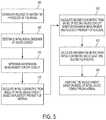
- FIG. 5is a flow chart illustrating an embodiment of a method for designing an implantable valved conduit composed of a plastically deformable material.
- valved conduit modeling parametersmay be provided 500 to the solid-mechanics modeling algorithm, the parameters including data related to the anatomy or physiology of the recipient patient.
- anatomic and/or physiologic datamay include a pressure across the valve structure within the valved conduit, a fluid flow rate through the valved conduit, and physical measurements of vascular structures to which the valved conduit may be attached.
- An initial radial dimension of the valved conduit to be modeledmay also be provided 505 .
- data related to the expandability of the plastically deformable materialmay be provided to the model. Such data may include a yield strength, ultimate tensile strength, elastic modulus, and other mechanical properties of the plastically deformable material.
- a measure of expected patient anatomic growth, or changes to the patient physiology as a response of patient growthmay be determined.
- the expected patient growth information, along with the data related to the plastic deformability of the valved conduit material,may be used to estimate a desired amount of expandability for the valved conduit.
- Such expandability datamay be provided 510 to the modeling software as one or more expansion measurements for the conduit.
- Physical parameters associated with the initially defined valve structuremay be provided to the modeling software as well. Such physical parameters may include, without limitation, a conduit length and a conduit wall thickness. Additional physical parameters may be provided to the modeling software that relate to physical dimensions of the valve structure. Some examples of such physical dimensions may be related to the shape and size of valve leaflets that may comprise the valve structure. Non-limiting examples of valve leaflet physical parameters may include one or more of a sinus edge shape, a sinus edge perimeter length, a fan edge shape, a fan edge perimeter length, a height, a fan structure height, a baseline width, and a commissure length.
- a valve structure modeling computationmay then create an initial mathematical model of the initial valved conduit related to the physical and mechanical properties of the valved conduit as initially defined.
- the initial model representing the initial valved conduitmay then be used in a fluid flow simulation algorithm to determine the characteristics of fluid flow through the initial valved conduit.
- One or more initial fluid flow metricsincluding, without limitation, a fluid velocity profile, a fluid pressure profile, and a fluid volumetric flow profile may then be calculated 515 by the fluid flow simulation algorithm.
- One or more plastic deformability characteristics of the materialmay also be used in such a fluid flow simulation algorithm in addition to the anatomic and/or physiological data from a patient, the initial proposed radial dimension of the conduit, and physical metrics associated with the valve structure.
- the initial mathematical model representing the initial valved conduitmay be altered to provide at least a second mathematical model representing at least a second valved conduit.
- the at least second valved conduit modelmay differ from the initial valved conduit model in a variety of ways, including, but not limited to, radial dimension of the conduit, valve leaflet physical parameters, expansion measurements of the material, and one or more measures related to the plasticity properties of the material (such as a change in stress or strain characteristics of the materials).
- One or more second fluid flow metricsmay then be calculated 520 by the fluid flow simulation algorithm based on the at least second model of the valved conduit.
- the fluid flow simulation algorithmmay be sequentially applied to additional valved conduit models, each succeeding model representing a succeeding valved conduit that has been altered in some manner from a preceding valved conduit.
- a series of valved conduitsmay be modeled that may differ only in their conduit radial dimensions.
- Such a seriesmay represent a radial change of an implanted valved conduit over time as the patient grows and the conduit expands to accommodate the patient growth.
- the change in radial dimension of the valved conduit over timemay be simulated by the fluid flow simulation algorithm as a change in the fluid flow metrics associated with each succeeding conduit configuration analyzed thereby.
- a deformation metricmay be calculated 525 .
- the deformation metricmay be calculated from the multiplicity of fluid flow metrics in any number of ways, including, without limitation, an arithmetic mean of fluid flow metrics, a geometric mean of fluid flow metrics, a harmonic mean of fluid flow metrics, or a weighted average of fluid flow metrics.
- a weighted average of fluid flow metricsmay be calculated as a sum of fluid flow metrics, each weighted by some weighting factor.
- a weighting factormay be derived from a flow efficiency metric or cost function associated with the effectiveness of fluid flow through a valved conduit structure having a particular set of characteristics, such as radial dimension. Efficiency may be based on a fluid flow rate, an open area within the valve structure during flow, or a measure of regurgitant flow.
- a valved conduitmay be fabricated 530 from the plastically deformable material using physical characteristics of the conduit and valve structure as supplied to the initial model of the valved conduit if the calculated deformation metric is greater than or equal to an acceptance value.
- Some nonlimiting examples of such acceptance valuesmay incorporate values calculated for one or more of a regurgitation fraction, an open orifice area, and a percent leaflet/wall contact measure.
- a regurgitation fractionmay measure the ratio of fluid back-flow through a valve in a closed state to the fluid forward-flow through the valve in an open state.
- An open orifice areamay be calculated at a percent of a cross-sectional area of the conduit lumen not occluded by the valve structure when the valve structure is in an open position.
- An additional measure of conduit patencymay include a measure of the fraction of a valve structure leaflet in contact with an inner surface of the conduit (compared to total leaflet area).
- Some examples of an acceptance valuemay include a regurgitation fraction less than or equal to about 30%, an open orifice area greater than or equal to about 80%, or a leaflet/wall contact value of less than or equal to about 15%.
- an implanted valved conduit fabricated from a plastically deformable materialmay be able to expand as the patient grows, thereby providing some long term treatment, it may be possible that a single plastically deformable valved conduit may not be sufficient to assist a patient from neonatal size to full adult size. In such an instance, it may be necessary to replace an initial valved conduit with a second valved conduit capable of expanding from an intermediate patient age to full adulthood.
- a plastically deformable valved conduitmay be replaced in situ without the need for excising the original and replacing it with a second valved conduit.
- FIG. 6is a flow chart of one method that may be used to replace an implanted first expandable valved conduit with a second expandable valved conduit.
- a first expandable valved conduitmay be unable to assist a patient after some period of patient growth.
- the conduitmay radially enlarge to an extent that the valve structure may no longer efficiently regulate blood flow.
- the first valved conduitmay not have expanded to its fully expanded state when valve structure inefficiency may become apparent.
- the first valved conduitmay be replaced by a second valved conduit by introducing the second valved conduit within the first valved conduit and expanding the second in situ.
- an expansion devicesuch as a balloon catheter, may be introduced into the vasculature so that the expansion device contacts 600 an inner surface of the first valved conduit.
- the expansion devicemay then be expanded 610 within the first valved conduit thereby radially increasing at least a portion of the first valved conduit.
- a second valved conduitmay then be introduced 620 within at least a portion of the expanded first valved conduit.
- the second valved conduitmay be introduced using the same expansion device as used to expand the first valved conduit while the first valved conduit is expanded.
- the second valved conduitmay be introduced 620 by the use of an alternative device.
- the second valved conduitmay also be expanded 630 to provide a valve structure capable of regulating fluid flow through the conduit.
- Visible markingsmay provide details including, but not limited to, the conduit's diameter (inner and/or outer), orientation, length and obliqueness of cutting, and valve location and orientation if one is present.
- Radiopaque markingsmay provide details including, but not limited to, the conduit's original diameter (inner and/or outer), current diameter (inner and/or outer), orientation, length, and whether any kinking and/or compression has occurred.
- Radiopaque markings and visible markingsmay be applied to the conduit via pad printing, impregnation of an ink, dye, or other material (such as a small metal pellet) into the material itself, or other standard methods of marking biomedical devices.
- radiopaque markings unique to an expandable conduitare the need for markings to be legible after significant radial deformation. This can be difficult with processes such as pad printing, because the designed may “crack” under stress or deformation.
- One method for maintaining legibilityis the use of patterns which are initially “dashed” (not completely space-filling), so that after deformation the general design does not significantly change relative to itself. As an example, no line would extend more than 1 mm in the radial dimension before a gap, building in stress-relief and preventing significant cracking or other destruction of the markings when radially-deformed. An example is shown in FIG. 8 .
- each cliché that is usedmay only print on a certain portion of the conduit's circumference, nearly always less than 180 degrees.
- a design which does not require multiple cliché sis preferable because it significantly reduces the printing time and thus provides economic benefit.
- thisoften requires a repeating pattern, and this is not preferable for radiopaque markings, as many imaging techniques (such as x-ray) project onto 2-dimensions, and thus a repeated radiopaque marking may result in indecipherable markings.
- the ideal designwould not require multiple cliché's and would not radially repeat markings (such as for length measurement).
- the example in FIG. 8may be created with a single cliché by printing on one side and flipping the conduit about its vertical axis (perpendicular to the longitudinal axis) before repeating the same cliché on the other portion of the conduits circumference.
- the conduitmay include one or more of the following features: a single length scale which further serves to identify orientation such as curvature and twisting (which is not repeated circumferentially, including not at the 180 degrees from the initial length scale, so as not to become indecipherable under a 2D projection such as through x-ray), circumference markings to identify the current circumference of the conduit, and numbers or symbols to identify the initial diameter which repeat along the length of the conduit but are not repeated circumferentially.
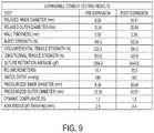
- FIG. 7 Adepicts the stress/strain curve of a first plastically deformable material that may be used to fabricate a plastically deformable and implantable conduit.
- the materialhas an average yield strength of about 2.1 MPa and an ultimate tensile strength of about 5 MPa.
- the materialfurther demonstrates elastic deformation below the yield strength, characterized by an average elastic modulus of about 5.9 MPa.
- the materialdemonstrates an average 36% elongation above the original length at the yield stress point.
- Such a materialmay be favorably used for a plastically deformable conduit capable of expanding to meet the needs of a growing anatomical structure due to the extended region of the stress/strain curve indicating plastic deformability as opposed to elastic deformability.
- FIG. 7Bdepicts the stress/strain curve of a second plastically deformable material that may be used to fabricate a plastically deformable and implantable conduit.
- the materialdemonstrates an average yield strength of about 1.7 MPa and an ultimate tensile strength of about 5.5 MPa.
- the materialalso has a region of elastic deformation below the yield strength characterized by an average elastic modulus of about 7.4 MPa.
- the materialdemonstrates an average 24% elongation above the original length at the yield stress point.
- Such a materialmay be favorably used for a plastically deformable conduit capable of expanding to meet the needs of a growing anatomical structure due to the extended region of the stress/strain curve indicating plastic deformability as opposed to elastic deformability.
Landscapes
- Health & Medical Sciences (AREA)
- Engineering & Computer Science (AREA)
- Biomedical Technology (AREA)
- Cardiology (AREA)
- Heart & Thoracic Surgery (AREA)
- Transplantation (AREA)
- Oral & Maxillofacial Surgery (AREA)
- Vascular Medicine (AREA)
- Life Sciences & Earth Sciences (AREA)
- Animal Behavior & Ethology (AREA)
- General Health & Medical Sciences (AREA)
- Public Health (AREA)
- Veterinary Medicine (AREA)
- Pulmonology (AREA)
- Gastroenterology & Hepatology (AREA)
- Prostheses (AREA)
Abstract
Description
Claims (15)
Priority Applications (3)
| Application Number | Priority Date | Filing Date | Title |
|---|---|---|---|
| US15/447,526US11000370B2 (en) | 2016-03-02 | 2017-03-02 | Expandable implantable conduit |
| US17/226,731US20210220129A1 (en) | 2016-03-02 | 2021-04-09 | Expandable implantable conduit |
| US18/456,708US20230404751A1 (en) | 2016-03-02 | 2023-08-28 | Expandable implantable conduit |
Applications Claiming Priority (2)
| Application Number | Priority Date | Filing Date | Title |
|---|---|---|---|
| US201662302595P | 2016-03-02 | 2016-03-02 | |
| US15/447,526US11000370B2 (en) | 2016-03-02 | 2017-03-02 | Expandable implantable conduit |
Related Child Applications (1)
| Application Number | Title | Priority Date | Filing Date |
|---|---|---|---|
| US17/226,731ContinuationUS20210220129A1 (en) | 2016-03-02 | 2021-04-09 | Expandable implantable conduit |
Publications (2)
| Publication Number | Publication Date |
|---|---|
| US20170252156A1 US20170252156A1 (en) | 2017-09-07 |
| US11000370B2true US11000370B2 (en) | 2021-05-11 |
Family
ID=59722430
Family Applications (3)
| Application Number | Title | Priority Date | Filing Date |
|---|---|---|---|
| US15/447,526ActiveUS11000370B2 (en) | 2016-03-02 | 2017-03-02 | Expandable implantable conduit |
| US17/226,731AbandonedUS20210220129A1 (en) | 2016-03-02 | 2021-04-09 | Expandable implantable conduit |
| US18/456,708PendingUS20230404751A1 (en) | 2016-03-02 | 2023-08-28 | Expandable implantable conduit |
Family Applications After (2)
| Application Number | Title | Priority Date | Filing Date |
|---|---|---|---|
| US17/226,731AbandonedUS20210220129A1 (en) | 2016-03-02 | 2021-04-09 | Expandable implantable conduit |
| US18/456,708PendingUS20230404751A1 (en) | 2016-03-02 | 2023-08-28 | Expandable implantable conduit |
Country Status (2)
| Country | Link |
|---|---|
| US (3) | US11000370B2 (en) |
| WO (1) | WO2017151900A1 (en) |
Cited By (1)
| Publication number | Priority date | Publication date | Assignee | Title |
|---|---|---|---|---|
| US20210220129A1 (en)* | 2016-03-02 | 2021-07-22 | Peca Labs, Inc. | Expandable implantable conduit |
Families Citing this family (13)
| Publication number | Priority date | Publication date | Assignee | Title |
|---|---|---|---|---|
| US20090105810A1 (en)* | 2007-10-17 | 2009-04-23 | Hancock Jaffe Laboratories | Biological valve for venous valve insufficiency |
| US9585746B2 (en) | 2011-07-29 | 2017-03-07 | Carnegie Mellon University | Artificial valved conduits for cardiac reconstructive procedures and methods for their production |
| EP2964153B1 (en) | 2013-03-08 | 2022-08-31 | Carnegie Mellon University | Expandable implantable conduit |
| US10507101B2 (en) | 2014-10-13 | 2019-12-17 | W. L. Gore & Associates, Inc. | Valved conduit |
| EP4397276A3 (en) | 2016-10-10 | 2024-09-18 | Peca Labs, Inc. | Transcatheter stent and valve assembly |
| US11406533B2 (en) | 2017-03-17 | 2022-08-09 | W. L. Gore & Associates, Inc. | Integrated aqueous shunt for glaucoma treatment |
| CN111818875B (en) | 2017-10-31 | 2024-05-14 | 爱德华兹生命科学公司 | Valved pipe |
| CN111818876B (en) | 2017-11-16 | 2024-02-09 | 儿童医学中心公司 | Geometrically adaptive heart valve replacement device |
| WO2020047221A1 (en) | 2018-08-29 | 2020-03-05 | W. L. Gore & Associates, Inc. | Drug therapy delivery systems and methods |
| USD977642S1 (en) | 2018-10-29 | 2023-02-07 | W. L. Gore & Associates, Inc. | Pulmonary valve conduit |
| US11678983B2 (en) | 2018-12-12 | 2023-06-20 | W. L. Gore & Associates, Inc. | Implantable component with socket |
| JP2023518769A (en)* | 2020-03-18 | 2023-05-08 | エドワーズ ライフサイエンシーズ コーポレイション | Fabrics, implantable medical devices using such fabrics, and processes for making same |
| WO2024030237A1 (en)* | 2022-08-03 | 2024-02-08 | The Children's Medical Center Corporation | Geometrically-accommodating heart valve replacement device |
Citations (95)
| Publication number | Priority date | Publication date | Assignee | Title |
|---|---|---|---|---|
| US4160688A (en) | 1977-02-03 | 1979-07-10 | Johns-Manville Corporation | Low profile heat sealing iron |
| US4187390A (en) | 1970-05-21 | 1980-02-05 | W. L. Gore & Associates, Inc. | Porous products and process therefor |
| US4475972A (en) | 1981-10-01 | 1984-10-09 | Ontario Research Foundation | Implantable material |
| JPS6446468A (en) | 1987-04-28 | 1989-02-20 | Baxter Int | Multilayer poly(tetrafluoroethylene)/elastomer material useful for intracorporeal transplantation |
| US4955899A (en) | 1989-05-26 | 1990-09-11 | Impra, Inc. | Longitudinally compliant vascular graft |
| WO1994015548A1 (en) | 1993-01-14 | 1994-07-21 | Meadox Medicals, Inc. | Radially expandable tubular prosthesis |
| US5466261A (en) | 1992-11-19 | 1995-11-14 | Wright Medical Technology, Inc. | Non-invasive expandable prosthesis for growing children |
| US5469868A (en) | 1992-02-12 | 1995-11-28 | Reger Medical Inc. | Method of making an artificial heart valve stent |
| US5725572A (en)* | 1994-04-25 | 1998-03-10 | Advanced Cardiovascular Systems, Inc. | Radiopaque stent |
| US5800512A (en) | 1996-01-22 | 1998-09-01 | Meadox Medicals, Inc. | PTFE vascular graft |
| US5804011A (en) | 1994-05-25 | 1998-09-08 | W. L. Gore & Associates, Inc. | Process of making a two-way stretchable fabric laminate and articles made from it |
| US5824050A (en) | 1996-12-03 | 1998-10-20 | Atrium Medical Corporation | Prosthesis with in-wall modulation |
| US5843161A (en) | 1996-06-26 | 1998-12-01 | Cordis Corporation | Endoprosthesis assembly for percutaneous deployment and method of deploying same |
| US6016848A (en) | 1996-07-16 | 2000-01-25 | W. L. Gore & Associates, Inc. | Fluoropolymer tubes and methods of making same |
| US6039755A (en)* | 1997-02-05 | 2000-03-21 | Impra, Inc., A Division Of C.R. Bard, Inc. | Radially expandable tubular polytetrafluoroethylene grafts and method of making same |
| CN1304298A (en) | 1998-06-26 | 2001-07-18 | 圣朱德医疗有限公司 | Single suture aortic non-stretch biological tissue valve |
| US20010039450A1 (en) | 1999-06-02 | 2001-11-08 | Dusan Pavcnik | Implantable vascular device |
| US20020055775A1 (en) | 1999-01-26 | 2002-05-09 | Alain F. Carpentier | Flexible heart valve |
| US6436135B1 (en) | 1974-10-24 | 2002-08-20 | David Goldfarb | Prosthetic vascular graft |
| US20020133226A1 (en) | 1999-01-26 | 2002-09-19 | Salvador Marquez | Holder for flexible heart valve |
| US20020138135A1 (en) | 2001-03-21 | 2002-09-26 | Duerig Thomas W. | Stent-based venous valves |
| US20030027332A1 (en) | 2001-07-16 | 2003-02-06 | Edwards Lifesciences Corporation | Tissue engineered heart valve |
| US6517571B1 (en) | 1999-01-22 | 2003-02-11 | Gore Enterprise Holdings, Inc. | Vascular graft with improved flow surfaces |
| US20030114924A1 (en) | 2001-12-18 | 2003-06-19 | Riyad Moe | Polymer heart valve |
| US20030139805A1 (en) | 2002-01-24 | 2003-07-24 | Holmberg William R. | Conduit for aorta or pulmonary artery replacement |
| US20030191525A1 (en) | 2002-04-03 | 2003-10-09 | Thornton Sally C. | Artifical valve |
| US6716239B2 (en) | 2001-07-03 | 2004-04-06 | Scimed Life Systems, Inc. | ePTFE graft with axial elongation properties |
| WO2005011535A2 (en) | 2003-07-31 | 2005-02-10 | Cook Incorporated | Prosthetic valve for implantation in a body vessel |
| US6863686B2 (en) | 1995-04-17 | 2005-03-08 | Donald Shannon | Radially expandable tape-reinforced vascular grafts |
| US20050075727A1 (en) | 2001-10-29 | 2005-04-07 | Wheatley David John | Mitral valve prosthesis |
| US20050096734A1 (en) | 2003-10-31 | 2005-05-05 | Majercak David C. | Implantable valvular prosthesis |
| US20050137682A1 (en) | 2003-12-22 | 2005-06-23 | Henri Justino | Stent mounted valve |
| US6939372B2 (en) | 2001-07-03 | 2005-09-06 | Scimed Life Systems, Inc. | Low profile, high stretch, low dilation knit prosthetic device |
| US20050228495A1 (en) | 2004-01-15 | 2005-10-13 | Macoviak John A | Suspended heart valve devices, systems, and methods for supplementing, repairing, or replacing a native heart valve |
| US20050240262A1 (en) | 2002-05-03 | 2005-10-27 | The General Hospital Corporation | Involuted endovascular valve and method of construction |
| US20050283224A1 (en) | 2004-06-22 | 2005-12-22 | Scimed Life Systems, Inc. | Implantable medical devices with antimicrobial and biodegradable matrices |
| US20060122693A1 (en) | 2004-05-10 | 2006-06-08 | Youssef Biadillah | Stent valve and method of manufacturing same |
| US20060149366A1 (en) | 2004-12-31 | 2006-07-06 | Jamie Henderson | Sintered structures for vascular graft |
| US20060161248A1 (en) | 2004-12-01 | 2006-07-20 | Case Brian C | Medical device with leak path |
| US20060229716A1 (en) | 2005-04-11 | 2006-10-12 | Zan Mitrev | Replacement aortic valve leaflets and related technology |
| US20060259136A1 (en) | 2005-05-13 | 2006-11-16 | Corevalve Sa | Heart valve prosthesis and methods of manufacture and use |
| US20060265053A1 (en) | 2005-05-17 | 2006-11-23 | Cook Incorporated | Prosthetic valve devices and methods of making and using such devices |
| US20060287717A1 (en) | 2005-05-24 | 2006-12-21 | Rowe Stanton J | Methods for rapid deployment of prosthetic heart valves |
| US7153324B2 (en) | 2003-07-31 | 2006-12-26 | Cook Incorporated | Prosthetic valve devices and methods of making such devices |
| US7153325B2 (en) | 2003-08-01 | 2006-12-26 | Ultra-Kinetics, Inc. | Prosthetic intervertebral disc and methods for using the same |
| US20070027528A1 (en) | 2005-07-29 | 2007-02-01 | Cook Incorporated | Elliptical implantable device |
| US20070043431A1 (en) | 2005-08-19 | 2007-02-22 | Cook Incorporated | Prosthetic valve |
| US20070067021A1 (en) | 2005-09-21 | 2007-03-22 | Boston Scientific Scimed, Inc. | Venous valve, system, and method with sinus pocket |
| US7306729B2 (en) | 2005-07-18 | 2007-12-11 | Gore Enterprise Holdings, Inc. | Porous PTFE materials and articles produced therefrom |
| JP2007536951A (en) | 2004-04-08 | 2007-12-20 | エンドロジックス、インク. | Intraluminal artificial blood vessel with neointimal inhibitory polymer sleeve |
| JP2008526366A (en) | 2005-01-06 | 2008-07-24 | ボストン サイエンティフィック リミテッド | Optimally expanded, collagen-sealed ePTFE graft with improved tissue ingrowth |
| US20080206442A1 (en) | 2002-09-27 | 2008-08-28 | Labcoat, Ltd. | Contact coating of prostheses |
| US20090118826A1 (en) | 2001-05-01 | 2009-05-07 | Asghar Khaghani | Valve prosthesis |
| WO2009061419A1 (en) | 2007-11-09 | 2009-05-14 | Cook Incorporated | Aortic valve stent graft |
| WO2009134701A2 (en) | 2008-05-01 | 2009-11-05 | Edwards Lifesciences Corporation | Device and method for replacing mitral valve |
| US20100023120A1 (en) | 2008-04-23 | 2010-01-28 | Holecek Arin N | Tissue attachment devices and methods for prosthetic heart valves |
| US20100023114A1 (en) | 2008-07-24 | 2010-01-28 | Cook Incorporated | Valve device with biased leaflets |
| US20100204775A1 (en)* | 2006-05-22 | 2010-08-12 | Tarun John Edwin | Tissue Synthetic- Biomaterial Hybrid Medical Devices |
| US7789908B2 (en) | 2002-06-25 | 2010-09-07 | Boston Scientific Scimed, Inc. | Elastomerically impregnated ePTFE to enhance stretch and recovery properties for vascular grafts and coverings |
| US20100312333A1 (en) | 2009-04-29 | 2010-12-09 | The Cleveland Clinic Foundation | Apparatus and method for replacing a diseased cardiac valve |
| US20110060401A1 (en) | 2007-02-16 | 2011-03-10 | Universität Zürich | Tubular Supporting Prosthesis Capable of Growing |
| US20110071625A1 (en) | 2005-02-23 | 2011-03-24 | Boston Scientific Scimed, Inc. | Valve apparatus, system and method |
| US20110098800A1 (en) | 2008-07-15 | 2011-04-28 | St. Jude Medical, Inc | Axially anchoring collapsible and re-expandable prosthetic heart valves for various disease states |
| US20110094592A1 (en) | 2009-10-22 | 2011-04-28 | Howard Cheng | Fluid Check Valve with a Floating Pivot |
| US20110125163A1 (en) | 2007-02-28 | 2011-05-26 | Medtronic, Inc. | Implantable medical device system with fixation member |
| US20110166637A1 (en) | 2009-07-15 | 2011-07-07 | Irwin Craig W | Self constraining radially expandable medical devices |
| WO2012018779A2 (en) | 2010-08-02 | 2012-02-09 | Children's Medical Center Corporation | Expandable valve and method of use |
| CN102497836A (en) | 2009-06-02 | 2012-06-13 | 麦德托尼克公司 | Stented prosthetic heart valves |
| US20120158125A1 (en) | 2009-09-16 | 2012-06-21 | Milisav Obradovic | Stent having expandable elements |
| US20120271396A1 (en) | 2007-01-19 | 2012-10-25 | Elixir Medical Corporation | Biodegradable endoprostheses and methods for their fabrication |
| US20120290082A1 (en) | 2011-05-10 | 2012-11-15 | Biotronik Ag | Mechanical transcatheter heart valve prosthesis |
| US20130013058A1 (en) | 2011-07-06 | 2013-01-10 | Waseda University | Stentless artificial mitral valve, artificial valve leaflet, template and cutter |
| US20130019756A1 (en) | 2011-07-20 | 2013-01-24 | Andrew Coats | Coffee maker |
| WO2013019756A2 (en) | 2011-07-29 | 2013-02-07 | Carnegie Mellon University | Artificial valved conduits for cardiac reconstructive procedures and methods for their production |
| US20130166016A1 (en) | 2006-04-21 | 2013-06-27 | W. L. Gore & Associates, Inc. | Expandable stent with wrinkle-free elastomeric cover |
| US20130184807A1 (en) | 2012-01-16 | 2013-07-18 | W. L. Gore & Associates, Inc. | Articles Including Expanded Polytetrafluoroethylene Membranes with Serpentine Fibrils and Having a Discontinuous Fluoropolymer Layer Thereon |
| US20130261739A1 (en) | 2012-03-28 | 2013-10-03 | Medtronic, Inc. | Dual Valve Prosthesis for Transcatheter Valve Implantation |
| US20130304196A1 (en) | 2012-05-08 | 2013-11-14 | Medtronic Vascular, Inc. | Prosthetic venous valve having leaflets forming a scalloped commissure |
| US20130319603A1 (en)* | 2006-05-26 | 2013-12-05 | Abbott Cardiovascular Systems Inc. | Stents With Radiopaque Markers |
| US20140005772A1 (en) | 2012-07-02 | 2014-01-02 | Boston Scientific Scimed, Inc. | Prosthetic heart valve formation |
| US20140031927A1 (en) | 2012-07-27 | 2014-01-30 | W. L. Gore & Associates, Inc. | Multi-frame prosthetic valve apparatus and methods |
| US20140031924A1 (en) | 2012-07-25 | 2014-01-30 | W. L. Gore & Associates, Inc. | Everting transcatheter valve and methods |
| US20140131268A1 (en) | 2011-06-23 | 2014-05-15 | Solvay Specialty Polymers Italy S.P.A. | Process for manufacturing porous membranes |
| US20140155955A1 (en) | 2003-10-03 | 2014-06-05 | Medtronic, Inc. | System, apparatus and method for interacting with a targeted tissue of a patient |
| WO2014138599A1 (en) | 2013-03-08 | 2014-09-12 | Carnegie Mellon University | Expandable implantable conduit |
| WO2014145811A1 (en) | 2013-03-15 | 2014-09-18 | Edwards Lifesciences Corporation | Valved aortic conduits |
| US20140330372A1 (en) | 2013-05-03 | 2014-11-06 | Medtronic, Inc. | Medical Devices for Implanting in a Valve and Associated Methods |
| US8900652B1 (en)* | 2011-03-14 | 2014-12-02 | Innovatech, Llc | Marked fluoropolymer surfaces and method of manufacturing same |
| US20150366664A1 (en)* | 2014-06-20 | 2015-12-24 | Edwards Lifesciences Corporation | Surgical heart valves identifiable post-implant |
| WO2016028591A1 (en) | 2014-08-18 | 2016-02-25 | W.L. Gore & Associates, Inc. | Frame with integral sewing cuff for prosthetic valves |
| US20160067038A1 (en) | 2013-04-19 | 2016-03-10 | Strait Access Technologies Holdings (Pty) Ltd | A Prosthetic Heart Valve |
| US20160100939A1 (en) | 2014-10-13 | 2016-04-14 | W. L. Gore & Associates, Inc. | Valved conduit |
| WO2016205773A1 (en) | 2015-06-18 | 2016-12-22 | Peca Labs, Inc. | Valved conduit and method for fabricating same |
| WO2017151900A1 (en) | 2016-03-02 | 2017-09-08 | Peca Labs, Inc. | Expandable implantable conduit |
| US20180098845A1 (en) | 2016-10-10 | 2018-04-12 | Peca Labs, Inc. | Transcatheter stent and valve assembly |
Family Cites Families (2)
| Publication number | Priority date | Publication date | Assignee | Title |
|---|---|---|---|---|
| US5824042A (en)* | 1996-04-05 | 1998-10-20 | Medtronic, Inc. | Endoluminal prostheses having position indicating markers |
| US8014849B2 (en)* | 2003-11-21 | 2011-09-06 | Stryker Corporation | Rotational markers |
- 2017
- 2017-03-02WOPCT/US2017/020421patent/WO2017151900A1/ennot_activeCeased
- 2017-03-02USUS15/447,526patent/US11000370B2/enactiveActive
- 2021
- 2021-04-09USUS17/226,731patent/US20210220129A1/ennot_activeAbandoned
- 2023
- 2023-08-28USUS18/456,708patent/US20230404751A1/enactivePending
Patent Citations (108)
| Publication number | Priority date | Publication date | Assignee | Title |
|---|---|---|---|---|
| US4187390A (en) | 1970-05-21 | 1980-02-05 | W. L. Gore & Associates, Inc. | Porous products and process therefor |
| US6436135B1 (en) | 1974-10-24 | 2002-08-20 | David Goldfarb | Prosthetic vascular graft |
| US4160688A (en) | 1977-02-03 | 1979-07-10 | Johns-Manville Corporation | Low profile heat sealing iron |
| US4475972A (en) | 1981-10-01 | 1984-10-09 | Ontario Research Foundation | Implantable material |
| JPS6446468A (en) | 1987-04-28 | 1989-02-20 | Baxter Int | Multilayer poly(tetrafluoroethylene)/elastomer material useful for intracorporeal transplantation |
| US4816339A (en) | 1987-04-28 | 1989-03-28 | Baxter International Inc. | Multi-layered poly(tetrafluoroethylene)/elastomer materials useful for in vivo implantation |
| US4955899A (en) | 1989-05-26 | 1990-09-11 | Impra, Inc. | Longitudinally compliant vascular graft |
| US5469868A (en) | 1992-02-12 | 1995-11-28 | Reger Medical Inc. | Method of making an artificial heart valve stent |
| US5466261A (en) | 1992-11-19 | 1995-11-14 | Wright Medical Technology, Inc. | Non-invasive expandable prosthesis for growing children |
| US5443499A (en) | 1993-01-14 | 1995-08-22 | Meadox Medicals, Inc. | Radially expandable tubular prosthesis |
| JPH07505815A (en) | 1993-01-14 | 1995-06-29 | ミードックス メディカルズ インコーポレイテッド | Radially expandable tubular prosthesis |
| WO1994015548A1 (en) | 1993-01-14 | 1994-07-21 | Meadox Medicals, Inc. | Radially expandable tubular prosthesis |
| US5725572A (en)* | 1994-04-25 | 1998-03-10 | Advanced Cardiovascular Systems, Inc. | Radiopaque stent |
| US5804011A (en) | 1994-05-25 | 1998-09-08 | W. L. Gore & Associates, Inc. | Process of making a two-way stretchable fabric laminate and articles made from it |
| US6863686B2 (en) | 1995-04-17 | 2005-03-08 | Donald Shannon | Radially expandable tape-reinforced vascular grafts |
| US5800512A (en) | 1996-01-22 | 1998-09-01 | Meadox Medicals, Inc. | PTFE vascular graft |
| US5843161A (en) | 1996-06-26 | 1998-12-01 | Cordis Corporation | Endoprosthesis assembly for percutaneous deployment and method of deploying same |
| US6016848A (en) | 1996-07-16 | 2000-01-25 | W. L. Gore & Associates, Inc. | Fluoropolymer tubes and methods of making same |
| US5824050A (en) | 1996-12-03 | 1998-10-20 | Atrium Medical Corporation | Prosthesis with in-wall modulation |
| US6039755A (en)* | 1997-02-05 | 2000-03-21 | Impra, Inc., A Division Of C.R. Bard, Inc. | Radially expandable tubular polytetrafluoroethylene grafts and method of making same |
| CN1304298A (en) | 1998-06-26 | 2001-07-18 | 圣朱德医疗有限公司 | Single suture aortic non-stretch biological tissue valve |
| US6517571B1 (en) | 1999-01-22 | 2003-02-11 | Gore Enterprise Holdings, Inc. | Vascular graft with improved flow surfaces |
| US20020133226A1 (en) | 1999-01-26 | 2002-09-19 | Salvador Marquez | Holder for flexible heart valve |
| US20020055775A1 (en) | 1999-01-26 | 2002-05-09 | Alain F. Carpentier | Flexible heart valve |
| US20010039450A1 (en) | 1999-06-02 | 2001-11-08 | Dusan Pavcnik | Implantable vascular device |
| US20020138135A1 (en) | 2001-03-21 | 2002-09-26 | Duerig Thomas W. | Stent-based venous valves |
| US20090118826A1 (en) | 2001-05-01 | 2009-05-07 | Asghar Khaghani | Valve prosthesis |
| US6939372B2 (en) | 2001-07-03 | 2005-09-06 | Scimed Life Systems, Inc. | Low profile, high stretch, low dilation knit prosthetic device |
| US6716239B2 (en) | 2001-07-03 | 2004-04-06 | Scimed Life Systems, Inc. | ePTFE graft with axial elongation properties |
| US20030027332A1 (en) | 2001-07-16 | 2003-02-06 | Edwards Lifesciences Corporation | Tissue engineered heart valve |
| US20050075727A1 (en) | 2001-10-29 | 2005-04-07 | Wheatley David John | Mitral valve prosthesis |
| US20030114924A1 (en) | 2001-12-18 | 2003-06-19 | Riyad Moe | Polymer heart valve |
| US20030139805A1 (en) | 2002-01-24 | 2003-07-24 | Holmberg William R. | Conduit for aorta or pulmonary artery replacement |
| US20030191525A1 (en) | 2002-04-03 | 2003-10-09 | Thornton Sally C. | Artifical valve |
| US20050240262A1 (en) | 2002-05-03 | 2005-10-27 | The General Hospital Corporation | Involuted endovascular valve and method of construction |
| US7789908B2 (en) | 2002-06-25 | 2010-09-07 | Boston Scientific Scimed, Inc. | Elastomerically impregnated ePTFE to enhance stretch and recovery properties for vascular grafts and coverings |
| US20080206442A1 (en) | 2002-09-27 | 2008-08-28 | Labcoat, Ltd. | Contact coating of prostheses |
| US7153324B2 (en) | 2003-07-31 | 2006-12-26 | Cook Incorporated | Prosthetic valve devices and methods of making such devices |
| WO2005011535A2 (en) | 2003-07-31 | 2005-02-10 | Cook Incorporated | Prosthetic valve for implantation in a body vessel |
| US7153325B2 (en) | 2003-08-01 | 2006-12-26 | Ultra-Kinetics, Inc. | Prosthetic intervertebral disc and methods for using the same |
| US20140155955A1 (en) | 2003-10-03 | 2014-06-05 | Medtronic, Inc. | System, apparatus and method for interacting with a targeted tissue of a patient |
| US20050096734A1 (en) | 2003-10-31 | 2005-05-05 | Majercak David C. | Implantable valvular prosthesis |
| US20050137682A1 (en) | 2003-12-22 | 2005-06-23 | Henri Justino | Stent mounted valve |
| US20050228495A1 (en) | 2004-01-15 | 2005-10-13 | Macoviak John A | Suspended heart valve devices, systems, and methods for supplementing, repairing, or replacing a native heart valve |
| JP2007536951A (en) | 2004-04-08 | 2007-12-20 | エンドロジックス、インク. | Intraluminal artificial blood vessel with neointimal inhibitory polymer sleeve |
| US8377110B2 (en)* | 2004-04-08 | 2013-02-19 | Endologix, Inc. | Endolumenal vascular prosthesis with neointima inhibiting polymeric sleeve |
| US20060122693A1 (en) | 2004-05-10 | 2006-06-08 | Youssef Biadillah | Stent valve and method of manufacturing same |
| US20050283224A1 (en) | 2004-06-22 | 2005-12-22 | Scimed Life Systems, Inc. | Implantable medical devices with antimicrobial and biodegradable matrices |
| US20060161248A1 (en) | 2004-12-01 | 2006-07-20 | Case Brian C | Medical device with leak path |
| US20060149366A1 (en) | 2004-12-31 | 2006-07-06 | Jamie Henderson | Sintered structures for vascular graft |
| JP2008526366A (en) | 2005-01-06 | 2008-07-24 | ボストン サイエンティフィック リミテッド | Optimally expanded, collagen-sealed ePTFE graft with improved tissue ingrowth |
| US20110071625A1 (en) | 2005-02-23 | 2011-03-24 | Boston Scientific Scimed, Inc. | Valve apparatus, system and method |
| US20060229716A1 (en) | 2005-04-11 | 2006-10-12 | Zan Mitrev | Replacement aortic valve leaflets and related technology |
| US20060259136A1 (en) | 2005-05-13 | 2006-11-16 | Corevalve Sa | Heart valve prosthesis and methods of manufacture and use |
| US20060265053A1 (en) | 2005-05-17 | 2006-11-23 | Cook Incorporated | Prosthetic valve devices and methods of making and using such devices |
| US20060287717A1 (en) | 2005-05-24 | 2006-12-21 | Rowe Stanton J | Methods for rapid deployment of prosthetic heart valves |
| US7306729B2 (en) | 2005-07-18 | 2007-12-11 | Gore Enterprise Holdings, Inc. | Porous PTFE materials and articles produced therefrom |
| US20070027528A1 (en) | 2005-07-29 | 2007-02-01 | Cook Incorporated | Elliptical implantable device |
| US20070043431A1 (en) | 2005-08-19 | 2007-02-22 | Cook Incorporated | Prosthetic valve |
| US8672997B2 (en) | 2005-09-21 | 2014-03-18 | Boston Scientific Scimed, Inc. | Valve with sinus |
| US20070067021A1 (en) | 2005-09-21 | 2007-03-22 | Boston Scientific Scimed, Inc. | Venous valve, system, and method with sinus pocket |
| US20130166016A1 (en) | 2006-04-21 | 2013-06-27 | W. L. Gore & Associates, Inc. | Expandable stent with wrinkle-free elastomeric cover |
| US20100204775A1 (en)* | 2006-05-22 | 2010-08-12 | Tarun John Edwin | Tissue Synthetic- Biomaterial Hybrid Medical Devices |
| US20130319603A1 (en)* | 2006-05-26 | 2013-12-05 | Abbott Cardiovascular Systems Inc. | Stents With Radiopaque Markers |
| US20120271396A1 (en) | 2007-01-19 | 2012-10-25 | Elixir Medical Corporation | Biodegradable endoprostheses and methods for their fabrication |
| US20110060401A1 (en) | 2007-02-16 | 2011-03-10 | Universität Zürich | Tubular Supporting Prosthesis Capable of Growing |
| US20110125163A1 (en) | 2007-02-28 | 2011-05-26 | Medtronic, Inc. | Implantable medical device system with fixation member |
| CN101896139A (en) | 2007-11-09 | 2010-11-24 | 库克股份有限公司 | Aortic valve stent graft |
| WO2009061419A1 (en) | 2007-11-09 | 2009-05-14 | Cook Incorporated | Aortic valve stent graft |
| US20100023120A1 (en) | 2008-04-23 | 2010-01-28 | Holecek Arin N | Tissue attachment devices and methods for prosthetic heart valves |
| WO2009134701A2 (en) | 2008-05-01 | 2009-11-05 | Edwards Lifesciences Corporation | Device and method for replacing mitral valve |
| US20110098800A1 (en) | 2008-07-15 | 2011-04-28 | St. Jude Medical, Inc | Axially anchoring collapsible and re-expandable prosthetic heart valves for various disease states |
| US20100023114A1 (en) | 2008-07-24 | 2010-01-28 | Cook Incorporated | Valve device with biased leaflets |
| US20100312333A1 (en) | 2009-04-29 | 2010-12-09 | The Cleveland Clinic Foundation | Apparatus and method for replacing a diseased cardiac valve |
| CN102497836A (en) | 2009-06-02 | 2012-06-13 | 麦德托尼克公司 | Stented prosthetic heart valves |
| US20110166637A1 (en) | 2009-07-15 | 2011-07-07 | Irwin Craig W | Self constraining radially expandable medical devices |
| US20120158125A1 (en) | 2009-09-16 | 2012-06-21 | Milisav Obradovic | Stent having expandable elements |
| US20110094592A1 (en) | 2009-10-22 | 2011-04-28 | Howard Cheng | Fluid Check Valve with a Floating Pivot |
| WO2012018779A2 (en) | 2010-08-02 | 2012-02-09 | Children's Medical Center Corporation | Expandable valve and method of use |
| US8900652B1 (en)* | 2011-03-14 | 2014-12-02 | Innovatech, Llc | Marked fluoropolymer surfaces and method of manufacturing same |
| US20120290082A1 (en) | 2011-05-10 | 2012-11-15 | Biotronik Ag | Mechanical transcatheter heart valve prosthesis |
| US20140131268A1 (en) | 2011-06-23 | 2014-05-15 | Solvay Specialty Polymers Italy S.P.A. | Process for manufacturing porous membranes |
| US20130013058A1 (en) | 2011-07-06 | 2013-01-10 | Waseda University | Stentless artificial mitral valve, artificial valve leaflet, template and cutter |
| US20130019756A1 (en) | 2011-07-20 | 2013-01-24 | Andrew Coats | Coffee maker |
| WO2013019756A2 (en) | 2011-07-29 | 2013-02-07 | Carnegie Mellon University | Artificial valved conduits for cardiac reconstructive procedures and methods for their production |
| US20170196685A1 (en) | 2011-07-29 | 2017-07-13 | Carnegie Mellon University | Artificial valved conduits for cardiac reconstructive procedures and methods for their production |
| US9585746B2 (en) | 2011-07-29 | 2017-03-07 | Carnegie Mellon University | Artificial valved conduits for cardiac reconstructive procedures and methods for their production |
| US20140288642A1 (en) | 2011-07-29 | 2014-09-25 | University of Pittsburg - of the Commonwealth System of Higher Education | Artificial valved conduits for cardiac reconstructive procedures and methods for their production |
| US20130184807A1 (en) | 2012-01-16 | 2013-07-18 | W. L. Gore & Associates, Inc. | Articles Including Expanded Polytetrafluoroethylene Membranes with Serpentine Fibrils and Having a Discontinuous Fluoropolymer Layer Thereon |
| US20130261739A1 (en) | 2012-03-28 | 2013-10-03 | Medtronic, Inc. | Dual Valve Prosthesis for Transcatheter Valve Implantation |
| US20130304196A1 (en) | 2012-05-08 | 2013-11-14 | Medtronic Vascular, Inc. | Prosthetic venous valve having leaflets forming a scalloped commissure |
| US20140005772A1 (en) | 2012-07-02 | 2014-01-02 | Boston Scientific Scimed, Inc. | Prosthetic heart valve formation |
| US20140031924A1 (en) | 2012-07-25 | 2014-01-30 | W. L. Gore & Associates, Inc. | Everting transcatheter valve and methods |
| US20140031927A1 (en) | 2012-07-27 | 2014-01-30 | W. L. Gore & Associates, Inc. | Multi-frame prosthetic valve apparatus and methods |
| WO2014138599A1 (en) | 2013-03-08 | 2014-09-12 | Carnegie Mellon University | Expandable implantable conduit |
| US20160015516A1 (en) | 2013-03-08 | 2016-01-21 | Carnegie Mellon University | Expandable implantable conduit |
| WO2014145811A1 (en) | 2013-03-15 | 2014-09-18 | Edwards Lifesciences Corporation | Valved aortic conduits |
| US20160067038A1 (en) | 2013-04-19 | 2016-03-10 | Strait Access Technologies Holdings (Pty) Ltd | A Prosthetic Heart Valve |
| US20140330372A1 (en) | 2013-05-03 | 2014-11-06 | Medtronic, Inc. | Medical Devices for Implanting in a Valve and Associated Methods |
| US20150366664A1 (en)* | 2014-06-20 | 2015-12-24 | Edwards Lifesciences Corporation | Surgical heart valves identifiable post-implant |
| WO2016028591A1 (en) | 2014-08-18 | 2016-02-25 | W.L. Gore & Associates, Inc. | Frame with integral sewing cuff for prosthetic valves |
| US20160100939A1 (en) | 2014-10-13 | 2016-04-14 | W. L. Gore & Associates, Inc. | Valved conduit |
| WO2016205773A1 (en) | 2015-06-18 | 2016-12-22 | Peca Labs, Inc. | Valved conduit and method for fabricating same |
| US20180168795A1 (en) | 2015-06-18 | 2018-06-21 | Peca Labs, Inc. | Valved conduit and method for fabricating same |
| WO2017151900A1 (en) | 2016-03-02 | 2017-09-08 | Peca Labs, Inc. | Expandable implantable conduit |
| US20180098845A1 (en) | 2016-10-10 | 2018-04-12 | Peca Labs, Inc. | Transcatheter stent and valve assembly |
| US20180098844A1 (en) | 2016-10-10 | 2018-04-12 | Peca Labs, Inc. | Transcatheter stent and valve assembly |
| WO2018071417A1 (en) | 2016-10-10 | 2018-04-19 | Peca Labs, Inc. | Transcatheter stent and valve assembly |
Non-Patent Citations (64)
| Title |
|---|
| Ando et al., Ten-year experience with handmade trileaflet polytetrafluoroethylene valved conduit used for pulmonary reconstruction, J Thorac Cadiovasc Surg, (2009), 137:124-131. |
| Angioplasty Balloons from Advanced Polymers by MedDevice Online, accessed Oct. 3, 2019 (published 2019). |
| Back et al. Biomaterials in Vascular Surgery; Human Biomaterials Applications. Humana Press, Inc., 1996: 257-299. |
| Batlivala et al., Pulmonary Valve Replacement Function in Adolescents: A Comparison of Bioprosthetic Valves and Homograft Conduits, Ann Thorac Surg, (2012), 93:2007-2016. |
| BD Bard Conquest PTA Dilation Catheters, accessed Oct. 3, 2019 (published 2018). |
| Bernstein et al., Bicuspid-Valved PTFE Conduit Optimization for Pediatric RVOT Reconstruction, Bioengineering Conference (NEBEC), (2011). |
| Bianca et al., Sex ratio imbalance in transposition of the great arteries and possible agricultural environmental risk factors, Images Paediatr Cardiol, (2001), 8:10-14. |
| Bielefeld et al., Reoperative Homograft Right Ventricular Outflow Tract Reconstruction, Ann Thorac Surg, (2001), 71(2):482-488. |
| Boethig et al., Mid term course after pediatric right ventricular outflow tract reconstruction: a comparison of homografts, porcine xenografts and Contegras, European Journal of Cardio-thoracic Surgery, (2005), 27:58-66. |
| Boudjemline et al., Use of bovine jugular vein to reconstruct the right ventricular outflow tract: Early results, J Thorac Cardiovasc Surg, (2003), 126:490-497. |
| Brown et al., Right ventricular outflow tract reconstruction with polytetrafluoroethylene monocusp valve: A twelve-year experience, J Thorac Cardiovasc Surg, (2007), 133(5):1336-1343. |
| Caldarone et al., Independent Factors Associated with Longevity of Prosthetic Pulmonary Valves and Valved Conduits, J Thorac Cardiovasc Surg, (2000), 120:1022-1031. |
| Canadian Office Action dated Jul. 19, 2018 for Canadian Patent Application No. 2,855,943. |
| Canfield et al., National Estimates and Race/Ethnic-Specific Variation of Selected Birth Defects in the United States, Birth Defects Research (Part A): Clinical and Molecular Teratology, (2006), 76:747-756. |
| Cantanese et al. "Mechanical Properties of Medical Grade Expanded Polytetrafluorothylene: The Effect of Internodal Distance, Density, and Displacement Rate". Apr. 24, 1998. Journal of Biomedical Materials Research, 48: 187-192. |
| Chrysosotomou et al., Chapter 21: Tetralogy of Fallot with Pulmonary Atresia, Critical Care of Children with Heart Disease: Basic Medical and Surgical Concepts, (2010), 213-219. |
| Chrysosotomou et al., Chapter 22: Pulmonary Atresia with Intact Interventricular Septum, Critical Care of Children with Heart Disease: Basic Medical and Surgical Concepts, (2010), 221-229. |
| Cruz et al., Truncus Arteriosus, Chapter 35, Critical Care of Children with Heart Disease, Springer, (2009). |
| DeFrances et al., National Hospital Discharge Survey: 2005 annual summary with detailed diagnosis and procedure data, National Center for Health Statistics, Vital Health Stat (2007), 13(165):1-218. |
| Dur et al., In Vitro Evaluation of Right Ventricular Outflow Tract Reconstruction With Bicuspid Valved Polytetrafluoroethylene Conduit, Artificial Organs, (2010), 34: 1010-1016. |
| Erek et al., Durability of Stentless Bioprostheses for Right Ventricular Outflow Tract Reconstruction, Ann Thorac Surg, (2005), 79(6): 2202-2203. |
| Famaey, In Situ Evolution of the Mechanical Properties of Stretchable and Non-Stretchable Epffe Vascular Grafts and Adjacent Native Vessels, Int J Artif Organs 2014; 37 (12): 900-910. |
| Forbess et al., Conduit selection for right ventricular outflow tract reconstruction: contemporary options and outcomes, Semin Thorac Cardiovasc Surg Pediatr Card Surg Annu, (2004), 7:115-124. |
| Forbess et al., Cryopreserved Homografts in the Pulmonary Position: Determinants of Durability, Ann Thorac Surg, (2001), 71(1):54-59. |
| Fung, Biodynamics-circulation, Springer-verlag, New York-Berlin-Heidelberg-Tokyo 1984, 404 p. 189. |
| Gober et al., Adverse Mid-Term Outcome Following RVOT Reconstruction Using the Contegra Valved Bovine Jugular Vein, Ann Thorac Surg, (2005), 79:625-631. |
| Graham et al, Comparison of Norwood Shunt Types: Do the Outcomes Differ 6 Years Later?, Ann Thorac Surg, (2010), 90:31-35. |
| Hamilton et al., Births: Preliminary Data for 2009, National Vital Statistics Reports, (Dec. 21, 2010), 59(3):1-19. |
| Heron et al., Deaths: Final Data for 2006, National Vital Statistics Reports, (Apr. 17, 2009), 57(14):1-135. |
| Hoffman, Chapter 21: Congenital Heart Disease, Essential Cardiology: Principles and Practice, 2nd Ed., 393-406. |
| Hoffman, The Incidence of Congenital Heart Disease, J. Am. Coll. Cardiol., (2002), 39(12):1890-1900. |
| Hoyert et al., Annual Summary of Vital Statistics: 2004, Pediatrics, (2006):168-183. |
| International Search Report and Written Opinion for International application No. PCT/US2017/055939, dated Feb. 5, 2018. |
| International Search Report and Written Opinion for PCT/US2012/048902 dated Oct. 5, 2012. |
| International Search Report and Written Opinion for PCT/US2014/021814 dated Jun. 25, 2014. |
| International Search Report and Written Opinion for PCT/US2016/038302 dated Sep. 7, 2016. |
| International Search Report and Written Opinion for PCT/US2017/020421 dated May 18, 2017. |
| International Standard for Cardiovascular implants and extracorporeal systems—Vascular prostheses—Tubular vascular grafts and vascular patches (ISO 7198:1-54 (2016)). |
| Kaza et al., Long-term results of right ventricular outflow tract reconstruction in neonatal cardiac surgery: Options and outcomes, J Thorac Cardiovasc Surg, (2009), 138:911-916. |
| Menon et al., Regional Myocardial Dysfunction following Norwood with Right Ventricle to Pulmonary Artery Conduit in Patients with Hypoplastic Left Heart Syndrome, Journal of the American Society of Echocardiography, (2011), 24(8):827-833. |
| Meyns et al., The Contegra conduit in the right ventricular outflow tract induces supravalvular stenosis, J Thorac Cardiovasc Surg, (2004), 128:834-840. |
| Miyazaki et al., Expanded polytetrafluoroethylene conduits and patches with bulging sinuses and fan-shaped valves in right ventricular outflow tract reconstruction: Multicenter study in Japan, J Thorac Cardiovasc Surg, (2011), 142:1122-1129. |
| Naheed et al., Chapter 16 Pulmonary Atresia with Intact Ventricular Septum, Heart Diseases in Children: A Pediatrician's Guide, (2011), 195-202. |
| Niwa et al., Progressive Aortic Root Dilation in Adults and Late After Repair of Tetralogy of Fallot, Circulation, (2002), 106:1374-1378. |
| Nordson Medical, Medical Balloon Types and Specifications, accessed Oct. 3, 2019 (published 2018). |
| Ohye, Comparison of Right Ventricle to Pulmonary Artery Conduit and Modified Blalock-Taussig Shunt Hemodynamics After the Norwood Operation, Ann Thorac Surg, (2004), 78:1090-1093. |
| Oury, The Ross Procedure: Currently Registry Results, Ann Thorac Surg, (1998), 66:5162-S165. |
| Parker et al., Updated National Birth Prevalence Estimates for Selected Birth Defects in the United States, 2004-2006, Birth Defects Research (Part A): Clinical and Molecular Tetratology, (2010), 88:1008-1016. |
| Pashneh-Tala, The Tissue-Engineered Vascular Graft—Past, Present, and Future; International Standard 2016, Tissue Engineering: Part B,vol. 22, No. 1, 2016: 68-100. |
| Proptopapas et al., Contegra conduit for reconstruction of the right ventricular outflow tract: a review of published early and mid-time results, Journal of Cardiothoracic Surgery, (2008), 3:62 (7 pages). |
| Rosti et al., Mechanical valves in the pulmonary position: a reappraisal, J Thorac Cardiovasc Surg, (1998), 115(5):1074-1079. |
| Sano et al., Right ventricle-pulmonary artery shunt in first-stage palliation of hypoplastic left heart syndrome, J Thorac Cardiovasc Surg, (2003), 126:504-510. |
| Schreiber et al., Early Graft Failure of Small-Sized Porcine-Valved Conduits in Reconstruction of Right Ventricular Outflow Tract, Ann Thorac Surg, (2006), 82:179-186. |
| Shebani et al., Right ventricular outflow tract reconstruction using Contegra valved conduit: natural history and conduit performance under pressure, European Journal of Cardio-thoracic Surgery, (2006), 29:397-405. |
| Shiose et al., Recent Advances and Patents on Circulatory Support Devices for Pediatric Patients, Recent Patents on Biomedical Engineering, (2009), 2:161-164. |
| Stefano et al., Right ventricle outflow tract reconstruction in the pediatric population: A comparative analysis between different grafts, The 15th Congress on Cardio-Thoracic Surgery, (Nov. 2010). |
| Supplementary European Search Report and Written Opinion for EP 16812600 dated Jan. 15, 2019. |
| Teleflex Medical OEM Balloons and Balloon Catheters, accessed Oct. 3, 2019 (published 2015). |
| Wald et al., Refining the assessment of pulmonary regurgitation in adults after tetralogy of Fallot repair: should we be measuring regurgitant fraction or regurgitant volume?, European Heart Journal, (2009), 30:356-361. |
| Yang et al., In vivo degradation characteristics of poly(glycerol sebacate), J Biomed Mater Res A, (2003), 66A:192-197. |
| Yoganathan et al., Fluid mechanics of heart valves, Annu Rev Biomed Eng, (2004), 6:331-362. |
| Yoshida et al., Midterm results of bicuspid valved PTFE conduit for right ventricular outflow tract reconstruction, The 48th Annual Meeting of STS (2012). |
| Yoshida et al., Right Ventricular Outflow Tract Reconstruction with Bicuspid Valved Polytetrafluoroethylene Conduit, Annals of Thoracic Surgery, (2011), 91:1235-1239. |
| Yuan et al., Right ventricular outflow tract reconstruction: valve conduit of choice and clinical outcomes, J Cardiovasc Med, (2008), 9(4):327-337. |
Cited By (2)
| Publication number | Priority date | Publication date | Assignee | Title |
|---|---|---|---|---|
| US20210220129A1 (en)* | 2016-03-02 | 2021-07-22 | Peca Labs, Inc. | Expandable implantable conduit |
| US20230404751A1 (en)* | 2016-03-02 | 2023-12-21 | Peca Labs, Inc. | Expandable implantable conduit |
Also Published As
| Publication number | Publication date |
|---|---|
| US20210220129A1 (en) | 2021-07-22 |
| WO2017151900A1 (en) | 2017-09-08 |
| US20230404751A1 (en) | 2023-12-21 |
| US20170252156A1 (en) | 2017-09-07 |
Similar Documents
| Publication | Publication Date | Title |
|---|---|---|
| US20230404751A1 (en) | Expandable implantable conduit | |
| US20230149171A1 (en) | Expandable implantable conduit | |
| US11672651B2 (en) | Artificial valved conduits for cardiac reconstructive procedures and methods for their production | |
| JP6295204B2 (en) | Heart valve prosthesis with an open stent | |
| CN110545756B (en) | Novel transcatheter valve replacement device | |
| HK40039103A (en) | Expandable implantable conduit | |
| HK1219866B (en) | Expandable implantable conduit | |
| Cao et al. | The Road to Developing a Growing Conduit for Right Ventricular Outflow Tract Reconstruction for Congenital Heart Disease Patient | |
| Twardowska | Early prototyping and testing of a model of an expandable heart valve apparatus for paediatric deployment | |
| Claiborne III | Development and evaluation of a catheter deliverable artificial aortic heart valve prosthesis and delivery system | |
| Mandragouras | Impact of Leaflet Material on Transcatheter Aortic Valve Function |
Legal Events
| Date | Code | Title | Description |
|---|---|---|---|
| AS | Assignment | Owner name:PECA LABS, INC., PENNSYLVANIA Free format text:ASSIGNMENT OF ASSIGNORS INTEREST;ASSIGNOR:KALRA, ARUSH;REEL/FRAME:043976/0321 Effective date:20160302 Owner name:PECA LABS, INC., PENNSYLVANIA Free format text:ASSIGNMENT OF ASSIGNORS INTEREST;ASSIGNOR:BERNSTEIN, C. DOUGLAS;REEL/FRAME:043976/0309 Effective date:20160302 Owner name:PECA LABS, INC., PENNSYLVANIA Free format text:ASSIGNMENT OF ASSIGNORS INTEREST;ASSIGNOR:FAULK, DENVER;REEL/FRAME:043976/0336 Effective date:20160302 | |
| STPP | Information on status: patent application and granting procedure in general | Free format text:DOCKETED NEW CASE - READY FOR EXAMINATION | |
| STPP | Information on status: patent application and granting procedure in general | Free format text:NON FINAL ACTION MAILED | |
| STPP | Information on status: patent application and granting procedure in general | Free format text:RESPONSE TO NON-FINAL OFFICE ACTION ENTERED AND FORWARDED TO EXAMINER | |
| STPP | Information on status: patent application and granting procedure in general | Free format text:FINAL REJECTION MAILED | |
| FEPP | Fee payment procedure | Free format text:ENTITY STATUS SET TO SMALL (ORIGINAL EVENT CODE: SMAL); ENTITY STATUS OF PATENT OWNER: SMALL ENTITY | |
| STPP | Information on status: patent application and granting procedure in general | Free format text:NOTICE OF ALLOWANCE MAILED -- APPLICATION RECEIVED IN OFFICE OF PUBLICATIONS | |
| STPP | Information on status: patent application and granting procedure in general | Free format text:PUBLICATIONS -- ISSUE FEE PAYMENT RECEIVED | |
| STPP | Information on status: patent application and granting procedure in general | Free format text:PUBLICATIONS -- ISSUE FEE PAYMENT VERIFIED | |
| STCF | Information on status: patent grant | Free format text:PATENTED CASE | |
| MAFP | Maintenance fee payment | Free format text:PAYMENT OF MAINTENANCE FEE, 4TH YR, SMALL ENTITY (ORIGINAL EVENT CODE: M2551); ENTITY STATUS OF PATENT OWNER: SMALL ENTITY Year of fee payment:4 |