US10991270B2 - Advanced surgical simulation constructions and methods - Google Patents
Advanced surgical simulation constructions and methodsDownload PDFInfo
- Publication number
- US10991270B2 US10991270B2US15/927,968US201815927968AUS10991270B2US 10991270 B2US10991270 B2US 10991270B2US 201815927968 AUS201815927968 AUS 201815927968AUS 10991270 B2US10991270 B2US 10991270B2
- Authority
- US
- United States
- Prior art keywords
- simulated
- layer
- outer layer
- tumor
- surgical
- Prior art date
- Legal status (The legal status is an assumption and is not a legal conclusion. Google has not performed a legal analysis and makes no representation as to the accuracy of the status listed.)
- Active, expires
Links
Images


Classifications
- G—PHYSICS
- G09—EDUCATION; CRYPTOGRAPHY; DISPLAY; ADVERTISING; SEALS
- G09B—EDUCATIONAL OR DEMONSTRATION APPLIANCES; APPLIANCES FOR TEACHING, OR COMMUNICATING WITH, THE BLIND, DEAF OR MUTE; MODELS; PLANETARIA; GLOBES; MAPS; DIAGRAMS
- G09B23/00—Models for scientific, medical, or mathematical purposes, e.g. full-sized devices for demonstration purposes
- G09B23/28—Models for scientific, medical, or mathematical purposes, e.g. full-sized devices for demonstration purposes for medicine
- G09B23/285—Models for scientific, medical, or mathematical purposes, e.g. full-sized devices for demonstration purposes for medicine for injections, endoscopy, bronchoscopy, sigmoidscopy, insertion of contraceptive devices or enemas
- G—PHYSICS
- G09—EDUCATION; CRYPTOGRAPHY; DISPLAY; ADVERTISING; SEALS
- G09B—EDUCATIONAL OR DEMONSTRATION APPLIANCES; APPLIANCES FOR TEACHING, OR COMMUNICATING WITH, THE BLIND, DEAF OR MUTE; MODELS; PLANETARIA; GLOBES; MAPS; DIAGRAMS
- G09B23/00—Models for scientific, medical, or mathematical purposes, e.g. full-sized devices for demonstration purposes
- G09B23/28—Models for scientific, medical, or mathematical purposes, e.g. full-sized devices for demonstration purposes for medicine
- G09B23/30—Anatomical models
Definitions
- This applicationis generally related to surgical training tools, and in particular, to anatomical models simulating organs or tissue for teaching and practicing various surgical techniques and procedures.
- model organs or simulated tissue elementsthat are likely to be encountered in endoscopic, laparoscopic, transanal, minimally invasive or other surgical procedures that include the removal of tumors or other tissue structures.
- model organs or simulated tissue elementsthat are likely to be encountered in endoscopic, laparoscopic, transanal, minimally invasive or other surgical procedures that include the removal of tumors or other tissue structures.
- realistic model organsfor the repeatable practice of removing a tumor or other undesired tissue followed by the closure of the target area by suturing or stapling as part of the same surgical procedure.
- the medical training and simulation systems and devices of the present inventionprovide a user with visual, tactile and technical properties that emulate the situations extant in live surgical procedures. Emulation is an effort to equal or surpass real surgical conditions or effects in a surgical simulation.
- the present inventioncontemplates the use of synthetic materials that are compounded, configured and combined to emulate the properties, responses and characteristics of human or animal tissue under surgical conditions and in response to the activities of surgical instruments.
- Such conditions and activitiesmay include incision, penetration, dissection, occlusion, anastomosis, approximation, ablation, and the like.
- Electrosurgeryis generally considered the application of high voltage, high frequency electrical energy to tissue for the purpose of cutting or destroying.
- Electrocauteryis a type of electrosurgery in which an electrical current generates resistance heating in the instrument, which is sufficiently high to apply to tissue for the purpose of cutting or destroying tissue.
- proceduresmake use of energy devices based on high frequency sound. These instruments provide a surgeon with the convenience of nearly effortless cutting and dissection and nearly instant thermal hemostasis. Such instruments have become a standard within the surgical community and are used regularly.
- any fake organs or organ simulation modules or training modulesmust include the ability to train in the use of energy-based surgical instruments.
- Many of the existing training or simulation modulesrequire the use of harvested animal tissue, synthetic materials that must be wetted or infused with saline solution or materials having embedded metallic particles so that they are electrically conductive and suitable for energy-based surgical technique training.
- the most preferred synthetic materialssuch as silicone rubber, latex, vinyl, polyester, polyurethane and the like do not respond to energy-based surgical instruments and devices in a way that satisfies the need to train users to use the instruments in an actual surgical procedure.
- one aspect of the present inventionis to provide a combination of synthetic materials, some that have dielectric characteristics, and some that are electrically conductive and yet mimic the physical properties of natural tissue and action of energy-based surgical instruments and devices.
- the present inventionprovides a method for constructing various body parts, conduits, organs, cysts, tumors and the like that provides life-like synthetic samples.
- a surgical simulation systemincludes a tray having a base with a perimeter and one or more anatomical receptacle portion formed by at least one upstanding wall configured to substantially cooperate and conform in size and shape with one or more simulated body organ located within the one or more receptacle portion.
- the systemincludes one or more simulated body organ placed upon the base within the one or more receptacle portion.
- At least one covering layeris placed over the one or more simulated body organ.
- the covering layeris attached to the one or more simulated body organ in at least one location.
- the least one of the one or more simulated body organ and covering layerincludes electro-conductive gel operably severable under application of electrical current to simulate electrosurgery in a training environment.
- a surgical simulation systemfor the practice of electrosurgical activity.
- the surgical simulation systemincludes a simulated tissue structure that includes an inner layer that is adjacent to and in contact with an outer layer.
- the inner layercomprises a foam material and the outer layer comprises an elastomeric hydrogel.
- the inner layerdefines an interior cavity and both the inner layer and the outer layer define a shape of at least a portion of a uterus.
- the surgical simulation systemalso includes a simulated pathology located adjacent to or embedded in the inner layer. The simulated pathology is removable from the simulated tissue structure.
- the elastomeric hydrogelis electo-conductive such that it is operably severable under application of electrical current to simulate electrosurgery in a training environment.
- a method for surgical simulationincludes the step of providing an organ tray having a base with one or more simulated body organ on it.
- a covering layeris placed over the one or more simulated body organ.
- the covering layerincludes a first planar layer of non-conductive material and a second planar layer of electro-conductive gel.
- the covering layeris placed over the one or more simulated body organ such that the second layer is adjacent to the one or more simulated body organ.
- the organ trayis placed into an internal cavity of a surgical training device such that the organ tray is at least partially obstructed from direct visual observation by a practitioner.
- the surgical training deviceincludes a top cover spaced apart from the base. The internal cavity is defined between the top cover and base.
- the surgical training deviceincludes an aperture or penetrable simulated tissue region in the top cover.
- the methodfurther includes the step of inserting a scope configured to capture video of the internal cavity through the aperture or penetrable simulated tissue region and into the internal cavity of the training device.
- At least one instrumentis inserted through the aperture or penetrable simulated tissue region into the internal cavity of the training device.
- the methodincludes the step of separating the first layer from the second layer with the at least one instrument.
- a method of making a simulated tumoris provided.
- the tumoris made by mixing uncured silicone rubber with untreated fumed silicon dioxide.
- the mixtureis then shaped and cured to form a simulated tumor.
- a simulated tissue structure for surgical trainingincludes an organ tray, simulated organs placed on the tray and a covering layer.
- the covering layerincludes a semi-transparent sheet of silicone rubber.
- a simulated tissue structure for surgical trainingincludes an organ tray, simulated organs placed on the tray and a covering layer.
- the covering layerincludes a semi-transparent sheet of silicone rubber and a semi-transparent sheet of hydrogel material.
- a method for forming a covering layer for a tray containing simulated tissueincludes the step of mixing electro-conductive material such as platinum or tin into liquid silicone. The mixture is spread onto a first layer of polyethylene foam. A second layer of polyethylene foam is placed over the silicone layer. A textured roller or stamping device is moved over the surface of the second layer of foam to calendar the silicone material between the foam layers of foam. The silicone layer is removed from between the foam layers.
- a simulated organ model of a uterusincludes an outer shell of soft silicone and an inner layer of foam with simulated tumors located between the outer shell and inner layer.
- a simulated organ model of a uterusincludes an outer shell of soft silicone and an inner layer of foam with simulated tumors located inside the inner foam layer.
- a simulated organ model of a uterusincludes fallopian tubes of silicone containing electro-conductive material.
- the fallopian tubeincludes a lumen extending between a first end and a second end and a bulbous portion near the second end that transitions to a funnel shape at the second end having a plurality of axial cuts in the funnel portion. At least a portion of the lumen includes a soft fibrous material.
- a simulated organ model of a uterusincludes fallopian tubes of silicone containing electro-conductive material.
- the fallopian tubeincludes a lumen extending between a first end and a second end and a bulbous portion near the second end that transitions to a funnel shaped at the second end having a plurality of axial cuts in the funnel portion.
- At least a portion of the lumenincludes a soft fibrous material and a simulated ectopic pregnancy is placed inside the bulbous portion.
- the simulated ectopic pregnancyis made of silicone rubber and untreated fumed silicon dioxide.
- a simulated organ model of a stomachincludes a hollow stomach-shaped bladder having a proximal opening and a distal opening.
- the modelincludes a predetermined pathway for practicing resection of at least a portion of the stomach along the predetermined pathway.
- the predetermined pathwayis defined by a portion of two opposing inner surfaces of the stomach model being joined together.
- a tray for receiving model organsincludes a bottom surface and at least one receptacle portion for receiving at least one organ.
- the at least one receptacle portionis formed by upstanding walls having a height and shape that substantially conforms to the height, shape and size of the organ to be placed into the receptacle portion.
- FIG. 1illustrates a side view of a surgical training device with a model organ according to the present invention.
- FIG. 2Aillustrates a side cross-sectional view of a simulated tissue structure according to the present invention.
- FIG. 2Billustrates a side cross-sectional view of a simulated tissue structure with tumor excised according to the present invention.
- FIG. 2Cillustrates a side cross-sectional view of a simulated tissue structure with an open suture according to the present invention.
- FIG. 2Dillustrates a side cross-sectional view of a simulated tissue structure with a closed suture according to the present invention.
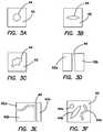
- FIG. 3Aillustrates a top view of a defect layer having a circular shaped defect according to the present invention.
- FIG. 3Billustrates a top view of a defect layer having an elongated defect according to the present invention.
- FIG. 3Cillustrates a top view of a defect layer having an amorphous defect according to the present invention.
- FIG. 3Dillustrates a top view of a defect layer having a two-piece defect according to the present invention.
- FIG. 3Eillustrates a top view of a multi-part defect layer according to the present invention.
- FIG. 3Fillustrates a top view of a defect layer having multiple defects according to the present invention.
- FIG. 4illustrates a top view of a simulated tissue structure according to the present invention.
- FIG. 5illustrates a side cross-sectional view of a simulated tissue structure according to the present invention.
- FIG. 6Aillustrates a perspective view of a modular tissue structure and support according to the present invention.
- FIG. 6Billustrates a perspective view of a modular tissue structure and support according to the present invention.
- FIG. 7illustrates a cross-sectional view of a simulated tissue structure configured to mimic a human uterus according to the present invention.
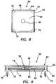
- FIG. 8illustrates a top view of a modular tissue structure according to the present invention.
- FIG. 9illustrates a side view of a modular tissue structure according to the present invention.
- FIG. 10Aillustrates a perspective view of a simulated tissue structure according to the present invention.
- FIG. 10Billustrates a perspective view of a simulated tissue structure according to the present invention.
- FIG. 11Aillustrates a perspective view of a simulated tissue structure according to the present invention.
- FIG. 11Billustrates a perspective view of a simulated tissue structure according to the present invention.
- FIG. 12illustrates a perspective view of a suture needle and a simulated tissue structure according to the present invention.
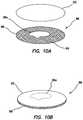
- FIG. 13illustrates a schematic of a model of female uterine anatomy with tumor placement according to the present invention.
- a surgical training device 10that is configured to mimic the torso of a patient such as the abdominal region is shown in FIG. 1 .
- the surgical training device 10provides a simulated body cavity 18 substantially obscured from the user for receiving model organs or simulated or live tissue 20 .
- the body cavity 18is accessed via a tissue simulation region 19 that is penetrated by the user employing devices to practice surgical techniques on the tissue or organ 20 found located in the body cavity 18 .
- the body cavity 18is shown to be accessible through a tissue simulation region 19 , a hand-assisted access device or single-site port device may be alternatively employed to access the body cavity 18 as described in U.S. patent application Ser. No. 13/248,449 entitled “Portable Laparoscopic Trainer” filed on Sep. 29, 2011 and incorporated herein by reference in its entirety.
- the surgical training device 10is particularly well suited for practicing laparoscopic or other minimally invasive surgical procedures.
- the surgical training device 10includes a base 12 and a top cover 14 connected to and spaced apart from the base 12 to define an internal body cavity 18 between the top cover 14 and the base 12 . At least one leg 16 interconnects and spaces apart the top cover 14 and base 12 .
- a model organ or simulated tissue 20is disposed within the body cavity 18 .
- the model organ 20 shown in FIG. 1is a partial colon or intestine that is shown suspended from the top cover 14 by tethers 22 and connected to at least one leg 24 .
- the at least one leg 24has an aperture (not shown) facing the internal body cavity 18 .
- the model colon 20includes a tube 26 having a proximal end and a distal end.
- the proximal end of the tube 26is interconnected with the aperture of the leg 24 such that the aperture provides an access port to the lumen of the tube 26 .
- the access port and apertureis shown to be closed off in FIG. 1 with an access device 28 which in combination with a sealed distal end of the tube 26 provides a model organ 20 that is adapted for insufflation with fluid deliverable via an insufflation port 30 .
- An optional insert 32 made of soft material such as siliconecreates a realistic interface for the access port.
- the distal end of the tube 26extends into the body cavity 18 and is suspended within the body cavity 18 .
- the interior of the tube 26 of the simulated organ 20is accessible via the access port of leg 24 or via the tissue simulation region 19 or instrument insertion ports 34 .
- An endoscopic camera inserted into the body cavity 18 or into the organ 20 via the access portgenerates a live image for display on a fold out video screen 36 shown in the closed position in FIG. 1 .
- An endoscopeis a visualization device that is used to view a hollow structure.
- the simulated organ 20 of FIG. 1is ideal for practicing procedures related to transanal minimally invasive surgery, any simulated organ or tissue portion may be employed.
- One particular aspect of the organ 20is at least one tumor or defect 38 is provided and connected to the organ. As shown in FIG. 1 , the tumor 38 is connected to the wall of the organ tube 26 .
- FIG. 2Athere is shown a partial side cross-sectional view of a portion of a simulated organ 20 that includes the tumor 38 .
- the simulated organ or tissue 20includes a base layer or organ wall 40 .
- the organ wall 40is made from a material configured to mimic real live tissue such as silicone or other polymer and is dyed appropriately.
- One or more base layers 40 of varying thicknesses and colorationsmay be employed to comprise the entirety of the wall 40 .
- the organ wall 40is rigid and made of polymeric material.
- the defect layer 42is the same size or smaller than the base layer 40 forming a raised platform for the tumor 38 .
- the defect layer 42is connected to the base layer 40 by adhesive or other means known to one having ordinary skill in the art including being integrally formed with the base layer 40 as a single unit.
- the defect layer 42is made of silicone and in one variation of the same color as the base layer 40 such that the defect layer 42 blends into the background of the base layer 40 .
- the defect layer 42includes at least one defect or gap 44 .
- the defect 44is a pre-fabricated breach in the defect layer 42 that mimics an incision, gap or other void in real tissue resulting from a tear, cut, removal or other surgical procedure that requires surgical attention by way of suturing, stapling or the like to close the defect.
- the defect 44comprises two opposed sides or surfaces defining a gap therebetween. Although the adjacent sides or surfaces are shown to be vertical with respect to the base layer 40 , the invention is not so limited and the juxtaposed surfaces or sides can have any shape and, for example, be curved.
- the defect 44can be any shape as will be discussed with respect to FIGS. 3A-3F .
- FIG. 3Athere is shown a top view of a defect layer 42 having a circular defect 44 .
- a defect layer 42 with an elongated, oblong or elliptically shaped defect 44is shown in the FIG. 3B .
- the defect 44can be amorphic or any shape as shown in FIG. 3C .
- the defect layer 42may be multi-part as shown in FIG. 3D wherein the defect layer 42 includes two or more adjacent defect layer pieces 42 a , 42 b juxtaposed to create at least one defect 44 therebetween.
- Another multi-part defect layer 42is shown in FIG. 3E where a plurality of adjacent defect layer pieces 42 a , 42 b and 42 c form one or more defects 44 therebetween.
- a defect layer 42may include multiple defects 44 a , 44 b and 44 c as shown in FIG. 3F .
- the defects 44may all be the same or have different shapes as shown in FIG. 3F .
- the shape, thickness and size of the defectallow the surgeon trainee to practice suturing across defects of varying difficulty.
- the defect layer 42is not of equal thickness. Instead, the thickness of the defect layer 42 varies at the defect 44 location to increase the difficulty of suturing or closing the defect.
- a tumor 38is located above the defect layer 42 .
- the tumor 38is preferably a different color from the base layer 40 or defect layer 42 or both such that it is readily identifiable by the trainee.
- the tumor 38is made of silicone or other polymer material and is red, black, blue or dark brown in color.
- the tumor 38is of a darker color than the base or defect layers 40 , 42 or otherwise in contrast therewith when viewed through a scope.
- the tumor 38is connected to the defect layer 42 by adhesive or other means known to one of ordinary skill in the art.
- the tumor 38is not connected or attached to the defect layer 42 but is removably located thereon.
- the simulated tissue structure 20includes a cover layer 46 located above the tumor 38 .
- the cover layer 46overlays the tumor 38 , defect layer 42 and the base layer 40 .
- the cover layer 46is preferably transparent or translucent in color and made of a polymer material such as silicone.
- the cover layer 46is the same color as the base layer 40 or defect layer 42 .
- the cover layer 46is at least as thick as the base layer 40 or defect layer 42 and in one variation is thinner than the defect layer 42 and in another variation is thinner than the base layer 40 .
- the cover layer 46is sized to cover the entire tumor 38 and defect layer 42 and is big enough to contact the base layer 40 in one variation.
- the cover layer 46is sized to cover the entire tumor 38 and contact the defect layer 40 .
- the cover layer 46is connected to the base layer 40 , defect layer 42 , tumor 38 or any more than one of the three layers by way of adhesive or other means known to one of ordinary skill in the art.
- the cover layer 46is smaller and connected to the defect layer 42 alone.
- the cover layer 46is connected to both the defect layer 42 and base layer 42 by adhesive or other means known to one of ordinary skill in the art.
- the cover layer 46can be any shape or size and be configured to provide a smooth surface to the surgeon instead of a layered surface to the artificial tumor location.
- the cover layer 46 , tumor 38 , defect layer 42 or base layer 40includes surface texturing in one variation.
- the cover layer 46assists in keeping the tumor 38 and defect layer 42 sandwiched between the cover layer 46 and base layer 40 which is advantageous in a variation wherein the tumor 38 is not adhered to the defect layer 42 .
- a top planar view of the base layer 40 , defect layer 42 , cover layer 46 and tumor 38is shown in FIG. 4 .
- any one or more of the base layer 40 , defect layer 42 and cover layer 46is formed of silicone molded over a woven, fabric, or mesh material such as nylon or cheesecloth so that the silicone layer has an integrated mesh structural support or other type of reinforcement.
- Any one or more of the layers 38 , 40 , 42 , 46can include a fabric or mesh reinforcement combined with an elastic polymer such silicone.
- the mesh supportaids in preventing the suture, staple, or suture needle from tearing through at least one of layers and especially the defect layer 42 when the suture is pulled to close the gap 44 .
- FIG. 2Bthe tumor 38 and a portion of the cover layer 46 are shown excised from the base layer 40 .
- the excisionis performed by the trainee using a surgical instrument such as a scalpel or other medical instrument to remove the tumor 38 .
- the traineewill cut through the cover layer 46 around the tumor 38 , isolate the tumor 38 , lift and remove the tumor 38 away from the site to expose the underlying defect 44 as shown in FIG. 2B .
- the traineesutures the defect 44 using a surgical suture 48 bringing the lips or edges of the defect layer 42 together as shown in FIG. 2D , thereby, practicing the closing of a gap or wound created by the surgical removal of a tumor 38 .
- Cutting the at least one layer to create an opening and removing the artificial tumor and suturing the gapis performed while the simulated tissue structure is disposed inside a simulated body cavity 18 of a surgical training device such that the simulated tissue structure is at least partially obscured from view by the user.
- FIG. 5there is shown another variation in which there is no pre-formed gap or defect in the second or defect layer 42 .
- the defectis created by the user in one or more of the cover layer 46 , defect layer 42 , base layer 40 and any remaining tumor portion not removed by the user. The user would then practice suturing the created defect in any of these layers 38 , 40 , 42 , 46 .
- one of the defect layer 42 or base layer 40is omitted from the construct.
- the tumor 38is located on a base layer 40 and the defect layer 42 is placed over the tumor 38 such that the defect layer 42 is above the tumor 38 .
- a cover layer 46may or may not be included.
- a cover layer 46it may be integrally formed together with the defect layer as a separate unitary layer.
- the constructsmay be flipped upside down or otherwise the layers placed in reverse or otherwise the construct being approachable by the user from either the top or bottom direction with the thicknesses and colors of the layers being adjusted accordingly if necessary to provide the simulated effects of real tissue.
- the simulated tissue constructcan be modular such that it is not integrally formed with the entire simulated organ 20 but instead configured as a module 50 that is removable and interchangeable.
- One or more modules 50are supported or contained in a module support 52 .
- a module support 52includes a first surface 51 , a second surface 53 and one or more tumor module receiving portions 54 , 56 , 58 formed in the support 52 .
- the tumor support 52can be rigid or pliable and made of polymeric material.
- the tumor support 52may also comprise a sheet of elastomeric material.
- the module receiving portions 54 , 56 , 58are each sized and configured to receive a correspondingly sized and configured module 50 .
- the modules 50 and module receiving portions 54 , 56 , 58 in FIG. 6are shown to be circular; however, the tumor module 50 can be any shape with a complementary shaped receiving portion formed in the module support 52 .
- the thickness of the support 52can vary providing the construct with varying depths of tumor module 50 positioning.
- the module receiving portions 54 , 56 , 58may include bottom walls onto which the tumor modules 50 may rest.
- the tumor receiving portions 54 , 56 , 58extend between openings in the first surface 51 and the second surface 53 with the modules 50 with tumor 38 being connected between or at one of the openings at either surface 51 , 53 or suspended within the tumor receiving portion.
- a single tumor module 50includes one or more tumors 38 .
- the module support 52is loaded with one or more tumor modules 50 and the simulated tissue construct 20 is inserted into the body cavity 18 of the surgical training device 10 , framework or other torso model. It can be placed on the base 12 of the training device 10 or suspended within the body cavity 18 of the training device 10 .
- the simulated tissue construct 20 and/or training deviceis fashioned with attachment mechanisms such as clips, fasteners, wires, hook-and-loop type fasteners and the like for placement, suspension or connection of the simulated tissue construct 20 to a training device 10 .
- the module support 52 of FIG. 6Bincludes a first layer 57 connected to a second layer 55 .
- the first layer 57is made of a sheet of elastomeric material and the second layer 55 is made of any suitable polymeric material such as low-density elastomeric foam.
- the second layer 55serves as a support for the first layer 57 .
- the second layer 55also advantageously provides depth to the module support 52 permitting the tumors 38 within the modules 50 to be placed deeply into the module support 52 relative to the first surface 51 .
- Module receiving portions 54 , 56 , 58are formed in one or more than one of the first layer 57 and the second layer 55 .
- Module receiving portions 54 , 56 , 58 formed in the second layer 55may have a different shape than the shape the same module receiving portion 54 , 56 , 58 has in the first layer 57 .
- the tumor module 50comprises at least only the simulated tumor 38 which is embedded or buried inside the second layer 55 with at least one of the first layer 57 or second layer 55 constituting a defect layer which the user can practice closing.
- the first layer 57does not include a module receiving portion but instead the first layer 57 serves as a cover layer which the user practices cutting through to access the tumor 38 located in a tumor receiving portion formed in the second layer 55 .
- the first layer 57can be a sheet of elastomeric material such as silicone and the second layer 55 is a layer of low-density elastomeric foam.
- the module support 52is planar as shown in FIGS. 6A and 6B or, alternatively, shaped to mimic a portion of the human anatomy, tissue or organ.
- FIG. 7illustrates a support 52 that is shaped to mimic a human uterus.
- the support 52includes a first layer 57 connected to a second layer 55 .
- the first layer 57is made of any suitable polymeric material such as a sheet of elastomeric material and the second layer 55 is made of any suitable polymeric material such as low-density elastomeric foam.
- the second layer 55serves as a support for the first layer 57 and advantageously permits the tumors 38 within the modules 50 or the tumors 38 by themselves to be connected to the support 52 and realistically extend deeply into the support 52 and be dispersed throughout the support 52 in various locations and orientations including being embedded into the first layer 57 as shown in FIG. 7 .
- Tumor or module receiving portions 61are formed in at least one of the first layer 57 and second layer 55 .
- the tumor receiving portions 61may be pockets that are preformed in the second layer 55 or can be formed by the user by cutting slits into the second layer 55 .
- the tumors 38are configured to mimic fibroid tumors commonly found in the human uterus. Examples of fibroid tumors that are simulated by the tumors 38 disposed in the support include but are not limited to one or more of the following types of fibroids: pedunculated submucosal fibroids, subserosal fibroids, submucosal fibroids, pedunculated subserosal fibroids and intramural fibroids.
- the usercan approach the support 52 to excise the simulated tumors 38 from the first surface 51 or the second surface 53 via the access channel or opening 63 .
- the opening 63serves as the only opening to the hollow portion 59 or alternatively the support 52 can have a substantially C-shaped planar configuration with access available to the user from above or below the planar C-shaped structure.
- the module support 52 in any of the variationsis not planar but is provided with a landscape that includes curves and other structures, mountains and valleys and various textures.
- the varying landscapeprovides the user with various levels of difficulty in approaching each tumor location requiring the user to navigate around artifacts and features that may obscure the tumor location.
- These structural artifacts in the tumor support 52may be integrally formed with the tumor support 52 or also be modular in structure similar to the tumor modules 50 making the anatomy landscape modules removable and interchangeable.
- Tumor modules 50are interchangeable with non-tumor modules that include, for example, features and artifacts or textures made of silicone or other material extending outwardly or inwardly from the one or more of the upper and lower surfaces 51 , 53 of the module support 52 .
- non-tumor modulescan have various shapes to mimic anatomy that includes adjacent organ structures or tissues.
- a non-tumor modulecan include a tubular form of silicone to mimic an intestine.
- the non-tumor and tumor modules 50are removably connected to the module support 52 by any means known to one skilled in the art enabling the user to discard a module after use and then to continue practicing by replacing the discarded module or moving to an adjacent module 50 in the module support 52 or changing out a tumor module 50 for another tumor module 50 having a different feature or level of difficulty.
- the tumor module 50includes a simulated tissue portion 60 connected to a support 62 .
- the support 62includes a top frame 64 connected to a bottom frame 66 . At least one of the top frame 64 and bottom frame 66 includes a window.
- the top frame 64 having a window 68is shown in FIG. 8 .
- the bottom frame 66may or may not include a window. If windows are provided in both the top frame 64 and the bottom frame 66 , the windows are aligned at least in part.
- the support 62is sized and configured to receive a simulated tissue portion 60 between the top frame 64 and the bottom frame 66 .
- the top frame 64is connectable to the bottom frame 66 to capture the unitary simulated tissue portion 60 or a simulated tissue portion 60 formed from multiple layers and, in one variation, separable.
- the frames 64 , 66are spaced apart from each other using spacers 70 .
- at least one of the top and bottom frames 64 , 66includes one or more connecting features 72 configured to secure the tumor module 50 to a tumor support 52 (not shown).
- the connecting features 72are shown as extending pegs for insertion into corresponding holes formed in the tumor support 52 to provide a snap-fit engagement.
- a friction fit or other fasteners or connecting meanssuch as hook-and-loop type materials can be employed on the module 50 and module support 52 to connect the module 50 to the support 52 in a removable fashion.
- the simulated tissue portion 60can be any of the constructs described above with reference to FIGS. 2-5 . With windows formed in both the first and second frames 64 , 66 , the simulated tissue portion 60 can be approached from either side of the module 50 . Any layer described above as a cover layer may act as a top layer or as a bottom layer depending on from which side or direction the simulated tissue portion 60 is approached. For example, a base layer may also serve as a top layer or as a bottom layer depending on which side or direction the simulated tissue portion 60 is approached. In such bi-directional constructs, the thicknesses and colors of the layers may be adjusted accordingly to provide the desired simulated effect.
- the simulated tissue portion 60 in FIG. 9includes a first layer 74 and a second layer 76 .
- the first and second layers 74 , 76are made from a polymeric material configured to mimic real live tissue such as silicone or other polymer and can include dye of any one or more appropriate colors or mesh, fabric, or other reinforcement.
- Each of the layers 74 , 76includes a tumor-receiving portion 78 , 80 , respectively.
- Each tumor-receiving portion 78 , 80is a concavity, indent, half-pocket or a location of reduced layer thickness that is formed in the layers 74 , 76 .
- the tumor-receiving portions 78 , 80are substantially aligned to form a pocket for the tumor 38 .
- each layer 74 , 76 in FIG. 9is shown with a tumor-receiving portion 78 , 80 , a single tumor-receiving portion is formed in at least one of the first and second layers 74 , 76 in one variation.
- a tumor 38is disposed within the pocket formed by one or more tumor-receiving portions 78 , 80 formed in the one or more layers 74 , 76 .
- the tumor 38may be adhered to either layer 74 , 76 or free-floating inside the pocket.
- the tumor-receiving portion formed in a layercan be considered to be one type of defect and the variation of FIG. 9 describes a simulated tissue construct comprising two defect layers with a tumor therebetween.
- the userwill see the target tumor location. Visualization of the target tumor 38 is enhanced by the tumor-receiving portion being thinner in thickness relative to the rest of the layer with the thinning of the layer being provided by the concavity or pocket.
- the userwill then cut in the general location of the tumor cutting into at least one of the layers 74 , 76 to remove the tumor 38 . Cutting through one or more layers completes the creation of a gap or full defect, which the user can then practice suturing or otherwise closing together.
- at least one tumoris disposed between the two layers 74 , 76 wherein the layers 74 , 76 have a substantially uniform thickness with the tumor 38 creating a minor bulge in the layers.
- the tissue portion 86can be integral or modular as described above.
- the tissue portion 86includes a base layer 88 formed of any suitable polymeric material such as silicone or other elastomeric polymer that may or may not include a reinforcement material such as fabric, mesh, nylon or other reinforcement material or filler that will resist tearing while carrying sutures or while being sutured.
- the base layer 88is connected to a defect layer 90 that is overlaid onto the base layer 88 .
- the defect layer 90includes a plurality of protrusions extending upwardly from the base layer 88 .
- the defect layer 90may be integrally formed with the base layer 88 or be a separate layer that is adhered to the base layer 88 . As can be seen in FIGS. 10A, 11A and 12 , the defect layer 90 is configured into a lattice shaped pattern such that the lattice is raised above the base layer 88 or projects upwardly from the base layer 88 .
- a lattice patternis exemplary and any shape may be formed by the defect layer 90 such that it contains a plurality of adjacent projections. These projections of the base layer 90 provide the user with locations to hook a suture needle into and as a platform to raise the tumor 38 a , 38 b above the base layer 88 for easy excision.
- FIGS. 10A and 11Ashow the base layer 88 , defect layer 90 , tumors 38 a , 38 b and a cover layer 92 in a semi-exploded view of the simulated tissue portion 86 wherein the cover layer 92 is raised above the other layers.
- the tumor 38 a of FIG. 10 ais substantially planar and is shown covered in FIG. 10B by the cover layer 92 .
- Tumor 38 b of FIG. 11Ahas greater height and is substantially spherical in shape and FIG. 11B shows the spherical tumor 38 b covered with the cover layer 92 leaving a raised portion or protuberance in the construct.
- FIG. 12shows the tumor 38 being removed leaving a remnant defect 94 in the base layer 88 and a suture needle crossing the gap in the defect 94 with the defect having been accessed under or through the cover layer 92 .
- Synthetic materials that mimic the characteristics of living tissuemay include silicone elastomers, natural latex, polyurethane elastomers, hydrogels and styrenic-block-copolymers.
- the elastomeric materialsare dielectric unless specially treated.
- An elastomeris generally any of various polymers with elastic properties resembling those of natural rubber.
- a hydrogelis generally a hydrophilic polymer containing between 50% and 99% water.
- a thermoplasticgenerally pertains to materials that may be repeatedly made soft and hard by heating and cooling. Thermoplastics are non-conductive and are suitable for making the tray or base, bone and other similar structures.
- a thermosetgenerally pertains to elastomeric materials that permanently harden or solidify upon being heated or cured.
- Thermoset plasticsare non-conductive such as silicone and polyester and are suitable for forming pathologies, tumors and the like.
- Silicone elastomersare usually very soft, stable and non-conductive and therefore suitable for forming artificial organs such as the liver, kidney, spleen, ovaries, gallbladder, stomach, major arteries, colon, intestine, major veins, omentum, mesentery, pathologies and other anatomy.
- Natural latexis very resilient and non-conductive and suitable for forming artificial muscle, cartilage and the like.
- Polyurethane elastomers and foamsare non-conductive and suitable for filling hollow structures, bone and the like.
- Hydrogels SBCsmay be conductive and are good for any soft structure to be operated upon by electrosurgery.
- a surgical simulation traythat is insertable into a lap trainer 10 for practicing surgical techniques including laparoscopic and electro-surgical methods, comprises a base, an arrangement of anatomical organs, and a covering layer.
- the basecomprises a rigid or semi-rigid structure that is sized and configured to fit within or upon a surgical training device 10 .
- the baseis additionally supplied with anatomical support features or receptacle portions formed by upstanding walls that cooperate and conform in size and shape with the placement of body organs within the receptacle portion or upon the base.
- Body organsmade of elastomeric materials, are placed strategically within or upon the base according to the specific needs of the training device and/or according to the target anatomy.
- At least one covering layermay be placed over the entire assembly or upon specific areas thereof.
- the covering layeris sized and configured to represent one or more of the omentum, mesentery, fat, connective tissue, peritoneum, mesothelium, broad ligaments or the like.
- the covering layermay comprise silicone elastomer, which is non-conductive.
- the non-conductive covering layeris suitable if no electrosurgical activity is used on the covering layer. If electrosurgical activity is contemplated, the covering layer is comprised of a conductive gel such as a hydrogel. A combination of conductive and non-conductive layers is provided when electrosurgery is directed to one of the layers.
- a uterusmay be formed having an outer layer of silicone rubber and a substantially hollow inner layer of soft polyurethane foam as described above with respect to FIG. 7 .
- synthetic fibroid tumorsmay be placed for identification and removal by a surgical trainee.
- One simulated construction of a synthetic fibroid tumorcomprises a small quantity of very soft uncured silicone rubber.
- the uncured silicone rubberis mixed with a quantity of amorphous, untreated fumed silicon dioxide, which acts as a filler and flow controller.
- the combination of the uncured silicone rubber and the silicon dioxideis shaped and allowed to cure. When fully cured, this combination results in an irregularly shaped, somewhat fibrous structure that resembles a human fibroid tumor.
- This construction of a simulated human fibroid tumoris then placed into a simulated organ model such as that of a uterus.
- This tumor simulationis not limited for use to mimic a tumor in a gynecological model, but also can be used in other organ models that include tumors for practicing their removal.
- This tumor simulationcomprising the cured mixture of silicone rubber and silicon dioxide strikingly resembles real-life tumors that are found in a gynecological surgical situation and provides an amorphous and realistic look and feel when practicing surgical techniques.
- Dark colored dyesuch as red or black may be added to the mixture before curing and mixed throughout.
- This constructionmay also be used to construct simulated ectopic pregnancies for insertion into a simulated fallopian tube of a simulated organ placed within the training device 10 .
- the mixed consistency of the silicone and fillerbeing very dry and shape-able, advantageously allows the tumors or other pathologies to be formed very creatively, easily and at any size to mimic actual physical conditions. Tumors made of silicone and filler are non-conductive and may be fractured or torn if not properly handled.
- organ simulation modelsthat include the combination of conductive and non-conductive portions will now be discussed.
- a simulated organ model tray for training electrosurgical procedureswill have a conductive hydrogel liver, cystic duct and mesentery.
- These conductive portions of the modelare located adjacent to non-conductive portions of the anatomy comprising the same organ or different organs.
- the organ modelincludes a cystic duct and mesothelium made from electro-conductive hydrogel and the liver and gallbladder are non-conductive.
- the simulated organ modelincludes one or more of the blood vessels and the greater omentum/mesentery along the greater curvature of the stomach made of electro-conductive hydrogel material and one or more of the stomach, large intestine, and small intestine made of non-conductive material.
- the simulated organ modelincludes one or more of the short gastric vessels and the mesentery/omentum along the greater curvature of the stomach made of electro-conductive hydrogel material and the stomach made of non-conductive material.
- at least a portion of the jejunum and/or stomachis made of electro-conductive hydrogel.
- the training modelincludes both conductive and non-conductive materials.
- the organ modelmay include one or more simulated fallopian tubes, round ligament, ovarian ligament, IP ligament, broad ligament, bladder flap, uterine artery/vein, cardinal ligament, uterosacral ligament, made of electro-conductive hydrogel and one or more of the uterus, ovaries, rectum, urinary bladder, ureters and kidneys are non-conductive.
- the location just above the cervix and/or just below the cervixis made of electro-conductive hydrogel for practicing a supracervical or total colpotomy. Procedures that involve the colon, small intestine, sigmoid or rectum may also require that specific portions be electrically conductive. These conductive portions are located adjacent to non-conductive portions. For example, to practice transanal minimally invasive surgery for the local excision of tumors, the organ model would include a colon and/or rectum, and tumor made of non-conductive elastomeric material except for the area surrounding the tumor which would be made of electro-conductive hydrogel material.
- the rectumis made of electro-conductive hydrogel such as for the practice of transanal total mesorectal excision.
- the simulated organ modelmay include one or more of the mesentery/mesoappendix, appendiceal artery and blood vessels made of electro-conductive hydrogel and one or more of the appendix, cecum and terminal ileum made of non-conductive elastomeric material.
- the simulated organ modelmay include one or more of the mesentery, ileocolic artery, middle colic artery, right colic artery, inferior mesenteric artery, inferior mesenteric vein, left colic artery, sigmoid arteries, rectal arteries, marginal arteries, corresponding veins, omentum, white line of Toldt, mesenteric attachments to the retroperitoneum, and mesorectum made of electro-conductive hydrogel and one or more of the colon, liver, spleen, stomach, kidney, duodenum, retroperitoneum made of non-conductive material. Hydrogel material must be hydrated in order to be sufficiently conductive and therefore it may be difficult to maintain a long shelf-life.
- the covering layercomprises a thin semi-transparent sheet of silicone rubber that is calendared or press-formed so as to have a texture and finish that appears to be naturally occurring.
- An alternate variation of the covering layermay further comprise a thin semi-transparent sheet of hydrogel material that is cured from slurry and allowed to develop surface features as it cures. The hydrogel material, when hydrated becomes conductive and allows the use of electrosurgical devices.
- a composite structure for the covering layercomprises a conductive gel layer sandwiched between two silicone elastomer non-conductive layers. In such a case, one or more of the outer non-conductive layers are removed to expose the conductive gel layer.
- the non-conductive silicone layersadvantageously provide a sealing for the hydrogel layer retaining the fluid content of the conductive gel.
- a thin film of two-part platinum or tin cured liquid silicone, thoroughly mixed,is placed upon a sheet of textured polyethylene foam.
- a notched trowel or spreaderis then used to spread the silicone material over the surface of the first layer of foam leaving an irregular pattern of material thickness.
- a second layer of textured polyethylene foamis placed over the first layer of foam leaving the silicone between.
- a textured roller or stamping deviceis then moved over the surface of the second layer of foam to calendar the silicone material between the foam layers.
- the resulting silicone sheetwhen cured, is non-tacky and exhibits the characteristics of omentum, mesentery, fat etc.
- the sheetadvantageously has strong and weak regions that can be used to demonstrate the use of mechanical dissecting instruments and scissors.
- the specific organs that may be used in a surgical simulation deviceinclude a uterus 100 as shown in FIG. 13 .
- the uteruscomprises an outer shell constructed of soft silicone rubber molded over a uterine form. When the shell is fully cured, it is placed over a molded foam rubber uterine form that is substantially hollow, having about a 7 to 9 millimeter thick wall.
- Various pathologiesmay be placed between the silicone shell and the foam wall. Some pathologies may be inserted into the foam wall to mimic intramural tumors, fibroid tumors 102 or cysts.
- Fallopian tubes 104 , ovarian ligaments 106 and other attendant structuresmay be inserted into the silicone/foam structure and attached with adhesive.
- Ovarian cysts 124may also be provided and made of the same tumor material.
- Attendant structuresmay include the aorta 114 , the internal iliac artery 116 , the ovarian artery 118 , the uterine artery 120 , the vaginal artery 121 , and the uterosacral ligament 122 .
- the uterine shellis the primary portion to be operated upon. In one variation, it is constructed of silicone elastomer and therefore suitable if the uterine model is intended to be cut or incised in training. If electrosurgery is being practiced upon the uterine model, a uterine model is selected that comprises conductive gel.
- Connecting structures and tubesare constructed of silicone elastomer or conductive gel depending on the surgical modality.
- Fallopian tubes 104constructed of two-part platinum or tin cured silicone comprising a first open end and a second open end and a through lumen.
- the first open endforms a tubular structure that extends a distance of about 20 centimeters and has a diameter of about 6.5 millimeters and a very thin wall of approximately 1-1.5 millimeters.
- a bulbous portionis formed having a diameter of about 1.5 centimeters and a length of about 3 centimeters. The bulbous portion transitions to a narrowing of the tubular structure to about 7 millimeters.
- the narrowed tubular structurethen gradually enlarges into a funnel shaped structure having a final open diameter of about 2 centimeters over a length of about 3.5 centimeters.
- a plurality of axial cuts 108is made at the second enlarged open end. When removed from the form, these cuts allow the silicone material to move in a way that resembles human fimbria.
- Pathologysuch as an ectopic pregnancy 110 may be inserted into the bulbous portion of the fallopian tube 104 for identification or excision.
- a length of soft fibrous yarnsuch as used in knitting, may be placed within the lumen.
- the ovaries 112are hollow bulbous structures formed from two-part platinum or tin cured silicone.
- a soft polyurethane foam supportis placed within the ovarian structure.
- the polyurethane supportis sized and configured to fit neatly within the ovarian shell and have a nest or receptacle for pathology such as an ovarian cyst 124 .
- the traineemay cut through the ovarian wall and into the polyurethane foam to remove the pathology and subsequently suture the defect to close.
- Ovariesare made of non-conductive material and are cut with scissors or scalpel.
- the ovariesare made of conductive gel so they could be cut with electrosurgery.
- the cystis made of non-conductive material.
- a stomachcomprises a hollow stomach-shaped bladder having a first open end, a second open end and an enlarged central portion.
- the enlarged central portionis divided by a pathway that extends from near the first open end to near the second open end.
- the pathwaycomprises a region of silicone adhesive placed strategically along a desired trajectory adjacent to the lesser curvature of the stomach.
- the opposing walls of the stomach bladderare approximated and held together by the adhesive.
- the stomachmay be divided along the adhesive pathway to simulate a particular procedure. That is, the adhesive pathway directs the trainee to staple or cut along a preferred surgical pathway.
- the adhesivesimulates the condition in which several rows of staples would be placed before the cutting element in a surgical stapler is deployed.
- the dissected stomach portionappears to be stapled securely and the residual stomach portion is gas tight and secure.
- the adhered portion of the stomachis formed of conductive gel material adjacent to non-conductive adjacent portions of the stomach.
- the predetermined surgical pathway across the stomach or other organis constructed of conductive gel material adjacent to non-conductive material of the same organ or adjacent to non-conductive material of different organs and anatomical structures.
- a liver constructed of hydrogelmay be placed into the training module 10 where the procedure would involve electrosurgical dissection.
- the base or tray of the training module 10receives and holds in place either a silicone liver or a hydrogel liver.
- a receiving featuremay comprise a nest, pocket or receptacle sized and configured to maintain in position a silicone or hydrogel or foam rubber liver depending on the needs of the particular training module. If a procedure required electrosurgical activity, such as a liver resection, the liver is made of conductive gel.
- the base or trayis configured to accept a liver made of gel, silicone or foam depending on the specific procedure. If the procedure to be practiced does not involve electrosurgery, it is far more economical to use a silicone or foam model.
Landscapes
- Engineering & Computer Science (AREA)
- Physics & Mathematics (AREA)
- General Physics & Mathematics (AREA)
- Health & Medical Sciences (AREA)
- Mathematical Analysis (AREA)
- Pure & Applied Mathematics (AREA)
- Medical Informatics (AREA)
- Algebra (AREA)
- Computational Mathematics (AREA)
- General Health & Medical Sciences (AREA)
- Chemical & Material Sciences (AREA)
- Mathematical Optimization (AREA)
- Mathematical Physics (AREA)
- Medicinal Chemistry (AREA)
- Business, Economics & Management (AREA)
- Educational Administration (AREA)
- Educational Technology (AREA)
- Theoretical Computer Science (AREA)
- Pulmonology (AREA)
- Radiology & Medical Imaging (AREA)
- Instructional Devices (AREA)
- Surgical Instruments (AREA)
Abstract
Description
Claims (14)
Priority Applications (3)
| Application Number | Priority Date | Filing Date | Title |
|---|---|---|---|
| US15/927,968US10991270B2 (en) | 2013-03-01 | 2018-03-21 | Advanced surgical simulation constructions and methods |
| US17/236,947US20210256876A1 (en) | 2013-03-01 | 2021-04-21 | Advanced surgical simulation constructions and methods |
| US18/457,245US20240062679A1 (en) | 2013-03-01 | 2023-08-28 | Advanced surgical simulation constructions and methods |
Applications Claiming Priority (3)
| Application Number | Priority Date | Filing Date | Title |
|---|---|---|---|
| US201361771316P | 2013-03-01 | 2013-03-01 | |
| US14/195,327US9940849B2 (en) | 2013-03-01 | 2014-03-03 | Advanced surgical simulation constructions and methods |
| US15/927,968US10991270B2 (en) | 2013-03-01 | 2018-03-21 | Advanced surgical simulation constructions and methods |
Related Parent Applications (1)
| Application Number | Title | Priority Date | Filing Date |
|---|---|---|---|
| US14/195,327ContinuationUS9940849B2 (en) | 2013-03-01 | 2014-03-03 | Advanced surgical simulation constructions and methods |
Related Child Applications (1)
| Application Number | Title | Priority Date | Filing Date |
|---|---|---|---|
| US17/236,947ContinuationUS20210256876A1 (en) | 2013-03-01 | 2021-04-21 | Advanced surgical simulation constructions and methods |
Publications (2)
| Publication Number | Publication Date |
|---|---|
| US20180211565A1 US20180211565A1 (en) | 2018-07-26 |
| US10991270B2true US10991270B2 (en) | 2021-04-27 |
Family
ID=50280535
Family Applications (4)
| Application Number | Title | Priority Date | Filing Date |
|---|---|---|---|
| US14/195,327Active2036-08-06US9940849B2 (en) | 2013-03-01 | 2014-03-03 | Advanced surgical simulation constructions and methods |
| US15/927,968Active2034-11-10US10991270B2 (en) | 2013-03-01 | 2018-03-21 | Advanced surgical simulation constructions and methods |
| US17/236,947AbandonedUS20210256876A1 (en) | 2013-03-01 | 2021-04-21 | Advanced surgical simulation constructions and methods |
| US18/457,245PendingUS20240062679A1 (en) | 2013-03-01 | 2023-08-28 | Advanced surgical simulation constructions and methods |
Family Applications Before (1)
| Application Number | Title | Priority Date | Filing Date |
|---|---|---|---|
| US14/195,327Active2036-08-06US9940849B2 (en) | 2013-03-01 | 2014-03-03 | Advanced surgical simulation constructions and methods |
Family Applications After (2)
| Application Number | Title | Priority Date | Filing Date |
|---|---|---|---|
| US17/236,947AbandonedUS20210256876A1 (en) | 2013-03-01 | 2021-04-21 | Advanced surgical simulation constructions and methods |
| US18/457,245PendingUS20240062679A1 (en) | 2013-03-01 | 2023-08-28 | Advanced surgical simulation constructions and methods |
Country Status (8)
| Country | Link |
|---|---|
| US (4) | US9940849B2 (en) |
| EP (3) | EP2962291A1 (en) |
| JP (5) | JP6482478B2 (en) |
| KR (4) | KR20250004119A (en) |
| AU (5) | AU2014224004B2 (en) |
| CA (1) | CA2897832A1 (en) |
| ES (1) | ES2897418T3 (en) |
| WO (1) | WO2014134597A1 (en) |
Families Citing this family (80)
| Publication number | Priority date | Publication date | Assignee | Title |
|---|---|---|---|---|
| EP2622594B1 (en) | 2010-10-01 | 2018-08-22 | Applied Medical Resources Corporation | Portable laparoscopic trainer |
| US9218753B2 (en) | 2011-10-21 | 2015-12-22 | Applied Medical Resources Corporation | Simulated tissue structure for surgical training |
| KR101953187B1 (en) | 2011-12-20 | 2019-02-28 | 어플라이드 메디컬 리소시스 코포레이션 | Advanced surgical simulation |
| CA2880277A1 (en) | 2012-08-03 | 2014-02-06 | Applied Medical Resources Corporation | Simulated stapling and energy based ligation for surgical training |
| JP2015532450A (en) | 2012-09-26 | 2015-11-09 | アプライド メディカル リソーシーズ コーポレイション | Surgical training model for laparoscopic procedures |
| US10679520B2 (en) | 2012-09-27 | 2020-06-09 | Applied Medical Resources Corporation | Surgical training model for laparoscopic procedures |
| US9959786B2 (en) | 2012-09-27 | 2018-05-01 | Applied Medical Resources Corporation | Surgical training model for laparoscopic procedures |
| WO2014052612A1 (en) | 2012-09-27 | 2014-04-03 | Applied Medical Resources Corporation | Surgical training model for laparoscopic procedures |
| EP2901439A1 (en) | 2012-09-28 | 2015-08-05 | Applied Medical Resources Corporation | Surgical training model for laparoscopic procedures |
| CA2885314C (en) | 2012-09-28 | 2021-01-19 | Applied Medical Resources Corporation | Surgical training model for transluminal laparoscopic procedures |
| AU2014224004B2 (en) | 2013-03-01 | 2018-04-05 | Applied Medical Resources Corporation | Advanced surgical simulation constructions and methods |
| US9472122B2 (en)* | 2013-03-07 | 2016-10-18 | Syndaver Labs, Inc. | Central line simulation and training device |
| CA3139494A1 (en) | 2013-05-15 | 2014-11-20 | Applied Medical Resources Corporation | Hernia model |
| KR102607634B1 (en) | 2013-06-18 | 2023-11-29 | 어플라이드 메디컬 리소시스 코포레이션 | Gallbladder model for teaching and practicing surgical procedures |
| US10198966B2 (en) | 2013-07-24 | 2019-02-05 | Applied Medical Resources Corporation | Advanced first entry model for surgical simulation |
| AU2014293036B2 (en) | 2013-07-24 | 2017-12-21 | Applied Medical Resources Corporation | First entry model |
| ES2891756T3 (en) | 2014-03-26 | 2022-01-31 | Applied Med Resources | Simulated dissectable tissue |
| AU2015347077B2 (en) | 2014-11-13 | 2021-08-12 | Applied Medical Resources Corporation | Simulated tissue models and methods |
| KR102586607B1 (en)* | 2015-02-19 | 2023-10-10 | 어플라이드 메디컬 리소시스 코포레이션 | Simulated tissue structures and methods |
| ES2716924T3 (en) | 2015-05-14 | 2019-06-18 | Applied Med Resources | Synthetic tissue structures for training and electrosurgical stimulation |
| EP4167212B1 (en)* | 2015-05-27 | 2025-10-01 | Applied Medical Resources Corporation | Surgical training model for laparoscopic procedures |
| AU2016276771B2 (en)* | 2015-06-09 | 2022-02-03 | Applied Medical Resources Corporation | Hysterectomy model |
| JP2017021137A (en)* | 2015-07-09 | 2017-01-26 | 株式会社 タナック | Organ models |
| EP3300057B1 (en)* | 2015-07-10 | 2020-03-25 | Kotobuki Giken Inc. | Use of a simulated animal organ |
| KR102697097B1 (en) | 2015-07-16 | 2024-08-21 | 어플라이드 메디컬 리소시스 코포레이션 | Simulated exciseable tissue |
| WO2017015438A1 (en) | 2015-07-22 | 2017-01-26 | Applied Medical Resources Corporation | Appendectomy model |
| CA2939744A1 (en)* | 2015-08-20 | 2017-02-20 | Uti Limited Partnership | Suturing training device and method |
| KR20250099424A (en)* | 2015-10-02 | 2025-07-01 | 어플라이드 메디컬 리소시스 코포레이션 | Hysterectomy Model |
| CN108352132A (en)* | 2015-10-16 | 2018-07-31 | 维塔医疗股份公司 | ultrasonic simulation method |
| JP6886975B2 (en)* | 2015-11-20 | 2021-06-16 | アプライド メディカル リソーシーズ コーポレイション | Simulated incisable tissue |
| JP6055069B1 (en)* | 2015-12-10 | 2016-12-27 | サンアロー株式会社 | Organ, tissue or organ model |
| JPWO2017126313A1 (en)* | 2016-01-19 | 2018-11-22 | 株式会社ファソテック | Surgical training and simulation system using biological texture organs |
| US10235903B2 (en)* | 2016-04-18 | 2019-03-19 | Vivienne Souter | Simulator for training medical personnel to perform uterine procedures |
| EP3449474B1 (en)* | 2016-04-26 | 2020-09-16 | Applied Medical Resources Corporation | Residual stress features in organ models |
| EP3252737A1 (en)* | 2016-06-03 | 2017-12-06 | Sofradim Production | Abdominal model for laparoscopic abdominal wall repair/reconstruction simulation |
| ES2946810T3 (en) | 2016-06-27 | 2023-07-26 | Applied Med Resources | simulated abdominal wall |
| EP3291208B1 (en)* | 2016-08-31 | 2020-09-30 | Ricoh Company, Ltd. | Hydrogel structure, blood vessel, internal organ model, practice tool for medical procedure, and method of manufacturing the hydrogel structure |
| JP6868239B2 (en)* | 2016-10-24 | 2021-05-12 | 学校法人 名城大学 | Dural model |
| US20190287423A1 (en) | 2016-11-22 | 2019-09-19 | PraxiCut, LLC | Surgical simulation systems, methods, and compositions |
| WO2018118858A1 (en) | 2016-12-19 | 2018-06-28 | National Board Of Medical Examiners | Medical training and performance assessment instruments, methods, and systems |
| ES3004046T3 (en) | 2017-02-14 | 2025-03-11 | Applied Med Resources | Laparoscopic training system |
| US10847057B2 (en) | 2017-02-23 | 2020-11-24 | Applied Medical Resources Corporation | Synthetic tissue structures for electrosurgical training and simulation |
| JP6860162B2 (en)* | 2017-04-21 | 2021-04-14 | 学校法人日本医科大学 | Blood vessel puncture practice model |
| US11847932B2 (en) | 2017-09-07 | 2023-12-19 | Intuitive Surgical Operations, Inc. | Modified animal organs for use in surgical simulators |
| CN107749227B (en)* | 2017-11-07 | 2023-09-08 | 天津天堰科技股份有限公司 | Craniotomy training device with interlayer positioning and throwing device |
| AU2018368439B2 (en)* | 2017-11-14 | 2024-08-29 | Applied Medical Resources Corporation | Hysterectomy model |
| US11501662B2 (en) | 2017-11-15 | 2022-11-15 | Applied Medical Resources Corporation | Suturing skills surgical training model |
| EP3727172A1 (en) | 2017-12-19 | 2020-10-28 | Applied Medical Resources Corporation | Total mesorectal excision surgical simulator |
| US11373554B2 (en)* | 2018-02-14 | 2022-06-28 | The Charlotte Mecklenburg Hospital Authority | Pelvic model for robotic, laparoscopic, and abdominal/open approach surgical training |
| EP3803837A1 (en) | 2018-06-01 | 2021-04-14 | Applied Medical Resources Corporation | Renal hilum surgical simulation system |
| US11417241B2 (en) | 2018-12-01 | 2022-08-16 | Syndaver Labs, Inc. | Artificial canine model |
| JP2020103674A (en)* | 2018-12-28 | 2020-07-09 | 株式会社三洋物産 | Game machine |
| JP2020103669A (en)* | 2018-12-28 | 2020-07-09 | 株式会社三洋物産 | Game machine |
| JP2020108445A (en)* | 2018-12-28 | 2020-07-16 | 株式会社三洋物産 | Game machine |
| JP2020103675A (en)* | 2018-12-28 | 2020-07-09 | 株式会社三洋物産 | Game machine |
| JP2020103673A (en)* | 2018-12-28 | 2020-07-09 | 株式会社三洋物産 | Game machine |
| JP2020103667A (en)* | 2018-12-28 | 2020-07-09 | 株式会社三洋物産 | Game machine |
| JP2020108443A (en)* | 2018-12-28 | 2020-07-16 | 株式会社三洋物産 | Game machine |
| JP2020108442A (en)* | 2018-12-28 | 2020-07-16 | 株式会社三洋物産 | Game machine |
| JP2020108441A (en)* | 2018-12-28 | 2020-07-16 | 株式会社三洋物産 | Game machine |
| JP2020103668A (en)* | 2018-12-28 | 2020-07-09 | 株式会社三洋物産 | Game machine |
| JP2020108444A (en)* | 2018-12-28 | 2020-07-16 | 株式会社三洋物産 | Amusement machine |
| JP2020103676A (en)* | 2018-12-28 | 2020-07-09 | 株式会社三洋物産 | Game machine |
| JP2020103677A (en)* | 2018-12-28 | 2020-07-09 | 株式会社三洋物産 | Game machine |
| JP2020108446A (en)* | 2018-12-28 | 2020-07-16 | 株式会社三洋物産 | Amusement machine |
| JP2020146307A (en)* | 2019-03-14 | 2020-09-17 | 株式会社三洋物産 | Game machine |
| JP2020146306A (en)* | 2019-03-14 | 2020-09-17 | 株式会社三洋物産 | Game machine |
| JP2020146305A (en)* | 2019-03-14 | 2020-09-17 | 株式会社三洋物産 | Game machine |
| CO2019010338A1 (en)* | 2019-09-25 | 2019-12-10 | Pontificia Univ Javeriana | Morphological models for minimally invasive surgery training, its manufacturing method and system for practice |
| BR102019024459A2 (en)* | 2019-11-21 | 2021-06-01 | Pedro Henrique Alves De Morais | MULTIMODAL MODEL FOR LAPAROSCOPIC TRAINING |
| CN110910737A (en)* | 2019-12-02 | 2020-03-24 | 大理大学 | A model of appendectomy |
| JP6908296B2 (en)* | 2019-12-24 | 2021-07-21 | 泰弘 山本 | Organ model manufacturing equipment, organ model manufacturing method |
| CN111653138A (en)* | 2020-04-26 | 2020-09-11 | 齐予渲 | A surgical simulation operation platform |
| KR20230113600A (en)* | 2020-12-03 | 2023-07-31 | 인튜어티브 서지컬 오퍼레이션즈 인코포레이티드 | Systems and methods for assessing surgical performance |
| US12354496B2 (en)* | 2021-01-14 | 2025-07-08 | Intuitive Surgical Operations, Inc. | Colorectal and pelvic surgical simulation model systems and methods |
| JP2024528855A (en)* | 2021-07-30 | 2024-08-01 | アプライド メディカル リソーシーズ コーポレイション | Gynecologic pathology surgery simulation model and system for surgical training |
| CA3234698A1 (en)* | 2021-10-22 | 2023-04-27 | Jacqueline FOSS | Colpotomy model |
| CN118922876A (en)* | 2022-03-30 | 2024-11-08 | 电化株式会社 | Conductive resin composition-metal laminate |
| CN114822139B (en)* | 2022-06-23 | 2024-11-08 | 高银光 | Tumor whole separation simulation training equipment and simulation training method |
| US20240029585A1 (en)* | 2022-07-19 | 2024-01-25 | The Children's Medical Center Corporation | Surgical repair simulation device |
Citations (202)
| Publication number | Priority date | Publication date | Assignee | Title |
|---|---|---|---|---|
| US3991490A (en) | 1973-04-09 | 1976-11-16 | Markman H David | Teaching aid for sigmoidoscope and the like |
| US4001951A (en) | 1975-03-25 | 1977-01-11 | Fasse Wolfgang G | Breast cancer detection training device |
| US4332569A (en) | 1981-03-16 | 1982-06-01 | University Of Kentucky Research Foundation | Instructional device for use of a bronchoscope |
| US4371345A (en) | 1980-10-17 | 1983-02-01 | National Research Development Corporation | Multi-dimensional display equipment |
| US4459113A (en) | 1981-08-31 | 1984-07-10 | Boscaro Gatti Antonino E | Device for specializing in the use of endoscopes |
| US4481001A (en)* | 1983-05-26 | 1984-11-06 | Collagen Corporation | Human skin model for intradermal injection demonstration or training |
| US4596528A (en) | 1984-07-02 | 1986-06-24 | Lewis Leonard A | Simulated skin and method |
| US4726772A (en) | 1986-12-01 | 1988-02-23 | Kurt Amplatz | Medical simulator |
| US4789340A (en) | 1987-08-18 | 1988-12-06 | Zikria Bashir A | Surgical student teaching aid |
| US4907973A (en) | 1988-11-14 | 1990-03-13 | Hon David C | Expert system simulator for modeling realistic internal environments and performance |
| DE9102218U1 (en) | 1991-02-14 | 1991-05-16 | Pier, Arnold, Dipl.-Ing. Dr.med., 5138 Heinsberg | Training device for laparoscopic surgical technique |
| US5061187A (en) | 1990-04-12 | 1991-10-29 | Ravinder Jerath | Ultrasound training apparatus |
| US5104328A (en) | 1990-04-18 | 1992-04-14 | Lounsbury Katherine L | Anatomical model |
| DE4105892A1 (en) | 1991-02-14 | 1992-08-27 | Arnold Dipl Ing Dr Med Pier | Simulation training appts. for surgery - comprises sealed housing and cover releasably connected to housing, excess pressure being formable in housing, and having flat insert in cover top side opening |
| US5149270A (en) | 1990-10-29 | 1992-09-22 | Mckeown M J | Apparatus for practicing surgical procedures |
| US5180308A (en) | 1992-01-06 | 1993-01-19 | Garito Jon C | Medical demonstration model |
| US5230630A (en) | 1992-07-20 | 1993-07-27 | Richard Burgett | Suture training device |
| FR2691826A1 (en) | 1992-06-01 | 1993-12-03 | Allal Hossein | Coelio-surgery simulator for teaching and training of surgeons - uses semi-rigid envelope in form of human body, with coeloscope connected to video camera and to video screen |
| US5273435A (en) | 1992-07-16 | 1993-12-28 | The Mcw Research Foundation, Inc. | Tumor localization phantom |
| US5295694A (en) | 1992-10-27 | 1994-03-22 | Levin John M | Laparoscopic surgery simulating game |
| US5320537A (en) | 1993-03-16 | 1994-06-14 | Triangle Research And Development Corporation | Microsurgical training apparatus |
| US5358408A (en) | 1993-03-09 | 1994-10-25 | Marelyn Medina | Tissue specimen suspension device |
| US5368487A (en) | 1992-07-31 | 1994-11-29 | Medina; Marelyn | Laparoscopic training device and method of use |
| US5403191A (en) | 1991-10-21 | 1995-04-04 | Tuason; Leo B. | Laparoscopic surgery simulator and method of use |
| US5425644A (en) | 1993-05-13 | 1995-06-20 | Gerhard Szinicz | Surgical training apparatus and method |
| US5425731A (en) | 1991-04-05 | 1995-06-20 | Metcal, Inc. | Instrument for cutting, coagulating and ablating tissue |
| DE4414832A1 (en) | 1994-04-28 | 1995-11-02 | Laerdal Asmund S As | Teaching model for practising blood taking or injection of blood vessels |
| US5472345A (en) | 1993-04-14 | 1995-12-05 | Gaumard Scientific Company, Inc. | Gynecological simulator |
| US5518406A (en) | 1993-11-24 | 1996-05-21 | Waters; Tammie C. | Percutaneous endoscopic gastrostomy teaching device |
| US5518407A (en) | 1993-11-02 | 1996-05-21 | Greenfield; Cathy L. | Anatomically correct artificial organ replicas for use as teaching aids |
| US5620326A (en) | 1995-06-09 | 1997-04-15 | Simulab Corporation | Anatomical simulator for videoendoscopic surgical training |
| US5722836A (en) | 1996-05-21 | 1998-03-03 | Simulab Corporation | Reflected-image videoendoscopic surgical trainer and method of training |
| US5727948A (en) | 1996-09-05 | 1998-03-17 | Jordan; Lynette S. | Syringe injection practice device |
| US5769640A (en) | 1992-12-02 | 1998-06-23 | Cybernet Systems Corporation | Method and system for simulating medical procedures including virtual reality and control method and system for use therein |
| US5775916A (en) | 1992-01-15 | 1998-07-07 | Limbs & Things Limited | Method of making a surgical and/or clinical apparatus |
| US5785531A (en) | 1996-06-06 | 1998-07-28 | Wilson-Cook Medical Incorporated | Cuttable papilla and sphincterotomy training apparatus |
| JPH10211160A (en) | 1997-01-29 | 1998-08-11 | Olympus Optical Co Ltd | Colon endoscope insertion training device |
| US5800178A (en) | 1995-03-29 | 1998-09-01 | Gillio; Robert G. | Virtual surgery input device |
| US5803746A (en) | 1996-01-23 | 1998-09-08 | Medisim Corporation | Body part model and method of making same |
| US5850033A (en) | 1994-09-30 | 1998-12-15 | Mirzeabasov; Timur Akhmedbekovich | Target for simulating biological subjects |
| CA2293585A1 (en) | 1997-06-19 | 1998-12-23 | Limbs & Things Limited | Clinical and/or surgical training apparatus |
| WO1999001074A1 (en) | 1997-07-03 | 1999-01-14 | Neothermia Corporation | Methods and apparatus for therapeutic cauterization of predetermined volumes of biological tissue |
| US5873863A (en) | 1997-08-29 | 1999-02-23 | United States Surgical Corporation | Vascular surgery demonstration/training kit |
| US5873732A (en) | 1997-09-26 | 1999-02-23 | Hasson; Harrith M. | Apparatus for training for the performance of a medical procedure |
| US5908302A (en) | 1998-06-12 | 1999-06-01 | Goldfarb; Michael A. | Inguinal hernia model |
| US5947743A (en) | 1997-09-26 | 1999-09-07 | Hasson; Harrith M. | Apparatus for training for the performance of a medical procedure |
| WO2000036577A1 (en) | 1998-12-14 | 2000-06-22 | Pharmabotics Limited | Simulated body tissue |
| US6083008A (en) | 1997-09-01 | 2000-07-04 | Agency Of Industrial Science & Technology, Ministry Of International Trade & Industry | Optical phantom of living body and method for producing it |
| CN2421706Y (en) | 2000-04-26 | 2001-02-28 | 佟玉章 | Multifunctional exerciser for operating tying and suturing |
| US6234804B1 (en) | 1999-03-02 | 2001-05-22 | Peter Yong | Thoracic training model for endoscopic cardiac surgery |
| US6271278B1 (en) | 1997-05-13 | 2001-08-07 | Purdue Research Foundation | Hydrogel composites and superporous hydrogel composites having fast swelling, high mechanical strength, and superabsorbent properties |
| WO2002038039A2 (en) | 2000-10-23 | 2002-05-16 | Toly Christopher C | Human surgical trainer and methods for training |
| US6398557B1 (en) | 1999-09-17 | 2002-06-04 | The University Of Iowa Research Foundation | Devices, methods and kits for training in surgical techniques |
| US6474993B1 (en) | 1996-12-04 | 2002-11-05 | Erbe Elektromedizin Gmbh | Artificial tissue |
| US6485308B1 (en) | 2001-07-09 | 2002-11-26 | Mark K. Goldstein | Training aid for needle biopsy |
| US6488507B1 (en) | 1999-11-29 | 2002-12-03 | Ethicon, Inc. | Portable surgical trainer |
| US6511325B1 (en) | 1998-05-04 | 2003-01-28 | Advanced Research & Technology Institute | Aortic stent-graft calibration and training model |
| US6517354B1 (en) | 2000-11-17 | 2003-02-11 | David Levy | Medical simulation apparatus and related method |
| US6589057B1 (en) | 2000-09-27 | 2003-07-08 | Becton, Dickinson & Company | Incision trainer for ophthalmological surgery |
| US6654000B2 (en) | 1994-07-14 | 2003-11-25 | Immersion Corporation | Physically realistic computer simulation of medical procedures |
| US6659776B1 (en) | 2000-12-28 | 2003-12-09 | 3-D Technical Services, Inc. | Portable laparoscopic trainer |
| US20040126746A1 (en) | 2000-10-23 | 2004-07-01 | Toly Christopher C. | Medical physiological simulator including a conductive elastomer layer |
| US6773263B2 (en) | 2001-10-09 | 2004-08-10 | Robert J. Nicholls | Medical simulator |
| US20040248072A1 (en) | 2001-10-02 | 2004-12-09 | Gray Roger Leslie | Method and apparatus for simulation of an endoscopy operation |
| US20050008997A1 (en) | 2003-07-08 | 2005-01-13 | Mayo Foundation For Medical Education And Research | Portable endoscopic training and research device and methods of use |
| US20050026125A1 (en) | 2000-10-23 | 2005-02-03 | Toly Christopher C. | Simulated anatomical structures incorporating an embedded image layer |
| US6857878B1 (en) | 1998-01-26 | 2005-02-22 | Simbionix Ltd. | Endoscopic tutorial system |
| US6866514B2 (en) | 2003-01-31 | 2005-03-15 | Von Enterprises, Inc. | Gel electrophoresis training aid and training kit |
| US20050064378A1 (en) | 2003-09-24 | 2005-03-24 | Toly Christopher C. | Laparoscopic and endoscopic trainer including a digital camera |
| US20050084833A1 (en) | 2002-05-10 | 2005-04-21 | Gerard Lacey | Surgical training simulator |
| US6887082B2 (en) | 2000-08-23 | 2005-05-03 | The Royal Alexandra Hospital For Children | Laparoscopic trainer |
| US20050142525A1 (en) | 2003-03-10 | 2005-06-30 | Stephane Cotin | Surgical training system for laparoscopic procedures |
| WO2005071639A1 (en) | 2004-01-09 | 2005-08-04 | Board Of Regents, The University Of Texas System | Models imitating internal organs and the real anatomy |
| US6929481B1 (en) | 1996-09-04 | 2005-08-16 | Immersion Medical, Inc. | Interface device and method for interfacing instruments to medical procedure simulation systems |
| US6939138B2 (en) | 2000-04-12 | 2005-09-06 | Simbionix Ltd. | Endoscopic tutorial system for urology |
| US20050196740A1 (en) | 2004-03-08 | 2005-09-08 | Olympus Corporation | Simulator system and training method for endoscopic manipulation using simulator |
| US20050196739A1 (en) | 2004-03-08 | 2005-09-08 | Olympus Corporation | Endoscopic simulator system and training method for endoscopic manipulation using endoscopic simulator |
| WO2005083653A1 (en) | 2004-02-24 | 2005-09-09 | Cedars-Sinai Medical Center | Laparoscopic surgery training device with adjustable instrument placement |
| US20050214727A1 (en) | 2004-03-08 | 2005-09-29 | The Johns Hopkins University | Device and method for medical training and evaluation |
| EP1609431A1 (en) | 2004-06-22 | 2005-12-28 | Simsurgery AS | Kit, operating element and haptic device for use in surgical smulation systems |
| CN2751372Y (en) | 2004-12-15 | 2006-01-11 | 武彪 | Laparoscope simulated training table |
| US6997719B2 (en) | 2002-06-26 | 2006-02-14 | Ethicon, Inc. | Training model for endoscopic vessel harvesting |
| US7008232B2 (en) | 2001-09-29 | 2006-03-07 | Friedhelm Brassel | Method for producing a modeling system for vessel deformations |
| US7056123B2 (en) | 2001-07-16 | 2006-06-06 | Immersion Corporation | Interface apparatus with cable-driven force feedback and grounded actuators |
| JP2006187566A (en) | 2005-01-05 | 2006-07-20 | Tamotsu Sato | Table for injection and blood collection |
| US7080984B1 (en) | 2002-04-29 | 2006-07-25 | Bonnie Cohen | Simulated disposable foreskin for training surgical procedure of infant circumcision |
| US20060275741A1 (en) | 2005-06-02 | 2006-12-07 | Depuy Spine, Inc. | Spine simulator system |
| CN2909427Y (en) | 2006-03-21 | 2007-06-06 | 南方医科大学珠江医院 | Surgical basic skill training box |
| WO2007068360A1 (en) | 2005-12-13 | 2007-06-21 | Erbe Elektromedizin Gmbh | Training model for the endoscopic investigation and treatment of hollow organs |
| US20070148626A1 (en) | 2003-10-16 | 2007-06-28 | Seiichi Ikeda | Three-dimensional model |
| US20070166682A1 (en) | 2003-01-22 | 2007-07-19 | Realsim Systems, Llc. | Medical training apparatus |
| US7255565B2 (en) | 2004-03-15 | 2007-08-14 | Brian Keegan | Anthropomorphic phantoms and method |
| US7272766B2 (en) | 2005-04-04 | 2007-09-18 | Christopher Sakezles | Method of making tissue simulating analog materials and models made from same |
| US20080032273A1 (en) | 2006-06-21 | 2008-02-07 | Boston Scientific Scimed, Inc. | Anatomical model |
| US20080064017A1 (en) | 2006-08-29 | 2008-03-13 | Grundmeyer Ramond Iii | Suture training device |
| US20080187895A1 (en) | 2005-02-03 | 2008-08-07 | Christopher Sakezles | Models And Methods Of Using Same For Testing Medical Devices |
| WO2008103383A1 (en) | 2007-02-20 | 2008-08-28 | Gildenberg Philip L | Videotactic and audiotactic assisted surgical methods and procedures |
| US7419376B2 (en) | 2006-08-14 | 2008-09-02 | Artahn Laboratories, Inc. | Human tissue phantoms and methods for manufacturing thereof |
| US7427199B2 (en) | 2005-02-03 | 2008-09-23 | Christopher Sakezles | Models and methods of using same for testing medical devices |
| US7467075B2 (en) | 2004-12-23 | 2008-12-16 | Covidien Ag | Three-dimensional finite-element code for electrosurgery and thermal ablation simulations |
| US7465168B2 (en) | 2004-09-03 | 2008-12-16 | Birth Injury Prevention, Llc | Birthing simulator |
| US20080317818A1 (en) | 2005-09-09 | 2008-12-25 | May Griffith | Interpenetrating Networks, and Related Methods and Compositions |
| WO2009000939A1 (en) | 2007-06-22 | 2008-12-31 | Gmv, S.A. | Laparoscopic surgical simulator |
| JP2009063787A (en) | 2007-09-06 | 2009-03-26 | Hakko Co Ltd | Endoscopic surgery training device |
| US20090142741A1 (en) | 2007-11-29 | 2009-06-04 | Cedars-Sinai Medical Center | Medical training methods and devices |
| US20090142739A1 (en) | 2006-10-18 | 2009-06-04 | Shyh-Jen Wang | Laparoscopic trainer and method of training |
| EP2068295A2 (en) | 2007-12-03 | 2009-06-10 | Endosim Limited | Laparoscopic training apparatus |
| US7553159B1 (en) | 2006-05-12 | 2009-06-30 | Ams Research Corporation | Abdominopelvic region surgical training model |
| WO2009089614A1 (en) | 2008-01-14 | 2009-07-23 | The University Of Western Ontario | Sensorized medical instrument |
| US20090187079A1 (en) | 2008-01-22 | 2009-07-23 | Applied Medical Resources Corporation | Surgical instrument access device |
| US7575434B2 (en) | 2006-08-01 | 2009-08-18 | Palakodeti Ratna K | Surgery practice kit |
| CN101528780A (en) | 2006-09-11 | 2009-09-09 | 费迪亚医药股份公司 | Hyaluronic acid derivatives obtained via 'click chemistry' crosslinking |
| US20090246747A1 (en) | 2008-03-25 | 2009-10-01 | Operative Experience, Inc. | Simulator for major surgical operations |
| JP2009236963A (en) | 2008-03-25 | 2009-10-15 | Panasonic Electric Works Co Ltd | Training device for endoscopic surgery, and skill evaluation method for endoscopic surgery |
| CN201364679Y (en) | 2008-12-05 | 2009-12-16 | 天津市天堰医教科技开发有限公司 | Genital cutting demonstration model |
| US20090314550A1 (en) | 2008-06-18 | 2009-12-24 | Layton Michael D | Touchpad designed in a planar configuration that can be molded to conform to a non-planar object |
| US7648367B1 (en) | 2005-09-23 | 2010-01-19 | Acclarent, Inc. | Anatomical models and methods for training and demonstration of medical procedures |
| US20100099067A1 (en) | 2008-10-21 | 2010-04-22 | Felice Eugenio Agro' | Mannequin for Medical Training |
| US20100167250A1 (en) | 2008-12-31 | 2010-07-01 | Haptica Ltd. | Surgical training simulator having multiple tracking systems |
| US20100167253A1 (en) | 2008-12-31 | 2010-07-01 | Haptica Ltd. | Surgical training simulator |
| US20100167249A1 (en) | 2008-12-31 | 2010-07-01 | Haptica Ltd. | Surgical training simulator having augmented reality |
| US7775916B1 (en) | 2005-08-05 | 2010-08-17 | Thomas Henry Mahoney | Soccer goal structure |
| EP2218570A1 (en) | 2009-01-26 | 2010-08-18 | VKR Holding A/S | Roofing components having vacuum-formed thermoset materials and related manufacturing methods |
| US20100209899A1 (en) | 2009-02-18 | 2010-08-19 | Park Adrian E | Simulated Abdominal Wall |
| US7780451B2 (en) | 2006-11-07 | 2010-08-24 | Arthrex, Inc. | Shoulder model for shoulder arthroscopy |
| WO2010094730A1 (en) | 2009-02-20 | 2010-08-26 | Commissariat A L'energie Atomique Et Aux Energies Alternatives | Device for connecting two hollow biological tissues by sutures |
| US7802990B2 (en) | 2004-01-23 | 2010-09-28 | Korndorffer James R Jr | Laparoscopic camera navigation trainer |
| US20100248200A1 (en) | 2008-09-26 | 2010-09-30 | Ladak Hanif M | System, Method and Computer Program for Virtual Reality Simulation for Medical Procedure Skills Training |
| US7806696B2 (en) | 1998-01-28 | 2010-10-05 | Immersion Corporation | Interface device and method for interfacing instruments to medical procedure simulation systems |
| US20100279263A1 (en) | 2009-04-29 | 2010-11-04 | Scott Duryea | Polysomnography Training Apparatus |
| US20100285094A1 (en) | 2006-04-20 | 2010-11-11 | University Of Utah Research Foundation | Polymeric compositions and methods of making and using thereof |
| US7837473B2 (en) | 2006-04-11 | 2010-11-23 | Koh Charles H | Surgical training device and method |
| US7866983B2 (en) | 2006-01-13 | 2011-01-11 | East Tennessee State University Research Foundation | Surgical simulator system |
| US20110020779A1 (en) | 2005-04-25 | 2011-01-27 | University Of Washington | Skill evaluation using spherical motion mechanism |
| US7931471B2 (en) | 2007-05-24 | 2011-04-26 | Anthony Senagore | Surgical training aid apparatus |
| JP2011113056A (en) | 2009-11-30 | 2011-06-09 | Kagoshima Tlo Co Ltd | Device for simulating operation using mirror |
| US20110200976A1 (en) | 2010-02-12 | 2011-08-18 | Mari Hou | Method and apparatus for in vitro testing for medical devices |
| US20110207104A1 (en) | 2010-02-19 | 2011-08-25 | Gaumard Scientific Company, Inc. | Breast tissue models, materials, and methods |
| US8007281B2 (en) | 2003-09-24 | 2011-08-30 | Toly Christopher C | Laparoscopic and endoscopic trainer including a digital camera with multiple camera angles |
| US8017107B2 (en) | 2005-12-22 | 2011-09-13 | Zimmer, Inc. | Perfluorocyclobutane crosslinked hydrogels |
| WO2011127379A2 (en) | 2010-04-09 | 2011-10-13 | University Of Florida Research Foundation Inc. | Interactive mixed reality system and uses thereof |
| US20110281251A1 (en) | 2009-01-27 | 2011-11-17 | Thierry Mousques | Pedagogical device for incisions and sutures |
| US20120015339A1 (en) | 2010-07-15 | 2012-01-19 | Hendrickson Dean A | Surgical simulator, simulated organs and method of making same |
| US20120045743A1 (en) | 2009-04-28 | 2012-02-23 | Yuugengaisha Seiwadental | Organ model |
| US20120082970A1 (en) | 2010-10-01 | 2012-04-05 | Applied Medical Resources Corporation | Portable laparoscopic trainer |
| US20120115118A1 (en) | 2010-11-08 | 2012-05-10 | Marshall M Blair | Suture training device |
| US20120115117A1 (en) | 2010-11-08 | 2012-05-10 | Marshall M Blair | Suture training device |
| CN102458496A (en) | 2009-05-15 | 2012-05-16 | 新加坡南洋理工大学 | Composition for manufacturing scaffold for tissue engineering and production method thereof |
| US20120148994A1 (en) | 2010-12-14 | 2012-06-14 | Sini Inc. | Human body partial manikin |
| US20120164616A1 (en) | 2009-09-07 | 2012-06-28 | Koken Co., Ltd. | Exercise Mode For Small Intestine Endoscope |
| US20120165866A1 (en) | 2006-02-03 | 2012-06-28 | Biomet Sports Medicine, Llc | Method and Apparatus for Coupling Soft Tissue to Bone |
| CN102596275A (en) | 2009-09-04 | 2012-07-18 | 亚洲大学校产学协力团 | In situ-forming hydrogel for tissue adhesives and biomedical use thereof |
| US20120202180A1 (en) | 2008-10-14 | 2012-08-09 | Pyng Medical Corp. | Training Device For Medical Procedures |
| GB2488994A (en) | 2011-03-14 | 2012-09-19 | Marek Stefan Cynk | Surgical Training Model |
| US20120282584A1 (en)* | 2009-09-22 | 2012-11-08 | The University Of Western Ontario | Surgical training aids and methods of fabrication thereof |
| CN202563792U (en) | 2012-05-17 | 2012-11-28 | 北京日正华瑞科技发展有限公司 | Base laparoscope simulator |
| US8323028B2 (en) | 2006-10-05 | 2012-12-04 | Mudit Matanhelia | Endotrainer |
| US20120308977A1 (en) | 2010-08-24 | 2012-12-06 | Angelo Tortola | Apparatus and method for laparoscopic skills training |
| CN202601055U (en) | 2012-05-17 | 2012-12-12 | 谢梅芳 | Perineum cutting and suturing simulation teaching model |
| WO2012168287A1 (en) | 2011-06-06 | 2012-12-13 | Lapskill Medical As | Artificial organs for surgical simulation training and method of producing artificial organs |
| WO2012175993A1 (en) | 2011-06-22 | 2012-12-27 | Royal Brompton & Harefield Nhs Foundation | Simulation apparatus |
| CN202694651U (en) | 2012-03-15 | 2013-01-23 | 中国人民解放军第二军医大学 | Laparoscopic surgery puncture operation training device |
| KR101231565B1 (en) | 2009-09-04 | 2013-02-08 | 한양대학교 산학협력단 | Preparation method of dna-carbon nanotube hydrogel fiber and dna-carbon nanotube hydrogel fiber thereof |
| US8403674B2 (en) | 2004-03-23 | 2013-03-26 | Laerdal Medical As | Vascular-access simulation system with ergonomic features |
| CN103050040A (en) | 2011-10-11 | 2013-04-17 | 天津艾劢奇科技有限公司 | Surgical planar model for use in simulation teaching of laparoscope gynecological tumor surgery |
| US20130101973A1 (en) | 2011-10-21 | 2013-04-25 | Applied Medical Resources Corporation | Simulated tissue structure for surgical training |
| US8439687B1 (en) | 2006-12-29 | 2013-05-14 | Acclarent, Inc. | Apparatus and method for simulated insertion and positioning of guidewares and other interventional devices |
| US8465771B2 (en) | 2005-03-30 | 2013-06-18 | The University Of Western Ontario | Anisotropic hydrogels |
| CN203013103U (en) | 2013-01-16 | 2013-06-19 | 黄磊 | A uterus operation teaching and training model |
| US20130157240A1 (en) | 2011-12-20 | 2013-06-20 | Applied Medical Resources Corporation | Advanced surgical simulation |
| US8469716B2 (en) | 2010-04-19 | 2013-06-25 | Covidien Lp | Laparoscopic surgery simulator |
| JP2013127496A (en) | 2011-12-16 | 2013-06-27 | Tanac Co Ltd | Simulated internal organ installing base and surgical operation training device |
| CN203038549U (en) | 2012-12-12 | 2013-07-03 | 内蒙古自治区人民医院医学工程处 | Endoscopic surgery operation training device |
| US8480408B2 (en) | 2005-02-09 | 2013-07-09 | Koken Co., Ltd. | Medical training model device |
| US8480407B2 (en) | 2008-08-13 | 2013-07-09 | National Research Council Of Canada | Tissue-mimicking phantom for prostate cancer brachytherapy |
| US20130177890A1 (en) | 2011-11-23 | 2013-07-11 | Christopher Sakezles | Artificial anatomic model |
| US20130245681A1 (en) | 2012-03-16 | 2013-09-19 | Ethicon, Inc. | Devices for dispensing surgical fasteners into tissue while simultaneously generating external marks that mirror the number and location of the dispensed surgical fasteners |
| PT106230A (en) | 2012-03-27 | 2013-09-27 | David Serrano Faustino Angelo | SURGICAL TRAINING PLATFORM |
| US8556635B2 (en) | 2000-10-23 | 2013-10-15 | Christopher C. Toly | Physiological simulator for use as a brachial plexus nerve block trainer |
| CN103396562A (en) | 2013-07-09 | 2013-11-20 | 西安交通大学 | Preparation method for sodium alginate-acrylamide-based hydrogel |
| CN203338651U (en) | 2013-07-09 | 2013-12-11 | 金黑鹰 | Laparoscope exercising machine |
| US20140011172A1 (en)* | 2012-01-28 | 2014-01-09 | Gaumard Scientific Company, Inc. | Surgical Simulation Models, Materials, and Methods |
| CN203397593U (en) | 2013-08-22 | 2014-01-15 | 马常兰 | Obstetric perineum cutting and stitching skill training model |
| US20140030682A1 (en) | 2012-07-26 | 2014-01-30 | William Jackson THILENIUS | Training device and method for spaying and/or suturing animals |
| US8641423B2 (en) | 2009-08-14 | 2014-02-04 | Covidien Lp | Circumcision testing and training model |
| US20140038151A1 (en) | 2012-08-03 | 2014-02-06 | Applied Medical Resources Corporation | Simulated stapling and energy based ligation for surgical training |
| US20140051049A1 (en) | 2012-08-17 | 2014-02-20 | Intuitive Surgical Operations, Inc. | Anatomical model and method for surgical training |
| CN103845757A (en) | 2013-12-13 | 2014-06-11 | 天津大学 | Artificial articular cartilage material and preparation method thereof |
| US20140187855A1 (en) | 2012-12-28 | 2014-07-03 | Boston Scientific Scimed, Inc. | Methods, compositions and kits for surgical repair |
| WO2014197793A1 (en) | 2013-06-06 | 2014-12-11 | The Board Of Regents Of The University Of Nebraska | Camera aided simulator for minimally invasive surgical training |
| CN105194740A (en) | 2015-09-20 | 2015-12-30 | 哈尔滨工业大学 | Postoperation anti-adhesion hydrogel and preparing method thereof |
| US20160058534A1 (en) | 2014-08-27 | 2016-03-03 | The Cleveland Clinic Foundation | Biocompatible tissue graft |
| CN105504166A (en) | 2016-01-20 | 2016-04-20 | 武汉理工大学 | Sodium alginate-acrylamide composite aquagel, and preparation method and application thereof |
| US20160125762A1 (en) | 2014-11-05 | 2016-05-05 | Illinois Tool Works Inc. | System and method for welding system clamp assembly |
| US20160194378A1 (en) | 2013-03-18 | 2016-07-07 | Heart Biotech Limited | Peptides and uses thereof |
| US9387276B2 (en) | 2012-01-05 | 2016-07-12 | President And Fellows Of Harvard College | Interpenetrating networks with covalent and Ionic Crosslinks |
| US9427496B2 (en) | 2005-02-18 | 2016-08-30 | Drexel University | Method for creating an internal transport system within tissue scaffolds using computer-aided tissue engineering |
| WO2016138528A1 (en) | 2015-02-27 | 2016-09-01 | Wayne State University | Methods and compositions relating to biocompatible implants |
| WO2016183412A1 (en) | 2015-05-14 | 2016-11-17 | Applied Medical Resources Corporation | Synthetic tissue structures for electrosurgical training and simulation |
| WO2016198238A1 (en) | 2015-06-11 | 2016-12-15 | Commissariat à l'énergie atomique et aux énergies alternatives | Material comprising a polymer capable of forming a hydrogel and nanoparticles |
| WO2016201085A1 (en) | 2015-06-09 | 2016-12-15 | Applied Medical Resources Corporation | Hysterectomy model |
| WO2017042301A1 (en) | 2015-09-09 | 2017-03-16 | ETH Zürich | Injectable macroporous hydrogels |
Family Cites Families (248)
| Publication number | Priority date | Publication date | Assignee | Title |
|---|---|---|---|---|
| US184573A (en) | 1876-11-21 | Improvement in gas-cocks | ||
| US2127774A (en) | 1936-04-27 | 1938-08-23 | Jacobs Julian Bay | Apparatus for teaching obstetrics |
| US2324702A (en) | 1938-11-30 | 1943-07-20 | Karl F Hoffmann | Surgical simulacra and process of preparing same |
| US2284888A (en) | 1941-04-14 | 1942-06-02 | Arc Diaphragm & Drug Co | Demonstrating device for vaginal diaphragms |
| US2345489A (en) | 1943-04-10 | 1944-03-28 | Frederic P Lord | Anatomical model |
| US2495568A (en) | 1948-12-30 | 1950-01-24 | Holland Rantos Company Inc | Clinical model |
| US3766666A (en) | 1971-10-13 | 1973-10-23 | Robins Co Inc A H | Uterine simulator trainer |
| US3789518A (en) | 1972-04-12 | 1974-02-05 | Weatherby Nasco Inc | Simulated human limb |
| US3775865A (en) | 1972-07-24 | 1973-12-04 | R Rowan | Simulator for teaching suturing techniques |
| US3921311A (en) | 1975-01-06 | 1975-11-25 | Pathfinder Fund | Clinical demonstration model |
| US4001952A (en) | 1975-10-24 | 1977-01-11 | Kleppinger Trygve M | Hysteroscopy teaching aid |
| US4321047A (en) | 1980-06-05 | 1982-03-23 | Bradley Landis | Simulator and process for teaching surgical knot tying techniques |
| US4323350A (en) | 1980-09-22 | 1982-04-06 | Bowden Jr Robert L | Anatomical model |
| US4386917A (en) | 1981-09-16 | 1983-06-07 | Forrest Leonard E | Suturing training device and method |
| US4737109A (en) | 1987-03-03 | 1988-04-12 | Abramson Daniel J | Breast cancer detection training device |
| US4832978A (en) | 1987-04-24 | 1989-05-23 | Lesser Jary M | Simulated connective tissue for construction of models and prostheses |
| JP2663165B2 (en) | 1987-08-24 | 1997-10-15 | プラカス,ヴィクター・マニュエル | Human figure |
| IL84752A (en) | 1987-12-08 | 1991-11-21 | Elscint Ltd | Anatomical models and methods for manufacturing such models |
| US4867686A (en) | 1989-02-09 | 1989-09-19 | Goldstein Mark K | Breast cancer detection model and method for using same |
| US4938696A (en) | 1989-07-25 | 1990-07-03 | Foster-Pickard International, Inc. | Model demonstrating human organ systems |
| US5279547A (en) | 1991-01-03 | 1994-01-18 | Alcon Surgical Inc. | Computer controlled smart phacoemulsification method and apparatus |
| US5318448A (en) | 1992-01-06 | 1994-06-07 | Garito Jon C | Demonstration model for gynecological procedure |
| US5762458A (en) | 1996-02-20 | 1998-06-09 | Computer Motion, Inc. | Method and apparatus for performing minimally invasive cardiac procedures |
| DE9320422U1 (en)* | 1992-09-07 | 1994-06-30 | Bastert, G., Prof. Dr., 69115 Heidelberg | Medical training device |
| EP0611469B1 (en) | 1992-09-07 | 1999-12-15 | Karl Storz GmbH & Co. | Medical training apparatus |
| US5310348A (en) | 1992-10-09 | 1994-05-10 | United States Surgical Corporation | Suture demonstration portfolio |
| US5380207A (en) | 1993-12-27 | 1995-01-10 | Siepser; Steven B. | Slip suture practice kit |
| TW369558B (en)* | 1994-01-28 | 1999-09-11 | Minnesota Mining & Mfg | Polymerized microemulsion pressure sensitive adhesive compositions and methods of preparing and using same |
| US5541304A (en) | 1994-05-02 | 1996-07-30 | Hercules Incorporated | Crosslinked hydrogel compositions with improved mechanical performance |
| JPH0817590A (en)* | 1994-06-28 | 1996-01-19 | Achilles Corp | Static elimination roll |
| US5720742A (en) | 1994-10-11 | 1998-02-24 | Zacharias; Jaime | Controller and actuating system for surgical instrument |
| US5649956A (en) | 1995-06-07 | 1997-07-22 | Sri International | System and method for releasably holding a surgical instrument |
| US5814038A (en) | 1995-06-07 | 1998-09-29 | Sri International | Surgical manipulator for a telerobotic system |
| US5855583A (en) | 1996-02-20 | 1999-01-05 | Computer Motion, Inc. | Method and apparatus for performing minimally invasive cardiac procedures |
| US5743730A (en) | 1996-05-07 | 1998-04-28 | Clester; Kenneth E. | Dental porcelain shading guide and method of use therefore |
| DE19716341C2 (en)* | 1997-03-19 | 2000-09-21 | Erbe Elektromedizin | Training model, especially torso |
| US8016823B2 (en) | 2003-01-18 | 2011-09-13 | Tsunami Medtech, Llc | Medical instrument and method of use |
| US6113395A (en) | 1998-08-18 | 2000-09-05 | Hon; David C. | Selectable instruments with homing devices for haptic virtual reality medical simulation |
| US6169155B1 (en) | 1999-01-14 | 2001-01-02 | Dow Corning Corporation | Silicone gel composition and silicone gel produced therefrom |
| JP2001005378A (en) | 1999-06-24 | 2001-01-12 | Yasuhisa Koki:Kk | Simulator for training of operation technique |
| US20030031993A1 (en) | 1999-08-30 | 2003-02-13 | Carla Pugh | Medical examination teaching and measurement system |
| US6497902B1 (en) | 1999-12-01 | 2002-12-24 | The Regents Of The University Of Michigan | Ionically crosslinked hydrogels with adjustable gelation time |
| BR9906054A (en) | 1999-12-29 | 2001-09-18 | Thadeu Rezende Provenza | Female human mammary gland simulator device |
| US6817973B2 (en) | 2000-03-16 | 2004-11-16 | Immersion Medical, Inc. | Apparatus for controlling force for manipulation of medical instruments |
| US7819799B2 (en) | 2000-03-16 | 2010-10-26 | Immersion Medical, Inc. | System and method for controlling force applied to and manipulation of medical instruments |
| US6969480B2 (en) | 2000-05-12 | 2005-11-29 | Matregen Corp. | Method of producing structures using centrifugal forces |
| AU2002230578A1 (en) | 2000-10-30 | 2002-05-15 | Naval Postgraduate School | Method and apparatus for motion tracking of an articulated rigid body |
| US7526112B2 (en) | 2001-04-30 | 2009-04-28 | Chase Medical, L.P. | System and method for facilitating cardiac intervention |
| NL1018874C2 (en) | 2001-09-03 | 2003-03-05 | Michel Petronella Hub Vleugels | Surgical instrument. |
| US7464847B2 (en) | 2005-06-03 | 2008-12-16 | Tyco Healthcare Group Lp | Surgical stapler with timer and feedback display |
| EP1453432B1 (en) | 2001-12-04 | 2012-08-01 | Tyco Healthcare Group LP | System and method for calibrating a surgical instrument |
| FR2838185B1 (en) | 2002-04-05 | 2004-08-06 | Commissariat Energie Atomique | DEVICE FOR CAPTURING ROTATIONAL MOVEMENTS OF A SOLID |
| WO2003089506A1 (en) | 2002-04-22 | 2003-10-30 | Purdue Research Foundation | Hydrogels having enhanced elasticity and mechanical strength properties |
| WO2003090630A2 (en) | 2002-04-25 | 2003-11-06 | Tyco Healthcare Group, Lp | Surgical instruments including micro-electromechanical systems (mems) |
| MXPA02004422A (en) | 2002-05-03 | 2003-11-07 | Univ Mexico Nacional Autonoma | Training device for surgery of minimal invasion. |
| US6950025B1 (en) | 2002-05-17 | 2005-09-27 | Li Nguyen | Medical surgery safety device |
| JP2004049479A (en)* | 2002-07-18 | 2004-02-19 | Olympus Corp | Training device for endoscope |
| US7018327B1 (en) | 2002-08-27 | 2006-03-28 | Conti James C | Test apparatus providing pulsatile flow service for test samples |
| WO2004032095A1 (en) | 2002-10-07 | 2004-04-15 | Xitact S.A. | Interactive medical training system and method |
| US6854976B1 (en) | 2002-11-02 | 2005-02-15 | John S. Suhr | Breast model teaching aid and method |
| US7997903B2 (en) | 2003-01-22 | 2011-08-16 | Realsim Systems, Llc | Medical training apparatus |
| WO2004082486A1 (en) | 2003-03-18 | 2004-09-30 | Anke Gasche | Apparatus and method for colonoscopic appendectomy |
| US7850456B2 (en) | 2003-07-15 | 2010-12-14 | Simbionix Ltd. | Surgical simulation device, system and method |
| FR2858453B1 (en) | 2003-08-01 | 2006-04-28 | Centre Nat Rech Scient | FUNCTIONAL SIMULATOR AND ANATOMIC DELIVERY |
| US10041822B2 (en) | 2007-10-05 | 2018-08-07 | Covidien Lp | Methods to shorten calibration times for powered devices |
| US7147650B2 (en) | 2003-10-30 | 2006-12-12 | Woojin Lee | Surgical instrument |
| US7901348B2 (en) | 2003-12-12 | 2011-03-08 | University Of Washington | Catheterscope 3D guidance and interface system |
| US7968085B2 (en) | 2004-07-05 | 2011-06-28 | Ascendis Pharma A/S | Hydrogel formulations |
| US8021162B2 (en) | 2004-08-06 | 2011-09-20 | The Chinese University Of Hong Kong | Navigation surgical training model, apparatus having the same and method thereof |
| US7699615B2 (en) | 2005-02-03 | 2010-04-20 | Christopher Sakezles | Joint replica models and methods of using same for testing medical devices |
| US8137110B2 (en) | 2005-02-03 | 2012-03-20 | Christopher Sakezles | Dielectric properties models and methods of using same |
| US8945095B2 (en) | 2005-03-30 | 2015-02-03 | Intuitive Surgical Operations, Inc. | Force and torque sensing for surgical instruments |
| US20060252019A1 (en) | 2005-05-06 | 2006-11-09 | David Burkitt | Knot tying training apparatus |
| US7717312B2 (en) | 2005-06-03 | 2010-05-18 | Tyco Healthcare Group Lp | Surgical instruments employing sensors |
| US20070077544A1 (en) | 2005-06-16 | 2007-04-05 | Gottfried Lemperle | Life-like anatomic feature for testing injection of soft tissue fillers |
| US8573462B2 (en) | 2006-05-19 | 2013-11-05 | Ethicon Endo-Surgery, Inc. | Electrical surgical instrument with optimized power supply and drive |
| US7544062B1 (en) | 2005-08-02 | 2009-06-09 | Ams Research Corporation | Abdominopelvic region male anatomic model |
| CA2520942C (en) | 2005-09-23 | 2013-03-19 | Queen's University At Kingston | Tactile amplification instrument and method of use |
| US20070078484A1 (en) | 2005-10-03 | 2007-04-05 | Joseph Talarico | Gentle touch surgical instrument and method of using same |
| US20070074584A1 (en) | 2005-10-03 | 2007-04-05 | Joseph Talarico | Gentle touch surgical instrument and method of using same |
| US7549866B2 (en) | 2005-12-15 | 2009-06-23 | Kimberly-Clark Worldwide, Inc. | Mannequin with more skin-like properties |
| DE102006001884A1 (en) | 2006-01-13 | 2007-07-19 | Siemens Ag | Medical instrument`s e.g. intracranial catheter, guidance visual aiding method, involves marking input point and/or target point in spatial representation of hollow organ section of examination object by medical personnel |
| US8708213B2 (en) | 2006-01-31 | 2014-04-29 | Ethicon Endo-Surgery, Inc. | Surgical instrument having a feedback system |
| US7845537B2 (en) | 2006-01-31 | 2010-12-07 | Ethicon Endo-Surgery, Inc. | Surgical instrument having recording capabilities |
| US20120292367A1 (en) | 2006-01-31 | 2012-11-22 | Ethicon Endo-Surgery, Inc. | Robotically-controlled end effector |
| EP1815949A1 (en) | 2006-02-03 | 2007-08-08 | The European Atomic Energy Community (EURATOM), represented by the European Commission | Medical robotic system with manipulator arm of the cylindrical coordinate type |
| US20070197895A1 (en) | 2006-02-17 | 2007-08-23 | Sdgi Holdings, Inc. | Surgical instrument to assess tissue characteristics |
| US20070225734A1 (en) | 2006-03-22 | 2007-09-27 | Minos Medical | Systems and methods for less invasive resolution of maladies of tissue including the appendix, gall bladder, and hemorrhoids |
| US7621749B2 (en) | 2006-05-05 | 2009-11-24 | Wallcur, Inc. | Kit, injectable object, aids and a method of using them for practicing hypodermic needle insertion techniques |
| US20080076101A1 (en) | 2006-05-12 | 2008-03-27 | Abbott Laboratories | Forming vascular diseases within anatomical models |
| EP2023811B1 (en) | 2006-05-17 | 2018-08-08 | NuVasive, Inc. | Surgical trajectory monitoring system |
| US8403676B2 (en) | 2006-05-19 | 2013-03-26 | Olympus Endo Technology America Inc. | Anatomical model |
| US7854612B2 (en) | 2006-05-19 | 2010-12-21 | Spirus Medical, Inc. | Anatomical model |
| US20080097501A1 (en) | 2006-06-22 | 2008-04-24 | Tyco Healthcare Group Lp | Ultrasonic probe deflection sensor |
| MX2009000046A (en) | 2006-07-06 | 2009-06-05 | Abbott Respiratory Llc | Superporous hydrogels for heavy-duty applications. |
| US7431189B2 (en) | 2006-08-02 | 2008-10-07 | Ethicon Endo-Surgery, Inc. | Pneumatically powered surgical cutting and fastening instrument with mechanical linkage coupling end effector and trigger motion |
| US8807414B2 (en) | 2006-10-06 | 2014-08-19 | Covidien Lp | System and method for non-contact electronic articulation sensing |
| US8116847B2 (en) | 2006-10-19 | 2012-02-14 | Stryker Corporation | System and method for determining an optimal surgical trajectory |
| WO2008048688A2 (en) | 2006-10-20 | 2008-04-24 | Femsuite, Llc | Optical surgical device and methods of use |
| EP1915963A1 (en) | 2006-10-25 | 2008-04-30 | The European Atomic Energy Community (EURATOM), represented by the European Commission | Force estimation for a minimally invasive robotic surgery system |
| WO2008055284A1 (en) | 2006-11-10 | 2008-05-15 | Bayer Healthcare Llc | Training aid |
| WO2008075303A1 (en) | 2006-12-21 | 2008-06-26 | Koninklijke Philips Electronics N.V. | Anatomically and functionally accurate soft tissue phantoms and method for generating same |
| US20110174861A1 (en) | 2007-01-10 | 2011-07-21 | Shelton Iv Frederick E | Surgical Instrument With Wireless Communication Between Control Unit and Remote Sensor |
| US8459520B2 (en) | 2007-01-10 | 2013-06-11 | Ethicon Endo-Surgery, Inc. | Surgical instrument with wireless communication between control unit and remote sensor |
| US8684253B2 (en) | 2007-01-10 | 2014-04-01 | Ethicon Endo-Surgery, Inc. | Surgical instrument with wireless communication between a control unit of a robotic system and remote sensor |
| US20080188948A1 (en) | 2007-02-05 | 2008-08-07 | Flatt Terry J | Liner system and liner for prosthetics and method for using and making |
| US8893946B2 (en) | 2007-03-28 | 2014-11-25 | Ethicon Endo-Surgery, Inc. | Laparoscopic tissue thickness and clamp load measuring devices |
| US8647125B2 (en) | 2007-03-30 | 2014-02-11 | Emory University | Apparatuses and methods for simulating microlaryngeal surgery |
| CN101313842A (en) | 2007-05-29 | 2008-12-03 | 高永东 | Peritoneoscope vermiform appendix ablation lancing retractor |
| US8157145B2 (en) | 2007-05-31 | 2012-04-17 | Ethicon Endo-Surgery, Inc. | Pneumatically powered surgical cutting and fastening instrument with electrical feedback |
| FR2917876B1 (en) | 2007-06-22 | 2010-01-15 | Michel Bams | ANATOMICAL PEDAGOGICAL DEVICE OF THE VERTEBRAL COLUMN TYPE WHICH ENABLES TO DEMONSTRATE AND TEACH THE IMPORTANCE OF THE ROLE OF FLEXIBILITY |
| US20110046659A1 (en) | 2007-07-09 | 2011-02-24 | Immersion Corporation | Minimally Invasive Surgical Tools With Haptic Feedback |
| WO2009010898A2 (en) | 2007-07-13 | 2009-01-22 | Koninklijke Philips Electronics N.V. | Phantom for ultrasound guided needle insertion and method for making the phantom |
| EP2170229A2 (en) | 2007-08-02 | 2010-04-07 | Össur HF | Liner for prosthetic and orthopedic systems |
| US8469715B2 (en) | 2007-09-26 | 2013-06-25 | Rose Marie Ambrozio | Dynamic human model |
| US8197464B2 (en) | 2007-10-19 | 2012-06-12 | Cordis Corporation | Deflecting guide catheter for use in a minimally invasive medical procedure for the treatment of mitral valve regurgitation |
| DE112008002851B4 (en) | 2007-10-24 | 2018-06-21 | Nuvasive, Inc. | Surgical pathway monitoring system and related procedures |
| JP4580973B2 (en) | 2007-11-29 | 2010-11-17 | オリンパスメディカルシステムズ株式会社 | Treatment instrument system |
| EP2127604A1 (en) | 2008-05-30 | 2009-12-02 | Nederlandse Organisatie voor toegepast- natuurwetenschappelijk onderzoek TNO | An instrument for minimally invasive surgery |
| US8221129B2 (en) | 2008-06-03 | 2012-07-17 | Techline Technologies, Inc. | Wearable wound simulant |
| US9280917B2 (en) | 2008-06-03 | 2016-03-08 | Techline Technologies, Inc. | Simulant with vascular element mechanically responsive to a tourniquet |
| US8491309B2 (en) | 2008-06-03 | 2013-07-23 | Techline Technologies, Inc. | Wearable wound simulant |
| JP4974253B2 (en) | 2008-07-16 | 2012-07-11 | 学校法人早稲田大学 | Mold for manufacturing simulated blood vessel, method for manufacturing simulated blood vessel, and simulated blood vessel |
| US9017080B1 (en) | 2008-08-29 | 2015-04-28 | Otto J. Placik | System and method for teaching injection techniques of the human head and face |
| US8342851B1 (en) | 2008-09-19 | 2013-01-01 | Devicor Medical Products, Inc. | Tissue model for testing biopsy needles |
| US20100099066A1 (en) | 2008-10-21 | 2010-04-22 | Warsaw Orthopedics, Inc. | Surgical Training System and Model With Simulated Neural Responses and Elements |
| US8083691B2 (en) | 2008-11-12 | 2011-12-27 | Hansen Medical, Inc. | Apparatus and method for sensing force |
| US8535062B2 (en) | 2008-12-23 | 2013-09-17 | Simskin, Llc | Cutaneous surgical training model of the head, neck and shoulders |
| US20100167248A1 (en) | 2008-12-31 | 2010-07-01 | Haptica Ltd. | Tracking and training system for medical procedures |
| US8459094B2 (en) | 2009-01-30 | 2013-06-11 | Research In Motion Limited | Method for calibrating an accelerometer of an electronic device, an accelerometer, and an electronic device having an accelerometer with improved calibration features |
| CN102365058A (en)* | 2009-02-06 | 2012-02-29 | 奥林巴斯医疗株式会社 | handling tools |
| CN102317992B (en) | 2009-02-17 | 2015-01-07 | 泰尔茂株式会社 | Biological model for training and method for producing biological model for training |
| US20130066304A1 (en) | 2009-02-27 | 2013-03-14 | Modular Surgical, Inc. | Apparatus and methods for hybrid endoscopic and laparoscopic surgery |
| JP5726850B2 (en) | 2009-03-20 | 2015-06-03 | ザ ジョンズ ホプキンス ユニバーシティ | Method and system for quantifying technical skills |
| US20100273136A1 (en) | 2009-04-24 | 2010-10-28 | Sangampalyam Vedanayagam Kandasami | Svk's real time turp simulator |
| US8888498B2 (en) | 2009-06-02 | 2014-11-18 | National Research Council Of Canada | Multilayered tissue phantoms, fabrication methods, and use |
| CN104246855B (en)* | 2009-06-29 | 2017-08-15 | 皇家飞利浦电子股份有限公司 | Tumour ablation training system |
| US9218453B2 (en)* | 2009-06-29 | 2015-12-22 | Roche Diabetes Care, Inc. | Blood glucose management and interface systems and methods |
| US8205779B2 (en) | 2009-07-23 | 2012-06-26 | Tyco Healthcare Group Lp | Surgical stapler with tactile feedback system |
| DE102009042438A1 (en) | 2009-09-22 | 2011-03-31 | Erbe Elektromedizin Gmbh | surgical device |
| EP2489028A1 (en) | 2009-10-15 | 2012-08-22 | Douglas W. Miyazaki | Pelvic surgery training model |
| JP3162161U (en) | 2009-11-26 | 2010-08-26 | 株式会社ワインレッド | Endoscopic surgery / inspection training organ placement device and pulsation device |
| CN102413786B (en)* | 2010-01-18 | 2014-07-02 | 奥林巴斯医疗株式会社 | Treatment device for endoscope |
| US20110218550A1 (en) | 2010-03-08 | 2011-09-08 | Tyco Healthcare Group Lp | System and method for determining and adjusting positioning and orientation of a surgical device |
| US20110244436A1 (en) | 2010-04-01 | 2011-10-06 | Campo Theresa M | Incision and drainage simulator |
| FR2959409B1 (en) | 2010-05-03 | 2012-06-29 | Gen Electric | METHOD FOR DETERMINING A TOOL INSERTION PATH IN A DEFORMABLE TISSUE MATRIX AND ROBOTIC SYSTEM USING THE METHOD |
| EP2386493B1 (en) | 2010-05-12 | 2017-09-06 | Mondi Halle GmbH | Standing pouch made of a heat sealable plastic film |
| CN102906802B (en) | 2010-05-31 | 2016-06-15 | 挪度医疗器械有限公司 | Iv training system |
| US9226799B2 (en) | 2010-06-23 | 2016-01-05 | Mako Surgical Corp. | Inertially tracked objects |
| US9345534B2 (en) | 2010-10-04 | 2016-05-24 | Covidien Lp | Vessel sealing instrument |
| US9299476B2 (en) | 2010-10-22 | 2016-03-29 | Newsouth Innovations Pty Limited | Polymeric material |
| US9072523B2 (en) | 2010-11-05 | 2015-07-07 | Ethicon Endo-Surgery, Inc. | Medical device with feature for sterile acceptance of non-sterile reusable component |
| US9782214B2 (en) | 2010-11-05 | 2017-10-10 | Ethicon Llc | Surgical instrument with sensor and powered control |
| US8679279B2 (en) | 2010-11-16 | 2014-03-25 | Allergan, Inc. | Methods for creating foam-like texture |
| CN201955979U (en) | 2010-11-17 | 2011-08-31 | 天津市医学堂科技有限公司 | Abdominal operation opening-suturing model |
| US8480703B2 (en) | 2010-11-19 | 2013-07-09 | Covidien Lp | Surgical device |
| CA2819622C (en) | 2010-12-14 | 2021-02-09 | Khan, Aslam | Stylus and treatment head for use with a medical device |
| WO2012082987A1 (en) | 2010-12-15 | 2012-06-21 | Allergan, Inc. | Anatomical model |
| ES2384853B1 (en) | 2010-12-17 | 2013-05-20 | Jesus HERNANDEZ JUANPERA | AUTONOMOUS PORTABLE DEVICE, SPECIFICALLY APPLICABLE IN SURGERY, MICRO-COMPONENT AND SIMILAR HANDLING |
| US9026247B2 (en) | 2011-03-30 | 2015-05-05 | University of Washington through its Center for Communication | Motion and video capture for tracking and evaluating robotic surgery and associated systems and methods |
| WO2012132463A1 (en) | 2011-03-31 | 2012-10-04 | 国立大学法人神戸大学 | Method for manufacturing three-dimensional molded model and support tool for medical treatment, medical training, research, and education |
| US8932063B2 (en) | 2011-04-15 | 2015-01-13 | Ams Research Corporation | BPH laser ablation simulation |
| US10354555B2 (en) | 2011-05-02 | 2019-07-16 | Simbionix Ltd. | System and method for performing a hybrid simulation of a medical procedure |
| WO2012149606A1 (en) | 2011-05-05 | 2012-11-08 | University Of New England | Artificial bowel model |
| KR20140048128A (en) | 2011-05-05 | 2014-04-23 | 더 존스 홉킨스 유니버시티 | Method and system for analyzing a task trajectory |
| US20140357977A1 (en) | 2011-05-12 | 2014-12-04 | William Beaumont Hospital | Catheter Placement Detection System and Method for Surgical Procedures |
| JP6188685B2 (en) | 2011-06-10 | 2017-08-30 | コーニンクレッカ フィリップス エヌ ヴェKoninklijke Philips N.V. | Fiber optic sensing determines real-time changes in applicator placement for interventional therapy |
| US9498231B2 (en) | 2011-06-27 | 2016-11-22 | Board Of Regents Of The University Of Nebraska | On-board tool tracking system and methods of computer assisted surgery |
| EP2760491A1 (en) | 2011-09-26 | 2014-08-06 | Allergan, Inc. | Silicone implant with imprinted texture |
| US20150037773A1 (en) | 2011-10-06 | 2015-02-05 | Cesar Quirarte Catano | Tissue-Simulation Device for Learning and Training in Basic Techniques of Laparoscopic, Endoscopic or Minimally-Invasive Surgery |
| EP2770937B1 (en) | 2011-10-26 | 2016-10-05 | Intuitive Surgical Operations, Inc. | Cartridge status and presence detection |
| TWI452999B (en) | 2011-10-31 | 2014-09-21 | Iner Aec Executive Yuan | Medical prostheses for medical imaging systems |
| US8911238B2 (en) | 2011-11-28 | 2014-12-16 | BrachyTech LLC | Prostate brachytherapy simulator |
| WO2013085832A1 (en) | 2011-12-06 | 2013-06-13 | Ohio University | Active colonoscopy training model and method of using the same |
| US20130171288A1 (en) | 2011-12-29 | 2013-07-04 | Allergan, Inc. | Device for facilitating molding of breast implant shells |
| CN202443680U (en) | 2012-01-19 | 2012-09-19 | 德州学院 | Teaching model for abdominal operation |
| US9472123B2 (en) | 2012-01-27 | 2016-10-18 | Gaumard Scientific Company, Inc. | Human tissue models, materials, and methods |
| US20130218166A1 (en) | 2012-02-21 | 2013-08-22 | Ranell Elmore | Surgical Angulation Measurement Instrument for Orthopedic Instumentation System |
| US9489869B2 (en) | 2012-02-24 | 2016-11-08 | Arizona Board Of Regents, On Behalf Of The University Of Arizona | Portable low cost computer assisted surgical trainer and assessment system |
| US20130253480A1 (en) | 2012-03-22 | 2013-09-26 | Cory G. Kimball | Surgical instrument usage data management |
| USD699297S1 (en) | 2012-03-30 | 2014-02-11 | Ali Nehme Bahsoun | Laparoscopic trainer |
| US20130302771A1 (en) | 2012-04-17 | 2013-11-14 | Suzanne Renee Alderete | Three-dimensional muscle and fascial pieces |
| US9788851B2 (en) | 2012-04-18 | 2017-10-17 | Ethicon Llc | Surgical instrument with tissue density sensing |
| WO2013165256A1 (en) | 2012-04-30 | 2013-11-07 | Laerdal Global Health As | Postpartum uterus model |
| US9008989B2 (en) | 2012-05-02 | 2015-04-14 | Microsoft Technology Licensing, Llc | Wireless controller |
| WO2013165529A2 (en) | 2012-05-03 | 2013-11-07 | Poniatowski Lauren H | Systems and methods for analyzing surgical techniques |
| US9572592B2 (en) | 2012-05-31 | 2017-02-21 | Ethicon Endo-Surgery, Llc | Surgical instrument with orientation sensing |
| US9681884B2 (en) | 2012-05-31 | 2017-06-20 | Ethicon Endo-Surgery, Llc | Surgical instrument with stress sensor |
| CN104394764B (en) | 2012-06-28 | 2018-01-05 | 皇家飞利浦有限公司 | The private subscribers interface intervened between tissue for MR guiding |
| WO2014028209A1 (en) | 2012-08-14 | 2014-02-20 | The Trustees Of The University Of Pennsylvania | Stabilizing shear-thinning hydrogels |
| US20140081659A1 (en) | 2012-09-17 | 2014-03-20 | Depuy Orthopaedics, Inc. | Systems and methods for surgical and interventional planning, support, post-operative follow-up, and functional recovery tracking |
| JP2015532450A (en) | 2012-09-26 | 2015-11-09 | アプライド メディカル リソーシーズ コーポレイション | Surgical training model for laparoscopic procedures |
| WO2014052612A1 (en) | 2012-09-27 | 2014-04-03 | Applied Medical Resources Corporation | Surgical training model for laparoscopic procedures |
| US9959786B2 (en) | 2012-09-27 | 2018-05-01 | Applied Medical Resources Corporation | Surgical training model for laparoscopic procedures |
| CA2885314C (en) | 2012-09-28 | 2021-01-19 | Applied Medical Resources Corporation | Surgical training model for transluminal laparoscopic procedures |
| EP2901439A1 (en) | 2012-09-28 | 2015-08-05 | Applied Medical Resources Corporation | Surgical training model for laparoscopic procedures |
| US9713564B2 (en) | 2012-10-05 | 2017-07-25 | Wsm Investment Llc | Model dolls and methods for making the same |
| US20140106328A1 (en) | 2012-10-17 | 2014-04-17 | The Cleveland Clinic Foundation | Surgical training apparatus |
| EP2915157B1 (en) | 2012-10-30 | 2019-05-08 | Truinject Corp. | System for injection training |
| US9070306B2 (en) | 2012-11-02 | 2015-06-30 | Digital Surgicals Pte. Ltd. | Apparatus, method and system for microsurgical suture training |
| JP5904110B2 (en) | 2012-12-06 | 2016-04-13 | ソニー株式会社 | Manufacturing method of shaped objects |
| CA2895083A1 (en) | 2012-12-13 | 2014-06-19 | Allergan, Inc. | Device and method for making a variable surface breast implant |
| US20140170623A1 (en) | 2012-12-19 | 2014-06-19 | John S. Jarstad | Cataract surgical trainer |
| US10265090B2 (en) | 2013-01-16 | 2019-04-23 | Covidien Lp | Hand held electromechanical surgical system including battery compartment diagnostic display |
| US20140212861A1 (en) | 2013-01-29 | 2014-07-31 | Peter Joseph Romano | Educational suturing apparatus |
| US9011158B2 (en) | 2013-02-07 | 2015-04-21 | The Johns Hopkins University | Human surrogate neck model |
| US20140220527A1 (en) | 2013-02-07 | 2014-08-07 | AZ Board of Regents, a body corporate of the State of AZ, acting for & on behalf of AZ State | Video-Based System for Improving Surgical Training by Providing Corrective Feedback on a Trainee's Movement |
| US9468438B2 (en) | 2013-03-01 | 2016-10-18 | Eticon Endo-Surgery, LLC | Sensor straightened end effector during removal through trocar |
| AU2014224004B2 (en) | 2013-03-01 | 2018-04-05 | Applied Medical Resources Corporation | Advanced surgical simulation constructions and methods |
| US9675272B2 (en) | 2013-03-13 | 2017-06-13 | DePuy Synthes Products, Inc. | Methods, systems, and devices for guiding surgical instruments using radio frequency technology |
| US20140272870A1 (en) | 2013-03-14 | 2014-09-18 | 7-Sigma, Inc. | Responsive model with sensors |
| US9808244B2 (en) | 2013-03-14 | 2017-11-07 | Ethicon Llc | Sensor arrangements for absolute positioning system for surgical instruments |
| US9087458B2 (en) | 2013-03-15 | 2015-07-21 | Smartummy Llc | Dynamically-changeable abdominal simulator system |
| US9600138B2 (en) | 2013-03-15 | 2017-03-21 | Synaptive Medical (Barbados) Inc. | Planning, navigation and simulation systems and methods for minimally invasive therapy |
| US10105149B2 (en) | 2013-03-15 | 2018-10-23 | Board Of Regents Of The University Of Nebraska | On-board tool tracking system and methods of computer assisted surgery |
| US9117377B2 (en) | 2013-03-15 | 2015-08-25 | SmarTummy, LLC | Dynamically-changeable abdominal simulator system |
| EP2981218A1 (en) | 2013-03-29 | 2016-02-10 | Koninklijke Philips N.V. | Force feedback gripping device with a magnetorheological based actuator |
| US20140303660A1 (en) | 2013-04-04 | 2014-10-09 | Elwha Llc | Active tremor control in surgical instruments |
| KR20140121581A (en) | 2013-04-08 | 2014-10-16 | 삼성전자주식회사 | Surgical robot system |
| CA3139494A1 (en) | 2013-05-15 | 2014-11-20 | Applied Medical Resources Corporation | Hernia model |
| EP2811479B1 (en) | 2013-06-07 | 2017-08-02 | Surgical Science Sweden AB | A user interface for a surgical simulation system |
| KR102607634B1 (en) | 2013-06-18 | 2023-11-29 | 어플라이드 메디컬 리소시스 코포레이션 | Gallbladder model for teaching and practicing surgical procedures |
| US9666102B2 (en) | 2013-07-18 | 2017-05-30 | Biotras Holdings, Llc | Spinal injection trainer and methods therefor |
| AU2014293036B2 (en) | 2013-07-24 | 2017-12-21 | Applied Medical Resources Corporation | First entry model |
| US10198966B2 (en) | 2013-07-24 | 2019-02-05 | Applied Medical Resources Corporation | Advanced first entry model for surgical simulation |
| CN110074755A (en) | 2013-09-25 | 2019-08-02 | 柯惠Lp公司 | Surgical instrument with magnetic sensor |
| US9817019B2 (en) | 2013-11-13 | 2017-11-14 | Intuitive Surgical Operations, Inc. | Integrated fiber bragg grating accelerometer in a surgical instrument |
| CN203562128U (en) | 2013-11-29 | 2014-04-23 | 刘兰峰 | Porous laparoscope simulation teaching aid |
| US9801679B2 (en) | 2014-01-28 | 2017-10-31 | Ethicon Llc | Methods and devices for controlling motorized surgical devices |
| US9802033B2 (en) | 2014-01-28 | 2017-10-31 | Ethicon Llc | Surgical devices having controlled tissue cutting and sealing |
| US10342623B2 (en) | 2014-03-12 | 2019-07-09 | Proximed, Llc | Surgical guidance systems, devices, and methods |
| CN103886797B (en) | 2014-03-13 | 2017-06-20 | 西安交通大学 | A kind of height emulation laparoscopic surgery simulation trainer |
| US20150262511A1 (en) | 2014-03-17 | 2015-09-17 | Henry Lin | Systems and methods for medical device simulator scoring |
| US10013049B2 (en) | 2014-03-26 | 2018-07-03 | Ethicon Llc | Power management through sleep options of segmented circuit and wake up control |
| ES2891756T3 (en) | 2014-03-26 | 2022-01-31 | Applied Med Resources | Simulated dissectable tissue |
| US20150272580A1 (en) | 2014-03-26 | 2015-10-01 | Ethicon Endo-Surgery, Inc. | Verification of number of battery exchanges/procedure count |
| US10420577B2 (en) | 2014-03-31 | 2019-09-24 | Covidien Lp | Apparatus and method for tissue thickness sensing |
| US10653339B2 (en) | 2014-04-29 | 2020-05-19 | Nxp B.V. | Time and frequency domain based activity tracking system |
| US9613545B2 (en) | 2014-05-16 | 2017-04-04 | Kimberly Jean Alexander | Kit for simulated animal spaying |
| US9987095B2 (en) | 2014-06-26 | 2018-06-05 | Covidien Lp | Adapter assemblies for interconnecting electromechanical handle assemblies and surgical loading units |
| US10369045B2 (en) | 2014-07-29 | 2019-08-06 | The Johns Hopkins University | Micromanipulation systems and methods |
| US10135242B2 (en) | 2014-09-05 | 2018-11-20 | Ethicon Llc | Smart cartridge wake up operation and data retention |
| US10820939B2 (en) | 2014-09-15 | 2020-11-03 | Covidien Lp | Vessel-sealing device including force-balance interface and electrosurgical system including same |
| US9830834B2 (en) | 2014-10-09 | 2017-11-28 | Douglas Miyazaki | Pelvic model |
| AU2015347077B2 (en) | 2014-11-13 | 2021-08-12 | Applied Medical Resources Corporation | Simulated tissue models and methods |
| US9734732B2 (en) | 2014-11-18 | 2017-08-15 | Ibrahim Ihsan Jabbour | Collapsible surgical training apparatus and method for laparoscopic procedures |
| KR102586607B1 (en) | 2015-02-19 | 2023-10-10 | 어플라이드 메디컬 리소시스 코포레이션 | Simulated tissue structures and methods |
| US10548504B2 (en) | 2015-03-06 | 2020-02-04 | Ethicon Llc | Overlaid multi sensor radio frequency (RF) electrode system to measure tissue compression |
- 2014
- 2014-03-03AUAU2014224004Apatent/AU2014224004B2/enactiveActive
- 2014-03-03EPEP14710488.9Apatent/EP2962291A1/ennot_activeWithdrawn
- 2014-03-03CACA2897832Apatent/CA2897832A1/enactivePending
- 2014-03-03KRKR1020247040623Apatent/KR20250004119A/enactivePending
- 2014-03-03JPJP2015560391Apatent/JP6482478B2/enactiveActive
- 2014-03-03WOPCT/US2014/019840patent/WO2014134597A1/enactiveApplication Filing
- 2014-03-03EPEP21191452.8Apatent/EP3929895A3/enactivePending
- 2014-03-03KRKR1020157020883Apatent/KR102380465B1/enactiveActive
- 2014-03-03ESES20153338Tpatent/ES2897418T3/enactiveActive
- 2014-03-03KRKR1020237017292Apatent/KR102742242B1/enactiveActive
- 2014-03-03USUS14/195,327patent/US9940849B2/enactiveActive
- 2014-03-03KRKR1020227009906Apatent/KR102537277B1/enactiveActive
- 2014-03-03EPEP20153338.7Apatent/EP3660816B1/enactiveActive
- 2018
- 2018-03-21USUS15/927,968patent/US10991270B2/enactiveActive
- 2018-06-15AUAU2018204281Apatent/AU2018204281B2/enactiveActive
- 2019
- 2019-02-12JPJP2019022507Apatent/JP7053515B2/enactiveActive
- 2020
- 2020-05-25AUAU2020203427Apatent/AU2020203427B2/enactiveActive
- 2021
- 2021-04-21USUS17/236,947patent/US20210256876A1/ennot_activeAbandoned
- 2022
- 2022-03-31JPJP2022059052Apatent/JP2022089871A/enactivePending
- 2022-06-09AUAU2022204028Apatent/AU2022204028B2/enactiveActive
- 2023
- 2023-08-09JPJP2023130003Apatent/JP7514986B2/enactiveActive
- 2023-08-28USUS18/457,245patent/US20240062679A1/enactivePending
- 2024
- 2024-07-01JPJP2024105972Apatent/JP2024137959A/enactivePending
- 2024-09-11AUAU2024219615Apatent/AU2024219615A1/enactivePending
Patent Citations (236)
| Publication number | Priority date | Publication date | Assignee | Title |
|---|---|---|---|---|
| US3991490A (en) | 1973-04-09 | 1976-11-16 | Markman H David | Teaching aid for sigmoidoscope and the like |
| US4001951A (en) | 1975-03-25 | 1977-01-11 | Fasse Wolfgang G | Breast cancer detection training device |
| US4371345A (en) | 1980-10-17 | 1983-02-01 | National Research Development Corporation | Multi-dimensional display equipment |
| US4332569A (en) | 1981-03-16 | 1982-06-01 | University Of Kentucky Research Foundation | Instructional device for use of a bronchoscope |
| US4459113A (en) | 1981-08-31 | 1984-07-10 | Boscaro Gatti Antonino E | Device for specializing in the use of endoscopes |
| US4481001A (en)* | 1983-05-26 | 1984-11-06 | Collagen Corporation | Human skin model for intradermal injection demonstration or training |
| US4596528A (en) | 1984-07-02 | 1986-06-24 | Lewis Leonard A | Simulated skin and method |
| US4726772A (en) | 1986-12-01 | 1988-02-23 | Kurt Amplatz | Medical simulator |
| US4789340A (en) | 1987-08-18 | 1988-12-06 | Zikria Bashir A | Surgical student teaching aid |
| US4907973A (en) | 1988-11-14 | 1990-03-13 | Hon David C | Expert system simulator for modeling realistic internal environments and performance |
| US5061187A (en) | 1990-04-12 | 1991-10-29 | Ravinder Jerath | Ultrasound training apparatus |
| US5104328A (en) | 1990-04-18 | 1992-04-14 | Lounsbury Katherine L | Anatomical model |
| US5149270A (en) | 1990-10-29 | 1992-09-22 | Mckeown M J | Apparatus for practicing surgical procedures |
| DE9102218U1 (en) | 1991-02-14 | 1991-05-16 | Pier, Arnold, Dipl.-Ing. Dr.med., 5138 Heinsberg | Training device for laparoscopic surgical technique |
| DE4105892A1 (en) | 1991-02-14 | 1992-08-27 | Arnold Dipl Ing Dr Med Pier | Simulation training appts. for surgery - comprises sealed housing and cover releasably connected to housing, excess pressure being formable in housing, and having flat insert in cover top side opening |
| US5425731A (en) | 1991-04-05 | 1995-06-20 | Metcal, Inc. | Instrument for cutting, coagulating and ablating tissue |
| US5403191A (en) | 1991-10-21 | 1995-04-04 | Tuason; Leo B. | Laparoscopic surgery simulator and method of use |
| US5180308A (en) | 1992-01-06 | 1993-01-19 | Garito Jon C | Medical demonstration model |
| US5775916A (en) | 1992-01-15 | 1998-07-07 | Limbs & Things Limited | Method of making a surgical and/or clinical apparatus |
| FR2691826A1 (en) | 1992-06-01 | 1993-12-03 | Allal Hossein | Coelio-surgery simulator for teaching and training of surgeons - uses semi-rigid envelope in form of human body, with coeloscope connected to video camera and to video screen |
| US5273435B1 (en) | 1992-07-16 | 1995-12-05 | Wisconsin Med College Inc | Tumor localization phantom |
| US5273435A (en) | 1992-07-16 | 1993-12-28 | The Mcw Research Foundation, Inc. | Tumor localization phantom |
| US5230630A (en) | 1992-07-20 | 1993-07-27 | Richard Burgett | Suture training device |
| US5368487A (en) | 1992-07-31 | 1994-11-29 | Medina; Marelyn | Laparoscopic training device and method of use |
| US5295694A (en) | 1992-10-27 | 1994-03-22 | Levin John M | Laparoscopic surgery simulating game |
| US5769640A (en) | 1992-12-02 | 1998-06-23 | Cybernet Systems Corporation | Method and system for simulating medical procedures including virtual reality and control method and system for use therein |
| US5358408A (en) | 1993-03-09 | 1994-10-25 | Marelyn Medina | Tissue specimen suspension device |
| US5320537A (en) | 1993-03-16 | 1994-06-14 | Triangle Research And Development Corporation | Microsurgical training apparatus |
| US5472345A (en) | 1993-04-14 | 1995-12-05 | Gaumard Scientific Company, Inc. | Gynecological simulator |
| US5425644A (en) | 1993-05-13 | 1995-06-20 | Gerhard Szinicz | Surgical training apparatus and method |
| US5518407A (en) | 1993-11-02 | 1996-05-21 | Greenfield; Cathy L. | Anatomically correct artificial organ replicas for use as teaching aids |
| US5518406A (en) | 1993-11-24 | 1996-05-21 | Waters; Tammie C. | Percutaneous endoscopic gastrostomy teaching device |
| DE4414832A1 (en) | 1994-04-28 | 1995-11-02 | Laerdal Asmund S As | Teaching model for practising blood taking or injection of blood vessels |
| US6654000B2 (en) | 1994-07-14 | 2003-11-25 | Immersion Corporation | Physically realistic computer simulation of medical procedures |
| US5850033A (en) | 1994-09-30 | 1998-12-15 | Mirzeabasov; Timur Akhmedbekovich | Target for simulating biological subjects |
| US5800178A (en) | 1995-03-29 | 1998-09-01 | Gillio; Robert G. | Virtual surgery input device |
| US5951301A (en) | 1995-06-09 | 1999-09-14 | Simulab Corporation | Anatomical simulator for videoendoscopic surgical training |
| US5620326A (en) | 1995-06-09 | 1997-04-15 | Simulab Corporation | Anatomical simulator for videoendoscopic surgical training |
| US5803746A (en) | 1996-01-23 | 1998-09-08 | Medisim Corporation | Body part model and method of making same |
| US5722836A (en) | 1996-05-21 | 1998-03-03 | Simulab Corporation | Reflected-image videoendoscopic surgical trainer and method of training |
| US5785531A (en) | 1996-06-06 | 1998-07-28 | Wilson-Cook Medical Incorporated | Cuttable papilla and sphincterotomy training apparatus |
| US7931470B2 (en) | 1996-09-04 | 2011-04-26 | Immersion Medical, Inc. | Interface device and method for interfacing instruments to medical procedure simulation systems |
| US20060046235A1 (en) | 1996-09-04 | 2006-03-02 | David Alexander | Interface device and method for interfacing instruments to medical procedure simulation systems |
| US6929481B1 (en) | 1996-09-04 | 2005-08-16 | Immersion Medical, Inc. | Interface device and method for interfacing instruments to medical procedure simulation systems |
| US7833018B2 (en) | 1996-09-04 | 2010-11-16 | Immersion Corporation | Interface device and method for interfacing instruments to medical procedure simulation systems |
| US5727948A (en) | 1996-09-05 | 1998-03-17 | Jordan; Lynette S. | Syringe injection practice device |
| US6474993B1 (en) | 1996-12-04 | 2002-11-05 | Erbe Elektromedizin Gmbh | Artificial tissue |
| JPH10211160A (en) | 1997-01-29 | 1998-08-11 | Olympus Optical Co Ltd | Colon endoscope insertion training device |
| US6271278B1 (en) | 1997-05-13 | 2001-08-07 | Purdue Research Foundation | Hydrogel composites and superporous hydrogel composites having fast swelling, high mechanical strength, and superabsorbent properties |
| CA2293585A1 (en) | 1997-06-19 | 1998-12-23 | Limbs & Things Limited | Clinical and/or surgical training apparatus |
| US6336812B1 (en) | 1997-06-19 | 2002-01-08 | Limbs & Things Limited | Clinical and/or surgical training apparatus |
| WO1999001074A1 (en) | 1997-07-03 | 1999-01-14 | Neothermia Corporation | Methods and apparatus for therapeutic cauterization of predetermined volumes of biological tissue |
| US5873863A (en) | 1997-08-29 | 1999-02-23 | United States Surgical Corporation | Vascular surgery demonstration/training kit |
| US6083008A (en) | 1997-09-01 | 2000-07-04 | Agency Of Industrial Science & Technology, Ministry Of International Trade & Industry | Optical phantom of living body and method for producing it |
| US5947743A (en) | 1997-09-26 | 1999-09-07 | Hasson; Harrith M. | Apparatus for training for the performance of a medical procedure |
| US5873732A (en) | 1997-09-26 | 1999-02-23 | Hasson; Harrith M. | Apparatus for training for the performance of a medical procedure |
| US6857878B1 (en) | 1998-01-26 | 2005-02-22 | Simbionix Ltd. | Endoscopic tutorial system |
| US6863536B1 (en) | 1998-01-26 | 2005-03-08 | Simbionix Ltd. | Endoscopic tutorial system with a bleeding complication |
| US7806696B2 (en) | 1998-01-28 | 2010-10-05 | Immersion Corporation | Interface device and method for interfacing instruments to medical procedure simulation systems |
| US6511325B1 (en) | 1998-05-04 | 2003-01-28 | Advanced Research & Technology Institute | Aortic stent-graft calibration and training model |
| US5908302A (en) | 1998-06-12 | 1999-06-01 | Goldfarb; Michael A. | Inguinal hernia model |
| WO2000036577A1 (en) | 1998-12-14 | 2000-06-22 | Pharmabotics Limited | Simulated body tissue |
| US20010019818A1 (en) | 1999-03-02 | 2001-09-06 | Peter Yong | Method of endoscopic cardiac surgery training |
| US6234804B1 (en) | 1999-03-02 | 2001-05-22 | Peter Yong | Thoracic training model for endoscopic cardiac surgery |
| US6398557B1 (en) | 1999-09-17 | 2002-06-04 | The University Of Iowa Research Foundation | Devices, methods and kits for training in surgical techniques |
| US6488507B1 (en) | 1999-11-29 | 2002-12-03 | Ethicon, Inc. | Portable surgical trainer |
| US6939138B2 (en) | 2000-04-12 | 2005-09-06 | Simbionix Ltd. | Endoscopic tutorial system for urology |
| CN2421706Y (en) | 2000-04-26 | 2001-02-28 | 佟玉章 | Multifunctional exerciser for operating tying and suturing |
| US6887082B2 (en) | 2000-08-23 | 2005-05-03 | The Royal Alexandra Hospital For Children | Laparoscopic trainer |
| US6589057B1 (en) | 2000-09-27 | 2003-07-08 | Becton, Dickinson & Company | Incision trainer for ophthalmological surgery |
| US20040126746A1 (en) | 2000-10-23 | 2004-07-01 | Toly Christopher C. | Medical physiological simulator including a conductive elastomer layer |
| US7857626B2 (en) | 2000-10-23 | 2010-12-28 | Toly Christopher C | Medical physiological simulator including a conductive elastomer layer |
| US20050026125A1 (en) | 2000-10-23 | 2005-02-03 | Toly Christopher C. | Simulated anatomical structures incorporating an embedded image layer |
| WO2002038039A2 (en) | 2000-10-23 | 2002-05-16 | Toly Christopher C | Human surgical trainer and methods for training |
| US20090068627A1 (en) | 2000-10-23 | 2009-03-12 | Toly Christopher C | Medical physiological simulator including a conductive elastomer layer |
| US6780016B1 (en) | 2000-10-23 | 2004-08-24 | Christopher C. Toly | Human surgical trainer and methods for training |
| US7850454B2 (en) | 2000-10-23 | 2010-12-14 | Toly Christopher C | Simulated anatomical structures incorporating an embedded image layer |
| US8556635B2 (en) | 2000-10-23 | 2013-10-15 | Christopher C. Toly | Physiological simulator for use as a brachial plexus nerve block trainer |
| WO2002038039A3 (en) | 2000-10-23 | 2002-10-10 | Christopher C Toly | Human surgical trainer and methods for training |
| US8323029B2 (en) | 2000-10-23 | 2012-12-04 | Toly Christopher C | Medical physiological simulator including a conductive elastomer layer |
| US6517354B1 (en) | 2000-11-17 | 2003-02-11 | David Levy | Medical simulation apparatus and related method |
| US6659776B1 (en) | 2000-12-28 | 2003-12-09 | 3-D Technical Services, Inc. | Portable laparoscopic trainer |
| US6568941B1 (en) | 2001-07-09 | 2003-05-27 | Mark K. Goldstein | Training aids and methods for needle biopsy |
| US6485308B1 (en) | 2001-07-09 | 2002-11-26 | Mark K. Goldstein | Training aid for needle biopsy |
| US8007282B2 (en) | 2001-07-16 | 2011-08-30 | Immersion Corporation | Medical simulation interface apparatus and method |
| US7404716B2 (en) | 2001-07-16 | 2008-07-29 | Immersion Corporation | Interface apparatus with cable-driven force feedback and four grounded actuators |
| US7056123B2 (en) | 2001-07-16 | 2006-06-06 | Immersion Corporation | Interface apparatus with cable-driven force feedback and grounded actuators |
| US7008232B2 (en) | 2001-09-29 | 2006-03-07 | Friedhelm Brassel | Method for producing a modeling system for vessel deformations |
| US20040248072A1 (en) | 2001-10-02 | 2004-12-09 | Gray Roger Leslie | Method and apparatus for simulation of an endoscopy operation |
| US6773263B2 (en) | 2001-10-09 | 2004-08-10 | Robert J. Nicholls | Medical simulator |
| US7080984B1 (en) | 2002-04-29 | 2006-07-25 | Bonnie Cohen | Simulated disposable foreskin for training surgical procedure of infant circumcision |
| US20050084833A1 (en) | 2002-05-10 | 2005-04-21 | Gerard Lacey | Surgical training simulator |
| US6997719B2 (en) | 2002-06-26 | 2006-02-14 | Ethicon, Inc. | Training model for endoscopic vessel harvesting |
| US20070166682A1 (en) | 2003-01-22 | 2007-07-19 | Realsim Systems, Llc. | Medical training apparatus |
| US6866514B2 (en) | 2003-01-31 | 2005-03-15 | Von Enterprises, Inc. | Gel electrophoresis training aid and training kit |
| US20050142525A1 (en) | 2003-03-10 | 2005-06-30 | Stephane Cotin | Surgical training system for laparoscopic procedures |
| US20050008997A1 (en) | 2003-07-08 | 2005-01-13 | Mayo Foundation For Medical Education And Research | Portable endoscopic training and research device and methods of use |
| US20050064378A1 (en) | 2003-09-24 | 2005-03-24 | Toly Christopher C. | Laparoscopic and endoscopic trainer including a digital camera |
| US7594815B2 (en) | 2003-09-24 | 2009-09-29 | Toly Christopher C | Laparoscopic and endoscopic trainer including a digital camera |
| US8007281B2 (en) | 2003-09-24 | 2011-08-30 | Toly Christopher C | Laparoscopic and endoscopic trainer including a digital camera with multiple camera angles |
| US20070148626A1 (en) | 2003-10-16 | 2007-06-28 | Seiichi Ikeda | Three-dimensional model |
| WO2005071639A1 (en) | 2004-01-09 | 2005-08-04 | Board Of Regents, The University Of Texas System | Models imitating internal organs and the real anatomy |
| US7802990B2 (en) | 2004-01-23 | 2010-09-28 | Korndorffer James R Jr | Laparoscopic camera navigation trainer |
| WO2005083653A1 (en) | 2004-02-24 | 2005-09-09 | Cedars-Sinai Medical Center | Laparoscopic surgery training device with adjustable instrument placement |
| US20050196739A1 (en) | 2004-03-08 | 2005-09-08 | Olympus Corporation | Endoscopic simulator system and training method for endoscopic manipulation using endoscopic simulator |
| US20050214727A1 (en) | 2004-03-08 | 2005-09-29 | The Johns Hopkins University | Device and method for medical training and evaluation |
| US20050196740A1 (en) | 2004-03-08 | 2005-09-08 | Olympus Corporation | Simulator system and training method for endoscopic manipulation using simulator |
| US8403675B2 (en) | 2004-03-08 | 2013-03-26 | The Johns Hopkins University | Device and method for medical training and evaluation |
| US7255565B2 (en) | 2004-03-15 | 2007-08-14 | Brian Keegan | Anthropomorphic phantoms and method |
| US8403674B2 (en) | 2004-03-23 | 2013-03-26 | Laerdal Medical As | Vascular-access simulation system with ergonomic features |
| US20070275359A1 (en) | 2004-06-22 | 2007-11-29 | Rotnes Jan S | Kit, operating element and haptic device for use in surgical simulation systems |
| EP1609431A1 (en) | 2004-06-22 | 2005-12-28 | Simsurgery AS | Kit, operating element and haptic device for use in surgical smulation systems |
| US7465168B2 (en) | 2004-09-03 | 2008-12-16 | Birth Injury Prevention, Llc | Birthing simulator |
| CN2751372Y (en) | 2004-12-15 | 2006-01-11 | 武彪 | Laparoscope simulated training table |
| US7467075B2 (en) | 2004-12-23 | 2008-12-16 | Covidien Ag | Three-dimensional finite-element code for electrosurgery and thermal ablation simulations |
| JP2006187566A (en) | 2005-01-05 | 2006-07-20 | Tamotsu Sato | Table for injection and blood collection |
| US20080187895A1 (en) | 2005-02-03 | 2008-08-07 | Christopher Sakezles | Models And Methods Of Using Same For Testing Medical Devices |
| US7677897B2 (en) | 2005-02-03 | 2010-03-16 | Christopher Sakezles | Models and methods of using same for testing medical devices |
| US7427199B2 (en) | 2005-02-03 | 2008-09-23 | Christopher Sakezles | Models and methods of using same for testing medical devices |
| US7993140B2 (en) | 2005-02-03 | 2011-08-09 | Christopher Sakezles | Models and methods of using same for testing medical devices |
| US8480408B2 (en) | 2005-02-09 | 2013-07-09 | Koken Co., Ltd. | Medical training model device |
| US9427496B2 (en) | 2005-02-18 | 2016-08-30 | Drexel University | Method for creating an internal transport system within tissue scaffolds using computer-aided tissue engineering |
| US9096744B2 (en) | 2005-03-30 | 2015-08-04 | The University Of Western Ontario | Anisotropic hydrogels |
| US8465771B2 (en) | 2005-03-30 | 2013-06-18 | The University Of Western Ontario | Anisotropic hydrogels |
| US7272766B2 (en) | 2005-04-04 | 2007-09-18 | Christopher Sakezles | Method of making tissue simulating analog materials and models made from same |
| US20110020779A1 (en) | 2005-04-25 | 2011-01-27 | University Of Washington | Skill evaluation using spherical motion mechanism |
| US20060275741A1 (en) | 2005-06-02 | 2006-12-07 | Depuy Spine, Inc. | Spine simulator system |
| US7775916B1 (en) | 2005-08-05 | 2010-08-17 | Thomas Henry Mahoney | Soccer goal structure |
| US20080317818A1 (en) | 2005-09-09 | 2008-12-25 | May Griffith | Interpenetrating Networks, and Related Methods and Compositions |
| US7648367B1 (en) | 2005-09-23 | 2010-01-19 | Acclarent, Inc. | Anatomical models and methods for training and demonstration of medical procedures |
| US20080299529A1 (en) | 2005-12-13 | 2008-12-04 | Daniel Schaller | Training Model for the Endoscopic Investigation and Treatment of Hollow Organs |
| WO2007068360A1 (en) | 2005-12-13 | 2007-06-21 | Erbe Elektromedizin Gmbh | Training model for the endoscopic investigation and treatment of hollow organs |
| US8017107B2 (en) | 2005-12-22 | 2011-09-13 | Zimmer, Inc. | Perfluorocyclobutane crosslinked hydrogels |
| US7866983B2 (en) | 2006-01-13 | 2011-01-11 | East Tennessee State University Research Foundation | Surgical simulator system |
| US20120165866A1 (en) | 2006-02-03 | 2012-06-28 | Biomet Sports Medicine, Llc | Method and Apparatus for Coupling Soft Tissue to Bone |
| CN2909427Y (en) | 2006-03-21 | 2007-06-06 | 南方医科大学珠江医院 | Surgical basic skill training box |
| US7837473B2 (en) | 2006-04-11 | 2010-11-23 | Koh Charles H | Surgical training device and method |
| US20100285094A1 (en) | 2006-04-20 | 2010-11-11 | University Of Utah Research Foundation | Polymeric compositions and methods of making and using thereof |
| US7553159B1 (en) | 2006-05-12 | 2009-06-30 | Ams Research Corporation | Abdominopelvic region surgical training model |
| US20080032273A1 (en) | 2006-06-21 | 2008-02-07 | Boston Scientific Scimed, Inc. | Anatomical model |
| US7575434B2 (en) | 2006-08-01 | 2009-08-18 | Palakodeti Ratna K | Surgery practice kit |
| US7419376B2 (en) | 2006-08-14 | 2008-09-02 | Artahn Laboratories, Inc. | Human tissue phantoms and methods for manufacturing thereof |
| US20080064017A1 (en) | 2006-08-29 | 2008-03-13 | Grundmeyer Ramond Iii | Suture training device |
| CN101528780A (en) | 2006-09-11 | 2009-09-09 | 费迪亚医药股份公司 | Hyaluronic acid derivatives obtained via 'click chemistry' crosslinking |
| US8323028B2 (en) | 2006-10-05 | 2012-12-04 | Mudit Matanhelia | Endotrainer |
| US20090142739A1 (en) | 2006-10-18 | 2009-06-04 | Shyh-Jen Wang | Laparoscopic trainer and method of training |
| US8460002B2 (en) | 2006-10-18 | 2013-06-11 | Shyh-Jen Wang | Laparoscopic trainer and method of training |
| US7780451B2 (en) | 2006-11-07 | 2010-08-24 | Arthrex, Inc. | Shoulder model for shoulder arthroscopy |
| US8439687B1 (en) | 2006-12-29 | 2013-05-14 | Acclarent, Inc. | Apparatus and method for simulated insertion and positioning of guidewares and other interventional devices |
| WO2008103383A1 (en) | 2007-02-20 | 2008-08-28 | Gildenberg Philip L | Videotactic and audiotactic assisted surgical methods and procedures |
| US7931471B2 (en) | 2007-05-24 | 2011-04-26 | Anthony Senagore | Surgical training aid apparatus |
| WO2009000939A1 (en) | 2007-06-22 | 2008-12-31 | Gmv, S.A. | Laparoscopic surgical simulator |
| JP2009063787A (en) | 2007-09-06 | 2009-03-26 | Hakko Co Ltd | Endoscopic surgery training device |
| US20090142741A1 (en) | 2007-11-29 | 2009-06-04 | Cedars-Sinai Medical Center | Medical training methods and devices |
| US8454368B2 (en) | 2007-11-29 | 2013-06-04 | Cedars-Sinai Medical Center | Medical training methods and devices |
| EP2068295A2 (en) | 2007-12-03 | 2009-06-10 | Endosim Limited | Laparoscopic training apparatus |
| US20090176196A1 (en) | 2007-12-03 | 2009-07-09 | Endosim Limited | Laparoscopic apparatus |
| US8328560B2 (en) | 2007-12-03 | 2012-12-11 | Endosim Limited | Laparoscopic apparatus |
| WO2009089614A1 (en) | 2008-01-14 | 2009-07-23 | The University Of Western Ontario | Sensorized medical instrument |
| US20090187079A1 (en) | 2008-01-22 | 2009-07-23 | Applied Medical Resources Corporation | Surgical instrument access device |
| US20090246747A1 (en) | 2008-03-25 | 2009-10-01 | Operative Experience, Inc. | Simulator for major surgical operations |
| JP2009236963A (en) | 2008-03-25 | 2009-10-15 | Panasonic Electric Works Co Ltd | Training device for endoscopic surgery, and skill evaluation method for endoscopic surgery |
| US20090314550A1 (en) | 2008-06-18 | 2009-12-24 | Layton Michael D | Touchpad designed in a planar configuration that can be molded to conform to a non-planar object |
| US8480407B2 (en) | 2008-08-13 | 2013-07-09 | National Research Council Of Canada | Tissue-mimicking phantom for prostate cancer brachytherapy |
| US20100248200A1 (en) | 2008-09-26 | 2010-09-30 | Ladak Hanif M | System, Method and Computer Program for Virtual Reality Simulation for Medical Procedure Skills Training |
| US20120202180A1 (en) | 2008-10-14 | 2012-08-09 | Pyng Medical Corp. | Training Device For Medical Procedures |
| US20100099067A1 (en) | 2008-10-21 | 2010-04-22 | Felice Eugenio Agro' | Mannequin for Medical Training |
| CN201364679Y (en) | 2008-12-05 | 2009-12-16 | 天津市天堰医教科技开发有限公司 | Genital cutting demonstration model |
| US20100167249A1 (en) | 2008-12-31 | 2010-07-01 | Haptica Ltd. | Surgical training simulator having augmented reality |
| US20100167250A1 (en) | 2008-12-31 | 2010-07-01 | Haptica Ltd. | Surgical training simulator having multiple tracking systems |
| US20100167253A1 (en) | 2008-12-31 | 2010-07-01 | Haptica Ltd. | Surgical training simulator |
| EP2218570A1 (en) | 2009-01-26 | 2010-08-18 | VKR Holding A/S | Roofing components having vacuum-formed thermoset materials and related manufacturing methods |
| US20110281251A1 (en) | 2009-01-27 | 2011-11-17 | Thierry Mousques | Pedagogical device for incisions and sutures |
| US20100209899A1 (en) | 2009-02-18 | 2010-08-19 | Park Adrian E | Simulated Abdominal Wall |
| US8297982B2 (en) | 2009-02-18 | 2012-10-30 | University Of Maryland, Baltimore | Simulated abdominal wall |
| WO2010094730A1 (en) | 2009-02-20 | 2010-08-26 | Commissariat A L'energie Atomique Et Aux Energies Alternatives | Device for connecting two hollow biological tissues by sutures |
| US20120045743A1 (en) | 2009-04-28 | 2012-02-23 | Yuugengaisha Seiwadental | Organ model |
| US20100279263A1 (en) | 2009-04-29 | 2010-11-04 | Scott Duryea | Polysomnography Training Apparatus |
| CN102458496A (en) | 2009-05-15 | 2012-05-16 | 新加坡南洋理工大学 | Composition for manufacturing scaffold for tissue engineering and production method thereof |
| US8641423B2 (en) | 2009-08-14 | 2014-02-04 | Covidien Lp | Circumcision testing and training model |
| CN102596275A (en) | 2009-09-04 | 2012-07-18 | 亚洲大学校产学协力团 | In situ-forming hydrogel for tissue adhesives and biomedical use thereof |
| KR101231565B1 (en) | 2009-09-04 | 2013-02-08 | 한양대학교 산학협력단 | Preparation method of dna-carbon nanotube hydrogel fiber and dna-carbon nanotube hydrogel fiber thereof |
| CN102596275B (en) | 2009-09-04 | 2014-06-04 | 亚洲大学校产学协力团 | In situ forming hydrogels for tissue adhesives and their biomedical applications |
| US20120164616A1 (en) | 2009-09-07 | 2012-06-28 | Koken Co., Ltd. | Exercise Mode For Small Intestine Endoscope |
| US20120282584A1 (en)* | 2009-09-22 | 2012-11-08 | The University Of Western Ontario | Surgical training aids and methods of fabrication thereof |
| JP2011113056A (en) | 2009-11-30 | 2011-06-09 | Kagoshima Tlo Co Ltd | Device for simulating operation using mirror |
| US20110200976A1 (en) | 2010-02-12 | 2011-08-18 | Mari Hou | Method and apparatus for in vitro testing for medical devices |
| US20110207104A1 (en) | 2010-02-19 | 2011-08-25 | Gaumard Scientific Company, Inc. | Breast tissue models, materials, and methods |
| WO2011127379A2 (en) | 2010-04-09 | 2011-10-13 | University Of Florida Research Foundation Inc. | Interactive mixed reality system and uses thereof |
| US8469716B2 (en) | 2010-04-19 | 2013-06-25 | Covidien Lp | Laparoscopic surgery simulator |
| US8613621B2 (en) | 2010-07-15 | 2013-12-24 | Colorado State University Research Foundation | Simulated tissue, body lumens and body wall and methods of making same |
| US8915742B2 (en) | 2010-07-15 | 2014-12-23 | Colorado State University Research Foundation | Simulated tissue, body lumens and body wall and methods of making same |
| US20120015339A1 (en) | 2010-07-15 | 2012-01-19 | Hendrickson Dean A | Surgical simulator, simulated organs and method of making same |
| US20140072941A1 (en) | 2010-07-15 | 2014-03-13 | Colorado State University Research Foundation | Simulated tissue, body lumens and body wall and methods of making same |
| US20120308977A1 (en) | 2010-08-24 | 2012-12-06 | Angelo Tortola | Apparatus and method for laparoscopic skills training |
| US20120082970A1 (en) | 2010-10-01 | 2012-04-05 | Applied Medical Resources Corporation | Portable laparoscopic trainer |
| US20120115117A1 (en) | 2010-11-08 | 2012-05-10 | Marshall M Blair | Suture training device |
| US20120115118A1 (en) | 2010-11-08 | 2012-05-10 | Marshall M Blair | Suture training device |
| US20120148994A1 (en) | 2010-12-14 | 2012-06-14 | Sini Inc. | Human body partial manikin |
| GB2488994A (en) | 2011-03-14 | 2012-09-19 | Marek Stefan Cynk | Surgical Training Model |
| WO2012168287A1 (en) | 2011-06-06 | 2012-12-13 | Lapskill Medical As | Artificial organs for surgical simulation training and method of producing artificial organs |
| WO2012175993A1 (en) | 2011-06-22 | 2012-12-27 | Royal Brompton & Harefield Nhs Foundation | Simulation apparatus |
| CN103050040A (en) | 2011-10-11 | 2013-04-17 | 天津艾劢奇科技有限公司 | Surgical planar model for use in simulation teaching of laparoscope gynecological tumor surgery |
| US20130101973A1 (en) | 2011-10-21 | 2013-04-25 | Applied Medical Resources Corporation | Simulated tissue structure for surgical training |
| US20130177890A1 (en) | 2011-11-23 | 2013-07-11 | Christopher Sakezles | Artificial anatomic model |
| JP2013127496A (en) | 2011-12-16 | 2013-06-27 | Tanac Co Ltd | Simulated internal organ installing base and surgical operation training device |
| US20130157240A1 (en) | 2011-12-20 | 2013-06-20 | Applied Medical Resources Corporation | Advanced surgical simulation |
| US9387276B2 (en) | 2012-01-05 | 2016-07-12 | President And Fellows Of Harvard College | Interpenetrating networks with covalent and Ionic Crosslinks |
| US20140011172A1 (en)* | 2012-01-28 | 2014-01-09 | Gaumard Scientific Company, Inc. | Surgical Simulation Models, Materials, and Methods |
| CN202694651U (en) | 2012-03-15 | 2013-01-23 | 中国人民解放军第二军医大学 | Laparoscopic surgery puncture operation training device |
| US20130245681A1 (en) | 2012-03-16 | 2013-09-19 | Ethicon, Inc. | Devices for dispensing surgical fasteners into tissue while simultaneously generating external marks that mirror the number and location of the dispensed surgical fasteners |
| PT106230A (en) | 2012-03-27 | 2013-09-27 | David Serrano Faustino Angelo | SURGICAL TRAINING PLATFORM |
| CN202601055U (en) | 2012-05-17 | 2012-12-12 | 谢梅芳 | Perineum cutting and suturing simulation teaching model |
| CN202563792U (en) | 2012-05-17 | 2012-11-28 | 北京日正华瑞科技发展有限公司 | Base laparoscope simulator |
| US20140030682A1 (en) | 2012-07-26 | 2014-01-30 | William Jackson THILENIUS | Training device and method for spaying and/or suturing animals |
| US20140038151A1 (en) | 2012-08-03 | 2014-02-06 | Applied Medical Resources Corporation | Simulated stapling and energy based ligation for surgical training |
| US20140051049A1 (en) | 2012-08-17 | 2014-02-20 | Intuitive Surgical Operations, Inc. | Anatomical model and method for surgical training |
| CN203038549U (en) | 2012-12-12 | 2013-07-03 | 内蒙古自治区人民医院医学工程处 | Endoscopic surgery operation training device |
| US20140187855A1 (en) | 2012-12-28 | 2014-07-03 | Boston Scientific Scimed, Inc. | Methods, compositions and kits for surgical repair |
| CN203013103U (en) | 2013-01-16 | 2013-06-19 | 黄磊 | A uterus operation teaching and training model |
| US20160194378A1 (en) | 2013-03-18 | 2016-07-07 | Heart Biotech Limited | Peptides and uses thereof |
| WO2014197793A1 (en) | 2013-06-06 | 2014-12-11 | The Board Of Regents Of The University Of Nebraska | Camera aided simulator for minimally invasive surgical training |
| CN103396562B (en) | 2013-07-09 | 2015-07-08 | 西安交通大学 | A kind of preparation method based on sodium alginate-polyacrylamide hydrogel |
| CN203338651U (en) | 2013-07-09 | 2013-12-11 | 金黑鹰 | Laparoscope exercising machine |
| CN103396562A (en) | 2013-07-09 | 2013-11-20 | 西安交通大学 | Preparation method for sodium alginate-acrylamide-based hydrogel |
| CN203397593U (en) | 2013-08-22 | 2014-01-15 | 马常兰 | Obstetric perineum cutting and stitching skill training model |
| CN103845757A (en) | 2013-12-13 | 2014-06-11 | 天津大学 | Artificial articular cartilage material and preparation method thereof |
| US20160058534A1 (en) | 2014-08-27 | 2016-03-03 | The Cleveland Clinic Foundation | Biocompatible tissue graft |
| US20160125762A1 (en) | 2014-11-05 | 2016-05-05 | Illinois Tool Works Inc. | System and method for welding system clamp assembly |
| WO2016138528A1 (en) | 2015-02-27 | 2016-09-01 | Wayne State University | Methods and compositions relating to biocompatible implants |
| WO2016183412A1 (en) | 2015-05-14 | 2016-11-17 | Applied Medical Resources Corporation | Synthetic tissue structures for electrosurgical training and simulation |
| WO2016201085A1 (en) | 2015-06-09 | 2016-12-15 | Applied Medical Resources Corporation | Hysterectomy model |
| WO2016198238A1 (en) | 2015-06-11 | 2016-12-15 | Commissariat à l'énergie atomique et aux énergies alternatives | Material comprising a polymer capable of forming a hydrogel and nanoparticles |
| WO2017042301A1 (en) | 2015-09-09 | 2017-03-16 | ETH Zürich | Injectable macroporous hydrogels |
| CN105194740A (en) | 2015-09-20 | 2015-12-30 | 哈尔滨工业大学 | Postoperation anti-adhesion hydrogel and preparing method thereof |
| CN105504166A (en) | 2016-01-20 | 2016-04-20 | 武汉理工大学 | Sodium alginate-acrylamide composite aquagel, and preparation method and application thereof |
Non-Patent Citations (42)
| Title |
|---|
| "Surgical Female Pelvic Trainer (SFPT) with Advanced Surgical Uterus," Limbs & Things Limited, Issue 1, Jul. 31, 2003, URL:https://www.accuratesolutions.it/wp-content/uploads/2012/08/Surgical_Female_Pelvic_ Trainer_SFPT _with_Advanced_Uterus_Us er_Guide.pdf, retrieved Feb. 21, 2020, 2 pgs. |
| Anonymous: Realsim Systems—LTS2000, Sep. 4, 2005, pp. 1-2, XP055096193, Retrieved from the Internet: URL:https://web.archive.org/web/2005090403; 3030/http://www.realsimsystems.com/exersizes.htm (retrieved on Jan. 14, 2014). |
| European Patent Office, Extended European Search Report for European Patent Application No. EP 17202365.7, titled "Gallbladder Model", dated Jan. 31, 2018, 8 pgs. |
| European Patent Office, Extended European Search Report for European Patent Application No. EP 18177751.7, titled "Portable Laparoscopic Trainer," dated Jul. 13, 2018, 8 pgs. |
| European Patent Office, Extended European Search Report for European Patent Application No. EP 18184147.9, titled "First Entry Model," dated Nov. 7, 2018, 7 pgs. |
| European Patent Office, Extended European Search Report for European Patent Application No. EP 18207214.0, titled "Synthetic Tissue Structures for Electrosurgical Training and Simulation," dated Mar. 28, 2019, 6 pgs. |
| European Patent Office, Extended European Search Report for European Patent Application No. EP 18210006.5, titled "Surgical Training Model for Laparoscopic Procedures," dated Jan. 21, 2019, 7 pgs. |
| European Patent Office, Extended European Search Report for European Patent Application No. EP 18216002.8, titled "Surgical Training Model for Laparoscopic Procedures," dated Feb. 4, 2019, 6 pgs. |
| European Patent Office, Extended European Search Report for European Patent Application No. EP 18216005.1, titled "Surgical Training Model for Laparoscopic Procedures," dated Feb. 4, 2019, 7 pgs. |
| European Patent Office, Extended European Search Report for European Patent Application No. EP 19159065.2, titled "Simulated Tissue Structures and Methods," dated May 29, 2019, 8 pgs. |
| European Patent Office, Extended European Search Report for European Patent Application No. EP 19215545.5, titled "Advanced First Entry Model for Surgical Simulation," dated Mar. 26, 2020, 8 pgs. |
| European Patent Office, Extended European Search Report for European Patent Application No. EP 20153338.7, titled "Advanced Surgical Simulation Constructions and Methods," dated Mar. 5, 2020, 7 pgs. |
| European Patent Office, Extended European Search Report for European Patent Application No. EP 20158500.7, titled "Surgical Training Device," dated May 14, 2020, 9 pgs. |
| European Patent Office, International Search Report for International Application No. PCT/US2011/053859 A3, dated May 4, 2012, entitled "Portable Laparoscopic Trainer". |
| European Patent Office, the International Search Report and Written Opinion for International Application No. PCT/US2012/070971, dated Mar. 18, 2013, entitled "Advanced Surgical Simulation". |
| European Patent Office, The International Search Report and Written Opinion for International Application No. PCT/US2012/60997, dated Mar. 7, 2013, entitled "Simulated Tissue Structure for Surgical Training". |
| European Patent Office, The International Search Report and Written Opinion for International Application No. PCT/US2013/061557, entitled "Surgical Training Model for Laparoscopic Procedures," dated Feb. 10, 2014, 9 pgs. |
| European Patent Office, The International Search Report and Written Opinion for International Application No. PCT/US2013/061728, entitled "Surgical Training Model for Laparoscopic Procedures," dated Oct. 18, 2013, 9 pgs. |
| European Patent Office, The International Search Report and Written Opinion for International Application No. PCT/US2013/061949, entitled "Surgical Training Model for Laparoscopic Procedures," dated Feb. 17, 2014, 7 pgs. |
| European Patent Office, The International Search Report and Written Opinion for International Application No. PCT/US2013/062269, entitled "Surgical Training Model for Transluminal Procedures," dated Feb. 17, 2014, 8 pgs. |
| European Patent Office, The International Search Report and Written Opinion for International Application No. PCT/US2013/062363, entitled "Surgical Training Model for Laparoscopic Procedures," dated Jan. 22, 2014, 11 pgs. |
| European Patent Office, The International Search Report and Written Opinion for International Application No. PCT/US2014/019840, entitled "Advanced Surgical Simulation Constructions and Methods," dated Jul. 4, 2014, 8 pgs. |
| European Patent Office, The International Search Report and Written Opinion for International Application No. PCT/US2017/039113, entitled "Simulated Abdominal Wall," dated Aug. 7, 2017, 13 pgs. |
| European Patent Office, The International Search Report and Written Opinion for International Application No. PCT/US2018/018036, entitled "Laparoscopic Training System," dated Jun. 8, 2018, 13 pgs. |
| European Patent Office, The International Search Report and Written Opinion for International Application No. PCT/US2018/018895, entitled "Synthetic Tissue Structures for Electrosurgical Training and Simulation," dated May 17, 2018, 12 pgs. |
| European Patent Office, The International Search Report and Written Opinion for International Application No. PCT/US2018/034705, entitled "Laparoscopic Training System," dated Aug. 20, 2018, 14 pgs. |
| Human Patient Simulator, Medical Education Technologies, Inc., http://www.meti.com (1999) all. |
| Limps and Things, EP Guildford MATTU Hernia Trainer, http://limbsandthings.com/us/products/tep-guildford-mattu-hernia-trainer/, printed May 29, 2014, 11 pgs. |
| McGill Laparoscopic Inguinal Hernia Simulator, Novel Low-Cost Simulator for Laparoscopic Inguinal Hernia Repair, Feb. 8, 2011, 1 pg. |
| Simulab, Hernia Model, http://www.simulab.com/product/surgery/open/hernia model, printed printed May 29, 2014, 4 pgs. |
| The International Bureau of WIPO, International Preliminary Report on Patentability for International Application No. PCT/US2011/053859, titled "Portable Laparoscopic Trainer" dated Apr. 2, 2013. |
| The International Bureau of WIPO, International Preliminary Report on Patentability for International Application No. PCT/US2016/0032292, entitled "Synthetic Tissue Structures for Electrosurgical Training and Simulation," dated Nov. 23, 2017, 8 pgs. |
| The International Bureau of WIPO, International Preliminary Report on Patentability for International Application No. PCT/US2016/034591, entitled "Surgical Training Model for Laparoscopic Procedures," dated Dec. 7, 2017, 14 pgs. |
| The International Bureau of WIPO, International Preliminary Report on Patentability for International Application No. PCT/US2016/036664, entitled "Hysterectomy Model," dated Dec. 21, 2017, 10 pgs. |
| The International Bureau of WIPO, International Preliminary Report on Patentability for International Application No. PCT/US2016/043277, entitled "Appendectomy Model," dated Feb. 1, 2018, 9 pgs. |
| The International Bureau of WIPO, International Preliminary Report on Patentability for International Application No. PCT/US2016/055148, entitled "Hysterectomy Model," dated Apr. 12, 2018, 12 pgs. |
| The International Bureau of WIPO, International Preliminary Report on Patentability for International Application No. PCT/US2016/062669, entitled "Simulated Dissectible Tissue," dated May 31, 2018, 11 pgs. |
| The International Bureau of WIPO, International Preliminary Report on Patentability for International Application No. PCT/US2017/020389, entitled "Simulated Tissue Cartridge," dated Sep. 13, 2018, 8 pgs. |
| The International Bureau of WIPO, International Preliminary Report on Patentability for International Application No. PCT/US2017/039113, entitled "Simulated Abdominal Wall," dated Jan. 10, 2019, 8 pgs. |
| The International Bureau of WIPO, International Preliminary Report on Patentability for International Application No. PCT/US2018/018036, entitled "Laparoscopic Training System," dated Aug. 29, 2019, 8 pgs. |
| The International Bureau of WIPO, International Preliminary Report on Patentability for International Application No. PCT/US2018/018895, entitled "Synthetic Tissue Structures for Electrosurgical Training and Simulation," dated Sep. 6, 2019, 7 pgs. |
| University of Wisconsin—Madison Biomedical Engineering, Inguinal Hernia Model, http://bmedesign.engr.wisc.edu/projects/s10/hernia_model/, printed May 29, 2014, 62 pgs. |
Also Published As
Similar Documents
| Publication | Publication Date | Title |
|---|---|---|
| AU2022204028B2 (en) | Advanced surgical simulation constructions and methods | |
| US12014652B2 (en) | Simulated tissue structure for surgical training |
Legal Events
| Date | Code | Title | Description |
|---|---|---|---|
| FEPP | Fee payment procedure | Free format text:ENTITY STATUS SET TO UNDISCOUNTED (ORIGINAL EVENT CODE: BIG.); ENTITY STATUS OF PATENT OWNER: LARGE ENTITY | |
| STPP | Information on status: patent application and granting procedure in general | Free format text:DOCKETED NEW CASE - READY FOR EXAMINATION | |
| STPP | Information on status: patent application and granting procedure in general | Free format text:NON FINAL ACTION MAILED | |
| STPP | Information on status: patent application and granting procedure in general | Free format text:RESPONSE TO NON-FINAL OFFICE ACTION ENTERED AND FORWARDED TO EXAMINER | |
| STPP | Information on status: patent application and granting procedure in general | Free format text:RESPONSE TO NON-FINAL OFFICE ACTION ENTERED AND FORWARDED TO EXAMINER | |
| STPP | Information on status: patent application and granting procedure in general | Free format text:FINAL REJECTION MAILED | |
| STPP | Information on status: patent application and granting procedure in general | Free format text:RESPONSE AFTER FINAL ACTION FORWARDED TO EXAMINER | |
| STPP | Information on status: patent application and granting procedure in general | Free format text:NOTICE OF ALLOWANCE MAILED -- APPLICATION RECEIVED IN OFFICE OF PUBLICATIONS | |
| STPP | Information on status: patent application and granting procedure in general | Free format text:PUBLICATIONS -- ISSUE FEE PAYMENT RECEIVED | |
| STPP | Information on status: patent application and granting procedure in general | Free format text:PUBLICATIONS -- ISSUE FEE PAYMENT VERIFIED | |
| STCF | Information on status: patent grant | Free format text:PATENTED CASE | |
| AS | Assignment | Owner name:CITIBANK, N.A., TEXAS Free format text:SECURITY INTEREST;ASSIGNOR:APPLIED MEDICAL RESOURCES CORPORATION;REEL/FRAME:056683/0001 Effective date:20210625 | |
| AS | Assignment | Owner name:BMO BANK N.A., AS ADMINISTRATIVE AGENT, CALIFORNIA Free format text:SECURITY INTEREST;ASSIGNOR:APPLIED MEDICAL RESOURCES CORPORATION;REEL/FRAME:066702/0123 Effective date:20240227 | |
| AS | Assignment | Owner name:APPLIED MEDICAL RESOURCES CORPORATION, CALIFORNIA Free format text:RELEASE BY SECURED PARTY;ASSIGNOR:CITIBANK N.A., AS ADMINISTRATIVE AGENT;REEL/FRAME:066796/0262 Effective date:20240129 | |
| MAFP | Maintenance fee payment | Free format text:PAYMENT OF MAINTENANCE FEE, 4TH YEAR, LARGE ENTITY (ORIGINAL EVENT CODE: M1551); ENTITY STATUS OF PATENT OWNER: LARGE ENTITY Year of fee payment:4 |