US10987456B2 - Devices for use in extracting percutaneous ventricular assist devices - Google Patents
Devices for use in extracting percutaneous ventricular assist devicesDownload PDFInfo
- Publication number
- US10987456B2 US10987456B2US16/579,779US201916579779AUS10987456B2US 10987456 B2US10987456 B2US 10987456B2US 201916579779 AUS201916579779 AUS 201916579779AUS 10987456 B2US10987456 B2US 10987456B2
- Authority
- US
- United States
- Prior art keywords
- pvad
- drive line
- cutter
- aorta
- jaws
- Prior art date
- Legal status (The legal status is an assumption and is not a legal conclusion. Google has not performed a legal analysis and makes no representation as to the accuracy of the status listed.)
- Expired - Fee Related
Links
Images


Classifications
- A61M1/125—
- A—HUMAN NECESSITIES
- A61—MEDICAL OR VETERINARY SCIENCE; HYGIENE
- A61M—DEVICES FOR INTRODUCING MEDIA INTO, OR ONTO, THE BODY; DEVICES FOR TRANSDUCING BODY MEDIA OR FOR TAKING MEDIA FROM THE BODY; DEVICES FOR PRODUCING OR ENDING SLEEP OR STUPOR
- A61M60/00—Blood pumps; Devices for mechanical circulatory actuation; Balloon pumps for circulatory assistance
- A61M60/10—Location thereof with respect to the patient's body
- A61M60/122—Implantable pumps or pumping devices, i.e. the blood being pumped inside the patient's body
- A61M60/126—Implantable pumps or pumping devices, i.e. the blood being pumped inside the patient's body implantable via, into, inside, in line, branching on, or around a blood vessel
- A61M60/13—Implantable pumps or pumping devices, i.e. the blood being pumped inside the patient's body implantable via, into, inside, in line, branching on, or around a blood vessel by means of a catheter allowing explantation, e.g. catheter pumps temporarily introduced via the vascular system
- A—HUMAN NECESSITIES
- A61—MEDICAL OR VETERINARY SCIENCE; HYGIENE
- A61M—DEVICES FOR INTRODUCING MEDIA INTO, OR ONTO, THE BODY; DEVICES FOR TRANSDUCING BODY MEDIA OR FOR TAKING MEDIA FROM THE BODY; DEVICES FOR PRODUCING OR ENDING SLEEP OR STUPOR
- A61M60/00—Blood pumps; Devices for mechanical circulatory actuation; Balloon pumps for circulatory assistance
- A61M60/10—Location thereof with respect to the patient's body
- A61M60/122—Implantable pumps or pumping devices, i.e. the blood being pumped inside the patient's body
- A61M60/126—Implantable pumps or pumping devices, i.e. the blood being pumped inside the patient's body implantable via, into, inside, in line, branching on, or around a blood vessel
- A61M60/135—Implantable pumps or pumping devices, i.e. the blood being pumped inside the patient's body implantable via, into, inside, in line, branching on, or around a blood vessel inside a blood vessel, e.g. using grafting
- A—HUMAN NECESSITIES
- A61—MEDICAL OR VETERINARY SCIENCE; HYGIENE
- A61B—DIAGNOSIS; SURGERY; IDENTIFICATION
- A61B17/00—Surgical instruments, devices or methods
- A61B17/28—Surgical forceps
- A61B17/29—Forceps for use in minimally invasive surgery
- A61B17/295—Forceps for use in minimally invasive surgery combined with cutting implements
- A—HUMAN NECESSITIES
- A61—MEDICAL OR VETERINARY SCIENCE; HYGIENE
- A61M—DEVICES FOR INTRODUCING MEDIA INTO, OR ONTO, THE BODY; DEVICES FOR TRANSDUCING BODY MEDIA OR FOR TAKING MEDIA FROM THE BODY; DEVICES FOR PRODUCING OR ENDING SLEEP OR STUPOR
- A61M60/00—Blood pumps; Devices for mechanical circulatory actuation; Balloon pumps for circulatory assistance
- A61M60/20—Type thereof
- A61M60/205—Non-positive displacement blood pumps
- A61M60/216—Non-positive displacement blood pumps including a rotating member acting on the blood, e.g. impeller
- A—HUMAN NECESSITIES
- A61—MEDICAL OR VETERINARY SCIENCE; HYGIENE
- A61M—DEVICES FOR INTRODUCING MEDIA INTO, OR ONTO, THE BODY; DEVICES FOR TRANSDUCING BODY MEDIA OR FOR TAKING MEDIA FROM THE BODY; DEVICES FOR PRODUCING OR ENDING SLEEP OR STUPOR
- A61M60/00—Blood pumps; Devices for mechanical circulatory actuation; Balloon pumps for circulatory assistance
- A61M60/80—Constructional details other than related to driving
- A61M60/855—Constructional details other than related to driving of implantable pumps or pumping devices
- A61M60/865—Devices for guiding or inserting pumps or pumping devices into the patient's body
- A—HUMAN NECESSITIES
- A61—MEDICAL OR VETERINARY SCIENCE; HYGIENE
- A61B—DIAGNOSIS; SURGERY; IDENTIFICATION
- A61B17/00—Surgical instruments, devices or methods
- A61B17/50—Instruments, other than pincettes or toothpicks, for removing foreign bodies from the human body
- A—HUMAN NECESSITIES
- A61—MEDICAL OR VETERINARY SCIENCE; HYGIENE
- A61B—DIAGNOSIS; SURGERY; IDENTIFICATION
- A61B17/00—Surgical instruments, devices or methods
- A61B17/28—Surgical forceps
- A61B17/29—Forceps for use in minimally invasive surgery
- A61B2017/2926—Details of heads or jaws
- A—HUMAN NECESSITIES
- A61—MEDICAL OR VETERINARY SCIENCE; HYGIENE
- A61M—DEVICES FOR INTRODUCING MEDIA INTO, OR ONTO, THE BODY; DEVICES FOR TRANSDUCING BODY MEDIA OR FOR TAKING MEDIA FROM THE BODY; DEVICES FOR PRODUCING OR ENDING SLEEP OR STUPOR
- A61M2209/00—Ancillary equipment
- A61M2209/04—Tools for specific apparatus
- A—HUMAN NECESSITIES
- A61—MEDICAL OR VETERINARY SCIENCE; HYGIENE
- A61M—DEVICES FOR INTRODUCING MEDIA INTO, OR ONTO, THE BODY; DEVICES FOR TRANSDUCING BODY MEDIA OR FOR TAKING MEDIA FROM THE BODY; DEVICES FOR PRODUCING OR ENDING SLEEP OR STUPOR
- A61M2210/00—Anatomical parts of the body
- A61M2210/12—Blood circulatory system
- A61M2210/127—Aorta
Definitions
- Percutaneous ventricular assist devicesare pump devices positioned within the heart and used for circulatory support. In order for these pVADs to be considered minimally invasive, interventional cardiology-based procedures, they must enter the heart from a percutaneous puncture of a peripheral vessel. If the devices are thin and flexible, they may be introduced in an artery and advanced retrograde across the aortic valve to the left ventricle. If they are too large to enter an artery, they may be introduced into larger peripheral veins but then they must cross from the right side of the heart to the left side across the inter-atrial septum in a well-established but tortuous route via a technique known as transseptal catheterization.
- the transseptal routehas proven to be extremely difficult.
- the traditional transseptal approachinvolves driving a therapeutic device over a 0.035 in. guidewire that has been previously introduced across the interatrial septum, through the left atrium then across the mitral valve and into the left ventricle.
- This guidewireprovides a flexible track over which these large devices can potentially be forced into position.
- high cardiac output pVADsare too big and too rigid to easily negotiate the tight bends that are required when crossing into and navigating through the left atrium. As a result, they can fail to follow the course of the guidewire and continue in a relatively straight course when attempting to negotiate the multiple turns required, causing both the deformed guidewire and tip of the therapeutic device to protrude into the delicate cardiac tissues.
- U.S. application Ser. No. 16/578,375filed 22 Sep. 2019, entitled Systems and Methods for Transseptal Delivery of Percutaneous Ventricular Assist Devices and Other Non-Guidewire Based Transvascular Therapeutic Devices, and incorporated herein by reference discloses a system and method for delivering cardiac therapeutic devices positionable at the aortic valve, including pVADs, to the heart, together with exemplary methods for using those systems using superior access.
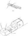
- FIG. 1is a perspective view of a first embodiment of a device suitable for use in extracting a percutaneous ventricular assist device
- FIG. 2Ais a perspective view of the distal end of the device of FIG. 1 with the jaws in the closed position;
- FIG. 2Bis a perspective view of the distal end of the device of FIG. 1 with the jaws in the open position;
- FIG. 3Ais a perspective view of the upper jaw member of the cutting and grasping device of FIG. 1 ;
- FIG. 3Bis a perspective view of the lower jaw member of the cutting and grasping device of FIG. 1 . For clarity the blade is not shown in FIG. 3B .
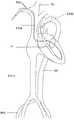
- FIG. 4schematically illustrates a pVAD implanted in the heart prior to extraction.
- FIG. 5Ais a perspective view of the distal end of a second embodiment, with the jaws in the open position;
- FIG. 5Bis a perspective view of the distal end of the second embodiment, with the jaws in the closed position;
- FIG. 6schematically illustrates use of an extraction device in a method for extracting a pVAD.
- This applicationdescribes instrument that may be used to cut a pVAD device that has been implanted within a patient so that it may be withdrawn from the patient.
- Instrument 10includes a distal end having jaws, 12 a , 12 b one or both of which are moveable to transition the jaw between the closed position shown in FIG. 2A and the open position shown in FIG. 2B .
- jaw 12 bis a fixed jaw and jaw 12 a is pivotable to open and close the jaws.
- the jaws 12 a , 12 bare positioned on the distal end of a flexible shaft 14 .
- the shaftis of sufficient length to extend from the right internal jugular vein into the right atrium and to the inter-atrial septum.
- a handle 16At the proximal end of the shaft 14 is a handle 16 including actuators operable to move the jaws between the open and closed position, and to drive a cutting blade (described below) that runs longitudinally between the jaws.
- the jawsare shaped to together define a laterally-extending passage 18 when in the closed position. See FIG. 2A . During use this passage is closed around a portion of the pVAD drive line that is to be cut and extracted.
- Ribs 20 a , 20 b on the jaws 12 a , 12 bline the passage 18 as shown in FIG. 2B .
- the ribspreferably extend in a longitudinal direction and together form an enclosed gripping area within the passage, allowing the jaw members 12 a , 12 b to maintain a secure grip, on at least one side of the blade channel, on the portion of the pVAD drive line extending through the lumen.
- Longitudinally extending channels 22 a , 22 bare positioned to define a blade channel when the jaws are closed.
- the blade 24( FIG. 2B ) is advanceable through the blade channel to cut the portion of the pVAD situated in the laterally-extending lumen.
- the drive line 116 a of the pVADextends out the right atrium to the vena cava to the right subclavian vein (RSV) and out of the body via an RSV sheath.
- the pumpcrosses the aortic valve and has an attached pigtail connector 116 b in the aorta.
- a snare catheter 118is advanced from the right femoral artery (RFA) through the descending aorta DA and used to capture the pigtail 116 b of the pVAD within the aorta, as shown in FIG. 6 . This ensures the distal part of the pVAD is secured when the drive line is cut.
- ROAright femoral artery
- the cutting device 10is inserted into a sheath previously inserted into the right internal jugular vein (RH), and its jaws 10 are advanced to and into the right atrium. It is positioned with the jaw members 12 a , 12 b adjacent to the right side of the inter-atrial septum. The jaw members 12 a , 12 b are closed over the drive line, oriented with the ribs 20 a , 20 b closed on the portion of the drive line that is closest to the interatrial septum. Then the blade is advanced through the jaws, severing the pVAD drive line at the inter-atrial septum. To facilitate cutting, slight tension may be applied to the drive line by pulling the drive line from the RSV.
- RHright internal jugular vein
- the jawsWhen the practitioner is ready to withdraw the venous side of the drive line, the jaws are opened slightly to release the cut ends from the ribs of the jaws. The venous side of the drive line is then withdrawn through the original right subclavian sheath through which the drive line 116 a extends. The cutting device 10 is withdrawn from the internal jugular sheath and the pVAD is withdrawn from the right femoral artery using the snare 118 .
- FIGS. 5A, 5BA second embodiment shown in FIGS. 5A, 5B , this embodiment is similar to the first embodiment, but is provided without the ribs.
Landscapes
- Health & Medical Sciences (AREA)
- Heart & Thoracic Surgery (AREA)
- Engineering & Computer Science (AREA)
- Life Sciences & Earth Sciences (AREA)
- Veterinary Medicine (AREA)
- Public Health (AREA)
- General Health & Medical Sciences (AREA)
- Animal Behavior & Ethology (AREA)
- Biomedical Technology (AREA)
- Cardiology (AREA)
- Hematology (AREA)
- Anesthesiology (AREA)
- Mechanical Engineering (AREA)
- Vascular Medicine (AREA)
- Surgery (AREA)
- Surgical Instruments (AREA)
- Transplantation (AREA)
- Ophthalmology & Optometry (AREA)
- Nuclear Medicine, Radiotherapy & Molecular Imaging (AREA)
- Medical Informatics (AREA)
- Molecular Biology (AREA)
Abstract
Description
Claims (7)
Priority Applications (1)
| Application Number | Priority Date | Filing Date | Title |
|---|---|---|---|
| US16/579,779US10987456B2 (en) | 2019-09-23 | 2019-09-23 | Devices for use in extracting percutaneous ventricular assist devices |
Applications Claiming Priority (1)
| Application Number | Priority Date | Filing Date | Title |
|---|---|---|---|
| US16/579,779US10987456B2 (en) | 2019-09-23 | 2019-09-23 | Devices for use in extracting percutaneous ventricular assist devices |
Publications (2)
| Publication Number | Publication Date |
|---|---|
| US20210085851A1 US20210085851A1 (en) | 2021-03-25 |
| US10987456B2true US10987456B2 (en) | 2021-04-27 |
Family
ID=74881582
Family Applications (1)
| Application Number | Title | Priority Date | Filing Date |
|---|---|---|---|
| US16/579,779Expired - Fee RelatedUS10987456B2 (en) | 2019-09-23 | 2019-09-23 | Devices for use in extracting percutaneous ventricular assist devices |
Country Status (1)
| Country | Link |
|---|---|
| US (1) | US10987456B2 (en) |
Citations (31)
| Publication number | Priority date | Publication date | Assignee | Title |
|---|---|---|---|---|
| US5584803A (en)* | 1991-07-16 | 1996-12-17 | Heartport, Inc. | System for cardiac procedures |
| US5928132A (en)* | 1998-03-31 | 1999-07-27 | Datascope Investment Corp. | Closed chest intra-aortic balloon based ventricular assist device |
| US6228018B1 (en)* | 1999-02-05 | 2001-05-08 | My-Tech, Inc. | Removable left ventricular assist device with an aortic support apparatus |
| US6409674B1 (en)* | 1998-09-24 | 2002-06-25 | Data Sciences International, Inc. | Implantable sensor with wireless communication |
| US20040055608A1 (en)* | 1993-02-22 | 2004-03-25 | Ethicon, Inc. | Minimally-invasive devices and methods for treatment of congestive heart failure |
| US20050192633A1 (en)* | 2004-01-23 | 2005-09-01 | Montpetit Karen P. | Tissue fastening and cutting tool, and methods |
| US7022100B1 (en)* | 1999-09-03 | 2006-04-04 | A-Med Systems, Inc. | Guidable intravascular blood pump and related methods |
| US20060074484A1 (en)* | 2004-10-02 | 2006-04-06 | Huber Christoph H | Methods and devices for repair or replacement of heart valves or adjacent tissue without the need for full cardiopulmonary support |
| US20060224110A1 (en)* | 2005-03-17 | 2006-10-05 | Scott Michael J | Methods for minimally invasive vascular access |
| US20070027456A1 (en)* | 2005-08-01 | 2007-02-01 | Ension, Inc. | Integrated medical apparatus for non-traumatic grasping, manipulating and closure of tissue |
| US20070161845A1 (en)* | 2006-01-09 | 2007-07-12 | Cardiacassist, Inc. | Percutaneous right ventricular assist apparatus and method |
| US20080214888A1 (en)* | 2005-07-19 | 2008-09-04 | Zvi Ben Shalom | Organ Assist System and Method |
| US20090088597A1 (en)* | 2007-10-01 | 2009-04-02 | Alpha Dev Llc | Intraatrial ventricular assist device |
| US20090149950A1 (en)* | 2006-05-31 | 2009-06-11 | Richard Wampler | Heart assist device |
| US20090192579A1 (en)* | 2007-12-03 | 2009-07-30 | Terrance Ransbury | Implantation methods, systems and tools for intravascular implantable devices |
| US20100004501A1 (en)* | 2008-05-05 | 2010-01-07 | Coherex Medical, Inc. | Ventricular assist device and related methods |
| US20100076247A1 (en)* | 2007-05-03 | 2010-03-25 | Leviticus-Cardio Ltd. | Permanent ventricular assist device for treating heart failure |
| US20110118537A1 (en)* | 2009-11-04 | 2011-05-19 | Richard Wampler | Methods and devices for treating heart failure |
| US20110130619A1 (en)* | 2008-05-05 | 2011-06-02 | Coherex Medical, Inc. | Ventricular assist device and related methods |
| US20110190683A1 (en)* | 2010-02-02 | 2011-08-04 | Levitronix Llc | Expandable and collapsible medical device |
| US20110201990A1 (en)* | 2010-02-17 | 2011-08-18 | Novita Therapeutics, Llc | System and method to increase the overall diameter of veins |
| US8066628B1 (en)* | 2010-10-22 | 2011-11-29 | Nupulse, Inc. | Intra-aortic balloon pump and driver |
| US20120296358A1 (en)* | 2011-05-18 | 2012-11-22 | John Duc Nguyen | Coring knife |
| US20130066342A1 (en)* | 2011-09-13 | 2013-03-14 | Abbott Cardiovascular Systems, Inc. | Gripper pusher mechanism for tissue apposition systems |
| US20150230811A1 (en)* | 2012-02-22 | 2015-08-20 | Carter J. Kovarik | Selectively Bendable Remote Gripping Tool |
| US9770256B2 (en)* | 2011-09-28 | 2017-09-26 | Mitracore Technologies Inc. | Apparatuses and methods for cutting a tissue bridge and/or removing a heart valve clip or suture |
| US20180099078A1 (en)* | 2016-10-07 | 2018-04-12 | Nuheart As | Transcatheter device and system for the delivery of intracorporeal devices |
| US20190125948A1 (en)* | 2016-05-02 | 2019-05-02 | Vadovations, Inc. | Heart assist device |
| US20190201015A1 (en)* | 2012-02-22 | 2019-07-04 | Carter J. Kovarik | Selectively bendable remote gripping tool |
| US20190209758A1 (en)* | 2018-01-10 | 2019-07-11 | Magenta Medical Ltd. | Impeller for blood pump |
| US20190239890A1 (en)* | 2018-02-08 | 2019-08-08 | Ethicon Llc | Surgical clip applier with parallel closure jaws |
- 2019
- 2019-09-23USUS16/579,779patent/US10987456B2/ennot_activeExpired - Fee Related
Patent Citations (34)
| Publication number | Priority date | Publication date | Assignee | Title |
|---|---|---|---|---|
| US5584803A (en)* | 1991-07-16 | 1996-12-17 | Heartport, Inc. | System for cardiac procedures |
| US20040055608A1 (en)* | 1993-02-22 | 2004-03-25 | Ethicon, Inc. | Minimally-invasive devices and methods for treatment of congestive heart failure |
| US5928132A (en)* | 1998-03-31 | 1999-07-27 | Datascope Investment Corp. | Closed chest intra-aortic balloon based ventricular assist device |
| US6409674B1 (en)* | 1998-09-24 | 2002-06-25 | Data Sciences International, Inc. | Implantable sensor with wireless communication |
| US6228018B1 (en)* | 1999-02-05 | 2001-05-08 | My-Tech, Inc. | Removable left ventricular assist device with an aortic support apparatus |
| US7022100B1 (en)* | 1999-09-03 | 2006-04-04 | A-Med Systems, Inc. | Guidable intravascular blood pump and related methods |
| US20050192633A1 (en)* | 2004-01-23 | 2005-09-01 | Montpetit Karen P. | Tissue fastening and cutting tool, and methods |
| US20060074484A1 (en)* | 2004-10-02 | 2006-04-06 | Huber Christoph H | Methods and devices for repair or replacement of heart valves or adjacent tissue without the need for full cardiopulmonary support |
| US20060224110A1 (en)* | 2005-03-17 | 2006-10-05 | Scott Michael J | Methods for minimally invasive vascular access |
| US20080214888A1 (en)* | 2005-07-19 | 2008-09-04 | Zvi Ben Shalom | Organ Assist System and Method |
| US20070027456A1 (en)* | 2005-08-01 | 2007-02-01 | Ension, Inc. | Integrated medical apparatus for non-traumatic grasping, manipulating and closure of tissue |
| US20070161845A1 (en)* | 2006-01-09 | 2007-07-12 | Cardiacassist, Inc. | Percutaneous right ventricular assist apparatus and method |
| US8992408B2 (en)* | 2006-01-09 | 2015-03-31 | Cardiacassist, Inc. | Percutaneous right ventricular assist apparatus and method |
| US8550973B2 (en)* | 2006-01-09 | 2013-10-08 | Cardiacassist, Inc. | Percutaneous right ventricular assist apparatus and method |
| US20090149950A1 (en)* | 2006-05-31 | 2009-06-11 | Richard Wampler | Heart assist device |
| US20100076247A1 (en)* | 2007-05-03 | 2010-03-25 | Leviticus-Cardio Ltd. | Permanent ventricular assist device for treating heart failure |
| US20090088597A1 (en)* | 2007-10-01 | 2009-04-02 | Alpha Dev Llc | Intraatrial ventricular assist device |
| US20090192579A1 (en)* | 2007-12-03 | 2009-07-30 | Terrance Ransbury | Implantation methods, systems and tools for intravascular implantable devices |
| US20110130619A1 (en)* | 2008-05-05 | 2011-06-02 | Coherex Medical, Inc. | Ventricular assist device and related methods |
| US20100004501A1 (en)* | 2008-05-05 | 2010-01-07 | Coherex Medical, Inc. | Ventricular assist device and related methods |
| US20110118537A1 (en)* | 2009-11-04 | 2011-05-19 | Richard Wampler | Methods and devices for treating heart failure |
| US20110190683A1 (en)* | 2010-02-02 | 2011-08-04 | Levitronix Llc | Expandable and collapsible medical device |
| US20110201990A1 (en)* | 2010-02-17 | 2011-08-18 | Novita Therapeutics, Llc | System and method to increase the overall diameter of veins |
| US8066628B1 (en)* | 2010-10-22 | 2011-11-29 | Nupulse, Inc. | Intra-aortic balloon pump and driver |
| US8323174B2 (en)* | 2010-10-22 | 2012-12-04 | Nupulse, Inc. | Skin interface for ventricular assist device |
| US20120296358A1 (en)* | 2011-05-18 | 2012-11-22 | John Duc Nguyen | Coring knife |
| US20130066342A1 (en)* | 2011-09-13 | 2013-03-14 | Abbott Cardiovascular Systems, Inc. | Gripper pusher mechanism for tissue apposition systems |
| US9770256B2 (en)* | 2011-09-28 | 2017-09-26 | Mitracore Technologies Inc. | Apparatuses and methods for cutting a tissue bridge and/or removing a heart valve clip or suture |
| US20150230811A1 (en)* | 2012-02-22 | 2015-08-20 | Carter J. Kovarik | Selectively Bendable Remote Gripping Tool |
| US20190201015A1 (en)* | 2012-02-22 | 2019-07-04 | Carter J. Kovarik | Selectively bendable remote gripping tool |
| US20190125948A1 (en)* | 2016-05-02 | 2019-05-02 | Vadovations, Inc. | Heart assist device |
| US20180099078A1 (en)* | 2016-10-07 | 2018-04-12 | Nuheart As | Transcatheter device and system for the delivery of intracorporeal devices |
| US20190209758A1 (en)* | 2018-01-10 | 2019-07-11 | Magenta Medical Ltd. | Impeller for blood pump |
| US20190239890A1 (en)* | 2018-02-08 | 2019-08-08 | Ethicon Llc | Surgical clip applier with parallel closure jaws |
Also Published As
| Publication number | Publication date |
|---|---|
| US20210085851A1 (en) | 2021-03-25 |
Similar Documents
| Publication | Publication Date | Title |
|---|---|---|
| EP3870088B1 (en) | Tissue cutting systems and devices | |
| US6494860B2 (en) | Introducer with multiple sheaths and method of use therefor | |
| US11376403B2 (en) | Transvascular access methods | |
| US10004879B2 (en) | Central venous access system | |
| US4922923A (en) | Method for effecting a catheter exchange | |
| JP5819927B2 (en) | Apparatus, method and system for establishing supplemental blood flow in the circulatory system | |
| US11986209B2 (en) | Methods and devices for creation of communication between aorta and left atrium | |
| US10426514B2 (en) | Self-dilating cannula | |
| JP4722441B2 (en) | Kit for penetration into the septum and placement of the transseptal device, especially placement of the stimulation probe in the left heart chamber | |
| US12121681B2 (en) | Guidewire access sleeve | |
| JPH06502327A (en) | Percutaneous transseptal left atrial cannulation system | |
| US8996135B2 (en) | Device and method for inserting a cardiac catheter | |
| US20230301784A1 (en) | Surgical system for excising a valve | |
| CN113440306A (en) | Interventional suture implantation device and interventional tendon implantation system | |
| US10987456B2 (en) | Devices for use in extracting percutaneous ventricular assist devices | |
| CN114081674A (en) | A valve leaflet dividing device | |
| US20240374892A1 (en) | Directable Tunnel Device for Subcutaneous Implantable Cardio Defibrillator | |
| US20250204980A1 (en) | Systems for traversing and lacerating a valve leaflet |
Legal Events
| Date | Code | Title | Description |
|---|---|---|---|
| FEPP | Fee payment procedure | Free format text:ENTITY STATUS SET TO UNDISCOUNTED (ORIGINAL EVENT CODE: BIG.); ENTITY STATUS OF PATENT OWNER: SMALL ENTITY | |
| FEPP | Fee payment procedure | Free format text:ENTITY STATUS SET TO SMALL (ORIGINAL EVENT CODE: SMAL); ENTITY STATUS OF PATENT OWNER: SMALL ENTITY | |
| AS | Assignment | Owner name:SYNECOR LLC, NORTH CAROLINA Free format text:ASSIGNMENT OF ASSIGNORS INTEREST;ASSIGNORS:STACK, RICHARD S;ATHAS, WILLIAM L;JOHNSON, KEVIN W;AND OTHERS;REEL/FRAME:055692/0387 Effective date:20210305 | |
| STPP | Information on status: patent application and granting procedure in general | Free format text:PUBLICATIONS -- ISSUE FEE PAYMENT VERIFIED | |
| STCF | Information on status: patent grant | Free format text:PATENTED CASE | |
| FEPP | Fee payment procedure | Free format text:MAINTENANCE FEE REMINDER MAILED (ORIGINAL EVENT CODE: REM.); ENTITY STATUS OF PATENT OWNER: SMALL ENTITY | |
| LAPS | Lapse for failure to pay maintenance fees | Free format text:PATENT EXPIRED FOR FAILURE TO PAY MAINTENANCE FEES (ORIGINAL EVENT CODE: EXP.); ENTITY STATUS OF PATENT OWNER: SMALL ENTITY | |
| STCH | Information on status: patent discontinuation | Free format text:PATENT EXPIRED DUE TO NONPAYMENT OF MAINTENANCE FEES UNDER 37 CFR 1.362 | |
| FP | Lapsed due to failure to pay maintenance fee | Effective date:20250427 |